Handle and cartridge system for medical interventions
Yarra , et al. April 12, 2
U.S. patent number 11,298,115 [Application Number 17/384,014] was granted by the patent office on 2022-04-12 for handle and cartridge system for medical interventions. This patent grant is currently assigned to Teleflex Life Sciences Limited. The grantee listed for this patent is Teleflex Life Sciences Limited. Invention is credited to Jolene Cutts, Alexander Charles Gordon, Calvin Lam, Kevin Alexander Lessard, Erik Noel, Maheshwara Rao, Curtis Yarra, Christopher Zaler.


View All Diagrams
| United States Patent | 11,298,115 |
| Yarra , et al. | April 12, 2022 |
Handle and cartridge system for medical interventions
Abstract
An apparatus for transferring mechanical energy in a handle to a cartridge to manipulate tissue or anatomical structures within the body of a human or animal subject for the purpose of treating diseases or disorders. The handle and cartridge contain safety interlocks.
| Inventors: | Yarra; Curtis (Oakland, CA), Zaler; Christopher (Los Gatos, CA), Gordon; Alexander Charles (San Carlos, CA), Noel; Erik (San Ramon, CA), Lam; Calvin (Dublin, CA), Lessard; Kevin Alexander (San Francisco, CA), Cutts; Jolene (San Francisco, CA), Rao; Maheshwara (Oakland, CA) | ||||||||||
|---|---|---|---|---|---|---|---|---|---|---|---|
| Applicant: |
|
||||||||||
| Assignee: | Teleflex Life Sciences Limited
(Valletta, MT) |
||||||||||
| Family ID: | 1000006236986 | ||||||||||
| Appl. No.: | 17/384,014 | ||||||||||
| Filed: | July 23, 2021 |
Prior Publication Data
| Document Identifier | Publication Date | |
|---|---|---|
| US 20220031303 A1 | Feb 3, 2022 | |
Related U.S. Patent Documents
| Application Number | Filing Date | Patent Number | Issue Date | ||
|---|---|---|---|---|---|
| 63060442 | Aug 3, 2020 | ||||
| Current U.S. Class: | 1/1 |
| Current CPC Class: | A61B 17/0401 (20130101); A61B 17/3468 (20130101); A61B 17/0467 (20130101); A61B 17/00234 (20130101); A61B 2017/0409 (20130101); A61B 2017/00389 (20130101) |
| Current International Class: | A61B 17/00 (20060101); A61B 17/34 (20060101); A61B 17/04 (20060101) |
References Cited [Referenced By]
U.S. Patent Documents
| 659422 | October 1900 | Shidler |
| 780392 | January 1905 | Wanamaker et al. |
| 789467 | May 1905 | West |
| 2360164 | October 1944 | Frank |
| 2485531 | October 1949 | William et al. |
| 2579192 | December 1951 | Alexander |
| 2646298 | July 1953 | Leary |
| 2697624 | December 1954 | Thomas et al. |
| 2734299 | February 1956 | Masson |
| 2825592 | March 1958 | Mckenzie |
| 3326586 | June 1967 | Frost et al. |
| 3470834 | October 1969 | Bone |
| 3521918 | July 1970 | Hammond |
| 3541591 | November 1970 | Hoegerman |
| 3664345 | May 1972 | Dabbs et al. |
| 3713680 | January 1973 | Pagano |
| 3716058 | February 1973 | Tanner |
| 3756638 | September 1973 | Stockberger |
| 3873140 | March 1975 | Bloch |
| 3875648 | April 1975 | Bone |
| 3886933 | June 1975 | Mori et al. |
| 3931667 | January 1976 | Merser et al. |
| 3976079 | August 1976 | Samuels et al. |
| 4006747 | February 1977 | Kronenthal et al. |
| 4137920 | February 1979 | Bonnet |
| 4164225 | August 1979 | Johnson et al. |
| 4210148 | July 1980 | Stivala |
| 4235238 | November 1980 | Ogiu et al. |
| 4291698 | September 1981 | Fuchs et al. |
| 4409974 | October 1983 | Freedland |
| 4419094 | December 1983 | Patel |
| 4493323 | January 1985 | Albright et al. |
| 4513746 | April 1985 | Aranyi et al. |
| 4621640 | November 1986 | Mulhollan et al. |
| 4655771 | April 1987 | Wallsten |
| 4657461 | April 1987 | Smith |
| 4669473 | June 1987 | Richards et al. |
| 4705040 | November 1987 | Mueller et al. |
| 4714281 | December 1987 | Peck |
| 4738255 | April 1988 | Goble et al. |
| 4741330 | May 1988 | Hayhurst |
| 4744364 | May 1988 | Kensey |
| 4750492 | June 1988 | Jacobs |
| 4762128 | August 1988 | Rosenbluth |
| 4823794 | April 1989 | Pierce |
| 4863439 | September 1989 | Sanderson |
| 4899743 | February 1990 | Nicholson et al. |
| 4926860 | May 1990 | Stice et al. |
| 4946468 | August 1990 | Li |
| 4955913 | September 1990 | Robinson |
| 4968315 | November 1990 | Gatturna |
| 5002550 | March 1991 | Li |
| 5019032 | May 1991 | Robertson |
| 5041129 | August 1991 | Hayhurst et al. |
| 5046513 | September 1991 | Gatturna et al. |
| 5053046 | October 1991 | Janese |
| 5078731 | January 1992 | Hayhurst |
| 5080660 | January 1992 | Buelna |
| 5098374 | March 1992 | Othel-Jacobsen et al. |
| 5100421 | March 1992 | Christoudias |
| 5123914 | June 1992 | Cope |
| 5127393 | July 1992 | McFarlin et al. |
| 5129912 | July 1992 | Noda et al. |
| 5133713 | July 1992 | Huang et al. |
| 5159925 | November 1992 | Neuwirth et al. |
| 5160339 | November 1992 | Chen et al. |
| 5192303 | March 1993 | Gattuma et al. |
| 5203787 | April 1993 | Noblitt et al. |
| 5207672 | May 1993 | Roth et al. |
| 5217470 | June 1993 | Weston |
| 5217486 | June 1993 | Rice et al. |
| 5234454 | August 1993 | Bangs |
| 5236445 | August 1993 | Hayhurst et al. |
| 5237984 | August 1993 | Williams et al. |
| 5258015 | November 1993 | Li et al. |
| 5267960 | December 1993 | Hayman et al. |
| 5269802 | December 1993 | Garber |
| 5269809 | December 1993 | Hayhurst et al. |
| 5300099 | April 1994 | Rudie |
| 5322501 | June 1994 | Mahmud-Durrani |
| 5330488 | July 1994 | Goldrath |
| 5334200 | August 1994 | Johnson |
| 5336240 | August 1994 | Metzler et al. |
| 5354271 | October 1994 | Yoda |
| 5358511 | October 1994 | Gattuma et al. |
| 5364408 | November 1994 | Gordon |
| 5366490 | November 1994 | Edwards et al. |
| 5368599 | November 1994 | Hirsch et al. |
| 5370646 | December 1994 | Reese et al. |
| 5380334 | January 1995 | Torrie et al. |
| 5391182 | February 1995 | Chin |
| 5403348 | April 1995 | Bonutti |
| 5405352 | April 1995 | Weston |
| 5411520 | May 1995 | Nash et al. |
| 5417691 | May 1995 | Hayhurst |
| 5435805 | July 1995 | Edwards et al. |
| 5470337 | November 1995 | Moss |
| 5472446 | December 1995 | Torre |
| 5478003 | December 1995 | Green et al. |
| 5480406 | January 1996 | Nolan et al. |
| 5499994 | March 1996 | Tihon et al. |
| 5501690 | March 1996 | Measamer et al. |
| 5507754 | April 1996 | Green et al. |
| 5522846 | June 1996 | Bonutti |
| 5531763 | July 1996 | Mastri et al. |
| 5536240 | July 1996 | Edwards et al. |
| 5540655 | July 1996 | Edwards et al. |
| 5540704 | July 1996 | Gordon et al. |
| 5545171 | August 1996 | Sharkey et al. |
| 5545178 | August 1996 | Kensey et al. |
| 5549631 | August 1996 | Bonutti |
| 5550172 | August 1996 | Regula et al. |
| 5554162 | September 1996 | DeLange |
| 5554171 | September 1996 | Gatturna et al. |
| 5562689 | October 1996 | Green et al. |
| 5569305 | October 1996 | Bonutti |
| 5571104 | November 1996 | Li |
| 5573540 | November 1996 | Yoon |
| 5578044 | November 1996 | Gordon et al. |
| 5591177 | January 1997 | Lehrer |
| 5593421 | January 1997 | Bauer |
| 5611515 | March 1997 | Benderev et al. |
| 5626614 | May 1997 | Hart |
| 5630824 | May 1997 | Hart |
| 5647836 | July 1997 | Blake et al. |
| 5665109 | September 1997 | Yoon |
| 5667486 | September 1997 | Mikulich et al. |
| 5667488 | September 1997 | Lundquist et al. |
| 5669917 | September 1997 | Sauer et al. |
| 5690649 | November 1997 | Li |
| 5690677 | November 1997 | Schmieding et al. |
| 5697950 | December 1997 | Fucci et al. |
| 5707394 | January 1998 | Miller et al. |
| 5716368 | February 1998 | Torre et al. |
| 5718717 | February 1998 | Bonutti |
| 5725556 | March 1998 | Moser et al. |
| 5725557 | March 1998 | Gatturna et al. |
| 5733306 | March 1998 | Bonutti |
| 5741276 | April 1998 | Poloyko et al. |
| 5746753 | May 1998 | Sullivan et al. |
| 5749846 | May 1998 | Edwards et al. |
| 5749889 | May 1998 | Bacich et al. |
| 5752963 | May 1998 | Allard et al. |
| 5782862 | July 1998 | Bonutti |
| 5782864 | July 1998 | Lizardi |
| 5791022 | August 1998 | Bohman |
| 5800445 | September 1998 | Ratcliff et al. |
| 5807403 | September 1998 | Beyar et al. |
| 5810848 | September 1998 | Hayhurst |
| 5810853 | September 1998 | Yoon |
| 5814072 | September 1998 | Bonutti |
| 5830179 | November 1998 | Mikus et al. |
| 5830221 | November 1998 | Stein et al. |
| 5845645 | December 1998 | Bonutti |
| 5846254 | December 1998 | Schulze et al. |
| 5861002 | January 1999 | Desai |
| 5868762 | February 1999 | Cragg et al. |
| 5873891 | February 1999 | Sohn |
| 5879357 | March 1999 | Heaton et al. |
| 5897574 | April 1999 | Bonutti |
| 5899911 | May 1999 | Carter |
| 5899921 | May 1999 | Caspari et al. |
| 5904679 | May 1999 | Clayman |
| 5904696 | May 1999 | Rosenman |
| 5908428 | June 1999 | Scirica et al. |
| 5908447 | June 1999 | Schroeppel et al. |
| 5919198 | July 1999 | Graves et al. |
| 5919202 | July 1999 | Yoon |
| 5921982 | July 1999 | Lesh et al. |
| 5921986 | July 1999 | Bonutti |
| 5928252 | July 1999 | Steadman et al. |
| 5931844 | August 1999 | Thompson et al. |
| 5944739 | August 1999 | Zlock et al. |
| 5948001 | September 1999 | Larsen |
| 5948002 | September 1999 | Bonutti |
| 5954057 | September 1999 | Li |
| 5954731 | September 1999 | Yoon |
| 5954747 | September 1999 | Clark |
| 5964732 | October 1999 | Willard |
| 5971447 | October 1999 | Steck |
| 5971967 | October 1999 | Willard |
| 6010514 | January 2000 | Burney et al. |
| 6011525 | January 2000 | Piole |
| 6015428 | January 2000 | Pagedas |
| 6030393 | February 2000 | Corlew |
| 6033413 | March 2000 | Mikus et al. |
| 6033430 | March 2000 | Bonutti |
| 6036701 | March 2000 | Rosenman |
| 6048351 | April 2000 | Gordon et al. |
| 6053908 | April 2000 | Crainich et al. |
| 6056722 | May 2000 | Jayaraman |
| 6056772 | May 2000 | Bonutti |
| 6066160 | May 2000 | Colvin et al. |
| 6068648 | May 2000 | Cole et al. |
| 6080167 | June 2000 | Lyell |
| 6086608 | July 2000 | Ek et al. |
| 6110183 | August 2000 | Cope |
| 6117133 | September 2000 | Zappala |
| 6117160 | September 2000 | Bonutti |
| 6117161 | September 2000 | Li et al. |
| 6120539 | September 2000 | Eldridge et al. |
| 6132438 | October 2000 | Fleischman et al. |
| 6139555 | October 2000 | Hart et al. |
| RE36974 | November 2000 | Bonutti |
| 6143006 | November 2000 | Chan |
| 6152935 | November 2000 | Kammerer et al. |
| 6156044 | December 2000 | Kammerer et al. |
| 6159207 | December 2000 | Yoon |
| 6159234 | December 2000 | Bonutti et al. |
| 6200329 | March 2001 | Fung et al. |
| 6206895 | March 2001 | Levinson |
| 6206907 | March 2001 | Marino et al. |
| 6228096 | May 2001 | Marchand |
| 6258124 | July 2001 | Darois et al. |
| 6261302 | July 2001 | Voegele et al. |
| 6270530 | August 2001 | Eldridge et al. |
| 6280460 | August 2001 | Bolduc et al. |
| 6287317 | September 2001 | Makower et al. |
| 6290711 | September 2001 | Caspari et al. |
| 6306158 | October 2001 | Bartlett |
| 6312448 | November 2001 | Bonutti |
| 6319263 | November 2001 | Levinson |
| 6322112 | November 2001 | Duncan |
| 6332889 | December 2001 | Sancoff et al. |
| 6382214 | May 2002 | Raz et al. |
| 6398795 | June 2002 | McAlister et al. |
| 6423079 | July 2002 | Blake, III |
| 6425900 | July 2002 | Knodel et al. |
| 6425919 | July 2002 | Lambrecht |
| 6428562 | August 2002 | Bonutti |
| 6436107 | August 2002 | Wang et al. |
| 6461355 | October 2002 | Svejkovsky et al. |
| 6482235 | November 2002 | Lambrecht et al. |
| 6488691 | December 2002 | Carroll et al. |
| 6491707 | December 2002 | Makower et al. |
| 6494888 | December 2002 | Laufer et al. |
| 6500184 | December 2002 | Chan et al. |
| 6500195 | December 2002 | Bonutti |
| 6506190 | January 2003 | Walshe |
| 6506196 | January 2003 | Laufer |
| 6517569 | February 2003 | Mikus et al. |
| 6527702 | March 2003 | Whalen et al. |
| 6527794 | March 2003 | McDevitt et al. |
| 6530932 | March 2003 | Swayze et al. |
| 6533796 | March 2003 | Sauer et al. |
| 6544230 | April 2003 | Flaherty et al. |
| 6547725 | April 2003 | Paolitto et al. |
| 6551328 | April 2003 | Kortenbach |
| 6551333 | April 2003 | Kuhns et al. |
| 6565578 | May 2003 | Peifer et al. |
| 6569187 | May 2003 | Bonutti et al. |
| 6572626 | June 2003 | Knodel et al. |
| 6572635 | June 2003 | Bonutti |
| 6572653 | June 2003 | Simonson |
| 6582453 | June 2003 | Tran et al. |
| 6592609 | July 2003 | Bonutti |
| 6596013 | July 2003 | Yang et al. |
| 6599311 | July 2003 | Biggs et al. |
| 6626913 | September 2003 | McKinnon et al. |
| 6626916 | September 2003 | Yeung et al. |
| 6626919 | September 2003 | Swanstrom |
| 6629534 | October 2003 | Goar et al. |
| 6641524 | November 2003 | Kovac |
| 6641592 | November 2003 | Sauer et al. |
| 6656182 | December 2003 | Hayhurst |
| 6660008 | December 2003 | Foerster et al. |
| 6660023 | December 2003 | McDevitt et al. |
| 6663589 | December 2003 | Halevy |
| 6663633 | December 2003 | Pierson |
| 6663639 | December 2003 | Laufer et al. |
| 6699263 | March 2004 | Cope |
| 6702846 | March 2004 | Mikus et al. |
| 6706047 | March 2004 | Trout et al. |
| 6709493 | March 2004 | DeGuiseppi et al. |
| 6715804 | April 2004 | Beers |
| 6719709 | April 2004 | Whalen et al. |
| 6730112 | May 2004 | Levinson |
| 6736823 | May 2004 | Darois et al. |
| 6736854 | May 2004 | Vadurro et al. |
| 6740098 | May 2004 | Abrams et al. |
| 6767037 | July 2004 | Wenstrom |
| 6770076 | August 2004 | Foerster |
| 6773438 | August 2004 | Knodel et al. |
| 6773441 | August 2004 | Laufer et al. |
| 6790213 | September 2004 | Cherok et al. |
| 6802846 | October 2004 | Hauschild et al. |
| 6821282 | November 2004 | Perry et al. |
| 6821285 | November 2004 | Laufer et al. |
| 6821291 | November 2004 | Bolea et al. |
| 6835200 | December 2004 | Laufer et al. |
| 6905475 | June 2005 | Hauschild et al. |
| 6908473 | June 2005 | Skiba et al. |
| 6921361 | July 2005 | Suzuki et al. |
| 6926732 | August 2005 | Derus et al. |
| 6951565 | October 2005 | Keane et al. |
| 6986775 | January 2006 | Morales et al. |
| 6986784 | January 2006 | Weiser et al. |
| 6991596 | January 2006 | Whalen et al. |
| 6991647 | January 2006 | Jadhav |
| 6997940 | February 2006 | Bonutti |
| 7001327 | February 2006 | Whalen et al. |
| 7008381 | March 2006 | Janssens |
| 7011688 | March 2006 | Gryska et al. |
| 7015253 | March 2006 | Escandon et al. |
| 7041111 | May 2006 | Chu |
| 7048698 | May 2006 | Whalen et al. |
| 7048747 | May 2006 | Arcia et al. |
| 7060077 | June 2006 | Gordon et al. |
| 7063715 | June 2006 | Onuki et al. |
| 7081126 | July 2006 | McDevitt et al. |
| 7083638 | August 2006 | Foerster |
| 7087073 | August 2006 | Bonutti |
| 7089064 | August 2006 | Manker et al. |
| 7090690 | August 2006 | Foerster et al. |
| 7093601 | August 2006 | Manker et al. |
| 7105004 | September 2006 | DiCesare et al. |
| 7108655 | September 2006 | Whalen et al. |
| 7141038 | November 2006 | Whalen et al. |
| 7153314 | December 2006 | Laufer et al. |
| 7179225 | February 2007 | Shluzas et al. |
| 7226558 | June 2007 | Nieman et al. |
| 7232448 | June 2007 | Battles et al. |
| 7255675 | August 2007 | Gertner et al. |
| 7288063 | October 2007 | Petros |
| 7303108 | December 2007 | Shelton |
| 7320701 | January 2008 | Haut et al. |
| 7322974 | January 2008 | Swoyer et al. |
| 7326221 | February 2008 | Sakamoto et al. |
| 7334822 | February 2008 | Hines |
| 7340300 | March 2008 | Christopherson et al. |
| 7399304 | July 2008 | Gambale et al. |
| 7402166 | July 2008 | Feigl |
| 7416554 | August 2008 | Lam et al. |
| 7417175 | August 2008 | Oda et al. |
| 7463934 | December 2008 | Tronnes et al. |
| 7481771 | January 2009 | Fonseca et al. |
| 7553317 | June 2009 | William et al. |
| 7608108 | October 2009 | Bhatnagar et al. |
| 7645286 | January 2010 | Catanese et al. |
| 7658311 | February 2010 | Boudreaux |
| 7674275 | March 2010 | Martin et al. |
| 7695494 | April 2010 | Foerster |
| 7704261 | April 2010 | Sakamoto et al. |
| 7727248 | June 2010 | Smith et al. |
| 7736374 | June 2010 | Vaughan et al. |
| 7758594 | July 2010 | Lamson et al. |
| 7766923 | August 2010 | Catanese et al. |
| 7766939 | August 2010 | Yeung et al. |
| 7780682 | August 2010 | Catanese et al. |
| 7815655 | October 2010 | Catanese et al. |
| 7887551 | February 2011 | Bojarski et al. |
| 7896891 | March 2011 | Catanese et al. |
| 7905889 | March 2011 | Catanese et al. |
| 7909836 | March 2011 | McLean et al. |
| 7914542 | March 2011 | Lamson et al. |
| 7951158 | May 2011 | Catanese et al. |
| 8007503 | August 2011 | Catanese et al. |
| 8043309 | October 2011 | Catanese et al. |
| 8157815 | April 2012 | Catanese et al. |
| 8211118 | July 2012 | Catanese et al. |
| 8216254 | July 2012 | McLean et al. |
| 8333776 | December 2012 | Cheng et al. |
| 8343187 | January 2013 | Lamson et al. |
| 8394110 | March 2013 | Catanese et al. |
| 8394113 | March 2013 | Wei et al. |
| 8425535 | April 2013 | McLean et al. |
| 8454655 | June 2013 | Yeung et al. |
| 8491606 | July 2013 | Tong et al. |
| 8529584 | September 2013 | Catanese et al. |
| 8603106 | December 2013 | Catanese et al. |
| 8628542 | January 2014 | Merrick et al. |
| 8663243 | March 2014 | Lamson et al. |
| 8668705 | March 2014 | Johnston et al. |
| 8715239 | May 2014 | Lamson et al. |
| 8715298 | May 2014 | Catanese et al. |
| 8758366 | June 2014 | McLean |
| 8900252 | December 2014 | Lamson et al. |
| 8936609 | January 2015 | Catanese et al. |
| 8939996 | January 2015 | Cheng et al. |
| 9320511 | April 2016 | McLean et al. |
| 9504461 | November 2016 | Catanese et al. |
| 9549739 | January 2017 | Catanese et al. |
| 10105132 | October 2018 | Lamson et al. |
| 10299780 | May 2019 | Catanese et al. |
| 10349932 | July 2019 | Catanese et al. |
| 10568651 | February 2020 | Kostrzewski et al. |
| 2001/0041916 | November 2001 | Bonutti |
| 2001/0044639 | November 2001 | Levinson |
| 2002/0095064 | July 2002 | Beyar |
| 2002/0095154 | July 2002 | Atkinson et al. |
| 2002/0107540 | August 2002 | Whalen et al. |
| 2002/0128684 | September 2002 | Foerster |
| 2002/0161382 | October 2002 | Neisz et al. |
| 2002/0183740 | December 2002 | Edwards et al. |
| 2002/0193809 | December 2002 | Meade et al. |
| 2003/0060819 | March 2003 | McGovern et al. |
| 2003/0109769 | June 2003 | Lowery et al. |
| 2003/0191497 | October 2003 | Cope |
| 2003/0199860 | October 2003 | Loeb et al. |
| 2003/0204195 | October 2003 | Keane et al. |
| 2003/0236535 | December 2003 | Onuki et al. |
| 2004/0030217 | February 2004 | Yeung et al. |
| 2004/0043052 | March 2004 | Hunter et al. |
| 2004/0078046 | April 2004 | Barzell et al. |
| 2004/0122456 | June 2004 | Saadat et al. |
| 2004/0122474 | June 2004 | Gellman et al. |
| 2004/0147958 | July 2004 | Lam et al. |
| 2004/0193191 | September 2004 | Starksen et al. |
| 2004/0193194 | September 2004 | Laufer et al. |
| 2004/0194790 | October 2004 | Laufer et al. |
| 2004/0243178 | December 2004 | Haut et al. |
| 2004/0243179 | December 2004 | Foerster |
| 2004/0243180 | December 2004 | Donnelly et al. |
| 2004/0243227 | December 2004 | Starksen et al. |
| 2004/0260345 | December 2004 | Foerster |
| 2005/0010203 | January 2005 | Edwards et al. |
| 2005/0055087 | March 2005 | Starksen |
| 2005/0065550 | March 2005 | Starksen et al. |
| 2005/0107811 | May 2005 | Starksen et al. |
| 2005/0107812 | May 2005 | Starksen et al. |
| 2005/0154401 | July 2005 | Weldon et al. |
| 2005/0165272 | July 2005 | Okada et al. |
| 2005/0177181 | August 2005 | Kagan et al. |
| 2005/0203344 | September 2005 | Orban et al. |
| 2005/0203550 | September 2005 | Laufer et al. |
| 2005/0216040 | September 2005 | Gertner et al. |
| 2005/0216078 | September 2005 | Starksen et al. |
| 2005/0251157 | November 2005 | Saadat et al. |
| 2005/0251206 | November 2005 | Maahs et al. |
| 2005/0267405 | December 2005 | Shah |
| 2005/0273138 | December 2005 | To et al. |
| 2006/0025750 | February 2006 | Starksen et al. |
| 2006/0025784 | February 2006 | Starksen et al. |
| 2006/0025789 | February 2006 | Laufer et al. |
| 2006/0025819 | February 2006 | Nobis et al. |
| 2006/0026750 | February 2006 | Ballance |
| 2006/0030884 | February 2006 | Yeung et al. |
| 2006/0058817 | March 2006 | Starksen et al. |
| 2006/0089646 | April 2006 | Bonutti |
| 2006/0167477 | July 2006 | Arcia et al. |
| 2006/0199996 | September 2006 | Caraballo et al. |
| 2006/0241694 | October 2006 | Cerundolo |
| 2006/0265042 | November 2006 | Catanese et al. |
| 2006/0276481 | December 2006 | Evrard et al. |
| 2006/0276871 | December 2006 | Lamson et al. |
| 2006/0282081 | December 2006 | Fanton et al. |
| 2007/0049929 | March 2007 | Catanese et al. |
| 2007/0049970 | March 2007 | Belef et al. |
| 2007/0060931 | March 2007 | Hamilton et al. |
| 2007/0088362 | April 2007 | Bonutti et al. |
| 2007/0112385 | May 2007 | Conlon |
| 2007/0142846 | June 2007 | Catanese et al. |
| 2007/0173888 | July 2007 | Gertner et al. |
| 2007/0260259 | November 2007 | Fanton et al. |
| 2008/0009888 | January 2008 | Ewers et al. |
| 2008/0021445 | January 2008 | Elmouelhi |
| 2008/0021485 | January 2008 | Catanese et al. |
| 2008/0033458 | February 2008 | McLean et al. |
| 2008/0033488 | February 2008 | Catanese et al. |
| 2008/0039833 | February 2008 | Catanese et al. |
| 2008/0039872 | February 2008 | Catanese et al. |
| 2008/0039874 | February 2008 | Catanese et al. |
| 2008/0039875 | February 2008 | Catanese et al. |
| 2008/0039893 | February 2008 | McLean et al. |
| 2008/0039894 | February 2008 | Catanese et al. |
| 2008/0045978 | February 2008 | Kuhns et al. |
| 2008/0051810 | February 2008 | To et al. |
| 2008/0058710 | March 2008 | Wilk |
| 2008/0065120 | March 2008 | Zannis et al. |
| 2008/0082113 | April 2008 | Bishop et al. |
| 2008/0086172 | April 2008 | Martin et al. |
| 2008/0091220 | April 2008 | Chu |
| 2008/0091237 | April 2008 | Schwartz et al. |
| 2008/0119874 | May 2008 | Merves |
| 2008/0154378 | June 2008 | Pelo |
| 2008/0161852 | July 2008 | Kaiser et al. |
| 2008/0195145 | August 2008 | Bonutti et al. |
| 2008/0208220 | August 2008 | Shiono et al. |
| 2008/0228202 | September 2008 | Cropper et al. |
| 2008/0269737 | October 2008 | Elmouelhi et al. |
| 2009/0012537 | January 2009 | Green |
| 2009/0018553 | January 2009 | McLean et al. |
| 2009/0060977 | March 2009 | Lamson et al. |
| 2009/0112234 | April 2009 | Crainich et al. |
| 2009/0112537 | April 2009 | Okumura |
| 2010/0010631 | January 2010 | Otte et al. |
| 2010/0023024 | January 2010 | Zeiner |
| 2010/0030262 | February 2010 | McLean et al. |
| 2010/0030263 | February 2010 | Cheng et al. |
| 2010/0063542 | March 2010 | Burg et al. |
| 2010/0114162 | May 2010 | Bojarski et al. |
| 2010/0240951 | September 2010 | Catanese et al. |
| 2010/0286106 | November 2010 | Gat et al. |
| 2010/0286679 | November 2010 | Hoey et al. |
| 2010/0298948 | November 2010 | Hoey et al. |
| 2011/0040312 | February 2011 | Lamson et al. |
| 2011/0046648 | February 2011 | Johnston et al. |
| 2011/0060349 | March 2011 | Cheng |
| 2011/0160747 | June 2011 | McLean et al. |
| 2011/0166564 | July 2011 | Merrick et al. |
| 2011/0190758 | August 2011 | Lamson et al. |
| 2011/0218387 | September 2011 | Lamson et al. |
| 2011/0245828 | October 2011 | Baxter et al. |
| 2012/0245600 | September 2012 | McLean et al. |
| 2012/0265006 | October 2012 | Makower et al. |
| 2013/0096582 | April 2013 | Cheng et al. |
| 2013/0211431 | August 2013 | Wei et al. |
| 2013/0253574 | September 2013 | Catanese et al. |
| 2013/0253662 | September 2013 | Lamson et al. |
| 2013/0261383 | October 2013 | Catanese et al. |
| 2013/0261665 | October 2013 | Yeung et al. |
| 2013/0267772 | October 2013 | Catanese et al. |
| 2013/0268001 | October 2013 | Catanese et al. |
| 2013/0274799 | October 2013 | Catanese, III |
| 2013/0289342 | October 2013 | Tong et al. |
| 2013/0296639 | November 2013 | Lamson et al. |
| 2013/0296889 | November 2013 | Fong et al. |
| 2013/0296935 | November 2013 | McLean et al. |
| 2013/0325143 | December 2013 | Lamson et al. |
| 2014/0005473 | January 2014 | Catanese et al. |
| 2014/0005690 | January 2014 | Catanese et al. |
| 2014/0088587 | March 2014 | Merrick et al. |
| 2014/0236230 | August 2014 | Johnston et al. |
| 2017/0035410 | February 2017 | Catanese et al. |
| 2019/0298334 | October 2019 | Catanese et al. |
| 2022/0031357 | February 2022 | Cutts et al. |
| 2022/0031358 | February 2022 | Yarra et al. |
| 2477220 | Nov 2007 | CA | |||
| 101795641 | Aug 2010 | CN | |||
| 102112064 | Jun 2014 | CN | |||
| 109009285 | Dec 2018 | CN | |||
| 10159470 | Jun 2003 | DE | |||
| 0246836 | Dec 1991 | EP | |||
| 0464480 | Jan 1992 | EP | |||
| 0632999 | Jan 1995 | EP | |||
| 1016377 | Jul 2000 | EP | |||
| 1482841 | Dec 2004 | EP | |||
| 1082941 | Mar 2005 | EP | |||
| 1006909 | Jan 2007 | EP | |||
| 1852071 | Nov 2007 | EP | |||
| 1584295 | Feb 2008 | EP | |||
| 1884198 | Feb 2008 | EP | |||
| 1884199 | Feb 2008 | EP | |||
| 1887976 | Feb 2008 | EP | |||
| 1670361 | Apr 2008 | EP | |||
| 1962720 | Sep 2008 | EP | |||
| 1331886 | Dec 2008 | EP | |||
| 1482840 | Dec 2008 | EP | |||
| 2164427 | Mar 2010 | EP | |||
| 1484023 | May 2011 | EP | |||
| 2339970 | Jul 2011 | EP | |||
| 2345373 | Jul 2011 | EP | |||
| 2345374 | Jul 2011 | EP | |||
| 2600781 | Jun 2013 | EP | |||
| 2658477 | Nov 2013 | EP | |||
| 2049023 | Dec 2014 | EP | |||
| 2344048 | Sep 2016 | EP | |||
| 2111167 | Nov 2018 | EP | |||
| 2658458 | Feb 2020 | EP | |||
| 2750031 | Dec 1997 | FR | |||
| 5836559 | Mar 1983 | JP | |||
| 09122134 | May 1997 | JP | |||
| 2001137254 | May 2001 | JP | |||
| 2004344427 | Dec 2004 | JP | |||
| 2009106755 | May 2009 | JP | |||
| 2009521278 | Jun 2009 | JP | |||
| 2011529745 | Dec 2011 | JP | |||
| 2012143622 | Aug 2012 | JP | |||
| 2062121 | Jun 1996 | RU | |||
| 2112571 | Jun 1998 | RU | |||
| 2128012 | Mar 1999 | RU | |||
| 2221501 | Jan 2004 | RU | |||
| 825094 | Apr 1981 | SU | |||
| 1987001270 | Mar 1987 | WO | |||
| 1991008708 | Jun 1991 | WO | |||
| 1992010142 | Jun 1992 | WO | |||
| 1993004727 | Mar 1993 | WO | |||
| 1993015664 | Aug 1993 | WO | |||
| 1995000818 | Jan 1995 | WO | |||
| 2000040159 | Jul 2000 | WO | |||
| 2001026588 | Apr 2001 | WO | |||
| 2001028432 | Apr 2001 | WO | |||
| 2001039671 | Jun 2001 | WO | |||
| 2001049195 | Jul 2001 | WO | |||
| 2001065997 | Sep 2001 | WO | |||
| 2001095818 | Dec 2001 | WO | |||
| 2002028289 | Apr 2002 | WO | |||
| 2002030335 | Apr 2002 | WO | |||
| 2002032321 | Apr 2002 | WO | |||
| 2003039334 | May 2003 | WO | |||
| 2003077772 | Sep 2003 | WO | |||
| 2004000159 | Dec 2003 | WO | |||
| 2004017845 | Mar 2004 | WO | |||
| 2004019787 | Mar 2004 | WO | |||
| 2004019788 | Mar 2004 | WO | |||
| 2004030569 | Apr 2004 | WO | |||
| 2004103189 | Dec 2004 | WO | |||
| 2005034738 | Apr 2005 | WO | |||
| 2005094447 | Oct 2005 | WO | |||
| 2006127431 | Nov 2006 | WO | |||
| 2007053516 | May 2007 | WO | |||
| 2007064906 | Jun 2007 | WO | |||
| 2007075981 | Jul 2007 | WO | |||
| 2008006084 | Jan 2008 | WO | |||
| 2008014191 | Jan 2008 | WO | |||
| 2008043044 | Apr 2008 | WO | |||
| 2008043917 | Apr 2008 | WO | |||
| 2008097942 | Aug 2008 | WO | |||
| 2009009617 | Jan 2009 | WO | |||
| 2009135005 | Nov 2009 | WO | |||
| 2010011832 | Jan 2010 | WO | |||
| 2010014821 | Feb 2010 | WO | |||
| 2010014825 | Feb 2010 | WO | |||
| 2012018446 | Feb 2012 | WO | |||
| 2012091952 | Jul 2012 | WO | |||
| 2012091954 | Jul 2012 | WO | |||
| 2012091955 | Jul 2012 | WO | |||
| 2012091956 | Jul 2012 | WO | |||
Other References
|
PCT International Search Report dated Nov. 11, 2021, in PCT/US2021/042902. cited by applicant . PCT Written Opinion dated Nov. 11, 2021, in PCT/US2021/042902. cited by applicant . Bacharova, O.A., et al. "The Effect of Rhodiolae rosea Extract on Incidence Rate of Superficial Bladder Carcinoma Relapses", Kozin 1995. cited by applicant . Berges, Richard, et al. "Alternative Minimalinvasive Therapien Beim Benignen Prostatasyndrom", Medizin, Jg. 104, Heft 37, Sep. 14, 2007. cited by applicant . Borzhievski, et al., "Tactics of the Surgical Treatment of Patients With Prostatic Adenoma and Acute Urinary Retention," Urologia Nefrol (Mosk), Jan.-Feb. 1987, (1):39-43. cited by applicant . Extended European Search Report dated Jan. 20, 2020 in EP Patent Application No. 19199026.6. cited by applicant . Hartung, Rudolf, et al. "Instrumentelle Therapie der benignen Prostatahyperplasie", Medizin, Deutsches Arzteblatt 97, Heft 15, Apr. 14, 2000. cited by applicant . Hofner, Klaus, et al., "Operative Therapie des benignen Prostatasyndroms", Medizin, Dtsch Arztebl, 2007; 104(36): A 2424-9. cited by applicant . Hubmann, R. "Geschichte der transurethralen Prostataeingriffe", Geschichte der Medizin, Urologe [B], 2000, 40:152-160. cited by applicant . Jonas, U., et al., "Benigne Prostatahyperplasie", Der Urologe 2006--[Sonderheft] 45:134-144. cited by applicant . Kruck, S., et al., "Aktuelle Therapiemoglichkeiten des Benignen Prostata-Syndroms", J Urol Urogynakol, 2009; 16(1): 19-22. cited by applicant . Miyake, Osamu. "Medical Examination and Treatment for BPH," Pharma Med, vol. 22, No. 3, 2004, p. 97-103. cited by applicant . Reich, O., et al., "Benignes Prostatasyndrom (BPS)," Der Urologe A Issue vol. 45, No. 6, Jun. 2006, p. 769-782. cited by applicant . Schauer, P., et al. "New applications for endoscopy: the emerging field of endoluminal and transgastric bariatric surgery", Surgical Endoscopy, (Apr. 24, 2006), 10 pgs. cited by applicant . Sharp, Howard T., M.D., et al. "Instruments and Methods--The 4-S Modification of the Roeder Knot: How to Tie It", Obstetrics & Gynecology, p. 1004-1006, vol. 90, No. 6, Dec. 1997. cited by applicant . Takashi, Daito. "Low-Invasive Treatment for BPH", Medico vol. 34, No. 10, p. 366-369, 2000. cited by applicant . Teruhisa, Ohashi. "Urinary Dysfunction by Lower Urinary Tract Obstraction in Male", Pharma Medica, vol. 8, No. 8, p. 35-39, 1990. cited by applicant . Tomohiko, Koyanagi, et al., "Surgery View of 21st Century," Urological Surgery, vol. 84, No. 1, p. 47-53, 2001. cited by applicant . Trapeznikov, et al., "New Technologies in the Treatment of Benign Prostatic Hyperplasia", Urologia Nefrol (Mosk), Jul.-Aug. 1996, (4):41-47. cited by applicant . Yeung, Jeff. "Treating Urinary Stress Incontenance Without Incision with Endoscopic Suture Anchor & Approximating Device," Aleeva Medical, Inc., 2007. cited by applicant. |
Primary Examiner: Dornbusch; Dianne
Attorney, Agent or Firm: Levitt; Kenneth E.
Parent Case Text
CROSS REFERENCE TO RELATED APPLICATIONS
This application claims priority to U.S. provisional patent application No. 63/060,442, entitled "HANDLE AND CARTRIDGE SYSTEM FOR MEDICAL INTERVENTIONS" and filed on Aug. 3, 2020, which is herein incorporated by reference in its entirety.
Claims
We claim:
1. A cartridge for a system that includes a handle and the cartridge, where the handle is configured to accept a series of such cartridges and impart mechanical energy to the cartridge to deliver an implant, comprising: a knob on the cartridge that moves from an unlocked position to a locked position such that the locked position secures the cartridge to the handle; a slidable cutter block connected to a slidable pusher block via an extension spring, wherein the cutter block and the pusher block are configured to slide in a linear direction within a body of the cartridge; and a pivotable implant actuator within the body of the cartridge and engaged with the pusher block such that the implant actuator prevents the pusher block from sliding when the implant actuator is in a first position, wherein the spring is in an extended position exerting a force tending to draw the cutter block and the pusher block toward each other.
2. The cartridge of claim 1, further comprising a pusher safety tab on the knob configured to engage the implant actuator when the knob is in the unlocked position such that the implant actuator cannot pivot while so engaged.
3. The cartridge of claim 2, wherein the pusher safety tab on the knob is configured to disengage the implant actuator when the knob is in the locked position and allow the implant actuator to pivot.
4. The cartridge of claim 1, further comprising a cutter pawl within the body of the cartridge and between the cutter block and the pusher block.
5. The cartridge of claim 4, wherein the cutter pawl has an engaged position with the cutter block such that the cutter pawl prevents the cutter block from sliding towards the pusher block and a disengaged position with the cutter block such that the cutter pawl allows the cutter block to slide towards the pusher block.
6. The cartridge of claim 5, wherein the pusher block is configured to slide into contact with the cutter pawl and cause the cutter pawl to pivot out of engagement with the cutter block.
7. The cartridge of claim 6, further comprising an indicator window on a cover of the body of the cartridge.
8. The cartridge of claim 7, wherein the indicator window indicates the position of the cutter block.
9. The cartridge of claim 8, wherein the indicator window is configured to provide access to slide the cutter block.
10. The cartridge of claim 5, further comprising an access window on the knob that provides access to the cutter pawl to allow the cutter pawl to be moved from the engaged position to the disengaged position without the pusher block contacting the cutter pawl.
11. The cartridge of claim 1, wherein the implant comprises a distal anchor component, a suture portion, and a proximal anchor component.
12. The cartridge of claim 11, wherein the pusher block is configured to push the proximal anchor component onto the suture portion.
13. The cartridge of claim 11, wherein the cutter block is configured to cut the suture portion.
14. A cartridge for delivering an implant, comprising: a knob on the cartridge that moves from an unlocked position to a locked position such that the locked position secures the cartridge to a handle; a cutter block connected to a pusher block via an extension spring, wherein the cutter block and the pusher block are configured to move in a linear direction within a body of the cartridge; and a pivotable implant actuator within the body of the cartridge and engaged with the pusher block such that the implant actuator prevents the pusher block from moving when the implant actuator is in a first position, wherein the spring is in an extended position exerting a force tending to draw the cutter block and the pusher block toward each other.
15. The cartridge of claim 14, further comprising a cutter pawl within the body of the cartridge and between the cutter block and the pusher block.
16. The cartridge of claim 15, wherein the cutter pawl has an engaged position with the cutter block such that the cutter pawl prevents the cutter block from moving towards the pusher block and a disengaged position with the cutter block such that the cutter pawl allows the cutter block to move towards the pusher block.
17. The cartridge of claim 16, wherein the pusher block is configured to move into contact with the cutter pawl and cause the cutter pawl to pivot out of engagement with the cutter block.
18. The cartridge of claim 14, wherein the implant comprises a distal anchor component, a suture portion, and a proximal anchor component.
19. The cartridge of claim 18, wherein the pusher block is configured to push the proximal anchor component onto the suture portion and the cutter block is configured to cut the suture portion.
Description
BACKGROUND OF THE INVENTION
The present invention relates generally to medical devices, and more particularly to medical device systems including a handle and replaceable cartridge. In such systems, mechanical energy in the handle is transferred into the cartridge to manipulate tissue or anatomical structures within the body of a human or animal subject for the purpose of treating diseases or disorders.
Benign Prostatic Hyperplasia (BPH) is one of the most common medical conditions that affect men, especially elderly men. It has been reported that, in the United States, more than half of all men have histopathologic evidence of BPH by age 60 and, by age 85, approximately 9 out of 10 men suffer from the condition. Moreover, the incidence and prevalence of BPH are expected to increase as the average age of the population in developed countries increases.
The prostate gland enlarges throughout a man's life. In some men, the prostatic capsule around the prostate gland may prevent the prostate gland from enlarging further. This causes the inner region of the prostate gland to squeeze the urethra. This pressure on the urethra increases resistance to urine flow through the region of the urethra enclosed by the prostate. Thus, the urinary bladder has to exert more pressure to force urine through the increased resistance of the urethra. Chronic over-exertion causes the muscular walls of the urinary bladder to remodel and become stiffer. This combination of increased urethral resistance to urine flow and stiffness and hypertrophy of urinary bladder walls leads to a variety of lower urinary tract symptoms (LUTS) that may severely reduce the patient's quality of life. These symptoms include weak or intermittent urine flow while urinating, straining when urinating, hesitation before urine flow starts, feeling that the bladder has not emptied completely even after urination, dribbling at the end of urination or leakage afterward, increased frequency of urination particularly at night, and an urgent need to urinate.
In addition to patients with BPH, LUTS may also be present in patients with prostate cancer, prostate infections, and chronic use of certain medications (e.g. ephedrine, pseudoephedrine, phenylpropanolamine, antihistamines such as diphenhydramine, chlorpheniramine etc.) that cause urinary retention especially in men with prostate enlargement.
Although BPH is rarely life threatening, it can lead to numerous clinical conditions including urinary retention, renal insufficiency, recurrent urinary tract infection, incontinence, hematuria, and bladder stones.
In developed countries, a large percentage of the patient population undergoes treatment for BPH symptoms. It has been estimated that by the age of 80 years, approximately 25% of the male population of the United States will have undergone some form of BPH treatment. At present, the available treatment options for BPH include watchful waiting, medications (phytotherapy and prescription medications), surgery, and minimally invasive procedures.
For patients who choose the watchful waiting option, no immediate treatment is provided to the patient, but the patient undergoes regular exams to monitor progression of the disease. This is usually done on patients that have minimal symptoms that are not especially bothersome.
Surgical procedures for treating BPH symptoms include Transurethal Resection of the Prostate (TURP), Transurethral Electrovaporization of the Prostate (TVP), Transurethral Incision of the Prostate (TUIP), Laser Prostatectomy and Open Prostatectomy.
Minimally invasive procedures for treating BPH symptoms include Transurethral Microwave Thermotherapy (TUMT), Transurethral Needle Ablation (TUNA), Interstitial Laser Coagulation (ILC), and Prostatic Stents.
Many current methods of treating BPH carry a high risk of adverse effects. These methods and devices either require general or spinal anesthesia or have potential adverse effects that dictate that the procedures be performed in a surgical operating room followed by a hospital stay for the patient. The methods of treating BPH that carry a lower risk of adverse effects are also associated with a lower reduction in the symptom score. While several of these procedures can be conducted with local analgesia in an office setting, the patient does not experience immediate relief and in fact often experiences worse symptoms for weeks after the procedure until the body begins to heal. Additionally, many device approaches require a urethral catheter placed in the bladder, in some cases for weeks. In some cases, catheterization is indicated because the therapy actually causes obstruction during a period of time post-operatively, and in other cases it is indicated because of post-operative bleeding and potentially occlusive clot formation. While drug therapies are easy to administer, the results are suboptimal, take significant time to take effect, and often entail undesired side effects.
New devices and methods have been developed for various procedures to lift, compress, support, reposition, ablate, or otherwise alter prostatic tissue in a discrete procedure or in combination with treating BPH. Such devices and methods are disclosed in U.S. Pat. Nos. 7,645,286; 7,758,594; 7,766,923; 7,905,889; 7,951,158; 8,007,503; 8,157,815; 8,216,254; 8,333,776; 8,343,187; 8,394,110; 8,425,535; 8,663,243; 8,715,239; 8,715,298; 8,900,252; 8,936,609; 8,939,996; 9,320,511; 9,549,739; 10,105,132; and 10,299,780 which are hereby incorporated by reference herein in their entireties.
There remains a need for mechanical designs and systems to reliably, repeatably, and efficiently transfer energy from the handle to the cartridge of such devices. The present disclosure addresses these needs.
BRIEF SUMMARY OF THE INVENTION
Aspects of the present invention are directed towards mechanical designs and configurations for transferring mechanical energy from a handle to a cartridge for manipulating tissues and anatomical or other structures within the body of a human or animal subject for the purpose of treating diseases or disorders.
In one aspect, a cartridge includes a slidable cutter block connected to a slidable pusher block via an extension spring, wherein the cutter block and the pusher block are configured to slide in a linear within a body of the cartridge. The cartridge also includes a pivotable implant actuator within the body of the cartridge and engaged with the pusher block such that the implant actuator prevents the pusher block from sliding when the implant actuator is in a first position. A spring is in an extended position exerting a force tending to draw the cutter block and the pusher block toward each other. The cartridge includes a knob that moves from an unlocked position to a locked position such that the locked position secures the cartridge to the handle. The cartridge includes a pusher safety tab on the knob configured to engage the implant actuator when the knob is in the unlocked position such that the implant actuator cannot pivot while so engaged. The pusher safety tab on the knob is configured to disengage the implant actuator when the knob is in the locked position and allow the implant actuator to pivot. The cartridge includes a cutter pawl within the body of the cartridge and between the cutter block and the pusher block. The cutter pawl has an engaged position with the cutter block such that the cutter pawl prevents the cutter block from sliding towards the pusher block and a disengaged position with the cutter block such that the cutter pawl allows the cutter block to slide towards the pusher block. The pusher block is configured to slide into contact with the cutter pawl and cause the cutter pawl to pivot out of engagement with the cutter block. The cartridge includes an indicator window on a cover of the body of the cartridge. The indicator window indicates the position of the cutter block. The indicator window is configured to provide access to slide the cutter block. The cartridge includes an access window on the knob that provides access to the cutter pawl to allow the cutter pawl to be moved from the engaged position to the disengaged position without the pusher block contacting the cutter pawl.
In another aspect, a cartridge includes a cartridge body coupled to a shaft assembly having a long axis with a distal portion of the shaft assembly having a lumen running through the distal portion. The lumen has a lumen radius of curvature defined by the curving of the lumen as it runs in a first direction parallel to the long axis to run in a second direction transverse to the long axis. A needle assembly is slidably disposed within the shaft assembly and the cartridge body and having a needle distal portion such that the needle distal portion is configured to exit the shaft assembly from an exit port at which the lumen terminates. The needle distal portion has a needle radius of curvature defined by the curving of the needle as it runs in a first direction parallel to the long axis to run in a second direction transverse to the long axis, where the lumen radius of curvature and the needle radius of curvature are different. The cartridge includes a cutout on the shaft assembly configured to allow at least part of the needle distal portion to flex through the cutout. The cartridge includes an additional cutout on the shaft assembly configured to allow at least part of the needle distal portion to flex through the additional cutout. The cartridge includes a distal lumen wall configured with a wall radius of curvature that is at a tangent to a desired exit trajectory of the needle distal portion.
A system that includes a handle and a cartridge has a cam wheel within the handle and coupled to a trigger assembly included in the handle. A wheel actuator is coupled to the cam wheel such that a feature on the cam wheel is configured to cause the wheel actuator to pivot in a first direction when the feature contacts the wheel actuator. The wheel actuator has a flexure that is engaged when the wheel actuator pivots in the first direction and causes the wheel actuator to pivot in a second direction opposite the first direction when the feature no longer contacts the wheel actuator. The system includes a slidable pusher block and an implant actuator each within the cartridge. The implant actuator is engaged with the pusher block to prevent the pusher block from sliding. The wheel actuator disengages the implant actuator from the pusher block when the wheel actuator pivots in the first direction.
A system that includes a handle and a cartridge has a trigger assembly included in the handle and a lock tab on the trigger assembly configured to enter a cartridge bay of the handle when the trigger is in a working position such that a cartridge cannot be secured within the cartridge bay when the lock tab is at least partially within the cartridge bay. The system includes a lock surface on the cartridge configured to be engaged by the lock tab such that the cartridge cannot be removed from the cartridge bay when the trigger is in the working position. The cartridge can be removed from the cartridge bay when the trigger is in an initial position.
The system delivers an implant formed of a distal anchor component, a suture portion, and a proximal anchor component. The system has a pusher block configured to push the proximal anchor component onto the suture portion and a cutter block configured to cut the suture portion.
Other features and advantages of embodiments of the present invention will become apparent from the following description, taken in conjunction with the accompanying drawings, which illustrate, by way of example, certain principles of the invention.
BRIEF DESCRIPTION OF THE SEVERAL VIEWS OF THE DRAWINGS
FIG. 1 is a perspective view of one embodiment of a system, including a handle and cartridge, for treating benign prostatic hyperplasia.
FIG. 2A is a perspective view of a handle of one embodiment of a system for treating benign prostatic hyperplasia in which the cartridge has been removed.
FIG. 2B is a perspective view of a cartridge removed from a handle of one embodiment of a system for treating benign prostatic hyperplasia.
FIG. 3 is a perspective view of one embodiment of an anchor assembly.
FIG. 4A is an isometric view of a cartridge of one embodiment of a system for treating benign prostatic hyperplasia.
FIG. 4B is an isometric view of a needle assembly that is contained within a cartridge of one embodiment of a system for treating benign prostatic hyperplasia.
FIG. 5A is a side view of a portion of a distal tip component of a cartridge of one embodiment of a system for treating benign prostatic hyperplasia.
FIG. 5B is an isometric, cross-sectioned view of a distal tip component of a cartridge of one embodiment of a system for treating benign prostatic hyperplasia.
FIG. 6A is a side view of a portion of the distal end of a handle and cartridge of one embodiment of a system for treating benign prostatic hyperplasia.
FIG. 6B is a bottom view of a portion of the distal end of a handle and cartridge of one embodiment of a system for treating benign prostatic hyperplasia.
FIG. 7A is a perspective, sectional view of a distal portion of a cartridge of another embodiment of a system for treating benign prostatic hyperplasia.
FIG. 7B is a perspective, sectional view of a distal portion of a cartridge of another embodiment of a system for treating benign prostatic hyperplasia.
FIG. 7C is a perspective, sectional view of a distal portion of a cartridge of another embodiment of a system for treating benign prostatic hyperplasia.
FIG. 8 is a perspective, cutaway view of a distal portion of a cartridge of one embodiment of a system for treating benign prostatic hyperplasia.
FIG. 9 is an exploded, isometric view of a handle of a system for treating benign prostatic hyperplasia.
FIG. 10 is an exploded, isometric view of one embodiment of a cartridge of a system for treating benign prostatic hyperplasia.
FIG. 11A is a view of the left side of a cam wheel within one embodiment of a handle of a system for treating benign prostatic hyperplasia.
FIG. 11B is a view of the right side of a cam wheel within one embodiment of a handle of a system for treating benign prostatic hyperplasia.
FIG. 12A is a view of the left side of a cam wheel and certain other mechanical features within one embodiment of a handle and cartridge of a system for treating benign prostatic hyperplasia.
FIG. 12B is a view of the left side of a cam wheel and certain other mechanical features within one embodiment of a handle and cartridge of a system for treating benign prostatic hyperplasia.
FIG. 13A is a side view of certain mechanical features within one embodiment of a cartridge of a system for treating benign prostatic hyperplasia.
FIG. 13B is a side view of certain mechanical features within one embodiment of a cartridge of a system for treating benign prostatic hyperplasia.
FIG. 14 is a side view of certain mechanical features within one embodiment of a cartridge of a system for treating benign prostatic hyperplasia.
FIG. 15A is an exploded, top view of certain mechanical features within one embodiment of a cartridge of a system for treating benign prostatic hyperplasia.
FIG. 15B is an exploded, perspective view of certain mechanical features within one embodiment of a cartridge of a system for treating benign prostatic hyperplasia.
FIG. 16A is a perspective view of a portion of a handle of one embodiment of a system for treating benign prostatic hyperplasia in which the cartridge has been removed.
FIG. 16B is a side view of a portion of a handle of one embodiment of a system for treating benign prostatic hyperplasia in which the cartridge has been removed.
FIG. 17 is a side view of a portion of a cartridge of one embodiment of a system for treating benign prostatic hyperplasia.
FIG. 18A is a perspective view of certain mechanical features within one embodiment of a cartridge of a system for treating benign prostatic hyperplasia.
FIG. 18B is a side view of certain mechanical features within one embodiment of a cartridge of a system for treating benign prostatic hyperplasia.
FIG. 19A is a side view of a portion of a cartridge of one embodiment of a system for treating benign prostatic hyperplasia.
FIG. 19B is a side view of a portion of a cartridge of one embodiment of a system for treating benign prostatic hyperplasia.
FIG. 19C is a perspective view of a cartridge cover of one embodiment of a system for treating benign prostatic hyperplasia.
FIG. 20A is an exploded perspective view of certain mechanical features within one embodiment of a cartridge of a system for treating benign prostatic hyperplasia.
FIG. 20B is a side view of certain mechanical features within one embodiment of a cartridge of a system for treating benign prostatic hyperplasia.
DETAILED DESCRIPTION OF THE INVENTION
Generally, embodiments of the system of the present disclosure include mechanical designs and configurations for transferring mechanical energy from a handle to a cartridge for manipulating tissues and anatomical or other structures within the body of a human or animal subject for the purpose of treating diseases or disorders. The handle and cartridge cooperate to deliver an implant, or anchor assembly, within tissue. The cartridge is configured to carry the components of an anchor assembly. Multiple cartridges may be used with a single handle such that, during a procedure on an individual subject, multiple anchor assemblies may be deployed within tissue using that single handle.
Turning now to the figures, which are provided by way of example and not limitation, FIG. 1 is a perspective view of one embodiment of a system 1, including a handle 100 and a cartridge 200, for treating benign prostatic hyperplasia. The handle 100 includes sources of mechanical energy that are transferred to the cartridge 200 to deploy the anchor assembly contained within the cartridge 200. The handle 100 is configured such that the energy can be restored to these mechanical energy sources while the first cartridge is being used and/or prior to the insertion of a second cartridge. The handle 100 is designed to reliably deliver energy to multiple cartridges in sequence before a new handle is required. The multiple uses of the handle 100 places unique constraints on the mechanical features of the handle 100 to facilitate the reliable, repeatable transfer of energy from the handle to the cartridge.
FIG. 2A is a perspective view of a handle 100 and FIG. 2B is a perspective view of a cartridge 200 of one embodiment of a system for treating benign prostatic hyperplasia. FIGS. 2A and 2B show the handle 100 and the cartridge 200 disengaged from one another. The handle 100 includes a cartridge bay 101 into which the cartridge 200 can be securely inserted. The cartridge 200 includes a cartridge body 201 that can be positioned to securely engage the cartridge 200 in the cartridge bay 101 of the handle 100. The handle 100 includes a scope tube 102, which is configured to accommodate an endoscopic instrument within a lumen of the scope tube 102. The scope tube 102 facilitates visualization of a treatment site when the system is used to deploy anchor assemblies to tissue. The cartridge 200 includes a shaft assembly 202, which includes parts of other assemblies (such as a needle assembly 210, a suture assembly 220, and a cutter assembly 230 described in further detail herein). The scope tube 102 and the shaft assembly 202 are configured to couple together when the handle 100 and cartridge 200 are engaged with one another. Thus, the handle 100 and the cartridge 200 securely engage one another through the interaction of the cartridge bay 101 with the cartridge body 201 and the scope tube 102 with the shaft assembly 202. The cartridge knob 203 can be rotated from an unlocked position to a locked position to secure the coupling between the cartridge 200 and the handle 100.
FIG. 2B depicts a needle distal portion 212 extending from a shaft distal portion 204. The needle assembly 210 is configured to move between a fully retracted position, in which the entire needle assembly is within the shaft assembly 202 and cartridge body 201, to a fully extended position, in which the needle distal portion 212 extends from a shaft distal portion 204. The needle distal portion 212 contains components of the anchor assembly that is delivered to tissue.
FIG. 3 is a perspective view of one embodiment of an anchor assembly. In an unconstrained configuration, the distal anchor component 70 includes a head portion 72 which is generally orthogonally oriented with respect to a tail portion 74. While housed in the needle distal portion 212 and prior to deployment at a target area, the distal anchor component 70 is constrained to a generally straight configuration, only subsequently assuming the unconstrained (i.e., orthogonally oriented) configuration upon deployment from the needle assembly 210.
In certain embodiments, the distal anchor component 70 is formed from a nitinol base stock that is generally tubular and can be shape-set to include the orthogonally oriented configuration of the head portion 72 with respect to the tail portion 74. A suture 78 is attached to the distal anchor component 70. In one embodiment, a polyethylene terephthalate (PET) suture portion 78 is thermoformed onto locking features in the distal anchor component 70. The distal anchor component 70 may be locally heated to re-flow the suture onto the end of the distal anchor component 70 and into cutouts on the distal anchor component 70. The distal anchor component 70 may be attached to the suture portion 78 through any of several known techniques for bonding a PET material to a nitinol material.
In one embodiment, a mid-section 80 of the distal anchor component 70 provides a structural transition from the head portion 72 to the tail portion 74 and has a portion of a side wall removed in the area of mid-section 80. A further portion of the side wall is removed to define a connector section 82 of the tail portion 74 which extends from the mid-section 80. In one embodiment, this connector section 82 includes a bend that creates the orthogonally oriented configuration. Thus, in its pre-implanted form, the anchor assembly can include a distal anchor component 70 whose initial engagement with a suture portion 78 is generally coaxial.
Still referring to FIG. 3, in one embodiment the proximal anchor component 84 includes prongs 96 that grip the suture portion 78. The interior structure of the prongs 96 functions to disrupt the surface of the suture portion 78, both pressing into the suture portion 78 and compressing the suture portion 78 between the prongs 96. A tab 98 can extend from one or more of the prongs 96 to help create secure engagement between the proximal anchor component 84 and the suture portion 78.
In certain embodiments, the proximal anchor component 84 is present in the shaft assembly 202 in a configuration that is separate and disconnected from the distal anchor component 70 and the suture portion 78, which are engaged with each other and contained within the needle assembly 210. After the distal anchor component 70 and the suture portion 78 have been placed within tissue, the proximal anchor component 84 is securely engaged with the suture portion 78 to form the fully assembled anchor assembly. To facilitate engagement of the proximal anchor component 84 with the suture portion 78, the proximal anchor component 84 includes a rigid, generally cylindrical back end 95. This a rigid, generally cylindrical back end 95 can be used to push the proximal anchor component 84 into engagement with the suture 78 via transfer of the mechanical energy in the handle 100.
FIG. 4A is an isometric view of a cartridge 200 of one embodiment of a system for treating benign prostatic hyperplasia and FIG. 4B is an isometric view of a needle assembly 210 that is contained within such a cartridge 200. The needle assembly 210 includes a needle distal portion 210, a needle shaft 212, and a needle proximal portion 216. In one embodiment, the needle distal portion 210 is formed from nitinol and shape-set into a curved configuration in an unconstrained state. The needle assembly 210 is configured to slide along the long axis of the shaft assembly 202 of the cartridge 200. The needle assembly 210 is initially in a fully retracted position, in which the entire needle assembly is within the shaft assembly 202 and the cartridge body 201. When the needle assembly 210 is slid distally along the long axis of the shaft assembly 202 from its initial position, the needle distal portion 212 extends from a shaft distal portion 204 and eventually reaches a fully extended position. The needle assembly 210 can be slid proximally to retract the needle distal portion 212 back to its position within the shaft assembly 202. A significant part of the proximal length of the needle distal portion 212 is capable of assuming a substantially straight configuration when the needle distal portion 212 is within the shaft assembly 202. As described in further detail herein, the needle proximal portion 216 interacts with the mechanisms in the handle to transmit energy from the handle to the needle assembly 210 to deploy and retract the needle distal portion 212.
In certain embodiments, there can be noticeable amounts of friction between the needle assembly 210 and the shaft assembly 202 when the needle assembly 210 slides with respect to the shaft assembly 202. In some cases, the friction between the needle assembly 210 and the shaft assembly 202 can interfere with the ability of the needle distal portion 212 to move through the shaft distal portion 204. In some cases, such friction can slow the velocity of the needle distal portion 212 as it exits the shaft distal portion 204 and thereby compromise the effective treatment of a patient. That is, in the case of treating benign prostatic hyperplasia, the needle distal portion 212 should move into tissue with sufficient velocity such that the distal tip of the needle distal portion 212 can penetrate through the tough tissue capsule that surrounds the prostate gland.
There are various sources for the friction between the needle assembly 210 and the shaft assembly 202 when the needle assembly 210 slides with respect to the shaft assembly 202. For example, discontinuities or imperfections along the inner surfaces of the shaft assembly 202 and/or the outer surface of the needle assembly 210 can increase friction between the needle assembly 210 and the shaft assembly 202. One important source of friction is the restraining force exerted on the needle distal portion 212 to induce it to be substantially straight along a significant part of its proximal length. That is, the shape-set configuration of the needle distal portion 212 includes a pre-determined radius of curvature that the needle distal portion 212 assumes when in an unconstrained state. This pre-determined radius of curvature is designed so that the needle distal portion 212 penetrates tissue at a particular angle (or range of angles) and at a particular position (or range of positions) with respect to the distal exit point of the needle distal portion 212 from the shaft distal portion 204. The method of treating benign prostatic hyperplasia performed by embodiments of the system described herein relies on a generally transverse path of the needle through tissue with respect to the long axis of the shaft assembly 202.
The pre-determined radius of curvature enables the needle distal portion 212 to penetrate tissue along such a generally transverse path. However, as described herein, a significant part of the proximal length of the needle distal portion 212 is constrained to be substantially straight when the needle distal portion 212 is within the shaft distal portion 204. While the needle distal portion 212 is comparatively flexible, constraining the needle distal portion 212 does create multiple points of contact along the inner surface of the shaft assembly 202 and the inner surface of the shaft distal portion 204.
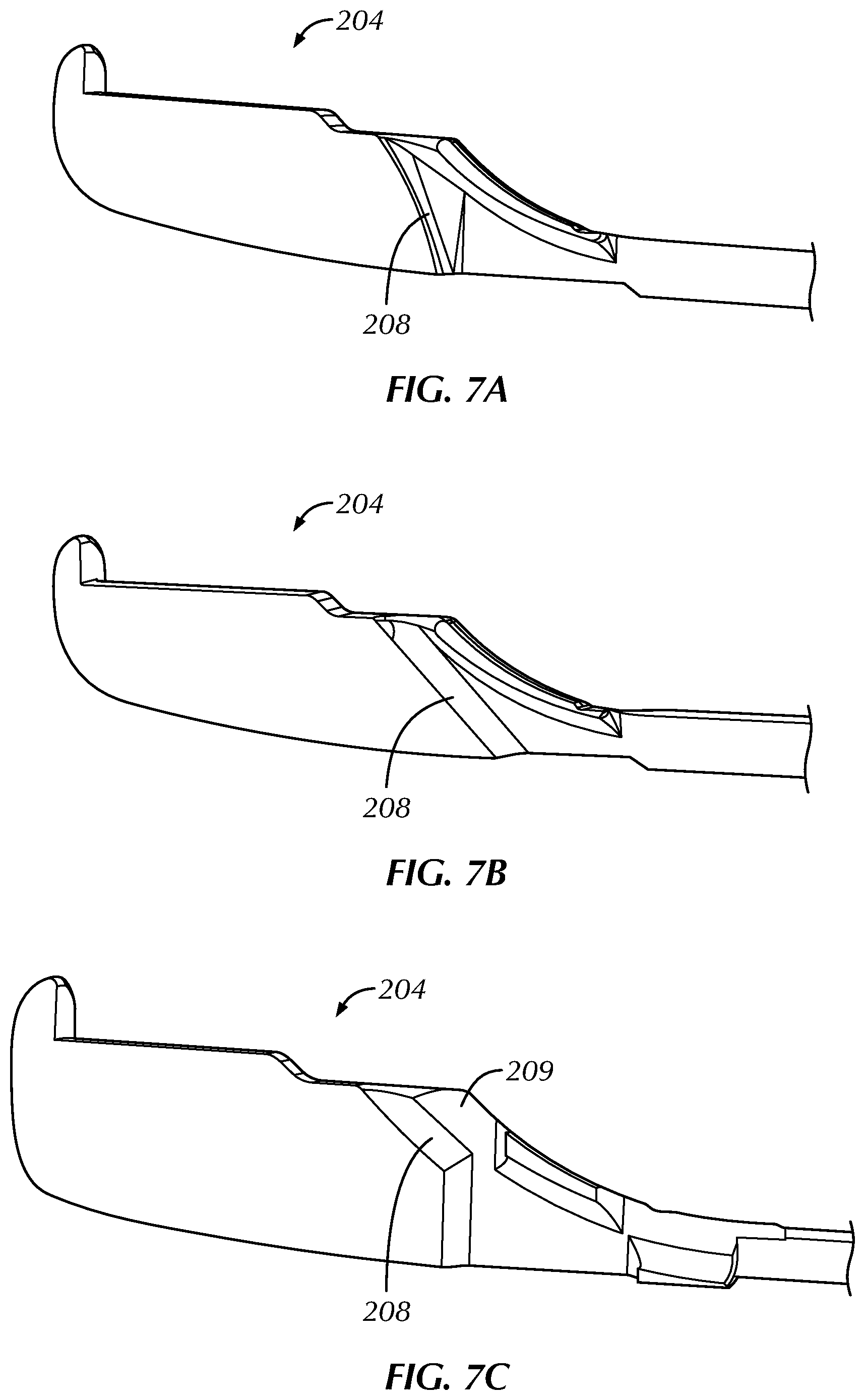
FIG. 5A is a side view of a portion of a distal tip component of a cartridge of one embodiment of a system for treating benign prostatic hyperplasia and FIG. 5B is an isometric, cross-sectioned view of the distal tip component. The shaft distal portion 204 includes a shaft distal portion exit port 205 from which the needle distal portion (not pictured) emerges when the needle distal portion is extended from the shaft distal portion 204. The shaft distal portion exit port 205 is the distal terminus of a shaft distal portion lumen 207. The shaft distal portion lumen 207 has a certain effective radius of curvature as the lumen transitions from running along the long axis of the shaft assembly to running in a direction transverse to the long axis of the shaft assembly. An aspect of that transition is the shaft distal portion interior exit wall 208, which is essentially the distal-most portion of the shaft distal portion lumen 207. Thus, when the needle assembly is present within the shaft assembly, the effective radius of curvature of the shaft distal portion lumen 207 and the shaft distal portion interior exit wall 208 can strongly influence the amount of friction that the needle assembly experiences when slid with respect to the shaft assembly.
FIG. 6A is a side view and FIG. 6B is a bottom view of a portion of the distal end of a handle and cartridge of one embodiment of a system for treating benign prostatic hyperplasia. FIGS. 6A and 6B depict the needle assembly in a retracted position within the shaft assembly 202 and shaft distal portion 204. That is, the needle assembly is being constrained to be substantially straight as compared to its shape-set configuration, which has a radius of curvature that positions the distal tip of the needle in a direction transverse to the long axis of the needle assembly. However, a shaft distal portion cutout 206 provides an opening between the interior and exterior of the shaft distal portion 204 and a portion of the needle distal portion 212 protrudes through the shaft distal portion cutout 206. The shaft distal portion cutout 206 allows at least part of the needle distal portion 212 to flex through a part of the bottom surface of the shaft distal portion 204, which allows the needle distal portion 212 to assume a smaller radius of curvature than would be possible if there were no shaft distal portion cutout 206. That is, the shaft distal portion cutout 206 allows for there to be less constraining force on the needle distal portion 212. The needle distal portion 212 does not contact a hard surface in the interior of the shaft distal portion 204 and this reduces the number of surfaces that the needle distal portion 212 contacts while moving with respect to the shaft distal portion 204. Fewer surface contacts can mean less overall friction during movement. Further, the shaft distal portion cutout 206 can help maintain the preferred exit trajectory of the needle distal portion 212 by accommodating part of the shape-set radius of curvature. In some embodiments, cutouts may be present along other parts of the shaft assembly 202. These cutouts can also reduce the number of surfaces that the needle assembly contacts while moving with respect to the shaft assembly 202.
FIGS. 7A, 7B, and 7C are perspective, sectional views of distal portions of cartridges of various embodiments of a system for treating benign prostatic hyperplasia. These embodiments illustrate various arrangements of the shaft distal portion interior exit wall 208. These embodiments intend to reduce the contact points and/or friction experienced by the needle assembly as it slides with respect to the shaft assembly. In FIG. 7A, the shaft distal portion interior exit wall 208 is configured with a radius of curvature that is at a tangent to the desired exit trajectory of the needle. In FIG. 7B, the shaft distal portion interior exit wall 208 is configured with a straight section where the entire section is angled at a tangent to the desired exit trajectory of the needle. In FIG. 7C, the shaft distal portion interior exit wall 208 is configured with a partial exit wall and a shaft distal portion upper cutout 209 on the upper surface. The shaft distal portion upper cutout 209 is intended to reduce the number of contact points between the shaft distal portion lumen and the needle assembly. Further, the shaft distal portion interior exit wall 208 is formed to minimize the presence of surface discontinuities, such as flash, burrs, or sharp edges.
FIG. 8 is a perspective, cutaway view of a distal portion of a cartridge of one embodiment of a system for treating benign prostatic hyperplasia. FIG. 8 illustrates one embodiment of the configuration of the shaft distal portion lumen 207 as the lumen curves towards the shaft distal portion exit port 205. In this configuration, the curved section of the shaft distal portion lumen 207 includes a tapered cross-sectional dimension such that the cross-sectional area of the shaft distal portion lumen 207 is larger at the proximal end of the curved section than at the distal end of the curved section. In one embodiment, the cross-sectional area and the cross-sectional shape of the shaft distal portion exit port 205 is incrementally larger than the outer diameter of the needle proximal portion. In one embodiment, the curvature of the curved section of the shaft distal portion lumen 207 matches the desired exit trajectory of the needle proximal portion.
FIG. 9 is an exploded, isometric view of one embodiment of a handle of a system for treating benign prostatic hyperplasia. The handle 100 includes a right handle case 103, a left handle case 104, and a cartridge bay 101 formed in the left handle case 104. The handle 100 is designed to transmit the energy stored in several springs (not pictured) within the handle 100 to a cartridge to enable the treatment of a patient. The energy is transmitted via the interaction of various mechanisms within the handle 100. A removable scope seal 105 covers the cartridge bay 101 and couples to the scope tube 102.
The mechanisms in the handle 100 include a handle trigger assembly 110, which is operatively connected to a handle trigger spring (not pictured) such that the handle trigger spring provides force sufficient to return the handle trigger assembly 110 to its initial position after the handle trigger assembly 110 has been squeezed and released by a user. A ratchet 114, which is connected to a ratchet spring (not pictured), affects the motion of the handle trigger assembly 110 such that the handle trigger assembly 110 does not return to its initial position prior to being moved (e.g. squeezed) to a predetermined position by a user. A safety 112 is connected to the handle trigger assembly 110 to ensure that the handle trigger assembly 110 is not operated accidentally. The handle trigger assembly 110 is connected to a drive gear 113, which is connected to a cam wheel 120.
The cam wheel 120 rotates about a central axis and, via structures and features on the cam wheel, triggers certain motions within the handle 100 as the cam wheel 120 rotates. There are multiple sleds operatively connected to the cam wheel 120, and the sleds move in a linear direction along a lateral axis of the handle 100. There are multiple springs that impart force to the multiple sleds to provide mechanical energy sufficient to deliver an implant. A cartridge includes multiple tab assemblies that mate with the sleds via slots in the sled such that the energy imparted by the operation of the mechanisms in the handle (such as the springs) is transmitted to the mechanisms in the cartridge.
A wheel actuator 125 is operatively connected to the cam wheel 120 and an implant sled 160, which is connected to an implant spring that provides energy related to the delivery of the implant. A needle sled 140 is operatively connected to the cam wheel 120 and an axle 145, and a needle sled spring provides energy related to the delivery of the implant. A suture sled 150 is operatively connected to the cam wheel 120, and a suture sled spring provides energy related to the delivery of the implant.
The handle 100 includes various other parts, such as a cover plate 130, a scope lock 170, a sheath lock 180, and various screws and/or fasteners to assemble the handle. The cover plate 130 provides the interior base for the cartridge bay 101. The scope tube 102, the scope lock 170, the scope seal 105, and the sheath lock 180 provide functionality for attaching an endoscope and other ancillary equipment (such as a surgical sheath) to facilitate the procedure.
FIG. 10 is an exploded, isometric view of one embodiment of a cartridge of a system for treating benign prostatic hyperplasia. In this embodiment, the cartridge 200 includes a cartridge cover 299 coupled to a cartridge base 298. These two parts couple with a shaft support 297 to form the cartridge body. The cartridge knob 203 couples to the cartridge cover 299.
The shaft support 297 is attached to the shaft assembly 202, which includes the shaft distal portion 204. An atraumatic tape 296 is present on a surface of the shaft distal portion 204 and helps reduce tissue trauma that could result from the tissue interacting with the various openings and joints on the shaft distal portion 204 (such as those described in FIGS. 5A and 5B). The proximal anchor component 84 is contained within the shaft distal portion 204 in a configuration that is separate and disconnected from the distal anchor component 70 and the suture portion (as described herein with respect to FIG. 3).
When the proximal anchor component 84 is connected to the distal anchor component 70 and the suture portion 78 as part of the implant deployment process, it is done via the action of the pusher assembly, which includes a pusher 242 connected with a pusher block 244. A cutter assembly, which cuts the suture 222 to create the suture portion 78 during the implant deployment process, includes a cutter 232 and a cutter block 234. The movement of the cutter assembly and the pusher assembly is coordinated by the interactions of the cutter pawl 236, the implant actuator 246, and the implant spring 248, as is described in more detail herein.
A suture assembly includes the suture 222, a suture support tube 224, a suture safety 226, and a suture proximal portion 228. The distal anchor component 70 is attached to a distal end portion of the suture 222 as described herein with respect to FIG. 3. The distal portion of the suture assembly and the distal anchor component 70 are contained within the needle assembly distal portion 212, which in turn is contained within the shaft assembly until the needle assembly distal portion 212 is moved into tissue during the implant deployment process. The movement of the needle assembly proximal portion 216 and the suture assembly proximal portion 228 is coordinated via the interaction of these features with mechanisms in the handle 100, as is described in more detail herein.
Referring again to the implant deployment process and to FIGS. 3, 4A, 4B, and 10, the anchor assembly (or implant) is deployed via a sequence of steps. The needle distal portion 212 extends from a shaft distal portion 204 to a fully extended position such that at least part of the needle distal portion 212 penetrates a tissue surface in a patient, such as the outer capsule of the prostate gland. The distal anchor component 70 and a distal portion of the suture 222 are contained within the needle distal portion 212 and move with the needle distal portion 212 to penetrate tissue.
Next, while the distal anchor component 70 and a distal portion of the suture 222 are held in place, the needle assembly 210 moves proximally such that the needle distal portion 212 moves to a partially retracted position. That is, the needle distal portion 212 moves proximally with respect to its fully extended position but is not yet completely retracted within the shaft assembly 202. The suture support tube 224 helps maintain the position of the distal anchor component 70 and a distal portion of the suture 222 while the needle assembly 210 moves proximally. Thus, the distal anchor component 70 and a distal portion of the suture 222 remain near the tissue surface and are no longer within the needle distal portion 212.
In a next step, the needle assembly 210 moves further proximally to be retracted within the shaft assembly 202 while the suture assembly also moves proximally. The tail portion 74 of the distal anchor component 70 is pulled snug against the tissue surface, which causes the distal anchor component 70 to pivot about the mid-section 80 such that the distal anchor component 70 is now transverse to a distal portion of the suture 222.
In a next step, the pusher assembly moves distally to push the proximal anchor component onto a distal portion of the suture 222 and this movement defines the suture portion 78 that becomes a component of the final anchor assembly implanted in the patient. And the cutter assembly moves proximally to pull a cutting edge through the suture. At this point, the anchor assembly is now completely detached from the cartridge and handle system.
The handle and cartridge system enables the multiple steps of deploying an anchor assembly through multiple squeezes of the handle trigger assembly by the user. That is, the relative motions of all the mechanisms in the handle and cartridge and the timing of those motions occurs via multiple squeezes of the handle trigger assembly. Referring now to FIG. 9, the handle trigger assembly 110 interacts with the cam wheel 120 via the drive gear 113 and the ratchet 114. The cam wheel 120 is the principal mechanism for the way in which the handle trigger assembly 110 drives the motion and timing of the various mechanisms in the handle and cartridge system.
FIGS. 11A and 11B are side views of a cam wheel within one embodiment of a handle of a system for treating benign prostatic hyperplasia. The cam wheel 120 is mounted on an axle molded into the right handle case. The left side of the cam wheel 120, shown in FIG. 11A, includes raised features 121a, 121b, and 121c. The raised features 121a, 121b, and 121c interact with other mechanisms in the handle (such as the various sleds) to facilitate the motion of the various assemblies in the cartridge (such as the needle assembly, suture assembly, pusher assembly, and cutter assembly). The right side of the cam wheel 120, shown in FIG. 11B, includes cam wheel gear teeth 123, which interact with the drive gear 113 (pictured in FIG. 9) to transmit the generally linear motion from squeezing the handle trigger assembly 110 to the circular motion of the cam wheel 120.
FIG. 12A is a view of the left side of a cam wheel and certain other mechanical features within one embodiment of a handle and cartridge of a system for treating benign prostatic hyperplasia. FIG. 12B is the same view as FIG. 12A with the cam wheel and other mechanical features in a different configuration than shown in FIG. 12A. In this embodiment the raised feature 121c on the cam wheel 120 is designed to interact with the wheel actuator 125 to release the pusher block 244 and then allow the wheel actuator 125 to be reset. When the cam wheel 120 rotates as a consequence of the user squeezing the handle trigger assembly, the raised feature 121c pushes against a wheel actuator head portion 126 causing the wheel actuator 125 to pivot about the wheel actuator axis 129. As the wheel actuator 125 pivots, the wheel actuator tail portion 127 interacts with the implant actuator 246 causing the implant actuator 246 to pivot. When the implant actuator 246 pivots, the pusher block 244 is released from a held position and allowed to move distally and push the proximal anchor component onto the suture as described elsewhere herein. Further rotation of the cam wheel 120 moves the raised feature 121c entirely past the wheel actuator head portion 126 such that the wheel actuator 125 can pivot back into its original position. The force necessary for returning the wheel actuator 125 to its original position is supplied by the wheel actuator flexure 128, which is flexed against the handle case during the pivot of the wheel actuator 125. Flexing the wheel actuator flexure 128 stores spring energy that is released to move the wheel actuator 125 back to its original position when the wheel actuator head portion 126 is clear of the raised feature 121c. The wheel actuator 125 is thus reset to be able to interact with the implant actuator 246 of the next cartridge that is mechanically engaged with the handle.
The implant actuator 246 and the pusher block 244 (as well as the proximal anchor component and the suture) are present in the cartridge while the cam wheel 120 and the wheel actuator 125 are present in the handle. FIGS. 12A and 12B illustrate the operation of these features when the cartridge and handle are mechanically engaged as described elsewhere herein. For clarity, other features are not pictured in FIGS. 12A and 12B but certain of those features are illustrated in FIGS. 13A and 13B.
FIG. 13A is a side view of certain mechanical features within one embodiment of a cartridge of a system for treating benign prostatic hyperplasia. FIG. 13B is the same view as FIG. 13A with the mechanical features in a different configuration than shown in FIG. 13A. The pusher block 244 is coupled to the cutter block 234 by the implant spring 248. Both the pusher block 244 and the cutter block 234 are constrained to move linearly along the long axis of the cartridge by the cartridge base 298 and shaft support 297 (each pictured in FIG. 10) that define the cartridge body. The implant spring 248 is an extension spring stretched between the pusher block 244 and the cutter block 234 and exerting a force that tends to draw the pusher block 244 and the cutter block 234 toward each other. The implant actuator 246 prevents the pusher block 244 from moving under the force of the implant spring 248 when the implant actuator 246 is in the configuration depicted in FIG. 13A (and FIG. 12A). The cutter pawl distal end 237 prevents the cutter block 234 from moving under the force of the implant spring 248 when the cutter pawl 236 is in the configuration depicted in FIG. 13A. When the implant actuator 246 is made to pivot as described herein with respect to FIGS. 12A and 12B, the pusher block 244 moves distally (i.e., the pusher assembly moves in the direction of arrow "P") to push the proximal anchor component onto the suture as described elsewhere herein. During this motion, which is driven by the force of the implant spring 248, the pusher block 244 interacts with the cutter pawl proximal end 238 and causes the cutter pawl 236 to pivot as depicted in FIG. 13B. The sloped front surface of the pusher block 244 and the curved surface of the cutter pawl proximal end 238 interact to create this pivoting motion. When the cutter pawl 236 pivots, the cutter pawl distal end 237 disengages from the cutter block 234 and allows the cutter block 234 to move proximally under the force of implant spring 248. The proximal motion of the cutter block 234 causes the suture to be cut to form the suture portion of the anchor assembly. That is, the cutter assembly moves in the direction of arrow "C" to complete the anchor assembly and detach it from the cartridge and handle system.
As described elsewhere herein, in some embodiments multiple cartridges can be used in serial fashion with a single handle. The steps and mechanisms illustrated and explained with respect to FIGS. 12A, 12B, 13A, and 13B disclose how the pushing and cutting steps of creating the anchor assembly can be repeatedly executed for multiple engagements of a series of cartridges with a single handle. One notable feature is that the implant spring is in an extended state as its initial condition. It is important, then, for the cartridge to have features that prevent the unintended release of the energy in the implant spring during handling of the cartridge.
FIG. 14 is a side view of certain mechanical features within one embodiment of a cartridge of a system for treating benign prostatic hyperplasia. FIG. 14 illustrates the mechanisms depicted in FIG. 13A (in the configuration of FIG. 13A) with the addition of the cartridge knob 203 to show the operational relationship among these features. FIG. 14 illustrates the cartridge knob 203 in the storage position. In the storage position, the grip section 292 is transverse to the long axis of the cartridge as compared to the engaged position shown in FIG. 1 in which the grip section 292 is aligned with the long axis of the cartridge. When in the storage position, the cartridge knob 203 functions as a safety interlock that prevents movement by the pusher block 244. In the storage position depicted in FIG. 14, a pusher safety tab 293 on the cartridge knob 203 engages with the distal end of the implant actuator 246 and prevents the implant actuator 246 from pivoting to release the pusher block 244 (as illustrated and explained with respect to FIGS. 12A and 12B). The pusher safety tab 293 allows the cartridge to be safely handled and stored while the implant spring is in its extended configuration.
FIG. 15A is an exploded top view of certain mechanical features within one embodiment of a cartridge of a system for treating benign prostatic hyperplasia and 15B is an exploded perspective view of the same features. FIGS. 15A and 15B illustrate another safety interlock in which a cutter safety tab 286 is present on the cutter pawl 236. The cutter safety tab 286 engages with the cartridge knob 203 when the cartridge knob 203 is in the storage position and prevents the cutter pawl 236 from pivoting to release the cutter block (as illustrated and explained with respect to FIGS. 13A and 13B). The cutter safety tab 286 allows the cartridge to be safely handled and stored while the implant spring is in its extended configuration. FIGS. 15A and 15B are illustrated in an exploded view to make the relevant features clear. However, in operation the cutter pawl 236 is closely engaged with the cartridge knob 203 to create the mechanical interactions described herein.
The pusher safety tab 293 and the cutter safety tab 286 solve the problem of accidental firing of the pusher assembly and/or cutter assembly in the cartridge by correlating safety interlocks with the storage position of the cartridge knob 203.
FIGS. 16A, 16B, 17, 18A, and 18B illustrate an embodiment of another safety interlock mechanism in the handle and cartridge system. FIG. 16A is a perspective view of a portion of a handle 100 and FIG. 16B is a side view of the handle 100. The handle 100 includes a cartridge bay 101, which is shown as empty because the cartridge has been removed. In this embodiment, the handle trigger assembly 110 includes cartridge lock tab 111 that protrudes into the cartridge bay 101 when the handle trigger assembly 110 pivots away from the handle grip 115. The presence of the cartridge lock tab 111 in the cartridge bay 101 prevents a cartridge from being engaged with the handle 100 because a cartridge cannot physically fit in the cartridge bay 101 with the cartridge lock tab 111 in the cartridge bay 101. This mechanism prevents a cartridge from being inserted into the handle except under the proper condition--when the handle trigger assembly 110 is in its initial position closest to the handle grip 115. This initial position corresponds with the mechanisms in the handle all being reset to their initial positions.
FIG. 17 is a side view of a portion of a cartridge of one embodiment of a system for treating benign prostatic hyperplasia. In FIG. 17, the cartridge cover has been removed, revealing the cartridge base 298. The cartridge knob 203 is also shown. A cartridge lock surface 291 is present on the shaft support 297 and extends below the cartridge base 298 in this side view of a portion of a cartridge.
FIGS. 18A and 18B are a perspective view and a side view, respectively, of certain mechanical features within one embodiment of a cartridge of a system for treating benign prostatic hyperplasia. FIGS. 18A and 18B illustrate the interaction between the cartridge lock tab 111 present on the handle trigger assembly 110 and the cartridge lock surface 291 present on the shaft support 297. When the handle trigger assembly 110 is in a position pivoted away from the handle grip, the cartridge lock tab 111 prevents the cartridge from being removed from the cartridge bay by physically impeding the removal of the cartridge. While the cartridge lock surface 291 is present on the shaft support 297 in this embodiment, a similar lock surface could be present on other parts of the cartridge, such as the cartridge base 298, and function in a substantially similar manner.
The cartridge lock tab 111 present on the handle trigger assembly 110 provides a method of preventing insertion of a cartridge until the handle is in the reset, initial condition, and prevents removal of a cartridge (via interaction with the cartridge lock surface 291) while the handle and cartridge system is in use. There are alternative embodiments for preventing removal and/or insertion of a cartridge. For example, a cartridge may contain a lock receiving feature and the handle may contain an additional wheel with a series of locking features. As the handle trigger is pulled, the additional wheel rotates such that a locking feature engages the lock receiver. If a complete cycle for implantation requires multiple trigger squeezes, then the additional wheel includes sufficient locking features to keep the cartridge locked in during the entire cycle. At the end of the cycle, a portion of the additional wheel containing no locking feature is aligned with the lock receiver. In this way, the cartridge would be unlocked and removable from the handle. As another example, a locking slide could be engaged by a user to lock the cartridge in the handle. When the trigger is squeezed, the slide would be mechanically engaged to prevent the slide from moving until the cycle was completed.
FIGS. 19A and 19B are side views of a portion of a cartridge of one embodiment of a system for treating benign prostatic hyperplasia. The cartridge knob 203 is engaged with the cartridge cover 299, which includes a cartridge indicator window 290. In FIG. 19A, the cutter block 234 is visible through the cartridge indicator window 290, which indicates that the cutter block 234 is still in its initial position and has not moved proximally to cut the suture in the final step of the formation of the anchor assembly. FIG. 19A shows that the cutter block 234 fills the cartridge indicator window 290. In FIG. 19B, only a small portion of the cutter block 234 is visible through the cartridge indicator window 290. That is, the cutter block 234 no longer fills the cartridge indicator window 290. This indicates that the cutter block 234 has moved proximally and cut the suture for the final step of the formation of the anchor assembly.
FIG. 19C is a perspective view of a cartridge cover of one embodiment of a system for treating benign prostatic hyperplasia. FIG. 19C illustrates that the cartridge indicator window 290 is cut through the distal surface of the cartridge cover 299. The cartridge indicator window 290 is able to perform an additional function of providing access to the cutter block 234 such that a user can use a small tool to move the cutter block distally to cut the suture if the cutter block 234 stalls and does not completely cut the suture. The presence of an opening to the cartridge indicator window 290 through the distal surface of the cartridge cover 299 allows for easy access to the cutter block 234 with a tool, even when the user is not looking directly at the cartridge indicator window 290.
FIG. 20A is an exploded perspective view of certain mechanical features within one embodiment of a cartridge of a system for treating benign prostatic hyperplasia and FIG. 20B is a side view of those mechanical features in their engaged configuration. As described herein with respect to FIGS. 13A and 13B, in normal operation the cutter pawl 236 pivots and disengages from the cutter block allowing the cutter block to move proximally under the force of the implant spring. The proximal motion of the cutter block causes the suture to be cut to form the suture portion of the anchor assembly. However, in some circumstances it may be desirable for a user to interrupt the deployment sequence after deployment of the distal anchor component but prior to the deployment of the proximal anchor component and cutting of the suture. In such a circumstance, the handle and cartridge system can then be removed from the patient without completely assembling and deploying the implant.
FIGS. 20A and 20B illustrate that the cutter pawl 236 includes a cutter manual tab 284 that is accessible to a user via a cutter access window 288 present on the cartridge knob 203. The cutter manual tab 284 is configured as an off-center ramp such that pushing on the cutter manual tab 284 through the cutter access window 288 creates rotation (or pivoting) of the cutter pawl 236 perpendicular to the push direction. A user can push the cutter manual tab 284 with a small tool inserted through the cutter access window 288. Indentations in the cartridge knob 203 near the cutter access window 288 guide the tool towards the cutter access window 288. As described herein, this pivoting of the cutter pawl 236 disengages the cutter pawl from the cutter block and allows the cutter block to move proximally under the force of the implant spring to cut the suture.
The disclosed apparatus can be implemented in various treatment devices employed for various medical purposes including, but not limited to, retracting, lifting, compressing, approximating, supporting, remodeling, repositioning, ablating, or otherwise altering tissues, organs, anatomical structures, grafts, or other material found within the body of a human or animal subject. In certain embodiments, treatment devices are intended to displace, compress, retract, or destroy tissue of the prostate to facilitate treatment of diseases or disorders such as BPH.
Other treatment devices may benefit from the use of the embodiments disclosed herein. Treatment devices equipped with various tools which manipulate, ablate, or otherwise alter tissue, where those tools are moved, deployed, or driven by mechanical energy, can benefit from the use of the safety interlocks and mechanisms, as well as the use of the deployment indicators, disclosed herein. Such tools can include, but are not limited to, needles, cutting blades, vacuums, grasping arm assemblies, expandable cutting members, blunt dissectors, noose or ligature clips, articulating heads with an integral or retractable blade, helical blades, electrodes for delivery of radiofrequency energy, cutting wires or rings, electrocauterizing probes, or staple or suture delivery heads.
In some embodiments, the system includes a cartridge carrying at least one implant and a handle configured to receive the cartridge. The handle includes an actuator and at least one spring mechanism loaded with mechanical energy. The handle also includes a member that mates with the cartridge to transfer mechanical energy from the spring mechanism to the cartridge for deploying the implant. The handle and cartridge system includes a first firing sled that has slots aligning with pusher tabs on a needle assembly. The slots of the first firing sled and the pusher tabs of the needle assembly are complementary mechanisms that allow for the transfer of energy from the spring mechanism via the first firing sled to fire a needle in the cartridge. The handle and cartridge system can also include a second firing sled with slots aligning with pusher tabs on a suture tube or connector tube. The slots of the second firing sled and the pusher tabs of the suture tube are complementary mechanisms that allow for the transfer of energy from the spring mechanism via the second firing sled to advance the suture tube simultaneously with the needle tube.
While particular elements, embodiments and applications of the present invention have been shown and described, it will be understood that the invention is not limited thereto since modifications can be made by those skilled in the art without departing from the scope of the present disclosure, particularly in light of the foregoing teachings.
TABLE-US-00001 Table of Reference Numerals 1 system 70 distal anchor component 72 head portion 74 tail portion 78 suture portion 80 mid-section 82 connector section 84 proximal anchor component 95 back end 96 prong 98 tab 100 handle 101 cartridge bay 102 scope tube 103 right handle case 104 left handle case 105 scope seal 110 handle trigger assembly 111 cartridge lock tab 112 safety 113 drive gear 114 ratchet 115 handle grip 120 cam wheel 125 wheel actuator 126 wheel actuator head portion 127 wheel actuator tail portion 128 wheel actuator flexure 129 wheel actuator axis 130 cover plate 140 needle sled 145 axle 150 suture sled 160 implant sled 170 scope lock 180 sheath lock 200 cartridge 201 cartridge body 202 shaft assembly 203 cartridge knob 204 shaft distal portion 205 shaft distal portion exit port 206 shaft distal portion cutout 207 shaft distal portion lumen 208 shaft distal portion interior exit wall 209 shaft distal portion upper cutout 210 needle assembly 212 needle distal portion 214 needle shaft 216 needle proximal portion 220 suture assembly 222 suture 224 suture support tube 226 suture safety 228 suture proximal portion 230 cutter assembly 232 cutter 234 cutter block 236 cutter pawl 237 cutter pawl distal end 238 cutter pawl proximal end 240 pusher assembly 242 pusher 244 pusher block 246 implant actuator 248 implant spring 284 cutter manual tab 286 cutter safety tab 288 cutter access window 290 cartridge indicator window 291 cartridge lock surface 292 grip section 293 pusher safety tab 296 atraumatic tape 297 shaft support 298 cartridge base 299 cartridge cover
* * * * *
D00000
D00001
D00002
D00003
D00004
D00005
D00006
D00007
D00008
D00009
D00010
D00011
D00012

D00013

D00014

D00015

D00016

D00017
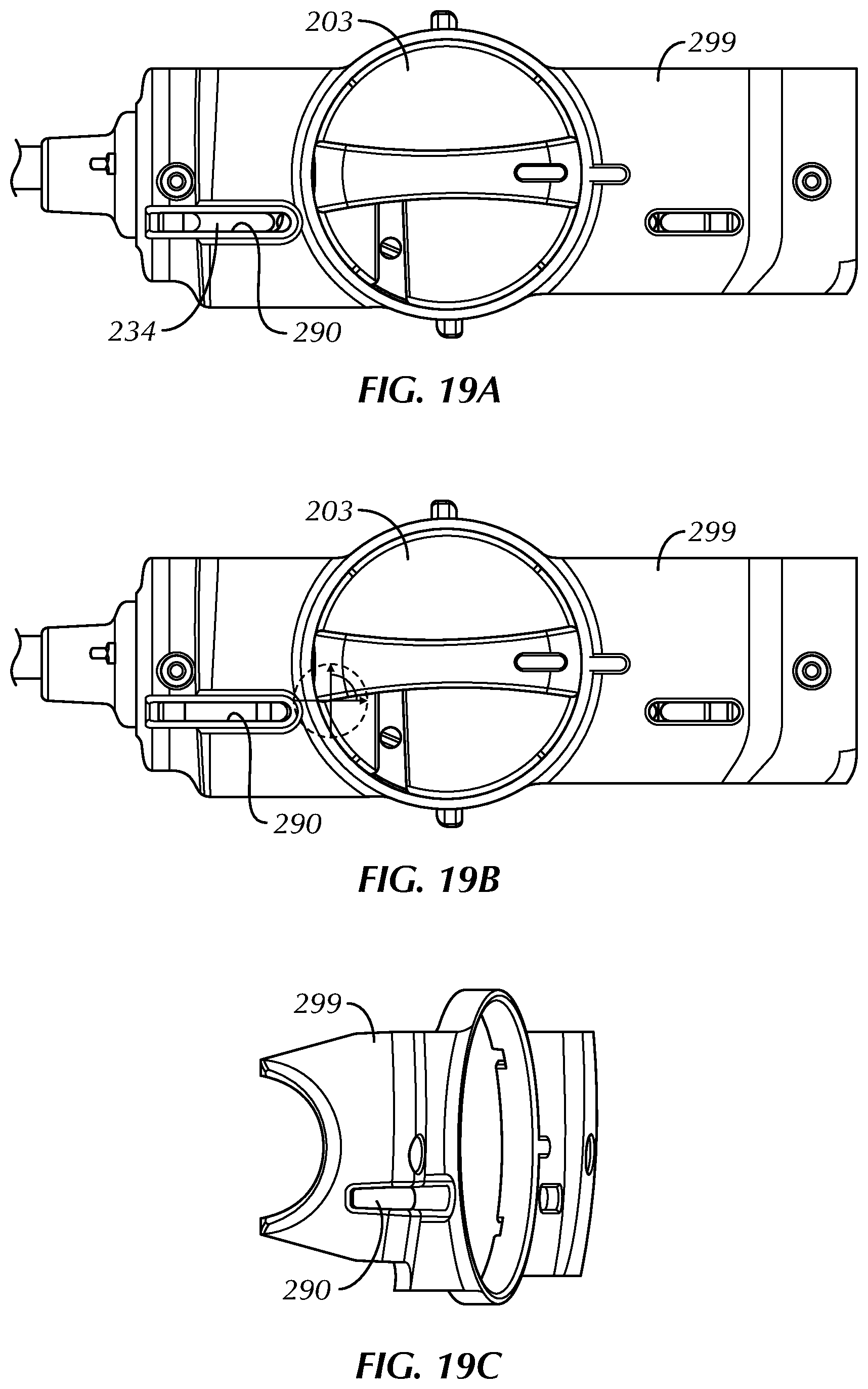
D00018

XML
uspto.report is an independent third-party trademark research tool that is not affiliated, endorsed, or sponsored by the United States Patent and Trademark Office (USPTO) or any other governmental organization. The information provided by uspto.report is based on publicly available data at the time of writing and is intended for informational purposes only.
While we strive to provide accurate and up-to-date information, we do not guarantee the accuracy, completeness, reliability, or suitability of the information displayed on this site. The use of this site is at your own risk. Any reliance you place on such information is therefore strictly at your own risk.
All official trademark data, including owner information, should be verified by visiting the official USPTO website at www.uspto.gov. This site is not intended to replace professional legal advice and should not be used as a substitute for consulting with a legal professional who is knowledgeable about trademark law.