Anti-AP2 antibodies and antigen binding agents to treat metabolic disorders
Hotamisligil , et al. May 25, 2
U.S. patent number 11,014,979 [Application Number 16/197,066] was granted by the patent office on 2021-05-25 for anti-ap2 antibodies and antigen binding agents to treat metabolic disorders. This patent grant is currently assigned to President and Fellows of Harvard College. The grantee listed for this patent is President and Fellows of Harvard College. Invention is credited to Ralph Adams, Mehmet F. Burak, Carl Brendan Doyle, Feyza Engin, Karine Jeannine Madeleine Herve, Gokhan S. Hotamisligil, Karen Inouye, Adrian Richard Moore, Elisabeth Helen Roberts, Kerry Louise Tyson, Shauna Mhairi Wales, Scott B. Widenmaier.


View All Diagrams
| United States Patent | 11,014,979 |
| Hotamisligil , et al. | May 25, 2021 |
Anti-AP2 antibodies and antigen binding agents to treat metabolic disorders
Abstract
This invention is in the area of improved anti-aP2 antibodies and antigen binding agents, and compositions thereof, which target the lipid chaperone aP2/FABP4 (referred to as "aP2") for use in treating disorders such as diabetes, obesity, cardiovascular disease, fatty liver disease, and/or cancer, among others. In one aspect, improved treatments for aP2 mediated disorders are disclosed in which serum aP2 is targeted and the biological activity of aP2 is neutralized or modulated using low-binding affinity aP2 monoclonal antibodies, providing lower fasting blood glucose levels, improved systemic glucose metabolism, increased systemic insulin sensitivity, reduced fat mass, reduced liver steatosis, reduced cardiovascular disease and/or a reduced risk of developing cardiovascular disease.
| Inventors: | Hotamisligil; Gokhan S. (Wellesley, MA), Burak; Mehmet F. (Brighton, MA), Engin; Feyza (Madison, WI), Widenmaier; Scott B. (Brighton, MA), Roberts; Elisabeth Helen (Slough, GB), Moore; Adrian Richard (Slough, GB), Doyle; Carl Brendan (Slough, GB), Adams; Ralph (Slough, GB), Herve; Karine Jeannine Madeleine (Vancouver, CA), Wales; Shauna Mhairi (Slough, GB), Tyson; Kerry Louise (Slough, GB), Inouye; Karen (Boston, MA) | ||||||||||
|---|---|---|---|---|---|---|---|---|---|---|---|
| Applicant: |
|
||||||||||
| Assignee: | President and Fellows of Harvard
College (Cambridge, MA) |
||||||||||
| Family ID: | 1000005573916 | ||||||||||
| Appl. No.: | 16/197,066 | ||||||||||
| Filed: | November 20, 2018 |
Prior Publication Data
| Document Identifier | Publication Date | |
|---|---|---|
| US 20190161536 A1 | May 30, 2019 | |
Related U.S. Patent Documents
| Application Number | Filing Date | Patent Number | Issue Date | ||
|---|---|---|---|---|---|
| 15143162 | Apr 29, 2016 | 10160798 | |||
| 62268257 | Dec 16, 2015 | ||||
| 62232148 | Sep 24, 2015 | ||||
| 62155217 | Apr 30, 2015 | ||||
| Current U.S. Class: | 1/1 |
| Current CPC Class: | C07K 16/18 (20130101); A61K 39/00 (20130101); A61K 2039/6018 (20130101); C07K 2317/565 (20130101); C07K 2317/24 (20130101); C07K 2317/55 (20130101); C07K 2317/76 (20130101); C07K 2317/92 (20130101); A61K 2039/507 (20130101); C07K 2317/70 (20130101); A61K 2039/505 (20130101) |
| Current International Class: | C07K 16/18 (20060101); A61K 39/00 (20060101) |
References Cited [Referenced By]
U.S. Patent Documents
| 5858366 | January 1999 | Sodroski et al. |
| 5889167 | March 1999 | Cascieri et al. |
| 6548529 | April 2003 | Robl et al. |
| 7390824 | June 2008 | Robl et al. |
| 7906117 | March 2011 | Smith et al. |
| 8846413 | September 2014 | Ruzicka |
| 9062104 | June 2015 | Garcia-Martinez et al. |
| 2002/0035064 | March 2002 | Robl et al. |
| 2003/0040516 | February 2003 | Sulsky et al. |
| 2004/0010119 | January 2004 | Guo et al. |
| 2004/0110226 | June 2004 | Lazar et al. |
| 2009/0022659 | January 2009 | Olson et al. |
| 2012/0134998 | May 2012 | Hotamisligil et al. |
| 2013/0302399 | November 2013 | Feldhaus et al. |
| 2015/0093769 | April 2015 | Ruzicka |
| 2016/0297874 | October 2016 | Hotamisligil et al. |
| 103665139 | Mar 2014 | CN | |||
| WO 00/15230 | Mar 2000 | WO | |||
| WO 2000/15229 | Mar 2000 | WO | |||
| WO 00/47734 | Aug 2000 | WO | |||
| WO 03/043624 | May 2003 | WO | |||
| WO 2010/057260 | May 2010 | WO | |||
| WO 2010/102171 | Sep 2010 | WO | |||
| WO 2014/093189 | Jun 2014 | WO | |||
| WO 2016/044337 | Mar 2016 | WO | |||
Other References
|
US. Pat. No. 9,879,078 B2, U.S. Appl. No. 15/093,508, Hotamisligil et al., Jan. 30, 2018. cited by applicant . Almagro & Fransson, Frontiers in Bioscience 2008; 13:1619-33. cited by applicant . De Genst et al., Dev Comp Immunol 2006; 30:187-98. cited by applicant . European Search Report for PCT/US2017039585 dated Feb. 18, 2020. cited by applicant . Brand C L et al., "Immunonuetralization of endogenous glucagon with monoclonal glucagon antibody normalizes hyperglycaemia in moderately streptozotocin-diabetic rats", Diabetologia, 1994, 34(10), 985-993, XP00908952; ISSN: 0012-186X. cited by applicant . Chen et al., "Enhancement and destruction of antibody function by somatic mutation: unequal occurrence is controlled by V gene combinatorial associations", The EMBO Journal, 1995, 14(12), 2784-2794, XP055530299, ISSN: 0261-4189, DOI: 10.1002/j.1460-2075. 1995.tb07278.x. cited by applicant . Colman P. M., "Effects of amino acid sequence changes on antibody-antigen interactions", Research in Immunology, Editions Scientifiques et Medicales Elsevier, FR, 1994, 145(1), 33-36, XP023944838, ISSN: 0923-2494, DOI: 10.1016/S0923-2494(9)80039-1. cited by applicant . European Search Report for PCT/US2016/030303 dated Apr. 20, 2020. cited by applicant . Kussie, Ph et al., "A single engineered amino acid substituted changes antibody fine specificity", The Journal of Immunology, 1994, 152, 146-152, XP055530279. cited by applicant . Joyner C J et al., "Development of a monoclonal antibody to the aP2 protein to identify adipocyte precursors in tumors of adipose differentiation", Pathology Research and Practice, 1999, 195(7), 461-466, XP002797422, ISSN: 0344-0338. cited by applicant . Rudikoff et al., "Single amino acid substitution altering antigen-binding specificity", Proceeding of the National Academy of Sciences, National Academy of Sciences, US, 1982, 79, 1979-1983, DOI: 10.1073/PNAS.79.6.1979. cited by applicant . Barf et al. "N-Benzyl-indolo carboxylic acids: Design and synthesis of potent and selective adipocyte fatty-acid binding protein (A-FABP) inhibitors" Bioorganic and Medicinal Chemistry Letters, Mar. 15, 2009; 19(6): 1745-1748. cited by applicant . Banaszak et al. "Lipid-binding proteins: a family of fatty acid and retinoid transport proteins" Adv. Protein Chem., 1994; 45: 89-151. cited by applicant . Baxa et al. "Human adipocyte lipid-binding protein: purification of the protein and cloning of its complementary DNA" Biochemistry, 1989: 28: 8683-8690. cited by applicant . Boord et al. "Adipocyte Fatty Acid-Binding protein, aP2, Alters Late Atherosclerotic Lesion Formation in Severe Hypercholesterolemia" Arterioscler. Thromb. Vasc. Biol., Oct. 2002; 22(10): 1686-1691. cited by applicant . Burak, M.F., et al, "Development of a therapeutic monoclonal antibody targeting secreted aP2 to treat type 2 diabetes;" PDB:5D8J deposited on Aug. 17, 2015 and released on Jan. 13, 2016, available at: http://www.rcsb.org/pdb/explore/explore.do?structureId=5D8J. cited by applicant . Burak, M.F., et al., "Chain L, Development of a Therapeutic Monoclonal AntibodyTargeting Secreted Ap2 to Treat Type 2 Diabetes." PDB:5D8J_L deposited on Aug. 17, 2015 and released on Aug. 2, 2017, available at: https://www.ncbi.nlm.nih.gov/protein/5D8J_L. cited by applicant . Burak, M.F., et al., "Chain D, Development of a Monoclonal Antibody Targeting Secreted Ap2 to Treat Diabetes and Fatty Liver Disease," PDB:5CON_D deposited on Jun. 12, 2015 and released on Jan. 13, 2016, available at: https://www.ncbi.nlm.nih.gov/protein/5C0N_D. cited by applicant . Burak, M.F., et al., "Development of a monoclonal antibody targeting secreted aP2 to treat diabetes and fatty liver disease," PDB:5CON deposited on Jun. 12, 2015 and released on Jun. 24, 2015, available at: http://www.rcsb.org/pdb/explore/explore.do?structureId=5C0N. cited by applicant . Cabre et al. "Fatty acid binding protein 4 is increased in metabolic syndrome and with thiazolidinedione treatment in diabetic patients" Altheroscherosis, 2007; 195: e150-e158. cited by applicant . Cai et al. "Benzbromarone, an old uricosuric drug, inhibits human fatty acid binding protein 4 in vitro and lowers the blood glucose level in db/db mice" Acta Pharmacologica Sinica, 2013; 34: 1397-1402. cited by applicant . Cao et al. "Identification of a Lipokine, a Lipid Hormone Linking Adipose Tissue to Systemic Metabolism" Cell, Sep. 19, 2008: 134:933-944. cited by applicant . Cao et al. "Adipocyte lipid chaperone aP2 is a secreted adipokine regulating hepatic glucose production" Cell Metabolism, May 2013; 17(5); 768-778. cited by applicant . Cao et al. "Regulation of Metabolic Responses by Adipocyte/Macrophage Fatty Acid-Binding Proteins in Leptin-Deficient Mice" Diabetes, Jul. 2006; 55: 1915-1922. cited by applicant . Cayman Chemical FABP4 Polyclonal Antibody product sheet (5 pages) downloaded on Sep. 30, 2015. cited by applicant . DeFronzo "Insulin resistance, lipotoxicity, type 2 diabetes and atherosclerosis: the missing links. The Claude Brenard Lecture 2009" Diabetologia, 2010; 53: 1270-1287. cited by applicant . Distel et al. "Fatty Acid Regulation of Gene Expression: Transcriptional and Post-Transcriptional Mechanism" The Journal of Biological Chemistry; Mar. 25, 1992; 267(9): 5937-5941. cited by applicant . Erbay et al. "Reducing endoplasmic reticulum stress through a macrophage lipid chaperone alleviates altherosclerosis" Nature Medicine, Dec. 2009; 15(12): 1383-1391. cited by applicant . Fu et al. "Oxidized LDL induces the expression of ALBP/aP2 mRNA and protein in human THP-1 macrophases" Journal of Lipid Research, 2000; 41: 2017-2023. cited by applicant . Furuhashi et al. "Adipocyte/Macrophage Fatty Acid-Binding Proteins Contribute to Metabolic Deterioration Through Actions in Both Macrophages and Adippocytes in Mice" J. Clin. Invest., Jul. 2008; 118(7): 2640-2650. cited by applicant . Furuhashi et al. "Serum Fatty Acid-Binding Protein 4 is a Predictor of Cardiovascular Events in End-Stage Renal Disease" PLOS ONE, Nov. 2011; 6(11): e27356. cited by applicant . Gillilan et al. "Structural Basis for Activation of Fatty Acid-binding Protein 4" J. Mol. Biol. 2007; 372:1246-1260. cited by applicant . Girona et al. "FABP4 Induces Vascular Smooth Muscle Cell Proliferation and Migration through a MAPK-Dependent Pathway" PloS ONE, Nov. 2013; 8(11): e81914. cited by applicant . Gorbenko et al. "Generation and Characterization of Monoclonal Antibodies against FABP4" Hybridoma, 2006; 25(2): 86-90. cited by applicant . Hall et al. "USP7 Attenuates Hepatic Gluconoegenesis Through Modulation of FoxO1 Gene Promoter Occupancy" Mol. Endocrinol., Jun. 2014; 28(6): 912-924. cited by applicant . Hecker et al. "Heat Shock proteins as biomarkers for the rapid detection of brain and spinal cord ischemia: a review and comparison to other methods of detection in thoracic aneurysm repair" Cell Stress and Chaperones, 2011; 16: 119-131. cited by applicant . Hellberg et al. "X-ray crystallographic analysis of adipocyte fatty acid binding protein (aP2) modified with 4-hydroxy-2-nonenal" Protein Science, 2010; 19: 1480-1489. cited by applicant . Hertzel et al. "Identification and characterization of a small molecule inhibitor of Fatty Acid binding proteins" Journal of Medicinal Chemistry, Oct. 8, 2009; 52(19): 6024-6031. cited by applicant . Hoo et al. "Pharmacological inhibition of adipocyte fatty acid binding protein alleviates both acute liver injury and non-aclcoholic steatohepatitis in mice" Journal of Hepatology, 2013; 58: 358-364. cited by applicant . Hotamisligil et al. "Uncoupling of Obesity from Insulin Resistance Through a Targeted Mutation in aP2, the Adipocyte Fatty Acid Binding Protein" Science, Nov. 22, 1996; 274(5291): 1377-1379. cited by applicant . Hunt et al. "Adipocyte P2 gene: Developmental expression and homology of 5'-flanking sequences among fat cell-specific genes" PNAS, Jun. 1986; 83: 3786-3790. cited by applicant . Hunter-Lavin et al. "Hsp70 release from peripheral blood mononuclear cells" Biochemical and Biophysical Research Communications, Nov. 12, 2004; 324(2): 511-517. cited by applicant . Ishimura et al. "Circulating Levels of Fatty Acid-Binding Protein Family and Metabolic Phenotype in the General Population" PLUS ONE, Nov. 2013; 8(11): e81318. cited by applicant . Jack et al. "C-terminal binding protein: A metabolic sensor implicated in regulating adipogenesis" The International Journal of Biochemistry & Cell Biology, 2011; 43: 693-696. cited by applicant . Jysal et al. "Improved Glucose and Lipid Metabolism in Genetically Obese Mice Lacking aP2" Endocrinology, 2000; 141: 3388-3396. cited by applicant . Kaess et al. "Cardiometabolic Correlates and Heritability of Fetuin-A, Retinol-Binding Protein 4, and Fatty-Acid Binding Protein 4 in the Framingham Heart Study" J. Clin. Endocrinol. Metab., Oct. 2012 97(10): e1943-e1947. cited by applicant . Kajimura et al. "Regulation of the brown and white fat gene programs through a PRDM16/CtBP transcriptional complex" Genes & Development, 2008; 22: 1397-1409. cited by applicant . Karakas et al "Serum fatty acid binding protein 4, free fatty acids, and metabolic risk markers" Metabolism Clinical and Experimental, 2009; 58: 1002-1007. cited by applicant . Kashima et al. "Diagnostic utility of aP2/FABP4 expression in soft tissue tumors" Virchows Archiv., Apr. 2013; 462(4): 465-472. cited by applicant . LaLonde et al., "X-ray Crystallographic Structures of Adipocyte Lipid-Binding Protein Complexed with Palmitate and Hexadecanesulfonic Acid. Properties of Cavity Binding Sites" Biochemistry, 1994; 33: 4885-4895. cited by applicant . Lan et al. "Small-molecule inhibitors of FABP4/5 ameliorate dyslipidemia but not insulin resistance in mice with diet-induced obesity" Journal of Lipid Research, Apr. 2011; 52(4): 646-656. cited by applicant . Layne et al. "Role of macrophage-expressed adipocyte fatty acid binding protein in the development of accelerated atherosclerosis in hypercholesterolemic mice" The FASEB Journal, Dec. 2001; 15: 2733-2735. cited by applicant . Lin et al. "Hormonal Regulation of Hepatic Glucose Production in Health and Disease" Cell Metabolism, Jul. 6, 2011; 14:9-19. cited by applicant . Maeda et al. "Adipocyte/Macrophage Fatty-Acid Bonding Proteins Control Integrated Metabolic Responses in Obesity and Diabetes" Cell Metab., Feb. 2005; 1:107-119. cited by applicant . Makowski et al. "Lack of Macrophage Fatty-Acid-Binding Protein aP2 Protects Mice Deficient in Apolipoprotein E Against Atherosclerosis" Nat. Med., Jun. 2001; 7(6):699-705. cited by applicant . Makowski et al. "The Fatty Acid-binding Protein, aP2, Coordinates Macrophage Cholesterol Trafficking and Inflammatory Activity" The Journal of Biological Chemistry, Apr. 1, 2005; 280(13): 12888-12895. cited by applicant . Melki et al. "Expression of the adipocyte fatty acid-binding protein streptozotocin--diabetes: efects of insulin deficiency and supplementation" Journal of Lipid Research, 1993; 34: 1527-1534. cited by applicant . Miao et al. "The mAb against adipocyte fatty acid-binding protein 2E4 attenuates the inflammation in the mouse model of high-fat diet-induced obesity via toll-like recetor 4 pathway" Molecular and Cellular Endocrinology, 2015; 403: 1-9. cited by applicant . Nardini et al. "CtBP/BARS: a dual-function protein involved in transcription co-repression and Golgi membrane fission" the EMBO Journal, 2003; 22(12): 3122-3130. cited by applicant . Ozcan et al. "Chemical Chaperones Reduce ER Stress and Restore Glucose Homeostasis in a Mouse Model of Type 2 Diabetes" Science, Aug. 25, 2006; 313(5790): 1137-1140. cited by applicant . PCT/US2016/030303, Invitation to pay additional fees and, where applicable, protest fee, dated Sep. 12, 2016. cited by applicant . PK Office Action dated Sep. 26, 2017 for Pakistan application No. 246/2016. cited by applicant . Rosen, et al. "Adipocytes as Regulators of Energy Balance and Glucose Homeostasis" Nature, Dec. 14, 2006; 444:847-853. cited by applicant . Saksi et al. "Low-Expression Variant of Fatty Acid-Binding Protein 4 Flavors Reduced Manifestations of Altherosclerotic Disease and Increased Plaque Stability" Circ. Cardiovasc. Genet., 2014; 7: 588-598. cited by applicant . Storch et al. "Structural and functional analysis of fatty acid-binding proteins" Journal of Lipid Research, 2009; 50:S126-S131. cited by applicant . Suh et al. "Serum AFBP levels are elevated in patients with nonalcoholic fatty liver disease" Scandinavian Journal of Gastroenterology, 2014; 49(8): 979-985. cited by applicant . Sulsky et al. "Potent and selective biphenyl azole inhibitors of adipocyte fatty acid binding protein (aFABP)" Bioorganic and Medicinal Chemistry Letters, Jun. 15, 2007; 17(12):351-3515. cited by applicant . van Dongen et al. "Structure-based screening as applied to human FABP4: a highly efficient alternative to HTS for hit generation" Journal of the American Chemical Society, Oct. 9, 2002; 124(40): 11874-11880. cited by applicant . Tuncman et al. "A genetic variant at the fattyacid-binding protein aP2 locus reduces the risk for hypertriglceridemia, type 2 diabetes, and cardiovascular disease" PNAS, May 2, 2006; 103(18): 6970-6975. cited by applicant . Vernochet et al. "C/EBPalpha and the Corespressors CtBP2 Regulate Repression of Select Visceral White Adipose Genes during Induction of the Brown Phenotype in White Adipocytes by Peroxisome Projiferator-Activated Receptor gamma Agonists" Molecular and Cellular Biology, Sep. 2009; 29(17) 4714-4728. cited by applicant . von Eynatten et al. "Circulating Adipocyte Fatty Acid-Binding Protein Levels and Cardiovascular Morbidity and Mortality in Patients with Coronary Heart Disease--A 10-year Prospective Study" Arteriscler. Thromb. Vasc. Biol., Sep. 2012; 32: 2327-2335. cited by applicant . Won et al. "Oligopeptide complex for targeted non-viral gene dilivery to adipocytes" Nature Materials, Dec. 2014; 13: 1157-1164. cited by applicant . Xu et al. "The adipocyte lipid-binding protein at 1.6--A resolution. Crystal structures of the apoprotein and with bound saturated and unsaturated fatty acids" Journal of Biological Chemistry, 1993; 268: 7874-7884. cited by applicant . Xu et al. "Adipocyte Fatty-Acid-Binding Protein is a Plasma Biomarker Closely Associated with Obesity and Metabolic Syndrome" Clinical Chemistry, 2006; 52(3): 405-413. cited by applicant . Xu et al. "Circulating Adipocyte-Fatty Acid Binding Protein Levels Preduct the Development of the Metabolic Syndrome--A 5-year Prospective Study" Circulation, 2007, 115:1537-1543. cited by applicant . Yoo et al, "Serum Adipocyte Fatty Acid-Binding Protein is Associated Independently with Vascular Imflammation: Analysis with 18F-Fluorodeoxyglucose Positron Emission Tomography" J. Clin. Endocrinol. Metab., Mar. 2011; 96(3): E488-E492. cited by applicant . Zhang et al. "Exosomes" a novel pathway to local and distant intercellular communication that facilitates the growth and metasisis of neoplastic lesions "American Journal of Pathology, Jan. 2014; 184(1)" 28-41. cited by applicant . U.S. Pat. No. 9,879,078, U.S. Appl. No. B2 15/093,508, Hotamisligil et al., Jan. 30, 2018. cited by applicant . U.S. Pat. No. 10,160,798 B2, U.S. Appl. No. 15/143,162, Hotamisligil, Dec. 25, 2018. cited by applicant . 2018/0105586 A1, U.S. Appl. No. 15/851,040, Hotamisligil et al., Apr. 19, 2018. cited by applicant . 2019/0135888 A1, U.S. Appl. No. 16/228,297, Hotamisligil et al., May 9, 2019. cited by applicant . 2019/0161536 A1, U.S. Appl. No. 16/197,066, Hotamisligil et al., May 30, 2019. cited by applicant . 2020/0102380 A1, U.S. Appl. No. 16/708,015, Hotamisligil et al., Apr. 2, 2020. cited by applicant . U.S. Appl. No. 17/102,329, Hotamisligil, et al., filed Nov. 23, 2020. cited by applicant . Joyner CJ et al., "Development of a monoclonal antibody to the aP2 protein to identify adipocyte precursors in tumours of adipose differentiation", Pathology Research and Practice, 1999, 195(7), 461-466. cited by applicant . Lehmann et al. "Discovery of inhibitors of human adipocyte fatty acid-binding protein, a potential type 2 diabetes target" Bioorganic and Medicinal Chemistry Letters, Sep. 6, 2004; 14(17): 4445-4448. cited by applicant . Muniyappa et al., Am J. Physiol. Endocrinol. Metab. 294:E15-E26 (2009) (Year: 2009). cited by applicant . Uysal et al. "Improved Glucose and Lipid Metabolism in Genetically Obese Mice Lacking aP2" Endocrinology, 2000; 141: 3388-3396. cited by applicant. |
Primary Examiner: Roark; Jessica H
Attorney, Agent or Firm: Knowles Intellectual Property Strategies, LLC
Parent Case Text
RELATED APPLICATIONS
This application is a continuation of U.S. application Ser. No. 15/143,162, filed Apr. 29, 2016, which claims priority to and claims the benefit of provisional U.S. Application No. 62/155,217, filed Apr. 30, 2015, provisional U.S. Application No. 62/232,148, filed Sep. 24, 2015, and provisional U.S. Application No. 62/268,257, filed Dec. 16, 2015. The entirety of these applications are hereby incorporated by reference for all purposes.
Claims
What is claimed is:
1. A method of attenuating an adipocyte protein 2 (aP2-mediated disorder in a human comprising administering an effective amount of a humanized anti-aP2 monoclonal antibody or antigen binding agent comprising: (a) a light chain variable region comprising: (i) a CDR-L1 complementarity determining region (CDR) comprising the amino acid sequence of Seq. ID No. 7; (ii) a CDR-L2 CDR comprising the amino acid sequence of Seq. ID No. 8; and (iii) a CDR-L3 CDR comprising an amino acid sequence selected from the group consisting of Seq. ID No. 9, Seq. ID No. 10, Seq. ID No. 11, and Seq. ID No. 12; and (b) a heavy chain variable region comprising: (i) a CDR-H1 CDR comprising the amino acid sequence of Seq. ID No. 14; (ii) a CDR-H2 CDR comprising an amino acid sequence selected from the group consisting of Seq. ID No. 16 and Seq. ID No. 17; and (iii) a CDR-H3 CDR comprising an amino acid sequence selected from the group consisting of Seq. ID No. 19 and Seq. ID No. 20; wherein the aP2-mediated disorder is selected from the group consisting of Type I diabetes, Type II diabetes, hyperglycemia, obesity, fatty liver disease, dyslipidemia, and atherosclerosis.
2. The method of claim 1, wherein the aP2-mediated disorder is Type I diabetes.
3. The method of claim 1, wherein the aP2-mediated disorder is Type II diabetes.
4. The method of claim 1, wherein the aP2-mediated disorder is hyperglycemia.
5. The method of claim 1, wherein the aP2-mediated disorder is obesity.
6. The method of claim 1, wherein the aP2-mediated disorder is fatty liver disease.
7. The method of claim 1, wherein the aP2-mediated disorder is dyslipidemia.
8. The method of claim 1, wherein the aP2-mediated disorder is atherosclerosis.
9. The method of claim 1, wherein the CDR-L3 CDR comprises the amino acid sequence of Seq. ID No. 9.
10. The method of claim 1, wherein the CDR-L3 CDR comprises the amino acid sequence of Seq. ID No. 10.
11. The method of claim 1, wherein the CDR-L3 CDR comprises the amino acid sequence of Seq. ID. No. 11.
12. The method of claim 1, herein the CDR-L3 CDR comprises the amino acid sequence of Seq. ID No. 12.
13. The method of claim 1, wherein the CDR-H2 CDR comprises the amino acid sequence of Seq. ID No. 16.
14. The method of claim 1, wherein the CDR-H2 CDR comprises the amino acid sequence of Seq. ID No. 17.
15. The method of claim 1, wherein the CDR-H3 CDR comprises the amino acid sequence of Seq. ID No. 19.
16. The method of claim 1, wherein the CDR-H3 CDR comprises the amino acid sequence of Seq. ID No. 20.
17. The method of claim 1, wherein the CDR-H1 CDR comprises the amino acid sequence of Seq. ID No. 14, the CDR-H2 CDR comprises the amino acid sequence of Seq. ID No. 16, and the CDR-H3 CDR comprises the amino acid sequence of Seq. ID No. 19.
18. The method of claim 1, wherein the CDR-H1 CDR comprises the amino acid sequence of Seq. ID No. 14, the CDR-H2 CDR comprises the amino acid sequence of Seq. ID No. 17, and the CDR-H3 CDR comprises the amino acid sequence of Seq. ID No. 19.
19. The method of claim 1, wherein the CDR-H1 CDR comprises the amino acid sequence of Seq. ID No. 14, the CDR-H2 CDR comprises the amino acid sequence of Seq. ID No. 16, and the CDR-H3 CDR comprises the amino acid sequence of Seq. ID No. 20.
20. The method of claim 1, wherein the CDR-H1 CDR comprises the amino acid sequence of Seq. ID No. 14, the CDR-H2 CDR comprises the amino acid sequence of Seq. ID No. 17, and the CDR-H3 CDR comprises the amino acid sequence of Seq. ID No. 20.
21. A method of attenuating an adipocyte protein 2 (aP2)-mediated disorder in a human comprising administering an effective amount of a humanized anti-aP2 monoclonal antibody or antigen binding agent comprising: (a) a light chain variable region comprising an amino acid sequence selected from the group consisting of Seq. ID No. 446, Seq. ID No. 448, Seq. ID No. 487, Seq. ID No. 488, Seq. ID No. 450, and Seq. ID No. 452; and (b) a heavy chain variable region comprising an amino acid sequence selected from the group consisting of Seq. ID No. 455, Seq. ID No. 457, Seq. ID No. 459, Seq. ID No. 461, and Seq. ID No. 463; wherein the aP2-mediated disorder is selected from the group consisting of Type I diabetes, Type II diabetes, hyperglycemia, obesity, fatty liver disease, dyslipidemia, and atherosclerosis.
22. The method of claim 21, wherein the aP2-mediated disorder is Type I diabetes.
23. The method of claim 21, wherein the aP2-mediated disorder is Type II diabetes.
24. The method of claim 21, wherein the aP2-mediated disorder is hyperglycemia.
25. The method of claim 21, wherein the aP2-mediated disorder is obesity.
26. The method of claim 21, wherein the aP2-mediated disorder is fatty liver disease.
27. The method of claim 21, wherein the aP2-mediated disorder is dyslipidemia.
28. The method of claim 21, wherein the aP2-mediated disorder is atherosclerosis.
29. The method of claim 21, wherein the light chain variable region comprises the amino acid sequence of Seq. ID No. 446.
30. The method of claim 21, wherein the heavy chain variable region comprises the amino acid sequence of Seq. ID No. 455.
31. The method of claim 29, wherein the heavy chain variable region comprises the amino acid sequence of Seq. ID No. 455.
32. The method of claim 21, wherein the heavy chain variable region comprises the amino acid sequence of Seq. ID No. 461.
33. The method of claim 29, wherein the heavy chain variable region comprises the amino acid sequence of Seq. ID No. 461.
34. The method of claim 21, wherein the heavy chain variable region comprises the amino acid sequence of Seq. ID No. 463.
35. The method of claim 29, wherein the heavy chain variable region comprises the amino acid sequence of Seq. ID No. 463.
Description
FIELD OF THE INVENTION
This invention is in the area of improved anti-aP2 antibodies and antigen binding agents, and compositions thereof, which target the lipid chaperone aP2/FABP4 (referred to as "aP2") for use in treating disorders such as diabetes, obesity, cardiovascular disease, fatty liver disease, and/or cancer, among others. In one aspect, improved treatments for aP2 mediated disorders are disclosed in which serum aP2 is targeted and the biological activity of aP2 is neutralized or modulated using low-binding affinity aP2 monoclonal antibodies, providing lower fasting blood glucose levels, improved systemic glucose metabolism, increased systemic insulin sensitivity, reduced fat mass, reduced liver steatosis, reduced cardiovascular disease and/or a reduced risk of developing cardiovascular disease.
INCORPORATION BY REFERENCE
The contents of the text file named "15020-001US2_2018-11-07_SequenceListing_ST25.txt" which was created on Nov. 7, 2018, and is 262 kilobytes in size, are hereby incorporated by reference in their entirety.
BACKGROUND OF THE INVENTION
Human adipocyte lipid-binding protein (aP2) belongs to a family of intracellular lipid-binding proteins involved in the transport and storage of lipids (Banzszak et al., (1994) Adv. Protein Chem. 45, 89-151). The aP2 protein is involved in lipolysis and lipogenesis and has been indicated in diseases of lipid and energy metabolism such as diabetes, atherosclerosis, and metabolic syndromes. aP2 has also been indicated in the integration of metabolic and inflammatory response systems. (Ozcan et al., (2006) Science 313(5790):1137-40; Makowski et al., (2005) J Biol Chem. 280(13):12888-95; and Erbay et al., (2009) Nat Med. 15(12):1383-91). More recently, aP2 has been shown to be differentially expressed in certain soft tissue tumors such as certain liposarcomas (Kashima et al., (2013) Virchows Arch. 462, 465-472).
aP2 is highly expressed in adipocytes and regulated by peroxisome-proliferator-activated receptor-.gamma. (PPAR.gamma.) agonists, insulin, and fatty acids (Hertzel et al., (2000) Trends Endocrinol. Metab. 11, 175-180; Hunt et al., (1986) PNAS USA 83, 3786-3790; Melki et al., (1993) J. Lipid Res. 34, 1527-1534; Distel et al., (1992) J. Biol. Chem. 267, 5937-5941). Studies in aP2 deficient mice (aP2-/-) indicate protection against the development of insulin resistance associated with genetic or diet-induced obesity and improved lipid profile in adipose tissue with increased levels of C16:1n7-palmitoleate, reduced hepatosteatosis, and improved control of hepatic glucose production and peripheral glucose disposal (Hotamisligil et al., (1996) Science 274, 1377-1379; Uysal et al., (2000) Endocrinol. 141, 3388-3396; Cao et al., (2008) Cell 134, 933-944).
In addition, genetic deficiency or pharmacological blockade of aP2 reduces both early and advanced atherosclerotic lesions in the apolipoprotein E-deficient (ApoE-/-) mouse model (Furuhashi et al., (2007) Nature, June 21; 447(7147):959-65; Makowski et al., (2001) Nature Med. 7, 699-705; Layne et al., (2001) FASEB 15, 2733-2735; Boord et al., (2002) Arteriosclerosis, Thrombosis, and Vas. Bio. 22, 1686-1691). Furthermore, aP2-deficiency leads to a marked protection against early and advanced atherosclerosis in apolipoprotein E-deficient (ApoE-/-) mice (Makowski et al., (2001) Nature Med. 7, 699-705; Fu et al., (2000) J. Lipid Res. 41, 2017-2023). Hence, aP2 plays a critical role in many aspects of development of metabolic disease in preclinical models.
In the past two decades, the biological functions of FABPs in general and aP2 in particular have primarily been attributed to their action as intracellular proteins. Since the abundance of aP2 protein in the adipocytes is extremely high, accounting for up to few percent of the total cellular protein (Cao et al., (2013) Cell Metab. 17(5):768-78), therapeutically targeting aP2 with traditional approaches has been challenging, and the promising success obtained in preclinical models (Furuhashi et al., (2007) Nature 447, 959-965; Won et al., (2014) Nature Mat. 13, 1157-1164; Cai et al., (2013) Acta Pharm. Sinica 34, 1397-1402; Hoo et al., (2013) J. of Hepat. 58, 358-364) has been slow to progress toward clinical translation.
In addition to its presence in the cytoplasm, it has recently been shown that aP2 is actively secreted from adipose tissue through a non-classical regulated pathway (Cao et al., (2013) Cell Metab. 17(5), 768-778; Ertunc et al., (2015) J. Lipid Res. 56, 423-424). The secreted form of aP2 acts as a novel adipokine and regulates hepatic glucose production and systemic glucose homeostasis in mice in response to fasting and fasting-related signals. Serum aP2 levels are significantly elevated in obese mice, and blocking circulating aP2 improves glucose homeostasis in mice with diet-induced obesity (Cao et al., (2013) Cell Metab. 17(5):768-78). Importantly, the same patterns are also observed in human populations where secreted aP2 levels are increased in obesity and strongly correlate with metabolic and cardiovascular diseases in multiple independent human studies (Xu et al., (2006) Clin. Chem. 53, 405-413; Yoo et al., (2011) J. Clin. Endocrin. & Metab. 96, E488-492; von Eynatten et al., (2012) Arteriosclerosis, Thrombosis, and Vas. Bio. 32, 2327-2335; Suh et al., (2014) Scandinavian J. Gastro. 49, 979-985; Furuhashi et al., (2011) PloS One 6, e27356; Ishimura et al., (2013) PloS One 8, e81318; Karakas et al., (2009) Metabolism: Clinical and Experimental 58, 1002-1007; Kaess et al., (2012) J. Endocrin. & Metab. 97, E1943-1947; Cabre et al., (2007) Atherosclerosis 195, e150-158). Finally, humans carrying a haploinsufficiency allele which results in reduced aP2 expression are protected against diabetes and cardiovascular disease (Tuncman et al., (2006) PNAS USA 103, 6970-6975; Saksi et al., (2014) Circulation, Cardiovascular Genetics 7, 588-598).
Cao et al. used a rabbit anti-mouse aP2 polyclonal antibody to show a reduction in plasma aP2 levels in obese antibody-treated mice, which occurred without any alteration in aP2 protein levels in the adipose tissue (Cao et al., (2013) Cell Metab. 17(5): 768-778; PCT Publication WO 2010/102171). Administration of the antibody in obese mice did not alter the body weight, but did cause a significant decrease in fasting blood glucose levels within two weeks of treatment compared to controls treated with a pre-immune IgG. In a glucose tolerance test, mice receiving the aP2 polyclonal antibody exhibited significantly improved glucose disposal curves compared to control animals.
Miao et al. reported the use of a high affinity mouse anti-human aP2 monoclonal antibody (identified as mAb 2E4) to achieve improved high-fat diet (HFD) induced inflammation in antibody treated mice receiving a high-fat diet (Miao et al., (2015) Molecular and Cellular Endocrinology 403, 1-9). Treatment with the high affinity mAb 2E4, however, resulted in drastically increased body weights compared with control animals, and no notable change was observed in basal glucose levels after six weeks of treatment. Furthermore, mAb 2E4 treatment failed to affect HFD-induced insulin tolerance.
It is an object of the invention to identify new compounds, methods, and compositions for the treatment of metabolic disorders.
It is in particular an object of the invention to identify new compounds, methods, and compositions for the reduction of fasting blood glucose levels, the improvement of systemic glucose metabolism, the improvement of glucose tolerance, the increase in systemic insulin sensitivity, the reduction in fat mass, the reduction in fat cell lipolysis, the reduction in hepatic glucose production, the reduction in hyperinsulinemia, and/or the reduction in liver steatosis.
It is also an object of the invention to identify new compounds, methods, and compositions for the treatment of diabetes, obesity, and dyslipidemia.
It is further object of the invention to identify new compounds, methods, and compositions for the treatment of inflammatory induced disorders, for example atherosclerosis.
It is another object of the invention to identify new compounds, methods, and compositions for the treatment of a tumor, cancer, or other neoplasm.
SUMMARY OF THE INVENTION
Anti-aP2 monoclonal antibodies and antigen binding agents are provided that have superior and unexpected activity for the treatment of aP2-mediated disorders. In one embodiment, anti-aP2 monoclonal antibodies and antigen binding agents are provided that contain a light chain or light chain fragment having a variable region, wherein said variable region comprises one, two, or three complementarity determining regions (CDRs) independently selected from Seq. ID No. 7, Seq. ID No. 8, and Seq. ID No. 9. In another embodiment, anti-aP2 monoclonal antibodies and antigen binding agents are provided that comprise a light chain or light chain fragment having a variable region, wherein said variable region comprises one, two, or three CDRs independently selected from Seq. ID No. 10, Seq. ID No. 11, Seq. ID No. 12, Seq. ID No. 13, Seq. ID No. 597, Seq. ID No. 598, or Seq. ID No. 599. In still another embodiment, anti-aP2 monoclonal antibodies and antigen binding agents are provided that comprise a light chain or light chain fragment having a variable region, wherein said variable region comprises one, two, or three CDRs independently selected from Seq. ID No. 7, Seq. ID No. 8 and Seq. ID No. 9, Seq. ID No. 10, Seq. ID No. 11, Seq. ID No. 12, Seq. ID No. 13, Seq. ID No. 597, Seq. ID No. 598, or Seq. ID No. 599. In one embodiment, anti-aP2 monoclonal antibodies and antigen binding agents are provided that comprise a light chain or light chain fragment having a variable region, wherein said variable region comprises Seq. ID No. 7, Seq. ID. No. 8, and at least one CDR selected from Seq. ID. No. 9, Seq. ID No. 10, Seq. ID No. 11, Seq. ID No. 12, Seq. ID No. 13, Seq. ID No. 597, Seq. ID No. 598, or Seq. ID No. 599. Alternatively, one or more of the disclosed and selected CDRs can be altered by substitution of one or more amino acids that do not adversely affect or that improve the properties of the antibody or antigen binding agent, as further described herein. In one embodiment, the selected CDR(s) is/are placed in a human immunoglobulin framework. In one embodiment, the human immunoglobulin framework is further modified or altered to maintain the binding affinity specificity of the grafted CDR region.
One of the unexpected discoveries disclosed herein is that the described antibodies and antigen binding agents do not tightly bind aP2 protein. Typically, antibodies and antigen binding agents are sought that have tight binding affinity (very low KD), as was reported by Miao, et al. (See Background of the Invention). It has been discovered that an antibody or antigen binding agent that binds to aP2 protein in its secreted (non-cytosolic) state with a weaker binding affinity having a KD of about .gtoreq.10.sup.-7 M, has an improved ability to neutralize secreted aP2 and cause a significant inhibitory effect on aP2-mediated disorders. In certain embodiments, the anti-aP2 monoclonal antibody or antigen binding agent has a KD for human aP2 of between about 10.sup.-4 to 10.sup.-6 M. In other examples, the anti-aP2 monoclonal antibody or antigen binding agent has a KD for human aP2 of about >500 nM, for example, about 500 nM to about 10 .mu.M. In another embodiment, the anti-aP2 monoclonal antibody or antigen binding agent has a KD for human aP2 of about 1 .mu.M to about 7 .mu.M, or 2 .mu.M to about 5 .mu.M. In an alternative embodiment, the anti-aP2 monoclonal antibody has a low binding affinity for mouse aP2 in its native, conformational form, for example, in the ranges specified above.
The inventors have also surprisingly found that mice treated with the antibodies described herein maintained total circulating aP2 levels at a level similar to or slightly lower than that seen in control-treated animals. These findings are in contrast to those observed with higher affinity antibodies, including H3, where treatment of mice with this high affinity antibody leads to a dramatic 10-fold increase in total circulating aP2 levels. The dramatic increase in aP2 levels seen in mice treated with high affinity antibodies may be due to the increased half-life of the aP2 protein, which generally has a short-half life, when complexed with a high-affinity aP2 antibody.
When administered to a host in need thereof, these anti-aP2 antibodies and antigen binding agents neutralize the activity of secreted aP2 and provide lower fasting blood glucose levels, improved systemic glucose metabolism, increased systemic insulin sensitivity, reduced fat mass, liver steatosis, improved serum lipid profiles, and reduced atherogenic plaque formation in a host when compared to anti-aP2 monoclonal antibodies having higher binding affinities. Therefore, the anti-aP2 antibodies and antigen binding agents described herein are particularly useful to treat metabolic disorders including, but not limited to, diabetes (both type 1 and type 2), hyperglycemia, obesity, fatty liver disease, dyslipidemia, polycystic ovary syndrome (POS), a proliferative disorder such as a tumor or neoplasm, (including, but not limited to, for example, transitional bladder cancer, ovarian cancer, and liposarcoma), atherosclerosis, and other cardiovascular disorders by administering an effective amount to a host, typically a human, in need thereof.
Without wishing to be bound by any one theory, it is believed that various tissues contribute to circulating aP2 levels. For example, it is believed that adipose tissue contributes to levels of circulating aP2. In addition, it is believed that other tissues, for example macrophages, contribute to circulating levels of aP2. In one embodiment, a host is administered an anti-aP2 antibody or antigen binding agent described herein to treat an aP2 mediated disorder. In one embodiment, a host is administered an anti-aP2 antibody or antigen binding agent described herein to treat an aP2 mediated disorder wherein the disorder is mediated by adipose tissue-contributed circulating aP2. In one embodiment, a host is administered an anti-aP2 antibody or antigen binding agent described herein to treat an aP2 mediated disorder wherein the disorder is mediate by macrophage-contributed circulating aP2.
In one embodiment, the anti-aP2 antibody or antigen binding agent includes at least one CDR selected from Seq. ID Nos. 7-13 or Seq. ID Nos. 597-599, and at least one CDR selected from CDRH1 (Seq. ID NO. 14), CDRH1 variant 1 (Seq. ID No. 15), CDRH1 variant 2 (Seq. ID No. 600), CDRH2 (Seq. ID No. 16), CDRH2 variant 1 (Seq. ID No. 17), CDRH2 variant 2 (Seq. ID No. 18), CDRH2 variant 3 (Seq. ID No. 601), CDHR3 (Seq. ID No. 19), CDHR3 variant 1 (Seq. ID No. 20), CDRH3 variant 2 (Seq. ID No. 21), or CDRH3 variant 3 (Seq. ID No. 602), wherein the CDR sequences are grafted into a human immunoglobulin framework. In one embodiment, the human immunoglobulin framework is further modified or altered to maintain the binding affinity specificity of the grafted CDR region.
In certain embodiments, the anti-aP2 antibody or antigen binding agent includes at least the light chain variable sequence 909 gL1 (Seq. ID No. 446), the light chain sequence 909 gL1 VL+CL (Seq. ID No. 447), the light chain variable sequence 909 gL10 (Seq. ID No. 448), the light chain sequence 909 gL10 VL+CL (Seq. ID No. 449), the light chain variable sequence 909 gL13 (Seq. ID No. 487), the light chain sequence 909 gL13 VL+CL (Seq. ID No. 489), the light chain variable sequence 909 gL50 (Seq. ID No. 488), the light chain sequence 909 gL50 VL+CL (Seq. ID No. 490), the light chain variable sequence 909 gL54 (Seq. ID No. 450), the light chain sequence 909 gL54 VL+CL (Seq. ID No. 451), the light chain variable sequence 909 gL55 (Seq. ID No. 452) or the light chain sequence 909 gL55 VL+CL (Seq. ID No. 453).
In other embodiments, the anti-aP2 antibody or antigen binding agent includes a light chain variable sequence selected from 909 gL1 (Seq. ID No. 446), 909 gL10 (Seq. ID No. 448), 909 gL13 (Seq. ID No. 487), 909 gL50 (Seq. ID No. 488), 909 gL54 (Seq. ID No. 450), or 909 gL55 (Seq. ID No. 452), and a heavy chain variable sequence selected from 909 gH1 (Seq. ID No. 455), 909 gH14 (Seq. ID No. 457), 909 gH15 (Seq. ID No. 459), 909 gH61 (Seq. ID No. 461), and 909 gH62 (Seq. ID No. 463). For example, the antibody or antigen binding agent can include at least the light chain variable sequence 909 gL1 (Seq. ID No. 446) and the heavy chain variable sequence 909 gH1 (Seq. ID. No. 455). In one embodiment, the anti-aP2 antibody or antigen binding agent includes at least the light chain variable sequence 909 gL10 (Seq. ID No. 448) and the heavy chain variable sequence 909 gH1 (Seq. ID No. 455). In one embodiment, the anti-aP2 antibody or antigen binding agent includes at least the light chain variable sequence 909 gL10 (Seq. ID No. 448) and the heavy chain variable sequence 909 gH15 (Seq. ID No. 459). In one embodiment, the anti-aP2 antibody or antigen binding agent includes at least the light chain variable sequence 909 gL1 (Seq. ID No. 446) and the heavy chain variable sequence 909 gH15 (Seq. ID No. 459). In one embodiment, the anti-aP2 antibody or antigen binding agent includes at least the light chain variable sequence 909 gL13 (Seq. ID No. 487) and the heavy chain variable sequence 909 gH1 (Seq. ID No. 455). In one embodiment, the anti-aP2 antibody or antigen binding agent includes at least the light chain variable sequence 909 gL13 (Seq. ID No. 487) and the heavy chain variable sequence 909 gH15 (Seq. ID No. 459). In one embodiment, the anti-aP2 antibody or antigen binding agent includes at least the light chain variable sequence 909 gL50 (Seq. ID No. 488) and the heavy chain variable sequence 909 gH1 (Seq. ID No. 455). In one embodiment, the anti-aP2 antibody or antigen binding agent includes at least the light chain variable sequence 909 gL50 (Seq. ID No. 488) and the heavy chain variable sequence 909 gH15 (Seq. ID No. 459). In one embodiment, the anti-aP2 antibody or antigen binding agent includes at least the light chain variable sequence 909 gL54 (Seq. ID No. 450) and the heavy chain variable sequence 909 gH1 (Seq. ID No. 455). In one embodiment, the anti-aP2 antibody or antigen binding agent includes at least the light chain variable sequence 909 gL54 (Seq. ID No. 450) and the heavy chain variable sequence 909 gH15 (Seq. ID No. 459). In one embodiment, the anti-aP2 antibody or antigen binding agent includes at least the light chain variable sequence 909 gL55 (Seq. ID No. 452) and the heavy chain variable sequence 909 gH1 (Seq. ID No. 455). In one embodiment, the anti-aP2 antibody or antigen binding agent includes at least the light chain variable sequence 909 gL55 (Seq. ID No. 452) and the heavy chain variable sequence 909 gH15 (Seq. ID No. 459). In one embodiment, the anti-aP2 antibody or antigen binding agent can include at least the light chain variable sequence 909 gL1 (Seq. ID No. 446) and the heavy chain variable sequence 909 gH14 (Seq. ID. No. 457). In one embodiment, the anti-aP2 antibody or antigen binding agent includes at least the light chain variable sequence 909 gL10 (Seq. ID No. 448) and the heavy chain variable sequence 909 gH14 (Seq. ID No. 457). In one embodiment, the anti-aP2 antibody or antigen binding agent includes at least the light chain variable sequence 909 gL13 (Seq. ID No. 487) and the heavy chain variable sequence 909 gH14 (Seq. ID No. 457). In one embodiment, the anti-aP2 antibody or antigen binding agent includes at least the light chain variable sequence 909 gL50 (Seq. ID No. 488) and the heavy chain variable sequence 909 gH14 (Seq. ID No. 457). In one embodiment, the anti-aP2 antibody or antigen binding agent includes at least the light chain variable sequence 909 gL54 (Seq. ID No. 450) and the heavy chain variable sequence 909 gH14 (Seq. ID No. 457). In one embodiment, the anti-aP2 antibody or antigen binding agent includes at least the light chain variable sequence 909 gL55 (Seq. ID No. 452) and the heavy chain variable sequence 909 gH14 (Seq. ID No. 457). In one embodiment, the anti-aP2 antibody or antigen binding agent can include at least the light chain variable sequence 909 gL1 (Seq. ID No. 446) and the heavy chain variable sequence 909 gH61 (Seq. ID. No. 461). In one embodiment, the anti-aP2 antibody or antigen binding agent includes at least the light chain variable sequence 909 gL10 (Seq. ID No. 448) and the heavy chain variable sequence 909 gH61 (Seq. ID No. 461). In one embodiment, the anti-aP2 antibody or antigen binding agent includes at least the light chain variable sequence 909 gL13 (Seq. ID No. 487) and the heavy chain variable sequence 909 gH61 (Seq. ID No. 461). In one embodiment, the anti-aP2 antibody or antigen binding agent includes at least the light chain variable sequence 909 gL50 (Seq. ID No. 488) and the heavy chain variable sequence 909 gH61 (Seq. ID No. 461). In one embodiment, the anti-aP2 antibody or antigen binding agent includes at least the light chain variable sequence 909 gL54 (Seq. ID No. 450) and the heavy chain variable sequence 909 gH61 (Seq. ID No. 461). In one embodiment, the anti-aP2 antibody or antigen binding agent includes at least the light chain variable sequence 909 gL55 (Seq. ID No. 452) and the heavy chain variable sequence 909 gH61 (Seq. ID No. 461). In one embodiment, the anti-aP2 antibody or antigen binding agent can include at least the light chain variable sequence 909 gL1 (Seq. ID No. 446) and the heavy chain variable sequence 909 gH62 (Seq. ID. No. 463). In one embodiment, the anti-aP2 antibody or antigen binding agent includes at least the light chain variable sequence 909 gL10 (Seq. ID No. 448) and the heavy chain variable sequence 909 gH62 (Seq. ID No. 463). In one embodiment, the anti-aP2 antibody or antigen binding agent includes at least the light chain variable sequence 909 gL13 (Seq. ID No. 487) and the heavy chain variable sequence 909 gH62 (Seq. ID No. 463). In one embodiment, the anti-aP2 antibody or antigen binding agent includes at least the light chain variable sequence 909 gL50 (Seq. ID No. 488) and the heavy chain variable sequence 909 gH62 (Seq. ID No. 463). In one embodiment, the anti-aP2 antibody or antigen binding agent includes at least the light chain variable sequence 909 gL54 (Seq. ID No. 450) and the heavy chain variable sequence 909 gH62 (Seq. ID No. 463). In one embodiment, the anti-aP2 antibody or antigen binding agent includes at least the light chain variable sequence 909 gL55 (Seq. ID No. 452) and the heavy chain variable sequence 909 gH62 (Seq. ID No. 463). The anti-aP2 monoclonal antibodies, and where relevant the antigen binding agents, described herein containing the variable light and/or variable heavy chain sequences containing the CDRs described herein may further comprise constant region domains selected having regard to the proposed function of the antibody molecule, and in particular the effector functions which may be required. For example, the constant region domains may be human IgA, IgD, IgE, IgG, or IgM domains. In particular, human IgG constant region domains may be used, especially for example the IgG1 and IgG3 isotypes when the antibody molecule is intended for therapeutic uses and antibody effector functions are required. Alternatively, IgG2 and IgG4 isotypes may be used when the antibody molecule is intended for therapeutic purposes and antibody effector functions are not required. It will be appreciated that sequence variants of these constant region domains may also be used. For example IgG4 molecules in which the serine at position 241 (IgG4P) has been changed to proline as described in Angal et al., Molecular Immunology, 1993, 30 (1), 105-108 may be used, and are contemplated herein.
In one embodiment, the anti-aP2 antibody comprises a light chain variable sequence Rabbit Ab 909 VL region (Seq. ID No. 445), and further optionally comprises a heavy chain variable sequence Rabbit Ab 909 VH region (Seq. ID No. 454).
In one embodiment, a low binding affinity monoclonal anti-aP2 antibody CA33, a rabbit-mouse hybrid anti-aP2 monoclonal antibody, which includes Rabbit 909 VH (Seq. ID No. 454) and 909 VL (Seq. ID No. 445), is described that lowers fasting blood glucose levels, improves systemic glucose metabolism, increases systemic insulin sensitivity and reduces fat mass and liver steatosis in obese mice.
It has been found that CA33 binds to both lipid-bound and lipid-free aP2 at similar affinities (See FIGS. 2H and 2I, respectively). These data suggest that the efficacy of CA33 is not mediated by its binding only apo-aP2 or only aP2 molecules that carry a specific lipid. It has also been found that the CA33 epitope does not overlap with the hinge region (which contains E15, N16, and F17) and it does not appear that CA33 binding alters ligand access to the hydrophobic pocket of aP2. In fact, at neutral pH, paranaric acid binding to aP2 is similar in the presence or absence of CA33, supporting the conclusion that antibody binding to aP2 does not block overall lipid binding (See FIG. 2G).
Furthermore, it has been discovered that exogenous aP2 treatment leads to the disassociation of a novel transcriptional holocomplex composed of Forkhead box protein O1 (FoxO1) and the transcriptional corepressor C-terminal binding protein 2 (CtBP2) in hepatocytes, leading to expression of gluconeogenic genes. In vivo, the FoxO1/CtBP2 interaction is readily detectable in the liver of lean mice, but markedly decreased in the context of obesity, a setting in which the level of circulating aP2 is markedly increased. It is shown herein that administration of recombinant aP2 decreases the FoxO1/CtBP2 interaction while the interaction increases in the setting of aP2 genetic deficiency and antibody-mediated neutralization. It has further been shown herein that CtBP2 overexpression in the liver of obese mice dramatically ameliorates glucose intolerance as well as hepatic steatosis through repression of gluconeogenic and lipogenic gene expression. In one embodiment of the invention, an anti-aP2 antibody is administered for the treatment of a disorder in a host, including a human, associated with the misregulation of the FoxO1/CtBP2 pathway. In one embodiment, improved treatments for FoxO1-mediated disorders or CtBP2-mediated disorders are disclosed in which serum aP2 is targeted and the biological activity of aP2 is neutralized or modulated using a low-binding affinity aP2 monoclonal antibody described herein, wherein the expression level of one or more FoxO1-regulated or CtBP2-regulated genes is reduced. In one embodiment, provided herein is a method of modulating the expression of a FoxO1 and/or CtBP2-regulated gene comprising administering to a host a low-binding affinity aP2 monoclonal antibody described herein. See Jack et al. "C-terminal binding protein: A metabolic sensor implicated in regulating adipogenesis." Int J Biochem Cell Biol. 2011 May; 43(5):693-6; Vernochet C, et al. "C/EBPalpha and the corepressors CtBP1 and CtBP2 regulate repression of select visceral white adipose genes during induction of the brown phenotype in white adipocytes by peroxisome proliferator-activated receptor gamma agonists." Mol Cell Biol. 2009 September; 29(17):4714-28; Kajimura, S. et al. "Regulation of the brown and white fat gene programs through a PRDM16/CtBP transcriptional complex." Genes Dev. 2008 May 15; 22 (10):1397-409.
Antigen binding agents may be in any form that provides the desired results. As non-limiting examples, the form of the binding agent may include a single chain fragment, Fab fragment, Fab' fragment, F(ab')2 fragment, a scFv, a scAb, single domain light chain, a single domain heavy chain, a synthetic antigen binding agent that includes a naturally occurring or non-naturally occurring linking moiety between two or more fragments (for example a compound that links two or more of the light chain CDRs described herein or a variant thereof with one or more amino acid substitutions), an antigen binding agent conjugated for targeted delivery, as well as any peptide obtained from or derived from such an antibody.
In one aspect, the present invention provides a polynucleotide, such as DNA, encoding an antibody or fragment as described herein, for example as provided in Table 12. Also provided is a host cell comprising said polynucleotide.
Specifically, the invention includes administering an effective amount of an anti-aP2 antibody described herein, or a pharmaceutically acceptable composition thereof, capable of reducing the activity of secreted aP2 (i.e., extracellular aP2) in a body fluid of a host, for example blood or serum, which results in the attenuation of the severity of, for example, aP2 mediated disorders, including but not limited to a metabolic, cardiovascular, inflammatory, liver, or neoplastic disorder or symptom.
In one aspect of the invention, the purified anti-aP2 monoclonal antibody or antigen binding agent binds to human aP2 protein (Seq. ID. No. 1) with a unique pattern of contact points within 3-4 Angstroms.
In one embodiment, the anti-aP2 monoclonal antibody binds human aP2 having the amino acid sequence:
TABLE-US-00001 (Seq. ID No. 1) MCDAFVGTWK LVSSENFDDY MKEVGVGFAT RKVAGMAKPN MIISVNGDVI TIKSESTFKN TEISFILGQE FDEVTADDRK VKSTITLDGG VLVHVQKWDG KSTTIKRKRE DDKLVVECVM KGVTSTRVYE RA,
or a naturally occurring variant thereof. In an alternative embodiment, the anti-aP2 monoclonal antibody binds to a human aP2 protein having an amino acid sequence that is at least 95%, 96%, 97%, 98%, or 99% identical to Seq. ID No. 1. In one embodiment, the anti-aP2 monoclonal antibody binds to a human aP2 protein having an amino acid sequence that has one or more (for example 1, 2, 3 or 4) amino acid substitutions, additions and/or deletions as compared to Seq. ID No. 1.
In one embodiment, the anti-aP2 monoclonal antibody or antigen binding agent binds to an epitope selected from an amino acid sequence underlined in Seq. ID No. 1 above. In one embodiment, the anti-aP2 monoclonal antibody directly interacts with one or more, for example 1, 2, 3, 4, 5, 6, 7, 8, or 9, amino acids bolded in Seq. ID No. 1 above. In one example, the anti-aP2 monoclonal antibody or antigen binding agent binds to an epitope of the human aP2 protein comprising at least one, for example 1, 2, 3, 4, 5, 6, 7, 8, 9, 10 or more, of amino acids 9-17, amino acids 20-28, or amino acids 118-132 of Seq. ID No. 1, and optionally has a KD of at least about .gtoreq.10.sup.-7 M.
In another example, the anti-aP2 monoclonal antibody or antigen binding agent thereof binds an epitope of human aP2 comprising one or more, for example 1, 2, 3, 4, 5, 6, 7, 8, or 9 or more, amino acid residues selected from 10K, 11L, 12V, 13S, 37A, 38K, 57T, 130E, 132A (bolded in Seq. ID No. 1, above), or an amino acid residue within about 4 angstroms of any of 10K, 11L, 12V, 13S, 37A, 38K, 57T, 130E, and 132A, optionally with a KD for secreted aP2 of about .gtoreq.10.sup.-7 M.
In one embodiment, the light chain of the antibody binds an epitope of human aP2 comprising one or more, for example 1, 2, 3, 4, 5, 6, 7, 8, or 9 or more, amino acid residues selected from 10K, 11L, 12V, 13S, 37A, 38K, 57T, 130E, or 132A, or an amino acid residue within about 4 angstroms thereof. In one embodiment, the light chain of the antibody binds an epitope of human aP2 comprising one or more, for example 1, 2, 3, 4, 5, 6, 7, 8, or 9 or more, amino acid residues selected from 10K, 11L, 12V, 13S, 37A, 38K, 57T, 130E, or 132A, or an amino acid residue within about 4 angstroms thereof, and has a KD of at least about .gtoreq.10.sup.-7M.
In one embodiment, the anti-aP2 monoclonal antibody or antigen binding agent binds to aP2 only through, or primarily through, light chain CDRs. In an alternative embodiment, the anti-aP2 monoclonal antibody or antigen binding agent has light chain CDRs that bind to aP2 with a greater affinity than its heavy chain CDRs bind to aP2. As one example, the antibody or antigen binding agent specifically binds aP2, and does not specifically bind to FABP5/Mal1.
Methods of producing the disclosed anti-aP2 antibodies and antigen binding agents are provided herein as well as methods of conjugating the antibody or fragment to a polymer, such as PEG.
The present disclosure also includes pharmaceutical compositions comprising an effective amount of one of the anti-aP2 antibodies and/or antigen binding agents in combination with a pharmaceutically acceptable carrier. The anti-aP2 monoclonal antibody or antigen binding agent can be administered to the host by any desired route, including intravenous, systemic, topical transdermal, sublingual, buccal, oral, intra-aortal, topical, intranasal, intraocular, or via inhalation. In one embodiment, the anti-aP2 monoclonal antibody or antigen binding agent is administered to the host via controlled release delivery.
A method of preventing or attenuating the severity of an aP2 mediated disorder in a host, such as a human, is presented that includes administering an effective amount of a humanized antibody, for example, an anti-aP2 monoclonal antibody or antigen binding agent described herein, resulting in the reduction or attenuation of the biological activity of secreted aP2. Nonlimiting examples of uses of the described anti-aP2 antibodies and antigen binding agents by administering an effective amount to a host in need thereof include one or a combination of: (i) Reduction of total cholesterol; (ii) Reduction of high density lipoprotein (HDL), low density lipoprotein (LDL), very low density lipoprotein (VLDL), and/or triglycerides; (iii) Reduction of fasting blood glucose levels; (iv) Reduction of fat mass levels; (v) Reduction of hepatic glucose production; (vi) Reduction of fat cell lipolysis; (vii) Reduction of hyperinsulinemia; (viii) Reduction of liver steatosis; (ix) Improvement in glucose metabolism; (x) Increase in insulin sensitivity; and/or, (xi) Preventing islet .beta.-cell death, dysfunction, or loss.
Other features and advantages of the invention will be apparent from the following detailed description and claims.
BRIEF DESCRIPTION OF THE DRAWINGS
FIGS. 1-8 are further discussed in Example 1 below.
FIG. 1A is a table listing the binding affinities (KD(M)) of anti-aP2 monoclonal antibodies (CA33, CA13, CA15, CA23, and H3) to human and mouse aP2 as determined by biomolecular interaction analysis, using a Biacore T200 system.
FIG. 1B is a schematic describing the in vivo study design and antibody regimen for the treatment of obese mice (fed a high-fat diet (HFD)) with vehicle or anti-aP2 antibodies. Mice (21 weeks old) were fed HFD for 15 weeks prior to antibody administration (n=10 for each group). Anti-aP2 antibodies were administered by subcutaneous injection at a concentration of 33 mg/kg, twice per week.
FIG. 1C is a bar graph showing plasma insulin levels (ng/ml) at week 0 (open bars) or week 4 (solid bars) in mice treated with vehicle or anti-aP2 monoclonal antibodies CA33, CA13, CA15, CA23, or H3. *p<0.05.
FIG. 1D is bar graph showing blood glucose levels (mg/dl) at week 0 (open bars) or week 4 (solid bars) in obese mice on a high-fat diet (HFD) treated with vehicle or anti-aP2 monoclonal antibodies CA33, CA13, CA15, CA23, or H3. Blood glucose levels were measured after 6 hours of day-time food withdrawal. *p<0.05, **p<0.01.
FIG. 1E is a line graph showing glucose levels (mg/dl) vs. time (minutes) during a glucose tolerance test (GTT). The test was performed after 2 weeks of treatment in obese mice on HFD with vehicle (diamonds) or anti-aP2 monoclonal antibodies (0.75 g/kg glucose)(CA33; squares)(CA15; triangles). *p<0.05.
FIG. 1F is a line graph showing insulin levels (mg/dl) vs. time (minutes) during an insulin tolerance test (ITT). This test was performed after 3 weeks of treatment in obese mice on HFD with vehicle (diamonds) or anti-aP2 monoclonal antibodies (0.75 IU/kg insulin) (CA33; squares) (CA15; triangles). **p<0.01.
FIG. 1G is a bar graph showing body weight (g) at week 0 (open bars) or week 4 (solid bars) in mice treated with vehicle or anti-aP2 monoclonal antibodies CA33, CA13, CA15, CA23, or H3. Weight was measured in the fed state. *p<0.05.
FIG. 2A is a bar graph of the signal interaction (nm) as determined by octet analysis for the anti-aP2 antibodies CA33 and H3 against aP2 (black bars) compared to the related proteins FABP3 (gray bars) and FABP5/Mal1 (light gray bars).
FIG. 2B is a bar graph of plasma aP2 levels (ng/ml) as determined by ELISA in HFD-fed mice treated with vehicle, CA33, or H3 for 3 weeks (n=10 mice per group). Mice had been on HFD for 12 weeks before the experiment was initiated. A Western blot to detect aP2 in serum from three mice from each group (vehicle, CA33, or H3) is shown in the inset. **p<0.01.
FIG. 2C is a table of antibody crossblocking of H3 vs. CA33, CA13, CA15, and CA23 as determined by Biacore analysis. ++=complete blocking; +=partial blocking; -=no crossblocking.
FIG. 2D shows the epitope sequence of aP2 residues involved in the interaction with CA33 and H3, as identified by hydrogen-deuterium exchange mass spectrometry (HDX). Interacting residues are underlined. The indicated interaction of CA33 with first alpha helix and the first beta sheet of aP2 on residues 9-17, 20-28 and 118-132 of Seq. ID. No. 2, which partially overlapped with the aP2 epitope on residues 21-41 of Seq. ID. No. 2 identified for H3.
FIG. 2E is a superimposed image of the Fab of CA33 co-crystallized with aP2 and the Fab of H3 co-crystallized with aP2.
FIG. 2F is a high resolution mapping of CA33 epitope on aP2. Interacting residues in both molecules are indicated. Hydrogen bonds are shown as dashed lines. The side chain of K10 in aP2 forms a hydrophobic interaction with the phenyl side chain of Y92.
FIG. 2G is a line graph showing paranaric acid binding to aP2 (relative fluorescence) vs. pH in the presence of IgG control antibody (circles) or CA33 antibody (squares).
FIG. 2H is a graph showing CA33 binding to aP2 (resonance units) vs. time (seconds) in response to increasing concentrations of lipid-loaded aP2.
FIG. 2I is a graph showing CA33 binding to aP2 (resonance units) vs. time (seconds) in response to increasing concentrations of de-lipidated aP2.
FIG. 3A is a bar graph showing fasting blood glucose (mg/dl) in HFD-induced obese aP2-/- mice before (open bars) and after CA33 antibody or vehicle treatment for three weeks (solid bars).
FIG. 3B is a bar graph showing body weight (g) in HFD-induced obese aP2-/- mice before (open bars) and after CA33 antibody or vehicle treatment for three weeks (solid bars).
FIG. 3C is a line graph showing glucose levels (mg/dl) in HFD-induced obese aP2-/- mice vs. time (minutes) during a glucose tolerance test (GTT). The test was performed after 2 weeks of vehicle (triangles) or CA33 antibody treatment (squares) in aP2-/- mice.
FIG. 3D is a bar graph showing body weight (g) in ob/ob mice before (open bars) and after (solid bars) 3 weeks of CA33 antibody or vehicle treatment (n=10 mice per group). **p<0.01.
FIG. 3E is a bar graph showing fasting blood glucose levels (mg/dl) in ob/ob mice before (open bars) and after (solid bars) 3 weeks of CA33 antibody or vehicle treatment (n=10 mice per group). **p<0.01.
FIG. 3F is a bar graph showing plasma insulin levels (ng/ml) in ob/ob mice following three weeks of vehicle (open bar) or CA33 antibody treatment (solid bar). **p<0.01.
FIG. 3G is a line graph showing glucose levels (mg/dl) in ob/ob mice vs. time (minutes) during a glucose tolerance test (GTT). The test was performed after 2 weeks of vehicle (triangles) or CA33 antibody treatment (squares) in aP2-/- mice. *p<0.05.
FIG. 4A is a representative image of hematoxylin and eosin (H&E) stained liver from HFD-induced obese mice after 5 weeks of treatment with vehicle or CA33. Scale bar is 50 .mu.m.
FIG. 4B is a bar graph of liver triglyceride (TG) content (mg/g of tissue) in HFD-induced obese mice after 5 weeks of treatment with vehicle (open bar) or CA33 antibody (solid bar). *p<0.05.
FIG. 4C is bar graph showing mRNA expression of lipogenic genes stearoyl-CoA desaturase (Scd1), fatty acid synthase (Fasn) and acetyl-CoA carboxylase (Acc1) in liver samples from HFD-induced obese mice after 5 weeks of vehicle (black bars) or CA33 treatment (gray bars). **p<0.01.
FIG. 4D is a bar graph showing levels of plasma nonesterified fatty acid (NEFA) (mg/ml) in HFD-induced obese mice after 5 weeks of vehicle (open bar) or CA33 treatment (solid bar). **p<0.01.
FIG. 4E is a bar graph showing levels of plasma glycerol (mg/ml) in HFD-induced obese mice after 5 weeks of vehicle (open bar) or CA33 treatment (solid bar). *p<0.05.
FIG. 4F is a bar graph showing levels of plasma total cholesterol (mg/dl) in HFD-induced obese mice after 5 weeks of vehicle (open bar) or CA33 treatment (solid bar).*p<0.05.
FIG. 4G is a bar graph showing levels of plasma triglycerides (mg/dl) in HFD-induced obese mice after 5 weeks of vehicle (open bar) or CA33 treatment (solid bar).
FIG. 4H is a bar graph showing levels of plasma FABP3 or FABP5 (Mal1) (ng/ml) in HFD-induced obese mice after 5 weeks of vehicle (open bar) or CA33 treatment (solid bar).
FIG. 4I is a bar graph showing levels of plasma glucagon (pg/ml) in HFD-induced obese mice after 5 weeks of vehicle (open bar) or CA33 treatment (solid bar).
FIG. 4J is a bar graph showing levels of plasma adiponectin (.mu.g/ml) in HFD-induced obese mice after 5 weeks of vehicle (open bar) or CA33 treatment (solid bar).
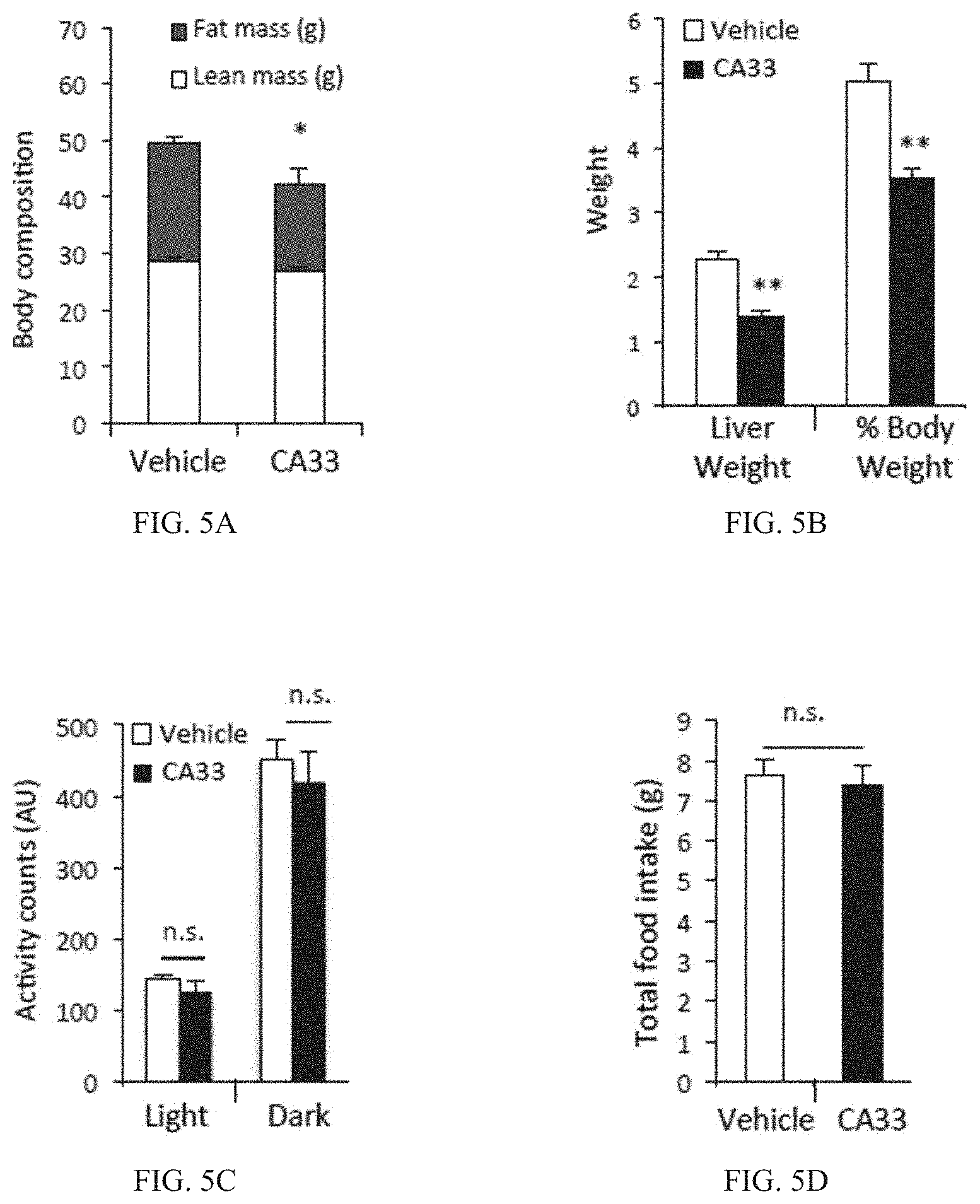
FIG. 5A is a bar graph of body fat mass (g) (open bar) and lean mass(g) (gray bar) as determined by dual-energy X-ray absorptiometry (DEXA) after 5 weeks of vehicle or CA33 treatment. (n=10 per group). *p<0.05.
FIG. 5B is a bar graph showing liver weight (g) and % body weight of obese mice after 5 weeks of vehicle (open bars) or CA33 treatment (solid bars). **p<0.01.
FIG. 5C is a bar graph showing physical activity (activity units--AU) for mice in the light or in the dark after 5 weeks of vehicle (open bars) or CA33 treatment (solid bars).
FIG. 5D is a bar graph showing total food intake (g) in obese mice after 5 weeks of vehicle (open bar) or CA33-treated mice (solid bar) on HFD (n=8 per group).
FIG. 5E is a bar graph showing VO2 concentrations by volume during the light and dark periods in mice treated with CA33 (solid bar) for eight weeks compared to vehicle (open bar).
FIG. 5F is a bar graph showing calculated Respiratory Exchange Ratio (RER) during light and dark periods in mice treated with CA33 (solid bar) for eight weeks compared to vehicle (open bar).
FIG. 5G is a bar graph showing the weight of brown adipose tissue (BAT) in mice treated with CA33 (solid bar) for eight weeks compared to vehicle (open bar).
FIG. 5H shows representative H&E stained sections of BAT in mice following treatment with CA33 (solid bar) for eight weeks compared to vehicle (open bar).
FIG. 5I is a bar graph showing the weight of perigonadal white adipose tissue (PGWAT) in obese mice after 5 weeks of vehicle (open bar) or CA33-treated mice (solid bar). **p<0.01.
FIG. 5J is a representative image of hematoxylin and eosin (H&E) stained epididymal adipose tissue after 5 weeks of treatment with vehicle (left image) or CA33 (right image). Scale bar is 200 .mu.m.
FIG. 5K is a bar graph showing F4/80+ Mac cells (%) in adipose tissue determined by FACS after 5 weeks of vehicle (open bar) or CA33-treated mice (solid bar).
FIG. 5L is a bar graph showing CD11b+ cells (%) in adipose tissue determined by FACS analysis after 5 weeks of vehicle (open bar) or CA33-treated mice (solid bar).
FIG. 5M is a bar graph showing mRNA levels (mRNA/Tbp) for TNF, IL-1.beta., IL-6, CCL2, CXCL1, F4/80 or CD68 in perigonadal white adipose tissue (PG-WAT) after 5 weeks of vehicle (open bar) or CA33-treated mice (solid bar).
FIG. 5N is a Western blot showing adipose tissue aP2/FABP4 protein levels in mice treated with vehicle or CA33 for 3 weeks. Adipose tissue samples from aP2-/-, mal1-/-, and aP2/mal1-/- animals were included as protein controls. .beta.-tubulin is shown as a loading control.
FIG. 5O is a Western blot showing adipose tissue Mal1/FABP5 protein levels in mice treated with vehicle or CA33 for 3 weeks. Adipose tissue samples from aP2-/-, mal1-/-, and aP2/mal1-/- animals were included as protein controls. .beta.-tubulin is shown as a loading control.
FIG. 5P is bar graph of relative protein levels for either aP2 or mal1 in mice treated with vehicle or CA33 for 3 weeks. The results shown in FIG. 5P quantify the Western blots shown in FIGS. 5N and 5O.
FIG. 6A is a bar graph showing mRNA expression of gluconeogenic genes phosphoenolpyruvate carboxykinase 1 (Pck1) and glucose-6-phosphatase (G6pc). Liver samples were collected after 6 hours of day-time food withdrawal from obese mice treated with vehicle (open bars) or CA33 (solid bars) (n=10 for each group) for 4 weeks. *p<0.05.
FIG. 6B is a bar graph showing enzymatic activity of Pck1 (nM/min/g) in liver samples. Liver samples were collected after 6 hours of day-time food withdrawal from obese mice treated with vehicle (open bars) or CA33 (solid bars) (n=10 for each group) for 4 weeks. *p<0.05.
FIG. 6C is a bar graph showing enzymatic activity of glucose-6-phosphatase (G6pc) (U/mg) in liver microsomal fraction for mice treated with vehicle (open bars) or CA33 (solid bars) (n=10 for each group) for four weeks. *p<0.05.
FIG. 6D is a line graph showing blood glucose (mg/dl) vs. time during hyperinsulinemic-euglycemic clamp. Clamp studies were performed in obese mice on a high-fat diet (HFD) after five weeks of treatment with vehicle (diamonds) or CA33 (squares) (n=7 for each group).
FIG. 6E is a bar graph showing glucose infusion rate (GIR) (mg/kg/min) in obese mice on a high-fat diet (HFD) after five weeks of treatment with vehicle (open bar) or CA33 (solid bar). *p<0.05.
FIG. 6F is a bar graph showing clamp hepatic glucose production (c-HGP) (mg/kg/min) in obese mice on a high-fat diet (HFD) after five weeks of treatment with vehicle (open bar) or CA33 (solid bar).*p<0.05.
FIG. 6G is a bar graph showing the rate of whole body glucose disappearance (RD) (mg/kg/min) in obese mice on a high-fat diet (HFD) after five weeks of treatment with vehicle (open bar) or CA33 (solid bar). *p<0.05.
FIG. 6H is a bar graph showing glucose uptake in triceps surae muscle (mg/kg/min) in obese mice on HFD after five weeks of treatment with vehicle (open bar) or CA33 (solid bar). *p<0.05.
FIG. 6I is a bar graph showing whole body glycolysis in obese mice on a high-fat diet (HFD) after five weeks of treatment with vehicle (open bar) or CA33 (solid bar). *p<0.05.
FIG. 7A is a line graph showing glucose levels (mg/dl) vs. time (minutes) in a glucose tolerance test (GTT) following two weeks of selective antibody treatment using high affinity antibodies (CA13, CA15, CA23, and H3) versus vehicle control.
FIG. 7B is a line graph showing glucose levels (mg/dl) vs. time (minutes) in an insulin tolerance test (ITT) following three weeks of selective antibody treatment using high affinity antibodies (CA13, CA15, CA23, and H3) versus vehicle control.
FIG. 8A is a bar graph showing basal hepatic glucose production (mg/kg/min) in vehicle control (open bar) or CA33 (solid bar) treated mice during hyperinsulinemic-euglycemic clamp of HFD-mice.
FIG. 8B is a bar graph showing serum insulin levels (ng/ml) in CA33 mice (solid bars) or vehicle control mice (open bars) during hyperinsulinemic-euglycemic clamp of HFD-mice. **p<0.01.
FIG. 8C is a bar graph showing glucose uptake (mg/kg/min) in gonadal white adipose tissue (GWAT) in vehicle control (open bar) or CA33 treated (solid bar) mice during hyperinsulinemic-euglycemic clamp of HFD-mice.
FIGS. 9-16 are further discussed in Example 2 below.
FIG. 9 is a line graph illustrating diabetes incidence (%) vs. time (weeks of treatment) in the NOD mouse model for Type 1 Diabetes. NOD mice were treated with vehicle (diamonds) or the aP2 monoclonal antibody CA33 (squares).
FIG. 10A is a line graph illustrating death rate (%) vs. time (weeks of treatment) in the NOD mouse model for Type 1 Diabetes. NOD mice were treated with vehicle (diamonds) or the aP2 monoclonal antibody CA33 (squares).
FIG. 10B is a bar graph illustrating blood glucose level (mg/dL) in PBS-treated or aP2-antibody treated NOD mice following a 6 hr fast.
FIG. 10C is a line graph illustrating insulin level (ng/mL) in PBS-treated or aP2-antibody treated NOD mice following a 6 hr fast.
FIGS. 11A and 11B: NOD aP2.sup.+/+ and NOD aP2.sup.-/- mice were subjected to glucose tolerance test (GTT) (FIG. 11A) and insulin tolerance test (ITT) (FIG. 11B).
FIG. 12A is a bar graph showing insulin (ng/ml/ug DNA) secretion from NOD aP2.sup.+/+ and NOD aP2.sup.-/- mice islets after stimulation with either low or high glucose.
FIGS. 12B and 12C: illustrates bar graphs showing total insulin (ng/ml/ug DNA) content from isolated islets of four (FIG. 12B) and seven (FIG. 12C) week old NOD aP2.sup.+/+ (left bar) and NOD aP2.sup.-/- mice (right bar).
FIG. 13 illustrates the number of islets visible in NOD aP2.sup.-/- mice compared to NOD aP2.sup.+/+ mice following pancreatic dissection.
FIGS. 14A and 14B: illustrates stained beta cells (FIG. 14A), which were subsequently quantified (FIG. 14B) in NOD aP2.sup.+/+ and NOD aP2.sup.-/- mice.
FIGS. 15A-15C: illustrates bar graphs showing glucose-stimulated insulin (ng/ml/ug DNA) secretion from either a rat insulinoma beta cell line (INS1) (FIG. 15A), aP2-deficient c57b/6 mice (Mouse Islets) (FIG. 15B), or human islets (FIG. 15C) after stimulation with either low or high glucose.
FIG. 15D illustrates that aP2 is taken up into mouse islets after 20 minutes of treatment with 10 ug/ml aP2 (n=5).
FIGS. 15E and 15F: bar graphs showing insulin (ng/ml/ug DNA) secretion following aP2 treatment for 24 hrs under "fasting" conditions from either INS1 beta cells (FIG. 15E) or primary islets isolated from aP2.sup.-/- mice (FIG. 15F) after stimulation with either low or high glucose.
FIG. 16 is a diagram of an inducible model of Type 1 diabetes in mice (rat insulin promoter-lymphocytic choriomeningitis virus-glycoprotein, or RIP-LCMV-GP, mice). Mice are injected with LCMV, which leads to destruction of .beta.-cells and the development of diabetes.
FIG. 17 is a schematic showing an aP2-antibody administration schedule in a Type 1 diabetes-induced mouse model (RIP-LCMV-GP mice) in a group of aP2-/- (Group C) and aP2-normal mice injected twice weekly with either 33 mg/kg of CA33 (Group B) or PBS control (Group A) and fed a normal chow diet.
FIG. 18 is a line graph showing 6-hour fasting blood glucose measurements (mg/dl) vs. time (days) in a Type 1 diabetes-induced mouse model (RIP-LCMV-GP mice) in a group of aP2-/- (triangles) and aP2-normal mice injected twice weekly with either 33 mg/kg of CA33 (squares) or control (PBS) (circles) and fed a normal chow diet. CA33 treated and aP2 deficient animals had significantly lower fasting blood glucose after LCMV administration compared to vehicle treated animals. *p<0.05, **p<0.01, ***p<0.005.
FIG. 19 is a bar graph showing incidence of Type 1 diabetes in a Type 1 diabetes-induced mouse model (RIP-LCMV-GP mice) in a group of aP2-/- (triangles) and aP2-normal mice injected twice weekly with either 33 mg/kg of CA33 (squares) or control (PBS) (circles) and fed a high fat diet. Diabetes was defined as a 6-hour fasting blood glucose measurement greater than 250 mg/dl. CA33 treatment provided protection against development of type 1 diabetes in the RIP-LCMV-GP model similar to that observed in aP2 genetically deficient animals.
FIG. 20A is a bar graph showing islet infiltration (%; non-insulitis (open bars); peri-insulitis (checkered bars); mild insulitis (gray bars); severe insulitis (black bars)) in GP+ control mice, GP+ aP2-/- mice, or GP+ mice treated with CA33, 14 days after injection with LCMV. Mice were treated with 1.5 mg CA33 or vehicle by injection twice weekly, starting 14 days prior to LCMV infection. Insulitis scoring was performed on H&E stained pancreatic sections. Each islet was scored as either "non-insulitis," "peri-insulitis," "mild insulitis" or "severe insulitis." The bar graph represents the percentage of each islet type in each animal analyzed.
FIG. 20B is a representative picture of an islet showing little to no insulitis.
FIG. 20C is a representative picture of an islet showing severe insulitis.
FIG. 20D is a bar graph showing islets with mild insulitis (%) in GP+ control mice, GP+ aP2-/- mice, or GP+ mice treated with CA33, 14 days after injection with LCMV. Mice were treated with 1.5 mg CA33 or vehicle by injection twice weekly, starting 14 days prior to LCMV infection. *p<0.05; **p<0.01.
FIG. 20E is a bar graph showing islets with severe insulitis (%) in GP+ control mice, GP+ aP2-/- mice, or GP+ mice treated with CA33, 14 days after injection with LCMV. Mice were treated with 1.5 mg CA33 or vehicle by injection twice weekly, starting 14 days prior to LCMV infection. **p<0.01; ***p<0.005.
FIG. 21A is a series of representative images of islets stained for ATF6 (left column) or XBP1 (right column) in GP+ control mice, GP+ aP2-/- mice, or GP+ mice treated with CA33, 14 days after injection with LCMV. Mice were treated with 1.5 mg CA33 or vehicle by injection twice weekly, starting 14 days prior to LCMV infection.
FIG. 21B is a bar graph showing ATF6 levels (relative fluorescence intensity, RFI) in pancreatic samples from GP+ control mice, GP+aP2-/- mice, or GP+ mice treated with CA33, 14 days after injection with LCMV. Mice were treated with 1.5 mg CA33 or vehicle by injection twice weekly, starting 14 days prior to LCMV infection. **p<0.01; ***p<0.005.
FIG. 21C is a bar graph showing sXBP1 levels (relative fluorescence intensity, RFI) in pancreatic samples from GP+ control mice, GP+aP2-/- mice, or GP+ mice treated with CA33, 14 days after injection with LCMV. Mice were treated with 1.5 mg CA33 or vehicle by injection twice weekly, starting 14 days prior to LCMV infection. **p<0.01; ***p<0.005.
FIGS. 22-26 are further discussed in Example 3 below.
FIG. 22A is table providing the antibody dose (.mu.g) and injection volume (.mu.l) calculations to achieve a 33 mg/kg dosage based on average body weight of the ApoE knockout mice (atherosclerosis mouse model) at the indicated time points (weeks).
FIG. 22B is a graph showing atherosclerotic lesion area (%) in ApoE knockout mice treated with PBS (circles), CA33 (squares), or CA15 (triangles). Aortas from sacrificed ApoE knockout mice were dissected from the proximal aorta to the iliac bifurcation, and the aortae were pinned out in an en face preparation. En face pinned-out aortas were stained with Sudan IV. Quantitation of lesion areas was achieved using ImageJ software developed at the NIH. The outer perimeter of the pinned out aorta was defined in the software to establish the total area of the aorta as a white background. The percent area of the lesions stained red with Sudan IV was then measured and calculated by the software. *p<0.05.
FIG. 22C is a representative image of an en face pinned aorta from an ApoE knockout mouse fed western diet and treated with vehicle for twelve weeks.
FIG. 22D is a representative image of an en face pinned aorta from an ApoE knockout mouse fed western diet and treated with CA33 (33 mg/kg) for twelve weeks.
FIG. 22E is a representative image of an en face pinned aorta from an ApoE knockout mouse fed western diet and treated with CA15 (33 mg/kg) for twelve weeks.
FIG. 22F is a line graph showing average body weight (g) of ApoE knockout mice vs. age (weeks old) in mice treated for twelve weeks with PBS (circles), CA33 (squares), or CA15 (triangles). ApoE knockout mice were fed western diet and treated with vehicle or antibody (33 mg/kg) for twelve weeks.
FIG. 22G illustrates aP2 protein expression in adipose tissue of aP2.sup.adip-/- mice.
FIGS. 22H-22J: are bar graphs showing the level of aP2 (ng/ml) (FIG. 22H), triglyceride (mg/dl) (FIG. 22I), and cholesterol (mg/dl) (FIG. 22J) in ApoE.sup.-/-aP2.sup.adip+/+ and ApoE.sup.-/-aP2.sup.adip-/- mice after 12 weeks of western diet.
FIG. 22K is a bar graph showing the atherosclerotic lesion area in ApoE.sup.-/-aP2.sup.adip+/+ and ApoE.sup.-/-aP2.sup.adip-/- mice.
FIG. 22L is a representative image of an en face pinned aorta from ApoE.sup.-/-aP2.sup.adip+/+ and ApoE.sup.-/-aP2.sup.adip-/- mice fed western diet for 12 weeks.
FIG. 23A is a bar graph showing average body weight (g) of ApoE knockout mice treated for twelve weeks with PBS (black bar), CA33 (light gray bar), or CA15 (gray bar). ApoE knockout mice were fed western diet and treated with vehicle or antibody (33 mg/kg) for twelve weeks. CA33-treated mice show a statistically significant lower average body weight than vehicle treated mice. *p<0.05.
FIG. 23B is a bar graph showing liver weight (g) of ApoE knockout mice treated for twelve weeks with PBS (black bar), CA33 (light gray bar), or CA15 (gray bar). ApoE knockout mice were fed western diet and treated with vehicle or antibody (33 mg/kg) for twelve weeks. CA33 treated mice show a statistically significant lower average liver weight than vehicle treated mice. *p<0.05.
FIG. 23C is a bar graph showing body weight minus liver weight (g) of ApoE knockout mice treated for twelve weeks with PBS (black bar), CA33 (light gray bar), or CA15 (gray bar). ApoE knockout mice were fed western diet and treated with vehicle or antibody (33 mg/kg) for twelve weeks. CA33 treated mice show a lower average body weight minus liver weight than vehicle treated mice.
FIG. 23D is a bar graph showing average body weight, lean mass, or fat mass (g) of ApoE knockout mice treated for twelve weeks with PBS (black bar), CA33 (light gray bar), or CA15 (gray bar). ApoE knockout mice were fed western diet and treated with vehicle or antibody (33 mg/kg) for twelve weeks and body weight, lean mass, and fat mass were measured by dual X-ray absorbance (DEXA) spectroscopy.
FIG. 24A is a bar graph of fasting basal glucose levels (mg/dl) prior to a glucose tolerance test in ApoE knockout mice treated with PBS (black bar), CA33 (light gray bar), or CA15 (gray bar). ApoE knockout mice were fed western diet and treated with vehicle or antibody (33 mg/kg) for twelve weeks. CA33-treated mice have statistically significant lower fasting blood glucose than vehicle treated mice. *p<0.05.
FIG. 24B is a line graph of glucose levels (mg/dl) vs. time (minutes) during a glucose tolerance test in ApoE knockout mice treated with PBS (triangles), CA33 (squares), or CA15 (circles). ApoE knockout mice were fed western diet and treated with vehicle or antibody (33 mg/kg) for twelve weeks. The glucose tolerance test was performed by oral glucose administration (1.0 g/kg) on conscious mice after an overnight (16 h) fast.
FIG. 25A is a bar graph of cholesterol in lipoprotein fractions (mg/dl) (total lipoprotein, very low-density lipoprotein (VLDL), low-density lipoprotein (LDL), or high-density lipoprotein (HDL)) in ApoE knockout mice treated with PBS (triangles), CA33 (squares), or CA15 (circles) for six weeks. Particle size distribution of the lipoproteins was determined by fast-performance liquid chromatography (FPLC), using pooled samples of plasma. *p<0.05.
FIG. 25B is a bar graph of cholesterol in lipoprotein fractions (mg/dl) (total lipoprotein, very low-density lipoprotein (VLDL), low-density lipoprotein (LDL), or high-density lipoprotein (HDL)) in ApoE knockout mice treated with PBS (triangles), CA33 (squares), or CA15 (circles) for twelve weeks. Particle size distribution of the lipoproteins was determined by fast-performance liquid chromatography (FPLC), using pooled samples of plasma. *p<0.05.
FIG. 26A is a bar graph of triglycerides in lipoprotein fractions (mg/dl) (total lipoprotein, very low-density lipoprotein (VLDL), low-density lipoprotein (LDL), or high-density lipoprotein (HDL)) in ApoE knockout mice treated with PBS (triangles), CA33 (squares), or CA15 (circles) for six weeks. Particle size distribution of the lipoproteins was determined by fast-performance liquid chromatography (FPLC), using pooled samples of plasma. *p<0.05.
FIG. 26B is a bar graph of triglycerides in lipoprotein fractions (mg/dl) (total lipoprotein, very low-density lipoprotein (VLDL), low-density lipoprotein (LDL), or high-density lipoprotein (HDL)) in ApoE knockout mice treated with PBS (triangles), CA33 (squares), or CA15 (circles) for twelve weeks. Particle size distribution of the lipoproteins was determined by fast-performance liquid chromatography (FPLC), using pooled samples of plasma. *p<0.05.
FIGS. 27-28 are further discussed in Example 4 below.
FIG. 27 provides anti-human aP2 humanized kappa light chain variable region antibody fragments, wherein the 909 sequence (Seq. ID. No. 445) is rabbit variable light chain sequence, and the 909 gL1 (Seq. No. 446), gL10 (Seq. ID. 448), gL13 (Seq. ID, No. 487), gL50 (Seq, ID. No. 488), gL54 (Seq. ID. No. 450), and gL55 (Seq. ID. No. 452 sequences are humanized grafts of 909 variable light chain using IGKV1-17 (Seq. ID. No. 506) human germline as the acceptor framework. The CDRs are shown in bold/underlined, while the applicable donor residues are shown in bold/italic and are highlighted: 2V, 3V, 63K and 70D. The mutation in CDRL3 to remove a Cysteine residue is shown in bold/underlined and is highlighted: 90A.
FIG. 28 provides anti-human aP2 humanized heavy chain variable region antibody fragments, wherein the 909 sequence (Seq. ID. No. 454) is rabbit variable heavy chain sequence, and the 909 gH1 (Seq. ID. No. 455), gH14 (Seq. ID. No. 457), gH15 (Seq. ID. No. 459), gH61 (Seq. ID. No. 461), and gH62 (Seq. ID. No. 463) sequences are humanized grafts of 909 variable heavy chain using IGHV4-4 (Seq. ID. No. 481) human germline as the acceptor framework. The CDRs are shown in bold/underlined. The two residue gap in framework 3, in the loop between beta sheet strands D and E, is highlighted in gH1: 75 and 76. Applicable donor residues are shown in bold/italic and are highlighted: 23T, 67F, 71K, 72A, 73S, 74T, 77T, 78V, 79D, 89T, and 91F. The mutation in CDRH2 to remove a Cysteine residue is shown in bold/underlined and is highlighted: 59S. The mutation in CDRH3 to remove a potential Aspartate isomerization site is shown in bold/underlined and is highlighted: 98E. The N-terminal Glutamine residue is replaced with Glutamic acid, and is shown in bold and highlighted: 1E.
FIGS. 29-31 are further discussed in Example 5 below.
FIG. 29 is an illustration representing key contact points between aP2 and a murine derived anti-aP2 antibody. As is shown in the figure, substitution of particular amino acid residues with CDR regions of the heavy and light chains resulted in a reduction in the affinity of the altered antibodies in comparison with the parent antibody H3.
FIG. 30A provides amino acid substitutions for inducing a reduction in binding affinity in a murine derived anti-aP2 antibody compared to the parent antibody H3, where g1=wild-type parent H3. Binding affinities (.mu.M) are shown for CA33 and two reduced affinity H3-like antibodies.
FIG. 30B provides the protein sequence of the heavy chain detailing the amino acid substitutions for inducing a reduction in binding affinity in a murine derived anti-aP2 antibody compared to the parent antibody H3, where g1=wild-type parent H3 (Seq. ID. No. 482), g19=W33A, Y52S (Seq. ID. No. 483), and g20=W33A, Y52S. and Y98F (Seq. :ID. No. 484). The CDRs are underlined and the amino acid substitutions are shown in bold.
FIG. 30C provides the protein sequence of the light chain detailing the amino acid substitutions for inducing a reduction in binding affinity in a murine derived anti-aP2 antibody compared to the parent antibody H3, where g1=wild-type parent H3 (Seq. ID. No. 485, and g11 =W91A (Seq. ID. No. 486). The CDRs are underlined and the amino acid substitutions are shown in bold.
FIG. 31 is a table indicating affinity data against human and mouse aP2 for murine derived anti-aP2 antibodies that have been mutated to reduce binding affinity. Binding affinities (KD) are shown for two clones for each antibody (APP5168/PB1172 and APP5169/PB1171) and average binding affinity (KD) is shown (.mu.M).
FIGS. 32-46 are further discussed in Example 6 below.
FIG. 32 illustrates that aP2 regulates gluconeogenic gene expression in a FoxO1-dependent manner. Primary hepatocytes were incubated with or without 50 .mu.g/ml of aP2 in the presence or absence of 2 .mu.M forskolin (fsk) for 3 h (FIGS. 32A, 32B, 32C, 32D, and 32E) or 6 h (FIG. 32F). FIGS. 32A and 32B show the impact of forskolin stimulation on the effect of aP2 on gluconeogenic gene expression (G6pc and Pck1 respectively) in hepatocytes, as well as the role of aP2 lipid binding in this process (n=4). mut: lipid binding mutant, delip: delipidated aP2, WT: lipid-loaded aP2. FIG. 32C illustrates the screening of transcription factors by siRNA mediated knockdown and the effect on forskolin and aP2-stimulated G6pc expression (n=4). FIG. 32D illustrates the effect of adenovirus-mediated FoxO1 knockdown on aP2-mediated G6pc expression (n=4). FIG. 32E illustrates the knockdown efficiency at protein levels for FoxO1.
FIG. 32F illustrates the effect of aP2 stimulation of FHRE luciferase activity in primary hepatocytes (n=6). FIG. 32G illustrates glucose production from hepatocytes following adenovirus-mediated FoxO1 knockdown and aP2 stimulation (n=5). Data are expressed as the mean.+-.SEM. *, ** and NS denote p<0.05, p<0.01 and no significant difference, respectively, determined by Student's t-test.
FIG. 33 illustrates that aP2 alters fatty acid metabolism to regulate gluconeogenic gene expression. FIGS. 33A and 33B show the tabulated results for G6pc and Pck1 expression, respectively, from primary hepatocytes preincubated with vehicle or 50 .mu.M Etomoxir for 30 min and stimulated with 50 .mu.g/ml of aP2 in the presence or absence of 2 .mu.M of forskolin (fsk) for 3 h (n=4). FIGS. 33C and 33D show the results obtained when primary hepatocytes were stimulated with 50 .mu.g/ml of aP2 for 2 h, and stimulated with 150 .mu.M palmitate (FIG. 33C) or 150 .mu.M oleate (FIG. 33D) for the indicated amount of time to measure oxygen consumption rate (OCR, FIG. 33C, n=6) or fatty acid uptake (FIG. 33D, n=5). FIGS. 33E and 33F illustrate the mRNA levels obtained for G6pc and Pck1, respectively, when primary hepatocytes were stimulated with 50 .mu.g/ml of aP2 for 2 h and the cells were further stimulated with 50 .mu.g/ml of aP2 in the presence or absence of 2 .mu.M of forskolin (fsk)/100 .mu.M palmitate for 3 h (n=4). FIGS. 33G and 33H illustrate the mRNA levels obtained for G6pc and Pck1, respectively, when primary hepatocytes were preincubated with 5 .mu.M Triacsin C for 30 min and further treated as in FIG. 33D with or without the same concentrations of Triacsin C (n=4). FIG. 33I illustrates the level of nuclear fatty acyl-CoA (pmol/10.sup.6 cells) in primary hepatocytes after they were treated with either vehicle or aP2. Data are expressed as the mean.+-.SEM. *, ** and NS denote p<0.05, p<0.01 and no significant difference, respectively, determined by Student's t-test.
FIG. 34 demonstrates the existence of a novel FoxO1/CtBP2 transcriptional complex regulated by aP2. Primary hepatocytes were stimulated with 50 .mu.g/ml of aP2 (FIGS. 34 A, B, H, L, M and N) in the presence or absence of 2 .mu.M of forskolin (fsk) for 3 h (FIGS. 34 A and B) or 90 min (FIGS. 34 H, K, M and N). FIGS. 34A and 34B show the effects of adenovirus-mediated CtBP1 and CtBP2 knockdown on aP2-induced expression of G6pc and Pck1, respectively (n=4). FIG. 34C shows the PxDLS-like motifs in human and mouse FoxO sequences as indicated by the rectangles: mouse FOXO1 amino acids 571 to 610 (Seq. ID. No. 603), mouse FOXO3 amino acids 581 to 610 (Seq. ID. No. 604), mouse FOXO4 amino acids 411 to 480 (Seq. ID. No. 605), mouse FOXO6 amino acids 481 to 520 (Seq. ID. No. 606), human FOXO1 amino acids 581 to 620 (Seq. ID. No. 607), human FOXO3 amino acids 591 to 630 (Seq. ID. No. 608), human FOXO4 amino acids 451 to 490 (Seq. ID. No. 609), human FOXO6 amino acids 411 to 451 (Seq. ID. No. 610). FIGS. 34D and 34E show endogenous FoxO1/CtBP complex in primary hepatocytes shown by reciprocal coimmunoprecipitation of CtBP or FoxO1, respectively. FIGS. 34F and 34G illustrate the critical role of the PxDL motif in the FoxO1/CtBP2 complex. HEK293 cells were transfected with either control plasmid, FLAG wild-type FoxO1 or mutant FLAG-FoxO1 (Mut: PSDL>PSAS, .DELTA.psdl: PxDL motif deletion) along with CtBP2 expression plasmid. The complexes were immunoprecipitated with FLAG agarose beads. FIG. 34H shows the dissociation of native FoxO1/CtBP2 complex by aP2 treatment. The endogenous FoxO1/CtBP2 complex was immunoprecipitated from primary hepatocytes. FIGS. 34I and 34J show the nutrient sensing capability of FoxO1/CtBP2 complex. HEK293 cells were transfected with FLAG wild-type FoxO1 and CtBP2. Increasing concentrations of either oleoyl-CoA (0, 50, 150, 500 .mu.M) (FIG. 34I) or NADH (0, 10, 30, 100 .mu.M) (FIG. 34J) were added to the cell lysates and immunoprecipitated with FLAG agarose beads. FIG. 34K shows the effect of cytosolic redox status on FoxO1/CtBP2 complex. HEK293 cells were transfected with FLAG wild-type FoxO1 and CtBP2 and incubated with different ratios of lactate and pyruvate for 1 h. FIG. 34L illustrates the effect of forskolin and aP2 treatment on the nuclear/cytoplasmic localization of FoxO1 and CtBP2. NE: nuclear extract. FIGS. 34M and 34N show chromatin immunoprecipitation (ChIP) analysis (n=4) of FoxO1 or CtBP2, respectively, at the G6pc promoter. Data are expressed as the mean.+-.SEM. * and ** denote p<0.05 and p<0.01, respectively, determined by Student's t-test.
FIG. 35 shows the fine-tuned regulation of gluconeogenic gene expression by CtBP2. Primary hepatocytes were stimulated with vehicle or 2 .mu.M of forskolin (fsk) for 3 h (FIGS. 35 A, D and E) or 6 h (B, F) after knockdown (A, B) or overexpression (C, D, E, F) of CtBP2. FIG. 35A shows the effect of CtBP2 and/or FoxO1 knockdown on G6pc expression (n=5). The y-axis scale is expanded in the inset to show the data in the absence of fsk more clearly. FIG. 35B shows FHRE luciferase activity following CtBP2 knockdown (n=8). FIG. 35C shows GUS and CtBP2 overexpression in primary hepatocytes. FIGS. 35D and 35E show the regulation of gluconeogenic gene expression by CtBP2 overexpression (n=4). FIG. 35F shows FHRE luciferase activity following CtBP2 overexpression (n=8). FIG. 35G shows the effect of acute activation of insulin and cAMP signaling on the FoxO1/CtBP2 complex. HEK293 cells transfected with FLAG-FoxO1 and CtBP2 were stimulated with either vehicle, 100 nM insulin (ins) or 50 .mu.M of forskolin (fsk) for 30 min.
FIG. 36 shows the regulation of the endogenous FoxO1/CtBP2 complex in vivo. FIGS. 36A, 36C, 36E, 36G, and 36J: liver homogenates from the following groups of mice were subjected to co-immunoprecipitaton to assay the endogenous FoxO1/CtBP2 complex. All mice were sacrificed after 4-6 h fasting. The densitometric quantification is shown to the right of each blot; see FIGS. 36B, 36D, 36F, 36H and 36K. FIGS. 36A and 36B: genetically obese ob/ob mice and their control lean mice. FIGS. 36C and 36D: diet-induced obese mice (high fat diet (HFD) for 16 weeks) and their control lean mice (normal chow (NC)). FIGS. 36E and 36F: aP2 knockout (KO) or their control wild-type (WT) mice on normal chow (NC) or high fat diet (HFD) for 16 weeks. FIGS. 36G and 36H: diet-induced obese mice (HFD) treated with monoclonal aP2 antibody (CA33) or vehicle, and control lean mice (NC). FIGS. 36I, 36J, 36K, 36L, and 36M: recombinant aP2 was administered into wild-type lean mice for 5 days. Serum aP2 levels (FIG. 36I) and FoxO1/CtBP2 complex (FIGS. 36J and 36K), gene expression in liver (FIGS. 36L and 36M) were analyzed (n=4). FIG. 36N shows the results from adenovirus-mediated knockdown of CtBP2 in the liver of wild-type lean mice 5 days after transduction (n=6). * and ** denote p<0.05 and p<0.01, respectively, determined by Student's t-test.
FIG. 37: CtBP2 gain of function in liver improves glucose tolerance and ameliorates steatosis in obese mice. CtBP2 was overexpressed in the liver of diet-induced obese mice (14 weeks on the diet) by adenoviral transduction. HFD: high fat diet, NC: normal chow. FIGS. 37A and 37B tabulate the measurement of body weights and blood glucose levels, respectively, after overnight fasting (n=4-5). FIGS. 37C, 37D, and 37Q: Dietary induced obese mice transduced with AdGUS or AdCtBP2 were subjected to glucose tolerance test (GTT, FIG. 37C), insulin tolerance test (ITT, FIG. 37D), and pyruvate tolerance test (PTT, FIG. 37Q)(n=10-12). FIGS. 37E, 37F and 37G: Gene expression in liver (n=4-5) for G6pc, Pck1, or Alb mRNA, respectively. FIG. 37H illustrates representative hematoxylin and eosin stained sections of liver. FIG. 37I shows tabulated liver triglyceride content (n=10). FIG. 37J shows tabulated serum ALT levels (n=10). FIGS. 37K, 37L, 37M, 37N, and 37O show Mlxipl/Srebf1c expression (FIGS. 37K and 37L) and lipogenic gene expression (FIGS. 37M, 37N and 37O) in the liver (n=10). Mice were sacrificed after overnight fasting. FIG. 37P is a schematic diagram showing a potential regulatory mechanism. ACSL; acyl-CoA synthetase. Data are expressed as the mean.+-.SEM. *, ** and NS denote p<0.05, p<0.01 and no significant difference, respectively, determined by Student's t-test.
FIG. 38: cAMP stimuli amplify aP2-induced gluconeogenic gene expression. FIGS. 38A and 38B show the G6pc mRNA and Pck1 mRNA levels, respectively, from when primary hepatocytes were stimulated with or without 50 .mu.g/ml of aP2 in the absence or presence of 2 .mu.M forskolin (fsk) for 3 h (n=4). The y-axis scale is expanded in the inset to show the data in the absence of fsk more clearly. In FIGS. 38C and 38D, primary hepatocytes were treated in the same way as in FIG. 38A with or without PKA inhibitor (H89, 20 .mu.M) (n=4); G6pc mRNA and Pck1 mRNA levels were measured and tabulated. Data are expressed as the mean.+-.SEM. ** denotes p<0.01 determined by Student's t-test.
FIG. 39 shows the screening of transcription factor(s) responsible for aP2-mediated upregulation of gluconeogenic genes. FIGS. 39A, 39B and 39C show the knockdown efficiency at the mRNA level, corresponding to the experiment shown in FIG. 32C (n=4). FIGS. 39D, 39E, 39F and 39G show the screening of the transcription factor(s) responsible for aP2-dependent upregulation of gluconeogenic genes. Primary hepatocytes were treated in the same way as FIG. 32A after knockdown of Hif1a, Ppargc1a or Stat3 (n=4). FIG. 39H shows Foxo1 expression levels as in FIG. 32 (n=4). Data are expressed as the mean.+-.SEM. * and ** denote p<0.05 and p<0.01, respectively, determined by Student's t-test.
FIG. 40: CtBP2-dependent transcriptional activation by aP2. FIGS. 40A, 40B, 40C and 40D show the knockdown efficiency at mRNA and protein levels corresponding to FIG. 34 (n=4). FIGS. 40E, 40F and 40G show the gluconeogenic gene expression in primary hepatocytes after adenoviral mediated knockdown of HNF4.alpha. (n=4). Gene expression profiles for G6pc and Pck1 are shown in FIGS. 40E and 40F, respectively, and knockdown efficiency at protein levels is shown in FIG. 40G. Data are expressed as the mean.+-.SEM. * and ** denote p<0.05 and p<0.01, respectively, determined by Student's t-test.
FIG. 41: Characterization of the FoxO1/CtBP complex. FIGS. 41A and 41B show the results of mutagenesis studies for FoxO1/CtBP1 interaction. HEK293 cells transfected with wild-type FLAG FoxO1 (WT) or mutant FoxO1 (Mut: PSDL>PSAS, .DELTA.psdl: PxDL motif deletion) along with CtBP1 expression plasmid were lysed and subjected to co-immunoprecipitation. FIG. 41C shows the results of incubating primary hepatocytes with 50 .mu.g/ml aP2 in the presence or absence of forskolin (fsk, 2 .mu.M) for 2 h and the native FoxO1/CtBP2 complex was immunoprecipitated. FIG. 41D shows the cellular lactate/pyruvate ratio. Primary hepatocytes were treated as in FIG. 33E. FIG. 41E shows immunocytochemistry corresponding to FIG. 34L. FIG. 41F shows levels of phosphorylation and acetylation of FoxO1. Primary hepatocytes were treated with forskolin (fsk, 2 .mu.M) and/or 50 .mu.g/ml aP2 for 30 min.
FIG. 42 shows the effect of CtBP2 overexpression on the expression of particular genes. Primary hepatocytes were treated as in FIG. 35D and the gene expression profile was analyzed.
FIG. 43 illustrates the results of the in vivo administration of recombinant aP2. Recombinant aP2 was administered into wild-type lean mice for 5 days (n=4). FIGS. 43A, 43B, 43C, 43D and 43E show results for body weights before (day 0) and after intraperitoneal injections (day 5) of aP2, serum levels of insulin, glucagon, glycerol, and free fatty acids (FFA), respectively. FIGS. 43F and 43G show the mRNA levels for Pck1 and Foxo1, respectively, in wild-type or liver specific FoxO1 KO mice treated with recombinant aP2 (n=7-8). Data are expressed as the mean.+-.SEM. NS denotes no significant difference determined by Student's t-test.
FIG. 44: CtBP2 overexpression in vivo. FIG. 44A illustrates the protein levels of overexpression of GUS and CtBP2 in liver. FIG. 44B illustrates the blood glucose levels without normalization to the baseline in the ITT study (FIG. 37D, n=10-12). FIG. 44C shows the serum insulin levels in GUS or CtBP2 overexpressed liver. Data are expressed as the mean.+-.SEM. * and ** denote p<0.05 and p<0.01, respectively, determined by Student's t-test.
FIG. 45: Involvement of CtBP2 in the pathogenesis of hepatic steatosis. FIGS. 45A, 45B, 45C, 45D, 45E, and 45F show the effect of CtBP2 overexpression on lipogenic gene expression in wild-type primary hepatocytes (FIGS. 45A, 45B, 45C, and 45D, n=4) or FoxO1 knockout hepatocytes (FIGS. 45E and 45F, n=5). FIGS. 45G, 45H, 45I, and 45J show results from liver samples analyzed after 14 days of Ad/shCtBP2 transduction in wild-type lean mice. FIG. 45G shows representative Hematoxylin and Eosin stained liver sections. FIG. 45H shows liver triglyceride content (n=6). FIGS. 45I and 45J show liver Stearyol-CoA desaturase-1 (Scd1) and Fatty acid synthase (Fasn) expression (n=6), respectively. Mice were sacrificed after overnight fasting. Data are expressed as the mean.+-.SEM. * and ** denote p<0.05 and p<0.01, respectively, determined by Student's t-test.
FIG. 46: Illustrates that the addition of recombinant aP2 activates FOXO1 translocation and activity. FIG. 46A illustrates nuclear translocation of FOXO1 in mouse and human islets treated with 10 ug/ml aP2 for 20 minutes (N=3 mouse islets, N=1 human islets). FIG. 46B illustrates gene expression changes of FOXO1 target genes in INS1 cells treated with 10 ug/ml aP2 for 24 hrs (N=4/group). FIG. 46C illustrates validation of change in FOXO1 target gene expression showing increased VEGFA protein after 24 hr of 10 ug/ml aP2 treatment (N=3/group).
FIG. 47 is a line graph of airway resistance (cmH2O.s/ml) vs. methacholine (mg/ml) during a methacholine challenge test in wild type and aP2 knockout mice treated with either ovalbumin or vehicle.
DETAILED DESCRIPTION OF THE INVENTION
Anti-aP2 monoclonal antibodies and antigen binding agents are provided that have superior and unexpected activity for the treatment of aP2-mediated disorders. For example, anti-aP2 monoclonal antibodies and antigen binding agents are provided that comprise a light chain or light chain fragment having a variable region, wherein said variable region comprises one, two, or three CDRs independently selected from Seq. ID No. 7, Seq. ID No. 8 and Seq. ID No. 9, Seq. ID No. 10, Seq. ID No. 11, Seq. ID No. 12, and Seq. ID No. 13. Alternatively, one or more of the disclosed and selected CDRs can be altered by substitution of one or more amino acids that do not adversely affect or that improve the properties of the antibody or antigen binding agent, as further described herein. In one embodiment, the selected CDR(s) is/are placed in a human immunoglobulin framework. In one embodiment, the human immunoglobulin framework is further modified or altered to maintain the binding affinity specificity of the grafted CDR region.
One of the unexpected discoveries disclosed herein is that the described antibodies and antigen binding agents do not tightly bind aP2 protein. Typically, antibodies and other antigen binding agents are sought that have tight binding affinity (very low KD), as was reported by Miao, et al. (See Background of the Invention).
Therefore, in another embodiment, it has been discovered that an antibody or antigen binding agent that binds to aP2 protein in its secreted (non-cytosolic) state with a weaker binding *.quadrature. affinity of KD about .gtoreq.10.sup.-7 M, has an improved ability to neutralize secreted aP2 and cause a significant inhibitory effect on aP2-mediated disorders when provided in an effective amount to a host in need thereof. Furthermore, it has been discovered that the use of a low-affinity binding anti-aP2 antibody reduces the undesirable effects seen with the use of high affinity anti-aP2 antibodies, for example, weight gain and increased aP2 serum levels.
The anti-aP2 antibodies and antigen binding agents of the present invention can alternatively be described by contact points between the antibody or antigen binding agent with the epitope(s) of the aP2 protein. aP2 is known to have a discontinuous epitope, in which the amino acids are in close proximity in the folded protein but not close when the protein is unfolded or stretched out (see WO 2010/102171). Thus, in one embodiment, the anti-aP2 monoclonal antibody or antigen binding agent thereof binds an epitope of human aP2 comprising one or more, for example 1, 2, 3, 4, 5, 6, 7, 8, or 9, amino acid residues selected from 10K, 11L, 12V, 13S, 37A, 38K, 57T, 130E, 132A (bolded in Seq. ID No. 1, above), or an amino acid residue within about 3 or 4 angstroms of any of 10K, 11L, 12V, 13S, 37A, 38K, 57T, 130E, and 132A, optionally with a KD for secreted aP2 of about .gtoreq.10.sup.-7 M. In a particular embodiment, the antibody has contact with each of these amino acids within a 3 or 4 Angstrom range. In another embodiment, the antibody or antigen binding agent of the present invention has 3 or 4 Angstrom range contact with at least 6, 7, or 8 of the listed amino acid residues.
In one embodiment, the anti-aP2 monoclonal antibody or antigen binding agent binds to aP2 only through, or primarily through, light chain complementarity determining regions (CDRs). In an alternative embodiment, the anti-aP2 monoclonal antibody or antigen binding agent has light chain CDRs that bind to aP2 with a greater affinity than its heavy chain CDRs bind to aP2. As one example, the antibody or antigen binding agent specifically binds aP2, and does not specifically bind to FABP5/Mal1.
When administered to a host in need thereof, these anti-aP2 antibodies and antigen binding agents neutralize the activity of aP2 and provide lower fasting blood glucose levels, improved systemic glucose metabolism, increased systemic insulin sensitivity, reduced fat mass, liver steatosis, improved serum lipid profiles, and/or reduced atherogenic plaque formation in a host when compared to anti-aP2 monoclonal antibodies having higher binding affinities. Therefore, the anti-aP2 antibodies and antigen binding agents described herein are particularly useful to treat metabolic disorders including, but not limited to, diabetes (both type 1 and type 2), hyperglycemia, obesity, fatty liver disease, dyslipidemia, polycystic ovary syndrome (POS), a proliferative disorder such as a tumor or neoplasm, (including, for example, transitional bladder cancer, ovarian cancer and liposarcoma), atherosclerosis and other cardiovascular disorders by administering an effective amount to a host, typically a human, in need thereof.
The present invention thus provides at least the following: (a) A monoclonal anti-aP2 antibody or antigenic binding agent, as described herein, or a described variant or conjugate thereof. (b) A humanized monoclonal anti-aP2 antibody or antigenic binding agent, as described herein, or a described variant or conjugate thereof (c) A monoclonal anti-aP2 antibody or antigenic binding agent, as described herein, or a described variant or conjugate thereof, wherein the antibody or antibody conjugate is characterized by at least one of: i. Structural inclusion of one or more CDRs described in Seq. IDs 7-13 or a variant thereof with amino acid substitutions that do not adversely affect the binding properties of the CDR region as described in Seq. IDs 7-13; ii. KD binding affinity for human aP2 of .gtoreq.10.sup.-7 M; and/or iii. Contact points with a human or mouse aP2 protein within 3 or 4 Angstroms as further specified herein. (d) A monoclonal anti-aP2 antibody or antigenic binding agent, as described herein, or a described variant or conjugate thereof for use to treat a host, and in particular a human, with an aP2-mediated disorder. (e) Use of a monoclonal anti-aP2 antibody or antigenic binding agent, as described herein, or a variant or conjugate thereof, to treat a host, and in particular a human, with an aP2-mediated disorder. (f) Use of a monoclonal anti-aP2 antibody or antigenic binding agent, as described herein, or a variant or conjugate thereof, in the manufacture of a medicament to treat a host, and in particular a human, with an aP2-mediated disorder. (g) A process for manufacturing a medicament intended for the therapeutic use in treating an aP2-mediated disorder, characterized in that a monoclonal anti-aP2 antibody or antigenic binding agent, as described herein, or a variant or conjugate thereof is used in the manufacture. (h) A pharmaceutical composition that includes an effective amount of a monoclonal anti-aP2 antibody or antigenic binding agent, as described herein, or a described variant or conjugate thereof. General Definitions
Unless otherwise defined, all technical and scientific terms used herein have the same meaning as commonly understood by one of ordinary skill in the art to which this invention belongs. Although methods and materials similar or equivalent to those described herein can be used in the practice or testing of the present invention, suitable methods and materials are described below. All publications, patent applications, patents, and other references mentioned herein are incorporated by reference in their entirety. In the case of conflict, the present specification, including definitions, will control. In addition, the materials, methods, and examples are illustrative only and are not intended to be limiting.
Unless otherwise required by context, singular terms shall include pluralities and plural terms shall include the singular. In this application, the use of "or" means "and/or" unless stated otherwise. Furthermore, the use of the term "including", as well as other forms, such as "includes" and "included", is not limiting. Also, terms such as "element" or "component" encompass both elements and components comprising one unit and elements and components that comprise more than one subunit unless specifically stated otherwise.
Generally, nomenclatures used in connection with, and techniques of, cell and tissue culture, molecular biology, immunology, microbiology, genetics, and protein and nucleic acid chemistry and hybridization described herein are those well known and commonly used in the art. The methods and techniques of the present invention are generally performed according to conventional methods well known in the art and as described in various general and more specific references that are cited and discussed throughout the present specification unless otherwise indicated. Enzymatic reactions and purification techniques may be performed according to manufacturer's specifications, as commonly accomplished in the art or as described herein. The nomenclatures used in connection with, and the laboratory procedures and techniques of, analytical chemistry, synthetic organic chemistry, and medicinal, and pharmaceutical chemistry described herein are those well known and commonly used in the art. Standard techniques are used for chemical syntheses, chemical analyses, pharmaceutical preparation, formulation, and delivery, and treatment of patients.
That the present invention may be more readily understood, selected terms are defined below.
The term "host" as used herein, typically refers to a human subject, and in particular where a human or humanized framework is used as an acceptor structure. Where another host is treated, it is understood by those of skill in the art that the antibody or antigen binding agent may need to be tailored to that host to avoid rejection or to make more compatible. It is known how to use the CDRs in the present invention and engineer them into the proper framework or peptide sequence for desired delivery and function for a range of hosts. Other hosts may include other mammals or vertebrate species. The term "host," therefore, can alternatively refer to animals such as mice, monkeys, dogs, pigs, rabbits, domesticated swine (pigs and hogs), ruminants, equine, poultry, felines, murines, bovines, canines, and the like, where the antibody or antigen binding agent, if necessary is suitably designed for compatibility with the host.
The term "polypeptide" as used herein, refers to any polymeric chain of amino acids. The terms "peptide" and "protein" are used interchangeably with the term polypeptide and also refer to a polymeric chain of amino acids. The term "polypeptide" encompasses native or artificial proteins, protein fragments, and polypeptide analogs of a protein sequence. A polypeptide may be monomeric or polymeric.
The term "recovering" as used herein, refers to the process of rendering a chemical species such as a polypeptide substantially free of naturally associated components by isolation, e.g., using protein purification techniques well known in the art.
The term "human aP2 protein" or "human FABP4/aP2 protein", as used herein refers to the protein encoded by Seq. ID. No. 1, and natural variants thereof, as described by Baxa, C. A., Sha, R. S., Buelt, M. K., Smith, A. J., Matarese, V., Chinander, L. L., Boundy, K. L., Bernlohr, A. Human adipocyte lipid-binding protein: purification of the protein and cloning of its complementary DNA. Biochemistry 28: 8683-8690, 1989.
The term "mouse aP2 protein" or "mouse FAB4P/aP2 protein", as used herein, refers to the protein encoded by Seq. ID. No. 2, and natural variants thereof. The mouse protein is registered in Swiss-Prot under the number P04117.
The terms "specific binding" or "specifically binding", as used herein, in reference to the interaction of an antibody, a protein, or a peptide with a second chemical species, mean that the interaction is dependent upon the presence of a particular structure (e.g., an "antigenic determinant" or "epitope" as defined below) on the chemical species; for example, an antibody recognizes and binds to a specific protein structure rather than to proteins generally. If an antibody is specific for epitope "A", the presence of a molecule containing epitope A (or free, unlabeled A), in a reaction containing labeled "A" and the antibody, will reduce the amount of labeled A bound to the antibody.
The term "antibody", as used herein, broadly refers to any immunoglobulin (Ig) molecule comprised of four polypeptide chains, two heavy (H) chains and two light (L) chains, or any functional fragment, mutant, variant, or derivation thereof, which retains at least some portion of the epitope binding features of an Ig molecule allowing it to specifically bind to aP2. Such mutant, variant, or derivative antibody formats are known in the art and described below. Nonlimiting embodiments of which are discussed below. An antibody is said to be "capable of binding" a molecule if it is capable of specifically reacting with the molecule to thereby bind the molecule to the antibody.
A "monoclonal antibody" as used herein is intended to refer to a preparation of antibody molecules, which share a common heavy chain and common light chain amino acid sequence, or any functional fragment, mutant, variant, or derivation thereof which retains at least the light chain epitope binding features of an Ig molecule, in contrast with "polyclonal" antibody preparations that contain a mixture of different antibodies. Monoclonal antibodies can be generated by several known technologies like phage, bacteria, yeast or ribosomal display, as well as classical methods exemplified by hybridoma-derived antibodies (e.g., an antibody secreted by a hybridoma prepared by hybridoma technology, such as the standard Kohler and Milstein hybridoma methodology ((1975) Nature 256:495-497).
In a full-length antibody, each heavy chain is comprised of a heavy chain variable region (abbreviated herein as HCVR or VH) and a heavy chain constant region (CH). The heavy chain constant region is comprised of four domains--either CH1, Hinge, CH2, and CH3 (heavy chains .gamma., .alpha. and .delta.), or CH1, CH2, CH3, and CH4 (heavy chains .mu. and .epsilon.). Each light chain is comprised of a light chain variable region (abbreviated herein as LCVR or VL) and a light chain constant region (CL). The light chain constant region is comprised of one domain, CL. The VH and VL regions can be further subdivided into regions of hypervariability, termed complementarity determining regions (CDR), interspersed with regions that are more conserved, termed framework regions (FR). Each VH and VL is composed of three CDRs and four FRs, arranged from amino-terminus to carboxy-terminus in the following order: FR1, CDR1, FR2, CDR2, FR3, CDR3, FR4. Immunoglobulin molecules can be of any type (e.g., IgG, IgE, IgM, IgD, IgA and IgY), class (e.g., IgG1, IgG2, IgG3, IgG4, IgA1 and IgA2) or subclass.
The term "antigen binding agent" or "antigenic binding agent" as used herein, refers to one or more fragments or portions of an antibody that retain the ability to specifically bind to an antigen (e.g., aP2), or synthetic modifications of antibody fragments that retain the desired binding ability to the antigen. It has been shown that the antigen-binding function of an antibody can be performed by fragments or certain portions of a full-length antibody, or modifications thereof. Embodiments include bispecific, dual specific and multi-specific formats which may specifically bind to two or more different antigens or to several epitopes or discontinuous epitope regions of an antigen. Nonlimiting examples of antigen binding agents include (i) a Fab fragment, a monovalent fragment consisting of the VL, VH, CL, and CH1 domains; (ii) a F(ab')2 fragment, a bivalent fragment comprising two Fab fragments linked by a disulfide bridge at the hinge region; (iii) a Fd fragment consisting of the VH and CH1 domains; (iv) a Fv fragment consisting of the VL and VH domains of a single arm of an antibody, (v) a dAb fragment (Ward et al., (1989) Nature 341:544-546, Winter et al., PCT publication WO 90/05144 A1 herein incorporated by reference), which comprises a single variable domain; (vi) an isolated complementarity determining region (CDR), (vii) fusions of antibody fragments such as those that are immunoglobulin in character, for example, diabodies, scab, bispecific, triabody, Fab-Fv, Fab-Fv-Fv, tribody, (Fab-Fv)2-Fc, and (viii) antibody portions such as CDRs or antibody loops grafted onto non-immunoglobulin frameworks such as fibronectin or leucine zippers (see Binz et al. (2005) Nat. Biotech. 23:1257-1268, incorporated herein). Furthermore, although the two domains of the Fv fragment, VL and VH, are coded for by separate genes, they can be joined, using recombinant or other methods, by a synthetic or naturally occurring linker that enables them to be made as a single protein chain in which the VL and VH regions pair to form monovalent molecules (known as single chain Fv (scFv); see e.g., Bird et al. (1988) Science 242:423-426; and Huston et al. (1988) Proc. Natl. Acad. Sci. USA 85:5879-5883). Such single chain antibodies are also intended to be encompassed within the term antigen binding agent. Other forms of single chain antibodies, such as diabodies are also encompassed. Diabodies are bivalent, bispecific antibodies in which VH and VL domains are expressed on a single polypeptide chain, but using a linker that is too short to allow for pairing between the two domains on the same chain, thereby forcing the domains to pair with complementary domains of another chain and creating two antigen binding sites (see e.g., Holliger, P., et al. (1993) Proc. Natl. Acad. Sci. USA 90:6444-6448; Poljak, R. J., et al. (1994) Structure 2:1121-1123). Such antibody binding portions are known in the art (Kontermann and Dubel eds., Antibody Engineering (2001) Springer-Verlag. New York. 790 pp. (ISBN 3-540-41354-5).
The term "antibody construct" as used herein refers to a polypeptide comprising one or more of the antigen binding portions of the invention linked to a linker polypeptide or an immunoglobulin constant domain. Linker polypeptides comprise two or more amino acid residues joined by peptide bonds and are used to link one or more antigen binding portions. Such linker polypeptides are well known in the art (see e.g., Holliger, P., et al. (1993) Proc. Natl. Acad. Sci. USA 90:6444-6448; Poljak, R. J., et al. (1994) Structure 2:1121-1123). An immunoglobulin constant domain refers to a heavy or light chain constant domain, for example a human IgA, IgD, IgE, IgG or IgM constant domains. Heavy chain and light chain constant domain amino acid sequences are known in the art. Non-limiting examples of Ig heavy chain .gamma.1 constant region and Ig light chain .lamda. and .kappa. chains are provided for in Tables 8 and 6, respectively.
Still further, an antibody or antigen-binding portion thereof may be part of a larger immunoadhesion molecule, formed by covalent or noncovalent association of the antibody or antibody portion with one or more other proteins or peptides. Examples of such immunoadhesion molecules include use of the streptavidin core region to make a tetrameric scFv molecule (Kipriyanov, S. M., et al. (1995) Human Antibodies and Hybridomas 6:93-101) and use of a cysteine residue, a marker peptide and a C-terminal polyhistidine tag to make bivalent and biotinylated scFv molecules (Kipriyanov, S. M., et al. (1994) Mol. Immunol. 31:1047-1058). Antibody portions, such as Fab and F(ab')2 fragments, can be prepared from whole antibodies using conventional techniques, such as papain or pepsin digestion, respectively, of whole antibodies. Moreover, antibodies, antibody portions and immunoadhesion molecules can be obtained using standard recombinant DNA techniques, as described herein.
An "isolated antibody", as used herein, is intended to refer to an antibody that is substantially free of other antibodies having different antigenic specificities (e.g., an isolated antibody that specifically binds aP2 is substantially free of antibodies that specifically bind antigens other than aP2). An isolated antibody that specifically binds, for example, human aP2 may, however, have cross-reactivity to other antigens, such as aP2 molecules from other species. Moreover, an isolated antibody may be substantially free of other cellular material and/or chemicals.
The term "CDR-grafted antibody" refers to antibodies which comprise heavy and light chain variable region sequences from one species but in which the sequences of one or more of the CDR regions of VH and/or VL are replaced with CDR sequences of another species, such as antibodies having human heavy and light chain variable regions in which one or more of the human CDRs (e.g., CDR3) has been replaced with murine CDR sequences.
The terms "Kabat numbering", "Kabat definitions" and "Kabat labeling" are used interchangeably herein. These terms, which are recognized in the art, refer to a system of numbering amino acid residues which are more variable (i.e. hypervariable) than other amino acid residues in the heavy and light chain variable regions of an antibody, or an antigen binding portion thereof (Kabat et al. (1971) Ann. NY Acad, Sci. 190:382-391 and, Kabat, E. A., et al. (1991) Sequences of Proteins of Immunological Interest, Fifth Edition, U.S. Department of Health and Human Services, NIH Publication No. 91-3242). For the heavy chain variable region, the hypervariable region ranges from amino acid positions 31-35 (CDR-H1), residues 50-65 (CDR-H2) and residues 95-102 (CDR-H3) according to the Kabat numbering system. However, according to Chothia (Chothia et al., (1987) J. Mol. Biol., 196, 901-917 (1987)), the loop equivalent to CDR-H1 extends from residue 26 to residue 32. Thus unless indicated otherwise "CDR-H1" as employed herein is intended to refer to residues 26 to 35, as described by a combination of the Kabat numbering system and Chothia's topological loop definition. For the light chain variable region, the hypervariable region ranges from amino acid positions 24 to 34 for CDRL1, amino acid positions 50 to 56 for CDRL2, and amino acid positions 89 to 97 for CDRL3.
As used herein, the terms "acceptor" and "acceptor antibody" refer to the antibody or nucleic acid sequence providing or encoding at least 80%, at least 85%, at least 90%, at least 95%, at least 98% or 100% of the amino acid sequences of one or more of the framework regions. In some embodiments, the term "acceptor" refers to the antibody amino acid or nucleic acid sequence providing or encoding the constant region(s). In yet another embodiment, the term "acceptor" refers to the antibody amino acid or nucleic acid sequence providing or encoding one or more of the framework regions and the constant region(s). In a specific embodiment, the term "acceptor" refers to a human antibody amino acid or nucleic acid sequence that provides or encodes at least 80%, preferably, at least 85%, at least 90%, at least 95%, at least 98%, or 100% of the amino acid sequences of one or more of the framework regions. In accordance with this embodiment, an acceptor may contain at least 1, at least 2, at least 3, least 4, at least 5, or at least 10 amino acid residues that does (do) not occur at one or more specific positions of a human antibody. An acceptor framework region and/or acceptor constant region(s) may be, e.g., derived or obtained from a germline antibody gene, a mature antibody gene, a functional antibody (e.g., antibodies well-known in the art, antibodies in development, or antibodies commercially available).
As used herein, the term "CDR" refers to the complementarity determining region within antibody variable sequences. There are three CDRs in each of the variable regions of the heavy chain and the light chain, which are designated CDRH1, CDRH2 and CDRH3 for the heavy chain CDRs, and CDRL1, CDRL2, and CDRL3 for the light chain CDRs. The term "CDR set" as used herein refers to a group of three CDRs that occur in a single variable region capable of binding the antigen. The exact boundaries of these CDRs have been defined differently according to different systems. The system described by Kabat (Kabat et al., Sequences of Proteins of Immunological Interest (National Institutes of Health, Bethesda, Md. (1987) and (1991)) not only provides an unambiguous residue numbering system applicable to any variable region of an antibody, but also provides precise residue boundaries defining the three CDRs. These CDRs may be referred to as Kabat CDRs. Chothia and coworkers (Chothia & Lesk, J. Mol. Biol. 196:901-917 (1987) and Chothia et al., Nature 342:877-883 (1989)) found that certain sub-portions within Kabat CDRs adopt nearly identical peptide backbone conformations, despite having great diversity at the level of amino acid sequence. These sub-portions were designated as L1, L2 and L3 or H1, H2 and H3 where the "L" and the "H" designates the light chain and the heavy chains regions, respectively. These regions may be referred to as Chothia CDRs, which have boundaries that overlap with Kabat CDRs. Other boundaries defining CDRs overlapping with the Kabat CDRs have been described by Padlan (FASEB J. 9:133-139 (1995)) and MacCallum (J Mol Biol 262(5):732-45 (1996)). Still other CDR boundary definitions may not strictly follow one of the above systems, but will nonetheless overlap with the Kabat CDRs, although they may be shortened or lengthened in light of prediction or experimental findings that particular residues or groups of residues or even entire CDRs do not significantly impact antigen binding. The methods used herein may utilize CDRs defined according to any of these systems, although preferred embodiments use Kabat or Chothia, or a mixture thereof, defined CDRs.
As used herein, the term "canonical" residue refers to a residue in a CDR or framework that defines a particular canonical CDR structure as defined by Chothia et al. (J. Mol. Biol. 196:901-907 (1987); Chothia et al., J. Mol. Biol. 227:799 (1992), both are incorporated herein by reference). According to Chothia et al., critical portions of the CDRs of many antibodies have nearly identical peptide backbone conformations despite great diversity at the level of amino acid sequence. Each canonical structure specifies primarily a set of peptide backbone torsion angles for a contiguous segment of amino acid residues forming a loop.
As used herein, the terms "donor" and "donor antibody" refer to an antibody providing one or more CDRs. In a preferred embodiment, the donor antibody is an antibody from a species different from the antibody from which the framework regions are obtained or derived. In the context of a humanized antibody, the term "donor antibody" refers to a non-human antibody providing one or more CDRs.
As used herein, the term "framework" or "framework sequence" refers to the remaining sequences of a variable region minus the CDRs. Because the exact definition of a CDR sequence can be determined by different systems, the meaning of a framework sequence is subject to correspondingly different interpretations. The six CDRs (CDR-L1, -L2, and -L3 of light chain and CDR-H1, -H2, and -H3 of heavy chain) also divide the framework regions on the light chain and the heavy chain into four sub-regions (FR1, FR2, FR3 and FR4) on each chain, in which CDR1 is positioned between FR1 and FR2, CDR2 between FR2 and FR3, and CDR3 between FR3 and FR4. Without specifying the particular sub-regions as FR1, FR2, FR3 or FR4, a framework region, as referred by others, represents the combined FR's within the variable region of a single, naturally occurring immunoglobulin chain. As used herein, a FR represents one of the four sub-regions, and FRs represents two or more of the four sub-regions constituting a framework region.
Human heavy chain and light chain acceptor sequences are known in the art. In one embodiment of the invention the human light chain and heavy chain acceptor sequences are selected from the sequences described in Tables 4, 5, and 7. Different combinations for human framework sequences FR1 to FR4 are described in said tables.
As used herein, the term "germline antibody gene" or "gene fragment" refers to an immunoglobulin sequence encoded by non-lymphoid cells that have not undergone the maturation process that leads to genetic rearrangement and mutation for expression of a particular immunoglobulin. See, e.g., Shapiro et al., Crit. Rev. Immunol. 22(3): 183-200 (2002); Marchalonis et al., Adv Exp Med Biol. 484:13-30 (2001). One of the advantages provided by various embodiments of the present invention takes advantage of the recognition that germline antibody genes are more likely than mature antibody genes to conserve essential amino acid sequence structures characteristic of individuals in the species, hence less likely to be recognized as from a foreign source when used therapeutically in that species.
As used herein, the term "key" residues refer to certain residues within the variable region that have more impact on the binding specificity and/or affinity of an antibody, in particular a humanized antibody. A key residue includes, but is not limited to, one or more of the following: a residue that is adjacent to a CDR, a potential glycosylation site (can be either N- or O-glycosylation site), a rare residue, a residue capable of interacting with the antigen, a residue capable of interacting with a CDR, a canonical residue, a contact residue between heavy chain variable region and light chain variable region, a residue within the Vernier zone, and a residue in the region that overlaps between the Chothia definition of a variable heavy chain CDR1 and the Kabat definition of the first heavy chain framework.
The term "humanized antibody" generally refers to antibodies which comprise heavy and light chain variable region sequences from a non-human species (e.g., a rabbit, mouse, etc.) but in which at least a portion of the VH and/or VL sequence has been altered to be more "human-like", i.e., more similar to human germline variable sequences. One type of humanized antibody is a CDR-grafted antibody, in which human CDR sequences are introduced into non-human VH and VL sequences to replace the corresponding nonhuman CDR sequences. Another type of humanized antibody is a CDR-grafted antibody, in which at least one non-human CDR is inserted into a human framework. The latter is typically the focus of the present invention.
In particular, the term "humanized antibody" as used herein, is an antibody or a variant, derivative, analog or fragment thereof which immuno-specifically binds to an antigen of interest and which comprises a framework (FR) region having substantially the amino acid sequence of a human antibody and a complementarity determining region (CDR) having substantially the amino acid sequence of a non-human antibody. As used herein, the term "substantially" in the context of a CDR refers to a CDR having an amino acid sequence at least 50, 55, 60, 65, 70, 75 or 80%, preferably at least 85%, at least 90%, at least 95%, at least 98% or at least 99% identical to the amino acid sequence of a non-human antibody CDR. In one embodiment, the humanized antibody has a CDR region having one or more (for example 1, 2, 3 or 4) amino acid substitutions, additions and/or deletions in comparison to the non-human antibody CDR. Further, the non-human CDR can be engineered to be more "human-like" or compatible with the human body, using known techniques. A humanized antibody comprises substantially all of at least one, and typically two, variable domains (Fab, Fab', F(ab')2, F(ab')c, Fv) in which all or substantially all of the CDR regions correspond to those of a non-human immunoglobulin (i.e., donor antibody) and all or substantially all of the framework regions are those of a human immunoglobulin consensus sequence. Preferably, a humanized antibody also comprises at least a portion of an immunoglobulin constant region (Fc), typically that of a human immunoglobulin. In some embodiments, a humanized antibody contains both the light chain as well as at least the variable domain of a heavy chain. The antibody also may include the CH1, hinge, CH2, and CH3, or CH1, CH2, CH3, and CH4 of the heavy chain. In some embodiments, a humanized antibody only contains a humanized light chain. In some embodiments, a humanized antibody only contains a humanized heavy chain. In specific embodiments, a humanized antibody only contains a humanized variable domain of a light chain and/or humanized heavy chain.
The humanized antibody can be selected from any class of immunoglobulins, including IgY, IgM, IgG, IgD, IgA and IgE, and any isotype, including without limitation IgA1, IgA2, IgG1, IgG2, IgG3 and IgG4. The humanized antibody may comprise sequences from more than one class or isotype, and particular constant domains may be selected to optimize desired effector functions using techniques well known in the art.
The framework and CDR regions of a humanized antibody need not correspond precisely to the parental sequences, e.g., the donor antibody CDR or the consensus framework may be mutagenized by substitution, insertion and/or deletion of at least one amino acid residue so that the CDR or framework residue at that site does not correspond exactly to either the donor antibody or the consensus framework. In a preferred embodiment, such mutations, however, will not be extensive. Usually, at least 50, 55, 60, 65, 70, 75 or 80%, preferably at least 85%, more preferably at least 90%, and most preferably at least 95%, 98% or 99% of the humanized antibody residues will correspond to those of the parental FR and CDR sequences. In one embodiment, one or more (for example 1, 2, 3 or 4) amino acid substitutions, additions and/or deletions may be present in the humanized antibody compared to the parental FR and CDR sequences. As used herein, the term "consensus framework" refers to the framework region in the consensus immunoglobulin sequence. As used herein, the term "consensus immunoglobulin sequence" refers to the sequence formed from the most frequently occurring amino acids (or nucleotides) in a family of related immunoglobulin sequences (See e.g., Winnaker, From Genes to Clones (Verlagsgesellschaft, Weinheim, Germany 1987). In a family of immunoglobulins, each position in the consensus sequence is occupied by the amino acid occurring most frequently at that position in the family. If two amino acids occur equally frequently, either can be included in the consensus sequence.
As used herein, "Vernier" zone refers to a subset of framework residues that may adjust CDR structure and fine-tune the fit to antigen as described by Foote and Winter (1992, J. Mol. Biol. 224:487-499, which is incorporated herein by reference). Vernier zone residues form a layer underlying the CDRs and may impact on the structure of CDRs and the affinity of the antibody.
As used herein, the term "neutralizing" refers to neutralization of biological activity of aP2 protein, for example, secreted aP2 protein, when an antibody described herein specifically binds the aP2 protein. Neutralizing may be the result of different ways of binding of said antibody to aP2. Preferably a neutralizing antibody is an antibody whose binding to aP2 results in neutralization of a biological activity of aP2. Preferably the neutralizing binding protein binds aP2 and decreases a biologically activity of aP2 by at least about 5%, 10%, 15%, 20%, 25%, 30%, 35%, 40%, 50%, 60%, 80%, 85%, or more. Neutralization of a biological activity of aP2 by a neutralizing antibody can be assessed by measuring one or more indicators of aP2 biological activity described herein.
A "neutralizing monoclonal antibody" as used herein is intended to refer to a preparation of antibody molecules, which upon binding to aP2 are able to inhibit or reduce the biological activity of aP2 either partially or fully.
As used herein, the term "attenuation," "attenuate," and the like refers to the lessening or reduction in the severity of a symptom or condition caused by elevated serum aP2 levels.
The term "epitope" or "antigenic determinant" includes any polypeptide determinant capable of specific binding to an immunoglobulin or T-cell receptor. In certain embodiments, epitope determinants include chemically active surface groupings of molecules such as amino acids, sugar side chains, phosphoryl, or sulfonyl, and, in certain embodiments, may have specific three dimensional structural characteristics, and/or specific charge characteristics. An epitope is a region of an antigen that is bound by an antibody. In certain embodiments, an antibody is said to specifically bind an antigen when it preferentially recognizes its target antigen in a complex mixture of proteins and/or macromolecules.
The term "K.sub.on", as used herein, is intended to refer to the on rate constant for association of an antibody to the antigen to form the antibody/antigen complex as is known in the art.
The term "K.sub.off", as used herein, is intended to refer to the off rate constant for dissociation of an antibody from the antibody/antigen complex as is known in the art.
The term "K.sub.d", as used herein, is intended to refer to the dissociation constant of a particular antibody-antigen interaction as is known in the art.
The strength, or affinity of immunological binding interactions can be expressed in terms of the dissociation constant (K.sub.d) of the interaction, wherein a smaller k.sub.d represents a greater or higher affinity. Immunological binding properties of selected polypeptides can be quantified using methods well known in the art. One such method involves measuring the rates of antigen-binding site/antigen complex formation and dissociation, wherein those rates depend on the concentrations of the complex partners, the affinity of the interaction, and geometric parameters that equally influence the rate in both directions. Thus, both the "on rate constant" ("K.sub.on") and the "off rate constant" ("K.sub.off") can be determined by calculation of the concentrations and the actual rates of association and dissociation. (Nature 361:186-87 (1993)). The ratio of K.sub.off/K.sub.on enables the cancellation of all parameters not related to affinity, and is equal to the dissociation constant K.sub.d. Davies et al. (1990) Annual Rev Biochem 59:439-473.
The term "KD", as used herein, is intended to refer to the Affinity (or Affinity constant), which is a measure of the rate of binding (association and dissociation) between the antibody and antigen, determining the intrinsic binding strength of the antibody binding reaction.
The term "antibody conjugate" refers to a binding protein, such as an antibody or antibody fragment or binding portion thereof, chemically linked to a second chemical moiety, such as a therapeutic or cytotoxic agent. The term "agent" is used herein to denote a chemical compound, a mixture of chemical compounds, a biological macromolecule, or an extract made from biological materials.
The terms "crystal", and "crystallized" as used herein, refer to an antibody, or antigen binding portion thereof, that exists in the form of a crystal. Crystals are one form of the solid state of matter, which is distinct from other forms such as the amorphous solid state or the liquid crystalline state. Crystals are composed of regular, repeating, three-dimensional arrays of atoms, ions, molecules (e.g., proteins such as antibodies), or molecular assemblies (e.g., antigen/antibody complexes). These three-dimensional arrays are arranged according to specific mathematical relationships that are well understood in the field. The fundamental unit, or building block, that is repeated in a crystal is called the asymmetric unit. Repetition of the asymmetric unit in an arrangement that conforms to a given, well-defined crystallographic symmetry provides the "unit cell" of the crystal. Repetition of the unit cell by regular translations in all three dimensions provides the crystal. See Giege, R. and Ducruix, A. Barrett, Crystallization of Nucleic Acids and Proteins, a Practical Approach, 2nd ea., pp. 20 1-16, Oxford University Press, New York, N.Y., (1999)."
The term "polynucleotide" as referred to herein means a polymeric form of two or more nucleotides, either ribonucleotides or deoxynucleotides or a modified form of either type of nucleotide. The term includes single and double stranded forms of DNA but preferably is double-stranded DNA.
The term "isolated polynucleotide" as used herein means a polynucleotide (e.g., of genomic, cDNA, or synthetic origin, or some combination thereof) that, by virtue of its origin, the "isolated polynucleotide" is not associated with all or a portion of a polynucleotide with which the "isolated polynucleotide" is found in nature; is operably linked to a polynucleotide that it is not linked to in nature; or does not occur in nature as part of a larger sequence.
The term "vector," as used herein, is intended to refer to a nucleic acid molecule capable of transporting another nucleic acid to which it has been linked. One type of vector is a "plasmid," which refers to a circular double stranded DNA loop into which additional DNA segments may be ligated. Another type of vector is a viral vector, wherein additional DNA segments may be ligated into the viral genome. Certain vectors are capable of autonomous replication in a host cell into which they are introduced (e.g., bacterial vectors having a bacterial origin of replication and episomal mammalian vectors). Other vectors (e.g., non-episomal mammalian vectors) can be integrated into the genome of a host cell upon introduction into the host cell, and thereby are replicated along with the host genome. Moreover, certain vectors are capable of directing the expression of genes to which they are operatively linked. Such vectors are referred to herein as "recombinant expression vectors" (or simply, "expression vectors"). In general, expression vectors of utility in recombinant DNA techniques are often in the form of plasmids. In the present specification, "plasmid" and "vector" may be used interchangeably as the plasmid is the most commonly used form of vector. However, the invention is intended to include such other forms of expression vectors, such as viral vectors (e.g., replication defective retroviruses, adenoviruses and adeno-associated viruses), which serve equivalent functions.
The term "operably linked" refers to a juxtaposition wherein the components described are in a relationship permitting them to function in their intended manner. A control sequence "operably linked" to a coding sequence is ligated in such a way that expression of the coding sequence is achieved under conditions compatible with the control sequences. "Operably linked" sequences include both expression control sequences that are contiguous with the gene of interest and expression control sequences that act in trans or at a distance to control the gene of interest. The term "expression control sequence" as used herein refers to polynucleotide sequences, which are necessary to effect the expression and processing of coding sequences to which they are ligated. Expression control sequences include appropriate transcription initiation, termination, promoter and enhancer sequences; efficient RNA processing signals such as splicing and polyadenylation signals; sequences that stabilize cytoplasmic mRNA; sequences that enhance translation efficiency (i.e., Kozak consensus sequence); sequences that enhance protein stability; and when desired, sequences that enhance protein secretion. The nature of such control sequences differs depending upon the host organism; in prokaryotes, such control sequences generally include promoter, ribosomal binding site, and transcription termination sequence; in eukaryotes, generally, such control sequences include promoters and transcription termination sequence. The term "control sequences" is intended to include components whose presence is essential for expression and processing, and can also include additional components whose presence is advantageous, for example, leader sequences and fusion partner sequences.
"Transformation," as defined herein, refers to any process by which exogenous DNA enters a host cell. Transformation may occur under natural or artificial conditions using various methods well known in the art. Transformation may rely on any known method for the insertion of foreign nucleic acid sequences into a prokaryotic or eukaryotic host cell. The method is selected based on the host cell being transformed and may include, but is not limited to, viral infection, electroporation, lipofection, and particle bombardment. Such "transformed" cells include stably transformed cells in which the inserted DNA is capable of replication either as an autonomously replicating plasmid or as part of the host chromosome. They also include cells, which transiently express the inserted DNA or RNA for limited periods of time.
The term "recombinant host cell" (or simply "host cell"), as used herein, is intended to refer to a cell into which exogenous DNA has been introduced. It should be understood that such terms are intended to refer not only to the particular subject cell, but, to the progeny of such a cell. Because certain modifications may occur in succeeding generations due to either mutation or environmental influences, such progeny may not, in fact, be identical to the parent cell, but are still included within the scope of the term "host cell" as used herein. Preferably host cells include prokaryotic and eukaryotic cells selected from any of the Kingdoms of life. Preferred eukaryotic cells include protist, fungal, plant and animal cells. Most preferably host cells include but are not limited to the prokaryotic cell line E. coli; mammalian cell lines CHO, HEK 293 and COS; the insect cell line Sf9; and the fungal cell Saccharomyces cerevisiae.
Standard techniques may be used for recombinant DNA, oligonucleotide synthesis, and tissue culture and transformation (e.g., electroporation, lipofection). Enzymatic reactions and purification techniques may be performed according to manufacturer's specifications or as commonly accomplished in the art or as described herein. The foregoing techniques and procedures may be generally performed according to conventional methods well known in the art and as described in various general and more specific references that are cited and discussed throughout the present specification. See e.g., Sambrook et al. Molecular Cloning: A Laboratory Manual (2d ed., Cold Spring Harbor Laboratory Press, Cold Spring Harbor, N.Y. (1989)), which is incorporated herein by reference for any purpose.
As used herein, the term "effective amount" refers to the amount of a therapy which is sufficient to reduce or ameliorate the severity and/or duration of a disorder or one or more symptoms thereof, prevent the advancement of a disorder, cause regression of a disorder, prevent the recurrence, development, onset or progression of one or more symptoms associated with a disorder, detect a disorder, or enhance or improve the prophylactic or therapeutic effect(s) of another therapy (e.g. prophylactic or therapeutic agent).
aP2 Protein
Fatty acid-binding proteins (FABPs) are members of the superfamily of lipid-binding proteins (LBP). Nine different FABPs have to date been identified, each showing relative tissue enrichment: L (liver), I (intestinal), H (muscle and heart), A (adipocyte), E (epidermal), Il (ileal), B (brain), M (myelin) and T (testis). The primary role of all the FABP family members is regulation of fatty acid uptake and intracellular transport. The structure of all FABPs is similar--the basic motif characterizing these proteins is .beta.-barrel, and a single ligand (e.g. a fatty acid, cholesterol, or retinoid) is bound in its internal water-filled cavity.
The adipocyte fatty acid-binding protein aP2 regulates systemic glucose and lipid metabolism, and has been implicated in the pathology of many immunometabolic diseases, such as diabetes and atherosclerosis. While aP2 has classically been considered a cytosolic protein, it has been found to be an active adipokine that contributes to hyperglycemia by promoting hepatic gluconeogenesis. Serum aP2 levels have been found to be markedly elevated in mouse and human obesity.
The human aP2 protein is a 14.7 kDa intracellular and extracellular (secreted) lipid binding protein that consists of 132 amino acids comprising the amino acid sequence (Seq. ID No. 1) of Table 1. The cDNA sequence of human aP2 was previously described in Baxa, C. A., Sha, R. S., Buelt, M. K., Smith, A. J., Matarese, V., Chinander, L. L., Boundy, K. L., Bernlohr, A. Human adipocyte lipid-binding protein: purification of the protein and cloning of its complementary DNA. Biochemistry 28: 8683-8690, 1989, and is provided in Seq. ID No. 5. The human protein is registered in Swiss-Prot under the number P15090.
The mouse aP2 protein sequence comprises the amino acid sequence of Seq. ID No. 2 of Table 1. The cDNA sequence of mouse aP2 is provided in Seq. ID No. 6. The mouse protein is registered in Swiss-Prot under the number P04117.
Both the human and mouse aP2 protein include at least two major conserved domains: an 11 amino acid nuclear localization signal (aa22-32: kevgvgfatrk (Seq. ID No. 3)); and a three amino acid fatty acid binding region (aa127-129: rvy (Seq. ID No. 4)).
TABLE-US-00002 TABLE 1 aP2 Protein and cDNA Sequences Protein or cDNA Seq. ID No. SEQUENCE Fatty acid-binding protein, 1 MCDAFVGTWKLVSSENFDDYMKEVGVGFATRKV adipocyte (FABP4/aP2) [H. sapiens] AGMAKPNMIISVNGDVITIKSESTFKNTEISFILGQE FDEVTADDRKVKSTITLDGGVLVHVQKWDGKSTT IKRKREDDKLVVECVMKGVTSTRVYERA Fatty acid-binding protein, 2 MCDAFVGTWKLVSSENFDDYMKEVGVGFATRKV adipocyte (FABP4/aP2 [M. musculus]) AGMAKPNMIISVNGDLVTIRSESTFKNTEISFKLGV EFDEITADDRKVKSIITLDGGALVQVQKWDGKSTTI KRKRDGDKLVVECVMKGVTSTRVYERA aP2 nuclear localization amino 3 KEVGVGFATRK acid sequence aP2 fatty acid binding domain 4 RVY amino acid sequence Fatty acid-binding protein, 5 ATGTGTGATGCTTTTGTAGGTACCTGGAAACTTG adipocyte (FABP4/aP2) [H. sapiens] TCTCCAGTGAAAACTTTGATGATTATATGAAAGA cDNA AGTAGGAGTGGGCTTTGCCACCAGGAAAGTGGC TGGCATGGCCAAACCTAACATGATCATCAGTGTG AATGGGGATGTGATCACCATTAAATCTGAAAGT ACCTTTAAAAATACTGAGATTTCCTTCATACTGG GCCAGGAATTTGACGAAGTCACTGCAGATGACA GGAAAGTCAAGAGCACCATAACCTTAGATGGGG GTGTCCTGGTACATGTGCAGAAATGGGATGG AAAATCAACCACCATAAAGAGAAAACGAGAGG ATGATAAACTGGTGGTGGAATGCGTCATGAAAG GCGTCACTTCCACGAGAGTTTATGAGAGAGCAT AA Fatty acid-binding protein, 6 ATGTGTGATGCCTTTGTGGGAACCTGGAAGCTTG adipocyte (FABP4/aP2 [M. musculus]) TCTCCAGTGAAAACTTCGATGATTACATGAAAGA cDNA AGTGGGAGTGGGCTTTGCCACAAGGAAAGTGGC AGGCATGGCCAAGCCCAACATGATCATCAGCGT AAATGGGGATTTGGTCACCATCCGGTCAGAGAG TACTTTTAAAAACACCGAGATTTCCTTCAAACTG GGCGTGGAATTCGATGAAATCACCGCAGACGAC AGGAAGGTGAAGAGCATCATAACCCTAGATGGC GGGGCCCTGGTGCAGGTGCAGAAGTGGGATGGA AAGTCGACCACAATAAAGAGAAAACGAGATGGT GACAAGCTGGTGGTGGAATGTGTTATGAAAGGC GTGACTTCCACAAGAGTTTATGAAAGGGCATGA
aP2-Binding Epitopes
In one aspect of the invention, anti-aP2 monoclonal antibody molecules, including humanized monoclonal antibodies, and antigen binding agents are provided, that specifically bind to human aP2 or mouse aP2 at specific, identified amino acids within the aP2 molecule while folded in its native, conformational form or complexed with its natural binding partner.
In one embodiment, the anti-aP2 monoclonal antibody binds human aP2 having the amino acid sequence:
TABLE-US-00003 (Seq. ID. No 1) MCDAFVGTWK LVSSENFDDY MKEVGVGFAT RKVAGMAKPN MIISVNGDVI TIKSESTFKN TEISFILGQE FDEVTADDRK VKSTITLDGG VLVHVQKWDG KSTTIKRKRE DDKLVVECVM KGVTSTRVYE RA,
or a naturally occurring variant thereof.
In an alternative embodiment, the anti-aP2 monoclonal antibody or antigen binding agent binds to a human aP2 having an amino acid sequence that is at least 80%, 85%, 90%, 95%, 96%, 97%, 98%, or 99% identical to Seq. ID No. 1. In one embodiment, the antibody has a KD for aP2 of .gtoreq.10.sup.-7 M. In one embodiment, the antibody binds to an epitope selected from an amino acid sequence underlined in Seq. ID No. 1 above and has a KD for aP2 of about .gtoreq.10.sup.-7M. In one embodiment, the antibody binds to an epitope that has one or more (for example, 1, 2, 3, or 4) amino acid substitutions, additions, or deletions as compared to Seq. ID. No. 1.
aP2-Binding Epitopes as Determined by X-Ray Crystallography
In one embodiment, the anti-aP2 antibody or antigen binding agent directly interacts with one or more, for example 1, 2, 3, 4, 5, 6, 7, 8, or 9, amino acids bolded in Seq. ID No. 1 above within a 3 or 4 Angstrom distance. In another embodiment, the anti-aP2 monoclonal antibody or antigen binding agent contacts all nine of the bolded amino acids in Seq. ID No. 1 above within a 3 or 4 Angstrom distance.
In one embodiment, the purified anti-aP2 monoclonal antibody binds a non-contiguous epitope of human and/or mouse aP2 comprising at least one or more, for example 1, 2, 3, 4, 5, 6, 7, 8, or 9 amino acid residues selected from 10K, 11L, 12V, 13S, 37A, 38K, 57T, 130E, and 132A (bolded in Seq. ID No. 1, above), or an amino acid residue within about 4 angstroms of any of 10K, 11L, 12V, 13S, 37A, 38K, 57T, 130E, and 132A. In one embodiment, the purified anti-aP2 monoclonal antibody binds an epitope of human aP2 comprising at least one or more, for example 1, 2, 3, 4, 5, 6, 7, 8, or 9, amino acid residues selected from 10K, 11L, 12V, 13S, 37A, 38K, 57T, 130E, and 132A (bolded in Seq. ID No. 1, above), or an amino acid residue within about 4 angstroms of any of 10K, 11L, 12V, 13S, 37A, 38K, 57T, 130E, and 132A and has a KD for aP2 of .gtoreq.10.sup.-7 M.
In an alternative embodiment, the purified anti-aP2 monoclonal antibody binds an epitope of human and/or mouse aP2 comprising at least one or more, for example 1, 2, 3, 4, 5, 6, or 7, amino acid residues selected from 10K, 11L, 12V, 13S, 38K, 130E, or 132A (bolded in Seq. ID No. 1, above), or an amino acid residue within about 4 angstroms of any of 10K, 11L, 12V, 13S, 38K, 130E, or 132A. In one embodiment, the purified anti-aP2 monoclonal antibody binds an epitope of human aP2 comprising at least one or more, for example 1, 2, 3, 4, 5, 6, or 7, amino acid residues selected from 10K, 11L, 12V, 13S, 38K, 130E, or 132A (bolded in Seq. ID No. 1, above), or an amino acid residue within about 4 angstroms of any of 10K, 11L, 12V, 13S, 38K, 130E, or 132A and has a KD for aP2 of .gtoreq.10 M. In one embodiment, the antibody further binds 37A and/or 57T.
In one embodiment, the light chain of the antibody binds an epitope of human aP2 comprising at least one or more, for example 1, 2, 3, 4, 5, 6, 7, 8, or 9, amino acid residues selected from 10K, 11L, 12V, 13S, 37A, 38K, 57T, 130E, or 132A, or an amino acid residue within about 4 angstroms thereof, and has a KD of at least about .gtoreq.10.sup.-7 M. In one embodiment, the light chain of the antibody binds an epitope of human aP2 comprising at least one or more, for example 1, 2, 3, 4, 5, 6, or 7, amino acid residues selected from 10K, 11L, 12V, 13S, 37A, 38K, 57T, 130E, or 132A, or an amino acid residue within about 4 angstroms thereof, and has a KD of at least about .gtoreq.10.sup.-7M.
In one embodiment, the light chain of the antibody binds an epitope of human aP2 comprising at least one or more, for example 1, 2, 3, 4, 5, 6, 7, 8, or 9, amino acid residues selected from 10K, 11L, 12V, 13S, 37A, 38K, 57T, 130E, or 132A, or an amino acid residue within about 4 angstroms thereof, and the heavy chain of the antibody binds an epitope of human aP2 comprising at least one of 10K and 132A, or an amino acid within about 4 angstroms thereof. In one embodiment, the antibody has a KD of at least about .gtoreq.10.sup.-7 M.
In one embodiment, the light chain of the antibody binds an epitope of human aP2 comprising at least one or more, for example 1, 2, 3, 4, 5, 6, or 7, amino acid residues selected from 10K, 11L, 12V, 13S, 38K, 130E, or 132A, or an amino acid residue within about 4 angstroms thereof, and the heavy chain of the antibody binds an epitope of human aP2 comprising at least one of 10K and 132A, or an amino acid within about 4 angstroms thereof, and has a KD of at least about .gtoreq.10.sup.-7M.
In one embodiment, the antibody binds to the human aP2 protein in its native confirmation at least at 10K. In one embodiment, the antibody binds to the human aP2 protein in its native confirmation at least at 38K. In one embodiment, the antibody binds to the human aP2 protein in its native confirmation at least at 12V. In one embodiment, the antibody binds to the human aP2 protein in its native confirmation at least at 11L. In one embodiment, the antibody binds to the human aP2 protein in its native confirmation at least at 130E. In one embodiment, the antibody binds to the human aP2 protein in its native confirmation at least at 132A. In one embodiment, the antibody binds to the human aP2 protein in its native confirmation at least at 13S. In one embodiment, the antibody binds to the human aP2 protein in its native confirmation at least at 11L and 12V. In one embodiment, the antibody binds to the human aP2 protein in its native confirmation at least at 130E and 132A. In one embodiment, the antibody binds to the human aP2 protein in its native confirmation at a specific amino acid described above and has a KD of about .gtoreq.10.sup.-7 M.
In one embodiment, the antibody binds to the human aP2 protein in its native confirmation at least at 10K and 38K. In one embodiment, the antibody binds to the human aP2 protein in its native confirmation at least at 10K, 38K, and 12V. In one embodiment, the antibody binds to the human aP2 protein in its native confirmation at least at 10K, 38K, 12V, and 11L. In one embodiment, the antibody binds to the human aP2 protein in its native confirmation at least at 10K, 38K, 12V, 11L, and 57T. In one embodiment, the antibody binds to the human aP2 protein in its native confirmation at least at 10K, 38K, 12V, 11L, 57T, and 37A. In one embodiment, the antibody binds to the human aP2 protein in its native confirmation at least at 10K, 38K, 12V, 11L, 57T, 37A, and 130E. In one embodiment, the antibody binds to the human aP2 protein in its native confirmation at least at 10K, 38K, 12V, 11L, 57T, 37A, 130E, and 132A. In one embodiment, the antibody binds to the human aP2 protein in its native confirmation at least at 10K, 38K, 12V, 11L, 57T, 37A, 130E, 132A, and 13S.
Also provided herein is a specific region or epitope of human aP2 which is bound by an antibody provided by the present invention, in particular an epitope bound by the antibody comprising the light chain variable sequence 909gL1 (Seq. ID No. 446), 909gL10 (Seq. ID No. 448), 909gL13 (Seq. ID No. 487), 909gL50 (Seq. ID No. 488), 909gL54 (Seq. ID No. 450), or 909gL55 (Seq. ID No. 452), and/or heavy chain variable sequence 909gH1 (Seq. ID No. 455), 909gH14 (Seq. ID No. 457), 909gH15 (Seq. ID No. 459), 909gH61 (Seq. ID No. 461), or 909gH62 (Seq. ID No. 463).
This specific region or epitope of the human aP2 protein provided herein can be identified by any suitable epitope mapping method known in the art in combination with any one of the antibodies provided by the present invention. Examples of such methods include screening peptides of varying lengths derived from aP2 for binding to the antibody of the present invention with the smallest fragment that can specifically bind to the antibody containing the sequence of the epitope recognized by the antibody. The aP2 peptides may be produced synthetically or by proteolytic digestion of the aP2 protein. Peptides that bind the antibody can be identified by, for example, mass spectrometric analysis. In another example, NMR spectroscopy or X-ray crystallography can be used to identify the epitope bound by an antibody of the present invention. Crystallization and X-ray crystallography techniques for determining the structure of aP2 and specific interactions of the aP2 protein with its natural binding partners, for example medium chain and long chain fatty acids, are described in Marr et al., Expression, purification, crystallization and structure of human adipocyte lipid-binding protein (aP2), Acta Cryst. (2006), F62, 1058-1060. Once identified, the epitopic fragment which binds an antibody of the present invention can be used, if desired, as an immunogen to obtain additional antibodies which bind the same epitope.
In one example the epitope of the antibody molecule is determined by X-ray crystallography using the aP2 protein (Seq. ID No. 1).
In one embodiment, the antibody of the present invention comprises at least one or more specific amino acids within a CDR domain as defined in Table 2 that interact with a mouse or human aP2 protein in its native conformation at the amino acid contact point defined in Table 2.
TABLE-US-00004 TABLE 2 Anti-aP2 antibody/aP2 protein Contact Points Ab Amino Ab Source aP2 Amino aP2 Target Distance Ab Chain Ab CDR Acid Atom Acid Atom (angstroms) Light CDRL3 92Tyr C 10Lys C 3.72 Light CDRL3 92Tyr C 10Lys C 3.92 Light CDRL3 92Tyr O 10Lys C 3.66 Light CDRL3 93Gly N 10Lys C 3.30 Light CDRL3 93Gly C 10Lys C 3.65 Light CDRL3 93Gly C 10Lys C 3.67 Light CDRL3 94Thr N 10Lys C 3.74 Light CDRL3 92Tyr C 10Lys C 3.25 Light CDRL3 92Tyr C 10Lys C 3.85 Light CDRL3 92Tyr C 10Lys C 3.14 Light CDRL3 94Thr O 10Lys C 3.31 Heavy 104Leu* C 10Lys N 3.06 Light CDRL3 93Gly N 10Lys N 2.74 Light CDRL3 93Gly C 10Lys N 3.64 Light CDRL3 92Tyr C 10Lys N 3.37 Light CDRL3 92Tyr C 10Lys N 3.32 Light CDRL3 92Tyr C 10Lys N 3.02 Light CDRL3 92Tyr C 10Lys N 3.87 Light CDRL3 92Tyr C 10Lys N 3.35 Light CDRL3 92Tyr C 10Lys N 3.88 Light CDRL3 92Tyr C 10Lys N 3.29 Light CDRL3 92Tyr C 10Lys N 3.82 Light CDRL3 92Tyr O 11Leu N 3.77 Light CDRL3 11Leu C 3.93 Light CDRL3 11Leu C 3.95 Light CDRL3 11Leu C 3.46 Light CDRL3 92Tyr C 11Leu O 3.51 Light CDRL3 92Tyr C 11Leu O 3.82 Light CDRL3 92Tyr O 11Leu O 2.40 Light CDRL3 95Tyr C 11Leu O 3.60 Light CDRL3 12Val C 3.88 Light CDRL3 12Val C 3.52 Light CDRL3 96Ala N 12Val C 3.92 Light CDRL3 95Tyr C 12Val C 3.88 Light CDRL3 95Tyr C 12Val O 3.25 Light CDRL3 95Tyr C 12Val O 3.43 Light CDRL3 95Tyr C 12Val O 3.45 Light CDRL3 96Ala N 12Val O 2.72 Light CDRL3 96Ala C 12Val O 3.77 Light CDRL3 95Tyr C 12Val O 3.90 Light CDRL3 95Tyr C 12Val O 3.97 Light CDRL3 13Ser N 3.93 Light CDRL1 28Asp C 37Ala C 3.94 Light CDRL1 28Asp C 37Ala C 3.68 Light CDRL1 28Asp O 37Ala C 3.83 Light CDRL1 28Asp O 37Ala C 4.00 Light CDRL1 28Asp C 38Lys N 3.88 Light CDRL1 28Asp C 38Lys N 3.60 Light CDRL1 28Asp O 38Lys N 3.20 Light CDRL1 38Lys C 3.93 Light CDRL1 28Asp O 38Lys C 3.40 Light CDRL1 38Lys C 3.40 Light CDRL1 27Glu O 38Lys C 3.50 Light CDRL3 95Tyr C 38Lys C 3.80 Light CDRL3 95Tyr C 38Lys C 3.83 Light CDRL3 95Tyr C 38Lys N 3.99 Light CDRL1 28Asp O 38Lys N 3.56 Light CDRL1 27Glu C 38Lys N 2.96 Light CDRL3 95Tyr C 38Lys N 3.54 Light CDRL3 95Tyr C 38Lys N 3.28 Light CDRL3 95Tyr C 38Lys N 3.54 Light 100Phe* C 38Lys N 3.23 Light 100Phe* C 38Lys N 3.95 Light CDRL1 28Asp O 38Lys O 3.89 Light CDRL1 30Ser O 38Lys O 3.37 Light CDRL1 28Asp O 57Thr C 3.57 Light CDRL1 28Asp C 57Thr O 3.25 Light CDRL1 28Asp O 57Thr O 2.61 Light CDRL1 28Asp O 57Thr O 3.15 Light CDRL1 28Asp O 57Thr C 3.95 Light CDRL3 94Thr O 130Glu C 3.88 Light CDRL3 94Thr C 130Glu O 3.84 Light CDRL3 94Thr O 130Glu O 2.81 Heavy CDRH3 104Leu* C 132Ala C 3.95 Heavy CDRH3 132Ala C 3.97 Light CDRL1 32Tyr O 132Ala C 3.59 *indicates contact points outside of CDR regions as determined by Kabat numbering
The anti-aP2 monoclonal antibodies of the present invention can further be defined by specific amino acids within the CDRs that contact the aP2 protein in its native, conformational form during binding. In one embodiment, provided is a purified anti-aP2 monoclonal antibody comprising a light chain comprising the following amino acids at the identified specific position: CDRL1--27Glu, 28Asp, 30Ser; CDRL3--92Tyr, 93Gly, 94Thr, 95Tyr, 96Ala; and 100Phe. In one embodiment, the antibody binds to the human aP2 protein in its native confirmation and has a KD of about .gtoreq.10.sup.-7 M. In one embodiment, the anti-aP2 monoclonal antibody is humanized.
In one embodiment, provided is a purified anti-aP2 monoclonal antibody comprising a light chain comprising the following amino acids at the identified position: CDRL1--27Glu, 28Asp, 30Ser; CDRL3--92Tyr, 93Gly, 94Thr, 95Tyr, 96Ala; and 100Phe; and a heavy chain comprising the following amino acid at the identified position: 104Leu. In one embodiment, the antibody binds to the human aP2 protein in its native confirmation and has a KD of about .gtoreq.10.sup.-7 M. In one embodiment, the anti-aP2 monoclonal antibody is humanized.
In one embodiment, provided is a purified anti-aP2 monoclonal antibody comprising a light chain comprising the following amino acid at the identified position: CDRL3--91Ala; and a heavy chain comprising the following amino acids at the identified positions: CDRH1--33Ala; CDRH2--52Ser. In one embodiment, the heavy chain further comprises the following amino acid at the identified position: CDRH3--98Phe. In one embodiment, the antibody binds to the human aP2 protein in its native confirmation and has a KD of about .gtoreq.10.sup.-7 M. In one embodiment, the anti-aP2 monoclonal antibody is humanized.
In one embodiment, the purified anti-aP2 monoclonal antibody binds to aP2 only through, or primarily through, its light chain CDRs. In an alternative embodiment, the purified anti-aP2 monoclonal antibody has light chain CDRs that bind to aP2 with a greater affinity than its heavy chain CDRs bind to aP2.
In one embodiment, the purified anti-aP2 monoclonal antibody is characterized by having a low affinity for human aP2 in its native, conformational form. In one embodiment, the purified anti-aP2 monoclonal antibody has a KD for human aP2 of about .gtoreq.10.sup.-7 M. In one embodiment, the purified anti-aP2 monoclonal antibody has a KD for human aP2 of between about 10.sup.-4 to 10.sup.-6 M. In one embodiment, the purified anti-aP2 monoclonal antibody has a KD for human aP2 of about >500 nM. In one embodiment, the purified anti-aP2 monoclonal antibody has a KD for human aP2 of about 500 nM to about 10 .mu.M. In one embodiment, the purified anti-aP2 monoclonal antibody has a KD for human aP2 of about 1 .mu.M to about 7 .mu.M. In one embodiment, the purified anti-aP2 monoclonal antibody has a KD for human aP2 of about 2 .mu.M to about 5 .mu.M.
In an alternative embodiment, the purified anti-aP2 monoclonal antibody has a low binding affinity for mouse aP2 in its native, conformational form. In one embodiment, the purified anti-aP2 monoclonal antibody has a KD for mouse aP2 of .gtoreq.10.sup.-7 M. In one embodiment, the purified anti-aP2 monoclonal antibody has a KD for mouse aP2 of between about 10.sup.-4 to 10.sup.-6 M. In one embodiment, the purified anti-aP2 monoclonal antibody has a KD for mouse aP2 of about >500 nM. In one embodiment, the purified anti-aP2 monoclonal antibody has a KD for mouse aP2 of about 500 nM to about 10 .mu.M. In one embodiment, the purified anti-aP2 monoclonal antibody has a KD for mouse aP2 of about 1 .mu.M to about 7 .mu.M. In one embodiment, the purified anti-aP2 monoclonal antibody has a KD for mouse aP2 of about 2 .mu.M to about 5 .mu.M.
In one embodiment, the antibody specifically binds aP2, and does not specifically bind to FABP5/Mal1.
In one embodiment, provided is a purified anti-aP2 monoclonal antibody, and a process of making same, wherein the affinity for aP2 has been reduced by identifying an anti-aP2 antibody having a KD for aP2 of at least <10-7 M, altering at least one amino acid in either a CDR region or a FR region of the anti-aP2 antibody, wherein the alteration results in an anti-aP2 antibody with a KD of at least about .gtoreq.10.sup.-7 M. In one embodiment, the altered antibody is capable of binding to human aP2 in its native conformational form. In an alternative embodiment, the altered antibody is capable of binding to mouse aP2 in its native conformation form. In one embodiment, provided is a method of reducing the affinity for an anti-aP2 antibody having a KD of <10.sup.-7 M comprising 1) identifying a monoclonal antibody having an affinity of at least <10.sup.-7 M for an aP2 protein, 2) identifying an amino acid within a CDR region that contacts the aP2 protein in its native conformational form, and 3) substituting one or more contact amino acids with an amino acid selected from alanine, phenylalanine, and serine, wherein the substitutions result in a reduction in the affinity of the antibody to a KD of about .gtoreq.10.sup.-7 M. In one embodiment, a cysteine residue in CDRL3 is substituted. In one embodiment, the cysteine residue is substituted with an amino acid selected from alanine, glutamine, and histidine.
aP2-Binding Epitopes as Determined by Hydrogen-Deuterium Exchange
In one aspect of the invention, the purified monoclonal antibody binds to an epitope of the human aP2 protein as determined by hydrogen-deuterium exchange (HDX) comprising at least one, for example 1, 2, 3, 4, 5, 6, 7, 8, 9, or 10, or more, of amino acids 9-17, amino acids 20-28, or amino acids 118-132 of Seq. ID No. 1. In one embodiment, the purified monoclonal antibody binds to an epitope of the human aP2 protein comprising at least one, for example 1, 2, 3, 4, 5, 6, 7, 8, 9, 10 or more, of amino acids 9-17 (WKLVSSENF) (Seq. ID No. 22), amino acids 20-28 (YMKEVGVGF) (Seq. ID No. 23), or amino acids 118-132 (CVMKGVTSTRVYERA) (Seq. ID No. 24) of Seq. ID No. 1, and has a KD of at least about .gtoreq.10.sup.-7 M. In one embodiment, the purified anti-aP2 monoclonal antibody binds to an epitope of the human aP2 protein within 3-4 angstrom contact points of at least one, for example 1, 2, 3, 4, 5, 6, 7, 8, 9, 10 or more, of amino acids 9-17, amino acids 20-28, or amino acids 118-132 of Seq. ID No. 1 when the human aP2 protein is in its native, conformational form. In one embodiment, the purified anti-aP2 monoclonal antibody binds to an epitope of the human aP2 protein within 3-4 angstrom contact points of at least one of, for example 1, 2, 3, 4, 5, 6, 7, 8, 9, 10 or more, amino acids 9-17, amino acids 20-28, or amino acids 118-132 of Seq. ID No. 1 when the human aP2 protein is in its native, conformational form and has a KD of at least about .gtoreq.10.sup.-7 M. Hydrogen-deuterium exchange (HDX) to determine binding interactions and antibody-epitope maps are well known in the art, for example, as described by Pandit et al. (2012) J. Mol. Recognit. March; 25(3):114-24 (incorporated herein by reference)).
Anti-aP2 Antibodies and Antigen Binding Agent Structures
Anti-aP2 monoclonal antibodies and antigen binding agents have been discovered that have superior and unexpected activity for the treatment of aP2-mediated disorders. In one embodiment, anti-aP2 monoclonal antibodies and fragments are provided that contain a light chain or light chain fragment having a variable region, wherein said variable region comprises one, two or three CDRs independently selected from Seq. ID No. 7, Seq. ID No. 8, and Seq. ID No. 9, Seq. ID No. 10, Seq. ID No. 11, Seq. ID No. 12 and Seq. ID No. 13. Alternatively, one or more of the disclosed and selected CDRs can be altered by substitution of one or more amino acids (for example, 1, 2, 3, 4, 5, 6, 7 or 8 amino acids) that do not adversely affect or that improve the properties of the antibody or antigen binding agent, as further described herein. In one embodiment, the selected CDR(s) is/are placed in a human immunoglobulin framework. In one embodiment, the human immunoglobulin framework is further modified or altered to maintain the binding affinity specificity of the grafted CDR region.
Therefore, in another embodiment, it has been discovered that an antibody or antigen binding agent that binds to aP2 protein in its secreted (non-cytosolic) state with a weaker binding affinity of KD about .gtoreq.10.sup.-7 M, has an improved ability to neutralize secreted aP2 and cause a significant inhibitory effect on aP2-mediated disorders when provided to in an effective amount to a host in need thereof.
In an antibody molecule, there are two heavy chains and two light chains. Each heavy chain and each light chain has at its N-terminal end a variable domain. Each variable domain is composed of four framework regions (FRs) alternating with three complementarity determining regions (CDRs). The residues in the variable domains are conventionally numbered according to a system devised by Kabat et al (supra), or a combination of Kabat and Chothia as described above for CDR-H1. This numbering system is used in the present specification except where otherwise indicated.
Immunoglobulins (Ig) are the antigen recognition molecules of B cells. An Ig molecule is made up of 2 identical heavy chains and 2 identical light chains, either kappa or lambda, joined by disulfide bonds so that each heavy chain is linked to a light chain and the 2 heavy chains are linked together. The kappa and lambda light chains have no apparent functional differences. Each Ig kappa and lambda light chain has an N-terminal variable (V) region containing the antigen-binding site and a C-terminal constant (C) region, encoded by the C region gene (IGKC or IGLC), that provides signaling functions. The kappa and lambda light chain V regions are encoded by 2 types of genes: V genes and joining (J) genes. Random selection of just 1 gene of each type to assemble a V region accounts for the great diversity of V regions among Ig molecules. The kappa light chain locus on human chromosome 2 contains approximately 40 functional V genes, followed by approximately 5 functional J genes. The lambda light chain locus on human chromosome 22 contains approximately 30 functional V genes, followed by approximately 4 functional J genes. Due to polymorphism, the numbers of functional V and J genes differ among individuals.
Each Ig heavy chain has an N-terminal variable (V) region containing the antigen-binding site and a C-terminal constant (C) region, encoded by a C region gene, that provides effector or signaling functions. The heavy chain V region is encoded by 3 types of genes: V genes, joining (J) genes, and diversity (D) genes. Random selection of just 1 gene of each type to assemble a V region accounts for the great diversity of V regions among Ig molecules. The heavy chain locus on human chromosome 14 contains approximately 40 functional V genes, followed by approximately 25 functional D genes and approximately 6 functional J genes. Due to polymorphism, the numbers of functional V, J, and D genes differ among individuals. There are five types of mammalian immunoglobulin heavy chains: .gamma., .delta., .alpha., .mu. and .epsilon.. They define classes of immunoglobulins: IgG, IgD, IgA, IgM and IgE, respectively. Heavy chains .gamma., .alpha. and .delta. have a constant region composed of three tandem (in a line next to each other) immunoglobulin domains but also have a hinge region between CH1 and CH2 regions for added flexibility. Heavy chains .mu. and .epsilon. have a constant region composed of four domains.
The Kabat residue designations do not always correspond directly with the linear numbering of the amino acid residues. The actual linear amino acid sequence may contain fewer or additional amino acids than in the strict Kabat numbering corresponding to a shortening of, or insertion into, a structural component, whether framework or CDR, of the basic variable domain structure. The correct Kabat numbering of residues may be determined for a given antibody by alignment of residues of homology in the sequence of the antibody with a "standard" Kabat numbered sequence.
The CDRs of the heavy chain variable domain are located at residues 31-35 (CDRH1), residues 50-65 (CDRH2) and residues 95-102 (CDRH3) according to the Kabat numbering. However, according to Chothia (Chothia et al., (1987) J. Mol. Biol., 196, 901-917), the loop equivalent to CDR-H1 extends from residue 26 to residue 32. Thus unless indicated otherwise, "CDR-H1" as employed herein is intended to refer to residues 26 to 35, as described by a combination of the Kabat numbering system and Chothia's topological loop definition.
The CDRs of the light chain variable domain are located at residues 24-34 (CDRL1), residues 50-56 (CDRL2) and residues 89-97 (CDRL3) according to the Kabat numbering. Antibodies for use in the present disclosure may be obtained using any suitable method known in the art. The aP2 protein including fusion proteins, or cells (recombinantly or naturally) expressing the protein, can be used to produce antibodies, which specifically recognize aP2. The aP2 protein used can be the full biologically active protein or a fragment or derivative thereof.
aP2 proteins or peptides, for use to immunize a host, may be prepared by processes well known in the art from genetically engineered host cells comprising expression systems or they may be recovered from natural biological sources. The aP2 protein may in some instances be part of a larger protein such as a fusion protein for example fused to an affinity tag or similar, or complexed with its naturally occurring biological partner.
Antibodies generated against the aP2 protein may be obtained, where immunization of an animal is necessary, by administering the protein to an animal, preferably a non-human animal, using well-known and routine protocols, see for example Handbook of Experimental Immunology, D. M. Weir (ed.), Vol 4, Blackwell Scientific Publishers, Oxford, England, 1986). Many warm-blooded animals, such as rabbits, mice, rats, sheep, cows, camels or pigs may be immunized. However, mice, rabbits, pigs and rats are generally most suitable. Monoclonal antibodies may be prepared by any method known in the art such as the hybridoma technique (Kohler & Milstein, 1975, Nature, 256:495-497), the trioma technique, the human B-cell hybridoma technique (Kozbor et al., 1983, Immunology Today, 4:72) and the EBV-hybridoma technique (Cole et al., Monoclonal Antibodies and Cancer Therapy, pp 77-96, Alan R Liss, Inc., 1985).
Antibodies for use in the invention may also be generated using single lymphocyte antibody methods by cloning and expressing immunoglobulin variable region cDNAs generated from single lymphocytes selected for the production of specific antibodies by, for example, the methods described by Babcook, J. et al., 1996, Proc. Natl. Acad. Sci. USA 93(15):7843-78481; WO92/02551; WO2004/051268 and International Patent Application number WO2004/106377. Screening for antibodies can be performed using assays to measure binding to human aP2 and/or assays to measure the ability to block aP2 binding to its natural receptor. An example of a binding assay is an ELISA, in particular, using a fusion protein of human aP2 and human Fc, which is immobilized on plates, and employing a secondary antibody to detect anti-aP2 antibody bound to the fusion protein. Examples of suitable antagonistic and blocking assays are described in the Examples herein.
Humanized antibodies (which include CDR-grafted antibodies) are antibody molecules having one or more complementarity determining regions (CDRs) from a non-human species (e.g., a rabbit or mouse) and a framework region from a human immunoglobulin molecule (see, e.g. U.S. Pat. No. 5,585,089; WO91/09967). It will be appreciated that it may only be necessary to transfer the specificity determining residues of the CDRs rather than the entire CDR (see for example, Kashmiri et al., 2005, Methods, 36, 25-34). Humanized antibodies may optionally further comprise one or more framework residues derived from the non-human species from which the CDRs were derived. The latter are often referred to as donor residues. The antibody molecules of the present invention suitably have a binding affinity of about .gtoreq.10.sup.-7 M, in particular in the micromolar (.mu.M) range. Affinity may be measured using any suitable method known in the art, including BIAcore, as described in the Examples herein, using isolated natural or recombinant aP2 or a suitable fusion protein/polypeptide. In one embodiment described herein, the binding affinities of the anti-aP2 monoclonal antibody described herein may include antibodies having a KD of about .gtoreq.10.sup.-7 M. In one embodiment, the purified anti-aP2 monoclonal antibody has a KD for human aP2 of between about 10.sup.-4 to 10.sup.-6 M. In one embodiment, the purified anti-aP2 monoclonal antibody has a KD for human aP2 of about 2 to about 5 .mu.M.
In one example the antibody of the present invention does not bind FABP5/Mal1. In one example the antibody of the present invention binds aP2 in its natural non-linear structural conformation.
The affinity of an antibody or antigen binding agent of the present invention, as well as the extent to which a binding agent (such as an antibody) inhibits binding, can be determined by one of ordinary skill in the art using conventional techniques, for example those described by Scatchard et al. (Ann. KY. Acad. Sci. 51:660-672 (1949)) or by surface plasmon resonance (SPR) using systems such as BIAcore. For surface plasmon resonance, target molecules are immobilized on a solid phase and exposed to ligands in a mobile phase running along a flow cell. If ligand binding to the immobilized target occurs, the local refractive index changes, leading to a change in SPR angle, which can be monitored in real time by detecting changes in the intensity of the reflected light. The rates of change of the SPR signal can be analysed to yield apparent rate constants for the association and dissociation phases of the binding reaction. The ratio of these values gives the apparent equilibrium constant (affinity) (see, e.g., Wolff et al, Cancer Res. 53:2560-65 (1993)).
In the present invention affinity of the test antibody molecule is typically determined using SPR as follows. The test antibody molecule is captured on the solid phase and human aP2 is run over the captured antibody in the mobile phase and affinity of the test antibody molecule for human aP2 is determined. The test antibody molecule may be captured on the solid phase chip surface using any appropriate method, for example using an anti-Fc or anti Fab' specific capture agent. In one example the affinity is determined at pH 6. In one example the affinity is determined at pH 7.4.
It will be appreciated that the affinity of antibodies provided by the present invention may be altered using any suitable method known in the art. The present invention therefore also relates to variants of the antibody molecules of the present invention, which have an improved affinity for aP2. Also contemplated, as described further herein, is the modification of high affinity anti-human aP2 antibodies in order to reduce the KD to at least about .gtoreq.10.sup.-7 M. Such variants can be obtained by a number of affinity maturation protocols including mutating the CDRs (Yang et al., J. Mol. Biol., 254, 392-403, 1995), chain shuffling (Marks et al., Bio/Technology, 10, 779-783, 1992), use of mutator strains of E. coli (Low et al., J. Mol. Biol., 250, 359-368, 1996), DNA shuffling (Patten et al., Curr. Opin. Biotechnol., 8, 724-733, 1997), phage display (Thompson et al., J. Mol. Biol., 256, 77-88, 1996) and sexual PCR (Crameri et al., Nature, 391, 288-291, 1998). Vaughan et al. (supra) discusses these methods of affinity maturation.
CDR Regions
In one aspect of the present invention, an anti-aP2 monoclonal antibody or antigen binding agent is provided that binds to aP2 protein in its native conformation wherein the antibody comprises at least one, or more than one, of the CDR regions provided in Table 3.
TABLE-US-00005 TABLE 3 Anti-aP2 Antibody Complementarity Determining Regions (CDRs) Protein Seq. ID No. SEQUENCE CDRL1 7 QASEDISRYLV CDRL1 variant 1 597 SVSSSISSSNLH CDRL2 8 KASTLAS CDRL2 variant 1 598 GTSNLAS CDRL3 9 QCTYGTYAGSFFYS CDRL3 variant 1 10 QATYGTYAGSFFYS CDRL3 variant 2 11 QQTYGTYAGSFFYS CDRL3 variant 3 12 QHTYGTYAGSFFYS CDRL3 variant 4 13 QQASHYPLT CDRL3 variant 5 599 QQWSHYPLT CDRH1 14 GFSLSTYYMS CDRH1 variant 1 15 GYTFTSNAIT CDRH1 variant 2 600 GYTFTSNWIT CDRH2 16 IIYPSGSTYCASWAKG CDRH2 variant 1 17 IIYPSGSTYSASWAKG CDRH2 variant 2 18 DISPGSGSTTNNEKFKS CDRH2 variant 3 601 DIYPGSGSTTNNEKFKS CDRH3 19 PDNDGTSGYLSGFGL CDRH3 variant 1 20 PDNEGTSGYLSGFGL CDRH3 variant 2 21 LRGFYDYFDF CDRH3 variant 3 602 LRGYYDYFDF
In one aspect, provided is a purified anti-aP2 monoclonal antibody or antigen binding fragment comprising a light chain wherein the variable domain comprises one, two, or three CDRs independently selected from CDRL1 (QASEDISRYLV) (Seq. ID No. 7), CDRL1 variant 1 (SVSSSISSSNLH) (Seq. ID No. 597), CDRL2 (KASTLAS) (Seq. ID No. 8), CDRL2 variant 1 (GTSNLAS) (Seq. ID No. 598), CDRL3 (QCTYGTYAGSFFYS) (Seq. ID. No. 9), CDRL3 variant 1 (QATYGTYAGSFFYS) (Seq. ID No. 10), CDRL3 variant 2 (QQTYGTYAGSFFYS) (Seq. ID No. 11), CDRL3 variant 3 (QHTYGTYAGSFFYS) (Seq. ID No. 12), CDRL3 variant 4 (QQASHYPLT) (Seq. ID No. 13), or CDRL3 variant 5 (QQWSHYPLT) (Seq. ID No. 599). In one embodiment, provided herein is an antibody or antigen binding agent comprising a light chain variable region comprising CDRL1 (Seq. ID No. 7), CDRL2 (Seq. ID No. 8), and CDRL3 (Seq. ID No. 9). In one embodiment, provided herein is an antibody or antigen binding agent comprising a light chain variable region comprising CDRL1 (Seq. ID No. 7), CDRL2 (Seq. ID No. 8), and CDRL3 variant 1 (Seq. ID No. 10). In one embodiment, provided herein is an antibody or antigen binding agent comprising a light chain variable region comprising CDRL1 (Seq. ID No. 7), CDRL2 (Seq. ID No. 8), and CDRL3 variant 2 (Seq. ID No. 11). In one embodiment, provided herein is an antibody or antigen binding agent comprising a light chain variable region comprising CDRL1 (Seq. ID No. 7), CDRL2 (Seq. ID No. 8), and CDRL3 variant 3 (Seq. ID No. 12).
In one embodiment, provided herein is an antibody or antigen binding agent comprising a light chain variable region comprising CDRL3 variant 4 (Seq. ID No. 13), wherein the antibody has a KD of about .gtoreq.10.sup.-7 M. In one embodiment, provided herein is an antibody or antigen binding agent comprising a light chain variable region comprising CDRL1 variant 1 (Seq. ID No. 597), CDRL2 variant 1 (Seq. ID No. 598), and CDRL3 variant 4 (Seq. ID No. 13). In one embodiment, provided herein is an antibody or antigen binding agent comprising a light chain variable region comprising CDRL3 variant 4 (Seq. ID No. 13) and a heavy chain variable region comprising CDHR1 variant 1 (GYTFTSNAIT) (Seq. ID No. 15), CDRH2 variant 2 (DISPGSGSTTNNEKFKS) (Seq. ID No. 18), and, in one embodiment, CDRH3 variant 2 (LRGFYDYFDF) (Seq. ID No. 21).
In one embodiment, the antibody or antigen binding agent comprises one, two, or three CDRs selected from CDRL1 (Seq. ID No. 7), CDRL2 (Seq. ID No. 8), CDRL3 (Seq. ID No. 9), CDRL3 variant 1 (Seq. ID No. 10), CDRL3 variant 2 (Seq. ID No. 11), CDRL3 variant 3 (Seq. ID No. 12), and CDRL3 variant 4 (Seq. ID No. 13), and has a KD of about .gtoreq.10.sup.-7 M. In one embodiment, the CDR sequences identified above are grafted into a human immunoglobulin framework. In one embodiment, the human immunoglobulin framework is further modified or altered, for example within the Vernier zone, to maintain the binding affinity specificity of the grafted CDR region.
In one embodiment, a purified anti-aP2 monoclonal antibody or antigen binding agent is provided comprising a light chain wherein the variable domain comprises one, two, or three CDRs independently selected from an amino acid sequence that is at least 80%, 85%, 90%, or 95% homologous with CDRL1 (Seq. ID No. 7), CDRL2 (Seq. ID No. 8), CDRL3 (Seq. ID No. 9), CDRL3 variant 1 (Seq. ID No. 10), CDRL3 variant 2 (Seq. ID No. 11), CDRL3 variant 3 (Seq. ID No. 12), or CDRL3 variant 4 (Seq. ID No. 13). In one embodiment, the antibody or antigen binding agent has a KD of about .gtoreq.10.sup.-7 M. In one embodiment, the CDR sequences identified above are grafted into a human immunoglobulin framework. In one embodiment, the human immunoglobulin framework is further modified or altered, for example within the Vernier zone, to maintain the binding affinity specificity of the grafted CDR region. In one embodiment, a purified anti-aP2 monoclonal antibody or antigen binding agent is provided comprising a light chain wherein the variable domain comprises one, two, or three CDRs independently selected from an amino acid sequence that has one or more (for example, 1, 2, 3, or 4) amino acid substitutions, additions, or deletions as compared with CDRL1 (Seq. ID No. 7), CDRL2 (Seq. ID No. 8), CDRL3 (Seq. ID No. 9), CDRL3 variant 1 (Seq. ID No. 10), CDRL3 variant 2 (Seq. ID No. 11), CDRL3 variant 3 (Seq. ID No. 12), or CDRL3 variant 4 (Seq. ID No. 13).
In one aspect, provided is a purified anti-aP2 monoclonal antibody or antigen binding agent comprising a light chain wherein the variable domain comprises one, two, or three CDRs selected from CDRL1 (Seq. ID No. 7), CDRL2 (Seq. ID No. 8), CDRL3 (Seq. ID No. 9), CDRL3 variant 1 (Seq. ID No. 10), CDRL3 variant 2 (Seq. ID No. 11), CDRL3 variant 3 (Seq. ID No. 12), or CDRL3 variant 4 (Seq. ID No. 13), and one, two, or three CDRs selected from CDRH1 (GFSLSTYYMS) (Seq. ID NO. 14), CDRH1 variant 1 (Seq. ID No. 15), CDRH1 variant 2 (GYTFTSNWIT) (Seq. ID No. 600), CDRH2 (IIYPSGSTYCASWAKG) (Seq. ID No. 16), CDRH2 variant 1 (IIYPSGSTYSASWAKG) (Seq. ID No. 17), CDRH2 variant 2 (Seq. ID No. 18), CDRH2 variant 3 (DIYPGSGSTTNNEKFKS) (Seq. ID No. 601), CDHR3 (PDNDGTSGYLSGFGL) (Seq. ID No. 19), CDRH3 variant 1 (PDNEGTSGYLSGFGL) (Seq. ID No. 20), CDRH3 variant 2 (Seq. ID No. 21), or CDRH3 variant 3 (LRGYYDYFDFW) (Seq. ID No. 602). In one embodiment, provided herein is an antibody or antigen binding agent comprising a heavy chain variable region comprising CDRH1 variant 1 (Seq. ID No. 15), CDRH2 variant 2 (Seq. ID No. 18), and CDRH3 variant 3 (Seq. ID No. 602). In one embodiment, provided herein is an antibody or antigen binding agent comprising a heavy chain variable region comprising CDRH1 variant 1 (Seq. ID No. 15), CDRH2 variant 2 (Seq. ID No. 18), and CDRH3 variant 2 (Seq. ID No. 21). In one embodiment, the antibody or antigen binding agent has a KD of about .gtoreq.10.sup.-7 M. In one embodiment, the CDR sequences identified above are grafted into a human immunoglobulin framework. In one embodiment, the human immunoglobulin framework is further modified or altered, for example within the Vernier zone, to maintain the binding affinity specificity of the grafted CDR region.
In one embodiment, the antibody or antigen binding agent comprises one, two, or three CDRs selected from CDRH1 (Seq. ID NO. 14), CDRH1 variant 1 (Seq. ID No. 15), CDRH2 (Seq. ID No. 16), CDRH2 variant 1 (Seq. ID No. 17), CDRH2 variant 2 (Seq. ID No. 18), CDRH3 (Seq. ID No. 19), CDRH3 variant 1 (Seq. ID No. 20), or CDRH3 variant 2 (Seq. ID No. 21), and has a KD of about .gtoreq.10.sup.-7 M. In one embodiment, the antibody or antigen binding agent comprises CDRs CDRH1 (Seq. ID No. 14), CDRH2 (Seq. ID No. 16), and CDRH3 (Seq. ID No. 19). In one embodiment, the antibody or antigen binding agent comprises CDRs CDRH1 (Seq. ID No. 14), CDRH2 variant 1 (Seq. ID No. 17), and CDHR3 variant 1 (Seq. ID No. 20). In one embodiment, the antibody comprises CDRs CDRH1 variant 1 (Seq. ID No. 15) and CDRH2 variant 2 (Seq. ID No. 18). In one embodiment, the antibody comprises CDRs CDRH1 variant 1 (Seq. ID No. 15), and CDRH2 variant 2 (Seq. ID No. 18), and CDRH3 variant 2 (Seq. ID No. 21). In one embodiment, the CDR sequences identified above are grafted into a human immunoglobulin framework. In one embodiment, the human immunoglobulin framework is further modified or altered, for example within the Vernier zone, to maintain the binding affinity specificity of the grafted CDR region. In one embodiment, the antibody or antigen binding agent comprises one, two, or three CDRs selected from an amino acid sequence that has one or more (for example, 1, 2, 3, or 4) amino acid substitutions, additions, or deletions as compared to CDRH1 (Seq. ID NO. 14), CDRH1 variant 1 (Seq. ID No. 15), CDRH2 (Seq. ID No. 16), CDRH2 variant 1 (Seq. ID No. 17), CDRH2 variant 2 (Seq. ID No. 18), CDRH3 (Seq. ID No. 19), CDRH3 variant 1 (Seq. ID No. 20), or CDRH3 variant 2 (Seq. ID No. 21).
In one embodiment, a purified anti-aP2 monoclonal antibody or antigen binding agent is provided comprising a heavy chain wherein the variable domain comprises one, two, or three CDRs selected from an amino acid sequence that is at least 80%, 85%, 90%, or 95% homologous with CDRH1 (Seq. ID No. 14), CDRH1 variant 1 (Seq. ID No. 15), CDRH2 (Seq. ID No. 16), CDRH2 variant 1 (Seq. ID No. 17), CDRH2 variant 2 (Seq. ID No. 18), CDRH3 (Seq. ID No. 19), CDRH3 variant 1 (Seq. ID No. 20), or CDRH3 variant 2 (Seq. ID No. 21). In one embodiment, the antibody or antigen binding agent has a KD of about .gtoreq.10.sup.-7 M. In one embodiment, the CDR sequences identified above are grafted into a human immunoglobulin framework. In one embodiment, the human immunoglobulin framework is further modified or altered, for example within the Vernier zone, to maintain the binding affinity specificity of the grafted CDR region.
CDRs can be altered or modified to provide for improved binding affinity, minimize loss of binding affinity when grafted into a different backbone, or to decrease unwanted interactions between the CDR and the hybrid framework as described further below.
Humanized Antibodies and Antigen Binding Agents
In one aspect of the present invention, provided herein are humanized anti-aP2 monoclonal antibodies and antigen binding agents. Humanized antibodies are antibodies wherein the heavy and/or light chain contains one or more CDRs (including, if desired, one or more modified CDRs) from a donor antibody (e.g. a non-human antibody such as a murine or rabbit monoclonal antibody) grafted into a heavy and/or light chain variable region framework of an acceptor antibody (e.g. a human antibody). For a review, see Vaughan et al, Nature Biotechnology, 16, 535-539, 1998.
In one embodiment, rather than the entire CDR being transferred, only one or more of the specificity determining residues from any one of the CDRs described herein above are transferred to the human antibody framework (see for example, Kashmiri et al., 2005, Methods, 36, 25-34). In one embodiment only the specificity determining residues from one or more of the CDRs described herein are transferred to the human antibody framework. In another embodiment only the specificity determining residues from each of the CDRs described herein are transferred to the human antibody framework.
When the CDRs or specificity determining residues are grafted, any appropriate acceptor variable region framework sequence may be used having regard to the class/type of the donor antibody from which the CDRs are derived, including mouse, rabbit, primate and human framework regions.
Suitably, the humanized antibody according to the present invention has a variable domain comprising human acceptor framework regions as well as one or more of the CDRs provided specifically herein. Thus, provided in one embodiment is a humanized monoclonal antibody which binds human aP2 wherein the variable domain comprises human acceptor framework regions and non-human donor CDRs.
Construction of CDR-grafted antibodies is generally described in European Patent Application EP-A-0239400, which discloses a process in which the CDRs of a mouse monoclonal antibody are grafted onto the framework regions of the variable domains of a human immunoglobulin by site directed mutagenesis using long oligonucleotides, and is incorporated herein. The CDRs determine the antigen binding specificity of antibodies and are relatively short peptide sequences carried on the framework regions of the variable domains.
The earliest work on humanizing monoclonal antibodies by CDR-grafting was carried out on monoclonal antibodies recognizing synthetic antigens, such as NP. However, examples in which a mouse monoclonal antibody recognizing lysozyme and a rat monoclonal antibody recognizing an antigen on human T-cells were humanized by CDR-grafting have been described by Verhoeyen et al. (Science, 239, 1534-1536, 1988) and Riechmann et al (Nature, 332, 323-324, 1988), respectively. Antibody humanization is achieved by grafting CDRs of a non-human antibody, such as a mouse, rat, goat, or rabbit antibody, onto a "similar" human framework (acceptor) and selecting minimal number of key framework residues (back-mutations) that are manually selected from the donor monoclonal antibody and incorporated into human acceptor framework in order to maintain the original CDR conformation. Such methods are known in the art, and include those described in Jones et al., Nature 321:522 (1986); Verhoeyen et al., Science 239:1534 (1988)), Sims et al., J. Immunol. 151: 2296 (1993); Chothia and Lesk, J. Mol. Biol. 196:901 (1987), Carter et al., Proc. Natl. Acad. Sci. U.S.A. 89:4285 (1992); Presta et al., J. Immunol. 151:2623 (1993), Padlan, Molecular Immunology 28(4/5):489-498 (1991); Studnicka et al., Protein Engineering 7(6):805-814 (1994); Roguska. et al., PNAS 91:969-973 (1994); PCT publication WO 91/09967, PCT/: US98/16280, US96/18978, US91/09630, US91/05939, US94/01234, GB89/01334, GB91/01134, GB92/01755; WO90/14443, WO90/14424, WO90/14430, EP 229246, EP 592,106; EP 519,596, EP 239,400, U.S. Pat. Nos. 5,565,332, 5,723,323, 5,976,862, 5,824,514, 5,817,483, 5,814,476, 5,763,192, 5,723,323, 5,766,886, 5,714,352, 6,204,023, 6,180,370, 5,693,762, 5,530,101, 5,585,089, 5,225,539; 4,816,567, which are incorporated herein.
The human variable heavy and light chain germline subfamily classification can be derived from the Kabat germline subgroup designations: VH1, VH2, VH3, VH4, VH5, VH6 or VH7 for a particular VH sequence and JH1, JH2, JH3, JH4, JH5, and JH6 for a for a particular variable heavy joining group for framework 4; VK1, VK2, VK3, VK4, VK5 or VK6 for a particular VL kappa sequence for framework 1, 2, and 3, and JK1, JK2, JK3, JK4, or JK5 for a particular kappa joining group for framework 4; or VL1, VL2, VL3, VL4, VL5, VL6, VL7, VL8, VL9, or VL10 for a particular VL lambda sequence for framework 1, 2, and 3, and JL1, JL2, JL3, or JL7 for a particular lambda joining group for framework 4.
In one embodiment, the general framework of the light chain contemplated herein comprises the structures selected from FR1-CDRL1-FR2-CDRL2-FR3-CDRL3-FR4 and FR1-CDRL1-FR2-CDRL2-FR3-CDRL3-FR4-CL, and variations thereof, wherein the CDR regions are selected from at least one variable light chain CDR selected from Seq. ID Nos. 7-13, the framework regions are selected from either an immunoglobulin kappa light chain variable framework region, for example as provided in Table 4 (Seq. ID Nos. 25-149), or an immunoglobulin lambda light chain variable framework region, for example as provided in Table 5 (Seq. ID Nos. 150-246), and an immunoglobulin light chain constant region from either a kappa light chain constant region (Seq. ID No. 247) when the framework region is a kappa light chain variable framework region, or a lambda light chain constant region (Seq. ID No. 248) when the framework region is a lambda light chain variable framework region.
In one embodiment, the general framework of the heavy chain regions contemplated herein comprises the structures selected from FR1-CDRH1-FR2-CDRH2-FR3-CDRH3-FR4, FR1-CDRH1-FR2-CDRH2-FR3-CDRH3-FR4-CH1, FR1-CDRH1-FR2-CDRH2-FR3-CDRH3-FR4-CH1-Hinge-CH2 for IgG, IgD, and IgA immunoglobulin classes and FR1-CDRH1-FR2-CDRH2-FR3-CDRH3-FR4-CH1-CH2 for IgM and IgE immunoglobulin classes, FR1-CDRH1-FR2-CDRH2-FR3-CDRH3-FR4-CH1-Hinge-CH2-CH3 for IgG, IgD, and IgA immunoglobulin classes, FR1-CDRH1-FR2-CDRH2-FR3-CDRH3-FR4-CH1-CH2-CH3 for IgM and IgE immunoglobulin classes, and FR1-CDRH1-FR2-CDRH2-FR3-CDRH3-FR4-CH1-CH2-CH3-CH4 for IgM and IgE immunoglobulin classes, and variations thereof, wherein the CDR regions are selected from at least one variable heavy chain CDR selected from Seq. ID Nos. 14-21, and the framework regions are selected from the heavy chain variable framework regions described in Table 7 (Seq. ID Nos. 249-407), and the heavy chain constant regions are selected from, for example, those provided in Table 8 (Seq. ID Nos. 408-443). IgA and IgM classes can further comprise a joining polypeptide (Seq. ID No. 444) provided in Table 9 that serves to link two monomer units of IgM or IgA together, respectively. In the case of IgM, the J chain-joined dimer is a nucleating unit for the IgM pentamer, and in the case of IgA it induces larger polymers.
The constant region domains of the antibody molecule of the present invention, if present, may be selected having regard to the proposed function of the antibody molecule, and in particular the effector functions which may be required. For example, the constant region domains may be human IgA, IgD, IgE, IgG or IgM domains. In particular embodiments, human IgG constant region domains may be used, especially of the IgG1 and IgG3 isotypes when the antibody molecule is intended for therapeutic uses and antibody effector functions are required. Alternatively, IgG2 and IgG4 isotypes may be used when the antibody molecule is intended for therapeutic purposes and antibody effector functions are not required. It will be appreciated that sequence variants of these constant region domains may also be used. For example IgG4 molecules in which the serine at position 241 has been changed to proline as described in Angal et al., Molecular Immunology, 1993, 30 (1), 105-108 may be used. It will also be understood by one skilled in the art that antibodies may undergo a variety of posttranslational modifications. The type and extent of these modifications often depends on the host cell line used to express the antibody as well as the culture conditions. Such modifications may include variations in glycosylation, methionine oxidation, diketopiperazine formation, aspartate isomerization and asparagine deamidation. A frequent modification is the loss of a carboxy-terminal basic residue (such as lysine or arginine) due to the action of carboxypeptidases (as described in Harris, R J. Journal of Chromatography 705:129-134, 1995). Accordingly, the C-terminal lysine of the antibody heavy chain may be absent.
In one embodiment, the anti-aP2 monoclonal antibody comprises at least one light chain CDR selected from CDRL1 (Seq. ID No. 7), CDRL2 (Seq. ID No. 8), CDRL3 (Seq. ID No. 9), or CDRL3 variant 1 (Seq. ID No. 10), CDRL3 variant 2 (Seq. ID No. 11), CDRL3 variant 3 (Seq. ID NO. 12), and CDRL3 variant 4 (Seq. ID No. 13), and/or at least one heavy chain CDR selected from CDRH1 (Seq. ID No. 14), CDRH1 variant 1 (Seq. ID No. 15), CDRH2 (Seq. ID No. 16), CDRH2 variant 1 (Seq. ID No. 17), CDRH2 variant 2 (Seq. ID No. 18), CDRH3 (Seq. ID No. 19), CDRH3 variant 1 (Seq. ID No 20), or CDRH3 variant 2 (Seq. ID No. 21), or a combination or variant thereof, wherein the CDR is grafted into a human light or heavy chain variable framework, respectively.
In one embodiment, the anti-aP2 monoclonal antibody comprises one, two, or three light chain CDRs selected from CDRL1 (Seq. ID No. 7), CDRL2 (Seq. ID No. 8), CDRL3 (Seq. ID No. 9), CDRL3 variant 1 (Seq. ID No. 10), CDRL3 variant 2 (Seq. ID No. 11), CDRL3 variant 3 (Seq. ID NO. 12), and CDRL3 variant 4 (Seq. ID No. 13), or a combination or variant thereof, grafted into a human acceptor light chain framework. In one embodiment, the anti-aP2 monoclonal antibody comprises a variable light chain comprising CDRL1 (Seq. ID No. 7), CDRL2 (Seq. ID No. 8), and CDRL3 (Seq. ID No. 9) or CDRL3 variant 1 (Seq. ID No. 10) or CDRL3 variant 2 (Seq. ID No. 11) or CDRL3 variant 3 (Seq. ID No. 12), or a combination or variant thereof, grafted into a human acceptor light chain framework. In one embodiment, the human acceptor light chain framework is derived from an amino acid sequence encoded by a human IGKV (VL kappa) gene for framework 1, 2, and 3, and an IGKJ gene for framework 4. In one embodiment, the human acceptor light chain framework is derived from an amino acid sequence encoded by a human IGLV (VL lambda) gene for framework 1, 2, and 3, and an IGLJ gene for framework 4. Non-limiting examples of human light chain IGKV and IGKJ acceptor framework regions are provided, for example, in Table 4, and non-limiting examples of human light chain IGLV and IGLJ acceptor framework regions are provide, for example, Table 5.
TABLE-US-00006 TABLE 4 Human IGKV and IGKJ Framework Regions Variable Light .kappa. Chain FR Seq. ID Region No. Sequence O12 FR1 25 DIQMTQSPSSLSASVGDRVTITC O12 FR2 26 WYQQKPGKAPKLLIY O12 FR3 27 GVPSRFSGSGSGTDFTLTISSLQPEDFATYYC O2 FR1 28 DIQMTQSPSSLSASVGDRVTITC O2 FR2 29 WYQQKPGKAPKLLIY O2 FR3 30 GVPSRFSGSGSGTDFTLTISSLQPEDFATYYC O18 FR1 31 DIQMTQSPSSLSASVGDRVTITC O18 FR2 32 WYQQKPGKAPKLLIY O18 FR3 33 GVPSRFSGSGSGTDFTFTISSLQPEDIATYYC O8 FR1 34 DIQMTQSPSSLSASVGDRVTITC O8 FR2 35 WYQQKPGKAPKLLIY O8 FR3 36 GVPSRFSGSGSGTDFTFTISSLQPEDIATYYC A20 FR1 37 DIQMTQSPSSLSASVGDRVTITC A20 FR2 38 WYQQKPGKVPKLLIY A20 FR3 39 GVPSRFSGSGSGTDFTLTISSLQPEDVATYYC A30 FR1 40 DIQMTQSPSSLSASVGDRVTITC A30 FR2 41 WYQQKPGKAPKRLIY A30 FR3 42 GVPSRFSGSGSGTEFTLTISSLQPEDFATYYC L14 FR1 43 NIQMTQSPSAMSASVGDRVTITC L14 FR2 44 WFQQKPGKVPKHLIY L14 FR3 45 GVPSRFSGSGSGTEFTLTISSLQPEDFATYYC L1 FR1 46 DIQMTQSPSSLSASVGDRVTITC L1 FR2 47 WFQQKPGKAPKSLIY L1 FR3 48 GVPSRFSGSGSGTDFTLTISSLQPEDFATYYC L15 FR1 49 DIQMTQSPSSLSASVGDRVTITC L15 FR2 50 WYQQKPEKAPKSLIY L15 FR3 51 GVPSRFSGSGSGTDFTLTISSLQPEDFATYYC L4 FR1 52 AIQLTQSPSSLSASVGDRVTITC L4 FR2 53 WYQQKPGKAPKLLIY L4 FR3 54 GVPSRFSGSGSGTDFTLTISSLQPEDFATYYC L18 FR1 55 AIQLTQSPSSLSASVGDRVTITC L18 FR2 56 WYQQKPGKAPKLLIY L18 FR3 57 GVPSRFSGSGSGTDFTLTISSLQPEDFATYYC L5 FR1 58 DIQMTQSPSSVSASVGDRVTITC L5 FR2 59 WYQQKPGKAPKLLIY L5 FR3 60 GVPSRFSGSGSGTDFTLTISSLQPEDFATYYC L19 FR1 61 DIQMTQSPSSVSASVGDRVTITC L19 FR2 62 WYQQKPGKAPKLLIY L19 FR3 63 GVPSRFSGSGSGTDFTLTISSLQPEDFATYYC L8 FR1 64 DIQLTQSPSFLSASVGDRVTITC L8 FR2 65 WYQQKPGKAPKLLIY L8 FR3 66 GVPSRFSGSGSGTEFTLTISSLQPEDFATYYC L23 FR1 67 AIRMTQSPFSLSASVGDRVTITC L23 FR2 68 WYQQKPAKAPKLFIY L23 FR3 69 GVPSRFSGSGSGTDYTLTISSLQPEDFATYYC L9 FR1 70 AIRMTQSPSSFSASTGDRVTITC L9 FR2 71 WYQQKPGKAPKLLIY L9 FR3 72 GVPSRFSGSGSGTDFTLTISCLQSEDFATYYC L24 FR1 73 VIWMTQSPSLLSASTGDRVTISC L24 FR2 74 WYQQKPGKAPELLIY L24 FR3 75 GVPSRFSGSGSGTDFTLTISCLQSEDFATYYC L11 FR1 76 AIQMTQSPSSLSASVGDRVTITC L11 FR2 77 WYQQKPGKAPKLLIY L11 FR3 78 GVPSRFSGSGSGTDFTLTISSLQPEDFATYYC L12 FR1 79 DIQMTQSPSTLSASVGDRVTITC L12 FR2 80 WYQQKPGKAPKLLIY L12 FR3 81 GVPSRFSGSGSGTEFTLTISSLQPDDFATYYC O11 FR1 82 DIVMTQTPLSLPVTPGEPASISC O11 FR2 83 WYLQKPGQSPQLLIY O11 FR3 84 GVPDRFSGSGSGTDFTLKISRVEAEDVGVYYC O1 FR1 85 DIVMTQTPLSLPVTPGEPASISC O1 FR2 86 WYLQKPGQSPQLLIY O1 FR3 87 GVPDRFSGSGSGTDFTLKISRVEAEDVGVYYC A17 FR1 88 DVVMTQSPLSLPVTLGQPASISC A17 FR2 89 WFQQRPGQSPRRLIY A17 FR3 90 GVPDRFSGSGSGTDFTLKISRVEAEDVGVYYC A1 FR1 91 DVVMTQSPLSLPVTLGQPASISC A1 FR2 92 WFQQRPGQSPRRLIY A1 FR3 93 GVPDRFSGSGSGTDFTLKISRVEAEDVGVYYC A18 FR1 94 DIVMTQTPLSLSVTPGQPASISC A18 FR2 95 WYLQKPGQSPQLLIY A18 FR3 96 GVPDRFSGSGSGTDFTLKISRVEAEDVGVYYC A2 FR1 97 DIVMTQTPLSLSVTPGQPASISC A2 FR2 98 WYLQKPGQPPQLLIY A2 FR3 99 GVPDRFSGSGSGTDFTLKISRVEAEDVGVYYC A19 FR1 100 DIVMTQSPLSLPVTPGEPASISC A19 FR2 101 WYLQKPGQSPQLLIY A19 FR3 102 GVPDRFSGSGSGTDFTLKISRVEAEDVGVYYC A3 FR1 103 DIVMTQSPLSLPVTPGEPASISC A3 FR2 104 WYLQKPGQSPQLLIY A3 FR3 105 GVPDRFSGSGSGTDFTLKISRVEAEDVGVYYC A23 FR1 106 DIVMTQTPLSSPVTLGQPASISC A23 FR2 107 WLQQRPGQPPRLLIY A23 FR3 108 GVPDRFSGSGAGTDFTLKISRVEAEDVGVYYC A27 FR1 109 EIVLTQSPGTLSLSPGERATLSC A27 FR2 110 WYQQKPGQAPRLLIY A27 FR3 111 GIPDRFSGSGSGTDFTLTISRLEPEDFAVYYC A11 FR1 112 EIVLTQSPATLSLSPGERATLSC A11 FR2 113 WYQQKPGLAPRLLIY A11 FR3 114 GIPDRFSGSGSGTDFTLTISRLEPEDFAVYYC L2 FR1 115 EIVMTQSPATLSVSPGERATLSC L2 FR2 116 WYQQKPGQAPRLLIY L2 FR3 117 GIPARFSGSGSGTEFTLTISSLQSEDFAVYYC L16 FR1 118 EIVMTQSPATLSVSPGERATLSC L16 FR2 119 WYQQKPGQAPRLLIY L16 FR3 120 GIPARFSGSGSGTEFTLTISSLQSEDFAVYYC L6 FR1 121 EIVLTQSPATLSLSPGERATLSC L6 FR2 122 WYQQKPGQAPRLLIY L6 FR3 123 GIPARFSGSGSGTDFTLTISSLEPEDFAVYYC L20 FR1 124 EIVLTQSPATLSLSPGERATLSC L20 FR2 125 WYQQKPGQAPRLLIY L20 FR3 126 GIPARFSGSGPGTDFTLTISSLEPEDFAVYYC L25 FR1 127 EIVMTQSPATLSLSPGERATLSC L25 FR2 128 WYQQKPGQAPRLLIY L25 FR3 129 GIPARFSGSGSGTDFTLTISSLQPEDFAVYYC B3 FR1 130 DIVMTQSPDSLAVSLGERATINC B3 FR2 131 WYQQKPGQPPKLLIY B3 FR3 132 GVPDRFSGSGSGTDFTLTISSLQAEDVAVYYC B2 FR1 133 ETTLTQSPAFMSATPGDKVNISC B2 FR2 134 WYQQKPGEAAIFIIQ B2 FR3 135 GIPPRFSGSGYGTDFTLTINNIESEDAAYYFC A26 FR1 136 EIVLTQSPDFQSVTPKEKVTITC A26 FR2 137 WYQQKPDQSPKLLIK A26 FR3 138 GVPSRFSGSGSGTDFTLTINSLEAEDAATYYC A10 FR1 139 EIVLTQSPDFQSVTPKEKVTITC A10 FR2 140 WYQQKPDQSPKLLIK A10 FR3 141 GVPSRFSGSGSGTDFTLTINSLEAEDAATYYC A14 FR1 142 DVVMTQSPAFLSVTPGEKVTITC A14 FR2 143 WYQQKPDQAPKLLIK A14 FR3 144 GVPSRFSGSGSGTDFTFTISSLEAEDAATYYC JK1 FR4 145 FGQGTKVEIK
JK2 FR4 146 FGQGTKLEIK JK3 FR4 147 FGPGTKVDIK JK4 FR4 148 FGGGTKVEIK JK5 FR4 149 FGQGTRLEIK
TABLE-US-00007 TABLE 5 Human IGLV and IGLJ Framework Regions Variable Light .lamda. Seq. Chain FR ID Region No. Sequence 1a FR1 150 QSVLTQPPSVSEAPRQRVTISC 1a FR2 151 WYQQLPGKAPKLLIY 1a FR3 152 GVSDRFSGSKSG-TSASLAISGLQSEDEADYYC 1e FR1 153 QSVLTQPPSVSGAPGQRVTISC 1e FR2 154 WYQQLPGTAPKLLIY 1e FR3 155 GVPDRFSGSKSG-TSASLAITGLQAEDEADYYC 1c FR1 156 QSVLTQPPSASGTPGQRVTISC 1c FR2 157 WYQQLPGTAPKLLIY 1c FR3 158 GVPDRFSGSKSG-TSASLAISGLQSEDEADYYC 1g FR1 159 QSVLTQPPSASGTPGQRVTISC 1g FR2 160 WYQQLPGTAPKLLIY 1g FR3 161 GVPDRFSGSKSG-TSASLAISGLRSEDEADYYC 1b FR1 162 QSVLTQPPSVSAAPGQKVTISC 1b FR2 163 WYQQLPGTAPKLLIY 1b FR3 164 GIPDRFSGSKSG-TSATLGITGLQTGDEADYYC 2c FR1 165 QSALTQPPSASGSPGQSVTISC 2c FR2 166 WYQQHPGKAPKLMIY 2c FR3 167 GVPDRFSGSKSG-NTASLTVSGLQAEDEADYYC 2e FR1 168 QSALTQPRSVSGSPGQSVTISC 2e FR2 169 WYQQHPGKAPKLMIY 2e FR3 170 GVPDRFSGSKSG-NTASLTISGLQAEDEADYYC 2a2 FR1 171 QSALTQPASVSGSPGQSITISC 2a2 FR2 172 WYQQHPGKAPKLMIY 2a2 FR3 173 GVSNRFSGSKSG-NTASLTISGLQAEDEADYYC 2d FR1 174 QSALTQPPSVSGSPGQSVTISC 2d FR2 175 WYQQPPGTAPKLMIY 2d FR3 176 GVPDRFSGSKSG-NTASLTISGLQAEDEADYYC 2b2 FR1 177 QSALTQPASVSGSPGQSITISC 2b2 FR2 178 WYQQHPGKAPKLMIY 2b2 FR3 179 GVSNRFSGSKSG-NTASLTISGLQAEDEADYYC 3r FR1 180 SYELTQPPSVSVSPGQTASITC 3r FR2 181 WYQQKPGQSPVLVIY 3r FR3 182 GIPERFSGSNSG-NTATLTISGTQAMDEADYYC 3j FR1 183 SYELTQPLSVSVALGQTARITC 3j FR2 184 WYQQKPGQAPVLVIY 3j FR3 185 GIPERFSGSNSG-NTATLTISRAQAGDEADYYC 3p FR1 186 SYELTQPPSVSVSPGQTARITC 3p FR2 187 WYQQKSGQAPVLVIY 3p FR3 188 GIPERFSGSSSG-TMATLTISGAQVEDEADYYC 3a FR1 189 SYELTQPPSVSVSLGQMARITC 3a FR2 190 WYQQKPGQFPVLVIY 3a FR3 191 GIPERFSGSSSG-TIVTLTISGVQAEDEADYYC 3l FR1 192 SSELTQDPAVSVALGQTVRITC 3l FR2 193 WYQQKPGQAPVLVIY 3l FR3 194 GIPDRFSGSSSG-NTASLTITGAQAEDEADYYC 3h FR1 195 SYVLTQPPSVSVAPGKTARITC 3h FR2 196 WYQQKPGQAPVLVIY 3h FR3 197 GIPERFSGSNSG-NTATLTISRVEAGDEADYYC 3e FR1 198 SYELTQLPSVSVSPGQTARITC 3e FR2 199 WYQQKPGQAPELVIY 3e FR3 200 GIPERFSGSTSG-NTTTLTISRVLTEDEADYYC 3m FR1 201 SYELMQPPSVSVSPGQTARITC 3m FR2 202 WYQQKPGQAPVLVIY 3m FR3 203 GIPERFSGSSSG-TTVTLTISGVQAEDEADYYC 2-19 FR1 204 SYELTQPSSVSVSPGQTARITC 2-19 FR2 205 WFQQKPGQAPVLVIY 2-19 FR3 206 GIPERFSGSSSG-TTVTLTISGAQVEDEADYYC 4c FR1 207 LPVLTQPPSASALLGASIKLTC 4c FR2 208 WYQQRPGRSPQYIMK 4c FR3 209 GIPDRFMGSSSG-ADRYLTFSNLQSDDEAEYHC 4a FR1 210 QPVLTQSSSASASLGSSVKLTC 4a FR2 211 WHQQQPGKAPRYLMK 4a FR3 212 GVPDRFSGSSSG-ADRYLTISNLQLEDEADYYC 4b FR1 213 QLVLTQSPSASASLGASVKLTC 4b FR2 214 WHQQQPEKGPRYLMK 4b FR3 215 GIPDRFSGSSSG-AERYLTISSLQSEDEADYYC 5e FR1 216 QPVLTQPPSSSASPGESARLTC 5e FR2 217 WYQQKPGSPPRYLLY 5e FR3 218 GVPSRFSGSKDASANTGILLISGLQSEDEADYYC 5c FR1 219 QAVLTQPASLSASPGASASLTC 5c FR2 220 WYQQKPGSPPQYLLR 5c FR3 221 GVPSRFSGSKDASANAGILLISGLQSEDEADYYC 5b FR1 222 QPVLTQPSSHSASSGASVRLTC 5b FR2 223 WYQQKPGNPPRYLLY 5b FR3 224 GVPSRFSGSNDASANAGILRISGLQPEDEADYYC 6a FR1 225 NFMLTQPHSVSESPGKTVTISC 6a FR2 226 WYQQRPGSSPTTVIY 6a FR3 227 GVPDRFSGSIDSSSNSASLTISGLKTEDEADYYC 7a FR1 228 QTVVTQEPSLTVSPGGTVTLTC 7a FR2 229 WFQQKPGQAPRALIY 7a FR3 230 WTPARFSGSLLG-GKAALTLSGVQPEDEAEYYC 7b FR1 231 QAVVTQEPSLTVSPGGTVTLTC 7b FR2 232 WFQQKPGQAPRTLIY 7b FR3 233 WTPARFSGSLLG-GKAALTLSGAQPEDEAEYYC 8a FR1 234 QTVVTQEPSFSVSPGGTVTLTC 8a FR2 235 WYQQTPGQAPRTLIY 8a FR3 236 GVPDRFSGSILG-NKAALTITGAQADDESDYYC 9a FR1 237 QPVLTQPPSASASLGASVTLTC 9a FR2 238 WYQQRPGKGPRFVMR 9a FR3 239 GIPDRFSVLGSG-LNRYLTIKNIQEEDESDYHC 10a FR1 240 QAGLTQPPSVSKGLRQTATLTC 10a FR2 241 WLQQHQGHPPKLLSY 10a FR3 242 GISERLSASRSG-NTASLTITGLQPEDEADYYC JL1 FR4 243 FGTGTKVTVL JL2 FR4 244 FGGGTKLTVL JL3 FR4 245 FGGGTKLTVL JL7 FR4 246 FGGGTQLTVL
The immunoglobulin constant light chain region for use in the present invention is determined by the variable light chain the CDRs are grafted into. For example, if the variable light chain FR regions are derived from the immunoglobulin kappa light chain variable region, then a constant light chain region from an immunoglobulin kappa light chain constant region (IGKC) can be used to produce a light chain VL-CL chain. An IGKC that may be used in the present invention includes Seq. ID No. 247 in Table 6 below. Conversely, when the framework region is immunoglobulin lambda light chain variable region, then an immunoglobulin lambda light chain constant region (IGLC) may be used to produce a lambda VL-CL light chain. An immunoglobulin lambda light chain constant region that may be used in the present invention includes (Seq. ID No. 248) in Table 6 below, and allelic variants thereof, which are generally known in the art, for example as identified in OMIM entry 147200 for IGKC variants and 147220 for IGLC variants.
TABLE-US-00008 TABLE 6 Sequence of Human Immunoglobulin Light Chain Constant Regions Ig Light Chain Constant Region Seq. ID No. Sequence Ig Kappa Constant 247 TVAAPSVFIFPPSDEQLKSGTASVVCLLNN Region (IGKC) FYPREAKVQWKVDNALQSGNSQESVTEQDS KDSTYSLSSTLTLSKADYEKHKVYACEVTH QGLSSPVTKSFNRGEC Ig Lambda 248 QPKAAPSVTLFPPSSEELQANKATLVCLIS Constant Region (IGLC) DFYPGAVTVAWKADSSPVKAGVETTTPSKQ SNNKYAASSYLSLTPEQWKSHRSYSCQVTH EGSTVEKTVAPTECS
In one embodiment, the anti-aP2 monoclonal antibody has at least one heavy chain CDR selected from CDRH1 (Seq. ID No. 14), CDRH1 variant 1 (Seq. ID No. 15), CDRH2 (Seq. ID No. 16), CDRH2 variant 1 (Seq. ID No. 17), CDRH2 variant 2 (Seq. ID No. 18), CDRH3 (Seq. ID No 19), CDRH3 variant 1 (Seq. ID No. 20), CHRH3 variant 2 (Seq. ID No. 21), or a combination or variant thereof, grafted into a human acceptor heavy chain framework. In one embodiment, the anti-aP2 monoclonal antibody comprises CDRs CDRH1 (Seq. ID No. 14), CDRH2 (Seq. ID No. 16), CDRH3 (Seq. ID No. 19), or variant thereof, grafted into a human acceptor heavy chain framework. In one embodiment, the anti-aP2 monoclonal antibody comprises CDRs CDRH1 (Seq. ID No. 14), CDRH2 variant 1 (Seq. ID No. 17), or CDRH3 variant 1 (Seq. ID No 20), or a variant thereof, grafted into a human acceptor heavy chain framework. In one embodiment, the anti-aP2 monoclonal antibody comprises CDRs CDRH1 variant 1 (Seq. ID No. 15) and CDRH2 variant 2 (Seq. ID No. 18), or a variant thereof, grafted into a human acceptor heavy chain framework. In one embodiment, the anti-aP2 monoclonal antibody comprises CDRs CDRH1 variant 1 (Seq. ID No. 15), CDRH2 variant 2 (Seq. ID No. 18), or CDRH3 variant 2 (Seq. ID No 21), or a variant thereof, grafted into a human acceptor heavy chain framework. In one embodiment, the human acceptor heavy chain framework is derived from an amino acid sequence encoded by a human IGHV gene for framework 1, 2, and 3, and an IGHJ gene for framework 4. Non-limiting examples of human heavy chain IGHV and IGHJ acceptor framework regions are provided, for example, in Table 7.
TABLE-US-00009 TABLE 7 Sequences of Human Immunoglobulin Heavy Chain Variable Regions Heavy Chain Variable Seq. FR ID Regions No. Sequence 1-02 FR1 249 QVQLVQSGAEVKKPGASVKVSCKAS 1-02 FR2 250 WVRQAPGQGLEWMG 1-02 FR3 251 RVTMTRDTSISTAYMELSRLRSDDTAVYYCAR 1-03 FR1 252 QVQLVQSGAEVKKPGASVKVSCKAS 1-03 FR2 253 WVRQAPGQRLEWMG 1-03 FR3 254 RVTITRDTSASTAYMELSSLRSEDTAVYYCAR 1-08 FR1 255 QVQLVQSGAEVKKPGASVKVSCKAS 1-08 FR2 256 WVRQATGQGLEWMG 1-08 FR3 257 RVTMTRNTSISTAYMELSSLRSEDTAVYYCAR 1-18 FR1 258 QVQLVQSGAEVKKPGASVKVSCKAS 1-18 FR2 259 WVRQAPGQGLEWMG 1-18 FR3 260 RVTMTTDTSTSTAYMELRSLRSDDTAVYYCAR 1-24 FR1 261 QVQLVQSGAEVKKPGASVKVSCKVS 1-24 FR2 262 WVRQAPGKGLEWMG 1-24 FR3 263 RVTMTEDTSTDTAYMELSSLRSEDTAVYYCAT 1-45 FR1 264 QMQLVQSGAEVKKTGSSVKVSCKAS 1-45 FR2 265 WVRQAPGQALEWMG 1-45 FR3 266 RVTITRDRSMSTAYMELSSLRSEDTAMYYCAR 1-46 FR1 267 QVQLVQSGAEVKKPGASVKVSCKAS 1-46 FR2 268 WVRQAPGQGLEWMG 1-46 FR3 269 RVTMTRDTSTSTVYMELSSLRSEDTAVYYCAR 1-58 FR1 270 QMQLVQSGPEVKKPGTSVKVSCKAS 1-58 FR2 271 WVRQARGQRLEWIG 1-58 FR3 272 RVTITRDMSTSTAYMELSSLRSEDTAVYYCAA 1-69 FR1 273 QVQLVQSGAEVKKPGSSVKVSCKAS 1-69 FR2 274 WVRQAPGQGLEWMG 1-69 FR3 275 RVTITADESTSTAYMELSSLRSEDTAVYYCAR 1-e FR1 276 QVQLVQSGAEVKKPGSSVKVSCKAS 1-e FR2 277 WVRQAPGQGLEWMG 1-e FR3 278 RVTITADKSTSTAYMELSSLRSEDTAVYYCAR 1-f FR1 279 EVQLVQSGAEVKKPGATVKISCKVS 1-f FR2 280 WVQQAPGKGLEWMG 1-f FR3 281 RVTITADTSTDTAYMELSSLRSEDTAVYYCAT 2-05 FR1 282 QITLKESGPTLVKPTQTLTLTCTFS 2-05 FR2 283 WIRQPPGKALEWLA 2-05 FR3 284 RLTITKDTSKNQVVLTMTNMDPVDTATYYCAHR 2-26 FR1 285 QVTLKESGPVLVKPTETLTLTCTVS 2-26 FR2 286 WIRQPPGKALEWLA 2-26 FR3 287 RLTISKDTSKSQVVLTMTNMDPVDTATYYCARI 2-70 FR1 288 QVTLKESGPALVKPTQTLTLTCTFS 2-70 FR2 289 WIRQPPGKALEWLA 2-70 FR3 290 RLTISKDTSKNQVVLTMTNMDPVDTATYYCARI 3-07 FR1 291 EVQLVESGGGLVQPGGSLRLSCAAS 3-07 FR2 292 WVRQAPGKGLEWVA 3-07 FR3 293 RFTISRDNAKNSLYLQMNSLRAEDTAVYYCAR 3-09 FR1 294 EVQLVESGGGLVQPGRSLRLSCAAS 3-09 FR2 295 WVRQAPGKGLEWVS 3-09 FR3 296 RFTISRDNAKNSLYLQMNSLRAEDTALYYCAKD 3-11 FR1 297 QVQLVESGGGLVKPGGSLRLSCAAS 3-11 FR2 298 WIRQAPGKGLEWVS 3-11 FR3 299 RFTISRDNAKNSLYLQMNSLRAEDTAVYYCAR 3-13 FR1 300 EVQLVESGGGLVQPGGSLRLSCAAS 3-13 FR2 301 WVRQATGKGLEWVS 3-13 FR3 302 RFTISRENAKNSLYLQMNSLRAGDTAVYYCAR 3-15 FR1 303 EVQLVESGGGLVKPGGSLRLSCAAS 3-15 FR2 304 WVRQAPGKGLEWVG 3-15 FR3 305 RFTISRDDSKNTLYLQMNSLKTEDTAVYYCTT 3-20 FR1 306 EVQLVESGGGVVRPGGSLRLSCAAS 3-20 FR2 307 WVRQAPGKGLEWVS 3-20 FR3 308 RFTISRDNAKNSLYLQMNSLRAEDTALYHCAR 3-21 FR1 309 EVQLVESGGGLVKPGGSLRLSCAAS 3-21 FR2 310 WVRQAPGKGLEWVS 3-21 FR3 311 RFTISRDNAKNSLYLQMNSLRAEDTAVYYCAR 3-23 FR1 312 EVQLLESGGGLVQPGGSLRLSCAAS 3-23 FR2 313 WVRQAPGKGLEWVS 3-23 FR3 314 RFTISRDNSKNTLYLQMNSLRAEDTAVYYCAK 3-30 FR1 315 QVQLVESGGGVVQPGRSLRLSCAAS 3-30 FR2 316 WVRQAPGKGLEWVA 3-30 FR3 317 RFTISRDNSKNTLYLQMNSLRAEDTAVYYCAK 3-30.3 FR1 318 QVQLVESGGGVVQPGRSLRLSCAAS 3-30.3 FR2 319 WVRQAPGKGLEWVA 3-30.3 FR3 320 RFTISRDNSKNTLYLQMNSLRAEDTAVYYCAR 3-3 FR1 321 QVQLVESGGGVVQPGRSLRLSCAAS 3-30.5 FR2 322 WVRQAPGKGLEWVA 3-30.5 FR3 323 RFTISRDNSKNTLYLQMNSLRAEDTAVYYCAK 3-3 FR13 324 QVQLVESGGGVVQPGRSLRLSCAAS 3-33 FR2 325 WVRQAPGKGLEWVA 3-33 FR3 326 RFTISRDNSKNTLYLQMNSLRAEDTAVYYCAR 3-43 FR1 327 EVQLVESGGVVVQPGGSLRLSCAAS 3-43 FR2 328 WVRQAPGKGLEWVS 3-43 FR3 329 RFTISRDNSKNSLYLQMNSLRTEDTALYYCAKD 3-48 FR1 330 EVQLVESGGGLVQPGGSLRLSCAAS 3-48 FR2 331 WVRQAPGKGLEWVS 3-48 FR3 332 RFTISRDNAKNSLYLQMNSLRDEDTAVYYCAR 3-49 FR1 333 EVQLVESGGGLVQPGRSLRLSCTAS 3-49 FR2 334 WFRQAPGKGLEWVG 3-49 FR3 335 RFTISRDGSKSIAYLQMNSLKTEDTAVYYCTR 3-53 FR1 336 EVQLVETGGGLIQPGGSLRLSCAAS 3-53 FR2 337 WVRQAPGKGLEWVS 3-53 FR3 338 RFTISRDNSKNTLYLQMNSLRAEDTAVYYCAR 3-64 FR1 339 EVQLVESGGGLVQPGGSLRLSCAAS 3-64 FR2 340 WVRQAPGKGLEYVS 3-64 FR3 341 RFTISRDNSKNTLYLQMGSLRAEDMAVYYCAR 3-66 FR1 342 EVQLVESGGGLVQPGGSLRLSCAAS 3-66 FR2 343 WVRQAPGKGLEWVS 3-66 FR3 344 RFTISRDNSKNTLYLQMNSLRAEDTAVYYCAR 3-72 FR1 345 EVQLVESGGGLVQPGGSLRLSCAAS 3-72 FR2 346 WVRQAPGKGLEWVG 3-72 FR3 347 RFTISRDDSKNSLYLQMNSLKTEDTAVYYCAR 3-73 FR1 348 EVQLVESGGGLVQPGGSLKLSCAAS 3-73 FR2 349 WVRQASGKGLEWVG 3-73 FR3 350 RFTISRDDSKNTAYLQMNSLKTEDTAVYYCTR 3-74 FR1 351 EVQLVESGGGLVQPGGSLRLSCAAS 3-74 FR2 352 WVRQAPGKGLVWVS 3-74 FR3 353 RFTISRDNAKNTLYLQMNSLRAEDTAVYYCAR 3-d FR1 354 EVQLVESRGVLVQPGGSLRLSCAAS 3-d FR2 355 WVRQAPGKGLEWVS 3-d FR3 356 RFTISRDNSKNTLHLQMNSLRAEDTAVYYCKK 4-04 FR1 357 QVQLQESGPGLVKPSGTLSLTCAVS 4-04 FR2 358 WVRQPPGKGLEWIG 4-04 FR3 359 RVTISVDKSKNQFSLKLSSVTAADTAVYYCAR 4-28 FR1 360 QVQLQESGPGLVKPSDTLSLTCAVS 4-28 FR2 361 WIRQPPGKGLEWIG 4-28 FR3 362 RVTMSVDTSKNQFSLKLSSVTAVDTAVYYCAR 4-30.1 FR1 363 QVQLQESGPGLVKPSQTLSLTCTVS 4-30.1 FR2 364 WIRQHPGKGLEWIG 4-30.1 FR3 365 RVTISVDTSKNQFSLKLSSVTAADTAVYYCAR 4-3 FR1 366 QLQLQESGSGLVKPSQTLSLTCAVS 4-30.2 FR2 367 WIRQPPGKGLEWIG 4-30.2 FR3 368 RVTISVDRSKNQFSLKLSSVTAADTAVYYCAR
4-3 FR10.4 369 QVQLQESGPGLVKPSQTLSLTCTVS 4-30.4 FR2 370 WIRQPPGKGLEWIG 4-30.4 FR3 371 RVTISVDTSKNQFSLKLSSVTAADTAVYYCAR 4-3 FR1 372 QVQLQESGPGLVKPSQTLSLTCTVS 4-31 FR2 373 WIRQHPGKGLEWIG 4-31 FR3 374 RVTISVDTSKNQFSLKLSSVTAADTAVYYCAR 4-34 FR1 375 QVQLQQWGAGLLKPSETLSLTCAVY 4-34 FR2 376 WIRQPPGKGLEWIG 4-34 FR3 377 RVTISVDTSKNQFSLKLSSVTAADTAVYYCAR 4-39 FR1 378 QLQLQESGPGLVKPSETLSLTCTVS 4-39 FR2 379 WIRQPPGKGLEWIG 4-39 FR3 380 RVTISVDTSKNQFSLKLSSVTAADTAVYYCAR 4-59 FR1 381 QVQLQESGPGLVKPSETLSLTCTVS 4-59 FR2 382 WIRQPPGKGLEWIG 4-59 FR3 383 RVTISVDTSKNQFSLKLSSVTAADTAVYYCAR 4-61 FR1 384 QVQLQESGPGLVKPSETLSLTCTVS 4-61 FR2 385 WIRQPPGKGLEWIG 4-61 FR3 386 RVTISVDTSKNQFSLKLSSVTAADTAVYYCAR 4-b FR1 387 QVQLQESGPGLVKPSETLSLTCAVS 4-b FR2 388 WIRQPPGKGLEWIG 4-b FR3 389 RVTISVDTSKNQFSLKLSSVTAADTAVYYCAR 5-51 FR1 390 EVQLVQSGAEVKKPGESLKISCKGS 5-51 FR2 391 WVRQMPGKGLEWMG 5-51 FR3 392 QVTISADKSISTAYLQWSSLKASDTAMYYCAR 5-a FR1 393 EVQLVQSGAEVKKPGESLRISCKGS 5-a FR2 394 WVRQMPGKGLEWMG 5-a FR3 395 HVTISADKSISTAYLQWSSLKASDTAMYYCAR 6-01 FR1 396 QVQLQQSGPGLVKPSQTLSLTCAIS 6-01 FR2 397 WIRQSPSRGLEWLG 6-01 FR3 398 RITINPDTSKNQFSLQLNSVTPEDTAVYYCAR 7-4.1 FR1 399 QVQLVQSGSELKKPGASVKVSCKAS 7-4.1 FR2 400 WVRQAPGQGLEWMG 7-4.1 FR3 401 RFVFSLDTSVSTAYLQICSLKAEDTAVYYCAR JH1 FR4 402 WGQGTLVTVSS JH2 FR4 403 WGRGTLVTVSS JH3 FR4 404 WGQGTMVTVSS JH4 FR4 405 WGQGTLVTVSS JH5 FR4 406 WGQGTLVTVSS JH6 FR4 407 WGQGTTVTVSS
The immunoglobulin heavy chain constant region for use in the present invention is determinant on the immunoglobulin class desired. All classes of immunoglobulins--IgG, IgD, IgA, IgM and IgE--are herein contemplated. For example, if the desired immunoglobulin is IgG, then the amino acid sequence encoding the IgG heavy chain constant region (IGGH) may be used. Immunoglobulin heavy chain constant regions that may be used in the present invention include those of IGGH, IGDH, IGAH, IGMH, and IGEH (Seq. ID Nos. 408-443) provided in Table 8 below, and allelic variants thereof, which are generally known in the art, for example as identified in OMIM entry 147100 for IGGH1 variants, 147110 for IGGH2 variants, 147120 for IGGH3 variants, 147130 for IGGH4 variants, 146900 for IGAH1 variants, 147000 for IGAH2 variants, 147180 for IGEH variants, 147020 for IGMH variants, 147170 for IGDH variants, all of which are incorporated by reference herein. In certain embodiment, the hinge region of a particular immunoglobulin class may be used in constructing the antibody contemplated herein. In one embodiment, the hinge region can be derived from a natural hinge region amino acid sequence as described in Table 8 (Seq. ID Nos. 409, 413, 417, 425, 429, 433, and 437), or a variant thereof. In one embodiment, the hinge region can be synthetically generated. Further contemplated herein are antibodies of immunoglobulin class IgA and IgM, which, in one embodiment, may be complexed with a joining polypeptide described in Table 9, or a variant thereof.
TABLE-US-00010 TABLE 8 Immunoglobulin Heavy Chain Constant Region Heavy Chain Constant Region Seq. ID No. Sequence IGAH1 CH1 408 ASPTSPKVFPLSLCSTQPDGNVVIACLVQGFFPQEPLSVTWSESGQG VTARNFPPSQDASGDLYTTSSQLTLPATQCLAGKSVTCHVKHYTNP SQDVTVPCP IGAH1 Hinge 409 PSTPPTPSPSTPPTPSPS IGAH1 CH2 410 CCHPRLSLHRPALEDLLLGSEANLTCTLTGLRDASGVTFTWTPSSG KSAVQGPPERDLCGCYSVSSVLPGCAEPWNHGKTFTCTAAYPESKT PLTATLSKS IGAH1 CH3 411 GNTFRPEVHLLPPPSEELALNELVTLTCLARGFSPKDVLVRWLQGS QELPREKYLTWASRQEPSQGTTTFAVTSILRVAAEDWKKGDTFSC MVGHEALPLAFTQKTIDRLA IGAH2 CH1 412 ASPTSPKVFPLSLDSTPQDGNVVVACLVQGFFPQEPLSVTWSESGQ NVTARNFPPSQDASGDLYTTSSQLTLPATQCPDGKSVTCHVKHYTN PSQDVTVPCP IGAH2 Hinge 413 PPPPP IGAH2 CH2 414 CCHPRLSLHRPALEDLLLGSEANLTCTLTGLRDASGATFTWTPSSG KSAVQGPPERDLCGCYSVSSVLPGCAQPWNHGETFTCTAAHPELK TPLTANITKS IGAH2 CH3 415 GNTFRPEVHLLPPPSEELALNELVTLTCLARGFSPKDVLVRWLQGS QELPREKYLTWASRQEPSQGTTTFAVTSILRVAAEDWKKGDTFSC MVGHEALPLAFTQKTIDRLA IGDH CH1 416 APTKAPDVFPIISGCRHPKDNSPVVLACLITGYHPTSVTVTWYMGT QSQPQRTFPEIQRRDSYYMTSSQLSTPLQQWRQGEYKCVVQHTAS KSKKEIFRWP IGDH Hinge 417 ESPKAQASSVPTAQPQAEGSLAKATTAPATTRNTGRGGEEKKKEKE KEEQEERETKTP IGDH CH2 418 ECPSHTQPLGVYLLTPAVQDLWLRDKATFTCFVVGSDLKDAHLTW EVAGKVPTGGVEEGLLERHSNGSQSQHSRLTLPRSLWNAGTSVTCT LNHPSLPPQRLMALREP IGDH CH3 419 AAQAPVKLSLNLLASSDPPEAASWLLCEVSGFSPPNILLMWLEDQR EVNTSGFAPARPPPQPRSTTFWAWSVLRVPAPPSPQPATYTCVVSH EDSRTLLNASRSLEVS IGEH CH1 420 ASTQSPSVFPLTRCCKNIPSNATSVTLGCLATGYFPEPVMVTCDTGS LNGTTMTLPATTLTLSGHYATISLLTVSGAWAKQMFTCRVAHTPSS TDWVDNKTFS IGEH CH2 421 VCSRDFTPPTVKILQSSCDGGGHFPPTIQLLCLVSGYTPGTINITWLE DGQVMDVDLSTASTTQEGELASTQSELTLSQKHWLSDRTYTCQVT YQGHTFEDSTKKCA IGEH CH3 422 DSNPRGVSAYLSRPSPFDLFIRKSPTITCLVVDLAPSKGTVNLTWSR ASGKPVNHSTRKEEKQRNGTLTVTSTLPVGTRDWIEGETYQCRVT HPHLPRALMRSTTKTS IGEH CH4 423 GPRAAPEVYAFATPEWPGSRDKRTLACLIQNFMPEDISVQWLHNEV QLPDARHSTTQPRKTKGSGFFVFSRLEVTRAEWEQKDEFICRAVHE AASPSQTVQRAVSVNP IGGH1 CH1 424 ASTKGPSVFPLAPSSKSTSGGTAALGCLVKDYFPEPVTVSWNSGAL TSGVHTFPAVLQSSGLYSLSSVVTVPSSSLGTQTYICNVNHKPSNTK VDKKV IGGH1 Hinge 425 EPKSCDKTHTCPPCP IGGH1 CH2 426 APELLGGPSVFLFPPKPKDTLMISRTPEVTCVVVDVSHEDPEVKFN WYVDGVEVHNAKTKPREEQYNSTYRVVSVLTVLHQDWLNGKEY KCKVSNKALPAPIEKTISKAK IGGH1 CH3 427 GQPREPQVYTLPPSRDELTKNQVSLTCLVKGFYPSDIAVEWESNGQ PENNYKTTPPVLDSDGSFFLYSKLTVDKSRWQQGNVFSCSVMHEA LHNHYTQKSLSLSP IGGH2 CH1 428 ASTKGPSVFPLAPCSRSTSESTAALGCLVKDYFPEPVTVSWNSGALT SGVHTFPAVLQSSGLYSLSSVVTVPSSNFGTQTYTCNVDHKPSNTK VDKTV IGGH2 Hinge 429 ERKCCVECPPCP IGGH2 CH2 430 APPVAGPSVFLFPPKPKDTLMISRTPEVTCVVVDVSHEDPEVQFNW YVDGVEVHNAKTKPREEQFNSTFRVVSVLTVVHQDWLNGKEYKC KVSNKGLPAPIEKTISKTK IGGH2 CH3 431 GQPREPQVYTLPPSREEMTKNQVSLTCLVKGFYPSDIAVEWESNGQ PENNYKTTPPMLDSDGSFFLYSKLTVDKSRWQQGNVFSCSVMHEA LHNHYTQKSLSLSP IGGH3 CH1 432 ASTKGPSVFPLAPCSRSTSGGTAALGCLVKDYFPEPVTVSWNSGAL TSGVHTFPAVLQSSGLYSLSSVVTVPSSSLGTQTYTCNVNHKPSNTK VDKRV IGGH3 Hinge 433 ELKTPLGDTTHTCPRCPEPKSCDTPPPCPRCPEPKSCDTPPPCPRCPE PKSCDTPPPCPRCP IGGH3 CH2 434 APELLGGPSVFLFPPKPKDTLMISRTPEVTCVVVDVSHEDPEVQFK WYVDGVEVHNAKTKPREEQYNSTFRVVSVLTVLHQDWLNGKEYK CKVSNKALPAPIEKTISKTK IGGH3 CH3 435 GQPREPQVYTLPPSREEMTKNQVSLTCLVKGFYPSDIAVEWESSGQ PENNYNTTPPMLDSDGSFFLYSKLTVDKSRWQQGNIFSCSVMHEAL HNRFTQKSLSLSP IGGH4 CH1 436 ASTKGPSVFPLAPCSRSTSESTAALGCLVKDYFPEPVTVSWNSGALT SGVHTFPAVLQSSGLYSLSSVVTVPSSSLGTKTYTCNVDHKPSNTK VDKRV IGGH4 Hinge 437 ESKYGPPCPSCP IGGH4 CH2 438 APEFLGGPSVFLFPPKPKDTLMISRTPEVTCVVVDVSQEDPEVQFN WYVDGVEVHNAKTKPREEQFNSTYRVVSVLTVLHQDWLNGKEYK CKVSNKGLPSSIEKTISKAK IGGH4 CH3 439 GQPREPQVYTLPPSQEEMTKNQVSLTCLVKGFYPSDIAVEWESNGQ PENNYKTTPPVLDSDGSFFLYSRLTVDKSRWQEGNVFSCSVMHEAL HNHYTQKSLSLSL IGMH CH1 440 GSASAPTLFPLVSCENSPSDTSSVAVGCLAQDFLPDSITLSWKYKNN SDISSTRGFPSVLRGGKYAATSQVLLPSKDVMQGTDEHVVCKVQH PNGNKEKNVPLP IGMH CH2 441 VIAELPPKVSVFVPPRDGFFGNPRKSKLICQATGFSPRQIQVSWLRE GKQVGSGVTTDQVQAEAKESGPTTYKVTSTLTIKESDWLGQSMFT CRVDHRGLTFQQNASSMCVP IGMH CH3 442 DQDTAIRVFAIPPSFASIFLTKSTKLTCLVTDLTTYDSVTISWTRQNG EAVKTHTNISESHPNATFSAVGEASICEDDWNSGERFTCTVTHTDLP SPLKQTISRPK IGMH CH4 443 GVALHRPDVYLLPPAREQLNLRESATITCLVTGFSPADVFVQWMQ RGQPLSPEKYVTSAPMPEPQAPGRYFAHSILTVSEEEWNTGETYTC VAHEALPNRVTERTVDKST
TABLE-US-00011 TABLE 9 Joining Polypeptide for IgA and IgM Class Antibodies Ig Protein Seq. ID No. Sequence Joining 444 QEDERIVLVDNKCKCARITSRIIRSSEDPNEDIVERNIRIIVPLNNRENISDPT Polypeptide SPLRTRFVYHLSDLCKKCDPTEVELDNQIVTATQSNICDEDSATETCYTYD RNKCYTAVVPLVYGGETKMVETALTPDACYPD
CDR and Human Framework Modifications
Riechmann et al., found that the transfer of the CDRs alone (as defined by Kabat (Kabat et al. (supra) and Wu et al., J. Exp. Med., 132, 211-250, 1970)) was not sufficient to provide satisfactory antigen binding activity in the CDR-grafted product. It was found that a number of framework residues have to be altered so that they correspond to those of the donor framework region. Proposed criteria for selecting which framework residues need to be altered are described in International Patent Application WO 90/07861, which is incorporated herein.
The substitution of non-human CDRs into a human variable domain framework is most likely to result in retention of the CDR's correct spatial orientation if the human variable domain framework adopts the same or similar conformation to the non-human variable framework from which the CDRs originated. This is achieved by obtaining the human variable domains from human antibodies whose framework sequences exhibit a high degree of sequence identity with the non-human variable framework domains from which the CDRs were derived. As described above, the heavy and light chain variable framework regions can be derived from the same or different human antibody sequences. The human antibody sequences can be the sequences of naturally occurring human antibodies or can be consensus sequences of several human antibodies. See Kettleborough et al, Protein Engineering 4:773 (1991); Kolbinger et al., Protein Engineering 6:971 (1993) and Carter et al, WO 92/22653.
Having identified the complementarity determining regions of the non-human donor immunoglobulin and appropriate human acceptor immunoglobulins, the next step is to determine which, if any, residues from these components should be substituted to optimize the properties of the resulting humanized antibody. In general, substitution of human amino acid residues with non-human amino acid residues should be minimized, because introduction of non-human residues increases the risk of the antibody eliciting a human-anti-donor-antibody (HADA) response in humans. Art-recognized methods of determining immune response can be performed to monitor a HADA response in a particular host or during clinical trials. Hosts administered humanized antibodies can be given an immunogenicity assessment at the beginning and throughout the administration of said therapy. The HADA response is measured, for example, by detecting antibodies to the humanized therapeutic reagent, in serum samples from the host using a method known to one in the art, including surface plasmon resonance technology (BIACORE) and/or solid-phase ELISA analysis.
The selection of amino acid residues for substitution is determined, in part, by computer modeling. Computer hardware and software are described herein for producing three-dimensional images of immunoglobulin molecules. In general, molecular models are produced starting from solved structures for immunoglobulin chains or domains thereof. The chains to be modeled are compared for amino acid sequence similarity with chains or domains of solved three-dimensional structures, and the chains or domains showing the greatest sequence similarity is/are selected as starting points for construction of the molecular model. Chains or domains sharing at least 50% sequence identity are selected for modeling, and preferably those sharing at least 60%, 70%, 80%, 90%, sequence identity or more are selected for modeling. The solved starting structures are modified to allow for differences between the actual amino acids in the immunoglobulin chains or domains being modeled, and those in the starting structure. The modified structures are then assembled into a composite immunoglobulin. Finally, the model is refined by energy minimization and by verifying that all atoms are within appropriate distances from one another and that bond lengths and angles are within chemically acceptable limits.
The selection of amino acid residues for substitution can also be determined, in part, by examination of the characteristics of the amino acids at particular locations, or empirical observation of the effects of substitution or mutagenesis of particular amino acids. For example, when an amino acid differs between a donor variable region framework residue and a selected human variable region framework residue, the human framework amino acid should usually be substituted by the equivalent framework amino acid from the donor antibody when it is reasonably expected that the amino acid: (1) noncovalently binds antigen directly, (2) is adjacent to a CDR region, (3) otherwise interacts with a CDR region (e.g., is within about 3-6 Angstrom of a CDR region as determined by computer modeling), or (4) participates in the VL-VH interface.
Residues which "noncovalently bind antigen directly" include amino acids in positions in framework regions which have a good probability of directly `interacting with amino acids on the antigen according to established chemical forces, for example, by hydrogen bonding, Van der Waals forces, hydrophobic interactions, and the like. CDR and framework regions are as defined by Kabat et al. or Chothia et al., supra. When framework residues, as defined by Kabat et al., supra, constitute structural loop residues as defined by Chothia et al., supra, the amino acids present in the donor antibody may be selected for substitution into the humanized antibody. Residues which are "adjacent to a CDR region" include amino acid residues in positions immediately adjacent to one or more of the CDRs in the primary sequence of the humanized immunoglobulin chain, for example, in positions immediately adjacent to a CDR as defined by Kabat, or a CDR as defined by Chothia (See e.g., Chothia and Lesk 1 MB 196:901 (1987)). These amino acids are particularly likely to interact with the amino acids in the CDRs and, if chosen from the acceptor, to distort the donor CDRs and reduce affinity. Moreover, the adjacent amino acids may interact directly with the antigen (Amit et al, Science, 233:747 (1986), which is incorporated herein by reference) and selecting these amino acids from the donor may be desirable to keep all the antigen contacts that provide affinity in the original antibody.
Residues that "otherwise interact with a CDR region" include those that are determined by secondary structural analysis to be in a spatial orientation sufficient to effect a CDR region. In one embodiment, residues that "otherwise interact with a CDR region" are identified by analyzing a three-dimensional model of the donor immunoglobulin (e.g., a computer-generated model). A three-dimensional model, typically of the original donor antibody, shows that certain amino acids outside of the CDRs are close to the CDRs and have a good probability of interacting with amino acids in the CDRs by hydrogen bonding, Van der Waals forces, hydrophobic interactions, etc. At those amino acid positions, the donor immunoglobulin amino acid rather than the acceptor immunoglobulin amino acid may be selected. Amino acids according to this criterion will generally have a side chain atom within about 3 angstrom units (A) of some atom in the CDRs and must contain an atom that could interact with the CDR atoms according to established chemical forces, such as those listed above. In the case of atoms that may form a hydrogen bond, the 3 .ANG. is measured between their nuclei, but for atoms that do not form a bond; the 3 .ANG. is measured between their Van der Waals surfaces. Hence, in the latter case, the nuclei must be within about 6 .ANG. (3 .ANG. plus the sum of the Van der Waals radii) for the atoms to be considered capable of interacting. In many cases the nuclei will be from 4 or 5 to 6 .ANG. apart. In determining whether an amino acid can interact with the CDRs, it is preferred not to consider the last 8 amino acids of heavy chain CDR 2 as part of the CDRs, because from the viewpoint of structure, these 8 amino acids behave more as part of the framework.
Amino acids that are capable of interacting with amino acids in the CDRs, may be identified in yet another way. The solvent accessible surface area of each framework amino acid is calculated in two ways: (1) in the intact antibody, and (2) in a hypothetical molecule consisting of the antibody with its CDRs removed. A significant difference between these numbers of about 10 square angstroms or more shows that access of the framework amino acid to solvent is at least partly blocked by the CDRs, and therefore that the amino acid is making contact with the CDRs. Solvent accessible surface area of an amino acid may be calculated based on a three-dimensional model of an antibody, using algorithms known in the art (e.g., Connolly, J. Appl. Cryst. 16:548 (1983) and Lee and Richards, J. Mol. Biol. 55:379 (1971), both of which are incorporated herein by reference). Framework amino acids may also occasionally interact with the CDRs indirectly, by affecting the conformation of another framework amino acid that in turn contacts the CDRs.
Particular amino acids at several positions in the framework are known to be capable of interacting with the CDRs in many antibodies (Chothia and Lesk, supra, Chothia et al., supra and Tramontano et al., J. Mol. Biol. 215:175 (1990), all of which are incorporated herein by reference). Notably, the amino acids at positions 2, 48, 64, and 71 of the light chain and 71 and 94 of the heavy chain (numbering according to Kabat) are known to be capable of interacting with the CDRs in many antibodies. The amino acids at positions 35 in the light chain and 93 and 103 in the heavy chain are also likely to interact with the CDRs. At all these numbered positions, choice of the donor amino acid rather than the acceptor amino acid (when they differ) to be in the humanized immunoglobulin is preferred. On the other hand, certain residues capable of interacting with the CDR region, such as the first 5 amino acids of the light chain, may sometimes be chosen from the acceptor immunoglobulin without loss of affinity in the humanized immunoglobulin.
In one embodiment, a humanized antibody to aP2 is provided comprising at least one light chain CDR selected from Seq. ID Nos. 7-13, at least one heavy chain CDR selected from Seq. ID Nos. 14-21, and at least one substitution within a human acceptor framework, wherein the substitution is derived from a donor residue.
In one example a humanized antibody is provided, wherein at least the residues at one of positions 23, 67, 71, 72, 73, 74, 76, 77, 78, 79, 88, 89, 91, 93 and 94 of the variable domain of the heavy chain (Kabat numbering) are donor residues. In one embodiment, at least the residues at one of positions 23, 67, 71, 72, 73, 74, 77, 78, 79, 89, and 91 of the variable domain of the heavy chain (Kabat numbering) are donor residues. In one embodiment, at least the residues at one of positions 23, 67, 71, 72, 73, 74, 77, 78, 79, 88, 89, 91, 93, and 94 of the variable domain of the heavy chain (Kabat numbering) are donor residues, but optionally (and in any permutation) one or more of the residues at positions 23, 67, 71, 72, 73, 74, 77, 78, 79, 88, 89, 91, 93, and 94 may use the human acceptor sequence. See for example the sequence given in Seq. ID Nos. 455, 457, 459, 461, and 463.
In one embodiment, the CDRs are Seq. ID No. 14, Seq. ID No. 16 or Seq. ID No. 17, and Seq. ID No. 19 or Seq. ID No. 20, and residue 1 of the heavy chain variable domain is replaced with an alternative amino acid, for example glutamic acid.
In one embodiment, the CDRs are Seq. ID No. 14, Seq. ID No. 16 or Seq. ID No. 17, and Seq. ID No. 19 or Seq. ID No. 20, and residue 23 of the heavy chain variable domain is replaced with an alternative amino acid, for example threonine.
In one embodiment, the CDRs are Seq. ID No. 14, Seq. ID No. 16 or Seq. ID No. 17, and Seq. ID No. 19 or Seq. ID No. 20, and residue 23 of the heavy chain is alanine.
In one embodiment, the CDRs are Seq. ID No. 14, Seq. ID No. 16 or Seq. ID No. 17, and Seq. ID No. 19 or Seq. ID No. 20, and residue 67 of the heavy chain variable domain is replaced with an alternative amino acid, for example phenylalanine.
In one embodiment, the CDRs are Seq. ID No. 14, Seq. ID No. 16 or Seq. ID No. 17, and Seq. ID No. 19 or Seq. ID No. 20, and residue 67 of the heavy chain variable domain is valine.
In one embodiment, the CDRs are Seq. ID No. 14, Seq. ID No. 16 or Seq. ID No. 17, and Seq. ID No. 19 or Seq. ID No. 20, and residue 71 of the heavy chain variable domain is replaced with an alternative amino acid, for example lysine.
In one embodiment, the CDRs are Seq. ID No. 14, Seq. ID No. 16 or Seq. ID No. 17, and Seq. ID No. 19 or Seq. ID No. 20, and residue 71 of the heavy chain variable domain is valine.
In one embodiment, the CDRs are Seq. ID No. 14, Seq. ID No. 16 or Seq. ID No. 17, and Seq. ID. No 19 or Seq. ID No. 20, and residue 72 of the heavy chain variable domain is replaced with an alternative amino acid, for example alanine.
In one embodiment, the CDRs are Seq. ID No. 14, Seq. ID No. 16 or Seq. ID No. 17, and Seq. ID. No 19 or Seq. ID No. 20, and residue 72 of the heavy chain variable domain is aspartic acid.
In one embodiment, the CDRs are Seq. ID No. 14, Seq. ID No. 16 or Seq. ID No. 17, and Seq. ID No. 19 or Seq. ID No. 20, and residue 73 of the heavy chain variable domain is replaced with an alternative amino acid, for example serine.
In one embodiment, the CDRs are Seq. ID No. 14, Seq. ID No. 16 or Seq. ID No. 17, and Seq. ID No. 19 or Seq. ID No. 20, and residue 73 of the heavy chain variable domain is lysine.
In one embodiment, the CDRs are Seq. ID No. 14, Seq. ID No. 16 or Seq. ID No. 17, and Seq. ID No. 19 or Seq. ID No. 20, and residue 74 of the heavy chain variable domain is replaced with an alternative amino acid, for example threonine.
In one embodiment, the CDRs are Seq. ID No. 14, Seq. ID No. 16 or Seq. ID No. 17, and Seq. ID No. 19 or Seq. ID No. 20, and residue 74 of the heavy chain variable domain is serine.
In one embodiment, the CDRs are Seq. ID No. 14, Seq. ID No. 16 or Seq. ID No. 17, and Seq. ID No. 19 or Seq. ID No. 20, and residue 77 of the heavy chain variable domain is replaced with an alternative amino acid, for example threonine.
In one embodiment, the CDRs are Seq. ID No. 14, Seq. ID No. 16 or Seq. ID No. 17, and Seq. ID No. 19 or Seq. ID No. 20, and residue 77 of the heavy chain variable domain is glutamine.
In one embodiment, the CDRs are Seq. ID No. 14, Seq. ID No. 16 or Seq. ID No. 17, and Seq. ID No. 19 or Seq. ID No. 20, and residue 78 of the heavy chain variable domain is replaced with an alternative amino acid, for example valine.
In one embodiment, the CDRs are Seq. ID No. 14, Seq. ID No. 16 or Seq. ID No. 17, and Seq. ID No. 19 or Seq. ID No. 20, and residue 78 of the heavy chain variable domain is phenylalanine.
In one embodiment, the CDRs are Seq. ID No. 14, Seq. ID No. 16 or Seq. ID No. 17, and Seq. ID No. 19 or Seq. ID No. 20, and residue 79 of the heavy chain variable domain is replaced with an alternative amino acid, for example aspartic acid.
In one embodiment, the CDRs are Seq. ID No. 14, Seq. ID No. 16 or Seq. ID No. 17, and Seq. ID No. 19 or Seq. ID No. 20, and residue 79 of the heavy chain variable domain is serine.
In one embodiment, the CDRs are Seq. ID No. 14, Seq. ID No. 16 or Seq. ID No. 17, and Seq. ID No. 19 or Seq. ID No. 20, and residue 89 of the heavy chain variable domain is replaced with an alternative amino acid, for example threonine.
In one embodiment, the CDRs are Seq. ID No. 14, Seq. ID No. 16 or Seq. ID No. 17, and Seq. ID No. 19 or Seq. ID No. 20, and residue 89 of the heavy chain variable domain is valine.
In one embodiment, the CDRs are Seq. ID No. 14, Seq. ID No. 16 or Seq. ID No. 17, and Seq. ID No. 19 or Seq. ID No. 20, and residue 91 of the heavy chain variable domain is replaced with an alternative amino acid, for example phenylalanine.
In one embodiment, the CDRs are Seq. ID No. 14, Seq. ID No. 16 or Seq. ID No. 17, and Seq. ID No. 19 or Seq. ID No. 20, and residue 91 of the heavy chain variable domain is tyrosine.
In one embodiment, the CDRs are Seq. ID No. 14, Seq. ID No. 16 or Seq. ID No. 17, and Seq. ID No. 19 or Seq. ID No. 20, and residue 23 is alanine, residue 67 is valine, residue 71 is valine, residue 72 is aspartic acid, reside 73 is lysine, reside 74 is serine, residue 77 is glutamine, residue 78 is phenylalanine, residue 79 is serine, residue 89 is valine, and residue 91 is tyrosine.
Accordingly, in one example there is provided a humanized antibody, wherein at least the residues at one of positions 2, 3, 36, 37, 58, 63, or 70 of the variable domain of the light chain (Kabat numbering) are donor residues. In one embodiment, at least the residues at one of positions 2, 3, 63, or 70 of the variable domain of the light chain (Kabat numbering) are donor residues. See for example the sequence given in Seq. ID Nos. 446, 448, 450, and 452.
In one embodiment, the CDRs are Seq. ID No. 7, Seq. ID No. 8, and Seq. ID No. 9, Seq. ID No. 10, Seq. ID No. 11, or Seq. ID No. 12, and residue 2 of the light chain variable domain is replaced with an alternative amino acid, for example valine.
In one embodiment, the CDRs are Seq. ID No. 7, Seq. ID No. 8, and Seq. ID No. 9, Seq. ID No. 10, Seq. ID No. 11, or Seq. ID No. 12, and residue 2 of the light chain variable domain is isoleucine.
In one embodiment, the CDRs are Seq. ID No. 7, Seq. ID No. 8, and Seq. ID No. 9, Seq. ID No. 10, Seq. ID No. 11, or Seq. ID No. 12, and residue 3 of the light chain variable domain is replaced with an alternative amino acid, for example valine.
In one embodiment, the CDRs are Seq. ID No. 7, Seq. ID No. 8, and Seq. ID No. 9, Seq. ID No. 10, Seq. ID No. 11, or Seq. ID No. 12, and residue 3 of the light chain variable domain is glutamine.
In one embodiment, the CDRs are Seq. ID No. 7, Seq. ID No. 8, and Seq. ID No. 9, Seq. ID No. 10, Seq. ID No. 11, or Seq. ID No. 12, and residue 63 of the light chain variable domain is replaced with an alternative amino acid, for example lysine.
In one embodiment, the CDRs are Seq. ID No. 7, Seq. ID No. 8, and Seq. ID No. 9, Seq. ID No. 10, Seq. ID No. 11, or Seq. ID No. 12, and residue 63 of the light chain variable domain is serine.
In one embodiment, the CDRs are Seq. ID No. 7, Seq. ID No. 8, and Seq. ID No. 9, Seq. ID No. 10, Seq. ID No. 11, or Seq. ID No. 12, and residue 70 of the light chain variable domain is replaced with an alternative amino acid, for example aspartic acid.
In one embodiment, the CDRs are Seq. ID No. 7, Seq. ID No. 8, and Seq. ID No. 9, Seq. ID No. 10, Seq. ID No. 11, or Seq. ID No. 12, and residue 70 of the light chain variable domain is glutamic acid.
In one embodiment, the CDRs are Seq. ID No. 7, Seq. ID No. 8, and Seq. ID No. 9, Seq. ID No. 10, Seq. ID No. 11, or Seq. ID No. 12, and residue 2 is isoleucine, residue 3 is glutamine, residue 63 is serine, residue 70 is glutamic acid.
Residues which "participate in the VL-VH interface" or "packing residues" include those residues at the interface between VL and VH as defined, for example, by Novotny and Haber, Proc. Natl. Acad. Sci. USA, 82:4592-66 (1985) or Chothia et al, supra. Generally, unusual packing residues should be retained in the humanized antibody if they differ from those in the human frameworks.
In general, one or more of the amino acids fulfilling the above criteria is substituted. In some embodiments, all or most of the amino acids fulfilling the above criteria are substituted. Occasionally, there is some ambiguity about whether a particular amino acid meets the above criteria, and alternative variant immunoglobulins are produced, one of which has that particular substitution, the other of which does not. Alternative variant immunoglobulins so produced can be tested in any of the assays described herein for the desired activity, and the preferred immunoglobulin selected.
Usually the CDR regions in humanized antibodies are substantially identical, and more usually, identical to the corresponding CDR regions of the donor antibody. Although not usually desirable, it is sometimes possible to make one or more conservative amino acid substitutions of CDR residues without appreciably affecting the binding affinity of the resulting humanized immunoglobulin. By conservative or similar substitutions is intended combinations such as, for example, leucine being substituted for isoleucine or valine. Other amino acids, which can often be substituted for one another, include but are not limited to:
phenylalanine, tyrosine and tryptophan (amino acids having aromatic side chains);
lysine, arginine and histidine (amino acids having basic side chains);
aspartate and glutamate (amino acids having acidic side chains);
asparagine and glutamine (amino acids having amide side chains); and,
cysteine and methionine (amino acids having sulphur-containing side chains).
Additional candidates for substitution are acceptor human framework amino acids that are unusual or "rare" for a human immunoglobulin at that position. These amino acids can be substituted with amino acids from the equivalent position of the donor antibody or from the equivalent positions of more typical human immunoglobulins. For example, substitution may be desirable when the amino acid in a human framework region of the acceptor immunoglobulin is rare for that position and the corresponding amino acid in the donor immunoglobulin is common for that position in human immunoglobulin sequences; or when the amino acid in the acceptor immunoglobulin is rare for that position and the corresponding amino acid in the donor immunoglobulin is also rare, relative to other human sequences. These criterion help ensure that an atypical amino acid in the human framework does not disrupt the antibody structure. Moreover, by replacing an unusual human acceptor amino acid with an amino acid from the donor antibody that happens to be typical for human antibodies, the humanized antibody may be made less immunogenic.
The term "rare", as used herein, indicates an amino acid occurring at that position in less than about 20% but usually less than about 10% of sequences in a representative sample of sequences, and the term "common," as used herein, indicates an amino acid occurring in more than about 25% but usually more than about 50% of sequences in a representative sample. For example, all human light and heavy chain variable region sequences are respectively grouped into "subgroups" of sequences that are especially homologous to each other and have the same amino acids at certain critical positions (Kabat et al, supra). When deciding whether an amino acid in a human acceptor sequence is "rare" or "common" among human sequences, it will often be preferable to consider only those human sequences in the same subgroup as the acceptor sequence.
Additional candidates for substitution are acceptor human framework amino acids that would be identified as part of a CDR region under the alternative definition proposed by Chothia et al., supra. Additional candidates for substitution are acceptor human framework amino acids that would be identified as part of a CDR region under the CDRs of Seq. ID Nos. 7-21 as described herein and/or the contact definitions described herein.
Additional candidates for substitution are acceptor framework residues that correspond to a rare or unusual donor framework residue. Rare or unusual donor framework residues are those that are rare or unusual (as defined herein) for donor antibodies at that position. For donor antibodies, the subgroup can be determined according to Kabat and residue positions identified which differ from the consensus. These donor specific differences may point to somatic mutations in the donor sequence, which enhance activity. Unusual residues that are predicted to affect binding are retained, whereas residues predicted to be unimportant for binding could be substituted.
Additional candidates for substitution are non-germline residues occurring in an acceptor framework region. For example, when an acceptor antibody chain (i.e., a human antibody chain sharing significant sequence identity with the donor antibody chain) is aligned to a germline antibody chain (likewise sharing significant sequence identity with the donor chain), residues not matching between acceptor chain framework and the germline chain framework can be substituted with corresponding residues from the germline sequence.
Other than the specific amino acid substitutions discussed above, the framework regions of humanized immunoglobulins are usually substantially identical, and more usually, identical to the framework regions of the human antibodies from which they were derived. Of course, many of the amino acids in the framework region make little or no direct contribution to the specificity or affinity of an antibody. Thus, many individual conservative substitutions of framework residues can be tolerated without appreciable change of the specificity or affinity of the resulting humanized immunoglobulin. Thus, in one embodiment the variable framework region of the humanized immunoglobulin shares at least 85% sequence similarity or identity to a human variable framework region sequence or consensus of such sequences. In another embodiment, the variable framework region of the humanized immunoglobulin shares at least 90%, preferably 95%, more preferably 96%, 97%, 98%, or 99%, sequence similarity or identity to a human variable framework region sequence or consensus of such sequences. In general, however, such substitutions are undesirable.
As used herein, degrees of identity and similarity can be readily calculated, for example as described in Computational Molecular Biology, Lesk, A. M., ed., Oxford University Press, New York, 1988; Biocomputing. Informatics and Genome Projects, Smith, D. W., ed., Academic Press, New York, 1993; Computer Analysis of Sequence Data, Part 1, Griffin, A. M., and Griffin, H. G., eds., Humana Press, New Jersey, 1994; Sequence Analysis in Molecular Biology, von Heinje, G., Academic Press, 1987, Sequence Analysis Primer, Gribskov, M. and Devereux, J., eds., M Stockton Press, New York, 1991, the BLAST.TM. software available from NCBI (Altschul, S. F. et al., 1990, J. Mol. Biol. 215:403-410; Gish, W. & States, D. J. 1993, Nature Genet. 3:266-272. Madden, T. L. et al., 1996, Meth. Enzymol. 266:131-141; Altschul, S. F. et al., 1997, Nucleic Acids Res. 25:3389-3402; Zhang, J. & Madden, T. L. 1997, Genome Res. 7:649-656, which are incorporated by reference herein.
A number of reviews discussing CDR-grafted antibodies have been published, including Vaughan et al. (Nature Biotechnology, 16, 535-539, 1998), which is incorporated by reference herein.
The anti-aP2 antibodies of the present invention may include further additional binding domains for example as per the molecule DVD-Ig as disclosed in WO 2007/024715, or the so-called (FabFv)2Fc described in WO2011/030107. Thus antibody as employed herein includes bi, tri or tetra-valent full length antibodies.
Antigen Binding Agents
Antigen binding agents include single chain antibodies (i.e. a full length heavy chain and light chain); Fab, modified Fab, Fab', modified Fab', F(ab')2, Fv, Fab-Fv, Fab-dsFv, single domain antibodies (e.g. VH or VL or VHH) for example as described in WO 2001090190, scFv, bi, tri or tetra-valent antibodies, Bis-scFv, diabodies, tribodies, triabodies, tetrabodies and epitope-antigen binding agents of any of the above (see for example Holliger and Hudson, 2005, Nature Biotech. 23(9):1126-1136; Adair and Lawson, 2005, Drug Design Reviews--Online 2(3), 209-217). The methods for creating and manufacturing these antibody fragments are well known in the art (see for example Verma et al., 1998, Journal of Immunological Methods, 216, 165-181). The Fab-Fv format was first disclosed in WO2009/040562 and the disulphide stabilised versions thereof, the Fab-dsFv was first disclosed in WO2010/035012. Other antibody fragments for use in the present invention include the Fab and Fab' fragments described in International patent applications WO2005/003169, WO2005/003170, and WO2005/003171. Multi-valent antibodies may comprise multiple specificities e.g. bispecific or may be monospecific (see for example WO 92/22583 and WO05/113605). One such example of the latter is a Tri-Fab (or TFM) as described in WO92/22583.
A typical Fab' molecule comprises a heavy and a light chain pair in which the heavy chain comprises a variable region VH, a constant domain CH1 and a natural or modified hinge region and the light chain comprises a variable region VL and a constant domain CL.
In one embodiment, there is provided a dimer of a Fab' according to the present disclosure to create a F(ab')2 for example dimerization may be through a natural hinge sequence described herein, or derivative thereof, or a synthetic hinge sequence.
An antibody binding domain will generally comprise 6 CDRs, three from a heavy chain and three from a light chain. In one embodiment, the CDRs are in a framework and together form a variable region. Thus in one embodiment, the antigen binding agent includes a binding domain specific for aP2 comprising a light chain variable region and a heavy chain variable region.
It will be appreciated that one or more (for example 1, 2, 3 or 4) amino acid substitutions, additions and/or deletions may be made to the CDRs or other sequences (e.g. variable domains) provided by the present invention, as described above or below, without significantly altering the ability of the antibody to bind to aP2. The effect of any amino acid substitutions, additions and/or deletions can be readily tested by one skilled in the art, for example by using the methods described herein, in particular in the Examples.
In one embodiment, one or more (for example 1, 2, 3 or 4) amino acid substitutions, additions and/or deletions may be made to the CDRs or framework region employed in the antibody or fragment provided by the present invention so that the binding affinity to aP2 is retained, increased, or decreased to an affinity of about .gtoreq.10.sup.-7 M. In one embodiment, provided is a modified humanized antibody wherein modifications have been made to either the CDRs, framework regions, or both, in order to decrease the binding affinity, for example, to about .gtoreq.10.sup.-7 M.
Rabbit Donor CDR/Human Acceptor Framework Anti-aP2 Monoclonal Antibodies
In one aspect of the present invention a humanized anti-aP2 monoclonal antibody derived from an anti-aP2 rabbit CDR/mouse framework hybrid donor monoclonal antibody is provided, wherein the CDRs from the anti-human aP2 protein monoclonal antibody are grafted into human light chain and heavy chain framework regions.
In one embodiment, the variable light chain Ab 909 VL (Seq. ID No. 445 (Table 10, below and FIG. 27)) is the donor light chain sequence used for subsequent grafting into a human framework, wherein subsequent CDR and framework modifications may be optionally performed. In one embodiment, the CDRs provided for in Seq. ID Nos. 7, 8, and 9 derived from the variable light chain provided in Seq. ID No. 445, are grafted into the human immunoglobulin kappa light chain variable domain of human immunoglobulin IGKVA30-JK4, resulting in a humanized light chain variable region comprising A30 FR1 (Seq. ID No. 40)-CDRL1 (Seq. ID. No. 7)-A30 FR2 (Seq. ID No. 41)-CDRL2 (Seq. ID No. 8)-A30 FR3 (Seq. ID No. 41)-CDRL3 (Seq. ID. No. 9)-JK4 (Seq. ID No. 148).
In one embodiment, the CDRs provided for in Seq. ID Nos. 7, 8, and 10 derived from the variable light chain provided in Seq. ID No. 445, are grafted into the human immunoglobulin kappa light chain variable domain of human immunoglobulin IGKVA30-JK4, resulting in a humanized light chain variable region comprising A30 FR1 (Seq. ID No. 40)-CDRL1 (Seq. ID. No. 7)-A30 FR2 (Seq. ID No. 41)-CDRL2 (Seq. ID No. 8)-A30 FR3 (Seq. ID No. 41)-CDRL3 (Seq. ID. No. 10)-JK4 (Seq. ID No. 148), resulting in Ab 909 gL13 (Seq. ID No. 487).
In one embodiment, provided is the humanized kappa light chain variable region 909 gL1 (Seq. ID No. 446 (Table 10, below and FIG. 27)) wherein light chain variable region donor residues 2V, 3V, 63K, and 70D from Ab 909 VL (Seq. ID No. 445) are used as amino acid substitutes for the IGKV30-JK4 amino acids 2I, 3Q, 63S, 70E, resulting in the humanized kappa light chain 909 gL1 (Seq. ID No. 446 (Table 10, below and FIG. 27)).
In one embodiment, provided is the humanized kappa light chain 909 gL1 VL+CL (Seq. ID No. 447 (Table 10, below)), wherein the humanized kappa light chain variable region 909 gL1 (Seq. ID No. 446 (Table 10, below and FIG. 27)) further comprises a human kappa light chain constant region.
In one embodiment, provided is the humanized kappa light chain 909 gL10 (Seq. ID No. 448 (Table 10, below and FIG. 27)) wherein the light chain variable region donor residues 2V, 3V, 63K, and 70D from Ab 909 VL (Seq. ID No. 445) are used as amino acid substitutes for the IGKV30-JK4 amino acids 2I, 3Q, 63S, 70E, and a substitution of an alanine in place of cysteine in the second position of CDRL3 (CDRL3 variant 1 (Seq. ID No. 10)) is provided, resulting in the humanized kappa light chain 909 gL10 (Seq. ID No. 448 (Table 10, below and FIG. 27)). Alternatively, a further substitution comprising C88A in FR3 is provided, resulting in Ab 909 gL50 (Seq. ID No. 488).
In one embodiment, provided is the humanized kappa light chain 909 gL10 VL+CL (Seq. ID No. 449 (Table 10, below)), wherein the humanized kappa light chain variable region 909 gL10 (Seq. ID No. 448 (Table 10, below and FIG. 27)) further comprises a human kappa light chain constant region. Alternatively, a further substitution comprising C88A in FR3 is provided, resulting in Ab909 gL50VL+CL (Seq. ID. No. 490).
In one embodiment, provided is the humanized kappa light chain 909 gL54 (Seq. ID No. 450 (Table 10, below)) wherein the light chain variable region donor residues 2V, 3V, 63K, and 70D from Ab 909 VL (Seq. ID No. 445) are used as amino acid substitutes for the IGKV30-JK4 amino acids 2I, 3Q, 63S, 70E, and a substitution of an glutamine in place of cysteine in the second position of CDRL3 (CDRL3 variant 2 (Seq. ID No. 11)) is provided, resulting in the humanized kappa light chain 909 gL54 (Seq. ID No. 450 (Table 10, below)).
In one embodiment, provided is the humanized kappa light chain 909 gL54 VL+CL (Seq. ID No. 451 (Table 10, below)), wherein the humanized kappa light chain variable region 909 gL54 (Seq. ID No. 450 (Table 10, below)) further comprises a human kappa light chain constant region.
In one embodiment, provided is the humanized kappa light chain 909 gL55 (Seq. ID No. 452 (Table 10, below)) wherein the light chain variable region donor residues 2V, 3V, 63K, and 70D from Ab 909 VL (Seq. ID No. 445) are used as amino acid substitutes for the IGKV30-JK4 amino acids 2I, 3Q, 63S, 70E, and a substitution of an histidine in place of cysteine in the second position of CDRL3 (CDRL3 variant 2 (Seq. ID No. 12)) is provided, resulting in the humanized kappa light chain 909 gL55 (Seq. ID No. 452 (Table 10, below)).
In one embodiment, provided is the humanized kappa light chain 909 gL55 VL+CL (Seq. ID No. 453 (Table 10, below)), wherein the humanized kappa light chain variable region 909 gL55 (Seq. ID No. 452 (Table 10, below)) further comprises a human kappa light chain constant region.
TABLE-US-00012 TABLE 10 Sequences of Humanized aP2 Light Chain Regions Seq. ID Protein No. Sequence Rabbit Ab 909 VL- 445 DVVMTQTPASVSEPVGGTVTIKCQASEDISRYLVWYQQKPGQPPK region RLIYKASTLASGVPSRFKGSGSGTDFTLTISDLECDDAATYYCQCT YGTYAGSFFYSFGGGTEVVVE 909 gL1 VL-region 446 DVVMTQSPSSLSASVGDRVTITCQASEDISRYLVWYQQKPGKAPK RLIYKASTLASGVPSRFKGSGSGTDFTLTISSLQPEDFATYYCQCTY GTYAGSFFYSFGGGTKVEIK 909 gL1 VL + CL- 447 DVVMTQSPSSLSASVGDRVTITCQASEDISRYLVWYQQKPGKAPK region RLIYKASTLASGVPSRFKGSGSGTDFTLTISSLQPEDFATYYCQCTY GTYAGSFFYSFGGGTKVEIKRTVAAPSVFIFPPSDEQLKSGTASVV CLLNNFYPREAKVQWKVDNALQSGNSQESVTEQDSKDSTYSLSST LTLSKADYEKHKVYACEVTHQGLSSPVTKSFNRGEC 909 gL10 VL-region 448 DVVMTQSPSSLSASVGDRVTITCQASEDISRYLVWYQQKPGKAPK RLIYKASTLASGVPSRFKGSGSGTDFTLTISSLQPEDFATYYCQATY GTYAGSFFYSFGGGTKVEIK 909 gL10 VL + CL- 449 DVVMTQSPSSLSASVGDRVTITCQASEDISRYLVWYQQKPGKAPK region RLIYKASTLASGVPSRFKGSGSGTDFTLTISSLQPEDFATYYCQATY GTYAGSFFYSFGGGTKVEIKRTVAAPSVFIFPPSDEQLKSGTASVV CLLNNFYPREAKVQWKVDNALQSGNSQESVTEQDSKDSTYSLSST LTLSKADYEKHKVYACEVTHQGLSSPVTKSFNRGEC 909 gL54 VL-region 450 DVVMTQSPSSLSASVGDRVTITCQASEDISRYLVWYQQKPGKAPK RLIYKASTLASGVPSRFKGSGSGTDFTLTISSLQPEDFATYYCQQTY GTYAGSFFYSFGGGTKVEIK 909 gL54 VL + CL- 451 DVVMTQSPSSLSASVGDRVTITCQASEDISRYLVWYQQKPGKAPK region RLIYKASTLASGVPSRFKGSGSGTDFTLTISSLQPEDFATYYCQQTY GTYAGSFFYSFGGGTKVEIKRTVAAPSVFIFPPSDEQLKSGTASVV CLLNNFYPREAKVQWKVDNALQSGNSQESVTEQDSKDSTYSLSST LTLSKADYEKHKVYACEVTHQGLSSPVTKSFNRGEC 909 gL55 VL-region 452 DVVMTQSPSSLSASVGDRVTITCQASEDISRYLVWYQQKPGKAPK RLIYKASTLASGVPSRFKGSGSGTDFTLTISSLQPEDFATYYCQHTY GTYAGSFFYSFGGGTKVEIK 909 gL55 VL + CL- 453 DVVMTQSPSSLSASVGDRVTITCQASEDISRYLVWYQQKPGKAPK region RLIYKASTLASGVPSRFKGSGSGTDFTLTISSLQPEDFATYYCQHTY GTYAGSFFYSFGGGTKVEIKRTVAAPSVFIFPPSDEQLKSGTASVV CLLNNFYPREAKVQWKVDNALQSGNSQESVTEQDSKDSTYSLSST LTLSKADYEKHKVYACEVTHQGLSSPVTKSFNRGEC 909 gL13 VL-region 487 DIQMTQSPSSLSASVGDRVTITCQASEDISRYLVWYQQKPGKAPK RLIYKASTLASGVPSRFSGSGSGTEFTLTISSLQPEDFATYYCQATY GTYAGSFFYSFGGGTKVEIK 909 gL13 VL + CL- 489 DIQMTQSPSSLSASVGDRVTITCQASEDISRYLVWYQQKPGKAPK region RLIYKASTLASGVPSRFSGSGSGTEFTLTISSLQPEDFATYYCQATY GTYAGSFFYSFGGGTKVEIKRTVAAPSVFIFPPSDEQLKSGTASVV CLLNNFYPREAKVQWKVDNALQSGNSQESVTEQDSKDSTYSLSST LTLSKADYEKHKVYACEVTHQGLSSPVTKSFNRGEC 909 gL50 VL-region 488 DVVMTQSPSSLSASVGDRVTITCQASEDISRYLVWYQQKPGKAPK RLIYKASTLASGVPSRFKGSGSGTDFTLTISSLQPEDFATYYAQATY GTYAGSFFYSFGGGTKVEIK 909 gL50 VL + CL- 490 DVVMTQSPSSLSASVGDRVTITCQASEDISRYLVWYQQKPGKAPK region RLIYKASTLASGVPSRFKGSGSGTDFTLTISSLQPEDFATYYAQATY GTYAGSFFYSFGGGTKVEIKRTVAAPSVFIFPPSDEQLKSGTASVV CLLNNFYPREAKVQWKVDNALQSGNSQESVTEQDSKDSTYSLSST LTLSKADYEKHKVYACEVTHQGLSSPVTKSFNRGEC
For example, the variable heavy chain Ab 909 VH (Seq. ID No. 454 (Table 11, below and FIG. 28)) is the donor heavy chain sequence used for subsequent grafting into a human framework, wherein subsequent CDR and framework modifications may be optionally performed. In one embodiment, the variable heavy CDRs provided for in Seq. ID. Nos. 14, 16, and 19 derived from the variable heavy chain provide in Seq. ID No. 454, are grafted into the human immunoglobulin heavy chain variable domain of human immunoglobulin IGHV4-04-JH4 (Seq. ID No. 481 (FIG. 28)), resulting in a humanized heavy chain variable region comprising 4-04 FR1 (Seq. ID No. 357)-CDRH1 (Seq. ID No. 14)-4-04 FR2 (Seq. ID No. 358)-CDRH2 (Seq. ID No. 16)-4-04 FR3 (Seq. ID No. 359)-CDRH3 (Seq. ID No. 19)-JH4 (Seq. ID No. 405).
In one embodiment, the humanized heavy chain variable region 909 gH1 variable region is provided (Seq. ID No. 455 (Table 11, below and FIG. 28)) wherein the heavy chain variable region donor residues 23T, 67F, 71K, 72A, 73S, 74T, 77T, 78V, 79D, 89T, 91F from Ab 909 VH (Seq. ID No. 454) are used as amino acid substitutes for the IGHV4-04-JK4 amino acids 23A, 67V, 71V, 72D, 73K, 74S, 77Q, 78F, 79S, 89V, 91Y, and 1E is substituted for the IGHV4-04-JK4 amino acid 1Q, and a two amino acid residue amino acid gap in framework 3, in the loop between beta sheet strands D and E at amino acids 75 and 76 is maintained, resulting in the humanized heavy chain 909 gH1 (Seq. ID No. 455 (Table 11, below and FIG. 28)).
In one embodiment, the humanized IgG4 heavy chain 909 gH1 VH+ IgG4P Constant is provided (Seq. ID No. 456 (Table 11, below)), wherein the humanized heavy chain variable region 909 gH1 (Seq. ID No. 455 (Table 11, below and FIG. 28)) further comprises a human IgG4P constant region. IgG4P as employed herein is a mutation of the wild-type IgG4 isotype where amino acid 241 is replaced by proline see for example where serine at position 241 has been changed to proline as described in Angal et al., Molecular Immunology, 1993, 30 (1), 105-108.
In one embodiment, the humanized heavy chain variable region 909 gH14 variable region is provided (Seq. ID No. 457 (Table 11, below and FIG. 28)) wherein the heavy chain variable region donor residues 67F, 71K, 72A, 73S, 74T, 77T, 78V, 79D, 89T, 91F from Ab 909 VH (Seq. ID No. 454) are used as amino acid substitutes for the IGHV4-04-JK4 amino acids 67V, 71V, 72D, 73K, 74S, 77Q, 78F, 79S, 89V, 91Y, and 1E is substituted for the IGHV4-04-JK4 amino acid 1Q, resulting in the humanized heavy chain 909 gH14 (Seq. ID No. 457 (Table 11, below and FIG. 28)).
In one embodiment, the humanized IgG4 heavy chain 909 gH14 VH+ IgG4P Constant is provided (Seq. ID No. 458 (Table 11, below)), wherein the humanized heavy chain variable region 909 gH14 (Seq. ID No. 457 (Table 11, below and FIG. 28)) further comprises a human IgG4P constant region. IgG4P as employed herein is a mutation of the wild-type IgG4 isotype where amino acid 241 is replaced by proline see for example where serine at position 241 has been changed to proline as described in Angal et al., Molecular Immunology, 1993, 30 (1), 105-108.
In one embodiment, the humanized heavy chain variable region 909 gH15 variable region (Seq. ID No. 459 (Table 11, below and FIG. 28)) is provided wherein the heavy chain variable region donor residues 23T, 67F, 71K, 72A, 73S, 74T, 77T, 78V, 79D, 89T, 91F from Rabbit Ab 909 VH (Seq. ID No. 454) are used as amino acid substitutes for the IGHV4-04-JK4 amino acids 23A, 67V, 71V, 72D, 73K, 74S, 77Q, 78F, 79S, 89V, 91Y, and 1E is substituted for the IGHV4-04-JK4 amino acid 1Q, and there is a substitution of a serine in place of cysteine in the tenth position of CDRH2 (CDRH2 variant 1 (Seq. ID No. 17)) and a substitution of a glutamic acid in place of aspartic acid in the fourth position of CDRH3 (CDRH3 variant 1 (Seq. ID No. 20), resulting in the humanized heavy chain 909 gH15 VH region (Seq. ID No. 459 (Table 11, below and FIG. 28)).
In one embodiment, the humanized IgG4 heavy chain 909 gH15 VH+ IgG4P Constant (Seq. ID No. 460 (Table 11, below)) is provided, wherein the humanized heavy chain variable region 909 gH15 VH (Seq. ID No. 459 (Table 11, below and FIG. 28)) further comprises a human IgG4P constant region. IgG4P as employed herein is a mutation of the wild-type IgG4 isotype where amino acid 241 is replaced by proline see for example where serine at position 241 has been changed to proline as described in Angal et al., Molecular Immunology, 1993, 30 (1), 105-108.
In one embodiment, the humanized heavy chain variable region 909 gH61 variable region (Seq. ID No. 461 (Table 11, below and FIG. 28)) is provided wherein the heavy chain variable region donor residues 71K, 73S, 78V from Rabbit Ab 909 VH (Seq. ID No. 454) are used as amino acid substitutes for the IGHV4-04-JK4 amino acids 71V, 73K, 78F, and 1E is substituted for the IGHV4-04-JK4 amino acid 1Q, resulting in the humanized heavy chain 909 gH61 VH region (Seq. ID No. 461 (Table 11, below and FIG. 28)).
In one embodiment, the humanized IgG4 heavy chain 909 gH61 VH+ IgG4P Constant (Seq. ID No. 462 (Table 11, below)) is provided, wherein the humanized heavy chain variable region 909 gH61 VH (Seq. ID No. 461 (Table 11, below and FIG. 28)) further comprises a human IgG4P constant region. IgG4P as employed herein is a mutation of the wild-type IgG4 isotype where amino acid 241 is replaced by proline see for example where serine at position 241 has been changed to proline as described in Angal et al., Molecular Immunology, 1993, 30 (1), 105-108.
In one embodiment, the humanized heavy chain variable region 909 gH62 variable region (Seq. ID No. 463 (Table 11, below and FIG. 28)) is provided wherein the heavy chain variable region donor residues 71K, 73S, 78V from Rabbit Ab 909 VH (Seq. ID No. 454) are used as amino acid substitutes for the IGHV4-04-JK4 amino acids 71V, 73K, 78F, and 1E is substituted for the IGHV4-04-JK4 amino acid 1Q, and there is a substitution of a serine in place of cysteine in the tenth position of CDRH2 (CDRH2 variant 1 (Seq. ID No. 17)) and a substitution of a glutamic acid in place of aspartic acid in the fourth position of CDRH3 (CDRH3 variant 1 (Seq. ID No. 20), resulting in the humanized heavy chain 909 gH62 VH region (Seq. ID No. 463 (Table 11, below and FIG. 28)).
In one embodiment, the humanized IgG4 heavy chain 909 gH62 VH+ IgG4P Constant (Seq. ID No. 464 (Table 11, below)) is provided, wherein the humanized heavy chain variable region 909 gH62 VH (Seq. ID No. 463 (Table 11, below and FIG. 28)) further comprises a human IgG4P constant region. IgG4P as employed herein is a mutation of the wild-type IgG4 isotype where amino acid 241 is replaced by proline see for example where serine at position 241 has been changed to proline as described in Angal et al., Molecular Immunology, 1993, 30 (1), 105-108.
TABLE-US-00013 TABLE 11 Sequences of Humanized aP2 Heavy Chain Regions Seq. ID Protein No. Sequence Rabbit Ab 909 VH 454 QSVEESGGRLVTPGTPLTLTCTVSGFSLSTYYMSWVRQAPGKGLE region WIGIIYPSGSTYCASWAKGRFTISKASTTVDLKITSPTTEDTATYFC ARPDNDGTSGYLSGFGLWGQGTLVTVSS 909gH1 VH region 455 EVQLQESGPGLVKPSGTLSLTCTVSGFSLSTYYMSWVRQPPGKGL EWIGIIYPSGSTYCASWAKGRFTISKASTTVDLKLSSVTAADTATY FCARPDNDGTSGYLSGFGLWGQGTLVTVSS 909gH1 IgG4 VH + 456 EVQLQESGPGLVKPSGTLSLTCTVSGFSLSTYYMSWVRQPPGKGL human .gamma.-4P constant EWIGIIYPSGSTYCASWAKGRFTISKASTTVDLKLSSVTAADTATY FCARPDNDGTSGYLSGFGLWGQGTLVTVSSASTKGPSVFPLAPCS RSTSESTAALGCLVKDYFPEPVTVSWNSGALTSGVHTFPAVLQSS GLYSLSSVVTVPSSSLGTKTYTCNVDHKPSNTKVDKRVESKYGPP CPPCPAPEFLGGPSVFLFPPKPKDTLMISRTPEVTCVVVDVSQEDPE VQFNWYVDGVEVHNAKTKPREEQFNSTYRVVSVLTVLHQDWLN GKEYKCKVSNKGLPSSIEKTISKAKGQPREPQVYTLPPSQEEMTKN QVSLTCLVKGFYPSDIAVEWESNGQPENNYKTTPPVLDSDGSFFL YSRLTVDKSRWQEGNVFSCSVMHEALHNHYTQK SLSLSLGK 909gH14 VH region 457 EVQLQESGPG LVKPSGTLSLTCAVSGFSLSTYYMSWVRQP PGKGLEWIGIIYPSGSTYCASWAKGRFTISKASTKNTVDLKLSSVT AADTATYFCARPDNDGTSGYLSGFGLWGQGTLVTVSS 909gH14 IgG4 VH + 458 EVQLQESGPGLVKPSGTLSLTCAVSGFSLSTYYMSWVRQPPGKGL human .gamma.-4P constant EWIGIIYPSGSTYCASWAKGRFTISKASTKNTVDLKLSSVTAADTA TYFCARPDNDGTSGYLSGFGLWGQGTLVTVSSASTKGPSVFPLAP CSRSTSESTAALGCLVKDYFPEPVTVSWNSGALTSGVHTFPAVLQ SSGLYSLSSVVTVPSSSLGTKTYTCNVDHKPSNTKVDKRVESKYG PPCPPCPAPEFLGGPSVFLFPPKPKDTLMISRTPEVTCVVVDVSQED PEVQFNWYVDGVEVHNAKTKPREEQFNSTYRVVSVLTVLHQDW LNGKEYKCKVSNKGLPSSIEKTISKAKGQPREPQVYTLPPSQEEMT KNQVSLTCLVKGFYPSDIAVEWESNGQPENNYKTTPPVLDSDGSF FLYSRLTVDKSRWQEGNVFSCSVMHEALHNHYTQKSLSLSLGK 909 gH15 VH region 459 EVQLQESGPGLVKPSGTLSLTCTVSGFSLSTYYMSWVRQPPGKGL EWIGIIYPSGSTYSASWAKGRFTISKASTKNTVDLKLSSVTAADTA TYFCARPDNEGTSGYLSGFGLWGQGTLVTVSS 909gH15 IgG4 VH + 460 EVQLQESGPGLVKPSGTLSLTCTVSGFSLSTYYMSWVRQPPGKGL human .gamma.-4P constant EWIGIIYPSGSTYSASWAKGRFTISKASTKNTVDLKLSSVTAADTA TYFCARPDNEGTSGYLSGFGLWGQGTLVTVSSASTKGPSVFPLAP CSRSTSESTAALGCLVKDYFPEPVTVSWNSGALTSGVHTFPAVLQ SSGLYSLSSVVTVPSSSLGTKTYTCNVDHKPSNTKVDKRVESKYG PPCPPCPAPEFLGGPSVFLFPPKPKDTLMISRTPEVTCVVVDVSQED PEVQFNWYVDGVEVHNAKTKPREEQFNSTYRVVSVLTVLHQDW LNGKEYKCKVSNKGLPSSIEKTISKAKGQPREPQVYTLPPSQEEMT KNQVSLTCLVKGFYPSDIAVEWESNGQPENNYKTTPPVLDSDGSF FLYSRLTVDKSRWQEGNVFSCSVMHEALHNHYTQKSLSLSLGK 909 gH61 VH region 461 EVQLQESGPGLVKPSGTLSLTCAVSGFSLSTYYMSWVRQPPGKGL EWIGIIYPSGSTYCASWAKGRVTISKDSSKNQVSLKLSSVTAADTA VYYCARPDNDGTSGYLSGFGLWGQGTLVTVSS 909gH61 IgG4 VH + 462 EVQLQESGPGLVKPSGTLSLTCAVSGFSLSTYYMSWVRQPPGKGL human .gamma.-4P constant EWIGIIYPSGSTYCASWAKGRVTISKDSSKNQVSLKLSSVTAADTA VYYCARPDNDGTSGYLSGFGLWGQGTLVTVSSASTKGPSVFPLAP CSRSTSESTAALGCLVKDYFPEPVTVSWNSGALTSGVHTFPAVLQ SSGLYSLSSVVTVPSSSLGTKTYTCNVDHKPSNTKVDKRVESKYG PPCPPCPAPEFLGGPSVFLFPPKPKDTLMISRTPEVTCVVVDVSQED PEVQFNWYVDGVEVHNAKTKPREEQFNSTYRVVSVLTVLHQDW LNGKEYKCKVSNKGLPSSIEKTISKAKGQPREPQVYTLPPSQEEMT KNQVSLTCLVKGFYPSDIAVEWESNGQPENNYKTTPPVLDSDGSF FLYSRLTVDKSRWQEGNVFSCSVMHEALHNHYTQKSLSLSLGK 909 gH62 VH region 463 EVQLQESGPGLVKPSGTLSLTCAVSGFSLSTYYMSWVRQPPGKGL EWIGIIYPSGSTYSASWAKGRVTISKDSSKNQVSLKLSSVTAADTA VYYCARPDNEGTSGYLSGFGLWGQGTLVTVSS 909gH62 IgG4 VH + 464 EVQLQESGPGLVKPSGTLSLTCAVSGFSLSTYYMSWVRQPPGKGL human .gamma.-4P constant EWIGIIYPSGSTYSASWAKGRVTISKDSSKNQVSLKLSSVTAADTA VYYCARPDNEGTSGYLSGFGLWGQGTLVTVSSASTKGPSVFPLAP CSRSTSESTAALGCLVKDYFPEPVTVSWNSGALTSGVHTFPAVLQ SSGLYSLSSVVTVPSSSLGTKTYTCNVDHKPSNTKVDKRVESKYG PPCPPCPAPEFLGGPSVFLFPPKPKDTLMISRTPEVTCVVVDVSQED PEVQFNWYVDGVEVHNAKTKPREEQFNSTYRVVSVLTVLHQDW LNGKEYKCKVSNKGLPSSIEKTISKAKGQPREPQVYTLPPSQEEMT KNQVSLTCLVKGFYPSDIAVEWESNGQPENNYKTTPPVLDSDGSF FLYSRLTVDKSRWQEGNVFSCSVMHEALHNHYTQKSLSLSLGK
In one embodiment the disclosure provides an antibody sequence which is 80% similar or identical to a sequence disclosed herein, for example 85%, 90%, 91%, 92%, 93%, 94%, 95% 96%, 97%, 98% or 99% over part or whole of the relevant sequence, for example a variable domain sequence, a CDR sequence or a variable domain sequence excluding the CDRs. In one embodiment the relevant sequence is selected from Seq. ID Nos. 446, 447, 448, 487, 488, 489, 490, 449, 450, 451, 452, 453, 455, 456, 457, 458, 459, 460, 461, 462, 463, or 464. In one embodiment the disclosure provides an antibody sequence which has one or more (for example, 1, 2, 3, or 4) amino acid substitutions, additions, or deletions in the relevant sequence, for example a variable domain sequence, a CDR sequence or a variable domain sequence excluding the CDRs selected from Seq. ID Nos. 446, 447, 448, 487, 488, 489, 490, 449, 450, 451, 452, 453, 455, 456, 457, 458, 459, 460, 461, 462, 463, or 464.
In one embodiment, the present invention provides an antibody molecule which binds human aP2 comprising a light chain, wherein the variable domain of the light chain comprises a sequence having at least 80%, 85%, 90%, 91%, 92%, 93%, 94%, 95% 96%, 97%, 98% or 99% identity or similarity to a sequence selected from Seq. ID Nos. 446, 448, 487, 488, 450, or 452. In one embodiment, the present invention provides an antibody molecule which binds human aP2 comprising a light chain, wherein the variable domain of the light chain comprises a sequence having one or more (for example, 1, 2, 3, or 4) amino acid substitutions, additions, or deletions in its sequence compared to Seq. ID Nos. 446, 448, 487, 488, 450, or 452.
In one embodiment the present invention provides an antibody molecule which binds human aP2 wherein the antibody has a heavy chain variable domain which is at least 80% 90%, 91%, 92%, 93%, 94%, 95% 96%, 97%, 98% or 99% similar or identical to a sequence selected from Seq. ID Nos. 455, 457, 459, 461 or 463. In one embodiment the present invention provides an antibody molecule which binds human aP2 wherein the antibody has a heavy chain variable domain which has one or more (for example, 1, 2, 3, or 4) amino acid substitutions, additions, or deletions in its sequence compared to Seq. ID Nos. 455, 467, 459, 461 or 463.
A suitable framework region for the light chain of the humanized antibody of the present invention is derived from the human germline sub-group IgKV1-17 (A30) (Seq. ID Nos. 40-42) together with JK4 (Seq. ID No. 148). Accordingly, in one example there is provided a humanized antibody comprising the sequence given in Seq. ID No. 7 for CDR-L1, the sequence given in Seq. ID No. 8 for CDR-L2 and the sequence selected from Seq. ID Nos. 9, 10, 11, or 12 for CDRL3, wherein the light chain framework region is derived from the human subgroup IGKV1-17 (A30) (Seq. ID Nos. 40-42) together with JK4 (Seq. ID No. 148). The JK4 sequence is as follows: FGGGTKVEIK (Seq. ID No. 148). In one example the light chain variable domain of the antibody comprises the sequence selected from Seq. ID Nos. 446, 448, 487, 488, 450, and 452.
A suitable framework region for the heavy chain variable region of the humanized antibody of the present invention is derived from the human germline sub-group IGHV 4-04 (Seq. ID Nos. 357, 358, and 359) together with JH4 (Seq. ID No. 405). Accordingly, in one example there is provided a humanized antibody comprising the sequence given in Seq. ID No. 14 for CDR-H1, a sequence selected from Seq. ID. Nos. 16 or 17 for CDR-H2 and the sequence selected from Seq. ID Nos. 19 or 20 for CDR-H3, wherein the heavy chain framework region is derived from the human subgroup IGHV 4-04 (Seq. ID Nos. 357, 358, and 359) together with JH4 (Seq. ID No. 405). The JH4 sequence is as follows: WGQGTLVTVSS (Seq. ID No. 405).
In one embodiment the antibody molecule of the present disclosure is a Fab, Fab', or F(ab')2 antibody fragment comprising a light chain variable region selected from Seq. ID Nos. 446, 448, 487, 488, 450, or 452, and a heavy chain variable region selected from Seq. ID Nos. 455, 457, 459, 461, or 463. In one embodiment the antibody molecule of the present disclosure is a Fab, Fab', or F(ab')2 antibody fragment comprising a light chain variable region given in Seq. ID No. 446, and a heavy chain variable region given in Seq. ID No. 455. In one embodiment the antibody molecule of the present disclosure is a Fab, Fab', or F(ab')2 antibody fragment comprising a light chain variable region given in Seq. ID No. 448, and a heavy chain variable region given in Seq. ID No. 455. In one embodiment the antibody molecule of the present disclosure is a Fab, Fab', or F(ab')2 antibody fragment comprising a light chain variable region given in Seq. ID No. 450, and a heavy chain variable region given in Seq. ID No. 455. In one embodiment the antibody molecule of the present disclosure is a Fab, Fab', or F(ab')2 antibody fragment comprising a light chain variable region given in Seq. ID No. 452, and a heavy chain variable region given in Seq. ID No. 455. In one embodiment the antibody molecule of the present disclosure is a Fab, Fab', or F(ab')2 antibody fragment comprising a light chain variable region given in Seq. ID No. 487, and a heavy chain variable region given in Seq. ID No. 455. In one embodiment the antibody molecule of the present disclosure is a Fab, Fab', or F(ab')2 antibody fragment comprising a light chain variable region given in Seq. ID No. 488, and a heavy chain variable region given in Seq. ID No. 455. In one embodiment, the antibody molecule of the present disclosure is a Fab, Fab', or F(ab')2 antibody fragment comprising a light chain variable region given in Seq. ID No. 446 and a heavy chain variable region given in Seq. ID No. 459. In one embodiment the antibody molecule of the present disclosure is a Fab, Fab', or F(ab')2 antibody fragment comprising a light chain variable region given in Seq. ID No. 448, and a heavy chain variable region given in Seq. ID No. 459. In one embodiment, the antibody molecule of the present disclosure is a Fab, Fab', or F(ab')2 antibody fragment comprising a light chain variable region given in Seq. ID No. 450 and a heavy chain variable region given in Seq. ID No. 459. In one embodiment, the antibody molecule of the present disclosure is a Fab, Fab', or F(ab')2 antibody fragment comprising a light chain variable region given in Seq. ID No. 452 and a heavy chain variable region given in Seq. ID No. 459. In one embodiment, the antibody molecule of the present disclosure is a Fab, Fab', or F(ab')2 antibody fragment comprising a light chain variable region given in Seq. ID No. 487 and a heavy chain variable region given in Seq. ID No. 459. In one embodiment, the antibody molecule of the present disclosure is a Fab, Fab', or F(ab')2 antibody fragment comprising a light chain variable region given in Seq. ID No. 488 and a heavy chain variable region given in Seq. ID No. 459. In one embodiment, the antibody molecule of the present disclosure is a Fab, Fab', or F(ab')2 antibody fragment comprising a light chain variable region given in Seq. ID No. 446 and a heavy chain variable region given in Seq. ID No. 457. In one embodiment the antibody molecule of the present disclosure is a Fab, Fab', or F(ab')2 antibody fragment comprising a light chain variable region given in Seq. ID No. 448, and a heavy chain variable region given in Seq. ID No. 457. In one embodiment, the antibody molecule of the present disclosure is a Fab, Fab', or F(ab')2 antibody fragment comprising a light chain variable region given in Seq. ID No. 450 and a heavy chain variable region given in Seq. ID No. 457. In one embodiment, the antibody molecule of the present disclosure is a Fab, Fab', or F(ab')2 antibody fragment comprising a light chain variable region given in Seq. ID No. 452 and a heavy chain variable region given in Seq. ID No. 457. In one embodiment, the antibody molecule of the present disclosure is a Fab, Fab', or F(ab')2 antibody fragment comprising a light chain variable region given in Seq. ID No. 487 and a heavy chain variable region given in Seq. ID No. 457. In one embodiment, the antibody molecule of the present disclosure is a Fab, Fab', or F(ab')2 antibody fragment comprising a light chain variable region given in Seq. ID No. 488 and a heavy chain variable region given in Seq. ID No. 457. In one embodiment, the antibody molecule of the present disclosure is a Fab, Fab', or F(ab')2 antibody fragment comprising a light chain variable region given in Seq. ID No. 446 and a heavy chain variable region given in Seq. ID No. 461. In one embodiment the antibody molecule of the present disclosure is a Fab, Fab', or F(ab')2 antibody fragment comprising a light chain variable region given in Seq. ID No. 448, and a heavy chain variable region given in Seq. ID No. 461. In one embodiment, the antibody molecule of the present disclosure is a Fab, Fab', or F(ab')2 antibody fragment comprising a light chain variable region given in Seq. ID No. 450 and a heavy chain variable region given in Seq. ID No. 461. In one embodiment, the antibody molecule of the present disclosure is a Fab, Fab', or F(ab')2 antibody fragment comprising a light chain variable region given in Seq. ID No. 452 and a heavy chain variable region given in Seq. ID No. 461. In one embodiment, the antibody molecule of the present disclosure is a Fab, Fab', or F(ab')2 antibody fragment comprising a light chain variable region given in Seq. ID No. 487 and a heavy chain variable region given in Seq. ID No. 461. In one embodiment, the antibody molecule of the present disclosure is a Fab, Fab', or F(ab')2 antibody fragment comprising a light chain variable region given in Seq. ID No. 488 and a heavy chain variable region given in Seq. ID No. 461. In one embodiment, the antibody molecule of the present disclosure is a Fab, Fab', or F(ab')2 antibody fragment comprising a light chain variable region given in Seq. ID No. 446 and a heavy chain variable region given in Seq. ID No. 463. In one embodiment the antibody molecule of the present disclosure is a Fab, Fab', or F(ab')2 antibody fragment comprising a light chain variable region given in Seq. ID No. 448, and a heavy chain variable region given in Seq. ID No. 463. In one embodiment, the antibody molecule of the present disclosure is a Fab, Fab', or F(ab')2 antibody fragment comprising a light chain variable region given in Seq. ID No. 450 and a heavy chain variable region given in Seq. ID No. 463. In one embodiment, the antibody molecule of the present disclosure is a Fab, Fab', or F(ab')2 antibody fragment comprising a light chain variable region given in Seq. ID No. 452 and a heavy chain variable region given in Seq. ID No. 463. In one embodiment, the antibody molecule of the present disclosure is a Fab, Fab', or F(ab')2 antibody fragment comprising a light chain variable region given in Seq. ID No. 487 and a heavy chain variable region given in Seq. ID No. 463. In one embodiment, the antibody molecule of the present disclosure is a Fab, Fab', or F(ab')2 antibody fragment comprising a light chain variable region given in Seq. ID No. 488 and a heavy chain variable region given in Seq. ID No. 463.
In one embodiment the antibody molecule of the present disclosure is a full length IgG1 antibody comprising the variable regions shown in Seq. ID Nos. 446, 448, 487, 488, 450, or 452 for the light chain and Seq. ID Nos. 455, 457, 459, 461, or 463 for the heavy chain.
In one embodiment the antibody molecule of the present disclosure is a full length IgG4 antibody comprising the variable regions shown in Seq. ID Nos. 446, 448, 487, 488, 450, or 452 for the light chain and Seq. ID Nos. 455, 457, 459, 461, or 463 for the heavy chain.
In one embodiment the antibody molecule of the present disclosure is a full length IgG4P antibody comprising the variable regions shown in Seq. ID Nos. 446, 448, 487, 488, 450, or 452 for the light chain and Seq. ID Nos. 455, 457, 459, 461, or 463 for the heavy chain. In one embodiment the antibody molecule has a light chain comprising a sequence selected from Seq. ID. Nos. 447, 449, 489, 490, 451, or 453 for the light chain and a sequence selected from Seq. ID Nos. 456, 458, 460, 462, or 464 for the heavy chain. In one embodiment the antibody according to the present disclosure is provided as aP2 binding antibody fusion protein which comprises an immunoglobulin moiety, for example a Fab or Fab' fragment, and one or two single domain antibodies (dAb) linked directly or indirectly thereto, for example as described in WO2009/040562, WO2010035012, WO2011/030107, WO2011/061492 and WO2011/086091 all incorporated herein by reference.
In one embodiment the fusion protein comprises two domain antibodies, for example as a variable heavy (VH) and variable light (VL) pairing, optionally linked by a disulphide bond.
The antibody fragment of the present invention includes Fab, Fab', F(ab')2, scFv, diabody, scFAb, dFv, single domain light chain antibodies, dsFv, a peptide comprising CDR, and the like.
An Fab is an antibody fragment having a molecular weight of about 50,000 and antigen binding activity, in which about a half of the N-terminal side of H chain and the entire L chain, among fragments obtained by treating IgG with a protease, papain (cut at an amino acid residue at position 224 of the H chain), are bound together through a disulfide bond.
The Fab of the present invention can be obtained by treating a human CDR-grafted antibody of the present invention, which specifically reacts with aP2, with a protease, papain. Also, the Fab can be produced by inserting DNA encoding Fab of the antibody into an expression vector for prokaryote or an expression vector for eukaryote, and introducing the vector into a prokaryote or eukaryote to express the Fab.
An F(ab')2 is an antibody fragment having a molecular weight of about 100,000 and antigen binding activity, which is slightly larger than the Fab bound via a disulfide bond of the hinge region, among fragments obtained by treating IgG with a protease, pepsin.
The F(ab')2 of the present invention can be obtained by treating a human CDR-grafted antibody which specifically reacts with aP2, with a protease, pepsin. Also, the F(ab')2 can be produced by binding Fab' described below via a thioether bond or a disulfide bond.
An Fab' is an antibody fragment having a molecular weight of about 50,000 and antigen binding activity, which is obtained by cutting a disulfide bond of the hinge region of the F(ab')2.
The Fab' of the present invention can be obtained by treating the F(ab')2 which specifically reacts with aP2, with a reducing agent, dithiothreitol. Also, the Fab' of the present invention can be produced by inserting DNA encoding an Fab' of a human CDR-grafted antibody of the present invention which specifically reacts with aP2 into an expression vector for prokaryote or an expression vector for eukaryote, and introducing the vector into a prokaryote or eukaryote to express the Fab'.
An scFv is a VH-P-VL or VL-P-VH polypeptide in which one chain VH and one chain VL are linked using an appropriate peptide linker (P) of 12 or more residues and which has an antigen-binding activity.
The scFv of the present invention can be produced by obtaining cDNAs encoding VH and VL of a human CDR-grafted antibody which specifically reacts with aP2 of the present invention, constructing DNA encoding scFv, inserting the DNA into an expression vector for prokaryote or an expression vector for eukaryote, and then introducing the expression vector into a prokaryote or eukaryote to express the scFv.
A diabody is an antibody fragment in which scFv's having the same or different antigen binding specificity forms a dimer, and has an divalent antigen binding activity to the same antigen or two specific antigen binding activities to different antigens.
The diabody of the present invention, for example, a divalent diabody which specifically reacts with aP2, can be produced by obtaining cDNAs encoding VH and VL of an antibody which specifically reacts with aP2, constructing DNA encoding scFv having a polypeptide linker of 3 to 10 residues, inserting the DNA into an expression vector for prokaryote or an expression vector for eukaryote, and then introducing the expression vector into a prokaryote or eukaryote to express the diabody.
A dsFv is obtained by binding polypeptides in which one amino acid residue of each of VH and VL is substituted with a cysteine residue via a disulfide bond between the cysteine residues. The amino acid residue, which is substituted with a cysteine residue, can be selected based on a three-dimensional structure estimation of the antibody in accordance with the method shown by Reiter et al. (Protein Engineering, 7, 697 (1994)).
The dsFv of the present invention can be produced by obtaining cDNAs encoding VH and VL of a human CDR-grafted antibody which specifically reacts with aP2 of the present invention, constructing DNA encoding dsFv, inserting the DNA into an expression vector for prokaryote or an expression vector for eukaryote, and then introducing the expression vector into a prokaryote or eukaryote to express the dsFv.
A peptide comprising CDR is constituted by including at least one region of H chain and L chain CDRs. Plural CDRs can be bound directly or via an appropriate peptide linker.
The peptide comprising CDR of the present invention can be produced by obtaining cDNA encoding CDR of VH and VL of a human CDR-grafted antibody which specifically reacts with aP2, constructing DNA encoding CDR, inserting the DNA into an expression vector for prokaryote or an expression vector for eukaryote, and then by introducing the expression vector into a prokaryote or eukaryote to express the peptide. Also, the peptide comprising CDR can also be produced by a chemical synthesis method such as an Fmoc method (fluorenylmethoxycarbonyl method), a tBoc method (t-butyloxycarbonyl method), or the like.
The antibody of the present invention includes antibody derivatives in which a radioisotope, a protein, an agent or the like is chemically or genetically conjugated to the antibody of the present invention.
The antibody derivatives of the present invention can be produced by chemically conjugating a radioisotope, a protein or an agent to the N-terminal side or C-terminal side of an H chain or an L chain of an antibody or antibody fragment which specifically reacts with aP2, to an appropriate substituent group or side chain of the antibody or antibody fragment or to a sugar chain in the antibody or antibody fragment (Antibody Engineering Handbook, edited by Osamu Kanemitsu, published by Chijin Shokan (1994)).
Also, it can be genetically produced by linking a DNA encoding the antibody or the antibody fragment of the present invention which specifically reacts with aP2 to other DNA encoding a protein to be bound, inserting the DNA into an expression vector, and introducing the expression vector into a host cell.
The radioisotope includes 131I, 125I and the like, and it can be conjugated to the antibody by, e.g., a chloramine T method.
The agent is preferably a low molecular weight compound. Examples include anticancer agents such as alkylating agents (e.g., nitrogen mustard, cyclophosphamide), metabolic antagonists (e.g., 5-fluorouracil, methotrexate), antibiotics (e.g., daunomycin, bleomycin, mitomycin C, daunorubicin, doxorubicin), plant alkaloids (e.g., vincristine, vinblastine, vindesine), hormone drugs (e.g., tamoxifen, dexamethasone), and the like (Clinical Oncology, edited by Japanese Society of Clinical Oncology, published by Cancer and Chemotherapy (1996)); anti-inflammatory agents such as steroid agents (e.g., hydrocortisone, prednisone), nonsteroidal drugs (e.g., aspirin, indomethacin), immunomodulators (e.g., aurothiomalate, penicillamine), immunosuppressing agents (e.g., cyclophosphamide, azathioprine) and antihistaminic agents (e.g., chlorpheniramine maleate, clemastine) (Inflammation and Anti-inflammatory Therapy, Ishiyaku Shuppan (1982)); and the like. The method for conjugating daunomycin to an antibody includes a method in which daunomycin and an amino group of an antibody are conjugated via glutaraldehyde, a method in which an amino group of daunomycin and a carboxyl group of an antibody are conjugated via a water-soluble carbodiimide, and the like.
Also, in order to inhibit cancer cells directly, a toxin such as ricin, diphtheria toxin and the like, can be used. For example, a fusion antibody with a protein can be produced by linking a cDNA encoding an antibody or antibody fragment to other cDNA encoding the protein, constructing DNA encoding the fusion antibody, inserting the DNA into an expression vector for prokaryote or an expression vector for eukaryote, and then introducing it into a prokaryote or eukaryote to express the fusion antibody.
Further contemplated herein are antibody fragments or antigen binding agents including fusions of binding agents, for example immunoglobulin like fragments and agents such as diabodies, scAbs, bispecific fragments, triabodies, Fab-Fv-Fv, Fab-Fv, tribody, (Fab-Fv)2-Fc, and antibody fragments or portions, such as CDRs or antibody loops including CDRs, which are grafted onto non-Ig frameworks such as fibronectin or leucine zippers, as descried in Binz et al., (2005) Nat. Biotech. 23:1257-1268, incorporated in its entirety herein.
Conjugated Anti-aP2 Monoclonal Antibodies and Antigen Binding Agents
If desired, an antibody or antigen binding agent for use in the present invention may be conjugated to one or more effector molecule(s). It will be appreciated that the effector molecule may comprise a single effector molecule or two or more such molecules so linked as to form a single moiety that can be attached to the antibodies of the present invention. Where it is desired to obtain an antibody fragment linked to an effector molecule, this may be prepared by standard chemical or recombinant DNA procedures in which the antibody fragment is linked either directly or via a coupling agent to the effector molecule. Techniques for conjugating such effector molecules to antibodies are well known in the art (see, Hellstrom et al., Controlled Drug Delivery, 2nd Ed., Robinson et al., eds., 1987, pp. 623-53; Thorpe et al., 1982, Immunol. Rev., 62:119-58 and Dubowchik et al., 1999, Pharmacology and Therapeutics, 83, 67-123). Particular chemical procedures include, for example, those described in WO 93/06231, WO 92/22583, WO 89/00195, WO 89/01476 and WO 03/031581. Alternatively, where the effector molecule is a protein or polypeptide the linkage may be achieved using recombinant DNA procedures, for example as described in WO 86/01533 and EP0392745.
The term effector molecule as used herein includes, for example, antineoplastic agents, drugs, toxins, biologically active proteins, for example enzymes, other antibody or antibody fragments, antigen binding agents, synthetic (including PEG) or naturally occurring polymers, nucleic acids and fragments thereof e.g. DNA, RNA and fragments thereof, radionuclides, particularly radioiodide, radioisotopes, chelated metals, nanoparticles and reporter groups such as fluorescent compounds or compounds which may be detected by NMR or ESR spectroscopy.
Examples of effector molecules may include cytotoxins or cytotoxic agents including any agent that is detrimental to (e.g. kills) cells. Examples include combrestatins, dolastatins, epothilones, staurosporin, maytansinoids, spongistatins, rhizoxin, halichondrins, roridins, hemiasterlins, taxol, cytochalasin B, gramicidin D, ethidium bromide, emetine, mitomycin, etoposide, tenoposide, vincristine, vinblastine, colchicin, doxorubicin, daunorubicin, dihydroxy anthracin dione, mitoxantrone, mithramycin, actinomycin D, 1-dehydrotestosterone, glucocorticoids, procaine, tetracaine, lidocaine, propranolol, and puromycin and analogs or homologs thereof.
Effector molecules also include, but are not limited to, antimetabolites (e.g. methotrexate, 6-mercaptopurine, 6-thioguanine, cytarabine, 5-fluorouracil decarbazine), alkylating agents (e.g. mechlorethamine, thioepa chlorambucil, melphalan, carmustine (BSNU) and lomustine (CCNU), cyclothosphamide, busulfan, dibromomannitol, streptozotocin, mitomycin C, and cis-dichlorodiamine platinum (II) (DDP) cisplatin), anthracyclines (e.g. daunorubicin (formerly daunomycin) and doxorubicin), antibiotics (e.g. dactinomycin (formerly actinomycin), bleomycin, mithramycin, anthramycin (AMC), calicheamicins or duocarmycins), and antimitotic agents (e.g. vincristine and vinblastine).
Other effector molecules may include chelated radionuclides such as 111In and 90Y, Lu177, Bismuth213, Californium252, Iridium192 and Tungsten188/Rhenium188; or drugs such as but not limited to, alkylphosphocholines, topoisomerase I inhibitors, taxoids and suramin.
Other effector molecules include proteins, peptides and enzymes. Enzymes of interest include, but are not limited to, proteolytic enzymes, hydrolases, lyases, isomerases, transferases. Proteins, polypeptides and peptides of interest include, but are not limited to, immunoglobulins, toxins such as abrin, ricin A, pseudomonas exotoxin, or diphtheria toxin, a protein such as insulin, tumor necrosis factor, .alpha.-interferon, .beta.-interferon, nerve growth factor, platelet derived growth factor or tissue plasminogen activator, a thrombotic agent or an anti-angiogenic agent, e.g. angiostatin or endostatin, or, a biological response modifier such as a lymphokine, interleukin-1 (IL-1), interleukin-2 (IL-2), granulocyte macrophage colony stimulating factor (GM-CSF), granulocyte colony stimulating factor (G-CSF), nerve growth factor (NGF) or other growth factor and immunoglobulins.
Other effector molecules may include detectable substances useful for example in diagnosis. Examples of detectable substances include various enzymes, prosthetic groups, fluorescent materials, luminescent materials, bioluminescent materials, radioactive nuclides, positron emitting metals (for use in positron emission tomography), and nonradioactive paramagnetic metal ions. See generally U.S. Pat. No. 4,741,900 for metal ions, which can be conjugated to antibodies for use as diagnostics. Suitable enzymes include horseradish peroxidase, alkaline phosphatase, beta-galactosidase, or acetylcholinesterase; suitable prosthetic groups include streptavidin, avidin and biotin; suitable fluorescent materials include umbelliferone, fluorescein, fluorescein isothiocyanate, rhodamine, dichlorotriazinylamine fluorescein, dansyl chloride and phycoerythrin; suitable luminescent materials include luminol; suitable bioluminescent materials include luciferase, luciferin, and aequorin; and suitable radioactive nuclides include 125I, 131I, 111In and 99Tc.
In another example the effector molecule may increase the half-life of the antibody in vivo, and/or reduce immunogenicity of the antibody and/or enhance the delivery of an antibody across an epithelial barrier to the immune system. Examples of suitable effector molecules of this type include polymers, albumin, albumin binding proteins or albumin binding compounds such as those described in WO05/117984.
In one embodiment a half-life provided by an effector molecule which is independent of aP2 or an anti-human aP2 antibody is advantageous.
Where the effector molecule is a polymer it may, in general, be a synthetic or a naturally occurring polymer, for example an optionally substituted straight or branched chain polyalkylene, polyalkenylene or polyoxyalkylene polymer or a branched or unbranched polysaccharide, e.g. a homo- or hetero-polysaccharide.
Specific optional substituents, which may be present on the above-mentioned synthetic polymers, include one or more hydroxy, methyl or methoxy groups.
Specific examples of synthetic polymers include optionally substituted straight or branched chain poly(ethyleneglycol), poly(propyleneglycol) poly(vinylalcohol) or derivatives thereof, especially optionally substituted poly(ethyleneglycol) such as methoxypoly(ethyleneglycol) or derivatives thereof.
Specific naturally occurring polymers include lactose, amylose, dextran, glycogen or derivatives thereof.
In one embodiment the polymer is albumin or a fragment thereof, such as human serum albumin or a fragment thereof. In one embodiment the polymer is a PEG molecule.
"Derivatives" as used herein in regard to conjugates is intended to include reactive derivatives, for example thiol-selective reactive groups such as maleimides and the like. The reactive group may be linked directly or through a linker segment to the polymer. It will be appreciated that the residue of such a group will in some instances form part of the product as the linking group between the antibody fragment and the polymer.
The size of the natural or synthetic polymer may be varied as desired, but will generally be in an average molecular weight range from 500 Da to 50000 Da, for example from 5000 to 40000 Da such as from 20000 to 40000 Da. The polymer size may in particular be selected on the basis of the intended use of the product for example ability to localize to certain tissues such as tumors or extend circulating half-life (for review see Chapman, 2002, Advanced Drug Delivery Reviews, 54, 531-545). Thus, for example, where the product is intended to leave the circulation and penetrate tissue, for example for use in the treatment of a tumour, it may be advantageous to use a small molecular weight polymer, for example with a molecular weight of around 5000 Da. For applications where the product remains in the circulation, it may be advantageous to use a higher molecular weight polymer, for example having a molecular weight in the range from 20000 Da to 40000 Da.
Suitable polymers include a polyalkylene polymer, such as a poly(ethyleneglycol) or, especially, a methoxypoly(ethyleneglycol) or a derivative thereof, and especially with a molecular weight in the range from about 15000 Da to about 40000 Da.
In one example antibodies for use in the present invention are attached to poly(ethyleneglycol) (PEG) moieties. In one particular example the antibody is an antibody fragment and the PEG molecules may be attached through any available amino acid side-chain or terminal amino acid functional group located in the antibody fragment, for example any free amino, imino, thiol, hydroxyl or carboxyl group. Such amino acids may occur naturally in the antibody fragment or may be engineered into the fragment using recombinant DNA methods (see for example U.S. Pat. Nos. 5,219,996; 5,667,425; WO98/25971, WO2008/038024). In one example the antibody molecule of the present invention is a modified Fab fragment wherein the modification is the addition to the C-terminal end of its heavy chain one or more amino acids to allow the attachment of an effector molecule. Suitably, the additional amino acids form a modified hinge region containing one or more cysteine residues to which the effector molecule may be attached. Multiple sites can be used to attach two or more PEG molecules.
Suitably PEG molecules are covalently linked through a thiol group of at least one cysteine residue located in the antibody fragment. Each polymer molecule attached to the modified antibody fragment may be covalently linked to the sulphur atom of a cysteine residue located in the fragment. The covalent linkage will generally be a disulphide bond or, in particular, a sulphur-carbon bond. Where a thiol group is used as the point of attachment appropriately activated effector molecules, for example thiol selective derivatives such as maleimides and cysteine derivatives may be used. An activated polymer may be used as the starting material in the preparation of polymer-modified antibody fragments as described above.
The activated polymer may be any polymer containing a thiol reactive group such as an .alpha.-halocarboxylic acid or ester, e.g. iodoacetamide, an imide, e.g. maleimide, a vinyl sulphone or a disulphide. Such starting materials may be obtained commercially (for example from Nektar, formerly Shearwater Polymers Inc., Huntsville, Ala., USA) or may be prepared from commercially available starting materials using conventional chemical procedures. Particular PEG molecules include 20K methoxy-PEG-amine (obtainable from Nektar, formerly Shearwater; Rapp Polymere; and SunBio) and M-PEG-SPA (obtainable from Nektar, formerly Shearwater).
In one embodiment, the antibody is a modified Fab fragment, Fab' fragment or diFab which is PEGylated, i.e. has PEG (poly(ethyleneglycol)) covalently attached thereto, e.g. according to the method disclosed in EP 0948544 or EP1090037 [see also "Poly(ethyleneglycol) Chemistry, Biotechnical and Biomedical Applications", 1992, J. Milton Harris (ed), Plenum Press, New York, "Poly(ethyleneglycol) Chemistry and Biological Applications", 1997, J. Milton Harris and S. Zalipsky (eds), American Chemical Society, Washington D.C. and "Bioconjugation Protein Coupling Techniques for the Biomedical Sciences", 1998, M. Aslam and A. Dent, Grove Publishers, New York; Chapman, A. 2002, Advanced Drug Delivery Reviews 2002, 54:531-545]. In one example PEG is attached to a cysteine in the hinge region. In one example, a PEG modified Fab fragment has a maleimide group covalently linked to a single thiol group in a modified hinge region. A lysine residue may be covalently linked to the maleimide group and to each of the amine groups on the lysine residue may be attached a methoxypoly(ethyleneglycol) polymer having a molecular weight of approximately 20,000 Da. The total molecular weight of the PEG attached to the Fab fragment may therefore be approximately 40,000 Da.
Particular PEG molecules include 2-[3-(N-maleimido)propionamido]ethyl amide of N,N'-bis(methoxypoly(ethylene glycol) MW 20,000) modified lysine, also known as PEG2MAL40K (obtainable from Nektar, formerly Shearwater).
Alternative sources of PEG linkers include NOF who supply GL2-400MA3 (wherein m in the structure below is 5) and GL2-400MA (where m is 2) and n is approximately 450:
##STR00001## That is to say each PEG is about 20,000 Da.
Thus in one embodiment the PEG is 2,3-Bis(methylpolyoxyethylene-oxy)-1-{[3-(6-maleimido-1-oxohexyl)amino]pr- opyloxy}hexane (the 2 arm branched PEG, --CH2) 3NHCO(CH2)5-MAL, Mw 40,000 known as SUNBRIGHT GL2-400MA3.
Further alternative PEG effector molecules of the following type:
##STR00002## are available from Dr Reddy, NOF and Jenkem.
In one embodiment there is provided an antibody which is PEGylated (for example with a PEG described herein), attached through a cysteine amino acid residue at or about amino acid 229 in the chain, for example amino acid 229 of the heavy chain (by sequential numbering), for example amino acid 229 of Seq. ID No. 456, 460, 458, 462, or 464.
In one embodiment the present disclosure provides a Fab'PEG molecule comprising one or more PEG polymers, for example 1 or 2 polymers such as a 40 kDa polymer or polymers.
Fab'-PEG molecules according to the present disclosure may be particularly advantageous in that they have a half-life independent of the Fc fragment. In one example the present invention provides a method treating a disease ameliorated by modulating human aP2 biological activity comprising administering a therapeutically effective amount of an anti-aP2 antibody or antigen binding agent thereof wherein the antibody or antigen binding agent thereof has a half-life that is independent of Fc binding to aP2.
In one embodiment there is provided a Fab' conjugated to a polymer, such as a PEG molecule, a starch molecule or an albumin molecule.
In one embodiment there is provided a scFv conjugated to a polymer, such as a PEG molecule, a starch molecule or an albumin molecule.
In one embodiment the antibody or fragment is conjugated to a starch molecule, for example to increase the half-life. Methods of conjugating starch to a protein as described in U.S. Pat. No. 8,017,739 incorporated herein by reference.
Polynucleotides
The present invention also provides an isolated DNA sequence encoding the heavy and/or light chain(s) of an antibody molecule of the present invention. Suitably, the DNA sequence encodes the heavy or the light chain of an antibody molecule of the present invention. The DNA sequence of the present invention may comprise synthetic DNA, for instance produced by chemical processing, cDNA, genomic DNA or any combination thereof.
DNA sequences, which encode an antibody molecule of the present invention, can be obtained by methods well known to those skilled in the art. For example, DNA sequences coding for part or all of the antibody heavy and light chains may be synthesised as desired from the determined DNA sequences or on the basis of the corresponding amino acid sequences.
DNA coding for acceptor framework sequences is widely available to those skilled in the art and can be readily synthesised on the basis of their known amino acid sequences.
Standard techniques of molecular biology may be used to prepare DNA sequences coding for the antibody molecule of the present invention. Desired DNA sequences may be synthesized completely or in part using oligonucleotide synthesis techniques. Site-directed mutagenesis and polymerase chain reaction (PCR) techniques may be used as appropriate.
Examples of suitable cDNA sequences are provided in Table 12 below.
Examples of suitable cDNA sequences encoding a humanized light chain variable region are provided in Seq. ID Nos. 467, 469, 491, 493, 471, and 473. Examples of suitable DNA sequences encoding the humanized heavy chain variable region are provided in Seq. ID No. 475, 507, 477, 509, and 511.
Examples of suitable cDNA sequences encoding the light chain (variable and constant) are provided in Seq. ID Nos. 468, 470, 492, 494, 472, and 474.
Examples of suitable cDNA sequences encoding the heavy chain (variable and constant) are provided in Seq. ID Nos. 476, 508, 478, 510, and 512.
TABLE-US-00014 TABLE 12 Examples of Suitable DNA sequences Encoding anti-aP2 Antibody Fragments cDNA Encoding Identifier Seq. ID No. Sequence Rabbit Ab 909 VL region (Seq. 465 gacgtcgtgatgacccagactccagcctccgtgtctgaacctgtgggaggcacag ID No. 445) cDNA tcaccatcaagtgccaggccagtgaggatattagtaggtacttagtctggtatcagc agaaaccagggcagcctcccaagcgcctgatctacaaggcatccactctggcatc tggggtcccatcgcggttcaaaggcagtggatctgggacagatttcactctcacca tcagcgacctggagtgtgacgatgctgccacttactactgtcaatgcacttatggta cttatgctggtagttttttttattctttcggcggagggaccgaggtggtcgtcgaa Rabbit Ab 909 VH region (Seq. 466 cagtcggtggaggagtccgggggtcgcctggtcacgcctgggacacccctgac ID No. 454) cDNA actcacctgcacagtctctggattctccctcagtacctactacatgagctgggtccg ccaggctccagggaaggggctggaatggatcggaatcatttatcctagtggtagc acatactgcgcgagctgggcgaaaggccgattcaccatctccaaagcctcaacca cggtggatctgaaaatcaccagtccgacaaccgaggacacggccacctatttctgt gccagacctgataatgatggtacttctggttatttgagtggtttcggcttgtggggcc aaggcaccctcgtcaccgtctcgagc 909 gL1 V-region (Seq. ID No. 467 gacgtcgtcatgacccagtccccttcctccctttcagccagcgtgggcgatagagt 446) cDNA gactatcacttgccaagcgtcggaggacatctcgcgctacctggtgtggtatcaac agaagccaggtaaagcgcccaagcggctgatctacaaggcctcaactttggcatc cggagtgccgtcgaggttcaagggcagcggatcgggaaccgacttcactctcac cattagctcactgcagccggaagattttgccacttactactgccagtgtacctacgg gacctacgctgggtcgttcttttacagcttcggaggcggaaccaaagtggaaatca ag 909 gL1 light chain (V + 468 gacgtcgtcatgacccagtccccttcctccctttcagccagcgtgggcgatagagt constant) (Seq. ID No. 447) gactatcacttgccaagcgtcggaggacatctcgcgctacctggtgtggtatcaac cDNA agaagccaggtaaagcgcccaagcggctgatctacaaggcctcaactttggcatc cggagtgccgtcgaggttcaagggcagcggatcgggaaccgacttcactctcac cattagctcactgcagccggaagattttgccacttactactgccagtgtacctacgg gacctacgctgggtcgttcttttacagcttcggaggcggaaccaaagtggaaatca agcgtacggtggccgctccctccgtgttcatcttcccaccctccgacgagcagctg aagtccggcaccgcctccgtcgtgtgcctgctgaacaacttctacccccgcgagg ccaaggtgcagtggaaggtggacaacgccctgcagtccggcaactcccaggaat ccgtcaccgagcaggactccaaggacagcacctactccctgtcctccaccctgac cctgtccaaggccgactacgagaagcacaaggtgtacgcctgcgaagtgaccca ccagggcctgtccagccccgtgaccaagtccttcaaccggggcgagtgc 909 gL10 V-region (Seq. ID No. 469 gacgtcgtcatgacccagtccccttcctccctttcagccagcgtgggcgatagagt 448) cDNA gactatcacttgccaagcgtcggaggacatctcgcgctacctggtgtggtatcaac agaagccaggtaaagcgcccaagcggctgatctacaaggcctcaactttggcatc cggagtgccgtcgaggttcaagggcagcggatcgggaaccgacttcactctcac cattagctcactgcagccggaagattttgccacttactactgccaggctacctacgg gacctacgctgggtcgttcttttacagcttcggaggcggaaccaaagtggaaatca ag 909 gL10 light chain (V + 470 gacgtcgtcatgacccagtccccttcctccctttcagccagcgtgggcgatagagt constant) (Seq. ID No. 449) gactatcacttgccaagcgtcggaggacatctcgcgctacctggtgtggtatcaac cDNA agaagccaggtaaagcgcccaagcggctgatctacaaggcctcaactttggcatc cggagtgccgtcgaggttcaagggcagcggatcgggaaccgacttcactctcac cattagctcactgcagccggaagattttgccacttactactgccaggctacctacgg gacctacgctgggtcgttcttttacagcttcggaggcggaaccaaagtggaaatca agcgtacggtggccgctccctccgtgttcatcttcccaccctccgacgagcagctg aagtccggcaccgcctccgtcgtgtgcctgctgaacaacttctacccccgcgagg ccaaggtgcagtggaaggtggacaacgccctgcagtccggcaactcccaggaat ccgtcaccgagcaggactccaaggacagcacctactccctgtcctccaccctgac cctgtccaaggccgactacgagaagcacaaggtgtacgcctgcgaagtgaccca ccagggcctgtccagccccgtgaccaagtccttcaaccggggcgagtgc 909 gL13 V-region (Seq. ID No. 491 gacatccaaatgacccagtccccttcctccctttcagccagcgtgggcgatagagt 487) cDNA gactatcacttgccaagcgtcggaggacatctcgcgctacctggtgtggtatcaac agaagccaggtaaagcgcccaagcggctgatctacaaggcctcaactttggcatc cggagtgccgtcgaggttctccggcagcggatcgggaaccgagttcactctcacc attagctcactgcagccggaagattttgccacttactactgccaggctacctacggg acctacgctgggtcgttcttttacagcttcggaggcggaaccaaagtggaaatcaag 909 gL13 light chain (V + 492 gacatccaaatgacccagtccccttcctccctttcagccagcgtgggcgatagagt constant) (Seq. ID No. 489) gactatcacttgccaagcgtcggaggacatctcgcgctacctggtgtggtatcaac cDNA agaagccaggtaaagcgcccaagcggctgatctacaaggcctcaactttggcatc cggagtgccgtcgaggttctccggcagcggatcgggaaccgagttcactctcacc attagctcactgcagccggaagattttgccacttactactgccaggctacctacggg acctacgctgggtcgttcttttacagcttcggaggcggaaccaaagtggaaatcaa gcgtacggtggccgctccctccgtgttcatcttcccaccctccgacgagcagctga agtccggcaccgcctccgtcgtgtgcctgctgaacaacttctacccccgcgaggc caaggtgcagtggaaggtggacaacgccctgcagtccggcaactcccaggaatc cgtcaccgagcaggactccaaggacagcacctactccctgtcctccaccctgacc ctgtccaaggccgactacgagaagcacaaggtgtacgcctgcgaagtgacccac cagggcctgtccagccccgtgaccaagtccttcaaccggggcgagtgc 909 gL50 V-region (Seq. ID No. 493 gacgtcgtcatgacccagtccccttcctccctttcagccagcgtgggcgatagagt 488) cDNA gactatcacttgccaagcgtcggaggacatctcgcgctacctggtgtggtatcaac agaagccaggtaaagcgcccaagcggctgatctacaaggcctcaactttggcatc cggagtgccgtcgaggttcaagggcagcggatcgggaaccgacttcactctcac cattagctcactgcagccggaagattttgccacttactacgcccaggctacctacg ggacctacgctgggtcgttcttttacagcttcggaggcggaaccaaagtggaaatc aag 909 gL50 light chain (V + 494 gacgtcgtcatgacccagtccccttcctccctttcagccagcgtgggcgatagagt constant) (Seq. ID No. 490) gactatcacttgccaagcgtcggaggacatctcgcgctacctggtgtggtatcaac cDNA agaagccaggtaaagcgcccaagcggctgatctacaaggcctcaactttggcatc cggagtgccgtcgaggttcaagggcagcggatcgggaaccgacttcactctcac cattagctcactgcagccggaagattttgccacttactacgcccaggctacctacg ggacctacgctgggtcgttcttttacagcttcggaggcggaaccaaagtggaaatc aagcgtacggtggccgctccctccgtgttcatcttcccaccctccgacgagcagct gaagtccggcaccgcctccgtcgtgtgcctgctgaacaacttctacccccgcgag gccaaggtgcagtggaaggtggacaacgccctgcagtccggcaactcccagga atccgtcaccgagcaggactccaaggacagcacctactccctgtcctccaccctg accctgtccaaggccgactacgagaagcacaaggtgtacgcctgcgaagtgacc caccagggcctgtccagccccgtgaccaagtccttcaaccggggcgagtgc 909 gL54 V-region (Seq. ID No. 471 gacgtcgtcatgacccagtccccttcctccctttcagccagcgtgggcgatagagt 450) cDNA gactatcacttgccaagcgtcggaggacatctcgcgctacctggtgtggtatcaac agaagccaggtaaagcgcccaagcggctgatctacaaggcctcaactttggcatc cggagtgccgtcgaggttcaagggcagcggatcgggaaccgacttcactctcac cattagctcactgcagccggaagattttgccacttactactgccagcagacctacg ggacctacgctgggtcgttcttttacagcttcggaggcggaaccaaagtggaaatc aag 909 gL54 light chain (V + 472 gacgtcgtcatgacccagtccccttcctccctttcagccagcgtgggcgatagagt constant) (Seq. ID No. 451) gactatcacttgccaagcgtcggaggacatctcgcgctacctggtgtggtatcaac cDNA agaagccaggtaaagcgcccaagcggctgatctacaaggcctcaactttggcatc cggagtgccgtcgaggttcaagggcagcggatcgggaaccgacttcactctcac cattagctcactgcagccggaagattttgccacttactactgccagcagacctacg ggacctacgctgggtcgttcttttacagcttcggaggcggaaccaaagtggaaatc aagcgtacggtggccgctccctccgtgttcatcttcccaccctccgacgagcagct gaagtccggcaccgcctccgtcgtgtgcctgctgaacaacttctacccccgcgag gccaaggtgcagtggaaggtggacaacgccctgcagtccggcaactcccagga atccgtcaccgagcaggactccaaggacagcacctactccctgtcctccaccctg accctgtccaaggccgactacgagaagcacaaggtgtacgcctgcgaagtgacc caccagggcctgtccagccccgtgaccaagtccttcaaccggggcgagtgc 909 gL55 V-region (Seq. ID No. 473 gacgtcgtcatgacccagtccccttcctccctttcagccagcgtgggcgatagagt 452) cDNA gactatcacttgccaagcgtcggaggacatctcgcgctacctggtgtggtatcaac agaagccaggtaaagcgcccaagcggctgatctacaaggcctcaactttggcatc cggagtgccgtcgaggttcaagggcagcggatcgggaaccgacttcactctcac cattagctcactgcagccggaagattttgccacttactactgccagcatacctacgg gacctacgctgggtcgttcttttacagcttcggaggcggaaccaaagtggaaatca ag 909 gL55 light chain (V + 474 gacgtcgtcatgacccagtccccttcctccctttcagccagcgtgggcgatagagt constant) (Seq. ID No. 453) gactatcacttgccaagcgtcggaggacatctcgcgctacctggtgtggtatcaac cDNA agaagccaggtaaagcgcccaagcggctgatctacaaggcctcaactttggcatc cggagtgccgtcgaggttcaagggcagcggatcgggaaccgacttcactctcac cattagctcactgcagccggaagattttgccacttactactgccagcatacctacgg gacctacgctgggtcgttcttttacagcttcggaggcggaaccaaagtggaaatca agcgtacggtggccgctccctccgtgttcatcttcccaccctccgacgagcagctg aagtccggcaccgcctccgtcgtgtgcctgctgaacaacttctacccccgcgagg ccaaggtgcagtggaaggtggacaacgccctgcagtccggcaactcccaggaat ccgtcaccgagcaggactccaaggacagcacctactccctgtcctccaccctgac cctgtccaaggccgactacgagaagcacaaggtgtacgcctgcgaagtgaccca ccagggcctgtccagccccgtgaccaagtccttcaaccggggcgagtgc 909gH1 V-region (Seq. ID No. 475 gaagtccagctgcaagaatcaggtccaggcctcgtcaaaccatcaggaactttgtc 455) cDNA attgacttgcacggtgagcgggttctcgctttcgacctactacatgtcgtgggtgcg ccagccgcctgggaagggactggagtggatcggcatcatctacccgtccggcag cacgtactgcgctagctgggccaaggggcggttcaccatcagcaaggcgtccac tactgtggacctcaagctgtcgtcagttactgcggccgacactgcaacctacttttgt gcccgcccggataacgatggaacctccggctacctgtccggattcggactgtggg gacagggaacccttgtgactgtctcgagc 909 gH1 IgG4 heavy chain (V + 476 gaagtccagctgcaagaatcaggtccaggcctcgtcaaaccatcaggaactttgtc human .gamma.-4P constant) Seq. ID attgacttgcacggtgagcgggttctcgctttcgacctactacatgtcgtgggtgcg No. 456) cDNA ccagccgcctgggaagggactggagtggatcggcatcatctacccgtccggcag cacgtactgcgctagctgggccaaggggcggttcaccatcagcaaggcgtccac tactgtggacctcaagctgtcgtcagttactgcggccgacactgcaacctacttttgt gcccgcccggataacgatggaacctccggctacctgtccggattcggactgtggg gacagggaacccttgtgactgtctcgagcgcctccaccaagggcccctccgtgtt ccctctggccccttgctcccggtccacctccgagtctaccgccgctctgggctgcc tggtcaaggactacttccccgagcccgtgacagtgtcctggaactctggcgccctg acctccggcgtgcacaccttccctgccgtgctgcagtcctccggcctgtactccct gtcctccgtcgtgaccgtgccctcctccagcctgggcaccaagacctacacctgta acgtggaccacaagccctccaacaccaaggtggacaagcgggtggaatctaagt acggccctccctgccccccctgccctgcccctgaatttctgggcggaccttccgtg ttcctgttccccccaaagcccaaggacaccctgatgatctcccggacccccgaagt gacctgcgtggtggtggacgtgtcccaggaagatcccgaggtccagttcaattgg tacgtggacggcgtggaagtgcacaatgccaagaccaagcccagagaggaaca gttcaactccacctaccgggtggtgtccgtgctgaccgtgctgcaccaggactgg ctgaacggcaaagagtacaagtgcaaggtgtccaacaagggcctgccctccagc atcgaaaagaccatctccaaggccaagggccagccccgcgagccccaggtgta caccctgccccctagccaggaagagatgaccaagaaccaggtgtccctgacctg tctggtcaagggcttctacccctccgacattgccgtggaatgggagtccaacggcc agcccgagaacaactacaagaccaccccccctgtgctggacagcgacggctcct tcttcctgtactctcggctgaccgtggacaagtcccggtggcaggaaggcaacgt cttctcctgctccgtgatgcacgaggccctgcacaaccactacacccagaagtccc tgtccctgagcctgggcaag 909gH14 V-region (Seq. ID No. 507 gaagtccagctgcaagaatcaggtccaggcctcgtcaaaccatcaggaactttgtc 457) cDNA attgacttgcgccgtgagcgggttctcgctttcgacctactacatgtcgtgggtgcg ccagccgcctgggaagggactggagtggatcggcatcatctacccgtccggcag cacgtactgcgctagctgggccaaggggcggttcaccatcagcaaggcgtccac taaaaatactgtggacctcaagctgtcgtcagttactgcggccgacactgcaacct acttttgtgcccgcccggataacgatggaacctccggctacctgtccggattcgga ctgtggggacagggaacccttgtgactgtctcgagc 909 gH14 IgG4 heavy chain (V + 508 gaagtccagctgcaagaatcaggtccaggcctcgtcaaaccatcaggaactttgtc human gamma-4P constant) attgacttgcgccgtgagcgggttctcgctttcgacctactacatgtcgtgggtgcg (Seq. ID No. 457) cDNA ccagccgcctgggaagggactggagtggatcggcatcatctacccgtccggcag cacgtactgcgctagctgggccaaggggcggttcaccatcagcaaggcgtccac taaaaatactgtggacctcaagctgtcgtcagttactgcggccgacactgcaacct acttttgtgcccgcccggataacgatggaacctccggctacctgtccggattcgga ctgtggggacagggaacccttgtgactgtctcgagcgcctccaccaagggcccct ccgtgttccctctggccccttgctcccggtccacctccgagtctaccgccgctctgg gctgcctggtcaaggactacttccccgagcccgtgacagtgtcctggaactctggc gccctgacctccggcgtgcacaccttccctgccgtgctgcagtcctccggcctgta ctccctgtcctccgtcgtgaccgtgccctcctccagcctgggcaccaagacctaca cctgtaacgtggaccacaagccctccaacaccaaggtggacaagcgggtggaat ctaagtacggccctccctgccccccctgccctgcccctgaatttctgggcggacct tccgtgttcctgttccccccaaagcccaaggacaccctgatgatctcccggacccc cgaagtgacctgcgtggtggtggacgtgtcccaggaagatcccgaggtccagttc aattggtacgtggacggcgtggaagtgcacaatgccaagaccaagcccagaga ggaacagttcaactccacctaccgggtggtgtccgtgctgaccgtgctgcaccag gactggctgaacggcaaagagtacaagtgcaaggtgtccaacaagggcctgccc tccagcatcgaaaagaccatctccaaggccaagggccagccccgcgagcccca ggtgtacaccctgccccctagccaggaagagatgaccaagaaccaggtgtccct gacctgtctggtcaagggcttctacccctccgacattgccgtggaatgggagtcca acggccagcccgagaacaactacaagaccaccccccctgtgctggacagcgac ggctccttcttcctgtactctcggctgaccgtggacaagtcccggtggcaggaagg caacgtcttctcctgctccgtgatgcacgaggccctgcacaaccactacacccaga agtccctgtccctgagcctgggcaag 909gH15 V-region (Seq. ID. No. 477 gaagtccagctgcaagaatcaggtccaggcctcgtcaaaccatcaggaactttgtc 459) cDNA attgacttgcacggtgagcgggttctcgctttcgacctactacatgtcgtgggtgcg ccagccgcctgggaagggactggagtggatcggcatcatctacccgtccggcag cacgtactccgctagctgggccaaggggcggttcaccatcagcaaggcgtccac taaaaatactgtggacctcaagctgtcgtcagttactgcggccgacactgcaacct acttttgtgcccgcccggataacgagggaacctccggctacctgtccggattcgga ctgtggggacagggaacccttgtgactgtctcgagc
909 gH15 IgG4 heavy chain (V + 478 gaagtccagctgcaagaatcaggtccaggcctcgtcaaaccatcaggaactttgtc human gamma-4P constant) attgacttgcacggtgagcgggttctcgctttcgacctactacatgtcgtgggtgcg (Seq. ID No. 460) cDNA ccagccgcctgggaagggactggagtggatcggcatcatctacccgtccggcag cacgtactccgctagctgggccaaggggcggttcaccatcagcaaggcgtccac taaaaatactgtggacctcaagctgtcgtcagttactgcggccgacactgcaacct acttttgtgcccgcccggataacgagggaacctccggctacctgtccggattcgga ctgtggggacagggaacccttgtgact gtctcgagcgcctccaccaagggcccctccgtgttccctctggccccttgctcccg gtccacctccgagtctaccgccgctctgggctgcctggtcaaggactacttccccg agcccgtgacagtgtcctggaactctggcgccctgacctccggcgtgcacacctt ccctgccgtgctgcagtcctccggcctgtactccctgtcctccgtcgtgaccgtgcc ctcctccagcctgggcaccaagacctacacctgtaacgtggaccacaagccctcc aacaccaaggtggacaagcgggtggaatctaagtacggccctccctgccccccc tgccctgcccctgaatttctgggcggaccttccgtgttcctgttccccccaaagccc aaggacaccctgatgatctcccggacccccgaagtgacctgcgtggtggtggac gtgtcccaggaagatcccgaggtccagttcaattggtacgtggacggcgtggaag tgcacaatgccaagaccaagcccagagaggaacagttcaactccacctaccggg tggtgtccgtgctgaccgtgctgcaccaggactggctgaacggcaaagagtacaa gtgcaaggtgtccaacaagggcctgccctccagcatcgaaaagaccatctccaa ggccaagggccagccccgcgagccccaggtgtacaccctgccccctagccagg aagagatgaccaagaaccaggtgtccctgacctgtctggtcaagggcttctaccc ctccgacattgccgtggaatgggagtccaacggccagcccgagaacaactacaa gaccaccccccctgtgctggacagcgacggctccttcttcctgtactctcggctga ccgtggacaagtcccggtggcaggaaggcaacgtcttctcctgctccgtgatgca cgaggccctgcacaaccactacacccagaagtccctgtccctgagcctgggcaag 909gH61 V-region (Seq. ID. No. 509 gaagtccagctgcaagaatcaggtccaggcctcgtcaaaccatcaggaactttgtc 461) cDNA attgacttgcgccgtgagcgggttctcgctttcgacctactacatgtcgtgggtgcg ccagccgcctgggaagggactggagtggatcggcatcatctacccgtccggcag cacgtactgcgctagctgggccaaggggcgggtgaccatcagcaaggactcca gcaaaaatcaggtgagcctcaagctgtcgtcagttactgcggccgacactgcagt gtactattgtgcccgcccggataacgatggaacctccggctacctgtccggattcg gactgtggggacagggaacccttgtgactgtctcgagc 909 gH61 IgG4 heavy chain (V + 510 gaagtccagctgcaagaatcaggtccaggcctcgtcaaaccatcaggaactttgtc human gamma-4P constant) attgacttgcgccgtgagcgggttctcgctttcgacctactacatgtcgtgggtgcg (Seq. ID No. 462) cDNA ccagccgcctgggaagggactggagtggatcggcatcatctacccgtccggcag cacgtactgcgctagctgggccaaggggcgggtgaccatcagcaaggactcca gcaaaaatcaggtgagcctcaagctgtcgtcagttactgcggccgacactgcagt gtactattgtgcccgcccggataacgatggaacctccggctacctgtccggattc ggactgtggggacagggaacccttgtgactgtctcgagcgcctccaccaagggc ccctccgtgttccctctggccccttgctcccggtccacctccgagtctaccgccgc tctgggctgcctggtcaaggactacttccccgagcccgtgacagtgtcctggaact ctggcgccctgacctccggcgtgcacaccttccctgccgtgctgcagtcctccgg cctgtactccctgtcctccgtcgtgaccgtgccctcctccagcctgggcaccaaga cctacacctgtaacgtggaccacaagccctccaacaccaaggtggacaagcgg gtggaatctaagtacggccctccctgccccccctgccctgcccctgaatttctggg cggaccttccgtgttcctgttccccccaaagcccaaggacaccctgatgatctccc ggacccccgaagtgacctgcgtggtggtggacgtgtcccaggaagatcccgag gtccagttcaattggtacgtggacggcgtggaagtgcacaatgccaagaccaagc ccagagaggaacagttcaactccacctaccgggtggtgtccgtgctgaccgtgct gcaccaggactggctgaacggcaaagagtacaagtgcaaggtgtccaacaagg gcctgccctccagcatcgaaaagaccatctccaaggccaagggccagccccg cgagccccaggtgtacaccctgccccctagccaggaagagatgaccaagaacc aggtgtccctgacctgtctggtcaagggcttctacccctccgacattgccgtggaat gggagtccaacggccagcccgagaacaactacaagaccaccccccctgtgctg gacagcgacggctccttcttcctgtactctcggctgaccgtggacaagtcccggtg gcaggaaggcaacgtcttctcctgctccgtgatgcacgaggccctgcacaaccac tacacccagaagtccctgtccctgagcctgggcaag 909gH62 V-region (Seq. ID. No. 511 gaagtccagctgcaagaatcaggtccaggcctcgtcaaaccatcaggaactttgtc 463) cDNA attgacttgcgccgtgagcgggttctcgctttcgacctactacatgtcgtgggtgcg ccagccgcctgggaagggactggagtggatcggcatcatctacccgtccggcag cacgtactccgctagctgggccaaggggcgggtgaccatcagcaaggactcca gcaaaaatcaggtgagcctcaagctgtcgtcagttactgcggccgacactgcagt gtactattgtgcccgcccggataacgagggaacctccggctacctgtccggattcg gactgtggggacagggaacccttgtgactgtctcgagc 909 gH62 IgG4 heavy chain (V + 512 gaagtccagctgcaagaatcaggtccaggcctcgtcaaaccatcaggaactttgtc human gamma-4P constant) attgacttgcgccgtgagcgggttctcgctttcgacctactacatgtcgtgggtgcg (Seq. ID No. 464) cDNA ccagccgcctgggaagggactggagtggatcggcatcatctacccgtccggcag cacgtactccgctagctgggccaaggggcgggtgaccatcagcaaggactcca gcaaaaatcaggtgagcctcaagctgtcgtcagttactgcggccgacactgcagt gtactattgtgcccgcccggataacgagggaacctccggctacctgtccggattc ggactgtggggacagggaacccttgtgactgtctcgagcgcctccaccaagggc ccctccgtgttccctctggccccttgctcccggtccacctccgagtctaccgccgct ctgggctgcctggtcaaggactacttccccgagcccgtgacagtgtcctggaactc tggcgccctgacctccggcgtgcacaccttccctgccgtgctgcagtcctccggc ctgtactccctgtcctccgtcgtgaccgtgccctcctccagcctgggcaccaagac ctacacctgtaacgtggaccacaagccctccaacaccaaggtggacaagcgggt ggaatctaagtacggccctccctgccccccctgccctgcccctgaatttctgggcg gaccttccgtgttcctgttccccccaaagcccaaggacaccctgatgatctcccgg acccccgaagtgacctgcgtggtggtggacgtgtcccaggaagatcccgaggtc cagttcaattggtacgtggacggcgtggaagtgcacaatgccaagaccaagccc agagaggaacagttcaactccacctaccgggtggtgtccgtgctgaccgtgctgc accaggactggctgaacggcaaagagtacaagtgcaaggtgtccaacaagggc ctgccctccagcatcgaaaagaccatctccaaggccaagggccagccccgcga gccccaggtgtacaccctgccccctagccaggaagagatgaccaagaaccaggt gtccctgacctgtctggtcaagggcttctacccctccgacattgccgtggaatggg agtccaacggccagcccgagaacaactacaagaccaccccccctgtgctggaca gcgacggctccttcttcctgtactctcggctgaccgtggacaagtcccggtggcag gaaggcaacgtcttctcctgctccgtgatgcacgaggccctgcacaaccactaca cccagaagtccctgtccctgagcctgggcaag Human IGKV1-17 (A30)-JK4 479 gacatccagatgacccagtctccatcctccctgtctgcatctgtaggagacagagt acceptor framework cDNA caccatcacttgccgggcaagtcagggcattagaaatgatttaggctggtatcagc agaaaccagggaaagcccctaagcgcctgatctatgctgcatccagtttgcaaagt ggggtcccatcaaggttcagcggcagtggatctgggacagaattcactctcacaat cagcagcctgcagcctgaagattttgcaacttattactgtctacagcataatagttac cctctcactttcggcggagggaccaaggtggagatcaaa Human IGHV4-4 JH4 acceptor 480 caggtgcagctgcaggagtcgggcccaggactggtgaagccttcagggaccct framework cDNA gtccctcacctgcgctgtctctggtggctccatcagcagtagtaactggtggagttg ggtccgccagcccccagggaaggggctggagtggattggggaaatctatcatag tgggagcaccaactacaacccgtccctcaagagtcgagtcaccatatcagtagac aagtccaagaaccagttctccctgaagctgagctctgtgaccgccgcggacacgg ccgtgtattactgtgcgagatactttgactactggggccaaggaaccctggtcacc gtctcctca
The present invention also relates to a cloning or expression vector comprising one or more DNA sequences of the present invention. Accordingly, provided is a cloning or expression vector comprising one or more DNA sequences encoding an antibody of the present invention. Suitably, the cloning or expression vector comprises two DNA sequences, encoding the light chain and the heavy chain of the antibody molecule of the present invention, respectively and suitable signal sequences. In one example the vector comprises an intergenic sequence between the heavy and the light chains (see WO03/048208).
General methods by which the vectors may be constructed, transfection methods and culture methods are well known to those skilled in the art. In this respect, reference is made to "Current Protocols in Molecular Biology", 1999, F. M. Ausubel (ed), Wiley Interscience, New York and the Maniatis Manual produced by Cold Spring Harbour Publishing.
Host Cells Expressing Anti-aP2 Antibodies or Fragments Thereof
Also provided is a host cell comprising one or more cloning or expression vectors comprising one or more DNA sequences encoding an antibody of the present invention. Any suitable host cell/vector system may be used for expression of the DNA sequences encoding the antibody molecule of the present invention. Bacterial, for example E. coli, and other microbial systems may be used or eukaryotic, for example mammalian, host cell expression systems may also be used. Suitable mammalian host cells include CHO, myeloma or hybridoma cells.
Suitable types of Chinese Hamster Ovary (CHO cells) for use in the present invention may include CHO and CHO-K1 cells including dhfr- CHO cells, such as CHO-DG44 cells and CHO-DXB11 cells and which may be used with a DHFR selectable marker or CHOK1-SV cells which may be used with a glutamine synthetase selectable marker. Other cell types of use in expressing antibodies include lymphocytic cell lines, e.g., NSO myeloma cells and SP2 cells, COS cells. Other suitable cells may include human embryonic kidney (hek) fibroblasts, for example hek293F and ExpiHek cells, which are known in the art.
In one embodiment, provided is a host cell comprising a cloning or expression vector comprising a DNA sequence selected from Seq. ID Nos. 467, 469, 491, 493, 471, 473, 475, 507, 477, 509, 511, 468, 470, 492, 494, 472, 474, 476, 508, 478, 510, or 512.
Production of Anti-aP2 Antibodies or Fragments Thereof
The present invention also provides a process for the production of an antibody molecule according to the present invention comprising culturing a host cell containing a vector of the present invention under conditions suitable for leading to expression of protein from DNA encoding the antibody molecule of the present invention, and isolating the antibody molecule.
The antibody molecule may comprise only a heavy or light chain polypeptide, in which case only a heavy chain or light chain polypeptide coding sequence needs to be used to transfect the host cells. For production of products comprising both heavy and light chains, the cell line may be transfected with two vectors, a first vector encoding a light chain polypeptide and a second vector encoding a heavy chain polypeptide. Alternatively, a single vector may be used, the vector including sequences encoding light chain and heavy chain polypeptides.
There is a provided a process for culturing a host cell and expressing an antibody or fragment thereof, isolating the latter and optionally purifying the same to provide an isolated antibody or fragment. In one embodiment the process further comprises the step of conjugating an effector molecule to the isolated antibody or fragment, for example conjugating to a PEG polymer in particular as described herein.
In one embodiment there is provided a process for purifying an antibody (in particular an antibody or fragment according to the invention) comprising the steps: performing anion exchange chromatography in non-binding mode such that the impurities are retained on the column and the antibody is eluted.
In one embodiment the purification employs affinity capture on a Protein A column, and then titration. On one embodiment, the purification employs affinity capture on a Protein G column, and then HPLC titration. On one embodiment, the purification employs affinity capture on an aP2 column, and then titration.
In one embodiment the purification employs cibacron blue or similar for purification of albumin fusion or conjugate molecules.
Suitable ion exchange resins for use in the process include Q.FF resin (supplied by GE-Healthcare). The step may, for example be performed at a pH about 8.
The process may further comprise an initial capture step employing cation exchange chromatography, performed for example at a pH of about 4 to 5, such as 4.5. The cation exchange chromatography may, for example employ a resin such as CaptoS resin or SP sepharose FF (supplied by GE-Healthcare). The antibody or fragment can then be eluted from the resin employing an ionic salt solution such as sodium chloride, for example at a concentration of 200 mM.
Thus the chromatograph step or steps may include one or more washing steps, as appropriate.
The purification process may also comprise one or more filtration steps, such as a diafiltration step or HPLC filtration step.
Thus in one embodiment there is provided a purified anti-aP2 antibody or fragment, for example a humanised antibody or fragment, in particular an antibody or fragment according to the invention, in substantially purified from, in particular free or substantially free of endotoxin and/or host cell protein or DNA.
Purified from as used supra is intended to refer to at least 90% purity, such as 91, 92, 93, 94, 95, 96, 97, 98, 99% w/w or more pure.
Substantially free of endotoxin is generally intended to refer to an endotoxin content of 1 EU per mg antibody product or less such as 0.5 or 0.1 EU per mg product.
Substantially free of host cell protein or DNA is generally intended to refer to host cell protein and/or DNA content 400 .mu.g per mg of antibody product or less such as 100 .mu.g per mg or less, in particular 20 .mu.g per mg, as appropriate.
Pharmaceutical Compositions
As the antibodies of the present invention are useful in the treatment and/or prophylaxis of a pathological condition, the present invention also provides a pharmaceutical or diagnostic composition comprising an antibody or antigen binding agent of the present invention in combination with one or more of a pharmaceutically acceptable excipient, diluent, or carrier. Accordingly, provided is the use of an antibody or antigen binding agent of the invention for the manufacture of a medicament. The composition will usually be supplied as part of a sterile, pharmaceutical composition that will normally include a pharmaceutically acceptable carrier. A pharmaceutical composition of the present invention may additionally comprise a pharmaceutically-acceptable excipient.
The present invention also provides a process for preparation of a pharmaceutical or diagnostic composition comprising adding and mixing the antibody or antigen binding agent of the present invention together with one or more of a pharmaceutically acceptable excipient, diluent, or carrier.
The antibody or antigen binding agent may be the sole active ingredient in the pharmaceutical or diagnostic composition or may be accompanied by other active ingredients including other antibody ingredients or non-antibody ingredients such as steroids or other drug molecules, in particular drug molecules whose half-life is independent of aP2 binding.
The pharmaceutical compositions suitably comprise a therapeutically effective amount of the antibody or antigen binding agent of the invention. The term "therapeutically effective amount" as used herein refers to an amount of a therapeutic agent needed to treat, ameliorate, or prevent a targeted disease or condition, or to exhibit a detectable therapeutic or preventative effect. For any disclosed antibody or antigen binding agent, the therapeutically effective amount can be estimated initially either in cell culture assays or in animal models, usually in rodents, rabbits, dogs, pigs or primates. The animal model may also be used to determine the appropriate concentration range and route of administration. Such information can then be used to determine useful doses and routes for administration in humans.
The precise therapeutically effective amount for a human subject will depend upon the severity of the disease state, the general health of the subject, the age, weight and gender of the subject, diet, time and frequency of administration, drug combination(s), reaction sensitivities and tolerance/response to therapy. This amount can be determined by routine experimentation and is within the judgment of the clinician. Generally, a therapeutically effective amount will be from 0.01 mg/kg to 500 mg/kg, for example 0.1 mg/kg to 200 mg/kg, such as 100 mg/Kg. Pharmaceutical compositions may be conveniently presented in unit dose forms containing a predetermined amount of an active agent of the invention per dose.
Therapeutic doses of the antibodies or antigen binding agents according to the present disclosure show no apparent toxicology effects in vivo.
Advantageously, the levels of aP2 activity in vivo may be maintained at an appropriately reduced level by administration of sequential doses of the antibody or binding agent according to the disclosure.
Compositions may be administered individually to a patient or may be administered in combination (e.g. simultaneously, sequentially, or separately) with other agents, drugs or hormones.
A pharmaceutical composition may also contain a pharmaceutically acceptable carrier for administration of the antibody or antigen binding agent. The carrier should not itself induce the production of antibodies harmful to the individual receiving the composition and should not be toxic. Suitable carriers may be large, slowly metabolised macromolecules such as proteins, polypeptides, liposomes, polysaccharides, polylactic acids, polyglycolic acids, polymeric amino acids, amino acid copolymers and inactive virus particles.
Pharmaceutically acceptable salts can be used, for example mineral acid salts, such as hydrochlorides, hydrobromides, phosphates and sulphates, or salts of organic acids, such as acetates, propionates, malonates and benzoates.
Pharmaceutically acceptable carriers in therapeutic compositions may additionally contain liquids such as water, saline, glycerol and ethanol. Additionally, auxiliary substances, such as wetting or emulsifying agents or pH buffering substances, may be present in such compositions. Such carriers enable the pharmaceutical compositions to be formulated as tablets, pills, dragees, capsules, liquids, gels, syrups, slurries and suspensions, for ingestion by the patient.
Preferred forms for administration include forms suitable for parenteral administration, e.g. by injection or infusion, for example by bolus injection or continuous infusion. Where the product is for injection or infusion, it may take the form of a suspension, solution or emulsion in an oily or aqueous vehicle and it may contain formulatory agents, such as suspending, preservative, stabilising and/or dispersing agents. Alternatively, the antibody molecule may be in dry form, for reconstitution before use with an appropriate sterile liquid.
Once formulated, the compositions of the invention can be administered directly to the subject. The subjects to be treated can be animals. However, it is preferred that the compositions are adapted for administration to human subjects.
The pharmaceutical compositions of this invention may be administered by any number of routes including, but not limited to, oral, intravenous, intramuscular, intra-arterial, intramedullary, intrathecal, intraventricular, transdermal, transcutaneous (for example, see WO98/20734), subcutaneous, intraperitoneal, intranasal, enteral, topical, sublingual, intravaginal or rectal routes. Hyposprays may also be used to administer the pharmaceutical compositions of the invention. Typically, the therapeutic compositions may be prepared as injectables, either as liquid solutions or suspensions. Solid forms suitable for solution in, or suspension in, liquid vehicles prior to injection may also be prepared. Direct delivery of the compositions will generally be accomplished by injection, subcutaneously, intraperitoneally, intravenously or intramuscularly, or delivered to the interstitial space of a tissue. The compositions can also be administered into a lesion. Dosage treatment may be a single dose schedule or a multiple dose schedule.
It will be appreciated that the active ingredient in the composition will be an antibody molecule. As such, it will be susceptible to degradation in the gastrointestinal tract. Thus, if the composition is to be administered by a route using the gastrointestinal tract, the composition will need to contain agents which protect the antibody from degradation but which release the antibody once it has been absorbed from the gastrointestinal tract.
A thorough discussion of pharmaceutically acceptable carriers is available in Remington's Pharmaceutical Sciences (Mack Publishing Company, N.J. 1991).
In one embodiment, the anti-aP2 monoclonal antibodies described herein are administered as a controlled release formulation, including implants, transdermal patches, and microencapsulated delivery systems. Biodegradable, biocompatible polymers can be used, such as ethylene vinyl acetate, polyanhydrides, polyglycolic acid, collagen, polyorthoesters, and polylactic acid. Many methods for the preparation of such formulations are described by e.g., Sustained and Controlled Release Drug Delivery Systems, J. R. Robinson, ed., Marcel Dekker, Inc., New York, 1978. Pharmaceutical compositions are preferably manufactured under GMP conditions.
In one embodiment, the anti-aP2 monoclonal antibodies are administered continuously, for example, the antibody can be administered with a needleless hypodermic injection device, such as the devices disclosed in, e.g., U.S. Pat. Nos. 5,399,163, 5,383,851, 5,312,335, 5,064,413, 4,941,880, 4,790,824, or 4,596,556. Examples of implants and modules useful in the present invention include: U.S. Pat. No. 4,487,603, which discloses an implantable micro-infusion pump for dispensing medication at a controlled rate; U.S. Pat. No. 4,486,194, which discloses a therapeutic device for administering medicants through the skin; U.S. Pat. No. 4,447,233, which discloses a medication infusion pump for delivering medication at a precise infusion rate; U.S. Pat. No. 4,447,224, which discloses a variable flow implantable infusion apparatus for continuous drug delivery; U.S. Pat. No. 4,439,196, which discloses an osmotic drug delivery system having multi-chamber compartments; and U.S. Pat. No. 4,475,196, which discloses an osmotic drug delivery system. Many other such implants, delivery systems, and modules are known.
Therapeutic Applications
The anti-aP2 monoclonal antibody compounds described herein, including anti-human aP2 humanized antibody compounds, as well as the disclosed antigen binding agents target the lipid chaperone aP2/FABP4 protein and are useful in treating metabolic disorders, including, but not limited to, diabetes (for example type 2 diabetes), obesity, and fatty liver disease, cancer, including liposarcomas, bladder cancer, and ovarian cancer, and cardiovascular disorders. It has been surprisingly discovered that the anti-aP2 antibody compounds described herein are capable of binding to secreted aP2 at a low-binding affinity, which, when administered to a host in need thereof, neutralizes the activity of aP2 and provides lower fasting blood glucose levels, improved systemic glucose metabolism, increased systemic insulin sensitivity, reduced fat mass, liver steatosis, improved serum lipid profiles, and/or reduced atherogenic plaque formation in a host when compared to anti-aP2 monoclonal antibodies having higher binding affinities.
In one aspect of the present invention, a method is provided for treating an aP2 mediated disorder in a host by administering an effective amount of an anti-aP2 monoclonal antibody or antigen binding agent described herein. In one embodiment, the disorder is a metabolic disorder. In one embodiment, the disorder is diabetes. In one embodiment, the disorder is Type I diabetes. In one embodiment, the disorder is Type II diabetes. In one embodiment, the disorder is hyperglycemia. In one embodiment, the disorder is obesity. In one embodiment, the disorder is dyslipidemia. In one embodiment, the disorder is fatty liver disease. In one embodiment, the disorder is a cardiovascular disorder. In one embodiment, the disorder is atherosclerosis. In one embodiment, the disorder is an inflammatory disorder. In one embodiment, the disorder is asthma. In one embodiment, the disorder is a proliferative disorder, for example, a tumor or neoplasm. In one embodiment, the tumor is selected from transitional bladder cancer, ovarian cancer, and a liposarcoma. In one embodiment, the disorder is polycystic ovary syndrome (POS).
Metabolic Disorders
In one aspect of the present invention, a method is provided for treating metabolic disorder in a host by administering an effective amount of an anti-aP2 monoclonal antibody described herein. A metabolic disorder includes a disorder, disease, or condition, which is caused or characterized by an abnormal metabolism (i.e., the chemical changes in living cells by which energy is provided for vital processes and activities) in a subject. Metabolic disorders include diseases, disorders, or conditions associated with hyperglycemia or aberrant adipose cell (e.g., brown or white adipose cell) phenotype or function. Metabolic disorders can detrimentally affect cellular functions such as cellular proliferation, growth, differentiation, or migration, cellular regulation of homeostasis, inter- or intra-cellular communication; tissue function, such as liver function, renal function, or adipocyte function; systemic responses in an organism, such as hormonal responses (e.g., insulin response). Examples of metabolic disorders include obesity, diabetes, hyperphagia, endocrine abnormalities, triglyceride storage disease, Bardet-Biedl syndrome, Laurence-Moon syndrome, Prader-Labhart-Willi syndrome, and disorders of lipid metabolism.
Diabetes
Diabetes mellitus is the most common metabolic disease worldwide. Every day, 1700 new cases of diabetes are diagnosed in the United States, and at least one-third of the 16 million Americans with diabetes are unaware of it. Diabetes is the leading cause of blindness, renal failure, and lower limb amputations in adults and is a major risk factor for cardiovascular disease and stroke.
In one aspect of the present invention, a method is provided for treating diabetes by administering to a host an effective amount of an anti-aP2 monoclonal antibody described herein. In one embodiment, the disorder is Type I diabetes. In one embodiment, the disorder is Type II diabetes.
Type I diabetes results from autoimmune destruction of pancreatic beta cells causing insulin deficiency. Type II or non-insulin-dependent diabetes mellitus (NIDDM) accounts for >90% of cases and is characterized by a resistance to insulin action on glucose uptake in peripheral tissues, especially skeletal muscle and adipocytes, impaired insulin action to inhibit hepatic glucose production, and misregulated insulin secretion.
In one embodiment of the present invention, provided herein is a method of treating Type I diabetes in a host by administering to the host an effective amount of an anti-aP2 monoclonal antibody described herein in combination or alteration with insulin. In one embodiment of the present invention, provided herein is a method of treating Type I diabetes in a host by administering to the host an effective amount of an anti-aP2 monoclonal antibody described herein in combination or alteration with a synthetic insulin analog.
Some people who have Type II diabetes can achieve their target blood sugar levels with diet and exercise alone, but many also need diabetes medications or insulin therapy. In one embodiment of the present invention, provided herein is a method of treating Type II diabetes in a host by administering to the host an effective amount of an anti-aP2 monoclonal antibody described herein in combination or alteration with a compound selected from metformin (Glucophage, Glumetza); sulfonylureas, including glyburide (DiaBeta, Glynase), glipizide (Glucotrol) and glimepiride (Amaryl); Meglitinides, for example repaglinide (Prandin) and nateglinide (Starlix); thiazolidinediones, for example rosiglitazone (Avandia) and pioglitazone (Actos); DPP-4 inhibitors, for example, sitagliptin (Januvia), saxagliptin (Onglyza) and linagliptin (Tradjenta); GLP-1 receptor agonists, for example Exenatide (Byetta) and liraglutide (Victoza); SGLT2 inhibitors, for example canagliflozin (Invokana) and dapagliflozin (Farxiga); or insulin therapy. Nonlimiting examples of insulin include Insulin glulisine (Apidra); Insulin lispro (Humalog); Insulin aspart (Novolog); Insulin glargine (Lantus); Insulin detemir (Levemir); Insulin isophane (Humulin N, Novolin N).
In one embodiment, provided herein is a method of treating a disease or condition associated with diabetes by administering to a host an effective amount of an anti-aP2 monoclonal antibody described herein. Diseases and conditions associated with diabetes mellitus can include, but are not restricted to, hyperglycemia, hyperinsulinaemia, hyperlipidaemia, insulin resistance, impaired glucose metabolism, obesity, diabetic retinopathy, macular degeneration, cataracts, diabetic nephropathy, glomerulosclerosis, diabetic neuropathy, erectile dysfunction, premenstrual syndrome, vascular restenosis and ulcerative colitis. Furthermore, diseases and conditions associated with diabetes mellitus comprise, but are not restricted to: coronary heart disease, hypertension, angina pectoris, myocardial infarction, stroke, skin and connective tissue disorders, foot ulcerations, metabolic acidosis, arthritis, osteoporosis and in particular conditions of impaired glucose tolerance.
Body Weight Disorders
In one embodiment of the present invention, a method is provided for treating obesity in a host by administering an effective amount of an anti-aP2 monoclonal antibody described herein. Obesity represents the most prevalent of body weight disorders, affecting an estimated 30 to 50% of the middle-aged population in the western world.
In one embodiment of the present invention, a method is provided for treating obesity in a host by administering an effective amount of an anti-aP2 monoclonal antibody or antigen binding agent described herein in combination or alteration with a second therapeutic agent for treating obesity. Examples of treatments for obesity include, but are not limited to phentermine, Belviq (lorcaserin), diethylpropion, phendimetrazine tartrate, Xenical (orlistat), Contrave, orlistat, methamphetamine, Desoxyn, Didrex, Bontril PDM, Suprenza, benzphetamine, Qsymia (phentermine-topiramat), Regimex, naltrexone-bupropion, Evekeo, lorcaserin, and amphetamine sulfate.
In one embodiment, a method is provided for reducing or inhibiting weight gain in a host by administering an effective amount of an anti-aP2 monoclonal antibody or antigen binding agent described herein.
Fatty Liver Disease
There is a need for compositions and methods for the treatment and prevention of the development of fatty liver and conditions stemming from fatty liver, such as nonalcoholic steatohepatitis (NASH), liver inflammation, cirrhosis and liver failure. In one embodiment of the present invention, a method is provided for treating fatty liver disease in a host by administering an effective amount of an anti-aP2 monoclonal antibody or binding agent as described herein.
In one embodiment, the anti-aP2 monoclonal antibody or antigen binding agent described herein is administered in combination or alteration with omega-3 fatty acids or peroxisome proliferator-activated receptors (PPARs) agonists.
Omega-3 fatty acids are known to reduce serum triglycerides by inhibiting DGAT and by stimulating peroxisomal and mitochondrial beta-oxidation. Two omega-3 fatty acids, eicosapentaenoic acid (EPA) and docosahexaenoic acid (DHA), have been found to have high affinity for both PPAR-alpha and PPAR-gamma. Marine oils, e.g., fish oils, are a good source of EPA and DHA, which have been found to regulate lipid metabolism. Omega-3 fatty acids have been found to have beneficial effects on the risk factors for cardiovascular diseases, especially mild hypertension, hypertriglyceridemia and on the coagulation factor VII phospholipid complex activity. Omega-3 fatty acids lower serum triglycerides, increase serum HDL-cholesterol, lower systolic and diastolic blood pressure and the pulse rate, and lower the activity of the blood coagulation factor VII-phospholipid complex. Further, omega-3 fatty acids seem to be well tolerated, without giving rise to any severe side effects. One such form of omega-3 fatty acid is a concentrate of omega-3, long chain, polyunsaturated fatty acids from fish oil containing DHA and EPA and is sold under the trademark Omacor.RTM.. Such a form of omega-3 fatty acid is described, for example, in U.S. Pat. Nos. 5,502,077, 5,656,667 and 5,698,594, the disclosures of which are incorporated herein by reference.
Peroxisome proliferator-activated receptors (PPARs) are members of the nuclear hormone receptor superfamily ligand-activated transcription factors that are related to retinoid, steroid and thyroid hormone receptors. There are three distinct PPAR subtypes that are the products of different genes and are commonly designated PPAR-alpha, PPAR-beta/delta (or merely, delta) and PPAR-gamma. General classes of pharmacological agents that stimulate peroxisomal activity are known as PPAR agonists, e.g., PPAR-alpha agonists, PPAR-gamma agonists and PPAR-delta agonists. Some pharmacological agents are combinations of PPAR agonists, such as alpha/gamma agonists, etc., and some other pharmacological agents have dual agonist/antagonist activity. Fibrates such as fenofibrate, bezafibrate, clofibrate and gemfibrozil, are PPAR-alpha agonists and are used in patients to decrease lipoproteins rich in triglycerides, to increase HDL and to decrease atherogenic-dense LDL. Fibrates are typically orally administered to such patients. Fenofibrate or 2-[4-(4-chlorobenzoyl)phenoxy]-2-methyl-propanoic acid, 1-methylethyl ester, has been known for many years as a medicinally active principle because of its efficacy in lowering blood triglyceride and cholesterol levels.
Cardiovascular Disease
In one embodiment of the present invention, a method is provided for treating cardiovascular disease in a host by administering an effective amount of an anti-aP2 monoclonal antibody described herein. The anti-aP2 antibodies of the present invention are useful in preventing, inhibiting or reducing risk of cardiovascular and cerebrovascular diseases resulting from atherosclerosis, such as cardiac and/or cerebral ischemia, myocardial infarction, angina, peripheral vascular disease and stroke.
In one embodiment, a method is provided for preventing, inhibiting or reducing risk of cardiovascular and cerebrovascular diseases resulting from atherosclerosis in a host by administering to the host an effective amount of an anti-aP2 monoclonal antibody described herein in combination or alteration with an anti-atherosclerotic agent.
Examples of anti-atherosclerotic agents include, but are not limited to, HMG CoA reductase inhibitors, microsomal triglyceride transfer protein (MTP) inhibitors, fibric acid derivatives, squalene synthetase inhibitors and other known cholesterol lowering agents, lipoxygenase inhibitors, ACAT inhibitors, and PPAR .alpha./.gamma. dual agonists as disclosed hereinafter.
The anti-atherosclerotic agent may be an HMG CoA reductase inhibitor, which includes, but is not limited to, mevastatin and related compounds as disclosed in U.S. Pat. No. 3,983,140, lovastatin (mevinolin) and related compounds as disclosed in U.S. Pat. No. 4,231,938, pravastatin and related compounds such as disclosed in U.S. Pat. No. 4,346,227, simvastatin and related compounds as disclosed in U.S. Pat. Nos. 4,448,784 and 4,450,171, with pravastatin, lovastatin or simvastatin being preferred. Other HMG CoA reductase inhibitors, which may be employed herein, include, but are not limited to, fluvastatin, disclosed in U.S. Pat. No. 5,354,772, cerivastatin disclosed in U.S. Pat. Nos. 5,006,530 and 5,177,080, atorvastatin disclosed in U.S. Pat. Nos. 4,681,893, 5,273,995, 5,385,929 and 5,686,104, pyrazole analogs of mevalonolactone derivatives as disclosed in U.S. Pat. No. 4,613,610, indene analogs of mevalonolactone derivatives as disclosed in PCT application WO 86/03488, 6-(2-(substituted-pyrrol-1-yl)-alkyl)pyran-2-ones and derivatives thereof as disclosed in U.S. Pat. No. 4,647,576, Searle's SC-45355 (a 3-substituted pentanedioic acid derivative) dichloroacetate, imidazole analogs of mevalonolactone as disclosed in PCT application WO 86/07054, 3-carboxy-2-hydroxy-propane-phosphonic acid derivatives as disclosed in French Patent No. 2,596,393, 2,3-disubstituted pyrrole, furan and thiophene derivatives as disclosed in European Patent Application No. 0221025, naphthyl analogs of mevalonolactone as disclosed in U.S. Pat. No. 4,686,237, octahydronaphthalenes such as disclosed in U.S. Pat. No. 4,499,289, keto analogs of mevinolin (lovastatin) as disclosed in European Patent Application No. 0142146A2, as well as other known HMG CoA reductase inhibitors. In addition, phosphinic acid compounds useful in inhibiting HMG CoA reductase suitable for use herein are disclosed in GB 2205837.
The squalene synthetase inhibitors suitable for use herein include, but are not limited to, .alpha.-phosphono-sulfonates disclosed in U.S. Pat. No. 5,712,396, those disclosed by Biller et al, J. Med. Chem., 1988, Vol. 31, No. 10, pp 1869-1871, including isoprenoid (phosphinylmethyl)phosphonates as well as other squalene synthetase inhibitors as disclosed in U.S. Pat. Nos. 4,871,721 and 4,924,024. In addition, other squalene synthetase inhibitors suitable for use herein include the terpenoid pyrophosphates disclosed by P. Ortiz de Montellano et al, J. Med. Chem., 1977, 20, 243-249, the farnesyl diphosphate analog A and presqualene pyrophosphate (PSQ-PP) analogs as disclosed by Corey and Volante, J. Am. Chem. Soc., 1976, 98, 1291-1293, phosphinylphosphonates reported by McClard, R. W. et al, J.A.C.S., 1987, 109, 5544 and cyclopropanes reported by Capson, T. L., PhD dissertation, June 1987, Dept. Med. Chem. U of Utah, abstract, Table of Contents, pp 16, 17, 40-43, 48-51, Summary.
Other cholesterol lowering drugs suitable for use herein include, but are not limited to, antihyperlipoproteinemic agents such as fibric acid derivatives, such as fenofibrate, gemfibrozil, clofibrate, bezafibrate, ciprofibrate, clinofibrate and the like, probucol, and related compounds as disclosed in U.S. Pat. No. 3,674,836, probucol and gemfibrozil being preferred bile acid sequestrants such as cholestyramine, colestipol and DEAE-Sephadex (Secholex.RTM., Polidexide.RTM.), as well as clofibrate, lipostabil (Rhone-Poulenc), Eisal E-5050 (an N-substituted ethanolamine derivatives, imanixii (HOE-402), tetrahydrolipstatin (THL), istigmastanylphosphorylcholine (SPC, Roche), aminocyclodextrin (Tanabe Seiyoku), Ajinomoto AJ-814 (azulene derivative), melinamide (Sumitomo), Sandoz 58-035. American Cyanamid CL-277,082 and CL-283,546 (disubstituted urea derivatives), nicotinic acid, acipimox, acifran, neomycin, p-aminosalicylic acid, aspirin, poly(diallylmethylamine) derivatives such as disclosed in U.S. Pat. No. 4,759,923, quaternary amine poly(diallyldimethylarmonium chloride) and ionenes such as disclosed in U.S. Pat. No. 4,027,009, and other known serum cholesterol lowering agents.
The antiatherosclerotic agent may also be a PPAR .alpha./.gamma. dual agonist such as disclosed by Murakami et al, "A Novel Insulin Sensitizer Acts As a Coligand for Peroxisome Proliferator--Activated Receptor Alpha (PPAR alpha) and PPAR gamma. Effect on PPAR alpha Activation on Abnormal Lipid Metabolism in Liver of Zucker Fatty Rats", Diabetes 47, 1841-1847 (1998).
The anti-atherosclerotic agent may be an ACAT inhibitor such as disclosed in, "The ACAT inhibitor, Cl-1011 is effective in the prevention and regression of aortic fatty streak area in hamsters", Nicolosi et al, Atherosclerosis (Shannon, Irel). (1998), 137(1), 77-85; "The pharmacological profile of FCE 27677: a novel ACAT inhibitor with potent hypolipidemic activity mediated by selective suppression of the hepatic secretion of ApoB100-containing lipoprotein", Ghiselli, Giancarlo, Cardiovasc. Drug Rev. (1998), 16(1), 16-30; "RP 73163: a bioavailable alkylsulfinyl-diphenylimidazole ACAT inhibitor", Smith, C., et al, Bioorg. Med. Chem. Lett. (1996), 6(1), 47-50; "ACAT inhibitors: physiologic mechanisms for hypolipidemic and anti-atherosclerotic activities in experimental animals", Krause et al, Editor(s): Ruffolo, Robert R., Jr.; Hollinger, Mannfred A., Inflammation: Mediators Pathways (1995), 173-98, Publisher: CRC, Boca Raton, Fla.; "ACAT inhibitors: potential anti-atherosclerotic agents", Sliskovic et al, Curr. Med. Chem. (1994), 1(3), 204-25; "Inhibitors of acyl-CoA:cholesterol O-acyl transferase (ACAT) as hypocholesterolemic agents. 6. The first water-soluble ACAT inhibitor with lipid-regulating activity. Inhibitors of acyl-CoA:cholesterol acyltransferase (ACAT). 7. Development of a series of substituted N-phenyl-N'-[(1-phenylcyclopentyl)methyl]ureas with enhanced hypocholesterolemic activity", Stout et al, Chemtracts: Org. Chem. (1995), 8(6), 359-62.
The other anti-atherosclerotic agent may also be a lipoxygenase inhibitor including a 15-lipoxygenase (15-LO) inhibitor such as benzimidazole derivatives as disclosed in WO 97/12615, 15-LO inhibitors as disclosed in WO 97/12613, isothiazolones as disclosed in WO 96/38144, and 15-LO inhibitors as disclosed by Sendobry et al "Attenuation of diet-induced atherosclerosis in rabbits with a highly selective 15-lipoxygenase inhibitor lacking significant antioxidant properties, Brit. J. Pharmacology (1997) 120, 1199-1206, and Cornicelli et al, "15-Lipoxygenase and its Inhibition: A Novel Therapeutic Target for Vascular Disease", Current Pharmaceutical Design, 1999, 5, 11-20.
In one embodiment, provided herein is a method of preventing, attenuating or treating a cardiovascular disorder in a host, wherein the host is peri- or post-menopausal. aP2 is known to increase in peri- and post-menopausal women, who have a higher incidence of cardiovascular disease than pre-menopausal women. Accordingly, administering an anti-aP2 monoclonal antibody or antigen binding agent described herein may be used to attenuate, prevent, or treat peri- and post-menopausal women at risk for developing, or who have developed, cardiovascular disease associated with elevated levels of circulating aP2.
Inflammatory Disease
The anti-aP2 antibodies and antigen binding agents described herein may be administered for the treatment of an inflammatory disorder in a subject. Inflammation may arise as a response to an injury or abnormal stimulation caused by a physical, chemical, or biologic agent. An inflammation reaction may include the local reactions and resulting morphologic changes, destruction or removal of injurious material such as an infective organism, and responses that lead to repair and healing. The term "inflammatory" when used in reference to a disorder refers to a pathological process, which is caused by, resulting from, or resulting in inflammation that is inappropriate or which does not resolve in the normal manner. Inflammatory disorders may be systemic or localized to particular tissues or organs.
Inflammation is known to occur in many disorders which include, but are not limited to: Systemic Inflammatory Response (SIRS); Alzheimer's Disease (and associated conditions and symptoms including: chronic neuroinflammation, glial activation; increased microglia; neuritic plaque formation; Amyotrophic Lateral Sclerosis (ALS), arthritis (and associated conditions and symptoms including, but not limited to: acute joint inflammation, antigen-induced arthritis, arthritis associated with chronic lymphocytic thyroiditis, collagen-induced arthritis, juvenile arthritis, rheumatoid arthritis, osteoarthritis, prognosis and streptococcus-induced arthritis, spondyloarthropathies, and gouty arthritis), asthma (and associated conditions and symptoms, including: bronchial asthma; chronic obstructive airway disease, chronic obstructive pulmonary disease, juvenile asthma and occupational asthma); cardiovascular diseases (and associated conditions and symptoms, including atherosclerosis, autoimmune myocarditis, chronic cardiac hypoxia, congestive heart failure, coronary artery disease, cardiomyopathy and cardiac cell dysfunction, including: aortic smooth muscle cell activation, cardiac cell apoptosis and immunomodulation of cardiac cell function); diabetes (and associated conditions, including autoimmune diabetes, insulin-dependent (Type I) diabetes, diabetic periodontitis, diabetic retinopathy, and diabetic nephropathy); gastrointestinal inflammations (and related conditions and symptoms, including celiac disease, associated osteopenia, chronic colitis, Crohn's disease, inflammatory bowel disease and ulcerative colitis); gastric ulcers; hepatic inflammations such as viral and other types of hepatitis, cholesterol gallstones and hepatic fibrosis; HIV infection (and associated conditions, including--degenerative responses, neurodegenerative responses, and HIV associated Hodgkin's Disease); Kawasaki's Syndrome (and associated diseases and conditions, including mucocutaneous lymph node syndrome, cervical lymphadenopathy, coronary artery lesions, edema, fever, increased leukocytes, mild anemia, skin peeling, rash, conjunctiva redness, thrombocytosis); nephropathies (and associated diseases and conditions, including diabetic nephropathy, endstage renal disease, acute and chronic glomerulonephritis, acute and chronic interstitial nephritis, lupus nephritis, Goodpasture's syndrome, hemodialysis survival and renal ischemic reperfusion injury); neurodegenerative diseases or neuropathological conditions (and associated diseases and conditions, including acute neurodegeneration, induction of IL-I in aging and neurodegenerative disease, IL-I induced plasticity of hypothalamic neurons and chronic stress hyperresponsiveness, myelopathy); ophthalmopathies (and associated diseases and conditions, including diabetic retinopathy, Graves' ophthalmopathy, inflammation associated with corneal injury or infection including corneal ulceration, and uveitis), osteoporosis (and associated diseases and conditions, including alveolar, femoral, radial, vertebral or wrist bone loss or fracture incidence, postmenopausal bone loss, fracture incidence or rate of bone loss); otitis media (adult or pediatric); pancreatitis or pancreatic acinitis; periodontal disease (and associated diseases and conditions, including adult, early onset and diabetic); pulmonary diseases, including chronic lung disease, chronic sinusitis, hyaline membrane disease, hypoxia and pulmonary disease in SIDS; restenosis of coronary or other vascular grafts; rheumatism including rheumatoid arthritis, rheumatic Aschoff bodies, rheumatic diseases and rheumatic myocarditis; thyroiditis including chronic lymphocytic thyroiditis; urinary tract infections including chronic prostatitis, chronic pelvic pain syndrome and urolithiasis; immunological disorders, including autoimmune diseases, such as alopecia aerata, autoimmune myocarditis, Graves' disease, Graves ophthalmopathy, lichen sclerosis, multiple sclerosis, psoriasis, systemic lupus erythematosus, systemic sclerosis, thyroid diseases (e.g. goitre and struma lymphomatosa (Hashimoto's thyroiditis, lymphadenoid goitre); lung injury (acute hemorrhagic lung injury, Goodpasture's syndrome, acute ischemic reperfusion), myocardial dysfunction, caused by occupational and environmental pollutants (e.g. susceptibility to toxic oil syndrome silicosis), radiation trauma, and efficiency of wound healing responses (e.g. burn or thermal wounds, chronic wounds, surgical wounds and spinal cord injuries), septicaemia, acute phase response (e.g. febrile response), general inflammatory response, acute respiratory distress response, acute systemic inflammatory response, wound healing, adhesion, immuno-inflammatory response, neuroendocrine response, fever development and resistance, acute-phase response, stress response, disease susceptibility, repetitive motion stress, tennis elbow, and pain management and response.
In one embodiment of the present invention, provided herein is a method of treating Type I diabetes in a host by administering to the host an effective amount of an anti-aP2 monoclonal antibody described herein in combination or alteration with an anti-inflammatory agent. The anti-inflammatory agent can be a steroidal anti-inflammatory agent, a nonsteroidal anti-inflammatory agent, or a combination thereof. In some embodiments, anti-inflammatory drugs include, but are not limited to, alclofenac, alclometasone dipropionate, algestone acetonide, alpha amylase, amcinafal, amcinafide, amfenac sodium, amiprilose hydrochloride, anakinra, anirolac, anitrazafen, apazone, balsalazide disodium, bendazac, benoxaprofen, benzydamine hydrochloride, bromelains, broperamole, budesonide, carprofen, cicloprofen, cintazone, cliprofen, clobetasol propionate, clobetasone butyrate, clopirac, cloticasone propionate, cormethasone acetate, cortodoxone, deflazacort, desonide, desoximetasone, dexamethasone dipropionate, diclofenac potassium, diclofenac sodium, diflorasone diacetate, diflumidone sodium, diflunisal, difluprednate, diftalone, dimethyl sulfoxide, drocinonide, endrysone, enlimomab, enolicam sodium, epirizole, etodolac, etofenamate, felbinac, fenamole, fenbufen, fenclofenac, fenclorac, fendosal, fenpipalone, fentiazac, flazalone, fluazacort, flufenamic acid, flumizole, flunisolide acetate, flunixin, flunixin meglumine, fluocortin butyl, fluorometholone acetate, fluquazone, flurbiprofen, fluretofen, fluticasone propionate, furaprofen, furobufen, halcinonide, halobetasol propionate, halopredone acetate, ibufenac, ibuprofen, ibuprofen aluminum, ibuprofen piconol, ilonidap, indomethacin, indomethacin sodium, indoprofen, indoxole, intrazole, isoflupredone acetate, isoxepac, isoxicam, ketoprofen, lofemizole hydrochloride, lomoxicam, loteprednol etabonate, meclofenamate sodium, meclofenamic acid, meclorisone dibutyrate, mefenamic acid, mesalamine, meseclazone, methylprednisolone suleptanate, morniflumate, nabumetone, naproxen, naproxen sodium, naproxol, nimazone, olsalazine sodium, orgotein, orpanoxin, oxaprozin, oxyphenbutazone, paranyline hydrochloride, pentosan polysulfate sodium, phenbutazone sodium glycerate, pirfenidone, piroxicam, piroxicam cinnamate, piroxicam olamine, pirprofen, prednazate, prifelone, prodolic acid, proquazone, proxazole, proxazole citrate, rimexolone, romazarit, salcolex, salnacedin, salsalate, sanguinarium chloride, seclazone, sermetacin, sudoxicam, sulindac, suprofen, talmetacin, talniflumate, talosalate, tebufelone, tenidap, tenidap sodium, tenoxicam, tesicam, tesimide, tetrydamine, tiopinac, tixocortol pivalate, tolmetin, tolmetin sodium, triclonide, triflumidate, zidometacin, zomepirac sodium, aspirin (acetylsalicylic acid), salicylic acid, corticosteroids, glucocorticoids, tacrolimus, pimecorlimus, prodrugs thereof, co-drugs thereof, and combinations thereof.
Cancers
The invention further provides a method of administering an anti-aP2 antibody or antigen binding agent disclosed herein to treat cancer. In one embodiment, the cancer is selected from liposarcoma, mesentery mixed-mullerian tumor, spinal schwannoma, gallbladder cancer, prostate cancer, brain astrocytoma, lung cancer, ovarian cancer, bladder cancer, colon cancer, esophageal cancer, post-menopausal breast cancer, endometrial cancer, kidney cancer, liver cancer, and pancreatic cancer
In one embodiment of the present invention, a method is provided for treating a proliferative disorder, for example, a tumor or neoplasm, in a host by administering an effective amount of an anti-aP2 monoclonal antibody or antigen binding agent described herein. In one embodiment, the tumor is selected from transitional bladder cancer, ovarian cancer, and a liposarcoma.
In one embodiment, the cancer is bladder cancer. In one embodiment the cancer is transitional cell carcinoma of the bladder. In one embodiment of the present invention, provided herein is a method of treating bladder cancer in a host by administering to the host an effective amount of an anti-aP2 monoclonal antibody or antigen binding agent described herein.
In one embodiment of the present invention, a method is provided for treating bladder cancer in a host by administering an effective amount of an anti-aP2 monoclonal antibody or antigen binding agent described herein in combination or alteration with an additional chemotherapeutic agent. Additional chemotherapeutic agents for treatment of bladder cancer include, but are not limited to, methotrexate, vinblastine, doxorubicin, cisplatin, gemcitabine, carboplatin, paclitaxel, and epirubicin.
In one embodiment the cancer is ovarian cancer. In one embodiment of the present invention, provided herein is a method of treating ovarian cancer in a host by administering to the host an effective amount of an anti-aP2 monoclonal antibody or an antigen binding agent described herein.
In one embodiment of the present invention, a method is provided for treating ovarian cancer in a host by administering an effective amount of an anti-aP2 monoclonal antibody described herein in combination or alteration with an additional chemotherapeutic agent. Additional chemotherapeutic agents for treatment of ovarian cancer include, but are not limited to, cisplatin, carboplatin, paclitaxel, docetaxel, albumin bound paclitaxel, altretamine, capecitabine, cyclophosphamide, etoposide, gemcitabine, ifosfamide, irinotecan, liposomal doxorubicin, melphalan, pemetrexed, topotecan, and vinorelbine.
In one embodiment of the present invention, provided herein is a method of treating liposarcoma in a host by administering an effective amount of an anti-aP2 monoclonal antibody or antigen binding agent described herein. In one embodiment, the liposarcoma is well-differentiated liposarcoma, myxoid liposarcoma, pleomorphic liposarcoma, or dedifferentiated liposarcoma. In one embodiment, provided herein is a method of treating a sarcoma, for example, but not limited to, a fibrous histiocytoma, synovial sarcoma, or leiomyosarcoma. In one embodiment, the anti-aP2 monoclonal antibody or antigen binding agent is administered in combination with a chemotherapeutic agent and or radiative agent.
In one embodiment, the neoplasm is a benign lipoma, for example, an adenolipoma, angiolipoleiomyoma, angiolipoma, cerebellar pontine angle and internal auditory canal lipoma, chondroid lipoma, corpus callosum lipoma, hibernoma, intradermal spindle cell lipoma, neural fibrolipoma, pleomorphic lipoma, spindle-cell lipoma, and superficial subcutaneous lipoma.
Methods of Attenuating the Severity of an aP2 Mediated Disorder
A method of preventing or treating a disease or disorder caused by an aberrant level of aP2 in a host, typically a human, is provided by administering to the host a therapeutically effective amount of a monoclonal antibody or antigen binding agent as described herein. The monoclonal antibody or fragment is administered at a dose sufficient to inhibit or reduce the biological activity of aP2 either partially or fully.
In one aspect, a method of preventing or attenuating the severity of an aP2 mediated disorder in a host is provided by administering an effective amount of an anti-aP2 monoclonal antibody described herein, resulting in the reduction or attenuation of the biological activity of secreted aP2, and a reduction in the associated physiological effects of elevated aP2 serum levels, for example, a reduction in total cholesterol, high density lipoprotein (HDL), low density lipoprotein (LDL), very low density lipoprotein (VLDL), and/or triglyceride, fasting blood glucose levels, fat mass levels, hepatic glucose production, fat cell lipolysis, hyperinsulinemia, and/or liver steatosis. In one embodiment, the attenuation of the biological activity of secreted aP2 results in an increase in insulin sensitivity, glucose metabolism, and/or the prevention of islet .beta.-cell death, dysfunction, or loss.
In one aspect of the present invention, a method of reducing total cholesterol in a host is provided by administering an effective amount of an anti-aP2 monoclonal antibody or antigen binding agent as described herein. In one embodiment, provided herein is a method of reducing total cholesterol, high density lipoprotein (HDL), low density lipoprotein (LDL), very low density lipoprotein (VLDL), and/or triglycerides in a host by administering an effective amount of an anti-aP2 monoclonal antibody described herein.
In other aspects of the present invention, methods are providing for:
reducing fasting blood glucose levels;
reducing fat mass levels;
reducing hepatic glucose production;
reducing fat cell lipolysis;
reducing hyperinsulinemia;
reducing liver steatosis;
increasing glucose metabolism;
increasing insulin sensitivity;
preventing .beta.-cell death, dysfunction, or loss; and/or
determining circulating secreted aP2 levels in a host;
comprising administering an effective amount of an anti-aP2 antibody or antigen binding agent described herein to a host, typically a human, in need thereof.
EXAMPLES
The lipid chaperone aP2/FABP4 has been implicated in the pathology of many immunometabolic diseases, such as diabetes and atherosclerosis. While multiple lines of evidence also support its involvement in human disease, targeting aP2 for therapeutic applications has not yet been accomplished. Recent studies have shown that aP2 is not simply an intracellular protein but also an active adipokine that contributes to hyperglycemia by promoting hepatic gluconeogenesis and interfering with peripheral insulin action. Serum aP2 levels are markedly elevated in mouse and human obesity, and strongly correlate with metabolic complications. As an illustrative embodiment, a low binding affinity monoclonal anti-aP2 antibody CA33, a rabbit-mouse hybrid anti-aP2 monoclonal antibody, which includes Rabbit 909 VH (Seq. ID No. 454) and 909 VL (Seq. ID No. 445), is described that lowers fasting blood glucose levels, improves systemic glucose metabolism, increases systemic insulin sensitivity and reduces fat mass and liver steatosis in obese mice. The structure of the aP2-CA33 complex was examined and the target epitope resolved by crystallographic studies in comparison to another monoclonal antibody that lacked efficacy in vivo (anti-aP2 monoclonal antibody H3). In hyperinsulinemic-euglycemic clamp studies, the anti-diabetic effect of CA33 was predominantly linked to the regulation of hepatic glucose output and peripheral glucose utilization. Importantly, this antibody exhibited no biological effects in aP2-deficient mice, demonstrating its target specificity.
Example 1: Preparation of an Illustrative Monoclonal Antibody Targeting Secreted aP2
Animals
Animal care and experimental procedures were performed with approval from animal care committees of Harvard University. Male mice (leptin-deficient (ob/ob) and diet induced obese (DIO) mice with C57BL/6J background) were purchased from The Jackson Laboratory (Bar Harbor, Me.) and kept on a 12-hour light/dark cycle. DIO mice with C57BL/6J background were maintained on high-fat diet (60% kcal fat, Research Diets, Inc., D12492i) for 12 to 15 weeks before starting treatment except in clamp studies, for which they were on HFD for 20 weeks. Leptin-deficient (ob/ob) mice were maintained on regular chow diet (RD, PicoLab 5058 Lab Diet). Animals used were 18 to 31 weeks of age for dietary models and 9 to 12 weeks of age for the ob/ob model. In all experiments, at least 7 mice in each group were used, unless otherwise stated in the text. The mice were treated with 150 .mu.l PBS (vehicle) or 1.5 mg/mouse (.about.33 mg/kg) anti-aP2 monoclonal antibody in 150 .mu.l PBS by twice a week subcutaneous injections for 3 to 5 weeks (FIG. 1B). Before and after the treatment, blood samples were collected from the tail after 6 hours of daytime food withdrawal. Body weights were measured weekly in the fed state. Blood glucose levels were measured weekly after 6 hours of food withdrawal or after 16 hours overnight fast. After 2 weeks of treatment, glucose tolerance tests were performed by intraperitoneal glucose injections (0.75 g/kg for DIO, 0.5 g/kg for ob/ob mice). After 3 weeks of treatment, insulin tolerance tests were performed in DIO mice by intraperitoneal insulin injections (0.75 IU/kg). After 5 weeks of treatment, hyperinsulinemic-euglycemic clamp experiments were performed in DIO mice as previously described (Furuhashi et al., (2007) Nature 447, 959-965; Maeda et al., (2005) Cell metabolism 1, 107-119).
Metabolic cage (Oxymax, Columbus Instruments) and total body fat measurement by dual energy X-ray absorptiometry (DEXA; PIXImus) were performed as previously described (Furuhashi et al., (2007) Nature 447, 959-965).
Production and Administration of Anti-aP2 Antibodies
CA13, CA15, CA23 and CA33 (Rabbit Ab 909) were produced and purified by UCB. New Zealand White rabbits were immunized with a mixture containing recombinant human and mouse aP2 (generated in-house in E. coli: accession numbers CAG33184.1 and CAJ18597.1, respectively). Splenocytes, peripheral blood mononuclear cells (PBMCs) and bone marrow were harvested from immunized rabbits and subsequently stored at -80.degree. C. B cell cultures from immunized animals were prepared using a method similar to that described by Zubler et al., ("Mutant EL-4 thymoma cells polyclonally activate murine and human B cells via direct cell interaction", J Immunol 134, 3662-3668 (1985)). After a 7 day incubation, antigen-specific antibody-containing wells were identified using a homogeneous fluorescence-linked immunosorbent assay with biotinylated mouse or human aP2 immobilized on Superavidin.TM. beads (Bangs Laboratories) and a goat anti-rabbit IgG Fc.gamma.-specific Cy-5 conjugate (Jackson ImmunoResearch). To identify, isolate and recover the antigen-specific B-cell from the wells of interest, we used the fluorescent foci method (Clargo et al., (2014) mAbs 6, 143-159). This method involved harvesting B cells from a positive well and mixing with paramagnetic streptavidin beads (New England Biolabs) coated with biotinylated mouse and human aP2 and goat anti-rabbit Fc fragment-specific FITC conjugate (Jackson ImmunoResearch). After static incubation at 37.degree. C. for 1 h, antigen-specific B cells could be identified due to the presence of a fluorescent halo surrounding that B cell. Individual antigen-specific antibody secreting B cells were viewed using an Olympus IX70 microscope, and were picked with an Eppendorf micromanipulator and deposited into a PCR tube. Variable region genes from these single B-cells were recovered by RT-PCR, using primers that were specific to heavy- and light-chain variable regions. Two rounds of PCR were performed, with the nested 2.degree. PCR incorporating restriction sites at the 3' and 5' ends allowing cloning of the variable region into a variety of expression vectors; mouse .gamma.1 IgG, mouse Fab, rabbit .gamma.1 IgG (VH) or mouse kappa and rabbit kappa (VL). Heavy- and light-chain constructs were transfected into HEK-293 cells using Fectin 293 (Invitrogen) and recombinant antibody expressed in 6-well plates. After 5 days' expression, supernatants were harvested and the antibody was subjected to further screening by biomolecular interaction analysis using the BiaCore system to determine affinity and epitope bin.
Mouse anti-aP2 monoclonal antibody H3 was produced by the Dana Farber Cancer Institute Antibody Core Facility. Female C57BL/6 aP2-/- mice, 4-6 weeks old, were immunized by injection of full-length human aP2/FABP4-Gst recombinant protein was suspended in Dulbecco's phosphate buffered saline (PBS; GIBCO, Grand Island, N.Y.) and emulsified with an equal volume of complete Freund's adjuvant (Sigma Chemical Co., St. Louis, Mo.). Spleens were harvested from immunized mice and cell suspensions were prepared and washed with PBS. The spleen cells were counted and mixed with SP 2/0 myeloma cells (ATCC No. CRL8-006, Rockville, Md.) that are incapable of secreting either heavy or light chain immunoglobulins (Kearney et al., (1979) Journal of Immunology 123, 1548-1550) at a spleen:myeloma ratio of 2:1. Cells were fused with polyethylene glycol 1450 (ATCC) in 12 96-well tissue culture plates in HAT selection medium according to standard procedures (Kohler et al., (1975) Nature 256, 495-497). Between 10 and 21 days after fusion, hybridoma colonies became visible and culture supernatants were harvested then screened by western blot on strep-His-human-aP2/FABP4. A secondary screen of 17 selected positive wells was done using high-protein binding 96-well EIA plates (Costar/Corning, Inc. Corning, N.Y.) coated with 50 .mu.l/well of a 2 .mu.g/ml solution (0.1 .mu.g/well) of strep-His-human-aP2/FABP4 or an irrelevant Gst-protein and incubated overnight at 4.degree. C.). Positive hybridomas were subcloned by limiting dilution and screened by ELISA. Supernatant fusions were isotyped with Isostrip kit (RocheDiagnostic Corp., Indianapolis, Ind.).
Large-scale transient transfections were carried out using UCB's proprietary CHOSXE cell line and electroporation expression platform. Cells were and maintained in logarithmic growth phase in CDCHO media (LifeTech) supplemented with 2 mM Glutamax at 140 rpm in a shaker incubator (Kuhner AG, Birsfelden, Switzerland) supplemented with 8% CO2 at 37.degree. C. Prior to transfection, the cell numbers and viability were determined using CEDEX cell counter (Innovatis AG. Bielefeld, Germany) and 2.times.108 cells/ml were centrifuged at 1400 rpm for 10 minutes. The pelleted cells were washed in Hyclone MaxCyte buffer (Thermo Scientific) and respun for a further 10 minutes and the pellets were re-suspended at 2.times.108 cells/ml in fresh buffer. Plasmid DNA, purified using QIAGEN Plasmid Plus Giga Kit.RTM. was then added at 400 .mu.g/ml. Following electroporation using a MAxcyte STX.RTM. flow electroporation instrument, the cells were transferred in ProCHO medium (Lonza) containing 2 mM Glutamax and antibiotic antimitotic solution and cultured in wave bag (Cell Bag, GE Healthcare) placed on Bioreactor platform set at 37.degree. C. and 5% CO2 with wave motion induced by 25 rpm rocking.
Twenty-four hours post transfection a bolus feed was added and the temperature was reduced to 320 C and maintained for the duration of the culture period (12-14 days). At day 4 3 mM sodium butryrate (n-BUTRIC ACID sodium salt, Sigma B-5887) was added to the culture. At day 14, the cultures were centrifuged for 30 minutes at 4000 rpm and the retained supernatants were filtered through 0.22 .mu.m SARTO BRAN-P (Millipore) followed by 0.22 .mu.m Gamma gold filters. CHOSXE harvest expressing mouse monoclonal antibody (mAb) was conditioned by addition of NaCl (to 4M). The sample was loaded onto a protein A Mab Select Sure packed column (GE-healthcare) equilibrated with 0.1M Glycine+4M NaCl pH8.5 at 15 ml/min. The sample was washed with 0.1M Glycine+4M NaCl pH8.5 and an additional wash step was performed with 0.15M Na2HPO4 pH 9. The U.V absorbance peak at A280 nm was collected during elution from the column using 0.1M sodium citrate pH 3.4 elution buffer and then neutralized to pH 7.4 by addition of 2M Tris-HCl pH 8.5. The mAb pool from protein A was then concentrated to suitable volume using a minisette Tangential Flow Filtration device before being purified further on a HiLoad XK 50/60 Superdex 200 prep grade gel filtration column (GE-healthcare). Fractions collected were then analysed by analytical gel filtration technique for monomer peak and clean monomer fractions pooled as final product. The final product sample was then characterised by reducing and non-reduced SDS-PAGE and analytical gel filtration for final purity check. The sample was also tested and found to be negative for endotoxin using a LAL assay method for endotoxin measurements. The final buffer for all mAbs tested was PBS. For in vivo analysis, purified antibodies were diluted in saline to 10 mg/ml and injected at a dose of 1.5 mg/mouse (33 mg/kg) into ob/ob and WT mice on high-fat diet.
Measurement of Antibody Affinity
The affinity of anti-aP2 binding to aP2 (recombinantly generated in E. coli as described below) was determined by biomolecular interaction analysis, using a Biacore T200 system (GE Healthcare). Affinipure F(ab')2 fragment goat anti-mouse IgG, Fc fragment specific (Jackson ImmunoResearch Lab, Inc.) in 10 mM NaAc, pH 5 buffer was immobilized on a CMS Sensor Chip via amine coupling chemistry to a capture level between 4500-6000 response units (RU) using HBS-EP+ (GE Healthcare) as the running buffer. Anti-aP2 IgG was diluted to between 1-10 .mu.g/ml in running buffer. A 60 s injection of anti-aP2 IgG at 10 .mu.l/min was used for capture by the immobilized anti-mouse IgG, Fc then aP2 was titrated from 25 nM to 3.13 nM over the captured anti-aP2 for 180 s at 30 .mu.l/min followed by 300 s dissociation. The surface was regenerated by 2.times.60 s 40 mM HCl and 1.times.30 s 5 mM NaOH at 10 .mu.l/min. The data were analyzed using Biacore T200 evaluation software (version 1.0) using the 1:1 binding model with local Rmax. For CA33, 60 s injection of the antibody at 10 .mu.l/min was used for capture by the immobilized anti-mouse IgG, Fc then aP2 was titrated from 40 .mu.M to 0.625 .mu.M over the captured anti-aP2 for 180 s at 30 .mu.l/min followed by 300 s dissociation. The surface was regenerated by 1.times.60 s 40 mM HCl, 1.times.30 s 5 mM NaOH and 1.times.60 s 40 mM HCl at 10 .mu.l/min. Steady state fitting was used to determine affinity values.
Antibody Cross-Blocking
The assay was performed by injecting mouse aP2 in the presence or absence of mouse anti-aP2 IgG over captured rabbit anti-aP2 IgG. Biomolecular interaction analysis was performed using a Biacore T200 (GE Healthcare Bio-Sciences AB). Anti-aP2 rabbit IgG transient supernatants were captured on the immobilized anti-rabbit Fc surfaces (one supernatant per flowcell) using a flow rate of 10 .mu.l/min and a 60 s injection to give response levels above 200 RU. Then mouse aP2 at 100 nM, 0 nM or mouse aP2 at 100 nM plus mouse anti-aP2 IgG at 500 nM were passed over for 120 s followed by 120 s dissociation. The surfaces were regenerated with 2.times.60 s 40 mM HCl and 1.times.30 s 5 mM NaOH.
FABP Cross-Reactivity
The recombinant human proteins aP2 (generated at UCB in E. coli (see method below)), hFABP3 (Sino Biological Inc.) and hFABP5/hMal1 (Sino Biological Inc.) were biotinylated in a 5-fold molar excess of EZ-Link.RTM. Sulfo-NHS-LC-Biotin (Thermo Fisher Scientific) and purified from unbound biotin using a Zeba desalting column (Thermo Fisher Scientific). All binding studies were performed at 25.degree. C. using a ForteBio Octet RED384 system (Pall ForteBio Corp.). After a 120 s baseline step in PBS containing 0.05% Tween 20, pH7.4 (PBS-T), Dip and ReadTM streptavidin (SA) biosensors (Pall ForteBio Corp.) were loaded with biotinylated recombinant haP2, hFABP3 or hFABP5/hMal1 at 60 nM for 90 s. After a 60 s stabilisation step in PBS-T, each FABP-loaded biosensor was transferred to a sample of monoclonal antibody at 800 nM and association was measured for 5 min. Biosensors were then transferred back to PBS-T for 5 min to measure dissociation. Non-specific binding of antibodies was monitored using unloaded biosensor tips. Maximal association binding i.e., once signal had plateaued, minus background binding, was plotted for each antibody/FABP combination.
aP2 Expression and Purification
Mouse (or human) aP2 cDNA optimized for expression in E. coli was purchased from DNA 2.0 (Menlo Park, Calif.) and subcloned directly into a modified pET28a vector (Novagen) containing an in-frame N-terminal 10 His-tag followed by a Tobacco Etch Virus (TEV) protease site. Protein was expressed in the E. coli strain BL21DE3 and purified as follows. Typically, cells were lysed with a cooled cell disruptor (Constant Systems Ltd.) in 50 ml lysis buffer (PBS (pH 7.4) containing 20 mM imidazole) per liter of E. coli culture supplemented with a Complete protease inhibitor cocktail tablet, EDTA-free (Roche, Burgess Hill). Lysate was then clarified by high-speed centrifugation (60000 g, 30 minutes, 4.degree. C.). 4 ml/Ni-NTA beads (Qiagen) were added per 100 ml cleared lysate and tumbled for 1 h at 4.degree. C. Beads were packed in a Tri-Corn column (GE Healthcare) attached to an AKTA FPLC (GE Life Sciences) and protein eluted in a buffer containing 250 mM imidazole. Fractions containing protein of interest as judged by 4-12% Bis/Tris NuPage (Life Technologies Ltd.) gel electrophoresis were dialyzed to remove imidazole and treated with TEV protease at a ratio of 1 mg per 100 mg protein. After overnight incubation at 4.degree. C. the sample was re-passed over the Ni/NTA beads in the Tri-Corn column. Untagged (i.e. TEV cleaved) aP2 protein did not bind to the beads and was collected in the column flow through. The protein was concentrated, and loaded onto an S75 26/60 gel filtration column (GE healthcare) pre-equilibrated in PBS, 1 mM DTT. Peak fractions were pooled and concentrated to 5 mg/ml. Six ml of this protein was then extracted and precipitated with acetonitrile at a ratio of 2:1 to remove any lipid. Following centrifugation at 16000 g for 15 mins the acetonitrile+ buffer was removed for analysis of original lipid content. The pellet of denatured protein was then resuspended in 6 ml of 6 M GuHCl PBS+2 .mu.Moles palmitic acid (5:1 ratio of palmitic acid to aP2) and then dialyzed two times against 5 L PBS for 20 hrs at 4.degree. C. to allow refolding. Following centrifugation to remove precipitate (16000 g, 15 minutes) protein was gel filtered using a S75 26/20 column in PBS to remove aggregate. Peak fractions were pooled and concentrated to 13 mg/ml.
aP2 Crystallography
Purified mouse aP2 was complexed with CA33 and H3 Fab (generated at UCB by conventional methods) as follows. Complex was made by mixing 0.5 ml of aP2 at 13 mg/ml with either 0.8 ml of CA33 Fab at 21.8 mg/ml or 1.26 ml of H3 Fab at 13.6 mg/ml (aP2:Fab molar ratio of 1.2:1). Proteins were incubated at RT for 30 minutes then run on an S75 16/60 gel filtration column (GE Healthcare) in 50 mM Tris pH7.2, 150 mM NaCl+5% glycerol. Peak fractions were pooled and concentrated to 10 mg/ml for crystallography.
Sitting-drop crystallization trials were set up using commercially available screening kits (QIAGEN). Diffraction-quality crystals were obtained directly in primary crystallization screening without any need to optimize crystallization conditions. For the aP2/CA33 complex the well solution contained 0.1 M Hepes pH 7.5, 0.2 M (NH.sub.4).sub.2SO.sub.4, 16% PEG 4K and 10% isopropanol. For the aP2/H3 complex the well solution contained 0.1 M MES pH5.5, 0.15 M (NH.sub.4).sub.2SO.sub.4 and 24% PEG 4K. Data were collected at the Diamond Synchrotron on i02 (.lamda.=0.97949) giving a 2.9 .ANG. dataset for aP2/CA33 and a 2.3 .ANG. dataset for aP2/H3. Structures were determined by molecular replacement using Phaser (44) (CCP4) with AP2 and a Fab domain starting models. Two complexes were found to be in the asymmetric unit for aP2/CA33 and one for aP2/H3. Cycles of refinement and model building were performed using CNS (Brunger et al., (2007) Nature Protocols 2, 2728-2733) and coot (Emsley et al., (2004) Acta crystallographica. Section D, Biological crystallography 60, 2126-2132) (CCP4) until all the refinement statistics converged for both models. Epitope information described above was derived by considering atoms within 4 .ANG. distance at the aP2/Fab contact surface. The data collection and refinement statistics are shown below. Values in parenthesis refer to the high resolution shell.
TABLE-US-00015 Structure aP2-CA33 aP2-H3 Space group P 1 2.sub.1 1 P 1 2.sub.1 1 Cell dimensions a, b, c (.ANG.) 65.27, 101.95, 95.31 71.50, 66.04, 75.68 .alpha., .beta., .gamma. (.degree.) 90.00, 90.03, 90.00 90.00, 111.67, 90.00 Resolution (.ANG.) 54.97-2.95 (3.09-2.95) 33.03-2.23 (2.37-2.23) R.sub.sym or R.sub.merge 0.18 (1.169) 0.11 (0.352) I/.sigma.I 8.3 (2.9) 6.8 (1.7) Completeness (%) 99.2 (98.9) 98.6 (98.4) Redundancy 6.2 (6.3) 2.6 (2.6) Refinement Resolution (.ANG.) 54.97-2.95 33.02-3.00 No. reflections 24898 13077 R.sub.work/R.sub.free 0.21/0.28 0.22/0.27 No. atoms Protein 8632 4331 Water 0 0 B-factors aP2 (molecule 1) 58.3; 27.5 (molecule 2) 64.6 Fab (molecule 1) 52.9; 22.5 (molecule 2) 52.5 R.m.s. deviations Bond lengths (.ANG.) 0.009 0.011 Bond angles (.degree.) 1.42 1.67 Values in parenthesis refer to the high resolution shell. R.sub.sym = .SIGMA.|(I - <I>)|/.SIGMA.(I), where I is the observed integrated intensity, <I> is the average integrated intensity obtained from multiple measurements, and the summation is over all observed reflections. R.sub.work = .SIGMA.||F.sub.obs| - k|F.sub.calc||/.SIGMA.|F.sub.obs|, where F.sub.obs and F.sub.calc are the observed and calculated structure factors, respectively. R.sub.free is calculated as R.sub.work using 5% of the reflection data chosen randomly and omitted from the refinement calculations. Epitope information was derived by considering atoms within 4 .ANG. distance at the aP2/Fab contact surface.
Hyperinsulinemic-Euglycemic Clamp Studies and Hepatic Biochemical Assays
Hyperinsulinemic-euglycemic clamps were performed by a modification of a reported procedure (Cao et al., (2013) Cell Metab. 17, 768-778). Specifically, mice were clamped after 5 hours fasting and infused with 5 mU/kg/min insulin. Blood samples were collected at 10-min intervals for the immediate measurement of plasma glucose concentration, and 25% glucose was infused at variable rates to maintain plasma glucose at basal concentrations. Baseline whole-body glucose disposal was estimated with a continuous infusion of [3-3H]-glucose (0.05 .mu.Ci/min). This was followed by determination of insulin-stimulated whole-body glucose disposal whereby [3-3H]-glucose was infused at 0.1 .mu.Ci/min.
Total lipids in liver were extracted according to the Bligh-Dyer protocol (Bligh et al., (1959) Canadian J. Biochem. and Phys. 37, 911-917), and a colorimetric method used for triglyceride content measurement by a commercial kit according to manufacturer's instructions (Sigma Aldrich). Gluconeogenic enzyme Pck1 activity was measured by a modification of reported method (Petrescu et al., (1979) Analytical Biochem. 96, 279-281). Glucose-6-phosphatase (G6pc) activity was measured by a modification of Sigma protocol [EC 3.1.3.9]. Briefly, the livers were homogenized in lysis buffer containing 250 mM sucrose, Tris HCl and EDTA. Lysates were centrifuged at full speed for 15 min and the supernatant (predominantly cytoplasm) isolated. Then microsomal fractions were isolated by ultracentrifugation of cytoplasmic samples. Microsomal protein concentrations were measured by commercial BCA kit (Thermo Scientific Pierce). 200 mM glucose-6-phosphate (Sigma Aldrich) was pre-incubated in Bis-Tris. 150 .mu.g microsomal protein or serial dilution of recombinant G6Pase were added and incubated in that solution for 20 min at 37.degree. C. Then 20% TCA was added, mixed and incubated for 5 min at room temperature. Samples were centrifuged at full speed at 4.degree. C. for 10 min, and the supernatant was transferred to a separate UV plate. Color reagent was added and absorbance at 660 nm was measured and normalized to standard curve prepared with serial dilution of recombinant glucose-6-phosphatase (G6pc) enzyme.
Plasma aP2, mal1, FABP3, Adiponectin, Glucagon, and Insulin ELISAs
Blood was collected from mice by tail bleeding after 6 hours daytime or 16 hours overnight food withdrawal. Terminal blood samples were collected by cardiac puncture or collected from tail vein. The samples were spun in a microcentrifuge at 3,000 rpm for 15 minutes at 4.degree. C. Plasma aP2 (Biovendor R&D), mal1 (Circulex Mouse Mal1 ELISA, CycLex Co., Ltd., Japan), FABP3 (Hycult Biotech, Plymouth Meeting, Pa.) glucagon, adiponectin (Quantikine ELISA, R&D Systems, Minneapolis, Minn.), and insulin (insulin-mouse ultrasensitive ELISA, Alpco Diagnostics, Salem, N.H.) measurements were performed according to the manufacturer's instructions.
Quantitative Real Time PCR Analysis
Tissues were collected after 6 hours daytime food withdrawal, immediately frozen and stored at -80.degree. C. RNA isolation was performed using Trizol (Invitrogen, Carlsbad, Calif.) according to the manufacturer's protocol. For first strand cDNA synthesis 0.5-1 ng RNA and 5.times. iScript RT Supermix were used (BioRad Laboratories, Herculus, Calif.). Quantitative real time PCR (Q-PCR) was performed using Power SYBR Green PCR master mix (Applied Biosystems, Life Technologies, Grand Island, N.Y.) and samples were analyzed using a ViiA7 PCR machine (Applied Biosystems, Life Technologies, Grand Island, N.Y.). Primers used for Q-PCR were as follows:
TABLE-US-00016 36B4 Seq. ID No. 513 5'-cactggtctaggacccgagaa-3'; Seq. ID No. 514 5'-agggggagatgttcagcatgt-3' FAS Seq. ID No. 515 5'-ggaggtggtgatag ccggtat-3'; Seq. ID No. 516 5'-tgggtaatccatagagcccag-3' SCD1 Seq. ID No. 517 5'-ttcttgcgatacactctggtgc-3'; Seq. ID No. 518 5'-cgggattgaatgttcttgtcgt-3' Pck1 Seq. ID No. 519 5'-ctgcataacggtctggacttc-3'; Seq. ID No. 520 5'-cagcaactgcccgtactcc-3' G6pc Seq. ID No. 521 5'-cgactcgctatctccaagtga-3'; Seq. ID No. 522 5'-gttgaaccagtctccgacca-3' ACC1 Seq. ID No. 523 5'-atgtctggcttgcacctagta-3'; Seq. ID No. 524 5'-ccccaaagcgagtaacaaattct-3' TNF Seq. ID No. 525 5'-ccctcacactcagatcatcttct-3'; Seq. ID No. 526 5'-gctacgacgtgggcta cag-3' IL-1.beta. Seq. ID No. 527 5'-gcaactgttcctgaactcaact-3'; Seq. ID No. 528 5'-atcttttggggtccgtcaact-3' IL-6 Seq. ID No. 529 5'-acaacc acggccttccctactt-3'; Seq. ID No. 530 5'-cacgatttcccagagaacatgtg-3' CCL2 Seq. ID No. 531 5'-catccacgtgttggctca-3'; Seq. ID No. 532 5'-gatcatcttgctggtgaatgagt-3' CXCL1 Seq. ID No. 533 5'-gactccagccacactccaac-3'; Seq. ID No. 534 5'-tgacagcgcagctcattg-3' F4/80 Seq. ID No. 535 5'-tgactcaccttgtggtcctaa-3'; Seq. ID No. 536 5'-cttcccagaatccagtctttcc-3' CD68 Seq. ID No. 537 5'-tgtctgatcttgctaggaccg-3'; Seq. ID No. 538 5'-gagagtaacggcctttttgtga-3' TBP Seq. ID No. 539 5'-agaacaatccagactagcagca-3'; Seq. ID No. 540 5'-gggaacttcacatcacagctc-3'
Statistical Analysis
Results are presented as the mean.+-.SEM. Statistical significance was determined by repeated measures ANOVA or student's t test. * denotes significance at p<0.05, **denotes significance at p<0.01.
Anti-aP2 Monoclonal Antibody Development and Screening
Obesity is associated with increased levels of circulating aP2, which contributes to the elevation of hepatic glucose production and reduced peripheral glucose disposal and insulin resistance, characteristics of type 2 diabetes. Therefore, neutralizing serum aP2 represents an efficient approach to treat diabetes and possibly other metabolic diseases.
Mouse and rabbit-mouse hybrid monoclonal antibodies raised against the human and mouse aP2 peptides were produced and screened. Assessment of binding affinity by biomolecular interaction analysis using a Biacore system demonstrated a wide range of affinities for these antibodies, from the micromolar to the low nanomolar range (FIG. 1A). As an initial test for the potential effects of these antibodies in vivo, the antibodies were administered subcutaneously for 4 weeks to mice with high fat diet (HFD)-induced obesity (FIG. 1B). The HFD-feeding resulted in a rise in serum insulin levels during the experiment, an effect that was blunted by treatment with the mouse antibody H3 and reversed by the hybrid antibody CA33, but unaltered by the other three hybrid antibodies tested (FIG. 1C). Interestingly, CA33 also significantly decreased fasting blood glucose (FIG. 1D), while the other antibodies tested did not improve glycemia, indicating that CA33 reduced insulin resistance associated with HFD and improved glucose metabolism. The systemic improvement in glucose metabolism was further verified using a glucose tolerance test (GTT). CA33 therapy resulted in significantly improved glucose tolerance (FIG. 1E), while the other antibodies did not improve glucose tolerance and glucose disposal curves were not different compared to vehicle (FIG. 7A). Furthermore, only CA33 treatment markedly improved insulin sensitivity as demonstrated in insulin tolerance tests, while other antibodies tested were similar to vehicle (FIG. 1F, FIG. 7B). There was a moderate reduction in weight gain in all but one of the antibody-treated groups (CA15) (FIG. 1G), although this did not correlate with improvement in glucose metabolism. Taken together, these results demonstrated that CA33 uniquely possessed anti-diabetic properties.
CA33 is a Low-Affinity Antibody that Neutralizes aP2
CA33 was further characterized to better understand its unique therapeutic properties. In an octet-binding assay, all of the antibodies tested demonstrated saturable binding to aP2. There was a measurable but low interaction with the related protein FABP3 (.about.25% of the aP2/FABP4 interaction), and only minor interaction with Mal1/FABP5 that was similar to control IgG (FIG. 2A). Interestingly, we also found that the improvement in glucose homeostasis in CA33-treated mice was related to a unique effect of this antibody on circulating aP2 levels. After 4 weeks of treatment, CA33 treated mice maintained circulating aP2 levels at a level similar to or slightly lower than that seen in control-treated animals, while all other antibodies including H3 resulted in a dramatic 10-fold increase in circulating aP2 levels (FIG. 2B). Indeed, circulating aP2 was undetectable by western blot in both control and CA-33-treated mice, but robustly evident in serum following H3 treatment (FIG. 2B, inset).
In cross blocking experiments to begin characterizing the target sites, we found that CA33 partially blocked binding of the ineffective mouse antibody H3 to aP2, while H3 binding was completely blocked by the hybrid antibodies CA13 and CA15 (FIG. 2C). These data suggest that the epitope recognized by CA33 only partially overlaps with that recognized by H3. In further analysis, epitope identification based on hydrogen-deuterium exchange mass spectrometry experiments, for example, as described by Pandit et al. (2012) J. Mol. Recognit. March; 25(3):114-24 (incorporated herein by reference), indicated interaction of CA33 with first alpha helix and the first beta sheet of aP2 on residues 9-17, 20-28 and 118-132, which partially overlapped with the epitope identified for H3 (FIG. 2D). To understand these epitopes precisely, we then co-crystalized the Fab fragments of CA33 and H3 with aP2 (FIG. 2E). Analyses of the crystals showed that CA33 binds an epitope spread out over the secondary structure elements beta1 and beta10 and the random coil regions linking alpha2 to beta2 and beta3 to beta4, and includes the aP2 amino acids 57T, 38K, 11L, 12V, 10K and 130E (FIG. 2F). Despite the partial blocking of H3 by CA33, we observed that there is in fact no direct overlap of their epitopes. Instead, the significant movement of the region around aP2 Phe58 may partially block binding of one antibody by the other in the competition experiments. In addition, the low affinity of the CA33 Fab can be explained by the crystal structure. Unusually, only one amino acid in the heavy chain of CA33 makes a contact with aP2, and the majority of the contacts are through the light chain (FIG. 2E, F). In contrast, H3-aP2 contact is more conventional, with both Fab chains interacting with aP2. The structure also shows that CA33 does not bind to the `lid` of the .beta.-barrel (14S to 37A), which has been postulated to control access of lipids to the binding pocket or the `hinge` which contains E15, N16, and F17. In addition, it was found that lipid binding (paranaric acid) to aP2 was not substantially altered by the presence of CA33 (FIG. 2G). H3 does bind directly to the `lid` but has limited activity. Binding of CA33 to lipid-bound aP2 or lipid-free aP2 was also examined using biochemical analysis (Biacore). As seen in FIGS. 2H and 2I, CA33 binds to both lipid-bound aP2 and lipid-free aP2 with the following affinities:
TABLE-US-00017 Mouse aP2 KD Lipid-loaded 9.3 .mu.M De-lipidated 4.7 .mu.M
Cumulatively, these results suggest that CA33 activity may be independent of aP2 lipid binding.
Given the relatively low affinity of CA33 for aP2, off-target effects were examined. The effect of CA33 treatment in aP2-/- mice fed a HFD were tested. In these experiments, antibody therapy failed to induce any change in weight or fasting glucose in this model (FIG. 3A, 3B). Furthermore, CA33 did not affect glucose tolerance in obese aP2-/- mice (FIG. 3C), clearly demonstrating that the antibody's effects are due to targeting aP2.
Finally, the effect of CA33 in a second model of severe genetic obesity and insulin resistance using leptin-deficient ob/ob mice was examined. Over the course of 3 weeks of treatment, both the CA33 and vehicle-treated groups gained weight but the extent was less in the CA33-treated group (FIG. 3D). Strikingly, hyperglycemia in the ob/ob mice was normalized in CA33-treated mice compared to controls (FIG. 3E). The extent of hyperinsulinemia was also partially reduced in the CA33-treated animals (FIG. 3F). Normal glucose and lower insulin levels suggest improved glucose metabolism upon neutralization of aP2. Indeed, following administration of exogenous glucose, CA33 treated ob/ob mice also exhibited significantly improved glucose tolerance compared to vehicle treated mice despite the presence of massive obesity (FIG. 3G). These data underscore the broad applicability of aP2 neutralization to metabolic disease in independent preclinical models and are consistent with the results obtained in the context of dietary and genetic obesity.
CA33 Treatment Improves Lipid Metabolism and Inhibits Hepatosteatosis in Obese Mice
Having identified and physically characterized a candidate monoclonal antibody that could generate metabolic benefits, detailed functional studies in the HFD model were performed. To further explore the metabolic effects of CA33 treatment, we treated HFD-induced obese mice with CA33 or vehicle for five weeks and examined the effects on liver. As expected, long term high fat feeding induced steatosis and triglyceride accumulation in vehicle-treated mice, however, these effects were significantly ameliorated by CA33 treatment (FIG. 4A, B). In addition, the improvement in liver lipid homeostasis was accompanied by reduced hepatic expression of key genes involved in de novo lipogenesis (FIG. 4C). Similar to genetic aP2 deficiency, CA33 treated mice had moderately higher plasma free fatty acid levels (FIG. 4D) and lower glycerol levels (FIG. 4E). Total cholesterol levels were also lower in CA33 treated group (FIG. 4F), although there was no significant difference in plasma triglycerides (FIG. 4G). Notably, although complete whole body genetic aP2 deficiency is associated with a substantial upregulation of tissue mal1 (FABP5/Mal1) expression, significant changes in circulating mal1 levels upon CA33 treatment indicating lack of compensatory changes were not detected (FIG. 4H). In addition, CA33 treatment did not significantly alter the circulating levels of FABP3 protein (FIG. 4H). There was also no alteration in the serum levels of glucagon or adiponectin in CA33 treated mice (FIG. 4I-J).
Effects of CA33 Treatment on Adipose Tissue and Body Composition
The impact of CA33 treatment on adipose tissue and body composition was also examined. Dual-energy X-ray absorptiometry (DEXA) scans showed that CA33-treatment significantly reduced fat mass (FIG. 5A); while lean mass was also slightly reduced, this likely reflects the reduced lipid accumulation in solid organs, especially liver (FIG. 4A, B). Indeed, CA33 treatment significantly decreased liver weight, and this reduction remained significant when expressed as percentage of body weight (FIG. 5B). The decrease in body weight in CA33-treated obese mice is reminiscent of the phenotype of mice with aP2-deficiency and combined aP2/mal1-deficiency on HFD, and raised the possibility that the antibody therapy directly altered metabolic parameters. In metabolic cage analysis, physical activity, food intake, and heat production were similar in vehicle and antibody-treated mice (FIG. 5C, D).
CA33 May Increase Fatty Acid Oxidation
For additional metabolic measurements, obese mice fed a HFD were placed in an indirect open circuit calorimeter (Oxymax System, Columbus Instruments). Oxygen and carbon dioxide concentrations by volume were monitored at the inlet and outlet parts of a partially sealed chamber, through which a known flow of ambient air was forcibly ventilated. The concentration difference measured between the parts was used to compute oxygen consumption (VO2) and carbon dioxide production (VCO2), and to calculate respiratory exchange rate (VCO2/VO2). In metabolic cage analysis, obese mice who were fed HFD and treated with CA33 for eight weeks showed increased VO2 utilization compared to vehicle treated mice (FIG. 5E), resulting in a respiratory exchange ratio (RER) closer to 0.7 (FIG. 5F), suggesting that the antibody increased lipid utilization.
Histological analysis revealed that, as expected, prolonged HFD-feeding resulted in abundant lipid accumulation in brown adipose tissue (BAT). However, treatment with CA33 resulted in a significant decrease in BAT tissue weight and dramatically decreased lipid droplet size (FIG. 5G, 5H).
In general, adipose tissue depots appeared similar between antibody-treated and control groups; although there was a significant decrease in the size of the perigonadal fat pad (PGWAT; FIG. 5I). Analysis of H&E stained sections of PGWAT revealed a similar degree of inflammatory cell infiltration in CA33 and vehicle-treated mice. (FIG. 5J). Indeed, the infiltration of F4/80 and CD11b positive myeloid cells in adipose tissue preparations by FACS analysis indicated a similar number of these immune cells between the groups (FIG. 5K, L). In addition, there was no change observed in the expression of the inflammatory markers TNF, IL-1.beta., CCL2, CXCL1, F4/80 or CD68 in perigonadal white adipose tissue (PG-WAT) isolated from CA33-treated mice and only a modest increase in IL-6 mRNA levels (FIG. 5M). Adipose tissue aP2 protein levels showed a slight increase upon antibody treatment (FIG. 5N and FIG. 5P), and we did not observe compensatory regulation of mal1 (FIG. 5O and FIG. 5P), suggesting that neutralizing circulating aP2 in adult mice does not result in molecular compensation by other FABP isoforms, and in this way differs from genetic deficiency.
CA33 Decreases Liver Glucose Production and Increases Peripheral Insulin Sensitivity
Hepatic glucose production and peripheral glucose utilization are both critical in the maintenance of normoglycemia and adaptation to feeding and fasting responses. Recent studies demonstrated that hepatic glucose production and liver gluconeogenic activity were regulated by aP2 (Cao et al., (2013) Cell Metab. 17, 768-778), and suggested that this response, alone or in combination with peripheral effects, may be critical in mediating the anti-diabetic effect of aP2 blockade. To determine whether this underlies the therapeutic properties of CA33, livers from HFD-fed mice after 5 weeks of CA33 or vehicle treatment were collected. A marked reduction in expression of gluconeogenic genes phosphoenolpyruvate carboxykinase 1 (Pck1) and glucose-6-phosphatase (G6pc) in samples from the CA33-treated obese mice was observed (FIG. 6A). In addition, the enzymatic activities of cytoplasmic PCK1 and microsomal glucose-6-phosphatase were significantly reduced in samples from CA33-treated mice (FIG. 6B, C). These findings were consistent with earlier studies of aP2 function on liver (Cao et al., (2013) Cell Metab. 17, 768-778).
Next whole body glucose fluxes by hyperinsulinemic-euglycemic clamp studies were examined (FIG. 6D). For these experiments mice were kept on HFD for 20 weeks prior to antibody treatment, and clamp studies were performed after 5 weeks of antibody intervention. During the clamp study, CA33 treated obese mice required significantly higher glucose infusion rates to maintain euglycemia (FIG. 6E) and showed decreased clamp hepatic glucose production (FIG. 6F), as well as a non-significant trend towards a decrease basal hepatic glucose production (FIG. 8A, p=0.07). CA33 treatment also significantly increased whole body clamp glucose disposal rates (RD) (FIG. 6G), although plasma insulin levels were decreased compared to controls despite equivalent insulin infusion (5 mU/kg/min) (FIG. 8B). Taken together, these data indicate that CA33 also increased whole body systemic insulin sensitivity and increased insulin-stimulated glucose utilization. To further assess glucose utilization, we collected peripheral tissues after the clamp experiment and performed a 14C 2-deoxyglucose uptake assay. Insulin stimulated glucose uptake was significantly higher in muscle tissue isolated from CA33-treated obese mice (FIG. 4H), and there was a trend towards an increase in glucose uptake in perigonadal white adipose tissue (PGWAT) (FIG. 8C, p=0.1). Furthermore, whole body glycolysis, measured as the rate of increase in plasma [3H.sub.2O] as a byproduct of glycolysis, was also increased by CA33 treatment (FIG. 6I). These data support the conclusion that the glucose-lowering effect of CA33 occurs predominantly through decreasing glucose production in liver and increasing glucose utilization in peripheral tissues such as muscle.
Example 2. Anti-aP2 Monoclonal Antibody (CA33) for the Treatment of Type 1 Diabetes CA33 Treatment is Protective Against Hyperglycemia and Mortality in the NOD Mouse Model of Type 1 Diabetes
A model of Type 1 diabetes (T1D) in which diabetes onset is spontaneous, the NOD mouse (S. Makino, K. Kunimoto, Y. Murakoa, Y. Mizushima, K. Katagiri, Y. Tochino. Breeding of a non-obese, diabetic strain of mice. Jikken-dobutsu, Experimental animals 29, 1-13 (1980)), was used to examine the effects of CA33 treatment on diabetes incidence and mortality. Animal care and experimental procedures were performed with approval from animal care committees of Harvard University. Eight-week old female NOD mice were injected subcutaneously with either vehicle or CA33. For the first 6 weeks of treatment the CA33 treated mice were injected with 1 mg/injection, which corresponds to .about.45 mg/kg dose, twice weekly. Starting at the seventh week of the study CA33 treated mice were injected with a 30 mg/kg dose of CA33 twice weekly. Six hour fasting blood glucose was measured weekly. Incidence of diabetes was defined as 2 consecutive weeks of 6 hour fasting blood glucose >200 mg/dL.
As seen in FIG. 9, treatment of NOD mice with anti-aP2 monoclonal antibody (CA33) was protective against the development of hyperglycemia. As a result, treatment with CA33 also reduced the mortality rate of NOD mice in this experiment (FIG. 10A). Furthermore, treatment of NOD mice with anti-aP2 antibody from 10 weeks of age for 22 weeks (until 32 weeks of age) reduced fasting glycemia (6 hr fast) at 11, 12, and 15 weeks of treatment (FIG. 10B) and reduced fasting insulin levels (6 hr fast) at 16 weeks of treatment (FIG. 10C), as compared to vehicle-treated NOD mice.
aP2 Deficiency Results in Improved Glucose Tolerance in NOD Mice Due to Enhanced Insulin Secretion and Expanded Beta-Cell Mass
It has previously been reported that blocking aP2 function via genetic deletion, small molecule inhibition or antibody-mediated neutralization is protective in mouse models of obesity-induced glucose intolerance (Hotamisligil et al, 2006; Uysal et al., 1996; Furuhashi et al., 2007; Cao et al., 2013; Burak et al., 2015). aP2 deletion was explored to see if it would similarly confer improvements in glucose homeostasis in lean NOD mice. Indeed, NOD aP2.sup.-/- mice displayed significantly improved glucose tolerance compared to aP2.sup.+/+ controls both at weaning (data not shown) and at 7-8 weeks of age (FIG. 11A). Notably, this effect on glucose homeostasis was lost by the time mice reached 11 weeks of age, in line with the predicted onset of insulitis in this model.
In the setting of obesity, blocking aP2 is associated with improved insulin sensitivity. However, altered insulin sensitivity in lean NOD aP2.sup.-/- mice compared to controls was not observed (FIG. 11B). This raised the possibility that improved glucose tolerance in NOD aP2.sup.-/- mice was related to a beta-cell intrinsic effect of aP2 deficiency. To evaluate the effect of aP2 genetic deletion on beta cell function, islets were isolated from NOD aP2.sup.+/+ and NOD aP2.sup.-/- mice at 4, 7, and 12 weeks of age. At baseline, genotype was not associated with differences in insulin secretion. However, upon stimulation with high glucose, aP2.sup.-/- islets secreted significantly more insulin per cell than aP2.sup.+/+ controls (FIG. 12A). Interestingly, this enhanced insulin secretion began to decline with age (FIG. 12B and FIG. 12C), and significant insulin secretion differences in islets isolated from 19 week old mice was not observed (data not shown). This is in line with the finding of a transient improvement in glucose tolerance in young NOD aP2.sup.-/- mice, and suggests that before the onset of insulitis, enhanced beta cell responsiveness at least in part underlies the glucose homeostasis phenotype in NOD aP2.sup.-/- mice.
Improved glucose control observed in the setting of aP2 deficiency was also related to changes in beta cell mass. Remarkably, during pancreatic dissection more islets were visible in NOD aP2.sup.-/- mice than in aP2.sup.+/+ controls (FIG. 13). This observation was further validated by quantifying beta-cell mass in NOD aP2.sup.-/- mice and controls. In 7-week old animals, aP2 deficiency was associated with a significant (.about.35%) increase in beta-cell mass (FIGS. 14A and 14B).
Hormonal aP2 Amplifies Glucose-Stimulated Insulin Secretion
Given the recent discovery that aP2 is an actively secreted hormone and the finding that antibody-mediated aP2 neutralization is protective in models of Type 1 Diabetes, the circulating form of aP2 was further explored to try and understand whether this molecule acts directly on beta cells to regulate their function, survival, or proliferation. While addition of aP2 to primary islets under control, nutrient-abundant conditions had no effect on glucose-stimulated insulin secretion (GSIS) (FIG. 15A, FIG. 15B, and FIG. 15C), under starvation conditions (i.e., conditions where FOXO1 is activated), addition of aP2 acts to potentiate GSIS (FIG. 15E and FIG. 15F). A Western blot was run to confirm that aP2 is in fact taken up into mouse islets after 20 minutes of treatment with 10 ug/ml aP2 (FIG. 15D). This potentiation of glucose-responsive insulin secretion following prolonged starvation may be associated with increased stress-resistance in response to nutrient depletion. While this may be acutely beneficial, chronic activation of FOXO1 has been shown to be deleterious to beta cell function, resulting in loss of insulin expression, impaired GSIS, beta cell dedifferentiation, and increased susceptibility to apoptosis due to loss of PDX1 activity. Thus in the context of T1D, chronic activation of beta cell FOXO1 may increase susceptibility to immune destruction and accelerate diseases development.
Monoclonal Antibody Targeting aP2 (CA33) is Effective in Reducing the Incidence of Diabetes in the RIP-LCMV-GP Mouse Model of Type 1 Diabetes
Neutralization of secreted aP2 provided beneficial effects in Type 1 diabetes, also referred to as juvenile diabetes. Monoclonal antibody targeting of aP2 with CA33 was effective in reducing the incidence of diabetes in an art-recognized rat insulin promoter-lymphocytic choriomeningitis virus-glycoprotein (RIP-LCMV-GP) model of type 1 diabetes.
The RIP-LCMV-GP mouse model of type 1 diabetes is characterized by high penetrance and rapid onset of diabetes due to virally mediated .beta.-cell destruction (FIG. 16) (Oldstone et al., (1991) Cell 65:319-331). The RIP-LCMV-GP mice were a gift from K. Bornfeldt's laboratory at Washington University in Seattle. The genetic backgrounds of all intercrossed mouse models were verified by congenic genotyping (288 loci for C57BL/6) with an ABI 3130 analyzer. In the RIP-LCMV-GP model, diabetes was induced with the administration of LCMV (2.times.10.sup.3 PFU/ml). Animal care and experimental procedures were performed with approval from animal care committees of Harvard University.
The experiment was performed on three groups of mice (FIG. 17). Group A consisted of 8 RIP-LCMV-GP (GP+) mice that were injected intraperitoneally with vehicle (PBS) twice weekly starting at Day -14. Group B consisted of 8 RIP-LCMV-GP (GP+) mice that were injected intraperitoneally with CA33 at a dose of 1.5 mg/injection twice weekly starting at Day -14. Group C consisted of 8 RIP-LCMV-GP (GP+), aP2-/- mice that were injected intraperitoneally with vehicle twice weekly starting at Day -14. At Day 0, diabetes was induced with the administration of LCMV (2.times.10.sup.3 PFU/ml). Treatment with vehicle or CA33 was continued for two weeks after administration of LCMV (FIG. 17).
Six-hour fasted blood glucose levels were measured periodically, and the onset of diabetes was defined as a 6-hour fasting blood glucose above 250 mg/dL. GP+ vehicle-treated animals showed a significant rise in fasting blood glucose levels (FIG. 18) and a greater than 75% incidence of diabetes within 14 days from LCMV treatment (FIG. 19). GP+, aP2-/- mice showed overall, significantly lower blood glucose levels than GP+ vehicle treated mice after LCMV treatment (FIG. 18). GP+, aP2-/- mice also experienced a much lower incidence of diabetes than the GP+ vehicle treated mice, 15% vs. 75% incidence (FIG. 19). Monoclonal antibody treated GP+ mice showed significantly lower blood glucose levels than the vehicle treated control mice (FIG. 18) and incidence of diabetic phenotype was not observed in the monoclonal antibody treated group (FIG. 19).
These results demonstrate that reduction in aP2 levels either through genetic deficiency or by monoclonal antibody neutralization has a strong anti-diabetic effect in this virally induced model of Type 1 diabetes.
Monoclonal Antibody Targeting aP2 (CA33) is Effective in Reducing Inflammation in the RIP-LCMV-GP Mouse Model of Type 1 Diabetes
The RIP-LCMV-GP mouse model of type 1 diabetes is described above. Inflammation was examined by determining insulitis levels in three groups of mice. Group A consisted of 8 RIP-LCMV-GP (GP+) mice that were injected intraperitoneally with vehicle (PBS) twice weekly starting at Day -14. Group B consisted of 8 RIP-LCMV-GP (GP+) mice that were injected intraperitoneally with CA33 at a dose of 1.5 mg/injection twice weekly starting at Day -14. Group C consisted of 8 RIP-LCMV-GP (GP+), aP2-/- mice that were injected intraperitoneally with vehicle twice weekly starting at Day -14. At Day 0, diabetes was induced with the administration of LCMV (2.times.10.sup.3 PFU/ml). Treatment with vehicle or CA33 continued for two weeks after administration of LCMV.
Insulitis scoring was performed on hematoxylin and eosin (H&E) stained pancreatic sections. Each islet is scored as either "non-insulitis," "peri-insulitis," "mild insulitis" or "severe insulitis." The bar graph represents the percentage of each islet type in each animal analyzed (FIG. 20A).
For antibody staining, pancreata from RIP-LCMV-GP mice were formalin-fixed and paraffin-embedded. Then, 5-.mu.m serial sections of the pancreata were generated, and staining was performed with antibodies against insulin (Linco), ATF6 (Santa Cruz Biotechnology); phospho-eIF2.alpha. (Biosource); sXBP1 (in house) and Alexa Fluor 488 and Alexa Fluor 568 (Invitrogen) according to established protocols.
After staining, image analysis was performed by using custom software developed in MATLAB (The MathWorks Inc.). Briefly, islet regions were identified as contiguous areas (connected pixels) of insulin staining (green channel) at or above a threshold intensity value optimized across multiple images. Mean fluorescence intensity for insulin (green channel) and for either sXBP1 or ATF6 (red channel) was calculated as the sum of intensities for all pixels divided by the number of pixels within the islet.
As shown in FIG. 20A, GP+ vehicle-treated animals showed high levels inflammation with most islets showing either mild or severe insulitis. GP+, aP2-/- mice showed decreased levels of inflammation with most islets showing mild insulitis (FIGS. 20A and 20D). Monoclonal antibody (CA33) treated GP+ mice also showed decreased levels of inflammation with decreased numbers of islets showing severe insulitis (FIGS. 20A and 20E).
As shown in the images in FIG. 21A, islet morphology and endoplasmic reticulum (ER) adaptive capacity was preserved in aP2 deficient and CA33-treated RIP-LCMV-GP mice, as shown by the increased levels of ATF6 and sXBP1 staining. ATF6 levels were significantly increased in both the GP+, aP2-/- mice and the monoclonal antibody (CA33) treated GP+ mice (FIG. 21B). In addition, sXBP1 levels were also significantly increased in both the GP+, aP2-/- mice and the monoclonal antibody (CA33) treated GP+ mice (FIG. 21C).
These results demonstrate that reduction in aP2 activity levels either through genetic deficiency or by monoclonal antibody neutralization reduces inflammation and preserves islet morphology in this virally induced model of Type 1 diabetes.
Example 3: Anti-aP2 Monoclonal Antibody for the Treatment, Reduction or Prevention of Atherosclerosis
Animals
Animal care and experimental procedures were performed with approval from animal care committees of Harvard University. Male ApoE-/- mice in the C57BL/6J background (Jackson Laboratory) were kept on a 12-h light cycle and were fed a high-cholesterol atherogenic western diet (D12079B: 21% fat, 0.21% cholesterol; Research Diets) ad libitum, beginning at 4-5 weeks of age. The mice were treated by subcutaneous administration of vehicle or antibody (33 mg/kg) for 12 weeks starting at 6-weeks of age.
Assessment of Atherosclerotic Lesions
Mice were sacrificed and flushed with saline and then 10% neutral buffered formalin solution by injection through the left ventricle. The aorta was dissected from the proximal aorta to the iliac bifurcation, and the aortae were pinned out in an en face preparation. En face pinned-out aortas were stained with Sudan IV.
Quantitation of lesion areas was achieved using ImageJ software developed at the NIH. The outer perimeter of the pinned out aorta was defined in the software to establish the total area of the aorta as a white background. The percent area of the lesions stained red with Sudan IV was then measured and calculated by the software.
As shown in FIG. 22B, ApoE knockout mice treated with CA33 had decreased atherosclerotic lesion area. In contrast, ApoE knockout mice treated with CA15 showed no significant difference in atherosclerotic lesion area compared to controls. Representative images of en face pinned aortas from an ApoE knockout mice are shown in FIG. 22C (vehicle-treated mice), FIG. 22D (CA33-treated mice), and FIG. 22E (CA15-treated mice).
The deletion efficiency of mice that aP2 is specifically knocked out with the lox-P system in adipose tissue (aP2.sup.adip-/- mice) was analyzed by western blot (FIG. 22G). aP2.sup.adip-/- mice were crossed to ApoE.sup.-/- mice to generate ApoE.sup.-/-aP2.sup.adip-/- mice. ApoE.sup.-/-aP2.sup.adip+/+ and ApoE.sup.-/-aP2.sup.adip-/- mice fed western diet for 12 weeks were analyzed for serum aP2, TG, and cholesterol levels (FIG. 22H, FIG. 22I, and FIG. 22J). As shown in FIG. 22I and FIG. 22J, ApoE.sup.-/-aP2.sup.adip-/- mice had lower triglyceride and cholesterol levels compared to ApoE.sup.-/-aP2.sup.adip+/+ mice. After sacrifice of these mice, atherosclerotic lesion area was analyzed by Sudan IV staining of en face aorta from these mice (FIG. 22K and FIG. 22L). As shown in FIG. 22K, ApoE.sup.-/-aP2.sup.adip-/- mice had decreased atherosclerotic lesion area compared to ApoE.sup.-/-aP2.sup.adip+/+ mice. Overall, these results show that adipose tissue derived aP2 regulates dyslipidemia and atherosclerosis development in ApoE.sup.-/- mice.
Body Weight and Liver Weight
ApoE knockout mice were fed on western diet beginning at 4-5 weeks of age until mice were 18 weeks old. The mice were treated by subcutaneous administration of vehicle or antibody (33 mg/kg) for 12 weeks starting at 6-weeks of age. Body weight, lean mass, and fat mass were measured by dual X-ray absorbance (DXA) spectroscopy.
As shown in FIG. 23A, ApoE knockout mice treated with CA33 antibody showed a significant decrease in body weight. In addition, ApoE knockout mice treated with CA33 also showed a significant decrease in liver weight (FIG. 23B). In contrast, ApoE knockout mice treated with CA15 did not show any significant differences in body weight or liver weight (FIGS. 23A, 23B, and 23C).
As shown in FIG. 23D, ApoE knockout mice treated with CA33 antibody did not reveal any significant decreases in lean mass or fat mass. In addition, ApoE knockout mice treated with CA15 antibody did not show any significant differences in lean mass or fat mass (FIG. 23D).
Glucose Tolerance Test
The glucose tolerance test was performed by oral glucose administration (1.0 g/kg) on conscious mice after an overnight (16 h) fast. Particle size distribution of the lipoproteins was determined by fast-performance liquid chromatography (FPLC), using pooled samples of plasma after 6 or 12 weeks of vehicle or antibody treatment (33 mg/kg).
As shown in FIG. 24A, CA33 treated mice have a statistically significant lower fasting blood glucose than vehicle treated mice. However, CA33 treated mice did not show any difference in response to glucose in the glucose tolerance test (GTT) (FIG. 24B).
Measurement of Cholesterol and Triglycerides in Lipoprotein Fractions
Cholesterol and triglycerides were measured in lipoprotein fractions (total lipoprotein, very low-density lipoprotein (VLDL), low-density lipoprotein (LDL), or high-density lipoprotein (HDL)) in ApoE knockout mice treated with PBS, CA33 (33 mg/kg), or CA15 (33 mg/kg) for six weeks or twelve weeks. Particle size distribution of the lipoproteins was determined by fast-performance liquid chromatography (FPLC), using pooled samples of plasma.
As shown in FIG. 25A, ApoE knockout mice treated with CA33 for six weeks showed significant decreases in cholesterol levels in total lipoprotein, very low-density lipoprotein (VLDL), low-density lipoprotein (LDL), and high-density lipoprotein (HDL). In addition, ApoE knockout mice treated with CA33 for twelve weeks showed significant decreases in cholesterol levels in total lipoprotein, very low-density lipoprotein (VLDL), low-density lipoprotein (LDL), and high-density lipoprotein (HDL) (FIG. 25B). Treatment of ApoE knockout mice with CA15 for six weeks did not yield any significant differences in cholesterol levels (FIG. 25A), while treatment of ApoE knockout mice with CA15 for twelve weeks led to smaller decreases in cholesterol in total lipoprotein, low-density lipoprotein (LDL), and high-density lipoprotein (HDL), when compared to CA33-treated animals (FIG. 25B).
As shown in FIG. 26A, ApoE knockout mice treated with CA33 for six weeks showed significant decreases in triglyceride levels in total lipoprotein and very low-density lipoprotein (VLDL). In addition, ApoE knockout mice treated with CA33 for twelve weeks showed significant decreases in triglyceride levels in total lipoprotein, very low-density lipoprotein (VLDL), low-density lipoprotein (LDL), and high-density lipoprotein (HDL) (FIG. 26B). In contrast, treatment of ApoE knockout mice with CA15 for six weeks or twelve weeks did not yield any significant differences in triglyceride levels (FIGS. 26A and 26B), as compared to control animals treated with PBS.
Example 4: Humanization of CA33
Rabbit Antibody 909 (CA33) was humanised by grafting the CDRs from the rabbit CDR/mouse framework hybrid antibody V-region CDRs onto human germline antibody V-region frameworks. In order to recover the activity of the antibody, a number of framework residues from the rabbit/mouse hybrid V-region were also retained in the humanized sequence. These residues were selected using the protocol outlined by Adair et al. (1991) (Humanised antibodies. WO91/09967). Alignments of the rabbit/mouse hybrid antibody (donor) V-region sequences with the human germline (acceptor) V-region sequences are shown in FIG. 27 (VL) and FIG. 28 (VH), together with the designed humanized sequences. The CDRs grafted from the donor to the acceptor sequence are as defined by Kabat (Kabat et al., 1987), with the exception of CDRH1 where the combined Chothia/Kabat definition is used (see Adair et al., 1991 Humanised antibodies. WO91/09967).
Genes encoding a number of variant heavy and light chain V-region sequences were designed and constructed by an automated synthesis approach by DNA 2.0 Inc. Further variants of both heavy and light chain V-regions were created by modifying the VH and VK genes by oligonucleotide-directed mutagenesis, including, in some cases, mutations within CDRs to modify potential aspartic acid isomerisation sites or remove unpaired Cysteine residues. These genes were cloned into vectors to enable expression of humanized 909 IgG4P (human IgG4 containing the hinge stabilising mutation S241P, Angal et al., Mol Immunol. 1993, 30(1):105-8) antibodies in mammalian cells. The variant humanized antibody chains, and combinations thereof, were expressed and assessed for their potency relative to the parent antibody, their biophysical properties and suitability for downstream processing, leading to the selection of heavy and light chain grafts shown in FIGS. 27 and 28.
Human V-region IGKV1-17 (A30) plus JK4 J-region was chosen as the acceptor for antibody 909 light chain CDRs. The light chain framework residues in grafts gL1 (Seq. ID No. 446), gL10 (Seq. ID No. 448), gL54 (Seq. ID No. 450) and gL55 (Seq. ID No. 452) are all from the human germline gene, with the exception of residues 2, 3, 63 and 70 (Kabat numbering), where the donor residues Valine (2V), Valine (3V), Lysine (63K) and Aspartic acid (70D) were retained, respectively. Retention of residues 2, 3, 63 and 70 was essential for full potency of the humanized antibody. Residue 90 in CDRL3 of the gL10 graft, gL54 graft, and gL55 graft was mutated from a Cysteine (90C) to a Serine (90S), Glutamine (90Q), and Histidine (H90) residue, respectively, thus removing the unpaired Cysteine residue from the gL10, gL54, and gL55 sequence.
Human V-region IGHV4-4 plus JH4 J-region was chosen as the acceptor for the heavy chain CDRs of antibody 909. In common with many rabbit antibodies, the VH gene of antibody 909 is shorter than the selected human acceptor. When aligned with the human acceptor sequence, framework 1 of the VH region from antibody 909 (Seq. ID No. 454) lacks the N-terminal residue, which is retained in the humanised antibody (FIG. 28). Framework 3 of the 909 rabbit VH region also lacks two residues (75 and 76) in the loop between beta sheet strands D and E: in graft gH1 (Seq. ID No. 455) the gap in framework 3 is conserved, whilst in graft gH14 (Seq. ID No. 457), gH15 (Seq. ID No. 459), gH61 (Seq. ID No. 461), and gH62 (Seq. ID No. 463) the gap is filled with the corresponding residues (Lysine 75, 75K; Asparagine 76, 76N) from the selected human acceptor sequence (FIG. 28). The heavy chain framework residues in grafts gH1 and gH15 are all from the human germline gene, with the exception of residues 23, 67, 71, 72, 73, 74, 77, 78, 79, 89 and 91 (Kabat numbering), where the donor residues Threonine (23T), Phenylalanine (67F), Lysine (71K), Alanine (72A), Serine (73S), Threonine (74T), Threonine (77T), Valine (78V), Aspartic acid (79D), Threonine (89T) and Phenylalanine (91F) were retained, respectively. The heavy chain framework residues in graft gH14 are from the human germline gene, with the exception of residues 67, 71, 72, 73 74, 77, 78, 79, 89 and 91 (Kabat numbering), where the donor residues Threonine (23T), Phenylalanine (67F), Lysine (71K), Alanine (72A), Serine (73S), Threonine (74T), Threonine (77T), Valine (78V), Aspartic acid (79D), Threonine (89T) and Phenylalanine (91F) were retained, respectively. The heavy chain framework residues in grafts gH61 and gH62 are from the human germline gene, with the exception of residues 71, 73, and 78 (Kabat numbering), where the donor residues Lysine (71K), Serine (73S), and Valine (78V) were retained, respectively. The Glutamine residue at position 1 of the human framework was replaced with Glutamic acid (1E) to afford the expression and purification of a homogeneous product: the conversion of Glutamine to pyroGlutamate at the N-terminus of antibodies and antibody fragments is widely reported. Residue 59 in CDRH2 (Seq. ID No. 19) of the gH15 graft and gH62 graft was mutated from a Cysteine (59C) to a Serine (59S) residue, thus removing the unpaired Cysteine residue from the gH15 sequence. Residue 98 in CDRH3 (Seq. ID No. 20) of graft gH15 and graft gH62 was mutated from an Aspartic acid (98D) to a Glutamic acid (98E) residue, thus removing a potential Aspartic acid isomerization site from the gH15 sequence.
For expression of humanized Ab 909 in mammalian cells, the humanized light chain V-region gene was joined to a DNA sequence encoding the human C-kappa constant region (K1m3 allotype), to create a contiguous light chain gene. The humanized heavy chain V-region gene was joined to a DNA sequence encoding the human gamma-4 heavy chain constant region with the hinge stabilising mutation S241P (Angal et al., Mol Immunol. 1993, 30(1):105-8), to create a contiguous heavy chain gene. The heavy and light chain genes were cloned into the mammalian expression vector 1235-pGL3a(1)-SRHa(3)-SRLa(3)-DHFR(3) (Cellca GmbH).
Example 5. Reducing Affinity of a High Affinity Anti-aP2 Monoclonal Antibody
In light of the therapeutic activity of the low-affinity anti-aP2 monoclonal antibody CA33, amino acid residue substitutions were explored in the high-affinity anti-aP2 monoclonal murine antibody (H3 gL1/gH1) in order to develop low-affinity antibodies having potential aP2 modulating therapeutic activity. The original amino acid and cDNA sequences of H3 gL1/gH1, along with the mutated sequences thereof, are provided in Table 13 below.
TABLE-US-00018 TABLE 13 Modification of High Affinity H3 anti-aP2-antibody Seq. Protein/cDNA ID No. Sequence H3 gL1 light 485 EVVLTQSPALMAASPGEKVTITCSVSSSISSSNLHWYQQKSETSPKPWIYG chain variable TSNLASGVPVRFSGSGSGTSYSLTISSMEAEDAATYYCQQWSHYPLTFGAG region TKLELK H3 gL1 light 502 GAAGTTGTGCTCACCCAGTCTCCAGCACTCATGGCTGCATCTCCAGGGG chain variable AGAAGGTCACCATCACCTGCAGTGTCAGCTCAAGTATAAGTTCCAGCAA region (Seq. CTTGCACTGGTACCAGCAGAAGTCAGAAACCTCCCCCAAACCCTGGATT ID No. 485) TATGGCACATCCAACCTGGCTTCTGGAGTCCCTGTTCGCTTCAGTGGCAG cDNA TGGATCTGGGACCTCTTATTCTCTCACAATCAGCAGCATGGAGGCTGAAG ATGCTGCCACTTATTACTGTCAACAGTGGAGTCATTACCCGCTCACGTTC GGTGCTGGGACCAAGCTGGAGCTGAAA H3 gL11 light 486 EVVLTQSPALMAASPGEKVTITCSVSSSISSSNLHWYQQKSETSPKPWIYG chain variable TSNLASGVPVRFSGSGSGTSYSLTISSMEAEDAATYYCQQASHYPLTFGAG region TKLELK H3 gL11 light 503 GAAGTTGTGCTCACCCAGTCTCCAGCACTCATGGCTGCATCTCCAGGGG chain variable AGAAGGTCACCATCACCTGCAGTGTCAGCTCAAGTATAAGTTCCAGCAA region (Seq. CTTGCACTGGTACCAGCAGAAGTCAGAAACCTCCCCCAAACCCTGGATT ID No. 486) TATGGCACATCCAACCTGGCTTCTGGAGTCCCTGTTCGCTTCAGTGGCAG cDNA TGGATCTGGGACCTCTTATTCTCTCACAATCAGCAGCATGGAGGCTGAAG ATGCTGCCACTTATTACTGTCAACAGGCGAGTCATTACCCGCTCACGTTC GGTGCTGGGACCAAGCTGGAGCTGAAA H3 gH1 heavy 482 QVQLQQPGAELVKPGASVKLSCKASGYTFTSNWITWVKQRPGQGLEWIGD chain variable IYPGSGSTTNNEKFKSKATLTVDTSSSTAYMQLSSLTSEDSAVYYCARLRGY region YDYFDFWGQGTTLTVSS H3 gH1 heavy 504 CAGGTCCAACTACAGCAGCCTGGGGCTGAACTTGTGAAGCCTGGGG chain variable CTTCAGTGAAGCTGTCCTGCAAGGCTTCTGGCTACACCTTCACCAGC region (Seq. AACTGGATAACCTGGGTGAAGCAGAGGCCTGGACAAGGCCTTGAGT ID No. 482) GGATTGGAGATATTTATCCTGGTAGTGGTAGTACTACTAACAATGAG cDNA AAGTTCAAGAGCAAGGCCACACTGACTGTAGACACATCCTCCAGCAC AGCCTACATGCAGCTCAGCAGCCTGACATCTGAGGACTCTGCGGTCT ATTACTGTGCAAGGCTAAGGGGCTACTATGATTACTTTGACTTCTGGG GCCAAGGCACCACTCTCACAGTCTCGAGT H3 gH19 483 QVQLQQPGAELVKPGASVKLSCKASGYTFTSNAITWVKQRPGQGLEWIGD heavy chain ISPGSGSTTNNEKFKSKATLTVDTSSSTAYMQLSSLTSEDSAVYYCARLRGY variable region YDYFDFWGQGTTLTVSS H3 gH19 505 CAGGTCCAACTACAGCAGCCTGGGGCTGAACTTGTGAAGCCTGGGG heavy chain CTTCAGTGAAGCTGTCCTGCAAGGCTTCTGGCTACACCTTCACCAGC variable region AACGCGATAACCTGGGTGAAGCAGAGGCCTGGACAAGGCCTTGAGT (Seq. ID No. GGATTGGAGATATTTCTCCTGGTAGTGGTAGTACTACTAACAATGAG 483) cDNA AAGTTCAAGAGCAAGGCCACACTGACTGTAGACACATCCTCCAGCAC AGCCTACATGCAGCTCAGCAGCCTGACATCTGAGGACTCTGCGGTCT ATTACTGTGCAAGGCTAAGGGGCTACTATGATTACTTTGACTTCTGGG GCCAAGGCACCACTCTCACAGTCTCGAGT H3 gH20 484 QVQLQQPGAELVKPGASVKLSCKASGYTFTSNAITWVKQRPGQGLEWIGD heavy chain ISPGSGSTTNNEKFKSKATLTVDTSSSTAYMQLSSLTSEDSAVYYCARLRGFY variable region DYFDFWGQGTTLTVSS H3 gH20 495 CAGGTCCAACTACAGCAGCCTGGGGCTGAACTTGTGAAGCCTGGGG heavy chain CTTCAGTGAAGCTGTCCTGCAAGGCTTCTGGCTACACCTTCACCAGC variable region AACGCGATAACCTGGGTGAAGCAGAGGCCTGGACAAGGCCTTGAGT (Seq. ID No. GGATTGGAGATATTTCTCCTGGTAGTGGTAGTACTACTAACAATGAG 484) cDNA AAGTTCAAGAGCAAGGCCACACTGACTGTAGACACATCCTCCAGCAC AGCCTACATGCAGCTCAGCAGCCTGACATCTGAGGACTCTGCGGTCTA TTACTGTGCAAGGCTAAGGGGCTTCTATGATTACTTTGACTTCTGGGG CCAAGGCACCACTCTCACAGTCTCGAGT Heavy chain 496 AKTTPPSVYPLAPGSAAQTNSMVTLGCLVKGYFPEPVTVTWNSGSLSSG Fab gamma 1 VHTFPAVLQSDLYTLSSSVTVPSSTWPSETVTCNVAHPASSTKVDKKIVPRDC constant region Heavy chain 497 GCCAAAACGACACCCCCATCTGTCTATCCACTGGCCCCTGGATCTGCTGCC Fab gamma 1 CAAACTAACTCCATGGTGACCCTGGGATGCCTGGTCAAGGGCTATTTCCCT constant GAGCCAGTGACAGTGACCTGGAACTCTGGATCCCTGTCCAGCGGTGTGCAC region (Seq. ACCTTCCCGGCTGTCCTGCAATCTGACCTCTACACTCTGAGCAGCTCAGTGA ID No. 496) CTGTCCCCTCCAGCACCTGGCCCAGCGAGACCGTCACCTGCAACGTTGCCCA cDNA CCCGGCCAGCAGCACCAAGGTGGACAAGAAAATTGTGCCCAGGGATTGT Heavy chain 498 AKTTPPSVYPLAPGSAAQTNSMVTLGCLVKGYFPEPVTVTWNSGSLSSGVHT full length FPAVLQSDLYTLSSSVTVPSSTWPSETVTCNVAHPASSTKVDKKIVPRDCGCK gamma 1 PCICTVPEVSSVFIFPPKPKDVLTITLTPKVTCVVVDISKDDPEVQFSWFVDDV constant EVHTAQTQPREEQFNSTFRSVSELPIMHQDWLNGKEFKCRVNSAAFPAPIEKT region ISKTKGRPKAPQVYTIPPPKEQMAKDKVSLTCMITDFFPEDITVEWQWNGQPA ENYKNTQPIMDTDGSYFVYSKLNVQKSNWEAGNTFTCSVLHEGLHNHHTEKS LSHSPGK Heavy chain 499 GCCAAAACGACACCCCCATCTGTCTATCCACTGGCCCCTGGATCTGCTGCC full length CAAACTAACTCCATGGTGACCCTGGGATGCCTGGTCAAGGGCTATTTCCCT gamma 1 GAGCCAGTGACAGTGACCTGGAACTCTGGATCCCTGTCCAGCGGTGTGCAC constant ACCTTCCCAGCTGTCCTGCAGTCTGACCTCTACACTCTGAGCAGCTCAGTGA region (Seq. CTGTCCCCTCCAGCACCTGGCCCAGCGAGACCGTCACCTGCAACGTTGCCCA ID No. 498) CCCGGCCAGCAGCACCAAGGTGGACAAGAAAATTGTGCCCAGGGATTGTGG cDNA TTGTAAGCCTTGCATATGTACAGTCCCAGAAGTATCATCTGTCTTCATCTTCCC CCCAAAGCCCAAGGATGTGCTCACCATTACTCTGACTCCTAAGGTCACGTGTG TTGTGGTAGACATCAGCAAGGATGATCCCGAGGTCCAGTTCAGCTGGTTTGTA GATGATGTGGAGGTGCACACAGCTCAGACGCAACCCCGGGAGGAGCAGTTCA ACAGCACTTTCCGCTCAGTCAGTGAACTTCCCATCATGCACCAGGACTGGCTC AATGGCAAGGAGTTCAAATGCAGGGTCAACAGTGCAGCTTTCCCTGCCCCCAT CGAGAAAACCATCTCCAAAACCAAAGGCAGACCGAAGGCTCCACAGGTGTAC ACCATTCCACCTCCCAAGGAGCAGATGGCCAAGGATAAAGTCAGTCTGACCTG CATGATAACAGACTTCTTCCCTGAAGACATTACTGTGGAGTGGCAGTGGAATG GGCAGCCAGCGGAGAACTACAAGAACACTCAGCCCATCATGGACACAGATGG CTCTTACTTCGTCTACAGCAAGCTCAATGTGCAGAAGAGCAACTGGGAGGCAG GAAATACTTTCACCTGCTCTGTGTTACATGAGGGCCTGCACAACCACCATACTG AGAAGAGCCTCTCCCACTCTCCTGGTAAATGATCCCAGTGTCCTTGGAGCCCTC TGGTCCTACAGGACTCTGACACCTACCTCCACCCCTCCCTGTATAAA Light chain 500 RTDAAPTVSIFPPSSEQLTSGGASVVCFLNNFYPKDINVKWKIDGSERQNGVL kappa constant NSWTDQDSKDSTYSMSSTLTLTKDEYERHNSYTCEATHKTSTSPIVKSFNRNEC Light chain 501 CGTACGGATGCTGCACCAACTGTATCCATCTTCCCACCATCCAGTGAGCAGTT kappa constant AACATCTGGAGGTGCCTCAGTCGTGTGCTTCTTGAACAACTTCTACCCCAAAG (Seq. ID No. ACATCAATGTCAAGTGGAAGATTGATGGCAGTGAACGACAAAATGGCGTCCT 500) cDNA GAACAGTTGGACTGATCAGGACAGCAAAGACAGCACCTACAGCATGAGCAGC ACCCTCACGTTGACCAAGGACGAGTATGAACGACATAACAGCTATACCTGTGA GGCCACTCACAAGACATCAACTTCACCCATTGTCAAGAGCTTCAACAGGAATG AGTGT
To reduce the affinity of H3 (gL1/gH1) to that of CA33, H3 residues likely to form key contacts with murine aP2 were identified by visual inspection of the crystal structure (FIG. 29), and mutated to Alanine, Phenylalanine, or Serine. Initially, a panel of 11 point mutations across 9 positions were generated and ranked using SPR analysis. Mutations that showed significant reductions in affinity were combined and then ranked using the Octet. Further combinations were produced until the affinity of CA33 was roughly matched.
By substituting the amino acid residue W91 in the light chain parent CDRL3 region to Ala, W33 in the heavy chain CDRH1 region to Ala, and Y52 in the heavy chain CDRH2 region to Ser (H3 gL11/gH19), affinity was reduced from <0.07 .mu.M. By substituting the amino acid residue W33 in the heavy chain CDRH1 region to Ala, Y52 in the heavy chain CDRH2 region to Ser, and Y98 in the heavy chain CDRH3 region to Phe (H3 gL1/gH20), affinity was reduced from <0.07 .mu.M. The full variable chains of the resultant antibodies and their alignment are provided in FIGS. 30B & C.
Additional experiments examining the binding affinities of anti-aP2 antibodies that have been mutated to reduce binding affinity (5251.mIgG1168 (PB1172) and 5252.mIgG1.169 (PB1171)) are shown in FIG. 31. PB1172 is comprised on the g11 light chain and g19 heavy chain (See FIGS. 30B & C) and PB1171 is comprised on the g1 light chain and g20 heavy chain (See FIGS. 30B & C). Binding affinities toward human aP2 were 6.03 .mu.M and 5.82 .mu.M for the PB1172 and PB1171 antibodies, respectively. Binding affinities toward mouse aP2 were 4.57 and 5.12 .mu.M for the PB1172 and PB1171 antibodies, respectively.
Example 6. Adipokine aP2 Regulates Hepatic Glucose Production by Disrupting a Repressive FoxO1/CtBP2 Transcriptional Complex
Animals
Leptin-deficient ob/ob mice (10 weeks of age) and high-fat diet-induced obese mice (12-16 weeks on high-fat diet) were purchased from the Jackson Laboratories (Bar Harbor, Me.). All mice used were males and maintained on a 12 h dark/light cycle. All experimental procedures involving animals were conducted according to the guidelines of the Institutional Animal Care and Use Committee, Harvard School of Public Health.
1) aP2 Knockout Mice on High-Fat Diet Study.
Wild-type or aP2-deficient mice [see, Hotamisligil, G. S., et al. Uncoupling of obesity from insulin resistance through a targeted mutation in aP2, the adipocyte fatty acid binding protein. Science 274, 1377-1379 (1996)] on C57BL6/J background were fed either a high-fat diet (60 kcal %, Research Diets, Inc., NJ, D12492i) or normal chow diet (RD, PicoLab 5058 Lab Diet) from 8 weeks of age for 16 weeks.
2) Treatment of Diet-Induced Obese Mice with aP2 Antibody.
Wild-type mice were fed a high-fat diet for 12 weeks to induce dietary obesity and thereafter treated with either vehicle or monoclonal aP2 antibody (1.5 mg CA33/mouse) twice a week for 4 weeks on the same diet.
3) Recombinant aP2 Protein Injection Study.
Wild-type mice (10-12 weeks of age) received intraperitoneal injections of either vehicle or recombinant aP2 protein (100 .mu.g/mouse) twice daily for 5 days. FoxO1.sup.flox/flox mice were generously provided by Domenico Accili (Columbia University, NY) and crossed with Albumin-Cre mice (Jackson Laboratories, Bar Harbor, Me.) to generate liver specific FoxO1 knockout mice. Either wild-type mice or FoxO1 liver specific knockout mice were treated with recombinant aP2 in the same way.
4) Adenovirus Transduction Study.
Recombinant adenovirus encoding either .beta.-glucuronidase (AdGUS), CtBP2 (AdCtBP2), sh-control (Ad/shControl), sh-FoxO1 (Ad/shFoxO1) or sh-CtBP2 (Ad/shCtBP2) was delivered into mice at the titer of 1.5.times.10.sup.11 particles/mouse. Glucose tolerance tests were performed by intraperitoneal glucose injection (0.75 g/kg) after an overnight fast, and insulin tolerance tests were performed by intraperitoneal injection of insulin (0.5 U/kg) after a 6 h-fast. Serum parameters were analyzed using the following systems: aP2; Mouse FABP4 ELISA (BioVendor), insulin; Mouse Ultrasensitive Insulin ELISA (ALPCO), glucagon; Glucagon Quantikine ELISA Kit (R&D Systems), FFA; NEFA-HR(2) (Wako Chemicals), glycerol; Free Glycerol Determination Kit (Sigma), Alanine aminotransferase (ALT); ALT activity assay kit (Sigma). Liver triglyceride was extracted by the chloroform-methanol method and determined by a colorimetric assay (Sigma).
Recombinant Protein Production and Purification
Mouse aP2 cDNA with an N-terminal polyhistidine tag followed by a TEV protease cleavage site was cloned into pET28 vector (EMD bioscience) and the plasmid was transformed into BL21AI strain of E. coli (Invitrogen). After the affinity purification and extensive endotoxin removal, the N-terminal His tag was removed by cleavage with TEV protease (Sigma). Lipid binding mutant (R126L, Y128F) was generated following the same strategy [see, Erbay, E., et al. Reducing endoplasmic reticulum stress through a macrophage lipid chaperone alleviates atherosclerosis. Nature Medicine 15, 1383-1391 (2009)]. To delipidate the recombinant protein, temperature-dependent binding of hydrophobic ligands to Lipidex1000 (Perkin-Elmer, Norwalk, Conn.), a 10% substituted hydroxyalkoxypropyl derivate of Sephadex G-25, was utilized. At 37.degree. C., Lipidex 1000 removes both non-protein bound and protein associated lipids from an aqueous solution, whereas at 0.degree. C. it only removes non-protein bound lipids [see, Glatz, J. F. & Veerkamp, J. H. Removal of fatty acids from serum albumin by Lipidex 1000 chromatography. Journal of Biochemical and Biophysical Methods 8, 57-61 (1983)]. Recombinant aP2 protein was delipidated with Lipidex1000 at 37.degree. C. twice. Thereafter, the resultant proteins were further purified by gel filtration through Sephadex G-25 (PD-10, GE Healthcare).
Primary Hepatocyte Isolation, Screening of Transcription Factors, Subcellular Fractionation, and Adenovirus Transduction
Primary mouse hepatocytes were isolated as described previously, see, Sekiya, M., et al. SREBP-1-independent regulation of lipogenic gene expression in adipocytes. Journal of Lipid Research 48, 1581-1591 (2007). After overnight serum starvation, cells were treated in William's medium E (Invitrogen) with 50 .mu.g/ml of recombinant aP2 in the presence or absence of forskolin (2 .mu.M, Sigma) for 3 h unless otherwise indicated. For the screening of transcription factors, on-target plus siRNAs were purchased from Dharmacon and transiently transfected into primary hepatocytes by RNAiFect (QIAGEN). For subcellular fractionation, cells were treated as indicated above for 90 min and fractionated utilizing NE-PER cell fractionation system (Pierce). For adenoviral transduction, shRNA oligonucleotides were cloned into pENTR/U6 vector followed by the recombination with pAd/BLOCK-it-DEST vector (Invitrogen). The targeted sequences of shRNAs were designed as follows. Control; 5'-GTCTCCACGCGCAGTACATTT-3 Seq. ID No. 541 Foxo1; 5'-GCATGTTTATTGAGCGCTTGG-3', Seq. ID No. 542 CtBP1; 5'-GCAGCGGGTTTGACAATATCG-3', Seq ID No. 543 CtBP2; 5'-GGGAAGACTAGGACGTGATTA-3' Seq. ID No. 544 and 5'-GCCACATTCTCAATCTGTATC-3', Seq. ID No. 545 HNF4a; 5'-GCTGCAGATTGATGACAATGA-3' Seq. ID No. 546. Forkhead response element (FHRE) luciferase (Addgene, Cambridge, Mass.) was cloned into pAd/PL-DEST vector (Invitrogen). Adenovirus encoding renilla luciferase was purchased from Vector Biolabs (Philadelphia, Pa.). Adenoviruses were amplified in HEK293A cells and purified by CsCl gradient centrifugation. To measure FoxO transcriptional activity, primary hepatocytes were infected with adenovirus expressing FHRE luciferase and renilla luciferase for 24 h. Thereafter, cells were serum starved overnight and incubated with 50 .mu.g/ml of aP2 protein in the absence or presence of 2 .mu.M forskolin for 6 h. Cells were lysed in passive lysis buffer and analyzed using Dual-Glo luciferase Reporter Systems (Promega). Firefly luciferase signal was normalized to Renilla luciferase.
Quantitative Real-Time RT-PCR
Total RNA was isolated using Trizol Reagent (Invitrogen) and cDNA was synthesized with iScript Reverse Transcription Supermix (Bio-Rad). Quantitative real-time PCR analysis was performed using SYBR Green in ViiA 7 Real-Time PCR systems (Applied Biosystems). Data were normalized to acidic ribosomal phosphoprotein P0 (Rplp0, 36B4) expression. Primers used for Q-PCR were as follows:
TABLE-US-00019 Rplp0 Seq. ID No. 547 forward 5'-CACTGGTCTAGGACCCGAGAA-3' Seq. ID No. 548 reverse 5'-AGGGGGAGATGTTCAGCATGT-3' Pck1 Seq. ID No. 549 forward 5'-CTGCATAACGGTCTGGACTTC-3' Seq. ID No. 550 reverse 5'-CAGCAACTGCCCGTACTCC-3' G6pc Seq. ID No. 551 forward 5'-CGACTCGCTATCTCCAAGTGA-3' Seq. ID No. 552 reverse 5'-GTTGAACCAGTCTCCGACCA-3' Foxo1 Seq. ID No. 553 forward 5'-AACACACAGCTGGGTGTCAGG-3' Seq. ID No. 554 reverse 5'-GCATCTTTGGACTGCTCCTCAGT-3' Foxo3 Seq. ID No. 555 forward 5'-CTGGTGCTAAGCAGGCCTCAT-3' Seq. ID No. 556 reverse 5'-TGTAGGTCTTCCGTCAGTTTGAGG-3' Foxo4 Seq. ID No. 557 forward 5'-TGTATATGGAGAACCTGGAGTGCG-3' Seq. ID No. 558 reverse 5'-CAAAGCTTCTTGCTGTGACTCAGG-3' Cebpa Seq. ID No. 559 forward 5'-CAAGAACAGCAACGAGTACCGG-3' Seq. ID No. 560 reverse 5'-TGTCACTGGTCAACTCCAGCAC-3' Nr3c1 Seq. ID No. 561 forward 5'-CTGACGTGTGGAAGCTGTAAAGTC-3' Seq. ID No. 562 reverse 5'-GATGCAATCATTTCTTCCAGCAC-3' CtBP1 Seq. ID No. 563 forward 5'-TTGGGCATCATTGGACTAGGTC-3' Seq. ID No. 564 reverse 5'-GCTCGATTCCATCAGATAGGTATGG-3' CtBP2 Seq. ID No. 565 forward 5'-GCAGGACTTGCTATATCAGAGCGA-3' Seq. ID No. 566 reverse 5'-ATGCACCTTGCCTCATCTGCT-3' Alb Seq. ID No. 567 forward 5'-AGACGTGTGTTGCCGATGAGT-3' Seq. ID No. 568 reverse 5'-GTTTTCACGGAGGTTTGGAATG-3' Gpam Seq. ID No. 569 forward 5'-ACAGTTGGCACAATAGACGTTT-3' Seq. ID No. 570 reverse 5'-CCTTCCATTTCAGTGTTGCAGA-3' Fasn Seq. ID No. 571 forward 5'-AGAGATCCCGAGACGCTTCT-3' Seq. ID No. 572 reverse 5'-GCCTGGTAGGCATTCTGTAGT-3' Scd1 Seq. ID No. 573 forward 5'-CCCGGGAGAATATCCTGGTTT-3' Seq. ID No. 574 reverse 5'-TCGATGAAGAACGTGGTGAAGT-3' Hif1a Seq. ID No. 575 forward 5'-ATGAAGTGCACCCTAACAAGCC-3' Seq. ID No. 576 reverse 5'-CACACTGAGGTTGGTTACTGTTGG-3' Ppargc1a Seq. ID No. 577 forward 5'-GAAGTGGTGTAGCGACCAATC-3' Seq. ID No. 578 reverse 5'-AATGAGGGCAATCCGTCTTCA-3' Stat3 Seq. ID No. 579 forward 5'-ACCATTGACCTGCCGATGTC-3' Seq. ID No. 580 reverse 5'-TGAGCGACTCAAACTGCCCT-3' Srebf1c Seq. ID No. 581 forward 5'-GGAGCCATGGATTGCACATT-3' Seq. ID No. 582 reverse 5'-GGCCCGGGAAGTCACTGT-3' Mlxip1 Seq. ID No. 583 forward 5'-CACTCAGGGAATACACGCCTAC-3' Seq. ID No. 584 reverse 5'-ATCTTGGTCTTAGGGTCTTCAGG-3' Pklr Seq. ID No. 585 forward 5'-TCAAGGCAGGGATGAACATTG-3' Seq. ID No. 586 reverse 5'-CACGGGTCTGTAGCTGAGTGG-3' Acly Seq. ID No. 587 forward 5'-ACCCTTTCACTGGGGATCACA-3' Seq. ID No. 588 reverse 5'-GACAGGGATCAGGATTTCCTTG-3' Crp Seq. ID No. 589 forward 5'-ATGGAGAAGCTACTCTGGTGCC-3' Seq. ID No. 590 reverse 5'-ACACACAGTAAAGGTGTTCAGTGGC-3' Apcs Seq. ID No. 591 forward 5'-GTCAGACAGACCTCAAGAGGAAAGT-3' Seq. ID No. 592 reverse 5'-AGGTTCGGAAACACAGTGTAAAATT-3' CD36 Seq. ID No. 593 forward 5'-AGATGACGTGGCAAAGAACAG-3' Seq. ID No. 594 reverse 5'-CCTTGGCTAGATAACGAACTCTG-3' Acadm Seq. ID No. 595 forward 5'-AGGGTTTAGTTTTGAGTTGACGG-3' Seq. ID No. 596 reverse 5'-CCCCGCTTTTGTCATATTCCG-3'
Nuclear Fatty Acyl-CoA Content
Primary hepatocytes (6.times.10.sup.6 cells per sample) were stimulated with 50 .mu.g/ml of aP2 for 2 h. The cells were washed with PBS and nuclei were isolated without detergent as described previously in T. Nakamura et al., A critical role for PKR complexes with TRBP in Immunometabolic regulation and eIF2alpha phosphorylation in obesity. Cell Rep 11, 295-307 (2015). The lipids in the isolated nuclei were extracted basically by Bligh-Dyer method using water acidified with acetic acid. Fatty acyl-CoA partitioned into methanol/water phase was collected. Fatty acyl-CoAs were oxidized by fatty acyl-CoA oxidase (Wako, Japan), yielding 2,3-trans-Enoyl-CoAs and hydrogen peroxide. The hydrogen peroxide was quantified by a fluorescent detection system (Enzo Life Sciences, NY).
Glucose Production Assay
Primary hepatocytes were infected with either Ad/shControl or Ad/shFoxO1 for 24 h and further incubated in fresh William's E medium with 5% FBS for 12 h. Thereafter, cells were serum starved overnight and incubated with either control or recombinant aP2 (50 .mu.g/ml) in the presence or absence of forskolin (2 .mu.M) for 5 h. Cells were washed twice with DMEM without glucose (Sigma) supplemented with 10 mM HEPES and incubated with the same DMEM without glucose with 20 mM glycerol in the presence of the same concentrations of vehicle, aP2 and/or forskolin for 2 h. The glucose concentrations in the media were determined by using the Amplex Red Glucose/Glucose Oxidase Assay Kit (Invitrogen).
Western Blot Analysis and Co-Immunoprecipitation Experiments
Proteins were extracted from cells or liver samples with buffer A (50 mM Tris-HCl pH 7.4, 150 mM NaCl, 1% Nonidet P-40, 1 mM EDTA, 10 mM NaF, 2 mM Na.sub.3VO.sub.4) with complete protease inhibitors (Sigma-Aldrich) and subjected to SDS-polyacrylamide gel electrophoresis. To detect acetylated FoxO1, 5 .mu.M of trichostatin A (TSA) and 5 mM of nicotinamide were included in the buffer A to block deacetylation of FoxO1 protein. Membranes were incubated with anti-FoxO1 (Cell Signaling, C29H4), anti-CtBP (Santa Cruz, E-12), anti-CtBP1 (BD), anti-CtBP2 (BD), anti-FLAG (Clontech), anti-GAPDH (Santa Cruz, FL-335), anti-Lamin A/C (Cell Signaling, 4C11), anti-phospho PKA substrate (Cell signaling, #9621), anti-GUS (Invitrogen), anti-pAkt (Ser473, Cell Signaling, #9271), anti-Akt (Santa Cruz, H-136), anti-Ac-FKHR (Santa Cruz, D-19), anti-pFoxO1 (Ser 256, Cell Signaling, #9461) and anti-HNF4.alpha. (Santa Cruz, C-19). The membranes were incubated with the secondary antibody conjugated with horseradish peroxidase (Santa Cruz) and were visualized using the enhanced chemiluminescence system (Roche Diagnostics). To detect endogenous binding of FoxO1 and CtBPs, the anti-FoxO1 antibody (Santa Cruz, C-9), anti-CtBP antibody (Santa Cruz, E-12), anti-CtBP2 (Santa Cruz, E-16), control mouse IgG (generated in house) or control goat IgG (Santa Cruz) were cross-linked to Protein G dynabeads (Invitrogen) with 50 mM dimethyl pimelimidate (Sigma-Aldrich). Primary hepatocytes or liver samples were lysed with buffer A and the protein complex was immunoprecipitated in buffer A with reduced concentration of NP40 (0.5%) (50 mM Tris-HCl pH 7.4, 150 mM NaCl, 0.5% Nonidet P-40, 1 mM EDTA, 10 mM NaF, 2 mM Na.sub.3VO.sub.4) for 4 h at 4.degree. C. The beads were washed with the buffer A with 0.5% NP40 without EDTA four times, eluted with SDS loading buffer and analyzed by Western blot analysis. FLAG-tag co-immunoprecipitation study was performed as follows. The plasmids encoding FLAG wild-type FoxO1 and CtBP1 are generous gifts from Domenico Accili (Columbia University, NY) and Pere Puigserver (Harvard Medical School, MA), respectively. CtBP2 cDNA was amplified by PCR and cloned into pcDNA3.1(+) (Invitrogen). The PSDL motif in FoxO1 was either mutated to PSAS or deleted by gene tailor site-directed mutagenesis system (Invitrogen). HEK293 cells were transiently transfected with control plasmid, FLAG wild-type FoxO1 or mutated FLAG FoxO1 along with either CtBP1 or CtBP2 using lipofectamine LTX (Invitrogen). Cells were lysed with buffer A with 1% NP40 and immunoprecipitated with FLAG M2 magnetic beads (Sigma) in buffer A with 0.5% NP40 for 4 h at 4.degree. C. The beads were washed four times with buffer A, with 0.5% NP40 and eluted with 0.5 mg/ml of 3.times.FLAG peptide (Sigma). To evaluate the effect of oleoyl-CoA and NADH, the cell lysates were immunoprecipitated with FLAG M2 magnetic beads with increasing concentrations of oleoyl-CoA or NADH for 4 h at 4.degree. C. Thereafter, the FoxO1/CtBP2 complex was eluted and analyzed in the same way.
Fatty Acid Uptake Assay
[1-.sup.14C]oleate (59.0 mCi/mmol, PerkinElmer) was conjugated with BSA (fatty acid free, Sigma, BSA:oleate=1:6). Primary hepatocytes were incubated with recombinant aP2 (50 .mu.g/ml) for 2 h and [1-.sup.14C]oleate-BSA complex was added to give a final concentration of 150 .mu.M. After the indicated period of time, cells were placed on ice, washed with cold PBS five times and lysed with 0.1N NaOH to measure the radioactivity and protein concentrations.
Fatty Acid Oxidation
Primary hepatocytes were seeded on type I collagen (Sigma)-coated XF96 cell culture microplates (Seahorse Bioscience) at a density of 7.times.10.sup.3/well. Cells were treated with recombinant aP2 (50 .mu.g/ml) for 2 h in Williams' E media without serum, thereafter the culture medium was changed to Krebs-Henseleit buffer (111 mM NaCl, 4.7 mM KCl, 1.25 mM CaCl.sub.2), 2 mM MgSO.sub.4, 1.2 mM NaH.sub.2PO.sub.4, 2.5 mM glucose, 0.5 mM carnitine, 5 mM HEPES pH 7.4) with the same concentrations of vehicle or recombinant aP2. Oxygen (O.sub.2) consumption rates (OCR) were measured before and after the exposure to 150 .mu.M of palmitate conjugated with BSA (Seahorse Bioscience).
Immunocytochemistry
Primary hepatocytes were incubated as described above for 90 min and fixed in 4% paraformaldehyde for 10 min at room temperature. Cells were permeabilized with 0.1% Triton X-100 for 10 min and blocked with 10% donkey serum (Sigma) for 1 hour. Primary antibodies employed to detect CtBP2 and FoxO1 were goat polyclonal CtBP2 antibody E-16 (Santa Cruz, 1:50) and a mixture of rabbit polyclonal FoxO1 antibodies containing ab39670 (Abeam.RTM., 1:100), C29H4 (Cell Signaling, 1:100) and H-128 (Santa Cruz, 1:100), respectively. Alexa Fluor.RTM. 568-conjugated donkey anti-goat IgG and Alexa Fluor.RTM. 647-conjugated donkey anti-rabbit IgG were applied as secondary antibodies (Life Technologies, 1:200).
Chromatin Immunoprecipitation (ChIP)
Primary hepatocytes were incubated as described above for 90 min and fixed in 1% formaldehyde for 10 minutes at room temperature. Crosslinking was quenched by adding glycine to a final concentration of 125 mM. Thereafter, the ChIP assay was carried out using Magna ChIP HiSens Chromatin Immunoprecipitation Kit (EMD Millipore) with minor modifications: chromatin shearing was achieved by Micrococcal Nuclease (Cell Signaling). Chromatin was immunoprecipitated either with control IgG, anti-FoxO1 (Abeam.RTM., ab39670) or anti-CtBP2 (Santa Cruz, E-16). Immunoprecipitated DNA and input DNA were quantified by real-time PCR with primers specific for G6pc gene promoter. See, Hall, J. A., Tabata, M., Rodgers, J. T. & Puigserver, P. USP7 attenuates hepatic gluconeogenesis through modulation of FoxO1 gene promoter occupancy. Molecular endocrinology 28, 912-924 (2014).
Cellular Lactate/Pyruvate Measurement
To estimate the cytosolic NAD.sup.+/NADH ratio, cellular lactate and pyruvate contents were measured with a kit (Cayman Chemical). When the conversion between pyruvate/NADH and lactate/NAD is at equilibrium, the cytosolic NAD.sup.+/NADH ratio could be estimated by the lactate/pyruvate ratio. See, Williamson, D. H., Lund, P. & Krebs, H. A. The redox state of free nicotinamide-adenine dinucleotide in the cytoplasm and mitochondria of rat liver. The Biochemical Journal 103, 514-527 (1967).
aP2 Upregulates Gluconeogenic Gene Expression in a FoxO1-Dependent Manner
Acting as an adipokine, aP2 directly upregulates expression of gluconeogenic genes both in liver in vivo, and in hepatocytes in vitro. See, Cao, H., et al. Adipocyte lipid chaperone AP2 is a secreted adipokine regulating hepatic glucose production. Cell metabolism 17, 768-778 (2013).
Since the extent of upregulation is modest in cell culture systems, additional experimental settings were examined to obtain more robust upregulation of these genes. It has been discovered that the presence of signals mimicking fasting increases the effect of aP2 on gene expression. For example, forskolin (an activator of adenylate cyclase) and glucagon (data not shown) enhanced the ability of aP2 to upregulate G6pc and Pck1 gene expression in primary hepatocytes (FIGS. 38A and 38B). Conversely, blocking cAMP-PKA signaling with H89 abolished the effect of aP2 (FIGS. 38C and 38D).
Further, the molecular basis of gluconeogenic gene regulation by aP2 in this setting has been identified as described herein. Both delipidated aP2 and a mutant form of aP2 that lacks lipid binding ability (R126L, Y128F) (Erbay, E., et al. Reducing endoplasmic reticulum stress through a macrophage lipid chaperone alleviates atherosclerosis. Nature Medicine 15, 1383-1391 (2009)) failed to upregulate gluconeogenic gene expression in the presence of forskolin, (FIGS. 32A and 32B), indicating that aP2 lipid cargo is an important mediator of this activity.
siRNA knockdown was used to screen several key hepatic transcription factors and cofactors, such as FoxO1, Hypoxia-inducible factor 1.alpha. (Hif1.alpha.), Signal transducer and activator of transcription 3 (Stat3), Peroxisome proliferator-activated receptor-gamma coactivator 1.alpha. (PGC-1.alpha., Ppargc1a), CCAAT-enhancer-binding proteins .alpha. (C/EBP.alpha., Cebpa) and Glucocorticoid Receptor (GR, Nuclear receptor subfamily 3 group C member 1, Nr3c1) for their ability to abrogate the effect of aP2 on gene expression. Among these, knockdown of FoxO1, which has previously been shown to play a critical role in the transcriptional regulation of gluconeogenic gene expression (see, Lin, H. V. & Accili, D. Hormonal regulation of hepatic glucose production in health and disease. Cell metabolism 14, 9-19 (2011)), most markedly blunted the effect of aP2 on G6pc expression (FIGS. 32C, 39A-39H).
This finding was further validated by adenovirus-mediated shRNA delivery. Again adenovirus-mediated FoxO1 knockdown significantly diminished the upregulation of G6pc gene expression induced by aP2 (FIGS. 32D and 32E). Consistent with this finding, aP2 increased the activity of a FoxO-responsive luciferase reporter (FHRE) in primary hepatocytes, and this effect was more pronounced in the presence of forskolin (FIG. 32F). Treatment with aP2 did not influence the expression levels of Foxo1 (FIG. 39H). Furthermore, aP2 increased glucose production from hepatocytes in a FoxO1-dependent manner (FIG. 32G).
Extracellular aP2 Alters Fatty Acid Metabolism in Hepatocytes
Since aP2 is a fatty acid binding protein and genetic deficiency and antibody-mediated neutralization of aP2 are known to alter systemic fatty acid metabolism (Hotamisligil, G. S., et al. Uncoupling of obesity from insulin resistance through a targeted mutation in aP2, the adipocyte fatty acid binding protein. Science (New York, N.Y.) 274, 1377-1379 (1996); Maeda, K., et al. Adipocyte/macrophage fatty acid binding proteins control integrated metabolic responses in obesity and diabetes. Cell metabolism 1, 107-119 (2005); Cao, H., et al. Regulation of metabolic responses by adipocyte/macrophage Fatty Acid-binding proteins in leptin-deficient mice. Diabetes 55, 1915-1922 (2006)), the effect of exogenous aP2 treatment on fatty acid metabolism in hepatocytes was assessed. The effect of aP2 was significantly diminished in the presence of etomoxir, a fatty acid oxidation inhibitor (FIGS. 33A and 33B). This data suggests that aP2 is regulating gluconeogenic gene expression by inhibiting fatty acid oxidation. Consistent with this finding, aP2 decreased the palmitate-stimulated increase in fatty acid oxidation, as measured by oxygen consumption rate after palmitate injection (FIG. 33C). Furthermore, aP2 robustly increased fatty acid uptake into hepatocytes (FIG. 33D) suggesting that the extent of inhibition of fatty acid oxidation by aP2 observed (FIG. 33C) may be an underestimation. Moreover, exogenously supplied palmitate increased the extent of gluconeogenic gene expression induced by aP2 (FIGS. 33E and 33F).
Once taken up into cells, fatty acids are esterified with CoA to form fatty acyl-CoA thioesters, and transported to organelles such as mitochondria and endoplasmic reticulum where they are utilized for oxidation and lipid synthesis. See, Mashek, D. G. & Coleman, R. A. Cellular fatty acid uptake: the contribution of metabolism. Current opinion in lipidology 17, 274-278 (2006) and Anderson, C. M. & Stahl, A. SLC27 fatty acid transport proteins. Molecular aspects of medicine 34, 516-528 (2013). Following aP2 treatment, an increase of fatty acid uptake with concomitant decline of fatty acid oxidation would presumably cause accumulation of cytosolic fatty acyl-CoAs, which has been shown to play regulatory roles in metabolic diseases. See, DeFronzo, R. A. Insulin resistance, lipotoxicity, type 2 diabetes and atherosclerosis: the missing links. The Claude Bernard Lecture 2009. Diabetologia 53, 1270-1287 (2010). To test whether the effect of palmitate on aP2-induced gluconeogenic gene expression involves cytosolic acyl-CoA, the acyl-CoA synthetase inhibitor, Triacsin C, was used. See, Mashek, D. G. & Coleman, R. A. Cellular fatty acid uptake: the contribution of metabolism. Current opinion in lipidology 17, 274-278 (2006). Chemical inhibition of acyl-CoA synthetase significantly diminished the ability of exogenous palmitate to enhance the aP2 effect on gene expression (FIGS. 33G and 33H). Furthermore, treating primary hepatocytes with aP2 induced a significant increase in the accumulation of nuclear fatty acyl-CoA (FIG. 33I). Taken together, these data indicate that both lipid flux and turnover in liver cells are modulated by aP2, and that this underlies the effect of aP2 on gluconeogenic gene expression.
A Novel Repressive Complex, FoxO1/CtBP2, is Modulated by aP2
FoxO1 has not been previously shown to be regulated by the intracellular lipid milieu. Therefore, molecules which might link lipid signaling to FoxO1 activation were examined. C-terminal binding proteins (CtBPs) are transcriptional repressors that have been shown to bind to not only nicotinamide adenine dinucleotide (NAD.sup.+/NADH) but also fatty acyl-CoAs. See, Nardini, M., et al. CtBP/BARS: a dual-function protein involved in transcription co-repression and Golgi membrane fission. The EMBO journal 22, 3122-3130 (2003). Importantly, NAD.sup.+/NADH binding induces a conformational change of CtBPs and promotes homo-dimerization and formation of an active repressor form, while acyl-CoA binding induces monomeric conformation. See, Nardini, M., et al. CtBP/BARS: a dual-function protein involved in transcription co-repression and Golgi membrane fission. The EMBO journal 22, 3122-3130 (2003). In our experimental system, we examined the role of each of the two CtBP isoforms on aP2-mediated gene expression. While knockdown of CtBP1 had no impact on aP2-induced gene expression, CtBP2 knockdown blocked the effect of aP2 (See, FIGS. 34A, 34B, 40A, 40B, 40C and 40D). Notably, although Hepatocyte nuclear factor 4.alpha. (HNF4.alpha.) is a well-known gluconeogenic transcription factor with binding affinity against long chain fatty acyl-CoAs (Hertz, R., Magenheim, J., Berman, I. & Bar-Tana, J. Fatty acyl-CoA thioesters are ligands of hepatic nuclear factor-4alpha. Nature 392, 512-516 (1998)) an effect of HNF4.alpha. knockdown on aP2-mediated gene expression was not observed (FIGS. 40E and 40F).
CtBPs bind PxDLx motifs in DNA-binding proteins. See, Chinnadurai, G. Transcriptional regulation by C-terminal binding proteins. The international journal of biochemistry & cell biology 39, 1593-1607 (2007) and Turner, J. & Crossley, M. The CtBP family: enigmatic and enzymatic transcriptional co-repressors. BioEssays: news and reviews in molecular, cellular and developmental biology 23, 683-690 (2001). Sequence analysis revealed PxDLx motifs in multiple mouse and human FoxO proteins (FIG. 34C), suggesting the potential for direct interaction between FoxOs and CtBPs. Co-immunoprecipitation experiments revealed an endogenous FoxO1/CtBP complex in primary hepatocytes (FIGS. 34D and 34E). Mutation or deletion of the PSDL motif in FoxO1 specifically reduced the interaction between FoxO1 and CtBP2, but did not diminish interaction between FoxO1 and CtBP1 or another binding partner, .beta.-catenin (FIGS. 34F, 34G, 41A and 41B), indicating that CtBP2 directly binds to FoxO1 through its PxDLx motif whereas CtBP1 may bind to FoxO1 indirectly or through other interaction site(s). Remarkably, upon stimulation of primary hepatocytes with aP2, FoxO1 dissociated from CtBP2 (see FIGS. 34H, and 41C).
While the effect of NAD.sup.+/NADH on transcriptional repression by CtBPs has been thoroughly investigated, [Zhang, Q., Piston, D. W. & Goodman, R. H. Regulation of corepressor function by nuclear NADH. Science (New York, N.Y) 295, 1895-1897 (2002); Kumar, V., et al. Transcription corepressor CtBP is an NAD(+)-regulated dehydrogenase. Molecular cell 10, 857-869 (2002); Fjeld, C. C., Birdsong, W. T. & Goodman, R. H. Differential binding of NAD+ and NADH allows the transcriptional corepressor carboxyl-terminal binding protein to serve as a metabolic sensor. Proceedings of the National Academy of Sciences of the United States of America 100, 9202-9207 (2003); and Thio, S. S., Bonventre, J. V. & Hsu, S. I. The CtBP2 corepressor is regulated by NADH-dependent dimerization and possesses a novel N-terminal repression domain. Nucleic Acids Research 32, 1836-1847 (2004)), the effect of fatty acyl-CoA on the transcriptional repressor activity of CtBPs has not been studied. See, Valente, C., Spano, S., Luini, A. & Corda, D. Purification and functional properties of the membrane fissioning protein CtBP3/BARS. Methods In Enzymology 404, 296-316 (2005). To elucidate this, increasing concentrations of oleoyl-CoA were added into cell lysates and it was found that this treatment resulted in dissociation of the FoxO1/CtBP2 complexes in a dose-dependent manner (FIG. 34I). In contrast, we also observed increased FoxO1/CtBP2 complex formation in the presence of exogenous NADH (see FIG. 34J). CtBPs have been reported to be a redox sensor that senses NAD.sup.+/NADH ratio (Zhang, Q., Piston, D. W. & Goodman, R. H. Regulation of corepressor function by nuclear NADH. Science (New York, N.Y.) 295, 1895-1897 (2002); Chinnadurai, G. Transcriptional regulation by C-terminal binding proteins. The International Journal Of Biochemistry & Cell Biology 39, 1593-1607 (2007)), thus whether FoxO1/CtBP2 complex can respond to the cytosolic redox state or NAD.sup.+/NADH ratio was examined. HEK293 cells transfected with FLAG-FoxO1 and CtBP2 were treated with different ratios of extracellular lactate/pyruvate to change the cytosolic redox state. Formation of the FoxO1/CtBP2 complex was enhanced in cells with a high extracellular lactate/pyruvate ratio (low cytosolic NAD.sup.+/NADH ratio) (see FIG. 34K). These data suggest that the FoxO1/CtBP2 complex is regulated not only by fatty acyl-CoA but also by NAD.sup.+/NADH. Therefore we next asked whether aP2 treatment directly alters the cytosolic NAD.sup.+/NADH ratio. Since the cytosolic NAD.sup.+/NADH ratio is reflected in the cellular pyruvate/lactate ratio assuming equilibrium of lactate dehydrogenase, we measured cellular lactate/pyruvate ratio in the same experimental setting as in FIGS. 33E and 33F. The lactate/pyruvate ratio tended to be increased by aP2 in the presence or absence of palmitate but the difference was not significant (see FIG. 41D). The inhibition of fatty acid oxidation was reported to increase lactate production. See, Pike, L. S., Smift, A. L., Croteau, N. J., Ferrick, D. A. & Wu, M. Inhibition of fatty acid oxidation by etomoxir impairs NADPH production and increases reactive oxygen species resulting in ATP depletion and cell death in human glioblastoma cells. Biochimica Et Biophysica Acta 1807, 726-734 (2011). Therefore, this trend toward an increase of lactate/pyruvate ratio could be secondary to the inhibition of fatty acid oxidation. Here we investigated direct action of aP2 in relatively acute experimental settings. Thus, aP2 regulates FoxO1/CtBP2 complex formation mainly by modulating cellular fatty acyl-CoA content although aP2 might influence NADH/NAD.sup.+ ratio secondarily in a long term process. FoxO activity is regulated in part by nuclear cytoplasmic shuttling through posttranslational modifications such as phosphorylation and acetylation. See, Eijkelenboom, A. & Burgering, B. M. FOXOs: signalling integrators for homeostasis maintenance. Nature Reviews. Molecular Cell Biology 14, 83-97 (2013). It was found that aP2 treatment tended to increase the nuclear content of FoxO1 in hepatocytes, although the change of cytosolic content was negligible (see FIGS. 34L and 41E). In addition, nuclear cytoplasmic shuttling of CtBP2 by these stimuli were not observed. Interestingly, aP2 and forskolin treatment did not obviously alter the levels of FoxO1 phosphorylation and acetylation (see FIG. 41F), modifications, which are known to regulate nuclear-cytoplasmic shuttling of the protein.
The promoter occupancy of CtBP2 as well as FoxO1 by chromatin immunoprecipitation (ChIP) assay were further investigated. Exposure to aP2 caused dissociation of CtBP2 from the G6pc promoter in the absence or presence of forskolin (FIGS. 34M and 34N). FoxO1 was recruited to G6pc promoter by aP2 treatment although the effect was marginal (FIGS. 34M and 34N).
CtBP2 Tightly Regulates Gluconeogenic Gene Expression
The characteristics of this newly identified transcriptional system were further investigated. CtBPs are known to be repressors, but they function as activators in some cases. See, Fang, M., et al. C-terminal-binding protein directly activates and represses Wnt transcriptional targets in Drosophila. The EMBO journal 25, 2735-2745 (2006). Therefore, the effects of CtBP2 on FoxO1-mediated gluconeogenic gene expression were verified. CtBP2 knockdown upregulated expression of G6pc (FIG. 35A) and Pck1 (data not shown) in primary hepatocytes, and this effect was enhanced in the presence of forskolin, which was also the case with aP2-mediated upregulation of these genes. Furthermore, the upregulation of these genes by CtBP2 knockdown was diminished by simultaneous knockdown of FoxO1 (FIG. 35A). CtBP2 knockdown also activated the FoxO-responsive luciferase reporter (FIG. 35B). Conversely, CtBP2 overexpression downregulated G6pc expression at baseline and forskolin-induced upregulation of G6pc and Pck1 were robustly suppressed by CtBP2 overexpression compared to .beta.-glucuronidase (GUS) overexpression (see FIGS. 35C, 35D and 35E). To rule out the possibility of general transcriptional repression occurring upon overexpression of CtBP2, the expression of multiple hepatocyte genes was assessed. Some genes regulating cellular metabolism were upregulated (e.g. Acadm) others were unchanged (e.g. Alb, Creb1) (see FIGS. 36A-36N) and data not shown), suggesting a specific effect of CtBP2. Downregulation of FoxO1 target genes following CtBP2 overexpression was also verified by FoxO responsive luciferase reporter activity (FIG. 35F).
Whether insulin and cAMP signaling regulate the FoxO1/CtBP2 transcriptional complex, since these pathways have been previously shown to regulate FoxO1 transcriptional activity, was also investigated. See, Brunet, A., et al. Akt promotes cell survival by phosphorylating and inhibiting a Forkhead transcription factor. Cell 96, 857-868 (1999); Nakae, J., et al. Regulation of insulin action and pancreatic beta-cell function by mutated alleles of the gene encoding forkhead transcription factor Foxo1. Nature Genetics 32, 245-253 (2002); and Mihaylova, M. M., et al. Class IIa histone deacetylases are hormone-activated regulators of FOXO and mammalian glucose homeostasis. Cell 145, 607-621 (2011). As shown herein, acute activation of these pathways did not robustly alter the formation of the FoxO1/CtBP2 complex although a mild increase of FoxO1/CtBP2 complex by insulin treatment was observed (FIG. 35G), which may be caused by an increased NADH/NAD.sup.+ ratio through enhanced glycolytic flux.
Circulating aP2 Activity Dissociates the FoxO1/CtBP2 Holocomplex In Vivo
To further explore the relevance of aP2 modulation of the FoxO1/CtBP2 complex, the transcriptional complex formation was analyzed in vivo. Since decreased NAD.sup.+ and increased fatty acyl-CoA content in the liver of obese mice have been reported (Samuel, V. T., et al. Mechanism of hepatic insulin resistance in non-alcoholic fatty liver disease. The Journal of Biological Chemistry 279, 32345-32353 (2004); Hammond, L. E., et al. Mitochondrial glycerol-3-phosphate acyltransferase-1 is essential in liver for the metabolism of excess acyl-CoAs. The Journal of Biological Chemistry 280, 25629-25636 (2005); Cheng, Z., et al. Foxo1 integrates insulin signaling with mitochondrial function in the liver. Nature Medicine 15, 1307-1311 (2009); and Eckel-Mahan, K. L., et al. Reprogramming of the circadian clock by nutritional challenge. Cell 155, 1464-1478 (2013)) dysfunctional corepressor activity of CtBPs could underlie the unregulated hepatic gluconeogenesis in obesity. In the liver of both genetically-induced and diet-induced obese mice, which have elevated levels of circulating aP2 (Cao, H., et al. Adipocyte lipid chaperone aP2 is a secreted adipokine regulating hepatic glucose production. Cell Metabolism 17, 768-778 (2013)), FoxO1/CtBP2 association was dramatically reduced compared to lean controls (see FIGS. 36A, 36B, 36C and 36D). In addition, both genetic deficiency of aP2 and antibody-mediated neutralization of aP2 (using anti-aP2 monoclonal CA33) robustly enhanced formation of the FoxO1/CtBP2 complex (FIGS. 36E, 36F, 36G and 36H), consistent with the reduction in hepatic glucose production in these animals. See, Maeda, K., et al. Adipocyte/macrophage fatty acid binding proteins control integrated metabolic responses in obesity and diabetes. Cell Metabolism 1, 107-119 (2005); Cao, H., et al. Adipocyte lipid chaperone aP2 is a secreted adipokine regulating hepatic glucose production. Cell Metabolism 17, 768-778 (2013).
To determine whether elevated circulating aP2 was sufficient to disrupt the FoxO1/CtBP2 complex, recombinant aP2 by IP injection into wild-type lean mice for 5 days was administered, which resulted in a mild increase of serum aP2 levels (FIG. 36I), similar to the level observed in obese mice [Cao, H., et al. Adipocyte lipid chaperone aP2 is a secreted adipokine regulating hepatic glucose production. Cell Metabolism 17, 768-778 (2013)]. This short term increase in circulating aP2 resulted in significant reduction in FoxO1/CtBP2 interaction in the liver (FIGS. 36J and 36K), which led to a selective increase in G6pc expression (FIG. 36L). Importantly, this acute treatment did not affect body weight or serum metabolic parameters such as insulin, glucagon, free fatty acids or glycerol (FIGS. 43A, 43B, 43C, 43D, 43E, 43F and 43G).
Since CtBP2 overexpression in hepatocytes robustly downregulated gluconeogenic gene expression (FIGS. 35D and 35E), the therapeutic potential of this signaling pathway was investigated in vivo by overexpressing CtBP2 in the liver of diet-induced obese mice by adenovirus-mediated gene delivery. Three days after adenovirus transduction, CtBP2 overexpression normalized fasting blood glucose levels in obese mice without inducing weight loss (FIGS. 37A, 37B, and 45A). In an independent cohort, 3 days of adenoviral CtBP2 overexpression in obese mice resulted in marked improvement in glucose tolerance (FIG. 37C). This improvement was not due to changes in insulin sensitivity, as determined by insulin tolerance test after 5 days of adenoviral transduction (FIGS. 37D, 45E and 45F). To more directly assess hepatic glucose production in this model, a pyruvate tolerance test was performed, and it was observed that CtBP2 overexpression resulted in a significantly blunted glucose excursion following pyruvate injection (FIG. 37Q). Consistent with this finding, G6pc expression in liver was robustly downregulated in CtBP2 overexpressing animals whereas Alb gene expression was not affected (FIGS. 37E, 37F and 37G).
Furthermore, a reduction of hepatic lipid accumulation in obese mice following 7 days of CtBP2 overexpression (FIG. 37H) was observed. Dramatic improvement of liver steatosis is another feature of genetic deletion and antibody-mediated neutralization of aP2. See, Maeda, K., et al. Adipocyte/macrophage fatty acid binding proteins control integrated metabolic responses in obesity and diabetes. Cell Metabolism 1, 107-119 (2005); Cao, H., et al. Regulation of metabolic responses by adipocyte/macrophage Fatty Acid-binding proteins in leptin-deficient mice. Diabetes 55, 1915-1922 (2006). The reduction of steatosis by CtBP2 overexpression was also verified by the measurement of liver triglycerides (FIG. 37I) and this was accompanied by a reduction of serum alanine aminotransferase (ALT) levels (FIG. 37J). Consistent with these findings, lipogenic gene expression was robustly downregulated by CtBP2 overexpression (FIGS. 37K, 37L, 37M, 37N and 37O). This expression profile was also observed in primary hepatocyte in a cell autonomous manner (FIGS. 45A, 45B, 45C and 45D). CtBP2 overexpression decreases the expression levels of these genes in FoxO1 knockout hepatocytes (FIGS. 45E and 45F), which suggests that CtBP2 regulates lipogenic gene expression in a FoxO1 independent manner. A role for CtBP2 in repressing hepatic steatosis was also shown by CtBP2 loss of function. Fourteen days after transduction of Ad/shCtBP2, CtBP2 knockdown resulted in an accumulation of triglyceride in the liver in lean mice on a normal chow diet (FIGS. 45G and 45H), which was accompanied by increased expression of lipogenic genes (FIGS. 45I and 45J).
In summary, the molecular mechanisms underlying the action of the circulating aP2 protein were investigated, and a novel transcriptional holocomplex composed of FoxO1 and CtBP2 was identified which senses cellular nutrients such as fatty acyl-CoA and NAD.sup.+/NADH. By modulating fatty acid metabolism, aP2 mediates dissociation of the FoxO1/CtBP2 complex, liberating FoxO1 to drive gluconeogenic gene expression (FIG. 37P).
Furthermore, the adipokine aP2 is elevated under fasting conditions, where it acts to increase hepatic glucose production through modulation of FOXO1 activity. In the pancreatic beta cell, FOXO1 is a key regulator of beta cell function, acting as a counter-regulatory transcription factor to PDX1 (pancreatic and duodenal homeobox 1). Under stress conditions, such as prolonged fasting, FOXO1 translocates to the nucleus where it displaces PDX1 binding and activates the expression of stress-response genes including those for antioxidant and unfolded protein response (UPR) proteins. Addition of recombinant aP2 to primary mouse and human islets promoted the translocation of FOXO1 (FIG. 46A), suggesting that aP2 may act to modulate beta cell function through a FOXO1-dependent pathway. Further evidence of this is shown with increased expression of FOXO1 target genes and subsequent proteins after 24 hr treatment in INS1 cells (FIG. 46B and FIG. 46C).
In the absence of aP2, FOXO1 translocation is reduced, thus allowing for increased activity of PDX1. Enhanced PDX1 activity would correspond to increased insulin expression, enhanced glucose sensing due to up-regulation of glucose transporter and glucokinase, and reduced susceptibility to apoptosis. Thus, depletion of aP2 through genetic deletion or antibody treatment, may act to prevent T1D through a FOXO1-dependent mechanism.
Example 7: aP2 Deficiency Results in Improved Airway Function in aP2 Knockout Mice Upon Inhalation of a Bronchoconstrictor Agent (Methacholine)
Wild type and aP2 genetically deficient mice raised on regular diet until 11-13 weeks of age were sensitized with 100 micrograms of ovalbumin (OVA) injected intraperitoneally (IP) on experiment day 0 and 14. The animals were then nebulized with 1% OVA in PBS on days 28, 30, 32 and 33. Experimental measurements were collected on day 34. Airway resistance was measured during challenge using a bronchial constrictor, methacholine, at increasing concentrations. The mice were nebulized with methacholine for 20 minutes and repetitive measurements were taken for 3 minutes for a total of 12 consecutive measurements using the FlexiVent apparatus (Scireq, Scientific Respiratory Equipment, Canada). Results shown in FIG. 47 illustrate that ap2-deficient mice have improved airway function during methacholine challenge compared to wild type mice under the same conditions.
This specification has been described with reference to embodiments of the invention. However, one of ordinary skill in the art appreciates that various modifications and changes can be made without departing from the scope of the invention as set forth in the claims below. Accordingly, the specification is to be regarded in an illustrative rather than a restrictive sense, and all such modifications are intended to be included within the scope of invention.
SEQUENCE LISTINGS
1
610170PRTHomo sapiens 1Met Cys Asp Ala Phe Val Gly Thr Trp Lys Leu Val Ser Ser Glu Asn1 5 10 15Phe Asp Asp Tyr Met Lys Glu Val Gly Val Gly Phe Ala Thr Arg Lys 20 25 30Val Ala Gly Met Ala Lys Pro Asn Met Ile Ile Ser Val Asn Gly Asp 35 40 45Val Ile Thr Ile Lys Ser Glu Ser Thr Phe Lys Asn Thr Glu Ile Ser 50 55 60Phe Ile Leu Gly Gln Glu65 702132PRTMus musculus 2Met Cys Asp Ala Phe Val Gly Thr Trp Lys Leu Val Ser Ser Glu Asn1 5 10 15Phe Asp Asp Tyr Met Lys Glu Val Gly Val Gly Phe Ala Thr Arg Lys 20 25 30Val Ala Gly Met Ala Lys Pro Asn Met Ile Ile Ser Val Asn Gly Asp 35 40 45Leu Val Thr Ile Arg Ser Glu Ser Thr Phe Lys Asn Thr Glu Ile Ser 50 55 60Phe Lys Leu Gly Val Glu Phe Asp Glu Ile Thr Ala Asp Asp Arg Lys65 70 75 80Val Lys Ser Ile Ile Thr Leu Asp Gly Gly Ala Leu Val Gln Val Gln 85 90 95Lys Trp Asp Gly Lys Ser Thr Thr Ile Lys Arg Lys Arg Asp Gly Asp 100 105 110Lys Leu Val Val Glu Cys Val Met Lys Gly Val Thr Ser Thr Arg Val 115 120 125Tyr Glu Arg Ala 130311PRTArtificial SequenceaP2 nuclear localization amino acid sequence 3Lys Glu Val Gly Val Gly Phe Ala Thr Arg Lys1 5 1043PRTArtificial SequenceaP2 fatty acid binding domain amino acid sequence 4Arg Val Tyr15399DNAHomo sapiens 5atgtgtgatg cttttgtagg tacctggaaa cttgtctcca gtgaaaactt tgatgattat 60atgaaagaag taggagtggg ctttgccacc aggaaagtgg ctggcatggc caaacctaac 120atgatcatca gtgtgaatgg ggatgtgatc accattaaat ctgaaagtac ctttaaaaat 180actgagattt ccttcatact gggccaggaa tttgacgaag tcactgcaga tgacaggaaa 240gtcaagagca ccataacctt agatgggggt gtcctggtac atgtgcagaa atgggatgga 300aaatcaacca ccataaagag aaaacgagag gatgataaac tggtggtgga atgcgtcatg 360aaaggcgtca cttccacgag agtttatgag agagcataa 3996399DNAMus musculus 6atgtgtgatg cctttgtggg aacctggaag cttgtctcca gtgaaaactt cgatgattac 60atgaaagaag tgggagtggg ctttgccaca aggaaagtgg caggcatggc caagcccaac 120atgatcatca gcgtaaatgg ggatttggtc accatccggt cagagagtac ttttaaaaac 180accgagattt ccttcaaact gggcgtggaa ttcgatgaaa tcaccgcaga cgacaggaag 240gtgaagagca tcataaccct agatggcggg gccctggtgc aggtgcagaa gtgggatgga 300aagtcgacca caataaagag aaaacgagat ggtgacaagc tggtggtgga atgtgttatg 360aaaggcgtga cttccacaag agtttatgaa agggcatga 399711PRTArtificial SequenceCDRL1 7Gln Ala Ser Glu Asp Ile Ser Arg Tyr Leu Val1 5 1087PRTArtificial SequenceCDRL2 8Lys Ala Ser Thr Leu Ala Ser1 5914PRTArtificial SequenceCDRL3 9Gln Cys Thr Tyr Gly Thr Tyr Ala Gly Ser Phe Phe Tyr Ser1 5 101014PRTArtificial SequenceCDRL3 variant 1 10Gln Ala Thr Tyr Gly Thr Tyr Ala Gly Ser Phe Phe Tyr Ser1 5 101114PRTArtificial SequenceCDRL3 variant 2 11Gln Gln Thr Tyr Gly Thr Tyr Ala Gly Ser Phe Phe Tyr Ser1 5 101214PRTArtificial SequenceCDRL3 variant 3 12Gln His Thr Tyr Gly Thr Tyr Ala Gly Ser Phe Phe Tyr Ser1 5 10139PRTArtificial SequenceCDRL3 variant 4 13Gln Gln Ala Ser His Tyr Pro Leu Thr1 51410PRTArtificial SequenceCDRH1 14Gly Phe Ser Leu Ser Thr Tyr Tyr Met Ser1 5 101510PRTArtificial SequenceCDRH1 variant 1 15Gly Tyr Thr Phe Thr Ser Asn Ala Ile Thr1 5 101616PRTArtificial SequenceCDRH2 16Ile Ile Tyr Pro Ser Gly Ser Thr Tyr Cys Ala Ser Trp Ala Lys Gly1 5 10 151716PRTArtificial SequenceCDRH2 variant 1 17Ile Ile Tyr Pro Ser Gly Ser Thr Tyr Ser Ala Ser Trp Ala Lys Gly1 5 10 151817PRTArtificial SequenceCDRH2 variant 2 18Asp Ile Ser Pro Gly Ser Gly Ser Thr Thr Asn Asn Glu Lys Phe Lys1 5 10 15Ser1915PRTArtificial SequenceCDHR3 19Pro Asp Asn Asp Gly Thr Ser Gly Tyr Leu Ser Gly Phe Gly Leu1 5 10 152015PRTArtificial SequenceCDRH3 variant 1 20Pro Asp Asn Glu Gly Thr Ser Gly Tyr Leu Ser Gly Phe Gly Leu1 5 10 152110PRTArtificial SequenceCDRH3 variant 2 21Leu Arg Gly Phe Tyr Asp Tyr Phe Asp Phe1 5 10229PRTArtificial SequenceEpitope of the human aP2 protein 22Trp Lys Leu Val Ser Ser Glu Asn Phe1 5239PRTArtificial SequenceEpitope of human aP2 protein 23Tyr Met Lys Glu Val Gly Val Gly Phe1 52415PRTArtificial SequenceEpitope of human aP2 protein 24Cys Val Met Lys Gly Val Thr Ser Thr Arg Val Tyr Glu Arg Ala1 5 10 152523PRTArtificial SequenceO12 FR1 25Asp Ile Gln Met Thr Gln Ser Pro Ser Ser Leu Ser Ala Ser Val Gly1 5 10 15Asp Arg Val Thr Ile Thr Cys 202615PRTArtificial SequenceO12 FR2 26Trp Tyr Gln Gln Lys Pro Gly Lys Ala Pro Lys Leu Leu Ile Tyr1 5 10 152732PRTArtificial SequenceO12 FR3 27Gly Val Pro Ser Arg Phe Ser Gly Ser Gly Ser Gly Thr Asp Phe Thr1 5 10 15Leu Thr Ile Ser Ser Leu Gln Pro Glu Asp Phe Ala Thr Tyr Tyr Cys 20 25 302823PRTArtificial SequenceO2 FR1 28Asp Ile Gln Met Thr Gln Ser Pro Ser Ser Leu Ser Ala Ser Val Gly1 5 10 15Asp Arg Val Thr Ile Thr Cys 202915PRTArtificial SequenceO2 FR2 29Trp Tyr Gln Gln Lys Pro Gly Lys Ala Pro Lys Leu Leu Ile Tyr1 5 10 153032PRTArtificial SequenceO2 FR3 30Gly Val Pro Ser Arg Phe Ser Gly Ser Gly Ser Gly Thr Asp Phe Thr1 5 10 15Leu Thr Ile Ser Ser Leu Gln Pro Glu Asp Phe Ala Thr Tyr Tyr Cys 20 25 303123PRTArtificial SequenceO18 FR1 31Asp Ile Gln Met Thr Gln Ser Pro Ser Ser Leu Ser Ala Ser Val Gly1 5 10 15Asp Arg Val Thr Ile Thr Cys 203215PRTArtificial SequenceO18 FR2 32Trp Tyr Gln Gln Lys Pro Gly Lys Ala Pro Lys Leu Leu Ile Tyr1 5 10 153332PRTArtificial SequenceO18 FR3 33Gly Val Pro Ser Arg Phe Ser Gly Ser Gly Ser Gly Thr Asp Phe Thr1 5 10 15Phe Thr Ile Ser Ser Leu Gln Pro Glu Asp Ile Ala Thr Tyr Tyr Cys 20 25 303423PRTArtificial SequenceO8 FR1 34Asp Ile Gln Met Thr Gln Ser Pro Ser Ser Leu Ser Ala Ser Val Gly1 5 10 15Asp Arg Val Thr Ile Thr Cys 203515PRTArtificial SequenceO8 FR2 35Trp Tyr Gln Gln Lys Pro Gly Lys Ala Pro Lys Leu Leu Ile Tyr1 5 10 153632PRTArtificial SequenceO8 FR3 36Gly Val Pro Ser Arg Phe Ser Gly Ser Gly Ser Gly Thr Asp Phe Thr1 5 10 15Phe Thr Ile Ser Ser Leu Gln Pro Glu Asp Ile Ala Thr Tyr Tyr Cys 20 25 303723PRTArtificial SequenceA20 FR1 37Asp Ile Gln Met Thr Gln Ser Pro Ser Ser Leu Ser Ala Ser Val Gly1 5 10 15Asp Arg Val Thr Ile Thr Cys 203815PRTArtificial SequenceA20 FR2 38Trp Tyr Gln Gln Lys Pro Gly Lys Val Pro Lys Leu Leu Ile Tyr1 5 10 153932PRTArtificial SequenceA20 FR3 39Gly Val Pro Ser Arg Phe Ser Gly Ser Gly Ser Gly Thr Asp Phe Thr1 5 10 15Leu Thr Ile Ser Ser Leu Gln Pro Glu Asp Val Ala Thr Tyr Tyr Cys 20 25 304023PRTArtificial SequenceA30 FR1 40Asp Ile Gln Met Thr Gln Ser Pro Ser Ser Leu Ser Ala Ser Val Gly1 5 10 15Asp Arg Val Thr Ile Thr Cys 204115PRTArtificial SequenceA30 FR2 41Trp Tyr Gln Gln Lys Pro Gly Lys Ala Pro Lys Arg Leu Ile Tyr1 5 10 154232PRTArtificial SequenceA30 FR3 42Gly Val Pro Ser Arg Phe Ser Gly Ser Gly Ser Gly Thr Glu Phe Thr1 5 10 15Leu Thr Ile Ser Ser Leu Gln Pro Glu Asp Phe Ala Thr Tyr Tyr Cys 20 25 304323PRTArtificial SequenceL14 FR1 43Asn Ile Gln Met Thr Gln Ser Pro Ser Ala Met Ser Ala Ser Val Gly1 5 10 15Asp Arg Val Thr Ile Thr Cys 204415PRTArtificial SequenceL14 FR2 44Trp Phe Gln Gln Lys Pro Gly Lys Val Pro Lys His Leu Ile Tyr1 5 10 154532PRTArtificial SequenceL14 FR3 45Gly Val Pro Ser Arg Phe Ser Gly Ser Gly Ser Gly Thr Glu Phe Thr1 5 10 15Leu Thr Ile Ser Ser Leu Gln Pro Glu Asp Phe Ala Thr Tyr Tyr Cys 20 25 304623PRTArtificial SequenceL1 FR1 46Asp Ile Gln Met Thr Gln Ser Pro Ser Ser Leu Ser Ala Ser Val Gly1 5 10 15Asp Arg Val Thr Ile Thr Cys 204715PRTArtificial SequenceL1 FR2 47Trp Phe Gln Gln Lys Pro Gly Lys Ala Pro Lys Ser Leu Ile Tyr1 5 10 154832PRTArtificial SequenceL1 FR3 48Gly Val Pro Ser Arg Phe Ser Gly Ser Gly Ser Gly Thr Asp Phe Thr1 5 10 15Leu Thr Ile Ser Ser Leu Gln Pro Glu Asp Phe Ala Thr Tyr Tyr Cys 20 25 304923PRTArtificial SequenceL15 FR1 49Asp Ile Gln Met Thr Gln Ser Pro Ser Ser Leu Ser Ala Ser Val Gly1 5 10 15Asp Arg Val Thr Ile Thr Cys 205015PRTArtificial SequenceL15 FR2 50Trp Tyr Gln Gln Lys Pro Glu Lys Ala Pro Lys Ser Leu Ile Tyr1 5 10 155132PRTArtificial SequenceL15 FR3 51Gly Val Pro Ser Arg Phe Ser Gly Ser Gly Ser Gly Thr Asp Phe Thr1 5 10 15Leu Thr Ile Ser Ser Leu Gln Pro Glu Asp Phe Ala Thr Tyr Tyr Cys 20 25 305223PRTArtificial SequenceL4 FR1 52Ala Ile Gln Leu Thr Gln Ser Pro Ser Ser Leu Ser Ala Ser Val Gly1 5 10 15Asp Arg Val Thr Ile Thr Cys 205315PRTArtificial SequenceL4 FR2 53Trp Tyr Gln Gln Lys Pro Gly Lys Ala Pro Lys Leu Leu Ile Tyr1 5 10 155432PRTArtificial SequenceL4 FR3 54Gly Val Pro Ser Arg Phe Ser Gly Ser Gly Ser Gly Thr Asp Phe Thr1 5 10 15Leu Thr Ile Ser Ser Leu Gln Pro Glu Asp Phe Ala Thr Tyr Tyr Cys 20 25 305523PRTArtificial SequenceL18 FR1 55Ala Ile Gln Leu Thr Gln Ser Pro Ser Ser Leu Ser Ala Ser Val Gly1 5 10 15Asp Arg Val Thr Ile Thr Cys 205615PRTArtificial SequenceL18 FR2 56Trp Tyr Gln Gln Lys Pro Gly Lys Ala Pro Lys Leu Leu Ile Tyr1 5 10 155732PRTArtificial SequenceL18 FR3 57Gly Val Pro Ser Arg Phe Ser Gly Ser Gly Ser Gly Thr Asp Phe Thr1 5 10 15Leu Thr Ile Ser Ser Leu Gln Pro Glu Asp Phe Ala Thr Tyr Tyr Cys 20 25 305823PRTArtificial SequenceL5 FR1 58Asp Ile Gln Met Thr Gln Ser Pro Ser Ser Val Ser Ala Ser Val Gly1 5 10 15Asp Arg Val Thr Ile Thr Cys 205915PRTArtificial SequenceL5 FR2 59Trp Tyr Gln Gln Lys Pro Gly Lys Ala Pro Lys Leu Leu Ile Tyr1 5 10 156032PRTArtificial SequenceL5 FR3 60Gly Val Pro Ser Arg Phe Ser Gly Ser Gly Ser Gly Thr Asp Phe Thr1 5 10 15Leu Thr Ile Ser Ser Leu Gln Pro Glu Asp Phe Ala Thr Tyr Tyr Cys 20 25 306123PRTArtificial SequenceL19 FR1 61Asp Ile Gln Met Thr Gln Ser Pro Ser Ser Val Ser Ala Ser Val Gly1 5 10 15Asp Arg Val Thr Ile Thr Cys 206215PRTArtificial SequenceL19 FR2 62Trp Tyr Gln Gln Lys Pro Gly Lys Ala Pro Lys Leu Leu Ile Tyr1 5 10 156332PRTArtificial SequenceL19 FR3 63Gly Val Pro Ser Arg Phe Ser Gly Ser Gly Ser Gly Thr Asp Phe Thr1 5 10 15Leu Thr Ile Ser Ser Leu Gln Pro Glu Asp Phe Ala Thr Tyr Tyr Cys 20 25 306423PRTArtificial SequenceL8 FR1 64Asp Ile Gln Leu Thr Gln Ser Pro Ser Phe Leu Ser Ala Ser Val Gly1 5 10 15Asp Arg Val Thr Ile Thr Cys 206515PRTArtificial SequenceL8 FR2 65Trp Tyr Gln Gln Lys Pro Gly Lys Ala Pro Lys Leu Leu Ile Tyr1 5 10 156632PRTArtificial SequenceL8 FR3 66Gly Val Pro Ser Arg Phe Ser Gly Ser Gly Ser Gly Thr Glu Phe Thr1 5 10 15Leu Thr Ile Ser Ser Leu Gln Pro Glu Asp Phe Ala Thr Tyr Tyr Cys 20 25 306723PRTArtificial SequenceL23 FR1 67Ala Ile Arg Met Thr Gln Ser Pro Phe Ser Leu Ser Ala Ser Val Gly1 5 10 15Asp Arg Val Thr Ile Thr Cys 206815PRTArtificial SequenceL23 FR2 68Trp Tyr Gln Gln Lys Pro Ala Lys Ala Pro Lys Leu Phe Ile Tyr1 5 10 156932PRTArtificial SequenceL23 FR3 69Gly Val Pro Ser Arg Phe Ser Gly Ser Gly Ser Gly Thr Asp Tyr Thr1 5 10 15Leu Thr Ile Ser Ser Leu Gln Pro Glu Asp Phe Ala Thr Tyr Tyr Cys 20 25 307023PRTArtificial SequenceL9 FR1 70Ala Ile Arg Met Thr Gln Ser Pro Ser Ser Phe Ser Ala Ser Thr Gly1 5 10 15Asp Arg Val Thr Ile Thr Cys 207115PRTArtificial SequenceL9 FR2 71Trp Tyr Gln Gln Lys Pro Gly Lys Ala Pro Lys Leu Leu Ile Tyr1 5 10 157232PRTArtificial SequenceL9 FR3 72Gly Val Pro Ser Arg Phe Ser Gly Ser Gly Ser Gly Thr Asp Phe Thr1 5 10 15Leu Thr Ile Ser Cys Leu Gln Ser Glu Asp Phe Ala Thr Tyr Tyr Cys 20 25 307323PRTArtificial SequenceL24 FR1 73Val Ile Trp Met Thr Gln Ser Pro Ser Leu Leu Ser Ala Ser Thr Gly1 5 10 15Asp Arg Val Thr Ile Ser Cys 207415PRTArtificial SequenceL24 FR2 74Trp Tyr Gln Gln Lys Pro Gly Lys Ala Pro Glu Leu Leu Ile Tyr1 5 10 157532PRTArtificial SequenceL24 FR3 75Gly Val Pro Ser Arg Phe Ser Gly Ser Gly Ser Gly Thr Asp Phe Thr1 5 10 15Leu Thr Ile Ser Cys Leu Gln Ser Glu Asp Phe Ala Thr Tyr Tyr Cys 20 25 307623PRTArtificial SequenceL11 FR1 76Ala Ile Gln Met Thr Gln Ser Pro Ser Ser Leu Ser Ala Ser Val Gly1 5 10 15Asp Arg Val Thr Ile Thr Cys 207715PRTArtificial SequenceL11 FR2 77Trp Tyr Gln Gln Lys Pro Gly Lys Ala Pro Lys Leu Leu Ile Tyr1 5 10 157832PRTArtificial SequenceL11 FR3 78Gly Val Pro Ser Arg Phe Ser Gly Ser Gly Ser Gly Thr Asp Phe Thr1 5 10 15Leu Thr Ile Ser Ser Leu Gln Pro Glu Asp Phe Ala Thr Tyr Tyr Cys 20 25 307923PRTArtificial SequenceL12 FR1 79Asp Ile Gln Met Thr Gln Ser Pro Ser Thr Leu Ser Ala Ser Val Gly1 5 10 15Asp Arg Val Thr Ile Thr Cys 208015PRTArtificial SequenceL12 FR2 80Trp Tyr Gln Gln Lys Pro Gly Lys Ala Pro Lys Leu Leu Ile Tyr1 5 10 158132PRTArtificial SequenceL12 FR3 81Gly Val Pro Ser Arg Phe Ser Gly Ser Gly Ser Gly Thr Glu Phe Thr1 5 10 15Leu Thr Ile Ser Ser Leu Gln Pro Asp Asp Phe Ala Thr Tyr Tyr Cys 20 25 308223PRTArtificial SequenceO11 FR1 82Asp Ile Val Met Thr Gln Thr Pro Leu Ser Leu Pro Val Thr Pro Gly1 5 10 15Glu Pro Ala Ser Ile Ser Cys 208315PRTArtificial SequenceO11 FR2 83Trp Tyr Leu Gln Lys Pro Gly Gln Ser Pro Gln Leu Leu Ile Tyr1 5 10 158432PRTArtificial SequenceO11 FR3 84Gly Val Pro Asp Arg Phe Ser Gly Ser Gly Ser Gly Thr Asp Phe Thr1 5 10 15Leu Lys Ile Ser Arg Val Glu Ala Glu Asp Val Gly Val Tyr Tyr
Cys 20 25 308523PRTArtificial SequenceO1 FR1 85Asp Ile Val Met Thr Gln Thr Pro Leu Ser Leu Pro Val Thr Pro Gly1 5 10 15Glu Pro Ala Ser Ile Ser Cys 208615PRTArtificial SequenceO1 FR2 86Trp Tyr Leu Gln Lys Pro Gly Gln Ser Pro Gln Leu Leu Ile Tyr1 5 10 158732PRTArtificial SequenceO1 FR3 87Gly Val Pro Asp Arg Phe Ser Gly Ser Gly Ser Gly Thr Asp Phe Thr1 5 10 15Leu Lys Ile Ser Arg Val Glu Ala Glu Asp Val Gly Val Tyr Tyr Cys 20 25 308823PRTArtificial SequenceA17 FR1 88Asp Val Val Met Thr Gln Ser Pro Leu Ser Leu Pro Val Thr Leu Gly1 5 10 15Gln Pro Ala Ser Ile Ser Cys 208915PRTArtificial SequenceA17 FR2 89Trp Phe Gln Gln Arg Pro Gly Gln Ser Pro Arg Arg Leu Ile Tyr1 5 10 159032PRTArtificial SequenceA17 FR3 90Gly Val Pro Asp Arg Phe Ser Gly Ser Gly Ser Gly Thr Asp Phe Thr1 5 10 15Leu Lys Ile Ser Arg Val Glu Ala Glu Asp Val Gly Val Tyr Tyr Cys 20 25 309123PRTArtificial SequenceA1 FR1 91Asp Val Val Met Thr Gln Ser Pro Leu Ser Leu Pro Val Thr Leu Gly1 5 10 15Gln Pro Ala Ser Ile Ser Cys 209215PRTArtificial SequenceA1 FR2 92Trp Phe Gln Gln Arg Pro Gly Gln Ser Pro Arg Arg Leu Ile Tyr1 5 10 159332PRTArtificial SequenceA1 FR3 93Gly Val Pro Asp Arg Phe Ser Gly Ser Gly Ser Gly Thr Asp Phe Thr1 5 10 15Leu Lys Ile Ser Arg Val Glu Ala Glu Asp Val Gly Val Tyr Tyr Cys 20 25 309423PRTArtificial SequenceA18 FR1 94Asp Ile Val Met Thr Gln Thr Pro Leu Ser Leu Ser Val Thr Pro Gly1 5 10 15Gln Pro Ala Ser Ile Ser Cys 209515PRTArtificial SequenceA18 FR2 95Trp Tyr Leu Gln Lys Pro Gly Gln Ser Pro Gln Leu Leu Ile Tyr1 5 10 159632PRTArtificial SequenceA18 FR3 96Gly Val Pro Asp Arg Phe Ser Gly Ser Gly Ser Gly Thr Asp Phe Thr1 5 10 15Leu Lys Ile Ser Arg Val Glu Ala Glu Asp Val Gly Val Tyr Tyr Cys 20 25 309723PRTArtificial SequenceA2 FR1 97Asp Ile Val Met Thr Gln Thr Pro Leu Ser Leu Ser Val Thr Pro Gly1 5 10 15Gln Pro Ala Ser Ile Ser Cys 209815PRTArtificial SequenceA2 FR2 98Trp Tyr Leu Gln Lys Pro Gly Gln Pro Pro Gln Leu Leu Ile Tyr1 5 10 159932PRTArtificial SequenceA2 FR3 99Gly Val Pro Asp Arg Phe Ser Gly Ser Gly Ser Gly Thr Asp Phe Thr1 5 10 15Leu Lys Ile Ser Arg Val Glu Ala Glu Asp Val Gly Val Tyr Tyr Cys 20 25 3010023PRTArtificial SequenceA19 FR1 100Asp Ile Val Met Thr Gln Ser Pro Leu Ser Leu Pro Val Thr Pro Gly1 5 10 15Glu Pro Ala Ser Ile Ser Cys 2010115PRTArtificial SequenceA19 FR2 101Trp Tyr Leu Gln Lys Pro Gly Gln Ser Pro Gln Leu Leu Ile Tyr1 5 10 1510232PRTArtificial SequenceA19 FR3 102Gly Val Pro Asp Arg Phe Ser Gly Ser Gly Ser Gly Thr Asp Phe Thr1 5 10 15Leu Lys Ile Ser Arg Val Glu Ala Glu Asp Val Gly Val Tyr Tyr Cys 20 25 3010323PRTArtificial SequenceA3 FR1 103Asp Ile Val Met Thr Gln Ser Pro Leu Ser Leu Pro Val Thr Pro Gly1 5 10 15Glu Pro Ala Ser Ile Ser Cys 2010415PRTArtificial SequenceA3 FR2 104Trp Tyr Leu Gln Lys Pro Gly Gln Ser Pro Gln Leu Leu Ile Tyr1 5 10 1510532PRTArtificial SequenceA3 FR3 105Gly Val Pro Asp Arg Phe Ser Gly Ser Gly Ser Gly Thr Asp Phe Thr1 5 10 15Leu Lys Ile Ser Arg Val Glu Ala Glu Asp Val Gly Val Tyr Tyr Cys 20 25 3010623PRTArtificial SequenceA23 FR1 106Asp Ile Val Met Thr Gln Thr Pro Leu Ser Ser Pro Val Thr Leu Gly1 5 10 15Gln Pro Ala Ser Ile Ser Cys 2010715PRTArtificial SequenceA23 FR2 107Trp Leu Gln Gln Arg Pro Gly Gln Pro Pro Arg Leu Leu Ile Tyr1 5 10 1510832PRTArtificial SequenceA23 FR3 108Gly Val Pro Asp Arg Phe Ser Gly Ser Gly Ala Gly Thr Asp Phe Thr1 5 10 15Leu Lys Ile Ser Arg Val Glu Ala Glu Asp Val Gly Val Tyr Tyr Cys 20 25 3010923PRTArtificial SequenceA27 FR1 109Glu Ile Val Leu Thr Gln Ser Pro Gly Thr Leu Ser Leu Ser Pro Gly1 5 10 15Glu Arg Ala Thr Leu Ser Cys 2011015PRTArtificial SequenceA27 FR2 110Trp Tyr Gln Gln Lys Pro Gly Gln Ala Pro Arg Leu Leu Ile Tyr1 5 10 1511132PRTArtificial SequenceA27 FR3 111Gly Ile Pro Asp Arg Phe Ser Gly Ser Gly Ser Gly Thr Asp Phe Thr1 5 10 15Leu Thr Ile Ser Arg Leu Glu Pro Glu Asp Phe Ala Val Tyr Tyr Cys 20 25 3011223PRTArtificial SequenceA11 FR1 112Glu Ile Val Leu Thr Gln Ser Pro Ala Thr Leu Ser Leu Ser Pro Gly1 5 10 15Glu Arg Ala Thr Leu Ser Cys 2011315PRTArtificial SequenceA11 FR2 113Trp Tyr Gln Gln Lys Pro Gly Leu Ala Pro Arg Leu Leu Ile Tyr1 5 10 1511432PRTArtificial SequenceA11 FR3 114Gly Ile Pro Asp Arg Phe Ser Gly Ser Gly Ser Gly Thr Asp Phe Thr1 5 10 15Leu Thr Ile Ser Arg Leu Glu Pro Glu Asp Phe Ala Val Tyr Tyr Cys 20 25 3011523PRTArtificial SequenceL2 FR1 115Glu Ile Val Met Thr Gln Ser Pro Ala Thr Leu Ser Val Ser Pro Gly1 5 10 15Glu Arg Ala Thr Leu Ser Cys 2011615PRTArtificial SequenceL2 FR2 116Trp Tyr Gln Gln Lys Pro Gly Gln Ala Pro Arg Leu Leu Ile Tyr1 5 10 1511732PRTArtificial SequenceL2 FR3 117Gly Ile Pro Ala Arg Phe Ser Gly Ser Gly Ser Gly Thr Glu Phe Thr1 5 10 15Leu Thr Ile Ser Ser Leu Gln Ser Glu Asp Phe Ala Val Tyr Tyr Cys 20 25 3011823PRTArtificial SequenceL16 FR1 118Glu Ile Val Met Thr Gln Ser Pro Ala Thr Leu Ser Val Ser Pro Gly1 5 10 15Glu Arg Ala Thr Leu Ser Cys 2011915PRTArtificial SequenceL16 FR2 119Trp Tyr Gln Gln Lys Pro Gly Gln Ala Pro Arg Leu Leu Ile Tyr1 5 10 1512032PRTArtificial SequenceL16 FR3 120Gly Ile Pro Ala Arg Phe Ser Gly Ser Gly Ser Gly Thr Glu Phe Thr1 5 10 15Leu Thr Ile Ser Ser Leu Gln Ser Glu Asp Phe Ala Val Tyr Tyr Cys 20 25 3012123PRTArtificial SequenceL6 FR1 121Glu Ile Val Leu Thr Gln Ser Pro Ala Thr Leu Ser Leu Ser Pro Gly1 5 10 15Glu Arg Ala Thr Leu Ser Cys 2012215PRTArtificial SequenceL6 FR2 122Trp Tyr Gln Gln Lys Pro Gly Gln Ala Pro Arg Leu Leu Ile Tyr1 5 10 1512332PRTArtificial SequenceL6 FR3 123Gly Ile Pro Ala Arg Phe Ser Gly Ser Gly Ser Gly Thr Asp Phe Thr1 5 10 15Leu Thr Ile Ser Ser Leu Glu Pro Glu Asp Phe Ala Val Tyr Tyr Cys 20 25 3012423PRTArtificial SequenceL20 FR1 124Glu Ile Val Leu Thr Gln Ser Pro Ala Thr Leu Ser Leu Ser Pro Gly1 5 10 15Glu Arg Ala Thr Leu Ser Cys 2012515PRTArtificial SequenceL20 FR2 125Trp Tyr Gln Gln Lys Pro Gly Gln Ala Pro Arg Leu Leu Ile Tyr1 5 10 1512632PRTArtificial SequenceL20 FR3 126Gly Ile Pro Ala Arg Phe Ser Gly Ser Gly Pro Gly Thr Asp Phe Thr1 5 10 15Leu Thr Ile Ser Ser Leu Glu Pro Glu Asp Phe Ala Val Tyr Tyr Cys 20 25 3012723PRTArtificial SequenceL25 FR1 127Glu Ile Val Met Thr Gln Ser Pro Ala Thr Leu Ser Leu Ser Pro Gly1 5 10 15Glu Arg Ala Thr Leu Ser Cys 2012815PRTArtificial SequenceL25 FR2 128Trp Tyr Gln Gln Lys Pro Gly Gln Ala Pro Arg Leu Leu Ile Tyr1 5 10 1512932PRTArtificial SequenceL25 FR3 129Gly Ile Pro Ala Arg Phe Ser Gly Ser Gly Ser Gly Thr Asp Phe Thr1 5 10 15Leu Thr Ile Ser Ser Leu Gln Pro Glu Asp Phe Ala Val Tyr Tyr Cys 20 25 3013023PRTArtificial SequenceB3 FR1 130Asp Ile Val Met Thr Gln Ser Pro Asp Ser Leu Ala Val Ser Leu Gly1 5 10 15Glu Arg Ala Thr Ile Asn Cys 2013115PRTArtificial SequenceB3 FR2 131Trp Tyr Gln Gln Lys Pro Gly Gln Pro Pro Lys Leu Leu Ile Tyr1 5 10 1513232PRTArtificial SequenceB3 FR3 132Gly Val Pro Asp Arg Phe Ser Gly Ser Gly Ser Gly Thr Asp Phe Thr1 5 10 15Leu Thr Ile Ser Ser Leu Gln Ala Glu Asp Val Ala Val Tyr Tyr Cys 20 25 3013323PRTArtificial SequenceB2 FR1 133Glu Thr Thr Leu Thr Gln Ser Pro Ala Phe Met Ser Ala Thr Pro Gly1 5 10 15Asp Lys Val Asn Ile Ser Cys 2013415PRTArtificial SequenceB2 FR2 134Trp Tyr Gln Gln Lys Pro Gly Glu Ala Ala Ile Phe Ile Ile Gln1 5 10 1513532PRTArtificial SequenceB2 FR3 135Gly Ile Pro Pro Arg Phe Ser Gly Ser Gly Tyr Gly Thr Asp Phe Thr1 5 10 15Leu Thr Ile Asn Asn Ile Glu Ser Glu Asp Ala Ala Tyr Tyr Phe Cys 20 25 3013623PRTArtificial SequenceA26 FR1 136Glu Ile Val Leu Thr Gln Ser Pro Asp Phe Gln Ser Val Thr Pro Lys1 5 10 15Glu Lys Val Thr Ile Thr Cys 2013715PRTArtificial SequenceA26 FR2 137Trp Tyr Gln Gln Lys Pro Asp Gln Ser Pro Lys Leu Leu Ile Lys1 5 10 1513832PRTArtificial SequenceA26 FR3 138Gly Val Pro Ser Arg Phe Ser Gly Ser Gly Ser Gly Thr Asp Phe Thr1 5 10 15Leu Thr Ile Asn Ser Leu Glu Ala Glu Asp Ala Ala Thr Tyr Tyr Cys 20 25 3013923PRTArtificial SequenceA10 FR1 139Glu Ile Val Leu Thr Gln Ser Pro Asp Phe Gln Ser Val Thr Pro Lys1 5 10 15Glu Lys Val Thr Ile Thr Cys 2014015PRTArtificial SequenceA10 FR2 140Trp Tyr Gln Gln Lys Pro Asp Gln Ser Pro Lys Leu Leu Ile Lys1 5 10 1514132PRTArtificial SequenceA10 FR3 141Gly Val Pro Ser Arg Phe Ser Gly Ser Gly Ser Gly Thr Asp Phe Thr1 5 10 15Leu Thr Ile Asn Ser Leu Glu Ala Glu Asp Ala Ala Thr Tyr Tyr Cys 20 25 3014223PRTArtificial SequenceA14 FR1 142Asp Val Val Met Thr Gln Ser Pro Ala Phe Leu Ser Val Thr Pro Gly1 5 10 15Glu Lys Val Thr Ile Thr Cys 2014315PRTArtificial SequenceA14 FR2 143Trp Tyr Gln Gln Lys Pro Asp Gln Ala Pro Lys Leu Leu Ile Lys1 5 10 1514432PRTArtificial SequenceA14 FR3 144Gly Val Pro Ser Arg Phe Ser Gly Ser Gly Ser Gly Thr Asp Phe Thr1 5 10 15Phe Thr Ile Ser Ser Leu Glu Ala Glu Asp Ala Ala Thr Tyr Tyr Cys 20 25 3014510PRTArtificial SequenceJK1 FR4 145Phe Gly Gln Gly Thr Lys Val Glu Ile Lys1 5 1014610PRTArtificial SequenceJK2 FR4 146Phe Gly Gln Gly Thr Lys Leu Glu Ile Lys1 5 1014710PRTArtificial SequenceJK3 FR4 147Phe Gly Pro Gly Thr Lys Val Asp Ile Lys1 5 1014810PRTArtificial SequenceJK4 FR4 148Phe Gly Gly Gly Thr Lys Val Glu Ile Lys1 5 1014910PRTArtificial SequenceJK5 FR4 149Phe Gly Gln Gly Thr Arg Leu Glu Ile Lys1 5 1015022PRTArtificial Sequence1a FR1 150Gln Ser Val Leu Thr Gln Pro Pro Ser Val Ser Glu Ala Pro Arg Gln1 5 10 15Arg Val Thr Ile Ser Cys 2015115PRTArtificial Sequence1a FR2 151Trp Tyr Gln Gln Leu Pro Gly Lys Ala Pro Lys Leu Leu Ile Tyr1 5 10 1515232PRTArtificial Sequence1a FR3 152Gly Val Ser Asp Arg Phe Ser Gly Ser Lys Ser Gly Thr Ser Ala Ser1 5 10 15Leu Ala Ile Ser Gly Leu Gln Ser Glu Asp Glu Ala Asp Tyr Tyr Cys 20 25 3015322PRTArtificial Sequence1e FR1 153Gln Ser Val Leu Thr Gln Pro Pro Ser Val Ser Gly Ala Pro Gly Gln1 5 10 15Arg Val Thr Ile Ser Cys 2015415PRTArtificial Sequence1e FR2 154Trp Tyr Gln Gln Leu Pro Gly Thr Ala Pro Lys Leu Leu Ile Tyr1 5 10 1515532PRTArtificial Sequence1e FR3 155Gly Val Pro Asp Arg Phe Ser Gly Ser Lys Ser Gly Thr Ser Ala Ser1 5 10 15Leu Ala Ile Thr Gly Leu Gln Ala Glu Asp Glu Ala Asp Tyr Tyr Cys 20 25 3015622PRTArtificial Sequence1c FR1 156Gln Ser Val Leu Thr Gln Pro Pro Ser Ala Ser Gly Thr Pro Gly Gln1 5 10 15Arg Val Thr Ile Ser Cys 2015715PRTArtificial Sequence1c FR2 157Trp Tyr Gln Gln Leu Pro Gly Thr Ala Pro Lys Leu Leu Ile Tyr1 5 10 1515832PRTArtificial Sequence1c FR3 158Gly Val Pro Asp Arg Phe Ser Gly Ser Lys Ser Gly Thr Ser Ala Ser1 5 10 15Leu Ala Ile Ser Gly Leu Gln Ser Glu Asp Glu Ala Asp Tyr Tyr Cys 20 25 3015922PRTArtificial Sequence1g FR1 159Gln Ser Val Leu Thr Gln Pro Pro Ser Ala Ser Gly Thr Pro Gly Gln1 5 10 15Arg Val Thr Ile Ser Cys 2016015PRTArtificial Sequence1g FR2 160Trp Tyr Gln Gln Leu Pro Gly Thr Ala Pro Lys Leu Leu Ile Tyr1 5 10 1516132PRTArtificial Sequence1g FR3 161Gly Val Pro Asp Arg Phe Ser Gly Ser Lys Ser Gly Thr Ser Ala Ser1 5 10 15Leu Ala Ile Ser Gly Leu Arg Ser Glu Asp Glu Ala Asp Tyr Tyr Cys 20 25 3016222PRTArtificial Sequence1b FR1 162Gln Ser Val Leu Thr Gln Pro Pro Ser Val Ser Ala Ala Pro Gly Gln1 5 10 15Lys Val Thr Ile Ser Cys 2016315PRTArtificial Sequence1b FR2 163Trp Tyr Gln Gln Leu Pro Gly Thr Ala Pro Lys Leu Leu Ile Tyr1 5 10 1516432PRTArtificial Sequence1b FR3 164Gly Ile Pro Asp Arg Phe Ser Gly Ser Lys Ser Gly Thr Ser Ala Thr1 5 10 15Leu Gly Ile Thr Gly Leu Gln Thr Gly Asp Glu Ala Asp Tyr Tyr Cys 20 25 3016522PRTArtificial Sequence2c FR1 165Gln Ser Ala Leu Thr Gln Pro Pro Ser Ala Ser Gly Ser Pro Gly Gln1 5 10 15Ser Val Thr Ile Ser Cys 2016615PRTArtificial Sequence2c FR2 166Trp Tyr Gln Gln His Pro Gly Lys Ala Pro Lys Leu Met Ile Tyr1 5 10 1516732PRTArtificial Sequence2c FR3 167Gly Val Pro Asp Arg Phe Ser Gly Ser Lys Ser Gly Asn Thr Ala Ser1 5 10 15Leu Thr Val Ser Gly Leu Gln Ala Glu Asp Glu Ala Asp Tyr Tyr Cys 20 25 3016822PRTArtificial Sequence2e FR1 168Gln Ser Ala Leu Thr Gln Pro Arg Ser Val Ser Gly Ser Pro Gly Gln1 5 10 15Ser Val Thr Ile Ser Cys 2016915PRTArtificial Sequence2e FR2 169Trp Tyr Gln Gln His Pro Gly Lys Ala Pro Lys Leu Met Ile Tyr1 5 10 1517032PRTArtificial Sequence2e FR3 170Gly Val Pro Asp Arg Phe Ser Gly Ser Lys Ser Gly Asn Thr Ala Ser1 5 10 15Leu Thr Ile Ser Gly Leu Gln Ala Glu Asp Glu Ala Asp Tyr
Tyr Cys 20 25 3017122PRTArtificial Sequence2a2 FR1 171Gln Ser Ala Leu Thr Gln Pro Ala Ser Val Ser Gly Ser Pro Gly Gln1 5 10 15Ser Ile Thr Ile Ser Cys 2017215PRTArtificial Sequence2a2 FR2 172Trp Tyr Gln Gln His Pro Gly Lys Ala Pro Lys Leu Met Ile Tyr1 5 10 1517332PRTArtificial Sequence2a2 FR3 173Gly Val Ser Asn Arg Phe Ser Gly Ser Lys Ser Gly Asn Thr Ala Ser1 5 10 15Leu Thr Ile Ser Gly Leu Gln Ala Glu Asp Glu Ala Asp Tyr Tyr Cys 20 25 3017422PRTArtificial Sequence2d FR1 174Gln Ser Ala Leu Thr Gln Pro Pro Ser Val Ser Gly Ser Pro Gly Gln1 5 10 15Ser Val Thr Ile Ser Cys 2017515PRTArtificial Sequence2d FR2 175Trp Tyr Gln Gln Pro Pro Gly Thr Ala Pro Lys Leu Met Ile Tyr1 5 10 1517632PRTArtificial Sequence2d FR3 176Gly Val Pro Asp Arg Phe Ser Gly Ser Lys Ser Gly Asn Thr Ala Ser1 5 10 15Leu Thr Ile Ser Gly Leu Gln Ala Glu Asp Glu Ala Asp Tyr Tyr Cys 20 25 3017722PRTArtificial Sequence2b2 FR1 177Gln Ser Ala Leu Thr Gln Pro Ala Ser Val Ser Gly Ser Pro Gly Gln1 5 10 15Ser Ile Thr Ile Ser Cys 2017815PRTArtificial Sequence2b2 FR2 178Trp Tyr Gln Gln His Pro Gly Lys Ala Pro Lys Leu Met Ile Tyr1 5 10 1517932PRTArtificial Sequence2b2 FR3 179Gly Val Ser Asn Arg Phe Ser Gly Ser Lys Ser Gly Asn Thr Ala Ser1 5 10 15Leu Thr Ile Ser Gly Leu Gln Ala Glu Asp Glu Ala Asp Tyr Tyr Cys 20 25 3018022PRTArtificial Sequence3r FR1 180Ser Tyr Glu Leu Thr Gln Pro Pro Ser Val Ser Val Ser Pro Gly Gln1 5 10 15Thr Ala Ser Ile Thr Cys 2018115PRTArtificial Sequence3r FR2 181Trp Tyr Gln Gln Lys Pro Gly Gln Ser Pro Val Leu Val Ile Tyr1 5 10 1518232PRTArtificial Sequence3r FR3 182Gly Ile Pro Glu Arg Phe Ser Gly Ser Asn Ser Gly Asn Thr Ala Thr1 5 10 15Leu Thr Ile Ser Gly Thr Gln Ala Met Asp Glu Ala Asp Tyr Tyr Cys 20 25 3018322PRTArtificial Sequence3j FR1 183Ser Tyr Glu Leu Thr Gln Pro Leu Ser Val Ser Val Ala Leu Gly Gln1 5 10 15Thr Ala Arg Ile Thr Cys 2018415PRTArtificial Sequence3j FR2 184Trp Tyr Gln Gln Lys Pro Gly Gln Ala Pro Val Leu Val Ile Tyr1 5 10 1518532PRTArtificial Sequence3j FR3 185Gly Ile Pro Glu Arg Phe Ser Gly Ser Asn Ser Gly Asn Thr Ala Thr1 5 10 15Leu Thr Ile Ser Arg Ala Gln Ala Gly Asp Glu Ala Asp Tyr Tyr Cys 20 25 3018622PRTArtificial Sequence3p FR1 186Ser Tyr Glu Leu Thr Gln Pro Pro Ser Val Ser Val Ser Pro Gly Gln1 5 10 15Thr Ala Arg Ile Thr Cys 2018715PRTArtificial Sequence3p FR2 187Trp Tyr Gln Gln Lys Ser Gly Gln Ala Pro Val Leu Val Ile Tyr1 5 10 1518832PRTArtificial Sequence3p FR3 188Gly Ile Pro Glu Arg Phe Ser Gly Ser Ser Ser Gly Thr Met Ala Thr1 5 10 15Leu Thr Ile Ser Gly Ala Gln Val Glu Asp Glu Ala Asp Tyr Tyr Cys 20 25 3018922PRTArtificial Sequence3a FR1 189Ser Tyr Glu Leu Thr Gln Pro Pro Ser Val Ser Val Ser Leu Gly Gln1 5 10 15Met Ala Arg Ile Thr Cys 2019015PRTArtificial Sequence3a FR2 190Trp Tyr Gln Gln Lys Pro Gly Gln Phe Pro Val Leu Val Ile Tyr1 5 10 1519132PRTArtificial Sequence3a FR3 191Gly Ile Pro Glu Arg Phe Ser Gly Ser Ser Ser Gly Thr Ile Val Thr1 5 10 15Leu Thr Ile Ser Gly Val Gln Ala Glu Asp Glu Ala Asp Tyr Tyr Cys 20 25 3019222PRTArtificial Sequence3l FR1 192Ser Ser Glu Leu Thr Gln Asp Pro Ala Val Ser Val Ala Leu Gly Gln1 5 10 15Thr Val Arg Ile Thr Cys 2019315PRTArtificial Sequence3l FR2 193Trp Tyr Gln Gln Lys Pro Gly Gln Ala Pro Val Leu Val Ile Tyr1 5 10 1519432PRTArtificial Sequence3l FR3 194Gly Ile Pro Asp Arg Phe Ser Gly Ser Ser Ser Gly Asn Thr Ala Ser1 5 10 15Leu Thr Ile Thr Gly Ala Gln Ala Glu Asp Glu Ala Asp Tyr Tyr Cys 20 25 3019522PRTArtificial Sequence3h FR1 195Ser Tyr Val Leu Thr Gln Pro Pro Ser Val Ser Val Ala Pro Gly Lys1 5 10 15Thr Ala Arg Ile Thr Cys 2019615PRTArtificial Sequence3h FR2 196Trp Tyr Gln Gln Lys Pro Gly Gln Ala Pro Val Leu Val Ile Tyr1 5 10 1519732PRTArtificial Sequence3h FR3 197Gly Ile Pro Glu Arg Phe Ser Gly Ser Asn Ser Gly Asn Thr Ala Thr1 5 10 15Leu Thr Ile Ser Arg Val Glu Ala Gly Asp Glu Ala Asp Tyr Tyr Cys 20 25 3019822PRTArtificial Sequence3e FR1 198Ser Tyr Glu Leu Thr Gln Leu Pro Ser Val Ser Val Ser Pro Gly Gln1 5 10 15Thr Ala Arg Ile Thr Cys 2019915PRTArtificial Sequence3e FR2 199Trp Tyr Gln Gln Lys Pro Gly Gln Ala Pro Glu Leu Val Ile Tyr1 5 10 1520032PRTArtificial Sequence3e FR3 200Gly Ile Pro Glu Arg Phe Ser Gly Ser Thr Ser Gly Asn Thr Thr Thr1 5 10 15Leu Thr Ile Ser Arg Val Leu Thr Glu Asp Glu Ala Asp Tyr Tyr Cys 20 25 3020122PRTArtificial Sequence3m FR1 201Ser Tyr Glu Leu Met Gln Pro Pro Ser Val Ser Val Ser Pro Gly Gln1 5 10 15Thr Ala Arg Ile Thr Cys 2020215PRTArtificial Sequence3m FR2 202Trp Tyr Gln Gln Lys Pro Gly Gln Ala Pro Val Leu Val Ile Tyr1 5 10 1520332PRTArtificial Sequence3m FR3 203Gly Ile Pro Glu Arg Phe Ser Gly Ser Ser Ser Gly Thr Thr Val Thr1 5 10 15Leu Thr Ile Ser Gly Val Gln Ala Glu Asp Glu Ala Asp Tyr Tyr Cys 20 25 3020422PRTArtificial Sequence2-19 FR1 204Ser Tyr Glu Leu Thr Gln Pro Ser Ser Val Ser Val Ser Pro Gly Gln1 5 10 15Thr Ala Arg Ile Thr Cys 2020515PRTArtificial Sequence2-19 FR2 205Trp Phe Gln Gln Lys Pro Gly Gln Ala Pro Val Leu Val Ile Tyr1 5 10 1520632PRTArtificial Sequence2-19 FR3 206Gly Ile Pro Glu Arg Phe Ser Gly Ser Ser Ser Gly Thr Thr Val Thr1 5 10 15Leu Thr Ile Ser Gly Ala Gln Val Glu Asp Glu Ala Asp Tyr Tyr Cys 20 25 3020722PRTArtificial Sequence4c FR1 207Leu Pro Val Leu Thr Gln Pro Pro Ser Ala Ser Ala Leu Leu Gly Ala1 5 10 15Ser Ile Lys Leu Thr Cys 2020815PRTArtificial Sequence4c FR2 208Trp Tyr Gln Gln Arg Pro Gly Arg Ser Pro Gln Tyr Ile Met Lys1 5 10 1520932PRTArtificial Sequence4c FR3 209Gly Ile Pro Asp Arg Phe Met Gly Ser Ser Ser Gly Ala Asp Arg Tyr1 5 10 15Leu Thr Phe Ser Asn Leu Gln Ser Asp Asp Glu Ala Glu Tyr His Cys 20 25 3021022PRTArtificial Sequence4a FR1 210Gln Pro Val Leu Thr Gln Ser Ser Ser Ala Ser Ala Ser Leu Gly Ser1 5 10 15Ser Val Lys Leu Thr Cys 2021115PRTArtificial Sequence4a FR2 211Trp His Gln Gln Gln Pro Gly Lys Ala Pro Arg Tyr Leu Met Lys1 5 10 1521232PRTArtificial Sequence4a FR3 212Gly Val Pro Asp Arg Phe Ser Gly Ser Ser Ser Gly Ala Asp Arg Tyr1 5 10 15Leu Thr Ile Ser Asn Leu Gln Leu Glu Asp Glu Ala Asp Tyr Tyr Cys 20 25 3021322PRTArtificial Sequence4b FR1 213Gln Leu Val Leu Thr Gln Ser Pro Ser Ala Ser Ala Ser Leu Gly Ala1 5 10 15Ser Val Lys Leu Thr Cys 2021415PRTArtificial Sequence4b FR2 214Trp His Gln Gln Gln Pro Glu Lys Gly Pro Arg Tyr Leu Met Lys1 5 10 1521532PRTArtificial Sequence4b FR3 215Gly Ile Pro Asp Arg Phe Ser Gly Ser Ser Ser Gly Ala Glu Arg Tyr1 5 10 15Leu Thr Ile Ser Ser Leu Gln Ser Glu Asp Glu Ala Asp Tyr Tyr Cys 20 25 3021622PRTArtificial Sequence5e FR1 216Gln Pro Val Leu Thr Gln Pro Pro Ser Ser Ser Ala Ser Pro Gly Glu1 5 10 15Ser Ala Arg Leu Thr Cys 2021715PRTArtificial Sequence5e FR2 217Trp Tyr Gln Gln Lys Pro Gly Ser Pro Pro Arg Tyr Leu Leu Tyr1 5 10 1521834PRTArtificial Sequence5e FR3 218Gly Val Pro Ser Arg Phe Ser Gly Ser Lys Asp Ala Ser Ala Asn Thr1 5 10 15Gly Ile Leu Leu Ile Ser Gly Leu Gln Ser Glu Asp Glu Ala Asp Tyr 20 25 30Tyr Cys21922PRTArtificial Sequence5c FR1 219Gln Ala Val Leu Thr Gln Pro Ala Ser Leu Ser Ala Ser Pro Gly Ala1 5 10 15Ser Ala Ser Leu Thr Cys 2022015PRTArtificial Sequence5c FR2 220Trp Tyr Gln Gln Lys Pro Gly Ser Pro Pro Gln Tyr Leu Leu Arg1 5 10 1522134PRTArtificial Sequence5c FR3 221Gly Val Pro Ser Arg Phe Ser Gly Ser Lys Asp Ala Ser Ala Asn Ala1 5 10 15Gly Ile Leu Leu Ile Ser Gly Leu Gln Ser Glu Asp Glu Ala Asp Tyr 20 25 30Tyr Cys22222PRTArtificial Sequence5b FR1 222Gln Pro Val Leu Thr Gln Pro Ser Ser His Ser Ala Ser Ser Gly Ala1 5 10 15Ser Val Arg Leu Thr Cys 2022315PRTArtificial Sequence5b FR2 223Trp Tyr Gln Gln Lys Pro Gly Asn Pro Pro Arg Tyr Leu Leu Tyr1 5 10 1522434PRTArtificial Sequence5b FR3 224Gly Val Pro Ser Arg Phe Ser Gly Ser Asn Asp Ala Ser Ala Asn Ala1 5 10 15Gly Ile Leu Arg Ile Ser Gly Leu Gln Pro Glu Asp Glu Ala Asp Tyr 20 25 30Tyr Cys22522PRTArtificial Sequence6a FR1 225Asn Phe Met Leu Thr Gln Pro His Ser Val Ser Glu Ser Pro Gly Lys1 5 10 15Thr Val Thr Ile Ser Cys 2022615PRTArtificial Sequence6a FR2 226Trp Tyr Gln Gln Arg Pro Gly Ser Ser Pro Thr Thr Val Ile Tyr1 5 10 1522734PRTArtificial Sequence6a FR3 227Gly Val Pro Asp Arg Phe Ser Gly Ser Ile Asp Ser Ser Ser Asn Ser1 5 10 15Ala Ser Leu Thr Ile Ser Gly Leu Lys Thr Glu Asp Glu Ala Asp Tyr 20 25 30Tyr Cys22822PRTArtificial Sequence7a FR1 228Gln Thr Val Val Thr Gln Glu Pro Ser Leu Thr Val Ser Pro Gly Gly1 5 10 15Thr Val Thr Leu Thr Cys 2022915PRTArtificial Sequence7a FR2 229Trp Phe Gln Gln Lys Pro Gly Gln Ala Pro Arg Ala Leu Ile Tyr1 5 10 1523032PRTArtificial Sequence7a FR3 230Trp Thr Pro Ala Arg Phe Ser Gly Ser Leu Leu Gly Gly Lys Ala Ala1 5 10 15Leu Thr Leu Ser Gly Val Gln Pro Glu Asp Glu Ala Glu Tyr Tyr Cys 20 25 3023122PRTArtificial Sequence7b FR1 231Gln Ala Val Val Thr Gln Glu Pro Ser Leu Thr Val Ser Pro Gly Gly1 5 10 15Thr Val Thr Leu Thr Cys 2023215PRTArtificial Sequence7b FR2 232Trp Phe Gln Gln Lys Pro Gly Gln Ala Pro Arg Thr Leu Ile Tyr1 5 10 1523332PRTArtificial Sequence7b FR3 233Trp Thr Pro Ala Arg Phe Ser Gly Ser Leu Leu Gly Gly Lys Ala Ala1 5 10 15Leu Thr Leu Ser Gly Ala Gln Pro Glu Asp Glu Ala Glu Tyr Tyr Cys 20 25 3023422PRTArtificial Sequence8a FR1 234Gln Thr Val Val Thr Gln Glu Pro Ser Phe Ser Val Ser Pro Gly Gly1 5 10 15Thr Val Thr Leu Thr Cys 2023515PRTArtificial Sequence8a FR2 235Trp Tyr Gln Gln Thr Pro Gly Gln Ala Pro Arg Thr Leu Ile Tyr1 5 10 1523632PRTArtificial Sequence8a FR3 236Gly Val Pro Asp Arg Phe Ser Gly Ser Ile Leu Gly Asn Lys Ala Ala1 5 10 15Leu Thr Ile Thr Gly Ala Gln Ala Asp Asp Glu Ser Asp Tyr Tyr Cys 20 25 3023722PRTArtificial Sequence9a FR1 237Gln Pro Val Leu Thr Gln Pro Pro Ser Ala Ser Ala Ser Leu Gly Ala1 5 10 15Ser Val Thr Leu Thr Cys 2023815PRTArtificial Sequence9a FR2 238Trp Tyr Gln Gln Arg Pro Gly Lys Gly Pro Arg Phe Val Met Arg1 5 10 1523932PRTArtificial Sequence9a FR3 239Gly Ile Pro Asp Arg Phe Ser Val Leu Gly Ser Gly Leu Asn Arg Tyr1 5 10 15Leu Thr Ile Lys Asn Ile Gln Glu Glu Asp Glu Ser Asp Tyr His Cys 20 25 3024022PRTArtificial Sequence10a FR1 240Gln Ala Gly Leu Thr Gln Pro Pro Ser Val Ser Lys Gly Leu Arg Gln1 5 10 15Thr Ala Thr Leu Thr Cys 2024115PRTArtificial Sequence10a FR2 241Trp Leu Gln Gln His Gln Gly His Pro Pro Lys Leu Leu Ser Tyr1 5 10 1524232PRTArtificial Sequence10a FR3 242Gly Ile Ser Glu Arg Leu Ser Ala Ser Arg Ser Gly Asn Thr Ala Ser1 5 10 15Leu Thr Ile Thr Gly Leu Gln Pro Glu Asp Glu Ala Asp Tyr Tyr Cys 20 25 3024310PRTArtificial SequenceJL1 FR4 243Phe Gly Thr Gly Thr Lys Val Thr Val Leu1 5 1024410PRTArtificial SequenceJL2 FR4 244Phe Gly Gly Gly Thr Lys Leu Thr Val Leu1 5 1024510PRTArtificial SequenceJL3 FR4 245Phe Gly Gly Gly Thr Lys Leu Thr Val Leu1 5 1024610PRTArtificial SequenceJL7 FR4 246Phe Gly Gly Gly Thr Gln Leu Thr Val Leu1 5 10247106PRTArtificial SequenceIg Kappa Constant Region (IGKC) 247Thr Val Ala Ala Pro Ser Val Phe Ile Phe Pro Pro Ser Asp Glu Gln1 5 10 15Leu Lys Ser Gly Thr Ala Ser Val Val Cys Leu Leu Asn Asn Phe Tyr 20 25 30Pro Arg Glu Ala Lys Val Gln Trp Lys Val Asp Asn Ala Leu Gln Ser 35 40 45Gly Asn Ser Gln Glu Ser Val Thr Glu Gln Asp Ser Lys Asp Ser Thr 50 55 60Tyr Ser Leu Ser Ser Thr Leu Thr Leu Ser Lys Ala Asp Tyr Glu Lys65 70 75 80His Lys Val Tyr Ala Cys Glu Val Thr His Gln Gly Leu Ser Ser Pro 85 90 95Val Thr Lys Ser Phe Asn Arg Gly Glu Cys 100 105248105PRTArtificial SequenceIg Lambda Constant Region (IGLC) 248Gln Pro Lys Ala Ala Pro Ser Val Thr Leu Phe Pro Pro Ser Ser Glu1 5 10 15Glu Leu Gln Ala Asn Lys Ala Thr Leu Val Cys Leu Ile Ser Asp Phe 20 25 30Tyr Pro Gly Ala Val Thr Val Ala Trp Lys Ala Asp Ser Ser Pro Val 35 40 45Lys Ala Gly Val Glu Thr Thr Thr Pro Ser Lys Gln Ser Asn Asn Lys 50 55 60Tyr Ala Ala Ser Ser Tyr Leu Ser Leu Thr Pro Glu Gln Trp Lys Ser65 70 75 80His Arg Ser Tyr Ser Cys Gln Val Thr His Glu Gly Ser Thr Val Glu 85 90 95Lys Thr Val Ala Pro Thr Glu Cys Ser 100 10524925PRTArtificial Sequence1-02 FR1 249Gln Val Gln Leu Val Gln Ser Gly Ala Glu Val Lys Lys Pro Gly Ala1 5 10 15Ser Val Lys Val Ser Cys Lys Ala Ser 20 2525014PRTArtificial Sequence1-02 FR2 250Trp Val Arg Gln Ala Pro
Gly Gln Gly Leu Glu Trp Met Gly1 5 1025132PRTArtificial Sequence1-02 FR3 251Arg Val Thr Met Thr Arg Asp Thr Ser Ile Ser Thr Ala Tyr Met Glu1 5 10 15Leu Ser Arg Leu Arg Ser Asp Asp Thr Ala Val Tyr Tyr Cys Ala Arg 20 25 3025225PRTArtificial Sequence1-03 FR1 252Gln Val Gln Leu Val Gln Ser Gly Ala Glu Val Lys Lys Pro Gly Ala1 5 10 15Ser Val Lys Val Ser Cys Lys Ala Ser 20 2525314PRTArtificial Sequence1-03 FR2 253Trp Val Arg Gln Ala Pro Gly Gln Arg Leu Glu Trp Met Gly1 5 1025432PRTArtificial Sequence1-03 FR3 254Arg Val Thr Ile Thr Arg Asp Thr Ser Ala Ser Thr Ala Tyr Met Glu1 5 10 15Leu Ser Ser Leu Arg Ser Glu Asp Thr Ala Val Tyr Tyr Cys Ala Arg 20 25 3025525PRTArtificial Sequence1-08 FR1 255Gln Val Gln Leu Val Gln Ser Gly Ala Glu Val Lys Lys Pro Gly Ala1 5 10 15Ser Val Lys Val Ser Cys Lys Ala Ser 20 2525614PRTArtificial Sequence1-08 FR2 256Trp Val Arg Gln Ala Thr Gly Gln Gly Leu Glu Trp Met Gly1 5 1025732PRTArtificial Sequence1-08 FR3 257Arg Val Thr Met Thr Arg Asn Thr Ser Ile Ser Thr Ala Tyr Met Glu1 5 10 15Leu Ser Ser Leu Arg Ser Glu Asp Thr Ala Val Tyr Tyr Cys Ala Arg 20 25 3025825PRTArtificial Sequence1-18 FR1 258Gln Val Gln Leu Val Gln Ser Gly Ala Glu Val Lys Lys Pro Gly Ala1 5 10 15Ser Val Lys Val Ser Cys Lys Ala Ser 20 2525914PRTArtificial Sequence1-18 FR2 259Trp Val Arg Gln Ala Pro Gly Gln Gly Leu Glu Trp Met Gly1 5 1026032PRTArtificial Sequence1-18 FR3 260Arg Val Thr Met Thr Thr Asp Thr Ser Thr Ser Thr Ala Tyr Met Glu1 5 10 15Leu Arg Ser Leu Arg Ser Asp Asp Thr Ala Val Tyr Tyr Cys Ala Arg 20 25 3026125PRTArtificial Sequence1-24 FR1 261Gln Val Gln Leu Val Gln Ser Gly Ala Glu Val Lys Lys Pro Gly Ala1 5 10 15Ser Val Lys Val Ser Cys Lys Val Ser 20 2526214PRTArtificial Sequence1-24 FR2 262Trp Val Arg Gln Ala Pro Gly Lys Gly Leu Glu Trp Met Gly1 5 1026332PRTArtificial Sequence1-24 FR3 263Arg Val Thr Met Thr Glu Asp Thr Ser Thr Asp Thr Ala Tyr Met Glu1 5 10 15Leu Ser Ser Leu Arg Ser Glu Asp Thr Ala Val Tyr Tyr Cys Ala Thr 20 25 3026425PRTArtificial Sequence1-45 FR1 264Gln Met Gln Leu Val Gln Ser Gly Ala Glu Val Lys Lys Thr Gly Ser1 5 10 15Ser Val Lys Val Ser Cys Lys Ala Ser 20 2526514PRTArtificial Sequence1-45 FR2 265Trp Val Arg Gln Ala Pro Gly Gln Ala Leu Glu Trp Met Gly1 5 1026632PRTArtificial Sequence1-45 FR3 266Arg Val Thr Ile Thr Arg Asp Arg Ser Met Ser Thr Ala Tyr Met Glu1 5 10 15Leu Ser Ser Leu Arg Ser Glu Asp Thr Ala Met Tyr Tyr Cys Ala Arg 20 25 3026725PRTArtificial Sequence1-46 FR1 267Gln Val Gln Leu Val Gln Ser Gly Ala Glu Val Lys Lys Pro Gly Ala1 5 10 15Ser Val Lys Val Ser Cys Lys Ala Ser 20 2526814PRTArtificial Sequence1-46 FR2 268Trp Val Arg Gln Ala Pro Gly Gln Gly Leu Glu Trp Met Gly1 5 1026932PRTArtificial Sequence1-46 FR3 269Arg Val Thr Met Thr Arg Asp Thr Ser Thr Ser Thr Val Tyr Met Glu1 5 10 15Leu Ser Ser Leu Arg Ser Glu Asp Thr Ala Val Tyr Tyr Cys Ala Arg 20 25 3027025PRTArtificial Sequence1-58 FR1 270Gln Met Gln Leu Val Gln Ser Gly Pro Glu Val Lys Lys Pro Gly Thr1 5 10 15Ser Val Lys Val Ser Cys Lys Ala Ser 20 2527114PRTArtificial Sequence1-58 FR2 271Trp Val Arg Gln Ala Arg Gly Gln Arg Leu Glu Trp Ile Gly1 5 1027232PRTArtificial Sequence1-58 FR3 272Arg Val Thr Ile Thr Arg Asp Met Ser Thr Ser Thr Ala Tyr Met Glu1 5 10 15Leu Ser Ser Leu Arg Ser Glu Asp Thr Ala Val Tyr Tyr Cys Ala Ala 20 25 3027325PRTArtificial Sequence1-69 FR1 273Gln Val Gln Leu Val Gln Ser Gly Ala Glu Val Lys Lys Pro Gly Ser1 5 10 15Ser Val Lys Val Ser Cys Lys Ala Ser 20 2527414PRTArtificial Sequence1-69 FR2 274Trp Val Arg Gln Ala Pro Gly Gln Gly Leu Glu Trp Met Gly1 5 1027532PRTArtificial Sequence1-69 FR3 275Arg Val Thr Ile Thr Ala Asp Glu Ser Thr Ser Thr Ala Tyr Met Glu1 5 10 15Leu Ser Ser Leu Arg Ser Glu Asp Thr Ala Val Tyr Tyr Cys Ala Arg 20 25 3027625PRTArtificial Sequence1-e FR1 276Gln Val Gln Leu Val Gln Ser Gly Ala Glu Val Lys Lys Pro Gly Ser1 5 10 15Ser Val Lys Val Ser Cys Lys Ala Ser 20 2527714PRTArtificial Sequence1-e FR2 277Trp Val Arg Gln Ala Pro Gly Gln Gly Leu Glu Trp Met Gly1 5 1027832PRTArtificial Sequence1-e FR3 278Arg Val Thr Ile Thr Ala Asp Lys Ser Thr Ser Thr Ala Tyr Met Glu1 5 10 15Leu Ser Ser Leu Arg Ser Glu Asp Thr Ala Val Tyr Tyr Cys Ala Arg 20 25 3027925PRTArtificial Sequence1-f FR1 279Glu Val Gln Leu Val Gln Ser Gly Ala Glu Val Lys Lys Pro Gly Ala1 5 10 15Thr Val Lys Ile Ser Cys Lys Val Ser 20 2528014PRTArtificial Sequence1-f FR2 280Trp Val Gln Gln Ala Pro Gly Lys Gly Leu Glu Trp Met Gly1 5 1028132PRTArtificial Sequence1-f FR3` 281Arg Val Thr Ile Thr Ala Asp Thr Ser Thr Asp Thr Ala Tyr Met Glu1 5 10 15Leu Ser Ser Leu Arg Ser Glu Asp Thr Ala Val Tyr Tyr Cys Ala Thr 20 25 3028225PRTArtificial Sequence2-05 FR1 282Gln Ile Thr Leu Lys Glu Ser Gly Pro Thr Leu Val Lys Pro Thr Gln1 5 10 15Thr Leu Thr Leu Thr Cys Thr Phe Ser 20 2528314PRTArtificial Sequence2-05 FR2 283Trp Ile Arg Gln Pro Pro Gly Lys Ala Leu Glu Trp Leu Ala1 5 1028433PRTArtificial Sequence2-05 FR3 284Arg Leu Thr Ile Thr Lys Asp Thr Ser Lys Asn Gln Val Val Leu Thr1 5 10 15Met Thr Asn Met Asp Pro Val Asp Thr Ala Thr Tyr Tyr Cys Ala His 20 25 30Arg28525PRTArtificial Sequence2-26 FR1 285Gln Val Thr Leu Lys Glu Ser Gly Pro Val Leu Val Lys Pro Thr Glu1 5 10 15Thr Leu Thr Leu Thr Cys Thr Val Ser 20 2528614PRTArtificial Sequence2-26 FR2 286Trp Ile Arg Gln Pro Pro Gly Lys Ala Leu Glu Trp Leu Ala1 5 1028733PRTArtificial Sequence2-26 FR3 287Arg Leu Thr Ile Ser Lys Asp Thr Ser Lys Ser Gln Val Val Leu Thr1 5 10 15Met Thr Asn Met Asp Pro Val Asp Thr Ala Thr Tyr Tyr Cys Ala Arg 20 25 30Ile28825PRTArtificial Sequence2-70 FR1 288Gln Val Thr Leu Lys Glu Ser Gly Pro Ala Leu Val Lys Pro Thr Gln1 5 10 15Thr Leu Thr Leu Thr Cys Thr Phe Ser 20 2528914PRTArtificial Sequence2-70 FR2 289Trp Ile Arg Gln Pro Pro Gly Lys Ala Leu Glu Trp Leu Ala1 5 1029033PRTArtificial Sequence2-70 FR3 290Arg Leu Thr Ile Ser Lys Asp Thr Ser Lys Asn Gln Val Val Leu Thr1 5 10 15Met Thr Asn Met Asp Pro Val Asp Thr Ala Thr Tyr Tyr Cys Ala Arg 20 25 30Ile29125PRTArtificial Sequence3-07 FR1 291Glu Val Gln Leu Val Glu Ser Gly Gly Gly Leu Val Gln Pro Gly Gly1 5 10 15Ser Leu Arg Leu Ser Cys Ala Ala Ser 20 2529214PRTArtificial Sequence3-07 FR2 292Trp Val Arg Gln Ala Pro Gly Lys Gly Leu Glu Trp Val Ala1 5 1029332PRTArtificial Sequence3-07 FR3 293Arg Phe Thr Ile Ser Arg Asp Asn Ala Lys Asn Ser Leu Tyr Leu Gln1 5 10 15Met Asn Ser Leu Arg Ala Glu Asp Thr Ala Val Tyr Tyr Cys Ala Arg 20 25 3029425PRTArtificial Sequence3-09 FR1 294Glu Val Gln Leu Val Glu Ser Gly Gly Gly Leu Val Gln Pro Gly Arg1 5 10 15Ser Leu Arg Leu Ser Cys Ala Ala Ser 20 2529514PRTArtificial Sequence3-09 FR2 295Trp Val Arg Gln Ala Pro Gly Lys Gly Leu Glu Trp Val Ser1 5 1029633PRTArtificial Sequence3-09 FR3 296Arg Phe Thr Ile Ser Arg Asp Asn Ala Lys Asn Ser Leu Tyr Leu Gln1 5 10 15Met Asn Ser Leu Arg Ala Glu Asp Thr Ala Leu Tyr Tyr Cys Ala Lys 20 25 30Asp29725PRTArtificial Sequence3-11 FR1 297Gln Val Gln Leu Val Glu Ser Gly Gly Gly Leu Val Lys Pro Gly Gly1 5 10 15Ser Leu Arg Leu Ser Cys Ala Ala Ser 20 2529814PRTArtificial Sequence3-11 FR2 298Trp Ile Arg Gln Ala Pro Gly Lys Gly Leu Glu Trp Val Ser1 5 1029932PRTArtificial Sequence3-11 FR3 299Arg Phe Thr Ile Ser Arg Asp Asn Ala Lys Asn Ser Leu Tyr Leu Gln1 5 10 15Met Asn Ser Leu Arg Ala Glu Asp Thr Ala Val Tyr Tyr Cys Ala Arg 20 25 3030025PRTArtificial Sequence3-13 FR1 300Glu Val Gln Leu Val Glu Ser Gly Gly Gly Leu Val Gln Pro Gly Gly1 5 10 15Ser Leu Arg Leu Ser Cys Ala Ala Ser 20 2530114PRTArtificial Sequence3-13 FR2 301Trp Val Arg Gln Ala Thr Gly Lys Gly Leu Glu Trp Val Ser1 5 1030232PRTArtificial Sequence3-13 FR3 302Arg Phe Thr Ile Ser Arg Glu Asn Ala Lys Asn Ser Leu Tyr Leu Gln1 5 10 15Met Asn Ser Leu Arg Ala Gly Asp Thr Ala Val Tyr Tyr Cys Ala Arg 20 25 3030325PRTArtificial Sequence3-15 FR1 303Glu Val Gln Leu Val Glu Ser Gly Gly Gly Leu Val Lys Pro Gly Gly1 5 10 15Ser Leu Arg Leu Ser Cys Ala Ala Ser 20 2530414PRTArtificial Sequence3-15 FR2 304Trp Val Arg Gln Ala Pro Gly Lys Gly Leu Glu Trp Val Gly1 5 1030532PRTArtificial Sequence3-15 FR3 305Arg Phe Thr Ile Ser Arg Asp Asp Ser Lys Asn Thr Leu Tyr Leu Gln1 5 10 15Met Asn Ser Leu Lys Thr Glu Asp Thr Ala Val Tyr Tyr Cys Thr Thr 20 25 3030625PRTArtificial Sequence3-20 FR1 306Glu Val Gln Leu Val Glu Ser Gly Gly Gly Val Val Arg Pro Gly Gly1 5 10 15Ser Leu Arg Leu Ser Cys Ala Ala Ser 20 2530714PRTArtificial Sequence3-20 FR2 307Trp Val Arg Gln Ala Pro Gly Lys Gly Leu Glu Trp Val Ser1 5 1030832PRTArtificial Sequence3-20 FR3 308Arg Phe Thr Ile Ser Arg Asp Asn Ala Lys Asn Ser Leu Tyr Leu Gln1 5 10 15Met Asn Ser Leu Arg Ala Glu Asp Thr Ala Leu Tyr His Cys Ala Arg 20 25 3030925PRTArtificial Sequence3-21 FR1 309Glu Val Gln Leu Val Glu Ser Gly Gly Gly Leu Val Lys Pro Gly Gly1 5 10 15Ser Leu Arg Leu Ser Cys Ala Ala Ser 20 2531014PRTArtificial Sequence3-21 FR2 310Trp Val Arg Gln Ala Pro Gly Lys Gly Leu Glu Trp Val Ser1 5 1031132PRTArtificial Sequence3-21 FR3 311Arg Phe Thr Ile Ser Arg Asp Asn Ala Lys Asn Ser Leu Tyr Leu Gln1 5 10 15Met Asn Ser Leu Arg Ala Glu Asp Thr Ala Val Tyr Tyr Cys Ala Arg 20 25 3031225PRTArtificial Sequence3-23 FR1 312Glu Val Gln Leu Leu Glu Ser Gly Gly Gly Leu Val Gln Pro Gly Gly1 5 10 15Ser Leu Arg Leu Ser Cys Ala Ala Ser 20 2531314PRTArtificial Sequence3-23 FR2 313Trp Val Arg Gln Ala Pro Gly Lys Gly Leu Glu Trp Val Ser1 5 1031432PRTArtificial Sequence3-23 FR3 314Arg Phe Thr Ile Ser Arg Asp Asn Ser Lys Asn Thr Leu Tyr Leu Gln1 5 10 15Met Asn Ser Leu Arg Ala Glu Asp Thr Ala Val Tyr Tyr Cys Ala Lys 20 25 3031525PRTArtificial Sequence3-30 FR1 315Gln Val Gln Leu Val Glu Ser Gly Gly Gly Val Val Gln Pro Gly Arg1 5 10 15Ser Leu Arg Leu Ser Cys Ala Ala Ser 20 2531614PRTArtificial Sequence3-30 FR2 316Trp Val Arg Gln Ala Pro Gly Lys Gly Leu Glu Trp Val Ala1 5 1031732PRTArtificial Sequence3-30 FR3 317Arg Phe Thr Ile Ser Arg Asp Asn Ser Lys Asn Thr Leu Tyr Leu Gln1 5 10 15Met Asn Ser Leu Arg Ala Glu Asp Thr Ala Val Tyr Tyr Cys Ala Lys 20 25 3031825PRTArtificial Sequence3-30.3 FR1 318Gln Val Gln Leu Val Glu Ser Gly Gly Gly Val Val Gln Pro Gly Arg1 5 10 15Ser Leu Arg Leu Ser Cys Ala Ala Ser 20 2531914PRTArtificial Sequence3-30.3 FR2 319Trp Val Arg Gln Ala Pro Gly Lys Gly Leu Glu Trp Val Ala1 5 1032032PRTArtificial Sequence3-30.3 FR3 320Arg Phe Thr Ile Ser Arg Asp Asn Ser Lys Asn Thr Leu Tyr Leu Gln1 5 10 15Met Asn Ser Leu Arg Ala Glu Asp Thr Ala Val Tyr Tyr Cys Ala Arg 20 25 3032125PRTArtificial Sequence3-3 FR1 321Gln Val Gln Leu Val Glu Ser Gly Gly Gly Val Val Gln Pro Gly Arg1 5 10 15Ser Leu Arg Leu Ser Cys Ala Ala Ser 20 2532214PRTArtificial Sequence3-30.5 FR2 322Trp Val Arg Gln Ala Pro Gly Lys Gly Leu Glu Trp Val Ala1 5 1032332PRTArtificial Sequence3-30.5 FR3 323Arg Phe Thr Ile Ser Arg Asp Asn Ser Lys Asn Thr Leu Tyr Leu Gln1 5 10 15Met Asn Ser Leu Arg Ala Glu Asp Thr Ala Val Tyr Tyr Cys Ala Lys 20 25 3032425PRTArtificial Sequence3-3 FR13 324Gln Val Gln Leu Val Glu Ser Gly Gly Gly Val Val Gln Pro Gly Arg1 5 10 15Ser Leu Arg Leu Ser Cys Ala Ala Ser 20 2532514PRTArtificial Sequence3-33 FR2 325Trp Val Arg Gln Ala Pro Gly Lys Gly Leu Glu Trp Val Ala1 5 1032632PRTArtificial Sequence3-33 FR3 326Arg Phe Thr Ile Ser Arg Asp Asn Ser Lys Asn Thr Leu Tyr Leu Gln1 5 10 15Met Asn Ser Leu Arg Ala Glu Asp Thr Ala Val Tyr Tyr Cys Ala Arg 20 25 3032725PRTArtificial Sequence3-43 FR1 327Glu Val Gln Leu Val Glu Ser Gly Gly Val Val Val Gln Pro Gly Gly1 5 10 15Ser Leu Arg Leu Ser Cys Ala Ala Ser 20 2532814PRTArtificial Sequence3-43 FR2 328Trp Val Arg Gln Ala Pro Gly Lys Gly Leu Glu Trp Val Ser1 5 1032933PRTArtificial Sequence3-43 FR3 329Arg Phe Thr Ile Ser Arg Asp Asn Ser Lys Asn Ser Leu Tyr Leu Gln1 5 10 15Met Asn Ser Leu Arg Thr Glu Asp Thr Ala Leu Tyr Tyr Cys Ala Lys 20 25 30Asp33025PRTArtificial Sequence3-48 FR1 330Glu Val Gln Leu Val Glu Ser Gly Gly Gly Leu Val Gln Pro Gly Gly1 5 10 15Ser Leu Arg Leu Ser Cys Ala Ala Ser 20 2533114PRTArtificial Sequence3-48 FR2 331Trp Val Arg Gln Ala Pro Gly Lys Gly Leu Glu Trp Val Ser1 5 1033232PRTArtificial Sequence3-48 FR3 332Arg Phe Thr Ile Ser Arg Asp Asn Ala Lys Asn Ser Leu Tyr Leu Gln1 5 10 15Met Asn Ser Leu Arg Asp Glu Asp Thr Ala Val Tyr Tyr Cys Ala Arg 20 25 3033325PRTArtificial Sequence3-49 FR1 333Glu Val Gln Leu Val Glu Ser Gly Gly Gly Leu Val Gln Pro Gly Arg1 5 10 15Ser Leu Arg Leu Ser Cys Thr Ala Ser 20
2533414PRTArtificial Sequence3-49 FR2 334Trp Phe Arg Gln Ala Pro Gly Lys Gly Leu Glu Trp Val Gly1 5 1033532PRTArtificial Sequence3-49 FR3 335Arg Phe Thr Ile Ser Arg Asp Gly Ser Lys Ser Ile Ala Tyr Leu Gln1 5 10 15Met Asn Ser Leu Lys Thr Glu Asp Thr Ala Val Tyr Tyr Cys Thr Arg 20 25 3033625PRTArtificial Sequence3-53 FR1 336Glu Val Gln Leu Val Glu Thr Gly Gly Gly Leu Ile Gln Pro Gly Gly1 5 10 15Ser Leu Arg Leu Ser Cys Ala Ala Ser 20 2533714PRTArtificial Sequence3-53 FR2 337Trp Val Arg Gln Ala Pro Gly Lys Gly Leu Glu Trp Val Ser1 5 1033832PRTArtificial Sequence3-53 FR3 338Arg Phe Thr Ile Ser Arg Asp Asn Ser Lys Asn Thr Leu Tyr Leu Gln1 5 10 15Met Asn Ser Leu Arg Ala Glu Asp Thr Ala Val Tyr Tyr Cys Ala Arg 20 25 3033925PRTArtificial Sequence3-64 FR1 339Glu Val Gln Leu Val Glu Ser Gly Gly Gly Leu Val Gln Pro Gly Gly1 5 10 15Ser Leu Arg Leu Ser Cys Ala Ala Ser 20 2534014PRTArtificial Sequence3-64 FR2 340Trp Val Arg Gln Ala Pro Gly Lys Gly Leu Glu Tyr Val Ser1 5 1034132PRTArtificial Sequence3-64 FR3 341Arg Phe Thr Ile Ser Arg Asp Asn Ser Lys Asn Thr Leu Tyr Leu Gln1 5 10 15Met Gly Ser Leu Arg Ala Glu Asp Met Ala Val Tyr Tyr Cys Ala Arg 20 25 3034225PRTArtificial Sequence3-66 FR1 342Glu Val Gln Leu Val Glu Ser Gly Gly Gly Leu Val Gln Pro Gly Gly1 5 10 15Ser Leu Arg Leu Ser Cys Ala Ala Ser 20 2534314PRTArtificial Sequence3-66 FR2 343Trp Val Arg Gln Ala Pro Gly Lys Gly Leu Glu Trp Val Ser1 5 1034432PRTArtificial Sequence3-66 FR3 344Arg Phe Thr Ile Ser Arg Asp Asn Ser Lys Asn Thr Leu Tyr Leu Gln1 5 10 15Met Asn Ser Leu Arg Ala Glu Asp Thr Ala Val Tyr Tyr Cys Ala Arg 20 25 3034525PRTArtificial Sequence3-72 FR1 345Glu Val Gln Leu Val Glu Ser Gly Gly Gly Leu Val Gln Pro Gly Gly1 5 10 15Ser Leu Arg Leu Ser Cys Ala Ala Ser 20 2534614PRTArtificial Sequence3-72 FR2 346Trp Val Arg Gln Ala Pro Gly Lys Gly Leu Glu Trp Val Gly1 5 1034732PRTArtificial Sequence3-72 FR3 347Arg Phe Thr Ile Ser Arg Asp Asp Ser Lys Asn Ser Leu Tyr Leu Gln1 5 10 15Met Asn Ser Leu Lys Thr Glu Asp Thr Ala Val Tyr Tyr Cys Ala Arg 20 25 3034825PRTArtificial Sequence3-73 FR1 348Glu Val Gln Leu Val Glu Ser Gly Gly Gly Leu Val Gln Pro Gly Gly1 5 10 15Ser Leu Lys Leu Ser Cys Ala Ala Ser 20 2534914PRTArtificial Sequence3-73 FR2 349Trp Val Arg Gln Ala Ser Gly Lys Gly Leu Glu Trp Val Gly1 5 1035032PRTArtificial Sequence3-73 FR3 350Arg Phe Thr Ile Ser Arg Asp Asp Ser Lys Asn Thr Ala Tyr Leu Gln1 5 10 15Met Asn Ser Leu Lys Thr Glu Asp Thr Ala Val Tyr Tyr Cys Thr Arg 20 25 3035125PRTArtificial Sequence3-74 FR1 351Glu Val Gln Leu Val Glu Ser Gly Gly Gly Leu Val Gln Pro Gly Gly1 5 10 15Ser Leu Arg Leu Ser Cys Ala Ala Ser 20 2535214PRTArtificial Sequence3-74 FR2 352Trp Val Arg Gln Ala Pro Gly Lys Gly Leu Val Trp Val Ser1 5 1035332PRTArtificial Sequence3-74 FR3 353Arg Phe Thr Ile Ser Arg Asp Asn Ala Lys Asn Thr Leu Tyr Leu Gln1 5 10 15Met Asn Ser Leu Arg Ala Glu Asp Thr Ala Val Tyr Tyr Cys Ala Arg 20 25 3035425PRTArtificial Sequence3-d FR1 354Glu Val Gln Leu Val Glu Ser Arg Gly Val Leu Val Gln Pro Gly Gly1 5 10 15Ser Leu Arg Leu Ser Cys Ala Ala Ser 20 2535514PRTArtificial Sequence3-d FR2 355Trp Val Arg Gln Ala Pro Gly Lys Gly Leu Glu Trp Val Ser1 5 1035632PRTArtificial Sequence3-d FR3 356Arg Phe Thr Ile Ser Arg Asp Asn Ser Lys Asn Thr Leu His Leu Gln1 5 10 15Met Asn Ser Leu Arg Ala Glu Asp Thr Ala Val Tyr Tyr Cys Lys Lys 20 25 3035725PRTArtificial Sequence4-04 FR1 357Gln Val Gln Leu Gln Glu Ser Gly Pro Gly Leu Val Lys Pro Ser Gly1 5 10 15Thr Leu Ser Leu Thr Cys Ala Val Ser 20 2535814PRTArtificial Sequence4-04 FR2 358Trp Val Arg Gln Pro Pro Gly Lys Gly Leu Glu Trp Ile Gly1 5 1035932PRTArtificial Sequence4-04 FR3 359Arg Val Thr Ile Ser Val Asp Lys Ser Lys Asn Gln Phe Ser Leu Lys1 5 10 15Leu Ser Ser Val Thr Ala Ala Asp Thr Ala Val Tyr Tyr Cys Ala Arg 20 25 3036025PRTArtificial Sequence4-28 FR1 360Gln Val Gln Leu Gln Glu Ser Gly Pro Gly Leu Val Lys Pro Ser Asp1 5 10 15Thr Leu Ser Leu Thr Cys Ala Val Ser 20 2536114PRTArtificial Sequence4-28 FR2 361Trp Ile Arg Gln Pro Pro Gly Lys Gly Leu Glu Trp Ile Gly1 5 1036232PRTArtificial Sequence4-28 FR3 362Arg Val Thr Met Ser Val Asp Thr Ser Lys Asn Gln Phe Ser Leu Lys1 5 10 15Leu Ser Ser Val Thr Ala Val Asp Thr Ala Val Tyr Tyr Cys Ala Arg 20 25 3036325PRTArtificial Sequence4-30.1 FR1 363Gln Val Gln Leu Gln Glu Ser Gly Pro Gly Leu Val Lys Pro Ser Gln1 5 10 15Thr Leu Ser Leu Thr Cys Thr Val Ser 20 2536414PRTArtificial Sequence4-30.1 FR2 364Trp Ile Arg Gln His Pro Gly Lys Gly Leu Glu Trp Ile Gly1 5 1036532PRTArtificial Sequence4-30.1 FR3 365Arg Val Thr Ile Ser Val Asp Thr Ser Lys Asn Gln Phe Ser Leu Lys1 5 10 15Leu Ser Ser Val Thr Ala Ala Asp Thr Ala Val Tyr Tyr Cys Ala Arg 20 25 3036625PRTArtificial Sequence4-3 FR1 366Gln Leu Gln Leu Gln Glu Ser Gly Ser Gly Leu Val Lys Pro Ser Gln1 5 10 15Thr Leu Ser Leu Thr Cys Ala Val Ser 20 2536714PRTArtificial Sequence4-30.2 FR2 367Trp Ile Arg Gln Pro Pro Gly Lys Gly Leu Glu Trp Ile Gly1 5 1036832PRTArtificial Sequence4-30.2 FR3 368Arg Val Thr Ile Ser Val Asp Arg Ser Lys Asn Gln Phe Ser Leu Lys1 5 10 15Leu Ser Ser Val Thr Ala Ala Asp Thr Ala Val Tyr Tyr Cys Ala Arg 20 25 3036925PRTArtificial Sequence4-3 FR10.4 369Gln Val Gln Leu Gln Glu Ser Gly Pro Gly Leu Val Lys Pro Ser Gln1 5 10 15Thr Leu Ser Leu Thr Cys Thr Val Ser 20 2537014PRTArtificial Sequence4-30.4 FR2 370Trp Ile Arg Gln Pro Pro Gly Lys Gly Leu Glu Trp Ile Gly1 5 1037132PRTArtificial Sequence4-30.4 FR3 371Arg Val Thr Ile Ser Val Asp Thr Ser Lys Asn Gln Phe Ser Leu Lys1 5 10 15Leu Ser Ser Val Thr Ala Ala Asp Thr Ala Val Tyr Tyr Cys Ala Arg 20 25 3037225PRTArtificial Sequence4-3 FR1 372Gln Val Gln Leu Gln Glu Ser Gly Pro Gly Leu Val Lys Pro Ser Gln1 5 10 15Thr Leu Ser Leu Thr Cys Thr Val Ser 20 2537314PRTArtificial Sequence4-31 FR2 373Trp Ile Arg Gln His Pro Gly Lys Gly Leu Glu Trp Ile Gly1 5 1037432PRTArtificial Sequence4-31 FR3 374Arg Val Thr Ile Ser Val Asp Thr Ser Lys Asn Gln Phe Ser Leu Lys1 5 10 15Leu Ser Ser Val Thr Ala Ala Asp Thr Ala Val Tyr Tyr Cys Ala Arg 20 25 3037525PRTArtificial Sequence4-34 FR1 375Gln Val Gln Leu Gln Gln Trp Gly Ala Gly Leu Leu Lys Pro Ser Glu1 5 10 15Thr Leu Ser Leu Thr Cys Ala Val Tyr 20 2537614PRTArtificial Sequence4-34 FR2 376Trp Ile Arg Gln Pro Pro Gly Lys Gly Leu Glu Trp Ile Gly1 5 1037732PRTArtificial Sequence4-34 FR3 377Arg Val Thr Ile Ser Val Asp Thr Ser Lys Asn Gln Phe Ser Leu Lys1 5 10 15Leu Ser Ser Val Thr Ala Ala Asp Thr Ala Val Tyr Tyr Cys Ala Arg 20 25 3037825PRTArtificial Sequence4-39 FR1 378Gln Leu Gln Leu Gln Glu Ser Gly Pro Gly Leu Val Lys Pro Ser Glu1 5 10 15Thr Leu Ser Leu Thr Cys Thr Val Ser 20 2537914PRTArtificial Sequence4-39 FR2 379Trp Ile Arg Gln Pro Pro Gly Lys Gly Leu Glu Trp Ile Gly1 5 1038032PRTArtificial Sequence4-39 FR3 380Arg Val Thr Ile Ser Val Asp Thr Ser Lys Asn Gln Phe Ser Leu Lys1 5 10 15Leu Ser Ser Val Thr Ala Ala Asp Thr Ala Val Tyr Tyr Cys Ala Arg 20 25 3038125PRTArtificial Sequence4-59 FR1 381Gln Val Gln Leu Gln Glu Ser Gly Pro Gly Leu Val Lys Pro Ser Glu1 5 10 15Thr Leu Ser Leu Thr Cys Thr Val Ser 20 2538214PRTArtificial Sequence4-59 FR2 382Trp Ile Arg Gln Pro Pro Gly Lys Gly Leu Glu Trp Ile Gly1 5 1038332PRTArtificial Sequence4-59 FR3 383Arg Val Thr Ile Ser Val Asp Thr Ser Lys Asn Gln Phe Ser Leu Lys1 5 10 15Leu Ser Ser Val Thr Ala Ala Asp Thr Ala Val Tyr Tyr Cys Ala Arg 20 25 3038425PRTArtificial Sequence4-61 FR1 384Gln Val Gln Leu Gln Glu Ser Gly Pro Gly Leu Val Lys Pro Ser Glu1 5 10 15Thr Leu Ser Leu Thr Cys Thr Val Ser 20 2538514PRTArtificial Sequence4-61 FR2 385Trp Ile Arg Gln Pro Pro Gly Lys Gly Leu Glu Trp Ile Gly1 5 1038632PRTArtificial Sequence4-61 FR3 386Arg Val Thr Ile Ser Val Asp Thr Ser Lys Asn Gln Phe Ser Leu Lys1 5 10 15Leu Ser Ser Val Thr Ala Ala Asp Thr Ala Val Tyr Tyr Cys Ala Arg 20 25 3038725PRTArtificial Sequence4-b FR1 387Gln Val Gln Leu Gln Glu Ser Gly Pro Gly Leu Val Lys Pro Ser Glu1 5 10 15Thr Leu Ser Leu Thr Cys Ala Val Ser 20 2538814PRTArtificial Sequence4-b FR2 388Trp Ile Arg Gln Pro Pro Gly Lys Gly Leu Glu Trp Ile Gly1 5 1038932PRTArtificial Sequence4-b FR3 389Arg Val Thr Ile Ser Val Asp Thr Ser Lys Asn Gln Phe Ser Leu Lys1 5 10 15Leu Ser Ser Val Thr Ala Ala Asp Thr Ala Val Tyr Tyr Cys Ala Arg 20 25 3039025PRTArtificial Sequence5-51 FR1 390Glu Val Gln Leu Val Gln Ser Gly Ala Glu Val Lys Lys Pro Gly Glu1 5 10 15Ser Leu Lys Ile Ser Cys Lys Gly Ser 20 2539114PRTArtificial Sequence5-51 FR2 391Trp Val Arg Gln Met Pro Gly Lys Gly Leu Glu Trp Met Gly1 5 1039232PRTArtificial Sequence5-51 FR3 392Gln Val Thr Ile Ser Ala Asp Lys Ser Ile Ser Thr Ala Tyr Leu Gln1 5 10 15Trp Ser Ser Leu Lys Ala Ser Asp Thr Ala Met Tyr Tyr Cys Ala Arg 20 25 3039325PRTArtificial Sequence5-a FR1 393Glu Val Gln Leu Val Gln Ser Gly Ala Glu Val Lys Lys Pro Gly Glu1 5 10 15Ser Leu Arg Ile Ser Cys Lys Gly Ser 20 2539414PRTArtificial Sequence5-a FR2 394Trp Val Arg Gln Met Pro Gly Lys Gly Leu Glu Trp Met Gly1 5 1039532PRTArtificial Sequence5-a FR3 395His Val Thr Ile Ser Ala Asp Lys Ser Ile Ser Thr Ala Tyr Leu Gln1 5 10 15Trp Ser Ser Leu Lys Ala Ser Asp Thr Ala Met Tyr Tyr Cys Ala Arg 20 25 3039625PRTArtificial Sequence6-01 FR1 396Gln Val Gln Leu Gln Gln Ser Gly Pro Gly Leu Val Lys Pro Ser Gln1 5 10 15Thr Leu Ser Leu Thr Cys Ala Ile Ser 20 2539714PRTArtificial Sequence6-01 FR2 397Trp Ile Arg Gln Ser Pro Ser Arg Gly Leu Glu Trp Leu Gly1 5 1039832PRTArtificial Sequence6-01 FR3 398Arg Ile Thr Ile Asn Pro Asp Thr Ser Lys Asn Gln Phe Ser Leu Gln1 5 10 15Leu Asn Ser Val Thr Pro Glu Asp Thr Ala Val Tyr Tyr Cys Ala Arg 20 25 3039925PRTArtificial Sequence7-4.1 FR1 399Gln Val Gln Leu Val Gln Ser Gly Ser Glu Leu Lys Lys Pro Gly Ala1 5 10 15Ser Val Lys Val Ser Cys Lys Ala Ser 20 2540014PRTArtificial Sequence7-4.1 FR2 400Trp Val Arg Gln Ala Pro Gly Gln Gly Leu Glu Trp Met Gly1 5 1040132PRTArtificial Sequence7-4.1 FR3 401Arg Phe Val Phe Ser Leu Asp Thr Ser Val Ser Thr Ala Tyr Leu Gln1 5 10 15Ile Cys Ser Leu Lys Ala Glu Asp Thr Ala Val Tyr Tyr Cys Ala Arg 20 25 3040211PRTArtificial SequenceJH1 FR4 402Trp Gly Gln Gly Thr Leu Val Thr Val Ser Ser1 5 1040311PRTArtificial SequenceJH2 FR4 403Trp Gly Arg Gly Thr Leu Val Thr Val Ser Ser1 5 1040411PRTArtificial SequenceJH3 FR4 404Trp Gly Gln Gly Thr Met Val Thr Val Ser Ser1 5 1040511PRTArtificial SequenceJH4 FR4 405Trp Gly Gln Gly Thr Leu Val Thr Val Ser Ser1 5 1040611PRTArtificial SequenceJH5 FR4 406Trp Gly Gln Gly Thr Leu Val Thr Val Ser Ser1 5 1040711PRTArtificial SequenceJH6 FR4 407Trp Gly Gln Gly Thr Thr Val Thr Val Ser Ser1 5 10408102PRTArtificial SequenceIGAH1 CH1 408Ala Ser Pro Thr Ser Pro Lys Val Phe Pro Leu Ser Leu Cys Ser Thr1 5 10 15Gln Pro Asp Gly Asn Val Val Ile Ala Cys Leu Val Gln Gly Phe Phe 20 25 30Pro Gln Glu Pro Leu Ser Val Thr Trp Ser Glu Ser Gly Gln Gly Val 35 40 45Thr Ala Arg Asn Phe Pro Pro Ser Gln Asp Ala Ser Gly Asp Leu Tyr 50 55 60Thr Thr Ser Ser Gln Leu Thr Leu Pro Ala Thr Gln Cys Leu Ala Gly65 70 75 80Lys Ser Val Thr Cys His Val Lys His Tyr Thr Asn Pro Ser Gln Asp 85 90 95Val Thr Val Pro Cys Pro 10040918PRTArtificial SequenceIGAH1 Hinge 409Pro Ser Thr Pro Pro Thr Pro Ser Pro Ser Thr Pro Pro Thr Pro Ser1 5 10 15Pro Ser410101PRTArtificial SequenceIGAH1 CH2 410Cys Cys His Pro Arg Leu Ser Leu His Arg Pro Ala Leu Glu Asp Leu1 5 10 15Leu Leu Gly Ser Glu Ala Asn Leu Thr Cys Thr Leu Thr Gly Leu Arg 20 25 30Asp Ala Ser Gly Val Thr Phe Thr Trp Thr Pro Ser Ser Gly Lys Ser 35 40 45Ala Val Gln Gly Pro Pro Glu Arg Asp Leu Cys Gly Cys Tyr Ser Val 50 55 60Ser Ser Val Leu Pro Gly Cys Ala Glu Pro Trp Asn His Gly Lys Thr65 70 75 80Phe Thr Cys Thr Ala Ala Tyr Pro Glu Ser Lys Thr Pro Leu Thr Ala 85 90 95Thr Leu Ser Lys Ser 100411111PRTArtificial SequenceIGAH1 CH3 411Gly Asn Thr Phe Arg Pro Glu Val His Leu Leu Pro Pro Pro Ser Glu1 5 10 15Glu Leu Ala Leu Asn Glu Leu Val Thr Leu Thr Cys Leu Ala Arg Gly 20 25 30Phe Ser Pro Lys Asp Val Leu Val Arg Trp Leu Gln Gly Ser Gln Glu 35 40 45Leu Pro Arg Glu Lys Tyr Leu Thr Trp Ala Ser Arg Gln Glu Pro Ser 50 55 60Gln Gly Thr Thr Thr Phe Ala Val Thr Ser Ile Leu Arg Val Ala Ala65 70 75 80Glu Asp Trp Lys Lys Gly Asp Thr Phe Ser Cys Met Val Gly His Glu 85 90 95Ala Leu Pro Leu Ala Phe Thr Gln Lys Thr Ile Asp Arg Leu Ala 100 105 110412102PRTArtificial SequenceIGAH2 CH1 412Ala Ser Pro Thr Ser Pro Lys
Val Phe Pro Leu Ser Leu Asp Ser Thr1 5 10 15Pro Gln Asp Gly Asn Val Val Val Ala Cys Leu Val Gln Gly Phe Phe 20 25 30Pro Gln Glu Pro Leu Ser Val Thr Trp Ser Glu Ser Gly Gln Asn Val 35 40 45Thr Ala Arg Asn Phe Pro Pro Ser Gln Asp Ala Ser Gly Asp Leu Tyr 50 55 60Thr Thr Ser Ser Gln Leu Thr Leu Pro Ala Thr Gln Cys Pro Asp Gly65 70 75 80Lys Ser Val Thr Cys His Val Lys His Tyr Thr Asn Pro Ser Gln Asp 85 90 95Val Thr Val Pro Cys Pro 1004135PRTArtificial SequenceIGAH2 Hinge 413Pro Pro Pro Pro Pro1 5414101PRTArtificial SequenceIGAH2 CH2 414Cys Cys His Pro Arg Leu Ser Leu His Arg Pro Ala Leu Glu Asp Leu1 5 10 15Leu Leu Gly Ser Glu Ala Asn Leu Thr Cys Thr Leu Thr Gly Leu Arg 20 25 30Asp Ala Ser Gly Ala Thr Phe Thr Trp Thr Pro Ser Ser Gly Lys Ser 35 40 45Ala Val Gln Gly Pro Pro Glu Arg Asp Leu Cys Gly Cys Tyr Ser Val 50 55 60Ser Ser Val Leu Pro Gly Cys Ala Gln Pro Trp Asn His Gly Glu Thr65 70 75 80Phe Thr Cys Thr Ala Ala His Pro Glu Leu Lys Thr Pro Leu Thr Ala 85 90 95Asn Ile Thr Lys Ser 100415111PRTArtificial SequenceIGAH2 CH3 415Gly Asn Thr Phe Arg Pro Glu Val His Leu Leu Pro Pro Pro Ser Glu1 5 10 15Glu Leu Ala Leu Asn Glu Leu Val Thr Leu Thr Cys Leu Ala Arg Gly 20 25 30Phe Ser Pro Lys Asp Val Leu Val Arg Trp Leu Gln Gly Ser Gln Glu 35 40 45Leu Pro Arg Glu Lys Tyr Leu Thr Trp Ala Ser Arg Gln Glu Pro Ser 50 55 60Gln Gly Thr Thr Thr Phe Ala Val Thr Ser Ile Leu Arg Val Ala Ala65 70 75 80Glu Asp Trp Lys Lys Gly Asp Thr Phe Ser Cys Met Val Gly His Glu 85 90 95Ala Leu Pro Leu Ala Phe Thr Gln Lys Thr Ile Asp Arg Leu Ala 100 105 110416101PRTArtificial SequenceIGDH CH1 416Ala Pro Thr Lys Ala Pro Asp Val Phe Pro Ile Ile Ser Gly Cys Arg1 5 10 15His Pro Lys Asp Asn Ser Pro Val Val Leu Ala Cys Leu Ile Thr Gly 20 25 30Tyr His Pro Thr Ser Val Thr Val Thr Trp Tyr Met Gly Thr Gln Ser 35 40 45Gln Pro Gln Arg Thr Phe Pro Glu Ile Gln Arg Arg Asp Ser Tyr Tyr 50 55 60Met Thr Ser Ser Gln Leu Ser Thr Pro Leu Gln Gln Trp Arg Gln Gly65 70 75 80Glu Tyr Lys Cys Val Val Gln His Thr Ala Ser Lys Ser Lys Lys Glu 85 90 95Ile Phe Arg Trp Pro 10041758PRTArtificial SequenceIGDH Hinge 417Glu Ser Pro Lys Ala Gln Ala Ser Ser Val Pro Thr Ala Gln Pro Gln1 5 10 15Ala Glu Gly Ser Leu Ala Lys Ala Thr Thr Ala Pro Ala Thr Thr Arg 20 25 30Asn Thr Gly Arg Gly Gly Glu Glu Lys Lys Lys Glu Lys Glu Lys Glu 35 40 45Glu Gln Glu Glu Arg Glu Thr Lys Thr Pro 50 55418108PRTArtificial SequenceIGDH CH2 418Glu Cys Pro Ser His Thr Gln Pro Leu Gly Val Tyr Leu Leu Thr Pro1 5 10 15Ala Val Gln Asp Leu Trp Leu Arg Asp Lys Ala Thr Phe Thr Cys Phe 20 25 30Val Val Gly Ser Asp Leu Lys Asp Ala His Leu Thr Trp Glu Val Ala 35 40 45Gly Lys Val Pro Thr Gly Gly Val Glu Glu Gly Leu Leu Glu Arg His 50 55 60Ser Asn Gly Ser Gln Ser Gln His Ser Arg Leu Thr Leu Pro Arg Ser65 70 75 80Leu Trp Asn Ala Gly Thr Ser Val Thr Cys Thr Leu Asn His Pro Ser 85 90 95Leu Pro Pro Gln Arg Leu Met Ala Leu Arg Glu Pro 100 105419108PRTArtificial SequenceIGDH CH3 419Ala Ala Gln Ala Pro Val Lys Leu Ser Leu Asn Leu Leu Ala Ser Ser1 5 10 15Asp Pro Pro Glu Ala Ala Ser Trp Leu Leu Cys Glu Val Ser Gly Phe 20 25 30Ser Pro Pro Asn Ile Leu Leu Met Trp Leu Glu Asp Gln Arg Glu Val 35 40 45Asn Thr Ser Gly Phe Ala Pro Ala Arg Pro Pro Pro Gln Pro Arg Ser 50 55 60Thr Thr Phe Trp Ala Trp Ser Val Leu Arg Val Pro Ala Pro Pro Ser65 70 75 80Pro Gln Pro Ala Thr Tyr Thr Cys Val Val Ser His Glu Asp Ser Arg 85 90 95Thr Leu Leu Asn Ala Ser Arg Ser Leu Glu Val Ser 100 105420103PRTArtificial SequenceIGEH CH1 420Ala Ser Thr Gln Ser Pro Ser Val Phe Pro Leu Thr Arg Cys Cys Lys1 5 10 15Asn Ile Pro Ser Asn Ala Thr Ser Val Thr Leu Gly Cys Leu Ala Thr 20 25 30Gly Tyr Phe Pro Glu Pro Val Met Val Thr Cys Asp Thr Gly Ser Leu 35 40 45Asn Gly Thr Thr Met Thr Leu Pro Ala Thr Thr Leu Thr Leu Ser Gly 50 55 60His Tyr Ala Thr Ile Ser Leu Leu Thr Val Ser Gly Ala Trp Ala Lys65 70 75 80Gln Met Phe Thr Cys Arg Val Ala His Thr Pro Ser Ser Thr Asp Trp 85 90 95Val Asp Asn Lys Thr Phe Ser 100421107PRTArtificial SequenceIGEH CH2 421Val Cys Ser Arg Asp Phe Thr Pro Pro Thr Val Lys Ile Leu Gln Ser1 5 10 15Ser Cys Asp Gly Gly Gly His Phe Pro Pro Thr Ile Gln Leu Leu Cys 20 25 30Leu Val Ser Gly Tyr Thr Pro Gly Thr Ile Asn Ile Thr Trp Leu Glu 35 40 45Asp Gly Gln Val Met Asp Val Asp Leu Ser Thr Ala Ser Thr Thr Gln 50 55 60Glu Gly Glu Leu Ala Ser Thr Gln Ser Glu Leu Thr Leu Ser Gln Lys65 70 75 80His Trp Leu Ser Asp Arg Thr Tyr Thr Cys Gln Val Thr Tyr Gln Gly 85 90 95His Thr Phe Glu Asp Ser Thr Lys Lys Cys Ala 100 105422108PRTArtificial SequenceIGEH CH3 422Asp Ser Asn Pro Arg Gly Val Ser Ala Tyr Leu Ser Arg Pro Ser Pro1 5 10 15Phe Asp Leu Phe Ile Arg Lys Ser Pro Thr Ile Thr Cys Leu Val Val 20 25 30Asp Leu Ala Pro Ser Lys Gly Thr Val Asn Leu Thr Trp Ser Arg Ala 35 40 45Ser Gly Lys Pro Val Asn His Ser Thr Arg Lys Glu Glu Lys Gln Arg 50 55 60Asn Gly Thr Leu Thr Val Thr Ser Thr Leu Pro Val Gly Thr Arg Asp65 70 75 80Trp Ile Glu Gly Glu Thr Tyr Gln Cys Arg Val Thr His Pro His Leu 85 90 95Pro Arg Ala Leu Met Arg Ser Thr Thr Lys Thr Ser 100 105423108PRTArtificial SequenceIGEH CH4 423Gly Pro Arg Ala Ala Pro Glu Val Tyr Ala Phe Ala Thr Pro Glu Trp1 5 10 15Pro Gly Ser Arg Asp Lys Arg Thr Leu Ala Cys Leu Ile Gln Asn Phe 20 25 30Met Pro Glu Asp Ile Ser Val Gln Trp Leu His Asn Glu Val Gln Leu 35 40 45Pro Asp Ala Arg His Ser Thr Thr Gln Pro Arg Lys Thr Lys Gly Ser 50 55 60Gly Phe Phe Val Phe Ser Arg Leu Glu Val Thr Arg Ala Glu Trp Glu65 70 75 80Gln Lys Asp Glu Phe Ile Cys Arg Ala Val His Glu Ala Ala Ser Pro 85 90 95Ser Gln Thr Val Gln Arg Ala Val Ser Val Asn Pro 100 10542498PRTArtificial SequenceIGGH1 CH1 424Ala Ser Thr Lys Gly Pro Ser Val Phe Pro Leu Ala Pro Ser Ser Lys1 5 10 15Ser Thr Ser Gly Gly Thr Ala Ala Leu Gly Cys Leu Val Lys Asp Tyr 20 25 30Phe Pro Glu Pro Val Thr Val Ser Trp Asn Ser Gly Ala Leu Thr Ser 35 40 45Gly Val His Thr Phe Pro Ala Val Leu Gln Ser Ser Gly Leu Tyr Ser 50 55 60Leu Ser Ser Val Val Thr Val Pro Ser Ser Ser Leu Gly Thr Gln Thr65 70 75 80Tyr Ile Cys Asn Val Asn His Lys Pro Ser Asn Thr Lys Val Asp Lys 85 90 95Lys Val42515PRTArtificial SequenceIGGH1 Hinge 425Glu Pro Lys Ser Cys Asp Lys Thr His Thr Cys Pro Pro Cys Pro1 5 10 15426110PRTArtificial SequenceIGGH1 CH2 426Ala Pro Glu Leu Leu Gly Gly Pro Ser Val Phe Leu Phe Pro Pro Lys1 5 10 15Pro Lys Asp Thr Leu Met Ile Ser Arg Thr Pro Glu Val Thr Cys Val 20 25 30Val Val Asp Val Ser His Glu Asp Pro Glu Val Lys Phe Asn Trp Tyr 35 40 45Val Asp Gly Val Glu Val His Asn Ala Lys Thr Lys Pro Arg Glu Glu 50 55 60Gln Tyr Asn Ser Thr Tyr Arg Val Val Ser Val Leu Thr Val Leu His65 70 75 80Gln Asp Trp Leu Asn Gly Lys Glu Tyr Lys Cys Lys Val Ser Asn Lys 85 90 95Ala Leu Pro Ala Pro Ile Glu Lys Thr Ile Ser Lys Ala Lys 100 105 110427105PRTArtificial SequenceIGGH1 CH3 427Gly Gln Pro Arg Glu Pro Gln Val Tyr Thr Leu Pro Pro Ser Arg Asp1 5 10 15Glu Leu Thr Lys Asn Gln Val Ser Leu Thr Cys Leu Val Lys Gly Phe 20 25 30Tyr Pro Ser Asp Ile Ala Val Glu Trp Glu Ser Asn Gly Gln Pro Glu 35 40 45Asn Asn Tyr Lys Thr Thr Pro Pro Val Leu Asp Ser Asp Gly Ser Phe 50 55 60Phe Leu Tyr Ser Lys Leu Thr Val Asp Lys Ser Arg Trp Gln Gln Gly65 70 75 80Asn Val Phe Ser Cys Ser Val Met His Glu Ala Leu His Asn His Tyr 85 90 95Thr Gln Lys Ser Leu Ser Leu Ser Pro 100 10542898PRTArtificial SequenceIGGH2 CH1 428Ala Ser Thr Lys Gly Pro Ser Val Phe Pro Leu Ala Pro Cys Ser Arg1 5 10 15Ser Thr Ser Glu Ser Thr Ala Ala Leu Gly Cys Leu Val Lys Asp Tyr 20 25 30Phe Pro Glu Pro Val Thr Val Ser Trp Asn Ser Gly Ala Leu Thr Ser 35 40 45Gly Val His Thr Phe Pro Ala Val Leu Gln Ser Ser Gly Leu Tyr Ser 50 55 60Leu Ser Ser Val Val Thr Val Pro Ser Ser Asn Phe Gly Thr Gln Thr65 70 75 80Tyr Thr Cys Asn Val Asp His Lys Pro Ser Asn Thr Lys Val Asp Lys 85 90 95Thr Val42912PRTArtificial SequenceIGGH2 Hinge 429Glu Arg Lys Cys Cys Val Glu Cys Pro Pro Cys Pro1 5 10430109PRTArtificial SequenceIGGH2 CH2 430Ala Pro Pro Val Ala Gly Pro Ser Val Phe Leu Phe Pro Pro Lys Pro1 5 10 15Lys Asp Thr Leu Met Ile Ser Arg Thr Pro Glu Val Thr Cys Val Val 20 25 30Val Asp Val Ser His Glu Asp Pro Glu Val Gln Phe Asn Trp Tyr Val 35 40 45Asp Gly Val Glu Val His Asn Ala Lys Thr Lys Pro Arg Glu Glu Gln 50 55 60Phe Asn Ser Thr Phe Arg Val Val Ser Val Leu Thr Val Val His Gln65 70 75 80Asp Trp Leu Asn Gly Lys Glu Tyr Lys Cys Lys Val Ser Asn Lys Gly 85 90 95Leu Pro Ala Pro Ile Glu Lys Thr Ile Ser Lys Thr Lys 100 105431105PRTArtificial SequenceIGGH2 CH3 431Gly Gln Pro Arg Glu Pro Gln Val Tyr Thr Leu Pro Pro Ser Arg Glu1 5 10 15Glu Met Thr Lys Asn Gln Val Ser Leu Thr Cys Leu Val Lys Gly Phe 20 25 30Tyr Pro Ser Asp Ile Ala Val Glu Trp Glu Ser Asn Gly Gln Pro Glu 35 40 45Asn Asn Tyr Lys Thr Thr Pro Pro Met Leu Asp Ser Asp Gly Ser Phe 50 55 60Phe Leu Tyr Ser Lys Leu Thr Val Asp Lys Ser Arg Trp Gln Gln Gly65 70 75 80Asn Val Phe Ser Cys Ser Val Met His Glu Ala Leu His Asn His Tyr 85 90 95Thr Gln Lys Ser Leu Ser Leu Ser Pro 100 10543298PRTArtificial SequenceIGGH3 CH1 432Ala Ser Thr Lys Gly Pro Ser Val Phe Pro Leu Ala Pro Cys Ser Arg1 5 10 15Ser Thr Ser Gly Gly Thr Ala Ala Leu Gly Cys Leu Val Lys Asp Tyr 20 25 30Phe Pro Glu Pro Val Thr Val Ser Trp Asn Ser Gly Ala Leu Thr Ser 35 40 45Gly Val His Thr Phe Pro Ala Val Leu Gln Ser Ser Gly Leu Tyr Ser 50 55 60Leu Ser Ser Val Val Thr Val Pro Ser Ser Ser Leu Gly Thr Gln Thr65 70 75 80Tyr Thr Cys Asn Val Asn His Lys Pro Ser Asn Thr Lys Val Asp Lys 85 90 95Arg Val43362PRTArtificial SequenceIGGH3 Hinge 433Glu Leu Lys Thr Pro Leu Gly Asp Thr Thr His Thr Cys Pro Arg Cys1 5 10 15Pro Glu Pro Lys Ser Cys Asp Thr Pro Pro Pro Cys Pro Arg Cys Pro 20 25 30Glu Pro Lys Ser Cys Asp Thr Pro Pro Pro Cys Pro Arg Cys Pro Glu 35 40 45Pro Lys Ser Cys Asp Thr Pro Pro Pro Cys Pro Arg Cys Pro 50 55 60434110PRTArtificial SequenceIGGH3 CH2 434Ala Pro Glu Leu Leu Gly Gly Pro Ser Val Phe Leu Phe Pro Pro Lys1 5 10 15Pro Lys Asp Thr Leu Met Ile Ser Arg Thr Pro Glu Val Thr Cys Val 20 25 30Val Val Asp Val Ser His Glu Asp Pro Glu Val Gln Phe Lys Trp Tyr 35 40 45Val Asp Gly Val Glu Val His Asn Ala Lys Thr Lys Pro Arg Glu Glu 50 55 60Gln Tyr Asn Ser Thr Phe Arg Val Val Ser Val Leu Thr Val Leu His65 70 75 80Gln Asp Trp Leu Asn Gly Lys Glu Tyr Lys Cys Lys Val Ser Asn Lys 85 90 95Ala Leu Pro Ala Pro Ile Glu Lys Thr Ile Ser Lys Thr Lys 100 105 110435105PRTArtificial SequenceIGGH3 CH3 435Gly Gln Pro Arg Glu Pro Gln Val Tyr Thr Leu Pro Pro Ser Arg Glu1 5 10 15Glu Met Thr Lys Asn Gln Val Ser Leu Thr Cys Leu Val Lys Gly Phe 20 25 30Tyr Pro Ser Asp Ile Ala Val Glu Trp Glu Ser Ser Gly Gln Pro Glu 35 40 45Asn Asn Tyr Asn Thr Thr Pro Pro Met Leu Asp Ser Asp Gly Ser Phe 50 55 60Phe Leu Tyr Ser Lys Leu Thr Val Asp Lys Ser Arg Trp Gln Gln Gly65 70 75 80Asn Ile Phe Ser Cys Ser Val Met His Glu Ala Leu His Asn Arg Phe 85 90 95Thr Gln Lys Ser Leu Ser Leu Ser Pro 100 10543698PRTArtificial SequenceIGGH4 CH1 436Ala Ser Thr Lys Gly Pro Ser Val Phe Pro Leu Ala Pro Cys Ser Arg1 5 10 15Ser Thr Ser Glu Ser Thr Ala Ala Leu Gly Cys Leu Val Lys Asp Tyr 20 25 30Phe Pro Glu Pro Val Thr Val Ser Trp Asn Ser Gly Ala Leu Thr Ser 35 40 45Gly Val His Thr Phe Pro Ala Val Leu Gln Ser Ser Gly Leu Tyr Ser 50 55 60Leu Ser Ser Val Val Thr Val Pro Ser Ser Ser Leu Gly Thr Lys Thr65 70 75 80Tyr Thr Cys Asn Val Asp His Lys Pro Ser Asn Thr Lys Val Asp Lys 85 90 95Arg Val43712PRTArtificial SequenceIGGH4 Hinge 437Glu Ser Lys Tyr Gly Pro Pro Cys Pro Ser Cys Pro1 5 10438110PRTArtificial SequenceIGGH4 CH2 438Ala Pro Glu Phe Leu Gly Gly Pro Ser Val Phe Leu Phe Pro Pro Lys1 5 10 15Pro Lys Asp Thr Leu Met Ile Ser Arg Thr Pro Glu Val Thr Cys Val 20 25 30Val Val Asp Val Ser Gln Glu Asp Pro Glu Val Gln Phe Asn Trp Tyr 35 40 45Val Asp Gly Val Glu Val His Asn Ala Lys Thr Lys Pro Arg Glu Glu 50 55 60Gln Phe Asn Ser Thr Tyr Arg Val Val Ser Val Leu Thr Val Leu His65 70 75 80Gln Asp Trp Leu
Asn Gly Lys Glu Tyr Lys Cys Lys Val Ser Asn Lys 85 90 95Gly Leu Pro Ser Ser Ile Glu Lys Thr Ile Ser Lys Ala Lys 100 105 110439105PRTArtificial SequenceIGGH4 CH3 439Gly Gln Pro Arg Glu Pro Gln Val Tyr Thr Leu Pro Pro Ser Gln Glu1 5 10 15Glu Met Thr Lys Asn Gln Val Ser Leu Thr Cys Leu Val Lys Gly Phe 20 25 30Tyr Pro Ser Asp Ile Ala Val Glu Trp Glu Ser Asn Gly Gln Pro Glu 35 40 45Asn Asn Tyr Lys Thr Thr Pro Pro Val Leu Asp Ser Asp Gly Ser Phe 50 55 60Phe Leu Tyr Ser Arg Leu Thr Val Asp Lys Ser Arg Trp Gln Glu Gly65 70 75 80Asn Val Phe Ser Cys Ser Val Met His Glu Ala Leu His Asn His Tyr 85 90 95Thr Gln Lys Ser Leu Ser Leu Ser Leu 100 105440104PRTArtificial SequenceIGMH CH1 440Gly Ser Ala Ser Ala Pro Thr Leu Phe Pro Leu Val Ser Cys Glu Asn1 5 10 15Ser Pro Ser Asp Thr Ser Ser Val Ala Val Gly Cys Leu Ala Gln Asp 20 25 30Phe Leu Pro Asp Ser Ile Thr Leu Ser Trp Lys Tyr Lys Asn Asn Ser 35 40 45Asp Ile Ser Ser Thr Arg Gly Phe Pro Ser Val Leu Arg Gly Gly Lys 50 55 60Tyr Ala Ala Thr Ser Gln Val Leu Leu Pro Ser Lys Asp Val Met Gln65 70 75 80Gly Thr Asp Glu His Val Val Cys Lys Val Gln His Pro Asn Gly Asn 85 90 95Lys Glu Lys Asn Val Pro Leu Pro 100441112PRTArtificial SequenceIGMH CH2 441Val Ile Ala Glu Leu Pro Pro Lys Val Ser Val Phe Val Pro Pro Arg1 5 10 15Asp Gly Phe Phe Gly Asn Pro Arg Lys Ser Lys Leu Ile Cys Gln Ala 20 25 30Thr Gly Phe Ser Pro Arg Gln Ile Gln Val Ser Trp Leu Arg Glu Gly 35 40 45Lys Gln Val Gly Ser Gly Val Thr Thr Asp Gln Val Gln Ala Glu Ala 50 55 60Lys Glu Ser Gly Pro Thr Thr Tyr Lys Val Thr Ser Thr Leu Thr Ile65 70 75 80Lys Glu Ser Asp Trp Leu Gly Gln Ser Met Phe Thr Cys Arg Val Asp 85 90 95His Arg Gly Leu Thr Phe Gln Gln Asn Ala Ser Ser Met Cys Val Pro 100 105 110442106PRTArtificial SequenceIGMH CH3 442Asp Gln Asp Thr Ala Ile Arg Val Phe Ala Ile Pro Pro Ser Phe Ala1 5 10 15Ser Ile Phe Leu Thr Lys Ser Thr Lys Leu Thr Cys Leu Val Thr Asp 20 25 30Leu Thr Thr Tyr Asp Ser Val Thr Ile Ser Trp Thr Arg Gln Asn Gly 35 40 45Glu Ala Val Lys Thr His Thr Asn Ile Ser Glu Ser His Pro Asn Ala 50 55 60Thr Phe Ser Ala Val Gly Glu Ala Ser Ile Cys Glu Asp Asp Trp Asn65 70 75 80Ser Gly Glu Arg Phe Thr Cys Thr Val Thr His Thr Asp Leu Pro Ser 85 90 95Pro Leu Lys Gln Thr Ile Ser Arg Pro Lys 100 105443110PRTArtificial SequenceIGMH CH4 443Gly Val Ala Leu His Arg Pro Asp Val Tyr Leu Leu Pro Pro Ala Arg1 5 10 15Glu Gln Leu Asn Leu Arg Glu Ser Ala Thr Ile Thr Cys Leu Val Thr 20 25 30Gly Phe Ser Pro Ala Asp Val Phe Val Gln Trp Met Gln Arg Gly Gln 35 40 45Pro Leu Ser Pro Glu Lys Tyr Val Thr Ser Ala Pro Met Pro Glu Pro 50 55 60Gln Ala Pro Gly Arg Tyr Phe Ala His Ser Ile Leu Thr Val Ser Glu65 70 75 80Glu Glu Trp Asn Thr Gly Glu Thr Tyr Thr Cys Val Ala His Glu Ala 85 90 95Leu Pro Asn Arg Val Thr Glu Arg Thr Val Asp Lys Ser Thr 100 105 110444137PRTArtificial SequenceJoining Polypeptide 444Gln Glu Asp Glu Arg Ile Val Leu Val Asp Asn Lys Cys Lys Cys Ala1 5 10 15Arg Ile Thr Ser Arg Ile Ile Arg Ser Ser Glu Asp Pro Asn Glu Asp 20 25 30Ile Val Glu Arg Asn Ile Arg Ile Ile Val Pro Leu Asn Asn Arg Glu 35 40 45Asn Ile Ser Asp Pro Thr Ser Pro Leu Arg Thr Arg Phe Val Tyr His 50 55 60Leu Ser Asp Leu Cys Lys Lys Cys Asp Pro Thr Glu Val Glu Leu Asp65 70 75 80Asn Gln Ile Val Thr Ala Thr Gln Ser Asn Ile Cys Asp Glu Asp Ser 85 90 95Ala Thr Glu Thr Cys Tyr Thr Tyr Asp Arg Asn Lys Cys Tyr Thr Ala 100 105 110Val Val Pro Leu Val Tyr Gly Gly Glu Thr Lys Met Val Glu Thr Ala 115 120 125Leu Thr Pro Asp Ala Cys Tyr Pro Asp 130 135445112PRTArtificial SequenceRabbit Ab 909 VL-region 445Asp Val Val Met Thr Gln Thr Pro Ala Ser Val Ser Glu Pro Val Gly1 5 10 15Gly Thr Val Thr Ile Lys Cys Gln Ala Ser Glu Asp Ile Ser Arg Tyr 20 25 30Leu Val Trp Tyr Gln Gln Lys Pro Gly Gln Pro Pro Lys Arg Leu Ile 35 40 45Tyr Lys Ala Ser Thr Leu Ala Ser Gly Val Pro Ser Arg Phe Lys Gly 50 55 60Ser Gly Ser Gly Thr Asp Phe Thr Leu Thr Ile Ser Asp Leu Glu Cys65 70 75 80Asp Asp Ala Ala Thr Tyr Tyr Cys Gln Cys Thr Tyr Gly Thr Tyr Ala 85 90 95Gly Ser Phe Phe Tyr Ser Phe Gly Gly Gly Thr Glu Val Val Val Glu 100 105 110446112PRTArtificial Sequence909 gL1 VL-region 446Asp Val Val Met Thr Gln Ser Pro Ser Ser Leu Ser Ala Ser Val Gly1 5 10 15Asp Arg Val Thr Ile Thr Cys Gln Ala Ser Glu Asp Ile Ser Arg Tyr 20 25 30Leu Val Trp Tyr Gln Gln Lys Pro Gly Lys Ala Pro Lys Arg Leu Ile 35 40 45Tyr Lys Ala Ser Thr Leu Ala Ser Gly Val Pro Ser Arg Phe Lys Gly 50 55 60Ser Gly Ser Gly Thr Asp Phe Thr Leu Thr Ile Ser Ser Leu Gln Pro65 70 75 80Glu Asp Phe Ala Thr Tyr Tyr Cys Gln Cys Thr Tyr Gly Thr Tyr Ala 85 90 95Gly Ser Phe Phe Tyr Ser Phe Gly Gly Gly Thr Lys Val Glu Ile Lys 100 105 110447219PRTArtificial Sequence909 gL1 VL + CL-region 447Asp Val Val Met Thr Gln Ser Pro Ser Ser Leu Ser Ala Ser Val Gly1 5 10 15Asp Arg Val Thr Ile Thr Cys Gln Ala Ser Glu Asp Ile Ser Arg Tyr 20 25 30Leu Val Trp Tyr Gln Gln Lys Pro Gly Lys Ala Pro Lys Arg Leu Ile 35 40 45Tyr Lys Ala Ser Thr Leu Ala Ser Gly Val Pro Ser Arg Phe Lys Gly 50 55 60Ser Gly Ser Gly Thr Asp Phe Thr Leu Thr Ile Ser Ser Leu Gln Pro65 70 75 80Glu Asp Phe Ala Thr Tyr Tyr Cys Gln Cys Thr Tyr Gly Thr Tyr Ala 85 90 95Gly Ser Phe Phe Tyr Ser Phe Gly Gly Gly Thr Lys Val Glu Ile Lys 100 105 110Arg Thr Val Ala Ala Pro Ser Val Phe Ile Phe Pro Pro Ser Asp Glu 115 120 125Gln Leu Lys Ser Gly Thr Ala Ser Val Val Cys Leu Leu Asn Asn Phe 130 135 140Tyr Pro Arg Glu Ala Lys Val Gln Trp Lys Val Asp Asn Ala Leu Gln145 150 155 160Ser Gly Asn Ser Gln Glu Ser Val Thr Glu Gln Asp Ser Lys Asp Ser 165 170 175Thr Tyr Ser Leu Ser Ser Thr Leu Thr Leu Ser Lys Ala Asp Tyr Glu 180 185 190Lys His Lys Val Tyr Ala Cys Glu Val Thr His Gln Gly Leu Ser Ser 195 200 205Pro Val Thr Lys Ser Phe Asn Arg Gly Glu Cys 210 215448112PRTArtificial Sequence909 gL10 VL-region 448Asp Val Val Met Thr Gln Ser Pro Ser Ser Leu Ser Ala Ser Val Gly1 5 10 15Asp Arg Val Thr Ile Thr Cys Gln Ala Ser Glu Asp Ile Ser Arg Tyr 20 25 30Leu Val Trp Tyr Gln Gln Lys Pro Gly Lys Ala Pro Lys Arg Leu Ile 35 40 45Tyr Lys Ala Ser Thr Leu Ala Ser Gly Val Pro Ser Arg Phe Lys Gly 50 55 60Ser Gly Ser Gly Thr Asp Phe Thr Leu Thr Ile Ser Ser Leu Gln Pro65 70 75 80Glu Asp Phe Ala Thr Tyr Tyr Cys Gln Ala Thr Tyr Gly Thr Tyr Ala 85 90 95Gly Ser Phe Phe Tyr Ser Phe Gly Gly Gly Thr Lys Val Glu Ile Lys 100 105 110449219PRTArtificial Sequence909 gL10 VL + CL-region 449Asp Val Val Met Thr Gln Ser Pro Ser Ser Leu Ser Ala Ser Val Gly1 5 10 15Asp Arg Val Thr Ile Thr Cys Gln Ala Ser Glu Asp Ile Ser Arg Tyr 20 25 30Leu Val Trp Tyr Gln Gln Lys Pro Gly Lys Ala Pro Lys Arg Leu Ile 35 40 45Tyr Lys Ala Ser Thr Leu Ala Ser Gly Val Pro Ser Arg Phe Lys Gly 50 55 60Ser Gly Ser Gly Thr Asp Phe Thr Leu Thr Ile Ser Ser Leu Gln Pro65 70 75 80Glu Asp Phe Ala Thr Tyr Tyr Cys Gln Ala Thr Tyr Gly Thr Tyr Ala 85 90 95Gly Ser Phe Phe Tyr Ser Phe Gly Gly Gly Thr Lys Val Glu Ile Lys 100 105 110Arg Thr Val Ala Ala Pro Ser Val Phe Ile Phe Pro Pro Ser Asp Glu 115 120 125Gln Leu Lys Ser Gly Thr Ala Ser Val Val Cys Leu Leu Asn Asn Phe 130 135 140Tyr Pro Arg Glu Ala Lys Val Gln Trp Lys Val Asp Asn Ala Leu Gln145 150 155 160Ser Gly Asn Ser Gln Glu Ser Val Thr Glu Gln Asp Ser Lys Asp Ser 165 170 175Thr Tyr Ser Leu Ser Ser Thr Leu Thr Leu Ser Lys Ala Asp Tyr Glu 180 185 190Lys His Lys Val Tyr Ala Cys Glu Val Thr His Gln Gly Leu Ser Ser 195 200 205Pro Val Thr Lys Ser Phe Asn Arg Gly Glu Cys 210 215450112PRTArtificial Sequence909 gL54 VL-region 450Asp Val Val Met Thr Gln Ser Pro Ser Ser Leu Ser Ala Ser Val Gly1 5 10 15Asp Arg Val Thr Ile Thr Cys Gln Ala Ser Glu Asp Ile Ser Arg Tyr 20 25 30Leu Val Trp Tyr Gln Gln Lys Pro Gly Lys Ala Pro Lys Arg Leu Ile 35 40 45Tyr Lys Ala Ser Thr Leu Ala Ser Gly Val Pro Ser Arg Phe Lys Gly 50 55 60Ser Gly Ser Gly Thr Asp Phe Thr Leu Thr Ile Ser Ser Leu Gln Pro65 70 75 80Glu Asp Phe Ala Thr Tyr Tyr Cys Gln Gln Thr Tyr Gly Thr Tyr Ala 85 90 95Gly Ser Phe Phe Tyr Ser Phe Gly Gly Gly Thr Lys Val Glu Ile Lys 100 105 110451219PRTArtificial Sequence909 gL54 VL + CL-region 451Asp Val Val Met Thr Gln Ser Pro Ser Ser Leu Ser Ala Ser Val Gly1 5 10 15Asp Arg Val Thr Ile Thr Cys Gln Ala Ser Glu Asp Ile Ser Arg Tyr 20 25 30Leu Val Trp Tyr Gln Gln Lys Pro Gly Lys Ala Pro Lys Arg Leu Ile 35 40 45Tyr Lys Ala Ser Thr Leu Ala Ser Gly Val Pro Ser Arg Phe Lys Gly 50 55 60Ser Gly Ser Gly Thr Asp Phe Thr Leu Thr Ile Ser Ser Leu Gln Pro65 70 75 80Glu Asp Phe Ala Thr Tyr Tyr Cys Gln Gln Thr Tyr Gly Thr Tyr Ala 85 90 95Gly Ser Phe Phe Tyr Ser Phe Gly Gly Gly Thr Lys Val Glu Ile Lys 100 105 110Arg Thr Val Ala Ala Pro Ser Val Phe Ile Phe Pro Pro Ser Asp Glu 115 120 125Gln Leu Lys Ser Gly Thr Ala Ser Val Val Cys Leu Leu Asn Asn Phe 130 135 140Tyr Pro Arg Glu Ala Lys Val Gln Trp Lys Val Asp Asn Ala Leu Gln145 150 155 160Ser Gly Asn Ser Gln Glu Ser Val Thr Glu Gln Asp Ser Lys Asp Ser 165 170 175Thr Tyr Ser Leu Ser Ser Thr Leu Thr Leu Ser Lys Ala Asp Tyr Glu 180 185 190Lys His Lys Val Tyr Ala Cys Glu Val Thr His Gln Gly Leu Ser Ser 195 200 205Pro Val Thr Lys Ser Phe Asn Arg Gly Glu Cys 210 215452112PRTArtificial Sequence909 gL55 VL-region 452Asp Val Val Met Thr Gln Ser Pro Ser Ser Leu Ser Ala Ser Val Gly1 5 10 15Asp Arg Val Thr Ile Thr Cys Gln Ala Ser Glu Asp Ile Ser Arg Tyr 20 25 30Leu Val Trp Tyr Gln Gln Lys Pro Gly Lys Ala Pro Lys Arg Leu Ile 35 40 45Tyr Lys Ala Ser Thr Leu Ala Ser Gly Val Pro Ser Arg Phe Lys Gly 50 55 60Ser Gly Ser Gly Thr Asp Phe Thr Leu Thr Ile Ser Ser Leu Gln Pro65 70 75 80Glu Asp Phe Ala Thr Tyr Tyr Cys Gln His Thr Tyr Gly Thr Tyr Ala 85 90 95Gly Ser Phe Phe Tyr Ser Phe Gly Gly Gly Thr Lys Val Glu Ile Lys 100 105 110453219PRTArtificial Sequence909 gL55 VL + CL-region 453Asp Val Val Met Thr Gln Ser Pro Ser Ser Leu Ser Ala Ser Val Gly1 5 10 15Asp Arg Val Thr Ile Thr Cys Gln Ala Ser Glu Asp Ile Ser Arg Tyr 20 25 30Leu Val Trp Tyr Gln Gln Lys Pro Gly Lys Ala Pro Lys Arg Leu Ile 35 40 45Tyr Lys Ala Ser Thr Leu Ala Ser Gly Val Pro Ser Arg Phe Lys Gly 50 55 60Ser Gly Ser Gly Thr Asp Phe Thr Leu Thr Ile Ser Ser Leu Gln Pro65 70 75 80Glu Asp Phe Ala Thr Tyr Tyr Cys Gln His Thr Tyr Gly Thr Tyr Ala 85 90 95Gly Ser Phe Phe Tyr Ser Phe Gly Gly Gly Thr Lys Val Glu Ile Lys 100 105 110Arg Thr Val Ala Ala Pro Ser Val Phe Ile Phe Pro Pro Ser Asp Glu 115 120 125Gln Leu Lys Ser Gly Thr Ala Ser Val Val Cys Leu Leu Asn Asn Phe 130 135 140Tyr Pro Arg Glu Ala Lys Val Gln Trp Lys Val Asp Asn Ala Leu Gln145 150 155 160Ser Gly Asn Ser Gln Glu Ser Val Thr Glu Gln Asp Ser Lys Asp Ser 165 170 175Thr Tyr Ser Leu Ser Ser Thr Leu Thr Leu Ser Lys Ala Asp Tyr Glu 180 185 190Lys His Lys Val Tyr Ala Cys Glu Val Thr His Gln Gly Leu Ser Ser 195 200 205Pro Val Thr Lys Ser Phe Asn Arg Gly Glu Cys 210 215454120PRTArtificial SequenceRabbit Ab 909 VH region 454Gln Ser Val Glu Glu Ser Gly Gly Arg Leu Val Thr Pro Gly Thr Pro1 5 10 15Leu Thr Leu Thr Cys Thr Val Ser Gly Phe Ser Leu Ser Thr Tyr Tyr 20 25 30Met Ser Trp Val Arg Gln Ala Pro Gly Lys Gly Leu Glu Trp Ile Gly 35 40 45Ile Ile Tyr Pro Ser Gly Ser Thr Tyr Cys Ala Ser Trp Ala Lys Gly 50 55 60Arg Phe Thr Ile Ser Lys Ala Ser Thr Thr Val Asp Leu Lys Ile Thr65 70 75 80Ser Pro Thr Thr Glu Asp Thr Ala Thr Tyr Phe Cys Ala Arg Pro Asp 85 90 95Asn Asp Gly Thr Ser Gly Tyr Leu Ser Gly Phe Gly Leu Trp Gly Gln 100 105 110Gly Thr Leu Val Thr Val Ser Ser 115 120455121PRTArtificial Sequence909gH1 VH region 455Glu Val Gln Leu Gln Glu Ser Gly Pro Gly Leu Val Lys Pro Ser Gly1 5 10 15Thr Leu Ser Leu Thr Cys Thr Val Ser Gly Phe Ser Leu Ser Thr Tyr 20 25 30Tyr Met Ser Trp Val Arg Gln Pro Pro Gly Lys Gly Leu Glu Trp Ile 35 40 45Gly Ile Ile Tyr Pro Ser Gly Ser Thr Tyr Cys Ala Ser Trp Ala Lys 50 55 60Gly Arg Phe Thr Ile Ser Lys Ala Ser Thr Thr Val Asp Leu Lys Leu65 70 75 80Ser Ser Val Thr Ala Ala Asp Thr Ala Thr Tyr Phe Cys Ala Arg Pro 85 90 95Asp Asn Asp Gly Thr Ser Gly Tyr Leu Ser Gly Phe Gly Leu Trp Gly 100 105 110Gln Gly Thr Leu Val Thr Val Ser Ser 115
120456448PRTArtificial Sequence909gH1 IgG4 VH + human ?-4P constant 456Glu Val Gln Leu Gln Glu Ser Gly Pro Gly Leu Val Lys Pro Ser Gly1 5 10 15Thr Leu Ser Leu Thr Cys Thr Val Ser Gly Phe Ser Leu Ser Thr Tyr 20 25 30Tyr Met Ser Trp Val Arg Gln Pro Pro Gly Lys Gly Leu Glu Trp Ile 35 40 45Gly Ile Ile Tyr Pro Ser Gly Ser Thr Tyr Cys Ala Ser Trp Ala Lys 50 55 60Gly Arg Phe Thr Ile Ser Lys Ala Ser Thr Thr Val Asp Leu Lys Leu65 70 75 80Ser Ser Val Thr Ala Ala Asp Thr Ala Thr Tyr Phe Cys Ala Arg Pro 85 90 95Asp Asn Asp Gly Thr Ser Gly Tyr Leu Ser Gly Phe Gly Leu Trp Gly 100 105 110Gln Gly Thr Leu Val Thr Val Ser Ser Ala Ser Thr Lys Gly Pro Ser 115 120 125Val Phe Pro Leu Ala Pro Cys Ser Arg Ser Thr Ser Glu Ser Thr Ala 130 135 140Ala Leu Gly Cys Leu Val Lys Asp Tyr Phe Pro Glu Pro Val Thr Val145 150 155 160Ser Trp Asn Ser Gly Ala Leu Thr Ser Gly Val His Thr Phe Pro Ala 165 170 175Val Leu Gln Ser Ser Gly Leu Tyr Ser Leu Ser Ser Val Val Thr Val 180 185 190Pro Ser Ser Ser Leu Gly Thr Lys Thr Tyr Thr Cys Asn Val Asp His 195 200 205Lys Pro Ser Asn Thr Lys Val Asp Lys Arg Val Glu Ser Lys Tyr Gly 210 215 220Pro Pro Cys Pro Pro Cys Pro Ala Pro Glu Phe Leu Gly Gly Pro Ser225 230 235 240Val Phe Leu Phe Pro Pro Lys Pro Lys Asp Thr Leu Met Ile Ser Arg 245 250 255Thr Pro Glu Val Thr Cys Val Val Val Asp Val Ser Gln Glu Asp Pro 260 265 270Glu Val Gln Phe Asn Trp Tyr Val Asp Gly Val Glu Val His Asn Ala 275 280 285Lys Thr Lys Pro Arg Glu Glu Gln Phe Asn Ser Thr Tyr Arg Val Val 290 295 300Ser Val Leu Thr Val Leu His Gln Asp Trp Leu Asn Gly Lys Glu Tyr305 310 315 320Lys Cys Lys Val Ser Asn Lys Gly Leu Pro Ser Ser Ile Glu Lys Thr 325 330 335Ile Ser Lys Ala Lys Gly Gln Pro Arg Glu Pro Gln Val Tyr Thr Leu 340 345 350Pro Pro Ser Gln Glu Glu Met Thr Lys Asn Gln Val Ser Leu Thr Cys 355 360 365Leu Val Lys Gly Phe Tyr Pro Ser Asp Ile Ala Val Glu Trp Glu Ser 370 375 380Asn Gly Gln Pro Glu Asn Asn Tyr Lys Thr Thr Pro Pro Val Leu Asp385 390 395 400Ser Asp Gly Ser Phe Phe Leu Tyr Ser Arg Leu Thr Val Asp Lys Ser 405 410 415Arg Trp Gln Glu Gly Asn Val Phe Ser Cys Ser Val Met His Glu Ala 420 425 430Leu His Asn His Tyr Thr Gln Lys Ser Leu Ser Leu Ser Leu Gly Lys 435 440 445457123PRTArtificial Sequence909gH14 VH region 457Glu Val Gln Leu Gln Glu Ser Gly Pro Gly Leu Val Lys Pro Ser Gly1 5 10 15Thr Leu Ser Leu Thr Cys Ala Val Ser Gly Phe Ser Leu Ser Thr Tyr 20 25 30Tyr Met Ser Trp Val Arg Gln Pro Pro Gly Lys Gly Leu Glu Trp Ile 35 40 45Gly Ile Ile Tyr Pro Ser Gly Ser Thr Tyr Cys Ala Ser Trp Ala Lys 50 55 60Gly Arg Phe Thr Ile Ser Lys Ala Ser Thr Lys Asn Thr Val Asp Leu65 70 75 80Lys Leu Ser Ser Val Thr Ala Ala Asp Thr Ala Thr Tyr Phe Cys Ala 85 90 95Arg Pro Asp Asn Asp Gly Thr Ser Gly Tyr Leu Ser Gly Phe Gly Leu 100 105 110Trp Gly Gln Gly Thr Leu Val Thr Val Ser Ser 115 120458450PRTArtificial Sequence909gH14 IgG4 VH + human ?-4P constant 458Glu Val Gln Leu Gln Glu Ser Gly Pro Gly Leu Val Lys Pro Ser Gly1 5 10 15Thr Leu Ser Leu Thr Cys Ala Val Ser Gly Phe Ser Leu Ser Thr Tyr 20 25 30Tyr Met Ser Trp Val Arg Gln Pro Pro Gly Lys Gly Leu Glu Trp Ile 35 40 45Gly Ile Ile Tyr Pro Ser Gly Ser Thr Tyr Cys Ala Ser Trp Ala Lys 50 55 60Gly Arg Phe Thr Ile Ser Lys Ala Ser Thr Lys Asn Thr Val Asp Leu65 70 75 80Lys Leu Ser Ser Val Thr Ala Ala Asp Thr Ala Thr Tyr Phe Cys Ala 85 90 95Arg Pro Asp Asn Asp Gly Thr Ser Gly Tyr Leu Ser Gly Phe Gly Leu 100 105 110Trp Gly Gln Gly Thr Leu Val Thr Val Ser Ser Ala Ser Thr Lys Gly 115 120 125Pro Ser Val Phe Pro Leu Ala Pro Cys Ser Arg Ser Thr Ser Glu Ser 130 135 140Thr Ala Ala Leu Gly Cys Leu Val Lys Asp Tyr Phe Pro Glu Pro Val145 150 155 160Thr Val Ser Trp Asn Ser Gly Ala Leu Thr Ser Gly Val His Thr Phe 165 170 175Pro Ala Val Leu Gln Ser Ser Gly Leu Tyr Ser Leu Ser Ser Val Val 180 185 190Thr Val Pro Ser Ser Ser Leu Gly Thr Lys Thr Tyr Thr Cys Asn Val 195 200 205Asp His Lys Pro Ser Asn Thr Lys Val Asp Lys Arg Val Glu Ser Lys 210 215 220Tyr Gly Pro Pro Cys Pro Pro Cys Pro Ala Pro Glu Phe Leu Gly Gly225 230 235 240Pro Ser Val Phe Leu Phe Pro Pro Lys Pro Lys Asp Thr Leu Met Ile 245 250 255Ser Arg Thr Pro Glu Val Thr Cys Val Val Val Asp Val Ser Gln Glu 260 265 270Asp Pro Glu Val Gln Phe Asn Trp Tyr Val Asp Gly Val Glu Val His 275 280 285Asn Ala Lys Thr Lys Pro Arg Glu Glu Gln Phe Asn Ser Thr Tyr Arg 290 295 300Val Val Ser Val Leu Thr Val Leu His Gln Asp Trp Leu Asn Gly Lys305 310 315 320Glu Tyr Lys Cys Lys Val Ser Asn Lys Gly Leu Pro Ser Ser Ile Glu 325 330 335Lys Thr Ile Ser Lys Ala Lys Gly Gln Pro Arg Glu Pro Gln Val Tyr 340 345 350Thr Leu Pro Pro Ser Gln Glu Glu Met Thr Lys Asn Gln Val Ser Leu 355 360 365Thr Cys Leu Val Lys Gly Phe Tyr Pro Ser Asp Ile Ala Val Glu Trp 370 375 380Glu Ser Asn Gly Gln Pro Glu Asn Asn Tyr Lys Thr Thr Pro Pro Val385 390 395 400Leu Asp Ser Asp Gly Ser Phe Phe Leu Tyr Ser Arg Leu Thr Val Asp 405 410 415Lys Ser Arg Trp Gln Glu Gly Asn Val Phe Ser Cys Ser Val Met His 420 425 430Glu Ala Leu His Asn His Tyr Thr Gln Lys Ser Leu Ser Leu Ser Leu 435 440 445Gly Lys 450459123PRTArtificial Sequence909 gH15 VH region 459Glu Val Gln Leu Gln Glu Ser Gly Pro Gly Leu Val Lys Pro Ser Gly1 5 10 15Thr Leu Ser Leu Thr Cys Thr Val Ser Gly Phe Ser Leu Ser Thr Tyr 20 25 30Tyr Met Ser Trp Val Arg Gln Pro Pro Gly Lys Gly Leu Glu Trp Ile 35 40 45Gly Ile Ile Tyr Pro Ser Gly Ser Thr Tyr Ser Ala Ser Trp Ala Lys 50 55 60Gly Arg Phe Thr Ile Ser Lys Ala Ser Thr Lys Asn Thr Val Asp Leu65 70 75 80Lys Leu Ser Ser Val Thr Ala Ala Asp Thr Ala Thr Tyr Phe Cys Ala 85 90 95Arg Pro Asp Asn Glu Gly Thr Ser Gly Tyr Leu Ser Gly Phe Gly Leu 100 105 110Trp Gly Gln Gly Thr Leu Val Thr Val Ser Ser 115 120460450PRTArtificial Sequence909gH15 IgG4 VH + human ?-4P constant 460Glu Val Gln Leu Gln Glu Ser Gly Pro Gly Leu Val Lys Pro Ser Gly1 5 10 15Thr Leu Ser Leu Thr Cys Thr Val Ser Gly Phe Ser Leu Ser Thr Tyr 20 25 30Tyr Met Ser Trp Val Arg Gln Pro Pro Gly Lys Gly Leu Glu Trp Ile 35 40 45Gly Ile Ile Tyr Pro Ser Gly Ser Thr Tyr Ser Ala Ser Trp Ala Lys 50 55 60Gly Arg Phe Thr Ile Ser Lys Ala Ser Thr Lys Asn Thr Val Asp Leu65 70 75 80Lys Leu Ser Ser Val Thr Ala Ala Asp Thr Ala Thr Tyr Phe Cys Ala 85 90 95Arg Pro Asp Asn Glu Gly Thr Ser Gly Tyr Leu Ser Gly Phe Gly Leu 100 105 110Trp Gly Gln Gly Thr Leu Val Thr Val Ser Ser Ala Ser Thr Lys Gly 115 120 125Pro Ser Val Phe Pro Leu Ala Pro Cys Ser Arg Ser Thr Ser Glu Ser 130 135 140Thr Ala Ala Leu Gly Cys Leu Val Lys Asp Tyr Phe Pro Glu Pro Val145 150 155 160Thr Val Ser Trp Asn Ser Gly Ala Leu Thr Ser Gly Val His Thr Phe 165 170 175Pro Ala Val Leu Gln Ser Ser Gly Leu Tyr Ser Leu Ser Ser Val Val 180 185 190Thr Val Pro Ser Ser Ser Leu Gly Thr Lys Thr Tyr Thr Cys Asn Val 195 200 205Asp His Lys Pro Ser Asn Thr Lys Val Asp Lys Arg Val Glu Ser Lys 210 215 220Tyr Gly Pro Pro Cys Pro Pro Cys Pro Ala Pro Glu Phe Leu Gly Gly225 230 235 240Pro Ser Val Phe Leu Phe Pro Pro Lys Pro Lys Asp Thr Leu Met Ile 245 250 255Ser Arg Thr Pro Glu Val Thr Cys Val Val Val Asp Val Ser Gln Glu 260 265 270Asp Pro Glu Val Gln Phe Asn Trp Tyr Val Asp Gly Val Glu Val His 275 280 285Asn Ala Lys Thr Lys Pro Arg Glu Glu Gln Phe Asn Ser Thr Tyr Arg 290 295 300Val Val Ser Val Leu Thr Val Leu His Gln Asp Trp Leu Asn Gly Lys305 310 315 320Glu Tyr Lys Cys Lys Val Ser Asn Lys Gly Leu Pro Ser Ser Ile Glu 325 330 335Lys Thr Ile Ser Lys Ala Lys Gly Gln Pro Arg Glu Pro Gln Val Tyr 340 345 350Thr Leu Pro Pro Ser Gln Glu Glu Met Thr Lys Asn Gln Val Ser Leu 355 360 365Thr Cys Leu Val Lys Gly Phe Tyr Pro Ser Asp Ile Ala Val Glu Trp 370 375 380Glu Ser Asn Gly Gln Pro Glu Asn Asn Tyr Lys Thr Thr Pro Pro Val385 390 395 400Leu Asp Ser Asp Gly Ser Phe Phe Leu Tyr Ser Arg Leu Thr Val Asp 405 410 415Lys Ser Arg Trp Gln Glu Gly Asn Val Phe Ser Cys Ser Val Met His 420 425 430Glu Ala Leu His Asn His Tyr Thr Gln Lys Ser Leu Ser Leu Ser Leu 435 440 445Gly Lys 450461123PRTArtificial Sequence909 gH61 VH region 461Glu Val Gln Leu Gln Glu Ser Gly Pro Gly Leu Val Lys Pro Ser Gly1 5 10 15Thr Leu Ser Leu Thr Cys Ala Val Ser Gly Phe Ser Leu Ser Thr Tyr 20 25 30Tyr Met Ser Trp Val Arg Gln Pro Pro Gly Lys Gly Leu Glu Trp Ile 35 40 45Gly Ile Ile Tyr Pro Ser Gly Ser Thr Tyr Cys Ala Ser Trp Ala Lys 50 55 60Gly Arg Val Thr Ile Ser Lys Asp Ser Ser Lys Asn Gln Val Ser Leu65 70 75 80Lys Leu Ser Ser Val Thr Ala Ala Asp Thr Ala Val Tyr Tyr Cys Ala 85 90 95Arg Pro Asp Asn Asp Gly Thr Ser Gly Tyr Leu Ser Gly Phe Gly Leu 100 105 110Trp Gly Gln Gly Thr Leu Val Thr Val Ser Ser 115 120462450PRTArtificial Sequence909gH61 IgG4 VH + human ?-4P constant 462Glu Val Gln Leu Gln Glu Ser Gly Pro Gly Leu Val Lys Pro Ser Gly1 5 10 15Thr Leu Ser Leu Thr Cys Ala Val Ser Gly Phe Ser Leu Ser Thr Tyr 20 25 30Tyr Met Ser Trp Val Arg Gln Pro Pro Gly Lys Gly Leu Glu Trp Ile 35 40 45Gly Ile Ile Tyr Pro Ser Gly Ser Thr Tyr Cys Ala Ser Trp Ala Lys 50 55 60Gly Arg Val Thr Ile Ser Lys Asp Ser Ser Lys Asn Gln Val Ser Leu65 70 75 80Lys Leu Ser Ser Val Thr Ala Ala Asp Thr Ala Val Tyr Tyr Cys Ala 85 90 95Arg Pro Asp Asn Asp Gly Thr Ser Gly Tyr Leu Ser Gly Phe Gly Leu 100 105 110Trp Gly Gln Gly Thr Leu Val Thr Val Ser Ser Ala Ser Thr Lys Gly 115 120 125Pro Ser Val Phe Pro Leu Ala Pro Cys Ser Arg Ser Thr Ser Glu Ser 130 135 140Thr Ala Ala Leu Gly Cys Leu Val Lys Asp Tyr Phe Pro Glu Pro Val145 150 155 160Thr Val Ser Trp Asn Ser Gly Ala Leu Thr Ser Gly Val His Thr Phe 165 170 175Pro Ala Val Leu Gln Ser Ser Gly Leu Tyr Ser Leu Ser Ser Val Val 180 185 190Thr Val Pro Ser Ser Ser Leu Gly Thr Lys Thr Tyr Thr Cys Asn Val 195 200 205Asp His Lys Pro Ser Asn Thr Lys Val Asp Lys Arg Val Glu Ser Lys 210 215 220Tyr Gly Pro Pro Cys Pro Pro Cys Pro Ala Pro Glu Phe Leu Gly Gly225 230 235 240Pro Ser Val Phe Leu Phe Pro Pro Lys Pro Lys Asp Thr Leu Met Ile 245 250 255Ser Arg Thr Pro Glu Val Thr Cys Val Val Val Asp Val Ser Gln Glu 260 265 270Asp Pro Glu Val Gln Phe Asn Trp Tyr Val Asp Gly Val Glu Val His 275 280 285Asn Ala Lys Thr Lys Pro Arg Glu Glu Gln Phe Asn Ser Thr Tyr Arg 290 295 300Val Val Ser Val Leu Thr Val Leu His Gln Asp Trp Leu Asn Gly Lys305 310 315 320Glu Tyr Lys Cys Lys Val Ser Asn Lys Gly Leu Pro Ser Ser Ile Glu 325 330 335Lys Thr Ile Ser Lys Ala Lys Gly Gln Pro Arg Glu Pro Gln Val Tyr 340 345 350Thr Leu Pro Pro Ser Gln Glu Glu Met Thr Lys Asn Gln Val Ser Leu 355 360 365Thr Cys Leu Val Lys Gly Phe Tyr Pro Ser Asp Ile Ala Val Glu Trp 370 375 380Glu Ser Asn Gly Gln Pro Glu Asn Asn Tyr Lys Thr Thr Pro Pro Val385 390 395 400Leu Asp Ser Asp Gly Ser Phe Phe Leu Tyr Ser Arg Leu Thr Val Asp 405 410 415Lys Ser Arg Trp Gln Glu Gly Asn Val Phe Ser Cys Ser Val Met His 420 425 430Glu Ala Leu His Asn His Tyr Thr Gln Lys Ser Leu Ser Leu Ser Leu 435 440 445Gly Lys 450463123PRTArtificial Sequence909 gH62 VH region 463Glu Val Gln Leu Gln Glu Ser Gly Pro Gly Leu Val Lys Pro Ser Gly1 5 10 15Thr Leu Ser Leu Thr Cys Ala Val Ser Gly Phe Ser Leu Ser Thr Tyr 20 25 30Tyr Met Ser Trp Val Arg Gln Pro Pro Gly Lys Gly Leu Glu Trp Ile 35 40 45Gly Ile Ile Tyr Pro Ser Gly Ser Thr Tyr Ser Ala Ser Trp Ala Lys 50 55 60Gly Arg Val Thr Ile Ser Lys Asp Ser Ser Lys Asn Gln Val Ser Leu65 70 75 80Lys Leu Ser Ser Val Thr Ala Ala Asp Thr Ala Val Tyr Tyr Cys Ala 85 90 95Arg Pro Asp Asn Glu Gly Thr Ser Gly Tyr Leu Ser Gly Phe Gly Leu 100 105 110Trp Gly Gln Gly Thr Leu Val Thr Val Ser Ser 115 120464450PRTArtificial Sequence909gH62 IgG4 VH + human ?-4P constant 464Glu Val Gln Leu Gln Glu Ser Gly Pro Gly Leu Val Lys Pro Ser Gly1 5 10 15Thr Leu Ser Leu Thr Cys Ala Val Ser Gly Phe Ser Leu Ser Thr Tyr 20 25 30Tyr Met Ser Trp Val Arg Gln Pro Pro Gly Lys Gly Leu Glu Trp Ile 35 40 45Gly Ile Ile Tyr Pro Ser Gly Ser Thr Tyr Ser Ala Ser Trp Ala Lys 50 55 60Gly Arg Val Thr Ile Ser Lys Asp Ser Ser Lys Asn Gln Val Ser Leu65 70 75 80Lys Leu Ser Ser Val Thr Ala Ala Asp Thr Ala Val Tyr Tyr Cys Ala 85 90 95Arg Pro Asp Asn Glu Gly Thr Ser Gly Tyr Leu Ser Gly Phe Gly Leu 100 105 110Trp Gly Gln Gly Thr Leu Val Thr Val Ser Ser Ala
Ser Thr Lys Gly 115 120 125Pro Ser Val Phe Pro Leu Ala Pro Cys Ser Arg Ser Thr Ser Glu Ser 130 135 140Thr Ala Ala Leu Gly Cys Leu Val Lys Asp Tyr Phe Pro Glu Pro Val145 150 155 160Thr Val Ser Trp Asn Ser Gly Ala Leu Thr Ser Gly Val His Thr Phe 165 170 175Pro Ala Val Leu Gln Ser Ser Gly Leu Tyr Ser Leu Ser Ser Val Val 180 185 190Thr Val Pro Ser Ser Ser Leu Gly Thr Lys Thr Tyr Thr Cys Asn Val 195 200 205Asp His Lys Pro Ser Asn Thr Lys Val Asp Lys Arg Val Glu Ser Lys 210 215 220Tyr Gly Pro Pro Cys Pro Pro Cys Pro Ala Pro Glu Phe Leu Gly Gly225 230 235 240Pro Ser Val Phe Leu Phe Pro Pro Lys Pro Lys Asp Thr Leu Met Ile 245 250 255Ser Arg Thr Pro Glu Val Thr Cys Val Val Val Asp Val Ser Gln Glu 260 265 270Asp Pro Glu Val Gln Phe Asn Trp Tyr Val Asp Gly Val Glu Val His 275 280 285Asn Ala Lys Thr Lys Pro Arg Glu Glu Gln Phe Asn Ser Thr Tyr Arg 290 295 300Val Val Ser Val Leu Thr Val Leu His Gln Asp Trp Leu Asn Gly Lys305 310 315 320Glu Tyr Lys Cys Lys Val Ser Asn Lys Gly Leu Pro Ser Ser Ile Glu 325 330 335Lys Thr Ile Ser Lys Ala Lys Gly Gln Pro Arg Glu Pro Gln Val Tyr 340 345 350Thr Leu Pro Pro Ser Gln Glu Glu Met Thr Lys Asn Gln Val Ser Leu 355 360 365Thr Cys Leu Val Lys Gly Phe Tyr Pro Ser Asp Ile Ala Val Glu Trp 370 375 380Glu Ser Asn Gly Gln Pro Glu Asn Asn Tyr Lys Thr Thr Pro Pro Val385 390 395 400Leu Asp Ser Asp Gly Ser Phe Phe Leu Tyr Ser Arg Leu Thr Val Asp 405 410 415Lys Ser Arg Trp Gln Glu Gly Asn Val Phe Ser Cys Ser Val Met His 420 425 430Glu Ala Leu His Asn His Tyr Thr Gln Lys Ser Leu Ser Leu Ser Leu 435 440 445Gly Lys 450465336DNAArtificial SequenceRabbit Ab 909 VL region (Seq. ID No. 445) cDNA 465gacgtcgtga tgacccagac tccagcctcc gtgtctgaac ctgtgggagg cacagtcacc 60atcaagtgcc aggccagtga ggatattagt aggtacttag tctggtatca gcagaaacca 120gggcagcctc ccaagcgcct gatctacaag gcatccactc tggcatctgg ggtcccatcg 180cggttcaaag gcagtggatc tgggacagat ttcactctca ccatcagcga cctggagtgt 240gacgatgctg ccacttacta ctgtcaatgc acttatggta cttatgctgg tagttttttt 300tattctttcg gcggagggac cgaggtggtc gtcgaa 336466360DNAArtificial SequenceRabbit Ab 909 VH region (Seq. ID No. 454) cDNA 466cagtcggtgg aggagtccgg gggtcgcctg gtcacgcctg ggacacccct gacactcacc 60tgcacagtct ctggattctc cctcagtacc tactacatga gctgggtccg ccaggctcca 120gggaaggggc tggaatggat cggaatcatt tatcctagtg gtagcacata ctgcgcgagc 180tgggcgaaag gccgattcac catctccaaa gcctcaacca cggtggatct gaaaatcacc 240agtccgacaa ccgaggacac ggccacctat ttctgtgcca gacctgataa tgatggtact 300tctggttatt tgagtggttt cggcttgtgg ggccaaggca ccctcgtcac cgtctcgagc 360467336DNAArtificial Sequence909 gL1 V-region (Seq. ID No. 446) cDNA 467gacgtcgtca tgacccagtc cccttcctcc ctttcagcca gcgtgggcga tagagtgact 60atcacttgcc aagcgtcgga ggacatctcg cgctacctgg tgtggtatca acagaagcca 120ggtaaagcgc ccaagcggct gatctacaag gcctcaactt tggcatccgg agtgccgtcg 180aggttcaagg gcagcggatc gggaaccgac ttcactctca ccattagctc actgcagccg 240gaagattttg ccacttacta ctgccagtgt acctacggga cctacgctgg gtcgttcttt 300tacagcttcg gaggcggaac caaagtggaa atcaag 336468657DNAArtificial Sequence909 gL1 light chain (V + constant) (Seq. ID No. 447) cDNA 468gacgtcgtca tgacccagtc cccttcctcc ctttcagcca gcgtgggcga tagagtgact 60atcacttgcc aagcgtcgga ggacatctcg cgctacctgg tgtggtatca acagaagcca 120ggtaaagcgc ccaagcggct gatctacaag gcctcaactt tggcatccgg agtgccgtcg 180aggttcaagg gcagcggatc gggaaccgac ttcactctca ccattagctc actgcagccg 240gaagattttg ccacttacta ctgccagtgt acctacggga cctacgctgg gtcgttcttt 300tacagcttcg gaggcggaac caaagtggaa atcaagcgta cggtggccgc tccctccgtg 360ttcatcttcc caccctccga cgagcagctg aagtccggca ccgcctccgt cgtgtgcctg 420ctgaacaact tctacccccg cgaggccaag gtgcagtgga aggtggacaa cgccctgcag 480tccggcaact cccaggaatc cgtcaccgag caggactcca aggacagcac ctactccctg 540tcctccaccc tgaccctgtc caaggccgac tacgagaagc acaaggtgta cgcctgcgaa 600gtgacccacc agggcctgtc cagccccgtg accaagtcct tcaaccgggg cgagtgc 657469336DNAArtificial Sequence909 gL10 V-region (Seq. ID No. 448) cDNA 469gacgtcgtca tgacccagtc cccttcctcc ctttcagcca gcgtgggcga tagagtgact 60atcacttgcc aagcgtcgga ggacatctcg cgctacctgg tgtggtatca acagaagcca 120ggtaaagcgc ccaagcggct gatctacaag gcctcaactt tggcatccgg agtgccgtcg 180aggttcaagg gcagcggatc gggaaccgac ttcactctca ccattagctc actgcagccg 240gaagattttg ccacttacta ctgccaggct acctacggga cctacgctgg gtcgttcttt 300tacagcttcg gaggcggaac caaagtggaa atcaag 336470657DNAArtificial Sequence909 gL10 light chain (V + constant) (Seq. ID No. 449) cDNA 470gacgtcgtca tgacccagtc cccttcctcc ctttcagcca gcgtgggcga tagagtgact 60atcacttgcc aagcgtcgga ggacatctcg cgctacctgg tgtggtatca acagaagcca 120ggtaaagcgc ccaagcggct gatctacaag gcctcaactt tggcatccgg agtgccgtcg 180aggttcaagg gcagcggatc gggaaccgac ttcactctca ccattagctc actgcagccg 240gaagattttg ccacttacta ctgccaggct acctacggga cctacgctgg gtcgttcttt 300tacagcttcg gaggcggaac caaagtggaa atcaagcgta cggtggccgc tccctccgtg 360ttcatcttcc caccctccga cgagcagctg aagtccggca ccgcctccgt cgtgtgcctg 420ctgaacaact tctacccccg cgaggccaag gtgcagtgga aggtggacaa cgccctgcag 480tccggcaact cccaggaatc cgtcaccgag caggactcca aggacagcac ctactccctg 540tcctccaccc tgaccctgtc caaggccgac tacgagaagc acaaggtgta cgcctgcgaa 600gtgacccacc agggcctgtc cagccccgtg accaagtcct tcaaccgggg cgagtgc 657471336DNAArtificial Sequence909 gL54 V-region (Seq. ID No. 450) cDNA 471gacgtcgtca tgacccagtc cccttcctcc ctttcagcca gcgtgggcga tagagtgact 60atcacttgcc aagcgtcgga ggacatctcg cgctacctgg tgtggtatca acagaagcca 120ggtaaagcgc ccaagcggct gatctacaag gcctcaactt tggcatccgg agtgccgtcg 180aggttcaagg gcagcggatc gggaaccgac ttcactctca ccattagctc actgcagccg 240gaagattttg ccacttacta ctgccagcag acctacggga cctacgctgg gtcgttcttt 300tacagcttcg gaggcggaac caaagtggaa atcaag 336472657DNAArtificial Sequence909 gL54 light chain (V + constant) (Seq. ID No. 451) cDNA 472gacgtcgtca tgacccagtc cccttcctcc ctttcagcca gcgtgggcga tagagtgact 60atcacttgcc aagcgtcgga ggacatctcg cgctacctgg tgtggtatca acagaagcca 120ggtaaagcgc ccaagcggct gatctacaag gcctcaactt tggcatccgg agtgccgtcg 180aggttcaagg gcagcggatc gggaaccgac ttcactctca ccattagctc actgcagccg 240gaagattttg ccacttacta ctgccagcag acctacggga cctacgctgg gtcgttcttt 300tacagcttcg gaggcggaac caaagtggaa atcaagcgta cggtggccgc tccctccgtg 360ttcatcttcc caccctccga cgagcagctg aagtccggca ccgcctccgt cgtgtgcctg 420ctgaacaact tctacccccg cgaggccaag gtgcagtgga aggtggacaa cgccctgcag 480tccggcaact cccaggaatc cgtcaccgag caggactcca aggacagcac ctactccctg 540tcctccaccc tgaccctgtc caaggccgac tacgagaagc acaaggtgta cgcctgcgaa 600gtgacccacc agggcctgtc cagccccgtg accaagtcct tcaaccgggg cgagtgc 657473336DNAArtificial Sequence909 gL55 V-region (Seq. ID No. 452) cDNA 473gacgtcgtca tgacccagtc cccttcctcc ctttcagcca gcgtgggcga tagagtgact 60atcacttgcc aagcgtcgga ggacatctcg cgctacctgg tgtggtatca acagaagcca 120ggtaaagcgc ccaagcggct gatctacaag gcctcaactt tggcatccgg agtgccgtcg 180aggttcaagg gcagcggatc gggaaccgac ttcactctca ccattagctc actgcagccg 240gaagattttg ccacttacta ctgccagcat acctacggga cctacgctgg gtcgttcttt 300tacagcttcg gaggcggaac caaagtggaa atcaag 336474657DNAArtificial Sequence909 gL55 light chain (V + constant) (Seq. ID No. 453) cDNA 474gacgtcgtca tgacccagtc cccttcctcc ctttcagcca gcgtgggcga tagagtgact 60atcacttgcc aagcgtcgga ggacatctcg cgctacctgg tgtggtatca acagaagcca 120ggtaaagcgc ccaagcggct gatctacaag gcctcaactt tggcatccgg agtgccgtcg 180aggttcaagg gcagcggatc gggaaccgac ttcactctca ccattagctc actgcagccg 240gaagattttg ccacttacta ctgccagcat acctacggga cctacgctgg gtcgttcttt 300tacagcttcg gaggcggaac caaagtggaa atcaagcgta cggtggccgc tccctccgtg 360ttcatcttcc caccctccga cgagcagctg aagtccggca ccgcctccgt cgtgtgcctg 420ctgaacaact tctacccccg cgaggccaag gtgcagtgga aggtggacaa cgccctgcag 480tccggcaact cccaggaatc cgtcaccgag caggactcca aggacagcac ctactccctg 540tcctccaccc tgaccctgtc caaggccgac tacgagaagc acaaggtgta cgcctgcgaa 600gtgacccacc agggcctgtc cagccccgtg accaagtcct tcaaccgggg cgagtgc 657475363DNAArtificial Sequence909gH1 V-region (Seq. ID No. 455) cDNA 475gaagtccagc tgcaagaatc aggtccaggc ctcgtcaaac catcaggaac tttgtcattg 60acttgcacgg tgagcgggtt ctcgctttcg acctactaca tgtcgtgggt gcgccagccg 120cctgggaagg gactggagtg gatcggcatc atctacccgt ccggcagcac gtactgcgct 180agctgggcca aggggcggtt caccatcagc aaggcgtcca ctactgtgga cctcaagctg 240tcgtcagtta ctgcggccga cactgcaacc tacttttgtg cccgcccgga taacgatgga 300acctccggct acctgtccgg attcggactg tggggacagg gaacccttgt gactgtctcg 360agc 3634761344DNAArtificial Sequence909 gH1 IgG4 heavy chain (V + human ?-4P constant) Seq. ID No. 456) cDNA 476gaagtccagc tgcaagaatc aggtccaggc ctcgtcaaac catcaggaac tttgtcattg 60acttgcacgg tgagcgggtt ctcgctttcg acctactaca tgtcgtgggt gcgccagccg 120cctgggaagg gactggagtg gatcggcatc atctacccgt ccggcagcac gtactgcgct 180agctgggcca aggggcggtt caccatcagc aaggcgtcca ctactgtgga cctcaagctg 240tcgtcagtta ctgcggccga cactgcaacc tacttttgtg cccgcccgga taacgatgga 300acctccggct acctgtccgg attcggactg tggggacagg gaacccttgt gactgtctcg 360agcgcctcca ccaagggccc ctccgtgttc cctctggccc cttgctcccg gtccacctcc 420gagtctaccg ccgctctggg ctgcctggtc aaggactact tccccgagcc cgtgacagtg 480tcctggaact ctggcgccct gacctccggc gtgcacacct tccctgccgt gctgcagtcc 540tccggcctgt actccctgtc ctccgtcgtg accgtgccct cctccagcct gggcaccaag 600acctacacct gtaacgtgga ccacaagccc tccaacacca aggtggacaa gcgggtggaa 660tctaagtacg gccctccctg ccccccctgc cctgcccctg aatttctggg cggaccttcc 720gtgttcctgt tccccccaaa gcccaaggac accctgatga tctcccggac ccccgaagtg 780acctgcgtgg tggtggacgt gtcccaggaa gatcccgagg tccagttcaa ttggtacgtg 840gacggcgtgg aagtgcacaa tgccaagacc aagcccagag aggaacagtt caactccacc 900taccgggtgg tgtccgtgct gaccgtgctg caccaggact ggctgaacgg caaagagtac 960aagtgcaagg tgtccaacaa gggcctgccc tccagcatcg aaaagaccat ctccaaggcc 1020aagggccagc cccgcgagcc ccaggtgtac accctgcccc ctagccagga agagatgacc 1080aagaaccagg tgtccctgac ctgtctggtc aagggcttct acccctccga cattgccgtg 1140gaatgggagt ccaacggcca gcccgagaac aactacaaga ccaccccccc tgtgctggac 1200agcgacggct ccttcttcct gtactctcgg ctgaccgtgg acaagtcccg gtggcaggaa 1260ggcaacgtct tctcctgctc cgtgatgcac gaggccctgc acaaccacta cacccagaag 1320tccctgtccc tgagcctggg caag 1344477369DNAArtificial Sequence909gH15 V-region (Seq. ID. No. 457) cDNA 477gaagtccagc tgcaagaatc aggtccaggc ctcgtcaaac catcaggaac tttgtcattg 60acttgcacgg tgagcgggtt ctcgctttcg acctactaca tgtcgtgggt gcgccagccg 120cctgggaagg gactggagtg gatcggcatc atctacccgt ccggcagcac gtactccgct 180agctgggcca aggggcggtt caccatcagc aaggcgtcca ctaaaaatac tgtggacctc 240aagctgtcgt cagttactgc ggccgacact gcaacctact tttgtgcccg cccggataac 300gagggaacct ccggctacct gtccggattc ggactgtggg gacagggaac ccttgtgact 360gtctcgagc 3694781350DNAArtificial Sequence909 gH15 IgG4 heavy chain (V + human gamma-4P constant) (Seq. ID No. 458) cDNA 478gaagtccagc tgcaagaatc aggtccaggc ctcgtcaaac catcaggaac tttgtcattg 60acttgcacgg tgagcgggtt ctcgctttcg acctactaca tgtcgtgggt gcgccagccg 120cctgggaagg gactggagtg gatcggcatc atctacccgt ccggcagcac gtactccgct 180agctgggcca aggggcggtt caccatcagc aaggcgtcca ctaaaaatac tgtggacctc 240aagctgtcgt cagttactgc ggccgacact gcaacctact tttgtgcccg cccggataac 300gagggaacct ccggctacct gtccggattc ggactgtggg gacagggaac ccttgtgact 360gtctcgagcg cctccaccaa gggcccctcc gtgttccctc tggccccttg ctcccggtcc 420acctccgagt ctaccgccgc tctgggctgc ctggtcaagg actacttccc cgagcccgtg 480acagtgtcct ggaactctgg cgccctgacc tccggcgtgc acaccttccc tgccgtgctg 540cagtcctccg gcctgtactc cctgtcctcc gtcgtgaccg tgccctcctc cagcctgggc 600accaagacct acacctgtaa cgtggaccac aagccctcca acaccaaggt ggacaagcgg 660gtggaatcta agtacggccc tccctgcccc ccctgccctg cccctgaatt tctgggcgga 720ccttccgtgt tcctgttccc cccaaagccc aaggacaccc tgatgatctc ccggaccccc 780gaagtgacct gcgtggtggt ggacgtgtcc caggaagatc ccgaggtcca gttcaattgg 840tacgtggacg gcgtggaagt gcacaatgcc aagaccaagc ccagagagga acagttcaac 900tccacctacc gggtggtgtc cgtgctgacc gtgctgcacc aggactggct gaacggcaaa 960gagtacaagt gcaaggtgtc caacaagggc ctgccctcca gcatcgaaaa gaccatctcc 1020aaggccaagg gccagccccg cgagccccag gtgtacaccc tgccccctag ccaggaagag 1080atgaccaaga accaggtgtc cctgacctgt ctggtcaagg gcttctaccc ctccgacatt 1140gccgtggaat gggagtccaa cggccagccc gagaacaact acaagaccac cccccctgtg 1200ctggacagcg acggctcctt cttcctgtac tctcggctga ccgtggacaa gtcccggtgg 1260caggaaggca acgtcttctc ctgctccgtg atgcacgagg ccctgcacaa ccactacacc 1320cagaagtccc tgtccctgag cctgggcaag 1350479320DNAArtificial SequenceHuman IGKV1-17 (A30)-JK4 acceptor framework cDNA 479gacatccaga tgacccagtc tccatcctcc ctgtctgcat ctgtaggaga cagagtcacc 60atcacttgcc gggcaagtca gggcattaga aatgatttag gctggtatca gcagaaacca 120gggaaagccc ctaagcgcct gatctatgct gcatccagtt tgcaaagtgg ggtcccatca 180aggttcagcg gcagtggatc tgggacagaa ttcactctca caatcagcag cctgcagcct 240gaagattttg caacttatta ctgtctacag cataatagtt accctctcac tttcggcgga 300gggaccaagg tggagatcaa 320480339DNAArtificial SequenceHuman IGHV4-4 JH4 acceptor framework cDNA 480caggtgcagc tgcaggagtc gggcccagga ctggtgaagc cttcagggac cctgtccctc 60acctgcgctg tctctggtgg ctccatcagc agtagtaact ggtggagttg ggtccgccag 120cccccaggga aggggctgga gtggattggg gaaatctatc atagtgggag caccaactac 180aacccgtccc tcaagagtcg agtcaccata tcagtagaca agtccaagaa ccagttctcc 240ctgaagctga gctctgtgac cgccgcggac acggccgtgt attactgtgc gagatacttt 300gactactggg gccaaggaac cctggtcacc gtctcctca 339481113PRTArtificial SequenceIGHV4-4 481Gln Val Gln Leu Gln Glu Ser Gly Pro Gly Leu Val Lys Pro Ser Gly1 5 10 15Thr Leu Ser Leu Thr Cys Ala Val Ser Gly Gly Ser Ile Ser Ser Ser 20 25 30Asn Trp Trp Ser Trp Val Arg Gln Pro Pro Gly Lys Gly Leu Glu Trp 35 40 45Ile Gly Glu Ile Tyr His Ser Gly Ser Thr Asn Tyr Asn Pro Ser Leu 50 55 60Lys Ser Arg Val Thr Ile Ser Val Asp Lys Ser Lys Asn Gln Phe Ser65 70 75 80Leu Lys Leu Ser Ser Val Thr Ala Ala Asp Thr Ala Val Tyr Tyr Cys 85 90 95Ala Arg Tyr Phe Asp Tyr Trp Gly Gln Gly Thr Leu Val Thr Val Ser 100 105 110Ser482119PRTArtificial SequenceH3 gH1 heavy chain variable region 482Gln Val Gln Leu Gln Gln Pro Gly Ala Glu Leu Val Lys Pro Gly Ala1 5 10 15Ser Val Lys Leu Ser Cys Lys Ala Ser Gly Tyr Thr Phe Thr Ser Asn 20 25 30Trp Ile Thr Trp Val Lys Gln Arg Pro Gly Gln Gly Leu Glu Trp Ile 35 40 45Gly Asp Ile Tyr Pro Gly Ser Gly Ser Thr Thr Asn Asn Glu Lys Phe 50 55 60Lys Ser Lys Ala Thr Leu Thr Val Asp Thr Ser Ser Ser Thr Ala Tyr65 70 75 80Met Gln Leu Ser Ser Leu Thr Ser Glu Asp Ser Ala Val Tyr Tyr Cys 85 90 95Ala Arg Leu Arg Gly Tyr Tyr Asp Tyr Phe Asp Phe Trp Gly Gln Gly 100 105 110Thr Thr Leu Thr Val Ser Ser 115483119PRTArtificial SequenceH3 gH19 heavy chain variable region 483Gln Val Gln Leu Gln Gln Pro Gly Ala Glu Leu Val Lys Pro Gly Ala1 5 10 15Ser Val Lys Leu Ser Cys Lys Ala Ser Gly Tyr Thr Phe Thr Ser Asn 20 25 30Ala Ile Thr Trp Val Lys Gln Arg Pro Gly Gln Gly Leu Glu Trp Ile 35 40 45Gly Asp Ile Ser Pro Gly Ser Gly Ser Thr Thr Asn Asn Glu Lys Phe 50 55 60Lys Ser Lys Ala Thr Leu Thr Val Asp Thr Ser Ser Ser Thr Ala Tyr65 70 75 80Met Gln Leu Ser Ser Leu Thr Ser Glu Asp Ser Ala Val Tyr Tyr Cys 85 90 95Ala Arg Leu Arg Gly Tyr Tyr Asp Tyr Phe Asp Phe Trp Gly Gln Gly 100 105 110Thr Thr Leu Thr Val Ser Ser 115484119PRTArtificial SequenceH3 gH20 heavy chain variable region 484Gln Val Gln Leu Gln Gln Pro Gly Ala Glu Leu Val Lys Pro Gly Ala1 5 10 15Ser Val Lys Leu Ser Cys Lys Ala Ser Gly Tyr Thr Phe Thr Ser Asn 20 25 30Ala Ile Thr Trp Val Lys Gln Arg Pro Gly Gln Gly Leu Glu Trp Ile 35 40 45Gly Asp Ile Ser Pro Gly Ser Gly Ser Thr Thr Asn Asn Glu Lys Phe 50 55 60Lys Ser Lys Ala Thr Leu Thr Val Asp Thr Ser Ser Ser
Thr Ala Tyr65 70 75 80Met Gln Leu Ser Ser Leu Thr Ser Glu Asp Ser Ala Val Tyr Tyr Cys 85 90 95Ala Arg Leu Arg Gly Phe Tyr Asp Tyr Phe Asp Phe Trp Gly Gln Gly 100 105 110Thr Thr Leu Thr Val Ser Ser 115485108PRTArtificial SequenceH3 gL1 light chain variable region 485Glu Val Val Leu Thr Gln Ser Pro Ala Leu Met Ala Ala Ser Pro Gly1 5 10 15Glu Lys Val Thr Ile Thr Cys Ser Val Ser Ser Ser Ile Ser Ser Ser 20 25 30Asn Leu His Trp Tyr Gln Gln Lys Ser Glu Thr Ser Pro Lys Pro Trp 35 40 45Ile Tyr Gly Thr Ser Asn Leu Ala Ser Gly Val Pro Val Arg Phe Ser 50 55 60Gly Ser Gly Ser Gly Thr Ser Tyr Ser Leu Thr Ile Ser Ser Met Glu65 70 75 80Ala Glu Asp Ala Ala Thr Tyr Tyr Cys Gln Gln Trp Ser His Tyr Pro 85 90 95Leu Thr Phe Gly Ala Gly Thr Lys Leu Glu Leu Lys 100 105486108PRTArtificial SequenceH3 gL11 light chain variable region 486Glu Val Val Leu Thr Gln Ser Pro Ala Leu Met Ala Ala Ser Pro Gly1 5 10 15Glu Lys Val Thr Ile Thr Cys Ser Val Ser Ser Ser Ile Ser Ser Ser 20 25 30Asn Leu His Trp Tyr Gln Gln Lys Ser Glu Thr Ser Pro Lys Pro Trp 35 40 45Ile Tyr Gly Thr Ser Asn Leu Ala Ser Gly Val Pro Val Arg Phe Ser 50 55 60Gly Ser Gly Ser Gly Thr Ser Tyr Ser Leu Thr Ile Ser Ser Met Glu65 70 75 80Ala Glu Asp Ala Ala Thr Tyr Tyr Cys Gln Gln Ala Ser His Tyr Pro 85 90 95Leu Thr Phe Gly Ala Gly Thr Lys Leu Glu Leu Lys 100 105487112PRTArtificial Sequence909 gL13 VL-region 487Asp Ile Gln Met Thr Gln Ser Pro Ser Ser Leu Ser Ala Ser Val Gly1 5 10 15Asp Arg Val Thr Ile Thr Cys Gln Ala Ser Glu Asp Ile Ser Arg Tyr 20 25 30Leu Val Trp Tyr Gln Gln Lys Pro Gly Lys Ala Pro Lys Arg Leu Ile 35 40 45Tyr Lys Ala Ser Thr Leu Ala Ser Gly Val Pro Ser Arg Phe Ser Gly 50 55 60Ser Gly Ser Gly Thr Glu Phe Thr Leu Thr Ile Ser Ser Leu Gln Pro65 70 75 80Glu Asp Phe Ala Thr Tyr Tyr Cys Gln Ala Thr Tyr Gly Thr Tyr Ala 85 90 95Gly Ser Phe Phe Tyr Ser Phe Gly Gly Gly Thr Lys Val Glu Ile Lys 100 105 110488112PRTArtificial Sequence909 gL50 VL-region 488Asp Val Val Met Thr Gln Ser Pro Ser Ser Leu Ser Ala Ser Val Gly1 5 10 15Asp Arg Val Thr Ile Thr Cys Gln Ala Ser Glu Asp Ile Ser Arg Tyr 20 25 30Leu Val Trp Tyr Gln Gln Lys Pro Gly Lys Ala Pro Lys Arg Leu Ile 35 40 45Tyr Lys Ala Ser Thr Leu Ala Ser Gly Val Pro Ser Arg Phe Lys Gly 50 55 60Ser Gly Ser Gly Thr Asp Phe Thr Leu Thr Ile Ser Ser Leu Gln Pro65 70 75 80Glu Asp Phe Ala Thr Tyr Tyr Ala Gln Ala Thr Tyr Gly Thr Tyr Ala 85 90 95Gly Ser Phe Phe Tyr Ser Phe Gly Gly Gly Thr Lys Val Glu Ile Lys 100 105 110489219PRTArtificial Sequence909 gL13 VL + CL-region 489Asp Ile Gln Met Thr Gln Ser Pro Ser Ser Leu Ser Ala Ser Val Gly1 5 10 15Asp Arg Val Thr Ile Thr Cys Gln Ala Ser Glu Asp Ile Ser Arg Tyr 20 25 30Leu Val Trp Tyr Gln Gln Lys Pro Gly Lys Ala Pro Lys Arg Leu Ile 35 40 45Tyr Lys Ala Ser Thr Leu Ala Ser Gly Val Pro Ser Arg Phe Ser Gly 50 55 60Ser Gly Ser Gly Thr Glu Phe Thr Leu Thr Ile Ser Ser Leu Gln Pro65 70 75 80Glu Asp Phe Ala Thr Tyr Tyr Cys Gln Ala Thr Tyr Gly Thr Tyr Ala 85 90 95Gly Ser Phe Phe Tyr Ser Phe Gly Gly Gly Thr Lys Val Glu Ile Lys 100 105 110Arg Thr Val Ala Ala Pro Ser Val Phe Ile Phe Pro Pro Ser Asp Glu 115 120 125Gln Leu Lys Ser Gly Thr Ala Ser Val Val Cys Leu Leu Asn Asn Phe 130 135 140Tyr Pro Arg Glu Ala Lys Val Gln Trp Lys Val Asp Asn Ala Leu Gln145 150 155 160Ser Gly Asn Ser Gln Glu Ser Val Thr Glu Gln Asp Ser Lys Asp Ser 165 170 175Thr Tyr Ser Leu Ser Ser Thr Leu Thr Leu Ser Lys Ala Asp Tyr Glu 180 185 190Lys His Lys Val Tyr Ala Cys Glu Val Thr His Gln Gly Leu Ser Ser 195 200 205Pro Val Thr Lys Ser Phe Asn Arg Gly Glu Cys 210 215490219PRTArtificial Sequence909 gL50 VL + CL-region 490Asp Val Val Met Thr Gln Ser Pro Ser Ser Leu Ser Ala Ser Val Gly1 5 10 15Asp Arg Val Thr Ile Thr Cys Gln Ala Ser Glu Asp Ile Ser Arg Tyr 20 25 30Leu Val Trp Tyr Gln Gln Lys Pro Gly Lys Ala Pro Lys Arg Leu Ile 35 40 45Tyr Lys Ala Ser Thr Leu Ala Ser Gly Val Pro Ser Arg Phe Lys Gly 50 55 60Ser Gly Ser Gly Thr Asp Phe Thr Leu Thr Ile Ser Ser Leu Gln Pro65 70 75 80Glu Asp Phe Ala Thr Tyr Tyr Ala Gln Ala Thr Tyr Gly Thr Tyr Ala 85 90 95Gly Ser Phe Phe Tyr Ser Phe Gly Gly Gly Thr Lys Val Glu Ile Lys 100 105 110Arg Thr Val Ala Ala Pro Ser Val Phe Ile Phe Pro Pro Ser Asp Glu 115 120 125Gln Leu Lys Ser Gly Thr Ala Ser Val Val Cys Leu Leu Asn Asn Phe 130 135 140Tyr Pro Arg Glu Ala Lys Val Gln Trp Lys Val Asp Asn Ala Leu Gln145 150 155 160Ser Gly Asn Ser Gln Glu Ser Val Thr Glu Gln Asp Ser Lys Asp Ser 165 170 175Thr Tyr Ser Leu Ser Ser Thr Leu Thr Leu Ser Lys Ala Asp Tyr Glu 180 185 190Lys His Lys Val Tyr Ala Cys Glu Val Thr His Gln Gly Leu Ser Ser 195 200 205Pro Val Thr Lys Ser Phe Asn Arg Gly Glu Cys 210 215491336DNAArtificial Sequence909 gL13 V-region (Seq. ID No. 487) cDNA 491gacatccaaa tgacccagtc cccttcctcc ctttcagcca gcgtgggcga tagagtgact 60atcacttgcc aagcgtcgga ggacatctcg cgctacctgg tgtggtatca acagaagcca 120ggtaaagcgc ccaagcggct gatctacaag gcctcaactt tggcatccgg agtgccgtcg 180aggttctccg gcagcggatc gggaaccgag ttcactctca ccattagctc actgcagccg 240gaagattttg ccacttacta ctgccaggct acctacggga cctacgctgg gtcgttcttt 300tacagcttcg gaggcggaac caaagtggaa atcaag 336492657DNAArtificial Sequence909 gL13 light chain (V + constant) (Seq. ID No. 489) cDNA 492gacatccaaa tgacccagtc cccttcctcc ctttcagcca gcgtgggcga tagagtgact 60atcacttgcc aagcgtcgga ggacatctcg cgctacctgg tgtggtatca acagaagcca 120ggtaaagcgc ccaagcggct gatctacaag gcctcaactt tggcatccgg agtgccgtcg 180aggttctccg gcagcggatc gggaaccgag ttcactctca ccattagctc actgcagccg 240gaagattttg ccacttacta ctgccaggct acctacggga cctacgctgg gtcgttcttt 300tacagcttcg gaggcggaac caaagtggaa atcaagcgta cggtggccgc tccctccgtg 360ttcatcttcc caccctccga cgagcagctg aagtccggca ccgcctccgt cgtgtgcctg 420ctgaacaact tctacccccg cgaggccaag gtgcagtgga aggtggacaa cgccctgcag 480tccggcaact cccaggaatc cgtcaccgag caggactcca aggacagcac ctactccctg 540tcctccaccc tgaccctgtc caaggccgac tacgagaagc acaaggtgta cgcctgcgaa 600gtgacccacc agggcctgtc cagccccgtg accaagtcct tcaaccgggg cgagtgc 657493336DNAArtificial Sequence909 gL50 V-region (Seq. ID No. 488) cDNA 493gacgtcgtca tgacccagtc cccttcctcc ctttcagcca gcgtgggcga tagagtgact 60atcacttgcc aagcgtcgga ggacatctcg cgctacctgg tgtggtatca acagaagcca 120ggtaaagcgc ccaagcggct gatctacaag gcctcaactt tggcatccgg agtgccgtcg 180aggttcaagg gcagcggatc gggaaccgac ttcactctca ccattagctc actgcagccg 240gaagattttg ccacttacta cgcccaggct acctacggga cctacgctgg gtcgttcttt 300tacagcttcg gaggcggaac caaagtggaa atcaag 336494657DNAArtificial Sequence909 gL50 light chain (V + constant) (Seq. ID No. 490) cDNA 494gacgtcgtca tgacccagtc cccttcctcc ctttcagcca gcgtgggcga tagagtgact 60atcacttgcc aagcgtcgga ggacatctcg cgctacctgg tgtggtatca acagaagcca 120ggtaaagcgc ccaagcggct gatctacaag gcctcaactt tggcatccgg agtgccgtcg 180aggttcaagg gcagcggatc gggaaccgac ttcactctca ccattagctc actgcagccg 240gaagattttg ccacttacta cgcccaggct acctacggga cctacgctgg gtcgttcttt 300tacagcttcg gaggcggaac caaagtggaa atcaagcgta cggtggccgc tccctccgtg 360ttcatcttcc caccctccga cgagcagctg aagtccggca ccgcctccgt cgtgtgcctg 420ctgaacaact tctacccccg cgaggccaag gtgcagtgga aggtggacaa cgccctgcag 480tccggcaact cccaggaatc cgtcaccgag caggactcca aggacagcac ctactccctg 540tcctccaccc tgaccctgtc caaggccgac tacgagaagc acaaggtgta cgcctgcgaa 600gtgacccacc agggcctgtc cagccccgtg accaagtcct tcaaccgggg cgagtgc 657495357DNAArtificial SequenceH3 gH20 heavy chain variable region (Seq. ID No. 484) cDNA 495caggtccaac tacagcagcc tggggctgaa cttgtgaagc ctggggcttc agtgaagctg 60tcctgcaagg cttctggcta caccttcacc agcaacgcga taacctgggt gaagcagagg 120cctggacaag gccttgagtg gattggagat atttctcctg gtagtggtag tactactaac 180aatgagaagt tcaagagcaa ggccacactg actgtagaca catcctccag cacagcctac 240atgcagctca gcagcctgac atctgaggac tctgcggtct attactgtgc aaggctaagg 300ggcttctatg attactttga cttctggggc caaggcacca ctctcacagt ctcgagt 357496102PRTArtificial SequenceHeavy chain Fab gamma 1 constant region 496Ala Lys Thr Thr Pro Pro Ser Val Tyr Pro Leu Ala Pro Gly Ser Ala1 5 10 15Ala Gln Thr Asn Ser Met Val Thr Leu Gly Cys Leu Val Lys Gly Tyr 20 25 30Phe Pro Glu Pro Val Thr Val Thr Trp Asn Ser Gly Ser Leu Ser Ser 35 40 45Gly Val His Thr Phe Pro Ala Val Leu Gln Ser Asp Leu Tyr Thr Leu 50 55 60Ser Ser Ser Val Thr Val Pro Ser Ser Thr Trp Pro Ser Glu Thr Val65 70 75 80Thr Cys Asn Val Ala His Pro Ala Ser Ser Thr Lys Val Asp Lys Lys 85 90 95Ile Val Pro Arg Asp Cys 100497306DNAArtificial SequenceHeavy chain Fab gamma 1 constant region (Seq. ID No. 496) cDNA 497gccaaaacga cacccccatc tgtctatcca ctggcccctg gatctgctgc ccaaactaac 60tccatggtga ccctgggatg cctggtcaag ggctatttcc ctgagccagt gacagtgacc 120tggaactctg gatccctgtc cagcggtgtg cacaccttcc cggctgtcct gcaatctgac 180ctctacactc tgagcagctc agtgactgtc ccctccagca cctggcccag cgagaccgtc 240acctgcaacg ttgcccaccc ggccagcagc accaaggtgg acaagaaaat tgtgcccagg 300gattgt 306498324PRTArtificial SequenceHeavy chain full length gamma 1 constant region 498Ala Lys Thr Thr Pro Pro Ser Val Tyr Pro Leu Ala Pro Gly Ser Ala1 5 10 15Ala Gln Thr Asn Ser Met Val Thr Leu Gly Cys Leu Val Lys Gly Tyr 20 25 30Phe Pro Glu Pro Val Thr Val Thr Trp Asn Ser Gly Ser Leu Ser Ser 35 40 45Gly Val His Thr Phe Pro Ala Val Leu Gln Ser Asp Leu Tyr Thr Leu 50 55 60Ser Ser Ser Val Thr Val Pro Ser Ser Thr Trp Pro Ser Glu Thr Val65 70 75 80Thr Cys Asn Val Ala His Pro Ala Ser Ser Thr Lys Val Asp Lys Lys 85 90 95Ile Val Pro Arg Asp Cys Gly Cys Lys Pro Cys Ile Cys Thr Val Pro 100 105 110Glu Val Ser Ser Val Phe Ile Phe Pro Pro Lys Pro Lys Asp Val Leu 115 120 125Thr Ile Thr Leu Thr Pro Lys Val Thr Cys Val Val Val Asp Ile Ser 130 135 140Lys Asp Asp Pro Glu Val Gln Phe Ser Trp Phe Val Asp Asp Val Glu145 150 155 160Val His Thr Ala Gln Thr Gln Pro Arg Glu Glu Gln Phe Asn Ser Thr 165 170 175Phe Arg Ser Val Ser Glu Leu Pro Ile Met His Gln Asp Trp Leu Asn 180 185 190Gly Lys Glu Phe Lys Cys Arg Val Asn Ser Ala Ala Phe Pro Ala Pro 195 200 205Ile Glu Lys Thr Ile Ser Lys Thr Lys Gly Arg Pro Lys Ala Pro Gln 210 215 220Val Tyr Thr Ile Pro Pro Pro Lys Glu Gln Met Ala Lys Asp Lys Val225 230 235 240Ser Leu Thr Cys Met Ile Thr Asp Phe Phe Pro Glu Asp Ile Thr Val 245 250 255Glu Trp Gln Trp Asn Gly Gln Pro Ala Glu Asn Tyr Lys Asn Thr Gln 260 265 270Pro Ile Met Asp Thr Asp Gly Ser Tyr Phe Val Tyr Ser Lys Leu Asn 275 280 285Val Gln Lys Ser Asn Trp Glu Ala Gly Asn Thr Phe Thr Cys Ser Val 290 295 300Leu His Glu Gly Leu His Asn His His Thr Glu Lys Ser Leu Ser His305 310 315 320Ser Pro Gly Lys4991044DNAArtificial SequenceHeavy chain full length gamma 1 constant region (Seq. ID No. 498) cDNA 499gccaaaacga cacccccatc tgtctatcca ctggcccctg gatctgctgc ccaaactaac 60tccatggtga ccctgggatg cctggtcaag ggctatttcc ctgagccagt gacagtgacc 120tggaactctg gatccctgtc cagcggtgtg cacaccttcc cagctgtcct gcagtctgac 180ctctacactc tgagcagctc agtgactgtc ccctccagca cctggcccag cgagaccgtc 240acctgcaacg ttgcccaccc ggccagcagc accaaggtgg acaagaaaat tgtgcccagg 300gattgtggtt gtaagccttg catatgtaca gtcccagaag tatcatctgt cttcatcttc 360cccccaaagc ccaaggatgt gctcaccatt actctgactc ctaaggtcac gtgtgttgtg 420gtagacatca gcaaggatga tcccgaggtc cagttcagct ggtttgtaga tgatgtggag 480gtgcacacag ctcagacgca accccgggag gagcagttca acagcacttt ccgctcagtc 540agtgaacttc ccatcatgca ccaggactgg ctcaatggca aggagttcaa atgcagggtc 600aacagtgcag ctttccctgc ccccatcgag aaaaccatct ccaaaaccaa aggcagaccg 660aaggctccac aggtgtacac cattccacct cccaaggagc agatggccaa ggataaagtc 720agtctgacct gcatgataac agacttcttc cctgaagaca ttactgtgga gtggcagtgg 780aatgggcagc cagcggagaa ctacaagaac actcagccca tcatggacac agatggctct 840tacttcgtct acagcaagct caatgtgcag aagagcaact gggaggcagg aaatactttc 900acctgctctg tgttacatga gggcctgcac aaccaccata ctgagaagag cctctcccac 960tctcctggta aatgatccca gtgtccttgg agccctctgg tcctacagga ctctgacacc 1020tacctccacc cctccctgta taaa 1044500107PRTArtificial SequenceLight chain kappa constant 500Arg Thr Asp Ala Ala Pro Thr Val Ser Ile Phe Pro Pro Ser Ser Glu1 5 10 15Gln Leu Thr Ser Gly Gly Ala Ser Val Val Cys Phe Leu Asn Asn Phe 20 25 30Tyr Pro Lys Asp Ile Asn Val Lys Trp Lys Ile Asp Gly Ser Glu Arg 35 40 45Gln Asn Gly Val Leu Asn Ser Trp Thr Asp Gln Asp Ser Lys Asp Ser 50 55 60Thr Tyr Ser Met Ser Ser Thr Leu Thr Leu Thr Lys Asp Glu Tyr Glu65 70 75 80Arg His Asn Ser Tyr Thr Cys Glu Ala Thr His Lys Thr Ser Thr Ser 85 90 95Pro Ile Val Lys Ser Phe Asn Arg Asn Glu Cys 100 105501321DNAArtificial SequenceLight chain kappa constant (Seq. ID No. 500) cDNA 501cgtacggatg ctgcaccaac tgtatccatc ttcccaccat ccagtgagca gttaacatct 60ggaggtgcct cagtcgtgtg cttcttgaac aacttctacc ccaaagacat caatgtcaag 120tggaagattg atggcagtga acgacaaaat ggcgtcctga acagttggac tgatcaggac 180agcaaagaca gcacctacag catgagcagc accctcacgt tgaccaagga cgagtatgaa 240cgacataaca gctatacctg tgaggccact cacaagacat caacttcacc cattgtcaag 300agcttcaaca ggaatgagtg t 321502324DNAArtificial SequenceH3 gL1 light chain variable region (Seq. ID No. 485) cDNA 502gaagttgtgc tcacccagtc tccagcactc atggctgcat ctccagggga gaaggtcacc 60atcacctgca gtgtcagctc aagtataagt tccagcaact tgcactggta ccagcagaag 120tcagaaacct cccccaaacc ctggatttat ggcacatcca acctggcttc tggagtccct 180gttcgcttca gtggcagtgg atctgggacc tcttattctc tcacaatcag cagcatggag 240gctgaagatg ctgccactta ttactgtcaa cagtggagtc attacccgct cacgttcggt 300gctgggacca agctggagct gaaa 324503324DNAArtificial SequenceH3 gL11 light chain variable region (Seq. ID No. 486) cDNA 503gaagttgtgc tcacccagtc tccagcactc atggctgcat ctccagggga gaaggtcacc 60atcacctgca gtgtcagctc aagtataagt tccagcaact tgcactggta ccagcagaag 120tcagaaacct cccccaaacc ctggatttat ggcacatcca acctggcttc tggagtccct 180gttcgcttca gtggcagtgg atctgggacc tcttattctc tcacaatcag cagcatggag 240gctgaagatg ctgccactta ttactgtcaa caggcgagtc attacccgct cacgttcggt 300gctgggacca agctggagct gaaa 324504357DNAArtificial SequenceH3 gH1 heavy chain
variable region (Seq. ID No. 482) cDNA 504caggtccaac tacagcagcc tggggctgaa cttgtgaagc ctggggcttc agtgaagctg 60tcctgcaagg cttctggcta caccttcacc agcaactgga taacctgggt gaagcagagg 120cctggacaag gccttgagtg gattggagat atttatcctg gtagtggtag tactactaac 180aatgagaagt tcaagagcaa ggccacactg actgtagaca catcctccag cacagcctac 240atgcagctca gcagcctgac atctgaggac tctgcggtct attactgtgc aaggctaagg 300ggctactatg attactttga cttctggggc caaggcacca ctctcacagt ctcgagt 357505357DNAArtificial SequenceH3 gH19 heavy chain variable region (Seq. ID No. 483) cDNA 505caggtccaac tacagcagcc tggggctgaa cttgtgaagc ctggggcttc agtgaagctg 60tcctgcaagg cttctggcta caccttcacc agcaacgcga taacctgggt gaagcagagg 120cctggacaag gccttgagtg gattggagat atttctcctg gtagtggtag tactactaac 180aatgagaagt tcaagagcaa ggccacactg actgtagaca catcctccag cacagcctac 240atgcagctca gcagcctgac atctgaggac tctgcggtct attactgtgc aaggctaagg 300ggctactatg attactttga cttctggggc caaggcacca ctctcacagt ctcgagt 357506107PRTArtificial SequenceIGKV1-17 506Asp Ile Gln Met Thr Gln Ser Pro Ser Ser Leu Ser Ala Ser Val Gly1 5 10 15Asp Arg Val Thr Ile Thr Cys Arg Ala Ser Gln Gly Ile Arg Asn Asp 20 25 30Leu Gly Trp Tyr Gln Gln Lys Pro Gly Lys Ala Pro Lys Arg Leu Ile 35 40 45Tyr Ala Ala Ser Ser Leu Gln Ser Gly Val Pro Ser Arg Phe Ser Gly 50 55 60Ser Gly Ser Gly Thr Glu Phe Thr Leu Thr Ile Ser Ser Leu Gln Pro65 70 75 80Glu Asp Phe Ala Thr Tyr Tyr Cys Leu Gln His Asn Ser Tyr Pro Tyr 85 90 95Thr Phe Gly Gln Gly Thr Lys Leu Glu Ile Lys 100 105507369DNAArtificial Sequence909gH14 V-region (Seq. ID No. 457) cDNA 507gaagtccagc tgcaagaatc aggtccaggc ctcgtcaaac catcaggaac tttgtcattg 60acttgcgccg tgagcgggtt ctcgctttcg acctactaca tgtcgtgggt gcgccagccg 120cctgggaagg gactggagtg gatcggcatc atctacccgt ccggcagcac gtactgcgct 180agctgggcca aggggcggtt caccatcagc aaggcgtcca ctaaaaatac tgtggacctc 240aagctgtcgt cagttactgc ggccgacact gcaacctact tttgtgcccg cccggataac 300gatggaacct ccggctacct gtccggattc ggactgtggg gacagggaac ccttgtgact 360gtctcgagc 3695081350DNAArtificial Sequence909 gH14 IgG4 heavy chain (V + human gamma-4P constant) (Seq. ID No. 457) cDNA 508gaagtccagc tgcaagaatc aggtccaggc ctcgtcaaac catcaggaac tttgtcattg 60acttgcgccg tgagcgggtt ctcgctttcg acctactaca tgtcgtgggt gcgccagccg 120cctgggaagg gactggagtg gatcggcatc atctacccgt ccggcagcac gtactgcgct 180agctgggcca aggggcggtt caccatcagc aaggcgtcca ctaaaaatac tgtggacctc 240aagctgtcgt cagttactgc ggccgacact gcaacctact tttgtgcccg cccggataac 300gatggaacct ccggctacct gtccggattc ggactgtggg gacagggaac ccttgtgact 360gtctcgagcg cctccaccaa gggcccctcc gtgttccctc tggccccttg ctcccggtcc 420acctccgagt ctaccgccgc tctgggctgc ctggtcaagg actacttccc cgagcccgtg 480acagtgtcct ggaactctgg cgccctgacc tccggcgtgc acaccttccc tgccgtgctg 540cagtcctccg gcctgtactc cctgtcctcc gtcgtgaccg tgccctcctc cagcctgggc 600accaagacct acacctgtaa cgtggaccac aagccctcca acaccaaggt ggacaagcgg 660gtggaatcta agtacggccc tccctgcccc ccctgccctg cccctgaatt tctgggcgga 720ccttccgtgt tcctgttccc cccaaagccc aaggacaccc tgatgatctc ccggaccccc 780gaagtgacct gcgtggtggt ggacgtgtcc caggaagatc ccgaggtcca gttcaattgg 840tacgtggacg gcgtggaagt gcacaatgcc aagaccaagc ccagagagga acagttcaac 900tccacctacc gggtggtgtc cgtgctgacc gtgctgcacc aggactggct gaacggcaaa 960gagtacaagt gcaaggtgtc caacaagggc ctgccctcca gcatcgaaaa gaccatctcc 1020aaggccaagg gccagccccg cgagccccag gtgtacaccc tgccccctag ccaggaagag 1080atgaccaaga accaggtgtc cctgacctgt ctggtcaagg gcttctaccc ctccgacatt 1140gccgtggaat gggagtccaa cggccagccc gagaacaact acaagaccac cccccctgtg 1200ctggacagcg acggctcctt cttcctgtac tctcggctga ccgtggacaa gtcccggtgg 1260caggaaggca acgtcttctc ctgctccgtg atgcacgagg ccctgcacaa ccactacacc 1320cagaagtccc tgtccctgag cctgggcaag 1350509369DNAArtificial Sequence909gH61 V-region (Seq. ID. No. 461) cDNA 509gaagtccagc tgcaagaatc aggtccaggc ctcgtcaaac catcaggaac tttgtcattg 60acttgcgccg tgagcgggtt ctcgctttcg acctactaca tgtcgtgggt gcgccagccg 120cctgggaagg gactggagtg gatcggcatc atctacccgt ccggcagcac gtactgcgct 180agctgggcca aggggcgggt gaccatcagc aaggactcca gcaaaaatca ggtgagcctc 240aagctgtcgt cagttactgc ggccgacact gcagtgtact attgtgcccg cccggataac 300gatggaacct ccggctacct gtccggattc ggactgtggg gacagggaac ccttgtgact 360gtctcgagc 3695101350DNAArtificial Sequence909 gH61 IgG4 heavy chain (V + human gamma-4P constant) (Seq. ID No. 462) cDNA 510gaagtccagc tgcaagaatc aggtccaggc ctcgtcaaac catcaggaac tttgtcattg 60acttgcgccg tgagcgggtt ctcgctttcg acctactaca tgtcgtgggt gcgccagccg 120cctgggaagg gactggagtg gatcggcatc atctacccgt ccggcagcac gtactgcgct 180agctgggcca aggggcgggt gaccatcagc aaggactcca gcaaaaatca ggtgagcctc 240aagctgtcgt cagttactgc ggccgacact gcagtgtact attgtgcccg cccggataac 300gatggaacct ccggctacct gtccggattc ggactgtggg gacagggaac ccttgtgact 360gtctcgagcg cctccaccaa gggcccctcc gtgttccctc tggccccttg ctcccggtcc 420acctccgagt ctaccgccgc tctgggctgc ctggtcaagg actacttccc cgagcccgtg 480acagtgtcct ggaactctgg cgccctgacc tccggcgtgc acaccttccc tgccgtgctg 540cagtcctccg gcctgtactc cctgtcctcc gtcgtgaccg tgccctcctc cagcctgggc 600accaagacct acacctgtaa cgtggaccac aagccctcca acaccaaggt ggacaagcgg 660gtggaatcta agtacggccc tccctgcccc ccctgccctg cccctgaatt tctgggcgga 720ccttccgtgt tcctgttccc cccaaagccc aaggacaccc tgatgatctc ccggaccccc 780gaagtgacct gcgtggtggt ggacgtgtcc caggaagatc ccgaggtcca gttcaattgg 840tacgtggacg gcgtggaagt gcacaatgcc aagaccaagc ccagagagga acagttcaac 900tccacctacc gggtggtgtc cgtgctgacc gtgctgcacc aggactggct gaacggcaaa 960gagtacaagt gcaaggtgtc caacaagggc ctgccctcca gcatcgaaaa gaccatctcc 1020aaggccaagg gccagccccg cgagccccag gtgtacaccc tgccccctag ccaggaagag 1080atgaccaaga accaggtgtc cctgacctgt ctggtcaagg gcttctaccc ctccgacatt 1140gccgtggaat gggagtccaa cggccagccc gagaacaact acaagaccac cccccctgtg 1200ctggacagcg acggctcctt cttcctgtac tctcggctga ccgtggacaa gtcccggtgg 1260caggaaggca acgtcttctc ctgctccgtg atgcacgagg ccctgcacaa ccactacacc 1320cagaagtccc tgtccctgag cctgggcaag 1350511369DNAArtificial Sequence909gH62 V-region (Seq. ID. No. 463) cDNA 511gaagtccagc tgcaagaatc aggtccaggc ctcgtcaaac catcaggaac tttgtcattg 60acttgcgccg tgagcgggtt ctcgctttcg acctactaca tgtcgtgggt gcgccagccg 120cctgggaagg gactggagtg gatcggcatc atctacccgt ccggcagcac gtactccgct 180agctgggcca aggggcgggt gaccatcagc aaggactcca gcaaaaatca ggtgagcctc 240aagctgtcgt cagttactgc ggccgacact gcagtgtact attgtgcccg cccggataac 300gagggaacct ccggctacct gtccggattc ggactgtggg gacagggaac ccttgtgact 360gtctcgagc 3695121350DNAArtificial Sequence909 gH62 IgG4 heavy chain (V + human gamma-4P constant) (Seq. ID No. 464) cDNA 512gaagtccagc tgcaagaatc aggtccaggc ctcgtcaaac catcaggaac tttgtcattg 60acttgcgccg tgagcgggtt ctcgctttcg acctactaca tgtcgtgggt gcgccagccg 120cctgggaagg gactggagtg gatcggcatc atctacccgt ccggcagcac gtactccgct 180agctgggcca aggggcgggt gaccatcagc aaggactcca gcaaaaatca ggtgagcctc 240aagctgtcgt cagttactgc ggccgacact gcagtgtact attgtgcccg cccggataac 300gagggaacct ccggctacct gtccggattc ggactgtggg gacagggaac ccttgtgact 360gtctcgagcg cctccaccaa gggcccctcc gtgttccctc tggccccttg ctcccggtcc 420acctccgagt ctaccgccgc tctgggctgc ctggtcaagg actacttccc cgagcccgtg 480acagtgtcct ggaactctgg cgccctgacc tccggcgtgc acaccttccc tgccgtgctg 540cagtcctccg gcctgtactc cctgtcctcc gtcgtgaccg tgccctcctc cagcctgggc 600accaagacct acacctgtaa cgtggaccac aagccctcca acaccaaggt ggacaagcgg 660gtggaatcta agtacggccc tccctgcccc ccctgccctg cccctgaatt tctgggcgga 720ccttccgtgt tcctgttccc cccaaagccc aaggacaccc tgatgatctc ccggaccccc 780gaagtgacct gcgtggtggt ggacgtgtcc caggaagatc ccgaggtcca gttcaattgg 840tacgtggacg gcgtggaagt gcacaatgcc aagaccaagc ccagagagga acagttcaac 900tccacctacc gggtggtgtc cgtgctgacc gtgctgcacc aggactggct gaacggcaaa 960gagtacaagt gcaaggtgtc caacaagggc ctgccctcca gcatcgaaaa gaccatctcc 1020aaggccaagg gccagccccg cgagccccag gtgtacaccc tgccccctag ccaggaagag 1080atgaccaaga accaggtgtc cctgacctgt ctggtcaagg gcttctaccc ctccgacatt 1140gccgtggaat gggagtccaa cggccagccc gagaacaact acaagaccac cccccctgtg 1200ctggacagcg acggctcctt cttcctgtac tctcggctga ccgtggacaa gtcccggtgg 1260caggaaggca acgtcttctc ctgctccgtg atgcacgagg ccctgcacaa ccactacacc 1320cagaagtccc tgtccctgag cctgggcaag 135051321DNAArtificial Sequence36B4 primer 1 513cactggtcta ggacccgaga a 2151421DNAArtificial Sequence36B4 primer 2 514agggggagat gttcagcatg t 2151521DNAArtificial SequenceFAS primer 1 515ggaggtggtg atagccggta t 2151621DNAArtificial SequenceFAS primer 2 516tgggtaatcc atagagccca g 2151722DNAArtificial SequenceSCD1 primer 1 517ttcttgcgat acactctggt gc 2251822DNAArtificial SequenceSCD1 primer 2 518cgggattgaa tgttcttgtc gt 2251921DNAArtificial SequencePck1 primer 1 519ctgcataacg gtctggactt c 2152019DNAArtificial SequencePck1 primer 2 520cagcaactgc ccgtactcc 1952121DNAArtificial SequenceG6pc primer 1 521cgactcgcta tctccaagtg a 2152220DNAArtificial SequenceG6pc primer 2 522gttgaaccag tctccgacca 2052321DNAArtificial SequenceACC1 primer 1 523atgtctggct tgcacctagt a 2152423DNAArtificial SequenceACC1 primer 2 524ccccaaagcg agtaacaaat tct 2352523DNAArtificial SequenceTNF primer 1 525ccctcacact cagatcatct tct 2352619DNAArtificial SequenceTNF primer 2 526gctacgacgt gggctacag 1952722DNAArtificial SequenceIL-1beta primer 1 527gcaactgttc ctgaactcaa ct 2252821DNAArtificial SequenceIL-1beta primer 2 528atcttttggg gtccgtcaac t 2152922DNAArtificial SequenceIL-6 primer 1 529acaaccacgg ccttccctac tt 2253023DNAArtificial SequenceIL-6 primer 2 530cacgatttcc cagagaacat gtg 2353118DNAArtificial SequenceCCL2 primer 1 531catccacgtg ttggctca 1853223DNAArtificial SequenceCCL2 primer 2 532gatcatcttg ctggtgaatg agt 2353320DNAArtificial SequenceCXCL1 primer 1 533gactccagcc acactccaac 2053418DNAArtificial SequenceCXCL1 primer 2 534tgacagcgca gctcattg 1853521DNAArtificial SequenceF4/80 primer 1 535tgactcacct tgtggtccta a 2153622DNAArtificial SequenceF4/80 primer 2 536cttcccagaa tccagtcttt cc 2253721DNAArtificial SequenceCD68 primer 1 537tgtctgatct tgctaggacc g 2153822DNAArtificial SequenceCD68 primer 2 538gagagtaacg gcctttttgt ga 2253922DNAArtificial SequenceTBP primer 1 539agaacaatcc agactagcag ca 2254021DNAArtificial SequenceTBP primer 2 540gggaacttca catcacagct c 2154121DNAArtificial SequenceControl target sequence 541gtctccacgc gcagtacatt t 2154221DNAArtificial SequenceFoxo1 target sequence 542gcatgtttat tgagcgcttg g 2154321DNAArtificial SequenceCtBP1 target sequence 543gcagcgggtt tgacaatatc g 2154421DNAArtificial SequenceCtBP2 target sequence 1 544gggaagacta ggacgtgatt a 2154521DNAArtificial SequenceCtBP2 target sequence 2 545gccacattct caatctgtat c 2154621DNAArtificial SequenceHNF4a target sequence 546gctgcagatt gatgacaatg a 2154721DNAArtificial SequenceRplp0 forward primer 547cactggtcta ggacccgaga a 2154821DNAArtificial SequenceRplp0 reverse primer 548agggggagat gttcagcatg t 2154921DNAArtificial SequencePck1 forward primer 549ctgcataacg gtctggactt c 2155019DNAArtificial SequencePck1 reverse primer 550cagcaactgc ccgtactcc 1955121DNAArtificial SequenceG6pc forward primer 551cgactcgcta tctccaagtg a 2155220DNAArtificial SequenceG6pc reverse primer 552gttgaaccag tctccgacca 2055321DNAArtificial SequenceFoxo1 forward primer 553aacacacagc tgggtgtcag g 2155423DNAArtificial SequenceFoxo1 reverse primer 554gcatctttgg actgctcctc agt 2355521DNAArtificial SequenceFoxo3 forward primer 555ctggtgctaa gcaggcctca t 2155624DNAArtificial SequenceFoxo3 reverse primer 556tgtaggtctt ccgtcagttt gagg 2455724DNAArtificial SequenceFoxo4 forward primer 557tgtatatgga gaacctggag tgcg 2455824DNAArtificial SequenceFoxo4 reverse primer 558caaagcttct tgctgtgact cagg 2455922DNAArtificial SequenceCebpa forward primer 559caagaacagc aacgagtacc gg 2256022DNAArtificial SequenceCebpa reverse primer 560tgtcactggt caactccagc ac 2256124DNAArtificial SequenceNr3c1 forward primer 561ctgacgtgtg gaagctgtaa agtc 2456223DNAArtificial SequenceNr3c1 reverse primer 562gatgcaatca tttcttccag cac 2356322DNAArtificial SequenceCtBP1 forward primer 563ttgggcatca ttggactagg tc 2256425DNAArtificial SequenceCtBP1 reverse primer 564gctcgattcc atcagatagg tatgg 2556524DNAArtificial SequenceCtBP2 forward primer 565gcaggacttg ctatatcaga gcga 2456621DNAArtificial SequenceCtBP2 reverse primer 566atgcaccttg cctcatctgc t 2156721DNAArtificial SequenceAlb forward primer 567agacgtgtgt tgccgatgag t 2156822DNAArtificial SequenceAlb reverse primer 568gttttcacgg aggtttggaa tg 2256922DNAArtificial SequenceGpam forward primer 569acagttggca caatagacgt tt 2257022DNAArtificial SequenceGpam reverse primer 570ccttccattt cagtgttgca ga 2257120DNAArtificial SequenceFasn forward primer 571agagatcccg agacgcttct 2057221DNAArtificial SequenceFasn reverse primer 572gcctggtagg cattctgtag t 2157321DNAArtificial SequenceScd1 forward primer 573cccgggagaa tatcctggtt t 2157422DNAArtificial SequenceScd1 reverse primer 574tcgatgaaga acgtggtgaa gt 2257522DNAArtificial SequenceHif1a forward primer 575atgaagtgca ccctaacaag cc 2257624DNAArtificial SequenceHif1a reverse primer 576cacactgagg ttggttactg ttgg 2457721DNAArtificial SequencePpargc1a forward primer 577gaagtggtgt agcgaccaat c 2157821DNAArtificial SequencePpargc1a reverse primer 578aatgagggca atccgtcttc a 2157920DNAArtificial SequenceStat3 forward primer 579accattgacc tgccgatgtc 2058020DNAArtificial SequenceStat3 reverse primer 580tgagcgactc aaactgccct 2058120DNAArtificial SequenceSrebf1c forward primer 581ggagccatgg attgcacatt 2058218DNAArtificial SequenceSrebf1c reverse primer 582ggcccgggaa gtcactgt
1858322DNAArtificial SequenceMlxip1 forward primer 583cactcaggga atacacgcct ac 2258423DNAArtificial SequenceMlxip1 reverse primer 584atcttggtct tagggtcttc agg 2358521DNAArtificial SequencePklr forward primer 585tcaaggcagg gatgaacatt g 2158621DNAArtificial SequencePklr reverse primer 586cacgggtctg tagctgagtg g 2158721DNAArtificial SequenceAcly forward primer 587accctttcac tggggatcac a 2158822DNAArtificial SequenceAcly reverse primer 588gacagggatc aggatttcct tg 2258922DNAArtificial SequenceCrp forward primer 589atggagaagc tactctggtg cc 2259025DNAArtificial SequenceCrp reverse primer 590acacacagta aaggtgttca gtggc 2559125DNAArtificial SequenceApcs forward primer 591gtcagacaga cctcaagagg aaagt 2559225DNAArtificial SequenceApcs reverse primer 592aggttcggaa acacagtgta aaatt 2559321DNAArtificial SequenceCD36 forward primer 593agatgacgtg gcaaagaaca g 2159423DNAArtificial SequenceCD36 reverse primer 594ccttggctag ataacgaact ctg 2359523DNAArtificial SequenceAcadm forward primer 595agggtttagt tttgagttga cgg 2359621DNAArtificial SequenceAcadm reverse primer 596ccccgctttt gtcatattcc g 2159712PRTArtificial SequenceCDRL1 variant 1 597Ser Val Ser Ser Ser Ile Ser Ser Ser Asn Leu His1 5 105987PRTArtificial SequenceCDRL2 variant 1 598Gly Thr Ser Asn Leu Ala Ser1 55999PRTArtificial SequenceCDRL3 variant 5 599Gln Gln Trp Ser His Tyr Pro Leu Thr1 560010PRTArtificial SequenceCDRH1 variant 2 600Gly Tyr Thr Phe Thr Ser Asn Trp Ile Thr1 5 1060117PRTArtificial SequenceCDRH2 variant 3 601Asp Ile Tyr Pro Gly Ser Gly Ser Thr Thr Asn Asn Glu Lys Phe Lys1 5 10 15Ser60210PRTArtificial SequenceCDRH3 variant 3 602Leu Arg Gly Tyr Tyr Asp Tyr Phe Asp Phe1 5 1060350PRTMus musculus 603Ala Leu Gly Ser Tyr Ser Ser Val Ser Ser Cys Asn Gly Tyr Gly Arg1 5 10 15Met Gly Val Leu His Gln Glu Lys Leu Pro Ser Asp Leu Asp Gly Met 20 25 30Phe Ile Glu Arg Leu Asp Cys Asp Met Glu Ser Ile Ile Arg Asn Asp 35 40 45Leu Met 5060450PRTMus musculus 604Thr Leu Ser Asp Ser Leu Ser Gly Ser Ser Leu Tyr Ser Ala Ser Ala1 5 10 15Asn Leu Pro Val Met Gly His Asp Lys Phe Pro Ser Asp Leu Asp Leu 20 25 30Asp Met Phe Asn Gly Ser Leu Glu Cys Asp Met Glu Ser Ile Ile Arg 35 40 45Ser Glu 5060550PRTMus musculus 605Met Ala Gly Ala Pro Ile Pro Lys Val Leu Gly Thr Pro Val Leu Ala1 5 10 15Ser Pro Thr Glu Asp Ser Ser His Asp Arg Met Pro Gln Asp Leu Asp 20 25 30Leu Asp Met Tyr Met Glu Asn Leu Glu Cys Asp Met Asp Asn Ile Ile 35 40 45Ser Asp 5060650PRTMus musculus 606Leu Pro Gly Pro Tyr Ala Ala Ala Ala Ala Gly Pro Leu Gly Ala Gly1 5 10 15Pro Asp Arg Phe Pro Ala Asp Leu Asp Leu Asp Met Phe Ser Gly Ser 20 25 30Leu Glu Cys Asp Val Glu Ser Ile Ile Leu Asn Asp Phe Met Asp Ser 35 40 45Asp Glu 5060750PRTHomo sapiens 607Val Ser Ser Cys Asn Gly Tyr Gly Arg Met Gly Leu Leu His Gln Glu1 5 10 15Lys Leu Pro Ser Asp Leu Asp Gly Met Phe Ile Glu Arg Leu Asp Cys 20 25 30Asp Met Glu Ser Ile Ile Arg Asn Asp Leu Met Asp Gly Gly Thr Leu 35 40 45Asp Phe 5060850PRTHomo sapiens 608Ser Leu Tyr Ser Thr Ser Ala Asn Leu Pro Val Met Gly His Glu Lys1 5 10 15Leu Pro Ser Asp Leu Gly Leu Asp Met Phe Asn Gly Ser Leu Glu Cys 20 25 30Asp Met Glu Ser Ile Ile Arg Ser Glu Leu Met Asp Ala Asp Gly Leu 35 40 45Asp Phe 5060950PRTHomo sapiens 609Thr Pro Val Leu Thr Pro Pro Thr Glu Ala Ala Ser Gln Asp Arg Met1 5 10 15Pro Gln Asp Leu Asp Leu Asp Met Tyr Met Glu Asn Leu Glu Cys Asp 20 25 30Met Asp Asn Ile Ile Ser Asp Leu Met Asp Glu Gly Glu Gly Leu Asp 35 40 45Phe Asn 5061050PRTHomo sapiens 610Leu Asp Ala Leu Pro Gly Pro Tyr Ala Ala Ala Ala Ala Gly Pro Leu1 5 10 15Gly Ala Ala Pro Asp Arg Phe Pro Ala Asp Leu Asp Leu Asp Met Phe 20 25 30Ser Gly Ser Leu Glu Cys Asp Val Glu Ser Ile Ile Leu Asn Asp Phe 35 40 45Met Asp 50
* * * * *
References
C00001
C00002
D00001
D00002
D00003
D00004
D00005
D00006
D00007
D00008
D00009
D00010
D00011

D00012

D00013
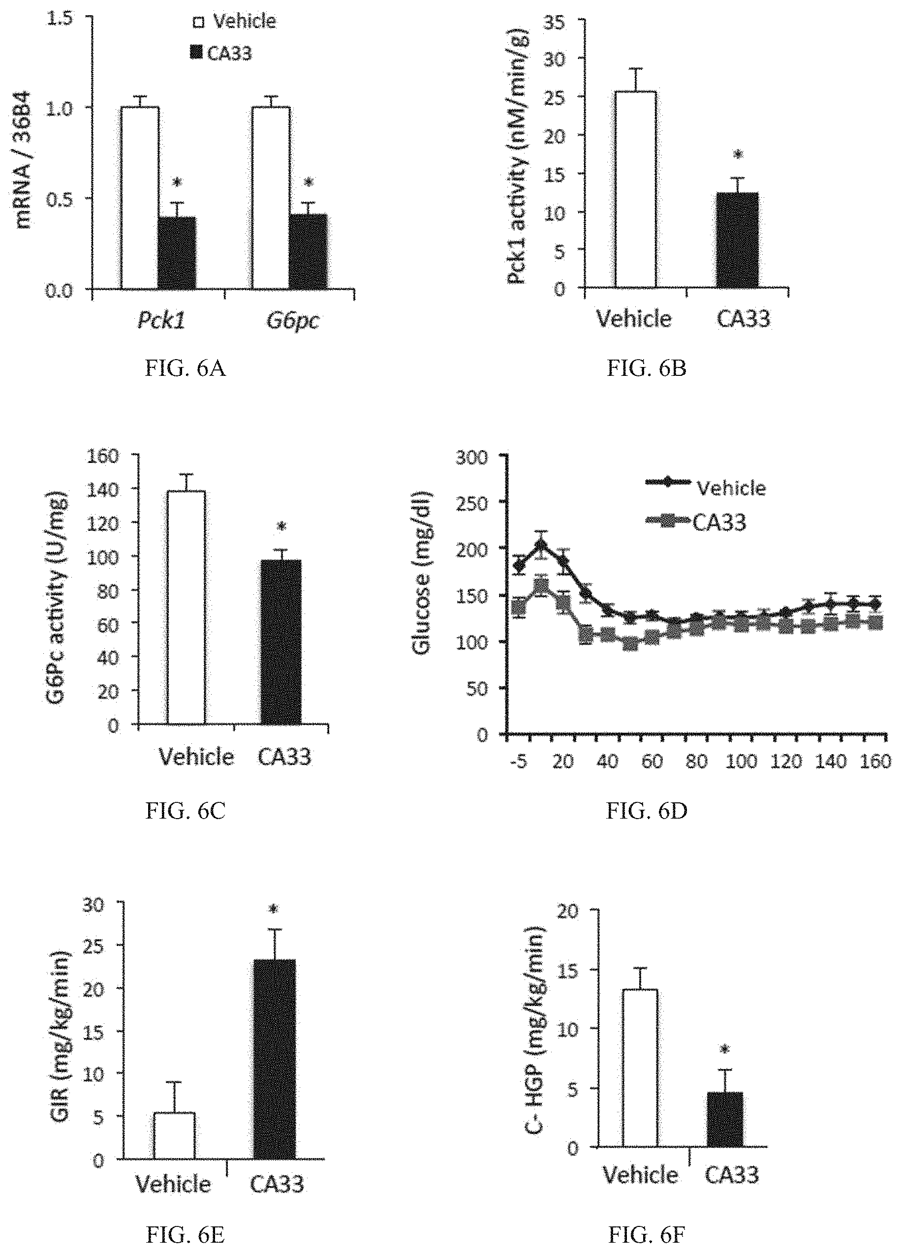
D00014

D00015

D00016

D00017

D00018

D00019

D00020

D00021

D00022

D00023

D00024

D00025

D00026

D00027

D00028

D00029

D00030

D00031

D00032

D00033

D00034

D00035

D00036

D00037

D00038

D00039

D00040

D00041

D00042

D00043

D00044

D00045

D00046

D00047

D00048

D00049

D00050

D00051

D00052

D00053

D00054

D00055

D00056

D00057

D00058

D00059

D00060

D00061

D00062

D00063

D00064

D00065

D00066

D00067

D00068

D00069

D00070

D00071
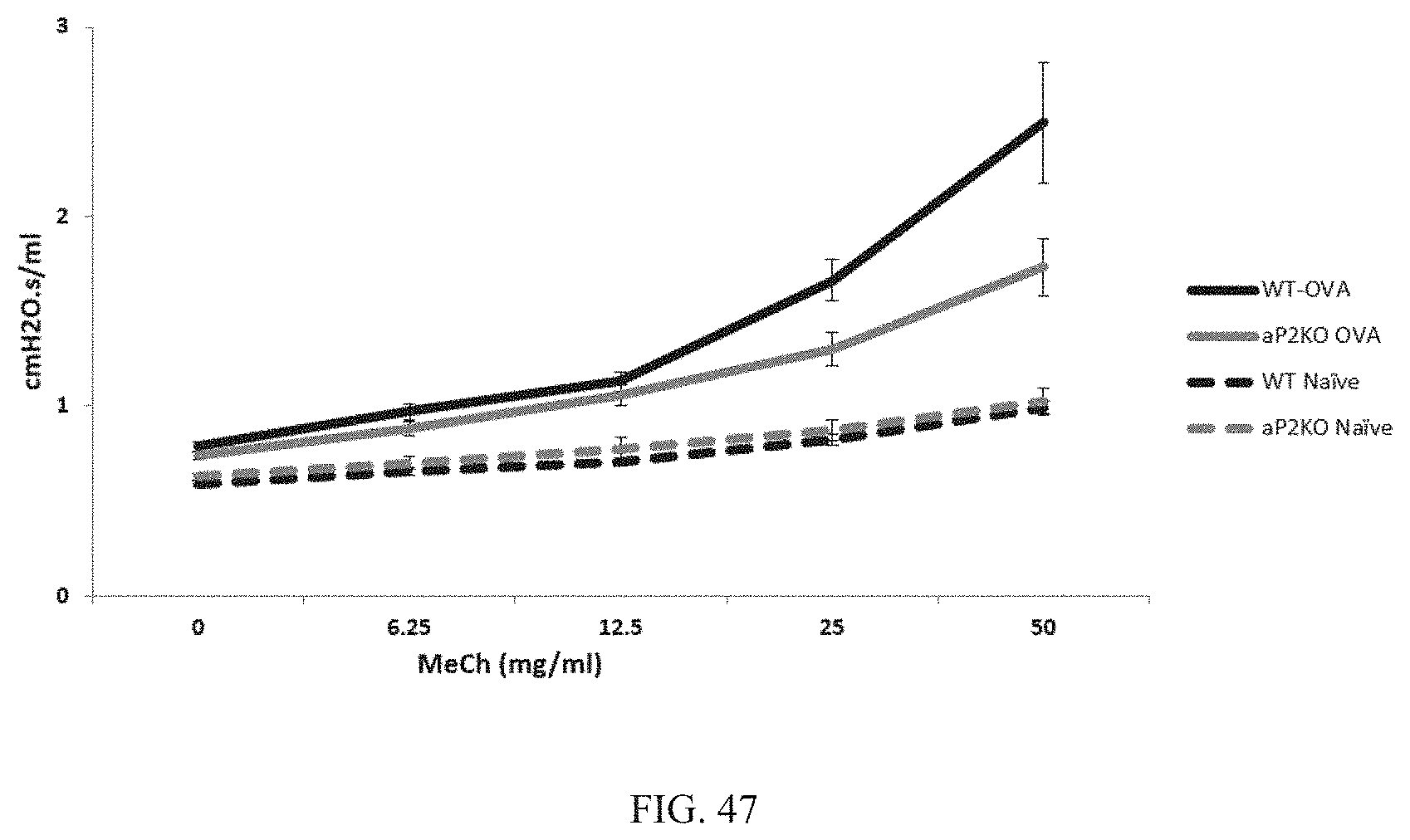
S00001
XML
uspto.report is an independent third-party trademark research tool that is not affiliated, endorsed, or sponsored by the United States Patent and Trademark Office (USPTO) or any other governmental organization. The information provided by uspto.report is based on publicly available data at the time of writing and is intended for informational purposes only.
While we strive to provide accurate and up-to-date information, we do not guarantee the accuracy, completeness, reliability, or suitability of the information displayed on this site. The use of this site is at your own risk. Any reliance you place on such information is therefore strictly at your own risk.
All official trademark data, including owner information, should be verified by visiting the official USPTO website at www.uspto.gov. This site is not intended to replace professional legal advice and should not be used as a substitute for consulting with a legal professional who is knowledgeable about trademark law.