Peptides and combination thereof for use in the immunotherapy against cancers
Walz , et al. May 18, 2
U.S. patent number 11,007,223 [Application Number 16/804,548] was granted by the patent office on 2021-05-18 for peptides and combination thereof for use in the immunotherapy against cancers. This patent grant is currently assigned to IMMATICS BIOTECHNOLOGIES GMBH. The grantee listed for this patent is Immatics Biotechnologies GmbH. Invention is credited to Moreno Di Marco, Sebastian Haen, Daniel Johannes Kowalewski, Markus Loeffler, Annika Nelde, Hans-Georg Rammensee, Stefan Stevanovic, Nico Trautwein, Juliane Sarah Walz.


View All Diagrams
| United States Patent | 11,007,223 |
| Walz , et al. | May 18, 2021 |
Peptides and combination thereof for use in the immunotherapy against cancers
Abstract
The present invention relates to peptides, proteins, nucleic acids and cells for use in immunotherapeutic methods. In particular, the present invention relates to the immunotherapy of cancer. The present invention furthermore relates to tumor-associated T-cell peptide epitopes, alone or in combination with other tumor-associated peptides that can for example serve as active pharmaceutical ingredients of vaccine compositions that stimulate anti-tumor immune responses, or to stimulate T cells ex vivo and transfer into patients. Peptides bound to molecules of the major histocompatibility complex (MHC), or peptides as such, can also be targets of antibodies, soluble T-cell receptors, and other binding molecules.
| Inventors: | Walz; Juliane Sarah (Tuebingen, DE), Kowalewski; Daniel Johannes (Kirchentellinsfurt, DE), Loeffler; Markus (Ammerbuch, DE), Di Marco; Moreno (Tuebingen, DE), Trautwein; Nico (Stuttgart, DE), Nelde; Annika (Tuebingen, DE), Stevanovic; Stefan (Tuebingen, DE), Rammensee; Hans-Georg (Tuebingen, DE), Haen; Sebastian (Tuebingen, DE) | ||||||||||
|---|---|---|---|---|---|---|---|---|---|---|---|
| Applicant: |
|
||||||||||
| Assignee: | IMMATICS BIOTECHNOLOGIES GMBH
(Tuebingen, DE) |
||||||||||
| Family ID: | 63710678 | ||||||||||
| Appl. No.: | 16/804,548 | ||||||||||
| Filed: | February 28, 2020 |
Prior Publication Data
| Document Identifier | Publication Date | |
|---|---|---|
| US 20200223900 A1 | Jul 16, 2020 | |
Related U.S. Patent Documents
| Application Number | Filing Date | Patent Number | Issue Date | ||
|---|---|---|---|---|---|
| 15949665 | Apr 10, 2018 | ||||
| 62483702 | Apr 10, 2017 | ||||
Foreign Application Priority Data
| Apr 10, 2017 [DE] | 102017107697.2 | |||
| Current U.S. Class: | 1/1 |
| Current CPC Class: | C12N 15/115 (20130101); C07K 7/06 (20130101); A61K 38/08 (20130101); C07K 14/7051 (20130101); C12N 5/0636 (20130101); C07K 14/70539 (20130101); C07K 16/2833 (20130101); A61K 39/0011 (20130101); A61P 35/00 (20180101); C07K 16/2818 (20130101); A61K 35/17 (20130101); C07K 14/4748 (20130101); G01N 33/505 (20130101); C07K 2319/70 (20130101); C12N 2310/16 (20130101) |
| Current International Class: | A61K 35/17 (20150101); A61P 35/00 (20060101); C12N 15/115 (20100101); C07K 14/725 (20060101); C07K 16/28 (20060101); C07K 14/74 (20060101); C07K 7/06 (20060101); A61K 39/00 (20060101); G01N 33/50 (20060101); C07K 14/47 (20060101); C12N 5/0783 (20100101) |
References Cited [Referenced By]
U.S. Patent Documents
| 2003/0148321 | August 2003 | Pecker |
| 2006/0115832 | June 2006 | Hoon |
| 2006/0275844 | December 2006 | Linke |
| 2008/0020990 | January 2008 | Yano |
| 2008/0280297 | November 2008 | Dalla-Favera |
| 2011/0190157 | August 2011 | Kipps |
| 2012/0178111 | July 2012 | Diamandis |
| 2015/0125477 | May 2015 | Kuttruff-Coqui et al. |
| 01/42270 | Jun 2001 | WO | |||
| 2004022709 | Mar 2004 | WO | |||
| 2004/052917 | Jun 2004 | WO | |||
| 2004052917 | Jun 2004 | WO | |||
| 2016170139 | Oct 2016 | WO | |||
Other References
|
Paul (Fundamental Immunology, 3rd Edition, 1993, pp. 292-295) (Year: 1993). cited by examiner . Bendig M. M. (Methods: A Companion to Methods in Enzymology, 1995; 8:83-93) (Year: 1995). cited by examiner . Kirkin (Nature Communications, vol. 9, No. 785, p. 1-12, 2018) (Year: 2018). cited by examiner . Rudikoff et al. (Proceedings of the National Academy of Sciences USA, vol. 79, p. 1979-1983, 1982) (Year: 1982). cited by examiner . Heidenreich (Int. J. Cancer, vol. 137, p. 372-384, 2015) (Year: 2015). cited by examiner . Martin (Plos One, vol. 12, No. 1, e0170504, p. 1-17, 2017) (Year: 2017). cited by examiner . Schuster et al., "The immunopeptidomic landscape of ovarian carcinomas" PNAS. Approved Oct. 10, 2017. pp. 1-10, 1-4, and 1-150. cited by applicant . Seeger, F.H. et al. The HLA-B 1516 motif demonstrates HLA-B-specific P2 pocket characteristics. Immunogenetics. 1998. cited by applicant . German Search Report issued in Counterpart German Application No. DE 102017107697.2, dated Oct. 25, 2017. cited by applicant . Y. Ma et al., "Significance of PTPRZ1 and CIN85 expression in cervical carcinoma," Arch Gynecol Obstet 284, Sep. 30, 2010, 699-704. cited by applicant . R.D. Blumenthal et al., "Expression patterns of CEACAM5 and CEACAM6 in primary and metastatic cancers," BMC Cancer 7(2), Jan. 3, 2007, 1-15. cited by applicant . C. Zheng et al., "A Novel Anti-CEACAM5 Monocolonal Antibody, CC4, Suppresses Colorectal Tumor Growth and Enhances NK Cells--Mediated Tumor Immunity," PLoS ONE 6(6), Jun. 22, 2011, 1-11. cited by applicant . L. Ren et al., "Apolipoproteins and cancer," Cancer Medicine, 2019, 7032-7043. cited by applicant . C. Garcia-Pravia et al., "Overexpression of COL11A1 by Cancer-Associated Fibroblasts: Clinical Relevance of a Stromal Marker in Pancreatic Cancer," PLoS ONE 8(10), Oct. 23, 2013, 1-13. cited by applicant . B. Liedert et al., "Overexpression of cMOAT (MRP2/ABCC2) Is Associated with Decreased Formation of Platinum-DNA Adducts and Decreased G2-Arrest in Melanoma Cells Resistant to Cisplatin," J of Investigative Dermatology 121(1), Jul. 2003, 172-176. cited by applicant . G. Wang et al., "Identification of MXRA5 as a novel biomarker in colorectal cancer," Oncology Letters 5, 2013, 544-548. cited by applicant . Y. Wang et al., "HAVcR-1 Expression in Human Colorectal Cancer and its Effects on Colorectal Cancer cells In Vitro," Anticancer Research 33, Jan. 2013, 207-214. cited by applicant . S. Elsheikh et al., "CCND1 amplification and cyclin D1 expression in breast cancer and their relation with proteomic subgroups and patient outcome," Breast Cancer Research & Treatment 109, Jul. 26, 2007, 325-335. cited by applicant . H. Wang et al., "SOX9 is Expressed in Human Fetal Prostate Epithelium and Enhances Prostrate Cancer Invasion," Cancer Research 68(6), Mar. 2008, 1625-1630. cited by applicant . J. He et al., "Association of DCBLD2 upregulation with tumor progression and poor survival in colorectal cancer," Cellular Oncology 43, Mar. 12, 2020, 409-420. cited by applicant . H. Arafat et al., "Tumor-specific expression and alternative splicing of the COL6A3 gene in pancreatic cancer," Surgery 150(2), Aug. 2011, 306-315. cited by applicant . F. Ponten et al., "The Human Protein Atlas--a tool for pathology," PCDHGC3, J. Pathol., 2008, available at https://www.proteinatlas.org/ENSG00000240184-PCDHGC3/pathology. cited by applicant . DC Betticher et al., "Prognostic significance of CCND1 (cyclin D1) overexpression in primary resected non-smell-cell lung cancer," British J. of Cancer 73, Feb. 1, 1996, 294-300. cited by applicant. |
Primary Examiner: Allen; Michael
Attorney, Agent or Firm: McBee Moore & Vanik IP, LLC
Parent Case Text
CROSS REFERENCE TO RELATED APPLICATIONS
This application is a continuation application of U.S. patent application Ser. No. 15/949,665, filed Apr. 10, 2018, which claims benefit to U.S. Provisional Application No. 62/483,702, filed Apr. 10, 2017, and is related to German Application No. 102017107697.2, filed Apr. 10, 2017, the content of which is incorporated herein by reference in its entirety.
Claims
The invention claimed is:
1. A method of treating a patient who has cancer that presents a peptide consisting of the amino acid sequence of SEQ ID NO: 250 in a complex with an MHC class I molecule, comprising administering to said patient a population of activated CD8+ cytotoxic T cells that bind a peptide consisting of the amino acid sequence of SEQ ID NO: 250 in a complex with an MHC class I molecule, wherein said cancer is selected from the group consisting of gastric cancer, colorectal cancer, renal cell carcinoma, and gallbladder adenocarcinoma.
2. The method of claim 1, wherein the CD8+ cytotoxic T cells are autologous to the patient.
3. The method of claim 1, wherein the CD8+ cytotoxic T cells are obtained from a healthy donor.
4. The method of claim 1, wherein the CD8+ cytotoxic T cells are obtained from tumor infiltrating lymphocytes or peripheral blood mononuclear cells.
5. The method of claim 1, wherein the activated CD8+ cytotoxic T cells are expanded in vitro.
6. The method of claim 1, further comprising administering to said patient an adjuvant.
7. The method of claim 6, wherein the adjuvant is selected from imiquimod, GM-CSF, poly-(ICLC), interleukin (IL)-1, IL-2, IL-4, IL-7, IL-12, IL-13, IL-15, IL-21, and IL-23.
8. The method of claim 1, wherein the activated T cells are cytotoxic T cells produced by contacting T cells with an antigen presenting cell that expresses the peptide in a complex with an MHC class I molecule on the surface of the antigen presenting cell, for a period of time sufficient to activate said T cell.
9. The method of claim 8, wherein the antigen presenting cell is infected with a recombinant virus expressing the peptide.
10. The method of claim 1, wherein the cancer is gastric cancer.
11. The method of claim 1, wherein the cancer is gallbladder adenocarcinoma.
12. The method of claim 1, wherein the cancer is renal cell carcinoma.
13. The method of claim 1, wherein the cancer is colorectal cancer.
14. A method of eliciting an immune response in a patient who has cancer that presents a peptide consisting of the amino acid sequence of SEQ ID NO: 250 in a complex with an MHC class I molecule, comprising administering to said patient a population of activated CD8+ cytotoxic T cells that bind a peptide consisting of the amino acid sequence of SEQ ID NO: 250 in a complex with an MHC class I molecule, wherein said cancer is selected from the group consisting of gastric cancer, colorectal cancer, renal cell carcinoma, and gallbladder adenocarcinoma.
15. The method of claim 14, wherein the activated T cells are cytotoxic T cells produced by contacting T cells with an antigen presenting cell that expresses the peptide in a complex with an MHC class I molecule on the surface of the antigen presenting cell, for a period of time sufficient to activate said T cell.
16. The method of claim 14, wherein the cancer is gastric cancer.
17. The method of claim 14, wherein the cancer is gallbladder adenocarcinoma.
18. The method of claim 14, wherein the cancer is renal cell carcinoma.
19. The method of claim 14, wherein the cancer is colorectal cancer.
20. The method of claim 1, further comprising administering to said patient cyclophosphamide, sunitinib, bevacizumab, or sildenafil.
Description
REFERENCE TO SEQUENCE LISTING SUBMITTED AS A COMPLIANT ASCII TEXT FILE (.txt)
Pursuant to the EFS-Web legal framework and 37 CFR .sctn..sctn. 1.821-825 (see MPEP .sctn. 2332.03(a)), a Sequence Listing in the form of an ASCII-compliant text file (entitled "Sequence_Listing_2912919-085004_ST25.txt" created on Nov. 17, 2020 and 83,071 bytes in size) is submitted concurrently with the instant application, and the entire contents of the Sequence Listing are incorporated herein by reference.
FIELD
The present invention relates to peptides, proteins, nucleic acids and cells for use in immunotherapeutic methods. In particular, the present invention relates to the immunotherapy of cancer. The present invention furthermore relates to tumor-associated T-cell peptide epitopes, alone or in combination with other tumor-associated peptides that can for example serve as active pharmaceutical ingredients of vaccine compositions that stimulate anti-tumor immune responses, or to stimulate T cells ex vivo and transfer into patients. Peptides bound to molecules of the major histocompatibility complex (MHC), or peptides as such, can also be targets of antibodies, soluble T-cell receptors, and other binding molecules.
The present invention relates to several novel peptide sequences and their variants derived from HLA class I molecules of human tumor cells that can be used in vaccine compositions for eliciting anti-tumor immune responses, or as targets for the development of pharmaceutically/immunologically active compounds and cells.
BACKGROUND OF THE INVENTION
According to the World Health Organization (WHO), cancer ranged among the four major non-communicable deadly diseases worldwide in 2012. For the same year, colorectal cancer, breast cancer and respiratory tract cancers were listed within the top 10 causes of death in high income countries (who.int/mediacentre/factsheets/fs310/en/).
In 2012, 14.1 million new cancer cases, 32.6 million patients suffering from cancer (within 5 years of diagnosis) and 8.2 million cancer deaths were estimated worldwide (Ferlay et al., 2013; Bray et al., 2013).
Estimated incidences of different cancer types (adult population, both sexes) world-wide in 2012 were (Ferlay et al., 2013; Bray et al., 2013): For cancer of the brain and nervous system: 256213, for colorectal cancer: 1360602, for kidney cancer: 337860, for liver cancer: 782451, and for gastric cancer: 951594 cases.
Estimated incidences of different cancer types (adult population, both sexes) in the USA, EU-28, China and Japan in 2012 were (Ferlay et al., 2013; Bray et al., 2013): For cancer of the brain and nervous system: 135884, for colorectal cancer: 845797, for kidney cancer: 226733, for liver cancer: 513172, and for gastric cancer: 615641 cases.
Estimated mortalities of different cancer types (adult population, both sexes) world-wide in 2012 were (Ferlay et al., 2013; Bray et al., 2013): For cancer of the brain and nervous system: 189382, for colorectal cancer: 693933, for kidney cancer: 143406, for liver cancer: 745533, and for gastric cancer: 723073 cases.
Estimated mortalities of different cancer types (adult population, both sexes) in the USA, EU-28, China and Japan in 2012 were (Ferlay et al., 2013; Bray et al., 2013): For cancer of the brain and nervous system: 100865, for colorectal cancer: 396066, for kidney cancer: 83741, for liver cancer: 488485, and for gastric cancer: 447735 cases.
Within the groups of brain cancer, the current invention specifically focuses on glioblastoma (GBM). GBM is the most common central nervous system malignancy with an age-adjusted incidence rate of 3.19 per 100,000 inhabitants within the United States. GBM has a very poor prognosis with a 1-year survival rate of 35% and a 5-year survival rate lower than 5%. Male gender, older age and ethnicity appear to be risk factors for GBM (Thakkar et al., 2014).
Colorectal cancer--Depending on the colorectal cancer (CRC) stage, different standard therapies are available for colon and rectal cancer. Standard procedures include surgery, radiation therapy, chemotherapy and targeted therapy for CRC (Berman et al., 2015a; Berman et al., 2015b).
Removal of the tumor is essential for the treatment of CRC. Anatomic conditions differ for rectal carcinomas from another CRC as the rectum is located in the pelvis and the tumor can be difficult to access. Well-differentiated small rectal tumors (stage T1) require excision, but no further treatment with chemotherapy. Patients with rectal tumors of higher T stages receive neoadjuvant radio-chemotherapy with a fluoropyrimidine prior to total mesorectal excision (TME) and adjuvant chemotherapy. For chemotherapeutic treatment, the drugs capecitabine or 5-fluorouracil (5-FU) are used. For combinational chemotherapy, a cocktail containing 5-FU, leucovorin and oxaliplatin (FOLFOX) is recommended (Stintzing, 2014; Berman et al., 2015b).
Treatment of colon carcinomas involves radical hemicolectomy and lymph node resection. Early stages (UICC stage I) do not require additional treatment. Patients with tumors of UICC stage II receive 5-FU or capecitabine. Treatment for patients with UICC stage III includes the drug combinations FOLFOX and XELOX (capecitabine plus oxaliplatin) (Berman et al., 2015a; Stintzing, 2014).
Metastatic, unresectable CRC are treated with chemotherapeutical cocktails such as FOLFIRI (5-FU, leucovorin, irinotecan), FOLFOX, FOLFOXIRI (5-FU, irinotecan, oxaliplatin), FOLFOX/capecitabine, FOLFOX/oxaliplatin, FOLFIRI/capecitabine and irinotecan or UFT (5-FU, tegafur-uracil) (Stintzing, 2014).
In addition to chemotherapeutic drugs, several monoclonal antibodies targeting the epidermal growth factor receptor (EGFR, cetuximab, panitumumab) or the vascular endothelial growth factor-A (VEGF-A, bevacizumab) are administered to patients with high stage disease. For second-line and later treatment the inhibitor for VEGF aflibercept, the tyrosine kinase inhibitor regorafenib and the thymidylate-synthetase inhibitor TAS-102 and the dUTPase inhibitor TAS-114 can be used (Stintzing, 2014; Wilson et al., 2014).
Latest clinical trials analyze active immunotherapy as a treatment option against CRC. Those strategies include the vaccination with peptides from tumor-associated antigens (TAAs), whole tumor cells, dendritic cell (DC) vaccines and viral vectors (Koido et al., 2013).
Peptide vaccines have so far been directed against carcinoembryonic antigen (CEA), mucin 1, EGFR, squamous cell carcinoma antigen recognized by T cells 3 (SART3), beta-human chorionic gonadotropin (beta-hCG), Wilms' Tumor antigen 1 (WT1), Survivin-2B, MAGE3, p53, ring finger protein 43 and translocase of the outer mitochondrial membrane 34 (TOMM34), or mutated KRAS. In several phase I and II clinical trials patients showed antigen-specific CTL responses or antibody production. In contrast to immunological responses, many patients did not benefit from peptide vaccines on the clinical level (Koido et al., 2013; Miyagi et al., 2001; Moulton et al., 2002; Okuno et al., 2011).
Dendritic cell vaccines comprise DCs pulsed with either TAA-derived peptides, tumor cell lysates, apoptotic tumor cells, or tumor RNA or DC-tumor cell fusion products. While many patients in phase I/II trials showed specific immunological responses, only the minority had a clinical benefit (Koido et al., 2013).
Whole tumor cell vaccines consist of autologous tumor cells modified to secrete GM-CSF, modified by irradiation or virus-infected, irradiated cells. Most patients showed no clinical benefit in several phase II/III trials (Koido et al., 2013).
Vaccinia virus or replication-defective avian poxvirus encoding CEA as well as B7.1, ICAM-1 and LFA-3 have been used as vehicles in viral vector vaccines in phase I clinical trials. A different study used non-replicating canary pox virus encoding CEA and B7.1. Besides the induction of CEA-specific T cell responses 40% of patients showed objective clinical responses (Horig et al., 2000; Kaufman et al., 2008).
Gastric cancer--The wall of the stomach is made up of 3 layers of tissue: the mucosal (innermost) layer, the muscularis (middle) layer, and the serosal (outermost) layer. Gastric cancer (GC) begins in the cells lining the mucosal layer and spreads through the outer layers as it grows. Four types of standard treatment are used. Treatment for gastric cancer may involve endoscopic or surgical resection, chemotherapy, radiation therapy or chemoradiation. Surgery is the primary treatment and the only curative treatment for gastric cancer. Since the early stages of gastric cancer are mostly asymptomatic, the disease is usually diagnosed in an advanced stage. For metastatic gastric cancer, no globally accepted standard chemotherapy combination regimen has yet been established. However, the combination of 5-FU and a platinum analog is still the most widely accepted reference regimen worldwide, although 5-FU can be replaced by capecitabine or irinotecan and cisplatin can be replaced by oxaliplatin. Additionally, triple-combination therapies comprising cisplatin, 5-FU and docetaxel or, in the case of HER-2 over-expressing tumors, cisplatin, 5-FU and trastuzumab can be applied (Leitlinie Magenkarzinom, 2012).
The efficacy of current therapeutic regimens for advanced GC is poor, resulting in low 5-year survival rates. Immunotherapy might be an alternative approach to ameliorate the survival of GC patients. Adoptive transfer of tumor-associated lymphocytes and cytokine induced killer cells, peptide-based vaccines targeting HER2/neu, MAGE-3 or vascular endothelial growth factor receptor 1 and 2 and dendritic cell-based vaccines targeting HER2/neu showed promising results in clinical GC trials. Immune checkpoint inhibition and engineered T cells might represent additional therapeutic options, which is currently evaluated in pre-clinical and clinical studies (Matsueda and Graham, 2014).
Glioblastoma--The therapeutic options for glioblastoma (WHO grade IV) are very limited. According to the guidelines released by the German Society for Neurology the standard therapy in young patients includes resection or biopsy of the tumor, focal radiation therapy and chemotherapy with temozolomide. Alternative chemotherapeutic regimens consist of CCNU/lomustine or a combination of procarbazine with CCNU and vincristine (PCV). In elderly patients' resection or biopsy of the tumor are not recommended. These patients receive chemo- or radiation therapy, depending on the methylation state of the O6-methylguanine-DNA-methyltransferase-(MGMT)-promotor. Negative methylation state is an indication for focal radiation therapy, whereas positive methylation state is an indication for temozolomide treatment with or without focal radiation therapy. Relapse therapy comprises again resection as well as chemo- and radiation therapy. In the USA, Canada and Switzerland treatment with bevacizumab (anti-VEGF-antibody) is also approved for relapse therapy (Leitlinien fur Diagnostik and Therapie in der Neurologie, 2014).
Different immunotherapeutic approaches are investigated for the treatment of GB, including immune-checkpoint inhibition, vaccination and adoptive transfer of engineered T cells.
Antibodies directed against inhibitory T cell receptors or their ligands were shown to efficiently enhance T cell-mediated anti-tumor immune responses in different cancer types, including melanoma and bladder cancer. The effects of T cell activating antibodies like ipilimumab and nivolumab are therefore assessed in clinical GB trials, but preliminary data indicate autoimmune-related adverse events.
Different vaccination strategies for GB patients are currently investigated, including peptide-based vaccines, heat-shock protein vaccines, autologous tumor cell vaccines, dendritic cell-based vaccines and viral protein-based vaccines. In these approaches peptides derived from GB-associated proteins like epidermal growth factor receptor variant III (EGFRvIll) or heat shock proteins or dendritic cells pulsed with autologous tumor cell lysate or cytomegalo virus components are applied to induce an anti-tumor immune response in GB patients. Several of these studies reveal good safety and tolerability profiles as well as promising efficacy data.
Adoptive transfer of genetically modified T cells is an additional immunotherapeutic approach for the treatment of GB. Different clinical trials currently evaluate the safety and efficacy of chimeric antigen receptor bearing T cells directed against HER2, IL-13 receptor alpha 2 and EGFRvIll (Ampie et al., 2015).
Liver cancer--Disease management depends on the tumor stage at the time of diagnosis and the overall condition of the liver. If possible, parts of the liver (partial hepatectomy) or the whole organ (liver resection) is removed by surgery. Especially patients with small or completely resectable tumors are qualified to receive a liver transplant.
If surgery is not a treatment option, different other therapies are available at hand. For tumor ablation, a probe is injected into the liver and the tumor is destroyed by radio or microwaves or cryotherapy. In embolization procedures, the blood supply of the tumor is blocked by mechanical or chemical means. High energy radio waves can be used to destroy the tumor in radiation therapy.
Chemotherapy against HCC includes combinations of doxorubicin, 5-fluorouracil and cisplatin for systemic therapy and doxorubicin, floxuridine and mitomycin C for hepatic artery infusions. However, most HCC show a high resistance to chemotherapeutics (Enguita-German and Fortes, 2014).
Therapeutic options in advanced non-resectable HCC are limited to Sorafenib, a multi-tyrosine kinase inhibitor (Chang et al., 2007; Wilhelm et al., 2004). Sorafenib is the only systemic drug confirmed to increase survival by about 3 months and currently represents the only experimental treatment option for such patients (Chapiro et al., 2014; Llovet et al., 2008).
Lately, a limited number of immunotherapy trials for HCC have been conducted. Cytokines have been used to activate subsets of immune cells and/or increase the tumor immunogenicity (Reinisch et al., 2002; Sangro et al., 2004). Other trials have focused on the infusion of Tumor-infiltrating lymphocytes or activated peripheral blood lymphocytes (Shi et al., 2004; Takayama et al., 1991; Takayama et al., 2000).
So far, a small number of therapeutic vaccination trials have been executed. Butterfield et al. conducted two trials using peptides derived from alpha-fetoprotein (AFP) as a vaccine or DCs loaded with AFP peptides ex vivo (Butterfield et al., 2003; Butterfield et al., 2006). In two different studies, autologous dendritic cells (DCs) were pulsed ex vivo with autologous tumor lysate (Lee et al., 2005) or lysate of the hepatoblastoma cell line HepG2 (Palmer et al., 2009). So far, vaccination trials have only shown limited improvements in clinical outcomes.
Renal cell carcinoma--Initial treatment is most commonly either partial or complete removal of the affected kidney(s) and remains the mainstay of curative treatment (Rini et al., 2008). For first-line treatment of patients with poor prognostic score a guidance elaborated by several cancer organizations and societies recommend the receptor tyrosine kinase inhibitors (TKIs) sunitinib and pazopanib, the monoclonal antibody bevacizumab combined with interferon-.alpha. (IFN-.alpha.) and the mTOR inhibitor temsirolimus. Based on guidelines elaborated by the US NCCN as well as the European EAU and ESMO, the TKIs sorafenib, pazopanib or recently axitinib are recommended as second-line therapy in RCC patients who have failed prior therapy with cytokines (IFN-.alpha., IL-2). The NCCN guidelines advise also sunitinib in this setting (high-level evidence according to NCCN Category I).
Everolimus and axitinib are recommended as second-line therapy of those patients who have not benefited from a VEGF-targeted therapy with TKIs according to the established guidelines.
The known immunogenity of RCC has represented the basis supporting the use of immunotherapy and cancer vaccines in advanced RCC. The interesting correlation between lymphocytes PD-1 expression and RCC advanced stage, grade and prognosis, as well as the selective PD-L1 expression by RCC tumor cells and its potential association with worse clinical outcomes, have led to the development of new anti PD-1/PD-L1 agents, alone or in combination with anti-angiogenic drugs or other immunotherapeutic approaches, for the treatment of RCC (Massari et al., 2015). In advanced RCC, a phase III cancer vaccine trial called TRIST study evaluates whether TroVax (a vaccine using a tumor-associated antigen 5T4, with a pox virus vector), added to first-line standard of care therapy, prolongs survival of patients with locally advanced or mRCC. Median survival had not been reached in either group with 399 patients (54%) remaining on study however analysis of the data confirms prior clinical results, demonstrating that TroVax is both immunologically active and that there is a correlation between the strength of the 5T4-specific antibody response and improved survival. Further there are several studies searching for peptide vaccines using epitopes being over-expressed in RCC.
Various approaches of tumor vaccines have been under investigation. Studies using whole-tumor approaches, including tumor cell lysates, fusions of dendritic cells with tumor cells, or whole-tumor RNA were done in RCC patients, and remissions of tumor lesions were reported in some of these trials (Avigan et al., 2004; Holtl et al., 2002; Marten et al., 2002; Su et al., 2003; Wittig et al., 2001).
Considering the severe side-effects and expense associated with treating cancer, there is a need to identify factors that can be used in the treatment of cancer in general and colorectal cancer, glioblastoma, gastric cancer, hepatocellular carcinoma, and renal cell carcinoma. There is also a need to identify factors representing biomarkers for cancer in general and colorectal cancer, glioblastoma, gastric cancer, hepatocellular carcinoma, and renal cell carcinoma in particular, leading to better diagnosis of cancer, assessment of prognosis, and prediction of treatment success.
Immunotherapy of cancer represents an option of specific targeting of cancer cells while minimizing side effects. Cancer immunotherapy makes use of the existence of tumor associated antigens.
The current classification of tumor associated antigens (TAAs) comprises the following major groups:
a) Cancer-testis antigens: The first TAAs ever identified that can be recognized by T cells belong to this class, which was originally called cancer-testis (CT) antigens because of the expression of its members in histologically different human tumors and, among normal tissues, only in spermatocytes/spermatogonia of testis and, occasionally, in placenta. Since the cells of testis do not express class I and II HLA molecules, these antigens cannot be recognized by T cells in normal tissues and can therefore be considered as immunologically tumor-specific. Well-known examples for CT antigens are the MAGE family members and NY-ESO-1. b) Differentiation antigens: These TAAs are shared between tumors and the normal tissue from which the tumor arose. Most of the known differentiation antigens are found in melanomas and normal melanocytes. Many of these melanocyte lineage-related proteins are involved in biosynthesis of melanin and are therefore not tumor specific but nevertheless are widely used for cancer immunotherapy. Examples include, but are not limited to, tyrosinase and Melan-A/MART-1 for melanoma or PSA for prostate cancer. c) Over-expressed TAAs: Genes encoding widely expressed TAAs have been detected in histologically different types of tumors as well as in many normal tissues, generally with lower expression levels. It is possible that many of the epitopes processed and potentially presented by normal tissues are below the threshold level for T-cell recognition, while their over-expression in tumor cells can trigger an anticancer response by breaking previously established tolerance. Prominent examples for this class of TAAs are Her-2/neu, survivin, telomerase, or WT1. d) Tumor-specific antigens: These unique TAAs arise from mutations of normal genes (such as .beta.-catenin, CDK4, etc.). Some of these molecular changes are associated with neoplastic transformation and/or progression. Tumor-specific antigens are generally able to induce strong immune responses without bearing the risk for autoimmune reactions against normal tissues. On the other hand, these TAAs are in most cases only relevant to the exact tumor on which they were identified and are usually not shared between many individual tumors. Tumor-specificity (or -association) of a peptide may also arise if the peptide originates from a tumor- (-associated) exon in case of proteins with tumor-specific (-associated) isoforms. e) TAAs arising from abnormal post-translational modifications: Such TAAs may arise from proteins which are neither specific nor overexpressed in tumors but nevertheless become tumor associated by posttranslational processes primarily active in tumors. Examples for this class arise from altered glycosylation patterns leading to novel epitopes in tumors as for MUC1 or events like protein splicing during degradation which may or may not be tumor specific. f) Oncoviral proteins: These TAAs are viral proteins that may play a critical role in the oncogenic process and, because they are foreign (not of human origin), they can evoke a T-cell response. Examples of such proteins are the human papilloma type 16 virus proteins, E6 and E7, which are expressed in cervical carcinoma.
T-cell based immunotherapy targets peptide epitopes derived from tumor-associated or tumor-specific proteins, which are presented by molecules of the major histocompatibility complex (MHC). The antigens that are recognized by the tumor specific T lymphocytes, that is, the epitopes thereof, can be molecules derived from all protein classes, such as enzymes, receptors, transcription factors, etc. which are expressed and, as compared to unaltered cells of the same origin, usually up-regulated in cells of the respective tumor.
There are two classes of MHC-molecules, MHC class I and MHC class II. MHC class I molecules are composed of an alpha heavy chain and beta-2-microglobulin, MHC class II molecules of an alpha and a beta chain. Their three-dimensional conformation results in a binding groove, which is used for non-covalent interaction with peptides.
MHC class I molecules can be found on most nucleated cells. They present peptides that result from proteolytic cleavage of predominantly endogenous proteins, defective ribosomal products (DRIPs) and larger peptides. However, peptides derived from endosomal compartments or exogenous sources are also frequently found on MHC class I molecules. This non-classical way of class I presentation is referred to as cross-presentation in the literature (Brossart and Bevan, 1997; Rock et al., 1990). MHC class II molecules can be found predominantly on professional antigen presenting cells (APCs), and primarily present peptides of exogenous or transmembrane proteins that are taken up by APCs e.g. during endocytosis, and are subsequently processed.
Complexes of peptide and MHC class I are recognized by CD8-positive T cells bearing the appropriate T-cell receptor (TCR), whereas complexes of peptide and MHC class II molecules are recognized by CD4-positive-helper-T cells bearing the appropriate TCR. It is well known that the TCR, the peptide and the MHC are thereby present in a stoichiometric amount of 1:1:1.
CD4-positive helper T cells play an important role in inducing and sustaining effective responses by CD8-positive cytotoxic T cells. The identification of CD4-positive T-cell epitopes derived from tumor associated antigens (TAA) is of immense importance for the development of pharmaceutical products for triggering anti-tumor immune responses (Gnjatic et al., 2003). At the tumor site, T helper cells, support a cytotoxic T cell- (CTL-) friendly cytokine milieu (Mortara et al., 2006) and attract effector cells, e.g. CTLs, natural killer (NK) cells, macrophages, and granulocytes (Hwang et al., 2007).
In the absence of inflammation, expression of MHC class II molecules is mainly restricted to cells of the immune system, especially professional antigen-presenting cells (APC), e.g., monocytes, monocyte-derived cells, macrophages, dendritic cells. In cancer patients, cells of the tumor have been found to express MHC class II molecules (Dengjel et al., 2006).
Longer (elongated) peptides of the invention can act as MHC class II active epitopes.
T-helper cells, activated by MHC class II epitopes, play an important role in orchestrating the effector function of CTLs in anti-tumor immunity. T-helper cell epitopes that trigger a T-helper cell response of the TH1 type support effector functions of CD8-positive killer T cells, which include cytotoxic functions directed against tumor cells displaying tumor-associated peptide/MHC complexes on their cell surfaces. In this way tumor-associated T-helper cell peptide epitopes, alone or in combination with other tumor-associated peptides, can serve as active pharmaceutical ingredients of vaccine compositions that stimulate anti-tumor immune responses.
It was shown in mammalian animal models, e.g., mice, that even in the absence of CD8-positive T lymphocytes, CD4-positive T cells are sufficient for inhibiting manifestation of tumors via inhibition of angiogenesis by secretion of interferon-gamma (IFN.gamma.) (Beatty and Paterson, 2001; Mumberg et al., 1999). There is evidence for CD4 T cells as direct anti-tumor effectors (Braumuller et al., 2013; Tran et al., 2014).
Since the constitutive expression of HLA class II molecules is usually limited to immune cells, the possibility of isolating class II peptides directly from primary tumors was previously not considered possible. However, Dengjel et al. were successful in identifying a number of MHC Class II epitopes directly from tumors (WO 2007/028574, EP 1 760 088 B1).
Since both types of response, CD8 and CD4 dependent, contribute jointly and synergistically to the anti-tumor effect, the identification and characterization of tumor-associated antigens recognized by either CD8+ T cells (ligand: MHC class I molecule+peptide epitope) or by CD4-positive T-helper cells (ligand: MHC class II molecule+peptide epitope) is important in the development of tumor vaccines.
For an MHC class I peptide to trigger (elicit) a cellular immune response, it also must bind to an MHC-molecule. This process is dependent on the allele of the MHC-molecule and specific polymorphisms of the amino acid sequence of the peptide. MHC-class-1-binding peptides are usually 8-12 amino acid residues in length and usually contain two conserved residues ("anchors") in their sequence that interact with the corresponding binding groove of the MHC-molecule. In this way, each MHC allele has a "binding motif" determining which peptides can bind specifically to the binding groove.
In the MHC class I dependent immune reaction, peptides not only have to be able to bind to certain MHC class I molecules expressed by tumor cells, they subsequently also have to be recognized by T cells bearing specific T cell receptors (TCR).
For proteins to be recognized by T-lymphocytes as tumor-specific or -associated antigens, and to be used in a therapy, particular prerequisites must be fulfilled. The antigen should be expressed mainly by tumor cells and not, or in comparably small amounts, by normal healthy tissues. In a preferred embodiment, the peptide should be over-presented by tumor cells as compared to normal healthy tissues. It is furthermore desirable that the respective antigen is not only present in a type of tumor, but also in high concentrations (i.e. copy numbers of the respective peptide per cell). Tumor-specific and tumor-associated antigens are often derived from proteins directly involved in transformation of a normal cell to a tumor cell due to their function, e.g. in cell cycle control or suppression of apoptosis. Additionally, downstream targets of the proteins directly causative for a transformation may be up-regulated and thus may be indirectly tumor-associated. Such indirect tumor-associated antigens may also be targets of a vaccination approach (Singh-Jasuja et al., 2004). It is essential that epitopes are present in the amino acid sequence of the antigen, in order to ensure that such a peptide ("immunogenic peptide"), being derived from a tumor associated antigen, leads to an in vitro or in vivo T-cell-response.
Basically, any peptide able to bind an MHC molecule may function as a T-cell epitope. A prerequisite for the induction of an in vitro or in vivo T-cell-response is the presence of a T cell having a corresponding TCR and the absence of immunological tolerance for this particular epitope.
Therefore, TAAs are a starting point for the development of a T cell based therapy including but not limited to tumor vaccines. The methods for identifying and characterizing the TAAs are usually based on the use of T-cells that can be isolated from patients or healthy subjects, or they are based on the generation of differential transcription profiles or differential peptide expression patterns between tumors and normal tissues. However, the identification of genes over-expressed in tumor tissues or human tumor cell lines, or selectively expressed in such tissues or cell lines, does not provide precise information as to the use of the antigens being transcribed from these genes in an immune therapy. This is because only an individual subpopulation of epitopes of these antigens are suitable for such an application since a T cell with a corresponding TCR has to be present and the immunological tolerance for this particular epitope needs to be absent or minimal. In a very preferred embodiment of the invention it is therefore important to select only those over- or selectively presented peptides against which a functional and/or a proliferating T cell can be found. Such a functional T cell is defined as a T cell, which upon stimulation with a specific antigen can be clonally expanded and is able to execute effector functions ("effector T cell").
In case of targeting peptide-MHC by specific TCRs (e.g. soluble TCRs) and antibodies or other binding molecules (scaffolds) according to the invention, the immunogenicity of the underlying peptides is secondary. In these cases, the presentation is the determining factor.
SUMMARY
In a first aspect of the present invention, the present invention relates to a peptide comprising an amino acid sequence selected from the group consisting of SEQ ID NO: 1 to SEQ ID NO: 268 or a variant sequence thereof which is at least 77%, preferably at least 88%, homologous (preferably at least 77% or at least 88% identical) to SEQ ID NO: 1 to SEQ ID NO: 268, wherein said variant binds to MHC and/or induces T cells cross-reacting with said peptide, or a pharmaceutical acceptable salt thereof, wherein said peptide is not the underlying full-length polypeptide.
The present invention further relates to a peptide of the present invention comprising a sequence that is selected from the group consisting of SEQ ID NO: 1 to SEQ ID NO: 268 or a variant thereof, which is at least 77%, preferably at least 88%, homologous (preferably at least 77% or at least 88% identical) to SEQ ID NO: 1 to SEQ ID NO: 268, wherein said peptide or variant thereof has an overall length of between 8 and 100, preferably between 8 and 30, and most preferred of between 8 and 14 amino acids.
BRIEF DESCRIPTION OF THE DRAWINGS
FIGS. 1A-1P to FIG. 9 all depict embodiments as described herein.
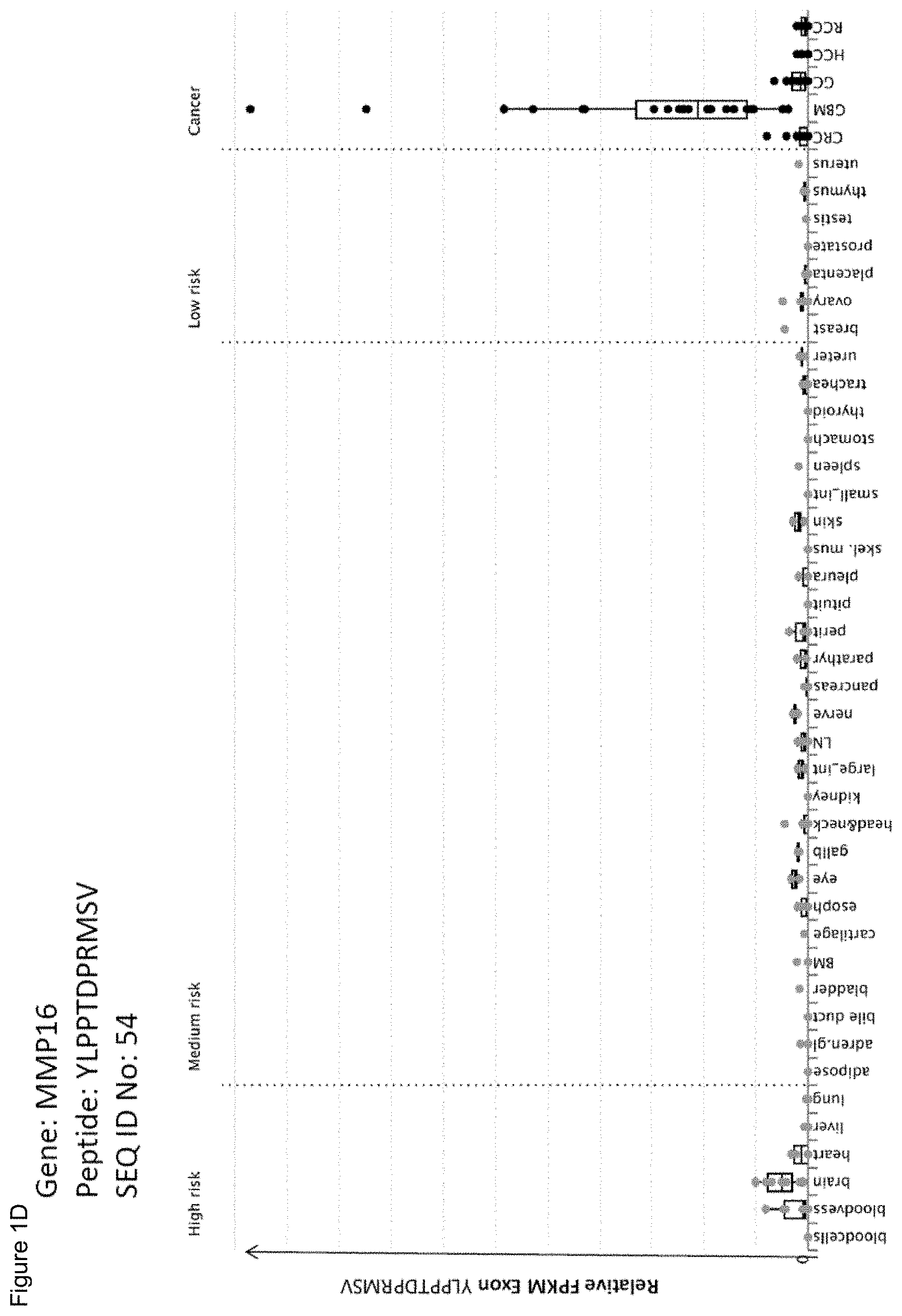
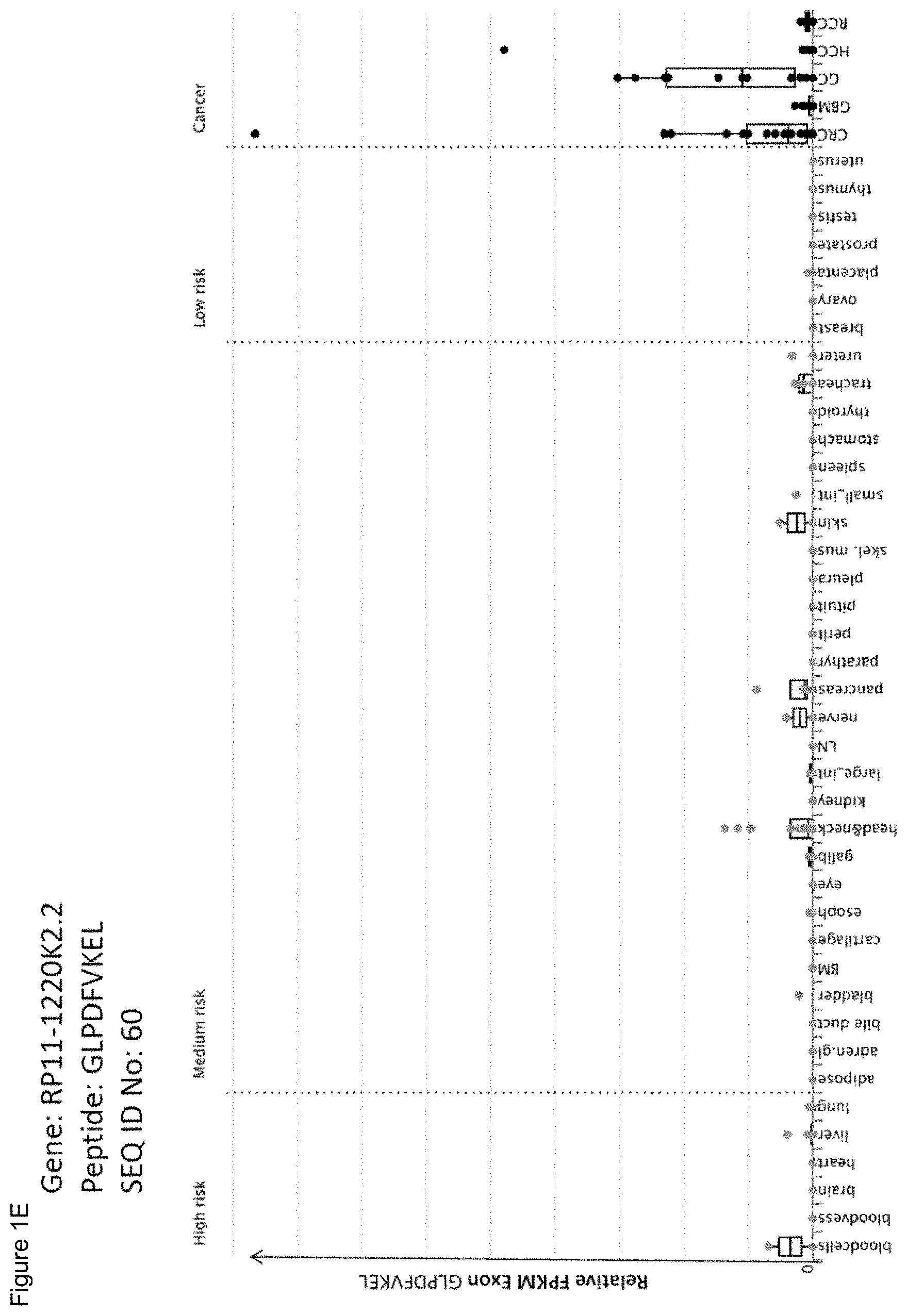
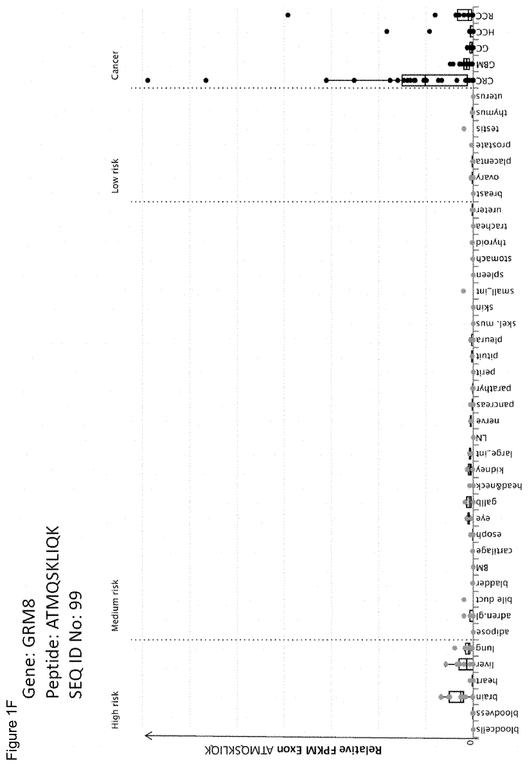
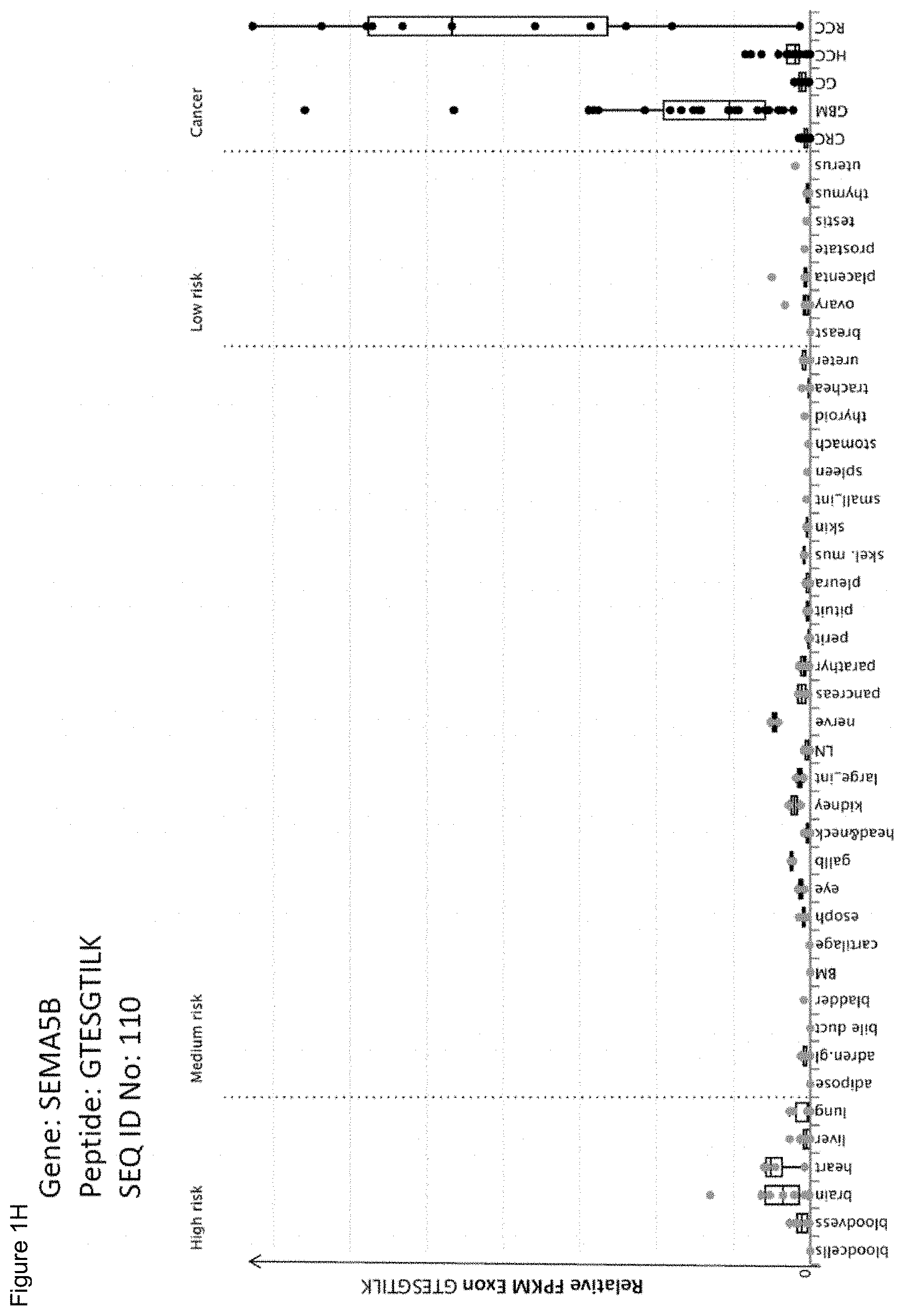
FIGS. 1A through 1P show exemplary expression profile of source genes of the present invention that are over-expressed in different cancer samples. Tumor (black dots) and normal (grey dots) samples are grouped according to organ of origin, and box-and-whisker plots represent median, 25th and 75th percentile (box), and minimum and maximum (whiskers) RPKM values. Normal organs are ordered according to risk categories. RPKM=reads per kilobase per million mapped reads. Normal samples: blood cells; blood vessel; brain; heart; liver; lung; adipose: adipose tissue; adren.gl.: adrenal gland; bile duct; bladder; BM: bone marrow; cartilage; esoph: esophagus; eye; gallb: gallbladder; head and neck; kidney; large_int: large intestine; LN: lymph node; nerve; pancreas; parathyr: parathyroid; perit: peritoneum; pituit: pituitary; skel.mus: skeletal muscle; skin; small_int: small intestine; spleen; stomach; thyroid; trachea; ureter; breast; ovary; placenta; prostate; testis; thymus; uterus. Tumor samples: CRC: colorectal cancer; GBM: glioblastoma; GC: gastric cancer; HCC: hepatocellular carcinoma; RCC: renal cell carcinoma. FIG. 1A) Gene symbol: EGFR, Peptide: LPSPTDSNFY (SEQ ID No.: 2), FIG. 1B) Gene symbol: PTPRZ1, Peptide: LTDYINANY (SEQ ID No.: 28), 1C) Gene symbol: CCDC146, Peptide: KMMALVAEL (SEQ ID No.: 42), 1D) Gene symbol: MMP16, Peptide: YLPPTDPRMSV (SEQ ID No.: 54), 1E) Gene symbol: RP11-1220K2.2, Peptide: GLPDFVKEL (SEQ ID No.: 60), 1F) Gene symbol: GRM8, Peptide: ATMQSKLIQK (SEQ ID No.: 99), 1G) Gene symbol: HAVCR1, Peptide: GVIIAKKYFFK (SEQ ID No.: 101), 1H) Gene symbol: SEMA5B, Peptide: GTESGTILK (SEQ ID No.: 110), 1I) Gene symbol: PIWIL1, Peptide: SFDSNLLSF (SEQ ID No.: 133), 1J) Gene symbol: UGT1A3, Peptide: KYLSIPTVF (SEQ ID No.: 138), 1K) Gene symbol: FEZF1, Peptide: APAAVPSAPA (SEQ ID No.: 153), 1L) Gene symbol: MMP11, Peptide: RPASSLRP (SEQ ID No.: 163), 1M) Gene symbol: QRFPR, Peptide: SPMWHVQQL (SEQ ID No.: 170), 1N) Gene symbol: REG4, Peptide: SRSMRLLLL (SEQ ID No.: 190), 1O) Gene symbol: PTHLH, Peptide: AEIHTAEI (SEQ ID No.: 231), 1P) Gene symbol: EGFR, Peptide: DEYLIPQQGF (SEQ ID No.: 264).
FIG. 2 shows exemplary results of peptide-specific in vitro CD8+ T cell responses of a healthy HLA-A*02+ donor. CD8+ T cells were primed using artificial APCs coated with anti-CD28 mAB and HLA-A*02 in complex with SeqID No 267 peptide (KTLGKLWRL, Seq ID NO: 267) (A, left panel). After three cycles of stimulation, the detection of peptide-reactive cells was performed by 2D multimer staining with A*02/SeqID No 267 (A). Right panel (B) show control staining of cells stimulated with irrelevant A*02/peptide complexes. Viable singlet cells were gated for CD8+ lymphocytes. Boolean gates helped excluding false-positive events detected with multimers specific for different peptides. Frequencies of specific multimer+cells among CD8+ lymphocytes are indicated.
FIG. 3 shows exemplary results of peptide-specific in vitro CD8+ T cell responses of a healthy HLA-A*24+ donor. CD8+ T cells were primed using artificial APCs coated with anti-CD28 mAB and HLA-A*24 in complex with SEQ ID NO: 268 peptide (A, left panel). After three cycles of stimulation, the detection of peptide-reactive cells was performed by 2D multimer staining with A*24/SEQ ID NO: 268 (DYIPYVFKL, SEQ ID NO: 268) (A). Right panel (B) shows control staining of cells stimulated with irrelevant A*24/peptide complexes. Viable singlet cells were gated for CD8+ lymphocytes. Boolean gates helped excluding false-positive events detected with multimers specific for different peptides. Frequencies of specific multimer+ cells among CD8+ lymphocytes are indicated.
FIG. 4 shows exemplary results of peptide-specific in vitro CD8+ T cell responses of a healthy HLA-A*01+ donor. CD8+ T cells were primed using artificial APCs coated with anti-CD28 mAb and HLA-A*01 in complex with SEQ ID NO: 6 peptide (VLDLSTNVY; A, left panel) and SEQ ID NO: 245 peptide (ITEKNSGLY; B, left panel), respectively. After three cycles of stimulation, the detection of peptide-reactive cells was performed by 2D multimer staining with A*01/SEQ ID NO: 6 (A) or A*01/SEQ ID NO: 245 (B). Right panels (A and B) show control staining of cells stimulated with irrelevant A*01/peptide complexes. Viable singlet cells were gated for CD8+ lymphocytes. Boolean gates helped excluding false-positive events detected with multimers specific for different peptides. Frequencies of specific multimer+ cells among CD8+ lymphocytes are indicated.
FIG. 5 shows exemplary results of peptide-specific in vitro CD8+ T cell responses of a healthy HLA-A*02+ donor. CD8+ T cells were primed using artificial APCs coated with anti-CD28 mAb and HLA-A*02 in complex with SEQ ID NO: 42 peptide (KMMALVAEL; A, left panel) and SEQ ID NO: 250 peptide (KMILKMVQL; B, left panel), respectively. After three cycles of stimulation, the detection of peptide-reactive cells was performed by 2D multimer staining with A*02/SEQ ID NO: 42 (A) or A*02/SEQ ID NO: 250 (B). Right panels (A and B) show control staining of cells stimulated with irrelevant A*02/peptide complexes. Viable singlet cells were gated for CD8+ lymphocytes. Boolean gates helped excluding false-positive events detected with multimers specific for different peptides. Frequencies of specific multimer+ cells among CD8+ lymphocytes are indicated.
FIG. 6 shows exemplary results of peptide-specific in vitro CD8+ T cell responses of a healthy HLA-A*03+ donor. CD8+ T cells were primed using artificial APCs coated with anti-CD28 mAb and HLA-A*03 in complex with SEQ ID NO: 94 peptide (KTYVGHPVKM; A, left panel) and SEQ ID NO: 110 peptide (GTESGTILK; B, left panel), respectively. After three cycles of stimulation, the detection of peptide-reactive cells was performed by 2D multimer staining with A*03/SEQ ID NO: 94 (A) or A*03/SEQ ID NO: 110 (B). Right panels (A and B) show control staining of cells stimulated with irrelevant A*03/peptide complexes. Viable singlet cells were gated for CD8+ lymphocytes. Boolean gates helped excluding false-positive events detected with multimers specific for different peptides. Frequencies of specific multimer+ cells among CD8+ lymphocytes are indicated.
FIG. 7 shows exemplary results of peptide-specific in vitro CD8+ T cell responses of a healthy HLA-A*24+ donor. CD8+ T cells were primed using artificial APCs coated with anti-CD28 mAb and HLA-A*24 in complex with SEQ ID NO: 138 peptide (KYLSIPTVF; left panel). After three cycles of stimulation, the detection of peptide-reactive cells was performed by 2D multimer staining with A*02/SEQ ID NO: 138. Right panel shows control staining of cells stimulated with irrelevant A*24/peptide complexes. Viable singlet cells were gated for CD8+ lymphocytes. Boolean gates helped excluding false-positive events detected with multimers specific for different peptides. Frequencies of specific multimer+ cells among CD8+ lymphocytes are indicated.
FIG. 8 shows exemplary results of peptide-specific in vitro CD8+ T cell responses of a healthy HLA-B*07+ donor. CD8+ T cells were primed using artificial APCs coated with anti-CD28 mAb and HLA-B*07 in complex with SEQ ID NO: 170 peptide (SPMWHVQQL; A, left panel) and SEQ ID NO: 155 peptide (FPYPYAERL; B, left panel), respectively. After three cycles of stimulation, the detection of peptide-reactive cells was performed by 2D multimer staining with B*07/SEQ ID NO: 170 (A) or B*07/SEQ ID NO: 155 (B). Right panels (A and B) show control staining of cells stimulated with irrelevant B*07/peptide complexes. Viable singlet cells were gated for CD8+ lymphocytes. Boolean gates helped excluding false-positive events detected with multimers specific for different peptides. Frequencies of specific multimer+ cells among CD8+ lymphocytes are indicated.
FIG. 9 shows exemplary results of peptide-specific in vitro CD8+ T cell responses of a healthy HLA-B*44+ donor. CD8+ T cells were primed using artificial APCs coated with anti-CD28 mAb and HLA-B*44 in complex with SEQ ID NO: 225 peptide (SEAFPSRAL; A, left panel) and SEQ ID NO: 236 peptide (EEKLIIQDF; B, left panel), respectively. After three cycles of stimulation, the detection of peptide-reactive cells was performed by 2D multimer staining with B*44/SEQ ID NO: 225 (A) or B*44/SEQ ID NO: 236 (B). Right panels (A and B) show control staining of cells stimulated with irrelevant B*44/peptide complexes. Viable singlet cells were gated for CD8+ lymphocytes. Boolean gates helped excluding false-positive events detected with multimers specific for different peptides. Frequencies of specific multimer+ cells among CD8+ lymphocytes are indicated.
DETAILED DESCRIPTION OF A PREFERRED EMBODIMENT
The following tables show the peptides according to the present invention, their respective SEQ ID NOs, and the prospective source (underlying) genes for these peptides. In Table 1, peptides with SEQ ID NO: 1 to SEQ ID NO: 37 bind to HLA-A*01, peptides with SEQ ID NO: 38 to SEQ ID NO: 61 bind to HLA-A*02, peptides with SEQ ID NO: 62 to SEQ ID NO: 112 bind to HLA-A*03, peptides with SEQ ID NO: 113 to SEQ ID NO: 142 bind to HLA-A*24, peptides with SEQ ID NO: 143 to SEQ ID NO: 175 bind to HLA-B*07, peptides with SEQ ID NO: 176 to SEQ ID NO: 194 bind to HLA-B*08, peptides with SEQ ID NO: 195 to SEQ ID NO: 241 bind to HLA-B*44. The peptides in Table 2 have been disclosed before in large listings as results of high-throughput screenings with high error rates or calculated using algorithms, but have not been associated with cancer at all before. In Table 2, peptides with SEQ ID NO: 242 to SEQ ID NO: 248 bind to HLA-A*01, peptides with SEQ ID NO: 249 to SEQ ID NO: 251 bind to HLA-A*02, peptides with SEQ ID NO: 252 to SEQ ID NO: 254 bind to HLA-A*03, peptides with SEQ ID NO: 255 to SEQ ID NO: 259 bind to HLA-B*07, peptides with SEQ ID NO: 260 to SEQ ID NO: 266 bind to HLA-B*44. The peptides in Table 3 are additional peptides that may be useful in combination with the other peptides of the invention. In Table 3, peptide with SEQ ID NO: 267 binds to HLA-A*02, peptide with SEQ ID NO: 268 binds to HLA-A*24.
TABLE-US-00001 TABLE 1 Peptides according to the present invention. Seq ID HLA No Sequence Official Gene Symbol(s) allotype 1 RSDPVTLDV CEACAM5 A*01 2 LPSPTDSNFY EGFR A*01 3 ASSTDSASYY APOB A*01 4 NSDLKYNAL APOB A*01 5 SILGSDVRVPSY APOB A*01 6 VLDLSTNVY APOB A*01 7 LITGDPKAAYDY COL11A1 A*01/A*03 8 TPVTEFSLNTY COL6A3 A*01 9 FITAQNHGY CPS1 A*01 10 ITAQNHGY CPS1 A*01 11 LSAGSGPGQY CPT2 A*01 12 ITFGERFEY CYP2J2 A*01 13 GSTMVEHNY DCBLD2 A*01/A*03 14 YTERDGSAMVY DCLK2 A*01 15 LTDYLKNTY DPP4 A*01 16 LSLIDRLVLY EGLN3 A*01 17 YTDKLQHY EPHB2 A*01 18 EVSNGKWLLY ITGA3 A*01/A*03 19 VSNGKWLLY ITGA3 A*01/A*03 20 STDEITTRY KLB A*01 21 STDIGALMY MMP1 A*01 22 TLEQVQLYY MYO7B A*01 23 TASEDVFQY NOX1 A*01 24 YTHHLFIFY NOX1 A*01 25 LMKEVMEHY PLOD2 A*01/B*15 26 EVLDSHIHAY PTPRZ1 A*01 27 LDSHIHAY PTPRZ1 A*01 28 LTDYINANY PTPRZ1 A*01 29 SVTDLEMPHY PTPRZ1 A*01/B*18 30 VLDSHIHAY PTPRZ1 A*01 31 VTDLEMPHY PTPRZ1 A*01 32 ATVGYFIFY RNF128 A*01 33 FADKIHLAY RNF128 A*01 34 ITDFNNIRY RP11-1220K2.2 A*01 35 FASDLLHLY SLC16A11 A*01 36 YAAYIIHAY TLR3 A*01/A*29 37 LTDSFPLKV TTPA A*01 38 VMLNSNVLL AC010879.1, NLGN4X, A*02 NLGN4Y 39 YLLPSVVLL AGPAT5 A*02 40 KIDDIWNLEV APOB A*02 41 SLQDTKITL APOB A*02 42 KMMALVAEL CCDC146 A*02 43 GLMTIVTSL CCL24 A*02 44 SQTGFVVLV CHI3L1 A*02 45 KLLDEVTYL CYP2J2 A*02 46 VLITGLPLI CYP2J2 A*02 47 YQDSWFQQL CYP2J2 A*02 48 NLTFIIILI F13B A*02 49 NLASRPYSL F5 A*02 50 ELMPRVYTL FAT1 A*02 51 ALAAELNQL GFAP A*02 52 YVSSGEMMV GFAP A*02 53 LLMTSLTES LRRN1 A*02 54 YLPPTDPRMSV MMP16 A*02 55 RLWQIQHHL MTCP1 A*02 56 FLNQIYTQL MUC5AC A*02 57 GLTGVIMTI NOX1 A*02 58 MLCLLLTL PAEP A*02 59 KLHEIYIQA PCDHGC3 A*02 60 GLPDFVKEL RP11-1220K2.2 A*02 61 RLFGLFLNNV TLR3 A*02 62 GSYSALLAKK ABCC2 A*03/A*11 63 KVLGPNGLLK ABCC2 A*03 64 STTKLYLAK ABCC2 A*03/A*11 65 VLGPNGLLK ABCC2 A*03/A*68 66 ATYEGIQKK ALDH1L1, ALDH1L2 A*03/A*11 67 ATALSLSNK APOB A*03 68 ATAYGSTVSK APOB A*03/A*11 69 ATAYGSTVSKR APOB A*03 70 ATWSASLKNK APOB A*03 71 KLGNNPVSK APOB A*03 72 KQVFPGLNY APOB A*03 73 KSFDRHFEK APOB A*03/A*11 74 QLYSKFLLK APOB A*03 75 QVPTFTIPK APOB A*03 76 SAFGYVFPK APOB A*03/A*11 77 SSASLAHMK APOB A*03/A*68 78 STKSTSPPK APOB A*03 79 STNNEGNLK APOB A*03/A*11 80 STSHHLVSR APOB A*03/A*68 81 SVKLQGTSK APOB A*03/A*68 82 TAYGSTVSK APOB A*03 83 TAYGSTVSKR APOB A*03/A*68 84 TVASLHTEK APOB A*03/A*68 85 KMAAWPFSR C4BPA A*03 86 KTPSGALHRK C4BPA A*03/A*11 87 SSYSRSSAVK DCLK2 A*03 88 MLLQQPLIY DNAH11 A*03 89 KITDFGLAK EGFR A*03 90 GSRLGKYYVK EGLN3 A*03 91 SLIDRLVLY EGLN3 A*03 92 AVLDLGSLLAK FAM149A A*03 93 ALDKPGKSK FAM181B A*03 94 KTYVGHPVKM FAT1 A*03 95 RLFESSFHY GAL3ST1 A*03/A*29 96 FSLAGALNAGFK GFAP A*03 97 RMPPPLPTR GFAP A*03 98 KLYPTYSTK GPLD1 A*03 99 ATMQSKLIQK GRM8 A*03 100 ALLGVIIAK HAVCR1 A*03 101 GVIIAKKYFFK HAVCR1 A*03/A*11 102 IIAKKYFFK HAVCR1 A*03 103 KSWTASSSY LOXL2 A*03 104 STQDTLLIK MXRA5 A*03 105 GSAALYLLR NDUFA4L2 A*03 106 RLSPNDQYK NDUFA4L2 A*03 107 EIYGGHHAGF OLIG2 A*03 108 LLKSSVGNFY PCDHB8 A*03 109 KIIAPLVTR PLOD2 A*03/A*11 110 GTESGTILK SEMA5B A*03/A*11 111 KIKEHVRSK UBD A*03 112 KMMADYGIRK UBD A*03 113 VWAKILSAF ABCB4 A*24 114 KFLDSNIKF APOB A*24/A*23 115 YFEEAANFL BAAT A*24 116 LVLDYSKDYNHW CPS1 A*24 117 NFLPPIIARF DCBLD2 A*24 118 TYISKTIAL EXOC3L2 A*24 119 YMKALGVGF FABP7 A*24/B*15 120 MYAKEFDLL FMO5 A*24 121 SYIEKVRFL GFAP A*24
122 KLYGMPTDFGF GRB7 A*24/A*32 123 RQYLAINQI ITPR2 A*24 124 EVYSPEADQW KLHDC8A A*24/A*25 125 IYGPKYIHPSF MACC1 A*24/A*23 126 TFQDKTLNF MACC1 A*24 127 IFINLSPEF MUC5AC A*24 128 SYTKVEARL MUC5AC A*24 129 VFLNQIYTQL MUC5AC A*24 130 VYGDGHYLTF MUC5AC A*24 131 KQLDHNLTF NOX1 A*24/B*15 132 VYNPVIYVF OPN3 A*24 133 SFDSNLLSF PIWIL1 A*24 134 TYLTGRQF PLCB4 A*24 135 VIAPIISNF SLC12A2 A*24/B*15 136 EYNNIQHLF TLR3 A*24 137 KYLSLSNSF TLR3 A*24 138 KYLSIPTVF UGT1A3 A*24 139 PYASLASELF UGT1A3, UGT1A4, UGT1A5 A*24 140 KYLSIPAVF UGT1A4, UGT1A5 A*24 141 KYLSIPAVFF UGT1A4, UGT1A5 A*24 142 SSFPGAGNTW WSCD1 A*24/A*25 143 FELPTGAGLQL APOB B*07 144 IPEPSAQQL APOB B*07 145 RVPSYTLIL APOB B*07 146 SPGDKRLAA APOB B*07 147 SPIKVPLLL APOB B*07 148 VPDGVSKVL APOB B*07 149 YPLTGDTRL APOB B*07 150 KPSSKALGTSL ATP10B B*07 151 VVHPRTLLL CYP2J2 B*07/B*15 152 IPSRLLAIL EFNA5 B*07 153 APAAVPSAPA FEZF1 B*07 154 GPGTRLSL GFAP B*07 155 FPYPYAERL GRIN2D B*07/B*35 156 HPQVVILSL HAVCR1 B*07/B*35 157 SPSPGKDPTL HSF4 B*07 158 VPERGEPEL HSF4 B*07 159 FPAHPSLLL ITGA3 B*07 160 RPAPADSAL KISS1R B*07 161 NPYEGRVEV LOXL2 B*07/B*51 162 MPMISIPRV LPPR5 B*07/B*51 163 RPASSLRP MMP11 B*07 164 ISTPSEVSTPL MUC17 B*07 165 TPIAKVSEL NKD1 B*07 166 HDPDVGSNSL PCDHGC3 B*07 167 YPSEVEHMF PGF B*07/B*35 168 IPTDKLLVI PLOD2 B*07 169 FPTEVTPHAF PTPRZ1 B*07 170 SPMWHVQQL QRFPR B*07 171 APKLFAVAF SEC14L6 B*07 172 KPAHYPLIAL TEX11 B*07 173 MVPSAGQLALF TGFA B*07 174 VPSLQRLML TLR3 B*07 175 HPIETLVDIF VEGFA B*07/B*35 176 AAMSRYEL APOB B*08 177 DLKYNALDL APOB B*08 178 HAKEKLTAL APOB B*08 179 IQIYKKLRTSSF APOB B*08 180 LLKAEPLAF APOB B*08/B*15 181 YKKLRTSSF APOB B*08 182 LPFLRENDL ASTN1 B*08/B*07 183 FQKLKLLSL ATP10B B*08 184 EPVKKSRL CCND1 B*08 185 NPNLKTLL CHI3L1 B*08 186 SLIDRLVL EGLN3 B*08/B*07 187 YVKERSKAM EGLN3 B*08 188 SALDHVTRL EXOC3L2 B*08 189 HIFLRTTL ITPR2 B*08 190 SRSMRLLLL REG4 B*08 191 LINLKYLSL TLR3 B*08 192 LPMLKVLNL TLR3 B*08 193 LSYNKYLQL TLR3 B*08 194 EAKRHLLQV UBD B*08 195 AEAVLKTLQEL APOB B*44/B*40 196 AEQTGTWKL APOB B*44 197 EEAKQVLFL APOB B*44 198 FELPTGAGL APOB B*44/B*40 199 GEATLQRIY APOB B*44 200 GEELGFASL APOB B*44 201 GEHTSKATL APOB B*44 202 KEFNLQNMGL APOB B*44 203 KENFAGEATL APOB B*44 204 KESQLPTVM APOB B*44 205 QEVLLQTFL APOB B*44 206 SEPINIIDAL APOB B*44/B*40 207 TEATMTFKY APOB B*44 208 AEHDAVRNAL ASCL2 B*44 209 YEVDTVLRY BCAN B*44 210 SENIVIQVY C5 B*44 211 TEKEMIQKL CCDC146 B*44 212 AEETCAPSV CCND1 B*44/B*51 213 TTMDQKSLW CHI3L2 B*44 214 AEQPDGLIL CPS1 B*44 215 AFITAQNHGY CPS1 B*44 216 LQEEKVPAIY CPS1 B*44 217 NEINEKIAPSF CPS1 B*44 218 AEGGKVPIKW EGFR B*44 219 AENAEYLRV EGFR B*44 220 KEITGFLLI EGFR B*44 221 AEERAEAKKKF EGLN3 B*44 222 NEISTFHNL GPC3 B*44 223 SEVPVARVW IGFBP1 B*44 224 SESAVFHGF ITGA3 B*44 225 SEAFPSRAL KISS1R B*44 226 EELLHGQLF MUC5AC B*44 227 TEHTQSQAAW NXPH4 B*44 228 AEKQTPDGRKY PCDHGB2 B*44 229 KESDGFHRF PLOD2 B*44 230 AENLFRAFL PRKDC B*44 231 AEIHTAEI PTHLH B*44 232 AEKDGKLTDY PTPRZ1 B*44 233 DESEKTTKSF PTPRZ1 B*44 234 EEESLLTSF PTPRZ1 B*44 235 EEFETLKEF PTPRZ1 B*44 236 EEKLIIQDF PTPRZ1 B*44 237 LEMPHYSTF PTPRZ1 B*44 238 SENPETITY PTPRZ1 B*44 239 TEVLDSHIHAY PTPRZ1 B*44 240 HELENHSMY TRIM9 B*44 241 REAEPIPKM TRIO B*44
TABLE-US-00002 TABLE 2 Additional peptides according to the present invention with no prior known cancer association. Seq ID HLA No Sequence Official Gene Symbol(s) allotype 242 FSDKELAAY ABCB4 A*01 243 RSPNNFLSY CCND1 A*01/A*03 244 RSDPVTLNV CEACAM1, CEACAM6, A*01 CEACAM7, PSG1, PSG4, PSG5, PSG7 245 ITEKNSGLY CEACAM5 A*01 246 YSDLHAFYY MANEAL A*01 247 RSDPGGGGLAY MEX3B A*01 248 YSHAAGQGTGLY SOX9 A*01 249 ALFPERITV ATAT1 A*02 250 KMILKMVQL FRAME A*02 251 RLASRPLLL PTGFRN A*02 252 RIYNGIGVSR DCBLD2 A*03 253 KLFGTSGQK EGFR A*03 254 AVATKFVNK TRIO A*03 255 LPDGSRVEL ACTL8 B*07 256 LPALPQQLI COL6A3 B*07 257 SPLRGGSSL EFNA3, EFNA4 B*07 258 APSGTRVVQVL PCDHGC3 B*07 259 RPAVGHSGL ZC3H3 B*07 260 EEAPLVTKAF ASPSCR1 B*44 261 IEALLESSL CCND1 B*44 262 MELLLVNKL CCND1 B*44 263 QQATPGPAY CEA, CEACAM5, CEACAM6 B*44 264 DEYLIPQQGF EGFR B*44 265 EEVDVPIKLY EPHB1, EPHB2 B*44 266 ARLTPIPFGL TMEM64 B*44
TABLE-US-00003 TABLE 3 Peptides of the invention useful for e.g. personalized cancer therapies Seq ID HLA No Sequence Official Gene Symbol(s) allotype 267 KTLGKLWRL SOX10, SOX8, SOX9 A*02 268 DYIPYVFKL APOB A*24
The present invention generally relates to the peptides according to the present invention for use in the treatment of proliferative diseases, such as, for example, chronic lymphocytic leukemia, chronic myeloid leukemia and acute myeloid leukemia, and other lymphoid neoplasms, for example, Non-Hodgkin lymphoma, post-transplant lymphoproliferative disorders (PTLD) as well as other myeloid neoplasms, such as primary myelofibrosis, essential thrombocytopenia, polycythemia vera, as well as other neoplasms such as esophageal cancer, non-small cell lung cancer, small cell lung cancer, pancreatic cancer, prostate cancer, melanoma, breast cancer, gallbladder cancer and cholangiocarcinoma, urinary bladder cancer, uterine cancer, head and neck squamous cell carcinoma, mesothelioma.
Particularly preferred are the peptides--alone or in combination--according to the present invention selected from the group consisting of SEQ ID NO: 1 to SEQ ID NO: 268. More preferred are the peptides--alone or in combination--selected from the group consisting of SEQ ID NO: 1 to SEQ ID NO: 241 (see Table 1), and their uses in the immunotherapy of colorectal cancer, glioblastoma, gastric cancer, hepatocellular carcinoma, and renal cell carcinoma, chronic lymphocytic leukemia, chronic myeloid leukemia and acute myeloid leukemia, and other lymphoid neoplasms, for example, Non-Hodgkin lymphoma, post-transplant lymphoproliferative disorders (PTLD) as well as other myeloid neoplasms, such as primary myelofibrosis, essential thrombocytopenia, polycythemia vera, as well as other neoplasms such as esophageal cancer, non-small cell lung cancer, small cell lung cancer, pancreatic cancer, prostate cancer, melanoma, breast cancer, gallbladder cancer and cholangiocarcinoma, urinary bladder cancer, uterine cancer, head and neck squamous cell carcinoma, mesothelioma, and preferably colorectal cancer, glioblastoma, gastric cancer, hepatocellular carcinoma, and renal cell carcinoma.
Another aspect of the present invention relates to the use of the peptides according to the present invention for the--preferably combined--treatment of a proliferative disease selected from the group of colorectal cancer, glioblastoma, gastric cancer, hepatocellular carcinoma, and renal cell carcinoma, chronic lymphocytic leukemia, chronic myeloid leukemia and acute myeloid leukemia, and other lymphoid neoplasms, for example, Non-Hodgkin lymphoma, post-transplant lymphoproliferative disorders (PTLD) as well as other myeloid neoplasms, such as primary myelofibrosis, essential thrombocytopenia, polycythemia vera, as well as other neoplasms such as esophageal cancer, non-small cell lung cancer, small cell lung cancer, pancreatic cancer, prostate cancer, melanoma, breast cancer, gallbladder cancer and cholangiocarcinoma, urinary bladder cancer, uterine cancer, head and neck squamous cell carcinoma, mesothelioma.
The present invention furthermore relates to peptides according to the present invention that have the ability to bind to a molecule of the human major histocompatibility complex (MHC) class-I or--in an elongated form, such as a length-variant--MHC class-II.
The present invention further relates to the peptides according to the present invention wherein said peptides (each) consist or consist essentially of an amino acid sequence according to SEQ ID NO: 1 to SEQ ID NO: 268.
The present invention further relates to the peptides according to the present invention, wherein said peptide is modified and/or includes non-peptide bonds.
The present invention further relates to the peptides according to the present invention, wherein said peptide is part of a fusion protein, in particular fused to the N-terminal amino acids of the HLA-DR antigen-associated invariant chain (Ii), or fused to (or into the sequence of) an antibody, such as, for example, an antibody that is specific for dendritic cells.
The present invention further relates to a nucleic acid, encoding the peptides according to the present invention. The present invention further relates to the nucleic acid according to the present invention that is DNA, cDNA, PNA, RNA or combinations thereof.
The present invention further relates to an expression vector capable of expressing and/or expressing a nucleic acid according to the present invention.
The present invention further relates to a peptide according to the present invention, a nucleic acid according to the present invention or an expression vector according to the present invention for use in the treatment of diseases and in medicine, in particular in the treatment of cancer.
The present invention further relates to antibodies that are specific against the peptides according to the present invention or complexes of said peptides according to the present invention with MHC, and methods of making these.
The present invention further relates to T-cell receptors (TCRs), in particular soluble TCR (sTCRs) and cloned TCRs engineered into autologous or allogeneic T cells, and methods of making these, as well as NK cells or other cells bearing said TCR or cross-reacting with said TCRs.
The antibodies and TCRs are additional embodiments of the immunotherapeutic use of the peptides according to the invention at hand.
The present invention further relates to a host cell comprising a nucleic acid according to the present invention or an expression vector as described before. The present invention further relates to the host cell according to the present invention that is an antigen presenting cell, and preferably is a dendritic cell.
The present invention further relates to a method for producing a peptide according to the present invention, said method comprising culturing the host cell according to the present invention, and isolating the peptide from said host cell or its culture medium.
The present invention further relates to said method according to the present invention, wherein the antigen is loaded onto class I or II MHC molecules expressed on the surface of a suitable antigen-presenting cell or artificial antigen-presenting cell by contacting a sufficient amount of the antigen with an antigen-presenting cell.
The present invention further relates to the method according to the present invention, wherein the antigen-presenting cell comprises an expression vector capable of expressing or expressing said peptide containing SEQ ID No. 1 to SEQ ID No.: 268, preferably containing SEQ ID No. 1 to SEQ ID No. 241, or a variant amino acid sequence.
The present invention further relates to activated T cells, produced by the method according to the present invention, wherein said T cell selectively recognizes a cell which expresses a polypeptide comprising an amino acid sequence according to the present invention.
The present invention further relates to a method of killing target cells in a patient which target cells aberrantly express a polypeptide comprising any amino acid sequence according to the present invention, the method comprising administering to the patient an effective number of T cells as produced according to the present invention.
The present invention further relates to the use of any peptide as described, the nucleic acid according to the present invention, the expression vector according to the present invention, the cell according to the present invention, the activated T lymphocyte, the T cell receptor or the antibody or other peptide- and/or peptide-MHC-binding molecules according to the present invention as a medicament or in the manufacture of a medicament. Preferably, said medicament is active against cancer.
Preferably, said medicament is a cellular therapy, a vaccine or a protein based on a soluble TCR or antibody.
The present invention further relates to a use according to the present invention, wherein said cancer cells are colorectal cancer, glioblastoma, gastric cancer, hepatocellular carcinoma, and renal cell carcinoma, chronic lymphocytic leukemia, chronic myeloid leukemia and acute myeloid leukemia, and other lymphoid neoplasms, for example, Non-Hodgkin lymphoma, post-transplant lymphoproliferative disorders (PTLD) as well as other myeloid neoplasms, such as primary myelofibrosis, essential thrombocytopenia, polycythemia vera, as well as other neoplasms such as esophageal cancer, non-small cell lung cancer, small cell lung cancer, pancreatic cancer, prostate cancer, melanoma, breast cancer, gallbladder cancer and cholangiocarcinoma, urinary bladder cancer, uterine cancer, head and neck squamous cell carcinoma, mesothelioma, and preferably colorectal cancer, glioblastoma, gastric cancer, hepatocellular carcinoma, and renal cell carcinoma cells.
The present invention further relates to biomarkers based on the peptides according to the present invention, herein called "targets" that can be used in the diagnosis of cancer, preferably colorectal cancer, glioblastoma, gastric cancer, hepatocellular carcinoma, and renal cell carcinoma. The marker can be over-presentation of the peptide(s) themselves, or over-expression of the corresponding gene(s). The markers may also be used to predict the probability of success of a treatment, preferably an immunotherapy, and most preferred an immunotherapy targeting the same target that is identified by the biomarker. For example, an antibody or soluble TCR can be used to stain sections of the tumor to detect the presence of a peptide of interest in complex with MHC.
Optionally the antibody carries a further effector function such as an immune stimulating domain or toxin.
The present invention also relates to the use of these novel targets in the context of cancer treatment.
Stimulation of an immune response is dependent upon the presence of antigens recognized as foreign by the host immune system. The discovery of the existence of tumor associated antigens has raised the possibility of using a host's immune system to intervene in tumor growth. Various mechanisms of harnessing both the humoral and cellular arms of the immune system are currently being explored for cancer immunotherapy.
Specific elements of the cellular immune response are capable of specifically recognizing and destroying tumor cells. The isolation of T-cells from tumor-infiltrating cell populations or from peripheral blood suggests that such cells play an important role in natural immune defense against cancer. CD8-positive T-cells in particular, which recognize class I molecules of the major histocompatibility complex (MHC)-bearing peptides of usually 8 to 10 amino acid residues derived from proteins or defect ribosomal products (DRIPS) located in the cytosol, play an important role in this response. The MHC-molecules of the human are also designated as human leukocyte-antigens (HLA).
The term "T-cell response" means the specific proliferation and activation of effector functions induced by a peptide in vitro or in vivo. For MHC class I restricted cytotoxic T cells, effector functions may be lysis of peptide-pulsed, peptide-precursor pulsed or naturally peptide-presenting target cells, secretion of cytokines, preferably Interferon-gamma, TNF-alpha, or IL-2 induced by peptide, secretion of effector molecules, preferably granzymes or perforins induced by peptide, or degranulation.
The term "peptide" is used herein to designate a series of amino acid residues, connected one to the other by peptide bonds between the alpha-amino and carbonyl groups of the adjacent amino acids. The peptides are preferably 9 amino acids in length, but can be as short as 8 amino acids in length, and as long as 10, 11, or 12 or longer, and in case of MHC class II peptides (elongated variants of the peptides of the invention) they can be as long as 13, 14, 15, 16, 17, 18, 19 or 20 or more amino acids in length.
Furthermore, the term "peptide" shall include salts of a series of amino acid residues, connected one to the other typically by peptide bonds between the alpha-amino and carbonyl groups of the adjacent amino acids. Preferably, the salts are pharmaceutical acceptable salts of the peptides, such as, for example, the chloride or acetate (trifluoroacetate) salts. It has to be noted that the salts of the peptides according to the present invention differ substantially from the peptides in their state(s) in vivo, as the peptides are not salts in vivo.
The term "peptide" shall also include "oligopeptide". The term "oligopeptide" is used herein to designate a series of amino acid residues, connected one to the other by peptide bonds between the alpha-amino and carbonyl groups of the adjacent amino acids. The length of the oligopeptide is not critical to the invention, as long as the correct epitope or epitopes are maintained therein. The oligopeptides are typically less than about 30 amino acid residues in length, and greater than about 15 amino acids in length.
The term "polypeptide" designates a series of amino acid residues, connected one to the other typically by peptide bonds between the alpha-amino and carbonyl groups of the adjacent amino acids. The length of the polypeptide is not critical to the invention as long as the correct epitopes are maintained. In contrast to the terms peptide or oligopeptide, the term polypeptide is meant to refer to molecules containing more than about 30 amino acid residues.
A peptide, oligopeptide, protein or polynucleotide coding for such a molecule is "immunogenic" (and thus is an "immunogen" within the present invention), if it is capable of inducing an immune response. In the case of the present invention, immunogenicity is more specifically defined as the ability to induce a T-cell response. Thus, an "immunogen" would be a molecule that is capable of inducing an immune response, and in the case of the present invention, a molecule capable of inducing a T-cell response. In another aspect, the immunogen can be the peptide, the complex of the peptide with MHC, oligopeptide, and/or protein that is used to raise specific antibodies or TCRs against it.
A class I T cell "epitope" requires a short peptide that is bound to a class I MHC receptor, forming a ternary complex (MHC class I alpha chain, beta-2-microglobulin, and peptide) that can be recognized by a T cell bearing a matching T-cell receptor binding to the MHC/peptide complex with appropriate affinity. Peptides binding to MHC class I molecules are typically 8-14 amino acids in length, and most typically 9 amino acids in length.
In humans, there are three different genetic loci that encode MHC class I molecules (the MHC-molecules of the human are also designated human leukocyte antigens (HLA)): HLA-A, HLA-B, and HLA-C. HLA-A*01, HLA-A*02, and HLA-B*07 are examples of different MHC class I alleles that can be expressed from these loci.
TABLE-US-00004 TABLE 4 Expression frequencies F of HLA-A*02, HLA-A*01, HLA-A*03, HLA-A*24, HLA-B*07, HLA-B*08 and HLA-B*44 serotypes. Haplotype frequencies Gf are derived from a study which used HLA-typing data from a registry of more than 6.5 million volunteer donors in the U.S. (Gragert et al., 2013). The haplotype frequency is the frequency of a distinct allele on an individual chromosome. Due to the diploid set of chromosomes within mammalian cells, the frequency of genotypic occurrence of this allele is higher and can be calculated employing the Hardy-Weinberg principle (F = 1 - (1-Gf).sup.2). Calculated phenotype from Allele Population allele frequency (F) A*02 African (N = 28557) 32.3% European Caucasian 49.3% (N = 1242890) Japanese (N = 24582) 42.7% Hispanic, S + Cent Amer. 46.1% (N = 146714) Southeast Asian (N = 27978) 30.4% A*01 African (N = 28557) 10.2% European Caucasian 30.2% (N = 1242890) Japanese (N = 24582) 1.8% Hispanic, S + Cent Amer. 14.0% (N = 146714) Southeast Asian (N = 27978) 21.0% A*03 African (N = 28557) 14.8% European Caucasian 26.4% (N = 1242890) Japanese (N = 24582) 1.8% Hispanic, S + Cent Amer. 14.4% (N = 146714) Southeast Asian (N = 27978) 10.6% A*24 African (N = 28557) 2.0% European Caucasian 8.6% (N = 1242890) Japanese (N = 24582) 35.5% Hispanic, S + Cent Amer. 13.6% (N = 146714) Southeast Asian (N = 27978) 16.9% B*07 African (N = 28557) 14.7% European Caucasian 25.0% (N = 1242890) Japanese (N = 24582) 11.4% Hispanic, S + Cent Amer. 12.2% (N = 146714) Southeast Asian (N = 27978) 10.4% B*08 African (N = 28557) 6.0% European Caucasian 21.6% (N = 1242890) Japanese (N = 24582) 1.0% Hispanic, S + Cent Amer. 7.6% (N = 146714) Southeast Asian (N = 27978) 6.2% B*44 African (N = 28557) 10.6% European Caucasian 26.9% (N = 1242890) Japanese (N = 24582) 13.0% Hispanic, S + Cent Amer. 18.2% (N = 146714) Southeast Asian (N = 27978) 13.1%
The peptides of the invention, preferably when included into a vaccine of the invention as described herein bind to A*02, A*01, A*03, A*24, B*07, B*08 or B*44. A vaccine may also include pan-binding MHC class II peptides. Therefore, the vaccine of the invention can be used to treat cancer in patients that are A*02-, A*01-, A*03-, A*24-, B*07-, B*08- or B*44-positive, whereas no selection for MHC class II allotypes is necessary due to the pan-binding nature of these peptides.
If A*02 peptides of the invention are combined with peptides binding to another allele, for example A*24, a higher percentage of any patient population can be treated compared with addressing either MHC class I allele alone. While in most populations less than 50% of patients could be addressed by either allele alone, a vaccine comprising HLA-A*24 and HLA-A*02 epitopes can treat at least 60% of patients in any relevant population. Specifically, the following percentages of patients will be positive for at least one of these alleles in various regions: USA 61%, Western Europe 62%, China 75%, South Korea 77%, Japan 86% (calculated from allelefrequencies.net).
TABLE-US-00005 TABLE 5 HLA alleles coverage in European Caucasian population (calculated from (Gragert et al., 2013)). coverage (at least combined one A- combined combined with B*07 allele) with B*07 with B*44 and B*44 A*02/A*01 70% 78% 78% 84% A*02/A*03 68% 76% 76% 83% A*02/A*24 61% 71% 71% 80% A*'01/A*03 52% 64% 65% 75% A*01/A*24 44% 58% 59% 71% A*03/A*24 40% 55% 56% 69% A*02/A*01/A*03 84% 88% 88% 91% A*02/A*01/A*24 79% 84% 84% 89% A*02/A*03/A*24 77% 82% 83% 88% A*01/A*03/A*24 63% 72% 73% 81% A*02/A*01/A*03/ 90% 92% 93% 95% A*24
In a preferred embodiment, the term "nucleotide sequence" refers to a heteropolymer of deoxyribonucleotides.
The nucleotide sequence coding for a particular peptide, oligopeptide, or polypeptide may be naturally occurring or they may be synthetically constructed. Generally, DNA segments encoding the peptides, polypeptides, and proteins of this invention are assembled from cDNA fragments and short oligonucleotide linkers, or from a series of oligonucleotides, to provide a synthetic gene that is capable of being expressed in a recombinant transcriptional unit comprising regulatory elements derived from a microbial or viral operon.
As used herein the term "a nucleotide coding for (or encoding) a peptide" refers to a nucleotide sequence coding for the peptide including artificial (man-made) start and stop codons compatible for the biological system the sequence is to be expressed by, for example, a dendritic cell or another cell system useful for the production of TCRs.
As used herein, reference to a nucleic acid sequence includes both single stranded and double stranded nucleic acid. Thus, for example for DNA, the specific sequence, unless the context indicates otherwise, refers to the single strand DNA of such sequence, the duplex of such sequence with its complement (double stranded DNA) and the complement of such sequence.
The term "coding region" refers to that portion of a gene which either naturally or normally codes for the expression product of that gene in its natural genomic environment, i.e., the region coding in vivo for the native expression product of the gene.
The coding region can be derived from a non-mutated ("normal"), mutated or altered gene, or can even be derived from a DNA sequence, or gene, wholly synthesized in the laboratory using methods well known to those of skill in the art of DNA synthesis.
The term "expression product" means the polypeptide or protein that is the natural translation product of the gene and any nucleic acid sequence coding equivalents resulting from genetic code degeneracy and thus coding for the same amino acid(s).
The term "fragment", when referring to a coding sequence, means a portion of DNA comprising less than the complete coding region, whose expression product retains essentially the same biological function or activity as the expression product of the complete coding region.
The term "DNA segment" refers to a DNA polymer, in the form of a separate fragment or as a component of a larger DNA construct, which has been derived from DNA isolated at least once in substantially pure form, i.e., free of contaminating endogenous materials and in a quantity or concentration enabling identification, manipulation, and recovery of the segment and its component nucleotide sequences by standard biochemical methods, for example, by using a cloning vector. Such segments are provided in the form of an open reading frame uninterrupted by internal non-translated sequences, or introns, which are typically present in eukaryotic genes. Sequences of non-translated DNA may be present downstream from the open reading frame, where the same do not interfere with manipulation or expression of the coding regions.
The term "primer" means a short nucleic acid sequence that can be paired with one strand of DNA and provides a free 3'-OH end at which a DNA polymerase starts synthesis of a deoxyribonucleotide chain.
The term "promoter" means a region of DNA involved in binding of RNA polymerase to initiate transcription.
The term "isolated" means that the material is removed from its original environment (e.g., the natural environment, if it is naturally occurring). For example, a naturally-occurring polynucleotide or polypeptide present in a living animal is not isolated, but the same polynucleotide or polypeptide, separated from some or all of the coexisting materials in the natural system, is isolated. Such polynucleotides could be part of a vector and/or such polynucleotides or polypeptides could be part of a composition, and still be isolated in that such vector or composition is not part of its natural environment.
The polynucleotides, and recombinant or immunogenic polypeptides, disclosed in accordance with the present invention may also be in "purified" form. The term "purified" does not require absolute purity; rather, it is intended as a relative definition, and can include preparations that are highly purified or preparations that are only partially purified, as those terms are understood by those of skill in the relevant art. For example, individual clones isolated from a cDNA library have been conventionally purified to electrophoretic homogeneity. Purification of starting material or natural material to at least one order of magnitude, preferably two or three orders, and more preferably four or five orders of magnitude is expressly contemplated. Furthermore, a claimed polypeptide which has a purity of preferably 99.999%, or at least 99.99% or 99.9%; and even desirably 99% by weight or greater is expressly encompassed.
The nucleic acids and polypeptide expression products disclosed according to the present invention, as well as expression vectors containing such nucleic acids and/or such polypeptides, may be in "enriched form". As used herein, the term "enriched" means that the concentration of the material is at least about 2, 5, 10, 100, or 1000 times its natural concentration (for example), advantageously 0.01%, by weight, preferably at least about 0.1% by weight. Enriched preparations of about 0.5%, 1%, 5%, 10%, and 20% by weight are also contemplated. The sequences, constructs, vectors, clones, and other materials comprising the present invention can advantageously be in enriched or isolated form. The term "active fragment" means a fragment, usually of a peptide, polypeptide or nucleic acid sequence, that generates an immune response (i.e., has immunogenic activity) when administered, alone or optionally with a suitable adjuvant or in a vector, to an animal, such as a mammal, for example, a rabbit or a mouse, and also including a human, such immune response taking the form of stimulating a T-cell response within the recipient animal, such as a human. Alternatively, the "active fragment" may also be used to induce a T-cell response in vitro.
As used herein, the terms "portion", "segment" and "fragment", when used in relation to polypeptides, refer to a continuous sequence of residues, such as amino acid residues, which sequence forms a subset of a larger sequence. For example, if a polypeptide were subjected to treatment with any of the common endopeptidases, such as trypsin or chymotrypsin, the oligopeptides resulting from such treatment would represent portions, segments or fragments of the starting polypeptide. When used in relation to polynucleotides, these terms refer to the products produced by treatment of said polynucleotides with any of the endonucleases.
In accordance with the present invention, the term "percent identity" or "percent identical", when referring to a sequence, means that a sequence is compared to a claimed or described sequence after alignment of the sequence to be compared (the "Compared Sequence") with the described or claimed sequence (the "Reference Sequence"). The percent identity is then determined according to the following formula: percent identity=100 [1-(C/R)] wherein C is the number of differences between the Reference Sequence and the Compared Sequence over the length of alignment between the Reference Sequence and the Compared Sequence, wherein (i) each base or amino acid in the Reference Sequence that does not have a corresponding aligned base or amino acid in the Compared Sequence and (ii) each gap in the Reference Sequence and (iii) each aligned base or amino acid in the Reference Sequence that is different from an aligned base or amino acid in the Compared Sequence, constitutes a difference and (iiii) the alignment has to start at position 1 of the aligned sequences; and R is the number of bases or amino acids in the Reference Sequence over the length of the alignment with the Compared Sequence with any gap created in the Reference Sequence also being counted as a base or amino acid.
If an alignment exists between the Compared Sequence and the Reference Sequence for which the percent identity as calculated above is about equal to or greater than a specified minimum Percent Identity then the Compared Sequence has the specified minimum percent identity to the Reference Sequence even though alignments may exist in which the herein above calculated percent identity is less than the specified percent identity.
As mentioned above, the present invention thus provides a peptide comprising a sequence that is selected from the group of consisting of SEQ ID NO: 1 to SEQ ID NO: 268 or a variant thereof which is 88% homologous to SEQ ID NO: 1 to SEQ ID NO: 268, or a variant thereof that will induce T cells cross-reacting with said peptide. The peptides of the invention have the ability to bind to a molecule of the human major histocompatibility complex (MHC) class-I or elongated versions of said peptides to class II.
In the present invention, the term "homologous" refers to the degree of identity (see percent identity above) between sequences of two amino acid sequences, i.e. peptide or polypeptide sequences. The aforementioned "homology" is determined by comparing two sequences aligned under optimal conditions over the sequences to be compared. Such a sequence homology can be calculated by creating an alignment using, for example, the ClustalW algorithm. Commonly available sequence analysis software, more specifically, Vector NTI, GENETYX or other tools are provided by public databases.
A person skilled in the art will be able to assess, whether T cells induced by a variant of a specific peptide will be able to cross-react with the peptide itself (Appay et al., 2006; Colombetti et al., 2006; Fong et al., 2001; Zaremba et al., 1997).
By a "variant" of the given amino acid sequence the inventors mean that the side chains of, for example, one or two of the amino acid residues are altered (for example by replacing them with the side chain of another naturally occurring amino acid residue or some other side chain) such that the peptide is still able to bind to an HLA molecule in substantially the same way as a peptide consisting of the given amino acid sequence in consisting of SEQ ID NO: 1 to SEQ ID NO: 268. For example, a peptide may be modified so that it at least maintains, if not improves, the ability to interact with and bind to the binding groove of a suitable MHC molecule, such as HLA-A*02 or -DR, and in that way, it at least maintains, if not improves, the ability to bind to the TCR of activated T cells.
These T cells can subsequently cross-react with cells and kill cells that express a polypeptide that contains the natural amino acid sequence of the cognate peptide as defined in the aspects of the invention. As can be derived from the scientific literature and databases (Rammensee et al., 1999; Godkin et al., 1997), certain positions of HLA binding peptides are typically anchor residues forming a core sequence fitting to the binding motif of the HLA receptor, which is defined by polar, electrophysical, hydrophobic and spatial properties of the polypeptide chains constituting the binding groove. Thus, one skilled in the art would be able to modify the amino acid sequences set forth in SEQ ID NO: 1 to SEQ ID NO 268, by maintaining the known anchor residues, and would be able to determine whether such variants maintain the ability to bind MHC class I or II molecules. The variants of the present invention retain the ability to bind to the TCR of activated T cells, which can subsequently cross-react with and kill cells that express a polypeptide containing the natural amino acid sequence of the cognate peptide as defined in the aspects of the invention.
The original (unmodified) peptides as disclosed herein can be modified by the substitution of one or more residues at different, possibly selective, sites within the peptide chain, if not otherwise stated. Preferably those substitutions are located at the end of the amino acid chain. Such substitutions may be of a conservative nature, for example, where one amino acid is replaced by an amino acid of similar structure and characteristics, such as where a hydrophobic amino acid is replaced by another hydrophobic amino acid. Even more conservative would be replacement of amino acids of the same or similar size and chemical nature, such as where leucine is replaced by isoleucine. In studies of sequence variations in families of naturally occurring homologous proteins, certain amino acid substitutions are more often tolerated than others, and these are often show correlation with similarities in size, charge, polarity, and hydrophobicity between the original amino acid and its replacement, and such is the basis for defining "conservative substitutions."
Conservative substitutions are herein defined as exchanges within one of the following five groups: Group 1-small aliphatic, nonpolar or slightly polar residues (Ala, Ser, Thr, Pro, Gly); Group 2-polar, negatively charged residues and their amides (Asp, Asn, Glu, Gln); Group 3-polar, positively charged residues (His, Arg, Lys); Group 4-large, aliphatic, nonpolar residues (Met, Leu, Ile, Val, Cys); and Group 5-large, aromatic residues (Phe, Tyr, Trp).
Less conservative substitutions might involve the replacement of one amino acid by another that has similar characteristics but is somewhat different in size, such as replacement of an alanine by an isoleucine residue. Highly non-conservative replacements might involve substituting an acidic amino acid for one that is polar, or even for one that is basic in character. Such "radical" substitutions cannot, however, be dismissed as potentially ineffective since chemical effects are not totally predictable and radical substitutions might well give rise to serendipitous effects not otherwise predictable from simple chemical principles.
Of course, such substitutions may involve structures other than the common L-amino acids. Thus, D-amino acids might be substituted for the L-amino acids commonly found in the antigenic peptides of the invention and yet still be encompassed by the disclosure herein. In addition, non-standard amino acids (i.e., other than the common naturally occurring proteinogenic amino acids) may also be used for substitution purposes to produce immunogens and immunogenic polypeptides according to the present invention.
If substitutions at more than one position are found to result in a peptide with substantially equivalent or greater antigenic activity as defined below, then combinations of those substitutions will be tested to determine if the combined substitutions result in additive or synergistic effects on the antigenicity of the peptide. At most, no more than 4 positions within the peptide would be simultaneously substituted.
A peptide consisting essentially of the amino acid sequence as indicated herein can have one or two non-anchor amino acids (see below regarding the anchor motif) exchanged without that the ability to bind to a molecule of the human major histocompatibility complex (MHC) class-I or -II is substantially changed or is negatively affected, when compared to the non-modified peptide. In another embodiment, in a peptide consisting essentially of the amino acid sequence as indicated herein, one or two amino acids can be exchanged with their conservative exchange partners (see herein below) without that the ability to bind to a molecule of the human major histocompatibility complex (MHC) class-I or -II is substantially changed, or is negatively affected, when compared to the non-modified peptide.
The amino acid residues that do not substantially contribute to interactions with the T-cell receptor can be modified by replacement with other amino acid whose incorporation does not substantially affect T-cell reactivity and does not eliminate binding to the relevant MHC. Thus, apart from the proviso given, the peptide of the invention may be any peptide (by which term the inventors include oligopeptide or polypeptide), which includes the amino acid sequences or a portion or variant thereof as given.
TABLE-US-00006 TABLE 6 Variants and motif of the peptides according to SEQ ID NO: 1, 21, 48, 250, 91, 253, 117, 125, 160, 255, 178, 183, 229, and 232. Position 1 2 3 4 5 6 7 8 9 SEQ ID NO: 1 R S D P V T L D V Variant Y SEQ ID NO: 272 SEQ ID NO: 273 A SEQ ID NO: 274 E Y SEQ ID NO: 275 E A SEQ ID NO: 276 T Y SEQ ID NO: 277 T A SEQ ID NO: 278 T E Y SEQ ID NO: 279 T E A Position 1 2 3 4 5 6 7 8 9 SEQ ID NO: 21 S T D I G A L M Y Variant S SEQ ID NO: 280 SEQ ID NO: 281 S A SEQ ID NO: 282 S E SEQ ID NO: 283 S E A SEQ ID NO: 284 A SEQ ID NO: 285 E SEQ ID NO: 286 E A Position 1 2 3 4 5 6 7 8 9 SEQ ID NO: 48 N L T F I I I L I Variant V SEQ ID NO: 287 SEQ ID NO: 288 L SEQ ID NO: 289 A SEQ ID NO: 290 M V SEQ ID NO: 291 M SEQ ID NO: 292 M L SEQ ID NO: 293 M A SEQ ID NO: 294 A V SEQ ID NO: 295 A SEQ ID NO: 296 A L SEQ ID NO: 297 A A SEQ ID NO: 298 V V SEQ ID NO: 299 V SEQ ID NO: 300 V L SEQ ID NO: 301 V A SEQ ID NO: 302 T V SEQ ID NO: 303 T SEQ ID NO: 304 T L SEQ ID NO: 305 T A SEQ ID NO: 306 Q V SEQ ID NO: 307 Q SEQ ID NO: 308 Q L SEQ ID NO: 309 Q A Position 1 2 3 4 5 6 7 8 9 SEQ ID NO: 250 K M I L K M V Q L Variant L V SEQ ID NO: 310 SEQ ID NO: 311 L I SEQ ID NO: 312 L SEQ ID NO: 313 L A SEQ ID NO: 314 V SEQ ID NO: 315 I SEQ ID NO: 316 A SEQ ID NO: 317 A V SEQ ID NO: 318 A I SEQ ID NO: 319 A SEQ ID NO: 320 A A SEQ ID NO: 321 V V SEQ ID NO: 322 V I SEQ ID NO: 323 V SEQ ID NO: 324 V A SEQ ID NO: 325 T V SEQ ID NO: 326 T I SEQ ID NO: 327 T SEQ ID NO: 328 T A SEQ ID NO: 329 Q V SEQ ID NO: 330 Q I SEQ ID NO: 331 Q SEQ ID NO: 332 Q A Position 1 2 3 4 5 6 7 8 9 SEQ ID NO: 91 S L I D R L V L Y Variant K SEQ ID NO: 333 SEQ ID NO: 334 R SEQ ID NO: 335 F SEQ ID NO: 336 I K SEQ ID NO: 337 I SEQ ID NO: 338 I R SEQ ID NO: 339 I F SEQ ID NO: 340 M K SEQ ID NO: 341 M SEQ ID NO: 342 M R SEQ ID NO: 343 M F SEQ ID NO: 344 V K SEQ ID NO: 345 V SEQ ID NO: 346 V R SEQ ID NO: 347 V F SEQ ID NO: 348 T K SEQ ID NO: 349 T SEQ ID NO: 350 T R SEQ ID NO: 351 T F Position 1 2 3 4 5 6 7 8 9 SEQ ID NO: 253 K L F G T S G Q K Variant Y SEQ ID NO: 352 SEQ ID NO: 353 R SEQ ID NO: 354 F SEQ ID NO: 355 I SEQ ID NO: 356 I Y SEQ ID NO: 357 I R SEQ ID NO: 358 I F SEQ ID NO: 359 M SEQ ID NO: 360 M Y SEQ ID NO: 361 M R SEQ ID NO: 362 M F SEQ ID NO: 363 V SEQ ID NO: 364 V Y SEQ ID NO: 365 V R SEQ ID NO: 366 V F SEQ ID NO: 367 T SEQ ID NO: 368 T Y SEQ ID NO: 369 T R SEQ ID NO: 370 T F Position 1 2 3 4 5 6 7 8 9 10 SEQ ID NO: 117 N F L P P I I A R F Variant Y I SEQ ID NO: 371 SEQ ID NO: 372 Y L SEQ ID NO: 373 Y SEQ ID NO: 374 I SEQ ID NO: 375 L Position 1 2 3 4 5 6 7 8 9 10 11 SEQ ID NO: 125 I Y G P K Y I H P S F Variant I SEQ ID NO: 376 SEQ ID NO: 377 L SEQ ID NO: 378 F I SEQ ID NO: 379 F L SEQ ID NO: 380 F Position 1 2 3 4 5 6 7 8 9 SEQ ID NO: 160 R P A P A D S A L Variant F SEQ ID NO: 381 SEQ ID NO: 382 V SEQ ID NO: 383 M SEQ ID NO: 384 A SEQ ID NO: 385 I Position 1 2 3 4 5 6 7 8 9 SEQ ID NO: 255 L P D G S R V E L Variant F SEQ ID NO: 386 SEQ ID NO: 387 V SEQ ID NO: 388 M SEQ ID NO: 389 A SEQ ID NO: 390 I Position 1 2 3 4 5 6 7 8 9 SEQ ID NO: 178 H A K E K L T A L Variant V SEQ ID NO: 391 SEQ ID NO: 392 I SEQ ID NO: 393 M SEQ ID NO: 394 F SEQ ID NO: 395 R SEQ ID NO: 396 R V SEQ ID NO: 397 R I SEQ ID NO: 398 R M SEQ ID NO: 399 R F SEQ ID NO: 400 H SEQ ID NO: 401 H V SEQ ID NO: 402 H I SEQ ID NO: 403 H M SEQ ID NO: 404 H F SEQ ID NO: 405 R SEQ ID NO: 406 R V SEQ ID NO: 407 R I SEQ ID NO: 408 R M SEQ ID NO: 409 R F SEQ ID NO: 410 R R SEQ ID NO: 411 R R V SEQ ID NO: 412 R R I SEQ ID NO: 413 R R M SEQ ID NO: 414 R R F SEQ ID NO: 415 R H SEQ ID NO: 416 R H V SEQ ID NO: 417 R H I SEQ ID NO: 418 R H M SEQ ID NO: 419 R H F SEQ ID NO: 420 L SEQ ID NO: 421 L V SEQ ID NO: 422 L I SEQ ID NO: 423 L M SEQ ID NO: 424 L F SEQ ID NO: 425 L R SEQ ID NO: 426 L R V SEQ ID NO: 427 L R I SEQ ID NO: 428 L R M SEQ ID NO: 429 L R F SEQ ID NO: 430 L H SEQ ID NO: 431 L H V SEQ ID NO: 432 L H I SEQ ID NO: 433 L H M SEQ ID NO: 434 L H F Position 1 2 3 4 5 6 7 8 9 SEQ ID NO: 183 F Q K L K L L S L Variant V SEQ ID NO: 435 SEQ ID NO: 436 I SEQ ID NO: 437 M SEQ ID NO: 438 F SEQ ID NO: 439 R SEQ ID NO: 440 R V SEQ ID NO: 441 R I SEQ ID NO: 442 R M SEQ ID NO: 443 R F SEQ ID NO: 444 H SEQ ID NO: 445 H V SEQ ID NO: 446 H I SEQ ID NO: 447 H M SEQ ID NO: 448 H F SEQ ID NO: 449 R SEQ ID NO: 450 R V SEQ ID NO: 451 R I SEQ ID NO: 452 R M SEQ ID NO: 453 R F SEQ ID NO: 454 R R SEQ ID NO: 455 R R V SEQ ID NO: 456 R R I SEQ ID NO: 457 R R M SEQ ID NO: 458 R R F SEQ ID NO: 459 R H SEQ ID NO: 460 R H V SEQ ID NO: 461 R H I SEQ ID NO: 462 R H M SEQ ID NO: 463 R H F SEQ ID NO: 464 L SEQ ID NO: 465 L V SEQ ID NO: 466 L I
SEQ ID NO: 467 L M SEQ ID NO: 468 L F SEQ ID NO: 469 L R SEQ ID NO: 470 L R V SEQ ID NO: 471 L R I SEQ ID NO: 472 L R M SEQ ID NO: 473 L R F SEQ ID NO: 474 L H SEQ ID NO: 475 L H V SEQ ID NO: 476 L H I SEQ ID NO: 477 L H M SEQ ID NO: 478 L H F Position 1 2 3 4 5 6 7 8 9 SEQ ID NO: 229 K E S D G F H R F Variant W SEQ ID NO: 479 SEQ ID NO: 480 Y SEQ ID NO: 481 L SEQ ID NO: 482 D SEQ ID NO: 483 D W SEQ ID NO: 484 D Y SEQ ID NO: 485 D L Position 1 2 3 4 5 6 7 8 9 10 SEQ ID NO: 232 A E K D G K L T D Y Variant F SEQ ID NO: 486 SEQ ID NO: 487 W SEQ ID NO: 488 L SEQ ID NO: 489 D F SEQ ID NO: 490 D W SEQ ID NO: 491 D SEQ ID NO: 492 D L
Longer (elongated) peptides may also be suitable. It is possible that MHC class I epitopes, although usually between 8 and 11 amino acids long, are generated by peptide processing from longer peptides or proteins that include the actual epitope. It is preferred that the residues that flank the actual epitope are residues that do not substantially affect proteolytic cleavage necessary to expose the actual epitope during processing.
The peptides of the invention can be elongated by up to four amino acids, that is 1, 2, 3 or 4 amino acids can be added to either end in any combination between 4:0 and 0:4. Combinations of the elongations according to the invention can be found in Table 7.
TABLE-US-00007 TABLE 7 Combinations of the elongations of peptides of the invention C-terminus N-terminus 4 0 3 0 or 1 2 0 or 1 or 2 1 0 or 1 or 2 or 3 0 0 or 1 or 2 or 3 or 4 N-terminus C-terminus C-terminus N-terminus 4 0 3 0 or 1 2 0 or 1 or 2 1 0 or 1 or 2 or 3 0 0 or 1 or 2 or 3 or 4
The amino acids for the elongation/extension can be the peptides of the original sequence of the protein or any other amino acid(s). The elongation can be used to enhance the stability or solubility of the peptides.
Thus, the epitopes of the present invention may be identical to naturally occurring tumor-associated or tumor-specific epitopes or may include epitopes that differ by no more than four residues from the reference peptide, if they have substantially identical antigenic activity.
In an alternative embodiment, the peptide is elongated on either or both sides by more than 4 amino acids, preferably to a total length of up to 30 amino acids. This may lead to MHC class II binding peptides. Binding to MHC class II can be tested by methods known in the art.
Accordingly, the present invention provides peptides and variants of MHC class I epitopes, wherein the peptide or variant has an overall length of between 8 and 100, preferably between 8 and 30, and most preferred between 8 and 14, namely 8, 9, 10, 11, 12, 13, 14 amino acids, in case of the elongated class II binding peptides the length can also be 15, 16, 17, 18, 19, 20, 21 or 22 amino acids.
Of course, the peptide or variant according to the present invention will have the ability to bind to a molecule of the human major histocompatibility complex (MHC) class I or II. Binding of a peptide or a variant to a MHC complex may be tested by methods known in the art.
Preferably, when the T cells specific for a peptide according to the present invention are tested against the substituted peptides, the peptide concentration at which the substituted peptides achieve half the maximal increase in lysis relative to background is no more than about 1 mM, preferably no more than about 1 .mu.M, more preferably no more than about 1 nM, and still more preferably no more than about 100 pM, and most preferably no more than about 10 pM. It is also preferred that the substituted peptide be recognized by T cells from more than one individual, at least two, and more preferably three individuals.
In a particularly preferred embodiment of the invention the peptide consists or consists essentially of an amino acid sequence according to SEQ ID NO: 1 to SEQ ID NO: 268.
"Consisting essentially of" shall mean that a peptide according to the present invention, in addition to the sequence according to any of SEQ ID NO: 1 to SEQ ID NO 268 or a variant thereof contains additional N- and/or C-terminally located stretches of amino acids that are not necessarily forming part of the peptide that functions as an epitope for MHC molecules epitope.
Nevertheless, these stretches can be important to provide an efficient introduction of the peptide according to the present invention into the cells. In one embodiment of the present invention, the peptide is part of a fusion protein which comprises, for example, the 80 N-terminal amino acids of the HLA-DR antigen-associated invariant chain (p33, in the following "Ii") as derived from the NCBI, GenBank Accession number X00497. In other fusions, the peptides of the present invention can be fused to an antibody as described herein, or a functional part thereof, in particular into a sequence of an antibody, to be specifically targeted by said antibody, or, for example, to or into an antibody that is specific for dendritic cells as described herein.
In addition, the peptide or variant may be modified further to improve stability and/or binding to MHC molecules in order to elicit a stronger immune response. Methods for such an optimization of a peptide sequence are well known in the art and include, for example, the introduction of reverse peptide bonds or non-peptide bonds.
In a reverse peptide bond, amino acid residues are not joined by peptide (--CO--NH--) linkages but the peptide bond is reversed. Such retro-inverso peptidomimetics may be made using methods known in the art, for example such as those described in Meziere et al (1997) (Meziere et al., 1997), incorporated herein by reference. This approach involves making pseudopeptides containing changes involving the backbone, and not the orientation of side chains. Meziere et al. (Meziere et al., 1997) show that for MHC binding and T helper cell responses, these pseudopeptides are useful. Retro-inverse peptides, which contain NH--CO bonds instead of CO--NH peptide bonds, are much more resistant to proteolysis.
A non-peptide bond is, for example, --CH.sub.2--NH, --CH.sub.2S--, --CH.sub.2CH.sub.2--, --CH.dbd.CH--, --COCH.sub.2--, --CH(OH)CH.sub.2--, and --CH.sub.2SO--. U.S. Pat. No. 4,897,445 provides a method for the solid phase synthesis of non-peptide bonds (--CH.sub.2--NH) in polypeptide chains which involves polypeptides synthesized by standard procedures and the non-peptide bond synthesized by reacting an amino aldehyde and an amino acid in the presence of NaCNBH.sub.3.
Peptides comprising the sequences described above may be synthesized with additional chemical groups present at their amino and/or carboxy termini, to enhance the stability, bioavailability, and/or affinity of the peptides. For example, hydrophobic groups such as carbobenzoxyl, dansyl, or t-butyloxycarbonyl groups may be added to the peptides' amino termini. Likewise, an acetyl group or a 9-fluorenylmethoxy-carbonyl group may be placed at the peptides' amino termini. Additionally, the hydrophobic group, t-butyloxycarbonyl, or an amido group may be added to the peptides' carboxy termini.
Further, the peptides of the invention may be synthesized to alter their steric configuration. For example, the D-isomer of one or more of the amino acid residues of the peptide may be used, rather than the usual L-isomer. Still further, at least one of the amino acid residues of the peptides of the invention may be substituted by one of the well-known non-naturally occurring amino acid residues. Alterations such as these may serve to increase the stability, bioavailability and/or binding action of the peptides of the invention.
Similarly, a peptide or variant of the invention may be modified chemically by reacting specific amino acids either before or after synthesis of the peptide. Examples for such modifications are well known in the art and are summarized e.g. in R. Lundblad, Chemical Reagents for Protein Modification, 3rd ed. CRC Press, 2004 (Lundblad, 2004), which is incorporated herein by reference. Chemical modification of amino acids includes but is not limited to, modification by acylation, amidination, pyridoxylation of lysine, reductive alkylation, trinitrobenzylation of amino groups with 2,4,6-trinitrobenzene sulphonic acid (TNBS), amide modification of carboxyl groups and sulphydryl modification by performic acid oxidation of cysteine to cysteic acid, formation of mercurial derivatives, formation of mixed disulphides with other thiol compounds, reaction with maleimide, carboxymethylation with iodoacetic acid or iodoacetamide and carbamoylation with cyanate at alkaline pH, although without limitation thereto. In this regard, the skilled person is referred to Chapter 15 of Current Protocols In Protein Science, Eds. Coligan et al. (John Wiley and Sons NY 1995-2000) (Coligan et al., 1995) for more extensive methodology relating to chemical modification of proteins.
Briefly, modification of e.g. arginyl residues in proteins is often based on the reaction of vicinal dicarbonyl compounds such as phenylglyoxal, 2,3-butanedione, and 1,2-cyclohexanedione to form an adduct. Another example is the reaction of methylglyoxal with arginine residues. Cysteine can be modified without concomitant modification of other nucleophilic sites such as lysine and histidine. As a result, a large number of reagents are available for the modification of cysteine. The websites of companies such as Sigma-Aldrich (sigma-aldrich.com) provide information on specific reagents.
Selective reduction of disulfide bonds in proteins is also common. Disulfide bonds can be formed and oxidized during the heat treatment of biopharmaceuticals. Woodward's Reagent K may be used to modify specific glutamic acid residues. N-(3-(dimethylamino)propyl)-N'-ethylcarbodiimide can be used to form intra-molecular crosslinks between a lysine residue and a glutamic acid residue. For example, diethylpyrocarbonate is a reagent for the modification of histidyl residues in proteins. Histidine can also be modified using 4-hydroxy-2-nonenal. The reaction of lysine residues and other .alpha.-amino groups is, for example, useful in binding of peptides to surfaces or the cross-linking of proteins/peptides. Lysine is the site of attachment of poly(ethylene)glycol and the major site of modification in the glycosylation of proteins. Methionine residues in proteins can be modified with e.g. iodoacetamide, bromoethylamine, and chloramine T.
Tetranitromethane and N-acetyl imidazole can be used for the modification of tyrosyl residues. Cross-linking via the formation of dityrosine can be accomplished with hydrogen peroxide/copper ions.
Recent studies on the modification of tryptophan have used N-bromosuccinimide, 2-hydroxy-5-nitrobenzyl bromide or 3-bromo-3-methyl-2-(2-nitrophenylmercapto)-3H-indole (BPNS-skatole).
Successful modification of therapeutic proteins and peptides with PEG is often associated with an extension of circulatory half-life while cross-linking of proteins with glutaraldehyde, polyethylene glycol diacrylate and formaldehyde is used for the preparation of hydrogels. Chemical modification of allergens for immunotherapy is often achieved by carbamylation with potassium cyanate.
A peptide or variant, wherein the peptide is modified or includes non-peptide bonds is a preferred embodiment of the invention.
Another embodiment of the present invention relates to a non-naturally occurring peptide wherein said peptide consists or consists essentially of an amino acid sequence according to SEQ ID NO: 1 to SEQ ID NO: 268 and has been synthetically produced (e.g. synthesized) as a pharmaceutically acceptable salt. Methods to synthetically produce peptides are well known in the art. The salts of the peptides according to the present invention differ substantially from the peptides in their state(s) in vivo, as the peptides as generated in vivo are no salts. The non-natural salt form of the peptide mediates the solubility of the peptide, in particular in the context of pharmaceutical compositions comprising the peptides, e.g. the peptide vaccines as disclosed herein. A sufficient and at least substantial solubility of the peptide(s) is required in order to efficiently provide the peptides to the subject to be treated. Preferably, the salts are pharmaceutically acceptable salts of the peptides. These salts according to the invention include alkaline and earth alkaline salts such as salts of the Hofmeister series comprising as anions PO.sub.4.sup.3-, SO.sub.4.sup.2-, CH.sub.3COO.sup.-, Cl, Br, NO.sub.3.sup.-, ClO.sub.4, I.sup.-, SCN.sup.- and as cations NH.sub.4.sup.+, Rb.sup.+, K.sup.+, Na.sup.+, Cs.sup.+, Li.sup.+, Zn.sup.2+, Mg.sup.2+, Ca.sup.2+, Mn.sup.2+, Cu.sup.2+ and Ba.sup.2+. Particularly salts are selected from (NH.sub.4).sub.3PO.sub.4, (NH.sub.4).sub.2HPO.sub.4, (NH.sub.4)H.sub.2PO.sub.4, (NH.sub.4).sub.2SO.sub.4, NH.sub.4CH.sub.3COO, NH.sub.4Cl, NH.sub.4Br, NH.sub.4NO.sub.3, NH.sub.4ClO.sub.4, NH.sub.4I, NH.sub.4SCN, Rb.sub.3PO.sub.4, Rb.sub.2HPO.sub.4, RbH.sub.2PO.sub.4, Rb.sub.2SO.sub.4, Rb.sub.4CH.sub.3COO, Rb.sub.4Cl, Rb.sub.4Br, Rb.sub.4NO.sub.3, Rb.sub.4ClO.sub.4, Rb.sub.4I, Rb.sub.4SCN, K.sub.3PO.sub.4, K.sub.2HPO.sub.4, KH.sub.2PO.sub.4, K.sub.2SO.sub.4, KCH.sub.3COO, KCl, KBr, KNO.sub.3, KClO.sub.4, KI, KSCN, Na.sub.3PO.sub.4, Na.sub.2HPO.sub.4, NaH.sub.2PO.sub.4, Na.sub.2SO.sub.4, NaCH.sub.3COO, NaCl, NaBr, NaNO.sub.3, NaClO.sub.4, NaI, NaSCN, ZnCl.sub.2 Cs.sub.3PO.sub.4, Cs.sub.2HPO.sub.4, CsH.sub.2PO.sub.4, Cs.sub.2SO.sub.4, CsCH.sub.3COO, CsCl, CsBr, CsNO.sub.3, CsClO.sub.4, CsI, CsSCN, Li.sub.3PO.sub.4, Li.sub.2HPO.sub.4, LiH.sub.2PO.sub.4, Li.sub.2SO.sub.4, LiCH.sub.3COO, LiCl, LiBr, LiNO.sub.3, LiClO.sub.4, LiI, LiSCN, Cu.sub.2SO.sub.4, Mg.sub.3(PO.sub.4).sub.2, Mg.sub.2HPO.sub.4, Mg(H.sub.2PO.sub.4).sub.2, Mg.sub.2SO.sub.4, Mg(CH.sub.3COO).sub.2, MgCl.sub.2, MgBr.sub.2, Mg(NO.sub.3).sub.2, Mg(ClO.sub.4).sub.2, MgI.sub.2, Mg(SCN).sub.2, MnCl.sub.2, Ca.sub.3(PO.sub.4), Ca.sub.2HPO.sub.4, Ca(H.sub.2PO.sub.4).sub.2, CaSO.sub.4, Ca(CH.sub.3COO).sub.2, CaCl.sub.2, CaBr.sub.2, Ca(NO.sub.3).sub.2, Ca(ClO.sub.4).sub.2, CaI.sub.2, Ca(SCN).sub.2, Ba.sub.3(PO.sub.4).sub.2, Ba.sub.2HPO.sub.4, Ba(H.sub.2PO.sub.4).sub.2, BaSO.sub.4, Ba(CH.sub.3COO).sub.2, BaCl.sub.2, BaBr.sub.2, Ba(NO.sub.3).sub.2, Ba(ClO.sub.4).sub.2, BaI.sub.2, and Ba(SCN).sub.2. Particularly preferred are NH acetate, MgCl.sub.2, KH.sub.2PO.sub.4, Na.sub.2SO.sub.4, KCl, NaCl, and CaCl.sub.2, such as, for example, the chloride or acetate (trifluoroacetate) salts.
Generally, peptides and variants (at least those containing peptide linkages between amino acid residues) may be synthesized by the Fmoc-polyamide mode of solid-phase peptide synthesis as disclosed by Lukas et al. (Lukas et al., 1981) and by references as cited therein. Temporary N-amino group protection is afforded by the 9-fluorenylmethyloxycarbonyl (Fmoc) group. Repetitive cleavage of this highly base-labile protecting group is done using 20% piperidine in N, N-dimethylformamide. Side-chain functionalities may be protected as their butyl ethers (in the case of serine threonine and tyrosine), butyl esters (in the case of glutamic acid and aspartic acid), butyloxycarbonyl derivative (in the case of lysine and histidine), trityl derivative (in the case of cysteine) and 4-methoxy-2,3,6-trimethylbenzenesulphonyl derivative (in the case of arginine). Where glutamine or asparagine are C-terminal residues, use is made of the 4,4'-dimethoxybenzhydryl group for protection of the side chain amido functionalities. The solid-phase support is based on a polydimethyl-acrylamide polymer constituted from the three monomers dimethyl acrylamide (backbone-monomer), bisacryloylethylene diamine (cross linker) and acryloylsarcosine methyl ester (functionalizing agent). The peptide-to-resin cleavable linked agent used is the acid-labile 4-hydroxymethyl-phenoxyacetic acid derivative. All amino acid derivatives are added as their preformed symmetrical anhydride derivatives with the exception of asparagine and glutamine, which are added using a reversed N,N-dicyclohexyl-carbodiimide/1hydroxybenzotriazole mediated coupling procedure. All coupling and deprotection reactions are monitored using ninhydrin, trinitrobenzene sulphonic acid or isotin test procedures. Upon completion of synthesis, peptides are cleaved from the resin support with concomitant removal of side-chain protecting groups by treatment with 95% trifluoracetic acid containing a 50% scavenger mix. Scavengers commonly used include ethanedithiol, phenol, anisole and water, the exact choice depending on the constituent amino acids of the peptide being synthesized. Also a combination of solid phase and solution phase methodologies for the synthesis of peptides is possible (see, for example, (Bruckdorfer et al., 2004), and the references as cited therein).
Trifluoracetic acid is removed by evaporation in vacuo, with subsequent trituration with diethyl ether affording the crude peptide. Any scavengers present are removed by a simple extraction procedure which on lyophilization of the aqueous phase affords the crude peptide free of scavengers. Reagents for peptide synthesis are generally available from e.g. Calbiochem-Novabiochem (Nottingham, UK).
Purification may be performed by any one, or a combination of, techniques such as re-crystallization, size exclusion chromatography, ion-exchange chromatography, hydrophobic interaction chromatography and (usually) reverse-phase high performance liquid chromatography using e.g. acetonitrile/water gradient separation.
Analysis of peptides may be carried out using thin layer chromatography, electrophoresis, in particular capillary electrophoresis, solid phase extraction (CSPE), reverse-phase high performance liquid chromatography, amino-acid analysis after acid hydrolysis and by fast atom bombardment (FAB) mass spectrometric analysis, as well as MALDI and ESI-Q-TOF mass spectrometric analysis.
For the identification and relative quantitation of HLA ligands by mass spectrometry, HLA molecules from shock-frozen tissue samples were purified and HLA-associated peptides were isolated. The isolated peptides were separated and sequences were identified by online nano-electrospray-ionization (nanoESl) liquid chromatography-mass spectrometry (LC-MS) experiments. The resulting peptide sequences were verified by comparison of the fragmentation pattern of natural tumor-associated peptides (TUMAPs) recorded from colorectal cancer, glioblastoma, gastric cancer, hepatocellular carcinoma, and renal cell carcinoma samples (N=35 CRC, 12 GBM, 10 GC, 22 HCC, and 79 RCC samples) with the fragmentation patterns of corresponding synthetic reference peptides of identical sequences. Since the peptides were directly identified as ligands of HLA molecules of primary tumors, these results provide direct evidence for the natural processing and presentation of the identified peptides on primary cancer tissue obtained from 158 colorectal cancer, glioblastoma, gastric cancer, hepatocellular carcinoma, and renal cell carcinoma patients (cf. Example 1).
The discovery pipeline XPRESIDENT.RTM. v2.1 (see, for example, US 2013-0096016, which is hereby incorporated by reference in its entirety) allows the identification and selection of relevant over-presented peptide vaccine candidates based on direct relative quantitation of HLA-restricted peptide levels on cancer tissues in comparison to several different non-cancerous tissues and organs. This was achieved by the development of label-free differential quantitation using the acquired LC-MS data processed by a proprietary data analysis pipeline, combining algorithms for sequence identification, spectral clustering, ion counting, retention time alignment, charge state deconvolution and normalization.
HLA-peptide complexes from colorectal cancer, glioblastoma, gastric cancer, hepatocellular carcinoma, and renal cell carcinoma tissue samples were purified and HLA-associated peptides were isolated and analyzed by LC-MS (see example 1). All TUMAPs contained in the present application were identified with this approach on primary colorectal cancer, glioblastoma, gastric cancer, hepatocellular carcinoma, and renal cell carcinoma samples confirming their presentation on primary colorectal cancer, glioblastoma, gastric cancer, hepatocellular carcinoma, and renal cell carcinoma.
Besides presentation of the peptide, mRNA expression of the underlying gene was tested. mRNA data were obtained via RNASeq analyses of normal tissues and cancer tissues (cf. Example 2, FIG. 1). Peptides which are derived from proteins whose coding mRNA is highly expressed in cancer tissue, but very low or absent in vital normal tissues, were preferably included in the present invention.
The present invention provides peptides that are useful in treating cancers/tumors, preferably colorectal cancer, glioblastoma, gastric cancer, hepatocellular carcinoma, and renal cell carcinoma that over- or exclusively present the peptides of the invention. These peptides were shown by mass spectrometry to be naturally presented by HLA molecules on primary human colorectal cancer, glioblastoma, gastric cancer, hepatocellular carcinoma, and renal cell carcinoma samples.
Many of the source gene/proteins (also designated "full-length proteins" or "underlying proteins") from which the peptides are derived were shown to be highly over-expressed in cancer compared with normal tissues--"normal tissues" in relation to this invention shall mean either healthy brain cells, kidney cells, liver cells, large intestine cells, stomach cells or other normal tissue cells, demonstrating a high degree of tumor association of the source genes (see Example 2). Moreover, the peptides themselves are presented on tumor tissue--"tumor tissue" in relation to this invention shall mean a sample from a patient suffering from colorectal cancer, glioblastoma, gastric cancer, hepatocellular carcinoma, and renal cell carcinoma.
HLA-bound peptides can be recognized by the immune system, specifically T lymphocytes. T cells can destroy the cells presenting the recognized HLA/peptide complex, e.g. colorectal cancer, glioblastoma, gastric cancer, hepatocellular carcinoma, and renal cell carcinoma cells presenting the derived peptides.
The peptides of the present invention have been shown to be capable of stimulating T cell responses and/or are over-presented and thus can be used for the production of antibodies and/or TCRs, such as soluble TCRs, according to the present invention (see Example 3, Example 4). Furthermore, the peptides when complexed with the respective MHC can be used for the production of antibodies and/or TCRs, in particular sTCRs, according to the present invention, as well. Respective methods are well known to the person of skill, and can be found in the respective literature as well (see also below). Thus, the peptides of the present invention are useful for generating an immune response in a patient by which tumor cells can be destroyed. An immune response in a patient can be induced by direct administration of the described peptides or suitable precursor substances (e.g. elongated peptides, proteins, or nucleic acids encoding these peptides) to the patient, ideally in combination with an agent enhancing the immunogenicity (i.e. an adjuvant). The immune response originating from such a therapeutic vaccination can be expected to be highly specific against tumor cells because the target peptides of the present invention are not presented on normal tissues in comparable copy numbers, preventing the risk of undesired autoimmune reactions against normal cells in the patient.
The present description further relates to T-cell receptors (TCRs) comprising an alpha chain and a beta chain ("alpha/beta TCRs"). Also provided are peptides according to the invention capable of binding to TCRs and antibodies when presented by an MHC molecule.
The present description also relates to fragments of the TCRs according to the invention that are capable of binding to a peptide antigen according to the present invention when presented by an HLA molecule. The term particularly relates to soluble TCR fragments, for example TCRs missing the transmembrane parts and/or constant regions, single chain TCRs, and fusions thereof to, for example, with Ig.
The present description also relates to nucleic acids, vectors and host cells for expressing TCRs and peptides of the present description; and methods of using the same.
The term "T-cell receptor" (abbreviated TCR) refers to a heterodimeric molecule comprising an alpha polypeptide chain (alpha chain) and a beta polypeptide chain (beta chain), wherein the heterodimeric receptor is capable of binding to a peptide antigen presented by an HLA molecule. The term also includes so-called gamma/delta TCRs.
In one embodiment, the description provides a method of producing a TCR as described herein, the method comprising culturing a host cell capable of expressing the TCR under conditions suitable to promote expression of the TCR.
The description in another aspect relates to methods according to the description, wherein the antigen is loaded onto class I or II MHC molecules expressed on the surface of a suitable antigen-presenting cell or artificial antigen-presenting cell by contacting a sufficient amount of the antigen with an antigen-presenting cell or the antigen is loaded onto class I or II MHC tetramers by tetramerizing the antigen/class I or II MHC complex monomers.
The alpha and beta chains of alpha/beta TCR's, and the gamma and delta chains of gamma/delta TCRs, are generally regarded as each having two "domains", namely variable and constant domains. The variable domain consists of a concatenation of variable region (V), and joining region (J). The variable domain may also include a leader region (L). Beta and delta chains may also include a diversity region (D). The alpha and beta constant domains may also include C-terminal transmembrane (TM) domains that anchor the alpha and beta chains to the cell membrane.
With respect to gamma/delta TCRs, the term "TCR gamma variable domain" as used herein refers to the concatenation of the TCR gamma V (TRGV) region without leader region (L), and the TCR gamma J (TRGJ) region, and the term TCR gamma constant domain refers to the extracellular TRGC region, or to a C-terminal truncated TRGC sequence. Likewise, the term "TCR delta variable domain" refers to the concatenation of the TCR delta V (TRDV) region without leader region (L) and the TCR delta D/J (TRDD/TRDJ) region, and the term "TCR delta constant domain" refers to the extracellular TRDC region, or to a C-terminal truncated TRDC sequence.
TCRs of the present description preferably bind to a peptide-HLA molecule complex with a binding affinity (KD) of about 100 .mu.M or less, about 50 .mu.M or less, about 25 .mu.M or less, or about 10 .mu.M or less. More preferred are high affinity TCRs having binding affinities of about 1 .mu.M or less, about 100 nM or less, about 50 nM or less, about 25 nM or less. Non-limiting examples of preferred binding affinity ranges for TCRs of the present invention include about 1 nM to about 10 nM; about 10 nM to about 20 nM; about 20 nM to about 30 nM; about 30 nM to about 40 nM; about 40 nM to about 50 nM; about 50 nM to about 60 nM; about 60 nM to about 70 nM; about 70 nM to about 80 nM; about 80 nM to about 90 nM; and about 90 nM to about 100 nM.
As used herein in connect with TCRs of the present description, "specific binding" and grammatical variants thereof are used to mean a TCR having a binding affinity (KD) for a peptide-HLA molecule complex of 100 .mu.M or less.
Alpha/beta heterodimeric TCRs of the present description may have an introduced disulfide bond between their constant domains. Preferred TCRs of this type include those which have a TRAC constant domain sequence and a TRBC1 or TRBC2 constant domain sequence except that Thr 48 of TRAC and Ser 57 of TRBC1 or TRBC2 are replaced by cysteine residues, the said cysteines forming a disulfide bond between the TRAC constant domain sequence and the TRBC1 or TRBC2 constant domain sequence of the TCR.
With or without the introduced inter-chain bond mentioned above, alpha/beta hetero-dimeric TCRs of the present description may have a TRAC constant domain sequence and a TRBC1 or TRBC2 constant domain sequence, and the TRAC constant domain sequence and the TRBC1 or TRBC2 constant domain sequence of the TCR may be linked by the native disulfide bond between Cys4 of exon 2 of TRAC and Cys2 of exon 2 of TRBC1 or TRBC2.
TCRs of the present description may comprise a detectable label selected from the group consisting of a radionuclide, a fluorophore and biotin. TCRs of the present description may be conjugated to a therapeutically active agent, such as a radionuclide, a chemotherapeutic agent, or a toxin.
In an embodiment, a TCR of the present description having at least one mutation in the alpha chain and/or having at least one mutation in the beta chain has modified glycosylation compared to the unmutated TCR.
In an embodiment, a TCR comprising at least one mutation in the TCR alpha chain and/or TCR beta chain has a binding affinity for, and/or a binding half-life for, a peptide-HLA molecule complex, which is at least double that of a TCR comprising the unmutated TCR alpha chain and/or unmutated TCR beta chain. Affinity-enhancement of tumor-specific TCRs, and its exploitation, relies on the existence of a window for optimal TCR affinities. The existence of such a window is based on observations that TCRs specific for HLA-A2-restricted pathogens have KD values that are generally about 10-fold lower when compared to TCRs specific for HLA-A2-restricted tumor-associated self-antigens. It is now known, although tumor antigens have the potential to be immunogenic, because tumors arise from the individual's own cells only mutated proteins or proteins with altered translational processing will be seen as foreign by the immune system. Antigens that are upregulated or overexpressed (so called self-antigens) will not necessarily induce a functional immune response against the tumor: T-cells expressing TCRs that are highly reactive to these antigens will have been negatively selected within the thymus in a process known as central tolerance, meaning that only T-cells with low-affinity TCRs for self-antigens remain. Therefore, affinity of TCRs or variants of the present description to peptides can be enhanced by methods well known in the art.
The present description further relates to a method of identifying and isolating a TCR according to the present description, said method comprising incubating PBMCs from HLA-A*02-negative healthy donors with A2/peptide monomers, incubating the PBMCs with tetramer-phycoerythrin (PE) and isolating the high avidity T-cells by fluorescence activated cell sorting (FACS)--Calibur analysis.
The present description further relates to a method of identifying and isolating a TCR according to the present description, said method comprising obtaining a transgenic mouse with the entire human TCR.alpha..beta. gene loci (1.1 and 0.7 Mb), whose T-cells express a diverse human TCR repertoire that compensates for mouse TCR deficiency, immunizing the mouse with a peptide, incubating PBMCs obtained from the transgenic mice with tetramer-phycoerythrin (PE), and isolating the high avidity T-cells by fluorescence activated cell sorting (FACS)--Calibur analysis.
In one aspect, to obtain T-cells expressing TCRs of the present description, nucleic acids encoding TCR-alpha and/or TCR-beta chains of the present description are cloned into expression vectors, such as gamma retrovirus or lentivirus. The recombinant viruses are generated and then tested for functionality, such as antigen specificity and functional avidity. An aliquot of the final product is then used to transduce the target T-cell population (generally purified from patient PBMCs), which is expanded before infusion into the patient.
In another aspect, to obtain T-cells expressing TCRs of the present description, TCR RNAs are synthesized by techniques known in the art, e.g., in vitro transcription systems. The in vitro-synthesized TCR RNAs are then introduced into primary CD8+ T-cells obtained from healthy donors by electroporation to re-express tumor specific TCR-alpha and/or TCR-beta chains.
To increase the expression, nucleic acids encoding TCRs of the present description may be operably linked to strong promoters, such as retroviral long terminal repeats (LTRs), cytomegalovirus (CMV), murine stem cell virus (MSCV) U3, phosphoglycerate kinase (PGK), .beta.-actin, ubiquitin, and a simian virus 40 (SV40)/CD43 composite promoter, elongation factor (EF)-1a and the spleen focus-forming virus (SFFV) promoter. In a preferred embodiment, the promoter is heterologous to the nucleic acid being expressed.
In addition to strong promoters, TCR expression cassettes of the present description may contain additional elements that can enhance transgene expression, including a central polypurine tract (cPPT), which promotes the nuclear translocation of lentiviral constructs (Follenzi et al., 2000), and the woodchuck hepatitis virus posttranscriptional regulatory element (wPRE), which increases the level of transgene expression by increasing RNA stability (Zufferey et al., 1999).
The alpha and beta chains of a TCR of the present invention may be encoded by nucleic acids located in separate vectors, or may be encoded by polynucleotides located in the same vector.
Achieving high-level TCR surface expression requires that both the TCR-alpha and TCR-beta chains of the introduced TCR be transcribed at high levels. To do so, the TCR-alpha and TCR-beta chains of the present description may be cloned into bi-cistronic constructs in a single vector, which has been shown to be capable of over-coming this obstacle. The use of a viral intraribosomal entry site (IRES) between the TCR-alpha and TCR-beta chains results in the coordinated expression of both chains, because the TCR-alpha and TCR-beta chains are generated from a single transcript that is broken into two proteins during translation, ensuring that an equal molar ratio of TCR-alpha and TCR-beta chains are produced (Schmitt et al., 2009).
Nucleic acids encoding TCRs of the present description may be codon optimized to increase expression from a host cell. Redundancy in the genetic code allows some amino acids to be encoded by more than one codon, but certain codons are less "op-timal" than others because of the relative availability of matching tRNAs as well as other factors (Gustafsson et al., 2004). Modifying the TCR-alpha and TCR-beta gene sequences such that each amino acid is encoded by the optimal codon for mammalian gene expression, as well as eliminating mRNA instability motifs or cryptic splice sites, has been shown to significantly enhance TCR-alpha and TCR-beta gene expression (Scholten et al., 2006).
Furthermore, mispairing between the introduced and endogenous TCR chains may result in the acquisition of specificities that pose a significant risk for autoimmunity. For example, the formation of mixed TCR dimers may reduce the number of CD3 molecules available to form properly paired TCR complexes, and therefore can significantly decrease the functional avidity of the cells expressing the introduced TCR (Kuball et al., 2007).
To reduce mispairing, the C-terminus domain of the introduced TCR chains of the present description may be modified in order to promote interchain affinity, while de-creasing the ability of the introduced chains to pair with the endogenous TCR. These strategies may include replacing the human TCR-alpha and TCR-beta C-terminus domains with their murine counterparts (murinized C-terminus domain); generating a second interchain disulfide bond in the C-terminus domain by introducing a second cysteine residue into both the TCR-alpha and TCR-beta chains of the introduced TCR (cysteine modification); swapping interacting residues in the TCR-alpha and TCR-beta chain C-terminus domains ("knob-in-hole"); and fusing the variable domains of the TCR-alpha and TCR-beta chains directly to CD3.zeta. (CD3.zeta. fusion) (Schmitt et al., 2009).
In an embodiment, a host cell is engineered to express a TCR of the present description. In preferred embodiments, the host cell is a human T-cell or T-cell progenitor. In some embodiments, the T-cell or T-cell progenitor is obtained from a cancer patient. In other embodiments, the T-cell or T-cell progenitor is obtained from a healthy donor. Host cells of the present description can be allogeneic or autologous with respect to a patient to be treated. In one embodiment, the host is a gamma/delta T-cell transformed to express an alpha/beta TCR.
A "pharmaceutical composition" is a composition suitable for administration to a human being in a medical setting. Preferably, a pharmaceutical composition is sterile and produced according to GMP guidelines.
The pharmaceutical compositions comprise the peptides either in the free form or in the form of a pharmaceutically acceptable salt (see also above). As used herein, "a pharmaceutically acceptable salt" refers to a derivative of the disclosed peptides wherein the peptide is modified by making acid or base salts of the agent. For example, acid salts are prepared from the free base (typically wherein the neutral form of the drug has a neutral--NH2 group) involving reaction with a suitable acid. Suitable acids for preparing acid salts include both organic acids, e.g., acetic acid, propionic acid, glycolic acid, pyruvic acid, oxalic acid, malic acid, malonic acid, succinic acid, maleic acid, fumaric acid, tartaric acid, citric acid, benzoic acid, cinnamic acid, mandelic acid, methane sulfonic acid, ethane sulfonic acid, p-toluene sulfonic acid, salicylic acid, and the like, as well as inorganic acids, e.g., hydrochloric acid, hydrobromic acid, sulfuric acid, nitric acid phosphoric acid and the like. Conversely, preparation of basic salts of acid moieties which may be present on a peptide are prepared using a pharmaceutically acceptable base such as sodium hydroxide, potassium hydroxide, ammonium hydroxide, calcium hydroxide, trimethylamine or the like.
In an especially preferred embodiment, the pharmaceutical compositions comprise the peptides as salts of acetic acid (acetates), trifluoro acetates or hydrochloric acid (chlorides).
Preferably, the medicament of the present invention is an immunotherapeutic such as a vaccine. It may be administered directly into the patient, into the affected organ or systemically i.d., i.m., s.c., i.p. and i.v., or applied ex vivo to cells derived from the patient or a human cell line which are subsequently administered to the patient, or used in vitro to select a subpopulation of immune cells derived from the patient, which are then re-administered to the patient. If the nucleic acid is administered to cells in vitro, it may be useful for the cells to be transfected so as to co-express immune-stimulating cytokines, such as interleukin-2. The peptide may be substantially pure, or combined with an immune-stimulating adjuvant (see below) or used in combination with immune-stimulatory cytokines, or be administered with a suitable delivery system, for example liposomes. The peptide may also be conjugated to a suitable carrier such as keyhole limpet haemocyanin (KLH) or mannan (see WO 95/18145 and (Longenecker et al., 1993)). The peptide may also be tagged, may be a fusion protein, or may be a hybrid molecule. The peptides whose sequence is given in the present invention are expected to stimulate CD4 or CD8 T cells. However, stimulation of CD8 T cells is more efficient in the presence of help provided by CD4 T-helper cells. Thus, for MHC Class I epitopes that stimulate CD8 T cells the fusion partner or sections of a hybrid molecule suitably provide epitopes which stimulate CD4-positive T cells. CD4- and CD8-stimulating epitopes are well known in the art and include those identified in the present invention.
In one aspect, the vaccine comprises at least one peptide having the amino acid sequence set forth SEQ ID No. 1 to SEQ ID No. 268, and at least one additional peptide, preferably two to 50, more preferably two to 25, even more preferably two to 20 and most preferably two, three, four, five, six, seven, eight, nine, ten, eleven, twelve, thirteen, fourteen, fifteen, sixteen, seventeen or eighteen peptides. The peptide(s) may be derived from one or more specific TAAs and may bind to MHC class I molecules.
A further aspect of the invention provides a nucleic acid (for example a polynucleotide) encoding a peptide or peptide variant of the invention. The polynucleotide may be, for example, DNA, cDNA, PNA, RNA or combinations thereof, either single- and/or double-stranded, or native or stabilized forms of polynucleotides, such as, for example, polynucleotides with a phosphorothioate backbone and it may or may not contain introns so long as it codes for the peptide. Of course, only peptides that contain naturally occurring amino acid residues joined by naturally occurring peptide bonds are encodable by a polynucleotide. A still further aspect of the invention provides an expression vector capable of expressing a polypeptide according to the invention.
A variety of methods have been developed to link polynucleotides, especially DNA, to vectors for example via complementary cohesive termini. For instance, complementary homopolymer tracts can be added to the DNA segment to be inserted to the vector DNA. The vector and DNA segment are then joined by hydrogen bonding between the complementary homopolymeric tails to form recombinant DNA molecules.
Synthetic linkers containing one or more restriction sites provide an alternative method of joining the DNA segment to vectors. Synthetic linkers containing a variety of restriction endonuclease sites are commercially available from a number of sources including International Biotechnologies Inc. New Haven, Conn., USA.
A desirable method of modifying the DNA encoding the polypeptide of the invention employs the polymerase chain reaction as disclosed by Saiki R K, et al. (Saiki et al., 1988). This method may be used for introducing the DNA into a suitable vector, for example by engineering in suitable restriction sites, or it may be used to modify the DNA in other useful ways as is known in the art. If viral vectors are used, pox- or adenovirus vectors are preferred.
The DNA (or in the case of retroviral vectors, RNA) may then be expressed in a suitable host to produce a polypeptide comprising the peptide or variant of the invention. Thus, the DNA encoding the peptide or variant of the invention may be used in accordance with known techniques, appropriately modified in view of the teachings contained herein, to construct an expression vector, which is then used to transform an appropriate host cell for the expression and production of the polypeptide of the invention. Such techniques include those disclosed, for example, in U.S. Pat. Nos. 4,440,859, 4,530,901, 4,582,800, 4,677,063, 4,678,751, 4,704,362, 4,710,463, 4,757,006, 4,766,075, and 4,810,648.
The DNA (or in the case of retroviral vectors, RNA) encoding the polypeptide constituting the compound of the invention may be joined to a wide variety of other DNA sequences for introduction into an appropriate host. The companion DNA will depend upon the nature of the host, the manner of the introduction of the DNA into the host, and whether episomal maintenance or integration is desired.
Generally, the DNA is inserted into an expression vector, such as a plasmid, in proper orientation and correct reading frame for expression. If necessary, the DNA may be linked to the appropriate transcriptional and translational regulatory control nucleotide sequences recognized by the desired host, although such controls are generally available in the expression vector. The vector is then introduced into the host through standard techniques. Generally, not all of the hosts will be transformed by the vector. Therefore, it will be necessary to select for transformed host cells. One selection technique involves incorporating into the expression vector a DNA sequence, with any necessary control elements, that codes for a selectable trait in the transformed cell, such as antibiotic resistance.
Alternatively, the gene for such selectable trait can be on another vector, which is used to co-transform the desired host cell.
Host cells that have been transformed by the recombinant DNA of the invention are then cultured for a sufficient time and under appropriate conditions known to those skilled in the art in view of the teachings disclosed herein to permit the expression of the polypeptide, which can then be recovered.
Many expression systems are known, including bacteria (for example E. coli and Bacillus subtilis), yeasts (for example charomyces cerevisiae), filamentous fungi (for example Aspergillus spec.), plant cells, animal cells and insect cells. Preferably, the system can be mammalian cells such as CHO cells available from the ATCC Cell Biology Collection.
A typical mammalian cell vector plasmid for constitutive expression comprises the CMV or SV40 promoter with a suitable poly A tail and a resistance marker, such as neomycin. One example is pSVL available from Pharmacia, Piscataway, N.J., USA. An example of an inducible mammalian expression vector is pMSG, also available from Pharmacia. Useful yeast plasmid vectors are pRS403-406 and pRS413-416 and are generally available from Stratagene Cloning Systems, La Jolla, Calif. 92037, USA. Plasmids pRS403, pRS404, pRS405 and pRS406 are Yeast Integrating plasmids (Yips) and incorporate the yeast selectable markers HIS3, TRP1, LEU2 and URA3. Plasmids pRS413-416 are Yeast Centromere plasmids (Yeps). CMV promoter-based vectors (for example from Sigma-Aldrich) provide transient or stable expression, cytoplasmic expression or secretion, and N-terminal or C-terminal tagging in various combinations of FLAG.RTM. (FLAG epitope), 3.times.FLAG.RTM. (FLAG epitope), c-myc or MAT. These fusion proteins allow for detection, purification and analysis of recombinant protein. Dual-tagged fusions provide flexibility in detection.
The strong human cytomegalovirus (CMV) promoter regulatory region drives constitutive protein expression levels as high as 1 mg/L in COS cells. For less potent cell lines, protein levels are typically .about.0.1 mg/L. The presence of the SV40 replication origin will result in high levels of DNA replication in SV40 replication permissive COS cells. CMV vectors, for example, can contain the pMB1 (derivative of pBR322) origin for replication in bacterial cells, the b-lactamase gene for ampicillin resistance selection in bacteria, hGH poly A, and the f1 origin. Vectors containing the pre-pro-trypsin leader (PPT) sequence can direct the secretion of FLAG.RTM. (FLAG epitope) fusion proteins into the culture medium for purification using ANTI-FLAG.RTM. (FLAG epitope) antibodies, resins, and plates. Other vectors and expression systems are well known in the art for use with a variety of host cells.
In another embodiment two or more peptides or peptide variants of the invention are encoded and thus expressed in a successive order (similar to "beads on a string" constructs). In doing so, the peptides or peptide variants may be linked or fused together by stretches of linker amino acids, such as for example LLLLLL (SEQ ID NO: 271), or may be linked without any additional peptide(s) between them. These constructs can also be used for cancer therapy, and may induce immune responses both involving MHC I and MHC II.
The present invention also relates to a host cell transformed with a polynucleotide vector construct of the present invention. The host cell can be either prokaryotic or eukaryotic. Bacterial cells may be preferred prokaryotic host cells in some circumstances and typically are a strain of E. coli such as, for example, the E. coli strains DH5 available from Bethesda Research Laboratories Inc., Bethesda, Md., USA, and RR1 available from the American Type Culture Collection (ATCC) of Rockville, Md., USA (No ATCC 31343). Preferred eukaryotic host cells include yeast, insect and mammalian cells, preferably vertebrate cells such as those from a mouse, rat, monkey or human fibroblastic and colon cell lines. Yeast host cells include YPH499, YPH500 and YPH501, which are generally available from Stratagene Cloning Systems, La Jolla, Calif. 92037, USA. Preferred mammalian host cells include Chinese hamster ovary (CHO) cells available from the ATCC as CCL61, NIH Swiss mouse embryo cells NIH/3T3 available from the ATCC as CRL 1658, monkey kidney-derived COS-1 cells available from the ATCC as CRL 1650 and 293 cells which are human embryonic kidney cells. Preferred insect cells are Sf9 cells which can be transfected with baculovirus expression vectors. An overview regarding the choice of suitable host cells for expression can be found in, for example, the textbook of Paulina Balbas and Argelia Lorence "Methods in Molecular Biology Recombinant Gene Expression, Reviews and Protocols," Part One, Second Edition, ISBN 978-1-58829-262-9, and other literature known to the person of skill.
Transformation of appropriate cell hosts with a DNA construct of the present invention is accomplished by well-known methods that typically depend on the type of vector used. With regard to transformation of prokaryotic host cells, see, for example, Cohen et al. (Cohen et al., 1972) and (Green and Sambrook, 2012). Transformation of yeast cells is described in Sherman et al. (Sherman et al., 1986). The method of Beggs (Beggs, 1978) is also useful. With regard to vertebrate cells, reagents useful in transfecting such cells, for example calcium phosphate and DEAE-dextran or liposome formulations, are available from Stratagene Cloning Systems, or Life Technologies Inc., Gaithersburg, Md. 20877, USA. Electroporation is also useful for transforming and/or transfecting cells and is well known in the art for transforming yeast cell, bacterial cells, insect cells and vertebrate cells.
Successfully transformed cells, i.e. cells that contain a DNA construct of the present invention, can be identified by well-known techniques such as PCR. Alternatively, the presence of the protein in the supernatant can be detected using antibodies.
It will be appreciated that certain host cells of the invention are useful in the preparation of the peptides of the invention, for example bacterial, yeast and insect cells. However, other host cells may be useful in certain therapeutic methods. For example, antigen-presenting cells, such as dendritic cells, may usefully be used to express the peptides of the invention such that they may be loaded into appropriate MHC molecules. Thus, the current invention provides a host cell comprising a nucleic acid or an expression vector according to the invention.
In a preferred embodiment, the host cell is an antigen presenting cell, in particular a dendritic cell or antigen presenting cell. APCs loaded with a recombinant fusion protein containing prostatic acid phosphatase (PAP) were approved by the U.S. Food and Drug Administration (FDA) on Apr. 29, 2010, to treat asymptomatic or minimally symptomatic metastatic HRPC (Sipuleucel-T) (Rini et al., 2006; Small et al., 2006).
A further aspect of the invention provides a method of producing a peptide or its variant, the method comprising culturing a host cell and isolating the peptide from the host cell or its culture medium.
In another embodiment, the peptide, the nucleic acid or the expression vector of the invention are used in medicine. For example, the peptide or its variant may be prepared for intravenous (i.v.) injection, sub-cutaneous (s.c.) injection, intradermal (i.d.) injection, intraperitoneal (i.p.) injection, intramuscular (i.m.) injection. Preferred methods of peptide injection include s.c., i.d., i.p., i.m., and i.v. Preferred methods of DNA injection include i.d., i.m., s.c., i.p. and i.v. Doses of e.g. between 50 .mu.g and 1.5 mg, preferably 125 .mu.g to 500 .mu.g, of peptide or DNA may be given and will depend on the respective peptide or DNA. Dosages of this range were successfully used in previous trials (Walter et al., 2012).
The polynucleotide used for active vaccination may be substantially pure, or contained in a suitable vector or delivery system. The nucleic acid may be DNA, cDNA, PNA, RNA or a combination thereof. Methods for designing and introducing such a nucleic acid are well known in the art. An overview is provided by e.g. Teufel et al. (Teufel et al., 2005). Polynucleotide vaccines are easy to prepare, but the mode of action of these vectors in inducing an immune response is not fully understood. Suitable vectors and delivery systems include viral DNA and/or RNA, such as systems based on adenovirus, vaccinia virus, retroviruses, herpes virus, adeno-associated virus or hybrids containing elements of more than one virus. Non-viral delivery systems include cationic lipids and cationic polymers and are well known in the art of DNA delivery. Physical delivery, such as via a "gene-gun" may also be used. The peptide or peptides encoded by the nucleic acid may be a fusion protein, for example with an epitope that stimulates T cells for the respective opposite CDR as noted above.
The medicament of the invention may also include one or more adjuvants. Adjuvants are substances that non-specifically enhance or potentiate the immune response (e.g., immune responses mediated by CD8-positive T cells and helper-T (TH) cells to an antigen, and would thus be considered useful in the medicament of the present invention. Suitable adjuvants include, but are not limited to, 1018 ISS, aluminum salts, AMPLIVAX.RTM., AS15, BCG, CP-870,893, CpG7909, CyaA, dSLIM, flagellin or TLR5 ligands derived from flagellin, FLT3 ligand, GM-CSF, IC30, IC31, lmiquimod (ALDARA.RTM.), resiquimod, IMUFACT.RTM., IMP321, Interleukins as IL-2, IL-13, IL-21, Interferon-alpha or -beta, or pegylated derivatives thereof, IS Patch, ISS, ISCOMATRIX, ISCOMs, JUVIMMUNE.RTM., LIPOVAC.RTM., MALP2, MF59, monophosphoryl lipid A, Montanide IMS 1312, Montanide ISA 206, Montanide ISA 50V, Montanide ISA-51, water-in-oil and oil-in-water emulsions, OK-432, OM-174, OM-197-MP-EC, ONTAK.RTM., OspA, PEPTEL.RTM. vector system, poly(lactid coglycolid) [PLG]-based and dextran microparticles, talactoferrin SRL 172, Virosomes and other Virus-like particles, YF-17D, VEGF trap, R848, beta-glucan, Pam3Cys, Aquila's QS21 stimulon, which is derived from saponin, mycobacterial extracts and synthetic bacterial cell wall mimics, and other proprietary adjuvants such as Ribi's Detox, QUIL.RTM., or Superfos. Adjuvants such as Freund's or GM-CSF are preferred. Several immunological adjuvants (e.g., MF59) specific for dendritic cells and their preparation have been described previously (Allison and Krummel, 1995). Also, cytokines may be used. Several cytokines have been directly linked to influencing dendritic cell migration to lymphoid tissues (e.g., TNF-), accelerating the maturation of dendritic cells into efficient antigen-presenting cells for T-lymphocytes (e.g., GM-CSF, IL-1 and IL-4) (U.S. Pat. No. 5,849,589, specifically incorporated herein by reference in its entirety) and acting as immunoadjuvants (e.g., IL-12, IL-15, IL-23, IL-7, IFN-alpha. IFN-beta) (Gabrilovich et al., 1996).
CpG immunostimulatory oligonucleotides have also been reported to enhance the effects of adjuvants in a vaccine setting. Without being bound by theory, CpG oligonucleotides act by activating the innate (non-adaptive) immune system via Toll-like receptors (TLR), mainly TLR9. CpG triggered TLR9 activation enhances antigen-specific humoral and cellular responses to a wide variety of antigens, including peptide or protein antigens, live or killed viruses, dendritic cell vaccines, autologous cellular vaccines and polysaccharide conjugates in both prophylactic and therapeutic vaccines. More importantly it enhances dendritic cell maturation and differentiation, resulting in enhanced activation of TH1 cells and strong cytotoxic T-lymphocyte (CTL) generation, even in the absence of CD4 T cell help. The TH1 bias induced by TLR9 stimulation is maintained even in the presence of vaccine adjuvants such as alum or incomplete Freund's adjuvant (IFA) that normally promote a TH2 bias. CpG oligonucleotides show even greater adjuvant activity when formulated or co-administered with other adjuvants or in formulations such as microparticles, nanoparticles, lipid emulsions or similar formulations, which are especially necessary for inducing a strong response when the antigen is relatively weak. They also accelerate the immune response and enable the antigen doses to be reduced by approximately two orders of magnitude, with comparable antibody responses to the full-dose vaccine without CpG in some experiments (Krieg, 2006). U.S. Pat. No. 6,406,705 B1 describes the combined use of CpG oligonucleotides, non-nucleic acid adjuvants and an antigen to induce an antigen-specific immune response. A CpG TLR9 antagonist is dSLIM (double Stem Loop Immunomodulator) by Mologen (Berlin, Germany) which is a preferred component of the pharmaceutical composition of the present invention. Other TLR binding molecules such as RNA binding TLR 7, TLR 8 and/or TLR 9 may also be used.
Other examples for useful adjuvants include, but are not limited to chemically modified CpGs (e.g. CpR, ldera), dsRNA analogues such as Poly(1:C) and derivatives thereof (e.g., rintaolimod, HILTONOL.RTM. (poly-ICLC), poly-(ICLC), poly(IC-R), poly(I:C12U), non-CpG bacterial DNA or RNA as well as immunoactive small molecules and antibodies such as cyclophosphamide, sunitinib, bevacizumab, CELEBREX.RTM. (celecoxib), NCX-4016, sildenafil, tadalafil, vardenafil, sorafenib, temozolomide, temsirolimus, XL-999, CP-547632, pazopanib, VEGF Trap, ZD2171, AZD2171, anti-CTLA4, other antibodies targeting key structures of the immune system (e.g. anti-CD40, anti-TGFbeta, anti-TNFalpha receptor) and SC58175, which may act therapeutically and/or as an adjuvant. The amounts and concentrations of adjuvants and additives useful in the context of the present invention can readily be determined by the skilled artisan without undue experimentation.
Preferred adjuvants are anti-CD40, imiquimod, resiquimod, GM-CSF, cyclophosphamide, sunitinib, bevacizumab, interferon-alpha, CpG oligonucleotides and derivates, poly-(I:C) and derivates, RNA, sildenafil, and particulate formulations with PLG or virosomes.
In a preferred embodiment, the pharmaceutical composition according to the invention the adjuvant is selected from the group consisting of colony-stimulating factors, such as Granulocyte Macrophage Colony Stimulating Factor (GM-CSF, sargramostim), cyclophosphamide, imiquimod, resiquimod, and interferon-alpha.
In a preferred embodiment, the pharmaceutical composition according to the invention the adjuvant is selected from the group consisting of colony-stimulating factors, such as Granulocyte Macrophage Colony Stimulating Factor (GM-CSF, sargramostim), cyclophosphamide, imiquimod and resiquimod. In a preferred embodiment of the pharmaceutical composition according to the invention, the adjuvant is cyclophosphamide, imiquimod or resiquimod. Even more preferred adjuvants are Montanide IMS 1312, Montanide ISA 206, Montanide ISA 50V, Montanide ISA-51, poly-ICLC (Hiltonal.RTM.) and anti-CD40 mAB, or combinations thereof.
This composition is used for parenteral administration, such as subcutaneous, intradermal, intramuscular or oral administration. For this, the peptides and optionally other molecules are dissolved or suspended in a pharmaceutically acceptable, preferably aqueous carrier. In addition, the composition can contain excipients, such as buffers, binding agents, blasting agents, diluents, flavors, lubricants, etc. The peptides can also be administered together with immune stimulating substances, such as cytokines. An extensive listing of excipients that can be used in such a composition, can be, for example, taken from A. Kibbe, Handbook of Pharmaceutical Excipients (Kibbe, 2000). The composition can be used for a prevention, prophylaxis and/or therapy of adenomatous or cancerous diseases. Exemplary formulations can be found in, for example, EP2112253.
It is important to realize that the immune response triggered by the vaccine according to the invention attacks the cancer in different cell-stages and different stages of development. Furthermore, different cancer associated signaling pathways are attacked. This is an advantage over vaccines that address only one or few targets, which may cause the tumor to easily adapt to the attack (tumor escape). Furthermore, not all individual tumors express the same pattern of antigens. Therefore, a combination of several tumor-associated peptides ensures that every single tumor bears at least some of the targets. The composition is designed in such a way that each tumor is expected to express several of the antigens and cover several independent pathways necessary for tumor growth and maintenance. Thus, the vaccine can easily be used "off-the-shelf" for a larger patient population. This means that a pre-selection of patients to be treated with the vaccine can be restricted to HLA typing, does not require any additional biomarker assessments for antigen expression, but it is still ensured that several targets are simultaneously attacked by the induced immune response, which is important for efficacy (Banchereau et al., 2001; Walter et al., 2012).
As used herein, the term "scaffold" refers to a molecule that specifically binds to an (e.g. antigenic) determinant. In one embodiment, a scaffold is able to direct the entity to which it is attached (e.g. a (second) antigen binding moiety) to a target site, for example to a specific type of tumor cell or tumor stroma bearing the antigenic determinant (e.g. the complex of a peptide with MHC, according to the application at hand). In another embodiment, a scaffold is able to activate signaling through its target antigen, for example a T cell receptor complex antigen. Scaffolds include but are not limited to antibodies and fragments thereof, antigen binding domains of an antibody, comprising an antibody heavy chain variable region and an antibody light chain variable region, binding proteins comprising at least one ankyrin repeat motif and single domain antigen binding (SDAB) molecules, aptamers, (soluble) TCRs and (modified) cells such as allogenic or autologous T cells. To assess whether a molecule is a scaffold binding to a target, binding assays can be performed.
"Specific" binding means that the scaffold binds the peptide-MHC-complex of interest better than other naturally occurring peptide-MHC-complexes, to an extent that a scaffold armed with an active molecule that is able to kill a cell bearing the specific target is not able to kill another cell without the specific target but presenting another peptide-MHC complex(es). Binding to other peptide-MHC complexes is irrelevant if the peptide of the cross-reactive peptide-MHC is not naturally occurring, i.e. not derived from the human HLA-peptidome. Tests to assess target cell killing are well known in the art. They should be performed using target cells (primary cells or cell lines) with unaltered peptide-MHC presentation, or cells loaded with peptides such that naturally occurring peptide-MHC levels are reached.
Each scaffold can comprise a labeling which provides that the bound scaffold can be detected by determining the presence or absence of a signal provided by the label. For example, the scaffold can be labelled with a fluorescent dye or any other applicable cellular marker molecule. Such marker molecules are well known in the art. For example, a fluorescence-labelling, for example provided by a fluorescence dye, can provide a visualization of the bound aptamer by fluorescence or laser scanning microscopy or flow cytometry.
Each scaffold can be conjugated with a second active molecule such as for example IL-21, anti-CD3, and anti-CD28.
For further information on polypeptide scaffolds see for example the background section of WO 2014/071978A1 and the references cited therein.
The present invention further relates to aptamers. Aptamers (see for example WO 2014/191359 and the literature as cited therein) are short single-stranded nucleic acid molecules, which can fold into defined three-dimensional structures and recognize specific target structures. They have appeared to be suitable alternatives for developing targeted therapies. Aptamers have been shown to selectively bind to a variety of complex targets with high affinity and specificity.
Aptamers recognizing cell surface located molecules have been identified within the past decade and provide means for developing diagnostic and therapeutic approaches. Since aptamers have been shown to possess almost no toxicity and immunogenicity they are promising candidates for biomedical applications. Indeed aptamers, for example prostate-specific membrane-antigen recognizing aptamers, have been successfully employed for targeted therapies and shown to be functional in xenograft in vivo models. Furthermore, aptamers recognizing specific tumor cell lines have been identified.
DNA aptamers can be selected to reveal broad-spectrum recognition properties for various cancer cells, and particularly those derived from solid tumors, while non-tumorigenic and primary healthy cells are not recognized. If the identified aptamers recognize not only a specific tumor sub-type but rather interact with a series of tumors, this renders the aptamers applicable as so-called broad-spectrum diagnostics and therapeutics.
Further, investigation of cell-binding behavior with flow cytometry showed that the aptamers revealed very good apparent affinities that are within the nanomolar range.
Aptamers are useful for diagnostic and therapeutic purposes. Further, it could be shown that some of the aptamers are taken up by tumor cells and thus can function as molecular vehicles for the targeted delivery of anti-cancer agents such as siRNA into tumor cells.
Aptamers can be selected against complex targets such as cells and tissues and complexes of the peptides comprising, preferably consisting of, a sequence according to any of SEQ ID NO 1 to SEQ ID NO 268, according to the invention at hand with the MHC molecule, using the cell-SELEX (Systematic Evolution of Ligands by Exponential enrichment) technique.
The peptides of the present invention can be used to generate and develop specific antibodies against MHC/peptide complexes. These can be used for therapy, targeting toxins or radioactive substances to the diseased tissue. Another use of these antibodies can be targeting radionuclides to the diseased tissue for imaging purposes such as PET. This use can help to detect small metastases or to determine the size and precise localization of diseased tissues.
Therefore, it is a further aspect of the invention to provide a method for producing a recombinant antibody specifically binding to a human major histocompatibility complex (MHC) class I or II being complexed with a HLA-restricted antigen (preferably a peptide according to the present invention), the method comprising: immunizing a genetically engineered non-human mammal comprising cells expressing said human major histocompatibility complex (MHC) class I or II with a soluble form of a MHC class I or II molecule being complexed with said HLA-restricted antigen; isolating mRNA molecules from antibody producing cells of said non-human mammal; producing a phage display library displaying protein molecules encoded by said mRNA molecules; and isolating at least one phage from said phage display library, said at least one phage displaying said antibody specifically binding to said human major histocompatibility complex (MHC) class I or II being complexed with said HLA-restricted antigen.
It is thus a further aspect of the invention to provide an antibody that specifically binds to a human major histocompatibility complex (MHC) class I or II being complexed with a HLA-restricted antigen, wherein the antibody preferably is a polyclonal antibody, monoclonal antibody, bi-specific antibody and/or a chimeric antibody.
Respective methods for producing such antibodies and single chain class I major histocompatibility complexes, as well as other tools for the production of these antibodies are disclosed in WO 03/068201, WO 2004/084798, WO 01/72768, WO 03/070752, and in publications (Cohen et al., 2003a; Cohen et al., 2003b; Denkberg et al., 2003), which for the purposes of the present invention are all explicitly incorporated by reference in their entireties.
Preferably, the antibody is binding with a binding affinity of below 20 nanomolar, preferably of below 10 nanomolar, to the complex, which is also regarded as "specific" in the context of the present invention.
The present invention relates to a peptide comprising a sequence that is selected from the group consisting of SEQ ID NO: 1 to SEQ ID NO: 268, or a variant thereof which is at least 88% homologous (preferably identical) to SEQ ID NO: 1 to SEQ ID NO: 268 or a variant thereof that induces T cells cross-reacting with said peptide, wherein said peptide is not the underlying full-length polypeptide.
The present invention further relates to a peptide comprising a sequence that is selected from the group consisting of SEQ ID NO: 1 to SEQ ID NO: 268 or a variant thereof which is at least 88% homologous (preferably identical) to SEQ ID NO: 1 to SEQ ID NO: 268, wherein said peptide or variant has an overall length of between 8 and 100, preferably between 8 and 30, and most preferred between 8 and 14 amino acids.
The present invention further relates to the peptides according to the invention that have the ability to bind to a molecule of the human major histocompatibility complex (MHC) class-I or -II.
The present invention further relates to the peptides according to the invention wherein the peptide consists or consists essentially of an amino acid sequence according to SEQ ID NO: 1 to SEQ ID NO: 268.
The present invention further relates to the peptides according to the invention, wherein the peptide is (chemically) modified and/or includes non-peptide bonds.
The present invention further relates to the peptides according to the invention, wherein the peptide is part of a fusion protein, in particular comprising N-terminal amino acids of the HLA-DR antigen-associated invariant chain (Ii), or wherein the peptide is fused to (or into) an antibody, such as, for example, an antibody that is specific for dendritic cells.
The present invention further relates to a nucleic acid, encoding the peptides according to the invention, provided that the peptide is not the complete (full) human protein.
The present invention further relates to the nucleic acid according to the invention that is DNA, cDNA, PNA, RNA or combinations thereof.
The present invention further relates to an expression vector capable of expressing a nucleic acid according to the present invention.
The present invention further relates to a peptide according to the present invention, a nucleic acid according to the present invention or an expression vector according to the present invention for use in medicine, in particular in the treatment of colorectal cancer, glioblastoma, gastric cancer, hepatocellular carcinoma, and renal cell carcinoma.
The present invention further relates to a host cell comprising a nucleic acid according to the invention or an expression vector according to the invention.
The present invention further relates to the host cell according to the present invention that is an antigen presenting cell, and preferably a dendritic cell.
The present invention further relates to a method of producing a peptide according to the present invention, said method comprising culturing the host cell according to the present invention, and isolating the peptide from said host cell or its culture medium.
The present invention further relates to the method according to the present invention, where-in the antigen is loaded onto class I or II MHC molecules expressed on the surface of a suitable antigen-presenting cell by contacting a sufficient amount of the antigen with an antigen-presenting cell.
The present invention further relates to the method according to the invention, wherein the antigen-presenting cell comprises an expression vector capable of expressing said peptide containing SEQ ID NO: 1 to SEQ ID NO: 268 or said variant amino acid sequence.
The present invention further relates to activated T cells, produced by the method according to the present invention, wherein said T cells selectively recognizes a cell which aberrantly expresses a polypeptide comprising an amino acid sequence according to the present invention.
The present invention further relates to a method of killing target cells in a patient which target cells aberrantly express a polypeptide comprising any amino acid sequence according to the present invention, the method comprising administering to the patient an effective number of T cells as according to the present invention.
The present invention further relates to the use of any peptide described, a nucleic acid according to the present invention, an expression vector according to the present invention, a cell according to the present invention, or an activated cytotoxic T lymphocyte according to the present invention as a medicament or in the manufacture of a medicament. The present invention further relates to a use according to the present invention, wherein the medicament is active against cancer.
The present invention further relates to a use according to the invention, wherein the medicament is a vaccine. The present invention further relates to a use according to the invention, wherein the medicament is active against cancer.
The present invention further relates to a use according to the invention, wherein said cancer cells are colorectal cancer, glioblastoma, gastric cancer, hepatocellular carcinoma, and renal cell carcinoma cells or other solid or hematological tumor cells such as chronic lymphocytic leukemia, chronic myeloid leukemia and acute myeloid leukemia, and other lymphoid neoplasms, for example, Non-Hodgkin lymphoma, post-transplant lymphoproliferative disorders (PTLD) as well as other myeloid neoplasms, such as primary myelofibrosis, essential thrombocytopenia, polycythemia vera, as well as other neoplasms such as esophageal cancer, non-small cell lung cancer, small cell lung cancer, pancreatic cancer, prostate cancer, melanoma, breast cancer, gallbladder cancer and cholangiocarcinoma, urinary bladder cancer, uterine cancer, head and neck squamous cell carcinoma, mesothelioma.
The present invention further relates to particular marker proteins and biomarkers based on the peptides according to the present invention, herein called "targets" that can be used in the diagnosis and/or prognosis of colorectal cancer, glioblastoma, gastric cancer, hepatocellular carcinoma, and renal cell carcinoma. The present invention also relates to the use of these novel targets for cancer treatment.
The term "antibody" or "antibodies" is used herein in a broad sense and includes both polyclonal and monoclonal antibodies. In addition to intact or "full" immunoglobulin molecules, also included in the term "antibodies" are fragments (e.g. CDRs, Fv, Fab and Fc fragments) or polymers of those immunoglobulin molecules and humanized versions of immunoglobulin molecules, as long as they exhibit any of the desired properties (e.g., specific binding of a colorectal cancer, glioblastoma, gastric cancer, hepatocellular carcinoma, and renal cell carcinoma marker (poly)peptide, delivery of a toxin to a colorectal cancer, glioblastoma, gastric cancer, hepatocellular carcinoma, and renal cell carcinoma cell expressing a cancer marker gene at an increased level, and/or inhibiting the activity of a colorectal cancer, glioblastoma, gastric cancer, hepatocellular carcinoma, and renal cell carcinoma marker polypeptide) according to the invention.
Whenever possible, the antibodies of the invention may be purchased from commercial sources. The antibodies of the invention may also be generated using well-known methods. The skilled artisan will understand that either full length colorectal cancer, glioblastoma, gastric cancer, hepatocellular carcinoma, and renal cell carcinoma marker polypeptides or fragments thereof may be used to generate the antibodies of the invention. A polypeptide to be used for generating an antibody of the invention may be partially or fully purified from a natural source, or may be produced using recombinant DNA techniques.
For example, a cDNA encoding a peptide according to the present invention, such as a peptide according to SEQ ID NO: 1 to SEQ ID NO: 268 polypeptide, or a variant or fragment thereof, can be expressed in prokaryotic cells (e.g., bacteria) or eukaryotic cells (e.g., yeast, insect, or mammalian cells), after which the recombinant protein can be purified and used to generate a monoclonal or polyclonal antibody preparation that specifically bind the colorectal cancer, glioblastoma, gastric cancer, hepatocellular carcinoma, and renal cell carcinoma marker polypeptide used to generate the antibody according to the invention.
One of skill in the art will realize that the generation of two or more different sets of monoclonal or polyclonal antibodies maximizes the likelihood of obtaining an antibody with the specificity and affinity required for its intended use (e.g., ELISA, immunohistochemistry, in vivo imaging, immunotoxin therapy). The antibodies are tested for their desired activity by known methods, in accordance with the purpose for which the antibodies are to be used (e.g., ELISA, immunohistochemistry, immunotherapy, etc.; for further guidance on the generation and testing of antibodies, see, e.g., Greenfield, 2014 (Greenfield, 2014)). For example, the antibodies may be tested in ELISA assays or, Western blots, immunohistochemical staining of formalin-fixed cancers or frozen tissue sections. After their initial in vitro characterization, antibodies intended for therapeutic or in vivo diagnostic use are tested according to known clinical testing methods.
The term "monoclonal antibody" as used herein refers to an antibody obtained from a substantially homogeneous population of antibodies, i.e.; the individual antibodies comprising the population are identical except for possible naturally occurring mutations that may be present in minor amounts. The monoclonal antibodies herein specifically include "chimeric" antibodies in which a portion of the heavy and/or light chain is identical with or homologous to corresponding sequences in antibodies derived from a particular species or belonging to a particular antibody class or subclass, while the remainder of the chain(s) is identical with or homologous to corresponding sequences in antibodies derived from another species or belonging to another antibody class or subclass, as well as fragments of such antibodies, so long as they exhibit the desired antagonistic activity (U.S. Pat. No. 4,816,567, which is hereby incorporated in its entirety).
Monoclonal antibodies of the invention may be prepared using hybridoma methods. In a hybridoma method, a mouse or other appropriate host animal is typically immunized with an immunizing agent to elicit lymphocytes that produce or are capable of producing antibodies that will specifically bind to the immunizing agent. Alternatively, the lymphocytes may be immunized in vitro.
The monoclonal antibodies may also be made by recombinant DNA methods, such as those described in U.S. Pat. No. 4,816,567. DNA encoding the monoclonal antibodies of the invention can be readily isolated and sequenced using conventional procedures (e.g., by using oligonucleotide probes that are capable of binding specifically to genes encoding the heavy and light chains of murine antibodies).
In vitro methods are also suitable for preparing monovalent antibodies. Digestion of antibodies to produce fragments thereof, particularly Fab fragments, can be accomplished using routine techniques known in the art. For instance, digestion can be performed using papain. Examples of papain digestion are described in WO 94/29348 and U.S. Pat. No. 4,342,566. Papain digestion of antibodies typically produces two identical antigen binding fragments, called Fab fragments, each with a single antigen binding site, and a residual Fc fragment. Pepsin treatment yields a F(ab')2 fragment and a pFc' fragment.
The antibody fragments, whether attached to other sequences or not, can also include insertions, deletions, substitutions, or other selected modifications of particular regions or specific amino acids residues, provided the activity of the fragment is not significantly altered or impaired compared to the non-modified antibody or antibody fragment. These modifications can provide for some additional property, such as to remove/add amino acids capable of disulfide bonding, to increase its bio-longevity, to alter its secretory characteristics, etc. In any case, the antibody fragment must possess a bioactive property, such as binding activity, regulation of binding at the binding domain, etc. Functional or active regions of the antibody may be identified by mutagenesis of a specific region of the protein, followed by expression and testing of the expressed polypeptide. Such methods are readily apparent to a skilled practitioner in the art and can include site-specific mutagenesis of the nucleic acid encoding the antibody fragment.
The antibodies of the invention may further comprise humanized antibodies or human antibodies. Humanized forms of non-human (e.g., murine) antibodies are chimeric immunoglobulins, immunoglobulin chains or fragments thereof (such as Fv, Fab, Fab' or other antigen-binding subsequences of antibodies) which contain minimal sequence derived from non-human immunoglobulin. Humanized antibodies include human immunoglobulins (recipient antibody) in which residues from a complementary determining region (CDR) of the recipient are replaced by residues from a CDR of a non-human species (donor antibody) such as mouse, rat or rabbit having the desired specificity, affinity and capacity. In some instances, Fv framework (FR) residues of the human immunoglobulin are replaced by corresponding non-human residues. Humanized antibodies may also comprise residues which are found neither in the recipient antibody nor in the imported CDR or framework sequences. In general, the humanized antibody will comprise substantially all of at least one, and typically two, variable domains, in which all or substantially all of the CDR regions correspond to those of a non-human immunoglobulin and all or substantially all of the FR regions are those of a human immunoglobulin consensus sequence. The humanized antibody optimally also will comprise at least a portion of an immunoglobulin constant region (Fc), typically that of a human immunoglobulin.
Methods for humanizing non-human antibodies are well known in the art. Generally, a humanized antibody has one or more amino acid residues introduced into it from a source which is non-human. These non-human amino acid residues are often referred to as "import" residues, which are typically taken from an "import" variable domain. Humanization can be essentially performed by substituting rodent CDRs or CDR sequences for the corresponding sequences of a human antibody. Accordingly, such "humanized" antibodies are chimeric antibodies (U.S. Pat. No. 4,816,567), wherein substantially less than an intact human variable domain has been substituted by the corresponding sequence from a non-human species. In practice, humanized antibodies are typically human antibodies in which some CDR residues and possibly some FR residues are substituted by residues from analogous sites in rodent antibodies.
Transgenic animals (e.g., mice) that are capable, upon immunization, of producing a full repertoire of human antibodies in the absence of endogenous immunoglobulin production can be employed. For example, it has been described that the homozygous deletion of the antibody heavy chain joining region gene in chimeric and germ-line mutant mice results in complete inhibition of endogenous antibody production. Transfer of the human germ-line immunoglobulin gene array in such germ-line mutant mice will result in the production of human antibodies upon antigen challenge. Human antibodies can also be produced in phage display libraries.
Antibodies of the invention are preferably administered to a subject in a pharmaceutically acceptable carrier. Typically, an appropriate amount of a pharmaceutically-acceptable salt is used in the formulation to render the formulation isotonic. Examples of the pharmaceutically-acceptable carrier include saline, Ringer's solution and dextrose solution. The pH of the solution is preferably from about 5 to about 8, and more preferably from about 7 to about 7.5. Further carriers include sustained release preparations such as semipermeable matrices of solid hydrophobic polymers containing the antibody, which matrices are in the form of shaped articles, e.g., films, liposomes or microparticles. It will be apparent to those persons skilled in the art that certain carriers may be more preferable depending upon, for instance, the route of administration and concentration of antibody being administered.
The antibodies can be administered to the subject, patient, or cell by injection (e.g., intravenous, intraperitoneal, subcutaneous, intramuscular), or by other methods such as infusion that ensure its delivery to the bloodstream in an effective form. The antibodies may also be administered by intratumoral or peritumoral routes, to exert local as well as systemic therapeutic effects. Local or intravenous injection is preferred.
Effective dosages and schedules for administering the antibodies may be determined empirically, and making such determinations is within the skill in the art. Those skilled in the art will understand that the dosage of antibodies that must be administered will vary depending on, for example, the subject that will receive the antibody, the route of administration, the particular type of antibody used and other drugs being administered. A typical daily dosage of the antibody used alone might range from about 1 (.mu.g/kg to up to 100 mg/kg of body weight or more per day, depending on the factors mentioned above. Following administration of an antibody, preferably for treating colorectal cancer, glioblastoma, gastric cancer, hepatocellular carcinoma, and renal cell carcinoma, the efficacy of the therapeutic antibody can be assessed in various ways well known to the skilled practitioner. For instance, the size, number, and/or distribution of cancer in a subject receiving treatment may be monitored using standard tumor imaging techniques. A therapeutically-administered antibody that arrests tumor growth, results in tumor shrinkage, and/or prevents the development of new tumors, compared to the disease course that would occurs in the absence of antibody administration, is an efficacious antibody for treatment of cancer.
It is a further aspect of the invention to provide a method for producing a soluble T-cell receptor (sTCR) recognizing a specific peptide-MHC complex. Such soluble T-cell receptors can be generated from specific T-cell clones, and their affinity can be increased by mutagenesis targeting the complementarity-determining regions. For the purpose of T-cell receptor selection, phage display can be used (US 2010/0113300, (Liddy et al., 2012)). For the purpose of stabilization of T-cell receptors during phage display and in case of practical use as drug, alpha and beta chain can be linked e.g. by non-native disulfide bonds, other covalent bonds (single-chain T-cell receptor), or by dimerization domains (Boulter et al., 2003; Card et al., 2004; Willcox et al., 1999). The T-cell receptor can be linked to toxins, drugs, cytokines (see, for example, US 2013/0115191), and domains recruiting effector cells such as an anti-CD3 domain, etc., in order to execute particular functions on target cells. Moreover, it could be expressed in T cells used for adoptive transfer. Further information can be found in WO 2004/033685A1 and WO 2004/074322A1. A combination of sTCRs is described in WO 2012/056407A1. Further methods for the production are disclosed in WO 2013/057586A1.
In addition, the peptides and/or the TCRs or antibodies or other binding molecules of the present invention can be used to verify a pathologist's diagnosis of a cancer based on a biopsied sample.
The antibodies or TCRs may also be used for in vivo diagnostic assays. Generally, the antibody is labeled with a radionucleotide (such as .sup.111In, .sup.99Tc, .sup.14C, .sup.131I, .sup.3H, .sup.32P or .sup.35S) so that the tumor can be localized using immunoscintiography. In one embodiment, antibodies or fragments thereof bind to the extracellular domains of two or more targets of a protein selected from the group consisting of the above-mentioned proteins, and the affinity value (Kd) is less than 1.times.10 .mu.M.
Antibodies for diagnostic use may be labeled with probes suitable for detection by various imaging methods. Methods for detection of probes include, but are not limited to, fluorescence, light, confocal and electron microscopy; magnetic resonance imaging and spectroscopy; fluoroscopy, computed tomography and positron emission tomography. Suitable probes include, but are not limited to, fluorescein, rhodamine, eosin and other fluorophores, radioisotopes, gold, gadolinium and other lanthanides, paramagnetic iron, fluorine-18 and other positron-emitting radionuclides. Additionally, probes may be bi- or multi-functional and be detectable by more than one of the methods listed. These antibodies may be directly or indirectly labeled with said probes. Attachment of probes to the antibodies includes covalent attachment of the probe, incorporation of the probe into the antibody, and the covalent attachment of a chelating compound for binding of probe, amongst others well recognized in the art. For immunohistochemistry, the disease tissue sample may be fresh or frozen or may be embedded in paraffin and fixed with a preservative such as formalin. The fixed or embedded section contains the sample are contacted with a labeled primary antibody and secondary antibody, wherein the antibody is used to detect the expression of the proteins in situ.
Another aspect of the present invention includes an in vitro method for producing activated T cells, the method comprising contacting in vitro T cells with antigen loaded human MHC molecules expressed on the surface of a suitable antigen-presenting cell for a period of time sufficient to activate the T cell in an antigen specific manner, wherein the antigen is a peptide according to the invention. Preferably a sufficient amount of the antigen is used with an antigen-presenting cell.
Preferably the mammalian cell lacks or has a reduced level or function of the TAP peptide transporter. Suitable cells that lack the TAP peptide transporter include T2, RMA-S and Drosophila cells. TAP is the transporter associated with antigen processing.
The human peptide loading deficient cell line T2 is available from the American Type Culture Collection, 12301 Parklawn Drive, Rockville, Md. 20852, USA under Catalogue No CRL 1992; the Drosophila cell line Schneider line 2 is available from the ATCC under Catalogue No CRL 19863; the mouse RMA-S cell line is described in Ljunggren et al. (Ljunggren and Karre, 1985).
Preferably, before transfection the host cell expresses substantially no MHC class I molecules. It is also preferred that the stimulator cell expresses a molecule important for providing a co-stimulatory signal for T-cells such as any of B7.1, B7.2, ICAM-1 and LFA 3. The nucleic acid sequences of numerous MHC class I molecules and of the co-stimulator molecules are publicly available from the GenBank and EMBL databases.
In case of a MHC class I epitope being used as an antigen, the T cells are CD8-positive T cells.
If an antigen-presenting cell is transfected to express such an epitope, preferably the cell comprises an expression vector capable of expressing a peptide containing SEQ ID NO: 1 to SEQ ID NO: 268, or a variant amino acid sequence thereof.
A number of other methods may be used for generating T cells in vitro. For example, autologous tumor-infiltrating lymphocytes can be used in the generation of CTL. Plebanski et al. (Plebanski et al., 1995) made use of autologous peripheral blood lymphocytes (PLBs) in the preparation of T cells. Furthermore, the production of autologous T cells by pulsing dendritic cells with peptide or polypeptide, or via infection with recombinant virus is possible. Also, B cells can be used in the production of autologous T cells. In addition, macrophages pulsed with peptide or polypeptide, or infected with recombinant virus, may be used in the preparation of autologous T cells. S. Walter et al. (Walter et al., 2003) describe the in vitro priming of T cells by using artificial antigen presenting cells (aAPCs), which is also a suitable way for generating T cells against the peptide of choice. In the present invention, aAPCs were generated by the coupling of preformed MHC:peptide complexes to the surface of polystyrene particles (microbeads) by biotin:streptavidin biochemistry. This system permits the exact control of the MHC density on aAPCs, which allows to selectively elicit high- or low-avidity antigen-specific T cell responses with high efficiency from blood samples. Apart from MHC: peptide complexes, aAPCs should carry other proteins with co-stimulatory activity like anti-CD28 antibodies coupled to their surface. Furthermore, such aAPC-based systems often require the addition of appropriate soluble factors, e. g. cytokines, like interleukin-12.
Allogeneic cells may also be used in the preparation of T cells and a method is described in detail in WO 97/26328, incorporated herein by reference. For example, in addition to Drosophila cells and T2 cells, other cells may be used to present antigens such as CHO cells, baculovirus-infected insect cells, bacteria, yeast, and vaccinia-infected target cells. In addition, plant viruses may be used (see, for example, Porta et al. (Porta et al., 1994) which describes the development of cowpea mosaic virus as a high-yielding system for the presentation of foreign peptides.
The activated T cells that are directed against the peptides of the invention are useful in therapy. Thus, a further aspect of the invention provides activated T cells obtainable by the foregoing methods of the invention.
Activated T cells, which are produced by the above method, will selectively recognize a cell that aberrantly expresses a polypeptide that comprises an amino acid sequence of SEQ ID NO: 1 to SEQ ID NO 268.
Preferably, the T cell recognizes the cell by interacting through its TCR with the HLA/peptide-complex (for example, binding). The T cells are useful in a method of killing target cells in a patient whose target cells aberrantly express a polypeptide comprising an amino acid sequence of the invention wherein the patient is administered an effective number of the activated T cells. The T cells that are administered to the patient may be derived from the patient and activated as described above (i.e. they are autologous T cells). Alternatively, the T cells are not from the patient but are from another individual. Of course, it is preferred if the individual is a healthy individual. By "healthy individual" the inventors mean that the individual is generally in good health, preferably has a competent immune system and, more preferably, is not suffering from any disease that can be readily tested for, and detected.
In vivo, the target cells for the CD8-positive T cells according to the present invention can be cells of the tumor (which sometimes express MHC class II) and/or stromal cells surrounding the tumor (tumor cells) (which sometimes also express MHC class II; (Dengjel et al., 2006)).
The T cells of the present invention may be used as active ingredients of a therapeutic composition. Thus, the invention also provides a method of killing target cells in a patient whose target cells aberrantly express a polypeptide comprising an amino acid sequence of the invention, the method comprising administering to the patient an effective number of T cells as defined above.
By "aberrantly expressed" the inventors also mean that the polypeptide is over-expressed compared to levels of expression in normal tissues or that the gene is silent in the tissue from which the tumor is derived but in the tumor, it is expressed. By "over-expressed" the inventors mean that the polypeptide is present at a level at least 1.2-fold of that present in normal tissue; preferably at least 2-fold, and more preferably at least 5-fold or 10-fold the level present in normal tissue.
T cells may be obtained by methods known in the art, e.g. those described above.
Protocols for this so-called adoptive transfer of T cells are well known in the art. Reviews can be found in: Gattioni et al. and Morgan et al. (Gattinoni et al., 2006; Morgan et al., 2006).
Another aspect of the present invention includes the use of the peptides complexed with MHC to generate a T-cell receptor whose nucleic acid is cloned and is introduced into a host cell, preferably a T cell. This engineered T cell can then be transferred to a patient for therapy of cancer.
Any molecule of the invention, i.e. the peptide, nucleic acid, antibody, expression vector, cell, activated T cell, T-cell receptor or the nucleic acid encoding it, is useful for the treatment of disorders, characterized by cells escaping an immune response. Therefore, any molecule of the present invention may be used as medicament or in the manufacture of a medicament. The molecule may be used by itself or combined with other molecule(s) of the invention or (a) known molecule(s).
The present invention is further directed at a kit comprising:
(a) a container containing a pharmaceutical composition as described above, in solution or in lyophilized form;
(b) optionally a second container containing a diluent or reconstituting solution for the lyophilized formulation; and (c) optionally, instructions for (i) use of the solution or (ii) reconstitution and/or use of the lyophilized formulation.
The kit may further comprise one or more of (iii) a buffer, (iv) a diluent, (v) a filter, (vi) a needle, or (v) a syringe. The container is preferably a bottle, a vial, a syringe or test tube; and it may be a multi-use container. The pharmaceutical composition is preferably lyophilized.
Kits of the present invention preferably comprise a lyophilized formulation of the present invention in a suitable container and instructions for its reconstitution and/or use. Suitable containers include, for example, bottles, vials (e.g. dual chamber vials), syringes (such as dual chamber syringes) and test tubes. The container may be formed from a variety of materials such as glass or plastic. Preferably the kit and/or container contain/s instructions on or associated with the container that indicates directions for reconstitution and/or use. For example, the label may indicate that the lyophilized formulation is to be reconstituted to peptide concentrations as described above. The label may further indicate that the formulation is useful or intended for subcutaneous administration.
The container holding the formulation may be a multi-use vial, which allows for repeat administrations (e.g., from 2-6 administrations) of the reconstituted formulation. The kit may further comprise a second container comprising a suitable diluent (e.g., sodium bicarbonate solution).
Upon mixing of the diluent and the lyophilized formulation, the final peptide concentration in the reconstituted formulation is preferably at least 0.15 mg/mL/peptide (=75 .mu.g) and preferably not more than 3 mg/mL/peptide (=1500 .mu.g). The kit may further include other materials desirable from a commercial and user standpoint, including other buffers, diluents, filters, needles, syringes, and package inserts with instructions for use.
Kits of the present invention may have a single container that contains the formulation of the pharmaceutical compositions according to the present invention with or without other components (e.g., other compounds or pharmaceutical compositions of these other compounds) or may have distinct container for each component.
Preferably, kits of the invention include a formulation of the invention packaged for use in combination with the co-administration of a second compound (such as adjuvants (e.g. GM-CSF), a chemotherapeutic agent, a natural product, a hormone or antagonist, an anti-angiogenesis agent or inhibitor, an apoptosis-inducing agent or a chelator) or a pharmaceutical composition thereof. The components of the kit may be pre-complexed or each component may be in a separate distinct container prior to administration to a patient. The components of the kit may be provided in one or more liquid solutions, preferably, an aqueous solution, more preferably, a sterile aqueous solution. The components of the kit may also be provided as solids, which may be converted into liquids by addition of suitable solvents, which are preferably provided in another distinct container.
The container of a therapeutic kit may be a vial, test tube, flask, bottle, syringe, or any other means of enclosing a solid or liquid. Usually, when there is more than one component, the kit will contain a second vial or other container, which allows for separate dosing. The kit may also contain another container for a pharmaceutically acceptable liquid. Preferably, a therapeutic kit will contain an apparatus (e.g., one or more needles, syringes, eye droppers, pipette, etc.), which enables administration of the agents of the invention that are components of the present kit.
The present formulation is one that is suitable for administration of the peptides by any acceptable route such as oral (enteral), nasal, ophthal, subcutaneous, intradermal, intramuscular, intravenous or transdermal. Preferably, the administration is s.c., and most preferably i.d. administration may be by infusion pump.
Since the peptides of the invention were isolated from colorectal cancer, glioblastoma, gastric cancer, hepatocellular carcinoma, and renal cell carcinoma, the medicament of the invention is preferably used to treat colorectal cancer, glioblastoma, gastric cancer, hepatocellular carcinoma, and renal cell carcinoma.
The present invention further relates to a method for producing a personalized pharmaceutical for an individual patient comprising manufacturing a pharmaceutical composition comprising at least one peptide selected from a warehouse of pre-screened TUMAPs, wherein the at least one peptide used in the pharmaceutical composition is selected for suitability in the individual patient. In one embodiment, the pharmaceutical composition is a vaccine. The method could also be adapted to produce T cell clones for down-stream applications, such as TCR isolations, or soluble antibodies, and other treatment options.
A "personalized pharmaceutical" shall mean specifically tailored therapies for one individual patient that will only be used for therapy in such individual patient, including actively personalized cancer vaccines and adoptive cellular therapies using autologous patient tissue.
As used herein, the term "warehouse" shall refer to a group or set of peptides that have been pre-screened for immunogenicity and/or over-presentation in a particular tumor type. The term "warehouse" is not intended to imply that the particular peptides included in the vaccine have been pre-manufactured and stored in a physical facility, although that possibility is contemplated. It is expressly contemplated that the peptides may be manufactured de novo for each individualized vaccine produced, or may be pre-manufactured and stored. The warehouse (e.g. in the form of a database) is composed of tumor-associated peptides which were highly overexpressed in the tumor tissue of colorectal cancer, glioblastoma, gastric cancer, hepatocellular carcinoma, and renal cell carcinoma patients with various HLA-A HLA-B and HLA-C alleles. It may contain MHC class I and MHC class II peptides or elongated MHC class I peptides. In addition to the tumor associated peptides collected from several colorectal cancer, glioblastoma, gastric cancer, hepatocellular carcinoma, and renal cell carcinoma tissues, the warehouse may contain HLA-A*02, HLA-A*01, HLA-A*03, HLA-A*24, HLA-B*07, HLA-B*08 and HLA-B*44 marker peptides. These peptides allow comparison of the magnitude of T-cell immunity induced by TUMAPS in a quantitative manner and hence allow important conclusion to be drawn on the capacity of the vaccine to elicit anti-tumor responses. Secondly, they function as important positive control peptides derived from a "non-self" antigen in the case that any vaccine-induced T-cell responses to TUMAPs derived from "self" antigens in a patient are not observed. And thirdly, it may allow conclusions to be drawn, regarding the status of immunocompetence of the patient.
TUMAPs for the warehouse are identified by using an integrated functional genomics approach combining gene expression analysis, mass spectrometry, and T-cell immunology (XPresident.RTM.). The approach assures that only TUMAPs truly present on a high percentage of tumors but not or only minimally expressed on normal tissue, are chosen for further analysis. For initial peptide selection, colorectal cancer, glioblastoma, gastric cancer, hepatocellular carcinoma, and renal cell carcinoma samples from patients and blood from healthy donors were analyzed in a stepwise approach:
1. HLA ligands from the malignant material were identified by mass spectrometry
2. Genome-wide messenger ribonucleic acid (mRNA) expression analysis was used to identify genes over-expressed in the malignant tissue (colorectal cancer, glioblastoma, gastric cancer, hepatocellular carcinoma, and renal cell carcinoma) compared with a range of normal organs and tissues 3. Identified HLA ligands were compared to gene expression data. Peptides over-presented or selectively presented on tumor tissue, preferably encoded by selectively expressed or over-expressed genes as detected in step 2 were considered suitable TUMAP candidates for a multi-peptide vaccine. 4. Literature research was performed in order to identify additional evidence supporting the relevance of the identified peptides as TUMAPs 5. The relevance of over-expression at the mRNA level was confirmed by redetection of selected TUMAPs from step 3 on tumor tissue and lack of (or infrequent) detection on healthy tissues. 6. In order to assess, whether an induction of in vivo T-cell responses by the selected peptides may be feasible, in vitro immunogenicity assays were performed using human T cells from healthy donors as well as from colorectal cancer, glioblastoma, gastric cancer, hepatocellular carcinoma, and renal cell carcinoma patients.
In an aspect, the peptides are pre-screened for immunogenicity before being included in the warehouse. By way of example, and not limitation, the immunogenicity of the peptides included in the warehouse is determined by a method comprising in vitro T-cell priming through repeated stimulations of CD8+ T cells from healthy donors with artificial antigen presenting cells loaded with peptide/MHC complexes and anti-CD28 antibody.
This method is preferred for rare cancers and patients with a rare expression profile. In contrast to multi-peptide cocktails with a fixed composition as currently developed, the warehouse allows a significantly higher matching of the actual expression of antigens in the tumor with the vaccine. Selected single or combinations of several "off-the-shelf" peptides will be used for each patient in a multi-target approach. In theory, an approach based on selection of e.g. 5 different antigenic peptides from a library of 50 would already lead to approximately 17 million possible drug product (DP) compositions.
In an aspect, the peptides are selected for inclusion in the vaccine based on their suitability for the individual patient based on the method according to the present invention as described herein, or as below.
The HLA phenotype, transcriptomic and peptidomic data is gathered from the patient's tumor material, and blood samples to identify the most suitable peptides for each patient containing "warehouse" and patient-unique (i.e. mutated) TUMAPs. Those peptides will be chosen, which are selectively or over-expressed in the patients' tumor and, where possible, show strong in vitro immunogenicity if tested with the patients' individual PBMCs.
Preferably, the peptides included in the vaccine are identified by a method comprising: (a) identifying tumor-associated peptides (TUMAPs) presented by a tumor sample from the individual patient; (b) comparing the peptides identified in (a) with a warehouse (database) of peptides as described above; and (c) selecting at least one peptide from the warehouse (database) that correlates with a tumor-associated peptide identified in the patient. For example, the TUMAPs presented by the tumor sample are identified by: (a1) comparing expression data from the tumor sample to expression data from a sample of normal tissue corresponding to the tissue type of the tumor sample to identify proteins that are over-expressed or aberrantly expressed in the tumor sample; and (a2) correlating the expression data with sequences of MHC ligands bound to MHC class I and/or class II molecules in the tumor sample to identify MHC ligands derived from proteins over-expressed or aberrantly expressed by the tumor. Preferably, the sequences of MHC ligands are identified by eluting bound peptides from MHC molecules isolated from the tumor sample, and sequencing the eluted ligands. Preferably, the tumor sample and the normal tissue are obtained from the same patient.
In addition to, or as an alternative to, selecting peptides using a warehousing (database) model, TUMAPs may be identified in the patient de novo, and then included in the vaccine. As one example, candidate TUMAPs may be identified in the patient by (a1) comparing expression data from the tumor sample to expression data from a sample of normal tissue corresponding to the tissue type of the tumor sample to identify proteins that are over-expressed or aberrantly expressed in the tumor sample; and (a2) correlating the expression data with sequences of MHC ligands bound to MHC class I and/or class II molecules in the tumor sample to identify MHC ligands derived from proteins over-expressed or aberrantly expressed by the tumor. As another example, proteins may be identified containing mutations that are unique to the tumor sample relative to normal corresponding tissue from the individual patient, and TUMAPs can be identified that specifically target the mutation. For example, the genome of the tumor and of corresponding normal tissue can be sequenced by whole genome sequencing: For discovery of non-synonymous mutations in the protein-coding regions of genes, genomic DNA and RNA are extracted from tumor tissues and normal non-mutated genomic germline DNA is extracted from peripheral blood mononuclear cells (PBMCs). The applied NGS approach is confined to the re-sequencing of protein coding regions (exome re-sequencing). For this purpose, exonic DNA from human samples is captured using vendor-supplied target enrichment kits, followed by sequencing with e.g. a HiSeq2000 (Illumina). Additionally, tumor mRNA is sequenced for direct quantification of gene expression and validation that mutated genes are expressed in the patients' tumors. The resultant millions of sequence reads are processed through software algorithms. The output list contains mutations and gene expression. Tumor-specific somatic mutations are determined by comparison with the PBMC-derived germline variations and prioritized. The de novo identified peptides can then be tested for immunogenicity as described above for the warehouse, and candidate TUMAPs possessing suitable immunogenicity are selected for inclusion in the vaccine.
In one exemplary embodiment, the peptides included in the vaccine are identified by: (a) identifying tumor-associated peptides (TUMAPs) presented by a tumor sample from the individual patient by the method as described above; (b) comparing the peptides identified in a) with a warehouse of peptides that have been prescreened for immunogenicity and over presentation in tumors as compared to corresponding normal tissue; (c) selecting at least one peptide from the warehouse that correlates with a tumor-associated peptide identified in the patient; and (d) optionally, selecting at least one peptide identified de novo in (a) confirming its immunogenicity.
In one exemplary embodiment, the peptides included in the vaccine are identified by: (a) identifying tumor-associated peptides (TUMAPs) presented by a tumor sample from the individual patient; and (b) selecting at least one peptide identified de novo in (a) and confirming its immunogenicity.
Once the peptides for a personalized peptide based vaccine are selected, the vaccine is produced. The vaccine preferably is a liquid formulation consisting of the individual peptides dissolved in between 20-40% DMSO, preferably about 30-35% DMSO, such as about 33% DMSO.
Each peptide to be included into a product is dissolved in DMSO. The concentration of the single peptide solutions has to be chosen depending on the number of peptides to be included into the product. The single peptide-DMSO solutions are mixed in equal parts to achieve a solution containing all peptides to be included in the product with a concentration of .about.2.5 mg/ml per peptide. The mixed solution is then diluted 1:3 with water for injection to achieve a concentration of 0.826 mg/ml per peptide in 33% DMSO. The diluted solution is filtered through a 0.22 .mu.m sterile filter. The final bulk solution is obtained.
Final bulk solution is filled into vials and stored at -20.degree. C. until use. One vial contains 700 .mu.L solution, containing 0.578 mg of each peptide. Of this, 500 .mu.L (approx. 400 .mu.g per peptide) will be applied for intradermal injection.
In addition to being useful for treating cancer, the peptides of the present invention are also useful as diagnostics. Since the peptides were generated from colorectal cancer, glioblastoma, gastric cancer, hepatocellular carcinoma, and renal cell carcinoma cells and since it was determined that these peptides are not or at lower levels present in normal tissues, these peptides can be used to diagnose the presence of a cancer.
The presence of claimed peptides on tissue biopsies in blood samples can assist a pathologist in diagnosis of cancer. Detection of certain peptides by means of antibodies, mass spectrometry or other methods known in the art can tell the pathologist that the tissue sample is malignant or inflamed or generally diseased, or can be used as a biomarker for colorectal cancer, glioblastoma, gastric cancer, hepatocellular carcinoma, and renal cell carcinoma. Presence of groups of peptides can enable classification or sub-classification of diseased tissues.
The detection of peptides on diseased tissue specimen can enable the decision about the benefit of therapies involving the immune system, especially if T-lymphocytes are known or expected to be involved in the mechanism of action. Loss of MHC expression is a well described mechanism by which infected of malignant cells escape immuno-surveillance. Thus, presence of peptides shows that this mechanism is not exploited by the analyzed cells.
The peptides of the present invention might be used to analyze lymphocyte responses against those peptides such as T cell responses or antibody responses against the peptide or the peptide complexed to MHC molecules. These lymphocyte responses can be used as prognostic markers for decision on further therapy steps. These responses can also be used as surrogate response markers in immunotherapy approaches aiming to induce lymphocyte responses by different means, e.g. vaccination of protein, nucleic acids, autologous materials, adoptive transfer of lymphocytes. In gene therapy settings, lymphocyte responses against peptides can be considered in the assessment of side effects. Monitoring of lymphocyte responses might also be a valuable tool for follow-up examinations of transplantation therapies, e.g. for the detection of graft versus host and host versus graft diseases.
The present invention will now be described in the following examples which describe preferred embodiments thereof, and with reference to the accompanying figures, nevertheless, without being limited thereto. For the purposes of the present invention, all references as cited herein are incorporated by reference in their entireties.
EXAMPLES
Example 1
Identification of Tumor Associated Peptides Presented on the Cell Surface Tissue Samples
Patients' tumor tissues and normal tissues were obtained from the University Hospital Tubingen (Tubingen, Germany). Written informed consents of all patients had been given before surgery or autopsy. Tissues were shock-frozen immediately after excision and stored until isolation of TUMAPs at -70.degree. C. or below. Sample numbers for TUMAP selection were: for renal cancer N=79, for colorectal cancer N=35, for hepatocellular carcinoma N=22, for gastric cancer N=10, and for glioblastoma N=12.
Isolation of HLA Peptides from Tissue Samples
HLA peptide pools from shock-frozen tissue samples were obtained by immune precipitation from solid tissues according to a slightly modified protocol (Falk et al., 1991; Seeger et al., 1999) using the HLA-A*02-specific antibody BB7.2, the HLA-A, -B, C-specific antibody W6/32, the HLA-DR specific antibody L243 and the pan-HLA class II specific antibody TU39, CNBr-activated sepharose, acid treatment, and ultrafiltration.
Mass Spectrometry Analyses
The HLA peptide pools as obtained were separated according to their hydrophobicity by reversed-phase chromatography (Ultimate 3000 RSLC Nano UHPLC System, Dionex) and the eluting peptides were analyzed in LTQ-Orbitrap and Fusion Lumos hybrid mass spectrometers (ThermoElectron) equipped with an ESI source. Peptide samples were loaded with 3% of solvent B (20% H.sub.2O, 80% acetonitrile and 0.04% formic acid) on a 2 cm PepMap 100 C18 Nanotrap column (Dionex) at a flowrate of 4 .mu.l/min for 10 min. Separation was performed on either 25 cm or 50 cm PepMap C18 columns with a particle size of 2 .mu.m (Dionex) mounted in a column oven running at 50.degree. C. The applied gradient ranged from 3 to 32% solvent B within 90 min at a flow rate of 300 nl/min (for 25 cm columns) or 140 min at a flow rate of 175 nl/min (for 50 cm columns). (Solvent A: 99% H.sub.2O, 1% ACN and 0.1% formic acid; Solvent B: 20% H.sub.2O, 80% ACN and 0.1% formic acid).
Mass spectrometry analysis was performed in data dependent acquisition mode employing a top five method (i.e. during each survey scan the five most abundant precursor ions were selected for fragmentation). Alternatively, a TopSpeed method was employed for analysis on Fusion Lumos instruments.
Survey scans were recorded in the Orbitrap at a resolution of 60,000 (for Orbitrap XL) or 120,000 (for Orbitrap Fusion Lumos). MS/MS analysis was performed by collision induced dissociation (CID, normalized collision energy 35%, activation time 30 ms, isolation width 1.3 m/z) with subsequent analysis in the linear trap quadrupole (LTQ). Mass range for HLA class I ligands was limited to 400-650 m/z with possible charge states 2+ and 3+ selected for fragmentation. For HLA class II mass range was set to 300-1500 m/z allowing for fragmentation with all positive charge states.gtoreq.2.
Tandem mass spectra were interpreted by MASCOT or SEQUEST at a fixed false discovery rate (q.ltoreq.0.05) and additional manual control. In cases where the identified peptide sequence was uncertain it was additionally validated by comparison of the generated natural peptide fragmentation pattern with the fragmentation pattern of a synthetic sequence-identical reference peptide.
Table 8a and 8b show the presentation on various cancer entities for selected peptides, and thus the particular relevance of the peptides as mentioned for the diagnosis and/or treatment of the cancers as indicated (e.g. peptide SEQ ID No. 1 for colorectal cancer (Table 8a) and for GBC and GC (Table 8b), peptide SEQ ID No. 50 for colorectal cancer and hepatocellular carcinoma (Table 8a) and for CCC, GBM, HNSCC, NHL, NSCLCsquam, PACA, SCLC and UBC (Table 8b)).
TABLE-US-00008 TABLE 8a Overview of presentation of selected tumor-associ- ated peptides of the present invention across exemplary and preferred entities (diseases). GBM = glioblastoma, CRC = colorectal cancer, RCC = renal cell carcinoma, HCC = hepatocellular carcinoma, GC = gastric cancer. SEQ ID Peptide Presentation on No. Sequence cancer entities 1 RSDPVTLDV CRC 2 LPSPTDSNFY RCC 3 ASSTDSASYY HCC 4 NSDLKYNAL HCC 5 SILGSDVRVPSY HCC 6 VLDLSTNVY HCC 7 LITGDPKAAYDY CRC 8 TPVTEFSLNTY CRC 9 FITAQNHGY HCC 10 ITAQNHGY HCC 11 LSAGSGPGQY HCC 12 ITFGERFEY RCC 13 GSTMVEHNY RCC 14 YTERDGSAMVY GBM 15 LTDYLKNTY HCC 16 LSLIDRLVLY RCC 17 YTDKLQHY CRC 18 EVSNGKWLLY RCC 19 VSNGKWLLY RCC 20 STDEITTRY HCC 21 STDIGALMY CRC 22 TLEQVQLYY CRC 23 TASEDVFQY CRC 24 YTHHLFIFY CRC 25 LMKEVMEHY RCC 26 EVLDSHIHAY GBM 27 LDSHIHAY GBM 28 LTDYINANY GBM 29 SVTDLEMPHY GBM 30 VLDSHIHAY GBM 31 VTDLEMPHY GBM 32 ATVGYFIFY RCC 33 FADKIHLAY RCC 34 ITDFNNIRY CRC 35 FASDLLHLY HCC 36 YAAYIIHAY RCC 37 LTDSFPLKV HCC 38 VMLNSNVLL GBM 39 YLLPSVVLL GBM 40 KIDDIWNLEV HCC 41 SLQDTKITL HCC 42 KMMALVAEL RCC 43 GLMTIVTSL CRC 44 SQTGFVVLV GBM 45 KLLDEVTYL RCC 46 VLITGLPLI RCC 47 YQDSWFQQL RCC 48 NLTFIIILI HCC 49 NLASRPYSL HCC 50 ELMPRVYTL CRC, HCC 51 ALAAELNQL GBM 52 YVSSGEMMV GBM 53 LLMTSLTES GBM 54 YLPPTDPRMSV GBM 55 RLWQIQHHL RCC 56 FLNQIYTQL GC 57 GLTGVIMTI CRC 58 MLCLLLTL RCC 59 KLHEIYIQA GBM 60 GLPDFVKEL GC 61 RLFGLFLNNV RCC 62 GSYSALLAKK HCC 63 KVLGPNGLLK HCC 64 STTKLYLAK HCC 65 VLGPNGLLK HCC 66 ATYEGIQKK HCC 67 ATALSLSNK HCC 68 ATAYGSTVSK HCC 69 ATAYGSTVSKR HCC 70 ATWSASLKNK HCC 71 KLGNNPVSK HCC 72 KQVFPGLNY HCC 73 KSFDRHFEK HCC 74 QLYSKFLLK HCC 75 QVPTFTIPK HCC 76 SAFGYVFPK HCC 77 SSASLAHMK HCC 78 STKSTSPPK HCC 79 STNNEGNLK HCC 80 STSHHLVSR HCC 81 SVKLQGTSK HCC 82 TAYGSTVSK HCC 83 TAYGSTVSKR HCC 84 TVASLHTEK HCC 85 KMAAWPFSR HCC 86 KTPSGALHRK HCC 87 SSYSRSSAVK GBM 88 MLLQQPLIY RCC 89 KITDFGLAK RCC 90 GSRLGKYYVK RCC 91 SLIDRLVLY RCC 92 AVLDLGSLLAK RCC 93 ALDKPGKSK GBM 94 KTYVGHPVKM RCC 95 RLFESSFHY RCC 96 FSLAGALNAGFK GBM 97 RMPPPLPTR GBM 98 KLYPTYSTK HCC 99 ATMQSKLIQK RCC 100 ALLGVIIAK RCC 101 GVIIAKKYFFK RCC 102 IIAKKYFFK RCC 103 KSWTASSSY RCC 104 STQDTLLIK CRC 105 GSAALYLLR RCC 106 RLSPNDQYK RCC 107 EIYGGHHAGF GBM 108 LLKSSVGNFY GBM 109 KIIAPLVTR RCC 110 GTESGTILK RCC 111 KIKEHVRSK HCC 112 KMMADYGIRK HCC 113 VWAKILSAF HCC 114 KFLDSNIKF HCC 115 YFEEAANFL HCC 116 LVLDYSKDYNHW HCC 117 NFLPPIIARF RCC 118 TYISKTIAL RCC 119 YMKALGVGF RCC 120 MYAKEFDLL HCC
121 SYIEKVRFL GBM 122 KLYGMPTDFGF GC 123 RQYLAINQI HCC 124 EVYSPEADQW GBM 125 IYGPKYIHPSF GC 126 TFQDKTLNF CRC 127 IFINLSPEF GC 128 SYTKVEARL GC 129 VFLNQIYTQL GC 130 VYGDGHYLTF GC 131 KQLDHNLTF CRC 132 VYNPVIYVF HCC 133 SFDSNLLSF CRC 134 TYLTGRQF CRC 135 VIAPIISNF CRC 136 EYNNIQHLF RCC 137 KYLSLSNSF RCC 138 KYLSIPTVF HCC 139 PYASLASELF HCC 140 KYLSIPAVF HCC 141 KYLSIPAVFF HCC 142 SSFPGAGNTW GBM 143 FELPTGAGLQL HCC 144 IPEPSAQQL HCC 145 RVPSYTLIL HCC 146 SPGDKRLAA HCC 147 SPIKVPLLL HCC 148 VPDGVSKVL HCC 149 YPLTGDTRL HCC 150 KPSSKALGTSL CRC 151 VVHPRTLLL RCC 152 IPSRLLAIL RCC 153 APAAVPSAPA GBM 154 GPGTRLSL GBM 155 FPYPYAERL CRC 156 HPQVVILSL RCC 157 SPSPGKDPTL RCC 158 VPERGEPEL RCC 159 FPAHPSLLL RCC 160 RPAPADSAL RCC 161 NPYEGRVEV RCC 162 MPMISIPRV RCC 163 RPASSLRP CRC, HCC 164 ISTPSEVSTPL GC 165 TPIAKVSEL CRC 166 HDPDVGSNSL GBM 167 YPSEVEHMF RCC 168 IPTDKLLVI RCC 169 FPTEVTPHAF GBM 170 SPMWHVQQL RCC 171 APKLFAVAF RCC 172 KPAHYPLIAL RCC 173 MVPSAGQLALF RCC 174 VPSLQRLML RCC 175 HPIETLVDIF RCC 176 AAMSRYEL HCC 177 DLKYNALDL HCC 178 HAKEKLTAL HCC 179 IQIYKKLRTSSF HCC 180 LLKAEPLAF HCC 181 YKKLRTSSF HCC 182 LPFLRENDL GBM 183 FQKLKLLSL CRC 184 EPVKKSRL RCC 185 NPNLKTLL GBM 186 SLIDRLVL RCC 187 YVKERSKAM RCC 188 SALDHVTRL RCC 189 HIFLRTTL HCC 190 SRSMRLLLL GC 191 LINLKYLSL RCC 192 LPMLKVLNL RCC 193 LSYNKYLQL RCC 194 EAKRHLLQV HCC 195 AEAVLKTLQEL HCC 196 AEQTGTWKL HCC 197 EEAKQVLFL HCC 198 FELPTGAGL HCC 199 GEATLQRIY HCC 200 GEELGFASL HCC 201 GEHTSKATL HCC 202 KEFNLQNMGL HCC 203 KENFAGEATL HCC 204 KESQLPTVM HCC 205 QEVLLQTFL HCC 206 SEPINIIDAL HCC 207 TEATMTFKY HCC 208 AEHDAVRNAL CRC 209 YEVDTVLRY GBM 210 SENIVIQVY HCC 211 TEKEMIQKL RCC 212 AEETCAPSV RCC 213 TTMDQKSLW GBM 214 AEQPDGLIL HCC 215 AFITAQNHGY HCC 216 LQEEKVPAIY HCC 217 NEINEKIAPSF HCC 218 AEGGKVPIKW GBM, RCC 219 AENAEYLRV GBM 220 KEITGFLLI GBM 221 AEERAEAKKKF RCC 222 NEISTFHNL HCC 223 SEVPVARVW HCC 224 SESAVFHGF RCC 225 SEAFPSRAL RCC 226 EELLHGQLF GC 227 TEHTQSQAAW RCC 228 AEKQTPDGRKY GBM 229 KESDGFHRF RCC 230 AENLFRAFL CRC 231 AEIHTAEI RCC 232 AEKDGKLTDY GBM 233 DESEKTTKSF GBM 234 EEESLLTSF GBM 235 EEFETLKEF GBM 236 EEKLIIQDF GBM 237 LEMPHYSTF GBM 238 SENPETITY GBM 239 TEVLDSHIHAY GBM 240 HELENHSMY GBM 241 REAEPIPKM GBM 242 FSDKELAAY HCC 243 RSPNNFLSY RCC 244 RSDPVTLNV CRC 245 ITEKNSGLY CRC
246 YSDLHAFYY GBM 247 RSDPGGGGLAY GBM 248 YSHAAGQGTGLY CRC 249 ALFPERITV GBM 250 KMILKMVQL RCC 251 RLASRPLLL GBM 252 RIYNGIGVSR RCC 253 KLFGTSGQK GBM 254 AVATKFVNK GBM 255 LPDGSRVEL CRC 256 LPALPQQLI CRC 257 SPLRGGSSL CRC 258 APSGTRVVQVL GBM 259 RPAVGHSGL HCC 260 EEAPLVTKAF HCC 261 IEALLESSL RCC 262 MELLLVNKL RCC 263 QQATPGPAY CRC 264 DEYLIPQQGF RCC 265 EEVDVPIKLY CRC 266 ARLTPIPFGL HCC
TABLE-US-00009 TABLE 8b Overview of presentation of selected tumor-associated peptides of the present invention across entities (diseases). BRCA = breast cancer, CCC = bile duct cancer, GBM = brain cancer, CRC = colorectal carcinoma, OSCAR = esophageal cancer, GBC = gallbladder adenocarcinoma, GC = gastric cancer, HNSCC = head and neck squamous cell carcinoma, HCC = hepatocellular carcinoma, MEL = melanoma, NHL = non-Hodgkin lymphoma, NSCLCadeno = non-small cell lung cancer adenocarcinoma, NSCLCother = NSCLC samples that could not unambiguously be assigned to NSCLCadeno or NSCLCsquam, NSCLCsquam = squamous cell non-small cell lung cancer, OC = ovarian cancer, PACA = pancreatic cancer, PRCA = prostate cancer and benign prostate hyperplasia, RCC = renal cell carcinoma, SCLC = small cell lung cancer, UBC = urinary bladder cancer, UEC = uterine cancer. SEQ ID No. Sequence Peptide Presentation on tumor types 1 RSDPVTLDV GBC, GC 2 LPSPTDSNFY CCC, OSCAR 5 SILGSDVRVPSY NSCLCadeno, PACA 6 VLDLSTNVY CCC, GBC 8 TPVTEFSLNTY BRCA, CCC, GBC, GC, HNSCC, MEL, NSCLCadeno, NSCLCsquam, OC, OSCAR, PACA, RCC, SCLC, UBC, UEC 9 FITAQNHGY GC 10 ITAQNHGY GC 12 ITFGERFEY CRC, GC, HCC, HNSCC, MEL, OSCAR, PACA, PRCA, UBC 13 GSTMVEHNY GBC, PRCA 14 YTERDGSAMVY BRCA, SCLC, UEC 15 LTDYLKNTY GBC, GC, NSCLCadeno, NSCLCsquam, PRCA, RCC, UEC 16 LSLIDRLVLY HCC, NSCLCsquam, UEC 17 YTDKLQHY BRCA, HNSCC, NSCLCadeno, SCLC 21 STDIGALMY BRCA, GBC, GC, GEJC, HNSCC, MEL, NSCLCadeno, NSCLCother, NSCLCsquam, OC, OSCAR, PACA, RCC, SCLC, UBC 22 TLEQVQLYY GC 25 LMKEVMEHY MEL, NSCLCadeno, NSCLCsquam, OC, SCLC 26 EVLDSHIHAY BRCA, CRC, GC, HNSCC, MEL, NHL, NSCLCsquam, OSCAR, PACA, PRCA, SCLC, UBC 27 LDSHIHAY MEL 28 LTDYINANY GC, MEL, NSCLCsquam, OSCAR 29 SVTDLEMPHY NSCLCsquam, OSCAR 30 VLDSHIHAY GC, HNSCC, MEL, NSCLCsquam, OSCAR 31 VTDLEMPHY GC, HNSCC, MEL, NSCLCsquam, OSCAR 33 FADKIHLAY GBC, GC, OSCAR 34 ITDFNNIRY BRCA, GC, GEJC, HNSCC, MEL, NHL, OSCAR 36 YAAYIIHAY HNSCC, NSCLCadeno, NSCLCsquam, OC, OSCAR, UEC 37 LTDSFPLKV CCC 39 YLLPSVVLL AML, BRCA, CCC, CLL, CRC, GBC, GC, GEJC, HCC, HNSCC, MEL, NHL, NSCLCadeno, NSCLCother, NSCLCsquam, OC, OSCAR, PACA, PRCA, RCC, SCLC, UBC, UEC 42 KMMALVAEL OC 44 SQTGFVVLV AML, BRCA, CCC, CLL, CRC, GBC, GC, GEJC, HCC, HNSCC, MEL, NHL, NSCLCadeno, NSCLCother, NSCLCsquam, OC, OSCAR, PACA, PRCA, RCC, SCLC, UBC, UEC 45 KLLDEVTYL BRCA, CCC, CRC, GBC, GBM, GC, GEJC, HCC, HNSCC, MEL, NHL, NSCLCadeno, NSCLCother, NSCLCsquam, OC, OSCAR, PACA, PRCA, SCLC, UBC, UEC 46 VLITGLPLI BRCA, CCC, CRC, HCC, HNSCC, NSCLCsquam, UBC 47 YQDSWFQQL HCC, UBC 50 ELMPRVYTL CCC, GBM, HNSCC, NHL, NSCLCsquam, PACA, SCLC, UBC 51 ALAAELNQL NHL 53 LLMTSLTES AML, BRCA, CLL, CRC, GBC, GC, HCC, HNSCC, NHL, NSCLCadeno, NSCLCother, NSCLCsquam, OC, PACA, PRCA, SCLC, UBC, UEC 56 FLNQIYTQL CRC, GEJC, PACA 60 GLPDFVKEL GBC, MEL, OSCAR 67 ATALSLSNK CCC 68 ATAYGSTVSK CCC, CRC, GBC, PACA 70 ATWSASLKNK CCC 72 KQVFPGLNY CCC, CRC, RCC, SCLC 74 QLYSKFLLK CCC, OSCAR, RCC, SCLC 75 QVPTFTIPK AML, CCC, OSCAR, SCLC 76 SAFGYVFPK AML, BRCA, CCC, CRC, GBC, GC, MEL, NSCLCother, OSCAR, PACA, PRCA, RCC, SCLC, UBC 82 TAYGSTVSK CCC, NHL 85 KMAAWPFSR BRCA, GBC, NSCLCadeno, NSCLCsquam, OC 89 KITDFGLAK GBM 90 GSRLGKYYVK GBM, HCC, HNSCC, NSCLCadeno, NSCLCother, NSCLCsquam, OC, PACA, SCLC, UBC, UEC 91 SLIDRLVLY GBC, NSCLCadeno, NSCLCsquam, PACA, SCLC 92 AVLDLGSLLAK BRCA, CCC, CRC, GBC, GBM, GC, HCC, NHL, NSCLCadeno, NSCLCother, NSCLCsquam, OC, OSCAR, PACA, PRCA, SCLC, UBC, UEC 94 KTYVGHPVKM CRC, GBC, HNSCC, MEL, NSCLCadeno, PACA 98 KLYPTYSTK OSCAR, SCLC 99 ATMQSKLIQK SCLC 100 ALLGVIIAK CCC, CRC, NSCLCadeno, OC, UBC, UEC 103 KSWTASSSY CRC 104 STQDTLLIK BRCA, GBC, GC, HNSCC, NHL, NSCLCsquam, OC, OSCAR, PACA, RCC, SCLC, UBC, UEC 105 GSAALYLLR NSCLCsquam, UBC, UEC 107 EIYGGHHAGF SCLC 109 KIIAPLVTR HCC, NSCLCadeno, OC, UEC 112 KMMADYGIRK BRCA, CCC, CRC, GBC, MEL, NHL, NSCLCadeno, NSCLCother, NSCLCsquam, OC, OSCAR, PACA, RCC, UEC 113 VWAKILSAF CLL, GBC, NHL 115 YFEEAANFL CCC, GBC 117 NFLPPIIARF CRC, GBC, GBM, HNSCC, MEL, NSCLCadeno, OC, OSCAR, UEC 118 TYISKTIAL BRCA, CRC, GBC, GBM, GC, HCC, HNSCC, MEL, NHL, NSCLCadeno, NSCLCother, NSCLCsquam, OC, OSCAR, PACA, PRCA, SCLC, UBC, UEC 119 YMKALGVGF GBM 120 MYAKEFDLL BRCA, CLL, CRC, GBC, GC, HNSCC, MEL, NHL, NSCLCadeno, NSCLCother, NSCLCsquam, OSCAR, PACA, PRCA, RCC, UEC 121 SYIEKVRFL BRCA, NHL, SCLC 122 KLYGMPTDFGF BRCA, CRC, HNSCC, NHL, NSCLCsquam, OC, OSCAR, PACA, PRCA, SCLC, UBC 123 RQYLAINQI BRCA, CCC, CLL, CRC, GBC, GBM, GC, HNSCC, MEL, NHL, NSCLCadeno, NSCLCother, NSCLCsquam, OC, OSCAR, PACA, PRCA, RCC, SCLC, UBC 124 EVYSPEADQW OSCAR 125 IYGPKYIHPSF GBC, NSCLCadeno 126 TFQDKTLNF GC, MEL, OSCAR, PRCA, SCLC, UBC, UEC 127 IFINLSPEF CCC, GBC, NSCLCadeno, PACA 128 SYTKVEARL CCC, CRC, GBC, NSCLCadeno, PACA 129 VFLNQIYTQL GBC, PACA 130 VYGDGHYLTF NSCLCadeno, PACA 132 VYNPVIYVF AML, BRCA, CCC, CLL, CRC, GBC, GBM, GC, HNSCC, MEL, NHL, NSCLCadeno, NSCLCother, NSCLCsquam, OC, OSCAR, PACA, PRCA, RCC, SCLC, UBC, UEC 134 TYLTGRQF GC, OSCAR, PRCA, SCLC, UEC 135 VIAPIISNF AML, BRCA, CCC, CLL, GBC, GBM, GC, HCC, MEL, NHL, NSCLCadeno, NSCLCother, NSCLCsquam, OC, PACA, PRCA, RCC, SCLC, UEC 136 EYNNIQHLF GBC, GBM, GC, HCC, NSCLCadeno, NSCLCother, NSCLCsquam, OC, PRCA, UBC, UEC 137 KYLSLSNSF HCC, OC, PACA, PRCA 138 KYLSIPTVF NSCLCadeno 139 PYASLASELF GC 140 KYLSIPAVF CCC, GBC, GC, HNSCC 141 KYLSIPAVFF GBC, GC, HNSCC, SCLC 142 SSFPGAGNTW OSCAR, SCLC 144 IPEPSAQQL CCC, CRC, GBC, NHL 145 RVPSYTLIL NSCLCsquam, PRCA 147 SPIKVPLLL GBC, GC, OSCAR, PACA, RCC 148 VPDGVSKVL CCC 150 KPSSKALGTSL GBC 151 VVHPRTLLL CCC, HCC, UEC 155 FPYPYAERL GC, HNSCC, NSCLCsquam, OC, OSCAR, PACA
156 HPQVVILSL GBC 157 SPSPGKDPTL NSCLCadeno, PRCA 159 FPAHPSLLL NSCLCadeno 160 RPAPADSAL GBC, NSCLCadeno 161 NPYEGRVEV CRC, GBC, GBM, GC, MEL 162 MPMISIPRV GBM, NHL 163 RPASSLRP OC 165 TPIAKVSEL HCC, OC, UEC 166 HDPDVGSNSL BRCA, CRC, GBC, GC, HCC, HNSCC, NHL, NSCLCadeno, NSCLCother, NSCLCsquam, OC, OSCAR, PACA, PRCA, SCLC, UBC, UEC 167 YPSEVEHMF MEL, NSCLCadeno, NSCLCother, NSCLCsquam, PRCA, UEC 168 IPTDKLLVI BRCA, CRC, GBC, GBM, GC, HCC, HNSCC, MEL, NHL, NSCLCadeno, NSCLCother, NSCLCsquam, OC, OSCAR, PACA, SCLC, UBC, UEC 169 FPTEVTPHAF HNSCC, MEL, NSCLCsquam, SCLC 171 APKLFAVAF NSCLCadeno 173 MVPSAGQLALF GC, HNSCC, OSCAR, SCLC 174 VPSLQRLML CRC, NSCLCadeno 175 HPIETLVDIF CCC, CRC, GBC, GBM, GC, MEL, NSCLCadeno, NSCLCsquam, OC, SCLC, UBC, UEC 177 DLKYNALDL CCC 178 HAKEKLTAL CCC 180 LLKAEPLAF CCC, GBC, RCC, SCLC 182 LPFLRENDL PRCA, UEC 183 FQKLKLLSL GBC, PACA 188 SALDHVTRL NHL, PRCA 190 SRSMRLLLL CRC 192 LPMLKVLNL HNSCC, NSCLCadeno, NSCLCother, NSCLCsquam, OC 193 LSYNKYLQL HCC, UEC 194 EAKRHLLQV CCC, MEL, NHL, NSCLCother, NSCLCsquam, OC, OSCAR, PACA, SCLC 195 AEAVLKTLQEL CCC 196 AEQTGTWKL CCC 197 EEAKQVLFL RCC 198 FELPTGAGL CCC, HNSCC, RCC, SCLC 199 GEATLQRIY CCC, CRC 200 GEELGFASL CCC, RCC, SCLC 206 SEPINIIDAL CCC, RCC, SCLC 207 TEATMTFKY CCC, GBC 208 AEHDAVRNAL GC, HNSCC 209 YEVDTVLRY BRCA, MEL 210 SENIVIQVY CCC, MEL, NSCLCother 212 AEETCAPSV BRCA, MEL, UEC 214 AEQPDGLIL CCC, SCLC 218 AEGGKVPIKW HNSCC, NSCLCadeno, PRCA, UEC 219 AENAEYLRV RCC 220 KEITGFLLI NSCLCadeno, UEC 223 SEVPVARVW CCC, CRC, GBC, NSCLCadeno, OSCAR, RCC 224 SESAVFHGF BRCA, CCC, CRC, GBC, GBM, GC, HNSCC, MEL, NHL, NSCLCadeno, NSCLCsquam, OC, OSCAR, PACA, PRCA, SCLC, UBC, UEC 226 EELLHGQLF CCC, CRC, NSCLCadeno, OSCAR, PACA 227 TEHTQSQAAW CLL, HCC, HNSCC, NSCLCadeno, UEC 229 KESDGFHRF BRCA, CCC, CRC, GBC, GBM, GC, HCC, HNSCC, MEL, NHL, NSCLCadeno, NSCLCother, NSCLCsquam, OC, OSCAR, PACA, SCLC, UBC, UEC 230 AENLFRAFL AML, CCC, CLL, GBC, GC, HCC, HNSCC, MEL, NHL, NSCLCadeno, NSCLCsquam, OC, OSCAR, PACA, PRCA, RCC, UBC, UEC 232 AEKDGKLTDY MEL 234 EEESLLTSF NSCLCsquam, OSCAR 235 EEFETLKEF HNSCC, MEL 236 EEKLIIQDF HNSCC, MEL, NSCLCadeno, NSCLCsquam, OSCAR, PRCA 238 SENPETITY HNSCC 239 TEVLDSHIHAY MEL, NSCLCsquam, OSCAR 240 HELENHSMY OSCAR 242 FSDKELAAY BRCA, CCC, GBC, GBM, GC, MEL, NHL, NSCLCadeno, NSCLCsquam, OSCAR, PACA 243 RSPNNFLSY GBC 244 RSDPVTLNV BRCA, GBC, GC, NSCLCadeno, PACA 245 ITEKNSGLY GBC, GC 246 YSDLHAFYY AML, BRCA, CLL, CRC, GBC, GC, GEJC, HCC, HNSCC, MEL, NHL, NSCLCadeno, NSCLCother, NSCLCsquam, OSCAR, PACA, PRCA, RCC, SCLC, UBC, UEC 247 RSDPGGGGLAY AML, BRCA, CRC, GBC, GC, GEJC, HCC, HNSCC, MEL, NHL, NSCLCadeno, NSCLCother, NSCLCsquam, OC, OSCAR, PACA, PRCA, SCLC, UBC, UEC 248 YSHAAGQGTGLY BRCA, GBC, HNSCC, OSCAR, UEC 249 ALFPERITV AML, BRCA, CCC, GBC, GBM, GC, HCC, HNSCC, MEL, NHL, NSCLCadeno, NSCLCother, NSCLCsquam, OC, OSCAR, PACA, PRCA, RCC, SCLC, UBC, UEC 251 RLASRPLLL NSCLCsquam, OC, OSCAR 252 RIYNGIGVSR CRC, GBM, MEL, NSCLCadeno 254 AVATKFVNK AML, BRCA, CCC, CRC, GBC, GC, HCC, HNSCC, MEL, NHL, NSCLCadeno, NSCLCother, NSCLCsquam, OC, OSCAR, PACA, PRCA, RCC, SCLC, UBC, UEC 255 LPDGSRVEL BRCA 256 LPALPQQLI GBC, GBM, GC, MEL, NHL, NSCLCadeno, NSCLCsquam, OSCAR, PACA, SCLC, UEC 257 SPLRGGSSL AML, BRCA, GC, HNSCC, MEL, NHL, NSCLCadeno, NSCLCsquam, OC, OSCAR, SCLC, UBC 258 APSGTRVVQVL BRCA, CCC, CRC, GBC, GC, HCC, HNSCC, MEL, NHL, NSCLCadeno, NSCLCother, NSCLCsquam, OC, OSCAR, PACA, PRCA, RCC, SCLC, UBC, UEC 259 RPAVGHSGL AML, BRCA, HNSCC, MEL, NHL, NSCLCadeno, NSCLCsquam, OC, OSCAR, RCC 260 EEAPLVTKAF AML, CLL, CRC, MEL, NHL, NSCLCadeno, NSCLCsquam, PRCA, UEC 261 IEALLESSL BRCA, CCC, CLL, CRC, GBC, GBM, GC, HCC, HNSCC, MEL, NHL, NSCLCadeno, NSCLCsquam, OC, OSCAR, PACA, PRCA, SCLC, UBC, UEC 262 MELLLVNKL HCC, HNSCC, MEL, OC, UEC 263 QQATPGPAY AML, BRCA, CCC, GBC, GC, NSCLCadeno, NSCLCother, NSCLCsquam, OSCAR, PACA, UBC 264 DEYLIPQQGF BRCA, CCC, CRC, GBC, GBM, GC, HCC, HNSCC, MEL, NHL, NSCLCadeno, NSCLCother, NSCLCsquam, OC, OSCAR, PACA, PRCA, SCLC, UBC, UEC 265 EEVDVPIKLY BRCA, CCC, GC, HCC, HNSCC, MEL, NHL, NSCLCadeno, NSCLCother, NSCLCsquam, OC, OSCAR, PACA, PRCA, RCC, SCLC, UEC 266 ARLTPIPFGL BRCA, GBM, HNSCC, MEL, NHL, NSCLCadeno, NSCLCother, NSCLCsquam, OC, RCC, SCLC, UBC, UEC
Example 2
Expression Profiling of Genes Encoding the Peptides of the Invention
Over-presentation or specific presentation of a peptide on tumor cells compared to normal cells is sufficient for its usefulness in immunotherapy, and some peptides are tumor-specific despite their source protein occurring also in normal tissues. Still, mRNA expression profiling adds an additional level of safety in selection of peptide targets for immunotherapies. Especially for therapeutic options with high safety risks, such as affinity-matured TCRs, the ideal target peptide will be derived from a protein that is unique to the tumor and not found on normal tissues.
RNA Sources and Preparation
Surgically removed tissue specimens were provided as indicated above (see Example 1) after written informed consent had been obtained from each patient. Tumor tissue specimens were snap-frozen immediately after surgery and later homogenized with mortar and pestle under liquid nitrogen. Total RNA was prepared from these samples using TRI Reagent (Ambion, Darmstadt, Germany) followed by a cleanup with RNEASY.RTM. (QIAGEN, Hilden, Germany); both methods were performed according to the manufacturer's protocol.
Total RNA from healthy human tissues for RNASeq experiments was obtained from: Asterand (Detroit, Mich., USA & Royston, Herts, UK); Bio-Options Inc. (Brea, Calif., USA); Geneticist Inc. (Glendale, Calif., USA); ProteoGenex Inc. (Culver City, Calif., USA); Tissue Solutions Ltd (Glasgow, UK). Total RNA from tumor tissues for RNASeq experiments was obtained from: Asterand (Detroit, Mich., USA & Royston, Herts, UK); BioCat GmbH (Heidelberg, Germany); BioServe (Beltsville, Md., USA); Geneticist Inc. (Glendale, Calif., USA); Istituto Nazionale Tumori "Pascale" (Naples, Italy); ProteoGenex Inc. (Culver City, Calif., USA); University Hospital Heidelberg (Heidelberg, Germany). Quality and quantity of all RNA samples were assessed on an Agilent 2100 Bioanalyzer (Agilent, Waldbronn, Germany) using the RNA 6000 Pico LabChip Kit (Agilent).
RNAseq Experiments
Gene expression analysis of--tumor and normal tissue RNA samples was performed by next generation sequencing (RNAseq) by CeGaT (Tibingen, Germany). Briefly, sequencing libraries are prepared using the Illumina HiSeq v4 reagent kit according to the provider's protocol (Illumina Inc., San Diego, Calif., USA), which includes RNA fragmentation, cDNA conversion and addition of sequencing adaptors. Libraries derived from multiple samples are mixed equimolar and sequenced on the Illumina HiSeq 2500 sequencer according to the manufacturer's instructions, generating 50 bp single end reads. Processed reads are mapped to the human genome (GRCh38) using the STAR software. Expression data are provided on transcript level as RPKM (Reads Per Kilobase per Million mapped reads, generated by the software Cufflinks) and on exon level (total reads, generated by the software Bedtools), based on annotations of the ensembl sequence database (Ensembl77). Exon reads are normalized for exon length and alignment size to obtain RPKM values.
Exemplary expression profiles of source genes of the present invention that are highly over-expressed or exclusively expressed in colorectal cancer, glioblastoma, gastric cancer, hepatocellular carcinoma, and renal cell carcinoma are shown in FIG. 1. Expression scores for further exemplary genes are shown in Table 9.
TABLE-US-00010 TABLE 9 Expression scores. The table lists peptides from genes that are very highly over-expressed in tumors compared to a panel of normal tissues (+++), highly over-expressed in tumors compared to a panel of normal tissues (++) or over-expressed in tumors compared to a panel of normal tissues (+). The baseline for this score was calculated from measurements of the following relevant normal tissues: blood cells, blood vessels, brain, heart, liver, lung, adipose tissue, adrenal gland, bile duct, bladder, bone marrow, cartilage, esophagus, eye, gallbladder, head and neck, kidney, large intestine, lymph node, nerve, pancreas, para- thyroid, peritoneum, pituitary, pleura, skeletal muscle, skin, small intestine, spleen, stomach, thyroid gland, trachea, ureter. In case expression data for several samples of the same tissue type were available, the arithmetic mean of all respective samples was used for the calculation. SEQ ID Gene Expression No Sequence CRC GBM GC HCC RCC 1 RSDPVTLDV + 2 LPSPTDSNFY +++ + 3 ASSTDSASYY ++ 4 NSDLKYNAL + 5 SILGSDVRVPSY + 6 VLDLSTNVY ++ 7 LITGDPKAAYDY + + + 8 TPVTEFSLNTY + + 9 FITAQNHGY + 10 ITAQNHGY + 11 LSAGSGPGQY + 12 ITFGERFEY + 13 GSTMVEHNY + + 14 YTERDGSAMVY + 15 LTDYLKNTY + 16 LSLIDRLVLY + 17 YTDKLQHY + 18 EVSNGKWLLY + 19 VSNGKWLLY + 20 STDEITTRY ++ 21 STDIGALMY + ++ 22 TLEQVQLYY + 23 TASEDVFQY + 24 YTHHLFIFY ++ 25 LMKEVMEHY + 26 EVLDSHIHAY ++ 27 LDSHIHAY ++ 28 LTDYINANY +++ 29 SVTDLEMPHY +++ 30 VLDSHIHAY ++ 31 VTDLEMPHY +++ 32 ATVGYFIFY + + 33 FADKIHLAY ++ + 34 ITDFNNIRY + + 35 FASDLLHLY +++ 36 YAAYIIHAY + 37 LTDSFPLKV + 38 VMLNSNVLL + 39 YLLPSVVLL + 40 KIDDIWNLEV + 41 SLQDTKITL + 42 KMMALVAEL ++ 43 GLMTIVTSL ++ 44 SQTGFVVLV + 45 KLLDEVTYL + 46 VLITGLPLI + 47 YQDSWFQQL + 48 NLTFIIILI +++ 49 NLASRPYSL + 50 ELMPRVYTL + + + + + 51 ALAAELNQL + 52 YVSSGEMMV + 53 LLMTSLTES + 54 YLPPTDPRMSV +++ 55 RLWQIQHHL + 56 FLNQIYTQL + 57 GLTGVIMTI ++ 58 MLCLLLTL ++ + 59 KLHEIYIQA + 60 GLPDFVKEL +++ ++ 61 RLFGLFLNNV + 62 GSYSALLAKK + 63 KVLGPNGLLK + 64 STTKLYLAK + 65 VLGPNGLLK + 66 ATYEGIQKK + 67 ATALSLSNK + 68 ATAYGSTVSK ++ 69 ATAYGSTVSKR ++ 70 ATWSASLKNK + 71 KLGNNPVSK + 72 KQVFPGLNY ++ 73 KSFDRHFEK + 74 QLYSKFLLK + 75 QVPTFTIPK ++ 76 SAFGYVFPK + 77 SSASLAHMK + 78 STKSTSPPK ++ 79 STNNEGNLK + 80 STSHHLVSR + 81 SVKLQGTSK + 82 TAYGSTVSK ++ 83 TAYGSTVSKR ++ 84 TVASLHTEK + 85 KMAAWPFSR + 86 KTPSGALHRK + 87 SSYSRSSAVK + 88 MLLQQPLIY + 89 KITDFGLAK +++ + 90 GSRLGKYYVK + 91 SLIDRLVLY + 92 AVLDLGSLLAK + 93 ALDKPGKSK + 94 KTYVGHPVKM + + + + + 95 RLFESSFHY ++ 96 FSLAGALNAGFK + 97 RMPPPLPTR + 98 KLYPTYSTK + 99 ATMQSKLIQK +++ + 100 ALLGVIIAK ++ 101 GVIIAKKYFFK ++ 102 IIAKKYFFK ++ 103 KSWTASSSY + + + 104 STQDTLLIK + + 105 GSAALYLLR ++ 106 RLSPNDQYK ++ 107 EIYGGHHAGF ++ 108 LLKSSVGNFY + 109 KIIAPLVTR + 110 GTESGTILK ++ +++ 111 KIKEHVRSK + 112 KMMADYGIRK + 113 VWAKILSAF +
114 KFLDSNIKF + 115 YFEEAANFL + 116 LVLDYSKDYNHW + 117 NFLPPIIARF + 118 TYISKTIAL + 119 YMKALGVGF + + 120 MYAKEFDLL + 121 SYIEKVRFL + 122 KLYGMPTDFGF + 123 RQYLAINQI + 124 EVYSPEADQW +++ 125 IYGPKYIHPSF ++ + 126 TFQDKTLNF ++ + 127 IFINLSPEF + 128 SYTKVEARL + 129 VFLNQIYTQL + 130 VYGDGHYLTF + 131 KQLDHNLTF ++ 132 VYNPVIYVF + 133 SFDSNLLSF +++ + 134 TYLTGRQF + 135 VIAPIISNF + 136 EYNNIQHLF + 137 KYLSLSNSF + 138 KYLSIPTVF +++ 139 PYASLASELF + 140 KYLSIPAVF + 141 KYLSIPAVFF + 142 SSFPGAGNTW + 143 FELPTGAGLQL ++ 144 IPEPSAQQL ++ 145 RVPSYTLIL + 146 SPGDKRLAA ++ 147 SPIKVPLLL + 148 VPDGVSKVL ++ 149 YPLTGDTRL ++ 150 KPSSKALGTSL + 151 VVHPRTLLL + 152 IPSRLLAIL + 153 APAAVPSAPA ++ ++ +++ + 154 GPGTRLSL + 155 FPYPYAERL ++ ++ 156 HPQVVILSL ++ 157 SPSPGKDPTL + 158 VPERGEPEL + 159 FPAHPSLLL + 160 RPAPADSAL +++ 161 NPYEGRVEV + 162 MPMISIPRV + 163 RPASSLRP ++ +++ + 164 ISTPSEVSTPL + 165 TPIAKVSEL + +++ 166 HDPDVGSNSL + 167 YPSEVEHMF + 168 IPTDKLLVI + 169 FPTEVTPHAF +++ 170 SPMWHVQQL +++ 171 APKLFAVAF + 172 KPAHYPLIAL + ++ 173 MVPSAGQLALF ++ 174 VPSLQRLML + 175 HPIETLVDIF + 176 AAMSRYEL + 177 DLKYNALDL + 178 HAKEKLTAL + 179 IQIYKKLRTSSF + 180 LLKAEPLAF + 181 YKKLRTSSF + 182 LPFLRENDL + 183 FQKLKLLSL + 184 EPVKKSRL + 185 NPNLKTLL + 186 SLIDRLVL + 187 YVKERSKAM + 188 SALDHVTRL + 189 HIFLRTTL + 190 SRSMRLLLL ++ +++ 191 LINLKYLSL + 192 LPMLKVLNL + 193 LSYNKYLQL + 194 EAKRHLLQV + 195 AEAVLKTLQEL ++ 196 AEQTGTWKL + 197 EEAKQVLFL + 198 FELPTGAGL ++ 199 GEATLQRIY + 200 GEELGFASL ++ 201 GEHTSKATL + 202 KEFNLQNMGL ++ 203 KENFAGEATL + 204 KESQLPTVM ++ 205 QEVLLQTFL ++ 206 SEPINIIDAL + 207 TEATMTFKY ++ 208 AEHDAVRNAL ++ 209 YEVDTVLRY +++ 210 SENIVIQVY + 211 TEKEMIQKL ++ 212 AEETCAPSV + 213 TTMDQKSLW + 214 AEQPDGLIL + 215 AFITAQNHGY + 216 LQEEKVPAIY + 217 NEINEKIAPSF + 218 AEGGKVPIKW +++ + 219 AENAEYLRV ++ 220 KEITGFLLI +++ 221 AEERAEAKKKF + 222 NEISTFHNL + 223 SEVPVARVW + 224 SESAVFHGF + 225 SEAFPSRAL +++ 226 EELLHGQLF + 227 TEHTQSQAAW + 228 AEKQTPDGRKY ++ 229 KESDGFHRF + 230 AENLFRAFL + 231 AEIHTAEI +++ 232 AEKDGKLTDY +++ 233 DESEKTTKSF +++ 234 EEESLLTSF +++ 235 EEFETLKEF ++ 236 EEKLIIQDF ++ 237 LEMPHYSTF +++ 238 SENPETITY +++ 239 TEVLDSHIHAY ++
240 HELENHSMY + 241 REAEPIPKM + 242 FSDKELAAY + 243 RSPNNFLSY + 244 RSDPVTLNV + 245 ITEKNSGLY ++ 246 YSDLHAFYY + 247 RSDPGGGGLAY + 248 YSHAAGQGTGLY + 249 ALFPERITV + 250 KMILKMVQL + + +++ 251 RLASRPLLL + 252 RIYNGIGVSR + + 253 KLFGTSGQK +++ 254 AVATKFVNK + 255 LPDGSRVEL ++ + +++ ++ 256 LPALPQQLI + + 257 SPLRGGSSL + 258 APSGTRVVQVL + 259 RPAVGHSGL + 260 EEAPLVTKAF + 261 IEALLESSL + 262 MELLLVNKL + 263 QQATPGPAY ++ 264 DEYLIPQQGF +++ + 265 EEVDVPIKLY + 266 ARLTPIPFGL +
Example 3
In Vitro Immunogenicity for MHC class I Presented Peptides
In order to obtain information regarding the immunogenicity of the TUMAPs of the present invention, the inventors performed investigations using an in vitro T-cell priming assay based on repeated stimulations of CD8+ T cells with artificial antigen presenting cells (aAPCs) loaded with peptide/MHC complexes and anti-CD28 antibody. This way the inventors could show immunogenicity for HLA-A*02:01, HLA-A*24:02, HLA-A*01:01, HLA-A*03:01, HLA-B*07:02 and HLA-B*44:02 restricted TUMAPs of the invention, demonstrating that these peptides are T-cell epitopes against which CD8+ precursor T cells exist in humans (Table 10a and Table 10b).
In Vitro Priming of CD8+ T Cells
In order to perform in vitro stimulations by artificial antigen presenting cells loaded with peptide-MHC complex (pMHC) and anti-CD28 antibody, the inventors first isolated CD8+ T cells from fresh HLA-A*02, HLA-A*24, HLA-A*01, HLA-A*03, HLA-B*07 or HLA-B*44 leukapheresis products via positive selection using CD8 microbeads (Miltenyi Biotec, Bergisch-Gladbach, Germany) of healthy donors obtained from the University clinics Mannheim, Germany, after informed consent.
PBMCs and isolated CD8+ lymphocytes were incubated in T-cell medium (TCM) until use consisting of RPMI-Glutamax (Invitrogen, Karlsruhe, Germany) supplemented with 10% heat inactivated human AB serum (PAN-Biotech, Aidenbach, Germany), 100 U/ml Penicillin/100 .mu.g/ml Streptomycin (Cambrex, Cologne, Germany), 1 mM sodium pyruvate (CC Pro, Oberdorla, Germany), 20 .mu.g/ml Gentamycin (Cambrex). 2.5 ng/ml IL-7 (PromoCell, Heidelberg, Germany) and 10 U/ml IL-2 (Novartis Pharma, Nunberg, Germany) were also added to the TCM at this step.
Generation of pMHC/anti-CD28 coated beads, T-cell stimulations and readout was performed in a highly defined in vitro system using four different pMHC molecules per stimulation condition and 8 different pMHC molecules per readout condition.
The purified co-stimulatory mouse IgG2a anti human CD28 Ab 9.3 (Jung et al., 1987) was chemically biotinylated using Sulfo-N-hydroxysuccinimidobiotin as recommended by the manufacturer (Perbio, Bonn, Germany). Beads used were 5.6 .mu.m diameter streptavidin coated polystyrene particles (Bangs Laboratories, Ill., USA).
pMHC used for positive and negative control stimulations were A*0201/MLA-001 (peptide ELAGIGILTV (SEQ ID NO. 269) from modified Melan-A/MART-1) and A*0201/DDX5-001 (YLLPAIVHI from DDX5, SEQ ID NO. 270), respectively.
800.000 beads/200 .mu.l were coated in 96-well plates in the presence of 4.times.12.5 ng different biotin-pMHC, washed and 600 ng biotin anti-CD28 were added subsequently in a volume of 200 .mu.l. Stimulations were initiated in 96-well plates by co-incubating 1.times.10.sup.6 CD8+ T cells with 2.times.10.sup.5 washed coated beads in 200 .mu.l TCM supplemented with 5 ng/ml IL-12 (PromoCell) for 3 days at 37.degree. C. Half of the medium was then exchanged by fresh TCM supplemented with 80 U/ml IL-2 and incubating was continued for 4 days at 37.degree. C. This stimulation cycle was performed for a total of three times. For the pMHC multimer readout using 8 different pMHC molecules per condition, a two-dimensional combinatorial coding approach was used as previously described (Andersen et al., 2012) with minor modifications encompassing coupling to 5 different fluorochromes. Finally, multimeric analyses were performed by staining the cells with Live/dead near IR dye (Invitrogen, Karlsruhe, Germany), CD8-FITC antibody clone SK1 (BD, Heidelberg, Germany) and fluorescent pMHC multimers. For analysis, a BD LSRII SORP cytometer equipped with appropriate lasers and filters was used. Peptide specific cells were calculated as percentage of total CD8+ cells. Evaluation of multimeric analysis was done using the FlowJo software (Tree Star, Oreg., USA). In vitro priming of specific multimer+CD8+ lymphocytes was detected by comparing to negative control stimulations. Immunogenicity for a given antigen was detected if at least one evaluable in vitro stimulated well of one healthy donor was found to contain a specific CD8+ T-cell line after in vitro stimulation (i.e. this well contained at least 1% of specific multimer+ among CD8+ T-cells and the percentage of specific multimer+ cells was at least 10.times. the median of the negative control stimulations).
In Vitro Immunogenicity for Colorectal Cancer, Glioblastoma, Gastric Cancer, Hepatocellular Carcinoma, and Renal Cell Carcinoma P eptides
For tested HLA class I peptides, in vitro immunogenicity could be demonstrated by generation of peptide specific T-cell lines. Exemplary flow cytometry results after TUMAP-specific multimer staining for 13 peptides of the invention are shown in FIGS. 2 to 9 together with corresponding negative controls. Results for 56 peptides from the invention are summarized in Table 10a and Table 10b.
TABLE-US-00011 TABLE 10a in vitro immunogenicity of HLA class I peptides of the invention Exemplary results of in vitro immunogenicity experiments conducted by the applicant for the peptides of the invention. <20% = +; 20% - 49% = ++; 50% - 69% = +++; >=70% = ++++ Seq ID No Sequence Wells positive [%] 267 KTLGKLWRL ++++ 268 DYIPYVFKL ++++
TABLE-US-00012 TABLE 10b in vitro immunogenicity of HLA class I peptides of the invention Exemplary results of in vitro immunogenicity experiments conducted by the applicant for the peptides of the invention. <20% = +; 20% - 49% = ++; 50% - 69% = +++; >=70% = ++++ Seq ID Wells positive No Sequence [%] HLA 1 RSDPVTLDV "++" A*01 6 VLDLSTNVY "++" A*01 20 STDEITTRY "+" A*01 24 YTHHLFIFY "+" A*01 28 LTDYINANY "+" A*01 31 VTDLEMPHY "+" A*01 33 FADKIHLAY "++" A*01 37 LTDSFPLKV "+++" A*01 245 ITEKNSGLY "+++" A*01 247 RSDPGGGGLAY "+" A*01 38 VMLNSNVLL "+" A*02 42 KMMALVAEL "++++" A*02 43 GLMTIVTSL "++" A*02 50 ELMPRVYTL "+" A*02 56 FLNQIYTQL "+++" A*02 57 GLTGVIMTI "++++" A*02 60 GLPDFVKEL "++" A*02 249 ALFPERITV "++" A*02 250 KMILKMVQL "++" A*02 64 STTKLYLAK "+" A*03 66 ATYEGIQKK "+" A*03 82 TAYGSTVSK "+" A*03 94 KTYVGHPVKM "+" A*03 105 GSAALYLLR "+" A*03 110 GTESGTILK "+" A*03 138 KYLSIPTVF "+" A*24 144 IPEPSAQQL "+" B*07 146 SPGDKRLAA "++" B*07 148 VPDGVSKVL "+++" B*07 155 FPYPYAERL "+++" B*07 156 HPQVVILSL "++" B*07 164 ISTPSEVSTPL "+" B*07 170 SPMWHVQQL "++++" B*07 172 KPAHYPLIAL "+++" B*07 255 LPDGSRVEL "++++" B*07 195 AEAVLKTLQEL "+" B*44 198 FELPTGAGL "++" B*44 200 GEELGFASL "+" B*44 204 KESQLPTVM "++" B*44 205 QEVLLQTFL "+" B*44 208 AEHDAVRNAL "+" B*44 209 YEVDTVLRY "+" B*44 218 AEGGKVPIKW "++" B*44 219 AENAEYLRV "+++" B*44 220 KEITGFLLI "++" B*44 225 SEAFPSRAL "+++" B*44 232 AEKDGKLTDY "+" B*44 233 DESEKTTKSF "++++" B*44 235 EEFETLKEF "+" B*44 236 EEKLIIQDF "++++" B*44 237 LEMPHYSTF "+++" B*44 238 SENPETITY "++" B*44 239 TEVLDSHIHAY "++++" B*44 264 DEYLIPQQGF "+" B*44
Example 4
Synthesis of Peptides
All peptides were synthesized using standard and well-established solid phase peptide synthesis using the Fmoc-strategy. Identity and purity of each individual peptide have been determined by mass spectrometry and analytical RP-HPLC. The peptides were obtained as white to off-white lyophilizes (trifluoro acetate salt) in purities of >50%. All TUMAPs are preferably administered as trifluoro-acetate salts or acetate salts, other salt-forms are also possible.
Example 5
MHC Binding Assays
Candidate peptides for T cell based therapies according to the present invention were further tested for their MHC binding capacity (affinity). The individual peptide-MHC complexes were produced by UV-ligand exchange, where a UV-sensitive peptide is cleaved upon UV-irradiation, and exchanged with the peptide of interest as analyzed. Only peptide candidates that can effectively bind and stabilize the peptide-receptive MHC molecules prevent dissociation of the MHC complexes. To determine the yield of the exchange reaction, an ELISA was performed based on the detection of the light chain (.beta.2m) of stabilized MHC complexes. The assay was performed as generally described in Rodenko et al. (Rodenko et al., 2006).
96 well MAXISorp plates (NUNC) were coated over night with 2 ug/ml streptavidin in PBS at room temperature, washed 4.times. and blocked forl h at 37.degree. C. in 2% BSA containing blocking buffer. Refolded HLA-A*02:01/MLA-001 monomers served as standards, covering the range of 15-500 ng/ml. Peptide-MHC monomers of the UV-exchange reaction were diluted 100-fold in blocking buffer. Samples were incubated for 1 h at 37.degree. C., washed four times, incubated with 2 ug/ml HRP conjugated anti-.beta.2m for 1 h at 37.degree. C., washed again and detected with TMB solution that is stopped with NH.sub.2SO.sub.4. Absorption was measured at 450 nm. Candidate peptides that show a high exchange yield (preferably higher than 50%, most preferred higher than 75%) are generally preferred for a generation and production of antibodies or fragments thereof, and/or T cell receptors or fragments thereof, as they show sufficient avidity to the MHC molecules and prevent dissociation of the MHC complexes.
TABLE-US-00013 TABLE 11 MHC class I binding scores. Binding of HLA-class I restricted peptides to HLA-A*01:01 was ranged by peptide exchange yield: >10% = +; >20% = ++; >50 = +++; >75% = ++++ Seq ID No Sequence Peptide exchange 1 RSDPVTLDV "+++" 2 LPSPTDSNFY "++" 3 ASSTDSASYY "+++" 4 NSDLKYNAL "++" 5 SILGSDVRVPSY "++" 6 VLDLSTNVY "+++" 7 LITGDPKAAYDY "++" 8 TPVTEFSLNTY "++" 9 FITAQNHGY "++" 10 ITAQNHGY "++" 11 LSAGSGPGQY "+++" 12 ITFGERFEY "+++" 13 GSTMVEHNY "++" 14 YTERDGSAMVY "+++" 15 LTDYLKNTY "+++" 16 LSLIDRLVLY "++" 17 YTDKLQHY "+++" 18 EVSNGKWLLY "+++" 19 VSNGKWLLY "+++" 20 STDEITTRY "+++" 21 STDIGALMY "+++" 22 TLEQVQLYY "+++" 23 TASEDVFQY "+" 24 YTHHLFIFY "+" 25 LMKEVMEHY "++" 26 EVLDSHIHAY "+" 27 LDSHIHAY "+" 28 LTDYINANY "+++" 29 SVTDLEMPHY "+" 30 VLDSHIHAY "+++" 31 VTDLEMPHY "++++" 32 ATVGYFIFY "++" 33 FADKIHLAY "+++" 34 ITDFNNIRY "+++" 35 FASDLLHLY "++" 36 YAAYIIHAY "+++" 37 LTDSFPLKV "+++" 242 FSDKELAAY "++++" 243 RSPNNFLSY "++" 244 RSDPVTLNV "+++" 245 ITEKNSGLY "+++" 246 YSDLHAFYY "++++" 247 RSDPGGGGLAY "+++" 248 YSHAAGQGTGLY "+++"
TABLE-US-00014 TABLE 12 MHC class I binding scores. Binding of HLA-class I restricted peptides to HLA-A*02:01 was ranged by peptide exchange yield: >10% = +; >20% = ++; >50 = +++; >75% = ++++ Seq ID No Sequence Peptide exchange 38 VMLNSNVLL "++++" 39 YLLPSVVLL "++++" 40 KIDDIWNLEV "+++" 41 SLQDTKITL "+++" 42 KMMALVAEL "++++" 43 GLMTIVTSL "++++" 44 SQTGFVVLV "++" 45 KLLDEVTYL "+++" 46 VLITGLPLI "++++" 47 YQDSWFQQL "+++" 49 NLASRPYSL "++" 50 ELMPRVYTL "+++" 51 ALAAELNQL "+++" 52 YVSSGEMMV "+++" 53 LLMTSLTES "+++" 54 YLPPTDPRMSV "++++" 55 RLWQIQHHL "+++" 56 FLNQIYTQL "++++" 57 GLTGVIMTI "++++" 59 KLHEIYIQA "++++" 60 GLPDFVKEL "++++" 61 RLFGLFLNNV "++++" 249 ALFPERITV "++++" 250 KMILKMVQL "+" 251 RLASRPLLL "+++"
TABLE-US-00015 TABLE 13 MHC class I binding scores. Binding of HLA-class I restricted peptides to HLA-A*03:01 was ranged by peptide exchange yield: >10% = +; >20% = ++; >50 = +++; >75% = ++++ Seq ID No Sequence Peptide exchange 62 GSYSALLAKK "+++" 63 KVLGPNGLLK "++" 64 STTKLYLAK "++" 65 VLGPNGLLK "++" 66 ATYEGIQKK "++" 67 ATALSLSNK "+++" 68 ATAYGSTVSK "++" 70 ATWSASLKNK "+++" 71 KLGNNPVSK "++" 73 KSFDRHFEK "+++" 74 QLYSKFLLK "+++" 75 QVPTFTIPK "++" 76 SAFGYVFPK "+++" 77 SSASLAHMK "++" 78 STKSTSPPK "++" 79 STNNEGNLK "++" 80 STSHHLVSR "++" 81 SVKLQGTSK "++" 82 TAYGSTVSK "+++" 84 TVASLHTEK "++" 85 KMAAWPFSR "+++" 86 KTPSGALHRK "++" 87 SSYSRSSAVK "+++" 88 MLLQQPLIY "++" 89 KITDFGLAK "++" 90 GSRLGKYYVK "+" 92 AVLDLGSLLAK "+++" 93 ALDKPGKSK "++" 94 KTYVGHPVKM "+++" 95 RLFESSFHY "+++" 96 FSLAGALNAGFK "++" 97 RMPPPLPTR "++" 98 KLYPTYSTK "++" 99 ATMQSKLIQK "+++" 100 ALLGVIIAK "++" 101 GVIIAKKYFFK "++" 102 IIAKKYFFK "++" 104 STQDTLLIK "++" 105 GSAALYLLR "++" 106 RLSPNDQYK "++" 109 KIIAPLVTR "++" 110 GTESGTILK "++" 111 KIKEHVRSK "++" 112 KMMADYGIRK "+++" 252 RIYNGIGVSR "+++" 253 KLFGTSGQK "+++" 254 AVATKFVNK "++"
TABLE-US-00016 TABLE 14 MHC class I binding scores. Binding of HLA-class I restricted peptides to HLA-A*24:02 was ranged by peptide exchange yield: >10% = +; >20% = ++; >50 = +++; >75% = ++++ Seq ID No Sequence Peptide exchange 113 VWAKILSAF "+++" 114 KFLDSNIKF "+++" 115 YFEEAANFL "++" 116 LVLDYSKDYNHW "+" 117 NFLPPIIARF "++++" 118 TYISKTIAL "+++" 120 MYAKEFDLL "+++" 121 SYIEKVRFL "+++" 122 KLYGMPTDFGF "++" 123 RQYLAINQI "+" 125 IYGPKYIHPSF "++++" 126 TFQDKTLNF "+++" 127 IFINLSPEF "++++" 128 SYTKVEARL "+++" 129 VFLNQIYTQL "++" 130 VYGDGHYLTF "+++" 133 SFDSNLLSF "+" 134 TYLTGRQF "++" 135 VIAPIISNF "+++" 136 EYNNIQHLF "+++" 137 KYLSLSNSF "++++" 138 KYLSIPTVF "++++" 139 PYASLASELF "++++" 140 KYLSIPAVF "++++" 141 KYLSIPAVFF "++++"
TABLE-US-00017 TABLE 15 MHC class I binding scores. Binding of HLA-class I restricted peptides to HLA-B*07:02 was ranged by peptide exchange yield: >10% = +; >20% = ++; >50 = +++; >75% = ++++ Seq ID No Sequence Peptide exchange 143 FELPTGAGLQL "++" 144 IPEPSAQQL "++" 145 RVPSYTLIL "+++" 146 SPGDKRLAA "+++" 147 SPIKVPLLL "+++" 148 VPDGVSKVL "++" 149 YPLTGDTRL "++" 150 KPSSKALGTSL "+++" 151 VVHPRTLLL "++" 152 IPSRLLAIL "+++" 153 APAAVPSAPA "+++" 154 GPGTRLSL "++" 155 FPYPYAERL "++" 156 HPQVVILSL "+++" 157 SPSPGKDPTL "++" 158 VPERGEPEL "++" 159 FPAHPSLLL "++" 160 RPAPADSAL "+++" 161 NPYEGRVEV "++" 162 MPMISIPRV "++" 163 RPASSLRP "++" 164 ISTPSEVSTPL "++" 165 TPIAKVSEL "+++" 166 HDPDVGSNSL "++" 167 YPSEVEHMF "++" 168 IPTDKLLVI "++" 169 FPTEVTPHAF "++" 170 SPMWHVQQL "+++" 171 APKLFAVAF "+++" 172 KPAHYPLIAL "+++" 173 MVPSAGQLALF "++" 174 VPSLQRLML "+++" 175 HPIETLVDIF "++" 255 LPDGSRVEL "++" 256 LPALPQQLI "+++" 257 SPLRGGSSL "++" 258 APSGTRVVQVL "++" 259 RPAVGHSGL "++"
TABLE-US-00018 TABLE 16 MHC class I binding scores. Binding of HLA-class I restricted peptides to HLA-B*44:02 was ranged by peptide exchange yield: >10% = +; >20% = ++; >50 = +++; >75% = ++++ Seq ID No Sequence Peptide exchange 195 AEAVLKTLQEL "+++" 196 AEQTGTWKL "++" 197 EEAKQVLFL "++" 198 FELPTGAGL "++" 199 GEATLQRIY "+" 200 GEELGFASL "++" 201 GEHTSKATL "++" 202 KEFNLQNMGL "+++" 203 KENFAGEATL "++" 204 KESQLPTVM "++" 205 QEVLLQTFL "++++" 206 SEPINIIDAL "++" 207 TEATMTFKY "+++" 208 AEHDAVRNAL "+++" 209 YEVDTVLRY "++" 210 SENIVIQVY "++" 211 TEKEMIQKL "++" 212 AEETCAPSV "++" 213 TTMDQKSLW "+" 214 AEQPDGLIL "++" 216 LQEEKVPAIY "+" 217 NEINEKIAPSF "+++" 218 AEGGKVPIKW "+++" 219 AENAEYLRV "++" 220 KEITGFLLI "+++" 221 AEERAEAKKKF "++" 222 NEISTFHNL "+++" 223 SEVPVARVW "++++" 224 SESAVFHGF "+++" 225 SEAFPSRAL "+++" 226 EELLHGQLF "++" 227 TEHTQSQAAW "+++" 228 AEKQTPDGRKY "++" 229 KESDGFHRF "+++" 230 AENLFRAFL "+++" 231 AEIHTAEI "++" 232 AEKDGKLTDY "++" 233 DESEKTTKSF "++" 234 EEESLLTSF "+++" 235 EEFETLKEF "++" 236 EEKLIIQDF "++" 237 LEMPHYSTF "++++" 238 SENPETITY "+++" 239 TEVLDSHIHAY "++" 240 HELENHSMY "++" 241 REAEPIPKM "++" 260 EEAPLVTKAF "++" 261 IEALLESSL "++" 262 MELLLVNKL "+++" 263 QQATPGPAY "++" 264 DEYLIPQQGF "+++" 265 EEVDVPIKLY "++" 266 ARLTPIPFGL "+"
Example 6
Peptide-MHC Class I Stability
The peptide-MHC stability for HLA-B*08:01 peptides was performed by ImmunAware (Copenhagen, Denmark). The data were obtained using a proximity based, homogenous, real-time assay to measure the dissociation of peptides from HLA class I molecules. First human recombinant HLA-B*08:01 and b2m were expressed in E. coli and purified in a series of liquid chromatography based steps (Ferre et al., 2003; Ostergaard et al., 2001). Afterwards, the stability of a peptide-MHC complex (pMHC) can be determined by measuring the amount of b2m associated with the MHC heavy chain over time at 37.degree. C. (Harndahl et al., 2012). The stability of each pMHC, expressed as the half life of b2m associated with the respective heavy chain, was calculated by fitting the data to a one-phase dissociation equation.
The pMHC stability were measured in three independent experiments and the peptides in question, for HLA-B*08:01, were found to span the range from weak-binders (+) to very stable binders (++++). The mean half-life (T1/2) is shown in Table 17.
TABLE-US-00019 TABLE 17 Mean half-life (T1/2) based on three individual measurements. T1/2 >2 h = +; T1/2 >4 h = ++; T1/2 >6 h = +++; T1/2 >10 h = ++++ Mean Half-life Seq ID No Sequence (T1/2) 176 AAMSRYEL ++ 177 DLKYNALDL +++ 178 HAKEKLTAL +++ 180 LLKAEPLAF ++ 181 YKKLRTSSF ++ 182 LPFLRENDL ++ 183 FQKLKLLSL + 184 EPVKKSRL +++ 185 NPNLKTLL ++ 187 YVKERSKAM +++ 189 HIFLRTTL ++ 192 LPMLKVLNL + 193 LSYNKYLQL ++ 194 EAKRHLLQV ++
REFERENCE LIST
Allison, J. P. et al., Science 270 (1995): 932-933 Ampie, L. et al., Front Oncol. 5 (2015): 12 Andersen, R. S. et al., Nat. Protoc. 7 (2012): 891-902 Appay, V. et al., Eur. J Immunol. 36 (2006): 1805-1814 Avigan, D. et al., Clin Cancer Res. 10 (2004): 4699-4708 Banchereau, J. et al., Cell 106 (2001): 271-274 Beatty, G. et al., J Immunol 166 (2001): 2276-2282 Beggs, J. D., Nature 275 (1978): 104-109 Berman, R. S. et al., National Cancer Institute: PDQ(R) Colon Cancer Treatment (2015a) Berman, R. S. et al., National Cancer Institute: PDQ(R) Rectal Cancer Treatment (2015b) Boulter, J. M. et al., Protein Eng 16 (2003): 707-711 Braumuller, H. et al., Nature (2013) Bray, F. et al., Int J Cancer 132 (2013): 1133-1145 Brossart, P. et al., Blood 90 (1997): 1594-1599 Bruckdorfer, T. et al., Curr. Pharm. Biotechnol. 5 (2004): 29-43 Butterfield, L. H. et al., Clin Cancer Res 12 (2006): 2817-2825 Butterfield, L. H. et al., Clin Cancer Res 9 (2003): 5902-5908 Card, K. F. et al., Cancer Immunol Immunother. 53 (2004): 345-357 Chang, Y. S. et al., Cancer Chemother. Pharmacol. 59 (2007): 561-574 Chapiro, J. et al., Radiol. Med. 119 (2014): 476-482 Cohen, C. J. et al., J Mol Recognit. 16 (2003a): 324-332 Cohen, C. J. et al., J Immunol 170 (2003b): 4349-4361 Cohen, S. N. et al., Proc. Natl. Acad. Sci. U.S.A 69 (1972): 2110-2114 Coligan, J. E. et al., Current Protocols in Protein Science (1995) Colombetti, S. et al., J Immunol. 176 (2006): 2730-2738 Dengjel, J. et al., Clin Cancer Res 12 (2006): 4163-4170 Denkberg, G. et al., J Immunol 171 (2003): 2197-2207 Engurman, M. et al., World J Hepatol. 6 (2014): 716-737 Falk K. et al., Nature 351 (1991): 290-296 Ferlay et al., GLOBOCAN 2012 v1.0, Cancer Incidence and Mortality Worldwide: IARC CancerBase No. 11 [Internet], (2013), globocan.iarc.fr Ferre, H. et al., Protein Sci. 12 (2003): 551-559 Follenzi, A. et al., Nat Genet. 25 (2000): 217-222 Fong, L. et al., Proc. Natl. Acad. Sci. U.S.A 98 (2001): 8809-8814 Gabrilovich, D. I. et al., Nat Med. 2 (1996): 1096-1103 Gattinoni, L. et al., Nat Rev. Immunol 6 (2006): 383-393 Gnjatic, S. et al., Proc Natl. Acad. Sci. U.S.A 100 (2003): 8862-8867 Godkin, A. et al., Int. Immunol 9 (1997): 905-911 Gragert, L. et al., Hum. Immunol. 74 (2013): 1313-1320 Green, M. R. et al., Molecular Cloning, A Laboratory Manual 4th (2012) Greenfield, E. A., Antibodies: A Laboratory Manual 2nd (2014) Gustafsson, C. et al., Trends Biotechnol. 22 (2004): 346-353 Harndahl, M. et al., Eur. J Immunol. 42 (2012): 1405-1416 Holtl, L. et al., Clin. Cancer Res. 8 (2002): 3369-3376 Horig, H. et al., Cancer Immunol Immunother. 49 (2000): 504-514 Hwang, M. L. et al., J Immunol. 179 (2007): 5829-5838 Jung, G. et al., Proc Natl Acad Sci USA 84 (1987): 4611-4615 Kaufman, H. L. et al., Clin Cancer Res 14 (2008): 4843-4849 Kibbe, A. H., Handbook of Pharmaceutical Excipients rd (2000) Koido, S. et al., World J Gastroenterol. 19 (2013): 8531-8542 Krieg, A. M., Nat Rev. Drug Discov. 5 (2006): 471-484 Kuball, J. et al., Blood 109 (2007): 2331-2338 Lee, W. C. et al., J Immunother. 28 (2005): 496-504 Leitlinie Magenkarzinom, 032-0090L, (2012) Leitlinien fur Diagnostik and Therapie in der Neurologie, 030/099, (2014) Liddy, N. et al., Nat Med. 18 (2012): 980-987 Ljunggren, H. G. et al., J Exp. Med. 162 (1985): 1745-1759 Llovet, J. M. et al., N. Engl. J Med. 359 (2008): 378-390 Longenecker, B. M. et al., Ann N.Y. Acad. Sci. 690 (1993): 276-291 Lukas, T. J. et al., Proc. Natl. Acad. Sci. U.S.A 78 (1981): 2791-2795 Lundblad, R. L., Chemical Reagents for Protein Modification 3rd (2004) Marten, A. et al., Cancer Immunol. Immunother. 51 (2002): 637-644 Massari, F. et al., Cancer Treat. Rev. 41 (2015): 114-121 Matsueda, S. et al., World J Gastroenterol. 20 (2014): 1657-1666 Meziere, C. et al., J Immunol 159 (1997): 3230-3237 Miyagi, Y. et al., Clin Cancer Res 7 (2001): 3950-3962 Morgan, R. A. et al., Science 314 (2006): 126-129 Mortara, L. et al., Clin Cancer Res. 12 (2006): 3435-3443 Moulton, H. M. et al., Clin Cancer Res 8 (2002): 2044-2051 Mumberg, D. et al., Proc. Natl. Acad. Sci. U.S.A 96 (1999): 8633-8638 Okuno, K. et al., Exp. Ther Med. 2 (2011): 73-79 Ostergaard, Pedersen L. et al., Eur. J Immunol. 31 (2001): 2986-2996 Palmer, D. H. et al., Hepatology 49 (2009): 124-132 Plebanski, M. et al., Eur. J Immunol 25 (1995): 1783-1787 Porta, C. et al., Virology 202 (1994): 949-955 Rammensee, H. et al., Immunogenetics 50 (1999): 213-219 Reinisch, W. et al., J Immunother. 25 (2002): 489-499 Rini, B. I. et al., Curr. Opin. Oncol. 20 (2008): 300-306 Rini, B. I. et al., Cancer 107 (2006): 67-74 Rock, K. L. et al., Science 249 (1990): 918-921 Rodenko, B. et al., Nat Protoc. 1 (2006): 1120-1132 Saiki, R. K. et al., Science 239 (1988): 487-491 Sangro, B. et al., J Clin Oncol 22 (2004): 1389-1397 Schmitt, T. M. et al., Hum. Gene Ther. 20 (2009): 1240-1248 Scholten, K. B. et al., Clin Immunol. 119 (2006): 135-145 Seeger, F. H. et al., Immunogenetics 49 (1999): 571-576 Sherman, F. et al., Laboratory Course Manual for Methods in Yeast Genetics (1986) Shi, M. et al., World J Gastroenterol. 10 (2004): 1146-1151 Singh-Jasuja, H. et al., Cancer Immunol. Immunother. 53 (2004): 187-195 Small, E. J. et al., J Clin Oncol. 24 (2006): 3089-3094 Stintzing, S., F1000Prime. Rep. 6 (2014): 108 Su, Z. et al., Cancer Res. 63 (2003): 2127-2133 Takayama, T. et al., Cancer 68 (1991): 2391-2396 Takayama, T. et al., Lancet 356 (2000): 802-807 Teufel, R. et al., Cell Mol Life Sci. 62 (2005): 1755-1762 Thakkar, J. P. et al., Cancer Epidemiol. Biomarkers Prev. 23 (2014): 1985-1996 Tran, E. et al., Science 344 (2014): 641-645 Walter, S. et al., J Immunol 171 (2003): 4974-4978 Walter, S. et al., Nat Med. 18 (2012): 1254-1261 Wilhelm, S. M. et al., Cancer Res 64 (2004): 7099-7109 Willcox, B. E. et al., Protein Sci. 8 (1999): 2418-2423 Wilson, P. M. et al., Nat Rev. Clin Oncol 11 (2014): 282-298 Wittig, B. et al., Hum. Gene Ther. 12 (2001): 267-278 Zaremba, S. et al., Cancer Res. 57 (1997): 4570-4577 Zufferey, R. et al., J Virol. 73 (1999): 2886-2892
SEQUENCE LISTINGS
1
27019PRTHomo sapiens 1Arg Ser Asp Pro Val Thr Leu Asp Val1 5210PRTHomo sapiens 2Leu Pro Ser Pro Thr Asp Ser Asn Phe Tyr1 5 10310PRTHomo sapiens 3Ala Ser Ser Thr Asp Ser Ala Ser Tyr Tyr1 5 1049PRTHomo sapiens 4Asn Ser Asp Leu Lys Tyr Asn Ala Leu1 5512PRTHomo sapiens 5Ser Ile Leu Gly Ser Asp Val Arg Val Pro Ser Tyr1 5 1069PRTHomo sapiens 6Val Leu Asp Leu Ser Thr Asn Val Tyr1 5712PRTHomo sapiens 7Leu Ile Thr Gly Asp Pro Lys Ala Ala Tyr Asp Tyr1 5 10811PRTHomo sapiens 8Thr Pro Val Thr Glu Phe Ser Leu Asn Thr Tyr1 5 1099PRTHomo sapiens 9Phe Ile Thr Ala Gln Asn His Gly Tyr1 5108PRTHomo sapiens 10Ile Thr Ala Gln Asn His Gly Tyr1 51110PRTHomo sapiens 11Leu Ser Ala Gly Ser Gly Pro Gly Gln Tyr1 5 10129PRTHomo sapiens 12Ile Thr Phe Gly Glu Arg Phe Glu Tyr1 5139PRTHomo sapiens 13Gly Ser Thr Met Val Glu His Asn Tyr1 51411PRTHomo sapiens 14Tyr Thr Glu Arg Asp Gly Ser Ala Met Val Tyr1 5 10159PRTHomo sapiens 15Leu Thr Asp Tyr Leu Lys Asn Thr Tyr1 51610PRTHomo sapiens 16Leu Ser Leu Ile Asp Arg Leu Val Leu Tyr1 5 10178PRTHomo sapiens 17Tyr Thr Asp Lys Leu Gln His Tyr1 51810PRTHomo sapiens 18Glu Val Ser Asn Gly Lys Trp Leu Leu Tyr1 5 10199PRTHomo sapiens 19Val Ser Asn Gly Lys Trp Leu Leu Tyr1 5209PRTHomo sapiens 20Ser Thr Asp Glu Ile Thr Thr Arg Tyr1 5219PRTHomo sapiens 21Ser Thr Asp Ile Gly Ala Leu Met Tyr1 5229PRTHomo sapiens 22Thr Leu Glu Gln Val Gln Leu Tyr Tyr1 5239PRTHomo sapiens 23Thr Ala Ser Glu Asp Val Phe Gln Tyr1 5249PRTHomo sapiens 24Tyr Thr His His Leu Phe Ile Phe Tyr1 5259PRTHomo sapiens 25Leu Met Lys Glu Val Met Glu His Tyr1 52610PRTHomo sapiens 26Glu Val Leu Asp Ser His Ile His Ala Tyr1 5 10278PRTHomo sapiens 27Leu Asp Ser His Ile His Ala Tyr1 5289PRTHomo sapiens 28Leu Thr Asp Tyr Ile Asn Ala Asn Tyr1 52910PRTHomo sapiens 29Ser Val Thr Asp Leu Glu Met Pro His Tyr1 5 10309PRTHomo sapiens 30Val Leu Asp Ser His Ile His Ala Tyr1 5319PRTHomo sapiens 31Val Thr Asp Leu Glu Met Pro His Tyr1 5329PRTHomo sapiens 32Ala Thr Val Gly Tyr Phe Ile Phe Tyr1 5339PRTHomo sapiens 33Phe Ala Asp Lys Ile His Leu Ala Tyr1 5349PRTHomo sapiens 34Ile Thr Asp Phe Asn Asn Ile Arg Tyr1 5359PRTHomo sapiens 35Phe Ala Ser Asp Leu Leu His Leu Tyr1 5369PRTHomo sapiens 36Tyr Ala Ala Tyr Ile Ile His Ala Tyr1 5379PRTHomo sapiens 37Leu Thr Asp Ser Phe Pro Leu Lys Val1 5389PRTHomo sapiens 38Val Met Leu Asn Ser Asn Val Leu Leu1 5399PRTHomo sapiens 39Tyr Leu Leu Pro Ser Val Val Leu Leu1 54010PRTHomo sapiens 40Lys Ile Asp Asp Ile Trp Asn Leu Glu Val1 5 10419PRTHomo sapiens 41Ser Leu Gln Asp Thr Lys Ile Thr Leu1 5429PRTHomo sapiens 42Lys Met Met Ala Leu Val Ala Glu Leu1 5439PRTHomo sapiens 43Gly Leu Met Thr Ile Val Thr Ser Leu1 5449PRTHomo sapiens 44Ser Gln Thr Gly Phe Val Val Leu Val1 5459PRTHomo sapiens 45Lys Leu Leu Asp Glu Val Thr Tyr Leu1 5469PRTHomo sapiens 46Val Leu Ile Thr Gly Leu Pro Leu Ile1 5479PRTHomo sapiens 47Tyr Gln Asp Ser Trp Phe Gln Gln Leu1 5489PRTHomo sapiens 48Asn Leu Thr Phe Ile Ile Ile Leu Ile1 5499PRTHomo sapiens 49Asn Leu Ala Ser Arg Pro Tyr Ser Leu1 5509PRTHomo sapiens 50Glu Leu Met Pro Arg Val Tyr Thr Leu1 5519PRTHomo sapiens 51Ala Leu Ala Ala Glu Leu Asn Gln Leu1 5529PRTHomo sapiens 52Tyr Val Ser Ser Gly Glu Met Met Val1 5539PRTHomo sapiens 53Leu Leu Met Thr Ser Leu Thr Glu Ser1 55411PRTHomo sapiens 54Tyr Leu Pro Pro Thr Asp Pro Arg Met Ser Val1 5 10559PRTHomo sapiens 55Arg Leu Trp Gln Ile Gln His His Leu1 5569PRTHomo sapiens 56Phe Leu Asn Gln Ile Tyr Thr Gln Leu1 5579PRTHomo sapiens 57Gly Leu Thr Gly Val Ile Met Thr Ile1 5588PRTHomo sapiens 58Met Leu Cys Leu Leu Leu Thr Leu1 5599PRTHomo sapiens 59Lys Leu His Glu Ile Tyr Ile Gln Ala1 5609PRTHomo sapiens 60Gly Leu Pro Asp Phe Val Lys Glu Leu1 56110PRTHomo sapiens 61Arg Leu Phe Gly Leu Phe Leu Asn Asn Val1 5 106210PRTHomo sapiens 62Gly Ser Tyr Ser Ala Leu Leu Ala Lys Lys1 5 106310PRTHomo sapiens 63Lys Val Leu Gly Pro Asn Gly Leu Leu Lys1 5 10649PRTHomo sapiens 64Ser Thr Thr Lys Leu Tyr Leu Ala Lys1 5659PRTHomo sapiens 65Val Leu Gly Pro Asn Gly Leu Leu Lys1 5669PRTHomo sapiens 66Ala Thr Tyr Glu Gly Ile Gln Lys Lys1 5679PRTHomo sapiens 67Ala Thr Ala Leu Ser Leu Ser Asn Lys1 56810PRTHomo sapiens 68Ala Thr Ala Tyr Gly Ser Thr Val Ser Lys1 5 106911PRTHomo sapiens 69Ala Thr Ala Tyr Gly Ser Thr Val Ser Lys Arg1 5 107010PRTHomo sapiens 70Ala Thr Trp Ser Ala Ser Leu Lys Asn Lys1 5 10719PRTHomo sapiens 71Lys Leu Gly Asn Asn Pro Val Ser Lys1 5729PRTHomo sapiens 72Lys Gln Val Phe Pro Gly Leu Asn Tyr1 5739PRTHomo sapiens 73Lys Ser Phe Asp Arg His Phe Glu Lys1 5749PRTHomo sapiens 74Gln Leu Tyr Ser Lys Phe Leu Leu Lys1 5759PRTHomo sapiens 75Gln Val Pro Thr Phe Thr Ile Pro Lys1 5769PRTHomo sapiens 76Ser Ala Phe Gly Tyr Val Phe Pro Lys1 5779PRTHomo sapiens 77Ser Ser Ala Ser Leu Ala His Met Lys1 5789PRTHomo sapiens 78Ser Thr Lys Ser Thr Ser Pro Pro Lys1 5799PRTHomo sapiens 79Ser Thr Asn Asn Glu Gly Asn Leu Lys1 5809PRTHomo sapiens 80Ser Thr Ser His His Leu Val Ser Arg1 5819PRTHomo sapiens 81Ser Val Lys Leu Gln Gly Thr Ser Lys1 5829PRTHomo sapiens 82Thr Ala Tyr Gly Ser Thr Val Ser Lys1 58310PRTHomo sapiens 83Thr Ala Tyr Gly Ser Thr Val Ser Lys Arg1 5 10849PRTHomo sapiens 84Thr Val Ala Ser Leu His Thr Glu Lys1 5859PRTHomo sapiens 85Lys Met Ala Ala Trp Pro Phe Ser Arg1 58610PRTHomo sapiens 86Lys Thr Pro Ser Gly Ala Leu His Arg Lys1 5 108710PRTHomo sapiens 87Ser Ser Tyr Ser Arg Ser Ser Ala Val Lys1 5 10889PRTHomo sapiens 88Met Leu Leu Gln Gln Pro Leu Ile Tyr1 5899PRTHomo sapiens 89Lys Ile Thr Asp Phe Gly Leu Ala Lys1 59010PRTHomo sapiens 90Gly Ser Arg Leu Gly Lys Tyr Tyr Val Lys1 5 10919PRTHomo sapiens 91Ser Leu Ile Asp Arg Leu Val Leu Tyr1 59211PRTHomo sapiens 92Ala Val Leu Asp Leu Gly Ser Leu Leu Ala Lys1 5 10939PRTHomo sapiens 93Ala Leu Asp Lys Pro Gly Lys Ser Lys1 59410PRTHomo sapiens 94Lys Thr Tyr Val Gly His Pro Val Lys Met1 5 10959PRTHomo sapiens 95Arg Leu Phe Glu Ser Ser Phe His Tyr1 59612PRTHomo sapiens 96Phe Ser Leu Ala Gly Ala Leu Asn Ala Gly Phe Lys1 5 10979PRTHomo sapiens 97Arg Met Pro Pro Pro Leu Pro Thr Arg1 5989PRTHomo sapiens 98Lys Leu Tyr Pro Thr Tyr Ser Thr Lys1 59910PRTHomo sapiens 99Ala Thr Met Gln Ser Lys Leu Ile Gln Lys1 5 101009PRTHomo sapiens 100Ala Leu Leu Gly Val Ile Ile Ala Lys1 510111PRTHomo sapiens 101Gly Val Ile Ile Ala Lys Lys Tyr Phe Phe Lys1 5 101029PRTHomo sapiens 102Ile Ile Ala Lys Lys Tyr Phe Phe Lys1 51039PRTHomo sapiens 103Lys Ser Trp Thr Ala Ser Ser Ser Tyr1 51049PRTHomo sapiens 104Ser Thr Gln Asp Thr Leu Leu Ile Lys1 51059PRTHomo sapiens 105Gly Ser Ala Ala Leu Tyr Leu Leu Arg1 51069PRTHomo sapiens 106Arg Leu Ser Pro Asn Asp Gln Tyr Lys1 510710PRTHomo sapiens 107Glu Ile Tyr Gly Gly His His Ala Gly Phe1 5 1010810PRTHomo sapiens 108Leu Leu Lys Ser Ser Val Gly Asn Phe Tyr1 5 101099PRTHomo sapiens 109Lys Ile Ile Ala Pro Leu Val Thr Arg1 51109PRTHomo sapiens 110Gly Thr Glu Ser Gly Thr Ile Leu Lys1 51119PRTHomo sapiens 111Lys Ile Lys Glu His Val Arg Ser Lys1 511210PRTHomo sapiens 112Lys Met Met Ala Asp Tyr Gly Ile Arg Lys1 5 101139PRTHomo sapiens 113Val Trp Ala Lys Ile Leu Ser Ala Phe1 51149PRTHomo sapiens 114Lys Phe Leu Asp Ser Asn Ile Lys Phe1 51159PRTHomo sapiens 115Tyr Phe Glu Glu Ala Ala Asn Phe Leu1 511612PRTHomo sapiens 116Leu Val Leu Asp Tyr Ser Lys Asp Tyr Asn His Trp1 5 1011710PRTHomo sapiens 117Asn Phe Leu Pro Pro Ile Ile Ala Arg Phe1 5 101189PRTHomo sapiens 118Thr Tyr Ile Ser Lys Thr Ile Ala Leu1 51199PRTHomo sapiens 119Tyr Met Lys Ala Leu Gly Val Gly Phe1 51209PRTHomo sapiens 120Met Tyr Ala Lys Glu Phe Asp Leu Leu1 51219PRTHomo sapiens 121Ser Tyr Ile Glu Lys Val Arg Phe Leu1 512211PRTHomo sapiens 122Lys Leu Tyr Gly Met Pro Thr Asp Phe Gly Phe1 5 101239PRTHomo sapiens 123Arg Gln Tyr Leu Ala Ile Asn Gln Ile1 512410PRTHomo sapiens 124Glu Val Tyr Ser Pro Glu Ala Asp Gln Trp1 5 1012511PRTHomo sapiens 125Ile Tyr Gly Pro Lys Tyr Ile His Pro Ser Phe1 5 101269PRTHomo sapiens 126Thr Phe Gln Asp Lys Thr Leu Asn Phe1 51279PRTHomo sapiens 127Ile Phe Ile Asn Leu Ser Pro Glu Phe1 51289PRTHomo sapiens 128Ser Tyr Thr Lys Val Glu Ala Arg Leu1 512910PRTHomo sapiens 129Val Phe Leu Asn Gln Ile Tyr Thr Gln Leu1 5 1013010PRTHomo sapiens 130Val Tyr Gly Asp Gly His Tyr Leu Thr Phe1 5 101319PRTHomo sapiens 131Lys Gln Leu Asp His Asn Leu Thr Phe1 51329PRTHomo sapiens 132Val Tyr Asn Pro Val Ile Tyr Val Phe1 51339PRTHomo sapiens 133Ser Phe Asp Ser Asn Leu Leu Ser Phe1 51348PRTHomo sapiens 134Thr Tyr Leu Thr Gly Arg Gln Phe1 51359PRTHomo sapiens 135Val Ile Ala Pro Ile Ile Ser Asn Phe1 51369PRTHomo sapiens 136Glu Tyr Asn Asn Ile Gln His Leu Phe1 51379PRTHomo sapiens 137Lys Tyr Leu Ser Leu Ser Asn Ser Phe1 51389PRTHomo sapiens 138Lys Tyr Leu Ser Ile Pro Thr Val Phe1 513910PRTHomo sapiens 139Pro Tyr Ala Ser Leu Ala Ser Glu Leu Phe1 5 101409PRTHomo sapiens 140Lys Tyr Leu Ser Ile Pro Ala Val Phe1 514110PRTHomo sapiens 141Lys Tyr Leu Ser Ile Pro Ala Val Phe Phe1 5 1014210PRTHomo sapiens 142Ser Ser Phe Pro Gly Ala Gly Asn Thr Trp1 5 1014311PRTHomo sapiens 143Phe Glu Leu Pro Thr Gly Ala Gly Leu Gln Leu1 5 101449PRTHomo sapiens 144Ile Pro Glu Pro Ser Ala Gln Gln Leu1 51459PRTHomo sapiens 145Arg Val Pro Ser Tyr Thr Leu Ile Leu1 51469PRTHomo sapiens 146Ser Pro Gly Asp Lys Arg Leu Ala Ala1 51479PRTHomo sapiens 147Ser Pro Ile Lys Val Pro Leu Leu Leu1 51489PRTHomo sapiens 148Val Pro Asp Gly Val Ser Lys Val Leu1 51499PRTHomo sapiens 149Tyr Pro Leu Thr Gly Asp Thr Arg Leu1 515011PRTHomo sapiens 150Lys Pro Ser Ser Lys Ala Leu Gly Thr Ser Leu1 5 101519PRTHomo sapiens 151Val Val His Pro Arg Thr Leu Leu Leu1 51529PRTHomo sapiens 152Ile Pro Ser Arg Leu Leu Ala Ile Leu1 515310PRTHomo sapiens 153Ala Pro Ala Ala Val Pro Ser Ala Pro Ala1 5 101548PRTHomo sapiens 154Gly Pro Gly Thr Arg Leu Ser Leu1 51559PRTHomo sapiens 155Phe Pro Tyr Pro Tyr Ala Glu Arg Leu1 51569PRTHomo sapiens 156His Pro Gln Val Val Ile Leu Ser Leu1 515710PRTHomo sapiens 157Ser Pro Ser Pro Gly Lys Asp Pro Thr Leu1 5 101589PRTHomo sapiens 158Val Pro Glu Arg Gly Glu Pro Glu Leu1 51599PRTHomo sapiens 159Phe Pro Ala His Pro Ser Leu Leu Leu1 51609PRTHomo sapiens 160Arg Pro Ala Pro Ala Asp Ser Ala Leu1 51619PRTHomo sapiens 161Asn Pro Tyr Glu Gly Arg Val Glu Val1 51629PRTHomo sapiens 162Met Pro Met Ile Ser Ile Pro Arg Val1 51638PRTHomo sapiens 163Arg Pro Ala Ser Ser Leu Arg Pro1 516411PRTHomo sapiens 164Ile Ser Thr Pro Ser Glu Val Ser Thr Pro Leu1 5 101659PRTHomo sapiens 165Thr Pro Ile Ala Lys Val Ser Glu Leu1 516610PRTHomo sapiens 166His Asp Pro Asp Val Gly Ser Asn Ser Leu1 5 101679PRTHomo sapiens 167Tyr Pro Ser Glu Val Glu His Met Phe1 51689PRTHomo sapiens 168Ile Pro Thr Asp Lys Leu Leu Val Ile1 516910PRTHomo sapiens 169Phe Pro Thr Glu Val Thr Pro His Ala Phe1 5 101709PRTHomo sapiens 170Ser Pro Met Trp His Val Gln Gln Leu1 51719PRTHomo sapiens 171Ala Pro Lys Leu Phe Ala Val Ala Phe1 517210PRTHomo sapiens 172Lys Pro Ala His Tyr Pro Leu Ile Ala Leu1 5 1017311PRTHomo sapiens 173Met Val Pro Ser Ala Gly Gln Leu Ala Leu Phe1 5 101749PRTHomo sapiens 174Val Pro Ser Leu Gln Arg Leu Met Leu1 517510PRTHomo sapiens 175His Pro Ile Glu Thr Leu Val Asp Ile Phe1 5 101768PRTHomo sapiens 176Ala Ala Met Ser Arg Tyr Glu Leu1 51779PRTHomo sapiens 177Asp Leu Lys Tyr Asn Ala Leu Asp Leu1 51789PRTHomo sapiens 178His Ala Lys Glu Lys Leu Thr Ala Leu1 517912PRTHomo sapiens 179Ile Gln Ile Tyr Lys Lys Leu Arg Thr Ser Ser Phe1 5 101809PRTHomo sapiens 180Leu Leu Lys Ala Glu Pro Leu Ala Phe1 51819PRTHomo sapiens 181Tyr Lys Lys Leu Arg Thr Ser Ser Phe1 51829PRTHomo sapiens 182Leu Pro Phe Leu Arg Glu Asn Asp Leu1 51839PRTHomo sapiens 183Phe Gln Lys Leu Lys Leu Leu Ser Leu1 51848PRTHomo sapiens 184Glu Pro Val Lys Lys Ser Arg Leu1 51858PRTHomo sapiens 185Asn Pro Asn Leu Lys Thr Leu Leu1 51868PRTHomo sapiens 186Ser Leu Ile Asp Arg Leu Val Leu1 51879PRTHomo sapiens 187Tyr Val Lys Glu Arg Ser Lys Ala Met1 51889PRTHomo sapiens 188Ser Ala Leu Asp His Val Thr Arg Leu1 51898PRTHomo sapiens 189His Ile Phe Leu Arg Thr Thr Leu1 51909PRTHomo sapiens 190Ser Arg Ser Met Arg Leu Leu Leu Leu1 51919PRTHomo sapiens 191Leu Ile Asn Leu Lys Tyr Leu Ser Leu1 51929PRTHomo sapiens 192Leu Pro Met Leu Lys Val Leu Asn Leu1 51939PRTHomo sapiens 193Leu Ser Tyr Asn Lys Tyr Leu Gln Leu1 51949PRTHomo sapiens 194Glu Ala Lys Arg His Leu Leu Gln Val1 519511PRTHomo sapiens 195Ala Glu Ala Val Leu Lys Thr Leu Gln Glu Leu1 5 101969PRTHomo sapiens 196Ala Glu Gln Thr Gly Thr Trp Lys Leu1 51979PRTHomo sapiens 197Glu Glu Ala Lys Gln Val Leu Phe Leu1 51989PRTHomo sapiens 198Phe Glu Leu Pro Thr Gly Ala Gly Leu1 51999PRTHomo sapiens 199Gly Glu Ala Thr Leu Gln Arg Ile Tyr1 52009PRTHomo sapiens 200Gly Glu Glu Leu Gly Phe Ala Ser Leu1 52019PRTHomo sapiens 201Gly Glu His Thr Ser Lys Ala Thr Leu1 520210PRTHomo sapiens 202Lys Glu Phe Asn Leu Gln Asn Met Gly Leu1 5 1020310PRTHomo sapiens 203Lys Glu Asn Phe Ala Gly Glu Ala Thr Leu1 5 102049PRTHomo sapiens 204Lys Glu Ser Gln Leu Pro Thr Val Met1 52059PRTHomo sapiens 205Gln Glu Val Leu Leu Gln Thr Phe Leu1 520610PRTHomo sapiens 206Ser Glu Pro Ile Asn Ile Ile Asp Ala Leu1 5 102079PRTHomo sapiens 207Thr Glu Ala Thr Met Thr Phe Lys Tyr1 520810PRTHomo sapiens 208Ala Glu His Asp Ala Val Arg Asn Ala Leu1 5 102099PRTHomo sapiens 209Tyr Glu Val Asp Thr Val Leu Arg Tyr1 52109PRTHomo sapiens 210Ser Glu Asn Ile Val Ile Gln Val Tyr1 52119PRTHomo sapiens 211Thr Glu Lys Glu Met Ile Gln Lys Leu1 52129PRTHomo sapiens 212Ala Glu Glu Thr Cys Ala Pro Ser Val1 52139PRTHomo sapiens 213Thr Thr Met Asp Gln Lys Ser Leu Trp1 52149PRTHomo sapiens 214Ala Glu Gln Pro Asp Gly Leu Ile Leu1 521510PRTHomo sapiens 215Ala Phe Ile Thr Ala Gln Asn His Gly Tyr1 5 1021610PRTHomo sapiens 216Leu Gln Glu Glu Lys Val Pro Ala Ile Tyr1 5 1021711PRTHomo sapiens 217Asn Glu Ile Asn Glu Lys Ile Ala Pro Ser Phe1 5 1021810PRTHomo sapiens 218Ala Glu Gly Gly Lys Val Pro Ile Lys Trp1 5 102199PRTHomo sapiens 219Ala Glu Asn Ala Glu Tyr Leu Arg Val1 52209PRTHomo sapiens 220Lys Glu Ile Thr Gly Phe Leu Leu Ile1 522111PRTHomo sapiens 221Ala Glu Glu Arg Ala Glu Ala Lys Lys Lys Phe1 5 102229PRTHomo sapiens 222Asn Glu Ile Ser Thr Phe His Asn
Leu1 52239PRTHomo sapiens 223Ser Glu Val Pro Val Ala Arg Val Trp1 52249PRTHomo sapiens 224Ser Glu Ser Ala Val Phe His Gly Phe1 52259PRTHomo sapiens 225Ser Glu Ala Phe Pro Ser Arg Ala Leu1 52269PRTHomo sapiens 226Glu Glu Leu Leu His Gly Gln Leu Phe1 522710PRTHomo sapiens 227Thr Glu His Thr Gln Ser Gln Ala Ala Trp1 5 1022811PRTHomo sapiens 228Ala Glu Lys Gln Thr Pro Asp Gly Arg Lys Tyr1 5 102299PRTHomo sapiens 229Lys Glu Ser Asp Gly Phe His Arg Phe1 52309PRTHomo sapiens 230Ala Glu Asn Leu Phe Arg Ala Phe Leu1 52318PRTHomo sapiens 231Ala Glu Ile His Thr Ala Glu Ile1 523210PRTHomo sapiens 232Ala Glu Lys Asp Gly Lys Leu Thr Asp Tyr1 5 1023310PRTHomo sapiens 233Asp Glu Ser Glu Lys Thr Thr Lys Ser Phe1 5 102349PRTHomo sapiens 234Glu Glu Glu Ser Leu Leu Thr Ser Phe1 52359PRTHomo sapiens 235Glu Glu Phe Glu Thr Leu Lys Glu Phe1 52369PRTHomo sapiens 236Glu Glu Lys Leu Ile Ile Gln Asp Phe1 52379PRTHomo sapiens 237Leu Glu Met Pro His Tyr Ser Thr Phe1 52389PRTHomo sapiens 238Ser Glu Asn Pro Glu Thr Ile Thr Tyr1 523911PRTHomo sapiens 239Thr Glu Val Leu Asp Ser His Ile His Ala Tyr1 5 102409PRTHomo sapiens 240His Glu Leu Glu Asn His Ser Met Tyr1 52419PRTHomo sapiens 241Arg Glu Ala Glu Pro Ile Pro Lys Met1 52429PRTHomo sapiens 242Phe Ser Asp Lys Glu Leu Ala Ala Tyr1 52439PRTHomo sapiens 243Arg Ser Pro Asn Asn Phe Leu Ser Tyr1 52449PRTHomo sapiens 244Arg Ser Asp Pro Val Thr Leu Asn Val1 52459PRTHomo sapiens 245Ile Thr Glu Lys Asn Ser Gly Leu Tyr1 52469PRTHomo sapiens 246Tyr Ser Asp Leu His Ala Phe Tyr Tyr1 524711PRTHomo sapiens 247Arg Ser Asp Pro Gly Gly Gly Gly Leu Ala Tyr1 5 1024812PRTHomo sapiens 248Tyr Ser His Ala Ala Gly Gln Gly Thr Gly Leu Tyr1 5 102499PRTHomo sapiens 249Ala Leu Phe Pro Glu Arg Ile Thr Val1 52509PRTHomo sapiens 250Lys Met Ile Leu Lys Met Val Gln Leu1 52519PRTHomo sapiens 251Arg Leu Ala Ser Arg Pro Leu Leu Leu1 525210PRTHomo sapiens 252Arg Ile Tyr Asn Gly Ile Gly Val Ser Arg1 5 102539PRTHomo sapiens 253Lys Leu Phe Gly Thr Ser Gly Gln Lys1 52549PRTHomo sapiens 254Ala Val Ala Thr Lys Phe Val Asn Lys1 52559PRTHomo sapiens 255Leu Pro Asp Gly Ser Arg Val Glu Leu1 52569PRTHomo sapiens 256Leu Pro Ala Leu Pro Gln Gln Leu Ile1 52579PRTHomo sapiens 257Ser Pro Leu Arg Gly Gly Ser Ser Leu1 525811PRTHomo sapiens 258Ala Pro Ser Gly Thr Arg Val Val Gln Val Leu1 5 102599PRTHomo sapiens 259Arg Pro Ala Val Gly His Ser Gly Leu1 526010PRTHomo sapiens 260Glu Glu Ala Pro Leu Val Thr Lys Ala Phe1 5 102619PRTHomo sapiens 261Ile Glu Ala Leu Leu Glu Ser Ser Leu1 52629PRTHomo sapiens 262Met Glu Leu Leu Leu Val Asn Lys Leu1 52639PRTHomo sapiens 263Gln Gln Ala Thr Pro Gly Pro Ala Tyr1 526410PRTHomo sapiens 264Asp Glu Tyr Leu Ile Pro Gln Gln Gly Phe1 5 1026510PRTHomo sapiens 265Glu Glu Val Asp Val Pro Ile Lys Leu Tyr1 5 1026610PRTHomo sapiens 266Ala Arg Leu Thr Pro Ile Pro Phe Gly Leu1 5 102679PRTHomo sapiens 267Lys Thr Leu Gly Lys Leu Trp Arg Leu1 52689PRTHomo sapiens 268Asp Tyr Ile Pro Tyr Val Phe Lys Leu1 526910PRTHomo sapiens 269Glu Leu Ala Gly Ile Gly Ile Leu Thr Val1 5 102709PRTHomo sapiens 270Tyr Leu Leu Pro Ala Ile Val His Ile1 5
* * * * *
References
D00001
D00002
D00003
D00004
D00005
D00006
D00007
D00008
D00009
D00010
D00011
D00012
D00013

D00014

D00015
D00016
D00017

D00018
D00019

D00020

D00021
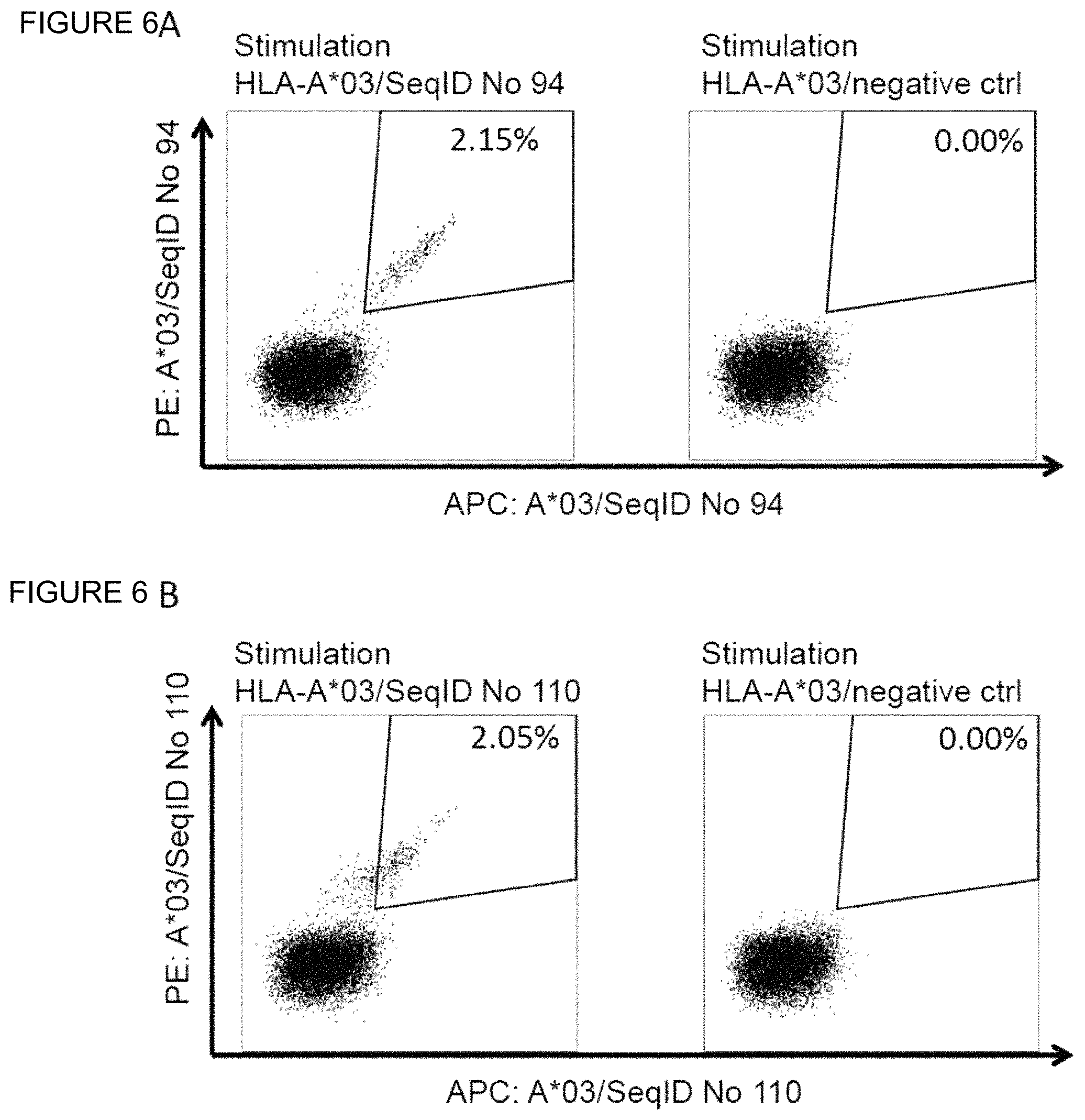
D00022

D00023
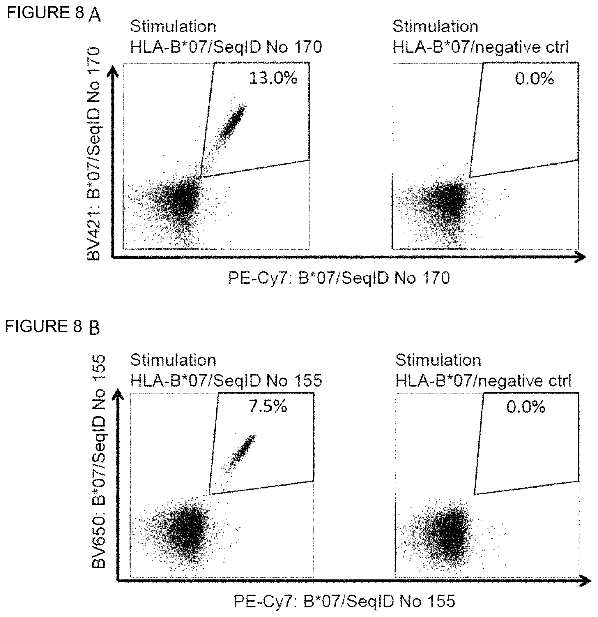
D00024

S00001
XML
uspto.report is an independent third-party trademark research tool that is not affiliated, endorsed, or sponsored by the United States Patent and Trademark Office (USPTO) or any other governmental organization. The information provided by uspto.report is based on publicly available data at the time of writing and is intended for informational purposes only.
While we strive to provide accurate and up-to-date information, we do not guarantee the accuracy, completeness, reliability, or suitability of the information displayed on this site. The use of this site is at your own risk. Any reliance you place on such information is therefore strictly at your own risk.
All official trademark data, including owner information, should be verified by visiting the official USPTO website at www.uspto.gov. This site is not intended to replace professional legal advice and should not be used as a substitute for consulting with a legal professional who is knowledgeable about trademark law.