Compositions and methods for analyzing modified nucleotides
Vaisvila , et al. May 11, 2
U.S. patent number 11,001,876 [Application Number 16/287,604] was granted by the patent office on 2021-05-11 for compositions and methods for analyzing modified nucleotides. This patent grant is currently assigned to New England Biolabs, Inc.. The grantee listed for this patent is New England Biolabs, Inc.. Invention is credited to Theodore B. Davis, Laurence Ettwiller, Shengxi Guan, Lana Saleh, Zhiyi Sun, Romualdas Vaisvila.


View All Diagrams
| United States Patent | 11,001,876 |
| Vaisvila , et al. | May 11, 2021 |
Compositions and methods for analyzing modified nucleotides
Abstract
Methods and compositions are provided for identifying any of the presence, location and phasing of methylated and/or hydroxymethylated cytosines in nucleic acids including long stretches of DNA. In some embodiments, the method may comprise reacting a first portion (aliquot) of a nucleic acid sample with a dioxygenase and optionally a glucosyltransferase in a reaction mixture containing the nucleic acid followed by a reaction with a cytidine deaminase to detect and optionally map .sup.5mC in a DNA. Optionally, a second portion can be reacted with glucosyltransferase followed by reaction with a cytidine deaminase to detect and optionally map .sup.5hmC in a DNA.
| Inventors: | Vaisvila; Romualdas (Ipswich, MA), Davis; Theodore B. (Boxford, MA), Guan; Shengxi (Ipswich, MA), Sun; Zhiyi (Gloucester, MA), Ettwiller; Laurence (Ipswich, MA), Saleh; Lana (Hamilton, MA) | ||||||||||
|---|---|---|---|---|---|---|---|---|---|---|---|
| Applicant: |
|
||||||||||
| Assignee: | New England Biolabs, Inc.
(Ipswich, MA) |
||||||||||
| Family ID: | 62557196 | ||||||||||
| Appl. No.: | 16/287,604 | ||||||||||
| Filed: | February 27, 2019 |
Prior Publication Data
| Document Identifier | Publication Date | |
|---|---|---|
| US 20190185919 A1 | Jun 20, 2019 | |
Related U.S. Patent Documents
| Application Number | Filing Date | Patent Number | Issue Date | ||
|---|---|---|---|---|---|
| 15893373 | Feb 9, 2018 | 10260088 | |||
| 15441431 | Feb 24, 2017 | 10227646 | |||
| PCT/US2016/059447 | Oct 28, 2016 | ||||
| 62325626 | Apr 21, 2016 | ||||
| 62300396 | Feb 26, 2016 | ||||
| 62271679 | Dec 28, 2015 | ||||
| 62257284 | Nov 19, 2015 | ||||
| 62248872 | Oct 30, 2015 | ||||
| Current U.S. Class: | 1/1 |
| Current CPC Class: | C12Y 305/04005 (20130101); C12Q 1/6827 (20130101); C12N 9/0071 (20130101); C12Q 1/6806 (20130101); C12Y 114/11 (20130101); C12N 9/78 (20130101); C12Q 1/6806 (20130101); C12Q 2521/531 (20130101); C12Q 2521/539 (20130101); C12Q 2537/164 (20130101) |
| Current International Class: | C12Q 1/6806 (20180101); C12N 9/02 (20060101); C12N 9/78 (20060101); C12Q 1/6827 (20180101) |
References Cited [Referenced By]
U.S. Patent Documents
| 9121061 | September 2015 | Vaisvila et al. |
| 9267117 | February 2016 | Guan |
| 9447452 | September 2016 | Rao |
| 9611510 | April 2017 | He |
| 9816986 | November 2017 | Rao |
| 10081827 | September 2018 | Guan |
| 10619200 | April 2020 | Vaisvila |
| 2013/0244237 | September 2013 | Vaisvila et al. |
| 2014/0272970 | September 2014 | Zegzouti |
| 2015/0004596 | January 2015 | Zhu et al. |
| WO2009150229 | Dec 2009 | WO | |||
| WO2013163207 | Oct 2013 | WO | |||
| WO2016183289 | Nov 2016 | WO | |||
Other References
|
Ehrich, Nucl. Acids Res. 35:e29, 2007. cited by applicant . Holmes, et al. PloS one 9, No. 4: e93933, 2014. cited by applicant . Jin, et al., Nucleic Acids Res., 2: 6956-71, 2014. cited by applicant . Pais, et al, Proc. Natl. Acad. Sci. 112: 4316-4321, 2015. cited by applicant . Stenglein, Nature Structural & Molecular Biology 17: 222-229, 2010. cited by applicant . Soni, et al., Clin Chem 53: 1996-2001, 2007. cited by applicant . Adey, Genome Res. 24: 2041-9, 2014. cited by applicant . Amini, Nat Genet. 46: 1343-9, 2014. cited by applicant . Cabson, Nucleic Acids Res. 41:e112, 2013. cited by applicant . Krueger, et al. Bioinformatics 27, No. 11: 1571-1572, 2011. cited by applicant . Yu, et al., Cell, 149, 6, 1368-1380, 2012. cited by applicant . Navaratnam, et al., Int. J. Hematol, 83, 195-200, 2006. cited by applicant . Wijesinghe, et al., Nucleic Acids Research, 40, 18 9206-9217. cited by applicant. |
Primary Examiner: Dauner; Joseph G.
Attorney, Agent or Firm: New England Biolabs, Inc Strimpel; Harriet M.
Parent Case Text
CROSS REFERENCE
This application is a continuation of U.S. application Ser. No. 15/893,373, filed Feb. 9, 2018, which is a continuation-in-part of U.S. application Ser. No. 15/441,431, filed on Feb. 24, 2017, which is a continuation-in-part of International Application No. PCT/US16/59447, filed Oct. 28, 2016, which claims the benefit of US Provisional Application No. 62/248,872, filed Oct. 30, 2015; 62/257,284, filed Nov. 19, 2015; 62/271,679, filed Dec. 28, 2015; 62/300,396, filed Feb. 26, 2016; and 62/325,626, filed Apr. 21, 2016 all of which applications are incorporated by reference herein.
Claims
What is claimed is:
1. A preparation comprising 0.001-100 micrograms of a methylcytosine dioxygenase, wherein the methylcytosine dioxygenase comprises SEQ ID NO:2.
2. The preparation according to claim 1, wherein the methylcytosine dioxygenase is a fusion protein.
3. The preparation according to claim 1, wherein the preparation further comprises genomic DNA.
4. The preparation according to claim 1, wherein the preparation further comprises a cytidine deaminase.
5. The preparation according to claim 1, wherein the preparation further comprises a glucosyltransferase.
Description
BACKGROUND
The ability to phase modified nucleotides (e.g., methylated or hydroxymethylated nucleotides) in a genome (i.e., determine whether two or more modified nucleotides are linked on the same single DNA molecule or on different DNA molecules) can provide important information in epigenetic studies, particularly for studies on imprinting, gene regulation, and cancer. In addition, it would be useful to know which modified nucleotides are linked to sequence variations.
Modified nucleotides cannot be phased using conventional methods for investigating DNA modification because such methods typically involve bisulfite sequencing (BS-seq). In BS-seq methods, a DNA sample is treated with sodium bisulfite, which converts cytosines (C) to uracil (U), but 5-methylcytosine (.sup.5mC) remains unchanged. When bisulfite-treated DNA is sequenced, unmethylated C is read as thymine (T), and .sup.5mC is read as C, yielding single-nucleotide resolution information about the methylation status of a segment of DNA. However, sodium bisulfite is known to fragment DNA (see, e.g., Ehrich M 2007 Nucl. Acids Res. 35:e29), making it impossible to determine whether modified nucleotides are linked on the same DNA molecule over a long distance. Specifically, it is impossible for nucleotide modifications to be phased in the same way that sequence variants (e.g., polymorphisms) are phased because those methods require intact, long molecules.
Moreover, bisulfite sequencing displays a bias toward cytosine (C) adjacent to certain nucleotides and not others. It would be desirable to remove the observed bias.
SUMMARY
Provided herein are methods for phasing modified nucleotides that do not require bisulfite treatment. Further, such methods can be implemented in a way that distinguishes between .sup.5mC and hydroxymethylcytosine (.sup.5hmC) or C, formylcytosine (.sup.5fC) and carboxylcytosine (.sup.5caC), providing significant advantages over conventional methods.
This disclosure provides, among other things, compositions and methods to detect and phase methylation and/or hydroxymethylation of nucleotides or unmodified nucleotides in cis or trans at a single molecule level in long stretches of DNA. In various embodiments, glucosylation and oxidation reactions overcome the observed inherent deamination of .sup.5hmC and .sup.5mC by deaminases. Deaminases converts .sup.5mC to T and C to U while glucosylhydroxymethylcytosine (.sup.5ghmC), .sup.5fC and .sup.5caC are not deaminated. Examples of deaminases include APOBEC (apolipoprotein B mRNA editing enzyme, catalytic polypeptide-like). Embodiments utilize enzymes that have substantially no sequence bias in glucosylation, oxidation and deamination of cytosine. Moreover, embodiments provide substantially no non-specific damage of the DNA during the glucosylation, oxidation and deamination reactions.
In some embodiments, a glucosyltransferase (GT) for example .beta.-glucosyltransferase (.beta.GT) is utilized for glucosylating .sup.5hmC to protect this modified base from deamination. However, a person of ordinary skill in the art will appreciate that other enzymatic or chemical reactions may be used for modifying the .sup.5hmC to achieve the same effect. One alternative example provided herein is the use of Pyrrolo-dC for protecting cytosine from being converted to uracil by cytidine deaminase.
In general, and in one aspect, a method is provided that include (a) treating an aliquot (portion) of a nucleic acid sample with methylcytosine dioxygenase and GT in the same reaction mix to produce a reaction product in which substantially all modified cytosines (Cs) are oxidized and .sup.5hmCs are glucosylated; and (b) treating this reaction product with cytidine deaminase for converting substantially all unmodified Cs to U. The term "modified" cytosines used in throughout these examples and embodiments is intended to one or more of .sup.5mC, .sup.5hmC, .sup.5ghmC, .sup.5fc and .sup.5caC where oxidation to completion of .sup.5mC, .sup.5hmC and .sup.5fC results in .sup.5caC. GT reacts with .sup.5hmC only. However, some of the .sup.5hmC is predicted to be converted to .sup.5fC and then .sup.5caC by the dioxygenase before glucosylation occurs. In the presence of the dioxygenase, .sup.5mC is largely oxidized to .sup.5hmC where a substantial amount will be glucosylated, but some will be oxidized to .sup.5fC and then to .sup.5caC. .sup.ghmC blocks deaminase from converting .sup.hmC to T which would otherwise occur to some extent to cause background interference when analyzing .sup.mC. Since .sup.hmC will be converted slowly to T by the deaminase, this forms the basis of identifying .sup.hmC in a DNA sample in the presence or absence of deaminase alone compared with the glucosylated form of the DNA sample serving as a reference or vice versa.
The method as described therefore largely differentiates between unmodified and modified cytosine when a dioxygenase is present in the reaction mix. However, it is generally recognized that the amount of naturally occurring .sup.5mC in a genomic DNA substantially exceeds the amount of .sup.5hmC which in turn exceeds the amount of naturally occurring .sup.5fC and .sup.5caC hence the amount of naturally occurring modified cytosine generally is considered to approximate to the amount of naturally occurring .sup.5mC.
In one embodiment, the method further includes: treating a second aliquot of the nucleic acid sample with GT and subsequently with cytidine deaminase to produce a third reaction product in which substantially all the .sup.5hmCs in the aliquot are glucosylated, substantially all the Cs are converted to Us and substantially all .sup.5mCs are converted to Ts. In the event that the DNA fragments from the third reaction product are amplified, the Us are converted to Ts during amplification and thus cytosine and .sup.5mC become indistinguishable when sequenced.
Another method described herein uses the above method for the second aliquot on its own without treating a first aliquot of the sample. The results of doing this and then sequencing the reaction product thereof and comparing it to a reference sequence is to differentiate .sup.5hmC from C and .sup.5mC and to map these modified nucleotides with respect to a reference sequence for example where the reference sequence is from a sample in or taken from a mammalian subject.
With respect to the method applied to the first aliquot, the second reaction product may be sequenced to determine which Cs are modified and which are unmodified and optionally the third reaction product or an amplification product thereof may be sequenced to determine which Cs are unmodified or methylated and the location of .sup.5hmCs. A reference DNA that may be a third aliquot of the nucleic acid sample not reacted with the dioxygenase, GT or deaminase can also be sequenced or alternatively, a reference sequence may be used that is known and possibly available in a database of sequences.
In one embodiment, the sequence from the second reaction product can be compared to the reference sequence and optionally to the sequence of the third reaction product to determine which cytosines (Cs) in the nucleic acid sample are modified by a methyl and/or hydroxymethyl group.
In another embodiment, the nucleic acid sample is a library of DNA fragments wherein the DNA fragments are ligated to nucleic acid adaptors in which cytosine is replaced by pyrollo-dC which does not substantially react with deaminase and enables downstream amplification to occur as desired.
The method has many uses which are exemplified as follows:
Fragments that are linked, in cis, to a gene in a transcriptionally active or inactive state can be compared with a reference sequence of the same fragment that is linked, in cis, to the same gene in an opposite transcriptionally active or inactive state; to determine an altered pattern of cytosine modifications associated with transcriptional activity where modified cytosines are indicative of transcriptional inactivation. This is also useful for correlating patterns of cytosine modification in ex vivo cell or tissue for correlation with a disease or condition. Other uses include identifying the pattern of cytosine modification for a nucleic acid fragment from a mammalian subject that has a disease or condition and comparing this to the pattern of cytosine modification in the same nucleic acid fragment from a mammalian subject that does not have the disease or condition. In another application, the pattern of cytosine modification for a nucleic acid fragment from a mammalian subject undergoing a treatment can be compared with the pattern of cytosine modification in the same intact nucleic acid fragment from a mammalian subject that has not been treated with the agent to obtain information about the efficacy of the treatment.
In an embodiment of the invention, the methylcytosine dioxygenase and GT may be reacted with the nucleic sample sequentially or together. In the latter case, the enzymes may be added to the reaction mixture together from separate tubes or the enzymes may be combined in a convenient formulation prior to adding to the reaction mixture.
In general, in one aspect, a kit is provided that includes a eukaryotic or prokaryotic deaminase; and a bacteriophage GT and optionally instructions for use. The kit may additionally contain any or all of a dioxygenase, adaptor oligonucleotides in which all the Cs have been replaced with pyrollo-dC and a dU bypass polymerase.
The enzymes may each be packaged in separate tubes although in one example, the GT and the dioxygenase are combined in a single tube.
In embodiments of the methods and the kit, the methylcytosine dioxygenase has an amino acid sequence that is at least 90% identical to SEQ ID NO:1 and comprises the amino acid sequence of SEQ ID NO:2 or alternatively an amino acid sequence that is at least 90% identical to SEQ ID NO:3. One example of the cytidine deaminase is APOBEC3A.
In general, in one aspect, methods for detecting nucleic acid (NA) methylation are provided that include subjecting the NA to enzymatic glucosylation, enzymatic oxidation and enzymatic deamination where an unmodified C is converted to a U, .sup.5mC is converted to T, an .sup.5hmC that is .sup.5ghmC remains C and a modified C that is oxidized to .sup.5caC is read as a C. The majority of modified C are predicted to be .sup.5mC. For some diagnostic purposes, differentiating between .sup.5mC and .sup.5hmC is not required. Accordingly, it is sufficient to utilize a single pathway of oxidation and glucosylation followed by deamination. Where it is desirable to distinguish .sup.5mC from .sup.5hmC, this can be achieved by a performing two different reactions on two aliquots of the same sample and subsequently comparing the sequences of the DNA obtained. One reaction utilizes a GT and a cytidine deaminase while a second reaction utilizes a methylcytosine dioxygenase and a cytidine deaminase. It has been found here that the presence of GT in a reaction with a methylcytosine dioxygenase results in an outcome which shows an improved conversion rate (greater than 97%, 98% or 99% conversion, preferably at least 99%) of modified bases and more accurate mapping than would otherwise be possible. Methylcytosine dioxygenase variants are described herein which catalyze the conversion of the .sup.5mC to .sup.5hmC to .sup.5fC and then .sup.5caC with little or no bias caused by neighboring nucleotides. These and other improved properties of such variants are also described herein. Methods using enzymes described herein utilizing phasing or other sequencing methods are more time and sample efficient and provide improved accuracy for diagnostic sequencing of .sup.5mC and other modified nucleotides.
In each of these methods, it is desirable to compare the product of the enzyme reactions with each other and/or an unreacted sequence. Comparing sequences can be achieved by hybridization techniques and/or by sequencing. Prior to comparing sequences, it may be desirable to amplify the NA using PCR or isothermal methods and/or clone the reacted sequence.
The NA fragments being analyzed may be DNA, RNA or a hybrid or chimera of DNA and RNA. The NA fragments may be single-stranded (ss) or double-stranded (ds). The NA fragments may be genomic DNA or synthetic DNA.
The size of the fragments may be any size but for embodiments of the present invention that utilize single molecule sequencing, fragment sizes that are particularly advantageous are greater than 1 Kb, 2 Kb, 3 kb, 4 kb, 5 kb, 6 Kb, 7 Kb or larger (for example, preferably greater than 4 kb) with no theoretical limitation on the upper size although the upper size of the fragment may be limited by the polymerase in the amplification step commonly used prior to sequencing if amplification is needed.
In some cases, the sequences obtained from the reactions are compared with a corresponding reference sequence to determine: (i) which Cs are converted into a U in the first product for differentiating a .sup.5mC from a .sup.5hmC; and (ii) which Cs are converted to a U for differentiating an unmodified C from a modified C in the optional second product. In these embodiments, the reference sequence may be a hypothetical deaminated sequence, a hypothetical deaminated and PCR amplified sequence or a hypothetical non-deaminated sequence, for example.
In any embodiment, the first and second products may be amplified prior to sequencing. In these embodiments, any U's in the first and second products may be read as T's in the resultant sequence reads.
In any embodiment, the methylcytosine dioxygenase may convert .sup.5mC and .sup.5hmC to .sup.5caC so that cytidine deaminase cannot deaminate the product of .sup.5mC or .sup.5hmC oxidation. The methylcytosine dioxygenase may be a TET protein that enzymatically converts modified C to .sup.5caC.
In any embodiment, the GT may be a .beta.GT or .alpha.-glucosyltransferase (.alpha.GT) that forms .sup.5ghmC from .sup.5hmC so that substantially no derivatized .sup.5hmC is deaminated by the cytidine deaminase.
In any embodiment, the NA sample may contain at least one CpG island. In another embodiment, the NA may include at least two modified Cs with nucleotide neighbors selected from CpG, CpA, CpT and CpC.
In any embodiment, the method may comprise determining the location of the .sup.5mC and/or .sup.5hmC on one strand of a ds nucleic acid.
In any embodiment, the NA is a fragment of genomic DNA and, in some cases, the NA may be linked to a transcribed gene (e.g., within 50 kb, within 20 kb, within 10 kb, within 5 kb or within 1 kb) of a transcribed gene.
The method summarized above may be employed in a variety of applications. A method for sample analysis is provided. In some embodiments, this method may comprise one or more of the following steps: (a) determining the location of all modified Cs in a test NA fragment to identify a pattern for the modified C; (b) comparing the pattern of C modifications in the test NA fragment with the pattern of C modifications in a reference NA; (c) identifying a difference in the pattern of cytosine modifications in the test NA fragment relative to the reference NA fragment; and (d) determining a pattern of .sup.5hmC in the test NA fragment.
In some embodiments, this method may comprise comparing the pattern of C modification or unmodified C for a NA fragment that is linked, in cis, to a gene in a transcriptionally active state to the pattern of C modifications in the same intact NA fragment that is linked, in cis, to the same gene in a transcriptionally inactive state. In these embodiments, the level of transcription of the gene may be correlated with a disease or condition.
In some embodiments, this method may comprise comparing the pattern of cytosine modification for a NA fragment from a patient that has a disease or condition with the pattern of C modification in the same NA fragment from a patient that does not have the disease or condition. In other embodiments, the method may comprise comparing the pattern of cytosine modification for a NA fragment from a patient is undergoing a treatment with the pattern of C modification in the same intact NA fragment from a patient that has not been treated with the agent. In another embodiment, detected differences in the pattern of C modification in the test NA fragment relative to the reference NA fragment corresponds to a variant single nucleotide polymorphism, an insertion/deletion or a somatic mutation associated with a pathology.
A variety of compositions are also provided. In some embodiments, the composition may comprise a NA, wherein the NA comprises: a) G, A, T, U, C; b) G, A, T, U, .sup.5caC and no C and/or C; c) G, A, T, U and .sup.5ghmC and/or no C; or d) G, A, T, U, .sup.5caC and .sup.5ghmC and/or no C. In some embodiments, the composition may further comprise a cytidine deaminase or mutant thereof (as described in U.S. Pat. No. 9,121,061), or a methylcytosine dioxygenase or mutant thereof as described below.
A kit is also provided. In some embodiments, the kit may comprise a GT, a methylcytosine dioxygenase e.g., a mutant methylcytosine dioxygenase (TETv as described below) and a cytidine deaminase, as well as instructions for use. As would be apparent, the various components of the kit may be in separate vessels.
In general, in one aspect, a protein is described that includes an amino acid sequence that is at least 90% identical to SEQ ID NO:1; and contains SEQ ID NO:2. In one aspect, the protein is a fusion protein that includes an N-terminal affinity binding domain. The protein may have methylcytosine dioxygenase activity where the methylcytosine dioxygenase activity is similarly effective for NCA, NCT, NCG and NCC in a target DNA. The protein may be employed in any method herein.
In any embodiment, the protein may be a fusion protein. In these embodiments, the variant protein may comprise an N-terminal affinity binding domain.
Also provided by this disclosure is a method for modifying a naturally occurring DNA containing one or more methylated C. In some embodiments, this method may comprise combining a sample comprising the DNA with a variant methylcytosine dioxygenase to make a reaction mix; and incubating the reaction mix to oxidize the methylated cytosine in the DNA.
In some embodiments, the reaction mix may further comprise analyzing the oxidized sample, e.g., by sequencing or mass spectrometry.
In some embodiments, the reaction mix may further comprise a GT.
In some embodiments, the method may be done in vitro, in a cell-free reaction.
In some embodiments, the method may be done in vitro, e.g., in cultured cells.
The above-summarized variant methylcytosine dioxygenase can be used as a methylcytosine dioxygenase in any of the methods, compositions or kits described below.
In general in one aspect, a method is provided for determining the location of modified cytosines in a nucleic acid fragment, that includes: (a) reacting a nucleic acid sample containing at least one C and/or at least one modified C with a methylcytosine dioxygenase and a GT in a single buffer either together or sequentially; (b) reacting the product of (a) with a cytidine deaminase; and (c) comparing the sequences obtained in (a), or amplification products thereof, with an untreated reference sequence to determine which Cs in the initial nucleic acid fragment are modified. In one aspect, the methylcytosine dioxygenase is an amino acid sequence that is at least 90% identical to SEQ ID NO:1; and contains the amino acid sequence of SEQ ID NO:2.
Present embodiments include an embodiment of a method, that comprises: treating an aliquot of a nucleic acid sample with a methylcytosine dioxygenase and optionally a glucosyltransferase in a reaction mix to produce a reaction product in which the modified cytosines (Cs) are oxidized and optionally the 5-hydroxymethylcytosines (.sup.5hmCs) are glucosylated; and treating the reaction product of (a) with cytidine deaminase to form a second reaction product in which substantially all unmodified Cs are converted to uracil (U).
Another embodiment of the method described above further comprises: treating a second aliquot of the nucleic acid sample with glucosyltransferase in the absence of a dioxygenase and subsequently with cytidine deaminase to produce a third reaction product in which substantially all the 5-hydroxymethylcytosines (.sup.5hmCs) in the aliquot are glucosylated, substantially all the unmodified Cs are converted to Us and methylcytosines (.sup.5mCs) are converted to Thymine (Ts).
In embodiments of the method described above, the nucleic acid sample may be a library of DNA fragments wherein the DNA fragments are ligated to nucleic acid adaptors in which cytosine in the adaptors is replaced by pyrollo-dC.
Another embodiment of the method described above further comprises: sequencing the second reaction product or amplification product thereof to determine which cytosines (Cs) are methylated and which are unmethylated.
Another embodiment of the method described above further comprises sequencing the third reaction product or an amplification product thereof to determine which cytosines are hydroxymethylated.
Another embodiment of the method described above further comprises obtaining a reference sequence by sequencing a third aliquot of the nucleic acid sample not reacted with the dioxygenase, glucosyltransferase or deaminase, or obtaining a reference sequence from a database of sequences.
Another embodiment of the method described above further comprises: comparing the sequences obtained from the second reaction product and the reference sequence and optionally the third reaction product to determine which cytosines (Cs) in the nucleic acid sample are unmodified and optionally which of the modified cytosine are methylated or hydroxymethylated
In embodiments of the method described above, the nucleic acid sample contains fragments that are linked, in cis, to a gene in a transcriptionally active or inactive state whereas the reference sequence is the same fragment that is linked, in cis, to the same gene in an opposite transcriptionally active or inactive state; and determining an altered pattern of cytosine methylation associated with transcriptional activity.
In embodiments of the method described above, the altered pattern of cytosine methylation associated with transcriptional activity of the gene in an ex vivo cell or tissue is correlated with a disease or condition.
Another embodiment of the method described above further comprises: comparing (i) the pattern of cytosine methylation for a nucleic acid fragment from a mammalian subject that has a disease or condition with (ii) the pattern of cytosine methylation in the same nucleic acid fragment from a mammalian subject that does not have the disease or condition.
Another embodiment of the method described above further comprises: comparing (i) the pattern of cytosine methylation for a nucleic acid fragment from a mammalian subject undergoing a treatment with (ii) the pattern of cytosine methylation in the same intact nucleic acid fragment from a mammalian subject that has not been treated with the agent for detecting differences.
In embodiments of the method described above, the methylcytosine dioxygenase and glucosyltransferase are combined within a single reagent for adding to the reaction together.
In embodiments of the method described above, the methylcytosine dioxygenase has an amino acid sequence that is at least 90% identical to SEQ ID NO:1 and comprises the amino acid sequence of SEQ ID NO:2.
In embodiments of the method described above, the methylcytosine dioxygenase has an amino acid sequence that is at least 90% identical to SEQ ID NO:3.
In embodiments of the method described above, the cytidine deaminase is APOBEC3A.
Embodiments include a kit comprising: a. a eukaryotic or prokaryotic deaminase; and b. a bacteriophage dioxygenase.
An embodiment of the kit further comprises a glucosyl transferase.
An embodiment of the kit further comprises adaptor oligonucleotides in which all the cytosines (Cs) have been replaced with pyrollo-dC.
An embodiment of the kit further comprises a dU bypass polymerase.
In embodiment of the kit described above, the glucosyltransferase and the dioxygenase are combined in a single tube or contained in separate tubes.
In embodiment of the kit described above, the methylcytosine dioxygenase has an amino acid sequence that is at least 90% identical to SEQ ID NO:1 and comprises the amino acid sequence of SEQ ID NO:2.
In embodiment of the kit described above, the methylcytosine dioxygenase has an amino acid sequence that is at least 90% identical to SEQ ID NO:3.
In embodiment of the kit described above, the cytidine deaminase is APOBEC3A.
In one embodiment, a method is provided that further comprises: treating an aliquot of a nucleic acid sample with glucosyltransferase in the absence of dioxygenase and subsequently with cytidine deaminase to produce a reaction product in which substantially all the 5-hydroxymethylcytosines (.sup.5hmCs) are glucosylated and substantially all the Cs are converted to Us and substantially all the 5-methylcytosines (.sup.5mCs) are converted to Ts.
Another embodiment of the method described above further comprises: determining the location of 5-hydroxymethylcytosine (.sup.5hmC) in the sample.
BRIEF DESCRIPTION OF THE FIGURES
The patent of application file contains at least one figure executed in color. Copies of this patent or application publication with color figures will be provided by the Office upon request and payment of necessary fee.
Certain aspects of the following detailed description are best understood when read in conjunction with the accompanying drawings. It is emphasized that, according to common practice, the various features of the drawings are not to scale. On the contrary, the dimensions of the various features are arbitrarily expanded or reduced for clarity. Included in the drawings are the following figures:
FIG. 1A shows a schematic diagram of a method for protecting modified Cs from deamination by a cytidine deaminase and a .sup.5mC dioxygenase, for example a TET enzyme such as TETv, that converts .sup.5mC and .sup.5hmC (not C) to .sup.5caC that is insensitive to deamination. After .sup.5mC dioxygenase treatment, deamination of unmodified C only occurs resulting in its replacement by U. From left to right: SEQ ID NO.20, SEQ ID NO. 20, SEQ ID NO. 21.
FIG. 1B shows a second method for protecting .sup.5hmC but not .sup.5mC from deamination by APOBEC enzyme. Here .sup.5hmC is glucosylated using a .beta.GT for example T4-.beta.GT or .alpha.GT for example T4-.alpha.GT. C and .sup.5mC are modified by a cytidine deaminase (e.g. deaminase) to a U and a T respectively. From left to right: SEQ ID NO:20, SEQ ID NO:20, SEQ ID NO:22.
FIG. 1C is a table showing readouts of bases of a genomic sample after PCR amplification and Sanger sequencing or NGS sequencing.
FIG. 2A-2B shows the methylation and hydroxymethylation status of mouse genomic DNA.
FIG. 2A shows the distribution of .sup.5mC and .sup.5hmC at a single locus (locus size: 1078 bp) of mouse fibroblast NHI/3T3 genomic DNA following methylcytosine dioxygenase (here TETv) and cytidine deaminase treatment (according to FIG. 1A).
FIG. 2B shows the distribution of .sup.5hmC at the same locus as FIG. 3A after GT (here .beta.GT) and cytidine deaminase treatment (according to FIG. 1B).
FIG. 2C is a summary of LC-MS data of methylation status of a locus in genomic DNAs of mouse fibroblasts.
FIG. 3A-3E shows that ss DNA is not damaged during preparation and analysis using TETv and/or .beta.GT and cytidine deaminase in contrast to methods that use conventional bisulfite treatment (for bisulfite method see for example, Holmes, et al. PloS one 9, no. 4 (2014): e93933).
FIG. 3A shows results obtained with .beta.GT and cytidine deaminase. Six different fragment sizes (388 bp, 731 bp, 1456 bp, 2018 bp, 3325 bp, and 4229 bp) were analyzed after treatment with a cytidine deaminase and .beta.GT. Full-length fragments in each size category were amplified. No fragmentation was observed.
FIG. 3B shows results obtained with TETv and cytidine deaminase. 6 different fragment sizes (388 bp, 731 bp, 1456 bp, 2018 bp, 3325 bp, and 4229 bp) were analyzed after treatment with a cytidine deaminase and TET. Full-length fragments in each size category were amplified. No fragmentation was observed.
FIG. 3C shows results obtained with bisulfite converted DNA. 6 different fragment sizes (388 bp, 731 bp, 1456 bp, 2018 bp, 3325 bp, and 4229 bp) were analyzed after bisulfite treatment. Full-length fragments in each size category were amplified. When bisulfite converted DNA was amplified, only the two smallest fragments were obtained because of the breakdown of the larger fragments by the bisulfite method.
FIG. 3D shows results obtained with the primers for 5030 bp amplicon, and 5378 bp amplicon after treating DNA before amplification with T4-.beta.GT (.sup.5hmC detection) or TETv (.sup.5mC+.sup.5hmC detection), and cytidine deaminase (see FIGS. 1A and 1B). Each amplification is shown in triplicate. No fragmentation was observed.
FIG. 3E shows that that a 15 kb fragment of ss DNA containing .sup.5mC/.sup.5hmC is not damaged during preparation and analysis using TETv/.beta.GT/cytidine deaminase enzymes in contrast to methods that use conventional bisulfite treatment. The light blue line represents the denatured ss DNA of the 15 kb fragment which is also the control. The red line is APOBEC deamination on glucosylated DNA. The dark blue is DNA deamination on TETv oxidized DNA. And the green is bisulfite treated DNA.
FIGS. 4A and 4B shows that cytidine deaminase does not deaminate the modified base-Pyrrolo-dC (Glen Research, Sterling, Va.). This modified base can be used in Illumina NGS library construction to protect C in the adapters ligated to the ends of DNA fragments in the library from deamination prior to cytidine deaminase treatment.
FIG. 4A shows the results of treating oligonucleotide (5'-ATAAGAATAGAATGAATXGTGAAATGAA TATGAAATGAATAGTA-3', X=Pyrrolo-dC, SEQ ID NO:4) with cytidine deaminase (APOBEC3A) at 37.degree. C. for 16 hours (upper line (black)). The control (lower line (grey)) is untreated SEQ ID NO:4. No difference was observed between the sample and the control confirming that cytidine deaminase does not deaminate Pyrrolo-dC.
FIG. 4B shows a chromatogram (LC-MS) of an adaptor containing Pyrrolo-dC, with the following sequence, where X=Pyrrolo-dC.5'/5Phos/GATXGGAAGAGXAXAXGTXTGAAXTXXAGTX/deoxyU/AXAXTXTTTXXX- TAXAXGAXGXTXTTXXGATCT (SEQ ID NO:5). The LC-MS chromatogram confirms that all C's are replaced by Pyrrolo-dC, with no trace of contaminated Cs.
FIG. 5 shows that the method described in Example 4 that provides sequences from Next generation sequencing (NGS) using an Illumina platform as an example of Deaminase-seq providing superior conversion efficiency compared with BS-seq. Unmethylated lambda DNA was used as a negative control to estimate the non-conversion error rate (methylated C calls/total C calls). In a 3 nucleotide (CD.sup.5mC) reaction (left slashes), the smallest error rate of 0.1% for both CpG and CH (H=A,C,T) context is observed. Bisulfite conversion using Zymo kit (right slashes) has 3 times higher error rate than the method shown in FIGS. 1A and 1B (0.4%), and bisulfite conversion by Qiagen (white) has even higher error rate of 1.6% for CpG context and 1.5% for CH context.
FIG. 6A-6D shows that Deaminase-seq displays no systematic sequence preference while BS-seq generates a significant amount of conversion errors most notably in a CA context. Pie charts depict the numbers and percentages of false positive methylation calls in each C dinucleotide context in the unmethylated lambda genome by different methods.
FIG. 6A shows a pie chart of wild type lambda genome as a control with the naturally occurring distribution of CT, CA, CG and CC.
FIG. 6B shows the representation of .sup.5mC in a lambda genome where every C has been modified using Deaminase-seq. The observed distribution matches that found in FIG. 6A.
FIG. 6C shows the representation of .sup.5mC in a lambda genome where every C has been modified using BS-seq (Qiagen). The observed distribution is not consistent with that found in FIG. 6A.
FIG. 6D shows the representation of .sup.5mC in a lambda genome where every C has been modified using BS-seq (Zymo). The observed distribution is not consistent with that found in FIG. 6A.
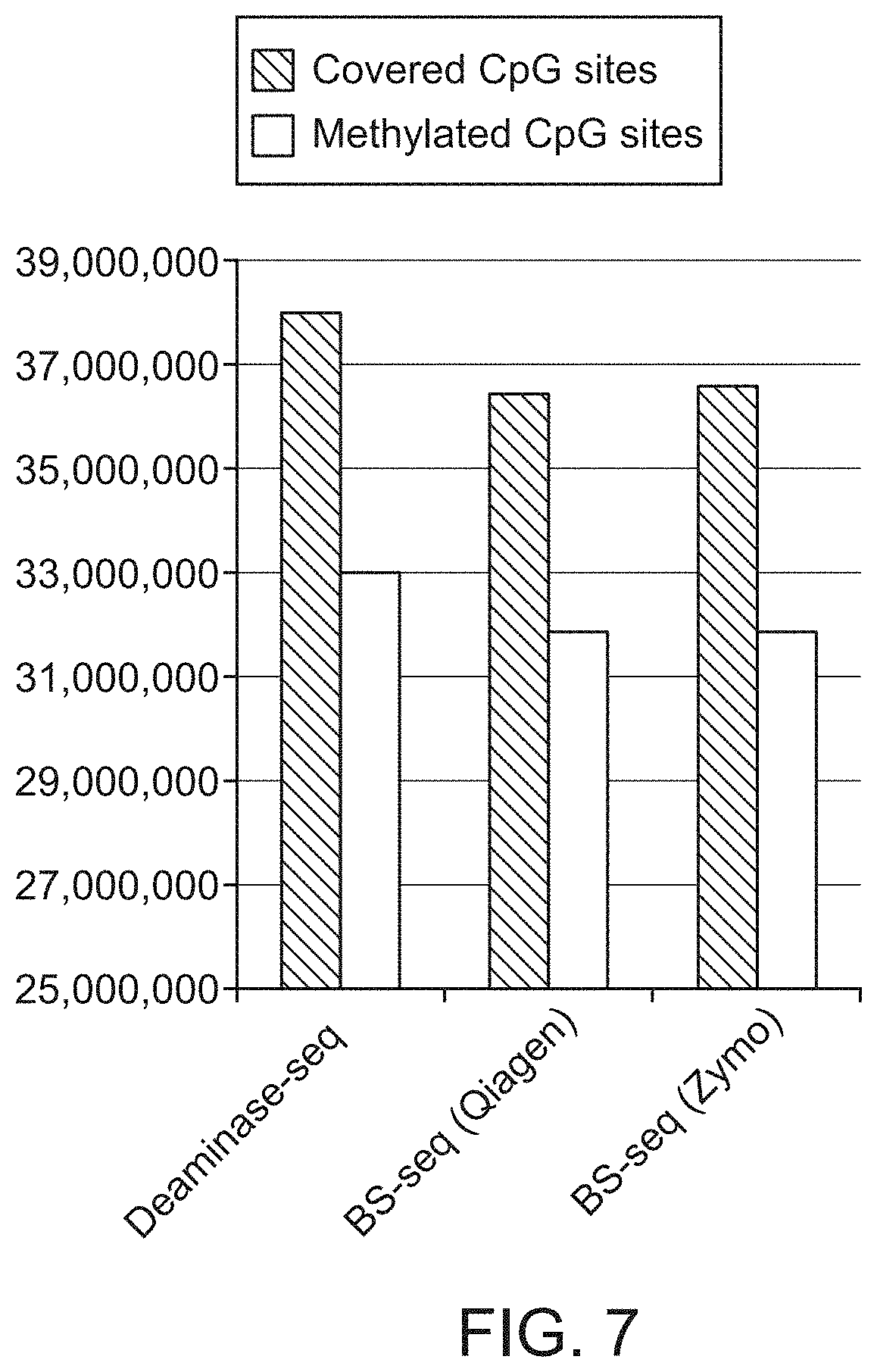
FIG. 7 shows that Deaminase-seq (Illumina) covered more CpG sites and detected more methylated CpG sites than both BS-seq libraries using the same library analysis and the same number of sequencing reads demonstrating that Deaminase-seq is a more efficient and cost-effective method than BS-seq.
FIG. 8A-8C shows that Deaminase-seq provides an even genome-wide sequence coverage in the mouse genome from Illumina generated reads of overlapping fragments. Three histograms of CpG coverage are shown where the 3 methods have the same mean (5.times.) and median (4.times.) sequencing depth for CpG sites. However, Deaminase-seq has fewer outliers (sites with very low or very high copy numbers) when compared with BS-seq kits from Zymo and Qiagen. Three data sets are shown in which, library size was--normalized.
FIG. 8A shows the distribution of reads for DNA Deaminase-seq.
FIG. 8B shows the distribution of reads for BS-seq (Qiagen).
FIG. 8C shows the distribution of reads for BS-seq (Zymo).
FIG. 9 shows that Deaminase-seq provides higher coverage in CpG islands than BS-seq for the same number of sequencing reads, Deaminase-seq gives nearly 2 times as much coverage as BS-seq in the CpG islands.
FIG. 10 provides a loci specific map of .sup.5hmC on a genomic fragment from mouse chromosome 8. Deaminase-seq (described in FIGS. 1A and 1B) accurately detect .sup.55hmC of large fragments (5 Kb) at base resolution enabling phasing of DNA modifications and phase DNA modifications together with other genomic features such as SNPs or variants.
FIG. 11A-11B shows a .sup.5mC and .sup.5hmC profile at single-molecule level across the 5.4 kb region generated by PacBio sequencing. Each row represents one DNA molecule. Each CpG site in the 5.4 kb region was represented by a dot. C modification states were denoted by color.
FIG. 11A shows that the present method can be used to phase .sup.5mC (red=methylated; blue=unmethylated).
FIG. 11B shows that the present method can be used to phase .sup.5hmC (red=hydroxymethylated and blue=unmodified). "Unmodified" in this panel is .sup.5mC or C.
FIGS. 12A and 12B shows the activity of TETv compared with TETcd.
FIG. 12A shows an activity comparison of mouse TET catalytic domain (TETcd; SEQ ID NO:3) with TETv (SEQ ID NO:1) on sheared 3T3 genomic DNA.
FIG. 12B shows activity of TETv on ss and ds genomic (3T3) DNA is similar.
FIG. 13 shows that TETv exhibits very low sequence bias and is context independent for .sup.5mC as demonstrated for 5 cell lines (Arabidopsis, rice, M.Fnu4H, E14 and Jurkat).
FIG. 14 shows that TETv does not degrade DNA as determined from the preservation of supercoiled DNA after enzyme treatment. Lane 1 is a size ladder. Lane 2 is substrate plasmid only, Lane 3 is supercoiled plasmid+323 pmol of TETv; Lane 4 is supercoiled plasmid+162 pmol TETv; Lane 5 is supercoiled plasmid+162 pmol TETv; Lane 6 is Substrate plasmid+323 pmol TETv+BamHI+MspI; Lane 7 is Substrate plasmid+162 pmol TETv+BamHI+MspI; and Lane 8 is Substrate plasmid+BamHI+MspI.
FIG. 15 shows that cytidine deaminase (APOBEC3A) can substantially completely deaminate both C and .sup.5mC.
FIG. 16 shows that low sequence bias of Deaminase-Seq includes accurate representation of cytosine in cytosine rich fragments such as CpG islands. Cytosine in CpG islands are substantially depleted using bisulfite sequencing.
FIG. 17 shows that the lack of fragmentation using Deaminase-Seq correlates with a low nucleic acid starting concentration for detecting the position of modified bases in the nucleic acid. For example, Ing of a genomic DNA library is sufficient for detecting or mapping normal and modified cytosine.
FIG. 18 shows a second example of methylome phasing (also see FIG. 10 and FIG. 11A-11B) using embodiments of the methods described herein where the results of methylome phasing using Deaminase-seq (SMRT.RTM. sequencing, (Pacific Biosciences, Menlo Park, Calif.)) of an imprinted gene. The region of imprinting identified by bisulfite sequencing is relatively short while a region of greater than twice the length is identified using Deaminase-seq (also called here APOBEC-seq). Each red dot on the sequence map correspond to a modified cytosine.
FIG. 19A highlights changes in modified cytosines after methods described in FIG. 19B(ii) and FIG. 19B(iii) that are used to detect and map .sup.mC and/or .sup.hmC.
FIG. 19B is a schematic that summarizes three different approaches to detecting .sup.mC, .sup.hmC and .sup.mC/.sup.hmC. FIG. 19B(i) shows a schematic for the conventional bisulfite sequencing method for detecting .sup.mC/.sup.hmC. FIG. 19B(ii) provides a schematic for detecting .sup.hmC only and not .sup.mC while 19B(iii) provides a schematic for detecting .sup.mC/.sup.hmC.
FIG. 19C schematically shows a use of the deaminase based method for detecting epigenetic changes in DNA that depends on the less disruptive nature of this approach compared with bisulfite sequencing which significantly damages and fragments DNA. Phasing studies are an example of such a use.
FIG. 20A-20B shows an additional representation to the data shown in FIG. 3A-3C and also shown in FIG. 20A.
FIG. 20B shows that the % of .sup.5mC sites detected after bisulfite sequencing is similar to the number detected using the method of FIG. 19B(iii). However, as detected from the corresponding gels above, the bands on the gel after bisulfite treatment are low molecular weight compared to the much higher molecular weight bands observed using the deaminase dependent methods. This shows that the distribution of 5 mC in the deaminase method is spread throughout the large fragments as well as the small fragments providing significantly more sequence and context information. As expected, the % of .sup.5hmC is low as this is a relatively rare species in the epigenome.
FIG. 21 shows a schematic of a workflow for DNA methylation analysis and phasing. Genomic DNA is extracted from a mouse and combined with lambda DNA which is an unmethylated control. The DNA is treated with a dioxygenase (e.g. Tet) and a glucosyl transferase (BGT) followed by deaminase treatment (see FIG. 18(c).
The large fragments are amplified (PCR) and the DNA is sequenced to determine the methylation pattern and phasing.
FIG. 22A-22B shows phased single-Molecule Real-Time (SMRT) sequencing of 5 mC (FIG. 21 A) and .sup.5hmC (FIG. 21B) of a 4.6 kb region of an imprinted gene Inpp5f_v2's in the mouse brain using methods outlined in FIG. 19A-19C. In Individual CpG sites are shown at single molecule level of the 4.6 kb region overlapping the promoter of the imprinted Inpp5f_v2 gene (beige: unmodified; red: modified). This data identified a previous determined Differentially Methylated Regions of imprinted genes (DMR) (orange box) but the results showed that the differentially methylated region was larger than previously reported. The shaded area in the dot plots corresponds to the known DMR.
FIG. 23 shows an example of using APOBEC(.sup.5mC) deamination method coupled with Pacbio SMRT sequencing technology to phase cytosine methylation and heterozygous SNPs to detect imprinted DMRs of the imprinted gene Gnas 1A in the mouse brain.
The experiment was performed in a similar way as the previous SMRT-APOBEC experiment for DMR phasing.
This figure shows the methylation status (beige: unmodified; grey: modified) of each CpG site at single molecule level of a 2.7 kb region overlapping the promoter of the imprinted Gnas1A gene in the mouse brain.
This region includes a previous determined DMR (orange box). Our results showed a larger differentially methylated region than the reported DMR. Moreover, this DMR is associated with a heterozygous SNP highlighted by red (genotype=G) and blue (genotype=A) bars and thus implies that this DMR is indeed an imprinted DMR.
FIG. 24 A and FIG. 24B shows that the insert size is larger and there is lower GC bias for NEBNext (APOBEC) than for WGBS libraries. NEBNext (APOBEC) and WGBS libraries each gave >250M paired reads (Illumina 2.times.100 base NovaSeq sequencing). For methylation analysis 398M reads were used from each library.
The libraries were created as described in Example 12. Technical replicates of the 10 ng and 50 ng NA12878 genomic DNA were used.
FIG. 24A provides a comparison of insert size where the peak for the NEBNext (APOBEC) library is at 170 bp compared to 100 bp for bisulfite sequencing.
FIG. 24B shows the GC bias determined from NEBNext (APOBEC) and WGBS libraries from a 50 ng input representative plot. The WGBS libraries are AT rich and have lower GC coverage.
FIG. 25A and FIG. 25B shows that coverage of CpGs is higher in the NEBNext (APOBEC) libraries compared to WGBS libraries.
FIG. 25A is a table showing the total unique CpG's identified in NA12878 DNA. NEBNext (APOBEC) libraries identified more unique CpG's than WGBS libraries using the same number of reads (398M).
FIG. 25B shows the coverage of CpG's in NEBNext (APOBEC) and WGBS libraries. 398M reads for 10 ng and 50 ng NEBNext (APOBEC) and bisulfite libraries were used to determine distribution of CpG coverage across the human genome. NEBNext (APOBEC) libraries show a higher percentage of CpG's coverage at minimum coverage of 3.times., 5.times., 8.times., 12.times., 15.times. and 20.times..
FIG. 26 shows the distribution of CpGs across genomic features.
Unique CpG's were compared between NEBNext(APOBEC) and WGBS. 53.7M. 56 M CpG's are present in the human genome. NEBNext(APOBEC) libraries identified 54.9M CpG's and WGBS identified 53.7 M CpG's in the human genome at 1.times. coverage. At 8.times. coverage, a level that provides increased confidence in the data, 24.6 million CpGs were detected using NEBNext (APOBEC) whereas only 11.6M CpGs could be detected by WGBS. The distribution of hits is shown by the histogram where significantly more CpGs are detected by NEBNext (APOBEC) than by WGBS in short interspersed nuclear elements (SINE), long terminal repeats (LTR), Introns, promoters and Intergenic regions.
BRIEF DESCRIPTION OF THE EMBODIMENTS
Unless defined otherwise, all technical and scientific terms used herein have the same meaning as commonly understood by one of ordinary skill in the art to which this invention belongs. Singleton, et al., DICTIONARY OF MICROBIOLOGY AND MOLECULAR BIOLOGY, 2D ED., John Wiley and Sons, New York (1994), and Hale & Markham, THE HARPER COLLINS DICTIONARY OF BIOLOGY, Harper Perennial, N.Y. (1991) provide one of skill with the general meaning of many of the terms used herein. Still, certain terms are defined below for the sake of clarity and ease of reference.
As used herein, the term "buffering agent", refers to an agent that allows a solution to resist changes in pH when acid or alkali is added to the solution. Examples of suitable non-naturally occurring buffering agents that may be used in the compositions, kits, and methods of the invention include, for example, Tris, HEPES, TAPS, MOPS, tricine, or MES.
The term "non-naturally occurring" refers to a composition that does not exist in nature.
Any protein described herein may be non-naturally occurring, where the term "non-naturally occurring" refers to a protein that has an amino acid sequence and/or a post-translational modification pattern that is different from the protein in its natural state. For example, a non-naturally occurring protein may have one or more amino acid substitutions, deletions or insertions at the N-terminus, the C-terminus and/or between the N- and C-termini of the protein. A "non-naturally occurring" protein may have an amino acid sequence that is different from a naturally occurring amino acid sequence (i.e., having less than 100% sequence identity to the amino acid sequence of a naturally occurring protein), but that is at least 80%, at least 85%, at least 90%, at least 95%, at least 97%, at least 98% or at least 99% identical to the naturally occurring amino acid sequence. In certain cases, a non-naturally occurring protein may contain an N-terminal methionine or may lack one or more post-translational modifications (e.g., glycosylation, phosphorylation, etc.) if it is produced by a different (e.g., bacterial) cell. A "mutant" protein may have one or more amino acid substitutions relative to a wild-type protein and a "fusion" protein may have one or more exogenous domains added to the N-terminus, C-terminus, and or the middle portion of the protein.
In the context of a nucleic acid (NA), the term "non-naturally occurring" refers to a NA that contains: a) a sequence of nucleotides that is different from a NA in its natural state (i.e. having less than 100% sequence identity to a naturally occurring NA sequence), b) one or more non-naturally occurring nucleotide monomers (which may result in a non-natural backbone or sugar that is not G, A, T or C) and may contain one or more other modifications (e.g., an added label or other moiety) to the 5'-end, the 3' end, and/or between the 5'- and 3'-ends of the NA.
In the context of a composition, the term "non-naturally occurring" refers to: a) a combination of components that are not combined by nature, e.g., because they are at different locations, in different cells or different cell compartments; b) a combination of components that have relative concentrations that are not found in nature; c) a combination that lacks something that is usually associated with one of the components in nature; d) a combination that is in a form that is not found in nature, e.g., dried, freeze dried, crystalline, aqueous; and/or e) a combination that contains a component that is not found in nature. For example, a preparation may contain a "non-naturally occurring" buffering agent (e.g., Tris, HEPES, TAPS, MOPS, tricine or MES), a detergent, a dye, a reaction enhancer or inhibitor, an oxidizing agent, a reducing agent, a solvent or a preservative that is not found in nature.
As used herein, the term "composition" refers to a combination of reagents that may contain other reagents, e.g., glycerol, salt, dNTPs, etc., in addition to those listed. A composition may be in any form, e.g., aqueous or lyophilized, and may be at any state (e.g., frozen or in liquid form).
As used herein, the term "location" refers to the position of a nucleotide in an identified strand in a NA molecule.
As used herein, the term "phasing" refers to a determination of the status of two or more nucleotides on a single DNA molecule or within an allele (i.e. whether the nucleotides are modified or not, for example, whether the nucleotides such as C are methylated, hydroxymethylated, formyl modified or carboxylated or unmodified) are on the same molecule of NA or different homologous chromosomes from a single cell or from homologous chromosomes from different cells in a sample noting that in different cells or different tissues, homologous chromosomes may have a different epigenetic status.
As used herein, the term "nucleic acid" (NA) refers to a DNA, RNA, DNA/RNA chimera or hybrid that may be ss or ds and may be genomic or derived from the genome of a eukaryotic or prokaryotic cell, or synthetic, cloned, amplified, or reverse transcribed. In certain embodiments of the methods and compositions, NA preferably refers to genomic DNA as the context requires.
As used herein unless otherwise stated, the term "modified cytosine" refers to methylcytosine (.sup.5mC), hydroxymethylcytosine (.sup.5hmC), formyl modified, carboxy modified or modified by any other chemical group that may be found naturally associated with C.
As used herein, the term "methylcytosine dioxygenase" also referred to as "dioxygenase" refers to an enzyme that converts .sup.5mC to .sup.5hmC. TET1 (Jin, et al., Nucleic Acids Res. 2014 42: 6956-71) is an example of a methylcytosine dioxygenase, although many others are known including TET2, TET3 and Naeglaria TET (Pais et al, Proc. Natl. Acad. Sci. 2015 112: 4316-4321). Examples of methylcytosine dioxygenases which may be referred to as "oxygenase" are provided in U.S. Pat. No. 9,121,061. TETv is an example of a methylcytosine dioxygenase that oxidizes at least 90%, 92%, 94%, 96%, or 98% of all modified C.
As used herein, the term "cytidine deaminase" refers to an enzyme that is capable of deaminating C to form a U. Many cytidine deaminases are known. For example, the APOBEC family of cytidine deaminases is described in U.S. Pat. No. 9,121,061. APOBEC3A (Stenglein, Nature Structural & Molecular Biology 2010 17: 222-229) is an example of a deaminase. In any embodiment, the deaminase used may have an amino acid sequence that is at least 90% identical to (e.g., at least 95% identical to) the amino acid sequence of GenBank accession number AKE33285.1, which is the human APOBEC3A. Preferably, the cytidine deaminase converts unmodified cytosine to uracil with an efficiency of at least 90%, 92%, 94%, 96%, 98% preferably at least 96%.
As used herein, the term "glucosyltransferase (GT)" refers to an enzyme that catalyzes the transfer of a 1 or .alpha.-D-glucosyl residue from UDP-glucose to .sup.5hmC residue in DNA to form .sup.5ghmC. An example of a GT is T4-.beta.GT (BGT). In one example, the use of GT follows a dioxygenase reaction and ensures that deamination of .sup.5hmC is blocked so that less than 10% or 7% or 5% or 3% (preferably less than 3% of .sup.5hmC) is converted to U by the deaminase. In another example, GT is used together with dioxygenase in the same reaction mix with DNA where the dioxygenase converts .sup.5mC to .sup.5hmC and CaC while the GT converts any residual .sup.5hmC to .sup.5ghmC, to ensure only cytosine is deaminated.
As used herein, "a portion" of a nucleic acid sample and "an aliquot" of a nucleic acid sample are intended to mean the same and can be used interchangeably.
The term "substantially" refers to greater than 50%, 60%, 70%, 80%, or more particularly 90% of the whole.
As used herein, the term "comparing" refers to analyzing two or more sequences relative to one another. In some cases, comparing may be done by aligning two or more sequences with one another such that correspondingly positioned nucleotides are aligned with one another.
As used herein, the term "reference sequence" refers to the sequence of a fragment that is being analyzed. A reference sequence may be obtained from a public database or it may be separately sequenced as part of an experiment. In some cases, the reference sequence may be "hypothetical" in the sense that it may be computationally deaminated (i.e., to change C's into U's or T's etc.) to allow a sequence comparison to be made.
As used herein, the terms "G", "A", "T", "U", "C", ".sup.55mC", ".sup.5fC", ".sup.c5aC", ".sup.5hmC" and ".sup.5ghmC" refer to nucleotides that contain guanidine (G), adenine (A), thymine (T), uracil (U), cytosine (C), .sup.5mC, .sup.5fC, .sup.5caC, .sup.5hmC and .sup.5ghmC, respectively. For clarity, C, .sup.5fC, .sup.5caC, .sup.5mC and .sup.5ghmC are different moieties.
As used herein, the term "no C", in the context of a NA fragment that contains no C, refers to a NA fragment that contains no C. Such a NA may contain .sup.5caC, .sup.5mC and/or .sup.5ghmC and other nucleotides other than C.
The term "internal" refers to a location within the polypeptide that is within a region that extends up to 20 amino acids from either end of the polypeptide.
The term "repeat" refers to a plurality of amino acids that are repeated within the polypeptide.
The term "fusion" refers to a protein having one or more exogenous binding domains added to the N-terminus, C-terminus, and or the middle portion of the protein. The binding domain is capable of recognizing and binding to another molecule. Thus, in some embodiments the binding domain is a histidine tag ("His-tag"), a maltose-binding protein, a chitin-binding domain, a SNAP-Tag.RTM. (New England Biolabs, Ipswich, Mass.) or a DNA-binding domain, which may include a zinc finger and/or a transcription activator-like (TAL) effector domain.
As used herein "N-terminal portion of the protein" refers to amino acids within the first 50% of the protein. As used herein "C-terminal portion of the protein refers to the terminal 50% of the protein.
The term "Next Generation Sequencing" (NGS) generally applies to sequencing libraries of genomic fragments of a size of less than 1 kb preferably using an Illumina sequencing platform. In contrast, single molecule sequencing is performed using a platform from Pacific Biosystems, Oxford Nanopore, or 10.times. Genomics or any other platform known in the art that is capable of sequencing molecules of length greater than 1 kb or 2 kb.
The method for detecting hydroxymethyl cytosine (.sup.5hmC) and/or methyl cytosine (.sup.mC) herein is referred to as deaminase-seq or APOBEC-seq. This term is used independent of any specific sequencing platform. The term "NEBNext (APOBEC)" is a type of Deaminase Seq that is used specifically with an Illumina sequencing platform. Moreover, the terms "APOBEC", "APOBEC3A", "APOBECA3A", "A3A" are different names for the same cytosine deaminase.
Before the various embodiments are described, it is to be understood that the teachings of this disclosure are not limited to the particular embodiments described, and as such can, of course, vary. It is also to be understood that the terminology used herein is for the purpose of describing particular embodiments only, and is not intended to be limiting, since the scope of the present teachings will be limited only by the appended claims.
The section headings used herein are for organizational purposes only and are not to be construed as limiting the subject matter described in any way. While the present teachings are described in conjunction with various embodiments, it is not intended that the present teachings be limited to such embodiments. On the contrary, the present teachings encompass various alternatives, modifications, and equivalents, as will be appreciated by those of skill in the art.
Unless defined otherwise, all technical and scientific terms used herein have the same meaning as commonly understood by one of ordinary skill in the art to which this disclosure belongs. Although any methods and materials similar or equivalent to those described herein can also be used in the practice or testing of the present teachings, some exemplary methods and materials are now described.
The citation of any publication is for its disclosure prior to the filing date and should not be construed as an admission that the present claims are not entitled to antedate such publication by virtue of prior invention. Further, the dates of publication provided can be different from the actual publication dates which can be independently confirmed.
As will be apparent to those of skill in the art upon reading this disclosure, each of the individual embodiments described and illustrated herein has discrete components and features which can be readily separated from or combined with the features of any of the other several embodiments without departing from the scope or spirit of the present teachings. Any recited method can be carried out in the order of events recited or in any other order which is logically possible.
All patents and publications, including all sequences disclosed within such patents and publications, referred to herein are expressly incorporated by reference.
Almost all studies on C modification in eukaryotic genomes have ignored the fact that eukaryotic genomes carry two or more copies of each chromosome. Thus, most traditional studies on C modification do not provide any information about linkage between modified C. For example, methylation studies have traditionally been done using sodium bisulfite, which converts C into U. However, as shown below, sodium bisulfite also fragments DNA, thereby making it difficult, if not impossible, to determine whether two nearby modified C are linked on the same DNA molecules or unlinked on different molecules. The method described herein provides a solution to this problem.
In some embodiments, the sequencing may be done in a way that allows one to determine the identity and location of unmodified or modified C, as well as whether those unmodified or modified C are linked on the same molecule (i.e., "phased"). For example, in some embodiments, the method may comprise reacting a first portion of a sample that contains relatively long, intact NA fragments (e.g., at least 1 kb, at least 5 kb, at least 10 kb, at least 50 kb, up to 100 kb or 200 kb or more in length) with a GT and then a cytidine deaminase to produce a first product. This product differentiates C and .sup.5mC from .sup.5hmC as shown in FIG. 1B. A second portion of the sample may be reacted with a methylcytosine dioxygenase (and optionally a GT) as shown in FIG. 1A. The methylcytosine dioxygenase and the GT may be combined in the same reaction mix or used sequentially in the same or different buffers.
This reaction is followed by a cytidine deaminase reaction to distinguish between unmodified C and modified C. Depending on the sequence of the initial fragment (e.g., whether the initial fragment in FIG. 1B contains G, A, T, C, .sup.5mC and, in some cases, .sup.5hmC), the first product may contain G, A, T, U, no C and .sup.5ghmC (if the initial fragment contained .sup.5hmC).
In FIG. 1A, the second product alone may contain G, A, T, U, .sup.5caC and no C. These enzyme and methods avoid degradation of the NA substrate and provide improved phasing of modified nucleotide over long pieces of the genome that are not degraded by the enzymes. These enzyme and methods achieve sequencing and mapping of modified nucleotides with minimal bias and improved efficiency.
After the first and optionally second products are produced, they may be amplified and/or cloned, and then sequenced using suitable sequencing methods. This may include single molecule sequencing for phased sequencing. Phased sequencing may be done in a variety of different ways. In some embodiments, the products may be sequenced using a long read single-molecule sequencing approach such as Nanopore sequencing (e.g., as described in Soni, et al. Clin Chem 53: 1996-2001 2007, and developed by Oxford Nanopore Technologies) or Pacific Biosciences' fluorescent base-cleavage method (which currently have an average read length of over 10 kb, with some reads over 60 kb). Alternatively, the products may be sequenced using, the methods of Moleculo (Illumina, San Diego, Calif.), 10.times. Genomics (Pleasanton, Calif.), or NanoString Technologies (Seattle, Wash.). In these methods, the sample is optionally diluted and then partitioned into a number of partitions (wells of a microtitre plate or droplets in an emulsion, etc.) in an amount that limits the probability that each partition contains two molecules of the same locus (e.g., two molecules containing the same gene). Next, these methods involve producing indexed amplicons of a size that is compatible with the sequencing platform being used (e.g., amplicons in the range of 200 bp to 1 kb in length) where amplicons derived from the same partitions are barcoded with the same index unique to the partition. Finally, the indexed amplicons are sequenced, and the sequence of the original, long, molecules can be reconstituted using the index sequences. Phased sequencing may also be done using barcoded transposons (see, e.g., Adey Genome Res. 2014 24: 2041-9 and Amini Nat Genet. 2014 46: 1343-9), and by using the "reflex" system of Population Genetics Technologies (Casbon, Nucleic Acids Res. 2013 41:e112).
Alternatively, the genome may be fragmented into fragments of less than 1 kb in size to form a library for Next gen sequencing. Pyrrolo-dC modified adaptors may be added to the fragments in the library prior to enzyme treatment according to FIGS. 1A-1B and Example 1. These adaptors are resistant to modification by the deaminase. After the enzyme reaction, the adaptor ligated libraries may be sequenced using an Illumina sequencer. After the sequences of the first and optionally the second product are obtained, the sequences are compared with a reference sequence to determine which C's in the initial NA fragment are modified. A matrix illustrating an embodiment of this part of the method is illustrated in FIG. 1C. In some embodiments, this comparing may be done by comparing the sequences obtained from the first product of the sample (i.e., the methylcytosine dioxygenase (and optionally GT) and cytidine deaminase treated portion of the sample) and the untreated sample and/or second product of the sample (i.e., the GT and cytidine deaminase treated portion of the sample) with a corresponding reference sequence (untreated and/or the first product). Possible outcomes include: i. The position of a C in the initial NA fragment is identified by a U in both the first and second products; ii. The position of a .sup.5mC in the initial NA fragment is determined by the presence of a C in the first product or a T in the second product iii. The position of a .sup.5hmC in the initial NA fragment is determined by the presence of a C in the second product only.
It should be noted that should there be no need to differentiate the .sup.5mC from the rarer .sup.5hmC, then this information can be obtained from the second product only (FIG. 1A). FIG. 18 shows similar information to FIG. 1A-1C but is arranged slightly differently. Accordingly, when there is no need to differentiate the .sup.5mC from the rarer .sup.5hmC, it may be desirable to use a dioxygenase such as Tet 2 or Tetv together with BGT in an initial reaction prior to deamination. An advantage of using both enzymes in a single reaction is that where the dioxygenase does not complete the conversion of .sup.5mC to .sup.5caC, and where .sup.5hmC is capable in small amounts of being deaminated, the addition of GT to glucosylate .sup.5hmC ensures that this contaminating deaminase activity is prevented thereby increasing the specificity of .sup.5mC mapping.
In the situation where .sup.5hmC is desired, the reaction pathway shown in FIGS. 1B and 1n FIG. 18 utilizes BGT followed by a deamination step in the absence of a dioxygenase.
In the situation where it is desirable to detect and/or map both .sup.5hmC and .sup.5mC in a sample, then the sample may be divided into portions where the first portion is treated according to FIG. 1A, the second portion according to FIG. 1B and optionally a third portion which is untreated. This is also shown in FIG. 18.
In embodiments, kits are provided that may include a Tet dioxygenase and a GT such as BGT in one tube and a deaminase such as APOBEC3A in a second tube with instructions. Alternatively, kits may include a GT in one tube, a deaminase in a second tube and optionally a dioxygenase in a third tube. The enzymes may be contained in the various mixes and tubes in a suitable storage buffer.
As would be understood, if the product is cloned, amplified or sequenced by a polymerase, a "U" will be read as "T". In these embodiments, nucleotides read as a T in both the first and second products still indicate Cs that have been changed to Us in the initial deamination reaction.
As would be recognized, some of the analysis steps of the method, e.g., the comparing step, can be implemented on a computer. In certain embodiments, a general-purpose computer can be configured to a functional arrangement for the methods and programs disclosed herein. The hardware architecture of such a computer is well known by a person skilled in the art and can comprise hardware components including one or more processors (CPU), a random-access memory (RAM), a read-only memory (ROM), an internal or external data storage medium (e.g., hard disk drive). A computer system can also comprise one or more graphic boards for processing and outputting graphical information to display means. The above components can be suitably interconnected via a bus inside the computer. The computer can further comprise suitable interfaces for communicating with general-purpose external components such as a monitor, keyboard, mouse, network, etc.
In some embodiments, the computer can be capable of parallel processing or can be part of a network configured for parallel or distributive computing to increase the processing power for the present methods and programs. In some embodiments, the program code read out from the storage medium can be written into memory provided in an expanded board inserted in the computer, or an expanded unit connected to the computer, and a CPU or the like provided in the expanded board or expanded unit can actually perform a part or all of the operations according to the instructions of the program code, so as to accomplish the functions described below. In other embodiments, the method can be performed using a cloud computing system. In these embodiments, the data files and the programming can be exported to a cloud computer that runs the program and returns an output to the user.
A system can, in certain embodiments, comprise a computer that includes: a) a central processing unit; b) a main non-volatile storage drive, which can include one or more hard drives, for storing software and data, where the storage drive is controlled by disk controller; c) a system memory, e.g., high speed random-access memory (RAM), for storing system control programs, data, and application programs, including programs and data loaded from non-volatile storage drive; system memory can also include read-only memory (ROM); d) a user interface, including one or more input or output devices, such as a mouse, a keypad, and a display; e) an optional network interface card for connecting to any wired or wireless communication network, e.g., a printer; and f) an internal bus for interconnecting the aforementioned elements of the system.
The method described above can be employed to analyze genomic DNA from virtually any organism, including, but not limited to, plants, animals (e.g., reptiles, mammals, insects, worms, fish, etc.), tissue samples, bacteria, fungi (e.g., yeast), phage, viruses, cadaveric tissue, archaeological/ancient samples, etc. In certain embodiments, the genomic DNA used in the method may be derived from a mammal, where in certain embodiments the mammal is a human. In exemplary embodiments, the genomic sample may contain genomic DNA from a mammalian cell, such as, a human, mouse, rat, or monkey cell. The sample may be made from cultured cells, formalin fixed samples or cells of a clinical sample, e.g., a tissue biopsy (for example from a cancer), scrape or lavage or cells of a forensic sample (i.e., cells of a sample collected at a crime scene). In particular embodiments, the NA sample may be obtained from a biological sample such as cells, tissues, bodily fluids, and stool. Bodily fluids of interest include but are not limited to, blood, serum, plasma, saliva, mucous, phlegm, cerebral spinal fluid, pleural fluid, tears, lactal duct fluid, lymph, sputum, cerebrospinal fluid, synovial fluid, urine, amniotic fluid, and semen. In particular embodiments, a sample may be obtained from a subject, e.g., a human. In some embodiments, the sample analyzed may be a sample of cell-free DNA obtained from blood, e.g., from the blood of a pregnant female.
In some embodiments of the invention, an enzymatic method has been provided which permits the sequencing of short and long NA (for example, ss DNA and ds DNA) to discover modified bases and to determine the phasing of such bases in the genome. Embodiments of the method may include a composition comprising a mixture of one or two enzymes where the one, two enzymes are selected from a methylcytosine dioxygenase and a GT where the cytidine deaminase is added in a subsequent reaction. The dioxygenase and GT may be stored in the same or different buffers and combined as desired in a storage buffer or in a reaction mixture.
When added separately to a reaction mixture, the addition may be sequential, or the enzymes may be added together at the start of the reaction. Embodiments of the method may utilize two or more enzymes selected from a cytidine deaminase, a methylcytosine dioxygenase and a GT. Embodiments of the method may include a methylcytosine dioxygenase and a cytidine deaminase used sequentially in a reaction mixture; a methylcytosine dioxygenase and a GT used sequentially or together preferably followed by a deaminase reaction; or a methylcytosine dioxygenase, GT and cytidine deaminase used sequentially or together.
In some embodiments, that utilize a GT, UDP-glucose may be added to the reaction mixture.
In one embodiment, the methylcytosine dioxygenase and optionally the GT may be added to ds DNA in an initial step and then removed by a proteinase treatment, heat treatment and/or separation treatment. This may be followed by a cytidine deaminase reaction with separation and isolation of the deaminated DNA. In some embodiments, the pH of the cytidine deaminase reaction mixture is in the range of pH 5.5-8.5, for example pH 6.0-8.0 for example, pH 6.0, pH 6.3, pH 6.5, pH 6.8, pH 7.0, pH 7.5, or pH 8.0 wherein the specific activity of the cytidine deaminase is increased at the lower end of the pH range such as at pH 6.0.
In one embodiment, concentration ranges of enzymes utilized in the reaction described for 1 .mu.g DNA include: 0.001-100 micrograms of a methylcytosine dioxygenase such as the Ngo TET (Pais, supra), TET1, TET or TET3 or mutants thereof; 0.001-100 micrograms cytidine deaminase such as APOBEC or Deaminase; 0.001-100 units GT such as T4-.beta.GT or T4-.alpha.GT. When Pyrollo-dC used in adaptor synthesis, a standard procedure described in Example 4 is followed. The amount of UDP-glucose used follows the recommendation of the manufacturer.
The ss DNA product of enzyme reaction or reactions can be amplified by PCR or isothermal methods such ligase mediated amplification (LMA), helicase dependent amplification (HDA), rolling circle amplification (RCA), loop mediated amplification (LAMP), multiple displacement amplification, (MDA), transcription mediated amplification (TMA), strand displacement amplification (SDA), or nicking enzyme amplification reaction (NEAR).
The amplified, or indeed non-amplified DNA, may be sequenced using any of the sequencing platforms in development or commercially available such as provided by Illumina, Oxford Nanopore, or Pacific Biosystems, or methods in development or commercially available such as Sanger sequencing or any WGS (whole genome sequencing) method. Long reads are mapped to the genome using the appropriate algorithm, for example, Bismark (see for example, Krueger et al. Bioinformatics 27, no. 11 (2011): 1571-1572). The methylation status is called when each read is mapped to the targeted region (for example, enhancer and promoter regions).
Present embodiments provide many advantages over existing systems that result from factors that include: a lower error rate in identifying .sup.5mC regardless of adjacent nucleotides, and a lower error rate in detecting low level methylations; no systematic sequence preference; more consistent genome-wide sequencing coverage; higher coverage in C rich regions and CpG islands; covering more CpG sites where these may be distributed widely in the genome portion being analyzed; and accurate detection of .sup.5hmC of large fragments (5 kb) at a base resolution enabling phasing of DNA modifications and phasing DNA modifications together with other genomic features such as SNPs or variants.
In some embodiments, the composition may comprise a NA that is made up of nucleotides G, A, T, U, and .sup.5caC, wherein the NA contains substantially no C. In some embodiments, the composition may comprise a NA that is made up of nucleotides G, A, T, U and .sup.5ghmC, wherein the NA contains substantially no C. In either embodiment, the composition may also contain a cytidine deaminase (e.g., a cytidine deaminase that is at least 90% identical to an APOBEC cytidine deaminase) and, in certain embodiments, may also contain a buffering agent and other components (e.g., NaCl) in amounts that are compatible with cytidine deaminase activity. The composition may be an aqueous composition.
Variant .sup.5mC Dioxygenases and Methods for Using the Same
A variant methylcytosine dioxygenase is also provided. In some embodiments, the methylcytosine dioxygenase comprises an amino acid sequence that is at least 90% identical to (e.g., at least 92%, at least 94%, at least 96%, at least 97%, at least 98%, or at least 99% identical to) the amino acid sequence of TETv (SEQ ID NO:1); and contain the amino acid sequence of SEQ ID NO:2. As would be apparent, this polypeptide has .sup.5mC dioxygenase activity. The TETv sequence is shown below:
TABLE-US-00001 TETv (SEQ ID NO: 1) GGSQSQNGKCEGCNPDKDEAPYYTHLGAGPDVAAIRTLMEERYGEKGKAI RIEKVIYTGKEGKSSQGCPIAKWVYRRSSEEEKLLCLVRVRPNHTCETAV MVIAIMLWDGIPKLLASELYSELTDILGKCGICTNRRCSQNETRNCCCQG ENPETCGASFSFGCSWSMYYNGCKFARSKKPRKFRLHGAEPKEEERLGSH LQNLATVIAPIYKKLAPDAYNNQVEFEHQAPDCCLGLKEGRPFSGVTACL DFSAHSHRDQQNMPNGSTVVVTLNREDNREVGAKPEDEQFHVLPMYIIAP EDEFGSTEGQEKKIRMGSIEVLQSFRRRRVIRIG DAA AVQEIEYWSDSEHNFQDPCIGGVAIAPTHGSILIECAKCEVHATTKVNDP DRNHPTRISLVLYRHKNLFLPKHCLALWEAKMAEKARKEEECGKNGSDHV SQKNHGKQEKREPTGPQEPSYLRFIQSLAENTGSVTTDSTVTTSPYAFTQ VTGPYNTFV
TETv is derived from mouse TET catalytic domain and contains a deletion. The amino acid sequence ELPKSCEVSGQ (SEQ ID NO:2) is italicized within the sequence of TETv and TETcd sequences shown above and below.
TABLE-US-00002 TETcd (TET-2 catalytic domain) (SEQ ID. NO. 3) QSQNGKCEGCNPDKDEAPYYTHLGAGPDVAAIRTLMEERYGEKGKAIRIE KVIYTGKEGKSSQGCPIAKWVYRRSSEEEKLLCLVRVRPNHTCETAVMVI AIMLWDGIPKLLASELYSELTDILGKCGICTNRRCSQNETRNCCCQGENP ETCGASFSFGCSWSMYYNGCKFARSKKPRKFRLHGAEPKEEERLGSHLQN LATVIAPIYKKLAPDAYNNQVEFEHQAPDCCLGLKEGRPFSGVTACLDFS AHSHRDQQNMPNGSTVVVTLNREDNREVGAKPEDEQFHVLPMYIIAPEDE FGSTEGQEKKIRMGSIEVLQSFRRRRVIRIGELPKSCKKKAEPKKAKTKK AARKRSSLENCSSRTEKGKSSSHTKLMENASHMKQMTAQPQLSGPVIRQP PTLQRHLQQGQRPQQPQPPQPQPQTTPQPQPQPQHIMPGNSQSVGSHCSG STSVYTRQPTPHSPYPSSAHTSDIYGDTNHVNFYPTSSHASGSYLNPSNY MNPYLGLLNQNNQYAPFPYNGSVPVDNGSPFLGSYSPQAQSRDLHRYPNQ DHLTNQNLPPIHTLHQQTFGDSPSKYLSYGNQNMQRDAFTTNSTLKPNVH HLATFSPYPTPKMDSHFMGAASRSPYSHPHTDYKTSEHHLPSHTIYSYTA AASGSSSSHAFHNKENDNIANGLSRVLPGFNHDRTASAQELLYSLTGSSQ EKQPEVSGQDAAAVQEIEYWSDSEHNFQDPCIGGVAIAPTHGSILIECAK CEVHATTKVNDPDRNHPTRISLVLYRHKNLFLPKHCLALWEAKMAEKARK EEECGKNGSDHVSQKNHGKQEKREPTGPQEPSYLRFIQSLAENTGSVTTD STVTTSPYAFTQVTGPYNTFV
The deleted amino acids correspond to residues 338 to 704 TETcd (shown in italics above). The amino acid sequence ELPKSCEVSGQ (SEQ ID NO:2) contains 5 amino acids from one side of the junction and 5 amino acids from the other side of the junction, as shown above.
In some embodiments, the variant methylcytosine dioxygenase may be a fusion protein. In these embodiments, the variant may have a binding domain that is capable of recognizing and binding to another molecule. Thus, in some embodiments the binding domain is a histidine tag ("His-tag"), although a maltose-binding protein, a chitin-binding domain, a SNAP-Tag.RTM. or a DNA-binding domain, which may include a zinc finger and/or a transcription activator-like (TAL) effector domain, are also examples of binding moieties.
Embodiments include a buffered composition containing a purified TETv. For example, the pH of the buffer in the composition is pH 5.5-8.5, for example pH 5.5-7.5, pH 7.5-8.0 or pH 8.0. In various embodiments, the buffered composition may contain glycerol; and/or contain Fe(II), as cofactor, and .alpha.-ketoglutarate, as co-substrate, for the enzyme. In some of these embodiments, the composition contains ATP to allow further oxidation of .sup.5hmC to .sup.5fC and .sup.5caC; in other embodiments, the composition does not contain dATP that limits the distribution of the oxidized forms of .sup.5mC.
Embodiments include an in vitro mixture that includes a TETv, a .beta.GT, a cytidine deaminase, and/or an endonuclease. The in vitro mixture may further include a polynucleotide substrate and at least dATP. The polynucleotide could be ss or ds, a DNA or RNA, a synthesized oligonucleotide (oligo), chromosomal DNA, or an RNA transcript. The polynucleotide used could be labeled at one or both ends. The polynucleotide may harbor a C, .sup.5mC, .sup.5hmC, .sup.5fC, .sup.5caC or .sup.5ghmC. In other embodiments, the polynucleotide may harbor a T, U, hydroxymethyluracil (.sup.5hmU), formyluracil (.sup.5fU), or carboxyuracil (.sup.5caU).
Embodiments provide a TETv, which oxidizes .sup.5mC to .sup.5hmC, .sup.5fC, and/or .sup.5caC preferably in any sequence context with minimal sequence bias and minimal damage to the DNA substrate compared to BS-seq. TETv may additionally or alternatively oxidize T to .sup.5hmU or .sup.5fU with improved efficiency and reduced bias compared with naturally occurring mouse TET-2 enzyme, or its catalytic domain (TETcd).
In an embodiment of the method, C could be distinguished from .sup.5mC by reacting the polynucleotide of interest with a TETv and a cytidine deaminase wherein only C is converted to U. A further embodiment includes sequencing the polynucleotide treated with the .beta.GT and the cytidine deaminase in which C is converted to U and .sup.5mC is converted to a T and comparing the sequencing results to that of sequencing the untreated polynucleotide to map .sup.5mC and .sup.5hmC location in the polynucleotide.
In another embodiment of the method, both .sup.5mC and .sup.5hmC locations in a polynucleotide are mapped. In this method: (a) the polynucleotide is untreated; (b) reacted with bisulfite reagent; or (c) reacted with GT prior to adding a methylcytosine dioxygenase then treating with bisulfite reagent. (a) through (c) are sequenced and comparison of the sequencing results enables the mapping of .sup.5mC and .sup.5hmC and their differentiation from C: (a) C, .sup.5mC, and .sup.5hmC are all sequenced as C; (b) C is sequenced as C while .sup.5mC and .sup.5hmC as T; and (c).sup.5hmC is converted to .sup.5ghmC and sequenced as C, C is sequenced as C, and .sup.5mC as T.
In some embodiments, .sup.5mC locations in a polynucleotide are mapped by coupling the oxidation activity of TETv to the activity of a restriction endonuclease or an AP endonuclease specific to .sup.5hmC or .sup.5fC/.sup.5caC, respectively.
In some aspects, .sup.5mC, .sup.5hmC, or .sup.5fC may be mapped to sites in a polynucleotide using single-molecule sequencing technologies such as Single Molecule Real-Time (SMRT) Sequencing, Oxford Nanopore Single Molecule Sequencing (Oxford, UK) or 10.times. Genomics (Pleasanton, Calif.). In some embodiments, the method may employ TETv, a cytidine deaminase, and/or GT.
The above-described TETv enzyme can be used as a methylcytosine dioxygenase in any of the methods, compositions or kits summarized above and described in greater detail below.
Kits
Also provided by the present disclosure are kits for practicing the subject method as described above. In certain embodiments, a subject kit may contain: a GT, a methylcytosine dioxygenase and a cytidine deaminase. In some embodiments, the kit may comprise a eukaryotic methylcytosine dioxygenase, and a bacteriophage GT. In these embodiments, the methylcytosine and GT may be present in the same container.
The components of the kit may be combined in one container, or each component may be in its own container. For example, the components of the kit may be combined in a single reaction tube or in one or more different reaction tubes. Further details of the components of this kit are described above. The kit may also contain other reagents described above and below that may be employed in the method, e.g., a buffer, UDP-glucose, plasmids into which NAs can be cloned, controls, amplification primers, etc., depending on how the method is going to be implemented.
In addition to above-mentioned components, the subject kit may further include instructions for using the components of the kit to practice the subject method. The instructions for practicing the subject method are generally recorded on a suitable recording medium. For example, the instructions may be printed on a substrate, such as paper or plastic, etc. As such, the instructions may be present in the kits as a package insert, in the labeling of the container of the kit or components thereof (i.e., associated with the packaging or subpackaging), etc. In other embodiments, the instructions are present as an electronic storage data file present on a suitable computer readable storage medium, e.g. CD-ROM, diskette, etc. In yet other embodiments, the actual instructions are not present in the kit, but means for obtaining the instructions from a remote source, e.g. via the internet, are provided. An example of this embodiment is a kit that includes a web address where the instructions can be viewed and/or from which the instructions can be downloaded. As with the instructions, this means for obtaining the instructions is recorded on a suitable substrate.
Utility
In some embodiments, the method can be used to compare two samples. In these embodiments, the method may be used to identify a difference in the pattern of C modification in a test NA fragment relative to the pattern of cytosine modification in a corresponding reference NA. This method may comprise (a) determining the location of all modified C in a test NA fragment using the above-described method to obtain a first pattern of C modification; (b) determining the location of all modified C in a reference NA fragment using the above-described method to obtain a first pattern of C modification; (c) comparing the test and reference patterns of C modification; and (d) identifying a difference in the pattern of cytosine modification, e.g., a change in the amount of .sup.5mC or .sup.5hmC, in the test NA fragment relative to the reference NA fragment.
In some embodiments, the test NA and the reference NA are collected from the same individual at different times. In other embodiments, the test NA and the reference NA collected from different tissues or different individuals.
Exemplary NAs that can be used in the method include, for example, NA isolated from cells isolated from a tissue biopsy (e.g., from a tissue having a disease such as colon, breast, prostate, lung, skin cancer, or infected with a pathogen etc.) and NA isolated from normal cells from the same tissue, e.g., from the same patient; NA isolated from cells grown in tissue culture that are immortal (e.g., cells with a proliferative mutation or an immortalizing transgene), infected with a pathogen, or treated (e.g., with environmental or chemical agents such as peptides, hormones, altered temperature, growth condition, physical stress, cellular transformation, etc.), and NA isolated from normal cells (e.g., cells that are otherwise identical to the experimental cells except that they are not immortalized, infected, or treated, etc.); NA isolated from cells isolated from a mammal with a cancer, a disease, a geriatric mammal, or a mammal exposed to a condition, and NA isolated from cells from a mammal of the same species, e.g., from the same family, that is healthy or young; and NA isolated from differentiated cells and NA isolated from non-differentiated cells from the same mammal (e.g., one cell being the progenitor of the other in a mammal, for example). In one embodiment, NA isolated from cells of different types, e.g., neuronal and non-neuronal cells, or cells of different status (e.g., before and after a stimulus on the cells) may be compared. In another embodiment, the experimental material is NA isolated from cells susceptible to infection by a pathogen such as a virus, e.g., human immunodeficiency virus (HIV), etc., and the reference material is NA isolated from cells resistant to infection by the pathogen. In another embodiment of the invention, the sample pair is represented by NA isolated from undifferentiated cells, e.g., stem cells, and NA isolated from differentiated cells.
In some exemplary embodiments, the method may be used to identify the effect of a test agent, e.g., a drug, or to determine if there are differences in the effect of two or more different test agents. In these embodiments, NA from two or more identical populations of cells may be prepared and, depending on how the experiment is to be performed, one or more of the populations of cells may be incubated with the test agent for a defined period of time. After incubation with the test agent, the genomic DNA from one both of the populations of cells can be analyzed using the methods set forth above, and the results can be compared. In a particular embodiment, the cells may be blood cells, and the cells can be incubated with the test agent ex vivo. These methods can be used to determine the mode of action of a test agent, to identify changes in chromatin structure or transcription factor occupancy in response to the drug, for example.
The method described above may also be used as a diagnostic (which term is intended to include methods that provide a diagnosis as well as methods that provide a prognosis). These methods may comprise, e.g., analyzing C modification from a patient using the method described above to produce a map; and providing a diagnosis or prognosis based on the map.
The method set forth herein may also be used to provide a reliable diagnostic for any condition associated with altered cytosine modification. The method can be applied to the characterization, classification, differentiation, grading, staging, diagnosis, or prognosis of a condition characterized by an epigenetic pattern. For example, the method can be used to determine whether the C modifications in a fragment from an individual suspected of being affected by a disease or condition is the same or different compared to a sample that is considered "normal" with respect to the disease or condition. In particular embodiments, the method can be directed to diagnosing an individual with a condition that is characterized by an epigenetic pattern at a particular locus in a test sample, where the pattern is correlated with the condition. The methods can also be used for predicting the susceptibility of an individual to a condition.
In some embodiments, the method can provide a prognosis, e.g., to determine if a patient is at risk for recurrence. Cancer recurrence is a concern relating to a variety of types of cancer. The prognostic method can be used to identify surgically treated patients likely to experience cancer recurrence so that they can be offered additional therapeutic options, including preoperative or postoperative adjuncts such as chemotherapy, radiation, biological modifiers and other suitable therapies. The methods are especially effective for determining the risk of metastasis in patients who demonstrate no measurable metastasis at the time of examination or surgery.
The method can also be used to determining a proper course of treatment for a patient having a disease or condition, e.g., a patient that has cancer. A course of treatment refers to the therapeutic measures taken for a patient after diagnosis or after treatment. For example, a determination of the likelihood for recurrence, spread, or patient survival, can assist in determining whether a more conservative or more radical approach to therapy should be taken, or whether treatment modalities should be combined. For example, when cancer recurrence is likely, it can be advantageous to precede or follow surgical treatment with chemotherapy, radiation, immunotherapy, biological modifier therapy, gene therapy, vaccines, and the like, or adjust the span of time during which the patient is treated.
In a particular embodiment, a lab will receive a sample (e.g., blood) from a remote location (e.g., a physician's office or hospital), the lab will analyze a NA isolated from the sample as described above to produce data, and the data may be forwarded to the remote location for analysis.
Epigenetic regulation of gene expression may involve cis or trans-acting factors including nucleotide methylation. While cis-acting methylated nucleotides are remotely positioned in a DNA sequence corresponding to an enhancer, these sites may become adjacent to a promoter in a three-dimensional structure for activating or deactivating expression of a gene. Enhancers can be megabases away from the corresponding promoter and thus understanding the relationship between a methylation site in an enhancer and its impact on a corresponding promoter (phasing) over long distances is desirable. Phasing the methylation of a distantly located enhancer to a promoter on which it acts can provide important insights into gene regulation and mis-regulation that occurs in diseases such as cancer.
The present embodiments can result in detection of unmodified and modified Cs regardless of sequence context throughout the genome. This contrasts with bisulfite sequencing which provides irregular coverage of the genome and appears to be sequence context dependent. The consequence of obtaining even coverage that results from substantial sequence context independence is that less depth of sequencing reads are required to map unmodified and modified C throughout a long DNA, a genome fragment or the genome itself. Moreover, the lack of bias provides assurances regarding whether cytosine is modified or not at promoter and enhancer regions which provides insights into transcriptional activation. This is desirable when evaluating the status of transcription associated with diseases and therapeutic drug efficacy.
In order to further illustrate the present invention, the following specific examples are given with the understanding that they are being offered to illustrate the present invention and should not be construed in any way as limiting its scope.
All publications, patents, and patent applications mentioned in this specification are herein incorporated by reference to the same extent as if each individual publication, patent, or patent application was specifically and individually indicated to be incorporated by reference. This includes U.S. application Ser. No. 15/893,373, filed Feb. 9, 2018, U.S. application Ser. No. 15/441,431, filed on Feb. 24, 2017, International Application No. PCT/US16/59447, filed Oct. 28, 2016, US Provisional Application No. 62/248,872, filed Oct. 30, 2015; 62/257,284, filed Nov. 19, 2015; 62/271,679, filed Dec. 28, 2015; 62/300,396, filed Feb. 26, 2016; and 62/325,626, filed Apr. 21, 2016
EXAMPLES
Example 1. Enzyme Based Method for Mapping Methylcytosine and Hydroxymethylcytosine
Embodiments of methods described herein provide an unbiased efficient means of mapping .sup.5mC and .sup.5hmC along long stretches of genomic DNA. Such methods describe how to protect biologically relevant DNA modification, such as .sup.5mC and .sup.5hmC in DNA deamination reaction in order to detect and read these modifications. The methods avoid unwanted fragmentation that arises using chemical methods (such as the bisulfite method). The enzymatic methods use one or more of the following enzymes: a cytidine deaminase, a methylcytosine dioxygenase and a GT.
Examples are provided that utilize a cytidine deaminase described in U.S. Pat. No. 9,121,061 (specifically APOBEC3A (A3A) in this example) although other cytidine deaminases may be used (as discussed above). The Examples provided herein utilize Deaminase-seq. Deaminase-seq refers to the pathway that depends on a deaminase reaction leading to sequencing to detect modified cytosine. The pathway shown in FIG. 1A may further include a GT such as .beta.GT which may be combined with the methylcytosine dioxygenase in one reaction mix or added sequentially in one reaction vessel. A novel methylcytosine dioxygenase is described herein that provides more efficient and unbiased conversion of .sup.5mC and .sup.5hmC to .sup.5CaC than wild type human or mouse TET proteins. Typically, Deaminase-seq includes the following steps: treating genomic DNA or DNA library preparations (such as Ultra II Library prep with protected adaptors (New England Biolabs, Ipswich, Mass.)), the use of one or more of TET dioxygenases and GT enzymes for example, TET dioxygenase followed by GT (.beta.GT) or in combination with GT, removal of enzyme activity by for example heat denaturation followed by deamination using for example APOBEC, amplification and then sequencing in an Illumina sequencer (NEBNext (APOBEC), PacBio sequencer, or sequencers from Oxford Nanopore, 10-.times. Genomics or other commercially available sequencing device. Further experimental details for embodiments are provided below. Examples of concentrations of enzymes for a given amount of DNA is provided. However, the amounts of dioxygenase, GT and deaminase may be varied with respect to a single concentration of DNA.
A. Discrimination of Methylcytosine from Unmodified Cytosine in Genomic DNA Using an Engineered Methylcytosine Dioxygenase (TETv) and a Cytidine Deaminase (APOBEC)
(i) Mouse NIH/3T3 DNA (250 ng) was reacted with TETv (8 .mu.M) in 50 ul Tris buffer at 37.degree. C. for 1 hour and the oxidized DNA was column purified (Zymo Research, Irvine, Calif.).
(ii) The DNA was then heated to 70.degree. C. in presence of 66% of formamide in a thermocycler and then placed on ice. RNase A (0.2 mg/ml), BSA (10 mg/ml) and cytidine deaminase (0.3 mg/ml) were added (see also Bransteitter et al. PNAS (2003) vol. 100, 4102-4107) and incubated for 3 hours at 37.degree. C. DNA was column purified (Zymo Research, Irvine, Calif.). Following PCR with U-bypass DNA polymerase (New England Biolabs, Ipswich, Mass.) using Primer 1 AATGAAGGAAATGAATTTGGTAGAG (SEQ ID NO:6) and Primer 2 TCCCAAATACATAAATCCACACTTA (SEQ ID NO:7), the products were cloned using the NEB PCR Cloning Kit (New England Biolabs, Ipswich, Mass.) and the clones were subjected to Sanger sequencing. Sequencing results are summarized in FIG. 2A. Empty dots represent unmodified CpG sites in the PCR fragment, black dots represent .sup.5mCpG sites in the PCR fragment.
B. Discrimination of Hydroxymethyicytosine from Unmodified Cytosine and Methyicytosine Using T4-6GT (New England Biolabs, Ipswich, Mass.) and cytidine deaminase
(iii) DNA was reacted with T4-PGT (20 Units) in the presence of UDP-glucose (1 .mu.l) in a volume of 50 .mu.l at 37.degree. C. for 1 hour and then column purified DNA. The method followed the steps in (ii) above. Sequencing results are summarized in FIG. 2B. Empty dots represent unmodified CpG sites in the PCR fragment, black dots represent .sup.5hmCpG sites in the PCR fragment.
Example 2. Ss DNA is not Damaged During Methylcytosine Dioxygenase, Glucosyltransferase or Cytidine Deaminase Treatment
The demonstration that DNA damage does not occur during the analysis of modified bases in ss DNA is a significant advantage over the current bisulfite method commonly used for methylome analysis (see FIG. 3A-3E). It is the lack of damage as shown in FIG. 3A-3B, 3D-3E that makes it possible to obtain phase data.
Mouse E14 genomic DNA was sheared to fragments (Covaris, Woburn, Mass.) of a size of approximately 15 kb and selected and purified using AMPure.RTM. XP beads (Beckman Coulter, Brea, Calif.). The DNA was then treated as follows:
(a) Control DNA. The 15 kb fragments of DNA was denaturated to ssDNA at 70.degree. C. in presence of 66% of formamide for 10 minutes.
(b) Bisulfite converted DNA. The 15 kb fragments of DNA were treated with sodium bisulfite using EZ DNA Methylation-Gold.TM. Kit (Zymo Research, Irvine, Calif.), according to the instruction manual.
(c) T4-.beta.GT and cytidine deaminase (APOBEC3A) treated DNA. 15 kb DNA fragments were glucosylated and then deaminated as described in Example 1.
(d) TETv and cytidine deaminase (APOBEC3A) treated DNA. 15 kb DNA fragments were treated with TETv, and then deaminated as described above.
Initially the DNA from samples (a)-(d) were examined on an Agilent RNA 6000 pico chip (Agilent, Santa Clara, Calif.). The data is given in FIG. 3E (y-axis is the fluorescent units while the X-axis is size (daltons). The light blue line represents the denatured ss DNA of the 15 kb AMPure size selected fragments, which is also the control. The red line is APOBEC deamination on glucosylated DNA. The dark blue is DNA deamination on TETv oxidized DNA. And the green is bisulfite treated DNA. When comparing to the control, both cytidine deaminase treated substrates show no significant difference in size distribution whereas the bisulfite treated DNA reduced in size greatly, showing significant DNA degradation.
The 15 Kb treated DNA from samples (a)-(d) was also PCR amplified to produce amplicons of 4229 bp, 3325 bp, 2018 bp, 1456 bp, 731 bp and 388 bp using Phusion.RTM. U (ThermoFisher Scientific, Waltham, Mass.) DNA polymerase.
Products were analyzed on 1% agarose gels and the results provided in FIG. 3A-3D. The results show that the treatment of DNA with cytidine deaminase, GT and the methylcytosine dioxgenase did not cause detectable fragmentation. In contrast, bisulfite treatment causes the DNA to fragment to fragments no larger than 731 bp.
TABLE-US-00003 388 (SEQ ID NO: 8) TAGGATAAAAATATAAATGTATTGTGGGATGAGG (SEQ ID NO: 9) AAAACATATAACCCCCTCCACTAATAC 731 (SEQ ID NO: 10) AGATATATTGGAGAAGTTTTGGATGATTTGG (SEQ ID NO: 11) AAAACATATAACCCCCTCCACTAATAC 1456 (SEQ ID NO: 12) TAAGATTAAGGTAGGTTGGATTTGG (SEQ ID NO: 13) TCATTACTCCCTCTCCAAAAATTAC 2018 (SEQ ID NO: 14) AAGATTTAAGGGAAGGTTGAATAGG (SEQ ID NO: 15) ACCTACAAAACCTTACAAACATAAC 3325 (SEQ ID NO: 16) TGGAGTTTGTTGGGGGGTTTGTTGTTTAAG (SEQ ID NO: 17) TCTAACCCTCACCACCTTCCTAATACCCAA 4229 (SEQ ID NO: 18) TGGTAAAGGTTAAGAAGGGAAGATTGTGGA (SEQ ID NO: 19) AACCCTACTTCCCCCTAACAAATTTTCAAC
Example 3. Synthesis of an Adaptor for NGS Library Construction where all Cytosines are Protected from Deamination in the Presence of Cytidine DNA Deaminases
This example describes the experiment, confirming that pyrrolo-dC is not a substrate for cytidine deaminase, and may be used to synthesize a protected adaptor suitable for a sequencing platform such as Illumina.
A reaction mixture was made containing 2 .mu.M 44 bp ssDNA oligonucleotide containing a single Pyrrolo-dC (5'-ATAAGAATAGAATGAATXGTGAAATGAATATGAAATGAATAGTA-3', X=Pyrrolo-dC) (SEQ ID NO:4), 50 mM BIS-TRIS pH6.0, 0.1% TritonX-100, 10 .mu.g BSA, 0.2 lag RNase A, and 0.2 .mu.M purified recombinant cytidine deaminase. This was incubated at 37.degree. C. for 16 hours. The DNA was recovered by using DNA Clean and Concentrator.TM. Kit (Zymo Research, Irvine, Calif.). A mixture of nuclease P1, Antarctic phosphatase and DNase I was used to digest purified ss DNA substrate to nucleosides. LC-MS was performed on an Agilent 1200 series (G1315D Diode Array Detector, 6120 Mass Detector) (Agilent, Santa Clara, Calif.) with Waters Atlantis T3 (4.6.times.150 mm, 3 mm, Waters, Milford, Mass.) column with in-line filter and guard column. The results are shown in FIGS. 4A and 4B. Expected peaks were observed in each sample, and no changes were detected after the treatment with cytidine deaminase (MS: m/z=265). Modified adaptor for NGS library construction was synthesized as 65-mer ss DNA using standard phosphoramidite chemistry (Glen Research Sterling, Va.) on an AB1394 Synthesizer (Applied Biosystems, Foster City, Calif.). Pyrrolo phosphoramidite and purification columns were purchased from Glen Research, Sterling, Va. Oligonucleotide was deprotected according to the manufacturer's recommendations, purified using Glen-Pak DMT-ON columns, desalted using Gel-Pak size-exclusion columns.
An example of a Pyrrolo-dC adaptor sequence is provided below, where X=Pyrrolo-dC:
TABLE-US-00004 (SEQ ID NO: 5) 5'/5Phos/GATXGGAAGAGXAXAXGTXTGAAXTXXAGTX/deoxyU/ AXAXTXTTTXXXTAXAXGAXGXTXTTXXGATCT (also see FIGS. 4A and 4B).
Example 4. Whole Genome Methylome Analysis
To explore whether any sequence bias occurred, and also the efficiency of the methodology, mouse ES cell genomic DNA was sheared to 300 bp fragments with Covaris S2 sonicator (Covaris, Woburn, Mass.) for library preparation with the NEBNext.RTM. Ultra.TM. DNA Library Prep Kit for Illumina.RTM. according to the manufacturer's instructions for DNA end repair, methylated adapter ligation, and size selection. The sample was then denatured by heat. A Pyrrolo-dC NEBNext adaptor (New England Biolabs, Ipswich, Mass.) was ligated to the dA-tailed DNA followed by treatment with NEB USER.RTM. (New England Biolabs, Ipswich, Mass.).
TABLE-US-00005 Adaptor Ligation Reaction Component .mu.l dA-tailed DNA 65 Pyrrolo-dC NEBNext adaptor (5 .mu.M) 2 Blunt/TA Ligase Master Mix 15 Ligation Enhancer 1 Total volume 83
Three libraries were created. A first library was sodium bisulfite treated with EZ DNA Methylation-Gold Kit. A second library was treated with EpiTect.RTM. Bisulfite Kit Cat. No. 591.04 (Qiagen, Valencia, Calif.) according to instruction manual. A third library was treated according to Example 1. The libraries were PCR amplified using NEBNext Q5.RTM. Uracil PCR Master Mix; NEBNext Universal PCR Primer for Illumina (15 .mu.M) and NEBNext Index PCR Primer for Illumina (15 .mu.M) (all commercially available at New England Biolabs, Ipswich, Mass.).
TABLE-US-00006 TABLE 1 Suggested PCR cycle numbers for mouse ES cell genomic DNA. DNA input Number of PCR cycles 1 .mu.g 4~7 100 ng 8~10 50 ng 9~11
The results are shown in FIGS. 5-9.
Deaminase-seq did not display strong sequence preferences, whereas both BS-seq methods produced more non-conversion errors (FIG. 5). Moreover, Deaminase-seq provided results that accurately reflected the number of C in a DNA regardless of the nature of the adjacent nucleotide, in contrast to BS-seq which showed significant biases for CA. (FIG. 6A-6D) With the same normalized library size of 336 million reads, Deaminase-seq library covered 1.5 million more CpG dinucleotide sites than both BS-seq libraries and in total had coverage for 38.0 million single CpG dinucleotide i.e., 89% of the entire mouse genome (FIG. 7). Deaminase-seq provides a more even sequencing coverage across the entire genome with few outliers with very low or very high copy numbers (FIG. 8A-8C). As a result, Deaminase-seq gives nearly 2 times as many reads as BS-seq in the CpG islands (FIG. 9), which are among the most important genomic regions in epigenetic studies.
A 5.4 kb fragment from glucosylated and deaminated mouse embryonic stem cell genomic DNA (chromosome 8) was sheared to 300 bp and a library of the fragmented DNA was made using the protocol described above and sequenced on Illumina sequencer. This method accurately identified .sup.5hmC at single base resolution across the entire 5.4 kb region (FIG. 10).
Example 5. .sup.5mC and .sup.5hmC Phasing with SMRT Sequencing (Pacific Biosystems)
Embodiments of the methods described have generated phased genomic maps of epigenetic modifications over regions that are limited only by the DNA polymerase used to amplify the DNA of interest. Should amplification not be utilized, whole genomes could be analyzed using these methods. A typical example is provided herein with results shown in FIGS. 11A and 11B for a genomic region of 5.4 Kb.
Mouse brain genomic DNA was treated as described in FIG. 1A and FIG. 1B namely by reacting aliquots of the DNA with (a) TETv+.beta.GT treatment (for .sup.5mC/.sup.5hmC detection) and (b) .beta.GT treatment (for .sup.5hmC detection) respectively. The products of these enzyme reactions were deaminated (cytidine deaminase e.g. APOBEC3A). A 5.4 kb fragment on chromosome 8 was then amplified from the deaminated DNA by PCR. After purification, the 5.4 kb amplicons were used to construct PacBio SMRT libraries following the "Amplicon template preparation and sequencing" protocol (Pacific Biosystems, Menlo Park, Calif.). One library was prepared for each modification type and was loaded onto SMRT cell using the MagBead method. More specifically, for .sup.5hmC detection, 200 ng of mouse brain genomic DNA were glucosylated by incubation with 20 U of T4-BGT (New England Biolabs, Ipswich Mass.) for 2 hours at 37.degree. C. (only). Glucosylated genomic DNA was then incubated for additional 30 minutes with 0.8 U of Proteinase K (New England Biolabs, Ipswich, Mass.) at 37.degree. C., and subsequently purified with a Genomic DNA Clean & Concentrator. For 5 mC detection, 200 ng of mouse brain genomic DNA was incubated with 16 .mu.g of TETv and 20 U BGT for 30 minutes at 37.degree. C. followed by an additional 30 minutes incubation with 0.8 U of Proteinase. Purified DNA was then denaturated at 80.degree. C. in presence of 66% of formamide, and deaminated with 0.3 mg of APOBEC3A in 100 .mu.l reaction volume for 16 hours for .sup.5hmC detection and 3 hours for .sup.5mC detection.
After DNA purification with a Genomic DNA Clean & Concentrator, the 4614 bp amplicons were obtained with Phusion U DNA polymerase to construct PacBio SMRT libraries following the "Amplicon template preparation and sequencing" protocol.
The two libraries were sequenced on a PacBio RSII machine. Consensus sequences of individual sequenced molecules (Read of Insert) were generated by the "RS_ReadsOf Insert" protocol using the SMRT portal and were mapped to the mouse reference genome using the Bismark algorithm. The modification states of all the CpG sites across the 5.4 kb were determined for individual molecules independently. The results show that this 5.4 kb region was heavily methylated across the entire region except for its 5' end. The molecules can be divided into 2 distinct populations: either hyper-methylated at 5' end or methylation depleted at 5' end. In comparison, .sup.5hmC exists in a few loci and is more dynamic between molecules.
Having shown that in contrast with bisulfite treatment, embodiments of the method preserve the integrity of converted DNA and thus allows phasing of .sup.5mC and .sup.5hmC over 5 kb (see for example, FIG. 11A-11B), it was also shown that the methods can also be used to map differentially methylated regions (DMRs) near imprinted genes by phasing of .sup.5sC and SNPs over several kilobases in the mouse genome. Our approach identified broader DMRs with more precise DMR boundaries than previously observed. This new result has shed light on the mechanisms of DMRs' establishment and maintenance (see FIGS. 22A-22B and FIG. 23).
Example 6. Methylation Phasing of Long DNA Fragments (More than 10 kb Long) Using Deaminase-Seq and Partitioning Technologies Such as 10.times. Genomics
ss long converted DNA fragments as describe in Example 5 are purified and 1 ng of the DNA is subject to 10.times. genomics GemCode.TM. Platform (10.times. Genomics, Pleasanton, Calif.). DNA is partitioned into droplets together with droplet-based reagents. The reagent contains gel beads with millions of copies of an oligonucleotides and a polymerase that reads through uracil such as Phusion U. Each oligonucleotide includes the universal Illumina-P5 Adaptor (Illumina, San Diego, Calif.), a barcode, Read 1 primer site and a semi-random N-mer priming sequence. The partitioning is done in such a way that statistically, one or several ss converted long DNA fragments are encapsulated with one bead. The beads are dissolved after partitioning, releasing the oligonucleotides. The semi-random N-mer priming sequence anneals randomly on the ss DNA fragment and polymerase copies the template ss DNA. Droplets are dissolved, DNA is sheared through physical shearing and after end repair and dA tailing, and the right adaptor is ligated to the ss DNA. Amplification of the library is done using the standard Illumina primers and sequenced using standard Illumina protocol as well.
Example 7. Activity Comparison of mTETCD with TETv on Genomic DNA
TETcd (3 M) (SEQ ID NO: 3) or TETv (SEQ ID NO:1) was added to 250 ng IMR90 gDNA (human fetal lung fibroblasts) substrate in a TRIS buffer pH 8.0 and the reaction was initiated with the addition of 50 .mu.M FeSO.sub.4. The reaction was performed for 1 hour at 37.degree. C. Subsequently, the genomic DNA was degraded to individual nucleotides and analyzed by mass spectrometry.
The results provided in FIGS. 12A and 12B show that in the absence of enzyme, .sup.5mC is the predominant modified nucleotide in the DNA with a small amount of .sup.5hmC. In the presence of mTETCD, some but not all .sup.5mC was converted to .sup.5hmC and a subset of these nucleotides were converted to .sup.5fC suggesting incomplete activity and/or bias. In contrast, TETv converted substantially all the .sup.5mC to .sup.5caC with very little intermediate substrate. The results are shown in FIG. 12A.
Example 8. Activity of TETv on Ss and Ds Mouse Genomic DNA
Mouse 3T3 gDNA was sheared to 1500 bp and purified using Qiagen nucleotide purification kit (Qiagen, Valencia, Calif.). Fragmented gDNA was denatured to form ss fragments by heating at 95.degree. C. for 5 minutes followed by immediate cool down on ice for 10 minutes. 250 ng sheared 3T3 gDNA substrate was treated with TETv as described in Example 8 under similar reaction conditions. Analysis of modified bases was done according to Example 8. The results are shown in FIG. 12B.
Example 9. TETv Exhibits Very Low Sequence Bias where Analysis of 5 Genomes Show that the Property is not Substrate Specific
The reaction was performed according to Example 7 using genomic DNA from 5 different cell types. Low sequence specificity is preferable as it denotes lack of sequence bias by the enzyme. The results are shown in FIG. 13. The key to the 4 different histograms in FIG. 13 are the same as for FIG. 12.
Example 10. DNA Treated with TETv is Intact
Cleavage of DNA with MspI is blocked by oxidized forms of .sup.5mC but not .sup.5mC. The reaction was performed according to Example 8. TETv was used at 3 .mu.M with 100 ng plasmid substrates in which all the HpaII sites are methylated. 20 U of BamHI (to linearize the plasmid) and 50 U of MspI in CutSmart.RTM. buffer (pH 7.9) (New England Biolabs, Ipswich, Mass.) were added for 1 hour at 37.degree. C. in 20 .mu.L total volume.
The reaction products were resolved on a 1.8% agarose gel. The results are shown in FIG. 14.
Example 11. Mapping Methylated Cytosines in DNA by Illumina Sequencing after Reacting Target DNA with Tetv in the Presence of BGT Followed by a Deaminase Reaction
Genomic DNA (IMR90) (3880 ng) (test sample); unmethylated lambda DNA (50.9 ng) (negative control); SssI Methyltransferase treated pUC19 (2.8 ng) (positive control for methylated CpG); and methylated XP12 phage (66.2 ng) (positive control for methylated C); were mixed in a final volume of 130 .mu.l 10 mM TRIS pH 8.0 buffer.
These pooled DNAs were fragmented to 200-300 bp (Covaris, 52). The sheared DNA was end-repaired and A-tailed followed by ligation to the following adapter (NEBNext Ultra II DNA Library Kit) and purification using AMPure XP beads.
TABLE-US-00007 5' (5Phos)GAT1GGAAGAG1A1A1GT1TGAA1T11AGT1 3' and 5' (5Phos)A1A1T1TTT111TA1A1GA1G1T1TT11GAT1T 3' (1 = pyrrolo-dC)
15 .mu.l of adapter ligated DNA was combined with 15 .mu.l water, 1 .mu.l of 2 mM uridine diphosphoglucose (UDP-Glc), 10 .mu.l 5.times. TET buffer, and 1 .mu.l T4-PGT (10 U/.mu.l).
Following the reaction with the dioxygenase and BGT, the DNA was purified using AMPure XP beads and denatured in 20% formamide at 85.degree. C. for 10 minutes.
Deamination was accomplished by adding 68 .mu.l water, 1 .mu.l of BSA (20 mg/ml) (New England Biolabs, Ipswich, Mass.), and 11 .mu.l of APOBEC3A (0.3 ug) in a buffered solution, and the reaction was incubated for 3 hours at 37.degree. C. DNA was then purified using AMPure XP beads.
Libraries were prepared for amplification by mixing 15l of the deamination products with 10 .mu.l of 15 .mu.M NEBNext PCR Primers for Illumina (New England Biolabs, Ipswich, Mass.), and 25 .mu.l of NEBNext Q5 Uracil PCR 2.times. Master Mix and the DNA was PCR amplified. Amplified DNA was purified using AMPure XP beads. Libraries were quantified using TapeStation.RTM. (Agilent, Santa Clara, Calif.) and sequenced using 2.times.76 base Illumina NextSeq reads to a depth of 7.times.. Adaptor and low-quality sequences were trimmed from paired-end sequencing reads using Trim Galore with default settings (as found at the babraham.ac.uk website). Sequencing reads were mapped with Bismark to reference genomes of known sequence which were human hg19, pUC19, XP12 and lambda. The percentages of modified cytosine for two technical replicates are detailed in Table 2. The results show that the method accurately and efficiently identified modified cytosines of which >90% are methylated.
TABLE-US-00008 TABLE 2 modified Cytosine in context of: DNA CpG CHG CHH IMR90 67.1% 0.5% 0.5% 67.0% 0.5% 0.5% Lambda 0.5% 0.5% 0.5% 0.4% 0.4% 0.5% pUC19 97.8% 1.0% 0.8% 97.8% 1.2% 0.8% XP12 97.6% 96.4% 96.2% 97.4% 96.1% 95.9%
Example 12: Detection of mC Sites after Reacting DNA with a Combination of a Dioxygenase and BGT Followed by Deamination
Library construction: 10 ng and 50 ng of NA12878 genomic DNA spiked with unmethylated lambda DNA was sheared using Covaris. DNA was ligated to sequencing adapters. This was achieved using NEBNext Ultra II DNA library kit using Y-shaped Pyrollo-C containing adapters. An AMPure bead clean up followed.
.sup.5mCs were protected from deamination to uracils by enzymatic oxidation followed by deamination. Adapter ligated DNA (50 ng/10 ng in 15 .mu.l) was added to Tetv (17.2 ug) combined with 10 units T4-BGT in a final reaction volume of 50 ul. After a 1 hour incubation at 37.degree. C., 8 units Proteinase K was added and incubated for 30 minutes at 37.degree. C. DNA was denatured using formamide (20%) at 85.degree. C. for 10 minutes. The deaminase, APOBEC3A, was added to the denatured DNA in a final reaction volume of 100 ul (0.3 ug APOBEC3A) and incubated for 3 hours at 37.degree. C.
Libraries were PCR amplified using sequencing primers and Q5 dU Bypass (New England Biolabs, Ipswich, Mass.). Libraries were sequenced using Illumina's NovaSeq 6000, 2.times.100 base reads. 5 bp of read 2 were trimmed. Reads were aligned using Bismark 0.19.0 to hg19.
The results are shown in FIGS. 24A-26 and in Table 3. The above method is also described herein as NEBNext (APOBEC).
TABLE-US-00009 TABLE 3 Methylation (NEBNext (APOBEC)) and bisulfite Illumina libraries (WGBS) have similar global methylation levels The percentage methylation for NA12878 and unmethylated lambda DNA in CpG/CHG/CHH contexts using 50 ng and 10 ng DNA inputs are shown from NEBNext (APOBEC) and WGBS libraries. Reads were obtained from Illumina 2x 100 base by NovaSeq sequencing. Each library gave >250M paired reads. For methylation analysis 398M reads were used from each library. NA12878: gave the same CpG methylation levels for all libraries. Unmethylated Lambda: <1% methlylated CpG/ CHG and CHH were detected for all libraries. The mapping efficiency of NEBNext (APOBEC) libraries were shown to be higher than WGBS. % Methylation % Methylaiton (50 ng) (10 ng) CpG CHG CHH CpG CHG CHH NA12878 NEBNext 53.7 .+-. 0.7 .+-. 0.7 .+-. 53.7 .+-. 0.8 .+-. 0.8 .+-. 0.00 0.07 0.07 0.07 0.07 0.14 WGBS 53.9 .+-. 0.9 .+-. 1.2 .+-. 52.6 .+-. 0.7 .+-. 0.9 .+-. 0.42 0.14 0.14 0.07 0.00 0.00 Lambda NEBNext 0.6 .+-. 0.6 .+-. 0.6 .+-. 0.7 .+-. 0.8 .+-. 0.8 .+-. 0.00 0.00 0.00 0.07 0.07 0.07 WGBS 0.9 .+-. 0.95 .+-. 1.1 .+-. 0.5 .+-. 0.5 .+-. 0.7 .+-. 0.30 0.20 0.20 0.00 0.00 0.00
The results shown herein in the figures and examples illustrate the significant advantages of sequencing .sup.5mC using a dioxygenase optionally in the presence of GT and subsequently a deaminase over conventional bisulfite sequencing methods. Advantages of present embodiments accrue from the fact that the dioxygenase, GT and deaminase do not detectably degrade DNA in contrast to bisulfite sequencing. This is demonstrated in FIGS. 3A-3C and also FIGS. 20A-20C and FIG. 24A. This feature in addition to the efficiency of the method in detecting cytosine and modified cytosine in nucleic acids results in a representative coverage of modified nucleotides over the genome as illustrated for example in FIG. 24A-B showing insert length and detection of fragments with varying GC content for a library of >250 million reads. The even coverage shown in FIG. 24B has a beneficial effect relating to efficiency and cost of sequencing a region of interest. FIG. 25A-25B shows that the depth of reads for the NEBNext(APOBEC) to achieve minimum coverage across CpGs with statistical confidence is significantly less than for bisulfite sequencing. For example, FIG. 26 shows that for 8.times. coverage, NEBNext (APOBEC) generates 24.6M hits for CpG compared with 11.6M hits from bisulfite sequencing.
Additional features of embodiments of the method, include the observed lack of bias in detecting cytosine regardless of its neighboring nucleotide (see for example FIG. 5 and FIGS. 6A-6D) and the efficiency in detecting the nucleotides as they occur (see for example, FIG. 7).
Embodiments of the method are directed to detecting and/or mapping .sup.5mC (also referred herein as mC) for various uses. In addition, embodiments are described for detecting unmethylated, methylated and hydroxymethylated cytosine that can be performed separately, sequentially, in parallel or together,
Other advantages associated with non-degradation of DNA include the ability to analyze long stretches of DNA limited only by the power of downstream amplification methods prior to sequencing. Phasing is shown in FIGS. 10, 11, 18, 22A-22B and 23 for modified nucleotides. Using the embodiments of the methods described herein in FIGS. 1A-1B and again in FIGS. 19A-19C, it is possible not only to map .sup.mC but also to identify and map .sup.hmC. Detecting distinguishing and mapping .sup.mC and .sup.hmC can be used to understand imprinting patterns in different generations of an individual and further to identify single nucleotide polymorphisms as markers for these regions.
In summary, embodiments of the enzymatic method described herein, allow the target DNA to remain intact, enabling longer sequencing reads, reduced bias and more even genome coverage on lower amounts of DNA than conventional methods for detecting modified bases at single base resolution.
In contrast to present embodiments, bisulfite sequencing significantly degrades DNA at purines which results in loss of fragment with a higher GC content, which is not seen with NEBNext (APOBEC). The enrichment of AT rich regions is caused by conversion of unmodified C to U by bisulfite and amplified by PCR (see for example FIG. 24B).
It will be recognized by those skilled in the art that, while the invention has been described above in terms of preferred embodiments, it is not limited thereto. Various features and aspects of the above described invention may be used individually or jointly. Further, although the invention has been described in the context of its implementation in a particular environment, and for particular applications (e.g. epigenetic analysis) those skilled in the art will recognize that its usefulness is not limited thereto and that the present invention can be beneficially utilized in any number of environments and implementations where it is desirable to examine DNA. Accordingly, the claims set forth below should be construed in view of the full breadth and spirit of the invention as disclosed herein.
SEQUENCE LISTINGS
1
221507PRTArtificial SequenceSynthetic construct 1Gly Gly Ser Gln Ser Gln Asn Gly Lys Cys Glu Gly Cys Asn Pro Asp1 5 10 15Lys Asp Glu Ala Pro Tyr Tyr Thr His Leu Gly Ala Gly Pro Asp Val 20 25 30Ala Ala Ile Arg Thr Leu Met Glu Glu Arg Tyr Gly Glu Lys Gly Lys 35 40 45Ala Ile Arg Ile Glu Lys Val Ile Tyr Thr Gly Lys Glu Gly Lys Ser 50 55 60Ser Gln Gly Cys Pro Ile Ala Lys Trp Val Tyr Arg Arg Ser Ser Glu65 70 75 80Glu Glu Lys Leu Leu Cys Leu Val Arg Val Arg Pro Asn His Thr Cys 85 90 95Glu Thr Ala Val Met Val Ile Ala Ile Met Leu Trp Asp Gly Ile Pro 100 105 110Lys Leu Leu Ala Ser Glu Leu Tyr Ser Glu Leu Thr Asp Ile Leu Gly 115 120 125Lys Cys Gly Ile Cys Thr Asn Arg Arg Cys Ser Gln Asn Glu Thr Arg 130 135 140Asn Cys Cys Cys Gln Gly Glu Asn Pro Glu Thr Cys Gly Ala Ser Phe145 150 155 160Ser Phe Gly Cys Ser Trp Ser Met Tyr Tyr Asn Gly Cys Lys Phe Ala 165 170 175Arg Ser Lys Lys Pro Arg Lys Phe Arg Leu His Gly Ala Glu Pro Lys 180 185 190Glu Glu Glu Arg Leu Gly Ser His Leu Gln Asn Leu Ala Thr Val Ile 195 200 205Ala Pro Ile Tyr Lys Lys Leu Ala Pro Asp Ala Tyr Asn Asn Gln Val 210 215 220Glu Phe Glu His Gln Ala Pro Asp Cys Cys Leu Gly Leu Lys Glu Gly225 230 235 240Arg Pro Phe Ser Gly Val Thr Ala Cys Leu Asp Phe Ser Ala His Ser 245 250 255His Arg Asp Gln Gln Asn Met Pro Asn Gly Ser Thr Val Val Val Thr 260 265 270Leu Asn Arg Glu Asp Asn Arg Glu Val Gly Ala Lys Pro Glu Asp Glu 275 280 285Gln Phe His Val Leu Pro Met Tyr Ile Ile Ala Pro Glu Asp Glu Phe 290 295 300Gly Ser Thr Glu Gly Gln Glu Lys Lys Ile Arg Met Gly Ser Ile Glu305 310 315 320Val Leu Gln Ser Phe Arg Arg Arg Arg Val Ile Arg Ile Gly Glu Leu 325 330 335Pro Lys Ser Cys Glu Val Ser Gly Gln Asp Ala Ala Ala Val Gln Glu 340 345 350Ile Glu Tyr Trp Ser Asp Ser Glu His Asn Phe Gln Asp Pro Cys Ile 355 360 365Gly Gly Val Ala Ile Ala Pro Thr His Gly Ser Ile Leu Ile Glu Cys 370 375 380Ala Lys Cys Glu Val His Ala Thr Thr Lys Val Asn Asp Pro Asp Arg385 390 395 400Asn His Pro Thr Arg Ile Ser Leu Val Leu Tyr Arg His Lys Asn Leu 405 410 415Phe Leu Pro Lys His Cys Leu Ala Leu Trp Glu Ala Lys Met Ala Glu 420 425 430Lys Ala Arg Lys Glu Glu Glu Cys Gly Lys Asn Gly Ser Asp His Val 435 440 445Ser Gln Lys Asn His Gly Lys Gln Glu Lys Arg Glu Pro Thr Gly Pro 450 455 460Gln Glu Pro Ser Tyr Leu Arg Phe Ile Gln Ser Leu Ala Glu Asn Thr465 470 475 480Gly Ser Val Thr Thr Asp Ser Thr Val Thr Thr Ser Pro Tyr Ala Phe 485 490 495Thr Gln Val Thr Gly Pro Tyr Asn Thr Phe Val 500 505211PRTArtificial SequenceSynthetic construct 2Glu Leu Pro Lys Ser Cys Glu Val Ser Gly Gln1 5 103871PRTArtificial SequenceSynthetic construct 3Gln Ser Gln Asn Gly Lys Cys Glu Gly Cys Asn Pro Asp Lys Asp Glu1 5 10 15Ala Pro Tyr Tyr Thr His Leu Gly Ala Gly Pro Asp Val Ala Ala Ile 20 25 30Arg Thr Leu Met Glu Glu Arg Tyr Gly Glu Lys Gly Lys Ala Ile Arg 35 40 45Ile Glu Lys Val Ile Tyr Thr Gly Lys Glu Gly Lys Ser Ser Gln Gly 50 55 60Cys Pro Ile Ala Lys Trp Val Tyr Arg Arg Ser Ser Glu Glu Glu Lys65 70 75 80Leu Leu Cys Leu Val Arg Val Arg Pro Asn His Thr Cys Glu Thr Ala 85 90 95Val Met Val Ile Ala Ile Met Leu Trp Asp Gly Ile Pro Lys Leu Leu 100 105 110Ala Ser Glu Leu Tyr Ser Glu Leu Thr Asp Ile Leu Gly Lys Cys Gly 115 120 125Ile Cys Thr Asn Arg Arg Cys Ser Gln Asn Glu Thr Arg Asn Cys Cys 130 135 140Cys Gln Gly Glu Asn Pro Glu Thr Cys Gly Ala Ser Phe Ser Phe Gly145 150 155 160Cys Ser Trp Ser Met Tyr Tyr Asn Gly Cys Lys Phe Ala Arg Ser Lys 165 170 175Lys Pro Arg Lys Phe Arg Leu His Gly Ala Glu Pro Lys Glu Glu Glu 180 185 190Arg Leu Gly Ser His Leu Gln Asn Leu Ala Thr Val Ile Ala Pro Ile 195 200 205Tyr Lys Lys Leu Ala Pro Asp Ala Tyr Asn Asn Gln Val Glu Phe Glu 210 215 220His Gln Ala Pro Asp Cys Cys Leu Gly Leu Lys Glu Gly Arg Pro Phe225 230 235 240Ser Gly Val Thr Ala Cys Leu Asp Phe Ser Ala His Ser His Arg Asp 245 250 255Gln Gln Asn Met Pro Asn Gly Ser Thr Val Val Val Thr Leu Asn Arg 260 265 270Glu Asp Asn Arg Glu Val Gly Ala Lys Pro Glu Asp Glu Gln Phe His 275 280 285Val Leu Pro Met Tyr Ile Ile Ala Pro Glu Asp Glu Phe Gly Ser Thr 290 295 300Glu Gly Gln Glu Lys Lys Ile Arg Met Gly Ser Ile Glu Val Leu Gln305 310 315 320Ser Phe Arg Arg Arg Arg Val Ile Arg Ile Gly Glu Leu Pro Lys Ser 325 330 335Cys Lys Lys Lys Ala Glu Pro Lys Lys Ala Lys Thr Lys Lys Ala Ala 340 345 350Arg Lys Arg Ser Ser Leu Glu Asn Cys Ser Ser Arg Thr Glu Lys Gly 355 360 365Lys Ser Ser Ser His Thr Lys Leu Met Glu Asn Ala Ser His Met Lys 370 375 380Gln Met Thr Ala Gln Pro Gln Leu Ser Gly Pro Val Ile Arg Gln Pro385 390 395 400Pro Thr Leu Gln Arg His Leu Gln Gln Gly Gln Arg Pro Gln Gln Pro 405 410 415Gln Pro Pro Gln Pro Gln Pro Gln Thr Thr Pro Gln Pro Gln Pro Gln 420 425 430Pro Gln His Ile Met Pro Gly Asn Ser Gln Ser Val Gly Ser His Cys 435 440 445Ser Gly Ser Thr Ser Val Tyr Thr Arg Gln Pro Thr Pro His Ser Pro 450 455 460Tyr Pro Ser Ser Ala His Thr Ser Asp Ile Tyr Gly Asp Thr Asn His465 470 475 480Val Asn Phe Tyr Pro Thr Ser Ser His Ala Ser Gly Ser Tyr Leu Asn 485 490 495Pro Ser Asn Tyr Met Asn Pro Tyr Leu Gly Leu Leu Asn Gln Asn Asn 500 505 510Gln Tyr Ala Pro Phe Pro Tyr Asn Gly Ser Val Pro Val Asp Asn Gly 515 520 525Ser Pro Phe Leu Gly Ser Tyr Ser Pro Gln Ala Gln Ser Arg Asp Leu 530 535 540His Arg Tyr Pro Asn Gln Asp His Leu Thr Asn Gln Asn Leu Pro Pro545 550 555 560Ile His Thr Leu His Gln Gln Thr Phe Gly Asp Ser Pro Ser Lys Tyr 565 570 575Leu Ser Tyr Gly Asn Gln Asn Met Gln Arg Asp Ala Phe Thr Thr Asn 580 585 590Ser Thr Leu Lys Pro Asn Val His His Leu Ala Thr Phe Ser Pro Tyr 595 600 605Pro Thr Pro Lys Met Asp Ser His Phe Met Gly Ala Ala Ser Arg Ser 610 615 620Pro Tyr Ser His Pro His Thr Asp Tyr Lys Thr Ser Glu His His Leu625 630 635 640Pro Ser His Thr Ile Tyr Ser Tyr Thr Ala Ala Ala Ser Gly Ser Ser 645 650 655Ser Ser His Ala Phe His Asn Lys Glu Asn Asp Asn Ile Ala Asn Gly 660 665 670Leu Ser Arg Val Leu Pro Gly Phe Asn His Asp Arg Thr Ala Ser Ala 675 680 685Gln Glu Leu Leu Tyr Ser Leu Thr Gly Ser Ser Gln Glu Lys Gln Pro 690 695 700Glu Val Ser Gly Gln Asp Ala Ala Ala Val Gln Glu Ile Glu Tyr Trp705 710 715 720Ser Asp Ser Glu His Asn Phe Gln Asp Pro Cys Ile Gly Gly Val Ala 725 730 735Ile Ala Pro Thr His Gly Ser Ile Leu Ile Glu Cys Ala Lys Cys Glu 740 745 750Val His Ala Thr Thr Lys Val Asn Asp Pro Asp Arg Asn His Pro Thr 755 760 765Arg Ile Ser Leu Val Leu Tyr Arg His Lys Asn Leu Phe Leu Pro Lys 770 775 780His Cys Leu Ala Leu Trp Glu Ala Lys Met Ala Glu Lys Ala Arg Lys785 790 795 800Glu Glu Glu Cys Gly Lys Asn Gly Ser Asp His Val Ser Gln Lys Asn 805 810 815His Gly Lys Gln Glu Lys Arg Glu Pro Thr Gly Pro Gln Glu Pro Ser 820 825 830Tyr Leu Arg Phe Ile Gln Ser Leu Ala Glu Asn Thr Gly Ser Val Thr 835 840 845Thr Asp Ser Thr Val Thr Thr Ser Pro Tyr Ala Phe Thr Gln Val Thr 850 855 860Gly Pro Tyr Asn Thr Phe Val865 870444DNAArtificial SequenceSynthetic constructmisc_feature(18)..(18)n is Pyrrolo-dC 4ataagaatag aatgaatngt gaaatgaata tgaaatgaat agta 44565DNAArtificial SequenceSynthetic constructmisc_feature(4)..(4)n is Pyrrolo-dcmisc_feature(12)..(12)n is Pyrrolo-dcmisc_feature(14)..(14)n is Pyrrolo-dcmisc_feature(16)..(16)n is Pyrrolo-dcmisc_feature(19)..(19)n is Pyrrolo-dcmisc_feature(24)..(24)n is Pyrrolo-dcmisc_feature(26)..(27)n is Pyrrolo-dcmisc_feature(31)..(31)n is Pyrrolo-dcmisc_feature(34)..(34)n is Pyrrolo-dcmisc_feature(36)..(36)n is Pyrrolo-dcmisc_feature(38)..(38)n is Pyrrolo-dcmisc_feature(42)..(44)n is Pyrrolo-dcmisc_feature(47)..(47)n is Pyrrolo-dcmisc_feature(49)..(49)n is Pyrrolo-dcmisc_feature(52)..(52)n is Pyrrolo-dcmisc_feature(54)..(54)n is Pyrrolo-dcmisc_feature(56)..(56)n is Pyrrolo-dcmisc_feature(59)..(60)n is Pyrrolo-dc 5gatnggaaga gnanangtnt gaantnnagt nuanantntt tnnntanang angntnttnn 60gatct 65625DNAArtificial SequenceSynthetic construct 6aatgaaggaa atgaatttgg tagag 25725DNAArtificial SequenceSynthetic construct 7tcccaaatac ataaatccac actta 25834DNAArtificial SequenceSynthetic construct 8taggataaaa atataaatgt attgtgggat gagg 34927DNAArtificial SequenceSynthetic construct 9aaaacatata accccctcca ctaatac 271031DNAArtificial SequenceSynthetic construct 10agatatattg gagaagtttt ggatgatttg g 311127DNAArtificial SequenceSynthetic construct 11aaaacatata accccctcca ctaatac 271225DNAArtificial SequenceSynthetic construct 12taagattaag gtaggttgga tttgg 251325DNAArtificial SequenceSynthetic construct 13tcattactcc ctctccaaaa attac 251425DNAArtificial SequenceSynthetic constuct 14aagatttaag ggaaggttga atagg 251525DNAArtificial SequenceSynthetic construct 15acctacaaaa ccttacaaac ataac 251630DNAArtificial SequenceSynthetic construct 16tggagtttgt tggggggttt gttgtttaag 301730DNAArtificial SequenceSynthetic construct 17tctaaccctc accaccttcc taatacccaa 301830DNAArtificial SequenceSynthetic construct 18tggtaaaggt taagaaggga agattgtgga 301930DNAArtificial SequenceSynthetic construct 19aaccctactt ccccctaaca aattttcaac 302012DNAArtificial SequenceSynthetic construct 20ccgtcggacc gc 122112DNAArtificial SequenceSynthetic construct 21uugtcggauu gc 122212DNAArtificial SequenceSynthetic construct 22uugtcggauu gt 12
* * * * *
D00001
D00002
D00003
D00004
D00005
D00006
D00007
D00008
D00009
D00010
D00011
D00012
D00013

D00014

D00015

D00016
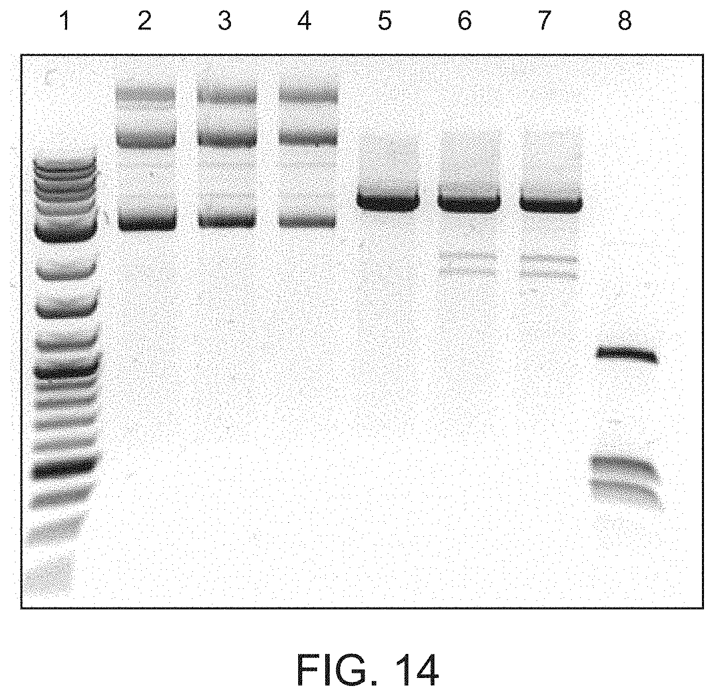
D00017

D00018

D00019

D00020

D00021
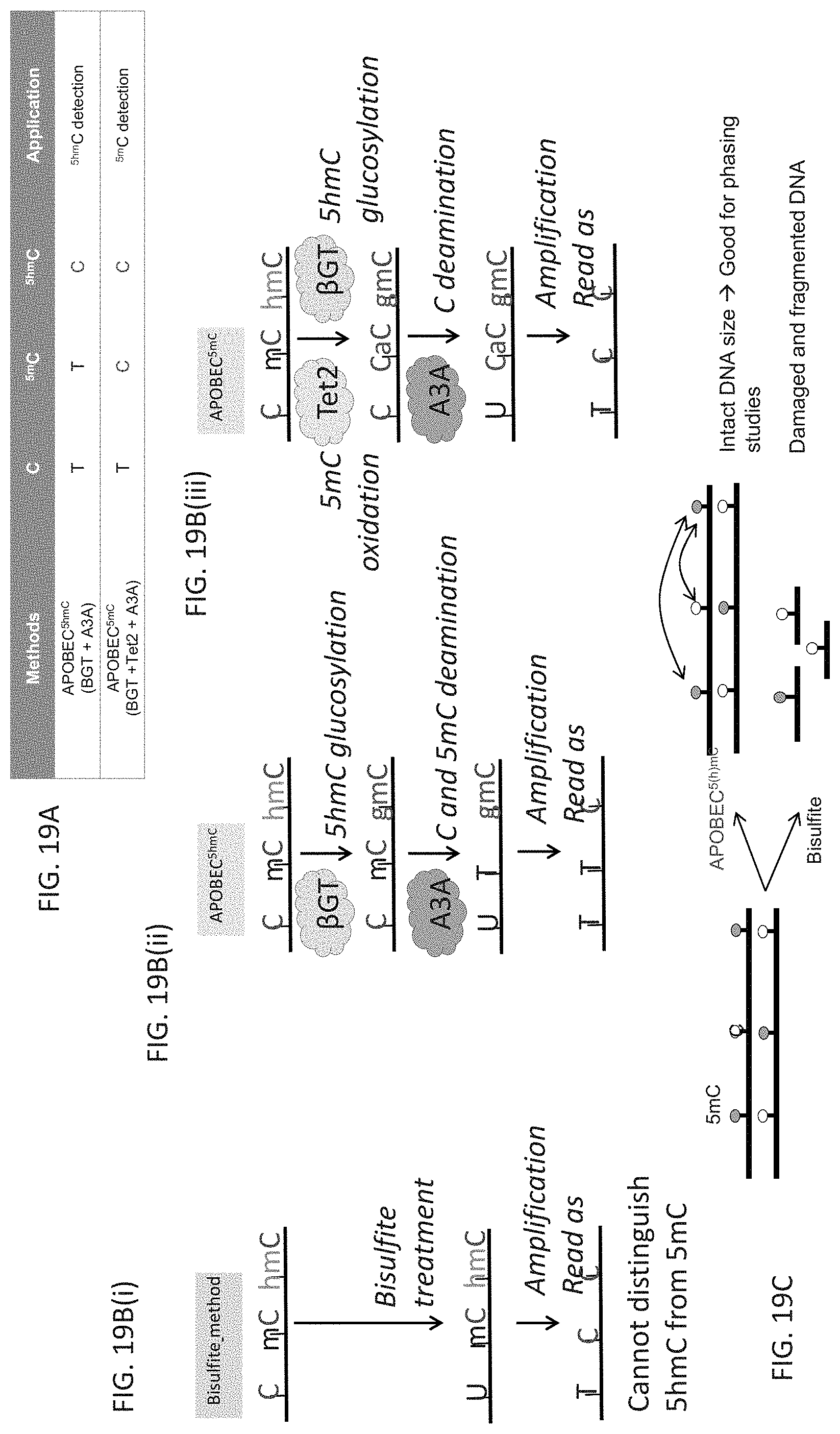
D00022

D00023

D00024
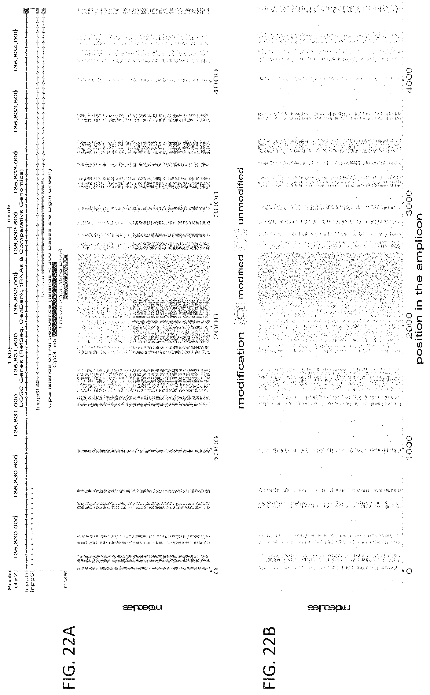
D00025

D00026
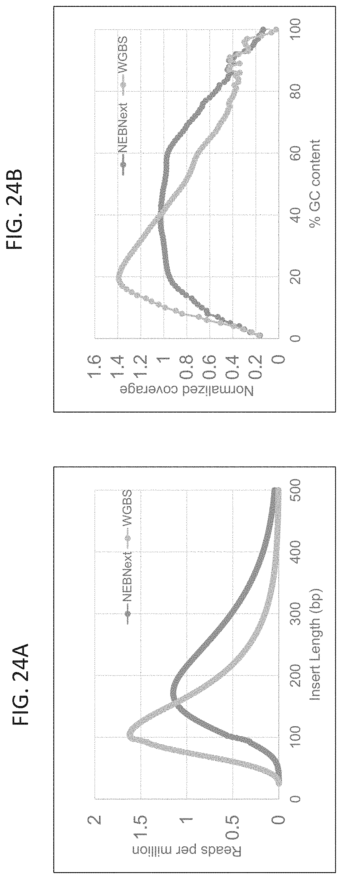
D00027

D00028

P00001

S00001
XML
uspto.report is an independent third-party trademark research tool that is not affiliated, endorsed, or sponsored by the United States Patent and Trademark Office (USPTO) or any other governmental organization. The information provided by uspto.report is based on publicly available data at the time of writing and is intended for informational purposes only.
While we strive to provide accurate and up-to-date information, we do not guarantee the accuracy, completeness, reliability, or suitability of the information displayed on this site. The use of this site is at your own risk. Any reliance you place on such information is therefore strictly at your own risk.
All official trademark data, including owner information, should be verified by visiting the official USPTO website at www.uspto.gov. This site is not intended to replace professional legal advice and should not be used as a substitute for consulting with a legal professional who is knowledgeable about trademark law.