Site-specific glycoengineering of targeting moieties
Avila , et al. May 4, 2
U.S. patent number 10,995,148 [Application Number 14/662,187] was granted by the patent office on 2021-05-04 for site-specific glycoengineering of targeting moieties. This patent grant is currently assigned to GENZYME CORPORATION. The grantee listed for this patent is Genzyme Corporation. Invention is credited to Luis Avila, Clark Pan, Huawei Qiu, Qun Zhou.


View All Diagrams
| United States Patent | 10,995,148 |
| Avila , et al. | May 4, 2021 |
Site-specific glycoengineering of targeting moieties
Abstract
The current disclosure provides binding polypeptides (e.g., antibodies), and targeting moiety conjugates thereof, comprising a site-specifically engineered glycan linkage within native or engineered glycans of the binding polypeptide. The current disclosure also provides nucleic acids encoding the antigen-binding polypeptides, recombinant expression vectors and host cells for making such antigen-binding polypeptides. Methods of using the antigen-binding polypeptides disclosed herein to treat disease are also provided.
| Inventors: | Avila; Luis (Arlington, MA), Pan; Clark (Sutton, MA), Qiu; Huawei (Westborough, MA), Zhou; Qun (Ashland, MA) | ||||||||||
|---|---|---|---|---|---|---|---|---|---|---|---|
| Applicant: |
|
||||||||||
| Assignee: | GENZYME CORPORATION (Cambridge,
MA) |
||||||||||
| Family ID: | 1000005528824 | ||||||||||
| Appl. No.: | 14/662,187 | ||||||||||
| Filed: | March 18, 2015 |
Prior Publication Data
| Document Identifier | Publication Date | |
|---|---|---|
| US 20160060354 A1 | Mar 3, 2016 | |
Related U.S. Patent Documents
| Application Number | Filing Date | Patent Number | Issue Date | ||
|---|---|---|---|---|---|
| 61955682 | Mar 19, 2014 | ||||
| 62061556 | Oct 8, 2014 | ||||
| Current U.S. Class: | 1/1 |
| Current CPC Class: | A61K 47/61 (20170801); C07K 16/2893 (20130101); A61K 47/6803 (20170801); C07K 16/40 (20130101); A61K 47/60 (20170801); C07K 16/32 (20130101); A61K 47/6889 (20170801); C07K 16/2851 (20130101); A61K 47/6855 (20170801); C07K 2317/524 (20130101); C07K 2317/92 (20130101); C07K 2317/77 (20130101); C07K 2317/41 (20130101); C07K 2317/71 (20130101); C07K 2317/52 (20130101) |
| Current International Class: | C07K 16/32 (20060101); A61K 47/68 (20170101); A61K 47/61 (20170101); A61K 47/60 (20170101); C07K 16/40 (20060101); C07K 16/28 (20060101) |
References Cited [Referenced By]
U.S. Patent Documents
| 5606017 | February 1997 | Willner et al. |
| 5648260 | July 1997 | Winter et al. |
| 5739277 | April 1998 | Presta et al. |
| 5834250 | November 1998 | Wells et al. |
| 5837821 | November 1998 | Wu |
| 5869046 | February 1999 | Presta et al. |
| 5880270 | March 1999 | Berninger et al. |
| 5989830 | November 1999 | Davis et al. |
| 6096871 | August 2000 | Presta et al. |
| 6121022 | September 2000 | Presta et al. |
| 6193980 | February 2001 | Efstathiou et al. |
| 6194551 | February 2001 | Idusogie et al. |
| 6218149 | April 2001 | Morrison et al. |
| 6242195 | June 2001 | Idusogie et al. |
| 6277375 | August 2001 | Ward |
| 6465612 | October 2002 | Bertozzi et al. |
| 6514498 | February 2003 | Antonsson et al. |
| 6528624 | March 2003 | Idusogie et al. |
| 6538124 | March 2003 | Idusogie et al. |
| 6737056 | May 2004 | Presta |
| 6821505 | November 2004 | Ward |
| 6998253 | February 2006 | Presta et al. |
| 7083784 | August 2006 | Dall'Acqua et al. |
| 7183387 | February 2007 | Presta |
| 7317091 | January 2008 | Lazar et al. |
| 7332581 | February 2008 | Presta |
| 7335742 | February 2008 | Presta |
| 7355008 | April 2008 | Stavenhagen et al. |
| 7371826 | May 2008 | Presta |
| 7612181 | November 2009 | Wu et al. |
| 7785791 | August 2010 | Presta |
| 7790858 | September 2010 | Presta |
| 8163881 | April 2012 | Ober |
| 9580511 | February 2017 | Pan et al. |
| 9701753 | July 2017 | Pan et al. |
| 9790268 | October 2017 | Pan et al. |
| 2002/0102208 | August 2002 | Chinn et al. |
| 2002/0193572 | December 2002 | Leung et al. |
| 2004/0002587 | January 2004 | Watkins et al. |
| 2005/0107595 | May 2005 | Cairns et al. |
| 2005/0238649 | October 2005 | Doronina et al. |
| 2007/0092940 | April 2007 | Eigenbrot et al. |
| 2007/0248600 | October 2007 | Hansen et al. |
| 2008/0038260 | February 2008 | Ponath et al. |
| 2008/0311134 | December 2008 | Junutula et al. |
| 2010/0190247 | July 2010 | Lazar et al. |
| 2010/0226923 | September 2010 | Rao et al. |
| 2010/0260751 | October 2010 | Raju et al. |
| 2011/0191867 | August 2011 | Natunen et al. |
| 2012/0251541 | October 2012 | Baurin et al. |
| 2014/0271676 | September 2014 | Pan et al. |
| 2014/0294867 | October 2014 | Pan et al. |
| 2015/0079070 | March 2015 | Pan et al. |
| 2015/0099861 | April 2015 | Snell et al. |
| 2016/0136299 | May 2016 | Avila et al. |
| 2017/0267774 | September 2017 | Pan et al. |
| 0403156 | Dec 1990 | EP | |||
| 1988007089 | Sep 1988 | WO | |||
| 1994009817 | May 1994 | WO | |||
| 1996014339 | May 1996 | WO | |||
| 1998005787 | Feb 1998 | WO | |||
| 1998023289 | Jun 1998 | WO | |||
| 1999051642 | Oct 1999 | WO | |||
| 1999058572 | Nov 1999 | WO | |||
| 2000009560 | Feb 2000 | WO | |||
| 2000032767 | Jun 2000 | WO | |||
| 2000042072 | Jul 2000 | WO | |||
| 2002002781 | Jan 2002 | WO | |||
| 2002044215 | Jun 2002 | WO | |||
| 2002060919 | Aug 2002 | WO | |||
| 2003074569 | Sep 2003 | WO | |||
| 2004016750 | Feb 2004 | WO | |||
| 2004029207 | Apr 2004 | WO | |||
| 2004035752 | Apr 2004 | WO | |||
| 2004063351 | Jul 2004 | WO | |||
| 2004074455 | Sep 2004 | WO | |||
| 2004/099249 | Nov 2004 | WO | |||
| 2004099249 | Nov 2004 | WO | |||
| 2005018572 | Mar 2005 | WO | |||
| 2005040217 | May 2005 | WO | |||
| 2005047327 | May 2005 | WO | |||
| 2005070963 | Aug 2005 | WO | |||
| 2005077981 | Aug 2005 | WO | |||
| 2005092925 | Oct 2005 | WO | |||
| 2005123780 | Dec 2005 | WO | |||
| 2006019447 | Feb 2006 | WO | |||
| 2006047350 | May 2006 | WO | |||
| 2006085967 | Aug 2006 | WO | |||
| 2006/105021 | Oct 2006 | WO | |||
| 2007005786 | Jan 2007 | WO | |||
| 2008/086006 | Jul 2008 | WO | |||
| 2009/052249 | Apr 2009 | WO | |||
| 2009/099728 | Aug 2009 | WO | |||
| 2010/027797 | Mar 2010 | WO | |||
| 2011/109400 | Sep 2011 | WO | |||
| 2013/037484 | Mar 2013 | WO | |||
| 2014043361 | Mar 2014 | WO | |||
| 2014/164534 | Oct 2014 | WO | |||
| 2014164503 | Oct 2014 | WO | |||
| 2015/143091 | Sep 2015 | WO | |||
Other References
|
Anwer et al. ("Anwer", Pharmaceutical Res. 2000, 17, 451-459). cited by examiner . Lepenies et al. ("Lepenies", Adv. Drug Delivery Rev, 2013, 65, 1271-1281). cited by examiner . Wright et al. (1992) "Genetically Engineered Antibodies: Progress and Prospects," Critical Reviews in Immunology. 12:125-168. cited by applicant . Yamagami et al. (1999) "Suppression of Allograft Rejection with Anti-A.beta. T Cell Receptor Antibody in Rat Corneal Transplantation," Transplantation. 67:600-604. cited by applicant . Yoshino et al. (1992) "Depletion of alpha/beta T cells by a monoclonal antibody against the alpha/beta T cell receptor suppresses established adjuvant arthritis, but not established collagen-induced arthritis in rats.," J. Exp. Med. 175:907-915. cited by applicant . Zhou et al. (2008) "Development of a simple and rapid method for producing non-fucosylated oligomannose containing antibodies with increased effector function," Biotechnol. Bioeng. 99:652-665. cited by applicant . Zhou et al. (2011) "Strategies for Neoglycan Conjugation to Human Acid .alpha.-Glucosidase," Bioconjugate Chemistry. 22:741-751. cited by applicant . Zhou et al. (2014) "Site-Specific Antibody--Drug Conjugation through Glycoengineering," Bioconjugate Chemistry. 25:510-520. cited by applicant . Axup, J.Y. et al., "Synthesis of site-specific antibody-drug conjugates using unnatural amino acids", PNAS 2012, vol. 109, No. 40, pp. 16101-16106. cited by applicant . Boeggeman et al. (2009) "Site Specific Conjugation of Fluoroprobes to the Remodeled Fc N-Glycans of Monoclonal Antibodies Using Mutant Glycosyltransferases: Application for Cell Surface Antigen Detection," Bioconjugate Chemistry. 20:1228-1236. cited by applicant . Carrasquillo et al. Improved imaging of metastatic melanoma with high dose 9.2.27 In-11 1 monoclonal antibody'. J. Nuc. Med. 1985. vol. 26, pp. 67, Abstract No. 276. cited by applicant . Carter et al. (2008) "Antibody-drug conjugates for cancer therapy," Caner Journal. 14:154-169. cited by applicant . Chan et al. (2010) "Therapeutic antibodies for autoimmunity and inflammation," The Journal of Immunology. 10:301-316. cited by applicant . Chari (2008) "Targeted Cancer Therapy: Conferring Specificity to Cytotoxic Drugs," Accounts of Chemical Research. 41:98-107. cited by applicant . Cobos-Correa et. al. `Membrane-bound FRET probe visualizes MMP12 activity in pulmonary inflammation`. Nature Chemical Biology. 2009, vol. 5, No. 9, pp. 628-663. cited by applicant . Ebersbach et al. `Affilin-Novel Binding Molecules Based on Humans .gamma.-B-Crystallin, an All .beta.-Sheet Protein`. Journal of Molecular Biology. 2007, vol. 372, No. 1, pp. 172-1. cited by applicant . Gehrig et.al. `Spatially resolved monitoring of neutrophil elastase activity with ratiometric fluorescent reporters`. Angewandte Chemie International Edition. 2012, vol. 51, No. 25, pp. 6258-6261. cited by applicant . Grabulovski et al. `A Novel, Non-immunogenic Fyn SH3-derived Binding Protein with Tumor Vascular Targeting Proteins`. The Journal of Biological Chemistry. 2007, vol. 282, No. 5, pp. 3196-3204. cited by applicant . Guan et al. `Homogeneous Immunoconjugates for Boron Neutron-capture Therapy: Design, Synthesis, and Preliminary Characterization`. PNAS. 1998, vol. 95, No. 22, pp. 13206-13210. cited by applicant . Heidecke et al. (1996) "Alpha-Beta T Cell Receptor-Directed Therapy in Rat Allograft Recipients," Transplantation. 61:948-956. cited by applicant . Heidecke et al. (1996) "Induction of Long-Term Rat Renal Allograft Survival by Pretransplant T Cell Receptor-A/B-Targeted Therapy," Transplantation 61:336-339. cited by applicant . International Search Report and Written Opinion, PCT/US2015/021342, dated Jun. 8, 2015, 13 pages. cited by applicant . International Search Report with Written Opinion corresponding to International Patent Application No. PCT/EP2012/003819, dated Jul. 17, 2013, 20 pages. cited by applicant . International Search Report with Written Opinion corresponding to International Patent Application No. PCT/US2013/059481, dated Feb. 7, 2014. cited by applicant . International Search Report with Written Opinion corresponding to International Patent Application No. PCT/US2014/022623, dated Jul. 31, 2014. cited by applicant . International Search Report with Written Opinion corresponding to International Patent Application No. PCT/US2014/022728, dated Oct. 9, 2014. cited by applicant . Jassal et al. (2001) "Sialylation of Human IgG-Fc Carbohydrate by Transfected Rat .alpha.2,6-Sialyltransferase," Biochemical and Biophysical Research Communications. 286:243-249. cited by applicant . Jones. `Proteinase Mutants of Saccharomyces cerevisiae`. Genetics. 1977, vol. 85, pp. 23-33. cited by applicant . Jung et al. (1992) "Prevention and therapy of experimental autoimmune neuritis by an antibody against T cell receptors-alpha/beta," Journal of Immunology. 148:3768-3775. cited by applicant . Junutula et al. (2008) "Site-specific conjugation of a cytotoxic drug to an antibody improves the therapeutic index," Nat. Biotechnol. 26:925-932. cited by applicant . Junutula et al. (2010) "Engineered Thio-Trastuzumab-DM1 Conjugate with an Improved Therapeutic Index to Target Human Epidermal Growth Factor Receptor 2--Positive Breast Cancer," Clin. Cancer Res. 16:4769-4778. cited by applicant . Kaneko et al. `Anti-inflammatory activity of immunoglobulin G resulting from Fc sialylation`. Science. 2006, vol. 313, No. 5787, pp. 670-673. cited by applicant . Kingsman et al. `Replication in Saccharomyces cerevisiae of Plasmid pBR313 Carrying DNA from the Yeast trpl Region`. Gene. 1979, vol. 7, pp. 141-152. cited by applicant . Koide et al. `Monobodies: Antibody Mimics Based on the Scaffold of the Fibronectin Type III Domain`. Methods in Molecular Biology. 2007, vol. 352, pp. 95-109. cited by applicant . Krehenbrink et al. `Artificial Binding Proteins (Affitins) as Probes for Conformational Changes in Secretin PuID`. Journal of Molecular Biology. 2008, vol. 383, No. 5, pp. 1058-1068. cited by applicant . Labrijn et al. (2008) "When binding is enough: nonactivating antibody formats," Curr. Opin. Immunol. 20:479-485. cited by applicant . Leung et al. (1999) "The effects of domain deletion, glycosylation, and long IgG3 hinge on the biodistribution and serum stability properties of a humanized IgG1 immunoglobulin, hLL2, and its fragments," Clin. Cancer Res. 5:3106s-3117s. cited by applicant . Nixon et al. `Engineered Protein Inhibitors of Proteases`. Current Opinion in Drug Discovery & Development. 2006, vol. 9, No. 2, pp. 261-268. cited by applicant . Nygren et al. `Alternative Binding Proteins: Affibody Binding Proteins Devloped from a Small Three-helix Bundle Scaffold`. The FEBS Journal. 2008, vol. 275, No. 11, pp. 2668-2676. cited by applicant . Page et al. (2012) "Biologics in Organ Transplant," Transplant International. 25:707-719. cited by applicant . Polakis (2005) "Arming antibodies for cancer therapy," Current Opinion in Pharmacology. 5:382-387. cited by applicant . Qu et al. (1998) "Carbohydrates engineered at antibody constant domains can be used for site-specific conjugation of drugs and chelates," Journal of Immunological Methods. 213:131-144. cited by applicant . Roche Diagnostics (May 2013) "Alpha-2,6, Sialyltransferase Cat. No. 07 012 250 103 (Data Sheet)," XP002727803. Retrieved from Internet: URL: https://cssportal.roche.com/LFR_PublicDocs/ras/07012250103_en_02.pdf. [retrieved on Jul. 25, 2014]. cited by applicant . Roux et al. `Comparisons of the Ability of Human IgG3 Hinge Mutants, IgM, IgE, and IgA2, to Form Small Immune Complexes: A Role for Flexibility and Geometry`. Journal of Immunology. 1998, vol. 161, No. 8, pp. 4083-4090. cited by applicant . Sazinsky et al. (2008) "Aglycosylated immunoglobulin G1 variants productively engage activating Fc receptors," Proc. Natl. Acad. Sci. USA. 105(51): 20167-20172. cited by applicant . Scharpf et al. (2006) "Immunomodulation with anti-a.beta. T-cell receptor monoclonal antibodies in combination with cyclosporine a improves regeneration in nerve allografts," Microsurgery. 26:599-607. cited by applicant . Schorlemmer et al. (1995) "Synergistic effects of 15-deoxyspergualin with cyclosporine and the TCR-targeted monoclonal antibody R73 to induce specific unresponsiveness to skin allografts in rats," Transplantation Proceedings. 27:414-416. cited by applicant . Shao, J et al. Unprotected Peptides as Building Blocks for the Synthesis of Peptide Dendrimers with Oxime, Hydrazone, and Thiazolidine Linkages, vol. 17, No. 14, Apr. 1995, J. Am Chem. Soc. 3893-3899. cited by applicant . Shearman et al. (1991) "Construction, expression and characterization of humanized antibodies directed against the human alpha/beta T cell receptor," The Journal of Immunology. 147:4366-4373. cited by applicant . Shields et al. (2001) "High Resolution Mapping of the Binding Site on Human IgG1 for Fc.gamma.RI, FcyRII, FcyRIII, and FcRn and Design of IgG1 Variants with Improved Binding to the Fc.gamma.R," J. Biol. Chem. 276:6591-6604. cited by applicant . Silverman et al. `Multivalent Avimer Proteins Evolved by Exon Shuffling of a Family of Human Receptor Domains`. Nature Biotechnology. 2005, vol. 23, No. 12, pp. 1556-1561. cited by applicant . Skerra et al. `Alternative Binding Proteins: Anticalins--Harnessing the Structural Plasticity of the Lipocalin Ligand Pocket to Engineer Novel Binding Activites`. The FEBS Journal. 2008, vol. 275, No. 11, pp. 2677-2683. cited by applicant . Stinchcomb et al. `Isolation and Characterization of a Yeast Chromosomal Replicator`. Nature. 1979, vol. 282, pp. 39-43. cited by applicant . Stumpp et al. `DARPins: A New Generation of Protein Therpeutics`. Drug Discovery Today. 2008, vol. 13, No. 15-16, pp. 695-670. cited by applicant . Teicher. `Antibody-drug conjugate targets`. Current Cancer Drug Targets. 2009, vol. 9, No. 8, pp. 982-1004. cited by applicant . Tschumper et al. `Sequence of a Yeast DNA Fragment Containing a Chromosomal Replicator and the TRP1 Gene`. Gene. 1980, vol. 10, pp. 157-166. cited by applicant . Wang et al. (1998) "Single-chain Fv with manifold N-glycans as bifunctional scaffolds for immunomolecules," Protein Engineering. 11:1277-1283. cited by applicant . Wang et al. `Impact of methionine oxidation in human IgG1 Fc on serum half-life of monoclonal antibodies`. Molecular Immunology. 2011, vol. 48, No. 6, pp. 860-866. cited by applicant . U.S. Appl. No. 14/203,438 / 2015/0079070 / U.S. Pat. No. 9,701,753, filed Mar. 10, 2014 / Mar. 19, 2015 / Jul. 11, 2017, Clark Pan. cited by applicant . U.S. Appl. No. 15/614,015, filed Jun. 5, 2017, Clark Pan. cited by applicant . U.S. Appl. No. 14/241,099 / 2015/0099861, filed Nov. 18, 2014 / Apr. 9, 2015, Daniel Snell. cited by applicant . U.S. Appl. No. 14/203,479 / 2014/0294867 / U.S. Pat. No. 9,580, filed Mar. 10, 2014 / Oct. 2, 2014 / Feb. 28, 2017, Clark Pan. cited by applicant . U.S. Appl. No. 15/417,648 / 2017/0267774, filed Jan. 27, 2017 / Sep. 21, 2017, Clark Pan. cited by applicant . U.S. Appl. No. 14/205,264 / 2014/0271676 / U.S. Pat. No. 9,790,268, filed Mar. 11, 2014 / Sep. 18, 2014 / Oct. 17, 2017, Clark Pan. cited by applicant . U.S. Appl. No. 15/702,368, filed Sep. 12, 2017, Clark Pan. cited by applicant . 2016/0060354, Mar. 3, 2016, Luis Avila. cited by applicant . U.S. Appl. No. 14/878,444 / 2016/0136299, filed Oct. 8, 2015 / May 19, 2016, Luis Avila. cited by applicant . Al-Lazikani et al. (1997) "Standard conformations for the canonical structures of immunoglobulins," J. Mol. Biol. 273:927-948. cited by applicant . Anthony et al. (2008) "Identification of a receptor required for the anti-inflammatory activity of IVIG," Proc. Natl. Acad. Sci. USA. 105:19571-19578. cited by applicant . Anthony et al. (2008) "Recapitulation of IVIG anti-inflammatory activity with a recombinant IgG Fc," Science. 320:373-376. cited by applicant . Boeggeman et al. (2007) "Direct Identification of Nonreducing GIcNAc Residues on N-Glycans of Glycoproteins Using a Novel Chemoenzymatic Method," Bioconjugate Chemistry. 18(3):806-814. cited by applicant . Cervigni et al. (1996) "Synthesis of Glycopeptides and Lipopeptides by Chemoselective Ligation," Angew. Chem., Int. Ed. 35(11):1230-1232. cited by applicant . Chen et al. (2010) "In vivo targeting of B-cell lymphoma with glycan ligands of CD22," Blood. 115:4778-4786. cited by applicant . Chen et al. (2012) "Targeting B lymphoma with nanoparticles bearing glycan ligands of CD22," Leukemia and Lymphoma. 53:208-210. cited by applicant . Doronina et al. (2003) "Development of potent monoclonal antibody auristatin conjugates for cancer therapy," Nat. Biotech. 21(7):778-784. cited by applicant . Feige et al. (2009) "Structure of the murine unglycosylated IgG1 Fc fragment," J. Mol Biol. 391:599-608. cited by applicant . Ganesan et al. (2011) "Rapid and Efficient Clearance of Blood-borne Virus by Liver Sinusoidal Endothelium," PLoS Pathogens. 7(9):e1002281. pp. 1-11. cited by applicant . Giudicelli et al. (2011) "IMGT/V-QUEST: IMGT Standardized Analysis of the Immunoglobulin (IG) and T Cell Receptor (TR) Nucleotide Sequences" In; Cold Spring Harb. Protoc. 2011(6):58-78. cited by applicant . Hatekeyama et al. (2011) "Targeted Drug Delivery to Tumor Vasculature by a Carbohydrate Mimetic Peptide," Proc. Natl. Acad. Sci. USA. 108:19587-19592. cited by applicant . Hong et al. (2003) "Beta-glucan functions as an adjuvant for monoclonal antibody immunotherapy by recruiting tumoricidal granulocytes as killer cells," Cancer Res. 23:9023-9031. cited by applicant . Hosoguchi (2010) "An efficient approach to the discovery of potent inhibitors against glycosyltransferases," J. Med. Chem. 53:5607-5619. cited by applicant . Kawasaki et al. (2013) "Targeted delivery of lipid antigen to macrophages via the CD169/sialoadhesin endocytic pathway induces robust invariant natural killer T cell activation," Proc. Natl. Acad. Sci. USA. 110:7826-7831. cited by applicant . Khidekel et al. (2003) "A Chemoenzymatic Approach toward the Rapid and Sensitive Detection of O-GIcNAc Posttranslational Modifications," J. Am. Chem. Soc. 125:16162-16163. cited by applicant . Krapp et al. (2003) "Structural analysis of human IgG-Fc glycoforms reveals a correlation between glycosylation and structural integrity," J. Mol. Biol. 325:979-989. cited by applicant . Li et al. (May 23, 2014) "The Preparation of Well-Defined Antibody-Drug Conjugates Through Glycan Remodeling and Strain Promoted Azide-Alkyne Cycloadditions," Angew. Chem., Int. Ed. 53(28):7179-7182. cited by applicant . Martin (2010) "Protein Sequence and Structure Analysis of Antibody Variable Domains" Ch. 3 In; Antibody Engineering. vol. 2. Eds: Kontermann et al. Springer-Verlag. pp. 33-51. cited by applicant . Mattner et al. (2005) "Exogenous and endogenous glycolipid antigens activate NKT cells during microbial nfections," Nature. 434:525-529. cited by applicant . Mccarthy (Aug. 16, 2013) "Chemoenzymatic synthesis of immunogenic meningococcal group C polysialic acid-tetanus Hc fragment glycoconjugates," Glycoconj. J. 30:857-870. cited by applicant . Medina et al. (2011) "N-acetylgalactosamine-functionalized dendrimers as hepatic cancer cell-targeted carriers," Biomaterials. 32:4118-4129. cited by applicant . Monnier et al. (2014) "Glucosepane: a poorly understood advanced glycation end product of growing importance for diabetes and its complications," Clin. Chem. Lab. Med. 52:21-32. cited by applicant . Murray (1985) "Imaging Findings and Pharmacaokinetics of 111-Indium ZME-018 Monoclonal Antibodies," J. Nuc. Med. 26:P16. Abstract No. 55. cited by applicant . North et al. (2011) "A new clustering of antibody CDR loop conformations," J. Mol. Biol. 406:228-256. cited by applicant . Piatesi et al. (2004) "Immunological optimization of a generic hydrophobic pocket for high affinity hapten binding and Diels-Alder activity," ChemBio Chem. 5:460-466. cited by applicant . Raju et al. (2011) "Glycoengineering of therapeutic glycoproteins: in vitro galactosylation and sialylation of glycoproteins with terminal N-acetylglucosamine and galactose residues," Biochem. 40(30):8868-8876. cited by applicant . Renaudet et al. (2006) "On-bead synthesis and binding assay of chemoselectively template-assembled multivalent heoglycopeptides," Org. Biomol. Chem. 4:2628-2636. cited by applicant . Wei et al. (2008) "Glyco-engineering of human IgG1-Fc through combined yeast expression and in vitro chemoenzymatic glycosylation," Biochem. 47(39):10294-10304. cited by applicant . Williams et al. (2010) "Humanising Antibodies by CDR Grafting" Ch. 21 In; Antibody Engineering. vol. 1. Eds.: Kontermann et al. Springer-Verlag. pp. 319-339. cited by applicant . Winkler et al. (2000) "Changing the Antigen Binding Specificity by Single Point Mutations of an Anti-P24 (HIV-1) Antibody," J. Immunol. 165(8):4505-4514. cited by applicant . Wu et al. (1999) "Humanization of a murine monoclonal antibody by simultaneous optimization of framework and CDR residues," J. Mol. Biol. 294:151-162. cited by applicant . Yu et al. (2008) "Interaction between Bevacizumab and Murine VEGF-A: A Reassessment," Investigative Ophthalmology & Visual Science. 49(2):522-527. cited by applicant . Zhou (Aug. 22, 2013) "Bioconjugation by native chemical tagging of C---H bonds.," J. Am. Chem. Soc. 135:12994-12997. cited by applicant . Zhu et al. (2009) "Glycoengineered acid alpha-glucosidase with improved efficacy at correcting the metabolic aberrations and motor function deficits in a mouse model of Pompe disease," Mol. Ther. 17(6):954-963. cited by applicant . International Search Report with Written Opinion corresponding to International Patent Application No. PCT/US2015/054651, dated Apr. 20, 2016. cited by applicant. |
Primary Examiner: Puttlitz; Karl J
Attorney, Agent or Firm: Lathrop GPM LLP Velema; James H. Stone-Hulslander; Judith L.
Parent Case Text
RELATED APPLICATIONS
This application claims the benefit of priority of U.S. Provisional Application No. 61/955,682, filed Mar. 19, 2014, and U.S. Provisional Application No. 62/061,556, filed Oct. 8, 2014. The contents of the aforementioned applications are hereby incorporated by reference in their entirety for all purposes.
Claims
We claim:
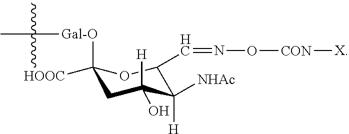
1. An antibody, or antigen-binding fragment thereof, comprising at least one modified glycan, wherein the glycan comprises at least one moiety of Formula (IV): ##STR00100## wherein: A) Q is NH or O; B) CON is a connector moiety; C) X is a targeting moiety comprising a carbohydrate or a glycopeptide that binds to a carbohydrate receptor on a cell or a glycopeptide receptor on a cell; D) Gal is a galactose moiety; and E) Sia is a sialic acid moiety.
2. The antibody, or antigen-binding fragment thereof, of claim 1, wherein the cell is a mammalian cell.
3. The antibody, or antigen-binding fragment thereof, of claim 2, wherein the mammalian cell is selected from an immune cell, a liver cell, a tumor cell, a vascular cell, an epithelial cell, or a mesenchymal cell.
4. The antibody, or antigen-binding fragment thereof, of claim 2, wherein the mammalian cell is selected from a B cell, a T cell, a dendritic cell, a natural killer (NK) cell, a macrophage, a neutrophil, a hepatocyte, a liver sinusoidal endothelial cell, or a hepatoma cell.
5. The antibody, or antigen-binding fragment thereof, of claim 1, wherein the targeting moiety comprises an .alpha.2,3-sialyllactose moiety or an .alpha.2,6-sialyllactose moiety.
6. The antibody, or antigen-binding fragment thereof, of claim 3, wherein the targeting moiety is a tri-galactosylated glycopeptide or lactose.sub.3-Cys.sub.3Gly.sub.4.
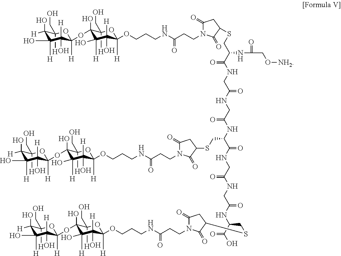
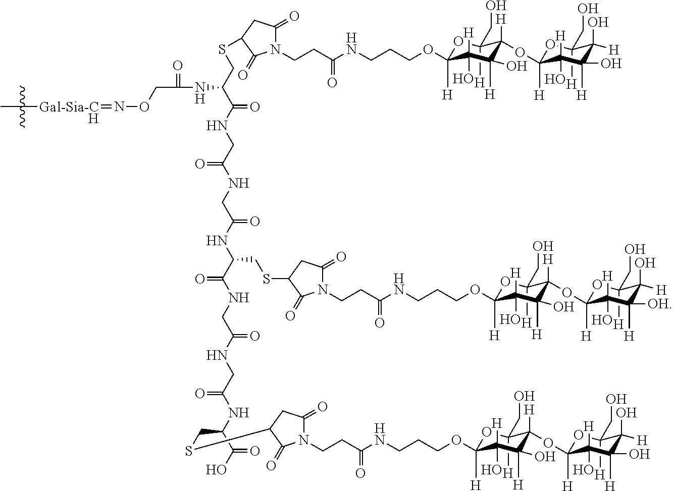
7. The antibody, or antigen-binding fragment thereof, of claim 6, wherein the lactose.sub.3-Cys.sub.3Gly.sub.4 moiety is represented by Formula V: ##STR00101##
8. The antibody, or antigen-binding fragment thereof, of claim 7, comprising at least one moiety selected from the following structural formulae: ##STR00102##
9. The antibody, or antigen-binding fragment thereof, of claim 1, wherein the targeting moiety is a trivalent GalNAc glycan moiety.
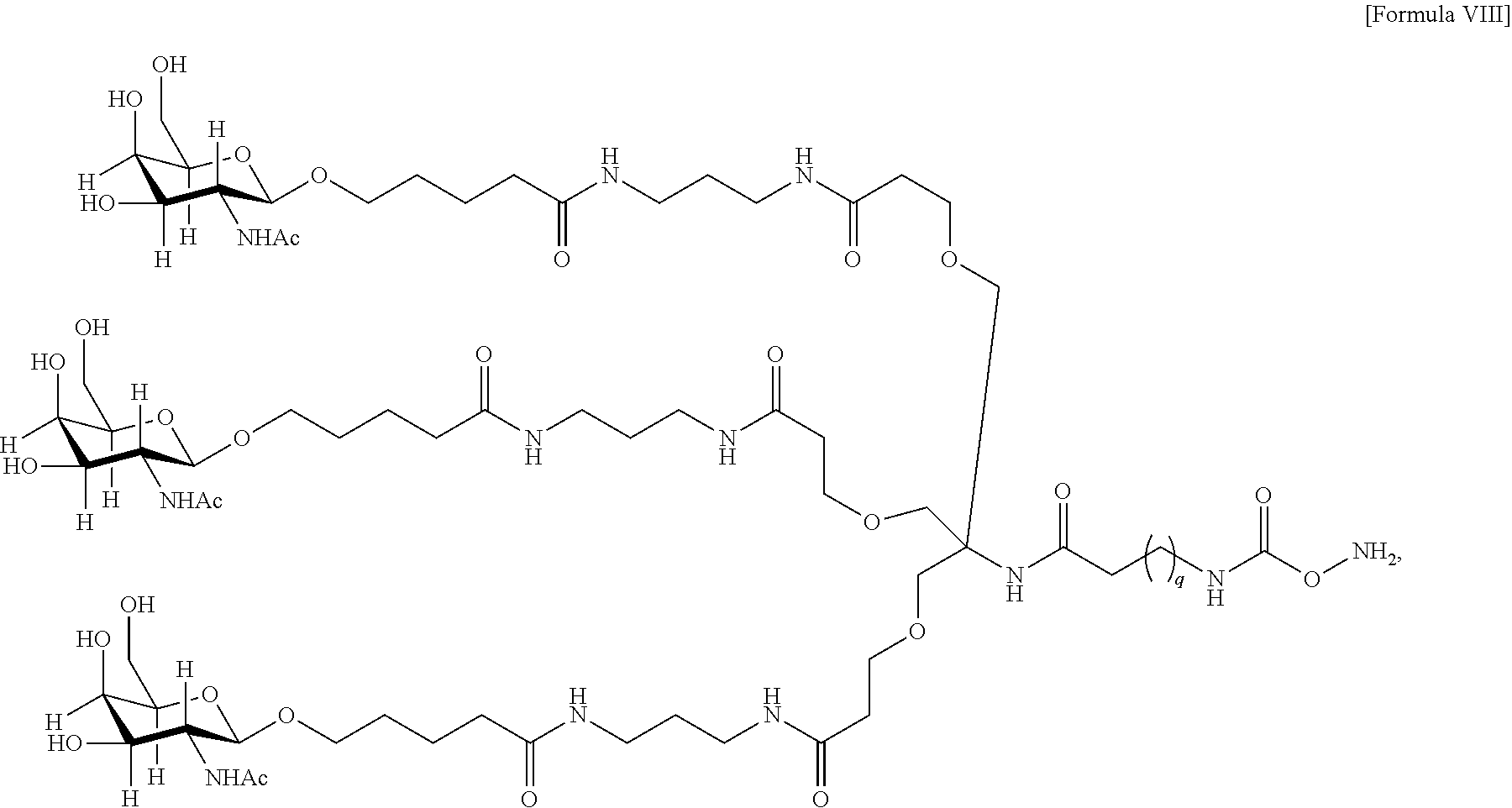
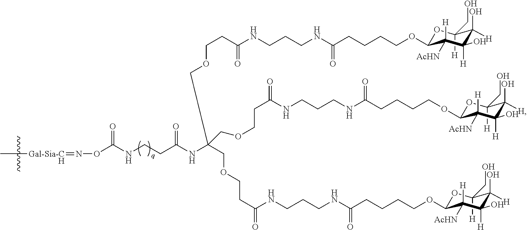
10. The antibody, or antigen-binding fragment thereof, of claim 9, wherein the trivalent GalNAc glycan moiety is represented by Formula VIII: ##STR00103## wherein q is an integer between 1 and 29 inclusive.
11. The antibody, or antigen-binding fragment thereof, of claim 1, wherein the antibody, or antigen-binding fragment thereof, comprises an Fc domain.
12. The antibody, or antigen-binding fragment thereof, of claim 1, wherein the modified glycan is N-linked to the antibody, or antigen-binding fragment thereof, via an asparagine residue at amino acid position 297 of the Fc domain, according to EU numbering.
13. A composition comprising the antibody, or antigen-binding fragment thereof, of claim 1 and a pharmaceutically acceptable carrier or excipient.
14. The composition of claim 13, wherein the ratio of targeting moiety to antibody, or antigen-binding fragment thereof, is equal to or more than about 4.
15. The composition of claim 13, wherein the ratio of targeting moiety to antibody, or antigen-binding fragment thereof, is at least about 2.
16. An antibody, or antigen-binding fragment thereof, comprising at least one modified glycan comprising at least one moiety of Formula (IV): ##STR00104## wherein: A) Q is NH or O; B) CON is a connector moiety; C) X is a targeting moiety comprising PEG that binds to a cell; D) Gal is a galactose moiety; and E) Sia is a sialic acid moiety.
17. The antibody, or antigen-binding fragment thereof, of claim 16, wherein the glycan comprises at least one moiety represented by Formula IX or Formula XI: ##STR00105## wherein p has a value of 1 to 32; or ##STR00106## wherein p has a value of 1 to 32.
18. The antibody, or antigen-binding fragment thereof, of claim 16, wherein the PEG comprises 3 to 3.5 PEG.
19. The antibody, or antigen-binding fragment thereof, of claim 1, wherein targeting moiety binds to a mannose 6 phosphate receptor on the cell.
20. The antibody, or antigen-binding fragment thereof, of claim 1, wherein the targeting moiety binds to a sialic acid-binding immunoglobulin-type lectin (Siglec) on the cell.
21. The antibody, or antigen-binding fragment thereof, of claim 20, wherein the Siglec is sialoadhesin (Siglec-1), CD22 (Siglec-2), CD33 (Siglec-3), myelin-associated glycoprotein (MAG (Siglec-4)), Siglec-5, Siglec-6, Siglec-7, Siglec-8, Siglec-9, Siglec-10, Siglec-11, Siglec-12, Siglec-14, or Siglec-15.
22. The antibody, or antigen-binding fragment thereof, of claim 1, wherein the targeting moiety binds to a C-type lectin receptor, a galectin, or a L-type lectin receptor.
23. The antibody, or antigen-binding fragment thereof, of claim 1, wherein Q is O.
24. The antibody, or antigen-binding fragment thereof, of claim 1, wherein the glycan comprises a moiety of the following structural formula: ##STR00107##
25. The antibody, or antigen-binding fragment thereof, of claim 1, wherein the targeting moiety is a glycopeptide capable of binding an asialoglycoprotein receptor (ASGPR) on a cell.
26. The antibody, or antigen-binding fragment thereof, of claim 1, wherein the amount of the antibody, or antigen-binding fragment thereof, internalized by the cell is greater than the amount of a reference antibody, or antigen-binding fragment thereof, lacking a targeting moiety internalized by the cell.
27. The antibody, or antigen-binding fragment thereof, of claim 1, wherein the targeting moiety binds to DEC-205 (CD205; lymphocyte antigen 75), macrophage mannose receptor (MMR; CD206), Dectin-1, Dectin-2, macrophage-inducible C-type lectin (Mincle), dendritic cell-specific ICAM3-grabbing nonintegrin (DC-SIGN, CD209), DC NK lectin group receptor-1 (DNGR-1), Langerin (CD207), a lectican, an asialoglycoprotein receptor (ASGPR), C-lectin receptor dendritic cell immunoreceptor (CLEC4A; CLECSF6; DCIR), macrophage galactose-type lectin (MGL), a DC receptor, a collectin, a selectin, an NK-cell receptor, a multi-C-type lectin domain (CTLD) endocytic receptor, a Reg group (type VII) lectin, chondrolectin, tetranectin, polycystin, attractin (ATRN), eosinophil major basic protein (EMBP), DiGeorge Syndrome Critical Region Gene 2 (DGCR2), Thrombomodulin, Bimlec, a group XVI lectin (SEEC), or a group XVII lectin (CBCP/Frem1/QBRICK).
28. The antibody, or antigen-binding fragment thereof, of claim 1, wherein the targeting moiety is a trivalent GalNAc glycan moiety.
29. The antibody, or antigen-binding fragment thereof, of claim 1, wherein the antibody, or antigen-binding fragment thereof, comprises a CH1 domain.
30. The antibody, or antigen-binding fragment thereof, of claim 1, wherein the connector moiety comprises a pH-sensitive linker, disulfide linker, enzyme-sensitive linker or other cleavable linker moiety.
31. The antibody, or antigen-binding fragment thereof, of claim 9, wherein the antibody, or antigen-binding fragment thereof, comprises at least one moiety having the following structural formula: ##STR00108## wherein q is an integer between 1 and 29 inclusive.
32. The antibody, or antigen-binding fragment thereof, of claim 9, wherein the antibody, or antigen-binding fragment thereof, comprises at least one moiety having the following structural formula: ##STR00109## wherein q is an integer between 1 and 29 inclusive.
33. The antibody, or antigen-binding fragment thereof, of claim 9, wherein the trivalent GalNAc glycan moiety is represented by Formula VII, Formula XIII, or Formula XIV: ##STR00110##
34. The antibody, or antigen-binding fragment thereof, of claim 9, wherein the antibody, or antigen-binding fragment thereof, comprises at least one moiety selected from the following structural formulae: ##STR00111## ##STR00112##
35. The antibody, or antigen-binding fragment thereof, of claim 9, wherein the component comprising galactose residue is GalNAc.
36. The antibody, or antigen-binding fragment thereof, of claim 16, wherein the antibody, or antigen-binding fragment thereof, comprises at least one moiety selected from the following structural formulae: ##STR00113## wherein p has a value of 1 to 32; ##STR00114## wherein p has a value of 1 to 32; ##STR00115## wherein p has a value of 1 to 32; or ##STR00116## wherein p has a value of 1 to 32.
37. The antibody, or antigen-binding fragment thereof, of claim 16, wherein the targeting moiety comprises PEG and is represented by Formula X or Formula XII: ##STR00117##
38. The antibody, or antigen-binding fragment thereof, of claim 16, wherein the antibody, or antigen-binding fragment thereof, comprises at least one moiety selected from the following structural formulae: ##STR00118## ##STR00119##
39. The antibody, or antigen-binding fragment thereof, of claim 16, wherein the component comprising galactose is GalNAc.
Description
SEQUENCE LISTING
The instant application contains a Sequence Listing which has been submitted electronically in ASCII format and is hereby incorporated by reference in its entirety. Said ASCII copy, created on Mar. 5, 2015, is named 566622SA9-148PC_SL.txt and is 115,860 bytes in size.
BACKGROUND
Use of specific antibodies to treat people and other animals is a powerful tool that has been very effective in treating many conditions and disorders. However, there is great demand for more effective targeted therapeutics, especially target specific therapies with higher efficacy and greater therapeutic windows. One of these target specific treatments employs antibody-effector moiety conjugates in which a targeting moiety directs a specific antibody to a desired treatment site. These molecules have shown improved therapeutic index--higher efficacy and/or lower toxicity profiles than the un-targeted antibody in a clinical setting. However, development of such therapeutics can be challenging as many factors, including physical and/or structural properties of the antibody itself as well as linkage stability, can have significant impact on the disease target (e.g. tumor) specificity, thereby reducing efficacy. With high non-specific binding and low stability in circulation, the antibody-effector moiety conjugate is typically cleared through normal tissues before reaching the target site. Moreover, antibody-effector moiety conjugates with significant subpopulations of high drug loading could generate aggregates which would be eliminated by macrophages, leading to shorter half-life. Thus, there are increasing needs for critical process control and improvement as well as preventing complications, such as antibody aggregation and nonspecific antibody-mediated toxicity.
Although antibody-effector moiety conjugates generated according to current methods can be effective, development of such therapeutics are challenging, as heterogeneous mixtures are often a consequence of the conjugation chemistries used. For example, effector moiety conjugation to antibody lysine residues is complicated by the fact that there are many lysine residues (.about.30) in an antibody available for conjugation. Since the optimal number of conjugated effector moiety to antibody ratio (DAR) is much lower to minimize loss of function of the antibody (e.g., around 4:1), lysine conjugation often generates a very heterogeneous profile. Furthermore, many lysines are located in critical antigen binding sites of the CDR region, and drug conjugation may lead to a reduction in antibody affinity. Thiol mediated conjugation mainly targets the eight cysteines involved in hinge disulfide bonds. However, it is still difficult to predict and identify which four of eight cysteines are consistently conjugated among the different preparations. More recently, genetic engineering of free cysteine residues has enabled site-specific conjugation with thiol-based chemistries, but such linkages often exhibit highly variable stability, with the linker undergoing exchange reactions with albumin and other thiol-containing serum molecules. Therefore, a site-specific conjugation strategy which generates an antibody conjugate with a defined conjugation site and stable linkage would be useful to enable effector moiety conjugation while minimizing adverse effects on antibody structure or function.
SUMMARY
The current disclosure provides binding polypeptides (e.g., antibodies), and targeting moiety conjugates thereof. In certain embodiments, the conjugates comprise a site-specifically engineered targeting-moiety glycan linkage within native or modified glycans of the binding polypeptide. The current disclosure also provides nucleic acid sequences encoding the antigen-binding polypeptides, recombinant expression vectors, and host cells for making such antigen-binding polypeptides. Methods of using the antigen-binding polypeptides disclosed herein to treat disease are also provided.
Accordingly, in one aspect, the invention provides a binding polypeptide comprising at least one modified glycan comprising at least one moiety of Formula (IV):
##STR00001## wherein: A) Q is NH or O; B) CON is a connector moiety; C) X is a targeting moiety; D) Gal is a component derived from galactose; and E) Sia is a component derived from sialic acid; wherein Sia is present or absent, and wherein the targeting moiety binds to a cell.
In one embodiment, the cell is a mammalian cell. In a further embodiment, the cell is selected from an immune cell, a liver cell, a tumor cell, a vascular cell, an epithelial cell, or a mesenchymal cell. In yet another embodiment, the cell is selected from a B cell, a T cell, a dendritic cell, a natural killer (NK) cell, a macrophage, a neutrophil, a hepatocyte, a liver sinusoidal endothelial cell, or a hepatoma cell.
In one embodiment, the binding polypeptide is internalized by the cell. In another embodiment, the amount of the binding polypeptide internalized by the cell is greater than the amount of a reference binding polypeptide lacking a targeting moiety internalized by the cell.
In one embodiment, the targeting moiety binds to a mannose 6 phosphate receptor on the cell. In another embodiment, the targeting moiety comprises a mannose 6 phosphate (Man 6-P) moiety.
In one embodiment, the targeting moiety binds to a Siglec on the cell. In a further embodiment, the Siglec is sialoadhesin (Siglec-1), CD22 (Siglec-2), CD33 (Siglec-3), MAG (Siglec-4), Siglec-5, Siglec-6, Siglec-7, Siglec-8, Siglec-9, Siglec-10, Siglec-11, Siglec-12, Siglec-14, or Siglec-15. In another embodiment, the targeting moiety comprises an .alpha.2,3-, .alpha.2,6-, or .alpha.2,8-linked sialic acid residue. In a further embodiment, the targeting moiety comprises an .alpha.2,3-sialyllactose moiety or an .alpha.2,6-sialyllactose moiety.
In one embodiment, the targeting moiety binds to a C-type lectin receptor, a galectin, a L-type lectin receptor or other carbohydrate receptors. In a further embodiment, the targeting moiety binds to DEC-205 (CD205; lymphocyte antigen 75), macrophage mannose receptor (MMR; CD206), Dectin-1, Dectin-2, macrophage-inducible C-type lectin (Mincle), dendritic cell-specific ICAM3-grabbing nonintegrin (DC-SIGN; CD209), DC NK lectin group receptor-1 (DNGR-1), Langerin (CD207), a lectican, an asialoglycoprotein receptor (ASGPR), C-lectin receptor dendritic cell immunoreceptor (CLEC4A; CLECSF6; DCIR), macrophage galactose-type lectin (MGL), a DC receptor, a collectin, a selectin, an NK-cell receptor, a multi-C-type lectin domain (CTLD) endocytic receptor, a Reg group (type VII) lectin, chondrolectin, tetranectin, polycystin, attractin (ATRN), eosinophil major basic protein (EMBP), DiGeorge Syndrome Critical Region Gene 2 (DGCR2), Thrombomodulin, Bimlec, a group XVI lectin (SEEC), or a group XVII lectin (CBCP/Frem1/QBRICK).
In one embodiment, Q is O.
In one embodiment, the glycan comprises at least one moiety of the following structural formula:
##STR00002##
In an embodiment, the targeting moiety is a trivalent GalNAc glycan moiety.
In one embodiment, the targeting moiety is a glycopeptide. In a further embodiment, the targeting moiety is a tri-galactosylated glycopeptide, e.g., lactose.sub.3-Cys.sub.3Gly.sub.4.
In an embodiment, Sia is present and the lactose.sub.3-Cys.sub.3Gly.sub.4 moiety is represented by Formula V:
##STR00003##
In one embodiment, the glycan comprises at least one moiety of the following structural formula:
##STR00004##
In one embodiment, the glycan comprises at least one moiety of the following structural formula:
##STR00005##
In one embodiment, the targeting moiety is a trivalent GalNAc glycan moiety.
In one embodiment, Sia is present and the trivalent GalNAc glycan moiety is represented by Formula VIII:
##STR00006## wherein q is an integer between 1 and 29 inclusive.
In another embodiment, the glycan comprises at least one moiety having the following structural formula:
##STR00007## wherein q is an integer between 1 and 29 inclusive.
In another embodiment, the glycan comprises at least one moiety having the following structural formula:
##STR00008## wherein q is an integer between 1 and 29 inclusive.
In an embodiment, the trivalent GalNAc glycan moiety is represented by Formula VII, Formula XIII, or Formula XIV:
##STR00009##
In one embodiment, the glycan comprises at least one moiety selected from the following structural formulae:
##STR00010##
In one embodiment, the glycan comprises at least one moiety selected from the following structural formulae:
##STR00011##
In an embodiment, the binding polypeptide comprises an Fc domain. In a further embodiment, the modified glycan is N-linked to the binding polypeptide via an asparagine residue at amino acid position 297 of the Fc domain, according to EU numbering. In another embodiment, the modified glycan is N-linked to the binding polypeptide via an asparagine residue at amino acid position 298 of the Fc domain, according to EU numbering.
In one embodiment, the Fc domain is human. In another embodiment, the binding polypeptide comprises a CH1 domain. In a further embodiment, the modified glycan is N-linked to the binding polypeptide via an asparagine residue at amino acid position 114 of the CH1 domain, according to Kabat numbering.
In one embodiment, the connector moiety comprises a pH-sensitive linker, disulfide linker, enzyme-sensitive linker or other cleavable linker moiety. In an embodiment, the connector moiety comprising a linker moiety selected from the group of linker moieties depicted in Table 2 or 14.
In one embodiment, the binding polypeptide is an antibody or immunoadhesin.
In one aspect, the present invention provides a method of making the binding polypeptide of any one of the preceding claims, the method comprising reacting an effector moiety of Formula (I): NH.sub.2-Q-CON-X Formula (I), wherein:
A) Q is NH or O;
B) CON is a connector moiety; and
C) X is a targeting moiety,
with an precursor binding polypeptide comprising an oxidized glycan.
In one embodiment, the initial binding polypeptide comprises at least one moiety of the following structural formula:
##STR00012##
In one embodiment, the initial binding polypeptide comprises at least one moiety of the following structural formula:
##STR00013##
In one embodiment, the precursor binding polypeptide comprises an oxidized glycan comprises at least one moiety of the following structural formula:
##STR00014##
In one embodiment, the precursor binding polypeptide comprises an oxidized glycan comprises at least one moiety of the following structural formula:
##STR00015##
In one embodiment, the precursor binding polypeptide comprising an oxidized glycan is generated by reacting an initial binding polypeptide comprising a glycan with a mildly oxidizing agent. In a further embodiment, the mildly oxidizing agent is sodium periodate. In another embodiment, no more than 1 mM sodium periodate is employed. In another embodiment, the mildly oxidizing agent is galactose oxidase.
In one embodiment, the method of marking a binding protein comprises reacting an effector moiety of the following structural formula: NH.sub.2--O-CON-X with the precursor binding polypeptide comprising an oxidized glycan comprising at least one moiety of the following structural formula:
##STR00016## to form a binding polypeptide of the following structural formula:
##STR00017##
In an embodiment, the targeting moiety is a trivalent GalNAc glycan moiety. In an embodiment, the reacting step is conducted in the presence of a salt comprising a metal ion. In a further embodiment, wherein the metal ion is a copper ion. In one embodiment, the salt is copper acetate. In another embodiment, the salt comprising a metal ion is present at a concentration of at least 0.1 mM.
In another embodiment, the binding polypeptide comprising the glycan comprises one or two terminal sialic acid residues. In a further embodiment, the terminal sialic acid residues are introduced by treatment of the binding polypeptide with a sialyltransferase or combination of sialyltransferase and galactosyltransferase. In one embodiment, an initial binding polypeptide is contacted with a mildly oxidizing agent in order to produce a precursor binding polypeptide or precursor binding protein. In one embodiment, the precursor binding polypeptide or precursor binding protein comprises an oxidized sialic acid moiety comprising a terminal aldehyde.
In one aspect, the present invention provides a binding polypeptide comprising at least one modified glycan comprising at least one moiety of Formula (IV):
##STR00018## wherein: A) Q is NH or O; B) CON is a connector moiety; and C) X is a lactose.sub.3-Cys.sub.3Gly.sub.4 moiety; D) Gal is a component derived from galactose; E) Sia is a component derived from sialic acid; and wherein Sia is present and the lactose.sub.3-Cys.sub.3Gly.sub.4 moiety is represented by Formula V:
##STR00019##
In another embodiment, the present invention provides a composition comprising a binding polypeptide described supra and a pharmaceutically acceptable carrier or excipient. In one embodiment, the ratio of targeting moiety to binding polypeptide is equal to or more than about 4. In another embodiment, the ratio of targeting moiety to binding polypeptide is at least about 2. In a further embodiment, the present invention provides a method for treating a patient in need thereof comprising administering an effective amount of the composition.
In one aspect, the present invention provides a binding polypeptide comprising at least one modified glycan comprising at least one moiety of Formula (IV):
##STR00020## wherein: A) Q is NH or O; B) CON is a connector moiety; and C) X is a moiety comprising PEG; D) Gal is a component derived from galactose; and E) Sia is a component derived from sialic acid; wherein Sia is present or absent.
In one embodiment, the glycan comprises at least one moiety represented by Formula IX or Formula XI:
##STR00021## wherein p has a value of 1 to 32; or
##STR00022## wherein p has a value of 1 to 32.
In an embodiment, the glycan comprises at least one moiety selected from the following structural formulae:
##STR00023## wherein p has a value of 1 to 32; or
##STR00024## wherein p has a value of 1 to 32. In one embodiment, the glycan comprises at least one moiety selected from the following structural formulae:
##STR00025## wherein p has a value of 1 to 32; or
##STR00026## wherein p has a value of 1 to 32.
In an embodiment, the targeting moiety comprises PEG and is represented by Formula X or Formula XII:
##STR00027## In one embodiment, the glycan comprises at least one moiety selected from the following structural formulae:
##STR00028## In an embodiment, the glycan comprises at least one moiety selected from the following structural formulae:
##STR00029## In one embodiment, the PEG moiety comprises mono-PEG, bi-PEG, or tri-PEG. In another embodiment, the PEG moiety comprises 3 to 3.5 PEG. In another embodiment, the binding polypeptide is an antibody or immunoadhesin.
In another aspect, the present invention provides a method of making a PEGylated binding polypeptide comprising at least one oxidized glycan, wherein the method comprises: (a) reacting a binding polypeptide comprising at least one glycan with a mildly oxidizing agent in the presence of a salt comprising a metal ion, and (b) conjugating the oxidized binding polypeptide with at least one moiety comprising PEG.
In one embodiment, the metal ion is a copper ion. In another embodiment, the salt is copper acetate. In another embodiment, the salt comprising a metal ion is present at a concentration of at least 0.1 mM. In another embodiment, the mildly oxidizing agent is periodate or galactose oxidase. In another embodiment, the at least one glycan is a modified glycan.
In one embodiment, the method of making a binding polypeptide comprises: a) reacting a binding polypeptide comprising at least one modified glycan with a mildly oxidizing agent in the presence of a salt comprising a metal ion; and b) conjugating the oxidized binding polypeptide with the moiety comprising PEG. In a further embodiment, the PEG moiety comprises mono-PEG, bi-PEG, or tri-PEG.
In another aspect, the present invention provides a binding polypeptide comprising at least one modified glycan comprising at least one moiety of Formula (IV):
##STR00030## wherein: A) Q is NH or O; B) CON is a connector moiety; and C) X is a trivalent GalNAc glycan; D) Gal is a component derived from galactose; and E) Sia is a component derived from sialic acid; wherein Sia is present and the trivalent GalNAc glycan moiety is represented by Formula VI.
In one embodiment, the binding protein comprises an N-glycan glycoform selected from the group consisting of: a G0 glycoform, a G1 glycoform, and a G2 glycoform. In a further embodiment, the N-glycan glycoform is selected from the group consisting of: a G1S1 glycoform, a G2S1 glycoform, a G2S2 glycoform, a G1F glycoform, a G2F glycoform, a G1S1F glycoform, a G2S1F glycoform, and a G2S2F glycoform.
BRIEF DESCRIPTION OF THE DRAWINGS
FIGS. 1A-1C are a schematic illustration of the synthesis of an antibody drug conjugate where a toxin moiety is linked to an oxidized sialic acid residue of the antibody glycan using an oxime linkage. FIG. 1A depicts antibodies with wild type (existing) carbohydrates at position Asn297 and with engineered glycosylation sites at positions 114 (according to the Kabat numbering) and 298 (according to the EU numbering system). FIG. 1B depicts both the canonical glycan structure G1F (left, seen at position Asn297 on the antibodies shown in FIG. 1A) and the G2S1F structure (right, seen at positions Ala114 and Asn298 on the antibodies shown in FIG. 1A). The glycan structures shown in FIG. 1B have several different subunits: the white square boxes represent GlcNAc, the upside-down white triangle represents fucose, the white circles represent mannose, the shaded circles represent galactose, and the shaded upright triangle represents sialic acid. FIG. 1C depicts the preparation of a conjugate of the instant invention. On the left is an initial binding protein with a Gal and Sia moiety. Following oxidation with NaIO4, the sialic moiety is oxidized to form a precursor binding protein and then reacted with a toxin-containing moiety to form a binding peptide comprising at least one moiety of Formula IV.
FIG. 2 is a Coomassie-blue stained gel showing the expression and purification of glycosylation mutants.
FIG. 3 depicts the results of surface plasmon resonance experiments used to assess the binding of .alpha..beta.TCR HEBE1 IgG antibody mutants to recombinant human Fc.gamma.RIIIa (V158 & F158).
FIG. 4 depicts the results of surface plasmon resonance experiments used to assess the binding of .alpha..beta.TCR HEBE1 IgG antibody mutants to recombinant human Fc.gamma.RI.
FIG. 5 depicts the cytokine release profile from PBMCs for TNFa, GM-CSF, IFNy and IL10 in the presence of mutant anti-.alpha..beta.TCR antibodies (day 2).
FIG. 6 depicts the cytokine release profile from PBMCs for IL6, IL4 and IL2 in the presence of mutant anti-.alpha..beta.TCR antibodies (day 2).
FIG. 7 depicts the cytokine release profile from PBMCs for TNFa, GM-CSF, IFNy and IL10 in the presence of mutant anti-.alpha..beta.TCR antibodies (day 4).
FIG. 8 depicts the cytokine release profile from PBMCs for IL6, IL4 and IL2 in the presence of mutant anti-.alpha..beta.TCR antibodies (day 4).
FIGS. 9A-9B depict the results of experiments investigating the expression level of 2C3 mutants by Western blotting (FIG. 9A) and surface plasmon resonance (FIG. 9B).
FIG. 10 depicts the results of experiments investigating glycosylation of 2C3 mutants pre- and post-PNGase F treatment.
FIG. 11 depicts the results of SDS-PAGE experiments investigating glycosylation sites on 2C3 mutants isolated from cell culture.
FIGS. 12A-12C depict the results of surface plasmon resonance experiments used to assess the binding of modified anti-CD52 to recombinant human Fc.gamma.RIIIa (V158). Anti-CD52 comprising S298N/Y300S mutations in the Fc domain were used to assess the effector function of the modified molecule. binding to CD52 peptide (FIG. 12A), binding to Fc.gamma.RIIIa (V158, FIG. 12B), and control binding to mouse FcRn (FIG. 12C).
FIG. 13 depicts the results of surface plasmon resonance experiments investigating the Fc binding properties of 2C3 mutants.
FIGS. 14A-14B depict the results of surface plasmon resonance experiments investigating the binding of modified anti-CD52 to both Fc.gamma.RIIIa (Val158) (as above) and Fc.gamma.RIIIa (Phe158). Anti-CD52 antibodies comprising S298N/Y300S mutations in the Fc domain were used to assess the effector function of the modified molecule binding to Fc.gamma.RIIIa (Val158, FIG. 14A) and Fc.gamma.RIIIa (Phe58, FIG. 14B).
FIGS. 15A-15B depict the analysis of C1q binding in the S298N/Y300S mutant and the WT 2C3 control (FIG. 15A) and the results of an Eliza analysis confirming equivalent coating of the wells (FIG. 15B).
FIG. 16 depicts the results of plasmon resonance experiments measuring the binding kinetics of 2C3 mutants to CD-52 peptide 741.
FIG. 17 depicts the results of plasmon resonance experiments comparing the antigen binding affinity of WT anti-CD-52 2C3 and the A114N hyperglycosylation mutant.
FIGS. 18A-18D depict the results of isoelectric focusing and mass spectrometry charge characterization experiments to determine the glycan content of 2C3 mutants.
FIGS. 19A-19B depict the results of concentration (Octet) and plasmon resonance experiments comparing the antigen binding affinity of WT anti-CD52 2C3 and mutants.
FIG. 20 depicts the results of SDS-PAGE experiments to demonstrate the additional glycosylation of the anti-TEM1 A114N mutant.
FIG. 21 depicts the results of SDS-PAGE and hydrophobic interaction chromatography analysis of the A114N anti-Her2 mutant.
FIG. 22 depicts the results of SDS-PAGE experiments to demonstrate the conjugation of PEG to the 2C3 A114N mutant through an aminooxy linkage.
FIG. 23 depicts the results of LC-MS experiments to determine the glycan contents of anti-TEM1 A114N hyperglycosylation mutant.
FIG. 24 depicts the results of LC-MS experiments to determine the glycan contents of a wild-type HER2 antibody and an A114N anti-Her2 hyperglycosylation mutant.
FIGS. 25A-25C depict an exemplary method for performing site-specific conjugation of an antibody.
FIG. 26 depicts a synthesis of exemplary effector moieties: aminooxy-Cys-MC-VC-PABC-MMAE and aminooxy-Cys-MC-VC-PABC-PEG8-Dol10.
FIGS. 27A-27C depict characterization information for a sialylated HER2 antibody.
FIGS. 28A-28D depict characterization information for oxidized sialylated anti-HER 2 antibody.
FIG. 29 depicts hydrophobic interaction chromatographs of glycoconjugates prepared with three different sialylated antibodies with two different aminooxy groups.
FIG. 30 shows a HIC chromatograph of antiHer2 A114 glycosylation mutant conjugate with AO-MMAE prepared using GAM(+) chemistry.
FIGS. 31A-31D depict a comparison of the in vitro potency of an anti-HER2 glycoconjugate and thiol conjugate.
FIG. 32 depicts a comparison of the in vitro potency of an anti FAP B11 glycoconjugate and thiol conjugate.
FIGS. 33A-33D depict a comparison of in vivo efficacy of anti-HER2 glycoconjugates and thiol conjugates in a Her2+ tumor cell xenograft model.
FIG. 34 depicts the results of LC-MS experiments to determine the glycan content of a mutant anti-.alpha..beta.TCR antibody containing the S298N/Y300S mutation.
FIG. 35 depicts the results of circular dichroism experiments to determine the relative thermal stability of a wild-type anti-.alpha..beta.TCR antibody and mutant anti-.alpha..beta.TCR antibody containing the S298N/Y300S mutation.
FIG. 36 depicts the results of a cell proliferation assay for ADC prepared with the anti-HER antibody bearing the A114N hyperglycosylation mutation and AO-MMAE.
FIG. 37 is a schematic illustration of the synthesis of an antibody drug conjugate where a targeting moiety is linked to an oxidized sialic acid residue of the antibody glycan using an oxime linkage.
FIG. 38 is a schematic illustration depicting an exemplary method for performing site-specific conjugation of an antibody to a glycopeptide through an oxime linkage according to the described methods.
FIG. 39 is a schematic illustration depicting site-specific conjugation of neoglycans to antibody through sialic acid in native Fc glycans.
FIG. 40 is a series of exemplary glycans that may be used for conjugation including lactose aminooxy and bis Man-6-P (or bisM6P) hexamannose aminooxy (for aminooxy conjugation).
FIG. 41 is a schematic depiction of the preparation of Man-6-P hexamannose maleimide.
FIG. 42 depicts SDS-PAGE and MALDI-TOF characterization of Man-6-P hexamannose aminooxy conjugates made with rabbit polyclonal antibody.
FIG. 43 depicts the results of surface plasmon resonance experiments used to assess the binding of control and Man-6-P hexamannose conjugated rabbit IgG antibodies to Man-6-P receptor.
FIG. 44 depicts the uptake of Man-6-P conjugated rabbit IgG antibody in HepG2 and RAW cells.
FIG. 45 depicts the characterization of control, Man-6-P conjugated, and lactose conjugated antibodies through SDS-PAGE and lectin blotting.
FIG. 46 depicts the results of MALDI-TOF intact protein analyses for control, Man-6-P conjugated, and lactose conjugated antibodies.
FIG. 47 depicts the characterization of polyclonal antibody conjugated to Man-6-P hexamannose maleimide (thiol conjugation at hinge cysteines) through SDS-PAGE (non-reducing and reducing), lectin blot (reducing), and Man-6-P quantitation.
FIG. 48 depicts the characterization of polyclonal antibody conjugated to lactose maleimide (thio conjugation at hinge cysteines) through SDS-PAGE and galactose quantitation.
FIG. 49 depicts the characterization of monoclonal antibody conjugated to Man-6-P hexamannose maleimide (thiol conjugation at hinge cysteines) through SDS-PAGE (non-reducing and reducing), and glycan (bis Man-6-P) quantitation.
FIG. 50 depicts the results of size exclusion chromatography (SEC) analysis of a hinge cysteine polyclonal antibody conjugate.
FIG. 51 depicts the results of size exclusion chromatography (SEC) analysis of a hinge cysteine monoclonal antibody conjugate.
FIG. 52 depicts the results of sialidase titration and sialic acid quantitation to determine the amount of sialic acid release from NNAS, sialylated NNAS, and desialylated and galactosylated NNAS antibodies.
FIG. 53 depicts the results of MALDI-TOF MS analysis to determine the glycan structures of a mouse NNAS antibody and a desialylated and galactosylated NNAS antibody.
FIG. 54 depicts the results of MALDI-TOF MS analysis to determine the glycan structures of a mouse NNAS antibody and a sialylated NNAS antibody.
FIG. 55 depicts the characterization of Man-6-P receptor (CI-MPR) bound to bis Man-6-P glycan-conjugated polyclonal and monoclonal antibodies through native Fc glycan or hinge disulfides using native PAGE.
FIG. 56 depicts the characterization of enzyme modified and glycopeptide conjugated NNAS antibodies by SDS-PAGE (4-12% NuPAGE; reducing and non-reducing) and ECL lectin blotting (reducing).
FIG. 57 depicts the results of terminal galactose quantitation in an NNAS antibody, a desialylated/galactosylated NNAS antibody, and a conjugated NNAS antibody in mol galactose or mol glycopeptide per mol antibody.
FIG. 58 depicts the examination of lactose maleimide that had been modified with alpha-2,3-sialyltransferase and eluted from QAE purification columns with 20 mM NaCl. The resultant eluate was characterized using MALDI-TOF MS and Dionex HPLC.
FIGS. 59A-59B depict the characterization of rabbit polyclonal antibody conjugated with sialyllactose maleimide (thiol reaction) using SDS-PAGE and Dionex HPLC (sialic acid quantitation).
FIGS. 60A-60D depict the characterization of lactose maleimide sialylated with alpha-2,6-sialyltransferase and purified using a QAE-sepharose column Analysis using Dionex HPLC is shown for (FIG. 60A) a lactose standard; (FIG. 60B) an alpha-2,6-sialyllactose standard; (FIG. 60C) a lactose maleimide standard; and (FIG. 60D) a fraction of alpha-2,6-sialyllactose maleimide eluted from a QAE-sepharose column.
FIG. 61 depicts the characterization of a fraction of alpha-2,6-sialyllactose maleimide eluted from a QAE-sepharose column using MALDI-TOF MS.
FIGS. 62A-62B depict the characterization of a control antibody, an alpha-2,3-sialyllactose glycan conjugated polyclonal antibody, and an alpha-2,6-sialyllactose glycan conjugated polyclonal antibody through SDS-PAGE and Dionex HPLC (graph of sialic acid analysis shown).
FIG. 63 depicts the characterization of control and enzyme modified (desialylated/galactosylated) NNAS mutant antibodies using SDS-PAGE and lectin blotting.
FIG. 64 depicts the characterization through reducing and non-reducing SDS-PAGE of the PEGylated control antibody and Gal NNAS with various amounts of galactose oxidase.
FIG. 65 depicts the results of estimated PEGylation of an antibody heavy chain from previous galactose oxidase titration using ProteinSimple.
FIG. 66 depicts the characterization through reducing and non-reducing SDS-PAGE of the PEGylated control antibody and Gal NNAS with various molar excess of PEG over antibody.
FIG. 67 depicts the results of estimated PEGylation of an antibody heavy chain from previous PEG titration using ProteinSimple.
FIG. 68 is a structural drawing of aminooxy glycopeptide, lactose.sub.3-Cys.sub.3Gly.sub.4.
FIGS. 69A-69B depict the characterization through reducing SDS-PAGE of the PEGylated control antibody and Gal NNAS with galactose oxidase in the absence of copper acetate (FIG. 69A) and in the presence of varying amounts of copper acetate (FIG. 69A and FIG. 69B).
FIG. 70 depicts the characterization of enzyme modified wild-type, A114N, NNAS, and A114N/NNAS Herceptin by SDS-PAGE (4-12% NuPAGE; reducing and non-reducing) and ECL lectin blotting (reducing) along with the results of terminal galactose quantitation in mol galactose per mol antibody.
FIG. 71 is a table depicting the sialic acid content (in mol/mol) of wild-type and mutant antibodies as measured using Dionex HPLC.
FIG. 72 depicts the characterization of the PEGylation of wild-type and mutant antibodies through reducing and non-reducing SDS-PAGE.
FIG. 73 is a table depicting the PEGylation (in mol/mol) of wild-type and mutant antibodies estimated using ProteinSimple.
FIG. 74 is a series of photos depicting immunofluorescence staining of HepG2 cell uptake of control, enzyme modified (with galactosyltransferase), or conjugated (with lactose aminoxy or lactose maleimide) antibodies.
FIG. 75 is a depiction of an exemplary trivalent GalNAc glycan.
FIG. 76 depicts the results of surface plasmon resonance experiments used to assess the binding of trivalent GalNAc glycan-conjugated antibodies to ASGPR subunit H1.
FIG. 77 is a depiction of a trivalent GalNAc-containing glycan and a trivalent galactose-containing glycopeptide used for conjugation.
FIG. 78 depicts the results of surface plasmon resonance experiments used to assess the binding of trivalent GalNAc-conjugated and trivalent galactose containing glycopeptide-conjugated recombinant lysosomal enzymes to ASGPR subunit H1.
FIGS. 79A-79D are depiction of additional trivalent GalNAc glycans.
FIG. 80 is a depiction of the results of conjugation of periodate oxidized recombinant lysosomal enzyme rhGAA with an excess of trivalent GalNAc glycan C12 (at 20, 40, 80, and 200-fold molar excess glycan over rhGAA). The resulting glycan conjugated per rhGAA is depicted.
FIG. 81 depicts ASGPR binding of a recombinant lysosomal enzymes conjugated with trivalent GalNAc glycan C12 on Biacore. Enzymes conjugated with 20 (conjugate 1), 40, 80, and 200-fold (conjugate 4) excess of glycan all show strong binding to ASGPR subunit 1. There is no significant difference in binding among the conjugates (conjugates 1 to 4).
DETAILED DESCRIPTION
The current disclosure provides binding polypeptides (e.g., antibodies), and effector moiety conjugates (e.g., targeting moiety conjugates) thereof. In certain embodiments, the conjugates comprise a site-specifically engineered drug-glycan linkage within native or modified glycans of an antigen binding polypeptide such as an IgG molecule. The current disclosure also provides nucleic acids encoding antigen-binding polypeptides, recombinant expression vectors and host cells for making antigen-binding polypeptides. Methods of using the antigen-binding polypeptides disclosed herein to treat disease are also provided.
I. Definitions
Unless otherwise defined herein, scientific and technical terms used herein have the meanings that are commonly understood by those of ordinary skill in the art. In the event of any latent ambiguity, definitions provided herein take precedent over any dictionary or extrinsic definition. Unless otherwise required by context, singular terms shall include pluralities and plural terms shall include the singular. The use of "or" means "and/or" unless stated otherwise. The use of the term "including", as well as other forms, such as "includes" and "included", is not limiting.
Generally, nomenclatures used in connection with cell and tissue culture, molecular biology, immunology, microbiology, genetics and protein and nucleic acid chemistry and hybridization described herein are those well-known and commonly used in the art. The methods and techniques provided herein are generally performed according to conventional methods well known in the art and as described in various general and more specific references that are cited and discussed throughout the present specification unless otherwise indicated. Enzymatic reactions and purification techniques are performed according to manufacturer's specifications, as commonly accomplished in the art or as described herein. The nomenclatures used in connection with, and the laboratory procedures and techniques of, analytical chemistry, synthetic organic chemistry, and medicinal and pharmaceutical chemistry described herein are those well-known and commonly used in the art. Standard techniques are used for chemical syntheses, chemical analyses, pharmaceutical preparation, formulation, and delivery, and treatment of patients.
That the disclosure may be more readily understood, select terms are defined below.
The term "polypeptide" refer to any polymeric chain of amino acids and encompasses native or artificial proteins, polypeptide analogs or variants of a protein sequence, or fragments thereof, unless otherwise contradicted by context. A polypeptide may be monomeric or polymeric. For an antigenic polypeptide, a fragment of a polypeptide optionally contains at least one contiguous or nonlinear epitope of a polypeptide. The precise boundaries of the at least one epitope fragment can be confirmed using ordinary skill in the art. A polypeptide fragment comprises at least about 5 contiguous amino acids, at least about 10 contiguous amino acids, at least about 15 contiguous amino acids, or at least about 20 contiguous amino acids, for example.
The term "isolated protein" or "isolated polypeptide" refer to a protein or polypeptide that by virtue of its origin or source of derivation is not associated with naturally associated components that accompany it in its native state; is substantially free of other proteins from the same species; is expressed by a cell from a different species; or does not occur in nature. Thus, a protein or polypeptide that is chemically synthesized or synthesized in a cellular system different from the cell from which it naturally originates will be "isolated" from its naturally associated components. A protein or polypeptide may also be rendered substantially free of naturally associated components by isolation using protein purification techniques well known in the art.
As used herein, the term "binding protein" or "binding polypeptide" shall refer to a protein or polypeptide (e.g., an antibody or fragment thereof) that contains at least one binding site which is responsible for selectively binding to a target antigen of interest (e.g., a human antigen). Exemplary binding sites include an antibody variable domain, a ligand binding site of a receptor, or a receptor binding site of a ligand. In certain aspects, the binding proteins or binding polypeptides comprise multiple (e.g., two, three, four, or more) binding sites. In certain aspects, the binding protein or binding polypeptide is not a therapeutic enzyme.
As used herein, the term "native residue" shall refer to an amino acid residue that occurs naturally at a particular amino acid position of a binding polypeptide (e.g., an antibody or fragment thereof) and which has not been modified, introduced, or altered by the hand of man. As used herein, the term "altered binding protein," "altered binding polypeptide," "modified binding protein" or "modified binding polypeptide" shall refer to binding polypeptides and/or binding proteins (e.g., an antibody or fragment thereof) comprising at least one amino acid substitution, deletion and/or addition relative to the native (i.e., wild-type) amino acid sequence, and/or a mutation that results in altered glycosylation (e.g., hyperglycosylation, hypoglycosylation and/or aglycosylation) at one or more amino acid positions relative to the native (i.e., wild-type) amino acid sequence.
As used herein, the term "initial binding polypeptide" or "initial binding protein" shall refer to a binding polypeptide or binding protein that is contacted with a mildly oxidizing agent to produce a "precursor binding polypeptide" or a "precursor binding protein," respectively (see FIGS. 1A-C). As used herein, the term "precursor binding polypeptide" or "precursor binding protein" shall refer to a mildly oxidized polypeptide or protein that can be reacted with one or more of the effector moieties described herein. In certain embodiments, an initial binding polypeptide, initial binding protein, precursor binding polypeptide, and/or precursor binding protein (e.g., an antibody or fragment thereof) contains at least one binding site which is responsible for selectively binding to a target antigen of interest (e.g., a human antigen). Exemplary binding sites include an antibody variable domain, a ligand binding site of a receptor, or a receptor binding site of a ligand. In certain aspects, an initial binding polypeptide, initial binding protein, precursor binding polypeptide, and/or precursor binding protein comprises multiple (e.g., two, three, four, or more) binding sites. In certain aspects, the initial binding polypeptide, initial binding protein, precursor binding polypeptide, and/or precursor binding protein is not a therapeutic enzyme. An initial binding polypeptide, initial binding protein, precursor binding polypeptide, and/or precursor binding protein may have a wild-type sequence or they may comprise at least one amino acid substitution, deletion and/or addition relative to the native (i.e., wild-type) amino acid sequence, and/or a mutation that results in altered glycosylation (e.g., hyperglycosylation, hypoglycosylation and/or aglycosylation) at one or more amino acid positions relative to the native (i.e., wild-type) amino acid sequence.
The term "ligand" refers to any substance capable of binding, or of being bound, to another substance. Similarly, the term "antigen" refers to any substance to which an antibody may be generated. Although "antigen" is commonly used in reference to an antibody binding substrate, and "ligand" is often used when referring to receptor binding substrates, these terms are not distinguishing, one from the other, and encompass a wide range of overlapping chemical entities. For the avoidance of doubt, antigen and ligand are used interchangeably throughout herein. Antigens/ligands may be a peptide, a polypeptide, a protein, an aptamer, a polysaccharide, a sugar molecule, a carbohydrate, a lipid, an oligonucleotide, a polynucleotide, a synthetic molecule, an inorganic molecule, an organic molecule, and any combination thereof.
The term "specifically binds" as used herein, refers to the ability of an antibody or an antigen-binding fragment thereof to bind to an antigen with a dissociation constant (Kd) of at most about 1.times.10.sup.-6 M, 1.times.10.sup.-7 M, 1.times.10.sup.-8 M, 1.times.10.sup.-9 M, 1.times.10.sup.-10 M, 1.times.10.sup.-11 M, 1.times.10.sup.-12 M, or less, and/or to bind to an antigen with an affinity that is at least two-fold greater than its affinity for a nonspecific antigen.
As used herein, the term "antibody" refers to such assemblies (e.g., intact antibody molecules, antibody fragments, or variants thereof) which have significant known specific immunoreactive activity to an antigen of interest (e.g. a tumor associated antigen). Antibodies and immunoglobulins comprise light and heavy chains, with or without an interchain covalent linkage between them. Basic immunoglobulin structures in vertebrate systems are relatively well understood.
As will be discussed in more detail below, the generic term "antibody" comprises five distinct classes of antibody that can be distinguished biochemically. While all five classes of antibodies are clearly within the scope of the current disclosure, the following discussion will generally be directed to the IgG class of immunoglobulin molecules. With regard to IgG, immunoglobulins comprise two identical light chains of molecular weight approximately 23,000 Daltons, and two identical heavy chains of molecular weight 53,000-70,000. The four chains are joined by disulfide bonds in a "Y" configuration wherein the light chains bracket the heavy chains starting at the mouth of the "Y" and continuing through the variable region.
Light chains of immunoglobulin are classified as either kappa or lambda (.kappa., .lamda.). Each heavy chain class may be bound with either a kappa or lambda light chain. In general, the light and heavy chains are covalently bonded to each other, and the "tail" portions of the two heavy chains are bonded to each other by covalent disulfide linkages or non-covalent linkages when the immunoglobulins are generated either by hybridomas, B cells, or genetically engineered host cells. In the heavy chain, the amino acid sequences run from an N-terminus at the forked ends of the Y configuration to the C-terminus at the bottom of each chain. Those skilled in the art will appreciate that heavy chains are classified as gamma, mu, alpha, delta, or epsilon, (.gamma., .mu., .alpha., .delta., .epsilon.) with some subclasses among them (e.g., .gamma.1-.gamma.4). It is the nature of this chain that determines the "class" of the antibody as IgG, IgM, IgA IgG, or IgE, respectively. The immunoglobulin isotype subclasses (e.g., IgG1, IgG2, IgG3, IgG4, IgA1, etc.) are well characterized and are known to confer functional specialization. Modified versions of each of these classes and isotypes are readily discernable to the skilled artisan in view of the instant disclosure and, accordingly, are within the scope of the current disclosure.
Both the light and heavy chains are divided into regions of structural and functional homology. The term "region" refers to a part or portion of an immunoglobulin or antibody chain and includes constant region or variable regions, as well as more discrete parts or portions of said regions. For example, light chain variable regions include "complementarity determining regions" or "CDRs" interspersed among "framework regions" or "FRs", as defined herein.
The regions of an immunoglobulin heavy or light chain may be defined as "constant" (C) region or "variable" (V) regions, based on the relative lack of sequence variation within the regions of various class members in the case of a "constant region", or the significant variation within the regions of various class members in the case of a "variable regions". The terms "constant region" and "variable region" may also be used functionally. In this regard, it will be appreciated that the variable regions of an immunoglobulin or antibody determine antigen recognition and specificity. Conversely, the constant regions of an immunoglobulin or antibody confer important effector functions such as secretion, transplacental mobility, Fc receptor binding, complement binding, and the like. The subunit structures and three dimensional configurations of the constant regions of the various immunoglobulin classes are well known.
The constant and variable regions of immunoglobulin heavy and light chains are folded into domains. The term "domain" refers to a globular region of a heavy or light chain comprising peptide loops (e.g., comprising 3 to 4 peptide loops) stabilized, for example, by .beta.-pleated sheet and/or intrachain disulfide bond. Constant region domains on the light chain of an immunoglobulin are referred to interchangeably as "light chain constant region domains", "CL regions" or "CL domains". Constant domains on the heavy chain (e.g. hinge, CH1, CH2 or CH3 domains) are referred to interchangeably as "heavy chain constant region domains", "CH" region domains or "CH domains". Variable domains on the light chain are referred to interchangeably as "light chain variable region domains", "VL region domains or "VL domains". Variable domains on the heavy chain are referred to interchangeably as "heavy chain variable region domains", "VH region domains" or "VH domains".
By convention the numbering of the variable constant region domains increases as they become more distal from the antigen binding site or amino-terminus of the immunoglobulin or antibody. The N-terminus of each heavy and light immunoglobulin chain is a variable region and at the C-terminus is a constant region; the CH3 and CL domains actually comprise the carboxy-terminus of the heavy and light chain, respectively. Accordingly, the domains of a light chain immunoglobulin are arranged in a VL-CL orientation, while the domains of the heavy chain are arranged in the VH-CH1-hinge-CH2-CH3 orientation.
Amino acid positions in a heavy chain constant region, including amino acid positions in the CH1, hinge, CH2, CH3, and CL domains, may be numbered according to the Kabat index numbering system (see Kabat et al, in "Sequences of Proteins of Immunological Interest", U.S. Dept. Health and Human Services, 5th edition, 1991). Alternatively, antibody amino acid positions may be numbered according to the EU index numbering system (see Kabat et al, ibid).
As used herein, the term "VH domain" includes the amino terminal variable domain of an immunoglobulin heavy chain, and the term "VL domain" includes the amino terminal variable domain of an immunoglobulin light chain.
As used herein, the term "CH1 domain" includes the first (most amino terminal) constant region domain of an immunoglobulin heavy chain that extends, e.g., from about positions 114-223 in the Kabat numbering system (EU positions 118-215). The CH1 domain is adjacent to the VH domain and amino terminal to the hinge region of an immunoglobulin heavy chain molecule, and does not form a part of the Fc region of an immunoglobulin heavy chain.
As used herein, the term "hinge region" includes the portion of a heavy chain molecule that joins the CH1 domain to the CH2 domain. This hinge region comprises approximately 25 residues and is flexible, thus allowing the two N-terminal antigen binding regions to move independently. Hinge regions can be subdivided into three distinct domains: upper, middle, and lower hinge domains (Roux et al. J. Immunol. 1998, 161:4083).
As used herein, the term "CH2 domain" includes the portion of a heavy chain immunoglobulin molecule that extends, e.g., from about positions 244-360 in the Kabat numbering system (EU positions 231-340). The CH2 domain is unique in that it is not closely paired with another domain. Rather, two N-linked branched carbohydrate chains are interposed between the two CH2 domains of an intact native IgG molecule. In one embodiment, a binding polypeptide of the current disclosure comprises a CH2 domain derived from an IgG1 molecule (e.g. a human IgG1 molecule).
As used herein, the term "CH3 domain" includes the portion of a heavy chain immunoglobulin molecule that extends approximately 110 residues from N-terminus of the CH2 domain, e.g., from about positions 361-476 of the Kabat numbering system (EU positions 341-445). The CH3 domain typically forms the C-terminal portion of the antibody. In some immunoglobulins, however, additional domains may extend from CH3 domain to form the C-terminal portion of the molecule (e.g. the CH4 domain in the .mu. chain of IgM and the e chain of IgE). In one embodiment, a binding polypeptide of the current disclosure comprises a CH3 domain derived from an IgG1 molecule (e.g. a human IgG1 molecule).
As used herein, the term "CL domain" includes the constant region domain of an immunoglobulin light chain that extends, e.g., from about Kabat position 107A-216. The CL domain is adjacent to the VL domain. In one embodiment, a binding polypeptide of the current disclosure comprises a CL domain derived from a kappa light chain (e.g., a human kappa light chain).
As used herein, the term "Fc region" is defined as the portion of a heavy chain constant region beginning in the hinge region just upstream of the papain cleavage site (i.e. residue 216 in IgG, taking the first residue of heavy chain constant region to be 114) and ending at the C-terminus of the antibody. Accordingly, a complete Fc region comprises at least a hinge domain, a CH2 domain, and a CH3 domain.
The term "native Fc" as used herein refers to a molecule comprising the sequence of a non-antigen-binding fragment resulting from digestion of an antibody or produced by other means, whether in monomeric or multimeric form, and can contain the hinge region. The original immunoglobulin source of the native Fc is typically of human origin and can be any of the immunoglobulins, such as IgG1 and IgG2. Native Fc molecules are made up of monomeric polypeptides that can be linked into dimeric or multimeric forms by covalent (i.e., disulfide bonds) and non-covalent association. The number of intermolecular disulfide bonds between monomeric subunits of native Fc molecules ranges from 1 to 4 depending on class (e.g., IgG, IgA, and IgE) or subclass (e.g., IgG1, IgG2, IgG3, IgA1, and IgGA2). One example of a native Fc is a disulfide-bonded dimer resulting from papain digestion of an IgG. The term "native Fc" as used herein is generic to the monomeric, dimeric, and multimeric forms.
The term "Fc variant" as used herein refers to a molecule or sequence that is modified from a native Fc but still comprises a binding site for the salvage receptor, FcRn (neonatal Fc receptor). Exemplary Fc variants, and their interaction with the salvage receptor, are known in the art. Thus, the term "Fc variant" can comprise a molecule or sequence that is humanized from a non-human native Fc. Furthermore, a native Fc comprises regions that can be removed because they provide structural features or biological activity that are not required for the antibody-like binding polypeptides featured in the invention. Thus, the term "Fc variant" comprises a molecule or sequence that lacks one or more native Fc sites or residues, or in which one or more Fc sites or residues has be modified, that affect or are involved in: (1) disulfide bond formation, (2) incompatibility with a selected host cell, (3) N-terminal heterogeneity upon expression in a selected host cell, (4) glycosylation, (5) interaction with complement, (6) binding to an Fc receptor other than a salvage receptor, or (7) antibody-dependent cellular cytotoxicity (ADCC).
The term "Fc domain" as used herein encompasses native Fc and Fc variants and sequences as defined above. As with Fc variants and native Fc molecules, the term "Fc domain" includes molecules in monomeric or multimeric form, whether digested from whole antibody or produced by other means.
As indicated above, the variable regions of an antibody allow it to selectively recognize and specifically bind epitopes on antigens. That is, the VL domain and VH domain of an antibody combine to form the variable region (Fv) that defines a three dimensional antigen binding site. This quaternary antibody structure forms the antigen binding site present at the end of each arm of the Y. More specifically, the antigen binding site is defined by three complementary determining regions (CDRs) on each of the heavy and light chain variable regions. As used herein, the term "antigen binding site" includes a site that specifically binds (immunoreacts with) an antigen (e.g., a cell surface or soluble antigen). The antigen binding site includes an immunoglobulin heavy chain and light chain variable region and the binding site formed by these variable regions determines the specificity of the antibody. An antigen binding site is formed by variable regions that vary from one antibody to another. The altered antibodies of the current disclosure comprise at least one antigen binding site.
In certain embodiments, binding polypeptides of the current disclosure comprise at least two antigen binding domains that provide for the association of the binding polypeptide with the selected antigen. The antigen binding domains need not be derived from the same immunoglobulin molecule. In this regard, the variable region may or be derived from any type of animal that can be induced to mount a humoral response and generate immunoglobulins against the desired antigen. As such, the variable region of the a binding polypeptide may be, for example, of mammalian origin e.g., may be human, murine, rat, goat, sheep, non-human primate (such as cynomolgus monkeys, macaques, etc.), lupine, or camelid (e.g., from camels, llamas and related species).
In naturally occurring antibodies, the six CDRs present on each monomeric antibody are short, non-contiguous sequences of amino acids that are specifically positioned to form the antigen binding site as the antibody assumes its three dimensional configuration in an aqueous environment. The remainder of the heavy and light variable domains show less intermolecular variability in amino acid sequence and are termed the framework regions. The framework regions largely adopt a .beta.-sheet conformation and the CDRs form loops which connect, and in some cases form part of, the .beta.-sheet structure. Thus, these framework regions act to form a scaffold that provides for positioning the six CDRs in correct orientation by inter-chain, non-covalent interactions. The antigen binding domain formed by the positioned CDRs defines a surface complementary to the epitope on the immunoreactive antigen. This complementary surface promotes the non-covalent binding of the antibody to the immunoreactive antigen epitope.
Exemplary binding polypeptides include antibody variants. As used herein, the term "antibody variant" includes synthetic and engineered forms of antibodies which are altered such that they are not naturally occurring, e.g., antibodies that comprise at least two heavy chain portions but not two complete heavy chains (such as, domain deleted antibodies or minibodies); multispecific forms of antibodies (e.g., bispecific, trispecific, etc.) altered to bind to two or more different antigens or to different epitopes on a single antigen); heavy chain molecules joined to scFv molecules and the like. In addition, the term "antibody variant" includes multivalent forms of antibodies (e.g., trivalent, tetravalent, etc., antibodies that bind to three, four or more copies of the same antigen.
As used herein the term "valency" refers to the number of potential target binding sites in a polypeptide. Each target binding site specifically binds one target molecule or specific site on a target molecule. When a polypeptide comprises more than one target binding site, each target binding site may specifically bind the same or different molecules (e.g., may bind to different ligands or different antigens, or different epitopes on the same antigen). The subject binding polypeptides typically has at least one binding site specific for a human antigen molecule.
The term "specificity" refers to the ability to specifically bind (e.g., immunoreact with) a given target antigen (e.g., a human target antigen). A binding polypeptide may be monospecific and contain one or more binding sites which specifically bind a target or a polypeptide may be multispecific and contain two or more binding sites which specifically bind the same or different targets. In certain embodiments, a binding polypeptide is specific for two different (e.g., non-overlapping) portions of the same target. In certain embodiments, a binding polypeptide is specific for more than one target. Exemplary binding polypeptides (e.g., antibodies) which comprise antigen binding sites that bind to antigens expressed on tumor cells are known in the art and one or more CDRs from such antibodies can be included in an antibody featured in the invention.
The term "linking moiety" includes moieties which are capable of linking the effector moiety to the binding polypeptides disclosed herein. The linking moiety may be selected such that it is cleavable (e.g., enzymatically cleavable or pH-sensitive) or non-cleavable. Exemplary linking moieties are set forth in Table 2 herein.
As used herein, the term "effector moiety" comprises agents (e.g. proteins, nucleic acids, lipids, carbohydrates, glycopeptides, and fragments thereof) with biological or other functional activity. For example, a modified binding polypeptide comprising an effector moiety conjugated to a binding polypeptide has at least one additional function or property as compared to the unconjugated antibody. For example, the conjugation of a cytotoxic drug (e.g., an effector moiety) to binding polypeptide results in the formation of a binding polypeptide with drug cytotoxicity as second function (i.e. in addition to antigen binding). In another example, the conjugation of a second binding polypeptide to the binding polypeptide may confer additional binding properties. In certain embodiments, where the effector moiety is a genetically encoded therapeutic or diagnostic protein or nucleic acid, the effector moiety may be synthesized or expressed by either peptide synthesis or recombinant DNA methods that are well known in the art. In another aspect, where the effector moiety is a non-genetically encoded peptide, or a drug moiety, the effector moiety may be synthesized artificially or purified from a natural source. As used herein, the term "drug moiety" includes anti-inflammatory, anticancer, anti-infective (e.g., anti-fungal, antibacterial, anti-parasitic, anti-viral, etc.), and anesthetic therapeutic agents. In a further embodiment, the drug moiety is an anticancer or cytotoxic agent. Compatible drug moieties may also comprise prodrugs. Exemplary effector moieties are set forth in Table 1 herein.
As used herein the term "mildly oxidizing" refers to a reagent that accepts an electron from another species (and is therefore itself reduced), but does not attract those electrons very strongly. Examples of mildly oxidizing agents include, but are not limited to, periodate oxidase and galactose oxidase.
In certain embodiments, an "effector moiety" comprises a "targeting moiety." As used herein, the term "targeting moiety" refers to an effector moiety that binds to a target molecule. Targeting moieties can comprise, without limitation, proteins, nucleic acids, lipids, carbohydrates (e.g., glycans), and combinations thereof (e.g., glycoproteins, glycopeptides, and glycolipids).
As used herein, the term "prodrug" refers to a precursor or derivative form of a pharmaceutically active agent that is less active, reactive or prone to side effects as compared to the parent drug and is capable of being enzymatically activated or otherwise converted into a more active form in vivo. Prodrugs compatible with the compositions of the current disclosure include, but are not limited to, phosphate-containing prodrugs, amino acid-containing prodrugs, thiophosphate-containing prodrugs, sulfate-containing prodrugs, peptide-containing prodrugs, .beta.-lactam-containing prodrugs, optionally substituted phenoxyacetamide-containing prodrugs or optionally substituted phenylacetamide-containing prodrugs, 5-fluorocytosine and other 5-fluorouridine prodrugs that can be converted to the more active cytotoxic free drug. One skilled in the art may make chemical modifications to the desired drug moiety or its prodrug in order to make reactions of that compound more convenient for purposes of preparing modified binding polypeptides of the current disclosure. The drug moieties also include derivatives, pharmaceutically acceptable salts, esters, amides, and ethers of the drug moieties described herein. Derivatives include modifications to drugs identified herein which may improve or not significantly reduce a particular drug's desired therapeutic activity.
As used herein, the term "anticancer agent" includes agents which are detrimental to the growth and/or proliferation of neoplastic or tumor cells and may act to reduce, inhibit or destroy malignancy. Examples of such agents include, but are not limited to, cytostatic agents, alkylating agents, antibiotics, cytotoxic nucleosides, tubulin binding agents, hormones, hormone antagonists, cytotoxic agents, and the like. Cytotoxic agents include tomaymycin derivatives, maytansine derivatives, cryptophycine derivatives, anthracycline derivatives, bisphosphonate derivatives, leptomycin derivatives, streptonigrin derivatives, auristatine derivatives, and duocarmycin derivatives. Any agent that acts to retard or slow the growth of immunoreactive cells or malignant cells is within the scope of the current disclosure.
The term "antigen" or "target antigen" as used herein refers to a molecule or a portion of a molecule that is capable of being bound by the binding site of a binding polypeptide. A target antigen may have one or more epitopes.
The term "salt" comprises a metal ion. For example, a metal ion includes but is not limited to an alkali metal (Group Ia), e.g. lithium, sodium, and potassium, an alkaline earth metal (Group IIa), e.g., magnesium and calcium, a transition metal, e.g., copper, zinc, nickel, iron, and manganese in usual valences. Exemplary usual valences of metals include, for example, sodium(I), calcium(II), magnesium(II), zinc(II), copper(I), and copper(II). Example salts comprising a metal ion, include but are not limited to, copper(II) acetate (Cu.sub.2(OAc).sub.4), zinc(II) acetate (Zn.sub.2(OAc).sub.4), iron chloride (Fe.sub.3Cl.sub.3) and calcium chloride (CaCl.sub.2).
II. Binding Polypeptides
In one aspect, the current disclosure provides binding polypeptides (e.g., antibodies, antibody fragments, antibody variants, and fusion proteins) comprising a glycosylated domain, e.g, a glycosylated constant domain. The binding polypeptides disclosed herein encompass any binding polypeptide that comprises a domain having an N-linked glycosylation site. In certain embodiments, the binding polypeptide is an antibody, or fragment or derivative thereof. Any antibody from any source or species can be employed in the binding polypeptides disclosed herein. Suitable antibodies include without limitation, human antibodies, humanized antibodies or chimeric antibodies.
In certain embodiments, the glycosylated domain is an Fc domain. In certain embodiments, the glycosylation domain is a native glycosylation domain at N297, according to EU numbering.
In other embodiments, the glycosylation domain is an engineered glycosylation domain. Exemplary engineered glycosylation domains in Fc domain comprise an asparagine residue at amino acid position 298, according to EU numbering, and/or a serine or threonine residue at amino acid position 300, according to EU numbering.
Fc domains from any immunoglobulin class (e.g., IgM, IgG, IgD, IgA and IgE) and species can be used in the binding polypeptides disclosed herein. Chimeric Fc domains comprising portions of Fc domains from different species or Ig classes can also be employed. In certain embodiments, the Fc domain is a human IgG1 Fc domain. In the case of a human IgG1 Fc domain, mutation of the wild-type amino acid at Kabat position 298 to an asparagine and Kabat position 300 to a serine or threonine results in the formation of an N-linked glycosylation consensus site (i.e., the N-X-T/S sequon, where X is any amino acid except proline). However, in the case of Fc domains of other species and/or Ig classes or isotypes, the skill artisan will appreciate that it may be necessary to mutate Kabat position 299 of the Fc domain if a proline residue is present to recreate an N-X-T/S sequon.
In other embodiments, the current disclosure provides binding polypeptides (e.g., antibodies, antibody fragments, antibody variants, and fusion proteins) comprising at least one CH1 domain having an N-linked glycosylation site. Such exemplary binding polypeptides may comprise, for example, and engineered glycosylation site at position 114, according to Kabat numbering.
CH1 domains from any immunoglobulin class (e.g., IgM, IgG, IgD, IgA and IgE) and species can be used in the binding polypeptides disclosed herein. Chimeric CHI domains comprising portions of CHI domains from different species or Ig classes can also be employed. In certain embodiments, the CH1 domain is a human IgG1 CH1 domain. In the case of a human IgG1 domain, mutation of the wild-type amino acid at position 114 to an asparagine results in the formation of an N-linked glycosylation consensus site (i.e., the N-X-T/S sequon, where X is any amino acid except proline). However, in the case of other CH1 domains of other species and/or Ig classes or isotypes, the skilled artisan will appreciate that it may be necessary to mutate positions 115 and/or 116 of the CH1 domain to create an N-X-T/S sequon.
In certain embodiments, the binding polypeptide of the current disclosure may comprise an antigen binding fragment of an antibody. The term "antigen-binding fragment" refers to a polypeptide fragment of an immunoglobulin or antibody which binds antigen or competes with intact antibody (i.e., with the intact antibody from which they were derived) for antigen binding (i.e., specific binding). Antigen binding fragments can be produced by recombinant or biochemical methods that are well known in the art. Exemplary antigen-binding fragments include Fv, Fab, Fab', and (Fab')2. In exemplary embodiments, the antigen-binding fragment of the current disclosure is an altered antigen-binding fragment comprising at least one engineered glycosylation site. In one exemplary embodiment, an altered antigen binding fragment of the current disclosure comprises an altered VH domain described supra. In another exemplary embodiment, an altered antigen binding fragment of the current disclosure comprises an altered CH1 domain described.
In exemplary embodiments, the binding polypeptide comprises a single chain variable region sequence (ScFv). Single chain variable region sequences comprise a single polypeptide having one or more antigen binding sites, e.g., a VL domain linked by a flexible linker to a VH domain. ScFv molecules can be constructed in a VH-linker-VL orientation or VL-linker-VH orientation. The flexible hinge that links the VL and VH domains that make up the antigen binding site includes from about 10 to about 50 amino acid residues. Connecting peptides are known in the art. Binding polypeptides may comprise at least one scFv and/or at least one constant region. In one embodiment, a binding polypeptide of the current disclosure may comprise at least one scFv linked or fused to an antibody or fragment comprising a CH1 domain (e.g. a CH1 domain comprising an asparagine residue at Kabat position 114/EU position 118) and/or a CH2 domain (e.g. a CH2 domain comprising an asparagine residue at EU position 298, and a serine or threonine residue at EU position 300).
In certain exemplary embodiments, a binding polypeptide of the current disclosure is a multivalent (e.g., tetravalent) antibody which is produced by fusing a DNA sequence encoding an antibody with a ScFv molecule (e.g., an altered ScFv molecule). For example, in one embodiment, these sequences are combined such that the ScFv molecule (e.g., an altered ScFv molecule) is linked at its N-terminus or C-terminus to an Fc fragment of an antibody via a flexible linker (e.g., a gly/ser linker). In another embodiment a tetravalent antibody of the current disclosure can be made by fusing an ScFv molecule to a connecting peptide, which is fused to a CH1 domain (e.g. a CH1 domain comprising an asparagine residue at Kabat position 114/EU position 118) to construct an ScFv-Fab tetravalent molecule.
In another embodiment, a binding polypeptide of the current disclosure is an altered minibody. An altered minibody of the current disclosure is a dimeric molecule made up of two polypeptide chains each comprising an ScFv molecule (e.g., an altered ScFv molecule comprising an altered VH domain described supra) which is fused to a CH3 domain or portion thereof via a connecting peptide. Minibodies can be made by constructing an ScFv component and connecting peptide-CH3 components using methods described in the art (see, e.g., U.S. Pat. No. 5,837,821 or WO 94/09817A1). In another embodiment, a tetravalent minibody can be constructed. Tetravalent minibodies can be constructed in the same manner as minibodies, except that two ScFv molecules are linked using a flexible linker. The linked scFv-scFv construct is then joined to a CH3 domain.
In another embodiment, a binding polypeptide of the current disclosure comprises a diabody. Diabodies are dimeric, tetravalent molecules each having a polypeptide similar to scFv molecules, but usually having a short (less than 10, e.g., 1-5) amino acid residue linker connecting both variable domains, such that the VL and VH domains on the same polypeptide chain cannot interact. Instead, the VL and VH domain of one polypeptide chain interact with the VH and VL domain (respectively) on a second polypeptide chain (see, for example, WO 02/02781). Diabodies of the current disclosure comprise an scFv molecule fused to a CH3 domain.
In other embodiments, the binding polypeptides comprise multispecific or multivalent antibodies comprising one or more variable domain in series on the same polypeptide chain, e.g., tandem variable domain (TVD) polypeptides. Exemplary TVD polypeptides include the "double head" or "Dual-Fv" configuration described in U.S. Pat. No. 5,989,830. In the Dual-Fv configuration, the variable domains of two different antibodies are expressed in a tandem orientation on two separate chains (one heavy chain and one light chain), wherein one polypeptide chain has two VH domains in series separated by a peptide linker (VH1-linker-VH2) and the other polypeptide chain consists of complementary VL domains connected in series by a peptide linker (VL1-linker-VL2). In the cross-over double head configuration, the variable domains of two different antibodies are expressed in a tandem orientation on two separate polypeptide chains (one heavy chain and one light chain), wherein one polypeptide chain has two VH domains in series separated by a peptide linker (VH1-linker-VH2) and the other polypeptide chain consists of complementary VL domains connected in series by a peptide linker in the opposite orientation (VL2-linker-VL1). Additional antibody variants based on the "Dual-Fv" format include the Dual-Variable-Domain IgG (DVD-IgG) bispecific antibody (see U.S. Pat. No. 7,612,181 and the TBTI format (see US 2010/0226923 A1). The addition of constant domains to respective chains of the Dual-Fv (CH1-Fc to the heavy chain and kappa or lambda constant domain to the light chain) leads to functional bispecific antibodies without any need for additional modifications (i.e., obvious addition of constant domains to enhance stability).
In another exemplary embodiment, the binding polypeptide comprises a cross-over dual variable domain IgG (CODV-IgG) bispecific antibody based on a "double head" configuration (see US20120251541 A1, which is incorporated by reference herein in its entirety). CODV-IgG antibody variants have one polypeptide chain with VL domains connected in series to a CL domain (VL1-L1-VL2-L2-CL) and a second polypeptide chain with complementary VH domains connected in series in the opposite orientation to a CH1 domain (VH2-L3-VH1-L4-CH1), where the polypeptide chains form a cross-over light chain-heavy chain pair. In certain embodiment, the second polypeptide may be further connected to an Fc domain (VH2-L3-VH1-L4-CH1-Fc). In certain embodiments, linker L3 is at least twice the length of linker L1 and/or linker L4 is at least twice the length of linker L2. For example, L1 and L2 may be 1-3 amino acid residues in length, L3 may be 2 to 6 amino acid residues in length, and L4 may be 4 to 7 amino acid residues in length. Examples of suitable linkers include a single glycine (Gly) residue; a diglycine peptide (Gly-Gly); a tripeptide (Gly-Gly-Gly); a peptide with four glycine residues (Gly-Gly-Gly-Gly) (SEQ ID NO: 40); a peptide with five glycine residues (Gly-Gly-Gly-Gly-Gly) (SEQ ID NO: 41); a peptide with six glycine residues (Gly-Gly-Gly-Gly-Gly-Gly) (SEQ ID NO: 42); a peptide with seven glycine residues (Gly-Gly-Gly-Gly-Gly-Gly-Gly) (SEQ ID NO: 43); a peptide with eight glycine residues (Gly-Gly-Gly-Gly-Gly-Gly-Gly-Gly) (SEQ ID NO: 44). Other combinations of amino acid residues may be used such as the peptide Gly-Gly-Gly-Gly-Ser (SEQ ID NO: 45) and the peptide Gly-Gly-Gly-Gly-Ser-Gly-Gly-Gly-Gly-Ser (SEQ ID NO: 46).
In certain embodiments, the binding polypeptide comprises an immunoadhesin molecule comprising a non-antibody binding region (e.g., a receptor, ligand, or cell-adhesion molecule) fused to an antibody constant region (see e.g., Ashkenazi et al., Methods, 1995 8(2), 104-115, which is incorporated by reference herein in its entirety)
In certain embodiments, the binding polypeptide comprises immunoglobulin-like domains. Suitable immunoglobulin-like domains include, without limitation, fibronectin domains (see, for example, Koide et al. (2007), Methods Mol. Biol. 352: 95-109, which is incorporated by reference herein in its entirety), DARPin (see, for example, Stumpp et al. (2008) Drug Discov. Today 13 (15-16): 695-701, which is incorporated by reference herein in its entirety), Z domains of protein A (see, Nygren et al. (2008) FEBS J. 275 (11): 2668-76, which is incorporated by reference herein in its entirety), Lipocalins (see, for example, Skerra et al. (2008) FEBS J. 275 (11): 2677-83, which is incorporated by reference herein in its entirety), Affilins (see, for example, Ebersbach et al. (2007) J. Mol. Biol. 372 (1): 172-85, which is incorporated by reference herein in its entirety), Affitins (see, for example, Krehenbrink et al. (2008). J. Mol. Biol. 383 (5): 1058-68, which is incorporated by reference herein in its entirety), Avimers (see, for example, Silverman et al. (2005) Nat. Biotechnol. 23 (12): 1556-61, which is incorporated by reference herein in its entirety), Fynomers, (see, for example, Grabulovski et al. (2007) J Biol Chem 282 (5): 3196-3204, which is incorporated by reference herein in its entirety), and Kunitz domain peptides (see, for example, Nixon et al. (2006) Curr Opin Drug Discov Devel 9 (2): 261-8, which is incorporated by reference herein in its entirety).
III. N-Linked Glycans
In certain embodiments, the binding polypeptides featured in the invention employ glycans that are "N-linked" via an asparagine residue to a glycosylation site in the polypeptide backbone of the binding polypeptide. The glycosylation site may be a native or engineered glycosylation site. Additionally or alternatively, the glycan may be a native glycan or an engineered glycan (e.g., a modified glycan) containing one or more non-native linkages. "N-glycans" or "N-linked glycans" are attached at an amide nitrogen of an asparagine or an arginine residue in a protein via an N-acetylglucosamine residue. These "N-linked glycosylation sites" occur in the peptide primary structure containing, for example, the amino acid sequence asparagine-X-serine/threonine, where X is any amino acid residue except proline and aspartic acid. Such N-Glycans are fully described in, for example, Drickamer K, Taylor M E (2006). Introduction to Glycobiology, 2nd ed., which is incorporated herein by reference in its entirety.
In certain embodiments, glycoengineered binding proteins and/or binding polypeptides and methods of making glycoengineered binding proteins and/or binding polypeptides are provided. As used herein, the term "glycoengineering" refers to any art-recognized method for altering the glycoform profile of a binding protein composition to generate a "modified glycan."
As used herein the terms "G0 glycoform," "G1 glycoform," and "G2 glycoform" refer to N-Glycan glycoforms that have zero, one or two terminal galactose residues respectively. These terms include G0, G1, and G2 glycoforms that are fucosylated or comprise a bisecting N-acetylglucosamine residue.
In certain embodiments, the G1 and G2 glycoforms further comprise sialic acid residues linked to one or both of the terminal galactose residues to form G1S1, G2S1 and G2S2 glycoforms. As used herein the terms "G1S1 glycoform," "G2S1 glycoform," and "G2S2 glycoform" refer to N-Glycan glycoforms that have a sialic acid residue linked to the sole terminal galactose residue in a G1 glycoform, one of the terminal galactose residue in a G2 glycoform, or both of the terminal galactose residues in a G2 glycoform, respectively. These terms include G1S1, G2S1 and G2S2 glycoforms that are fucosylated or comprise a bisecting N-acetylglucosamine residue. In certain embodiments, the sialic acid residues of G1S1, G2S1 and G2S2 glycoforms are linked by alpha-2,6-sialic acid linkages to the terminal galactose residue of each glycoform in order to enhance the anti-inflammatory activity of the binding molecule (see e.g., Anthony et al., PNAS 105: 19571-19578, 2008).
As used herein the terms "G1F glycoform," "G2F glycoform," "G1S1F glycoform," "G2S1F glycoform," and "G2S2F glycoform" refer to "G1 glycoform," "G2 glycoform" "G1S1 glycoform," "G2S1 glycoform," and "G2S2 glycoform" that are fucosylated, respectively.
In certain exemplary embodiments, the binding polypeptide comprises the native glycosylation site of an antibody Fc domain. This native glycosylation site comprises a wild-type asparagine residue at position 297 of the Fc domain (N297), according to EU numbering. The native N-linked glycan that resides at this position is generally linked through a .beta.-glycosylamide linkage to the nitrogen group of the N297 side chain. However, other suitable art recognized linkages can also be employed. An N297 N-linked glycan may contain a terminal mannose, N-acetyl-glucosamine, galactose or sialic acid.
In other exemplary embodiments, the binding polypeptides comprise one or more engineered glycosylation sites. Such engineered glycosylation sites comprise the substitution of one or more wild-type amino acids in the polypeptide backbone of the binding polypeptide with an asparagine residue that is capable of being N-glycosylated by the glycosylation enzymes of a cell. Exemplary engineered glycosylation sites include the introduction of asparagine mutation at amino acid position 298 of the Fc domain (298N) according to EU numbering or amino acid position 114 of a CH1 domain (114N) according to Kabat numbering (position 118 of a CH1 domain according to EU numbering).
Any type of naturally occurring or synthetic (i.e., non-natural or modified) N-linked glycan can be linked to a glycosylation site of a binding polypeptide featured in the invention. In certain embodiments, the glycan comprises a saccharide (e.g., a saccharide residue located at terminus of an oligosaccharide) that can be oxidized (e.g., by periodate treatment or galactose oxidase) to produce a group suitable for conjugation to an effector moiety (e.g., a reactive aldehyde group). Suitable oxidizable saccharides include, without limitation, galactose and sialic acid (e.g., N-Acetylneuraminic acid). In certain embodiments, the glycan is a biantennary glycan. In certain embodiments, the glycan is a naturally occurring mammalian glycoform.
Glycosylation can be achieved through any means known in the art. In certain embodiments, the glycosylation is achieved by expression of the binding polypeptides in cells capable of N-linked glycosylation. Any natural or engineered cell (e.g., prokaryotic or eukaryotic (e.g., CHO or NS0 cells)) can be employed. In general, mammalian cells are employed to effect glycosylation. The N-glycans that are produced in mammalian cells are commonly referred to as complex, high mannose, hybrid-type N-glycans (see e.g., Drickamer (2006)). These complex N-glycans have a structure that typically has two to six outer branches with a sialyllactosamine sequence linked to an inner core structure Man.sub.3GlcNAc.sub.2. A complex N-glycan has at least one branch, and or at least two branches, of alternating GlcNAc and galactose (Gal) residues that terminate in oligosaccharides such as, for example: NeuNAc-; NeuAc .alpha.2,6 GalNAc .alpha.1-; NeuAc .alpha.2,3 Gal .beta.1,3 GalNAc .alpha.1-; and NeuAc .alpha.2,3/6 Gal .beta.1,4 GlcNAc .beta.1. In addition, sulfate esters can occur on galactose, GalNAc, and GlcNAc residues. NeuAc can be O-acetylated or replaced by NeuGl (N-glycolylneuraminic acid). Complex N-glycans may also have intrachain substitutions of bisecting GlcNAc and core fucose (Fuc).
Additionally or alternatively, glycosylation can be achieved or modified through enzymatic means, in vitro. For example, one or more glycosyltransferases may be employed to add specific saccharide residues to the native or engineered N-glycan of a binding polypeptide, and one or more glycosidases may be employed to remove unwanted saccharides from the N-linked glycan. Such enzymatic means are well known in the art (see. e.g., WO2007/005786, which is incorporated herein by reference in its entirety).
IV. Immunological Effector Functions and Fc Modifications
In certain embodiments, binding polypeptides may comprise an antibody constant region (e.g., an IgG constant region e.g., a human IgG constant region, e.g., a human IgG1 or IgG4 constant region) which mediates one or more effector functions. For example, binding of the C1-complex to an antibody constant region may activate the complement system. Activation of the complement system is important in the opsonisation and lysis of cell pathogens. The activation of the complement system also stimulates the inflammatory response and may also be involved in autoimmune hypersensitivity. Further, antibodies bind to receptors on various cells via the Fc region (Fc receptor binding sites on the antibody Fc region bind to Fc receptors (FcRs) on a cell). There are a number of Fc receptors which are specific for different classes of antibody, including IgG (gamma receptors), IgE (epsilon receptors), IgA (alpha receptors) and IgM (mu receptors). Binding of antibody to Fc receptors on cell surfaces triggers a number of important and diverse biological responses including engulfment and destruction of antibody-coated particles, clearance of immune complexes, lysis of antibody-coated target cells by killer cells (called antibody-dependent cell-mediated cytotoxicity, or ADCC), release of inflammatory mediators, placental transfer and control of immunoglobulin production. In some embodiments, the binding polypeptides (e.g., antibodies or antigen binding fragments thereof) bind to an Fc-gamma receptor. In alternative embodiments, binding polypeptides may comprise a constant region which is devoid of one or more effector functions (e.g., ADCC activity) and/or is unable to bind Fc.gamma. receptor.
Certain embodiments include antibodies in which at least one amino acid in one or more of the constant region domains has been deleted or otherwise altered so as to provide desired biochemical characteristics such as reduced or enhanced effector functions, the ability to non-covalently dimerize, increased ability to localize at the site of a tumor, reduced serum half-life, or increased serum half-life when compared with a whole, unaltered antibody of approximately the same immunogenicity. For example, certain antibodies for use in the diagnostic and treatment methods described herein are domain deleted antibodies which comprise a polypeptide chain similar to an immunoglobulin heavy chain, but which lack at least a portion of one or more heavy chain domains. For instance, in certain antibodies, one entire domain of the constant region of the modified antibody will be deleted, for example, all or part of the CH2 domain will be deleted.
In certain other embodiments, binding polypeptides comprise constant regions derived from different antibody isotypes (e.g., constant regions from two or more of a human IgG1, IgG2, IgG3, or IgG4). In other embodiments, binding polypeptides comprises a chimeric hinge (i.e., a hinge comprising hinge portions derived from hinge domains of different antibody isotypes, e.g., an upper hinge domain from an IgG4 molecule and an IgG1 middle hinge domain). In one embodiment, binding polypeptides comprise an Fc region or portion thereof from a human IgG4 molecule and a Ser228Pro mutation (EU numbering) in the core hinge region of the molecule.
In certain embodiments, the Fc portion may be mutated to increase or decrease effector function using techniques known in the art. For example, the deletion or inactivation (through point mutations or other means) of a constant region domain may reduce Fc receptor binding of the circulating modified antibody thereby increasing tumor localization. In other cases it may be that constant region modifications consistent with the instant invention moderate complement binding and thus reduce the serum half life and nonspecific association of a conjugated cytotoxin. Yet other modifications of the constant region may be used to modify disulfide linkages or oligosaccharide moieties that allow for enhanced localization due to increased antigen specificity or flexibility. The resulting physiological profile, bioavailability and other biochemical effects of the modifications, such as tumor localization, biodistribution and serum half-life, may easily be measured and quantified using well know immunological techniques without undue experimentation.
In certain embodiments, an Fc domain employed in an antibody is an Fc variant. As used herein, the term "Fc variant" refers to an Fc domain having at least one amino acid substitution relative to the wild-type Fc domain from which said Fc domain is derived. For example, wherein the Fc domain is derived from a human IgG1 antibody, the Fc variant of said human IgG1 Fc domain comprises at least one amino acid substitution relative to said Fc domain.
The amino acid substitution(s) of an Fc variant may be located at any position (i.e., any EU convention amino acid position) within the Fc domain. In one embodiment, the Fc variant comprises a substitution at an amino acid position located in a hinge domain or portion thereof. In another embodiment, the Fc variant comprises a substitution at an amino acid position located in a CH2 domain or portion thereof. In another embodiment, the Fc variant comprises a substitution at an amino acid position located in a CH3 domain or portion thereof. In another embodiment, the Fc variant comprises a substitution at an amino acid position located in a CH4 domain or portion thereof.
The binding polypeptides may employ any art-recognized Fc variant which is known to impart an improvement (e.g., reduction or enhancement) in effector function and/or FcR binding. Said Fc variants may include, for example, any one of the amino acid substitutions disclosed in International PCT Publications WO88/07089A1, WO96/14339A1, WO98/05787A1, WO98/23289A1, WO99/51642A1, WO99/58572A1, WO00/09560A2, WO00/32767A1, WO00/42072A2, WO02/44215A2, WO02/060919A2, WO03/074569A2, WO04/016750A2, WO04/029207A2, WO04/035752A2, WO04/063351A2, WO04/074455A2, WO04/099249A2, WO05/040217A2, WO05/070963A1, WO05/077981A2, WO05/092925A2, WO05/123780A2, WO06/019447A1, WO06/047350A2, and WO06/085967A2 or U.S. Pat. Nos. 5,648,260; 5,739,277; 5,834,250; 5,869,046; 6,096,871; 6,121,022; 6,194,551; 6,242,195; 6,277,375; 6,528,624; 6,538,124; 6,737,056; 6,821,505; 6,998,253; and 7,083,784, each of which is incorporated in its entirety by reference herein. In one exemplary embodiment, a binding polypeptide may comprise an Fc variant comprising an amino acid substitution at EU position 268 (e.g., H268D or H268E). In another exemplary embodiment, a binding polypeptide may comprise an amino acid substitution at EU position 239 (e.g., S239D or S239E) and/or EU position 332 (e.g., I332D or I332Q).
In certain embodiments, a binding polypeptide may comprise an Fc variant comprising an amino acid substitution which alters the antigen-independent effector functions of the antibody, in particular the circulating half-life of the binding polypeptide. Such binding polypeptides exhibit either increased or decreased binding to FcRn when compared to binding polypeptides lacking these substitutions, therefore, have an increased or decreased half-life in serum, respectively. Fc variants with improved affinity for FcRn are anticipated to have longer serum half-lives, and such molecules have useful applications in methods of treating mammals where long half-life of the administered antibody is desired, e.g., to treat a chronic disease or disorder. In contrast, Fc variants with decreased FcRn binding affinity are expected to have shorter half-lives, and such molecules are also useful, for example, for administration to a mammal where a shortened circulation time may be advantageous, e.g. for in vivo diagnostic imaging or in situations where the starting antibody has toxic side effects when present in the circulation for prolonged periods. Fc variants with decreased FcRn binding affinity are also less likely to cross the placenta and, thus, are also useful in the treatment of diseases or disorders in pregnant women. In addition, other applications in which reduced FcRn binding affinity may be desired include applications localized to the brain, kidney, and/or liver. In one exemplary embodiment, the altered binding polypeptides (e.g., antibodies or antigen binding fragments thereof) exhibit reduced transport across the epithelium of kidney glomeruli from the vasculature. In another embodiment, the altered binding polypeptides (e.g., antibodies or antigen binding fragments thereof) exhibit reduced transport across the blood brain barrier (BBB) from the brain into the vascular space. In one embodiment, an antibody with altered FcRn binding comprises an Fc domain having one or more amino acid substitutions within the "FcRn binding loop" of an Fc domain. The FcRn binding loop is comprised of amino acid residues 280-299 (according to EU numbering). Exemplary amino acid substitutions which alter FcRn binding activity are disclosed in International PCT Publication No. WO05/047327 which is incorporated in its entirety by reference herein. In certain exemplary embodiments, the binding polypeptides (e.g., antibodies or antigen binding fragments thereof) comprise an Fc domain having one or more of the following substitutions: V284E, H285E, N286D, K290E and S304D (EU numbering). In yet other exemplary embodiments, the binding molecules comprise a human Fc domain with the double mutation H433K/N434F (see, e.g., U.S. Pat. No. 8,163,881).
In other embodiments, binding polypeptides, for use in the diagnostic and treatment methods described herein have a constant region, e.g., an IgG1 or IgG4 heavy chain constant region, which is altered to reduce or eliminate glycosylation. For example, binding polypeptides (e.g., antibodies or antigen binding fragments thereof) may also comprise an Fc variant comprising an amino acid substitution which alters the glycosylation of the antibody Fc. For example, said Fc variant may have reduced glycosylation (e.g., N- or O-linked glycosylation). In exemplary embodiments, the Fc variant comprises reduced glycosylation of the N-linked glycan normally found at amino acid position 297 (EU numbering). In another embodiment, the antibody has an amino acid substitution near or within a glycosylation motif, for example, an N-linked glycosylation motif that contains the amino acid sequence NXT or NXS. In a particular embodiment, the antibody comprises an Fc variant with an amino acid substitution at amino acid position 228 or 299 (EU numbering). In more particular embodiments, the antibody comprises an IgG1 or IgG4 constant region comprising an S228P and a T299A mutation (EU numbering).
Exemplary amino acid substitutions which confer reduced or altered glycosylation are disclosed in International PCT Publication No. WO05/018572, which is incorporated in its entirety by reference herein. In some embodiments, the binding polypeptides are modified to eliminate glycosylation. Such binding polypeptides may be referred to as "agly" binding polypeptides (e.g. "agly" antibodies). While not being bound by theory, it is believed that "agly" binding polypeptides may have an improved safety and stability profile in vivo. Agly binding polypeptides can be of any isotype or subclass thereof, e.g., IgG1, IgG2, IgG3, or IgG4. In certain embodiments, agly binding polypeptides comprise an aglycosylated Fc region of an IgG4 antibody which is devoid of Fc-effector function, thereby eliminating the potential for Fc mediated toxicity to the normal vital organs that express IL-6. In yet other embodiments, binding polypeptides comprise an altered glycan. For example, the antibody may have a reduced number of fucose residues on an N-glycan at Asn297 of the Fc region, i.e., is afucosylated. Afucosylation increases Fc.gamma.RII binding on the NK cells and potently increases ADCC. It has been shown that a diabody comprising an anti-IL-6 scFv and an anti-CD3 scFv induces killing of IL-6 expressing cells by ADCC. Accordingly, in one embodiment, an afucosylated anti-IL-6 antibody is used to target and kill IL-6-expressing cells. In another embodiment, the binding polypeptide may have an altered number of sialic acid residues on the N-glycan at Asn297 of the Fc region. Numerous art-recognized methods are available for making "agly" antibodies or antibodies with altered glycans. For example, genetically engineered host cells (e.g., modified yeast, e.g., Picchia, or CHO cells) with modified glycosylation pathways (e.g., glycosyl-transferase deletions) can be used to produce such antibodies.
V. Effector Moieties
In certain embodiments, the binding polypeptides of the current disclosure comprise effector moieties (e.g., targeting moieties). In general these effector moieties are conjugated (either directly or through a linker moiety) to an N-linked glycan on the binding polypeptide, (e.g., an N-linked glycan linked to N298 (EU numbering) of the CH2 domain and/or N114 (Kabat numbering) of a CH1 domain). In certain embodiments, the binding polypeptide is full length antibody comprising two CH1 domains with a glycan at Kabat position 114 (EU position 118), wherein both of the glycans are conjugated to one or more effector moieties.
Any effector moiety can be added to the binding polypeptides disclosed herein. The effector moieties can add a non-natural function to an altered antibody or fragments thereof without significantly altering the intrinsic activity of the binding polypeptide. In certain exemplary embodiments, an effector moiety is a targeting moiety (e.g., a glycopeptide or neoglycan). A modified binding polypeptide (e.g., an antibody) of the current disclosure may comprise one or more effector moieties, which may be the same of different.
In one embodiment, the effector moiety can be of Formula (I): H.sub.2N-Q-CON-X Formula (I), wherein:
A) Q is NH or O; and
B) CON is a connector moiety; and
C) X is an effector moiety (e.g., a targeting moiety as defined herein).
The connector moiety connects the therapeutic agent to H.sub.2N-Q-. The connector moiety can include at least one of any suitable components known to those skilled in the art, including, for example, an alkylenyl component, a polyethylene glycol component, a poly(glycine) component, a poly(oxazoline) component, a carbonyl component, a component derived from cysteinamide, a component derived from valine coupled with citruline, and a component derived from 4-aminobenzyl carbamate, or any combination thereof.
In another embodiment, the effector moiety of Formula (I) can be of Formula (Ia): H.sub.2N-Q-CH.sub.2--C(O)--Z--X Formula (Ia), wherein:
A) Q is NH or O; and
B) Z is -Cys-(MC).sub.a-(VC).sub.b-(PABC).sub.c-(C.sub.16H.sub.32O.sub.8C- .sub.2H.sub.4).sub.f, wherein i. Cys is a component derived cysteinamide; ii. MC is a component derived from maleimide; iii. VC is a component derived from valine coupled with citruline; iv. PABC is a component derived from 4-aminobenzyl carbamate; v. X is an effector moiety (e.g., a targeting moiety as defined herein); vi. a is 0 or 1; vii. b is 0 or 1; viii. c is 0 or 1; and ix. f is 0 or 1
The "component derived from cysteinamide" is the point of attachment to H.sub.2N-Q-CH.sub.2--C(O)--. In one embodiment, the "component derived from cysteinamide" can refer to one or more portions of the effector moiety having the structure:
##STR00031##
In one embodiment, the "Cys" component of an effector moiety may include one such portion. For example, the following structure shows an effector moiety with one such portion (wherein the "Cys" component is indicated with the dotted line box):
##STR00032##
In another embodiment, the "Cys" component of an effector moiety may include two or more such portions. For example, the following moiety contains two such portions:
##STR00033## As can be seen from the structure, each "Cys" component bears an -(MC).sub.a-(VC).sub.b-(PABC).sub.c-(C.sub.16H.sub.32O.sub.8C.su- b.2H.sub.4).sub.f--X group.
In one embodiment, the phrase "component derived from maleimide" can refer to any portion of the effector moiety having the structure:
##STR00034## wherein d is an integer from 2 to 5. The number of MC components included in any Cys-(MC).sub.a-(VC).sub.b-(PABC).sub.c-(C.sub.16H.sub.32O.sub.8C.sub.2H.s- ub.4).sub.f--X group in the effector moiety is indicated by subscript "a," and can be 0 or 1 In one embodiment, a is 1. In another embodiment, b is 0.
In one embodiment, the "Cys" component can be connected to the "MC" component via the sulfur atom in the "Cys" component, as indicated with the dotted line box in the structure below:
##STR00035##
In one embodiment, the phrase "component derived from valine coupled with citruline" can refer to any portion of the effector moiety with the following structure:
##STR00036##
The number of VC components included in any Cys-(MC).sub.a-(VC).sub.b-(PABC).sub.c-(C.sub.16H.sub.32O.sub.8C.sub.2H.s- ub.4).sub.f--X group in the effector moiety is indicated by subscript "b," and can be 0 or 1. In one embodiment, b is 1. In another embodiment, b is 0.
In one embodiment, the phrase "component derived from 4-aminobenzyl carbamate" can refer to any portion of the effector moiety with the following structure:
##STR00037##
The number of PABC components included in any Cys-(MC).sub.a-(VC).sub.b-(PABC).sub.c-(C.sub.16H.sub.32O.sub.8C.sub.2H.s- ub.4).sub.f--X group in the effector moiety is indicated by subscript "c," and can be 0 or 1. In one embodiment, c is 1. In another embodiment, c is 0.
In one embodiment, "C.sub.16H.sub.32O.sub.8C.sub.2H.sub.4" refers to the following structure:
##STR00038##
The number of C.sub.16H.sub.32O.sub.8 units included in any Cys-(MC).sub.a-(VC).sub.b-(PABC).sub.c-(C.sub.16H.sub.32O.sub.8C.sub.2H.s- ub.4).sub.f--X group in the effector moiety is indicated by subscript "f," In one embodiment, f is 1. In another embodiment, f is 0.
In one embodiment, a is 1, b is 1, c is 1, and f is 0.
a) Therapeutic Effector Moieties
In certain embodiments, the binding polypeptides of the current disclosure are conjugated to an effector moiety comprising a therapeutic agent, e.g. a drug moiety (or prodrug thereof) or radiolabeled compound. In one embodiment the therapeutic agent is a cytotoxin. Exemplary cytotoxic therapeutic agents are set forth in Table 1 herein.
TABLE-US-00001 TABLE 1 Exemplary cytotoxic therapeutic agents ##STR00039## ##STR00040## ##STR00041## ##STR00042## ##STR00043## ##STR00044## ##STR00045## ##STR00046## ##STR00047## ##STR00048## ##STR00049## ##STR00050## ##STR00051## ##STR00052## ##STR00053## ##STR00054## ##STR00055## ##STR00056## ##STR00057## ##STR00058##
Further exemplary drug moieties include anti-inflammatory, anti-cancer, anti-infective (e.g., anti-fungal, antibacterial, anti-parasitic, anti-viral, etc.), and anesthetic therapeutic agents. In a further embodiment, the drug moiety is an anti-cancer agent. Exemplary anti-cancer agents include, but are not limited to, cytostatics, enzyme inhibitors, gene regulators, cytotoxic nucleosides, tubulin binding agents or tubulin inhibitors, proteasome inhibitors, hormones and hormone antagonists, anti-angiogenesis agents, and the like. Exemplary cytostatic anti-cancer agents include alkylating agents such as the anthracycline family of drugs (e.g. adriamycin, carminomycin, cyclosporin-A, chloroquine, methopterin, mithramycin, porfiromycin, streptonigrin, porfiromycin, anthracenediones, and aziridines). Other cytostatic anti-cancer agents include DNA synthesis inhibitors (e.g., methotrexate and dichloromethotrexate, 3-amino-1,2,4-benzotriazine 1,4-dioxide, aminopterin, cytosine .beta.-D-arabinofuranoside, 5-fluoro-5'-deoxyuridine, 5-fluorouracil, ganciclovir, hydroxyurea, actinomycin-D, and mitomycin C), DNA-intercalators or cross-linkers (e.g., bleomycin, carboplatin, carmustine, chlorambucil, cyclophosphamide, cis-diammineplatinum(II) dichloride (cisplatin), melphalan, mitoxantrone, and oxaliplatin), and DNA-RNA transcription regulators (e.g., actinomycin D, daunorubicin, doxorubicin, homoharringtonine, and idarubicin). Other exemplary cytostatic agents that are compatible with the present disclosure include ansamycin benzoquinones, quinonoid derivatives (e.g. quinolones, genistein, bactacyclin), busulfan, ifosfamide, mechlorethamine, triaziquone, diaziquone, carbazilquinone, indoloquinone EO9, diaziridinyl-benzoquinone methyl DZQ, triethylenephosphoramide, and nitrosourea compounds (e.g. carmustine, lomustine, semustine).
Exemplary cytotoxic nucleoside anti-cancer agents include, but are not limited to: adenosine arabinoside, cytarabine, cytosine arabinoside, 5-fluorouracil, fludarabine, floxuridine, ftorafur, and 6-mercaptopurine. Exemplary anti-cancer tubulin binding agents include, but are not limited to: taxoids (e.g. paclitaxel, docetaxel, taxane), nocodazole, rhizoxin, dolastatins (e.g. Dolastatin-10, -11, or -15), colchicine and colchicinoids (e.g. ZD6126), combretastatins (e.g. Combretastatin A-4, AVE-6032), and vinca alkaloids (e.g. vinblastine, vincristine, vindesine, and vinorelbine (navelbine)). Exemplary anti-cancer hormones and hormone antagonists include, but are not limited to: corticosteroids (e.g. prednisone), progestins (e.g. hydroxyprogesterone or medroprogesterone), estrogens, (e.g. diethylstilbestrol), antiestrogens (e.g. tamoxifen), androgens (e.g. testosterone), aromatase inhibitors (e.g. aminogluthetimide), 17-(allylamino)-17-demethoxygeldanamycin, 4-amino-1,8-naphthalimide, apigenin, brefeldin A, cimetidine, dichloromethylene-diphosphonic acid, leuprolide (leuprorelin), luteinizing hormone-releasing hormone, pifithrin-a, rapamycin, sex hormone-binding globulin, and thapsigargin. Exemplary anti-cancer, anti-angiogenesis compounds include, but are not limited to: Angiostatin K1-3, DL-a-difluoromethyl-ornithine, endostatin, fumagillin, genistein, minocycline, staurosporine, and (.+-.)-thalidomide.
Exemplary anti-cancer enzyme inhibitors include, but are not limited to: S(+)-camptothecin, curcumin, (-)-deguelin, 5,6-diCH1orobenz-imidazole 1-.beta.-D-ribofuranoside, etoposide, formestane, fostriecin, hispidin, 2-imino-1-imidazolidineacetic acid (cyclocreatine), mevinolin, trichostatin A, tyrphostin AG 34, and tyrphostin AG 879.
Exemplary anti-cancer gene regulators include, but are not limited to: 5-aza-2'-deoxycytidine, 5-azacytidine, cholecalciferol (vitamin D3), 4-hydroxytamoxifen, melatonin, mifepristone, raloxifene, trans-retinal (vitamin A aldehydes), retinoic acid, vitamin A acid, 9-cis-retinoic acid, 13-cis-retinoic acid, retinol (vitamin A), tamoxifen, and troglitazone.
Other classes of anti-cancer agents include, but are not limited to: the pteridine family of drugs, diynenes, and the podophyllotoxins. Particularly useful members of those classes include, for example, methopterin, podophyllotoxin, or podophyllotoxin derivatives such as etoposide or etoposide phosphate, leurosidine, vindesine, leurosine and the like.
Still other anti-cancer agents that are compatible with the teachings herein include auristatins (e.g. auristatin E and monomethylauristan E), geldanamycin, calicheamicin, gramicidin D, maytansanoids (e.g. maytansine), neocarzinostatin, topotecan, taxanes, cytochalasin B, ethidium bromide, emetine, tenoposide, colchicin, dihydroxy anthracindione, mitoxantrone, procaine, tetracaine, lidocaine, propranolol, puromycin, and analogs or homologs thereof.
Still other anti-cancer agents that are compatible with the teachings herein include tomaymycin derivatives, maytansine derivatives, cryptophycine derivatives, anthracycline derivatives, bisphosphonate derivatives, leptomycin derivatives, streptonigrin derivatives, auristatine derivatives, and duocarmycin derivatives.
Another class of compatible anti-cancer agents that may be used as drug moieties are radiosensitizing drugs that may be effectively directed to tumor or immunoreactive cells. Such drug moeities enhance the sensitivity to ionizing radiation, thereby increasing the efficacy of radiotherapy. Not to be limited by theory, an antibody modified with a radiosensitizing drug moiety and internalized by the tumor cell would deliver the radiosensitizer nearer the nucleus where radiosensitization would be maximal. Antibodies which lose the radiosensitizer moiety would be cleared quickly from the blood, localizing the remaining radiosensitization agent in the target tumor and providing minimal uptake in normal tissues. After clearance from the blood, adjunct radiotherapy could be administered by external beam radiation directed specifically to the tumor, radioactivity directly implanted in the tumor, or systemic radioimmunotherapy with the same modified antibody.
In one embodiment, the therapeutic agent comprises radionuclides or radiolabels with high-energy ionizing radiation that are capable of causing multiple strand breaks in nuclear DNA, leading to cell death. Exemplary high-energy radionuclides include: .sup.90Y, .sup.125I, .sup.131I, .sup.123I, .sup.111In, .sup.105Rh, .sup.153Sm, .sup.67Cu, .sup.67Ga, .sup.166Ho, .sup.177Lu, .sup.186Re and .sup.188Re. These isotopes typically produce high energy .alpha.- or .beta.-particles which have a short path length. Such radionuclides kill cells to which they are in close proximity, for example neoplastic cells to which the conjugate has attached or has entered. They have little or no effect on non-localized cells and are essentially non-immunogenic. Alternatively, high-energy isotopes may be generated by thermal irradiation of an otherwise stable isotope, for example as in boron neutron-capture therapy (Guan et al., PNAS, 95: 13206-10, 1998).
In one embodiment, the therapeutic agent is selected from MMAE, MMAF, and PEG8-Dol10.
Exemplary therapeutic effector moieties include the structures:
##STR00059## ##STR00060## ##STR00061## In one embodiment, the effector moiety is selected from:
##STR00062##
In certain embodiments, the effector moiety contains more than one therapeutic agent. These multiple therapeutic agents can be the same or different.
b) Diagnostic Effector Moieties
In certain embodiments, the binding polypeptides of the current disclosure are conjugated to an effector moiety comprising a diagnostic agent. In one embodiment, the diagnostic agent is a detectable small molecule label e.g. biotin, fluorophores, chromophores, spin resonance probes, or radiolabels. Exemplary fluorophores include fluorescent dyes (e.g. fluorescein, rhodamine, and the like) and other luminescent molecules (e.g. luminal). A fluorophore may be environmentally-sensitive such that its fluorescence changes if it is located close to one or more residues in the modified binding polypeptide that undergo structural changes upon binding a substrate (e.g. dansyl probes). Exemplary radiolabels include small molecules containing atoms with one or more low sensitivity nuclei (.sup.13C, .sup.15N, .sup.2H, .sup.125I, .sup.124I, .sup.123I, .sup.99Tc, .sup.43K, .sup.52Fe, .sup.64Cu, .sup.68Ga, .sup.111In and the like). The radionuclide can be, e.g., a gamma, photon, or positron-emitting radionuclide with a half-life suitable to permit activity or detection after the elapsed time between administration and localization to the imaging site.
In one embodiment, the diagnostic agent is a polypeptide. Exemplary diagnostic polypeptides include enzymes with fluorogenic or chromogenic activity, e.g. the ability to cleave a substrate which forms a fluorophore or chromophore as a product (i.e. reporter proteins such as luciferase). Other diagnostic proteins may have intrinsic fluorogenic or chromogenic activity (e.g., green, red, and yellow fluorescent bioluminescent aequorin proteins from bioluminescent marine organisms) or they may comprise a protein containing one or more low-energy radioactive nuclei (.sup.13C, .sup.15N, .sup.2H, .sup.125I, .sup.124I, .sup.123I, .sup.99Tc, .sup.43K, .sup.52Fe, .sup.64Cu, .sup.68Ga, .sup.111In and the like).
With respect to the use of radiolabeled conjugates in conjunction with the present disclosure, binding polypeptides of the current disclosure may be directly labeled (such as through iodination) or may be labeled indirectly through the use of a chelating agent. As used herein, the phrases "indirect labeling" and "indirect labeling approach" both mean that a chelating agent is covalently attached to a binding polypeptide and at least one radionuclide is associated with the chelating agent. Such chelating agents are typically referred to as bifunctional chelating agents as they bind both the polypeptide and the radioisotope. Exemplary chelating agents comprise 1-isothiocycmatobenzyl-3-methyldiothelene triaminepentaacetic acid ("MX-DTPA") and cyclohexyl diethylenetriamine pentaacetic acid ("CHX-DTPA") derivatives. Other chelating agents comprise P-DOTA and EDTA derivatives. Exemplary radionuclides for indirect labeling include 111In and 90Y. Most imaging studies utilize 5 mCi 111In-labeled antibody, because this dose is both safe and has increased imaging efficiency compared with lower doses, with optimal imaging occurring at three to six days after antibody administration. See, for example, Murray, (1985), J. Nuc. Med. 26: 3328 and Carraguillo et al, (1985), J. Nuc. Med. 26: 67. An exemplary radionuclide for direct labeling is 131I. Those skilled in the art will appreciate that non-radioactive conjugates may also be assembled depending on the selected agent to be conjugated.
In certain embodiments, the diagnostic effector moiety is a FRET (Fluorescence Resonance Energy Transfer) probe. FRET has been used for a variety of diagnostic applications including cancer diagnostics. A FRET probe may include a cleavable linker (enzyme sensitive or pH linker) connecting the donor and acceptor moieties of the FRET probe, wherein cleavage results in enhanced fluorescence (including near Infrared) (see, e.g., A. Cobos-Correa et. al. Membrane-bound FRET probe visualizes MMP12 activity in pulmonary inflammation, Nature Chemical Biology (2009), 5(9), 628-63; S. Gehrig et. al. Spatially Resolved Monitoring of Neutrophil Elastase Activity with Ratiometric Fluorescent Reporters (2012) Angew. Chem. Int. Ed., 51, 6258-6261).
In one embodiment, the effector moiety is selected from:
##STR00063## ##STR00064##
c) Functionalized Effector Moieties
In certain embodiments, effector moieties may be functionalized to contain one or more additional groups in addition to the effector moiety itself. For example, the effector moiety may contain cleavable linkers which release the effector moiety from the binding polypeptide under particular conditions. In exemplary embodiments, the effector moiety may include a linker that is cleavable by cellular enzymes and/or is pH sensitive. Additionally or alternatively, the effector moiety may contain a disulfide bond that cleaved by intracellular glutathione upon uptake into the cell. Exemplary disulfide and pH sensitive linkers are provided below:
##STR00065##
In yet other embodiments, the effector moiety may include hydrophilic and biocompatible moieties such as poly(glycine), poly(oxazoline), and/or PEG moieties. Exemplary structures ("Y") are provided below:
##STR00066##
R=H, unsubstituted or functional group containing alkyl groups
P and Q=same or different functional groups for linking drugs, reporter molecules and protein
In certain embodiments, the effector moiety contains an aminooxy group which facilitates conjugation to a binding polypeptide via a stable oxime linkage. Exemplary effector moieties containing aminooxy groups are set forth in Table 2 herein.
TABLE-US-00002 TABLE 2 Exemplary aminoxy effectory moieties (wherein X can be any linker, Y is any spacer, and wherein X and/or Y are optional) Z--Y--X--Drug ##STR00067## ##STR00068## ##STR00069## ##STR00070## W, W1 and W2 = ##STR00071## ##STR00072## ##STR00073## Y = ##STR00074## ##STR00075## ##STR00076## ##STR00077## X = ##STR00078## ##STR00079## ##STR00080## ##STR00081## ##STR00082## Z = ##STR00083## ##STR00084## ##STR00085## ##STR00086##
In other embodiments, the effector moiety contains a hydrazide and/or N-alkylated hydrazine group to facilitate conjugation to a binding polypeptide via a stable hydrazone linkage. Exemplary effector moieties containing aminooxy groups are set forth in Table 14 herein.
TABLE-US-00003 TABLE 14 Exemplary hydrazine and/or hydrazide effector moieties ##STR00087## ##STR00088## ##STR00089## ##STR00090##
d) Targeting Moieties
In certain embodiments, effector moieties comprise targeting moieties that specifically bind to one or more target molecules. Any type of targeting moiety can be employed including, without limitation, proteins, nucleic acids, lipids, carbohydrates (e.g., glycans), and combinations thereof (e.g., glycoproteins, glycopeptides, and glycolipids). In certain embodiments, the targeting moiety is a carbohydrate or glycopeptide. In certain embodiments, the targeting moiety is a glycan. Targeting moieties can be naturally or non-naturally occurring molecules. Targeting moieties suitable for conjugation may include those containing aminooxy linkers (see, e.g., FIGS. 40 and 41).
The targeting moieties described in the present invention may bind to any type of cell, including animal (e.g., mammalian), plant, or insect cells either in vitro or in vivo, without limitation. The cells may be of endodermal, mesodermal, or ectodermal origins, and may include any cell type. In certain embodiments, the targeting moiety binds to a cell, e.g., a mammalian cell, a facilitates delivery of a binding polypeptide to the targeted cell, e.g., to improve cell-targeting and/or uptake. Exemplary target cells include, without limitation, immune cells (e.g., lymphocytes such as B cells, T cells, natural killer (NK) cells, basophils, macrophages, or dendritic cells), liver cells (e.g., hepatocytes or non-parenchymal cells such as liver sinusoidal endothelial cells, Kupffer cells, or hepatic stellate cells), tumor cells (e.g., any malignant or benign cell including hepatoma cells, lung cancer cells, sarcoma cells, leukemia cells, or lymphoma cells), vascular cells (e.g., aortic endothelial cells or pulmonary artery endothelial cells), epithelial cells (e.g., simple squamous epithelial cells, simple columnar epithelial cells, pseudostratified columnar epithelial cells, or stratified squamous epithelial cells), or mesenchymal cells (e.g., cells of the lymphatic and circulatory systems, bone, and cartilage cells).
In one embodiment, the binding polypeptide comprising one or more targeting moieties is internalized by the cell. In another embodiment, the amount of the binding polypeptide comprising one or more targeting moieties internalized by the cell is greater than the amount of a reference binding polypeptide lacking a targeting moiety internalized by the cell.
In one embodiment, the targeting moiety binds to a receptor on the target cell. For example, the targeting moiety may comprise a mannose 6 phosphate moiety that binds to a mannose 6 phosphate receptor on the cell. In other exemplary embodiments, the targeting moiety binds to a Siglec on a target cell. Exemplary Siglecs include sialoadhesin (Siglec-1), CD22 (Siglec-2), CD33 (Siglec-3), MAG (Siglec-4), Siglec-5, Siglec-6, Siglec-7, Siglec-8, Siglec-9, Siglec-10, Siglec-11, Siglec-12, Siglec-14, or Siglec-15. In yet other embodiments, the targeting moiety comprises an .alpha.2,3-, .alpha.2,6-, or .alpha.2,8-linked sialic acid residue. In a further embodiment, the targeting moiety comprises an .alpha.2,3-siallylactose moiety or an .alpha.2,6-siallylactose moiety. Other exemplary receptors include lectin receptors, including but not limited to C-type lectin receptors, galectins, and L-type lectin receptors. Exemplary lectin receptors include: DEC-205 (CD205; lymphocyte antigen 75), macrophage mannose receptor (MMR; CD206), Dectin-1, Dectin-2, macrophage-inducible C-type lectin (Minele), dendritic cell-specific ICAM3-grabbing nonintegrin (DC-SIGN, CD209), DC NK lectin group receptor-1 (DNGR-1), Langerin (CD207), a lectican, an asialoglycoprotein receptor, C-lectin receptor dendritic cell immunoreceptor (CLEC4A; CLECSF6; DCIR), macrophage galactose-type lectin (MGL), a DC receptor, a collectin, a selectin, an NK-cell receptor, a multi-C-type lectin domain (CTLD) endocytic receptor, a Reg group (type VII) lectin, chondrolectin, tetranectin, polycystin, attractin (ATRN), eosinophil major basic protein (EMBP), DiGeorge Syndrome Critical Region Gene 2 (DGCR2), Thrombomodulin, Bimlec, a group XVI lectin (SEEC), and a group XVII lectin (CBCP/Frem1/QBRICK).
The binding polypeptides of the present invention may be used to remove toxic compounds and harmful substances from the liver in multiple diseases by targeting carbohydrate receptors (e.g., mannose 6-phosphate receptor, mannose receptor, and asialoglycoprotein receptor). Please see: Ganesan, L. P. et al: Rapid and Efficient Clearance of Blood-borne Virus by Liver Sinusoidal Endothelium. PLoS Pathogens 2011, 9: 1; and Monnier, V. M. et al: Glucosepane: a poorly understood advanced glycation end product of growing importance for diabetes and its complications. Clin Chem Lab Med 2014; 52: 21.
The binding polypeptides of the present invention may also be used to target tumor cells through targeting one or more different cell receptors including, but not limited to: carbohydrate receptors, asialoglycoprotein receptors, and/or Siglecs. Please see: Chen, W. C. et al: In vivo targeting of B-cell lymphoma with glycan ligands of CD22. Blood 2010, 115: 4778; Chen, W. C. et al: Targeting B lymphoma with nanoparticles bearing glycan ligands of CD22. Leuk Lymphoma 2012, 53: 208; Hatakeyama, S. et al: Targeted drug delivery to tumor vasculature by a carbohydrate mimetic peptide. PNAS, 2011, 108: 19587; Hong, F. et al: .beta.-Glucan Functions as an Adjuvant for Monoclonal Antibody Immunotherapy by Recruiting Tumoricidal Granulocytes as Killer Cells. Cancer Res. 2003, 23: 9023; Kawasakia, N. et al: Targeted delivery of lipid antigen to macrophages via the CD169/sialoadhesin endocytic pathway induces robust invariant natural killer T cell activation. PNAS 2013, 110: 7826; and
Medina, S. H. et al: N-acetylgalactosamine-functionalized dendrimers as hepatic cancer cell-targeted carriers. Biomaterials 2011, 32: 4118.
The binding peptides of the present invention may also be used to regulate immune response through various receptors including, but not limited to, carbohydrate receptors, DC-SIGNs, and/or Siglecs. Please see: Anthony, R. M. et al: Recapitulation of IVIG Anti-Inflammatory Activity with a Recombinant IgG Fc. Science 2008, 320: 373; Anthony, R. M. et al: Identification of a receptor required for the anti-inflammatory activity of IVIG. PNAS 2008, 105: 19571; Kaneko, Y. et al: Anti-Inflammatory Activity of Immunoglobulin G Resulting from Fc Sialylation. Science 2006, 313: 670; and Mattner, J. et al: Exogenous and endogenous glycolipid antigens activate NKT cells during microbial infections. Nature 2005, 434: 525.
In one embodiment, the targeting moiety is a glycopeptide. In a further embodiment, the targeting moiety is a tri-galactosylated glycopeptide, e.g., lactose.sub.3-Cys.sub.3Gly.sub.4 (shown in Formula V, below):
##STR00091##
In some embodiments, the targeting moiety may be represented by Formula VII:
##STR00092##
In some embodiments, the connector moiety of the effector moiety of Formula (I) comprises a spacer, including but not limited to a C.sub.2-30 alkyl or 1 to 32 PEG. In some embodiments, the connector moiety of the effector moiety is chosen based on approximate length (FIG. 79). Appropriate spacer lengths include but are not limited to .about.12 .ANG., .about.16 .ANG., .about.20 .ANG., .about.31 .ANG., .about.45 .ANG., .about.60 .ANG., .about.80 .ANG., and .about.88 .ANG..
In certain embodiments, the effector moiety of Formula (I) may be represented by Formula VIII:
##STR00093##
wherein, q is an integer between 1 and 29 inclusive. For example, q may be 6, 8, 10, 11, 12, 16, 18, or 22. In example embodiments, the effector moiety of Formula VIII may be represented by:
##STR00094##
In other embodiments, the effector moiety of Formula (I) may be represented by Formula IX:
##STR00095##
wherein, p has a value of 1 to 32. For example, p may be 2, 4, 6, 8, 11, 12, or 24. In example embodiments, the effector moiety of Formula IX may be represented by Formula X:
##STR00096##
In other embodiments, the effector moiety of Formula (I) may be represented by Formula XI:
##STR00097##
wherein p has a value of 1 to 32. For example, p may be 2, 4, 6, 8, 11, 12, or 24. In example embodiments, the effector moiety of Formula XI may be represented by Formula XII:
##STR00098##
e) PEG Moieties
In other aspects, the effector moiety is a moiety comprising poly(ethylene glycol) (PEG, PEO, or POE). PEG is an oligomer or polymer of ethylene oxide and has the chemical structure H--(O--CH2-CH2)n-OH wherein the element in parentheses is repeated. PEGylation (or pegylation) is a process in which PEG polymer chains are attached to another molecule (e.g., a binding polypeptide), which is then described as PEGylated (or pegylated). PEGylation can serve to reduce immunogenicity and antigenicity as well as to increase the hydrodynamic size (size in solution) of the molecule it is attached to, reducing renal clearance and prolonging circulation time. PEGylation can also make molecules more water soluble. In one embodiment of the present invention, the PEG moiety may comprise mono-PEG, bi-PEG, or tri-PEG. In another embodiment, the PEG moiety comprises 3 to 3.5 PEG.
VI. Conjugation of Effector Moieties to Binding Polypeptides
In certain embodiments, effector moieties are conjugated (either directly or through a linker moiety) to an oxidized glycan (e.g., an oxidized N-linked glycan) of an altered binding polypeptide, (e.g., an engineered glycan at N114 of an antibody CH1 domain or a native glycan at N297 of an antibody F domain). The term "oxidized glycan" means that an alcohol substituent on the glycan has been oxidized, providing a carbonyl substituent. The carbonyl substituent can react with suitable nitrogen nucleophile to form a carbon-nitrogen double bond. For example, reaction of the carbonyl group with an aminooxy group or hydrazine group would form an oxime or hydrazine, respectively. In one embodiment, the carbonyl substituent is an aldehyde. Suitable oxidized glycans include oxidized galactose and oxidized sialic acid.
In one embodiment, the modified polypeptide of Formula (II) may be of Formula (II): Ab(Gal-C(O)H).sub.x(Gal-Sia-C(O)H).sub.y Formula (II), wherein
A) Ab is an antibody or other binding polypeptide as defined herein;
B) Gal is a component derived from galactose;
C) Sia is a component derived from sialic acid;
D) x is 0 to 5; and
E) y is 0 to 5, wherein at least one of x and y is not 0.
Any art recognized chemistry can be employed to conjugate an effector moiety (e.g., an effector moiety comprising a linker moiety) to a glycan (see e.g., Hermanson, G. T., Bioconjugate Techniques. Academic Press (1996), which is incorporated herein ion its entirety). In certain embodiments, a saccharide residue (e.g., a sialic acid or galactose residue) of the glycan is first oxidized (e.g., using sodium periodate treatment of sialic acid or galactose oxidase treatment of galactose) to generate a reactive aldehyde group. This aldehyde group is reacted with effector moiety an aminooxy group or hydrazine group to form an oxime or hydrazone linker, respectively. Exemplary methods employing this general reaction scheme are set forth in Examples 10 to 15.
In certain embodiments, the native or engineered glycans of a binding polypeptide are first pre-treated with glycosyltransferase enzymes in vitro to provide a terminal saccharide residue that is suitably reactive. For example, sialylation may be achieved first using a combination of galactosyltransferase (Gal T) and sialyltransferase (Sial T). In certain embodiments, biantennary glycans that lack galatose (G0F or G0) or that contain only one galactose (G1F or G1) can be converted to higher-order galactosylated or sialylated structures suitable for conjugation (G1F, G1, G2F, G2, G1S1F, G1S1, G2S1F, G2S1, G2S2F, or G2S2).
An exemplary conjugation scheme for producing sialylated glycoconjugates is shown in FIG. 25C. In an exemplary embodiment, sialic acid residues are introduced enzymatically and site specifically into the glycan of an antibody (e.g., a native glycan at Asn-297) using a combination of galactosyltransferase (Gal T) and sialyltransferase (Sial T). Introduced sialic acid residues are subsequently oxidized with a low concentration of sodium periodate to yield reactive sialic acid aldehydes suitably reactive with linkers (e.g., aminooxy linkers) to generate antibody-effector moiety conjugates (e.g., oxime-linked antibody-effector moiety conjugates). By controlling the number of glycan and the number of sialic residues with in vitro remodeling, the skilled artisan has precise control over the drug-antibody ratio (DAR) of the antibody-effector moiety conjugates. For example, if .about.1 sialic acid is added onto a single biantennary glycan (A1F) in each of heavy chain, an antibody or binding polypeptide with a DAR of 2 can be homogeneously obtained.
VII. Modified Binding Polypeptides
In certain embodiments, the invention provides modified polypeptides which are the product of the conjugating effector moieties are conjugated (either directly or through a linker moiety) to one or more oxidized glycans (e.g., an oxidized N-linked glycan) of an altered binding polypeptide (e.g., an engineered glycan at N114 of an antibody CH1 domain or a native glycan at N297 of an antibody F domain).
In one embodiment, the binding polypeptide can be of Formula (III): Ab(Gal-C(H).dbd.N-Q-CON-X).sub.x(Gal-Sia-C(H).dbd.N-Q-CON-X).sub.y Formula (III), wherein:
A) Ab is an antibody as defined herein;
B) Q is NH or O;
C) CON is a connector moiety as defined herein;
D) X is a targeting moiety as defined herein;
E) Gal is a component derived from galactose;
F) Sia is a component derived from sialic acid;
G) x is 0 to 5; and
H) y is 0 to 5, wherein at least one of x and y is not 0.
In one embodiment, the binding polypeptide can be of Formula (III) can be of Formula (IIIa): Ab(Gal-C(H).dbd.N-Q-CH.sub.2--C(O)--Z--X).sub.x(Gal-Sia-C(H).dbd.N-Q-CH.s- ub.2--C(O)--Z--X).sub.y Formula (IIIa), wherein:
A) Ab is an antibody;
B) Q is NH or O;
C) Z is Cys-(MC).sub.a-(VC).sub.b-(PABC).sub.c-(C.sub.16H.sub.32O.sub.8C.- sub.2H.sub.4).sub.f--, wherein i. Cys is a component derived cysteinamide; ii. MC is a component derived from maleimide; iii. VC is a component derived from valine coupled with citruline; iv. PABC is a component derived from 4-aminobenzyl carbamate; v. X is an effector moiety (e.g., a targeting moiety as defined herein); vi. a is 0 or 1; vii. b is 0 or 1; viii. c is 0 or 1; and ix. f is 0 or 1;
D) X is a therapeutic agent as defined herein;
E) Gal is a component derived from galactose;
F) Sia is a component derived from sialic acid;
G) x is 0 to 5; and
H) y is 0 to 5, wherein at least one of x and y is not 0.
It is to be understood that the Formula (III) is not intended to imply that the antibody, the Gal substituent, and the Gal-Sia substituent are connected in a chain-like manner. Rather, when such substituents are present, the antibody is connected directly to each substituent. For example, a binding polypeptide of Formula (III) in which x is 1 and y is 2 could have the arrangement shown below:
##STR00099##
The CON substituent in Formula (III) and components therein are as described with regard to Formula (I) for effector moieties.
In one embodiment, Q is NH. In another embodiment, Q is O.
In one embodiment, x is 0.
The antibody Ab of Formula (III) may be any suitable antibody as described herein.
In one embodiment, there is provided a method for preparing the binding polypeptide of Formula (III), the method comprising reacting an effector moiety of Formula (I): NH.sub.2-Q-CON-X Formula (I), wherein:
A) Q is NH or O;
B) CON is a connector moiety; and
C) X is an effector moiety (e.g., a targeting moiety as defined herein),
with a modified antibody of Formula (II) Ab(OXG).sub.r Formula (II) wherein
A) OXG is an oxidized glycan; and
B) r is selected from 0 to 4;
In one embodiment, there is provided a method for preparing the binding polypeptide of Formula (III), the method comprising reacting an effector moiety of Formula (I): NH.sub.2-Q-CON-X Formula (I), wherein:
A) Q is NH or O;
B) CON is a connector moiety; and
C) X is an effector moiety (e.g., a targeting moiety as defined herein),
with a modified antibody of Formula (IIa) Ab(Gal-C(O)H).sub.x(Gal-Sia-C(O)H).sub.y Formula (IIa), wherein
A) Ab is an antibody as described herein;
B) Gal is a component derived from galactose;
C) Sia is a component derived from sialic acid;
D) x is 0 to 5; and
E) y is 0 to 5, wherein at least one of x and y is not 0.
VII. Methods of Treatment with Modified Antibodies
In one aspect, the invention provides methods of treating or diagnosing a patient in need thereof comprising administering an effective amount of a binding polypeptide disclosed herein. In certain embodiments of the present disclosure provide kits and methods for the diagnosis and/or treatment of disorders, e.g., neoplastic disorders in a mammalian subject in need of such treatment. In certain exemplary embodiments, the subject is a human.
The binding polypeptides of the current disclosure are useful in a number of different applications. For example, in one embodiment, the subject binding polypeptides are useful for reducing or eliminating cells bearing an epitope recognized by the binding domain of the binding polypeptide. In another embodiment, the subject binding polypeptides are effective in reducing the concentration of or eliminating soluble antigen in the circulation. In one embodiment, the binding polypeptides may reduce tumor size, inhibit tumor growth and/or prolong the survival time of tumor-bearing animals. Accordingly, this disclosure also relates to a method of treating tumors in a human or other animal by administering to such human or animal an effective, non-toxic amount of modified antibody. One skilled in the art would be able, by routine experimentation, to determine what an effective, non-toxic amount of modified binding polypeptide would be for the purpose of treating malignancies. For example, a therapeutically active amount of a modified antibody or one or more fragments thereof may vary according to factors such as the disease stage (e.g., stage I versus stage IV), age, sex, medical complications (e.g., immunosuppressed conditions or diseases) and weight of the subject, and the ability of the modified antibody to elicit a desired response in the subject. The dosage regimen may be adjusted to provide the optimum therapeutic response. For example, several divided doses may be administered daily, or the dose may be proportionally reduced as indicated by the exigencies of the therapeutic situation.
In general, the compositions provided in the current disclosure may be used to prophylactically or therapeutically treat any neoplasm comprising an antigenic marker that allows for the targeting of the cancerous cells by the modified antibody.
VIII. Methods of Administering Modified Antibodies or Fragments Thereof
Methods of preparing and administering binding polypeptides of the current disclosure to a subject are well known to or are readily determined by those skilled in the art. The route of administration of the binding polypeptides of the current disclosure may be oral, parenteral, by inhalation or topical. The term parenteral as used herein includes intravenous, intraarterial, intraperitoneal, intramuscular, subcutaneous, rectal or vaginal administration. While all these forms of administration are clearly contemplated as being within the scope of the current disclosure, a form for administration would be a solution for injection, in particular for intravenous or intraarterial injection or drip. Usually, a suitable pharmaceutical composition for injection may comprise a buffer (e.g. acetate, phosphate or citrate buffer), a surfactant (e.g. polysorbate), optionally a stabilizer agent (e.g. human albumin), etc. However, in other methods compatible with the teachings herein, the modified antibodies can be delivered directly to the site of the adverse cellular population thereby increasing the exposure of the diseased tissue to the therapeutic agent.
In one embodiment, the binding polypeptide that is administered is a binding polypeptide of Formula (III): Ab(Gal-C(H).dbd.N-Q-CON-X).sub.x(Gal-Sia-C(H).dbd.N-Q-CON-X).sub.y Formula (III), wherein:
A) Ab is an antibody as defined herein;
B) Q is NH or O;
C) CON is a connector moiety as defined herein;
D) X is an effector moiety (e.g., a targeting moiety as defined herein);
E) Gal is a component derived from galactose;
F) Sia is a component derived from sialic acid;
G) x is 0 to 5; and
H) y is 0 to 5, wherein at least one of x and y is not 0.
Preparations for parenteral administration include sterile aqueous or non-aqueous solutions, suspensions, and emulsions. Examples of non-aqueous solvents are propylene glycol, polyethylene glycol, vegetable oils such as olive oil, and injectable organic esters such as ethyl oleate. Aqueous carriers include water, alcoholic/aqueous solutions, emulsions or suspensions, including saline and buffered media. In the compositions and methods of the current disclosure, pharmaceutically acceptable carriers include, but are not limited to, 0.01-0.1 M, e.g., 0.05M phosphate buffer, or 0.8% saline. Other common parenteral vehicles include sodium phosphate solutions, Ringer's dextrose, dextrose and sodium chloride, lactated Ringer's, or fixed oils. Intravenous vehicles include fluid and nutrient replenishers, electrolyte replenishers, such as those based on Ringer's dextrose, and the like. Preservatives and other additives may also be present such as for example, antimicrobials, antioxidants, chelating agents, and inert gases and the like. More particularly, pharmaceutical compositions suitable for injectable use include sterile aqueous solutions (where water soluble) or dispersions and sterile powders for the extemporaneous preparation of sterile injectable solutions or dispersions. In such cases, the composition must be sterile and should be fluid to the extent that easy syringability exists. It should be stable under the conditions of manufacture and storage and will typically be preserved against the contaminating action of microorganisms, such as bacteria and fungi. The carrier can be a solvent or dispersion medium containing, for example, water, ethanol, polyol (e.g., glycerol, propylene glycol, and liquid polyethylene glycol, and the like), and suitable mixtures thereof. The proper fluidity can be maintained, for example, by the use of a coating such as lecithin, by the maintenance of the required particle size in the case of dispersion and by the use of surfactants.
Prevention of the action of microorganisms can be achieved by various antibacterial and antifungal agents, for example, parabens, chlorobutanol, phenol, ascorbic acid, thimerosal and the like. In many cases, isotonic agents will be included, for example, sugars, polyalcohols, such as mannitol, sorbitol, or sodium chloride in the composition. Prolonged absorption of the injectable compositions can be brought about by including in the composition an agent which delays absorption, for example, aluminum monostearate and gelatin.
In any case, sterile injectable solutions can be prepared by incorporating an active compound (e.g., a modified binding polypeptide by itself or in combination with other active agents) in the required amount in an appropriate solvent with one or a combination of ingredients enumerated herein, as required, followed by filtered sterilization. Generally, dispersions are prepared by incorporating the active compound into a sterile vehicle, which contains a basic dispersion medium and the required other ingredients from those enumerated above. In the case of sterile powders for the preparation of sterile injectable solutions, exemplary methods of preparation include vacuum drying and freeze-drying, which yields a powder of an active ingredient plus any additional desired ingredient from a previously sterile-filtered solution thereof. The preparations for injections are processed, filled into containers such as ampoules, bags, bottles, syringes or vials, and sealed under aseptic conditions according to methods known in the art. Further, the preparations may be packaged and sold in the form of a kit such as those described in co-pending U.S. Ser. No. 09/259,337 and U.S. Ser. No. 09/259,338 each of which is incorporated herein by reference. Such articles of manufacture will typically have labels or package inserts indicating that the associated compositions are useful for treating a subject suffering from, or predisposed to autoimmune or neoplastic disorders.
Effective doses of the compositions of the present disclosure, for the treatment of the above described conditions vary depending upon many different factors, including means of administration, target site, physiological state of the patient, whether the patient is human or an animal, other medications administered, and whether treatment is prophylactic or therapeutic. Usually, the patient is a human but non-human mammals including transgenic mammals can also be treated. Treatment dosages may be titrated using routine methods known to those of skill in the art to optimize safety and efficacy.
For passive immunization with a binding polypeptide, the dosage can range, e.g., from about 0.0001 to 100 mg/kg, and more usually 0.01 to 5 mg/kg (e.g., 0.02 mg/kg, 0.25 mg/kg, 0.5 mg/kg, 0.75 mg/kg, 1 mg/kg, 2 mg/kg, etc.), of the host body weight. For example dosages can be 1 mg/kg body weight or 10 mg/kg body weight or within the range of 1-10 mg/kg, e.g., at least 1 mg/kg. Doses intermediate in the above ranges are also intended to be within the scope of the current disclosure. Subjects can be administered such doses daily, on alternative days, weekly or according to any other schedule determined by empirical analysis. An exemplary treatment entails administration in multiple dosages over a prolonged period, for example, of at least six months. Additional exemplary treatment regimens entail administration once per every two weeks or once a month or once every 3 to 6 months. Exemplary dosage schedules include 1-10 mg/kg or 15 mg/kg on consecutive days, 30 mg/kg on alternate days or 60 mg/kg weekly. In some methods, two or more monoclonal antibodies with different binding specificities are administered simultaneously, in which case the dosage of each antibody administered falls within the ranges indicated.
Binding polypeptides of the current disclosure can be administered on multiple occasions. Intervals between single dosages can be weekly, monthly or yearly. Intervals can also be irregular as indicated by measuring blood levels of modified binding polypeptide or antigen in the patient. In some methods, dosage is adjusted to achieve a plasma modified binding polypeptide concentration of 1-1000 .mu.g/ml and in some methods 25-300 .mu.g/ml. Alternatively, binding polypeptides can be administered as a sustained release formulation, in which case less frequent administration is required. For antibodies, dosage and frequency vary depending on the half-life of the antibody in the patient. In general, humanized antibodies show the longest half-life, followed by chimeric antibodies and nonhuman antibodies.
The dosage and frequency of administration can vary depending on whether the treatment is prophylactic or therapeutic. In prophylactic applications, compositions containing the present antibodies or a cocktail thereof are administered to a patient not already in the disease state to enhance the patient's resistance. Such an amount is defined to be a "prophylactic effective dose." In this use, the precise amounts again depend upon the patient's state of health and general immunity, but generally range from 0.1 to 25 mg per dose, especially 0.5 to 2.5 mg per dose. A relatively low dosage is administered at relatively infrequent intervals over a long period of time. Some patients continue to receive treatment for the rest of their lives. In therapeutic applications, a relatively high dosage (e.g., from about 1 to 400 mg/kg of antibody per dose, with dosages of from 5 to 25 mg being more commonly used for radioimmunoconjugates and higher doses for cytotoxin-drug modified antibodies) at relatively short intervals is sometimes required until progression of the disease is reduced or terminated, or until the patient shows partial or complete amelioration of disease symptoms. Thereafter, the patient can be administered a prophylactic regime.
Binding polypeptides of the current disclosure can optionally be administered in combination with other agents that are effective in treating the disorder or condition in need of treatment (e.g., prophylactic or therapeutic). Effective single treatment dosages (i.e., therapeutically effective amounts) of 90Y-labeled modified antibodies of the current disclosure range from between about 5 and about 75 mCi, such as between about 10 and about 40 mCi. Effective single treatment non-marrow ablative dosages of .sup.131I-modified antibodies range from between about 5 and about 70 mCi, or between about 5 and about 40 mCi. Effective single treatment ablative dosages (i.e., may require autologous bone marrow transplantation) of .sup.131I-labeled antibodies range from between about 30 and about 600 mCi, such as between about 50 and less than about 500 mCi. In conjunction with a chimeric antibody, owing to the longer circulating half-life vis-a-vis murine antibodies, an effective single treatment non-marrow ablative dosages of iodine-131 labeled chimeric antibodies range from between about 5 and about 40 mCi, such as less than about 30 mCi. Imaging criteria for, e.g., the .sup.111In label, are typically less than about 5 mCi.
While the binding polypeptides may be administered as described immediately above, it must be emphasized that in other embodiments binding may be administered to otherwise healthy patients as a first line therapy. In such embodiments the binding polypeptides may be administered to patients having normal or average red marrow reserves and/or to patients that have not, and are not, undergoing. As used herein, the administration of modified antibodies or fragments thereof in conjunction or combination with an adjunct therapy means the sequential, simultaneous, coextensive, concurrent, concomitant, or contemporaneous administration or application of the therapy and the disclosed antibodies. Those skilled in the art will appreciate that the administration or application of the various components of the combined therapeutic regimen may be timed to enhance the overall effectiveness of the treatment. For example, chemotherapeutic agents could be administered in standard, well known courses of treatment followed within a few weeks by radioimmunoconjugates of the present disclosure. Conversely, cytotoxin associated binding polypeptides could be administered intravenously followed by tumor localized external beam radiation. In yet other embodiments, the modified binding polypeptide may be administered concurrently with one or more selected chemotherapeutic agents in a single office visit. A skilled artisan (e.g. an experienced oncologist) would be readily be able to discern effective combined therapeutic regimens without undue experimentation based on the selected adjunct therapy and the teachings of the instant specification.
In this regard it will be appreciated that the combination of the binding polypeptides and the chemotherapeutic agent may be administered in any order and within any time frame that provides a therapeutic benefit to the patient. That is, the chemotherapeutic agent and binding polypeptides may be administered in any order or concurrently. In selected embodiments the binding polypeptides of the present disclosure will be administered to patients that have previously undergone chemotherapy. In yet other embodiments, the binding polypeptides and the chemotherapeutic treatment will be administered substantially simultaneously or concurrently. For example, the patient may be given the binding polypeptides while undergoing a course of chemotherapy. In some embodiments the modified antibody will be administered within one year of any chemotherapeutic agent or treatment. In other embodiments the binding polypeptides will be administered within 10, 8, 6, 4, or 2 months of any chemotherapeutic agent or treatment. In still other embodiments the binding polypeptide will be administered within 4, 3, 2, or 1 week(s) of any chemotherapeutic agent or treatment. In yet other embodiments the binding polypeptides will be administered within 5, 4, 3, 2, or 1 day(s) of the selected chemotherapeutic agent or treatment. It will further be appreciated that the two agents or treatments may be administered to the patient within a matter of hours or minutes (i.e. substantially simultaneously).
It will further be appreciated that the binding polypeptides of the current disclosure may be used in conjunction or combination with any chemotherapeutic agent or agents (e.g. to provide a combined therapeutic regimen) that eliminates, reduces, inhibits or controls the growth of neoplastic cells in vivo. Exemplary chemotherapeutic agents that are compatible with the current disclosure include alkylating agents, vinca alkaloids (e.g., vincristine and vinblastine), procarbazine, methotrexate and prednisone. The four-drug combination MOPP (mechlethamine (nitrogen mustard), vincristine (Oncovin), procarbazine and prednisone) is very effective in treating various types of lymphoma. In MOPP-resistant patients, ABVD (e.g., adriamycin, bleomycin, vinblastine and dacarbazine), ChIVPP (CHlorambucil, vinblastine, procarbazine and prednisone), CABS (lomustine, doxorubicin, bleomycin and streptozotocin), MOPP plus ABVD, MOPP plus ABV (doxorubicin, bleomycin and vinblastine) or BCVPP (carmustine, cyclophosphamide, vinblastine, procarbazine and prednisone) combinations can be used. Arnold S. Freedman and Lee M. Nadler, Malignant Lymphomas, in HARRISON'S PRINCIPLES OF INTERNAL MEDICINE 1774-1788 (Kurt J. Isselbacher et al, eds., 13th ed. 1994) and V. T. DeVita et al, (1997) and the references cited therein for standard dosing and scheduling. These therapies can be used unchanged, or altered as needed for a particular patient, in combination with one or more binding polypeptides of the current disclosure as described herein.
Additional regimens that are useful in the context of the present disclosure include use of single alkylating agents such as cyclophosphamide or chlorambucil, or combinations such as CVP (cyclophosphamide, vincristine and prednisone), CHOP (CVP and doxorubicin), C-MOPP (cyclophosphamide, vincristine, prednisone and procarbazine), CAP-BOP (CHOP plus procarbazine and bleomycin), m-BACOD (CHOP plus methotrexate, bleomycin and leucovorin), ProMACE-MOPP (prednisone, methotrexate, doxorubicin, cyclophosphamide, etoposide and leucovorin plus standard MOPP), ProMACE-CytaBOM (prednisone, doxorubicin, cyclophosphamide, etoposide, cytarabine, bleomycin, vincristine, methotrexate and leucovorin) and MACOP-B (methotrexate, doxorubicin, cyclophosphamide, vincristine, fixed dose prednisone, bleomycin and leucovorin). Those skilled in the art will readily be able to determine standard dosages and scheduling for each of these regimens. CHOP has also been combined with bleomycin, methotrexate, procarbazine, nitrogen mustard, cytosine arabinoside and etoposide. Other compatible chemotherapeutic agents include, but are not limited to, 2-Chlorodeoxyadenosine (2-CDA), 2'-deoxycoformycin and fludarabine.
For patients with intermediate- and high-grade NHL (non-Hodgkin's lymphoma), who fail to achieve remission or relapse, salvage therapy is used. Salvage therapies employ drugs such as cytosine arabinoside, carboplatin, cisplatin, etoposide and ifosfamide given alone or in combination. In relapsed or aggressive forms of certain neoplastic disorders the following protocols are often used: IMVP-16 (ifosfamide, methotrexate and etoposide), MIME (methyl-gag, ifosfamide, methotrexate and etoposide), DHAP (dexamethasone, high dose cytarabine and cisplatin), ESHAP (etoposide, methylpredisolone, HD cytarabine, cisplatin), CEPP(B) (cyclophosphamide, etoposide, procarbazine, prednisone and bleomycin) and CAMP (lomustine, mitoxantrone, cytarabine and prednisone) each with well-known dosing rates and schedules.
The amount of chemotherapeutic agent to be used in combination with the modified antibodies of the current disclosure may vary by subject or may be administered according to what is known in the art. See for example, Bruce A Chabner et al, Antineoplastic Agents, in GOODMAN & GILMAN'S THE PHARMACOLOGICAL BASIS OF THERAPEUTICS 1233-1287 ((Joel G. Hardman et al, eds., 9th ed. 1996).
As previously discussed, the binding polypeptides of the present disclosure, immunoreactive fragments or recombinants thereof may be administered in a pharmaceutically effective amount for the in vivo treatment of mammalian disorders. In this regard, it will be appreciated that the disclosed binding polypeptides will be formulated to facilitate administration and promote stability of the active agent.
A pharmaceutical compositions in accordance with the present disclosure can comprise a pharmaceutically acceptable, non-toxic, sterile carrier such as physiological saline, nontoxic buffers, preservatives and the like. For the purposes of the instant application, a pharmaceutically effective amount of the modified binding polypeptide, immunoreactive fragment or recombinant thereof, conjugated or unconjugated to a therapeutic agent, shall be held to mean an amount sufficient to achieve effective binding to an antigen and to achieve a benefit, e.g., to ameliorate symptoms of a disease or disorder or to detect a substance or a cell. In the case of tumor cells, the modified binding polypeptide can interact with selected immunoreactive antigens on neoplastic or immunoreactive cells and provide for an increase in the death of those cells. Of course, the pharmaceutical compositions of the present disclosure may be administered in single or multiple doses to provide for a pharmaceutically effective amount of the modified binding polypeptide.
In keeping with the scope of the present disclosure, the binding polypeptides of the disclosure may be administered to a human or other animal in accordance with the aforementioned methods of treatment in an amount sufficient to produce a therapeutic or prophylactic effect. The binding polypeptides of the disclosure can be administered to such human or other animal in a conventional dosage form prepared by combining the antibody of the disclosure with a conventional pharmaceutically acceptable carrier or diluent according to known techniques. It will be recognized by one of skill in the art that the form and character of the pharmaceutically acceptable carrier or diluent is dictated by the amount of active ingredient with which it is to be combined, the route of administration and other well-known variables. Those skilled in the art will further appreciate that a cocktail comprising one or more species of binding polypeptides described in the current disclosure may prove to be particularly effective.
IX. Expression of Binding Polypeptides
In one aspect, the invention provides polynucleotides encoding the binding polypeptides disclosed herein. A method of making a binding polypeptide comprising expressing these polynucleotides are also provided.
Polynucleotides encoding the binding polypeptides disclosed herein are typically inserted in an expression vector for introduction into host cells that may be used to produce the desired quantity of the claimed antibodies, or fragments thereof. Accordingly, in certain aspects, the invention provides expression vectors comprising polynucleotides disclosed herein and host cells comprising these vectors and polynucleotides.
The term "vector" or "expression vector" is used herein for the purposes of the specification and claims, to mean vectors used in accordance with the present invention as a vehicle for introducing into and expressing a desired gene in a cell. As known to those skilled in the art, such vectors may easily be selected from the group consisting of plasmids, phages, viruses and retroviruses. In general, vectors compatible with the instant invention will comprise a selection marker, appropriate restriction sites to facilitate cloning of the desired gene and the ability to enter and/or replicate in eukaryotic or prokaryotic cells.
Numerous expression vector systems may be employed for the purposes of this invention. For example, one class of vector utilizes DNA elements which are derived from animal viruses such as bovine papilloma virus, polyoma virus, adenovirus, vaccinia virus, baculovirus, retroviruses (RSV, MMTV or MOMLV), or SV40 virus. Others involve the use of polycistronic systems with internal ribosome binding sites. Additionally, cells which have integrated the DNA into their chromosomes may be selected by introducing one or more markers which allow selection of transfected host cells. The marker may provide for prototrophy to an auxotrophic host, biocide resistance (e.g., antibiotics) or resistance to heavy metals such as copper. The selectable marker gene can either be directly linked to the DNA sequences to be expressed, or introduced into the same cell by cotransformation. Additional elements may also be needed for optimal synthesis of mRNA. These elements may include signal sequences, splice signals, as well as transcriptional promoters, enhancers, and termination signals. In some embodiments the cloned variable region genes are inserted into an expression vector along with the heavy and light chain constant region genes (such as human genes) synthesized as discussed above.
In other embodiments the binding polypeptides featured in the invention may be expressed using polycistronic constructs. In such expression systems, multiple gene products of interest such as heavy and light chains of antibodies may be produced from a single polycistronic construct. These systems advantageously use an internal ribosome entry site (IRES) to provide relatively high levels of polypeptides in eukaryotic host cells. Compatible IRES sequences are disclosed in U.S. Pat. No. 6,193,980 which is incorporated by reference herein. Those skilled in the art will appreciate that such expression systems may be used to effectively produce the full range of polypeptides disclosed in the instant application.
More generally, once a vector or DNA sequence encoding an antibody, or fragment thereof, has been prepared, the expression vector may be introduced into an appropriate host cell. That is, the host cells may be transformed. Introduction of the plasmid into the host cell can be accomplished by various techniques well known to those of skill in the art. These include, but are not limited to, transfection (including electrophoresis and electroporation), protoplast fusion, calcium phosphate precipitation, cell fusion with enveloped DNA, microinjection, and infection with intact virus. See, Ridgway, A. A. G. "Mammalian Expression Vectors" Chapter 24.2, pp. 470-472 Vectors, Rodriguez and Denhardt, Eds. (Butterworths, Boston, Mass. 1988). The transformed cells are grown under conditions appropriate to the production of the light chains and heavy chains, and assayed for heavy and/or light chain protein synthesis. Exemplary assay techniques include enzyme-linked immunosorbent assay (ELISA), radioimmunoassay (RIA), or fluorescence-activated cell sorter analysis (FACS), immunohistochemistry and the like.
As used herein, the term "transformation" shall be used in a broad sense to refer to the introduction of DNA into a recipient host cell that changes the genotype and consequently results in a change in the recipient cell.
Along those same lines, "host cells" refers to cells that have been transformed with vectors constructed using recombinant DNA techniques and encoding at least one heterologous gene. In descriptions of processes for isolation of polypeptides from recombinant hosts, the terms "cell" and "cell culture" are used interchangeably to denote the source of antibody unless it is clearly specified otherwise. In other words, recovery of polypeptide from the "cells" may mean either from spun down whole cells, or from the cell culture containing both the medium and the suspended cells.
In one embodiment, the host cell line used for antibody expression is of mammalian origin; those skilled in the art can determine particular host cell lines which are best suited for the desired gene product to be expressed therein. Exemplary host cell lines include, but are not limited to, DG44 and DUXB11 (Chinese Hamster Ovary lines, DHFR minus), HELA (human cervical carcinoma), CVI (monkey kidney line), COS (a derivative of CVI with SV40 T antigen), R1610 (Chinese hamster fibroblast) BALBC/3T3 (mouse fibroblast), HAK (hamster kidney line), SP2/0 (mouse myeloma), BFA-1c1BPT (bovine endothelial cells), RAJI (human lymphocyte), 293 (human kidney). In one embodiment, the cell line provides for altered glycosylation, e.g., afucosylation, of the antibodyexpressed therefrom (e.g., PER.C6.RTM. (Crucell) or FUT8-knock-out CHO cell lines (Potelligent.RTM. Cells) (Biowa, Princeton, N.J.)). In one embodiment NS0 cells may be used. Host cell lines are typically available from commercial services, the American Tissue Culture Collection or from published literature.
In vitro production allows scale-up to give large amounts of the desired polypeptides. Techniques for mammalian cell cultivation under tissue culture conditions are known in the art and include homogeneous suspension culture, e.g. in an airlift reactor or in a continuous stirrer reactor, or immobilized or entrapped cell culture, e.g. in hollow fibers, microcapsules, on agarose microbeads or ceramic cartridges. If necessary and/or desired, the solutions of polypeptides can be purified by the customary chromatography methods, for example gel filtration, ion-exchange chromatography, chromatography over DEAE-cellulose and/or (immuno-) affinity chromatography.
One or more genes encoding binding polypeptides can also be expressed non-mammalian cells such as bacteria or yeast or plant cells. In this regard it will be appreciated that various unicellular non-mammalian microorganisms such as bacteria can also be transformed; i.e. those capable of being grown in cultures or fermentation. Bacteria, which are susceptible to transformation, include members of the enterobacteriaceae, such as strains of Escherichia coli or Salmonella; Bacillaceae, such as Bacillus subtilis; Pneumococcus; Streptococcus, and Haemophilus influenzae. It will further be appreciated that, when expressed in bacteria, the polypeptides can become part of inclusion bodies. The polypeptides must be isolated, purified and then assembled into functional molecules.
In addition to prokaryotes, eukaryotic microbes may also be used. Saccharomyces cerevisiae, or common baker's yeast, is the most commonly used among eukaryotic microorganisms although a number of other strains are commonly available. For expression in Saccharomyces, the plasmid YRp7, for example, (Stinchcomb et al., Nature, 282:39 (1979); Kingsman et al., Gene, 7:141 (1979); Tschemper et al., Gene, 10:157 (1980)) is commonly used. This plasmid already contains the TRP1 gene which provides a selection marker for a mutant strain of yeast lacking the ability to grow in tryptophan, for example ATCC No. 44076 or PEP4-1 (Jones, Genetics, 85:12 (1977)). The presence of the trp1 lesion as a characteristic of the yeast host cell genome then provides an effective environment for detecting transformation by growth in the absence of tryptophan.
EXAMPLES
The present invention is further illustrated by the following examples which should not be construed as further limiting. The contents of the Sequence Listing, figures and all references, patents, and published patent applications cited throughout this application are expressly incorporated herein by reference.
Example 1
Design, Preparation, and Characterization of 2C3 Anti-CD-52 Hyperglycosylation Antibody Mutants
Multiple hyperglycosylation mutations were designed in the heavy chain of the anti-CD-52 antibody, 2C3, for the purpose of adding a bulky group to an interaction interface (e.g., the FcRn binding site to modulate antibody pharmacokinetics), for modulating antibody effector function by changing its interaction with Fc.gamma.Rs, or to introduce a novel cross-linking site subsequence chemical modification for effector moiety conjugation, including but not limited to, drugs, toxins, cytotoxic agents, radionucleotides and the like. The hyperglycosylated 2C3 mutants are set forth in Table 3.
TABLE-US-00004 TABLE 3 Hyperglycosylated 2C3 anti-CD-52 mutants Mutation Desired Benefit Applications A114N Glycosylation at 1) Control Asn-Ser-Thr 2) Effector moiety conjugation Y436T Glycosylation at 1) Transplant and other Asn434 indications which need Inhibition of short half-life FcRn binding Y436S Glycosylation at 1) Transplant and other Asn434 indications which need Inhibition of short half-life FcRn binding S440N Glycosylation at 1) Control Asn-Leu-Ser 2) Effector moiety conjugation S442N Glycosylation at 1) Control Asn-Leu-Ser 2) Effector moiety conjugation Add NGT to Glycosylation 1) Control C-terminal 2) Effector moiety conjugation S298N/ Glycosylation at 1) Reduce effector function Y300S Asn298 2) Effector moiety conjugation Reduced effector function
1A. Creation of 2C3 Anti-CD-52 Antibody Hyperglycosylation Mutants
The A114N mutation, designated based upon the Kabat numbering system (equivalent to EU position 118), was introduced into the CH1 domain of 2C3 by mutagenic PCR. To create the full-length antibody, the VH domain plus the mutated A114N residue was inserted by ligation independent cloning (LIC) into the pENTR-LIC-IgG1 vector encoding antibody CH domains 1-3. All other mutations were introduced on pENTR-LIC-IgG1 by site-directed mutagenesis with a QuikChange site-directed mutagenesis kit (Agilent Technologies, Inc., Santa Clara, Calif., USA). The WT 2C3 VH was cloned into mutated vectors by LIC. Full-length mutants were cloned into the pCEP4(-E+I)Dest expression vector by Gateway cloning. Fc mutations were designated based on the EU numbering system. Mutations were confirmed by DNA sequencing Amino acid sequences of the WT 2C3 heavy and light chains and the mutated 2C3 heavy chains are set forth in Table 4. Mutated amino acids are shaded and the consensus glycosylation target sites created by the mutation are underlined.
TABLE-US-00005 TABLE 4 Amino acid sequences of 2C3 anti-CD-52 antibodies SEQ ID NO Name Amino Acid Sequence 1 Anti-CD-52 DIVMTQTPLSLSVTPGQPASISCKSSQ WT SLLYSNGKTYLNWLLQKPGQSPQRLIY light chain LVSKLDSGVPDRFSGSGSGTDFTLKIS RVEAEDVGVYYCVQGTHLHTFGQGTRL EIKRTVAAPSVFIFPPSDEQLKSGTAS VVCLLNNFYPREAKVQWKVDNALQSGN SQESVTEQDSKDSTYSLSSTLTLSKAD YEKHKVYACEVTHQGLSSPVTKSFNRG EC 2 Anti-CD-52 VQLVESGGGLVQPGGSLRLSCAASGFT WT FNTYWMNWVRQAPGKGLEWVGQIRLKS heavy chain NNYATHYAESVKGRFTISRDDSKNSLY LQMNSLKTEDTAVYYCTPVDFWGQGTT VTVSSASTKGPSVFPLAPSSKSTSGGT AALGCLVKDYFPEPVTVSWNSGALTSG VHTFPAVLQSSGLYSLSSVVTVPSSSL GTQTYICNVNHKPSNTKVDKKVEPKSC DKTHTCPPCPAPELLGGPSVFLFPPKP KDTLMISRTPEVTCVVVDVSHEDPEVK FNWYVDGVEVHNAKTKPREEQYNSTYR VVSVLTVLHQDWLNGKEYKCKVSNKAL PAPIEKTISKAKGQPREPQVYTLPPSR DELTKNQVSLTCLVKGFYPSDIAVEWE SNGQPENNYKTTPPVLDSDGSFFLYSK LTVDKSRWQQGNVFSCSVMHEALHNHY TQKSLSLSPGK 3 Anti-CD-52 EVQLVESGGGLVQPGGSLRLSCAASGF A114N TFNTYWMNWVRQAPGKGLEWVGQIRLK heavy chain SNNYATHYAESVKGRFTISRDDSKNSL YLQMNSLKTEDTAVYYCTPVDFWGQGT TVTVSS STKGPSVFPLAPSSKSTSGG TAALGCLVKDYFPEPVTVSWNSGALTS GVHTFPAVLQSSGLYSLSSVVTVPSSS LGTQTYICNVNHKPSNTKVDKKVEPKS CDKTHTCPPCPAPELLGGPSVFLFPPK PKDTLMISRTPEVTCVVVDVSHEDPEV KFNWYVDGVEVHNAKTKPREEQYNSTY RVVSVLTVLHQDWLNGKEYKCKVSNKA LPAPIEKTISKAKGQPREPQVYTLPPS RDELTKNQVSLTCLVKGFYPSDIAVEW ESNGQPENNYKTTPPVLDSDGSFFLYS KLTVDKSRWQQGNVFSCSVMHEALHNH YTQKSLSLSPGK 4 Anti-CD-52 EVQLVESGGGLVQPGGSLRLSCAASGF Y436S heavy TFNTYWMNWVRQAPGKGLEWVGQIRLK chain SNNYATHYAESVKGRFTISRDDSKNSL YLQMNSLKTEDTAVYYCTPVDFWGQGT TVTVSSASTKGPSVFPLAPSSKSTSGG TAALGCLVKDYFPEPVTVSWNSGALTS GVHTFPAVLQSSGLYSLSSVVTVPSSS LGTQTYICNVNHKPSNTKVDKKVEPKS CDKTHTCPPCPAPELLGGPSVFLFPPK PKDTLMISRTPEVTCVVVDVSHEDPEV KFNWYVDGVEVHNAKTKPREEQYNSTY RVVSVLTVLHQDWLNGKEYKCKVSNKA LPAPIEKTISKAKGQPREPQVYTLPPS RDELTKNQVSLTCLVKGFYPSDIAVEW ESNGQPENNYKTTPPVLDSDGSFFLYS KLTVDKSRWQQGNVFSCSVMHEALHNH TQKSLSLSPGK 5 Anti-CD-52 EVQLVESGGGLVQPGGSLRLSCAASGF S440N heavy TFNTYWMNWVRQAPGKGLEWVGQIRLK chain SNNYATHYAESVKGRFTISRDDSKNSL YLQMNSLKTEDTAVYYCTPVDFWGQGT TVTVSSASTKGPSVFPLAPSSKSTSGG TAALGCLVKDYFPEPVTVSWNSGALTS GVHTFPAVLQSSGLYSLSSVVTVPSSS LGTQTYICNVNHKPSNTKVDKKVEPKS CDKTHTCPPCPAPELLGGPSVFLFPPK PKDTLMISRTPEVTCVVVDVSHEDPEV KFNWYVDGVEVHNAKTKPREEQYNSTY RVVSVLTVLHQDWLNGKEYKCKVSNKA LPAPIEKTISKAKGQPREPQVYTLPPS RDELTKNQVSLTCLVKGFYPSDIAVEW ESNGQPENNYKTTPPVLDSDGSFFLYS KLTVDKSRWQQGNVFSCSVMHEALHNH YTQK LSLSPGK 6 Anti-CD-52 EVQLVESGGGLVQPGGSLRLSCAASGF S442N heavy TFNTYWMNWVRQAPGKGLEWVGQIRLK chain SNNYATHYAESVKGRFTISRDDSKNSL YLQMNSLKTEDTAVYYCTPVDFWGQGT TVTVSSASTKGPSVFPLAPSSKSTSGG TAALGCLVKDYFPEPVTVSWNSGALTS GVHTFPAVLQSSGLYSLSSVVTVPSSS LGTQTYICNVNHKPSNTKVDKKVEPKS CDKTHTCPPCPAPELLGGPSVFLFPPK PKDTLMISRTPEVTCVVVDVSHEDPEV KFNWYVDGVEVHNAKTKPREEQYNSTY RVVSVLTVLHQDWLNGKEYKCKVSNKA LPAPIEKTISKAKGQPREPQVYTLPPS RDELTKNQVSLTCLVKGFYPSDIAVEW ESNGQPENNYKTTPPVLDSDGSFFLYS KLTVDKSRWQQGNVFSCSVMHEALHNH YTQKSL LSPGK 7 Anti-CD-52 EVQLVESGGGLVQPGGSLRLSCAASGF NGT TFNTYWMNWVRQAPGKGLEWVGQIRLK heavy chain SNNYATHYAESVKGRFTISRDDSKNSL YLQMNSLKTEDTAVYYCTPVDFWGQGT TVTVSSASTKGPSVFPLAPSSKSTSGG TAALGCLVKDYFPEPVTVSWNSGALTS GVHTFPAVLQSSGLYSLSSVVTVPSSS LGTQTYICNVNHKPSNTKVDKKVEPKS CDKTHTCPPCPAPELLGGPSVFLFPPK PKDTLMISRTPEVTCVVVDVSHEDPEV KFNWYVDGVEVHNAKTKPREEQYNSTY RVVSVLTVLHQDWLNGKEYKCKVSNKA LPAPIEKTISKAKGQPREPQVYTLPPS RDELTKNQVSLTCLVKGFYPSDIAVEW ESNGQPENNYKTTPPVLDSDGSFFLYS KLTVDKSRWQQGNVFSCSVMHEALHNH YTQKSLSLSPGK 8 Anti-CD-52 EVQLVESGGGLVQPGGSLRLSCAASGF S298N/ TFNTYWMNWVRQAPGKGLEWVGQIRLK Y300S SNNYATHYAESVKGRFTISRDDSKNSL heavy chain YLQMNSLKTEDTAVYYCTPVDFWGQGT TVTVSSASTKGPSVFPLAPSSKSTSGG TAALGCLVKDYFPEPVTVSWNSGALTS GVHTFPAVLQSSGLYSLSSVVTVPSSS LGTQTYICNVNHKPSNTKVDKKVEPKS CDKTHTCPPCPAPELLGGPSVFLFPPK PKDTLMISRTPEVTCVVVDVSHEDPEV KFNWYVDGVEVHNAKTKPREEQYN T RVVSVLTVLHQDWLNGKEYKCKVSNKA LPAPIEKTISKAKGQPREPQVYTLPPS RDELTKNQVSLTCLVKGFYPSDIAVEW ESNGQPENNYKTTPPVLDSDGSFFLYS KLTVDKSRWQQGNVFSCSVMHEALHNH YTQKSLSLSPGK
The mutants and WT control were transfected into HEK293-EBNA cells in a 6-well plate format. As shown in FIG. 9A, the expression level was found to be .about.0.1 .mu.g/ml, as analyzed by SDS-PAGE and Western blot. Expression of mutants in conditioned media was also measured by protein A capture on Biacore. Concentration was determined using the dissociation response 6 minutes after injection into immobilized Protein A. CHO-produced WT 2C3 serially diluted in media from 90 .mu.g/mL to 1.5 ng/mL was used as a standard curve. Concentrations were calculated down to .about.0.2 .mu.g/mL by a calibration curve using a 4-parameter fit. As shown in FIG. 9B, relative expressions levels were low and generally corresponded with the Western blot results.
1B. Verification of Hyperglycosylation
To determine whether additional glycosylation sites were introduced by mutation, 2C3 mutant and wild-type proteins were treated with the universal deglycosylating enzyme PNGase F and protein samples were analyzed by SDS-PAGE and Western blot. As shown in FIG. 10, only the A114N mutant had an increased apparent molecular weight, indicating the presence of an additional N-linked carbohydrate.
Small scale antibody preparations were produced to purify the 2C3 mutants for further verification of glycosylation site introduction. As shown in FIG. 11, it was confirmed by SDS-PAGE that only the A114N mutant had additional glycosylation sites introduced.
1C. Binding Properties of 2C3 Anti-CD-52 Mutants
Biacore was used to compare the binding properties of the purified proteins. Mouse and SEC-purified human FcRn-HPC4 were immobilized on a CM5 chip via amine coupling. Each antibody was diluted to 200, 50, and 10 nM and injected over the immobilized Fc receptors. Campath, CHO-produced WT 2C3, and DEPC-treated Campath were included as positive and negative controls. As shown in FIG. 13, the Y436S mutant displayed about a 2-fold decrease in binding to human FcRn. Interestingly, binding of this mutant to mouse FcRn was not affected. None of the other 2C3 mutations had any considerable effect on human or mouse FcRn binding.
Biacore was used to compare the antigen binding properties of the purified proteins using the CD-52 peptide 741 Biacore binding assay. CD-52 peptide 741 and control peptide 777 were immobilized to a CM5 chip. Antibodies were serially diluted 2-fold from 60 to 0.2 nM in HBS-EP and injected in duplicate for 3 min followed by a 5 min dissociation in buffer at a 50 .mu.L/min flow-rate. GLD52 lot 17200-084 was included as a control. The surface was regenerated with 1 pulse of 40 mM HCl. A 1:1 binding model was used to fit the 7.5 to 0.2 nM curves. As shown in FIG. 16, the A114N mutant had a slightly lower CD-52 binding affinity while the NGT mutant had a slightly higher affinity than the rest of the mutants in this assay. The CD-52 peptide 741 Biacore binding assay was repeated with protein purified from larger scale prep. As shown in FIG. 17, the A114N mutant exhibited CD-52 peptide binding that was comparable to WT 2C3.
1D. Charge Characterization of the A114N Mutant
Isoelectric Focusing (IEF) was Performed to Characterize the Charge of the 2C3 mutants. Purified protein was run on immobilized pH gradient (pH3-10) acrylamide (IPG) gels. As shown in FIG. 18A, A114N was found to have more negative charges, likely due to sialic acid residues. Intact MS data confirmed the complex structure with sialic acids on the A114N mutant. In contrast, the WT 2C3 was shown to have G0F and G1F as the dominant glycosylation species (FIGS. 18C and 18D, respectively).
Example 2
Preparation of Hyperglycosylation Mutants in Several Antibody Backbones
In addition to the 2C3 anti-CD-52 antibody, the A114N mutation was engineered in several other antibody backbones to confirm that the unique hyperglycosylation site could be introduced into unrelated heavy chain variable domain sequences. The hyperglycosylated anti-TEM1, anti-FAP, and anti-Her2 mutants are set forth in Table 5.
TABLE-US-00006 TABLE 5 A114N and/or S298N mutants designed in several unrelated antibody backbones Mutation Antibody Desired benefits Applications A114N anti-TEM1 Additional 1) Control anti-FAP glycosylation site at 2) Aminooxy toxin anti-Her2 the elbow hinge of conjugation via heavy chain for exposed sialic site-specific acid or galactose carbohydrate-mediated group conjugation (SAM or GAM) S298N/ anti-Her2 Switch the 1) Aminooxy toxin T299A/ glycosylation from conjugation via Y300S Asn297 to an exposed sialic (NNAS) engineered Asn298. acid or galactose Expect solvent group exposed and complex (SAM or GAM) carbohydrates at 2) Reduced S298N, offering effector function conjugation site and means to remove effector function A114N/ anti-Her2 Potential for 1) Control NNAS increased conjugation 2) Aminooxy toxin yield with two conjugation via conjugation sites exposed sialic acid or galactose group (SAM or GAM)
2A. Creation of Anti-TEM1 and Anti-FAP Antibody Hyperglycosylation Mutants
The A114N mutation, designated based upon the Kabat numbering system, was introduced into the CH1 domain of anti-TEM1 and anti-FAP by mutagenic PCR. To create the full-length antibody, the mutated VH plus residue 114 was inserted by ligation independent cloning (LIC) into the pENTR-LIC-IgG1 vector encoding antibody CH domains 1-3. Full-length mutants were then cloned into the pCEP4(-E+I)Dest expression vector by Gateway cloning. Mutations were confirmed by DNA sequencing Amino acid sequences of the anti-TEM1 wild-type and mutated heavy and light chains are set forth in Table 6. Mutated amino acids are shaded and the consensus glycosylation target sites created by the mutation are underlined.
TABLE-US-00007 TABLE 6 Amino acid sequences of anti-TEM1 and anti-FAP antibodies SEQ ID NO Name Amino Acid Sequence 9 Anti-TEM1 EIVLTQSPGTLSLSPGERATLSCRASQ WT light SVSSSYLAWYQQKPGQAPRLLIYGASS chain RATGIPDRFSGSGSGTDFTLTISRLEP (clone #187) EDFAVYYCQQYGSSPWTFGQGTKVEIK RTVAAPSVFIFPPSDEQLKSGTASVVC LLNNFYPREAKVQWKVDNALQSGNSQE SVTEQDSKDSTYSLSSTLTLSKADYEK HKVYACEVTHQGLSSPVTKSFNRGEC 10 Anti-TEM1 QVQLQESAPGLVKPSETLSLTCTVSGG WT heavy SIRSYYWSWIRQPPGKGLEYIGYIYYT chain GSAIYNPSLQSRVTISVDTSKNQFSLK (clone #187) LNSVTAADTAVYYCAREGVRGASGYYY YGMDVWGQGTTVTVSSASTKGPSVFPL APSSKSTSGGTAALGCLVKDYFPEPVT VSWNSGALTSGVHTFPAVLQSSGLYSL SSVVTVPSSSLGTQTYICNVNHKPSNT KVDKKVEPKSCDKTHTCPPCPAPELLG GPSVFLFPPKPKDTLMISRTPEVTCVV VDVSHEDPEVKFNWYVDGVEVHNAKTK PREEQYNSTYRVVSVLTVLHQDWLNGK EYKCKVSNKALPAPIEKTISKAKGQPR EPQVYTLPPSRDELTKNQVSLTCLVKG FYPSDIAVEWESNGQPENNYKTTPPVL DSDGSFFLYSKLTVDKSRWQQGNVFSC SVMHEALHNHYTQKSLSLSPGK 11 Anti-TEM1 QVQLQESAPGLVKPSETLSLTCTVSGG A114N SIRSYYWSWIRQPPGKGLEYIGYIYYT GSAIYNPSLQSRVTISVDTSKNQFSLK LNSVTAADTAVYYCAREGVRGASGYYY YGMDVWGQGTTVTVSS STKGPSVFPL APSSKSTSGGTAALGCLVKDYFPEPVT VSWNSGALTSGVHTFPAVLQSSGLYSL SSVVTVPSSSLGTQTYICNVNHKPSNT KVDKKVEPKSCDKTHTCPPCPAPELLG GPSVFLFPPKPKDTLMISRTPEVTCVV VDVSHEDPEVKFNWYVDGVEVHNAKTK PREEQYNSTYRVVSVLTVLHQDWLNGK EYKCKVSNKALPAPIEKTISKAKGQPR EPQVYTLPPSRDELTKNQVSLTCLVKG FYPSDIAVEWESNGQPENNYKTTPPVL DSDGSFFLYSKLTVDKSRWQQGNVFSC SVMHEALHNHYTQKSLSLSPGK*
The mutants and wild-type control were transfected into HEK293-EBNA cells in a triple flask format and purified on HiTrap protein A columns (GE Healthcare Biosciences, Pittsburgh, Pa., USA). As analyzed by A280 on a NanoDrop spectrophotometer, the expression of anti-FAP A114N and anti-FAP A114C was about 3 .mu.g/ml and about 1 .mu.g/ml, respectively. The expression of anti-TEM1 A114N was about 0.04 .mu.g/ml.
2B. Verification of Hyperglycosylation
To confirm that the additional glycosylation site was introduced into the A114N mutants, purified protein from the A114N mutants was analyzed on reducing SDS-PAGE along with wild-type control protein. One additional glycosylation site would add 2000-3000 Daltons to the molecular weight of the heavy chain. As shown in FIG. 20, SDS-PAGE indicated that the anti-FAP and anti-TEM1 A114N mutants heavy chain bands had increased apparent molecular weight, consistent with successful introduction of an additional glycosylation site to both antibodies.
2C. Creation of Anti-Her2 Antibody Hyperglycosylation Mutants
The Her-2 A114N, Her-2 A114N/NNAS, and WT Her-2 antibodies were created by ligation independent cloning. The VH domain of Herceptin was synthesized and PCR-amplified with two LIC-compatible sets of primers, either WT or bearing the A114N mutation. To obtain a full-length antibody, amplified VH inserts (WT or A114N) were cloned into two pENTR vectors encoding CH 1-3 domains, pENTR-LIC-IgG1 WT and pENTR-LIC-IgG1 NNAS, resulting in three full-length mutants (A114N, NNAS, A114N/NNAS) and WT control as entry clones on pENTR. These mutants were cloned into the pCEP4(-E+I)Dest expression vector, by Gateway cloning. Mutations were confirmed by DNA sequencing. Amino acid sequences of the anti-Her-2 wild-type and mutated heavy and light chains are set forth in Table 7. Mutated amino acids are shaded and the consensus glycosylation target sites created by the mutation are underlined.
TABLE-US-00008 TABLE 7 Amino acid sequences of anti-Her-2 antibodies SEQ ID NO Name Amino Acid Sequence 12 Anti-Her-2 DIQMTQSPSSLSASVGDRVTITCRASQ WT DVNTAVAWYQQKPGKAPKLLIYSASFL light chain YSGVPSRFSGSRSGTDFTLTISSLQPE DFATYYCQQHYTTPPTFGQGTKVEIKR TVAAPSVFIFPPSDEQLKSGTASVVCL LNNFYPREAKVQWKVDNALQSGNSQES VTEQDSKDSTYSLSSTLTLSKADYEKH KVYACEVTHQGLSSPVTKSFNRGEC 13 Anti-Her-2 EVQLVESGGGLVQPGGSLRLSCAASGF WT NIKDTYIHWVRQAPGKGLEWVARIYPT heavy chain NGYTRYADSVKGRFTISADTSKNTAYL QMNSLRAEDTAVYYCSRWGGDGFYAMD YWGQGTLVTVSSASTKGPSVFPLAPSS KSTSGGTAALGCLVKDYFPEPVTVSWN SGALTSGVHTFPAVLQSSGLYSLSSVV TVPSSSLGTQTYICNVNHKPSNTKVDK KVEPKSCDKTHTCPPCPAPELLGGPSV FLFPPKPKDTLMISRTPEVTCVVVDVS HEDPEVKFNWYVDGVEVHNAKTKPREE QYNSTYRVVSVLTVLHQDWLNGKEYKC KVSNKALPAPIEKTISKAKGQPREPQV YTLPPSRDELTKNQVSLTCLVKGFYPS DIAVEWESNGQPENNYKTTPPVLDSDG SFFLYSKLTVDKSRWQQGNVFSCSVMH EALHNHYTQKSLSLSPGK 14 Anti-Her-2 EVQLVESGGGLVQPGGSLRLSCAASGF A114N NIKDTYIHWVRQAPGKGLEWVARIYPT heavy chain NGYTRYADSVKGRFTISADTSKNTAYL QMNSLRAEDTAVYYCSRWGGDGFYAMD YWGQGTLVTVSS STKGPSVFPLAPSS KSTSGGTAALGCLVKDYFPEPVTVSWN SGALTSGVHTFPAVLQSSGLYSLSSVV TVPSSSLGTQTYICNVNHKPSNTKVDK KVEPKSCDKTHTCPPCPAPELLGGPSV FLFPPKPKDTLMISRTPEVTCVVVDVS HEDPEVKFNWYVDGVEVHNAKTKPREE QYNSTYRVVSVLTVLHQDWLNGKEYKC KVSNKALPAPIEKTISKAKGQPREPQV YTLPPSRDELTKNQVSLTCLVKGFYPS DIAVEWESNGQPENNYKTTPPVLDSDG SFFLYSKLTVDKSRWQQGNVFSCSVMH EALHNHYTQKSLSLSPGK 15 Anti-Her2 EVQLVESGGGLVQPGGSLRLSCAASGF NNAS NIKDTYIHWVRQAPGKGLEWVARIYPT heavy chain NGYTRYADSVKGRFTISADTSKNTAYL QMNSLRAEDTAVYYCSRWGGDGFYAMD YWGQGTLVTVSSASTKGPSVFPLAPSS KSTSGGTAALGCLVKDYFPEPVTVSWN SGALTSGVHTFPAVLQSSGLYSLSSVV TVPSSSLGTQTYICNVNHKPSNTKVDK KVEPKSCDKTHTCPPCPAPELLGGPSV FLFPPKPKDTLMISRTPEVTCVVVDVS HEDPEVKFNWYVDGVEVHNAKTKPREE QYN RVVSVLTVLHQDWLNGKEYKC KVSNKALPAPIEKTISKAKGQPREPQV YTLPPSRDELTKNQVSLTCLVKGFYPS DIAVEWESNGQPENNYKTTPPVLDSDG SFFLYSKLTVDKSRWQQGNVFSCSVMH EALHNHYTQKSLSLSPGK 16 Anti-Her2 EVQLVESGGGLVQPGGSLRLSCAASGF A114N/ NIKDTYIHWVRQAPGKGLEWVARIYPT NNAS NGYTRYADSVKGRFTISADTSKNTAYL heavy chain QMNSLRAEDTAVYYCSRWGGDGFYAMD YWGQGTLVTVSS STKGPSVFPLAPSS KSTSGGTAALGCLVKDYFPEPVTVSWN SGALTSGVHTFPAVLQSSGLYSLSSVV TVPSSSLGTQTYICNVNHKPSNTKVDK KVEPKSCDKTHTCPPCPAPELLGGPSV FLFPPKPKDTLMISRTPEVTCVVVDVS HEDPEVKFNWYVDGVEVHNAKTKPREE QYN RVVSVLTVLHQDWLNGKEYKC KVSNKALPAPIEKTISKAKGQPREPQV YTLPPSRDELTKNQVSLTCLVKGFYPS DIAVEWESNGQPENNYKTTPPVLDSDG SFFLYSKLTVDKSRWQQGNVFSCSVMH EALHNHYTQKSLSLSPGK
2D. Expression of the A114N Anti-Her2 Antibody Hyperglycosylation Mutant
The A114N anti-Her2 and wild-type constructs were transfected with Lipofectamine-2000 (2.5:1 ratio of reagent to DNA) and XtremeGene HP (3:1 ratior of reagent to DNA) into HEK293-EBNA cells in 12 triple flasks. Octet measurement of aliquots from day 3 conditioned media (CM) showed that protein expression was consistent across 6 flasks for both Lipofectamine-2000 and XtremeGene HP. As shown in Table 8, the overall transfection efficiency was about 30% higher with XtremeGene HP. Conditioned media collected on day 3 was pooled together for both transfection conditions and purified by protein A column Octet measurement showed 1.8 ug/ml antibody in the serum-containing mock media versus 0 ug/ml in no serum mock media.
TABLE-US-00009 TABLE 8 A114N anti-Her2 hyperglycosylation mutant expression Lipofectamine- XtremeGene 2000 HP Purified Concentration 1.72 3.18 protein (mg/ml) from Volume (ml) 3.5 3.5 protein A Total protein 6.02 11.13 column (mg) Buffer- Concentration 15.59 16.86 exchanged (mg/ml) protein Volume (ml) 0.2 0.36 Total protein 3.1 6.07 (mg) % Recovery 51.8 54.5
Conditioned media from day 6 was collected and purified separately for each transfection condition. Both eluates were buffer-exchanged separately into PBS, pH 7.2, and concentrated .about.15-fold using Amicon-4 (50 kD cut-off) columns Day 6 CM showed higher expression level compared to Day 3 CM. As shown in Table 8, a total of 3 mg of Herceptin A114N 15.59 mg/ml (from Lipofectamine transfection) and 6 mg of Herceptin A114N 16.86 mg/ml (from XtremeGene HP transfection) was produced from day 6 conditioned media for additional downstream applications, such as antibody-drug conjugation.
2E. SDS-PAGE and HIC Analysis of the A114N Anti-Her2 Mutant
Prior to conjugation, purified A114N Herceptin was characterized by SDS-PAGE and HIC (hydrophobic interaction chromatography). As shown in FIG. 21, the quality of purified A114N Herceptin was determined to be suitable for further downstream applications.
2F. Conjugation to Engineered Glycosylation
It was demonstrated that: a) a glycosylation site was introduced at Kabat position 114 (EU position 118) site on anti-TEM1; b) the A114N mutant had hyperglycosylation on the heavy chain by reducing SDS-PAGE; and c) the A114N hyperglycosylated mutant had complex carbohydrate structure by intact LC/MS, including terminal sialic acids and galactose, which are ideal for SAM and GAM conjugation. To confirm that the engineered glycosylation site was suitable for conjugation, anti-TEM1 A114N was conjugated with a 5 kDa PEG via aminooxy chemistry. As shown in FIG. 22, PEG was successfully conjugated to anti-TEM1 A114N through an aminooxy linkage. This mutant was also successfully prepared on the anti-FAP and anti-CD-52 2C3 backbones (not shown). These data demonstrate that the glycosylation site at N114 is useful for conjugation of effector moieties.
Example 3
Generation of S298N/Y300S Fc Mutants
Engineered Fc variants were designed and generated in which a new glycosylation site was introduced at EU position Ser 298, next to the naturally-occurring Asn297 site. The glycosylation at Asn297 was either maintained or ablated by mutation. Mutations and desired glycosylation results are set forth in Table 9.
TABLE-US-00010 TABLE 9 Glycosylation states of various antibody variants Desired Glycosylation # Mutation State Applications 17 N297Q No glycosylation Agly Control (agly) 18 T299A No glycosylation Agly Control, (agly) unknown effector function 19 N297Q/ No glycosylation at Reduced effector S298N/ 297 but engineered function; Conjugation Y300S glycosylation site via exposed sialic (NSY) at 298 acid or galactose groups. 20 S298N/ No glycosylation at Reduced effector T299A/ 297 but engineered function; Conjugation Y300S glycosylation site via exposed sialic (STY) at 298 acid or galactose groups. 21 S298N/ Two potential Reduced effector Y300S glycosylation sites function; Conjugation (SY) at 297 & 298; via exposed sialic Alterations in acid or galactose glycosylation pattern. groups. 22 Wild-type 297 control
3A. Creation of H66 .alpha..beta.-TCR Antibody Altered Glycosylation Variants
Mutations were made on the heavy chain of .alpha..beta. T-cell receptor antibody clone #66 by Quikchange using a pENTR_LIC_IgG1 template. The VH domain of HEBE1 .DELTA.ab IgG1 #66 was amplified with LIC primers before being cloned into mutated or wild-type pENTR_LIC_IgG1 by LIC to create full-length mutant or wild-type antibodies. The subcloning was verified with DraIII/XhoI double digest, producing an approximately 1250 bp-sized insert in the successful clones. Those full-length mutants were then cloned into an expression vector, pCEP4(-E+I)Dest, via Gateway cloning. The mutations were confirmed by DNA sequencing Amino acid sequences of the WT H66 anti-.alpha..beta.TCR heavy and light chains and the mutated H66 heavy chains are set forth in Table 10. Mutated amino acids are shaded and the consensus glycosylation target sites created by the mutation are underlined.
TABLE-US-00011 TABLE 10 Amino acid sequences of H66 anti-.alpha..beta.TCR antibodies SEQ ID NO Name Amino Acid Sequence 23 Anti-.alpha..beta.TCR EIVLTQSPATLSLSPGERATLSCSATS clone H66 SVSYMHWYQQKPGQAPRRLIYDTSKLA light chain SGVPARFSGSGSGTSYTLTISSLEPED FAVYYCQQWSSNPLTFGGGTKVEIKRT VAAPSVFIFPPSDEQLKSGTASVVCLL NNFYPREAKVQWKVDNALQSGNSQESV TEQDSKDSTYSLSSTLTLSKADYEKHK VYACEVTHQGLSSPVTKSFNRGEC* 24 Anti-.alpha..beta.TCR EVQLLQSGGGLVQPGGSLRLSCAASGY clone H66 KFTSYVMHWVRQAPGKGLEWVGYINPY heavy chain NDVTKYNEKFKGRFTLSRDNSKNTLYL QMNSLRAEDTAVYYCARGSYYDYDGFV YWGQGTLVTVSSASTKGPSVFPLAPSS KSTSGGTAALGCLVKDYFPEPVTVSWN SGALTSGVHTFPAVLQSSGLYSLSSVV TVPSSSLGTQTYICNVNHKPSNTKVDK KVEPKSCDKTHTCPPCPAPELLGGPSV FLFPPKPKDTLMISRTPEVTCVVVDVS HEDPEVKFNWYVDGVEVHNAKTKPREE QYNSTYRVVSVLTVLHQDWLNGKEYKC KVSNKALPAPIEKTISKAKGQPREPQV YTLPPSRDELTKNQVSLTCLVKGFYPS DIAVEWESNGQPENNYKTTPPVLDSDG SFFLYSKLTVDKSRWQQGNVFSCSVMH EALHNHYTQKSLSLSPGK* 25 Anti-.alpha..beta.TCR EVQLLQSGGGLVQPGGSLRLSCAASGY clone H66 KFTSYVMHWVRQAPGKGLEWVGYINPY S298N/Y300S NDVTKYNEKFKGRFTLSRDNSKNTLYL heavy chain QMNSLRAEDTAVYYCARGSYYDYDGFV YWGQGTLVTVSSASTKGPSVFPLAPSS KSTSGGTAALGCLVKDYFPEPVTVSWN SGALTSGVHTFPAVLQSSGLYSLSSVV TVPSSSLGTQTYICNVNHKPSNTKVDK KVEPKSCDKTHTCPPCPAPELLGGPSV FLFPPKPKDTLMISRTPEVTCVVVDVS HEDPEVKFNWYVDGVEVHNAKTKPREE QYN T RVVSVLTVLHQDWLNGKEYKC KVSNKALPAPIEKTISKAKGQPREPQV YTLPPSRDELTKNQVSLTCLVKGFYPS DIAVEWESNGQPENNYKTTPPVLDSDG SFFLYSKLTVDKSRWQQGNVFSCSVMH EALHNHYTQKSLSLSPGK* 26 Anti-.alpha..beta.TCR EVQLLQSGGGLVQPGGSLRLSCAASGY clone H66 KFTSYVMHWVRQAPGKGLEWVGYINPY S298N/ NDVTKYNEKFKGRFTLSRDNSKNTLYL T299A/Y300S QMNSLRAEDTAVYYCARGSYYDYDGFV heavy chain YWGQGTLVTVSSASTKGPSVFPLAPSS KSTSGGTAALGCLVKDYFPEPVTVSWN SGALTSGVHTFPAVLQSSGLYSLSSVV TVPSSSLGTQTYICNVNHKPSNTKVDK KVEPKSCDKTHTCPPCPAPELLGGPSV FLFPPKPKDTLMISRTPEVTCVVVDVS HEDPEVKFNWYVDGVEVHNAKTKPREE QYN RVVSVLTVLHQDWLNGKEYKC KVSNKALPAPIEKTISKAKGQPREPQV YTLPPSRDELTKNQVSLTCLVKGFYPS DIAVEWESNGQPENNYKTTPPVLDSDG SFFLYSKLTVDKSRWQQGNVFSCSVMH EALHNHYTQKSLSLSPGK* 27 Anti-.alpha..beta.TCR EVQLLQSGGGLVQPGGSLRLSCAASGY clone H66 KFTSYVMHWVRQAPGKGLEWVGYINPY N297Q/ NDVTKYNEKFKGRFTLSRDNSKNTLYL S298N/Y300S QMNSLRAEDTAVYYCARGSYYDYDGFV heavy chain YWGQGTLVTVSSASTKGPSVFPLAPSS KSTSGGTAALGCLVKDYFPEPVTVSWN SGALTSGVHTFPAVLQSSGLYSLSSVV TVPSSSLGTQTYICNVNHKPSNTKVDK KVEPKSCDKTHTCPPCPAPELLGGPSV FLFPPKPKDTLMISRTPEVTCVVVDVS HEDPEVKFNWYVDGVEVHNAKTKPREE QY T RVVSVLTVLHQDWLNGKEYKC KVSNKALPAPIEKTISKAKGQPREPQV YTLPPSRDELTKNQVSLTCLVKGFYPS DIAVEWESNGQPENNYKTTPPVLDSDG SFFLYSKLTVDKSRWQQGNVFSCSVMH EALHNHYTQKSLSLSPGK*
The mutant, wild-type, and two aglycosylated control (HEBE1 Agly IgG4 and HEBE1 .DELTA.ab IgG1 in pCEP4) constructs were transfected into HEK293-EBNA cells in triple-flasks for expression. Proteins were purified from 160 ml of conditioned media (CM) with 1 ml HiTrap protein A columns (GE) using a multi-channel peristaltic pump. Five micrograms of each resulting supernatant were analyzed on 4-20% Tris-Glycine reducing and non-reducing SDS-PAGE gels (see FIG. 2). The heavy chains of the aglycosylated mutants (N297Q, T299A, and Agly controls), have migrated further (arrowhead), consistent with the loss of the glycans in these antibodies. The heavy chains of the engineered glycosylated antibodies (NSY, STY, SY, .DELTA.ab, and wt control, arrows), however, migrate similarly to the wild-type control. This result is consistent with the existence of an engineered glycosylation site at EU position 298. SEC-HPLC analysis indicated that all mutants are expressed as monomers.
3B. Glycosylation Analysis by LC-MS
The engineered H66 IgG1 Fc variants were partially reduced with 20 mM DTT at 37.degree. C. for 30 min. The samples were then analyzed by capillary LC/MS on an Agilent 1100 capillary HPLC system coupled with a QSTAR qq TOF hybrid system (Applied Biosystems). A Bayesian protein reconstruction with baseline correction and computer modeling in Analyst QS 1.1 (Applied Bisoystem) was used for data analysis. In the S298N/T299A/Y300S H66 antibody mutant, one glycosylation site was observed at amino acid 298 with biantennary and tri-antennary complex-type glycans detected as the major species alongside G0F, G1F and G2F (see FIG. 34). This altered glycosylation profile is consistent which shifted glycosylation at N298 instead of the wild-type glycosylation site at N297.
3C. Binding Properties of .alpha..beta.TCR Antibody Mutants to Human Fc.gamma.RIIIa and Fc.gamma.RI Using Biacore
Biacore was used to assess binding to recombinant human Fc.gamma.RIIIa (V158 & F158) and Fc.gamma.RI. All four flowcells of a CM5 chip were immobilized with anti-HPC4 antibody via the standard amine coupling procedure provided by Biacore. The anti-HPC4 antibody was diluted to 50 .mu.g/mL in 10 mM sodium acetate pH 5.0 for the coupling reaction and injected for 25 min at 5 .mu.L/min. Approximately 12,000 RU of antibody was immobilized to the chip surface. Recombinant human Fc.gamma.RIIIa-V158 and Fc.gamma.RIIIa-F158 were diluted to 0.6 .mu.g/mL in binding buffer (HBS-P with 1 mM CaCl.sub.2) and injected to flowcells 2 and 4, respectively, for 3 min at 5 .mu.L/min to capture 300-400 RU receptor on the anti-HPC4 chip. In order to distinguish between the low binders, three times more rhFc.gamma.RIIIa was captured on the anti-HPC4 surface than usually used in this assay. Flowcells 1 and 3 were used as reference controls. Each antibody was diluted to 200 nM in binding buffer and injected over all four flowcells for 4 min, followed by 5 min dissociation in buffer. The surfaces were regenerated with 10 mM EDTA in HBS-EP buffer for 3 min at 20 .mu.L/min. The results of these experiments are shown in FIG. 3.
Biacore was also used to compare the Fc.gamma.RI binding. Anti-tetra His antibody was buffer exchanged into 10 mM sodium acetate pH 4.0 using a Zeba Desalting column and diluted to 25 .mu.g/mL in the acetate buffer for amine coupling. Two flowcells of a CM5 chip were immobilized with .about.9000 RU of the anti-Tetra-His antibody after 20 min injection at 5 .mu.L/min. As in the previous experiment, ten times more Fc.gamma.RI was captured to the anti-tetra-His surface in order to compare samples with weak binding. Recombinant human Fc.gamma.RI was diluted 10 .mu.g/mL in HBS-EP binding buffer and injected to flowcell 2 for 1 min at 5 .mu.L/min to capture .about.1000 RU receptor to the anti-tetra-His chip. A single concentration of antibody, 100 nM, was injected for 3 min at 30 .mu.L/min over the captured receptor and control surface. Subsequently, dissociation was monitored for three minutes. The surface was then regenerated with two 30 second injections of 10 mM glycine pH 2.5 at 20 .mu.L/min. The results of these experiments are shown in FIG. 4.
These results demonstrate a striking decrease in binding of the glycoengineered mutants to Fc.gamma.RIIIa or Fc.gamma.RI. H66 S298N/T299A/Y300S in particular has almost completely abolished binding to both receptors. This mutant was chosen for more detailed analysis.
3D. Stability Characterization Using Circular Dichroism (CD)
The stability of the S298N/T299A/Y300S antibody mutant was monitored by a Far-UV CD thermo melting experiment in which the CD signal at 216 nm and 222 nm was monitored as increasing temperature lead to the unfolding of the antibody (denaturation).
Temperature was controlled by a thermoelectric peltier (Jasco model AWC100) and was increased at a rate of 1.degree. C./min from 25-89.degree. C. The CD spectra were collected on a Jasco 815 spectrophotometer at a protein concentration of approximately 0.5 mg/mL in PBS buffer in a quartz cuvette (Hellma, Inc) with a path length of 10 mm. The scanning speed was 50 nm/min and a data pitch of 0.5 nm. A bandwidth of 2.5 nm was used with a sensitivity setting of medium. The CD signal and HT voltage were collected from 210-260 nm with data intervals of 0.5 nm and at temperature intervals of 1.degree. C. and four replicate scans were performed for each sample. The results demonstrate that both delta AB H66 and the S298N/T299A/Y300S H66 mutant exhibit similar thermal behaviors and have approximately the same onset temperature for degradation (around 63.degree. C.) (FIG. 35), further suggesting that they have comparable stability.
Example 4
Functional Analysis of Fc-Engineered Mutants
Fc-engineered mutants were assessed through a PBMC proliferation assay and a cytokine release assay. In the PBMC proliferation assay, human PBMC were cultured with increasing concentrations of therapeutic antibody for 72 hours, .sup.3H-thymidine was added and cells were harvested 18 hours later. For the T cell depletion/Cytokine Release assay, human PBMC were cultured with increasing concentrations of therapeutic antibody and were analyzed daily for cell counts and viability (Vi-Cell, Beckman Coulter) out to day 7. Cell supernatants were also harvested, stored at -20.degree. C. and analyzed on an 8-plex cytokine panel (Bio-Rad).
Normal donor PBMC were thawed and treated under the following conditions (all in media containing complement): Untreated; BMA031, moIgG2b 10 ug/ml; OKT3, moIgG2a 10 ug/ml; H66, huIgG1 deltaAB 10 ug/ml, 1 ug/ml and 0.1 ug/ml; H66, huIgG1 S298N/T299A/Y300S 10 ug/ml, 1 ug/ml and 0.1 ug/ml.
Cytokines were harvested at day 2 (D2) and day 4 (D4) for Bioplex Analysis (IL2, IL4, IL6, IL8, IL10, GM-CSF, IFNg, TNFa). Cells were stained at D4 for CD4, CD8, CD25 and abTCR expression.
The results, shown in FIGS. 5-8, demonstrate that H66 S298N/T299A/Y300S behaved similarly to the H66 deltaAB in all the cell based assays performed, showing minimal T-cell activation by CD25 expression, binding to abTCR (with slightly different kinetics to deltaAB), and minimal cytokine release at both D2 and D4 time points. The S298N/T299A/Y300S mutant thus eliminated effector function as effectively as the deltaAB mutation.
Example 5
Preparation and Characterization of an Engineered Fc Variant in the Anti-CD52 Antibody Backbone
In addition to the H66 anti-.alpha..beta.TCR antibody, the S298N/Y300S mutation was also engineered in an anti-CD52 antibody backbone (clone 2C3). This mutant was then examined in order to determine whether the observed effector function modulation seen in the S298N/Y300S H66 anti-.alpha.TCR antibody was consistent in another antibody backbone.
5A. Creation of 2C3 Anti-CD52 Antibody Altered Glycosylation Variants
First, S298N/Y300S 2C3 variant DNA was prepared by quick change mutagenesis using pENTR_LIC_IgG1, and WT 2C3 VH was cloned into the mutated vector by LIC. Full-length mutants were cloned into the pCEP4 (-E+I)Dest expression vector using Gateway technology. Mutations were subsequently confirmed by DNA sequencing and the sequences are set forth in Table 11. Mutated amino acids are shaded and the consensus glycosylation target sites created by the mutation are underlined. The mutants were then transfected into HEK293-EBNA cells in a 6-well plate format and the protein was purified from conditioned media. Anti-CD52 2C3 wild-type antibody was produced in parallel as a control. The expression level was found to be 0.1 .mu.g/mL using SD-PAGE and Western blot analyses (FIG. 9A). Expression of mutants in neat conditioned media was also measured by protein A capture on Biacore. Concentration was determined using the dissociation response after a six-minute injection to immobilized protein A. CHO-produced WT 2C3 serially diluted in media from 90 .mu.g/mL to 1.5 ng/mL was used as a standard curve. Concentrations were calculated within approximately 0.2 .mu.g/mL by a calibration curve using a 4-parameter fit. Relative expression levels were low and generally agree with the Western blot data (FIG. 9B).
TABLE-US-00012 TABLE 11 Anti-CD52 clone 2C3 antibody sequences SEQ ID NO Name Amino Acid Sequence 28 Anti-CD-52 DIVMTQTPLSLSVTPGQPASISCKSSQ 2C3 WT SLLYSNGKTYLNWLLQKPGQSPQRLIY light chain LVSKLDSGVPDRFSGSGSGTDFTLKIS RVEAEDVGVYYCVQGTHLHTFGQGTRL EIKRTVAAPSVFIFPPSDEQLKSGTAS VVCLLNNFYPREAKVQWKVDNALQSGN SQESVTEQDSKDSTYSLSSTLTLSKAD YEKHKVYACEVTHQGLSSPVTKSFNRG EC* 29 Anti-CD-52 EVQLVESGGGLVQPGGSLRLSCAASGF 2C3 WT TFNTYWMNWVRQAPGKGLEWVGQIRLK heavy chain SNNYATHYAESVKGRFTISRDDSKNSL YLQMNSLKTEDTAVYYCTPVDFWGQGT TVTVSSASTKGPSVFPLAPSSKSTSGG TAALGCLVKDYFPEPVTVSWNSGALTS GVHTFPAVLQSSGLYSLSSVVTVPSSS LGTQTYICNVNHKPSNTKVDKKVEPKS CDKTHTCPPCPAPELLGGPSVFLFPPK PKDTLMISRTPEVTCVVVDVSHEDPEV KFNWYVDGVEVHNAKTKPREEQYNSTY RVVSVLTVLHQDWLNGKEYKCKVSNKA LPAPIEKTISKAKGQPREPQVYTLPPS RDELTKNQVSLTCLVKGFYPSDIAVEW ESNGQPENNYKTTPPVLDSDGSFFLYS KLTVDKSRWQQGNVFSCSVMHEALHNH YTQKSLSLSPGK* 30 Anti-CD-52 EVQLVESGGGLVQPGGSLRLSCAASGF 2C3 TFNTYWMNWVRQAPGKGLEWVGQIRLK S298N/Y300S SNNYATHYAESVKGRFTISRDDSKNSL heavy chain YLQMNSLKTEDTAVYYCTPVDFWGQGT TVTVSSASTKGPSVFPLAPSSKSTSGG TAALGCLVKDYFPEPVTVSWNSGALTS GVHTFPAVLQSSGLYSLSSVVTVPSSS LGTQTYICNVNHKPSNTKVDKKVEPKS CDKTHTCPPCPAPELLGGPSVFLFPPK PKDTLMISRTPEVTCVVVDVSHEDPEV KFNWYVDGVEVHNAKTKPREEQYN T RVVSVLTVLHQDWLNGKEYKCKVSNKA LPAPIEKTISKAKGQPREPQVYTLPPS RDELTKNQVSLTCLVKGFYPSDIAVEW ESNGQPENNYKTTPPVLDSDGSFFLYS KLTVDKSRWQQGNVFSCSVMHEALHNH YTQKSLSLSPGK*
5B. Glycosylation Analysis Using PNGaseF
To evaluate the additional glycosylation sites introduced by the mutation, the enriched S298N/Y300S mutant was de-glycosylated with PNGase F. Deglycosylation did not demonstrate any apparent change in molecular weight, which indicates that no additional carbohydrate was present (FIG. 10). Small scale preparations were performed in order to purify these mutants for further characterization and the results reconfirmed that there was not an additional carbohydrate present on the S298N/Y300S mutant (FIG. 11).
5C. Binding Properties of 2C3 Anti-CD52 Antibody Mutants to Human Fc.gamma.RIIIa Using Biacore
Biacore was also used to characterize the antigen-binding, Fc.gamma.RIII, and binding properties of the purified antibodies (see FIGS. 12, 13, and 14). The S298N/Y300S 2C3 variant bound to the CD52 peptide tightly and the binding sensorgram was undistinguishable from the wild-type control, demonstrating that this mutation does not affect its antigen binding (FIG. 12A).
To assay for Fc effector function, Fc.gamma.RIII receptor (Val158) was used in binding studies. The mutant and wild-type control antibody were diluted to 200 nM and injected to HPC4-tag captured Fc.gamma.RIIIa. Fc.gamma.RIII binding was almost undetectable for the S298N/Y300S mutant, which indicated a loss of effector function by this variant (FIG. 12B and FIG. 14A). To further assay for Fc effector function, the Fc.gamma.RIII receptor (Phe158) was also used in binding studies. The mutant and wild-type control antibodies were diluted to 200 nM and injected to HPC4-tag captured Fc.gamma.RIIIa. Fc.gamma.RIII binding was almost undetectable for the S298N/Y300S mutant, which indicates a loss of effector function with the Phe158 variant (FIG. 14B). Finally, Biacore was used to compare the FcRn binding properties of the purified proteins. Mouse and SEC-purified human FcRn-HPC4 were immobilized to a CM5 chip via amine coupling. Each antibody was diluted to 200, 50, and 10 nM and injected over the receptors. Campath, CHO-produced WT 2C3, and DEPC-treated Campath were included as positive and negative controls. These data show that the mutant binds to both human and murine FcRn receptor with the same affinity as the wild-type antibody control and that it likely has no alterations in its circulation half-life or other pharmacokinetic properties. (see FIG. 12C, FIGS. 13A and B). Accordingly, the S298N/Y300S mutation is applicable to antibodies in general, to reduce or eliminate undesired Fc effector function, for example through engagement of human Fc.gamma. receptors.
Example 6
Circulating Immune Complex Detection in the S298N/Y300S Mutant
Circulating immune complex detection was also investigated using a C1q binding assay for the S298N/Y300S mutant and WT control. High binding Costar 96-well plates were coated overnight at 4.degree. C. with 100 .mu.l of 2-fold serially diluted 2C3 Abs at concentrations ranging from 10-0.001 .mu.g/ml in coating buffer (0.1M NaCHO.sub.3 pH 9.2). ELISA analysis showed that C1q binding is reduced for the S298N/Y300S mutant compared to WT (FIG. 15A). The binding of anti-Fab Ab to the coated 2C3 Abs confirmed equivalent coating of the wells (FIG. 15B).
Example 7
Separation and Analysis of S298N/Y300S Mutant Using Isoelectric Focusing
A pH 3-10 Isoelectric Focusing (IEF) gel was run to characterize the S298N/Y300S mutants. S298/Y300S was found to have more negative charges, and therefore, likely more sialic acid molecules (FIG. 18A). Both the S298N/Y300S mutant and WT 2C3 were shown by intact MS to have G0F and G1F as the dominant glycosylation species (FIGS. 18 B and D, respectively).
Example 8
Antigen Binding Affinity of S298N/Y300S
Biacore was used to compare the antigen binding affinity of WT anti-CD52 2C3 Ab and the S298N/Y300S mutant that had been prepared and purified from both smaller (FIG. 16) and larger (FIG. 17) scale expressions. CM5 chips immobilized with CD52 peptide 741 and control peptide 777 were obtained. Antibodies were serially diluted 2-fold from 60 to 0.2 nM in HBS-EP and were then injected over the chip surface for 3 min followed by a 5 min dissociation in buffer at a flow rate of 50 .mu.l/min. The surface was then regenerated with a pulse of 40 mM HCl. These analyses were performed in duplicate and demonstrate that the S298N/Y300S mutant and WT 2C3 antibodies show comparable CD52 peptide binding.
A media screening platform was designed to test functional binding properties prior to purification in order to screen antibodies created during small scale transfections. These tests were performed using Octet (FIG. 19A) to determine concentration and used Protein A biosensors and a GLD52 standard curve. Samples were diluted to 7.5 and 2 nM in HBS-Ep for a CD52 binding comparison using Biacore (FIG. 19B). The results of the peptide binding assay showed that both the S298N/Y300S mutant and the WT 2C3 antibodies have comparable CD52 peptide binding. Furthermore, these analyses demonstrate that Octet and Biacore work well to predict antigen binding by antibodies from small scale transfections.
Example 9
Preparation of S298N/Y300S, S298N/T299A/Y300S, and N297Q/S298N/Y300S Altered Glycosylation Mutants in Additional Antibody Backbones
In addition to the anti-.alpha..beta.-TCR antibody and 2C3 anti-CD-52 antibody, the S298/Y300S, S298N/T299A/Y300S, and N297Q/S298N/Y300S mutations were engineered in other antibody backbones to confirm that the additional tandem glycosylation site could be introduced into unrelated heavy chain variable domain sequences. The alternatively glycosylated anti-CD-52 12G6 and anti-Her2 mutants are set forth in Tables 12 and 13. Mutated amino acids are shaded and the consensus glycosylation target sites created by the mutation are underlined.
TABLE-US-00013 TABLE 12 Anti-CD52 clone 12G6 antibody sequences SEQ ID NO Name Amino Acid Sequence 31 Anti-CD-52 DIVMTQTPLSLSVTPGQPASISCKSSQ 12G6 WT SLLYSNGKTYLNWVLQKPGQSPQRLIY light chain LVSKLDSGVPDRFSGSGSGTDFTLKIS RVEAEDVGVYYCVQGSHFHTFGQGTKL EIKRTVAAPSVFIFPPSDEQLKSGTAS VVCLLNNFYPREAKVQWKVDNALQSGN SQESVTEQDSKDSTYSLSSTLTLSKAD YEKHKVYACEVTHQGLSSPVTKSFNRG EC 32 Anti-CD-52 EVQLVESGGGLVQPGGSLRLSCAASGF 12G6 WT PFSNYWMNWVRQAPGKGLEWVGQIRLK heavy chain SNNYATHYAESVKGRFTISRDDSKNSL YLQMNSLKTEDTAVYYCTPIDYWGQGT TVTVSSASTKGPSVFPLAPSSKSTSGG TAALGCLVKDYFPEPVTVSWNSGALTS GVHTFPAVLQSSGLYSLSSVVTVPSSS LGTQTYICNVNHKPSNTKVDKKVEPKS CDKTHTCPPCPAPELLGGPSVFLFPPK PKDTLMISRTPEVTCVVVDVSHEDPEV KFNWYVDGVEVHNAKTKPREEQYNSTY RVVSVLTVLHQDWLNGKEYKCKVSNKA LPAPIEKTISKAKGQPREPQVYTLPPS RDELTKNQVSLTCLVKGFYPSDIAVEW ESNGQPENNYKTTPPVLDSDGSFFLYS KLTVDKSRWQQGNVFSCSVMHEALHNH YTQKSLSLSPGK* 33 Anti-CD-52 EVQLVESGGGLVQPGGSLRLSCAASGF 12G6 PFSNYWMNWVRQAPGKGLEWVGQIRLK S298N/Y300S SNNYATHYAESVKGRFTISRDDSKNSL heavy chain YLQMNSLKTEDTAVYYCTPIDYWGQGT TVTVSSASTKGPSVFPLAPSSKSTSGG TAALGCLVKDYFPEPVTVSWNSGALTS GVHTFPAVLQSSGLYSLSSVVTVPSSS LGTQTYICNVNHKPSNTKVDKKVEPKS CDKTHTCPPCPAPELLGGPSVFLFPPK PKDTLMISRTPEVTCVVVDVSHEDPEV KFNWYVDGVEVHNAKTKPREEQYN T RVVSVLTVLHQDWLNGKEYKCKVSNKA LPAPIEKTISKAKGQPREPQVYTLPPS RDELTKNQVSLTCLVKGFYPSDIAVEW ESNGQPENNYKTTPPVLDSDGSFFLYS KLTVDKSRWQQGNVFSCSVMHEALHNH YTQKSLSLSPGK* 34 Anti-CD-52 EVQLVESGGGLVQPGGSLRLSCAASGF 12G6 S298N/ PFSNYWMNWVRQAPGKGLEWVGQIRLK T299A/Y300S SNNYATHYAESVKGRFTISRDDSKNSL heavy chain YLQMNSLKTEDTAVYYCTPIDYWGQGT TVTVSSASTKGPSVFPLAPSSKSTSGG TAALGCLVKDYFPEPVTVSWNSGALTS GVHTFPAVLQSSGLYSLSSVVTVPSSS LGTQTYICNVNHKPSNTKVDKKVEPKS CDKTHTCPPCPAPELLGGPSVFLFPPK PKDTLMISRTPEVTCVVVDVSHEDPEV KFNWYVDGVEVHNAKTKPREEQYN RVVSVLTVLHQDWLNGKEYKCKVSNKA LPAPIEKTISKAKGQPREPQVYTLPPS RDELTKNQVSLTCLVKGFYPSDIAVEW ESNGQPENNYKTTPPVLDSDGSFFLYS KLTVDKSRWQQGNVFSCSVMHEALHNH YTQKSLSLSPGK* 35 Anti-CD-52 EVQLVESGGGLVQPGGSLRLSCAASGF 12G6 N297Q/ PFSNYWMNWVRQAPGKGLEWVGQIRLK S298N/Y300S SNNYATHYAESVKGRFTISRDDSKNSL heavy chain YLQMNSLKTEDTAVYYCTPIDYWGQGT TVTVSSASTKGPSVFPLAPSSKSTSGG TAALGCLVKDYFPEPVTVSWNSGALTS GVHTFPAVLQSSGLYSLSSVVTVPSSS LGTQTYICNVNHKPSNTKVDKKVEPKS CDKTHTCPPCPAPELLGGPSVFLFPPK PKDTLMISRTPEVTCVVVDVSHEDPEV KFNWYVDGVEVHNAKTKPREEQY T RVVSVLTVLHQDWLNGKEYKCKVSNKA LPAPIEKTISKAKGQPREPQVYTLPPS RDELTKNQVSLTCLVKGFYPSDIAVEW ESNGQPENNYKTTPPVLDSDGSFFLYS KLTVDKSRWQQGNVFSCSVMHEALHNH YTQKSLSLSPGK*
TABLE-US-00014 TABLE 13 Anti-Her2 antibody sequences SEQ ID NO Name Amino Acid Sequence 36 Anti-Her2 DIQMTQSPSSLSASVGDRVTITCRASQ WT DVNTAVAWYQQKPGKAPKLLIYSASFL light chain YSGVPSRFSGSRSGTDFTLTISSLQPE DFATYYCQQHYTTPPTFGQGTKVEIKR TVAAPSVFIFPPSDEQLKSGTASVVCL LNNFYPREAKVQWKVDNALQSGNSQES VTEQDSKDSTYSLSSTLTLSKADYEKH KVYACEVTHQGLSSPVTKSFNRGEC* 37 Anti-Her2 EVQLVESGGGLVQPGGSLRLSCAASGF WT NIKDTYIHWVRQAPGKGLEWVARIYPT heavy chain NGYTRYADSVKGRFTISADTSKNTAYL QMNSLRAEDTAVYYCSRWGGDGFYAMD YWGQGTLVTVSSASTKGPSVFPLAPSS KSTSGGTAALGCLVKDYFPEPVTVSWN SGALTSGVHTFPAVLQSSGLYSLSSVV TVPSSSLGTQTYICNVNHKPSNTKVDK KVEPKSCDKTHTCPPCPAPELLGGPSV FLFPPKPKDTLMISRTPEVTCVVVDVS HEDPEVKFNWYVDGVEVHNAKTKPREE QYNSTYRVVSVLTVLHQDWLNGKEYKC KVSNKALPAPIEKTISKAKGQPREPQV YTLPPSRDELTKNQVSLTCLVKGFYPS DIAVEWESNGQPENNYKTTPPVLDSDG SFFLYSKLTVDKSRWQQGNVFSCSVMH EALHNHYTQKSLSLSPGK* 38 Anti-Her2 EVQLVESGGGLVQPGGSLRLSCAASGF S298N/T299A/ NIKDTYIHWVRQAPGKGLEWVARIYPT Y300S NGYTRYADSVKGRFTISADTSKNTAYL heavy chain QMNSLRAEDTAVYYCSRWGGDGFYAMD YWGQGTLVTVSSASTKGPSVFPLAPSS KSTSGGTAALGCLVKDYFPEPVTVSWN SGALTSGVHTFPAVLQSSGLYSLSSVV TVPSSSLGTQTYICNVNHKPSNTKVDK KVEPKSCDKTHTCPPCPAPELLGGPSV FLFPPKPKDTLMISRTPEVTCVVVDVS HEDPEVKFNWYVDGVEVHNAKTKPREE QYN RVVSVLTVLHQDWLNGKEYKC KVSNKALPAPIEKTISKAKGQPREPQV YTLPPSRDELTKNQVSLTCLVKGFYPS DIAVEWESNGQPENNYKTTPPVLDSDG SFFLYSKLTVDKSRWQQGNVFSCSVMH EALHNHYTQKSLSLSPGK*
Example 10
Generation of Altered Antibodies Containing Reactive Glycan Moieties
In order to generate antibodies containing glycan moieties capable of reacting with derivatized effector moieties, an anti-HER antibody was first glycosylated in vitro using glycosyltransferase and relevant sugar nucleotide donors. For example, to introduce the sialic acid residues, donor antibodies were first galactosylated with .beta.-galactosyltransferase, followed with sialylation with .alpha.2,6-sialyltransferase according to the methods of Kaneko et al. (Kaneko, Y., Nimmerjahn, F., and Ravetch, J. V. (2006) Anti-inflammatory activity of immunoglobulin G resulting from Fc sialylation. Science 313, 670-3). The reaction was performed in a one-pot synthesis step using .beta.-galactosyltransferase (50 mU/mg, Sigma) and .alpha.2,6-sialyltranafrease (5 ug/mg, R&D system) with donor sugar nucleotide substrates, UDP-galactose (10 mM) and CMP-sialic acid (10 mM) in 50 mM MES buffer (pH 6.5) containing 5 mM MnCl.sub.2. The reaction mixture containing 5 mg/ml anti-HER2 antibody was incubated for 48 hours at 37.degree. C. The sialylation was verified using MALDI-TOF MS analysis of permethylated glycans released from the antibody with PNGase F, sialic acid content analysis using Dionex HPLC and lectin blotting with SNA, a lectin specific for .alpha.2,6-sialic acid.
MALDI-TOF analysis of glycans released by PNGase F treatment of the sialylated anti-HER2 antibody indicated that native glycans had been completely remodeled with a mainly monosialylated biantennary structure, A1F (FIG. 27A) together with small amount of disialylated species. Treatment of the antibody with higher amounts of .alpha.2,6-sialyltransferase produced more homogenous populations of the A1F glycoform, suggesting that either the enzyme activity or glycan localization may prevent full sialylation. Sialic acid content was determined to be .about.2 mol per mol of antibody, which is consistent with A1F glycan as the major glycoform species (FIG. 27B). Lectin blotting with a SAN lectin, Sambucus nigra agglutinin specific for .alpha.2,6-linked sialic acid, confirmed that the sialic acid was present in an .alpha.2,6-linkage configuration (FIG. 27C).
In conclusion, although the native protein glycans are somewhat heterogeneous, remodeling through galactosyl and sialyltransferases yields a nearly homogeneous antibody with monosialylated but fully galactosylated biantennary glycans (A1F). The introduction of only .about.1 sialic acid on the two galactose acceptors on each branched glycan may be due to limited accessibility of one of the galactoses from glycans which are often buried in the antibody or non-covalent interactions of the glycans with the protein surface.
Example 11
Oxidation of Altered Antibodies Containing Reactive Glycan Moieties
Once the sialylation was verified, the in-process oxidation of sialylated anti-HER2 antibody with various concentrations of periodate (0.25 to 2 mM) was investigated. The sialylated antibody was first buffer-exchanged into 25 mM Tris-HCl (pH 7.5) containing 5 mM EDTA followed by buffer exchange with PBS buffer. The buffered antibody mixture was then applied to protein A Sepharose column pre-equilibrated with PBS buffer. After the column was washed with 15 column volumes of PBS, 15 column volumes of PBS containing 5 mM EDTA, and 30 column volumes of PBS, it was then eluted with 25 mM citrate phosphate buffer (pH 2.9). The eluates were immediately neutralized with dibasic phosphate buffer and the antibody concentrated using Amicon ultra from Millipore. Following purification, the sialylated anti-HER2 antibody then was oxidized with sodium periodate (Sigma) in 100 mM sodium acetate buffer (pH 5.6) on ice in the dark for 30 minutes, and the reaction quenched with 3% glycerol on ice for 15 minutes. The product was desalted and exchanged into 100 mM sodium acetate (pH 5.6) by 5 rounds of ultrafiltration over 50 kDa Amicons. FIG. 28A shows sialic acid content analysis of sialylated antibody titrated with various amounts of periodate. Complete oxidation of the sialic acid residues was achieved at a periodate concentration above 0.5 mM. Indeed, a periodate concentration as low as 0.5 mM was enough to fully oxidize the introduced sialic acid. Accordingly, a 1 mM concentration of periodate was chosen for oxidation of sialylated antibody for drug conjugation.
Oxidation can have adverse effects on the integrity of an antibody. For example, the oxidation of methionine residues, including Met-252 and Met-428, located in Fc CH3 region, close to FcRn binding site is known to affect FcRn binding which is critical for prolonging antibody serum half-life (Wang, W., et al. (2011) Impact of methionine oxidation in human IgG1 Fc on serum half-life of monoclonal antibodies. Mol Immunol 48, 860-6). Accordingly, to examine the potential side effects of periodate oxidation on methionine residues (e.g., Met-252) critical for FcRn interaction, the oxidation state of the sialylated antibody was determined by LC/MS analysis of a trypsin peptide digest. This analysis revealed .about.30% oxidation of Met-252 and <10% oxidation of Met-428 after treatment of the sialylated trastuzumab with 1 mM periodate. To determine the impact of this degree of methionine oxidation on FcRn binding, the FcRn binding kinetics for each antibody was evaluated using surface plasmon resonance (BIACORE). This analysis revealed that oxidation state correlated with a minor loss in FcRn binding (12% and 26% reduction to mouse and human FcRn, see FIGS. 28B and 28C respectively). Notably, a .about.25% reduction in the Ka for human FcRn has been reported to have no effect on the serum half-life in a human FcRn transgenic mouse, since a single intact FcRn site on each antibody is sufficient to provide functionality and the PK advantage (Wang et al., Id).
In summary, these data indicate that the introduction of periodate-sensitive sialic acid residues by sialyltransferase treatment permits the use of much lower concentrations of periodate, resulting in minimal side effects on antibody-FcRn interactions and antibody integrity as assessed by aggregation (.ltoreq.1%). Thus, the use of sialylated antibodies provides a wider window of oxidation conditions to be employed, allowing the reproducible generation of active glycoconjugates without an effect on serum half-life.
The galactose in a hyperglycosylated antibody mutant can also be oxidized specifically using galactose oxidase to generate an aldehyde group for conjugation. To confirm this approach, an A114N anti-TEM1 antibody was concentrated to 13-20 mg/ml and then treated with 20 mU/mg sialidase in PBS for 6 hours at 37.degree. C. The desialated product was then oxidized with galactose oxidase ("GAO"), first with 5 ug GAO/mg protein overnight at 37.degree. C. followed by addition of 2 ug GAO/mg protein and incubation for an additional 5 hours. Sodium acetate was added to adjust the pH to 5.6 (0.1 v/v, pH5.6), and DMSO was added to achieve a final reaction concentration of 16%, were added prior to conjugation. The hyperglycosylation mutant A114N anti-HER antibody (15 mg/ml) was similarly desialylated with sialidase (20 mU/mg) and oxidized with 5 ug GAO per mg protein in a single reaction overnight at 37.degree. C.
Example 12
Synthesis of Reactive Effector Moieties
In order to facilitate conjugation with aldehyde-derivatized antibody glycoforms, candidate drug effector moieties (e.g., Momomethyl Auristatin E (MMAE) and Dolastatin 10 (Dol10)) were derivatized with aminooxy-cystamide to contain functional groups (e.g., aminooxy-cys) specifically reactive with the aldehyde.
Briefly, to generate aminooxy-cystamide as a starting material, S-Trityl-L-cysteinamide (362 mg, 1 mmol) was added to a 3 mL of a DMF solution of t-BOC-aminooxyacetic acid N-hydroxysuccinimide ester (289 mg, 1 mmol). The reaction was complete after 3 h as evident from HPLC analysis. The reaction mixture was subsequently diluted with 30 ml of dichloromethane and was washed with 0.1 M sodium bicarbonate solution (2.times.20 mL), water (2.times.20 mL), and brine (2.times.20 mL). The solution was dried over anhydrous sodium sulfate, filtered and concentrated to dryness. To this dried residue was added 3 mL of TFA followed by 150 .mu.L of triethylsilane. The resulting solution was precipitated from t-butyl methyl ether and the process repeated three times. After filtration, the residue was dried under reduced pressure yielding 205 mg of an off white solid (67% yield). The compound was used for next step without further purification.
To generate aminooxy-derivatized MMAE (Aminooxy-Cys-MC-VC-PABC-MMAE), 30.1 mg of aminooxy-cystamide (0.098 mmol, 2 eq.) was combined with 64.6 mg of MC-VC-PABC-MMAE (0.049 mmol), and 100 .mu.L of triethylamine in 3 mL of DMF. The resulting reaction mixture was stirred at room temperature for 15 minutes, by which time reaction was complete according to HPLC analysis. The compound was purified by preparative HPLC yielding 45 mg (62%) of the desired product as an off-white solid. Reversed-phase HPLC analysis suggested the purity of the compound to be >96%. ESI calcd for C73H116N14O18S (MH).sup.+ 1509.8501. found, m/z 1509.8469.
To generate aminooxy-derivatized Dol10 (Aminooxy-Cys-MC-VC-PABC-PEG8-Dol10), 7.4 mg (0.024 mmol, 3 eq.) of aminooxy-cystamide, 12 mg (0.008 mmol) of MC-VC-PABC-PEG8-Dol10 and 30 .mu.L triethylamine were combined in 3 mL of DMF. The reaction was complete within 15 minutes according to HPLC analysis. Preparative HPLC purification resulted in 6.2 mg (46%) of the desired product as an offwhite solid. Reversed-phase HPLC analysis suggests the purity of the compound to be >96%. ESI calcd for C80H124N16O19S2 (MH).sup.+ 1678.0664. found, m/z 1678.0613.
Example 13
Sialic Acid-Mediated (SAM) Conjugation of Reactive Effector Moieties
Following desalting, drug-linkers of Example 11 were combined with the oxidized, sialylated antibodies of Example 10 with 75% DMSO (0.167 v/v) at a concentration of 25 mM to achieve a 24:1 molar ratio of drug-linker to antibody and a final antibody concentration at 5 mg/ml. The mixture was incubated overnight at room temperature. The unincorporated drug-linkers and any free drugs were scavenged using BioBeads. The product was buffer-exchanged into Histidine-Tween buffer using PD-10 columns and sterile filtered. The endotoxin levels were determined and less than 0.1 EU/mg ADC was achieved for in vivo study.
FIG. 29A-C shows a hydrophobic interaction chromatograph (HIC) of different sialylated antibodies (anti FAP B11 and G11 and the anti-HER2 antibody of Example 11) glycoconjugated to AO-MMAE. Sialylated HER2 antibody was also conjugated with the drug-linker, AO-Cys-MC-VC-PABC-PEG8-Dol10 (FIG. 29D). This analysis reveals that there are mainly one or two drug conjugates per antibody with a drug-to-antibody ratio (DAR) ranging from 1.3-1.9. The increased retention time of the Dol10 glycoconjugate (FIG. 29D) as compared to the MMAE glycoconjugate (FIG. 29C) is likely due to the greater hydrophobicity of Dol10.
LC-MS analysis was also conducted with an anti-HER antibody conjugated with two different drug-linkers (AO-MMAE or AO-PEG8-Dol10) at 30 mg scale. This analysis showed similar DAR values of 1.7 and 1.5 following conjugation, which is comparable to HIC analysis. Size-exclusion chromatography (SEC) showed very low levels (1%) of aggregates in these conjugates.
Example 14
Galactose-Mediated (GAM) Conjugation of Reactive Effector Moieties
The galactose aldehyde generated with galactose oxidase on the A114N antiTEM1 hyperglycosylation mutant antibody as described in Example 11 was conjugated with 24 molar excess of aminooxy-MC-VC-PABC-MMAE drug-linker over antibody by overnight incubation at 25.degree. C., yielding a ADC conjugate with a DAR of 1.72.
To the galactose oxidase-treated antiHER antibody prepared as described in Example 11, one tenth reaction volume of 1M sodium acetate, pH5.6, was added to adjust the pH to 5.6 and DMSO was added to make the final concentration of 14% before adding 24 eq. aminooxy MC-VC-PABC-MMAE drug linker. The reactions were incubated for overnight at room temperature. Free drug and drug-linker were scavenged with Biobeads and the product buffer exchanged by SEC (65% yield). The product conjugate was analyzed by HIC. As shown in FIG. 30, AO-MMAE had been conjugated to .about.60% of the molecules.
Example 15
In Vitro ADC Cell Proliferation Assays
The in vitro activity of the anti-HER and anti-FAP glycoconjugate molecules were also compared with corresponding thiol conjugates containing the same drug moiety linked via thiol linkages to hinge region cysteines of the same donor antibody. The thiol conjugates contained approximately twice the number of drugs per antibody (DAR) than the glycoconjugates. Thiol-based conjugation was performed as described by Stefano et al (Methods in Molecular Biology 2013, in press). Her2+ SK-BR-3 and Her2- MDA-MB-231 cell lines were then employed to evaluate the relative efficacy of each ADC. The results of this analysis are presented in Table 15 below
TABLE-US-00015 TABLE 15 EC.sub.50 comparison of glycoconjugates and thiol conjugates DAR EC.sub.50 (ng/ml) Anti-HER-MC-VC-PABC-MMAE 3.8* 2.3 (Thiol MMAE) AntiHER-AO-Cys-MC-VC-PABC-MMAE 1.7* 4.7 (Glyco MMAE) Anti-HER-MC-VC-PABC-PEG8-Dol10 3.9* 0.45 (Thiol Dol10) Anti-HER-AO-Cys-MC-VC-PABC-PEG8- 1.5* 0.97 Dol10 (Glyco Dol10) Anti FAP B11-MC-VC-PABC-MMAE 3.3** 382.4 (Thiol MMAE), CHO + FAP Anti FAP B11-AO-Cys-MC-VC-PABC- 1.5** 682.4 MMAE (Glyco MMAE), CHO + FAP Note: *DAR determined by LC-MS; **DAR determined by HIC
FIGS. 31(A-D) show a comparison of in vitro potency of anti-HER glycoconjugate and its counterpart thiol conjugate. Cell viability was determined following 72 hr exposure of the conjugates to Her2 antigen expressing (SK-BR-3) cells (FIGS. 31A and C) or non-expressing (MDA-MB-231) cells (FIGS. 31B and D). The ADCs contained either MMAE or PEG8-Dol10 linked to the glycans ("glyco") or by conventional chemistry to hinge region cysteines ("thiol"). As shown in FIGS. 31A and C, .about.2-fold lower EC.sub.50 was observed for the thiol conjugates compared to the glycoconjugates, which is consistent with 2-fold higher DAR in the former than the latter. No toxicity was observed with the Her2- cell line with any antibody up to 100 ug/ml.
Similar trends were also observed in the cell proliferation for ADC prepared with antibodies against a tumor antigen (FAP) which is highly expressed by reactive stromal fibroblasts in epithelial cancers including colon, pancreatic and breast cancer (Teicher, B. A. (2009) Antibody-drug conjugate targets. Curr Cancer Drug Targets 9, 982-1004). These conjugates were again prepared by conjugating either aminooxy MMAE drug-linker or maleimido MMAE drug-linker to glycans or a thiol group. Cell proliferation assays of these conjugates showed that EC.sub.50 of the thiol conjugate had .about.100-fold higher potency on the CHO cells transfected with human FAP than the same cells lacking FAP expression as depicted in FIG. 32, which shows a comparison of in vitro potency of anti FAP B11 glycoconjugate and thiol conjugate. Cell viability was determined following exposure of the conjugates to CHO cells transfected with or without FAP antigen. The ADCs contained MMAE linked to the glycans ("glyco") or by conventional chemistry to hinge region cysteines ("thiol"). Note that the .about.2-fold lower EC50 for the thiol compared to the glycoconjugates is consistent with the relative amounts of drug delivered per antibody assuming similar efficiencies for target binding and internalization in antigen expressing CHO cells. In parallel, a glycoconjugate of anti FAP (B11) ADC with a DAR of 1.5 as described previously was assayed and showed an .about.2-fold higher EC.sub.50 than comparator thiol conjugate (DAR 3.3).
As shown in FIG. 36, similar trends were observed in the cell proliferation assay for ADC prepared with the anti-HER antibody bearing the A114N hyperglycosylation mutation and AO-MMAE as described in Example 14, when assayed on SK-BR-3 expressing cells or MDA-MB-231 cells. The A114N glycoconjugate clearly shows enhanced cell toxicity against the Her2 expressing cell line over the non-expressing line. The relative toxicity compared to the SialT glycoconjugate prepared with the same antibody is consistent with the lower drug loading of this preparation.
A cell proliferation assay was also performed for ADC prepared with the anti-TEM1 antibody bearing the A114N hyperglycosylation mutation and AO-MMAE prepared as described in Example 14. Higher toxicity was observed with the TEM1-expressing cells lines SJSA-1 and A673 compared to the non-expressing MDA-MB-231 line. The level of toxicity compared with a conventional thiol conjugate with the same antibody was in keeping with the drug loading (DAR) of this preparation.
TABLE-US-00016 A673-DMEM- SJSA-1 A673-RPMI RPMI MDA-MB-231 IC50 IC50 IC50 IC50 antiTEM1 3 .mu.g/ml 3.2 .mu.g/ml 2.2 .mu.g/ml 40 .mu.g/ml A114N-AO- MC-VC- PABC-MMAE antiTEM1- 4 .mu.g/ml 1 .mu.g/ml 0.9 .mu.g/ml 20 .mu.g/ml MC-VC- PABC-MMAE
In summary, the site-specific conjugation of the drugs through the glycans with cleavable linkers produces ADCs with toxicities and in vitro efficacy that are equivalent to conventional thiol-based conjugates, as demonstrated using different antibodies and different drug-linkers. Moreover, below 2 mM periodate, the level of drug conjugation correlates with the reduction of sialic acid. Increasing periodate concentration above 2 mM produces little benefit, as expected from the complete conversion of sialic acid to the oxidized form. However, under all conditions, the number of drugs per antibody was slightly lower than the sialic acid content, indicating that some of the oxidized sialic acids may similarly not be available for coupling, either because of being buried or otherwise due to steric hindrance arising from the bulk of the drug-linker.
Example 16
In Vivo Characterization of Antibody Drug Conjugates
Efficacy of anti-HER glycoconjugates were also evaluated in a Her2+ tumor cell xenograft mode and compared with thiol conjugate comparators having .about.2-fold higher DAR. Beige/SCID mice were implanted with SK-OV-3 Her2+ tumor cells which were allowed to establish tumors of .about.150 mm.sup.3 prior to initiation of treatment. ADCs at 3 or 10 mg/kg doses were injected through tail vein on days 38, 45, 52 and 59. There were .about.10 mice per group. The tumor volume of mice in different group was measured and their survival was recorded. The survival curve was plotted based on Kaplan-Meier method.
FIGS. 33(A-D) show a comparison of in vivo efficacy of the anti-HER glycoconjugates and thiol conjugates in a Her2+ tumor cell xenograft model. Beige/SCID mice implanted with SK-OV-3 Her2+ tumor cells were dosed with MMAE (FIGS. 33 A and B) and PEG8-Dol10 (FIGS. 33 C and D) containing glycoconjugates or a thiol conjugate comparators with .about.2-fold higher DAR. The tumor growth kinetics of the MMAE conjugates is shown in FIG. 33A. In this case, the glycoconjugate showed a significantly higher efficacy than the naked antibody alone (black) but less than a thiol conjugate comparator having a .about.2-fold higher DAR (green). The MMAE glycoconjugate showed significant tumor regression and a .about.20 day delay in tumor growth (FIG. 33A) and .about.2-fold increase in survival time from first dose (FIG. 33B). The thiol MMAE conjugate showed near-complete tumor suppression at the same dose of ADC (10 mg/kg).
The in vivo efficacy of a PEG8-Dol10 glycoconjugate ("Glyco Dol10`) and a thiol conjugate comparator with .about.2-fold higher DAR ("Thiol Dol10") was also determined in the same Her2+ tumor cell xenograft model. Both conjugates showed lower efficacy than MMAE conjugates as described previously. However, the aminooxy-PEG8-Dol10 glycoconjugate ("Glyco Dol10") at 10 mg/kg showed a 15-day delay in tumor growth (FIG. 33C) and .about.20 day (1.7-fold) increase in survival time following first administration (FIG. 33D). The thiol conjugate was more efficacious at the same dose, showing a 2-fold increase in survival. At a lower dose (3 mg/kg), the thiol conjugate showed a lesser efficacy than the glycoconjugate at 10 mg/kg. This dose corresponds to 80 umol PEG8-Dol10 drug per kg dose, compared to 110 umol PEG8-Dol10 drug per kg dose for the glycoconjugate.
These data demonstrate that site-specific conjugation of drugs onto sialic acid of antibody glycans yields molecules with comparable potency as ADCs generated via thiol-based chemistry. The somewhat lower in vivo efficacy likely stems from the fewer number of drugs which are carried by each antibody into the tumor cells by the internalization of each antibody-bound antigen. Although we have not compared these glycoconjugates with thiol conjugates of the same DAR, the efficacy observed at different doses of the two ADCs representing comparable levels of administered drug shows that the glycoconjugates have comparable intrinsic efficacy as their thiol counterparts, indicating no deleterious effect of conjugation at this site. Moreover, a 10 mg/kg dose of the Dol10 glycoconjugate which introduced only 28% more drug provided a 2-fold increase in survival over the thiol conjugate (at 3 mg/kg), suggesting these conjugates may even provide superior efficacies at the same DAR. Given the apparent limitation in sialic acid incorporation at native glycans, higher drug loading could be achieved by a number of different strategies including the use of branched drug linkers or the introduction of additional glycosylation sites and using the same method.
Example 17
Conjugation of Targeting Moieties
FIG. 37 demonstrates the overall scheme for the conjugation of targeting moieties to existing carbohydrates or engineered glycosylation sites. This conjugation can be performed through the attachment of neoglycans, glycopeptides, or other targeting moieties to oxidized sialylated antibodies (FIGS. 38 and 39). Moieties suitable for conjugation may include those containing aminooxy linkers (FIGS. 40 and 41).
Example 18
Conjugation Through Sialic Acid in Native Fc Glycans
Man-6-P hexamannose aminooxy was conjugated to either a polyclonal antibody or monoclonal antibody specifically targeting a Man-6-P receptor. The SDS-PAGE and MALDI-TOF MS analyses of the conjugation of the rabbit polyclonal antibody with Man-6-P hexamannose aminooxy is shown in FIG. 42. FIG. 43 depicts the results of surface plasmon resonance experiments used to assess the binding of control and Man-6-P hexamannose conjugated rabbit polyclonal IgG antibodies to cation-independent Man-6-P receptor (CI-MPR). In vitro analyses of this conjugated antibody demonstrates increased uptake into both HepG2 (Homo sapiens liver hepatocellular carcinoma) and RAW (Mus musculus murine leukemia) cell lines (FIG. 44). Cultures were stained with anti-rabbit-Alexa 488 antibody counterstained with DAPI.
Antibodies conjugated with Man-6-P or lactose aminooxy moieties were further tested through SDS-PAGE and lectin blotting and compared with unconjugated antibodies (FIG. 45). MALDI-TOF intact protein analyses of the control and conjugated antibodies demonstrate that the conjugates have approximately two glycan moieties per antibody, while control antibodies have none (FIG. 46).
Example 19
Conjugation Through Sialic Acid to Hinge Cysteine Residues in Antibody
Man-6-P hexamannose maleimide (FIG. 41) was conjugated to either a polyclonal antibody or monoclonal antibody.
The conjugation of a polyclonal antibody with Man-6-P hexamannose maleimide through hinge cysteines was examined through SDS-PAGE, lectin blotting, and Man-6-P quantitation (to determine the number of glycans conjugated per antibody) (FIG. 47). Conjugation of a polyclonal antibody with lactose maleimide was also examined through the use of SDS-PAGE and galactose quantitation of the control antibody, conjugated antibody, and filtrate are shown in FIG. 48. Little increased aggregation was observed in hinge cysteine-conjugated polyclonal antibodies by size exclusion chromatography (SEC) (FIG. 50).
The conjugation of a monoclonal antibody with Man-6-P hexamannose maleimide through hinge cysteines was also examined through SDS-PAGE and glycan quantitation (to determine the number of glycans conjugated per antibody) (FIG. 49). Little increased aggregation was observed in hinge cysteine-conjugated polyclonal antibodies by size exclusion chromatography (SEC) (FIG. 51).
The binding of Man-6-P receptor (CI-MPR) to bis Man-6-P hexamannose conjugated polyclonal and monoclonal antibodies through native Fc glycans or hinge disulfides was also demonstrated through gel shift on a native PAGE (FIG. 55).
Example 20
Preparation of Fully Galactosylated Monoclonal Antibody and Conjugation of a Trigalactosylated Glycopeptide to the Sialylated Antibody
A mouse monoclonal antibody mutant with an STY mutation (NNAS) was modified with sialidase and galactosyltransferase for making mainly native trigalactosylated glycans (2 glycans per antibody). The same mutant was also sialylated with sialyltransferase and conjugated with a trivalent galactose containing glycopeptide (FIG. 68) using SAM approach. The sialic acid content of the enzyme modified antibodies was examined (FIG. 52). Further, MALDI-TOF analysis of the glycans released from control and desialylated/galactosylated (FIG. 53) NNAS as well as the glycans released from control and sialylated (FIG. 54) NNAS were examined. SDS-PAGE (4-12% NuPAGE) and lectin blotting of enzyme modified and conjugated NNAS are shown in (FIG. 56). Terminal galactose quantitation was also measured for the control NNAS antibody, desialylated/galactosylated NNAS antibody, and glycopeptide conjugated NNAS antibody (FIG. 57).
Example 21
Preparation of .alpha.2,3 Sialylated Lactose Maleimide Using a Chemoenzyme Approach and Subsequent Conjugation to Non-Immune Rabbit IgG Through Hinge Disulfides
Carbohydrate-binding proteins (including Siglec proteins) are able to bind more efficiently to areas with greater sialic acid density. Thus, the monosialylated glycans on a given antibody may not provide enough sialic acid density to facilitate binding to other Siglec proteins. Therefore, a hinge disulfide conjugation approach for introducing multiple copies of sialylated glycans was investigated. To produce sialylated glycans for conjugation, lactose maleimide (5 mg) was sialylated in vitro with .alpha.2,3 sialyltransferase from Photobacterium damsela in Tris buffer (pH 7.5) for 2 hrs at 37.degree. C. A control glycan was incubated without sialyltransferase and compared with the original glycans. MALDI-TOF MS analysis showed that the incubation of lactose maleimide without enzyme in Tris buffer (pH 7.5) for 2 hrs at 37.degree. C. did not change the expected molecular weight of the molecule, suggesting that the examined condition did not result in maleimide hydrolysis. The MALDI-TOF and Dionex HPLC analysis of glycans modified with .alpha.2,3 sialyltransferase indicate the presence of sialyllactose, although not as major peak (data not shown). Therefore, the sialyllactose maleimide was additionally purified using QAE-sepharose columns and each fraction was subsequently analyzed using MALDI-TOF and Dionex HPLC. These analyses indicated that sialyllactose maleimide existed as major species in the 20 mM NaCl eluate from QAE column (FIG. 58). The amount of sialylated glycans purified was estimated using sialic acid quantitation analysis of the samples, indicating a recovery of .about.1.8 mg sialyllactose maleimide.
Subsequent conjugation of a rabbit polyclonal antibody with this sialyllactose maleimide was tested using thiol-maleimide chemistry. A rabbit IgG antibody (1 mg) was reduced with TCEP at a 4 molar excess (over the antibody) for 2 hrs at 37.degree. C. before being conjugated to 24 molar excess of sialyllactose for 1 hr at room temperature. The conjugate was then buffer-exchanged into PBS for analysis on SDS-PAGE (FIG. 59A). Sialic acid quantitation was also performed using Dionex HPLC (FIG. 59B). Aliquots of control and thiol conjugate were treated with or without sialidase (1 U per mg) overnight at 37.degree. C. before supernatants were recovered through filtration (10 kDa MWCO). The sialic acid content of the supernatants was measured and compared to samples treated without sialidase. There are approximately 4 .alpha.2,3 sialyllactose moieties coupled per antibody.
Example 22
Preparation of .alpha.2,6 Sialyllactose Maleimide by Sialylating Lactose Maleimide and Conjugation to Hinge Disulfides of a Rabbit Polyclonal Antibody Through .alpha.2,3- or .alpha.2,6-Linkages Resulting in High Sialylation
The conjugation of multiple copies of .alpha.2,6-sialylated glycans to the hinge disulfides of a rabbit polyclonal antibody was also investigated. Since the .alpha.2,3 sialyllactose maleimide was successfully produced using a chemoenzyme approach (see above, Example 21), a similar method was used to produce .alpha.2,6 sialyllactose maleimide (minor modifications of the protocol included the use of a different sialyltransferase). To produce .alpha.2,6 sialylated glycan for conjugation, lactose maleimide (.about.5 mg) was sialylated in vitro with 0.5 U of a bacterial .alpha.2,6 sialyltransferase from Photobacterium damsela in Tris buffer (pH 8) for 1 hr at 37.degree. C. After enzymatic reaction, the product was applied to a QAE-sepharose column. The column was washed with 10 fractions of 1 ml 2 mM Tris (pH 8), 5 fractions of 1 ml of Tris buffer containing 20 mM NaCl, and 5 fractions of 1 ml Tris buffer containing 70 mM NaCl. The aliquots from each fraction were analyzed using Dionex HPLC alongside lactose and .alpha.2,6 sialyllactose standards. The oligosaccharide profiles of the standards and one of the eluted fractions are shown in FIGS. 60 (A-D). The fractions containing .alpha.2,6 sialyllactose maleimide were also analyzed and confirmed by MALDI-TOF. The glycan in one of the fractions can be seen in FIG. 61.
The amount of .alpha.2,6 sialyllactose maleimide purified was then estimated using sialic acid quantitation analysis which indicated a recovery of .about.1.5 mg sialyllactose maleimide. Once the glycan was prepared, the conjugation of antibody with either .alpha.2,6 sialyllactose maleimide or .alpha.2,3 sialyllactose maleimide was tested using thiol chemistry. A rabbit polyclonal IgG antibody (1 mg) was buffer-exchanged and reduced with TCEP at a 4 molar excess (over antibody) for 2 hrs at 37.degree. C. The reduced antibody was then split in half: one portion was conjugated to 24 molar excess of .alpha.2,6 sialyllactose maleimide, and the other to .alpha.2,3 sialyllactose maleimide for 1 hr at room temperature. The two conjugates were then buffer-exchanged into PBS before SDS-PAGE analysis (FIG. 62A) and sialic acid quantitation using Dionex HPLC (FIG. 62B). Sialic acid quantitation was used to estimate the number of glycan conjugated. Aliquots of control antibody and thiol-conjugated antibody were treated with or without sialidase (1 U per mg) overnight at 37.degree. C. before supernatants were recovered through filtration (10 kDa MWCO). The sialic acid content of the supernatants was measured and compared to samples treated without sialidase (control). The analysis demonstrated that approximately 7 glycans (either .alpha.2,3- or .alpha.2,6-sialyllactose glycans) were conjugated to the polyclonal antibody by this method.
Example 23
PEGylation of NNAS Using GAM Chemistry
A mouse NNAS (S298N/T299A/Y300S) mutant monoclonal antibody was galactosylated and desialylated, generating a Gal NNAS monoclonal antibody without any protease degradation. This antibody was modified with galactose oxidase (GAO) to generate galactose aldehyde. The galactose aldehyde was then conjugated with 2 or 5 kDa of aminooxy polyethylene glycol (PEG). FIG. 63 depicts the characterization of control and enzyme modified (desalylated/galactosylated) NNAS mutant antibodies using SDS-PAGE and lectin blotting. FIG. 64 depicts the characterization through reducing SDS-PAGE of the PEGylation of a control antibody and Gal NNAS with various amounts of galactose oxidase. These results demonstrate that Gal NNAS can be PEGylated efficiently with significant amounts of mono-, bi-, and tri-PEG conjugated per heavy chain. FIG. 66 depicts the characterization through reducing SDS-PAGE of the PEGylation of a control antibody and Gal NNAS with various molar excess of PEG over antibody. Protein Simple scans characterizing the PEGylation of the antibodies demonstrate that approximately 1.5-1.7 PEG moieties are conjugated per heavy chain (or about 3-3.4 PEG per antibody) (FIGS. 65 and 67).
Example 24
PEGylation of NNAS Using GAM Chemistry
An NNAS antibody was galactosylated with 50 mU/mg galactosyltransferase and subsequently desialylated with 1 U/mg sialidase in 50 mM MES buffer (pH 6.5). Desialylated fetuin and NNAS as well as galactosylated NNAS were then treated with galactose oxidase (57 mU/mg)/catalase in the presence or absence of 0.5 mM copper acetate before conjugation with 25 molar excess of 5 kDa aminoxy PEG (FIG. 69A). In another experiment, galactosylated NNAS was treated with galactose oxidase (57 mU/mg)/catalase in the presence of 0, 0.02, 0.1 and 0.5 mM copper acetate before conjugation with 25 molar excess of 5 kDa aminoxy PEG (FIG. 69B). Antibody oxidized with galactose oxidase in the presence of copper acetate showed a higher degree of PEGylation than the same antibody reacted with galactose oxidase in the absence of copper acetate. Significantly higher levels of PEGylation were observed when the conjugation was performed in a reaction containing copper sulfate in concentrations above 0.1 mM.
Example 25
Modification of Wild-Type and Mutant Herceptin Using Sialidase/Galactosyltransferase
Wild-type and mutant (A114N, NNAS, and A114N/NNAS) Herceptin antibodies were enzymaticically modified with 50 mU/mg galactosyltransferase and subsequently desialylated with 1 U/mg sialidase in 50 mM MES buffer (pH 6.5). The modified antibodies were analyzed using SDS-PAGE (reducing and nonreducing), lectin blotting with ECL (a plant lectin specific for terminal galactose), and terminal galactose quantitation using Dionex HPLC analysis of released galactose by galactosidase (FIG. 70). Enzyme modified antibodies containing approximately three to nine terminal galactose were obtained with the NNAS and NNAS/A114N double mutants demonstrating a higher level of terminal galactose than the wild-type and A114N mutant.
Example 26
PEGylation of Wild-Type and Mutant Antibodies Using the SAM Conjugation Method
Wild-type and (A114N, NNAS, and A114N/NNAS) Herceptin antibodies were PEGylated using sialic acid-mediated (SAM) conjugation. The antibodies were subsequently oxidized with 2 mM periodate. After buffer exchange, the oxidized antibodies were PEGylated with 25 molar excess of 5 kDa aminoxy PEG. The sialic acid content of the wild-type and mutant antibodies was measured using Dionex HPLC (FIG. 71). The PEGylated antibodies were then analyzed using reducing and non-reducing SDS-PAGE (FIG. 72). Further, the PEGylation (PAR, number of PEG per antibody) was estimated by analyzing the scanned gels using ProteinSimple (FIG. 73). The NNAS, A114N, and A114N/NNAS mutants all showed higher PAR (2.7-4.6) than wild-type Herceptin antibodies (1.4).
Example 27
Investigation of Uptake of Glycoengineered Antibodies with Galactose Containing Glycan Ligands
A polyclonal antibody was either enzymatic modified with galactosyltransferase (Gal Transferase), conjugated to lactose aminoxy (Gal-Glc to 297: conjugated to sialic acid in glycans from Asn-297 of sialylated antibody), or conjugated to lactose maleimide (Gal-Glc to Hinge: conjugated to cysteines in hinge disulfides). The control, modified, or conjugated antibodies were then incubated with HepG2 cells (a hepatocyte cell line expressing ASGPR) for 1-2 hrs at 37.degree. C. before the uptaken antibodies were measured using Immunofluorescence staining (FIG. 74). The results showed increased HepG2 cell uptake of enzymatic modified or lactose conjugated antibodies.
Example 28
Conjugation of a Trivalent GalNAc Glycan to Herceptin
Herceptin (anti-Her2) was sialylated and conjugated with a trivalent GalNAc glycan (FIG. 75) for targeting ASGPR using the SAM conjugation method. Subsequently, surface plasmon resonance experiments (Biacore) were used to assess the binding of these trivalent GalNAc glycan-conjugated antibodies to ASGPR subunit H1 (FIG. 76).
Example 29
Conjugation of Trivalent GalNAc Glycan and Trivalent Galactose Containing Glycopeptide to a Recombinant Lysosomal Enzyme
A recombinant lysosomal enzyme was conjugated with either trivalent GalNAc glycan or trivalent galactose containing glycopeptides (FIG. 77) for targeting ASGPR using the SAM conjugation method. Subsequently, surface plasmon resonance experiments (Biacore) were used to assess the binding of these trivalent GalNAc glycan-conjugated and trivalent galactose containing glycopeptide-conjugated enzymes to ASGPR subunit H1 (FIG. 78). The results showed strong ASGPR binding of trivalent GalNAc glycan conjugated recombinant lysosomal enzyme. FIGS. 79(A-D) show a number of exemplary trivalent GalNAc moieties. Examples are shown that have no spacer, a 12 .ANG. spacer, a 1 kDa (approximately 80 .ANG.) spacer, and both a .about.20 .ANG. spacer and a 1 kDa (approximately 80 .ANG.) spacer. A second recombinant lysosomal enzyme (rhGAA) was also conjugated with trivalent GalNAc glycan C12. FIG. 80 shows a graph depicting the results of conjugation of periodate oxidized recombinant lysosomal enzyme rhGAA with an excess of trivalent GalNAc glycan C12. The resulting glycan per GAA conjugated is depicted with 20, 40, 80, and 200-fold molar excess glycan over rhGAA. FIG. 81 depicts ASGPR binding of a recombinant lysosomal enzymes conjugated with trivalent GalNAc glycan C12 on Biacore. Enzymes conjugated with 20 (conjugate 1), 40, 80, and 200-fold (conjugate 4) excess of glycan all show strong binding to ASGPR subunit 1. There is no significant difference in binding among the conjugates (conjugates 1 to 4).
SEQUENCE LISTINGS
1
SEQUENCE LISTING <160> NUMBER OF SEQ ID NOS: 46 <210> SEQ ID NO 1 <211> LENGTH: 218 <212> TYPE: PRT <213> ORGANISM: Artificial Sequence <220> FEATURE: <223> OTHER INFORMATION: Description of Artificial Sequence: Synthetic polypeptide <400> SEQUENCE: 1 Asp Ile Val Met Thr Gln Thr Pro Leu Ser Leu Ser Val Thr Pro Gly 1 5 10 15 Gln Pro Ala Ser Ile Ser Cys Lys Ser Ser Gln Ser Leu Leu Tyr Ser 20 25 30 Asn Gly Lys Thr Tyr Leu Asn Trp Leu Leu Gln Lys Pro Gly Gln Ser 35 40 45 Pro Gln Arg Leu Ile Tyr Leu Val Ser Lys Leu Asp Ser Gly Val Pro 50 55 60 Asp Arg Phe Ser Gly Ser Gly Ser Gly Thr Asp Phe Thr Leu Lys Ile 65 70 75 80 Ser Arg Val Glu Ala Glu Asp Val Gly Val Tyr Tyr Cys Val Gln Gly 85 90 95 Thr His Leu His Thr Phe Gly Gln Gly Thr Arg Leu Glu Ile Lys Arg 100 105 110 Thr Val Ala Ala Pro Ser Val Phe Ile Phe Pro Pro Ser Asp Glu Gln 115 120 125 Leu Lys Ser Gly Thr Ala Ser Val Val Cys Leu Leu Asn Asn Phe Tyr 130 135 140 Pro Arg Glu Ala Lys Val Gln Trp Lys Val Asp Asn Ala Leu Gln Ser 145 150 155 160 Gly Asn Ser Gln Glu Ser Val Thr Glu Gln Asp Ser Lys Asp Ser Thr 165 170 175 Tyr Ser Leu Ser Ser Thr Leu Thr Leu Ser Lys Ala Asp Tyr Glu Lys 180 185 190 His Lys Val Tyr Ala Cys Glu Val Thr His Gln Gly Leu Ser Ser Pro 195 200 205 Val Thr Lys Ser Phe Asn Arg Gly Glu Cys 210 215 <210> SEQ ID NO 2 <211> LENGTH: 443 <212> TYPE: PRT <213> ORGANISM: Artificial Sequence <220> FEATURE: <223> OTHER INFORMATION: Description of Artificial Sequence: Synthetic polypeptide <400> SEQUENCE: 2 Val Gln Leu Val Glu Ser Gly Gly Gly Leu Val Gln Pro Gly Gly Ser 1 5 10 15 Leu Arg Leu Ser Cys Ala Ala Ser Gly Phe Thr Phe Asn Thr Tyr Trp 20 25 30 Met Asn Trp Val Arg Gln Ala Pro Gly Lys Gly Leu Glu Trp Val Gly 35 40 45 Gln Ile Arg Leu Lys Ser Asn Asn Tyr Ala Thr His Tyr Ala Glu Ser 50 55 60 Val Lys Gly Arg Phe Thr Ile Ser Arg Asp Asp Ser Lys Asn Ser Leu 65 70 75 80 Tyr Leu Gln Met Asn Ser Leu Lys Thr Glu Asp Thr Ala Val Tyr Tyr 85 90 95 Cys Thr Pro Val Asp Phe Trp Gly Gln Gly Thr Thr Val Thr Val Ser 100 105 110 Ser Ala Ser Thr Lys Gly Pro Ser Val Phe Pro Leu Ala Pro Ser Ser 115 120 125 Lys Ser Thr Ser Gly Gly Thr Ala Ala Leu Gly Cys Leu Val Lys Asp 130 135 140 Tyr Phe Pro Glu Pro Val Thr Val Ser Trp Asn Ser Gly Ala Leu Thr 145 150 155 160 Ser Gly Val His Thr Phe Pro Ala Val Leu Gln Ser Ser Gly Leu Tyr 165 170 175 Ser Leu Ser Ser Val Val Thr Val Pro Ser Ser Ser Leu Gly Thr Gln 180 185 190 Thr Tyr Ile Cys Asn Val Asn His Lys Pro Ser Asn Thr Lys Val Asp 195 200 205 Lys Lys Val Glu Pro Lys Ser Cys Asp Lys Thr His Thr Cys Pro Pro 210 215 220 Cys Pro Ala Pro Glu Leu Leu Gly Gly Pro Ser Val Phe Leu Phe Pro 225 230 235 240 Pro Lys Pro Lys Asp Thr Leu Met Ile Ser Arg Thr Pro Glu Val Thr 245 250 255 Cys Val Val Val Asp Val Ser His Glu Asp Pro Glu Val Lys Phe Asn 260 265 270 Trp Tyr Val Asp Gly Val Glu Val His Asn Ala Lys Thr Lys Pro Arg 275 280 285 Glu Glu Gln Tyr Asn Ser Thr Tyr Arg Val Val Ser Val Leu Thr Val 290 295 300 Leu His Gln Asp Trp Leu Asn Gly Lys Glu Tyr Lys Cys Lys Val Ser 305 310 315 320 Asn Lys Ala Leu Pro Ala Pro Ile Glu Lys Thr Ile Ser Lys Ala Lys 325 330 335 Gly Gln Pro Arg Glu Pro Gln Val Tyr Thr Leu Pro Pro Ser Arg Asp 340 345 350 Glu Leu Thr Lys Asn Gln Val Ser Leu Thr Cys Leu Val Lys Gly Phe 355 360 365 Tyr Pro Ser Asp Ile Ala Val Glu Trp Glu Ser Asn Gly Gln Pro Glu 370 375 380 Asn Asn Tyr Lys Thr Thr Pro Pro Val Leu Asp Ser Asp Gly Ser Phe 385 390 395 400 Phe Leu Tyr Ser Lys Leu Thr Val Asp Lys Ser Arg Trp Gln Gln Gly 405 410 415 Asn Val Phe Ser Cys Ser Val Met His Glu Ala Leu His Asn His Tyr 420 425 430 Thr Gln Lys Ser Leu Ser Leu Ser Pro Gly Lys 435 440 <210> SEQ ID NO 3 <211> LENGTH: 444 <212> TYPE: PRT <213> ORGANISM: Artificial Sequence <220> FEATURE: <223> OTHER INFORMATION: Description of Artificial Sequence: Synthetic polypeptide <400> SEQUENCE: 3 Glu Val Gln Leu Val Glu Ser Gly Gly Gly Leu Val Gln Pro Gly Gly 1 5 10 15 Ser Leu Arg Leu Ser Cys Ala Ala Ser Gly Phe Thr Phe Asn Thr Tyr 20 25 30 Trp Met Asn Trp Val Arg Gln Ala Pro Gly Lys Gly Leu Glu Trp Val 35 40 45 Gly Gln Ile Arg Leu Lys Ser Asn Asn Tyr Ala Thr His Tyr Ala Glu 50 55 60 Ser Val Lys Gly Arg Phe Thr Ile Ser Arg Asp Asp Ser Lys Asn Ser 65 70 75 80 Leu Tyr Leu Gln Met Asn Ser Leu Lys Thr Glu Asp Thr Ala Val Tyr 85 90 95 Tyr Cys Thr Pro Val Asp Phe Trp Gly Gln Gly Thr Thr Val Thr Val 100 105 110 Ser Ser Asn Ser Thr Lys Gly Pro Ser Val Phe Pro Leu Ala Pro Ser 115 120 125 Ser Lys Ser Thr Ser Gly Gly Thr Ala Ala Leu Gly Cys Leu Val Lys 130 135 140 Asp Tyr Phe Pro Glu Pro Val Thr Val Ser Trp Asn Ser Gly Ala Leu 145 150 155 160 Thr Ser Gly Val His Thr Phe Pro Ala Val Leu Gln Ser Ser Gly Leu 165 170 175 Tyr Ser Leu Ser Ser Val Val Thr Val Pro Ser Ser Ser Leu Gly Thr 180 185 190 Gln Thr Tyr Ile Cys Asn Val Asn His Lys Pro Ser Asn Thr Lys Val 195 200 205 Asp Lys Lys Val Glu Pro Lys Ser Cys Asp Lys Thr His Thr Cys Pro 210 215 220 Pro Cys Pro Ala Pro Glu Leu Leu Gly Gly Pro Ser Val Phe Leu Phe 225 230 235 240 Pro Pro Lys Pro Lys Asp Thr Leu Met Ile Ser Arg Thr Pro Glu Val 245 250 255 Thr Cys Val Val Val Asp Val Ser His Glu Asp Pro Glu Val Lys Phe 260 265 270 Asn Trp Tyr Val Asp Gly Val Glu Val His Asn Ala Lys Thr Lys Pro 275 280 285 Arg Glu Glu Gln Tyr Asn Ser Thr Tyr Arg Val Val Ser Val Leu Thr 290 295 300 Val Leu His Gln Asp Trp Leu Asn Gly Lys Glu Tyr Lys Cys Lys Val 305 310 315 320 Ser Asn Lys Ala Leu Pro Ala Pro Ile Glu Lys Thr Ile Ser Lys Ala 325 330 335 Lys Gly Gln Pro Arg Glu Pro Gln Val Tyr Thr Leu Pro Pro Ser Arg 340 345 350 Asp Glu Leu Thr Lys Asn Gln Val Ser Leu Thr Cys Leu Val Lys Gly 355 360 365 Phe Tyr Pro Ser Asp Ile Ala Val Glu Trp Glu Ser Asn Gly Gln Pro 370 375 380 Glu Asn Asn Tyr Lys Thr Thr Pro Pro Val Leu Asp Ser Asp Gly Ser 385 390 395 400 Phe Phe Leu Tyr Ser Lys Leu Thr Val Asp Lys Ser Arg Trp Gln Gln 405 410 415 Gly Asn Val Phe Ser Cys Ser Val Met His Glu Ala Leu His Asn His 420 425 430 Tyr Thr Gln Lys Ser Leu Ser Leu Ser Pro Gly Lys 435 440
<210> SEQ ID NO 4 <211> LENGTH: 444 <212> TYPE: PRT <213> ORGANISM: Artificial Sequence <220> FEATURE: <223> OTHER INFORMATION: Description of Artificial Sequence: Synthetic polypeptide <400> SEQUENCE: 4 Glu Val Gln Leu Val Glu Ser Gly Gly Gly Leu Val Gln Pro Gly Gly 1 5 10 15 Ser Leu Arg Leu Ser Cys Ala Ala Ser Gly Phe Thr Phe Asn Thr Tyr 20 25 30 Trp Met Asn Trp Val Arg Gln Ala Pro Gly Lys Gly Leu Glu Trp Val 35 40 45 Gly Gln Ile Arg Leu Lys Ser Asn Asn Tyr Ala Thr His Tyr Ala Glu 50 55 60 Ser Val Lys Gly Arg Phe Thr Ile Ser Arg Asp Asp Ser Lys Asn Ser 65 70 75 80 Leu Tyr Leu Gln Met Asn Ser Leu Lys Thr Glu Asp Thr Ala Val Tyr 85 90 95 Tyr Cys Thr Pro Val Asp Phe Trp Gly Gln Gly Thr Thr Val Thr Val 100 105 110 Ser Ser Ala Ser Thr Lys Gly Pro Ser Val Phe Pro Leu Ala Pro Ser 115 120 125 Ser Lys Ser Thr Ser Gly Gly Thr Ala Ala Leu Gly Cys Leu Val Lys 130 135 140 Asp Tyr Phe Pro Glu Pro Val Thr Val Ser Trp Asn Ser Gly Ala Leu 145 150 155 160 Thr Ser Gly Val His Thr Phe Pro Ala Val Leu Gln Ser Ser Gly Leu 165 170 175 Tyr Ser Leu Ser Ser Val Val Thr Val Pro Ser Ser Ser Leu Gly Thr 180 185 190 Gln Thr Tyr Ile Cys Asn Val Asn His Lys Pro Ser Asn Thr Lys Val 195 200 205 Asp Lys Lys Val Glu Pro Lys Ser Cys Asp Lys Thr His Thr Cys Pro 210 215 220 Pro Cys Pro Ala Pro Glu Leu Leu Gly Gly Pro Ser Val Phe Leu Phe 225 230 235 240 Pro Pro Lys Pro Lys Asp Thr Leu Met Ile Ser Arg Thr Pro Glu Val 245 250 255 Thr Cys Val Val Val Asp Val Ser His Glu Asp Pro Glu Val Lys Phe 260 265 270 Asn Trp Tyr Val Asp Gly Val Glu Val His Asn Ala Lys Thr Lys Pro 275 280 285 Arg Glu Glu Gln Tyr Asn Ser Thr Tyr Arg Val Val Ser Val Leu Thr 290 295 300 Val Leu His Gln Asp Trp Leu Asn Gly Lys Glu Tyr Lys Cys Lys Val 305 310 315 320 Ser Asn Lys Ala Leu Pro Ala Pro Ile Glu Lys Thr Ile Ser Lys Ala 325 330 335 Lys Gly Gln Pro Arg Glu Pro Gln Val Tyr Thr Leu Pro Pro Ser Arg 340 345 350 Asp Glu Leu Thr Lys Asn Gln Val Ser Leu Thr Cys Leu Val Lys Gly 355 360 365 Phe Tyr Pro Ser Asp Ile Ala Val Glu Trp Glu Ser Asn Gly Gln Pro 370 375 380 Glu Asn Asn Tyr Lys Thr Thr Pro Pro Val Leu Asp Ser Asp Gly Ser 385 390 395 400 Phe Phe Leu Tyr Ser Lys Leu Thr Val Asp Lys Ser Arg Trp Gln Gln 405 410 415 Gly Asn Val Phe Ser Cys Ser Val Met His Glu Ala Leu His Asn His 420 425 430 Ser Thr Gln Lys Ser Leu Ser Leu Ser Pro Gly Lys 435 440 <210> SEQ ID NO 5 <211> LENGTH: 444 <212> TYPE: PRT <213> ORGANISM: Artificial Sequence <220> FEATURE: <223> OTHER INFORMATION: Description of Artificial Sequence: Synthetic polypeptide <400> SEQUENCE: 5 Glu Val Gln Leu Val Glu Ser Gly Gly Gly Leu Val Gln Pro Gly Gly 1 5 10 15 Ser Leu Arg Leu Ser Cys Ala Ala Ser Gly Phe Thr Phe Asn Thr Tyr 20 25 30 Trp Met Asn Trp Val Arg Gln Ala Pro Gly Lys Gly Leu Glu Trp Val 35 40 45 Gly Gln Ile Arg Leu Lys Ser Asn Asn Tyr Ala Thr His Tyr Ala Glu 50 55 60 Ser Val Lys Gly Arg Phe Thr Ile Ser Arg Asp Asp Ser Lys Asn Ser 65 70 75 80 Leu Tyr Leu Gln Met Asn Ser Leu Lys Thr Glu Asp Thr Ala Val Tyr 85 90 95 Tyr Cys Thr Pro Val Asp Phe Trp Gly Gln Gly Thr Thr Val Thr Val 100 105 110 Ser Ser Ala Ser Thr Lys Gly Pro Ser Val Phe Pro Leu Ala Pro Ser 115 120 125 Ser Lys Ser Thr Ser Gly Gly Thr Ala Ala Leu Gly Cys Leu Val Lys 130 135 140 Asp Tyr Phe Pro Glu Pro Val Thr Val Ser Trp Asn Ser Gly Ala Leu 145 150 155 160 Thr Ser Gly Val His Thr Phe Pro Ala Val Leu Gln Ser Ser Gly Leu 165 170 175 Tyr Ser Leu Ser Ser Val Val Thr Val Pro Ser Ser Ser Leu Gly Thr 180 185 190 Gln Thr Tyr Ile Cys Asn Val Asn His Lys Pro Ser Asn Thr Lys Val 195 200 205 Asp Lys Lys Val Glu Pro Lys Ser Cys Asp Lys Thr His Thr Cys Pro 210 215 220 Pro Cys Pro Ala Pro Glu Leu Leu Gly Gly Pro Ser Val Phe Leu Phe 225 230 235 240 Pro Pro Lys Pro Lys Asp Thr Leu Met Ile Ser Arg Thr Pro Glu Val 245 250 255 Thr Cys Val Val Val Asp Val Ser His Glu Asp Pro Glu Val Lys Phe 260 265 270 Asn Trp Tyr Val Asp Gly Val Glu Val His Asn Ala Lys Thr Lys Pro 275 280 285 Arg Glu Glu Gln Tyr Asn Ser Thr Tyr Arg Val Val Ser Val Leu Thr 290 295 300 Val Leu His Gln Asp Trp Leu Asn Gly Lys Glu Tyr Lys Cys Lys Val 305 310 315 320 Ser Asn Lys Ala Leu Pro Ala Pro Ile Glu Lys Thr Ile Ser Lys Ala 325 330 335 Lys Gly Gln Pro Arg Glu Pro Gln Val Tyr Thr Leu Pro Pro Ser Arg 340 345 350 Asp Glu Leu Thr Lys Asn Gln Val Ser Leu Thr Cys Leu Val Lys Gly 355 360 365 Phe Tyr Pro Ser Asp Ile Ala Val Glu Trp Glu Ser Asn Gly Gln Pro 370 375 380 Glu Asn Asn Tyr Lys Thr Thr Pro Pro Val Leu Asp Ser Asp Gly Ser 385 390 395 400 Phe Phe Leu Tyr Ser Lys Leu Thr Val Asp Lys Ser Arg Trp Gln Gln 405 410 415 Gly Asn Val Phe Ser Cys Ser Val Met His Glu Ala Leu His Asn His 420 425 430 Tyr Thr Gln Lys Asn Leu Ser Leu Ser Pro Gly Lys 435 440 <210> SEQ ID NO 6 <211> LENGTH: 444 <212> TYPE: PRT <213> ORGANISM: Artificial Sequence <220> FEATURE: <223> OTHER INFORMATION: Description of Artificial Sequence: Synthetic polypeptide <400> SEQUENCE: 6 Glu Val Gln Leu Val Glu Ser Gly Gly Gly Leu Val Gln Pro Gly Gly 1 5 10 15 Ser Leu Arg Leu Ser Cys Ala Ala Ser Gly Phe Thr Phe Asn Thr Tyr 20 25 30 Trp Met Asn Trp Val Arg Gln Ala Pro Gly Lys Gly Leu Glu Trp Val 35 40 45 Gly Gln Ile Arg Leu Lys Ser Asn Asn Tyr Ala Thr His Tyr Ala Glu 50 55 60 Ser Val Lys Gly Arg Phe Thr Ile Ser Arg Asp Asp Ser Lys Asn Ser 65 70 75 80 Leu Tyr Leu Gln Met Asn Ser Leu Lys Thr Glu Asp Thr Ala Val Tyr 85 90 95 Tyr Cys Thr Pro Val Asp Phe Trp Gly Gln Gly Thr Thr Val Thr Val 100 105 110 Ser Ser Ala Ser Thr Lys Gly Pro Ser Val Phe Pro Leu Ala Pro Ser 115 120 125 Ser Lys Ser Thr Ser Gly Gly Thr Ala Ala Leu Gly Cys Leu Val Lys 130 135 140 Asp Tyr Phe Pro Glu Pro Val Thr Val Ser Trp Asn Ser Gly Ala Leu 145 150 155 160 Thr Ser Gly Val His Thr Phe Pro Ala Val Leu Gln Ser Ser Gly Leu 165 170 175 Tyr Ser Leu Ser Ser Val Val Thr Val Pro Ser Ser Ser Leu Gly Thr 180 185 190 Gln Thr Tyr Ile Cys Asn Val Asn His Lys Pro Ser Asn Thr Lys Val 195 200 205 Asp Lys Lys Val Glu Pro Lys Ser Cys Asp Lys Thr His Thr Cys Pro 210 215 220 Pro Cys Pro Ala Pro Glu Leu Leu Gly Gly Pro Ser Val Phe Leu Phe 225 230 235 240 Pro Pro Lys Pro Lys Asp Thr Leu Met Ile Ser Arg Thr Pro Glu Val
245 250 255 Thr Cys Val Val Val Asp Val Ser His Glu Asp Pro Glu Val Lys Phe 260 265 270 Asn Trp Tyr Val Asp Gly Val Glu Val His Asn Ala Lys Thr Lys Pro 275 280 285 Arg Glu Glu Gln Tyr Asn Ser Thr Tyr Arg Val Val Ser Val Leu Thr 290 295 300 Val Leu His Gln Asp Trp Leu Asn Gly Lys Glu Tyr Lys Cys Lys Val 305 310 315 320 Ser Asn Lys Ala Leu Pro Ala Pro Ile Glu Lys Thr Ile Ser Lys Ala 325 330 335 Lys Gly Gln Pro Arg Glu Pro Gln Val Tyr Thr Leu Pro Pro Ser Arg 340 345 350 Asp Glu Leu Thr Lys Asn Gln Val Ser Leu Thr Cys Leu Val Lys Gly 355 360 365 Phe Tyr Pro Ser Asp Ile Ala Val Glu Trp Glu Ser Asn Gly Gln Pro 370 375 380 Glu Asn Asn Tyr Lys Thr Thr Pro Pro Val Leu Asp Ser Asp Gly Ser 385 390 395 400 Phe Phe Leu Tyr Ser Lys Leu Thr Val Asp Lys Ser Arg Trp Gln Gln 405 410 415 Gly Asn Val Phe Ser Cys Ser Val Met His Glu Ala Leu His Asn His 420 425 430 Tyr Thr Gln Lys Ser Leu Asn Leu Ser Pro Gly Lys 435 440 <210> SEQ ID NO 7 <211> LENGTH: 447 <212> TYPE: PRT <213> ORGANISM: Artificial Sequence <220> FEATURE: <223> OTHER INFORMATION: Description of Artificial Sequence: Synthetic polypeptide <400> SEQUENCE: 7 Glu Val Gln Leu Val Glu Ser Gly Gly Gly Leu Val Gln Pro Gly Gly 1 5 10 15 Ser Leu Arg Leu Ser Cys Ala Ala Ser Gly Phe Thr Phe Asn Thr Tyr 20 25 30 Trp Met Asn Trp Val Arg Gln Ala Pro Gly Lys Gly Leu Glu Trp Val 35 40 45 Gly Gln Ile Arg Leu Lys Ser Asn Asn Tyr Ala Thr His Tyr Ala Glu 50 55 60 Ser Val Lys Gly Arg Phe Thr Ile Ser Arg Asp Asp Ser Lys Asn Ser 65 70 75 80 Leu Tyr Leu Gln Met Asn Ser Leu Lys Thr Glu Asp Thr Ala Val Tyr 85 90 95 Tyr Cys Thr Pro Val Asp Phe Trp Gly Gln Gly Thr Thr Val Thr Val 100 105 110 Ser Ser Ala Ser Thr Lys Gly Pro Ser Val Phe Pro Leu Ala Pro Ser 115 120 125 Ser Lys Ser Thr Ser Gly Gly Thr Ala Ala Leu Gly Cys Leu Val Lys 130 135 140 Asp Tyr Phe Pro Glu Pro Val Thr Val Ser Trp Asn Ser Gly Ala Leu 145 150 155 160 Thr Ser Gly Val His Thr Phe Pro Ala Val Leu Gln Ser Ser Gly Leu 165 170 175 Tyr Ser Leu Ser Ser Val Val Thr Val Pro Ser Ser Ser Leu Gly Thr 180 185 190 Gln Thr Tyr Ile Cys Asn Val Asn His Lys Pro Ser Asn Thr Lys Val 195 200 205 Asp Lys Lys Val Glu Pro Lys Ser Cys Asp Lys Thr His Thr Cys Pro 210 215 220 Pro Cys Pro Ala Pro Glu Leu Leu Gly Gly Pro Ser Val Phe Leu Phe 225 230 235 240 Pro Pro Lys Pro Lys Asp Thr Leu Met Ile Ser Arg Thr Pro Glu Val 245 250 255 Thr Cys Val Val Val Asp Val Ser His Glu Asp Pro Glu Val Lys Phe 260 265 270 Asn Trp Tyr Val Asp Gly Val Glu Val His Asn Ala Lys Thr Lys Pro 275 280 285 Arg Glu Glu Gln Tyr Asn Ser Thr Tyr Arg Val Val Ser Val Leu Thr 290 295 300 Val Leu His Gln Asp Trp Leu Asn Gly Lys Glu Tyr Lys Cys Lys Val 305 310 315 320 Ser Asn Lys Ala Leu Pro Ala Pro Ile Glu Lys Thr Ile Ser Lys Ala 325 330 335 Lys Gly Gln Pro Arg Glu Pro Gln Val Tyr Thr Leu Pro Pro Ser Arg 340 345 350 Asp Glu Leu Thr Lys Asn Gln Val Ser Leu Thr Cys Leu Val Lys Gly 355 360 365 Phe Tyr Pro Ser Asp Ile Ala Val Glu Trp Glu Ser Asn Gly Gln Pro 370 375 380 Glu Asn Asn Tyr Lys Thr Thr Pro Pro Val Leu Asp Ser Asp Gly Ser 385 390 395 400 Phe Phe Leu Tyr Ser Lys Leu Thr Val Asp Lys Ser Arg Trp Gln Gln 405 410 415 Gly Asn Val Phe Ser Cys Ser Val Met His Glu Ala Leu His Asn His 420 425 430 Tyr Thr Gln Lys Ser Leu Ser Leu Ser Pro Gly Lys Asn Gly Thr 435 440 445 <210> SEQ ID NO 8 <211> LENGTH: 444 <212> TYPE: PRT <213> ORGANISM: Artificial Sequence <220> FEATURE: <223> OTHER INFORMATION: Description of Artificial Sequence: Synthetic polypeptide <400> SEQUENCE: 8 Glu Val Gln Leu Val Glu Ser Gly Gly Gly Leu Val Gln Pro Gly Gly 1 5 10 15 Ser Leu Arg Leu Ser Cys Ala Ala Ser Gly Phe Thr Phe Asn Thr Tyr 20 25 30 Trp Met Asn Trp Val Arg Gln Ala Pro Gly Lys Gly Leu Glu Trp Val 35 40 45 Gly Gln Ile Arg Leu Lys Ser Asn Asn Tyr Ala Thr His Tyr Ala Glu 50 55 60 Ser Val Lys Gly Arg Phe Thr Ile Ser Arg Asp Asp Ser Lys Asn Ser 65 70 75 80 Leu Tyr Leu Gln Met Asn Ser Leu Lys Thr Glu Asp Thr Ala Val Tyr 85 90 95 Tyr Cys Thr Pro Val Asp Phe Trp Gly Gln Gly Thr Thr Val Thr Val 100 105 110 Ser Ser Ala Ser Thr Lys Gly Pro Ser Val Phe Pro Leu Ala Pro Ser 115 120 125 Ser Lys Ser Thr Ser Gly Gly Thr Ala Ala Leu Gly Cys Leu Val Lys 130 135 140 Asp Tyr Phe Pro Glu Pro Val Thr Val Ser Trp Asn Ser Gly Ala Leu 145 150 155 160 Thr Ser Gly Val His Thr Phe Pro Ala Val Leu Gln Ser Ser Gly Leu 165 170 175 Tyr Ser Leu Ser Ser Val Val Thr Val Pro Ser Ser Ser Leu Gly Thr 180 185 190 Gln Thr Tyr Ile Cys Asn Val Asn His Lys Pro Ser Asn Thr Lys Val 195 200 205 Asp Lys Lys Val Glu Pro Lys Ser Cys Asp Lys Thr His Thr Cys Pro 210 215 220 Pro Cys Pro Ala Pro Glu Leu Leu Gly Gly Pro Ser Val Phe Leu Phe 225 230 235 240 Pro Pro Lys Pro Lys Asp Thr Leu Met Ile Ser Arg Thr Pro Glu Val 245 250 255 Thr Cys Val Val Val Asp Val Ser His Glu Asp Pro Glu Val Lys Phe 260 265 270 Asn Trp Tyr Val Asp Gly Val Glu Val His Asn Ala Lys Thr Lys Pro 275 280 285 Arg Glu Glu Gln Tyr Asn Asn Thr Ser Arg Val Val Ser Val Leu Thr 290 295 300 Val Leu His Gln Asp Trp Leu Asn Gly Lys Glu Tyr Lys Cys Lys Val 305 310 315 320 Ser Asn Lys Ala Leu Pro Ala Pro Ile Glu Lys Thr Ile Ser Lys Ala 325 330 335 Lys Gly Gln Pro Arg Glu Pro Gln Val Tyr Thr Leu Pro Pro Ser Arg 340 345 350 Asp Glu Leu Thr Lys Asn Gln Val Ser Leu Thr Cys Leu Val Lys Gly 355 360 365 Phe Tyr Pro Ser Asp Ile Ala Val Glu Trp Glu Ser Asn Gly Gln Pro 370 375 380 Glu Asn Asn Tyr Lys Thr Thr Pro Pro Val Leu Asp Ser Asp Gly Ser 385 390 395 400 Phe Phe Leu Tyr Ser Lys Leu Thr Val Asp Lys Ser Arg Trp Gln Gln 405 410 415 Gly Asn Val Phe Ser Cys Ser Val Met His Glu Ala Leu His Asn His 420 425 430 Tyr Thr Gln Lys Ser Leu Ser Leu Ser Pro Gly Lys 435 440 <210> SEQ ID NO 9 <211> LENGTH: 215 <212> TYPE: PRT <213> ORGANISM: Artificial Sequence <220> FEATURE: <223> OTHER INFORMATION: Description of Artificial Sequence: Synthetic polypeptide <400> SEQUENCE: 9 Glu Ile Val Leu Thr Gln Ser Pro Gly Thr Leu Ser Leu Ser Pro Gly 1 5 10 15 Glu Arg Ala Thr Leu Ser Cys Arg Ala Ser Gln Ser Val Ser Ser Ser 20 25 30 Tyr Leu Ala Trp Tyr Gln Gln Lys Pro Gly Gln Ala Pro Arg Leu Leu 35 40 45
Ile Tyr Gly Ala Ser Ser Arg Ala Thr Gly Ile Pro Asp Arg Phe Ser 50 55 60 Gly Ser Gly Ser Gly Thr Asp Phe Thr Leu Thr Ile Ser Arg Leu Glu 65 70 75 80 Pro Glu Asp Phe Ala Val Tyr Tyr Cys Gln Gln Tyr Gly Ser Ser Pro 85 90 95 Trp Thr Phe Gly Gln Gly Thr Lys Val Glu Ile Lys Arg Thr Val Ala 100 105 110 Ala Pro Ser Val Phe Ile Phe Pro Pro Ser Asp Glu Gln Leu Lys Ser 115 120 125 Gly Thr Ala Ser Val Val Cys Leu Leu Asn Asn Phe Tyr Pro Arg Glu 130 135 140 Ala Lys Val Gln Trp Lys Val Asp Asn Ala Leu Gln Ser Gly Asn Ser 145 150 155 160 Gln Glu Ser Val Thr Glu Gln Asp Ser Lys Asp Ser Thr Tyr Ser Leu 165 170 175 Ser Ser Thr Leu Thr Leu Ser Lys Ala Asp Tyr Glu Lys His Lys Val 180 185 190 Tyr Ala Cys Glu Val Thr His Gln Gly Leu Ser Ser Pro Val Thr Lys 195 200 205 Ser Phe Asn Arg Gly Glu Cys 210 215 <210> SEQ ID NO 10 <211> LENGTH: 454 <212> TYPE: PRT <213> ORGANISM: Artificial Sequence <220> FEATURE: <223> OTHER INFORMATION: Description of Artificial Sequence: Synthetic polypeptide <400> SEQUENCE: 10 Gln Val Gln Leu Gln Glu Ser Ala Pro Gly Leu Val Lys Pro Ser Glu 1 5 10 15 Thr Leu Ser Leu Thr Cys Thr Val Ser Gly Gly Ser Ile Arg Ser Tyr 20 25 30 Tyr Trp Ser Trp Ile Arg Gln Pro Pro Gly Lys Gly Leu Glu Tyr Ile 35 40 45 Gly Tyr Ile Tyr Tyr Thr Gly Ser Ala Ile Tyr Asn Pro Ser Leu Gln 50 55 60 Ser Arg Val Thr Ile Ser Val Asp Thr Ser Lys Asn Gln Phe Ser Leu 65 70 75 80 Lys Leu Asn Ser Val Thr Ala Ala Asp Thr Ala Val Tyr Tyr Cys Ala 85 90 95 Arg Glu Gly Val Arg Gly Ala Ser Gly Tyr Tyr Tyr Tyr Gly Met Asp 100 105 110 Val Trp Gly Gln Gly Thr Thr Val Thr Val Ser Ser Ala Ser Thr Lys 115 120 125 Gly Pro Ser Val Phe Pro Leu Ala Pro Ser Ser Lys Ser Thr Ser Gly 130 135 140 Gly Thr Ala Ala Leu Gly Cys Leu Val Lys Asp Tyr Phe Pro Glu Pro 145 150 155 160 Val Thr Val Ser Trp Asn Ser Gly Ala Leu Thr Ser Gly Val His Thr 165 170 175 Phe Pro Ala Val Leu Gln Ser Ser Gly Leu Tyr Ser Leu Ser Ser Val 180 185 190 Val Thr Val Pro Ser Ser Ser Leu Gly Thr Gln Thr Tyr Ile Cys Asn 195 200 205 Val Asn His Lys Pro Ser Asn Thr Lys Val Asp Lys Lys Val Glu Pro 210 215 220 Lys Ser Cys Asp Lys Thr His Thr Cys Pro Pro Cys Pro Ala Pro Glu 225 230 235 240 Leu Leu Gly Gly Pro Ser Val Phe Leu Phe Pro Pro Lys Pro Lys Asp 245 250 255 Thr Leu Met Ile Ser Arg Thr Pro Glu Val Thr Cys Val Val Val Asp 260 265 270 Val Ser His Glu Asp Pro Glu Val Lys Phe Asn Trp Tyr Val Asp Gly 275 280 285 Val Glu Val His Asn Ala Lys Thr Lys Pro Arg Glu Glu Gln Tyr Asn 290 295 300 Ser Thr Tyr Arg Val Val Ser Val Leu Thr Val Leu His Gln Asp Trp 305 310 315 320 Leu Asn Gly Lys Glu Tyr Lys Cys Lys Val Ser Asn Lys Ala Leu Pro 325 330 335 Ala Pro Ile Glu Lys Thr Ile Ser Lys Ala Lys Gly Gln Pro Arg Glu 340 345 350 Pro Gln Val Tyr Thr Leu Pro Pro Ser Arg Asp Glu Leu Thr Lys Asn 355 360 365 Gln Val Ser Leu Thr Cys Leu Val Lys Gly Phe Tyr Pro Ser Asp Ile 370 375 380 Ala Val Glu Trp Glu Ser Asn Gly Gln Pro Glu Asn Asn Tyr Lys Thr 385 390 395 400 Thr Pro Pro Val Leu Asp Ser Asp Gly Ser Phe Phe Leu Tyr Ser Lys 405 410 415 Leu Thr Val Asp Lys Ser Arg Trp Gln Gln Gly Asn Val Phe Ser Cys 420 425 430 Ser Val Met His Glu Ala Leu His Asn His Tyr Thr Gln Lys Ser Leu 435 440 445 Ser Leu Ser Pro Gly Lys 450 <210> SEQ ID NO 11 <211> LENGTH: 454 <212> TYPE: PRT <213> ORGANISM: Artificial Sequence <220> FEATURE: <223> OTHER INFORMATION: Description of Artificial Sequence: Synthetic polypeptide <400> SEQUENCE: 11 Gln Val Gln Leu Gln Glu Ser Ala Pro Gly Leu Val Lys Pro Ser Glu 1 5 10 15 Thr Leu Ser Leu Thr Cys Thr Val Ser Gly Gly Ser Ile Arg Ser Tyr 20 25 30 Tyr Trp Ser Trp Ile Arg Gln Pro Pro Gly Lys Gly Leu Glu Tyr Ile 35 40 45 Gly Tyr Ile Tyr Tyr Thr Gly Ser Ala Ile Tyr Asn Pro Ser Leu Gln 50 55 60 Ser Arg Val Thr Ile Ser Val Asp Thr Ser Lys Asn Gln Phe Ser Leu 65 70 75 80 Lys Leu Asn Ser Val Thr Ala Ala Asp Thr Ala Val Tyr Tyr Cys Ala 85 90 95 Arg Glu Gly Val Arg Gly Ala Ser Gly Tyr Tyr Tyr Tyr Gly Met Asp 100 105 110 Val Trp Gly Gln Gly Thr Thr Val Thr Val Ser Ser Asn Ser Thr Lys 115 120 125 Gly Pro Ser Val Phe Pro Leu Ala Pro Ser Ser Lys Ser Thr Ser Gly 130 135 140 Gly Thr Ala Ala Leu Gly Cys Leu Val Lys Asp Tyr Phe Pro Glu Pro 145 150 155 160 Val Thr Val Ser Trp Asn Ser Gly Ala Leu Thr Ser Gly Val His Thr 165 170 175 Phe Pro Ala Val Leu Gln Ser Ser Gly Leu Tyr Ser Leu Ser Ser Val 180 185 190 Val Thr Val Pro Ser Ser Ser Leu Gly Thr Gln Thr Tyr Ile Cys Asn 195 200 205 Val Asn His Lys Pro Ser Asn Thr Lys Val Asp Lys Lys Val Glu Pro 210 215 220 Lys Ser Cys Asp Lys Thr His Thr Cys Pro Pro Cys Pro Ala Pro Glu 225 230 235 240 Leu Leu Gly Gly Pro Ser Val Phe Leu Phe Pro Pro Lys Pro Lys Asp 245 250 255 Thr Leu Met Ile Ser Arg Thr Pro Glu Val Thr Cys Val Val Val Asp 260 265 270 Val Ser His Glu Asp Pro Glu Val Lys Phe Asn Trp Tyr Val Asp Gly 275 280 285 Val Glu Val His Asn Ala Lys Thr Lys Pro Arg Glu Glu Gln Tyr Asn 290 295 300 Ser Thr Tyr Arg Val Val Ser Val Leu Thr Val Leu His Gln Asp Trp 305 310 315 320 Leu Asn Gly Lys Glu Tyr Lys Cys Lys Val Ser Asn Lys Ala Leu Pro 325 330 335 Ala Pro Ile Glu Lys Thr Ile Ser Lys Ala Lys Gly Gln Pro Arg Glu 340 345 350 Pro Gln Val Tyr Thr Leu Pro Pro Ser Arg Asp Glu Leu Thr Lys Asn 355 360 365 Gln Val Ser Leu Thr Cys Leu Val Lys Gly Phe Tyr Pro Ser Asp Ile 370 375 380 Ala Val Glu Trp Glu Ser Asn Gly Gln Pro Glu Asn Asn Tyr Lys Thr 385 390 395 400 Thr Pro Pro Val Leu Asp Ser Asp Gly Ser Phe Phe Leu Tyr Ser Lys 405 410 415 Leu Thr Val Asp Lys Ser Arg Trp Gln Gln Gly Asn Val Phe Ser Cys 420 425 430 Ser Val Met His Glu Ala Leu His Asn His Tyr Thr Gln Lys Ser Leu 435 440 445 Ser Leu Ser Pro Gly Lys 450 <210> SEQ ID NO 12 <211> LENGTH: 214 <212> TYPE: PRT <213> ORGANISM: Artificial Sequence <220> FEATURE: <223> OTHER INFORMATION: Description of Artificial Sequence: Synthetic polypeptide <400> SEQUENCE: 12 Asp Ile Gln Met Thr Gln Ser Pro Ser Ser Leu Ser Ala Ser Val Gly 1 5 10 15 Asp Arg Val Thr Ile Thr Cys Arg Ala Ser Gln Asp Val Asn Thr Ala 20 25 30 Val Ala Trp Tyr Gln Gln Lys Pro Gly Lys Ala Pro Lys Leu Leu Ile 35 40 45
Tyr Ser Ala Ser Phe Leu Tyr Ser Gly Val Pro Ser Arg Phe Ser Gly 50 55 60 Ser Arg Ser Gly Thr Asp Phe Thr Leu Thr Ile Ser Ser Leu Gln Pro 65 70 75 80 Glu Asp Phe Ala Thr Tyr Tyr Cys Gln Gln His Tyr Thr Thr Pro Pro 85 90 95 Thr Phe Gly Gln Gly Thr Lys Val Glu Ile Lys Arg Thr Val Ala Ala 100 105 110 Pro Ser Val Phe Ile Phe Pro Pro Ser Asp Glu Gln Leu Lys Ser Gly 115 120 125 Thr Ala Ser Val Val Cys Leu Leu Asn Asn Phe Tyr Pro Arg Glu Ala 130 135 140 Lys Val Gln Trp Lys Val Asp Asn Ala Leu Gln Ser Gly Asn Ser Gln 145 150 155 160 Glu Ser Val Thr Glu Gln Asp Ser Lys Asp Ser Thr Tyr Ser Leu Ser 165 170 175 Ser Thr Leu Thr Leu Ser Lys Ala Asp Tyr Glu Lys His Lys Val Tyr 180 185 190 Ala Cys Glu Val Thr His Gln Gly Leu Ser Ser Pro Val Thr Lys Ser 195 200 205 Phe Asn Arg Gly Glu Cys 210 <210> SEQ ID NO 13 <211> LENGTH: 450 <212> TYPE: PRT <213> ORGANISM: Artificial Sequence <220> FEATURE: <223> OTHER INFORMATION: Description of Artificial Sequence: Synthetic polypeptide <400> SEQUENCE: 13 Glu Val Gln Leu Val Glu Ser Gly Gly Gly Leu Val Gln Pro Gly Gly 1 5 10 15 Ser Leu Arg Leu Ser Cys Ala Ala Ser Gly Phe Asn Ile Lys Asp Thr 20 25 30 Tyr Ile His Trp Val Arg Gln Ala Pro Gly Lys Gly Leu Glu Trp Val 35 40 45 Ala Arg Ile Tyr Pro Thr Asn Gly Tyr Thr Arg Tyr Ala Asp Ser Val 50 55 60 Lys Gly Arg Phe Thr Ile Ser Ala Asp Thr Ser Lys Asn Thr Ala Tyr 65 70 75 80 Leu Gln Met Asn Ser Leu Arg Ala Glu Asp Thr Ala Val Tyr Tyr Cys 85 90 95 Ser Arg Trp Gly Gly Asp Gly Phe Tyr Ala Met Asp Tyr Trp Gly Gln 100 105 110 Gly Thr Leu Val Thr Val Ser Ser Ala Ser Thr Lys Gly Pro Ser Val 115 120 125 Phe Pro Leu Ala Pro Ser Ser Lys Ser Thr Ser Gly Gly Thr Ala Ala 130 135 140 Leu Gly Cys Leu Val Lys Asp Tyr Phe Pro Glu Pro Val Thr Val Ser 145 150 155 160 Trp Asn Ser Gly Ala Leu Thr Ser Gly Val His Thr Phe Pro Ala Val 165 170 175 Leu Gln Ser Ser Gly Leu Tyr Ser Leu Ser Ser Val Val Thr Val Pro 180 185 190 Ser Ser Ser Leu Gly Thr Gln Thr Tyr Ile Cys Asn Val Asn His Lys 195 200 205 Pro Ser Asn Thr Lys Val Asp Lys Lys Val Glu Pro Lys Ser Cys Asp 210 215 220 Lys Thr His Thr Cys Pro Pro Cys Pro Ala Pro Glu Leu Leu Gly Gly 225 230 235 240 Pro Ser Val Phe Leu Phe Pro Pro Lys Pro Lys Asp Thr Leu Met Ile 245 250 255 Ser Arg Thr Pro Glu Val Thr Cys Val Val Val Asp Val Ser His Glu 260 265 270 Asp Pro Glu Val Lys Phe Asn Trp Tyr Val Asp Gly Val Glu Val His 275 280 285 Asn Ala Lys Thr Lys Pro Arg Glu Glu Gln Tyr Asn Ser Thr Tyr Arg 290 295 300 Val Val Ser Val Leu Thr Val Leu His Gln Asp Trp Leu Asn Gly Lys 305 310 315 320 Glu Tyr Lys Cys Lys Val Ser Asn Lys Ala Leu Pro Ala Pro Ile Glu 325 330 335 Lys Thr Ile Ser Lys Ala Lys Gly Gln Pro Arg Glu Pro Gln Val Tyr 340 345 350 Thr Leu Pro Pro Ser Arg Asp Glu Leu Thr Lys Asn Gln Val Ser Leu 355 360 365 Thr Cys Leu Val Lys Gly Phe Tyr Pro Ser Asp Ile Ala Val Glu Trp 370 375 380 Glu Ser Asn Gly Gln Pro Glu Asn Asn Tyr Lys Thr Thr Pro Pro Val 385 390 395 400 Leu Asp Ser Asp Gly Ser Phe Phe Leu Tyr Ser Lys Leu Thr Val Asp 405 410 415 Lys Ser Arg Trp Gln Gln Gly Asn Val Phe Ser Cys Ser Val Met His 420 425 430 Glu Ala Leu His Asn His Tyr Thr Gln Lys Ser Leu Ser Leu Ser Pro 435 440 445 Gly Lys 450 <210> SEQ ID NO 14 <211> LENGTH: 450 <212> TYPE: PRT <213> ORGANISM: Artificial Sequence <220> FEATURE: <223> OTHER INFORMATION: Description of Artificial Sequence: Synthetic polypeptide <400> SEQUENCE: 14 Glu Val Gln Leu Val Glu Ser Gly Gly Gly Leu Val Gln Pro Gly Gly 1 5 10 15 Ser Leu Arg Leu Ser Cys Ala Ala Ser Gly Phe Asn Ile Lys Asp Thr 20 25 30 Tyr Ile His Trp Val Arg Gln Ala Pro Gly Lys Gly Leu Glu Trp Val 35 40 45 Ala Arg Ile Tyr Pro Thr Asn Gly Tyr Thr Arg Tyr Ala Asp Ser Val 50 55 60 Lys Gly Arg Phe Thr Ile Ser Ala Asp Thr Ser Lys Asn Thr Ala Tyr 65 70 75 80 Leu Gln Met Asn Ser Leu Arg Ala Glu Asp Thr Ala Val Tyr Tyr Cys 85 90 95 Ser Arg Trp Gly Gly Asp Gly Phe Tyr Ala Met Asp Tyr Trp Gly Gln 100 105 110 Gly Thr Leu Val Thr Val Ser Ser Asn Ser Thr Lys Gly Pro Ser Val 115 120 125 Phe Pro Leu Ala Pro Ser Ser Lys Ser Thr Ser Gly Gly Thr Ala Ala 130 135 140 Leu Gly Cys Leu Val Lys Asp Tyr Phe Pro Glu Pro Val Thr Val Ser 145 150 155 160 Trp Asn Ser Gly Ala Leu Thr Ser Gly Val His Thr Phe Pro Ala Val 165 170 175 Leu Gln Ser Ser Gly Leu Tyr Ser Leu Ser Ser Val Val Thr Val Pro 180 185 190 Ser Ser Ser Leu Gly Thr Gln Thr Tyr Ile Cys Asn Val Asn His Lys 195 200 205 Pro Ser Asn Thr Lys Val Asp Lys Lys Val Glu Pro Lys Ser Cys Asp 210 215 220 Lys Thr His Thr Cys Pro Pro Cys Pro Ala Pro Glu Leu Leu Gly Gly 225 230 235 240 Pro Ser Val Phe Leu Phe Pro Pro Lys Pro Lys Asp Thr Leu Met Ile 245 250 255 Ser Arg Thr Pro Glu Val Thr Cys Val Val Val Asp Val Ser His Glu 260 265 270 Asp Pro Glu Val Lys Phe Asn Trp Tyr Val Asp Gly Val Glu Val His 275 280 285 Asn Ala Lys Thr Lys Pro Arg Glu Glu Gln Tyr Asn Ser Thr Tyr Arg 290 295 300 Val Val Ser Val Leu Thr Val Leu His Gln Asp Trp Leu Asn Gly Lys 305 310 315 320 Glu Tyr Lys Cys Lys Val Ser Asn Lys Ala Leu Pro Ala Pro Ile Glu 325 330 335 Lys Thr Ile Ser Lys Ala Lys Gly Gln Pro Arg Glu Pro Gln Val Tyr 340 345 350 Thr Leu Pro Pro Ser Arg Asp Glu Leu Thr Lys Asn Gln Val Ser Leu 355 360 365 Thr Cys Leu Val Lys Gly Phe Tyr Pro Ser Asp Ile Ala Val Glu Trp 370 375 380 Glu Ser Asn Gly Gln Pro Glu Asn Asn Tyr Lys Thr Thr Pro Pro Val 385 390 395 400 Leu Asp Ser Asp Gly Ser Phe Phe Leu Tyr Ser Lys Leu Thr Val Asp 405 410 415 Lys Ser Arg Trp Gln Gln Gly Asn Val Phe Ser Cys Ser Val Met His 420 425 430 Glu Ala Leu His Asn His Tyr Thr Gln Lys Ser Leu Ser Leu Ser Pro 435 440 445 Gly Lys 450 <210> SEQ ID NO 15 <211> LENGTH: 450 <212> TYPE: PRT <213> ORGANISM: Artificial Sequence <220> FEATURE: <223> OTHER INFORMATION: Description of Artificial Sequence: Synthetic polypeptide <400> SEQUENCE: 15 Glu Val Gln Leu Val Glu Ser Gly Gly Gly Leu Val Gln Pro Gly Gly 1 5 10 15 Ser Leu Arg Leu Ser Cys Ala Ala Ser Gly Phe Asn Ile Lys Asp Thr 20 25 30 Tyr Ile His Trp Val Arg Gln Ala Pro Gly Lys Gly Leu Glu Trp Val
35 40 45 Ala Arg Ile Tyr Pro Thr Asn Gly Tyr Thr Arg Tyr Ala Asp Ser Val 50 55 60 Lys Gly Arg Phe Thr Ile Ser Ala Asp Thr Ser Lys Asn Thr Ala Tyr 65 70 75 80 Leu Gln Met Asn Ser Leu Arg Ala Glu Asp Thr Ala Val Tyr Tyr Cys 85 90 95 Ser Arg Trp Gly Gly Asp Gly Phe Tyr Ala Met Asp Tyr Trp Gly Gln 100 105 110 Gly Thr Leu Val Thr Val Ser Ser Ala Ser Thr Lys Gly Pro Ser Val 115 120 125 Phe Pro Leu Ala Pro Ser Ser Lys Ser Thr Ser Gly Gly Thr Ala Ala 130 135 140 Leu Gly Cys Leu Val Lys Asp Tyr Phe Pro Glu Pro Val Thr Val Ser 145 150 155 160 Trp Asn Ser Gly Ala Leu Thr Ser Gly Val His Thr Phe Pro Ala Val 165 170 175 Leu Gln Ser Ser Gly Leu Tyr Ser Leu Ser Ser Val Val Thr Val Pro 180 185 190 Ser Ser Ser Leu Gly Thr Gln Thr Tyr Ile Cys Asn Val Asn His Lys 195 200 205 Pro Ser Asn Thr Lys Val Asp Lys Lys Val Glu Pro Lys Ser Cys Asp 210 215 220 Lys Thr His Thr Cys Pro Pro Cys Pro Ala Pro Glu Leu Leu Gly Gly 225 230 235 240 Pro Ser Val Phe Leu Phe Pro Pro Lys Pro Lys Asp Thr Leu Met Ile 245 250 255 Ser Arg Thr Pro Glu Val Thr Cys Val Val Val Asp Val Ser His Glu 260 265 270 Asp Pro Glu Val Lys Phe Asn Trp Tyr Val Asp Gly Val Glu Val His 275 280 285 Asn Ala Lys Thr Lys Pro Arg Glu Glu Gln Tyr Asn Asn Ala Ser Arg 290 295 300 Val Val Ser Val Leu Thr Val Leu His Gln Asp Trp Leu Asn Gly Lys 305 310 315 320 Glu Tyr Lys Cys Lys Val Ser Asn Lys Ala Leu Pro Ala Pro Ile Glu 325 330 335 Lys Thr Ile Ser Lys Ala Lys Gly Gln Pro Arg Glu Pro Gln Val Tyr 340 345 350 Thr Leu Pro Pro Ser Arg Asp Glu Leu Thr Lys Asn Gln Val Ser Leu 355 360 365 Thr Cys Leu Val Lys Gly Phe Tyr Pro Ser Asp Ile Ala Val Glu Trp 370 375 380 Glu Ser Asn Gly Gln Pro Glu Asn Asn Tyr Lys Thr Thr Pro Pro Val 385 390 395 400 Leu Asp Ser Asp Gly Ser Phe Phe Leu Tyr Ser Lys Leu Thr Val Asp 405 410 415 Lys Ser Arg Trp Gln Gln Gly Asn Val Phe Ser Cys Ser Val Met His 420 425 430 Glu Ala Leu His Asn His Tyr Thr Gln Lys Ser Leu Ser Leu Ser Pro 435 440 445 Gly Lys 450 <210> SEQ ID NO 16 <211> LENGTH: 450 <212> TYPE: PRT <213> ORGANISM: Artificial Sequence <220> FEATURE: <223> OTHER INFORMATION: Description of Artificial Sequence: Synthetic polypeptide <400> SEQUENCE: 16 Glu Val Gln Leu Val Glu Ser Gly Gly Gly Leu Val Gln Pro Gly Gly 1 5 10 15 Ser Leu Arg Leu Ser Cys Ala Ala Ser Gly Phe Asn Ile Lys Asp Thr 20 25 30 Tyr Ile His Trp Val Arg Gln Ala Pro Gly Lys Gly Leu Glu Trp Val 35 40 45 Ala Arg Ile Tyr Pro Thr Asn Gly Tyr Thr Arg Tyr Ala Asp Ser Val 50 55 60 Lys Gly Arg Phe Thr Ile Ser Ala Asp Thr Ser Lys Asn Thr Ala Tyr 65 70 75 80 Leu Gln Met Asn Ser Leu Arg Ala Glu Asp Thr Ala Val Tyr Tyr Cys 85 90 95 Ser Arg Trp Gly Gly Asp Gly Phe Tyr Ala Met Asp Tyr Trp Gly Gln 100 105 110 Gly Thr Leu Val Thr Val Ser Ser Asn Ser Thr Lys Gly Pro Ser Val 115 120 125 Phe Pro Leu Ala Pro Ser Ser Lys Ser Thr Ser Gly Gly Thr Ala Ala 130 135 140 Leu Gly Cys Leu Val Lys Asp Tyr Phe Pro Glu Pro Val Thr Val Ser 145 150 155 160 Trp Asn Ser Gly Ala Leu Thr Ser Gly Val His Thr Phe Pro Ala Val 165 170 175 Leu Gln Ser Ser Gly Leu Tyr Ser Leu Ser Ser Val Val Thr Val Pro 180 185 190 Ser Ser Ser Leu Gly Thr Gln Thr Tyr Ile Cys Asn Val Asn His Lys 195 200 205 Pro Ser Asn Thr Lys Val Asp Lys Lys Val Glu Pro Lys Ser Cys Asp 210 215 220 Lys Thr His Thr Cys Pro Pro Cys Pro Ala Pro Glu Leu Leu Gly Gly 225 230 235 240 Pro Ser Val Phe Leu Phe Pro Pro Lys Pro Lys Asp Thr Leu Met Ile 245 250 255 Ser Arg Thr Pro Glu Val Thr Cys Val Val Val Asp Val Ser His Glu 260 265 270 Asp Pro Glu Val Lys Phe Asn Trp Tyr Val Asp Gly Val Glu Val His 275 280 285 Asn Ala Lys Thr Lys Pro Arg Glu Glu Gln Tyr Asn Asn Ala Ser Arg 290 295 300 Val Val Ser Val Leu Thr Val Leu His Gln Asp Trp Leu Asn Gly Lys 305 310 315 320 Glu Tyr Lys Cys Lys Val Ser Asn Lys Ala Leu Pro Ala Pro Ile Glu 325 330 335 Lys Thr Ile Ser Lys Ala Lys Gly Gln Pro Arg Glu Pro Gln Val Tyr 340 345 350 Thr Leu Pro Pro Ser Arg Asp Glu Leu Thr Lys Asn Gln Val Ser Leu 355 360 365 Thr Cys Leu Val Lys Gly Phe Tyr Pro Ser Asp Ile Ala Val Glu Trp 370 375 380 Glu Ser Asn Gly Gln Pro Glu Asn Asn Tyr Lys Thr Thr Pro Pro Val 385 390 395 400 Leu Asp Ser Asp Gly Ser Phe Phe Leu Tyr Ser Lys Leu Thr Val Asp 405 410 415 Lys Ser Arg Trp Gln Gln Gly Asn Val Phe Ser Cys Ser Val Met His 420 425 430 Glu Ala Leu His Asn His Tyr Thr Gln Lys Ser Leu Ser Leu Ser Pro 435 440 445 Gly Lys 450 <210> SEQ ID NO 17 <400> SEQUENCE: 17 000 <210> SEQ ID NO 18 <400> SEQUENCE: 18 000 <210> SEQ ID NO 19 <400> SEQUENCE: 19 000 <210> SEQ ID NO 20 <400> SEQUENCE: 20 000 <210> SEQ ID NO 21 <400> SEQUENCE: 21 000 <210> SEQ ID NO 22 <400> SEQUENCE: 22 000 <210> SEQ ID NO 23 <211> LENGTH: 213 <212> TYPE: PRT <213> ORGANISM: Artificial Sequence <220> FEATURE: <223> OTHER INFORMATION: Description of Artificial Sequence: Synthetic polypeptide <400> SEQUENCE: 23 Glu Ile Val Leu Thr Gln Ser Pro Ala Thr Leu Ser Leu Ser Pro Gly 1 5 10 15 Glu Arg Ala Thr Leu Ser Cys Ser Ala Thr Ser Ser Val Ser Tyr Met 20 25 30 His Trp Tyr Gln Gln Lys Pro Gly Gln Ala Pro Arg Arg Leu Ile Tyr 35 40 45 Asp Thr Ser Lys Leu Ala Ser Gly Val Pro Ala Arg Phe Ser Gly Ser 50 55 60 Gly Ser Gly Thr Ser Tyr Thr Leu Thr Ile Ser Ser Leu Glu Pro Glu 65 70 75 80 Asp Phe Ala Val Tyr Tyr Cys Gln Gln Trp Ser Ser Asn Pro Leu Thr 85 90 95
Phe Gly Gly Gly Thr Lys Val Glu Ile Lys Arg Thr Val Ala Ala Pro 100 105 110 Ser Val Phe Ile Phe Pro Pro Ser Asp Glu Gln Leu Lys Ser Gly Thr 115 120 125 Ala Ser Val Val Cys Leu Leu Asn Asn Phe Tyr Pro Arg Glu Ala Lys 130 135 140 Val Gln Trp Lys Val Asp Asn Ala Leu Gln Ser Gly Asn Ser Gln Glu 145 150 155 160 Ser Val Thr Glu Gln Asp Ser Lys Asp Ser Thr Tyr Ser Leu Ser Ser 165 170 175 Thr Leu Thr Leu Ser Lys Ala Asp Tyr Glu Lys His Lys Val Tyr Ala 180 185 190 Cys Glu Val Thr His Gln Gly Leu Ser Ser Pro Val Thr Lys Ser Phe 195 200 205 Asn Arg Gly Glu Cys 210 <210> SEQ ID NO 24 <211> LENGTH: 450 <212> TYPE: PRT <213> ORGANISM: Artificial Sequence <220> FEATURE: <223> OTHER INFORMATION: Description of Artificial Sequence: Synthetic polypeptide <400> SEQUENCE: 24 Glu Val Gln Leu Leu Gln Ser Gly Gly Gly Leu Val Gln Pro Gly Gly 1 5 10 15 Ser Leu Arg Leu Ser Cys Ala Ala Ser Gly Tyr Lys Phe Thr Ser Tyr 20 25 30 Val Met His Trp Val Arg Gln Ala Pro Gly Lys Gly Leu Glu Trp Val 35 40 45 Gly Tyr Ile Asn Pro Tyr Asn Asp Val Thr Lys Tyr Asn Glu Lys Phe 50 55 60 Lys Gly Arg Phe Thr Leu Ser Arg Asp Asn Ser Lys Asn Thr Leu Tyr 65 70 75 80 Leu Gln Met Asn Ser Leu Arg Ala Glu Asp Thr Ala Val Tyr Tyr Cys 85 90 95 Ala Arg Gly Ser Tyr Tyr Asp Tyr Asp Gly Phe Val Tyr Trp Gly Gln 100 105 110 Gly Thr Leu Val Thr Val Ser Ser Ala Ser Thr Lys Gly Pro Ser Val 115 120 125 Phe Pro Leu Ala Pro Ser Ser Lys Ser Thr Ser Gly Gly Thr Ala Ala 130 135 140 Leu Gly Cys Leu Val Lys Asp Tyr Phe Pro Glu Pro Val Thr Val Ser 145 150 155 160 Trp Asn Ser Gly Ala Leu Thr Ser Gly Val His Thr Phe Pro Ala Val 165 170 175 Leu Gln Ser Ser Gly Leu Tyr Ser Leu Ser Ser Val Val Thr Val Pro 180 185 190 Ser Ser Ser Leu Gly Thr Gln Thr Tyr Ile Cys Asn Val Asn His Lys 195 200 205 Pro Ser Asn Thr Lys Val Asp Lys Lys Val Glu Pro Lys Ser Cys Asp 210 215 220 Lys Thr His Thr Cys Pro Pro Cys Pro Ala Pro Glu Leu Leu Gly Gly 225 230 235 240 Pro Ser Val Phe Leu Phe Pro Pro Lys Pro Lys Asp Thr Leu Met Ile 245 250 255 Ser Arg Thr Pro Glu Val Thr Cys Val Val Val Asp Val Ser His Glu 260 265 270 Asp Pro Glu Val Lys Phe Asn Trp Tyr Val Asp Gly Val Glu Val His 275 280 285 Asn Ala Lys Thr Lys Pro Arg Glu Glu Gln Tyr Asn Ser Thr Tyr Arg 290 295 300 Val Val Ser Val Leu Thr Val Leu His Gln Asp Trp Leu Asn Gly Lys 305 310 315 320 Glu Tyr Lys Cys Lys Val Ser Asn Lys Ala Leu Pro Ala Pro Ile Glu 325 330 335 Lys Thr Ile Ser Lys Ala Lys Gly Gln Pro Arg Glu Pro Gln Val Tyr 340 345 350 Thr Leu Pro Pro Ser Arg Asp Glu Leu Thr Lys Asn Gln Val Ser Leu 355 360 365 Thr Cys Leu Val Lys Gly Phe Tyr Pro Ser Asp Ile Ala Val Glu Trp 370 375 380 Glu Ser Asn Gly Gln Pro Glu Asn Asn Tyr Lys Thr Thr Pro Pro Val 385 390 395 400 Leu Asp Ser Asp Gly Ser Phe Phe Leu Tyr Ser Lys Leu Thr Val Asp 405 410 415 Lys Ser Arg Trp Gln Gln Gly Asn Val Phe Ser Cys Ser Val Met His 420 425 430 Glu Ala Leu His Asn His Tyr Thr Gln Lys Ser Leu Ser Leu Ser Pro 435 440 445 Gly Lys 450 <210> SEQ ID NO 25 <211> LENGTH: 450 <212> TYPE: PRT <213> ORGANISM: Artificial Sequence <220> FEATURE: <223> OTHER INFORMATION: Description of Artificial Sequence: Synthetic polypeptide <400> SEQUENCE: 25 Glu Val Gln Leu Leu Gln Ser Gly Gly Gly Leu Val Gln Pro Gly Gly 1 5 10 15 Ser Leu Arg Leu Ser Cys Ala Ala Ser Gly Tyr Lys Phe Thr Ser Tyr 20 25 30 Val Met His Trp Val Arg Gln Ala Pro Gly Lys Gly Leu Glu Trp Val 35 40 45 Gly Tyr Ile Asn Pro Tyr Asn Asp Val Thr Lys Tyr Asn Glu Lys Phe 50 55 60 Lys Gly Arg Phe Thr Leu Ser Arg Asp Asn Ser Lys Asn Thr Leu Tyr 65 70 75 80 Leu Gln Met Asn Ser Leu Arg Ala Glu Asp Thr Ala Val Tyr Tyr Cys 85 90 95 Ala Arg Gly Ser Tyr Tyr Asp Tyr Asp Gly Phe Val Tyr Trp Gly Gln 100 105 110 Gly Thr Leu Val Thr Val Ser Ser Ala Ser Thr Lys Gly Pro Ser Val 115 120 125 Phe Pro Leu Ala Pro Ser Ser Lys Ser Thr Ser Gly Gly Thr Ala Ala 130 135 140 Leu Gly Cys Leu Val Lys Asp Tyr Phe Pro Glu Pro Val Thr Val Ser 145 150 155 160 Trp Asn Ser Gly Ala Leu Thr Ser Gly Val His Thr Phe Pro Ala Val 165 170 175 Leu Gln Ser Ser Gly Leu Tyr Ser Leu Ser Ser Val Val Thr Val Pro 180 185 190 Ser Ser Ser Leu Gly Thr Gln Thr Tyr Ile Cys Asn Val Asn His Lys 195 200 205 Pro Ser Asn Thr Lys Val Asp Lys Lys Val Glu Pro Lys Ser Cys Asp 210 215 220 Lys Thr His Thr Cys Pro Pro Cys Pro Ala Pro Glu Leu Leu Gly Gly 225 230 235 240 Pro Ser Val Phe Leu Phe Pro Pro Lys Pro Lys Asp Thr Leu Met Ile 245 250 255 Ser Arg Thr Pro Glu Val Thr Cys Val Val Val Asp Val Ser His Glu 260 265 270 Asp Pro Glu Val Lys Phe Asn Trp Tyr Val Asp Gly Val Glu Val His 275 280 285 Asn Ala Lys Thr Lys Pro Arg Glu Glu Gln Tyr Asn Asn Thr Ser Arg 290 295 300 Val Val Ser Val Leu Thr Val Leu His Gln Asp Trp Leu Asn Gly Lys 305 310 315 320 Glu Tyr Lys Cys Lys Val Ser Asn Lys Ala Leu Pro Ala Pro Ile Glu 325 330 335 Lys Thr Ile Ser Lys Ala Lys Gly Gln Pro Arg Glu Pro Gln Val Tyr 340 345 350 Thr Leu Pro Pro Ser Arg Asp Glu Leu Thr Lys Asn Gln Val Ser Leu 355 360 365 Thr Cys Leu Val Lys Gly Phe Tyr Pro Ser Asp Ile Ala Val Glu Trp 370 375 380 Glu Ser Asn Gly Gln Pro Glu Asn Asn Tyr Lys Thr Thr Pro Pro Val 385 390 395 400 Leu Asp Ser Asp Gly Ser Phe Phe Leu Tyr Ser Lys Leu Thr Val Asp 405 410 415 Lys Ser Arg Trp Gln Gln Gly Asn Val Phe Ser Cys Ser Val Met His 420 425 430 Glu Ala Leu His Asn His Tyr Thr Gln Lys Ser Leu Ser Leu Ser Pro 435 440 445 Gly Lys 450 <210> SEQ ID NO 26 <211> LENGTH: 450 <212> TYPE: PRT <213> ORGANISM: Artificial Sequence <220> FEATURE: <223> OTHER INFORMATION: Description of Artificial Sequence: Synthetic polypeptide <400> SEQUENCE: 26 Glu Val Gln Leu Leu Gln Ser Gly Gly Gly Leu Val Gln Pro Gly Gly 1 5 10 15 Ser Leu Arg Leu Ser Cys Ala Ala Ser Gly Tyr Lys Phe Thr Ser Tyr 20 25 30 Val Met His Trp Val Arg Gln Ala Pro Gly Lys Gly Leu Glu Trp Val 35 40 45 Gly Tyr Ile Asn Pro Tyr Asn Asp Val Thr Lys Tyr Asn Glu Lys Phe 50 55 60 Lys Gly Arg Phe Thr Leu Ser Arg Asp Asn Ser Lys Asn Thr Leu Tyr 65 70 75 80 Leu Gln Met Asn Ser Leu Arg Ala Glu Asp Thr Ala Val Tyr Tyr Cys 85 90 95
Ala Arg Gly Ser Tyr Tyr Asp Tyr Asp Gly Phe Val Tyr Trp Gly Gln 100 105 110 Gly Thr Leu Val Thr Val Ser Ser Ala Ser Thr Lys Gly Pro Ser Val 115 120 125 Phe Pro Leu Ala Pro Ser Ser Lys Ser Thr Ser Gly Gly Thr Ala Ala 130 135 140 Leu Gly Cys Leu Val Lys Asp Tyr Phe Pro Glu Pro Val Thr Val Ser 145 150 155 160 Trp Asn Ser Gly Ala Leu Thr Ser Gly Val His Thr Phe Pro Ala Val 165 170 175 Leu Gln Ser Ser Gly Leu Tyr Ser Leu Ser Ser Val Val Thr Val Pro 180 185 190 Ser Ser Ser Leu Gly Thr Gln Thr Tyr Ile Cys Asn Val Asn His Lys 195 200 205 Pro Ser Asn Thr Lys Val Asp Lys Lys Val Glu Pro Lys Ser Cys Asp 210 215 220 Lys Thr His Thr Cys Pro Pro Cys Pro Ala Pro Glu Leu Leu Gly Gly 225 230 235 240 Pro Ser Val Phe Leu Phe Pro Pro Lys Pro Lys Asp Thr Leu Met Ile 245 250 255 Ser Arg Thr Pro Glu Val Thr Cys Val Val Val Asp Val Ser His Glu 260 265 270 Asp Pro Glu Val Lys Phe Asn Trp Tyr Val Asp Gly Val Glu Val His 275 280 285 Asn Ala Lys Thr Lys Pro Arg Glu Glu Gln Tyr Asn Asn Ala Ser Arg 290 295 300 Val Val Ser Val Leu Thr Val Leu His Gln Asp Trp Leu Asn Gly Lys 305 310 315 320 Glu Tyr Lys Cys Lys Val Ser Asn Lys Ala Leu Pro Ala Pro Ile Glu 325 330 335 Lys Thr Ile Ser Lys Ala Lys Gly Gln Pro Arg Glu Pro Gln Val Tyr 340 345 350 Thr Leu Pro Pro Ser Arg Asp Glu Leu Thr Lys Asn Gln Val Ser Leu 355 360 365 Thr Cys Leu Val Lys Gly Phe Tyr Pro Ser Asp Ile Ala Val Glu Trp 370 375 380 Glu Ser Asn Gly Gln Pro Glu Asn Asn Tyr Lys Thr Thr Pro Pro Val 385 390 395 400 Leu Asp Ser Asp Gly Ser Phe Phe Leu Tyr Ser Lys Leu Thr Val Asp 405 410 415 Lys Ser Arg Trp Gln Gln Gly Asn Val Phe Ser Cys Ser Val Met His 420 425 430 Glu Ala Leu His Asn His Tyr Thr Gln Lys Ser Leu Ser Leu Ser Pro 435 440 445 Gly Lys 450 <210> SEQ ID NO 27 <211> LENGTH: 450 <212> TYPE: PRT <213> ORGANISM: Artificial Sequence <220> FEATURE: <223> OTHER INFORMATION: Description of Artificial Sequence: Synthetic polypeptide <400> SEQUENCE: 27 Glu Val Gln Leu Leu Gln Ser Gly Gly Gly Leu Val Gln Pro Gly Gly 1 5 10 15 Ser Leu Arg Leu Ser Cys Ala Ala Ser Gly Tyr Lys Phe Thr Ser Tyr 20 25 30 Val Met His Trp Val Arg Gln Ala Pro Gly Lys Gly Leu Glu Trp Val 35 40 45 Gly Tyr Ile Asn Pro Tyr Asn Asp Val Thr Lys Tyr Asn Glu Lys Phe 50 55 60 Lys Gly Arg Phe Thr Leu Ser Arg Asp Asn Ser Lys Asn Thr Leu Tyr 65 70 75 80 Leu Gln Met Asn Ser Leu Arg Ala Glu Asp Thr Ala Val Tyr Tyr Cys 85 90 95 Ala Arg Gly Ser Tyr Tyr Asp Tyr Asp Gly Phe Val Tyr Trp Gly Gln 100 105 110 Gly Thr Leu Val Thr Val Ser Ser Ala Ser Thr Lys Gly Pro Ser Val 115 120 125 Phe Pro Leu Ala Pro Ser Ser Lys Ser Thr Ser Gly Gly Thr Ala Ala 130 135 140 Leu Gly Cys Leu Val Lys Asp Tyr Phe Pro Glu Pro Val Thr Val Ser 145 150 155 160 Trp Asn Ser Gly Ala Leu Thr Ser Gly Val His Thr Phe Pro Ala Val 165 170 175 Leu Gln Ser Ser Gly Leu Tyr Ser Leu Ser Ser Val Val Thr Val Pro 180 185 190 Ser Ser Ser Leu Gly Thr Gln Thr Tyr Ile Cys Asn Val Asn His Lys 195 200 205 Pro Ser Asn Thr Lys Val Asp Lys Lys Val Glu Pro Lys Ser Cys Asp 210 215 220 Lys Thr His Thr Cys Pro Pro Cys Pro Ala Pro Glu Leu Leu Gly Gly 225 230 235 240 Pro Ser Val Phe Leu Phe Pro Pro Lys Pro Lys Asp Thr Leu Met Ile 245 250 255 Ser Arg Thr Pro Glu Val Thr Cys Val Val Val Asp Val Ser His Glu 260 265 270 Asp Pro Glu Val Lys Phe Asn Trp Tyr Val Asp Gly Val Glu Val His 275 280 285 Asn Ala Lys Thr Lys Pro Arg Glu Glu Gln Tyr Gln Asn Thr Ser Arg 290 295 300 Val Val Ser Val Leu Thr Val Leu His Gln Asp Trp Leu Asn Gly Lys 305 310 315 320 Glu Tyr Lys Cys Lys Val Ser Asn Lys Ala Leu Pro Ala Pro Ile Glu 325 330 335 Lys Thr Ile Ser Lys Ala Lys Gly Gln Pro Arg Glu Pro Gln Val Tyr 340 345 350 Thr Leu Pro Pro Ser Arg Asp Glu Leu Thr Lys Asn Gln Val Ser Leu 355 360 365 Thr Cys Leu Val Lys Gly Phe Tyr Pro Ser Asp Ile Ala Val Glu Trp 370 375 380 Glu Ser Asn Gly Gln Pro Glu Asn Asn Tyr Lys Thr Thr Pro Pro Val 385 390 395 400 Leu Asp Ser Asp Gly Ser Phe Phe Leu Tyr Ser Lys Leu Thr Val Asp 405 410 415 Lys Ser Arg Trp Gln Gln Gly Asn Val Phe Ser Cys Ser Val Met His 420 425 430 Glu Ala Leu His Asn His Tyr Thr Gln Lys Ser Leu Ser Leu Ser Pro 435 440 445 Gly Lys 450 <210> SEQ ID NO 28 <211> LENGTH: 218 <212> TYPE: PRT <213> ORGANISM: Artificial Sequence <220> FEATURE: <223> OTHER INFORMATION: Description of Artificial Sequence: Synthetic polypeptide <400> SEQUENCE: 28 Asp Ile Val Met Thr Gln Thr Pro Leu Ser Leu Ser Val Thr Pro Gly 1 5 10 15 Gln Pro Ala Ser Ile Ser Cys Lys Ser Ser Gln Ser Leu Leu Tyr Ser 20 25 30 Asn Gly Lys Thr Tyr Leu Asn Trp Leu Leu Gln Lys Pro Gly Gln Ser 35 40 45 Pro Gln Arg Leu Ile Tyr Leu Val Ser Lys Leu Asp Ser Gly Val Pro 50 55 60 Asp Arg Phe Ser Gly Ser Gly Ser Gly Thr Asp Phe Thr Leu Lys Ile 65 70 75 80 Ser Arg Val Glu Ala Glu Asp Val Gly Val Tyr Tyr Cys Val Gln Gly 85 90 95 Thr His Leu His Thr Phe Gly Gln Gly Thr Arg Leu Glu Ile Lys Arg 100 105 110 Thr Val Ala Ala Pro Ser Val Phe Ile Phe Pro Pro Ser Asp Glu Gln 115 120 125 Leu Lys Ser Gly Thr Ala Ser Val Val Cys Leu Leu Asn Asn Phe Tyr 130 135 140 Pro Arg Glu Ala Lys Val Gln Trp Lys Val Asp Asn Ala Leu Gln Ser 145 150 155 160 Gly Asn Ser Gln Glu Ser Val Thr Glu Gln Asp Ser Lys Asp Ser Thr 165 170 175 Tyr Ser Leu Ser Ser Thr Leu Thr Leu Ser Lys Ala Asp Tyr Glu Lys 180 185 190 His Lys Val Tyr Ala Cys Glu Val Thr His Gln Gly Leu Ser Ser Pro 195 200 205 Val Thr Lys Ser Phe Asn Arg Gly Glu Cys 210 215 <210> SEQ ID NO 29 <211> LENGTH: 444 <212> TYPE: PRT <213> ORGANISM: Artificial Sequence <220> FEATURE: <223> OTHER INFORMATION: Description of Artificial Sequence: Synthetic polypeptide <400> SEQUENCE: 29 Glu Val Gln Leu Val Glu Ser Gly Gly Gly Leu Val Gln Pro Gly Gly 1 5 10 15 Ser Leu Arg Leu Ser Cys Ala Ala Ser Gly Phe Thr Phe Asn Thr Tyr 20 25 30 Trp Met Asn Trp Val Arg Gln Ala Pro Gly Lys Gly Leu Glu Trp Val 35 40 45 Gly Gln Ile Arg Leu Lys Ser Asn Asn Tyr Ala Thr His Tyr Ala Glu 50 55 60 Ser Val Lys Gly Arg Phe Thr Ile Ser Arg Asp Asp Ser Lys Asn Ser 65 70 75 80 Leu Tyr Leu Gln Met Asn Ser Leu Lys Thr Glu Asp Thr Ala Val Tyr
85 90 95 Tyr Cys Thr Pro Val Asp Phe Trp Gly Gln Gly Thr Thr Val Thr Val 100 105 110 Ser Ser Ala Ser Thr Lys Gly Pro Ser Val Phe Pro Leu Ala Pro Ser 115 120 125 Ser Lys Ser Thr Ser Gly Gly Thr Ala Ala Leu Gly Cys Leu Val Lys 130 135 140 Asp Tyr Phe Pro Glu Pro Val Thr Val Ser Trp Asn Ser Gly Ala Leu 145 150 155 160 Thr Ser Gly Val His Thr Phe Pro Ala Val Leu Gln Ser Ser Gly Leu 165 170 175 Tyr Ser Leu Ser Ser Val Val Thr Val Pro Ser Ser Ser Leu Gly Thr 180 185 190 Gln Thr Tyr Ile Cys Asn Val Asn His Lys Pro Ser Asn Thr Lys Val 195 200 205 Asp Lys Lys Val Glu Pro Lys Ser Cys Asp Lys Thr His Thr Cys Pro 210 215 220 Pro Cys Pro Ala Pro Glu Leu Leu Gly Gly Pro Ser Val Phe Leu Phe 225 230 235 240 Pro Pro Lys Pro Lys Asp Thr Leu Met Ile Ser Arg Thr Pro Glu Val 245 250 255 Thr Cys Val Val Val Asp Val Ser His Glu Asp Pro Glu Val Lys Phe 260 265 270 Asn Trp Tyr Val Asp Gly Val Glu Val His Asn Ala Lys Thr Lys Pro 275 280 285 Arg Glu Glu Gln Tyr Asn Ser Thr Tyr Arg Val Val Ser Val Leu Thr 290 295 300 Val Leu His Gln Asp Trp Leu Asn Gly Lys Glu Tyr Lys Cys Lys Val 305 310 315 320 Ser Asn Lys Ala Leu Pro Ala Pro Ile Glu Lys Thr Ile Ser Lys Ala 325 330 335 Lys Gly Gln Pro Arg Glu Pro Gln Val Tyr Thr Leu Pro Pro Ser Arg 340 345 350 Asp Glu Leu Thr Lys Asn Gln Val Ser Leu Thr Cys Leu Val Lys Gly 355 360 365 Phe Tyr Pro Ser Asp Ile Ala Val Glu Trp Glu Ser Asn Gly Gln Pro 370 375 380 Glu Asn Asn Tyr Lys Thr Thr Pro Pro Val Leu Asp Ser Asp Gly Ser 385 390 395 400 Phe Phe Leu Tyr Ser Lys Leu Thr Val Asp Lys Ser Arg Trp Gln Gln 405 410 415 Gly Asn Val Phe Ser Cys Ser Val Met His Glu Ala Leu His Asn His 420 425 430 Tyr Thr Gln Lys Ser Leu Ser Leu Ser Pro Gly Lys 435 440 <210> SEQ ID NO 30 <211> LENGTH: 444 <212> TYPE: PRT <213> ORGANISM: Artificial Sequence <220> FEATURE: <223> OTHER INFORMATION: Description of Artificial Sequence: Synthetic polypeptide <400> SEQUENCE: 30 Glu Val Gln Leu Val Glu Ser Gly Gly Gly Leu Val Gln Pro Gly Gly 1 5 10 15 Ser Leu Arg Leu Ser Cys Ala Ala Ser Gly Phe Thr Phe Asn Thr Tyr 20 25 30 Trp Met Asn Trp Val Arg Gln Ala Pro Gly Lys Gly Leu Glu Trp Val 35 40 45 Gly Gln Ile Arg Leu Lys Ser Asn Asn Tyr Ala Thr His Tyr Ala Glu 50 55 60 Ser Val Lys Gly Arg Phe Thr Ile Ser Arg Asp Asp Ser Lys Asn Ser 65 70 75 80 Leu Tyr Leu Gln Met Asn Ser Leu Lys Thr Glu Asp Thr Ala Val Tyr 85 90 95 Tyr Cys Thr Pro Val Asp Phe Trp Gly Gln Gly Thr Thr Val Thr Val 100 105 110 Ser Ser Ala Ser Thr Lys Gly Pro Ser Val Phe Pro Leu Ala Pro Ser 115 120 125 Ser Lys Ser Thr Ser Gly Gly Thr Ala Ala Leu Gly Cys Leu Val Lys 130 135 140 Asp Tyr Phe Pro Glu Pro Val Thr Val Ser Trp Asn Ser Gly Ala Leu 145 150 155 160 Thr Ser Gly Val His Thr Phe Pro Ala Val Leu Gln Ser Ser Gly Leu 165 170 175 Tyr Ser Leu Ser Ser Val Val Thr Val Pro Ser Ser Ser Leu Gly Thr 180 185 190 Gln Thr Tyr Ile Cys Asn Val Asn His Lys Pro Ser Asn Thr Lys Val 195 200 205 Asp Lys Lys Val Glu Pro Lys Ser Cys Asp Lys Thr His Thr Cys Pro 210 215 220 Pro Cys Pro Ala Pro Glu Leu Leu Gly Gly Pro Ser Val Phe Leu Phe 225 230 235 240 Pro Pro Lys Pro Lys Asp Thr Leu Met Ile Ser Arg Thr Pro Glu Val 245 250 255 Thr Cys Val Val Val Asp Val Ser His Glu Asp Pro Glu Val Lys Phe 260 265 270 Asn Trp Tyr Val Asp Gly Val Glu Val His Asn Ala Lys Thr Lys Pro 275 280 285 Arg Glu Glu Gln Tyr Asn Asn Thr Ser Arg Val Val Ser Val Leu Thr 290 295 300 Val Leu His Gln Asp Trp Leu Asn Gly Lys Glu Tyr Lys Cys Lys Val 305 310 315 320 Ser Asn Lys Ala Leu Pro Ala Pro Ile Glu Lys Thr Ile Ser Lys Ala 325 330 335 Lys Gly Gln Pro Arg Glu Pro Gln Val Tyr Thr Leu Pro Pro Ser Arg 340 345 350 Asp Glu Leu Thr Lys Asn Gln Val Ser Leu Thr Cys Leu Val Lys Gly 355 360 365 Phe Tyr Pro Ser Asp Ile Ala Val Glu Trp Glu Ser Asn Gly Gln Pro 370 375 380 Glu Asn Asn Tyr Lys Thr Thr Pro Pro Val Leu Asp Ser Asp Gly Ser 385 390 395 400 Phe Phe Leu Tyr Ser Lys Leu Thr Val Asp Lys Ser Arg Trp Gln Gln 405 410 415 Gly Asn Val Phe Ser Cys Ser Val Met His Glu Ala Leu His Asn His 420 425 430 Tyr Thr Gln Lys Ser Leu Ser Leu Ser Pro Gly Lys 435 440 <210> SEQ ID NO 31 <211> LENGTH: 218 <212> TYPE: PRT <213> ORGANISM: Artificial Sequence <220> FEATURE: <223> OTHER INFORMATION: Description of Artificial Sequence: Synthetic polypeptide <400> SEQUENCE: 31 Asp Ile Val Met Thr Gln Thr Pro Leu Ser Leu Ser Val Thr Pro Gly 1 5 10 15 Gln Pro Ala Ser Ile Ser Cys Lys Ser Ser Gln Ser Leu Leu Tyr Ser 20 25 30 Asn Gly Lys Thr Tyr Leu Asn Trp Val Leu Gln Lys Pro Gly Gln Ser 35 40 45 Pro Gln Arg Leu Ile Tyr Leu Val Ser Lys Leu Asp Ser Gly Val Pro 50 55 60 Asp Arg Phe Ser Gly Ser Gly Ser Gly Thr Asp Phe Thr Leu Lys Ile 65 70 75 80 Ser Arg Val Glu Ala Glu Asp Val Gly Val Tyr Tyr Cys Val Gln Gly 85 90 95 Ser His Phe His Thr Phe Gly Gln Gly Thr Lys Leu Glu Ile Lys Arg 100 105 110 Thr Val Ala Ala Pro Ser Val Phe Ile Phe Pro Pro Ser Asp Glu Gln 115 120 125 Leu Lys Ser Gly Thr Ala Ser Val Val Cys Leu Leu Asn Asn Phe Tyr 130 135 140 Pro Arg Glu Ala Lys Val Gln Trp Lys Val Asp Asn Ala Leu Gln Ser 145 150 155 160 Gly Asn Ser Gln Glu Ser Val Thr Glu Gln Asp Ser Lys Asp Ser Thr 165 170 175 Tyr Ser Leu Ser Ser Thr Leu Thr Leu Ser Lys Ala Asp Tyr Glu Lys 180 185 190 His Lys Val Tyr Ala Cys Glu Val Thr His Gln Gly Leu Ser Ser Pro 195 200 205 Val Thr Lys Ser Phe Asn Arg Gly Glu Cys 210 215 <210> SEQ ID NO 32 <211> LENGTH: 444 <212> TYPE: PRT <213> ORGANISM: Artificial Sequence <220> FEATURE: <223> OTHER INFORMATION: Description of Artificial Sequence: Synthetic polypeptide <400> SEQUENCE: 32 Glu Val Gln Leu Val Glu Ser Gly Gly Gly Leu Val Gln Pro Gly Gly 1 5 10 15 Ser Leu Arg Leu Ser Cys Ala Ala Ser Gly Phe Pro Phe Ser Asn Tyr 20 25 30 Trp Met Asn Trp Val Arg Gln Ala Pro Gly Lys Gly Leu Glu Trp Val 35 40 45 Gly Gln Ile Arg Leu Lys Ser Asn Asn Tyr Ala Thr His Tyr Ala Glu 50 55 60 Ser Val Lys Gly Arg Phe Thr Ile Ser Arg Asp Asp Ser Lys Asn Ser 65 70 75 80 Leu Tyr Leu Gln Met Asn Ser Leu Lys Thr Glu Asp Thr Ala Val Tyr 85 90 95 Tyr Cys Thr Pro Ile Asp Tyr Trp Gly Gln Gly Thr Thr Val Thr Val 100 105 110
Ser Ser Ala Ser Thr Lys Gly Pro Ser Val Phe Pro Leu Ala Pro Ser 115 120 125 Ser Lys Ser Thr Ser Gly Gly Thr Ala Ala Leu Gly Cys Leu Val Lys 130 135 140 Asp Tyr Phe Pro Glu Pro Val Thr Val Ser Trp Asn Ser Gly Ala Leu 145 150 155 160 Thr Ser Gly Val His Thr Phe Pro Ala Val Leu Gln Ser Ser Gly Leu 165 170 175 Tyr Ser Leu Ser Ser Val Val Thr Val Pro Ser Ser Ser Leu Gly Thr 180 185 190 Gln Thr Tyr Ile Cys Asn Val Asn His Lys Pro Ser Asn Thr Lys Val 195 200 205 Asp Lys Lys Val Glu Pro Lys Ser Cys Asp Lys Thr His Thr Cys Pro 210 215 220 Pro Cys Pro Ala Pro Glu Leu Leu Gly Gly Pro Ser Val Phe Leu Phe 225 230 235 240 Pro Pro Lys Pro Lys Asp Thr Leu Met Ile Ser Arg Thr Pro Glu Val 245 250 255 Thr Cys Val Val Val Asp Val Ser His Glu Asp Pro Glu Val Lys Phe 260 265 270 Asn Trp Tyr Val Asp Gly Val Glu Val His Asn Ala Lys Thr Lys Pro 275 280 285 Arg Glu Glu Gln Tyr Asn Ser Thr Tyr Arg Val Val Ser Val Leu Thr 290 295 300 Val Leu His Gln Asp Trp Leu Asn Gly Lys Glu Tyr Lys Cys Lys Val 305 310 315 320 Ser Asn Lys Ala Leu Pro Ala Pro Ile Glu Lys Thr Ile Ser Lys Ala 325 330 335 Lys Gly Gln Pro Arg Glu Pro Gln Val Tyr Thr Leu Pro Pro Ser Arg 340 345 350 Asp Glu Leu Thr Lys Asn Gln Val Ser Leu Thr Cys Leu Val Lys Gly 355 360 365 Phe Tyr Pro Ser Asp Ile Ala Val Glu Trp Glu Ser Asn Gly Gln Pro 370 375 380 Glu Asn Asn Tyr Lys Thr Thr Pro Pro Val Leu Asp Ser Asp Gly Ser 385 390 395 400 Phe Phe Leu Tyr Ser Lys Leu Thr Val Asp Lys Ser Arg Trp Gln Gln 405 410 415 Gly Asn Val Phe Ser Cys Ser Val Met His Glu Ala Leu His Asn His 420 425 430 Tyr Thr Gln Lys Ser Leu Ser Leu Ser Pro Gly Lys 435 440 <210> SEQ ID NO 33 <211> LENGTH: 444 <212> TYPE: PRT <213> ORGANISM: Artificial Sequence <220> FEATURE: <223> OTHER INFORMATION: Description of Artificial Sequence: Synthetic polypeptide <400> SEQUENCE: 33 Glu Val Gln Leu Val Glu Ser Gly Gly Gly Leu Val Gln Pro Gly Gly 1 5 10 15 Ser Leu Arg Leu Ser Cys Ala Ala Ser Gly Phe Pro Phe Ser Asn Tyr 20 25 30 Trp Met Asn Trp Val Arg Gln Ala Pro Gly Lys Gly Leu Glu Trp Val 35 40 45 Gly Gln Ile Arg Leu Lys Ser Asn Asn Tyr Ala Thr His Tyr Ala Glu 50 55 60 Ser Val Lys Gly Arg Phe Thr Ile Ser Arg Asp Asp Ser Lys Asn Ser 65 70 75 80 Leu Tyr Leu Gln Met Asn Ser Leu Lys Thr Glu Asp Thr Ala Val Tyr 85 90 95 Tyr Cys Thr Pro Ile Asp Tyr Trp Gly Gln Gly Thr Thr Val Thr Val 100 105 110 Ser Ser Ala Ser Thr Lys Gly Pro Ser Val Phe Pro Leu Ala Pro Ser 115 120 125 Ser Lys Ser Thr Ser Gly Gly Thr Ala Ala Leu Gly Cys Leu Val Lys 130 135 140 Asp Tyr Phe Pro Glu Pro Val Thr Val Ser Trp Asn Ser Gly Ala Leu 145 150 155 160 Thr Ser Gly Val His Thr Phe Pro Ala Val Leu Gln Ser Ser Gly Leu 165 170 175 Tyr Ser Leu Ser Ser Val Val Thr Val Pro Ser Ser Ser Leu Gly Thr 180 185 190 Gln Thr Tyr Ile Cys Asn Val Asn His Lys Pro Ser Asn Thr Lys Val 195 200 205 Asp Lys Lys Val Glu Pro Lys Ser Cys Asp Lys Thr His Thr Cys Pro 210 215 220 Pro Cys Pro Ala Pro Glu Leu Leu Gly Gly Pro Ser Val Phe Leu Phe 225 230 235 240 Pro Pro Lys Pro Lys Asp Thr Leu Met Ile Ser Arg Thr Pro Glu Val 245 250 255 Thr Cys Val Val Val Asp Val Ser His Glu Asp Pro Glu Val Lys Phe 260 265 270 Asn Trp Tyr Val Asp Gly Val Glu Val His Asn Ala Lys Thr Lys Pro 275 280 285 Arg Glu Glu Gln Tyr Asn Asn Thr Ser Arg Val Val Ser Val Leu Thr 290 295 300 Val Leu His Gln Asp Trp Leu Asn Gly Lys Glu Tyr Lys Cys Lys Val 305 310 315 320 Ser Asn Lys Ala Leu Pro Ala Pro Ile Glu Lys Thr Ile Ser Lys Ala 325 330 335 Lys Gly Gln Pro Arg Glu Pro Gln Val Tyr Thr Leu Pro Pro Ser Arg 340 345 350 Asp Glu Leu Thr Lys Asn Gln Val Ser Leu Thr Cys Leu Val Lys Gly 355 360 365 Phe Tyr Pro Ser Asp Ile Ala Val Glu Trp Glu Ser Asn Gly Gln Pro 370 375 380 Glu Asn Asn Tyr Lys Thr Thr Pro Pro Val Leu Asp Ser Asp Gly Ser 385 390 395 400 Phe Phe Leu Tyr Ser Lys Leu Thr Val Asp Lys Ser Arg Trp Gln Gln 405 410 415 Gly Asn Val Phe Ser Cys Ser Val Met His Glu Ala Leu His Asn His 420 425 430 Tyr Thr Gln Lys Ser Leu Ser Leu Ser Pro Gly Lys 435 440 <210> SEQ ID NO 34 <211> LENGTH: 444 <212> TYPE: PRT <213> ORGANISM: Artificial Sequence <220> FEATURE: <223> OTHER INFORMATION: Description of Artificial Sequence: Synthetic polypeptide <400> SEQUENCE: 34 Glu Val Gln Leu Val Glu Ser Gly Gly Gly Leu Val Gln Pro Gly Gly 1 5 10 15 Ser Leu Arg Leu Ser Cys Ala Ala Ser Gly Phe Pro Phe Ser Asn Tyr 20 25 30 Trp Met Asn Trp Val Arg Gln Ala Pro Gly Lys Gly Leu Glu Trp Val 35 40 45 Gly Gln Ile Arg Leu Lys Ser Asn Asn Tyr Ala Thr His Tyr Ala Glu 50 55 60 Ser Val Lys Gly Arg Phe Thr Ile Ser Arg Asp Asp Ser Lys Asn Ser 65 70 75 80 Leu Tyr Leu Gln Met Asn Ser Leu Lys Thr Glu Asp Thr Ala Val Tyr 85 90 95 Tyr Cys Thr Pro Ile Asp Tyr Trp Gly Gln Gly Thr Thr Val Thr Val 100 105 110 Ser Ser Ala Ser Thr Lys Gly Pro Ser Val Phe Pro Leu Ala Pro Ser 115 120 125 Ser Lys Ser Thr Ser Gly Gly Thr Ala Ala Leu Gly Cys Leu Val Lys 130 135 140 Asp Tyr Phe Pro Glu Pro Val Thr Val Ser Trp Asn Ser Gly Ala Leu 145 150 155 160 Thr Ser Gly Val His Thr Phe Pro Ala Val Leu Gln Ser Ser Gly Leu 165 170 175 Tyr Ser Leu Ser Ser Val Val Thr Val Pro Ser Ser Ser Leu Gly Thr 180 185 190 Gln Thr Tyr Ile Cys Asn Val Asn His Lys Pro Ser Asn Thr Lys Val 195 200 205 Asp Lys Lys Val Glu Pro Lys Ser Cys Asp Lys Thr His Thr Cys Pro 210 215 220 Pro Cys Pro Ala Pro Glu Leu Leu Gly Gly Pro Ser Val Phe Leu Phe 225 230 235 240 Pro Pro Lys Pro Lys Asp Thr Leu Met Ile Ser Arg Thr Pro Glu Val 245 250 255 Thr Cys Val Val Val Asp Val Ser His Glu Asp Pro Glu Val Lys Phe 260 265 270 Asn Trp Tyr Val Asp Gly Val Glu Val His Asn Ala Lys Thr Lys Pro 275 280 285 Arg Glu Glu Gln Tyr Asn Asn Ala Ser Arg Val Val Ser Val Leu Thr 290 295 300 Val Leu His Gln Asp Trp Leu Asn Gly Lys Glu Tyr Lys Cys Lys Val 305 310 315 320 Ser Asn Lys Ala Leu Pro Ala Pro Ile Glu Lys Thr Ile Ser Lys Ala 325 330 335 Lys Gly Gln Pro Arg Glu Pro Gln Val Tyr Thr Leu Pro Pro Ser Arg 340 345 350 Asp Glu Leu Thr Lys Asn Gln Val Ser Leu Thr Cys Leu Val Lys Gly 355 360 365 Phe Tyr Pro Ser Asp Ile Ala Val Glu Trp Glu Ser Asn Gly Gln Pro 370 375 380 Glu Asn Asn Tyr Lys Thr Thr Pro Pro Val Leu Asp Ser Asp Gly Ser 385 390 395 400 Phe Phe Leu Tyr Ser Lys Leu Thr Val Asp Lys Ser Arg Trp Gln Gln 405 410 415 Gly Asn Val Phe Ser Cys Ser Val Met His Glu Ala Leu His Asn His 420 425 430
Tyr Thr Gln Lys Ser Leu Ser Leu Ser Pro Gly Lys 435 440 <210> SEQ ID NO 35 <211> LENGTH: 444 <212> TYPE: PRT <213> ORGANISM: Artificial Sequence <220> FEATURE: <223> OTHER INFORMATION: Description of Artificial Sequence: Synthetic polypeptide <400> SEQUENCE: 35 Glu Val Gln Leu Val Glu Ser Gly Gly Gly Leu Val Gln Pro Gly Gly 1 5 10 15 Ser Leu Arg Leu Ser Cys Ala Ala Ser Gly Phe Pro Phe Ser Asn Tyr 20 25 30 Trp Met Asn Trp Val Arg Gln Ala Pro Gly Lys Gly Leu Glu Trp Val 35 40 45 Gly Gln Ile Arg Leu Lys Ser Asn Asn Tyr Ala Thr His Tyr Ala Glu 50 55 60 Ser Val Lys Gly Arg Phe Thr Ile Ser Arg Asp Asp Ser Lys Asn Ser 65 70 75 80 Leu Tyr Leu Gln Met Asn Ser Leu Lys Thr Glu Asp Thr Ala Val Tyr 85 90 95 Tyr Cys Thr Pro Ile Asp Tyr Trp Gly Gln Gly Thr Thr Val Thr Val 100 105 110 Ser Ser Ala Ser Thr Lys Gly Pro Ser Val Phe Pro Leu Ala Pro Ser 115 120 125 Ser Lys Ser Thr Ser Gly Gly Thr Ala Ala Leu Gly Cys Leu Val Lys 130 135 140 Asp Tyr Phe Pro Glu Pro Val Thr Val Ser Trp Asn Ser Gly Ala Leu 145 150 155 160 Thr Ser Gly Val His Thr Phe Pro Ala Val Leu Gln Ser Ser Gly Leu 165 170 175 Tyr Ser Leu Ser Ser Val Val Thr Val Pro Ser Ser Ser Leu Gly Thr 180 185 190 Gln Thr Tyr Ile Cys Asn Val Asn His Lys Pro Ser Asn Thr Lys Val 195 200 205 Asp Lys Lys Val Glu Pro Lys Ser Cys Asp Lys Thr His Thr Cys Pro 210 215 220 Pro Cys Pro Ala Pro Glu Leu Leu Gly Gly Pro Ser Val Phe Leu Phe 225 230 235 240 Pro Pro Lys Pro Lys Asp Thr Leu Met Ile Ser Arg Thr Pro Glu Val 245 250 255 Thr Cys Val Val Val Asp Val Ser His Glu Asp Pro Glu Val Lys Phe 260 265 270 Asn Trp Tyr Val Asp Gly Val Glu Val His Asn Ala Lys Thr Lys Pro 275 280 285 Arg Glu Glu Gln Tyr Gln Asn Thr Ser Arg Val Val Ser Val Leu Thr 290 295 300 Val Leu His Gln Asp Trp Leu Asn Gly Lys Glu Tyr Lys Cys Lys Val 305 310 315 320 Ser Asn Lys Ala Leu Pro Ala Pro Ile Glu Lys Thr Ile Ser Lys Ala 325 330 335 Lys Gly Gln Pro Arg Glu Pro Gln Val Tyr Thr Leu Pro Pro Ser Arg 340 345 350 Asp Glu Leu Thr Lys Asn Gln Val Ser Leu Thr Cys Leu Val Lys Gly 355 360 365 Phe Tyr Pro Ser Asp Ile Ala Val Glu Trp Glu Ser Asn Gly Gln Pro 370 375 380 Glu Asn Asn Tyr Lys Thr Thr Pro Pro Val Leu Asp Ser Asp Gly Ser 385 390 395 400 Phe Phe Leu Tyr Ser Lys Leu Thr Val Asp Lys Ser Arg Trp Gln Gln 405 410 415 Gly Asn Val Phe Ser Cys Ser Val Met His Glu Ala Leu His Asn His 420 425 430 Tyr Thr Gln Lys Ser Leu Ser Leu Ser Pro Gly Lys 435 440 <210> SEQ ID NO 36 <211> LENGTH: 214 <212> TYPE: PRT <213> ORGANISM: Artificial Sequence <220> FEATURE: <223> OTHER INFORMATION: Description of Artificial Sequence: Synthetic polypeptide <400> SEQUENCE: 36 Asp Ile Gln Met Thr Gln Ser Pro Ser Ser Leu Ser Ala Ser Val Gly 1 5 10 15 Asp Arg Val Thr Ile Thr Cys Arg Ala Ser Gln Asp Val Asn Thr Ala 20 25 30 Val Ala Trp Tyr Gln Gln Lys Pro Gly Lys Ala Pro Lys Leu Leu Ile 35 40 45 Tyr Ser Ala Ser Phe Leu Tyr Ser Gly Val Pro Ser Arg Phe Ser Gly 50 55 60 Ser Arg Ser Gly Thr Asp Phe Thr Leu Thr Ile Ser Ser Leu Gln Pro 65 70 75 80 Glu Asp Phe Ala Thr Tyr Tyr Cys Gln Gln His Tyr Thr Thr Pro Pro 85 90 95 Thr Phe Gly Gln Gly Thr Lys Val Glu Ile Lys Arg Thr Val Ala Ala 100 105 110 Pro Ser Val Phe Ile Phe Pro Pro Ser Asp Glu Gln Leu Lys Ser Gly 115 120 125 Thr Ala Ser Val Val Cys Leu Leu Asn Asn Phe Tyr Pro Arg Glu Ala 130 135 140 Lys Val Gln Trp Lys Val Asp Asn Ala Leu Gln Ser Gly Asn Ser Gln 145 150 155 160 Glu Ser Val Thr Glu Gln Asp Ser Lys Asp Ser Thr Tyr Ser Leu Ser 165 170 175 Ser Thr Leu Thr Leu Ser Lys Ala Asp Tyr Glu Lys His Lys Val Tyr 180 185 190 Ala Cys Glu Val Thr His Gln Gly Leu Ser Ser Pro Val Thr Lys Ser 195 200 205 Phe Asn Arg Gly Glu Cys 210 <210> SEQ ID NO 37 <211> LENGTH: 450 <212> TYPE: PRT <213> ORGANISM: Artificial Sequence <220> FEATURE: <223> OTHER INFORMATION: Description of Artificial Sequence: Synthetic polypeptide <400> SEQUENCE: 37 Glu Val Gln Leu Val Glu Ser Gly Gly Gly Leu Val Gln Pro Gly Gly 1 5 10 15 Ser Leu Arg Leu Ser Cys Ala Ala Ser Gly Phe Asn Ile Lys Asp Thr 20 25 30 Tyr Ile His Trp Val Arg Gln Ala Pro Gly Lys Gly Leu Glu Trp Val 35 40 45 Ala Arg Ile Tyr Pro Thr Asn Gly Tyr Thr Arg Tyr Ala Asp Ser Val 50 55 60 Lys Gly Arg Phe Thr Ile Ser Ala Asp Thr Ser Lys Asn Thr Ala Tyr 65 70 75 80 Leu Gln Met Asn Ser Leu Arg Ala Glu Asp Thr Ala Val Tyr Tyr Cys 85 90 95 Ser Arg Trp Gly Gly Asp Gly Phe Tyr Ala Met Asp Tyr Trp Gly Gln 100 105 110 Gly Thr Leu Val Thr Val Ser Ser Ala Ser Thr Lys Gly Pro Ser Val 115 120 125 Phe Pro Leu Ala Pro Ser Ser Lys Ser Thr Ser Gly Gly Thr Ala Ala 130 135 140 Leu Gly Cys Leu Val Lys Asp Tyr Phe Pro Glu Pro Val Thr Val Ser 145 150 155 160 Trp Asn Ser Gly Ala Leu Thr Ser Gly Val His Thr Phe Pro Ala Val 165 170 175 Leu Gln Ser Ser Gly Leu Tyr Ser Leu Ser Ser Val Val Thr Val Pro 180 185 190 Ser Ser Ser Leu Gly Thr Gln Thr Tyr Ile Cys Asn Val Asn His Lys 195 200 205 Pro Ser Asn Thr Lys Val Asp Lys Lys Val Glu Pro Lys Ser Cys Asp 210 215 220 Lys Thr His Thr Cys Pro Pro Cys Pro Ala Pro Glu Leu Leu Gly Gly 225 230 235 240 Pro Ser Val Phe Leu Phe Pro Pro Lys Pro Lys Asp Thr Leu Met Ile 245 250 255 Ser Arg Thr Pro Glu Val Thr Cys Val Val Val Asp Val Ser His Glu 260 265 270 Asp Pro Glu Val Lys Phe Asn Trp Tyr Val Asp Gly Val Glu Val His 275 280 285 Asn Ala Lys Thr Lys Pro Arg Glu Glu Gln Tyr Asn Ser Thr Tyr Arg 290 295 300 Val Val Ser Val Leu Thr Val Leu His Gln Asp Trp Leu Asn Gly Lys 305 310 315 320 Glu Tyr Lys Cys Lys Val Ser Asn Lys Ala Leu Pro Ala Pro Ile Glu 325 330 335 Lys Thr Ile Ser Lys Ala Lys Gly Gln Pro Arg Glu Pro Gln Val Tyr 340 345 350 Thr Leu Pro Pro Ser Arg Asp Glu Leu Thr Lys Asn Gln Val Ser Leu 355 360 365 Thr Cys Leu Val Lys Gly Phe Tyr Pro Ser Asp Ile Ala Val Glu Trp 370 375 380 Glu Ser Asn Gly Gln Pro Glu Asn Asn Tyr Lys Thr Thr Pro Pro Val 385 390 395 400 Leu Asp Ser Asp Gly Ser Phe Phe Leu Tyr Ser Lys Leu Thr Val Asp 405 410 415 Lys Ser Arg Trp Gln Gln Gly Asn Val Phe Ser Cys Ser Val Met His 420 425 430 Glu Ala Leu His Asn His Tyr Thr Gln Lys Ser Leu Ser Leu Ser Pro 435 440 445 Gly Lys
450 <210> SEQ ID NO 38 <211> LENGTH: 450 <212> TYPE: PRT <213> ORGANISM: Artificial Sequence <220> FEATURE: <223> OTHER INFORMATION: Description of Artificial Sequence: Synthetic polypeptide <400> SEQUENCE: 38 Glu Val Gln Leu Val Glu Ser Gly Gly Gly Leu Val Gln Pro Gly Gly 1 5 10 15 Ser Leu Arg Leu Ser Cys Ala Ala Ser Gly Phe Asn Ile Lys Asp Thr 20 25 30 Tyr Ile His Trp Val Arg Gln Ala Pro Gly Lys Gly Leu Glu Trp Val 35 40 45 Ala Arg Ile Tyr Pro Thr Asn Gly Tyr Thr Arg Tyr Ala Asp Ser Val 50 55 60 Lys Gly Arg Phe Thr Ile Ser Ala Asp Thr Ser Lys Asn Thr Ala Tyr 65 70 75 80 Leu Gln Met Asn Ser Leu Arg Ala Glu Asp Thr Ala Val Tyr Tyr Cys 85 90 95 Ser Arg Trp Gly Gly Asp Gly Phe Tyr Ala Met Asp Tyr Trp Gly Gln 100 105 110 Gly Thr Leu Val Thr Val Ser Ser Ala Ser Thr Lys Gly Pro Ser Val 115 120 125 Phe Pro Leu Ala Pro Ser Ser Lys Ser Thr Ser Gly Gly Thr Ala Ala 130 135 140 Leu Gly Cys Leu Val Lys Asp Tyr Phe Pro Glu Pro Val Thr Val Ser 145 150 155 160 Trp Asn Ser Gly Ala Leu Thr Ser Gly Val His Thr Phe Pro Ala Val 165 170 175 Leu Gln Ser Ser Gly Leu Tyr Ser Leu Ser Ser Val Val Thr Val Pro 180 185 190 Ser Ser Ser Leu Gly Thr Gln Thr Tyr Ile Cys Asn Val Asn His Lys 195 200 205 Pro Ser Asn Thr Lys Val Asp Lys Lys Val Glu Pro Lys Ser Cys Asp 210 215 220 Lys Thr His Thr Cys Pro Pro Cys Pro Ala Pro Glu Leu Leu Gly Gly 225 230 235 240 Pro Ser Val Phe Leu Phe Pro Pro Lys Pro Lys Asp Thr Leu Met Ile 245 250 255 Ser Arg Thr Pro Glu Val Thr Cys Val Val Val Asp Val Ser His Glu 260 265 270 Asp Pro Glu Val Lys Phe Asn Trp Tyr Val Asp Gly Val Glu Val His 275 280 285 Asn Ala Lys Thr Lys Pro Arg Glu Glu Gln Tyr Asn Asn Ala Ser Arg 290 295 300 Val Val Ser Val Leu Thr Val Leu His Gln Asp Trp Leu Asn Gly Lys 305 310 315 320 Glu Tyr Lys Cys Lys Val Ser Asn Lys Ala Leu Pro Ala Pro Ile Glu 325 330 335 Lys Thr Ile Ser Lys Ala Lys Gly Gln Pro Arg Glu Pro Gln Val Tyr 340 345 350 Thr Leu Pro Pro Ser Arg Asp Glu Leu Thr Lys Asn Gln Val Ser Leu 355 360 365 Thr Cys Leu Val Lys Gly Phe Tyr Pro Ser Asp Ile Ala Val Glu Trp 370 375 380 Glu Ser Asn Gly Gln Pro Glu Asn Asn Tyr Lys Thr Thr Pro Pro Val 385 390 395 400 Leu Asp Ser Asp Gly Ser Phe Phe Leu Tyr Ser Lys Leu Thr Val Asp 405 410 415 Lys Ser Arg Trp Gln Gln Gly Asn Val Phe Ser Cys Ser Val Met His 420 425 430 Glu Ala Leu His Asn His Tyr Thr Gln Lys Ser Leu Ser Leu Ser Pro 435 440 445 Gly Lys 450 <210> SEQ ID NO 39 <211> LENGTH: 4 <212> TYPE: PRT <213> ORGANISM: Artificial Sequence <220> FEATURE: <223> OTHER INFORMATION: Description of Artificial Sequence: Synthetic peptide <400> SEQUENCE: 39 Asn Asn Ala Ser 1 <210> SEQ ID NO 40 <211> LENGTH: 4 <212> TYPE: PRT <213> ORGANISM: Artificial Sequence <220> FEATURE: <223> OTHER INFORMATION: Description of Artificial Sequence: Synthetic peptide <400> SEQUENCE: 40 Gly Gly Gly Gly 1 <210> SEQ ID NO 41 <211> LENGTH: 5 <212> TYPE: PRT <213> ORGANISM: Artificial Sequence <220> FEATURE: <223> OTHER INFORMATION: Description of Artificial Sequence: Synthetic peptide <400> SEQUENCE: 41 Gly Gly Gly Gly Gly 1 5 <210> SEQ ID NO 42 <211> LENGTH: 6 <212> TYPE: PRT <213> ORGANISM: Artificial Sequence <220> FEATURE: <223> OTHER INFORMATION: Description of Artificial Sequence: Synthetic peptide <400> SEQUENCE: 42 Gly Gly Gly Gly Gly Gly 1 5 <210> SEQ ID NO 43 <211> LENGTH: 7 <212> TYPE: PRT <213> ORGANISM: Artificial Sequence <220> FEATURE: <223> OTHER INFORMATION: Description of Artificial Sequence: Synthetic peptide <400> SEQUENCE: 43 Gly Gly Gly Gly Gly Gly Gly 1 5 <210> SEQ ID NO 44 <211> LENGTH: 8 <212> TYPE: PRT <213> ORGANISM: Artificial Sequence <220> FEATURE: <223> OTHER INFORMATION: Description of Artificial Sequence: Synthetic peptide <400> SEQUENCE: 44 Gly Gly Gly Gly Gly Gly Gly Gly 1 5 <210> SEQ ID NO 45 <211> LENGTH: 5 <212> TYPE: PRT <213> ORGANISM: Artificial Sequence <220> FEATURE: <223> OTHER INFORMATION: Description of Artificial Sequence: Synthetic peptide <400> SEQUENCE: 45 Gly Gly Gly Gly Ser 1 5 <210> SEQ ID NO 46 <211> LENGTH: 10 <212> TYPE: PRT <213> ORGANISM: Artificial Sequence <220> FEATURE: <223> OTHER INFORMATION: Description of Artificial Sequence: Synthetic peptide <400> SEQUENCE: 46 Gly Gly Gly Gly Ser Gly Gly Gly Gly Ser 1 5 10
* * * * *
References
C00001
C00002
C00003
C00004
C00005
C00006
C00007
C00008
C00009
C00010
C00011
C00012
C00013

C00014

C00015
C00016
C00017

C00018
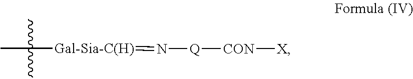
C00019

C00020

C00021
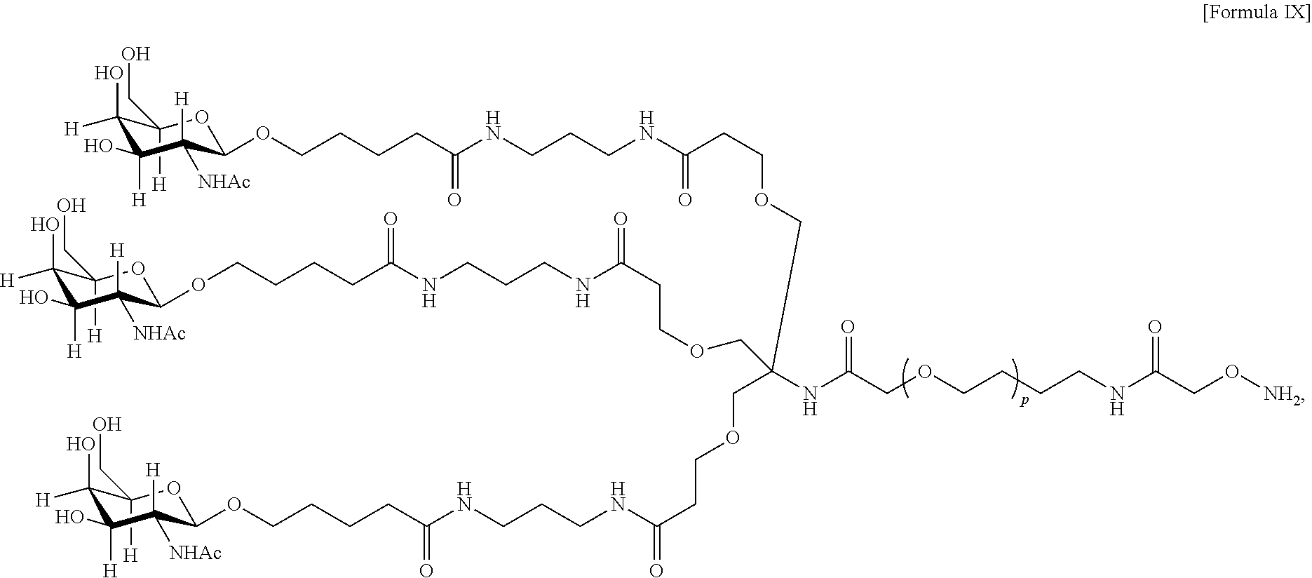
C00022
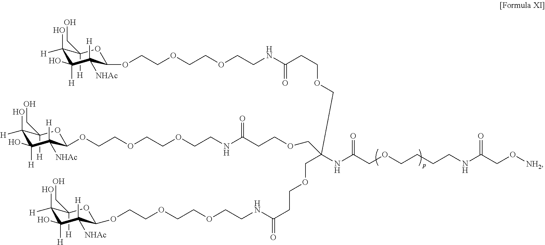
C00023

C00024

C00025

C00026
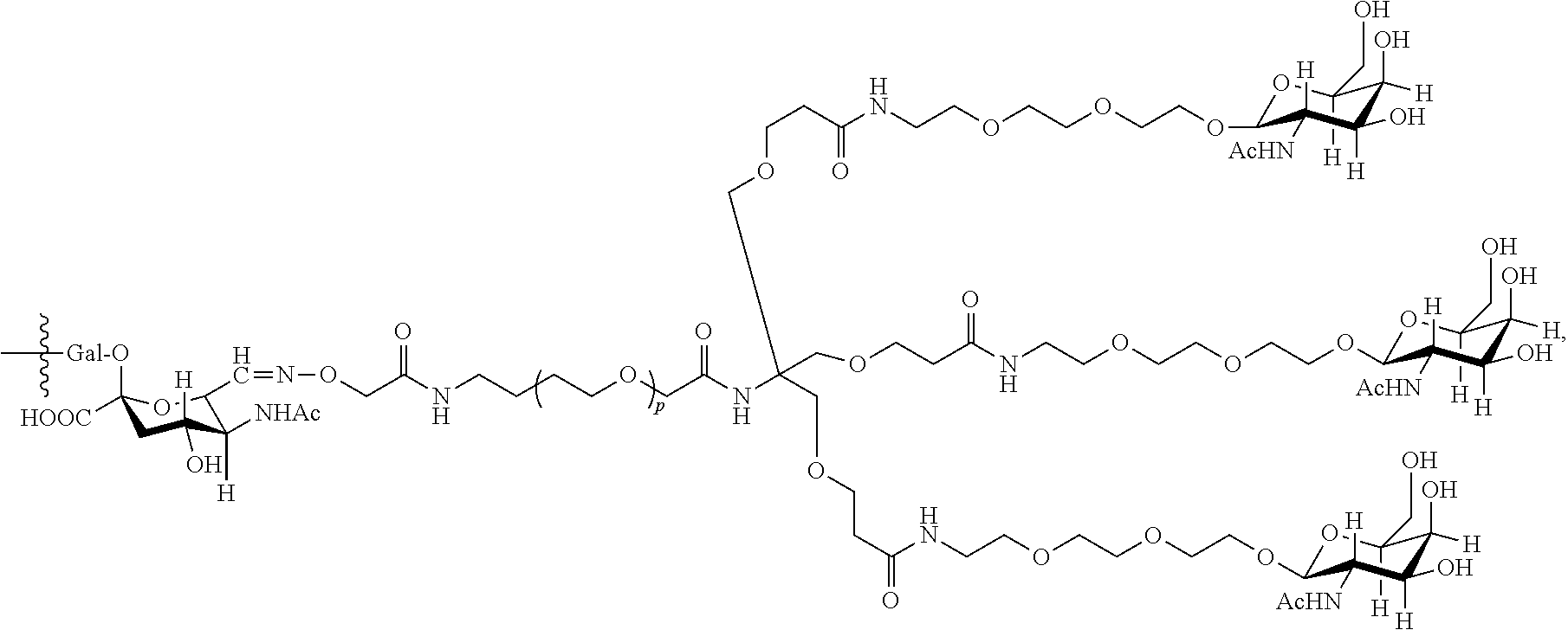
C00027
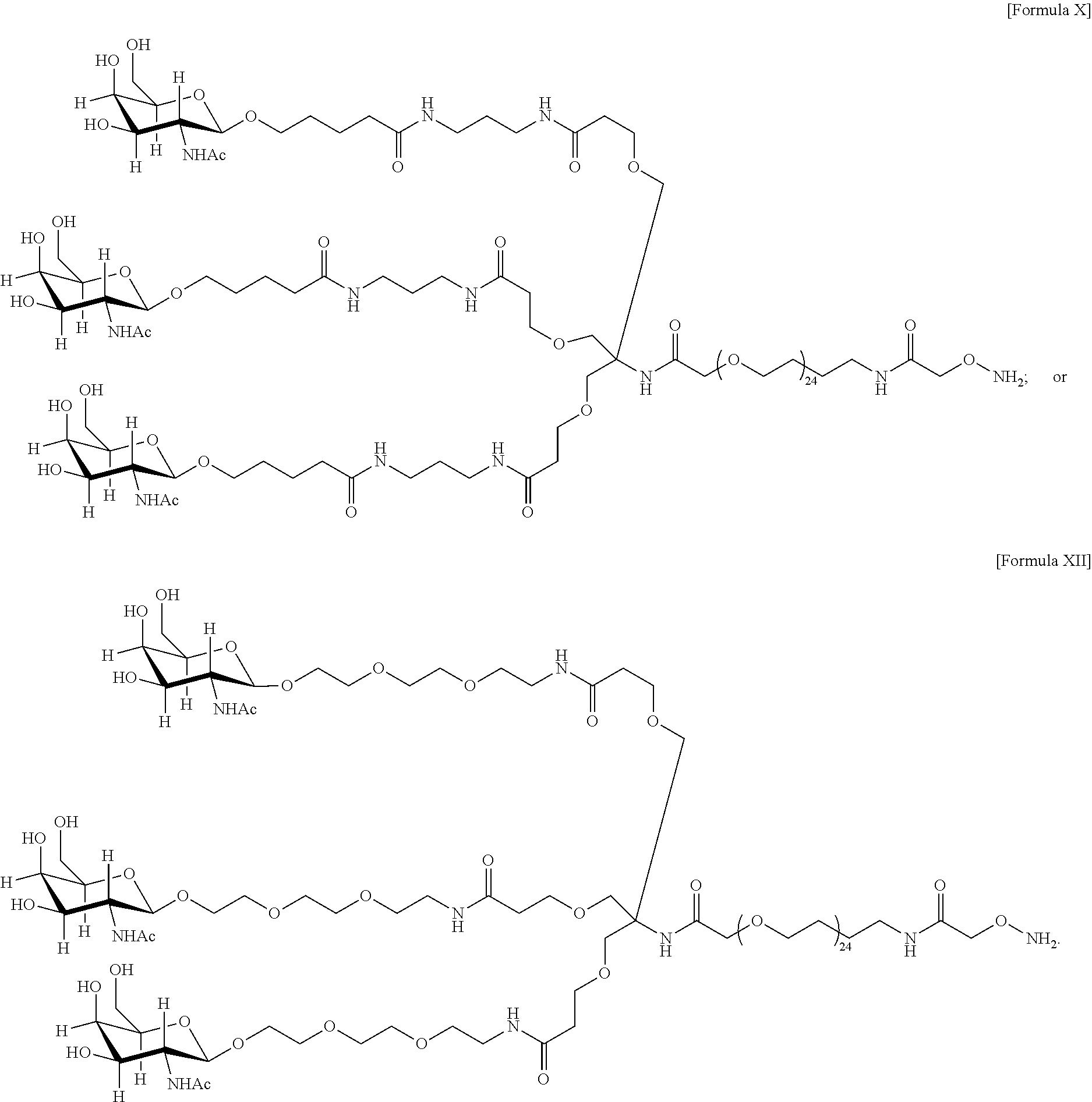
C00028
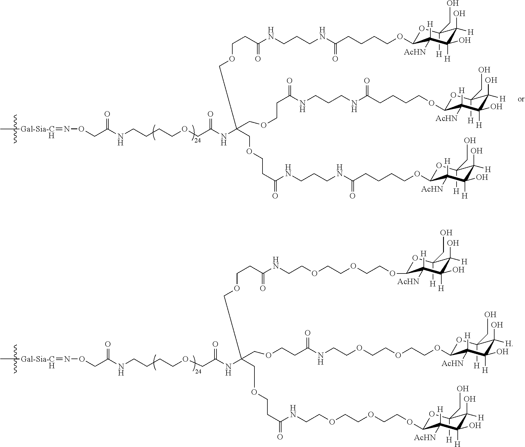
C00029

C00030
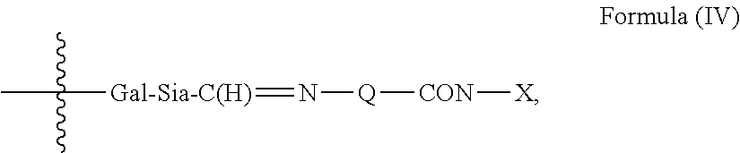
C00031

C00032
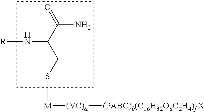
C00033
C00034

C00035

C00036

C00037

C00038
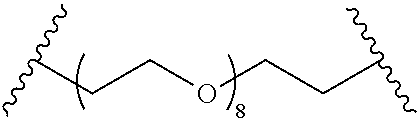
C00039
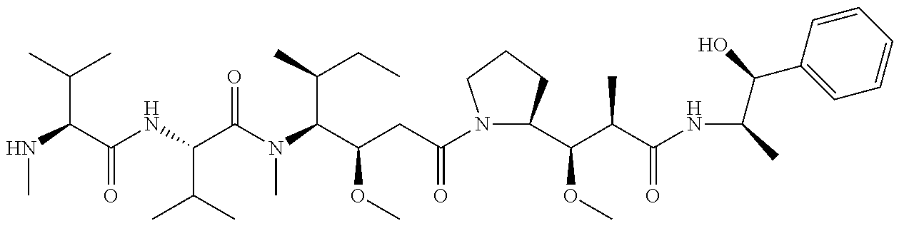
C00040
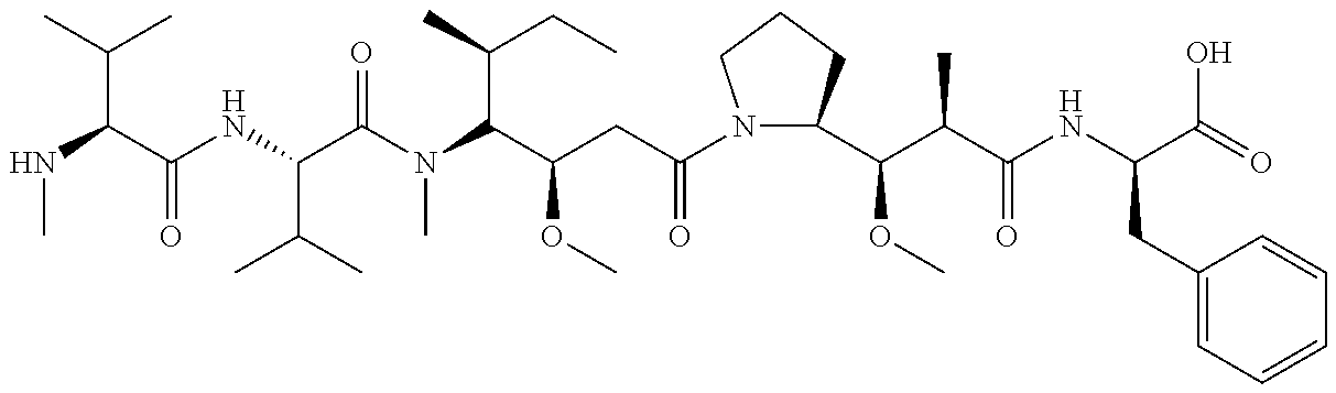
C00041

C00042
C00043

C00044
C00045

C00046

C00047

C00048
C00049

C00050

C00051
C00052

C00053

C00054

C00055
C00056

C00057
C00058

C00059

C00060

C00061

C00062

C00063
C00064
C00065
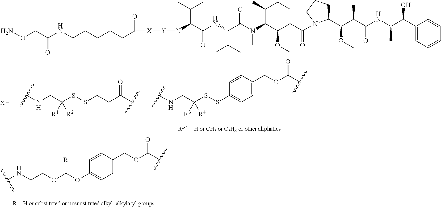
C00066

C00067

C00068
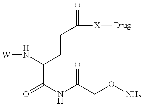
C00069
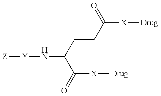
C00070

C00071

C00072

C00073
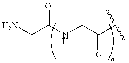
C00074

C00075

C00076
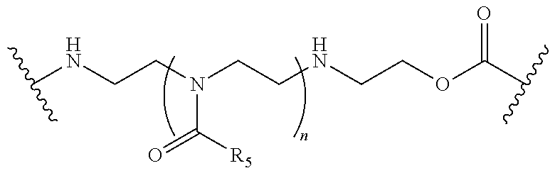
C00077

C00078
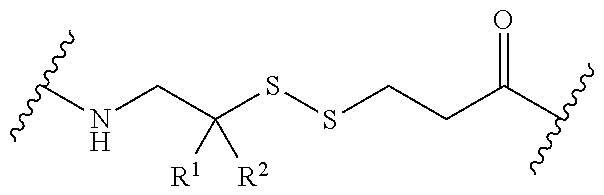
C00079

C00080

C00081

C00082
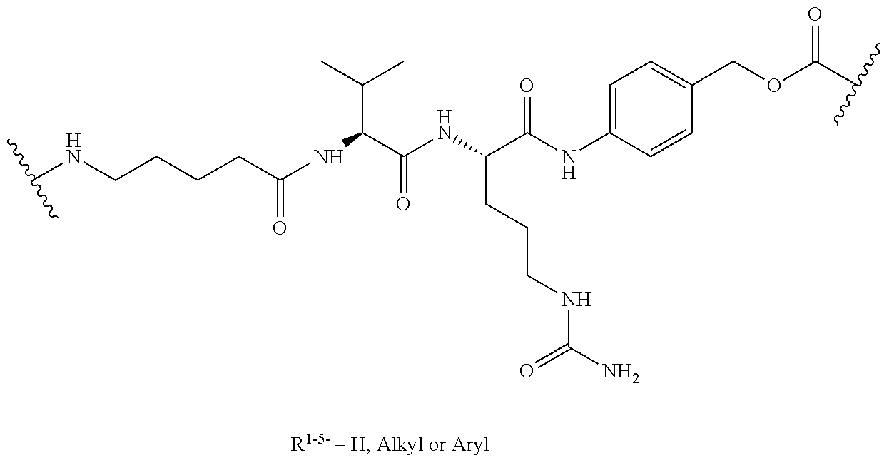
C00083

C00084

C00085
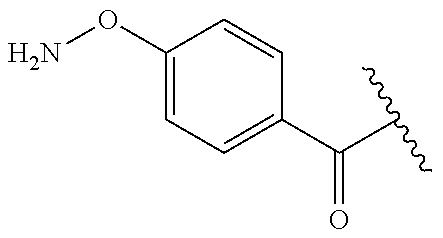
C00086
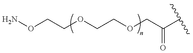
C00087
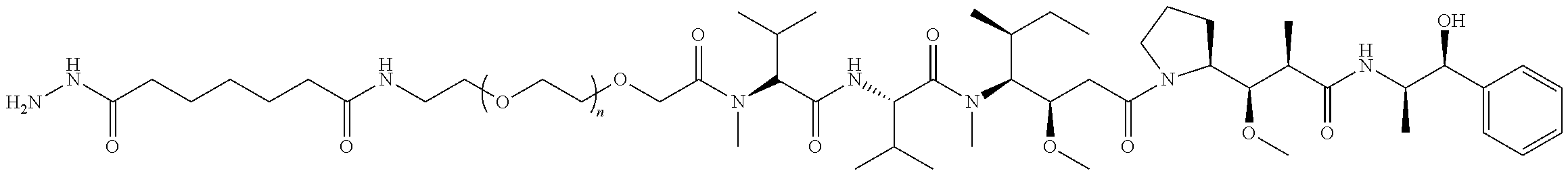
C00088

C00089

C00090
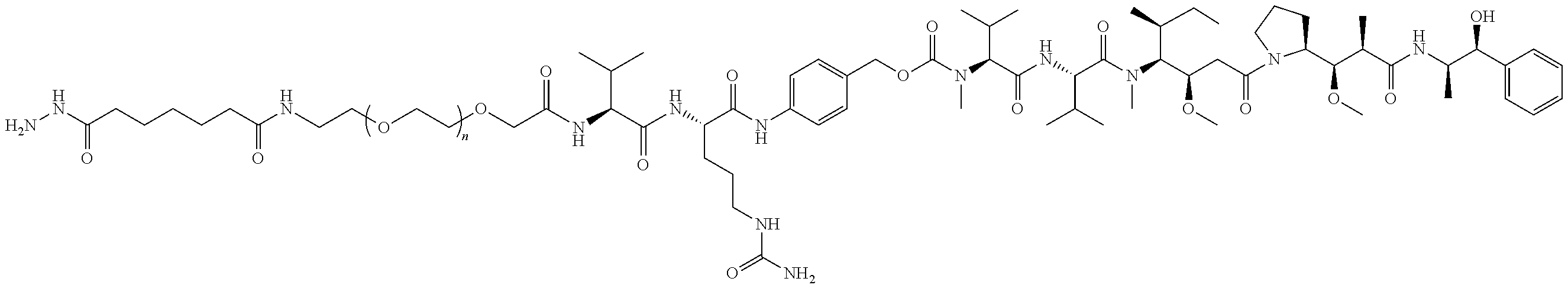
C00091
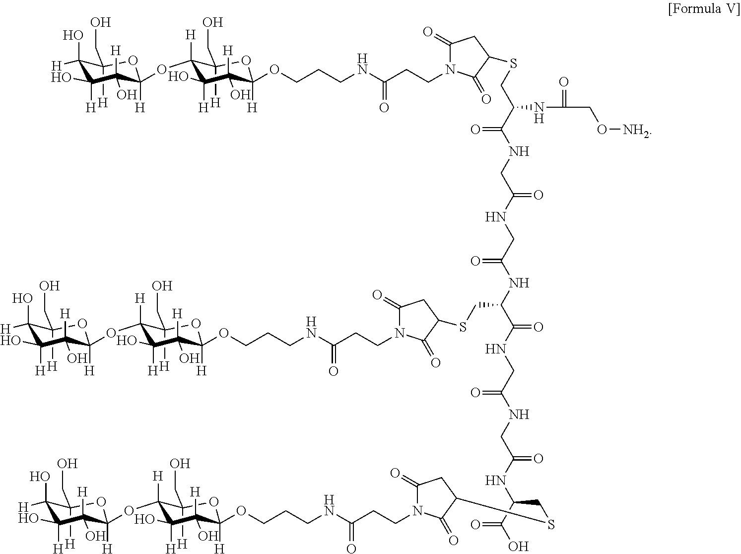
C00092
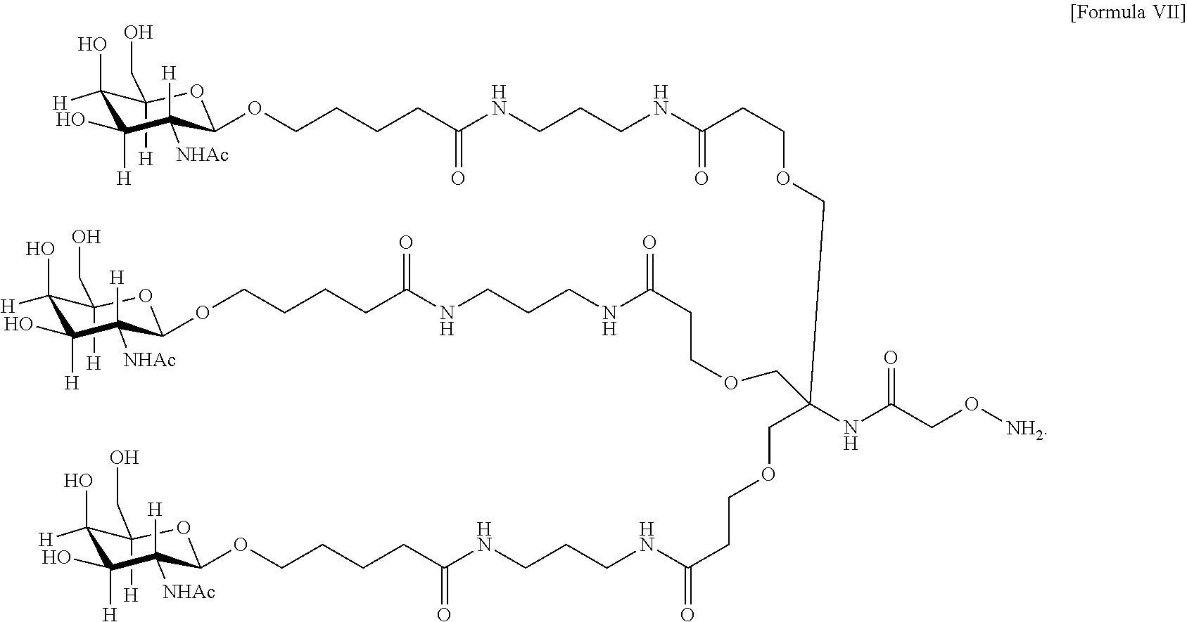
C00093

C00094
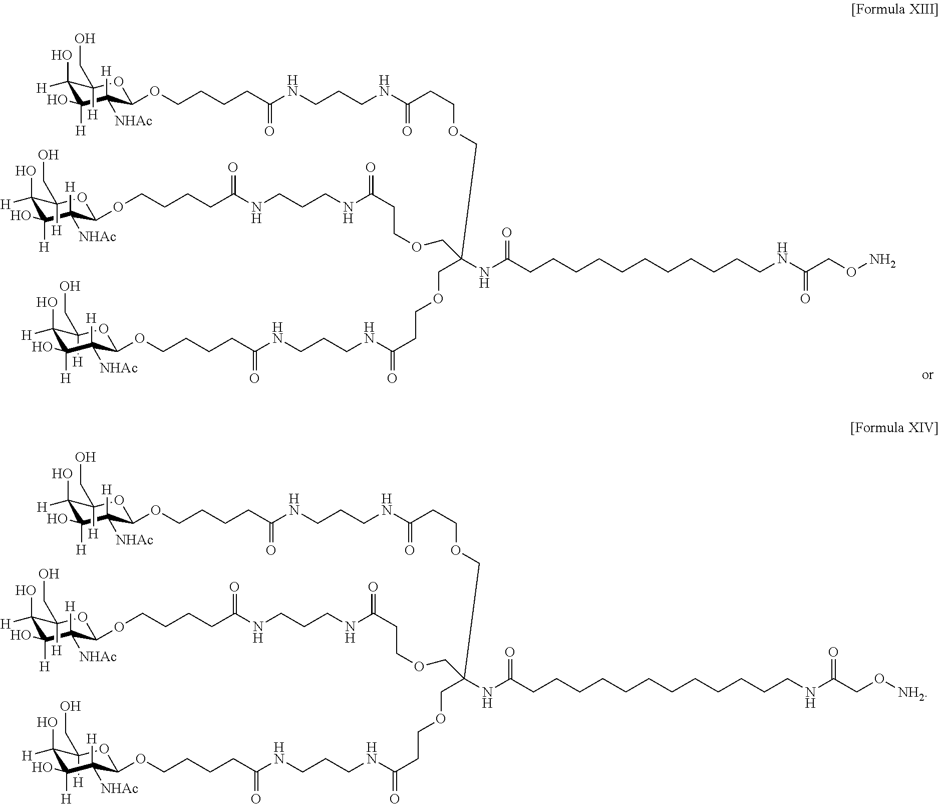
C00095
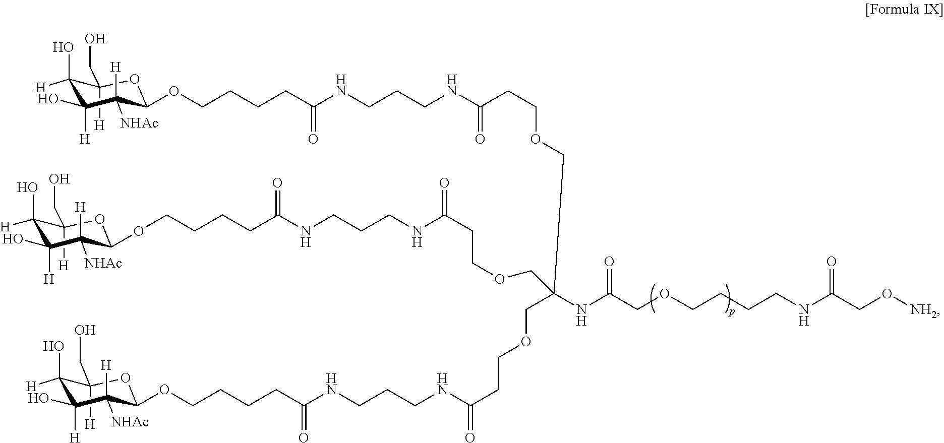
C00096

C00097

C00098
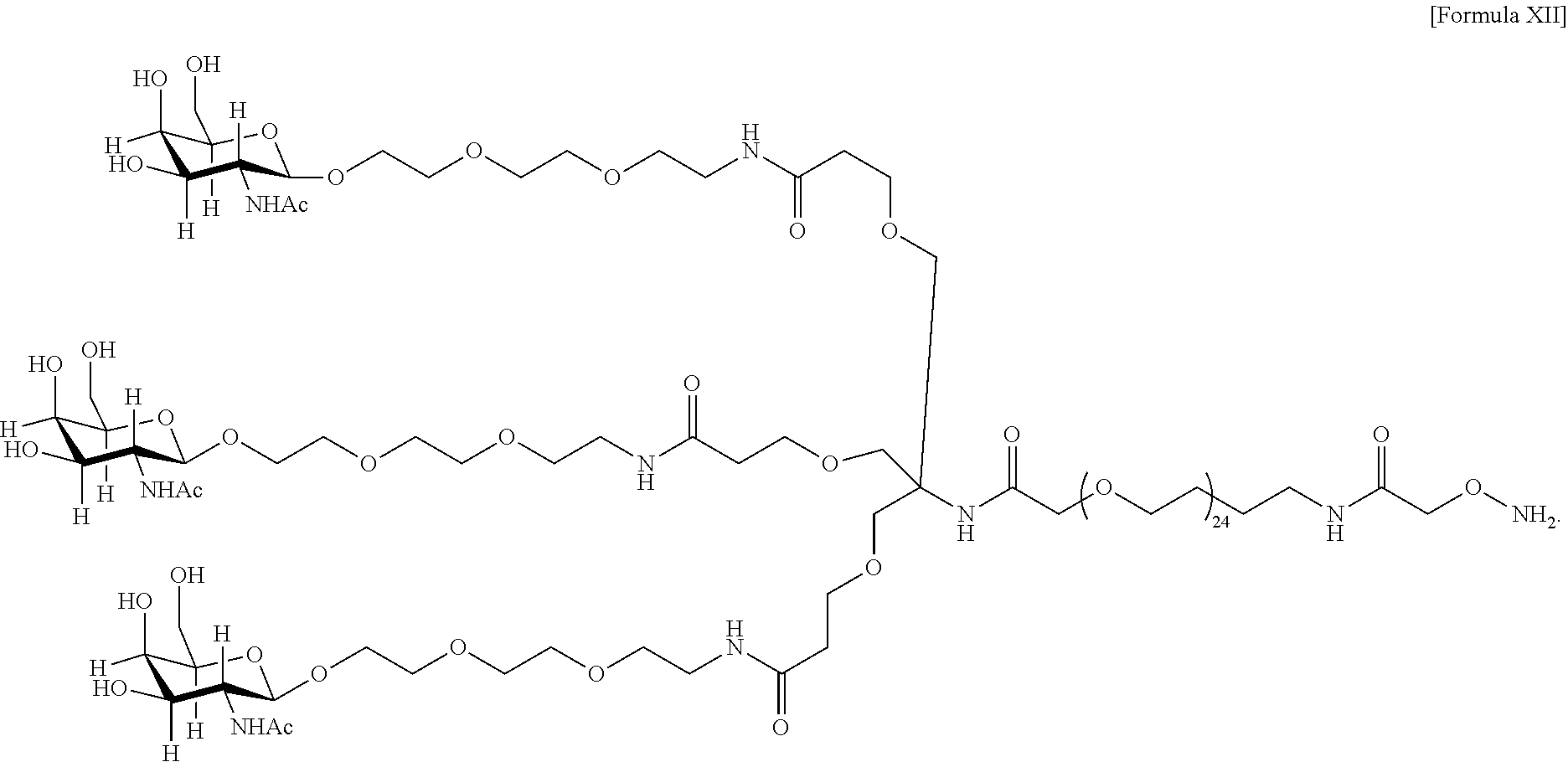
C00099

C00100

C00101
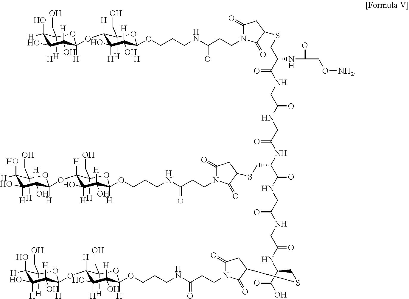
C00102

C00103
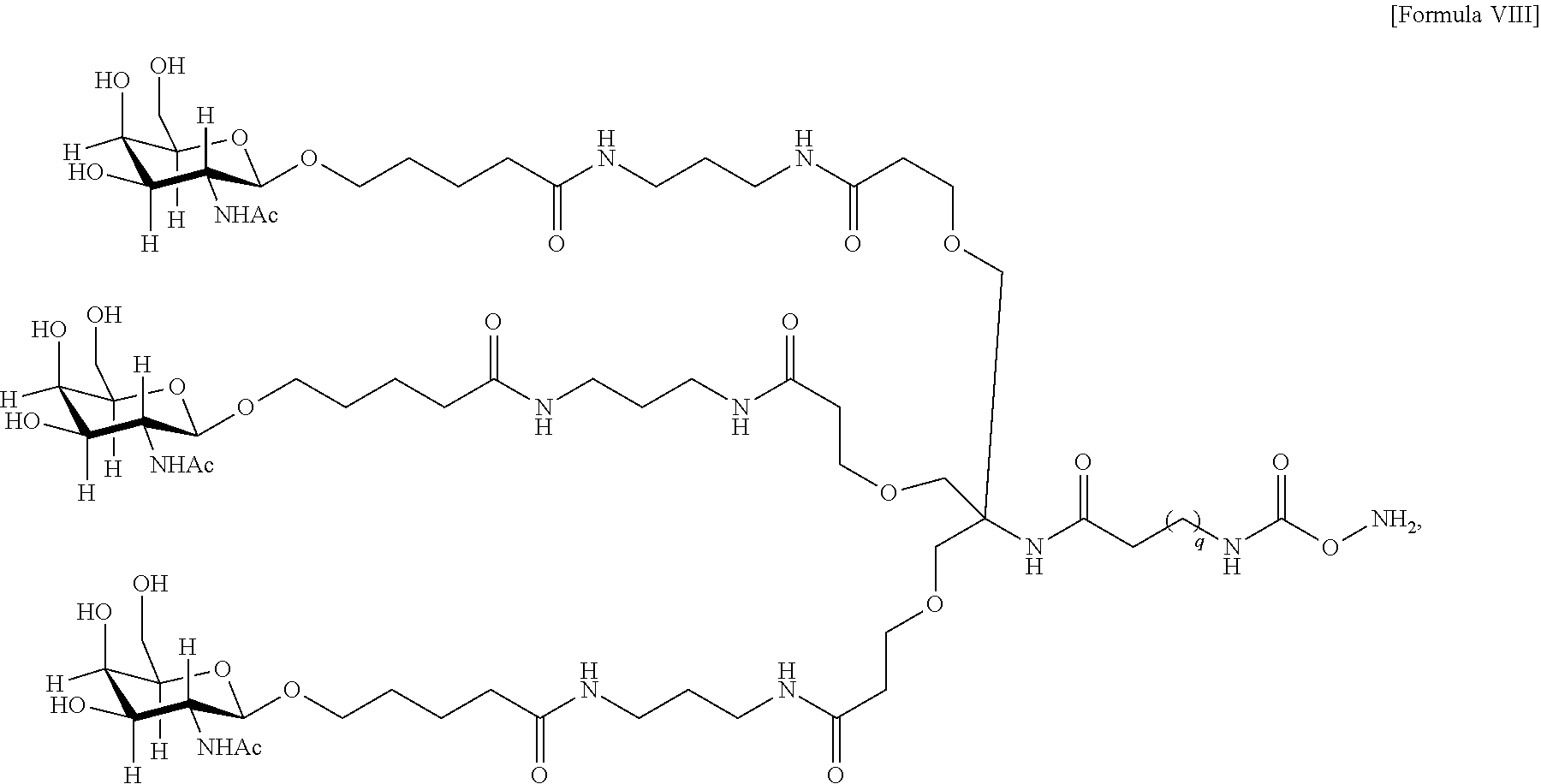
C00104
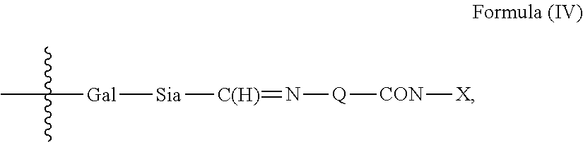
C00105

C00106

C00107
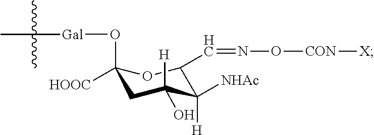
C00108

C00109
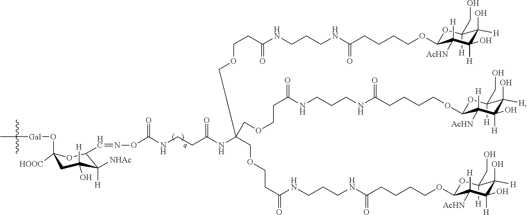
C00110

C00111

C00112

C00113

C00114
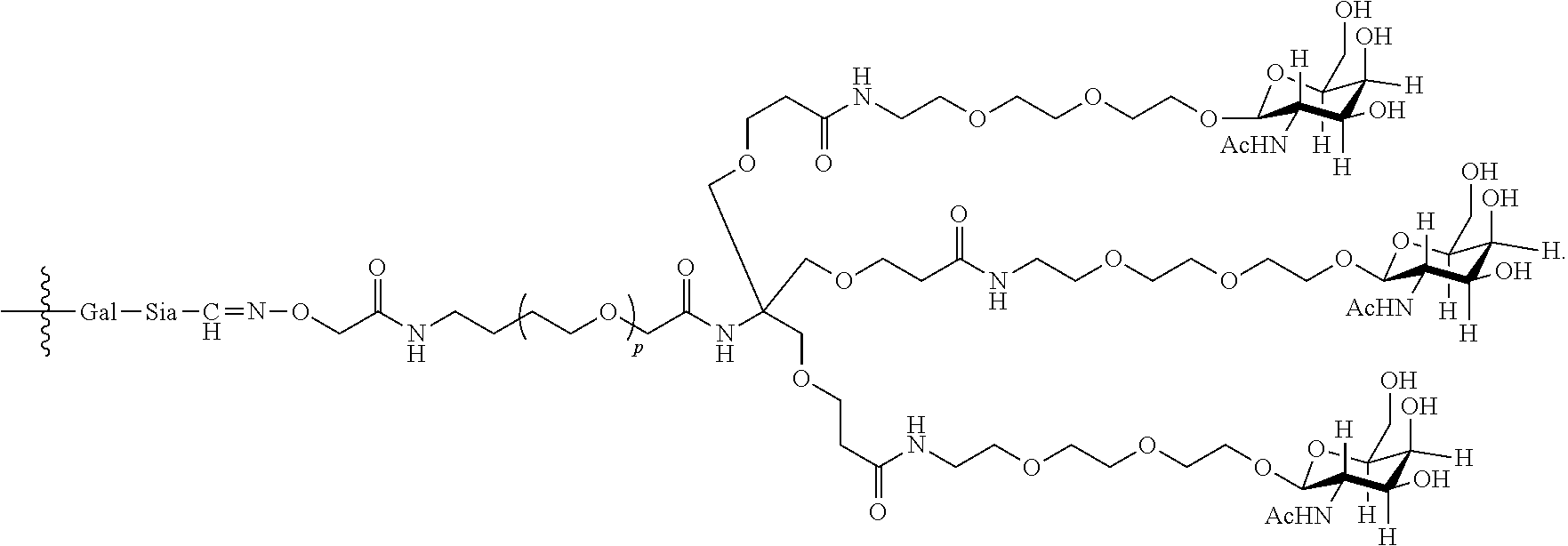
C00115
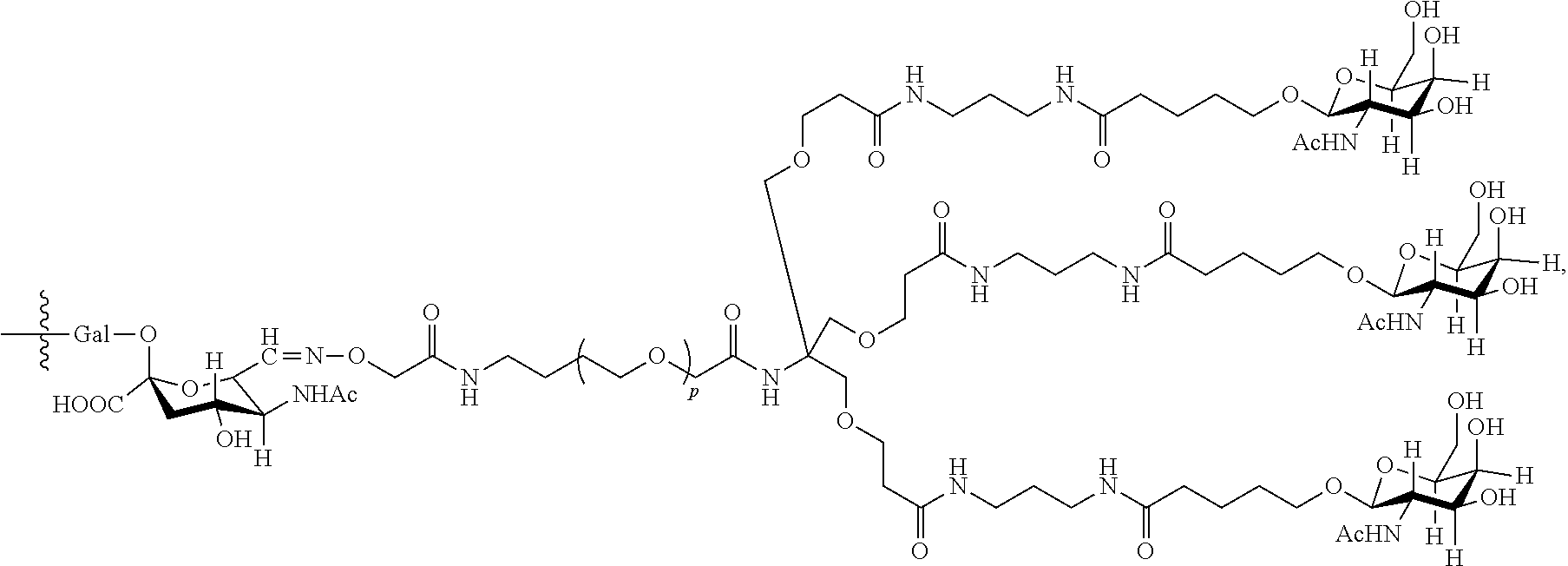
C00116

C00117
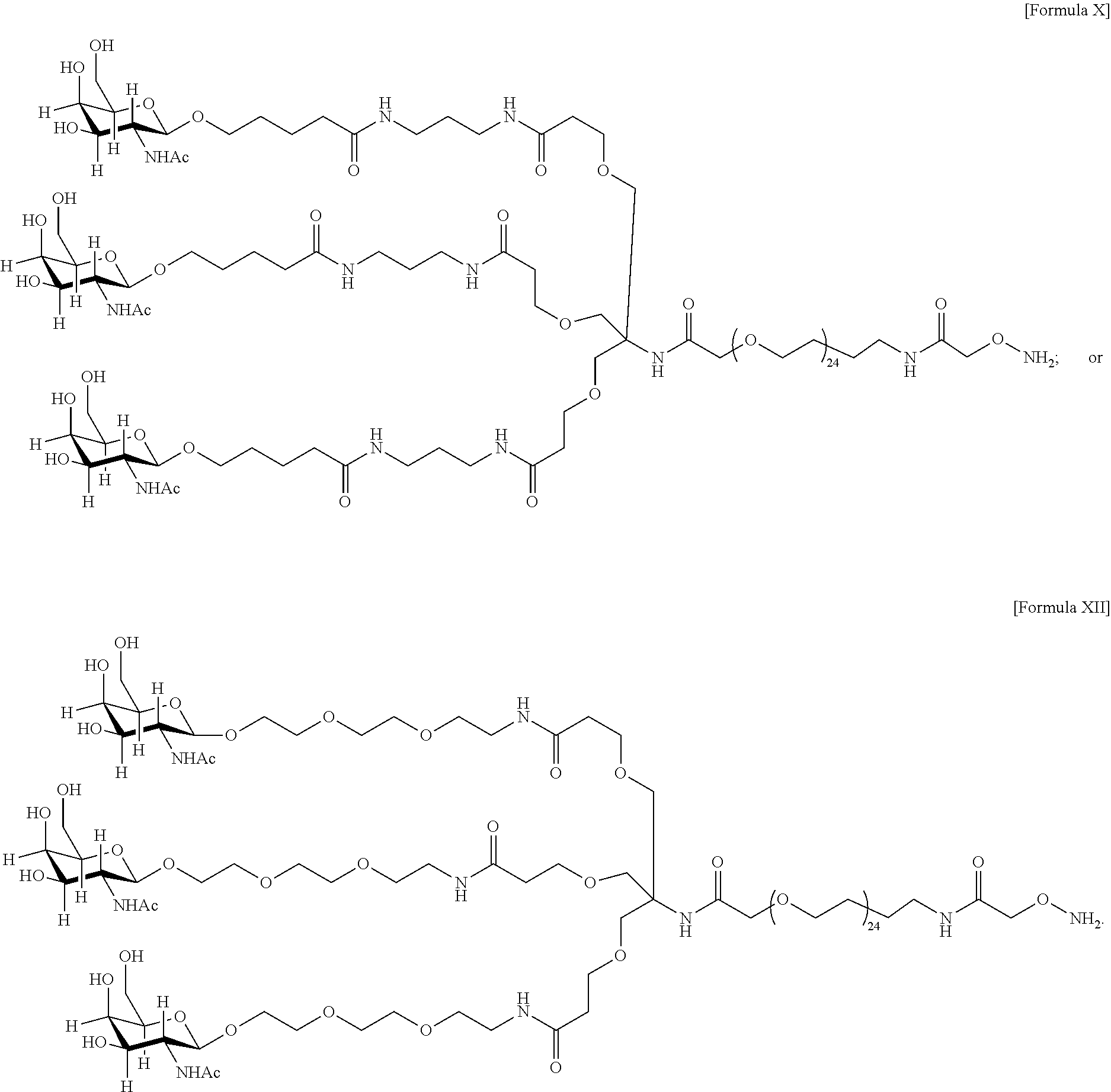
C00118
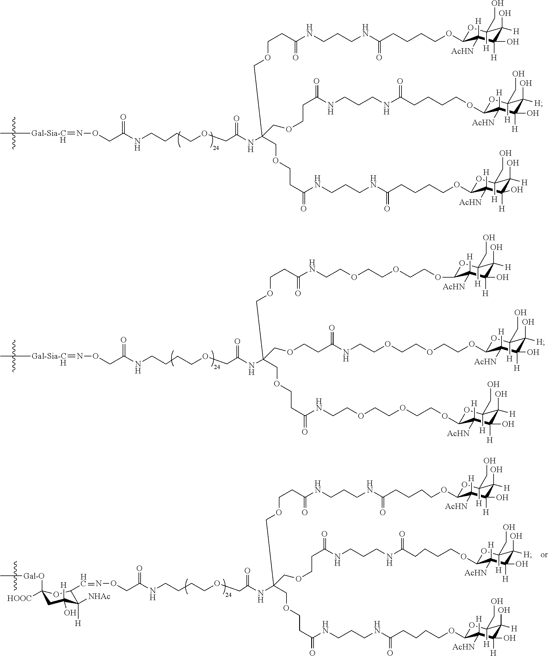
C00119

D00000
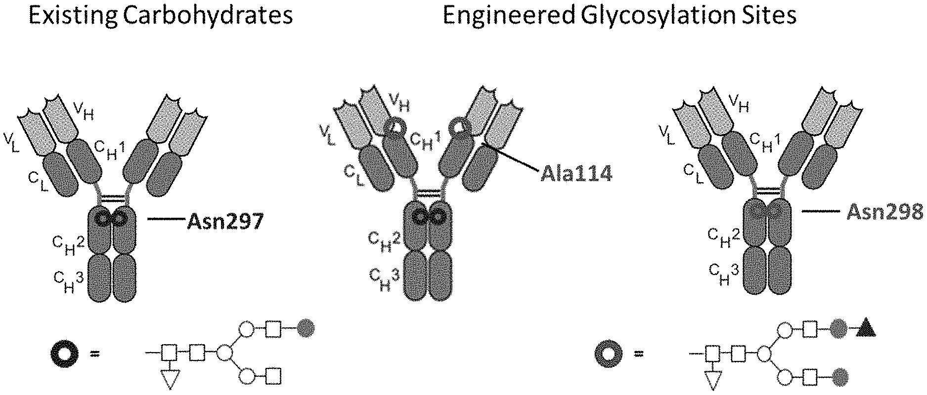
D00001
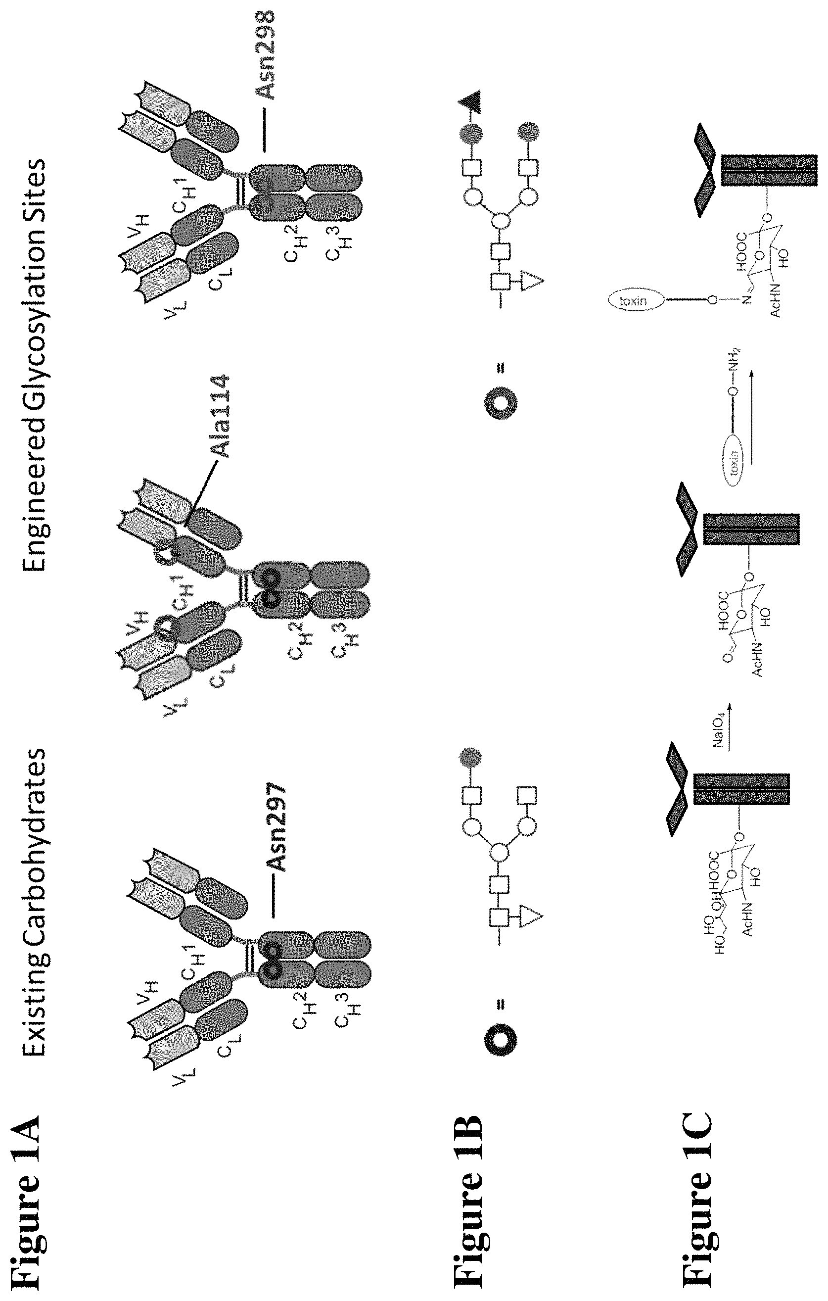
D00002
D00003
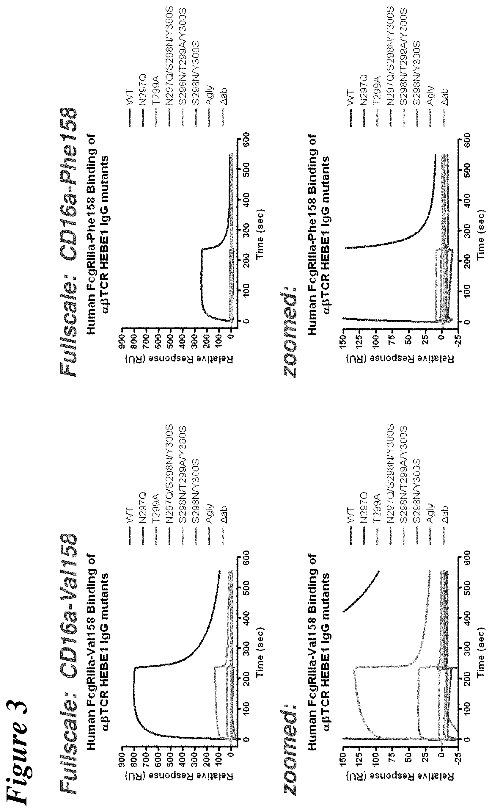
D00004
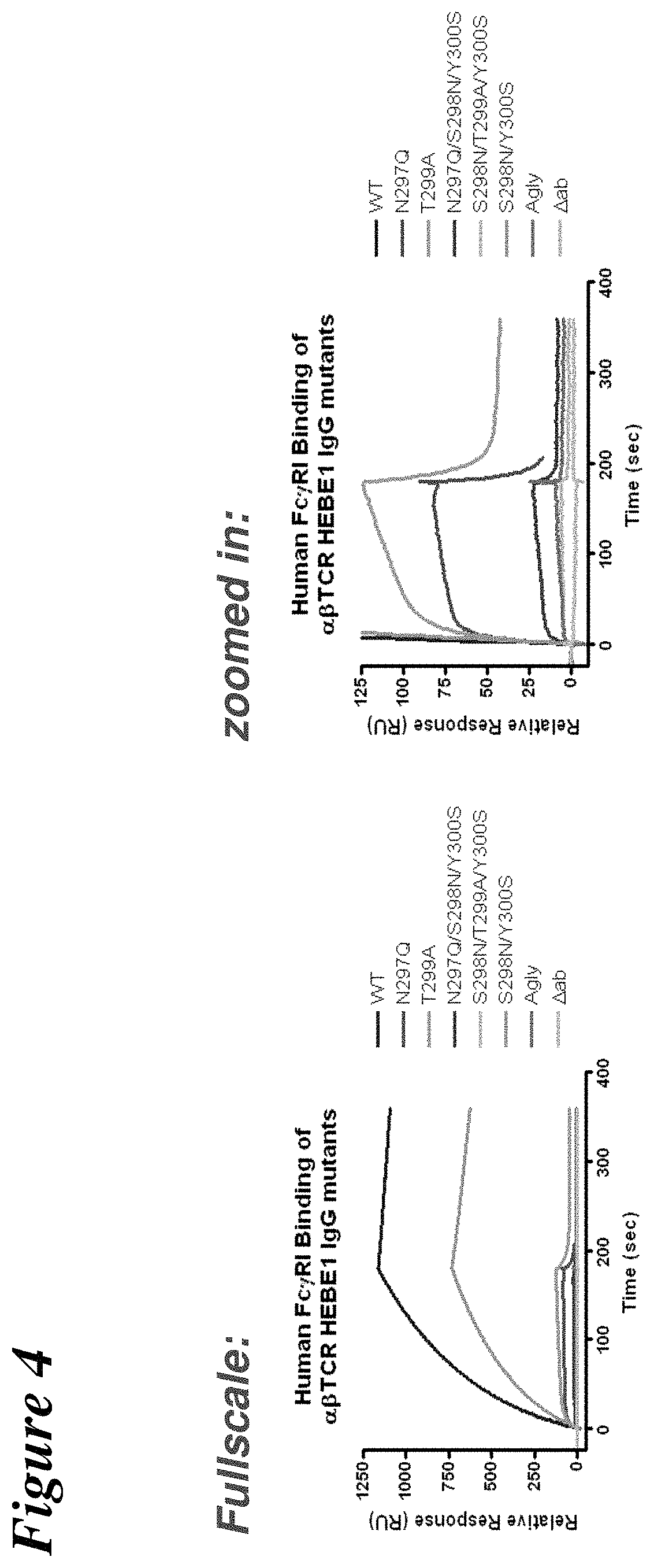
D00005
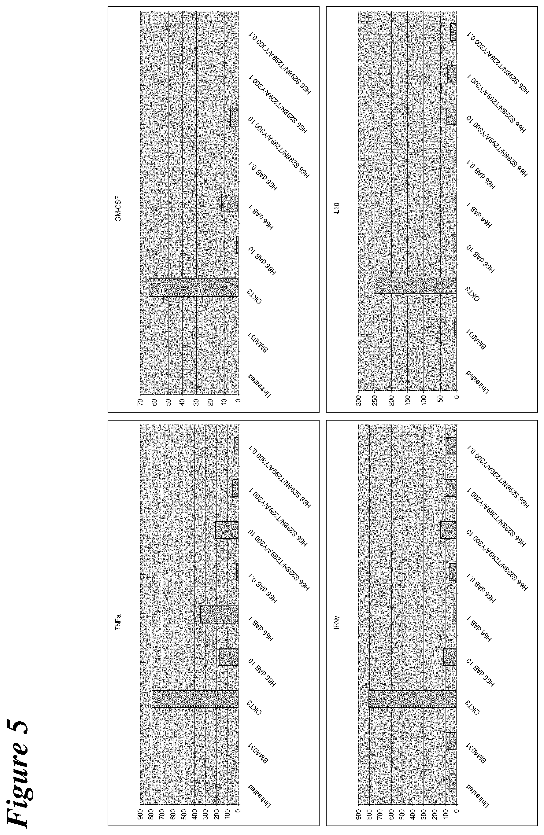
D00006

D00007
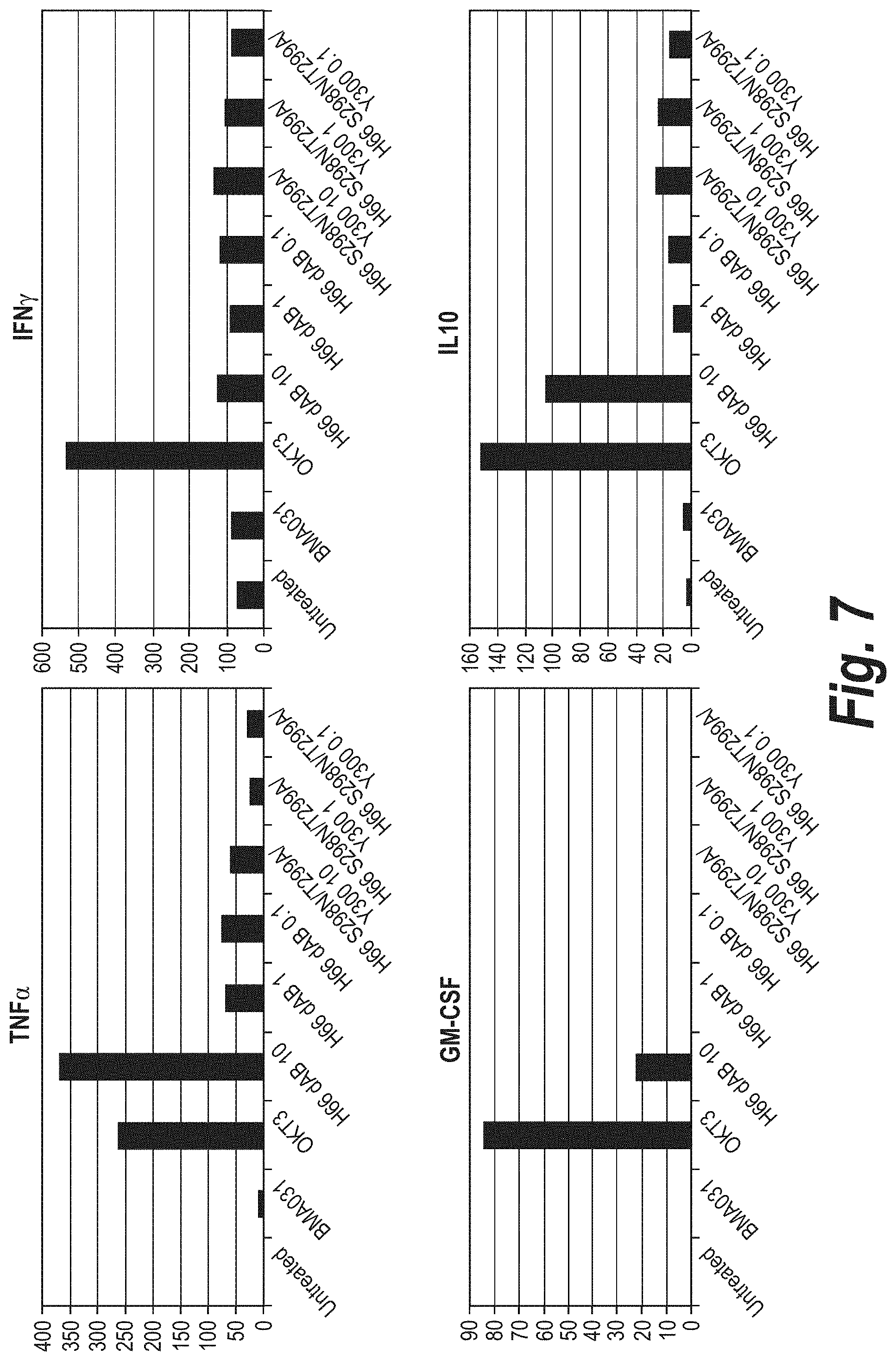
D00008
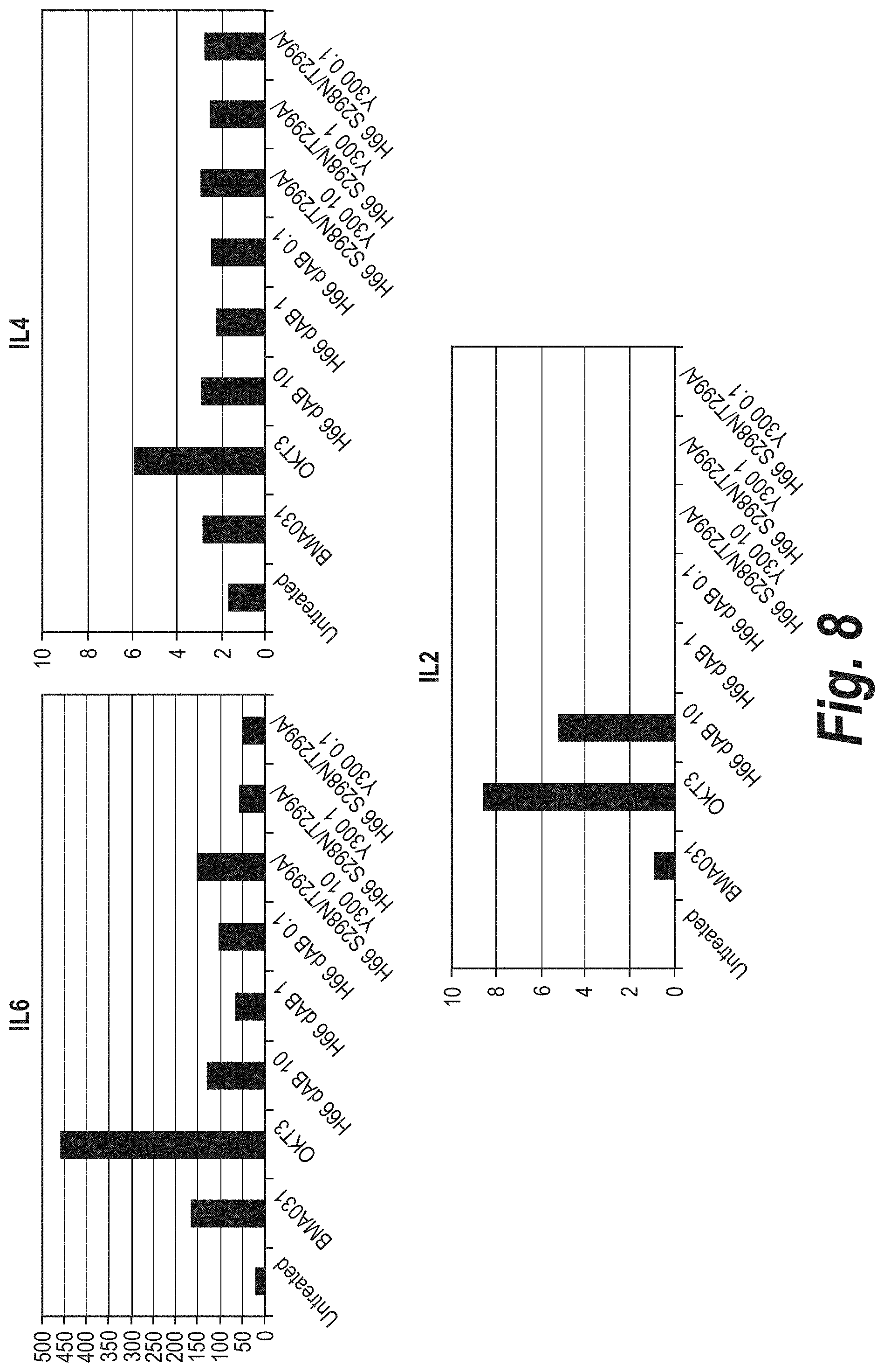
D00009
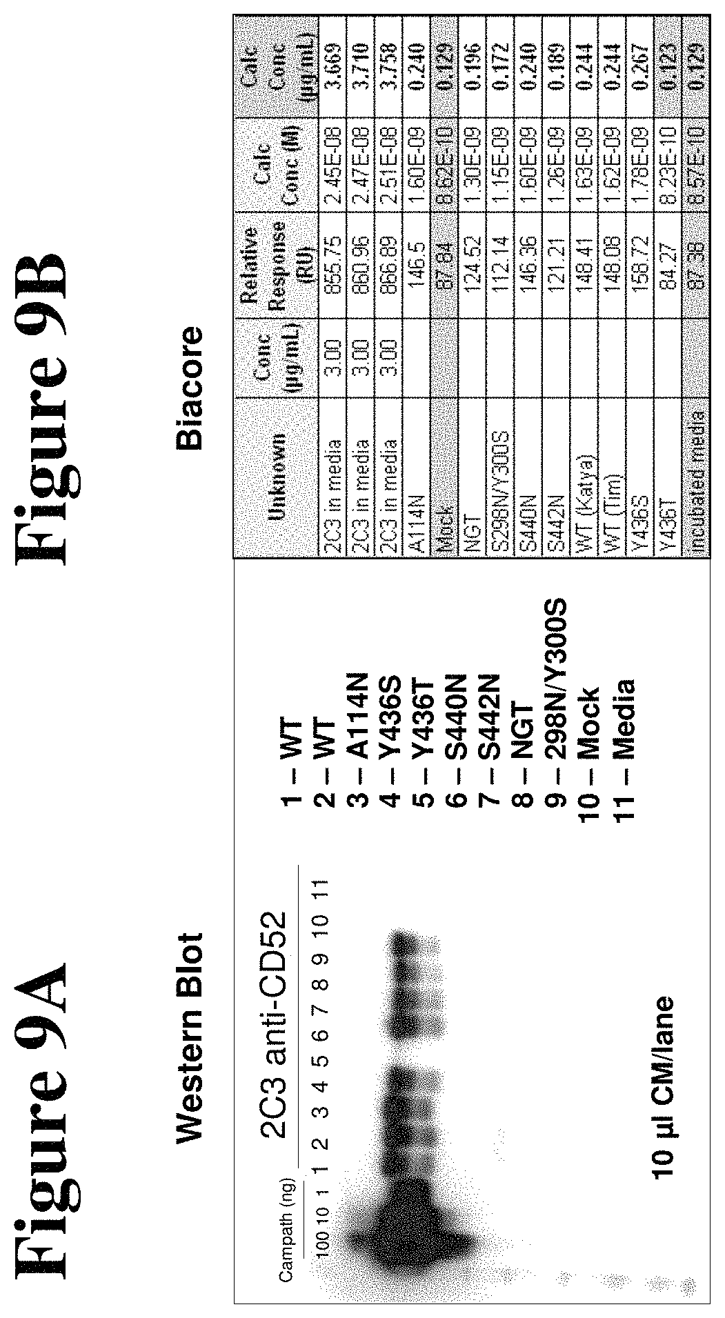
D00010

D00011

D00012

D00013

D00014

D00015
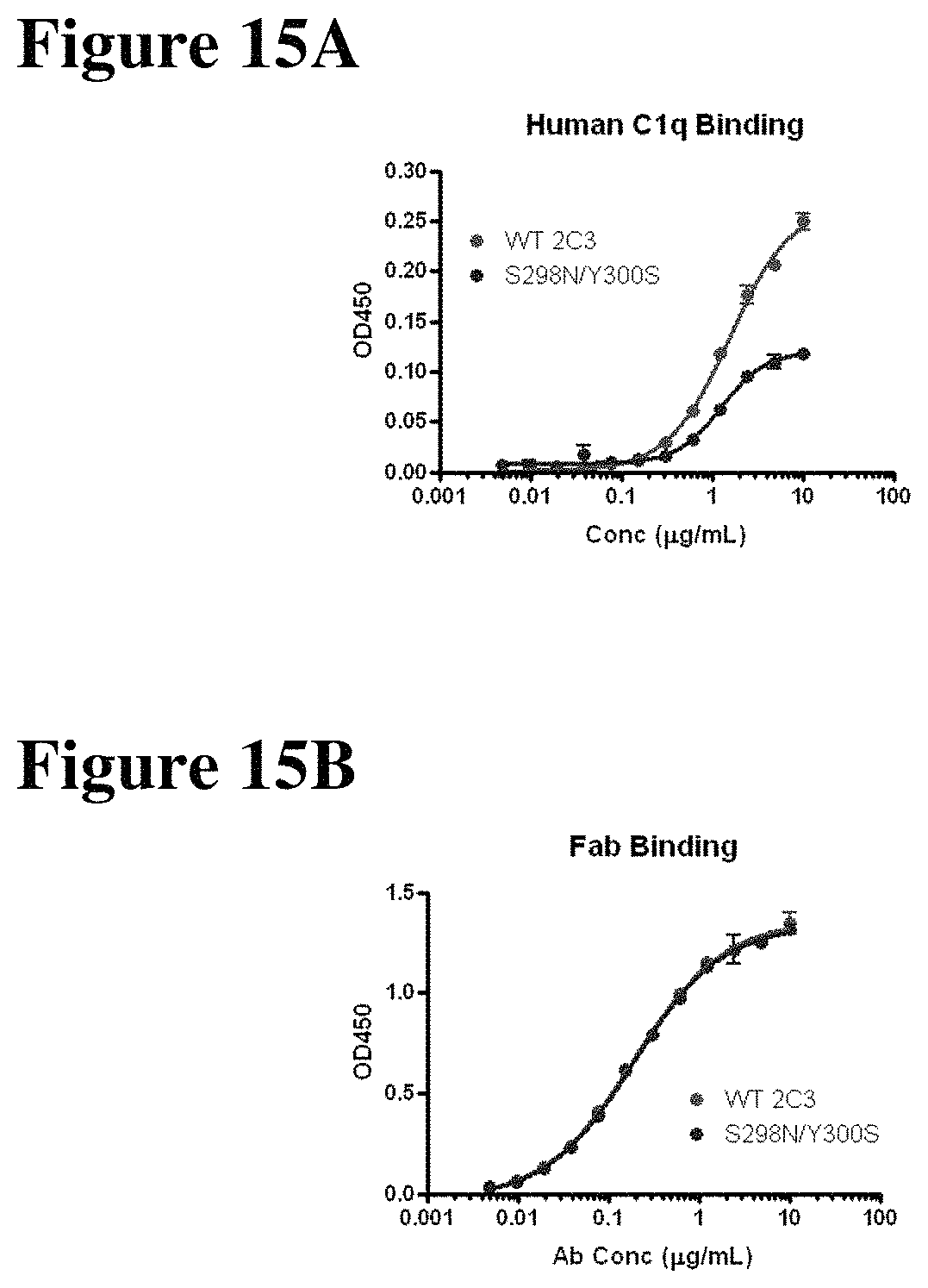
D00016
D00017
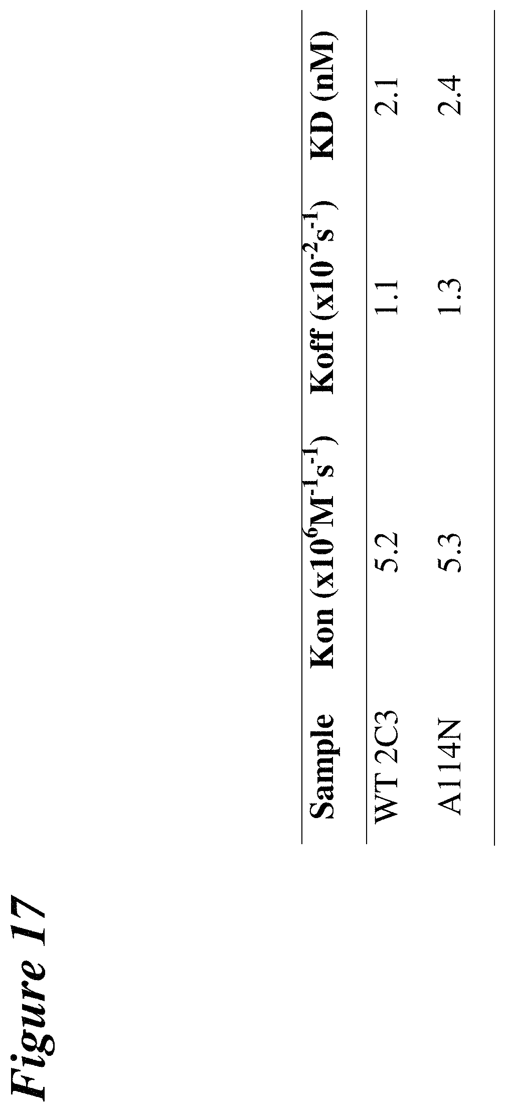
D00018
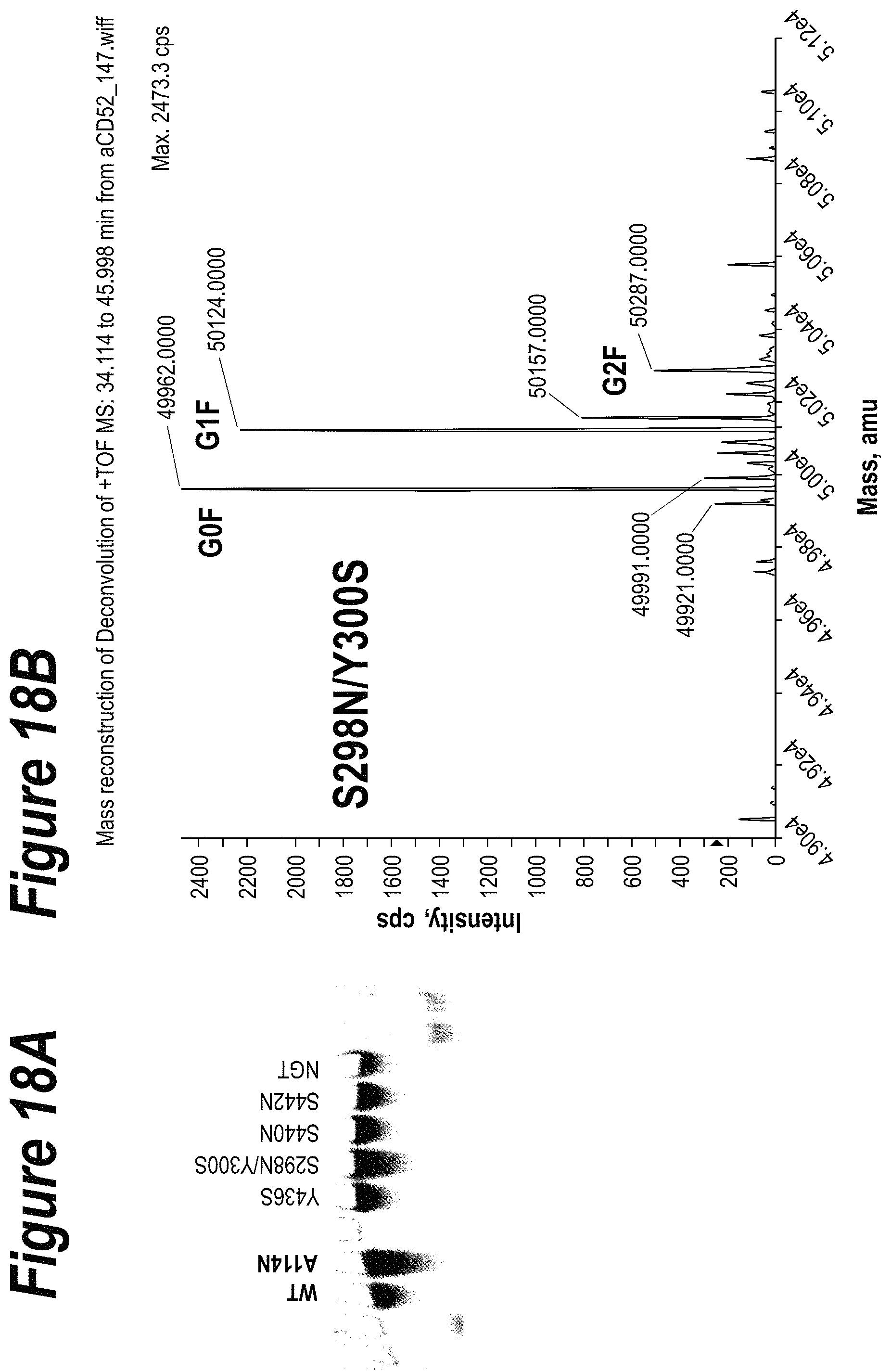
D00019
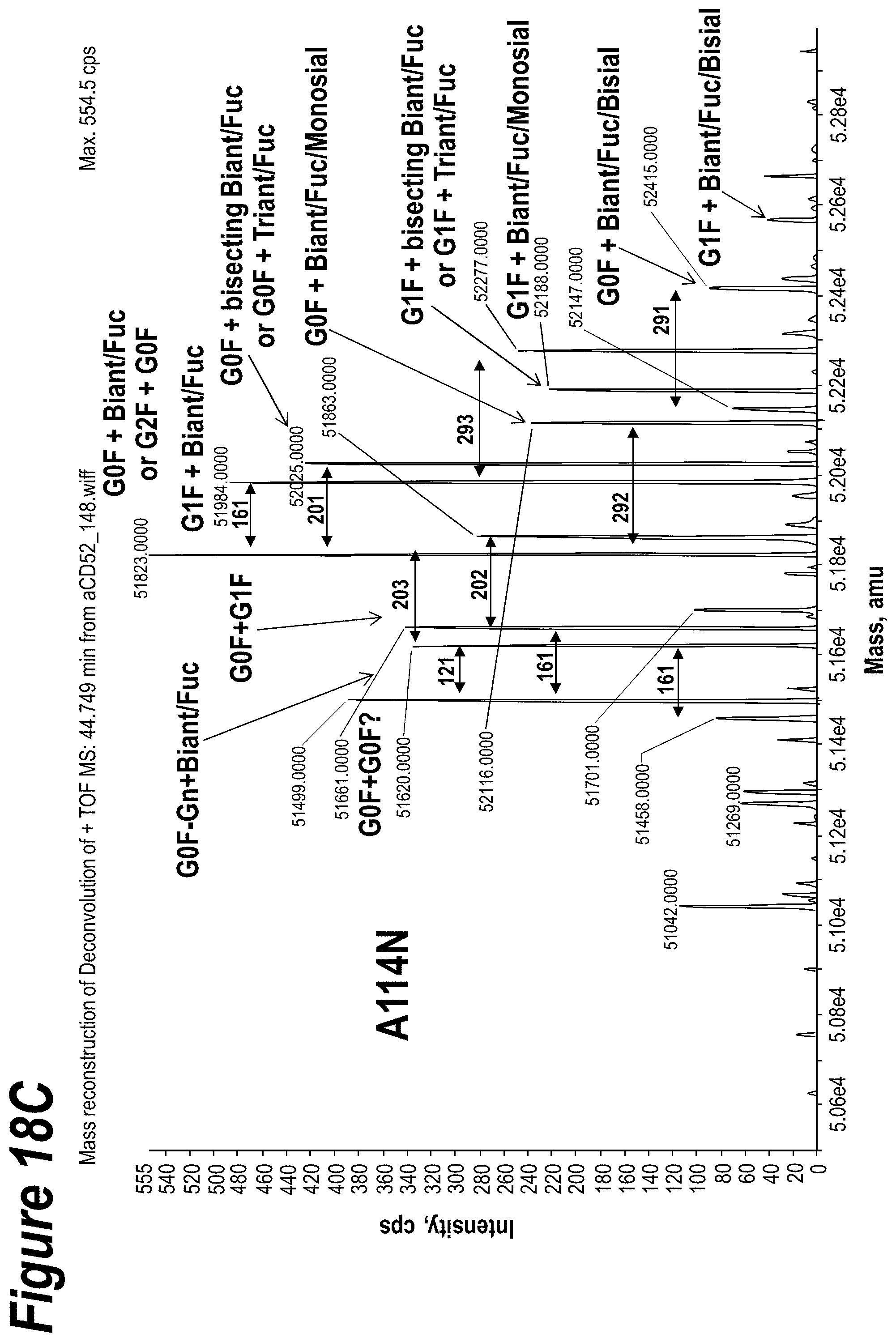
D00020

D00021
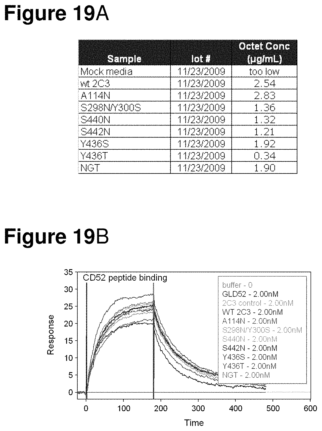
D00022
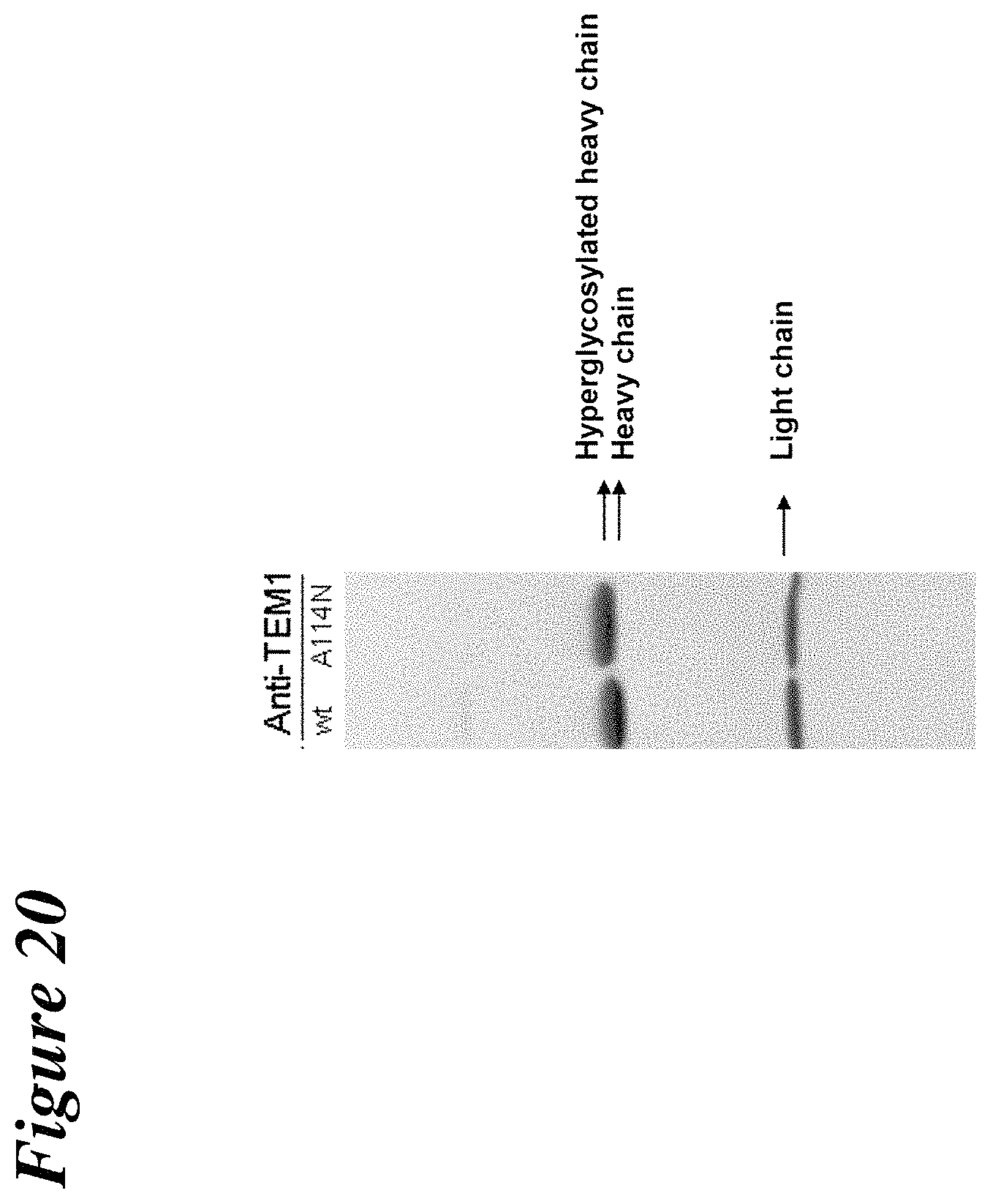
D00023
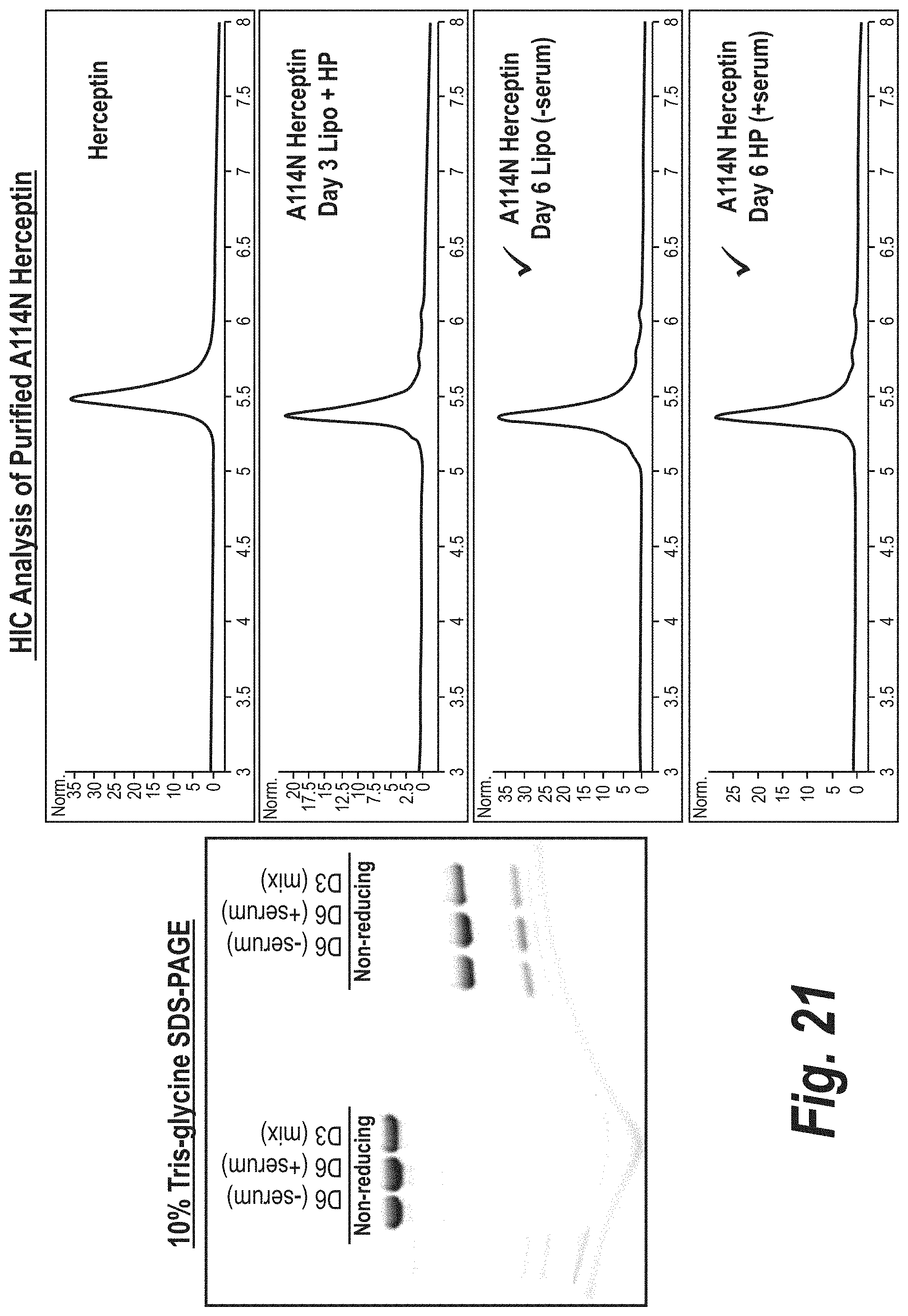
D00024
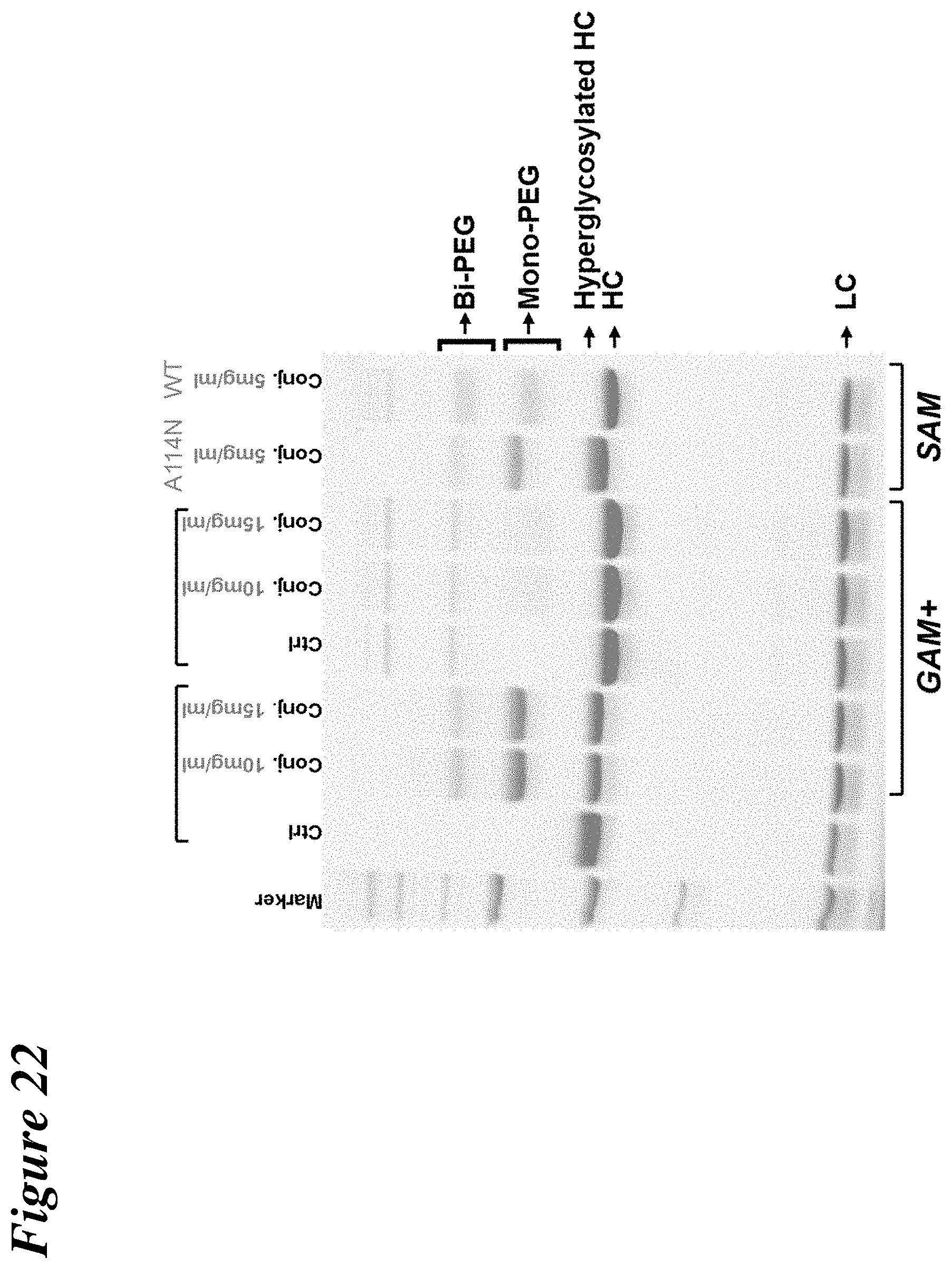
D00025
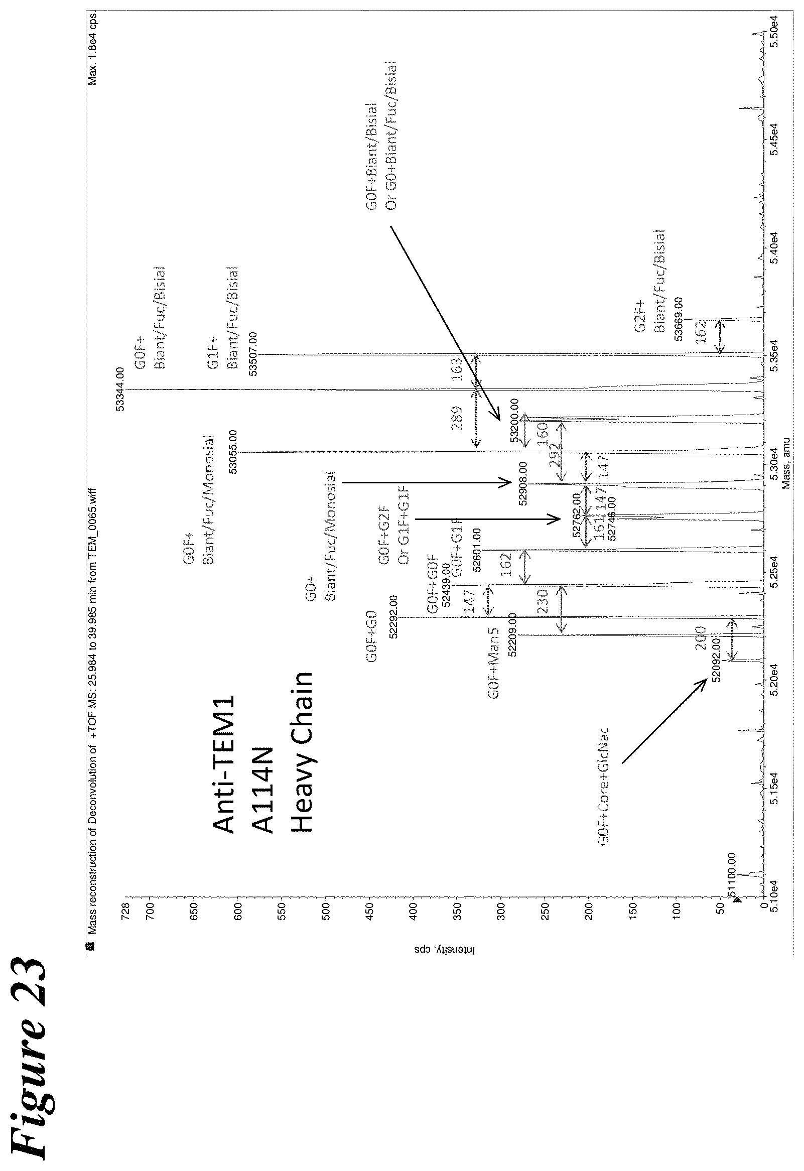
D00026

D00027
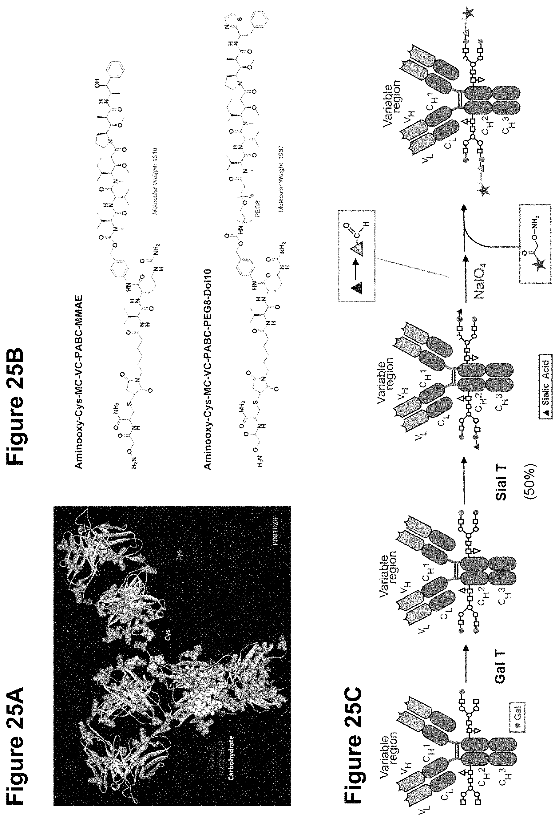
D00028

D00029

D00030

D00031
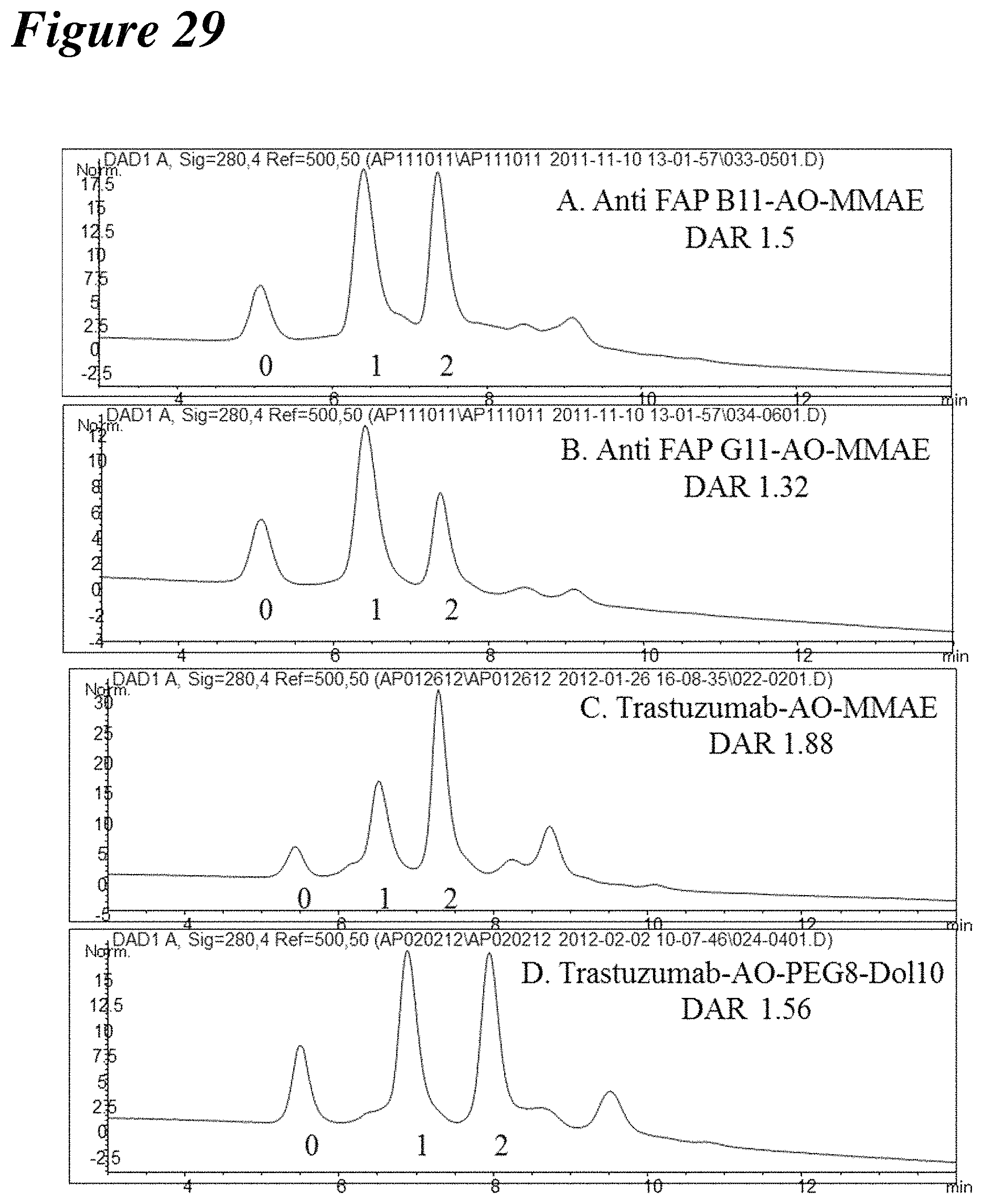
D00032

D00033
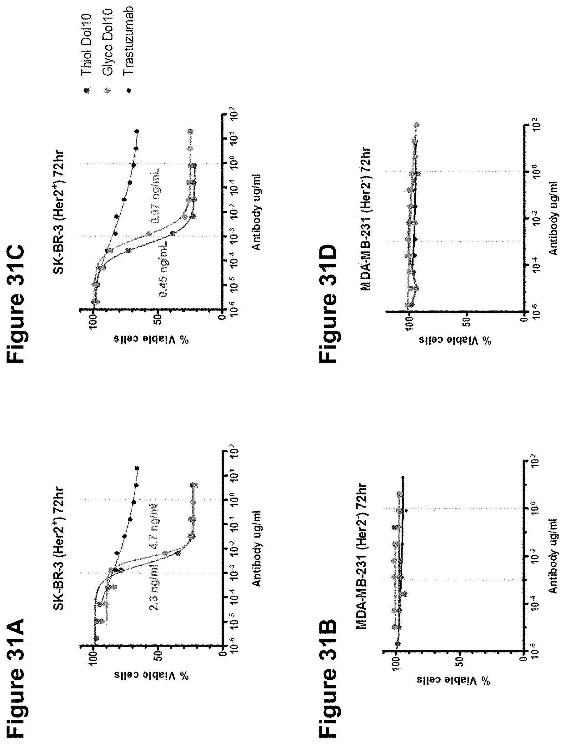
D00034

D00035
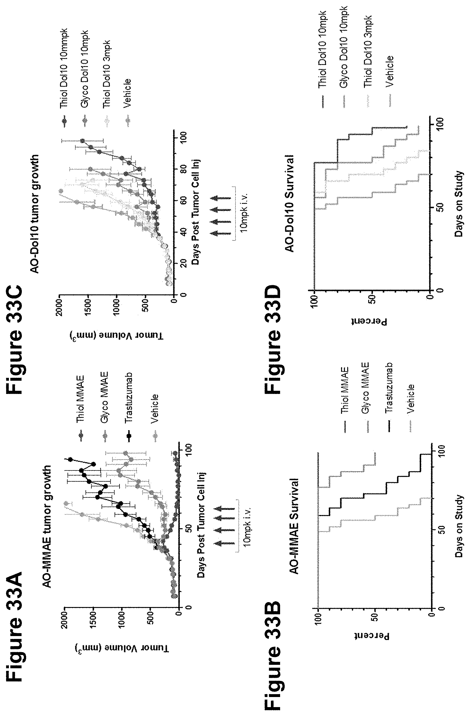
D00036
D00037
D00038

D00039
D00040

D00041
D00042

D00043
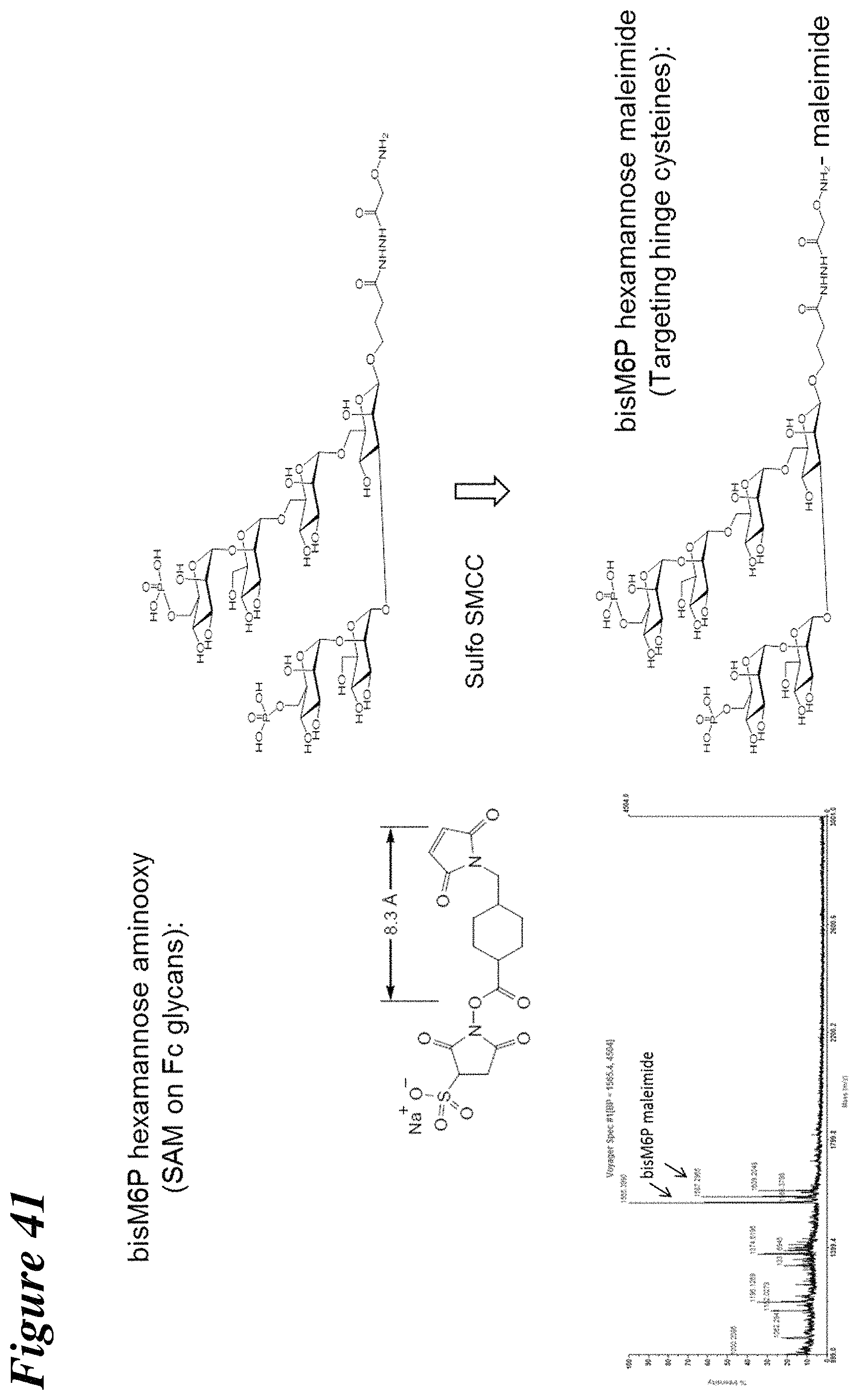
D00044
D00045

D00046
D00047

D00048

D00049
D00050
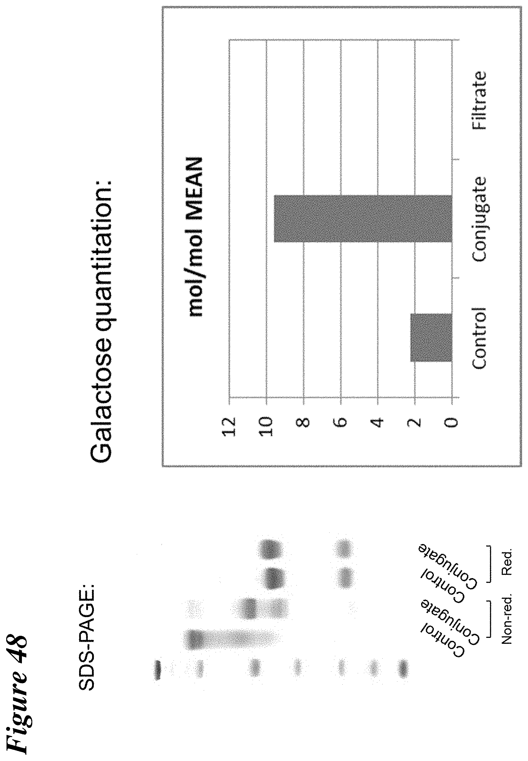
D00051
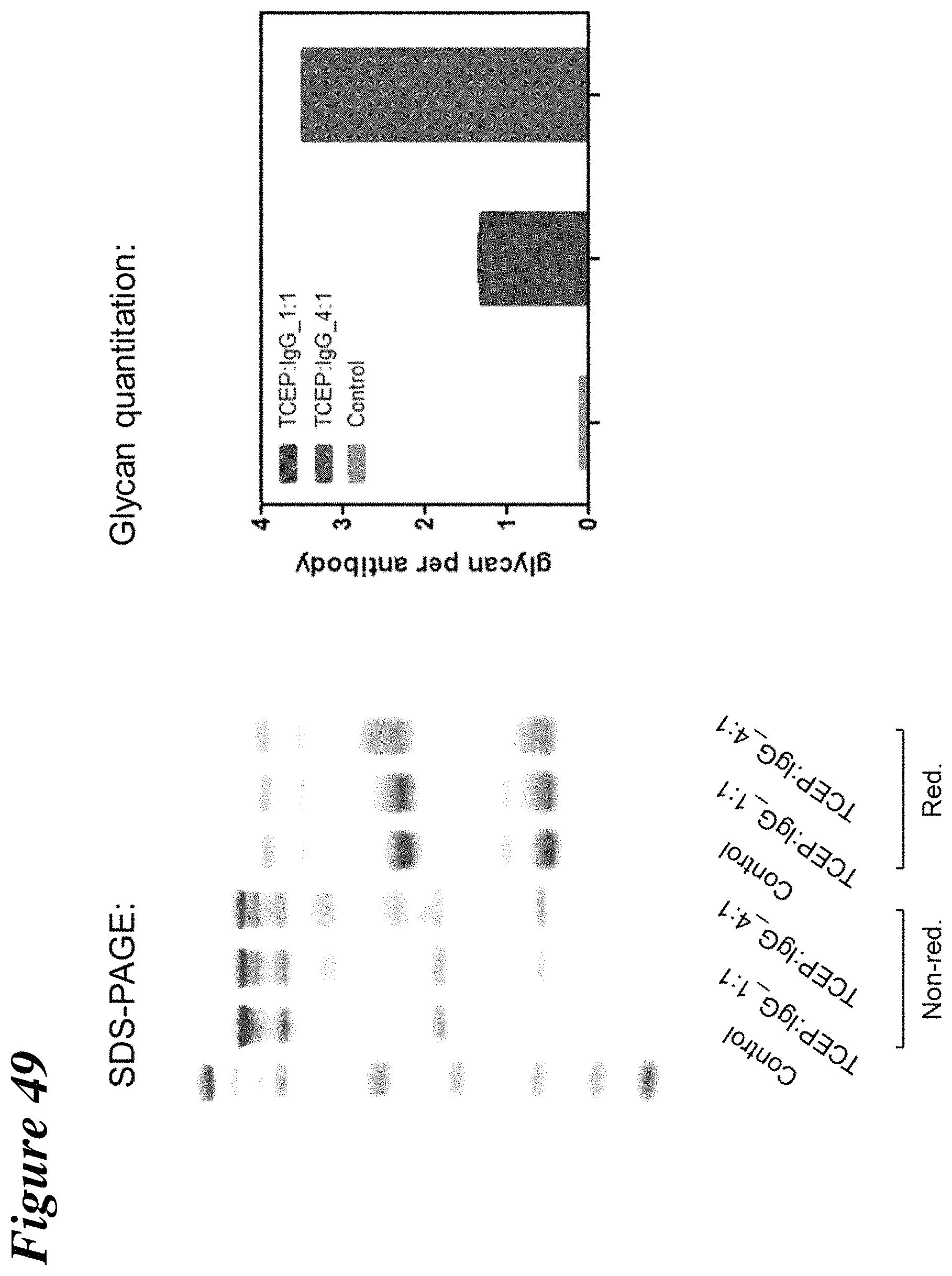
D00052
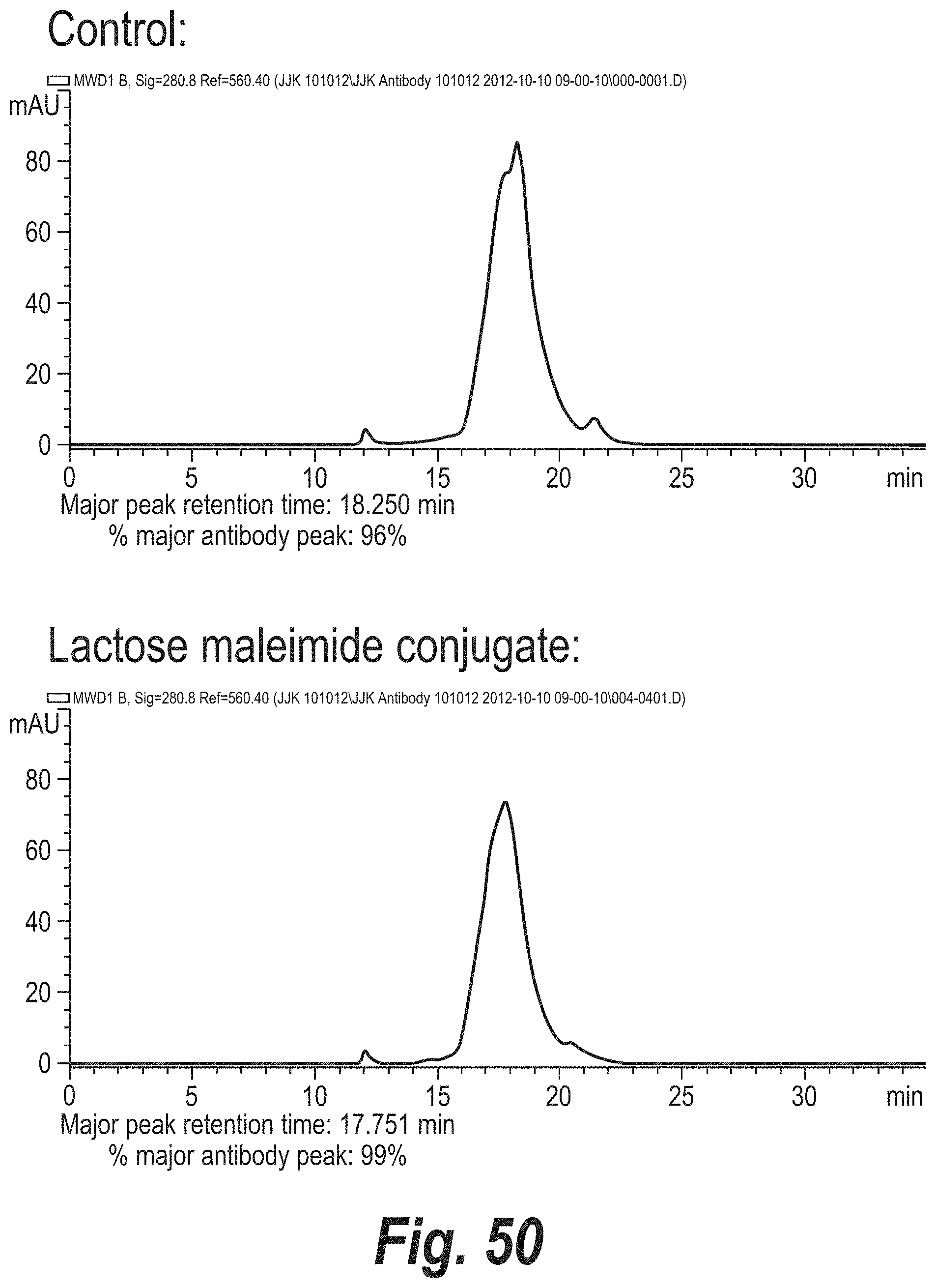
D00053
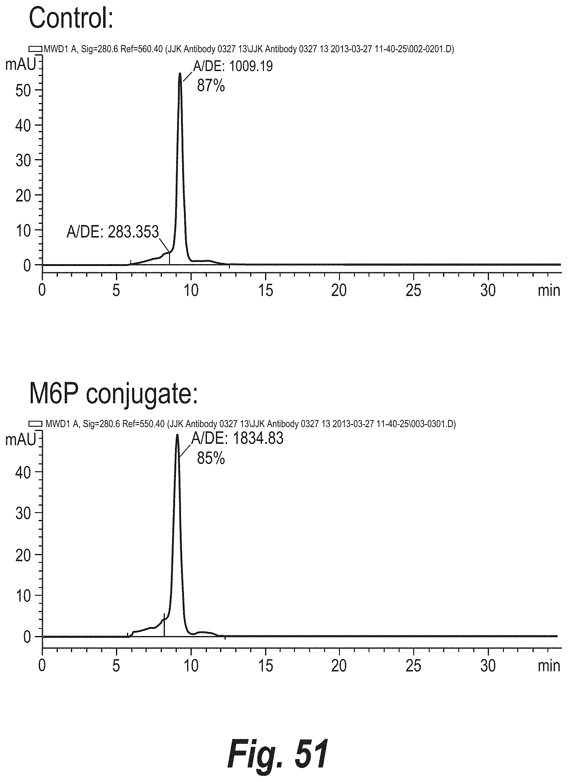
D00054
D00055
D00056

D00057
D00058
D00059
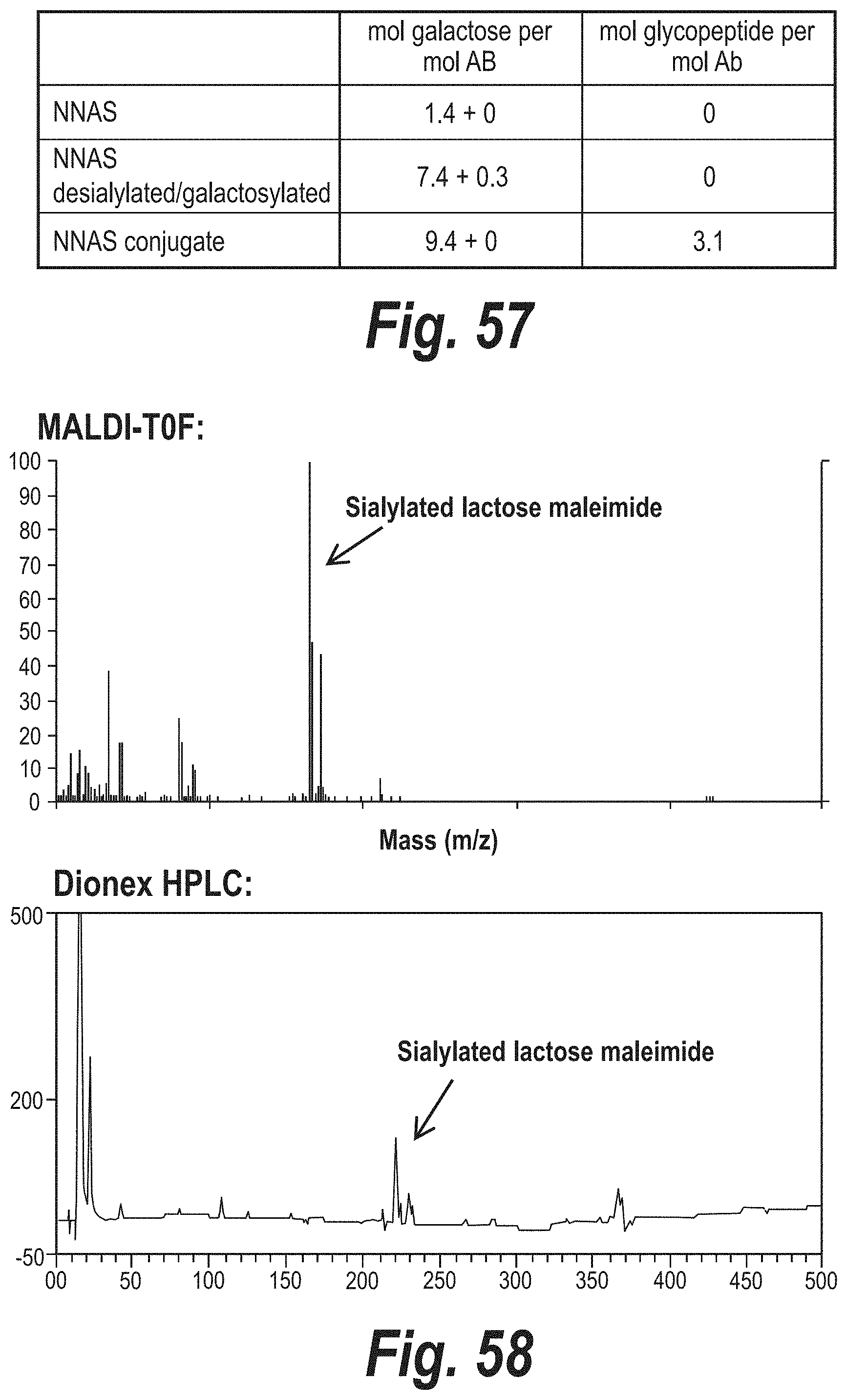
D00060

D00061

D00062
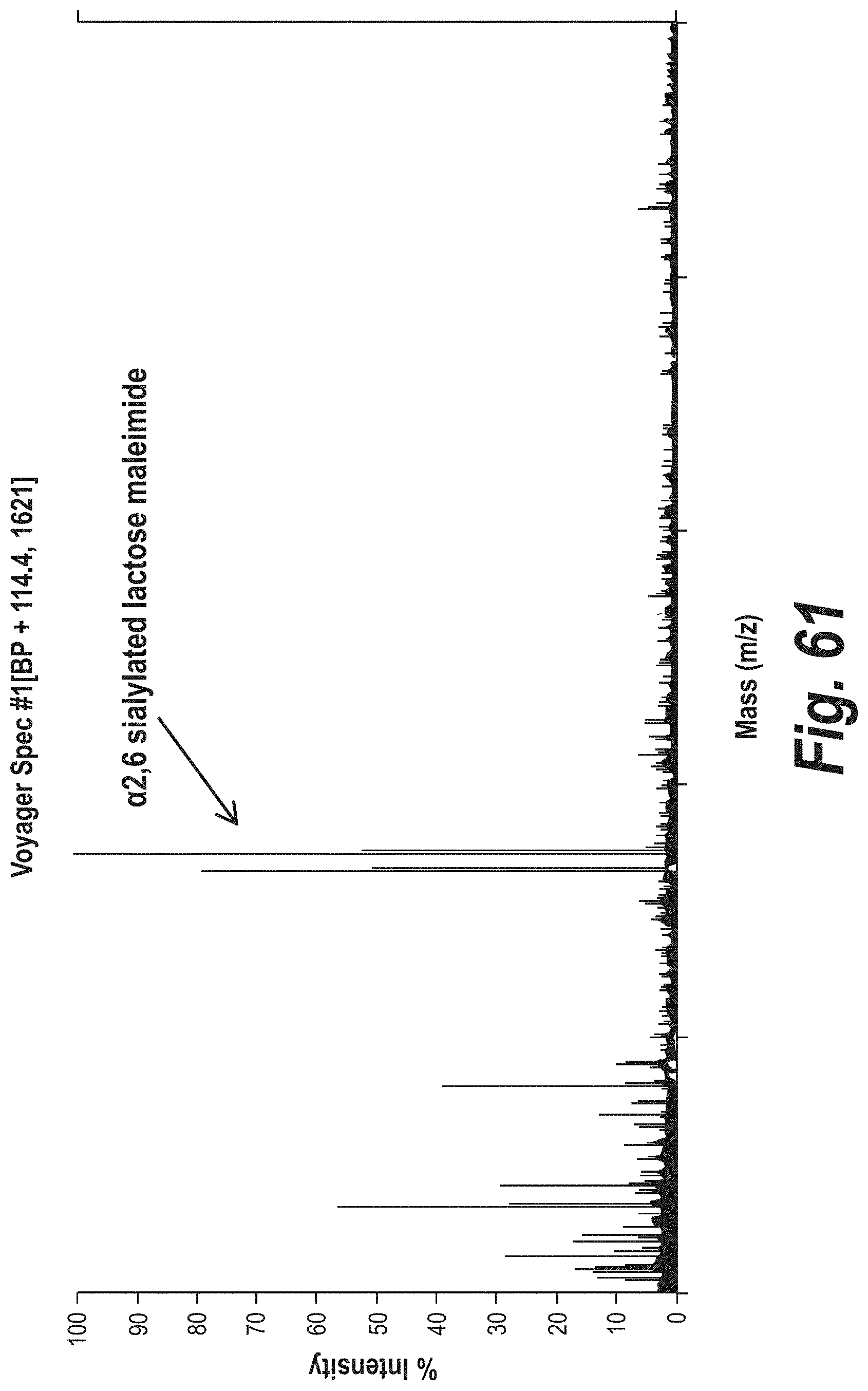
D00063
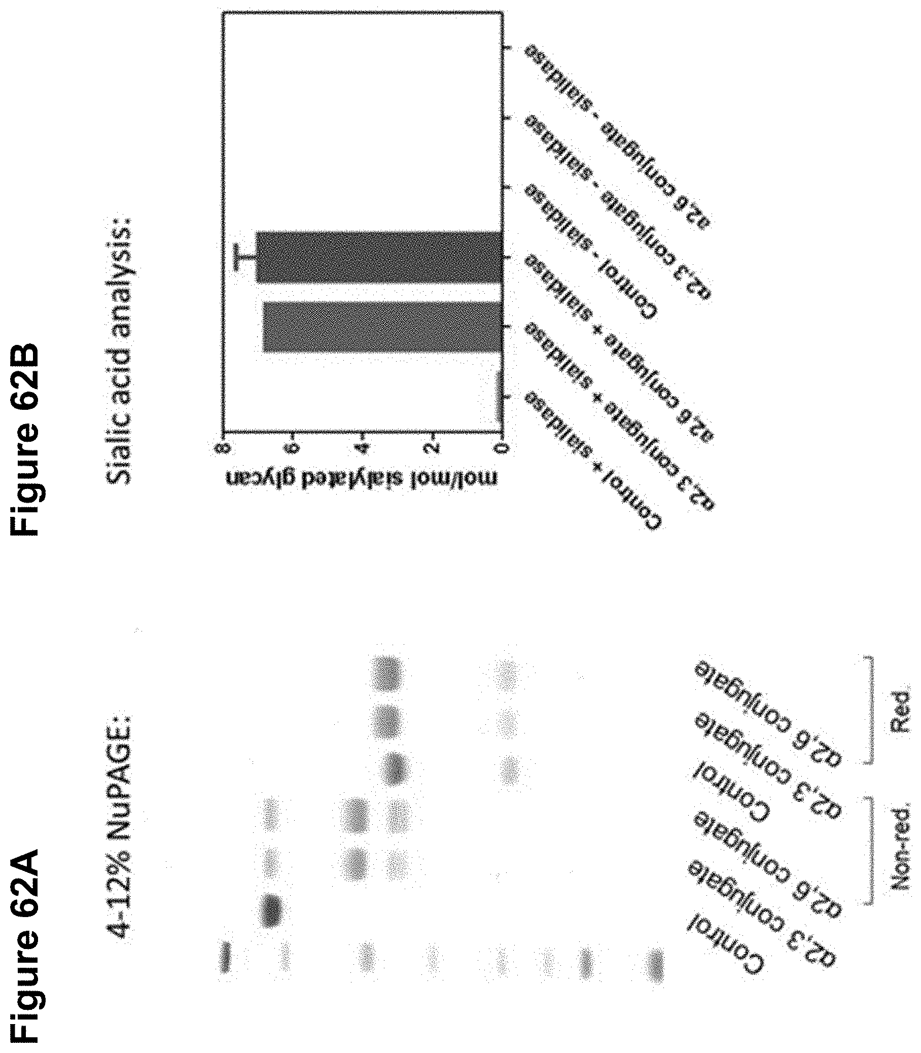
D00064

D00065

D00066
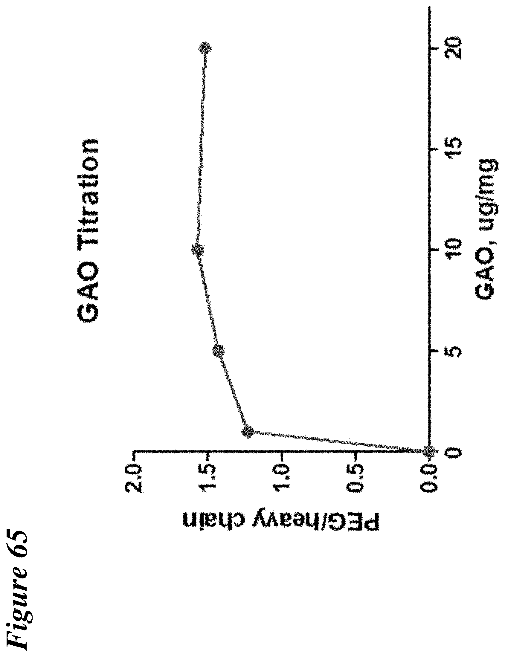
D00067
D00068

D00069

D00070
D00071

D00072

D00073
D00074

D00075

D00076
D00077

D00078

D00079
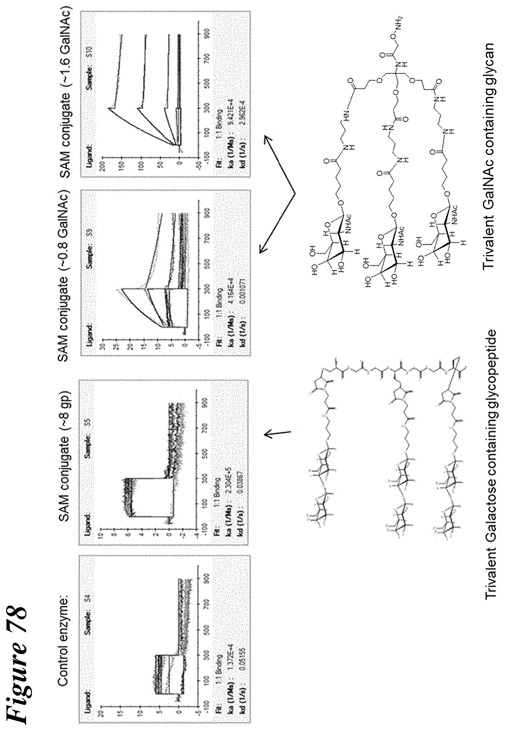
D00080
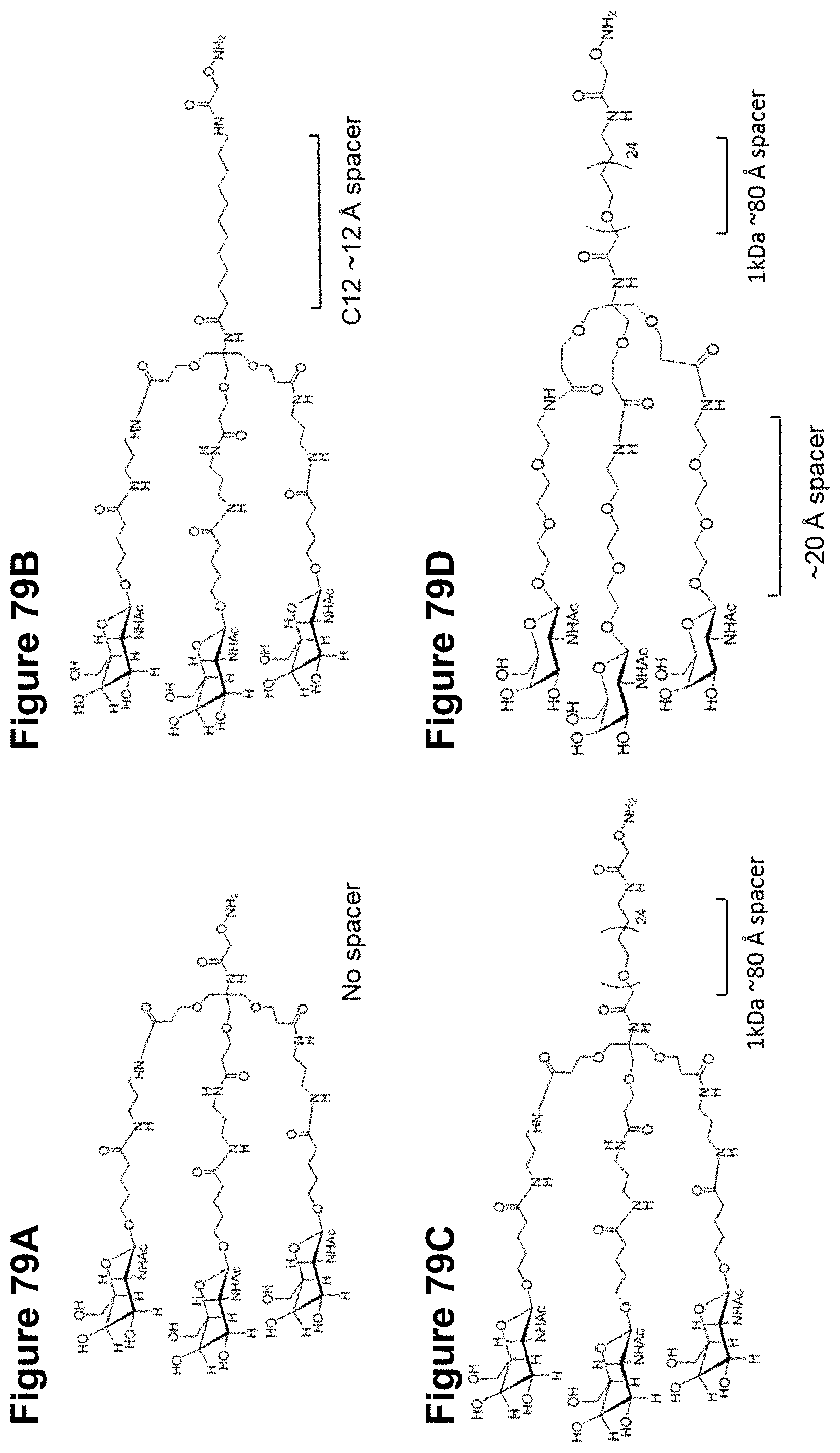
D00081

D00082

P00001

P00002

P00003

P00004

P00005

P00006

P00007

P00008

P00009

P00010

P00011

P00012

P00013

P00014

P00015

P00016

P00017

P00018

P00019

P00020

P00021

P00022

P00023

P00024

P00025

S00001
XML
uspto.report is an independent third-party trademark research tool that is not affiliated, endorsed, or sponsored by the United States Patent and Trademark Office (USPTO) or any other governmental organization. The information provided by uspto.report is based on publicly available data at the time of writing and is intended for informational purposes only.
While we strive to provide accurate and up-to-date information, we do not guarantee the accuracy, completeness, reliability, or suitability of the information displayed on this site. The use of this site is at your own risk. Any reliance you place on such information is therefore strictly at your own risk.
All official trademark data, including owner information, should be verified by visiting the official USPTO website at www.uspto.gov. This site is not intended to replace professional legal advice and should not be used as a substitute for consulting with a legal professional who is knowledgeable about trademark law.