Method of treating one or more symptoms of pulmonary fibrosis by administering inhibitors of nicotinamide phosphoribotransferase
Garcia , et al. May 4, 2
U.S. patent number 10,993,936 [Application Number 16/604,511] was granted by the patent office on 2021-05-04 for method of treating one or more symptoms of pulmonary fibrosis by administering inhibitors of nicotinamide phosphoribotransferase. This patent grant is currently assigned to Arizona Board of Regents on Behalf of The University of Arizona, The United States of America as Represented by the Department of Veterans Affairs. The grantee listed for this patent is Arizona Board of Regents on Behalf of the University of Arizona, The United States of America as Represented by the Department of Veterans Affairs. Invention is credited to Joe G. N. Garcia, Louise Hecker.


| United States Patent | 10,993,936 |
| Garcia , et al. | May 4, 2021 |
Method of treating one or more symptoms of pulmonary fibrosis by administering inhibitors of nicotinamide phosphoribotransferase
Abstract
Inhibition of the expression and/or function of nicotinamide phosphoribosyltransferase (NAMPT) can reduce, prevent or reverse the pathophysiological vascular changes associated with the onset and progression of Pulmonary Fibrosis. Compositions and methods to inhibit the expression and function of NAMPT for treating and preventing Pulmonary Fibrosis in a subject in need are provided. The compositions and methods are useful for the modulation of pathophysiological processes that contribute to the development and progression of Pulmonary Fibrosis by reducing lung inflammation, aberrant myofibroblast accumulation and deposition of collagen in fibrotic foci.
| Inventors: | Garcia; Joe G. N. (Tucson, AZ), Hecker; Louise (Tucson, AZ) | ||||||||||
|---|---|---|---|---|---|---|---|---|---|---|---|
| Applicant: |
|
||||||||||
| Assignee: | Arizona Board of Regents on Behalf
of The University of Arizona (Tucson, AZ) The United States of America as Represented by the Department of Veterans Affairs (Washington, DC) |
||||||||||
| Family ID: | 1000005527733 | ||||||||||
| Appl. No.: | 16/604,511 | ||||||||||
| Filed: | April 16, 2018 | ||||||||||
| PCT Filed: | April 16, 2018 | ||||||||||
| PCT No.: | PCT/US2018/027799 | ||||||||||
| 371(c)(1),(2),(4) Date: | October 10, 2019 | ||||||||||
| PCT Pub. No.: | WO2018/191751 | ||||||||||
| PCT Pub. Date: | October 18, 2018 |
Prior Publication Data
| Document Identifier | Publication Date | |
|---|---|---|
| US 20200138799 A1 | May 7, 2020 | |
Related U.S. Patent Documents
| Application Number | Filing Date | Patent Number | Issue Date | ||
|---|---|---|---|---|---|
| 62485863 | Apr 14, 2017 | ||||
| Current U.S. Class: | 1/1 |
| Current CPC Class: | A61K 31/444 (20130101); C07K 16/40 (20130101); A61K 31/4545 (20130101); A61K 9/0019 (20130101); A61P 11/00 (20180101); A61K 31/4439 (20130101); C07K 2317/55 (20130101); C07K 2317/76 (20130101) |
| Current International Class: | A61K 31/4545 (20060101); C07K 16/40 (20060101); A61K 31/444 (20060101); A61K 31/4439 (20060101); A61K 9/00 (20060101); A61P 11/00 (20060101) |
References Cited [Referenced By]
U.S. Patent Documents
| 7754703 | July 2010 | Lynch |
| 8329676 | December 2012 | Lynch |
| 9409983 | August 2016 | Garcia |
| 2008/0249070 | October 2008 | Lynch |
| 2009/0042954 | February 2009 | Hale |
| 2010/0003242 | January 2010 | Sabbadini |
| 2016/0031880 | February 2016 | Clark |
| 2012118910 | Sep 2012 | WO | |||
| 2014144821 | Sep 2014 | WO | |||
| 2016016898 | Feb 2016 | WO | |||
| 2017031213 | Feb 2017 | WO | |||
| 2017041114 | Mar 2017 | WO | |||
Other References
|
https://www.novusbio.com/products/pbef-visfatin-nampt-antibody_nb100-594 (retrieved from the internet Jan. 12, 2021). cited by examiner . https://www.bethyl.com/antibody/pca_a-z/NAMPT+PBEF+Visfatin (retrieved from the internet Jan. 12, 2021). cited by examiner . Albert, et al., "Novel Immunomodulator FTY720 IS Phosphorylated in Rats and Humans to Form a Single Stereoisomer. Identification, Chemical Proof, and Biological Characterization of the Biologically Active Species and Its Enantiomer", J. Med. Chem., 48:5373-5377 (2005). cited by applicant . Almagro, et al., "Antibody Modeling Assessment", Proteins, 79:3050-3066 (2011). cited by applicant . Anscher, et al., "Plasma transforming growth factor betal as a predictor of radiation pneumonitis", Int. J. Radiat. Oncol. Biol. Phys., 41:1029-35 (1998). cited by applicant . Baker, et al., "Identification and Removal of Immunogenicity in Therapeutic Proteins", Curr. Opin. Drug. Discov. Devel., 10:219-227 (2007). cited by applicant . Berdyshev, et al., "De Novo Biosynthesis of Dihydrosphingosine-1-Phophate by Sphingosine Kinase 1 in Mammalian Cells", Cell Signal, 18:1779-92 (2006). cited by applicant . Berdyshev, et al., "Quantitative analysis of Sphingoid base-1-phophates as Bisacetylated derivatives by liquid Chromatography-Tandem Mass Spectrometry", Anal. Biochem., 339:129-36 (2005). cited by applicant . Brigham, et al., "Endotoxin and lung injury", Am. Rev. Respir. Dis., 133:913-27 (1986). cited by applicant . Camp, et al., "Synthetic Analogs of FTY720 [2, Amino-2[4-octylphenyl]ethyl)-1,3-propanediol] Differentially Regulate Pulmonary Vascular Permeability in Vivo and in Vitro", The Journal of Pharmacology and Experimental Ther., 331(1):54-64 (2009). cited by applicant . Camp, et al., "Unique toll-Like Receptor 4 Activation by NAMPT/PBEF Induces NFkB Signaling and Inflammatory Lung Injury", Sci Rep., 5(13135):1-14 (2015). cited by applicant . Carruthers, et al., "Total Body Irradiation and Pneumonitis Risk: A Review of Outcomes", British Journal of Cancer, 90:2080-2084 (2004). cited by applicant . Chen, et al., "Nicotinamide Phospphoribosyltransferase Promotes Pulmonary Vascular Remodeling and is a Therapeutic Target in Pulmonary Arterial Hypertension", Circulation, 135(16):1532-1546 (2017). cited by applicant . Chen, et al., "Radiation Pneumonitis and Early Circulatory Cytokine Markers", Semin. Radiat. Oncol., 12:26-33 (2002). cited by applicant . Cho, et al., "Nrf2 Defends the Lung From Oxidative Stress", Antioxid. Redox. Signal, 8:76-87 (2006). cited by applicant . Diab, et al. "Stimulation of Sphingosine 1-Phophate Signaling as an alveolar Cell survival Strategy in Emphysema", Am. J. Respir. Crit. Care Med., 181:344-352 (2010). cited by applicant . Dinkova-Kostova, et al., "Direct and Indirect Antioxidant Properties of Inducers of Cytoprotective Proteins", Mol. Nutr. Food Res., 52 Suppl, 1:S128-138 (2008). cited by applicant . Dudek, et al., "Cytoskeletal Regulation of Pulmonary Vascular Permeability", J. Appl. Physiol., 91(4):1487-1500 (2001). cited by applicant . Dudek, et al., "Pulmonary Endothelial Cell Barrier Enhancement by FTY720 Does Not Require the S1P1 Receptor", Cell Signal, 19:1754-1764 (2007). cited by applicant . Dudek, et al., "Pulmonary Endothelial cell Barrier Enhancement by Sphingosine 1-phosphate: Roles for Cortactin and Myosin light Chain Kinase", J. Biol. Chem., 279:24692-24700 (2004). cited by applicant . Dziarski, et al., "Role of MD-2 in tlr2-And TLR4-mediated Recognition of Gram-negative and Gram-positive bacteria and Activation of Chemokine Genes", J. Endotoxin Res. 6(5):401-5 (2000). cited by applicant . Foncea, et al., "Endothelial Cell Oxidative Stress and Signal Transduction", Biological Research, 33:89-96 (2000). cited by applicant . Forrest, et al., "Immune Cell Regulation and Cardiovascular Effects of Sphingosine 1-Phosphate Receptor Agonists in Rodents are Mediated via Distinct Receptor Subtypes", J. Pharmacol. Exp. Ther., 309:758-68 (2004). cited by applicant . Foss, et al., "Synthesis, Stability, and implications of phosphothioate agonists of sphingosine-1-phosphate receptors", Bioroganic & Medicinal Chemistry Letters, 15:4470-4474 (2005). cited by applicant . Garcia, et al., "Sphingosine 1-phosphate Promotes Endothelial Cell Barrier Integrity by Edg-dependent Cytoskeletal Rearrangement", J. Clin. Invest. 108:689-701 (2001). cited by applicant . Garcia, et al., "Thrombin-induced Increase in Albumin Permeability Across the Endothelium", J. Cell Physiol., 128:96-104 (1986). cited by applicant . Georgel, et al., "The Heterogeneous Allelic Repertoire of Human Toll-Like Receptor (TLR) Genes", PLoS ONE, 4(11): e7803: 1-11 (2009). cited by applicant . Ghafoori, et al., Radiation-Induced Lung Injury: Assessment, Management, and Prevention, Oncology, 22(1):37-47 (2008). cited by applicant . Ghio, et al., "Pulmonary Arterial Compliance: How and why Should We Measure It?" Glob. Cardiol. Sci. Pract. 2015(4): 58 (2015). cited by applicant . Giaid, et al., "Inducible Nitric Oxide Synthase and Nitrotyrosine in Mice With radiation-Induced Lung Damage", Am. J. Clin. Oncol., 26:e67-72 (2003). cited by applicant . Girgis, et al., "Attenuation of Chronic Hypoxic Pulmonary Hypertension by simvastatin", Am. J. Physiol. Heart Circ. Physiol., 285:H938-945 (2003). cited by applicant . Goggel, et al., "PAF-mediated Pulmonary Edema: A New Role for Acid Sphingomyelinase and Ceramide", Nat. Med., 10:155-60 (2004). cited by applicant . Gon, et al., "S1P3 receptor-Induced Reorganization of Epithelial Tight Junctions Compromises Lung Barrier Integrity and is Potentiated by TNF", Proc. Natl. Acad. Sci., 102:9270-75 (2005). cited by applicant . Gross, "Experimental radiation Pneumonitis. IV. Leakage of Circulatory Proteins Onto the Alveolar Surface", J. Lab Clin Med., 95:19-31 (1980). cited by applicant . Grynkiewicz, et al., "A New Generation of Ca2+ Indicators with greatly Improved Fluorescence Properties", J. Biol. Chem., 260:3440-3450 (1985). cited by applicant . Hallahan, et al., "Nuclear Factor kappaB Dominant Negative Genetic Constructs Inhibit X-ray Induction of Cell Adhesion molecules in the Vascular Endothelium", Cancer Res., 58:5484-5488 (1998). cited by applicant . Harbeck, et al., "Simultaneous Optical measurements of Cytosolic Ca2+ and cAMP in Single Cells", Sci. STKE, 16 (2006). cited by applicant . Harris, et al., "Identification of Multiple Sources of Charge Heterogeneity in a Recombinant Antibody", J. Chromatogr. B. Biomed. Sci. Appl., 752:233-245 (2001). cited by applicant . Honegger, et al., "The Influence of the Framework Core Residues on the Biophysical Properties of Immunoglobulin Heavy Chain Variable Domains", Protein Eng. Des. Sel., 22:121-134 (2009). cited by applicant . Hong, et al., "Essential Role of pre-B-cell Colony enhancing Factor in Ventilator-Induced Lung Injury", Am J. Respir. Crit. Care Med., 178:605-617 (2008). cited by applicant . Hong, et al., "Rapid Induction of Cytokine Gene Expression in the Lung After Single and Fractionated doses of Radiation", Int. J Radiat. Biol., 75:1421-7 (1999). cited by applicant . International Search Report and Written Opinion for corresponding PCT PCT/US2018/27799 dated Jul. 3, 2018. cited by applicant . International Search Report and Written Opinion for corresponding PCT application PCT/US2018/27780 dated Jul. 11, 2018. cited by applicant . International Search Report for PCT application PCT/US2011/027074 dated May 24, 2011. cited by applicant . Iwakawa, et al., "Strain Dependent Differences in a Histological Study of CD44 and Collagen Fibers With an Expression Analysis of Inflammatory Response-Related Genes in Irradiated Murine Lung", J. Radiat. Res., 45:423-433 (2004). cited by applicant . Jacobson, et al., "Cytoskeletal Activation and Altered Gene Expression in Endothelial barrier Regulation by Simvastatin", Am. J. Respir. Cell Moo. Biol., 30:662-670 (2004). cited by applicant . Jacobson, et al., "Simvastatin Attenuates Vascular Leak and Inflammation in Murine Inflammatory Lung Injury", Am. J. Physiol. Lung Cell Mol. Physiol., 288:L1026-1032 (2005). cited by applicant . Johnson, et al., "Adjusting Batch Effects in Microarray Expression Data using Empirical Bayes Methods", Biostatistics, 8:118-27 (2007). cited by applicant . Kim, et al., "Crystal structure of Visfatin/Pre-B cell Colony-enhancing Factor 1/Nicotinamide Phosphoribosyltransferase, Free and in Complex with the Anti-Cancer Agent FK-866", J Mol Biol., 362:66-77 (2006). cited by applicant . Kovarik, et al., "Overview of FTY720 Clinical Pharmacokinetics and Pharmacology", Ther. Drug. Monit., 26:585-587 (2004). cited by applicant . Koyrakh, et al., "The Heart Rate Decrease Caused by Acute FTY720 Administration is Mediated by the G Protein-Gated Potassium Channel 1", American Journal of Transplantation, 5:529-536 (2005). cited by applicant . Kureishi, et al., "The HMG-CoA Reductase Inhibitor Simvastatin Activate the Protein Kinase Akt and promotes Angiogenesis in Normocholesterolemic Animals", Nat. Med., 6:1004-1010 (2000). cited by applicant . Kwok, et al., "Corticosteroids and Azathioprine Do Not Prevent Radiation-Induced Lung Injury", Can. Respir. J., 5:211-214 (1998). cited by applicant . Li, et al., "Model-base Analysis of Oligonucleotide Arrays: Model Validation, Design Issues and Standard Error Application", Genome Biol., 2:Research0032 (2001). cited by applicant . Liu, et al., "Sphingosine-1-Phosphate and Its Analogue FTY720 Diminish Acute Pulmonary Injury in Rats With acute Necrotizing Pancreatitis", Pancreas, 36(3)e10-e15 (2008). cited by applicant . Lu, et al., "Radiation-induced Changes in Gene Expression involve Recruitment of existing Messenger RNAS to and Away From Polysomes", Cancer Research, 66:1052-1061 (2006). cited by applicant . Marchesini, et al., "Acid and Neutral Sphingomyelinases: Roles and Mechanisms of Regulation", Biochem. Cell Biol., 82:27-44 (2004). cited by applicant . Matloubian, et al., "Lymphocyte Egress from Thymus and Peripheral Lymphoid Organs IS Dependent on S1P Receptor 1", Nature, 427:355-360 (2004). cited by applicant . Matthew, et al., "Simvastatin Attenuates Radiation-Induced Murine Lung Injury and Dysregulated Lung Gene Expression", Am. J. Respir. Cell Mol. Biol., ePublicatino only, PMID: 20508068 (2010). cited by applicant . Mcverry, et al., "Sphingosine 1-Phosphate Reduces Vascular Leak in murine and Canine Models of Acute Lung Injury", Am. J. Respir. Crit. Care Med., 170:987-993 (2004). cited by applicant . Meyer, et al., "GADD45a Is a Novel candidate gene in Inflammatory Lung Injury via Influence on Akt Signaling", Faseb. J., 23:1325-1337 (2009). cited by applicant . Moitra, et al., "A Transgenic Mouse With Vascular Endothelial Over-Expression of the Non-Muscle Myosin Light Chain Kinase-2 Isoform is Susceptible to Inflammatory Lung Injury: Role of Sexual Dimorphism and Age", Transl. Res., 151:141-153 (2008). cited by applicant . Moitra, et al., "Re-Evaluation of Evans Blue Dye as a Marker of Albumin Clearance in Murine Models of Acute Lung Injury", Trans, Res., 150:253-265 (2007). cited by applicant . Molteni, et al., "Control of Radiation-Induced Pneumopathy and Lung Fibrosis by Angiotensin-Converting Enzyme Inhibitors and an Angiotensin II Type 1 Receptor Blocker", Int. J. Radiat. Biol., 76:523-532 (2000). cited by applicant . Moreno-Vinasco, et al., "Attenuation of Rodent Lung Ischemia-Reperfusion Injury by Sphingosine 1-Phosphate", Journal of Organ Dysfunction, 4:106-114 (2008). cited by applicant . Nonas, et al., "Use of Consomic Rats for Genomic Insights Into Ventilator-Associated Lung Injury", Am. J. Physiol. Lung Cell Mol. Physiol., 293:L92-302 (2007). cited by applicant . North, et al., "A New Clustering of Antibody CDR Loop Conformations", J. Mol. Biol., 406:228-256 (2011). cited by applicant . Ogata, et al., "Early Administration of IL-6RA Does Not Prevent Radiation-Induced Lung Injury in Mice", Radiat. Oncol., 5:26 (2010). cited by applicant . Ostrau, et al., "Lovastatin attenuates ionizing radiation-induced normal tissue damage in vivo", Radiother. Oncol., 92:492-499 (2009). cited by applicant . Peng, et al., "Protective Effects of sphingosine 1-phosphate in Murine Endotoxin-Induced Inflammatory Lung Injury", Am. J. Respir. Crit. Care Med., 169:1245-51 (2004). cited by applicant . Petrache, et al., "Ceramide Upregulation Causes Pulmonary Cell Apoptosis and Emphysema-Like Disease in Mice", Nat Med., 11:491-8 (2005). cited by applicant . Philippe, et al., "Drug-Induced respiratory disease in patients with hematological diseases", Seminars in respiratory and critical care medicine, 26(5):458-81 (2005). cited by applicant . Pollack, et al., "The Importance of Protein Kinase A in Prostate Cancer: Relationship to Patient Outcome in radiation Therapy oncology Group Trial 92-02", Clin, Cancer Res., 15:5478-84 (2009). cited by applicant . Qin, et al., "Differential Regulation of oxidative and Osmotic Stress Induced Syk Activation by Both Autophosphorylation and SH2 Domains", Biochemistry, 37:5481-5486 (1998). cited by applicant . Rabbani, et al., "Hypoxia Inducible Factor 1alpha Signaling in fractionalized radiation-Induced Lung Injury: Role of Oxidative Stress and Tissue Hypoxia", Radiat. Res., 173:165-74 (2010). cited by applicant . Ragaller, et al., "Acute lung injury and acute respiratory distress syndrome", J. Emerg. Trauma Shock., 3(1):43051 (2010). cited by applicant . Remick, et al., "Role of Tumor Necrosis factor-Alpha in Lipopolysaccharide-Induced Pathologic Alterations", Am. J. Pathol., 136:49-60 (1990). cited by applicant . Roberts, et al., "Radiation Pneumonitis: A Possible Lymphocyte-Mediated Hypersensitivity Reaction", Ann. Intern. Med., 118:696-700 (1993). cited by applicant . Rodrigues, et al., "Prediction of Radiation Pneumonitis by Dose-Volume Histogram Parameters in Lung Cancer--a Systematic Review", Radiother. Oncol., 71:127-138 (2004). cited by applicant . Rosenfeldt, et al., "Sphingosine-1-phosphate Stimulates Contraction of Human Airway Smooth Muscle Cells", FASEB J., 17:1789-99 (2003). cited by applicant . Roviezzo, et al., "Sphingosine-1-phosphate/sphingosine Kinase Pathway Is Involved in Mouse Airway Hyperresponsiveness", Am. J. Respir. Cell Mol. Biol., 36:757-62 (2007). cited by applicant . Rubin, et al., "A Perpetual Cascade of Cytokines Postirradiation Leads to Pulmonary Fibrosis", Int. J. Radiat. Oncol. Biol. Phys., 33:99-109 (1995). cited by applicant . Sakai, et al., "CD44 and Bak Expression in IL-6 or TNF-alpha Gene Knockout Mice After Whole Lung Irradiation", Journal Radiat. Res., 49:409-416 (2008). cited by applicant . Samal, et al., "Cloning and Characterization of the cDNA Encoding a Novel human pre-B-cell Colony-Enhancing Factor", Mol. Cell. Biol., 14(2):1431-1437 (1994). cited by applicant . Sammani, et al., "Differential Effects pf Sphingosine 1-phosphate Receptors on Airway and Vascular Barrier Function in the Murine Lung", Am. J. Respir. Cell Mol. Biol., 43(4):394-402 (2010). cited by applicant . Sanchez, et al., "Phosphorylation and Action of the Immunomodulator FTY720 Inhibits Vascular Endothelial Cell Growth Factor-induced Vascular Permeability", J. Biol. Chem., 278:4781-47290 (2003). cited by applicant . Sanchez, et al., "Structural and Functional Characteristics of S1P Receptors", J. Cell. Biochem., 92:913-22 (2004). cited by applicant . Shea, et al., "Prolonged exposure to Sphingosine 1-phosphate receptor-1 Agonists Exacerbates Vascular leak, Fibrosis, and Mortality After Lung Injury", Am. J. Respir. Cell Mol. Biol., 43(6):662-73 (2010). cited by applicant . Simonneau, et al., "Updated Clinical Classification of Pulmonary Hypertension", J. Am. Coll. Cardiol., 54:S43-54 (2009). cited by applicant . Singleton, et al., "CD44 Regulates Hepatocyte Growth Factor-Mediated vascular Integrity. Role of c-Met, Tiam1/Rac1, Dynamin 2, and Cortactin", J. Biol. Chem., 282:30642-30657 (2007). cited by applicant . Singleton, et al., "Regulation of Sphingosine 1-phosphate-induced Endothelial Cytoskeletal Rearrangement and Barrier Enhancement by S1P1 Receptor, PI3 Kinase, Tiam1/Rac1, and Alpha-Actinin", FASEB J., 19:1646-1656 (2005). cited by applicant . Stas, et al., "Immunogenicity Assessment of Antibody Therapeutics", Cambridge University Press, Cambridge, (2009). cited by applicant . Sun, et al., "Pre-B cell Colony Enhancing Factor 9pbef0, a Cytokine with Multiple Physiological Functions", Cytokine & Growth Factor Reviews, 24(5):433-442 (2013). cited by applicant . Takahashi, et al., "Structure and reaction mechanism of human nicotinamide phosphoribosyltransferase", J. Biochem., 147: 95-107 (2010). cited by applicant . Tao, et al., "Mogroside IIIE, a Novel Anti-Fibrotic Compound, Reduces Pulmonary Fibrosis through Toll-Like Receptor 4 Pathways", J. of Pharmacol. Exp. Ther., 361:268-279 (2017). cited by applicant . Travis, et al., "Early Indicators of radiation Injury in the Lung: Are They useful Predictors for Late Changes?", Int. J. Radiat. Oncol. Biol. Phys., 6:1267-1269 (1980). cited by applicant . Tusher, et al., "Significance analysis of Microarrays Applied to the Ionizing Radiation Response", Proc. Natl., Acad. Sci., 98:5116-5121 (2001). cited by applicant . Undas, et al., "Anti-inflammatory and Antithrombotic Effects of Statins in the Management of Coronary Artery Disease", Clin. Lab., 48:287-296 (2002). cited by applicant . Van Walle, et al., "Immunogenicity Screening in Protein Drug Development", Expert Opin. Biol. Ther., 7:405-418 (2007). cited by applicant . Villar, et al., "Current definitions of acute lung injury and the acute respiratory distress syndrome do not reflect their true severity and outcome", Intensive Care Med., 25:930-935 (1999). cited by applicant . Vitali, et al., "The Sugen 5416/hypoxia Mouse Model of Pulmonary Hypertension Revisited: Long-term Follow-Up", Pulm. Circ. 4(4): 619-629 (2014). cited by applicant . Vujaskovic, et al., "The physical parameters and molecular events associated with radiation-induced lung toxicity", Semin. Radiat. Oncol., 10:296-307 (2000). cited by applicant . Wang, et al., "Structure of Nampt/PBEF/visfatin, a Mammalian NAD+ Biosynthetic Enzyme", Nat. Struct. Mol. Biol., 13:661-662 (2006). cited by applicant . Wheeler, et al., "Acute Lung Injury and the Acute Respiratory Distress Syndrome: A Clinical review", Lancet, 369:1553-64 (2007). cited by applicant . Williams, et al., "Effect of Administration of Lovastatin on the Development of Late Pulmonary effects After Whole-Lung Irradiation in a Murine Model", Radiat. Res., 161:560-567 (2004). cited by applicant . Yeager, et al., "Animal Models of Pulmonary Hypertension: Matching Disease Mechanisms to Etiology of the Human Disease", Pulm. Respir. Med. 4(4): 198 (2014). cited by applicant . Zhao, et al., "Inflammation and chronic oxidative Stress in Radiation-Induced Late Normal tissue injury: Therapeutic Implications", Curr. Med. Chem., 16:130-143 (2009). cited by applicant . Zhao, et al., "Intracellular Generation of Sphingosine 1-phosphate in human lung endothelial cells: Role of Lipid Phosphate phosphatae-1 and Sphingosine Kinase 1", J. Biol. Chem., 282:14165-77 (2007). cited by applicant . Zisman, et al., "A Controlled Trial of Sildenafil in Advanced Idiopathic Pulmonary Fibrosis", N. Engl. J. Med., 363(7):620-628 (2010). cited by applicant. |
Primary Examiner: Landsman; Robert S
Attorney, Agent or Firm: Pabst Patent Group LLP
Government Interests
STATEMENT REGARDING FEDERALLY SPONSORED RESEARCH OR DEVELOPMENT
This invention was made with US. government support under grant number 1 IK2 BXOOI477-01AI, awarded by the Veteran's Administration. The US. Government has certain rights in the invention.
Parent Case Text
CROSS-REFERENCE TO RELATED APPLICATIONS
This application is a National Phase application under 35 U.S.C. 371 of PCT/US2018/027799, filed on Apr. 16, 2018, which claims priority to and benefit of U.S. Provisional Application No. 62/485,863 entitled "Methods for treating fibrosis" filed Apr. 14, 2017, the contents of which are herein incorporated by reference in their entirety.
Claims
We claim:
1. A method of treating one or more symptoms of pulmonary fibrosis (PF) in a patient, comprising administering to the patient in need thereof an effective amount of one or more inhibitors of nicotinamide phosphoribosyltransferase (NAMPT), one or more inhibitors of a NAMPT receptor, or combinations thereof to increase susceptibility of myofibroblasts to apoptosis.
2. The method of claim 1 comprising administering one or more inhibitors of NAMPT in an amount between 0.1 and 15 mg/kg body weight of a human.
3. The method of claim 1 wherein the effective amount is effective to reduce, or decrease the likelihood of, myofibroblast accumulation in the lungs of a patient with PF relative to an untreated control patient.
4. The method of claim 1 wherein the effective amount is effective to reduce, or decrease the likelihood of, PF-induced dyspnea in a patient with PF relative to an untreated control patient.
5. The method of claim 1 wherein the inhibitor is an F(Ab) fragment of an antibody that binds to NAMPT.
6. The method of claim 5, wherein the inhibitor is a divalent F(Ab)2' fragment of an antibody that binds to NAMPT.
7. The method of claim 1 comprising administering antibodies or antibody fragments which reduce interaction between NAMPT and one or more receptors of NAMPT.
8. The method of claim 7, wherein the antibodies or antibody fragments reduce interaction between NAMPT and toll-like receptor 4 (TLR4).
9. The method of claim 1 comprising administering between about 10 mg and about 400 mg, inclusive, of antibody or antibody fragment in an amount for administration by infusion to a human with pulmonary fibrosis.
10. The method of claim 9, wherein between about 50 mg and about 200 mg, inclusive, of antibody is administered.
11. The method of claim 1 comprising administering a functional nucleic acid encoding an inhibitor of NAMPT, wherein the functional nucleic acid is selected from the group consisting of an antisense molecule, siRNA, miRNA, aptamers, ribozymes, triplex forming molecules, RNAi, and external guide sequences.
12. The method of claim 11 wherein one or more functional nucleic acids are expressed from an expression vector.
13. The method of claim 1 comprising administering a small molecule inhibitor.
14. The method of claim 13 wherein the small molecule is selected from the group consisting of FK-866, MS-1-82, Rari049, and Al-pii135.
15. The method of claim 1 comprising administering intravenous administration between 10 and 400 mg of antibody or antigen-binding fragment thereof.
16. The method of claim 1, comprising administering antibodies or antibody fragments inhibiting NAMPT by infusion in an amount between 1 mg and 200 mg.
17. The method of claim 16, wherein the infusion is carried out over the course of one hour.
18. The method of claim 16 wherein the composition is administered weekly, monthly or less frequently.
19. The method of claim 1 comprising administered one or more small molecules selected from the group consisting of FK-866, MS-1-82, Rari049, and Al-pii135 at a dosage of between about 1.0 mg/kg and about 3.0 mg/kg body weight of the recipient, inclusive.
20. The method of claim 19 comprising administering Rari049 in an amount of about 2.5 mg/kg body weight of the recipient.
21. The method of claim 1 wherein the patient is diagnosed with idiopathic pulmonary fibrosis, or familial pulmonary fibrosis.
22. The method of claim 1, wherein the composition is administered for a time and in an amount effective to reduce myofibroblast accumulation in the lungs in a subject relative to an untreated control subject.
23. The method of claim 1 to reduce aberrant myofibroblast accumulation in the lungs of a human subject comprising administering to the subject one or more inhibitors of nicotinamide phosphoribosyltransferase (NAMPT), in an amount effective to reduce or inhibit the interaction between NAMPT and a NAMPT receptor in the subject relative to an untreated control subject.
Description
REFERENCE TO SEQUENCE LISTING
The Sequence Listing submitted as a text file named "UA_17_154_PCT_ST25.txt," created on Apr. 16, 2018, and having a size of 17,398 bytes is hereby incorporated by reference.
FIELD OF THE INVENTION
The field of the invention is generally related to compositions, and methods for reducing morbidity and mortality associated with pulmonary fibrosis.
BACKGROUND OF THE INVENTION
Human fibrotic disorders affect many organ systems including the heart, blood vessels, kidney, liver, and lungs. An estimated 45% of deaths in the U.S. are attributable to disorders that are characterized by varying degrees of fibrosis. The most severe form of lung fibrosis is idiopathic pulmonary fibrosis (IPF), a fatal and relentlessly progressive disorder. IPF is characterized by excessive scar tissue formation and irreversible destruction of the lung parenchyma, resulting in gas-exchange abnormalities and respiratory failure. The disease course of IPF is relentlessly progressive; the median survival rate is less than three years. IPF affects approximately 200,000 people in the U.S. and five million worldwide.
Pulmonary fibrosis can develop from acute or chronic injurious exposures, even after the exposure cease. Thus, patients who have experienced these inhalational exposures are at a higher risk for developing IPF. Aging is a well-recognized risk factor for IPF (mean age=66 at the time of diagnosis), leading to a significant healthcare burden amongst the aging population. The prevalence of IPF is 20.2 per 100,000 for men and 13.2 per 100,000 for women. IPF is most prevalent among elderly males, and cigarette smoking is a major risk factor for IPF. Tobacco use has been reported for 20% of the US adult population.
Despite the well-recognized role of oxidative stress in fibrosis and aging, the ability to precisely target key mediators of this process has proved difficult. Given this shift in demographic, it is critical to understand the contribution of aging to the cellular/molecular mechanism(s) leading to the pathogenesis of age-related diseases, such as IPF. A major limitation to identification of effective treatments for IPF has been the failure of pre-clinical animal models to reliably reflect human IPF, and to predict efficacy of therapeutic agents in clinical trials. One important reason for this failure is that fibrosis spontaneously resolves in the conventional model of fibrosis in young mice. In resolving fibrosis, lung myofibroblasts (the key `scar tissue generating` cell) undergo apoptosis to promote healing. In contrast, myofibroblasts from aged mice with non-resolving fibrosis acquire a senescent and apoptosis-resistant phenotype, mediated in part by persistent expression of NADPH-oxidase-4 (Nox4). Similarly, lung myofibroblasts from IPF patients exhibit senescence and apoptosis-resistance, associated with elevated Nox4 expression. However, the mechanisms that drive persistence of Nox4 and apoptosis-resistance of myofibroblasts in the context of aging/IPF remain unknown.
Although two drugs have recently gained FDA-approval for IPF, no drug treatment has been shown to definitively improve quality of life for IPF patients and they have only been shown to delay death by six months. The current drugs only moderately slow the progression of lung decline. There are no available therapies which can `reverse` fibrosis. Existing treatment interventions are largely preventative (dosing before or at the time of injury), rather than curative. Clearly, improved therapies for the treatment of IPF and other fibrotic diseases are needed in order to improve the patient experience and outcomes.
Therefore, it is an object of the invention to provide compositions and methods of use thereof for reducing and reversing the pathophysiological processes associated with the onset and progression of pulmonary fibrosis in a subject.
It is also an object of the invention to provide compositions, devices, grafts, and methods of use thereof to reduce or prevent inappropriate or deleterious fibrosis in a subject having idiopathic pulmonary fibrosis.
It is a further object of the invention to provide dosage formulations of compositions effective to treat one or more symptoms of pulmonary fibrosis in a subject.
SUMMARY OF THE INVENTION
It has been established that inhibition of the expression and function of nicotinamide phosphoribosyltransferase ("NAMPT") reduces or prevents pathophysiological processes that lead to the onset and progression of Pulmonary Fibrosis (PF) in humans. Dosage formulations including one or more NAMPT inhibitors in an amount effective to reduce or prevent the progression of PF in a human are described.
Pharmaceutical compositions to reduce or prevent the progression of PF in a subject in need thereof including one or more inhibitors of nicotinamide phosphoribosyltransferase (NAMPT) enzymatic activity, or one or more inhibitors of NAMPT as a ligand for an inflammatory receptor or one or more inhibitors of the NAMPT receptor (TLR4), or combinations thereof, and a pharmaceutically acceptable excipient for systemic administration are provided. Inhibitors of NAMPT enzymatic activity, inhibitors of NAMPT as a ligand, or inhibitors of the NAMPT receptor include antibodies, antibody fragments, and proteins having the binding specificity of an antibody. In some embodiments, the inhibitor is an F(Ab) fragment of an antibody, or a divalent F(Ab)2' fragment of an antibody.
The compositions are effective to reduce or prevent one or more physiological processes associated with the pathology of PF in a subject relative to a control subject. For example, in one embodiment, the compositions are effective to reduce or prevent one or more of the cellular activities associated with PF, including myofibroblast accumulation, excessive extracellular matrix deposition, including collagen and fibronectin deposition in a subject relative to a control subject. Dosage formulations for systemically delivering one or more small molecule inhibitors of NAMPT, or one or more inhibitors of a NAMPT receptor, TLR4, or combinations thereof, in an amount between 10 micrograms to 3.5 mg small molecule (defined as having a molecular weight of 2,000 Daltons, more preferably less than 1,000 Daltons)/kg body weight of a human or between 10 and 400 mg antibody or antibody fragment/kg human body weight are also provided. Dosage forms including one or more inhibitors of NAMPT in an amount for administration by intravenous infusion of between about 10 mg and about 200 mg, inclusive, are provided. In some embodiments, the inhibitor of NAMPT is an antibody or fragment thereof in an amount for administration by infusion of between about 10 mg and about 400 mg, inclusive. In some embodiments, an inhibitor of NAMPT is a F(Ab)2' fragment in an amount for administration by infusion of between about 10 mg and about 200 mg, inclusive. Small molecules are preferably administered orally once a week and antibody and antibody fragments are preferably administered intravenously once a month for a period of time.
Methods including administering anti-NAMPT antibodies, antibody fragments thereof, or proteins having the binding specificity thereof to a subject by infusion in an amount between 10 mg and 400 mg are provided. In some embodiments, the infusion is carried out over the course of one hour. The administration can be repeated, preferably once per month.
An exemplary receptor of NAMPT is human Toll-Like Receptor 4 (TLR4). Therefore, exemplary compositions of inhibitors of NAMPT or NAMPT receptors include antibodies, antibody fragments, or proteins having the binding specificity of antibodies that bind NAMPT or TLR4 and prevent or reduce interaction between NAMPT and TLR4. In some embodiments the anti-NAMPT antibody, or fragment thereof, or protein having the binding affinity thereof binds to an epitope on the NAMPT protein comprising one or more residues selected from the group consisting of Glu445, Gly446, Lys447, Gly448, Asp449, Leu450, Glu451, Glu452, Tyr453, Gly454, His455, Asp456 and Leu457. In other embodiments, the inhibitor of NAMPT binds to the NAMPT molecule to prevent or reduce the homo-dimerization of NAMPT. In other embodiments, the inhibitor of NAMPT binds to the TLR4 receptor to prevent receptor activation by NAMPT.
Inhibitors of NAMPT, inhibitors of NAMPT ligands, or combinations thereof in the form of a functional nucleic acid are also provided. Exemplary functional nucleic acids include antisense molecule, siRNA, miRNA, aptamers, ribozymes, triplex forming molecules, RNAi, and external guide sequences. In some embodiments, one or more functional nucleic acids are expressed from an expression vector.
Inhibitors of NAMPT expression or function, NAMPT receptor ligation, or inhibitors of the NAMPT receptor, TLR4, or combinations thereof, in the form of a small molecule are also provided. Exemplary small molecule inhibitors include FK-866, MS-1-82, Rari049, and Al-pii135. Dosage formulations including one or more small molecule inhibitors of NAMPT enzymatic function, NAMPT receptor ligation, or inhibitors of the NAMPT receptor, TLR4, in an amount for administration of between about 10 .mu.g/kg and about 3.5 mg/kg body weight of the recipient, inclusive, are provided. In an exemplary embodiment, the small molecule inhibitor is Rari049 in an amount of about 2.5 mg/kg body weight of the recipient.
The compositions can also include a delivery vehicle, most typically an aqueous solution such as sterile saline. Other exemplary delivery vehicles include nanoparticles, microparticles, micelles, emulsions, synthetic lipoprotein particles, liposomes, carbon nanotubes, gels, or coatings. The composition can also include one or more additional therapeutic agents. Exemplary additional therapeutic agents include vasoactive compounds, anti-neointima agents, chemotherapeutic agents, steroidal and non-steroidal anti-inflammatories, conventional immunotherapeutic agents, immune-suppressants, cytokines, chemokines, and growth factors.
Methods including administering anti-NAMPT antibodies, antibody fragments thereof, or NAMPT inhibitor proteins having the binding specificity thereof to a subject by infusion in an amount between 1 mg and 400 mg, more preferably between 20 mg and 200 mg, are provided. The methods reduce or prevent lung inflammation and tissue remodeling in a subject relative to an untreated control subject. In some embodiments, the infusion is carried out over the course of one hour. The administration can be repeated, for example, once per hour, once per day, once per week, once per month, or less frequently. Small molecules are preferably administered orally once a week and antibody and antibody fragments are preferably administered intravenously once a month for a period of time. The methods can administer combinations of NAMPT inhibitors and one or more drugs to the subject.
The methods reduce or prevent one or more of the symptoms of PF in a subject at risk of having PF, or diagnosed with PF. The methods reduce or prevent myofibroblast accumulation in a subject relative to an untreated control subject. The methods can administer combinations of NAMPT inhibitors and one or more vasoactive drugs to the subject.
The methods can reduce or prevent the onset or development of PF, or one or more symptoms of PF in a subject in need thereof. Symptoms of PF that can be reduced, prevented or otherwise managed include dyspnea, fatigue, angina pectoris (chest pain), syncope, edema (swelling/redness), right heart failure, reduced oral intake, dizziness, tachycardia, and palpitation.
The methods can reduce or prevent the onset or development of acute or chronic PF, and/or treat, prevent or manage one of more of the symptoms of acute or chronic PF in the subject relative to an untreated control subject. Acute PF may occur in the intensive care setting.
BRIEF DESCRIPTION OF THE DRAWINGS
FIGS. 1A-1C show that aged mice demonstrate lack of resolution to bleomycin-induced lung injury compared to young mice.
FIG. 2 shows that fibroblasts isolated from young and aged mice demonstrate p16 induction in response to injury that is transient in young mice, while sustained in aged mice with persistent fibrosis. Fibroblasts isolated from injured lungs of aged mice demonstrate higher levels of senescence-associated .beta.-galactosidase (.mu.gal) activity, a marker of senescence, as compared to young cohorts by cellular staining for .beta. gal. These results demonstrate that non-resolving fibrosis in aged mice is associated with persistence of senescent myofibroblasts.
FIG. 3 shows ROS generation in fibroblasts from young and aged mice at the corresponding time points (control, 3w, 2m) evaluated.
FIGS. 4A and 4B show that lung tissue sections from aged mice post-lung injury show lower levels of apoptosis (TUNEL+cells) in fibrotic regions in comparison to young mice. Fibroblast cells isolated from aged mice demonstrate apoptosis resistance with fewer apoptotic cells with resistance to the apoptosis-inducing agent, staurosporine (FIG. 4A). Consistent with the acquisition of an anti-apoptotic phenotype, lungs from aged mice demonstrate elevated levels of Bcl-2 (FIG. 4B).
FIG. 5 shows heterozygous NAMPT mice Nampt+/- are protected from bleomycin-induced lung injury and lung fibrosis reflected by soluble collagen in whole lungs (compared to WT mice 3w post-injury). In response to injury, Nampt+/- mice demonstrated increased survival compared to WT mice (80%, n=8/10 vs. 50%, n=5/10). These studies demonstrate proof-of-concept that in vivo targeting of Nampt leads to protection from lung fibrosis.
FIG. 6 shows that iNampt is aberrantly regulated in aging mice and humans with IPF. iNampt is upregulated in representative fibroblasts from senescent and IPF lung fibroblasts. iNampt mRNA levels in fibroblasts isolated from advanced vs. early stage IPF patients show increasing NAMPT expression with increasing severity (FIG. 6).
FIG. 7 shows persistent gene expression of Nampt (RT-PCR) is associated with non-resolving fibrosis in aging mice evaluated in lung tissue 2m post-injury injury compared to resolving fibrosis in young mice. The 2m post-injury time point represents a point where fibrosis is actively resolving in young mice, whereas aged mice are not.
FIG. 8A shows that eNampt increases gene expression of pathways related to fibrosis. Mice were injected intratracheally with 60 g of recombinant Nampt and lung tissue was harvested 4.5 h post-administration. RNA was extracted from the lungs and 3 microarray analysis was performed (Affymetrix Mouse430_2). 630 pathways for altered gene expression were assessed. Significant enrichment in several pathways associated with lung fibrosis was identified. Importantly, in response to systemic eNampt. "Lung fibrosis" was among the most significantly altered pathways, 10th most altered out of 640 pathways assessed. FIG. 8B is a horizontal bar graph showing genome-wide transcriptomic profiling of NAMPT silenced-lung endothelial cells and pathway analysis identifying differentially-regulated pathways. These results support a role for eNampt in mediating fibrotic responses to lung injury.
FIGS. 9A and 9B show eNampt mediates pro-fibrotic myofibroblast phenotypes. Fibroblasts were dose-dependently treated with exogenous eNampt resulting in increased expression of .alpha.SMA, Nox4, iNampt, and GAPDH by western blotting. These results show that eNampt mediates fibroblast-to-myofibroblast differentiation. eNampt led to the induction of oxidant signaling, as demonstrated by dose-dependent increases in Nox4 expression and ROS generation (FIG. 9A), and fibroblast senescence (FIG. 9B). These studies demonstrate Nampt mediating pro-fibrotic lung myofibroblast phenotypes.
FIG. 10 shows that pro-fibrotic effect of eNampt requires TLR4 signaling. eNampt mediates innate immunity and transduces pro-survival signals via its known receptor, TLR4. Lung fibroblasts treated with or without a TLR4 antagonist, a competitive inhibitor of TLR4 (RS-LPS, Invitrogen), followed by treatment with/without exogenous eNampt (50 ng/ml, 48 h) showed that TLR4 blockade prevented eNampt-TLR4 mediated myofibroblast differentiation, inhibited Nox4 induction as determined by Western blot, and led to decreased ROS generation in a dose-dependent manner.
FIGS. 11A and 11B show that Nampt contributes to mouse and human IPF fibroblasts resistance to apoptosis. Staurosporine (300 nM, 8 h)-induced expression of apoptotic markers, cleaved caspase 3 and PARP (FIG. 11A) was increased in lung fibroblasts isolated from Nampt+/- compared with WT mice. FIG. 11B demonstrates that iNampt enzymatic activity is required for iNAMPT- mediated resistance to staurosporine-induced apoptosis in lung myofibroblasts (which express high levels of iNampt) as IPF fibroblasts pre-treated with FK-866, showed restored apoptosis.
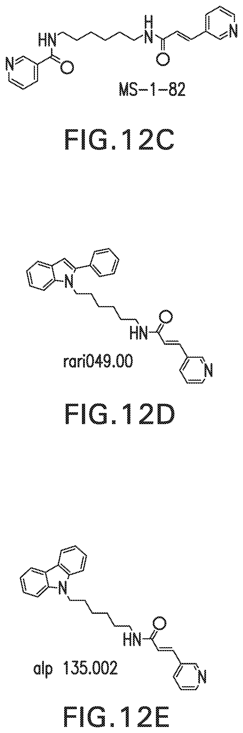
FIGS. 12A-E show chemical structure of the NAMPT inhibitor, FK-866 (FIG. 12A) which is divided into three regions (FIG. 12B) and varied by replacing with N-heterocycles to generate novel FK866 analogs: MS-1-82 (FIG. 12C), Rari049 (FIG. 12D), Alpii135 (FIG. 12E
FIG. 13 is a bar graph showing normalized NAMPT activity in the present of FK866 and FK analogues MS-1-82, Rari049, Alpii135 at 0.1, 1, and 10 .mu.M concentrations;
FIG. 14 is a bar graph showing the role of Nampt enzymatic activity in H.sub.2O.sub.2-induced apoptosis defined by the TUNEL assay. The NAMPT enzymatic inhibitor, FK-866, blocks H.sub.2O.sub.2-induced apoptosis
FIG. 15 is a bar graph showing increased lung endothelial cell NAMPT promoter activity in response to IPF-relevant stimuli. Human lung EC, transfected with a NAMPT luciferase promoter in response to VEGF (100 ng/ml) or TGF.beta.1 (2 ng/ml) after exposure for 4 hr and 24 hr show increased luciferase activity.
DETAILED DESCRIPTION OF THE INVENTION
I. Definitions
The term "dosing" or "dosage", refers to the administration of a substance (e.g., an anti-NAMPT antibody) to achieve a therapeutic objective (e.g., the treatment of a NAMPT-associated disorder).
The term "pharmaceutically acceptable carrier" encompasses any of the standard pharmaceutical carriers, such as a phosphate buffered saline solution, water and emulsions such as an oil/water or water/oil emulsion, and various types of wetting agents.
The term "inhibit" or other forms of the word such as "inhibiting" or "inhibition" means to hinder or restrain a particular characteristic. It is understood that this is typically in relation to some standard or expected value, i.e., it is relative, but that it is not always necessary for the standard or relative value to be referred to. For example, "inhibits" means hindering, interfering with or restraining the activity of the gene relative to a standard or a control. "Inhibits" can also mean to hinder or restrain the synthesis, expression or function of the protein relative to a standard or control.
"Treatment" or "treating" means to administer a composition to a subject or a system with an undesired condition (e.g., hypertension or a cardiovascular disorder). The condition can include a disease. "Prevention" or "preventing" means to administer a composition to a subject or a system at risk for the condition. The condition can be a predisposition to a disease. The effect of the administration of the composition to the subject (either treating and/or preventing) can be, but is not limited to, the cessation of a particular symptom of a condition, a reduction or prevention of the symptoms of a condition, a reduction in the severity of the condition, the complete ablation of the condition, a stabilization or delay of the development or progression of a particular event or characteristic, or minimization of the chances that a particular event or characteristic will occur.
The term "binding" refers to the interaction between a corresponding pair of molecules or portions thereof that exhibit mutual affinity or binding capacity, typically due to specific or non-specific binding or interaction, including, but not limited to, biochemical, physiological, and/or chemical interactions. "Binding partner" or "ligand" refers to a molecule that can undergo specific binding with a particular molecule. "Biological binding" defines a type of interaction that occurs between pairs of molecules including proteins, peptides, nucleic acids, glycoproteins, carbohydrates, or endogenous small molecules. "Specific binding" refers to molecules, such as polynucleotides, that are able to bind to or recognize a binding partner (or a limited number of binding partners) to a substantially higher degree than to other, similar biological entities.
The term "antibody" refers to natural or synthetic antibodies that bind a target antigen. The term includes polyclonal and monoclonal antibodies. In addition to intact immunoglobulin molecules, also included in the term "antibodies" are fragments or polymers of those immunoglobulin molecules, and human or humanized versions of immunoglobulin molecules that bind the target antigen. Thus, the term "antibody" encompasses a molecule having at least one variable region from a light chain immunoglobulin molecule and at least one variable region from a heavy chain molecule that in combination form a specific binding site for the target antigen. The antibody can be a IgG antibody, for example, the antibody can be a IgG1, IgG2, IgG3, or IgG4 antibody.
An "antibody fragment" or "antigen binding fragment" of an antibody is defined as at least a portion of the variable region of the immunoglobulin molecule that binds to its target, i.e., the antigen-binding region. An antibody can be in the form of an antigen binding antibody fragment including a Fab fragment, F(ab').sub.2 fragment, a single chain variable region, and the like. Fragments of intact molecules can be generated using methods well known in the art and include enzymatic digestion and recombinant means.
As used herein, the term "single chain Fv" or "scFv" as used herein means a single chain variable fragment that includes a light chain variable region (V.sub.L) and a heavy chain variable region (V.sub.H) in a single polypeptide chain joined by a linker which enables the scFv to form the desired structure for antigen binding (i.e., for the V.sub.H and V.sub.L of the single polypeptide chain to associate with one another to form a Fv). The V.sub.L and V.sub.H regions may be derived from the parent antibody or may be chemically or recombinantly synthesized.
The term "variable region" is intended to distinguish such domain of the immunoglobulin from domains that are broadly shared by antibodies (such as an antibody Fc domain). The variable region includes a "hypervariable region" whose residues are responsible for antigen binding. The hypervariable region includes amino acid residues from a "Complementarity Determining Region" or "CDR" (i.e., typically at approximately residues 24-34 (L), 50-56 (L2) and 89-97 (L3) in the light chain variable domain and at approximately residues 27-35 (H1), 50-65 (H2) and 95-102 (H3) in the heavy chain variable domain; Kabat et al., Sequences of Proteins of Immunological Interest, 5th Ed. Public Health Service, National Institutes of Health, Bethesda, Md. (1991)) and/or those residues from a "hypervariable loop" (i.e., residues 26-32 (L), 50-52 (L2) and 91-96 (L3) in the light chain variable domain and 26-32 (H1), 53-55 (H2) and 96-101 (H3) in the heavy chain variable domain; Chothia and Lesk, 1987, J. Mol. Biol. 196:901-917).
The term "Framework Region" or "FR" residues are those variable domain residues other than the hypervariable region residues as herein defined.
A "neutralizing antibody", (or an "antibody that neutralized NAMPT activity"), is intended to refer to an antibody whose binding to NAMPT results in inhibition of the biological activity of NAMPT. This inhibition of the biological activity of NAMPT, or its ligands, can be assessed by measuring one or more indicators of NAMPT biological activity, such as quantities of extracellular NAMPT (either in vitro or in vivo), NAMPT-induced cellular activation and NAMPT binding to NAMPT ligands. These indicators of NAMPT biological activity can be assessed by one or more of several standard in vitro or in vivo assays known in the art (see Examples). For example, in one embodiment, the ability of an antibody to neutralize NAMPT activity is assessed by inhibition of NAMPT-induced activation of fibroblasts or endothelial cells. As an additional or alternative parameter of NAMPT activity, the ability of an antibody to inhibit NAMPT-induced transcription activities via NFKB as a measure of NAMPT-induced cellular activation, can be assessed.
Any form of the "antigen" can be used to generate an antibody that is specific for a target antigen. Thus, the eliciting antigen may contain a single epitope, multiple epitopes, or can be the entire protein alone or in combination with one or more immunogenicity enhancing agents known in the art. The eliciting antigen may be an isolated full-length protein, a cell surface protein (e.g., immunizing with cells transfected with at least a portion of the antigen), or a soluble protein (e.g., immunizing with only the extracellular domain portion of the protein). The antigen may be produced in a genetically modified cell. The DNA encoding the antigen may genomic or non-genomic (e.g., cDNA). Any genetic vectors suitable for transformation of the cells of interest may be employed, including but not limited to adenoviral vectors, plasmids, and non-viral vectors, such as cationic lipids.
As used herein, the term "specifically binds" refers to the binding of an antibody to its cognate antigen while not significantly binding to other antigens. Preferably, an antibody "specifically binds" to an antigen with an affinity constant (Ka) greater than about 105 mol.sup.-1 (e.g., 10.sup.6 mol.sup.-1, 10.sup.7 mol.sup.-1, 10 mol.sup.-1, 10.sup.9 mol.sup.-1, 10.sup.10 mol.sup.-1, 10.sup.11 mol.sup.-1, and 10.sup.12 mol.sup.-1 or more) with that second molecule.
As used herein, the term "monoclonal antibody" or "MAb" refers to an antibody obtained from a substantially homogeneous population of antibodies, i.e., the individual antibodies within the population are identical except for possible naturally occurring mutations that may be present in a small subset of the antibody molecules.
As used herein, the terms "inhibit" and "inhibition" mean to decrease an activity, response, condition, disease, or other biological parameter. This can include, but is not limited to, the complete ablation of the activity, response, condition, or disease. This may also include, for example, a 10% reduction in the activity, response, condition, or disease as compared to the native or control level. Thus, the reduction can be a 10, 20, 30, 40, 50, 60, 70, 80, 90, 100%, or any amount of reduction in between as compared to native or control levels.
As used herein, the term "fusion protein" refers to a polypeptide formed by the joining of two or more polypeptides through a peptide bond formed between the amino terminus of one polypeptide and the carboxyl terminus of another polypeptide or through linking of one polypeptide to another through reactions between amino acid side chains (for example disulfide bonds between cysteine residues on each polypeptide). The fusion protein can be formed by the chemical coupling of the constituent polypeptides or it can be expressed as a single polypeptide from a nucleic acid sequence encoding the single contiguous fusion protein. Fusion proteins can be prepared using conventional techniques in molecular biology to join the two genes in frame into a single nucleic acid sequence, and then expressing the nucleic acid in an appropriate host cell under conditions in which the fusion protein is produced.
As used herein, the term "variant" refers to a polypeptide or polynucleotide that differs from a reference polypeptide or polynucleotide, but retains essential properties. A typical variant of a polypeptide differs in amino acid sequence from another, reference polypeptide. Generally, differences are limited so that the sequences of the reference polypeptide and the variant are closely similar overall and, in many regions, identical. A variant and reference polypeptide may differ in amino acid sequence by one or more modifications (e.g., substitutions, additions, and/or deletions). A substituted or inserted amino acid residue may or may not be one encoded by the genetic code. A variant of a polypeptide may be naturally occurring such as an allelic variant, or it may be a variant that is not known to occur naturally.
Modifications and changes can be made in the structure of the polypeptides of in disclosure and still obtain a molecule having similar characteristics as the polypeptide (e.g., a conservative amino acid substitution). For example, certain amino acids can be substituted for other amino acids in a sequence without appreciable loss of activity. Because it is the interactive capacity and nature of a polypeptide that defines that polypeptide's biological functional activity, certain amino acid sequence substitutions can be made in a polypeptide sequence and nevertheless obtain a polypeptide with like properties.
In making such changes, the hydropathic index of amino acids can be considered. The importance of the hydropathic amino acid index in conferring interactive biologic function on a polypeptide is generally understood in the art. It is known that certain amino acids can be substituted for other amino acids having a similar hydropathic index or score and still result in a polypeptide with similar biological activity. Each amino acid has been assigned a hydropathic index on the basis of its hydrophobicity and charge characteristics. Those indices are: isoleucine (+4.5); valine (+4.2); leucine (+3.8); phenylalanine (+2.8); cysteine/cystine (+2.5); methionine (+1.9); alanine (+1.8); glycine (-0.4); threonine (-0.7); serine (-0.8); tryptophan (-0.9); tyrosine (-1.3); proline (-1.6); histidine (-3.2); glutamate (-3.5); glutamine (-3.5); aspartate (-3.5); asparagine (-3.5); lysine (-3.9); and arginine (-4.5).
It is believed that the relative hydropathic character of the amino acid determines the secondary structure of the resultant polypeptide, which in turn defines the interaction of the polypeptide with other molecules, such as enzymes, substrates, receptors, antibodies, antigens, and cofactors. It is known in the art that an amino acid can be substituted by another amino acid having a similar hydropathic index and still obtain a functionally equivalent polypeptide. In such changes, the substitution of amino acids whose hydropathic indices are within .+-.2 is preferred, those within .+-.1 are particularly preferred, and those within .+-.0.5 are even more particularly preferred.
Substitution of like amino acids can also be made on the basis of hydrophilicity, particularly where the biological functional equivalent polypeptide or peptide thereby created is intended for use in immunological embodiments. The following hydrophilicity values have been assigned to amino acid residues: arginine (+3.0); lysine (+3.0); aspartate (+3.0.+-.1); glutamate (+3.0.+-.1); serine (+0.3); asparagine (+0.2); glutamine (+0.2); glycine (0); proline (-0.5.+-.1); threonine (-0.4); alanine (-0.5); histidine (-0.5); cysteine (-1.0); methionine (-1.3); valine (-1.5); leucine (-1.8); isoleucine (-1.8); tyrosine (-2.3); phenylalanine (-2.5); tryptophan (-3.4). It is understood that an amino acid can be substituted for another having a similar hydrophilicity value and still obtain a biologically equivalent, and in particular, an immunologically equivalent polypeptide. In such changes, the substitution of amino acids whose hydrophilicity values are within .+-.2 is preferred, those within .+-.1 are particularly preferred, and those within .+-.0.5 are even more particularly preferred.
As outlined above, amino acid substitutions are generally based on the relative similarity of the amino acid side-chain substituents, for example, their hydrophobicity, hydrophilicity, charge, size, and the like. Exemplary substitutions that take various of the foregoing characteristics into consideration are well known to those of skill in the art and include (original residue: exemplary substitution): (Ala: Gly, Ser), (Arg: Lys), (Asn: Gln, His), (Asp: Glu, Cys, Ser), (Gln: Asn), (Glu: Asp), (Gly: Ala), (His: Asn, Gln), (Ile: Leu, Val), (Leu: Ile, Val), (Lys: Arg), (Met: Leu, Tyr), (Ser: Thr), (Thr: Ser), (Tip: Tyr), (Tyr: Trp, Phe), and (Val: Ile, Leu). Embodiments include functional or biological equivalents of a polypeptide as set forth above. In particular, embodiments of the polypeptides can include variants having about 50%, 60%, 70%, 80%, 90%, 95%, 96%, 97%, 98%, 99%, or more sequence identity to the polypeptide of interest. The term "conservative amino acid substitution", as used herein, is one in which one amino acid residue is replaced with another amino acid residue having a similar side chain. Families of amino acid residues having similar side chains have been defined in the art, including basic side chains (e.g., lysine, arginine, histidine), acidic side chains (e.g., aspartic acid, glutamic acid), uncharged polar side chains (e.g., glycine, asparagine, glutamine, serine, threonine, tyrosine, cysteine), nonpolar side chains (e.g., alanine, valine, leucine, isoleucine, proline, phenylalanine, methionine, tryptophan), beta-branched side chains (e.g., threonine, valine, isoleucine) and aromatic side chains (e.g., tyrosine, phenylalanine, tryptophan, histidine).
The term "percent (%) sequence identity" is defined as the percentage of nucleotides or amino acids in a candidate sequence that are identical with the nucleotides or amino acids in a reference nucleic acid sequence, after aligning the sequences and introducing gaps, if necessary, to achieve the maximum percent sequence identity. Alignment for purposes of determining percent sequence identity can be achieved in various ways that are within the skill in the art, for instance, using publicly available computer software such as BLAST, BLAST-2, ALIGN, ALIGN-2 or Megalign (DNASTAR) software. Appropriate parameters for measuring alignment, including any algorithms needed to achieve maximal alignment over the full-length of the sequences being compared can be determined by known methods.
For purposes herein, the % sequence identity of a given nucleotides or amino acids sequence C to, with, or against a given nucleic acid sequence D (which can alternatively be phrased as a given sequence C that has or includes a certain % sequence identity to, with, or against a given sequence D) is calculated as follows: 100 times the fraction W/Z, where W is the number of nucleotides or amino acids scored as identical matches by the sequence alignment program in that program's alignment of C and D, and where Z is the total number of nucleotides or amino acids in D. It will be appreciated that where the length of sequence C is not equal to the length of sequence D, the % sequence identity of C to D will not equal the % sequence identity of D to C.
The term "K.sub.off", is intended to refer to the off rate constant for dissociation of an interaction between a molecule and its ligand, for example, an antibody from the antibody/antigen complex.
The term "K.sub.d", as used herein, is intended to refer to the dissociation constant of a particular antibody-antigen interaction.
The term "myofibroblast accumulation" refers to a the presence of fibroblast focci, caused by physiological processes of excessive cellular proliferation, combined reduced apoptosis/programmed cell death in myofibroblasts, and loss of cellular homeostasis/disordered metabolism and dysregulation of certain growth factors.
The terms "monthly dosing regimen", "monthly dosing", and "monthly administration", as used herein, refer to the time course of administering a substance (e.g., an anti-NAMPT antibody) to a subject to achieve a therapeutic objective (e.g., the treatment of a NAMPT-associated disorder). The monthly dosing regimen is not intended to include a weekly dosing regimen. Preferably, the substance is administered every 26-36 days, more preferably, every 28-31 days, even more preferably, every 28-30 days, and most preferably, every 30 days.
The term "human NAMPT" (abbreviated herein as hNAMPT, or simply NAMPT), as used herein, is intended to refer to a human nicotinamide phosphoribosyltransferase enzyme that exists as a 120 kD secreted form, the biologically active form of which is composed of a dimer of noncovalently bound 60 kD molecules. The structure of NAMPT is described further in, for example, Kim, et al. J Mol Biol.; 362:66-77 (2006). The term NAMPT is intended to include recombinant human NAMPT, which can be prepared by standard recombinant expression methods. The human NAMPT gene is referred to as NAMPT.
II. Compositions
Dosage formulations including one or more inhibitors of NAMPT and/or one or more inhibitors of a NAMPT receptor effective to reduce or prevent the development and/or progression of PF in a human have been developed. Compositions for treatment of IPF include: i) inhibitors of the expression and function of the NAMPT gene; ii) inhibitors of the enzymatic activity of the NAMPT gene product; iii) manipulation of the interaction of the NAMPT gene product with its receptor, TLR4 (NAMPT/TLR4), iv) neutralization of circulating extracellular NAMPT (eNAMPT); v) manipulation of one or more of the downstream cellular signaling events associated with NAMPT/TLR4 such as NFkB phosphorylation/activation. Loss of function of the NAMPT gene product gives rise to abnormal function in cellular processes associated with tissue remodeling and scarring, resulting in an associated reduction in the onset, development and severity of IPF in human subjects. Loss of function of the NAMPT gene product gives rise to reduction in myofibroblast accumulation, resulting in an associated reduction cellular processes associated with the onset, development and severity of PF in human subjects.
Compositions for preventing or reducing diseases characterized by myofibroblast accumulation by blockade of expression and/or function of intracellular NAMPT enzyme (iNAMPT) and/or extracellular NAMPT cytokine (eNAMPT) are provided.
A. Targets of Inhibition
1. Nicotinamide Phosphoribosyltransferase (NAMPT)
In some embodiments, the target of inhibition is nicotinamide phosphoribosyltransferase (NAMPT). The NAMPT gene product is the rate-limiting enzyme in the Nicotinamide adenine dinucleotide (NAD+) salvage pathway that converts nicotinamide to nicotinamide mononucleotide in mammals to enable NAD+biosynthesis.
The mature form of the extracellular NAMPT protein is a homodimer of approximately 120 kDa, each monomer having approximately 500 amino acid residues (Takahashi, et al., J. Biochem. 147: 95-107 (2010)).
It has been established that mutations which reduce or inhibit the function of the NAMPT enzyme reduce or prevent the physiological processes that give rise to PF. It is believed that modulation of the NAMPT enzyme provides a means to modulate physiological processes that give rise to myofibroblast accumulation associated with PF.
a. The NAMPT Gene
The human NAMPT gene (NAMPT) is located at chromosome 7, (segment 7q22.3; base pairs 106,248,285 to 106,286,326). Nucleic acid sequences for the human NAMPT gene product are known in the art. See, for example, NCBI Reference Sequence: NM_005746.2, Homo sapiens nicotinamide phosphoribosyltransferase (NAMPT), mRNA, which provides the nucleic acid sequence:
TABLE-US-00001 (SEQ ID NO: 1) ATGAATCCTG CGGCAGAAGC CGAGTTCAAC ATCCTCCTGG CCACCGACTC CTACAAGGTT ACTCACTATA AACAATATCC ACCCAACACA AGCAAAGTTT ATTCCTACTT TGAATGCCGT GAAAAGAAGA CAGAAAACTC CAAATTAAGG AAGGTGAAAT ATGAGGAAAC AGTATTTTAT GGGTTGCAGT ACATTCTTAA TAAGTACTTA AAAGGTAAAG TAGTAACCAA AGAGAAAATC CAGGAAGCCA AAGATGTCTA CAAAGAACAT TTCCAAGATG ATGTCTTTAA TGAAAAGGGA TGGAACTACA TTCTTGAGAA GTATGATGGG CATCTTCCAA TAGAAATAAA AGCTGTTCCT GAGGGCTTTG TCATTCCCAG AGGAAATGTT CTCTTCACGG TGGAAAACAC AGATCCAGAG TGTTACTGGC TTACAAATTG GATTGAGACT ATTCTTGTTC AGTCCTGGTA TCCAATCACA GTGGCCACAA ATTCTAGAGA GCAGAAGAAA ATATTGGCCA AATATTTGTT AGAAACTTCT GGTAACTTAG ATGGTCTGGA ATACAAGTTA CATGATTTTG GCTACAGAGG AGTCTCTTCC CAAGAGACTG CTGGCATAGG AGCATCTGCT CACTTGGTTA ACTTCAAAGG AACAGATACA GTAGCAGGAC TTGCTCTAAT TAAAAAATAT TATGGAACGA AAGATCCTGT TCCAGGCTAT TCTGTTCCAG CAGCAGAACA CAGTACCATA ACAGCTTGGG GGAAAGACCA TGAAAAAGAT GCTTTTGAAC ATATTGTAAC ACAGTTTTCA TCAGTGCCTG TATCTGTGGT CAGCGATAGC TATGACATTT ATAATGCGTG TGAGAAAATA TGGGGTGAAG ATCTAAGACA TTTAATAGTA TCGAGAAGTA CACAGGCACC ACTAATAATC AGACCTGATT CTGGAAACCC TCTTGACACT GTGTTAAAGG TTTTGGAGAT TTTAGGTAAG AAGTTTCCTG TTACTGAGAA CTCAAAGGGT TACAAGTTGC TGCCACCTTA TCTTAGAGTT ATTCAAGGGG ATGGAGTAGA TATTAATACC TTACAAGAGA TTGTAGAAGG CATGAAACAA AAAATGTGGA GTATTGAAAA TATTGCCTTC GGTTCTGGTG GAGGTTTGCT ACAGAAGTTG ACAAGAGATC TCTTGAATTG TTCCTTCAAG TGTAGCTATG TTGTAACTAA TGGCCTTGGG ATTAACGTCT TCAAGGACCC AGTTGCTGAT CCCAACAAAA GGTCCAAAAA GGGCCGATTA TCTTTACATA GGACGCCAGC AGGGAATTTT GTTACACTGG AGGAAGGAAA AGGAGACCTT GAGGAATATG GTCAGGATCT TCTCCATACT GTCTTCAAGA ATGGCAAGGT GACAAAAAGC TATTCATTTG ATGAAATAAG AAAAAATGCA CAGCTGAATA TTGAACTGGA AGCAGCACAT CATTAG.
Nucleotide sequences that have at least 80%, 85%, 90%, 95%, 99% or 100% amino acid sequence identity to SEQ ID NO: 1 are also disclosed.
b. The NAMPT Enzyme
The NAMPT polypeptide is a 473 amino acid cytoplasmic protein (also known as nicotinamide phosphoribosyltransferase, pre-B-cell colony-enhancing factor (PBEF) protein) with a molecular weight of approximately 52,521 Da. There are 3 mRNA variants, with lengths of 2.0, 2.4, and 4.0 kilobases (kb), transcribed by the NAMPT gene. The 2.4-kb variant is the most abundant and its open reading frame encodes a protein of 473 amino acids (aa) in length, with a predicted size of approximately 52 kDa (Samal, et al. Mol. Cell. Biol. 14 (2), 1431-1437 (1994)). It has been found in human endothelial cells, where it is able to induce angiogenesis through upregulation of VEGF and VEGFR and secretion of MCP-1. In human umbilical endothelial cells, NAMPT increases levels of the protease MMP 2/9. NAMPT has also been found in a variety of immune cells other than B cells and has been shown to inhibit apoptosis of macrophages and fibroblasts. Extracellular NAMPT (eNAMPT) has been shown to increase NFkB activation and subsequent induction of inflammatory cytokines, such as TNF-.beta., IL-1.beta., IL-16, and TGF-.beta.1, and the chemokine receptor CCR3. NAMPT also increases the production of IL-6, TNF-.beta., and IL-1.beta. in CD14+ monocyctes, macrophages, and dendritic cells, enhances the effectiveness of T cells, and is involved in the development of both B and T lymphocytes (Sun, et al., Cytokine & growth factor reviews 24(5):433-442 (2013)).
The NAMPT enzyme crystal structure is described in detail in Kim, et al. J Mol Biol.; 362:66-77 (2006). NAMPT is a dimeric type II phosphoribosyltransferase. The active site of the enzyme is at the dimer interface where the two NAMPT molecules interact. In the apoenzyme structure, a sulfate ion binds in place of the phosphate of NMN. A hydrogen bond between Asp219 and the amide of nicotinamide prevents the enzyme from forming a hydrogen bond to nicotinic or quinolinic acid. Crystal structures of NAMPT are available in the Protein Data Bank as PDB ID Nos. 2G95, 2G96 and 2G97. Amino acid sequences of the human NAMPT enzyme are known in the art. See, for example, GenBank Accession No. NP_005737.1:
TABLE-US-00002 (SEQ ID NO: 2) 10 20 30 40 MNPAAEAEFN ILLATDSYKV THYKQYPPNT SKVYSYFECR 50 60 70 80 EKKTENSKLR KVKYEETVFY GLQYILNKYL KGKVVTKEKI 90 100 110 120 QEAKDVYKEH FQDDVFNEKG WNYILEKYDG HLPIEIKAVP 130 140 150 160 EGFVIPRGNV LFTVENTDPE CYWLTNWIET ILVQSWYPIT 170 180 190 200 VATNSREQKK ILAKYLLETS GNLDGLEYKL HDFGYRGVSS 210 220 230 240 QETAGIGASA HLVNFKGTDT VAGLALIKKY YGTKDPVPGY 250 260 270 280 SVPAAEHSTI TAWGKDHEKD AFEHIVTQFS SVPVSVVSDS 290 300 310 320 YDIYNACEKI WGEDLRHLIV SRSTQAPLII RPDSGNPLDT 330 340 350 360 VLKVLEILGK KFPVTENSKG YKLLPPYLRV IQGDGVDINT 370 380 390 400 LQEIVEGMKQ KMWSIENIAF GSGGGLLQKL TRDLLNCSFK 410 420 430 440 CSYVVTNGLG INVFKDPVAD PNKRSKKGRL SLHRTPAGNF 450 460 470 480 VTLEEGKGDL EEYGQDLLHT VFKNGKVTKS YSFDEIRKNA 490 QLNIELEAAH H
NAMPT polypeptides that have, for example, at least 80%, 85%, 90%, 95%, 99% or 100% amino acid sequence identity to SEQ ID NO: 2.
The NAMPT enzyme has been associated with many diverse cellular activities, however the biological function of the NAMPT enzyme in the onset and progression of PF remained largely unknown. The region of dimerization within the mature form of the NAMPT enzyme is described in the X-ray crystal structure of NAMPT, described in Wang, et al., Nat Struct Mol Biol, 13, 661-662. (2006). Residues involved in the interface include Ser199 and Ser200.
It may be that the NAMPT protein interacts with one or more ligands through interaction by hydrogen bonding with one or more residues selected from Glu445, Gly446, Lys447, Gly448, Asp449, Leu450, Glu451, Glu452, Tyr453, Gly454, Gln455, Asp456 and Leu457. These residues for a loop that may interact with TLR4 in a manner analogous to MD-2.
2. NAMPT Receptors
In some embodiments, the target of inhibition are the receptors for NAMPT, such as Toll-like receptor 4 (TLR4). Toll-like receptor 4 is a protein that in humans is encoded by the TLR4 gene. TLR4 is a transmembrane protein, member of the toll-like receptor family, which belongs to the pattern recognition receptor (PRR) family. Its activation leads to an intracellular NF-.kappa.B signaling pathway and inflammatory cytokine production which is responsible for activating the innate immune system. It is most well known for recognizing lipopolysaccharide (LPS), a component present in many Gram-negative bacteria (e.g. Neisseria spp.) and select Gram-positive bacteria. Its ligands also include several viral proteins, polysaccharide, and a variety of endogenous proteins such as low-density lipoprotein, beta-defensins, and heat shock protein.
The human TLR4 gene (TLR4) is located at chromosome 9, (segment 9q32-q33) (Georgel, et al., PLoS ONE 4(11): e7803 (2009)). Nucleic acid sequences for the human TLR4 gene product are known in the art. See, for example, NCBI Reference Sequence: AAY82268.1, Homo sapiens toll-like receptor 4 (TLR4), mRNA, which provides the nucleic acid sequence:
TABLE-US-00003 (SEQ ID NO: 3) ATGATGTCTG CCTCGCGCCT GGCTGGGACT CTGATCCCAG CCATGGCCTT CCTCTCCTGC GTGAGACCAG AAAGCTGGGA GCCCTGCGTG GAGGTGGTTC CTAATATTAC TTATCAATGC ATGGAGCTGA ATTTCTACAA AATCCCCGAC AACCTCCCCT TCTCAACCAA GAACCTGGAC CTGAGCTTTA ATCCCCTGAG GCATTTAGGC AGCTATAGCT TCTTCAGTTT CCCAGAACTG CAGGTGCTGG ATTTATCCAG GTGTGAAATC CAGACAATTG AAGATGGGGC ATATCAGAGC CTAAGCCACC TCTCTACCTT AATATTGACA GGAAACCCCA TCCAGAGTTT AGCCCTGGGA GCCTTTTCTG GACTATCAAG TTTACAGAAG CTGGTGGCTG TGGAGACAAA TCTAGCATCT CTAGAGAACT TCCCCATTGG ACATCTCAAA ACTTTGAAAG AACTTAATGT GGCTCACAAT CTTATCCAAT CTTTCAAATT ACCTGAGTAT TTTTCTAATC TGACCAATCT AGAGCACTTG GACCTTTCCA GCAACAAGAT TCAAAGTATT TATTGCACAG ACTTGCGGGT TCTACATCAA ATGCCCCTAC TCAATCTCTC TTTAGACCTG TCCCTGAACC CTATGAACTT TATCCAACCA GGTGCATTTA AAGAAATTAG GCTTCATAAG CTGACTTTAA GAAATAATTT TGATAGTTTA AATGTAATGA AAACTTGTAT TCAAGGTCTG GCTGGTTTAG AAGTCCATCG TTTGGTTCTG GGAGAATTTA GAAATGAAGG AAACTTGGAA AAGTTTGACA AATCTGCTCT AGAGGGCCTG TGCAATTTGA CCATTGAAGA ATTCCGATTA GCATACTTAG ACTACTACCT CGATGATATT ATTGACTTAT TTAATTGTTT GACAAATGTT TCTTCATTTT CCCTGGTGAG TGTGACTATT GAAAGGGTAA AAGACTTTTC TTATAATTTC GGATGGCAAC ATTTAGAATT AGTTAACTGT AAATTTGGAC AGTTTCCCAC ATTGAAACTC AAATCTCTCA AAAGGCTTAC TTTCACTTCC AACAAAGGTG GGAATGCTTT TTCAGAAGTT GATCTACCAA GCCTTGAGTT TCTAGATCTC AGTAGAAATG GCTTGAGTTT CAAAGGTTGC TGTTCTCAAA GTGATTTTGG GACAACCAGC CTAAAGTATT TAGATCTGAG CTTCAATGGT GTTATTACCA TGAGTTCAAA CTTCTTGGGC TTAGAACAAC TAGAACATCT GGATTTCCAG CATTCCAATT TGAAACAAAT GAGTGAGTTT TCAGTATTCC TATCACTCAG AAACCTCATT TACCTTGACA TTTCTCATAC TCACACCAGA GTTGCTTTCA ATGGCATCTT CAATGGCTTG TCCAGTCTCG AAGTCTTGAA AATGGCTGGC AATTCTTTCC AGGAAAACTT CCTTCCAGAT ATCTTCACAG AGCTGAGAAA CTTGACCTTC CTGGACCTCT CTCAGTGTCA ACTGGAGCAG TTGTCTCCAA CAGCATTTAA CTCACTCTCC AGTCTTCAGG TACTAAATAT GAGCCACAAC AACTTCTTTT CATTGGATAC GTTTCCTTAT AAGTGTCTGA ACTCCCTCCA GGTTCTTGAT TACAGTCTCA ATCACATAAT GACTTCCAAA AAACAGGAAC TACAGCATTT TCCAAGTAGT CTAGCTTTCT TAAATCTTAC TCAGAATGAC TTTGCTTGTA CTTGTGAACA CCAGAGTTTC CTGCAATGGA TCAAGGACCA GAGGCAGCTC TTGGTGGAAG TTGAACGAAT GGAATGTGCA ACACCTTCAG ATAAGCAGGG CATGCCTGTG CTGAGTTTGA ATATCACCTG TCAGATGAAT AAGACCATCA TTGGTGTGTC GGTCCTCAGT GTGCTTGTAG TATCTGTTGT AGCAGTTCTG GTCTATAAGT TCTATTTTCA CCTGATGCTT CTTGCTGGCT GCATAAAGTA TGGTAGAGGT GAAAACATCT ATGATGCCTT TGTTATCTAC TCAAGCCAGG ATGAGGACTG GGTAAGGAAT GAGCTAGTAA AGAATTTAGA AGAAGGGGTG CCTCCATTTC AGCTCTGCCT TCACTACAGA GACTTTATTC CCGGTGTGGC CATTGCTGCC AACATCATCC ATGAAGGTTT CCATAAAAGC CGAAAGGTGA TTGTTGTGGT GTCCCAGCAC TTCATCCAGA GCCGCTGGTG TATCTTTGAA TATGAGATTG CTCAGACCTG GCAGTTTCTG AGCAGTCGTG CTGGTATCAT CTTCATTGTC CTGCAGAAGG TGGAGAAGAC CCTGCTCAGG CAGCAGGTGG AGCTGTACCG CCTTCTCAGC AGGAACACTT ACCTGGAGTG GGAGGACAGT GTCCTGGGGC GGCACATCTT CTGGAGACGA CTCAGAAAAG CCCTGCTGGA TGGTAAATCA TGGAATCCAG AAGGAACAGT GGGTACAGGA TGCAATTGGC AGGAAGCAAC ATCTATCTGA.
Nucleotide sequences that have at least 80%, 85%, 90%, 95%, 99% or 100% amino acid sequence identity to SEQ ID NO: 3 are also disclosed.
Amino acid sequences of the human TLR4 are known in the art. See, for example, Genfank Accession No. AAY2268.1
TABLE-US-00004 (SEQ ID NO: 4) 10 20 30 40 MMSASRLAGT LIPAMAFLSC VRPESWEPCV EVVPNITYQC 50 60 70 80 MELNFYKIPD NLPFSTKNLD LSFNPLRHLG SYSFFSFPEL 90 100 110 120 QVLDLSRCEI QTIEDGAYQS LSHLSTLILT GNPIQSLALG 130 140 150 160 AFSGLSSLQK LVAVETNLAS LENFPIGHLK TLKELNVAHN 170 180 190 200 LIQSFKLPEY FSNLTNLEHL DLSSNKIQSI YCTDLRVLHQ 210 220 230 240 MPLLNLSLDL SLNPMNFIQP GAFKEIRLHK LTLRNNFDSL 250 260 270 280 NVMKTCIQGL AGLEVHRLVL GEFRNEGNLE KFDKSALEGL 290 300 310 320 CNLTIEEFRL AYLDYYLDDI IDLFNCLTNV SSFSLVSVTI 330 340 350 360 ERVKDFSYNF GWQHLELVNC KFGQFPTLKL KSLKRLTFTS 370 380 390 400 NKGGNAFSEV DLPSLEFLDL SRNGLSFKGC CSQSDFGTTS 410 420 430 440 LKYLDLSFNG VITMSSNFLG LEQLEHLDFQ HSNLKQMSEF 450 460 470 480 SVFLSLRNLI YLDISHTHIR VAFNGIFNGL SSLEVLKMAG 490 500 510 520 NSFQENFLPD IFTELRNLIF LDLSQCQLEQ LSPTAFNSLS 530 540 550 560 SLQVLNMSHN NFFSLDTFPY KCLNSLQVLD YSLNHIMTSK 570 580 590 600 KQELQHFPSS LAFLNLTQND FACTCEHQSF LQWIKDQRQL 610 620 630 640 LVEVERMECA TPSDKQGMPV LSLNITCQMN KTIIGVSVLS 650 660 670 680 VLVVSVVAVL VYKFYFHLML LAGCIKYGRG ENIYDAFVIY 690 700 710 720 SSQDEDWVRN ELVKNLEEGV PPFQLCLHYR DFIPGVAIAA 730 740 750 760 NIIHEGFHKS RKVIVVVSQH FIQSRWCIFE YEIAQTWQFL 770 780 790 800 SSRAGIIFIV LQKVEKTLLR QQVELYRLLS RNTYLEWEDS 810 820 830 VLGRHIFWRR LRKALLDGKS WNPEGTVGTG CNWQEATSI.
TLR4 polypeptides that have at least 80%, 85%, 90%, 95%, 99% or 100% amino acid sequence identity to SEQ ID NO: 4 are described.
Lymphocyte antigen 96, also known as "MD2" is a protein that is associated with TLR4 on the cell surface and enables TLR4 to respond to LPS. MD-2 also enables TLR4 to respond to a wide variety of endotoxic LPS partial structures, Gram-negative bacteria, and Gram-positive lipoteichoic acid, but not to Gram-positive bacteria, peptidoglycan, and lipopeptide. MD-2 physically associates with TLR4 and TLR2, but the association with TLR2 is weaker than with TLR4. MD-2 and TLR4 enhance each other's expression (Dziarski, et al., J Endotoxin Res. 6(5):401-5 (2000)).
It has been established that TLR4 is a receptor for extracellular NAMPT (eNAMPT) (Camp et al., Sci Rep. 5:13135 (2015)). It may be that eNAMPT binds to TLR4 in the region of the interaction with MD2. Therefore, antibodies, small molecules and functional nucleic acids that bind to TLR4 in the region of the interaction with MD2 are described.
B. Inhibitors of NAMPT and NAMPT Receptors
Blockade of the NAMPT expression and/or function of NAMPT can reduce or prevent immune processes that give rise to the onset and development of chronic and acute PF. Agents that inhibit or reduce the transcription, translation or function of the NAMPT enzyme, or which inhibit the interaction of NAMPT with TLR4 (NAMPT/TLR4) are described.
Inhibitors of NAMPT can bind to the NAMPT gene or to NAMPT polypeptide and directly or indirectly block the biological function of NAMPT polypeptide. Inhibitors can also block the biological function of one or more signaling pathways that constitute the down-stream biological function of NAMPT. In some embodiments, inhibitors of NAMPT act by preventing endogenous receptors of the NAMPT polypeptide from interacting with or binding directly to the NAMPT polypeptide. The inhibitors can block protein-protein interactions involving the NAMPT polypeptide, or they can prevent or reduce the functional activity of a complex of the NAMPT enzyme and a receptors. Inhibitors that bind directly to the NAMPT polypeptide may act by direct occlusion of an active site on the NAMPT polypeptide, or through indirect occlusion, such as by stearic blockade of NAMPT interactions. For example, in some embodiments the inhibitor obstructs or occludes the function of a protein interaction domain, such as the enzyme active site, or the site of homo-dimerization between two NAMPT monomers within the active NAMPT polypeptide, or the site of interaction with a receptor, for example, the site of interaction with TLR4. In other embodiments, inhibitors bind to a location that is spatially distinct from an active site. Therefore, in certain embodiments, inhibitors that bind to the NAMPT polypeptide can prevent NAMPT function by mechanisms including, but not limited to, preventing or disrupting dimerization, inducing oligomerization, inducing conformational changes, preventing catalytic functions, inducing degradation, inducing uptake by immune cells, preventing uptake by target cells, preventing ligand binding, preventing phosphorylation, inducing denaturation, preventing one or more post-translational modifications or otherwise altering the native tertiary structure of the NAMPT polypeptide.
It is understood that initiation or transduction of cellular signaling pathways by NAMPT can require binding of a receptor by the NAMPT polypeptide. Therefore, proteins, antibodies or small molecules that block signal transduction pathways involving NAMPT and optionally prevent co-ligation of NAMPT and its receptors are useful immune-modulatory agents. Classes of NAMPT inhibitors discussed below include antibodies, Fab fractions of antibodies and functional nucleic acids that bind directly to the NAMPT polypeptide, as well as antibodies, Fab fractions of antibodies and functional nucleic acids that bind to ligands of NAMPT.
1. Antibodies
Antibodies that inhibit the function of NAMPT by specific interaction directly with the NAMPT enzyme, its receptors, or its accessory molecules are provided. Antibodies can include an antigen binding site that binds to an epitope on the NAMPT enzyme. Binding of an antibody to NAMPT can inhibit or reduce the function of the NAMPT enzyme via one or more distinct mechanisms. Typically, the antibodies can reduce or neutralize NAMPT biological activity in vitro and in vivo. In some embodiments, the antibodies have high affinity for NAMPT (e.g., K.sub.d=10.sup.-8 M or less), a slow off rate for NAMPT dissociation (e.g., K.sub.off=10.sup.-3 sec.sup.-1, or less), or a combination thereof.
Full-length antibodies, antigen binding fragments thereof, and fusion proteins based thereon are provided. Useful antibodies, and antigen-binding fragments thereof are typically characterized by binding to NAMPT, or one or more ligands of NAMPT, preferably with high affinity and slow dissociation kinetics. In some embodiments, the antibodies, or antigen-binding fragments thereof inhibit NAMPT activity, including NAMPT-induced transcription through NFKB (in vitro and in vivo) and NAMPT-induced cellular activation. The antibodies can be full-length (e.g., an IgG subtype 1, or IgG4 antibody) or can comprise only an antigen-binding portion (e.g., a Fab, F(ab')2' scFv fragment, or F(Ab) single domain). An exemplary recombinant antibody binds an epitope including two or more of the amino acid residues set forth in SEQ ID NO. 2.
In some embodiments, inhibitors of NAMPT, or ligands of NAMPT, are proteins that have the antigen-binding specificity of an antibody, such as a fragment of an antibody. The term "antigen-binding portion" of an antibody (or simply "antibody portion"), refers to one or more fragments of an antibody that retain the ability to specifically bind to an antigen (e.g., NAMPT).
Various types of antibodies and antibody fragments can be used in the disclosed compositions and methods, including whole immunoglobulin of any class, fragments thereof, and synthetic proteins containing at least the antigen binding variable domain of an antibody. The antibody can be an IgG antibody, such as IgG.sub.1, IgG.sub.2, IgG.sub.3, or IgG.sub.4. An antibody can be in the form of an antigen binding fragment including a Fab fragment, F(ab')2 fragment, a single chain variable region, and the like. Antibodies can be polyclonal or monoclonal (mAb).
Monoclonal antibodies include "chimeric" antibodies in which a portion of the heavy and/or light chain is identical with or homologous to corresponding sequences in antibodies derived from a particular species or belonging to a particular antibody class or subclass, while the remainder of the chain(s) is identical with or homologous to corresponding sequences in antibodies derived from another species or belonging to another antibody class or subclass, as well as fragments of such antibodies, so long as they specifically bind the target antigen and/or exhibit the desired biological activity (U.S. Pat. No. 4,816,567; and Morrison, et al., Proc. Natl. Acad. Sci. USA, 81: 6851-6855 (1984)).
The antigen-binding function of an antibody can be performed by fragments of a full-length antibody. Examples of binding fragments encompassed within the term "antigen binding portion" of an antibody include (i) a Fab fragment, a monovalent fragment consisting of the VL, VH, CL and CHI domains; (ii) a F(ab')2 fragment, a bivalent fragment comprising two Fab fragments linked by a disulfide bridge at the hinge region; (iii) a Fd fragment consisting of the VH and CHI domains; (iv) a Fv fragment consisting of the VL and VH domains of a single arm of an antibody, (v) a dAb fragment (Ward et al., (1989) Nature 341:544-546), which consists of a VH domain; and (vi) an isolated complementarity determining region (CDR). Furthermore, although the two domains of the Fv fragment, VL and VH, are coded for by separate genes, they can be joined, using recombinant methods, by a synthetic linker that enables them to be made as a single protein chain in which the VL and VH regions pair to form monovalent molecules (known as single chain Fv (scFv); see e.g., Bird et al. (1988) Science 242:423-426; and Huston et al. (1988) Proc. Natl. Acad. Sci. USA 85:5879-5883). Such single chain antibodies are also intended to be encompassed within the term "antigen-binding portion" of an antibody. Other forms of single chain antibodies, such as diabodies, are also encompassed. Diabodies are bivalent, bispecific antibodies in which VH and VL domains are expressed on a single polypeptide chain, but using a linker that is too short to allow for pairing between the two domains on the same chain, thereby forcing the domains to pair with complementary domains of another chain and creating two antigen binding sites.
a. Characteristics of the Antibodies
In some embodiments, the antibody or antigen binding fragment binds specifically to an epitope within the protein encoded by the amino acid sequence of SEQ ID NO: 2. A linear epitope is an epitope that formed by a continuous sequence of amino acids from the antigen. Linear epitopes typically include approximately 5 to about 10 continuous amino acid residues. Antibodies bind a linear epitope based on the primary sequence of the antigen. Thus, in some embodiments, the epitope can be a linear epitope and can include 1, 2, 3, 4, 5, 6, 7, 8, 9, 10, or more consecutive amino acids of the primary sequence of SEQ ID NO: 2. A "conformational epitope" is an epitope that includes discontinuous sections of the antigen's amino acid sequence. Antibodies bind a conformational epitope based on 3-D surface features, shape, or tertiary structure of the antigen. Thus, in some embodiments, the antibody or antigen binding fragment thereof can bind a conformational epitope that includes a 3-D surface feature, shape, or tertiary structure of the NAMPT enzyme. In some embodiments, a 3-D surface feature can include any number of amino acids from SEQ ID NO: 2, or the corresponding residues in a homolog, ortholog, paralog, or variant thereof.
In some embodiments, the antibody or antigen binding fragment thereof interferes with the interaction between NAMPT and TLR4. NAMPT may bind to TLR4 through a binding loop including some or all of the residues in the amino acid sequence EGKGDLEEYGHDL (SEQ ID NO:5) corresponding with amino acids 445 through 457 of SEQ ID NO:2. In some embodiments, SEQ ID NO: 5 serves as part or all of an antigen for producing an anti-NAMPT antibody. In some embodiments, SEQ ID NO: 5, or residues thereof, form part or all of the epitope to which the antibody binds. In some embodiments, SEQ ID NO: 5 forms part or all of a conformation epitope.
In some embodiments, the antibody or antigen binding fragment that binds specifically to an epitope within the protein encoded by the amino acid sequence of SEQ ID NO: 2 can only bind if the protein encoded by the amino acid sequence of SEQ ID NO: 2 is not bound by a ligand or small molecule.
In some embodiments, the antibody or antigen binding portion thereof dissociates from human NAMPT, or a ligand of human NAMPT, with a K.sub.off rate constant of 1.times.10.sup.-1/s.sup.-1 or less. Preferably, the antibody, or antigen-binding portion thereof, dissociates from human NAMPT, or a ligand of human NAMPT with a K.sub.off rate constant of 5.times.10.sup.-4/s.sup.-1 or less. Even more preferably, the antibody, or antigen binding portion thereof, dissociates from human NAMPT, or a ligand of human NAMPT with a K.sub.off rate constant of 1.times.10.sup.-4/s.sup.-1 or less or less. Typically, the anti-NAMPT antibody binds an epitope formed by two or more amino acid residues at the surface of the tertiary structure of the NAMPT enzyme formed by the amino acid sequence of SEQ ID NO. 2. Exemplary suitable antibodies are also discussed in U.S. Pat. No. 9,409,983.
Commercial antibodies specific for NAMPT are available. For example, polyclonal and monoclonal rabbit, mouse or rat anti-human NAMPT antibodies are commercially available from multiple vendors (e.g., Rabbit anti-human NAMPT polyclonal Antibody (Thermo-Fischer scientific Catalog #PA5-34858); or mouse anti-human NAMPT monoclonal antibody 1D3A12 (Thermo-Fischer scientific Catalog #MA5-15388); or rat anti-human NAMPT monoclonal antibody 362616 (Thermo-Fischer scientific Catalog #MA5-24108)).
Rabbit and mouse polyclonal and monoclonal anti-human TLR4 antibodies are commercially available from multiple vendors (e.g., Rabbit anti-human TLR4 polyclonal Antibody (Thermo-Fischer scientific Catalog #48-2300); or mouse anti-human TLR4 monoclonal antibody HTA125 (Thermo-Fischer scientific Catalog #14-9917-82); or mouse polyclonal antibody (Thermo-Fischer scientific Catalog #36-3700)).
In some embodiments, a commercially available antibody is used. In some embodiments, the antibody utilized in the disclosed compositions and methods is a humanized or chimeric antibody or an antigen-binding fragment thereof (e.g., a single chain antibody), having one, two, three, four, five, or six CDRs from a commercially available antibody, or having variant CDRs thereof having 65, 70, 75, 80, 85, 90, 95, 96, 97, 98, or 99 percent sequence identity to the corresponding CDRs of commercially available antibody.
In some embodiments, the antibody has the same epitope specificity as a commercially available anti-NAMPT antibody or anti-TRL4 antibody or an anti-NAMPT antibody or anti-TRL4 antibody that is otherwise known in the art. This can be achieved by producing a recombinant antibody that contains the paratope of the commercially or otherwise available antibody.
b. Antibody Composition and Methods of Manufacture
To prepare an antibody that specifically binds to NAMPT or a receptor thereof, purified polypeptides, fragments, fusions, or epitopes thereof, or polypeptides expressed from their nucleic acid sequences, can be used. Using the purified NAMPT or NAMPT ligand polypeptides, or receptor fragments, fusions, or epitopes thereof or proteins expressed from their nucleic acid sequences, antibodies can be prepared using any suitable methods known in the art.
The antibodies can be generated in cell culture, in phage, or in various animals, including mice, rabbits, sheep and horses. Therefore, in some embodiments, an antibody is a mammalian antibody. Phage techniques can be used to isolate an initial antibody or to generate variants with altered specificity or avidity characteristics. Such techniques are routine and well known in the art. In one embodiment, the antibody is produced by recombinant means known in the art. For example, a recombinant antibody can be produced by transfecting a host cell with a vector comprising a DNA sequence encoding the antibody. One or more vectors can be used to transfect the DNA sequence expressing at least one VL and one VH region in the host cell. Exemplary descriptions of recombinant means of antibody generation and production include Delves, Antibody Production: Essential Techniques (Wiley, 1997); Shephard, et al., Monoclonal Antibodies (Oxford University Press, 2000); Goding, Monoclonal Antibodies: Principles And Practice (Academic Press, 1993); Current Protocols In Immunology (John Wiley & Sons, most recent edition).
The antibodies can be modified by recombinant means to increase efficacy of the antibody in mediating the desired function. The antibodies can be modified by substitutions using recombinant means. Typically, the substitutions will be conservative substitutions. For example, at least one amino acid in the constant region of the antibody can be replaced with a different residue. See, e.g., U.S. Pat. Nos. 5,624,821, 6,194,551, Application No. WO 9958572; and Angal, et al., Mol. Immunol. 30:105-08 (1993). The modification in amino acids includes deletions, additions, substitutions of amino acids. In some cases, such changes are made to reduce undesired activities, e.g., complement-dependent cytotoxicity. Frequently, the antibodies are labeled by joining, either covalently or non-covalently, a substance which provides for a detectable signal. A wide variety of labels and conjugation techniques are known and are reported extensively in both the scientific and patent literature. These antibodies can be screened for binding to NAMPT or NAMPT ligand polypeptides, or fragments, or fusions thereof. See e.g., Antibody Engineering: A Practical Approach (Oxford University Press, 1996).
Suitable antibodies with the desired biologic activities can be identified by in vitro assays including but not limited to: proliferation, migration, adhesion, soft agar growth, angiogenesis, cell-cell communication, apoptosis, transport, signal transduction, and the following in vivo assays such as the inhibition of tumor growth.
Antibodies that can be used in the disclosed compositions and methods include whole immunoglobulin (i.e., an intact antibody) of any class, fragments thereof, and synthetic proteins containing at least the antigen binding variable domain of an antibody. The variable domains differ in sequence among antibodies and are used in the binding and specificity of each particular antibody for its particular antigen. However, the variability is not usually evenly distributed through the variable domains of antibodies. It is typically concentrated in three segments called complementarity determining regions (CDRs) or hypervariable regions both in the light chain and the heavy chain variable domains. The more highly conserved portions of the variable domains are called the framework (FR). The variable domains of native heavy and light chains each include four FR regions, largely adopting a beta-sheet configuration, connected by three CDRs, which form loops connecting, and in some cases forming part of, the beta-sheet structure. The CDRs in each chain are held together in close proximity by the FR regions and, with the CDRs from the other chain, contribute to the formation of the antigen binding site of antibodies.
Also disclosed are fragments of antibodies which have bioactivity. The fragments, whether attached to other sequences or not, include insertions, deletions, substitutions, or other selected modifications of particular regions or specific amino acids residues, provided the activity of the fragment is not significantly altered or impaired compared to the nonmodified antibody or antibody fragment.
Techniques can also be adapted for the production of single-chain antibodies specific to an antigenic protein. Methods for the production of single-chain antibodies are well known to those of skill in the art. A single chain antibody can be created by fusing together the variable domains of the heavy and light chains using a short peptide linker, thereby reconstituting an antigen binding site on a single molecule. Single-chain antibody variable fragments (scFvs) in which the C-terminus of one variable domain is tethered to the N-terminus of the other variable domain via a 15 to 25 amino acid peptide or linker have been developed without significantly disrupting antigen binding or specificity of the binding. The linker is chosen to permit the heavy chain and light chain to bind together in their proper conformational orientation.
Divalent single-chain variable fragments (di-scFvs) can be engineered by linking two scFvs. This can be done by producing a single peptide chain with two VH and two VL regions, yielding tandem scFvs. ScFvs can also be designed with linker peptides that are too short for the two variable regions to fold together (about five amino acids), forcing scFvs to dimerize. This type is known as diabodies. Diabodies have been shown to have dissociation constants up to 40-fold lower than corresponding scFvs, meaning that they have a much higher affinity to their target. Still shorter linkers (one or two amino acids) lead to the formation of trimers (triabodies or tribodies). Tetrabodies have also been produced. They exhibit an even higher affinity to their targets than diabodies.
A monoclonal antibody is obtained from a substantially homogeneous population of antibodies, i.e., the individual antibodies within the population are identical except for possible naturally occurring mutations that may be present in a small subset of the antibody molecules. Monoclonal antibodies include "chimeric" antibodies in which a portion of the heavy and/or light chain is identical with or homologous to corresponding sequences in antibodies derived from a particular species or belonging to a particular antibody class or subclass, while the remainder of the chain(s) is identical with or homologous to corresponding sequences in antibodies derived from another species or belonging to another antibody class or subclass, as well as fragments of such antibodies, as long as they exhibit the desired antagonistic activity.
Monoclonal antibodies can be made using any procedure which produces monoclonal antibodies. In a hybridoma method, a mouse or other appropriate host animal is typically immunized with an immunizing agent to elicit lymphocytes that produce or are capable of producing antibodies that will specifically bind to the immunizing agent. Alternatively, the lymphocytes may be immunized in vitro.
Antibodies may also be made by recombinant DNA methods. DNA encoding the disclosed antibodies can be readily isolated and sequenced using conventional procedures (e.g., by using oligonucleotide probes that are capable of binding specifically to genes encoding the heavy and light chains of murine antibodies). Libraries of antibodies or active antibody fragments can also be generated and screened using phage display techniques.
i. Human and Humanized Antibodies
Many non-human antibodies (e.g., those derived from mice, rats, or rabbits) are naturally antigenic in humans, and thus can give rise to undesirable immune responses when administered to humans. Therefore, the use of human or humanized antibodies in the methods serves to lessen the chance that an antibody administered to a human will evoke an undesirable immune response.
Transgenic animals (e.g., mice) that are capable, upon immunization, of producing a full repertoire of human antibodies in the absence of endogenous immunoglobulin production can be employed. For example, it has been described that the homozygous deletion of the antibody heavy chain joining region (J(H)) gene in chimeric and germ-line mutant mice results in complete inhibition of endogenous antibody production. Transfer of the human germ-line immunoglobulin gene array in such germ-line mutant mice will result in the production of human antibodies upon antigen challenge.
Optionally, the antibodies are generated in other species and "humanized" for administration in humans. Humanized forms of non-human (e.g., murine) antibodies are chimeric immunoglobulins, immunoglobulin chains or fragments thereof (such as Fv, Fab, Fab', F(ab').sub.2, or other antigen-binding subsequences of antibodies) which contain minimal sequence derived from non-human immunoglobulin. Humanized antibodies include human immunoglobulins (recipient antibody) in which residues from a complementarity determining region (CDR) of the recipient antibody are replaced by residues from a CDR of a non-human species (donor antibody) such as mouse, rat or rabbit having the desired specificity, affinity and capacity. In some instances, Fv framework residues of the human immunoglobulin are replaced by corresponding non-human residues. Humanized antibodies may also contain residues that are found neither in the recipient antibody nor in the imported CDR or framework sequences. In general, the humanized antibody will contain substantially all of at least one, and typically two, variable domains, in which all or substantially all of the CDR regions correspond to those of a non-human immunoglobulin and all or substantially all of the FR regions are those of a human immunoglobulin consensus sequence. The humanized antibody optimally also will contain at least a portion of an immunoglobulin constant region (Fc), typically that of a human immunoglobulin.
Methods for humanizing non-human antibodies are well known in the art. Generally, a humanized antibody has one or more amino acid residues introduced into it from a source that is non-human. These non-human amino acid residues are often referred to as "import" residues, which are typically taken from an "import" variable domain. Antibody humanization techniques generally involve the use of recombinant DNA technology to manipulate the DNA sequence encoding one or more polypeptide chains of an antibody molecule. Humanization can be essentially performed by substituting rodent CDRs or CDR sequences for the corresponding sequences of a human antibody. Accordingly, a humanized form of a non-human antibody (or a fragment thereof) is a chimeric antibody or fragment, wherein substantially less than an intact human variable domain has been substituted by the corresponding sequence from a non-human species. In practice, humanized antibodies are typically human antibodies in which some CDR residues and possibly some FR residues are substituted by residues from analogous sites in rodent antibodies.
The choice of human variable domains, both light and heavy, to be used in making the humanized antibodies is very important in order to reduce antigenicity. According to the "best-fit" method, the sequence of the variable domain of a rodent antibody is screened against the entire library of known human variable domain sequences. The human sequence which is closest to that of the rodent is then accepted as the human framework (FR) for the humanized antibody. Another method uses a particular framework derived from the consensus sequence of all human antibodies of a particular subgroup of light or heavy chains. The same framework may be used for several different humanized antibodies.
Sometimes, CDR-grafting alone can lead to a reduction or complete loss of binding affinity, as a set of supporting framework residues in the Vernier zone are important for maintaining the conformation of the CDRs (Foote and Winter, J. Mol. Bio., 224:487-499 (1992)). This problem can be addressed by reintroducing murine residues into the human framework (Queen, et al., Proc. Natl. Acad. Sci. USA, 86(24):10029-33 (1989)); such substitutions are commonly called back-mutations.
Most therapeutic proteins are, to a varying extent, immunogenic (Van Walle et al., Expert Opin. Biol., Ther., 7:405-418 (2007), Stas et al., Cambridge University Press, Cambridge, (2009)) and even so called fully-human antibody therapeutics may contain immunogenic regions (Harding et al., J. Chromatogr. B. Biomed. Sci. Appl., 752:233-245 (2001)). Immunogenicity is the ability to induce a Th (T-helper) response, which is triggered when a unique T-cell receptor recognizes a peptide bound to the HLA class II molecules displayed on antigen presenting cells. The peptides are generated from proteins internalized by the antigen presenting cell which are then processed through the endosomal cleavage pathway. Only peptides with sufficient affinity for the HLA class II molecules will be presented on the cell surface, and could possibly trigger a Th response.
Consequently, it is possible to lower the immunogenicity potential by removing Th epitopes, a process known as deimmunization (Chamberlain, The Regulatory Review, 5:4-9 (2002), Baker and Jones, Curr. Opin. Drug. Discov. Devel., 10:219-227 (2007)). This is achieved by predicting which peptides in the therapeutic protein can bind to HLA class II molecules, and subsequently introducing substitutions that eliminate or reduce the peptide binding affinity for HLA class II molecules.
There are several HLA class II genes and almost all are highly polymorphic. Additionally, HLA class II molecules consist of an alpha and beta chain, each derived from a different gene which, with the inherent polymorphism, further increases variation. Every individual expresses the genes: DRA/DRB, DQA/DQB and DPA/DPB. Of these only DRA is non-polymorphic. In addition, a `second` DRB gene (DRB3, DRB4 or DRB5) may also be present, the product of which also associates with the DRA chain.
The focus during a deimmunization is on the DR allotypes, which are known to express at a higher level than DQ and DP (Laupeze et al., Hum. Immunol., 61:591-97 (1999), Gansbacher and Zier, Cell Immunol., 117:22-34 (1988), Berdoz, et al., J. Immunol., 139:1336-1341 (1987), Stunz et al., "HLA-DRB 1 abd-DRB4 genes are differentially regulated at the transcriptional level, J. Immunol., 143:3081-3086 (1989)). The assessment of severity for individual epitopes is based on the criteria of promiscuity, i.e., the number of HLA allotypes a specific epitope binds to, as well as the importance (frequency) of the allotypes in the population and a qualitative assessment of the HLA:peptide complex binding strength. As the T-cell population of an individual has been selected to not recognize "self-peptides" it is possible to screen the protein that is being deimmunized for peptides that correspond to (known) self-peptides which should not normally induce a Th response.
Because an important property of a therapeutic antibody is the binding activity, it is important that substitutions proposed during the humanization and deimmunization do not substantially affect the affinity or stability of the antibody. A large amount of information has been collected in the last 20 years on humanization and grafting of the CDRs (Jones et al., Nature, 321, 522-525 (1986), Foote and Winter, J. Mol. Bio., 224:487-499 (1992)), the biophysical properties of antibodies (Ewert et al., J. Mol. Biol., 325:531-553 (2003)), the conformation of the CDR-loops (Chothia and Lesk, J. Mol. Biol., 196:901-917 (1987), Al-Lazikani, et al., J. Mol. Biol., 273:927-948 (1997), North, et al., J. Mol. Biol., 406:228-256 (2011)) and for the frameworks (Vargas-Madrazo and Paz-Garcia, J. Mol. Recognit., 16:113-120 (2003), Honegger, et al., Protein Eng. Des. Sel., 22:121-134 (2009)), which along with advances in protein modeling (Desmet, et al., Proteins, 48:31-43 (2002), Almagro, et al., Proteins, 79:3050-3066 (2011)) makes it possible to accurately humanize and deimmunize antibodies with substantially retained binding affinity and stability.
For example, humanized antibodies can be prepared by a process of analysis of the parental sequences and various conceptual humanized products using three dimensional models of the parental and humanized sequences. Three dimensional immunoglobulin models are commonly available and are familiar to those skilled in the art. Computer programs are available which illustrate and display probable three-dimensional conformational structures of selected candidate immunoglobulin sequences. Inspection of these displays permits analysis of the likely role of the residues in the functioning of the candidate immunoglobulin sequence, i.e., the analysis of residues that influence the ability of the candidate immunoglobulin to bind its antigen. In this way, FR residues can be selected and combined from the consensus and import sequence so that the desired antibody characteristic, such as increased affinity for the target antigen(s), is achieved. In general, the CDR residues are directly and most substantially involved in influencing antigen binding.
ii. Single-Chain Antibodies
Methods for the production of single-chain antibodies are well known to those of skill in the art. A single chain antibody is created by fusing together the variable domains of the heavy and light chains using a short peptide linker, thereby reconstituting an antigen binding site on a single molecule. Single-chain antibody variable fragments (scFvs) in which the C-terminus of one variable domain is tethered to the N-terminus of the other variable domain via a 15 to 25 amino acid peptide or linker have been developed without significantly disrupting antigen binding or specificity of the binding. The linker is chosen to permit the heavy chain and light chain to bind together in their proper conformational orientation. These Fvs lack the constant regions (Fc) present in the heavy and light chains of the native antibody.
iii. Monovalent Antibodies
In vitro methods are also suitable for preparing monovalent antibodies. Digestion of antibodies to produce fragments thereof, particularly, Fab fragments, can be accomplished using routine techniques known in the art. For instance, digestion can be performed using papain. Papain digestion of antibodies typically produces two identical antigen binding fragments, called Fab fragments, each with a single antigen binding site, and a residual Fc fragment. Pepsin treatment yields a fragment, called the F(ab').sub.2 fragment, that has two antigen combining sites and is still capable of cross-linking antigen.
The Fab fragments produced in the antibody digestion also contain the constant domains of the light chain and the first constant domain of the heavy chain. Fab' fragments differ from Fab fragments by the addition of a few residues at the carboxy terminus of the heavy chain domain including one or more cysteines from the antibody hinge region. The F(ab').sub.2 fragment is a bivalent fragment comprising two Fab' fragments linked by a disulfide bridge at the hinge region. Fab'-SH is the designation herein for Fab' in which the cysteine residue(s) of the constant domains bear a free thiol group. Antibody fragments originally were produced as pairs of Fab' fragments which have hinge cysteines between them. Other chemical couplings of antibody fragments are also known.
iv. Hybrid Antibodies
The antibody can be a hybrid antibody. In hybrid antibodies, one heavy and light chain pair is homologous to that found in an antibody raised against one epitope, while the other heavy and light chain pair is homologous to a pair found in an antibody raised against another epitope. This results in the property of multi-functional valency, i.e., a bivalent antibody has the ability to bind at least two different epitopes simultaneously. Such hybrids can be formed by fusion of hybridomas producing the respective component antibodies, or by recombinant techniques. Such hybrids may, of course, also be formed using chimeric chains.
v. Conjugates or Fusions of Antibody Fragments
The targeting function of the antibody can be used therapeutically by coupling the antibody or a fragment thereof with a therapeutic agent. Such coupling of the antibody or fragment (e.g., at least a portion of an immunoglobulin constant region (Fc)) with the therapeutic agent can be achieved by making an immunoconjugate or by making a fusion protein, comprising the antibody or antibody fragment and the therapeutic agent.
Such coupling of the antibody or fragment with the therapeutic agent can be achieved by making an immunoconjugate or by making a fusion protein, or by linking the antibody or fragment to a nucleic acid such as an siRNA, comprising the antibody or antibody fragment and the therapeutic agent.
In some embodiments, the antibody is modified to alter its half-life. In some embodiments, it is desirable to increase the half-life of the antibody so that it is present in the circulation or at the site of treatment for longer periods of time. For example, it may be desirable to maintain titers of the antibody in the circulation or in the location to be treated for extended periods of time. Antibodies can be engineered with Fc variants that extend half-life, e.g., using Xtend.TM. antibody half-life prolongation technology (Xencor, Monrovia, Calif.). In other embodiments, the half-life of the anti-DNA antibody is decreased to reduce potential side effects. The conjugates disclosed can be used for modifying a given biological response. The drug moiety is not to be construed as limited to classical chemical therapeutic agents. For example, the drug moiety may be a protein or polypeptide possessing a desired biological activity. Such proteins may include, for example, a toxin such as abrin, ricin A, pseudomonas exotoxin, or diphtheria toxin.
vi. Method of Making Antibodies Using Protein Chemistry
One method of producing proteins such as antibodies is to link two or more peptides or polypeptides together by protein chemistry techniques. For example, peptides or polypeptides can be chemically synthesized using currently available laboratory equipment using either Fmoc (9-fluorenylmethyloxycarbonyl) or Boc (tert-butyloxycarbonoyl) chemistry. (Applied Biosystems, Inc., Foster City, Calif.). One skilled in the art can readily appreciate that a peptide or polypeptide corresponding to the antibody, for example, can be synthesized by standard chemical reactions. For example, a peptide or polypeptide can be synthesized and not cleaved from its synthesis resin whereas the other fragment of an antibody can be synthesized and subsequently cleaved from the resin, thereby exposing a terminal group which is functionally blocked on the other fragment. By peptide condensation reactions, these two fragments can be covalently joined via a peptide bond at their carboxyl and amino termini, respectively, to form an antibody, or fragment thereof. Alternatively, the peptide or polypeptide is independently synthesized in vivo as described above. Once isolated, these independent peptides or polypeptides may be linked to form an antibody or antigen binding fragment thereof via similar peptide condensation reactions. For example, enzymatic ligation of cloned or synthetic peptide segments allow relatively short peptide fragments to be joined to produce larger peptide fragments, polypeptides or whole protein domains. Alternatively, native chemical ligation of synthetic peptides can be utilized to synthetically construct large peptides or polypeptides from shorter peptide fragments. This method consists of a two-step chemical reaction. The first step is the chemoselective reaction of an unprotected synthetic peptide-alpha-thioester with another unprotected peptide segment containing an amino-terminal Cys residue to give a thioester-linked intermediate as the initial covalent product. Without a change in the reaction conditions, this intermediate undergoes spontaneous, rapid intramolecular reaction to form a native peptide bond at the ligation site.
2. Functional Nucleic Acids
Functional nucleic acids that inhibit the transcription, translation or function of the NAMPT gene are disclosed. Functional nucleic acids are nucleic acid molecules that have a specific function, such as binding a target molecule or catalyzing a specific reaction. As discussed in more detail below, functional nucleic acid molecules can be divided into the following non-limiting categories: antisense molecules, siRNA, miRNA, aptamers, ribozymes, triplex forming molecules, RNAi, and external guide sequences. The functional nucleic acid molecules can act as effectors, inhibitors, modulators, and stimulators of a specific activity possessed by a target molecule, or the functional nucleic acid molecules can possess a de novo activity independent of any other molecules.
Functional nucleic acid molecules can interact with any macromolecule, such as DNA, RNA, polypeptides, or carbohydrate chains. Thus, functional nucleic acids can interact with the mRNA or the genomic DNA of the NAMPT gene or they can interact with the NAMPT polypeptide itself. Functional nucleic acids are often designed to interact with other nucleic acids based on sequence homology between the target molecule and the functional nucleic acid molecule. In other situations, the specific recognition between the functional nucleic acid molecule and the target molecule is not based on sequence homology between the functional nucleic acid molecule and the target molecule, but rather is based on the formation of tertiary structure that allows specific recognition to take place. Therefore the disclosed compositions can include one or more functional nucleic acids designed to reduce expression or function of the NAMPT enzyme.
In some embodiments, the composition includes a functional nucleic acid or polypeptide designed to target and reduce or inhibit expression or translation of NAMPT mRNA; or to reduce or inhibit expression, reduce activity, or increase degradation of NAMPT enzyme. In some embodiments, the composition includes a vector suitable for in vivo expression of the functional nucleic acid.
In some embodiments, a functional nucleic acid or polypeptide is designed to target a segment of the nucleic acid encoding the amino acid sequence of SEQ ID NO: 2, or the complement thereof, or variants thereof having a nucleic acid sequence 65%, 70%, 71%, 72%, 73%, 74%, 75%, 76%, 77%, 78%, 79%, 80%, 81%, 82%, 83%, 84%, 85%, 86%, 87%, 88%, 89%, 90%, 91%, 92%, 93%, 94%, 95%, 96%, 97%, 98%, 99% identical to a nucleic acid encoding the amino acid sequence of SEQ ID NO: 2.
In other embodiments, a functional nucleic acid or polypeptide is designed to target a segment of the nucleic acid sequence of SEQ ID NO: 1, or the complement thereof, or variants thereof having a nucleic acid sequence at least 65%, 70%, 71%, 72%, 73%, 74%, 75%, 76%, 77%, 78%, 79%, 80%, 81%, 82%, 83%, 84%, 85%, 86%, 87%, 88%, 89%, 90%, 91%, 92%, 93%, 94%, 95%, 96%, 97%, 98%, 99% identical to SEQ ID NO: 1 In some embodiments, the function nucleic acid hybridizes to the nucleic acid of SEQ ID NO: 1, or a complement thereof, for example, under stringent conditions. In some embodiments, the function nucleic acid hybridizes to a nucleic acid sequence that encodes SEQ ID NO: 2, or a complement thereof, for example, under stringent conditions.
Methods of making and using vectors for in vivo expression of the disclosed functional nucleic acids such as antisense oligonucleotides, siRNA, shRNA, miRNA, EGSs, ribozymes, and aptamers are known in the art.
i. Antisense Molecules
The functional nucleic acids can be antisense molecules. Antisense molecules are designed to interact with a target nucleic acid molecule through either canonical or non-canonical base pairing. The interaction of the antisense molecule and the target molecule is designed to promote the destruction of the target molecule through, for example, RNAse H mediated RNA-DNA hybrid degradation. Alternatively the antisense molecule is designed to interrupt a processing function that normally would take place on the target molecule, such as transcription or replication. Antisense molecules can be designed based on the sequence of the target molecule. There are numerous methods for optimization of antisense efficiency by finding the most accessible regions of the target molecule. Exemplary methods include in vitro selection experiments and DNA modification studies using DMS and DEPC. It is preferred that antisense molecules bind the NAMPT target molecule with a dissociation constant (K.sub.d) less than or equal to 10.sup.-6, 10.sup.-8, 10.sup.-10, or 10.sup.-12.
ii. Aptamers
The functional nucleic acids can be aptamers. Aptamers are molecules that interact with a target molecule, preferably in a specific way. Typically aptamers are small nucleic acids ranging from 15-50 bases in length that fold into defined secondary and tertiary structures, such as stem-loops or G-quartets. Aptamers can bind small molecules, such as ATP and theophylline, as well as large molecules, such as reverse transcriptase and thrombin. Aptamers can bind very tightly with K.sub.d's from the target molecule of less than 10.sup.-12 M. It is preferred that the aptamers bind the NAMPT target molecule with a K.sub.d less than 10.sup.-6, 10.sup.-8, 10.sup.-10, or 10.sup.-12. Aptamers can bind the target molecule with a very high degree of specificity. For example, aptamers have been isolated that have greater than a 10,000 fold difference in binding affinities between the target molecule and another molecule that differ at only a single position on the molecule. It is preferred that the aptamer have a K.sub.d with the NAMPT target molecule at least 10, 100, 1000, 10,000, or 100,000 fold lower than the K.sub.d with a background binding molecule. It is preferred when doing the comparison for a molecule such as a polypeptide, that the background molecule be a different polypeptide.
iii. Ribozymes
The functional nucleic acids can be ribozymes. Ribozymes are nucleic acid molecules that are capable of catalyzing a chemical reaction, either intra-molecularly or inter-molecularly. It is preferred that the ribozymes catalyze intermolecular reactions. Different types of ribozymes that catalyze nuclease or nucleic acid polymerase-type reactions which are based on ribozymes found in natural systems, such as hammerhead ribozymes are disclosed. Ribozymes that are not found in natural systems, but which have been engineered to catalyze specific reactions de novo are also disclosed. Preferred ribozymes cleave RNA or DNA substrates, and more preferably cleave RNA substrates. Ribozymes typically cleave nucleic acid substrates through recognition and binding of the target substrate with subsequent cleavage. This recognition is often based mostly on canonical or non-canonical base pair interactions. This property makes ribozymes particularly good candidates for targeting specific cleavage of nucleic acids because recognition of the target substrate is based on the target substrates sequence.
iv. Triplex Forming Oligonucleotides
The functional nucleic acids can be triplex forming oligonucleotide molecules. Triplex forming functional nucleic acid molecules are molecules that can interact with either double-stranded or single-stranded nucleic acid. When triplex molecules interact with a target region, a structure called a triplex is formed in which there are three strands of DNA forming a complex dependent on both Watson-Crick and Hoogsteen base-pairing. Triplex molecules are preferred because they can bind target regions with high affinity and specificity. It is preferred that the triplex forming molecules bind the target molecule with a K.sub.d less than 10.sup.-6, 10.sup.-8, 10.sup.-10, or 10.sup.-12.
v. External Guide Sequences
The functional nucleic acids can be external guide sequences. External guide sequences (EGSs) are molecules that bind a target nucleic acid molecule forming a complex, which is recognized by RNase P, which then cleaves the target molecule. EGSs can be designed to specifically target a RNA molecule of choice. RNAse P aids in processing transfer RNA (tRNA) within a cell. Bacterial RNAse P can be recruited to cleave virtually any RNA sequence by using an EGS that causes the target RNA:EGS complex to mimic the natural tRNA substrate. Similarly, eukaryotic EGS/RNAse P-directed cleavage of RNA can be utilized to cleave desired targets within eukaryotic cells. Representative examples of how to make and use EGS molecules to facilitate cleavage of a variety of different target molecules are known in the art.
vi. RNA Interference
In some embodiments, the functional nucleic acids induce gene silencing through RNA interference (siRNA). Expression of the NAMPT gene can be effectively silenced in a highly specific manner through RNA interference.
Gene silencing was originally observed with the addition of double stranded RNA (dsRNA) (Fire, et al. (1998) Nature, 391:806-11; Napoli, et al. (1990) Plant Cell 2:279-89; Hannon, (2002) Nature, 418:244-51). Once dsRNA enters a cell, it is cleaved by an RNase III-like enzyme called Dicer, into double stranded small interfering RNAs (siRNA) 21-23 nucleotides in length that contain 2 nucleotide overhangs on the 3' ends (Elbashir, et al., Genes Dev., 15:188-200 (2001); Bernstein, et al., Nature, 409:363-6 (2001); Hammond, et al., Nature, 404:293-6 (2000); Nykanen, et al., Cell, 107:309-21 (2001); Martinez, et al., Cell, 110:563-74 (2002)). The effect of iRNA or siRNA or their use is not limited to any type of mechanism.
In one embodiment, a siRNA triggers the specific degradation of homologous NAMPT RNA molecules, such as NAMPT mRNAs, within the region of sequence identity between both the siRNA and the target NAMPT RNA.
Sequence specific gene silencing can be achieved in mammalian cells using synthetic, short double-stranded RNAs that mimic the siRNAs produced by the enzyme dicer (Elbashir, et al., Nature, 411:494-498 (2001)) (Ui-Tei, et al., FEBS Lett, 479:79-82 (2000)).
siRNA can be chemically or in vitro-synthesized or can be the result of short double-stranded hairpin-like RNAs (shRNAs) that are processed into siRNAs inside the cell. For example, WO 02/44321 discloses siRNAs capable of sequence-specific degradation of target mRNAs when base-paired with 3' overhanging ends, herein incorporated by reference for the method of making these siRNAs. Synthetic siRNAs are generally designed using algorithms and a conventional DNA/RNA synthesizer. Suppliers include Ambion (Austin, Tex.), ChemGenes (Ashland, Mass.), Dharmacon (Lafayette, Colo.), Glen Research (Sterling, Va.), MWB Biotech (Esbersberg, Germany), Proligo (Boulder, Colo.), and Qiagen (Vento, The Netherlands). siRNA can also be synthesized in vitro using kits such as Ambion's SILENCER.RTM. siRNA Construction Kit. In some embodiments, the composition includes a vector expressing the functional nucleic acid. The production of siRNA from a vector is more commonly done through the transcription of a short hairpin RNAse (shRNAs). Kits for the production of vectors including shRNA are available, such as, for example, Imgenex's GENESUPPRESSOR.TM. Construction Kits and Invitrogen's BLOCK-IT.TM. inducible RNAi plasmid and lentivirus vectors. In some embodiments, the functional nucleic acid is siRNA, shRNA, or miRNA.
3. Small Molecule Inhibitors of NAMPT
Small molecules that specifically inhibit the transcription, translation or function of the NAMPT gene and/or gene product are described. Small molecule inhibitors of NAMPT are non-protein, non-nucleic acid molecules that have a specific function, such as binding a target molecule or reducing, preventing or otherwise moderating a specific reaction or interaction. As discussed in more detail below, The term "small molecules" generally includes a molecule of less than 10,000 Da in molecular weight. Small molecules that specifically interact with NAMPT or NAMPT receptors can act as effectors, inhibitors, modulators, and stimulators of a specific activity possessed by a target molecule, or the small molecules can possess a de novo activity independent of any other molecules. Preferred small molecule inhibitors of NAMPT have excellent dose-dependent enzyme inhibitory properties. Exemplary small molecule inhibitors of NAMPT include the NAMPT enzymatic inhibitors FK-866, and FK-866 analogues MS-1-82, Rari049, and Alpii135 (see FIGS. 11A-11B).
a. FK-866
##STR00001##
FK-866 ((E)-N-[4-(1-Benzoylpiperidin-4-yl)butyl]-3-pyridin-3-ylprop-2-ena- mide) is a potent, selective, non-competitive NAMPT inhibitor. which inhibits NAMPT enzymatic activity. FK-866 (formula C24H29N3O2; CAS Number658084-64-1) is available from multiple commercial sources (e.g., Abcam catalog No. ab142148).
b. Analogs of FK-866
To generate functional analogs of the NAMPT inhibitor FK-866, the FK-866 structure was divided into three regions and varied by replacing with N-heterocycles to generate FK866 analogs. Preliminary studies in MCT-PH show that Rari049 has promise as a preventive therapy reducing both right ventricular systolic pressure (RVSP), and hyper-trophy-ratio of RV and LV plus septal-S weight (RVH-RV).
##STR00002##
C. Excipients, Delivery Vehicles and Devices
NAMPT inhibitors can be administered with or without the aid of a delivery vehicle. Appropriate delivery vehicles for the inhibitors are known in the art and can be selected to suit the particular inhibitor. In a preferred embodiment, the inhibitor is delivered by intravenous injection or orally. Typical carriers are saline, phosphate buffered saline, and other injectable carriers.
The NAMPT inhibitors can be formulated into pharmaceutical compositions including one or more pharmaceutically acceptable carriers.
The formulation may also be in the form of a suspension or emulsion, and optionally include pharmaceutically acceptable diluents, preservatives, solubilizers, emulsifiers, adjuvants and/or carriers. Such compositions include the diluents sterile water, buffered saline of various buffer content (e.g., Tris-HCl, acetate, phosphate), pH and ionic strength and optionally additives such as detergents and solubilizing agents (e.g., TWEEN.RTM. 20, TWEEN.RTM. 80 also referred to as polysorbate 20 or 80), anti-oxidants (e.g., ascorbic acid, sodium metabisulfite), and preservatives (e.g., Thimersol, benzyl alcohol). Examples of non-aqueous solvents or vehicles are propylene glycol, polyethylene glycol, vegetable oils, such as olive oil and corn oil, gelatin, and injectable organic esters such as ethyl oleate. The formulations may be lyophilized and redissolved/resuspended immediately before use. The formulation may be sterilized by, for example, filtration through a bacteria retaining filter, by incorporating sterilizing agents into the compositions, by irradiating the compositions, or by heating the compositions.
The antibodies, proteins having the binding properties of antibodies, nucleic acids, or small molecules are administered to a subject in an amount effective to treat diseases and disorders in which NAMPT activity is detrimental to the subject. The antibodies are administered with or without one or more additional therapeutic agents. Kits containing a pharmaceutical composition and instructions for dosing, and preloaded syringes containing pharmaceutical compositions are also described.
III. Methods of Use
Methods of using the NAMPT inhibitors include systemically administering to a subject an effective amount of a composition including one or more NAMPT inhibitors to prevent, reduce, or inhibit the expression or function of NAMPT in the subject.
Methods for the repeated dosing regimens for using antibodies specific for NAMPT to treat PF are provided. Daily, weekly, bi-weekly and monthly dosing regimens are described. In a preferred embodiment, antibodies, F(Ab)s or F(Ab)2's are administered via infusion and dosing is repeated on a monthly basis. Monthly dosing has many advantages over weekly dosing including, but not limited to, a lower number of total injections, decreased number of injection site reactions (e.g., local pain and swelling), increased patient compliance (i.e., due to less frequent injections), and less cost to the patient as well as the health care provider.
The methods include utilizing a combination therapy wherein human antibodies are administered to a subject with another therapeutic agent, such as one or more additional antibodies that bind other targets (e.g., antibodies that bind NAMPT, one or more receptors of NAMPT), one or more cytokines, soluble NAMPT receptor (e.g., soluble TLR-4) and/or one or more chemical agents that inhibit NAMPT production or activity (such as small molecule inhibitors of NAMPT), or another vasoactive drug.
A. Methods of Treatment for Pulmonary Fibrosis
NAMPT inhibition has therapeutic effects in pulmonary fibrosis by reducing lung inflammation, reducing myofibroblast transition and preventing excessive fibrin deposition. In some embodiments, methods of treating disorders in which NAMPT activity is detrimental include parenteral administration of human antibodies, preferably recombinant human monoclonal antibodies, or antigen binding fragments thereof, that specifically bind to human NAMPT, or one or more specific receptors of NAMPT.
In preferred embodiments, one or more NAMPT inhibitors are effective to reduce, inhibit, or delay one or more symptoms of a disease, disorder or condition associated with the thickening and rigidifying of blood vessels in a human patient.
Methods of using NAMPT inhibitors include, but not limited to, methods designed to inhibit or block transcription, translation, or function of the NAMPT enzyme can be used to modulate cellular functions and prevent, reduce or reverse undesirable myofibroblast accumulation. Inhibition of NAMPT can be used as a diagnostic, prophylactic or therapeutic mechanism, for example, by systemic delivery of one or more inhibitors of NAMPT or inhibitors of NAMPT ligands. Methods of treatment and prevention of diseases and disorders using the disclosed NAMPT inhibitors optionally including a delivery vehicle are discussed in more detail below.
1. Pulmonary Fibrosis (PF)
Methods of using NAMPT inhibitors for treating PH and are provided. Progressive fibrosis is a hallmark of aging in various organ systems, including the liver, kidney, pancreas and lung. IPF, the most fatal and progressive fibrotic lung disease, disproportionately affects the elderly population and is now widely regarded as a disease of aging. The incidence and prevalence of IPF increase with age; two-thirds of IPF patients are older than 60 years at the time of presentation with a mean age of 66 years at the time of diagnosis. Further, the survival rate for IPF patients markedly decreases with age.
Idiopathic Pulmonary fibrosis (IPF) is a specific subgroup of pulmonary fibrosis. IPF is a lung disease that results in scarring (fibrosis) of the lungs for an unknown reason. Over time, the scarring gets worse and it becomes hard to take in a deep breath and the lungs cannot take in enough oxygen. IPF is a form of interstitial lung disease, primarily involving the interstitium (the tissue and space around the air sacs of the lungs), and not directly affecting the airways or blood vessels. The cause of idiopathic pulmonary fibrosis is not completely understood.
Recent studies of familial and sporadic cases of IPF have been associated with telomere shortening further supporting the concept that IPF may represent an age-related degenerative disease process. The cause(s) for the shortened telomeres in IPF patients without mutations in telomerase is currently unknown; however, oxidative stress represents one potential mechanism. Aging and fibrotic disease are both associated with cumulative oxidant burden, and lung tissue from IPF patients demonstrate "signatures" of chronic oxidative damage. The lungs are particularly prone to insult and injury by oxygen free radicals given their direct exposure to the environment and inspired air. It has been suggested that core pathways that mediate fibrosis in multiple organ systems may serve as better targets for anti-fibrotic drug development.
Common risk factors for IPF include genetic background, with up to 20% of people with IPF having another family member with an interstitial lung disease. Where more than one additional family member has IPF, the disease is termed "familial pulmonary fibrosis".
Cigarette smoking is another factor, with approximately 75% of people with IPF being current or previous cigarette smokers. Acid reflux (gastroesophageal reflux disease [GERD]) is also another factor, with approximately 75% of people with IPF having symptoms of acid reflux (heartburn). Male sex is another risk factor, with approximately 75% of patients with IPF being male. Age is also important, with almost all patients with IPF are over the age of 50 years.
A major limitation in the field is the lack of reliable animal models that predict the efficacy of therapeutic agents in subsequent clinical trials. A commonly used model is bleomycin-induced lung injury. However, despite the preclinical efficacy of a large number of therapeutic agents using this animal model, clinical translation has been poor; thus, the use of this animal model for preclinical evaluation of candidate drugs has been questioned. A significant limitation of this model is the resolving nature of fibrosis, as bleomycin-induced lung injury results in a limited fibrotic response which resolves 4-6 weeks post-injury.
The described compositions and devices can be administered to a subject to reduce or inhibit smooth muscle cell proliferation, migration, and a combination thereof in an amount effective to reduce or myofibroblast accumulation and thereby treat or prevent PF and other vascular disorders in the subject. In some embodiments, the patency of vessels that have been thickened and rigidified by myofibroblast accumulation can be increased using a composition containing a NAMPT inhibitor. Therefore, methods for administering a composition containing a NAMPT inhibitor to the subject prior to or after a vascular injury, surgery or trauma to prevent, reduce or reverse vascular changes due to myofibroblast accumulation in a subject in need thereof are provided.
i. Symptoms of IPF
Clinical signs of IPF indicative of a need for treatment include any one or more of dyspnea (i.e., breathlessness, shortness of breath), usually during exercise, chronic cough, chest pain or tightness, unexplained weight loss, loss of appetite, fatigue, and clubbing of the digits (i.e., change of finger shape).
About 85% of people with IPF have a chronic cough that has lasts longer than 8 weeks. This is often a dry cough, but some people may also cough up sputum or phlegm. Breathlessness can affect day-to-day activities such as showering, climbing stairs, getting dressed and eating. As scarring in the lungs gets worse, breathlessness may prevent all activities.
Methods for identifying a subject having IPF are known in the art, Exemplary clinical diagnostic techniques include pulmonary function test (PFT; or breathing test) to measure how much air can be inhaled/exhaled blow in and out of your lungs and capacity for lungs to absorb oxygen; six-minute walk test to determine physical fitness, as well as the amount of oxygen in the blood at rest and with physical activity; chest x-ray: Chest X to screen for interstitial lung disease and to monitor progression; blood tests for serological identity of other causes of interstitial lung disease; computed tomography (CT scan) to determine extent of scarring in the lungs; bronchoscopy to identify the presence of infection or to suggest other subtypes of interstitial lung disease; and surgical lung biopsy.
Signs of the improvement in PF, for example, in response to treatment with one or more inhibitors of NAMPT, include an improvement in any one or more of the above symptoms.
Criteria constituting treatment failure in PF include any worsening/no change of the above symptoms, side effects such as issues with toxicity/tolerability/drug-drug interactions with drugs patient already taking, infections due to administration issues, worsening or no change in an observable factor such as 6 minute walk distance, and worsening or no change in cardiopulmonary test results (e.g., worsening oxygen consumption).
B Controls
The effect of a NAMPT inhibitor can be compared to a control. Suitable controls are known in the art and include, for example, untreated cells or an untreated human subject. In some embodiments, the control is untreated tissue from the subject that is treated, or from an untreated subject. Preferably the cells or tissue of the control are derived from the same tissue as the treated cells or tissue. In some embodiments, an untreated control subject suffers from the same disease or condition as the treated subject. For example, in some embodiments, one or more of the pharmacological or physiological markers or pathways affected by anti-NAMPT treatment is compared to the same pharmacological or physiological marker or pathway in untreated control cells or untreated control subjects. For example, anti-NAMPT treated subjects can be compared to subjects treated with other inhibitors of PF, such as, pirfenidone, nintedanib, corticosteroid, N-acetylcysteine, azathioprine, cyclophosphamide, or oxygen.
The subjects treated with other inhibitors of PF can have a greater incidence of post-operative PF, or a reduced reduction of tissue affected by fibrosis than do subjects treated with the NAMPT inhibitors.
C. Dosages and Effective Amounts for Treating PF
In some in vivo approaches, the compositions of NAMPT inhibitors are administered to a subject in a therapeutically effective amount for treatment of one or more of the signs or symptoms of PF.
The term "effective amount" or "therapeutically effective amount" means a dosage sufficient to treat, inhibit, or alleviate one or more symptoms of the disorder being treated or to otherwise provide a desired pharmacologic and/or physiologic effect. The precise dosage will vary according to a variety of factors such as subject-dependent variables (e.g., age, immune system health, etc.), the disease or disorder, and the treatment being effected.
For all of the disclosed compounds, as further studies are conducted, information will emerge regarding appropriate dosage levels for treatment of various conditions in various patients, and the ordinary skilled worker, considering the therapeutic context, age, and general health of the recipient, will be able to ascertain proper dosing. The selected dosage depends upon the desired therapeutic effect, on the route of administration, and on the duration of the treatment desired. Generally dosage levels of between 0.1 and 15 mg/kg of body weight are administered to humans per administration. Generally, for intravenous injection or infusion, dosage may be between 30 and 400 mg. Preferably, the compositions are formulated to achieve a NAMPT inhibitor serum level of between about 1 and about 1,000 .mu.M.
Pharmaceutical compositions of NAMPT inhibitors are useful for the modulation of cellular processes that contribute to onset and progression of PF, including myofibroblast accumulation. Exemplary cellular processes associated with myofibroblast accumulation deposition resistance to apoptosis, enhanced deposition of extracellular matrix (ECM), including deposition of collagen and fibronectin. In some embodiments the compositions reduce or prevent the expression and/or function of the NAMPT protein and/or its interaction with ligands, such as Toll-like Receptor-4 (TLR4). Therefore, compositions and methods for treatment and prevention of PF include compositions and methods that prevent, reduce or otherwise disrupt the physiological interaction between TLR4 and NAMPT (NAMPT/TLR4). Typically, inhibitors of NAMPT/TLR4 are administered in an amount effective to reduce or prevent one or more of the downstream cellular processes associated with the physiological NAMPT/TLR4 interaction. Therefore, methods of reducing or preventing NAMPT/TLR4-mediated activation of NF.kappa.B transcriptional activities to treat PF in a subject are also provided. In preferred embodiments, the amount of one or more NAMPT inhibitors does not prevent or reduce normal, healthy vascular neotissue formation in the subject. Typically, the compositions include one or more small molecule NAMPT inhibitors, in an amount between 0.1-15 mg/kg body weight of a human.
1. Target-Specific Effects
In some embodiments, the NAMPT inhibitors are effective to prevent the biological activities of smooth muscle cells, such as proliferation and activation. In some embodiments, one or more inhibitors can be in an amount effective to increase or stimulate the process of apoptosis in a cell.
In one embodiment the one or more NAMPT inhibitors are in an amount effective to prevent or reduce fibrin deposition in a subject. In a preferred embodiment the amount of one or more NAMPT inhibitors does not prevent wound healing or the formation of normal, health vascular neotissue in a subject compared to an untreated control. In another embodiment, the one or more NAMPT inhibitors are in an amount effective to decrease the amount of myofibroblast accumulation, aberrant/excessive ECM production, and tissue damage. Typically, one or more NAMPT inhibitors are administered to a subject in an amount effective to decrease the amount of soluble extracellular NAMPT in the subject. Accordingly, one or more NAMPT inhibitors can be effective to reduce or prevent one or more biological activities that occur as a result of extracellular NAMPT, or as a result of downstream signaling events controlled by extracellular NAMPT. For example, by reducing or preventing the interaction between extracellular NAMPT and TLR4, NAMPT inhibitors can reduce or prevent TLR4-mediated induction of several signaling pathways controlling cellular activities including cellular proliferation, activation, chemotaxis and actin reorganization. Preferably the amount of one or more NAMPT inhibitors does not prevent the desirable healthy tissue remodeling that occurs as a component of healthy wound healing and tissue regeneration.
Inhibitors of NAMPT can be administered in an amount effective to reduce, prevent or otherwise or modify the amount, expression or functions of the NAMPT gene or NAMPT protein, or one or more receptors of NAMPT. Therefore, in some embodiments, the inhibitors can be administered in an amount effective to reduce one or more of the transcription factors that regulate transcription of NAMPT, such as HIF-1.alpha., HIF-2.alpha., STAT5 and proline hydroxylase-2 (PHD2).
In the case of chronic PF, the process of myofibroblast accumulation within the lungs is unnecessary and undesirable, thus, enhancing apoptosis of myofibroblasts, and/or excessive ECM production and deposition is not detrimental. Therefore, inhibitors of NAMPT can be administered in an amount effective to reduce one or more of the molecular events that give rise to myofibroblast accumulation in a subject. For example, the inhibitors can be effective to reduce resistance of myofibroblast to apoptosis, reduce ECM production, reduce fibronectin, reduce collagen, and combinations thereof.
Inhibitors of NAMPT can be administered in an amount effective to enhance pulmonary compliance in a subject with PF. The desired effect can be achieved over a time period consistent with the stage and severity of the disease. For example, any one or more of the effects can be observed in a subject following administration after a period of one hour, one day, one week, one month or more than one month.
2. Therapeutic Amounts
The range for a therapeutically effective amount of an inhibitor of NAMPT can vary according to one or more of the type of inhibitor, the mechanism of action, the route of administration, the type and severity of the condition to be alleviated, and physiological parameters relating to the recipient, such as age, weight, etc.
An exemplary, non-limiting range for a therapeutically or prophylactically effective amount of an antibody or antibody binding fragment is between about 10 mg and 200 mg, inclusive, more preferably between about 20 mg and 100 mg and most preferably about 40 mg. It is to be noted that dosage values may vary with the type and severity of the condition to be alleviated.
In some embodiments, one or more antibody or antigen binding fragments thereof inhibitors of NAMPT or NAMPT ligands is administered via intravenous infusion to a human subject diagnosed with PF, in an amount between 0.1 and 1.5 mg/kg body weight, inclusive, to treat one or more of the signs or symptoms of chronic PF. In some embodiments, one or more antibody or antigen binding fragments thereof inhibitors of NAMPT or NAMPT ligands is administered via endotracheal administration in an amount between 10 mg and 400 mg body weight, inclusive, to treat one or more of the signs or symptoms of chronic PF.
In some embodiments, one or more small molecule inhibitors of NAMPT are administered to a human subject diagnosed with PF via oral or via intravenous infusion in an amount between 0.1 and 3 mg/kg body weight, inclusive, for example, 10 mg/kg, 100 mg/kg, or 1 mg/kg body weight. The one or more small molecule inhibitors of NAMPT can be administered alone, or contained within liposomes.
In some embodiments, one or more cell permeable inhibitors of signal transducer and activator of transcription (STAT5), such as nicotinoyl hydrazine, SPI, or pimozide are administered to a human subject diagnosed with PF via intravenous infusion in an amount between 1 .mu.g/kg and 20 mg/kg recipient, most preferably between 10 .mu.g/kg and 3.5 mg/kg recipient, inclusive. The one or more inhibitors of STAT5 can be administered alone, or contained within liposomes.
In some embodiments, one or more inhibitors of ligands of NAMPT, such as TLR4, are administered to a human subject diagnosed with PF, or at risk of PF. An exemplary agent is Lipopolysaccharide from the photosynthetic bacterium Rhodobacter sphaeroides (LPS-RS), which is a potent antagonist of lipopolysaccharide (LPS) from pathogenic bacteria. For example, in some embodiments, LPS-RS is administered to a subject with PF or at risk of PF in an amount between 1 mg and 400 g per day.
3. Timing of Administration and Dosage Regimens
The subject can be administered one or more doses of the composition until efficacy is observed. For small molecules, these are typically administered between one day, twice weekly, or weekly. For intravenous infusion, these are typically administered weekly, monthly or quarterly. The timing of commencement of anti-NAMPT therapy should be determined based upon the needs of the subject. In some embodiments, therapy using inhibitors of NAMPT can be discontinued once physiological signs of myofibroblast accumulation, or symptoms or PF have abated.
In some embodiments, the subject is a patient in intensive care. In the intensive care setting, the compositions including one or more NAMPT inhibitors can be administered over the course of an hour, for example, as a rescue therapy or salvage therapy. Administration may be repeated hourly, daily, weekly, or monthly, as required. In a particular embodiment, the NAMPT inhibitors are delivered via endotracheal instillation, for example, using an endotracheal tube. In other embodiments, the inhibitors are delivered to the patient via intravenous infusion over the course of one hour.
PF may be associated with an underlying autoimmune disease such as scleroderma and systemic lupus, sarcoidosis, drug toxicity such as amiodarone or nitrofurantoin or exposure to asbestos, or associated with radiation-induced lung injury. In the case of injury involving the lungs, the NAMPT inhibitors can be administered immediately, as well as subsequently throughout the healing and regeneration of the lung tissue surface.
D. Combination Therapies
The compositions including NAMPT inhibitors can be administered alone, or in combination with one or more additional active agent(s), as part of a therapeutic or prophylactic treatment regime.
The term "combination" or "combined" is used to refer to either concomitant, simultaneous, or sequential administration of two or more agents. Therefore, the combinations can be administered either concomitantly (e.g., as an admixture), separately but simultaneously (e.g., via separate intravenous lines into the same subject), or sequentially (e.g., one of the compounds or agents is given first followed by the second). For example, one or more NAMPT inhibitors can be administered on the same day, or a different day than the second active agent. In some embodiments, the second active agent can be administered on the first, second, third, or fourth day, following or before one or more inhibitors of NAMPT.
In some embodiments, the additional therapeutic agent is Pirfenidone. Pirfenidone (ESBRIET.RTM., PIRESPA.RTM., ETUARY.RTM.) is an anti-scarring (anti-fibrotic) medication that slows the progression of IPF. Some patients taking pirfenidone have side effects, most commonly stomach upset and skin rash, particularly with exposure to sun. Pirfenidone has been approved by Health Canada for the treatment of mild to moderate IPF.
In some embodiments, the additional therapeutic agent is Nintedanib (VARGATEF.RTM., OFEV.RTM.). Nintedanib is an anti-scarring (anti-fibrotic) medication that slows progression of IPF. Some patients taking nintedanib have side effects, most commonly including diarrhea.
In some embodiments, the additional therapeutic agent is Corticosteroid, such as Corticosteroid pills (for example, Prednisone oral pills, ORASONE.RTM., ADASONE.RTM.) can reduce inflammation in your lungs by suppressing your immune system. Corticosteroids are only used in patients with IPF who have an acute exacerbation of their lung fibrosis, and can be harmful in patients with IPF that have scarring that is stable or slowly worsening.
In some embodiments, the additional therapeutic agent is N-Acetylcysteine (NAC; oral or aerosolized; MUCOMYST.RTM.). NAC is an antioxidant that has frequently been used in patients with IPF. A large clinical trial published in May 2014 showed that NAC does not slow progression of IPF.
In some embodiments, the additional therapeutic agent is azathioprine, cyclophosphamide, and others.
In some embodiments, the additional therapeutic agent is oxygen. Some people who have pulmonary hypertension eventually require continuous oxygen therapy.
Additional classes of drugs that can be combined with one or more inhibitors of NAMPT, and/or inhibitors of NAMPT ligands include anti-neointima agents, chemotherapeutic agents, antibiotics, antivirals, steroidal and non-steroidal anti-inflammatories, conventional immunotherapeutic agents, immune-suppressants, cytokines, chemokines and/or growth factors, anti-proliferatives or anti-migration agents designed for treating or preventing PF, agents which affect migration and extracellular matrix production, agents which affect platelet deposition or formation of thrombus, and agents that promote vascular healing and re-endothelialization.
Exemplary antiproliferative agents include, but are not limited to, Paclitaxel (Taxol), QP-2 Vincristin, Methotrexat, Angiopeptin, Mitomycin, BCP 678, Antisense c-myc, ABT 578, Actinomycin-D, RestenASE, 1-Chlor-deoxyadenosin, PCNA Ribozym, and Celecoxib.
Exemplary agents modulating cell replication/proliferation include targets of rapamycin (TOR) inhibitors (including sirolimus, everolimus and ABT-578), paclitaxel and antineoplastic agents, including alkylating agents (e.g., cyclophosphamide, mechlorethamine, chlorambucil, melphalan, carmustine, lomustine, ifosfamide, procarbazine, dacarbazine, temozolomide, altretamine, cisplatin, carboplatin and oxaliplatin), antitumor antibiotics (e.g., bleomycin, actinomycin D, mithramycin, mitomycin C, etoposide, teniposide, amsacrine, topotecan, irinotecan, doxorubicin, daunorubicin, idarubicin, epirubicin, mitoxantrone and mitoxantrone), antimetabolites (e.g., deoxycoformycin, 6-mercaptopurine, 6-thioguanine, azathioprine, 2-chlorodeoxyadenosine, hydroxyurea, methotrexate, 5-fluorouracil, capecitabine, cytosine arabinoside, azacytidine, gemcitabine, fludarabine phosphate and aspariginase), antimitotic agents (e.g., vincristine, vinblastine, vinorelbine, docetaxel, estramustine) and molecularly targeted agents (e.g., imatinib, tretinoin, bexarotene, bevacizumab, gemtuzumab ogomicin and denileukin diftitox).
The additional therapeutic agents can be administered locally or systemically to the subject, or coated or incorporated onto, or into a device or graft. The additional therapeutic reagents can be administered by the same, or by different routes and by different means. For example, one or more NAMPT inhibitors can be delivered via infusion with one or more of paclitaxel, taxotere and other taxoid compounds, methotrexate, anthracyclines such as doxorubicin, everolimus, serolimus, rapamycin or rapamycin derivatives delivered by different means.
E. Methods for Diagnostic and Prophylactic Treatment
Given their ability to bind to NAMPT, in some embodiments, inhibitors of NAMPT are useful to detect NAMPT (e.g., in a biological sample, such as blood, serum or plasma), using a conventional immunoassay, such as an enzyme linked immunosorbent assays (ELISA), an radioimmunoassay (RIA) or tissue immunohistochemistry. Therefore, methods for detecting and/or quantitating the amount of NAMPT in a biological sample are provided. The methods include contacting a biological sample with one or more inhibitors of NAMPT and detecting either the inhibitor bound to NAMPT, or the unbound inhibitor, to detect and/or quantitate the NAMPT in the biological sample. In some embodiments, the NAMPT inhibitor is an antibody or fragment thereof. For example, the anti-NAMPT antibody is directly or indirectly labeled with a detectable substance to facilitate detection of the bound or unbound antibody. Suitable detectable substances include various enzymes, prosthetic groups, fluorescent materials, luminescent materials and radioactive materials. Alternative to labeling the inhibitor, NAMPT can be assayed in biological fluids by a competition immunoassay, for example, utilizing NAMPT standards labeled with a detectable substance and an unlabeled anti-NAMPT antibody. In this assay, the biological sample, the labeled NAMPT standards and the anti-NAMPT antibody are combined and the amount of labeled NAMPT standard bound to the unlabeled antibody is determined. The amount of NAMPT in the biological sample is inversely proportional to the amount of labeled NAMPT standard bound to the anti-NAMPT antibody.
Therefore, in some embodiments, the methods include the step of identifying a subject in need of anti-NAMPT treatment, for example, a subject at risk of a disease or disorder associated with detrimental NAMPT activity. An exemplary subject is a human at risk of PF. The methods can include the step of assaying a biological fluid from the subject to determine the presence and/or quantity of NAMPT present in the sample, as compared to a normalized standard or control sample. An exemplary control sample includes a sample of equivalent biological fluid taken from a healthy individual.
The present invention will be further understood by reference to the following non-limiting examples.
EXAMPLES
Example 1: IPF Lung Myofibroblasts Demonstrate Senescence & Apoptosis-Resistance
Materials and Methods
Reagents:
Porcine platelet-derived TGF-.beta.1 from R&D Systems (Minneapolis, Minn.). Staurosporine from LC Laboratories (Woburn, Mass.). Antibodies to: actin (clone AC-15) and .alpha.-tubulin (clone B-5-1-2) from Sigma (St. Louis, Mo.); a-SMA (clone ASM-1) from American Research Products (Belmont, Mass.); cleaved caspase 3, cleaved PARP, and Bcl-2 from Cell Signaling (Boston, Mass.); Nox4 and Ki67 from Novus Biologicals (Littleton, Colo.); and p21, CollA1, and lamin A/C from Santa Cruz Biotechnology (Dallas, Tex.). Antibodies to p16INK4a were from Santa Cruz Biotechnology and BD Biosciences (San Jose, Calif.). Antibodies to GAPDH were from Abcam and Cell Signaling. All other reagents were purchased from Sigma (St. Louis, Mo.), unless otherwise specified.
Lung Histology and Immunohistochemical Staining:
Paraffin embedded tissue sections were processed for lung histology and immunohistochemical staining.
Immunofluorescence Labeling:
Tissue sections were permeablized with 1% Triton-X100 in PBS, blocked with 1% BSA in PBS, and incubated with primary antibodies in PBS at room temperature for 1 h. Tissue sections were then washed with PBS followed by incubation with conjugated secondary antibodies in PBS for 1 h. Sections were then washed with PBS and nuclear staining mounding media containing DAPI was used. Slides were visualized with a fluorescent microscope and images were obtained.
Tunel Staining:
Apoptotic cells in tissue slices were revealed using the In Situ Cell Death Detection Kit (Roche, Mannheim, Germany) according to kit instructions. Slices were first incubated with anti-.alpha.SMA overnight at 4.degree. C. After the TUNEL protocol, slides were mounted in DAPI-containing media (Vector Labs) and cells were visualized on a Zeiss fluorescent microscope. TUNEL-positive cells were counted per field in 15-20 fields and normalized to total cells from the DAPI stain.
Human lung tissue and fibroblasts were isolated from the lungs of patients with a confirmed diagnosis of IPF as previously described (18), under an approved protocol by the Institutional Review Board. Fibroblasts were isolated and evaluated for apoptosis, senescence, and ROS levels by immunofluorescence, immunohistochemistry, and/or biochemical assays.
Senescence Assays.
We used a high-sensitivity substrate (fluorescein di-.beta.-D-galactosidase) for quantitative assessment of cellular senescence (MarkerGene Technologies), according to manufacturer instructions. Cell number was normalized by DAPI (Fluorescent Cell Count Normalization Kit; MarkerGene Technologies). We also used a Senescence Detection Kit designed to histochemically detect SA-3-GAL activity in cultured cells (Abcam).
Results
Human IPF myofibroblast demonstrate senescence and apoptosis-resistance. The senescence marker, p16, was expressed in the fibroblast foci (FF), a key pathologic hallmark of IPF lung. Cells expressing Ki67, a marker of cell proliferation, were largely absent within the FF, and were primarily detected in cells at the periphery of the foci. High levels of apoptosis are detected in epithelial cells lining alveolar spaces, with little evidence of apoptosis in subepithelial .alpha.SMA-positive myofibroblasts within these foci manifest by TUNEL and immunofluorescence indicating the presence of a predominantly non-proliferative, senescent, and anti-apoptotic phenotype within FF of human IPF lungs.
Example 2: Aged Mice Show Impaired Resolution of Fibrosis and Accumulation of Senescent Myofibroblasts
Materials and Methods
Materials and methods are described above.
Detection of H.sub.2O.sub.2.
Extracellular H.sub.2O.sub.2 release was assayed from cultured cells. Cell number was normalized by DAPI (Fluorescent Cell Count Normalization Kit; MarkerGene Technologies).
Results
FIG. 1 shows that aged mice demonstrate lack of resolution to bleomycin-induced lung injury compared to young mice. Aged mice exhibited myofibroblast persistence in the fibrotic regions of the lung at 2m post-injury, as determined by immune-histochemical (IHC) staining for aSMA, as compared to young mice with resolving fibrosis. FIG. 2 shows that fibroblasts isolated from young and aged mice demonstrate p16 induction in response to injury that is transient in young mice, while sustained in aged mice with persistent fibrosis. Fibroblasts isolated from injured lungs of aged mice demonstrate higher levels of senescence-associated .beta.-galactosidase (.beta.gal) activity, a marker of senescence, as compared to young cohorts by cellular staining for .beta. gal. These results demonstrate that non-resolving fibrosis in aged mice is associated with persistence of senescent myofibroblasts.
FIG. 3 shows that ROS generation in fibroblasts from young and aged mice at the corresponding time points (control, 3w, 2m) evaluated.
Example 3: Accumulated Senescent Myofibroblasts from Aged Mice Show Resistance to Apoptosis
Materials and Methods
Materials and methods are described above.
Results
Consistent with human IPF data, FIGS. 4A and 4B show that lung tissue sections from aged mice post-lung injury show lower levels of apoptosis (TUNEL+cells) in fibrotic regions in comparison to young mice. Fibroblast cells isolated from aged mice demonstrate apoptosis resistance with fewer apoptotic cells with resistance to the apoptosis-inducing agent, staurosporine (FIG. 4A). Consistent with the acquisition of an anti-apoptotic phenotype, lungs from aged mice demonstrate elevated levels of Bcl-2 (FIG. 4B).
Taken together, these results demonstrate that non-resolving fibrosis in aging is associated with acquisition of a senescent and apoptosis-resistant myofibroblast phenotype.
Example 4: NAMPT Protein is Increased in Human IPF Tissues
Materials and Methods
Materials and methods are described above.
Western Immunoblotting:
Cell lysates were prepared in RIPA buffer, subjected SDSPAGE under reducing conditions and western immunoblotting performed. Cytosolic and nuclear lysates were prepared using the Pierce Ne-Per kit or the Epigentek EpiQuik Nuclear Extraction kit according to manufacturer's recommendations.
Lysates were quantitated using a Micro BCA Protein assay kit (Pierce) or the Dc Protein assay kit (Bio Rad) according to instructions.
Results
FNAMPT protein is increased in human IPF tissues. NAMPT is specifically expressed in fibroblasts within fibrotic regions of IPF lung tissue via IHC staining for NAMPT.
Example 5: NAMPT Heterozygous Mice (Nampt+/-) are Protected in an Aged Mouse Model of Pulmonary Fibrosis
Materials and Methods
Were as described in Example 4.
Results
FIG. 5 shows heterozygous NAMPT mice Nampt+/- are protected from bleomycin-induced lung injury and lung fibrosis reflected by soluble collagen in whole lungs (compared to WT mice 3w post-injury). Further, in response to injury, Nampt+/- mice demonstrated increased survival compared to WT mice (80%, n=8/10 vs. 50%, n=5/10). These studies demonstrate proof-of-concept that in vivo targeting of Nampt leads to protection from lung fibrosis.
Example 6: NAMPT Remains Persistently Expressed in the Lungs of Aged Mice with Non-Resolving Fibrosis
Materials and Methods
Materials and methods were as described above.
Caspase Activity Assay.
Cells were lysed using caspase lysis buffer and analyzed for activated caspase 3 using the BioVision Caspase 3 Fluorometric Assay Kit according to the manufacturer's instructions (BioVision, Inc, Milpitas Calif.).
Cell Culture:
Human fetal lung fibroblasts (IMR-90 cells) at low (11) and high (39) population doubling (PD) were purchased from Coriell Cell Repositories (Camden, N.J.). Primary fibroblasts were isolated from the lungs of young and aged C57BL/6 mice. All cells were cultured in DMEM (Life Technologies, Inc.) supplemented with 10% fetal calf serum (Hyclone Laboratories, Logan, Utah), 100 U/ml penicillin, 100 .mu.g/ml streptomycin, and 1.25 .mu.g/ml amphotericin B, and at 37.degree. C. in 5% CO.sub.2, 95% air.
Real-Time PCR:
Total RNA was isolated from cells using the RNeasy.RTM. Mini Kit (Qiagen) and reverse transcribed using iScript Reverse Transcription SuperMix for RT-qPCR (Bio Rad) as per manufacturers' protocols. Real-time PCR reactions for each cDNA sample were performed in duplicate using SYBR.RTM.Select Master Mix (Applied Biosystems) and gene specific primer pairs for HO-1, NQO-1, GCLC, and beta-actin (Table S1, primer sequences). Reactions were carried out for 40 cycles (95.degree. C. for 15 sec, 60.degree. C. for 1 min) in a StepOnePlus Real Time PCR System (Applied Biosystems, Foster City, Calif.). Real-time PCR data for each target gene were normalized to endogenous a-actin and compared using the 2-.DELTA..DELTA.Ct method.
Results
iNampt is aberrantly regulated in aging mice and humans with IPE iNampt is upregulated in representative fibroblasts from senescent and IPF lung fibroblasts (FIG. 8A). iNampt mRNA levels in fibroblasts isolated from advanced vs. early stage IPF patients show increasing NAMPT expression with increasing severity (FIG. 6).
FIG. 7 shows persistent gene expression of Nampt (RT-PCR) is associated with non-resolving fibrosis in aging mice evaluated in lung tissue 2m post-injury injury compared to resolving fibrosis in young mice. The 2m post-injury time point represents a point where fibrosis is actively resolving in young mice, whereas aged mice are not.
Example 7: NAMPT Mediates Fibrotic Gene Responses to Lung Injury
Materials and Methods
Materials and methods are described above.
Results
FIG. 8A shows that eNampt increases gene expression of pathways related to fibrosis. Mice were injected intratracheally with 60 g of recombinant Nampt and lung tissue was harvested 4.5 h post-administration. RNA was extracted from the lungs and 3 microarray analysis was performed (Affymetrix Mouse430_2). 630 pathways for altered gene expression were assessed; a significant enrichment in several pathways associated with lung fibrosis was identified. Importantly, in response to systemic eNampt. "Lung fibrosis" was among the most significantly altered pathways, 10th most altered out of 640 pathways assessed. FIG. 8B is a horizontal bar graph showing genome-wide transcriptomic profiling of NAMPT silenced-lung endothelial cells and pathway analysis identifying differentially-regulated pathways. These results support a role for eNampt in mediating fibrotic responses to lung injury.
Example 8: eNampt Promotes Pro-Fibrotic Myofibroblast Phenotypes
Materials and Methods
Materials and methods were as described above.
Results eNampt mediates pro-fibrotic myofibroblast phenotypes. Fibroblasts were dose-dependently treated with exogenous eNampt resulting in increased expression of .alpha.SMA, Nox4, iNampt, and GAPDH by western blotting.
These results show that eNampt mediates fibroblast-to-myofibroblast differentiation. eNampt led to the induction of oxidant signaling, as demonstrated by dose-dependent increases in Nox4 expression and ROS generation (FIG. 9A), and fibroblast senescence (FIG. 9B).
These studies demonstrate a role for Nampt in mediating pro-fibrotic lung myofibroblast phenotypes.
Example 9: eNampt-Mediated Pro-Fibrotic Effects Require TLR4-Dependent Nox4 Signaling
Materials and Methods
Materials and methods are described above.
Results
Pro-fibrotic effect of eNampt requires TLR4 signaling. eNampt is known to mediate innate immunity and transduce pro-survival signals via its known receptor, TLR4. Lung fibroblasts treated with or without a TLR4 antagonist, a competitive inhibitor of TLR4 (RS-LPS, Invitrogen), followed by treatment with/without exogenous eNampt (50 ng/ml, 48 h) showed that TLR4 blockade prevented eNampt-TLR4 mediated myofibroblast differentiation, inhibited Nox4 induction as determined by Western blot, and led to decreased ROS generation (FIG. 10) in a dose-dependent manner.
Example 10: iNampt Confers Resistance to Apoptosis in Mice and IPF Lung Myofibroblasts
Materials and Methods
Materials and methods are described above.
Results
Nampt contributes to mouse and human IPF fibroblasts resistance to apoptosis. Staurosporine (300 nM, 8 h)-induced expression of apoptotic markers, cleaved caspase 3 and PARP (FIG. 11A) was increased in lung fibroblasts isolated from Nampt+/- compared with WT mice.
Example 11: Pharmacologic Inhibition of NAMPT Enzymatic Activity in IPF Myofibroblasts Restored Susceptibility to Apoptosis
Materials and Methods
Materials and methods are described above.
Results
FIG. 111B demonstrates that iNampt enzymatic activity is required for iNAMPT-mediated resistance to staurosporine-induced apoptosis in lung myofibroblasts (which express high levels of iNampt) as IPF fibroblasts pre-treated with FK-866, showed restored apoptosis.
FIGS. 12A-D show chemical structure of the NAMPT inhibitor, FK-866 (FIG. 12A) which is divided into three regions (FIG. 12B) and varied by replacing with N-heterocycles to generate novel FK866 analogs: MS-1-82 (FIG. 12C), Rari049 (FIG. 12D), Alpii135 (FIG. 12E); FIG. 13 is a bar graph showing normalized NAMPT activity in the present of FK866 and FK analogues MS-1-82, Rari049, Alpii135 at 0.1, 1, and 10 M concentrations; FIG. 14 is a bar graph showing the role of Nampt enzymatic activity in H.sub.2O.sub.2-induced apoptosis defined by the TUNEL assay. The NAMPT enzymatic inhibitor, FK-866, blocks H.sub.2O.sub.2-induced apoptosis
Example 12: Fibrotic Stimuli Induce NAMPT Promoter Activity
Materials and Methods
Materials and methods are described above.
Results
FIG. 15 is a bar graph showing increased lung endothelial cell NAMPT promoter activity in response to IPF-relevant stimuli. Human lung EC, transfected with a NAMPT luciferase promoter in response to VEGF (100 ng/ml) or TGF.beta.1 (2 ng/ml) after exposure for 4 hr and 24 hr show increased luciferase activity.
Example 13: Anti-NAMPT Fabs Potently Neutralize rhNAMPT-Induced NFkB Phosphorylation
Materials and Methods
Materials and methods are described above.
Results
Anti-NAMPT Fabs potently neutralize rhNAMPT-induced NFkB phosphorylation. Human lung endothelial cells (ECs) were treated with rhNAMPT alone (1 ug/ml, 1 hr) or rhNAMPT-antibody mixture, then lysed and probed for phospho-NFkB and total NFkB via Western blot. Two human, phage-derived Fabs, 2K and 3K, (200 ug/ml) neutralize rhNAMPT-induced NFkB phosphorylation at a greater level than the prototypic NAMPT polyclonal pAb.
SEQUENCE LISTINGS
1
511476DNAHomo sapiens 1atgaatcctg cggcagaagc cgagttcaac atcctcctgg ccaccgactc ctacaaggtt 60actcactata aacaatatcc acccaacaca agcaaagttt attcctactt tgaatgccgt 120gaaaagaaga cagaaaactc caaattaagg aaggtgaaat atgaggaaac agtattttat 180gggttgcagt acattcttaa taagtactta aaaggtaaag tagtaaccaa agagaaaatc 240caggaagcca aagatgtcta caaagaacat ttccaagatg atgtctttaa tgaaaaggga 300tggaactaca ttcttgagaa gtatgatggg catcttccaa tagaaataaa agctgttcct 360gagggctttg tcattcccag aggaaatgtt ctcttcacgg tggaaaacac agatccagag 420tgttactggc ttacaaattg gattgagact attcttgttc agtcctggta tccaatcaca 480gtggccacaa attctagaga gcagaagaaa atattggcca aatatttgtt agaaacttct 540ggtaacttag atggtctgga atacaagtta catgattttg gctacagagg agtctcttcc 600caagagactg ctggcatagg agcatctgct cacttggtta acttcaaagg aacagataca 660gtagcaggac ttgctctaat taaaaaatat tatggaacga aagatcctgt tccaggctat 720tctgttccag cagcagaaca cagtaccata acagcttggg ggaaagacca tgaaaaagat 780gcttttgaac atattgtaac acagttttca tcagtgcctg tatctgtggt cagcgatagc 840tatgacattt ataatgcgtg tgagaaaata tggggtgaag atctaagaca tttaatagta 900tcgagaagta cacaggcacc actaataatc agacctgatt ctggaaaccc tcttgacact 960gtgttaaagg ttttggagat tttaggtaag aagtttcctg ttactgagaa ctcaaagggt 1020tacaagttgc tgccacctta tcttagagtt attcaagggg atggagtaga tattaatacc 1080ttacaagaga ttgtagaagg catgaaacaa aaaatgtgga gtattgaaaa tattgccttc 1140ggttctggtg gaggtttgct acagaagttg acaagagatc tcttgaattg ttccttcaag 1200tgtagctatg ttgtaactaa tggccttggg attaacgtct tcaaggaccc agttgctgat 1260cccaacaaaa ggtccaaaaa gggccgatta tctttacata ggacgccagc agggaatttt 1320gttacactgg aggaaggaaa aggagacctt gaggaatatg gtcaggatct tctccatact 1380gtcttcaaga atggcaaggt gacaaaaagc tattcatttg atgaaataag aaaaaatgca 1440cagctgaata ttgaactgga agcagcacat cattag 14762491PRTHomo sapiens 2Met Asn Pro Ala Ala Glu Ala Glu Phe Asn Ile Leu Leu Ala Thr Asp1 5 10 15Ser Tyr Lys Val Thr His Tyr Lys Gln Tyr Pro Pro Asn Thr Ser Lys 20 25 30Val Tyr Ser Tyr Phe Glu Cys Arg Glu Lys Lys Thr Glu Asn Ser Lys 35 40 45Leu Arg Lys Val Lys Tyr Glu Glu Thr Val Phe Tyr Gly Leu Gln Tyr 50 55 60Ile Leu Asn Lys Tyr Leu Lys Gly Lys Val Val Thr Lys Glu Lys Ile65 70 75 80Gln Glu Ala Lys Asp Val Tyr Lys Glu His Phe Gln Asp Asp Val Phe 85 90 95Asn Glu Lys Gly Trp Asn Tyr Ile Leu Glu Lys Tyr Asp Gly His Leu 100 105 110Pro Ile Glu Ile Lys Ala Val Pro Glu Gly Phe Val Ile Pro Arg Gly 115 120 125Asn Val Leu Phe Thr Val Glu Asn Thr Asp Pro Glu Cys Tyr Trp Leu 130 135 140Thr Asn Trp Ile Glu Thr Ile Leu Val Gln Ser Trp Tyr Pro Ile Thr145 150 155 160Val Ala Thr Asn Ser Arg Glu Gln Lys Lys Ile Leu Ala Lys Tyr Leu 165 170 175Leu Glu Thr Ser Gly Asn Leu Asp Gly Leu Glu Tyr Lys Leu His Asp 180 185 190Phe Gly Tyr Arg Gly Val Ser Ser Gln Glu Thr Ala Gly Ile Gly Ala 195 200 205Ser Ala His Leu Val Asn Phe Lys Gly Thr Asp Thr Val Ala Gly Leu 210 215 220Ala Leu Ile Lys Lys Tyr Tyr Gly Thr Lys Asp Pro Val Pro Gly Tyr225 230 235 240Ser Val Pro Ala Ala Glu His Ser Thr Ile Thr Ala Trp Gly Lys Asp 245 250 255His Glu Lys Asp Ala Phe Glu His Ile Val Thr Gln Phe Ser Ser Val 260 265 270Pro Val Ser Val Val Ser Asp Ser Tyr Asp Ile Tyr Asn Ala Cys Glu 275 280 285Lys Ile Trp Gly Glu Asp Leu Arg His Leu Ile Val Ser Arg Ser Thr 290 295 300Gln Ala Pro Leu Ile Ile Arg Pro Asp Ser Gly Asn Pro Leu Asp Thr305 310 315 320Val Leu Lys Val Leu Glu Ile Leu Gly Lys Lys Phe Pro Val Thr Glu 325 330 335Asn Ser Lys Gly Tyr Lys Leu Leu Pro Pro Tyr Leu Arg Val Ile Gln 340 345 350Gly Asp Gly Val Asp Ile Asn Thr Leu Gln Glu Ile Val Glu Gly Met 355 360 365Lys Gln Lys Met Trp Ser Ile Glu Asn Ile Ala Phe Gly Ser Gly Gly 370 375 380Gly Leu Leu Gln Lys Leu Thr Arg Asp Leu Leu Asn Cys Ser Phe Lys385 390 395 400Cys Ser Tyr Val Val Thr Asn Gly Leu Gly Ile Asn Val Phe Lys Asp 405 410 415Pro Val Ala Asp Pro Asn Lys Arg Ser Lys Lys Gly Arg Leu Ser Leu 420 425 430His Arg Thr Pro Ala Gly Asn Phe Val Thr Leu Glu Glu Gly Lys Gly 435 440 445Asp Leu Glu Glu Tyr Gly Gln Asp Leu Leu His Thr Val Phe Lys Asn 450 455 460Gly Lys Val Thr Lys Ser Tyr Ser Phe Asp Glu Ile Arg Lys Asn Ala465 470 475 480Gln Leu Asn Ile Glu Leu Glu Ala Ala His His 485 49032520DNAHomo sapiens 3atgatgtctg cctcgcgcct ggctgggact ctgatcccag ccatggcctt cctctcctgc 60gtgagaccag aaagctggga gccctgcgtg gaggtggttc ctaatattac ttatcaatgc 120atggagctga atttctacaa aatccccgac aacctcccct tctcaaccaa gaacctggac 180ctgagcttta atcccctgag gcatttaggc agctatagct tcttcagttt cccagaactg 240caggtgctgg atttatccag gtgtgaaatc cagacaattg aagatggggc atatcagagc 300ctaagccacc tctctacctt aatattgaca ggaaacccca tccagagttt agccctggga 360gccttttctg gactatcaag tttacagaag ctggtggctg tggagacaaa tctagcatct 420ctagagaact tccccattgg acatctcaaa actttgaaag aacttaatgt ggctcacaat 480cttatccaat ctttcaaatt acctgagtat ttttctaatc tgaccaatct agagcacttg 540gacctttcca gcaacaagat tcaaagtatt tattgcacag acttgcgggt tctacatcaa 600atgcccctac tcaatctctc tttagacctg tccctgaacc ctatgaactt tatccaacca 660ggtgcattta aagaaattag gcttcataag ctgactttaa gaaataattt tgatagttta 720aatgtaatga aaacttgtat tcaaggtctg gctggtttag aagtccatcg tttggttctg 780ggagaattta gaaatgaagg aaacttggaa aagtttgaca aatctgctct agagggcctg 840tgcaatttga ccattgaaga attccgatta gcatacttag actactacct cgatgatatt 900attgacttat ttaattgttt gacaaatgtt tcttcatttt ccctggtgag tgtgactatt 960gaaagggtaa aagacttttc ttataatttc ggatggcaac atttagaatt agttaactgt 1020aaatttggac agtttcccac attgaaactc aaatctctca aaaggcttac tttcacttcc 1080aacaaaggtg ggaatgcttt ttcagaagtt gatctaccaa gccttgagtt tctagatctc 1140agtagaaatg gcttgagttt caaaggttgc tgttctcaaa gtgattttgg gacaaccagc 1200ctaaagtatt tagatctgag cttcaatggt gttattacca tgagttcaaa cttcttgggc 1260ttagaacaac tagaacatct ggatttccag cattccaatt tgaaacaaat gagtgagttt 1320tcagtattcc tatcactcag aaacctcatt taccttgaca tttctcatac tcacaccaga 1380gttgctttca atggcatctt caatggcttg tccagtctcg aagtcttgaa aatggctggc 1440aattctttcc aggaaaactt ccttccagat atcttcacag agctgagaaa cttgaccttc 1500ctggacctct ctcagtgtca actggagcag ttgtctccaa cagcatttaa ctcactctcc 1560agtcttcagg tactaaatat gagccacaac aacttctttt cattggatac gtttccttat 1620aagtgtctga actccctcca ggttcttgat tacagtctca atcacataat gacttccaaa 1680aaacaggaac tacagcattt tccaagtagt ctagctttct taaatcttac tcagaatgac 1740tttgcttgta cttgtgaaca ccagagtttc ctgcaatgga tcaaggacca gaggcagctc 1800ttggtggaag ttgaacgaat ggaatgtgca acaccttcag ataagcaggg catgcctgtg 1860ctgagtttga atatcacctg tcagatgaat aagaccatca ttggtgtgtc ggtcctcagt 1920gtgcttgtag tatctgttgt agcagttctg gtctataagt tctattttca cctgatgctt 1980cttgctggct gcataaagta tggtagaggt gaaaacatct atgatgcctt tgttatctac 2040tcaagccagg atgaggactg ggtaaggaat gagctagtaa agaatttaga agaaggggtg 2100cctccatttc agctctgcct tcactacaga gactttattc ccggtgtggc cattgctgcc 2160aacatcatcc atgaaggttt ccataaaagc cgaaaggtga ttgttgtggt gtcccagcac 2220ttcatccaga gccgctggtg tatctttgaa tatgagattg ctcagacctg gcagtttctg 2280agcagtcgtg ctggtatcat cttcattgtc ctgcagaagg tggagaagac cctgctcagg 2340cagcaggtgg agctgtaccg ccttctcagc aggaacactt acctggagtg ggaggacagt 2400gtcctggggc ggcacatctt ctggagacga ctcagaaaag ccctgctgga tggtaaatca 2460tggaatccag aaggaacagt gggtacagga tgcaattggc aggaagcaac atctatctga 25204839PRTHomo sapiens 4Met Met Ser Ala Ser Arg Leu Ala Gly Thr Leu Ile Pro Ala Met Ala1 5 10 15Phe Leu Ser Cys Val Arg Pro Glu Ser Trp Glu Pro Cys Val Glu Val 20 25 30Val Pro Asn Ile Thr Tyr Gln Cys Met Glu Leu Asn Phe Tyr Lys Ile 35 40 45Pro Asp Asn Leu Pro Phe Ser Thr Lys Asn Leu Asp Leu Ser Phe Asn 50 55 60Pro Leu Arg His Leu Gly Ser Tyr Ser Phe Phe Ser Phe Pro Glu Leu65 70 75 80Gln Val Leu Asp Leu Ser Arg Cys Glu Ile Gln Thr Ile Glu Asp Gly 85 90 95Ala Tyr Gln Ser Leu Ser His Leu Ser Thr Leu Ile Leu Thr Gly Asn 100 105 110Pro Ile Gln Ser Leu Ala Leu Gly Ala Phe Ser Gly Leu Ser Ser Leu 115 120 125Gln Lys Leu Val Ala Val Glu Thr Asn Leu Ala Ser Leu Glu Asn Phe 130 135 140Pro Ile Gly His Leu Lys Thr Leu Lys Glu Leu Asn Val Ala His Asn145 150 155 160Leu Ile Gln Ser Phe Lys Leu Pro Glu Tyr Phe Ser Asn Leu Thr Asn 165 170 175Leu Glu His Leu Asp Leu Ser Ser Asn Lys Ile Gln Ser Ile Tyr Cys 180 185 190Thr Asp Leu Arg Val Leu His Gln Met Pro Leu Leu Asn Leu Ser Leu 195 200 205Asp Leu Ser Leu Asn Pro Met Asn Phe Ile Gln Pro Gly Ala Phe Lys 210 215 220Glu Ile Arg Leu His Lys Leu Thr Leu Arg Asn Asn Phe Asp Ser Leu225 230 235 240Asn Val Met Lys Thr Cys Ile Gln Gly Leu Ala Gly Leu Glu Val His 245 250 255Arg Leu Val Leu Gly Glu Phe Arg Asn Glu Gly Asn Leu Glu Lys Phe 260 265 270Asp Lys Ser Ala Leu Glu Gly Leu Cys Asn Leu Thr Ile Glu Glu Phe 275 280 285Arg Leu Ala Tyr Leu Asp Tyr Tyr Leu Asp Asp Ile Ile Asp Leu Phe 290 295 300Asn Cys Leu Thr Asn Val Ser Ser Phe Ser Leu Val Ser Val Thr Ile305 310 315 320Glu Arg Val Lys Asp Phe Ser Tyr Asn Phe Gly Trp Gln His Leu Glu 325 330 335Leu Val Asn Cys Lys Phe Gly Gln Phe Pro Thr Leu Lys Leu Lys Ser 340 345 350Leu Lys Arg Leu Thr Phe Thr Ser Asn Lys Gly Gly Asn Ala Phe Ser 355 360 365Glu Val Asp Leu Pro Ser Leu Glu Phe Leu Asp Leu Ser Arg Asn Gly 370 375 380Leu Ser Phe Lys Gly Cys Cys Ser Gln Ser Asp Phe Gly Thr Thr Ser385 390 395 400Leu Lys Tyr Leu Asp Leu Ser Phe Asn Gly Val Ile Thr Met Ser Ser 405 410 415Asn Phe Leu Gly Leu Glu Gln Leu Glu His Leu Asp Phe Gln His Ser 420 425 430Asn Leu Lys Gln Met Ser Glu Phe Ser Val Phe Leu Ser Leu Arg Asn 435 440 445Leu Ile Tyr Leu Asp Ile Ser His Thr His Thr Arg Val Ala Phe Asn 450 455 460Gly Ile Phe Asn Gly Leu Ser Ser Leu Glu Val Leu Lys Met Ala Gly465 470 475 480Asn Ser Phe Gln Glu Asn Phe Leu Pro Asp Ile Phe Thr Glu Leu Arg 485 490 495Asn Leu Thr Phe Leu Asp Leu Ser Gln Cys Gln Leu Glu Gln Leu Ser 500 505 510Pro Thr Ala Phe Asn Ser Leu Ser Ser Leu Gln Val Leu Asn Met Ser 515 520 525His Asn Asn Phe Phe Ser Leu Asp Thr Phe Pro Tyr Lys Cys Leu Asn 530 535 540Ser Leu Gln Val Leu Asp Tyr Ser Leu Asn His Ile Met Thr Ser Lys545 550 555 560Lys Gln Glu Leu Gln His Phe Pro Ser Ser Leu Ala Phe Leu Asn Leu 565 570 575Thr Gln Asn Asp Phe Ala Cys Thr Cys Glu His Gln Ser Phe Leu Gln 580 585 590Trp Ile Lys Asp Gln Arg Gln Leu Leu Val Glu Val Glu Arg Met Glu 595 600 605Cys Ala Thr Pro Ser Asp Lys Gln Gly Met Pro Val Leu Ser Leu Asn 610 615 620Ile Thr Cys Gln Met Asn Lys Thr Ile Ile Gly Val Ser Val Leu Ser625 630 635 640Val Leu Val Val Ser Val Val Ala Val Leu Val Tyr Lys Phe Tyr Phe 645 650 655His Leu Met Leu Leu Ala Gly Cys Ile Lys Tyr Gly Arg Gly Glu Asn 660 665 670Ile Tyr Asp Ala Phe Val Ile Tyr Ser Ser Gln Asp Glu Asp Trp Val 675 680 685Arg Asn Glu Leu Val Lys Asn Leu Glu Glu Gly Val Pro Pro Phe Gln 690 695 700Leu Cys Leu His Tyr Arg Asp Phe Ile Pro Gly Val Ala Ile Ala Ala705 710 715 720Asn Ile Ile His Glu Gly Phe His Lys Ser Arg Lys Val Ile Val Val 725 730 735Val Ser Gln His Phe Ile Gln Ser Arg Trp Cys Ile Phe Glu Tyr Glu 740 745 750Ile Ala Gln Thr Trp Gln Phe Leu Ser Ser Arg Ala Gly Ile Ile Phe 755 760 765Ile Val Leu Gln Lys Val Glu Lys Thr Leu Leu Arg Gln Gln Val Glu 770 775 780Leu Tyr Arg Leu Leu Ser Arg Asn Thr Tyr Leu Glu Trp Glu Asp Ser785 790 795 800Val Leu Gly Arg His Ile Phe Trp Arg Arg Leu Arg Lys Ala Leu Leu 805 810 815Asp Gly Lys Ser Trp Asn Pro Glu Gly Thr Val Gly Thr Gly Cys Asn 820 825 830Trp Gln Glu Ala Thr Ser Ile 835513PRTArtificial SequenceSynthetic Polypeptide 5Glu Gly Lys Gly Asp Leu Glu Glu Tyr Gly His Asp Leu1 5 10
* * * * *
References
C00001
C00002
D00001
D00002
D00003
D00004
D00005
D00006
D00007
D00008
D00009
S00001
XML
uspto.report is an independent third-party trademark research tool that is not affiliated, endorsed, or sponsored by the United States Patent and Trademark Office (USPTO) or any other governmental organization. The information provided by uspto.report is based on publicly available data at the time of writing and is intended for informational purposes only.
While we strive to provide accurate and up-to-date information, we do not guarantee the accuracy, completeness, reliability, or suitability of the information displayed on this site. The use of this site is at your own risk. Any reliance you place on such information is therefore strictly at your own risk.
All official trademark data, including owner information, should be verified by visiting the official USPTO website at www.uspto.gov. This site is not intended to replace professional legal advice and should not be used as a substitute for consulting with a legal professional who is knowledgeable about trademark law.