Rotational ultrasound imaging catheter with extended catheter body telescope
Meyer , et al. May 4, 2
U.S. patent number 10,993,694 [Application Number 14/136,408] was granted by the patent office on 2021-05-04 for rotational ultrasound imaging catheter with extended catheter body telescope. This patent grant is currently assigned to PHILIPS IMAGE GUIDED THERAPY CORPORATION. The grantee listed for this patent is VOLCANO CORPORATION. Invention is credited to Douglas Meyer, Dylan Van Hoven.


| United States Patent | 10,993,694 |
| Meyer , et al. | May 4, 2021 |
Rotational ultrasound imaging catheter with extended catheter body telescope
Abstract
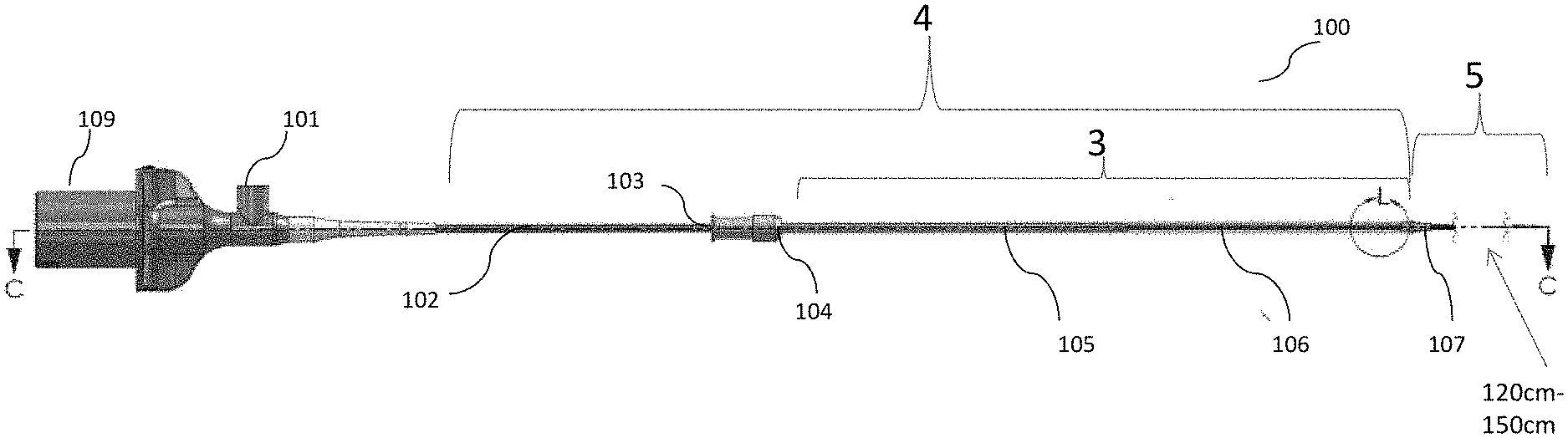
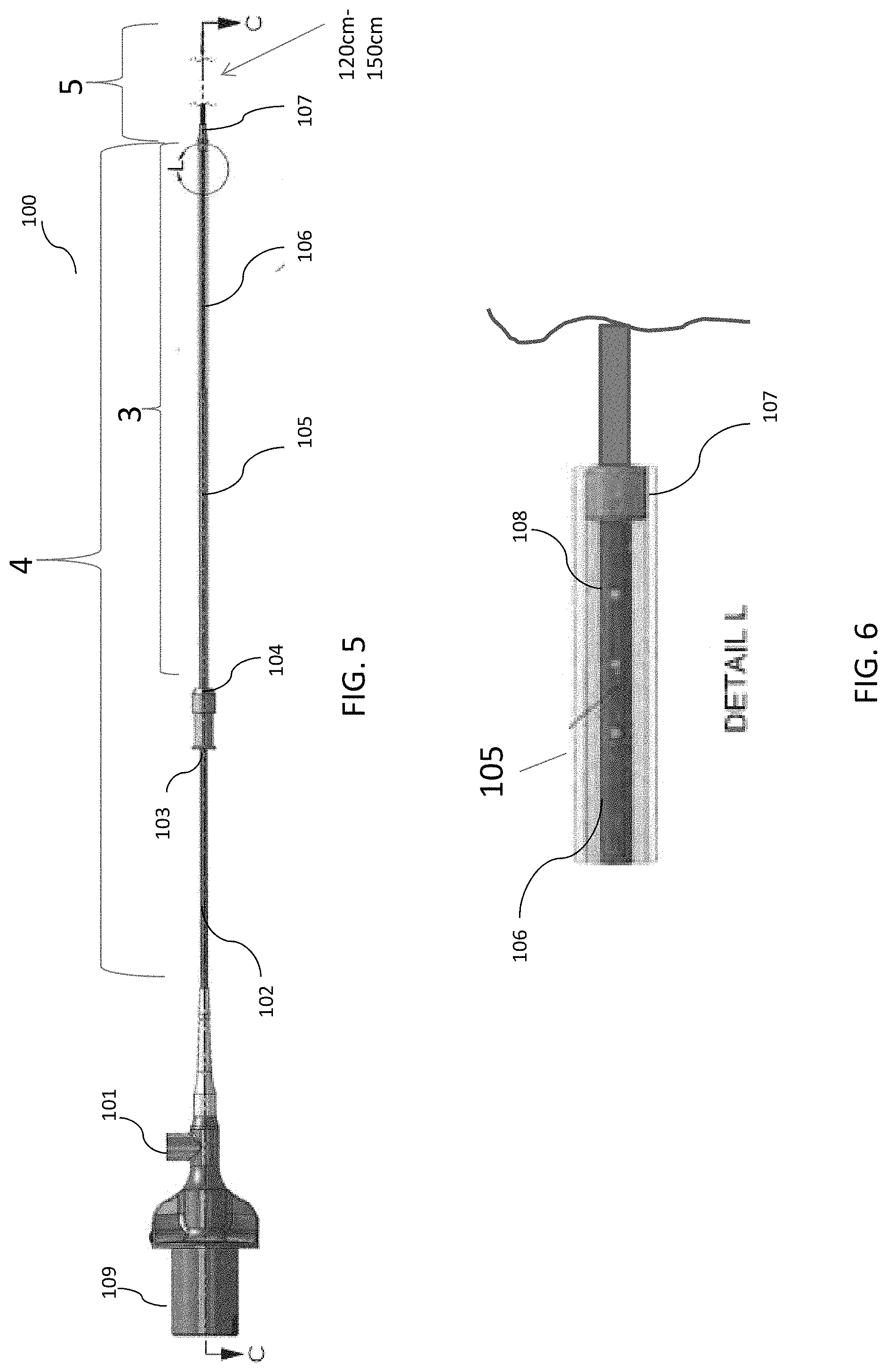
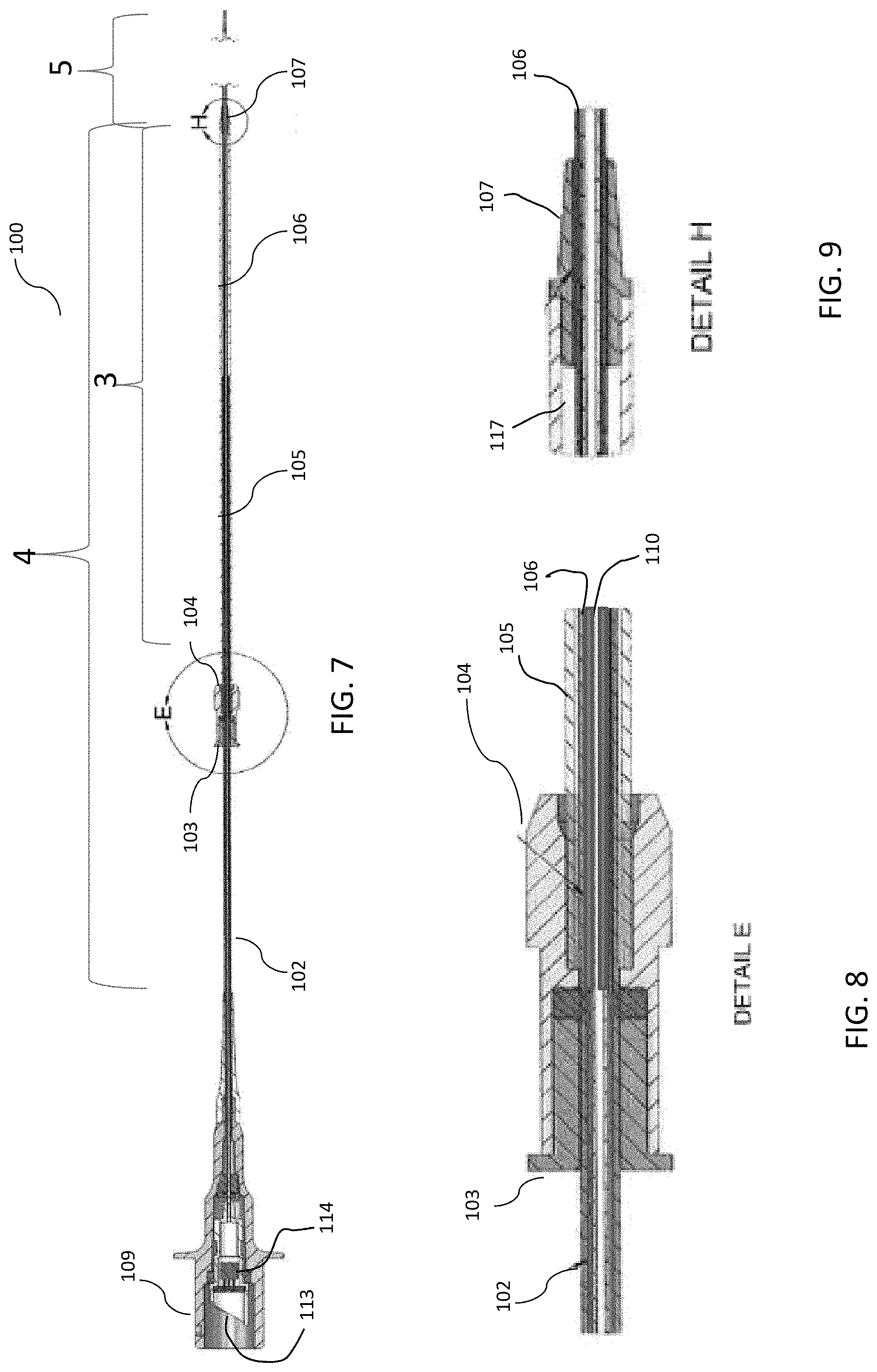
The present invention generally relates to devices for imaging the interior of a vessel. The device can involve an elongated body configured to fit within the lumen of a vessel, a rotatable shaft positioned inside the elongated body, and a telescoping element, wherein a portion of the elongated body extends through the telescoping element, in which the elongated body is configured to contain the rotatable shaft inside the telescoping element.
| Inventors: | Meyer; Douglas (Folsom, CA), Van Hoven; Dylan (Oceanside, CA) | ||||||||||
|---|---|---|---|---|---|---|---|---|---|---|---|
| Applicant: |
|
||||||||||
| Assignee: | PHILIPS IMAGE GUIDED THERAPY
CORPORATION (San Diego, CA) |
||||||||||
| Family ID: | 1000005527506 | ||||||||||
| Appl. No.: | 14/136,408 | ||||||||||
| Filed: | December 20, 2013 |
Prior Publication Data
| Document Identifier | Publication Date | |
|---|---|---|
| US 20140180127 A1 | Jun 26, 2014 | |
Related U.S. Patent Documents
| Application Number | Filing Date | Patent Number | Issue Date | ||
|---|---|---|---|---|---|
| 61745285 | Dec 21, 2012 | ||||
| Current U.S. Class: | 1/1 |
| Current CPC Class: | A61B 8/0891 (20130101); A61B 8/445 (20130101); A61B 8/4461 (20130101); A61B 8/12 (20130101); A61B 8/145 (20130101); A61B 5/0073 (20130101) |
| Current International Class: | A61B 8/08 (20060101); A61B 8/00 (20060101); A61B 8/12 (20060101); A61B 8/14 (20060101); A61B 5/00 (20060101) |
References Cited [Referenced By]
U.S. Patent Documents
| 3301258 | January 1967 | Werner |
| 3617880 | November 1971 | Cormack et al. |
| 3789841 | February 1974 | Antoshkiw |
| 3841308 | October 1974 | Tate |
| 4140364 | February 1979 | Yamashita et al. |
| 4274423 | June 1981 | Mizuno et al. |
| 4344438 | August 1982 | Schultz |
| 4398791 | August 1983 | Dorsey |
| 4432370 | February 1984 | Hughes et al. |
| 4552554 | November 1985 | Gould et al. |
| 4577543 | March 1986 | Wilson |
| 4676980 | June 1987 | Segal et al. |
| 4682895 | July 1987 | Costello |
| 4733665 | March 1988 | Palmaz |
| 4744619 | May 1988 | Cameron |
| 4762129 | August 1988 | Bonzel |
| 4766386 | August 1988 | Oliver et al. |
| 4771774 | September 1988 | Simpson et al. |
| 4794931 | January 1989 | Yock |
| 4800886 | January 1989 | Nestor |
| 4803639 | February 1989 | Steele et al. |
| 4816567 | March 1989 | Cabilly et al. |
| 4819740 | April 1989 | Warrington |
| 4821731 | April 1989 | Martinelli et al. |
| 4824435 | April 1989 | Giesy et al. |
| 4830023 | May 1989 | de Toledo et al. |
| 4834093 | May 1989 | Littleford et al. |
| 4841977 | June 1989 | Griffith et al. |
| 4864578 | September 1989 | Proffitt et al. |
| 4873690 | October 1989 | Adams |
| 4877314 | October 1989 | Kanamori |
| 4887606 | December 1989 | Yock et al. |
| 4917085 | April 1990 | Smith |
| 4917097 | April 1990 | Proudian et al. |
| 4928693 | May 1990 | Goodin et al. |
| 4932413 | June 1990 | Shockey et al. |
| 4932419 | June 1990 | de Toledo |
| 4948229 | August 1990 | Soref |
| 4951677 | August 1990 | Crowley et al. |
| 4969742 | November 1990 | Falk et al. |
| 4987412 | January 1991 | Vaitekunas et al. |
| 4993412 | February 1991 | Murphy-Chutorian |
| 4998972 | March 1991 | Chin et al. |
| 5000185 | March 1991 | Yock |
| 5024234 | June 1991 | Leary et al. |
| 5025445 | June 1991 | Anderson et al. |
| 5032123 | July 1991 | Katz et al. |
| 5037169 | August 1991 | Chun |
| 5039193 | August 1991 | Snow et al. |
| 5040548 | August 1991 | Yock |
| 5041108 | August 1991 | Fox et al. |
| 5054492 | October 1991 | Scribner et al. |
| 5065010 | November 1991 | Knute |
| 5065769 | November 1991 | de Toledo |
| 5085221 | February 1992 | Ingebrigtsen et al. |
| 5095911 | March 1992 | Pomeranz |
| 5100424 | March 1992 | Jang et al. |
| 5120308 | June 1992 | Hess |
| 5125137 | June 1992 | Corl et al. |
| 5135486 | August 1992 | Eberle et al. |
| 5135516 | August 1992 | Sahatjian et al. |
| 5155439 | October 1992 | Holmbo et al. |
| 5158548 | October 1992 | Lau et al. |
| 5163433 | November 1992 | Kagawa |
| 5163445 | November 1992 | Christian et al. |
| 5167233 | December 1992 | Eberle et al. |
| 5174295 | December 1992 | Christian et al. |
| 5176141 | January 1993 | Bom et al. |
| 5176674 | January 1993 | Hofmann |
| 5178159 | January 1993 | Christian |
| 5183048 | February 1993 | Eberle |
| 5188632 | February 1993 | Goldenberg |
| 5201316 | April 1993 | Pomeranz et al. |
| 5202745 | April 1993 | Sorin et al. |
| 5203779 | April 1993 | Muller et al. |
| 5220922 | June 1993 | Barany |
| 5224953 | July 1993 | Morgentaler |
| 5226421 | July 1993 | Frisbie et al. |
| 5240003 | August 1993 | Lancee et al. |
| 5240437 | August 1993 | Christian |
| 5242460 | September 1993 | Klein et al. |
| 5243988 | September 1993 | Sieben et al. |
| 5257974 | November 1993 | Cox |
| 5266302 | November 1993 | Peyman et al. |
| 5267954 | December 1993 | Nita |
| 5301001 | April 1994 | Murphy et al. |
| 5312425 | May 1994 | Evans et al. |
| 5313949 | May 1994 | Yock |
| 5313957 | May 1994 | Little |
| 5319492 | June 1994 | Dorn et al. |
| 5321501 | June 1994 | Swanson et al. |
| 5325198 | June 1994 | Hartley et al. |
| 5336178 | August 1994 | Kaplan et al. |
| 5346689 | September 1994 | Peyman et al. |
| 5348017 | September 1994 | Thornton et al. |
| 5348481 | September 1994 | Ortiz |
| 5353798 | October 1994 | Sieben |
| 5358409 | October 1994 | Obara |
| 5358478 | October 1994 | Thompson et al. |
| 5368037 | November 1994 | Eberle et al. |
| 5373845 | December 1994 | Gardineer et al. |
| 5373849 | December 1994 | Maroney et al. |
| 5375602 | December 1994 | Lancee et al. |
| 5377682 | January 1995 | Ueno et al. |
| 5383853 | January 1995 | Jung et al. |
| 5387193 | February 1995 | Miraki |
| 5396328 | March 1995 | Jestel et al. |
| 5397355 | March 1995 | Marin et al. |
| 5405377 | April 1995 | Cragg |
| 5411016 | May 1995 | Kume et al. |
| 5419777 | May 1995 | Hofling |
| 5421338 | June 1995 | Crowley et al. |
| 5423806 | June 1995 | Dale et al. |
| 5427118 | June 1995 | Nita et al. |
| 5431673 | July 1995 | Summers et al. |
| 5436759 | July 1995 | Dijaili et al. |
| 5439139 | August 1995 | Brovelli |
| 5443457 | August 1995 | Ginn et al. |
| 5453575 | September 1995 | O'Donnell et al. |
| 5456693 | October 1995 | Conston et al. |
| 5459570 | October 1995 | Swanson et al. |
| 5480388 | January 1996 | Zadini et al. |
| 5485845 | January 1996 | Verdonk et al. |
| 5492125 | February 1996 | Kim et al. |
| 5496997 | March 1996 | Pope |
| 5507761 | April 1996 | Duer |
| 5512044 | April 1996 | Duer |
| 5514128 | May 1996 | Hillsman et al. |
| 5529674 | June 1996 | Hedgcoth |
| 5541730 | July 1996 | Chaney |
| 5546717 | August 1996 | Penczak et al. |
| 5546948 | August 1996 | Hamm et al. |
| 5565332 | October 1996 | Hoogenboom et al. |
| 5573520 | November 1996 | Schwartz et al. |
| 5581638 | December 1996 | Givens et al. |
| 5586054 | December 1996 | Jensen et al. |
| 5592939 | January 1997 | Martinelli |
| 5596079 | January 1997 | Smith et al. |
| 5598844 | February 1997 | Diaz et al. |
| 5609606 | March 1997 | O'Boyle |
| 5630806 | May 1997 | Inagaki et al. |
| 5651366 | July 1997 | Liang et al. |
| 5660180 | August 1997 | Malinowski et al. |
| 5667499 | September 1997 | Welch et al. |
| 5667521 | September 1997 | Keown |
| 5672877 | September 1997 | Liebig et al. |
| 5674232 | October 1997 | Halliburton |
| 5693015 | December 1997 | Walker et al. |
| 5713848 | February 1998 | Dubrul et al. |
| 5745634 | April 1998 | Garrett et al. |
| 5771895 | June 1998 | Slager |
| 5779731 | July 1998 | Leavitt |
| 5780958 | July 1998 | Strugach et al. |
| 5798521 | August 1998 | Froggatt |
| 5800450 | September 1998 | Lary et al. |
| 5803083 | September 1998 | Buck et al. |
| 5814061 | September 1998 | Osborne et al. |
| 5817025 | October 1998 | Alekseev et al. |
| 5820594 | October 1998 | Fontirroche et al. |
| 5824520 | October 1998 | Mulligan-Kehoe |
| 5827313 | October 1998 | Ream |
| 5830222 | November 1998 | Makower |
| 5848121 | December 1998 | Gupta et al. |
| 5851464 | December 1998 | Davila et al. |
| 5857974 | January 1999 | Eberle et al. |
| 5872829 | February 1999 | Wischmann et al. |
| 5873835 | February 1999 | Hastings et al. |
| 5882722 | March 1999 | Kydd |
| 5912764 | June 1999 | Togino |
| 5916194 | June 1999 | Jacobsen et al. |
| 5921931 | July 1999 | O'Donnell et al. |
| 5925055 | July 1999 | Adrian et al. |
| 5949929 | September 1999 | Hamm |
| 5951586 | September 1999 | Berg et al. |
| 5974521 | October 1999 | Akerib |
| 5976120 | November 1999 | Chow et al. |
| 5978391 | November 1999 | Das et al. |
| 5997523 | December 1999 | Jang |
| 6021240 | February 2000 | Murphy et al. |
| 6022319 | February 2000 | Willard et al. |
| 6031071 | February 2000 | Mandeville et al. |
| 6036889 | March 2000 | Kydd |
| 6043883 | March 2000 | Leckel et al. |
| 6050949 | April 2000 | White et al. |
| 6059738 | May 2000 | Stoltze et al. |
| 6068638 | May 2000 | Makower |
| 6074362 | June 2000 | Jang et al. |
| 6078831 | June 2000 | Belef et al. |
| 6080109 | June 2000 | Baker et al. |
| 6091496 | July 2000 | Hill |
| 6094591 | July 2000 | Foltz et al. |
| 6095976 | August 2000 | Nachtomy et al. |
| 6097755 | August 2000 | Guenther, Jr. et al. |
| 6099471 | August 2000 | Torp et al. |
| 6099549 | August 2000 | Bosma et al. |
| 6102938 | August 2000 | Evans et al. |
| 6106476 | August 2000 | Corl et al. |
| 6120445 | September 2000 | Grunwald |
| 6123673 | September 2000 | Eberle et al. |
| 6134003 | October 2000 | Tearney et al. |
| 6139510 | October 2000 | Palermo |
| 6141089 | October 2000 | Thoma et al. |
| 6146328 | November 2000 | Chiao et al. |
| 6148095 | November 2000 | Prause et al. |
| 6151433 | November 2000 | Dower et al. |
| 6152877 | November 2000 | Masters |
| 6152878 | November 2000 | Nachtomy et al. |
| 6159225 | December 2000 | Makower |
| 6165127 | December 2000 | Crowley |
| 6176842 | January 2001 | Tachibana et al. |
| 6179809 | January 2001 | Khairkhahan et al. |
| 6186949 | February 2001 | Hatfield et al. |
| 6190353 | February 2001 | Makower et al. |
| 6200266 | March 2001 | Shokrollahi et al. |
| 6200268 | March 2001 | Vince et al. |
| 6203537 | March 2001 | Adrian |
| 6208415 | March 2001 | De Boer et al. |
| 6210332 | April 2001 | Chiao et al. |
| 6210339 | April 2001 | Kiepen et al. |
| 6212308 | April 2001 | Donald |
| 6231518 | May 2001 | Grabek et al. |
| 6245066 | June 2001 | Morgan et al. |
| 6249076 | June 2001 | Madden et al. |
| 6254543 | July 2001 | Grunwald et al. |
| 6256090 | July 2001 | Chen et al. |
| 6258052 | July 2001 | Milo |
| 6261246 | July 2001 | Pantages et al. |
| 6275628 | August 2001 | Jones et al. |
| 6283921 | September 2001 | Nix et al. |
| 6283951 | September 2001 | Flaherty et al. |
| 6295308 | September 2001 | Zah |
| 6299622 | October 2001 | Snow et al. |
| 6312384 | November 2001 | Chiao |
| 6325797 | December 2001 | Stewart et al. |
| 6328696 | December 2001 | Fraser |
| 6343168 | January 2002 | Murphy et al. |
| 6343178 | January 2002 | Burns et al. |
| 6350240 | February 2002 | Song et al. |
| 6364841 | April 2002 | White et al. |
| 6366722 | April 2002 | Murphy et al. |
| 6367984 | April 2002 | Stephenson et al. |
| 6373970 | April 2002 | Dong et al. |
| 6375615 | April 2002 | Flaherty et al. |
| 6375618 | April 2002 | Chiao et al. |
| 6375628 | April 2002 | Zadno-Azizi et al. |
| 6376830 | April 2002 | Froggatt et al. |
| 6379352 | April 2002 | Reynolds et al. |
| 6381350 | April 2002 | Klingensmith et al. |
| 6387124 | May 2002 | Buscemi et al. |
| 6396976 | May 2002 | Little et al. |
| 6398792 | June 2002 | O'Connor |
| 6417948 | July 2002 | Chowdhury et al. |
| 6419644 | July 2002 | White et al. |
| 6421164 | July 2002 | Tearney et al. |
| 6423012 | July 2002 | Kato et al. |
| 6426796 | July 2002 | Pulliam et al. |
| 6428041 | August 2002 | Wohllebe et al. |
| 6428498 | August 2002 | Uflacker |
| 6429421 | August 2002 | Meller et al. |
| 6440077 | August 2002 | Jung et al. |
| 6443903 | September 2002 | White et al. |
| 6450964 | September 2002 | Webler |
| 6457365 | October 2002 | Stephens et al. |
| 6459844 | October 2002 | Pan |
| 6468290 | October 2002 | Weldon et al. |
| 6475149 | November 2002 | Sumanaweera |
| 6480285 | November 2002 | Hill |
| 6491631 | December 2002 | Chiao et al. |
| 6491636 | December 2002 | Chenal et al. |
| 6501551 | December 2002 | Tearney et al. |
| 6504286 | January 2003 | Porat et al. |
| 6508824 | January 2003 | Flaherty et al. |
| 6514237 | February 2003 | Maseda |
| 6520269 | February 2003 | Geiger et al. |
| 6520677 | February 2003 | Iizuka |
| 6535764 | March 2003 | Imran et al. |
| 6538778 | March 2003 | Leckel et al. |
| 6544217 | April 2003 | Gulachenski |
| 6544230 | April 2003 | Flaherty et al. |
| 6545760 | April 2003 | Froggatt et al. |
| 6546272 | April 2003 | MacKinnon et al. |
| 6551250 | April 2003 | Khalil |
| 6566648 | May 2003 | Froggatt |
| 6570894 | May 2003 | Anderson |
| 6572555 | June 2003 | White et al. |
| 6579311 | June 2003 | Makower |
| 6584335 | June 2003 | Haar et al. |
| 6592612 | July 2003 | Samson et al. |
| 6594448 | July 2003 | Herman et al. |
| 6602241 | August 2003 | Makower et al. |
| 6611322 | August 2003 | Nakayama et al. |
| 6611720 | August 2003 | Hata et al. |
| 6612992 | September 2003 | Hossack et al. |
| 6615062 | September 2003 | Ryan et al. |
| 6615072 | September 2003 | Izatt et al. |
| 6621562 | September 2003 | Durston |
| 6631284 | October 2003 | Nutt et al. |
| 6638227 | October 2003 | Bae |
| 6645152 | November 2003 | Jung et al. |
| 6646745 | November 2003 | Verma et al. |
| 6655386 | December 2003 | Makower et al. |
| 6659957 | December 2003 | Vardi et al. |
| 6660024 | December 2003 | Flaherty et al. |
| 6663565 | December 2003 | Kawagishi et al. |
| 6665456 | December 2003 | Dave et al. |
| 6669716 | December 2003 | Gilson et al. |
| 6671055 | December 2003 | Wavering et al. |
| 6673015 | January 2004 | Glover et al. |
| 6673064 | January 2004 | Rentrop |
| 6685648 | February 2004 | Flaherty et al. |
| 6689056 | February 2004 | Kilcoyne et al. |
| 6689144 | February 2004 | Gerberding |
| 6696173 | February 2004 | Naundorf et al. |
| 6701044 | March 2004 | Arbore et al. |
| 6701176 | March 2004 | Halperin et al. |
| 6709444 | March 2004 | Makower |
| 6712836 | March 2004 | Berg et al. |
| 6714703 | March 2004 | Lee et al. |
| 6719717 | April 2004 | Johnson et al. |
| 6725073 | April 2004 | Motamedi et al. |
| 6726677 | April 2004 | Flaherty et al. |
| 6730107 | May 2004 | Kelley et al. |
| 6733474 | May 2004 | Kusleika |
| 6738144 | May 2004 | Dogariu |
| 6740113 | May 2004 | Vrba |
| 6746464 | June 2004 | Makower |
| 6780157 | August 2004 | Stephens et al. |
| 6795188 | September 2004 | Ruck et al. |
| 6795196 | September 2004 | Funakawa |
| 6798522 | September 2004 | Stolte et al. |
| 6822798 | November 2004 | Wu et al. |
| 6830559 | December 2004 | Schock |
| 6832024 | December 2004 | Gerstenberger et al. |
| 6842639 | January 2005 | Winston et al. |
| 6847449 | January 2005 | Bashkansky et al. |
| 6855115 | February 2005 | Fonseca et al. |
| 6856138 | February 2005 | Bohley |
| 6856400 | February 2005 | Froggatt |
| 6856472 | February 2005 | Herman et al. |
| 6860867 | March 2005 | Seward et al. |
| 6866670 | March 2005 | Rabiner et al. |
| 6878113 | April 2005 | Miwa et al. |
| 6886411 | May 2005 | Kjellman et al. |
| 6891984 | May 2005 | Petersen et al. |
| 6895106 | May 2005 | Wang et al. |
| 6898337 | May 2005 | Averett et al. |
| 6900897 | May 2005 | Froggatt |
| 6912051 | June 2005 | Jensen |
| 6916329 | July 2005 | Zhao |
| 6922498 | July 2005 | Shah |
| 6937346 | August 2005 | Nebendahl et al. |
| 6937696 | August 2005 | Mostafavi |
| 6943939 | September 2005 | DiJaili et al. |
| 6947147 | September 2005 | Motamedi et al. |
| 6947787 | September 2005 | Webler |
| 6949094 | September 2005 | Yaron |
| 6952603 | October 2005 | Gerber et al. |
| 6954737 | October 2005 | Kalantar et al. |
| 6958042 | October 2005 | Honda |
| 6961123 | November 2005 | Wang et al. |
| 6966891 | November 2005 | Ookubo et al. |
| 6969293 | November 2005 | Thai |
| 6969395 | November 2005 | Eskuri |
| 6985234 | January 2006 | Anderson |
| 7004963 | February 2006 | Wang et al. |
| 7006231 | February 2006 | Ostrovsky et al. |
| 7010458 | March 2006 | Wilt |
| 7024025 | April 2006 | Sathyanarayana |
| 7027211 | April 2006 | Ruffa |
| 7027743 | April 2006 | Tucker et al. |
| 7033347 | April 2006 | Appling |
| 7035484 | April 2006 | Silberberg et al. |
| 7037269 | May 2006 | Nix et al. |
| 7042573 | May 2006 | Froggatt |
| 7044915 | May 2006 | White et al. |
| 7044964 | May 2006 | Jang et al. |
| 7048711 | May 2006 | Rosenman et al. |
| 7049306 | May 2006 | Konradi et al. |
| 7058239 | June 2006 | Singh et al. |
| 7060033 | June 2006 | White et al. |
| 7060421 | June 2006 | Naundorf et al. |
| 7063679 | June 2006 | Maguire et al. |
| 7068852 | June 2006 | Braica |
| 7074188 | July 2006 | Nair et al. |
| 7095493 | August 2006 | Harres |
| 7110119 | September 2006 | Maestle |
| 7113875 | September 2006 | Terashima et al. |
| 7123777 | October 2006 | Rondinelli et al. |
| 7130054 | October 2006 | Ostrovsky et al. |
| 7139440 | November 2006 | Rondinelli et al. |
| 7153299 | December 2006 | Tu et al. |
| 7171078 | January 2007 | Sasaki et al. |
| 7175597 | February 2007 | Vince et al. |
| 7177491 | February 2007 | Dave et al. |
| 7190464 | March 2007 | Alphonse |
| 7215802 | May 2007 | Klingensmith et al. |
| 7218811 | May 2007 | Shigenaga et al. |
| 7236812 | June 2007 | Ballerstadt et al. |
| 7245125 | July 2007 | Harer et al. |
| 7245789 | July 2007 | Bates et al. |
| 7249357 | July 2007 | Landman et al. |
| 7291146 | November 2007 | Steinke et al. |
| 7292715 | November 2007 | Furnish |
| 7292885 | November 2007 | Scott et al. |
| 7294124 | November 2007 | Eidenschink |
| 7300460 | November 2007 | Levine et al. |
| 7335161 | February 2008 | Von Arx et al. |
| 7337079 | February 2008 | Park et al. |
| 7355716 | April 2008 | de Boer et al. |
| 7356367 | April 2008 | Liang et al. |
| 7358921 | April 2008 | Snyder et al. |
| 7359062 | April 2008 | Chen et al. |
| 7359554 | April 2008 | Klingensmith et al. |
| 7363927 | April 2008 | Ravikumar |
| 7366376 | April 2008 | Shishkov et al. |
| 7382949 | June 2008 | Bouma et al. |
| 7387636 | June 2008 | Cohn et al. |
| 7391520 | June 2008 | Zhou et al. |
| 7397935 | July 2008 | Kimmel et al. |
| 7399095 | July 2008 | Rondinelli |
| 7408648 | August 2008 | Kleen et al. |
| 7414779 | August 2008 | Huber et al. |
| 7440087 | October 2008 | Froggatt et al. |
| 7447388 | November 2008 | Bates et al. |
| 7449821 | November 2008 | Dausch |
| 7450165 | November 2008 | Ahiska |
| RE40608 | December 2008 | Glover et al. |
| 7458967 | December 2008 | Appling et al. |
| 7463362 | December 2008 | Lasker et al. |
| 7463759 | December 2008 | Klingensmith et al. |
| 7491226 | February 2009 | Palmaz et al. |
| 7515276 | April 2009 | Froggatt et al. |
| 7527594 | May 2009 | Vardi et al. |
| 7534251 | May 2009 | WasDyke |
| 7535797 | May 2009 | Peng et al. |
| 7547304 | June 2009 | Johnson |
| 7564949 | July 2009 | Sattler et al. |
| 7577471 | August 2009 | Camus et al. |
| 7583857 | September 2009 | Xu et al. |
| 7603165 | October 2009 | Townsend et al. |
| 7612773 | November 2009 | Magnin et al. |
| 7633627 | December 2009 | Choma et al. |
| 7645229 | January 2010 | Armstrong |
| 7658715 | February 2010 | Park et al. |
| 7660452 | February 2010 | Zwirn et al. |
| 7660492 | February 2010 | Bates et al. |
| 7666204 | February 2010 | Thornton et al. |
| 7672790 | March 2010 | McGraw et al. |
| 7680247 | March 2010 | Atzinger et al. |
| 7684991 | March 2010 | Stohr et al. |
| 7711413 | May 2010 | Feldman et al. |
| 7720322 | May 2010 | Prisco |
| 7728986 | June 2010 | Lasker et al. |
| 7734009 | June 2010 | Brunner et al. |
| 7736317 | June 2010 | Stephens et al. |
| 7742795 | June 2010 | Stone et al. |
| 7743189 | June 2010 | Brown et al. |
| 7762954 | July 2010 | Nix et al. |
| 7766896 | August 2010 | Kornkven Volk et al. |
| 7773792 | August 2010 | Kimmel et al. |
| 7775981 | August 2010 | Guracar et al. |
| 7777399 | August 2010 | Eidenschink et al. |
| 7781724 | August 2010 | Childers et al. |
| 7783337 | August 2010 | Feldman et al. |
| 7787127 | August 2010 | Galle et al. |
| 7792342 | September 2010 | Barbu et al. |
| 7801343 | September 2010 | Unal et al. |
| 7801590 | September 2010 | Feldman et al. |
| 7813609 | October 2010 | Petersen et al. |
| 7831081 | November 2010 | Li |
| 7846101 | December 2010 | Eberle et al. |
| 7853104 | December 2010 | Oota et al. |
| 7853316 | December 2010 | Milner et al. |
| 7860555 | December 2010 | Saadat |
| 7862508 | January 2011 | Davies et al. |
| 7872759 | January 2011 | Tearney et al. |
| 7880868 | February 2011 | Aoki |
| 7881763 | February 2011 | Brauker et al. |
| 7909844 | March 2011 | Alkhatib et al. |
| 7921854 | April 2011 | Hennings et al. |
| 7927784 | April 2011 | Simpson |
| 7929148 | April 2011 | Kemp |
| 7930014 | April 2011 | Huennekens et al. |
| 7930104 | April 2011 | Baker et al. |
| 7936462 | May 2011 | Jiang et al. |
| 7942852 | May 2011 | Mas et al. |
| 7947012 | May 2011 | Spurchise et al. |
| 7951186 | May 2011 | Eidenschink et al. |
| 7952719 | May 2011 | Brennan, III |
| 7972353 | July 2011 | Hendriksen et al. |
| 7976492 | July 2011 | Brauker et al. |
| 7977950 | July 2011 | Maslen |
| 7978916 | July 2011 | Klingensmith et al. |
| 7981041 | July 2011 | McGahan |
| 7981151 | July 2011 | Rowe |
| 7983737 | July 2011 | Feldman et al. |
| 7993333 | August 2011 | Oral et al. |
| 7995210 | August 2011 | Tearney et al. |
| 7996060 | August 2011 | Trofimov et al. |
| 7999938 | August 2011 | Wang |
| 8021377 | September 2011 | Eskuri |
| 8021420 | September 2011 | Dolan |
| 8036732 | October 2011 | Milner |
| 8040586 | October 2011 | Smith et al. |
| 8047996 | November 2011 | Goodnow et al. |
| 8049900 | November 2011 | Kemp et al. |
| 8050478 | November 2011 | Li et al. |
| 8050523 | November 2011 | Younge et al. |
| 8052605 | November 2011 | Muller et al. |
| 8057394 | November 2011 | Dala-Krishna |
| 8059923 | November 2011 | Bates et al. |
| 8070800 | December 2011 | Lock et al. |
| 8080800 | December 2011 | Hoctor et al. |
| 8088102 | January 2012 | Adams et al. |
| 8100838 | January 2012 | Wright et al. |
| 8104479 | January 2012 | Glynn et al. |
| 8108030 | January 2012 | Castella et al. |
| 8114102 | February 2012 | Galdonik et al. |
| 8116605 | February 2012 | Petersen et al. |
| 8125648 | February 2012 | Milner et al. |
| 8126239 | February 2012 | Sun et al. |
| 8133199 | March 2012 | Weber et al. |
| 8133269 | March 2012 | Flechsenhar et al. |
| 8140708 | March 2012 | Zaharia et al. |
| 8148877 | April 2012 | Jiang et al. |
| 8167932 | May 2012 | Bourang et al. |
| 8172757 | May 2012 | Jaffe et al. |
| 8177809 | May 2012 | Mavani et al. |
| 8187191 | May 2012 | Hancock et al. |
| 8187267 | May 2012 | Pappone et al. |
| 8187830 | May 2012 | Hu et al. |
| 8199218 | June 2012 | Lee et al. |
| 8206429 | June 2012 | Gregorich et al. |
| 8208995 | June 2012 | Tearney et al. |
| 8222906 | July 2012 | Wyar et al. |
| 8233681 | July 2012 | Aylward et al. |
| 8233718 | July 2012 | Klingensmith et al. |
| 8238624 | August 2012 | Doi et al. |
| 8239938 | August 2012 | Simeral et al. |
| 8277386 | October 2012 | Ahmed et al. |
| 8280470 | October 2012 | Milner et al. |
| 8289284 | October 2012 | Glynn et al. |
| 8289522 | October 2012 | Tearney et al. |
| 8298147 | October 2012 | Huennekens et al. |
| 8298149 | October 2012 | Hastings et al. |
| 8301000 | October 2012 | Sillard et al. |
| 8309428 | November 2012 | Lemmerhirt et al. |
| 8317713 | November 2012 | Davies et al. |
| 8323201 | December 2012 | Towfiq et al. |
| 8329053 | December 2012 | Martin et al. |
| 8336643 | December 2012 | Harleman |
| 8349000 | January 2013 | Schreck |
| 8353945 | January 2013 | Andreas et al. |
| 8353954 | January 2013 | Cai et al. |
| 8357981 | January 2013 | Martin et al. |
| 8361097 | January 2013 | Patel et al. |
| 8386560 | February 2013 | Ma et al. |
| 8398591 | March 2013 | Mas et al. |
| 8412312 | April 2013 | Judell et al. |
| 8417491 | April 2013 | Trovato et al. |
| 8449465 | May 2013 | Nair et al. |
| 8454685 | June 2013 | Hariton et al. |
| 8454686 | June 2013 | Alkhatib |
| 8475522 | July 2013 | Jimenez et al. |
| 8478384 | July 2013 | Schmitt et al. |
| 8486062 | July 2013 | Belhe et al. |
| 8486063 | July 2013 | Werneth et al. |
| 8491567 | July 2013 | Magnin et al. |
| 8500798 | August 2013 | Rowe et al. |
| 8550911 | October 2013 | Sylla |
| 8594757 | November 2013 | Boppart et al. |
| 8597349 | December 2013 | Alkhatib |
| 8600477 | December 2013 | Beyar et al. |
| 8600917 | December 2013 | Schimert et al. |
| 8601056 | December 2013 | Lauwers et al. |
| 8620055 | December 2013 | Barratt et al. |
| 8644910 | February 2014 | Rousso et al. |
| 2001/0007940 | July 2001 | Tu et al. |
| 2001/0029337 | October 2001 | Pantages et al. |
| 2001/0037073 | November 2001 | White et al. |
| 2001/0046345 | November 2001 | Snyder et al. |
| 2001/0049548 | December 2001 | Vardi et al. |
| 2002/0034276 | March 2002 | Hu et al. |
| 2002/0041723 | April 2002 | Ronnekleiv et al. |
| 2002/0069676 | June 2002 | Kopp et al. |
| 2002/0089335 | July 2002 | Williams |
| 2002/0099289 | July 2002 | Crowley |
| 2002/0163646 | November 2002 | Anderson |
| 2002/0186818 | December 2002 | Arnaud et al. |
| 2002/0196446 | December 2002 | Roth et al. |
| 2002/0197456 | December 2002 | Pope |
| 2003/0004412 | January 2003 | Izatt et al. |
| 2003/0016604 | January 2003 | Hanes |
| 2003/0018273 | January 2003 | Corl et al. |
| 2003/0023153 | January 2003 | Izatt et al. |
| 2003/0032886 | February 2003 | Dgany et al. |
| 2003/0050871 | March 2003 | Broughton |
| 2003/0065371 | April 2003 | Satake |
| 2003/0069723 | April 2003 | Hegde |
| 2003/0077043 | April 2003 | Hamm et al. |
| 2003/0085635 | May 2003 | Davidsen |
| 2003/0090753 | May 2003 | Takeyama et al. |
| 2003/0092995 | May 2003 | Thompson |
| 2003/0093059 | May 2003 | Griffin et al. |
| 2003/0103212 | June 2003 | Westphal et al. |
| 2003/0152259 | August 2003 | Belykh et al. |
| 2003/0181802 | September 2003 | Ogawa |
| 2003/0187369 | October 2003 | Lewis et al. |
| 2003/0194165 | October 2003 | Silberberg et al. |
| 2003/0195419 | October 2003 | Harada |
| 2003/0208116 | November 2003 | Liang et al. |
| 2003/0212491 | November 2003 | Mitchell et al. |
| 2003/0219202 | November 2003 | Loeb et al. |
| 2003/0220749 | November 2003 | Chen et al. |
| 2003/0228039 | December 2003 | Green |
| 2004/0015065 | January 2004 | Panescu et al. |
| 2004/0023317 | February 2004 | Motamedi et al. |
| 2004/0028333 | February 2004 | Lomas |
| 2004/0037742 | February 2004 | Jen et al. |
| 2004/0042066 | March 2004 | Kinoshita et al. |
| 2004/0054287 | March 2004 | Stephens |
| 2004/0067000 | April 2004 | Bates et al. |
| 2004/0068161 | April 2004 | Couvillon |
| 2004/0082844 | April 2004 | Vardi et al. |
| 2004/0092830 | May 2004 | Scott et al. |
| 2004/0106853 | June 2004 | Moriyama |
| 2004/0111552 | June 2004 | Arimilli et al. |
| 2004/0126048 | July 2004 | Dave et al. |
| 2004/0143160 | July 2004 | Couvillon |
| 2004/0146546 | July 2004 | Gravett et al. |
| 2004/0186369 | September 2004 | Lam |
| 2004/0186558 | September 2004 | Pavcnik et al. |
| 2004/0195512 | October 2004 | Crosetto |
| 2004/0220606 | November 2004 | Goshgarian |
| 2004/0225220 | November 2004 | Rich |
| 2004/0239938 | December 2004 | Izatt |
| 2004/0242990 | December 2004 | Brister et al. |
| 2004/0248439 | December 2004 | Gernhardt et al. |
| 2004/0260236 | December 2004 | Manning et al. |
| 2005/0013778 | January 2005 | Green et al. |
| 2005/0031176 | February 2005 | Hertel et al. |
| 2005/0036150 | February 2005 | Izatt et al. |
| 2005/0078317 | April 2005 | Law et al. |
| 2005/0101859 | May 2005 | Maschke |
| 2005/0140582 | June 2005 | Lee et al. |
| 2005/0140682 | June 2005 | Sumanaweera et al. |
| 2005/0140981 | June 2005 | Waelti |
| 2005/0140984 | June 2005 | Hitzenberger |
| 2005/0147303 | July 2005 | Zhou et al. |
| 2005/0165439 | July 2005 | Weber et al. |
| 2005/0171433 | August 2005 | Boppart et al. |
| 2005/0171438 | August 2005 | Chen et al. |
| 2005/0182297 | August 2005 | Gravenstein et al. |
| 2005/0196028 | September 2005 | Kleen et al. |
| 2005/0197585 | September 2005 | Brockway et al. |
| 2005/0213103 | September 2005 | Everett et al. |
| 2005/0215942 | September 2005 | Abrahamson et al. |
| 2005/0234445 | October 2005 | Conquergood et al. |
| 2005/0243322 | November 2005 | Lasker et al. |
| 2005/0249391 | November 2005 | Kimmel et al. |
| 2005/0251567 | November 2005 | Ballew et al. |
| 2005/0254059 | November 2005 | Alphonse |
| 2005/0264823 | December 2005 | Zhu et al. |
| 2006/0013523 | January 2006 | Childlers et al. |
| 2006/0015126 | January 2006 | Sher |
| 2006/0029634 | February 2006 | Berg et al. |
| 2006/0036167 | February 2006 | Shina |
| 2006/0038115 | February 2006 | Maas |
| 2006/0039004 | February 2006 | de Boer et al. |
| 2006/0041180 | February 2006 | Viswanathan et al. |
| 2006/0045536 | March 2006 | Arahira |
| 2006/0055936 | March 2006 | Yun et al. |
| 2006/0058622 | March 2006 | Tearney et al. |
| 2006/0064009 | March 2006 | Webler et al. |
| 2006/0067620 | March 2006 | Shishkov et al. |
| 2006/0072808 | April 2006 | Grimm et al. |
| 2006/0074442 | April 2006 | Noriega et al. |
| 2006/0098927 | May 2006 | Schmidt et al. |
| 2006/0100694 | May 2006 | Globerman |
| 2006/0106375 | May 2006 | Werneth et al. |
| 2006/0132790 | June 2006 | Gutin |
| 2006/0135870 | June 2006 | Webler |
| 2006/0142703 | June 2006 | Carter et al. |
| 2006/0142733 | June 2006 | Forsberg |
| 2006/0173299 | August 2006 | Romley et al. |
| 2006/0179255 | August 2006 | Yamazaki |
| 2006/0184048 | August 2006 | Saadat |
| 2006/0187537 | August 2006 | Huber et al. |
| 2006/0195269 | August 2006 | Yeatman et al. |
| 2006/0204119 | September 2006 | Feng et al. |
| 2006/0229591 | October 2006 | Lee |
| 2006/0239312 | October 2006 | Kewitsch et al. |
| 2006/0241342 | October 2006 | Macaulay et al. |
| 2006/0241465 | October 2006 | Huennekens et al. |
| 2006/0241484 | October 2006 | Horiike et al. |
| 2006/0241503 | October 2006 | Schmitt et al. |
| 2006/0244973 | November 2006 | Yun et al. |
| 2006/0258895 | November 2006 | Maschke |
| 2006/0264743 | November 2006 | Kleen et al. |
| 2006/0267756 | November 2006 | Kates |
| 2006/0270976 | November 2006 | Savage et al. |
| 2006/0276709 | December 2006 | Khamene et al. |
| 2006/0279742 | December 2006 | Tearney et al. |
| 2006/0279743 | December 2006 | Boesser et al. |
| 2006/0285638 | December 2006 | Boese et al. |
| 2006/0287595 | December 2006 | Maschke |
| 2006/0293597 | December 2006 | Johnson et al. |
| 2007/0015969 | January 2007 | Feldman et al. |
| 2007/0016029 | January 2007 | Donaldson et al. |
| 2007/0016034 | January 2007 | Donaldson |
| 2007/0016062 | January 2007 | Park et al. |
| 2007/0027390 | February 2007 | Maschke et al. |
| 2007/0036417 | February 2007 | Argiro et al. |
| 2007/0038061 | February 2007 | Huennekens et al. |
| 2007/0038121 | February 2007 | Feldman et al. |
| 2007/0038125 | February 2007 | Kleen et al. |
| 2007/0043292 | February 2007 | Camus et al. |
| 2007/0043597 | February 2007 | Donaldson |
| 2007/0049847 | March 2007 | Osborne |
| 2007/0060973 | March 2007 | Ludvig et al. |
| 2007/0065077 | March 2007 | Childers et al. |
| 2007/0066888 | March 2007 | Maschke |
| 2007/0066890 | March 2007 | Maschke |
| 2007/0066983 | March 2007 | Maschke |
| 2007/0084995 | April 2007 | Newton et al. |
| 2007/0100226 | May 2007 | Yankelevitz et al. |
| 2007/0135887 | June 2007 | Maschke |
| 2007/0142707 | June 2007 | Wiklof et al. |
| 2007/0156019 | July 2007 | Larkin et al. |
| 2007/0161893 | July 2007 | Milner et al. |
| 2007/0161896 | July 2007 | Adachi et al. |
| 2007/0161963 | July 2007 | Smalling |
| 2007/0162860 | July 2007 | Muralidharan et al. |
| 2007/0165141 | July 2007 | Srinivas et al. |
| 2007/0167710 | July 2007 | Unal et al. |
| 2007/0167804 | July 2007 | Park et al. |
| 2007/0191682 | August 2007 | Rolland et al. |
| 2007/0201736 | August 2007 | Klingensmith et al. |
| 2007/0206193 | September 2007 | Pesach |
| 2007/0208276 | September 2007 | Kornkven Volk et al. |
| 2007/0219446 | September 2007 | Beyhan |
| 2007/0225220 | September 2007 | Ming et al. |
| 2007/0225590 | September 2007 | Ramos |
| 2007/0229801 | October 2007 | Tearney et al. |
| 2007/0232872 | October 2007 | Prough et al. |
| 2007/0232874 | October 2007 | Ince |
| 2007/0232890 | October 2007 | Hirota |
| 2007/0232891 | October 2007 | Hirota |
| 2007/0232892 | October 2007 | Hirota |
| 2007/0232893 | October 2007 | Tanioka |
| 2007/0232933 | October 2007 | Gille et al. |
| 2007/0238957 | October 2007 | Yared |
| 2007/0247033 | October 2007 | Eidenschink et al. |
| 2007/0250000 | October 2007 | Magnin et al. |
| 2007/0250036 | October 2007 | Volk et al. |
| 2007/0258094 | November 2007 | Izatt et al. |
| 2007/0260138 | November 2007 | Feldman et al. |
| 2007/0278389 | December 2007 | Ajgaonkar et al. |
| 2007/0287914 | December 2007 | Cohen |
| 2008/0002183 | January 2008 | Yatagai et al. |
| 2008/0013093 | January 2008 | Izatt et al. |
| 2008/0021275 | January 2008 | Tearney et al. |
| 2008/0027481 | January 2008 | Gilson et al. |
| 2008/0043024 | February 2008 | Schiwietz et al. |
| 2008/0045842 | February 2008 | Furnish |
| 2008/0051660 | February 2008 | Kakadaris et al. |
| 2008/0063304 | March 2008 | Russak et al. |
| 2008/0085041 | April 2008 | Breeuwer |
| 2008/0095465 | April 2008 | Mullick et al. |
| 2008/0095714 | April 2008 | Castella et al. |
| 2008/0097194 | April 2008 | Milner |
| 2008/0101667 | May 2008 | Begelman et al. |
| 2008/0108867 | May 2008 | Zhou |
| 2008/0114254 | May 2008 | Matcovitch et al. |
| 2008/0119739 | May 2008 | Vardi et al. |
| 2008/0124495 | May 2008 | Horn et al. |
| 2008/0125772 | May 2008 | Stone et al. |
| 2008/0139897 | June 2008 | Ainsworth et al. |
| 2008/0143707 | June 2008 | Mitchell |
| 2008/0146941 | June 2008 | Dala-Krishna |
| 2008/0147111 | June 2008 | Johnson et al. |
| 2008/0154128 | June 2008 | Milner |
| 2008/0161696 | July 2008 | Schmitt et al. |
| 2008/0171944 | July 2008 | Brenneman et al. |
| 2008/0175465 | July 2008 | Jiang et al. |
| 2008/0177183 | July 2008 | Courtney et al. |
| 2008/0180683 | July 2008 | Kemp |
| 2008/0181477 | July 2008 | Izatt et al. |
| 2008/0187201 | August 2008 | Liang et al. |
| 2008/0228086 | September 2008 | Ilegbusi et al. |
| 2008/0247622 | October 2008 | Aylward et al. |
| 2008/0247716 | October 2008 | Thomas et al. |
| 2008/0262470 | October 2008 | Lee et al. |
| 2008/0262489 | October 2008 | Steinke |
| 2008/0269599 | October 2008 | Csavoy et al. |
| 2008/0281205 | November 2008 | Naghavi et al. |
| 2008/0281248 | November 2008 | Angheloiu et al. |
| 2008/0285043 | November 2008 | Fercher et al. |
| 2008/0287795 | November 2008 | Klingensmith et al. |
| 2008/0291463 | November 2008 | Milner et al. |
| 2008/0292173 | November 2008 | Hsieh et al. |
| 2008/0294034 | November 2008 | Krueger et al. |
| 2008/0298655 | December 2008 | Edwards |
| 2008/0306766 | December 2008 | Ozeki et al. |
| 2009/0009801 | January 2009 | Tabuki |
| 2009/0018393 | January 2009 | Dick |
| 2009/0034813 | February 2009 | Dikmen et al. |
| 2009/0043191 | February 2009 | Castella et al. |
| 2009/0046295 | February 2009 | Kemp et al. |
| 2009/0052614 | February 2009 | Hempel et al. |
| 2009/0069843 | March 2009 | Agnew |
| 2009/0079993 | March 2009 | Yatagai et al. |
| 2009/0088650 | April 2009 | Corl |
| 2009/0093980 | April 2009 | Kemp et al. |
| 2009/0122320 | May 2009 | Petersen et al. |
| 2009/0138544 | May 2009 | Wegenkittl et al. |
| 2009/0149739 | June 2009 | Maschke |
| 2009/0156941 | June 2009 | Moore |
| 2009/0157002 | June 2009 | Dumot |
| 2009/0174886 | July 2009 | Inoue |
| 2009/0174931 | July 2009 | Huber et al. |
| 2009/0177090 | July 2009 | Grunwald et al. |
| 2009/0177183 | July 2009 | Pinkernell et al. |
| 2009/0195514 | August 2009 | Glynn et al. |
| 2009/0196470 | August 2009 | Carl et al. |
| 2009/0198125 | August 2009 | Nakabayashi et al. |
| 2009/0203991 | August 2009 | Papaioannou et al. |
| 2009/0264768 | October 2009 | Courtney et al. |
| 2009/0269014 | October 2009 | Winberg et al. |
| 2009/0270695 | October 2009 | McEowen |
| 2009/0284322 | November 2009 | Harrison et al. |
| 2009/0284332 | November 2009 | Moore et al. |
| 2009/0284749 | November 2009 | Johnson et al. |
| 2009/0290167 | November 2009 | Flanders et al. |
| 2009/0292048 | November 2009 | Li et al. |
| 2009/0299195 | December 2009 | Muller et al. |
| 2009/0299284 | December 2009 | Holman et al. |
| 2009/0318951 | December 2009 | Kashkarov et al. |
| 2009/0326634 | December 2009 | Vardi |
| 2010/0007669 | January 2010 | Bethune et al. |
| 2010/0030042 | February 2010 | Denninghoff et al. |
| 2010/0061611 | March 2010 | Xu et al. |
| 2010/0063400 | March 2010 | Hall et al. |
| 2010/0087732 | April 2010 | Eberle et al. |
| 2010/0094125 | April 2010 | Younge et al. |
| 2010/0094127 | April 2010 | Xu |
| 2010/0094135 | April 2010 | Fang-Yen et al. |
| 2010/0094143 | April 2010 | Mahapatra et al. |
| 2010/0113919 | May 2010 | Maschke |
| 2010/0125238 | May 2010 | Lye et al. |
| 2010/0125268 | May 2010 | Gustus et al. |
| 2010/0125648 | May 2010 | Zaharia et al. |
| 2010/0128348 | May 2010 | Taverner |
| 2010/0152717 | June 2010 | Keeler |
| 2010/0160788 | June 2010 | Davies et al. |
| 2010/0161023 | June 2010 | Cohen et al. |
| 2010/0168714 | July 2010 | Burke et al. |
| 2010/0179421 | July 2010 | Tupin |
| 2010/0179426 | July 2010 | Davies et al. |
| 2010/0220334 | September 2010 | Condit et al. |
| 2010/0226607 | September 2010 | Zhang et al. |
| 2010/0234736 | September 2010 | Corl |
| 2010/0249601 | September 2010 | Courtney |
| 2010/0256616 | October 2010 | Katoh et al. |
| 2010/0272432 | October 2010 | Johnson |
| 2010/0284590 | November 2010 | Peng et al. |
| 2010/0290693 | November 2010 | Cohen et al. |
| 2010/0331950 | December 2010 | Strommer |
| 2011/0010925 | January 2011 | Nix et al. |
| 2011/0021911 | January 2011 | Waters |
| 2011/0021926 | January 2011 | Spencer et al. |
| 2011/0025853 | February 2011 | Richardson |
| 2011/0026797 | February 2011 | Declerck et al. |
| 2011/0032533 | February 2011 | Izatt et al. |
| 2011/0034801 | February 2011 | Baumgart |
| 2011/0044546 | February 2011 | Pan et al. |
| 2011/0066073 | March 2011 | Kuiper et al. |
| 2011/0071401 | March 2011 | Hastings et al. |
| 2011/0072405 | March 2011 | Chen et al. |
| 2011/0077528 | March 2011 | Kemp et al. |
| 2011/0080591 | April 2011 | Johnson et al. |
| 2011/0087104 | April 2011 | Moore et al. |
| 2011/0137140 | June 2011 | Tearney et al. |
| 2011/0144502 | June 2011 | Zhou et al. |
| 2011/0152771 | June 2011 | Milner et al. |
| 2011/0157597 | June 2011 | Lu et al. |
| 2011/0160586 | June 2011 | Li et al. |
| 2011/0178413 | July 2011 | Schmitt et al. |
| 2011/0190586 | August 2011 | Kemp |
| 2011/0216378 | September 2011 | Poon et al. |
| 2011/0220985 | September 2011 | Son et al. |
| 2011/0238061 | September 2011 | van der Weide et al. |
| 2011/0238083 | September 2011 | Moll et al. |
| 2011/0245669 | October 2011 | Zhang |
| 2011/0249094 | October 2011 | Wang et al. |
| 2011/0257545 | October 2011 | Suri |
| 2011/0264125 | October 2011 | Wilson et al. |
| 2011/0274329 | November 2011 | Mathew et al. |
| 2011/0282334 | November 2011 | Groenhoff |
| 2011/0301684 | December 2011 | Fischell et al. |
| 2011/0306995 | December 2011 | Moberg |
| 2011/0319752 | December 2011 | Steinberg et al. |
| 2012/0004529 | January 2012 | Tolkowsky et al. |
| 2012/0004668 | January 2012 | Wallace et al. |
| 2012/0013914 | January 2012 | Kemp et al. |
| 2012/0016344 | January 2012 | Kusakabe |
| 2012/0016395 | January 2012 | Olson |
| 2012/0022360 | January 2012 | Kemp |
| 2012/0026503 | February 2012 | Lewandowski et al. |
| 2012/0029007 | February 2012 | Graham et al. |
| 2012/0059253 | March 2012 | Wang et al. |
| 2012/0059368 | March 2012 | Takaoka et al. |
| 2012/0062843 | March 2012 | Ferguson et al. |
| 2012/0065481 | March 2012 | Hunter et al. |
| 2012/0071823 | March 2012 | Chen |
| 2012/0071838 | March 2012 | Fojtik |
| 2012/0075638 | March 2012 | Rollins et al. |
| 2012/0083696 | April 2012 | Kitamura |
| 2012/0095340 | April 2012 | Smith |
| 2012/0095372 | April 2012 | Sverdlik et al. |
| 2012/0108943 | May 2012 | Bates et al. |
| 2012/0113108 | May 2012 | Dala-Krishna |
| 2012/0116353 | May 2012 | Arnold et al. |
| 2012/0130243 | May 2012 | Balocco et al. |
| 2012/0130247 | May 2012 | Waters et al. |
| 2012/0136259 | May 2012 | Milner et al. |
| 2012/0136427 | May 2012 | Palmaz et al. |
| 2012/0137075 | May 2012 | Vorbach |
| 2012/0155734 | June 2012 | Barratt et al. |
| 2012/0158101 | June 2012 | Stone et al. |
| 2012/0162660 | June 2012 | Kemp |
| 2012/0165661 | June 2012 | Kemp et al. |
| 2012/0170848 | July 2012 | Kemp et al. |
| 2012/0172698 | July 2012 | Teo et al. |
| 2012/0176607 | July 2012 | Ott |
| 2012/0184853 | July 2012 | Waters |
| 2012/0184859 | July 2012 | Shah et al. |
| 2012/0184977 | July 2012 | Wolf |
| 2012/0215094 | August 2012 | Rahimian et al. |
| 2012/0220836 | August 2012 | Alpert et al. |
| 2012/0220851 | August 2012 | Razansky et al. |
| 2012/0220865 | August 2012 | Brown et al. |
| 2012/0220874 | August 2012 | Hancock et al. |
| 2012/0220883 | August 2012 | Manstrom et al. |
| 2012/0224751 | September 2012 | Kemp et al. |
| 2012/0226153 | September 2012 | Brown et al. |
| 2012/0230565 | September 2012 | Steinberg et al. |
| 2012/0232400 | September 2012 | Dickinson et al. |
| 2012/0238869 | September 2012 | Schmitt et al. |
| 2012/0238956 | September 2012 | Yamada et al. |
| 2012/0244043 | September 2012 | Leblanc et al. |
| 2012/0250028 | October 2012 | Schmitt et al. |
| 2012/0253186 | October 2012 | Simpson et al. |
| 2012/0253192 | October 2012 | Cressman |
| 2012/0253276 | October 2012 | Govari et al. |
| 2012/0257210 | October 2012 | Whitney et al. |
| 2012/0262720 | October 2012 | Brown et al. |
| 2012/0265077 | October 2012 | Gille et al. |
| 2012/0265268 | October 2012 | Blum et al. |
| 2012/0265296 | October 2012 | McNamara et al. |
| 2012/0271170 | October 2012 | Emelianov et al. |
| 2012/0271175 | October 2012 | Moore et al. |
| 2012/0271339 | October 2012 | O'Beirne et al. |
| 2012/0274338 | November 2012 | Baks et al. |
| 2012/0276390 | November 2012 | Ji et al. |
| 2012/0277722 | November 2012 | Gerber et al. |
| 2012/0279764 | November 2012 | Jiang et al. |
| 2012/0283758 | November 2012 | Miller et al. |
| 2012/0289987 | November 2012 | Wilson et al. |
| 2012/0299439 | November 2012 | Huang |
| 2012/0302891 | November 2012 | Webler |
| 2012/0310081 | December 2012 | Adler et al. |
| 2012/0310332 | December 2012 | Murray et al. |
| 2012/0319535 | December 2012 | Dausch |
| 2012/0323075 | December 2012 | Younge et al. |
| 2012/0323127 | December 2012 | Boyden et al. |
| 2012/0330141 | December 2012 | Brown et al. |
| 2013/0015975 | January 2013 | Huennekens et al. |
| 2013/0023762 | January 2013 | Huennekens et al. |
| 2013/0023763 | January 2013 | Huennekens et al. |
| 2013/0026655 | January 2013 | Lee et al. |
| 2013/0030295 | January 2013 | Huennekens et al. |
| 2013/0030303 | January 2013 | Ahmed et al. |
| 2013/0030410 | January 2013 | Drasler et al. |
| 2013/0053949 | February 2013 | Pintor et al. |
| 2013/0109958 | May 2013 | Baumgart et al. |
| 2013/0109959 | May 2013 | Baumgart et al. |
| 2013/0137980 | May 2013 | Waters et al. |
| 2013/0150716 | June 2013 | Stigall et al. |
| 2013/0158594 | June 2013 | Carrison et al. |
| 2013/0218201 | August 2013 | Obermiller et al. |
| 2013/0218267 | August 2013 | Braido et al. |
| 2013/0223789 | August 2013 | Lee et al. |
| 2013/0223798 | August 2013 | Jenner et al. |
| 2013/0274636 | October 2013 | Akagane |
| 2013/0296704 | November 2013 | Magnin et al. |
| 2013/0303907 | November 2013 | Corl |
| 2013/0303920 | November 2013 | Corl |
| 2013/0310698 | November 2013 | Judell et al. |
| 2013/0331820 | December 2013 | Itou et al. |
| 2013/0338766 | December 2013 | Hastings et al. |
| 2013/0339958 | December 2013 | Droste et al. |
| 2014/0039294 | February 2014 | Jiang |
| 2014/0180067 | June 2014 | Stigall et al. |
| 2014/0180128 | June 2014 | Corl |
| 2014/0200438 | July 2014 | Millett et al. |
| 1041373 | Oct 2000 | EP | |||
| 01172637 | Jan 2002 | EP | |||
| 2438877 | Apr 2012 | EP | |||
| 2280261 | Jan 1995 | GB | |||
| 2000-262461 | Sep 2000 | JP | |||
| 2000-292260 | Oct 2000 | JP | |||
| 2001-125009 | May 2001 | JP | |||
| 2001-272331 | Oct 2001 | JP | |||
| 2002-374034 | Dec 2002 | JP | |||
| 2003-143783 | May 2003 | JP | |||
| 2003-172690 | Jun 2003 | JP | |||
| 2003-256876 | Sep 2003 | JP | |||
| 2003-287534 | Oct 2003 | JP | |||
| 2005-274380 | Oct 2005 | JP | |||
| 2006-184284 | Jul 2006 | JP | |||
| 2006-266797 | Oct 2006 | JP | |||
| 2006-313158 | Nov 2006 | JP | |||
| 2007-024677 | Feb 2007 | JP | |||
| 2009-233001 | Oct 2009 | JP | |||
| 2011-56786 | Mar 2011 | JP | |||
| 91/01156 | Feb 1991 | WO | |||
| 92/16865 | Oct 1992 | WO | |||
| 93/06213 | Apr 1993 | WO | |||
| 93/08829 | May 1993 | WO | |||
| 98/38907 | Sep 1998 | WO | |||
| 98/57583 | Dec 1998 | WO | |||
| 00/11511 | Mar 2000 | WO | |||
| 00/044296 | Aug 2000 | WO | |||
| 01/11409 | Feb 2001 | WO | |||
| 03/062802 | Jul 2003 | WO | |||
| 03/073950 | Sep 2003 | WO | |||
| 2004/010856 | Feb 2004 | WO | |||
| 2004/023992 | Mar 2004 | WO | |||
| 2004/096049 | Nov 2004 | WO | |||
| 2005/047813 | May 2005 | WO | |||
| 2005/106695 | Nov 2005 | WO | |||
| 2006/029634 | Mar 2006 | WO | |||
| 2006/037132 | Apr 2006 | WO | |||
| 2006/039091 | Apr 2006 | WO | |||
| 2006/061829 | Jun 2006 | WO | |||
| 2006/068875 | Jun 2006 | WO | |||
| 2006/111704 | Oct 2006 | WO | |||
| 2006/119416 | Nov 2006 | WO | |||
| 2006/121851 | Nov 2006 | WO | |||
| 2006/130802 | Dec 2006 | WO | |||
| 2007/002685 | Jan 2007 | WO | |||
| 2007/025230 | Mar 2007 | WO | |||
| 2007/045690 | Apr 2007 | WO | |||
| 2007/058895 | May 2007 | WO | |||
| 2007/067323 | Jun 2007 | WO | |||
| 2007/084995 | Jul 2007 | WO | |||
| 2007122908 | Nov 2007 | WO | |||
| 2008/058084 | May 2008 | WO | |||
| 2008/069991 | Jun 2008 | WO | |||
| 2008/107905 | Sep 2008 | WO | |||
| 2009/009799 | Jan 2009 | WO | |||
| 2009/009801 | Jan 2009 | WO | |||
| 2009/046431 | Apr 2009 | WO | |||
| 2009/121067 | Oct 2009 | WO | |||
| 2009/137704 | Nov 2009 | WO | |||
| 2011/06886 | Jan 2011 | WO | |||
| 2011/038048 | Mar 2011 | WO | |||
| 2011/081688 | Jul 2011 | WO | |||
| 2012/003369 | Jan 2012 | WO | |||
| 2012/061935 | May 2012 | WO | |||
| 2012/071388 | May 2012 | WO | |||
| 2012/087818 | Jun 2012 | WO | |||
| 2012/098194 | Jul 2012 | WO | |||
| 2012/109676 | Aug 2012 | WO | |||
| 2012/130289 | Oct 2012 | WO | |||
| 2012/154767 | Nov 2012 | WO | |||
| 2012/155040 | Nov 2012 | WO | |||
| 2013/033414 | Mar 2013 | WO | |||
| 2013/033415 | Mar 2013 | WO | |||
| 2013/033418 | Mar 2013 | WO | |||
| 2013/033489 | Mar 2013 | WO | |||
| 2013/033490 | Mar 2013 | WO | |||
| 2013/033592 | Mar 2013 | WO | |||
| 2013/126390 | Aug 2013 | WO | |||
| 2014/109879 | Jul 2014 | WO | |||
Other References
|
International Search Report for PCT/US2013/076965 dated Feb. 21, 2014, 2 pages. cited by applicant . Written Opinion for PCT/US2013/076965 dated Feb. 21, 2014, 4 pages. cited by applicant . Smith et al., 1989, Absolute displacement measurements using modulation of the spectrum of white light in a Michelson interferometer, Applied Optics, 28(16):3339-3342. cited by applicant . Smith, 1997, The Scientist and Engineer's Guide to Digital Signal Processing, California Technical Publishing, San Diego, CA:432-436. cited by applicant . Soller, 2003, Polarization diverse optical frequency domain interferornetry:All coupler implementation, Bragg Grating, Photosensitivity and Poling in Glass Waveguides Conference MB4:30-32. cited by applicant . Song et al., 2012, Active tremor cancellation by a "Smart" handheld vitreoretinal microsurgical tool using swept source optical coherence tomography, Optics Express, 20(21):23414-23421. cited by applicant . Stenqvist et al., 1983, Stiffness of central venous catheters, Acta Anaesthesiol Scand., 2:153-157. cited by applicant . Strickland, 1970, Time-Domain Reflectometer Measurements, Tektronix, Beaverton, OR, (107 pages). cited by applicant . Strobl et al., 2009, An Introduction to Recursive Partitioning:Rationale, Application and Characteristics of Classification and Regression Trees, Bagging and Random Forests, Psychol Methods., 14(4):323-348. cited by applicant . Sutcliffe et al., 1986, Dynamics of UV laser ablation of organic polymer surfaces, Journal of Applied Physics, 60(9):3315-3322. cited by applicant . Suzuki, 2013, A novel guidewire approach for handling acute-angle bifurcations, J Inv Cardiol 25(1):48-54. cited by applicant . Tanimoto et al., 2008, A novel approach for quantitative analysis of intracoronary optical coherence tomography: high inter-observer agreement with computer-assisted contour detection, Cathet Cardiovascular Intervent., 72(2):228-235. cited by applicant . Tearney et al., 1997, In vivo Endoscopic Optical Biopsy with Optical Coherence Tomography, Science, 276:2037-2039. cited by applicant . Tonino et al., 2009, Fractional flow reserve versus angiography for guiding percutaneous coronary intervention, The New England Journal of Medicine, 360:213-224. cited by applicant . Toregeani et al., 2008, Evaluation of hemodialysis arteriovenous fistula maturation by color-flow Doppler ultrasound, J Vasc. Bras. 7(3):203-213. cited by applicant . Translation of Notice of Reason(s) for Refusal dated Apr. 30, 2014, for Japanese Patent Application No. 2011-508677, (5 pages). cited by applicant . Translation of Notice of Reason(s) for Refusal dated May 25, 2012, for Japanese Patent Application No. 2009-536425, (3 pages). cited by applicant . Translation of Notice of Reason(s) for Refusal dated Nov. 22, 2012, for Japanese Patent Application No. 2010-516304, (6 pages). cited by applicant . Traunecker et al., 1991, Bispecific single chain molecules (Janusins) target cytotoxic lymphocytes on HIV infected cells, EMBO J., 10:3655-3659. cited by applicant . Trolier-McKinstry et. al., 2004, Thin Film Piezoelectric for MEMS, Journal of Electroceramics 12:7-17. cited by applicant . Tuniz et al., 2010, Weaving the invisible thread: design of an optically invisible metamaterial fibre, Optics Express 18(17):18095-18105. cited by applicant . Turk et al., 1991, Eigenfaces for Recognition, Journal of Cognitive Neuroscience 3(1):71-86. cited by applicant . Tuzel et al., 2006, Region Covariance: A Fast Descriptor for Detection and Classification, European Conference on Computer Vision (ECCV). cited by applicant . Urban et al., 2010, Design of a Pressure Sensor Based on Optical Bragg Grating Lateral Deformation, Sensors (Basel), 10(12):11212-11225. cited by applicant . Vakhtin et al., 2003, Common-path interferometer for frequency-domain optical coherence tomography, Applied Optics, 42(34):6953-6958. cited by applicant . Vakoc et al., 2005, Phase-Resolved Optical Frequency Domain Imaging, Optics Express 13(14):5483-5493. cited by applicant . Verhoeyen et al., 1988, Reshaping human antibodies: grafting an antilysozyme activity, Science, 239:1534-1536. cited by applicant . Villard et al., 2002, Use of a blood substitute to determine instantaneous murine right ventricular thickening with optical coherence tomography, Circulation, 105:1843-1849. cited by applicant . Wang et al., 2002, Optimizing the Beam Patten of a Forward-Viewing Ring-Annular Ultrasound Array for Intravascular Imaging, Transactions on Ultrasonics, Ferroelectrics, and Frequency Control, 49(12). cited by applicant . Wang et al., 2006, Multiple biomarkers for the prediction of first major cardiovascular events and death, The New England Journal of Medicine, 355(25):2631-2639. cited by applicant . Wang et al., 2009, Robust Guidewire Tracking in Fluoroscopy, IEEE Conference on Computer Vision and Pattern Recognition--CVPR 2009:691-698. cited by applicant . Wang et al., 2011, In vivo intracardiac optical coherence tomography imaging through percutaneous access: toward image-guided radio-frequency ablation, J. Biomed. Opt. 0001 16(11):110505-1 (3 pages). cited by applicant . Waterhouse et. al., 1993, Combinatorial infection and in vivo recombination: a strategy for making large phage antibody repertoires, Nucleic Acids Res., 21:2265-2266. cited by applicant . Wegener, 2011, 3D Photonic Metamaterials and Invisibility Cloaks: The Method of Making, MEMS 2011, Cancun, Mexico, Jan. 23-27, 2011. cited by applicant . West et al., 1991, Arterial insufficiency in hemodialysis access procedures: correction by banding technique, Transpl Proc 23(2):1838-40. cited by applicant . Wyawahare et al., 2009, Image registration techniques: an overview, International Journal of Signal Processing, Image Processing and Pattern Recognition, 2(3):11-28. cited by applicant . Yaqoob et al., 2006, Methods and application areas of endoscopic optical coherence tomography, J. Biomed. Opt., 11, 063001-1-063001-19. cited by applicant . Yasuno et al., 2004, Polarization-sensitive complex Fourier domain optical coherence tomography for Jones matrix imaging of biological samples, Applied Physics Letters 85(15):3023-3025. cited by applicant . Zhang et al., 2004, Full range polarization-sensitive Fourier domain optical coherence tomography, Optics Express, 12(24):6033-6039. cited by applicant . Zitova et al., 2003, Image registration methods: A survey. Image and Vision Computing, 21(11):977-1000. cited by applicant . International Search Report and Written Opinion dated Nov. 2, 2012, for International Patent Application No. PCT/US12/53168, filed Aug. 30, 2013 (8 pages). cited by applicant . International Search Report and Written Opinion dated Apr. 14, 2014, for International Patent Application No. PCT/US2013/076148, filed Dec. 18, 2013 (8 pages). cited by applicant . International Search Report and Written Opinion dated Apr. 21, 2014, for International Patent Application No. PCT/US2013/076015, filed Dec. 18, 2013 (7 pages). cited by applicant . International Search Report and Written Opinion dated Apr. 23, 2014, for International Patent Application No. PCT/US2013/075328, filed Dec. 16, 2013 (8 pages). cited by applicant . International Search Report and Written Opinion dated Apr. 29, 2014, for International Patent Application No. PCT/US13/76093, filed Dec. 18, 2013 (6 pages). cited by applicant . International Search Report and Written Opinion dated Apr. 9, 2014, for International Patent Application No. PCT/US13/75089, filed Dec. 13, 2013 (7 pages). cited by applicant . International Search Report and Written Opinion dated Feb. 27, 2014, for International Patent Application No. PCT/US13/75416, filed Dec. 16, 2013 (7 pages). cited by applicant . International Search Report and Written Opinion dated Feb. 28, 2014, for International Patent Application No. PCT/US13/75653, filed Dec. 17, 2013 (7 pages). cited by applicant . International Search Report and Written Opinion dated Feb. 28, 2014, for International Patent Application No. PCT/US13/75990, filed Dec. 18, 2013 (7 pages). cited by applicant . International Search Report and Written Opinion dated Jan. 16, 2009, for International Patent Application No. PCT/US08/78963 filed on Oct. 6, 2008 (7 pages). cited by applicant . International Search Report and Written Opinion dated Jul. 30, 2014, for International Patent Application No. PCT/US14/21659, filed Mar. 7, 2014 (15 pages). cited by applicant . International Search Report and Written Opinion dated Mar. 10, 2014, for International Patent Application No. PCT/US2013/076212, filed Dec. 18, 2013 (8 pages). cited by applicant . International Search Report and Written Opinion dated Mar. 11, 2014, for International Patent Application No. PCT/US13/76173, filed Dec. 16, 2013 (9 pages). cited by applicant . International Search Report and Written Opinion dated Mar. 11, 2014, for International Patent Application No. PCT/US13/76449, filed Dec. 19, 2013 (9 pages). cited by applicant . International Search Report and Written Opinion dated Mar. 18, 2014, for International Patent Application No. PCT/US2013/076502, filed Dec. 19, 2013 (7 pages). cited by applicant . International Search Report and Written Opinion dated Mar. 18, 2014, for International Patent Application No. PCT/US2013/076788, filed Dec. 20, 2013 (7 pages). cited by applicant . International Search Report and Written Opinion dated Mar. 19, 2014, for International Patent Application No. PCT/US13/75349, filed Dec. 16, 2013 (10 pages). cited by applicant . International Search Report and Written Opinion dated Mar. 19, 2014, for International Patent Application No. PCT/US2013/076587, filed Dec. 19, 2013 (10 pages). cited by applicant . International Search Report and Written Opinion dated Mar. 19, 2014, for International Patent Application No. PCT/US2013/076909, filed Dec. 20, 2013 (7 pages). cited by applicant . International Search Report and Written Opinion dated Mar. 7, 2014, for International Patent Application No. PCT/US2013/076304, filed Dec. 18, 2013 (9 pages). cited by applicant . International Search Report and Written Opinion dated Mar. 7, 2014, for International Patent Application No. PCT/US2013/076480, filed Dec. 19, 2013 (8 pages). cited by applicant . International Search Report and Written Opinion dated Mar. 7, 2014, for International Patent Application No. PCT/US2013/076512, filed Dec. 19, 2013 (8 pages). cited by applicant . International Search Report and Written Opinion dated Mar. 7, 2014, for International Patent Application No. PCT/US2013/076531, filed Dec. 19, 2013 (10 pages). cited by applicant . Jakobovits et al., 1993, Analysis of homozygous mutant chimeric mice:deletion of the immunoglobulin heavy-chain joining region blocks B-cell development and antibody production, PNAS USA 90:2551-255. cited by applicant . Jakobovits et al., 1993, Germ-line transmission and expression of a human-derived yeast artificial chromosome, Nature 362:255-258. cited by applicant . Jang et al., 2002, Visualization of Coronary Atherosclerotic Plaques in Patients Using Optical Coherence Tomography: Comparison With Intravascular Ultrasound, Journal of the American College of Cardiology 39:604-609. cited by applicant . Jiang et al. 1992, Image registration of multimodality 3-D medical images by chamfer matching, Proc. SPIE 1660, Biomedical Image Processing and Three-Dimensional Microscopy, 356-366. cited by applicant . Johnson et al., 1993, Human antibody engineering: Current Opinion in Structural Biology, 3:564-571. cited by applicant . Jones et al., 1986, Replacing the complementarity-determining regions in a human antibody with those from a mouse, Nature, 321:522-525. cited by applicant . Juviler et al., 2008, Anorectal sepsis and fistula-in-ano, Surgical Technology International, 17:139-149. cited by applicant . Karapatis et al., 1998, Direct rapid tooling:a review of current research, Rapid Prototyping Journal, 4(2):77-89. cited by applicant . Karp et al., 2009, The benefit of time-of-flight in PET imaging, J Nucl Med 49:462-470. cited by applicant . Kelly et al. 2005, Detection of Vascular Adhesion Molecule-1 Expression Using a Novel Multimodal Nanoparticle, Circulation Research 96:327-336. cited by applicant . Kemp et al., 2005, Depth Resolved Optic Axis Orientation in Multiple Layered Anisotropic Tissues Measured with Enhanced Polarization Sensitive Optical Coherence Tomography, Optics Express 13(12):4507-4518. cited by applicant . Kersey et al., 1991, Polarization insensitive fiber optic Michelson interferometer, Electron. Lett. 27:518-520. cited by applicant . Kheir et al., 2012, Oxygen Gas-Filled Microparticles Provide Intravenous Oxygen Delivery, Science Translational Medicine 4(140):140ra88 (10 pages). cited by applicant . Khuri-Yakub et al., 2011, Capacitive micromachined ultrasonic transducers for medical imaging and therapy, J Micromech Microeng. 21(5):054004-054014. cited by applicant . Kirkman, 1991, Technique for flow reduction in dialysis access fistulas, Surg Gyn Obstet, 172(3):231-3. cited by applicant . Kohler et al., 1975, Continuous cultures of fused cells secreting antibody of predefined specificity, Nature, 256:495-7. cited by applicant . Koo et al., 2011, Diagnosis of IschemiaCausing Coronary Stenoses by Noninvasive Fractional Flow Reserve Computed From Coronary Computed Tomographic Angiograms, J Am Coll Cardiol 58(19):1989-1997. cited by applicant . Kozbor et al., 1984, A human hybrid myeloma for production of human monoclonal antibodies, J. Immunol., 133:3001-3005. cited by applicant . Kruth et al., 2003, Lasers and materials in selective laser sintering, Assembly Automation, 23(4):357-371. cited by applicant . Kumagai et al., 1994, Ablation of polymer films by a femtosecond high-peak-power Ti:sapphire laser at 798 nm, Applied Physics Letters, 65(14):1850-1852. cited by applicant . Larin et al., 2002, Noninvasive Blood Glucose Monitoring with Optical Coherence Tomography: a pilot study in human subjects, Diabetes Care, 25(12):2263-7. cited by applicant . Larin et al., 2004, Measurement of Refractive Index Variation of Physiological Analytes using Differential Phase OCT, Proc of SPIE 5325:31-34. cited by applicant . Laufer, 1996, Introduction to Optics and Lasers in Engineering, Cambridge University Press, Cambridge UK:156-162. cited by applicant . Lefevre et al., 2001, Stenting of bifurcation lesions:a rational approach, J. Interv. Cardiol., 14(6):573-585. cited by applicant . Li et al., 2000, Optical Coherence Tomography: Advanced Technology for the Endoscopic Imaging of Barrett's Esophagus, Endoscopy, 32(12):921-930. cited by applicant . Little et al., 1991, The underlying coronary lesion in myocardial infarction:implications for coronary angiography, Clinical Cardiology, 14(11):868-874. cited by applicant . Loo, 2004, Nanoshell Enabled Photonics-Based Imaging and Therapy of Cancer, Technology in Cancer Research & Treatment 3(1):33-40. cited by applicant . Machine translation of JP 2000-097846, Apr. 7, 2000. cited by applicant . Machine translation of JP 2000-321034, Nov. 24, 2000. cited by applicant . Machine translation of JP 2000-329534, Nov. 30, 2000. cited by applicant . Machine translation of JP 2004-004080, Jan. 5, 20004. cited by applicant . Maintz et al., 1998, An Overview of Medical Image Registration Methods, Technical Report UU-CS, (22 pages). cited by applicant . Mamas et al., 2010, Resting Pd/Pa measured with intracoronary pressure wire strongly predicts fractional flow reserve, Journal of Invasive Cardiology 22(6):260-265. cited by applicant . Marks et al., 1991, By-passing Immunization Human Antibodies from V-gene Libraries Displayed on Phage, J. Mol. Biol. 222:581-597. cited by applicant . Marks et al., 1992, By-Passing Innnnunization:Building High Affinity Human Antibodies by Chain Shuffling, BioTechnol., 10:779-783. cited by applicant . Maruno et al., 1991, Fluorine containing optical adhesives for optical communications systems, J. Appl. Polymer. Sci. 42:2141-2148. cited by applicant . McCafferty et al., 1990, Phage antibodies: filamentous phage displaying antibody variable domains, Nature 348:552-554. cited by applicant . Mendieta et al., 1996, Complementary sequence correlations with applications to reflectometry studies, Instrumentation and Development 3(6):37-46. cited by applicant . Mickley, 2008, Steal Syndrome-strategies to preserve vascular access and extremity, Nephrol Dial Transplant 23:19-24. cited by applicant . Miller et al., 2010, The MILLER banding procedure is an effective method for treating dialysis-associated steal syndrome, Kidney International 77:359-366. cited by applicant . Milstein et al., 1983, Hybrid hybridomas and their use in immunohistochemistry, Nature 305:537-540. cited by applicant . Mindlin et al., 1936, A force at a point of a semi-infinite solid, Physics, 7:195-202. cited by applicant . Morrison et al., 1984, Chimeric human antibody molecules: mouse antigen-binding domains with human constant region domains, PNAS 81:6851-6855. cited by applicant . Munson et al., 1980, Ligand: a versatile computerized approach for characterization of ligand-binding systems, Analytical Biochemistry, 107:220-239. cited by applicant . Nezam, 2008, High Speed Polygon-Scanner-Based Wavelength-Swept Laser Source in the Telescope-Less Configurations with Application in Optical Coherence Tomography, Optics Letters 33(15):1741-1743. cited by applicant . Nissen, 2001, Coronary Angiography and Intravascular Ultrasound, American Journal of Cardiology, 87 (suppl):15A-20A. cited by applicant . Nitenberg et al., 1995, Coronary vascular reserve in humans: a critical review of methods of evaulation and of interpretation of the results, Eur Heart J. 16(Suppl 1):7-21. cited by applicant . Notice of Reason(s) for Refusal dated Apr. 30, 2013, for Japanese Patent Application No. 2011-508677 for Optical Imaging Catheter for Aberation Balancing to Volcano Corporation, which application is a Japanese national stage entry of PCT/US2009/043181 with international filing date May 7, 2009, of the same title, published on Nov. 12, 2009, as WO 2009/137704, and accompanying English translation of the Notice of Reason(s) for Refusal and machine translation of JP11-56786 and JP2004-290548 (56 pages). cited by applicant . Nygren, 1982, Conjugation of horseradish peroxidase to Fab fragments with different homobifunctional and heterobifunctional cross-linking reagents. A comparative study, J. Histochem. and Cytochem. 30:407-412. cited by applicant . Oesterle et al., 1986, Angioplasty at coronary bifurcations: single-guide, two-wire technique, Cathet Cardiovasc Diagn., 12:57-63. cited by applicant . Okuno et al., 2003, Recent Advances in Optical Switches Using Silica-based PLC Technology, NTT Technical Review 1(7):20-30. cited by applicant . Oldenburg et al., 1998, Nanoengineering of Optical Resonances, Chemical Physics Letters 288:243-247. cited by applicant . Oldenburg et al., 2003, Fast-Fourier-Domain Delay Line for In Vivo Optical Coherence Tomography with a Polygonal Scanner, Applied Optics, 42(22):4606-4611. cited by applicant . Othonos, 1997, Fiber Bragg gratings, Review of Scientific Instruments 68(12):4309-4341. cited by applicant . Owens et al., 2007, A Survey of General-Purpose Computation on Graphics Hardware, Computer Graphics Forum 26(1):80-113. cited by applicant . Pain et al., 1981, Preparation of protein A-peroxidase mono conjugate using a heterobifunctional reagent, and its use in enzyme immunoassays, J Immunol Methods, 40:219-30. cited by applicant . Park et al., 2005, Real-time fiber-based multi-functional spectral-domain optical coherence tomography at 1.3 um., Optics Express 13(11):3931-3944. cited by applicant . Pasquesi et al., 2006, In vivo detection of exercise induced ultrastructural changes in genetically-altered murine skeletal muscle using polarization-sensitive optical coherence tomography, Optics Express 14(4):1547-1556. cited by applicant . Pepe et al., 2004, Limitations of the odds ratio in gauging the performance of a diagnostic, prognostic, or screening marker, American Journal of Epidemiology 159(9):882-890. cited by applicant . Persson et al., 1985, Acoustic impedance matching of medical ultrasound transducers, Ultrasonics, 23(2):83-89. cited by applicant . Placht et al., 2012, Fast time-of-flight camera based surface registration for radiotherapy patient positioning, Medical Physics 39(1):4-17. cited by applicant . Rabbani et al., 1999, Review: Strategies to achieve coronary arterial plaque stabilization, Cardiovascular Research 41:402-417. cited by applicant . Radvany et al., 2008, Plaque Excision in Management of Lower Extremity Peripheral Arterial Disease with the SilverHawk Atherectomy Catheter, Seminars in Interventional Radiology, 25(1):11-19. cited by applicant . Reddy et al., 1996, An FFT-Based Technique for Translation, Rotation, and Scale-Invariant Image Registration, IEEE Transaction on Image Processing 5(8):1266-1271. cited by applicant . Riechmann et al., 1988, Reshaping human antibodies for therapy, Nature, 332:323-327. cited by applicant . Rivers et al., 1992, Correction of steal syndrome secondary to hemodialysis access fistulas: a simplified quantitative technique, Surgery, 112(3):593-7. cited by applicant . Robbin et al., 2002, Hemodialysis Arteriovenous Fistula Maturity: US Evaluation, Radiology 225:59-64. cited by applicant . Rollins et al., 1998, In vivo video rate optical coherence tomography, Optics Express 3:219-229. cited by applicant . Sarunic et al., 2005, Instantaneous Complex Conjugate Resolved Spectral Domain and Swept-Source OCT Using 3.times.3 Fiber Couplers, Optics Express 13(3):957-967. cited by applicant . Satiani et al., 2009, Predicted Shortage of Vascular Surgeons in the United States, J. Vascular Surgery 50:946-952. cited by applicant . Schneider et al., 2006, T-banding: A technique for flow reduction of a hyper-functioning arteriovenous fistula, J Vase Surg. 43(2):402-405. cited by applicant . Sen et al., 2012, Development and validation of a new adenosine-independent index of stenosis severity from coronary wave-intensity analysis, Journal of the American College of Cardiology 59(15):1392-1402. cited by applicant . Setta et al., 2005, Soft versus firm embryo transfer catheters for assisted reproduction: a systematic review and meta-analysis, Human Reproduction, 20(11):3114-3121. cited by applicant . Seward et al., 1996, Ultrasound Cardioscopy: Embarking on New Journey, Mayo Clinic Proceedings 71(7):629-635. cited by applicant . Shen et al., 2006, Eigengene-based linear discriminant model for tumor classification using gene expression microarray data, Bioinformatics 22(21):2635-2642. cited by applicant . Sihan et al., 2008, A novel approach to quantitative analysis of intraluminal optical coherence tomography imaging, Comput. Cardiol:1089-1092. cited by applicant . Siwy et al., 2003, Electro-responsive asymmetric nanopores in polyimide with stable ion-current signal, Applied Physics A: Materials Science & Processing 76:781-785. cited by applicant . Abdi et al., 2010, Principal component analysis, Wiley Interdisciplinary Reviews: Computational Statistics 2:433-459. cited by applicant . Adler et al., 2007, Phase-Sensitive Optical Coherence Tomography at up to 370,000 Lines Per Second Using Buffered Fourier Domain Mode-Locked Lasers, Optics Letters, 32(6):626-628 Fourier. cited by applicant . Agresti, 1996, Models for Matched Pairs, Chapter 8, An Introduction to Categorical Data Analysis, Wiley-Interscience A Wiley & Sons, Inc., Publication, Hoboken, New Jersey John. cited by applicant . Akasheh et al., 2004, Development of piezoelectric micromachined ultrasonic transducers, Sensors and Actuators A Physical, 111:275-287. cited by applicant . Amini et al., 1990, Using dynamic programming for solving variational problems in vision, IEEE Transactions on Pattern Analysis and Machine Intelligence, 12(9):855-867. cited by applicant . Bail et al., 1996, Optical coherence tomography with the "Spectral Radar"-Fast optical analysis in volume scatterers by short coherence interferometry, Optics Letters 21(14):1087-1089. cited by applicant . Bain, 2011, Privacy protection and face recognition, Chapter 3, Handbook of Face Recognition, Stan et al., Springer-Verlag. cited by applicant . Barnea et al., 1972, A class of algorithms for fast digital image registration, IEEE Trans. Computers, 21(2):179-186. cited by applicant . Blanchet et al., 1993, Laser Ablation and the Production of Polymer Films, Science, 262(5134):719-721. cited by applicant . Bonnema, 2008, Imaging Tissue Engineered Blood Vessel Mimics with Optical Tomography, College of Optical Sciences dissertation, University of Arizona (252 pages). cited by applicant . Bouma et al., 1999, Power-efficient nonreciprocal interferometer and linear-scanning fiber-optic catheter for optical coherence tomography, Optics Letters, 24(8):531-533. cited by applicant . Breiman, 2001, Random forests, Machine Learning 45:5-32. cited by applicant . Brown, 1992, A survey of image registration techniques, ACM Computing Surveys 24(4):325-376. cited by applicant . Bruining et al., 2009, Intravascular Ultrasound Registration/Integration with Coronary Angiography, Cardiology Clinics, 27(3):531-540. cited by applicant . Brummer, 1997, An euclidean distance measure between covariance matrices of speechcepstra for text-independent speaker recognition, in Proc. South African Symp. Communications and Signal Processing:167-172. cited by applicant . Burr et al., 2005, Searching for the Center of an Ellipse in Proceedings of the 17th Canadian Conference on Computational Geometry:260-263. cited by applicant . Canny, 1986, A computational approach to edge detection, IEEE Trans. Pattern Anal. Mach. Intell. 8:679-698. cited by applicant . Cavalli et al., 2010, Nanosponge formulations as oxygen delivery systems, International Journal of Pharmaceutics 402:254-257. cited by applicant . Choma et al., 2003, Sensitivity Advantage of Swept Source and Fourier Domain Optical Coherence Tomography, Optics Express 11(18):2183-2189. cited by applicant . Clarke et al., 1995, Hypoxia and myocardial ischaemia during peripheral angioplasty, Clinical Radiology, 50(5):301-303. cited by applicant . Collins, 1993, Coronary flow reserve, British Heart Journal 69:279-281. cited by applicant . Communication Mechanisms for Distributed Real-Time Applications, NI Developer Zone, http://zone.ni.eom/devzone/cda/tut/p/id/3105, accessed Jul. 23, 2007. cited by applicant . Cook, 2007, Use and misuse of receiver operating characteristic curve in risk prediction, Circulation 115(7):928-35. cited by applicant . D'Agostino et al., 2001, Validation of the Framingham coronary heart disease prediction score: results of a multiple ethnic group investigation, JAMA 286:180-187. cited by applicant . David et al., 1974, Protein iodination with solid-state lactoperoxidase, Biochemistry 13:1014-1021. cited by applicant . Davies et al., 1985, Plaque fissuring-the cause of acute myocardial infarction, sudden ischaemic death, and crescendo angina, British Heart Journal 53:363-373. cited by applicant . Davies et al., 1993, Risk of thrombosis in human atherosclerotic plaques: role of extracellular lipid, macrophage, and smooth muscle cell content, British Heart Journal 69:377-381. cited by applicant . Deterministic Data Streaming in Distributed Data Acquisition Systems, NI Developer Zone, "What is Developer Zone?", http://zone.ni.eom/devzone/cda/tut/p/id/3105, accessed Jul. 23, 2007. cited by applicant . Eigenwillig, 2008, K-Space Linear Fourier Domain Mode Locked Laser and Applications for Optical Coherence Tomography, Optics Express 16(12):8916-8937. cited by applicant . Elghanian et al., 1997, Selective colorimetric detection of polynucleotides based on the distance-dependent optical properties of gold nanoparticles, Science, 277(5329):1078-1080. cited by applicant . Ergun et al., 2003, Capacitive Micromachined Ultrasonic Transducers:Theory and Technology, Journal of Aerospace Engineering, 16(2):76-84. cited by applicant . Evans et al., 2006, Optical coherence tomography to identify intramucosa carcinoma and high-grade dysplasia in Barrett's esophagus, Clin Gast Hepat 4(1):38-43. cited by applicant . Fatemi et al., 1999, Vibro-acoustography: an imaging modality based on ultrasound-stimulated acoustic emission, PNAS U.S.A., 96(12):6603-6608. cited by applicant . Felzenszwalb et al., 2005, Pictorial Structures for Object Recognition, International Journal of Computer Vision, 61(1):55-79. cited by applicant . Ferring et al., 2008, Vasculature ultrasound for the pre-operative evaluation prior to arteriovenous fistula formation for haemodialysis: review of the evidence, Nephrol. Dial. Transplant. 23(6):1809-1815. cited by applicant . Fischler et al., 1973, The representation and matching of pictorial structures, IEEE Transactions on Computer 22:67-92. cited by applicant . Fleming et al., 2010, Real-time monitoring of cardiac radio-frequency ablation lesion formation using an optical coherence tomography forward-imaging catheter, Journal of Biomedical Optics 15 (3):030516-1 (3 pages). cited by applicant . Fookes et al., 2002, Rigid and non-rigid image registration and its association with mutual information:A review, Technical Report ISBN:1 86435 569 7, RCCVA, QUT. cited by applicant . Forstner & Moonen, 1999, A metric for covariance matrices, In Technical Report of the Dpt of Geodesy and Geoinformatics, Stuttgart University, 113-128. cited by applicant . Goel et al., 2006, Minimally Invasive Limited Ligation Endoluminal-assisted Revision (MILLER) for treatment of dialysis access-associated steal syndrome, Kidney Int 70(4):765-70. cited by applicant . Gotzinger et al., 2005, High speed spectral domain polarization sensitive optical coherence tomography of the human retina, Optics Express 13(25):10217-10229. cited by applicant . Gould et al., 1974, Physiologic basis for assessing critical coronary stenosis, American Journal of Cardiology, 33:87-94. cited by applicant . Griffiths et al., 1993, Human anti-self antibodies with high specificity from phage display libraries, The EMBO Journal, 12:725-734. cited by applicant . Griffiths et al., 1994, Isolation of high affinity human antibodies directly from large synthetic repertoires, The EMBO Journal, 13(14):3245-3260. cited by applicant . Grund et al., 2010, Analysis of biomarker data:logs, odds, ratios and ROC curves, Curr Opin HIV AIDS 5(6):473-479. cited by applicant . Harrison et al., 2011, Guidewire Stiffness: What's in a name?, J Endovasc Ther, 18(6):797-801. cited by applicant . Huber et al., 2005, Amplified, Frequency Swept Lasers for Frequency Domain Reflectometry and OCT Imaging: Design and Scaling Principles, Optics Express 13(9):3513-3528. cited by applicant . Huber et al., 2006, Fourier Domain Mode Locking (FDML): A New Laser Operating Regime and Applications for Optical Coherence Tomography, Optics Express 14(8):3225-3237. cited by applicant . International Search Report and Written Opinion dated Mar. 11, 2014, for International Patent Application No. PCT/US13/75675, filed Dec. 17, 2013 (7 pages). cited by applicant . International Search Report and Written Opinion dated Mar. 19, 2014, for International Patent Application No. PCT/US13/075353, filed Dec. 16, 2013 (8 pages). cited by applicant. |
Primary Examiner: Jacob; Oommen
Assistant Examiner: Mohammed; Shahdeep
Parent Case Text
RELATED APPLICATION
This application claims the benefit of and priority to U.S. Provisional Ser. No. 61/745,285, filed Dec. 21, 2012, which is incorporated by reference in its entirety.
Claims
What is claimed is:
1. An intravascular imaging device, comprising: an elongate, catheter body sized and shaped for positioning within of a vessel of a patient, the catheter body comprising a proximal portion and a distal portion, wherein a sidewall of the proximal portion of the catheter body comprises a plurality of holes; a flushing port disposed at the proximal portion of the catheter body and in fluid communication with a lumen of the catheter body such that the lumen of the catheter body is configured to be filled with a fluid via the flushing port during a flushing; a rotatable drive cable positioned within the lumen of the catheter body, the drive cable comprising a proximal portion and a distal portion; an ultrasound transducer coupled to the distal portion of the drive cable and within the distal portion of the catheter body; a telescoping element comprising an inner tubular member and an outer tubular member, the telescoping element disposed at the proximal portion of the catheter body, wherein the proximal portion of the catheter body extends within a lumen of the outer tubular member and a lumen of the inner tubular member; an anchor housing coupled to the outer tubular member; and a coupling member proximal of the ultrasound transducer and coupled to the outer tubular member and the catheter body to keep the outer tubular member and the catheter body stationary relative to one another, wherein the anchor housing is proximal of the coupling member, wherein the lumen of the outer tubular member comprises a completely enclosed space bounded by the anchor housing, the coupling member, and the outer tubular member, and wherein the distal portion of the catheter body is distal of the completely enclosed space; wherein the completely enclosed space of the outer tubular member is in fluid communication with the flushing port via the lumen of the catheter body and the plurality wherein the plurality of holes is positioned only within the completely enclosed space of the outer tubular member, wherein the plurality of holes is open to the completely enclosed space of the outer tubular member, and wherein the plurality of holes is configured to pass air within the lumen of the catheter body to the completely enclosed space of the outer tubular member as the fluid flows through the lumen of the catheter body from the proximal portion of the catheter body to the distal portion of the catheter body during the flushing.
2. The device of claim 1, wherein the telescoping element comprises an extended configuration and a non-extended configuration, and the catheter body is configured to contain the drive cable when the telescoping element is disposed in the extended configuration.
3. The device of claim 1, wherein the catheter body comprises a first diameter, the inner tubular member comprises a second diameter, and the outer tubular member comprises a third diameter, wherein the first diameter is less than the second diameter, and the second diameter is less than the third diameter.
4. The device of claim 1, wherein the distal portion of the catheter body is more flexible than the proximal portion of the catheter body.
5. The device of claim 1, wherein the catheter body further comprises a central portion between the proximal and distal portions, wherein the distal portion is more flexible than the central portion.
6. The device of claim 1, wherein the drive cable within the telescoping element is positioned within and directly supported by the lumen of the catheter body.
7. The device of claim 1, wherein the plurality of holes is disposed on a portion of the catheter body extending within the completely enclosed space of the outer tubular member.
8. The device of claim 1, wherein the anchor housing comprises a fluid seal.
9. The device of claim 1, wherein the plurality of holes is positioned proximate to the coupling member.
10. The device of claim 1, wherein a diameter of the coupling member is smaller than a diameter of the anchor housing.
11. The device of claim 3, wherein the diameter of the coupling member tapers from the third diameter of the outer tubular member to the first diameter of the catheter body.
Description
FIELD OF THE INVENTION
The present invention generally relates to a rotational imaging device for imaging the interior of a vessel.
BACKGROUND
Intravascular ultrasound (IVUS) is an important interventional diagnostic procedure for imaging atherosclerosis and other vessel diseases and defects. The procedure involves the threading of an IVUS catheter over a guidewire into a blood vessel and the acquisition of images of the surrounding area using ultrasonic echoes. The three-dimensional images obtained via IVUS are often more informative than images derived from other imaging techniques, such as angiography, which provide only two dimensional images.
Conventional IVUS catheters come in various designs, including include mechanical or rotational IVUS catheters. In a rotational IVUS catheter, a single transducer having a piezoelectric crystal is rapidly rotated while the transducer is intermittently excited with an electrical pulse. The excitation pulse causes the transducer to vibrate, sending out a series of transmit pulses. The transmit pulses are emitted at a frequency that allows time for receipt of echo signals. The sequence signals interspersed with receipt signals provides the ultrasound data required to reconstruct a complete cross-sectional image of a vessel.
Conventional rotational IVUS catheters include a drive cable disposed within a catheter body. A transducer is attached to the distal end of the drive cable. A coaxial cable or transmission line is disposed within the drive cable and also couples to the transducer. The coaxial cable delivers the intermittent electrical transmit pulses to the transducer, and delivers the received electrical radio signals from the transducer to the receiver amplifier. The IVUS catheter is removably coupled to an interface module, which controls the rotation of the drive cable and the coaxial cable within the catheter body and contains the transmitter and receiver circuitry for the transducer. Imaging catheters may also include a telescoping element, which is used to advance the transducer towards the distal end of the device.
Preventing the drive cable from buckling is important during operation of the IVUS catheter. When the drive cable folds over onto itself in the event of significant buckling, the electrical connections of the coaxial cable are severed and the imaging catheter is rendered inoperative. Even in less severe cases, buckling of the drive cable pulls the transducer inward, thereby scanning the incorrect region of the anatomy. This buckling of the drive cable often results from the small but still excessive amount of space between the drive cable and various components of the imaging device that surround the drive cable. Typically, a polyamide tube is used inside the telescope section of the catheter to constrain and support the drive cable, especially when the telescope is fully extended. This helps keep the drive cable from buckling when the telescope is moving forward from the fully extended position. The polyamide tube, however, adds unnecessary complexity (increased part number) to the device and also adds to assembly costs.
SUMMARY
The present invention provides a telescoping intraluminal imaging device in which a sheath extends over the telescoping portion of the device in order to provide increased stabilization and accommodation of telescoping drive cables.
Devices and methods of the invention reduce buckling of drive cables within an intraluminal device by extending a sheath or catheter portion of the imaging device through a telescopic region of the device. The invention provides support and contains the drive cable in the telescoping region. By extending a small-diameter catheter all the way through the telescoping element of an intraluminal device, the excessive space typically associated with drive cable buckling is eliminated.
Devices of the present invention eliminate drive cable buckling without the need for additional components, such as polyimide tubing. As such, devices of the present invention reduce device complexity and are less expensive to build. Most importantly, however, the present devices achieve greater imaging accuracy than conventional telescopic imaging catheters due to the increased steadiness of the drive cable and its associated transducer.
An intravascular device of the invention typically has an elongated body configured to fit within the lumen of a vessel, a rotatable shaft positioned inside the elongated body, and a telescoping element. As encompassed by the invention, a portion of the elongated body extends through the telescoping element and the elongated body is configured to contain the rotatable shaft inside the telescoping element. The catheter body extends through the telescope and contains the drive cable even when the telescope is fully extended.
In preferred aspects of the invention, the elongated body is a catheter and a rotatable drive cable is positioned inside the catheter. The interior dimensions of the catheter are small enough to prevent buckling of the drive cable when the drive cable extends through the telescoping element. For imaging the inside of a vessel, the drive cable may include a working element positioned, in certain aspects, at the distal region of the drive cable, which facilitates imaging the vessel interior. In certain aspects of the invention, the working element is a transducer, such as an ultrasonic transducer for use in IVUS. In other aspects of the invention, the working element comprises an optical element for use in OCT.
In certain conventional telescoping image catheters, the outer body of the catheter, distal of the telescope, contains the drive cable and keeps it from buckling. As contemplated by the present invention, however, the catheter body is also used to support the drive cable in the telescope. Accordingly, the need for a separate supporting structure, such as the polyamide tube is eliminated and the part number and complexity of the overall device is advantageously reduced.
BRIEF DESCRIPTION OF THE DRAWINGS
FIG. 1 is a sectional side view of a prior art intravascular catheter.
FIG. 2 is another sectional side view of the prior art intravascular catheter of FIG. 1.
FIG. 3 is a graphical depiction of a device when in a non-extended position in accordance with the invention.
FIG. 4 is a graphical depiction of a device when in a extended position in accordance with the invention.
FIG. 5 is a side view of a device in accordance with the invention.
FIG. 6 is a detail of distal region of the device.
FIG. 7 is a sectional side view of the exemplary device of FIG. 5.
FIG. 8 is a detail of an anchor housing component of the exemplary device of FIG. 5.
FIG. 9 is a detail of a catheter coupling component of the exemplar device of FIG. 5.
DETAILED DESCRIPTION
The invention generally relates to devices that can image an object in front of the device and methods of using those devices. More specifically, the invention relates to devices that include an elongated body configured for insertion into a lumen of a vessel and at least one sensor located on the elongated body configured to image an object in front of the device. The ability to image an object directly in front of the device provides a unique advantage over conventional imaging catheters that can only image at oblique angles relative to the length of the catheter.
The provided device shall now be explained in detail, especially in contrast to conventional imaging catheters. A typical rotational imaging catheter of the prior art is depicted in FIG. 1. This prior art catheter comprises a mechanically scanned IVUS catheter which mechanically spins an ultrasonic beam to sweep across a region of interest in the body. The prior art device of FIG. 1 mechanically rotates a transducer 46 that generates the ultrasound beam.
Mechanically scanning imaging catheters are formed from a telescoping portion 4 and a catheter body 2. The telescoping portion 4 is operably associated with the catheter body 2 to allow an imaging element (such as transducer 46) to translate laterally within the catheter body 2. The catheter body 2 is formed from a proximal catheter sheath 27 and a distal catheter sheath 28. The catheter body 2 is the portion of an imaging catheter that is positioned within the body for imaging. The length of a catheter body 2 is typically in the range from 125 cm to 200 cm, which allows the catheter body 2 to extend to various positions deep within the vasculature.
The catheter body 2 (sheaths 27 and 28) prevent rotating components 33 and 34 from coming into contact with the patient's tissue and causing trauma. In addition, the sheath provides a lumen 49 through which the imaging element moves along a portion of the distal length of the imaging catheter. Therefore, with a sheathed mechanical scanning IVUS catheter a volume of image data can be acquired over a region of interest without physically moving the sheaths 27 and 28 of the catheter within the patient's body.
Mechanically scanning IVUS imaging catheters contain drive cables 33 to "spin" the transducer 46 within the catheter body 2 (sheaths 27 and 28). Drive cables are currently assembled by winding multiple strands of metal wire on a mandrel to create a long spring containing a repeating series of concentric rings, or windings, of the wire. Two or more springs are wound for each drive cable sized one to fit over the other. Adjacent springs are wound in the opposite direction of each other so that the grooves between the windings do not line up and lock together. During assembly, the inner spring is inserted into the outer spring still on its winding mandrel and then released so that it expands into the outer spring. In this way, the drive cable is extremely flexible in order to navigate small tortuous distal coronary anatomy while still providing some degree of torsional rigidity between the proximal driving end and the distal end containing the transducer.
Proximal housing 25 contains engagement pins 38 that mechanically mate to the imaging system catheter interface port. Within proximal housing 25 is a connector 30 which couples in mechanical energy to the drive cable 33 and electrical energy into the transmission line 47 within the drive cable. Connector 30 is fixedly connected to drive shaft 31, such that when rotated by the imaging system, drive shaft 31 is similarly rotated. Internal drive shaft 31 has a smooth bearing surface 37 which provides the running surface for rotational bearing 36 and snap ring 35. Snap ring 35 is fixedly held in place by the groove in proximal housing 25. A fluid seal 39 prevents fluids from the lumen 49, which runs the length of the catheter, from getting into the connector 30. The distal end of internal drive shaft 31 is connected via solder, brazing, welding or gluing bond joints 32 to the drive cable 33, such that when drive shaft 31 is rotated, drive cable 33 is similarly rotated.
Connector 30 within proximal housing 25 contains an electrical interface to couple in rotating electrical energy into the transmission line 47 that is disposed within drive cable 33 and runs its entire length. Transmission line 47 couples transmit energy from the system via connector 30, through the drive cable 33, and to the transducer 46 located within the distal housing 34. The electrical excitation energy causes transducer 46 to generate a pressure wave into the lumen 49 which is filled with saline via flushing port 40. The ultrasonic energy is coupled via the saline into the ultrasonically transparent portion of the sheath 28 and into the body. Objects in the body having acoustic impedance variations reflect back a portion of the ultrasonic pressure wave which is received by the transducer 46 after passing through catheter body 2 and the saline filled lumen 49. Transducer 46 converts the received pressure signals into electrical signals which are coupled via transmission line 47 back to connector 30 and into the imaging systems' receiver. The system converts a series of scan lines acquired in the polar (R, .theta.) coordinate system, (similar to a beam from a lighthouse) into a slice or frame of image data by converting the polar scan lines into the Cartesian (X,Y) coordinate system for display on a X-Y scanning monitor, thus completing one rotation of the connector 30, drive shaft 31, drive cable 33, and distal housing 34.
In order to move, or translate, the rotating transducer 46 along the distal portion of the length of the lumen 49, a telescopic section 4 is added at the proximal end of the catheter. The telescopic section contains inner proximal tubular element 26, outer distal tubular element 50, and anchor housing 29. The outer distal tubular element 50, the anchor housing 29 or both are coupled to the proximal shaft 27 of the catheter body 2. The inner proximal tubular element 26 and the dive cable 33 are coupled to the proximal housing 26. For translation, the outer distal tubular element 50, anchor housing 29, and the catheter body 2 remain fixed, and the proximal housing is translated in the proximal or distal direction relative to the fixed catheter elements (50, 29, 2). As a result, the drive cable 33 and transducer 46 coupled thereto likewise translates within the catheter body 2. This allows the catheter body 2 to maintain its location within the body, while the rotating imaging transducer 46 is translated within the catheter body 2 in order to obtain images along a length of a vessel. The distal end of inner proximal tubular element 26 contains an end stop 51 to prevent the inner proximal tubular element 26 from disengaging from the outer distal tubular member 50 when the telescope is fully extended. Fluid seal 41, inside anchor housing 29 prevents fluids from lumen 49 from leaking out via the space between inner proximal tubular element 26 and outer distal tubular element 50. Groove 52 in anchor housing 29 provides a connection point for motorized (controlled) movement of the distal outer tubular element relative to the proximal housing 25.
Due to the flexible nature of the drive cable 33, the telescope section 26, 29, and 50, and catheter sheaths 27 and 28 must provide a running surface to support drive cable 33 when it is rotating. It is also important to note that drive cable 33 is of a fixed length, so that when the outer distal tubular element 50 is translated relative to the inner proximal tubular member 26, the transducer 46 is translated relative to the distal sheath 28. In this way, the transducer 46 is moved along the length of the sheath 28 to acquire a volume of image data.
Conventional telescoping catheters 1 as exemplified in FIG. 1 have several shortcomings which reduce the effectiveness of the design. As shown in FIG. 1, the outer distal telescopic tubular member 50 is attached at its proximal end to anchor housing 29 which contains fluid seal 41. Fluid seal 41, applies pressure to inner proximal tubular member 26. For this reason, inner proximal tubular member 26 must have a smooth outer surface along its entire length in order to form a fluid seal. The distal end of the outer distal tubular member 50 is bonded via glue 43, to strain relief 44 and the proximal sheath 27 of the catheter body 2. The inside diameter of the outer distal tubular member 50, is sized to accommodate the outside diameter of end stop 51 which must be larger than the outside diameter of the inner proximal tubular member 26. Therefore, the inside diameter of the outer distal tubular member 50 is larger than the outside diameter of the inner proximal tubular member 26. This creates a significant gap between the outside diameter of drive cable 33 compared to the inside diameter of the outer proximal tubular member 50.
This gap is a major deficiency of the conventional IVUS catheters. When the telescope is fully extended, the transducer is in its most proximal location within distal sheath 28 of the catheter body. Since the lumen 49 is filled with saline, the distal housing 34 and drive cable 33, must displace this fluid as the telescope is retracted and the transducer 46 is advanced into the sheath 28. This creates a backward force on drive cable 33. Due to the gap between drive cable 33 and the outer proximal tubular member 50 and the flexible nature of drive cable 33, drive cable 33 is compressed into an "S" curve as shown in FIG. 2. This "S" curve pulls the location of transducer 46 inward, thereby scanning the incorrect region of the anatomy and often leads to the drive cable 33 folding back over onto itself. When the drive cable 33 folds back over onto itself, the electrical connections of transmission line 47 are severed and the imaging catheter is rendered inoperative. Approximately 1% of all IVUS catheters used are returned as defected units as a result of this failure mechanism. Certain conventional IVUS catheters, such as the current REVOLUTION catheter by Volcano Corp., have attempted to mitigate this problem by using a polyimide tube (not shown) inside the telescope section to constrain and support the drive cable, especially when the telescope is fully extended. This helps keep the drive cable from buckling when the telescope is moving forward from the fully extended position. The polyamide tube, however, adds unnecessary complexity (increased part number) to the device and also adds to assembly costs.
As will be become evident from the following description and provided figures, the embodiments of the present invention shown in FIGS. 3-9 include improvements to the catheter portion of the device, which eliminate buckling of the drive cable without the additional complexity and cost of the polyamide tube.
In conventional rotational imaging catheters, the catheter body 106 does not extend all the way through the telescoping element, hence creating gaps (shown as 6 in FIGS. 1 and 2) between the outer surfaces and the drive cable, which can lead to buckling. In other conventional devices, the catheter body still does not extend all the way through the telescoping portion, but a separate tubular element (e.g., a polyamide tube) is used to support the drive shaft in the gap 6 in order to compensate for the gap effect and support the drive cable.
The present invention solves the above-described problems associated with the gap 6 of prior art devices by extending the catheter body 106 into the telescoping portion 4 (as shown in FIGS. 3-7). Because the catheter body 106 into the telescoping portion 4, the catheter body 106 acts to protect and provide extra support to the drive cable 110 when disposed within the telescoping portion 4. That is, the shaft of the catheter body 106 fills the gap 6 present in prior art catheters and eliminates/reduces proximal buckling of the drive cable in the telescoping portion as seen in the prior art catheter of FIGS. 1 and 2. The components and mechanics of the catheter 100 of the invention are described in more detail hereinafter.
As best seen in FIGS. 3 and 4, the catheter includes a catheter body 106 (formed from a first portion 4 and a second portion 5) and a telescoping portion 4. The telescoping portion 4 is operably associated with a proximal housing 109 that includes a rotary unit with a drive shaft. The proximal housing 109 is able translate relative to the catheter body 106 (as shown by arrow X in FIG. 4) while providing rotation to a drive cable 110 via the drive shaft. The proximal housing 109 can be translated manually or be coupled to a system (such a pullback device) that facilitates the movement of the proximal housing 109 and rotation of the drive shaft within the proximal housing 109. The drive cable 110 carries at its distal end a working element. In certain embodiments, the working element is an imaging transducer 111. As may be appreciated, the working element could alternatively be an optical minor or an optical lens, depending on the intended use of the catheter. If the working element is an optical element such as a minor or lens, the transmission line of the device would then be replaced by an optical fiber, for example. When the drive cable 110 is rotated and translated within the catheter body 106, the imaging transducer 111 is able to obtain cross-sectional and tomographic images along a length of a body lumen (such as a blood vessel).
The catheter body 106 includes a first portion 5 and a second portion 3. The second portion 5 is a portion of the catheter body 106 that is not disposed within the telescoping section 4. The second portion 5 of the catheter system may range from 125 cm to 200 cm, and the portion of the catheter body 106 that is introduced into the a patient's body. The second portion 5 of the catheter body 106, thus mirrors the catheter shafts 27 and 28 of the prior art catheters. However, instead of the catheter body 106 terminating at the second portion 5 like prior art catheters, the catheter body 106 in devices of the invention extends into the telescoping section 4. The first portion 3 of the catheter body 106 is the portion that extends into the telescoping portion 4 of the catheter 100. Thus, the catheter body 106 extends over the drive cable 110 within the telescoping portion 4 such that the catheter body 106 fills the gap 6 (of prior art catheters (FIGS. 1 and 2) and provides additional support to the drive cable 110. The catheter body 106 can be formed from a single material or multiple materials. In addition, the catheter body 106 may be segmented (e.g. of variable flexibility along its length). For example, the first portion 3 of the catheter 106 within the telescoping may be stiffer than the second portion 5 of the catheter body 106. In addition, the second portion 5 of the catheter body 106 may have a stiff proximal section and a more flexible distal section. Catheters bodies with variable thickness are described in more detail in U.S. Publication No. 2009/0018393.
The telescoping section 4 contains distal outer tubular member 105, an inner proximal tubular member 102 that slides into the distal outer tubular member 105, and an anchor housing unit 104 with an optional fluid seal 103 (in FIGS. 5 and 6). Although tubular members 102 and 105 are described as tubular, it is understood that these members can have other shapes. The outer distal tubular member may be coupled to the catheter body 106 via catheter coupling 107. The anchor housing unit 104 is coupled to the distal outer tubular member 105 and the catheter body 106. The anchor housing unit 104 can be held, either manually or by a system (such as pullback device), in a fixed position, thereby also maintaining the positioning of the distal outer tubular member 105 and the catheter body 106. The inner proximal tubular member 102 and the drive cable 110 is operably associated with the proximal housing such that translation of the proximal housing 109 causes the inner proximal tubular member 102 and the drive cable 110 to likewise translate. The proximal housing 109 can likewise be associated with the system (such as a pullback device) that causes the proximal housing and the inner proximal tubular member 102 and drive cable 102 to translate. The drive cable 110 is coupled to a rotary drive shaft of the proximal housing 109 which causes the drive cable 110 to rotate as well. The system in addition to translating the drive cable may be configured to provide rotation to the rotary drive shaft.
It is important to note that drive cable 110 is of a fixed length, so that when the inner tubular member 102 of the telescope section 4 is translated relative to the outer tubular member 105 and catheter body 106, the transducer 111 is translated relative to the catheter body 106
Each of the distal outer tubular member 105, inner proximal tubular member 102, anchor housing unit 104, and catheter body 106 define a lumen to receive the drive cable 110 therethrough. In addition to drive cable 110, the lumen of the distal outer tubular member 105 is configured to receive the inner tubular member 102 and the catheter body 106. The lumen of inner proximal tubular member 102 is configured to receive the catheter body 106 and the drive cable 110. That is, the lumen of the inner proximal tubular member 102 is sized to fit the outer diameter of the proximal section 3 of the catheter body 106. These configurations allow the inner proximal tubular member 102 and the drive cable 110 to slideably translate (as shown by x in FIG. 4) relative to outer distal tubular member 105 and catheter body 106.
FIGS. 3 and 4 illustrate translation of the telescoping portion 104 during an imaging pullback. An imaging pullback is the translation of the imaging transducer along a length of the catheter body. During the imaging pullback, the imaging transducer is able to obtain images along a length of the vessel in which the catheter is disposed. When rotated and translated, the imaging transducer is able to obtain cross-sectional and tomographical images of the vessel.
FIG. 3 illustrates the catheter 100 in a fully non-extended position. In a fully non-extended position, the distal outer tubular member 105, the inner proximal tubular member 102, and the proximal section 3 of the catheter body 106 are substantially concentrically aligned. When in the fully non-extended position, the imaging transducer 111 at a distal end of the drive cable 110 is at the distal end of the catheter body 106. Typically, imaging is initiated when the imaging transducer 111 is at the distal end of the catheter body 106, and then imaging transducer is pulled back (proximally) within the catheter body to image along a length of the catheter body. In order to initiate the pullback imaging, the telescoping section 4 transitions from a non-extended position to an extended position. In order to transition into an extended position, the inner proximal member 102 and drive cable 110 are moved in the proximal direction relative to the catheter body 106 and outer distal tubular member 105. FIG. 4 illustrates the catheter 100 in a fully extended position. As shown in FIG. 4, the inner proximal tubular member 4, drive cable 111, and proximal housing 109 moved a length L in the distal direction. At the same time, the distal outer tubular member 105, the anchor housing 105 and the catheter body 106 maintain their position. This causes the imaging transducer 111 coupled to drive cable 110 to likewise translate a distance L within the catheter body 106.
As an alternative to pullback imaging, the catheter of the invention is also configured to image using a push-forward. For this imaging, the telescoping portion 4 of the catheter 100 transitions from an extended position to a non-extended position.
In certain embodiments and as shown in FIG. 4, the inner proximal tubular member 102 may include stoppers 133 that act to stop the proximal translation of the inner proximal tubular member 102 at a certain point. For example, the stoppers 133 can be configured to prevent the inner proximal tubular member 102 from exiting a proximal end of the anchor housing 104.
The drive cable 110, is generally the same as drive cables associated with devices of the prior art. In contrast to the prior art devices, however, the catheter sheath 106 surrounding the drive cable 110 extends all the way back through the telescope portion 4 of the device, containing the drive cable 110 even when the telescope portion 4 is fully extended (i.e. when the distal outer tubular element 105 of the telescoping portion is fully extended over the catheter body 106).
FIG. 5 illustrates an additional view of a catheter of the invention when the telescoping region 4 is in an extended position. As shown in FIG. 5, the catheter includes a telescoping section 4 (with outer distal tubular member 105, inner proximal tubular member 102, and anchor housing 104 with fluid seal 103) and a catheter body 106 (first portion 3 and section portion 5). The first portion 3 of the catheter body 106 is disposed within the telescoping portion 4 to provide more support to the drive cable 111 (which is positioned within the telescoping portion 4 and catheter body 106). As further shown in FIG. 5, the inner proximal tubular member 102 is coupled to proximal housing 109.
In certain embodiments, the proximal housing 109 may contain contains engagement pins that mechanically mate to the imaging system catheter interface port. A connector 113 may be position that provides mechanical energy to the drive cable 110 and electrical energy into a transmission line within the drive cable. Connector 113 is fixedly connected to drive shaft 114, such that when rotated by the imaging system, drive shaft 114 is similarly rotated. Internal drive shaft 114 may have a smooth bearing surface which provides the running surface for a rotational bearing and snap ring. The snap ring may be fixedly held in place by the groove in proximal housing 109. A fluid seal 103 prevents fluids from the lumen 117, which runs the length of the catheter 100, from getting into the connector 113. The distal end of drive shaft 114 may be connected via solder, brazing, welding or gluing bond joints to the drive shaft 114, such that when drive shaft 114 is rotated, drive cable 110 is similarly rotated.
Connector 113 within proximal housing 109 contains an electrical interface to couple in rotating electrical energy into the transmission line disposed within drive cable 110 and runs its entire length. The transmission line couples transmit energy from the system via connector 113, through the drive cable 110, and to the transducer located at the end of the drive cable 110. The electrical excitation energy causes the transducer to generate a pressure wave into the catheter 100 which is filled with saline via flushing port 101. The ultrasonic energy generated by the transducer 111 sends acoustic signals through the saline and the sheath to the biological or non-biological objects in a body lumen (e.g. vessel tissue, plaque, thombus, blood) and the transducers receives returns signals of those objects. Objects in the body having acoustic impedance variations reflect back a portion of the ultrasonic pressure wave which is received by the transducer 111 after passing through catheter body 106 and the saline filled catheter body 106. The transducer 111 converts the received pressure signals into electrical signals which are coupled via transmission line back to connector 113 and into the imaging systems' receiver. The system converts a series of scan lines acquired in the polar (R, .theta.) coordinate system, (similar to a beam from a lighthouse) into a slice or frame of image data by converting the polar scan lines into the Cartesian (X,Y) coordinate system for display on a X-Y scanning monitor, thus completing one rotation of the connector 113, drive shaft 114, drive cable 110, and transducer 111.
FIG. 7 illustrates a cross-sectional view of the catheter depicted in FIG. 5 (that is in an extended position). In order to move, or translate, the rotating transducer 111 along the distal portion of the length of the catheter body 106, a telescopic section 4 is added to the catheter 100. The telescopic section 4 contains distal outer tubular member 105, a proximal inner tubular member 102 that slides into the distal outer tubular member 105 and an anchor housing unit 104 with a fluid seal 103. In certain embodiment, the proximal end of the proximal inner tubular member 102 contains an end stop (such as stoppers 33) to prevent the proximal inner tubular member 102 from disengaging the outer tubular member 105 when the telescope section 4 is fully extended. Fluid seal 103 is located inside the anchor housing 104 and prevents fluids for priming the catheter body 106 from leaking out via the space between distal outer tubular member 105 and the proximal inner tubular member 102. The device 100 may also, in certain embodiments, encompass a strain relief component that contains a groove which provides a connection point for motorized (controlled) movement of the telescopic section 4. As shown in FIG. 6, the catheter body 106 passes through a catheter coupling 107 distal to the anchor housing 104. In certain embodiments, the catheter body includes one or more holes 108 within the catheter body 106 to help purge air out of the catheter body during the priming process.
FIG. 8 provides a close-up view of section E in FIG. 7. Section E highlights the overlapping telescoping section 4 at the location of the anchor housing 104. As shown in FIG. 8, the catheter body 106 is disposed within and co-axially aligned with portions of the outer distal tubular member 105 and the inner proximal tubular member 102. The inner proximal tubular member 102 and drive cable 111 translates relative to the catheter body 106 and the outer distal tubular body 105 when the telescoping section 4 transitions from the extended position to a non-extended position. As a result, the imaging transducer 111 is likewise translated within a distal portion of the catheter body 106.
As mentioned above, the catheter 100 of the encompassed device not only supports the distal portion of the drive cable 110 as in conventional IVUS catheters, the provided catheter also supports the drive cable 110 inside the telescope section 104, preventing potential buckling by eliminating the gap between the inner surfaces of the telescope components and the drive cable. Moreover, the encompassed device eliminates the need for a separate polyamide tube, thus reducing device complexity and overall cost. The extension of the catheter throughout the device and telescope components is particularly evident in FIGS. 3 and 4, which illustrates the catheter in the extended and non-extended positions.
In certain embodiments, the components comprising the telescoping section 4 and the catheter body 106 are prepared from biocompatible materials, which are well-known in the art. Catheter bodies and/or telescoping components will typically be composed of an organic polymer that is fabricated by conventional extrusion techniques. Suitable polymers include polyvinylchloride, polyurethanes, polyesters, polytetrafluoroethylenes (PTFE), silicone rubbers, natural rubbers, and the like. Optionally, the catheter body may be reinforced with braid, helical wires, coils, axial filaments, or the like, in order to increase rotational strength, column strength, toughness, pushability, and the like. Suitable catheter bodies may be formed by extrusion, with one or more channels being provided when desired. The catheter diameter can be modified by heat expansion and shrinkage using conventional techniques. The resulting catheters will thus be suitable for introduction to the vascular system, often the coronary arteries, by conventional techniques. Preferably, at least a portion of the catheter body is flexible.
According to certain aspects, the proximal housing 109 and/or anchor housing 104 may be operably associated with a system that operates to transition the catheter between the extended and non-extended positions. Such a system is commonly referred to as a pullback device or patient interface module (PIM) for rotational catheters. The pullback device can perform several functions such as providing the translation and rotation of the catheter components as needed to obtain an image along a length of a body lumen. In addition, the pullback system can be configured to provide the necessary energy to the imaging element located on the distal end of the drive cable. The energy provided can be optical (e.g. for optical coherence tomography imaging elements) or electrical (e.g. for ultrasonic imaging elements). Patient interface modules are known in the art, and are described in, for example, U.S. Publication No. 2003/0187369 and U.S. 2013/0223798. A suitable pullback device for use with methods of the invention is the SPINVISION Pullback device from Volcano Corporation. When using pullback devices, the proximal housing 109 can be configured to mate or connect with the pullback device being used.
While particular embodiments of the present invention have been shown and described, modifications may be made, and it is therefore intended to cover in the appended claims, all such changes and modifications which fall within the true spirit and scope of the invention as defined by those claims.
INCORPORATION BY REFERENCE
References and citations to other documents, such as patents, patent applications, patent publications, journals, books, papers, web contents, have been made throughout this disclosure. All such documents are hereby incorporated herein by reference in their entirety for all purposes.
EQUIVALENTS
The invention may be embodied in other specific forms without departing from the spirit or essential characteristics thereof. The foregoing embodiments are therefore to be considered in all respects illustrative rather than limiting on the invention described herein. Scope of the invention is thus indicated by the appended claims rather than by the foregoing description, and all changes which come within the meaning and range of equivalency of the claims are therefore intended to be embraced therein.
* * * * *
References
D00000
D00001
D00002
D00003
D00004
XML
uspto.report is an independent third-party trademark research tool that is not affiliated, endorsed, or sponsored by the United States Patent and Trademark Office (USPTO) or any other governmental organization. The information provided by uspto.report is based on publicly available data at the time of writing and is intended for informational purposes only.
While we strive to provide accurate and up-to-date information, we do not guarantee the accuracy, completeness, reliability, or suitability of the information displayed on this site. The use of this site is at your own risk. Any reliance you place on such information is therefore strictly at your own risk.
All official trademark data, including owner information, should be verified by visiting the official USPTO website at www.uspto.gov. This site is not intended to replace professional legal advice and should not be used as a substitute for consulting with a legal professional who is knowledgeable about trademark law.