Methods, compositions, and kits for improving pancreatic beta cell viability and treating diseases or conditions related to beta cell destruction
Awasthi , et al. April 20, 2
U.S. patent number 10,980,840 [Application Number 15/987,817] was granted by the patent office on 2021-04-20 for methods, compositions, and kits for improving pancreatic beta cell viability and treating diseases or conditions related to beta cell destruction. This patent grant is currently assigned to CITY OF HOPE. The grantee listed for this patent is CITY OF HOPE. Invention is credited to Ismail Al Abdullah, Sanjay Awasthi, Fouad Kandeel, Brian McFadden, Indu Nair, Sharad S. Singhal, Sushma Yadav.


View All Diagrams
| United States Patent | 10,980,840 |
| Awasthi , et al. | April 20, 2021 |
Methods, compositions, and kits for improving pancreatic beta cell viability and treating diseases or conditions related to beta cell destruction
Abstract
The present disclosure provides novel methods for increasing .beta.-cell viability in islets by delivering RLIP76 polypeptides or GSTA4 polypeptides, or a combination thereof; or RLIP76 polynucleotides or GSTA4 polynucleotides, or a combination thereof, to the islets. The disclosure also provides novel methods for treating a disease or condition in a subject, such as type 1 diabetes mellitus, by delivering RLIP76 polypeptides or GSTA4 polypeptides, or a combination thereof; or RLIP76 polynucleotides or GSTA4 polynucleotides, or a combination thereof, to islets and transplanting the islets into the subject to treat the disease or condition. Kits and compositions including RLIP76 polypeptides or GSTA4 polypeptides, or a combination thereof; or RLIP76 polynucleotides or GSTA4 polynucleotides, or a combination thereof, are also provided to increase .beta.-cell viability.
| Inventors: | Awasthi; Sanjay (Lubbock, TX), Yadav; Sushma (Duarte, CA), Al Abdullah; Ismail (Duarte, CA), Kandeel; Fouad (Duarte, CA), McFadden; Brian (Duarte, CA), Nair; Indu (Duarte, CA), Singhal; Sharad S. (Duarte, CA) | ||||||||||
|---|---|---|---|---|---|---|---|---|---|---|---|
| Applicant: |
|
||||||||||
| Assignee: | CITY OF HOPE (Duarte,
CA) |
||||||||||
| Family ID: | 1000005497927 | ||||||||||
| Appl. No.: | 15/987,817 | ||||||||||
| Filed: | May 23, 2018 |
Prior Publication Data
| Document Identifier | Publication Date | |
|---|---|---|
| US 20190015456 A1 | Jan 17, 2019 | |
Related U.S. Patent Documents
| Application Number | Filing Date | Patent Number | Issue Date | ||
|---|---|---|---|---|---|
| 14659568 | Mar 16, 2015 | 10022403 | |||
| 16953725 | Mar 14, 2014 | ||||
| Current U.S. Class: | 1/1 |
| Current CPC Class: | C12Y 205/01018 (20130101); C12N 5/0676 (20130101); A61K 38/45 (20130101); C12N 9/1088 (20130101); A61K 35/39 (20130101); C12N 2501/72 (20130101) |
| Current International Class: | A61K 35/39 (20150101); C12N 5/071 (20100101); A61K 38/45 (20060101); C12N 9/10 (20060101) |
References Cited [Referenced By]
U.S. Patent Documents
| 10022403 | July 2018 | Awasthi |
| 2010/0124566 | May 2010 | Awasthi |
Other References
|
Awasthi, S., et al., "RLIP76 Is a Major Determinant of Radiation Sensitivity" Cancer Research 65: 6022-6028 (2005). cited by applicant . Awasthi, S., et al., "A Central Role of RLIP76 in Regulation of Glycemic Control" Diabetes 59(3): 714-725 (2010). cited by applicant . Awasthi, Y., et al., "Human Glutathione S-Transferasas" Int. J. Biochem. 26: 295-308 (1994). cited by applicant . Awasthi, Y. C., et al., "Role of 4-hydroxynonenal in stress-mediated apoptosis signaling," Mol. Aspects Med. 24:219-230 (2003). cited by applicant . Cheng J.Z. et al. "Transfection of mGSTA4 in HL-60 cells protects against 4-hydroxynonenal-induced apoptosis by inhibiting ink-mediated signaling," Arch. Biochem. Biophys. 392:197-207 (2001). cited by applicant . Thou, F. C., et al., "Overexpression of thioredoxin in islets transduced by a lentiviral vector prolongs graft survival in autoimmune diabetic NOD mice," J. Biomed. Sci. 16:71 (2009). cited by applicant . Craig, A. T. et al., "Transduction of rat pancreatic islets with pseudotyped adeno-associated virus vectors," Virol. J. 6:61 (2009). cited by applicant . Engle, M., et al., "Physiological Role of mGSTA-4-4, a Glutathione S-Transferase Metabolizing 4-Hydroxynonenal: Generation and Analysis of mGsta4 Null Mouse" Toxicology and Applied Pharmacology 194: 296-308 (2004). cited by applicant . Jakoby, W., "The Glutathione S-Transferases: A Group of Multifunctional Detoxification Proteins" Adv. Enzymol Relat. Areas Mot Biol. 46: 383-414 (1978). cited by applicant . Papadakis, E.D., et al., "Promoters and Control Elements: Designing Expression Cassettes for Gene Therapy" Current Gene Therapy 4: 89-113 (2005). cited by applicant . Sharma, R., et al., "Antioxidant Role of Glutathione S-Transferases: Protection Against Oxidant Toxicity and Regulation of Stress-Mediated Apoptosis" Antioxidants & Redox Signaling 6(2): 289-300 (2004). cited by applicant . Singhal, J., et al., "RLIP76, a Glutathione-Conjugate Transporter, Plays a Major Role in the Pathogenesis of Metabolic Syndrome" PLos ONE 6(9): e24688 (2011). cited by applicant . Singhal, S., et al., "Glutathione-Conjugate transport by RLIP76 in Required for Clathrin-Dependent endocytosis and Chemical Carcinogenesis" Mol. Cancer Ther. 10(1): 16-28 (2011). cited by applicant . Singhal, S., et al., "RLIP76 Protein knockdown attenuates obesity due to a High-fat diet" J. Bio. Chem. 288(32): 233940-23406 (2013). cited by applicant . Warnke, M., et al., "The determination of glutathione-4-hydroxynonenal (GSHNE), E-4-hydroxynonenal (HNE), and E-1-hydroxynon-2-en-4-one (HNO) in mouse liver tissue by LC-ESI-MS" Analyt. Bioanal. Chem. 392: 1325-1333 (2008). cited by applicant . Suarez-Pinzon, W. L, et al., "Destruction of rat pancreatic islet beta-cells by cytokines involves the production of cytotoxic aldehydes," Endocrinol. 137(12):5290-5296 (1996). cited by applicant . Yang, Y., et al., "Role of alpha class glutathione S-Transferases as antioxidants enzymes in rodent tissues" Toxicology and Applied Pharmacology 182: 105-115 (2002). cited by applicant . Yang, Y., et al., "Endothelial glutathione-S-transferase A4-4 protects against oxidative stress and modulates iNOS expression through NF-kB translocation," Toxicology and Applied Pharmacology 230:187-196 (2008). cited by applicant. |
Primary Examiner: Long; Scott
Attorney, Agent or Firm: Perkins Cole LLP Dueppen; Lara Prochnow; Courtney
Parent Case Text
PRIORITY CLAIM
This application is a continuation application of U.S. application Ser. No. 14/659,568 filed Mar. 16, 2015, which application claims the benefit of U.S. Provisional Application No. 61/953,725 filed Mar. 14, 2014, each of which is hereby incorporated by reference as if fully set forth herein.
Claims
What is claimed is:
1. A method of increasing p-cell viability in a target islet comprising contacting the target islet with a delivery vehicle comprising a molecule comprising a RLIP76 polypeptide having at least 95% sequence identity with an amino acid sequence of SEQ ID NO: 2 or an RLIP76 polynucleotide which encodes the RLIP76 polypeptide.
2. The method of claim 1, wherein contacting the target islet with the delivery vehicle occurs in the media or buffer solution used for isolation, preparation, or storage of the target islet.
3. The method of claim 2, wherein the delivery vehicle is a liposome, nanoparticle, nanotube, non-liposomal lipid, or polymer and comprises the RLIP76 polypeptide.
4. The method of claim 3, wherein the delivery vehicle further comprises a GSTA4 polypeptide having at least 95% sequence identity with an amino acid sequence of SEQ ID NO: 4.
5. The method of claim 2, wherein the delivery vehicle is a plasm id or a viral vector and comprises the RLIP76 polynucleotide, the RLIP76 polynucleotide having at least 95% sequence identity with a DNA sequence of SEQ ID NO: 1.
6. The method of claim 5, wherein the delivery vehicle further comprises a GSTA4 polynucleotide having at least 95% sequence identity with a DNA sequence of SEQ ID NO: 3.
7. The method of claim 5, wherein the delivery vehicle is the viral vector comprising an adenovirus vector, an adeno-associated virus vector, a herpes simplex virus vector, a retrovirus vector, or a lentivirus vector.
8. A method of treating a disease or condition in a subject comprising: contacting a target islet with a delivery vehicle comprising a molecule comprising a RLIP76 polypeptide having at least 95% sequence identity with an amino acid sequence of SEQ ID NO: 2 or a RLIP76 polynucleotide which encodes the RLIP76 polypeptide and transplanting the target islet into the subject to treat the disease or condition.
9. The method of claim 8, wherein the disease or condition is type 1 diabetes.
10. The method of claim 8, wherein contacting the target islet with the delivery vehicle occurs in the media or buffer solution used for isolation, preparation, or storage of the target islet.
11. The method of claim 10, wherein the delivery vehicle is a liposome, nanoparticle, nanotube, non-liposomal lipid, or polymer and comprises the RLIP76 polypeptide.
12. The method of claim 11, wherein the delivery vehicle further comprises a GSTA4 polypeptide having at least 95% sequence identity with an amino acid sequence of SEQ ID NO: 4.
13. The method of claim 10, wherein the delivery vehicle is a plasmid or a viral vector and comprises the RLIP76 polynucleotide, the RLIP76 polynucleotide having at least 95% sequence identity with a DNA sequence of SEQ ID NO: 1.
14. The method of claim 13, wherein the delivery vehicle further comprises a GSTA4 polynucleotide having at least 95% sequence identity with a DNA sequence of SEQ ID NO: 3.
15. The method of claim 14, wherein the delivery vehicle is the viral vector comprising an adenovirus vector, an adeno-associated virus vector, a herpes simplex virus vector, a retrovirus vector, or a lentivirus vector.
16. A kit to increase p-cell viability in a target islet comprising a delivery vehicle, a molecule comprising a RLIP76 polypeptide having at least 95% sequence identity with an amino acid sequence of SEQ ID NO: 2 or a RLIP76 polynucleotide which encodes the RLIP76 polypeptide, and a molecule comprising a GSTA4 polypeptide having at least 95% sequence identity with an amino acid sequence of SEQ ID NO: 4 or a GSTA4 polynucleotide having at least 95% sequence identity with a DNA sequence of SEQ ID NO: 3.
17. The kit of claim 16, wherein the target islet is to be transplanted into a subject.
18. The kit of claim 16, wherein the delivery vehicle comprises a viral vector, plasmid, liposome, nanoparticle, nanotube, non-liposomal lipid, or polymer.
19. The kit of claim 16, wherein the kit comprises the RLIP76 polynucleotide having at least 95% sequence identity with a DNA sequence of SEQ ID NO: 1.
20. The kit of claim 19, wherein the delivery vehicle is a viral vector comprising an adenovirus vector, an adeno-associated virus vector, a herpes simplex virus vector, a retrovirus vector, or a lentivirus vector.
Description
SEQUENCE LISTING
This application contains a Sequence Listing, which was submitted in ASCII format via EFS-Web, and is hereby incorporated by reference in its entirety. The ASCII copy, created on May 23, 2018, is named 54435-8133US2_SequenceListing.txt and is 21 KB in size.
BACKGROUND
Human islet transplant frequently fails due to apoptosis and necrosis occurring in the cells prior to transplantation. Successful transplantation is hampered by poor survival of transplanted insulin secreting beta (.beta.) cells contained in the islets. Oxidative stress and other stresses are a major cause of decrease in cell or tissue viability and loss or insulin-secretion function prior to transplantation. The success-rate of human islet-cell transplantation is highly dependent on the number of viable insulin-secreting cells transplanted.
Islet transplantation has been successful in treating many patients with type 1 diabetes mellitus. However, this therapeutic procedure is limited because of the chronic shortage of cadaveric pancreata. Therefore, developing methods to increase islet mass by stimulating the proliferation of islet cells in-vitro would have a significant impact for expansion of insulin producing .beta.-cells for transplant applications. Pancreatic .beta.-cells are relatively susceptible to the damaging effects of oxidative stress because of low levels of free-radical quenching enzymes. Thus, there is a need to increase the number of viable insulin-secreting cells as a means for improving tissue or cell transplantation or treating type 1 diabetes mellitus.
SUMMARY
Provided herein in certain embodiments are methods of increasing .beta.-cell viability in a target islet. The methods may include contacting the target islet with a delivery vehicle including a molecule such as a GSTA4 polypeptide having at least 95% sequence identity with an amino acid sequence of SEQ ID NO: 4 or a GSTA4 polynucleotide which encodes the GSTA4 polypeptide. In certain embodiments, contacting the target islet with the delivery vehicle may occur in the media or buffer solution used for isolation, preparation, or storage of the target islet. In certain embodiments, the delivery vehicle may be a liposome, nanoparticle, nanotube, non-liposomal lipid, or polymer and may include the GSTA4 polypeptide. In certain embodiments, the delivery vehicle may further include a RLIP76 polypeptide having at least 95% sequence identity with an amino acid sequence of SEQ ID NO: 2. In certain embodiments, the delivery vehicle may be a plasmid or a viral vector and may include the GSTA4 polynucleotide, the GSTA4 polynucleotide having at least 95% sequence identity with a DNA sequence of SEQ ID NO: 3. In certain embodiments, the delivery vehicle may further include a RLIP76 polynucleotide having at least 95% sequence identity with a DNA sequence of SEQ ID NO: 1. In certain embodiments, the delivery vehicle may be the viral vector comprising an adenovirus vector, an adeno-associated virus vector, a herpes simplex virus vector, a retrovirus vector, or a lentivirus vector.
Also provided herein in certain embodiments are methods of treating a disease or condition in a subject. In certain embodiments, the methods may include steps such as contacting a target islet with a delivery vehicle comprising a molecule comprising a GSTA4 polypeptide having at least 95% sequence identity with an amino acid sequence of SEQ ID NO: 4 or a GSTA4 polynucleotide which encodes the GSTA4 polypeptide and transplanting the target islet into the subject to treat the disease or condition. In certain embodiments, the disease or condition may be type 1 diabetes. In certain embodiments, contacting the target islet with the delivery vehicle may occur in the media or buffer solution used for isolation, preparation, or storage of the target islet. In certain embodiments, the delivery vehicle may be a liposome, nanoparticle, nanotube, non-liposomal lipid, or polymer and may include the GSTA4 polypeptide. In certain embodiments, the delivery vehicle may further include a RLIP76 polypeptide having at least 95% sequence identity with an amino acid sequence of SEQ ID NO: 2. In certain embodiments, the delivery vehicle may be a plasmid or a viral vector and may include the GSTA4 polynucleotide, the GSTA4 polynucleotide having at least 95% sequence identity with a DNA sequence of SEQ ID NO: 3. In certain embodiments, the delivery vehicle may further include a RLIP76 polynucleotide having at least 95% sequence identity with a DNA sequence of SEQ ID NO: 1. In certain embodiments, the delivery vehicle may be the viral vector comprising an adenovirus vector, an adeno-associated virus vector, a herpes simplex virus vector, a retrovirus vector, or a lentivirus vector.
Also provided herein in certain embodiments are kits to increase .beta.-cell viability in a target islet. In certain embodiments, the kits may include a delivery vehicle and a molecule including a GSTA4 polypeptide having at least 95% sequence identity with an amino acid sequence of SEQ ID NO: 4 or a GSTA4 polynucleotide which encodes the GSTA4 polypeptide. In certain embodiments, the kit may include the GSTA4 polypeptide and may further include a RLIP76 polypeptide having at least 95% sequence identity with an amino acid sequence of SEQ ID NO: 2. In certain embodiments, the kit may include the GSTA4 polynucleotide and may further include a RLIP76 polynucleotide having at least 95% sequence identity with a DNA sequence of SEQ ID NO: 1. In certain embodiments, the target islet may be transplanted into a subject. In certain embodiments, the delivery vehicle may include a viral vector, plasmid, liposome, nanoparticle, nanotube, non-liposomal lipid, or polymer.
Provided herein in certain embodiments are methods of increasing .beta.-cell viability in a target islet. The methods may include contacting the target islet with a delivery vehicle including a molecule such as a RLIP76 polypeptide having at least 95% sequence identity with an amino acid sequence of SEQ ID NO: 2 or a RLIP76 polynucleotide which encodes the RLIP76 polypeptide. In certain embodiments, contacting the target islet with the delivery vehicle may occur in the media or buffer solution used for isolation, preparation, or storage of the target islet. In certain embodiments, the delivery vehicle may be a liposome, nanoparticle, nanotube, non-liposomal lipid, or polymer and may include the RLIP76 polypeptide. In certain embodiments, the delivery vehicle may further include a GSTA4 polypeptide having at least 95% sequence identity with an amino acid sequence of SEQ ID NO: 4. In certain embodiments, the delivery vehicle may be a plasmid or a viral vector and may include the RLIP76 polynucleotide, the RLIP76 polynucleotide having at least 95% sequence identity with a DNA sequence of SEQ ID NO: 1. In certain embodiments, the delivery vehicle may further include a GSTA4 polynucleotide having at least 95% sequence identity with a DNA sequence of SEQ ID NO: 3. In certain embodiments, the delivery vehicle may be the viral vector comprising an adenovirus vector, an adeno-associated virus vector, a herpes simplex virus vector, a retrovirus vector, or a lentivirus vector.
Also provided herein in certain embodiments are methods of treating a disease or condition in a subject. In certain embodiments, the methods may include steps such as contacting a target islet with a delivery vehicle comprising a molecule comprising a RLIP76 polypeptide having at least 95% sequence identity with an amino acid sequence of SEQ ID NO: 2 or a RLIP76 polynucleotide which encodes the RLIP76 polypeptide and transplanting the target islet into the subject to treat the disease or condition. In certain embodiments, the disease or condition may be type 1 diabetes. In certain embodiments, contacting the target islet with the delivery vehicle may occur in the media or buffer solution used for isolation, preparation, or storage of the target islet. In certain embodiments, the delivery vehicle may be a liposome, nanoparticle, nanotube, non-liposomal lipid, or polymer and comprises the RLIP76 polypeptide. In certain embodiments, the delivery vehicle may further include a GSTA4 polypeptide having at least 95% sequence identity with an amino acid sequence of SEQ ID NO: 4. In certain embodiments, the delivery vehicle may be a plasmid or a viral vector and may include the RLIP76 polynucleotide, the RLIP76 polynucleotide having at least 95% sequence identity with a DNA sequence of SEQ ID NO: 1. In certain embodiments, the delivery vehicle may further include a GSTA4 polynucleotide having at least 95% sequence identity with a DNA sequence of SEQ ID NO: 3. In certain embodiments, the delivery vehicle may be the viral vector comprising an adenovirus vector, an adeno-associated virus vector, a herpes simplex virus vector, a retrovirus vector, or a lentivirus vector.
Also provided herein in certain embodiments are kits to increase .beta.-cell viability in a target islet. In certain embodiments, the kits may include a delivery vehicle and a molecule including a RLIP76 polypeptide having at least 95% sequence identity with an amino acid sequence of SEQ ID NO: 2 or a RLIP76 polynucleotide which encodes the RLIP76 polypeptide. In certain embodiments, the kit may include the RLIP76 polypeptide and may further include a GSTA4 polypeptide having at least 95% sequence identity with an amino acid sequence of SEQ ID NO: 4. In certain embodiments, the kit may include the RLIP76 polynucleotide and may further include a GSTA4 polynucleotide having at least 95% sequence identity with a DNA sequence of SEQ ID NO: 3. In certain embodiments, the target islet may be transplanted into a subject. In certain embodiments, the delivery vehicle may include a viral vector, plasmid, liposome, nanoparticle, nanotube, non-liposomal lipid, or polymer.
BRIEF DESCRIPTION OF THE DRAWINGS
FIG. 1A: Blood glucose levels in C57BL-6 and glutathione S-transferase A4 isoenzyme (GSTA4) knockout mice. Results of blood glucose (fasting) by tail vein blood from one separate determination is shown.
FIG. 1B: Blood glucose levels in C57BL-6 and glutathione S-transferase A4 isoenzyme (GSTA4) knockout mice. Results of blood glucose (fasting) by tail vein blood from one separate determination is shown.
FIG. 1C. Serum lipids for the wild-type (C57Bl-6, black bar) and GSTA4 knockout (GSTA4(-/-), grey) mice.
FIG. 1D. Serum insulin level for the wild-type (C57Bl-6) and GSTA4 knockout (GSTA4(-/-)) mice.
FIG. 2: Insulin levels and the pancreatic .beta.-cell mass in C57BL-6 and GSTA knockout mice. Pancreas were stained for hematoxylin and eosin (H&E). The % of .beta.-cell values for n=3 is presented in the bar graph.
FIG. 3: Pancreatic histology of GSTA4 knockout mice. Formalin fixed pancreas from C57BL-6 and GSTA4 knockout mice were sectioned and stained for H&E.
FIG. 4A: Serum liver enzymes for C57BL-6 and GSTA4 knockout mice. Serum levels of AST are presented.
FIG. 4B. Serum liver enzymes for C57BL-6 and GSTA4 knockout mice. Serum levels of ALT are presented.
FIG. 5: The expression of inflammation marker genes, IL-6, TNF-alpha, MCP-1, for C57BL-6 (black) and GSTA4 knockout mice (grey).
FIG. 6A: Expression of GSTA4 or RaI-binding protein-1 (RALBP1 gene product, also known as RaI-interacting protein (RLIP76)) in control, empty vector (pcDNA3.1) and GSTA4 (GSTA4/pcDNA3.1) transfected INS-1 cells. Several G418-resistant stable clones expressing GSTA4 or RLIP76 were characterized by RT-PCR.
FIG. 6B: A bar graph showing the fold change of the RT-PCR products described in FIG. 6A.
FIG. 6C: Gene specific primers were used for reverse transcription using RT kit (Applied Biosystems).
FIG. 6D: Immunocytochemistry showing INS-1 cells transfected with either control, empty vector (pcDNA3.1) or GSTA4 (GSTA4/pcDNA3.1).
FIG. 7A: Expression of GSTA4 and insulin in INS-1 cells. A. Immunocytochemistry showing DAPI, GSTA4, and insulin staining in cells transfected with control, vector, or GSTA4.
FIG. 7B: Expression of insulin in INS-1 cells. A bar graph showing the mean insulin intensity of cells. The values of n=3 are presented.
FIG. 8: Expression of genes involved in insulin signaling in control and GSTA4 transfected cells. A bar graph showing the fold change of expression of genes involved in insulin signaling as determined by RT-PCR. The left bar (medium grey) represents data from the control, the middle bar (dark grey) represents gene expression from clone D2 expressing GSTA4, and the right bar (light grey) represents gene expression from clone D4 expressing GSTA4.
FIG. 9: GSTA4 overexpression protects INS-1 cells from oxidative stress. A schematic shows the process of treating cells with H.sub.2O.sub.2 to induce oxidative stress. Fluorescence of cells is shown after treatment with H.sub.2O.sub.2 as described.
FIG. 10: Effect of GSTA4 transfection on proliferation of INS-1 cells. Flow cytometry data shows the overexpression of GSTA4 prevents the cells from H.sub.2O.sub.2 induced oxidative stress (see trace labeled C).
FIG. 11: Effect of GSTA4 and RLIP76 transfection on protection of INS-1 cells from H.sub.2O.sub.2 induced cell apoptosis by flow cytometry. Representative results for one of three independent measurements are presented. The statistical comparison showed that the difference in apoptotic fraction was significant for both enzymes (vector vs. GSTA4 transfected, p<0.01 and vector vs. RLIP76 transfected cell, p<0.01).
FIG. 12: Effect of GSTA4 or RLIP76 transfection on proliferation of INS-1 cells. A bar graph shows the cell proliferation results of a 3-(4,5-dimethylthiazol-2-yl)-2,5-diphenyl-2H-tetrazolium Bromide (MTT) assay. Fold change was normalized to control cells.
FIG. 13A: Effect of MaP enzyme transfection on human pancreatic islet cultures. The change of morphology at day 20 of the islets is shown at 20.times. magnification. FIG. 13A shows cells transfected with control.
FIG. 13B: Effect of MaP enzyme transfection on human pancreatic islet cultures. The change of morphology at day 20 of the islets is shown at 20.times. magnification. FIG. 13B shows cells transfected with control.
FIG. 13C: Effect of MaP enzyme transfection on human pancreatic islet cultures. The change of morphology at day 20 of the islets is shown at 20.times. magnification. FIG. 13C shows cells transfected with GSTA4.
FIG. 13D: Effect of MaP enzyme transfection on human pancreatic islet cultures. The change of morphology at day 20 of the islets is shown at 20.times. magnification. FIG. 13D shows cells transfected with GSTA4.
FIG. 13E: Effect of MaP enzyme transfection on human pancreatic islet cultures. The change of morphology at day 20 of the islets is shown at 20.times. magnification. FIG. 13E shows cells transfected with RLIP76.
FIG. 13F: Effect of MaP enzyme transfection on human pancreatic islet cultures. The change of morphology at day 20 of the islets is shown at 20.times. magnification. FIG. 13F shows cells transfected with RLIP76.
FIG. 13G: Effect of MaP enzyme transfection on human pancreatic islet cultures. The change of morphology at day 20 of the islets is shown at 20.times. magnification. After 25 days, the RLIP76-transfected islets were dissociated by trypsinization and grown in culture medium containing 11 mM glucose and 100 .mu.g/mL G418. Cell morphology was determined by phase contrast microscopy (note cell division in G).
FIG. 13H: Effect of MaP enzyme transfection on human pancreatic islet cultures. The change of morphology at day 20 of the islets is shown at 20.times. magnification. After 25 days, the RLIP76-transfected islets were dissociated by trypsinization and grown in culture medium containing 11 mM glucose and 100 .mu.g/mL G418. Cell morphology was determined by phase contrast microscopy.
FIG. 13I(A): A gel showing the RT-PCR results of the expression of RLIP76 in the RLIP76-transfected dissociated cells using two pair of gene specific primers (see FIG. 21 for primers).
FIG. 13I(B). Western blot analysis of the RLIP76-transfected dissociated cells.
FIG. 14: Effect of RLIP76 over-expression on proliferation of human islet cells in-vitro. Photomicrographs of cultured islet cells taken at 5 days after transient transfection of pcDNA-3 eukaryotic plasmid vector without or with full-length RLIP76.
FIG. 15: Effect of RLIP76-liposomes on proliferation of human islet cells in-vitro. Photomicrographs at 20.times. magnification of human islets isolated from human cadaveric pancreas that treated with RLIP76-liposomes at 40 .mu.g/mL at day 5 and day 10.
FIG. 16: DNA sequence of RLIP76 (SEQ ID NO: 1) and amino acid sequence of RLIP76 (SEQ ID NO: 2).
FIG. 17: DNA sequence of GSTA4 (SEQ ID NO: 3) and amino acid sequence of GSTA4 (SEQ ID NO: 4).
FIG. 18: Messenger RNA (mRNA) sequence of Homo sapiens ralA binding protein 1 (SEQ ID NO: 5).
FIG. 19: Table of the gene specific primers used for RT-PCR to determine expression of genes involved in insulin signaling in control and GSTA4 transfected cells (see FIG. 8 for data from RT-PCR).
FIG. 20: Table of mouse gene-specific primers used for RT-PCR to determine expression of inflammatory genes (see FIG. 5 for data from RT-PCR).
FIG. 21: Table of RLIP76 gene-specific primers used for RT-PCR to determine RLIP76 expression in dissociated cells (see FIG. 13IA for RT-PCR data). The forward primer for RLIP76 was used in combination with the RLIP76 Reverse (497 bp size band) or the RLIP76 Reverse (894 bp size band) primer.
DETAILED DESCRIPTION
The following description is merely intended to illustrate various embodiments of the invention. As such, the specific embodiments discussed are not to be construed as limitations on the scope of the invention. It will be apparent to one skilled in the art that various equivalents, changes, and modifications may be made without departing from the scope of invention, and it is understood that such equivalent embodiments are to be included herein. Further, all references cited in the disclosure are hereby incorporated by reference in their entirety, as if fully set forth herein.
According to certain embodiments, methods, compositions, and kits are provided herein to increase islet cell viability and to treat a disease or condition caused by the destruction of .beta.-cells in islets, such as type 1 diabetes mellitus. In certain embodiments, islet cells may be .beta.-cells, which are cells in the pancreas located in the islets of Langerhans that function to store and release insulin. In certain embodiments, the methods described herein may include increasing the quantity of enzymes of the mercapturic acid pathway (MaP), such as RaI-binding protein-1 (RALBP1 gene product, also known as RaI-interacting protein (RLIP)), glutathione S-transferase A4 isoenzyme (GSTA4), or a combination thereof, in islets. RLIP refers to all splice-variants of the protein-product encoded by human RALBP1 including, but not limited to, the predominant 76 kDa splice variant known as RLIP76.
As shown in the Example below, increasing the quantity of RLIP76 or GSTA4 in islets may result in an increase in the ability of such islets to survive in-vitro prior to transplantation and to survive in humans in whom islets are transplanted. In certain embodiments, the quantity of RLIP76 polypeptides and/or GSTA4 polypeptides in islets may be increased through delivery of RLIP76 polypeptides and/or GSTA4 polypeptides as described herein or RLIP76 polynucleotides and/or GSTA4 polynucleotides as described herein to the islets.
MaP enzymes function as defenses against oxidative stress. The examples provided herein demonstrate that augmenting the activity of the MaP by increasing the MaP enzymes including RLIP76 or human GSTA4 leads to an increase in the viability and insulin-secreting ability of pancreatic islet cells. The MaP has been shown to be the primary defense against oxidative stress and electrophilic lipid alkenals, such as 4-OH-t-2-nonenal (4HNE), generated during oxidative stress and metabolized primarily to glutathione-electrophile conjugates. GSTA4 and RLIP76 are the two major determinants of 4HNE levels in cells. Furthermore, RLIP76 is the rate limiting enzyme for both MaPs and clathrin dependent endocytosis which regulates the signaling of insulin and other peptide-hormones. Over-expression of GSTA4 has shown to increase the proliferation of normal epithelial cells to >50%. Therefore, targeting GSTA4 and RLIP76 to increase survival and proliferation of .beta.-cells may have clinical utility in preserving and maintaining functional .beta.-cell mass in early onset type 1 diabetics and in protecting newly formed or regenerated .beta.-cells from destruction and providing functional .beta.-cells for islet transplantation. The results provided herein demonstrate improved survival and reduced apoptosis in islet cells augmented with RLIP76 or GSTA4. Characterization of .beta.-cells for cell mass and insulin sensitivity in GSTA4 and RLIP76 knockout mice clearly showed the central role of these enzymes in insulin-sensitivity and .beta.-cell survival.
The MaP utilizes glutathione (GSH) for biotransformation of chemicals into mercapturic acids that are excreted in the urine. The MaP has long been known to play a key role in metabolism and excretion of mutagenic electrophilic chemicals (deficient in a pair of electrons) derived from exogenous sources (poisons, xenobiotics) and metabolites of xenobiotics (benzo[a]pyrene) or drugs (acetaminophen) (Jakoby 1978; Awasthi 1994). The importance of this pathway in the metabolism and excretion of endogenously derived electrophiles generated from the oxidative metabolism of polyunsaturated fatty acids (PUFA) has been increasingly recognized more recently (Sharma 2004). Through studies over the past two decades, the role of various glutathione S-transferases (that catalyze the first committed step of this pathway) in defending cells from pro-apoptotic and mutagenic effects of PUFA-derived reactive oxidant and electrophilic compounds has been elucidated. These studies have shown the importance of the alpha class GST isoenzymes (GSTA) as excellent catalysts for reductive metabolism of lipid-hydroperoxides originating from PUFA (Yang 2002). Through these studies, the key role of a specific alpha-class enzyme, GSTA4, in metabolism of the toxic and mutagenic reactive aldehydes that are byproducts of peroxidation of PUFA has been identified. 4HNE is the predominant reactive aldehyde produced from peroxidation of PUFA, and is the preferred substrate for GSTA4. GSTA4 catalyzes the formation of the glutathione (GSH)-4HNE adduct which must be subsequently transported out of cells by RLIP76 before it can be further metabolized to a mercapturic acid. The GSTA4 knockout mouse was created and showed that lack of this enzyme causes an increase in 4HNE in mouse tissues (Engle 2004). Subsequently, the major MaP pathway transporter, RLIP76 was identified and cloned. The knockout mouse lacking RLIP76 has a much greater level of oxidative stress (Awasthi 2005; Warnke 2008).
Since loss of RLIP76 in knockout mice is known to increase blood and tissue markers of oxidative stress, and oxidative stress has been implicated as a direct cause of type 2 diabetes, insulin-resistance and hyperlipidemia, the glycemic control and blood lipid levels in these mice was studied. Surprisingly, instead of insulin-resistance, as would be predicted from high levels of oxidative stress, marked insulin-sensitivity was found in these knockout mice. On the basis of this, a novel mechanism for insulin-resistance was proposed based on stress-induced induction of RLIP76 and consequent enhanced inactivation of insulin-signaling through the clathrin-dependent endocytosis (CDE) pathway (Awasthi 2010; Singhal 2011; Singhal 2011b; Singhal 2013). Because insulin-resistance is also implicated as a pathogenic mechanism in obesity, the hypothesis that the insulin-sensitivity of the RLIP76 knockout mice should cause a resistance to obesity was subsequently tested. These studies confirmed resistance to diet induced obesity of these mice (Singhal 2013). As part of these studies, the effect of RLIP76 knockout on blood insulin levels and on pancreatic islet cells which make and secrete insulin was investigated. Consistent with the known sensitivity to oxidative stress of islet cells, smaller pancreatic islets and lower insulin content of the islets were found.
However, the studies from the RLIP76 knockout mice did not fully answer the question of whether aberrant glycemic control in these mice was simply due to the deficient function of the MaP, or due more specifically to the loss of CDE in which RLIP76 plays a crucial catalytic role. Thus, the experiments in Example 1 below were performed to characterize the glycemic control mechanisms in the GSTA4 knockout mice, which are partially deficient in the MaP, but have a normal CDE mechanism.
The studies provided herein show a glycemic control mechanism in GSTA4 and the effect of MaP enzymes on the viability of islet cells. This is the first demonstration of a role of GSTA4 in regulating islet cell viability, insulin-secretion, as well as the regulation of expression of genes that control insulin-secretion. The studies described herein also provide a novel model of type 1 diabetes.
As provided in Example 1 below, similar to the results shown previously in the RLIP76 knockout mouse, the GSTA4 knockout mouse also had smaller islets and reduced plasma insulin levels. Although oxidative stress is known to be increased in both RLIP76 and GSTA4 knockout mice, their phenotype with respect to glycemic control were quite distinct. Whereas the RLIP76 knockout mouse is a model of insulin sensitivity (hypoglycemia despite low insulin levels), the GSTA4 knockout mice is diabetic, with normal insulin-sensitivity, a very interesting model for type 1 diabetes. Also, unlike the RLIP76 knockout mouse which is hypolipidemic, serum lipids in the GSTA4 knockout mouse were unaltered. The GSTA4 knockout mouse is also characterized by histological evidence of tissue damage, elevated liver enzymes, and increased cytokines.
As shown in Example 1 below, augmenting either RLIP76 or GSTA4 in cultured INS-1 cells reduced apoptosis and protected cells from oxidative stress, which is the first demonstration of these effects of MaP enzyme augmentation in islet cells. Thus, as provided herein, increasing the expression or quantity of these enzymes in human islets transplanted to treat type 1 diabetes enzymes should improve the efficacy of islet cell transplantation. Results provided herein showed markedly improved ability of human islets to survive in-vitro cell culture upon augmentation of MaP enzymes, which indicates the potential human application of this approach for improving islet cell transplantation.
The ability of MaP enzymes to induce proliferation of INS-1 cells and to increase insulin production is very novel and of high significance. The observations provided herein regarding the ability of GSTA4 to cause up-regulation of insulin-regulatory transcription factor is highly novel and may be applicable in the future for other possible novel applications. Because islet cells stably overexpressing GSTA4 may be immortalized and continuously self-replicating, and insulin-secretion by the islet cells could be controlled by regulating GSTA4 expression, they may be useful to treat type 1 diabetes if they were encapsulated in nano-tubes and transplanted into human with type 1 diabetes. The secretion of insulin by these cells could be controlled using drugs such as tetracycline by using a tetracycline-responsive promoter upstream of the GSTA4 gene.
According to certain embodiments herein, methods, compositions, and kits herein are provided to increase islet cell viability. In certain embodiments, the islet cell may be a .beta.-cell. In certain embodiments, increasing islet cell viability includes, without limitation, increasing insulin secretion by .beta.-cells in islets, increasing survival of islet cells, preserving and maintaining functional .beta.-cell mass of islets, increasing proliferation of islet cells, reducing apoptosis, and/or reducing oxidative stress in islet cells. As discussed herein and supported by the Example below, increasing the quantity of RLIP76 or GSTA4 protein in islets to be transplanted or in transplanted islets may result in an increase in the ability of such islets to survive in-vitro prior to transplantation and to survive in humans in whom islets are transplanted.
According to certain embodiments, increasing islet cell viability may be accomplished by increasing the expression or quantity of RLIP76 polypeptides, GSTA4 polypeptides, or a combination thereof in the islets. Accordingly, in certain embodiments, the expression or quantity of RLIP76 polypeptides, GSTA4 polypeptides, or a combination thereof (i.e., RLIP76 polypeptides and GSTA4 polypeptides) in islets may be increased through delivery of RLIP76 polypeptides, GSTA4 polypeptides, or a combination thereof (i.e., RLIP76 polypeptides and GSTA4 polypeptides) or RLIP76 polynucleotides, GSTA4 polynucleotides, or a combination thereof (i.e., RLIP76 polynucleotides and GSTA4 polynucleotides) to the islets. It is also contemplated that both polynucleotides (i.e., GSTA4 and RLIP76) and polypeptides (i.e., GSTA4 and RLIP76) described herein may be delivered to islets in order to increase viability of islets.
In certain embodiments, an "islet" or "target islet" may include, without limitation, a human cadaver islet, an islet of a subject, an islet to be transplanted into a subject, or an islet transplanted into a subject. An "islet" or a "target islet" includes populations of cells which produce hormones in response to glucose levels, including .beta.-cells.
RLIP76 is a multifunctional protein that is encoded in humans on chromosome 18p11.3 by a gene with 11 exons and 9 introns (see NCBI Reference Sequence: NM_006788 and SEQ ID NO: 5 (FIG. 18) for the mRNA sequence of the Homo sapiens ralA binding protein 1). The protein product of the gene is typically a 76 kDa protein (i.e., RLIP76); however, splice-variants including a 67 kDa polypeptide and longer 80 kDa or 102 kDa polypeptides have also been identified. In certain embodiments, it is contemplated that other splice-variants may be used in place of RLIP76 in the embodiments described herein.
According to certain embodiments herein, a RLIP76 polypeptide may comprise, consist of, or consist essentially of the amino acid sequence as provided in SEQ ID NO: 2 (i.e., human RLIP76 amino acid sequence, NCBI Reference Sequence: NP_006779.1, see FIG. 16). In certain embodiments, a RLIP76 polypeptide may be GMP-grade, for example, the GMP-grade RLIP76 produced in large scale by Terapio Inc., Austin, Tex. In certain embodiments, a RLIP76 polynucleotide may be any polynucleotide that encodes an RLIP76 polypeptide as described herein. In certain embodiments, a RLIP76 polynucleotide may comprise, consist or, or consist essentially of a DNA sequence provided in SEQ ID NO: 1 (i.e., RLIP76 DNA sequence, see FIG. 16). In certain embodiments, a RLIP76 polynucleotide may be a RLIP76 gene.
According to certain embodiments herein, a GSTA4 polypeptide may comprise, consist of, or consist essentially of the amino acid sequence as provided in SEQ ID NO: 4 (i.e., human GSTA4 amino acid sequence, NCBI Reference Sequence: NP 001503.1, see FIG. 17). In certain embodiments, a GSTA4 polynucleotide may be any polynucleotide that encodes a GSTA4 polypeptide as described herein. In certain embodiments, a GSTA4 polynucleotide may comprise, consist or, or consist essentially of the DNA sequence provided in SEQ ID NO: 3 (i.e., GSTA4 DNA sequence, see FIG. 17). In certain embodiments, a GSTA4 polynucleotide may be a GSTA4 gene.
In certain embodiments, the polynucleotides described herein may be recombinant or non-naturally occurring polynucleotides. In certain embodiments, the polynucleotides described herein may be messenger RNA (mRNA) or DNA. In certain embodiments, the polynucleotides may be cDNA.
Polynucleotides as described herein are not limited to the functional region of the nucleotide sequence, and may include at least one of an expression suppression region, a coding region, a leader sequence, an exon, an intron, and an expression cassette (see, e.g. Papadakis et al., "Promoters and Control Elements: Designing Expression Cassettes for Gene Therapy," Current Gene Therapy (2004), 4, 89-113). Further, polynucleotides may include double stranded DNA, single stranded DNA or RNA. The RLIP76 polynucleotides and GSTA4 polynucleotides described herein may be fragments or mutants of the full length RLIP76 polynucleotides or GSTA4 polynucleotides, respectively. A fragment means a part of the polynucleotide that encodes a polypeptide which provides substantially the same function as the polypeptide encoded by the full-length polynucleotide. Examples of polynucleotide mutants include naturally occurring allelic mutants; artificial mutants; and polynucleotide sequences obtained by deletion, substitution, addition, and/or insertion of one or more nucleotides to the polynucleotide sequence. It should be understood that such a fragment and/or mutant of a polynucleotide sequence encodes a polypeptide having substantially the same function as a polypeptide encoded by the original full-length polynucleotide sequence. For example, a fragment and/or mutant of a RLIP76 polynucleotide encodes a RLIP76 polypeptide that possesses substantially the same function of a full length RLIP76 polypeptide and a fragment and/or mutant of a GSTA4 polynucleotide encodes a GSTA4 polypeptide that possesses substantially the same function of a full length GSTA4 polypeptide.
In certain embodiments, it is contemplated that RLIP76 polypeptides and GSTA4 polypeptides as described herein may include modifications in their amino acid sequences or chemical modifications in their structures formulated for use with or without a delivery vehicle, such as those described herein (e.g., liposomes, nanoparticles, etc.).
According to certain embodiments herein, a polypeptide or amino acid sequence described herein may have at least 90%, at least 91%, at least 92%, at least 93%, at least 94%, at least 95%, at least 96%, at least 97%, at least 98%, at least 99%, about 90%, about 91%, about 92%, about 93%, about 94%, about 95%, about 96%, about 97%, about 98%, about 99%, about 90%.about.99.999%, about 91%.about.99.999%, about 92%.about.99.999%, about 93%.about.99.999%, about 94%.about.99.999%, about 95%.about.99.999%, about 96%.about.99.999%, about 97%.about.99.999%, about 98%.about.99.999%, or about 99%.about.99.999% sequence identity with SEQ ID NO: 2 or SEQ ID NO: 4. For example, in certain embodiments, a GSTA4 polypeptide used with the methods, compositions, or kits as described herein may have at least 95% sequence identity with an amino acid sequence of SEQ ID NO: 4. In certain embodiments, a RLIP76 polypeptide used with the methods, compositions, or kits as described herein may have at least 95% sequence identity with an amino acid sequence of SEQ ID NO: 2.
According to certain embodiments herein, a polynucleotide or DNA sequence described herein may have at least 90%, at least 91%, at least 92%, at least 93%, at least 94%, at least 95%, at least 96%, at least 97%, at least 98%, at least 99%, about 90%, about 91%, about 92%, about 93%, about 94%, about 95%, about 96%, about 97%, about 98%, about 99%, about 90%.about.99.999%, about 91%.about.99.999%, about 92%.about.99.999%, about 93%.about.99.999%, about 94%.about.99.999%, about 95%.about.99.999%, about 96%.about.99.999%, about 97%.about.99.999%, about 98%.about.99.999%, or about 99%.about.99.999% sequence identity with SEQ ID NO: 1 or SEQ ID NO: 3. For example, in certain embodiments, a GSTA4 polynucleotide used with the methods, compositions, or kits as described herein may have at least 95% sequence identity with a DNA sequence of SEQ ID NO: 3. In certain embodiments, a RLIP76 polynucleotide used with the methods, compositions, or kits as described herein may have at least 95% sequence identity with a DNA sequence of SEQ ID NO: 1.
Codon optimization is a technique that may be used to maximize the protein expression in an organism by increasing the translational efficiency of the gene of interest. Different organisms often show particular preferences for one of the several codons that encode the same amino acid due to mutational biases and natural selection. For example, in fast growing microorganisms such as E. coli, optimal codons reflect the composition of their respective genomic tRNA pool. Therefore, the codons of low frequency of an amino acid may be replaced with codons for the same amino acid but of high frequency in the fast growing microorganism. Accordingly, the expression of the optimized DNA sequence is improved in the fast growing microorganism. See, e.g. http://www.guptalab.org/shubhg/pdf/shubhra_codon.pdf for an overview of codon optimization technology, which is incorporated herein by reference in its entirety. As provided herein, the DNA sequences described herein may be codon optimized for optimal polypeptide expression in a particular type of cell line including, but not limited to, mammalian cells.
In certain embodiments, a delivery vehicle may be used to deliver a RLIP76 polynucleotide and/or GSTA4 polynucleotide to a target islet. In certain embodiments, the delivery vehicle may include, without limitation, viral vectors, plasmids, liposomes, nanoparticles, nanotubes, non-liposomal lipids, and/or polymers. In certain embodiments, the delivery vehicle may include any delivery vehicle known to one skilled in the art for delivering polypeptides or polynucleotides to a cell.
In certain embodiments, an increase in RLIP76 and/or GSTA4 expression in a target islet may occur through genetic or epigenetic methods, which includes, without limitation, partial or stable transfection or transduction of the respective full length gene or DNA sequence into the target islet.
In certain embodiments, a RLIP76 polynucleotide and/or GSTA4 polynucleotide may be delivered to a target islet via transfection, in which the RLIP76 polynucleotide and/or GSTA4 polynucleotide is introduced into the target islet either transiently or permanently (also called persistent or stable). In certain embodiments, transfection of the target islet may occur using non-viral transfection delivery vehicles including, but not limited to, a plasmid, liposome, nanoparticle, nanotube, non-liposomal lipid, or polymer. For example, in certain embodiments, a tetracycline-controlled transcriptional activation plasmid may be used to deliver the RLIP76 polynucleotides and/or GSTA4 polynucleotides to the target islets and further control expression of the transfected polynucleotides. In certain embodiments, the tetracycline-controlled transcriptional activation plasmid may include a tetracycline-responsive promoter upstream of the RLIP76 polynucleotides and/or GSTA4 polynucleotides in the plasmid.
In certain embodiments, a RLIP76 polynucleotide and/or GSTA4 polynucleotide may be delivered to a target islet via virus-mediated transfection (i.e., transduction), in which the RLIP76 polynucleotide and/or GSTA4 polynucleotide is introduced into the target islet. Transduction may be either transient or permanent (also called persistent or stable). In certain embodiments, viral vectors may be used to transduce a target islet. In certain embodiments, the viral vectors may include an adenovirus vector, an adeno-associated virus vector, a herpes simplex virus vector, a retrovirus vector, or a lentivirus vector.
In certain embodiments, a RLIP76 polypeptide and/or GSTA4 polypeptide may be delivered to a target islet via a delivery vehicle including, without limitation, a liposome, nanoparticle, nanotube, non-liposomal lipid, or polymer. For example, in certain embodiments, a RLIP76 polypeptide and/or GSTA4 polypeptide may be encapsulated in liposomes and delivered to a target islet to increase the quantity of the RLIP76 polypeptide and/or GSTA4 polypeptide in the target islet.
The novel findings described herein offer strong evidence for the feasibility of using RLIP76 protein and/or GSTA4 protein as an additive to islet preparation medium for preservation of the viability of human islets for transplantation. In certain embodiments, delivery of a RLIP76 and/or GSTA4 polypeptide or a RLIP76 and/or GSTA4 polynucleotide to a target islet may be performed in a media or buffer used for isolation, preparation or storage of the target islets. In certain embodiments, it is also contemplated that certain chemical agents known to increase polynucleotide or polypeptide expression of RLIP76 and/or GSTA4 may be added to the media or buffer used for isolation, preparation or storage of islets in order to increase expression of RLIP76 and/or GSTA4.
According to certain embodiments, methods of treating a disease or condition in a subject are also provided herein. In certain embodiments, a "disease or condition" as described herein may be, without limitation, a disease or condition caused by a decrease in islet cell viability (e.g., .beta.-cell viability), a destruction of .beta.-cells, and/or a decrease in .beta.-cell insulin secretion. In certain embodiments, a disease or condition may be diabetes mellitus or a peptide hormone deficiency. In certain embodiments, diabetes mellitus may be type 1 diabetes mellitus. In certain embodiments, a disease or condition may include any disease or condition that relates to a .beta.-cell deficiency.
In certain embodiments, methods of treating a disease or condition in a subject may comprise delivering a RLIP76 polynucleotide and/or GSTA4 polynucleotide to a target islet, and transplanting the target islet into the subject to treat the disease or condition. In certain embodiments, methods of treating a disease or condition in a subject may comprise delivering a RLIP76 polypeptide and/or GSTA4 polypeptide to a target islet, and transplanting the target islet into the subject to treat the disease or condition. In certain embodiments, the RLIP76 polynucleotide and/or GSTA4 polynucleotide or RLIP76 polypeptide and/or GSTA4 polypeptide may be delivered to a target islet using any of the methods and delivery vehicles as described herein. For example, in certain embodiments, the delivery vehicle used for delivering the polypeptides or polynucleotides as described herein may include, without limitation, any delivery vehicle known to one skilled in the art for delivering polypeptides or polynucleotides to a cell including, without limitation, viral vectors, plasmids, liposomes, nanoparticles, nanotubes, non-liposomal lipids, or polymers. In certain embodiments, the delivery of the RLIP76 polynucleotide and/or GSTA4 polynucleotide may be transient or permanent. In certain embodiments, a RLIP76 polynucleotide and/or GSTA4 polynucleotide or a RLIP76 polypeptide and/or GSTA4 polypeptide may be added to islet isolation, storage or preparation media prior to transplantation of the islet into the subject.
In certain embodiments, methods of treating a disease or condition in a subject may comprise administering a therapeutically effective amount of a RLIP76 polynucleotide and/or GSTA4 polynucleotide or a RLIP76 polypeptide and/or GSTA4 polypeptide to an islet of the subject to treat the disease or condition. The therapeutically effective amount of a RLIP76 polynucleotide and/or GSTA4 polynucleotide or a RLIP76 polypeptide and/or GSTA4 polypeptide may be administered using any of the delivery vehicles described herein. The treatment may be used to treat any disease or condition as described herein. The RLIP76 polypeptides and/or GSTA4 polypeptides or RLIP76 polynucleotides and/or GSTA4 polynucleotides described herein may be administered alone or as part of a composition comprising the polypeptides or polynucleotides. The compositions may also include any one or more delivery vehicles that are described herein. The compositions may be delivered in any effective manner and may be delivered and/or utilized alone or in combination with another therapy.
In certain embodiments, the methods described herein may be used with other MaP enzymes.
"Treating" or "treatment" of a disease or condition may refer to preventing the disease or condition, slowing the onset or rate of development of the disease or condition, reducing the risk of developing the condition, preventing or delaying the development of symptoms associated with the disease or condition, reducing or ending symptoms associated with the disease or condition, generating a complete or partial regression of the disease or condition, or some combination thereof. Treatment may also mean a prophylactic or preventative treatment of a disease or condition.
A "subject in need thereof" as used herein with regard to a disease or condition refers to a human subject who has previously been diagnosed with a disease or condition, is suspected of having a disease or condition, and/or a subject who has previously exhibited one or more symptoms associated with a disease or condition.
The phrases "patient" and "subject" are used interchangeably herein.
The term "effective amount" as used herein refers to an amount of a RLIP76 polypeptide and/or a GSTA4 polypeptide or a RLIP76 polynucleotide and/or a GSTA4 polynucleotide described herein that produces a desired effect. For example, a population of cells may be contacted with an effective amount of a RLIP76 polypeptide and/or a GSTA4 polypeptide or a RLIP76 polynucleotide and/or a GSTA4 polynucleotide described herein to study its effect in vitro (e.g., cell culture) or to produce a desired therapeutic effect ex vivo or in vitro. An effective amount of a RLIP76 polypeptide and/or a GSTA4 polypeptide or a RLIP76 polynucleotide and/or a GSTA4 polynucleotide described herein may be used to produce a therapeutic effect in a subject, such as preventing or treating a target disease or condition, alleviating symptoms associated with the disease or condition, or producing a desired physiological effect. In such a case, the effective amount of a RLIP76 polypeptide and/or a GSTA4 polypeptide or a RLIP76 polynucleotide and/or a GSTA4 polynucleotide described herein is a "therapeutically effective amount," "therapeutically effective concentration" or "therapeutically effective dose." The precise effective amount or therapeutically effective amount is an amount of the composition that will yield the most effective results in terms of efficacy of treatment in a given subject or population of cells. This amount will vary depending upon a variety of factors, including but not limited to the characteristics of a RLIP76 polypeptide and/or a GSTA4 polypeptide or a RLIP76 polynucleotide and/or a GSTA4 polynucleotide described herein (including activity, pharmacokinetics, pharmacodynamics, and bioavailability), the physiological condition of the subject (including age, sex, disease type and stage, general physical condition, responsiveness to a given dosage, and type of medication) or cells, the nature of the pharmaceutically acceptable carrier or carriers in the formulation, and the route of administration. Further an effective or therapeutically effective amount may vary depending on whether a RLIP76 polypeptide and/or GSTA4 polypeptide or RLIP76 polynucleotide and/or GSTA4 polynucleotide described herein is administered alone or in combination with another polypeptide or polynucleotide, compound, drug, therapy or other therapeutic method or modality. One skilled in the clinical and pharmacological arts will be able to determine an effective amount or therapeutically effective amount through routine experimentation, namely by monitoring a cell's or subject's response to administration of a RLIP76 polypeptide and/or GSTA4 polypeptide or a RLIP76 polynucleotide and/or a GSTA4 polynucleotide described herein and adjusting the dosage accordingly. For additional guidance, see Remington: The Science and Practice of Pharmacy, 21.sup.st Edition, Univ. of Sciences in Philadelphia (USIP), Lippincott Williams & Wilkins, Philadelphia, Pa., 2005, which is hereby incorporated by reference as if fully set forth herein.
A "pharmaceutically acceptable carrier" as used herein refers to a pharmaceutically acceptable material, composition, or delivery vehicle that is involved in delivering a RLIP76 polypeptide and/or a GSTA4 polypeptide or a RLIP76 polynucleotide and/or a GSTA4 polynucleotide as described herein of interest to a cell, tissue or organ (i.e., pancreas). A pharmaceutically acceptable carrier may comprise a variety of components, including but not limited to a liquid or solid filler, diluent, excipient, solvent, buffer, encapsulating material, surfactant, stabilizing agent, binder, or pigment, or some combination thereof. Each component of the carrier must be "pharmaceutically acceptable" in that it must be compatible with the other ingredients of the composition and must be suitable for contact with any cell, tissue or organ that it may encounter, meaning that it must not carry a risk of toxicity, irritation, allergic response, immunogenicity, or any other complication that excessively outweighs its therapeutic benefits.
Examples of pharmaceutically acceptable carriers for use in the compositions provided herein include, but are not limited to, (1) sugars, such as lactose, glucose, sucrose, or mannitol; (2) starches, such as corn starch and potato starch; (3) cellulose and its derivatives, such as sodium carboxymethyl cellulose, ethyl cellulose and cellulose acetate; (4) powdered tragacanth; (5) malt; (6) gelatin; (7) talc; (8) excipients, such as cocoa butter and suppository waxes; (9) oils, such as peanut oil, cottonseed oil, safflower oil, sesame oil, olive oil, corn oil and soybean oil; (10) glycols such as propylene glycol; (11) polyols such as glycerin, sorbitol, mannitol and polyethylene glycol; (12) esters, such as ethyl oleate and ethyl laurate; (13) disintegrating agents such as agar or calcium carbonate; (14) buffering or pH adjusting agents such as magnesium hydroxide, aluminum hydroxide, sodium chloride, sodium lactate, calcium chloride, and phosphate buffer solutions; (15) alginic acid; (16) pyrogen-free water; (17) isotonic saline; (18) Ringer's solution; (19) alcohols such as ethyl alcohol and propane alcohol; (20) paraffin; (21) lubricants, such as talc, calcium stearate, magnesium stearate, solid polyethylene glycol, or sodium lauryl sulfate; (22) coloring agents or pigments; (23) glidants such as colloidal silicon dioxide, talc, and starch or tri-basic calcium phosphate; (24) other non-toxic compatible substances employed in pharmaceutical compositions such as acetone; and (25) combinations thereof.
The term "about" as used herein means within 5% or 10% of a stated value or range of values.
Provided herein in certain embodiments are kits for carrying out the assays and methods disclosed herein. The kits disclosed herein may include a RLIP76 polypeptide and/or a GSTA4 polypeptide or a RLIP76 polynucleotide and/or a GSTA4 polynucleotide as described herein. The kits may additionally include any delivery vehicle that can be used to deliver a RLIP76 polypeptide and/or a GSTA4 polypeptide or a RLIP76 polynucleotide and/or a GSTA4 polynucleotide to an islet. The kits may additionally include other pigments, binders, surfactants, buffers, stabilizers, and/or chemicals. In certain embodiments, the kits may additionally include substances that may be used for testing levels of RLIP76 polynucleotides and/or GSTA4 polynucleotides or RLIP76 polypeptides and/or GSTA4 polypeptides. In certain embodiments, the kits may include substances that may be used for testing insulin and glucose levels in serum. In certain embodiments, the kits provided herein comprise instructions in a tangible medium.
One of ordinary skill in the art will recognize that the various embodiments described herein can be combined. For example, steps from the various methods of treatment disclosed herein may be combined in order to achieve a satisfactory or improved level of treatment.
The following examples are provided to better illustrate the claimed invention and are not to be interpreted as limiting the scope of the invention. To the extent that specific materials are mentioned, it is merely for purposes of illustration and is not intended to limit the invention. One skilled in the art may develop equivalent means or reactants without the exercise of inventive capacity and without departing from the scope of the invention. It will be understood that many variations can be made in the procedures herein described while still remaining within the bounds of the present invention. It is the intention of the inventors that such variations are included within the scope of the invention.
EXAMPLES
Example 1: Targeting the Mercapturic Acid Pathway to Improve .beta.-Cell Survival and Proliferation
Effect of GSTA4 Gene Knockout in C57BL-6 Mice.
The studies of glycemic regulation in RLIP76 gene knockout mice revealed very unexpected findings of insulin-sensitivity, hypoglycemia, low-cholesterol and resistance to obesity, which are essentially the opposite of metabolic syndrome that would have been expected because of a high level of tissue oxidative stress in these mice (Awasthi 2005; Warnke 2008; Awasthi 2010; Singhal 2011a; Singhal 2011b; Singhal 2013). Thus, it could not be predicted whether a GSTA4 knockout, which also exhibits oxidative stress (but no CDE abnormalities), would have altered blood glucose or insulin levels, or any effects on their pancreatic islets. To address these questions, the first known studies of glycemic control in GSTA4 knockout mice were performed, as created by Yang 2002. Unlike the results in RLIP76 knockout mice, the 8 week old GSTA4 knockout mice were actually hyperglycemic; in addition, unlike RLIP76 knockout animals that had decreased serum cholesterol and triglycerides, these lipids as well as HDL-cholesterol were unaffected in these animals. However, similar to what was previously seen in the RLIP76 knockout mice, serum insulin was decreased in the GSTA4 knockout mice (see FIG. 1). This decrease of approximately 50% in serum insulin level seen in GSTA4 knockout mice was less dramatic (.about.75%) than what was seen in the RLIP76 knockout mice.
Taken together, the elevated glucose and low insulin level in the GSTA4 knockout mouse indicated that these mice have a phenotype consistent with type 1 diabetes. The ratio of glucose/insulin is nearly the same in wild-type and GSTA4 knockout mice, indicating that the hyperglycemia is not due to insulin-resistance. This is of particular interest because no previous genetic alterations (knockout or transgenic) have yielded a similar mouse. The previous models are driven primarily by genetic alterations that lead to immune mediated pancreatic islet destruction that occur gradually over the life-span of the mouse (20-40 weeks) or drug-induced destruction of pancreatic islets using streptozotocin, a chemotherapy drug that causes islet cell death due to oxidative stress-mediated apoptosis. The blood glucose and lipid alterations in the GSTA4 knockout mice are novel. In addition, because GSTA4 is not involved in CDE and CDE is not affected in these mice, the mechanism of diabetes is likely related to deficient pancreatic insulin production or secretion rather than to peripheral insulin-resistance.
The reason for decreased insulin levels was examined by studying the size and insulin-content of the pancreatic islets. Histological examination of hematoxylin-eosin slides of the pancreas showed that their islets were significantly smaller in the GSTA4 knockout mouse. Examination of insulin stained pancreatic tissue by fluorescence microscopy at low power confirmed the decreased number and size of the islets. Quantitation of insulin staining confirmed a significantly reduced islet cell mass (FIG. 2). Higher magnification analysis of hematoxylin-eosin stained sections of pancreatic tissues revealed significantly disorganized pancreatic tissue, without significant inflammatory cell infiltration (FIG. 3). Measurements of serum levels of AST and ALT enzymes were also found to be increased in the GSTA4 knockout mice (FIG. 4). Though most frequently used as markers of liver damage, these enzymes are also a generalized measure of damage to tissues, including the pancreas. Histological examination of other organs did not reveal significant damage, suggesting that the pancreas is particularly susceptible to the loss of GSTA4.
Because oxidative-stress or inflammation promoting cytokines secreted by the liver have generalized pro-inflammatory effects that have been shown to adversely affect pancreatic tissue, the expression of three of these genes, IL-6, TNF.alpha., and MCP1 was measured in liver tissues. Results of these studies showed marked elevation of IL-6 and TNF.alpha. (see FIG. 5). In contrast MCP-1, and inflammatory and immune regulatory cytokine secreted by monocytes, macrophages and dendritic cells was not altered. These findings suggest that oxidative-stress itself rather than inflammation play a greater role in damage to the pancreas in the GSTA4 knockout mice. These findings indicated that GSTA4 plays a significant role in protection of the pancreas as well as pancreatic islets by reducing oxidative stress.
The Effect of Over-expression MaP Genes in Rat INS-1 Cell Line.
To examine the mechanisms through which the MaP enzymes protect islet cells, the effect of GSTA4 or RLIP76 over-expression was examined in INS-1, a rat insulinoma derived cell line which is an widely accepted model for studies of islet cells because of the inability to culture non-transformed islet-cells in cell culture models. GSTA4 or RLIP76 were over-expressed in INS-1 cells by transfection with pcDNA3.1 eukaryotic expression vector. Northern blot analysis of total RNA extracted confirmed successful transfection. The Northern blots and their densitometric quantitation from these studies showed successful over-expression of GSTA4 in three selected clones and of RLIP76 in one clone (FIGS. 6A and B, respectively) (see FIG. 6C for primer sequences used for reverse transcription). Immunohistochemical staining for GSTA4 transfection confirmed increased enzyme expression (FIG. 1D).
The effect of GSTA4 over-expression of the insulin-levels in these cells was examined by immunohistochemistry. These studies showed a significantly increased intracellular content of insulin by fluorescence microscopy (FIG. 7A) and was confirmed by scanning densitometry for insulin staining (FIG. 7B). The potential mechanism for increased insulin production in these cells was examined by using RT-PCR to compare the expression of genes known to encode insulin and to regulate insulin expression. Mice are known to have two insulin genes, both responsive to glucose levels; they differ because Ins-1 expression is insensitive to oxidative-stress whereas the expression of Ins-2 is suppressed by oxidative-stress. Because GSTA4 suppresses oxidative-stress, it was predicted that Ins-1 should be up-regulated in GSTA4 over-expressing cells. This prediction was verified by results of RT-PCR showing over-expression of Ins-1 and no effect on Ins-2 mRNA (FIG. 8, bar graph). These findings suggest that the lower levels of insulin in GSTA4 knockout mice may be due to oxidative stress that results in a differential decrease in the expression of the Ins-1 gene. The mechanism of the altered transcription may be due to increases in the NgN3 and PDX-1 transcription factors that regulate insulin gene expression. In contrast, glucagon expression, which is inversely related to insulin expression, was decreased as expected. These findings are the first demonstration of the ability of MaP enzymes to regulate insulin content of islet cells, the differential increase in Ins-1 expression due to suppression of oxidative-stress by GSTA4 and that increases in GSTA4 may affect insulin expression through effects on PDX-1 and NgN3.
Effect of Gene Over-Expression on Cellular Oxidative Stress.
Pancreatic islets are particularly susceptible to oxidative stress, and drugs that cause oxidative stress are known to ablate pancreatic islets in rats and mice, rendering them diabetic. The mercapturic acid pathway is known to reduce cellular oxidative stress. To ensure that the overexpression of these enzymes did indeed have this known effect and that the observed effects on insulin expression were due to this effect, the effect of oxidative stress was measured in a standard fashion, after exposure to hydrogen-peroxide in cells with over-expression of GSTA4 or RLIP76. The fluorescent cytochemistry method that was used employed the dichloro-dihydro-fluorescein diacetate (DCFH-DA) fluorescent dye that reacts with free-radicals. The chemical basis of this widely accepted method is shown (FIG. 9, see schematic). Results of these studies showed that both GSTA4 and RLIP76 proteins conferred significant protection from oxidative stress (FIG. 9). The protection from oxidative stress by GSTA4 transfection was confirmed by flow-cytometric measurement of oxidative-stress using DCFA-DA (FIG. 10). The effect of oxidative stress-mediated apoptosis was also examined by flow-cytometry using dual labeling with propidium iodide and annexin V. Results of these studies showed that hydrogen peroxide exposure caused apoptosis, that it was suppressed by GSTA4 or RLIP76 transfection (see FIG. 11). These results are novel because this is the first study showing that augmenting cellular GSTA4 or RLIP76 suppresses apoptosis in islet cells. These results are significant because they imply that augmenting RLIP76 or GSTA4 in islet cells could prevent apoptosis, particularly in the context of islet-cell transplantation where the isolation and processing of human cadaveric pancreas islet results in significant loss or viable islets due to oxidative stress.
Effect of Gene Over-Expression on Cell Proliferation.
It was previously shown that other types of cells (retinal pigment epithelial, leukemia, lung cancer, etc.) are stimulated to proliferate upon over-expression of GSTA4 or RLIP76. To ensure that a similar effect was present in INS-1 cells, the effects of enzyme over-expression on cell proliferation was determined by MTT assay. These results demonstrated doubling of proliferation in cells overexpressing either of these proteins (FIG. 12). These results are novel because they are the first demonstration of increased cell proliferation of an islet derived cell by over-expression of MaP enzymes.
Effect of RLIP76 on Survival of Human Pancreatic Islets in Culture.
The results showing decreased apoptosis as well as increased proliferation are of significance because it demonstrates that it could be used to improve survival of human cadaveric pancreatic islet cells during and after isolation, prior to transplant. It is well known that in culture, human islets (consisting of clusters of hundreds to thousands of cells) do not proliferate. Intact islets isolated from human cadaveric pancreas were obtained from Department of Diabetes, Endocrinology and Metabolism, City of Hope through HDP distribution center, under the approved IRB (11159). The islets (.about.200 IEQ) were cultured in six well plates pre-coated with HTB-9 human bladder carcinoma cell matrix prepared as previously described (Jakoby 1994). Each plate was transfected with eukaryotic expression vector (pcDNA3.1) alone or containing RLIP76 or GSTA4 using Lipofectamine 2000 transfection reagent (Invitrogen) following the manufacturers instructions. After 5 days, the islets were selected by addition of G418 (100 .mu.g/ml) in the medium. The change in morphology of the islets was determined by taking the images every day using phase contrast microscopy (Olympus AX50). The change of morphology at day 20 of the islets is shown at 20.times. magnification (FIGS. 13A-F). After 25 days, islets transfected with RLIP76 were dissociated by trypsin-treatment and were grown into pre-coated six well plate in culture medium containing 11 mM glucose and 100 .mu.g/mL G418. Cell morphology was determined by phase contrast microscopy. RLIP76 transfected dissociated islet cells started to proliferate in culture and dividing cells were observed (FIGS. 13G, H). The continued expression of islets in these cells was confirmed by RT-PCR (FIG. 13I).
Effect of RLIP76 Over-Expression on Proliferation of Human Islet Cell In-Vitro. Human islet cell isolated from human cadaveric pancreas (IRB #11159) were dissociated and placed in cell culture, followed 24 hours later by transient transfection of pcDNA-3 eukaryotic plasmid vector without or with full-length RLIP76 (see FIG. 14).
Effect of RLIP76-Liposomes on Proliferation of Human Islet Cells In-Vitro.
Intact human islets isolated from human cadaveric pancreas (IRB #11159) were dissociated and placed in cell culture. The controls were treated with empty liposomes and experimental with RLIP76-liposomes at 40 .mu.g/mL. Photomicrographs at 20.times. magnification of human islets isolated from human cadaveric pancreas that treated with RLIP76-liposomes at 40 .mu.g/mL at day 5 and day 10 (see FIG. 15). The photographs demonstrate continued viability and progressive appearance of peripheral adherent cells at 5 and 10 days. The islets in control cultures began to shrink and all islets had disintegrated at 5 days (data not shown).
Materials and Methods
Blood Glucose and Insulin Level in C57Bl-6 and GSTA4 Knockout Mice.
Blood was collected by heart puncture and transferred into Eppendorf tubes on ice, and centrifuged at 3000.times.g for 10 min. The serum was collected and the serum glucose and lipids were determined (FIG. 1). Insulin levels were determined using the "Ultrasensitive Mouse Insulin ELISA" kit (Crystal Chem Inc.) following the manufacturer's instructions.
Insulin Level and the Pancreatic/3-Cell Mass in Control and GSTA Knockout Mice.
Formalin fixed pancreas from wild-type C57BL-6 and GSTA4 knockout mice were sectioned and stained for hematoxylin and eosin (H&E) (FIG. 2). H&E staining was performed for the overall morphology and size of the islets. .beta.-cell mass was determined by quantifying insulin-positive areas in nonadjacent sections at 50 .mu.m intervals throughout the sections according to protocol using laser scanning microscope (iCys LSC, iCys 3.4 software, 40.times. objective and 0.5 mm step 405, 488, 561 and 630 laser). Contour was based on DAPI stained nuclei and peripheral max and/or max pixel intensity.
Pancreatic Histology of GSTA4 Knockout Mice.
Formalin fixed pancreas from C57BL-6 and GSTA4 knockout mice were sectioned and stained for hematoxylin and eosin (H&E) (FIG. 3). The slides were viewed under Olympus BX51 microscope at pictures at 20.times. resolution are presented. H&E staining was performed for the overall morphology and size of the islets.
Serum Liver Enzymes in GSTA4 Knockout Mice.
Blood was collected by heart puncture and transferred into Eppendorf tubes on ice, centrifuged at 3000.times.g for 10 min, and serum was separated from the blood cells. The serum level of AST (FIG. 4A) and ALT (FIG. 4B) was performed.
Expression of Inflammation Marker Genes.
The expression of inflammatory genes was determined by RT-PCR using mouse gene-specific primers (FIG. 5). Briefly, 1 .mu.g of total RNA from liver tissue was used to synthesize cDNA by reverse transcription using RT kit (Applied Biosystems). The primers for RT-PCR are provided in FIG. 20.
Expression of GSTA4 or RLIP76 in Control, Empty Vector (pcDNA3.1) and GSTA4 (GSTA4/pcDNA3.1) Transfected INS-1 Cells.
INS-1 cells at a density of 1.times.10.sup.5 cells per well were seeded in 12 well tissue culture plate. At >70% confluence, cells were transfected with pcDNA3.1 vector alone (1 .mu.g/well) or pcDNA3.1 vector containing open reading frame (ORF) of the hGSTA4 sequence (GSTA4/pcDNA3.1), or RLIP76 (RLIP76/pcDNA3.1) using Lipofectamine-2000 transfection reagent as per the manufacturer's instructions. Stable transfectant cells were isolated by selection on 200 .mu.g/mL G418 for 2 weeks. Single clones of stably transfected cells were obtained by limited dilution. Further characterization of the several G418-resistant stable clones expressing GSTA4 or RLIP76 was achieved by RT-PCR (FIGS. 6A and B, respectively) and immunocytochemistry (FIG. 6D). RNA prepared using Trizol-reagent (Invitrogen) was quantified and purity determined by measuring absorbance at 260 and 280 nm using a nano-drop spectrophotometer (Thermo Scientific). Gene specific primers (FIG. 6C) were used for reverse transcription using RT kit (Applied Biosystems).
Expression of GSTA4 and Insulin in INS-1 Cells.
Expression of insulin was determined in INS-1 cells (control) and GSTA4 transfected by immunocytochemistry using anti-rat GSTA4 (raised in chicken) and anti-insulin (raised in guinea pig) IgG. DAPI was used as a nuclear stain (FIG. 7A). The slides were also stained and scanned for insulin content (FIG. 7B) using laser scanning microscope (iCys LSC, iCys 3.4 software, 40.times. objective and 0.5 mm step 405, 488, 561 and 630 laser). Contour based on DAPI stained nuclei and peripheral max and/or max pixel intensity.
Expression of Genes Involved in Insulin Signaling in Control and GSTA4 Transfected Cells.
Expression of genes involved in insulin signaling was determined by RT-PCR using gene specific primers as shown in the Table in FIG. 19. Expression of genes was determined by RT-PCR and quantified by densitometry using Alpha Imager (see FIG. 8).
GSTA4 Overexpression Protects INS-1 Cells from Oxidative Stress.
INS-1 cells: control, or stably transfected with empty vector (pcDNA3.1), GSTA4 (GSTA4/pcDN3.1) or RLIP76 (RLIP76/pcDNA3.1) were grown on cover-slips and incubated with 1 mM DFCH-DA at 37.degree. C. in a CO.sub.2 incubator. After 60 min, the cells were treated with 100 .mu.M H.sub.2O.sub.2 and were incubated for 30 min. Cells were washed 2 times with PBS and cover-slips were mounted and observed under a fluorescence microscope (Olympus BX51) using excitation and emission wavelengths of 480 nm and 530 nm, respectively (FIG. 9).
Effect of GSTA4 Transfection on Proliferation of INS-1 Cells.
CellTrace.TM. CFSE cell proliferation kit was used for staining the cells and analyzed by flow cytometry. Briefly, 1.times.10.sup.6 cells were incubated for 15 min with 20 .mu.M CSFE in complete medium in CO.sub.2 incubator at 37.degree. C., washed 2 times with pre-warmed media and grown for 72 hours in standard culture media. Cells were harvested and analyzed by flow cytometry following manufacturer instructions (Life Technologies). Dichloro-dihydro-fluorescein diacetate (DCFH-DA) was used for the quantification of oxidative stress in H.sub.2O.sub.2 treated control and transfected cells (FIG. 10).
Effect of GSTA4 and RLIP76 Transfection on Protection of INS-1 Cells from H.sub.2O.sub.2 Induced Cell Apoptosis by Flow Cytometry.
INS-1 cells (1.times.10.sup.6 cells/ml) were grown in a 6-well plate and treated with 100 .mu.M H.sub.2O.sub.2 for 30 min, harvested and centrifuged at 1500.times.g for 5 min. Cells were washed with PBS and resuspended in 400 .mu.l of cold annexin binding buffer containing 5 .mu.L of Annexin V-FITC (BD Biosciences) and 5 .mu.L of 0.1 mg/mL propidium iodide. Cells were incubated at room temperature for 10 min in the dark and were analyzed by flow cytometry. Results were processed using FloJo analysis software (FIG. 11).
Effect of GSTA4 or RLIP76 Transfection on Proliferation of INS-1 Cells.
Cell proliferation was determined by 3-(4,5-dimethylthiazol-2-yl)-2,5-diphenyl-2H-tetrazolium Bromide (MTT) assay as described before. Briefly, 50,000 cells were plated in 24 well tissue culture treated plate and grown in standard culture media. After 48 h incubation, 20 .mu.L of a stock solution of MTT (5 mg/mL in PBS) was added to each well and the plates were incubated for additional 4 h at 37.degree. C., centrifuged, and the medium was decanted. Cells were subsequently dissolved in 100 .mu.L DMSO with gentle shaking for 2 h at room temperature, followed by measuring absorbance at 570 nm in a micro plate reader (Tecan Infinite 200 Pro). Fold change was normalized to control cells (FIG. 12).
Effect of MaP Enzyme Transfection on Human Pancreatic Islet Cultures.
The islets (.about.200 IEQ) were cultured in six well plates pre-coated with HTB-9 human bladder carcinoma cell matrix prepared as previously described (Jakoby 1978). Each plate was transfected with eukaryotic expression vector (pcDNA3.1) alone or containing RLIP76 or GSTA4 using Lipofectamine 2000 transfection reagent (Invitrogen) following the manufacturer's instructions. After 5 days, the islets were selected by addition of G418 (100 .mu.g/ml) in the medium. The change in morphology of the islets was determined by taking the images every day using phase contrast microscopy (Olympus AX50). The change of morphology at day 20 of the islets is shown at 20.times. magnification: Controls (FIG. 13A and FIG. 13B), GSTA4 transfected (FIGS. 13C and 13D) and RLIP76-transfected (FIG. 13E and FIG. 13F). After 25 days, the RLIP76-transfected islets were dissociated by trypsinization and grown into precoated six well plates in culture medium containing 11 mM glucose and 100 .mu.g/mL G418. Cell morphology was determined by phase contrast microscopy (FIGS. 13G and 13E, note cell division in FIG. 13G). The expression of RLIP76 was checked in these dissociated cells RT-PCR using two pairs of gene specific primers (see FIG. 13I(A) and FIG. 21 for gene specific primers) and western blot analysis (see FIG. 13I(B), using anti-RLIP76 IgG from Cell Signaling as described (Awasthi 1994).
Effect of RLIP76 Over-Expression on Proliferation of Human Islet Cell In-Vitro.
Human islet cell isolated from human cadaveric pancreas (IRB #11159) were dissociated and placed in cell culture, followed 24 hours later by transient transfection of pcDNA-3 eukaryotic plasmid vector without or with full-length RLIP76. Photomicrographs of cultured cells were taken at 5 days (FIG. 14).
Effect of RLIP76-Liposomes on Proliferation of Human Islet Cells In-Vitro.
Intact human islets isolated from human cadaveric pancreas (IRB #11159) were dissociated and placed in cell culture. The controls were treated with empty liposomes and experimental with RLIP76-liposomes at 40 .mu.g/mL (FIG. 15).
REFERENCES
The references, patents and published patent applications listed below, and all references cited in the specification above are hereby incorporated by reference in their entirety, as if fully set forth herein. 1. Jakoby W B (1978) The glutathione S-transferases: a group of multifunctional detoxification proteins. Adv Enzymol Relat Areas Mol Biol 46:383-414. 2. Awasthi Y C, Sharma R, Singhal S S (1994) Human glutathione S-transferases. Int J Biochem 26:295-308. 3. Sharma R, Yang Y, Sharma A, Awasthi S, Awasthi Y C (2004) Antioxidant role of glutathione S-transferases: protection against oxidant toxicity and regulation of stress-mediated apoptosis. Antioxid Redox Signal 6:289-300. 4. Yang Y, Sharma R, Zimniak P, Awasthi Y C (2002) Role of alpha class glutathione Stransferases as antioxidant enzymes in rodent tissues. Toxicol Appl Pharmacol 182:105-115. 5. Engle M R, Singh S P, Czernik P J, Gaddy D, Montague D C, Ceci J D, Yang Y, Awasthi S, Awasthi Y C, Zimniak P (2004). Physiological role of mGSTA4-4, a glutathione S-transferase metabolizing 4-hydroxynonenal: generation and studies of mGSTA4 null mouse. Toxicol Appl Pharmacol, 194: 296-308. 6. Awasthi S, Singhal S S, Yadav S, Singhal J, Drake K, Nadkar A, Zajac E, Wickramarachchi D, Rowe N, Yacoub A, Boor P, Dwivedi S, Dent P, Jarman W, John B, Awasthi Y C (2005). RALBP1 is a major determinant of radiation sensitivity. Cancer Res, 65: 6022-6028. 7. Warnke M M, Wanigasekara E, Singhal S S, Singhal J, Awasthi, S, Armstrong D W (2008). The determination of glutathione-4-hydroxynonenal (GS-HNE), E-4-hydroxynonenal (HNE), and E-1-hydroxynon-2-en-4-one (HNO) in mouse liver tissue by LC-ESI-MS. Analyt Bioanal Chem, 392:1325-33. 8. Awasthi S, Singhal S S, Yadav S, Singhal J, Vatsyayan R, Zajac E, Luchowski R, Borvak J, Gryczynski K, Awasthi Y C. (2010). A central role of RLIP76 in regulation of glycemic control. Diabetes, March; 59(3): 714-725. 9. Singhal S S, Wickramarachchi D, Yadav S, Leake K, Vatsyayan R, Lelsani P, Chaudhary P, Suzuki S, Awasthi Y C, Awasthi S (2011a). Glutathione-Conjugate Transport by RLIP76 is required for Clathrin-Dependent Endocytosis and Chemical Carcinogenesis. Mol Cancer Ther, 10(1):16-28. 10. Singhal J, Nagaprashantha L, Vatsyayan R, Awasthi S, Singhal S S (2011b). RLIP76, a glutathione-conjugate transporter, plays a major role in the pathogenesis of metabolic syndrome. PLoS ONE, 6(9): e24688. 11. Singhal S S, Figarola J, Singhal J, Reddy M A, Liu X, Berz D, Natarajan R, Awasthi S (2013). RLIP76 Protein Knockdown Attenuates Obesity Due to a High-fat Diet. J Biol Chem. 288(32):23394-406.
SEQUENCE LISTINGS
1
2811968DNAHomo sapiens 1atgactgagt gcttcctgcc ccccaccagc agccccagtg aacaccgcag ggtggagcat 60ggcagcgggc ttacccggac ccccagctct gaagagatca gccctactaa gtttcctgga 120ttgtaccgca ctggcgagcc ctcacctccc catgacatcc tccatgagcc tcctgatgta 180gtgtctgatg atgagaaaga tcatgggaag aaaaaaggga aatttaagaa aaaggaaaag 240aggactgaag gctatgcagc ctttcaggaa gatagctctg gagatgaggc agaaagtcct 300tctaaaatga agaggtccaa gggaatccat gttttcaaga agcccagctt ttctaaaaag 360aaggaaaagg attttaaaat aaaagagaaa cccaaagaag aaaagcataa agaagaaaag 420cacaaagaag aaaaacataa agagaagaag tcaaaagact tgacagcagc tgatgttgtt 480aaacagtgga aggaaaagaa gaaaaagaaa aagccaattc aggagccaga ggtgcctcag 540attgatgttc caaatctcaa acccattttt ggaattcctt tggctgatgc agtagagagg 600accatgatgt atgatggcat tcggctgcca gccgttttcc gtgaatgtat agattacgta 660gagaagtatg gcatgaagtg tgaaggcatc tacagagtat caggaattaa atcaaaggtg 720gatgagctaa aagcagccta tgaccgggag gagtctacaa acttggaaga ctatgagcct 780aacactgtag ccagtttgct gaagcagtat ttgcgagacc ttccagagaa tttgcttacc 840aaagagctta tgcccagatt tgaagaggct tgtgggagga ccacggagac tgagaaagtg 900caggaattcc agcgtttact caaagaactg ccagaatgta actatcttct gatttcttgg 960ctcattgtgc acatggacca tgtcattgca aaggaactgg aaacaaaaat gaatatacag 1020aacatttcta tagtgctcag cccaactgtg cagatcagca atcgagtcct gtatgtgttt 1080ttcacacatg tgcaagaact ctttggaaat gtggtactaa agcaagtgat gaaacctctg 1140cgatggtcta acatggccac gatgcccacg ctgccagaga cccaggcggg catcaaggag 1200gagatcagga gacaggagtt tcttttgaat tgtttacatc gagatctgca gggtgggata 1260aaggatttgt ctaaagaaga aagattatgg gaagtacaaa gaattttgac agccctcaaa 1320agaaaactga gagaagctaa aagacaggag tgtgaaacca agattgcaca agagatagcc 1380agtctttcaa aagaggatgt ttccaaagaa gagatgaatg aaaatgaaga agttataaat 1440attctccttg ctcaggagaa tgagatcctg actgaacagg aggagctcct ggccatggag 1500cagtttctgc gccggcagat tgcctcagaa aaagaagaga ttgaacgcct cagagctgag 1560attgctgaaa ttcagagtcg ccagcagcac ggccgaagtg agactgagga gtactcctcc 1620gagagcgaga gcgagagtga ggatgaggag gagctgcaga tcattctgga agacttacag 1680agacagaacg aagagctgga aataaagaac aatcatttga atcaagcaat tcatgaggag 1740cgcgaggcca tcatcgagct gcgcgtgcag ctgcggctgc tccagatgca gcgagccaag 1800gccgagcagc aggcgcagga ggacgaggag cctgagtggc gcgggggtgc cgtccagccg 1860cccagagacg gcgtccttga gccaaaagca gctaaagagc agccaaaggc aggcaaggag 1920ccggcaaagc catcgcccag cagggatagg aaggagacgt ccatctga 19682655PRTHomo sapiens 2Met Thr Glu Cys Phe Leu Pro Pro Thr Ser Ser Pro Ser Glu His Arg1 5 10 15Arg Val Glu His Gly Ser Gly Leu Thr Arg Thr Pro Ser Ser Glu Glu 20 25 30Ile Ser Pro Thr Lys Phe Pro Gly Leu Tyr Arg Thr Gly Glu Pro Ser 35 40 45Pro Pro His Asp Ile Leu His Glu Pro Pro Asp Val Val Ser Asp Asp 50 55 60Glu Lys Asp His Gly Lys Lys Lys Gly Lys Phe Lys Lys Lys Glu Lys65 70 75 80Arg Thr Glu Gly Tyr Ala Ala Phe Gln Glu Asp Ser Ser Gly Asp Glu 85 90 95Ala Glu Ser Pro Ser Lys Met Lys Arg Ser Lys Gly Ile His Val Phe 100 105 110Lys Lys Pro Ser Phe Ser Lys Lys Lys Glu Lys Asp Phe Lys Ile Lys 115 120 125Glu Lys Pro Lys Glu Glu Lys His Lys Glu Glu Lys His Lys Glu Glu 130 135 140Lys His Lys Glu Lys Lys Ser Lys Asp Leu Thr Ala Ala Asp Val Val145 150 155 160Lys Gln Trp Lys Glu Lys Lys Lys Lys Lys Lys Pro Ile Gln Glu Pro 165 170 175Glu Val Pro Gln Ile Asp Val Pro Asn Leu Lys Pro Ile Phe Gly Ile 180 185 190Pro Leu Ala Asp Ala Val Glu Arg Thr Met Met Tyr Asp Gly Ile Arg 195 200 205Leu Pro Ala Val Phe Arg Glu Cys Ile Asp Tyr Val Glu Lys Tyr Gly 210 215 220Met Lys Cys Glu Gly Ile Tyr Arg Val Ser Gly Ile Lys Ser Lys Val225 230 235 240Asp Glu Leu Lys Ala Ala Tyr Asp Arg Glu Glu Ser Thr Asn Leu Glu 245 250 255Asp Tyr Glu Pro Asn Thr Val Ala Ser Leu Leu Lys Gln Tyr Leu Arg 260 265 270Asp Leu Pro Glu Asn Leu Leu Thr Lys Glu Leu Met Pro Arg Phe Glu 275 280 285Glu Ala Cys Gly Arg Thr Thr Glu Thr Glu Lys Val Gln Glu Phe Gln 290 295 300Arg Leu Leu Lys Glu Leu Pro Glu Cys Asn Tyr Leu Leu Ile Ser Trp305 310 315 320Leu Ile Val His Met Asp His Val Ile Ala Lys Glu Leu Glu Thr Lys 325 330 335Met Asn Ile Gln Asn Ile Ser Ile Val Leu Ser Pro Thr Val Gln Ile 340 345 350Ser Asn Arg Val Leu Tyr Val Phe Phe Thr His Val Gln Glu Leu Phe 355 360 365Gly Asn Val Val Leu Lys Gln Val Met Lys Pro Leu Arg Trp Ser Asn 370 375 380Met Ala Thr Met Pro Thr Leu Pro Glu Thr Gln Ala Gly Ile Lys Glu385 390 395 400Glu Ile Arg Arg Gln Glu Phe Leu Leu Asn Cys Leu His Arg Asp Leu 405 410 415Gln Gly Gly Ile Lys Asp Leu Ser Lys Glu Glu Arg Leu Trp Glu Val 420 425 430Gln Arg Ile Leu Thr Ala Leu Lys Arg Lys Leu Arg Glu Ala Lys Arg 435 440 445Gln Glu Cys Glu Thr Lys Ile Ala Gln Glu Ile Ala Ser Leu Ser Lys 450 455 460Glu Asp Val Ser Lys Glu Glu Met Asn Glu Asn Glu Glu Val Ile Asn465 470 475 480Ile Leu Leu Ala Gln Glu Asn Glu Ile Leu Thr Glu Gln Glu Glu Leu 485 490 495Leu Ala Met Glu Gln Phe Leu Arg Arg Gln Ile Ala Ser Glu Lys Glu 500 505 510Glu Ile Glu Arg Leu Arg Ala Glu Ile Ala Glu Ile Gln Ser Arg Gln 515 520 525Gln His Gly Arg Ser Glu Thr Glu Glu Tyr Ser Ser Glu Ser Glu Ser 530 535 540Glu Ser Glu Asp Glu Glu Glu Leu Gln Ile Ile Leu Glu Asp Leu Gln545 550 555 560Arg Gln Asn Glu Glu Leu Glu Ile Lys Asn Asn His Leu Asn Gln Ala 565 570 575Ile His Glu Glu Arg Glu Ala Ile Ile Glu Leu Arg Val Gln Leu Arg 580 585 590Leu Leu Gln Met Gln Arg Ala Lys Ala Glu Gln Gln Ala Gln Glu Asp 595 600 605Glu Glu Pro Glu Trp Arg Gly Gly Ala Val Gln Pro Pro Arg Asp Gly 610 615 620Val Leu Glu Pro Lys Ala Ala Lys Glu Gln Pro Lys Ala Gly Lys Glu625 630 635 640Pro Ala Lys Pro Ser Pro Ser Arg Asp Arg Lys Glu Thr Ser Ile 645 650 6553669DNAHomo sapiens 3atggcagcaa ggcccaagct ccactatccc aacggaagag gccggatgga gtccgtgaga 60tgggttttag ctgccgccgg agtcgagttt gatgaagaat ttctggaaac aaaagaacag 120ttgtacaagt tgcaggatgg taaccacctg ctgttccaac aagtgcccat ggttgaaatt 180gacgggatga agttggtaca gacccgaagc attctccact acatagcaga caagcacaat 240ctctttggca agaacctcaa ggagagaacc ctgattgaca tgtacgtgga ggggacactg 300gatctgctgg aactgcttat catgcatcct ttcttaaaac cagatgatca gcaaaaggaa 360gtggttaaca tggcccagaa ggctataatt agatactttc ctgtgtttga aaagatttta 420aggggtcacg gacaaagctt tcttgttggt aatcagctga gccttgcaga tgtgatttta 480ctccaaacca ttttagctct agaagagaaa attcctaata tcctgtctgc atttcctttc 540ctccaggaat acacagtgaa actaagtaat atccctacaa ttaagagatt ccttgaacct 600ggcagcaaga agaagcctcc ccctgatgaa atttatgtga gaaccgtcta caacatcttt 660aggccataa 6694222PRTHomo sapiens 4Met Ala Ala Arg Pro Lys Leu His Tyr Pro Asn Gly Arg Gly Arg Met1 5 10 15Glu Ser Val Arg Trp Val Leu Ala Ala Ala Gly Val Glu Phe Asp Glu 20 25 30Glu Phe Leu Glu Thr Lys Glu Gln Leu Tyr Lys Leu Gln Asp Gly Asn 35 40 45His Leu Leu Phe Gln Gln Val Pro Met Val Glu Ile Asp Gly Met Lys 50 55 60Leu Val Gln Thr Arg Ser Ile Leu His Tyr Ile Ala Asp Lys His Asn65 70 75 80Leu Phe Gly Lys Asn Leu Lys Glu Arg Thr Leu Ile Asp Met Tyr Val 85 90 95Glu Gly Thr Leu Asp Leu Leu Glu Leu Leu Ile Met His Pro Phe Leu 100 105 110Lys Pro Asp Asp Gln Gln Lys Glu Val Val Asn Met Ala Gln Lys Ala 115 120 125Ile Ile Arg Tyr Phe Pro Val Phe Glu Lys Ile Leu Arg Gly His Gly 130 135 140Gln Ser Phe Leu Val Gly Asn Gln Leu Ser Leu Ala Asp Val Ile Leu145 150 155 160Leu Gln Thr Ile Leu Ala Leu Glu Glu Lys Ile Pro Asn Ile Leu Ser 165 170 175Ala Phe Pro Phe Leu Gln Glu Tyr Thr Val Lys Leu Ser Asn Ile Pro 180 185 190Thr Ile Lys Arg Phe Leu Glu Pro Gly Ser Lys Lys Lys Pro Pro Pro 195 200 205Asp Glu Ile Tyr Val Arg Thr Val Tyr Asn Ile Phe Arg Pro 210 215 22054368DNAHomo sapiens 5atcattgtaa acaggcagag gctgggcggg gtgggaatgg ggcgcccgag gccggcctgg 60ggcgcagcgc aggaggcggc tccggtggct gcggcggcag cgtgagcgcg aggaggcgga 120ggctgcggcg gggcggacgg tcgcgcggcg gcaggcacag gtgtaatgga taggtaacag 180agaagacctc gtcccttcct agtcagggca tcagcatgac tgagtgcttc ctgcccccca 240ccagcagccc cagtgaacac cgcagggtgg agcatggcag cgggcttacc cggaccccca 300gctctgaaga gatcagccct actaagtttc ctggattgta ccgcactggc gagccctcac 360ctccccatga catcctccat gagcctcctg atgtagtgtc tgatgatgag aaagatcatg 420ggaagaaaaa agggaaattt aagaaaaagg aaaagaggac tgaaggctat gcagcctttc 480aggaagatag ctctggagat gaggcagaaa gtccttctaa aatgaagagg tccaagggaa 540tccatgtttt caagaagccc agcttttcta aaaagaagga aaaggatttt aaaataaaag 600agaaacccaa agaagaaaag cataaagaag aaaagcacaa agaagaaaaa cataaagaga 660agaagtcaaa agacttgaca gcagctgatg ttgttaaaca gtggaaggaa aagaagaaaa 720agaaaaagcc aattcaggag ccagaggtgc ctcagattga tgttccaaat ctcaaaccca 780tttttggaat tcctttggct gatgcagtag agaggaccat gatgtatgat ggcattcggc 840tgccagccgt tttccgtgaa tgtatagatt acgtagagaa gtatggcatg aagtgtgaag 900gcatctacag agtatcagga attaaatcaa aggtggatga gctaaaagca gcctatgacc 960gggaggagtc tacaaacttg gaagactatg agcctaacac tgtagccagt ttgctgaagc 1020agtatttgcg agaccttcca gagaatttgc ttaccaaaga gcttatgccc agatttgaag 1080aggcttgtgg gaggaccacg gagactgaga aagtgcagga attccagcgt ttactcaaag 1140aactgccaga atgtaactat cttctgattt cttggctcat tgtgcacatg gaccatgtca 1200ttgcaaagga actggaaaca aaaatgaata tacagaacat ttctatagtg ctcagcccaa 1260ctgtgcagat cagcaatcga gtcctgtatg tgtttttcac acatgtgcaa gaactctttg 1320gaaatgtggt actaaagcaa gtgatgaaac ctctgcgatg gtctaacatg gccacgatgc 1380ccacgctgcc agagacccag gcgggcatca aggaggagat caggagacag gagtttcttt 1440tgaattgttt acatcgagat ctgcagggtg ggataaagga tttgtctaaa gaagaaagat 1500tatgggaagt acaaagaatt ttgacagccc tcaaaagaaa actgagagaa gctaaaagac 1560aggagtgtga aaccaagatt gcacaagaga tagccagtct ttcaaaagag gatgtttcca 1620aagaagagat gaatgaaaat gaagaagtta taaatattct ccttgctcag gagaatgaga 1680tcctgactga acaggaggag ctcctggcca tggagcagtt tctgcgccgg cagattgcct 1740cagaaaaaga agagattgaa cgcctcagag ctgagattgc tgaaattcag agtcgccagc 1800agcacggccg aagtgagact gaggagtact cctccgagag cgagagcgag agtgaggatg 1860aggaggagct gcagatcatt ctggaagact tacagagaca gaacgaagag ctggaaataa 1920agaacaatca tttgaatcaa gcaattcatg aggagcgcga ggccatcatc gagctgcgcg 1980tgcagctgcg gctgctccag atgcagcgag ccaaggccga gcagcaggcg caggaggacg 2040aggagcctga gtggcgcggg ggtgccgtcc agccgcccag agacggcgtc cttgagccaa 2100aagcagctaa agagcagcca aaggcaggca aggagccggc aaagccatcg cccagcaggg 2160ataggaagga gacgtccatc tgagcagcct gcgtggccgt ctggagtccg tgagactgaa 2220aggacccgtg catcttactg taacccgggg gccaggccgg ctctctcgct gtacattctg 2280taaaggtgtc ttctcttctc agactcttcc tctgtcacac gtctgactcc ttcacgtcag 2340gctcaggttc catgggagga cgaagcagtg gacgcattgt gggctttagg gacagatgag 2400ttttccagat agtgtcagct tatttgaaga ttaattttct ttgttaactt aaaataacta 2460ttttaaccct tgagtggctt ctttttaaac caaaaaccgt ctttctttgc ttttttatca 2520cagcagaatc aggatctctt tctcattcaa ggggggaacc accccaggtc agcgctgcgc 2580ctgctgtggc cgccgcgagc cacgccctct gggatctctg gtaccgtcac tcttgcttgt 2640gccttccaca ccttctcggt gcagatccct atgggggagc tgcctcacgt tctctgactg 2700gtcagagcag cgcctggtgg gtgttccctg gcccactctc ctctctcctt ctgcagttct 2760aaaccacagt ctataagccc gagtcaccag gacggcctgt ctggccacag acaggggctg 2820cctgtggagc ctgcccaccg gcccccggca gtgcagtcca gcggggagga ggctgcccgt 2880tcctgccagt tcctcactgc ggggaccagc aaaggccttc tcactgggtt ggtcaaaggt 2940agtcaccttg gcctggtgca tccacagagg atgttgttca aaccagaaat cttttaaacg 3000actgaccttc cttaaaaaca gaatgactcc gattgcttgc ttgggctaga atgtacacgt 3060ctccttgcct gaataagcca tatatatgct cttaaacaaa agtttgaaat tatccatatc 3120atctcagtga acctactggt ggactcccaa ttgacaagat tgagcaatag aaaaaaattc 3180ctttcctttg aatgatagct gtgattcacc ccaccccatt ttcttgtttc tggtccatcc 3240gatgagacgg atgctctgat gctctgaggc ttctgggagg ctgggccctg gaggcaacgt 3300gctgcaggcg cactctgtca gagtgaacag caccgcgaga caggccaggc tcgtggctcg 3360gaagacaaac cccacacaca ctcaaggggt cgaaaacaaa ccccacacga gggctctcac 3420ctccttctcc taggtagtat ttattttcag cacctgtttg atgcagtttt taatcctcta 3480cctattgcac tgttgtgact cgttggccat tatttgattt ttgtacgaaa aaaagctttg 3540ttatagaaat cagcatacta tttttttaaa tctggagaga agatattctg gtgactgaaa 3600gtatggtcgg gtgtcagata taaatgtgca aatgccttct tgctgtcctg tcggtctcag 3660tacgttcact ttatagctgc tggcaatatc gaaggttcct tttttgtttg tgtaaactct 3720aatttctatc aaggtgtcat ggatttttaa aattagtatt tcattacaaa tgtctcagca 3780ttggttaact aatttttgcc aggaccatta ttgatcaagc aaataaattc aacagccatt 3840tgggaaaaag aaaagcttct agtttttttg tacacattct ttctgtgagg agattgagta 3900ctctgcagct ggcgaggagt tggttgaggc acttcttcaa ggccaagggg gaacacagtg 3960ttttgtttcc agctcacttt gtacccctca cctctgcaga cacggggaga accccggacc 4020cctggcatgc atgctggcgg cggcatgcct cccttccaca agcccatgct gctgcagagg 4080gagcctgtgt ttgcaaaacc cagtggactg ggctgggtct gctgtctgag cagctcctgg 4140ctccggtggg aactgcacac aagtccactg gcctggcttg gccccaggca ttgcaattga 4200cagacatttg catttcatac ggtaaatgag gactcagcac agccaaccat aatcagcatg 4260tctgggatag actggtctag aataaaaatg aagtttccat tgctttgttt gctttaaaaa 4320ttccacaatt aaaatatctg tcattgaaag cttaaaaaaa aaaaaaaa 4368621DNAArtificial SequenceGSTA4 forward primer 6caggagtcat ggaagtcaaa c 21723DNAArtificial SequenceGSTA4 reverse primer 7ttctcatatt gttctctcgt ctc 23820DNAArtificial SequenceRLIP76 forward primer 8ttcaagaagc ccagcttttc 20920DNAArtificial SequenceRLIP76 reverse primer 9attctctgga aggtctcgca 201020DNAArtificial Sequencebeta-actin forward primer 10cacccgcgac tacaaccttc 201120DNAArtificial Sequencebeta-actin reverse primer 11cccataccca ccatcacacc 201222DNAArtificial Sequenceinsulin-1 forward primer 12tagtgaccag ctataatcag ag 221320DNAArtificial Sequenceinsulin-1 reverse primer 13acgccaaggt ctgaaggtcc 201419DNAArtificial Sequenceinsulin-2 forward primer 14ccctgctggc cctgctctt 191520DNAArtificial Sequenceinsulin-2 reverse primer 15aggtctgaag gtcacctgct 201627DNAArtificial Sequenceglucagon forward primer 16gaattcattg cttggctggt gaaaggc 271727DNAArtificial Sequenceglucagon reverse primer 17catttcaaac atcccacgtg gcatgca 271824DNAArtificial SequenceNgN3 forward primer 18ctgcgcatag cggaccacag cttc 241923DNAArtificial SequenceNgN3 reverse primer 19cacaagaagt ctgagaacac cag 232020DNAArtificial SequencePDX1 forward primer 20ccaccccagt ttacaagctc 202120DNAArtificial SequencePDX1 reverse primer 21tgtaggcagt acgggtcctc 202220DNAArtificial SequenceTNF-alpha forward primer 22tgttgcctcc tcttttgctt 202320DNAArtificial SequenceTNF-alpha reverse primer 23tggtcaccaa atcagcgtta 202422DNAArtificial SequenceIL-6 forward primer 24acaaagccag agtccttcag ag 222521DNAArtificial SequenceIL-6 reverse primer 25accacagtga ggaatgtcca c 212621DNAArtificial SequenceMCP-1 forward primer 26aggtccctgt catgcttctg g 212725DNAArtificial SequenceMCP-1 reverse primer 27cagcacttct ttgggacacc tgctg 252823DNAArtificial SequenceRLIP76 reverse primer (497 bp size band) 28tcaaaagaaa ctcctgtctc ctg 23
* * * * *
References
D00001
D00002
D00003
D00004
D00005
D00006
D00007
D00008
D00009
D00010
D00011
D00012
D00013

D00014

D00015

D00016

D00017

D00018
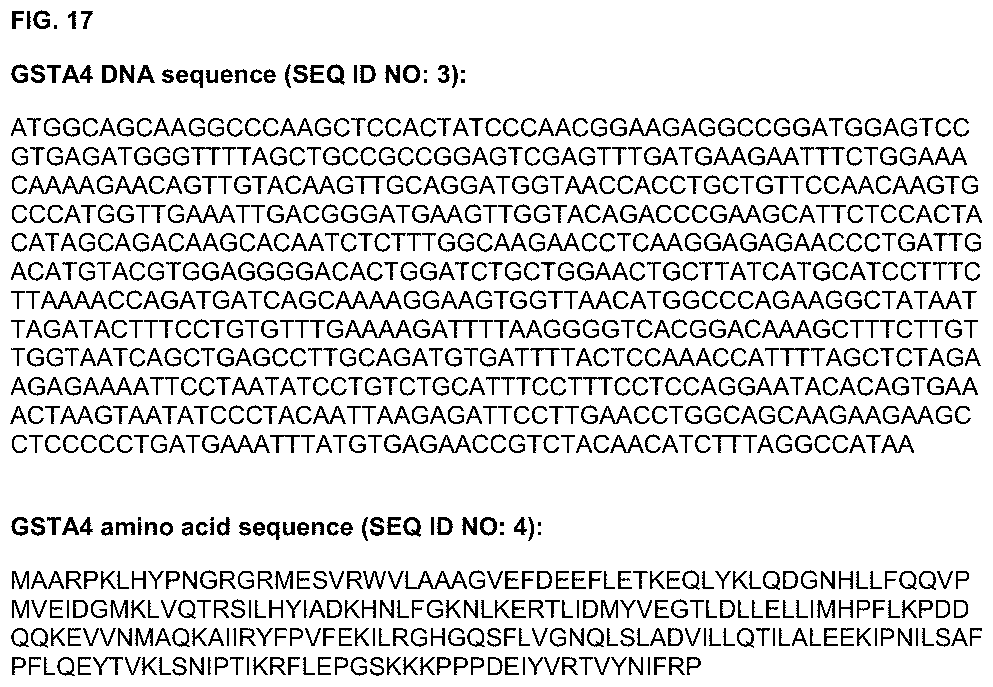
D00019

D00020

D00021

D00022

D00023

S00001
XML
uspto.report is an independent third-party trademark research tool that is not affiliated, endorsed, or sponsored by the United States Patent and Trademark Office (USPTO) or any other governmental organization. The information provided by uspto.report is based on publicly available data at the time of writing and is intended for informational purposes only.
While we strive to provide accurate and up-to-date information, we do not guarantee the accuracy, completeness, reliability, or suitability of the information displayed on this site. The use of this site is at your own risk. Any reliance you place on such information is therefore strictly at your own risk.
All official trademark data, including owner information, should be verified by visiting the official USPTO website at www.uspto.gov. This site is not intended to replace professional legal advice and should not be used as a substitute for consulting with a legal professional who is knowledgeable about trademark law.