Methods of treating cholangiocarcinoma
Ali , et al. April 20, 2
U.S. patent number 10,980,804 [Application Number 14/761,518] was granted by the patent office on 2021-04-20 for methods of treating cholangiocarcinoma. This patent grant is currently assigned to FOUNDATION MEDICINE, INC.. The grantee listed for this patent is FOUNDATION MEDICINE, INC.. Invention is credited to Siraj Mahamed Ali, Matthew J. Hawryluk, Jie He, Doron Lipson, Vincent A. Miller, Jeffrey S. Ross, Philip James Stephens.


View All Diagrams
| United States Patent | 10,980,804 |
| Ali , et al. | April 20, 2021 |
Methods of treating cholangiocarcinoma
Abstract
Methods and compositions for treating cholangiocarcinoma.
| Inventors: | Ali; Siraj Mahamed (Cambridge, MA), Hawryluk; Matthew J. (Watertown, MA), He; Jie (Newton, MA), Lipson; Doron (Chestnut Hill, MA), Miller; Vincent A. (West Orange, NJ), Ross; Jeffrey S. (Lebanon Springs, NY), Stephens; Philip James (Lexington, MA) | ||||||||||
|---|---|---|---|---|---|---|---|---|---|---|---|
| Applicant: |
|
||||||||||
| Assignee: | FOUNDATION MEDICINE, INC.
(Cambridge, MA) |
||||||||||
| Family ID: | 1000005497893 | ||||||||||
| Appl. No.: | 14/761,518 | ||||||||||
| Filed: | January 17, 2014 | ||||||||||
| PCT Filed: | January 17, 2014 | ||||||||||
| PCT No.: | PCT/US2014/012136 | ||||||||||
| 371(c)(1),(2),(4) Date: | July 16, 2015 | ||||||||||
| PCT Pub. No.: | WO2014/113729 | ||||||||||
| PCT Pub. Date: | July 24, 2014 |
Prior Publication Data
| Document Identifier | Publication Date | |
|---|---|---|
| US 20150366866 A1 | Dec 24, 2015 | |
Related U.S. Patent Documents
| Application Number | Filing Date | Patent Number | Issue Date | ||
|---|---|---|---|---|---|
| 61756372 | Jan 24, 2013 | ||||
| 61754509 | Jan 18, 2013 | ||||
| Current U.S. Class: | 1/1 |
| Current CPC Class: | A61K 31/553 (20130101); C07K 14/705 (20130101); A61K 31/519 (20130101); A61K 31/498 (20130101); C12Q 1/6883 (20130101); A61K 45/06 (20130101); A61K 31/496 (20130101); A61K 31/435 (20130101); C12N 15/1137 (20130101); A61K 31/4412 (20130101); A61K 31/517 (20130101); A61K 31/5025 (20130101); C12Y 207/10001 (20130101); C07K 16/40 (20130101); C12Q 1/6886 (20130101); A61K 31/47 (20130101); A61K 31/53 (20130101); A61K 31/5383 (20130101); G01N 33/573 (20130101); C12N 9/12 (20130101); A61K 31/4439 (20130101); A61K 31/506 (20130101); A61K 31/404 (20130101); C07K 14/71 (20130101); G01N 2500/04 (20130101); C07K 2319/00 (20130101); C12N 2310/12 (20130101); C12Q 2600/156 (20130101); C12Q 2600/158 (20130101); C12N 2310/11 (20130101); C12N 2310/14 (20130101); G01N 2333/912 (20130101) |
| Current International Class: | A61K 31/517 (20060101); A61K 31/5025 (20060101); C12N 9/12 (20060101); A61K 31/4412 (20060101); A61K 45/06 (20060101); A61K 31/47 (20060101); A61K 31/435 (20060101); A61K 31/506 (20060101); A61K 31/519 (20060101); G01N 33/573 (20060101); A61K 31/53 (20060101); A61K 31/404 (20060101); A61K 31/5383 (20060101); A61K 31/496 (20060101); C12Q 1/6886 (20180101); A61K 31/553 (20060101); C07K 14/705 (20060101); C07K 14/71 (20060101); C07K 16/40 (20060101); C12N 15/113 (20100101); C12Q 1/6883 (20180101); A61K 31/19 (20060101); A61K 31/498 (20060101); A61K 31/4439 (20060101) |
| Field of Search: | ;514/266.23,248,561 |
References Cited [Referenced By]
U.S. Patent Documents
| 8377921 | February 2013 | Michellys et al. |
| 10000814 | June 2018 | Cronin et al. |
| 2002/0197679 | December 2002 | Tang et al. |
| 2005/0287541 | December 2005 | Nakagawara et al. |
| 2007/0010657 | January 2007 | Klocke et al. |
| 2007/0059710 | March 2007 | Luke et al. |
| 2007/0248605 | October 2007 | Hestir et al. |
| 2008/0051462 | February 2008 | Fritz et al. |
| 2008/0131375 | June 2008 | Gordon et al. |
| 2008/0171689 | July 2008 | Bosch et al. |
| 2008/0226664 | September 2008 | Old et al. |
| 2009/0130101 | May 2009 | Cohen |
| 2009/0156475 | June 2009 | Rikova et al. |
| 2009/0209496 | August 2009 | Chaplin et al. |
| 2011/0118298 | May 2011 | Fritz et al. |
| 2011/0150893 | June 2011 | Cho et al. |
| 2012/0039805 | February 2012 | Lisanti et al. |
| 2012/0208706 | August 2012 | Downing et al. |
| 2013/0096021 | April 2013 | Chinnaiyan et al. |
| 2014/0336236 | November 2014 | Cronin et al. |
| 2015/0073036 | March 2015 | Hawryluk et al. |
| 2015/0366866 | December 2015 | Ali et al. |
| 2016/0009785 | January 2016 | Lipson et al. |
| 2016/0010068 | January 2016 | Bastian et al. |
| 2018/0346992 | December 2018 | Cronin et al. |
| 0698096 | Feb 1996 | EP | |||
| 0698096 | Mar 1997 | EP | |||
| 2057465 | May 2009 | EP | |||
| 9426889 | Nov 1994 | WO | |||
| 01027081 | Apr 2001 | WO | |||
| 03031568 | Apr 2003 | WO | |||
| 200413099 | Feb 2004 | WO | |||
| 2007060402 | May 2007 | WO | |||
| 2008021290 | Feb 2008 | WO | |||
| 2010081817 | Jul 2010 | WO | |||
| 2012092426 | Jul 2012 | WO | |||
| 2013059740 | Apr 2013 | WO | |||
| 2013076186 | May 2013 | WO | |||
| 2013087716 | Jun 2013 | WO | |||
| 2014018673 | Jan 2014 | WO | |||
| 2014036387 | Mar 2014 | WO | |||
| 2014071358 | May 2014 | WO | |||
| 2014071419 | May 2014 | WO | |||
| 2014113729 | Jul 2014 | WO | |||
| 2014130975 | Aug 2014 | WO | |||
Other References
|
Bai et al. "GP369, an FGFR2-IIIb-Specific Antibody, Exhibits Potent antitumor Activity against Human Cancers Driven by Activated FGFR2 Signaling" Cancer Research (2010) vol. 70 No. 19. cited by applicant . Brave et al. "Assessing the Activity of Cediranib, a VEGFR-2/3 Tyrosine Kinase Inhibitor, against VEGFR-1 and Members of the Structurally Related PDGFR Family" Molecular Cancer Therapeutics (2011) vol. 10 No. 5 pp. 861-873. cited by applicant . Byron et al. "Inhibition of Activated Fibroblast Growth Factor Receptor 2 in Endometrial Cancer Cells Induces Cell Death Despite PTEN Abrogation" Cancer Research (2008) vol. 68 No. 17. cited by applicant . Chiorean et al, "Imatinib Mesylate (STI-571), a c-Abl Kinase Inhibitor, Indirectly Blocks Receptor Tyrosine Kinase Activation and Induces Apoptosis in a Human Cholangiocarcinoma Cell Line" Gastroenterology (2003) vol. 124 No. 4. cited by applicant . Cho et al. "Enhanced Expression of Keratinocyte Growth Factor and Its Receptor Correlates with Venous Invasion in Pancreatic Cancer" The American Journal of Pathology (2007) vol. 170 No. 6. cited by applicant . Cole et al. "Inhibition of FGFR2 and FGFR1 increases cisplatin sensitivity in ovarian cancer" Cancer Biology & Therapy (2010) vol. 10 No. 5 pp. 495-504. cited by applicant . Cortes et al. "A Pivotal PhaM 2 Trial of Ponatinib in Patients with Chronic Myeloid Leukemia (CML) and Philadelphia Chromosome-Positive Acute Lymphoblastic Leukemia (Ph+ALL) ResIlltlInt or Intole111nt to Dasatlnlb or Nilotinib, or with the T315I BCR-ABL Mutation: 1Z-Month Follow-up of the PACE Trial" ASH Annual Meeting and Exposition (Dec. 9, 2012) Abstract No. 163. cited by applicant . Garcia-Mayoral et al. "The Structure of the C-Terminal KH Domains of KSRP Reveals a Noncanonical Motif Important for mRNA Degradation" Structure (2007) vol. 15 pp. 485-498. cited by applicant . Gartside et al. "Loss-of-Function Fibroblast Growth Factor Receptor-2 Mutations in Melanoma" Molecular Cancer Research (2009) vol. 7 No. 1 pp. 41-54. cited by applicant . GenBank Accession No. NM_000141 accessed Nov. 19, 2015 from http://www.ncbi.nlm.nih.gov/nuccore/189083823. cited by applicant . GenBank Accession No. NM_001012331 accessed Nov. 19, 2015 from <http://www.ncbi.nlm.nih.gov/nuccore/NM_001012331.1>. cited by applicant . Genbank Accession No. NM_001080512 accessed Nov. 19, 2015 from http://www.ncbi.nlm.nih.gov/nuccore/NM_001080512.2. cited by applicant . GenBank Accession No. NM_001127211 accessed Nov. 19, 2015 from http://www.ncbi.nlm.nih.gov/nuccore/385198090. cited by applicant . GenBank Accession No. NM_001144915 accessed on Nov. 17, 2015 from http://www.ncbi.nlm.nih.gov/nuccore/NM_001144915.1. cited by applicant . GenBank Accession No. NM_003787 accessed Nov. 19, 2015 from http://www.ncbi.nlm.nih.gov/nuccore/NM_003787. cited by applicant . GenBank Accession No. NM_004562 accessed Nov. 19, 2015 from http://www.ncbi.nlm.nih.gov/nuccore/NM_004562.2. cited by applicant . GenBank Accession No. NM_006342 accessed Nov. 19, 2015 from http://www.ncbi.nlm.nih.gov/nuccore/NM_006342. cited by applicant . GenBank Accession No. NM_022494 accessed Nov. 19, 2015 from http://www.ncbi.nlm.nih.gov/nuccore/NM_022494.2. cited by applicant . GenBank Accession No. NP_001012331 accessed Nov. 19, 2015 from http://www.ncbi.nlm.nih.gov/protein/59889558. cited by applicant . Gozgit et al "Ponatinib (AB24534), a Multitargeted Pan-FGFR Inhibitor with Activity in Multiple FGFR-Amplified or Mutated Cancer Models" Molecular Cancer Therapeutics (2012) vol. 11 No. 3 pp. 690-699. cited by applicant . Huether et al.: "Sorafenib alone or as combination therapy for growth control of cholangiocarcinoma", Biochemical Pharmacology, Elsevier, US, vol. 73, No. 9, Mar. 24, 2007 (Mar. 24, 2007), pp. 1308-1317. cited by applicant . International Preliminary Report on Patentability from PCT/US14/12136 dated Mar. 18, 2015. cited by applicant . International Search Report and Written Opinion for PCT/US2014/012136 dated Jul. 16, 2014. cited by applicant . Ko et al. "Phase II study of telatinib (T) in combination with capecitabine (X) and cisplatin (P) as first-line treatment in patients (pts) with advanced cancer of the stomach (G) or gastro-esophageal junction (GEJ)." Journal of Clinical Oncology ASCO Annual Meeting Abstracts, vol. 28 No. 15; May 20 supplement (2010). cited by applicant . Landis et al "Cancer Statistics, 1998" Ca Cancer J Clin (1998) vol. 48 No. 1 pp. 6-29. cited by applicant . Lorenzi et al. "FRAG1, a gene that potently activates fibroblast growth factor receptor by C-terminal fusion through chromosomal rearrangement" Proc. Natl. Acad. Sci. USA (1996) vol. 93 pp. 8956-8961. cited by applicant . Lorenzi et al. "Ligand-independent activation of fibroblast growth factor receptor-2 by carboxl terminal alterations" Oncogene (1997) vol. 15 pp. 817-826. cited by applicant . Matsumoto et al. "FGFR2 gene amplification and clinicopathological features in gastric cancer" British Journal of Cancer (2012) vol. 106 No. 4 pp. 727-732. cited by applicant . McKay et al. "PP58 Novel potential therapeutic targets for cholangiocarcinoma identified by array comparitive hybridization" European Journal of Cancer (2009) vol. 7 No. 4. cited by applicant . Narong and Leelawat "Basic fibroblast growth factor induces cholangiocarcinoma cell migration via activation of the MEK1/2 pathway" Oncology Letters (2011) pp. 821-825. cited by applicant . Patel et al "Cholangiocarcinom--controversies and challenges" Nat Rev Gastroenterol Hepatol (2011) vol. 8 No. 4. cited by applicant . Patel et al. "Worldwide trends in mortality from biliary tract malignancies" BMC Cancer (2002) vol. 2 No. 10. cited by applicant . Powers et al. "Fibroblast growth factors, their receptors and signaling" Endocrine-Related Cancer (2000) vol. 7 pp. 165-197. cited by applicant . Singh et al "Transforming Fusions of FGFR and TACC Genes in Human Glioblastoma" Science (2012) vol. 337(6099) pp. 1231-1235. cited by applicant . Toyokawa et al. "Co-expression of keratinocyte growth factor and K-sam is an independent prognostic factor in gastric carcinoma" Oncology Reports (2009) vol. 21 pp. 875-880. cited by applicant . Turner and Grose "Fibroblast growth factor signalling: from development to cancer" Nat Rev Cancer (2010) vol. 10 No. 2 pp. 116-129. cited by applicant . Turner et al. "Integrative molecular profiling of triple negative breast cancers identifies amplicon drivers and potential therapeutic targets" Oncogene (2010) vol. 8 No. 29 pp. 2013-2023. cited by applicant . Wang et al "Mutations in Isocitrate Dehydrogenase 1 and 2 Occur Frequently in Intrahepatic Cholangiocarcinomas and Share Hypermethylation Targets with Glioblastomas" Oncogene (2013) vol. 32 No. 25. cited by applicant . Williams et al. "Oncogenic FGFR3 gene fusions in bladder cancer" Human Molecular Genetics (2013) vol. 22 No. 4 pp. 795-803. cited by applicant . Wu et al. "Identification of Targetable FGFR Gene Fusions in Diverse Cancers" Cancer Discovery (Jun. 2013) pp. 636-647. cited by applicant . Yoon et al. "Enhanced epidermal growth factor receptor activation in human cholangiocarcinoma cells" Journal of Hepatology (2004) pp. 808-814. cited by applicant . Amatu et al. "NTRK gene fusions as novel targets of cancer therapy across multiple tumour types" ESMO Open (2016) vol. 1, e000023, pp. 1-9. cited by applicant . ClinicalTrials.Gov Identifier No. NCT02568267, "Basket Study of Entrectinib (RXDX-101) for the Treatment of Patients With Solid Tumors Harboring NTRK 1/2/3/ (Trk A/B/C), ROS1, or ALK Gene Rearrangements (Fusions) (STARTRK-2)" First Received: Oct. 2, 2016; Last Updated : Jan. 3, 2017; https://clinicaltrials.gov/ct2/show/NCT02568267?term=NTRK1+fusio- n+lung&rank=1; Retrieved Jan. 4, 2017. cited by applicant . ClinicalTrials.Gov Identifier No. NCT0257643.1, "Study of LOXO-101 in Subjects With NTRK Fusion Positive Solid Tumors (NAVIGATE)" First Received: Oct. 12, 2015; Last Updated: Nov. 16, 2016; https://clinicaltrials.gov/ct12/show/NCT02576431?term=NTRK1+fusion+lung&r- ank=2; Retrieved Jan. 4, 2017. cited by applicant . Doebele et al. "An Oncogenic NTRK Fusion in a Patient with Soft-Tissue Sarcoma with Response to the Tropomyosin-Related Kinase Inhibitor LOXO-101" Cancer Discovery (2015) vol. 5, pp. 1049-1057. cited by applicant . International Search Report and Written Opinion for International Application No. PCT/US2013/068457 dated Jul. 11, 2014. cited by applicant . Lih et al. "N of 2 Responders With LMNA-NTRK1" J Natl Cancer Inst (2016) vol. 108, No. 1, djv376, pp. 1-2. cited by applicant . Lin et al., "Exon Array Profiling Detects EML4-ALK Fusion in Breast, Colorectal, and Non-Small Cell Lung Cancers" Mol Cancer Research (2009) vol. 7, No. 9, pp. 1466-1476. cited by applicant . Marchetti et al., "Frequent Mutations in the Neurotrophic Tyrosine Receptor Kinase Gene Family in Large Cell Neuroendocrine Carcinoma of the Lung" Human Mutation (2008) vol. 29, No. 5, pp. 609-616. cited by applicant . Sartore-Bianciai et al. "Sensitivity to Entrectinib Associated With a Novel LMNA-NTRK1 Gene Fusion in Metastatic Colorectal Cancer" J Natl Cancer Inst (2016) vol. 108, No. 1, djv306, pp. 1-4. cited by applicant . Wong et al. "Evaluation of a Congenital Infantile Fibrosarcoma by Comprehensive Genomic Profiling Reveals an LMNA NTRK1 Gene Fusion Responsive to Crizotinib" J Natl Cancer Inst (2016) vol. 108, No. 1, djv307, pp. 1-3. cited by applicant . Albanese, C. et al., "Dual targeting of CDK and tropomyosin receptor kinase families by the oral inhibitor PHA-848125, an agent with broad-spectrum antitumor efficacy", Mol Cancer Ther 9(8):2243-54, Aug. 3, 2010. cited by applicant . Camidge et al. Optimizing the detection of lung cancer patients harboring anaplastic lymphoma kinase (ALK) gene rearrangements potentially suitable for ALK inhibitor treatment. Clin Cancer Res Nov. 14, 2010 vol. 16 No. 22 pp. 5581-5590. Especially p. 5586 col. 2 para 2-3. cited by applicant . Cohen, Roger B. et al., "A phase I dose-escalation study of danusertib (PHA-739358) administered as a 24-hour infusion with and without granulocyte colony-stimulating factor in a 14-day cycle in patients with advanced solid tumors", Clin Cancer Res 15(21):6694-701, ePub Oct. 13, 2009, Nov. 1, 2009. cited by applicant . Degrassi, A. et al., "Efficacy of PHA-848125, a cyclin-dependent kinase inhibitor, on the K-Ras(G12D) LA2 lung adenocarcinoma transgenic mouse model: evaluation by multimodality imaging", Mol Cancer Ther 9(3):673-81, Mar. 9, 2010. cited by applicant . Doebele et al., "NTRK1 Gene Fusions as a Novel Oncogene Target in Lung Cancer", 2013 ASCO Annual Meeting; Abstract No. 8023; Poster (May 31-Jun. 4, 2013). cited by applicant . Greco A, et al. "Characterization of the NTRK1 genomic region involved in chromosomal rearrangements generating TRK oncogenes." Genomics (1993) 18(2):397-400. cited by applicant . Greco A, et al. "TRK-T1 is a novel oncogene formed by the fusion of TPR and TRK genes in human papillary thyroid carcinomas." Oncogene (1992) 7(2):237-42. cited by applicant . Greco et al. Rearrangements of NTRK1 gene in papillary thyroid carcinoma. Mol Cell Endrocrinol May 28, 2010 vol. 321 No. 1 pp. 44-49. Especially p. 46 col. 2 para 3. cited by applicant . Greco, A. et al., "The DNA rearrangement that generates the TRK-T3 oncogene involves a novel gene on chromosome 3 whose product has a potential coiled-coil domain", Mol Cell Biol 15(11):6118-27, Nov. 1995. cited by applicant . Han, SY et al., "Evaluation of a multi-kinase inhibitor KRC-108 as an anti-tumor agent in vitro and in vivo", Invest New Drugs 30(2):518-23, ePub Nov. 16, 2010, Apr. 2012. cited by applicant . Huehne K, et al. "Novel missense, insertion and deletion mutations in the neurotrophic tyrosine kinase receptor type 1 gene (NTRK1) associated with congenital insensitivity to pain with anhidrosis." Neuromuscul Disord (2008) 18(2):159-66. cited by applicant . Indo Y, et al. "Structure and organization of the human TRKA gene encoding a high affinity receptor for nerve growth factor." Jpn J Hum Genet (1997) 42(2):343-51. cited by applicant . International Preliminary Report on Patentability and Written Opinion for International Application No. PCT/US2012/061211 dated Apr. 22, 2014. cited by applicant . International Preliminary Report on Patentability for International Application No. PCT/US2013/068604 dated May 5, 2015. cited by applicant . International Search Report for International Application No. PCT/US2012/061211 dated Feb. 5, 2013. cited by applicant . International Search Report for International Application No. PCT/US2012/061211 dated May 2, 2013. cited by applicant . International Search Report for International Application No. PCT/US2013/068604 dated Nov. 7, 2014. cited by applicant . Iyer, R. et al., "Lestaurtinib enhances the antitumor efficacy of chemotherapy in murine xenograft models of neuroblastoma", Clin Cancer Res 16(5):1478-85, ePub Feb. 23, 2010, Mar. 1, 2010. cited by applicant . Kelleher et al. The emerging pathogenic and therapeutic importance of the anaplastic lymphoma kinase gene. Eur J Cancer Sep. 2010 vol. 46 No. 13 pp. 2357-2368. Especially p. 2365 table 6. cited by applicant . Mardy et al., Congenital insensitivity to pain with anhidrosis: Novel mutations in the TRKA (NTRK1) gene encoding a high-affinity receptor for nerve growth factor, 1999, Am. J. Hum. Genet., 64, pp. 1570-1579. cited by applicant . Martin-Zanca D, et al. "A human oncogene formed by the fusion of truncated tropomyosin and protein tyrosine kinase sequences." Nature (1986) 319(6056):743-8. cited by applicant . Meulenbeld, Hielke J. et al., "Danusertib, an aurora kinase inhibitor," Expert Opinion Investigative Drugs. Mar. 2012, 21(3), pp. 383-393. cited by applicant . Miura Y, et al. "Mutation and polymorphism analysis of the TRKA (NTRK1) gene encoding a high-affinity receptor for serve growth factor in congenital insensitivity to pain with anhidrosis (CIPA) families." Hum Genet (2000) 106(1):116-24. cited by applicant . Muller-Tidow et al., Identification of Metastasis-Associated Receptor Tyrosine Kinases in Non-Small Cell Lung Cancer, 2005, Cancer Res., vol. 65, No. 5 pp. 1778-1782. cited by applicant . Notification of Transmittal of the International Search Report and Written Opinion of the International Searching Authority from corresponding PCT/US2013/068457 dated Jul. 11, 2014. cited by applicant . Perez-Pinera P, et al. "The Trk tyrosine kinase inhibitor K252a regulates growth of lung adenocarcinomas." Mol Cell Biochem (2007) 295(1-2):19-26. cited by applicant . Rao, R. et al., "Heat shock protein 90 inhibition depletes TrkA levels and signaling in human acute leukemia cells", Mol Cancer Ther 9(8):2232-42, ePub Jul. 27, 2010, Aug. 2010. cited by applicant . Schneider et al., "The transforming acidic coiled coil 3 protein is essential for spindle-dependent chromosome alignment and mitotic survival" The Journal of Biological Chemistry, 282(40):29273-29283 (2007). cited by applicant . Tacconelli, A. et al. , "TrkA alternative splicing: a regulated tumor-promoting switch in human neuroblastoma", Cancer Cell 6(4):347-60, Oct. 2004. cited by applicant . Takeuchi et al. Multiplex reverse transcription-PCR screening for EML4-ALK fusion transcripts. Clin Cancer Res Oct. 15, 2008 vol. 14 No. 20 pp. 6618-6624. Especially p. 6619 col. 1 para. cited by applicant . Teixeira et al., "Recurrent Fusion Oncogenes in Carcinomas" Critical Reviews in Oncogenesis, 12(3-4)257-271 (2006). cited by applicant . Thress, K. et al., "Identification and preclinical characterization of AZ-23, a novel, selective, and orally bioavailable inhibitor of the Trk kinase pathway", Mol Cancer Ther 8(7):1818-27, ePub Jun. 9, 2009, Jul. 2009. cited by applicant . Undevia, SD et al., "Phase I clinical trial of CEP-2563 dihydrochloride, a receptor tyrosine kinase inhibitor, in patients with refractory solid tumors", Invest New Drugs 22(4):449-58, Nov. 2004. cited by applicant . University of Colorado Denver; " NTRK1: A new oncogene and target in lung cancer". Press Release, Public release date: Jun. 3, 2013. cited by applicant . Vaishnavi, Aria et al., "Oncogenic and drug-sensitive NTRK1 rearrangements in lung cancer", Nature Medicine, vol. 19, No. 11, pp. 1469-1472, ePub Oct. 27, 2013, Nov. 2013. cited by applicant . Wang et al. Fusion of dynactin 1 to anaplastic lymphoma kinase in inflammatory myofibroblastic tumor. Hum Pathol ePub Jun. 1, 2012 vol. 43 No. 11 pp. 2047-2052. Especially abstract. cited by applicant . Weiss, GJ et al., "Phase I study of the safety, tolerability and pharmacokinetics of PHA-848125AC, a dual tropomyosin receptor kinase A and cyclin-dependent kinase inhibitor, in patients with advanced solid malignancies", Invest New Drugs, 30(6)2334-2343 ePub Dec. 2011, Dec. 2012. cited by applicant . Williams et al., "Oncogenic FGFR3 gene fusions in bladder cancer" Human Molecular Genetics, 22(4):795-803 (2013). cited by applicant . Written Opinion for International Application No. PCT/US2013/068604 dated May 5, 2015. cited by applicant . Altorki et al. "Phase II Proof-of-Concept Study of Pazopanib Monotherapy in Treatment-Naive Patience With State I/II Resectable Non-Small-Cell Lung Cancer" Journal of Clinical Oncology (2010) vol. 28, No. 19, pp. 3131-3137. cited by applicant . Avet-Loiseau et al. "High Incidence of Translocations t(11;14)(q13;q32) and t(4;14)(p16;q32) in Patients with Plasma Cell Malignancies" Cancer Research (1998) vol. 58, pp. 5640-5645. cited by applicant . Chen et al. "FGFR3 as a therapeutic target of the small molecule inhibitor PKC412 in hematopoietic malignancies" Oncogene (2005) vol. 24, pp. 8259-8267. cited by applicant . Gozgit et al. "Ponatinib (AP24534), a Multitargeted Pan-FGFR Inhibitor with Activity in Multiple FGFR-Amplified or Mutated Cancer Models" Molecular Cancer Therapeutics (2012) vol. 11, No. 3, pp. 690-699. cited by applicant . Keats et al. "In multiple myeloma, t(4;14)(p16;q32) is an adverse prognostic factor irrespective of FGFR3 expression" Blood (2003) vol. 101, No. 4, pp. 1520-1529. cited by applicant . Monk et al. "Phase II, Open-Label Study of Pazopanib or Lapatinib Monotherapy Compared With Pazopanib Plus Lapatinib Combination Therapy in Patients With Advanced and Recurrent Cervical Cancer" Journal of Clinical Oncology (2010) vol. 28, No. 22, pp. 3562-3569. cited by applicant . Reck et al. "A phase II double-blind study to investigate efficacy and safety of two doses of the triple angiokinase Inhibitor BIBF 1120 in patients with relapsed advanced non-small-cell lung cancer" Annals of Oncology (2011) vol. 22, pp. 1374-1381. cited by applicant . Richelda et al. "A Novel Chromosomal Translocation t(4; 14)(p16.3; q32) in Multiple Myeloma Involves the Fibroblast Growth Factor Receptor 3 Gene" Blood (1997) vol. 90, No. 10, pp. 4062-4070. cited by applicant . Santra et al. "A sibset of multiple myeloma harboring the t(4;14)(p16;q32) translocation lacks FGFR3 expression but maintains an IGH/MMSET fusion transcript" Blood (2003) vol. 101, No. 6, pp. 2374-2376. cited by applicant . Stewart et al. "Correlation of TACC3, FGFR3, MMSET and p21 expression with the t(4;14)(p16.3;q32) in multiple myeloma" British Journal of Haematology (2004) vol. 126, pp. 72-76. cited by applicant . Turner et al "Fibroblast growth factor signalling: from development to cancer" Nature (2010) vol. 10, pp. 116-129. cited by applicant . Ware et al. "Rapidly Acquired Resistance to EGFR Tyrosine Kinase Inhibitors in NSCLC Cell Lines through De-Repression of FGFR2 and FGFR3 Expression" PLOS One (2010) vol. 5, No. 11, pp. e14117. cited by applicant . Caneiro et al. "FGFR3-TACC3: A novel gene fusion in cervical cancer" Gynecologic Oncology Reports (2015), vol. 13, pp. 53-56. cited by applicant . Capelletti et al. "Identification of Recurrent FGFR3-TACC3 Fusion Oncogenes from Lung Adenocarcinoma" Clin Cancer Res (2014) vol. 20, pp. 6551-6558. cited by applicant . Costa et al. "FGFR3-TACC3 fusion in solid tumors: mini review" Oncotarget (2016) vol. 7, No. 34, pp. 55924-55938. cited by applicant . Farago et al., "Durable Clinical Response to Entrectinib in NTRK1-Rearranged Non-Small Cell Lung Cancer" Journal of Thoracic Oncology (2015) vol. 10, No. 12, pp. 1670-1674. cited by applicant . Gergely et al. "The TACC domain identities a family of centrosomal proteins that can interact with microtubules" Proc Natl Acad Sci. (2000) vol. 97, pp. 14352-14357. cited by applicant . Hyman et al. "The efficacy of larotrectinib (LOXO-101), a selective tropomyosin receptor kinase (TRK) inhibitor, in adult and pediatric TRK fusion cancers" Presentation from the ASCO Annual Meeting 2017. cited by applicant . Keegan et al. "Isolation of an additional member of the fibroblast growth factor receptor family, FGFR-3" Proc Natl Acad Sci. (1991) vol. 88, No. 4, pp. 1095-1099. cited by applicant . Lee et al., "The potential role of comprehensive genomic profiling to guide targeted therapy for patients with biliary cancer," Ther Adv Gastroenterol (2017) vol. 10, No. 6, pp. 507-520. cited by applicant . Brambilla et al., "The new World Health Organization classification of lung tumours," Eur Respir J (2001) vol. 18, pp. 1059-1068. cited by applicant . Cappellen et al., "Frequent activating mutations of FGFR3 in human bladder and cervix carcinomas," Nature Genetics (1999) vol. 23, pp. 18-20. cited by applicant . Dhami et al., "Comprehensive genomic profiling aids in treatment of metastatic endometrial cancer," Cold Spring Harb Mol Case Stud (2018) vol. 4, Article a002089, 14 pages. cited by applicant . Guagnano et al., "FGFR Genetic Alterations Predict for Sensitivity to NVP-BGJ398, a Selective PAN-FGFR Inhibitor," Cancer Discovery (2012) vol. 2, pp. 1118-1133. cited by applicant . Javle et al., "Biliary Cancer: Utility of Next-Generation Sequencing for Clinical Management," Cancer (2016) vol. 122, pp. 3838-3847. cited by applicant . Jiao et al., "Exome sequencing identifies frequest inactivating mutations in BAP1, ARID1A and PBRM1 in intrahepatic cholangiocarcinomas," Nature Genetics (2013) vol. 45, No. 12, pp. 1470-1473 and Supplementary Information, 15 pages. cited by applicant . Kang et al., "microRNA-99b acts as a tumor suppressor in non-small cell lung cancer by directly targeting fibroblast growth factor receptor 3," Experimental and Therapeutic Medicine (2012) vol. 3, pp. 149-153. cited by applicant . Lam et al., "Expression profiling in lung adenocarcinomas with or without epidermal growth factor receptor (EGFR) gene mutation at exons 18-21 reveals expression signatures related to the EGFR pathway," Proc Amer Assoc Cancer Res (2005) vol. 46, Abstract only, 2 pages. cited by applicant . National Center for Biotechnology Information PubChem Database. Ceritinib, CID=57379345, pubchem.ncbi.nim.nih.gov/compund/Ceritinib, created 2012, accessed on Jul. 21, 2019, 42 pages. cited by applicant . Ou et al., "Emergence of FGFR3-TACC3 fusions as a potential by-pass resistance mechanism to EGFR tyrosine kinase inhibitors in EGFR mutated NSCLC patients," Lung Cancer (2017) vol. 111, pp. 61-67. cited by applicant . Wiesner et al., "Kinase fusions are frequent in Spitz tumours and spitzoid melanomas," Nature Communications (2014) vol. 5, Article 3116, 9 pages. cited by applicant . Woenckhaus et al., "Prognostic value of FHIT, CTNNB1, and MUC1 expression in non-small cell lung cancer," Human Pathology (2008) vol. 39, pp. 126-136. cited by applicant. |
Primary Examiner: Henley, III; Raymond J
Attorney, Agent or Firm: Morrison & Foerster, LLP
Parent Case Text
RELATED APPLICATIONS
This application is a U.S. National Phase Application under 35 U.S.C. .sctn. 371 of International Application No. PCT/US2014/012136, filed Jan. 17, 2014, which claims the benefit of U.S. Provisional Application No. 61/754,509, filed Jan. 18, 2013 and U.S. Provisional Application No. 61/756,372, filed Jan. 24, 2013, the contents of each of which are incorporated herein by reference in their entirety.
Claims
We claim:
1. A method of treating a subject having a cholangiocarcinoma, comprising: acquiring knowledge of the presence of a fusion polypeptide or a fusion nucleic acid molecule encoding the fusion polypeptide in a sample from said subject, wherein the fusion polypeptide comprises an FGFR2-BICC1 fusion polypeptide encoded by a nucleic acid molecule comprising a fusion junction of exon 16 of SEQ ID NO: 1 and exon 18 of SEQ ID NO: 7; and administering to the subject an effective amount of a kinase inhibitor chosen from: ACTB1003; ARRY-470; ARRY-786; AV369b; AZ-23; AZD-2171 (Cediranib); AZD-4547; BGJ398; BIBF1120; Brivanib; Brivanib Alaninate; Danusertib; Dovitinib; Dovitinib Dilactic Acid; ENMD-2076; JNJ 42756493; K252a; Lenvatinib; Lestaurtinib; Loxo-101; LY2874455; Masitinib; MK-2461; Oxindole-3; Pazopanib; PD173074; PD-173955; PHA-848125; Ponatinib; TSU-68; Tyrphostin AG 1296; R1530; R406; Regorafenib; RXDX-101; RXDX-102; Volasertib, or a combination thereof, thereby treating the cholangiocarcinoma in the subject.
2. The method of claim 1, wherein the kinase inhibitor is administered responsive to the determination of the presence of the fusion nucleic acid molecule or the fusion polypeptide in a sample from said subject.
3. The method of claim 2, wherein the determination of the presence of the fusion nucleic acid molecule or the fusion polypeptide comprises sequencing.
4. The method of claim 1, wherein the subject is undergoing or has undergone treatment with a different therapeutic agent or therapeutic modality.
5. The method of claim 4, wherein responsive to the determination of the presence of the fusion nucleic acid molecule or the fusion polypeptide, the method further comprises discontinuing the different therapeutic agent or therapeutic modality.
6. The method of claim 4, wherein the different therapeutic agent or therapeutic modality is a chemotherapy or a surgical procedure.
7. The method of claim 1, wherein the FGFR2-BICC1 fusion polypeptide is encoded by a nucleic acid molecule comprising exons 1-16 of SEQ ID NO: 1 and exons 18-21 of SEQ ID NO: 7.
8. The method of claim 1, wherein the FGFR2-BICC1 fusion polypeptide comprises an FGFR2 receptor tyrosine kinase domain or a functional fragment thereof.
9. The method of claim 1, wherein the cholangiocarcinoma comprises one or more mutated cells that originate in the bile duct.
10. The method of claim 1, wherein the cholangiocarcinoma is an intrahepatic carcinoma or an extrahepatic carcinoma.
11. A method of treating a subject having a cholangiocarcinoma, comprising: detecting the presence of a fusion polypeptide or a fusion nucleic acid molecule encoding the fusion polypeptide in a sample from said subject, wherein the fusion polypeptide comprises an FGFR2-BICC1 fusion polypeptide encoded by a nucleic acid molecule comprising a fusion junction of exon 16 of SEQ ID NO: 1 and exon 18 of SEQ ID NO: 7; and administering to the subject an effective amount of a kinase inhibitor chosen from: ACTB1003; ARRY-470; ARRY-786; AV369b; AZ-23; AZD-2171 (Cediranib); AZD-4547; BGJ398; BIBF1120; Brivanib; Brivanib Alaninate; Danusertib; Dovitinib; Dovitinib Dilactic Acid; ENMD-2076; JNJ 42756493; K252a; Lenvatinib; Lestaurtinib; Loxo-101; LY2874455; Masitinib; MK-2461; Oxindole-3; Pazopanib; PD173074; PD-173955; PHA-848125; Ponatinib; TSU-68; Tyrphostin AG 1296; R1530; R406; Regorafenib; RXDX-101; RXDX-102; Volasertib, or a combination thereof; thereby treating the cholangiocarcinoma in the subject.
12. The method of claim 1, wherein the sample is a nucleic acid sample.
13. The method of claim 1, wherein the sample is a blood, serum, or plasma sample.
14. The method of claim 1, wherein the sample comprises a tumor biopsy or a circulating tumor cell or nucleic acid.
15. The method of claim 11, wherein the sample is a nucleic acid sample.
16. The method of claim 11, wherein the sample is a blood, serum, or plasma sample.
17. The method of claim 11, wherein the sample comprises a tumor biopsy or a circulating tumor cell or nucleic acid.
Description
SEQUENCE LISTING
The instant application contains a Sequence Listing which has been submitted in ASCII format via EFS-Web and is hereby incorporated by reference in its entirety. Said ASCII copy, created on Jan. 29, 2014, is named F2036-7046WO_SL.txt and is 97,543 bytes in size.
BACKGROUND
Cancer represents the phenotypic end-point of multiple genetic lesions that endow cells with a full range of biological properties required for tumorigenesis. Indeed, a hallmark genomic feature of many cancers is the presence of numerous complex chromosome structural aberrations, including translocations, intra-chromosomal inversions, point mutations, deletions, gene copy number changes, gene expression level changes, and germline mutations, among others.
Cholangiocarcinoma is a cancer that includes mutated epithelial cells that originate in the bile ducts. Cholangiocarcinoma is a relatively rare neoplasm that is classified as an adenocarcinoma (a cancer that forms glands or secretes significant amounts of mucins). It has an annual incidence rate of about 1-2 cases per 100,000 in the Western world, but rates of cholangiocarcinoma have been rising worldwide over the past several decades (Landis S. et al. (1998) CA Cancer J Clin 48 (1): 6-29; Patel T (2002) BMC Cancer 2: 10. doi:10.1186/1471-2407-2-10).
Cancer of the bile ducts can arise within the liver as an intrahepatic cholangiocarcinoma (ICC) or originate from extrahepatic bile ducts as a bile duct carcinoma, also referred to as an extra-hepatic cholangiocarcinoma. ICC is the second most common primary hepatic malignancy after hepatocellular carcinoma (HCC), and accounts for 3% of the malignant tumors of the gastrointestinal system and 15% of primary hepatic malignancies. Because ICC has a routine histologic appearance of an adenocarcinoma, the diagnosis of ICC on a liver biopsy requires an immunohistochemical (IHC) study of the tumor and a thorough clinical workup including imaging studies to rule out a metastatic adenocarcinoma to the liver.
Numerous studies have indicated that the incidence and mortality from ICC are increasing worldwide. ICC is associated with primary sclerosing cholangitis, parasitic biliary infection, polycystic disease of the liver, congenital intrahepatic bile duct dilatation (Caroli's Disease), congenital hepatic fibrosis, and choledochal cysts. Chronic Hepatitis C infection is an established cause of ICC with some studies describing a more than 300 fold increase in ICC incidence in patients with long-standing Hepatitis C infections. ICC has also been associated with cigarette smoking, alcohol consumption and exposure to a variety of toxins and chemical carcinogens. The onset of symptoms of ICC are often vague, typically arise late in the course of the disease and include abdominal pain, anorexia and palpable abdominal mass lesions. Thus, the median survival for ICC is less than 6 months for inoperable tumors and only 20 to 40% for patients who undergo surgery and achieve clear margins.
Cholangiocarcinoma is considered to be an incurable and rapidly lethal malignancy, unless both the primary tumor and any metastases can be fully resected (removed surgically). No potentially curative treatment exists at this time except surgery; however, most patients have advanced stage disease at presentation and are inoperable at the time of diagnosis. Cholangiocarcinoma has near-100% fatality due to attendant liver complications from the damage to the organ. Patients with cholangiocarcinoma are generally managed with chemotherapy, radiation therapy, and other palliative care measures.
Thus, the need still exists for identifying novel genetic lesions associated with cancers such as cholangiocarcinomas. Such genetic lesions can be an effective approach to develop compositions, methods and assays for evaluating and treating cancer patients.
SUMMARY
The invention is based, at least in part, on the discovery, in cholagiocarcinomas, of novel rearrangement events that give rise to alterations in a fibroblast growth factor receptor 2 (FGFR2) gene or a neurotrophic tyrosine receptor kinase (NTRK1) gene. In certain embodiments, the alteration is chosen from a translocation, a deletion, an inversion, a rearrangement, or an amplification of, an FGFR2 gene or the NTRK gene. For example, the alteration can be chosen from an alteration described in Table 1 and FIGS. 1A-1C. In one embodiment, the alteration includes a fragment of an FGFR2 gene or the NTRK1 gene, e.g., as exemplified in Table 1, FIGS. 1A-1C and FIGS. 2-17. Thus, the invention provides new insights into the treatment of these cancers, such as cholangiocarcinomas. Therefore, described herein are methods for treating a cholangiocarcinoma carcinoma, including intrahepatic cholangiocarcinoma (ICC) and extrahepatic cholangiocarcinoma, as well as novel FGFR2 and NTRK1 molecules (e.g., fusion molecules); methods and reagents for identifying, assessing or detecting an alteration in an FGFR2 and/or NTRK1.
Accordingly, in one aspect, the invention features a method of treating a subject having a cholangiocarcinoma. The method includes administering to the subject an effective amount of an agent (e.g., a therapeutic agent) that targets, antagonizes or inhibits an FGFR2 or NTRK1 (e.g., an FGFR2 or NTRK1 gene product, e.g., an FGFR2 or NTRK1 protein), thereby treating the subject.
In another aspect, the invention features, a method of treating a subject having a cholangiocarcinoma. The method includes administering to the subject an effective amount of a kinase inhibitor (e.g., a tyrosine kinase inhibitor), thereby treating the subject.
In one embodiment, the method further includes acquiring knowledge of one or both of:
(i) the presence (or absence) of an alteration in FGFR2 gene product, e.g., an FGFR2 protein; or
(ii) the presence (or absence) of an alteration in NTRK1 gene product, e.g., an NTRK1 protein, in the subject, or a cancer or tumor sample from the subject.
In another embodiment, the method further includes identifying the subject, or a cancer or tumor sample from the subject, as having one or both of:
(i) the presence (or absence) of an alteration in FGFR2 gene product, e.g., an FGFR2 protein; or
(ii) the presence (or absence) of an alteration in NTRK1 gene product, e.g., an NTRK1 protein.
In certain embodiments, the presence of the FGFR2 or NTRK1 alteration, or both, in the subject is indicative that the subject is likely to respond to the agent.
In yet other embodiments, the agent is administered responsive to a determination of the presence of the FGFR2 or NTRK1 alteration, or both, in the subject, or the cancer or tumor sample from the subject.
Cholangiocarcinoma
In certain embodiments, the cholangiocarcinoma comprises one or more mutated cells that originate in the bile duct. In certain embodiments, the cholangiocarcinoma is chosen from an intrahepatic cholangiocarcinoma or an extrahepatic cholangiocarcinoma. In other embodiments, the cholangiocarcinoma comprises, or is identified as having, an alteration that is chosen from a translocation, a deletion, an inversion, a rearrangement, or an amplification of, an FGFR2 gene or the NTRK gene. In one embodiment, the cholangiocarcinoma comprises, or is identified as having, an alteration chosen from an alteration described in Table 1 or FIGS. 1A-1C. In one embodiment, the cholangiocarcinoma comprises, or is identified as having, an alteration includes a fragment of an FGFR2 gene or the NTRK1 gene, e.g., as exemplified in Table 1, FIGS. 1A-1C and FIGS. 2-17. In yet other embodiments, the cholangiocarcinoma comprises, or is identified as having, a fusion molecule of FGFR2; e.g., a fusion molecule chosen from FGFR2-TACC3, FGFR2-KIAA1598, BICC1-FGFR2, FGFR2-BICC1, PARK2-FGFR2, FGFR2-NOL4, or ZDHHC6-FGFR2 as described, e.g., in Table 1, FIGS. 1A-1C and FIGS. 2-17. In other embodiments, the cholangiocarcinoma comprises, or is identified as having, a rearrangement or an amplification of FGFR2 as described, e.g., in Table 1, FIGS. 1A-1C and FIGS. 2-17.
In certain embodiments, the alteration in FGFR2 results in upregulation, increased activity (e.g., increased transformative or oncogenic activity, kinase activity and/or dimerization), and/or increased level of an FGFR2 gene product (e.g., an FGFR2 protein), compared to a wildtype activity of FGFR2.
Subjects
In certain embodiments, the subject has an alteration in FGFR2 or NTRK1, or both, e.g., the subject has a cholangiocarcinoma comprising an alteration in FGFR2 or NTRK1, or both, e.g., as described herein. In other embodiments, the subject is identified, or has been previously identified, as having a cholangiocarcinoma (e.g., an intrahepatic cholangiocarcinoma (ICC) or an extrahepatic cholangiocarcinoma) comprising an alteration in FGFR2 or NTRK1, or both, e.g., as described herein. In other embodiments, the subject has, or is identified as having, an alteration that is chosen from a translocation, a deletion, an inversion, a rearrangement, or an amplification of, an FGFR2 gene or the NTRK gene. In one embodiment, the subject has, or is identified as having, an alteration chosen from an alteration described in Table 1 or FIGS. 1A-1C. In one embodiment, the subject has, or is identified as having, an alteration includes a fragment of an FGFR2 gene or the NTRK1 gene, e.g., as exemplified in Table 1, FIGS. 1A-1C and FIGS. 2-17. In yet other embodiments, the subject has, or is identified as having, a fusion molecule of FGFR2; e.g., a fusion molecule chosen from FGFR2-TACC3, FGFR2-KIAA1598, BICC1-FGFR2, FGFR2-BICC1, PARK2-FGFR2, FGFR2-NOL4, or ZDHHC6-FGFR2 as described, e.g., in Table 1, FIGS. 1A-1C and FIGS. 2-17. In other embodiments, the subject has, or is identified as having, a rearrangement or an amplification of FGFR2 as described, e.g., in Table 1, FIGS. 1A-1C and FIGS. 2-17.
In one embodiment, the subject is a human. In one embodiment, the subject has, or is at risk of having a cholangiocarcinoma (e.g., a cholangiocarcinoma as described herein) at any stage of disease, e.g., Stage I, II, IIIA-IIIC or IV of intrahepatic cholangiocarcinoma; Stage 0, IA-IB, IIA-IIB, III or IV of extrahepatic cholangiocarcinoma; or a metastatic cancer. In other embodiments, the subject is a cancer patient, e.g., a patient having a cholangiocarcinoma as described herein.
In one embodiment, the subject is undergoing or has undergone treatment with a different (e.g., non-FGFR2 or non-NTRK1) therapeutic agent or therapeutic modality. In one embodiment, the non-FGFR2 or non-NTRK1 therapeutic agent or therapeutic modality is a chemotherapy, immunotherapy, or a surgical procedure. In one embodiment, the non-FGFR2 or non-NTRK1 therapeutic agent or therapeutic modality comprises one or more (or all) of: a surgical procedure, flurouracil (e.g., 5-FU, Adrucil, Efudex), doxorubicin (Adriamycin, Rubex), gemcitabine (e.g., Gemzar) and/or cisplatin (Platinol).
In one embodiment, responsive to the determination of the presence of the FGFR2 or NTRK1 alteration, the different therapeutic agent or therapeutic modality is discontinued. In yet other embodiments, the subject has been identified as being likely or unlikely to respond to the different therapeutic agent or therapeutic modality.
In certain embodiments, the subject has participated previously in a clinical trial, e.g., a clinical trial for a different (e.g., non-FGFR2 or non-NTRK1) therapeutic agent or therapeutic modality. In other embodiments, the subject is a cancer patient who has participated in a clinical trial, e.g., a clinical trial for a different (e.g., non-FGFR2 or non-NTRK1) therapeutic agent or therapeutic modality.
Agents
In certain embodiments, the agent (e.g., the therapeutic agent) used in the methods targets and/or inhibits FGFR2 or NTRK1 (e.g., a FGFR2 or NTRK1 gene or gene product as described herein). In one embodiment, the agent binds and inhibits FGFR2 or NTRK1. In one embodiment, the agent is a reversible or an irreversible FGFR2 inhibitor. In certain embodiments, the agent is a pan-FGFR2 inhibitor.
In one embodiment, the agent is an antibody molecule, e.g., an anti-FGFR2 or NTRK1 antibody molecule (e.g., a monoclonal or a bispecific antibody), or a conjugate thereof (e.g., an antibody to FGFR2 or NTRK1 conjugated to a cytotoxic agent (e.g., mertansine DM1)).
In one embodiment, the agent is a kinase inhibitor. In one embodiment, the kinase inhibitor is chosen from: a multi-specific kinase inhibitor, an FGFR2 inhibitor (e.g., a pan-FGFR2 inhibitor), an NTRK1 inhibitor, and/or a small molecule inhibitor that is selective for FGFR2 or NTRK1; and/or a FGFR2 or NTRK1 cellular immunotherapy.
In an embodiment, the therapeutic agent is chosen from a kinase inhibitor; a multi-specific kinase inhibitor; an FGF receptor inhibitor (e.g., a pan FGFR2 inhibitor); an antibody molecule (e.g., a monoclonal antibody) against FGFR2; and/or a small molecule (e.g., kinase) inhibitor that is selective for FGFR2 or NTRK1.
In an embodiment the therapeutic agent is selected from antisense molecules, ribozymes, RNAi, triple helix molecules that hybridize to a nucleic acid encoding the fusion, or a transcription regulatory region that blocks or reduces mRNA expression of FGFR2 or NTRK1.
In an embodiment the kinase inhibitor is chosen from: a kinase inhibitor; a multi-specific kinase inhibitor; an FGF receptor inhibitor (e.g., a pan FGFR2 inhibitor); and/or a kinase inhibitor that is selective for FGFR2 or NTRK1.
In an embodiment, the therapeutic agent is chosen from: Regorafenib; Ponatinib; AZD-2171 (Cediranib); AZD-4547; BGJ398; BIBF1120; Brivanib; Dovitinib; ENMD-2076; JNJ42756493; Masitinib; Lenvatinib; LY2874455; Pazopanib; PD-173955; R406; PD173074; Danusertib; Dovitinib Dilactic Acid; TSU-68; Tyrphostin AG 1296; MK-2461; Brivanib Alaninate; Lestaurtinib; PHA-848125; K252a; AZ-23; and/or Oxindole-3.
In an embodiment, the therapeutic agent is chosen from Regorafenib or Ponatinib.
Other features and embodiments of the invention include one or more of the following.
In an embodiment, the method includes acquiring knowledge of the presence of an alteration, e.g., fusion, from Table 1, FIGS. 1A-1C and FIGS. 2-17 in said subject.
In an embodiment the therapeutic agent is administered responsive to the determination of presence of the alteration, e.g., fusion, in a tumor sample from said subject.
In an embodiment the determination of the presence of the alteration, e.g., fusion, comprises sequencing.
In an embodiment the subject is undergoing or has undergone treatment with a different therapeutic agent or therapeutic modality, e.g., a non-FGFR2 or non-NTRK1 therapeutic agent or therapeutic modality.
In an embodiment responsive to a determination of the presence of the alteration, e.g., fusion, the different therapeutic agent or therapeutic modality is discontinued.
In an embodiment the different therapeutic agent or therapeutic modality is a chemotherapy or a surgical procedure. In one embodiment, the non-FGFR2 or non-NTRK1 therapeutic agent or therapeutic modality comprises one or more (or all) of: a surgical procedure, flurouracil (e.g., 5-FU, Adrucil, Efudex), doxorubicin (Adriamycin, Rubex), gemcitabine (e.g., Gemzar) and/or cisplatin (Platinol).
In another aspect, the invention features, a method of determining the presence of an alteration, e.g., a fusion, disclosed herein in cholangiocarcinoma sample, comprising:
directly acquiring knowledge that an alteration, e.g., a fusion nucleic acid molecule, of Table 1, FIGS. 1A-1C and FIGS. 2-17 is present in a sample from a subject.
In an embodiment the acquiring step comprises sequencing.
In an embodiment the method further comprises administering a kinase inhibitor to the subject responsive to the determination of the presence of the alteration, e.g., the fusion, in the sample from the subject.
The invention also provides, methods of: identifying, assessing or detecting an alteration, e.g., fusion, of an FGFR2 or an NTRK1, e.g., that arises in a cholangiocarcinoma. Exemplary alteration, e.g., fusions, include those summarized in Table 1, FIGS. 1A-1C and FIGS. 2-17, including a fusion of FGFR2 (e.g., an FGFR2 fusion molecule (e.g., a gene product or fragment thereof)) to a partner from Table 1, FIGS. 1A-1C and FIGS. 2-17, or a fusion of NTRK1 (e.g., an NTRK1 fusion molecule (e.g., a gene product or fragment thereof)) to a partner of Table 1. In one embodiment, the FGFR2 or NTRK1 is fused to a second gene, or a fragment thereof, e.g., as described in Table 1, FIGS. 1A-1C and FIGS. 2-17. In other embodiments, the fusion molecule is chosen from FGFR2-TACC3, FGFR2-KIAA1598, BICC1-FGFR2, FGFR2-BICC1, PARK2-FGFR2, FGFR2-NOL4, ZDHHC6-FGFR2, or RABGAP1L-NTRK1, e.g., as described, e.g., in Table 1, FIGS. 1A-1C and FIGS. 2-17. Included are fusion molecules; isolated fusions nucleic acid molecules, nucleic acid constructs, host cells containing the nucleic acid molecules; purified fusion polypeptides and binding agents; detection reagents (e.g., probes, primers, antibodies, kits, capable, e.g., of specific detection of a fusion nucleic acid or protein); screening assays for identifying molecules that interact with, e.g., inhibit, fusions, e.g., novel kinase inhibitors or binders of FGFR2 or NTRK1.
Nucleic Acid Molecules
In one aspect, the invention features an isolated nucleic acid molecule, or an isolated preparation of nucleic acid molecules, that includes a genetic alteration disclosed herein. Such nucleic acid molecules or preparations thereof can include a genetic alteration described herein or can be used to detect, e.g., sequence, a genetic alteration disclosed herein. In other embodiments, the alteration of FGFR2 or NRTK1 is chosen from an alteration set forth in Table 1, FIGS. 1A-1C and FIGS. 2-17. In other embodiments, the fusion nucleic acid molecule is chosen from FGFR2-TACC3, FGFR2-KIAA1598, BICC1-FGFR2, FGFR2-BICC1, PARK2-FGFR2, FGFR2-NOL4, ZDHHC6-FGFR2, or RABGAP1L-NTRK1, e.g., as described, e.g., in Table 1, FIGS. 1A-1C and FIGS. 2-17.
Nucleic Acid Detection and Capturing Reagents
The invention also features a nucleic acid molecule, e.g., nucleic acid fragment, suitable as probe, primer, bait or library member that includes, flanks, hybridizes to, which are useful for identifying, or are otherwise based on, a fusion described herein. In certain embodiments, the probe, primer or bait molecule is an oligonucleotide that allows capture, detection or isolation of a fusion nucleic acid molecule described herein, e.g., a fusion of FGFR2 to a second gene, or fragment thereof, e.g., BICC1, KIAA1598, TACC3, PARK2, NOL4, or ZDHHC6-FGFR2; or a fusion of NTRK1 to a second gene, or a fragment thereof, e.g., RABGAP1L (e.g., as described in Table 1, FIGS. 1A-1C and FIGS. 2-17).
The oligonucleotide can comprise a nucleotide sequence substantially complementary to a fragment of a fusion between partners described herein nucleic acid molecules described herein. The sequence identity between the nucleic acid fragment, e.g., the oligonucleotide, and the target sequence need not be exact, so long as the sequences are sufficiently complementary to allow the capture, detection or isolation of the target sequence. In one embodiment, the nucleic acid fragment is a probe or primer that includes an oligonucleotide between about 5 and 25, e.g., between 10 and 20, or 10 and 15 nucleotides in length. In other embodiments, the nucleic acid fragment is a bait that includes an oligonucleotide between about 100 to 300 nucleotides, 130 and 230 nucleotides, or 150 and 200 nucleotides, in length.
In one embodiment, the nucleic acid fragment can be used to identify or capture, e.g., by hybridization, a fusion nucleic acid molecule described herein, e.g., a fusion of FGFR2 to a second gene, or fragment thereof, e.g., BICC1, KIAA1598, TACC3, PARK2, NOL4, or ZDHHC6; or a fusion of NTRK1 to a second gene, or a fragment thereof, e.g., RABGAP1L (e.g., as described in Table 1, FIGS. 1A-1C and FIGS. 2-17). For example, the nucleic acid fragment can be a probe, a primer, or a bait, for use in identifying or capturing, e.g., by hybridization, a fusion described herein. In one embodiment, the nucleic acid fragment can be useful for identifying or capturing a fusion breakpoint. In one embodiment, the nucleic acid fragment hybridizes to a nucleotide sequence that includes a breakpoint, e.g., a breakpoint of the fusion.
The probes or primers described herein can be used, for example, for FISH detection or PCR amplification. In one exemplary embodiment where detection is based on PCR, amplification of the fusion junction can be performed using a primer or a primer pair, e.g., for amplifying a sequence flanking the fusion junctions described herein, e.g., the mutations or the junction of a chromosomal rearrangement described herein.
In one embodiment, a pair of isolated oligonucleotide primers can amplify a region containing or adjacent to a position in the fusion. For example, reverse primers can be designed to hybridize to a nucleotide sequence within genomic or mRNA sequence of one partner, and the forward primers can be designed to hybridize to a nucleotide sequence within the other fusion partner.
In other embodiments, the nucleic acid fragment includes a bait that comprises a nucleotide sequence that hybridizes to a fusion nucleic acid molecule described herein, and thereby allows the capture or isolation said nucleic acid molecule. In one embodiment, a bait is suitable for solution phase hybridization. In other embodiments, a bait includes a binding entity, e.g., an affinity tag, that allows capture and separation, e.g., by binding to a binding entity, of a hybrid formed by a bait and a nucleic acid hybridized to the bait.
In other embodiments, the nucleic acid fragment includes a library member comprising a nucleic acid molecule described herein. In one embodiment, the library member includes a rearrangement that results in a fusion described herein.
The nucleic acid fragment can be detectably labeled with, e.g., a radiolabel, a fluorescent label, a bioluminescent label, a chemiluminescent label, an enzyme label, a binding pair label, or can include an affinity tag; a tag, or identifier (e.g., an adaptor, barcode or other sequence identifier).
Fusion Polypeptides
In another aspect, the invention features a fusion polypeptide (e.g., a purified fusion polypeptide), a biologically active or antigenic fragment thereof, as well as reagents (e.g., antibody molecules that bind to a fusion polypeptide), methods for modulating a fusion polypeptide activity and detection of a fusion polypeptide.
In certain embodiments, the fusion polypeptide is chosen from FGFR2-TACC3, FGFR2-KIAA1598, BICC1-FGFR2, FGFR2-BICC1, PARK2-FGFR2, FGFR2-NOL4, ZDHHC6-FGFR2, or RABGAP1L-NTRK1, e.g., as described, e.g., in Table 1, FIGS. 1A-1C and FIGS. 2-17.
In one embodiment, the fusion polypeptide has at least one biological activity of one or both of its partners.
In other embodiments, the nucleic acid molecule includes a nucleotide sequence encoding a fusion polypeptide that includes a fragment of a each partner of a fusion described herein.
In a related aspect, the invention features fusion polypeptide or fragments operatively linked to heterologous polypeptides to form fusion proteins.
In another embodiment, the fusion polypeptide or fragment is a peptide, e.g., an immunogenic peptide or protein, that contains a fusion junction described herein. Such immunogenic peptides or proteins can be used to raise antibodies specific to the fusion protein. In other embodiments, such immunogenic peptides or proteins can be used for vaccine preparation. The vaccine preparation can include other components, e.g., an adjuvant.
In another aspect, the invention features antibody molecules that binds to a fusion polypeptide or fragment described herein. In embodiments the antibody can distinguish wild type from fusion.
Detection Reagents and Detection of Mutations
In another aspect, the invention features a detection reagent, e.g., a purified or an isolated preparation thereof. Detection reagents can distinguish a nucleic acid, or protein sequence, having an alteration, e.g., a breakpoint, of a rearrangement, e.g., of a fusion nucleic acid molecule described herein. Exemplary fusions include a fusion of FGFR2 to a second gene, or fragment thereof, e.g., BICC1, KIAA1598, TACC3, PARK2, NOL4, or ZDHHC6; or a fusion of NTRK1 to a second gene, or a fragment thereof, e.g., RABGAP1L, e.g., as described, e.g., in Table 1, FIGS. 1A-1C and FIGS. 2-17.
Detection reagents, e.g., nucleic acid-based detection reagents, can be used to identify mutations in a target nucleic acid, e.g., DNA, e.g., genomic DNA or cDNA, or RNA, e.g., in a sample, e.g., a sample of nucleic acid derived from a cholangiocarcinoma. Detection reagents, e.g., antibody-based detection reagents, can be used to identify mutations in a target protein, e.g., in a sample, e.g., a sample of protein derived from, or produced by, a cholangiocarcinoma cell.
Nucleic Acid-Based Detection Reagents
In an embodiment, the detection reagent comprises a nucleic acid molecule, e.g., a DNA, RNA or mixed DNA/RNA molecule, comprising sequence which is complementary with a nucleic acid sequence on a target nucleic acid (the sequence on the target nucleic acid that is bound by the detection reagent is referred to herein as the "detection reagent binding site" and the portion of the detection reagent that corresponds to the detection reagent binding site is referred to as the "target binding site"). In an embodiment, the detection reagent binding site is disposed in relationship to the interrogation position such that binding (or in embodiments, lack of binding) of the detection reagent to the detection reagent binding site allows differentiation of mutant and reference sequences for an alteration described herein (e.g., an alteration or a fusion nucleic acid molecule described in Table 1, FIGS. 1A-1C and FIGS. 2-17), e.g., a fusion of FGFR2 to a second gene, or fragment thereof, e.g., BICC1, KIAA1598, TACC3, PARK2, NOL4, or ZDHHC6; or a fusion of NTRK1 to a second gene, or a fragment thereof, e.g., RABGAP1L), from a reference sequence. The detection reagent can be modified, e.g., with a label or other moiety, e.g., a moiety that allows capture.
In an embodiment, the detection reagent comprises a nucleic acid molecule, e.g., a DNA, RNA or mixed DNA/RNA molecule, which, e.g., in its target binding site, includes the interrogation position and which can distinguish (e.g., by affinity of binding of the detection reagent to a target nucleic acid or the ability for a reaction, e.g., a ligation or extension reaction with the detection reagent) between a mutation, e.g., a translocation described herein, and a reference sequence. In embodiments, the interrogation position can correspond to a terminal, e.g., to a 3' or 5' terminal nucleotide, a nucleotide immediately adjacent to a 3' or 5' terminal nucleotide, or to another internal nucleotide, of the detection reagent or target binding site.
In embodiments, the difference in the affinity of the detection reagent for a target nucleic acid comprising the mutant and that for a target nucleic acid comprising the reference sequence allows determination of the presence or absence of the mutation (or reference) sequence. Typically, such detection reagents, under assay conditions, will exhibit substantially higher levels of binding only to the mutant or only to the reference sequence, e.g., will exhibit substantial levels of binding only to the mutant or only to the reference sequence.
In embodiments, binding allows (or inhibits) a subsequent reaction, e.g., a subsequent reaction involving the detection reagent or the target nucleic acid. E.g., binding can allow ligation, or the addition of one or more nucleotides to a nucleic acid, e.g., the detection reagent, e.g., by DNA polymerase, which can be detected and used to distinguish mutant from reference. In embodiments, the interrogation position is located at the terminus, or sufficiently close to the terminus, of the detection reagent or its target binding site, such that hybridization, or a chemical reaction, e.g., the addition of one or more nucleotides to the detection reagent, e.g., by DNA polymerase, only occurs, or occurs at a substantially higher rate, when there is a perfect match between the detection reagent and the target nucleic acid at the interrogation position or at a nucleotide position within 1, 2, or 3 nucleotides of the interrogation position.
In an embodiment, the detection reagent comprises a nucleic acid, e.g., a DNA, RNA or mixed DNA/RNA molecule wherein the molecule, or its target binding site, is adjacent (or flanks), e.g., directly adjacent, to the interrogation position, and which can distinguish between a mutation, e.g., a translocation described herein, and a reference sequence, in a target nucleic acid.
In embodiments, the detection reagent binding site is adjacent to the interrogation position, e.g., the 5' or 3' terminal nucleotide of the detection reagent, or its target binding site, is adjacent, e.g., between 0 (directly adjacent) and 1,000, 500, 400, 200, 100, 50, 10, 5, 4, 3, 2, or 1 nucleotides from the interrogation position. In embodiments, the outcome of a reaction will vary with the identity of the nucleotide at the interrogation position allowing one to distinguish between mutant and reference sequences. E.g., in the presence of a first nucleotide at the interrogation position a first reaction will be favored over a second reaction. E.g., in a ligation or primer extension reaction, the product will differ, e.g., in charge, sequence, size, or susceptibility to a further reaction (e.g., restriction cleavage) depending on the identity of the nucleotide at the interrogation position. In embodiments the detection reagent comprises paired molecules (e.g., forward and reverse primers), allowing for amplification, e.g., by PCR amplification, of a duplex containing the interrogation position. In such embodiments, the presence of the mutation can be determined by a difference in the property of the amplification product, e.g., size, sequence, charge, or susceptibility to a reaction, resulting from a sequence comprising the interrogation position and a corresponding sequence having a reference nucleotide at the interrogation positions. In embodiments, the presence or absence of a characteristic amplification product is indicative of the identity of the nucleotide at the interrogation site and thus allows detection of the mutation.
In embodiments, the detection reagent, or its target binding site, is directly adjacent to the interrogation position, e.g., the 5' or 3' terminal nucleotide of the detection reagent is directly adjacent to the interrogation position. In embodiments, the identity of the nucleotide at the interrogation position will determine the nature of a reaction, e.g., a reaction involving the detection reagent, e.g., the modification of one end of the detection reagent. E.g., in the presence of a first nucleotide at the interrogation position a first reaction will be favored over a second reaction. By way of example, the presence of a first nucleotide at the interrogation position, e.g., a nucleotide associated with a mutation, can promote a first reaction, e.g., the addition of a complementary nucleotide to the detection reagent. By way of example, the presence of an A at the interrogation position will cause the incorporation of a T, having, e.g., a first colorimetric label, while the presence of a G and the interrogation position will cause the incorporation for a C, having, e.g., a second colorimetric label. In an embodiment, the presence of a first nucleotide at the nucleotide will result in ligation of the detection reagent to a second nucleic acid. E.g., a third nucleic acid can be hybridized to the target nucleic acid sufficiently close to the interrogation site that if the third nucleic acid has an exact match at the interrogation site it will be ligated to the detection reagent. Detection of the ligation product, or its absence, is indicative of the identity of the nucleotide at the interrogation site and thus allows detection of the mutation.
A variety of readouts can be employed. E.g., binding of the detection reagent to the mutant or reference sequence can be followed by a moiety, e.g., a label, associated with the detection reagent, e.g., a radioactive or enzymatic label. In embodiments the label comprises a quenching agent and a signaling agent and hybridization results in altering the distance between those two elements, e.g., increasing the distance and un-quenching the signaling agent. In embodiments, the detection reagent can include a moiety that allows separation from other components of a reaction mixture. In embodiments, binding allows cleavage of the bound detection reagent, e.g., by an enzyme, e.g., by the nuclease activity of the DNA polymerase or by a restriction enzyme. The cleavage can be detected by the appearance or disappearance of a nucleic acid or by the separation of a quenching agent and a signaling agent associated with the detection reagent. In embodiments, binding protects, or renders the target susceptible, to further chemical reaction, e.g., labeling or degradation, e.g., by restriction enzymes. In embodiments binding with the detection reagent allows capture separation or physical manipulation of the target nucleic acid to thereby allow for identification. In embodiments binding can result in a detectable localization of the detection reagent or target, e.g., binding could capture the target nucleic acid or displace a third nucleic acid. Binding can allow for determination of the presence of mutant or reference sequences with FISH, particularly in the case of rearrangements. Binding can allow for the extension or other size change in a component, e.g., the detection reagent, allowing distinction between mutant and reference sequences. Binding can allow for the production, e.g., by PCR, of an amplicon that distinguishes mutant from reference sequence.
In an embodiment the detection reagent, or the target binding site, is between 5 and 500, 5 and 300, 5 and 250, 5 and 200, 5 and 150, 5 and 100, 5 and 50, 5 and 25, 5 and 20, 5 and 15, or 5 and 10 nucleotides in length. In an embodiment the detection reagent, or the target binding site, is between 10 and 500, 10 and 300, 10 and 250, 10 and 200, 10 and 150, 10 and 100, 10 and 50, 10 and 25, 10 and 20, or 10 and 15, nucleotides in length. In an embodiment the detection reagent, or the target binding site, is between 20 and 500, 20 and 300, 20 and 250, 20 and 200, 20 and 150, 20 and 100, 20 and 50, or 20 and 25 nucleotides in length. In an embodiment the detection reagent, or the target binding site, is sufficiently long to distinguish between mutant and reference sequences and is less than 100, 200, 300, 400, or 500 nucleotides in length.
Preparations of Mutant Nucleic Acid and Uses Thereof
In another aspect, the invention features purified or isolated preparations of a neoplastic or tumor cell nucleic acid, e.g., DNA, e.g., genomic DNA or cDNA, or RNA, containing an interrogation position described herein, useful for determining if a mutation disclosed herein is present. The nucleic acid includes the interrogation position, and typically additional fusion sequence on one or both sides of the interrogation position. In addition the nucleic acid can contain heterologous sequences, e.g., adaptor or priming sequences, typically attached to one or both terminus of the nucleic acid. The nucleic acid also includes a label or other moiety, e.g., a moiety that allows separation or localization.
In embodiments, the nucleic acid is between 20 and 1,000, 30 and 900, 40 and 800, 50 and 700, 60 and 600, 70 and 500, 80 and 400, 90 and 300, or 100 and 200 nucleotides in length (with or without heterologous sequences). In one embodiment, the nucleic acid is between 40 and 1,000, 50 and 900, 60 and 800, 70 and 700, 80 and 600, 90 and 500, 100 and 400, 110 and 300, or 120 and 200 nucleotides in length (with or without heterologous sequences). In another embodiment, the nucleic acid is between 50 and 1,000, 50 and 900, 50 and 800, 50 and 700, 50 and 600, 50 and 500, 50 and 400, 50 and 300, or 50 and 200 nucleotides in length (with or without heterologous sequences). In embodiments, the nucleic acid is of sufficient length to allow sequencing (e.g., by chemical sequencing or by determining a difference in T.sub.m between mutant and reference preparations) but is optionally less than 100, 200, 300, 400, or 500 nucleotides in length (with or without heterologous sequences).
Such preparations can be used to sequence nucleic acid from a sample, e.g., a neoplastic or tumor sample. In an embodiment the purified preparation is provided by in situ amplification of a nucleic acid provided on a substrate. In embodiments the purified preparation is spatially distinct from other nucleic acids, e.g., other amplified nucleic acids, on a substrate.
In an embodiment, the purified or isolated preparation of nucleic acid is derived from a cholangiocarcinoma.
Such preparations can be used to determine if a sample comprises mutant sequence, e.g., a translocation as described herein. In one embodiment, the translocation includes a breakpoint, e.g., a breakpoint in fusion nucleic acid molecule described herein, e.g., a fusion of FGFR2 to a second gene, or fragment thereof, e.g., BICC1, KIAA1598, TACC3, PARK2, NOL4, or ZDHHC6; or a fusion of NTRK1 to a second gene, or a fragment thereof, e.g., RABGAP1L (e.g., an alteration or a fusion nucleic acid molecule described in Table 1, FIGS. 1A-1C and FIGS. 2-17).
In another aspect, the invention features, a method of determining the sequence of an interrogation position for a mutation described herein, comprising:
providing a purified or isolated preparations of nucleic acid or fusion nucleic acid, e.g., DNA, e.g., genomic DNA or cDNA, or RNA, containing an interrogation position described herein, sequencing, by a method that breaks or forms a chemical bond, e.g., a covalent or non-covalent chemical bond, e.g., in a detection reagent or a target sequence, the nucleic acid so as to determine the identity of the nucleotide at an interrogation position. The method allows determining if a mutation described herein is present.
In an embodiment, sequencing comprises contacting the fusion nucleic acid with a detection reagent described herein.
In an embodiment, sequencing comprises determining a physical property, e.g., stability of a duplex form of the fusion nucleic acid, e.g., T.sub.m, that can distinguish mutant from reference sequence.
In an embodiment, the fusion nucleic acid is derived from a cholangiocarcinoma.
Reaction Mixtures and Devices
In another aspect, the invention features, purified or isolated preparations of a fusion nucleic acid, e.g., DNA, e.g., genomic DNA or cDNA, or RNA, containing an interrogation position described herein, useful for determining if a mutation disclosed herein is present, disposed in sequencing device, or a sample holder for use in such a device. In an embodiment, the fusion nucleic acid is derived from a cholangiocarcinoma.
In another aspect, the invention features, purified or isolated preparations of a fusion nucleic acid, e.g., DNA, e.g., genomic DNA or cDNA, or RNA, containing an interrogation position described herein, useful for determining if a mutation disclosed herein is present, disposed in a device for determining a physical or chemical property, e.g., stability of a duplex, e.g., T.sub.m or a sample holder for use in such a device. In an embodiment, the device is a calorimeter. In an embodiment the fusion nucleic acid is derived from a cholangiocarcinoma.
The detection reagents described herein can be used to determine if a mutation described herein is present in a sample. In embodiments, the sample comprises a nucleic acid that is derived from a cholangiocarcinoma. The cell can be from a neoplastic or a tumor sample, e.g., a biopsy taken from the neoplasm or the tumor; from circulating tumor cells, e.g., from peripheral blood; or from a blood or plasma sample. In an embodiment, the fusion nucleic acid is derived from a cholangiocarcinoma.
Accordingly, in one aspect, the invention features a method of making a reaction mixture, comprising:
combining a detection reagent, or purified or isolated preparation thereof, described herein with a target nucleic acid derived from a cholangiocarcinoma which comprises a sequence having an interrogation position for a mutation described herein.
In another aspect, the invention features a reaction mixture, comprising:
a detection reagent, or purified or isolated preparation thereof, described herein; and
a target nucleic acid derived from a cholangiocarcinoma cell which comprises a sequence having an interrogation position for a mutation described herein.
In an embodiment of the reaction mixture, or the method of making the reaction mixture: the detection reagent comprises a nucleic acid, e.g., a DNA, RNA or mixed DNA/RNA, molecule which is complementary with a nucleic acid sequence on a target nucleic acid (the detection reagent binding site) wherein the detection reagent binding site is disposed in relationship to the interrogation position such that binding of the detection reagent to the detection reagent binding site allows differentiation of mutant and reference sequences for a mutant described herein.
In an embodiment of the reaction mixture, or the method of making the reaction mixture, the cholangiocarcinoma is as described herein.
In an embodiment of the reaction mixture, or the method of making the reaction mixture: the alteration, e.g., the mutation is an alteration, e.g., a mutation, described herein, including: a translocation, a deletion, an invention, a rearrangement, an amplification as described herein (e.g., an alteration as described in Table 1, FIGS. 1A-1C and FIGS. 2-17). In one embodiment, the alteration, e.g., mutation, is a fusion described herein, e.g., a fusion of FGFR2 to a second gene, or fragment thereof, e.g., BICC1, KIAA1598, TACC3, PARK2, NOL4 or ZDHHC6; or a fusion of NTRK1 to a second gene, or a fragment thereof, e.g., RABGAP1L).
An alteration, e.g., a mutation described herein, can be distinguished from a reference, e.g., a non-mutant or wildtype sequence, by reaction with an enzyme that reacts differentially with the mutation and the reference. E.g., they can be distinguished by cleavage with a restriction enzyme that has differing activity for the mutant and reference sequences. E.g., the invention includes a method of contacting a nucleic acid comprising an alteration, e.g., a mutation, described herein with such an enzyme and determining if a product of that cleavage which can distinguish mutant form reference sequence is present.
In one aspect the inventions provides, a purified preparation of a restriction enzyme cleavage product which can distinguish between mutant and reference sequence, wherein one end of the cleavage product is defined by an enzyme that cleaves differentially between mutant and reference sequence. In an embodiment, the cleavage product includes the interrogation position.
Protein-Based Detection Reagents, Methods, Reaction Mixtures and Devices
A mutant protein described herein can be distinguished from a reference, e.g., a non-mutant or wild-type protein, by reaction with a reagent, e.g., a substrate, e.g, a substrate for catalytic activity or functional activity, or an antibody, that reacts differentially with the mutant and reference protein. In one aspect, the invention includes a method of contacting a sample comprising a mutant protein described herein with such reagent and determining if the mutant protein is present in the sample.
In another embodiment, the invention features, an antibody that can distinguish a mutant protein described herein, e.g., a mutant protein corresponding to fusion described herein, e.g., a fusion of FGFR2 to a second gene, or fragment thereof, e.g., BICC1, KIAA1598, TACC3, PARK2, NOL4 or ZDHHC6, or a fusion of NTRK1 to a second gene, or a fragment thereof, e.g., RABGAP1L, or an associated mutation from a reference, e.g., a non-mutant or wildtype protein (e.g., a fusion polypeptide described in Table 1, FIGS. 1A-1C and FIGS. 2-17).
Accordingly, in one aspect, the invention features a method of making a reaction mixture comprising combining a detection reagent, or purified or isolated preparation thereof, e.g., a substrate, e.g., a substrate for phosphorylation or other activity, or an antibody, described herein with a target fusion protein derived from a cholangiocarcinoma cell which comprises a sequence having an interrogation position for a mutation described herein.
In another aspect, the invention features a reaction mixture, comprising:
a detection reagent, or purified or isolated preparation thereof, e.g., a substrate, e.g., a substrate for phosphorylation or other activity, or an antibody, described herein; and a target fusion protein derived from a cholangiocarcinoma cell which comprises a sequence having an interrogation position for a mutation described herein.
In an embodiment of the reaction mixture, or the method of making the reaction mixture the detection reagent comprises an antibody specific for a mutant fusion protein described herein.
In an embodiment of the reaction mixture, or the method of making the reaction mixture the cholangiocarcinoma cell.
In an embodiment of the reaction mixture, or the method of making the reaction mixture the mutation is a mutation described herein, including: a translocation event, e.g., a translocation as described herein. In one embodiment, the mutation is a breakpoint, found in a fusion described herein, e.g., a fusion of FGFR2 to a second gene, or fragment thereof, e.g., BICC1, KIAA1598, TACC3, PARK2, NOL4, or ZDHHC6; or a fusion of NTRK1 to a second gene, or a fragment thereof, e.g., RABGAP1L) (e.g., a fusion described in Table 1, FIGS. 1A-1C and FIGS. 2-17).
Kits
In another aspect, the invention features a kit comprising a detection reagent as described herein.
Screening Methods
In another aspect, the invention features a method, or assay, for screening for agents that modulate, e.g., inhibit, the expression or activity of fusion as described herein. The method includes contacting a fusion, or a cell expressing a fusion, with a candidate agent; and detecting a change in a parameter associated with a fusion, e.g., a change in the expression or an activity of the fusion. The method can, optionally, include comparing the treated parameter to a reference value, e.g., a control sample (e.g., comparing a parameter obtained from a sample with the candidate agent to a parameter obtained from a sample without the candidate agent). In one embodiment, if a decrease in expression or activity of the fusion is detected, the candidate agent is identified as an inhibitor. In another embodiment, if an increase in expression or activity of the fusion is detected, the candidate agent is identified as an activator. In certain embodiments, the fusion is a nucleic acid molecule or a polypeptide as described herein.
In one embodiment, the contacting step is effected in a cell-free system, e.g., a cell lysate or in a reconstituted system. In other embodiments, the contacting step is effected in a cell in culture, e.g., a cell expressing fusion (e.g., a mammalian cell, a tumor cell or cell line, a recombinant cell). In yet other embodiments, the contacting step is effected in a cell in vivo (a-expressing cell present in a subject, e.g., an animal subject (e.g., an in vivo animal model).
In certain embodiments, a method for screening for an agent that modulates, e.g., inhibits, the expression or activity of an FGFR2 or NTRK1 alteration, e.g., a fusion, from Table 1, FIGS. 1A-1C and FIGS. 2-17 is disclosed. The method includes:
optionally, determining if the alteration, e.g., fusion, is present;
contacting the alteration, e.g., fusion, (or a host cell expressing the alteration, e.g., fusion) with a candidate agent; and
detecting a change in a parameter associated with the alteration, e.g., fusion.
In an embodiment, the parameter is the expression or an activity of the FGFR2 or NTRK1 alteration, e.g., a fusion.
In other embodiments, the parameter is selected from one or more of:
(i) direct binding of the candidate agent to the FGFR2 or NTRK1 alteration, e.g., a fusion molecule (e.g., fusion polypeptide);
(ii) a change in kinase activity;
(iii) a change in an activity of a cell containing the alteration (e.g., the fusion), e.g., a change in proliferation, morphology or tumorigenicity of the cell;
(iv) a change in tumor present in an animal subject, e.g., size, appearance, proliferation, of the tumor; or
(v) a change in the level, e.g., expression level, of the alteration, e.g., fusion polypeptide or nucleic acid molecule.
Exemplary parameters evaluated include one or more of:
(i) a change in binding activity, e.g., direct binding of the candidate agent to a fusion polypeptide; a binding competition between a known ligand and the candidate agent to a fusion polypeptide;
(ii) a change in kinase activity, e.g., phosphorylation levels of a fusion polypeptide (e.g., an increased or decreased autophosphorylation); or a change in a target of an fusion, In certain embodiments, a change in kinase activity, e.g., phosphorylation, is detected by any of Western blot (e.g., using an anti-fusion antibody, mass spectrometry, immunoprecipitation, immunohistochemistry, immunomagnetic beads, among others;
(iii) a change in an activity of a cell containing a fusion (e.g., a tumor cell or a recombinant cell), e.g., a change in proliferation, morphology or tumorigenicity of the cell;
(iv) a change in tumor present in an animal subject, e.g., size, appearance, proliferation, of the tumor; or
(v) a change in the level, e.g., expression level, of a fusion polypeptide or nucleic acid molecule.
In one embodiment, a change in a cell free assay in the presence of a candidate agent is evaluated. For example, an activity of a fusion, or interaction of a fusion with a downstream ligand can be detected. In one embodiment, a fusion polypeptide is contacted with a ligand, e.g., in solution, and a candidate agent is monitored for an ability to modulate, e.g., inhibit, an interaction, e.g., binding, between the fusion polypeptide and the ligand.
In other embodiments, a change in an activity of a cell is detected in a cell in culture, e.g., a cell expressing a fusion (e.g., a mammalian cell, a tumor cell or cell line, a recombinant cell). In one embodiment, the cell is a recombinant cell that is modified to express a fusion nucleic acid, e.g., is a recombinant cell transfected with a fusion nucleic acid. The transfected cell can show a change in response to the expressed fusion, e.g., increased proliferation, changes in morphology, increased tumorigenicity, and/or acquired a transformed phenotype. A change in any of the activities of the cell, e.g., the recombinant cell, in the presence of the candidate agent can be detected. For example, a decrease in one or more of: proliferation, tumorigenicity, transformed morphology, in the presence of the candidate agent can be indicative of an inhibitor of a fusion. In other embodiments, a change in binding activity or phosphorylation as described herein is detected.
In yet other embodiment, a change in a tumor present in an animal subject (e.g., an in vivo animal model) is detected. In one embodiment, the animal model is a tumor containing animal or a xenograft comprising cells expressing a fusion (e.g., tumorigenic cells expressing a fusion). The candidate agent can be administered to the animal subject and a change in the tumor is detected. In one embodiment, the change in the tumor includes one or more of a tumor growth, tumor size, tumor burden, survival, is evaluated. A decrease in one or more of tumor growth, tumor size, tumor burden, or an increased survival is indicative that the candidate agent is an inhibitor.
In other embodiments, a change in expression of a fusion can be monitored by detecting the nucleic acid or protein levels, e.g., using the methods described herein.
In certain embodiments, the screening methods described herein can be repeated and/or combined. In one embodiment, a candidate agent that is evaluated in a cell-free or cell-based described herein can be further tested in an animal subject.
In one embodiment, the candidate agent is a small molecule compound, e.g., a kinase inhibitor, a nucleic acid (e.g., antisense, siRNA, aptamer, ribozymes, microRNA), an antibody molecule (e.g., a full antibody or antigen binding fragment thereof that binds to the fusion). The candidate agent can be obtained from a library (e.g., a commercial library of kinase inhibitors) or rationally designed.
Methods for Detecting Fusions
In another aspect, the invention features a method of determining the presence of a fusion as described herein. In one embodiment, the fusion is detected in a nucleic acid molecule or a polypeptide. The method includes detecting whether a fusion nucleic acid molecule or polypeptide is present in a cell (e.g., a circulating cell), a tissue (e.g., a tumor), or a sample, e.g., a tumor sample, from a subject. In one embodiment, the sample is a nucleic acid sample. In one embodiment, the nucleic acid sample comprises DNA, e.g., genomic DNA or cDNA, or RNA, e.g., mRNA. In other embodiments, the sample is a protein sample.
In one embodiment, the sample is, or has been, classified as non-malignant using other diagnostic techniques, e.g., immunohistochemistry.
In one embodiment, the sample is acquired from a subject (e.g., a subject having or at risk of having a cancer, e.g., a patient), or alternatively, the method further includes acquiring a sample from the subject. The sample can be chosen from one or more of: tissue, e.g., cancerous tissue (e.g., a tissue biopsy), whole blood, serum, plasma, buccal scrape, sputum, saliva, cerebrospinal fluid, urine, stool, circulating tumor cells, circulating nucleic acids, or bone marrow. In certain embodiments, the sample is a tissue (e.g., a tumor biopsy), a circulating tumor cell or nucleic acid.
In embodiments, the tumor is from a cancer described herein, e.g., is chosen from a cholangiocarcinoma, e.g., an intrahepatic or an extrahepatic cholangiocarcinoma.
In one embodiment, the subject is at risk of having, or has a cholangiocarcinoma.
In other embodiments, the fusion is detected in a nucleic acid molecule by a method chosen from one or more of: nucleic acid hybridization assay, amplification-based assays (e.g., polymerase chain reaction (PCR)), PCR-RFLP assay, real-time PCR, sequencing, screening analysis (including metaphase cytogenetic analysis by standard karyotype methods, FISH (e.g., break away FISH), spectral karyotyping or MFISH, comparative genomic hybridization), in situ hybridization, SSP, HPLC or mass-spectrometric genotyping.
In one embodiment, the method includes: contacting a nucleic acid sample, e.g., a genomic DNA sample (e.g., a chromosomal sample or a fractionated, enriched or otherwise pre-treated sample) or a gene product (mRNA, cDNA), obtained from the subject, with a nucleic acid fragment (e.g., a probe or primer as described herein (e.g., an exon-specific probe or primer) under conditions suitable for hybridization, and determining the presence or absence of the fusion nucleic acid molecule. The method can, optionally, include enriching a sample for the gene or gene product.
In a related aspect, a method for determining the presence of a fusion nucleic acid molecule is provided. The method includes: acquiring a sequence for a position in a nucleic acid molecule, e.g., by sequencing at least one nucleotide of the nucleic acid molecule (e.g., sequencing at least one nucleotide in the nucleic acid molecule that comprises the fusion), thereby determining that the fusion is present in the nucleic acid molecule. Optionally, the sequence acquired is compared to a reference sequence, or a wild type reference sequence. In one embodiment, the nucleic acid molecule is from a cell (e.g., a circulating cell), a tissue (e.g., a cholangiocarcinoma), or any sample from a subject (e.g., blood or plasma sample). In other embodiments, the nucleic acid molecule from a tumor sample (e.g., a tumor or cancer sample) is sequenced. In one embodiment, the sequence is determined by a next generation sequencing method. The method further can further include acquiring, e.g., directly or indirectly acquiring, a sample, e.g., a cholangiocarcinoma.
In another aspect, the invention features a method of analyzing a tumor or a circulating tumor cell. The method includes acquiring a nucleic acid sample from the tumor or the circulating cell; and sequencing, e.g., by a next generation sequencing method, a nucleic acid molecule, e.g., a nucleic acid molecule that includes a fusion as described herein.
In yet other embodiment, a fusion polypeptide is detected. The method includes: contacting a protein sample with a reagent which specifically binds to a fusion polypeptide; and detecting the formation of a complex of the fusion polypeptide and the reagent. In one embodiment, the reagent is labeled with a detectable group to facilitate detection of the bound and unbound reagent. In one embodiment, the reagent is an antibody molecule, e.g., is selected from the group consisting of an antibody, and antibody derivative, and an antibody fragment.
In yet another embodiment, the level (e.g., expression level) or activity the fusion is evaluated. For example, the level (e.g., expression level) or activity of the fusion (e.g., mRNA or polypeptide) is detected and (optionally) compared to a pre-determined value, e.g., a reference value (e.g., a control sample).
In yet another embodiment, the fusion is detected prior to initiating, during, or after, a treatment in a subject having a fusion.
In one embodiment, the fusion is detected at the time of diagnosis with a cancer. In other embodiment, the fusion is detected at a pre-determined interval, e.g., a first point in time and at least at a subsequent point in time.
In certain embodiments, responsive to a determination of the presence of the fusion, the method further includes one or more of:
(1) stratifying a patient population (e.g., assigning a subject, e.g., a patient, to a group or class);
(2) identifying or selecting the subject as likely or unlikely to respond to a treatment, e.g., a kinase inhibitor treatment as described herein;
(3) selecting a treatment option, e.g., administering or not administering a preselected therapeutic agent, e.g., a kinase inhibitor as described herein; or
(4) prognosticating the time course of the disease in the subject (e.g., evaluating the likelihood of increased or decreased patient survival).
In certain embodiments, responsive to the determination of the presence of a fusion, the subject is classified as a candidate to receive treatment with a therapy disclosed herein, e.g., from Table 2. In one embodiment, responsive to the determination of the presence of a fusion, the subject, e.g., a patient, can further be assigned to a particular class if a fusion is identified in a sample of the patient. For example, a patient identified as having a fusion can be classified as a candidate to receive treatment with a therapy disclosed herein, e.g., from Table 2. In one embodiment, the subject, e.g., a patient, is assigned to a second class if the mutation is not present. For example, a patient who has a tumor that does not contain a fusion, may be determined as not being a candidate to receive a therapy disclosed herein, e.g., from Table 2.
In another embodiment, responsive to the determination of the presence of the fusion, the subject is identified as likely to respond to a treatment that comprises a therapy disclosed herein, e.g., from Table 2.
In yet another embodiment, responsive to the determination of the presence of the fusion, the method includes administering a kinase inhibitor, e.g., a kinase inhibitor as described herein, to the subject.
Method of Evaluating a Tumor or a Subject
In another aspect, the invention features a method of evaluating a subject (e.g., a patient), e.g., for risk of having or developing a cancer, e.g., cholangiocarcinoma, e.g., a intrahepatic cholangiocarcinoma (ICC). The method includes: acquiring information or knowledge of the presence of a fusion as described herein in a subject (e.g., acquiring genotype information of the subject that identifies a fusion as being present in the subject); acquiring a sequence for a nucleic acid molecule identified herein (e.g., a nucleic acid molecule that includes a fusion sequence); or detecting the presence of a fusion nucleic acid or polypeptide in the subject), wherein the presence of the fusion is positively correlated with increased risk for, or having, a cancer associated with such a fusion.
The method can further include acquiring, e.g., directly or indirectly, a sample from a patient and evaluating the sample for the present of a fusion as described herein.
The method can further include the step(s) of identifying (e.g., evaluating, diagnosing, screening, and/or selecting) the subject as being positively correlated with increased risk for, or having, a cancer associated with the fusion.
In another embodiment, a subject identified has having a fusion is identified or selected as likely or unlikely to respond to a treatment, e.g., a therapy disclosed herein, e.g., from Table 2. The method can further include treating the subject with a therapy disclosed herein, e.g., from Table 2.
In a related aspect, a method of evaluating a patient or a patient population is provided. The method includes: identifying, selecting, or obtaining information or knowledge that the patient or patient population has participated in a clinical trial; acquiring information or knowledge of the presence of a fusion in the patient or patient population (e.g., acquiring genotype information of the subject that identifies a fusion as being present in the subject); acquiring a sequence for a nucleic acid molecule identified herein (e.g., a nucleic acid molecule that includes a fusion sequence); or detecting the presence of a fusion nucleic acid or polypeptide in the subject), wherein the presence of the fusion identifies the patient or patient population as having an increased risk for, or having, a cholangiocarcinoma associated with the fusion.
In some embodiments, the method further includes treating the subject with an inhibitor, e.g., a kinase inhibitor as described herein.
Reporting
Methods described herein can include providing a report, such as, in electronic, web-based, or paper form, to the patient or to another person or entity, e.g., a caregiver, e.g., a physician, e.g., an oncologist, a hospital, clinic, third-party payor, insurance company or government office. The report can include output from the method, e.g., the identification of nucleotide values, the indication of presence or absence of a fusion as described herein, or wildtype sequence. In one embodiment, a report is generated, such as in paper or electronic form, which identifies the presence or absence of an alteration described herein, and optionally includes an identifier for the patient from which the sequence was obtained.
The report can also include information on the role of a fusion as described herein, or wild-type sequence, in disease. Such information can include information on prognosis, resistance, or potential or suggested therapeutic options. The report can include information on the likely effectiveness of a therapeutic option, the acceptability of a therapeutic option, or the advisability of applying the therapeutic option to a patient, e.g., a patient having a sequence, alteration or mutation identified in the test, and in embodiments, identified in the report. For example, the report can include information, or a recommendation on, the administration of a drug, e.g., the administration at a preselected dosage or in a preselected treatment regimen, e.g., in combination with other drugs, to the patient. In an embodiment, not all mutations identified in the method are identified in the report. For example, the report can be limited to mutations in genes having a preselected level of correlation with the occurrence, prognosis, stage, or susceptibility of the cancer to treatment, e.g., with a preselected therapeutic option. The report can be delivered, e.g., to an entity described herein, within 7, 14, or 21 days from receipt of the sample by the entity practicing the method.
In another aspect, the invention features a method for generating a report, e.g., a personalized cancer treatment report, by obtaining a sample, e.g., a tumor sample, from a subject, detecting a fusion as described herein in the sample, and selecting a treatment based on the mutation identified. In one embodiment, a report is generated that annotates the selected treatment, or that lists, e.g., in order of preference, two or more treatment options based on the mutation identified. In another embodiment, the subject, e.g., a patient, is further administered the selected method of treatment.
Unless otherwise defined, all technical and scientific terms used herein have the same meaning as commonly understood by one of ordinary skill in the art to which this invention belongs. Although methods and materials similar or equivalent to those described herein can be used in the practice or testing the invention, suitable methods and materials are described below. All publications, patent applications, patents, and other references mentioned herein are incorporated by reference in their entirety. In case of conflict, the present specification, including definitions, will control. In addition, the materials, methods, and the example are illustrative only and not intended to be limiting.
The details of one or more embodiments featured in the invention are set forth in the accompanying drawings and the description below. Other features, objects, and advantages featured in the invention will be apparent from the description and drawings, and from the claims.
BRIEF DESCRIPTION OF DRAWINGS
FIGS. 1A-1C are tables summarizing the fusion molecules and the rearrangement events described herein.
FIG. 1A summarizes the following: the name of the fusion (referred to as "fusion"); the tissue source (referred to as "disease"); the approximate locations of the first and second breakpoints that give rise to the rearrangement events (.+-.50 nucleotides) (referred to as "Breakpoint 1" and "Breakpoint 2," respectively); and the type of rearrangement (referred to as "rearrangement").
FIG. 1B summarizes the following: the name of the fusion (referred to as "fusion"); the accession number of the full length sequences that contain the 5'- and the 3'-exon sequences (referred to as "5' Transcript ID" and "3' Transcript ID," respectively); and the identity of the exon(s) of the 5' transcript and the exon(s) of the 3' transcript. The sequences corresponding to the accession numbers provided in FIG. 1B are set forth in the figures appended herein. Alternatively, the sequences can be found by searching the RefSeq Gene as databased at UCSC Genome Browser (genome.ucsc.edu). For example, the following link can be used: http://genome.ucsc.edu/cgi-bin/hgc?hgsid=359255927&c=chr10&o=123237843&t=- 123356159&g=refGene&i=NM_001144915 to search for Accession Number=NM_001144915.
FIG. 1C summarizes the following: the name of the fusion; the SEQ ID NOs. of the 5' partner and the 3' partner; and the figure in which the sequence is shown. For example, the Nt and Aa sequences of FGFR2 have SEQ ID NOs: 1 and 2, respectively, which are shown in FIGS. 2 and 3, respectively. The Nt and Aa sequences of TACC3 have SEQ ID NOs: 3 and 4, which are shown in FIGS. 4 and 5, respectively.
FIGS. 2A-2B depict the nucleotide sequence of FGFR2 cDNA (NM_001144915, SEQ ID NO: 1). The exon boundaries are shown in bold and underlined. The start of the first exon and the end of the last exon are shown by a single underline (e.g., shown as A). Further exons (second, third, fourth and so on) are indicated consecutively from 5' to 3' orientation by the underline of two consecutive nucleotides. For example, nucleotides GT at positions 169-170 correspond to the 3'-end of the first exon at position G, and the 5'-start of the second exon is at position T. The start codon is shown in bold and italics. The stop codon is shown in italics and underlined.
FIG. 3 depicts the amino acid sequence of FGFR2 (SEQ ID NO: 2).
FIGS. 4A-4B depict the nucleotide sequence of TACC3 cDNA (NM_006342, SEQ ID NO: 3). The exon boundaries are shown in bold and underlined. The start of the first exon is shown by a single underline. Further exons (second, third, fourth) are indicated consecutively from 5' to 3' orientation by the underline of two consecutive nucleotides (as exemplified in FIGS. 2A-2B above). The start codon is shown in bold and italics. The stop codon is shown in italics and underlined.
FIG. 5 depicts the amino acid sequence of TACC3 (SEQ ID NO: 4).
FIGS. 6A-6C depict the nucleotide sequence of KIAA1598 cDNA (NM_001127211, SEQ ID NO: 5). The exon boundaries are shown in bold and underlined. The start of the first exon is shown by a single underline. Further exons (second, third, fourth) are indicated consecutively from 5' to 3' orientation by the underline of two consecutive nucleotides (as exemplified in FIGS. 2A-2B above). The start codon is shown in bold and italics. The stop codon is shown in italics and underlined.
FIG. 7 depicts the amino acid sequence of KIAA1598 (SEQ ID NO: 6).
FIGS. 8A-8B depict the nucleotide sequence of BICC1 cDNA (NM_001080512, SEQ ID NO: 7). The exon boundaries are shown in bold and underlined. The start of the first exon is shown by a single underline. Further exons (second, third, fourth) are indicated consecutively from 5' to 3' orientation by the underline of two consecutive nucleotides (as exemplified in FIGS. 2A-2B above). The start codon is shown in bold and italics. The stop codon is shown in italics and underlined.
FIG. 9 depicts the amino acid sequence of BICC1 (SEQ ID NO: 8).
FIGS. 10A-10B depict the nucleotide sequence of PARK2 cDNA (NM_004562, SEQ ID NO: 9). The exon boundaries are shown in bold and underlined. The start of the first exon is shown by a single underline. Further exons (second, third, fourth) are indicated consecutively from 5' to 3' orientation by the underline of two consecutive nucleotides (as exemplified in FIGS. 2A-2B above). The start codon is shown in bold and italics. The stop codon is shown in italics and underlined.
FIG. 11 depicts the amino acid sequence of PARK2 (SEQ ID NO: 10).
FIGS. 12A-12B depict the nucleotide sequence of FGFR2 cDNA (NM_000141, SEQ ID NO: 11). The exon boundaries are shown in bold and underlined. The start of the first exon is shown by a single underline. Further exons (second, third, fourth) are indicated consecutively from 5' to 3' orientation by the underline of two consecutive nucleotides (as exemplified in FIGS. 2A-2B above). The start codon is shown in bold and italics. The stop codon is shown in italics and underlined.
FIG. 13 depicts the amino acid sequence of FGFR2 (SEQ ID NO: 12).
FIGS. 14A-14B depict the nucleotide sequence of NOL4 cDNA (NM_003787, SEQ ID NO: 13). The exon boundaries are shown in bold and underlined. The start of the first exon is shown by a single underline. Further exons (second, third, fourth) are indicated consecutively from 5' to 3' orientation by the underline of two consecutive nucleotides (as exemplified in FIGS. 2A-2B above). The start codon is shown in bold and italics. The stop codon is shown in italics and underlined.
FIG. 15 depicts the amino acid sequence of NOL4 (SEQ ID NO: 14).
FIG. 16 depicts the nucleotide sequence of ZDHHC6 cDNA (NM_022494, SEQ ID NO: 15). The exon boundaries are shown in bold and underlined. The start of the first exon is shown by a single underline. Further exons (second, third, fourth) are indicated consecutively from 5' to 3' orientation by the underline of two consecutive nucleotides (as exemplified in FIGS. 2A-2B above). The start codon is shown in bold and italics. The stop codon is shown in italics and underlined.
FIG. 17 depicts the amino acid sequence of ZDHHC6 (SEQ ID NO: 16).
DETAILED DESCRIPTION
Described herein are novel alterations, e.g., rearrangement events, found in cholangiocarcinomas. In certain embodiments, the rearrangement events are found in an FGFR2 gene or an NTRK gene, e.g., as exemplified in Table 1, FIGS. 1A-1C and FIGS. 2-17. In certain embodiments, the novel rearrangement events give rise to fusion molecules that includes a fragment of a first gene and a fragment of a second gene, e.g., a fusion that includes a 5'-exon and a 3'-exon summarized in FIGS. 1A-1C and FIGS. 2-17. The term "fusion" or "fusion molecule" is used generically herein, and includes any fusion molecule (e.g., gene, gene product (e.g., cDNA, mRNA, or polypeptide), and variant thereof) that includes a fragment of first gene and a fragment of second gene described herein, including, e.g., an FGFR2-TACC3, FGFR2-KIAA1598, BICC1-FGFR2, FGFR2-BICC1, PARK2-FGFR2, FGFR2-NOL4, ZDHHC6-FGFR2, or RABGAP1L-NTRK1, e.g., as described in Table 1, FIGS. 1A-1C and FIGS. 2-17. Expression of the fusion molecules was detected in cholangiocarcinomas, thus suggesting an association with neoplastic growth or cancer (including pre-malignant, or malignant and/or metastatic growth).
Cholangiocarcinoma (also known as bile duct cancer) can arise from the tissues in the bile duct. Cholangiocarcinoma can occur in any part of the bile duct. The part of the tube that is outside of the liver is called extrahepatic. It is in this portion of the bile duct where cancer usually arises. A perihilar cancer, also called a Klatskin tumor, begins where many small channels join into the bile duct at the point where it leaves the liver. About two-thirds of all cholangiocarcinomas occur here. Distal cholangiocarcinoma occurs at the opposite end of the duct from perihilar cancer, near where the bile duct empties into the small intestine. About one-fourth of all cholangiocarcinomas are distal cholangiocarcinomas. About 5% to 10% of cholangiocarcinomas are intrahepatic, or inside the liver. Adenocarcinoma is the most common type of extrahepatic cholangiocarcinoma, and accounting for up to 95% of all cholangiocarcinomas. Adenocarcinoma is cancer arising from the mucus glands lining the inside of the bile duct. Cholangiocarcinoma is another term that may be used to describe this type of cancer.
Accordingly, the invention provides, at least in part, the following: methods for treating a cholangiocarcinoma using an inhibitor of one of the alterations described herein, e.g., an FGFR2 or an NTRK1 inhibitor; methods for identifying, assessing or detecting an alteration, e.g., fusion molecule as described herein; methods for identifying, assessing, evaluating, and/or treating a subject having a cancer, e.g., a cholangiocarcinoma having a fusion molecule as described herein; isolated fusion nucleic acid molecules, nucleic acid constructs, host cells containing the nucleic acid molecules; purified fusion polypeptides and binding agents; detection reagents (e.g., probes, primers, antibodies, kits, capable, e.g., of specific detection of a fusion nucleic acid or protein); screening assays for identifying molecules that interact with, e.g., inhibit, the fusions, e.g., novel kinase inhibitors; as well as assays and kits for evaluating, identifying, assessing and/or treating a subject having a cancer, e.g., a cholangiocarcinoma having a fusion. The compositions and methods identified herein can be used, for example, to identify new inhibitors; to evaluate, identify or select a subject, e.g., a patient, having a cancer; and to treat or prevent a cancer, such as a cholangiocarcinoma.
Certain terms are defined. Additional terms are defined throughout the specification.
As used herein, the articles "a" and "an" refer to one or to more than one (e.g., to at least one) of the grammatical object of the article.
The term "or" is used herein to mean, and is used interchangeably with, the term "and/or", unless context clearly indicates otherwise.
"About" and "approximately" shall generally mean an acceptable degree of error for the quantity measured given the nature or precision of the measurements. Exemplary degrees of error are within 20 percent (%), typically, within 10%, and more typically, within 5% of a given value or range of values.
"Acquire" or "acquiring" as the terms are used herein, refer to obtaining possession of a physical entity, or a value, e.g., a numerical value, by "directly acquiring" or "indirectly acquiring" the physical entity or value. "Directly acquiring" means performing a process (e.g., performing a synthetic or analytical method) to obtain the physical entity or value. "Indirectly acquiring" refers to receiving the physical entity or value from another party or source (e.g., a third party laboratory that directly acquired the physical entity or value). Directly acquiring a physical entity includes performing a process that includes a physical change in a physical substance, e.g., a starting material. Exemplary changes include making a physical entity from two or more starting materials, shearing or fragmenting a substance, separating or purifying a substance, combining two or more separate entities into a mixture, performing a chemical reaction that includes breaking or forming a covalent or non-covalent bond. Directly acquiring a value includes performing a process that includes a physical change in a sample or another substance, e.g., performing an analytical process which includes a physical change in a substance, e.g., a sample, analyte, or reagent (sometimes referred to herein as "physical analysis"), performing an analytical method, e.g., a method which includes one or more of the following: separating or purifying a substance, e.g., an analyte, or a fragment or other derivative thereof, from another substance; combining an analyte, or fragment or other derivative thereof, with another substance, e.g., a buffer, solvent, or reactant; or changing the structure of an analyte, or a fragment or other derivative thereof, e.g., by breaking or forming a covalent or non-covalent bond, between a first and a second atom of the analyte; or by changing the structure of a reagent, or a fragment or other derivative thereof, e.g., by breaking or forming a covalent or non-covalent bond, between a first and a second atom of the reagent.
"Acquiring a sequence" as the term is used herein, refers to obtaining possession of a nucleotide sequence or amino acid sequence, by "directly acquiring" or "indirectly acquiring" the sequence. "Directly acquiring a sequence" means performing a process (e.g., performing a synthetic or analytical method) to obtain the sequence, such as performing a sequencing method (e.g., a Next Generation Sequencing (NGS) method). "Indirectly acquiring a sequence" refers to receiving information or knowledge of, or receiving, the sequence from another party or source (e.g., a third party laboratory that directly acquired the sequence). The sequence acquired need not be a full sequence, e.g., sequencing of at least one nucleotide, or obtaining information or knowledge, that identifies a fusion disclosed herein as being present in a subject constitutes acquiring a sequence.
Directly acquiring a sequence includes performing a process that includes a physical change in a physical substance, e.g., a starting material, such as a tissue sample, e.g., a biopsy, or an isolated nucleic acid (e.g., DNA or RNA) sample. Exemplary changes include making a physical entity from two or more starting materials, shearing or fragmenting a substance, such as a genomic DNA fragment; separating or purifying a substance (e.g., isolating a nucleic acid sample from a tissue); combining two or more separate entities into a mixture, performing a chemical reaction that includes breaking or forming a covalent or non-covalent bond. Directly acquiring a value includes performing a process that includes a physical change in a sample or another substance as described above.
"Acquiring a sample" as the term is used herein, refers to obtaining possession of a sample, e.g., a tissue sample or nucleic acid sample, by "directly acquiring" or "indirectly acquiring" the sample. "Directly acquiring a sample" means performing a process (e.g., performing a physical method such as a surgery or extraction) to obtain the sample. "Indirectly acquiring a sample" refers to receiving the sample from another party or source (e.g., a third party laboratory that directly acquired the sample). Directly acquiring a sample includes performing a process that includes a physical change in a physical substance, e.g., a starting material, such as a tissue, e.g., a tissue in a human patient or a tissue that has was previously isolated from a patient. Exemplary changes include making a physical entity from a starting material, dissecting or scraping a tissue; separating or purifying a substance (e.g., a sample tissue or a nucleic acid sample); combining two or more separate entities into a mixture; performing a chemical reaction that includes breaking or forming a covalent or non-covalent bond. Directly acquiring a sample includes performing a process that includes a physical change in a sample or another substance, e.g., as described above.
"Binding entity" means any molecule to which molecular tags can be directly or indirectly attached that is capable of specifically binding to an analyte. The binding entity can be an affinity tag on a nucleic acid sequence. In certain embodiments, the binding entity allows for separation of the nucleic acid from a mixture, such as an avidin molecule, or an antibody that binds to the hapten or an antigen-binding fragment thereof. Exemplary binding entities include, but are not limited to, a biotin molecule, a hapten, an antibody, an antibody binding fragment, a peptide, and a protein.
"Complementary" refers to sequence complementarity between regions of two nucleic acid strands or between two regions of the same nucleic acid strand. It is known that an adenine residue of a first nucleic acid region is capable of forming specific hydrogen bonds ("base pairing") with a residue of a second nucleic acid region which is antiparallel to the first region if the residue is thymine or uracil. Similarly, it is known that a cytosine residue of a first nucleic acid strand is capable of base pairing with a residue of a second nucleic acid strand which is antiparallel to the first strand if the residue is guanine. A first region of a nucleic acid is complementary to a second region of the same or a different nucleic acid if, when the two regions are arranged in an antiparallel fashion, at least one nucleotide residue of the first region is capable of base pairing with a residue of the second region. In certain embodiments, the first region comprises a first portion and the second region comprises a second portion, whereby, when the first and second portions are arranged in an antiparallel fashion, at least about 50%, at least about 75%, at least about 90%, or at least about 95% of the nucleotide residues of the first portion are capable of base pairing with nucleotide residues in the second portion. In other embodiments, all nucleotide residues of the first portion are capable of base pairing with nucleotide residues in the second portion.
The term "cancer" or "tumor" is used interchangeably herein. These terms refer to the presence of cells possessing characteristics typical of cancer-causing cells, such as uncontrolled proliferation, immortality, metastatic potential, rapid growth and proliferation rate, and certain characteristic morphological features. In one embodiment, the cancer is a cholangiocarcinoma.
The term "neoplasm" or "neoplastic" cell refers to an abnormal proliferative stage, e.g., a hyperproliferative stage, in a cell or tissue that can include a benign, pre-malignant, malignant (cancer) or metastatic stage.
Cancer is "inhibited" if at least one symptom of the cancer is alleviated, terminated, slowed, or prevented. As used herein, cancer is also "inhibited" if recurrence or metastasis of the cancer is reduced, slowed, delayed, or prevented.
"Chemotherapeutic agent" means a chemical substance, such as a cytotoxic or cytostatic agent, that is used to treat a condition, particularly cancer.
As used herein, "cancer therapy" and "cancer treatment" are synonymous terms.
As used herein, "chemotherapy" and "chemotherapeutic" and "chemotherapeutic agent" are synonymous terms.
The terms "homology" or "identity," as used interchangeably herein, refer to sequence similarity between two polynucleotide sequences or between two polypeptide sequences, with identity being a more strict comparison. The phrases "percent identity or homology" and "% identity or homology" refer to the percentage of sequence similarity found in a comparison of two or more polynucleotide sequences or two or more polypeptide sequences. "Sequence similarity" refers to the percent similarity in base pair sequence (as determined by any suitable method) between two or more polynucleotide sequences. Two or more sequences can be anywhere from 0-100% similar, or any integer value there between. Identity or similarity can be determined by comparing a position in each sequence that can be aligned for purposes of comparison. When a position in the compared sequence is occupied by the same nucleotide base or amino acid, then the molecules are identical at that position. A degree of similarity or identity between polynucleotide sequences is a function of the number of identical or matching nucleotides at positions shared by the polynucleotide sequences. A degree of identity of polypeptide sequences is a function of the number of identical amino acids at positions shared by the polypeptide sequences. A degree of homology or similarity of polypeptide sequences is a function of the number of amino acids at positions shared by the polypeptide sequences. The term "substantially identical," as used herein, refers to an identity or homology of at least 75%, at least 80%, at least 85%, at least 90%, 91%, 92%, 93%, 94%, 95%, 96%, 97%, 98%, 99%, or more.
"Likely to" or "increased likelihood," as used herein, refers to an increased probability that an item, object, thing or person will occur. Thus, in one example, a subject that is likely to respond to treatment with a kinase inhibitor, alone or in combination, has an increased probability of responding to treatment with the inhibitor alone or in combination, relative to a reference subject or group of subjects.
"Unlikely to" refers to a decreased probability that an event, item, object, thing or person will occur with respect to a reference. Thus, a subject that is unlikely to respond to treatment with a kinase inhibitor, alone or in combination, has a decreased probability of responding to treatment with a kinase inhibitor, alone or in combination, relative to a reference subject or group of subjects.
"Sequencing" a nucleic acid molecule requires determining the identity of at least 1 nucleotide in the molecule. In embodiments, the identity of less than all of the nucleotides in a molecule are determined. In other embodiments, the identity of a majority or all of the nucleotides in the molecule is determined.
"Next-generation sequencing or NGS or NG sequencing" as used herein, refers to any sequencing method that determines the nucleotide sequence of either individual nucleic acid molecules (e.g., in single molecule sequencing) or clonally expanded proxies for individual nucleic acid molecules in a highly parallel fashion (e.g., greater than 10.sup.5 molecules are sequenced simultaneously). In one embodiment, the relative abundance of the nucleic acid species in the library can be estimated by counting the relative number of occurrences of their cognate sequences in the data generated by the sequencing experiment. Next generation sequencing methods are known in the art, and are described, e.g., in Metzker, M. (2010) Nature Biotechnology Reviews 11:31-46, incorporated herein by reference. Next generation sequencing can detect a variant present in less than 5% of the nucleic acids in a sample.
"Sample," "tissue sample," "patient sample," "patient cell or tissue sample" or "specimen" each refers to a collection of similar cells obtained from a tissue of a subject or patient. The source of the tissue sample can be solid tissue as from a fresh, frozen and/or preserved organ, tissue sample, biopsy, or aspirate; blood or any blood constituents; bodily fluids such as cerebral spinal fluid, amniotic fluid, peritoneal fluid or interstitial fluid; or cells from any time in gestation or development of the subject. The tissue sample can contain compounds that are not naturally intermixed with the tissue in nature such as preservatives, anticoagulants, buffers, fixatives, nutrients, antibiotics or the like. In one embodiment, the sample is preserved as a frozen sample or as formaldehyde- or paraformaldehyde-fixed paraffin-embedded (FFPE) tissue preparation. For example, the sample can be embedded in a matrix, e.g., an FFPE block or a frozen sample.
A "tumor nucleic acid sample" as used herein, refers to nucleic acid molecules from a tumor or cancer sample. Typically, it is DNA, e.g., genomic DNA, or cDNA derived from RNA, from a tumor or cancer sample. In certain embodiments, the tumor nucleic acid sample is purified or isolated (e.g., it is removed from its natural state).
A "control" or "reference" "nucleic acid sample" as used herein, refers to nucleic acid molecules from a control or reference sample. Typically, it is DNA, e.g., genomic DNA, or cDNA derived from RNA, not containing the alteration or variation in the gene or gene product, e.g., not containing a fusion. In certain embodiments, the reference or control nucleic acid sample is a wild type or a non-mutated sequence. In certain embodiments, the reference nucleic acid sample is purified or isolated (e.g., it is removed from its natural state). In other embodiments, the reference nucleic acid sample is from a non-tumor sample, e.g., a blood control, a normal adjacent tumor (NAT), or any other non-cancerous sample from the same or a different subject.
"Adjacent to the interrogation position," as used herein, means that a site sufficiently close such that a detection reagent complementary with the site can be used to distinguish between a mutation, e.g., a mutation described herein, and a reference sequence, e.g., a non-mutant or wild-type sequence, in a target nucleic acid. Directly adjacent, as used herein, is where 2 nucleotides have no intervening nucleotides between them.
"Associated mutation," as used herein, refers to a mutation within a preselected distance, in terms of nucleotide or primary amino acid sequence, from a definitional mutation, e.g., a mutant as described herein, e.g., a translocation, breakpoint or fusion molecule described herein. In embodiments, the associated mutation is within n, wherein n is 2, 5, 10, 20, 30, 50, 100, or 200 nucleotides from the definitional mutation (n does not include the nucleotides defining the associated and definitional mutations). In embodiments, the associated mutation is a translocation mutation.
"Interrogation position," as used herein, comprises at least one nucleotide (or, in the case of polypeptides, an amino acid residue) which corresponds to a nucleotide (or amino acid residue) that is mutated in a mutation of interest, e.g., a mutation being identified, or in a nucleic acid (or protein) being analyzed, e.g., sequenced, or recovered.
A "reference sequence," as used herein, e.g., as a comparator for a mutant sequence, is a sequence which has a different nucleotide or amino acid at an interrogation position than does the mutant(s) being analyzed. In an embodiment, the reference sequence is wild-type for at least the interrogation position.
Headings, e.g., (a), (b), (i) etc, are presented merely for ease of reading the specification and claims. The use of headings in the specification or claims does not require the steps or elements be performed in alphabetical or numerical order or the order in which they are presented.
Various aspects featured in the invention are described in further detail below. Additional definitions are set out throughout the specification.
FGFR2 and NTRK1 Alterations
Described herein are novel rearrangements of the FGFR2 and NTRK1 genes in cholangiocarcinomas.
FGFR2 Alterations
The FGFR family plays an important role in cell differentiation, growth and angiogenesis (reviewed in Powers et al. (2000), Endocr. Relat. Cancer, 7(3):165-197, and gain of function mutations in FGFRs have been reported in several cancer types (reviewed in Eswarakumar et al. (2005), Cytokine Growth Factor Rev., 16(2):139-149).
FGFR2 (Fibroblast growth factor receptor 2) is a member of the fibroblast growth factor receptor family, where amino acid sequence is highly conserved between members and throughout evolution. FGFR family members differ from one another in their ligand affinities and tissue distribution. A full-length representative protein consists of an extracellular region, composed of three immunoglobulin-like domains, a single hydrophobic membrane-spanning segment and a cytoplasmic tyrosine kinase domain. FGFR2 is composed of three immunoglobulin c-2 type domains, one transmembrane domain, and one tyrosine kinase catalytic domain. The extracellular portion of the protein interacts with fibroblast growth factors, setting in motion a cascade of downstream signals, ultimately influencing mitogenesis and differentiation. This particular family member is a high-affinity receptor for acidic, basic and/or keratinocyte growth factor, depending on the isoform. Multiple alternatively spliced transcript variants encoding different isoforms have been noted for the FGFR2 gene. The FGFR2 amino and nucleotide sequences are known in the art. Exemplary sequences for human FGFR2 are provided herein as SEQ ID NOs:1 and 11, and FIGS. 2 and 12 (nucleotide), and SEQ ID NOs:2 and 12, and FIGS. 3 and 13 (amino acid).
FGFR2 amplification has been reported in several cancer types, most frequently in gastric cancer (3-4%) (Matsumoto et al., 2012, Br. J. Cancer, 106(4):727-732, Hara et al., 1998, Lab Invest., 78(9):1143-1153) and breast cancer (1-11%) (Heiskanen et al., 2001, Anal Cell Pathol. 22(4):229-234, Adnane et al., 1991; Oncogene 6(4):659-663, Turner et al., 2010, Oncogene 29(14):2013-2023). FGFR2 has been shown to be expressed in cholangiocarcinoma, leading to activation of the MEK1/2 pathway (Narong et al., 2011, Oncol. Lett. 2(5):821-825). The FGFR2 alterations described herein are expected to result in activation and/or upregulation of the FGFR2 protein. Accordingly, treatment with an agent that reduces (e.g., inhibits) FGFR2 is encompassed by the invention. In one embodiment, the agent is Regorafenib. Regorafenib inhibits cellular kinases including FGFR2, and has been approved for treatment of some metastatic colorectal cancer (mCRC) patients (FDA.gov, November 2012). The multi-kinase inhibitor ponatinib (AP24534), recently approved by the FDA for use in chronic myelogenous leukemia based on the results of a Phase 2 trial, has also been shown in preclinical studies to have substantial activity against all four FGFR kinases (Cortes et al., 2012, American Society of Hematology ASH, Abstract 163, Gozgit et al., 2012, Mol. Cancer Ther., 11(3):690-699). Clinical trials of multiple Fgfr inhibitors are currently underway (Turner and Grose, 2010, Nat. Rev. Cancer, 10(2):116-129).
Each of the FGFR2 alterations is described herein in more detail
FGFR2-TACC3
The FGFR2-TACC3 fusion has not been reported. However, similar FGFR3-TACC3 fusions have been previously reported in glioblastoma and in a bladder cancer cell line; these fusions were found to be activating and to have transformative potential (Williams et al., Hum. Mol. Genet ePub, December 2012, Singh et al., 2012, Science 337(6099):1231-1235). The FGFR2-TACC3 fusion is therefore expected to be oncogenic. FGFR2 amplification has also been reported in several cancer types, most frequently in gastric cancer and breast cancer as described herein. Inhibitors of FGFR2, such as Regorafenib and ponatinib can be used to treat cholangiosarcoma.
In one embodiment, the rearrangement, nucleotide and amino acid sequences for FGFR2 (exons 1-16)-TACC3 (exons 11-16) are depicted in FIGS. 1A-5 and SEQ ID NOs. 1-4.
FGFR2-KIAA1598
The FGFR2-KIAA1598 rearrangement results in truncation of the 3'UTR of the FGFR2 gene, which can result in upregulation of the FGFR2 protein. FGFR2 amplification has also been reported in several cancer types, most frequently in gastric cancer and breast cancer as described herein. Inhibitors of FGFR2, such as Regorafenib and ponatinib can be used to treat cholangiosarcoma.
In one embodiment, the rearrangement, nucleotide and amino acid sequences for FGFR2 (exons 1-16)-KIAA1598 (exons 7-17) are depicted in FIGS. 1A-1C, 2-3 and 6-7 and SEQ ID NOs. 1-2 and 5-6.
BICC1-FGFR2
The BICC1-FGFR2 fusion has not been reported in cholangiocarcinoma, or other cancers. FGFR2 amplification has also been reported in several cancer types, most frequently in gastric cancer and breast cancer as described herein. Treatment Inhibitors of FGFR2, such as Regorafenib and ponatinib can be used to treat cholangiosarcoma.
In one embodiment, the rearrangement, nucleotide and amino acid sequences for BICC1 (exons 1-2)-FGFR2 (exon 17) are depicted in FIGS. 1A-1C, 2-3 and 8-9 and SEQ ID NOs. 1-2 and 7-8.
FGFR2-BICC1
The FGFR2-BICC1 result in an in-frame fusion including the N-terminal portion of FGFR2 (containing the kinase domain) nearly the entire coding sequence of BICC1 (Garcia-Mayoral et al., 2007, Structure 15(4):485-498, Kim and Bowie, 2003, Trends Biochem. Sci. 28(12):625-628). Other in-frame fusions containing the kinase domain of FGFR2 have been shown to result in kinase activation (Singh et al., 2012, Science 337(6099):1231-1235, Lorenzi et al., 1996, Proc. Natl. Acad. Sci. USA, 93(17):8956-8961). A recent report has described an FGFR2 fusion gene in cholangiocarcinoma (Wu et al. Cancer Discov ePub, May 2013). FGFR2 mutations have been reported in 2% of tumors analyzed in COSMIC, with the highest prevalence in endometrial cancer (10%) and lower incidence in several other cancers (COSMIC, February 2013). F GFR2 signaling has been described as tumorigenic in lung, pancreatic and gastric cancers (Yamayoshi et al., 2004, J. Pathol., 204(1):110-118; Cho et al., 2007, Am. J. Pathol., 170(6):1964-1974; Toyokawa et al., 2009, Oncol. Rep., 21(4):875-880). However, FGFR2 has also been described as a tumor suppressor in the context of other cancers, such as melanoma (Gartside et al., 2009, Mol. Cancer Res., 7(1):41-54). Clinical trials of multiple FGFR inhibitors are currently underway (Turner and Grose, 2010, Oncogene, 29(14):2013-2023). Inhibitors of FGFR2, such as Regorafenib and ponatinib can be used to treat cholangiosarcoma.
In one embodiment, the rearrangement, nucleotide and amino acid sequences for FGFR2 (exons 1-16)-BICC1 (exons 18-21) are depicted in FIGS. 1A-1C, 2-3 and 8-9 and SEQ ID NOs. 1-2 and 7-8.
PARK2-FGFR2
The PARK2-FGFR2 fusion results in a fusion that includes the N-terminal portion of PARK2, which encodes the E3 ligase parkin, and the last exon (aa 768-821) of FGFR2 (Uniprot). The portion of FGFR2 not included in this fusion is predicted to encode a protein truncated after the functional kinase domain. Similar truncations of FGFR2 (764* and 776*) have been described as oncogenic, efficiently transforming cultured cells (Lorenzi et al., 1997, Oncogene 15(7):817-26). Therefore, this fusion is expected to activate the FGFR2 signaling.
FGFR2 amplification has also been reported in several cancer types, most frequently in gastric cancer and breast cancer as described herein. Treatment Inhibitors of FGFR2, such as Regorafenib and ponatinib can be used to treat cholangiosarcoma.
In one embodiment, the rearrangement, nucleotide and amino acid sequences for PARK2 (exons 1-9)-FGFR2 (exon 18) are depicted in FIGS. 1A-1C and 10-13 and SEQ ID NOs. 9-12.
In one embodiment, the rearrangement comprises a fusion of PARK2 (intron9) to FGFR2 (intron17). The expected genomic coordinates are: FGFR2 breakpoint: chr10:123239535-123243212. PARK2 breakpoint: chr6:161807909-161969886.
The fusion is comprised of 10 complete exons, all coming from the reverse strand. The fusion is in frame. The orientation of the fusion is expected to be 5' fusion partner exons: PARK2 (exons1-9) to 3' fusion partner exons: FGFR2 (exon18).
The fused domains include:
(i) PARK2, E3 ubiquitin-protein ligase parkin, has one ubiquitin homologue domain and two zink finger domains. The fusion, which includes exons 1-9 of PARK2 contains the entire ubiquitin homologue domain and part of the first zink finger domain, which are the core set of exons to give reasonable activity; and
(ii) FGFR2, the fusion includes the last exon of FGFR2.
The refSeq IDs for the nucleotide and amino acid sequences are: PARK2: NM_004562 provided herein as SEQ ID NOs: 9-10 and FIGS. 10-11, respectively. FGFR2: NM_000141 provided herein as SEQ ID NOs: 11-12 and FIGS. 12-13, respectively. FGFR2-NOL4
The FGFR2-NOL4 fusion results in an in-frame fusion, containing transcribed exons 1-17 of FGFR2 (coding for amino acids 1-768) fused to NOL4 transcribed exons 7-11 (coding for amino acids 353-638). The resulting fusion protein contains the N-terminus of FGFR2, which includes the protein kinase domain, fused to the C-terminus of the NOL4 protein (UniProt.org). FGFR2-involving fusions containing the FGFR2 kinase domain have been reported to be activating and oncogenic, including FGFR-TACC and FGFR2-FRAG1 (Singh et al., 2012, Science 337(6099):1231-1235, Lorenzi et al., 1996, Proc. Natl. Acad. Sci. USA, 93(17):8956-8961). FGFR2 mRNA has been shown to be expressed in cholangiocarcinoma cell lines, leading to activation of the MEK1/2 pathway (Narong and Leelawat, 2011, supra). Tumors with FGFR2 amplification or activating mutations can be sensitive to FGFR inhibitors as described herein. FGFR2 has been associated with resistance to chemotherapeutics; shRNA inhibition of FGFR2 increased the sensitivity of ovarian epithelial cancer cells to cisplatin (Cole et al., 2010, Cancer Biol Ther 10(5):495-504). Inhibitors of FGFR2, such as Regorafenib and ponatinib can be used to treat cholangiosarcoma.
In one embodiment, the rearrangement, nucleotide and amino acid sequences for FGFR2 (exons 1-17) and NOL4 (exons 7-11) are depicted in FIGS. 1A-1C and 12-15 and SEQ ID NOs. 11-14.
In one embodiment, the rearrangement comprises a fusion of FGFR2 (intron17) to NOL4 (intron 6). The expected genomic coordinates are: FGFR2 breakpoint: chr10:123239535-123243212. NOL4 breakpoint: chr18:31538203-31599282.
The fusion is comprised of 22 complete exons, all coming from the reverse strand. The fusion is in frame.
The orientation of the fusion is expected to be 5' fusion partner exons: FGFR2 (exons1-17) to 3' fusion partner exons: NOL4 (exons7-11).
The fused domains include:
FGFR2, the fusion includes the core set of exons for all active domains of this transmembrane protein.
The refSeq IDs for the nucleotide and amino acid sequences are: FGFR2: NM_000141 provided herein as SEQ ID NOs: 11-12 and FIGS. 12-13, respectively. NOL4: NM_003787 provided herein as SEQ ID NOs: 13-14 and FIGS. 14-15, respectively. ZDHHC6-FGFR2
The ZDHHC6-FGFR2 fusion results in a fusion including the N-terminal portion of ZDHHC6 (exons 1-5), which encodes the integral transmembrane domain of a palmitoyltransferase ZDHHC6, and the last exon (aa 768-821) of FGFR2 (Uniprot). The portion of FGFR2 not included in this fusion is predicted to encode a protein truncated after the functional kinase domain. Similar truncations of FGFR2 (764* and 776*) have been described as oncogenic, efficiently transforming cultured cells (Lorenzi et al., 1997, Oncogene 15(7):817-26). Therefore, the ZDHHC6-FGFR2 fusion is predicted to activate FGFR2 signaling by truncating the remaining FGFR2 allele. A recent report has described an FGFR2 fusion gene in cholangiocarcinoma, as well as a truncated FGFR2 similar to the one observed here in a patient with prostate cancer (Wu et al. Cancer Discov ePub, May 2013). Inhibitors of FGFR2, such as Regorafenib and ponatinib can be used to treat cholangiosarcoma.
In one embodiment, the rearrangement, nucleotide and amino acid sequences for ZDHHC6 (exons 1-5) and FGFR2 (exon 18) are depicted in FIGS. 1A-1C and 12-13 and 16-17 and SEQ ID NOs. 11-12 and 15-16.
In one embodiment, the rearrangement comprises a fusion of ZDHHC6 (intron5) to FGFR2 (intron17). The expected genomic coordinates are: FGFR2 breakpoint: chr10:123239535-123243212 ZDHHC6 breakpoint: chr10:114198147-114200292
The fusion is comprised of 6 complete exons, all coming from the reverse strand. The fusion is in frame.
The orientation of the fusion is expected to be 5' fusion partner exons: ZDHHC6 (exons1-5) to 3' fusion partner exons: FGFR2 (exon18).
The fused domains include:
(i) ZDHHC6 is a zinc-finger involved in transferase activity, transferring acyl groups and zinc ion binding. It contains 4 potential transmembrane domains and one zinc finger domain. All of these domains are contained within the first 5 exons, and therefore retained in the fusion product; and
(ii) FGFR2, the fusion includes the last exon of FGFR2.
The annotations above are based on the following refSeq IDs ZDHHC6: NM_022494 provided herein as SEQ ID NOs: 15-16 and FIGS. 16-17, respectively. FGFR2: NM_000141 provided herein as SEQ ID NOs: 11-12 and FIGS. 12-13, respectively. RABGAP1L-NTRK1
NTRK1 (Neurotrophic Tyrosine Kinase, Receptor, Type 1) is a member of the neurotrophic tyrosine kinase receptor (NTKR) family. This kinase is a membrane-bound receptor that, upon neurotrophin binding, phosphorylates itself and members of the MAPK pathway. The presence of this kinase leads to cell differentiation and may play a role in specifying sensory neuron subtypes. Mutations in this gene have been associated with congenital insensitivity to pain, anhidrosis, self-mutilating behavior, mental retardation and cancer. Alternate transcriptional splice variants of this gene have been found. The NTRK1 amino and nucleotide sequences are known in the art. An exemplary amino acid and nucleotide sequence for human NTRK1 are provided herein as SEQ ID NO:9 and SEQ ID NO:10, respectively.
TABLE-US-00001 NCBI Reference Sequence: NP_001012331 (SEQ ID NO: 9) 1 mlrggrrgql gwhswaagpg sllawlilas agaapcpdac cphgssglrc trdgaldslh 61 hlpgaenlte lyienqqhlq hlelrdlrgl gelrnitivk sglrfvapda fhftprlsrl 121 nlsfnalesl swktvgglsl gelvlsgnpl hcscalrwlq rweeeglggv peqklqchgq 181 gplahmpnas cgvptlkvqv pnasvdvgdd vllrcqvegr gleqagwilt eleqsatvmk 241 sgglpslglt lanvtsdlnr knvtcwaend vgraevsvqv nvsfpasvql htavemhhwc 301 ipfsvdgqpa pslrwlfngs vinetsfift eflepaanet vrhgcliing pcnvnngnyd 361 llaanpfgqa sasimaafmd npfefnpedp ipdtnstsgd pvekkdetpf gvsvavglav 421 faclflstll lvinkcgrrn kfginrpavl apedglamsl hfmtlggssl sptegkgsgl 481 qghiienpqy fsdacvhhik rrdivlkwel gegafgkvfl aechnllpeq dkmlvavkal 541 keasesarqd fqreaelltm lqhqhivrff gvctegrpll mvfeymrhgd lnrflrshgp 601 dakllagged vapgplglgq llavasqvaa gmvylaglhf vhrdlatrnc lvgqglvvki 661 gdfgmsrdiy stdyyrvggr tmlpirwmpp esilyrkftt esdvwsfgvv lweiftygkq 721 pwyqlsntea idcitqgrel erpracppev yaimrgcwqr epqqrhsikd vharlqalaq 781 appvyldvlg NCBI Reference Sequence: NM_001012331 (SEQ ID NO: 10) 1 tgcagctggg agcgcacaga cggctgcccc gcctgagcga ggcgggcgcc gccgcgatgc 61 tgcgaggcgg acggcgcggg cagcttggct ggcacagctg ggctgcgggg ccgggcagcc 121 tgctggcttg gctgatactg gcatctgcgg gcgccgcacc ctgccccgat gcctgctgcc 181 cccacggctc ctcgggactg cgatgcaccc gggatggggc cctggatagc ctccaccacc 241 tgcccggcgc agagaacctg actgagctct acatcgagaa ccagcagcat ctgcagcatc 301 tggagctccg tgatctgagg ggcctggggg agctgagaaa cctcaccatc gtgaagagtg 361 gtctccgttt cgtggcgcca gatgccttcc atttcactcc tcggctcagt cgcctgaatc 421 tctccttcaa cgctctggag tctctctcct ggaaaactgt gcagggcctc tccttacagg 481 aactggtcct gtcggggaac cctctgcact gttcttgtgc cctgcgctgg ctacagcgct 541 gggaggagga gggactgggc ggagtgcctg aacagaagct gcagtgtcat gggcaagggc 601 ccctggccca catgcccaat gccagctgtg gtgtgcccac gctgaaggtc caggtgccca 661 atgcctcggt ggatgtgggg gacgacgtgc tgctgcggtg ccaggtggag gggcggggcc 721 tggagcaggc cggctggatc ctcacagagc tggagcagtc agccacggtg atgaaatctg 781 ggggtctgcc atccctgggg ctgaccctgg ccaatgtcac cagtgacctc aacaggaaga 841 acgtgacgtg ctgggcagag aacgatgtgg gccgggcaga ggtctctgtt caggtcaacg 901 tctccttccc ggccagtgtg cagctgcaca cggcggtgga gatgcaccac tggtgcatcc 961 ccttctctgt ggatgggcag ccggcaccgt ctctgcgctg gctcttcaat ggctccgtgc 1021 tcaatgagac cagcttcatc ttcactgagt tcctggagcc ggcagccaat gagaccgtgc 1081 ggcacgggtg tctgcgcctc aaccagccca cccacgtcaa caacggcaac tacacgctgc 1141 tggctgccaa ccccttcggc caggcctccg cctccatcat ggctgccttc atggacaacc 1201 ctttcgagtt caaccccgag gaccccatcc ctgacactaa cagcacatct ggagacccgg 1261 tggagaagaa ggacgaaaca ccttttgggg tctcggtggc tgtgggcctg gccgtctttg 1321 cctgcctctt cctttctacg ctgctccttg tgctcaacaa atgtggacgg agaaacaagt 1381 ttgggatcaa ccgcccggct gtgctggctc cagaggatgg gctggccatg tccctgcatt 1441 tcatgacatt gggtggcagc tccctgtccc ccaccgaggg caaaggctct gggctccaag 1501 gccacatcat cgagaaccca caatacttca gtgatgcctg tgttcaccac atcaagcgcc 1561 gggacatcgt gctcaagtgg gagctggggg agggcgcctt tgggaaggtc ttccttgctg 1621 agtgccacaa cctcctgcct gagcaggaca agatgctggt ggctgtcaag gcactgaagg 1681 aggcgtccga gagtgctcgg caggacttcc agcgtgaggc tgagctgctc accatgctgc 1741 agcaccagca catcgtgcgc ttcttcggcg tctgcaccga gggccgcccc ctgctcatgg 1801 tctttgagta tatgcggcac ggggacctca accgcttcct ccgatcccat ggacctgatg 1861 ccaagctgct ggctggtggg gaggatgtgg ctccaggccc cctgggtctg gggcagctgc 1921 tggccgtggc tagccaggtc gctgcgggga tggtgtacct ggcgggtctg cattttgtgc 1981 accgggacct ggccacacgc aactgtctag tgggccaggg actggtggtc aagattggtg 2041 attttggcat gagcagggat atctacagca ccgactatta ccgtgtggga ggccgcacca 2101 tgctgcccat tcgctggatg ccgcccgaga gcatcctgta ccgtaagttc accaccgaga 2161 gcgacgtgtg gagcttcggc gtggtgctct gggagatctt cacctacggc aagcagccct 2221 ggtaccagct ctccaacacg gaggcaatcg actgcatcac gcagggacgt gagttggagc 2281 ggccacgtgc ctgcccacca gaggtctacg ccatcatgcg gggctgctgg cagcgggagc 2341 cccagcaacg ccacagcatc aaggatgtgc acgcccggct gcaagccctg gcccaggcac 2401 ctcctgtcta cctggatgtc ctgggctagg gggccggccc aggggctggg agtggttagc 2461 cggaatactg gggcctgccc tcagcatccc ccatagctcc cagcagcccc agggtgatct 2521 caaagtatct aattcaccct cagcatgtgg gaagggacag gtgggggctg ggagtagagg 2581 atgttcctgc ttctctaggc aaggtcccgt catagcaatt atatttatta tcccttgaaa 2641 aaaaaaa
Therapeutic Methods and Agents
The invention features methods of treating a cholangiocarcinoma, e.g., a cholangiocarcinoma harboring a fusion described herein. The methods include administering a therapeutic agent, e.g., which antagonizes the function of FGFR2 or NTRK1. The therapeutic agent can be a small molecule, protein, polypeptide, peptide, nucleic acid, e.g., a siRNA, antisense or micro RNA. Exemplary agents and classes of agents are provided in Table 2.
TABLE-US-00002 TABLE 2 Kinase inhibitors Multi- kinase inhibitors Pan-kinase inhibitors Kinase inhibitors having activity for or selectivity for FGFR2 Kinase inhibitors having activity for or selectivity for NTRK siRNA, antisense RNA, or other nucleic acid based inhibitors of FGFR2 or NTRK Antagonists of FGFR2, e.g., antibodies or small molecules that bind FGFR2 Antagonists of NTRK1, e.g., antibodies or small molecules that bind NTRK AZD-2171 AZD-4547 BGJ398 BIBF1120 Brivanib Cediranib Dovitinib ENMD-2076 JNJ42756493 Masitinib Lenvatinib LY2874455 Ponatinib Pazopanib R406 Regorafenib Other therapeutic agents disclosed herein. PD173074 PD173955 Danusertib Dovitinib Dilactic Acid TSU-68 Tyrphostin AG 1296 MK-2461 Brivanib Alaninate Lestaurtinib PHA-848125 K252a AZ-23 Oxindole-3 AV369b ACTB1003 Volasertib R1530 Loxo-101 ARRY-470 ARRY-786 RXDX-101 RXDX-102
These treatments can be provided to a patient having had unsatisfactory response to a cytotoxic chemotherapy or opportunistic resection.
An agent from Table 2 can be administered, alone or in combination, e.g., in combination with other chemotherapeutic agents or procedures, in an amount sufficient to reduce or inhibit the tumor cell growth, and/or treat or prevent the cancer(s), in the subject.
Exemplary agents are discussed in more detail below.
Regorafenib
Regorafenib is a multi-kinase inhibitor that inhibits multiple membrane-bound and intracellular kinases, including those in the RET, VEGFR1/2/3, KIT, PDGFR, FGFR1/2, and RAF pathways. Regorafenib has been approved to treat patients with metastatic colorectal cancer who have been previously treated with fluoropyrimidine-, oxaliplatin-, and irinotecan-based chemotherapy, an anti-VEGF therapy, and, if KRAS wild type, an anti-EGFR therapy. Tumors with Fgfr2 activation may be sensitive to regorafenib. Regorafenib is being studied in clinical trials for multiple solid tumor types.
In some embodiments, the kinase inhibitor is regorafenib. Regorafenib (STIVARGA, Bayer) is a small molecule inhibitor of multiple membrane-bound and intracellular kinases. In in vitro biochemical or cellular assays, regorafenib or its major human active metabolites M-2 and M-5 inhibited the activity of FGFR1 and FGFR-2 as well as multiple other kinases. STIVARGA Product Label dated Sep. 27, 2012. Regorafenib has the chemical name: 1-(4-chloro-3-(trifluoromethyl)phenyl)-3-(2-fluoro-4-(2-(methylcarbamoyl)- pyridin-4-yloxy)phenyl)urea; and has the following structure:
##STR00001## Regorafenib Chemical Structure Molecular Weight: 482.82. Ponatinib
Ponatinib is a multi-kinase inhibitor targeting BCR-ABL, as well as VEGFRs and FGFRs. Ponatinib has been approved by the FDA for use in chronic myeloid leukemia (CML) and Philadelphia chromosome-positive acute lymphoblastic leukemia (ALL). Activating mutations or amplification of FGFR2 can result in sensitivity to ponatinib (Gozgit et al., 2012, Mol. Cancer Ther., 11(3):690-699).
In some embodiments, the kinase inhibitor is ponatinib (AP24534, ICLUSIG, Ariad). Ponatinib is a small molecule kinase inhibitor. Ponatinib inhibited the in vitro tyrosine kinase activity of ABL and T315I mutant ABL with IC50 concentrations of 0.4 and 2.0 nM, respectively. Ponatinib inhibited the in vitro activity of additional kinases with IC50 concentrations between 0.1 and 20 nM, including members of the VEGFR, PDGFR, FGFR, EPH receptors and SRC families of kinases, and KIT, RET, TIE2, and FLT3. ICLUSIG Product Label dated Dec. 14, 2012. Ponatinib has the chemical name: 3-(2-(imidazo[1,2-b]pyridazin-3-yl)ethynyl)-4-methyl-N-(4-((4-methylpiper- azin-1-yl)methyl)-3-(trifluoromethyl)phenyl)benzamide; and has the following structure:
##STR00002## Ponatinib Chemical Structure Molecular Weight: 532.56. AZD-4547
In certain embodiments the kinase inhibitor is AZD-4547. AZD-4547 is an orally bioavailable small molecule inhibitor of the fibroblast growth factor receptor (FGFR). AZD-4547 binds to and inhibits FGFR1, 2 and 3 tyrosine kinases. FGFR, up-regulated in many tumor cell types, is a receptor tyrosine kinase essential to tumor cellular proliferation, differentiation and survival. AZD4547 is under clinical investigation for the treatment of FGFR-dependent tumors. AZD-4547 has the chemical name N-(5-(3,5-dimethoxyphenethyl)-1H-pyrazol-3-yl)-4-((3S,5R)-3,5-dimethylpip- erazin-1-yl)benzamide; and has the following structure:
##STR00003## AZD-4547 Chemical Structure Molecular Weight: 463.57. BGJ398
In some embodiments, the kinase inhibitor is BGJ398. BGJ398 (NVP-BGJ398) is a potent, selective, and orally bioavailable small molecule inhibitor of the FGFR2 tyrosine kinases. BGJ398 inhibits the proliferation of various FGFR-dependent cell lines including breast and lung cancers harboring FGFR1 amplification, FGFR2-amplified gastric cancer cell lines and FGFR3-mutated bladder cancers. BGJ398 has the chemical name: 3-(2,6-dichloro-3,5-dimethoxyphenyl)-1-(6-(4-(4-ethylpiperazin-1-yl)pheny- lamino)pyrimidin-4-yl)-1-methylurea; and has the following structure:
##STR00004## BGJ398 Chemical Structure Molecular Weight: 560.48. Masitinib
In some embodiments, the kinase inhibitor is masitinib. Masitinib (AB 1010) (commercial names: Masivet, Kinavet) is a small molecule tyrosine-kinase inhibitor that is used in the treatment of mast cell tumors in animals, particularly dogs. Masitinib inhibits the receptor tyrosine kinase c-Kit, as well as the platelet derived growth factor receptor (PDGFR) and fibroblast growth factor receptor (FGFR). Masitinib has the chemical name: N-(4-methyl-3-(4-(pyridin-3-yl)thiazol-2-ylamino)phenyl)-4-((4-methylpipe- razin-1-yl)methyl)benzamide; and has the following structure:
##STR00005## Masitinib Chemical Structure Molecular Weight: 498.64. Lenvatinib
In some embodiments, the kinase inhibitor is Lenvatinib (E7080). Lenvatinib is a small molecule multi-kinase inhibitor that is being investigated for the treatment of various types of cancer by Eisai Co. It inhibits multiple receptor tyrosine kinases including VEGF, FGF and SCF receptors. Lenvatinib (E7080) has the chemical name: 1-(4-(6-carbamoyl-7-methoxyquinolin-4-yloxy)-2-chlorophenyl)-3-cyclopropy- lurea; and has the following structure:
##STR00006## Lenvatinib (E7080) Chemical Structure Molecular Weight: 426.85. Dovitinib
In some embodiments, the kinase inhibitor is dovitinib. Dovitinib (dovitinib lactate, also known as receptor tyrosine kinase inhibitor TKI258; code names: TKI258 or CHIR-258) is an orally bioavailable lactate salt of a benzimidazole-quinolinone compound. Dovitinib strongly binds to fibroblast growth factor receptor 3 (FGFR3) and inhibits its phosphorylation. In addition, dovitinib may inhibit other members of the RTK superfamily, including the vascular endothelial growth factor receptor; fibroblast growth factor receptor 1; platelet-derived growth factor receptor type 3; FMS-like tyrosine kinase 3; stem cell factor receptor (c-KIT); and colony-stimulating factor receptor 1. See National Cancer Institute Drug Dictionary at cancer.gov/drugdictionary?cdrid=488976. Dovitinib has the chemical name: 1-amino-5-fluoro-3-(6-(4-methylpiperazin-1-yl)-1H-benzo[d]imidazol-2-yl)q- uinolin-2(1H)-one; and has the following structure:
##STR00007## Dovitinib Chemical Structure Molecular Weight: 392.43. Dovitinib Dilactic Acid
In some embodiments, the kinase inhibitor is dovitinib dilactic acid (TKI258 dilactic acid). Dovitinib dilactic acid is a multitargeted RTK inhibitor, mostly for class III (FLT3/c-Kit) with IC50 of 1 nM/2 nM, also potent to class IV (FGFR1/3) and class V (VEGFR1-4) RTKs with IC50 from 8-13 nM, less potent to InsR, EGFR, c-Met, EphA2, Tie2, IGFR1 and HER2. Dovitinib dilactic acid has the chemical name: Propanoic acid, 2-hydroxy-, compd. with 4-amino-5-fluoro-3-[6-(4-methyl-1-piperazinyl)-1H-benzimidazol-2-yl]-2(1H- )-quinolinone; and has the following structure:
##STR00008## Dovitinib Dilactic Acid Chemical Structure Molecular Weight: 572.59. Brivanib
In some embodiments, the kinase inhibitor is brivanib (BMS-540215). Brivanib is the alaninate salt of the VEGFR-2 inhibitor BMS-540215 and is hydrolyzed to the active moiety BMS-540215 in vivo. BMS-540215, a dual tyrosine kinase inhibitor, shows potent and selective inhibition of VEGFR and fibroblast growth factor receptor (FGFR) tyrosine kinases. Brivanib has the chemical name: (R)-1-(4-(4-fluoro-2-methyl-1H-indol-5-yloxy)-5-methylpyrrolo[1,2-f][1,2,- 4]triazin-6-yloxy)propan-2-ol; and has the following structure:
##STR00009## Brivanib Chemical Structure Molecular Weight: 370.38. ENMD-2076
In certain embodiments the kinase inhibitor is ENMD-2076. ENMD-2076 is orally bioavailable small molecule inhibitor of the Aurora kinase A, as well as kinases involved in angiogenesis (VEGFRs, FGFRs). The mechanism of action or ENMD-2076 involves several pathways key to tumor growth and survival: angiogenesis, proliferation, and the cell cycle. ENMD-2076 has received orphan drug designation from the United States Food and Drug Administration (the "FDA") for the treatment of ovarian cancer, multiple myeloma and acute myeloid leukemia ("AML"). ENMD-2076 has the chemical name (E)-N-(5-methyl-1H-pyrazol-3-yl)-6-(4-methylpiperazin-1-yl)-2-styryl- pyrimidin-4-amine; and has the following structure:
##STR00010## ENMD-2076 Chemical Structure Molecular Weight: 375.47. Cediranib
In some embodiments, the kinase inhibitor is Cediranib. Cediranib (also Recentin or AZD2171) is a small molecule inhibitor of vascular endothelial growth factor (VEGF) receptor tyrosine kinases. See, e.g., WO 2007/060402. Cediranib also inhibits platelet derived growth factor (PDGFR)-associated kinases c-Kit, PDGFR-.alpha., and PDGFR-.beta.. Cediranib also inhibits FGFR-1 and FGFR-4. Brave, S. R. Molecular Cancer Ther, 10(5): 861-873, published online Mar. 25, 2011, doi: 10.1158/1535-71634. Cediranib has the chemical name 4-(4-fluoro-2-methyl-1H-indol-5-yloxy)-6-methoxy-7-(3-(pyrrolidin-1-yl)pr- opoxy)quinazoline; and has the following structure:
##STR00011## Cediranib Chemical Structure Molecular Weight: 450.51. BIBF 1120
In some embodiments, the kinase inhibitor is BIBF1120 (Nintedanib). BIBF 1120 (Nintedanib) is an indolinone derivative that inhibits the process of blood vessel formation (angiogenesis) in tumors. See, e.g., WO2001/27081; WO2004/13099; WO2010/081817. It potently blocks the VEGF receptor (VEGFR), PDGFR and fibroblast growth factor receptor (FGFR) kinase activity in enzymatic assays (IC(50), 20-100 nmol/L). BIBF 1120 inhibits mitogen-activated protein kinase and Akt signaling pathways in three cell types contributing to angiogenesis, endothelial cells, pericytes, and smooth muscle cells, resulting in inhibition of cell proliferation (EC(50), 10-80 nmol/L) and apoptosis. BIBF1120 has the chemical name: (Z)-methyl 3-((4-(N-methyl-2-(4-methylpiperazin-1-yl)acetamido)phenylamino)(phenyl)m- ethylene)-2-oxoindoline-6-carboxylate; and has the following structure:
##STR00012## Nintedanib Chemical Structure Molecular Weight: 539.62. LY2874455
In some embodiments, the kinase inhibitor is LY2874455. LY2874455 is a small molecule that inhibits all four FGFRs with a similar potency in biochemical assays. It exhibits potent activity against FGF/FGFR-mediated signaling in several cancer cell lines and shows a broad spectrum of antitumor activity in several tumor xenograft models representing the major FGF/FGFR2 relevant tumor histologies including lung, gastric, and bladder cancers and multiple myeloma. LY28/4455 exhibits a 6- to 9-fold in vitro and in vivo selectivity on inhibition of FGF-over VEGF-mediated target signaling in mice. Furthermore, LY2874455 did not show VEGF receptor 2-mediated toxicities such as hypertension at efficacious doses. See Zhao, G. et al. Mol Cancer Ther. 2011 November; 10(11):2200-10. doi: 10.1158/1535-7163. LY2874455 has the chemical name: (R)-(E)-2-(4-(2-(5-(1-(3,5-Dichloropyridin-4-yl)ethoxy)-1H-indazol-3yl)vi- nyl)-1H-pyrazol-1-yl)ethanol; and has the following structure:
##STR00013## LY2874455 Chemical Structure Molecular Weight: 444.31. JNJ42756493
In some embodiments, the kinase inhibitor is JNJ42756493. JNJ42756493 is an orally bioavailable, pan fibroblast growth factor receptor (FGFR) inhibitor. Upon oral administration, JNJ-42756493 binds to and inhibits FGFR, which may result in the inhibition of FGFR-related signal transduction pathways and thus the inhibition of tumor cell proliferation and tumor cell death in FGFR-overexpressing tumor cells.
Pazopanib
In some embodiments, the kinase inhibitor is pazopanib. Pazopanib (Votrient.RTM.) is a potent and selective multi-targeted receptor tyrosine kinase inhibitor. The FDA has approved it for renal cell carcinoma and soft tissue sarcoma. Pazopanib has the chemical name: 5-[[4-[(2,3-dimethyl-2H-indazol-6yl)methylamino]-2-pyrimidinyl]amino]-2-m- ethylbenzenesulfonamide monohydrochloride; and has the following structure:
##STR00014## Pazopanib Chemical Structure Molecular Weight: 473.99. PD-173955
In some embodiments, the kinase inhibitor is PD-173955. PD-173955 is a potent tyrosine kinase inhibitor. PD-173955 is a src tyrosine kinase inhibitor. PD173955 inhibited Bcr-Abl-dependent cell growth. PD173955 showed cell cycle arrest in G(1). PD173955 has an IC(50) of 1-2 nM in kinase inhibition assays of Bcr-Abl, and in cellular growth assays it inhibits Bcr-Abl-dependent substrate tyrosine phosphorylation. PD173955 inhibited kit ligand-dependent c-kit autophosphorylation (IC(50)=approximately 25 nM) and kit ligand-dependent proliferation of M07e cells (IC(50)=40 nM) but had a lesser effect on interleukin 3-dependent (IC(50)=250 nM) or granulocyte macrophage colony-stimulating factor (IC(50)=1 microM)-dependent cell growth. PD-173955 has the chemical name: 6-(2,6-dichlorophenyl)-8-methyl-2-(3-methylsulfanylanilino)pyrido[2,3-d]p- yrimidin-7-one; and has the following structure:
##STR00015## PD-173955 Chemical Structure Molecular Weight: 443.35. R406
In some embodiments, the kinase inhibitor is R406. R406 is a potent tyrosine kinase inhibitor. R406 is a potent Syk inhibitor with IC50 of 41 nM, strongly inhibits Syk but not Lyn, 5-fold less potent to Flt3. R406 has the chemical name: 6-(2,6-dichlorophenyl)-8-methyl-2-(3-methylsulfanylanilino)pyrido[2,3-d]p- yrimidin-7-one; and has the following structure:
##STR00016## R406 Chemical Structure Molecular Weight: 628.63. PD173074
In some embodiments, the kinase inhibitor is PD173074. PD173074 is a potent FGFR1 inhibitor with IC50 of .about.25 nM and also inhibits VEGFR2 with IC50 of 100-200 nM, .about.1000-fold selective for FGFR1 than PDGFR and c-Src. PD173074 has the chemical name: 1-tert-butyl-3-(2-(4-(diethylamino)butylamino)-6-(3,5-dimethoxyphenyl)pyr- ido[2,3-d]pyrimidin-7-yl)urea; and has the following structure:
##STR00017## PD173074 Chemical Structure Molecular Weight: 523.67. Danusertib
In some embodiments, the kinase inhibitor is danusertib (PHA-739358). Danusertib is an Aurora kinase inhibitor for Aurora A/B/C with IC50 of 13 nM/79 nM/61 nM, modestly potent to Abl, TrkA, c-RET and FGFR1, and less potent to Lck, VEGFR2/3, c-Kit, and CDK2. Danusertib has the chemical name: (R)--N-(5-(2-methoxy-2-phenylacetyl)-1,4,5,6-tetrahydropyrrolo[3,4-- c]pyrazol-3-yl)-4-(4-methylpiperazin-1-yl)benzamide; and has the following structure:
##STR00018## Danusertib Chemical Structure Molecular Weight: 474.55. TSU-68
In some embodiments, the kinase inhibitor is TSU-68 (SU6668). SU6668 has greatest potency against PDGFR autophosphorylation with K.sub.i of 8 nM, but also strongly inhibits Flk-1 and FGFR1 trans-phosphorylation, little activity against IGF-1R, Met, Src, Lck, Zap70, Abl and CDK2; and does not inhibit EGFR. SU6668 has the chemical name: (Z)-3-(2,4-dimethyl-5-((2-oxoindolin-3-ylidene)methyl)-1H-pyrrol-3-yl)pro- panoic acid; and has the following structure:
##STR00019## SU6668 Chemical Structure Molecular Weight: 310.35. Tyrphostin AG 1296
In some embodiments, the kinase inhibitor is tyrphostin AG 1296 (AG 1296). Tyrphostin AG 1296 (AG 1296) is an inhibitor of PDGFR with IC50 of 0.3-0.5 .mu.M, no activity to EGFR. Tyrphostin AG 1296 has the chemical name: Quinoxaline, 6,7-dimethoxy-2-phenyl-; and has the following structure:
##STR00020## Tyrphostin AG 1296 Chemical Structure Molecular Weight: 266.29. MK-2461
In some embodiments, the kinase inhibitor is MK-2461. MK-2461 is a potent, multi-targeted inhibitor for c-Met(WT/mutants) with IC50 of 0.4-2.5 nM, less potent to Ron, Flt1; 8- to 30-fold greater selectivity of c-Met targets versus FGFR1, FGFR2, FGFR3, PDGFR.beta., KDR, Flt3, Flt4, TrkA, and TrkB. MK-2461 has the chemical name: N-((2R)-1,4-Dioxan-2-ylmethyl)-N-methyl-N'-[3-(1-methyl-1H-pyrazol-4-yl)-- 5-oxo-5H-benzo[4,5]cyclohepta[1,2-b]pyridin-7-yl]sulfamide; and has the following structure:
##STR00021## MK-2461 Chemical Structure Molecular Weight: 495.55. Brivanib Alaninate
In some embodiments, the kinase inhibitor is brivanib alaninate (BMS-582664). Brivanib alaninate (BMS-582664) is the prodrug of BMS-540215, an ATP-competitive inhibitor against VEGFR2 with IC50 of 25 nM. Brivanib alaninate has the chemical name: (S)--((R)-1-(4-(4-fluoro-2-methyl-1H-indol-5-yloxy)-5-methylpyrrolo[1,2-f- ][1,2,4]triazin-6-yloxy)propan-2-yl) 2-aminopropanoate; and has the following structure:
##STR00022## Brivanib Alaninate Chemical Structure Molecular Weight: 441.46. Lestaurtinib
In certain embodiments the kinase inhibitor is lestaurtinib. Lestaurtinib is a potent JAK2, FLT3 and TrkA inhibitor (IC.sub.50 values are 0.9, 3 and <25 nM respectively) that prevents STATS phosphorylation (IC.sub.50=20-30 nM). Exhibits antiproliferative activity in vitro (IC.sub.50=30-100 nM in HEL92.1.7 cells) and is effective against myeloproliferative disorders in vivo. Lestaurtinib has the chemical name: (9S,10S,12R)-2,3,9,10,11,12-Hexahydro-10-hydroxy-10-(hydroxymethyl)-9-met- hyl-9,12-epoxy-1H-diindolo[1,2,3-fg:3',2',1'-kl]pyrrolo[3,4-i][1,6]benzodi- azocin-1-one; and has the following structure:
##STR00023## Lestaurtinib Chemical Structure Molecular Weight: 439.46. PHA-848125
In certain embodiments the kinase inhibitor is PHA-848125 (Milciclib). Milciclib is an orally bioavailable inhibitor of cyclin-dependent kinases (CDKs) and thropomyosin receptor kinase A (TRKA), with potential antineoplastic activity. CDK2/TRKA inhibitor PHA-848125 AC potently inhibits cyclin-dependent kinase 2 (CDK2) and exhibits activity against other CDKs including CDK1 and CDK4, in addition to TRKA. PHA-848125 (Milciclib) has the chemical name: N,1,4,4-tetramethyl-8-((4-(4-methylpiperazin-1-yl)phenyl)amino)-4,5-dihyd- ro-1H-pyrazolo[4,3-h]quinazoline-3-carboxamide; and has the following structure:
##STR00024## Milciclib Chemical Structure Molecular Weight: 460.57. K252a
In certain embodiments the kinase inhibitor is K252a. K252a is an analog of Staurosporine (Cat. No. 1048) that acts as a non-selective protein kinase inhibitor. Inhibits PKA (Ki=18 nM), PKC (Ki=25 nM), and PKG (Ki=20 nM). Potently inhibits CaMK (Ki=1.8 nM), competitively with ATP and noncompetitively with the substrate. K252a has the following structure:
##STR00025## K252a Chemical Structure Molecular Weight: 467.47. AZ-23
In certain embodiments the kinase inhibitor is AZ-23. AZ-23 is a potent and selective tyrosine kinase Trk inhibitor with IC50 to 2 and 8 nM for TrkA and TrkB respectively; AZ-23 showed in vivo TrkA kinase inhibition and efficacy in mice following oral administration; having potential for therapeutic utility in neuroblastoma and multiple other cancer indications. AZ-23 has the chemical name: 5-chloro-N-[(1S)-1-(5-fluoropyridin-2-yl)ethyl]-N'-(5-propan-2-yloxy-1H-p- yrazol-3-yl)pyrimidine-2,4-diamine; and has the following structure:
##STR00026## AZ-23 Chemical Structure Molecular Weight: 391.83. Oxindole 3
In certain embodiments the kinase inhibitor is oxindole 3. Oxindole 3 has the chemical name: 1,2 Dihydro-3H-indol-3-one; and has the following structure:
##STR00027## Oxindole 3 Chemical Structure Molecular Weight: 133.147.
In other embodiments, the inhibitor is a pan FGFR inhibitor. For example, the inhibitor is ACTB-1003 as described in Burd, A. et al. (2010) EJC Supplements Vol. 8(7): page 51; Patel, K. et al. (2010) Journal of Clinical Oncology, ASCO Annual Meeting Abstracts. Vol 28, No 15_suppl (May 20 Supplement), 2010: e13665.
In other embodiments, the inhibitor is an oral inhibitor.
In other embodiments, the inhibitor is Volasertib. Volasertib has the following chemical structure:
##STR00028## Volasertib (BI 6727) Chemical Structure Molecular Weight: 618.81
In another embodiment, the inhibitor is R1530. R1530 is a pyrazolobenzodiazepine small molecule with potential antiangiogenesis and antineoplastic activities. R1530 is also a mitosis-angiogenesis inhibitor (MAI) that inhibits multiple receptor tyrosine kinases involved in angiogenesis, such as vascular endothelial growth factor receptor (VEGFR)-1, -2, -3, platelet-derived growth factor receptor (PDGFR) beta, FMS-like tyrosine kinase (Flt)-3, and fibroblast growth factor receptor (FGFR)-1, -2. In addition, this agents exhibits anti-proliferative activity by initiating mitotic arrest and inducing apoptosis. R1530 has a chemical name: 5-(2-chlorophenyl)-7-fluoro-8-methoxy-3-methyl-2,10-dihydrobenzo[e]pyrazo- lo[4,3-b][1,4]diazepine (described in, e.g., Kolinsky K, et al. Cancer Chemother Pharmacol. 2011 December; 68(6):1585-94. Epub 2011 May 8. PubMed PMID: 21553286).
In another embodiment, the inhibitor is ARRY-470. ARRY-470 has the following structure and chemical name:
##STR00029##
In another embodiment, the inhibitor is RXDX-101 or RXDX-102. RXDX-101 is an orally available, selective tyrosine kinase inhibitor of the TrkA, ROS1 and ALK proteins. RXDX-101 is designed as a targeted therapeutic candidate to treat patients with cancers that harbor activating alterations to TrkA, ROS1 and ALK. RXDX-102 is an orally available, selective pan-TRK tyrosine kinase inhibitor, or inhibitor of the TrkA, TrkB and TrkC proteins. RXDX-102 is designed as an oncogene-targeted therapeutic candidate to treat patients with cancers that harbor activating alterations to TrkA, TrkB or TrkC.
In one embodiment, the therapeutic agent is a kinase inhibitor. For example, the kinase inhibitor is a multi-kinase inhibitor or a specific inhibitor. Exemplary kinase inhibitors include, but are not limited to, axitinib (AG013736), bosutinib (SKI-606), cediranib (RECENTIN.TM., AZD2171), dasatinib (SPRYCEL.RTM., BMS-354825), erlotinib (TARCEVA.RTM.), gefitinib (IRESSA.RTM.), imatinib (Gleevec.RTM., CGP57148B, STI-571), lapatinib (TYKERB.RTM., TYVERB.RTM.), lestaurtinib (CEP-701), neratinib (HKI-272), nilotinib (TASIGNA.RTM.), semaxanib (semaxinib, SU5416), sunitinib (SUTENT.RTM., SU11248), toceranib (PALLADIA.RTM.), vandetanib, vatalanib (PTK787, PTK/ZK), sorafenib (NEXAVAR.RTM.), ENMD-2076, PCI-32765, AC220, dovitinib lactate (TKI258, CHIR-258), BIBW 2992 (TOVOK.TM.), SGX523, PF-04217903, PF-02341066, PF-299804, BMS-777607, ABT-869, MP470, BIBF 1120 (VARGATEF.RTM.), AP24534, JNJ-26483327, MGCD265, DCC-2036, BMS-690154, CEP-11981, tivozanib (AV-951), OSI-930, MM-121, XL-184, XL-647, and XL228.
In other embodiments, the anti-cancer agent inhibits the expression of nucleic acid encoding fusions. Examples of such antagonists include nucleic acid molecules, for example, antisense molecules, ribozymes, RNAi, triple helix molecules that hybridize to a nucleic acid encoding a fusion, and blocks or reduces mRNA expression of a fusion.
In other embodiments, the kinase inhibitor is administered in combination with a second therapeutic agent or a different therapeutic modality, e.g., anti-cancer agents, and/or in combination with surgical and/or radiation procedures.
In yet another embodiment, the inhibitor is an antibody molecule (e.g., an antibody or an antigen-binding fragment thereof). In one embodiment, the antibody molecule binds to FGFR2, e.g., binds to the extracellular ligand binding domain of FGFR2. In one embodiment, the antibody molecule binds to an isoform of FGFR2, e.g., binds to a IIIb-isoform of FGFR2. In one embodiment, the antibody molecule is AV369b described in Bai et al. (2010) 22.sup.nd EORTC-NCI-AACR Symposium, Berlin, Germany 16-19, 2010. In one embodiment, the antibody molecule: competes for binding, binds to a similar epitope as AV369b and/or has one or more of the properties as AV369b.
By "in combination with," it is not intended to imply that the therapy or the therapeutic agents must be administered at the same time and/or formulated for delivery together, although these methods of delivery are within the scope or the invention. The pharmaceutical compositions can be administered concurrently with, prior to, or subsequent to, one or more other additional therapies or therapeutic agents. In general, each agent will be administered at a dose and/or on a time schedule determined for that agent. In will further be appreciated that the additional therapeutic agent utilized in this combination can be administered together in a single composition or administered separately in different compositions. The particular combination to employ in a regimen will take into account compatibility of the inventive pharmaceutical composition with the additional therapeutically active agent and/or the desired therapeutic effect to be achieved.
"Treat," "treatment," and other forms of this word refer to the administration of a kinase inhibitor, alone or in combination with a second agent to impede growth of a cancer, to cause a cancer to shrink by weight or volume, to extend the expected survival time of the subject and or time to progression of the tumor or the like. In those subjects, treatment can include, but is not limited to, inhibiting tumor growth, reducing tumor mass, reducing size or number of metastatic lesions, inhibiting the development of new metastatic lesions, prolonged survival, prolonged progression-free survival, prolonged time to progression, and/or enhanced quality of life.
As used herein, unless otherwise specified, the terms "prevent," "preventing" and "prevention" contemplate an action that occurs before a subject begins to suffer from the re-growth of the cancer and/or which inhibits or reduces the severity of the cancer.
As used herein, and unless otherwise specified, a "therapeutically effective amount" of a compound is an amount sufficient to provide a therapeutic benefit in the treatment or management of the cancer, or to delay or minimize one or more symptoms associated with the cancer. A therapeutically effective amount of a compound means an amount of therapeutic agent, alone or in combination with other therapeutic agents, which provides a therapeutic benefit in the treatment or management of the cancer. The term "therapeutically effective amount" can encompass an amount that improves overall therapy, reduces or avoids symptoms or causes of the cancer, or enhances the therapeutic efficacy of another therapeutic agent.
As used herein, and unless otherwise specified, a "prophylactically effective amount" of a compound is an amount sufficient to prevent re-growth of the cancer, or one or more symptoms associated with the cancer, or prevent its recurrence. A prophylactically effective amount of a compound means an amount of the compound, alone or in combination with other therapeutic agents, which provides a prophylactic benefit in the prevention of the cancer. The term "prophylactically effective amount" can encompass an amount that improves overall prophylaxis or enhances the prophylactic efficacy of another prophylactic agent.
As used herein, the term "patient" or "subject" refers to an animal, typically a human (i.e., a male or female of any age group, e.g., a pediatric patient (e.g, infant, child, adolescent) or adult patient (e.g., young adult, middle-aged adult or senior adult) or other mammal, such as a primate (e.g., cynomolgus monkey, rhesus monkey). When the term is used in conjunction with administration of a compound or drug, then the patient has been the object of treatment, observation, and/or administration of the compound or drug.
Isolated Nucleic Acid Molecules
One aspect featured in the invention pertains to isolated nucleic acid molecules that include a fusion, including nucleic acids which encode a fusion polypeptide or a portion of such a polypeptide. The nucleic acid molecules include those nucleic acid molecules which reside in genomic regions identified herein. As used herein, the term "nucleic acid molecule" includes DNA molecules (e.g., cDNA or genomic DNA) and RNA molecules (e.g., mRNA) and analogs of the DNA or RNA generated using nucleotide analogs. The nucleic acid molecule can be single-stranded or double-stranded; in certain embodiments the nucleic acid molecule is double-stranded DNA.
Isolated nucleic acid molecules also include nucleic acid molecules sufficient for use as hybridization probes or primers to identify nucleic acid molecules that correspond to a fusion, e.g., those suitable for use as PCR primers for the amplification or mutation of nucleic acid molecules.
An "isolated" nucleic acid molecule is one which is separated from other nucleic acid molecules which are present in the natural source of the nucleic acid molecule. In certain embodiments, an "isolated" nucleic acid molecule is free of sequences (such as protein-encoding sequences) which naturally flank the nucleic acid (i.e., sequences located at the 5' and 3' ends of the nucleic acid) in the genomic DNA of the organism from which the nucleic acid is derived. For example, in various embodiments, the isolated nucleic acid molecule can contain less than about 5 kB, less than about 4 kB, less than about 3 kB, less than about 2 kB, less than about 1 kB, less than about 0.5 kB or less than about 0.1 kB of nucleotide sequences which naturally flank the nucleic acid molecule in genomic DNA of the cell from which the nucleic acid is derived. Moreover, an "isolated" nucleic acid molecule, such as a cDNA molecule, can be substantially free of other cellular material or culture medium when produced by recombinant techniques, or substantially free of chemical precursors or other chemicals when chemically synthesized.
The language "substantially free of other cellular material or culture medium" includes preparations of nucleic acid molecule in which the molecule is separated from cellular components of the cells from which it is isolated or recombinantly produced. Thus, nucleic acid molecule that is substantially free of cellular material includes preparations of nucleic acid molecule having less than about 30%, less than about 20%, less than about 10%, or less than about 5% (by dry weight) of other cellular material or culture medium.
A fusion nucleic acid molecule can be isolated using standard molecular biology techniques and the sequence information in the database records described herein. Using all or a portion of such nucleic acid sequences, fusion nucleic acid molecules as described herein can be isolated using standard hybridization and cloning techniques (e.g., as described in Sambrook et al., ed., Molecular Cloning: A Laboratory Manual, 2nd ed., Cold Spring Harbor Laboratory Press, Cold Spring Harbor, N.Y., 1989).
A fusion nucleic acid molecule can be amplified using cDNA, mRNA, or genomic DNA as a template and appropriate oligonucleotide primers according to standard PCR amplification techniques. The nucleic acid molecules so amplified can be cloned into an appropriate vector and characterized by DNA sequence analysis. Furthermore, oligonucleotides corresponding to all or a portion of a nucleic acid molecule featured in the invention can be prepared by standard synthetic techniques, e.g., using an automated DNA synthesizer.
In another embodiment, a fusion nucleic acid molecule comprises a nucleic acid molecule which has a nucleotide sequence complementary to the nucleotide sequence of the fusion nucleic acid molecule or to the nucleotide sequence of a nucleic acid encoding a fusion protein. A nucleic acid molecule which is complementary to a given nucleotide sequence is one which is sufficiently complementary to the given nucleotide sequence that it can hybridize to the given nucleotide sequence thereby forming a stable duplex.
Moreover, a fusion nucleic acid molecule can comprise only a portion of a nucleic acid sequence, wherein the full length nucleic acid sequence or which encodes a fusion polypeptide. Such nucleic acid molecules can be used, for example, as a probe or primer. The probe/primer typically is used as one or more substantially purified oligonucleotides. The oligonucleotide typically comprises a region of nucleotide sequence that hybridizes under stringent conditions to at least about 7, at least about 15, at least about 25, at least about 50, at least about 75, at least about 100, at least about 125, at least about 150, at least about 175, at least about 200, at least about 250, at least about 300, at least about 350, at least about 400, at least about 500, at least about 600, at least about 700, at least about 800, at least about 900, at least about 1 kb, at least about 2 kb, at least about 3 kb, at least about 4 kb, at least about 5 kb, at least about 6 kb, at least about 7 kb, at least about 8 kb, at least about 9 kb, at least about 10 kb, at least about 15 kb, at least about 20 kb, at least about 25 kb, at least about 30 kb, at least about 35 kb, at least about 40 kb, at least about 45 kb, at least about 50 kb, at least about 60 kb, at least about 70 kb, at least about 80 kb, at least about 90 kb, at least about 100 kb, at least about 200 kb, at least about 300 kb, at least about 400 kb, at least about 500 kb, at least about 600 kb, at least about 700 kb, at least about 800 kb, at least about 900 kb, at least about 1 mb, at least about 2 mb, at least about 3 mb, at least about 4 mb, at least about 5 mb, at least about 6 mb, at least about 7 mb, at least about 8 mb, at least about 9 mb, at least about 10 mb or more consecutive nucleotides of a fusion nucleic acid.
In another embodiment, an isolated fusion nucleic acid molecule is at least 7, at least 15, at least 20, at least 25, at least 30, at least 35, at least 40, at least 45, at least 50, at least 55, at least 60, at least 65, at least 70, at least 75, at least 80, at least 85, at least 90, at least 95, at least 100, at least 125, at least 150, at least 175, at least 200, at least 250, at least 300, at least 350, at least 400, at least 450, at least 550, at least 650, at least 700, at least 800, at least 900, at least 1000, at least 1200, at least 1400, at least 1600, at least 1800, at least 2000, at least 2200, at least 2400, at least 2600, at least 2800, at least 3000, or more nucleotides in length and hybridizes under stringent conditions to a fusion nucleic acid molecule or to a nucleic acid molecule encoding a protein corresponding to a marker featured in the invention.
As used herein, the term "hybridizes under stringent conditions" is intended to describe conditions for hybridization and washing under which nucleotide sequences at least 60%, at least 65%, at least 70%, at least 75%, at least 80%, or at least 85% identical to each other typically remain hybridized to each other. Such stringent conditions are known to those skilled in the art and can be found in sections 6.3.1-6.3.6 of Current Protocols in Molecular Biology, John Wiley & Sons, N.Y. (1989). Another, non-limiting example of stringent hybridization conditions are hybridization in 6.times. sodium chloride/sodium citrate (SSC) at about 45.degree. C., followed by one or more washes in 0.2.times.SSC, 0.1% SDS at 50-65.degree. C.
The invention also includes molecular beacon nucleic acid molecules having at least one region which is complementary to a fusion nucleic acid molecule, such that the molecular beacon is useful for quantitating the presence of the nucleic acid molecule featured in the invention in a sample. A "molecular beacon" nucleic acid is a nucleic acid molecule comprising a pair of complementary regions and having a fluorophore and a fluorescent quencher associated therewith. The fluorophore and quencher are associated with different portions of the nucleic acid in such an orientation that when the complementary regions are annealed with one another, fluorescence of the fluorophore is quenched by the quencher. When the complementary regions of the nucleic acid molecules are not annealed with one another, fluorescence of the fluorophore is quenched to a lesser degree. Molecular beacon nucleic acid molecules are described, for example, in U.S. Pat. No. 5,876,930.
In one embodiment, a fusion includes an in-frame fusion of an exon of fibroblast growth factor receptor 2 (FGFR2), e.g., one more exons of FGFR2 (e.g., one or more of exons 1-16 of FGFR2) or a fragment thereof, and an exon of a partner as set forth in FIG. 1B (e.g., a transforming, acidic coiled-coil containing protein 3 (TACC3), e.g., one or more exons of a TACC3 (e.g., one or more of exons 11-16 of TACC3) or a fragment thereof. In other embodiments, one or more exons or KIAA1598, BICC1, PARK2, NOL4 or ZDHHC6 are fused as summarized in FIGS. 1A-1C. For example, the FGFR3-TACC3 fusion can include an in-frame fusion within an intron of FGFR2 or a fragment thereof, with an intron of TACC3, KIAA1598, BICC1 PARK2, NOL4 or ZDHHC6, or a fragment thereof, as depicted in FIG. 1A. In one embodiment, the fusion of the FGFR2-fusion comprises the nucleotide sequence of: chromosome 10 at one or more of the nucleotides shown in FIG. 1A (plus or minus 10, 20, 30, 50, 60, 70, 80, 100 or more nucleotides) and a partner in chromosome 4 or 10 at one or more of the nucleotides shown in FIG. 1A (plus or minus 10, 20, 30, 50, 60, 70, 80, 100 or more nucleotides). In one embodiment, the FGFR3-TACC3 fusion is a translocation, e.g., a translocation of a portion of chromosome 10 and 4. In another embodiment, the FGFR2-KIAA1598 fusion is a deletion, e.g., a deletion of chromosome 10. In another embodiment, the FGFR2-BICC1 fusion is an inversion, e.g., a an inversion of chromosome 10 (e.g., as summarized in FIG. 1A).
In certain embodiments, the FGFR2-TACC3, FGFR2-KIAA1598, FGFR2-BICC1, BICC1-FGFR2, PARK2-FGFR2, FGFR2-NOL4, or ZDHHC6-FGFR2 fusion is in a 5'- to 3'-configuration (also referred to herein as, for example, "5'-FGFR2-TACC-3')." The term "fusion" or "fusion molecule" can refer to a polypeptide or a nucleic acid fusion, depending on the context. It may include a full-length sequence or a fragment thereof, e.g., a fusion junction (e.g., a fragment including a portion of FGFR2 and a portion of TAC3, KIAA1598, BICC1 PARK2, NOL4 or ZDHHC6, e.g., a portion of the FGFR3-TACC3, FGFR2-KIAA1598, FGFR2-BICC1, BICC1-FGFR2 PARK2-FGFR2, FGFR2-NOL4, or ZDHHC6-FGFR2 fusion described herein). In one embodiment, the FGFR2-TACC3 fusion polypeptide includes the amino acid sequence shown in FIG. 3 (SEQ ID NO:2) and/or FIG. 5 (SEQ ID NO:4), or an amino acid sequence substantially identical thereto. In another embodiment, the FGFR2-TACC3 fusion nucleic acid includes the nucleotide sequence shown in FIGS. 2A-2B (SEQ ID NO:1) and/or FIGS. 4A-4B (SEQ ID NO:3), or a nucleotide sequence substantially identical thereto. In another embodiment, the FGFR2-KIAA1598 fusion polypeptide includes the amino acid sequence shown in FIG. 3 (SEQ ID NO:2) and/or FIG. 7 (SEQ ID NO:6), or an amino acid sequence substantially identical thereto. In another embodiment, the FGFR2-KIAA1598 fusion nucleic acid includes the nucleotide sequence shown in FIGS. 2A-2B (SEQ ID NO:1) and/or FIGS. 6A-6C (SEQ ID NO:5), or a nucleotide sequence substantially identical thereto. In another embodiment, the FGFR2-BICC1 fusion polypeptide includes the amino acid sequence shown in FIG. 3 (SEQ ID NO:2) and/or FIG. 9 (SEQ ID NO:8), or an amino acid sequence substantially identical thereto. In another embodiment, the FGFR2-BICC1 fusion nucleic acid includes the nucleotide sequence shown in FIG. 2 (SEQ ID NO:1) and/or FIG. 8 (SEQ ID NO:7), or a nucleotide sequence substantially identical thereto. In another embodiment, the BICC1-FGFR2 fusion polypeptide includes the amino acid sequence shown in FIG. 9 (SEQ ID NO:8) and/or FIG. 3 (SEQ ID NO:2), or an amino acid sequence substantially identical thereto. In another embodiment, the BICC1-FGFR2 fusion nucleic acid includes the nucleotide sequence shown in FIGS. 8A-8B (SEQ ID NO:7) and/or FIGS. 2A-2B (SEQ ID NO:1), or a nucleotide sequence substantially identical thereto. In another embodiment, the PARK2-FGFR2 fusion polypeptide includes the amino acid sequence shown in FIG. 11 (SEQ ID NO:10) and/or FIG. 13 (SEQ ID NO:12), or an amino acid sequence substantially identical thereto. In another embodiment, the FGFR2-NOL4 fusion polypeptide includes the amino acid sequence shown in FIG. 15 (SEQ ID NO:14) and/or FIG. 13 (SEQ ID NO:12), or an amino acid sequence substantially identical thereto. In another embodiment, the ZDHHC6-FGFR2 fusion polypeptide includes the amino acid sequence shown in FIG. 16 (SEQ ID NO:15) and/or FIG. 13 (SEQ ID NO:12), or an amino acid sequence substantially identical thereto.
In one embodiment, the FGFR2 fusion polypeptide comprises sufficient FGFR2 and sufficient partner sequence such that the fusion has kinase activity, e.g., has elevated activity, e.g., FGFR2 tyrosine kinase activity, as compared with wild type FGFR2, e.g., in a cell of a cancer referred to herein (a cholangiocarcinoma). In one embodiment, the partner, e.g., TACC3 sequence, has a coiled-coil domain, e.g., it may dimerize with one or more partners.
In certain embodiments, the FGFR2-TACC3 fusion comprises one or more (or all of) exons 1-16 from FGFR2 and one or more (or all of) exons 11-16 from TACC3 (e.g., one or more of the exons shown in FIGS. 2-5). In certain embodiments, the FGFR2-TACC3 fusion comprises at least 1, 2, 3, 4, 5, 6, 7, 9, 10, 11, 12, 13, 14, 15, 16 or more exons from FGFR2 and at least 1, 2, 3, 4, 5, 6, 7, 8, or more exons from TACC3 (e.g., from the FGFR3 and TACC3 sequences shown in FIGS. 2-5 (SEQ ID NOs:1-4).
In certain embodiments, the FGFR2-KIAA1598 fusion comprises one or more (or all of) exons 1-16 from FGFR2 and one or more (or all of) exons 7-17 from KIAA1598 (e.g., one or more of the exons shown in FIGS. 2-3 and 6-7). In certain embodiments, the FGFR2-KIAA1598 fusion comprises at least 1, 2, 3, 4, 5, 6, 7, 9, 10, 11, 12, 13, 14, 15, 16 or more exons from FGFR2 and at least 1, 2, 3, 4, 5, 6, 7, 8, or more exons from KIAA1598 (e.g., from the FGFR2 and KIAA1598 sequences shown in FIGS. 2-3 and 6-7 (SEQ ID NOs:1-2 and 5-6).
In certain embodiments, the FGFR2-BICC1 fusion comprises one or more (or all of) exons 1-16 from FGFR2 and one or more (or all of) exons 18-21 from BICC1 (e.g., one or more of the exons shown in FIGS. 2-3 and 8-9). In certain embodiments, the FGFR2-BICC1 fusion comprises at least 1, 2, 3, 4, 5, 6, 7, 9, 10, 11, 12, 13, 14, 15, 16 or more exons from FGFR2 and at least 1, 2, 3, 4, or more exons from BICC1 (e.g., from the FGFR2 and BICC1 sequences shown in FIGS. 2-3 and 8-9 (SEQ ID NOs:1-2 and 7-8).
In certain embodiments, the BICC1-FGFR2-fusion comprises one or more (or all of) exons 1-2 FROM BICC1 and exon 17 from FGFR2 (e.g., one or more of the exons shown in FIGS. 2-3 and 8-9) (e.g., from the FGFR2 and BICC1 sequences shown in FIGS. 2-3 and 8-9 (SEQ ID NOs:1-2 and 7-8).
In certain embodiments, the PARK2-FGFR2-fusion comprises one or more (or all of) exons 1-9 of PARK2 and exon 18 from FGFR2 (e.g., one or more of the exons shown in FIGS. 10-11 and 12-13) (e.g., from the PARK2 and FGFR2 sequences shown in FIGS. 10-11 and 12-13 (SEQ ID NOs:9-10 and 11-12).
In certain embodiments, the FGFR2-NOL4-fusion comprises one or more (or all of) exons 1-17 of FGFR2 and exon 7-11 from NOL4 (e.g., one or more of the exons shown in FIGS. 12-13 and 14-15 and) (e.g., from the FGFR2 and NOL4 sequences shown in FIGS. 12-13 and 14-15 (SEQ ID NOs: 11-12 and 13-14).
In certain embodiments, the ZDHHC6-FGFR2-fusion comprises one or more (or all of) exons 1-5 of ZDHHC6 and exon 18 from FGFR2 (e.g., one or more of the exons shown in FIGS. 16-17 and 12-13) (e.g., from the ZDHHC6 and FGFR2 sequences shown in FIGS. 16-17 and 12-13 (SEQ ID NOs:15-16 and 11-12).
FGFR2 Fusion Nucleic Acid Molecules
In one aspect, the invention features a nucleic acid molecule (e.g., an isolated or purified) nucleic acid molecule that includes a fragment of an FGFR2 gene, and a fragment of a TACC3, KIAA1598, BICC1, PARK2, NOL4 or ZDHHC6 as summarized in FIGS. 1A-1C gene. In one embodiment, the nucleotide sequence encodes a FGFR2 fusion polypeptide that includes an FGFR2 tyrosine kinase domain or a functional fragment thereof. In another embodiment, the nucleotide sequence encodes a fragment of the FGFR2 polypeptide of SEQ ID NO:2 or 12, or a fragment thereof; or a sequence substantially identical thereto. In other embodiments, the nucleic acid molecule includes a fragment of the TACC3 gene encoding the amino acid sequence of SEQ ID NO:4, or a fragment thereof; or a sequence substantially identical thereto. In other embodiments, the nucleic acid molecule includes a fragment of the KIAA1598 gene encoding the amino acid sequence of SEQ ID NO:6, or a fragment thereof; or a sequence substantially identical thereto. In other embodiments, the nucleic acid molecule includes a fragment of the BICC1 gene encoding the amino acid sequence of SEQ ID NO:8, or a fragment thereof; or a sequence substantially identical thereto. In other embodiments, the nucleic acid molecule includes a fragment of the PARK2 gene encoding the amino acid sequence of SEQ ID NO:10, or a fragment thereof; or a sequence substantially identical thereto. In other embodiments, the nucleic acid molecule includes a fragment of the NOL4 gene encoding the amino acid sequence of SEQ ID NO:14, or a fragment thereof; or a sequence substantially identical thereto. In other embodiments, the nucleic acid molecule includes a fragment of the ZDHHC6 gene encoding the amino acid sequence of SEQ ID NO:16, or a fragment thereof; or a sequence substantially identical thereto.
In one embodiment, the nucleic acid molecule includes a fusion, e.g., an in-frame fusion, between an intron of FGFR2, or a fragment thereof), and an intron of TACC3, KIAA1598, BICC1 PARK2, NOL4 or ZDHHC6, or a fragment thereof, as depicted in FIG. 1A. The FGFR2 fusion can comprise a fusion of the nucleotide sequence of: chromosome 10 at one or more of the nucleotides depicted in FIG. 1A (plus or minus 10, 20, 30, 50, 60, 70, 80, 100 nucleotides) and chromosome 4 or 10 at one or more of the nucleotides depicted in FIG. 1A (plus or minus 10, 20, 30, 50, 60, 70, 80, 100 nucleotides), or a fragment thereof. In another embodiment, the FGFR2 fusion comprises a nucleotide sequence shown in FIGS. 2A-2B (SEQ ID NO: 1) or FIGS. 12A-12B (SEQ ID NO:11) and a partner chosen from: FIGS. 4A-4B (SEQ ID NO:3), or a fragment thereof; FIGS. 6A-6C (SEQ ID NO:5), or a fragment thereof; FIGS. 8A-8B (SEQ ID NO:3), or a fragment thereof; FIGS. 10A-10B (SEQ ID NO:9), or a fragment thereof; FIGS. 14A-14B (SEQ ID NO:13), or a fragment thereof; or FIG. 16 (SEQ ID NO:15), or a fragment thereof.
In one embodiment, the FGFR2 fusion comprises a nucleotide sequence substantially identical to the nucleotide sequence shown in FIGS. 2A-2B (SEQ ID NO: 1), or a fragment thereof or FIGS. 12A-12B (SEQ ID NO:11) and a partner chosen from: FIGS. 4A-4B (SEQ ID NO:3), or a fragment thereof; FIGS. 6A-6C (SEQ ID NO:5), or a fragment thereof; FIGS. 8A-8B (SEQ ID NO:3), or a fragment thereof; FIGS. 10A-10B (SEQ ID NO:9), or a fragment thereof; FIGS. 14A-14B (SEQ ID NO:13), or a fragment thereof; or FIG. 16 (SEQ ID NO:15), or a fragment thereof; or a nucleotide sequence at least 70%, at least 75%, at least 80%, at least 85%, at least 90%, at least 95%, at least 99%, at least 99.5 or greater, identical thereto. In one embodiment, the FGFR2 fusion comprises a nucleotide sequence containing at least 50, 100, 150, 200, 500, 1000, 1500, 2000, 2500, 3000, or more nucleotides of the nucleotide sequence shown in FIGS. 2A-2B (SEQ ID NO: 1) or FIGS. 12A-12B (SEQ ID NO:11) and a partner chosen from: FIGS. 4A-4B (SEQ ID NO:3), or a fragment thereof; FIGS. 6A-6C (SEQ ID NO:5), or a fragment thereof; FIGS. 8A-8B (SEQ ID NO:3), or a fragment thereof; FIGS. 10A-10B (SEQ ID NO:9), or a fragment thereof; FIGS. 14A-14B (SEQ ID NO:13), or a fragment thereof; or FIG. 16 (SEQ ID NO:15), or a fragment thereof, or a sequence substantially identical thereto.
In certain embodiments, the BICC1-FGFR2-fusion comprises one or more (or all of) exons 1-2 and exon 17 from FGFR2 (e.g., one or more of the exons shown in FIGS. 2-3 and 8-9, or a sequence substantially identical thereto) (e.g., from the FGFR2 and BICC1 sequences shown in FIGS. 2-3 and 8-9 (SEQ ID NOs:1-2 and 7-8, or a sequence substantially identical thereto).
In one embodiment, the nucleic acid molecule is complementary to at least a portion of a nucleotide sequence disclosed herein, e.g., is capable of hybridizing under a stringency condition described herein to SEQ ID NO:1 or SEQ ID NO:11 and/or SEQ ID NO:3, SEQ ID NO:5, SEQ ID NO:7, SEQ ID NO:9, SEQ ID NO:13, or SEQ ID NO:15, or a fragment thereof. In yet another embodiment, the nucleic acid molecule hybridizes to a nucleotide sequence that is complementary to at least a portion of a nucleotide sequence disclosed herein, e.g., is capable of hybridizing under a stringency condition to a nucleotide sequence complementary to SEQ ID NO:1 or SEQ ID NO:11 and/or SEQ ID NO:3, SEQ ID NO:5, SEQ ID NO:7, SEQ ID NO:9, SEQ ID NO:13, or SEQ ID NO:15, or a fragment thereof.
In an embodiment, the FGFR2-TACC3 nucleic acid molecule comprises sufficient FGFR2 and sufficient TACC3 sequence such that the encoded 5' FGFR3-2' TACC3 fusion has kinase activity, e.g., has elevated activity, e.g., FGFR2 kinase activity, as compared with wild type FGFR2, e.g., in a cell of a cancer referred to herein. In certain embodiments, the 5' FGFR2-3' TACC3 fusion comprises exons 1-16 from FGFR2 and exon 11-16 from TACC3. In certain embodiments, the FGFR3-TACC3 fusion comprises at least 1, 2, 3, 4, 5, 6, 7, 9, 10, 11, 12, 13, 14, 15, 16, or more exons from FGFR2 and at least 1, 2, 3, 4, 5, 6, 7, 9, 10, or more exons from TACC3. In certain embodiments, the FGFR2-KIAA1598 fusion comprises one or more (or all of) exons 1-16 from FGFR2 and one or more (or all of) exons 7-17 from KIAA1598 (e.g., one or more of the exons shown in FIGS. 2-3 and 6-7 or a sequence substantially identical thereto). In certain embodiments, the FGFR2-KIAA1598 fusion comprises at least 1, 2, 3, 4, 5, 6, 7, 9, 10, 11, 12, 13, 14, 15, 16 or more exons from FGFR2 and at least 1, 2, 3, 4, 5, 6, 7, 8, or more exons from KIAA1598 (e.g., from the FGFR2 and KIAA1598 sequences shown in FIGS. 2-3 and 6-7 (SEQ ID NOs:1-2 and 5-6), or a sequence substantially identical thereto. In certain embodiments, the FGFR2-BICC1 fusion comprises one or more (or all of) exons 1-16 from FGFR2 and one or more (or all of) exons 18-21 from BICC1 (e.g., one or more of the exons shown in FIGS. 2-3 and 8-9, or a sequence substantially identical thereto). In certain embodiments, the FGFR2-BICC1 fusion comprises at least 1, 2, 3, 4, 5, 6, 7, 9, 10, 11, 12, 13, 14, 15, 16 or more exons from FGFR2 and at least 1, 2, 3, 4, or more exons from BICC1 (e.g., from the FGFR2 and BICC1 sequences shown in FIGS. 2-3 and 8-9 (SEQ ID NOs:1-2 and 7-8, or a sequence substantially identical thereto). In certain embodiments, the BICC1-FGFR2-fusion comprises one or more (or all of) exons 1-2 and exon 17 from FGFR2 (e.g., one or more of the exons shown in FIGS. 2-3 and 8-9, or a sequence substantially identical thereto) (e.g., from the FGFR2 and BICC1 sequences shown in FIGS. 2-3 and 8-9 (SEQ ID NOs:1-2 and 7-8, or a sequence substantially identical thereto). Additional fusions and exon combinations are disclosed in FIG. 1B.
In another embodiment, the nucleic acid molecule includes a nucleotide sequence that includes a breakpoint, e.g., a breakpoint depicted in FIG. 1A, or a sequence substantially identical thereto. In one embodiment, the nucleic acid molecule is complementary to at least a portion of a nucleotide sequence disclosed herein, e.g., is capable of hybridizing under a stringency condition described herein to SEQ ID NO:1 or SEQ ID NO:11 and/or SEQ ID NO:3, SEQ ID NO:5, SEQ ID NO:7, SEQ ID NO:9, SEQ ID NO:13, or SEQ ID NO:15 or a fragment thereof. In yet other embodiment, the nucleic acid molecule hybridizes to a nucleotide sequence that is complementary to at least a portion of a nucleotide sequence disclosed herein, e.g., is capable of hybridizing under a stringency condition described herein to a nucleotide sequence complementary to SEQ ID NO:1 or SEQ ID NO:11 and/or SEQ ID NO:3, SEQ ID NO:5, SEQ ID NO:7, SEQ ID NO:9, SEQ ID NO:13, or SEQ ID NO:15, or a fragment thereof.
In a related aspect, the invention features nucleic acid constructs that include the FGFR2 nucleic acid molecules described herein. In certain embodiments, the nucleic acid molecules are operatively linked to a native or a heterologous regulatory sequence. Also included are vectors and host cells that include the FGFR2 nucleic acid molecules described herein, e.g., vectors and host cells suitable for producing the nucleic acid molecules and polypeptides described herein.
In a related aspect, methods of producing the nucleic acid molecules and polypeptides described herein are also described.
In another aspect, the invention features nucleic acid molecules that reduce or inhibit the expression of a nucleic acid molecule that encodes a FGFR2 fusion described herein. Examples of such nucleic acid molecules include, for example, antisense molecules, ribozymes, RNAi, triple helix molecules that hybridize to a nucleic acid encoding FGFR2, or a transcription regulatory region of FGFR2, and blocks or reduces mRNA expression of FGFR2.
Nucleic Acid Detection and Capturing Reagents
The invention also features a nucleic acid molecule, e.g., nucleic acid fragment, suitable as probe, primer, bait or library member that includes, flanks, hybridizes to, which are useful for identifying, or are otherwise based on, the FGFR2 fusions described herein. In certain embodiments, the probe, primer or bait molecule is an oligonucleotide that allows capture, detection or isolation of an FGFR2 fusion nucleic acid molecule described herein. The oligonucleotide can comprise a nucleotide sequence substantially complementary to a fragment of the FGFR2 fusion nucleic acid molecules described herein. The sequence identity between the nucleic acid fragment, e.g., the oligonucleotide, and the target FGFR2 sequence need not be exact, so long as the sequences are sufficiently complementary to allow the capture, detection or isolation of the target sequence. In one embodiment, the nucleic acid fragment is a probe or primer that includes an oligonucleotide between about 5 and 25, e.g., between 10 and 20, or 10 and 15 nucleotides in length. In other embodiments, the nucleic acid fragment is a bait that includes an oligonucleotide between about 100 to 300 nucleotides, 130 and 230 nucleotides, or 150 and 200 nucleotides, in length.
In one embodiment, the nucleic acid fragment can be used to identify or capture, e.g., by hybridization, an FGFR2 fusion. For example, the nucleic acid fragment can be a probe, a primer, or a bait, for use in identifying or capturing, e.g., by hybridization, an FGFR2 fusion described herein. In one embodiment, the nucleic acid fragment can be useful for identifying or capturing an FGFR2 breakpoint, e.g., the nucleotide sequence of: chromosome 10 at the nucleotides depicted in FIG. 1A plus or minus 10, 20, 30, 40 50, 60, 80, 100, 150 nucleotides and chromosome 4 at the nucleotides depicted in FIG. 1A plus or minus 10, 20, 30, 40 50, 60, 80, 100, 150 nucleotides.
In one embodiment, the nucleic acid fragment hybridizes to a nucleotide sequence within a chromosomal rearrangement that creates an in-frame fusion of intron 16 of FGFR3 with an intron depicted in FIG. 1A. In one embodiment, the nucleic acid fragment hybridizes to a nucleotide sequence in the region
In another embodiment, the nucleic acid fragment hybridizes to a nucleotide sequence that comprises at least 6, 12, 15, 20, 25, 50, 75, 100, 150 or more nucleotides from exon 16 of FGFR2 (e.g., from the nucleotide sequence of FGFR3 preceding the fusion junction with the partner, e.g., a partner depicted in FIGS. 1A-1C.
In another embodiment, the nucleic acid fragment hybridizes to a nucleotide sequence that comprises at least 6, 12, 15, 20, 25, 50, 75, 100, 150 or more nucleotides from exon 16 of FGFR2 (e.g., from the nucleotide sequence of FGFR2 preceding the fusion junction with a partner TACC3, KIAA1598, BICC1, PARK2, NOL4 or ZDHHC6.
The probes or primers described herein can be used, for example, for FISH detection or PCR amplification. In one exemplary embodiment where detection is based on PCR, amplification of the FGFR2 fusion junction fusion junction can be performed using a primer or a primer pair, e.g., for amplifying a sequence flanking the fusion junctions described herein, e.g., the mutations or the junction of a chromosomal rearrangement described herein, e.g., FGFR2.
In one embodiment, a pair of isolated oligonucleotide primers can amplify a region containing or adjacent to a position in the FGFR2 fusion. For example, forward primers can be designed to hybridize to a nucleotide sequence within FGFR2 genomic or mRNA sequence, and the reverse primers can be designed to hybridize to a nucleotide sequence of TACC3, KIAA1598, BICC1, PARK2, NOL4 or ZDHHC6.
In another embodiment, the nucleic acid fragments can be used to identify, e.g., by hybridization, an FGFR2 fusion. In one embodiment, the nucleic acid fragment hybridizes to a nucleotide sequence that includes a fusion junction between the FGFR2 transcript and the partner transcript.
In other embodiments, the nucleic acid fragment includes a bait that comprises a nucleotide sequence that hybridizes to the FGFR2 fusion nucleic acid molecule described herein, and thereby allows the capture or isolation said nucleic acid molecule. In one embodiment, a bait is suitable for solution phase hybridization. In other embodiments, a bait includes a binding entity, e.g., an affinity tag, that allows capture and separation, e.g., by binding to a binding entity, of a hybrid formed by a bait and a nucleic acid hybridized to the bait.
In other embodiments, the nucleic acid fragment includes a library member comprising the FGFR2 nucleic acid molecule described herein. In one embodiment, the library member includes a rearrangement that results in the FGFR2 fusion described herein.
The nucleic acid fragment can be detectably labeled with, e.g., a radiolabel, a fluorescent label, a bioluminescent label, a chemiluminescent label, an enzyme label, a binding pair label, or can include an affinity tag; a tag, or identifier (e.g., an adaptor, barcode or other sequence identifier).
Detection Reagents and Detection of Mutations
In another aspect, the invention features a detection reagent, e.g., a purified or an isolated preparation thereof. Detection reagents can distinguish a nucleic acid, or protein sequence, having a breakpoint, e.g., a FGFR2 breakpoint; from a reference sequence (e.g., a breakpoint disclosed herein, e.g., in FIGS. 1A-1C. In one embodiment, the detection reagent detects (e.g., specifically detects) a FGFR2 fusion nucleic acid or a polypeptide (e.g., distinguishes a wild type TACC3 or another TACC3 fusion (or FGFR2) from a FGFR2 nucleic acid (e.g., as described herein).
Detection reagents, e.g., nucleic acid-based detection reagents, can be used to identify mutations in a target nucleic acid, e.g., DNA, e.g., genomic DNA or cDNA, or RNA, e.g., in a sample, e.g., a sample of nucleic acid derived from a neoplastic or tumor cell, e.g., a melanocytic neoplasm, melanoma or metastatic cell. Detection reagents, e.g., antibody-based detection reagents, can be used to identify mutations in a target protein, e.g., in a sample, e.g., a sample of protein derived from, or produced by, a neoplastic or tumor cell, e.g., a cholangiocarcinoma or metastatic cell.
Probes
The invention also provides isolated nucleic acid molecules useful as probes. Such nucleic acid probes can be designed based on the sequence of a fusion.
Probes based on the sequence of a fusion nucleic acid molecule as described herein can be used to detect transcripts or genomic sequences corresponding to one or more markers featured in the invention. The probe comprises a label group attached thereto, e.g., a radioisotope, a fluorescent compound, an enzyme, or an enzyme co-factor. Such probes can be used as part of a test kit for identifying cells or tissues which express the fusion protein such as by measuring levels of a nucleic acid molecule encoding the protein in a sample of cells from a subject, e.g., detecting mRNA levels or determining whether a gene encoding the protein has been mutated or deleted.
Probes featured in the invention include those that will specifically hybridize to a gene sequence described in herein. Typically these probes are 12 to 20, e.g., 17 to 20 nucleotides in length (longer for large insertions) and have the nucleotide sequence corresponding to the region of the mutations at their respective nucleotide locations on the gene sequence. Such molecules can be labeled according to any technique known in the art, such as with radiolabels, fluorescent labels, enzymatic labels, sequence tags, biotin, other ligands, etc. As used herein, a probe that "specifically hybridizes" to a fusion gene sequence will hybridize under high stringency conditions.
A probe will typically contain one or more of the specific mutations described herein. Typically, a nucleic acid probe will encompass only one mutation. Such molecules may be labeled and can be used as allele-specific probes to detect the mutation of interest.
In one aspect, the invention features a probe or probe set that specifically hybridizes to a nucleic acid comprising an inversion resulting in a fusion. In another aspect, the invention features a probe or probe set that specifically hybridizes to a nucleic acid comprising a deletions resulting in a fusion.
Isolated pairs of allele specific oligonucleotide probes are also provided, where the first probe of the pair specifically hybridizes to the mutant allele, and the second probe of the pair specifically hybridizes to the wildtype allele. For example, in one exemplary probe pair, one probe will recognize the fusion junction in the fusion, and the other probe will recognize a sequence downstream or upstream of, neither of which includes the fusion junction. These allele-specific probes are useful in detecting an fusion partner somatic mutation in a tumor sample, e.g., cholangiocarcinoma sample.
Primers
The invention also provides isolated nucleic acid molecules useful as primers.
The term "primer" as used herein refers to a sequence comprising two or more deoxyribonucleotides or ribonucleotides, e.g., more than three, and more than eight, or at least 20 nucleotides of a gene described in herein, where the sequence corresponds to a sequence flanking one of the mutations or a wild type sequence of a gene identified herein gene. Primers may be used to initiate DNA synthesis via the PCR (polymerase chain reaction) or a sequencing method. Primers featured in the invention include the sequences recited and complementary sequences which would anneal to the opposite DNA strand of the sample target. Since both strands of DNA are complementary and mirror images of each other, the same segment of DNA will be amplified.
Primers can be used to sequence a nucleic acid, e.g., an isolated nucleic acid described herein, such as by an NGS method, or to amplify a gene described in the Example, such as by PCR. The primers can specifically hybridize, for example, to the ends of the exons or to the introns flanking the exons. The amplified segment can then be further analyzed for the presence of the mutation such as by a sequencing method. The primers are useful in directing amplification of a target polynucleotide prior to sequencing. In another aspect, the invention features a pair of oligonucleotide primers that amplify a region that contains or is adjacent to a fusion junction identified in the Example. Such primers are useful in directing amplification of a target region that includes a fusion junction identified herein, e.g., prior to sequencing. The primer typically contains 12 to 20, or 17 to 20, or more nucleotides, although a primer may contain fewer nucleotides.
A primer is typically single stranded, e.g., for use in sequencing or amplification methods, but may be double stranded. If double stranded, the primer may first be treated to separate its strands before being used to prepare extension products. A primer must be sufficiently long to prime the synthesis of extension products in the presence of the inducing agent for polymerization. The exact length of primer will depend on many factors, including applications (e.g., amplification method), temperature, buffer, and nucleotide composition. A primer typically contains 12-20 or more nucleotides, although a primer may contain fewer nucleotides.
Primers are typically designed to be "substantially" complementary to each strand of a genomic locus to be amplified. Thus, the primers must be sufficiently complementary to specifically hybridize with their respective strands under conditions which allow the agent for polymerization to perform. In other words, the primers should have sufficient complementarity with the 5' and 3' sequences flanking the mutation to hybridize therewith and permit amplification of the genomic locus.
The term "substantially complementary to" or "substantially the sequence" refers to sequences that hybridize to the sequences provided under stringent conditions and/or sequences having sufficient homology with a sequence comprising a fusion junction identified in the Example, or the wildtype counterpart sequence, such that the allele specific oligonucleotides hybridize to the sequence. In one embodiment, a sequence is substantially complementary to a fusion junction in an inversion event, e.g., to a fusion junction. "Substantially the same" as it refers to oligonucleotide sequences also refers to the functional ability to hybridize or anneal with sufficient specificity to distinguish between the presence or absence of the mutation. This is measurable by the temperature of melting being sufficiently different to permit easy identification of whether the oligonucleotide is binding to the normal or mutant gene sequence identified in the Example.
In one aspect, the invention features a primer or primer set for amplifying a nucleic acid comprising an inversion resulting in a fusion. In another aspect, the invention features a primer or primer set for amplifying a nucleic acid comprising a deletion resulting in an fusion.
Isolated pairs of allele specific oligonucleotide primer are also provided, where the first primer of the pair specifically hybridizes to the mutant allele, and the second primer of the pair specifically hybridizes to a sequence upstream or downstream of a mutation, or a fusion junction resulting from, e.g., an inversion, duplication, deletion, insertion or translocation. For example, in one exemplary primer pair, one probe will recognize a translocation, such as by hybridizing to a sequence at the fusion junction between the fusion partner transcripts, and the other primer will recognize a sequence upstream or downstream of the fusion junction. These allele-specific primers are useful for amplifying a fusion sequence from a tumor sample, e.g., cholangiocarcinoma. Similarly, in one exemplary primer pair, one probe will recognize a fusion, such as by hybridizing to a sequence at the fusion junction between the transcripts, and the other primer will recognize a sequence upstream or downstream of the fusion junction. These allele-specific primers are useful for amplifying a fusion sequence from a tumor sample.
In another exemplary primer pair, one primer can recognize an translocation such as by hybridizing to a sequence at the fusion junction between the transcripts, and the other primer will recognize a sequence upstream or downstream of the fusion junction. These allele-specific primers are useful for amplifying a fusion sequence from a cholangiocarcinoma sample.
In addition, an exemplary primer pair can be designed such that one primer recognizes an fusion, such as by hybridizing to a sequence at the fusion junction between the transcripts, and the other primer will recognize a sequence upstream or downstream of the fusion junction. These allele-specific primers are useful for amplifying a fusion sequence from a tumor sample, e.g., a cholangiocarcinoma sample.
Primers can be prepared using any suitable method, such as conventional phosphotriester and phosphodiester methods or automated embodiments thereof. In one such automated embodiment, diethylphosphoramidites are used as starting materials and may be synthesized as described by Beaucage, et al., Tetrahedron Letters, 22:1859-1862, (1981). One method for synthesizing oligonucleotides on a modified solid support is described in U.S. Pat. No. 4,458,066.
An oligonucleotide probe or primer that hybridizes to a mutant or wildtype allele is said to be the complement of the allele. As used herein, a probe exhibits "complete complementarity" when every nucleotide of the probe is complementary to the corresponding nucleotide of the allele. Two polynucleotides are said to be "minimally complementary" if they can hybridize to one another with sufficient stability to permit them to remain annealed to one another under at least conventional "low-stringency" conditions. Similarly, the polynucleotides are said to be "complementary" if they can hybridize to one another with sufficient stability to permit them to remain annealed to one another under conventional "high-stringency" conditions. Conventional stringency conditions are known to those skilled in the art and can be found, for example in Molecular Cloning: A Laboratory Manual, 3rd edition Volumes 1, 2, and 3. J. F. Sambrook, D. W. Russell, and N. Irwin, Cold Spring Harbor Laboratory Press, 2000.
Departures from complete complementarity are therefore permissible, as long as such departures do not completely preclude the capacity of a probe to hybridize to an allele. Thus, in order for a polynucleotide to serve as a primer or probe it need only be sufficiently complementary in sequence to be able to form a stable double-stranded structure under the particular solvent and salt concentrations employed. Appropriate stringency conditions which promote DNA hybridization are, for example, 6.0.times. sodium chloride/sodium citrate (SSC) at about 45.degree. C., followed by a wash of 2.0.times.SSC at 50.degree. C. Such conditions are known to those skilled in the art and can be found, for example in Current Protocols in Molecular Biology, John Wiley & Sons, N.Y. (1989). Salt concentration and temperature in the wash step can be adjusted to alter hybridization stringency. For example, conditions may vary from low stringency of about 2.0.times.SSC at 40.degree. C. to moderately stringent conditions of about 2.0.times.SSC at 50.degree. C. to high stringency conditions of about 0.2.times.SSC at 50.degree. C.
Fusion Proteins and Antibodies
One aspect featured in the invention pertains to purified fusion polypeptides, and biologically active portions thereof. In one embodiment, the native fusion polypeptide can be isolated from cells or tissue sources by an appropriate purification scheme using standard protein purification techniques. In another embodiment, fusion polypeptide is produced by recombinant DNA techniques. Alternative to recombinant expression, a fusion polypeptide can be synthesized chemically using standard peptide synthesis techniques.
FGFR2 Fusion Polypeptides
In another embodiment, the FGFR2 fusion comprises an amino acid sequence shown in FIG. 3 (SEQ ID NO:2) or FIG. 13 (SEQ ID NO:12) or a fragment thereof, and a partner chosen from an amino acid sequence of SEQ ID NO: 4, SEQ ID NO: 6, SEQ ID NO: 8, SEQ ID NO: 10, SEQ ID NO: 14, or SEQ ID NO: 16, or a fragment thereof. In one embodiment, the FGFR2 fusion comprises an amino acid sequence substantially identical to the amino acid sequence shown in FIG. 3 (SEQ ID NO:2) or FIG. 13 (SEQ ID NO:12) and SEQ ID NO: 4, SEQ ID NO: 6, SEQ ID NO: 8, SEQ ID NO: 10, SEQ ID NO: 14, or SEQ ID NO: 16, or a fragment thereof. In one embodiment, the FGFR2 fusion comprises an amino acid sequence at least 70%, at least 75%, at least 80%, at least 85%, at least 90%, at least 95%, at least 99%, at least 99.5 or greater, identical to the amino acid sequence shown in FIG. 3 (SEQ ID NO:2) or FIG. 13 (SEQ ID NO:12) and SEQ ID NO: 4, SEQ ID NO: 6, SEQ ID NO: 8, SEQ ID NO: 10, SEQ ID NO: 14, or SEQ ID NO: 16. In one embodiment, the FGFR2 fusion comprises a sequence containing at least 10, 20, 50, 100, 500, 600, 700, 800, 900, 1000, or more amino acids of the amino acid sequence shown in FIG. 3 (SEQ ID NO:2) or FIG. 13 (SEQ ID NO:12); and at least 5, 10, 20, 50, 100, 500, 600, 700, 800, 900, 1000, or more amino acids of the amino acid sequence shown in SEQ ID NO: 4, SEQ ID NO: 6, SEQ ID NO: 8, SEQ ID NO: 10, SEQ ID NO: 14, or SEQ ID NO: 16. In one embodiment, the FGFR2 fusion comprises an amino acid sequence containing at least 10, 20, 50, 100, 500, 600, 700, 800, 900, 1000, or more contiguous amino acids of the amino acid sequence shown in FIG. 3 (SEQ ID NO:2) or FIG. 13 (SEQ ID NO:12); and at least 5, 10, 20, 50, 100, 500, 600, 700, 800, 900, 1000, or more contiguous amino acids of the amino acid sequence shown in SEQ ID NO: 4, SEQ ID NO: 6, SEQ ID NO: 8, SEQ ID NO: 10, SEQ ID NO: 14, or SEQ ID NO: 16. In one embodiment, the FGFR2 fusion polypeptide includes a FGFR2 receptor tyrosine kinase domain or a functional fragment thereof. In an embodiment, the FGFR2 fusion polypeptide comprises sufficient partner sequence, e.g., TACC3, and sufficient FGFR2 sequence such that it has kinase activity, e.g., has elevated activity, e.g., FGFR2 kinase activity, as compared with wild type FGFR2, e.g., in a cell of a cancer referred to herein.
In another aspect, the invention features a FGFR2 fusion polypeptide (e.g., a purified FGFR2 fusion polypeptide), a biologically active or antigenic fragment thereof, as well as reagents (e.g., antibody molecules that bind to a FGFR2 fusion polypeptide), methods for modulating a FGFR2 polypeptide activity and detection of a FGFR2 polypeptide.
In one embodiment, the FGFR2 fusion polypeptide has at least one biological activity, e.g., an FGFR2 kinase activity. In one embodiment, at least one biological activity of the FGFR2 fusion polypeptide is reduced or inhibited by an anti-cancer drug, e.g., a kinase inhibitor (e.g., a multikinase inhibitor or an FGFR2-specific inhibitor). In one embodiment, at least one biological activity of the FGFR2 fusion polypeptide is reduced or inhibited by an FGFR2 kinase inhibitor chosen from an inhibitor depicted in Table 2.
In yet other embodiments, the FGFR2 fusion polypeptide is encoded by a nucleic acid molecule described herein.
In certain embodiments, the FGFR2 fusion polypeptide comprises one or more of encoded exons 1-16 from FGFR2 and one or more of encoded exons of a partner depicted in FIGS. 1A-1C.
In one embodiment, the FGFR2 fusion polypeptide includes a FGFR2 tyrosine kinase domain or a functional fragment thereof. In a related aspect, the invention features FGFR2 fusion polypeptide or fragments operatively linked to heterologous polypeptides to form fusion proteins.
In another embodiment, the FGFR2 fusion polypeptide or fragment is a peptide, e.g., an immunogenic peptide or protein, that contains a fusion junction described herein. Such immunogenic peptides or proteins can be used to raise antibodies specific to the fusion protein. In other embodiments, such immunogenic peptides or proteins can be used for vaccine preparation. The vaccine preparation can include other components, e.g., an adjuvant.
In another aspect, the invention features antibody molecules that bind to a FGFR2 fusion polypeptide or fragment described herein. In embodiments the antibody can distinguish wild type TACC3, KIAA1598, BICC1 or PARK2, NOL4 or ZDHHC6 (or FGFR2) from FGFR2.
An "isolated" or "purified" protein or biologically active portion thereof is substantially free of cellular material or other contaminating proteins from the cell or tissue source from which the protein is derived, or substantially free of chemical precursors or other chemicals when chemically synthesized. The language "substantially free of cellular material" includes preparations of protein in which the protein is separated from cellular components of the cells from which it is isolated or recombinantly produced. Thus, protein that is substantially free of cellular material includes preparations of protein having less than about 30%, less than about 20%, less than about 10%, or less than about 5% (by dry weight) of heterologous protein (also referred to herein as a "contaminating protein"). When the protein or biologically active portion thereof is recombinantly produced, it can be substantially free of culture medium, i.e., culture medium represents less than about 20%, less than about 10%, or less than about 5% of the volume of the protein preparation. When the protein is produced by chemical synthesis, it can substantially be free of chemical precursors or other chemicals, i.e., it is separated from chemical precursors or other chemicals which are involved in the synthesis of the protein. Accordingly such preparations of the protein have less than about 30%, less than about 20%, less than about 10%, less than about 5% (by dry weight) of chemical precursors or compounds other than the polypeptide of interest.
Biologically active portions of a fusion polypeptide include polypeptides comprising amino acid sequences sufficiently identical to or derived from the amino acid sequence of the fusion protein, which include fewer amino acids than the full length protein, and exhibit at least one activity of the corresponding full-length protein, e.g., a kinase activity. A biologically active portion of a protein featured in the invention can be a polypeptide which is, for example, 10, 25, 50, 100 or more amino acids in length. Moreover, other biologically active portions, in which other regions of the protein are deleted, can be prepared by recombinant techniques and evaluated for one or more of the functional activities of the native form of a polypeptide.
In certain embodiments, the fusion polypeptide has an amino acid sequence of a protein encoded by a nucleic acid molecule disclosed herein. Other useful proteins are substantially identical (e.g., at least 60, at least 65, at least 70, at least 75, at least 80, at least 85, at least 86, at least 87, at least 88, at least 89, at least 90, at least 91, at least 92, at least 93, at least 94, at least 95, at least 96, at least 97, at least 98, at least 99, at least 99.5% or greater) to one of these sequences and retain the functional activity of the protein of the corresponding full-length protein yet differ in amino acid sequence.
To determine the percent identity of two amino acid sequences or of two nucleic acids, the sequences are aligned for optimal comparison purposes (e.g., gaps can be introduced in the sequence of a first amino acid or nucleic acid sequence for optimal alignment with a second amino or nucleic acid sequence). The amino acid residues or nucleotides at corresponding amino acid positions or nucleotide positions are then compared. When a position in the first sequence is occupied by the same amino acid residue or nucleotide as the corresponding position in the second sequence, then the molecules are identical at that position. The percent identity between the two sequences is a function of the number of identical positions shared by the sequences (i.e., % identity=# of identical positions/total # of positions (e.g., overlapping positions).times.100). In one embodiment the two sequences are the same length.
The determination of percent identity between two sequences can be accomplished using a mathematical algorithm. Another, non-limiting example of a mathematical algorithm utilized for the comparison of two sequences is the algorithm of Karlin and Altschul (1990) Proc. Natl. Acad. Sci. USA 87:2264-2268, modified as in Karlin and Altschul (1993) Proc. Natl. Acad. Sci. USA 90:5873-5877. Such an algorithm is incorporated into the NBLAST and XBLAST programs of Altschul, et al. (1990) J. Mol. Biol. 215:403-410. BLAST nucleotide searches can be performed with the NBLAST program, score=100, wordlength=12 to obtain nucleotide sequences homologous to a nucleic acid molecules featured in the invention. BLAST protein searches can be performed with the XBLAST program, score=50, word length=3 to obtain amino acid sequences homologous to protein molecules featured in the invention. To obtain gapped alignments for comparison purposes, Gapped BLAST can be utilized as described in Altschul et al. (1997) Nucleic Acids Res. 25:3389-3402. Alternatively, PSI-Blast can be used to perform an iterated search which detects distant relationships between molecules. When utilizing BLAST, Gapped BLAST, and PSI-Blast programs, the default parameters of the respective programs (e.g., XBLAST and NBLAST) can be used. See http://www.ncbi.nlm.nih.gov. Another non-limiting example of a mathematical algorithm utilized for the comparison of sequences is the algorithm of Myers and Miller, (1988) Comput Appl Biosci, 4:11-7. Such an algorithm is incorporated into the ALIGN program (version 2.0) which is part of the GCG sequence alignment software package. When utilizing the ALIGN program for comparing amino acid sequences, a PAM120 weight residue table, a gap length penalty of 12, and a gap penalty of 4 can be used. Yet another useful algorithm for identifying regions of local sequence similarity and alignment is the FASTA algorithm as described in Pearson and Lipman (1988) Proc. Natl. Acad. Sci. USA 85:2444-2448. When using the FASTA algorithm for comparing nucleotide or amino acid sequences, a PAM120 weight residue table can, for example, be used with a k-tuple value of 2.
The percent identity between two sequences can be determined using techniques similar to those described above, with or without allowing gaps. In calculating percent identity, only exact matches are counted.
An isolated fusion polypeptide, or a fragment thereof, can be used as an immunogen to generate antibodies using standard techniques for polyclonal and monoclonal antibody preparation. The full-length fusion polypeptide can be used or, alternatively, the invention provides antigenic peptide fragments for use as immunogens. The antigenic peptide of a protein featured in the invention comprises at least 8 (or at least 10, at least 15, at least 20, or at least 30 or more) amino acid residues of the amino acid sequence of one of the polypeptides featured in the invention, and encompasses an epitope of the protein such that an antibody raised against the peptide forms a specific immune complex with a marker featured in the invention to which the protein corresponds. Exemplary epitopes encompassed by the antigenic peptide are regions that are located on the surface of the protein, e.g., hydrophilic regions. Hydrophobicity sequence analysis, hydrophilicity sequence analysis, or similar analyses can be used to identify hydrophilic regions.
An immunogen typically is used to prepare antibodies by immunizing a suitable (i.e., immunocompetent) subject such as a rabbit, goat, mouse, or other mammal or vertebrate. An appropriate immunogenic preparation can contain, for example, recombinantly-expressed or chemically-synthesized polypeptide. The preparation can further include an adjuvant, such as Freund's complete or incomplete adjuvant, or a similar immunostimulatory agent.
Accordingly, another aspect featured in the invention pertains to antibodies directed against a fusion polypeptide. In one embodiment, the antibody molecule specifically binds to fusion, e.g., specifically binds to an epitope formed by the fusion. In embodiments the antibody can distinguish wild type from fusion.
Another aspect featured in the invention provides antibodies directed against a fusion polypeptide are contemplated. In one embodiment, the antibody molecule specifically binds to La fusion, e.g., specifically binds to an epitope formed by the fusion. In embodiments the antibody can distinguish wild type from the fusion.
The terms "antibody" and "antibody molecule" as used interchangeably herein refer to immunoglobulin molecules and immunologically active portions of immunoglobulin molecules, i.e., molecules that contain an antigen binding site which specifically binds an antigen, such as a polypeptide featured in the invention. A molecule which specifically binds to a given polypeptide featured in the invention is a molecule which binds the polypeptide, but does not substantially bind other molecules in a sample, e.g., a biological sample, which naturally contains the polypeptide. Examples of immunologically active portions of immunoglobulin molecules include F(ab) and F(ab').sub.2 fragments which can be generated by treating the antibody with an enzyme such as pepsin. The invention provides polyclonal and monoclonal antibodies. The term "monoclonal antibody" or "monoclonal antibody composition," as used herein, refers to a population of antibody molecules that contain only one species of an antigen binding site capable of immunoreacting with a particular epitope.
Polyclonal antibodies can be prepared as described above by immunizing a suitable subject with a fusion polypeptide as an immunogen. Antibody-producing cells can be obtained from the subject and used to prepare monoclonal antibodies by standard techniques, such as the hybridoma technique originally described by Kohler and Milstein (1975) Nature 256:495-497, the human B cell hybridoma technique (see Kozbor et al., 1983, Immunol. Today 4:72), the EBV-hybridoma technique (see Cole et al., pp. 77-96 In Monoclonal Antibodies and Cancer Therapy, Alan R. Liss, Inc., 1985) or trioma techniques. The technology for producing hybridomas is well known (see generally Current Protocols in Immunology, Coligan et al. ed., John Wiley & Sons, New York, 1994). Hybridoma cells producing a monoclonal antibody are detected by screening the hybridoma culture supernatants for antibodies that bind the polypeptide of interest, e.g., using a standard ELISA assay.
Alternative to preparing monoclonal antibody-secreting hybridomas, a monoclonal antibody can be identified and isolated by screening a recombinant combinatorial immunoglobulin library (e.g., an antibody phage display library) with the polypeptide of interest. Kits for generating and screening phage display libraries are commercially available (e.g., the Pharmacia Recombinant Phage Antibody System, Catalog No. 27-9400-01; and the Stratagene SurfZAP Phage Display Kit, Catalog No. 240612). Additionally, examples of methods and reagents particularly amenable for use in generating and screening antibody display library can be found in, for example, U.S. Pat. No. 5,223,409; PCT Publication No. WO 92/18619; PCT Publication No. WO 91/17271; PCT Publication No. WO 92/20791; PCT Publication No. WO 92/15679; PCT Publication No. WO 93/01288; PCT Publication No. WO 92/01047; PCT Publication No. WO 92/09690; PCT Publication No. WO 90/02809; Fuchs et al. (1991) Bio/Technology 9:1370-1372; Hay et al. (1992) Hum. Antibod. Hybridomas 3:81-85; Huse et al. (1989) Science 246:1275-1281; Griffiths et al. (1993) EMBO J. 12:725-734.
Additionally, recombinant antibodies, such as chimeric and humanized monoclonal antibodies, comprising both human and non-human portions can be made using standard recombinant DNA techniques. Such chimeric and humanized monoclonal antibodies can be produced by recombinant DNA techniques known in the art, for example using methods described in PCT Publication No. WO 87/02671; European Patent Application 184,187; European Patent Application 171,496; European Patent Application 173,494; PCT Publication No. WO 86/01533; U.S. Pat. No. 4,816,567; European Patent Application 125,023; Better et al. (1988) Science 240:1041-1043; Liu et al. (1987) Proc. Natl. Acad. Sci. USA 84:3439-3443; Liu et al. (1987) J. Immunol. 139:3521-3526; Sun et al. (1987) Proc. Natl. Acad. Sci. USA 84:214-218; Nishimura et al. (1987) Cancer Res. 47:999-1005; Wood et al. (1985) Nature 314:446-449; and Shaw et al. (1988) J. Natl. Cancer Inst. 80:1553-1559; Morrison (1985) Science 229:1202-1207; Oi et al. (1986) Bio/Techniques 4:214; U.S. Pat. No. 5,225,539; Jones et al. (1986) Nature 321:552-525; Verhoeyan et al. (1988) Science 239:1534; and Beidler et al. (1988) J. Immunol. 141:4053-4060.
Completely human antibodies can be produced using transgenic mice which are incapable of expressing endogenous immunoglobulin heavy and light chains genes, but which can express human heavy and light chain genes. For an overview of this technology for producing human antibodies, see Lonberg and Huszar (1995) Int. Rev. Immunol. 13:65-93). For a detailed discussion of this technology for producing human antibodies and human monoclonal antibodies and protocols for producing such antibodies, see, e.g., U.S. Pat. Nos. 5,625,126; 5,633,425; 5,569,825; 5,661,016; and 5,545,806. In addition, companies such as Abgenix, Inc. (Freemont, Calif.), can be engaged to provide human antibodies directed against a selected antigen using technology similar to that described above.
An antibody directed against a fusion polypeptide or a fusion polypeptide (e.g., a monoclonal antibody) can be used to isolate the polypeptide by standard techniques, such as affinity chromatography or immunoprecipitation. Moreover, such an antibody can be used to detect the marker (e.g., in a cellular lysate or cell supernatant) in order to evaluate the level and pattern of expression of the marker. Detection can be facilitated by coupling the antibody to a detectable substance. Examples of detectable substances include, but are not limited to, various enzymes, prosthetic groups, fluorescent materials, luminescent materials, bioluminescent materials, and radioactive materials. Examples of suitable enzymes include, but are not limited to, horseradish peroxidase, alkaline phosphatase, .beta.-galactosidase, or acetylcholinesterase; examples of suitable prosthetic group complexes include, but are not limited to, streptavidin/biotin and avidin/biotin; examples of suitable fluorescent materials include, but are not limited to, umbelliferone, fluorescein, fluorescein isothiocyanate, rhodamine, dichlorotriazinylamine fluorescein, dansyl chloride or phycoerythrin; an example of a luminescent material includes, but is not limited to, luminol; examples of bioluminescent materials include, but are not limited to, luciferase, luciferin, and aequorin, and examples of suitable radioactive materials include, but are not limited to, .sup.125I, .sup.131I, .sup.35S or .sup.3H.
An antibody directed against a fusion polypeptide can also be used diagnostically to monitor protein levels in tissues or body fluids (e.g., in a tumor cell-containing body fluid) as part of a clinical testing procedure, e.g., to, for example, determine the efficacy of a given treatment regimen.
Antigens and Vaccines
Embodiments featured in the invention include preparations, e.g., antigenic preparations, of the entire fusion or a fragment thereof, e.g., a fragment capable of raising antibodies specific to the fusion protein, e.g., a fusion junction containing fragment (collectively referred to herein as a fusion specific polypeptides or FSP). The preparation can include an adjuvant or other component.
An FSP can be used as an antigen or vaccine. For example, an FSP can be used as an antigen to immunize an animal, e.g., a rodent, e.g., a mouse or rat, rabbit, horse, goat, dog, or non-human primate, to obtain antibodies, e.g., fusion protein specific antibodies. In an embodiment a fusion specific antibody molecule is an antibody molecule described herein, e.g., a polyclonal. In other embodiments a fusion specific antibody molecule is monospecific, e.g., monoclonal, human, humanized, chimeric or other monospecific antibody molecule. Afusion protein specific antibody molecule can be used to treat a subject having a cholangiocarcinoma.
Embodiments featured in the invention include vaccine preparations that comprise an FSP capable of stimulating an immune response in a subject, e.g., by raising, in the subject, antibodies specific to the fusion protein. The vaccine preparation can include other components, e.g., an adjuvant. The vaccine preparations can be used to treat a subject having cholangiocarcinoma.
Expression Vectors, Host Cells and Recombinant Cells
In another aspect, the invention includes vectors (e.g., expression vectors), containing a nucleic acid encoding a fusion polypeptide or encoding an fusion polypeptide as described herein. As used herein, the term "vector" refers to a nucleic acid molecule capable of transporting another nucleic acid to which it has been linked and can include a plasmid, cosmid or viral vector. The vector can be capable of autonomous replication or it can integrate into a host DNA. Viral vectors include, e.g., replication defective retroviruses, adenoviruses and adeno-associated viruses.
A vector can include a fusion nucleic acid in a form suitable for expression of the nucleic acid in a host cell. Preferably the recombinant expression vector includes one or more regulatory sequences operatively linked to the nucleic acid sequence to be expressed. The term "regulatory sequence" includes promoters, enhancers and other expression control elements (e.g., polyadenylation signals). Regulatory sequences include those which direct constitutive expression of a nucleotide sequence, as well as tissue-specific regulatory and/or inducible sequences. The design of the expression vector can depend on such factors as the choice of the host cell to be transformed, the level of expression of protein desired, and the like. The expression vectors can be introduced into host cells to thereby produce a fusion polypeptide, including fusion proteins or polypeptides encoded by nucleic acids as described herein, mutant forms thereof, and the like).
The term "recombinant host cell" (or simply "host cell" or "recombinant cell"), as used herein, is intended to refer to a cell into which a recombinant expression vector has been introduced. It should be understood that such terms are intended to refer not only to the particular subject cell, but to the progeny of such a cell. Because certain modifications may occur in succeeding generations due to either mutation or environmental influences, such progeny may not, in fact, be identical to the parent cell, but are still included within the scope of the term "host cell" as used herein.
The recombinant expression vectors can be designed for expression of a fusion polypeptide in prokaryotic or eukaryotic cells. For example, polypeptides featured in the invention can be expressed in E. coli, insect cells (e.g., using baculovirus expression vectors), yeast cells or mammalian cells. Suitable host cells are discussed further in Goeddel, (1990) Gene Expression Technology: Methods in Enzymology 185, Academic Press, San Diego, Calif. Alternatively, the recombinant expression vector can be transcribed and translated in vitro, for example using T7 promoter regulatory sequences and T7 polymerase.
Expression of proteins in prokaryotes is most often carried out in E. coli with vectors containing constitutive or inducible promoters directing the expression of either fusion or non-fusion proteins. Fusion vectors add a number of amino acids to a protein encoded therein, usually to the amino terminus of the recombinant protein. Such fusion vectors typically serve three purposes: 1) to increase expression of recombinant protein; 2) to increase the solubility of the recombinant protein; and 3) to aid in the purification of the recombinant protein by acting as a ligand in affinity purification. Often, a proteolytic cleavage site is introduced at the junction of the fusion moiety and the recombinant protein to enable separation of the recombinant protein from the fusion moiety subsequent to purification of the fusion protein. Such enzymes, and their cognate recognition sequences, include Factor Xa, thrombin and enterokinase. Typical fusion expression vectors include pGEX (Pharmacia Biotech Inc; Smith, D. B. and Johnson, K. S. (1988) Gene 67:31-40), pMAL (New England Biolabs, Beverly, Mass.) and pRIT5 (Pharmacia, Piscataway, N.J.) which fuse glutathione S-transferase (GST), maltose E binding protein, or protein A, respectively, to the target recombinant protein.
Purified fusion polypeptides can be used in activity assays (e.g., direct assays or competitive assays described in detail below), or to generate antibodies specific for fusion polypeptides.
To maximize recombinant protein expression in E. coli is to express the protein in a host bacteria with an impaired capacity to proteolytically cleave the recombinant protein (Gottesman, S., (1990) Gene Expression Technology: Methods in Enzymology 185, Academic Press, San Diego, Calif. 119-128). Another strategy is to alter the nucleic acid sequence of the nucleic acid to be inserted into an expression vector so that the individual codons for each amino acid are those preferentially utilized in E. coli (Wada et al., (1992) Nucleic Acids Res. 20:2111-2118). Such alteration of nucleic acid sequences can be carried out by standard DNA synthesis techniques.
The fusion polypeptide expression vector can be a yeast expression vector, a vector for expression in insect cells, e.g., a baculovirus expression vector or a vector suitable for expression in mammalian cells.
When used in mammalian cells, the expression vector's control functions can be provided by viral regulatory elements. For example, commonly used promoters are derived from polyoma, Adenovirus 2, cytomegalovirus and Simian Virus 40.
In another embodiment, the promoter is an inducible promoter, e.g., a promoter regulated by a steroid hormone, by a polypeptide hormone (e.g., by means of a signal transduction pathway), or by a heterologous polypeptide (e.g., the tetracycline-inducible systems, "Tet-On" and "Tet-Off"; see, e.g., Clontech Inc., CA, Gossen and Bujard (1992) Proc. Natl. Acad. Sci. USA 89:5547, and Paillard (1989) Human Gene Therapy 9:983).
In another embodiment, the recombinant mammalian expression vector is capable of directing expression of the nucleic acid preferentially in a particular cell type (e.g., tissue-specific regulatory elements are used to express the nucleic acid). Non-limiting examples of suitable tissue-specific promoters include the albumin promoter (liver-specific; Pinkert et al. (1987) Genes Dev. 1:268-277), lymphoid-specific promoters (Calame and Eaton (1988) Adv. Immunol. 43:235-275), in particular promoters of T cell receptors (Winoto and Baltimore (1989) EMBO J. 8:729-733) and immunoglobulins (Banerji et al. (1983) Cell 33:729-740; Queen and Baltimore (1983) Cell 33:741-748), neuron-specific promoters (e.g., the neurofilament promoter; Byrne and Ruddle (1989) Proc. Natl. Acad. Sci. USA 86:5473-5477), pancreas-specific promoters (Edlund et al. (1985) Science 230:912-1716), and mammary gland-specific promoters (e.g., milk whey promoter; U.S. Pat. No. 4,873,316 and European Application Publication No. 264,166). Developmentally-regulated promoters are also encompassed, for example, the murine hox promoters (Kessel and Gruss (1990) Science 249:374-379) and the -fetoprotein promoter (Campes and Tilghman (1989) Genes Dev. 3:537-546).
The invention further provides a recombinant expression vector comprising a DNA molecule featured in the invention cloned into the expression vector in an antisense orientation. Regulatory sequences (e.g., viral promoters and/or enhancers) operatively linked to a nucleic acid cloned in the antisense orientation can be chosen which direct the constitutive, tissue specific or cell type specific expression of antisense RNA in a variety of cell types. The antisense expression vector can be in the form of a recombinant plasmid, phagemid or attenuated virus.
Another aspect the invention provides a host cell which includes a nucleic acid molecule described herein, e.g., fusion nucleic acid molecule within a recombinant expression vector or a fusion nucleic acid molecule containing sequences which allow it to homologous recombination into a specific site of the host cell's genome.
A host cell can be any prokaryotic or eukaryotic cell. For example, a fusion polypeptide can be expressed in bacterial cells (such as E. coli), insect cells, yeast or mammalian cells (such as Chinese hamster ovary cells (CHO) or COS cells, e.g., COS-7 cells, CV-1 origin SV40 cells; Gluzman (1981) Cell 23:175-182). Other suitable host cells are known to those skilled in the art.
Vector DNA can be introduced into host cells via conventional transformation or transfection techniques. As used herein, the terms "transformation" and "transfection" are intended to refer to a variety of art-recognized techniques for introducing foreign nucleic acid (e.g., DNA) into a host cell, including calcium phosphate or calcium chloride co-precipitation, DEAE-dextran-mediated transfection, lipofection, or electroporation.
A host cell can be used to produce (e.g., express) a fusion polypeptide. Accordingly, the invention further provides methods for producing a fusion polypeptide using the host cells. In one embodiment, the method includes culturing the host cell (into which a recombinant expression vector encoding a polypeptide has been introduced) in a suitable medium such that the fusion polypeptide is produced. In another embodiment, the method further includes isolating a fusion polypeptide from the medium or the host cell.
In another aspect, the invention features, a cell or purified preparation of cells which include a fusion transgene, or which otherwise misexpress fusion. In another aspect, the invention features, a cell or purified preparation of cells which include a fusion transgene, or which otherwise misexpress a fusion.
The cell preparation can consist of human or non-human cells, e.g., rodent cells, e.g., mouse or rat cells, rabbit cells, or pig cells. In embodiments, the cell or cells include a fusion transgene, e.g., a heterologous form of a fusion, e.g., a gene derived from humans (in the case of a non-human cell) or a fusion transgene, e.g., a heterologous form of a fusion. The fusion transgene can be misexpressed, e.g., overexpressed or underexpressed. In other preferred embodiments, the cell or cells include a gene that mis-expresses an endogenous fusion, e.g., a gene the expression of which is disrupted, e.g., a knockout. Such cells can serve as a model for studying disorders that are related to mutated or mis-expressed fusion alleles (e.g., cancers) or for use in drug screening, as described herein.
Screening Methods
In another aspect, the invention features a method, or assay, for screening for agents that modulate, e.g., inhibit, the expression or activity of a fusion, described herein. The method includes contacting a fusion, or a cell expressing a fusion, with a candidate agent; and detecting a change in a parameter associated with a fusion, e.g., a change in the expression or an activity of the fusion. The method can, optionally, include comparing the treated parameter to a reference value, e.g., a control sample (e.g., comparing a parameter obtained from a sample with the candidate agent to a parameter obtained from a sample without the candidate agent). In one embodiment, if a decrease in expression or activity of the fusion is detected, the candidate agent is identified as an inhibitor. In another embodiment, if an increase in expression or activity of the fusion is detected, the candidate agent is identified as an activator. In certain embodiments, the fusion is a nucleic acid molecule or a polypeptide as described herein.
In one embodiment, the contacting step is effected in a cell-free system, e.g., a cell lysate or in a reconstituted system. In other embodiments, the contacting step is effected in a cell in culture, e.g., a cell expressing a fusion (e.g., a mammalian cell, a tumor cell or cell line, a recombinant cell). In yet other embodiments, the contacting step is effected in a cell in vivo.
Exemplary parameters evaluated include one or more of:
(i) a change in binding activity, e.g., direct binding of the candidate agent to a fusion polypeptide; a binding competition between a known ligand and the candidate agent to a fusion polypeptide;
(ii) a change in kinase activity, e.g., phosphorylation levels of a fusion polypeptide (e.g., an increased or decreased autophosphorylation); or a change in phosphorylation of a target of fusion. In certain embodiments, a change in kinase activity, e.g., phosphorylation, is detected by any of Western blot, mass spectrometry, immunoprecipitation, immunohistochemistry, immunomagnetic beads, among others;
(iii) a change in an activity of a cell containing a fusion (e.g., a tumor cell or a recombinant cell), e.g., a change in proliferation, morphology or tumorigenicity of the cell;
(iv) a change in tumor present in an animal subject, e.g., size, appearance, proliferation, of the tumor; or
(v) a change in the level, e.g., expression level, of a fusion polypeptide or nucleic acid molecule.
In one embodiment, a change in a cell free assay in the presence of a candidate agent is evaluated. For example, an activity of a Fusion, or interaction of a Fusion with a downstream ligand can be detected. In one embodiment, a Fusion polypeptide is contacted with a ligand, e.g., in solution, and a candidate agent is monitored for an ability to modulate, e.g., inhibit, an interaction, e.g., binding, between the Fusion polypeptide and the ligand. In one exemplary assay, purified Fusion protein is contacted with a ligand, e.g., in solution, and a candidate agent is monitored for an ability to inhibit interaction of the fusion protein with the ligand, or to inhibit phosphorylation of the ligand by the fusion protein. An effect on an interaction between the fusion protein and a ligand can be monitored by methods known in the art, such as by absorbance, and an effect on phosphorylation of the ligand can be assayed, e.g., by Western blot, immunoprecipitation, or immunomagnetic beads.
In other embodiments, a change in an activity of a cell is detected in a cell in culture, e.g., a cell expressing a Fusion (e.g., a mammalian cell, a tumor cell or cell line, a recombinant cell). In one embodiment, the cell is a recombinant cell that is modified to express a Fusion nucleic acid, e.g., is a recombinant cell transfected with a Fusion nucleic acid. The transfected cell can show a change in response to the expressed K Fusion, e.g., increased proliferation, changes in morphology, increased tumorigenicity, and/or acquired a transformed phenotype. A change in any of the activities of the cell, e.g., the recombinant cell, in the presence of the candidate agent can be detected. For example, a decrease in one or more of: proliferation, tumorigenicity, transformed morphology, in the presence of the candidate agent can be indicative of an inhibitor of a Fusion. In other embodiments, a change in binding activity or phosphorylation as describes herein is detected.
In an exemplary cell-based assay, a nucleic acid comprising a Fusion can be expressed in a cell, such as a cell (e.g., a mammalian cell) in culture. The cell containing a nucleic acid expressing the Fusion can be contacted with a candidate agent, and the cell is monitored for an effect of the candidate agent. A candidate agent that causes decreased cell proliferation or cell death can be determined to be a candidate for treating a tumor (e.g., a cancer) that carries a Fusion.
In one embodiment, a cell containing a nucleic acid expressing a Fusion can be monitored for expression of the Fusion protein. Protein expression can be monitored by methods known in the art, such as by, e.g., mass spectrometry (e.g., tandem mass spectrometry), a reporter assay (e.g., a fluorescence-based assay), Western blot, and immunohistochemistry. By one method, decreased expression is detected. A candidate agent that causes decreased expression of the Fusion protein as compared to a cell that does not contain the nucleic acid fusion can be determined to be a candidate for treating a tumor (e.g., a cancer) that carries a Fusion.
In yet other embodiment, a change in a tumor present in an animal subject (e.g., an in vivo animal model) is detected. In one embodiment, the animal model is a tumor containing animal or a xenograft comprising cells expressing a Fusion (e.g., tumorigenic cells expressing a Fusion). The candidate agent can be administered to the animal subject and a change in the tumor is detected. In one embodiment, the change in the tumor includes one or more of a tumor growth, tumor size, tumor burden, survival, is evaluated. A decrease in one or more of tumor growth, tumor size, tumor burden, or an increased survival is indicative that the candidate agent is an inhibitor.
In one exemplary animal model, a xenograft is created by injecting cells into mouse. A candidate agent is administered to the mouse, e.g., by injection (such as subcutaneous, intraperitoneal, or tail vein injection, or by injection directly into the tumor) or oral delivery, and the tumor is observed to determine an effect of the candidate anti-cancer agent. The health of the animal is also monitored, such as to determine if an animal treated with a candidate agent survives longer. A candidate agent that causes growth of the tumor to slow or stop, or causes the tumor to shrink in size, or causes decreased tumor burden, or increases survival time, can be considered to be a candidate for treating a tumor (e.g., a cancer) that carries a Fusion.
In another exemplary animal assay, cells expressing a Fusion are injected into the tail vein, e.g., of a mouse, to induce metastasis. A candidate agent is administered to the mouse, e.g., by injection (such as subcutaneous, intraperitoneal, or tail vein injection, or by injection directly into the tumor) or oral delivery, and the tumor is observed to determine an effect of the candidate anti-cancer agent. A candidate agent that inhibits or prevents or reduces metastasis, or increases survival time, can be considered to be a candidate for treating a tumor (e.g., a cancer) that carries a Fusion.
Cell proliferation can be measured by methods known in the art, such as PCNA (Proliferating cell nuclear antigen) assay, 5-bromodeoxyuridine (BrdUrd) incorporation, Ki-67 assay, mitochondrial respiration, or propidium iodide staining. Cells can also be measured for apoptosis, such as by use of a TUNEL (Terminal Deoxynucleotide Transferase dUTP Nick End Labeling) assay. Cells can also be assayed for presence of angiogenesis using methods known in the art, such as by measuring endothelial tube formation or by measuring the growth of blood vessels from subcutaneous tissue, such as into a solid gel of basement membrane.
In other embodiments, a change in expression of a Fusion can be monitored by detecting the nucleic acid or protein levels, e.g., using the methods described herein.
In certain embodiments, the screening methods described herein can be repeated and/or combined. In one embodiment, a candidate agent that is evaluated in a cell-free or cell-based described herein can be further tested in an animal subject.
In one embodiment, the candidate agent is identified and re-tested in the same or a different assay. For example, a test compound is identified in an in vitro or cell-free system, and re-tested in an animal model or a cell-based assay. Any order or combination of assays can be used. For example, a high throughput assay can be used in combination with an animal model or tissue culture.
Candidate agents suitable for use in the screening assays described herein include, e.g., small molecule compounds, nucleic acids (e.g., siRNA, aptamers, short hairpin RNAs, antisense oligonucleotides, ribozymes, antagomirs, microRNA mimics or DNA, e.g., for gene therapy) or polypeptides, e.g., antibodies (e.g., full length antibodies or antigen-binding fragments thereof, Fab fragments, or scFv fragments). The candidate anti-cancer agents can be obtained from a library (e.g., a commercial library), or can be rationally designed, such as to target an active site in a functional domain of fusion partner.
In other embodiments, the method, or assay, includes providing a step based on proximity-dependent signal generation, e.g., a two-hybrid assay that includes a first fusion protein (e.g., a Fusion protein), and a second fusion protein (e.g., a ligand), contacting the two-hybrid assay with a test compound, under conditions wherein said two hybrid assay detects a change in the formation and/or stability of the complex, e.g., the formation of the complex initiates transcription activation of a reporter gene.
In one non-limiting example, the three-dimensional structure of the active site of Fusion is determined by crystallizing the complex formed by the Fusion and a known inhibitor. Rational drug design is then used to identify new test agents by making alterations in the structure of a known inhibitor or by designing small molecule compounds that bind to the active site of the Fusion.
The candidate agents can be obtained using any of the numerous approaches in combinatorial library methods known in the art, including: biological libraries; peptoid libraries (libraries of molecules having the functionalities of peptides, but with a novel, non-peptide backbone which are resistant to enzymatic degradation but which nevertheless remain bioactive; see, e.g., Zuckermann, R. N. et al. (1994) J. Med. Chem. 37:2678-85); spatially addressable parallel solid phase or solution phase libraries; synthetic library methods requiring deconvolution; the `one-bead one-compound` library method; and synthetic library methods using affinity chromatography selection. The biological library and peptoid library approaches are limited to peptide libraries, while the other four approaches are applicable to peptide, non-peptide oligomer or small molecule libraries of compounds (Lam (1997) Anticancer Drug Des. 12:145).
Examples of methods for the synthesis of molecular libraries can be found in the art, for example in: DeWitt et al. (1993) Proc. Natl. Acad. Sci. U.S.A. 90:6909; Erb et al. (1994) Proc. Natl. Acad. Sci. USA 91:11422; Zuckermann et al. (1994). J. Med. Chem. 37:2678; Cho et al. (1993) Science 261:1303; Carrell et al. (1994) Angew. Chem. Int. Ed. Engl. 33:2059; Carell et al. (1994) Angew. Chem. Int. Ed. Engl. 33:2061; and Gallop et al. (1994) J. Med. Chem. 37:1233.
Libraries of compounds may be presented in solution (e.g., Houghten (1992) Biotechniques 13:412-421), or on beads (Lam (1991) Nature 354:82-84), chips (Fodor (1993) Nature 364:555-556), bacteria (Ladner, U.S. Pat. No. 5,223,409), spores (Ladner U.S. Pat. No. 5,223,409), plasmids (Cull et al. (1992) Proc Natl Acad Sci USA 89:1865-1869) or on phage (Scott and Smith (1990) Science 249:386-390; Devlin (1990) Science 249:404-406; Cwirla et al. (1990) Proc. Natl. Acad. Sci. 87:6378-6382; Felici (1991) J. Mol. Biol. 222:301-310; Ladner supra.).
The interaction between two molecules can also be detected, e.g., using fluorescence energy transfer (FET) (see, for example, Lakowicz et al., U.S. Pat. No. 5,631,169; Stavrianopoulos, et al., U.S. Pat. No. 4,868,103). A fluorophore label on the first, `donor` molecule is selected such that its emitted fluorescent energy will be absorbed by a fluorescent label on a second, `acceptor` molecule, which in turn is able to fluoresce due to the absorbed energy. Alternately, the `donor` protein molecule may simply utilize the natural fluorescent energy of tryptophan residues. Labels are chosen that emit different wavelengths of light, such that the `acceptor` molecule label may be differentiated from that of the `donor`. Since the efficiency of energy transfer between the labels is related to the distance separating the molecules, the spatial relationship between the molecules can be assessed. In a situation in which binding occurs between the molecules, the fluorescent emission of the `acceptor` molecule label in the assay should be maximal. An FET binding event can be conveniently measured through standard fluorometric detection means known in the art (e.g., using a fluorimeter).
In another embodiment, determining the ability of the Fusion protein to bind to a target molecule can be accomplished using real-time Biomolecular Interaction Analysis (BIA) (see, e.g., Sjolander, S. and Urbaniczky, C. (1991) Anal. Chem. 63:2338-2345 and Szabo et al. (1995) Curr. Opin. Struct. Biol. 5:699-705). "Surface plasmon resonance" or "BIA" detects biospecific interactions in real time, without labeling any of the interactants (e.g., BIAcore). Changes in the mass at the binding surface (indicative of a binding event) result in alterations of the refractive index of light near the surface (the optical phenomenon of surface plasmon resonance (SPR)), resulting in a detectable signal which can be used as an indication of real-time reactions between biological molecules.
Nucleic Acid Inhibitors
In yet another embodiment, the Fusion inhibitor inhibits the expression or nucleic acid encoding the fusion. Examples of such fusion inhibitors include nucleic acid molecules, for example, antisense molecules, ribozymes, siRNA, triple helix molecules that hybridize to a nucleic acid encoding a Fusion, or a transcription regulatory region, and blocks or reduces mRNA expression of the fusion.
In one embodiment, the nucleic acid antagonist is a siRNA that targets mRNA encoding a Fusion. Other types of antagonistic nucleic acids can also be used, e.g., a dsRNA, a ribozyme, a triple-helix former, or an antisense nucleic acid. Accordingly, isolated nucleic acid molecules that are nucleic acid inhibitors, e.g., antisense, RNAi, to a Fusion-encoding nucleic acid molecule are provided.
An "antisense" nucleic acid can include a nucleotide sequence which is complementary to a "sense" nucleic acid encoding a protein, e.g., complementary to the coding strand of a double-stranded cDNA molecule or complementary to an mRNA sequence. The antisense nucleic acid can be complementary to an entire fusion coding strand, or to only a portion thereof. In another embodiment, the antisense nucleic acid molecule is antisense to a "noncoding region" of the coding strand of a nucleotide sequence encoding fusion (e.g., the 5' and 3' untranslated regions). Anti-sense agents can include, for example, from about 8 to about 80 nucleobases (i.e., from about 8 to about 80 nucleotides), e.g., about 8 to about 50 nucleobases, or about 12 to about 30 nucleobases. Anti-sense compounds include ribozymes, external guide sequence (EGS) oligonucleotides (oligozymes), and other short catalytic RNAs or catalytic oligonucleotides which hybridize to the target nucleic acid and modulate its expression. Anti-sense compounds can include a stretch of at least eight consecutive nucleobases that are complementary to a sequence in the target gene. An oligonucleotide need not be 100% complementary to its target nucleic acid sequence to be specifically hybridizable. An oligonucleotide is specifically hybridizable when binding of the oligonucleotide to the target interferes with the normal function of the target molecule to cause a loss of utility, and there is a sufficient degree of complementarity to avoid non-specific binding of the oligonucleotide to non-target sequences under conditions in which specific binding is desired, i.e., under physiological conditions in the case of in vivo assays or therapeutic treatment or, in the case of in vitro assays, under conditions in which the assays are conducted.
Hybridization of antisense oligonucleotides with mRNA can interfere with one or more of the normal functions of mRNA. The functions of mRNA to be interfered with include all key functions such as, for example, translocation of the RNA to the site of protein translation, translation of protein from the RNA, splicing of the RNA to yield one or more mRNA species, and catalytic activity which may be engaged in by the RNA. Binding of specific protein(s) to the RNA may also be interfered with by antisense oligonucleotide hybridization to the RNA.
Exemplary antisense compounds include DNA or RNA sequences that specifically hybridize to the target nucleic acid, e.g., the mRNA encoding Fusion. The complementary region can extend for between about 8 to about 80 nucleobases. The compounds can include one or more modified nucleobases. Modified nucleobases are known in the art. Descriptions of modified nucleic acid agents are also available. See, e.g., U.S. Pat. Nos. 4,987,071; 5,116,742; and 5,093,246; Woolf et al. (1992) Proc Natl Acad Sci USA; Antisense RNA and DNA, D. A. Melton, Ed., Cold Spring Harbor Laboratory, Cold Spring Harbor, N.Y. (1988); 89:7305-9; Haselhoff and Gerlach (1988) Nature 334:585-59; Helene, C. (1991) Anticancer Drug Des. 6:569-84; Helene (1992) Ann. N.Y. Acad. Sci. 660:27-36; and Maher (1992) Bioassays 14:807-15.
The antisense nucleic acid molecules are typically administered to a subject (e.g., by direct injection at a tissue site), or generated in situ such that they hybridize with or bind to cellular mRNA and/or genomic DNA encoding a fusion to thereby inhibit expression of the protein, e.g., by inhibiting transcription and/or translation. Alternatively, antisense nucleic acid molecules can be modified to target selected cells and then be administered systemically. For systemic administration, antisense molecules can be modified such that they specifically bind to receptors or antigens expressed on a selected cell surface, e.g., by linking the antisense nucleic acid molecules to peptides or antibodies which bind to cell surface receptors or antigens. The antisense nucleic acid molecules can also be delivered to cells using the vectors described herein. To achieve sufficient intracellular concentrations of the antisense molecules, vector constructs in which the antisense nucleic acid molecule is placed under the control of a strong pol II or pol III promoter are preferred.
In yet another embodiment, the antisense nucleic acid molecule is an .alpha.-anomeric nucleic acid molecule. An .alpha.-anomeric nucleic acid molecule forms specific double-stranded hybrids with complementary RNA in which, contrary to the usual .beta.-units, the strands run parallel to each other (Gaultier et al. (1987) Nucleic Acids. Res. 15:6625-6441). The antisense nucleic acid molecule can also comprise a 2'-o-methylribonucleotide (Inoue et al. (1987) Nucleic Acids Res. 15:6131-6148) or a chimeric RNA-DNA analogue (Inoue et al. (1987) FEBS Lett. 215:327-330).
siRNAs are small double stranded RNAs (dsRNAs) that optionally include overhangs. For example, the duplex region of an siRNA is about 18 to 25 nucleotides in length, e.g., about 19, 20, 21, 22, 23, or 24 nucleotides in length. Typically, the siRNA sequences are exactly complementary to the target mRNA. dsRNAs and siRNAs in particular can be used to silence gene expression in mammalian cells (e.g., human cells). siRNAs also include short hairpin RNAs (shRNAs) with 29-base-pair stems and 2-nucleotide 3' overhangs. See, e.g., Clemens et al. (2000) Proc. Natl. Acad. Sci. USA 97:6499-6503; Billy et al. (2001) Proc. Natl. Sci. USA 98:14428-14433; Elbashir et al. (2001) Nature. 411:494-8; Yang et al. (2002) Proc. Natl. Acad. Sci. USA 99:9942-17947; Siolas et al. (2005), Nat. Biotechnol. 23(2):227-31; 20040086884; U.S. 20030166282; 20030143204; 20040038278; and 20030224432.
In still another embodiment, an antisense nucleic acid featured in the invention is a ribozyme. A ribozyme having specificity for a Fusion-encoding nucleic acid can include one or more sequences complementary to the nucleotide sequence of a fusion cDNA disclosed herein (i.e., SEQ ID NO:6), and a sequence having known catalytic sequence responsible for mRNA cleavage (see U.S. Pat. No. 5,093,246 or Haselhoff and Gerlach (1988) Nature 334:585-591). For example, a derivative of a Tetrahymena L-19 IVS RNA can be constructed in which the nucleotide sequence of the active site is complementary to the nucleotide sequence to be cleaved in a Fusion-encoding mRNA. See, e.g., Cech et al. U.S. Pat. No. 4,987,071; and Cech et al. U.S. Pat. No. 5,116,742. Alternatively, fusion mRNA can be used to select a catalytic RNA having a specific ribonuclease activity from a pool of RNA molecules. See, e.g., Bartel, D. and Szostak, J. W. (1993) Science 261:1411-1418.
Inhibition of a Fusion gene can be accomplished by targeting nucleotide sequences complementary to the regulatory region of the fusion to form triple helical structures that prevent transcription of the Fusion gene in target cells. See generally, Helene, C. (1991) Anticancer Drug Des. 6:569-84; Helene, C. i (1992) Ann. N.Y. Acad. Sci. 660:27-36; and Maher, L. J. (1992) Bioassays 14:807-15. The potential sequences that can be targeted for triple helix formation can be increased by creating a so-called "switchback" nucleic acid molecule. Switchback molecules are synthesized in an alternating 5'-3',3'-5' manner, such that they base pair with first one strand of a duplex and then the other, eliminating the necessity for a sizeable stretch of either purines or pyrimidines to be present on one strand of a duplex.
The invention also provides detectably labeled oligonucleotide primer and probe molecules. Typically, such labels are chemiluminescent, fluorescent, radioactive, or colorimetric.
A fusion nucleic acid molecule can be modified at the base moiety, sugar moiety or phosphate backbone to improve, e.g., the stability, hybridization, or solubility of the molecule. For non-limiting examples of synthetic oligonucleotides with modifications see Toulme (2001) Nature Biotech. 19:17 and Faria et al. (2001) Nature Biotech. 19:40-44. Such phosphoramidite oligonucleotides can be effective antisense agents.
For example, the deoxyribose phosphate backbone of the nucleic acid molecules can be modified to generate peptide nucleic acids (see Hyrup B. et al. (1996) Bioorganic& Medicinal Chemistry 4: 5-23). As used herein, the terms "peptide nucleic acid" or "PNA" refers to a nucleic acid mimic, e.g., a DNA mimic, in which the deoxyribose phosphate backbone is replaced by a pseudopeptide backbone and only the four natural nucleobases are retained. The neutral backbone of a PNA can allow for specific hybridization to DNA and RNA under conditions of low ionic strength. The synthesis of PNA oligomers can be performed using standard solid phase peptide synthesis protocols as described in Hyrup B. et al. (1996) supra and Perry-O'Keefe et al. Proc. Natl. Acad. Sci. 93: 14670-675.
PNAs of Fusion nucleic acid molecules can be used in therapeutic and diagnostic applications. For example, PNAs can be used as antisense or antigene agents for sequence-specific modulation of gene expression by, for example, inducing transcription or translation arrest or inhibiting replication. PNAs of fusion nucleic acid molecules can also be used in the analysis of single base pair mutations in a gene, (e.g., by PNA-directed PCR clamping); as `artificial restriction enzymes` when used in combination with other enzymes, (e.g., S1 nucleases (Hyrup B. et al. (1996) supra)); or as probes or primers for DNA sequencing or hybridization (Hyrup B. et al. (1996) supra; Perry-O'Keefe supra).
In other embodiments, the oligonucleotide may include other appended groups such as peptides (e.g., for targeting host cell receptors in vivo), or agents facilitating transport across the cell membrane (see, e.g., Letsinger et al. (1989) Proc. Natl. Acad. Sci. USA 86:6553-6556; Lemaitre et al. (1987) Proc. Natl. Acad. Sci. USA 84:648-652; WO88/09810) or the blood-brain barrier (see, e.g., WO 89/10134). In addition, oligonucleotides can be modified with hybridization-triggered cleavage agents (see, e.g., Krol et al. (1988) Bio-Techniques 6:958-976) or intercalating agents (See, e.g., Zon (1988) Pharm. Res. 5:539-549). To this end, the oligonucleotide may be conjugated to another molecule, (e.g., a peptide, hybridization triggered cross-linking agent, transport agent, or hybridization-triggered cleavage agent).
In some embodiments, a nucleic acid inhibitor described herein is provided for the inhibition of expression of an fusion nucleic acid in vitro.
Evaluation of Subjects
Subjects, e.g., patients, can be evaluated for the presence of a fusion. A patient can be evaluated, for example, by determining the genomic sequence of the patient, e.g., by an NGS method. Alternatively, or in addition, evaluation of a patient can include directly assaying for the presence of a fusion in the patient, such as by an assay to detect a fusion nucleic acid (e.g., DNA or RNA), such as by, Southern blot, Northern blot, or RT-PCR, e.g., qRT-PCR. Alternatively, or in addition, a patient can be evaluated for the presence of a protein fusion, such as by immunohistochemistry, Western blot, immunoprecipitation, or immunomagnetic bead assay.
Evaluation of a patient can also include a cytogenetic assay, such as by fluorescence in situ hybridization (FISH), to identify the chromosomal rearrangement resulting in the fusion. For example, to perform FISH, at least a first probe tagged with a first detectable label can be designed to target one fusion partner, and at least a second probe tagged with a second detectable label can be designed to target the other fusion partner. The at least one first probe and the at least one second probe will be closer together in patients who carry the fusion than in patients who do not carry the Fusion. Additional methods for fusion detection are provided below.
In one aspect, the results of a clinical trial, e.g., a successful or unsuccessful clinical trial, can be repurposed to identify agents that target a fusion. By one exemplary method, a candidate agent used in a clinical trial can be reevaluated to determine if the agent in the trial targets a fusion, or is effective to treat a tumor containing a particular fusion. For example, subjects who participated in a clinical trial for an agent, such as a kinase inhibitor, can be identified. Patients who experienced an improvement in symptoms, e.g., cancer (e.g., lung cancer) symptoms, such as decreased tumor size, or decreased rate of tumor growth, can be evaluated for the presence of a Fusion. Patients who did not experience an improvement in cancer symptoms can also be evaluated for the presence of a Fusion. Where patients carrying a Fusion are found to have been more likely to respond to the test agent than patients who did not carry such a fusion, then the agent is determined to be an appropriate treatment option for a patient carrying the fusion.
"Reevaluation" of patients can include, for example, determining the genomic sequence of the patients, or a subset of the clinical trial patients, e.g., by an NGS method. Alternatively, or in addition, reevaluation of the patients can include directly assaying for the presence of a Fusion in the patient, such as by an assay to detect a fusion nucleic acid (e.g., RNA), such as by RT-PCR, e.g., qRT-PCR. Alternatively, or in addition, a patient can be evaluated for the presence of a protein fusion, such as by immunohistochemistry, Western blot, immunoprecipitation, or immunomagnetic bead assay.
Methods for Detection of Fusion Nucleic Acids and Polypeptides
Methods for evaluating a fusion gene, mutations and/or gene products are known to those of skill in the art. In one embodiment, the fusion is detected in a nucleic acid molecule by a method chosen from one or more of: nucleic acid hybridization assay, amplification-based assays (e.g., polymerase chain reaction (PCR)), PCR-RFLP assay, real-time PCR, sequencing, screening analysis (including metaphase cytogenetic analysis by standard karyotype methods, FISH (e.g., break away FISH), spectral karyotyping or MFISH, comparative genomic hybridization), in situ hybridization, SSP, HPLC or mass-spectrometric genotyping.
Additional exemplary methods include, traditional "direct probe" methods such as Southern blots or in situ hybridization (e.g., fluorescence in situ hybridization (FISH) and FISH plus SKY), and "comparative probe" methods such as comparative genomic hybridization (CGH), e.g., cDNA-based or oligonucleotide-based CGH, can be used. The methods can be used in a wide variety of formats including, but not limited to, substrate (e.g., membrane or glass) bound methods or array-based approaches.
In certain embodiments, the evaluation methods include the probes/primers described herein.
In one embodiment, probes/primers can be designed to detect a fusion or a reciprocal thereof. These probes/primers are suitable, e.g., for FISH or PCR amplification. In one embodiment, FISH analysis is used to identify the chromosomal rearrangement resulting in the fusions as described above. In one approach, a variation of a FISH assay, e.g., "break-away FISH", is used to evaluate a patient. Other variations of the FISH method known in the art are suitable for evaluating a patient.
Probes are used that contain DNA segments that are essentially complementary to DNA base sequences existing in different portions of chromosomes. Examples of probes useful according to the invention, and labeling and hybridization of probes to samples are described in two U.S. patents to Vysis, Inc. U.S. Pat. Nos. 5,491,224 and 6,277,569 to Bittner, et al.
Additional protocols for FISH detection are described below.
Chromosomal probes are typically about 50 to about 10.sup.5 nucleotides in length. Longer probes typically comprise smaller fragments of about 100 to about 500 nucleotides in length. Probes that hybridize with centromeric DNA and locus-specific DNA are available commercially, for example, from Vysis, Inc. (Downers Grove, Ill.), Molecular Probes, Inc. (Eugene, Oreg.) or from Cytocell (Oxfordshire, UK). Alternatively, probes can be made non-commercially from chromosomal or genomic DNA through standard techniques. For example, sources of DNA that can be used include genomic DNA, cloned DNA sequences, somatic cell hybrids that contain one, or a part of one, chromosome (e.g., human chromosome) along with the normal chromosome complement of the host, and chromosomes purified by flow cytometry or microdissection. The region of interest can be isolated through cloning, or by site-specific amplification via the polymerase chain reaction (PCR). See, for example, Nath and Johnson, Biotechnic Histochem., 1998, 73(1):6-22, Wheeless et al., Cytometry 1994, 17:319-326, and U.S. Pat. No. 5,491,224.
The probes to be used hybridize to a specific region of a chromosome to determine whether a cytogenetic abnormality is present in this region. One type of cytogenetic abnormality is a deletion. Although deletions can be of one or more entire chromosomes, deletions normally involve loss of part of one or more chromosomes. If the entire region of a chromosome that is contained in a probe is deleted from a cell, hybridization of that probe to the DNA from the cell will normally not occur and no signal will be present on that chromosome. If the region of a chromosome that is partially contained within a probe is deleted from a cell, hybridization of that probe to the DNA from the cell can still occur, but less of a signal can be present. For example, the loss of a signal is compared to probe hybridization to DNA from control cells that do not contain the genetic abnormalities which the probes are intended to detect. In some embodiments, at least 1, 5, 10, 20, 30, 40, 50, 60, 70, 80, 90, 100, 110, 120, 130, 140, 150, 160, 170, 180, 190, 200, or more cells are enumerated for presence of the cytogenetic abnormality.
Cytogenetic abnormalities to be detected can include, but are not limited to, non-reciprocal translocations, balanced translocations, intra-chromosomal inversions, point mutations, deletions, gene copy number changes, gene expression level changes, and germ line mutations. In particular, one type of cytogenetic abnormality is a duplication. Duplications can be of entire chromosomes, or of regions smaller than an entire chromosome. If the region of a chromosome that is contained in a probe is duplicated in a cell, hybridization of that probe to the DNA from the cell will normally produce at least one additional signal as compared to the number of signals present in control cells with no abnormality of the chromosomal region contained in the probe.
Chromosomal probes are labeled so that the chromosomal region to which they hybridize can be detected. Probes typically are directly labeled with a fluorophore, an organic molecule that fluoresces after absorbing light of lower wavelength/higher energy. The fluorophore allows the probe to be visualized without a secondary detection molecule. After covalently attaching a fluorophore to a nucleotide, the nucleotide can be directly incorporated into the probe with standard techniques such as nick translation, random priming, and PCR labeling. Alternatively, deoxycytidine nucleotides within the probe can be transaminated with a linker. The fluorophore then is covalently attached to the transaminated deoxycytidine nucleotides. See, U.S. Pat. No. 5,491,224.
U.S. Pat. No. 5,491,224 describes probe labeling as a number or the cytosine residues having a fluorescent label covalently bonded thereto. The number of fluorescently labeled cytosine bases is sufficient to generate a detectable fluorescent signal while the individual so labeled DNA segments essentially retain their specific complementary binding (hybridizing) properties with respect to the chromosome or chromosome region to be detected. Such probes are made by taking the unlabeled DNA probe segment, transaminating with a linking group a number of deoxycytidine nucleotides in the segment, covalently bonding a fluorescent label to at least a portion of the transaminated deoxycytidine bases.
Probes can also be labeled by nick translation, random primer labeling or PCR labeling. Labeling is done using either fluorescent (direct)- or haptene (indirect)-labeled nucleotides. Representative, non-limiting examples of labels include: AMCA-6-dUTP, CascadeBlue-4-dUTP, Fluorescein-12-dUTP, Rhodamine-6-dUTP, TexasRed-6-dUTP, Cy3-6-dUTP, Cy5-dUTP, Biotin(BIO)-11-dUTP, Digoxygenin(DIG)-11-dUTP or Dinitrophenyl (DNP)-11-dUTP.
Probes also can be indirectly labeled with biotin or digoxygenin, or labeled with radioactive isotopes such as .sup.32p and ..sup.3H, although secondary detection molecules or further processing then is required to visualize the probes. For example, a probe labeled with biotin can be detected by avidin conjugated to a detectable marker. For example, avidin can be conjugated to an enzymatic marker such as alkaline phosphatase or horseradish peroxidase. Enzymatic markers can be detected in standard colorimetric reactions using a substrate and/or a catalyst for the enzyme. Catalysts for alkaline phosphatase include 5-bromo-4-chloro-3-indolylphosphate and nitro blue tetrazolium. Diaminobenzoate can be used as a catalyst for horseradish peroxidase.
Probes can also be prepared such that a fluorescent or other label is not part of the DNA before or during the hybridization, and is added after hybridization to detect the probe hybridized to a chromosome. For example, probes can be used that have antigenic molecules incorporated into the DNA. After hybridization, these antigenic molecules are detected using specific antibodies reactive with the antigenic molecules. Such antibodies can themselves incorporate a fluorochrome, or can be detected using a second antibody with a bound fluorochrome.
However treated or modified, the probe DNA is commonly purified in order to remove unreacted, residual products (e.g., fluorochrome molecules not incorporated into the DNA) before use in hybridization.
Prior to hybridization, chromosomal probes are denatured according to methods well known in the art. Probes can be hybridized or annealed to the chromosomal DNA under hybridizing conditions. "Hybridizing conditions" are conditions that facilitate annealing between a probe and target chromosomal DNA. Since annealing of different probes will vary depending on probe length, base concentration and the like, annealing is facilitated by varying probe concentration, hybridization temperature, salt concentration and other factors well known in the art.
Hybridization conditions are facilitated by varying the concentrations, base compositions, complexities, and lengths of the probes, as well as salt concentrations, temperatures, and length of incubation. For example, in situ hybridizations are typically performed in hybridization buffer containing 1-2.times.SSC, 50-65% formamide and blocking DNA to suppress non-specific hybridization. In general, hybridization conditions, as described above, include temperatures of about 25.degree. C. to about 55.degree. C., and incubation lengths of about 0.5 hours to about 96 hours.
Non-specific binding of chromosomal probes to DNA outside of the target region can be removed by a series of washes. Temperature and concentration of salt in each wash are varied to control stringency of the washes. For example, for high stringency conditions, washes can be carried out at about 65.degree. C. to about 80.degree. C., using 0.2.times. to about 2.times.SSC, and about 0.1% to about 1% of a non-ionic detergent such as Nonidet P-40 (NP40). Stringency can be lowered by decreasing the temperature of the washes or by increasing the concentration of salt in the washes. In some applications it is necessary to block the hybridization capacity of repetitive sequences. Thus, in some embodiments, tRNA, human genomic DNA, or Cot-I DNA is used to block non-specific hybridization. After washing, the slide is allowed to drain and air dry, then mounting medium, a counterstain such as DAPI, and a coverslip are applied to the slide. Slides can be viewed immediately or stored at -20.degree. C. before examination.
For fluorescent probes used in fluorescence in situ hybridization (FISH) techniques, fluorescence can be viewed with a fluorescence microscope equipped with an appropriate filter for each fluorophore, or by using dual or triple band-pass filter sets to observe multiple fluorophores. See, for example, U.S. Pat. No. 5,776,688. Alternatively, techniques such as flow cytometry can be used to examine the hybridization pattern of the chromosomal probes.
In CGH methods, a first collection of nucleic acids (e.g., from a sample, e.g., a possible tumor) is labeled with a first label, while a second collection of nucleic acids (e.g., a control, e.g., from a healthy cell/tissue) is labeled with a second label. The ratio of hybridization of the nucleic acids is determined by the ratio of the two (first and second) labels binding to each fiber in the array. Where there are chromosomal deletions or multiplications, differences in the ratio of the signals from the two labels will be detected and the ratio will provide a measure of the copy number. Array-based CGH can also be performed with single-color labeling (as opposed to labeling the control and the possible tumor sample with two different dyes and mixing them prior to hybridization, which will yield a ratio due to competitive hybridization of probes on the arrays). In single color CGH, the control is labeled and hybridized to one array and absolute signals are read, and the possible tumor sample is labeled and hybridized to a second array (with identical content) and absolute signals are read. Copy number difference is calculated based on absolute signals from the two arrays.
Hybridization protocols suitable for use with the methods featured in the invention are described, e.g., in Albertson (1984) EMBO J. 3: 1227-1234; Pinkel (1988) Proc. Natl. Acad. Sci. USA 85: 9138-9142; EPO Pub. No. 430,402; Methods in Molecular Biology, Vol. 33: In situ Hybridization Protocols, Choo, ed., Humana Press, Totowa, N.J. (1994), etc. In one embodiment, the hybridization protocol of Pinkel, et al. (1998) Nature Genetics 20: 207-211, or of Kallioniemi (1992) Proc. Natl Acad Sci USA 89:5321-5325 (1992) is used. Array-based CGH is described in U.S. Pat. No. 6,455,258, the contents of each of which are incorporated herein by reference.
In still another embodiment, amplification-based assays can be used to measure presence/absence and copy number. In such amplification-based assays, the nucleic acid sequences act as a template in an amplification reaction (e.g., Polymerase Chain Reaction (PCR). In a quantitative amplification, the amount of amplification product will be proportional to the amount of template in the original sample. Comparison to appropriate controls, e.g., healthy tissue, provides a measure or the copy number.
Methods of "quantitative" amplification are well known to those of skill in the art. For example, quantitative PCR involves simultaneously co-amplifying a known quantity of a control sequence using the same primers. This provides an internal standard that can be used to calibrate the PCR reaction. Detailed protocols for quantitative PCR are provided in Innis, et al. (1990) PCR Protocols, A Guide to Methods and Applications, Academic Press, Inc. N.Y.). Measurement of DNA copy number at microsatellite loci using quantitative PCR analysis is described in Ginzonger, et al. (2000) Cancer Research 60:5405-5409. The known nucleic acid sequence for the genes is sufficient to enable one of skill in the art to routinely select primers to amplify any portion of the gene. Fluorogenic quantitative PCR can also be used. In fluorogenic quantitative PCR, quantitation is based on amount of fluorescence signals, e.g., TaqMan and sybr green.
Other suitable amplification methods include, but are not limited to, ligase chain reaction (LCR) (see Wu and Wallace (1989) Genomics 4: 560, Landegren, et al. (1988) Science 241:1077, and Barringer et al. (1990) Gene 89: 117), transcription amplification (Kwoh, et al. (1989) Proc. Natl. Acad. Sci. USA 86: 1173), self-sustained sequence replication (Guatelli, et al. (1990) Proc. Nat. Acad. Sci. USA 87: 1874), dot PCR, and linker adapter PCR, etc.
Nucleic Acid Samples
A variety of tissue samples can be the source of the nucleic acid samples used in the present methods. Genomic or subgenomic DNA fragments can be isolated from a subject's sample (e.g., a tumor sample, a normal adjacent tissue (NAT), a blood sample or any normal control)). In certain embodiments, the tissue sample is preserved as a frozen sample or as formaldehyde- or paraformaldehyde-fixed paraffin-embedded (FFPE) tissue preparation. For example, the sample can be embedded in a matrix, e.g., an FFPE block or a frozen sample. The isolating step can include flow-sorting of individual chromosomes; and/or micro-dissecting a subject's sample (e.g., a tumor sample, a NAT, a blood sample).
Protocols for DNA isolation from a tissue sample are known in the art. Additional methods to isolate nucleic acids (e.g., DNA) from formaldehyde- or paraformaldehyde-fixed, paraffin-embedded (FFPE) tissues are disclosed, e.g., in Cronin M. et al., (2004) Am J Pathol. 164(1):35-42; Masuda N. et al., (1999) Nucleic Acids Res. 27(22):4436-4443; Specht K. et al., (2001) Am J Pathol. 158(2):419-429, Ambion RecoverAll.TM. Total Nucleic Acid Isolation Protocol (Ambion, Cat. No. AM1975, September 2008), and QIAamp.RTM. DNA FFPE Tissue Handbook (Qiagen, Cat. No. 37625, October 2007). RecoverAll.TM. Total Nucleic Acid Isolation Kit uses xylene at elevated temperatures to solubilize paraffin-embedded samples and a glass-fiber filter to capture nucleic acids. QIAamp.RTM. DNA FFPE Tissue Kit uses QIAamp.RTM. DNA Micro technology for purification of genomic and mitochondrial DNA.
The isolated nucleic acid samples (e.g., genomic DNA samples) can be fragmented or sheared by practicing routine techniques. For example, genomic DNA can be fragmented by physical shearing methods, enzymatic cleavage methods, chemical cleavage methods, and other methods well known to those skilled in the art. The nucleic acid library can contain all or substantially all of the complexity of the genome. The term "substantially all" in this context refers to the possibility that there can in practice be some unwanted loss of genome complexity during the initial steps of the procedure. The methods described herein also are useful in cases where the nucleic acid library is a portion of the genome, i.e., where the complexity of the genome is reduced by design. In some embodiments, any selected portion of the genome can be used with the methods described herein. In certain embodiments, the entire exome or a subset thereof is isolated.
Methods can further include isolating a nucleic acid sample to provide a library (e.g., a nucleic acid library). In certain embodiments, the nucleic acid sample includes whole genomic, subgenomic fragments, or both. The isolated nucleic acid samples can be used to prepare nucleic acid libraries. Thus, in one embodiment, the methods featured in the invention further include isolating a nucleic acid sample to provide a library (e.g., a nucleic acid library as described herein). Protocols for isolating and preparing libraries from whole genomic or subgenomic fragments are known in the art (e.g., Illumina's genomic DNA sample preparation kit). In certain embodiments, the genomic or subgenomic DNA fragment is isolated from a subject's sample (e.g., a tumor sample, a normal adjacent tissue (NAT), a blood sample or any normal control)). In one embodiment, the sample (e.g., the tumor or NAT sample) is a preserved. For example, the sample is embedded in a matrix, e.g., an PIPE block or a frozen sample. In certain embodiments, the isolating step includes flow-sorting of individual chromosomes; and/or microdissecting a subject's sample (e.g., a tumor sample, a NAT, a blood sample). In certain embodiments, the nucleic acid sample used to generate the nucleic acid library is less than 5, less than 1 microgram, less than 500 ng, less than 200 ng, less than 100 ng, less than 50 ng or less than 20 ng (e.g., 10 ng or less).
In still other embodiments, the nucleic acid sample used to generate the library includes RNA or cDNA derived from RNA. In some embodiments, the RNA includes total cellular RNA. In other embodiments, certain abundant RNA sequences (e.g., ribosomal RNAs) have been depleted. In some embodiments, the poly(A)-tailed mRNA fraction in the total RNA preparation has been enriched. In some embodiments, the cDNA is produced by random-primed cDNA synthesis methods. In other embodiments, the cDNA synthesis is initiated at the poly(A) tail of mature mRNAs by priming by oligo(dT)-containing oligonucleotides. Methods for depletion, poly(A) enrichment, and cDNA synthesis are well known to those skilled in the art.
The method can further include amplifying the nucleic acid sample (e.g., DNA or RNA sample) by specific or non-specific nucleic acid amplification methods that are well known to those skilled in the art. In some embodiments, certain embodiments, the nucleic acid sample is amplified, e.g., by whole-genome amplification methods such as random-primed strand-displacement amplification.
In other embodiments, the nucleic acid sample is fragmented or sheared by physical or enzymatic methods and ligated to synthetic adapters, size-selected (e.g., by preparative gel electrophoresis) and amplified (e.g., by PCR). In other embodiments, the fragmented and adapter-ligated group of nucleic acids is used without explicit size selection or amplification prior to hybrid selection.
In other embodiments, the isolated DNA (e.g., the genomic DNA) is fragmented or sheared. In some embodiments, the library includes less than 50% of genomic DNA, such as a subfraction of genomic DNA that is a reduced representation or a defined portion of a genome, e.g., that has been subfractionated by other means. In other embodiments, the library includes all or substantially all genomic DNA.
In some embodiments, the library includes less than 50% of genomic DNA, such as a subfraction of genomic DNA that is a reduced representation or a defined portion of a genome, e.g., that has been subfractionated by other means. In other embodiments, the library includes all or substantially all genomic DNA. Protocols for isolating and preparing libraries from whole genomic or subgenomic fragments are known in the art (e.g., Illumina's genomic DNA sample preparation kit). Alternative DNA shearing methods can be more automatable and/or more efficient (e.g., with degraded FFPE samples). Alternatives to DNA shearing methods can also be used to avoid a ligation step during library preparation.
The methods described herein can be performed using a small amount of nucleic acids, e.g., when the amount of source DNA is limiting (e.g., even after whole-genome amplification). In one embodiment, the nucleic acid comprises less than about 5 .mu.g, 4 .mu.g, 3 .mu.g, 2 .mu.g, 1 .mu.g, 0.8 .mu.g, 0.7 .mu.g, 0.6 .mu.g, 0.5 .mu.g, or 400 ng, 300 ng, 200 ng, 100 ng, 50 ng, or 20 ng or less of nucleic acid sample. For example, to prepare 500 ng of hybridization-ready nucleic acids, one typically begins with 3 .mu.g of genomic DNA. One can start with less, however, if one amplifies the genomic DNA (e.g., using PCR) before the step of solution hybridization. Thus it is possible, but not essential, to amplify the genomic DNA before solution hybridization.
In some embodiments, a library is generated using DNA (e.g., genomic DNA) from a sample tissue, and a corresponding library is generated with RNA (or cDNA) isolated from the same sample tissue.
Design of Baits
A bait can be a nucleic acid molecule, e.g., a DNA or RNA molecule, which can hybridize to (e.g., be complementary to), and thereby allow capture of a target nucleic acid. In one embodiment, a bait is an RNA molecule. In other embodiments, a bait includes a binding entity, e.g., an affinity tag, that allows capture and separation, e.g., by binding to a binding entity, of a hybrid formed by a bait and a nucleic acid hybridized to the bait. In one embodiment, a bait is suitable for solution phase hybridization.
Baits can be produced and used by methods and hybridization conditions as described in US 2010/0029498 and Gnirke, A. et al. (2009) Nat Biotechnol. 27(2):182-189, and U.S. Ser. No. 61/428,568, filed Dec. 30, 2010, incorporated herein by reference. For example, biotinylated RNA baits can be produced by obtaining a pool of synthetic long oligonucleotides, originally synthesized on a microarray, and amplifying the oligonucleotides to produce the bait sequences. In some embodiments, the baits are produced by adding an RNA polymerase promoter sequence at one end of the bait sequences, and synthesizing RNA sequences using RNA polymerase. In one embodiment, libraries of synthetic oligodeoxynucleotides can be obtained from commercial suppliers, such as Agilent Technologies, Inc., and amplified using known nucleic acid amplification methods.
Each bait sequence can include a target-specific (e.g., a member-specific) bait sequence and universal tails on each end. As used herein, the term "bait sequence" can refer to the target-specific bait sequence or the entire oligonucleotide including the target-specific "bait sequence" and other nucleotides of the oligonucleotide.
In one embodiment, the bait is an oligonucleotide about 200 nucleotides in length, of which 170 nucleotides are target-specific "bait sequence". The other 30 nucleotides (e.g., 15 nucleotides on each end) are universal arbitrary tails used for PCR amplification. The tails can be any sequence selected by the user. The bait sequences described herein can be used for selection of exons and short target sequences. In one embodiment, the bait is between about 100 nucleotides and 300 nucleotides in length. In another embodiment, the bait is between about 130 nucleotides and 230 nucleotides in length. In yet another embodiment, the bait is between about 150 nucleotides and 200 nucleotides in length. The target-specific sequences in the baits, e.g., for selection of exons and short target sequences, are between about 40 nucleotides and 1000 nucleotides in length. In one embodiment, the target-specific sequence is between about 70 nucleotides and 300 nucleotides in length. In another embodiment, the target-specific sequence is between about 100 nucleotides and 200 nucleotides in length. In yet another embodiment, the target-specific sequence is between about 120 nucleotides and 170 nucleotides in length.
Sequencing
The invention also includes methods of sequencing nucleic acids. In one embodiment, any of a variety of sequencing reactions known in the art can be used to directly sequence at least a portion of a fusion. In one embodiment, the fusion sequence is compared to a corresponding reference (control) sequence.
In one embodiment, the sequence of the fusion nucleic acid molecule is determined by a method that includes one or more of: hybridizing an oligonucleotide, e.g., an allele specific oligonucleotide for one alteration described herein to said nucleic acid; hybridizing a primer, or a primer set (e.g., a primer pair), that amplifies a region comprising the mutation or a fusion junction of the allele; amplifying, e.g., specifically amplifying, a region comprising the mutation or a fusion junction of the allele; attaching an adapter oligonucleotide to one end of a nucleic acid that comprises the mutation or a fusion junction of the allele; generating an optical, e.g., a colorimetric signal, specific to the presence of the one of the mutation or fusion junction; hybridizing a nucleic acid comprising the mutation or fusion junction to a second nucleic acid, e.g., a second nucleic acid attached to a substrate; generating a signal, e.g., an electrical or fluorescent signal, specific to the presence of the mutation or fusion junction; and incorporating a nucleotide into an oligonucleotide that is hybridized to a nucleic acid that contains the mutation or fusion junction.
In another embodiment, the sequence is determined by a method that comprises one or more of: determining the nucleotide sequence from an individual nucleic acid molecule, e.g., where a signal corresponding to the sequence is derived from a single molecule as opposed, e.g., from a sum of signals from a plurality of clonally expanded molecules; determining the nucleotide sequence of clonally expanded proxies for individual nucleic acid molecules; massively parallel short-read sequencing; template-based sequencing; pyrosequencing; real-time sequencing comprising imaging the continuous incorporation of dye-labeling nucleotides during DNA synthesis; nanopore sequencing; sequencing by hybridization; nano-transistor array based sequencing; polony sequencing; scanning tunneling microscopy (STM) based sequencing; or nanowire-molecule sensor based sequencing.
Any method of sequencing known in the art can be used. Exemplary sequencing reactions include those based on techniques developed by Maxam and Gilbert (Proc. Natl Acad Sci USA (1977) 74:560) or Sanger (Sanger et al. (1977) Proc. Nat. Acad. Sci 74:5463). Any of a variety of automated sequencing procedures can be utilized when performing the assays (Biotechniques (1995) 19:448), including sequencing by mass spectrometry (see, for example, U.S. Pat. No. 5,547,835 and international patent application Publication Number WO 94/16101, entitled DNA Sequencing by Mass Spectrometry by H. Koster; U.S. Pat. No. 5,547,835 and international patent application Publication Number WO 94/21822 entitled DNA Sequencing by Mass Spectrometry Via Exonuclease Degradation by H. Koster), and U.S. Pat. No. 5,605,798 and International Patent Application No. PCT/US96/03651 entitled DNA Diagnostics Based on Mass Spectrometry by H. Koster; Cohen et al. (1996) Adv Chromatogr 36:127-162; and Griffin et al. (1993) Appl Biochem Biotechnol 38:147-159).
Sequencing of nucleic acid molecules can also be carried out using next-generation sequencing (NGS). Next-generation sequencing includes any sequencing method that determines the nucleotide sequence of either individual nucleic acid molecules or clonally expanded proxies for individual nucleic acid molecules in a highly parallel fashion (e.g., greater than 10.sup.5 molecules are sequenced simultaneously). In one embodiment, the relative abundance of the nucleic acid species in the library can be estimated by counting the relative number of occurrences of their cognate sequences in the data generated by the sequencing experiment. Next generation sequencing methods are known in the art, and are described, e.g., in Metzker, M. (2010) Nature Biotechnology Reviews 11:31-46, incorporated herein by reference.
In one embodiment, the next-generation sequencing allows for the determination of the nucleotide sequence of an individual nucleic acid molecule (e.g., Helicos BioSciences' HeliScope Gene Sequencing system, and Pacific Biosciences' PacBio RS system). In other embodiments, the sequencing method determines the nucleotide sequence of clonally expanded proxies for individual nucleic acid molecules (e.g., the Solexa sequencer, Illumina Inc., San Diego, Calif.; 454 Life Sciences (Branford, Conn.), and Ion Torrent). e.g., massively parallel short-read sequencing (e.g., the Solexa sequencer, Illumina Inc., San Diego, Calif.), which generates more bases of sequence per sequencing unit than other sequencing methods that generate fewer but longer reads. Other methods or machines for next-generation sequencing include, but are not limited to, the sequencers provided by 454 Life Sciences (Branford, Conn.), Applied Biosystems (Foster City, Calif.; SOLiD sequencer), and Helicos BioSciences Corporation (Cambridge, Mass.).
Platforms for next-generation sequencing include, but are not limited to, Roche/454's Genome Sequencer (GS) FLX System, Illumina/Solexa's Genome Analyzer (GA), Life/APG's Support Oligonucleotide Ligation Detection (SOLiD) system, Polonator's G.007 system, Helicos BioSciences' HeliScope Gene Sequencing system, and Pacific Biosciences' PacBio RS system.
NGS technologies can include one or more of steps, e.g., template preparation, sequencing and imaging, and data analysis.
Template Preparation
Methods for template preparation can include steps such as randomly breaking nucleic acids (e.g., genomic DNA or cDNA) into smaller sizes and generating sequencing templates (e.g., fragment templates or mate-pair templates). The spatially separated templates can be attached or immobilized to a solid surface or support, allowing massive amounts of sequencing reactions to be performed simultaneously. Types of templates that can be used for NGS reactions include, e.g., clonally amplified templates originating from single DNA molecules, and single DNA molecule templates.
Methods for preparing clonally amplified templates include, e.g., emulsion PCR (emPCR) and solid-phase amplification.
EmPCR can be used to prepare templates for NGS. Typically, a library of nucleic acid fragments is generated, and adapters containing universal priming sites are ligated to the ends of the fragment. The fragments are then denatured into single strands and captured by beads. Each bead captures a single nucleic acid molecule. After amplification and enrichment of emPCR beads, a large amount of templates can be attached or immobilized in a polyacrylamide gel on a standard microscope slide (e.g., Polonator), chemically crosslinked to an amino-coated glass surface (e.g., Life/APG; Polonator), or deposited into individual PicoTiterPlate (PTP) wells (e.g., Roche/454), in which the NGS reaction can be performed.
Solid-phase amplification can also be used to produce templates for NGS. Typically, forward and reverse primers are covalently attached to a solid support. The surface density of the amplified fragments is defined by the ratio of the primers to the templates on the support. Solid-phase amplification can produce hundreds of millions spatially separated template clusters (e.g., Illumina/Solexa). The ends of the template clusters can be hybridized to universal sequencing primers for NGS reactions.
Other methods for preparing clonally amplified templates also include, e.g., Multiple Displacement Amplification (MDA) (Lasken R. S. Curr Opin Microbiol. 2007; 10(5):510-6). MDA is a non-PCR based DNA amplification technique. The reaction involves annealing random hexamer primers to the template and DNA synthesis by high fidelity enzyme, typically 029 at a constant temperature. MDA can generate large sized products with lower error frequency.
Template amplification methods such as PCR can be coupled with NGS platforms to target or enrich specific regions of the genome (e.g., exons). Exemplary template enrichment methods include, e.g., microdroplet PCR technology (Tewhey R. et al., Nature Biotech. 2009, 27:1025-1031), custom-designed oligonucleotide microarrays (e.g., Roche/NimbleGen oligonucleotide microarrays), and solution-based hybridization methods (e.g., molecular inversion probes (MIPs) (Porreca G. J. et al., Nature Methods, 2007, 4:931-936; Krishnakumar S. et al., Proc. Natl. Acad. Sci. USA, 2008, 105:9296-9310; Turner E. H. et al., Nature Methods, 2009, 6:315-316), and biotinylated RNA capture sequences (Gnirke A. et al., Nat. Biotechnol. 2009; 27(2):182-17)
Single-molecule templates are another type of templates that can be used for NGS reaction. Spatially separated single molecule templates can be immobilized on solid supports by various methods. In one approach, individual primer molecules are covalently attached to the solid support. Adapters are added to the templates and templates are then hybridized to the immobilized primers. In another approach, single-molecule templates are covalently attached to the solid support by priming and extending single-stranded, single-molecule templates from immobilized primers. Universal primers are then hybridized to the templates. In yet another approach, single polymerase molecules are attached to the solid support, to which primed templates are bound.
Sequencing and Imaging
Exemplary sequencing and imaging methods for NGS include, but are not limited to, cyclic reversible termination (CRT), sequencing by ligation (SBL), single-molecule addition (pyrosequencing), and real-time sequencing.
CRT uses reversible terminators in a cyclic method that minimally includes the steps of nucleotide incorporation, fluorescence imaging, and cleavage. Typically, a DNA polymerase incorporates a single fluorescently modified nucleotide corresponding to the complementary nucleotide of the template base to the primer. DNA synthesis is terminated after the addition of a single nucleotide and the unincorporated nucleotides are washed away. Imaging is performed to determine the identity of the incorporated labeled nucleotide. Then in the cleavage step, the terminating/inhibiting group and the fluorescent dye are removed. Exemplary NGS platforms using the CRT method include, but are not limited to, Illumina/Solexa Genome Analyzer (GA), which uses the clonally amplified template method coupled with the four-color CRT method detected by total internal reflection fluorescence (TIRF); and Helicos BioSciences/HeliScope, which uses the single-molecule template method coupled with the one-color CRT method detected by TIRF.
SBL uses DNA ligase and either one-base-encoded probes or two-base-encoded probes for sequencing. Typically, a fluorescently labeled probe is hybridized to its complementary sequence adjacent to the primed template. DNA ligase is used to ligate the dye-labeled probe to the primer. Fluorescence imaging is performed to determine the identity of the ligated probe after non-ligated probes are washed away. The fluorescent dye can be removed by using cleavable probes to regenerate a 5'-PO.sub.4 group for subsequent ligation cycles. Alternatively, a new primer can be hybridized to the template after the old primer is removed. Exemplary SBL platforms include, but are not limited to, Life/APG/SOLiD (support oligonucleotide ligation detection), which uses two-base-encoded probes.
Pyrosequencing method is based on detecting the activity of DNA polymerase with another chemiluminescent enzyme. Typically, the method allows sequencing of a single strand of DNA by synthesizing the complementary strand along it, one base pair at a time, and detecting which base was actually added at each step. The template DNA is immobile, and solutions of A, C, G, and T nucleotides are sequentially added and removed from the reaction. Light is produced only when the nucleotide solution complements the first unpaired base of the template. The sequence of solutions which produce chemiluminescent signals allows the determination of the sequence of the template. Exemplary pyrosequencing platforms include, but are not limited to, Roche/454, which uses DNA templates prepared by emPCR with 1-2 million beads deposited into PTP wells.
Real-time sequencing involves imaging the continuous incorporation of dye-labeled nucleotides during DNA synthesis. Exemplary real-time sequencing platforms include, but are not limited to, Pacific Biosciences platform, which uses DNA polymerase molecules attached to the surface of individual zero-mode waveguide (ZMW) detectors to obtain sequence information when phospholinked nucleotides are being incorporated into the growing primer strand; Life/VisiGen platform, which uses an engineered DNA polymerase with an attached fluorescent dye to generate an enhanced signal after nucleotide incorporation by fluorescence resonance energy transfer (FRET); and LI-COR Biosciences platform, which uses dye-quencher nucleotides in the sequencing reaction.
Other sequencing methods for NGS include, but are not limited to, nanopore sequencing, sequencing by hybridization, nano-transistor array based sequencing, polony sequencing, scanning tunneling microscopy (STM) based sequencing, and nanowire-molecule sensor based sequencing.
Nanopore sequencing involves electrophoresis of nucleic acid molecules in solution through a nano-scale pore which provides a highly confined space within which single-nucleic acid polymers can be analyzed. Exemplary methods of nanopore sequencing are described, e.g., in Branton D. et al., Nat Biotechnol. 2008; 26(10):1146-53.
Sequencing by hybridization is a non-enzymatic method that uses a DNA microarray. Typically, a single pool of DNA is fluorescently labeled and hybridized to an array containing known sequences. Hybridization signals from a given spot on the array can identify the DNA sequence. The binding of one strand of DNA to its complementary strand in the DNA double-helix is sensitive to even single-base mismatches when the hybrid region is short or is specialized mismatch detection proteins are present. Exemplary methods of sequencing by hybridization are described, e.g., in Hanna G. J. et al., J. Clin. Microbiol. 2000; 38 (7): 2715-21; and Edwards J. R. et al., Mut. Res. 2005; 573 (1-2): 3-12.
Polony sequencing is based on polony amplification and sequencing-by-synthesis via multiple single-base-extensions (FISSEQ). Polony amplification is a method to amplify DNA in situ on a polyacrylamide film. Exemplary polony sequencing methods are described, e.g., in US Patent Application Publication No. 2007/0087362.
Nano-transistor array based devices, such as Carbon NanoTube Field Effect Transistor (CNTFET), can also be used for NGS. For example, DNA molecules are stretched and driven over nanotubes by micro-fabricated electrodes. DNA molecules sequentially come into contact with the carbon nanotube surface, and the difference in current flow from each base is produced due to charge transfer between the DNA molecule and the nanotubes. DNA is sequenced by recording these differences. Exemplary Nano-transistor array based sequencing methods are described, e.g., in U.S. Patent Application Publication No. 2006/0246497.
Scanning tunneling microscopy (STM) can also be used for NGS. STM uses a piezo-electric-controlled probe that performs a raster scan of a specimen to form images of its surface. STM can be used to image the physical properties of single DNA molecules, e.g., generating coherent electron tunneling imaging and spectroscopy by integrating scanning tunneling microscope with an actuator-driven flexible gap. Exemplary sequencing methods using STM are described, e.g., in U.S. Patent Application Publication No. 2007/0194225.
A molecular-analysis device which is comprised of a nanowire-molecule sensor can also be used for NGS. Such device can detect the interactions of the nitrogenous material disposed on the nanowires and nucleic acid molecules such as DNA. A molecule guide is configured for guiding a molecule near the molecule sensor, allowing an interaction and subsequent detection. Exemplary sequencing methods using nanowire-molecule sensor are described, e.g., in U.S. Patent Application Publication No. 2006/0275779.
Double ended sequencing methods can be used for NGS. Double ended sequencing uses blocked and unblocked primers to sequence both the sense and antisense strands of DNA. Typically, these methods include the steps of annealing an unblocked primer to a first strand of nucleic acid; annealing a second blocked primer to a second strand of nucleic acid; elongating the nucleic acid along the first strand with a polymerase; terminating the first sequencing primer; deblocking the second primer; and elongating the nucleic acid along the second strand. Exemplary double ended sequencing methods are described, e.g., in U.S. Pat. No. 7,244,567.
Data Analysis
After NGS reads have been generated, they can be aligned to a known reference sequence or assembled de novo.
For example, identifying genetic variations such as single-nucleotide polymorphism and structural variants in a sample (e.g., a tumor sample) can be accomplished by aligning NGS reads to a reference sequence (e.g., a wild-type sequence). Methods of sequence alignment for NGS are described e.g., in Trapnell C. and Salzberg S. L. Nature Biotech., 2009, 27:455-457.
Examples of de novo assemblies are described, e.g., in Warren R. et al., Bioinformatics, 2007, 23:500-501; Butler J. et al., Genome Res., 2008, 18:810-820; and Zerbino D. R. and Birney E., Genome Res., 2008, 18:821-829.
Sequence alignment or assembly can be performed using read data from one or more NGS platforms, e.g., mixing Roche/454 and Illumina/Solexa read data.
Algorithms and methods for data analysis are described in U.S. Ser. No. 61/428,568, filed Dec. 30, 2010, incorporated herein by reference.
Fusion Expression Level
In certain embodiments, fusion expression level can also be assayed. Fusion expression can be assessed by any of a wide variety of methods for detecting expression of a transcribed molecule or protein. Non-limiting examples of such methods include immunological methods for detection of secreted, cell-surface, cytoplasmic, or nuclear proteins, protein purification methods, protein function or activity assays, nucleic acid hybridization methods, nucleic acid reverse transcription methods, and nucleic acid amplification methods.
In certain embodiments, activity of a particular gene is characterized by a measure of gene transcript (e.g., mRNA), by a measure of the quantity of translated protein, or by a measure of gene product activity. fusion expression can be monitored in a variety of ways, including by detecting mRNA levels, protein levels, or protein activity, any of which can be measured using standard techniques. Detection can involve quantification of the level of gene expression (e.g., genomic DNA, cDNA, mRNA, protein, or enzyme activity), or, alternatively, can be a qualitative assessment of the level of gene expression, in particular in comparison with a control level. The type of level being detected will be clear from the context.
Methods of detecting and/or quantifying the fusion gene transcript (mRNA or cDNA made therefrom) using nucleic acid hybridization techniques are known to those of skill in the art (see Sambrook et al. supra). For example, one method for evaluating the presence, absence, or quantity of cDNA involves a Southern transfer as described above. Briefly, the mRNA is isolated (e.g., using an acid guanidinium-phenol-chloroform extraction method, Sambrook et al. supra.) and reverse transcribed to produce cDNA. The cDNA is then optionally digested and run on a gel in buffer and transferred to membranes. Hybridization is then carried out using the nucleic acid probes specific for the fusion cDNA, e.g., using the probes and primers described herein.
In other embodiments, expression is assessed by preparing genomic DNA or mRNA/cDNA (i.e., a transcribed polynucleotide) from cells in a subject sample, and by hybridizing the genomic DNA or mRNA/cDNA with a reference polynucleotide which is a complement of a polynucleotide comprising the fusion, and fragments thereof. cDNA can, optionally, be amplified using any of a variety of polymerase chain reaction methods prior to hybridization with the reference polynucleotide. Expression of a fusion as described herein can likewise be detected using quantitative PCR (QPCR) to assess the level of expression.
Detection of Fusion Polypeptide
The activity or level of a fusion polypeptide can also be detected and/or quantified by detecting or quantifying the expressed polypeptide. The fusion polypeptide can be detected and quantified by any of a number of means known to those of skill in the art. These can include analytic biochemical methods such as electrophoresis, capillary electrophoresis, high performance liquid chromatography (HPLC), thin layer chromatography (TLC), hyperdiffusion chromatography, and the like, or various immunological methods such as fluid or gel precipitin reactions, immunodiffusion (single or double), immunoelectrophoresis, radioimmunoassay (RIA), enzyme-linked immunosorbent assays (ELISAs), immunofluorescent assays, Western blotting, immunohistochemistry (IHC) and the like. A skilled artisan can adapt known protein/antibody detection methods.
Another agent for detecting a fusion polypeptide is an antibody molecule capable of binding to a polypeptide corresponding to a marker, e.g., an antibody with a detectable label. Techniques for generating antibodies are described herein. The term "labeled", with regard to the probe or antibody, is intended to encompass direct labeling of the probe or antibody by coupling (i.e., physically linking) a detectable substance to the probe or antibody, as well as indirect labeling of the probe or antibody by reactivity with another reagent that is directly labeled. Examples of indirect labeling include detection of a primary antibody using a fluorescently labeled secondary antibody and end-labeling of a DNA probe with biotin such that it can be detected with fluorescently labeled streptavidin.
In another embodiment, the antibody is labeled, e.g., a radio-labeled, chromophore-labeled, fluorophore-labeled, or enzyme-labeled antibody. In another embodiment, an antibody derivative (e.g., an antibody conjugated with a substrate or with the protein or ligand of a protein-ligand pair {e.g., biotin-streptavidin}), or an antibody fragment (e.g., a single-chain antibody, an isolated antibody hypervariable domain, etc.) which binds specifically with a fusion protein, is used.
Fusion polypeptides from cells can be isolated using techniques that are known to those of skill in the art. The protein isolation methods employed can, for example, be such as those described in Harlow and Lane (Harlow and Lane, 1988, Antibodies: A Laboratory Manual, Cold Spring Harbor Laboratory Press, Cold Spring Harbor, N.Y.).
Means of detecting proteins using electrophoretic techniques are well known to those of skill in the art (see generally, R. Scopes (1982) Protein Purification, Springer-Verlag, N.Y.; Deutscher, (1990) Methods in Enzymology Vol. 182: Guide to Protein Purification, Academic Press, Inc., N.Y.).
In another embodiment, Western blot (immunoblot) analysis is used to detect and quantify the presence of a polypeptide in the sample.
In another embodiment, the polypeptide is detected using an immunoassay. As used herein, an immunoassay is an assay that utilizes an antibody to specifically bind to the analyte. The immunoassay is thus characterized by detection of specific binding of a polypeptide to an anti-antibody as opposed to the use of other physical or chemical properties to isolate, target, and quantify the analyte.
The fusion polypeptide is detected and/or quantified using any of a number of immunological binding assays (see, e.g., U.S. Pat. Nos. 4,366,241; 4,376,110; 4,517,288; and 4,837,168). For a review of the general immunoassays, see also Asai (1993) Methods in Cell Biology Volume 37: Antibodies in Cell Biology, Academic Press, Inc. New York; Stites & Terr (1991) Basic and Clinical Immunology 7th Edition.
Kits
In one aspect, the invention features, a kit, e.g., containing an oligonucleotide having a mutation described herein, e.g., a fusion. Optionally, the kit can also contain an oligonucleotide that is the wildtype counterpart of the mutant oligonucleotide.
A kit featured in the invention can include a carrier, e.g., a means being compartmentalized to receive in close confinement one or more container means. In one embodiment the container contains an oligonucleotide, e.g., a primer or probe as described above. The components of the kit are useful, for example, to diagnose or identify a mutation in a tumor sample in a patient. The probe or primer of the kit can be used in any sequencing or nucleotide detection assay known in the art, e.g., a sequencing assay, e.g., an NGS method, RT-PCR, or in situ hybridization.
In some embodiments, the components of the kit are useful, for example, to diagnose or identify a fusion in a tumor sample in a patient, and to accordingly identify an appropriate therapeutic agent to treat the cancer.
A kit featured in the invention can include, e.g., assay positive and negative controls, nucleotides, enzymes (e.g., RNA or DNA polymerase or ligase), solvents or buffers, a stabilizer, a preservative, a secondary antibody, e.g., an anti-HRP antibody (IgG) and a detection reagent.
An oligonucleotide can be provided in any form, e.g., liquid, dried, semi-dried, or lyophilized, or in a form for storage in a frozen condition.
Typically, an oligonucleotide, and other components in a kit are provided in a form that is sterile. An oligonucleotide, e.g., an oligonucleotide that contains an mutation, e.g., a fusion, described herein, or an oligonucleotide complementary to a fusion described herein, is provided in a liquid solution, the liquid solution generally is an aqueous solution, e.g., a sterile aqueous solution. When the oligonucleotide is provided as a dried form, reconstitution generally is accomplished by the addition or a suitable solvent. The solvent, e.g., sterile buffer, can optionally be provided in the kit.
The kit can include one or more containers for the composition containing an oligonucleotide in a concentration suitable for use in the assay or with instructions for dilution for use in the assay. In some embodiments, the kit contains separate containers, dividers or compartments for the oligonucleotide and assay components, and the informational material. For example, the oligonucleotides can be contained in a bottle or vial, and the informational material can be contained in a plastic sleeve or packet. In other embodiments, the separate elements of the kit are contained within a single, undivided container. For example, an oligonucleotide composition is contained in a bottle or vial that has attached thereto the informational material in the form of a label. In some embodiments, the kit includes a plurality (e.g., a pack) of individual containers, each containing one or more unit forms (e.g., for use with one assay) of an oligonucleotide. For example, the kit includes a plurality of ampoules, foil packets, or blister packs, each containing a single unit of oligonucleotide for use in sequencing or detecting a mutation in a tumor sample. The containers of the kits can be air tight and/or waterproof. The container can be labeled for use.
For antibody-based kits, the kit can include: (1) a first antibody (e.g., attached to a solid support) which binds to a fusion polypeptide; and, optionally, (2) a second, different antibody which binds to either the polypeptide or the first antibody and is conjugated to a detectable agent.
In one embodiment, the kit can include informational material for performing and interpreting the sequencing or diagnostic. In another embodiment, the kit can provide guidance as to where to report the results of the assay, e.g., to a treatment center or healthcare provider. The kit can include forms for reporting the results of a sequencing or diagnostic assay described herein, and address and contact information regarding where to send such forms or other related information; or a URL (Uniform Resource Locator) address for reporting the results in an online database or an online application (e.g., an app). In another embodiment, the informational material can include guidance regarding whether a patient should receive treatment with a particular chemotherapeutic drug, depending on the results of the assay.
The informational material of the kits is not limited in its form. In many cases, the informational material, e.g., instructions, is provided in printed matter, e.g., a printed text, drawings, and/or photographs, e.g., a label or printed sheet. However, the informational material can also be provided in other formats, such as computer readable material, video recording, or audio recording. In another embodiment, the informational material of the kit is contact information, e.g., a physical address, email address, website, or telephone number, where a user of the kit can obtain substantive information about the sequencing or diagnostic assay and/or its use in the methods described herein. The informational material can also be provided in any combination of formats.
In some embodiments, a biological sample is provided to an assay provider, e.g., a service provider (such as a third party facility) or a healthcare provider, who evaluates the sample in an assay and provides a read out. For example, in one embodiment, an assay provider receives a biological sample from a subject, such as a blood or tissue sample, e.g., a biopsy sample, and evaluates the sample using an assay described herein, e.g., a sequencing assay or in situ hybridization assay, and determines that the sample contains a fusion. The assay provider, e.g., a service provider or healthcare provider, can then conclude that the subject is, or is not, a candidate for a particular drug or a particular cancer treatment regimen.
Exemplary Rearrangements
TABLE-US-00003 TABLE 1 BICC1- This is an in-frame fusion (chr10 inversion). The break- FGFR2 point in FGFR2 is found approximately in the middle of the kinase domain. This was selected since the breakpoint is close to a rare exon that was baited. There are possibly additional breakpoints. FGFR2- This is a chr10 deletion. The breakpoint is in the 3' KIAA1598 utr of FGFR2 so the entire protein is intact. This is similar to the FGFR3-TACC3 structure. FGFR2- This is an in-frame fusion (chr4; 10 translocation). TACC3 The breakpoints are in FGFR2 intron 17 and TACC3 intron10. The FGFR2 brkpt is right after the kinase domain. FGFR3-TACC3 has been recently reported as a potential driver in GBM (e.g., PMID: 22837387). RABGAP1L- This is an in-frame fusion (chr1 tandem duplication). NTRK1 Again the breakpoint found within the tyrosine kinase domain. This rearrangement is complex. NTRK1 is also amplified.
Additional description of the alterations disclosed herein in provided in FIGS. 1A-1C and FIGS. 2-17, which are summarized below.
TABLE-US-00004 Fusion Description FGFR2-TACC3 chr10: chr4 translocation FGFR2-KIAA1598 chr10 deletion BICC1-FGFR2 chr10 inversion FGFR2-BICC1 chr10: inversion
Genomic Location
TABLE-US-00005 Fusion Breakpoint 1 Breakpoint 2 FGFR2- FGFR2(NM_001144915): TACC3(NM_006342): TACC3 chr10: 123,243,122; chr4: 1,740,657; intron16 intron10 FGFR2- FGFR2(NM_001144915): KIAA1598(NM_001127211): KIAA1598 chr10: 123,239,241; chr10: 118708643; intron16 intron6 BICC1- BICC1(NM_001080512): FGFR2(NM_001144915): FGFR2 chr10: 60446461; chr10: 123,241,845; intron2 intron16 FGFR2- FGFR2(NM_001144915): BICC1(NM_001080512): BICC1 chr10: 123,241,713; chr10: 60,567,607; intron16 intron17
Exons
TABLE-US-00006 Fusion Exons FGFR2-TACC3 FGFR2 (exon 1-16) - TACC3 (exon11-16) FGFR2-KIAA1598 FGFR2 (exon 1-16) - KIAA1598 (exon7-17) BICC1-FGFR2 BICC1 (exon 1-2) - FGFR2 (exon17) FGFR2-BICC1 FGFR2 (exon 1-16) - BICC1(exon18-21)
Exons in the 5'-Partner and the 3'-Partner
TABLE-US-00007 Fusion 5' Partner 3' Partner FGFR2- FGFR2: kinase domain TACC3: coiled-coil region TACC3 exon 10-16, included in exon 11-16, included in fusion product fusion product FGFR2- FGFR2: kinase domain KIAA1598 KIAA1598 exon 10-16, included in fusion product BICC1- BICC1: unknown FGFR2: kinase domain FGFR2 function in exon 10-16, not included fusion product in fusion product FGFR2- FGFR2: kinase domain BICC1: BICC1 exon 10-16, included in fusion product
The RefSeq Gene are databased at UCSC Genome Browser (http://genome.ucsc.edu/cgi-bin/hgc?hgsid=309144129&c=chr4&o=1795038&t=18- 10599&g=refGene&i=NM_000142
TABLE-US-00008 Fusion 5' Partner 3' Partner FGFR2- FGFR2: NM_001144915 TACC3: NM_006342 TACC3 FGFR2- FGFR2: NM_001144915 KIAA1598: KIAA1598 NM_001127211 BICC1- BICC1: NM_001080512 FGFR2: NM_001144915 FGFR2 FGFR2- FGFR2: NM_001144915 BICC1: NM_001080512 BICC1
The invention is further illustrated by the following example, which should not be construed as further limiting.
EXAMPLE
Sequencing of approximately 30 cholangiocarcinomas has revealed 3 FGFR2 fusions, 1 FGFR2 amplification and 1 NTRAK 1 fusion. Hepatocellular carcinomas are far more common worldwide. HCC is a tumor derived from hepatocytes and ICC is derived from the intrahepatic bile duct epithelium (also known as the cholangiole. Both HCC and ICC are related to hepatitis C infection. As these rearrangements were not selected through hybridization capture reaction it is believed that rearrangements of this type are far more common in these cancers than the observed frequency.
EQUIVALENTS
Those skilled in the art will recognize, or be able to ascertain using no more than routine experimentation, many equivalents to the specific embodiments described herein. Such equivalents are intended to be encompassed by the following claims.
SEQUENCE LISTINGS
1
1813708DNAHomo sapiens 1aatttgttga ggaatttccc cctagccttg accccttgac agctcccgct cctactcagt 60gctggggaga agtagggagg ccttaagcga agagatgggt ctgcactttg gaggagccgg 120acactgttga ctttcctgat gtgaaatcta cccaggaaca aaacaccagt gactgcagca 180gcagcggcag cgcctcggtt cctgagccca ccgcaggctg aaggcattgc gcgtagtcca 240tgcccgtaga ggaagtgtgc agatgggatt aacgtccaca tggagatatg gaagaggacc 300ggggattggt accgtaacca tggtcagctg gggtcgtttc atctgcctgg tcgtggtcac 360catggcaacc ttgtccctgg cccggccctc cttcagttta gttgaggata ccacattaga 420gccagaagat gccatctcat ccggagatga tgaggatgac accgatggtg cggaagattt 480tgtcagtgag aacagtaaca acaagagagc accatactgg accaacacag aaaagatgga 540aaagcggctc catgctgtgc ctgcggccaa cactgtcaag tttcgctgcc cagccggggg 600gaacccaatg ccaaccatgc ggtggctgaa aaacgggaag gagtttaagc aggagcatcg 660cattggaggc tacaaggtac gaaaccagca ctggagcctc attatggaaa gtgtggtccc 720atctgacaag ggaaattata cctgtgtagt ggagaatgaa tacgggtcca tcaatcacac 780gtaccacctg gatgttgtgg agcgatcgcc tcaccggccc atcctccaag ccggactgcc 840ggcaaatgcc tccacagtgg tcggaggaga cgtagagttt gtctgcaagg tttacagtga 900tgcccagccc cacatccagt ggatcaagca cgtggaaaag aacggcagta aatacgggcc 960cgacgggctg ccctacctca aggttctcaa ggccgccggt gttaacacca cggacaaaga 1020gattgaggtt ctctatattc ggaatgtaac ttttgaggac gctggggaat atacgtgctt 1080ggcgggtaat tctattggga tatcctttca ctctgcatgg ttgacagttc tgccagcgcc 1140tggaagagaa aaggagatta cagcttcccc agactacctg gagatagcca tttactgcat 1200aggggtcttc ttaatcgcct gtatggtggt aacagtcatc ctgtgccgaa tgaagaacac 1260gaccaagaag ccagacttca gcagccagcc ggctgtgcac aagctgacca aacgtatccc 1320cctgcggaga caggtaacag tttcggctga gtccagctcc tccatgaact ccaacacccc 1380gctggtgagg ataacaacac gcctctcttc aacggcagac acccccatgc tggcaggggt 1440ctccgagtat gaacttccag aggacccaaa atgggagttt ccaagagata agctgacact 1500gggcaagccc ctgggagaag gttgctttgg gcaagtggtc atggcggaag cagtgggaat 1560tgacaaagac aagcccaagg aggcggtcac cgtggccgtg aagatgttga aagatgatgc 1620cacagagaaa gacctttctg atctggtgtc agagatggag atgatgaaga tgattgggaa 1680acacaagaat atcataaatc ttcttggagc ctgcacacag gatgggcctc tctatgtcat 1740agttgagtat gcctctaaag gcaacctccg agaatacctc cgagcccgga ggccacccgg 1800gatggagtac tcctatgaca ttaaccgtgt tcctgaggag cagatgacct tcaaggactt 1860ggtgtcatgc acctaccagc tggccagagg catggagtac ttggcttccc aaaaatgtat 1920tcatcgagat ttagcagcca gaaatgtttt ggtaacagaa aacaatgtga tgaaaatagc 1980agactttgga ctcgccagag atatcaacaa tatagactat tacaaaaaga ccaccaatgg 2040gcggcttcca gtcaagtgga tggctccaga agccctgttt gatagagtat acactcatca 2100gagtgatgtc tggtccttcg gggtgttaat gtgggagatc ttcactttag ggggctcgcc 2160ctacccaggg attcccgtgg aggaactttt taagctgctg aaggaaggac acagaatgga 2220taagccagcc aactgcacca acgaactgta catgatgatg agggactgtt ggcatgcagt 2280gccctcccag agaccaacgt tcaagcagtt ggtagaagac ttggatcgaa ttctcactct 2340cacaaccaat gaggaggaga agaaggtttc tggagcagtg gactgccaca agccaccatg 2400taacccctct cacctgccgt gcgtactggc tgtggaccag taggactcaa ggtggacgtg 2460cgttctgcct tccttgttaa ttttgtaata attggagaag atttatgtca gcacacactt 2520acagagcaca aatgcagtat ataggtgctg gatgtatgta aatatattca aattatgtat 2580aaatatatat tatatattta caaggagtta ttttttgtat tgattttaaa tggatgtccc 2640aatgcaccta gaaaattggt ctctcttttt ttaatagcta tttgctaaat gctgttctta 2700cacataattt cttaattttc accgagcaga ggtggaaaaa tacttttgct ttcagggaaa 2760atggtataac gttaatttat taataaattg gtaatataca aaacaattaa tcatttatag 2820ttttttttgt aatttaagtg gcatttctat gcaggcagca cagcagacta gttaatctat 2880tgcttggact taactagtta tcagatcctt tgaaaagaga atatttacaa tatatgacta 2940atttggggaa aatgaagttt tgatttattt gtgtttaaat gctgctgtca gacgattgtt 3000cttagacctc ctaaatgccc catattaaaa gaactcattc ataggaaggt gtttcatttt 3060ggtgtgcaac cctgtcatta cgtcaacgca acgtctaact ggacttccca agataaatgg 3120taccagcgtc ctcttaaaag atgccttaat ccattccttg aggacagacc ttagttgaaa 3180tgatagcaga atgtgcttct ctctggcagc tggccttctg cttctgagtt gcacattaat 3240cagattagcc tgtattctct tcagtgaatt ttgataatgg cttccagact ctttggcgtt 3300ggagacgcct gttaggatct tcaagtccca tcatagaaaa ttgaaacaca gagttgttct 3360gctgatagtt ttggggatac gtccatcttt ttaagggatt gctttcatct aattctggca 3420ggacctcacc aaaagatcca gcctcatacc tacatcagac aaaatatcgc cgttgttcct 3480tctgtactaa agtattgtgt tttgctttgg aaacacccac tcactttgca atagccgtgc 3540aagatgaatg cagattacac tgatcttatg tgttacaaaa ttggagaaag tatttaataa 3600aacctgttaa tttttatact gacaataaaa atgtttctac agatattaat gttaacaaga 3660caaaataaat gtcacgcaac ttattttttt aataaaaaaa aaaaaaaa 37082707PRTHomo sapiens 2Met Val Ser Trp Gly Arg Phe Ile Cys Leu Val Val Val Thr Met Ala1 5 10 15Thr Leu Ser Leu Ala Arg Pro Ser Phe Ser Leu Val Glu Asp Thr Thr 20 25 30Leu Glu Pro Glu Asp Ala Ile Ser Ser Gly Asp Asp Glu Asp Asp Thr 35 40 45Asp Gly Ala Glu Asp Phe Val Ser Glu Asn Ser Asn Asn Lys Arg Ala 50 55 60Pro Tyr Trp Thr Asn Thr Glu Lys Met Glu Lys Arg Leu His Ala Val65 70 75 80Pro Ala Ala Asn Thr Val Lys Phe Arg Cys Pro Ala Gly Gly Asn Pro 85 90 95Met Pro Thr Met Arg Trp Leu Lys Asn Gly Lys Glu Phe Lys Gln Glu 100 105 110His Arg Ile Gly Gly Tyr Lys Val Arg Asn Gln His Trp Ser Leu Ile 115 120 125Met Glu Ser Val Val Pro Ser Asp Lys Gly Asn Tyr Thr Cys Val Val 130 135 140Glu Asn Glu Tyr Gly Ser Ile Asn His Thr Tyr His Leu Asp Val Val145 150 155 160Glu Arg Ser Pro His Arg Pro Ile Leu Gln Ala Gly Leu Pro Ala Asn 165 170 175Ala Ser Thr Val Val Gly Gly Asp Val Glu Phe Val Cys Lys Val Tyr 180 185 190Ser Asp Ala Gln Pro His Ile Gln Trp Ile Lys His Val Glu Lys Asn 195 200 205Gly Ser Lys Tyr Gly Pro Asp Gly Leu Pro Tyr Leu Lys Val Leu Lys 210 215 220Ala Ala Gly Val Asn Thr Thr Asp Lys Glu Ile Glu Val Leu Tyr Ile225 230 235 240Arg Asn Val Thr Phe Glu Asp Ala Gly Glu Tyr Thr Cys Leu Ala Gly 245 250 255Asn Ser Ile Gly Ile Ser Phe His Ser Ala Trp Leu Thr Val Leu Pro 260 265 270Ala Pro Gly Arg Glu Lys Glu Ile Thr Ala Ser Pro Asp Tyr Leu Glu 275 280 285Ile Ala Ile Tyr Cys Ile Gly Val Phe Leu Ile Ala Cys Met Val Val 290 295 300Thr Val Ile Leu Cys Arg Met Lys Asn Thr Thr Lys Lys Pro Asp Phe305 310 315 320Ser Ser Gln Pro Ala Val His Lys Leu Thr Lys Arg Ile Pro Leu Arg 325 330 335Arg Gln Val Thr Val Ser Ala Glu Ser Ser Ser Ser Met Asn Ser Asn 340 345 350Thr Pro Leu Val Arg Ile Thr Thr Arg Leu Ser Ser Thr Ala Asp Thr 355 360 365Pro Met Leu Ala Gly Val Ser Glu Tyr Glu Leu Pro Glu Asp Pro Lys 370 375 380Trp Glu Phe Pro Arg Asp Lys Leu Thr Leu Gly Lys Pro Leu Gly Glu385 390 395 400Gly Cys Phe Gly Gln Val Val Met Ala Glu Ala Val Gly Ile Asp Lys 405 410 415Asp Lys Pro Lys Glu Ala Val Thr Val Ala Val Lys Met Leu Lys Asp 420 425 430Asp Ala Thr Glu Lys Asp Leu Ser Asp Leu Val Ser Glu Met Glu Met 435 440 445Met Lys Met Ile Gly Lys His Lys Asn Ile Ile Asn Leu Leu Gly Ala 450 455 460Cys Thr Gln Asp Gly Pro Leu Tyr Val Ile Val Glu Tyr Ala Ser Lys465 470 475 480Gly Asn Leu Arg Glu Tyr Leu Arg Ala Arg Arg Pro Pro Gly Met Glu 485 490 495Tyr Ser Tyr Asp Ile Asn Arg Val Pro Glu Glu Gln Met Thr Phe Lys 500 505 510Asp Leu Val Ser Cys Thr Tyr Gln Leu Ala Arg Gly Met Glu Tyr Leu 515 520 525Ala Ser Gln Lys Cys Ile His Arg Asp Leu Ala Ala Arg Asn Val Leu 530 535 540Val Thr Glu Asn Asn Val Met Lys Ile Ala Asp Phe Gly Leu Ala Arg545 550 555 560Asp Ile Asn Asn Ile Asp Tyr Tyr Lys Lys Thr Thr Asn Gly Arg Leu 565 570 575Pro Val Lys Trp Met Ala Pro Glu Ala Leu Phe Asp Arg Val Tyr Thr 580 585 590His Gln Ser Asp Val Trp Ser Phe Gly Val Leu Met Trp Glu Ile Phe 595 600 605Thr Leu Gly Gly Ser Pro Tyr Pro Gly Ile Pro Val Glu Glu Leu Phe 610 615 620Lys Leu Leu Lys Glu Gly His Arg Met Asp Lys Pro Ala Asn Cys Thr625 630 635 640Asn Glu Leu Tyr Met Met Met Arg Asp Cys Trp His Ala Val Pro Ser 645 650 655Gln Arg Pro Thr Phe Lys Gln Leu Val Glu Asp Leu Asp Arg Ile Leu 660 665 670Thr Leu Thr Thr Asn Glu Glu Glu Lys Lys Val Ser Gly Ala Val Asp 675 680 685Cys His Lys Pro Pro Cys Asn Pro Ser His Leu Pro Cys Val Leu Ala 690 695 700Val Asp Gln70532847DNAHomo sapiens 3gcgtttgaaa ctccggcgcg ccggcggcca tcaagggcta gaagcgcgac ggcggtagca 60gctaggcttg gcccccggcg tggagcagac gcggacccct ccttcctggc ggcggcggcg 120cgggctcaga gcccggcaac gggcgggcgg gcagaatgag tctgcaggtc ttaaacgaca 180aaaatgtcag caatgaaaaa aatacagaaa attgcgactt cctgttttcg ccaccagaag 240ttaccggaag atcgtctgtt cttcgtgtgt cacagaaaga aaatgtgcca cccaagaacc 300tggccaaagc tatgaaggtg acttttcaga cacctctgcg ggatccacag acgcacagga 360ttctaagtcc tagcatggcc agcaaacttg aggctccttt cactcaggat gacacccttg 420gactggaaaa ctcacacccg gtctggacac agaaagagaa ccaacagctc atcaaggaag 480tggatgccaa aactactcat ggaattctac agaaaccagt ggaggctgac accgacctcc 540tgggggatgc aagcccagcc tttgggagtg gcagctccag cgagtctggc ccaggtgccc 600tggctgacct ggactgctca agctcttccc agagcccagg aagttctgag aaccaaatgg 660tgtctccagg aaaagtgtct ggcagccctg agcaagccgt ggaggaaaac cttagttcct 720attccttaga cagaagagtg acacccgcct ctgagaccct agaagaccct tgcaggacag 780agtcccagca caaagcggag actccgcacg gagccgagga agaatgcaaa gcggagactc 840cgcacggagc cgaggaggaa tgccggcacg gtggggtctg tgctcccgca gcagtggcca 900cttcgcctcc tggtgcaatc cctaaggaag cctgcggagg agcacccctg cagggtctgc 960ctggcgaagc cctgggctgc cctgcgggtg tgggcacccc cgtgccagca gatggcactc 1020agacccttac ctgtgcacac acctctgctc ctgagagcac agccccaacc aaccacctgg 1080tggctggcag ggccatgacc ctgagtcctc aggaagaagt ggctgcaggc caaatggcca 1140gctcctcgag gagcggacct gtaaaactag aatttgatgt atctgatggc gccaccagca 1200aaagggcacc cccaccaagg agactgggag agaggtccgg cctcaagcct cccttgagga 1260aagcagcagt gaggcagcaa aaggccccgc aggaggtgga ggaggacgac ggtaggagcg 1320gagcaggaga ggaccccccc atgccagctt ctcggggctc ttaccacctc gactgggaca 1380aaatggatga cccaaacttc atcccgttcg gaggtgacac caagtctggt tgcagtgagg 1440cccagccccc agaaagccct gagaccaggc tgggccagcc agcggctgaa cagttgcatg 1500ctgggcctgc cacggaggag ccaggtccct gtctgagcca gcagctgcat tcagcctcag 1560cggaggacac gcctgtggtg cagttggcag ccgagacccc aacagcagag agcaaggaga 1620gagccttgaa ctctgccagc acctcgcttc ccacaagctg tccaggcagt gagccagtgc 1680ccacccatca gcaggggcag cctgccttgg agctgaaaga ggagagcttc agagaccccg 1740ctgaggttct aggcacgggc gcggaggtgg attacctgga gcagtttgga acttcctcgt 1800ttaaggagtc ggccttgagg aagcagtcct tatacctcaa gttcgacccc ctcctgaggg 1860acagtcctgg tagaccagtg cccgtggcca ccgagaccag cagcatgcac ggtgcaaatg 1920agactccctc aggacgtccg cgggaagcca agcttgtgga gttcgatttc ttgggagcac 1980tggacattcc tgtgccaggc ccacccccag gtgttcccgc gcctgggggc ccacccctgt 2040ccaccggacc tatagtggac ctgctccagt acagccagaa ggacctggat gcagtggtaa 2100aggcgacaca ggaggagaac cgggagctga ggagcaggtg tgaggagctc cacgggaaga 2160acctggaact ggggaagatc atggacaggt tcgaagaggt tgtgtaccag gccatggagg 2220aagttcagaa gcagaaggaa ctttccaaag ctgaaatcca gaaagttcta aaagaaaaag 2280accaacttac cacagatctg aactccatgg agaagtcctt ctccgacctc ttcaagcgtt 2340ttgagaaaca gaaagaggtg atcgagggct accgcaagaa cgaagagtca ctgaagaagt 2400gcgtggagga ttacctggca aggatcaccc aggagggcca gaggtaccaa gccctgaagg 2460cccacgcgga ggagaagctg cagctggcaa acgaggagat cgcccaggtc cggagcaagg 2520cccaggcgga agcgttggcc ctccaggcca gcctgaggaa ggagcagatg cgcatccagt 2580cgctggagaa gacagtggag cagaagacta aagagaacga ggagctgacc aggatctgcg 2640acgacctcat ctccaagatg gagaagatct gacctccacg gagccgctgt ccccgccccc 2700ctgctcccgt ctgtctgtcc tgtctgattc tcttaggtgt catgttcttt tttctgtctt 2760gtcttcaact tttttaaaaa ctagattgct ttgaaaacat gactcaataa aagtttcctt 2820tcaatttaaa cactgaaaaa aaaaaaa 28474838PRTHomo sapiens 4Met Ser Leu Gln Val Leu Asn Asp Lys Asn Val Ser Asn Glu Lys Asn1 5 10 15Thr Glu Asn Cys Asp Phe Leu Phe Ser Pro Pro Glu Val Thr Gly Arg 20 25 30Ser Ser Val Leu Arg Val Ser Gln Lys Glu Asn Val Pro Pro Lys Asn 35 40 45Leu Ala Lys Ala Met Lys Val Thr Phe Gln Thr Pro Leu Arg Asp Pro 50 55 60Gln Thr His Arg Ile Leu Ser Pro Ser Met Ala Ser Lys Leu Glu Ala65 70 75 80Pro Phe Thr Gln Asp Asp Thr Leu Gly Leu Glu Asn Ser His Pro Val 85 90 95Trp Thr Gln Lys Glu Asn Gln Gln Leu Ile Lys Glu Val Asp Ala Lys 100 105 110Thr Thr His Gly Ile Leu Gln Lys Pro Val Glu Ala Asp Thr Asp Leu 115 120 125Leu Gly Asp Ala Ser Pro Ala Phe Gly Ser Gly Ser Ser Ser Glu Ser 130 135 140Gly Pro Gly Ala Leu Ala Asp Leu Asp Cys Ser Ser Ser Ser Gln Ser145 150 155 160Pro Gly Ser Ser Glu Asn Gln Met Val Ser Pro Gly Lys Val Ser Gly 165 170 175Ser Pro Glu Gln Ala Val Glu Glu Asn Leu Ser Ser Tyr Ser Leu Asp 180 185 190Arg Arg Val Thr Pro Ala Ser Glu Thr Leu Glu Asp Pro Cys Arg Thr 195 200 205Glu Ser Gln His Lys Ala Glu Thr Pro His Gly Ala Glu Glu Glu Cys 210 215 220Lys Ala Glu Thr Pro His Gly Ala Glu Glu Glu Cys Arg His Gly Gly225 230 235 240Val Cys Ala Pro Ala Ala Val Ala Thr Ser Pro Pro Gly Ala Ile Pro 245 250 255Lys Glu Ala Cys Gly Gly Ala Pro Leu Gln Gly Leu Pro Gly Glu Ala 260 265 270Leu Gly Cys Pro Ala Gly Val Gly Thr Pro Val Pro Ala Asp Gly Thr 275 280 285Gln Thr Leu Thr Cys Ala His Thr Ser Ala Pro Glu Ser Thr Ala Pro 290 295 300Thr Asn His Leu Val Ala Gly Arg Ala Met Thr Leu Ser Pro Gln Glu305 310 315 320Glu Val Ala Ala Gly Gln Met Ala Ser Ser Ser Arg Ser Gly Pro Val 325 330 335Lys Leu Glu Phe Asp Val Ser Asp Gly Ala Thr Ser Lys Arg Ala Pro 340 345 350Pro Pro Arg Arg Leu Gly Glu Arg Ser Gly Leu Lys Pro Pro Leu Arg 355 360 365Lys Ala Ala Val Arg Gln Gln Lys Ala Pro Gln Glu Val Glu Glu Asp 370 375 380Asp Gly Arg Ser Gly Ala Gly Glu Asp Pro Pro Met Pro Ala Ser Arg385 390 395 400Gly Ser Tyr His Leu Asp Trp Asp Lys Met Asp Asp Pro Asn Phe Ile 405 410 415Pro Phe Gly Gly Asp Thr Lys Ser Gly Cys Ser Glu Ala Gln Pro Pro 420 425 430Glu Ser Pro Glu Thr Arg Leu Gly Gln Pro Ala Ala Glu Gln Leu His 435 440 445Ala Gly Pro Ala Thr Glu Glu Pro Gly Pro Cys Leu Ser Gln Gln Leu 450 455 460His Ser Ala Ser Ala Glu Asp Thr Pro Val Val Gln Leu Ala Ala Glu465 470 475 480Thr Pro Thr Ala Glu Ser Lys Glu Arg Ala Leu Asn Ser Ala Ser Thr 485 490 495Ser Leu Pro Thr Ser Cys Pro Gly Ser Glu Pro Val Pro Thr His Gln 500 505 510Gln Gly Gln Pro Ala Leu Glu Leu Lys Glu Glu Ser Phe Arg Asp Pro 515 520 525Ala Glu Val Leu Gly Thr Gly Ala Glu Val Asp Tyr Leu Glu Gln Phe 530 535 540Gly Thr Ser Ser Phe Lys Glu Ser Ala Leu Arg Lys Gln Ser Leu Tyr545 550 555 560Leu Lys Phe Asp Pro Leu Leu Arg Asp Ser Pro Gly Arg Pro Val Pro 565 570 575Val Ala Thr Glu Thr Ser Ser Met His Gly Ala Asn Glu Thr Pro Ser 580 585 590Gly Arg Pro Arg Glu Ala Lys Leu Val Glu Phe Asp Phe Leu Gly Ala 595 600 605Leu Asp Ile Pro Val Pro Gly Pro Pro Pro Gly Val Pro Ala Pro Gly 610 615 620Gly Pro Pro Leu Ser Thr Gly Pro Ile Val Asp Leu Leu Gln Tyr Ser625 630 635 640Gln Lys Asp Leu Asp Ala Val Val Lys Ala Thr Gln Glu Glu Asn Arg 645 650 655Glu Leu Arg Ser Arg Cys Glu Glu Leu His Gly Lys Asn Leu Glu Leu 660 665 670Gly
Lys Ile Met Asp Arg Phe Glu Glu Val Val Tyr Gln Ala Met Glu 675 680 685Glu Val Gln Lys Gln Lys Glu Leu Ser Lys Ala Glu Ile Gln Lys Val 690 695 700Leu Lys Glu Lys Asp Gln Leu Thr Thr Asp Leu Asn Ser Met Glu Lys705 710 715 720Ser Phe Ser Asp Leu Phe Lys Arg Phe Glu Lys Gln Lys Glu Val Ile 725 730 735Glu Gly Tyr Arg Lys Asn Glu Glu Ser Leu Lys Lys Cys Val Glu Asp 740 745 750Tyr Leu Ala Arg Ile Thr Gln Glu Gly Gln Arg Tyr Gln Ala Leu Lys 755 760 765Ala His Ala Glu Glu Lys Leu Gln Leu Ala Asn Glu Glu Ile Ala Gln 770 775 780Val Arg Ser Lys Ala Gln Ala Glu Ala Leu Ala Leu Gln Ala Ser Leu785 790 795 800Arg Lys Glu Gln Met Arg Ile Gln Ser Leu Glu Lys Thr Val Glu Gln 805 810 815Lys Thr Lys Glu Asn Glu Glu Leu Thr Arg Ile Cys Asp Asp Leu Ile 820 825 830Ser Lys Met Glu Lys Ile 83555361DNAHomo sapiens 5attgcgtccc gctctacctc tgtggttctt tgggagcgac ccccgggaag cgtccaaagt 60ggagttccca cacacgctgc gaacccacag ccggttttct ctgaactcgc gtccctgagt 120ccgggaggtg gaggcggaga aaagggtgcg gagcgacccc acgcagggcc gccccccctc 180ccaccagcgc gtcctgccgc gccggcagcc acaggctggc atagcggctg ccgacccgcc 240ctcgttcctc caccccctga acgggactgc tgggcccgcc ccgccccgcc tgcaggtgaa 300gcggccgcag ccgccgagta ggtgcgtggg gatgatctca ctcgcgcgct ccgcgccagg 360aggaggagga gcgggagcgg atccaacttc cgggtagtgg agccgcaagc caccggcatc 420ttgctttttc ttccccctcc tcctgtgtgc cccgcgccgc tccctctttc ccttttattc 480ccggccccac ccgccaaaat gaacagctcg gacgaagaga agcagctgca gctcattacc 540agtctgaagg agcaagcaat aggcgaatat gaagacctta gagcagagaa ccagaaaaca 600aaggagaagt gtgacaaaat taggcaagaa cgagatgaag ccgttaaaaa actggaagaa 660tttcagaaaa tttctcacat ggtcatagag gaagttaatt tcatgcagaa ccatcttgaa 720atagagaaga cttgtcgaga aagtgctgaa gctttggcaa caaagctaaa taaagaaaat 780aaaacgttga aaagaatcag catgttgtac atggccaagc tgggaccaga tgtaataact 840gaagagataa acattgatga tgaagattcg actacagaca cagacggtgc cgccgagact 900tgtgtctcag tacagtgtca gaagcaaatt aaagaacttc gagatcaaat tgtatctgtt 960caggaggaaa agaagatttt agccattgag ctggaaaatc tcaagagcaa actcgtagaa 1020gtaattgaag aagtaaataa agttaaacaa gaaaagactg ttttaaattc agaagttctt 1080gaacagagaa aagtcttaga aaaatgcaat agagtgtcca tgttagctgt agaagagtat 1140gaggagatgc aagtaaacct ggagctggag aaggaccttc gaaagaaagc agagtcattt 1200gcacaagaga tgttcattga gcaaaacaag ctaaagagac aaagccacct tctgctgcag 1260agctccatcc ctgatcagca gcttttgaaa gctttagacg aaaatgcaaa actcacccag 1320caacttgaag aagagagaat tcagcatcaa caaaaggtca aagaattaga agagcaacta 1380gaaaatgaaa cactccacaa agaaatacac aacctcaaac agcaactgga gcttctagag 1440gaagataaaa aggaattgga attgaaatat cagaattctg aagagaaagc cagaaattta 1500aagcactctg ttgatgaact ccagaaacga gtgaaccagt ctgagaattc agtacctcca 1560ccacctcctc ctccaccacc acttccccct ccacctccca atcctatccg atccctcatg 1620tccatgatcc ggaaacgatc ccaccccagt ggcagtggtg ctaagaaaga aaaggcaact 1680caaccagaaa caactgaaga agtcacagat ctaaagaggc aagcagttga agagatgatg 1740gatagaatta aaaagggagt tcatcttaga cccgttaatc agacagccag accgaagaca 1800aagccagaat cttcgaaagg ctgcgaaagt gcagtggatg aactaaaagg aatactgggg 1860acacttaaca aatccactag ttcaagaagc ttaaaatccc ttgaccctga aaacagtgaa 1920actgagttag aaaggatttt gcgtcgcaga aaggtgacag cagaagcaga tagcagtagt 1980ccaactggga tattagccac ctcagagtcc aaatccatgc cagtgttggg ttctgtatcc 2040agtgtaacaa aaacagcctt gaacaagaaa actctggagg cagaattcaa cagcccgtcc 2100cccccaacac ctgagccagg tgaagggccc cgtaaattgg aaggatgcac aagttccaag 2160gttacgtttc agcctcccag tagcattgga tgcaggaaaa aatacattga cggtgaaaaa 2220caagccgaac cagttgtagt tttagatcct gtttctacac atgaacccca aaccaaagac 2280caggttgctg aaaaagatcc aactcaacac aaggaggatg aaggcgaaat tcaaccagaa 2340aacaaagaag acagcattga aaacgtgaga gagacagaca gctccaactg ctgatccata 2400aaccagaagc ctgacatgtt tggaagtcct tttcaataag cacatgatta gtgttgttat 2460attggcaagg gctgtagaca ttctgctctg gtcactgtat tcagaataca ggttcttttc 2520tggtgtcact tttgtaagta gcaactataa acataagtaa gctgtttagc aaaacacaca 2580ttcctagtag gttttggttt tttgatcttt ataaagatga ggtttttttc ctagttactg 2640tattaagtat gacttctttt agaaggttac aaaaaaattc agatgttgat acctttttag 2700gaaatgtgca taccactcat caaatggaat gctgaaagtt tgaggtgctt gtatataatc 2760ggataaacaa aactgatcaa cccaatgtga ttttaaaagc ccccaaagaa gcttctgttt 2820tgggtctgat cctcttgatg gagaaactgc agcagcatgg aaattgttgg gtactgtggc 2880atacaagtta ttttctacag tagactgaga taaactgaaa actcaggagc tggcatcaaa 2940ctcgtagtcc catagtcagt gttaattaca cacattgtta actattggat gaaaaataca 3000tgctattgat tgtgtccaaa gcctcccgag gacctccgtg gggatgctct ggtagcctga 3060atacagaact gaggtgaaag tccaaacctt gaattttaca gtagtaagtt ggtaaaccat 3120gtgctctgtg ctatgagtta attatgtttt cccaaatact aatgtggcac aagtaccata 3180ttttatcaga gttcttatgt acagtatggt gaagataagt gacaagcaca catttttctt 3240gcttcactgc tgttctatat tacacaggtt tgttgttgtt ttttttaaaa aagaaattaa 3300gcagtagtta gtctctaaaa atacaatgtt tcaggctacc acagtgaata aatagaaatg 3360taatcaggga ttaaaaaaaa aacttatgca gcttttcaaa gttgattgtt tcaaaattgg 3420tgtttattta aaataagtgg taatgtactt gaatgcactt tttatgacaa tgattcagta 3480atggtaattt tactattaaa gaaagtgaaa ggtttagttt tgttagcatg gctcagcatg 3540tagctgtcag gtgtttttca cctaagggca aaagaaaatg atagtaataa ttgcagtagt 3600tgtattgtat tgtatttttg cacgtgtggt aagcataggc ttgaagaggt gggtaggcag 3660gtacatgtac ttcctaaatt tggagataat tatctttctg taagttcgtt atgcttgact 3720gtttccatgt tctcccaata atgattttat agttacttat cactttactc atggagaatt 3780aaaacgtaat gtttttcaac tgtatctttc tttaactgga taatactgct atatgatatg 3840cttactacag actgcattaa ttcacgaaac gaattctgtt atgctgtaat ttgaactctc 3900ctcaccacaa cttattaaaa aggcaccaat agtttcccat taagggtcag ttgtggttat 3960tattaacgtt tctggtttag ttccccaagc ttgacattct ttaatagaaa attgtatatg 4020atttgacaac tttagtaatt tttaatagtc cctaagatgg tttattgagt tttctttcat 4080gtttctttgt gctgtcttcc ttcttgcatc tgtgatctgt ctgccagcat gcaactcaca 4140cacatttagg aatataaaaa tatgtacact gtctttccat atttcatcac accatcacag 4200aataatatgg ttatcaaaat accctccttt ctagagtaag aagttgccct ttgggtgaaa 4260tgtgttagct ggactaggga acaattagta acaggtattt tgaaagtatt cctgcctttt 4320ttaagctgct tactttctct cactgtgatt acaaagcatt ttaaattaat ttgacatgaa 4380ggtatttgat ggaagatatt cactcacctt tttctgccta cagtttttcc cttttcactt 4440tggttttgag ggggttttgc ccctggctgg ggtgacactg tcaaccaaag caagggatcc 4500caaataggaa gacataggag aaccgtgctt atatctgcaa ggtatgttca tagctattgc 4560acacacaaaa cttacatacg tcctgcttac acaattttaa gttggaaaag actgcatctt 4620tatgtttttg atttctctaa aaggttattt atgcttcctt ctgtttggga aagataaatt 4680aagtcttgtg cgctatagag gatttttttt ctttaagaaa aacgaatgtt gatgcatttt 4740atagcccgag tgaggaacag agatagaggt actttgtgcc attgctatta aagaaaaaga 4800aaatgtctct ttttttttct ggaagaataa gattttaatt aaagcacttg cacccttttg 4860tatgtgagct ggtctcaaac aaagtcctca cccacagcag tttcagcagc tgaacagtcc 4920catggaagtt ctgactggca ggcatcaaca gggctattag cacccagcat agtttgccct 4980gagtacggag gatggatgct ttggctctaa ctactcacca ataattgctc cttccttacc 5040ttctttgtta atggtaaact gctggaaatg gagtagtaca ggtaacaatt atttttaatt 5100gtctttccag accagttttt ggttgtgtgt tcagtaaatg atagtctgta tcacagcctt 5160caagtctgga ttatttttct aaatgcatac tctacctgtt cagttacact cgttgtggaa 5220caacattagc ttatatacca gtaagttgtc gagaatggat aaccatctgt cattatcact 5280gaccttcaaa gactcatcaa gcagtccctg cataaggatt ggagtggttt gaagtttctc 5340ttccaagcac taacatgtcc c 53616631PRTHomo sapiens 6Met Asn Ser Ser Asp Glu Glu Lys Gln Leu Gln Leu Ile Thr Ser Leu1 5 10 15Lys Glu Gln Ala Ile Gly Glu Tyr Glu Asp Leu Arg Ala Glu Asn Gln 20 25 30Lys Thr Lys Glu Lys Cys Asp Lys Ile Arg Gln Glu Arg Asp Glu Ala 35 40 45Val Lys Lys Leu Glu Glu Phe Gln Lys Ile Ser His Met Val Ile Glu 50 55 60Glu Val Asn Phe Met Gln Asn His Leu Glu Ile Glu Lys Thr Cys Arg65 70 75 80Glu Ser Ala Glu Ala Leu Ala Thr Lys Leu Asn Lys Glu Asn Lys Thr 85 90 95Leu Lys Arg Ile Ser Met Leu Tyr Met Ala Lys Leu Gly Pro Asp Val 100 105 110Ile Thr Glu Glu Ile Asn Ile Asp Asp Glu Asp Ser Thr Thr Asp Thr 115 120 125Asp Gly Ala Ala Glu Thr Cys Val Ser Val Gln Cys Gln Lys Gln Ile 130 135 140Lys Glu Leu Arg Asp Gln Ile Val Ser Val Gln Glu Glu Lys Lys Ile145 150 155 160Leu Ala Ile Glu Leu Glu Asn Leu Lys Ser Lys Leu Val Glu Val Ile 165 170 175Glu Glu Val Asn Lys Val Lys Gln Glu Lys Thr Val Leu Asn Ser Glu 180 185 190Val Leu Glu Gln Arg Lys Val Leu Glu Lys Cys Asn Arg Val Ser Met 195 200 205Leu Ala Val Glu Glu Tyr Glu Glu Met Gln Val Asn Leu Glu Leu Glu 210 215 220Lys Asp Leu Arg Lys Lys Ala Glu Ser Phe Ala Gln Glu Met Phe Ile225 230 235 240Glu Gln Asn Lys Leu Lys Arg Gln Ser His Leu Leu Leu Gln Ser Ser 245 250 255Ile Pro Asp Gln Gln Leu Leu Lys Ala Leu Asp Glu Asn Ala Lys Leu 260 265 270Thr Gln Gln Leu Glu Glu Glu Arg Ile Gln His Gln Gln Lys Val Lys 275 280 285Glu Leu Glu Glu Gln Leu Glu Asn Glu Thr Leu His Lys Glu Ile His 290 295 300Asn Leu Lys Gln Gln Leu Glu Leu Leu Glu Glu Asp Lys Lys Glu Leu305 310 315 320Glu Leu Lys Tyr Gln Asn Ser Glu Glu Lys Ala Arg Asn Leu Lys His 325 330 335Ser Val Asp Glu Leu Gln Lys Arg Val Asn Gln Ser Glu Asn Ser Val 340 345 350Pro Pro Pro Pro Pro Pro Pro Pro Pro Leu Pro Pro Pro Pro Pro Asn 355 360 365Pro Ile Arg Ser Leu Met Ser Met Ile Arg Lys Arg Ser His Pro Ser 370 375 380Gly Ser Gly Ala Lys Lys Glu Lys Ala Thr Gln Pro Glu Thr Thr Glu385 390 395 400Glu Val Thr Asp Leu Lys Arg Gln Ala Val Glu Glu Met Met Asp Arg 405 410 415Ile Lys Lys Gly Val His Leu Arg Pro Val Asn Gln Thr Ala Arg Pro 420 425 430Lys Thr Lys Pro Glu Ser Ser Lys Gly Cys Glu Ser Ala Val Asp Glu 435 440 445Leu Lys Gly Ile Leu Gly Thr Leu Asn Lys Ser Thr Ser Ser Arg Ser 450 455 460Leu Lys Ser Leu Asp Pro Glu Asn Ser Glu Thr Glu Leu Glu Arg Ile465 470 475 480Leu Arg Arg Arg Lys Val Thr Ala Glu Ala Asp Ser Ser Ser Pro Thr 485 490 495Gly Ile Leu Ala Thr Ser Glu Ser Lys Ser Met Pro Val Leu Gly Ser 500 505 510Val Ser Ser Val Thr Lys Thr Ala Leu Asn Lys Lys Thr Leu Glu Ala 515 520 525Glu Phe Asn Ser Pro Ser Pro Pro Thr Pro Glu Pro Gly Glu Gly Pro 530 535 540Arg Lys Leu Glu Gly Cys Thr Ser Ser Lys Val Thr Phe Gln Pro Pro545 550 555 560Ser Ser Ile Gly Cys Arg Lys Lys Tyr Ile Asp Gly Glu Lys Gln Ala 565 570 575Glu Pro Val Val Val Leu Asp Pro Val Ser Thr His Glu Pro Gln Thr 580 585 590Lys Asp Gln Val Ala Glu Lys Asp Pro Thr Gln His Lys Glu Asp Glu 595 600 605Gly Glu Ile Gln Pro Glu Asn Lys Glu Asp Ser Ile Glu Asn Val Arg 610 615 620Glu Thr Asp Ser Ser Asn Cys625 63073119DNAHomo sapiens 7atggccgccc agggagagcc cggctacctg gcggcgcagt cggaccccgg ctccaacagc 60gagcgcagca ccgactcccc agtgcccggc tccgaggacg acttggtcgc cggggcgacc 120ctgcacagcc cggagtggag cgaggagcgc ttccgcgtgg acaggaagaa acttgaggcc 180atgttacaag ctgctgctga agggaaaggc agaagtgggg aagacttttt tcaaaagatc 240atggaggaaa caaatacgca gattgcttgg ccatcaaaac tgaagatcgg agccaaatcc 300aagaaagatc cccatattaa ggtttctgga aagaaagaag atgttaaaga agccaaggaa 360atgatcatgt ctgtcttaga cacaaaaagc aatcgagtca cactgaagat ggatgtttca 420catacagaac attcacatgt aatcggcaaa ggtggcaaca atattaaaaa agtgatggaa 480gaaaccggat gccatatcca ctttccagat tccaacagga ataaccaagc agaaaaaagc 540aaccaggtat ctatagcggg acaaccagca ggagtagaat ctgcccgagt tagaattcgg 600gagctgcttc ctttggtgct gatgtttgag ctaccaattg ctggaattct tcaaccggtt 660cctgatccta attccccctc tattcagcat atatcacaaa cgtacaatat ttcagtatca 720tttaaacagc gttcccgaat gtatggtgct actgtcatag tacgagggtc tcagaataac 780actagtgctg tgaaggaagg aactgccatg ctgttagaac atcttgctgg gagcttagca 840tcagctattc ctgtgagcac acaactagat attgcagctc aacatcatct ctttatgatg 900ggtcgaaatg ggagcaacat caaacatatc atgcagagaa caggtgctca gatccacttt 960cctgatccca gtaatccaca aaagaaatct accgtctacc tccagggcac cattgagtct 1020gtctgtcttg caaggcaata tctcatgggt tgtcttcctc ttgtgttgat gtttgatatg 1080aaggaagaaa ttgaagtaga tccacaattc attgcgcagt tgatggaaca gcttgatgtc 1140ttcatcagta ttaaaccaaa gcccaaacag ccaagcaagt ctgtgattgt gaaaagtgtt 1200gagcgaaatg ccttaaatat gtatgaagca aggaaatgtc tcctcggact tgaaagcagt 1260ggggttacca tagcaaccag tccatcccca gcatcctgcc ctgccggcct ggcatgtccc 1320agcctggata tcttagcttc agcaggcctt ggactcactg gactaggtct tttgggaccc 1380accaccttat ctctgaacac ttcaacaacc ccaaactcac tcttgaatgc tcttaatagc 1440tcagtcagtc ctttgcaaag tccaagttct ggtacaccca gccccacatt atgggcaccc 1500ccacttgcta atacttcaag tgccacaggt ttttctgcta taccacacct tatgattcca 1560tctactgccc aagccacatt aactaatatt ttgttgtctg gagtgcccac ctatgggcac 1620acagctccat ctccccctcc tggcttgact cctgttgatg tccatatcaa cagtatgcag 1680accgaaggca aaaaaatctc tgctgcttta aatggacatg cacagtctcc agatataaaa 1740tatggtgcaa tatccacttc atcacttgga gaaaaagtgc tgagtgcaaa tcacggggat 1800ccgtccatcc agacaagtgg gtctgagcag acatctccca aatcaagccc cactgaaggt 1860tgtaatgatg cttttgttga agtaggcatg cctcgaagtc cttcccattc tgggaatgct 1920ggtgacttga aacagatgat gtgtccctcc aaggtttcct gtgccaaaag gcagacagtg 1980gaactattgc aaggcacgaa aaactcacac ttacacagca ctgacaggtt gctctcagac 2040cctgaactga gtgctaccga aagccctttg gctgacaaga aggctccagg gagtgagcgc 2100gctgcagaga gggcagcagc tgcccagcaa aactccgaaa gggcccacct tgctccacgg 2160tcatcatatg tcaacatgca ggcatttgac tatgaacaga agaagctatt agccaccaaa 2220gctatgttaa agaaaccagt ggtgacggag gtcagaacgc ccacaaatac ctggagtggc 2280ctgggttttt ctaaatccat gccagctgaa actatcaagg agttgagaag ggccaatcat 2340gtgtcctata agcccacaat gacaaccact tatgagggct catccatgtc cctttcacgg 2400tccaacagtc gtgagcactt gggaggtgga agcgaatctg ataactggag agaccgaaat 2460ggaattggac ctggaagtca tagtgaattt gcagcttcta ttggcagccc taagcgtaaa 2520caaaacaaat caacggaaca ctatctcagc agtagcaatt acatggactg catttcctcg 2580ctgacaggaa gcaatggctg taacttaaat agctctttca aaggttctga cctccctgag 2640ctcttcagca aactgggcct gggcaaatac acagatgttt tccagcaaca agagatcgat 2700cttcagacat tcctcactct cacagatcag gatctgaagg agctgggaat aactactttt 2760ggtgccagga ggaaaatgct gcttgcaatt tcagaactaa ataaaaaccg aagaaagctt 2820tttgaatcgc caaatgcacg cacctctttc ctggaaggtg gagcgagtgg aaggctaccc 2880cgtcagtatc actcagacat tgctagtgtc agtggccgct ggtagcagca ccctcttggc 2940acatgcccgc tgactaactg taaagtggac acaggagatg tatgaacagc cttcacagca 3000caccatcctt agcactctgg gtgtctggta tcaggaccaa agcattttat tcgcacctgt 3060actttatggc aaaaaggaag aagagagaga agatgttctt atgatgtcat acagaacac 31198974PRTHomo sapiens 8Met Ala Ala Gln Gly Glu Pro Gly Tyr Leu Ala Ala Gln Ser Asp Pro1 5 10 15Gly Ser Asn Ser Glu Arg Ser Thr Asp Ser Pro Val Pro Gly Ser Glu 20 25 30Asp Asp Leu Val Ala Gly Ala Thr Leu His Ser Pro Glu Trp Ser Glu 35 40 45Glu Arg Phe Arg Val Asp Arg Lys Lys Leu Glu Ala Met Leu Gln Ala 50 55 60Ala Ala Glu Gly Lys Gly Arg Ser Gly Glu Asp Phe Phe Gln Lys Ile65 70 75 80Met Glu Glu Thr Asn Thr Gln Ile Ala Trp Pro Ser Lys Leu Lys Ile 85 90 95Gly Ala Lys Ser Lys Lys Asp Pro His Ile Lys Val Ser Gly Lys Lys 100 105 110Glu Asp Val Lys Glu Ala Lys Glu Met Ile Met Ser Val Leu Asp Thr 115 120 125Lys Ser Asn Arg Val Thr Leu Lys Met Asp Val Ser His Thr Glu His 130 135 140Ser His Val Ile Gly Lys Gly Gly Asn Asn Ile Lys Lys Val Met Glu145 150 155 160Glu Thr Gly Cys His Ile His Phe Pro Asp Ser Asn Arg Asn Asn Gln 165 170 175Ala Glu Lys Ser Asn Gln Val Ser Ile Ala Gly Gln Pro Ala Gly Val 180 185 190Glu Ser Ala Arg Val Arg Ile Arg Glu Leu Leu Pro Leu Val Leu Met 195 200 205Phe Glu Leu Pro Ile Ala Gly Ile Leu Gln Pro Val Pro Asp Pro Asn 210 215 220Ser Pro Ser Ile Gln His Ile Ser Gln Thr Tyr Asn Ile Ser Val Ser225 230 235 240Phe Lys Gln Arg Ser Arg Met Tyr Gly Ala Thr Val Ile Val Arg Gly 245 250 255Ser Gln Asn Asn Thr Ser Ala Val Lys Glu Gly Thr Ala Met Leu Leu 260
265 270Glu His Leu Ala Gly Ser Leu Ala Ser Ala Ile Pro Val Ser Thr Gln 275 280 285Leu Asp Ile Ala Ala Gln His His Leu Phe Met Met Gly Arg Asn Gly 290 295 300Ser Asn Ile Lys His Ile Met Gln Arg Thr Gly Ala Gln Ile His Phe305 310 315 320Pro Asp Pro Ser Asn Pro Gln Lys Lys Ser Thr Val Tyr Leu Gln Gly 325 330 335Thr Ile Glu Ser Val Cys Leu Ala Arg Gln Tyr Leu Met Gly Cys Leu 340 345 350Pro Leu Val Leu Met Phe Asp Met Lys Glu Glu Ile Glu Val Asp Pro 355 360 365Gln Phe Ile Ala Gln Leu Met Glu Gln Leu Asp Val Phe Ile Ser Ile 370 375 380Lys Pro Lys Pro Lys Gln Pro Ser Lys Ser Val Ile Val Lys Ser Val385 390 395 400Glu Arg Asn Ala Leu Asn Met Tyr Glu Ala Arg Lys Cys Leu Leu Gly 405 410 415Leu Glu Ser Ser Gly Val Thr Ile Ala Thr Ser Pro Ser Pro Ala Ser 420 425 430Cys Pro Ala Gly Leu Ala Cys Pro Ser Leu Asp Ile Leu Ala Ser Ala 435 440 445Gly Leu Gly Leu Thr Gly Leu Gly Leu Leu Gly Pro Thr Thr Leu Ser 450 455 460Leu Asn Thr Ser Thr Thr Pro Asn Ser Leu Leu Asn Ala Leu Asn Ser465 470 475 480Ser Val Ser Pro Leu Gln Ser Pro Ser Ser Gly Thr Pro Ser Pro Thr 485 490 495Leu Trp Ala Pro Pro Leu Ala Asn Thr Ser Ser Ala Thr Gly Phe Ser 500 505 510Ala Ile Pro His Leu Met Ile Pro Ser Thr Ala Gln Ala Thr Leu Thr 515 520 525Asn Ile Leu Leu Ser Gly Val Pro Thr Tyr Gly His Thr Ala Pro Ser 530 535 540Pro Pro Pro Gly Leu Thr Pro Val Asp Val His Ile Asn Ser Met Gln545 550 555 560Thr Glu Gly Lys Lys Ile Ser Ala Ala Leu Asn Gly His Ala Gln Ser 565 570 575Pro Asp Ile Lys Tyr Gly Ala Ile Ser Thr Ser Ser Leu Gly Glu Lys 580 585 590Val Leu Ser Ala Asn His Gly Asp Pro Ser Ile Gln Thr Ser Gly Ser 595 600 605Glu Gln Thr Ser Pro Lys Ser Ser Pro Thr Glu Gly Cys Asn Asp Ala 610 615 620Phe Val Glu Val Gly Met Pro Arg Ser Pro Ser His Ser Gly Asn Ala625 630 635 640Gly Asp Leu Lys Gln Met Met Cys Pro Ser Lys Val Ser Cys Ala Lys 645 650 655Arg Gln Thr Val Glu Leu Leu Gln Gly Thr Lys Asn Ser His Leu His 660 665 670Ser Thr Asp Arg Leu Leu Ser Asp Pro Glu Leu Ser Ala Thr Glu Ser 675 680 685Pro Leu Ala Asp Lys Lys Ala Pro Gly Ser Glu Arg Ala Ala Glu Arg 690 695 700Ala Ala Ala Ala Gln Gln Asn Ser Glu Arg Ala His Leu Ala Pro Arg705 710 715 720Ser Ser Tyr Val Asn Met Gln Ala Phe Asp Tyr Glu Gln Lys Lys Leu 725 730 735Leu Ala Thr Lys Ala Met Leu Lys Lys Pro Val Val Thr Glu Val Arg 740 745 750Thr Pro Thr Asn Thr Trp Ser Gly Leu Gly Phe Ser Lys Ser Met Pro 755 760 765Ala Glu Thr Ile Lys Glu Leu Arg Arg Ala Asn His Val Ser Tyr Lys 770 775 780Pro Thr Met Thr Thr Thr Tyr Glu Gly Ser Ser Met Ser Leu Ser Arg785 790 795 800Ser Asn Ser Arg Glu His Leu Gly Gly Gly Ser Glu Ser Asp Asn Trp 805 810 815Arg Asp Arg Asn Gly Ile Gly Pro Gly Ser His Ser Glu Phe Ala Ala 820 825 830Ser Ile Gly Ser Pro Lys Arg Lys Gln Asn Lys Ser Thr Glu His Tyr 835 840 845Leu Ser Ser Ser Asn Tyr Met Asp Cys Ile Ser Ser Leu Thr Gly Ser 850 855 860Asn Gly Cys Asn Leu Asn Ser Ser Phe Lys Gly Ser Asp Leu Pro Glu865 870 875 880Leu Phe Ser Lys Leu Gly Leu Gly Lys Tyr Thr Asp Val Phe Gln Gln 885 890 895Gln Glu Ile Asp Leu Gln Thr Phe Leu Thr Leu Thr Asp Gln Asp Leu 900 905 910Lys Glu Leu Gly Ile Thr Thr Phe Gly Ala Arg Arg Lys Met Leu Leu 915 920 925Ala Ile Ser Glu Leu Asn Lys Asn Arg Arg Lys Leu Phe Glu Ser Pro 930 935 940Asn Ala Arg Thr Ser Phe Leu Glu Gly Gly Ala Ser Gly Arg Leu Pro945 950 955 960Arg Gln Tyr His Ser Asp Ile Ala Ser Val Ser Gly Arg Trp 965 97094073DNAHomo sapiens 9attcctaggg ccgggcgcgg gggcggggag gcctggagga tttaacccag gagagccgct 60ggtgggaggc gcggctggcg ccgctgcgcg catgggcctg ttcctggccc gcagccgcca 120cctacccagt gaccatgata gtgtttgtca ggttcaactc cagccatggt ttcccagtgg 180aggtcgattc tgacaccagc atcttccagc tcaaggaggt ggttgctaag cgacaggggg 240ttccggctga ccagttgcgt gtgattttcg cagggaagga gctgaggaat gactggactg 300tgcagaattg tgacctggat cagcagagca ttgttcacat tgtgcagaga ccgtggagaa 360aaggtcaaga aatgaatgca actggaggcg acgaccccag aaacgcggcg ggaggctgtg 420agcgggagcc ccagagcttg actcgggtgg acctcagcag ctcagtcctc ccaggagact 480ctgtggggct ggctgtcatt ctgcacactg acagcaggaa ggactcacca ccagctggaa 540gtccagcagg tagatcaatc tacaacagct tttatgtgta ttgcaaaggc ccctgtcaaa 600gagtgcagcc gggaaaactc agggtacagt gcagcacctg caggcaggca acgctcacct 660tgacccaggg tccatcttgc tgggatgatg ttttaattcc aaaccggatg agtggtgaat 720gccaatcccc acactgccct gggactagtg cagaattttt ctttaaatgt ggagcacacc 780ccacctctga caaggaaaca tcagtagctt tgcacctgat cgcaacaaat agtcggaaca 840tcacttgcat tacgtgcaca gacgtcagga gccccgtcct ggttttccag tgcaactccc 900gccacgtgat ttgcttagac tgtttccact tatactgtgt gacaagactc aatgatcggc 960agtttgttca cgaccctcaa cttggctact ccctgccttg tgtggctggc tgtcccaact 1020ccttgattaa agagctccat cacttcagga ttctgggaga agagcagtac aaccggtacc 1080agcagtatgg tgcagaggag tgtgtcctgc agatgggggg cgtgttatgc ccccgccctg 1140gctgtggagc ggggctgctg ccggagcctg accagaggaa agtcacctgc gaagggggca 1200atggcctggg ctgtgggttt gccttctgcc gggaatgtaa agaagcgtac catgaagggg 1260agtgcagtgc cgtatttgaa gcctcaggaa caactactca ggcctacaga gtcgatgaaa 1320gagccgccga gcaggctcgt tgggaagcag cctccaaaga aaccatcaag aaaaccacca 1380agccctgtcc ccgctgccat gtaccagtgg aaaaaaatgg aggctgcatg cacatgaagt 1440gtccgcagcc ccagtgcagg ctcgagtggt gctggaactg tggctgcgag tggaaccgcg 1500tctgcatggg ggaccactgg ttcgacgtgt agccagggcg gccgggcgcc ccatcgccac 1560atcctggggg agcataccca gtgtctacct tcattttcta attctctttt caaacacaca 1620cacacacgcg cgcgcgcgca cacacactct tcaagttttt ttcaaagtcc aactacagcc 1680aaattgcaga agaaactcct ggatcccttt cactatgtcc atgaaaaaca gcagagtaaa 1740attacagaag aagctcctga atccctttca gtttgtccac acaagacagc agagccatct 1800gcgacaccac caacaggcgt tctcagcctc cggatgacac aaataccaga gcacagattc 1860aagtgcaatc catgtatctg tatgggtcat tctcacctga attcgagaca ggcagaatca 1920gtagctggag agagagttct cacatttaat atcctgcctt ttaccttcag taaacaccat 1980gaagatgcca ttgacaaggt gtttctctgt aaaatgaact gcagtgggtt ctccaaacta 2040gattcatggc tttaacagta atgttcttat ttaaattttc agaaagcatc tattcccaaa 2100gaaccccagg caatagtcaa aaacatttgt ttatccttaa gaattccatc tatataaatc 2160gcattaatga aataccaact atgcgtaaat caacttgtca caaagtgaga aattatgaaa 2220gttaatttga atgttgaatg tttgaattac agggaagaaa tcaagttaat gtactttcat 2280tccctttcat gatttgcaac tttagaaaga aattgttttt ctgaaagtat caccaaaaaa 2340tctatagttt gattctgagt attcattttg caacttggag attttgctaa tacatttggc 2400tccactgtaa atttaataga taaagtgcct ataaaggaaa cacgtttaga aatgatttca 2460aaatgatatt caatcttaac aaaagtgaac attattaaat cagaatcttt aaagaggagc 2520ctttccagaa ctaccaaaat gaagacacgc ccgactctct ccatcagaag ggtttatacc 2580cctttggcac accctctctg tccaatctgc aagtcccagg gagctctgca taccaggggt 2640tccccaggag agaccttctc ttaggacagt aaactcacta gaatattcct tatgttgaca 2700tggattggat ttcagttcaa tcaaactttc agcttttttt tcagccattc acaacacaat 2760caaaagatta acaacactgc atgcggcaaa ccgcatgctc ttacccacac tacgcagaag 2820agaaagtaca accactatct tttgttctac ctgtattgtc tgacttctca ggaagatcgt 2880gaacataact gagggcatga gtctcactag cacatggagg cccttttgga tttagagact 2940gtaaattatt aaatcggcaa cagggcttct ctttttagat gtagcactga aatccttgct 3000ggagggaaga gaggggatga actcaagttt tccacatcct gggacacctg tccctctttt 3060cctaactgcc taagataacc catttcttcc aaccatctga ggacagtccc gtcgtctcag 3120aggccctgca ccggggagag actgggctct gcagcagcca catcagcatt cacagcttca 3180tgtggcttca ctgtctgaaa atctaccgac tccaacatgg ccccacggtg acaacagacc 3240tgtgacagga agcccaaagc tcacatagaa atggtggaca gatcaaagtc tctatagtaa 3300gggaaaaaaa gagaggtggc aggcatgagc cccctgcacc cagtggctcg tgtccatact 3360gagtccagac cctgatcaag gcctgactta gtgtcactgg cagtcccact aaattacact 3420tccttacact ggcccgatgc gacaaatcag gtggctccct tctgtcacgt ggagcacaca 3480gtgttttcca tcatccatag ctttcttcct gatggtgttt gcattattgc gccttcccaa 3540tctgcatgct gcgttgggct tgcggtgcct gaacaaggtt tgctcccatg agctcaggca 3600ccctaggatc ccctgttaga ctattaggct gtccagcatg gtctcctttc ccttcttggt 3660ggtggtcttt tccctttcca gaatagaaca gtgattctta aaataagtta gagcaggccg 3720ggcgcggtgg ctcatgcctg taatcccagc actttgggag gccgaggtgg gtggatcacg 3780aggtcaggag ttcaagacca gcctggccaa gatgatgaaa ccccgtctct attaaaaata 3840caaaaattag ctgggcgtgg tggcaggcac ctgtaatccc agcttcctgg gaggctgagg 3900caggagaatc acttgaaccc ggggggcaga ggttgcagtg agccgagatc acgccactga 3960actccagcct gggcaacaga gtgagactct gtctcaaaaa aaaaaaaaaa acaaaaacaa 4020aaaagcaaga tcatccacta cacatgaaca tgaatcacag tattatttgc aca 407310465PRTHomo sapiens 10Met Ile Val Phe Val Arg Phe Asn Ser Ser His Gly Phe Pro Val Glu1 5 10 15Val Asp Ser Asp Thr Ser Ile Phe Gln Leu Lys Glu Val Val Ala Lys 20 25 30Arg Gln Gly Val Pro Ala Asp Gln Leu Arg Val Ile Phe Ala Gly Lys 35 40 45Glu Leu Arg Asn Asp Trp Thr Val Gln Asn Cys Asp Leu Asp Gln Gln 50 55 60Ser Ile Val His Ile Val Gln Arg Pro Trp Arg Lys Gly Gln Glu Met65 70 75 80Asn Ala Thr Gly Gly Asp Asp Pro Arg Asn Ala Ala Gly Gly Cys Glu 85 90 95Arg Glu Pro Gln Ser Leu Thr Arg Val Asp Leu Ser Ser Ser Val Leu 100 105 110Pro Gly Asp Ser Val Gly Leu Ala Val Ile Leu His Thr Asp Ser Arg 115 120 125Lys Asp Ser Pro Pro Ala Gly Ser Pro Ala Gly Arg Ser Ile Tyr Asn 130 135 140Ser Phe Tyr Val Tyr Cys Lys Gly Pro Cys Gln Arg Val Gln Pro Gly145 150 155 160Lys Leu Arg Val Gln Cys Ser Thr Cys Arg Gln Ala Thr Leu Thr Leu 165 170 175Thr Gln Gly Pro Ser Cys Trp Asp Asp Val Leu Ile Pro Asn Arg Met 180 185 190Ser Gly Glu Cys Gln Ser Pro His Cys Pro Gly Thr Ser Ala Glu Phe 195 200 205Phe Phe Lys Cys Gly Ala His Pro Thr Ser Asp Lys Glu Thr Ser Val 210 215 220Ala Leu His Leu Ile Ala Thr Asn Ser Arg Asn Ile Thr Cys Ile Thr225 230 235 240Cys Thr Asp Val Arg Ser Pro Val Leu Val Phe Gln Cys Asn Ser Arg 245 250 255His Val Ile Cys Leu Asp Cys Phe His Leu Tyr Cys Val Thr Arg Leu 260 265 270Asn Asp Arg Gln Phe Val His Asp Pro Gln Leu Gly Tyr Ser Leu Pro 275 280 285Cys Val Ala Gly Cys Pro Asn Ser Leu Ile Lys Glu Leu His His Phe 290 295 300Arg Ile Leu Gly Glu Glu Gln Tyr Asn Arg Tyr Gln Gln Tyr Gly Ala305 310 315 320Glu Glu Cys Val Leu Gln Met Gly Gly Val Leu Cys Pro Arg Pro Gly 325 330 335Cys Gly Ala Gly Leu Leu Pro Glu Pro Asp Gln Arg Lys Val Thr Cys 340 345 350Glu Gly Gly Asn Gly Leu Gly Cys Gly Phe Ala Phe Cys Arg Glu Cys 355 360 365Lys Glu Ala Tyr His Glu Gly Glu Cys Ser Ala Val Phe Glu Ala Ser 370 375 380Gly Thr Thr Thr Gln Ala Tyr Arg Val Asp Glu Arg Ala Ala Glu Gln385 390 395 400Ala Arg Trp Glu Ala Ala Ser Lys Glu Thr Ile Lys Lys Thr Thr Lys 405 410 415Pro Cys Pro Arg Cys His Val Pro Val Glu Lys Asn Gly Gly Cys Met 420 425 430His Met Lys Cys Pro Gln Pro Gln Cys Arg Leu Glu Trp Cys Trp Asn 435 440 445Cys Gly Cys Glu Trp Asn Arg Val Cys Met Gly Asp His Trp Phe Asp 450 455 460Val465114654DNAHomo sapiens 11ggcggcggct ggaggagagc gcggtggaga gccgagcggg cgggcggcgg gtgcggagcg 60ggcgagggag cgcgcgcggc cgccacaaag ctcgggcgcc gcggggctgc atgcggcgta 120cctggcccgg cgcggcgact gctctccggg ctggcggggg ccggccgcga gccccggggg 180ccccgaggcc gcagcttgcc tgcgcgctct gagccttcgc aactcgcgag caaagtttgg 240tggaggcaac gccaagcctg agtcctttct tcctctcgtt ccccaaatcc gagggcagcc 300cgcgggcgtc atgcccgcgc tcctccgcag cctggggtac gcgtgaagcc cgggaggctt 360ggcgccggcg aagacccaag gaccactctt ctgcgtttgg agttgctccc cgcaaccccg 420ggctcgtcgc tttctccatc ccgacccacg cggggcgcgg ggacaacaca ggtcgcggag 480gagcgttgcc attcaagtga ctgcagcagc agcggcagcg cctcggttcc tgagcccacc 540gcaggctgaa ggcattgcgc gtagtccatg cccgtagagg aagtgtgcag atgggattaa 600cgtccacatg gagatatgga agaggaccgg ggattggtac cgtaaccatg gtcagctggg 660gtcgtttcat ctgcctggtc gtggtcacca tggcaacctt gtccctggcc cggccctcct 720tcagtttagt tgaggatacc acattagagc cagaagagcc accaaccaaa taccaaatct 780ctcaaccaga agtgtacgtg gctgcgccag gggagtcgct agaggtgcgc tgcctgttga 840aagatgccgc cgtgatcagt tggactaagg atggggtgca cttggggccc aacaatagga 900cagtgcttat tggggagtac ttgcagataa agggcgccac gcctagagac tccggcctct 960atgcttgtac tgccagtagg actgtagaca gtgaaacttg gtacttcatg gtgaatgtca 1020cagatgccat ctcatccgga gatgatgagg atgacaccga tggtgcggaa gattttgtca 1080gtgagaacag taacaacaag agagcaccat actggaccaa cacagaaaag atggaaaagc 1140ggctccatgc tgtgcctgcg gccaacactg tcaagtttcg ctgcccagcc ggggggaacc 1200caatgccaac catgcggtgg ctgaaaaacg ggaaggagtt taagcaggag catcgcattg 1260gaggctacaa ggtacgaaac cagcactgga gcctcattat ggaaagtgtg gtcccatctg 1320acaagggaaa ttatacctgt gtagtggaga atgaatacgg gtccatcaat cacacgtacc 1380acctggatgt tgtggagcga tcgcctcacc ggcccatcct ccaagccgga ctgccggcaa 1440atgcctccac agtggtcgga ggagacgtag agtttgtctg caaggtttac agtgatgccc 1500agccccacat ccagtggatc aagcacgtgg aaaagaacgg cagtaaatac gggcccgacg 1560ggctgcccta cctcaaggtt ctcaaggccg ccggtgttaa caccacggac aaagagattg 1620aggttctcta tattcggaat gtaacttttg aggacgctgg ggaatatacg tgcttggcgg 1680gtaattctat tgggatatcc tttcactctg catggttgac agttctgcca gcgcctggaa 1740gagaaaagga gattacagct tccccagact acctggagat agccatttac tgcatagggg 1800tcttcttaat cgcctgtatg gtggtaacag tcatcctgtg ccgaatgaag aacacgacca 1860agaagccaga cttcagcagc cagccggctg tgcacaagct gaccaaacgt atccccctgc 1920ggagacaggt aacagtttcg gctgagtcca gctcctccat gaactccaac accccgctgg 1980tgaggataac aacacgcctc tcttcaacgg cagacacccc catgctggca ggggtctccg 2040agtatgaact tccagaggac ccaaaatggg agtttccaag agataagctg acactgggca 2100agcccctggg agaaggttgc tttgggcaag tggtcatggc ggaagcagtg ggaattgaca 2160aagacaagcc caaggaggcg gtcaccgtgg ccgtgaagat gttgaaagat gatgccacag 2220agaaagacct ttctgatctg gtgtcagaga tggagatgat gaagatgatt gggaaacaca 2280agaatatcat aaatcttctt ggagcctgca cacaggatgg gcctctctat gtcatagttg 2340agtatgcctc taaaggcaac ctccgagaat acctccgagc ccggaggcca cccgggatgg 2400agtactccta tgacattaac cgtgttcctg aggagcagat gaccttcaag gacttggtgt 2460catgcaccta ccagctggcc agaggcatgg agtacttggc ttcccaaaaa tgtattcatc 2520gagatttagc agccagaaat gttttggtaa cagaaaacaa tgtgatgaaa atagcagact 2580ttggactcgc cagagatatc aacaatatag actattacaa aaagaccacc aatgggcggc 2640ttccagtcaa gtggatggct ccagaagccc tgtttgatag agtatacact catcagagtg 2700atgtctggtc cttcggggtg ttaatgtggg agatcttcac tttagggggc tcgccctacc 2760cagggattcc cgtggaggaa ctttttaagc tgctgaagga aggacacaga atggataagc 2820cagccaactg caccaacgaa ctgtacatga tgatgaggga ctgttggcat gcagtgccct 2880cccagagacc aacgttcaag cagttggtag aagacttgga tcgaattctc actctcacaa 2940ccaatgagga atacttggac ctcagccaac ctctcgaaca gtattcacct agttaccctg 3000acacaagaag ttcttgttct tcaggagatg attctgtttt ttctccagac cccatgcctt 3060acgaaccatg ccttcctcag tatccacaca taaacggcag tgttaaaaca tgaatgactg 3120tgtctgcctg tccccaaaca ggacagcact gggaacctag ctacactgag cagggagacc 3180atgcctccca gagcttgttg tctccacttg tatatatgga tcagaggagt aaataattgg 3240aaaagtaatc agcatatgtg taaagattta tacagttgaa aacttgtaat cttccccagg 3300aggagaagaa ggtttctgga gcagtggact gccacaagcc accatgtaac ccctctcacc 3360tgccgtgcgt actggctgtg gaccagtagg actcaaggtg gacgtgcgtt ctgccttcct 3420tgttaatttt gtaataattg gagaagattt atgtcagcac acacttacag agcacaaatg 3480cagtatatag gtgctggatg tatgtaaata tattcaaatt atgtataaat atatattata 3540tatttacaag gagttatttt ttgtattgat tttaaatgga tgtcccaatg cacctagaaa 3600attggtctct ctttttttaa tagctatttg ctaaatgctg ttcttacaca taatttctta 3660attttcaccg agcagaggtg gaaaaatact tttgctttca gggaaaatgg tataacgtta 3720atttattaat aaattggtaa tatacaaaac aattaatcat ttatagtttt ttttgtaatt 3780taagtggcat ttctatgcag gcagcacagc
agactagtta atctattgct tggacttaac 3840tagttatcag atcctttgaa aagagaatat ttacaatata tgactaattt ggggaaaatg 3900aagttttgat ttatttgtgt ttaaatgctg ctgtcagacg attgttctta gacctcctaa 3960atgccccata ttaaaagaac tcattcatag gaaggtgttt cattttggtg tgcaaccctg 4020tcattacgtc aacgcaacgt ctaactggac ttcccaagat aaatggtacc agcgtcctct 4080taaaagatgc cttaatccat tccttgagga cagaccttag ttgaaatgat agcagaatgt 4140gcttctctct ggcagctggc cttctgcttc tgagttgcac attaatcaga ttagcctgta 4200ttctcttcag tgaattttga taatggcttc cagactcttt ggcgttggag acgcctgtta 4260ggatcttcaa gtcccatcat agaaaattga aacacagagt tgttctgctg atagttttgg 4320ggatacgtcc atctttttaa gggattgctt tcatctaatt ctggcaggac ctcaccaaaa 4380gatccagcct catacctaca tcagacaaaa tatcgccgtt gttccttctg tactaaagta 4440ttgtgttttg ctttggaaac acccactcac tttgcaatag ccgtgcaaga tgaatgcaga 4500ttacactgat cttatgtgtt acaaaattgg agaaagtatt taataaaacc tgttaatttt 4560tatactgaca ataaaaatgt ttctacagat attaatgtta acaagacaaa ataaatgtca 4620cgcaacttat ttttttaata aaaaaaaaaa aaaa 465412821PRTHomo sapiens 12Met Val Ser Trp Gly Arg Phe Ile Cys Leu Val Val Val Thr Met Ala1 5 10 15Thr Leu Ser Leu Ala Arg Pro Ser Phe Ser Leu Val Glu Asp Thr Thr 20 25 30Leu Glu Pro Glu Glu Pro Pro Thr Lys Tyr Gln Ile Ser Gln Pro Glu 35 40 45Val Tyr Val Ala Ala Pro Gly Glu Ser Leu Glu Val Arg Cys Leu Leu 50 55 60Lys Asp Ala Ala Val Ile Ser Trp Thr Lys Asp Gly Val His Leu Gly65 70 75 80Pro Asn Asn Arg Thr Val Leu Ile Gly Glu Tyr Leu Gln Ile Lys Gly 85 90 95Ala Thr Pro Arg Asp Ser Gly Leu Tyr Ala Cys Thr Ala Ser Arg Thr 100 105 110Val Asp Ser Glu Thr Trp Tyr Phe Met Val Asn Val Thr Asp Ala Ile 115 120 125Ser Ser Gly Asp Asp Glu Asp Asp Thr Asp Gly Ala Glu Asp Phe Val 130 135 140Ser Glu Asn Ser Asn Asn Lys Arg Ala Pro Tyr Trp Thr Asn Thr Glu145 150 155 160Lys Met Glu Lys Arg Leu His Ala Val Pro Ala Ala Asn Thr Val Lys 165 170 175Phe Arg Cys Pro Ala Gly Gly Asn Pro Met Pro Thr Met Arg Trp Leu 180 185 190Lys Asn Gly Lys Glu Phe Lys Gln Glu His Arg Ile Gly Gly Tyr Lys 195 200 205Val Arg Asn Gln His Trp Ser Leu Ile Met Glu Ser Val Val Pro Ser 210 215 220Asp Lys Gly Asn Tyr Thr Cys Val Val Glu Asn Glu Tyr Gly Ser Ile225 230 235 240Asn His Thr Tyr His Leu Asp Val Val Glu Arg Ser Pro His Arg Pro 245 250 255Ile Leu Gln Ala Gly Leu Pro Ala Asn Ala Ser Thr Val Val Gly Gly 260 265 270Asp Val Glu Phe Val Cys Lys Val Tyr Ser Asp Ala Gln Pro His Ile 275 280 285Gln Trp Ile Lys His Val Glu Lys Asn Gly Ser Lys Tyr Gly Pro Asp 290 295 300Gly Leu Pro Tyr Leu Lys Val Leu Lys Ala Ala Gly Val Asn Thr Thr305 310 315 320Asp Lys Glu Ile Glu Val Leu Tyr Ile Arg Asn Val Thr Phe Glu Asp 325 330 335Ala Gly Glu Tyr Thr Cys Leu Ala Gly Asn Ser Ile Gly Ile Ser Phe 340 345 350His Ser Ala Trp Leu Thr Val Leu Pro Ala Pro Gly Arg Glu Lys Glu 355 360 365Ile Thr Ala Ser Pro Asp Tyr Leu Glu Ile Ala Ile Tyr Cys Ile Gly 370 375 380Val Phe Leu Ile Ala Cys Met Val Val Thr Val Ile Leu Cys Arg Met385 390 395 400Lys Asn Thr Thr Lys Lys Pro Asp Phe Ser Ser Gln Pro Ala Val His 405 410 415Lys Leu Thr Lys Arg Ile Pro Leu Arg Arg Gln Val Thr Val Ser Ala 420 425 430Glu Ser Ser Ser Ser Met Asn Ser Asn Thr Pro Leu Val Arg Ile Thr 435 440 445Thr Arg Leu Ser Ser Thr Ala Asp Thr Pro Met Leu Ala Gly Val Ser 450 455 460Glu Tyr Glu Leu Pro Glu Asp Pro Lys Trp Glu Phe Pro Arg Asp Lys465 470 475 480Leu Thr Leu Gly Lys Pro Leu Gly Glu Gly Cys Phe Gly Gln Val Val 485 490 495Met Ala Glu Ala Val Gly Ile Asp Lys Asp Lys Pro Lys Glu Ala Val 500 505 510Thr Val Ala Val Lys Met Leu Lys Asp Asp Ala Thr Glu Lys Asp Leu 515 520 525Ser Asp Leu Val Ser Glu Met Glu Met Met Lys Met Ile Gly Lys His 530 535 540Lys Asn Ile Ile Asn Leu Leu Gly Ala Cys Thr Gln Asp Gly Pro Leu545 550 555 560Tyr Val Ile Val Glu Tyr Ala Ser Lys Gly Asn Leu Arg Glu Tyr Leu 565 570 575Arg Ala Arg Arg Pro Pro Gly Met Glu Tyr Ser Tyr Asp Ile Asn Arg 580 585 590Val Pro Glu Glu Gln Met Thr Phe Lys Asp Leu Val Ser Cys Thr Tyr 595 600 605Gln Leu Ala Arg Gly Met Glu Tyr Leu Ala Ser Gln Lys Cys Ile His 610 615 620Arg Asp Leu Ala Ala Arg Asn Val Leu Val Thr Glu Asn Asn Val Met625 630 635 640Lys Ile Ala Asp Phe Gly Leu Ala Arg Asp Ile Asn Asn Ile Asp Tyr 645 650 655Tyr Lys Lys Thr Thr Asn Gly Arg Leu Pro Val Lys Trp Met Ala Pro 660 665 670Glu Ala Leu Phe Asp Arg Val Tyr Thr His Gln Ser Asp Val Trp Ser 675 680 685Phe Gly Val Leu Met Trp Glu Ile Phe Thr Leu Gly Gly Ser Pro Tyr 690 695 700Pro Gly Ile Pro Val Glu Glu Leu Phe Lys Leu Leu Lys Glu Gly His705 710 715 720Arg Met Asp Lys Pro Ala Asn Cys Thr Asn Glu Leu Tyr Met Met Met 725 730 735Arg Asp Cys Trp His Ala Val Pro Ser Gln Arg Pro Thr Phe Lys Gln 740 745 750Leu Val Glu Asp Leu Asp Arg Ile Leu Thr Leu Thr Thr Asn Glu Glu 755 760 765Tyr Leu Asp Leu Ser Gln Pro Leu Glu Gln Tyr Ser Pro Ser Tyr Pro 770 775 780Asp Thr Arg Ser Ser Cys Ser Ser Gly Asp Asp Ser Val Phe Ser Pro785 790 795 800Asp Pro Met Pro Tyr Glu Pro Cys Leu Pro Gln Tyr Pro His Ile Asn 805 810 815Gly Ser Val Lys Thr 820133968DNAHomo sapiens 13ggatggtcct ggtcacaaaa tattaaagag accgaccgct aagggaacag gaaaaacgtc 60cgagacagcc gttgcaatta cgaatggacc agacttggta gcacggggca ttgattgctg 120gtgcccaacc ggaccctcct cccctcttcc catcccttcc cccacccaaa gcaggctccc 180gctgcggccg ggacctcgca tccctgcaac gtggccgggg ctgcattttt catgagccta 240gggtgaacag gtgcgaagtg cgctgggagc atccggccag cggccgagcg cggggaacat 300ggagagcgag cgcgacatgt accgccagtt ccaggactgg tgcctcagga cttacgggga 360ctcaggcaag accaagacgg tgacccgtaa aaaatacgaa cggatcgtcc agctcctcaa 420tggctccgag tcgagctcca cggacaacgc caaatttaaa ttctgggtca aatcgaaggg 480cttccagctg ggccagccgg acgaggtccg cgggggaggc ggcggcgcca agcaagtgct 540ctacgtgcct gtcaagacca cggatggcgt aggggtagat gagaagctat ctttacgacg 600ggtagctgtg gttgaagatt tctttgacat tatttattcg atgcatgtgg aaacggggcc 660aaatggagaa caaattcgga aacacgctgg acaaaagaga acttacaaag caatttcaga 720gagctatgcc ttcctaccaa gagaagcggt gacacgattt ctaatgagct gctcagagtg 780ccagaaaaga atgcatttaa acccagatgg aacagatcat aaagataatg gaaaacctcc 840cactttggtg accagcatga ttgactacaa catgccaatt accatggcct acatgaaaca 900catgaagctg cagctgctaa actcacagca agatgaggat gaaagttcaa tagaaagtga 960tgaatttgac atgagtgatt caacacggat gtcagctgtg aactctgatc ttagctccaa 1020tcttgaagaa agaatgcaaa gtccccagaa tcttcatggc cagcaagatg atgattctgc 1080tgcagagagc tttaatggca atgagactct ggggcacagt tcaattgctt cagggggaac 1140acacagcagg gagatgggag actccaacag tgatggcaaa actgggctgg agcaagatga 1200acagccactg aacctgagtg acagtcccct ctctgcgcag ctaacttcgg aatacagaat 1260agatgatcac aacagtaatg ggaaaaacaa gtataagaat cttctaattt ctgacctcaa 1320gatggaacga gaggcgagag aaaatggaag caagtctcct gcacatagtt actccagcta 1380tgactctggc aaaaatgaga gtgtagaccg aggagctgag gacctctcac taaacagggg 1440agatgaggac gaagatgacc acgaggacca tgacgattcg gagaaagtta atgagacaga 1500cggcgttgaa gccgagcggc tgaaagcttt taatatgttt gtcaggctgt ttgtagatga 1560aaacttggac cgaatggtcc caatctctaa gcagcccaaa gaaaagatcc aggctatcat 1620tgactcatgc aggcgacaat tccctgagta tcaagagcgt gccagaaaac gtatacgtac 1680ttacctcaag tcctgcaggc ggatgaaaag aagtggtttt gagatgtctc gacctattcc 1740ttcccacctt acttcagcag ttgcagagag tatcttggct tcagcttgtg agagtgagag 1800tagaaatgcc gccaagagga tgcgtctgga gagacagcag gatgagtctg ctccagctga 1860caaacagtgt aaaccagagg cgacccaggc cacttactca acatcagctg ttccaggctc 1920acaggacgtg ctgtacatca atggaaatgg gacctatagt taccatagtt acagagggct 1980aggagggggt ctgctaaatc tgaatgatgc ttccagcagt ggacccactg atctcagcat 2040gaagagacaa ttggcgacta gctcaggatc ctccagcagc tcaaactcca gaccccagct 2100gagtccaact gaaatcaatg ccgtgagaca gcttgttgca ggatatcgag aatcagctgc 2160atttttattg cgatctgcag atgaactgga aaatctcatt ttacaacaga actgagacag 2220acgaccacca tattcactga ggtctaaatt tgcagtttcc actaatgaca ttttgatttc 2280ccaacagaga tacttctggt cttactgcac agtcttttaa gagaaatact tccattatgc 2340cacattgtcc ttgatccgta agtgatgtgt taaggtgctt caaaggaact ctgacctctg 2400aagtacttga gctactttag tatgtccagc ctattgcttt ttgttttagt gtgtcaccat 2460aaatatcagg ggcataaaag gctatctatt cttaattcaa ggataaaaca gaagaagctt 2520gtggtataaa acaatagttc aagatccagc tgaaatatta gtggaatttg ctactgactc 2580attggactga aagctgaagt acctggcaaa aaaaaaaaaa agaaaaaaaa aagccaaatt 2640tcttgttgct acaggatata acaacaatga aaaggatctc gtattttaaa aaaatatgta 2700atttttataa aaagaaaact tgtttttcat tcaaacttgt catttttact ttggtaactt 2760tttcataggt cctaaaagaa aactgttttg agaaactact gtaagtacct tttccacatc 2820cctttgcctt ctcctctttc caaattcttt ctacaaaaat aacacttgat gctggaaaaa 2880cccttgccta cgttctttca atcgtcacat caggaactac ttccaagaga agcctgcatt 2940tctgctctca tgctgatctc aaaaacccca ctcaacactg caactttatc atagcagttt 3000tcatcccaga attttttttt taataatgac aagacatgtt gttgaaaaaa aatcacacct 3060tggtttctta gagctgctcg ttcctgattg ccgctgctgt ctccaggcat ccctctagca 3120gcacctggat gtagatgact gaatgttaag aggttgcaag tgacaatctg aaaatttgca 3180ctcttgtgtg tagttttctt ttcatttctt tcagaaatag tttccaaaaa gaccattaca 3240tctcctgata tgatttgtat aattttcagt tctagctaaa aataatgtaa ggaactctca 3300gcggatgcag ctgcaactta caatgaactg tgccctccta tcccccatac tttacccttc 3360tttcttattt tatagtgtgg gatacacatg agtgatgttt tctttgtgca ctgagacaag 3420cctatttttt aaatatttag ggagaagtac tttagttcat gcttcttata caactttttt 3480ctgttgttta gctttggttg gattacaaat tctttgtgca ttcctgaatt tgccttattt 3540catgtaaaat ttatgtcatt cagtttttga caatgagttt gaggcatcag tgatatttct 3600tatctacttg ttacatatag tttttcaagt aatgactgtg attgtgaccg agtaatgtgc 3660actttttctt gtaactgtgg acattgctat gcttttttct tctagtgttt ctagaattac 3720tgttccttac aattatgtaa acaaaaaaca aaaaaaaaac ttttgtgata ctgttggtga 3780atataatgtg aaaaatctta ttgaaatatg agtattttgg aaatacatag ctgcacaaac 3840atcttttaag atgtggattt agagtttgct tatttaaatg aaaattcaaa aattgagggc 3900tggtataatt ttctctgttt tgtttggttt aataaacaga tttctgtgtt aaaagaaaaa 3960aaaaaaaa 396814638PRTHomo sapiens 14Met Glu Ser Glu Arg Asp Met Tyr Arg Gln Phe Gln Asp Trp Cys Leu1 5 10 15Arg Thr Tyr Gly Asp Ser Gly Lys Thr Lys Thr Val Thr Arg Lys Lys 20 25 30Tyr Glu Arg Ile Val Gln Leu Leu Asn Gly Ser Glu Ser Ser Ser Thr 35 40 45Asp Asn Ala Lys Phe Lys Phe Trp Val Lys Ser Lys Gly Phe Gln Leu 50 55 60Gly Gln Pro Asp Glu Val Arg Gly Gly Gly Gly Gly Ala Lys Gln Val65 70 75 80Leu Tyr Val Pro Val Lys Thr Thr Asp Gly Val Gly Val Asp Glu Lys 85 90 95Leu Ser Leu Arg Arg Val Ala Val Val Glu Asp Phe Phe Asp Ile Ile 100 105 110Tyr Ser Met His Val Glu Thr Gly Pro Asn Gly Glu Gln Ile Arg Lys 115 120 125His Ala Gly Gln Lys Arg Thr Tyr Lys Ala Ile Ser Glu Ser Tyr Ala 130 135 140Phe Leu Pro Arg Glu Ala Val Thr Arg Phe Leu Met Ser Cys Ser Glu145 150 155 160Cys Gln Lys Arg Met His Leu Asn Pro Asp Gly Thr Asp His Lys Asp 165 170 175Asn Gly Lys Pro Pro Thr Leu Val Thr Ser Met Ile Asp Tyr Asn Met 180 185 190Pro Ile Thr Met Ala Tyr Met Lys His Met Lys Leu Gln Leu Leu Asn 195 200 205Ser Gln Gln Asp Glu Asp Glu Ser Ser Ile Glu Ser Asp Glu Phe Asp 210 215 220Met Ser Asp Ser Thr Arg Met Ser Ala Val Asn Ser Asp Leu Ser Ser225 230 235 240Asn Leu Glu Glu Arg Met Gln Ser Pro Gln Asn Leu His Gly Gln Gln 245 250 255Asp Asp Asp Ser Ala Ala Glu Ser Phe Asn Gly Asn Glu Thr Leu Gly 260 265 270His Ser Ser Ile Ala Ser Gly Gly Thr His Ser Arg Glu Met Gly Asp 275 280 285Ser Asn Ser Asp Gly Lys Thr Gly Leu Glu Gln Asp Glu Gln Pro Leu 290 295 300Asn Leu Ser Asp Ser Pro Leu Ser Ala Gln Leu Thr Ser Glu Tyr Arg305 310 315 320Ile Asp Asp His Asn Ser Asn Gly Lys Asn Lys Tyr Lys Asn Leu Leu 325 330 335Ile Ser Asp Leu Lys Met Glu Arg Glu Ala Arg Glu Asn Gly Ser Lys 340 345 350Ser Pro Ala His Ser Tyr Ser Ser Tyr Asp Ser Gly Lys Asn Glu Ser 355 360 365Val Asp Arg Gly Ala Glu Asp Leu Ser Leu Asn Arg Gly Asp Glu Asp 370 375 380Glu Asp Asp His Glu Asp His Asp Asp Ser Glu Lys Val Asn Glu Thr385 390 395 400Asp Gly Val Glu Ala Glu Arg Leu Lys Ala Phe Asn Met Phe Val Arg 405 410 415Leu Phe Val Asp Glu Asn Leu Asp Arg Met Val Pro Ile Ser Lys Gln 420 425 430Pro Lys Glu Lys Ile Gln Ala Ile Ile Asp Ser Cys Arg Arg Gln Phe 435 440 445Pro Glu Tyr Gln Glu Arg Ala Arg Lys Arg Ile Arg Thr Tyr Leu Lys 450 455 460Ser Cys Arg Arg Met Lys Arg Ser Gly Phe Glu Met Ser Arg Pro Ile465 470 475 480Pro Ser His Leu Thr Ser Ala Val Ala Glu Ser Ile Leu Ala Ser Ala 485 490 495Cys Glu Ser Glu Ser Arg Asn Ala Ala Lys Arg Met Arg Leu Glu Arg 500 505 510Gln Gln Asp Glu Ser Ala Pro Ala Asp Lys Gln Cys Lys Pro Glu Ala 515 520 525Thr Gln Ala Thr Tyr Ser Thr Ser Ala Val Pro Gly Ser Gln Asp Val 530 535 540Leu Tyr Ile Asn Gly Asn Gly Thr Tyr Ser Tyr His Ser Tyr Arg Gly545 550 555 560Leu Gly Gly Gly Leu Leu Asn Leu Asn Asp Ala Ser Ser Ser Gly Pro 565 570 575Thr Asp Leu Ser Met Lys Arg Gln Leu Ala Thr Ser Ser Gly Ser Ser 580 585 590Ser Ser Ser Asn Ser Arg Pro Gln Leu Ser Pro Thr Glu Ile Asn Ala 595 600 605Val Arg Gln Leu Val Ala Gly Tyr Arg Glu Ser Ala Ala Phe Leu Leu 610 615 620Arg Ser Ala Asp Glu Leu Glu Asn Leu Ile Leu Gln Gln Asn625 630 635152187DNAHomo sapiens 15agagtcctgg cgagggcgct ggccgagagg tgctcggctt gtagcaggtc ccgcactcca 60gcctctcgct gccagggttt gctctctgct tgtcctgggc tgaggtgtcc atgacggagt 120catccaagga ggaaaaaatc tgttccgggt gagcccaggc cgccccggat atgcgatggc 180tgaggagcag acaccaggga ccacactgag gttgggtttc agaccaagac actggattct 240cctagttaag ataaagagct ttgggtgcct gacagtgaaa atggtgtaat ctgcgttaac 300agttcacagc ttgaaggcat gacaattaaa gaacacacat ggacttgtgg cacatggaaa 360tgtgcgcaca gaaaaaggaa atctataatt cttttaaagt aggaaggcat tcttccttgc 420caaaatgggt acgttctgtt cggttatcaa gtttgaaaat ctacaagaat taaagagact 480gtgtcactgg ggtcccatca tagcccttgg tgttatagca atatgttcta ccatggccat 540gattgactct gtgttgtggt attggccctt acatacaact ggaggaagtg tgaatttcat 600catgttgata aattggactg tcatgattct ttataattac ttcaatgcca tgtttgtcgg 660tccgggcttt gtccctctgg ggtggaaacc ggaaatttct caggatacca tgtatctcca 720gtattgtaaa gtctgccaag catacaaggc accacgttca catcactgca gaaagtgtaa 780cagatgtgtg atgaagatgg accatcactg tccttggatc aacaactgtt gtggttacca 840aaatcatgct tcgttcacac tgtttctcct tttagcacca ctgggttgta tccatgctgc 900tttcattttt gtgatgacta tgtacacaca gctttatcat cggctctcct ttgggtggaa 960cacagtgaag atcgacatga gtgcagcccg gagagatcct cttccaattg ttccatttgg 1020attagctgca tttgctacca ccttgtttgc cttgggatta gctttaggaa caaccatagc 1080tgttgggatg ttgtttttta tccagatgaa aataattctc agaaacaaaa cttctattga 1140gtcatggatt gaagagaagg ctaaagatcg aattcagtat tatcaactag atgaagtctt 1200tgtttttcca tatgatatgg gaagtagatg
gaggaacttt aaacaggtat ttacgtggtc 1260aggggtccct gaaggagatg gacttgagtg gccagtaaga gaaggctgtc accaatacag 1320cttaacaata gaacagttga aacaaaaagc agataagaga gtcagaagtg ttcgctataa 1380agtaatagaa gattatagtg gtgcctgctg ccctctgaat aaaggaatca aaaccttctt 1440cacaagtccc tgcaccgaag agcctcgaat acagctgcaa aaaggggaat tcattttagc 1500cacaagaggt ttacgatact ggttatatgg agacaaaatt cttgatgatt cctttataga 1560aggtgtttca agaataaggg gttggttccc tagaaaatgt gtggaaaagt gtccctgtga 1620tgctgaaaca gatcaagccc cagaggggga gaagaaaaat agatagctgc tgttaaaaca 1680aaattatcct ttaagtctgc ttaattactt gaaaattgta catattacta aagaattatg 1740caatgagcct actctggtta agatgttctt ttcctcaaag gtgccctagt gccatgattt 1800aaatattttt attaccattt tgaaatggag aagccattct gcatatgcct ttgaattcct 1860gcccttcttt accacctctt cctccccctc aaaggaaaaa catttcatcc aagtaagtta 1920acggcatttt ctgtaggatt ttcttatgca ctgcacactc tggacctcac ctgcagatac 1980agttcccccc ttgccaggag catctgcatg tggtacttct cttttccctc agttgatatt 2040tcttatatga tattctagat actatagaac tcaatttgtc agattcagta taacctcaga 2100ttttgttacc tgtcttttaa aaatgcagat tttgtcaaat caaataaaga tcaatggatg 2160ttgggtataa aaaaaaaaaa aaaaaaa 218716413PRTHomo sapiens 16Met Gly Thr Phe Cys Ser Val Ile Lys Phe Glu Asn Leu Gln Glu Leu1 5 10 15Lys Arg Leu Cys His Trp Gly Pro Ile Ile Ala Leu Gly Val Ile Ala 20 25 30Ile Cys Ser Thr Met Ala Met Ile Asp Ser Val Leu Trp Tyr Trp Pro 35 40 45Leu His Thr Thr Gly Gly Ser Val Asn Phe Ile Met Leu Ile Asn Trp 50 55 60Thr Val Met Ile Leu Tyr Asn Tyr Phe Asn Ala Met Phe Val Gly Pro65 70 75 80Gly Phe Val Pro Leu Gly Trp Lys Pro Glu Ile Ser Gln Asp Thr Met 85 90 95Tyr Leu Gln Tyr Cys Lys Val Cys Gln Ala Tyr Lys Ala Pro Arg Ser 100 105 110His His Cys Arg Lys Cys Asn Arg Cys Val Met Lys Met Asp His His 115 120 125Cys Pro Trp Ile Asn Asn Cys Cys Gly Tyr Gln Asn His Ala Ser Phe 130 135 140Thr Leu Phe Leu Leu Leu Ala Pro Leu Gly Cys Ile His Ala Ala Phe145 150 155 160Ile Phe Val Met Thr Met Tyr Thr Gln Leu Tyr His Arg Leu Ser Phe 165 170 175Gly Trp Asn Thr Val Lys Ile Asp Met Ser Ala Ala Arg Arg Asp Pro 180 185 190Leu Pro Ile Val Pro Phe Gly Leu Ala Ala Phe Ala Thr Thr Leu Phe 195 200 205Ala Leu Gly Leu Ala Leu Gly Thr Thr Ile Ala Val Gly Met Leu Phe 210 215 220Phe Ile Gln Met Lys Ile Ile Leu Arg Asn Lys Thr Ser Ile Glu Ser225 230 235 240Trp Ile Glu Glu Lys Ala Lys Asp Arg Ile Gln Tyr Tyr Gln Leu Asp 245 250 255Glu Val Phe Val Phe Pro Tyr Asp Met Gly Ser Arg Trp Arg Asn Phe 260 265 270Lys Gln Val Phe Thr Trp Ser Gly Val Pro Glu Gly Asp Gly Leu Glu 275 280 285Trp Pro Val Arg Glu Gly Cys His Gln Tyr Ser Leu Thr Ile Glu Gln 290 295 300Leu Lys Gln Lys Ala Asp Lys Arg Val Arg Ser Val Arg Tyr Lys Val305 310 315 320Ile Glu Asp Tyr Ser Gly Ala Cys Cys Pro Leu Asn Lys Gly Ile Lys 325 330 335Thr Phe Phe Thr Ser Pro Cys Thr Glu Glu Pro Arg Ile Gln Leu Gln 340 345 350Lys Gly Glu Phe Ile Leu Ala Thr Arg Gly Leu Arg Tyr Trp Leu Tyr 355 360 365Gly Asp Lys Ile Leu Asp Asp Ser Phe Ile Glu Gly Val Ser Arg Ile 370 375 380Arg Gly Trp Phe Pro Arg Lys Cys Val Glu Lys Cys Pro Cys Asp Ala385 390 395 400Glu Thr Asp Gln Ala Pro Glu Gly Glu Lys Lys Asn Arg 405 41017790PRTHomo sapiens 17Met Leu Arg Gly Gly Arg Arg Gly Gln Leu Gly Trp His Ser Trp Ala1 5 10 15Ala Gly Pro Gly Ser Leu Leu Ala Trp Leu Ile Leu Ala Ser Ala Gly 20 25 30Ala Ala Pro Cys Pro Asp Ala Cys Cys Pro His Gly Ser Ser Gly Leu 35 40 45Arg Cys Thr Arg Asp Gly Ala Leu Asp Ser Leu His His Leu Pro Gly 50 55 60Ala Glu Asn Leu Thr Glu Leu Tyr Ile Glu Asn Gln Gln His Leu Gln65 70 75 80His Leu Glu Leu Arg Asp Leu Arg Gly Leu Gly Glu Leu Arg Asn Leu 85 90 95Thr Ile Val Lys Ser Gly Leu Arg Phe Val Ala Pro Asp Ala Phe His 100 105 110Phe Thr Pro Arg Leu Ser Arg Leu Asn Leu Ser Phe Asn Ala Leu Glu 115 120 125Ser Leu Ser Trp Lys Thr Val Gln Gly Leu Ser Leu Gln Glu Leu Val 130 135 140Leu Ser Gly Asn Pro Leu His Cys Ser Cys Ala Leu Arg Trp Leu Gln145 150 155 160Arg Trp Glu Glu Glu Gly Leu Gly Gly Val Pro Glu Gln Lys Leu Gln 165 170 175Cys His Gly Gln Gly Pro Leu Ala His Met Pro Asn Ala Ser Cys Gly 180 185 190Val Pro Thr Leu Lys Val Gln Val Pro Asn Ala Ser Val Asp Val Gly 195 200 205Asp Asp Val Leu Leu Arg Cys Gln Val Glu Gly Arg Gly Leu Glu Gln 210 215 220Ala Gly Trp Ile Leu Thr Glu Leu Glu Gln Ser Ala Thr Val Met Lys225 230 235 240Ser Gly Gly Leu Pro Ser Leu Gly Leu Thr Leu Ala Asn Val Thr Ser 245 250 255Asp Leu Asn Arg Lys Asn Val Thr Cys Trp Ala Glu Asn Asp Val Gly 260 265 270Arg Ala Glu Val Ser Val Gln Val Asn Val Ser Phe Pro Ala Ser Val 275 280 285Gln Leu His Thr Ala Val Glu Met His His Trp Cys Ile Pro Phe Ser 290 295 300Val Asp Gly Gln Pro Ala Pro Ser Leu Arg Trp Leu Phe Asn Gly Ser305 310 315 320Val Leu Asn Glu Thr Ser Phe Ile Phe Thr Glu Phe Leu Glu Pro Ala 325 330 335Ala Asn Glu Thr Val Arg His Gly Cys Leu Arg Leu Asn Gln Pro Thr 340 345 350His Val Asn Asn Gly Asn Tyr Thr Leu Leu Ala Ala Asn Pro Phe Gly 355 360 365Gln Ala Ser Ala Ser Ile Met Ala Ala Phe Met Asp Asn Pro Phe Glu 370 375 380Phe Asn Pro Glu Asp Pro Ile Pro Asp Thr Asn Ser Thr Ser Gly Asp385 390 395 400Pro Val Glu Lys Lys Asp Glu Thr Pro Phe Gly Val Ser Val Ala Val 405 410 415Gly Leu Ala Val Phe Ala Cys Leu Phe Leu Ser Thr Leu Leu Leu Val 420 425 430Leu Asn Lys Cys Gly Arg Arg Asn Lys Phe Gly Ile Asn Arg Pro Ala 435 440 445Val Leu Ala Pro Glu Asp Gly Leu Ala Met Ser Leu His Phe Met Thr 450 455 460Leu Gly Gly Ser Ser Leu Ser Pro Thr Glu Gly Lys Gly Ser Gly Leu465 470 475 480Gln Gly His Ile Ile Glu Asn Pro Gln Tyr Phe Ser Asp Ala Cys Val 485 490 495His His Ile Lys Arg Arg Asp Ile Val Leu Lys Trp Glu Leu Gly Glu 500 505 510Gly Ala Phe Gly Lys Val Phe Leu Ala Glu Cys His Asn Leu Leu Pro 515 520 525Glu Gln Asp Lys Met Leu Val Ala Val Lys Ala Leu Lys Glu Ala Ser 530 535 540Glu Ser Ala Arg Gln Asp Phe Gln Arg Glu Ala Glu Leu Leu Thr Met545 550 555 560Leu Gln His Gln His Ile Val Arg Phe Phe Gly Val Cys Thr Glu Gly 565 570 575Arg Pro Leu Leu Met Val Phe Glu Tyr Met Arg His Gly Asp Leu Asn 580 585 590Arg Phe Leu Arg Ser His Gly Pro Asp Ala Lys Leu Leu Ala Gly Gly 595 600 605Glu Asp Val Ala Pro Gly Pro Leu Gly Leu Gly Gln Leu Leu Ala Val 610 615 620Ala Ser Gln Val Ala Ala Gly Met Val Tyr Leu Ala Gly Leu His Phe625 630 635 640Val His Arg Asp Leu Ala Thr Arg Asn Cys Leu Val Gly Gln Gly Leu 645 650 655Val Val Lys Ile Gly Asp Phe Gly Met Ser Arg Asp Ile Tyr Ser Thr 660 665 670Asp Tyr Tyr Arg Val Gly Gly Arg Thr Met Leu Pro Ile Arg Trp Met 675 680 685Pro Pro Glu Ser Ile Leu Tyr Arg Lys Phe Thr Thr Glu Ser Asp Val 690 695 700Trp Ser Phe Gly Val Val Leu Trp Glu Ile Phe Thr Tyr Gly Lys Gln705 710 715 720Pro Trp Tyr Gln Leu Ser Asn Thr Glu Ala Ile Asp Cys Ile Thr Gln 725 730 735Gly Arg Glu Leu Glu Arg Pro Arg Ala Cys Pro Pro Glu Val Tyr Ala 740 745 750Ile Met Arg Gly Cys Trp Gln Arg Glu Pro Gln Gln Arg His Ser Ile 755 760 765Lys Asp Val His Ala Arg Leu Gln Ala Leu Ala Gln Ala Pro Pro Val 770 775 780Tyr Leu Asp Val Leu Gly785 790182647DNAHomo sapiens 18tgcagctggg agcgcacaga cggctgcccc gcctgagcga ggcgggcgcc gccgcgatgc 60tgcgaggcgg acggcgcggg cagcttggct ggcacagctg ggctgcgggg ccgggcagcc 120tgctggcttg gctgatactg gcatctgcgg gcgccgcacc ctgccccgat gcctgctgcc 180cccacggctc ctcgggactg cgatgcaccc gggatggggc cctggatagc ctccaccacc 240tgcccggcgc agagaacctg actgagctct acatcgagaa ccagcagcat ctgcagcatc 300tggagctccg tgatctgagg ggcctggggg agctgagaaa cctcaccatc gtgaagagtg 360gtctccgttt cgtggcgcca gatgccttcc atttcactcc tcggctcagt cgcctgaatc 420tctccttcaa cgctctggag tctctctcct ggaaaactgt gcagggcctc tccttacagg 480aactggtcct gtcggggaac cctctgcact gttcttgtgc cctgcgctgg ctacagcgct 540gggaggagga gggactgggc ggagtgcctg aacagaagct gcagtgtcat gggcaagggc 600ccctggccca catgcccaat gccagctgtg gtgtgcccac gctgaaggtc caggtgccca 660atgcctcggt ggatgtgggg gacgacgtgc tgctgcggtg ccaggtggag gggcggggcc 720tggagcaggc cggctggatc ctcacagagc tggagcagtc agccacggtg atgaaatctg 780ggggtctgcc atccctgggg ctgaccctgg ccaatgtcac cagtgacctc aacaggaaga 840acgtgacgtg ctgggcagag aacgatgtgg gccgggcaga ggtctctgtt caggtcaacg 900tctccttccc ggccagtgtg cagctgcaca cggcggtgga gatgcaccac tggtgcatcc 960ccttctctgt ggatgggcag ccggcaccgt ctctgcgctg gctcttcaat ggctccgtgc 1020tcaatgagac cagcttcatc ttcactgagt tcctggagcc ggcagccaat gagaccgtgc 1080ggcacgggtg tctgcgcctc aaccagccca cccacgtcaa caacggcaac tacacgctgc 1140tggctgccaa ccccttcggc caggcctccg cctccatcat ggctgccttc atggacaacc 1200ctttcgagtt caaccccgag gaccccatcc ctgacactaa cagcacatct ggagacccgg 1260tggagaagaa ggacgaaaca ccttttgggg tctcggtggc tgtgggcctg gccgtctttg 1320cctgcctctt cctttctacg ctgctccttg tgctcaacaa atgtggacgg agaaacaagt 1380ttgggatcaa ccgcccggct gtgctggctc cagaggatgg gctggccatg tccctgcatt 1440tcatgacatt gggtggcagc tccctgtccc ccaccgaggg caaaggctct gggctccaag 1500gccacatcat cgagaaccca caatacttca gtgatgcctg tgttcaccac atcaagcgcc 1560gggacatcgt gctcaagtgg gagctggggg agggcgcctt tgggaaggtc ttccttgctg 1620agtgccacaa cctcctgcct gagcaggaca agatgctggt ggctgtcaag gcactgaagg 1680aggcgtccga gagtgctcgg caggacttcc agcgtgaggc tgagctgctc accatgctgc 1740agcaccagca catcgtgcgc ttcttcggcg tctgcaccga gggccgcccc ctgctcatgg 1800tctttgagta tatgcggcac ggggacctca accgcttcct ccgatcccat ggacctgatg 1860ccaagctgct ggctggtggg gaggatgtgg ctccaggccc cctgggtctg gggcagctgc 1920tggccgtggc tagccaggtc gctgcgggga tggtgtacct ggcgggtctg cattttgtgc 1980accgggacct ggccacacgc aactgtctag tgggccaggg actggtggtc aagattggtg 2040attttggcat gagcagggat atctacagca ccgactatta ccgtgtggga ggccgcacca 2100tgctgcccat tcgctggatg ccgcccgaga gcatcctgta ccgtaagttc accaccgaga 2160gcgacgtgtg gagcttcggc gtggtgctct gggagatctt cacctacggc aagcagccct 2220ggtaccagct ctccaacacg gaggcaatcg actgcatcac gcagggacgt gagttggagc 2280ggccacgtgc ctgcccacca gaggtctacg ccatcatgcg gggctgctgg cagcgggagc 2340cccagcaacg ccacagcatc aaggatgtgc acgcccggct gcaagccctg gcccaggcac 2400ctcctgtcta cctggatgtc ctgggctagg gggccggccc aggggctggg agtggttagc 2460cggaatactg gggcctgccc tcagcatccc ccatagctcc cagcagcccc agggtgatct 2520caaagtatct aattcaccct cagcatgtgg gaagggacag gtgggggctg ggagtagagg 2580atgttcctgc ttctctaggc aaggtcccgt catagcaatt atatttatta tcccttgaaa 2640aaaaaaa 2647
* * * * *
References
-
ncbi.nlm.nih.gov/nuccore/189083823
-
-
-
-
-
-
-
-
-
-
clinicaltrials.gov/ct2/show/NCT02568267?term=NTRK1+fusion+lung&rank=1
-
-
genome.ucsc.edu/cgi-bin/hgc?hgsid=359255927&c=chr10&o=123237843&t=123356159&g=refGene&i=NM_001144915tosearchforAccessionNumber=NM_001144915
-
-
C00001
C00002
C00003
C00004
C00005
C00006
C00007
C00008
C00009
C00010
C00011
C00012
C00013
C00014

C00015

C00016

C00017
C00018

C00019
C00020

C00021

C00022

C00023

C00024

C00025

C00026

C00027

C00028

C00029

D00001

D00002

D00003

D00004

D00005

D00006

D00007

D00008

D00009

D00010

D00011

D00012

D00013

D00014

D00015

D00016
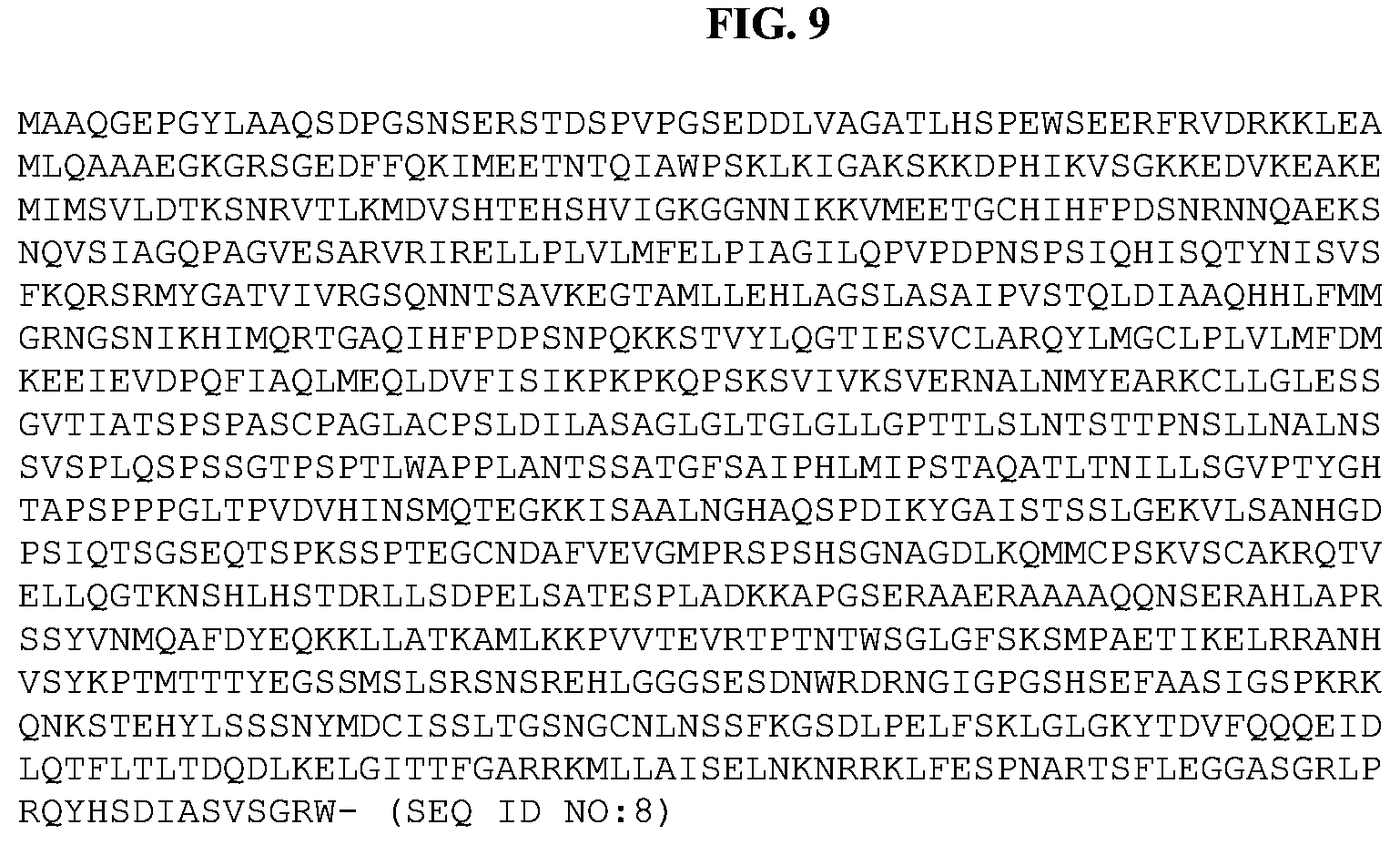
D00017

D00018

D00019

D00020

D00021

D00022

D00023

D00024

D00025

D00026

D00027

S00001
XML
uspto.report is an independent third-party trademark research tool that is not affiliated, endorsed, or sponsored by the United States Patent and Trademark Office (USPTO) or any other governmental organization. The information provided by uspto.report is based on publicly available data at the time of writing and is intended for informational purposes only.
While we strive to provide accurate and up-to-date information, we do not guarantee the accuracy, completeness, reliability, or suitability of the information displayed on this site. The use of this site is at your own risk. Any reliance you place on such information is therefore strictly at your own risk.
All official trademark data, including owner information, should be verified by visiting the official USPTO website at www.uspto.gov. This site is not intended to replace professional legal advice and should not be used as a substitute for consulting with a legal professional who is knowledgeable about trademark law.