Implantable neurostimulator-implemented method utilizing multi-modal stimulation parameters
Libbus , et al. April 13, 2
U.S. patent number 10,974,050 [Application Number 16/009,099] was granted by the patent office on 2021-04-13 for implantable neurostimulator-implemented method utilizing multi-modal stimulation parameters. This patent grant is currently assigned to East Tennessee State University, LivaNova USA, Inc.. The grantee listed for this patent is East Tennessee State University, LivaNova USA, Inc.. Invention is credited to Badri Amurthur, Jeffrey L. Ardell, Bruce H. KenKnight, Imad Libbus, Gregory A. Ordway.


View All Diagrams
| United States Patent | 10,974,050 |
| Libbus , et al. | April 13, 2021 |
Implantable neurostimulator-implemented method utilizing multi-modal stimulation parameters
Abstract
Multi-modal stimulation therapy may be utilized in which two or more stimulation therapies having different stimulation parameters may be delivered to a single patient. This can preferentially stimulate different nerve fiber types and drive different functional responses in the target organs. The stimulation parameters that may vary between the different stimulation therapies include, for example, pulse frequency, pulse width, pulse amplitude, and duty cycle.
| Inventors: | Libbus; Imad (St. Paul, MN), Amurthur; Badri (Los Gatos, CA), KenKnight; Bruce H. (Maple Grove, MN), Ardell; Jeffrey L. (Johnson City, TN), Ordway; Gregory A. (Johnson City, TN) | ||||||||||
|---|---|---|---|---|---|---|---|---|---|---|---|
| Applicant: |
|
||||||||||
| Assignee: | LivaNova USA, Inc. (Houston,
TX) East Tennessee State University (Johnson City, TN) |
||||||||||
| Family ID: | 1000005483059 | ||||||||||
| Appl. No.: | 16/009,099 | ||||||||||
| Filed: | June 14, 2018 |
Prior Publication Data
| Document Identifier | Publication Date | |
|---|---|---|
| US 20180361158 A1 | Dec 20, 2018 | |
Related U.S. Patent Documents
| Application Number | Filing Date | Patent Number | Issue Date | ||
|---|---|---|---|---|---|
| 14521044 | Oct 22, 2014 | 9999773 | |||
| 61897425 | Oct 30, 2013 | ||||
| Current U.S. Class: | 1/1 |
| Current CPC Class: | A61N 1/36139 (20130101); A61N 1/3615 (20130101); A61N 1/36171 (20130101); A61N 1/36053 (20130101); A61N 1/36114 (20130101); A61N 1/0556 (20130101) |
| Current International Class: | A61N 1/36 (20060101); A61N 1/05 (20060101) |
References Cited [Referenced By]
U.S. Patent Documents
| 5330507 | July 1994 | Schwartz |
| 5522854 | June 1996 | Ideker et al. |
| 5707400 | January 1998 | Terry, Jr. et al. |
| 5928272 | July 1999 | Adkins et al. |
| 5978709 | November 1999 | Begemann et al. |
| 6006134 | December 1999 | Hill et al. |
| 6266564 | July 2001 | Hill et al. |
| 6354991 | March 2002 | Gross et al. |
| 6449507 | September 2002 | Hill et al. |
| 6473644 | October 2002 | Terry, Jr. et al. |
| 6508771 | January 2003 | Padmanabhan et al. |
| 6522926 | February 2003 | Kieval et al. |
| 6600954 | July 2003 | Cohen et al. |
| 6610713 | August 2003 | Tracey |
| 6616624 | September 2003 | Kieval |
| 6622041 | September 2003 | Terry, Jr. et al. |
| 6652449 | November 2003 | Gross et al. |
| 6684105 | January 2004 | Cohen et al. |
| 6690971 | February 2004 | Schauerte et al. |
| 6712772 | March 2004 | Cohen et al. |
| 6718208 | April 2004 | Hill et al. |
| 6838471 | January 2005 | Tracey |
| 6839594 | January 2005 | Cohen et al. |
| 6850801 | February 2005 | Kieval et al. |
| 6862480 | March 2005 | Cohen et al. |
| 6892098 | May 2005 | Ayal et al. |
| 6896651 | May 2005 | Gross et al. |
| 6904318 | June 2005 | Hill et al. |
| 6907295 | June 2005 | Gross et al. |
| 6963773 | November 2005 | Waltman et al. |
| 6963779 | November 2005 | Shankar |
| 6985774 | January 2006 | Kieval et al. |
| 7010345 | March 2006 | Hill et al. |
| 7072720 | July 2006 | Puskas |
| 7123961 | October 2006 | Kroll et al. |
| 7136705 | November 2006 | Park |
| 7139607 | November 2006 | Shelchuk |
| 7158832 | January 2007 | Kieval et al. |
| 7184828 | February 2007 | Hill et al. |
| 7184829 | February 2007 | Hill et al. |
| 7189204 | March 2007 | Ni et al. |
| 7218964 | May 2007 | Hill et al. |
| 7221979 | May 2007 | Zhou et al. |
| 7225017 | May 2007 | Shelchuk |
| 7225019 | May 2007 | Jahns et al. |
| 7237320 | July 2007 | Lam |
| 7245967 | July 2007 | Shelchuk |
| 7260431 | August 2007 | Libbus et al. |
| 7269457 | September 2007 | Shafer et al. |
| 7277761 | October 2007 | Shelchuk |
| 7295881 | November 2007 | Cohen et al. |
| 7305265 | December 2007 | Fukui |
| 7321793 | January 2008 | Ben Ezra et al. |
| 7324853 | January 2008 | Ayal et al. |
| 7336997 | February 2008 | Fukui |
| 7346398 | March 2008 | Gross et al. |
| 7387603 | June 2008 | Gross et al. |
| 7389149 | June 2008 | Rossing et al. |
| 7395119 | July 2008 | Hagen et al. |
| 7403819 | July 2008 | Shelchuk et al. |
| 7418292 | August 2008 | Shafer |
| 7452800 | November 2008 | Sosnowchik et al. |
| 7480532 | January 2009 | Kieval et al. |
| 7481759 | January 2009 | Whitehurst et al. |
| 7485104 | February 2009 | Kieval |
| 7493167 | February 2009 | Hussein et al. |
| 7499742 | March 2009 | Bolea et al. |
| 7499747 | March 2009 | Kieval et al. |
| 7499748 | March 2009 | Moffitt et al. |
| 7502650 | March 2009 | Kieval |
| 7542800 | June 2009 | Libbus et al. |
| 7548780 | June 2009 | Libbus et al. |
| 7551958 | June 2009 | Libbus et al. |
| 7561922 | July 2009 | Cohen et al. |
| 7561923 | July 2009 | Libbus et al. |
| 7570999 | August 2009 | Libbus et al. |
| 7582053 | September 2009 | Gross et al. |
| 7584004 | September 2009 | Caparso et al. |
| 7587238 | September 2009 | Moffitt et al. |
| 7606622 | October 2009 | Reeve |
| 7613511 | November 2009 | Wu et al. |
| 7613516 | November 2009 | Cohen et al. |
| 7616990 | November 2009 | Chavan et al. |
| 7617003 | November 2009 | Caparso et al. |
| 7623926 | November 2009 | Rossing et al. |
| 7627384 | December 2009 | Ayal et al. |
| 7628750 | December 2009 | Cohen et al. |
| 7630760 | December 2009 | Libbus et al. |
| 7634315 | December 2009 | Cholette |
| 7634317 | December 2009 | Ben-David |
| 7640057 | December 2009 | Libbus et al. |
| 7647101 | January 2010 | Libbus et al. |
| 7647114 | January 2010 | Libbus |
| 7650190 | January 2010 | Zhou et al. |
| 7657312 | February 2010 | Pastore et al. |
| 7660628 | February 2010 | Libbus et al. |
| 7664548 | February 2010 | Amurthur et al. |
| 7668602 | February 2010 | Ben-David et al. |
| 7672733 | March 2010 | Zhou et al. |
| 7676275 | March 2010 | Farazi et al. |
| 7684866 | March 2010 | Fowler et al. |
| 7689286 | March 2010 | Pastore et al. |
| 7711415 | May 2010 | Farazi et al. |
| 7711421 | May 2010 | Shafer et al. |
| 7734355 | June 2010 | Cohen et al. |
| 7751884 | July 2010 | Ternes et al. |
| 7769442 | August 2010 | Shafer |
| 7769446 | August 2010 | Moffitt et al. |
| 7778702 | August 2010 | Ben-David et al. |
| 7778703 | August 2010 | Gross et al. |
| 7778711 | August 2010 | Ben-David et al. |
| 7783353 | August 2010 | Libbus et al. |
| 7797041 | September 2010 | Libbus et al. |
| 7801603 | September 2010 | Westlund et al. |
| 7801604 | September 2010 | Brockway et al. |
| 7801614 | September 2010 | Rossing et al. |
| 7805193 | September 2010 | Libbus et al. |
| 7805203 | September 2010 | Ben-David et al. |
| 7813805 | October 2010 | Farazi |
| 7813812 | October 2010 | Kieval et al. |
| 7835797 | November 2010 | Rossing et al. |
| 7840266 | November 2010 | Libbus et al. |
| 7840271 | November 2010 | Kieval et al. |
| 7844346 | November 2010 | Cohen et al. |
| 7848812 | December 2010 | Crowley et al. |
| 7848816 | December 2010 | Wenzel et al. |
| 7869869 | January 2011 | Farazi |
| 7885709 | February 2011 | Ben-David |
| 7885711 | February 2011 | Ben-Ezra et al. |
| 7890185 | February 2011 | Cohen et al. |
| 7894907 | February 2011 | Cowan et al. |
| 7904151 | March 2011 | Ben-David et al. |
| 7904175 | March 2011 | Scott et al. |
| 7904176 | March 2011 | Ben-Ezra et al. |
| 7908008 | March 2011 | Ben-David et al. |
| 7916013 | March 2011 | Stevenson |
| 7925342 | April 2011 | Amurthur et al. |
| 7925352 | April 2011 | Stack et al. |
| 7974693 | July 2011 | Ben-David et al. |
| 8005542 | August 2011 | Ben-Ezra et al. |
| 8005545 | August 2011 | Ben-David et al. |
| 8036745 | October 2011 | Ben-David et al. |
| 8060197 | November 2011 | Ben-David et al. |
| 8065021 | November 2011 | Gross et al. |
| 8083663 | December 2011 | Gross et al. |
| 8116881 | February 2012 | Cohen et al. |
| 8131362 | March 2012 | Moffitt et al. |
| 8160701 | April 2012 | Zhao et al. |
| 8160705 | April 2012 | Stevenson et al. |
| 8195290 | June 2012 | Brockway et al. |
| 8224436 | July 2012 | Libbus et al. |
| 8249711 | August 2012 | Libbus et al. |
| 8369943 | February 2013 | Shuros et al. |
| 8386038 | February 2013 | Bianchi et al. |
| 8401640 | March 2013 | Zhao et al. |
| 8417354 | April 2013 | Zhang et al. |
| 8571654 | October 2013 | Libbus et al. |
| 8577458 | November 2013 | Libbus et al. |
| 8600505 | December 2013 | Libbus et al. |
| 8688212 | April 2014 | Libbus et al. |
| 2002/0107553 | August 2002 | Hill et al. |
| 2003/0040774 | February 2003 | Terry, Jr. et al. |
| 2003/0153954 | August 2003 | Park et al. |
| 2004/0199210 | October 2004 | Shelchuk |
| 2004/0210261 | October 2004 | King et al. |
| 2004/0215265 | October 2004 | Keizer |
| 2005/0049655 | March 2005 | Boveja et al. |
| 2005/0065553 | March 2005 | Ben Ezra et al. |
| 2005/0065575 | March 2005 | Dobak |
| 2005/0125044 | June 2005 | Tracey |
| 2005/0131467 | June 2005 | Boveja |
| 2005/0165465 | July 2005 | Pianca et al. |
| 2005/0267542 | December 2005 | David et al. |
| 2006/0015153 | January 2006 | Gliner et al. |
| 2006/0100668 | May 2006 | Ben-David et al. |
| 2006/0190053 | August 2006 | Dobak, III |
| 2006/0206155 | September 2006 | Ben-David et al. |
| 2006/0253161 | November 2006 | Libbus et al. |
| 2007/0067004 | March 2007 | Boveja et al. |
| 2007/0093870 | April 2007 | Maschino |
| 2007/0097593 | May 2007 | Armstrong |
| 2007/0213773 | September 2007 | Hill et al. |
| 2007/0233194 | October 2007 | Craig |
| 2007/0255320 | November 2007 | Inman et al. |
| 2007/0276453 | November 2007 | Hill et al. |
| 2008/0015659 | January 2008 | Zhang et al. |
| 2008/0021503 | January 2008 | Whitehurst et al. |
| 2008/0033511 | February 2008 | Dobak |
| 2008/0051839 | February 2008 | Libbus et al. |
| 2008/0058874 | March 2008 | Westlund et al. |
| 2008/0061240 | March 2008 | Heuft |
| 2008/0091240 | April 2008 | Ben-David et al. |
| 2008/0132983 | June 2008 | Cohen et al. |
| 2008/0147140 | June 2008 | Ternes et al. |
| 2008/0183258 | July 2008 | Inman |
| 2008/0243196 | October 2008 | Libbus et al. |
| 2009/0030493 | January 2009 | Colborn et al. |
| 2009/0118777 | May 2009 | Iki et al. |
| 2009/0124848 | May 2009 | Miazga |
| 2009/0149900 | June 2009 | Moffitt et al. |
| 2009/0248097 | October 2009 | Tracey et al. |
| 2009/0270953 | October 2009 | Ecker et al. |
| 2009/0275956 | November 2009 | Burnes et al. |
| 2010/0010556 | January 2010 | Zhao et al. |
| 2010/0010603 | January 2010 | Ben-David et al. |
| 2010/0016919 | January 2010 | Hill et al. |
| 2010/0042173 | February 2010 | Farazi et al. |
| 2010/0114197 | May 2010 | Burnes et al. |
| 2010/0114203 | May 2010 | Burnes et al. |
| 2010/0114227 | May 2010 | Cholette et al. |
| 2010/0174341 | July 2010 | Bolea et al. |
| 2010/0286740 | November 2010 | Libbus et al. |
| 2010/0331908 | December 2010 | Farazi |
| 2011/0015692 | January 2011 | Libbus et al. |
| 2011/0082514 | April 2011 | Libbus et al. |
| 2011/0098796 | April 2011 | Ben-David et al. |
| 2011/0224749 | September 2011 | Ben-David et al. |
| 2011/0257708 | October 2011 | Kramer et al. |
| 2011/0313488 | December 2011 | Hincapie Ordonez et al. |
| 2012/0143286 | June 2012 | Hahn et al. |
| 2012/0172742 | July 2012 | Arcot-Krishnamurthy et al. |
| 2012/0185007 | July 2012 | Ziegler et al. |
| 2012/0185010 | July 2012 | Zhou et al. |
| 2012/0192874 | August 2012 | Bolea et al. |
| 2012/0271374 | October 2012 | Nelson et al. |
| 2012/0303080 | November 2012 | Ben-David et al. |
| 2012/0330369 | December 2012 | Osorio et al. |
| 2013/0158616 | June 2013 | Libbus et al. |
| 2013/0158617 | June 2013 | Libbus et al. |
| 2013/0158618 | June 2013 | Libbus et al. |
| 2013/0238047 | September 2013 | Libbus et al. |
| 2013/0289646 | October 2013 | Libbus et al. |
| 2014/0025132 | January 2014 | Libbus et al. |
| 2014/0135862 | May 2014 | Libbus et al. |
| 2014/0135863 | May 2014 | Libbus et al. |
| 2014/0135864 | May 2014 | Libbus et al. |
| 2014/0228905 | August 2014 | Bolea |
| 2014/0277232 | September 2014 | Libbus et al. |
| 2015/0073511 | March 2015 | Libbus et al. |
| 2015/0073512 | March 2015 | Libbus et al. |
| 2015/0094962 | April 2015 | Hoegh et al. |
| 2015/0119956 | April 2015 | Libbus et al. |
| 2015/0119959 | April 2015 | Libbus et al. |
| 2015/0196762 | July 2015 | Amurthur et al. |
| WO-2010/005482 | Jan 2010 | WO | |||
Other References
|
US 8,315,702 B2, 11/2012, Chavan et al. (withdrawn) cited by applicant . Zhang, et al., "Chronic Vagus Nerve Stimulation Improves Autonomic Control and Attenuates Systemic Inflammation and Heart Failure Progression in a Canine High-Rate Pacing Model," Journal of the American Heart Association, Circ Heart Fail, 2, pp. 692-699 (2009). cited by applicant . Abraham, et al., "Devices in the management of advanced, chronic heart failure," Nature Reviews Cardiology, vol. 10, pp. 98-110 (Feb. 2013) (Published online Dec. 11, 2012). cited by applicant . Adamson, et al., "Continuous Autonomic Assessment in Patients with Symptomatic Heart Failure: Prognostic Value of Heart Rate Variability Measured by an Implanted Cardiac Resynchronization Device," Circulation, Journal of the American Heart Association, 110, pp. 2389-2394 (2004). cited by applicant . Agostoni, et al., "Functional and Histological Studies of the Vagus Nerve and its Branches to the Heart, Lungs and Abdominal Viscera in the Cat," J. Physiol. 135, pp. 182-205 (1957). cited by applicant . Ajani, et al., "Prevalence of High C-Reactive Protein in Persons with Serum Lipid Concentrations within Recommended Values," Clinical Chemistry, 50:9, pp. 1618-1622 (2004). cited by applicant . Akiyama, et al., "Effects of right and left vagal stimulation on left ventricular acetylcholine levels in the cat," Acta Physiol Scand, 172, pp. 11-16 (2001). cited by applicant . Anand, et al., "C-Reactive Protein in Heart Failure: Prognostic Value and the Effect of Valsartan," Circulation, Journal of the American Heart Association, 112, pp. 1428-1434 (2005). cited by applicant . Anholt, et al., "Recruitment and blocking properties of the CardioFit stimulation lead," Journal of Neural Engineering, 8, pp. 1-6, (2011). cited by applicant . Ardell, et al., "Selective vagal innervation of sinoatrial and atrioventricular nodes in canine heart," Am. J. Physiol. 251 (Heart Circ. Physiol. 20), pp. H764-H773 (1986). cited by applicant . Armour, "Cardiac neuronal hierarchy in health and disease," Am J Physiol Regul Integr Comp Physiol, 287, pp. R262-R271 (2004). cited by applicant . Armour, "Myocardial ischaemia and the cardiac nervous system," Cardiovascular Research, 41, pp. 41-54 (1999). cited by applicant . Armour, "The little brain on the heart," Cleveland Clinic Journal of Medicine, vol. 74, supp. 1, pp. S48-S51 (Feb. 2007). cited by applicant . Armour, et al., "Functional anatomy of canine cardiac nerves," Acta anat., 91, pp. 510-528 (1975). cited by applicant . Armour, et al., "Localized myocardial responses to stimulation of small cardiac branches of the vagus," American Journal of Physiology, vol. 228, No. 1 pp. 141-148 (Jan. 1975). cited by applicant . Armour, JA, "Potential clinical relevance of the `little brain` on the mammalian heart," Experimental Physiology, vol. 93, No. 2, pp. 165-176 (Feb. 2008). Online Publication Date: Nov. 2, 2007. Available at: http://ep.physoc.org/content/93/2/165.long. cited by applicant . Asala, et al., "An electron microscope study of vagus nerve composition in the ferret," Anat Embryol, 175, pp. 247-253 (1986). cited by applicant . Aukrust, et al., "Inflammatory and anti-inflammatory cytokines in chronic heart failure: Potential therapeutic implications," Annals of Medicine, 37, pp. 74-85 (2005). cited by applicant . Author Unknown, "Nerve fiber--Types and Function," www.boddunan.com Available at www.boddunan/education/20-medicine-a-surgery/12730-nerver-fiber-types-and- -function.html (2010). cited by applicant . Author Unknown, American Diabetes Association, "Standards of Medical Care in Diabetes--2012," Diabetes Care, vol. 35, supplement 1, pp. S11-S63 (Jan. 2012). cited by applicant . Author Unknown, Staff of Adinstruments, "Principles of Nerve Stimulation," Application Note, ADInstruments (Apr. 2002). cited by applicant . Bae, et al., "Gliosis in the Amygdala Following Myocardial Infarction in the Rat," J Vet Med Sci, 72(8), pp. 1041-1045 (2010). cited by applicant . Bernik, et al., "Pharmacological Stimulation of the Cholinergic Antiinflammatory Pathway," J. Exp. Med, vol. 195, No. 6, pp. 781-788 (Mar. 18, 2002). cited by applicant . Berthoud, et al., "Functional and chemical anatomy of the afferent vagal system," Autonomic Neuroscience: Basic and Clinical, 85, pp. 1-17 (2000). cited by applicant . Bhagat, et al., "Differential Effect of Right and Left Vagal Stimulation on Right and Left Circumflex Coronary Arteries," S A Medical Journal, 50, pp. 1591-1594 (1976). cited by applicant . Biasucci, et al., "Elevated Levels of C-Reactive Protein at Discharge in Patients with Unstable Angina Predict Recurrent Instability," Circulation, Journal of the American Heart Association, 99, pp. 855-860 (1999). cited by applicant . Bibevski, et al., "Evidence for impaired vagus nerve activity in heart failure," Heart Fail Rev, 16, pp. 129-135 (2011). cited by applicant . Bibevski, et al., "Ganglionic Mechanisms Contribute to Diminished Vagal Control in Heart Failure," Circulation, Journal of the American Heart Association, 99, pp. 2958-2963 (1999). cited by applicant . Bilgutay, et al., "Vagal Tuning. A new concept in the treatment of supraventricular arrhythmias, angina pectoris, and heart failure," Journal of Thoracic and Cardiovascular Surgery, vol. 56, No. 1, pp. 71-82 (Jul. 1968). cited by applicant . Binkley, et al., "Parasympathetic Withdrawal Is an Integral Component of Autonomic Imbalance in Congestive Heart Failure: Demonstration in Human Subjects and Verification in a Paced Canine Model of Ventricular Failure," JACC, vol. 18, No. 2, pp. 464-472 (Aug. 1991). cited by applicant . Bois, et al., "Mode of action of bradycardic agent, S 16257, on ionic currents of rabbit sinoatrial node cells," Abstract, British Journal of Pharmacology, 118(4):1051-7 (1996). cited by applicant . Bonaz, et al., "Vagus nerve stimulation: From epilepsy to the cholinergic anti-inflammatory pathway," Neurogastroenterology & Motility, pp. 208-221 (2013). cited by applicant . Borggrefe, "Vagal Stimulation Devices," ESC Congress 2010 (2010). cited by applicant . Borovilkova, et al., "Vagus nerve stimulation attenuates the systemic inflammatory response to endotoxin," Nature, vol. 405, pp. 458-462 (May 25, 2000). cited by applicant . Brack, et al., "Mechanisms underlying the autonomic modulation of ventricular fibrillation initiation--tentative prophylactic properties of vagus nerve stimulation on malignant arrhythmias in heart failure," Heart Fail Rev (Published online Jun. 8, 2012). cited by applicant . Bronzino, "Biomedical Engineering Fundamentals," CRC Press, Chapter 30, pp. 30-10-30-15 (Apr. 2006). cited by applicant . Buschman, et al., "Heart Rate Control via Vagus Nerve Stimulation," Neuromodulation, vol. 9, No. 3, pp. 214-220 (2006). cited by applicant . Butterwick, et al., "Tissue Damage by Pulsed Electrical Stimulation," IEEE Transactions on Biomedical Engineering, vol. 54, No. 12, pp. 2261-2267 (Dec. 2007). cited by applicant . Calkins, et al., "Comparison of Responses to Isoproterenol and Epinephrine During Head-Up Tilt in Suspected Vasodepressor Syncope," The American Journal of Cardiology, vol. 67 pp. 207-209 (Jan. 15, 1991). cited by applicant . Castoro et al., "Excitation properties of the right cervical vagus nerve in adult dogs," Experimental Neurology, vol. 227, iss. 1, pp. 62-68 (Jan. 2011). Online Publication Date: Sep. 17, 2010. Available at: http://www. sciencedirect.com/science/article/pii/S001448861000347X. cited by applicant . Chapleau, et al., "Methods of assessing vagus nerve activity and reflexes," Heart Fail Rev, 16, pp. 109-127 (2011). cited by applicant . Chen, et al., "National and Regional Trends in Heart Failure Hospitalization and Mortality Rates for Medicare Beneficiaries, 1998-2008," JAMA, vol. 306, No. 15 (Oct. 19, 2011). cited by applicant . Chen, et al., "Role of Atrial Electrophysiology and Autonomic Nervous System in Patients with Supraventricular Tachycardia and Paroxysmal Artrial Fibrillation," J Am Coll Cardiol, vol. 32, No. 3, pp. 732-738 (Sep. 1998). cited by applicant . Cheng, et al., "Long-term Outcomes in Individuals with Prolonged PR Interval or First-Degree Atrioventricular Block," JAMA, vol. 301, No. 24 pp. 2571-2577 (Jun. 24, 2009). cited by applicant . Chiou, et al., "Effects of Continuous Enhanced Vagal Tone and Dual Atrioventricular Node and Accessory Pathways," Circulation, Journal of the American Heart Association, 107, pp. 2583-2588 (2003). cited by applicant . Cohen, et al., "Histopathology of the stimulated Vagus nerve: Primum non nocere," Heart Fail Rev, 16, pp. 163-169 (2011). cited by applicant . Colombo, et al., "Comparison between spectral analysis and the phenylephrine methods for the assessment of baroreflex sensitivity in chronic heart failure," Clinical Science, 97, pp. 503-513 (1999). cited by applicant . Cryan, et al., "Animal models and mood disorders: Recent developments," Current Opinion in Psychiatry, 20, pp. 1-7 (2007). cited by applicant . Das, "Vagal nerve stimulation in prevention and management of coronary heart disease," World J. Cardiol, 3(4), pp. 105-110 (Apr. 26, 2011). cited by applicant . De Castro, et al., "Parasympathetic-mediated atrial fibrillation during tilt test associated with increased baroreflex sensitivity," The European Society of Cardiology, Europace, 8, pp. 349-351 (2006). cited by applicant . De Ferrari, et al., "Chronic Vagus Nerve Stimulation: A New and Promising Therapeutic Approach for Chronic Heart Failure," European Heart Journal, vol. 32, Oct. 28, 2010, pp. 847-855. cited by applicant . De Ferrari, et al., "Baroreflex Sensitivity Predicts Long-Term Cardiovascular Mortality After Myocardial Infarction Even in Patients with Preserved Left Ventricular Function," Journal of the American College of Cardiology, vol. 50, No. 24, pp. 2285-2290 (2007). cited by applicant . De Ferrari, et al., "Chronic Vagal Stimulation in Patients with Congestive Heart Failure," 31st Annual International Conference of the IEE EMBS (2009). cited by applicant . De Ferrari, et al., "Vagus nerve stimulation: from pre-clinical to clinical application: challenges and future directions," Heart Fail Rev, 16, pp. 195-203 (2011) (Published Online Dec. 17, 2010). cited by applicant . De Jonge, et al., "Stimulation of the vagus nerve attenuates macrophage activation by activating the Jak2-STAT3 signaling pathway," Nature Immunology, vol. 6, No. 8, pp. 844-851 (Aug. 2005). cited by applicant . Desai, et al., "Pharmacologic modulation of parasympathetic activity in heart failure," Heart Fail Rev, 16, pp. 179-193 (Published online: Oct. 6, 2010) (2011). cited by applicant . Dickerson, et al., "Parasympathetic neurons in the cranial medial ventricular fat pad on the dog heart selectively decrease ventricular contractility," Journal of the Autonomic Nervous System, 70, pp. 129-141 (1998). cited by applicant . Dunlap, et al., "Mechanisms of altered vagal control in heart failure: influence of muscarinic receptors and acetylcholinesterase activity," Am J Physiol Heart Circ Physiol, 285, pp. H1632-H1640 (Jun. 26, 2003). cited by applicant . Elsenbruch, et al., "Heart Rate Variability During Waking and Sleep in Healthy Males and Females," Sleep, vol. 22, No. 8, pp. 1067-1071 (1999). cited by applicant . Euler, et al., "Acetylcholine release by a stimulus train lowers atrial fibrillation threshold," Am. J Physiol. 253 (Heart Circ Physiol, 22), pp. H863-H868 (1987). cited by applicant . Evans, et al., "Histological and functional studies on the fibre composition of the vagus nerve of the rabbit," Journal of Anatomy, 88, pp. 320-337 (1954). cited by applicant . Fallen, "Vagal Afferent Stimulation as a Cardioprotective Strategy? Introducing the Concept," A.N.E., vol. 10, No. 4 (Oct. 2005). cited by applicant . Fan, et al., "Transvenous vagus nerve stimulation: A potential heart failure therapy is feasible in humans," JACC, vol. 55, issue 10A, pp. E152-E153 (2010). cited by applicant . Fazan, et al., "Diabetic Peripheral Neuropathies: A Morphometric Overview," Int. J. Morphol, 28(I), pp. 51-64 (2010). cited by applicant . Feinauer, et al., "Ouabain enhances release of acetylcholine in the heart evoked by unilateral vagal stimulation," Arch Pharmacol, 333, pp. 7-12 (1986). cited by applicant . Fonarow, et al., "Incremental Reduction in Risk of Death Associated with Use of Guideline-Recommended Therapies in Patients with Heart Failure: A Nested Case-Control Analysis of Improve HF," J Am Heart Assoc, 1, pp. 16-26 (2012). cited by applicant . Ford, et al., "The effects of electrical stimulation of myelinated and non-myelinated vagal fibres on heart rate in the rabbit," J. Physiol. 380, pp. 341-347 (1986). cited by applicant . Furukawa, et al., "Effects of Verapamil, Zatebradine, and E-4031 on the Pacemaker Location and Rate in Response to Sympathetic Stimulation in Dog Hearts," The Journal of Pharmacology and Experimental Therapeutics, vol. 289, No. 3, pp. 1334-1342 (1999). cited by applicant . Furukawa, et al., "Selective inhibition by zatebradine and discrete parasympathetic stimulation of the positive chronotropic response to sympathetic stimulation in anesthetized dogs," Abstract, Journal of Pharmacology & Experimental Therapeutics, 272(2):744-9 (1995). cited by applicant . Gatti, et al., "Can neurons in the nucleus ambiguous selectively regulate cardiac rate and atrio-ventricular conduction?" Journal of the Autonomic Nervous System, 57, pp. 123-127 (1996). cited by applicant . Gatti, et al., "Vagal control of left ventricular contractility is selectively mediated by a cranioventricular intracardiac ganglion in the cat," Journal of the Autonomic Nervous System, 66, pp. 138-144 (1997). cited by applicant . Gibbons, et al., "Neuromodulation targets intrinsic cardiac neurons to attenuate neuronally mediated atrial arrhythmias," Am J Physiol Regul lntegr Comp Physiol 302: R357-R364 (2012) (First published Nov. 16, 2011). cited by applicant . Gottdiener, et al., "Predictors of Congestive Heart Failure in the Elderly: The Cardiovascular Heath Study," Journal of the American College of Cardiology, vol. 35, No. 6, pp. 1628-1637 (2000). cited by applicant . Gray, et al., "Parasympathetic control of the Heart. II. A novel interganglionic intrinsic cardiac circuit mediates neural control of heart rate," J. Appl Physiol, 96, pp. 2273-2278 (2004). cited by applicant . Gray, et al., "Parasympathetic control of the Heart. III. Neuropeptide Y-immunoreactive nerve terminals synapse on three populations of negative chronotropic vagal preganglionic neurons," J. Appl Physiol, 96, pp. 2279-2287 (2004). cited by applicant . Grill, "Chapter 14--Principles of Electric Field Generation for Stimulation of the Central Nervous System," Neuromodulation, Academic Press (2009). cited by applicant . Guilleminault, et al., "Cyclical Variation of the Heart Rate in Sleep Apnoea Syndrome," The Lancet, pp. 126-131 (Jan. 21, 1984). cited by applicant . Hardwick, et al., "Chronic myocardial infarction induces phenotypic and functional remodeling in the guinea pig cardiac plexus," Am J Physiol Regulatory Integrative Comp Physiol, 295, pp. 1926-1933 (2008). cited by applicant . Hardwick, et al., "Remodeling of the guinea pig intrinsic cardiac plexus with chronic pressure overload," Am J Physiol Regulatory Integrative Comp Physiol, 297, pp. R859-R866 (2009). cited by applicant . Hauptman, et al., "The vagus nerve and autonomic imbalance in heart failure: past, present, and future," Heart Fail Rev, 16, pp. 97-99 (2011). cited by applicant . Hellyer, et al., "Autonomic nerve activity and blood pressure in ambulatory dogs," Heart Rhythm, vol. 11(2), pp. 307-313 (Feb. 2014). cited by applicant . Hirooka, et al., "Imbalance of central nitric oxide and reactive oxygen species in the regulation of sympathetic activity and neural mechanisms of hypertension," Am J Physiol Regulatory Integration Comp Physiol, 300, pp. R818-R826 (2011). cited by applicant . Hoffman, et al., "Vagus Nerve Components," Anat Rec, 127, pp. 551-568 (1957). cited by applicant . Hu, et al., "Role of sympathetic nervous system in myocardial ischemia injury: Beneficial or deleterious?" Letters to the Editor, Elsevier Ireland Ltd. (Mar. 27, 2012). cited by applicant . Hua, et al., "Left vagal stimulation induces dynorphin release and suppresses substance P release from the rat thoracic spinal cord during cardiac ischemia," Am J Physiol Regulatory Integrative Comp Physiol, 287, pp. R1468-R1477 (2004). cited by applicant . Huston, et al., "Splenectomy inactivates the cholinergic antiinflammatory pathway during lethal endotoxemia and polymicrobial sepsis," J. Exp. Med, vol. 203, No. 7 pp. 1623-1628 (Jun. 19, 2006). cited by applicant . Huston, et al., "Transcutaneous vagus nerve stimulation reduces serum high mobility group box 1 levels and improves survival in murine sepsis," Crit Care Med, vol. 35, No. 12, pp. 2762-2768 (2007). cited by applicant . Ingemansson, et al., "Autonomic modulation of the atrial cycle length by the head up tilt test: non-invasive evaluation in patients with chronic atrial fibrillation," Heart, 80, pp. 71-76 (1998). cited by applicant . Ito, et al., "Efferent sympathetic and vagal innervation of the canine right ventricle," Circulation, Journal of the American Heart Association, vol. 90, pp. 1459-1468 (1994). cited by applicant . Jacques, et al., "Spinal Cord Stimulation Causes Potentiation of Right Vagus Nerve Effects on Atrial Chronotropic Function and Repolarization in Canines," Journal of Cardiovascular Electrophysiology, vol. 22, No. 4, pp. 440-447 (Apr. 2011). cited by applicant . Jaenisch, et al., "Respiratory muscle training improves baroreceptor sensitivity, decrease sympathetic tonus and increase vagal effect in rats with heart failure," European Heart Journal, 32 (Abstract Supplement), pp. 976 (2011). cited by applicant . Jammes, et al., "Afferent and efferent components of the bronchial vagal branches in cats," Journal of the Autonomic Nervous System, 5, pp. 165-176 (1982). cited by applicant . Janabi, et al., "Oxidized LDL-Induced NF-.kappa.B Activation and Subsequent Expression of Proinflammatory Genes are Defective in Monocyte-Derived Macrophages from CD36-Deficient Patients," Arterioscler Thromb Vasc Biol, 20:1953-1960 (2000). cited by applicant . Janse, et al., "Effects of unilateral stellate ganglion stimulation and ablation on electrophysiologic changes induced by acute myocardial ischemia in dogs," Circulation, Journal of the American Heart Association, 72, pp. 585-595 (1985). cited by applicant . Jessup; et al., "2009 Focused Update: ACCF/AHA Guidelines for the Diagnosis and Management of Heart Failure in Adults," Circulation, Journal of the American Heart Association, vol. 119, pp. 1977-2016 (2009). cited by applicant . Johnson, et al., "Parasympathetic control of the heart. I. An interventriculo-septal ganglion is the major source of the vagal intracardiac innervation of the ventricles," J Appl Physiol, 96, pp. 2265-2272 (2004). cited by applicant . Kakinuma, et al., "Cholinoceptive and cholinergic properties of cardiomyocytes involving an amplification mechanism for vagal efferent effects in sparsely innervated ventricular myocardium," FEBS Journal, 276, pp. 5111-5125 (2009). cited by applicant . Kalman, et al., "Specific effects of zatebradine on sinus node function: suppression of automaticity, prolongation of sinoatrial conduction and pacemaker shift in the denervated canine heart," Abstract, Journal of Pharmacology & Experimental Therapeutics, 272(1):85-93 (1995). cited by applicant . Kaneko, et al., "C-Reactive Protein in Dilated Cardiomyopathy," Cardiology, 91, pp. 215-219 (1999). cited by applicant . Katare, et al., "Vagal nerve stimulation prevents reperfusion injury through inhibition of opening of mitochondrial permeability transition pore independent of bradycardiac effect," The Journal of Thoracic and Cardiovascular Surgery, vol. 137, No. 1, pp. 223-231 (2009). cited by applicant . Katz, et al., "Disease of the heart in the Works of Hippocrates," Br Heart J, 24, pp. 257-264 (1962). cited by applicant . Kawada, et al., "High-frequency dominant depression of peripheral vagal control of heart rate in rats with chronic heart failure," Acta Physiol 207, 494-502 (2013). cited by applicant . Kawada, et al., "Vagal stimulation suppresses ischemia-induced myocardial interstitial norepinephrine release," Life Sciences, 78, pp. 882-887 (2006). cited by applicant . Kawashima, "The autonomic nervous system of the human heart with special reference to its origin, course, and peripheral distribution," Anat Embryol, 209, pp. 425-438 (2005). cited by applicant . Klein et al., "Vagus nerve stimulation: A new approach to reduce heart failure," Cardiology Journal, vol. 17, iss. 6, pp. 638-643 (2010). cited by applicant . Kliks, et al., "Influence of Sympathetic Tone on Ventricular Fibrillation Threshold During Experimental Coronary Occlusion," The American Journal of Cardiology, vol. 36, pp. 45-49 (Jul. 1975). cited by applicant . Kolman, et al., "The effect of vagus nerve stimulation upon vulnerability of the canine ventricle: role of sympathetic-parasympathetic interactions," Circulation, Journal of the American Heart Association, 52, pp. 578-585 (1975). cited by applicant . Kong, et al., "Optimizing the Parameters of Vagus Nerve Stimulation by Uniform Design in Rats with Acute Myocardial Infarction," PLOS One, vol. 7, issue 11 (Nov. 2012). cited by applicant . Koopman, et al., "Pilot study of stimulation of the cholinergic anti-inflammatory pathway with an implantable vagus nerve stimulation device in patients with rheumatoid arthritis," Abstract (2012). cited by applicant . Kulbertus, et al., ed., "Neurocardiology," Futura Publishing Co., pp. 13 ("Anatomy of the Cardiac Efferent Innvervation"); 61-63 ("Autonomic Neural Control"); 87, 89, 92-93 ("Sympathetic-Parasympathetic Interactions"); 183, 187 ("Parasympathetic Nervous System"); 104 (1988). cited by applicant . La Rovere, et al., "Baroreflex Sensitivity and Heart Rate Variability in the Identification of Patients at Risk for Life-Threatening Arrhythmias: Implications for Clinical Trials," Circulation, Journal of the American Heart Association, 103, pp. 2072-2077 (2001). cited by applicant . La Rovere, et al., "Baroreflex sensitivity and heart-rate variability in prediction of total cardiac mortality after myocardial infarction. Atrami (Autonomic Tone and Reflexes After Myocardial Infarction) Investigators," Lancet, 351(9101), pp. 478-484 (Feb. 14, 1998). cited by applicant . Lane, et al., "Prediction and Prevention of Sudden Cardiac Death in Heart Failure," Heart, 91, pp. 674-680 (2005). cited by applicant . Lechat, et al., "Heart rate and Cardiac Rhythm Relationships with Bisoprolol Benefit in Chronic Heart Failure in CIBIS II Trial," Circulation, Journal of American Heart Association, 103, pp. 1428-1433 (2001). cited by applicant . Lewis, et al., "Vagus nerve stimulation decreases left ventricular contractility in vivo in the human and pig heart," Journal of Physiology, 534, pp. 547-552 (2001). cited by applicant . Li, et al., "Vagal Nerve Stimulation Markedly Improves Long-Term Survival After Chronic Heart Failure in Rats," Circulation: Journal of the American Heart Association, vol. 109, iss. 1, pp. 120-124 (Jan. 2004). Online publication date: Dec. 8, 2003. cited by applicant . Li, et al., "Early vagal stimulation markedly prevented cardiac dysfunction in rats after acute myocardial infarction in addition to suppressing arrhythmic death," European Heart Journal, 32 (Abstract Supplement), pp. 297-298 (2011). cited by applicant . Li, et al., "Inflammatory cytokines and nitric oxide in heart failure and potential modulation by vagus nerve stimulation," Heart Fail Rev, 16, pp. 137-145 (2011). cited by applicant . Li, et al., "Low-Level Vagosympathetic Stimulation. A Paradox and Potential New Modality for the Treatment of Focal Atrial Fibrillation," Circ Arrhythm Electrophysiol, Journal of American Heart Association, 2, pp. 645-651 (2009). cited by applicant . Li, et al., "Restoration of vagal tone by donepezil, on top of losartan treatment, markedly suppresses ventricular dysfunction and improves long-term survival in chronic heart failure rats," European Heart Journal, 32 (Abstract Supplement), pp. 642 (2011). cited by applicant . Libby, et al., "Inflammation and Atherosclerosis," Circulation, Journal of the American Heart Association, 105, pp. 1135-1143 (2002). cited by applicant . Liu, et al., "Differing sympathetic and vagal effects on atrial fibrillation in dogs: role of refractoriness heterogeneity," Am. J. Physiol. 273 (Heart Circ. Physiol. 42), pp. H805-H816 (1997). cited by applicant . Lo, et al., "Paradoxical long-term proarrhythmic effects after ablating the `head station` ganglionated plexi of the vagal innervation to the heart," Heart Rhythm, vol. 10, No. 5, pp. 751-757 (May 2013). cited by applicant . Lohmeier, et al., "Prolonged Activation of the Baroreflex Produces Sustained Hypotension," Hypertension, Journal of the American Heart Association, 43, pp. 306-311 (2004). cited by applicant . Lu, et al., "Vagal nerve stimulation protects cardiac injury by attenuating mitochondrial dysfunction in a murine burn injury model," J. Cell. Mol. Med., vol. 17, No. 5, pp. 664-671 (2013). cited by applicant . Ma, et al., "Analysis of afferent, central, and efferent components of the baroreceptor reflex in mice," Am J Physiol Regulatory Integration Comp Physiol, 283, pp. 1033-1040 (2002). cited by applicant . Maj, et al., "P5775: Autonomic imbalance and circulating androgens and estrogens in men with systolic heart failure," European Heart Journal, 32 (Abstract Supplement), pp. 1090 (2011). cited by applicant . Malkin, et al., "Life-saving or life-prolonging? Interpreting trial data and survival curves for patients with congestive heart failure," European Journal of Heart Failure, 7, pp. 143-148 (2005). cited by applicant . Mann, "Chapter 12--Peripheral Nerves," The Nervous System in Action, michaeldmann.net/mannl2.html, (Jul. 2011). cited by applicant . Mann, "Inflammatory Mediators and the Failing Heart. Past, Present, and the Foreseeable Future," Circ Res., 91, pp. 988-998 (2002). cited by applicant . Mann, "Stress-Activated Cytokines and the Heart: From Adaptation to Maladaptation," Annu. Rev. Physiol., 65, pp. 81-101 (2003). cited by applicant . Martin-Portugues, et al., "Histopathologic features of the vagus nerve after electrical stimulation in swine," Histol Histopathol, 20, pp. 851-856 (2005). cited by applicant . Martins, et al., "Distribution of Local Repolarization Changes Produced by Efferent Vagal Stimulation in the Canine Ventricles," JACC, vol. 2, No. 6, pp. 1191-1199 (Dec. 1983). cited by applicant . Massari, et al., "Neural control of left ventricular contractility in the dog heart: synaptic interactions of negative inotropic vagal preganglionic neurons in the nucleus ambiguus and tyrosine hydroxylase immunoreactive terminals," Brain Research, 802, pp. 205-220 (1998). cited by applicant . May, et al., "P564: Long-term prediction of all-cause mortality in diabetic autonomic neuropathy: simple function tests or 24-hour heart rate variability (HRV)?" European Heart Journal, 32 (Abstract Supplement), p. 64 (2011). cited by applicant . Mei, et al., "The Composition of the Vagus Nerve of the Cat," Cell Tissue Res., 209, pp. 423-431 (1980). cited by applicant . Merrill, et al., "Electrical stimulation of excitable tissue: design of efficacious and safe protocols," Journal of Neuroscience Methods, 141, pp. 171-198 (2005). cited by applicant . Mortara, et al., "Arterial Baroreflex Modulation of Heart Rate in Chronic Heart Failure," Circulation, Journal of the American Heart Association, vol. 96, No. 10, pp. 3450-3458 (Nov. 18, 1997). cited by applicant . Murakawa, et al., "Effect of Cervical Vagal Nerve Stimulation on Defibrillation Energy," Jpn Heart J, 44, pp. 91-100 (Jan. 2003). cited by applicant . Naito, "Effects of zatebradine and propranolol on canine ischemia and reperfusion-induced arrhythmias," European Journal of Pharmacology, 388, pp. 171-176 (2000). cited by applicant . Nakajima, et al., "Autonomic Control of the Location and Rate of the Cardiac Pacemaker in the Sinoatrial Fat Pad of Parasympathetically Denervated Dog Hearts," Journal of Cardiovascular Electrophysiology, vol. 13, No. 9 pp. 896-901 (Sep. 2002). cited by applicant . Nearing, et al., "Crescendo in Depolarization and Repolarization Heterogeneity Heralds Development of Ventricular Tachycardia in Hospitalized Patients with Decompensated Heart Failure," Circulation Arrhythmia and Electrophysiology, Journal of the American Heart Association, 5, pp. 84-90 (2012). cited by applicant . Nihei, et al., "Decreased Vagal Control Over Heart Rate in Rats with Right-Sided Congestive Heart Failure--Downregulation of Neuronal Nitric Oxide Synthase," Circ J, 69, pp. 493-499 (2005). cited by applicant . Ninomiya, "Direct Evidence of Nonuniform Distribution of Vagal Effects on Dog Atria," Circulation Research, vol. XIX, pp. 576-583 (Sep. 1966). cited by applicant . Nolan, et al., "Prospective Study of Heart Rate Variability and Mortality in Chronic Heart Failure: Results of the United Kingdom Heart Failure Evaluation and Assessment of Risk Trial (UK-Heart)," Circulation, Journal of the American Heart Association, 98, pp. 1510-1516 (1998). cited by applicant . Ochoa, et al., "P2497: Effects of insulin resistance on resting heart rate, baroreflex sensitivity and indices of autonomic cardiovascular modulation in individuals with high blood pressure levels," European Heart Journal, 32 (Abstract Supplement), pp. 431-432 (2011). cited by applicant . Ogawa, et al., "Left Stellate Ganglion and Vagal Nerve Activity and Cardiac Arrhythmias in Ambulatory Dogs with Pacing-Induced Congestive Heart Failure," Journal of the American College of Cardiology, vol. 50, No. 4, pp. 335-444 (2007). cited by applicant . Okada, et al., "Cyclic Stretch Upregulates Production of Interleukin-8 and Monocyte Chemotactic and Activating Factor/Monocyte Chemoattractant Protein-1 in Human Endothelial Cells," Arterioscler Thromb Vase Biol., 18, pp. 894-901 (1998). cited by applicant . Oliveira, et al., "Effects of vagal stimulation on induction and termination of atrial fibrillation in an in vivo rabbit heart model," Rev Port Cardiol, 29(03), pp. 375-389 (2010). cited by applicant . Olshansky et al., "Parasympathetic Nervous System and Heart Failure: Pathophysiology and Potential Implications for Therapy," Circulation: Journal of the American Heart Association, vol. 118, iss. 8, pp. 863-871 (Aug. 2008). cited by applicant . Onkka, et al., "Sympathetic nerve fibers and ganglia in canine cervical vagus nerves: Localization and quantitation," Heart Rhythm, vol. 10, No. 4, pp. 585-591 (Apr. 2013). cited by applicant . Ordelman, et al., "Selectivity for Specific Cardiovascular Effects of Vagal Nerve Stimulation with a Multi-Contact Electrode Cuff," IEEE, pp. 32-36 (2013). cited by applicant . Packer, et al., "Effect of Carvedilol on Survival in Severe Chronic Heart Failure," The New England Journal of Medicine, vol. 344, No. 22, pp. 1651-1658 (May 31, 2001). cited by applicant . Pavlov, et al., "Central muscarinic cholinergic regulation of the systemic inflammatory response during endotoxemia," PNAS, vol. 103, No. 13, pp. 5219-5223 (Mar. 28, 2006). cited by applicant . Pavlov, et al., "Controlling inflammation: the cholinergic anti-inflammatory pathway," Biochemical Society Transactions, vol. 34, part 6, pp. 1037-1040 (2006). cited by applicant . PCT Application No. PCT/US2012/068205, Search Report and Written Opinion dated Feb. 8, 2013, 15 pages. cited by applicant . PCT Application No. PCT/US2012/068211, Search Report and Written Opinion dated Jun. 13, 2013. cited by applicant . PCT Application No. PCT/US2012/068213, Search Report and Written Opinion dated Mar. 15, 2013, 11 pages. cited by applicant . PCT Application No. PCT/US2012/068223, Search Report and Written Opinion dated Apr. 3, 2013, 11 pages. cited by applicant . PCT Application No. PCT/US2013/021964, Search Report and Written Opinion dated Jul. 25, 2013, 9 pages. cited by applicant . PCT Application No. PCT/US2013/050390, Search Report and Written Opinion dated Nov. 5, 2013. cited by applicant . PCT Application No. PCT/US2013/068541, Search Report and Written Opinion dated Jan. 7, 2014. cited by applicant . PCT Application No. PCT/US2015/020116, Search Report and Written Opinion dated Jul. 6, 2015, 12 pages. cited by applicant . Peckham, et al., "Chapter 18--Implantable Neural Stimulators," Neuromodulation, Academic Press (2009). cited by applicant . Pina, et al., "The Predictive Value of Biomarkers in Heart Failure," Medscape Education Cardiology, Available at http://www.medscape.org/viewarticle/765328 (CME Released: Jun. 15, 2012). cited by applicant . Pitzalis, et al., "Comparison Between Noninvasive Indices of Baroreceptor Sensitivity and the Phenylephrine Method in Post-Myocardial Infarction Patients," Circulation, Journal of the American Heart Association, 97, pp. 1362-1367 (1998). cited by applicant . Poole-Wilson, "Relation of Pathophysiologic Mechanisms to Outcome in Heart Failure," JACC, vol. 22, No. 4 (supplement A), pp. 22A-29A (Oct. 1993). cited by applicant . Pye, et al., "Study of serum C-reactive protein concentration in cardiac failure," Br Heart J, 63, pp. 228-230 (1990). cited by applicant . Rademacher, et al., "P5878: Multidimensional halter-based analysis of cardiac autonomic regulation predicts early AF recurrence after electrical cardioversion," European Heart Journal, 32 (Abstract Supplement), pp. 1116-1117 (2011). cited by applicant . Randall, et al., "Regional vagosympathetic control of the heart," American Journal of Physiology, vol. 227, No. 2, pp. 444-452 (1974). cited by applicant . Randall, et al., "Selective Vagal Innervation of the Heart," Annals of Clinical and Laboratory Science, vol. 16, No. 3, pp. 198-208 (1986). cited by applicant . Raymond, et al., "Elevated interleukin-6 levels in patients with asymptomatic left ventricular systolic dysfunction," American Heart Journal, vol. 141, No. 3, pp. 435-438 (Mar. 2001). cited by applicant . Rhee, et al., "Effects of suprathreshold vagal stimulation on stellate ganglion nerve activity in ambulatory dogs," 33rd Annual Scientific Sessions, Heart Rhythm, Presentation Abstract (2012). cited by applicant . Ricco, et al., "Interganglionic segregation of distinct vagal afferent fibre phenotypes in guinea-pig airways," Journal of Physiology, 495.2, pp. 521-530 (1996). cited by applicant . Riddle, et al., "Epidemiologic Relationships Between A1C and All-Cause Mortality During a Median 3.4-Year Follow-up of Glycemic Treatment in the ACCORD Trial," Diabetes Care, vol. 33, No. 5, pp. 983-990 (May 2010). cited by applicant . Ridker, C-Reactive Protein: A Simple Test to Help Predict Risk of Heart Attack and Stroke, Circulation, Journal of the American Heart Association, 108, pp. e81-e85 Sep. 2003. 6 pages. cited by applicant . Ridker, et al., "Comparison of C-Reactive Protein and Low-Density Lipoprotein Cholesterol Levels in the Prediction of First cardiovascular Events," New England Journal of Medicine, vol. 347, No. 20, pp. 1557-1566 (Nov. 14, 2002). cited by applicant . Ridker, et al., "C-Reactive Protein and Other Markers of Inflammation in the Prediction of Cardiovascular Disease in Women," The New England Journal of Medicine, vol. 342, No. 12, pp. 836-841 (Mar. 23, 2000). cited by applicant . Ridker, et al., "Inflammation, Pravastatin, and the Risk of Coronary Events After Myocardial Infarction in Patients With Average Cholesterol Levels," Circulation, Journal of the American Heart Association, 98, pp. 839-844 (1998). cited by applicant . Roger, et al., "Heart Disease and Stroke Statistics-2011 Update: A Report from the American Heart Association," Circulation, Journal of the American Heart Association. Available at http://circ.ahajournals.oro/content/123/4/e18 (2010). cited by applicant . Romanovsky, et al., "The vagus nerve in the thermoregulatory response to systemic inflammation," Am. J. Physiol., 273, pp. R407-R413 (1997). cited by applicant . Rossi, et al., "Epicardial ganglionated plexus stimulation decreases postoperative inflammatory response in humans," Heart Rhythm, vol. 9, No. 6, pp. 943-950 (Jun. 2012). cited by applicant . Rouse, et al., "The haemodynamic actions of ZENECA ZD7288, a novel sino-atrial node function modulator, in the exercising beagle: a comparison with zategradine and propranolol," Abstract, British Journal of Pharmacology, 113(3):1071-7 (1994). cited by applicant . Rozman, et al., "Heart function influenced by selective mid-cervical left vagus nerve stimulation in a human case study," Hypertension Research, 32, pp. 1041-1043 (2009). cited by applicant . Rutecki, "Anatomical, Physiological and Theoretical Basis for the Antiepileptic Effect of Vagus Nerve Stimulation," Epilepsia, 31 (suppl. 2), pp. S1-S6 (1990). cited by applicant . Sabbah et al., "Vagus Nerve Stimulation in Experimental Heart Failure," Heart Failure Reviews, vol. 16, No. 2, Mar. 2011, pp. 171-178. cited by applicant . Sabbah, et al., "3722: Vagus nerve stimulation improves left ventricular function in heart failure: results of a 6 month investigation with a cross-over design in dogs with experimental heart failure," European Heart Journal, 32 (Abstract Supplement), pp. 642 (2011). cited by applicant . Sabbah, et al., "Baroreflex Activation Therapy for the Treatment of Heart Failure," Presentation available at http://www.cvrx.com/wp/wp-contenUuploads/2012/04/Dr.-Sabbah-Slides.pdf (2012). cited by applicant . Sabbah, et al., "Chronic Electrical Stimulation of the Carotid Sinus Baroreflex Improves Left Ventricular Function and Promotes Reversal of Ventricular Remodeling in Dogs with Advanced Heart Failure," Circulation Heart Failure, Journal of the American Heart Association, 4, pp. 65-70 (2010). cited by applicant . Samara, et al., "The Effects of Cardiac Resynchronization Therapy on Chronotropic Incompetence in Patients Intolerant of Beta Antagonist Therapy," Journal of Cardiac Failure, vol. 17, No. 8S, pp. S54-S55 (Aug. 2011). cited by applicant . Sanner, et al., "P4743: Prediction of cardiovascular risk from nocturnal pulse wave signal using the autonomic state indicator (ASI) technology," European Heart Journal, 32 (Abstract Supplement), pp. 839 (2011). cited by applicant . Sato, et al., "Serial Circulating Concentrations of C-Reactive Protein, Interleukin (IL)-4, and IL-6 in Patients with Acute Left Heart Decompensation," Clin. Cardiol. 22, pp. 811-813 (2009). cited by applicant . Schauerte, "Time for Change: Cardiac neurophysiology meets cardiac electrophysiology," Editorial Commentary, Heart Rhythm Society (2013). cited by applicant . Schiereck, et al., "AV blocking due to asynchronous vagal stimulation in rats," Am J Physiol Heart Circ Physiol, 278, pp. H67-H73 (2000). cited by applicant . Schocken, et al., "Prevalence and Mortality Rate of Congestive Heart Failure in the United States," JACC, vol. 20, No. 2, pp. 301-306 (Aug. 1992). cited by applicant . Schwartz, "Vagal Stimulation for Heart Diseases: From Animals to Men," Circulation Journal, vol. 75, pp. 20-27 (Jan. 2011). cited by applicant . Schwartz, "Vagal stimulation for heart failure," Current Opinion in Cardiology, 26, pp. 51-54 (2011). cited by applicant . Schwartz, "Vagal stimulation for the treatment of heart failure: a translational success story," Heart, vol. 98, No. 23, pp. 1687-1690 (2012). cited by applicant . Schwartz, et al. "Vagal stimulation for heart failure: Background and first in-man study," Heart Rhythm, 6, 11 suppl., pp. S76-S81 (Nov. 2009). cited by applicant . Schwartz, et al., "Autonomic mechanisms and sudden death. New insights from analysis of baroreceptor reflexes in conscious dogs with and without myocardial infarction," Circulation, Journal of the American Heart Association, 78, pp. 969-979 (1988). cited by applicant . Schwartz, et al., "Effects of Unilateral Cardiac Sympathetic Denervation on the Ventricular Fibrillation Threshold," The American Journal of Cardiology, vol. 37, pp. 1034-1040 (Jun. 1976). cited by applicant . Schwartz, et al., "Long term vagal stimulation in patients with advanced heart failure. First experience in man," European Journal of Heart Failure, 10, pp. 884-891 (2008). cited by applicant . Schwartz, et al., "Sympathetic-parasympathetic interaction in health and disease: abnormalities and relevance in heart failure," Heart Fail Rev, 16, pp. 101-107 (2011). cited by applicant . Seta, et al., "Basic Mechanisms in Heart Failure: The Cytokine Hypothesis," Journal of Cardiac Failure, vol. 2, No. 3, pp. 243-249 (1996). cited by applicant . Sha, et al., "Low-Level Right Vagal Stimulation: Anticholinergic and Antiadrenergic Effects," J Cardiovasc Electrophysiol, pp. 1147-1153 (Apr. 2011). cited by applicant . The Diabetes Control and Complications Trial Research Group, "The effect of intensive treatment of diabetes on the development and progression of long-term complications in insulin-dependent diabetes mellitus," The New England Journal of Medicine, vol. 329, No. 14, pp. 977-986 (Sep. 30, 1993). cited by applicant . Shannon, "A Model of Safe Levels for Electrical Stimulation," IEEE Transactions on Biomedical Engineering, vol. 39, No. 4, pp. 424-426 (Apr. 1992). cited by applicant . Shen, et al., "Continuous Low-Level Vagus Nerve Stimulation Reduces Stellate Ganglion Nerve Activity and Paroxysmal Atrial Tachyarrhythmias in Ambulatory Canines," Circulation, Journal of the American Heart Association, 123, pp. 2204-2212 (2011). cited by applicant . Shen, et al., "Low-level vagus nerve stimulation upregulates small conductance calcium-activated potassium channels in the stellate ganglion," Heart Rhythm, vol. 10, No. 6, pp. 910-915 (2013). cited by applicant . Shinohara, et al., "Heart Failure Decreases Nerve Activity in the Right Atrial Ganglionated Plexus," J Cardiovasc Electrophysiol, pp. 404-412 (2011). cited by applicant . Shioi, et al., "Increased Expression of Interleukin-1B and Monocyte Chemotactic and Activating Factor/Monocyte Chemoattractant Protein-1 in the Hypertrophied and Failing Heart with Pressure Overload," Circ Res., 81, pp. 664-671 (1997). cited by applicant . Singal, et al., "The role of oxidative stress in the genesis of heart disease," Cardiovascular Research, 40, pp. 426-432 (1998). cited by applicant . Spuck, et al., "Right-sided vagus nerve stimulation in humans: An effective therapy?," Epilepsy Research, pp. 232-234 (2008). cited by applicant . Stein, et al., "A Simple Method to Identify Sleep Apnea Using Holter Recordings," J Cardiovasc Electrophysiol, vol. 14, pp. 467-473 (May 2003). cited by applicant . Stein, et al., "Feasibility of Using Mobile Cardiac Outpatient Telemetry (MCOT) to Identify Severe Sleep Disorders" (2009). cited by applicant . Stieber, et al., "Bradycardic and proarrhythmic properties of sinus node inhibitors," Abstract, Molecular Pharmacology, 69(4):1328-37 (2006). cited by applicant . Taylor, et al., "The unequal influences of the left and right vagi on the control of the heart and pulmonary artery in the rattlesnake, Grata/us durissus," The Journal of Experimental Biology, 212, pp. 145-151 (2009). cited by applicant . Thayer, et al., "The role of vagal function in the risk for cardiovascular disease and mortality," Biological Psychology, 74, pp. 224-242 (2007). cited by applicant . Thollon, et al., "Electrophysiological effects of S 16257, a novel sino-atrial node modulator, on rabbit and guinea-pig cardiac preparations: comparison with UL-FS 49," Abstract, British Journal of Pharmacology, 112(1):37-42 (1994). cited by applicant . Tosato, et al., "Quasi-trapezoidal pulses to selectively block the activation of intrinsic laryngeal muscles during vagal nerve stimulation," J. Neural Eng., 4, pp. 205-212 (2007). cited by applicant . Tsutsumi, et al., "Modulation of the myocardial redox state by vagal nerve stimulation after experimental myocardial infarction," Cardiovascular Research, 77, pp. 713-721 (2008). cited by applicant . Tyler, et al., "Chapter 17--Electrodes for the Neural Interface," Neuromodulation, Academic Press (2009). cited by applicant . Ulphani, et al., "Quantitative analysis of parasympathetic innervation of the porcine heart," Heart Rhythm, 7, pp. 1113-1119 (2010). cited by applicant . Uthman, et al., "Effectiveness of vagus nerve stimulation in epilepsy patients. A 12-year observation," Neurology, 63, pp. 1124-1126 (2004). cited by applicant . Van Stee, "Autonomic Innervation of the Heart," Environmental Health Perspectives, vol. 26, pp. 151-158 (1978). cited by applicant . Vanoli, et al., "Vagal stimulation and prevention of sudden death in conscious dogs with a healed myocardial infarction," Circulation Research, Journal of the American Heart Association, 68, pp. 1471-1481 (1991). cited by applicant . Vasan, et al., "Inflammatory Markers and Risk of Heart Failure in Elderly Subjects Without Prior Myocardial Infarction," Circulation, Journal of the American Heart Association, 107, pp. 1486-1491 (2003). cited by applicant . Vassalle, et al., "An Analysis of Arrhythmias Induced by Ouabain in Intact Dogs," Circulation Research, Journal of the American Heart Association, 13, pp. 132-148 (1963). cited by applicant . Velagaleti, et al., "Long-Term Trends in the Incidence of Heart Failure After Myocardial Infarction," 118, pp. 2057-2062 (2008). cited by applicant . Verrier, et al., "Microvolt T-Wave Alternans," Journal of the American College of Cardiology, vol. 58, No. 13, pp. 1309-1324 (2011). cited by applicant . Vimercati, et al., "Acute vagal stimulation attenuates cardiac metabolic response to .beta.-adrenergic stress," The Journal of Physiology, vol. 590, No. 23, pp. 6065-6074 (2012). cited by applicant . Wang, et al., "Nicotinic acetylcholine receptor alpha-7 subunit is an essential regulator of inflammation," Nature, vol. 421, pp. 384-388 (Jan. 23, 2003). cited by applicant . Wang, et al., "Synaptic and Neurotransmitter Activation of Cardiac Vagal Neurons in the Nucleus Ambiguus," Annals New York Academy of Sciences, pp. 237-246 (2001). cited by applicant . Waninger, et al., "Characterization of Atrioventricular Nodal Response to Electrical Left Vagal Stimulation," Annals of Biomedical Engineering, vol. 27, pp. 758-762 (1999). cited by applicant . Wann, et al., "Behavioural signs of depression and apoptosis in the limbic system following myocardial infarction: effects of sertraline," Journal of Psychopharmacology, 23(4), pp. 451-459 (2009). cited by applicant . Wann, et al., "Vulnerability for apoptosis in the limbic system after myocardial infarction in rats: a possible model for human postinfarct major depression," J Psychiatry Neurosci, 32(1): pp. 11-16 (2007). cited by applicant . Watkins, et al., "Cytokine-to-Brain Communication: A Review & Analysis of Alternative Mechanisms," Life Sciences, vol. 57, No. 11, pp. 1011-1026 (1995). cited by applicant . Whyte, et al., "Reactive oxygen species modulate neuronal excitability in rat intrinsic cardiac ganglia," Auton Neurosci, 150(1-2), pp. 45-52 (Oct. 5, 2009). cited by applicant . Yang, et al., "Sustained increases in heart rate induced by time repetition of vagal stimulation in dogs," Am. J. Physiol., 249, pp. H703-H709 (1985). cited by applicant . Yin, et al., "Independent prognostic value of elevated high-sensitivity C-reactive protein in chronic heart failure," American Heart Journal, vol. 147, No. 5, pp. 931-938 (2004). cited by applicant . Yndestad, et al., "Systemic inflammation in heart failure--The whys and wherefores," Heart Fail Rev, 11, pp. 83-92 (2006). cited by applicant . Yoo, et al., "High-resolution measurement of electrically-evoked vagus nerve activity in the anesthetized dog," J. Neural Eng., 10, pp. 1-9 (2013). cited by applicant . Yoo, et al., "Selective Control of Physiological Responses by Temporally-Patterned Electrical Stimulation of the Canine Vagus Nerve," 33rd Annual International Conference of the IEEE EMBS (2011). cited by applicant . Yu, et al., "Interactions between atrial electrical remodeling and autonomic remodeling: How to break the vicious cycle," Heart Rhythm, 9, pp. 804-809 (2012). cited by applicant . Yu, et al., "Low-level transcutaneous electrical stimulation of the auricular branch of the vagus nerve: A noninvasive approach to treat the initial phase of atrial fibrillation," Heart Rhythm, 10, pp. 428-435 (2013). cited by applicant . Yuan, et al., "Gross and Microscopic Anatomy of the Canine Intrinsic Cardiac Nervous System," The Anatomical Record, 239, pp. 75-87 (1994). cited by applicant . Yusuf, et al., "Changes in Hypertension Treatment and in Congestive Heart Failure Mortality in the United States," Hypertension, Journal of the American Heart Association, 13:I74-I79 (1989). cited by applicant . Zhang, et al., "Arrhythmias and vagus nerve stimulation," Heart Fail Rev, 16, pp. 147-161 (2011). cited by applicant . Zhang, et al., "Involvement of activated astrocyte and microglia of locus coeruleus in cardiac pain processing after acute cardiac injury," Neurol Res, 31, pp. 432-438 (2009). cited by applicant . Zhang, et al., "Relationship between right cervical vagus nerve stimulation and atrial fibrillation inducibility: Therapeutic intensities do not increase arrhythmogenesis," Heart Rhythm, 6, pp. 244-250 (2009). cited by applicant . Zhang, et al., "Therapeutic Effects of Selective Atrioventricular Node Vagal Stimulation in Atrial Fibrillation and Heart Failure," Journal of Cardiovascular Electrophysiology, vol. 24, Issue 1, pp. 86-91 (2012). cited by applicant . Zheng, et al., "Vagal stimulation markedly suppresses arrhythmias in conscious rats with chronic heart failure after myocardial infarction," Proceedings of the 2005 IEEE (2005). cited by applicant . Zipes, et al., "Effects of selective vagal and stellate ganglion stimulation on atrial refractoriness," Cardiovascular Research, 8, pp. 647-655 (1974). cited by applicant . Zucker, et al., "Chronic Baroreceptor Activation Enhances Survival in Dogs with Pacing-Induced Heart Failure," Journal of the American Heart Association, Hypertension (2007). cited by applicant. |
Primary Examiner: Levicky; William J
Attorney, Agent or Firm: Foley & Lardner LLP
Parent Case Text
CROSS-REFERENCE TO RELATED APPLICATIONS
This application is a continuation of U.S. application Ser. No. 14/521,044, filed Oct. 22, 2014, which claims the benefit of and priority to U.S. Provisional Application No. 61/897,425, filed Oct. 30, 2013, both of which are hereby incorporated by reference herein in their entireties.
Claims
What is claimed is:
1. A medical device for vagus nerve stimulation, comprising: an electrode assembly comprising a first pair of electrodes structured to be implanted on or near a vagus nerve and a second pair of electrodes structured to be implanted on or near the vagus nerve, wherein the first pair of electrodes is configured to be implanted closer to a brain of a patient than the second pair of electrodes, and the second pair of electrodes is configured to be implanted closer to a heart of the patient than the first pair of electrodes; a neurostimulator coupled to the electrode assembly, said neurostimulator adapted to deliver electrical stimulation to a patient; and a control system coupled to the neurostimulator, said control system being programmed to cause the neurostimulator to deliver, in a predetermined pattern, a first mode of stimulation by the first pair of electrodes and a second mode of stimulation by the second pair of electrodes, wherein the first mode of stimulation is configured to generate an afferent response in the vagus nerve comprising propagation of action potentials to the brain and the second mode of stimulation is configured to generate an efferent response in the vagus nerve comprising propagation of action potentials to the heart; wherein the predetermined pattern comprises delivery of the first mode of stimulation during a first time and delivery of the second mode of stimulation during a second time, wherein delivery of the first mode of stimulation is suspended during the second time and delivery of the second mode of stimulation is suspended during the first time.
2. The device according to claim 1, wherein the first mode of stimulation comprises a first set of stimulation parameters and the second mode of stimulation comprises a second set of stimulation parameters, each of the first and second sets of stimulation parameters defining one or more of: an amplitude, frequency, pulse width, or duty cycle.
3. The device according to claim 1, wherein the control system is programmed to cause the neurostimulator to deliver a stimulation signal comprising an ON time and an OFF time, wherein the ON time comprises a first stimulation period during which the first mode of stimulation is delivered and a second stimulation period during which the second mode of stimulation is delivered.
4. The device according to claim 3, wherein the first stimulation period is equal to the second stimulation period.
5. The device according to claim 3, wherein the first stimulation period is separated from the second stimulation period by a ramp-down period and a ramp-up period.
6. The device according to claim 1, wherein the control system is programmed to cause the neurostimulator to interleave the first and second modes of stimulation.
7. The device according to claim 6, wherein the control system is programmed to cause the neurostimulator to deliver a stimulation signal comprising an ON time and an OFF time, wherein the ON time comprises a plurality of first stimulation periods during which the first mode of stimulation is delivered and a plurality of second stimulation periods during which the second mode of stimulation is delivered, the plurality of first stimulation periods being interleaved with the plurality of second stimulation periods.
8. The device according to claim 7, wherein a ramp-up period is provided at a beginning of the ON time and a ramp-down period is provided at an end of the ON time.
9. The device according to claim 1, wherein the first pair of electrodes comprises a first bipolar electrode adapted to be coupled to the vagus nerve and the second pair of electrodes comprises a second bipolar electrode adapted to be coupled to the vagus nerve.
10. The device according to claim 1, wherein the first pair of electrodes comprises a first cathode electrode and a first anode electrode, wherein the first cathode electrode is oriented towards and positioned closer to the brain when compared to the first anode electrode, and the second pair of electrodes comprises a second cathode electrode and a second anode electrode, wherein the second cathode electrode is oriented towards and positioned closer to the heart when compared to the second anode electrode.
11. The device according to claim 1, wherein the first mode of stimulation or the second mode of stimulation is delivered more frequently than the other of the first mode of stimulation or the second mode of stimulation.
12. The device according to claim 1, wherein the first mode of stimulation or the second mode of stimulation is delivered for a longer period of time compared with delivery of the other of the first mode of stimulation or the second mode of stimulation.
13. A method of operating a medical device for vagus nerve stimulation comprising a control system, a neurostimulator, and an electrode assembly, said electrode assembly comprising a first pair of electrodes and a second pair of electrodes, wherein the first pair of electrodes is configured to be implanted closer to a brain of a patient than the second pair of electrodes, and the second pair of electrodes is configured to be implanted closer to a heart of the patient than the first pair of electrodes, the neurostimulator being coupled to the control system and the electrode assembly and being adapted to deliver electrical stimulation to the patient, the method comprising: operating the control system to activate the neurostimulator to deliver, in a predetermined pattern, a first mode of stimulation and a second mode of stimulation, wherein the first mode of stimulation is configured to generate an afferent response in a vagus nerve comprising propagation of action potentials to the brain and the second mode of stimulation is configured to generate an efferent response in the vagus nerve comprising propagation of action potentials to the heart; and delivering the first mode of stimulation by the first pair of electrodes and the second mode of stimulation by the second pair of electrodes in the predetermined pattern, the predetermined pattern comprising delivery of the first mode of stimulation during a first time and delivery of the second mode of stimulation during a second time, wherein delivery of the first mode of stimulation is suspended during the second time and delivery of the second mode of stimulation is suspended during the first time.
14. The method of claim 13, wherein the first mode of stimulation comprises a first set of stimulation parameters and the second mode of stimulation comprises a second set of stimulation parameters, the first and second sets of stimulation parameters defining one or more of: an amplitude, frequency, pulse width, or duty cycle.
15. The method of claim 13, wherein operating the control system comprises operating the control system to cause the neurostimulator to deliver a stimulation signal comprising an ON time and an OFF time, wherein the ON time comprises a first stimulation period during which the first mode of stimulation is delivered and a second stimulation period during which the second mode of stimulation is delivered.
16. The method of claim 15, wherein the first stimulation period is equal to the second stimulation period.
17. The method of claim 15, wherein the first stimulation period is separated from the second stimulation period by a ramp-down period and a ramp-up period.
18. The method of claim 13, wherein the operating the control system comprises operating the control system to cause the neurostimulator to interleave the first and second modes of stimulation.
19. The method of claim 18, wherein said operating the control system comprises operating the control system to cause the neurostimulator to deliver a stimulation signal comprising an ON time and an OFF time, wherein said ON time comprises a plurality of first stimulation periods during which the first mode of stimulation is delivered and a plurality of second stimulation periods during which the second mode of stimulation is delivered, said plurality of first stimulation periods being interleaved with the plurality of second stimulation periods.
20. The method of claim 19, wherein a ramp-up period is provided at a beginning of the ON time and a ramp-down period is provided at an end of the ON time.
Description
FIELD
This application relates to neuromodulation.
BACKGROUND
Chronic heart failure (CHF) and other forms of chronic cardiac dysfunction (CCD) may be related to an autonomic imbalance of the sympathetic and parasympathetic nervous systems that, if left untreated, can lead to cardiac arrhythmogenesis, progressively worsening cardiac function and eventual patient death. CHF is pathologically characterized by an elevated neuroexitatory state and is accompanied by physiological indications of impaired arterial and cardiopulmonary baroreflex function with reduced vagal activity.
CHF triggers compensatory activations of the sympathoadrenal (sympathetic) nervous system and the renin-angiotensin-aldosterone hormonal system, which initially helps to compensate for deteriorating heart-pumping function, yet, over time, can promote progressive left ventricular dysfunction and deleterious cardiac remodeling. Patients suffering from CHF are at increased risk of tachyarrhythmias, such as atrial fibrillation (AF), ventricular tachyarrhythmias (ventricular tachycardia (VT) and ventricular fibrillation (VF)), and atrial flutter, particularly when the underlying morbidity is a form of coronary artery disease, cardiomyopathy, mitral valve prolapse, or other valvular heart disease. Sympathoadrenal activation also significantly increases the risk and severity of tachyarrhythmias due to neuronal action of the sympathetic nerve fibers in, on, or around the heart and through the release of epinephrine (adrenaline), which can exacerbate an already-elevated heart rate.
The standard of care for managing CCD in general continues to evolve. For instance, new therapeutic approaches that employ electrical stimulation of neural structures that directly address the underlying cardiac autonomic nervous system imbalance and dysregulation have been proposed. In one form, controlled stimulation of the cervical vagus nerve beneficially modulates cardiovascular regulatory function. Vagus nerve stimulation (VNS) has been used for the clinical treatment of drug-refractory epilepsy and depression, and more recently has been proposed as a therapeutic treatment of heart conditions such as CHF. For instance, VNS has been demonstrated in canine studies as efficacious in simulated treatment of AF and heart failure, such as described in Zhang et al., "Chronic Vagus Nerve Stimulation Improves Autonomic Control and Attenuates Systemic Inflammation and Heart Failure Progression in a Canine High-Rate Pacing Model," Circ Heart Fail 2009, 2, pp. 692-699 (Sep. 22, 2009), the disclosure of which is incorporated by reference. The results of a multi-center open-label phase II study in which chronic VNS was utilized for CHF patients with severe systolic dysfunction is described in De Ferrari et al., "Chronic Vagus Nerve Stimulation: A New and Promising Therapeutic Approach for Chronic Heart Failure," European Heart Journal, 32, pp. 847-855 (Oct. 28, 2010).
Conventional general therapeutic alteration of cardiac vagal efferent activation through electrical stimulation targets only the efferent nerves of the parasympathetic nervous system, such as described in Sabbah et al., "Vagus Nerve Stimulation in Experimental Heart Failure," Heart Fail. Rev., 16:171-178 (2011), the disclosure of which is incorporated by reference. The Sabbah paper discusses canine studies using a vagus nerve stimulation system, manufactured by BioControl Medical Ltd., Yehud, Israel, which includes an electrical pulse generator, right ventricular endocardial sensing lead, and right vagus nerve cuff stimulation lead. The sensing lead enables stimulation of the right vagus nerve in a highly specific manner, which includes closed-loop synchronization of the vagus nerve stimulation pulse to the cardiac cycle. An asymmetric tri-polar nerve cuff electrode is implanted on the right vagus nerve at the mid-cervical position. The electrode provides cathodic induction of action potentials while simultaneously applying asymmetric anodal block that lead to preferential activation of vagal efferent fibers. Electrical stimulation of the right cervical vagus nerve is delivered only when heart rate is above a preset threshold. Stimulation is provided at an intensity intended to reduce basal heart rate by ten percent by preferential stimulation of efferent vagus nerve fibers leading to the heart while blocking afferent neural impulses to the brain. Although effective in partially restoring baroreflex sensitivity, increasing left ventricular ejection fraction, and decreasing left ventricular end diastolic and end systolic volumes, a portion of the therapeutic benefit is due to incidental recruitment of afferent parasympathetic nerve fibers in the vagus. Efferent stimulation alone is less effective than bidirectional stimulation at restoring autonomic balance.
Accordingly, a need remains for an approach to efficiently providing neurostimulation therapy, and, in particular, to neurostimulation therapy for treating chronic cardiac dysfunction and other conditions.
SUMMARY
Multi-modal stimulation therapy may be utilized in which two or more stimulation therapies having different stimulation parameters may be delivered to a single patient. This can preferentially stimulate different nerve fiber types and drive different functional responses in the target organs. The stimulation parameters that may vary between the different stimulation therapies include, for example, pulse frequency, pulse width, pulse amplitude, and duty cycle.
In accordance with embodiments of the present invention, a medical device is provided, comprising: an electrode assembly; a neurostimulator coupled to the electrode assembly, said neurostimulator adapted to deliver electrical stimulation to a patient; and a control system coupled to the neurostimulator, said control system being programmed to cause the neurostimulator to deliver a first mode of stimulation and a second mode of stimulation in a predetermined pattern.
In accordance with other embodiments of the present invention, a method of operating a medical device comprising a control system, a neurostimulator, and an electrode assembly, said neurostimulator being coupled to the control system and the electrode assembly and being adapted to deliver electrical stimulation to a patient is provided. The method comprises: operating the control system to activate the neurostimulator to deliver a first mode of stimulation and a second mode of stimulation in a predetermined pattern; and delivering the first mode of stimulation and the second mode of stimulation through the electrode assembly to patient tissue.
Still other embodiments of the present invention will become readily apparent to those skilled in the art from the following detailed description, wherein are described embodiments by way of illustrating the best mode contemplated for carrying out the invention. As will be realized, the invention is capable of other and different embodiments and its several details are capable of modifications in various obvious respects, all without departing from the spirit and the scope of the present invention. Accordingly, the drawings and detailed description are to be regarded as illustrative in nature and not as restrictive.
BRIEF DESCRIPTION OF DRAWINGS
FIG. 1 is a front anatomical diagram showing, by way of example, placement of an implantable vagus stimulation device in a male patient, in accordance with one embodiment.
FIGS. 2A and 2B are diagrams respectively showing the implantable neurostimulator and the stimulation therapy lead of FIG. 1.
FIG. 3 is a diagram showing an external programmer for use with the implantable neurostimulator of FIG. 1.
FIG. 4 is a diagram showing electrodes provided as on the stimulation therapy lead of FIG. 2 in place on a vagus nerve in situ.
FIG. 5 is a graph showing, by way of example, the relationship between the targeted therapeutic efficacy and the extent of potential side effects resulting from use of the implantable neurostimulator of FIG. 1.
FIG. 6 is a graph showing, by way of example, the optimal duty cycle range based on the intersection depicted in FIG. 3.
FIG. 7 is a timing diagram showing, by way of example, a stimulation cycle and an inhibition cycle of VNS as provided by implantable neurostimulator of FIG. 1.
FIGS. 8A-8C are illustrative charts reflecting a heart rate response to gradually increased stimulation intensity at different frequencies.
FIG. 9 illustrates a method of operating an implantable medical device comprising neurostimulator coupled to an electrode assembly.
FIGS. 10A-10B are flow diagrams showing an implantable neurostimulator-implemented method for managing tachyarrhythmias through vagus nerve stimulation, in accordance with one embodiment.
FIG. 11 is a flow diagram showing a routine for storing operating modes for use with the method of FIGS. 10A-10B.
FIG. 12 is a flow diagram showing an optional routine for determining an onset or presence of arrhythmia with a recency filter for use with the method of FIGS. 10A-10B.
FIG. 13 is a flow diagram showing an implantable neurostimulator-implemented method for managing tachyarrhythmias upon awakening through vagus nerve stimulation, in accordance with one embodiment.
FIG. 14 is a flow diagram showing a routine for delivering an enhanced dose of VNS stimulation upon the patient's awakening for use with the method of FIG. 13, in accordance with one embodiment.
FIG. 15 is a flow diagram showing a routine for storing operating modes for use with the method of FIG. 13.
FIGS. 16-19 are timing diagrams of multi-modal VNS stimulation cycles, in accordance with embodiments of the present invention.
FIG. 20 illustrates an ipsilateral configuration with two bipolar electrodes, in accordance with embodiments of the present invention.
DETAILED DESCRIPTION
CHF and other cardiovascular diseases cause derangement of autonomic control of the cardiovascular system, favoring increased sympathetic and decreased parasympathetic central outflow. These changes are accompanied by elevation of basal heart rate arising from chronic sympathetic hyperactivation along the neurocardiac axis.
The vagus nerve is a diverse nerve trunk that contains both sympathetic and parasympathetic fibers, and both afferent and efferent fibers. These fibers have different diameters and myelination, and subsequently have different activation thresholds. This results in a graded response as intensity is increased. Low intensity stimulation results in a progressively greater tachycardia, which then diminishes and is replaced with a progressively greater bradycardia response as intensity is further increased. Peripheral neurostimulation therapies that target the fluctuations of the autonomic nervous system have been shown to improve clinical outcomes in some patients. Specifically, autonomic regulation therapy results in simultaneous creation and propagation of efferent and afferent action potentials within nerve fibers comprising the cervical vagus nerve. The therapy directly improves autonomic balance by engaging both medullary and cardiovascular reflex control components of the autonomic nervous system. Upon stimulation of the cervical vagus nerve, action potentials propagate away from the stimulation site in two directions, efferently toward the heart and afferently toward the brain. Efferent action potentials influence the intrinsic cardiac nervous system and the heart and other organ systems, while afferent action potentials influence central elements of the nervous system.
An implantable vagus nerve stimulator, such as used to treat drug-refractory epilepsy and depression, can be adapted for use in managing chronic cardiac dysfunction (CCD) through therapeutic bi-directional vagus nerve stimulation. FIG. 1 is a front anatomical diagram showing, by way of example, placement of an implantable medical device (e.g., a vagus nerve stimulation (VNS) system 11, as shown in FIG. 1) in a male patient 10, in accordance with embodiments of the present invention. The VNS provided through the stimulation system 11 operates under several mechanisms of action. These mechanisms include increasing parasympathetic outflow and inhibiting sympathetic effects by inhibiting norepinephrine release and adrenergic receptor activation. More importantly, VNS triggers the release of the endogenous neurotransmitter acetylcholine and other peptidergic substances into the synaptic cleft, which has several beneficial anti-arrhythmic, anti-apoptotic, and anti-inflammatory effects as well as beneficial effects at the level of the central nervous system.
The implantable vagus stimulation system 11 comprises an implantable neurostimulator or pulse generator 12 and a stimulating nerve electrode assembly 125. The stimulating nerve electrode assembly 125, preferably comprising at least an electrode pair, is conductively connected to the distal end of an insulated, electrically conductive lead assembly 13 and electrodes 14. The electrodes 14 may be provided in a variety of forms, such as, e.g., helical electrodes, probe electrodes, cuff electrodes, as well as other types of electrodes. The implantable vagus stimulation system 11 can be remotely accessed following implant through an external programmer, such as the programmer 40 shown in FIG. 3 and described in further detail below. The programmer 40 can be used by healthcare professionals to check and program the neurostimulator 12 after implantation in the patient 10. In some embodiments, an external magnet may provide basic controls, such as described in commonly assigned U.S. Pat. No. 8,600,505, entitled "Implantable Device For Facilitating Control Of Electrical Stimulation Of Cervical Vagus Nerves For Treatment Of Chronic Cardiac Dysfunction," the disclosure of which is incorporated by reference. For further example, an electromagnetic controller may enable the patient 10 or healthcare professional to interact with the implanted neurostimulator 12 to exercise increased control over therapy delivery and suspension, such as described in commonly-assigned U.S. Pat. No. 8,571,654, entitled "Vagus Nerve Neurostimulator With Multiple Patient-Selectable Modes For Treating Chronic Cardiac Dysfunction," the disclosure of which is incorporated by reference. For further example, an external programmer may communicate with the neurostimulation system 11 via other wired or wireless communication methods, such as, e.g., wireless RF transmission. Together, the implantable vagus stimulation system 11 and one or more of the external components form a VNS therapeutic delivery system.
The neurostimulator 12 is typically implanted in the patient's right or left pectoral region generally on the same side (ipsilateral) as the vagus nerve 15, 16 to be stimulated, although other neurostimulator-vagus nerve configurations, including contra-lateral and bi-lateral are possible. A vagus nerve typically comprises two branches that extend from the brain stem respectively down the left side and right side of the patient, as seen in FIG. 1. The electrodes 14 are generally implanted on the vagus nerve 15, 16 about halfway between the clavicle 19a-b and the mastoid process. The electrodes may be implanted on either the left or right side. The lead assembly 13 and electrodes 14 are implanted by first exposing the carotid sheath and chosen branch of the vagus nerve 15, 16 through a latero-cervical incision (perpendicular to the long axis of the spine) on the ipsilateral side of the patient's neck 18. The helical electrodes 14 are then placed onto the exposed nerve sheath and tethered. A subcutaneous tunnel is formed between the respective implantation sites of the neurostimulator 12 and helical electrodes 14, through which the lead assembly 13 is guided to the neurostimulator 12 and securely connected.
In one embodiment, the neural stimulation is provided as a low level maintenance dose independent of cardiac cycle. The stimulation system 11 bi-directionally stimulates either or both the left vagus nerve 15 or the right vagus nerve 16 through multimodal application of continuously-cycling, intermittent and periodic electrical stimuli, which are parametrically defined through stored stimulation parameters and timing cycles, as further described infra with reference to FIG. 11. In a further embodiment, bradycardia in VNS-titrated patients can be managed through suspension of on-going low-level VNS.
However, it is contemplated that multiple electrodes 14 and multiple leads 13 could be utilized to stimulate simultaneously, alternatively or in other various combinations. Stimulation may be through multimodal application of continuously-cycling, intermittent and periodic electrical stimuli, which are parametrically defined through stored stimulation parameters and timing cycles. Both sympathetic and parasympathetic nerve fibers in the vagosympathetic complex may be stimulated. A study of the relationship between cardiac autonomic nerve activity and blood pressure changes in ambulatory dogs is described in J. Hellyer et al., "Autonomic Nerve Activity and Blood Pressure in Ambulatory Dogs," Heart Rhythm, Vol. 11(2), pp. 307-313 (February 2014). Generally, cervical vagus nerve stimulation results in propagation of action potentials from the site of stimulation in a bi-directional manner. The application of bi-directional propagation in both afferent and efferent directions of action potentials within neuronal fibers comprising the cervical vagus nerve improves cardiac autonomic balance. Afferent action potentials propagate toward the parasympathetic nervous system's origin in the medulla in the nucleus ambiguus, nucleus tractus solitarius, and the dorsal motor nucleus, as well as towards the sympathetic nervous system's origin in the intermediolateral cell column of the spinal cord. Efferent action potentials propagate toward the heart 17 to activate the components of the heart's intrinsic nervous system. Either the left or right vagus nerve 15, 16 can be stimulated by the stimulation system 11. The right vagus nerve 16 has a moderately lower (approximately 30%) stimulation threshold than the left vagus nerve 15 for heart rate effects at the same stimulation frequency and pulse width.
The VNS therapy is delivered autonomously to the patient's vagus nerve 15, 16 through three implanted components that include a neurostimulator 12, lead assembly 13, and electrodes 14. FIGS. 2A and 2B are diagrams respectively showing the implantable neurostimulator 12 and the stimulation lead assembly 13 of FIG. 1. In one embodiment, the neurostimulator 12 can be adapted from a VNS Therapy Demipulse Model 103 or AspireSR Model 106 pulse generator, manufactured and sold by Cyberonics, Inc., Houston, Tex., although other manufactures and types of implantable VNS neurostimulators could also be used. The stimulation lead assembly 13 and electrodes 14 are generally fabricated as a combined assembly and can be adapted from a Model 302 lead, PerenniaDURA Model 303 lead, or PerenniaFLEX Model 304 lead, also manufactured and sold by Cyberonics, Inc., in two sizes based, for example, on a helical electrode inner diameter, although other manufactures and types of single-pin receptacle-compatible therapy leads and electrodes could also be used.
Referring first to FIG. 2A, the system 20 may be configured to provide multimodal vagus nerve stimulation. In a maintenance mode, the neurostimulator 12 is parametrically programmed to deliver continuously-cycling, intermittent and periodic ON-OFF cycles of VNS. Such delivery produces action potentials in the underlying nerves that propagate bi-directionally, both afferently and efferently.
The neurostimulator 12 includes an electrical pulse generator that is tuned to improve autonomic regulatory function by triggering action potentials that propagate both afferently and efferently within the vagus nerve 15, 16. The neurostimulator 12 is enclosed in a hermetically sealed housing 21 constructed of a biocompatible material, such as titanium. The housing 21 contains electronic circuitry 22 powered by a battery 23, such as a lithium carbon monofluoride primary battery or a rechargeable secondary cell battery. The electronic circuitry 22 may be implemented using complementary metal oxide semiconductor integrated circuits that include a microprocessor controller that executes a control program according to stored stimulation parameters and timing cycles; a voltage regulator that regulates system power; logic and control circuitry, including a recordable memory 29 within which the stimulation parameters are stored, that controls overall pulse generator function, receives and implements programming commands from the external programmer, or other external source, collects and stores telemetry information, processes sensory input, and controls scheduled and sensory-based therapy outputs; a transceiver that remotely communicates with the external programmer using radio frequency signals; an antenna, which receives programming instructions and transmits the telemetry information to the external programmer; and a reed switch 30 that provides remote access to the operation of the neurostimulator 12 using an external programmer, a simple patient magnet, or an electromagnetic controller. The recordable memory 29 can include both volatile (dynamic) and non-volatile/persistent (static) forms of memory, such as firmware within which the stimulation parameters and timing cycles can be stored. Other electronic circuitry and components are possible.
The neurostimulator 12 includes a header 24 to securely receive and connect to the lead assembly 13. In one embodiment, the header 24 encloses a receptacle 25 into which a single pin for the lead assembly 13 can be received, although two or more receptacles could also be provided, along with the corresponding electronic circuitry 22. The header 24 internally includes a lead connector block (not shown) and a set of screws 26.
In some embodiments, the housing 21 may also contain a heart rate sensor 31 that is electrically interfaced with the logic and control circuitry, which receives the patient's sensed heart rate as sensory inputs. The heart rate sensor 31 monitors heart rate using an ECG-type electrode. Through the electrode, the patient's heart beat can be sensed by detecting ventricular depolarization. In a further embodiment, a plurality of electrodes can be used to sense voltage differentials between electrode pairs, which can undergo signal processing for cardiac physiological measures, for instance, detection of the P-wave, QRS complex, and T-wave. The heart rate sensor 31 provides the sensed heart rate to the control and logic circuitry as sensory inputs that can be used to determine the onset or presence of arrhythmias, particularly VT, and/or to monitor and record changes in the patient's heart rate over time or in response to applied stimulation signals.
Referring next to FIG. 2B, the lead assembly 13 delivers an electrical signal from the neurostimulator 12 to the vagus nerve 15, 16 via the electrodes 14. On a proximal end, the lead assembly 13 has a lead connector 27 that transitions an insulated electrical lead body to a metal connector pin 28. During implantation, the connector pin 28 is guided through the receptacle 25 into the header 24 and securely fastened in place using the set screws 26 to electrically couple the lead assembly 13 to the neurostimulator 12. On a distal end, the lead assembly 13 terminates with the electrode 14, which bifurcates into a pair of anodic and cathodic electrodes 62 (as further described infra with reference to FIG. 4). In one embodiment, the lead connector 27 is manufactured using silicone and the connector pin 28 is made of stainless steel, although other suitable materials could be used, as well. The insulated lead body 13 utilizes a silicone-insulated alloy conductor material.
In some embodiments, the electrodes 14 are helical and placed around the cervical vagus nerve 15, 16 at the location below where the superior and inferior cardiac branches separate from the cervical vagus nerve. In alternative embodiments, the helical electrodes may be placed at a location above where one or both of the superior and inferior cardiac branches separate from the cervical vagus nerve. In one embodiment, the helical electrodes 14 are positioned around the patient's vagus nerve oriented with the end of the helical electrodes 14 facing the patient's head. In an alternate embodiment, the helical electrodes 14 are positioned around the patient's vagus nerve 15, 16 oriented with the end of the helical electrodes 14 facing the patient's heart 17. At the distal end, the insulated electrical lead body 13 is bifurcated into a pair of lead bodies that are connected to a pair of electrodes. The polarity of the electrodes could be configured into a monopolar cathode, a proximal anode and a distal cathode, or a proximal cathode and a distal anode.
In a further embodiment, the housing 21 contains a ventilation sensor that is electrically interfaced with the logic and control circuitry, which receives the patient's respiratory dynamics as sensory inputs. The ventilation sensor, such as described in U.S. Pat. No. 7,092,757, issued Aug. 15, 2006, to Larson et al., the disclosure of which is incorporated by reference, measures the patient's respiratory rate and tidal volume, and calculates the patient's minute ventilation volume. The ventilation sensor provides the minute ventilation volume to the control and logic circuitry as sensory inputs that can be used to determine whether the patient is awake.
In a still further embodiment, the housing 21 contains an accelerometer that is electrically interfaced with the logic and control circuitry, which receives the patient's physical movement as sensory inputs. The ventilation sensor may be combined into a blended sensor with at least one accelerometer. The accelerometer contains the circuitry and mechanical components necessary to measure acceleration of the patient's body along at least two axes, and may include multiple uniaxial accelerometers, a dual axial accelerometer, or a triaxial accelerometer. By measuring the acceleration along multiple axes, the accelerometer provides sensory inputs that can be used to determine the patient's posture and rate of movement and whether the patient has fallen or awakened from sleep. In a further embodiment, the accelerometer can be located separately from the ventilation sensor, either on the interior or exterior of the housing 21. The accelerometer may also be used to derive respiratory characteristics by sensing the inclination changes or other movements caused by breathing. In other embodiments, respiratory characteristics may be determined by sensing other types of physiological signals, such as, e.g., heart rate variability.
In some embodiments, by measuring the acceleration along multiple axes, the accelerometer provides sensory inputs that can be used to determine the patient's posture and rate of movement, which can augment or supplant the heart rate sensor 31 in sensing cessation of physical exercise. In some embodiments, with respect to the ventilation sensor, the relationship between oxygen uptake and tidal volume during aerobic metabolism closely ties minute ventilation to heart rate during physical exercise, which can augment or supplant the heart rate sensor 31 and accelerometer in sensing onset, maintenance and cessation of physical exercise.
The neurostimulator 12 may be interrogated prior to implantation and throughout the therapeutic period with a healthcare provider-operable control system comprising an external programmer and programming wand (shown in FIG. 3) for checking proper operation, downloading recorded data, diagnosing problems, and programming operational parameters, such as described in commonly-assigned U.S. Pat. Nos. 8,600,505 and 8,571,654, cited supra. FIG. 3 is a diagram showing an external programmer 40 for use with the implantable neurostimulator 12 of FIG. 1. The external programmer 40 includes a healthcare provider operable programming computer 41 and a programming wand 42. Generally, use of the external programmer is restricted to healthcare providers, while more limited manual control is provided to the patient through "magnet mode."
In one embodiment, the external programmer 40 executes application software 45 specifically designed to interrogate the neurostimulator 12. The programming computer 41 interfaces to the programming wand 42 through a wired or wireless data connection. The programming wand 42 can be adapted from a Model 201 Programming Wand, manufactured and sold by Cyberonics, Inc., and the application software 45 can be adapted from the Model 250 Programming Software suite, licensed by Cyberonics, Inc. Other configurations and combinations of external programmer 40, programming wand 42 and application software 45 are possible.
The programming computer 41 can be implemented using a general purpose programmable computer and can be a personal computer, laptop computer, ultrabook computer, netbook computer, handheld computer, tablet computer, smart phone, or other form of computational device. In one embodiment, the programming computer is a tablet computer that may operate under the iOS operating system from Apple Inc., such as the iPad from Apple Inc., or may operate under the Android operating system from Google Inc., such as the Galaxy Tab from Samsung Electronics Co., Ltd. In an alternative embodiment, the programming computer is a personal digital assistant handheld computer operating under the Pocket-PC, Windows Mobile, Windows Phone, Windows RT, or Windows operating systems, licensed by Microsoft Corporation, Redmond, Wash., such as the Surface from Microsoft Corporation, the Dell Axim X5 and X50 personal data assistants, sold by Dell, Inc., Round Top, Tex., the HP Jornada personal data assistant, sold by Hewlett-Packard Company, Palo Alto, Tex. The programming computer 41 functions through those components conventionally found in such devices, including, for instance, a central processing unit, volatile and persistent memory, touch-sensitive display, control buttons, peripheral input and output ports, and network interface. The computer 41 operates under the control of the application software 45, which is executed as program code as a series of process or method modules or steps by the programmed computer hardware. Other assemblages or configurations of computer hardware, firmware, and software are possible.
Operationally, the programming computer 41, when connected to a neurostimulator 12 through wireless telemetry using the programming wand 42, can be used by a healthcare provider to remotely interrogate the neurostimulator 12 and modify stored stimulation parameters. The programming wand 42 provides data conversion between the digital data accepted by and output from the programming computer and the radio frequency signal format that is required for communication with the neurostimulator 12. The programming computer 41 may further be configured to receive inputs, such as physiological signals received from patient sensors (e.g., implanted or external). These sensors may be configured to monitor one or more physiological signals, e.g., vital signs, such as body temperature, pulse rate, respiration rate, blood pressure, etc. These sensors may be coupled directly to the programming computer 41 or may be coupled to another instrument or computing device which receives the sensor input and transmits the input to the programming computer 41. The programming computer 41 may monitor, record, and/or respond to the physiological signals in order to effectuate stimulation delivery in accordance with embodiments of the present invention.
The healthcare provider operates the programming computer 41 through a user interface that includes a set of input controls 43 and a visual display 44, which could be touch-sensitive, upon which to monitor progress, view downloaded telemetry and recorded physiology, and review and modify programmable stimulation parameters. The telemetry can include reports on device history that provide patient identifier, implant date, model number, serial number, magnet activations, total ON time, total operating time, manufacturing date, and device settings and stimulation statistics and on device diagnostics that include patient identifier, model identifier, serial number, firmware build number, implant date, communication status, output current status, measured current delivered, lead impedance, and battery status. Other kinds of telemetry or telemetry reports are possible.
During interrogation, the programming wand 42 is held by its handle 46 and the bottom surface 47 of the programming wand 42 is placed on the patient's chest over the location of the implanted neurostimulator 12. A set of indicator lights 49 can assist with proper positioning of the wand and a set of input controls 48 enable the programming wand 42 to be operated directly, rather than requiring the healthcare provider to awkwardly coordinate physical wand manipulation with control inputs via the programming computer 41. The sending of programming instructions and receipt of telemetry information occur wirelessly through radio frequency signal interfacing. Other programming computer and programming wand operations are possible.
Preferably, the electrodes 14 are helical and placed on the cervical vagus nerve 15, 16 at the location below where the superior and inferior cardiac branches separate from the cervical vagus nerve. FIG. 4 is a diagram showing the helical electrodes 14 provided as on the stimulation lead assembly 13 of FIG. 2 in place on a vagus nerve 15, 16 in situ 50. Although described with reference to a specific manner and orientation of implantation, the specific surgical approach and implantation site selection particulars may vary, depending upon physician discretion and patient physical structure.
Under one embodiment, helical electrodes 14 may be positioned on the patient's vagus nerve 61 oriented with the end of the helical electrodes 14 facing the patient's head. At the distal end, the insulated electrical lead body 13 is bifurcated into a pair of lead bodies 57, 58 that are connected to a pair of electrodes 51, 52. The polarity of the electrodes 51, 52 could be configured into a monopolar cathode, a proximal anode and a distal cathode, or a proximal cathode and a distal anode. In addition, an anchor tether 53 is fastened over the lead bodies 57, 58 that maintains the helical electrodes' position on the vagus nerve 61 following implant. In one embodiment, the conductors of the electrodes 51, 52 are manufactured using a platinum and iridium alloy, while the helical materials of the electrodes 51, 52 and the anchor tether 53 are a silicone elastomer.
During surgery, the electrodes 51, 52 and the anchor tether 53 are coiled around the vagus nerve 61 proximal to the patient's head, each with the assistance of a pair of sutures 54, 55, 56, made of polyester or other suitable material, which help the surgeon to spread apart the respective helices. The lead bodies 57, 58 of the electrodes 51, 52 are oriented distal to the patient's head and aligned parallel to each other and to the vagus nerve 61. A strain relief bend 60 can be formed on the distal end with the insulated electrical lead body 13 aligned, for example, parallel to the helical electrodes 14 and attached to the adjacent fascia by a plurality of tie-downs 59a-b.
The neurostimulator 12 delivers VNS under control of the electronic circuitry 22. The stored stimulation parameters are programmable. Each stimulation parameter can be independently programmed to define the characteristics of the cycles of therapeutic stimulation and inhibition to ensure optimal stimulation for a patient 10. The programmable stimulation parameters include output current, signal frequency, pulse width, signal ON time, signal OFF time, magnet activation (for VNS specifically triggered by "magnet mode"), and reset parameters. Other programmable parameters are possible. In addition, sets or "profiles" of preselected stimulation parameters can be provided to physicians with the external programmer and fine-tuned to a patient's physiological requirements prior to being programmed into the neurostimulator 12, such as described in commonly-assigned U.S. Patent application entitled "Computer-Implemented System and Method for Selecting Therapy Profiles of Electrical Stimulation of Cervical Vagus Nerves for Treatment of Chronic Cardiac Dysfunction," Ser. No. 13/314,138, filed on Dec. 7, 2011, published as U.S. Patent Publication no. 2013-0158618 A1, pending, the disclosure of which is incorporated by reference.
Therapeutically, the VNS may be delivered as a multimodal set of therapeutic doses, which are system output behaviors that are pre-specified within the neurostimulator 12 through the stored stimulation parameters and timing cycles implemented in firmware and executed by the microprocessor controller. The therapeutic doses include a maintenance dose that includes continuously-cycling, intermittent and periodic cycles of electrical stimulation during periods in which the pulse amplitude is greater than 0 mA ("therapy ON") and during periods in which the pulse amplitude is 0 mA ("therapy OFF").
The neurostimulator 12 can operate either with or without an integrated heart rate sensor, such as respectively described in commonly-assigned U.S. Pat. No. 8,577,458, entitled "Implantable Device for Providing Electrical Stimulation of Cervical Vagus Nerves for Treatment of Chronic Cardiac Dysfunction with Leadless Heart Rate Monitoring," and U.S. Patent application, entitled "Implantable Device for Providing Electrical Stimulation of Cervical Vagus Nerves for Treatment of Chronic Cardiac Dysfunction," Ser. No. 13/314,119, filed on Dec. 7, 2011, pending, the disclosures of which are hereby incorporated by reference herein in their entirety. Additionally, where an integrated leadless heart rate monitor is available, the neurostimulator 12 can provide autonomic cardiovascular drive evaluation and self-controlled titration, such as respectively described in commonly-assigned U.S. Patent application entitled "Implantable Device for Evaluating Autonomic Cardiovascular Drive in a Patient Suffering from Chronic Cardiac Dysfunction," Ser. No. 13/314,133, filed on Dec. 7, 2011, U.S. Patent Publication No. 2013-0158616 A1, pending, and U.S. Patent application entitled "Implantable Device for Providing Electrical Stimulation of Cervical Vagus Nerves for Treatment of Chronic Cardiac Dysfunction with Bounded Titration," Ser. No. 13/314,135, filed on Dec. 7, 2011, U.S. Patent Publication No. 2013-0158617 A1, pending, the disclosures of which are incorporated by reference. Finally, the neurostimulator 12 can be used to counter natural circadian sympathetic surge upon awakening and manage the risk of cardiac arrhythmias during or attendant to sleep, particularly sleep apneic episodes, such as respectively described in commonly-assigned U.S. Patent application entitled "Implantable Neurostimulator-Implemented Method For Enhancing Heart Failure Patient Awakening Through Vagus Nerve Stimulation," Ser. No. 13/673,811, filed on Nov. 9, 2012, pending, the disclosure of which is incorporated by reference.
The VNS stimulation signal may be delivered as a therapy in a maintenance dose having an intensity that is insufficient to elicit undesirable side effects, such as cardiac arrhythmias. The VNS can be delivered with a periodic duty cycle in the range of 2% to 89% with a preferred range of around 4% to 36% that is delivered as a low intensity maintenance dose. Alternatively, the low intensity maintenance dose may comprise a narrow range approximately at 17.5%, such as around 15% to 20%. The selection of duty cycle is a tradeoff among competing medical considerations. The duty cycle is determined by dividing the stimulation ON time by the sum of the ON and OFF times of the neurostimulator 12 during a single ON-OFF cycle. However, the stimulation time may also need to include ramp-up time and ramp-down time, where the stimulation frequency exceeds a minimum threshold (as further described infra with reference to FIG. 7).
FIG. 5 is a graph 70 showing, by way of example, the relationship between the targeted therapeutic efficacy 73 and the extent of potential side effects 74 resulting from use of the implantable neurostimulator 12 of FIG. 1, after the patient has completed the titration process. The graph in FIG. 5 provides an illustration of the failure of increased stimulation intensity to provide additional therapeutic benefit, once the stimulation parameters have reached the neural fulcrum zone, as will be described in greater detail below with respect to FIG. 8. As shown in FIG. 5, the x-axis represents the duty cycle 71. The duty cycle is determined by dividing the stimulation ON time by the sum of the ON and OFF times of the neurostimulator 12 during a single ON-OFF cycle. However, the stimulation time may also include ramp-up time and ramp-down time, where the stimulation frequency exceeds a minimum threshold (as further described infra with reference to FIG. 7). The y-axis represents physiological response 72 to VNS therapy. The physiological response 72 can be expressed quantitatively for a given duty cycle 71 as a function of the targeted therapeutic efficacy 73 and the extent of potential side effects 74, as described infra. The maximum level of physiological response 72 ("max") signifies the highest point of targeted therapeutic efficacy 73 or potential side effects 74.
Targeted therapeutic efficacy 73 and the extent of potential side effects 74 can be expressed as functions of duty cycle 71 and physiological response 72. The targeted therapeutic efficacy 73 represents the intended effectiveness of VNS in provoking a beneficial physiological response for a given duty cycle and can be quantified by assigning values to the various acute and chronic factors that contribute to the physiological response 72 of the patient 10 due to the delivery of therapeutic VNS. Acute factors that contribute to the targeted therapeutic efficacy 73 include beneficial changes in heart rate variability and increased coronary flow, reduction in cardiac workload through vasodilation, and improvement in left ventricular relaxation. Chronic factors that contribute to the targeted therapeutic efficacy 73 include improved cardiovascular regulatory function, as well as decreased negative cytokine production, increased baroreflex sensitivity, increased respiratory gas exchange efficiency, favorable gene expression, renin-angiotensin-aldosterone system down-regulation, anti-arrhythmic, anti-apoptotic, and ectopy-reducing anti-inflammatory effects. These contributing factors can be combined in any manner to express the relative level of targeted therapeutic efficacy 73, including weighting particular effects more heavily than others or applying statistical or numeric functions based directly on or derived from observed physiological changes. Empirically, targeted therapeutic efficacy 73 steeply increases beginning at around a 5% duty cycle, and levels off in a plateau near the maximum level of physiological response at around a 30% duty cycle. Thereafter, targeted therapeutic efficacy 73 begins decreasing at around a 50% duty cycle and continues in a plateau near a 25% physiological response through the maximum 100% duty cycle.
The intersection 75 of the targeted therapeutic efficacy 73 and the extent of potential side effects 74 represents one optimal duty cycle range for VNS. FIG. 6 is a graph 80 showing, by way of example, the optimal duty cycle range 83 based on the intersection 75 depicted in FIG. 5. The x-axis represents the duty cycle 81 as a percentage of stimulation time over stimulation time plus inhibition time. The y-axis represents therapeutic points 82 reached in operating the neurostimulator 12 at a given duty cycle 81. The optimal duty range 83 is a function 84 of the intersection 74 of the targeted therapeutic efficacy 73 and the extent of potential side effects 74. The therapeutic operating points 82 can be expressed quantitatively for a given duty cycle 81 as a function of the values of the targeted therapeutic efficacy 73 and the extent of potential side effects 74 at their point of intersection in the graph 70 of FIG. 5. The optimal therapeutic operating point 85 ("max") signifies a tradeoff that occurs at the point of highest targeted therapeutic efficacy 73 in light of lowest potential side effects 74 and that point will typically be found within the range of a 5% to 30% duty cycle 81. Other expressions of duty cycles and related factors are possible.
Therapeutically and in the absence of patient physiology of possible medical concern, such as cardiac arrhythmias, VNS is delivered in a low level maintenance dose that uses alternating cycles of stimuli application (ON) and stimuli inhibition (OFF) that are tuned to activate both afferent and efferent pathways. Stimulation results in parasympathetic activation and sympathetic inhibition, both through centrally-mediated pathways and through efferent activation of preganglionic neurons and local circuit neurons. FIG. 7 is a timing diagram showing, by way of example, a stimulation cycle and an inhibition cycle of VNS 90, as provided by implantable neurostimulator 12 of FIG. 1. The stimulation parameters enable the electrical stimulation pulse output by the neurostimulator 12 to be varied by both amplitude (output current 96) and duration (pulse width 94). The number of output pulses delivered per second determines the signal frequency 93. In one embodiment, a pulse width in the range of 100 to 250 .mu.Sec delivers between 0.02 mA and 50 mA of output current at a signal frequency of about 10 Hz, although other therapeutic values could be used as appropriate. In general, the stimulation signal delivered to the patient may be defined by a stimulation parameter set comprising at least an amplitude, a frequency, a pulse width, and a duty cycle.
In one embodiment, the stimulation time is considered the time period during which the neurostimulator 12 is ON and delivering pulses of stimulation, and the OFF time is considered the time period occurring in-between stimulation times during which the neurostimulator 12 is OFF and inhibited from delivering stimulation.
In another embodiment, as shown in FIG. 7, the neurostimulator 12 implements a stimulation time 91 comprising an ON time 92, a ramp-up time 97 and a ramp-down time 98 that respectively precede and follow the ON time 92. Under this embodiment, the ON time 92 is considered to be a time during which the neurostimulator 12 is ON and delivering pulses of stimulation at the full output current 96. Under this embodiment, the OFF time 95 is considered to comprise the ramp-up time 97 and ramp-down time 98, which are used when the stimulation frequency is at least 10 Hz, although other minimum thresholds could be used, and both ramp-up and ramp-down times 97, 98 last two seconds, although other time periods could also be used. The ramp-up time 97 and ramp-down time 98 allow the strength of the output current 96 of each output pulse to be gradually increased and decreased, thereby avoiding deleterious reflex behavior due to sudden delivery or inhibition of stimulation at a programmed intensity.
Therapeutic vagus neural stimulation has been shown to provide cardioprotective effects. Although delivered in a maintenance dose having an intensity that is insufficient to elicit undesirable side effects, such as cardiac arrhythmias, ataxia, coughing, hoarseness, throat irritation, voice alteration, or dyspnea, therapeutic VNS can nevertheless potentially ameliorate pathological tachyarrhythmias in some patients. Although VNS has been shown to decrease defibrillation threshold, VNS has not been shown to terminate VF in the absence of defibrillation. VNS prolongs ventricular action potential duration, so may be effective in terminating VT. In addition, the effect of VNS on the AV node may be beneficial in patients with AF by slowing conduction to the ventricles and controlling ventricular rate.
Neural Fulcrum Zone
As described above, autonomic regulation therapy results in simultaneous creation of action potentials that simultaneously propagate away from the stimulation site in afferent and efferent directions within axons comprising the cervical vagus nerve complex. Upon stimulation of the cervical vagus nerve, action potentials propagate away from the stimulation site in two directions, efferently toward the heart and afferently toward the brain. Different parameter settings for the neurostimulator 12 may be adjusted to deliver varying stimulation intensities to the patient. The various stimulation parameter settings for current VNS devices include output current amplitude, signal frequency, pulse width, signal ON time, and signal OFF time.
When delivering neurostimulation therapies to patients, it is generally desirable to avoid stimulation intensities that result in either excessive tachycardia or excessive bradycardia. However, researchers have typically utilized the patient's heart rate changes as a functional response indicator or surrogate for effective recruitment of nerve fibers and engagement of the autonomic nervous system elements responsible for regulation of heart rate, which may be indicative of therapeutic levels of VNS. Some researchers have proposed that heart rate reduction caused by VNS stimulation is itself beneficial to the patient.
In accordance with embodiments of the present invention, a neural fulcrum zone is identified, and neurostimulation therapy is delivered within the neural fulcrum zone. Similar systems and methods are described in co-pending U.S. patent application Ser. No. 14/224,922, filed Mar. 25, 2014, entitled "Neurostimulation in a Neural Fulcrum Zone for the Treatment of Chronic Cardiac Dysfunction," Ser. No. 14/255,276, filed Apr. 17, 2014, entitled "Fine Resolution Identification of a Neural Fulcrum for the Treatment of Chronic Cardiac Dysfunction," and Ser. No. 14/262,270, filed Apr. 25, 2014, entitled "Dynamic Stimulation Adjustment for Identification of a Neural Fulcrum," the disclosures of which are incorporated herein in their entireties. This neural fulcrum zone corresponds to a combination of stimulation parameters at which autonomic engagement is achieved but for which a functional response determined by heart rate change is nullified due to the competing effects of afferently and efferently-transmitted action potentials. In this way, the tachycardia-inducing stimulation effects are offset by the bradycardia-inducing effects, thereby minimizing side effects such as significant heart rate changes while providing a therapeutic level of stimulation. One method of identifying the neural fulcrum zone is by delivering a plurality of stimulation signals at a fixed frequency but with one or more other parameter settings changed so as to gradually increase the intensity of the stimulation.
FIGS. 8A-8C provide illustrative charts reflecting the location of the neural fulcrum zone. FIG. 8A is a chart 800 illustrating a heart rate response in response to such a gradually increased intensity at a first frequency, in accordance with embodiments of the present invention. In this chart 800, the x-axis represents the intensity level of the stimulation signal, and the y-axis represents the observed heart rate change from the patient's baseline basal heart rate observed when no stimulation is delivered. In this example, the stimulation intensity is increased by increasing the output current amplitude.
A first set 810 of stimulation signals is delivered at a first frequency (e.g., 10 Hz). Initially, as the intensity (e.g., output current amplitude) is increased, a tachycardia zone 851-1 is observed, during which period, the patient experiences a mild tachycardia. As the intensity continues to be increased for subsequent stimulation signals, the patient's heart rate response begins to decrease and eventually enters a bradycardia zone 853-1, in which a bradycardia response is observed in response to the stimulation signals. As described above, the neural fulcrum zone is a range of stimulation parameters at which the functional effects from afferent activation are balanced with or nullified by the functional effects from efferent activation to avoid extreme heart rate changes while providing therapeutic levels of stimulation. In accordance with some embodiments, the neural fulcrum zone 852-1 can be located by identifying the zone in which the patient's response to stimulation produces either no heart rate change or a mildly decreased heart rate change (e.g., <5% decrease, or a target number of beats per minute). As the intensity of stimulation is further increased at the fixed first frequency, the patient enters an undesirable bradycardia zone 853-1. In these embodiments, the patient's heart rate response is used as an indicator of autonomic engagement. In other embodiments, other physiological responses may be used to indicate the zone of autonomic engagement at which the propagation of efferent and afferent action potentials are balanced, the neural fulcrum zone. Tachycardia and bradycardia are acute effects of the stimulation which can be seen on a beat-to-beat basis with approximately five seconds of initiation of the stimulation ON cycle. When monitoring the patient's heart rate response and making adjustments to the stimulation in response to detected tachycardia or bradycardia, it may be desirable to monitor the patient's heart rate over an extended period of time to avoid responding to erroneous measurements. This can be accomplished, for example, by taking an average of the detected heart rate over a period of several cycles (e.g., five to ten cycles).
FIG. 8B is a chart 860 illustrating a heart rate response in response to such a gradually increased intensity at two additional frequencies, in accordance with embodiments of the present invention. In this chart 860, the x-axis and y-axis represent the intensity level of the stimulation signal and the observed heart rate change, respectively, as in FIG. 8A, and the first set 810 of stimulation signals from FIG. 8A is also shown.
A second set 810 of stimulation signals is delivered at a second frequency lower than the first frequency (e.g., 5 Hz). Initially, as the intensity (e.g., output current amplitude) is increased, a tachycardia zone 851-2 is observed, during which period, the patient experiences a mild tachycardia. As the intensity continues to be increased for subsequent stimulation signals, the patient's heart rate response begins to decrease and eventually enters a bradycardia zone 853-2, in which a bradycardia response is observed in response to the stimulation signals. The low frequency of the stimulation signal in the second set 820 of stimulation signals limits the functional effects of nerve fiber recruitment and, as a result, the heart response remains relatively limited. Although this low frequency stimulation results in minimal side effects, the stimulation intensity is too low to result in effective recruitment of nerve fibers and engagement of the autonomic nervous system. As a result, a therapeutic level of stimulation is not delivered.
A third set of 830 of stimulation signals is delivered at a third frequency higher than the first and second frequencies (e.g., 20 Hz). As with the first set 810 and second set 820, at lower intensities, the patient first experiences a tachycardia zone 851-3. At this higher frequency, the level of increased heart rate is undesirable. As the intensity is further increased, the heart rate decreases, similar to the decrease at the first and second frequencies but at a much higher rate. The patient first enters the neural fulcrum zone 852-3 and then the undesirable bradycardia zone 853-3. Because the slope of the curve for the third set 830 is much steeper than the second set 820, the region in which the patient's heart rate response is between 0% and -5% (e.g., the neural fulcrum zone 852-3) is much narrower than the neural fulcrum zone 852-2 for the second set 820. Accordingly, when testing different operational parameter settings for a patient by increasing the output current amplitude by incremental steps, it can be more difficult to locate a programmable output current amplitude that falls within the neural fulcrum zone 852-3. When the slope of the heart rate response curve is high, the resulting heart rate may overshoot the neural fulcrum zone and create a situation in which the functional response transitions from the tachycardia zone 851-3 to the undesirable bradycardia zone 853-3 in a single step. At that point, the clinician would need to reduce the amplitude by a smaller increment or reduce the stimulation frequency in order to produce the desired heart rate response for the neural fulcrum zone 852-3.
FIG. 8C is a chart 880 illustrating mean heart rate response surfaces in conscious, normal dogs during 14 second periods of right cervical vagus VNS stimulation ON-time. The heart rate responses shown in z-axis represent the percentage heart rate change from the baseline heart rate at various sets of VNS parameters, with the pulse width the pulse width set at 250 .mu.sec, the pulse amplitude ranging from 0 mA to 3.5 mA (provided by the x-axis) and the pulse frequency ranging from 2 Hz to 20 Hz (provided by the y-axis). Curve 890 roughly represents the range of stimulation amplitude and frequency parameters at which a null response (i.e., 0% heart rate change from baseline) is produced. This null response curve 890 is characterized by the opposition of functional responses (e.g., tachycardia and bradycardia) arising from afferent and efferent activation.
FIG. 9 illustrates a method of operating an implantable medical device (IMD) comprising neurostimulator coupled to an electrode assembly. This method can be implemented using, for example, the VNS systems described above.
In step 901, the IMD is activated to deliver to the patient a plurality of stimulation signals at a first frequency (e.g., 2 Hz, as described above with respect to FIG. 9). Each of the plurality of stimulation signals is delivered having at least one operational parameter setting different than the other stimulation signals. For example, as described above, the output current amplitude is gradually increased while maintaining a fixed frequency. In other embodiments, different parameters may be adjusted to increase the intensity of stimulation at a fixed frequency.
In step 902, the patient's physiological response is monitored. In the example described above with respect to FIG. 8, the physiological response being observed is the patient's basal heart rate during stimulation at the various intensities at the first frequency. The physiological response may be measured using an implanted or external physiological sensor, such as, e.g., an implanted heart rate monitor 31, as well as other available physiological data, for instance, as derivable from an endocardial electrogram.
In step 903, the neural fulcrum zone for that first frequency is identified. In the example described above with respect to FIG. 8, the neural fulcrum zone corresponds to the range of stimulation parameter settings that result in a heart rate change of about 0% to about a decrease of 5%. In other embodiments, a different range of target heart rate changes or other physiological responses may be used to identify the neural fulcrum zone.
In accordance with some embodiments, stimulation at multiple frequencies may be delivered to the patient. In step 904, the IMD is activated to deliver to the patient a plurality of stimulation signals at a second frequency. In step 905, the patient's physiological response (e.g., basal heart rate) at the second frequency is observed. In step 906, the neural fulcrum zone for the second frequency is identified. Additional frequencies may be delivered and corresponding neural fulcrum zones may be identified for those frequencies.
As described in the various embodiments above, neural fulcrum zones may be identified for a patient. Different neural fulcrum zones may be identified using different stimulation signal characteristics. Based on the signal characteristics, the patient's physiological response to the stimulation may be mild with a low slope, as with, for example, the first set of stimulation signals 810 at a low frequency, or may be extreme with a large slope, as with, for example, the third set of stimulation signals 830 at a high frequency. Accordingly, it may be advantageous to identify a frequency at which the reaction is moderate, producing a moderate slope corresponding to a wide neural fulcrum zone in which therapeutically effective stimulation may be provided to the patient.
The observation of tachycardia in the tachycardia zone 851-2 and bradycardia in the bradycardia zone 853-2 indicates that the stimulation is engaging the autonomic nervous system, which suggests that a therapeutically effective intensity is being delivered. Typically, clinicians have assumed that stimulation must be delivered at intensity levels where a significant physiological response is detected. However, by selecting an operational parameter set in the neural fulcrum zone 852-2 that lies between the tachycardia 851-2 and the bradycardia zone 853-2, the autonomic nervous system may still be engaged without risking the undesirable effects of either excessive tachycardia or excessive bradycardia. At certain low frequencies, the bradycardia zone may not be present, in which case the neural fulcrum zone 852-2 is located adjacent to the tachycardia zone. While providing stimulation in the neural fulcrum zone, the autonomic nervous system remains engaged, but the functional effects of afferent and efferent activation are sufficiently balanced so that the heart rate response is nullified or minimized (<5% change). Ongoing stimulation therapy may then be delivered to the patient at a fixed intensity within the neural fulcrum zone.
Enhanced Dosages
FIGS. 10A-10B illustrate a flow diagram showing an implantable neurostimulator implemented method for managing tachyarrhythmias through vagus nerve stimulation 1070, in accordance with one embodiment. The method is implemented on the stimulation device 11, the operation of which is parametrically defined through stored stimulation parameters and timing cycles.
Preliminarily, an implantable neurostimulator 12 with an integrated heart rate sensor 31, which includes a pulse generator 11, a nerve stimulation therapy lead 13, and a pair of helical electrodes 14, is provided (step 1071). In an alternative embodiment, electrodes may be implanted with no implanted neurostimulator or leads. Power may be provided to the electrodes from an external power source and neurostimulator through wireless RF or inductive coupling. Such an embodiment may result in less surgical time and trauma to the patient. Furthermore, the integrated heart rate sensor 31 could be omitted in lieu of other types of sensing mechanisms for measuring the patient's physiology.
The pulse generator stores a set of operating modes (step 1072) that parametrically defines both a low level maintenance dose and a high level restorative dose of the stimulation, as further described infra with reference to FIG. 11. Therapeutic VNS, as parametrically defined by the maintenance dose operating mode, is delivered to at least one of the vagus nerve (step 1073). The pulse generator 11 delivers electrical therapeutic stimulation to the cervical vagus nerve of a patient 10 in a manner that results in creation and propagation (in both afferent and efferent directions) of action potentials within neuronal fibers of either the left or right vagus nerve 15, 16 independent of cardiac cycle.
During maintenance dose therapy delivery, the patient's normative physiology, which is physiology during normal sinus rhythm, is checked for tachyarrhythmias (step 1074), as further described infra with reference to FIGS. 7 and 8. In general, the onset or presence of pathological tachyarrhythmia can be determined by heart rate or normal sinus rhythm through an endocardial electrogram, as well as rhythm stability, onset characteristics, and similar rate and rhythm morphological indicators, as conventionally used in cardiac rhythm management devices, such as described in K. Ellenbogen et al., "Clinical Cardiac Pacing and Defibrillation," Ch. 3, pp. 68-126 (2d ed. 2000), the disclosure of which is incorporated by reference. QRS complex alone may be insufficient to identify a specific type of tachyarrhythmia. A heart rate in excess of 100 bpm, for instance, can indicate the onset of tachyarrhythmia. Notwithstanding, while a narrow QRS complex usually indicates SVT tachycardia, a wide QRS complex can indicate any form of tachycardia, including VT tachycardia, as well as SVT and sinus tachycardia, and other indicia of tachycardia or tachyarrhythmia may be necessary before concluding that a change in therapeutic maintenance dose delivery is appropriate. For example, a wide QRS complex combined with a heart rate between around 120 bpm to 250 bpm strongly indicates the presence of VT. In a further embodiment, the onset or presence of arrhythmias, particularly VT, can be determined with the assistance of a recency filter, as further described supra with reference to FIG. 12. Still other physiological measures and indications of tachyarrhythmias are possible.
If a monitored physiological condition is indicative of tachyarrhythmia, that is, the patient's physiology indicates the onset or presence of tachyarrhythmia (step 1075), the delivery of the maintenance dose is suspended and replaced with the delivery of a restorative dose of higher intensity VNS that is tuned to prevent initiation of or disrupt tachyarrhythmia (step 1076). The patient's physiology is periodically monitored during the delivery of the restorative dose (step 1077), the delivery of which is maintained (steps 1076-1077) while the tachyarrhythmia condition continues (step 1078). If, after multiple checks of the patient's physiology (step 1077), the arrhythmia is not responding to the delivery of the restorative dose (step 1076), absent an improvement in heart rhythm, such as a decrease in the rate of the arrhythmia (step 1077), restorative dose delivery is discontinued and treatment reverts to delivering the maintenance dose (step 1081) after a set period of time following the termination of the arrhythmia (step 1078).
In a further embodiment, delivery of the restorative dose can be manually suspended by providing the neurostimulator 12 with a magnetically-actuated reed switch that suspends delivery of the maintenance dose and resume delivery of the restorative dose, such as when the maintenance dose is tolerable to the patient 10, while the restorative dose is intolerable.
In a still further embodiment, the intensity of the restorative dose can be increased as necessary (step 1079). For non-life-threatening or non-paroxysmal tachyarrhythmias, the intensity of the restorative dose is progressively increased, while for life-threatening or paroxysmal arrhythmias, a strong restorative dose of significantly higher intensity is used right away, due to the lack of time to ramp up the intensity progressively.
The recordable memory 29 in the electronic circuitry 22 of the neurostimulator 12 (shown in FIG. 2A) stores the stimulation parameters that control the overall functionality of the pulse generator 11 in providing VNS therapy. FIG. 11 is a flow diagram showing a routine 1190 for storing operating modes for use with the method 1070 of FIGS. 10A-10B. Two operating modes are stored, which include a maintenance dose of VNS tuned to restore cardiac autonomic balance (step 1191) through continuously-cycling, intermittent and periodic electrical pulses and a restorative dose tuned to prevent initiation of or disrupt tachyarrhythmia (step 1192) through periodic electrical pulses delivered at higher intensity than the maintenance dose.
In one embodiment, the autonomic regulation therapy is provided in a low level maintenance dose independent of cardiac cycle to activate both parasympathetic afferent and efferent nerve fibers in the vagus nerve simultaneously and a high level restorative dose. In the maintenance dose, a pulse width in the range of 250 to 500 .mu.sec delivering between 0.02 and 1.0 mA of output current at a signal frequency in the range of 5 to 20 Hz, and a duty cycle of 5 to 30%, although other therapeutic values could be used as appropriate.
Different restorative doses are provided in response to different tachyarrhythmias. The restorative dose settings are physician-programmable. For a default restorative dose, the stimulation parameters would be in the same range as the maintenance dose, but would be moderately higher, with a pulse width again in the range of 250 to 500 .mu.sec delivering between 1.5 and 2.0 mA of output current at a signal frequency in the range of 5 to 20 Hz. The duty cycle may change significantly from nominally 10% to temporarily 50% or 100%, although other therapeutic values could be used as appropriate. In addition, for non-life-threatening or non-paroxysmal tachyarrhythmias, the intensity of the restorative dose is progressively increased over time by increasing output current, duty cycle, or frequency, lengthening pulse width, or through a combination of the foregoing parameters. As well, discretely-defined restorative doses, each using different sets of parameters, may be delivered in the course of treating a single continuing tachyarrhythmic event, such as for life-threatening or paroxysmal arrhythmias that rapidly generate and require a significantly stronger restorative dose with no ramp up time.
The physiology of the patient 10 is monitored during the delivery of the maintenance dose and while undertaking rehabilitative measures, such as restorative dose delivery to counter an occurrence of arrhythmia, particularly VT, or other significant increase, decrease, or entrainment of cardiac rhythm. FIG. 12 is a flow diagram showing an optional routine 1200 for determining an onset or presence of arrhythmias with a recency filter for use with the method 1070 of FIGS. 10A-10B. Normative physiology refers to the relative physiology of a patient suffering from CHF or other form of chronic cardiac dysfunction and the physiology of a particular CHF patient may vary from the norm as observed in a healthy non-CHF individual. Physiological thresholds of arrhythmogenesis are defined (step 1201) that are used to establish conditions that indicate a departure from normal sinus rhythm. Periodically, the normative physiology of the patient 10 is recorded in the recordable memory 29 (shown in FIG. 2A) (step 1202). The normative physiology can include heart rate or normal sinus rhythm, as sensed by a physiological sensor, such as a heart rate monitor 31, as well as other available physiological data, for instance, as derivable from an endocardial electrogram. In a further embodiment, statistics can be stored in the recordable memory 29 for storage efficiency, instead of the raw sensed heart rate data. For instance, a binned average heart rate could be stored as representative of the patient's overall heart rate during a fixed time period. Based on the recorded normative physiology, a statistical average can be determined (step 1205). If the statistical average exceeds the arrhythmogenic threshold (step 1206), arrhythmia likely is present (step 1207) and modification of the operating mode of the pulse generator 11 is indicated. The arrhythmia can include various forms of tachyarrhythmia, including atrial fibrillation (AF), ventricular tachyarrhythmias (ventricular tachycardia (VT) and ventricular fibrillation (VF)), and atrial flutter, and bradyarrhythmia, including atrial bradycardia, atrioventricular bradycardia, and ventricular bradycardia.
In a further embodiment, a form of recency filter is used to statistically favor recent events. By way of example, more statistical weight can be assigned to recently recorded normative physiology (step 1203) and less statistical weight can be assigned to normative physiology recorded in the past (step 1204). The lower weighting of older physiology values helps form a long-term running average that can be treated as a trailing baseline. A sliding window of physiology values collected, for example, over the past week can be applied to typify current patient condition. Weighted statistical averages are determined for recent and past normative physiology (step 1205) and recent (foreground) and past (background or trailing) physiology averages can be compared (step 1206) to identify arrhythmogenesis or other consideration.
In a still further embodiment, the sensed heart rate data can be used to analyze therapeutic efficacy and patient condition. For instance, statistics could be determined from the sensed heart rate, either onboard by the neurostimulator 12 or by an external device, such as a programming computer following telemetric data retrieval. The sensed heart rate data statistics can include determining a minimum heart rate over a stated time period, a maximum heart rate over a stated time period, an average heart rate over a stated time period, and a variability of heart rate over a stated period, where the stated period could be a minute, hour, day, week, month, or other selected time interval. Still other uses of the heart rate sensor 31 and the sensed heart rate data are possible
In still further embodiments, the suspension and resumption of either or both the delivery of the maintenance dose and the restorative dose can be titrated to gradually withdraw or introduce their respective forms of VNS. As well, both forms of VNS therapy delivery can be manually suspended by providing the neurostimulator 12 with a magnetically-actuated reed switch that suspends delivery of the maintenance dose and the restorative dose, as applicable, in response to a remotely applied magnetic signal.
FIG. 13 is a flow diagram showing an implantable neurostimulator-implemented method for managing tachyarrhythmias upon the patient's 10 awakening through vagus nerve stimulation 1370, in accordance with one embodiment. The method 1370 is implemented on the stimulation device 11, the operation of which is parametrically defined through stored stimulation parameters and timing cycles. The method 1370 can be used for treatment of both CHF and non-CHF patients suffering from other forms of chronic cardiac dysfunction.
Preliminarily, an implantable neurostimulator 12 with an integrated heart rate sensor 31, which includes a pulse generator 11, a nerve stimulation therapy lead 13, and a pair of helical electrodes 14, is provided (step 1371). In an alternative embodiment, electrodes may be implanted with no implanted neurostimulator or leads. Power may be provided to the electrodes from an external power source and neurostimulator through wireless RF or inductive coupling. Such an embodiment may result in less surgical time and trauma to the patient. In a further embodiment, the integrated heart rate sensor 31 could be omitted in lieu of other types of sensing mechanisms for measuring the patient's physiology.
The pulse generator stores a set of one or more operating modes (step 1372) that parametrically defines a low level maintenance dose, an enhanced dose, and a restorative dose of VNS stimulation, the latter two of which are higher in intensity than the maintenance dose, as further described infra with reference to FIG. 11. A sleeping patient's 10 physiological state is regularly monitored to determine whether the patient 10 has awakened from sleep (step 1373). In a further embodiment, the risk of cardiac arrhythmias during or attendant to sleep, particularly sleep apneic episodes, can be managed by the implantable neurostimulator 12, such as described in commonly-assigned U.S. Patent Application, entitled "Implantable Neurostimulator-Implemented Method For Managing Tachyarrhythmic Risk During Sleep Through Vagus Nerve Stimulation," Ser. No. 13/828,486, filed Mar. 14, 2013, pending, the disclosure of which is incorporated by reference. In one embodiment, heart rate is used to check the patient's physiology using the heart rate sensor 31. A normative heart rate during sleep is generally considered to fall between 60 to 70 beats per minute (bpm). Upon awakening, the heart rate naturally rises to an awake range generally somewhere under 100 bpm, depending upon patient condition. The normative heart rate of the patient 10 is monitored and recorded periodically while asleep to determine whether the patient 10 is awakening.
In general, awakening is characterized by the gradual onset of an increased heart rate, which can be sensed by the neurostimulator 12, as well as by evaluation of rhythm stability or related rate and rhythm morphological indicators, such as conventionally used in cardiac rhythm management devices. If the heart rate of the patient 10 is gradually elevated above the mean normative heart rate level recorded during sleep, for instance, a heart rate that gradually increases over a seven-minute period and is then maintained for a non-transitory period of time, the patient 10 is considered to be awakening. In contrast, abrupt onset of increased heart rate could be indicative of a non-sinus tachyarrhythmia.
In a still further embodiment, a minute ventilation sensor 32 can be used to determine patient awakening. Minute ventilation is closely tied to heart rate during sleep, as ventilatory volume (tidal volume) and breathing frequency (respiratory rate) decrease synchronously, as does heart rate, as the patient falls asleep, then settles into a regular pattern. Tidal volume at rest is measured by the minute ventilation sensor 32. In general, tidal volume at rest is around 0.5 L/min and can increase up to 3 L/min at a higher intensity level of exertion. Similarly, respiratory rate at rest is measured by the minute ventilation 32. In general, respiratory rate at rest is around 12 to 16 breathes/min and can increase 40 to 50 breathes/min during maximum levels of activity. A normative activity level while asleep is established by determining means of the tidal volume and respiratory rate. If tidal volume and respiratory rate of the patient 10 respectively exceed the mean resting values of tidal volume and respiratory rate, the patient 10 is considered to be awakening. In a still further embodiment, the heart rate sensor 31 and the accelerometer 32 can be used in combination with the minute ventilation sensor 32. Still other measures and indications of awakening are possible.
In a still further embodiment, the neurostimulator 12 can use a multiple forms of sensory data in determining whether the patient 10 has awakened. As well, the neurostimulator 12 can assign more weight to one type of sensory data over other types of sensory data. For example, more weight can be assigned to accelerometer 33 data, which would discount a rise in heart rate that occurs while the patient 10 remains recumbent and otherwise still. Other ways of preferentially weighting the data are possible.
If the physiological state indicates that the patient 10 has not awakened from sleep (step 1374), the patient's state is checked again periodically. If the monitored physiological state is indicative of the patient 10 having awakened (step 1374), therapeutic VNS, as parametrically defined by the enhanced dose operating mode, is delivered to at least one of the vagus nerves through continuously-cycling, intermittent and periodic electrical pulses to tuned to prevent initiation of or disrupt tachyarrhythmia upon the patient's awakening (step 1375), as further described infra with reference to FIG. 10.
Following a completion of the enhanced dose delivery (step 1375), a set of optional follow-up stimulation doses can be delivered as follows. The patient's normative physiology is monitored (step 1376). If a condition indicative of tachyarrhythmia is present (step 1377), a restorative dose of VNS stimulation is initiated (step 1378), such as further described in the commonly-assigned U.S. Patent application, entitled "Implantable Neurostimulator-Implemented Method for Managing Tachyarrhythmia Through Vagus Nerve Stimulation," Ser. No. 13/673,766, filed Nov. 9, 2012, pending, the disclosure of which is incorporated by reference. Contrarily, in the absence of tachyarrhythmia, the presence of bradyarrhythmia is assessed (step 1379). If a condition indicative of bradyarrhythmia is detected (step 1379), the method 1370 is terminated and the bradyarrhythmia is addressed, such as described in commonly-assigned U.S. Patent Application, entitled "Implantable Neurostimulator-Implemented Method for Managing Bradycardia through Vagus Nerve Stimulation," Ser. No. 13/544,656, filed on Jul. 20, 2012, pending, the disclosure of which is incorporated by reference. If a condition indicative of a bradyarrhythmia is absent (step 1379), a maintenance dose of VNS stimulation is initiated (step 1380), such as further described in the commonly-assigned U.S. Patent application, entitled "Implantable Neurostimulator-Implemented Method for Managing Tachyarrhythmia Through Vagus Nerve Stimulation," Ser. No. 13/673,766, filed Nov. 9, 2012, pending, the disclosure of which is incorporated by reference.
The enhanced dose of VNS stimulation helps ameliorate tachyarrhythmia vulnerability during awakening. FIG. 14 is a flow diagram showing the routine 1490 for providing an enhanced dose that is engaged upon the patient's 10 awakening for use in the method 1370 of FIG. 13. An enhanced dose, which has a higher intensity than the maintenance dose, is therapeutically delivered (step 1491). In one embodiment, the enhanced dose is parametrically defined with a pulse width in the range of 250 to 500 .mu.sec, delivering between 1.0 and 1.5 mA of output current at a signal frequency in the range of 5 to 20 Hz. The duty cycle may change significantly from nominally 10% to temporarily 50% or 100%, although other therapeutic values could be used as appropriate.
The patient's 10 normative physiology is monitored during delivery of the enhanced dose (step 1492). The enhanced dose ameliorates, but does not fully eliminate, the risk of tachyarrhythmias. In general, the onset or presence of pathological tachyarrhythmia can be determined by heart rate or rhythm, as well as rhythm stability, onset characteristics, and similar rate and rhythm morphological indicators, as conventionally detected in cardiac rhythm management devices, such as described in K. Ellenbogen et al., "Clinical Cardiac Pacing and Defibrillation," Ch. 3, pp. 68-126 (2d ed. 2000), the disclosure of which is incorporated by reference. If a condition indicative of tachyarrhythmia is detected (step 1493), the intensity of the enhanced dose is progressively increased (step 1494) and delivered (step 1495). The neurostimulator 12 checks whether the time period for the delivery of the enhanced dose has expired (step 1496), terminating the routine 1490 upon the period's expiration. In one embodiment, a fixed period of one to three hours is used, although the time period can be adjusted by a physician. In a further embodiment, the time period can be extended if a tachyarrhythmic condition occurs.
If the time period has not expired (step 1496), the responsiveness of the tachyarrhythmia to the enhanced dose is assessed (step 1497). Non-responsiveness to the delivery of VNS stimulation can occur during continuing heart rate elevation, which can present as no appreciable change in heart rate, insufficient heart rate decrease, or non-transitory increase in heart rate. Depending on the patient's 10 heart response trajectory, the intensity of the enhanced dose can be progressively increased by the same or similar amount each cycle (steps 1494-1497), or, for life-threatening or paroxysmal arrhythmias, immediately increased to a strongly enhanced dose of significantly higher intensity (step 1494), due to the lack of time to ramp up the intensity progressively. The amount by which the intensity of the enhanced dose is progressively increased can also depend on the heart rate trajectory. In one embodiment, the strongly enhanced dose delivery (step 1494) maximizes the VNS stimulation, delivering the maximum intensity of stimulation that the neurostimulator 12 can produce.
If the tachyarrhythmia is responding to the enhanced dose delivery (step 1497), therapeutic delivery of the enhanced dose at default intensity is resumed (step 1491) upon the termination of the tachyarrhythmia. In a further embodiment, the intensity of the enhanced dose can be increased continuously, independently of the heart response trajectory, for the duration of the period of time.
On the other hand, as the delivery of the enhanced dose is both preventative and precautionary, heart rate could decrease in response to the enhanced dose delivery as an unintended side-effect. If a condition indicative of bradyarrhythmia is detected (step 1498), the enhanced dose delivery is suspended (step 1499), terminating the routine 1490.
Finally, in the absence of tachyarrhythmia (step 1493) or bradyarrhythmia (step 1498), the neurostimulator 12 checks whether the time period for delivery of the enhanced dose has expired (step 1400), terminating the routine 1490 if the period has expired. Otherwise, enhanced dose delivery continues (step 1491).
In a still further embodiment, delivery of the enhanced, as well as the restorative dose, can be manually triggered, increased, decreased, or suspended by providing the neurostimulator 12 with a magnetically-actuated reed switch, such as described in commonly-assigned U.S. patent application Ser. Nos. 13/314,130 and 13/352,244, cited supra. In addition, the delivery of the enhanced dose and the maintenance dose can also be manually swapped. For instance, the switch can be used when the maintenance dose is tolerable to the patient 10, while the enhanced dose and the restorative dose are intolerable. Other uses of the switch are possible.
The recordable memory 29 in the electronic circuitry 22 of the neurostimulator 12 (shown in FIG. 2A) stores the stimulation parameters that control the overall functionality of the pulse generator 11 in providing VNS therapy. FIG. 15 is a flow diagram showing a routine 1510 for storing operating modes for use with the method 1370 of FIG. 13. Three operating modes are stored, which include a maintenance dose of VNS tuned to restore cardiac autonomic balance (step 1511) through continuously-cycling, intermittent and periodic electrical pulses; an enhanced dose of VNS tuned to prevent initiation of or disrupt tachyarrhythmia upon awakening through continuously-cycling, intermittent and periodic ON-OFF cycles of VNS delivered at a higher intensity than the maintenance dose (step 1512); and a restorative dose tuned to prevent initiation of or disrupt tachyarrhythmia (step 1513) through periodic electrical pulses delivered at a higher intensity than the maintenance dose.
In one embodiment, the autonomic regulation therapy is provided in a low level maintenance dose independent of cardiac cycle to activate both parasympathetic afferent and efferent neuronal fibers in the vagus nerve simultaneously and a high level enhanced dose. In the maintenance dose, a pulse width in the range of 250 to 500 .mu.sec delivering between 0.02 and 1.0 mA of output current at a signal frequency in the range of approximately 5 to approximately 20 Hz, and, more specifically approximately 5 to approximately 10 Hz, or approximately 10 Hz, and a duty cycle of 5 to 30%, although other therapeutic values could be used as appropriate.
Different enhanced doses can be provided to respond to different tachyarrhythmic events. The enhanced dose settings are physician-programmable. For a default enhanced dose, the stimulation parameters would be in the same range as the maintenance dose, but would be moderately higher, with a pulse width again in the range of 250 to 500 .mu.sec delivering between 1.5 and 2.0 mA of output current at a signal frequency in the range of 5 to 20 Hz. The duty cycle may change significantly from nominally 10% to temporarily 50% or 100%, although other therapeutic values could be used as appropriate. For non-life-threatening or non-paroxysmal tachyarrhythmias, the intensity of the enhanced dose is progressively increased over time by increasing output current, duty cycle, or frequency, lengthening pulse width, or through a combination of the foregoing parameters. Discretely-defined enhanced doses, each using different parameters sets, may be delivered in the course of treating a single continuing tachyarrhythmic event, such as for life-threatening or paroxysmal arrhythmias that rapidly generate and require a significantly strongly enhanced dose with no ramp up time.
In a further embodiment, the suspension and resumption of the enhanced dose, maintenance dose, or restorative dose can be titrated to gradually withdraw or introduce their respective forms of VNS.
Autonomic Regulation Therapy
In accordance with embodiments of the present invention, systems and methods for autonomic regulation therapy (ART) are provided. The systems and methods described herein may be used for any types of nerve stimulation, and, in particular, stimulation of the central nervous system and the cervical vagus nerve. The systems and methods may be used for action potential recruitment and direction control to treat all types of cardiovascular disease, and, in particular, cardiac dysfunction such as chronic heart failure.
ART can improve autonomic regulatory function and alleviate symptoms associated with chronic heart failure (HF). ART may utilize an implantable electrical stimulator connected to a lead wire attached to either or both of the vagus nerves in the neck. In other embodiments, the stimulation signal may be delivered to other nerve components.
In accordance with embodiments of the present invention, a multi-modal stimulation therapy may be utilized in which two or more stimulation therapies having different stimulation parameters may be delivered to a single patient. This can preferentially stimulate different nerve fiber types and drive different functional responses in the target organs. The stimulation parameters that may vary between the different stimulation therapies include, for example, pulse frequency, pulse width, pulse amplitude, and duty cycle.
In some embodiments, the multiple stimulation therapies may be delivered using the same stimulation electrodes on the same nerve component, such as, for example, one of the vagus nerves. In other embodiments, each of the different stimulation therapies may be delivered using different stimulation electrodes on the same or different nerve components. For example, when stimulating a bilateral structure such as the vagus nerve, the stimulation electrodes may be positioned ipsalaterally or contralaterally. For example, in one embodiment utilizing two different stimulation therapies, a first therapy may be delivered to the left vagus nerve and a second therapy may be delivered to the right vagus nerve. The different stimulation signals may be delivered at the same time or interlaced such that only one stimulation signal is applied at one time (non-contemporaneous). In addition, the duty cycles for the multiple therapies may vary so as to deliver proportionally more of a first stimulation waveform than a second stimulation waveform.
FIG. 20 illustrates a simplified embodiment utilizing an ipsilateral configuration in which two bipolar electrodes 2010, 2020 are placed adjacent to each other on the vagus nerve 2030. An anchor tether 2002 may also be used to provide a more secure coupling of the electrodes 2010, 2020 to the nerve 2030. In this embodiment, the two electrodes are arranged with opposite polarities, preferably with the two anodes 2012, 2022 closest to each other and the two cathodes 2014, 2024 distal from the anodes 2012, 2022. Stimulation parameters to preferentially stimulate efferent fibers would be delivered to the caudal pair 2020 (with the cathode 2024 oriented towards and positioned closest to the heart) and stimulation parameters to preferentially stimulate afferent fibers would be delivered to the cranial pair 2010 (with the cathode 2014 oriented towards and positioned closest to the brain).
In accordance with some embodiments, a multi-modal stimulation therapy may utilize a first stimulation therapy for generating an afferent response and a second stimulation for generating an efferent response. It is believed that both afferent and efferent stimulation can have positive therapeutic effects for treating chronic heart failure by providing a beneficial impact on both the heart and the brain.
In accordance with some embodiments, a first stimulation therapy for efferent activation utilizes a stimulation waveform having a higher amplitude and lower frequency, and a second stimulation therapy for afferent activation utilizes a stimulation waveform having a lower amplitude and higher frequency. For example, the efferent stimulation therapy may utilize a stimulation waveform having a frequency of about 10 Hz or less, or 2 Hz or less, with an amplitude of between about 1.5 mA and about 3.0 mA, and, more specifically, between about 1.5 mA and about 2.5 mA on the right side and between about 2.0 mA and about 3.0 mA on the left side. The afferent stimulation therapy may utilize a stimulation waveform having a frequency of above 10 Hz, or above 15 Hz or above 20 Hz, with an amplitude of between 0.25 mA and 1.5 mA.
FIG. 16 is a timing diagram showing, by way of example, a multi-modal stimulation cycle of VNS as provided by the implantable neurostimulator of FIG. 1. In this example, a single ON time period 1600 is shown, during which time the stimulation is applied. The single period 1600 includes a first stimulation period 1601 and a second stimulation period 1602. During the first stimulation period 1601, a stimulation waveform having a first set of stimulation parameters is delivered, and during the second stimulation period 1602, a stimulation waveform having a second set of stimulation parameters is delivered.
FIG. 17 is a timing diagram showing, by way of example, a multi-modal stimulation cycle similar to the cycle shown in FIG. 16 with an ON time period 1700, a first stimulation period 1701 and a second stimulation period 1702. In this example, the first and second stimulation periods 1701-1702 are separated by a ramp down period 1703, during which the first stimulation waveform is gradually decreased, and a ramp up period 1704, during which the second stimulation waveform is gradually increased. The ramp down and ramp up may comprise an adjustment of one or more of amplitude, frequency, pulse width, or a combination thereof, and may constitute a transition between the parameters of the first stimulation waveform and the parameters of the second stimulation waveform.
FIG. 18 is a timing diagram showing, by way of example, a multi-modal stimulation cycle in which a higher-order multiplexing of first and second stimulation parameters is provided. In this example, a single ON time period 1800 is shown. The single period 1800 includes a plurality of first stimulation periods 1801 multiplexed with a plurality of second stimulation periods 1802.
FIG. 19 is a timing diagram showing, by way of example, a multi-modal stimulation cycle similar to the cycle shown in FIG. 18 with a plurality of first stimulation periods 1901 are multiplexed with a plurality of second stimulation periods 1902. In this example, at the beginning of the ON time 1900, a ramp-up period 1903 is provided, during which time the stimulation waveform is gradually increased to the level of the first stimulation period 1901. At the end of the ON time 1900, a ramp-down period 1904 is provided, during which time the stimulation waveform is gradually decreased from the level of the second stimulation period 1902 to OFF.
In other embodiments, the duty cycles for the two or more stimulation therapies need not be the same. In some embodiments, the first stimulation therapy may be provided two or more times as frequently as the second stimulation therapy. In other embodiments, the multimodal stimulation cycle may be delivered every periodic cycle (e.g., every other cycle, every third cycle, etc.). Alternatively, a monomodal stimulation cycle (e.g., either preferentially afferent or preferentially efferent) could be delivered. In yet other embodiments, the ON and OFF times may be different for the first and second stimulation therapies. In some embodiments, it may be desirable for the ON time for afferent stimulation to be shorter than the ON time for the efferent stimulation.
In some embodiments, more than two different modes of stimulation may be interleaved in any desired pattern. For example, three or mode different stimulation modes may be utilized, with each mode being delivered for equal or differing periods of time, and with the same or different duty cycles. In some embodiments, different modes may be activated or deactivated in response to an external control input from the patient or health care provider (e.g., via magnet mode or using an external programmer) or in response to a detected signal. The detected signal may be a physiological signal detected by an implanted or external physiological sensor measuring any physiological signal (e.g., heart rate, ECG/EKG, EEG, ECoG, blood pressure, body temperature, etc.) or other signal detected by an implanted or external sensor (e.g., an accelerometer, inclinometer, microphone, etc.). The change in stimulation modes may be triggered based on a determined current patient state (e.g., sleep, wake, active, sedentary) or other pattern.
In accordance with embodiments of the present invention, the following may be achieved or pursued when utilizing ART therapy stimulation of the vagus nerve:
Stimulation in the neural fulcrum zone targets intrinsic cardiac and intrathoracic extracardiac neurons.
Stimulation in the neural fulcrum zone stabilizes intrinsic cardiac and intrathoracic extracardiac neurons in basal states.
Stimulation in the neural fulcrum zone stabilizes intrinsic cardiac and intrathoracic extracardiac neurons in: Ischemic heart disease; Non-ischemic heart disease; Systolic heart failure; Diastolic heart failure.
Stimulation in the neural fulcrum zone preserves intrinsic cardiac and intrathoracic extracardiac neuronal function in: ischemic heart disease; non-ischemic heart disease; systolic heart failure; diastolic heart failure.
Stimulation in the neural fulcrum zone preserves intrinsic cardiac, intrathoracic extracardiac and central neural reflex function in: ischemic heart disease; non-ischemic heart disease; systolic heart failure; diastolic heart failure.
Stimulation in the neural fulcrum zone stabilizes intrinsic cardiac and intrathoracic extracardiac neurons in response to neural imbalance, thereby reducing the atrial arrhythmogenic substrate.
Stimulation in the neural fulcrum zone stabilizes intrinsic cardiac and intrathoracic extracardiac neurons in response to neural imbalance, thereby reducing the ventricular arrhythmogenic substrate.
Stimulation in the neural fulcrum zone modifies the metabolic substrate of cardiac myocytes rendering them stress resistant.
Stimulation in the neural fulcrum zone, via activation of ascending neural projections, induces a state of neuro-protection in the central nervous system. (Note ART should be broadly defined to include VNS and SCS.
ART modifies peripheral and central processing of primary afferent inputs arising from the normal heart.
ART modifies peripheral and central processing of primary afferent inputs arising heart in pathological conditions.
Central and peripheral aspects of neuronal processing within the cardiac nervous system can be selectively targeted by various ART stimulus characteristics.
Low-amplitude, higher frequency ART selectively targets its ascending (afferent) projections.
Low-frequency, higher amplitude ART selectively targets its descending (efferent) autonomic projections.
While the invention has been particularly shown and described as referenced to the embodiments thereof, those skilled in the art will understand that the foregoing and other changes in form and detail may be made therein without departing from the spirit and scope.
For example, in various embodiments described above, the stimulation is applied to the vagus nerve. Alternatively, spinal cord stimulation (SCS) may be used in place of or in addition to vagus nerve stimulation for the above-described therapies. SCS may utilize stimulating electrodes implanted in the epidural space, an electrical pulse generator implanted in the lower abdominal area or gluteal region, and conducting wires coupling the stimulating electrodes to the generator.
* * * * *
References
D00000
D00001
D00002
D00003
D00004
D00005
D00006
D00007
D00008
D00009
D00010
D00011
D00012

D00013
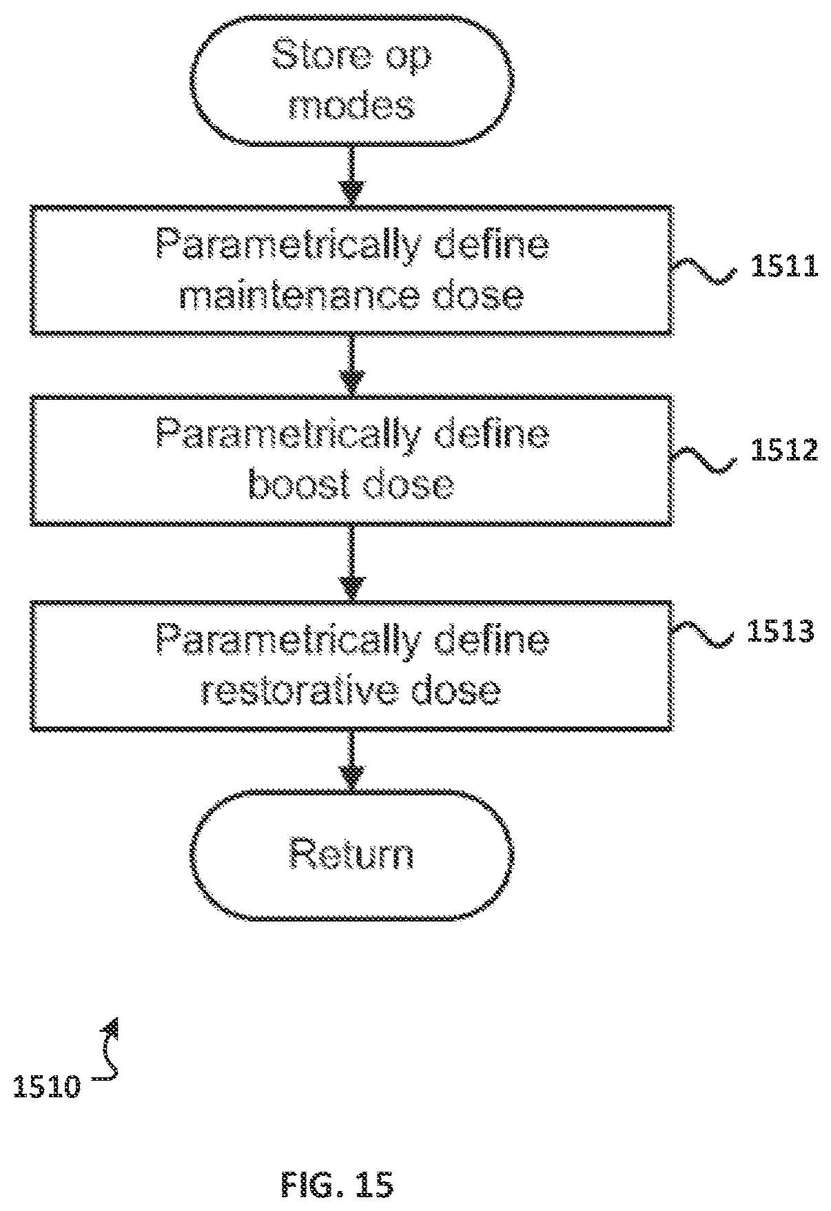
D00014
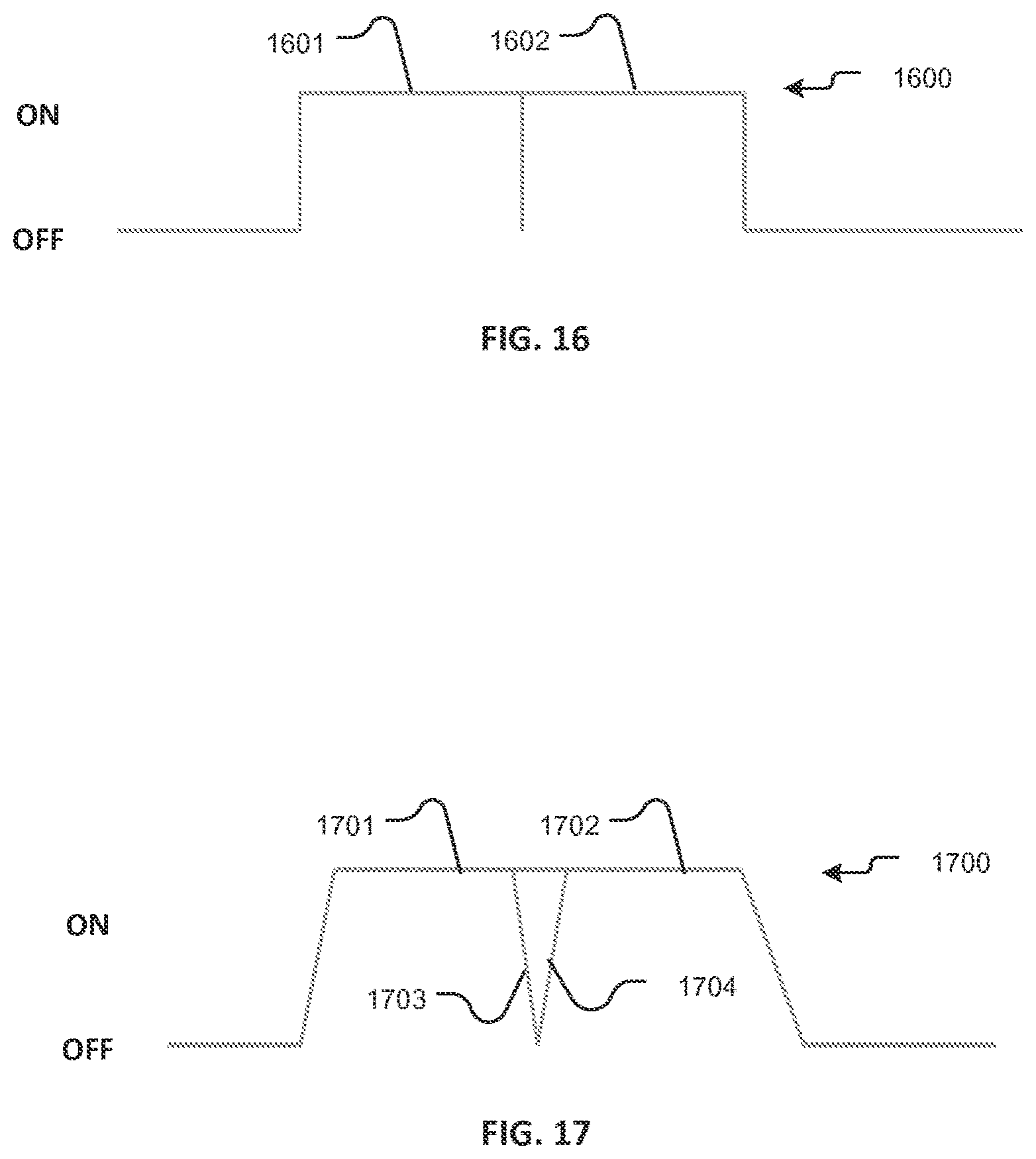
D00015

D00016

XML
uspto.report is an independent third-party trademark research tool that is not affiliated, endorsed, or sponsored by the United States Patent and Trademark Office (USPTO) or any other governmental organization. The information provided by uspto.report is based on publicly available data at the time of writing and is intended for informational purposes only.
While we strive to provide accurate and up-to-date information, we do not guarantee the accuracy, completeness, reliability, or suitability of the information displayed on this site. The use of this site is at your own risk. Any reliance you place on such information is therefore strictly at your own risk.
All official trademark data, including owner information, should be verified by visiting the official USPTO website at www.uspto.gov. This site is not intended to replace professional legal advice and should not be used as a substitute for consulting with a legal professional who is knowledgeable about trademark law.