Device and method for vascular access
Grunwald April 13, 2
U.S. patent number 10,973,584 [Application Number 14/996,247] was granted by the patent office on 2021-04-13 for device and method for vascular access. This patent grant is currently assigned to Bard Access Systems, Inc.. The grantee listed for this patent is Bard Access Systems, Inc.. Invention is credited to Sorin Grunwald.


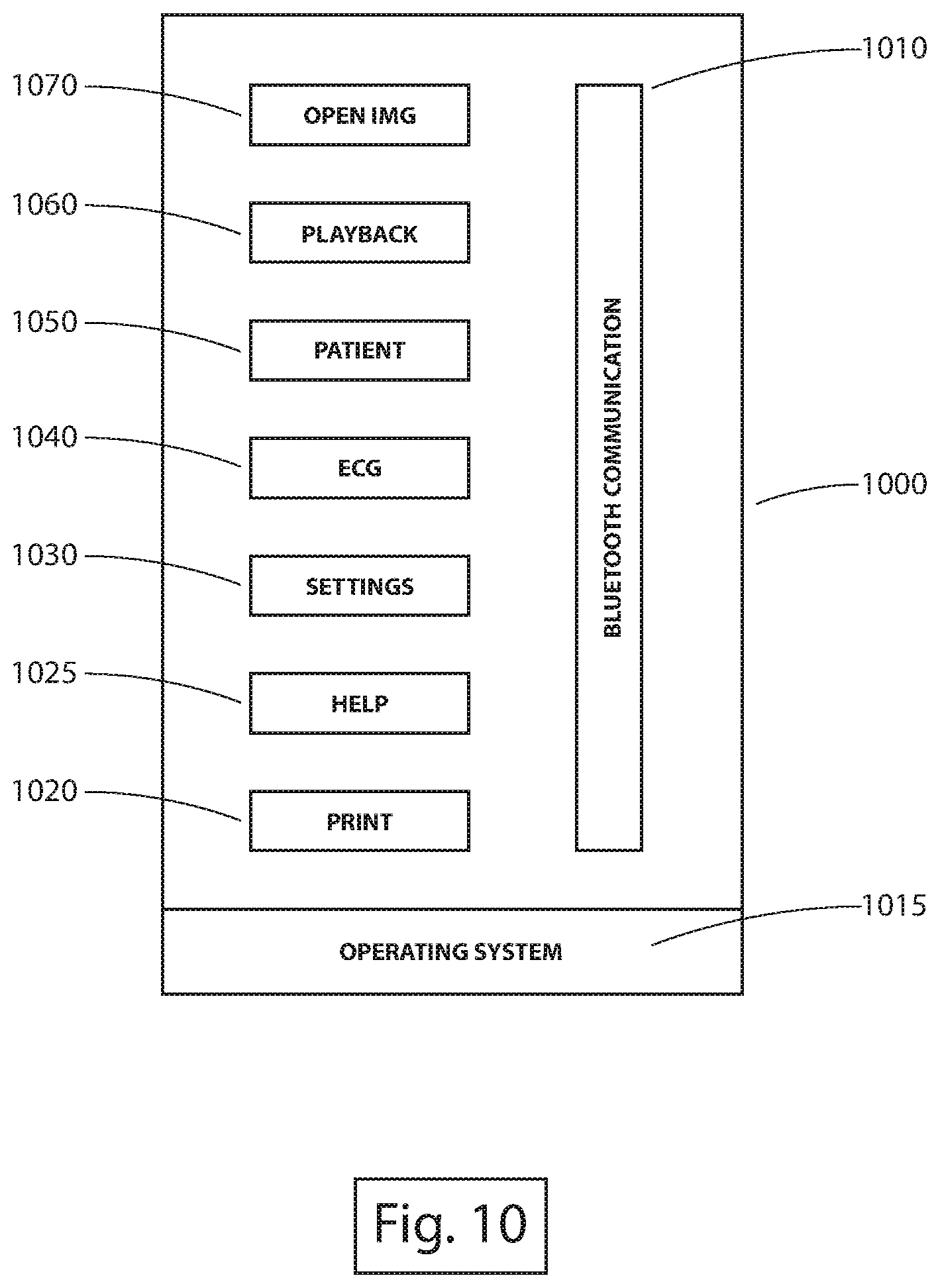


View All Diagrams
| United States Patent | 10,973,584 |
| Grunwald | April 13, 2021 |
Device and method for vascular access
Abstract
A system for facilitating vascular access is disclosed. The system includes an imaging device including a transducer configured for ultrasound image data acquisition, an ECG signal acquisition device configured to acquire ECG signal data via one or more leads, wherein a first lead is directly connected to skin of a patient, and a mobile application configured to render a graphical user interface to be displayed on a display screen of a mobile device, wherein the graphical user interface illustrates a representation of at least one of the ultrasound image data or the ECG signal data that is wireless transmitted to the mobile device.
| Inventors: | Grunwald; Sorin (Bucharest, RO) | ||||||||||
|---|---|---|---|---|---|---|---|---|---|---|---|
| Applicant: |
|
||||||||||
| Assignee: | Bard Access Systems, Inc. (Salt
Lake City, UT) |
||||||||||
| Family ID: | 1000005488480 | ||||||||||
| Appl. No.: | 14/996,247 | ||||||||||
| Filed: | January 15, 2016 |
Prior Publication Data
| Document Identifier | Publication Date | |
|---|---|---|
| US 20160278869 A1 | Sep 29, 2016 | |
Related U.S. Patent Documents
| Application Number | Filing Date | Patent Number | Issue Date | ||
|---|---|---|---|---|---|
| 62104895 | Jan 19, 2015 | ||||
| Current U.S. Class: | 1/1 |
| Current CPC Class: | A61B 34/20 (20160201); A61B 5/283 (20210101); A61B 5/0006 (20130101); A61B 5/065 (20130101); A61B 8/4472 (20130101); A61B 2034/2063 (20160201); A61B 8/546 (20130101); A61B 8/0841 (20130101); A61B 8/565 (20130101); A61B 5/332 (20210101); A61B 8/5223 (20130101); A61B 8/463 (20130101); A61B 5/352 (20210101); A61B 2034/2051 (20160201); A61B 8/5207 (20130101); A61B 5/743 (20130101); A61B 8/4416 (20130101); A61B 8/4494 (20130101); A61B 8/4427 (20130101); A61B 2034/2065 (20160201) |
| Current International Class: | A61B 34/20 (20160101); A61B 5/00 (20060101); A61B 8/00 (20060101); A61B 5/06 (20060101); A61B 8/08 (20060101) |
References Cited [Referenced By]
U.S. Patent Documents
| 3133244 | May 1964 | Wojtulewicz |
| 3297020 | January 1967 | Mathiesen |
| 3625200 | December 1971 | Muller |
| 3674014 | July 1972 | Tillander |
| 3795855 | March 1974 | Browning |
| 3817241 | June 1974 | Grausz |
| 3847157 | November 1974 | Caillouette et al. |
| 3868565 | February 1975 | Kuipers |
| 3896373 | July 1975 | Zelby |
| 3902501 | September 1975 | Citron et al. |
| 3986373 | October 1976 | Goodlaxson |
| 3995623 | December 1976 | Blake et al. |
| 4003369 | January 1977 | Heilman et al. |
| 4063561 | December 1977 | McKenna |
| 4072146 | February 1978 | Howes |
| 4114601 | September 1978 | Abels |
| 4149535 | April 1979 | Voider |
| 4173228 | November 1979 | Van Steenwyk et al. |
| 4175566 | November 1979 | Millar |
| 4181120 | January 1980 | Kunii et al. |
| 4224949 | September 1980 | Scott et al. |
| 4244362 | January 1981 | Anderson |
| 4289139 | September 1981 | Enjoji et al. |
| 4317078 | February 1982 | Weed et al. |
| 4327722 | May 1982 | Groshong et al. |
| 4327723 | May 1982 | Frankhouser |
| 4362166 | December 1982 | Furler et al. |
| 4365639 | December 1982 | Goldreyer |
| 4380237 | April 1983 | Newbower |
| 4407294 | October 1983 | Vilkomerson |
| 4417886 | November 1983 | Frankhouser et al. |
| 4429693 | February 1984 | Blake et al. |
| 4431005 | February 1984 | McCormick |
| 4431214 | February 1984 | Buffington |
| 4445501 | May 1984 | Bresler |
| 4459854 | July 1984 | Richardson et al. |
| 4469106 | September 1984 | Harui |
| 4483343 | November 1984 | Beyer et al. |
| 4491137 | January 1985 | Jingu |
| 4565201 | January 1986 | Lass |
| 4572198 | February 1986 | Codrington |
| 4577634 | March 1986 | Gessman |
| 4582067 | April 1986 | Silverstein et al. |
| 4587975 | May 1986 | Salo et al. |
| 4588394 | May 1986 | Schulte et al. |
| 4593687 | June 1986 | Gray |
| 4595012 | June 1986 | Webler et al. |
| 4601706 | July 1986 | Aillon |
| 4608989 | September 1986 | Drue |
| 4608992 | September 1986 | Hakim et al. |
| 4619247 | October 1986 | Inoue et al. |
| 4622644 | November 1986 | Hansen |
| 4644960 | February 1987 | Johans |
| 4652820 | March 1987 | Maresca |
| 4660571 | April 1987 | Hess et al. |
| 4665925 | May 1987 | Millar |
| 4667230 | May 1987 | Arakawa et al. |
| 4674518 | June 1987 | Salo |
| 4676249 | June 1987 | Arenas et al. |
| 4681106 | July 1987 | Kensey et al. |
| 4681117 | July 1987 | Brodman et al. |
| 4688578 | August 1987 | Takano et al. |
| 4692148 | September 1987 | Kantrowitz et al. |
| 4697595 | October 1987 | Breyer et al. |
| 4700997 | October 1987 | Strand |
| 4706681 | November 1987 | Breyer et al. |
| 4710708 | December 1987 | Rorden et al. |
| 4733669 | March 1988 | Segal |
| 4737794 | April 1988 | Jones |
| 4741356 | May 1988 | Letzo et al. |
| 4742356 | May 1988 | Kuipers |
| 4753247 | June 1988 | Kirsner |
| 4770185 | September 1988 | Silverstein et al. |
| 4771788 | September 1988 | Millar |
| 4781685 | November 1988 | Lehmann et al. |
| 4784646 | November 1988 | Feingold |
| 4787070 | November 1988 | Suzuki et al. |
| 4787396 | November 1988 | Pidorenko |
| 4790809 | December 1988 | Kuntz |
| 4793361 | December 1988 | DuFault |
| 4794930 | January 1989 | Machida et al. |
| 4796632 | January 1989 | Boyd et al. |
| 4798588 | January 1989 | Aillon |
| 4798598 | January 1989 | Bonello et al. |
| 4809681 | March 1989 | Kantrowitz et al. |
| 4809713 | March 1989 | Grayzel |
| 4813729 | March 1989 | Speckhart |
| 4821731 | April 1989 | Martinelli et al. |
| 4834709 | May 1989 | Banning et al. |
| 4836214 | June 1989 | Sramek |
| 4840182 | June 1989 | Carlson |
| 4840622 | June 1989 | Hardy |
| 4841977 | June 1989 | Griffith et al. |
| 4849692 | July 1989 | Blood |
| 4850358 | July 1989 | Millar |
| 4852580 | August 1989 | Wood |
| 4856317 | August 1989 | Pidorenko et al. |
| 4856529 | August 1989 | Segal |
| 4860757 | August 1989 | Lynch et al. |
| 4867169 | September 1989 | Machida et al. |
| 4869263 | September 1989 | Segal et al. |
| 4869718 | September 1989 | Brader |
| 4873987 | October 1989 | Djordjevich et al. |
| 4887606 | December 1989 | Yock et al. |
| 4887615 | December 1989 | Taylor |
| 4889128 | December 1989 | Millar |
| 4899756 | February 1990 | Sonek |
| 4901725 | February 1990 | Nappholz et al. |
| 4905698 | March 1990 | Strohl, Jr. et al. |
| 4911173 | March 1990 | Terwilliger |
| 4911174 | March 1990 | Pederson et al. |
| 4917669 | April 1990 | Bonaldo |
| 4924870 | May 1990 | Wlodarczyk et al. |
| 4943770 | July 1990 | Ashley-Rollman et al. |
| 4945305 | July 1990 | Blood |
| 4947852 | August 1990 | Nassi et al. |
| 4957110 | September 1990 | Vogel et al. |
| 4957111 | September 1990 | Millar |
| 4961433 | October 1990 | Christian |
| 4966148 | October 1990 | Millar |
| 4967753 | November 1990 | Haase et al. |
| 4977886 | December 1990 | Takehana et al. |
| 4989608 | February 1991 | Ratner |
| 4989610 | February 1991 | Patton et al. |
| 4995396 | February 1991 | Inaba et al. |
| 4998916 | March 1991 | Hammerslag et al. |
| 5004456 | April 1991 | Botterbusch et al. |
| 5005592 | April 1991 | Cartmell |
| 5016173 | May 1991 | Kenet et al. |
| 5025799 | June 1991 | Wilson |
| 5029585 | July 1991 | Lieber et al. |
| 5040548 | August 1991 | Yock |
| 5042486 | August 1991 | Pfeiler et al. |
| 5045071 | September 1991 | McCormick et al. |
| 5046497 | September 1991 | Millar |
| 5050607 | September 1991 | Bradley et al. |
| 5057095 | October 1991 | Fabian |
| 5058583 | October 1991 | Geddes et al. |
| 5058595 | October 1991 | Kern |
| 5067489 | November 1991 | Lind |
| 5076278 | December 1991 | Vilkomerson et al. |
| 5078140 | January 1992 | Kwoh |
| 5078148 | January 1992 | Nassi et al. |
| 5078149 | January 1992 | Katsumata et al. |
| 5078678 | January 1992 | Katims |
| 5078714 | January 1992 | Katims |
| 5084022 | January 1992 | Claude |
| 5092341 | March 1992 | Kelen |
| 5099845 | March 1992 | Besz et al. |
| 5099850 | March 1992 | Matsui et al. |
| 5100387 | March 1992 | Ng |
| 5105829 | April 1992 | Fabian et al. |
| 5109862 | May 1992 | Kelen et al. |
| 5114401 | May 1992 | Stuart et al. |
| 5121750 | June 1992 | Katims |
| 5125410 | June 1992 | Misono et al. |
| 5134370 | July 1992 | Jefferts et al. |
| 5144955 | September 1992 | O'Hara |
| 5146151 | September 1992 | Korn |
| 5156151 | October 1992 | Imran |
| 5158086 | October 1992 | Brown et al. |
| 5160342 | November 1992 | Reger et al. |
| 5161536 | November 1992 | Vilkomerson et al. |
| 5174295 | December 1992 | Christian et al. |
| 5174299 | December 1992 | Nelson |
| 5184601 | February 1993 | Putman |
| 5184627 | February 1993 | de Toledo |
| 5190045 | March 1993 | Frazin |
| 5202985 | April 1993 | Goyal |
| 5205830 | April 1993 | Dassa et al. |
| 5211165 | May 1993 | Dumoulin et al. |
| 5211636 | May 1993 | Mische |
| 5212988 | May 1993 | White et al. |
| 5214615 | May 1993 | Bauer |
| 5217026 | June 1993 | Stoy et al. |
| 5220924 | June 1993 | Frazin |
| 5233994 | August 1993 | Shmulewitz |
| 5235987 | August 1993 | Wolfe |
| 5239464 | August 1993 | Blair et al. |
| 5240004 | August 1993 | Walinsky et al. |
| 5243995 | September 1993 | Maier |
| 5246007 | September 1993 | Frisbie et al. |
| 5246426 | September 1993 | Lewis et al. |
| 5247171 | September 1993 | Wlodarczyk et al. |
| 5251635 | October 1993 | Dumoulin et al. |
| 5255680 | October 1993 | Darrow et al. |
| 5257636 | November 1993 | White |
| 5257979 | November 1993 | Jagpal |
| 5261409 | November 1993 | Dardel |
| 5265610 | November 1993 | Darrow et al. |
| 5265614 | November 1993 | Hayakawa et al. |
| 5267569 | December 1993 | Lienhard |
| 5269306 | December 1993 | Warnking et al. |
| 5270810 | December 1993 | Nishimura |
| 5271404 | December 1993 | Corl et al. |
| 5273025 | December 1993 | Sakiyama et al. |
| 5273042 | December 1993 | Lynch et al. |
| 5274551 | December 1993 | Corby, Jr. |
| 5275053 | January 1994 | Wlodarczyk et al. |
| 5279129 | January 1994 | Ito |
| 5279607 | January 1994 | Schentag et al. |
| 5280786 | January 1994 | Wlodarczyk et al. |
| 5287331 | February 1994 | Schindel et al. |
| 5289373 | February 1994 | Zarge et al. |
| 5292342 | March 1994 | Nelson et al. |
| 5307072 | April 1994 | Jones, Jr. |
| 5311871 | May 1994 | Yock |
| 5313949 | May 1994 | Yock |
| 5318025 | June 1994 | Dumoulin et al. |
| 5325860 | July 1994 | Seward et al. |
| 5325873 | July 1994 | Hirschi et al. |
| 5330496 | July 1994 | Alferness |
| 5331966 | July 1994 | Bennett et al. |
| 5333614 | August 1994 | Feiring |
| 5337678 | August 1994 | Grout |
| 5341807 | August 1994 | Nardella |
| 5343865 | September 1994 | Gardineer et al. |
| 5345940 | September 1994 | Seward et al. |
| 5348020 | September 1994 | Hutson |
| 5350352 | September 1994 | Buchholtz et al. |
| 5357961 | October 1994 | Fields et al. |
| 5365935 | November 1994 | Righter et al. |
| 5366443 | November 1994 | Eggers et al. |
| 5368048 | November 1994 | Stoy et al. |
| 5375596 | December 1994 | Twiss et al. |
| 5376083 | December 1994 | Mische |
| 5377678 | January 1995 | Dumoulin et al. |
| 5385053 | January 1995 | Wlodarczyk et al. |
| 5385146 | January 1995 | Goldreyer |
| 5391199 | February 1995 | Ben-Haim |
| 5394876 | March 1995 | Ma |
| 5394877 | March 1995 | Orr et al. |
| 5395366 | March 1995 | D'Andrea et al. |
| 5398683 | March 1995 | Edwards et al. |
| 5398691 | March 1995 | Martin et al. |
| 5411485 | May 1995 | Tennican et al. |
| 5413107 | May 1995 | Oakley et al. |
| 5417208 | May 1995 | Winkler |
| 5417701 | May 1995 | Holmes |
| 5422478 | June 1995 | Wlodarczyk et al. |
| 5423334 | June 1995 | Jordan |
| 5423877 | June 1995 | Mackey |
| 5425367 | June 1995 | Shapiro et al. |
| 5425370 | June 1995 | Vilkomerson |
| 5425382 | June 1995 | Golden et al. |
| 5427114 | June 1995 | Colliver et al. |
| 5429132 | July 1995 | Guy et al. |
| 5429617 | July 1995 | Hammersmark et al. |
| 5431641 | July 1995 | Grozinger et al. |
| 5433729 | July 1995 | Adams et al. |
| 5437276 | August 1995 | Takada |
| 5437277 | August 1995 | Dumoulin et al. |
| 5438873 | August 1995 | Wlodarczyk et al. |
| 5443066 | August 1995 | Dumoulin et al. |
| 5443489 | August 1995 | Ben-Haim |
| 5445150 | August 1995 | Dumoulin et al. |
| 5445166 | August 1995 | Taylor |
| 5450846 | September 1995 | Goldreyer |
| 5453575 | September 1995 | O'Donnell et al. |
| 5453576 | September 1995 | Krivitski |
| 5456256 | October 1995 | Schneider et al. |
| 5456718 | October 1995 | Szymaitis |
| 5464016 | November 1995 | Nicholas et al. |
| 5474065 | December 1995 | Meathrel et al. |
| 5476090 | December 1995 | Kishi |
| 5480422 | January 1996 | Ben-Haim |
| 5487729 | January 1996 | Avellanet et al. |
| 5490522 | February 1996 | Dardel |
| 5492538 | February 1996 | Johlin, Jr. |
| 5494038 | February 1996 | Wang et al. |
| 5500011 | March 1996 | Desai |
| 5500012 | March 1996 | Brucker et al. |
| 5505205 | April 1996 | Solomon et al. |
| 5509822 | April 1996 | Negus et al. |
| 5513637 | May 1996 | Twiss et al. |
| 5515160 | May 1996 | Schulz et al. |
| 5515853 | May 1996 | Smith et al. |
| 5517989 | May 1996 | Frisbie et al. |
| 5522878 | June 1996 | Montecalvo et al. |
| 5522880 | June 1996 | Barone et al. |
| 5526812 | June 1996 | Dumoulin et al. |
| 5531664 | July 1996 | Adachi et al. |
| 5536248 | July 1996 | Weaver et al. |
| 5540230 | July 1996 | Vilkomerson |
| 5540681 | July 1996 | Strul et al. |
| 5542938 | August 1996 | Avellanet et al. |
| 5546949 | August 1996 | Frazin et al. |
| 5546951 | August 1996 | Ben-Haim |
| 5555618 | September 1996 | Winkler |
| 5558091 | September 1996 | Acker et al. |
| 5568809 | October 1996 | Ben-haim |
| D375450 | November 1996 | Bidwell et al. |
| 5570671 | November 1996 | Hickey |
| 5575291 | November 1996 | Hayakawa et al. |
| 5588442 | December 1996 | Scovil et al. |
| 5592939 | January 1997 | Martinelli |
| 5598846 | February 1997 | Peszynski |
| 5599299 | February 1997 | Weaver et al. |
| 5600330 | February 1997 | Blood |
| 5603333 | February 1997 | Konings |
| 5606981 | March 1997 | Tartacower et al. |
| 5610967 | March 1997 | Moorman et al. |
| 5617866 | April 1997 | Marian, Jr. |
| 5622169 | April 1997 | Golden et al. |
| 5622170 | April 1997 | Schulz |
| 5622184 | April 1997 | Ashby et al. |
| 5623931 | April 1997 | Wung et al. |
| 5624430 | April 1997 | Eton et al. |
| 5626554 | May 1997 | Ryaby et al. |
| 5626870 | May 1997 | Monshipouri et al. |
| 5630419 | May 1997 | Ranalletta |
| 5638819 | June 1997 | Manwaring et al. |
| 5640967 | June 1997 | Fine et al. |
| 5644612 | July 1997 | Moorman et al. |
| 5645065 | July 1997 | Shapiro et al. |
| 5651047 | July 1997 | Moorman et al. |
| 5654864 | August 1997 | Ritter et al. |
| D383968 | September 1997 | Bidwell et al. |
| 5662115 | September 1997 | Torp et al. |
| 5665103 | September 1997 | Lafontaine et al. |
| 5665477 | September 1997 | Meathrel et al. |
| 5666473 | September 1997 | Wallace |
| 5666958 | September 1997 | Rothenberg et al. |
| 5669383 | September 1997 | Johnson |
| 5669388 | September 1997 | Vilkomerson |
| 5676159 | October 1997 | Navis |
| 5676673 | October 1997 | Ferre et al. |
| 5682890 | November 1997 | Kormos et al. |
| 5691898 | November 1997 | Rosenberg et al. |
| 5694945 | December 1997 | Ben-Haim |
| 5695479 | December 1997 | Jagpal |
| 5697377 | December 1997 | Wittkampf |
| 5699801 | December 1997 | Atalar et al. |
| 5700889 | December 1997 | Blair |
| 5701898 | December 1997 | Adam et al. |
| 5702433 | December 1997 | Taylor et al. |
| 5711299 | January 1998 | Manwaring et al. |
| 5713362 | February 1998 | Vilkomerson |
| 5713363 | February 1998 | Seward et al. |
| 5713858 | February 1998 | Heruth et al. |
| 5713946 | February 1998 | Ben-Haim |
| 5715817 | February 1998 | Stevens-Wright et al. |
| 5716389 | February 1998 | Walinsky et al. |
| 5718241 | February 1998 | Ben-Haim et al. |
| D391838 | March 1998 | Bidwell et al. |
| 5722412 | March 1998 | Pflugrath et al. |
| 5727550 | March 1998 | Montecalvo |
| 5727552 | March 1998 | Ryan |
| 5727553 | March 1998 | Saad |
| 5729055 | March 1998 | Manning |
| 5729129 | March 1998 | Acker |
| 5729584 | March 1998 | Moorman et al. |
| 5730129 | March 1998 | Darrow et al. |
| 5731996 | March 1998 | Gilbert |
| 5733323 | March 1998 | Buck et al. |
| 5738096 | April 1998 | Ben-Haim |
| 5740808 | April 1998 | Panescu et al. |
| 5742394 | April 1998 | Hansen |
| 5744953 | April 1998 | Hansen |
| 5748767 | May 1998 | Raab |
| 5749835 | May 1998 | Glantz |
| 5749938 | May 1998 | Coombs |
| 5751785 | May 1998 | Moorman et al. |
| 5752513 | May 1998 | Acker et al. |
| 5758650 | June 1998 | Miller et al. |
| 5762064 | June 1998 | Polvani |
| 5767669 | June 1998 | Hansen et al. |
| 5767960 | June 1998 | Orman |
| 5769786 | June 1998 | Wiegel |
| 5769843 | June 1998 | Abela et al. |
| 5769881 | June 1998 | Schroeppel et al. |
| 5771896 | June 1998 | Sliwa, Jr. et al. |
| 5775322 | July 1998 | Silverstein et al. |
| 5775332 | July 1998 | Goldman |
| 5776064 | July 1998 | Kalfas et al. |
| 5776080 | July 1998 | Thome et al. |
| 5779638 | July 1998 | Vesely et al. |
| 5782767 | July 1998 | Pretlow, III |
| 5782773 | July 1998 | Kuo et al. |
| 5785657 | July 1998 | Breyer et al. |
| 5792055 | August 1998 | McKinnon |
| 5795297 | August 1998 | Daigle |
| 5795298 | August 1998 | Vesely et al. |
| 5795632 | August 1998 | Buchalter |
| 5797849 | August 1998 | Vesely et al. |
| 5800352 | September 1998 | Ferre et al. |
| 5800410 | September 1998 | Gawreluk |
| 5800497 | September 1998 | Bakels et al. |
| 5803089 | September 1998 | Ferre et al. |
| 5810733 | September 1998 | Van Creveld et al. |
| RE35924 | October 1998 | Winkler |
| 5817022 | October 1998 | Vesely |
| 5817024 | October 1998 | Ogle et al. |
| 5820549 | October 1998 | Marian, Jr. |
| 5820560 | October 1998 | Sinderby et al. |
| 5824031 | October 1998 | Cookston et al. |
| 5827192 | October 1998 | Gopakumaran et al. |
| 5829444 | November 1998 | Ferre et al. |
| 5830145 | November 1998 | Tenhoff |
| 5831260 | November 1998 | Hansen |
| 5833608 | November 1998 | Acker |
| 5833622 | November 1998 | Meathrel et al. |
| 5835561 | November 1998 | Moorman et al. |
| 5836882 | November 1998 | Frazin |
| 5836990 | November 1998 | Li |
| 5840024 | November 1998 | Taniguchi et al. |
| 5840025 | November 1998 | Ben-Haim |
| 5840030 | November 1998 | Ferek-Petric et al. |
| 5840031 | November 1998 | Crowley |
| 5842986 | December 1998 | Avrin et al. |
| 5842998 | December 1998 | Gopakumaran et al. |
| 5843076 | December 1998 | Webster, Jr. et al. |
| 5843153 | December 1998 | Johnston et al. |
| 5844140 | December 1998 | Seale |
| 5846198 | December 1998 | Killmann |
| 5855553 | January 1999 | Tajima et al. |
| 5859893 | January 1999 | Moorman et al. |
| 5865748 | February 1999 | Co et al. |
| 5868673 | February 1999 | Vesely |
| 5873822 | February 1999 | Ferre et al. |
| 5876328 | March 1999 | Fox et al. |
| 5879297 | March 1999 | Haynor et al. |
| 5893363 | April 1999 | Little et al. |
| 5897495 | April 1999 | Aida et al. |
| 5899860 | May 1999 | Pfeiffer et al. |
| 5902238 | May 1999 | Golden et al. |
| 5907487 | May 1999 | Rosenberg et al. |
| 5908385 | June 1999 | Chechelski et al. |
| 5910113 | June 1999 | Pruter |
| 5910120 | June 1999 | Kim et al. |
| 5913820 | June 1999 | Bladen et al. |
| 5913830 | June 1999 | Miles |
| 5919141 | July 1999 | Money et al. |
| 5919170 | July 1999 | Woessner |
| 5928145 | July 1999 | Ocali et al. |
| 5929607 | July 1999 | Rosenberg et al. |
| 5931788 | August 1999 | Keen et al. |
| 5931818 | August 1999 | Werp et al. |
| 5931863 | August 1999 | Griffin, III et al. |
| 5935160 | August 1999 | Auricchio et al. |
| 5941858 | August 1999 | Johnson |
| 5941889 | August 1999 | Cermak |
| 5941904 | August 1999 | Johnston et al. |
| 5944022 | August 1999 | Nardella et al. |
| 5944023 | August 1999 | Johnson et al. |
| 5951472 | September 1999 | Van Vaals et al. |
| 5951598 | September 1999 | Bishay et al. |
| 5953683 | September 1999 | Hansen et al. |
| 5957857 | September 1999 | Hartley |
| 5961923 | October 1999 | Nova et al. |
| 5967978 | October 1999 | Littmann et al. |
| 5967980 | October 1999 | Ferre et al. |
| 5967991 | October 1999 | Gardineer et al. |
| 5969722 | October 1999 | Palm |
| 5971933 | October 1999 | Gopakumaran et al. |
| 5971983 | October 1999 | Lesh |
| 5978705 | November 1999 | KenKnight et al. |
| 5983126 | November 1999 | Wittkampf |
| 5984908 | November 1999 | Davis et al. |
| 5991693 | November 1999 | Zalewski |
| 5997473 | December 1999 | Taniguchi et al. |
| 5997481 | December 1999 | Adams et al. |
| 6006123 | December 1999 | Nguyen et al. |
| 6011988 | January 2000 | Lynch et al. |
| 6014473 | January 2000 | Hossack et al. |
| 6014580 | January 2000 | Blume et al. |
| 6015414 | January 2000 | Werp et al. |
| 6017496 | January 2000 | Nova et al. |
| 6019724 | February 2000 | Gronningsaeter et al. |
| 6019725 | February 2000 | Vesely et al. |
| 6022342 | February 2000 | Mukherjee |
| 6023638 | February 2000 | Swanson |
| 6026312 | February 2000 | Shemwell et al. |
| 6031765 | February 2000 | Lee et al. |
| 6032070 | February 2000 | Flock et al. |
| 6039694 | March 2000 | Larson et al. |
| 6050718 | April 2000 | Schena et al. |
| 6052610 | April 2000 | Koch |
| 6052618 | April 2000 | Dahlke et al. |
| D424693 | May 2000 | Pruter |
| 6058323 | May 2000 | Lemelson |
| 6059718 | May 2000 | Taniguchi et al. |
| 6063032 | May 2000 | Grunwald |
| 6064905 | May 2000 | Webster, Jr. et al. |
| 6066094 | May 2000 | Ben-Haim |
| 6068599 | May 2000 | Saito et al. |
| 6073043 | June 2000 | Schneider |
| 6074367 | June 2000 | Hubbell |
| 6075442 | June 2000 | Welch |
| 6076007 | June 2000 | England et al. |
| 6081737 | June 2000 | Shah |
| 6082366 | July 2000 | Andra et al. |
| 6083170 | July 2000 | Ben-Haim |
| 6099524 | August 2000 | Lipson et al. |
| 6100026 | August 2000 | Nova et al. |
| 6102044 | August 2000 | Naidyhorski |
| 6102862 | August 2000 | Grunwald et al. |
| 6107699 | August 2000 | Swanson |
| 6112111 | August 2000 | Glantz |
| 6112115 | August 2000 | Feldman et al. |
| 6113504 | September 2000 | Kuesters |
| 6115624 | September 2000 | Lewis et al. |
| 6120445 | September 2000 | Grunwald |
| 6122538 | September 2000 | Sliwa, Jr. et al. |
| 6128174 | October 2000 | Ritter et al. |
| 6129668 | October 2000 | Haynor et al. |
| 6129724 | October 2000 | Fleischman et al. |
| 6132378 | October 2000 | Marino |
| 6132379 | October 2000 | Patacsil et al. |
| 6135961 | October 2000 | Pflugrath et al. |
| 6136274 | October 2000 | Nova et al. |
| 6138681 | October 2000 | Chen et al. |
| 6139496 | October 2000 | Chen et al. |
| 6139502 | October 2000 | Fredriksen |
| 6139540 | October 2000 | Rost et al. |
| 6144300 | November 2000 | Dames |
| 6148823 | November 2000 | Hastings |
| 6152933 | November 2000 | Werp et al. |
| 6157853 | December 2000 | Blume et al. |
| 6165144 | December 2000 | Talish et al. |
| 6165977 | December 2000 | Mochly-Rosen |
| 6166496 | December 2000 | Lys et al. |
| 6166806 | December 2000 | Tjin |
| 6167765 | January 2001 | Weitzel |
| 6172499 | January 2001 | Ashe |
| 6173199 | January 2001 | Gabriel |
| 6173715 | January 2001 | Sinanan et al. |
| 6175756 | January 2001 | Ferre et al. |
| 6176829 | January 2001 | Vilkomerson |
| 6187744 | February 2001 | Rooney |
| 6190370 | February 2001 | Tsui |
| 6191136 | February 2001 | Marban |
| 6193743 | February 2001 | Brayton et al. |
| 6197001 | March 2001 | Wilson et al. |
| 6200305 | March 2001 | Berthiaume et al. |
| 6203499 | March 2001 | Imling et al. |
| 6208884 | March 2001 | Kumar et al. |
| 6211626 | April 2001 | Lys et al. |
| 6211666 | April 2001 | Acker |
| 6212426 | April 2001 | Swanson |
| 6212430 | April 2001 | Kung |
| 6216027 | April 2001 | Willis et al. |
| 6216028 | April 2001 | Haynor et al. |
| 6216029 | April 2001 | Paltieli |
| 6217517 | April 2001 | Grunwald |
| 6223087 | April 2001 | Williams |
| 6226547 | May 2001 | Lockhart et al. |
| 6230042 | May 2001 | Slettenmark |
| 6230046 | May 2001 | Crane et al. |
| 6231518 | May 2001 | Grabek et al. |
| 6233476 | May 2001 | Strommer et al. |
| 6233994 | May 2001 | Roy et al. |
| 6236883 | May 2001 | Ciaccio et al. |
| 6238344 | May 2001 | Gamelsky et al. |
| 6241673 | June 2001 | Williams |
| 6246231 | June 2001 | Ashe |
| 6246898 | June 2001 | Vesely et al. |
| 6248072 | June 2001 | Murkin |
| 6248074 | June 2001 | Ohno et al. |
| 6248075 | June 2001 | McGee et al. |
| 6249234 | June 2001 | Ely et al. |
| 6253770 | July 2001 | Acker et al. |
| 6254543 | July 2001 | Grunwald et al. |
| 6258035 | July 2001 | Hoeksel et al. |
| 6259938 | July 2001 | Zarychta et al. |
| 6259941 | July 2001 | Chia et al. |
| 6261231 | July 2001 | Damphousse et al. |
| 6263230 | July 2001 | Haynor et al. |
| 6266550 | July 2001 | Selmon et al. |
| 6266551 | July 2001 | Osadchy et al. |
| 6266552 | July 2001 | Slettenmark |
| 6266563 | July 2001 | KenKnight et al. |
| 6270493 | August 2001 | Lalonde et al. |
| 6271833 | August 2001 | Rosenberg et al. |
| 6272371 | August 2001 | Shlomo |
| 6272374 | August 2001 | Flock et al. |
| 6275258 | August 2001 | Chim |
| 6275724 | August 2001 | Dickinson et al. |
| 6277077 | August 2001 | Brisken et al. |
| 6284459 | September 2001 | Nova et al. |
| 6285898 | September 2001 | Ben-Haim |
| 6287259 | September 2001 | Grunwald |
| 6287260 | September 2001 | Hascoet et al. |
| 6288704 | September 2001 | Flack et al. |
| 6292678 | September 2001 | Hall et al. |
| 6292680 | September 2001 | Somogyi et al. |
| 6292901 | September 2001 | Lys et al. |
| 6293955 | September 2001 | Houser et al. |
| 6296604 | October 2001 | Garibaldi et al. |
| 6298261 | October 2001 | Rex |
| 6304768 | October 2001 | Blume et al. |
| 6306097 | October 2001 | Park et al. |
| 6306105 | October 2001 | Rooney et al. |
| 6311082 | October 2001 | Creighton, IV et al. |
| 6315709 | November 2001 | Garibaldi et al. |
| 6315727 | November 2001 | Coleman et al. |
| 6319668 | November 2001 | Nova et al. |
| 6323769 | November 2001 | Dames |
| 6323770 | November 2001 | Dames |
| 6324416 | November 2001 | Seibert |
| 6325540 | December 2001 | Lounsberry et al. |
| 6325762 | December 2001 | Tjin |
| 6329139 | December 2001 | Nova et al. |
| 6329916 | December 2001 | Dames |
| 6330467 | December 2001 | Creighton, IV et al. |
| 6332089 | December 2001 | Acker et al. |
| 6332874 | December 2001 | Eliasen et al. |
| 6340588 | January 2002 | Nova et al. |
| 6340868 | January 2002 | Lys et al. |
| 6341231 | January 2002 | Ferre et al. |
| 6346081 | February 2002 | Vilkomerson |
| 6348911 | February 2002 | Rosenberg et al. |
| 6350160 | February 2002 | Feuersanger et al. |
| 6352363 | March 2002 | Munger et al. |
| 6354999 | March 2002 | Dgany et al. |
| 6355026 | March 2002 | Mick |
| 6356791 | March 2002 | Westlund et al. |
| 6360123 | March 2002 | Kimchi et al. |
| 6361499 | March 2002 | Bates et al. |
| 6364823 | April 2002 | Garibaldi et al. |
| 6364839 | April 2002 | Little et al. |
| 6366804 | April 2002 | Mejia |
| 6368285 | April 2002 | Osadchy et al. |
| 6370411 | April 2002 | Osadchy et al. |
| 6373240 | April 2002 | Govari |
| 6373388 | April 2002 | Dames |
| 6374134 | April 2002 | Bladen et al. |
| 6374670 | April 2002 | Spelman et al. |
| 6375606 | April 2002 | Garibaldi et al. |
| 6375639 | April 2002 | Duplessie et al. |
| 6377857 | April 2002 | Brayton et al. |
| 6379302 | April 2002 | Kessman et al. |
| 6379303 | April 2002 | Seitz et al. |
| 6379307 | April 2002 | Filly et al. |
| 6381485 | April 2002 | Hunter et al. |
| 6385472 | May 2002 | Hall et al. |
| 6385476 | May 2002 | Osadchy et al. |
| 6398736 | June 2002 | Seward |
| 6398738 | June 2002 | Millar |
| 6401723 | June 2002 | Garibaldi et al. |
| 6406422 | June 2002 | Landesberg |
| 6406442 | June 2002 | McFann et al. |
| 6412978 | July 2002 | Watanabe et al. |
| 6412980 | July 2002 | Lounsberry et al. |
| 6417839 | July 2002 | Odell |
| 6418332 | July 2002 | Mastrototaro et al. |
| 6418335 | July 2002 | Avrin et al. |
| 6423002 | July 2002 | Hossack |
| 6423050 | July 2002 | Twardowski |
| 6427079 | July 2002 | Schneider et al. |
| 6428551 | August 2002 | Hall et al. |
| 6430315 | August 2002 | Makram-Ebeid |
| 6432069 | August 2002 | Godo et al. |
| 6438411 | August 2002 | Guttman et al. |
| 6442416 | August 2002 | Schultz |
| 6445943 | September 2002 | Ferre et al. |
| 6456874 | September 2002 | Hafer et al. |
| 6459919 | October 2002 | Lys et al. |
| 6463121 | October 2002 | Milnes |
| 6466815 | October 2002 | Saito et al. |
| 6471656 | October 2002 | Shalman et al. |
| 6471658 | October 2002 | Daniels et al. |
| 6471700 | October 2002 | Burbank et al. |
| 6473167 | October 2002 | Odell |
| 6474341 | November 2002 | Hunter et al. |
| 6475152 | November 2002 | Kelly, Jr. et al. |
| 6475223 | November 2002 | Werp et al. |
| 6477402 | November 2002 | Lynch et al. |
| 6484118 | November 2002 | Govari |
| 6487916 | December 2002 | Gomm et al. |
| 6491671 | December 2002 | Larson, III et al. |
| 6493573 | December 2002 | Martinelli et al. |
| 6494832 | December 2002 | Feldman et al. |
| 6496715 | December 2002 | Lee et al. |
| 6498944 | December 2002 | Ben-Haim et al. |
| 6500141 | December 2002 | Irion et al. |
| 6505062 | January 2003 | Ritter et al. |
| 6506159 | January 2003 | Hascoet et al. |
| 6507751 | January 2003 | Blume et al. |
| 6508802 | January 2003 | Rosengart et al. |
| 6511413 | January 2003 | Landesberg |
| 6512958 | January 2003 | Swoyer et al. |
| 6514202 | February 2003 | Grunwald |
| 6514226 | February 2003 | Levin et al. |
| 6514249 | February 2003 | Maguire et al. |
| 6515657 | February 2003 | Zanelli |
| 6516212 | February 2003 | Bladen et al. |
| 6516231 | February 2003 | Flammang |
| 6516807 | February 2003 | Panescu et al. |
| 6517520 | February 2003 | Chang et al. |
| 6522906 | February 2003 | Salisbury, Jr. et al. |
| 6522907 | February 2003 | Bladen et al. |
| 6522909 | February 2003 | Garibaldi et al. |
| 6524303 | February 2003 | Garibaldi |
| 6528954 | March 2003 | Lys et al. |
| 6528991 | March 2003 | Ashe |
| 6529761 | March 2003 | Creighton, IV et al. |
| 6529766 | March 2003 | Guendel |
| 6534982 | March 2003 | Jakab |
| 6535625 | March 2003 | Chang |
| 6537192 | March 2003 | Elliott et al. |
| 6537196 | March 2003 | Creighton, IV et al. |
| 6538634 | March 2003 | Chui et al. |
| 6540699 | April 2003 | Smith |
| 6542766 | April 2003 | Hall et al. |
| 6544251 | April 2003 | Crawford |
| 6545678 | April 2003 | Ohazama |
| 6546270 | April 2003 | Goldin et al. |
| 6546279 | April 2003 | Bova et al. |
| 6546787 | April 2003 | Schiller et al. |
| 6552841 | April 2003 | Lasser et al. |
| 6556858 | April 2003 | Zeman |
| 6562019 | May 2003 | Sell |
| 6564087 | May 2003 | Pitris et al. |
| 6569101 | May 2003 | Quistgaard et al. |
| 6569103 | May 2003 | Hoeksel et al. |
| 6569160 | May 2003 | Goldin et al. |
| 6569862 | May 2003 | Marban |
| 6571004 | May 2003 | Florent et al. |
| 6574518 | June 2003 | Lounsberry et al. |
| 6575908 | June 2003 | Barnes et al. |
| 6577080 | June 2003 | Lys et al. |
| 6577896 | June 2003 | Werner et al. |
| 6584343 | June 2003 | Ransbury et al. |
| 6589181 | July 2003 | Grunwald et al. |
| 6593754 | July 2003 | Steber et al. |
| 6593884 | July 2003 | Gilboa et al. |
| 6597943 | July 2003 | Taha et al. |
| 6599249 | July 2003 | Nordgren et al. |
| 6607488 | August 2003 | Jackson et al. |
| 6610058 | August 2003 | Flores |
| 6611141 | August 2003 | Schulz et al. |
| 6615071 | September 2003 | Casscells, III et al. |
| 6615155 | September 2003 | Gilboa |
| 6616610 | September 2003 | Steininger et al. |
| 6618612 | September 2003 | Acker et al. |
| 6626832 | September 2003 | Paltieli et al. |
| 6626834 | September 2003 | Dunne et al. |
| 6626902 | September 2003 | Kucharczyk et al. |
| 6630879 | October 2003 | Creighton, IV et al. |
| 6635027 | October 2003 | Cragg et al. |
| 6645148 | November 2003 | Nguyen-Dinh et al. |
| 6648875 | November 2003 | Simpson et al. |
| 6649914 | November 2003 | Moorman et al. |
| 6652505 | November 2003 | Tsugita |
| 6652506 | November 2003 | Bowe et al. |
| 6660024 | December 2003 | Flaherty et al. |
| 6662034 | December 2003 | Segner et al. |
| 6663661 | December 2003 | Boneau |
| 6666828 | December 2003 | Greco et al. |
| 6672308 | January 2004 | Gaspari |
| 6677752 | January 2004 | Creighton, IV et al. |
| 6679857 | January 2004 | Bastia et al. |
| 6684176 | January 2004 | Willins et al. |
| 6685644 | February 2004 | Seo et al. |
| 6687531 | February 2004 | Ferre et al. |
| 6689119 | February 2004 | Di Caprio et al. |
| 6690963 | February 2004 | Ben-Haim et al. |
| 6690964 | February 2004 | Bieger et al. |
| 6690968 | February 2004 | Mejia |
| 6694167 | February 2004 | Ferre et al. |
| 6695786 | February 2004 | Wang et al. |
| 6701179 | March 2004 | Martinelli et al. |
| 6701918 | March 2004 | Fariss et al. |
| 6702804 | March 2004 | Ritter et al. |
| 6704590 | March 2004 | Haldeman |
| 6709390 | March 2004 | Marie Pop |
| 6711429 | March 2004 | Gilboa et al. |
| 6711431 | March 2004 | Sarin et al. |
| 6719699 | April 2004 | Smith |
| 6719724 | April 2004 | Walker et al. |
| 6719756 | April 2004 | Muntermann |
| 6720745 | April 2004 | Lys et al. |
| 6733458 | May 2004 | Steins et al. |
| 6733473 | May 2004 | Reifart et al. |
| 6733511 | May 2004 | Hall et al. |
| 6736782 | May 2004 | Pfeiffer et al. |
| 6738656 | May 2004 | Ferre et al. |
| 6740103 | May 2004 | Hall et al. |
| 6743177 | June 2004 | Ito |
| 6754596 | June 2004 | Ashe |
| 6755789 | June 2004 | Stringer et al. |
| 6755816 | June 2004 | Ritter et al. |
| 6755822 | June 2004 | Reu et al. |
| 6757557 | June 2004 | Bladen et al. |
| 6763261 | July 2004 | Casscells, III et al. |
| 6764449 | July 2004 | Lee et al. |
| 6768496 | July 2004 | Bieger et al. |
| 6772001 | August 2004 | Maschke |
| 6774624 | August 2004 | Anderson et al. |
| 6783536 | August 2004 | Vilsmeier et al. |
| 6784660 | August 2004 | Ashe |
| 6785571 | August 2004 | Glossop |
| 6786219 | September 2004 | Garibaldi et al. |
| 6786870 | September 2004 | Miyaki et al. |
| 6788967 | September 2004 | Ben-Haim et al. |
| 6794667 | September 2004 | Noshi |
| 6799066 | September 2004 | Steines et al. |
| 6815651 | November 2004 | Odell |
| 6816266 | November 2004 | Varshneya et al. |
| 6817364 | November 2004 | Garibaldi et al. |
| 6834201 | December 2004 | Gillies et al. |
| 6844713 | January 2005 | Steber et al. |
| 6845142 | January 2005 | Ohishi |
| 6856823 | February 2005 | Ashe |
| 6860422 | March 2005 | Hull et al. |
| 6862467 | March 2005 | Moore et al. |
| 6869390 | March 2005 | Elliott et al. |
| 6875179 | April 2005 | Ferguson et al. |
| 6879160 | April 2005 | Jakab |
| 6887206 | May 2005 | Hoeksel et al. |
| 6889091 | May 2005 | Hine et al. |
| 6895268 | May 2005 | Rahn et al. |
| 6902528 | June 2005 | Garibaldi et al. |
| 6905469 | June 2005 | Hascoet et al. |
| 6908433 | June 2005 | Pruter |
| 6911026 | June 2005 | Hall et al. |
| 6923782 | August 2005 | O'Mahony et al. |
| 6926673 | August 2005 | Roberts et al. |
| 6926674 | August 2005 | Tenerz et al. |
| 6934575 | August 2005 | Ferre et al. |
| 6936010 | August 2005 | Fang et al. |
| 6939313 | September 2005 | Saadat et al. |
| 6940379 | September 2005 | Creighton |
| 6941166 | September 2005 | MacAdam et al. |
| 6945938 | September 2005 | Grunwald |
| 6947788 | September 2005 | Gilboa et al. |
| 6950689 | September 2005 | Willis et al. |
| 6953754 | October 2005 | Machida et al. |
| 6958677 | October 2005 | Carter |
| 6959214 | October 2005 | Pape et al. |
| 6962566 | November 2005 | Quistgaard et al. |
| 6968846 | November 2005 | Viswanathan |
| 6975197 | December 2005 | Creighton, IV |
| 6976962 | December 2005 | Bullis |
| 6976987 | December 2005 | Flores |
| 6980843 | December 2005 | Eng et al. |
| 6980852 | December 2005 | Jersey-Willuhn et al. |
| 6980921 | December 2005 | Anderson et al. |
| 6986739 | January 2006 | Warren et al. |
| 6986744 | January 2006 | Krivitski |
| 6999821 | February 2006 | Jenney et al. |
| 7001355 | February 2006 | Nunomura et al. |
| 7008418 | March 2006 | Hall et al. |
| 7010338 | March 2006 | Ritter et al. |
| 7015393 | March 2006 | Weiner et al. |
| 7017584 | March 2006 | Garibaldi et al. |
| 7019610 | March 2006 | Creighton, IV et al. |
| 7020512 | March 2006 | Ritter et al. |
| D518574 | April 2006 | Chaggares |
| 7022075 | April 2006 | Grunwald et al. |
| 7022082 | April 2006 | Sonek |
| 7026927 | April 2006 | Wright et al. |
| 7027634 | April 2006 | Odell |
| 7028387 | April 2006 | Huynh et al. |
| 7029446 | April 2006 | Wendelken et al. |
| 7033603 | April 2006 | Nelson et al. |
| D520139 | May 2006 | Chaggares |
| D520140 | May 2006 | Chaggares |
| 7038398 | May 2006 | Lys et al. |
| 7038657 | May 2006 | Rosenberg et al. |
| 7043293 | May 2006 | Baura |
| 7048733 | May 2006 | Hartley et al. |
| 7054228 | May 2006 | Hickling |
| 7065403 | June 2006 | Mouchawar et al. |
| 7066914 | June 2006 | Andersen |
| 7066924 | June 2006 | Garibaldi et al. |
| 7069072 | June 2006 | Jansen et al. |
| D525363 | July 2006 | Chaggares |
| 7070565 | July 2006 | Vaezy et al. |
| 7072704 | July 2006 | Bucholz |
| 7082325 | July 2006 | Hashimshony et al. |
| 7090639 | August 2006 | Govari |
| 7096059 | August 2006 | Geddes et al. |
| 7096148 | August 2006 | Anderson et al. |
| 7096870 | August 2006 | Lamprich et al. |
| 7098907 | August 2006 | Houston et al. |
| 7103205 | September 2006 | Wang et al. |
| 7104980 | September 2006 | Laherty et al. |
| 7106043 | September 2006 | Da Silva et al. |
| 7106431 | September 2006 | Odell |
| 7106479 | September 2006 | Roy et al. |
| 7107105 | September 2006 | Bjorklund et al. |
| 7112197 | September 2006 | Hartley et al. |
| 7128734 | October 2006 | Wilson et al. |
| 7132804 | November 2006 | Lys et al. |
| 7137976 | November 2006 | Ritter et al. |
| 7141019 | November 2006 | Pearlman |
| 7141812 | November 2006 | Appleby et al. |
| 7142905 | November 2006 | Slayton et al. |
| 7148970 | December 2006 | de Boer |
| 7153291 | December 2006 | Bierman |
| 7161453 | January 2007 | Creighton, IV |
| 7162291 | January 2007 | Nachaliel |
| 7167738 | January 2007 | Schweikard et al. |
| 7169107 | January 2007 | Jersey-Willuhn et al. |
| 7169109 | January 2007 | Jansen et al. |
| 7174201 | February 2007 | Govari et al. |
| 7175646 | February 2007 | Brenneman et al. |
| 7180252 | February 2007 | Lys et al. |
| 7184820 | February 2007 | Jersey-Willuhn et al. |
| 7189198 | March 2007 | Harburn et al. |
| 7189205 | March 2007 | McMorrow et al. |
| 7189208 | March 2007 | Beatty et al. |
| 7190819 | March 2007 | Viswanathan |
| 7194295 | March 2007 | Vilsmeier |
| 7204798 | April 2007 | Zdeblick et al. |
| 7206064 | April 2007 | Rogers et al. |
| 7207941 | April 2007 | Sharf |
| 7211082 | May 2007 | Hall et al. |
| 7214191 | May 2007 | Stringer et al. |
| 7215326 | May 2007 | Rosenberg |
| 7221104 | May 2007 | Lys et al. |
| 7223256 | May 2007 | Bierman |
| 7229400 | June 2007 | Elliott et al. |
| 7231243 | June 2007 | Tearney et al. |
| 7236157 | June 2007 | Schena et al. |
| 7236816 | June 2007 | Kumar et al. |
| 7236820 | June 2007 | Mabary et al. |
| 7237313 | July 2007 | Skujins et al. |
| 7241267 | July 2007 | Furia |
| 7244234 | July 2007 | Ridley et al. |
| 7248032 | July 2007 | Hular et al. |
| 7248914 | July 2007 | Hastings et al. |
| 7252633 | August 2007 | Obata et al. |
| 7261691 | August 2007 | Asomani |
| 7264584 | September 2007 | Ritter et al. |
| 7270662 | September 2007 | Visram et al. |
| 7276044 | October 2007 | Ferry et al. |
| 7286034 | October 2007 | Creighton |
| 7291146 | November 2007 | Steinke et al. |
| 7297140 | November 2007 | Orlu et al. |
| 7299085 | November 2007 | Bergelson et al. |
| 7300430 | November 2007 | Wilson et al. |
| 7302288 | November 2007 | Schellenberg |
| 7308296 | December 2007 | Lys et al. |
| 7310150 | December 2007 | Guillermo et al. |
| 7311702 | December 2007 | Tallarida et al. |
| 7321228 | January 2008 | Govari |
| 7326241 | February 2008 | Jang |
| 7327872 | February 2008 | Vaillant et al. |
| 7331462 | February 2008 | Steppe |
| 7342058 | March 2008 | Peppmoller et al. |
| 7349732 | March 2008 | Kil et al. |
| 7355716 | April 2008 | de Boer et al. |
| 7360427 | April 2008 | Drinkwater et al. |
| 7366376 | April 2008 | Shishkov et al. |
| 7366562 | April 2008 | Dukesherer et al. |
| 7366563 | April 2008 | Kleen et al. |
| 7373271 | May 2008 | Schneider |
| 7381204 | June 2008 | Wilson et al. |
| 7382949 | June 2008 | Bouma et al. |
| 7384407 | June 2008 | Rodriguez et al. |
| 7418169 | August 2008 | Tearney et al. |
| 7447408 | November 2008 | Bouma et al. |
| 7452331 | November 2008 | Pruter |
| 7452358 | November 2008 | Stern et al. |
| 7454244 | November 2008 | Kassab et al. |
| D585556 | January 2009 | Kosaku |
| 7479141 | January 2009 | Kleen et al. |
| 7519424 | April 2009 | Dennis et al. |
| 7529584 | May 2009 | Laske et al. |
| 7534223 | May 2009 | Boutilette et al. |
| 7538859 | May 2009 | Tearney et al. |
| 7543239 | June 2009 | Viswanathan et al. |
| 7546158 | June 2009 | Allison et al. |
| 7547282 | June 2009 | Lo et al. |
| 7551293 | June 2009 | Yelin et al. |
| D603050 | October 2009 | Chen |
| 7599730 | October 2009 | Hunter et al. |
| 7606615 | October 2009 | Makower et al. |
| 7613478 | November 2009 | Jabri et al. |
| 7616992 | November 2009 | Dennis et al. |
| 7627376 | December 2009 | Dennis et al. |
| 7635336 | December 2009 | Pruter |
| 7637163 | December 2009 | Fetzer et al. |
| 7640053 | December 2009 | Verin |
| 7651469 | January 2010 | Osborne et al. |
| 7652080 | January 2010 | Peppmoller et al. |
| 7660623 | February 2010 | Hunter et al. |
| 7665893 | February 2010 | Buchalter |
| 7666191 | February 2010 | Orban, III et al. |
| 7668583 | February 2010 | Fegert et al. |
| 7697972 | April 2010 | Verard et al. |
| 7699782 | April 2010 | Angelsen et al. |
| 7699829 | April 2010 | Harris et al. |
| 7715925 | May 2010 | Hafer et al. |
| 7727192 | June 2010 | Tokumoto et al. |
| 7729743 | June 2010 | Sabczynski et al. |
| 7751865 | July 2010 | Jascob et al. |
| 7766839 | August 2010 | Rogers et al. |
| 7771437 | August 2010 | Hogg et al. |
| 7774051 | August 2010 | Voth |
| 7774055 | August 2010 | Min |
| 7794407 | September 2010 | Rothenberg |
| 7798970 | September 2010 | Lo et al. |
| 7819810 | October 2010 | Stringer et al. |
| 7822464 | October 2010 | Maschke et al. |
| 7828528 | November 2010 | Estes et al. |
| 7831294 | November 2010 | Viswanathan |
| 7833168 | November 2010 | Taylor et al. |
| 7833214 | November 2010 | Wilson et al. |
| 7840252 | November 2010 | Strommer et al. |
| D629526 | December 2010 | Ladwig et al. |
| D629527 | December 2010 | Crunkilton |
| 7846157 | December 2010 | Kozel |
| 7850613 | December 2010 | Stribling |
| D630756 | January 2011 | Kitayama |
| D630757 | January 2011 | Kitayama |
| 7869854 | January 2011 | Shachar et al. |
| 7869865 | January 2011 | Govari et al. |
| 7873402 | January 2011 | Shachar |
| 7909815 | March 2011 | Whitmore, III et al. |
| 7931596 | April 2011 | Rachlin et al. |
| 7947040 | May 2011 | Davies et al. |
| 7969142 | June 2011 | Krueger et al. |
| 7976469 | July 2011 | Bonde et al. |
| 7976518 | July 2011 | Shaughnessy et al. |
| 7981038 | July 2011 | Kanade et al. |
| 7988633 | August 2011 | Hossack et al. |
| 8016814 | September 2011 | Blakstvedt et al. |
| 8046052 | October 2011 | Verard et al. |
| 8055327 | November 2011 | Strommer et al. |
| 8057394 | November 2011 | Dala-Krishna |
| 8060185 | November 2011 | Hunter et al. |
| 8078274 | December 2011 | Kassab |
| 8078279 | December 2011 | Dennis et al. |
| 8082025 | December 2011 | Amitai et al. |
| 8082032 | December 2011 | Kassab et al. |
| 8088072 | January 2012 | Munrow et al. |
| 8090430 | January 2012 | Makower et al. |
| 8099161 | January 2012 | Kassab |
| 8105338 | January 2012 | Anderson et al. |
| 8114143 | February 2012 | Kassab et al. |
| 8118743 | February 2012 | Park et al. |
| 8123691 | February 2012 | Mine et al. |
| 8133698 | March 2012 | Silver |
| 8142417 | March 2012 | Pajunk et al. |
| 8150522 | April 2012 | Echauz et al. |
| 8152724 | April 2012 | Ridley et al. |
| 8155732 | April 2012 | Scholz et al. |
| 8204582 | June 2012 | Zantos et al. |
| 8214018 | July 2012 | Markowitz et al. |
| 8221402 | July 2012 | Francischelli et al. |
| 8240211 | August 2012 | Zeitner et al. |
| 8241274 | August 2012 | Keogh et al. |
| 8244339 | August 2012 | Shen et al. |
| 8255035 | August 2012 | Cao et al. |
| 8260395 | September 2012 | Markowitz et al. |
| 8262577 | September 2012 | Munrow et al. |
| 8298149 | October 2012 | Hastings et al. |
| 8303502 | November 2012 | Washburn et al. |
| 8303505 | November 2012 | Webler et al. |
| 8326419 | December 2012 | Rosenberg et al. |
| 8326651 | December 2012 | McLaren et al. |
| 8340751 | December 2012 | Markowitz et al. |
| 8346343 | January 2013 | Kimura et al. |
| 8369922 | February 2013 | Paul et al. |
| 8388541 | March 2013 | Messerly et al. |
| 8388546 | March 2013 | Rothenberg |
| 8391956 | March 2013 | Zellers et al. |
| 8400164 | March 2013 | Osadchy et al. |
| 8401616 | March 2013 | Verard et al. |
| 8409103 | April 2013 | Grunwald et al. |
| 8412313 | April 2013 | Amitai et al. |
| 8425425 | April 2013 | Hagy et al. |
| 8437833 | May 2013 | Silverstein |
| 8439873 | May 2013 | Donovan |
| 8442621 | May 2013 | Gorek et al. |
| 8447384 | May 2013 | Ku et al. |
| D684265 | June 2013 | Cadera |
| 8456182 | June 2013 | Bar-Tal et al. |
| 8478382 | July 2013 | Burnside et al. |
| 8478388 | July 2013 | Nguyen et al. |
| 8485980 | July 2013 | Sinderby et al. |
| 8494608 | July 2013 | Markowitz et al. |
| 8496592 | July 2013 | Ridley et al. |
| 8504139 | August 2013 | Jacobsen et al. |
| 8512256 | August 2013 | Rothenberg |
| 8521122 | August 2013 | Scott et al. |
| 8527036 | September 2013 | Jalde et al. |
| 8538509 | September 2013 | Harlev et al. |
| 8597193 | December 2013 | Grunwald et al. |
| 8620412 | December 2013 | Griffiths et al. |
| 8644907 | February 2014 | Hartmann et al. |
| 8663116 | March 2014 | Hamilton, Jr. |
| 8690776 | April 2014 | Razzaque et al. |
| 8700137 | April 2014 | Albert |
| 8715195 | May 2014 | Ziv |
| 8721655 | May 2014 | Viswanathan et al. |
| 8734440 | May 2014 | Wu |
| 8761862 | June 2014 | Ridley et al. |
| 8774907 | July 2014 | Rothenberg |
| 8781555 | July 2014 | Burnside et al. |
| 8784336 | July 2014 | Bown et al. |
| 8801693 | August 2014 | He et al. |
| 8849382 | September 2014 | Cox et al. |
| 8858455 | October 2014 | Rothenberg |
| 8934961 | January 2015 | Lakin et al. |
| 8942784 | January 2015 | Neidert et al. |
| 8965490 | February 2015 | Lee et al. |
| 8971994 | March 2015 | Burnside et al. |
| 9014794 | April 2015 | Brodnick et al. |
| 9033889 | May 2015 | Hamilton, Jr. |
| 9125578 | September 2015 | Grunwald |
| 9179860 | November 2015 | Markowitz et al. |
| 9198600 | December 2015 | Grunwald et al. |
| 9339206 | May 2016 | Grunwald |
| 9415188 | August 2016 | He et al. |
| 9456766 | October 2016 | Cox et al. |
| 9492097 | November 2016 | Wilkes et al. |
| 9521961 | December 2016 | Silverstein et al. |
| 9526440 | December 2016 | Burnside et al. |
| 9532724 | January 2017 | Grunwald |
| 9554716 | January 2017 | Burnside et al. |
| 9636031 | May 2017 | Cox |
| 9642986 | May 2017 | Beasley |
| 9649048 | May 2017 | Cox et al. |
| 9681823 | June 2017 | Messerly et al. |
| 9833169 | December 2017 | Rothenberg |
| 9839372 | December 2017 | Bukhman et al. |
| 9901714 | February 2018 | Lemon et al. |
| 9907513 | March 2018 | Silverstein |
| 9999371 | June 2018 | Messerly et al. |
| 10004875 | June 2018 | Bown et al. |
| 10046139 | August 2018 | Powers et al. |
| 10105121 | October 2018 | Burnside et al. |
| 10165962 | January 2019 | Messerly et al. |
| 10231643 | March 2019 | Grunwald |
| 10231753 | March 2019 | Burnside et al. |
| 10238418 | March 2019 | Cox et al. |
| 10271762 | April 2019 | Grunwald |
| 10349857 | July 2019 | Grunwald |
| 10349890 | July 2019 | Misener et al. |
| 10449330 | October 2019 | Newman et al. |
| 10524691 | January 2020 | Newman et al. |
| 10602958 | March 2020 | Silverstein et al. |
| 2001/0014774 | August 2001 | Grunwald |
| 2001/0027332 | October 2001 | Grunwald et al. |
| 2002/0010392 | January 2002 | Desai |
| 2002/0016549 | February 2002 | Mejia |
| 2002/0019447 | February 2002 | Renn et al. |
| 2002/0022777 | February 2002 | Crieghton et al. |
| 2002/0032391 | March 2002 | McFann et al. |
| 2002/0045810 | April 2002 | Ben-Haim |
| 2002/0049488 | April 2002 | Boneau |
| 2002/0055680 | May 2002 | Miele et al. |
| 2002/0082559 | June 2002 | Chang et al. |
| 2002/0113555 | August 2002 | Lys et al. |
| 2002/0123679 | September 2002 | Dominguez |
| 2002/0128554 | September 2002 | Seward |
| 2002/0129952 | September 2002 | Matsudate et al. |
| 2002/0133079 | September 2002 | Sandhu |
| 2002/0156363 | October 2002 | Hunter et al. |
| 2002/0156376 | October 2002 | Wang et al. |
| 2002/0165448 | November 2002 | Ben-Haim et al. |
| 2002/0165534 | November 2002 | Hayzelden et al. |
| 2002/0165537 | November 2002 | Kelley et al. |
| 2002/0173721 | November 2002 | Grunwald et al. |
| 2002/0193756 | December 2002 | Prindle |
| 2002/0198568 | December 2002 | Hafer et al. |
| 2003/0009132 | January 2003 | Schwartz et al. |
| 2003/0011359 | January 2003 | Ashe |
| 2003/0013959 | January 2003 | Grunwald et al. |
| 2003/0013966 | January 2003 | Barnes et al. |
| 2003/0013986 | January 2003 | Saadat |
| 2003/0018251 | January 2003 | Solomon |
| 2003/0036696 | February 2003 | Willis et al. |
| 2003/0040671 | February 2003 | Somogyi et al. |
| 2003/0040743 | February 2003 | Cosman et al. |
| 2003/0072805 | April 2003 | Miyazawa et al. |
| 2003/0073901 | April 2003 | Simon et al. |
| 2003/0076281 | April 2003 | Morgan et al. |
| 2003/0083698 | May 2003 | Whitehurst et al. |
| 2003/0088195 | May 2003 | Vardi et al. |
| 2003/0092993 | May 2003 | Grunwald |
| 2003/0100849 | May 2003 | Jang |
| 2003/0114742 | June 2003 | Lewkowicz et al. |
| 2003/0114777 | June 2003 | Griffin et al. |
| 2003/0120150 | June 2003 | Govari |
| 2003/0120154 | June 2003 | Sauer et al. |
| 2003/0139661 | July 2003 | Kimchy et al. |
| 2003/0149328 | August 2003 | Elliott et al. |
| 2003/0149368 | August 2003 | Hennemann et al. |
| 2003/0152290 | August 2003 | Odell |
| 2003/0160721 | August 2003 | Gilboa et al. |
| 2003/0162414 | August 2003 | Schulz et al. |
| 2003/0163037 | August 2003 | Bladen et al. |
| 2003/0163142 | August 2003 | Paltieli et al. |
| 2003/0171691 | September 2003 | Casscells et al. |
| 2003/0173953 | September 2003 | Ashe |
| 2003/0181892 | September 2003 | Pajunk et al. |
| 2003/0184544 | October 2003 | Prudent |
| 2003/0191392 | October 2003 | Haldeman |
| 2003/0191460 | October 2003 | Hobbs et al. |
| 2003/0195420 | October 2003 | Mendlein et al. |
| 2003/0199746 | October 2003 | Fuimaono et al. |
| 2003/0208142 | November 2003 | Boudewijn et al. |
| 2003/0216639 | November 2003 | Gilboa et al. |
| 2003/0216723 | November 2003 | Shinmura et al. |
| 2003/0220557 | November 2003 | Cleary et al. |
| 2003/0220578 | November 2003 | Ho et al. |
| 2003/0229298 | December 2003 | Iwami et al. |
| 2003/0233042 | December 2003 | Ashe |
| 2003/0236445 | December 2003 | Couvillon |
| 2004/0010189 | January 2004 | van Sloun et al. |
| 2004/0015070 | January 2004 | Liang et al. |
| 2004/0024301 | February 2004 | Hockett et al. |
| 2004/0030319 | February 2004 | Korkor et al. |
| 2004/0054278 | March 2004 | Kimchy et al. |
| 2004/0059217 | March 2004 | Kessman et al. |
| 2004/0059237 | March 2004 | Narayan et al. |
| 2004/0082916 | April 2004 | Jenkins |
| 2004/0087877 | May 2004 | Besz et al. |
| 2004/0088136 | May 2004 | Ashe |
| 2004/0092962 | May 2004 | Thornton et al. |
| 2004/0097803 | May 2004 | Panescu |
| 2004/0097804 | May 2004 | Sobe |
| 2004/0097805 | May 2004 | Verard et al. |
| 2004/0097806 | May 2004 | Hunter et al. |
| 2004/0116809 | June 2004 | Chow et al. |
| 2004/0127805 | July 2004 | MacAdam et al. |
| 2004/0131998 | July 2004 | Marom et al. |
| 2004/0133111 | July 2004 | Szczech et al. |
| 2004/0133130 | July 2004 | Ferry et al. |
| 2004/0135069 | July 2004 | Odell |
| 2004/0138557 | July 2004 | Le et al. |
| 2004/0138564 | July 2004 | Hwang et al. |
| 2004/0138569 | July 2004 | Grunwald et al. |
| 2004/0138570 | July 2004 | Nita et al. |
| 2004/0143183 | July 2004 | Toyoda et al. |
| 2004/0147837 | July 2004 | Macaulay et al. |
| 2004/0150963 | August 2004 | Holmberg et al. |
| 2004/0152972 | August 2004 | Hunter |
| 2004/0155609 | August 2004 | Lys et al. |
| 2004/0158140 | August 2004 | Fuimaono et al. |
| 2004/0162487 | August 2004 | Klingenbeck-Regn et al. |
| 2004/0171924 | September 2004 | Mire et al. |
| 2004/0176688 | September 2004 | Haldeman |
| 2004/0186461 | September 2004 | DiMatteo |
| 2004/0199069 | October 2004 | Connelly et al. |
| 2004/0210289 | October 2004 | Wang et al. |
| 2004/0225233 | November 2004 | Frankowski et al. |
| 2004/0230131 | November 2004 | Kassab et al. |
| 2004/0230271 | November 2004 | Wang et al. |
| 2004/0234453 | November 2004 | Smith |
| 2004/0243018 | December 2004 | Organ et al. |
| 2004/0243116 | December 2004 | Joye et al. |
| 2004/0243118 | December 2004 | Ayers et al. |
| 2004/0253365 | December 2004 | Warren et al. |
| 2004/0254470 | December 2004 | Drinkwater et al. |
| 2004/0254495 | December 2004 | Mabary et al. |
| 2004/0260174 | December 2004 | Keene |
| 2004/0267086 | December 2004 | Anstadt et al. |
| 2005/0004450 | January 2005 | Ben-Haim et al. |
| 2005/0021019 | January 2005 | Hashimshony et al. |
| 2005/0033150 | February 2005 | Takahashi et al. |
| 2005/0038355 | February 2005 | Gellman et al. |
| 2005/0043640 | February 2005 | Chang |
| 2005/0049486 | March 2005 | Urquhart et al. |
| 2005/0049510 | March 2005 | Haldeman et al. |
| 2005/0063194 | March 2005 | Lys et al. |
| 2005/0070788 | March 2005 | Wilson et al. |
| 2005/0075561 | April 2005 | Golden |
| 2005/0085715 | April 2005 | Dukesherer et al. |
| 2005/0085716 | April 2005 | Hamm et al. |
| 2005/0085718 | April 2005 | Shahidi |
| 2005/0085720 | April 2005 | Jascob et al. |
| 2005/0090746 | April 2005 | Ohtake |
| 2005/0101868 | May 2005 | Ridley et al. |
| 2005/0101869 | May 2005 | Burba et al. |
| 2005/0105081 | May 2005 | Odell |
| 2005/0105101 | May 2005 | Duling et al. |
| 2005/0112135 | May 2005 | Cormier et al. |
| 2005/0113669 | May 2005 | Helfer et al. |
| 2005/0113676 | May 2005 | Weiner et al. |
| 2005/0113700 | May 2005 | Yanagihara et al. |
| 2005/0113873 | May 2005 | Weiner et al. |
| 2005/0113874 | May 2005 | Connelly et al. |
| 2005/0113876 | May 2005 | Weiner et al. |
| 2005/0143689 | June 2005 | Ramsey |
| 2005/0148836 | July 2005 | Kleen et al. |
| 2005/0148902 | July 2005 | Minar et al. |
| 2005/0149002 | July 2005 | Wang et al. |
| 2005/0151489 | July 2005 | Lys et al. |
| 2005/0154308 | July 2005 | Quistgaard et al. |
| 2005/0159644 | July 2005 | Takano |
| 2005/0159790 | July 2005 | Shalev |
| 2005/0165301 | July 2005 | Smith et al. |
| 2005/0165313 | July 2005 | Byron et al. |
| 2005/0175665 | August 2005 | Hunter et al. |
| 2005/0175703 | August 2005 | Hunter et al. |
| 2005/0178395 | August 2005 | Hunter et al. |
| 2005/0178396 | August 2005 | Hunter et al. |
| 2005/0182295 | August 2005 | Soper et al. |
| 2005/0182454 | August 2005 | Gharib et al. |
| 2005/0197674 | September 2005 | McCabe et al. |
| 2005/0203368 | September 2005 | Verin |
| 2005/0203396 | September 2005 | Angelsen et al. |
| 2005/0205081 | September 2005 | Barker et al. |
| 2005/0215901 | September 2005 | Anderson et al. |
| 2005/0215945 | September 2005 | Harris et al. |
| 2005/0222532 | October 2005 | Bertolero et al. |
| 2005/0240102 | October 2005 | Rachlin et al. |
| 2005/0245811 | November 2005 | Scheffler |
| 2005/0256398 | November 2005 | Hastings et al. |
| 2005/0256451 | November 2005 | Adams et al. |
| 2005/0256521 | November 2005 | Kozel |
| 2005/0256541 | November 2005 | Stypulkowski |
| 2005/0283210 | December 2005 | Blischak et al. |
| 2005/0283216 | December 2005 | Pyles |
| 2005/0288586 | December 2005 | Ferek-Petric |
| 2005/0288695 | December 2005 | Jenson et al. |
| 2006/0009759 | January 2006 | Chrisitian et al. |
| 2006/0015003 | January 2006 | Moaddes et al. |
| 2006/0025677 | February 2006 | Verard et al. |
| 2006/0025697 | February 2006 | Kurzweil et al. |
| 2006/0058633 | March 2006 | Hoshino et al. |
| 2006/0065275 | March 2006 | Lamprich et al. |
| 2006/0068074 | March 2006 | Stefandl |
| 2006/0084867 | April 2006 | Tremblay et al. |
| 2006/0106306 | May 2006 | Essner et al. |
| 2006/0116571 | June 2006 | Maschke et al. |
| 2006/0116576 | June 2006 | McGee et al. |
| 2006/0116578 | June 2006 | Grunwald et al. |
| 2006/0122514 | June 2006 | Byrd et al. |
| 2006/0142656 | June 2006 | Malackowski et al. |
| 2006/0149134 | July 2006 | Soper et al. |
| 2006/0173251 | August 2006 | Govari et al. |
| 2006/0173329 | August 2006 | Irioka et al. |
| 2006/0173407 | August 2006 | Shaughnessy et al. |
| 2006/0176242 | August 2006 | Jaramaz et al. |
| 2006/0184074 | August 2006 | Vaezy et al. |
| 2006/0206037 | September 2006 | Braxton |
| 2006/0211944 | September 2006 | Mauge et al. |
| 2006/0217655 | September 2006 | Vitullo et al. |
| 2006/0217755 | September 2006 | Eversull et al. |
| 2006/0224188 | October 2006 | Libbus et al. |
| 2006/0241432 | October 2006 | Herline et al. |
| 2006/0247746 | November 2006 | Danek et al. |
| 2006/0253029 | November 2006 | Altmann et al. |
| 2006/0253115 | November 2006 | Avitall et al. |
| 2006/0258895 | November 2006 | Maschke |
| 2006/0276867 | December 2006 | Viswanathan |
| 2006/0287595 | December 2006 | Maschke |
| 2007/0010753 | January 2007 | MacAdam |
| 2007/0016007 | January 2007 | Govari et al. |
| 2007/0016013 | January 2007 | Camus |
| 2007/0016068 | January 2007 | Grunwald et al. |
| 2007/0016069 | January 2007 | Grunwald et al. |
| 2007/0016070 | January 2007 | Grunwald et al. |
| 2007/0016072 | January 2007 | Grunwald et al. |
| 2007/0032746 | February 2007 | Sell |
| 2007/0038113 | February 2007 | Oonuki et al. |
| 2007/0049817 | March 2007 | Preiss et al. |
| 2007/0049822 | March 2007 | Bunce et al. |
| 2007/0049846 | March 2007 | Bown et al. |
| 2007/0055141 | March 2007 | Kruger et al. |
| 2007/0055142 | March 2007 | Webler |
| 2007/0055294 | March 2007 | Giap |
| 2007/0060992 | March 2007 | Pappone |
| 2007/0062544 | March 2007 | Rauk Bergstrom et al. |
| 2007/0066888 | March 2007 | Maschke |
| 2007/0073155 | March 2007 | Park et al. |
| 2007/0078343 | April 2007 | Kawashima et al. |
| 2007/0087038 | April 2007 | Richardson et al. |
| 2007/0093710 | April 2007 | Maschke |
| 2007/0100236 | May 2007 | McMorrow et al. |
| 2007/0100285 | May 2007 | Griffin et al. |
| 2007/0112282 | May 2007 | Skujins et al. |
| 2007/0123805 | May 2007 | Shireman et al. |
| 2007/0129770 | June 2007 | Younis |
| 2007/0135803 | June 2007 | Belson |
| 2007/0135886 | June 2007 | Maschke |
| 2007/0156205 | July 2007 | Larson et al. |
| 2007/0161853 | July 2007 | Yagi et al. |
| 2007/0161914 | July 2007 | Zdeblick et al. |
| 2007/0161915 | July 2007 | Desai |
| 2007/0167738 | July 2007 | Timinger et al. |
| 2007/0167743 | July 2007 | Honda et al. |
| 2007/0167762 | July 2007 | Kim et al. |
| 2007/0167769 | July 2007 | Ikuma et al. |
| 2007/0167801 | July 2007 | Webler et al. |
| 2007/0167997 | July 2007 | Forsberg et al. |
| 2007/0197891 | August 2007 | Shachar et al. |
| 2007/0197905 | August 2007 | Timinger et al. |
| 2007/0197926 | August 2007 | Danehorn et al. |
| 2007/0208255 | September 2007 | Ridley et al. |
| 2007/0219453 | September 2007 | Kremliovsky et al. |
| 2007/0225589 | September 2007 | Viswanathan |
| 2007/0225610 | September 2007 | Mickley et al. |
| 2007/0232882 | October 2007 | Glossop et al. |
| 2007/0232896 | October 2007 | Gilboa et al. |
| 2007/0238984 | October 2007 | Maschke et al. |
| 2007/0239004 | October 2007 | Kakee et al. |
| 2007/0239018 | October 2007 | Fetzer et al. |
| 2007/0244413 | October 2007 | Biggins |
| 2007/0247454 | October 2007 | Rahn et al. |
| 2007/0249911 | October 2007 | Simon |
| 2007/0250150 | October 2007 | Pal et al. |
| 2007/0255270 | November 2007 | Carney |
| 2007/0265526 | November 2007 | Govari et al. |
| 2007/0280974 | December 2007 | Son et al. |
| 2007/0282196 | December 2007 | Birk et al. |
| 2007/0282197 | December 2007 | Bill et al. |
| 2007/0299352 | December 2007 | Harlev et al. |
| 2007/0299353 | December 2007 | Harlev et al. |
| 2008/0004652 | January 2008 | Abboud et al. |
| 2008/0008745 | January 2008 | Stinchcomb et al. |
| 2008/0009720 | January 2008 | Schefelker et al. |
| 2008/0015442 | January 2008 | Watson et al. |
| 2008/0021283 | January 2008 | Kuranda |
| 2008/0027320 | January 2008 | Bolorforosh et al. |
| 2008/0033282 | February 2008 | Bar-Tal et al. |
| 2008/0033283 | February 2008 | Dellaca et al. |
| 2008/0033316 | February 2008 | Kassab et al. |
| 2008/0033350 | February 2008 | Wilson et al. |
| 2008/0045908 | February 2008 | Gould et al. |
| 2008/0051626 | February 2008 | Sato et al. |
| 2008/0077158 | March 2008 | Haider et al. |
| 2008/0081958 | April 2008 | Denison et al. |
| 2008/0082136 | April 2008 | Gaudiani |
| 2008/0097232 | April 2008 | Rothenberg |
| 2008/0108949 | May 2008 | Beasley et al. |
| 2008/0114095 | May 2008 | Peppmoller et al. |
| 2008/0119697 | May 2008 | Vadodaria et al. |
| 2008/0125772 | May 2008 | Stone et al. |
| 2008/0137927 | June 2008 | Altmann et al. |
| 2008/0139944 | June 2008 | Weymer et al. |
| 2008/0146939 | June 2008 | McMorrow et al. |
| 2008/0146940 | June 2008 | Jenkins et al. |
| 2008/0146941 | June 2008 | Dala-Krishna |
| 2008/0146942 | June 2008 | Dala-Krishna |
| 2008/0154100 | June 2008 | Thalmeier et al. |
| 2008/0166453 | July 2008 | Steele et al. |
| 2008/0171934 | July 2008 | Greenan et al. |
| 2008/0183075 | July 2008 | Govari et al. |
| 2008/0188830 | August 2008 | Rosenblatt et al. |
| 2008/0190438 | August 2008 | Harlev et al. |
| 2008/0195169 | August 2008 | Pinter et al. |
| 2008/0200754 | August 2008 | Buchalter |
| 2008/0200801 | August 2008 | Wildes et al. |
| 2008/0200913 | August 2008 | Viswanathan |
| 2008/0228082 | September 2008 | Scheirer et al. |
| 2008/0236598 | October 2008 | Gobel |
| 2008/0255404 | October 2008 | Nogawa et al. |
| 2008/0255475 | October 2008 | Kondrosky et al. |
| 2008/0269581 | October 2008 | Wood et al. |
| 2008/0269611 | October 2008 | Pedrizzetti et al. |
| 2008/0275465 | November 2008 | Paul et al. |
| 2008/0275765 | November 2008 | Kuchar |
| 2008/0288038 | November 2008 | Paul et al. |
| 2008/0294041 | November 2008 | Kassab |
| 2008/0319350 | December 2008 | Wallace et al. |
| 2009/0005674 | January 2009 | Saadat et al. |
| 2009/0005675 | January 2009 | Grunwald |
| 2009/0005679 | January 2009 | Dala-Krishna |
| 2009/0018497 | January 2009 | Birchard et al. |
| 2009/0024018 | January 2009 | Boyden et al. |
| 2009/0030380 | January 2009 | Binmoeller |
| 2009/0043205 | February 2009 | Pelissier et al. |
| 2009/0062646 | March 2009 | Creighton, IV et al. |
| 2009/0062684 | March 2009 | Gregersen et al. |
| 2009/0062772 | March 2009 | Wakeford et al. |
| 2009/0080738 | March 2009 | Zur et al. |
| 2009/0082661 | March 2009 | Saladin et al. |
| 2009/0084382 | April 2009 | Jalde et al. |
| 2009/0099468 | April 2009 | Thiagalingam et al. |
| 2009/0101577 | April 2009 | Fulkerson et al. |
| 2009/0115406 | May 2009 | Anderson et al. |
| 2009/0118612 | May 2009 | Grunwald et al. |
| 2009/0118637 | May 2009 | Kassab et al. |
| 2009/0118706 | May 2009 | Schweikert et al. |
| 2009/0124901 | May 2009 | Fink et al. |
| 2009/0143736 | June 2009 | Mittermeyer et al. |
| 2009/0156926 | June 2009 | Messerly et al. |
| 2009/0163810 | June 2009 | Kanade et al. |
| 2009/0171217 | July 2009 | Kim et al. |
| 2009/0177083 | July 2009 | Matsumura |
| 2009/0177090 | July 2009 | Grunwald et al. |
| 2009/0182224 | July 2009 | Shmarak et al. |
| 2009/0203989 | August 2009 | Burnside et al. |
| 2009/0204113 | August 2009 | MacAdam et al. |
| 2009/0209872 | August 2009 | Pop |
| 2009/0209950 | August 2009 | Starksen |
| 2009/0221908 | September 2009 | Glossop |
| 2009/0227952 | September 2009 | Blakstvedt et al. |
| 2009/0234328 | September 2009 | Cox et al. |
| 2009/0247835 | October 2009 | Voipio |
| 2009/0253976 | October 2009 | Harlev et al. |
| 2009/0258171 | October 2009 | Uang |
| 2009/0259124 | October 2009 | Rothenberg |
| 2009/0262982 | October 2009 | Markowitz et al. |
| 2009/0270729 | October 2009 | Corbucci et al. |
| 2009/0270746 | October 2009 | Min |
| 2009/0275828 | November 2009 | Shachar et al. |
| 2009/0281419 | November 2009 | Troesken et al. |
| 2009/0297441 | December 2009 | Canham et al. |
| 2009/0312629 | December 2009 | Razzaque et al. |
| 2010/0004543 | January 2010 | Ahlund et al. |
| 2010/0004547 | January 2010 | Scholz et al. |
| 2010/0010355 | January 2010 | Kassab |
| 2010/0010444 | January 2010 | Bettuchi |
| 2010/0010612 | January 2010 | Gelbart et al. |
| 2010/0016726 | January 2010 | Meier |
| 2010/0036227 | February 2010 | Cox et al. |
| 2010/0036284 | February 2010 | Laynes et al. |
| 2010/0041973 | February 2010 | Vu et al. |
| 2010/0041984 | February 2010 | Shapland et al. |
| 2010/0049062 | February 2010 | Ziv |
| 2010/0055153 | March 2010 | Majmudar |
| 2010/0055184 | March 2010 | Zeitels et al. |
| 2010/0057157 | March 2010 | Govari et al. |
| 2010/0060472 | March 2010 | Kimura et al. |
| 2010/0063401 | March 2010 | Nishina et al. |
| 2010/0076305 | March 2010 | Maier-Hein et al. |
| 2010/0076328 | March 2010 | Matsumura et al. |
| 2010/0081934 | April 2010 | Soltani et al. |
| 2010/0083719 | April 2010 | Peppmoller et al. |
| 2010/0094116 | April 2010 | Silverstein |
| 2010/0106011 | April 2010 | Byrd et al. |
| 2010/0113917 | May 2010 | Anderson |
| 2010/0114573 | May 2010 | Huang et al. |
| 2010/0117659 | May 2010 | Osadchy et al. |
| 2010/0130858 | May 2010 | Arai et al. |
| 2010/0143119 | June 2010 | Kooijman et al. |
| 2010/0152596 | June 2010 | Griffiths et al. |
| 2010/0152604 | June 2010 | Kaula et al. |
| 2010/0160772 | June 2010 | Gardeski et al. |
| 2010/0168557 | July 2010 | Deno et al. |
| 2010/0185097 | July 2010 | Hall |
| 2010/0198048 | August 2010 | Togawa |
| 2010/0198346 | August 2010 | Keogh et al. |
| 2010/0204569 | August 2010 | Burnside et al. |
| 2010/0204614 | August 2010 | Lindquist et al. |
| 2010/0210938 | August 2010 | Verard et al. |
| 2010/0210950 | August 2010 | Dunbar et al. |
| 2010/0217116 | August 2010 | Eck et al. |
| 2010/0222664 | September 2010 | Lemon et al. |
| 2010/0222786 | September 2010 | Kassab |
| 2010/0234724 | September 2010 | Jacobsen et al. |
| 2010/0234733 | September 2010 | Wahlheim |
| 2010/0249598 | September 2010 | Smith et al. |
| 2010/0258033 | October 2010 | Yang et al. |
| 2010/0268059 | October 2010 | Ryu et al. |
| 2010/0273895 | October 2010 | Stinchcomb et al. |
| 2010/0274150 | October 2010 | Harlev et al. |
| 2010/0291521 | November 2010 | Simon |
| 2010/0298702 | November 2010 | Rogers et al. |
| 2010/0298704 | November 2010 | Pelissier et al. |
| 2010/0298705 | November 2010 | Pelissier et al. |
| 2010/0298712 | November 2010 | Pelissier et al. |
| 2010/0312086 | December 2010 | Beatty et al. |
| 2010/0317981 | December 2010 | Grunwald |
| 2010/0318026 | December 2010 | Grunwald |
| 2010/0331712 | December 2010 | Rothenberg |
| 2011/0015496 | January 2011 | Sherman et al. |
| 2011/0015527 | January 2011 | Heasty et al. |
| 2011/0015533 | January 2011 | Cox et al. |
| 2011/0034823 | February 2011 | Gelbart et al. |
| 2011/0034940 | February 2011 | Payner |
| 2011/0040212 | February 2011 | Dietz et al. |
| 2011/0052694 | March 2011 | Stinchcomb et al. |
| 2011/0087105 | April 2011 | Ridley et al. |
| 2011/0087106 | April 2011 | Ridley et al. |
| 2011/0087107 | April 2011 | Lindekugel et al. |
| 2011/0106101 | May 2011 | Tortonese et al. |
| 2011/0112396 | May 2011 | Shachar et al. |
| 2011/0136242 | June 2011 | Marx et al. |
| 2011/0137156 | June 2011 | Razzaque et al. |
| 2011/0196235 | August 2011 | Dunbar et al. |
| 2011/0196248 | August 2011 | Grunwald |
| 2011/0196255 | August 2011 | Kassab |
| 2011/0237935 | September 2011 | Kalpin et al. |
| 2011/0245659 | October 2011 | Ma et al. |
| 2011/0282187 | November 2011 | Harlev et al. |
| 2011/0282188 | November 2011 | Burnside et al. |
| 2011/0282285 | November 2011 | Blanchard et al. |
| 2011/0282686 | November 2011 | Venon et al. |
| 2011/0295108 | December 2011 | Cox |
| 2011/0306859 | December 2011 | Saldivar et al. |
| 2011/0306867 | December 2011 | Gopinathan et al. |
| 2011/0313293 | December 2011 | Lindekugel et al. |
| 2012/0004564 | January 2012 | Dobak, III |
| 2012/0035460 | February 2012 | Stangenes et al. |
| 2012/0046562 | February 2012 | Powers et al. |
| 2012/0059249 | March 2012 | Verard et al. |
| 2012/0059270 | March 2012 | Grunwald |
| 2012/0059271 | March 2012 | Amitai et al. |
| 2012/0071751 | March 2012 | Sra et al. |
| 2012/0071759 | March 2012 | Hagy et al. |
| 2012/0071782 | March 2012 | Patil et al. |
| 2012/0078342 | March 2012 | Vollkron et al. |
| 2012/0095319 | April 2012 | Kondrosky et al. |
| 2012/0108950 | May 2012 | He et al. |
| 2012/0115007 | May 2012 | Felder et al. |
| 2012/0136242 | May 2012 | Qi et al. |
| 2012/0143029 | June 2012 | Silverstein et al. |
| 2012/0143078 | June 2012 | Kassab et al. |
| 2012/0172727 | July 2012 | Hastings et al. |
| 2012/0220854 | August 2012 | Messerly et al. |
| 2012/0265084 | October 2012 | Stewart et al. |
| 2012/0283582 | November 2012 | Mahapatra et al. |
| 2012/0296200 | November 2012 | Shachar et al. |
| 2012/0310052 | December 2012 | Mahapatra et al. |
| 2012/0310066 | December 2012 | Shachar et al. |
| 2012/0310660 | December 2012 | Liu et al. |
| 2012/0316440 | December 2012 | Munrow et al. |
| 2013/0006100 | January 2013 | Shachar et al. |
| 2013/0006102 | January 2013 | Wilkes et al. |
| 2013/0018248 | January 2013 | Hurezan |
| 2013/0035590 | February 2013 | Ma et al. |
| 2013/0041254 | February 2013 | Hagy et al. |
| 2013/0041269 | February 2013 | Stahmann et al. |
| 2013/0060116 | March 2013 | Messerly et al. |
| 2013/0079628 | March 2013 | Groszmann et al. |
| 2013/0085416 | April 2013 | Mest |
| 2013/0090938 | April 2013 | Fishman et al. |
| 2013/0102890 | April 2013 | Dib |
| 2013/0123597 | May 2013 | Rothenberg |
| 2013/0131503 | May 2013 | Schneider et al. |
| 2013/0169272 | July 2013 | Eichler et al. |
| 2013/0217999 | August 2013 | Burnside et al. |
| 2013/0223702 | August 2013 | Holsing et al. |
| 2013/0245434 | September 2013 | Messerly et al. |
| 2013/0281837 | October 2013 | Ridley et al. |
| 2013/0289417 | October 2013 | Grunwald et al. |
| 2013/0296691 | November 2013 | Ashe |
| 2013/0296693 | November 2013 | Wenzel et al. |
| 2013/0303878 | November 2013 | Nevo et al. |
| 2013/0303896 | November 2013 | Kalpin et al. |
| 2013/0303945 | November 2013 | Blumenkranz et al. |
| 2013/0317338 | November 2013 | Silverstein |
| 2013/0324841 | December 2013 | Kamen et al. |
| 2013/0338503 | December 2013 | Cohen et al. |
| 2013/0338517 | December 2013 | Rothenberg |
| 2013/0345555 | December 2013 | Kanade et al. |
| 2014/0031674 | January 2014 | Newman et al. |
| 2014/0046261 | February 2014 | Newman et al. |
| 2014/0066798 | March 2014 | Albert |
| 2014/0089836 | March 2014 | Damani et al. |
| 2014/0094694 | April 2014 | Moctezuma de la Barrera |
| 2014/0094768 | April 2014 | Stangenes et al. |
| 2014/0107475 | April 2014 | Cox et al. |
| 2014/0128712 | May 2014 | Banet et al. |
| 2014/0163356 | June 2014 | Burnside et al. |
| 2014/0180074 | June 2014 | Green et al. |
| 2014/0187917 | July 2014 | Clark et al. |
| 2014/0187990 | July 2014 | Banet et al. |
| 2014/0188133 | July 2014 | Misener |
| 2014/0221862 | August 2014 | Tambe |
| 2014/0228689 | August 2014 | Ishikawa et al. |
| 2014/0243659 | August 2014 | Rothenberg |
| 2014/0249428 | September 2014 | Ingold, Jr. et al. |
| 2014/0249505 | September 2014 | Bukhman |
| 2014/0253270 | September 2014 | Nicholls et al. |
| 2014/0257080 | September 2014 | Dunbar et al. |
| 2014/0275957 | September 2014 | Lupotti |
| 2014/0275990 | September 2014 | Hagy et al. |
| 2014/0276010 | September 2014 | Anderson |
| 2014/0303492 | October 2014 | Burnside et al. |
| 2014/0309624 | October 2014 | Bown et al. |
| 2014/0343398 | November 2014 | He et al. |
| 2015/0005621 | January 2015 | Liu |
| 2015/0018701 | January 2015 | Cox et al. |
| 2015/0025365 | January 2015 | Esguerra Wilczynski et al. |
| 2015/0025402 | January 2015 | Rothenberg |
| 2015/0051489 | February 2015 | Caluser et al. |
| 2015/0073285 | March 2015 | Albert et al. |
| 2015/0080716 | March 2015 | Powers et al. |
| 2015/0209008 | July 2015 | Ridley et al. |
| 2015/0216445 | August 2015 | Carmeli et al. |
| 2015/0216446 | August 2015 | Bukhman et al. |
| 2015/0223775 | August 2015 | Hamilton, Jr. |
| 2015/0245809 | September 2015 | Hagy et al. |
| 2015/0245872 | September 2015 | Hagy et al. |
| 2015/0246247 | September 2015 | Binnekamp et al. |
| 2015/0282734 | October 2015 | Schweikert et al. |
| 2015/0289781 | October 2015 | Grunwald et al. |
| 2015/0297114 | October 2015 | Cox et al. |
| 2015/0317810 | November 2015 | Grunwald et al. |
| 2015/0335310 | November 2015 | Bernstein et al. |
| 2015/0335383 | November 2015 | Cohen |
| 2016/0067449 | March 2016 | Misener et al. |
| 2016/0374589 | December 2016 | Misener et al. |
| 2017/0000367 | January 2017 | Grunwald |
| 2017/0020561 | January 2017 | Cox et al. |
| 2017/0079548 | March 2017 | Silverstein et al. |
| 2017/0079552 | March 2017 | Grunwald |
| 2017/0079615 | March 2017 | Burnside et al. |
| 2017/0079681 | March 2017 | Burnside et al. |
| 2017/0086782 | March 2017 | Hagy et al. |
| 2017/0151022 | June 2017 | Jascob et al. |
| 2017/0215762 | August 2017 | Burnside et al. |
| 2017/0231700 | August 2017 | Cox et al. |
| 2017/0281029 | October 2017 | Messerly et al. |
| 2018/0070856 | March 2018 | Grunwald |
| 2018/0103869 | April 2018 | Bukhman et al. |
| 2018/0116551 | May 2018 | Newman et al. |
| 2018/0145443 | May 2018 | Andreason et al. |
| 2018/0169389 | June 2018 | Lemon et al. |
| 2018/0296122 | October 2018 | Messerly et al. |
| 2019/0069877 | March 2019 | Burnside et al. |
| 2019/0099108 | April 2019 | Messerly et al. |
| 2019/0246945 | August 2019 | Grunwald |
| 2019/0290208 | September 2019 | Toth et al. |
| 2019/0320982 | October 2019 | Misener et al. |
| 2020/0054858 | February 2020 | Newman et al. |
| 2020/0119488 | April 2020 | Stats et al. |
| 2020/0138332 | May 2020 | Newman et al. |
| 2020/0237255 | July 2020 | Silverstein et al. |
| 2020/0237403 | July 2020 | Southard et al. |
| 2020/0345983 | November 2020 | Misener |
| 642647 | Oct 1993 | AU | |||
| 1860597 | Jun 1999 | AU | |||
| 20009592 | Sep 2000 | AU | |||
| 20015250 | Jun 2001 | AU | |||
| 768362 | Dec 2003 | AU | |||
| 2001229024 | Sep 2005 | AU | |||
| 2001283703 | May 2006 | AU | |||
| 2006904933 | Sep 2006 | AU | |||
| 2006202149 | Mar 2009 | AU | |||
| 2006283022 | Feb 2012 | AU | |||
| 1197745 | Dec 1985 | CA | |||
| 2420676 | Jul 2010 | CA | |||
| 2619909 | Jan 2014 | CA | |||
| 2031655 | Feb 1989 | CN | |||
| 1672649 | Sep 2005 | CN | |||
| 1913833 | Feb 2007 | CN | |||
| 101854853 | Oct 2010 | CN | |||
| 102209490 | Oct 2011 | CN | |||
| 102802514 | Nov 2012 | CN | |||
| 102821679 | Dec 2012 | CN | |||
| 103037761 | Apr 2013 | CN | |||
| 103037762 | Apr 2013 | CN | |||
| 103118591 | May 2013 | CN | |||
| 103189009 | Jul 2013 | CN | |||
| 4319033 | Jun 1994 | DE | |||
| 0362821 | Apr 1990 | EP | |||
| 0399536 | Nov 1990 | EP | |||
| 0359697 | Nov 1994 | EP | |||
| 0823261 | Feb 1998 | EP | |||
| 0928976 | Jul 1999 | EP | |||
| 1025805 | Aug 2000 | EP | |||
| 1015967 | Apr 2002 | EP | |||
| 1311226 | May 2003 | EP | |||
| 1504713 | Feb 2005 | EP | |||
| 1117331 | May 2005 | EP | |||
| 1117332 | Aug 2005 | EP | |||
| 1118019 | May 2006 | EP | |||
| 1717601 | Nov 2006 | EP | |||
| 1887940 | Feb 2008 | EP | |||
| 1932477 | Jun 2008 | EP | |||
| 2337491 | Jun 2011 | EP | |||
| 2440122 | Apr 2012 | EP | |||
| 2464407 | Jun 2012 | EP | |||
| 2482719 | Aug 2012 | EP | |||
| 2531098 | Dec 2012 | EP | |||
| 2575610 | Apr 2013 | EP | |||
| 2575611 | Apr 2013 | EP | |||
| 2603145 | Jun 2013 | EP | |||
| 2605699 | Jun 2013 | EP | |||
| 2474268 | Jul 2013 | EP | |||
| 2618727 | Jul 2013 | EP | |||
| 2632360 | Sep 2013 | EP | |||
| 2219526 | Mar 2014 | EP | |||
| 2712547 | Apr 2014 | EP | |||
| 2313143 | Sep 2014 | EP | |||
| 2992825 | May 2017 | EP | |||
| 2170162 | Aug 2017 | EP | |||
| 2265175 | Aug 2017 | EP | |||
| 2545349 | Sep 1986 | FR | |||
| 01097440 | Apr 1989 | JP | |||
| 03173542 | Jul 1991 | JP | |||
| 9-503054 | Mar 1997 | JP | |||
| 09-094298 | Apr 1997 | JP | |||
| 10043310 | Feb 1998 | JP | |||
| 10290839 | Nov 1998 | JP | |||
| 11128237 | May 1999 | JP | |||
| 2001-145630 | May 2001 | JP | |||
| 2001161683 | Jun 2001 | JP | |||
| 2001-514533 | Sep 2001 | JP | |||
| 2001-524339 | Dec 2001 | JP | |||
| 2001340334 | Dec 2001 | JP | |||
| 2002520893 | Jul 2002 | JP | |||
| 2002-224069 | Aug 2002 | JP | |||
| 2002-529133 | Sep 2002 | JP | |||
| 2002-541947 | Dec 2002 | JP | |||
| 2003-010138 | Jan 2003 | JP | |||
| 2003501127 | Jan 2003 | JP | |||
| 2003061752 | Mar 2003 | JP | |||
| 2003299654 | Oct 2003 | JP | |||
| 2003334191 | Nov 2003 | JP | |||
| 2004505748 | Feb 2004 | JP | |||
| 2004515298 | May 2004 | JP | |||
| 2006508744 | Mar 2006 | JP | |||
| 2006-338526 | Dec 2006 | JP | |||
| 2007-000226 | Jan 2007 | JP | |||
| 2007-068989 | Mar 2007 | JP | |||
| 2007-105450 | Apr 2007 | JP | |||
| 2007-313122 | Dec 2007 | JP | |||
| 4090741 | May 2008 | JP | |||
| 2009/271123 | Nov 2009 | JP | |||
| 5010604 | Aug 2012 | JP | |||
| 2012-529929 | Nov 2012 | JP | |||
| 2013-518676 | May 2013 | JP | |||
| 2013-526959 | Jun 2013 | JP | |||
| 2013-526961 | Jun 2013 | JP | |||
| 2009101949 | Jul 2010 | RU | |||
| 1980002376 | Nov 1980 | WO | |||
| 1991012836 | Sep 1991 | WO | |||
| 1992003090 | Mar 1992 | WO | |||
| 1994003159 | Feb 1994 | WO | |||
| 1994004938 | Mar 1994 | WO | |||
| 1996005768 | Feb 1996 | WO | |||
| 1996007352 | Mar 1996 | WO | |||
| 1996041119 | Dec 1996 | WO | |||
| 1997/22395 | Jun 1997 | WO | |||
| 1997029683 | Aug 1997 | WO | |||
| 1997043989 | Nov 1997 | WO | |||
| 97/48438 | Dec 1997 | WO | |||
| 1998025159 | Jun 1998 | WO | |||
| 98/29032 | Jul 1998 | WO | |||
| 1998035611 | Aug 1998 | WO | |||
| 1999016495 | Apr 1999 | WO | |||
| 1999027837 | Jun 1999 | WO | |||
| 1999049407 | Sep 1999 | WO | |||
| 2000019906 | Apr 2000 | WO | |||
| 2000027281 | May 2000 | WO | |||
| 2000040155 | Jul 2000 | WO | |||
| 2000063658 | Oct 2000 | WO | |||
| 2000074775 | Dec 2000 | WO | |||
| 2001013792 | Mar 2001 | WO | |||
| 2001039683 | Jun 2001 | WO | |||
| 2001076479 | Oct 2001 | WO | |||
| 02/07794 | Jan 2002 | WO | |||
| 2002015973 | Feb 2002 | WO | |||
| 2002019905 | Mar 2002 | WO | |||
| 2002025277 | Mar 2002 | WO | |||
| 2002085442 | Oct 2002 | WO | |||
| 2003061752 | Jul 2003 | WO | |||
| 2003077759 | Sep 2003 | WO | |||
| 03/088833 | Oct 2003 | WO | |||
| 2003091495 | Nov 2003 | WO | |||
| 2004002303 | Jan 2004 | WO | |||
| 2004049970 | Jun 2004 | WO | |||
| 2005033524 | Apr 2005 | WO | |||
| 2005033574 | Apr 2005 | WO | |||
| 2005/089851 | Sep 2005 | WO | |||
| 2005117690 | Dec 2005 | WO | |||
| 2005117733 | Dec 2005 | WO | |||
| 2006074509 | Jul 2006 | WO | |||
| 2006074510 | Jul 2006 | WO | |||
| 2006078677 | Jul 2006 | WO | |||
| 2006103661 | Oct 2006 | WO | |||
| 2006111056 | Oct 2006 | WO | |||
| 2007002541 | Jan 2007 | WO | |||
| 2007005976 | Jan 2007 | WO | |||
| 2007014447 | Feb 2007 | WO | |||
| 2007034196 | Mar 2007 | WO | |||
| 2007067324 | Jun 2007 | WO | |||
| 2007069168 | Jun 2007 | WO | |||
| 2007109123 | Sep 2007 | WO | |||
| 2007126536 | Nov 2007 | WO | |||
| 2007144894 | Dec 2007 | WO | |||
| 2008005480 | Jan 2008 | WO | |||
| 2008024596 | Feb 2008 | WO | |||
| 2008028253 | Mar 2008 | WO | |||
| 2008083111 | Jul 2008 | WO | |||
| 2008097767 | Aug 2008 | WO | |||
| 2008118992 | Oct 2008 | WO | |||
| 2008126074 | Oct 2008 | WO | |||
| 2008129326 | Oct 2008 | WO | |||
| 2008131017 | Oct 2008 | WO | |||
| 2008136008 | Nov 2008 | WO | |||
| 2009000439 | Dec 2008 | WO | |||
| 2009002514 | Dec 2008 | WO | |||
| 2009003138 | Dec 2008 | WO | |||
| 2009009064 | Jan 2009 | WO | |||
| 2009057774 | May 2009 | WO | |||
| 2009063166 | May 2009 | WO | |||
| 2009067654 | May 2009 | WO | |||
| 2009070616 | Jun 2009 | WO | |||
| 2009100158 | Aug 2009 | WO | |||
| 2009123819 | Oct 2009 | WO | |||
| 2009126340 | Oct 2009 | WO | |||
| 2009129475 | Oct 2009 | WO | |||
| 2009129477 | Oct 2009 | WO | |||
| 2009134605 | Nov 2009 | WO | |||
| 2009137262 | Nov 2009 | WO | |||
| 2010002313 | Jan 2010 | WO | |||
| 2010018500 | Feb 2010 | WO | |||
| 2010022370 | Feb 2010 | WO | |||
| 2010027349 | Mar 2010 | WO | |||
| 2010027471 | Mar 2010 | WO | |||
| 2010029906 | Mar 2010 | WO | |||
| 2010030820 | Mar 2010 | WO | |||
| 2010132857 | Nov 2010 | WO | |||
| 2010132985 | Nov 2010 | WO | |||
| 2010/144922 | Dec 2010 | WO | |||
| 2010143196 | Dec 2010 | WO | |||
| 2011019760 | Feb 2011 | WO | |||
| 2011041450 | Apr 2011 | WO | |||
| 2011044421 | Apr 2011 | WO | |||
| 2011057289 | May 2011 | WO | |||
| 2011064209 | Jun 2011 | WO | |||
| 2011084593 | Jul 2011 | WO | |||
| 2011097312 | Aug 2011 | WO | |||
| 2011128052 | Oct 2011 | WO | |||
| 2011150358 | Dec 2011 | WO | |||
| 2011150376 | Dec 2011 | WO | |||
| 2012021542 | Feb 2012 | WO | |||
| 2012024577 | Feb 2012 | WO | |||
| 2012039866 | Mar 2012 | WO | |||
| 2012040487 | Mar 2012 | WO | |||
| 2012058461 | May 2012 | WO | |||
| 2012083245 | Jun 2012 | WO | |||
| 2012088535 | Jun 2012 | WO | |||
| 2012110955 | Aug 2012 | WO | |||
| 2012173697 | Dec 2012 | WO | |||
| 2013006713 | Jan 2013 | WO | |||
| 2013006817 | Jan 2013 | WO | |||
| 2013034175 | Mar 2013 | WO | |||
| 2014042329 | Mar 2014 | WO | |||
| 2014052894 | Apr 2014 | WO | |||
| 2014062728 | Apr 2014 | WO | |||
| 2014138652 | Sep 2014 | WO | |||
| 2014138918 | Sep 2014 | WO | |||
| 2015/055797 | Apr 2015 | WO | |||
| 2015048514 | Apr 2015 | WO | |||
| 2015073962 | May 2015 | WO | |||
| 2015/120256 | Aug 2015 | WO | |||
| 2016/210325 | Dec 2016 | WO | |||
| 2018/112252 | Jun 2018 | WO | |||
| 2020/160315 | Aug 2020 | WO | |||
Other References
|
AZoMaterials. Nickel-Based Super Alloy Inconel 625--Properties and Applications by United Pertormance Alloys. Oct. 27, 2015. Last accessed Mar. 23, 2018. <URL:https:l/web.archive.org/web/20151027202821/https://www.azom.com/a- rtic1e.aspx?ArticleID=4461>. cited by applicant . CA 2800810 filed Nov. 26, 2012 Office Action dated Mar. 13, 2018. cited by applicant . CA 2800813 filed Nov. 26, 2012 Office Action dated Mar. 5, 2018. cited by applicant . CN 201380051172.5 filed Mar. 30, 2015 Office Action dated Jan. 16, 2018. cited by applicant . CN 201480010988.8 filed Aug. 27, 2015 Office Action dated Dec. 13, 2017. cited by applicant . CN 201610127217.8 filed Mar. 7, 2016 Office Action dated Dec. 28, 2017. cited by applicant . CN 201610166569.4 filed Dec. 23, 2010, Office Action dated Nov. 1, 2017. cited by applicant . EP 14197137.4 filed Dec. 10, 2014 Office Action dated Apr. 5, 2018. cited by applicant . EP 15179061.5 filed Jul. 30, 2015 Partial European Search Report dated Jan. 17, 2018. cited by applicant . EP 17186624.7 filed Aug. 17, 2017 Extended European Search Report dated Jan. 17, 2018. cited by applicant . EP 17186624.7 filed Aug. 17, 2017 Partial European Search Report dated Jan. 17, 2018. cited by applicant . JP 2013-530322 filed Mar. 18, 2013, Office Action dated Nov. 6, 2017. cited by applicant . JP 2015-534770 filed Mar. 26, 2015 Office Action dated Feb. 21, 2018. cited by applicant . MX/a/2015/004864 filed Apr. 16, 2015 Office Action dated Apr. 24, 2018. cited by applicant . MX/a/2015/004864 filed Apr. 16, 2015 Office Action dated Dec. 18, 2017. cited by applicant . NOT Resource Center. Magnetic Permeability. Oct. 18, 2014. Last accessed Mar. 23, 2018. <URL:https://web.archive.org/web/20141018213902/https://www.nde-ed.org- /EducationResources/CommunityCollege/Materials/Physical_Chemical/Permeabil- ity.htm>. cited by applicant . PCT/US2017/066503 filed Dec. 14, 2017 International Search Report and Written Opinion dated Feb. 20, 2018. cited by applicant . RU 2015111669 filed Apr. 1, 2015 Office Action dated Jan. 25, 2018. cited by applicant . RU 2015111669 filed Apr. 1, 2015 Office Action dated May 18, 2018. cited by applicant . U.S. Appl. No. 13/665,420, filed Oct. 31, 2012 Notice of Allowance dated Feb. 7, 2018. cited by applicant . U.S. Appl. No. 13/890,158, filed May 8, 2013 Advisory Action dated Feb. 13, 2018. cited by applicant . U.S. Appl. No. 13/890,158, filed May 8, 2013 Final Office Action dated Nov. 21, 2017. cited by applicant . U.S. Appl. No. 13/890,158, filed May 8, 2013 Notice of Allowance dated May 30, 2018. cited by applicant . U.S. Appl. No. 14/141,046, filed Dec. 26, 2013 Final Office Action dated Dec. 11, 2017. cited by applicant . U.S. Appl. No. 14/201,300, filed Mar. 7, 2014 Examiner's Answer dated Apr. 19, 2018. cited by applicant . U.S. Appl. No. 14/317,501, filed Jun. 27, 2014 Decision on Appeal dated Nov. 17, 2017. cited by applicant . U.S. Appl. No. 14/317,501, filed Jun. 27, 2014 Notice of Allowance dated Feb. 9, 2018. cited by applicant . U.S. Appl. No. 141/88,305, filed Jun. 30, 2015 Non-Final Office Action dated Jan. 10, 2018. cited by applicant . U.S. Appl. No. 15/192,561, filed Jun. 24, 2016 Non-Final Office Action dated Apr. 6, 2018. cited by applicant . U.S. Appl. No. 15/284,355, filed Oct. 3, 2016 Final Office Action dated May 24, 2018. cited by applicant . U.S. Appl. No. 15/284,355, filed Oct. 3, 2016 Non-Final Office Action dated Nov. 17, 2017. cited by applicant . U.S. Appl. No. 15/365,698, filed Nov. 30, 2016 Non-Final Office Action dated Dec. 14, 2017. cited by applicant . U.S. Appl. No. 15/365,734, filed Nov. 30, 2016 Non-Final Office Action dated Feb. 23, 2018. cited by applicant . U.S. Appl. No. 15/365,734, filed Nov. 30, 2016 Notice of Allowance dated Jun. 4, 2018. cited by applicant . U.S. Appl. No. 15/365,752, filed Nov. 30, 2016 Non-Final Office Action dated Dec. 13, 2017. cited by applicant . U.S. Appl. No. 15/365,872, filed Nov. 30, 2016 Restriction Requirement dated Apr. 5, 2018. cited by applicant . U.S. Appl. No. 15/585,051, filed May 2, 2017 Final Office Action dated Feb. 28, 2018. cited by applicant . U.S. Appl. No. 15/585,051, filed May 2, 2017 Final Office Action dated Mar. 15, 2018. cited by applicant . U.S. Appl. No. 15/900,623, filed Feb. 20, 2018 Non-Final Office Action dated May 3, 2018. cited by applicant . UAB Health Systems, Arrhythmias, retrieved from http://www.health,uab.edu/14564/ on Nov. 15, 2007, 12 pages. cited by applicant . U.S. Appl. No. 11/466,602, filed Aug. 23, 2006 Advisory Action dated Jun. 22, 2009. cited by applicant . U.S. Appl. No. 11/466,602, filed Aug. 23, 2006 Appeal Board Decision dated Sep. 17, 2012. cited by applicant . U.S. Appl. No. 11/466,602, filed Aug. 23, 2006 Final Office Action dated Apr. 8, 2010. cited by applicant . U.S. Appl. No. 11/466,602, filed Aug. 23, 2006 Final Office Action dated Jan. 30, 2009. cited by applicant . U.S. Appl. No. 11/466,602, filed Aug. 23, 2006 Final Office Action dated Oct. 28, 2013. cited by applicant . U.S. Appl. No. 11/466,602, filed Aug. 23, 2006 Non-Final Office Action dated Mar. 28, 2013. cited by applicant . U.S. Appl. No. 11/466,602, filed Aug. 23, 2006 Non-Final Office Action dated Sep. 25, 2009. cited by applicant . U.S. Appl. No. 11/466,602, filed Aug. 23, 2006 Notice of Allowance dated Dec. 3, 2012. cited by applicant . U.S. Appl. No. 11/466,602, filed Aug. 23, 2006 Notice of Allowance dated Mar. 14, 2014. cited by applicant . U.S. Appl. No. 11/552,094, filed Oct. 23, 2006 Notice of Allowability dated Apr. 2, 2010. cited by applicant . U.S. Appl. No. 11/552,094, filed Oct. 23, 2006 Non-Final Office Action dated Apr. 27, 2009. cited by applicant . U.S. Appl. No. 11/552,094, filed Oct. 23, 2006 Notice of Allowance dated May 20, 2010. cited by applicant . U.S. Appl. No. 12/104,253, filed Apr. 16, 2008 Final Office Action dated Jul. 27, 2011. cited by applicant . U.S. Appl. No. 12/104,253, filed Apr. 16, 2008 Non-Final Office Action dated Nov. 29, 2010. cited by applicant . U.S. Appl. No. 12/323,273, filed Nov. 25, 2008 Non-Final Office Action dated Jun. 8, 2012. cited by applicant . U.S. Appl. No. 12/369,625, filed Feb. 11, 2009 Final Office Action dated Feb. 23, 2012. cited by applicant . U.S. Appl. No. 12/369,625, filed Feb. 11, 2009 Notice of Allowance dated Oct. 5, 2012. cited by applicant . U.S. Appl. No. 12/369,625, filed Feb. 11, 2009 Notice of Panel Decision dated Aug. 1, 2012. cited by applicant . U.S. Appl. No. 12/369,625, filed Feb. 11, 2009 Non-Final Office Action dated Jul. 20, 2011. cited by applicant . U.S. Appl. No. 12/426,175, filed Apr. 17, 2009 Advisory Action dated Nov. 26, 2013. cited by applicant . U.S. Appl. No. 12/426,175, filed Apr. 17, 2009 Decision on Appeal dated Nov. 7, 2016. cited by applicant . U.S. Appl. No. 12/426,175, filed Apr. 17, 2009 Examinees Answer dated Oct. 7, 2014. cited by applicant . U.S. Appl. No. 12/426,175, filed Apr. 17, 2009 Final Office Action dated Aug. 2, 2013. cited by applicant . U.S. Appl. No. 12/426,175, filed Apr. 17, 2009 Final Office Action dated Jan. 31, 2014. cited by applicant . U.S. Appl. No. 12/426,175, filed Apr. 17, 2009 Non-Final Office Action dated Dec. 3, 2012. cited by applicant . U.S. Appl. No. 12/426,175, filed Apr. 17, 2009 Notice of Allowance dated Dec. 13, 2016. cited by applicant . U.S. Appl. No. 12/427,244, filed Apr. 21, 2009 Non-Final Office Action dated Jan. 19, 2012. cited by applicant . U.S. Appl. No. 12/545,762, filed Aug. 21, 2009 Final Office Action dated Apr. 10, 2017. cited by applicant . U.S. Appl. No. 12/545,762, filed Aug. 21, 2009 Final Office Action dated Mar. 7, 2013. cited by applicant . U.S. Appl. No. 12/545,762, filed Aug. 21, 2009 Non-Final Office Action dated Aug. 1, 2012. cited by applicant . U.S. Appl. No. 12/545,762, filed Aug. 21, 2009 Non-Final Office Action dated Dec. 13, 2013. cited by applicant . U.S. Appl. No. 12/545,762, filed Aug. 21, 2009 Non-Final Office Action dated Feb. 16, 2016. cited by applicant . U.S. Appl. No. 12/545,762, filed Aug. 21, 2009 Non-Final Office Action dated Nov. 7, 2014. cited by applicant . U.S. Appl. No. 12/545,762, filed Aug. 21, 2009 Non-Final Office Action dated Sep. 11, 2015. cited by applicant . U.S. Appl. No. 12/545,762, filed Aug. 21, 2009 Non-Final Office Action dated Sep. 26, 2016. cited by applicant . U.S. Appl. No. 12/545,762, filed Aug. 21, 2009 Notice of Panel Decision dated Jul. 14, 2017. cited by applicant . U.S. Appl. No. 12/557,401, filed Sep. 10, 2009 Non-Final Office Action dated Apr. 24, 2012. cited by applicant . U.S. Appl. No. 12/557,401, filed Sep. 10, 2009 Non-Final Office Action dated Jan. 6, 2014. cited by applicant . U.S. Appl. No. 12/575,456, filed Oct. 7, 2009 Non-Final Office Action dated Oct. 5, 2012. cited by applicant . U.S. Appl. No. 121/15,556, filed Mar. 2, 2010 Final Office Action dated Oct. 2, 2013. cited by applicant . U.S. Appl. No. 121/15,556, filed Mar. 2, 2010 Non-Final Office Action dated Sep. 13, 2012. cited by applicant . U.S. Appl. No. 12/815,331, filed Jun. 14, 2010 Advisory Action dated Mar. 5, 2015. cited by applicant . U.S. Appl. No. 12/815,331, filed Jun. 14, 2010 Advisory Action dated Oct. 4, 2013. cited by applicant . U.S. Appl. No. 12/815,331, filed Jun. 14, 2010 Final Office Action dated Dec. 23, 2014. cited by applicant . U.S. Appl. No. 12/815,331, filed Jun. 14, 2010 Final Office Action dated Jul. 26, 2013. cited by applicant . U.S. Appl. No. 12/815,331, filed Jun. 14, 2010 Final Office Action dated Nov. 4, 2015. cited by applicant . U.S. Appl. No. 12/815,331, filed Jun. 14, 2010 Non-Final Office Action dated Jan. 22, 2013. cited by applicant . U.S. Appl. No. 12/815,331, filed Jun. 14, 2010 Non-Final Office Action dated Jul. 2, 2014. cited by applicant . U.S. Appl. No. 12/815,331, filed Jun. 14, 2010 Non-Final Office Action dated Jun. 1, 2015. cited by applicant . JP 2012-552060 filed Aug. 1, 2012 Second Office Action dated Nov. 6, 2015. cited by applicant . JP 2013-512046 filed Nov. 26, 2012 First Office Action dated Mar. 23, 2015. cited by applicant . JP 2013-512046 filed Nov. 26, 2012 Office Action dated Dec. 8, 2015. cited by applicant . JP 2013-512046 filed Nov. 26, 2012 Office Action dated May 16, 2016. cited by applicant . JP 2013-512051 filed Nov. 26, 2012 First Office Action dated Mar. 23, 2015. cited by applicant . JP 2013-524999 filed Jan. 22, 2013 First Office Action dated Jun. 1, 2015. cited by applicant . JP 2013-530322 filed March18, 2013, Office Action dated Mar. 2, 2017. cited by applicant . JP 2014-519081 filed Dec. 27, 2013 First Office Action dated Apr. 26, 2016. cited by applicant . JP 2015-534770 filed Mar. 26, 2015 Office Action dated Jun. 12, 2017. cited by applicant . JP2013-530322 filed Mar. 18, 2013, Office Action dated May 2, 2016. cited by applicant . JP2013-530322 filed Mar. 18, 2013, First Office Action dated Jul. 31, 2015. cited by applicant . Kim, Ko et al, Positioning Internal Jugular Venous Catheters using the Right Third Intercostal Space in Children, Acta Anaesthesiol Scand, pp. 1284-1286, vol. 47 No. 10, Nov. 2003. cited by applicant . Kjelstrup T et al, Positioning of Central Venous Catheters using ECG, Tidssk nor Laegeforen, pp. 599-601, vol. 111 No. 5, Feb. 1999 (Abstract only). cited by applicant . Kofler, Julia, et al., Epinephrine application via an endotracheal airway and via the Combitube in esophageal position, Critical Care Medicine: May 2000, vol. 28: Issue 5, pp. 1445-1449. cited by applicant . Konings, MK, et al., Development of an intravascular impedance catheter for detection of fatty lesions in arteries, IEEE Trans Med Imaging Aug. 1997; 16(4):439-46. cited by applicant . Kowalski, CM et al, Migration of Central Venous Catheters: Implications for Initial Catheter Tip Positioning, J Vasc Interv Radiol, pp. 443-447, vol. 8 No. 3, May-Jun. 1997. cited by applicant . KR 10-2012-7000866 filed Jan. 11, 2012 First Office Action dated Jun. 16, 2016. cited by applicant . KR 10-2012-7000866 filed Jan. 11, 2012 Second Office Action dated Nov. 3, 2016. cited by applicant . KR 10-2013-7006933 filed Mar. 19, 2013 Office Action dated Aug. 7, 2017. cited by applicant . Leowenthal, MR et al, The Peripherally Inserted Central Catheter (PICC): A Prospective Study of its Natural History after Fossa Insertion, Anaesth Intensive Care, pp. 21-24; vol. 30 No. 1, Feb. 2002. cited by applicant . Lepage Ronan et al. ECG Segmentation and P-wave Feature Extraction: Application to Patients Prone to Atrial Fibrillation, IEEE/EMBS Proceedings, 23rd Annual Conference, Istanbul, Turkey, Oct. 25-28, 2001. cited by applicant . Liu , Ji-Bin et al, Catheter-Based Intralumincal Sonography, J Ultrasound Med, pp. 145-160, vol. 23, 2004. cited by applicant . Lucey, B et al, Routine Chest Radiographs after Central Line Insertion: Mandatory Postprocedural Evaluation or Unnecessary Waste of Resources?, Cardiovasc Intervent Radiol, pp. 381-384, vol. 22 No. 5, Sep.-Oct. 1999. cited by applicant . Lum, Phillip, A New Formula-Based Measurement Guide for Optimal Positioning of Central Venous Catheters, JAVA, vol. 9, No. 2, pp. 80-85, 2004. cited by applicant . Lynch, RE et al, A Procedure for Placing Pediatric Femoral Venous Catheter Tips near the Right Atrium, Pediatr Emerg Care, pp. 130-132, vol. 18 No. 2, Apr. 2002. cited by applicant . Madan, et al. "Right Atrial Electrocardiography: A Technique for the Placement of Central Venous Catheters for Chemotherapy or Intravenous Nutrition." British Journal of Surgery, vol. B1, pp. 1604-1605, 1994. cited by applicant . Madias, John E, Intracardiac (Superior Vena Cava/Right Atrial) ECGs using Saline Solution as the Conductive Medium for the Proper Positioning of the Shiley Hemodialysis Catheter: Is it Not Time to Forego the Postinsertion Chest Radiograph?, pp. 2363-2367, Chest, 2003. cited by applicant . Markovich, Mary B., Central Venous Catheter Tip Placement: Determination of Posterior Malposition--A Case Study, JAVA, vol. 11, No. 2, pp. 85-89, 2006. cited by applicant . Martin, Roy W, An Ultrasoundic Catheter for Intravascular Measurement of Blood Flow: Technical Details, IEEE Transactions on Sonics and Ultrasonics, vol. SU-27, No. 6, pp. 277-286, Nov. 1980. cited by applicant . Mcdonnall, "Intra-Atrial Electrocardiography (ECG) for Catheter Placement." Literature review prepared for Bard Access Systems, Oct. 2007. cited by applicant . Mcgee et al., "Accurate Placement of Central Venous Catheters: A Prospective, Randomize, Multicenter Trail." Critical Care Medicine, vol. 21 No. 8, Aug. 1993. cited by applicant . MedGraphics, CardioPerfect.RTM. Resting/Stress ECG System, 3 pages, 2001. cited by applicant . Michenfelder, John et al, Air Embolism During Neurosurgery--An Evaluation of Right-Atrial Catheters for Diagnosis and Treatment, JAMA, pp. 1353-1358, vol. 208, No. 8, May 26, 1969. cited by applicant . Michenfelder, John et al, Air Embolism During Neurosurgery. A New Method of Treatment, Anesthesia and Analgesia. Current Researches, pp. 390-395, vol. 45, No. 4, Jul.-Aug. 1966. cited by applicant . MICROBIRD.TM. Miniaturized DC Magnetic Sensors for Intra-body Navigation and Localization, Specifications, 2005. cited by applicant . Micronix CathRite.TM. Cardiac Access Device Brochure. Jun. 2004. cited by applicant . Micronix Pty Ltd "CathRite" Guiding Styled Core Manufacturing, Jun. 15, 2006. cited by applicant . Moureau, Nancy L. et al., "Electrocardiogram (EKG) Guided Peripherally Inserted Central Catheter Placement and Tip Position: Results of a Trial to Replace Radiological Confirmation," Journal of the Association for Vascular Access, pp. 8-14, vol. 15, No. 1, 2010. cited by applicant . Murthy, Vrudhula et al, Analysis of Power Spectral Densities of Electrocardiograms, Mathematical Biosciences, pp. 41-51, vol. 12 No. 1-2, Oct. 1971. cited by applicant . MX/a/2012/013672 filed Nov. 23, 2012 First Office Action dated Aug. 10, 2015. cited by applicant . MX/a/2012/013858 filed Nov. 28, 2012 First Office Action dated Sep. 26, 2014. cited by applicant . MX/a/2012/013858 filed Nov. 28, 2012 Second Office Action dated Jun. 10, 2015. cited by applicant . MX/a/2013/001317 filed Jan. 31, 2013 First Office Action dated Nov. 26, 2015. cited by applicant . Nadroo, AM et al, Changes in Upper Extremity Position Cause Migration of Peripherally Inserted Central Catheters in Neonates, Pediatrics, pp. 131-136, vol. 110, Jul. 2002. cited by applicant . Nakatani, K et al, Accurate Placement of Central Venous Catheters--ECG-guided method vs Patient Height Method, MASUI, pp. 34-38, vol. 51 No. 1, Jan. 2002. cited by applicant . Nazarian, GK et al, Changes in Tunneled Catheter Tip Position when a patient is Upright, J Vasc Interv Radiol, pp. 437-441, vol. 8 No. 3, May-Jun. 1997. cited by applicant . NEUROMETER.RTM. CPT, Clinical Applications. Neurotron , Inc. website: www.neurotron.com/CLINAPS.html, last accessed Oct. 23, 2006. cited by applicant . NEUROMETER.RTM. CPT, Frequently Asked Questions. Neurotron , Inc. website: www.neurotron.com/CPTFAQ/html, last accessed Oct. 23, 2006. cited by applicant . NEUROMETER.RTM. CPT, Products Page. Neurotron , Inc. website: www.neurotron.com/products.html, last accessed Oct. 23, 2006. cited by applicant . NEUROMETER.RTM. Electrodiagnostic Neuroselective Sensory Nerve Evaluation: Charts, Tables, Documents & Downloads. Neurotron , Inc. website: www.neurotron.com/downloads.html, last accessed Oct. 23, 2006. cited by applicant . EP 12177438.4 filed Jul. 23, 2012 Examination Report dated Dec. 5, 2014. cited by applicant . EP 12177438.4 filed Jul. 23, 2012 extended European Search Report dated Mar. 25, 2013. cited by applicant . EP 12807886.2 filed Jan. 15, 2014 Extended European Search Report dated Feb. 6, 2015. cited by applicant . EP 13194818.4 filed Nov. 28, 2013 extended European search report dated Feb. 28, 2014. cited by applicant . EP 13840356.3 filed Apr. 27, 2015 Extended European Search Report dated Mar. 22, 2017. cited by applicant . EP 13840356.3 filed Apr. 27, 2015 Partial European Search Report dated Oct. 19, 2016. cited by applicant . EP 13846380.7 filed May 15, 2015 Extended European Search Report dated Sep. 30, 2016. cited by applicant . EP 13846380.7 filed May 15, 2015 Partial European Search Report dated Sep. 30, 2016. cited by applicant . EP 14151268.1 filed Jan. 15, 2014 European Search Report dated Feb. 21, 2014. cited by applicant . EP 14197137.4 filed Dec. 10, 2014 Extended European Search Report dated Nov. 4, 2015. cited by applicant . EP 14197137.4 filed Dec. 10, 2014 Office Action dated, Sep. 20, 2017. cited by applicant . EP 14197137.4 filed Dec. 10, 2014, Partial European Search Report dated May 29, 2015. cited by applicant . EP 14761249.3 Filed Sep. 3, 2015 Extended European Search Report dated Sep. 19, 2016. cited by applicant . EP 14761249.3 Filed Sep. 3, 2015 Office Action dated Sep. 28, 2017. cited by applicant . EP 15179061.5 filed Jul. 30, 2015 Extended European Search Report dated Jan. 14, 2016. cited by applicant . EP 15746326.6 filed Jul. 1, 2016 Extended European Search Report dated Jun. 9, 2017. cited by applicant . EP 17157118.5 filed Feb. 21, 2017 Extended European Search Report Jun. 8, 2017. cited by applicant . EP14197136.6 filed Dec. 10, 2014 Extended European Search Report dated May 26, 2015. cited by applicant . Fearon, William F et al, Evaluating Intermediate Coronary Lesions in the Cardiac Catheterization Laboratory, vol. 4, No. 1, 7 pages, Reviews in Cardiovascular Medicine, 2003. cited by applicant . Felleiter P et al, Use of Electrocardiographic Placement Control of Central Venous Catheters in Austria, Acta Med Austriaca, pp. 109-113, vol. 26 No. 3, 1999 (Abstract only). cited by applicant . Forauer, AR et al, Change in Peripherally Inserted Central Catheter Tip Location with Abduction and Adduction of the Upper Extremity, J Vasc Interv Radiol, pp. 1315-1318, vol. 11 No. 10, Nov.-Dec. 2000. cited by applicant . Frassinelli, P et al, Utility of Chest Radiographs after Guidewire Exchanges of Central Venous Catheters, Crit Care Med, pp. 611-615, vol. 26 No. 3, Mar. 1998. cited by applicant . Frazin L et al, A Doppler Guided Retrograde Catheterization System, Cathet. Cardiovasc Diagn, pp. 41-50, May 1992. cited by applicant . French, PJ et al, Sensors for Catheter Applications, Sensors Update, vol. 13 Issue 1 pp. 107-153, Dec. 2003. cited by applicant . GB Application 0800474.9 filed Aug. 24, 2006 Office Action dated Aug. 9, 2010. cited by applicant . GB Application 0800474.9 filed Aug. 24, 2006 Office Action dated Mar. 17, 2010. cited by applicant . Gebauer, B et al, Ultrasound and Fluoroscopy-guided Implantation of Peripherally Inserted Central Venous Catheters (PICCs), ROFO, pp. 386-391, vol. 176 No. 3, Mar. 2004 (Abstract only). cited by applicant . Gebhard, et al., "The accuracy of Electrocardiogram-Controlled Central Line Placement." The International Anesthesia Research Society, vol. 104, No. 1 Jan. 2007. cited by applicant . Gjendemsjo, Anders, et al., Energy and Power, The Connexions Project, Version 1.2, Feb. 20, 2004. cited by applicant . Gladwin, MT et al, Cannulation of the Internal Jugular Vein: is postpocedural chest radiography always necessary?, Crit Care Med, 33 pages, Oct. 2000. cited by applicant . Gonzales, et al. "Peripherally Inserted Central Catheter Placement in Swine Using Magnet Detection." Journal of Intravenous Nursing, vol. 22, No. 3, May/Jun. 1999. cited by applicant . Greenall, M.J. et al, Cardiac Tamponade and Central Venous Catheters, British Medical Journal, pp. 595-597, Jun. 14, 1975. cited by applicant . Guillory, "Basic Principles of Technologies for Catheter Localization." C.R. Bard internal paper, Oct. 20, 2004. cited by applicant . Guth, AA, Routine Chest X-rays after Insertion of Implantable Long-Term Venous Catheters: Necessary or Not?, Am Surg, pp. 26-29, vol. 67 No. 1, Jan. 2001 (Abstract only). cited by applicant . Hill, Bradley et al, Abstract of article discussing Vasallova VPS as guide for placement of PICCs. 2009. cited by applicant . Hill, Bradley, Identifying the Caval-Atrial Junction Using Smart-Catheter Technology presentation, 22nd Annual Scientific Meeting of the AVA in Savannah, Georgia, Sep. 13, 2008. cited by applicant . Hoffman, Thomas et al, Simultaneous Measurement of Pulmonary Venous Flow by Intravascular Catheter Doppler Velocimetry and Transesophageal Doppler Echocardiography: Relation to Left Atrial Pressure and Left Atrial and Left Ventricular Function, pp. 239-249, J Am Coll Cardiol, Jul. 1995. cited by applicant . Hoffmann, et al. "New Procedure in Transesophageal Echocardiography: Multiplane Transesophageal Echocardiography and Transesophageal Stress Echocardiography." Herz, vol. 18, No. 5, pp. 269-277, Oct. 1993. cited by applicant . Iacopino, Domenico Gerardo et al, Intraoperative Microvascular Doppler Monitoring of Blood Flow within a Spinal Dural Arteriovenous Fistula: A Precious Surgical Tool, vol. 10, 5 pages, Neurosurg. Focus, Feb. 2001. cited by applicant . Jeon, Yunseok et al., "Transesophageal Echocardiographic Evaluation of ECG-guided Central Venous Catheter Placement," Canadian Journal of Anesthesia, vol. 53, No. 10, Oct. 1, 2006, pp. 978-983. cited by applicant . Joosting, Jean-Pierre, "Dual-interface RFID-compatible EEPROM enables remote access to electronic device parameters," EE Times, Mar. 8, 2010. cited by applicant . JP 2008-528151 filed Aug 24, 2006 Notice of Grant dated May 6, 2012. cited by applicant . JP 2010-504220 filed Sep. 3, 2009 Final Office Action dated Apr. 18, 2013. cited by applicant . JP 2010-504220 filed Sep. 3, 2009 Office Action dated Apr. 1, 2014. cited by applicant . JP 2010-504220 filed Sep. 3, 2009 Office Action dated Apr. 18, 2013. cited by applicant . JP 2010-504220 filed Sep. 3, 2009 Office Action dated May 21, 2012. cited by applicant . JP 2010-535117 filed May 26, 2011 First Office Action dated Aug. 5, 2013. cited by applicant . JP 2012-515222 filed Dec. 9, 2011 Office Action dated Feb. 23, 2015. cited by applicant . JP 2012-515222 filed Dec. 9, 2011 Office Action dated Mar. 24, 2014. cited by applicant . JP 2012-552060 filed Aug. 1, 2012 Office Action dated Nov. 12, 2014. cited by applicant . CN 2013800511725 filed Mar. 30, 2015 Office Action dated Jul. 30, 2018. cited by applicant . CN 201480010988.8 filed Aug. 27, 2015 Office Action dated Aug. 17, 2018. cited by applicant . CN 201580007645.0 filed Aug. 8, 2016 Office Action dated Sep. 12, 2018. cited by applicant . CN 201610127217.8 filed Mar. 7, 2016 Office Action dated Jun. 11, 2018. cited by applicant . CN 201610127217.8 filed Mar. 7, 2016 Office Action dated Nov. 19, 2018. cited by applicant . Enrique Company-Bosch, "ECG Front-End Design is Simplified with MicroConverter." Analog Dialogue 37-11, (dated Nov. 2003). cited by applicant . EP 11850625.2 filed Jul. 22, 2013 Office Action dated Sep. 24, 2018. cited by applicant . EP 14197137.4 filed Dec. 10, 2014, Office Action dated Nov. 21, 2018. cited by applicant . EP14197136.6 filed Dec. 10, 2014 Office Action dated Nov. 21, 2018. cited by applicant . Hamza, N. et al. "Interference reduction in ECG signal acquisition: Ground electrode removal." 2013 International Conference on Computer Medical Applications (ICCMA), Jan. 2013. cited by applicant . Honeywell, "1, 2 and 3 Axis Magnetic Sensors HMC1051/HMC1052L/HMC1053" dated Jan. 2010. cited by applicant . JP 2013-530322 filed Mar. 18, 2013, Office Action dated Jul. 6, 2018. cited by applicant . KR 10-2014-7002789 filed Feb. 3, 2014 Office Action dated Jun. 21, 2018. cited by applicant . RU 2015110633 filed Mar. 26, 2015 Office Action dated Oct. 25, 2018. cited by applicant . Thakor, N. V., et al. "Ground-Free ECG Recording with Two Electrodes." IEEE Transactions on Biomedical Engineering, vol. BME-27, No. 12, Dec. 1980. cited by applicant . U.S. Appl. No. 14/141,046, filed Dec. 26, 2013 Examiner's Answer dated Oct. 15, 2018. cited by applicant . U.S. Appl. No. 14/788,305, filed Jun. 30, 2015 Advisory Action dated Oct. 19, 2018. cited by applicant . U.S. Appl. No. 14/788,305, filed Jun. 30, 2015 Final Office Action dated Jul. 27, 2018. cited by applicant . U.S. Appl. No. 14/788,305, filed Jun. 30, 2015 Notice of Allowance dated Nov. 15, 2018. cited by applicant . U.S. Appl. No. 15/192,561, filed Jun. 24, 2016 Final Office Action dated Nov. 1, 2018. cited by applicant . U.S. Appl. No. 15/266,977, filed Sep. 15, 2016 Non-Final Office Action dated Oct. 30, 2018. cited by applicant . U.S. Appl. No. 15/284,355, filed Oct. 3, 2016 Advisory Action dated Aug. 13, 2018. cited by applicant . U.S. Appl. No. 15/365,698, filed Nov. 30, 2016 Final Office Action dated Jul. 12, 2018. cited by applicant . U.S. Appl. No. 15/365,752, filed Nov. 30, 2016 Final Office Action dated Jul. 12, 2018. cited by applicant . U.S. Appl. No. 15/365,752, filed Nov. 30, 2016 Notice of Allowance dated Nov. 6, 2018. cited by applicant . U.S. Appl. No. 15/365,872, filed Nov. 30, 2016 Non-Final Office Action dated Aug. 27, 2018. cited by applicant . U.S. Appl. No. 15/365,872, filed Nov. 30, 2016 Notice of Allowance dated Dec. 21, 2018. cited by applicant . U.S. Appl. No. 15/900,623, filed Feb. 20, 2018 Final Office Action dated Sep. 20, 2018. cited by applicant . U.S. Appl. No. 13/336,919, filed Dec. 23, 2011 Non-Final Office Action dated Dec. 27, 2013. cited by applicant . U.S. Appl. No. 13/336,919, filed Dec. 23, 2011 Non-Final Office Action dated Jul. 9, 2015. cited by applicant . U.S. Appl. No. 13/336,919, filed Dec. 23, 2011 Non-Final Office Action dated Oct. 16, 2012. cited by applicant . U.S. Appl. No. 13/336,919, filed Dec. 23, 2011 Notice of Allowance dated Jul. 26, 2016. cited by applicant . U.S. Appl. No. 13/337,987, filed Dec. 27, 2011 Examiner's Answer dated Jul. 2, 2014. cited by applicant . U.S. Appl. No. 13/337,987, filed Dec. 27, 2011 Final Office Action dated Sep. 19, 2013. cited by applicant . U.S. Appl. No. 13/337,987, filed Dec. 27, 2011 Non-Final Office Action dated Mar. 15, 2013. cited by applicant . U.S. Appl. No. 13/469,932, filed May 11, 2012 Advisory Action dated Jun. 27, 2016. cited by applicant . U.S. Appl. No. 13/469,932, filed May 11, 2012 Non-Final Office Action dated Jan. 3, 2014. cited by applicant . U.S. Appl. No. 13/469,932, filed May 11, 2012 Non-Final Office Action dated Jul. 31, 2014. cited by applicant . U.S. Appl. No. 13/469,932, filed May 11, 2012 Non-Final Office Action dated Sep. 4, 2015. cited by applicant . U.S. Appl. No. 13/469,932, filed May 11, 2012 Notice of Allowance dated Jan. 31, 2017. cited by applicant . U.S. Appl. No. 13/469,932, filed May 11, 2012 Final Office Action dated Apr. 7, 2016. cited by applicant . U.S. Appl. No. 13/665,420, filed Oct. 31, 2012 Final Office Action dated Apr. 8, 2016. cited by applicant . U.S. Appl. No. 13/665,420, filed Oct. 31, 2012 Non-Final Office Action dated Jan. 6, 2014. cited by applicant . U.S. Appl. No. 13/665,420, filed Oct. 31, 2012 Non-Final Office Action dated Jul. 9, 2015. cited by applicant . U.S. Appl. No. 13/665,420, filed Oct. 31, 2012 Non-Final Office Action dated Mar. 15, 2017. cited by applicant . U.S. Appl. No. 13/665,420, filed Oct. 31, 2012 Non-Final Office Action dated Oct. 9, 2014. cited by applicant . U.S. Appl. No. 13/737,806, filed Jan. 9, 2013 Notice of Allowance dated Oct. 31, 2013. cited by applicant . U.S. Appl. No. 13/858,782, filed Apr. 8, 2013 Notice of Allowance dated Oct. 9, 2014. cited by applicant . U.S. Appl. No. 13/887,166, filed May 3, 2013 Advisory Action dated Aug. 27, 2014. cited by applicant . U.S. Appl. No. 13/887,166, filed May 3, 2013 Appeal Decision dated Aug. 17, 2017. cited by applicant . U.S. Appl. No. 13/887,166, filed May 3, 2013 Examinees Answer dated Jul. 16, 2015. cited by applicant . U.S. Appl. No. 13/887,166, filed May 3, 2013 Final Office Action dated Jun. 23, 2014. cited by applicant . U.S. Appl. No. 13/887,166, filed May 3, 2013 Non-Final Office Action dated Jan. 7, 2014. cited by applicant . U.S. Appl. No. 13/887,166, filed May 3, 2013 Notice of Allowance dated Nov. 6, 2017. cited by applicant . U.S. Appl. No. 13/890,158, filed May 8, 2013 Advisory Action dated Jul. 26, 2016. cited by applicant . U.S. Appl. No. 13/890,158, filed May 8, 2013 Non-Final Office Action dated Apr. 7, 2017. cited by applicant . U.S. Appl. No. 13/890,158, filed May 8, 2013 Non-Final Office Action dated Aug. 15, 2014. cited by applicant . U.S. Appl. No. 13/890,158, filed May 8, 2013 Non-Final Office Action dated Jul. 9, 2015. cited by applicant . U.S. Appl. No. 13/969,265, filed Aug. 16, 2013 Non-Final Office Action dated Dec. 19, 2013. cited by applicant . U.S. Appl. No. 13/969,265, filed Aug. 16, 2013 Notice of Allowance dated Jun. 23, 2014. cited by applicant . U.S. Appl. No. 14/040,205, filed Sep. 27, 2013 Advisory Action dated Dec. 15, 2016. cited by applicant . U.S. Appl. No. 14/040,205, filed Sep. 27, 2013 Examiner's Answer dated Jul. 20, 2017. cited by applicant . U.S. Appl. No. 14/040,205, filed Sep. 27, 2013 Final Office Action dated Oct. 19, 2016. cited by applicant . U.S. Appl. No. 14/040,205, filed Sep. 27, 2013 Non-Final Office Action dated Mar. 10, 2016. cited by applicant . U.S. Appl. No. 14/054,700, filed Oct. 15, 2013 Advisory Action dated Dec. 15, 2016. cited by applicant . U.S. Appl. No. 14/054,700, filed Oct. 15, 2013 Examiner's Answer dated Jul. 3, 2017. cited by applicant . U.S. Appl. No. 14/054,700, filed Oct. 15, 2013 Final Office Action dated Oct. 19, 2016. cited by applicant . U.S. Appl. No. 14/141,046, filed Dec. 26, 2013 Advisory Action dated Aug. 4, 2016. cited by applicant . U.S. Appl. No. 14/141,046, filed Dec. 26, 2013 Final Office Action dated May 11, 2016. cited by applicant . U.S. Appl. No. 14/141,046, filed Dec. 26, 2013 Non-Final Office Action dated Apr. 7, 2017. cited by applicant . U.S. Appl. No. 14/141,046, filed Dec. 26, 2013 Non-Final Office Action dated Feb. 11, 2015. cited by applicant . U.S. Appl. No. 14/141,046, filed Dec. 26, 2013 Non-Final Office Action dated Jun. 20, 2014. cited by applicant . U.S. Appl. No. 14/141,046, filed Dec. 26, 2013 Non-Final Office Action dated Nov. 5, 2015. cited by applicant . U.S. Appl. No. 14/201,300, filed Mar. 7, 2014 Advisory Action dated Jul. 18, 2016. cited by applicant . U.S. Appl. No. 14/201,300, filed Mar. 7, 2014 Advisory Action dated Mar. 2, 2017. cited by applicant . U.S. Appl. No. 14/201,300, filed Mar. 7, 2014 Final Office Action dated Dec. 19, 2016. cited by applicant . U.S. Appl. No. 14/201,300, filed Mar. 7, 2014 Final Office Action dated Jul. 10, 2017. cited by applicant . U.S. Appl. No. 14/201,300, filed Mar. 7, 2014 Final Office Action dated May 5, 2016. cited by applicant . U.S. Appl. No. 12/854,083, filed Aug. 10, 2010 Advisory Action dated Sep. 8, 2014. cited by applicant . U.S. Appl. No. 12/854,083, filed Aug. 10, 2010 Final Office Action dated Aug. 15, 2013. cited by applicant . U.S. Appl. No. 12/854,083, filed Aug. 10, 2010 Final Office Action dated Aug. 21, 2015. cited by applicant . U.S. Appl. No. 12/854,083, filed Aug. 10, 2010 Final Office Action dated Jul. 1, 2014. cited by applicant . U.S. Appl. No. 12/854,083, filed Aug. 10, 2010 Non-Final Office Action dated Feb. 1, 2016. cited by applicant . U.S. Appl. No. 12/854,083, filed Aug. 10, 2010 Non-Final Office Action dated Jan. 29, 2013. cited by applicant . U.S. Appl. No. 12/854,083, filed Aug. 10, 2010 Non-Final Office Action dated Jan. 29, 2014. cited by applicant . U.S. Appl. No. 12/854,083, filed Aug. 10, 2010 Non-Final Office Action dated Mar. 16, 2015. cited by applicant . U.S. Appl. No. 12/878,915, filed Sep. 9, 2010 Final Office Action dated Sep. 26, 2012. cited by applicant . U.S. Appl. No. 12/878,915, filed Sep. 9, 2010 Non-Final Office Action dated Mar. 15, 2012. cited by applicant . U.S. Appl. No. 12/878,915, filed Sep. 9, 2010 Notice of Allowance dated Jan. 8, 2013. cited by applicant . U.S. Appl. No. 12/893,916, filed Sep. 29, 2010 Advisory Action dated Aug. 15, 2014. cited by applicant . U.S. Appl. No. 12/893,916, filed Sep. 29, 2010 Advisory Action dated Jun. 2, 2016. cited by applicant . U.S. Appl. No. 12/893,916, filed Sep. 29, 2010 Final Office Action dated Jan. 15, 2015. cited by applicant . U.S. Appl. No. 12/893,916, filed Sep. 29, 2010 Final Office Action dated Jun. 18, 2014. cited by applicant . U.S. Appl. No. 12/893,916, filed Sep. 29, 2010 Final Office Action dated Mar. 25, 2016. cited by applicant . U.S. Appl. No. 12/893,916, filed Sep. 29, 2010 Non-Final Office Action dated Aug. 31, 2016. cited by applicant . U.S. Appl. No. 12/893,916, filed Sep. 29, 2010 Non-Final Office Action dated Dec. 24, 2013. cited by applicant . U.S. Appl. No. 12/893,916, filed Sep. 29, 2010 Non-Final Office Action dated Sep. 10, 2015. cited by applicant . U.S. Appl. No. 12/893,916, filed Sep. 29, 2010 Non-Final Office Action dated Sep. 25, 2014. cited by applicant . U.S. Appl. No. 12/900,750, filed Oct. 8, 2010 Non-Final Office Action dated Jun. 3, 2013. cited by applicant . U.S. Appl. No. 13/019,939, filed Feb. 2, 2011 Final Office Action dated Apr. 2, 2014. cited by applicant . U.S. Appl. No. 13/019,939, filed Feb. 2, 2011 Non-Final Office Action dated Feb. 9. 2015. cited by applicant . U.S. Appl. No. 13/019,939, filed Feb. 2, 2011 Non-Final Office Action dated Oct. 11, 2013. cited by applicant . U.S. Appl. No. 13/118,033, filed May 27, 2011 Final Office Action dated Apr. 1, 2016. cited by applicant . U.S. Appl. No. 13/118,033, filed May 27, 2011 Non-Final Office Action dated Aug. 1, 2013. cited by applicant . U.S. Appl. No. 13/118,033, filed May 27, 2011 Non-Final Office Action dated Feb. 3, 2015. cited by applicant . U.S. Appl. No. 13/118,033, filed May 27, 2011 Non-Final Office Action dated Jul. 8, 2015. cited by applicant . U.S. Appl. No. 13/118,033, filed May 27, 2011 Non-Final Office Action dated May 22, 2014. cited by applicant . U.S. Appl. No. 13/118,033, filed May 27, 2011 Notice of Allowance dated Sep. 2, 2016. cited by applicant . U.S. Appl. No. 13/118,138, filed May 27, 2011 Final Office Action dated Apr. 1, 2016. cited by applicant . U.S. Appl. No. 13/118,138, filed May 27, 2011 Final Office Action dated Apr. 3, 2013. cited by applicant . U.S. Appl. No. 13/118,138, filed May 27, 2011 Non-Final Office Action dated Jul. 15, 2015. cited by applicant . U.S. Appl. No. 13/118,138, filed May 27, 2011 Non-Final Office Action dated Oct. 3, 2012. cited by applicant . U.S. Appl. No. 13/118,138, filed May 27, 2011 Non-Final Office Action dated Oct. 9, 2014. cited by applicant . U.S. Appl. No. 13/213,622, filed Aug. 19, 2011 Final Office Action dated Feb. 19, 2013. cited by applicant . U.S. Appl. No. 13/213,622, filed Aug. 19, 2011 Non-Final Office Action dated Jul. 31, 2012. cited by applicant . U.S. Appl. No. 13/213,622, filed Aug. 19, 2011 Non-Final Office Action dated May 22, 2014. cited by applicant . U.S. Appl. No. 13/240,171, filed Sep. 22, 2011 Advisory Action dated Aug. 18, 2015. cited by applicant . U.S. Appl. No. 13/240,171, filed Sep. 22, 2011 Advisory Action dated Jul. 22, 2016. cited by applicant . U.S. Appl. No. 13/240,171, filed Sep. 22, 2011 Final Office Action dated Jun. 10, 2015. cited by applicant . U.S. Appl. No. 13/240,171, filed Sep. 22, 2011 Final Office Action dated May 6, 2016. cited by applicant . U.S. Appl. No. 13/240,171, filed Sep. 22, 2011 Non-Final Office Action dated Dec. 1, 2015. cited by applicant . U.S. Appl. No. 13/240,171. filed Sep. 22, 2011 Non-Final Office Action dated Dec. 26, 2014. cited by applicant . U.S. Appl. No. 13/283,395, filed Oct. 27, 2011 Advisory Action dated Jan. 28, 2014. cited by applicant . U.S. Appl. No. 13/283,395, filed Oct. 27, 2011 Final Office Action dated Nov. 14, 2013. cited by applicant . U.S. Appl. No. 13/283,395, filed Oct. 27, 2011 Non-Final Office Action dated Apr. 23, 2013. cited by applicant . U.S. Appl. No. 13/336,919, filed Dec. 23, 2011 Advisory Action dated May 23, 2013. cited by applicant . U.S. Appl. No. 13/336,919, filed Dec. 23, 2011 Final Office Action dated Dec. 19, 2014. cited by applicant . U.S. Appl. No. 13/336,919, filed Dec. 23, 2011 Final Office Action dated Mar. 1, 2013. cited by applicant . PCT/US2011/058138 filed Oct. 27, 2011 International Preliminary Report on Patentability dated May 10, 2013. cited by applicant . PCT/US2011/058138 filed Oct. 27, 2011 International Search Report dated Feb. 7, 2012. cited by applicant . PCT/US2011/058138 filed Oct. 27, 2011 Written Opinion dated Feb. 7, 2012. cited by applicant . PCT/US2011/067268 filed Dec. 23, 2011 International Preliminary Report on Patentability dated Jul. 4, 2013. cited by applicant . PCT/US2011/067268 filed Dec. 23, 2011 International Search Report and Written Opinion dated Apr. 27, 2012. cited by applicant . PCT/US20121045814 filed Jul. 6, 2012 International Search Report and Written Opinion dated Oct. 1, 2012. cited by applicant . PCT/US2013/065121 filed Oct. 15, 2013 International Search Report and Written Opinion dated Jan. 16, 2014. cited by applicant . PCT/US2014/022019 filed Mar. 7, 2014 International Search Report and Written Opinion dated Jun. 11, 2014. cited by applicant . PCT/US2015/014795 filed Feb. 6, 2015 International Search Report and Written Opinion dated May 14, 2015. cited by applicant . PCT/US2016/039356 filed Jun. 24, 2016 International Search Report and Written Opinion dated Sep. 16, 2016. cited by applicant . Pennington, C.R., Right Atrial Thrombus: a Complication of Total Parenteral Nutrition, British Medical Journal, pp. 446-447, vol. 295, Aug. 15, 1987. cited by applicant . Petersen, J et al, Silicone Venous Access Devices Positioned with their Tip High in the Superior Vena Cava are More Likely to Malfunction, Am J Surg, pp. 38-41, vol. 178 No. 1, Jul. 1999. cited by applicant . Pittiruti, et al, "The intracavitary ECG method for positioning the tip of central venous catheters: results of an Italian multicenter study," J Vasc Access, pp. 1-9, Nov. 21, 2011. cited by applicant . Pittiruti, et al, Intracavitary EKG Monitoring: A reliable method for controlling tip position during and after PICC Insertion presentation in Catholic University, Rome, Italy in 2008. cited by applicant . Pittiruti, et al. "The EKG Method for Positioning the Tip of PICCs: Results from Two Preliminary Studies." JAVA, vol. 13, No. 4, pp. 179-185, 2008. cited by applicant . Pittiruti, et al. "The electrocardiographic method for positioning the tip of central venous catheters" JAVA, pp. 1-12, Feb. 12, 2011. cited by applicant . Polos, PG et al, Tips for Monitoring the Position of a Central Venous Catheter--How Placement can go awry--even when the anatomy is normal, J Crit Illn, pp. 660-674, vol. 8 No. 6, Jun. 1993 (Abstract only). cited by applicant . Pop, Gheorghe A. et al., Catheter-based impedance measurements in the right atrium for continuously monitoring hematocrit and estimating blood viscosity changes; an in vivo feasibility study in swine, Biosensors and Bioelectronics 19 (2004) 1685-1693. cited by applicant . Popp, M. B. et al., Accuracy of implanted port placement with the use of the electromagnetic CathTrack.RTM. catheter ocator system, The Journal of Vascular Access 2005; 6: 9-12. cited by applicant . Randolph AG et al, Ultrasound guidance for placement of central venous catheters: a meta-analysis of the literature, Critcal Care Medicine, pp. 2053-2058, vol. 24, Dec. 1996. cited by applicant . Reece, A et al, Posititioning Long Lines: Contrast Versus Plain Radiography, Arch Dis Child Fetal Neonatal Ed, pp. 129-130, vol. 84 No. 2, Mar. 2001. cited by applicant . Reynolds, N et al, Assessment of Distal Tip Position of Long Term Central Venous Feeding Catheters using Transesophageal Echocardiology, JPEN J Parenter Enteral Nutr, pp. 39-41, vol. 25 No. 1, Jan.-Feb. 2001. cited by applicant . RU 2011150917 filed Dec. 15, 2011 First Office Action dated Apr. 24, 2014. cited by applicant . RU 2011150917 filed Dec. 15, 2011 Second Office Action dated Aug. 28, 2014. cited by applicant . RU 2013158008 filed Dec. 26, 2013 First Office Action dated May 27, 2016. cited by applicant . RU 2015111669 filed Apr. 1, 2015 Office Action dated Sep. 5, 2017. cited by applicant . Ruschulte, Heiner et al, Prevention of Central Venous Catheter related infections with chlorhex idine gluconate impregnated wound dressings: A randomized controlled trial, presented as an abstract at the Annual meeting of the European Society of Anaesthesiologists (ESA) in Madrid, Spain in Jun. 2006, 12 pages, Annals of Hematology, Jul. 14, 2008. cited by applicant . Rutherford, J. S. et al., Depth of Central Venous Catheterization: An Audit of Practice in a Cardiac Surgical Unit, Anaesth Intens Care 1994; 22: 267-271. cited by applicant . Sacolick, et al. "Electromagnetically Tracked Placement of a Peripherally Inserted Central Catheter." SPIE Medical Imaging, 2004 Proceedings. cited by applicant . Salem, et al. "A New Peripherally Implanted Subcutaneous Permanent Central Venous Access Device for Patients Requiring Chemotherapy." Journal of Clinical Oncology, vol. 11, No. 11, pp. 2181-2185, Nov. 1993. cited by applicant . Savary, D et al, Intra-atrial Monitoring to Add Insertion of a Central Venous Line in Pre-Hospital Emergency Care Journal Europeen des Urgences, pp. 75-78, vol. 17 No. 2, 2004. cited by applicant . Schafer et al. "Incorrect placement of a vena cava catheter and its prevention by intra-atrial ECG." Anaesthesist. Jan. 1988;37(1):49-51. cited by applicant . Schummer, et al. "Central Venous Catheters--The inability of `intra-atrial ECG` to prove adequate positioning." British Journal of Anaesthesia, vol. 93, No. 2, pp. 193-198, 2004. cited by applicant . Schummer, W et al, ECG-guided Central Venous Catheter Positioning: Does it detect the Pericardial Reflection rather than the Right Atrium?, Eur J Anaesthesiol, pp. 600-605, vol. 21 No. 8, Aug. 2004 (Abstract only). cited by applicant . Schummer, W et al, Intra-Atrial ECG is not a Reliable Method for Positioning Left Internal Jugular Vein Catheters, Br J Anaesth, pp. 481-486, vol. 91 No. 4, Oct. 2003. cited by applicant . Schummer, W, Central Venous Catheter--the Inability of "Intra-Atrial ECG" to prove Adequate Positioning, Br J Anaesth, pp. 193-198, vol. 93 No. 2, Aug. 2004. cited by applicant . Schuster, M. et al., The carina as a landmark in central venous catheter placement, British Journal of Anaesthesia 85 (2): 192-4 (2000). cited by applicant . Siela, Debra, Using Chest Radiography in the Intensive Care Unit, Crit Care Nurse Aug. 1, 2002 vol. 22 No. 4, pp. 18-27. cited by applicant . Simon, et al., "Central Venous Catheter Placement in Children: Evaluation of Electrocardiography Using J-Wire." Paediatric Anaesthesia vol. 9, pp. 501-504, 1999. cited by applicant . Smith, Brigham, et al., Intravenous electrocardiographic guidance for placement of peripherally inserted central catheters, Journal of Electrocardiology 43 (2010) 274-278. cited by applicant . Stark, DD et al, Radiographic Assessment of Venous Catheter Position in Children: Value of the Lateral View, Pediatric Radiology, pp. 76-80, vol. 14 No. 2, 1984. cited by applicant . Starkhammar et al. "Cath-Finder Catheter Tracking System: A New Device for Positioning of Central Venous Catheters. Early Experience from Implantation of Brachial portal Systems." Acta Anaesthesiol Scandinavia, vol. 34, No. 4 pp. 296-300, May 1990. cited by applicant . Starkhammer, H et al, Central Venous Catheter Placement using Electromagnetic Position Sensing: A Clinical Evaluation, Biomed. Instrum Technol, vol. 30 No. 2, pp. 164-170; Mar.-Apr. 1996. cited by applicant . Starr, David S et al, EKG Guided Placement of Subclavian CVP Catheters Using J-Wire, pp. 673-676, Ann. Surg, Dec. 1986. cited by applicant . Stas, M et al, Peroperative Intravasal Electrographic Control of Catheter Tip Position in Access Ports Placed by Venous Cut-Down Technique, EJSO, pp. 316-320, vol. 27, 2001. cited by applicant . Stereotaxis Magetic Navigation System with Navigan.TM. User Interface, 2005 Brochure. cited by applicant . Stereotaxis, Expanding the Possibilites of Interventional Medicine: Remote Navigation and Automation, pp. 1-8, Apr. 2011. cited by applicant . Tepa.RTM. Health Innovation PC based ECG System Introduction and Technical Specifications, EKG Master USB, 2 pages, Nov. 2003. cited by applicant . The FloWire Doppler Guide Wire located <http://www.volcanocorp.com/products/flowire-doppler-guide-wire.php>- ;, 2011. cited by applicant . Traxal Technologies, Tracking Technology website overview: www.traxal.com/rd/rd_classroom_trackingtechnology.htm, last accessed Dec. 1, 2006. cited by applicant . U.S. Appl. No. 14/201,300, filed Mar. 7, 2014 Non-Final Office Action dated Aug. 24, 2016. cited by applicant . U.S. Appl. No. 14/201,300, filed Mar. 7, 2014 Non-Final Office Action dated Jan. 6, 2016. cited by applicant . U.S. Appl. No. 14/201,300, filed Mar. 7, 2014 Non-Final Office Action dated Mar. 30, 2017. cited by applicant . U.S. Appl. No. 14/270,241, filed May 5, 2014 Non-Final Office Action dated Apr. 23, 2015. cited by applicant . U.S. Appl. No. 14/270,241, filed May 5, 2014 Notice of Allowance dated Oct. 7, 2015. cited by applicant . U.S. Appl. No. 14/309,511, filed Jun. 19, 2014 Non-Final Office Action, dated Sep. 24, 2015. cited by applicant . U.S. Appl. No. 14/309,511, filed Jun. 19, 2014 Notice of Allowance, dated Jul. 26, 2016. cited by applicant . U.S. Appl. No. 14/317,501, filed Jun. 27, 2014 Advisory Action dated Sep. 16, 2015. cited by applicant . U.S. Appl. No. 14/317,501, filed Jun. 27, 2014 Examiners Answer dated Jun. 30, 2016. cited by applicant . U.S. Appl. No. 14/317,501, filed Jun. 27, 2014 Final Office Action dated Jul. 1, 2015. cited by applicant . U.S. Appl. No. 14/317,501, filed Jun. 27, 2014 Non-Final Office Action dated Mar. 3, 2015. cited by applicant . U.S. Appl. No. 14/317,501, filed Jun. 27, 2014 Non-Final Office Action dated Sep. 12, 2014. cited by applicant . U.S. Appl. No. 14/449,061, filed Jul. 31, 2014 Final Office Action dated Nov. 6, 2015. cited by applicant . U.S. Appl. No. 14/449,061, filed Jul. 31, 2014 Non-Final Office Action dated Apr. 27, 2015. cited by applicant . U.S. Appl. No. 14/449,061, filed Jul. 31, 2014 Notice of Allowance dated Apr. 13, 2016. cited by applicant . U.S. Appl. No. 14/498,887, filed Sep. 26, 2014 Advisory Action dated Aug. 22, 2016. cited by applicant . U.S. Appl. No. 14/498,887, filed Sep. 26, 2014 Final Office Action dated Jun. 15, 2016. cited by applicant . U.S. Appl. No. 14/498,887, filed Sep. 26, 2014 Non-Final Office Action dated Feb. 19, 2016. cited by applicant . U.S. Appl. No. 14/506,552, filed Oct. 3, 2014 Non-Final Office Action dated Oct. 1, 2015. cited by applicant . U.S. Appl. No. 14/548,151, filed Nov. 19, 2014 Advisory Action dated Aug. 1, 2017. cited by applicant . U.S. Appl. No. 14/548,151, filed Nov. 19, 2014 Advisory Action dated Jul. 22, 2016. cited by applicant . U.S. Appl. No. 14/548,151, filed Nov. 19, 2014 Final Office Action dated Apr. 19, 2017. cited by applicant . U.S. Appl. No. 14/548,151, filed Nov. 19, 2014 Non-Final Office Action dated Jun. 5, 2015. cited by applicant . U.S. Appl. No. 14/548,151, filed Nov. 19, 2014 Non-Final Office Action dated Sep. 21, 2017. cited by applicant . U.S. Appl. No. 14/548,151, filed Nov. 19, 2014 Non-Final Office Action dated Sep. 28, 2016. cited by applicant . U.S. Appl. No. 14/615,932, filed Feb. 6, 2015 Non-Final Office dated Dec. 29, 2016. cited by applicant . U.S. Appl. No. 14/788,305, filed Jun. 30, 2015 Restriction Requirement dated Aug. 25, 2017. cited by applicant . U.S. Appl. No. 14/846,496, filed Sep. 4, 2015 Non-Final Office Action dated Nov. 25, 2016. cited by applicant . U.S. Appl. No. 15/160,958, filed May 20, 2016 Advisory Action dated Jul. 10, 2017. cited by applicant . U.S. Appl. No. 15/160,958, filed May 20, 2016 Final Office Action dated Apr. 21, 2017. cited by applicant . U.S. Appl. No. 15/160,958, filed May 20, 2016 Non-Final Office Action dated Dec. 15, 2016. cited by applicant . U.S. Appl. No. 15/160,958, filed May 20, 2016 Notice of Allowance dated Jul. 26, 2017. cited by applicant . U.S. Appl. No. 15/284,355, filed Oct. 3, 2016 Non-Final Office Action dated Apr. 24, 2017. cited by applicant . U.S. Appl. No. 15/585,051, filed May 2, 2017 Non-Final Office Action dated Jul. 14, 2017. cited by applicant . U.S. Appl. No. 29/428,649, filed Aug. 1, 2012 Notice of Allowance dated Jul. 5, 2013. cited by applicant . Valdivieso, J.R. Perez, et al., Evaluation of a formula for optimal positioning of a central venous catheter inserted through the right internal jugular vein, Rev. Esp. Anestesiol. Reanim. 2003; 50: 77-79. cited by applicant . VasoNova Inc, Vascular navigation system for accurate placement of PICCs, Start-Up Emerging Medical Ventures, pp. 44-45, vol. 14 No. 7, Jul.-Aug. 2009. cited by applicant . Vesely, Thomas M. et al., Central Venous Catheter Tip Position: A Continuing Controversy, J Vasc Interv Radiol 2003; 14:527-534. cited by applicant . Viasys Health Care Inc. Cortrak .COPYRGT. Fact Sheet, 2005. cited by applicant . Viasys Healthcare MedSystems, Navigator.RTM. Benefits, 2008. cited by applicant . Viasys Healthcare MedSystems, Navigator.RTM. Research in Cost Justification, 2008. cited by applicant . Viasys MedSystems, Cortrak.TM. Systems Brochure, 2005. cited by applicant . Volcano ComboMap Features and Benefits/Technical Specifications, 2 pages, 2011. cited by applicant . Watters, et al. "Use of Electrocardiogram to Position Right Atrial Catheters During Surgery." Annals of Surgery, vol. 225, No. 2, pp. 165-171, 1997. cited by applicant . Welch Allyn Cardioperfect.RTM. PC-Based Resting ECG, 2003. cited by applicant . Wilson, R. G. et al, Right Atrial Electrocardiography in Placement of Central Venous Catheters, The Lancet, pp. 462-463, Feb. 27, 1988. cited by applicant . Wong, Jeffrey J. et al., Azygos Tip Placement for Hemodialysis Catheters in Patients with Superior Vena Cava Occlusion, Cardiovasc Intervent Radiol (2006) 29:143-146. cited by applicant . Worley, Seth J. "Use of a Real-Time Three-Dimensional Magenetic Navigation System for Radiofrequency Ablation of Accessory Pathways." PACE, vol. 21 pp. 1636-1643, Aug. 1998. cited by applicant . Yilmazlar A et al, Complications of 1303 Central Venous Cannulations, J R Soc Med, pp. 319-321, vol. 90 No. 6, Jun. 1997 (Abstract only). cited by applicant . Yoon, SZ et al, Usefulness of the Carina as a Radiographic Landmark for Central Venous Catheter Placement in Paediatric Patients, Br J Anaesth, Jul. 2005. cited by applicant . EP 10786978.6 filed Dec. 19, 2011 Office Action dated Jan. 16, 2019. cited by applicant . EP 11850625.2 filed Jul. 22, 2013 Office Action dated Feb. 25, 2019. cited by applicant . EP 15746326.6 filed Jul. 1, 2016 Office Action dated Jan. 29, 2019. cited by applicant . KR 10-2014-7002789 filed Feb. 3, 2014 Office Action dated Feb. 22, 2019. cited by applicant . U.S. Appl. No. 14/040,205, filed Sep. 27, 2013 Board Decision dated May 1, 2019. cited by applicant . U.S. Appl. No. 14/054,700, filed Oct. 15, 2013 Board Decision dated May 1, 2019. cited by applicant . U.S. Appl. No. 14/201,300, filed Mar. 7, 2014 Board Decision dated Apr. 12, 2019. cited by applicant . U.S. Appl. No. 15/284,355, filed Oct. 3, 2016 Notice of Allowance dated Feb. 21, 2019. cited by applicant . U.S. Appl. No. 15/365,698, filed Nov. 30, 2016 Non-Final Office Action dated Apr. 18, 2019. cited by applicant . CN 201180068309.9 filed Aug. 22, 2013 Second Office Action dated May 6, 2015. cited by applicant . CN 201180068309.9 filed Aug. 22, 2013 Third Office Action dated Sep. 2, 2015. cited by applicant . CN 201280033189.3 filed Jan. 3, 2014 First Office Action dated Apr. 3, 2014. cited by applicant . CN 201280033189.3 filed Jan. 3, 2014 Second Office Action dated Sep. 14, 2015. cited by applicant . CN 201380051172.5 filed Mar. 30, 2015 Office Action dated May 2, 2017. cited by applicant . CN 201380065663.5 filed Jun. 15, 2015 Office Action dated Mar. 15, 2017. cited by applicant . CN 201380065663.5 filed Jun. 15, 2015 Office Action dated Oct. 10, 2017. cited by applicant . CN 201410009216.4 filed Jan. 8, 2014 Office Action dated Jun. 15, 2016. cited by applicant . CN 201410009216.4 filed Jan. 8, 2014 Second Office Action dated Sep. 25, 2015. cited by applicant . CN 201510144728.6 filed Apr. 17, 2015 Office Action dated Aug. 29, 2017. cited by applicant . CN 201510144728.6 filed Apr. 17, 2015 Office Action dated Jan. 23, 2017. cited by applicant . CO 15110530 filed May 14, 2015 Office Action dated May 8, 2017. cited by applicant . CO 15110530 filed May 14, 2015 Office Action dated Nov. 25, 2016. cited by applicant . Colley, Peter S et al, ECG-Guided Placement of Sorenson CVP Catheters via Arm Veins, Anesthesia and Analgesia, pp. 953-956, vol. 63, 1984. cited by applicant . Collier, PE et al, Cardiac Tamponade from Central Venous Catheters, Am J Surg, pp. 212-214, vol. 176 No. 2, Aug. 1998. cited by applicant . ComboWire.RTM. Pressure/Flow Guide Wire Ref 9500 Series, Instructions for Use, Apr. 2011. cited by applicant . Corsten, et al., "Central Placement Catheter Placement Using the ECG-Guided Cavafix-Certodyn SD Catheter." Journal of Clinical Anesthesiology, vol. 6, Nov./Dec. 1994. cited by applicant . Cucchiara, Roy et al, Time Required and Success Rate of Percantaneous Right Atrial Catherization: Description of a Technique, Canad. Anaesth. Soc. J., pp. 572-573, vol. 27, No. 6, Nov. 1980. cited by applicant . Cullinane, DC et al, The Futility of Chest Roentgenograms Following Routine Central Venous Line Changes, Am J Surg, pp. 283-285, vol. 176 No. 3, Sep. 1998. cited by applicant . Curet, Myriam J. et al., University and Practice-based Physicians' Input on the Content of a Surgical Curriculum, The American Journal of Surgery.RTM. vol. 178 Jul. 1999, 78-84. cited by applicant . David, et al., "Is ECG-Guidance a Helpful Method to Correctly Position a Central Venous Catheter During Prehospital Emergency Care?" ACTA Anaesthesiologica Scandinavica, vol. 49, pp. 1010-1014, 2005. cited by applicant . Deltec Cath-Finder.RTM. Tracking System Operation Manual, 1994. cited by applicant . Egelhof, Petra, Effects of Somatostatin on Portal Blood Flow and Portal Vein Pressure in Patients with Portal Hypertension due to Liver Cirrhosis Invasive Monitoring during TIPSS Procedures, Dissertation submitted to: Technical University of Munich, Faculty of Medicine, May 13, 2002; Date of examination: Feb. 26, 2003. cited by applicant . Engelhardt, W et al, ECG-Controlled Placement of Central Venous Catheters in Patients with Atrial Fibrallation, Anaesthesist, pp. 476-479, vol. 38 No. 9, Sep. 1989 (Abstract only). cited by applicant . EP 08855396.1 filed Jun. 15, 2010 European Search Report dated Jul. 31, 2012. cited by applicant . EP 08855396.1 filed Jun. 15, 2010 Intent to Grant dated Jul. 5, 2013. cited by applicant . EP 09707467.8 supplemental European search report dated Jun. 18, 2013. cited by applicant . EP 09743249.6 filed Oct. 18, 2010 Extended European Search Report dated Jan. 13, 2016. cited by applicant . EP 09743249.6 filed Oct. 18, 2010 Intention to Grant dated Mar. 2, 2017. cited by applicant . EP 09808901.4 filed Aug. 21, 2009 European Search Report dated May 23, 2012. cited by applicant . EP 09808901.4 filed Aug. 21, 2009 Examination Report dated May 10, 2013. cited by applicant . EP 09813632.8 filed Apr. 5, 2011 European Search Report dated Jul. 4, 2012. cited by applicant . EP 09813632.8 filed Apr. 5, 2011 Office Action dated Apr. 30, 2013. cited by applicant . EP 09813632.8 filed Apr. 5, 2011 Summons to Attend Oral Proceedings dated Apr. 16, 2014. cited by applicant . EP 10 808 660.4 filed Feb. 15, 2012 Extended European Search Report dated Mar. 4, 2014. cited by applicant . EP 10786978.6 filed Dec. 19, 2011 Extended European Search Report dated Mar. 7, 2014. cited by applicant . EP 10786978.6 filed Dec. 19, 2011 Office Action dated Aug. 11, 2017. cited by applicant . EP 10821193.9 filed Mar. 27 2012 Partial European Search Report dated Oct. 9, 2015. cited by applicant . EP 11 818 828.3 filed Mar. 18, 2013 Extended European Search Report dated Dec. 10, 2014. cited by applicant . EP 11740309.7 filed Aug. 23, 2012 Extended European Search Report dated Aug. 3, 2015. cited by applicant . EP 11787515.3 filed Dec. 12, 2012 partial European search report dated Jun. 23, 2015. cited by applicant . EP 11787515.3 filed Dec. 12, 2012 partial European search report dated Oct. 27, 2015. cited by applicant . EP 11787527.8 filed Dec. 19 2012 Extended European Search Report dated Oct. 9, 2015. cited by applicant . EP 11787527.8 filed Dec. 19, 2012 partial European search report dated May 26, 2015. cited by applicant . EP 11827551.0 filed Feb. 7, 2013 Extended European Search Report dated Sep. 19, 2017. cited by applicant . EP 11837113.7 filed May 28, 2013 Extended European Search Report dated Apr. 24, 2014. cited by applicant . EP 11850625.2 filed Jul. 22, 2013 Extended European Search Report dated Jun. 21, 2017. cited by applicant . EP 12177438.4 filed Jul. 23, 2012 Communication dated Jan. 13, 2014. cited by applicant . EP 12177438.4 filed Jul. 23, 2012 European Search Report dated Dec. 4, 2012. cited by applicant . EP 12177438.4 filed Jul. 23, 2012 European Search Report dated Jun. 7, 2015. cited by applicant . Yoshida, Teruhisa et al, Detection of Concealed Left Sided Accessory Atrioventricular Pathway by P Wave Signal Averaged Electrocardiogram, J Am Coll Cardiol, pp. 55-62, 1999. cited by applicant . Zaaroor, et al. "Novel Magnetic Technology for Intraoperative Intracranial Frameless Navigation: In Vivo and in Vitro Results." Neurosurgery, vol. 48, No. 5. pp. 1100-1107, May 2001. cited by applicant . Zachariou, Zacharias et al., Intra-atrial ECG recording: a new and safe method for implantation of Broviac catheters in children, Pediatr Surg Int (1994) 9: 457-458. cited by applicant . Zaidi, Naveed A., et al. "Room temperature magnetic order in an organic magnet derived from polyaniline." 2004, Polymer, vol. 45, pp. 5683-5689. cited by applicant . Chen, Zhongping et al, Optical Doppler Tomography: Imaging in vivo Blood Flow Dynamics Following Pharmacological Intervention and Photodynamic Therapy, 7 pages, vol. 67, Photochemistry and Photobiology, 1998. cited by applicant . Cheng, KI et al, A Novel Approach of Intravenous Electrocardiograph Technique in Correct Position the Long-Term central Venous Catheter, Kaohsiung J Med Sci, pp. 241-247, vol. 16 No. 5, May 2000 (Abstract only). cited by applicant . Cheung, P., et al., The Effect of a Disposable Probe Cover on Pulse Oximetry, Anaesth Intensive Care 2002; 30:211-214. cited by applicant . Chu, et al., "Accurate Central Venous Port--A Catheter Placement: Intravenous Electrocardiography and Surface Landmark Techniques Compared by Using Transesophageal Echocardiography." The International Anesthesia Research Society, vol. 98, pp. 910-914, 2004. cited by applicant . Claasz, Antonia et al, A Study of the Relationship of the Superior Vena Cava to the Bony Landmarks of the Sternum in the Supine Adult: Implications for Magnetic Guidance Systems, Journal, vol. 12 No. 3, JAVA, Jul. 24, 2007. cited by applicant . Clifford, et al. "Assessment of Hepatic Motion Secondary to Respiration for Computer Assisted Interventions." computer Aided Surgery, vol. 7, pp. 291-299, 2002. cited by applicant . CN 200880012117.4 filed Apr. 16, 2008 First Office Action dated Dec. 23, 2011. cited by applicant . CN 200880012117.4 filed Apr. 16, 2008 Fourth Office Action dated Sep. 4, 2011. cited by applicant . CN 200880012117.4 filed Apr. 16, 2008 Second Office Action dated Oct. 8, 2012. cited by applicant . CN 200880012117.4 filed Apr. 16, 2008 Third Office Action dated Apr. 27, 2013. cited by applicant . CN 200880125528.4 filed Nov. 25, 2008 First Office Action dated Jun. 5, 2012. cited by applicant . CN 200880125528.4 filed Nov. 25, 2008 Second Office Action dated Mar. 6, 2013. cited by applicant . CN 200880125528.4 filed Nov. 25, 2008 Third Office Action dated Jul. 1, 2011. cited by applicant . CN 200980123021.X filed Dec. 17, 2010 First Office Action dated Nov. 19, 2012. cited by applicant . CN 200980123021.X filed Dec. 17, 2010 Second Office Action dated Aug. 13, 2011. cited by applicant . CN 200980123021.X filed Dec. 17, 2010 Third Office Action dated Apr. 22, 2014. cited by applicant . CN 200980144663.8 filed May 9, 2011 Decision of Re-Examination dated Feb. 21, 2017. cited by applicant . CN 200980144663.8 filed May 9, 2011 Fifth Office Action dated May 26, 2015. cited by applicant . CN 200980144663.8 filed May 9, 2011 First Office Action dated Dec. 5, 2012. cited by applicant . CN 200980144663.8 filed May 9, 2011 Fourth Office Action dated Nov. 15, 2014. cited by applicant . CN 200980144663.8 filed May 9, 2011 Notice of Reexamination dated Aug. 5, 2016. cited by applicant . CN 200980144663.8 filed May 9, 2011 Second Office Action dated Aug. 22, 2013. cited by applicant . CN 200980144663.8 filed May 9, 2011 Third Office Action dated May 4, 2014. cited by applicant . CN 201080035659.0 filed Feb. 10, 2012 First Office Action dated Jan. 26, 2014. cited by applicant . CN 201080035659.0 filed Feb. 10, 2012 Second Office Action dated Oct. 9, 2014. cited by applicant . CN 201080035659.0 filed Feb. 10, 2012 Third Office Action dated Mar. 19, 2015. cited by applicant . CN 201080053838.7 filed May 28, 2012 First Office Action dated Jan. 6, 2014. cited by applicant . CN 201080053838.7 filed May 28, 2012 Fourth Office Action dated Jun. 2, 2015. cited by applicant . CN 201080053838.7 filed May 28, 2012 Second Office Action dated Jun. 17, 2014. cited by applicant . CN 201080053838.7 filed May 28, 2012 Third Office Action dated Dec. 4, 2014. cited by applicant . CN 201180016462.7 filed Sep. 27, 2012 First Office Action dated Mar. 21, 2014. cited by applicant . CN 201180016462.7 filed Sep. 27, 2012 Second Office Action dated Dec. 9, 2014. cited by applicant . CN 201180016462.7 filed Sep. 27, 2012 Third Office Action dated Jun. 10, 2015. cited by applicant . CN 201180037065.8 filed Jan. 28, 2013 First Office Action dated Sep. 28, 2014. cited by applicant . CN 201180037065.8 filed Jan. 28, 2013 Fourth Office Action dated May 5, 2016. cited by applicant . CN 201180037065.8 filed Jan. 28, 2013 Notice of Grant dated Aug. 30, 2016. cited by applicant . CN 201180037065.8 filed Jan. 28, 2013 Second Office Action dated Jun. 2, 2015. cited by applicant . CN 201180037065.8 filed Jan. 28, 2013 Third Office Action dated Nov. 24, 2015. cited by applicant . CN 201180037068.1 filed Jan. 28, 2013 First Office Action dated Apr. 20, 2015. cited by applicant . CN 201180037068.1 filed Jan. 28, 2013 First Office Action dated Sep. 9, 2014. cited by applicant . CN 201180037068.1 filed Jan. 28, 2013 Third Office Action dated Oct. 19, 2015. cited by applicant . CN 201180040151.4 filed Feb. 19, 2013 First Office Action dated Oct. 28, 2014. cited by applicant . CN 201180040151.4 filed Feb. 19, 2013 Office Action dated Dec. 10, 2015. cited by applicant . CN 201180040151.4 filed Feb. 19, 2013 Second Office Action dated Jun., 19, 2015. cited by applicant . CN 201180043512.0 filed Mar. 8, 2013 First Office Action dated Jul. 31, 2014. cited by applicant . CN 201180043512.0 filed Mar. 8, 2013 Second Office Action dated Apr. 14, 2015. cited by applicant . CN 201180052587.5 filed Apr. 28, 2013 First Office Action dated Jan. 26, 2015. cited by applicant . CN 201180052587.5 filed Apr. 28, 2013 Office Action dated Feb. 24, 2016. cited by applicant . CN 201180052587.5 filed Apr. 28, 2013 Second Office Action dated Aug. 19, 2015. cited by applicant . CN 201180068309.9 filed Aug. 22, 2013 First Office Action dated Oct. 16, 2014. cited by applicant . Odd, De et al, Does Radio-opaque Contrast Improve Radiographic localisation of Percutaneous Central Venous Lines?, Arch Dis Child Fetal Neonatal Ed, pp. 41-43, vol. 89 No. 1, Jan. 2004. cited by applicant . Palesty, JA et al, Routine Chest Radiographs Following Central Venous Recatherization over a Wire are not Justified, Am J Surg, pp. 618-621, vol. 176 No. 6, Dec. 1998. cited by applicant . Paliotti, Roberta P. et al, Intravascular Doppler Technique for Monitoring Renal Venous Blood Flow in Man, J Nephrol, pp. 57-62, 2003. cited by applicant . Parker, K.H. et al, Cardiovascular Fluid Dynamics, Department of Bioengineering, National Heart and Lung Institute, Imperial College of Science, Technology and Medicine, Cardiovascular Haemodynamics, pp. 1-28, Sep. 26, 2005. cited by applicant . Pawlik, et al., "Central Venous Catheter Placement: Comparison of the Intravascular Guidewire and the Fluid Column Electrocardiograms." European Journal of Anaesthesiology, vol. 41, pp. 594-599, 2004. cited by applicant . PCT/US13/62409 filed Sep. 27, 2013 International Search Report and Written Opinion dated Feb. 24, 2014. cited by applicant . PCT/US2006/033079 filed Aug. 24, 2006 International Preliminary Report on Patentability dated Feb. 26, 2008. cited by applicant . PCT/US2006/033079 filed Aug. 24, 2006 Search Report dated Dec. 19, 2006. cited by applicant . PCT/US2006/033079 filed Aug. 24, 2006 Written Opinion dated Dec. 19, 2006. cited by applicant . PCT/US2008/060502 filed Apr. 16, 2008 International Search Report and Written Opinion dated Oct. 16, 2008. cited by applicant . PCT/US2008/084751 filed Nov. 25, 2008 International Preliminary Report on Patentability dated Jun. 1, 2010. cited by applicant . PCT/US2008/084751 filed Nov. 25, 2008 Search Report dated May 20, 2009. cited by applicant . PCT/US2008/084751 filed Nov. 25, 2008 Written Opinion dated May 20, 2009. cited by applicant . PCT/US2009/033116 filed Feb. 4, 2009 International Preliminary Report on Patentability dated Aug. 10, 2010. cited by applicant . PCT/US2009/033116 filed Feb. 4, 2009 Search Report dated Mar. 13, 2009. cited by applicant . PCT/US2009/033116 filed Feb. 4, 2009 Written Opinion dated Mar. 13, 2009. cited by applicant . PCT/US2009/041051 filed Apr. 17, 2009 International Preliminary Report on Patentability dated Apr. 8, 2014. cited by applicant . PCT/US2009/041051 filed Apr. 17, 2009 Search Report dated Jul. 28, 2009. cited by applicant . PCT/US2009/041051 filed Apr. 17, 2009 Written Opinion dated Jul. 28, 2009. cited by applicant . PCT/US2009/054687 filed Aug. 21, 2009 International Preliminary Report on Patentability dated Feb. 22, 2011. cited by applicant . PCT/US2009/054687 filed Aug. 21, 2009 Search Report dated Oct. 6, 2009. cited by applicant . PCT/US2009/054687 filed Aug. 21, 2009 Written Opinion dated Oct. 6, 2009. cited by applicant . PCT/US2009/056567 filed Sep. 10, 2009 International Preliminary Report on Patentability dated Mar. 15, 2011. cited by applicant . PCT/US2009/056567 filed Sep. 10, 2009 Search Report dated Nov. 6, 2009. cited by applicant . PCT/US2009/056567 filed Sep. 10, 2009 Written Opinion dated Nov. 6, 2009. cited by applicant . PCT/US2010/038555 filed Jun. 14, 2010 Search Report dated Oct. 5, 2010. cited by applicant . PCT/US2010/038555 filed Jun. 14, 2010 Written Opinion dated Oct. 5, 2010. cited by applicant . PCT/US2010/045084 filed Aug. 10, 2010 International Preliminary Report on Patentability dated Feb. 23, 2012. cited by applicant . PCT/US2010/045084 filed Aug. 10, 2010 Search Report dated Apr. 14, 2011. cited by applicant . PCT/US2010/045084 filed Aug. 10, 2010 Written Opinion dated Apr. 14, 2011. cited by applicant . PCT/US2010/050773 filed Sep. 29, 2010 Search Report dated Jan. 24, 2011. cited by applicant . PCT/US2010/050773 filed Sep. 29, 2010 Written Opinion dated Jan. 24, 2011. cited by applicant . PCT/US2010/051917 filed Oct. 8, 2010 Search Report dated Nov. 29, 2010. cited by applicant . PCT/US2010/051917 filed Oct. 8, 2010 Written Opinion dated Nov. 29, 2010. cited by applicant . PCT/US2011/023497 filed Feb. 2, 2011 Search Report dated Jun. 6, 2011. cited by applicant . PCT/US2011/023497 filed Feb. 2, 2011 Written Opinion dated Jun. 6, 2011. cited by applicant . PCT/US2011/038391 filed May 27, 2011 International Preliminary Report on Patentability and Written Opinion dated Dec. 4, 2012. cited by applicant . PCT/US2011/038391 filed May 27, 2011 International Search Report dated Sep. 21, 2011. cited by applicant . PCT/US2011/038415 filed May 27, 2011 International Preliminary Report on Patentability dated Dec. 13, 2012. cited by applicant . PCT/US2011/038415 filed May 27, 2011 International Search Report dated Sep. 28, 2011. cited by applicant . PCT/US2011/038415 filed May 27, 2011 Written Opinion dated Sep. 28, 2011. cited by applicant . PCT/US2011/047127 filed Aug. 9, 2011 International Preliminary Report on Patentability dated Apr. 18, 2013. cited by applicant . PCT/US2011/047127 filed Aug. 9, 2011 International Search Report dated Feb. 29, 2012. cited by applicant . PCT/US2011/047127 filed Aug. 9, 2011 Written Opinion dated Feb. 29, 2012. cited by applicant . PCT/US2011/048403 filed Aug. 19, 2011 International Preliminary Report on Patentability dated Jul. 30, 2013. cited by applicant . PCT/US2011/048403 filed Aug. 19, 2011 International Search Report dated Dec. 15, 2011. cited by applicant . PCT/US2011/048403 filed Aug. 19, 2011 Written Opinion dated Dec. 15, 2011. cited by applicant . PCT/US2011/052793 filed Sep. 22, 2011 International Preliminary Report on Patentability dated Apr. 4, 2013. cited by applicant . PCT/US2011/052793 filed Sep. 22, 2011 International Search Report dated Jan. 6, 2012. cited by applicant . PCT/US2011/052793 filed Sep. 22, 2011 Written Opinion dated Jan. 6, 2012. cited by applicant . "Ascension to Launch New 3D Guidance.TM. Tracker at TCT 2006." Press Releases from Ascension website: www.ascension-tech.com/news/press_101106.php, last accessed Dec. 1, 2006. cited by applicant . Acuson--The Value of Vision, AcuNav Diagnostic Ultrasound Catheter, 2000. cited by applicant . Advertising flyer for GAVECELT--The Italian Group for Long Term Venous Access Devices, for program on International Meeting on PICC's, Midline Catheters and Long Term Venous Access Devices in Catholic University, Rome, Italy on Dec. 3, 4, 5, 2008. cited by applicant . Alexander, GD et al, The Role of Nitrous Oxide in Postoperative Nausea and Vomiting, Collection of Abstracts Presented at the International Anesthesia Research Society by various speakers, 58th Congress, Mar. 12-14, 1984, Anesthesia and Analgesia, pp. 175-284, vol. 63, 1984. cited by applicant . Allan, P.L. et al, Role of Ultrsound in the Assessment of Chronic Venous Insufficiency, Ultrasound Quarterly, vol. 17, No. 1, pp. 3-10, 2001. cited by applicant . Andropoulos, et al. "A Controlled Study of the Transesophageal Echocardiography to Guide Central Venous Catheter Placement in Congetital Heart Surgery Patients." The International Anesthesia Research Society, vol. 89, pp. 55-70, 1999. cited by applicant . Anonymous author, Correct Catheter Placement with a low-impact, reliable and economical method, <http://www.cvc-partner.com/index.cfm?103A955CC6844BF58ACFE3C9C1471959- >, last accessed Dec. 22, 2011. cited by applicant . Arai, J et al, Detection of Peripherally Inserted Central Catheter Occlusion by in-line Pressure Monitoring, Paediatr Anaesth, pp. 621-624, vol. 12 No. 7, Sep. 2002. cited by applicant . Arrow International, Inc., The Arrow-Johans RAECG Adapter-Making Proper Central Venous Catheter Placement More Reliable (Modle No. EG-04900), Technical Report 1987, USA. cited by applicant . Aslamy, et al. "MRI of Central Venous Anatomy: Implications for Central Venous Catheter Insertion." American College of Chest Physicians, Jun. 8, 2009. cited by applicant . AU 2006283022 filed Aug. 24, 2006 Office Action dated Dec. 22, 2010. cited by applicant . AU 2008329807 exam requested Aug. 13, 2012 Examination Report No. 1 dated Feb. 15, 2013. cited by applicant . AU 2008329807 exam requested Aug. 13, 2012 Notice of Acceptance dated Feb. 14, 2014. cited by applicant . AU 2010300677 filed Mar. 12, 2012 First Examination Report dated Mar. 9, 2014. cited by applicant . AU 2011289513 filed Jan. 21, 2013 Examiner's Report dated Jul. 5, 2013. cited by applicant . AU 2012202293 filed Apr. 19, 2012 Examination Report No. 1 dated Apr. 24, 2013. cited by applicant . AU 2012278809 filed Nov. 12, 2013 Notice of Acceptance dated Sep. 13, 2016. cited by applicant . AU 2013201648 filed Mar. 19, 2013 Examiner's Report dated Mar. 5, 2014. cited by applicant . AU 2013201648 filed Mar. 19, 2013 Examiner's Report dated Oct. 14, 2013. cited by applicant . AU 2013202824 filed Apr. 6, 2013 First Examiner's Report dated Mar. 10, 2014. cited by applicant . AU 2013204243 filed Apr. 12, 2013 Examiner's Report dated Jun. 5, 2013. cited by applicant . AURORA.RTM. System Technical Specifications, Oct. 2003. cited by applicant . B. Braun Website, "The Optimal Position of the Central Venous Catheter." http://www.cvcpartner.com/index.cfm18F1BDEA1310466194960A39F4E90968 (2009). cited by applicant . B. Braun, Certofix Central Venous Catheter for Placement Using the Seldinger Technique with Simultaneous ECG Lead Option, Feb. 2010. cited by applicant . Bailey, SH et al, Is Immediate Chest Radiograph Necessary after Central Venous Catheter Placement in a Surgical Intensive Care Unit?, Am J Surg, pp. 517-522, vol. 180 No. 6, Dec. 2000. cited by applicant . Bankier, Alexander A., Azygos Arch Cannulation by Central Venous Catheters: Radiographic Detection of Malposition and Subsequent Complications, Journal of Thoracic Imaging 12:64-69 (1997). cited by applicant . Barber, JM et al, A Nurse led Peripherally Inserted Central Catheter Line Insertion Service is Effective with Radiological Support, Clin Radiol, pp. 352-354, vol. 57 No. 5, May 2002. cited by applicant . Bard Access Systems, Sherlock Tip Location System, 5 pages, 2006. cited by applicant . Bard Access Systems, Site Rite Vascular Acess Ultrasound System, 4 pages, 2005. cited by applicant . Benchimol, Alberto at al, Right Atrium and Superior Vena Cava Flow Velocity in Man Measured with the Doppler-Catheter Flowmeter-Telemetry System, The Amer Journal of Medicine, pp. 303-309, vol. 48, Mar. 1970. cited by applicant . Benzadon, M. N. et al: "Comparison of the Amplitude of the P-Wave from Intracardiac Electrocardiogram Obtained by Means of a Central Venous Catheter Filled With Saline Solution to That Obtained via Esophageal Electrocardiogram", American Journal of Cardiology, Cahners Publishing Co., Newton, MA, US, vol. 98, No. 7, Oct. 1, 2006 (Oct. 1, 2006), pp. 978-981. cited by applicant . BioAdvance Lumen Vu, Greenhouse Fund Feb. 2004 Recipient, www.bioadvance.com <http://www.bioadvance.com>, 2005. cited by applicant . Borgobello, Bridget, App allows users to view electrocardiograms on smartphones dated Oct. 15, 2010; printed from http://www.gizmag.com/app-to-view-electrocardiograms-on-smartphones/16664- / on Feb. 4, 2011. cited by applicant . Buehrle, Douglas, PICC Placement in Humans using Electromagnetic Detection, <http://www.corpakmedsystems.com/supplement_material/supplementpages/n- avigator/navarticle.html>, 2008. cited by applicant . C.R. Bard, CathTrack.TM. Catheter Location System at www.bardaccess.com <http://www.bardaccess.com>, last accessed Apr. 28, 2011. cited by applicant . C.R. Bard, Inc., Bard Electrophysiology Product Catalogue, Bard Catheters, pp. 74-75 (2002), USA. cited by applicant . CA 2,619,909 filed Aug. 24, 2006 Examiner's Report dated Oct. 26, 2012. cited by applicant . CA 2,721,715 filed Apr. 17, 2009 Examiner's Report dated Aug. 18, 2015. cited by applicant . CA 2,721,715 filed Apr. 17, 2009 Examiner's Report dated Oct. 25, 2016. cited by applicant . CA 2800810 filed Nov. 26, 2012 Office Action dated Mar. 30, 2017. cited by applicant . Cadman, A et al, To Clot or Not to Clot? That is the question in Central Venous Catheters, Clinical Radiology, pp. 349-355, vol. 59 No. 4, Apr. 2004. cited by applicant . Calvert, N et al, The Effectiveness and Cost-effectiveness of Ultrasound Locating Devices for Central Venous Access: A Systematic Review and Economic Evaluation, Health Technology Assessment, vol. 7, No. 12, 2003. cited by applicant . Cardella, John F. et al., Interventinal Radiologic Placement of Peripherally Inserted Central Catheters, Journal of Vascular and Interventional Radiology 1993; 4:653-660. cited by applicant . Carlon, R et al, Secondary Migration of a Central Venous Catheter--A Case Report, Minerva Anestesiol, pp. P7-931, vol. 69 No. 12, Dec. 2003. cited by applicant . Caruso, LJ et al, A Better Landmark for Positioning a Central Venous Catheter, J Clinical Monitoring and Computing, pp. 331-334, vol. 17 No. 6, Aug. 2002. cited by applicant . Cavatorta, et al., "Central Venous Catheter Placement in Hemodialysis: Evaluation of Electrocardiography Using a Guidewire." The Journal of Vascular Access, vol. 2, pp. 45-50, 2001. cited by applicant . Chalkiadis, GA et al, Depth of Central Venous Catheter Insertion in Adults: An Audit and Assessment of a Technique to Improve Tip Position, Anaesth Intensive Care, pp. 61-66, vol. 26 No. 1, Feb. 1998. cited by applicant . Chamsi-Pasfia, Hassan et al, Cardiac Complications of Total Parenteral Nutrition: The Role of Two-Dimensional Echocardiography in Diagnosis, Annals of the Royal College of Surgeons of England, pp. 120-123, vol. 71, 1989. cited by applicant . Chang, Thomas C. et al., Are Routine Ch Ladiographs Necessary After Image-Guided Placement of Internal Jugular Central Venous Access Devices?, AJR Feb. 1998;170:335-337. cited by applicant . Chaturvedi et al., "Catheter Malplacement During Central Venous Cannulation Through Arm Veins in Pediatric Patients." Journal of Neurosurgical Anesthesiology, vol. 15, No. 3 pp. 170-175, Jan. 2003. cited by applicant . U.S. Appl. No. 14/040,205, filed Sep. 27, 2013 Notice of Allowance dated Aug. 21, 2019. cited by applicant . U.S. Appl. No. 14/054,700, filed Oct. 15, 2013 Notice of Allowance dated Jun. 6, 2019. cited by applicant . U.S. Appl. No. 14/201,300, filed Mar. 7, 2014 Board Decision dated Jul. 23, 2019. cited by applicant . U.S. Appl. No. 15/418,475, filed Jan. 27, 2017 Restriction Requirement dated Jul. 23, 2019. cited by applicant . U.S. Appl. No. 15/585,051, filed May 2, 2017 Examiner's Answer dated May 2, 2019. cited by applicant . U.S. Appl. No. 15/816,932, filed Nov. 17, 2017 Non-Final Office Action dated Aug. 22, 2019. cited by applicant . U.S. Appl. No. 15/900,623, filed Feb. 20, 2018 Non-Final Office Action dated Jul. 16, 2019. cited by applicant . EP 11827551.0 filed Feb. 7, 2013 Office Action dated Mar. 13, 2020. cited by applicant . PCT/US2019/055716 filed Oct. 10, 2019 International Search Report and Written Opinion dated Feb. 4, 2020. cited by applicant . U.S. Appl. No. 14/201,300, filed Mar. 7, 2014 Final Office Action dated Mar. 9, 2020. cited by applicant . U.S. Appl. No. 15/418,475, filed Jan. 27, 2017 Final Office Action dated Apr. 2, 2020. cited by applicant . U.S. Appl. No. 15/585,051, filed May 2, 2017 Board Decision dated Apr. 21, 2020. cited by applicant . U.S. Appl. No. 15/816,932, filed Nov. 17, 2017 Final Office Action dated Feb. 28, 2020. cited by applicant . U.S. Appl. No. 15/836,741, filed Jan. 8, 2017 Restriction Requirement dated Mar. 3, 2020. cited by applicant . EP 20154593.6 filed Jan. 30, 2020 Extended European Search Report dated Jun. 2, 2020. cited by applicant . U.S. Appl. No. 14/201,300, filed Mar. 7, 2014 Notice of Allowance dated May 8, 2020. cited by applicant . U.S. Appl. No. 15/418,475, filed Jan. 27, 2017 Advisory Action dated May 28, 2020. cited by applicant . U.S. Appl. No. 15/625,842, filed Jun. 16, 2017 Non-Final Office Action dated Mar. 18, 2020. cited by applicant . U.S. Appl. No. 15/816,932, filed Nov. 17, 2017 Advisory Action dated Jun. 18, 2020. cited by applicant . U.S. Appl. No. 15/836,741, filed Jan. 8, 2017 Non-Final Office Action dated Jun. 12, 2020. cited by applicant . U.S. Appl. No. 15/900,623, filed Feb. 20, 2018 Final Office Action dated Jun. 12, 2020. cited by applicant . U.S. Appl. No. 16/017,695, filed Jun. 25, 2018 Non-Final Office Action dated Jul. 20, 2020. cited by applicant . U.S. Appl. No. 14/141,046, filed Dec. 26, 2013 Board Decision dated Nov. 26, 2019. cited by applicant . U.S. Appl. No. 14/201,300, filed Mar. 7, 2014 Non-Final Office Action dated Nov. 27, 2019. cited by applicant . U.S. Appl. No. 15/365,698m filed Nov. 30, 2016 Notice of Allowance dated Nov. 4, 2019. cited by applicant . U.S. Appl. No. 15/418,475, filed Jan. 27, 2017 Non-Final Office Action dated Oct. 18, 2019. cited by applicant . U.S. Appl. No. 15/900,623, filed Feb. 20, 2018 Non-Final Office Action dated Dec. 30, 2019. cited by applicant . U.S. Appl. No. 15/418,475, filed Jan. 27, 2017 Non-Final Office Action dated Sep. 1, 2020. cited by applicant . U.S. Appl. No. 15/585,051, filed May 2, 2017 Notice of Allowance dated Jul. 31, 2020. cited by applicant . U.S. Appl. No. 15/625,842, filed Jun. 16, 2017 Final Office Action dated Nov. 5, 2020. cited by applicant . U.S. Appl. No. 15/816,932, filed Nov. 17, 2017 Corrected Notice of Allowance dated Nov. 16, 2020. cited by applicant . U.S. Appl. No. 15/816,932, filed Nov. 17, 2017 Notice of Allowance dated Oct. 29, 2020. cited by applicant . U.S. Appl. No. 15/836,741, filed Jan. 8, 2017 Notice of Allowance dated Sep. 10, 2020. cited by applicant . U.S. Appl. No. 15/900,623, filed Feb. 20, 2018 Advisory Action dated Aug. 7, 2020. cited by applicant . U.S. Appl. No. 16/457,606, filed Jun. 28, 2017 Non-Final Office Action dated Sep. 15, 2020. cited by applicant. |
Primary Examiner: Behringer; Luther
Attorney, Agent or Firm: Rutan & Tucker LLP
Parent Case Text
This application claims priority to U.S. Provisional Patent Application No. 62/104,895 filed on Jan. 19, 2015, the disclosure of which is incorporated herein by reference in its entirety.
Claims
What is claimed is:
1. A system for facilitating vascular access, the system comprising: a handheld imaging device including a transducer, an image processing logic block, and a wireless communication device, wherein the transducer is configured for acquisition of ultrasound image data, and the image processing logic block is configured to generate an ultrasound image from the ultrasound image data; an ECG signal acquisition device configured to acquire ECG signal data via one or more leads, wherein a first lead is directly connected to skin of a patient; and a mobile application configured to render a graphical user interface to be displayed on a display screen of a mobile device, wherein: the wireless communication device of the handheld imaging device is configured to wirelessly transmit the ultrasound image to the mobile device, the ECG signal acquisition device is configured to wirelessly transmit the ECG signal data to the mobile device, the graphical user interface includes an ultrasound user interface including the ultrasound image and an ECG user interface including the ECG signal data, and the graphical user interface is configured to transition between the ultrasound user interface and the ECG user interface when the mobile device is rotated 90 degrees.
2. The system of claim 1, wherein the handheld imaging device comprises a module to perform ultrasound image data acquisition and processing on a trigger.
3. The system of claim 1, wherein the handheld imaging device comprises software to perform data compression.
4. The system of claim 1, wherein the handheld imaging device comprises software to perform pattern recognition.
5. The system of claim 1, wherein the handheld imaging device comprises software to perform scan conversion.
6. The system of claim 1, wherein the handheld imaging device comprises software to perform time gain compensation.
7. The system of claim 1, wherein the handheld imaging device comprises software and hardware for wireless transmission of ultrasound images.
8. The system of claim 1, wherein the ECG signal acquisition device comprises: a module to extract certain features from data collected through the first lead directly connected to the skin of the patient; and a module to generate a trigger based on certain features of the data collected through the first lead.
9. The system of claim 1, wherein the ECG signal acquisition device comprises software and hardware for wireless transmission of ECG signals.
10. The system of claim 1, wherein the ECG signal acquisition device is handheld.
11. The system of claim 1, wherein the graphical user interface comprises: a graphical layout to simultaneously display ultrasound images and ECG signals, wherein the graphical user interface is configured to receive user input to enable control and synchronization of ultrasound images and ECG signals.
12. The system of claim 1, wherein the mobile application comprises software to perform data decompression.
13. The system of claim 1, wherein the mobile application comprises software to perform pattern recognition.
14. The system of claim 1, wherein the mobile application comprises software to perform scan conversion.
15. The system of claim 1, wherein the mobile application comprises software to perform time gain compensation.
16. The system of claim 1, wherein the mobile application comprises software to perform wireless transmission of ultrasound images and ECG signals via hardware.
17. The system of claim 1, wherein the mobile device is a handheld device.
Description
FIELD OF THE INVENTION
The invention relates to the field of vascular access, in particular to the placement of vascular access devices, e.g., peripherally and centrally inserted central catheters, implantable ports, etc. Currently, ultrasound imaging is used to guide venipuncture and the insertion of a catheter in a vein and ECG-based guidance is used to confirm the tip location of the catheter at the cavo-atrial junction. Electromagnetic tracking, Doppler and ECG are used to track (navigate) the intravascular catheter from the insertion point towards the cavo-atrial junction and chest X-rays, ECG, and fluoroscopy are used to place the catheter tip at the cavo-atrial junction. Transcutaneous ultrasound imaging is currently used to visualize the catheter in the vasculature after insertion and to check if the catheter has accidentally moved to an undesired location wherever ultrasound imaging is available at that location. Transesophageal ultrasound imaging can also be used to accurately place the catheter tip at the cavo-atrial junction. The purpose of the present invention is to provide a single easy-to-use, wireless device which combines ultrasound imaging and ECG-based tracking in order to guide a needle and a catheter for vascular access and to help position the catheter at the desired location in the vasculature.
BACKGROUND OF THE INVENTION
Clinical Need
Vascular access is an important element of any minimally invasive clinical procedure and of clinical procedures needing access to the central venous system, e.g., chemotherapy, parenteral nutrition, etc. The access of the patient's vasculatures involves gaining access to the vasculature through an insertion or access point, inserting a catheter into the vasculature (cannulation) and advancing the catheter through the vasculature to the desired end location of the catheter tip. In different clinical situations the desired location of the catheter tip may be different for each of the situations. Due to the patient's anatomy, the catheter may not always go the desired route in the vasculature from the insertion to the end point. In many clinical situations accessing the patient's veins or arteries at the desired access point may be challenging because of the patient's anatomy or because of the blood vessel size and patency. For these reasons, devices and methods are needed to guide the insertion of a catheter into a blood vessel, the navigation of the catheter through the vasculature on the desired path, and the placement of the catheter at the desire location.
PRIOR ART
Currently, ultrasound imaging is used to guide venipuncture and the insertion of a catheter in a vein and ecg-based guidance is used to confirm the tip location of the catheter at the cavo-atrial junction. Electromagnetic tracking, Doppler, ECG, and fluoroscopy are used to track (navigate) the intravascular catheter from the insertion point towards the cavo-atrial junction and chest X-rays, fluoroscopy, and ECG are used to place the catheter tip at the cavo-atrial junction. Further, transcutaneous ultrasound imaging is currently used to visualize the catheter in the vasculature after insertion and to check if the catheter has accidentally moved to an undesired location wherever ultrasound imaging is available at that location. Transesophageal ultrasound imaging can also be used to accurately place the catheter tip at the cavo-atrial junction.
Contributions of the Present Invention
The purpose of the present invention is to provide a single easy-to-use, wireless device which combines ultrasound imaging and ECG-based tracking in order to guide a needle and a catheter for vascular access and to help position the catheter at the desired location in the vasculature.
SUMMARY OF THE INVENTION
A new system and method are introduced herein to facilitate vascular access in general and the placement of central venous access devices in particular. In one embodiment, the system consists of the following components: a wireless ultrasound imaging hand held scanner, a ECG (electrocardiography) data acquisition module that utilizes a wireless technology standard such as BLUETOOTH.RTM. with patient ECG cable and sterile adaptor, and a mobile medical application running on a mobile platform (device), e.g., a tablet, smartphone or smart watch. Ultrasound imaging and/or ECG-based catheter guidance provided by the system disclosed herein can be used to independently or simultaneously visualize the catheter in the vasculature and/or guide its placement at the desired location.
In another embodiment of the present invention, ultrasound imaging of the blood vessel targeted for vascular access can be used for assessing the blood vessel size prior to cannulating the blood vessel, for guiding an access needle into the targeted blood vessel, and for visualizing the catheter in the vasculature after the introduction of the catheter.
In another embodiment of the present invention, ECG-based navigation of an intravascular catheter can be used for tracking such intravascular catheter in the vasculature and positioning such intravascular catheter at a desired location.
In one aspect of the present invention, the ultrasound imaging handheld scanner contains all the electronics required to acquire and process ultrasound images and to transfer them wirelessly to a mobile platform device, e.g., a tablet or a smartphone.
In another aspect of the present invention, the ECG data acquisition module contains all the electronics required to acquire and process tracking and positioning information for ECG-based catheter guidance and to transfer such information wirelessly to a mobile platform device, e.g., a tablet or a smartphone.
In another aspect of the present invention, algorithms are introduced for processing and synchronization of ultrasound images and ECG signals.
In another aspect of the present invention, user interfaces for the handheld ultrasound imaging scanner, the ECG data acquisition module, and the mobile medical application running on a mobile platform are introduced in order to simplify the use of ultrasound imaging and/or ECG-based guidance for catheter placement on mobile platforms.
In another aspect of the present invention, a new vascular access method is introduced using simultaneous ultrasound imaging and ECG-based catheter guidance. According to the present invention, using ultrasound imaging of a catheter in the vasculature simultaneously with detecting ECG signals at the tip of the catheter provide accurate and reliable catheter location information in adult and pediatric population for most of patients conditions and clinical environments.
BRIEF DESCRIPTION OF THE DRAWINGS
FIG. 1: Overview of the device and method for vascular access according to the present invention
FIG. 2: Wireless hand held ultrasound imaging device according to the present invention
FIG. 3: Method of use of the ultrasound imaging device according to the present invention
FIG. 4: Needle guide for the ultrasound imaging device according to the present invention
FIG. 5: Block diagram of the ultrasound imaging device according to the present invention
FIG. 6: ECG device according to the present invention
FIG. 7: Block diagram of the ECG device according to the present invention
FIG. 8: User interface for ultrasound imaging according to the present invention
FIG. 9: User interface for the ECG device according to the present invention
FIG. 10: Block diagram of the software application for the ECG device according to the present invention
FIG. 11: Block diagram of the software application for ultrasound imaging according to the present invention
FIG. 12: User interface for patient information input according to the present invention
FIG. 13: User interface for ECG device settings according to the present invention
FIG. 14: Block diagram of the software application for the vascular access device according to the present invention
FIG. 15: User interface for the vascular access device according to the present invention
FIG. 16: User interface for ultrasound imaging settings according to the present invention
FIG. 17: Method for guiding the placement of catheters according to the present invention
DETAILED DESCRIPTION OF THE INVENTION
FIG. 1 illustrates the device and the method for vascular access according to the present invention. The patient 100 is subject to a vascular access procedure, whereby the catheter 140 is inserted in the vasculature, e.g., in the venous system at the access point 141. The catheter 140 is pushed such that the catheter tip 134 navigates from the access point 141 towards a desired location in the vasculature, e.g., at the cavo-atrial junction (CAJ) 108 near the sino-atrial node 118 of the patient's heart 105. On the way towards the cavo-atrial junction, the catheter tip reaches the subclavian vein 112 and the superior vena cava 107 and can inadvertently reach the internal jugular vein 110, the right atrium 119 or the inferior vena cava 114. The catheter 140 is inserted into the vein through a needle, which punctures the vein at the insertion point 141, and which is removed after the insertion of the catheter.
The hand held ultrasound imaging device 170 is used during the vascular access procedure in order to: a) Assess blood vessel size and select the appropriate catheter size of approximately 1/3 of the blood vessel size. b) Verify blood vessel patency to see if there are potential obstacles along the catheter path c) Guide the insertion of the needle into the right blood vessel and avoid injuring nerves or other relevant tissue. d) Verify if the catheter is on the desired path or has reached an undesired location wherever such path and/or location can be visualized by ultrasound imaging.
The ultrasound imaging device 170 has an ergonomic hand-held enclosure and communicates wirelessly with the mobile platform device 190 through a wireless communication channel 186 and 196. The housing of the hand held imaging scanner 170 has a flat and wide surface 184 in order to allow for placing the housing on a flat surface, e.g., on a table. The wireless ultrasound imaging device 170 further comprises a linear transducer array 171 to transmit and receive ultrasound energy. A pair of buttons on the housing allows for the increase 177 and the decrease 176 of the field of view of the ultrasound image. The button 179 allows for stopping and starting ultrasound imaging in order to conserve power. The button 180 allows for switching between the ultrasound imaging mode, the ECG-based navigation mode and the combined ultrasound imaging--ECG-based navigation mode of the device 190. A mechanical needle guide 172 can be mounted on the housing of the device 170 in order to allow for accurate guidance of a needle 174 in the field of view of the transducer array 171. The cover 182 can be opened in order to allow for the exchange of rechargeable batteries. The LEDs 183 indicate: a) the status of the wireless connection, the battery status, and the on/off imaging status of the device 170.
The BLUETOOTH.RTM. ECG data acquisition device 125 provides ECG signals from the patient's body and from the tip of the catheter to the device 190 via the BLUETOOTH.RTM. communication channel 164-191. The ECG signals are acquired by the device 125 from the patient via three leads: a reference lead 130 placed on the patient's abdomen right below the diaphragm 116, a control lead 132 placed on the patient's skin over the jugular notch 102, and catheter lead via the sterile adaptor {connector) 144. The connector 144 can be an alligator clip which is connected to a stylet or to a guidewire inserted in the catheter 140. The connector 144 can also be a saline solution adaptor which makes an electrical connection to saline solution flowing from a syringe 146 through the catheter hub 142 to the catheter tip 134. Using the connector 144, the device 125 can obtain ECG information from the tip 134 of the catheter 140. The LEDs 150 indicate: the status of the BLUETOOTH.RTM. communication, the battery status, and the on/off status of the device 125. The ON/OFF button 152 is used to switch the device 125 on and off. Buttons 154 and 156 are used to increase and respectively decrease the scale on the display of the device 190 and/or the amplification of the ECG signal in the device 125. The button 162 is used to create a reference ECG waveform on the display of the device 190. The button 160 is used to save and print patient information and ECG waveforms in the memory of the device 190 and on an optional BLUETOOTH.RTM. printer connected to the device 190, respectively.
The device 190 is a mobile platform, e.g., a tablet or a smartphone which runs a mobile application described in the present invention. On the display of the device 190 ultrasound images 192 and ECG waveforms 193 are displayed according to the present invention. Control buttons 194 are used to control the device 190 and to remotely control the ultrasound imaging device 170 and the ECG data acquisition device 125. Ultrasound images, ECG waveforms, other patient information and voice can be transferred via the wireless communication channel 197 to other mobile devices in real time.
FIG. 2 illustrates the wireless hand held ultrasound imaging device 200 according to the present invention. The ultrasound imaging device 200 consists of an ergonomic housing 230 which allows for the user to easily hold the housing in one hand and to perform all hand movements necessary for ultrasound imaging required by vascular access and needle puncture. A linear transducer array and the associated electronics are used to provide ultrasound images of appropriate resolution, penetration depth (field of view), and frame rate. Several buttons on the housing 230 allow an operator to access the most frequently used functions during ultrasound imaging with only one hand: decrease field of view (208) (shallower) and increase field of view (206) (deeper), switch between ECG-based guidance, combined ultrasound-ECG guidance, and ultrasound only operating modes (210) and starting and stopping ultrasound imaging in order to conserve power (212). The cover 220 can be opened in order to allow for the exchange of rechargeable batteries. The LEDs 222 indicate: a) the status of the wireless connection, the battery status, and the on/off imaging status of the device.
FIG. 3 illustrates a method of use of the ultrasound imaging device according to the present invention. The ultrasound imaging device 300 is placed in a stable position with the flat surface of the housing 305 on a flat surface like a table or a cart or other flat surfaces 310 and with the transducer array 315 facing up. In this position of the scanner 300, a single sterile operator can place a sterile bag 320 over the housing 300 and handle the scanner in such a way as not to compromise the sterile field while using the scanner 300 and operating its buttons 330.
FIG. 4 illustrates a needle guide for the ultrasound imaging device according to the present invention. The hand held scanner according to the present invention 400 can be fitted with a device 410 on either side or laterally in order to allow for guiding a needle 420 in the field of view of the transducer array 430 in-plane or out of plane. A sterile needle guide 410 can also be attached to the housing 400 after a sterile bag has been placed over the housing as described in FIG. 3.
FIG. 5 illustrates a block diagram of the ultrasound imaging device according to the present invention. The linear transducer array of 16 to 256 transducer elements 502 is powered by the high voltage generator 510. The transmit-receive switch 506 alternates between powering the transducer array 502 using the high voltage generator 510 during the transmit time and receiving the incoming signals generated by the incoming ultrasound echoes during receive time. The incoming signals generated by the incoming ultrasound echoes is processed by the beamformer 514 to form a focused ultrasound beam out of the 16 to 256 individual signals generated by the transducer array 502. The high voltage generator 510, the transmit-receive switch 506 and the beamformer 514 are synchronized, set up, and controlled by the control unit 520.
The image processing block 530 generates a raw ultrasound image out of the individual ultrasound beams provided by the beamformer 514 and processes the received individual beams and the raw ultrasound image in order to improve signal-to-noise ratio, contrast, and gain, and to reduce image speckles amongst others. The image processed by the image processing block 530 is then transferred to the scan converter 534, which performs linear or bi-linear weighted interpolation and converts the image into a format that can be visualized on a display.
The same processed image is also transferred to a feature extraction block 550 which extracts certain useful relevant features form the image which can serve for automatic control and adjustments of system settings. One such extracted relevant feature is the computed difference between subsequent images. In the case that such differences between subsequent images are not relevant for a certain period of time, it is assumed that the device is not in clinical use and the high voltage generator will automatically be turned off by the control unit 520 in order to conserver battery power.
Other extracted relevant features are the average, the minimum and the maximum amplitudes in an image or in a certain area of an image. These features and their changes in time allow for performing automatic adjustment of the signal gain in order to optimize overall gain compensation and image contrast.
Another extracted relevant feature is the offset or the delay from the image origin to the first returning ultrasound echo. This offset (delay) is used to compute typical initial internal ultrasound echoes in the transducer housing and to the patient skin. These initial echoes are very strong and are eliminated from the image display in order to not influence the display of the weaker ultrasound echoes coming from real targets in the patient's body. This offset (delay) is also used to compute the reference position for the image display in order to optimize the presentation of the useful ultrasound image on the display and not to waste display space for unwanted ultrasound echoes or delays.
The image scan converted by 534 is compressed (lossy or lossless) by the data compression module 544 in order to decrease the data throughput through the WiFi communication channel 560. The image processed by the block 530 can alternatively be sent to the data compression block 544 in the case that no scan converting of the image is needed to be performed in the hand held device 170. The data compression module 544 can alternatively be set to no compression, i.e., it does not perform any compression on the ultrasound images.
The ultrasound images scan converted or not and compressed or uncompressed, as well as the features extracted by the feature extraction module 550 are transmitted by the WiFi communication module 560 in real time to the display and receiving device 190 in FIG. 1. The extracted features by module 550 are also sent to the control unit 520 for automatic adaptive adjustments.
The WiFi communication module 560 also receives commands and messages from the device 190 in FIG. 1. These commands and messages are interpreted and directed to the control unit 520 for execution. The hand held device 170 in FIG. 1 has an user interface consisting of several buttons. The inputs from these buttons are processed by the user interface module 540 and directed to the control unit 520 for execution. The control unit 520 also controls the battery charger 528 and transmits status information about the status of the battery and of the hand-held device to the device 190 in FIG. 1 through the WiFi communication channel 560.
The temperature sensor 570 measures the internal temperature in the housing of the hand held ultrasound imaging device. The control unit transmits this value over the WiFi communication channel 560 and takes appropriate power management measures to keep the internal temperature under the designated threshold, e.g., by turning off the high voltage generator or the battery charger 528. The battery charger 528 charges the internal battery 524 which provides power for all the electronics of the device including for the high voltage generator 510.
FIG. 6 illustrates an ECG device according to the present invention. The device 600 is an ECG data acquisition and processing device with a wireless BLUETOOTH.RTM. connection to the device 190 in FIG. 1. The device 600 has several connectors for connecting ECG leads: the connector and lead 610 can be connected to a control electrode, e.g., 132 in FIG. 1, the connector and the lead 612 can be connected to a reference electrode, e.g., 130 in FIG. 1, the connector and lead 614 can be connected to catheter, e.g., 144 in FIG. 1, the connectors and the lead 616 can be connected to an active, noise-reduction electrode.
The LED 622 indicates the status of the BLUETOOTH.RTM. connection, the LED 620 indicates if the device 600 is on or off, the LED 624 indicates the internal battery status. The button 628 is used to turn on and off the device 600. Pressing the button 630 sends a "Print/Save" command to the device 190 in FIG. 1. Pressing the button 632 sends a "Freeze" command to the device 190 in FIG. 1. Pressing the button 634 sends a command to increase the scale of the ECG signal to the device 190 in FIG. 1. Pressing the button 636 sends a command to decrease the scale of the ECG signal to the device 190 in FIG. 1.
The LED 640 is on when the device 600 is charging. The micro USB connector 642 allows for charging the rechargeable battery of the device 600 via an USB cable. The device 600 transmits ECG and status information to the device 190 in FIG. 1 via a wireless technology standard such as BLUETOOTH.RTM.. ECG information includes raw ECG data, processed signals, and relevant features extracted from the ECG signal.
The communication protocol over the BLUETOOTH.RTM. communication channel between the device 600 and the device 190 in FIG. 1 is structured as to allow the bidirectional transmission of multiple ECG signals, relevant features, and messages in real time. Each signal can be sampled and transmitted with up to 1000 samples per second per signal the BLUETOOTH.RTM. communication channel of the device 600 can also receive messages, commands, and settings from the device 190 in FIG. 1.
FIG. 7 illustrates a block diagram of the ECG device according to the present invention. ECG signals 702 as described for example by 610, 612, and 614 in FIG. 6 are input to the input optically isolated amplifier 704 which are then analog-to-digitally converted by the A/D converter 706. The digital signals are processed by the signal processing module 730.
The signal processing performed by module 730 includes notch filtering of unwanted frequencies, high pass filtering for base line fluctuations reduction, common mode rejection and averaging. The signal processing performed by module 730 further includes synchronization between several ECG signals and the computation of signals using weighted averages of the input raw ECG signals.
The feature extraction block 750 detects the R peaks of the ECG waveforms, marks the location of the ECG R-peak on the ECG signal and computes the heart rate using instantaneous and averaged computations. The feature extraction block 750 further detects ECG lead-off conditions, i.e., the conditions in which a lead is not connected to the patient. The signal processing results and the extracted relevant features of the signals are transmitted over the Bluetooth communication channel to the device 190 in FIG. 1 using the BLUETOOTH.RTM. module 760.
The BLUETOOTH.RTM. module 760 also receives messages from the device 190 in FIG. 1 and transmits commands to the control unit 720. The control unit 720 also receives commands directly from the buttons situated on the housing of device as illustrated in FIG. 6, buttons 630, 632, 634, 636. The control unit 720 controls the status of the LEDs 620, 622 and 624 illustrated in FIG. 6.
The control unit 720 also controls the status and settings of the battery charger 712, of the A/D converter 706, of the signal processing block 730 and of the feature extraction block 750. The battery charger 712 charges the internal battery 710.
FIG. 8 illustrates a user interface for ultrasound imaging according to the present invention. The user interface 800 is displayed on the touchscreen 802 of the device 190 in FIG. 1. Display window 804 is used to display ultrasound images received from the device 170 in FIG. 1. The depth scale 806 shows target mm markers and can be used to assess the sizes of objects visualized on the ultrasound images and their distance from the face of the transducer linear array.
The field of view indicator 808 is a number indicating the maximum depth (distance from transducer face) which can be visualized at the current system settings. The field of view value can be changed by touching the screen over the display window 804 of the ultrasound image and by moving the finger up and down on the touch screen. Moving the finger up decreases the field of view and moving the finger down increases the field of view. Moving the finger up while touching the screen has the same effect of decreasing the field of view as pressing the button 208 in FIG. 2. Moving the finger down while touching the screen has the same effect of increasing the field of view as pressing the button 206 in FIG. 2.
Tapping on the display window 804 when the ultrasound image is displayed in real time freezes the ultrasound image and tapping on the display window 804 when the ultrasound image is frozen unfreezes the ultrasound image and switches back to the real-time display mode. A frozen image can be displayed in a small window on the bottom of the display 804 for reference purposes.
The touch button 812 is used to start the measurements mode. Touching the button 812 while in the measurements mode exits the measurements mode. The measurements mode can be used to assess the size of the objects visualized on the ultrasound image. When in measurements mode, when taping a first time on the display window 804, a first marker is drawn at the tapping location. When tapping a second time on the display window 804, a second marker is drawn at the tapping location, a dotted line is drawn between the two markers and the distance in mm between the two markers is displayed close to the dotted line or in one of the corners of the display window 804. In order to move the location of one marker, the user has to drag and drop it to a new location by using a finger and touching the touch screen. A new dotted line is drawn and the new distance is calculated and displayed after the marker was dropped at a new location.
The buttons 810 provide real-time control for ultrasound imaging. Touching the button 812 when in measurements mode, erases all graphics related to measurements and exits the measurement mode. Touching the button 814 switches the display mode to a combined displayed mode, in which ultrasound images and ECG signals are displayed at the same time on the display 802 as illustrated in FIG. 15.
Turning the device 800 with 90 degrees in either direction (840) switches the display mode from ultrasound imaging (in portrait orientation) to the ECG mode and display (in landscape orientation) as illustrated in FIG. 9. If an ECG mode and display are not available, the action switches the ultrasound imaging display from a portrait mode to a landscape mode.
The buttons 815 and 816 are used to change the overall gain setting of the ultrasound image: touching the button 815 increases the overall gain and touching the button 816 decreases the overall gain of the ultrasound image.
Touching the button 819 enters a "Tools" menu as illustrated in FIG. 16. The field 830 of the graphical user interface provides general controls and additional functions for the ultrasound imaging device 170 in FIG. 1.
The button 832 is divided into a left and a right button. The left button 832 provides a "Home" function, i.e., the mobile device 190 in FIG. 1 goes to its home page without exiting the ultrasound imaging application described by the user interface 800. The right button 832 provides a "Back" function, i.e., when touching this button, the menu navigation goes one step back to a previous state.
Button 834 switches the display 802 to a "Patient" display illustrated in FIG. 12. Touching the button 836 switches to the Setting menu and user interface illustrated in FIG. 16. The display window 838 shows the battery levels of the batteries of the devices 170 and 190 from FIG. 1.
FIG. 9 illustrates a user interface for the ECG device according to the present invention. The graphical user interface 900 is displayed on the touchscreen display of the device 190 in FIG. 1. The display window 902 displays a reference (frozen) ECG waveform 910 with a marker marking the R-peak of the ECG waveform. Display window 904 displays a real-time signal 960, which can be an ECG waveform or a computed signal. The R-peak of the ECG waveform or a certain location in the computed signal is marked with a marker similar to the marker 912. Display window 906 displays a real time ECG waveform from a skin (surface, control) ECG electrode.
The ECG signals displayed in windows 904 and 906 are acquired and/or computed by the device 125 in FIG. 1 and transmitted to the device 190 in FIG. 1 over the BLUETOOTH.RTM. communication channel 164-191 in FIG. 1. The signal scale of the signal displayed in display windows 904 can be increased or decreased using two-finger zoom over the touchscreen in the display area 904 to zoom out (increase signal scale) or zoom in (decrease signal scale).
When tapping once on the display window 904 the signal in the display window 904 is copied and frozen as a reference signal in the display window 902. When tapping on the display window 902, the reference signal 910 is erased. The baseline of the signal 960 can be moved up and down in the display window 904 by touching the display window 904 and dragging up and down the ECG signal. The baseline of the signal 962 can be moved up and down in the display window 906 by touching the display window 906 and dragging up and down the ECG signal. The indicator 920 indicates the battery level of device 125 in FIG. 1 and the indicator 922 indicates the battery level of device 190 in FIG. 1.
Touching the button 926 switches to the patient information screen illustrated in FIG. 12. Touching button 930 prints the waveform displayed in window 902 together with patient information input as descried in FIG. 12 on a BLUETOOTH.RTM. printer connected to the device 190 in FIG. 1 as described in FIG. 13. Touching the button 930 also saves the printed image as an image file in jpg format in the memory (internal or removable) of the device 190 in FIG. 1. Touching the button 930 also saves the ECG waveforms for a patient in a file in the memory (internal or removable) of the device 190 in FIG. 1. The file names containing printed images or case data are automatically generated. Touching the button 934 switches the graphical user interface to the display illustrated in FIG. 13.
If an ultrasound imaging device 170 in FIG. 1 is connected to the device 190 in FIG. 1, touching the button 938 or rotating the device 190 in FIG. 1 with 90 degrees from landscape to portrait view switches the user interface to the one illustrated in FIG. 8. The field 940 displays the heart rate computed by device 125 in FIG. 1 and transmitted in real time to the device 190 in FIG. 1.
Field 944 shows the logo of the device and also serves as start/pause button for the real time display of the ECG waveform in display window 904. The button 970 provides a "Home" function, i.e., the mobile device 190 in FIG. 1 goes to its home page without exiting the ultrasound imaging application described by the user interface 900. The button 974 provides a "Back" function, i.e., when touching this button, the menu navigation goes one step back to a previous state. The button 978 is a shortcut to Settings screen illustrated in FIG. 13.
FIG. 10 illustrates a block diagram of the software application 1000 for the ECG device according to the present invention. The software application 1000 can run on any mobile platform fulfilling minimum requirements, e.g., tablets, smartphones, smart watches and other smart wearable and head-mounted technology. The application 1000 is built on a real-time multi-tasking operating system 1015 and structured into several threads of execution with appropriate execution priorities and computing and memory resources.
The BLUETOOTH.RTM. communication thread 1010 ensures the communication over the Bluetooth communication channel 164-191 in FIG. 1 between the devices 125 and 190 in FIG. 1 and between the device 190 in FIG. 1 and a connected BLUETOOTH.RTM. printer. Upon user request, the "Print" thread 1020 prints ECG waveforms and patient information on a BLUETOOTH.RTM. printer, when such a printer is connected to the device 190 in FIG. 1. The "Print" thread 1020 also saves data files to the storage medium available in the device 190 in FIG. 1. The "Print" thread 1020 performs the functions described in FIG. 9 for button 930. The "Print" thread 1020 continuously saves in real-time the data received from the device 125 in FIG. 1 in a memory buffer.
Upon touching the button 930 in FIG. 9, the data from the internal memory buffer is converted into an optimized file format and also transferred to a permanent storage medium. The thread "Help" 1025 is responsible for all real-time and off-line activities related to providing real time context dependent, educational, and on line help to the user.
The user can obtain real time context dependent help regarding the system functionality by touching a question mark drawn on the user interface, dragging and dropping it on the region of interest on the graphical user interface, about which the user wants to obtain help.
Educational help is provided in the form of pictures, text, and movies which the user can select from a list of available choices.
On line help can be obtained by connecting to available remote help tools, e.g., clinical information database.
Additionally, the user can obtain help using the phone or the wireless communication capabilities of the mobile platform device 190 in FIG. 1. The user can dial a number and can share in real time the information displayed on the graphical user interface, for example on the display in FIG. 9, with the person answering the phone call.
The thread "Settings" 1030 is responsible for activities related to setting and maintaining the system status, including setting and marinating the status of devices 125 and 190 in FIG. 1 through appropriate communication.
The thread "ECG" 1040 is responsible for maintaining the BLUETOOTH.RTM. communication and for receiving and transmitting messages from and to the device 125 in FIG. 1, for displaying signals and information, including relevant features on the display of device 190 in FIG. 1 as illustrated in FIG. 9 and for the user interface interaction related to the display illustrated and described in FIG. 9.
The thread "Patient" 1050 is responsible for implementing the activities related to the "Patient" button 926 in FIG. 9. A user interface corresponding to thread 1050 is illustrated in FIG. 12.
The "Playback" thread 1060 is responsible for activities related to playing back patient data saved to file. When a case data file is opened using the "Open Img" thread 1070, the thread "Playback11 1060 reads the contents of the file and post processes it as if the data was real time data. I.e., the data can be modified through the user interface as it is displayed on the display illustrated in FIG. 9 using all real time controls as if the data was real time data. For example, the user can change the signal scale using a finger zoom function over the display 904, modify the baseline of the signals by touching, dragging and dropping the signals, freezing the signal in display window 902, or modifying the signal scale of the signal displayed in display window 906. The thread "Open Img" 1070 is responsible for activities related to finding and opening a saved file. A file can be saved either as an image file containing the printout of the information printed to a BLUETOOTH.RTM. printer or as a case data file containing the signals and information for a patient received from the device 125 in FIG. 1.
FIG. 11 illustrates a block diagram of the software application 1100 for ultrasound imaging according to the present invention. The software application 1100 can run on any mobile platform fulfilling minimum requirements, e.g., tablets, smartphones, smart watches and other smart wearable and head-mounted technology. The application 1100 is built on a real-time multi-tasking operating system 1105 and structured into several threads of execution with appropriate execution priorities and computing and memory resources.
The wireless communication thread 1110 ensures wireless communication over the communication channel 186-196 in FIG. 1 between the devices 170 and 190 in FIG. 1 and between the device 190 in FIG. 1 and a BLUETOOTH.RTM. or other wireless printer connected to the device 190 in FIG. 1. In another embodiment of the present invention, the thread 1110 can ensure real-time communication with other wireless devices and the real-time broadcasting over the communication channel 197 of the ultrasound images received from the device 170 in FIG. 1.
The "Measurements" thread 1115 is responsible for activities related to measurements of object on the ultrasound image as described in FIG. 8.
The thread "Help" 1120 is responsible for all real-time and off-line activities related to providing real time context dependent, educational, and on line help to the user. The user can obtain real time context dependent help regarding the system functionality by touching a question mark drawn on the user interface, dragging and dropping it on the region of interest on the graphical user interface, about which the user wants to obtain help.
Educational help is provided in the form of pictures, text, and movies which the user can select from a list of available choices.
On line help can be obtained by connecting to available remote help tools, e.g., clinical information database.
Additionally, the user can obtain help using the phone or the wireless communication capabilities of the mobile platform device 190 in FIG. 1. The user can dial a number and can share in real time the information displayed on the graphical user interface, for example on the display in FIG. 9, with the person answering the phone call.
The thread "Patient" 1125 is responsible for implementing the activities related to the "Patient" button 834 in FIG. 8. A user interface corresponding to thread 1125 is illustrated in FIG. 12.
The thread "Settings" 1130 is responsible for activities related to setting and maintaining the system status, including setting and maintaining the status of devices 170 and 190 in FIG. 1 through appropriate communication.
The thread "Data Compression" 1135 performs data decompression on the ultrasound imaging data received from device 170 in FIG. 1, if the device was set to performed data compression as described in FIG. 5.
The thread "Scan Converter" 1140 performs scan conversion operations on the ultrasound imaging data received from device 170 in FIG. 1 if the device 170 does not perform such scan conversion operations. A scan conversion operation is defined as a conversion between the data structures of ultrasound images as acquired by the beamformer 514 in FIG. 5 and the data structures of the corresponding ultrasound images as displayed on the display 804 in FIG. 8.
The thread "Feature Extraction" extracts relevant features from the ultrasound image received from the device 170 in FIG. 1. Such relevant features may include averages, gray scale distributions, recognition of active the field of view, recognition of idle states of the device 170 in FIG. 1, the computation of the image attenuation as a function of imaging depth, and the recognition of the boundaries and of the characteristics of certain objects in the ultrasound image.
The thread "Image processing" 1150 is responsible for image stabilization and enhancement, e.g., low pass, high pass, band pass and selective filtering, time gain compensation, rescaling, and reorientation of ultrasound images.
The thread "Beamformer" 1155 is responsible for setting and controlling the module beamformer 514 in FIG. 5. Such setting and controlling include selecting beamforming tables computed by thread 1160, setting the field of view, the amplification scale, and the power management of the device 170 in FIG. 1 determined by thread 1165 as a function of overall system settings and imaging parameters. Such setting and controlling may result from user input or automatically from computations by the threads 1135, 1140, 1145, or 1150 and are transmitted to the device 170 in FIG. 1 over the wireless communication channel 196-186 in FIG. 1.
FIG. 12 illustrates a user interface for patient information input according to the present invention. The display window 1205 displays a real-time ECG waveform from the control electrode 132 in FIG. 1 connected to the patient's skin. The heart rate displayed in field 1265 in real time is computed by the device 125 in FIG. 1 based on the ECG waveform acquired from the control electrode 132 in FIG. 1.
The field 1240 displays an alphanumeric keyboard which can be used by touching the touchscreen of the device 190 in FIG. 1. The soft alphanumeric keyboard 1240 is automatically displayed if any of the input fields 1210, 1215, 1200, 1225 or 1230 are touched. The field 1200 labeled "Notes" is a general text input field. The field 1215 is labeled "Device Type" and the user can input information about the vascular access device (VAD) used in the clinical procedure. The field 1210 is labeled "Patient" and the user can input the name and/or the ID of the patient undergoing the vascular access device placement procedure. The field 1230 is labeled "Institution" and the user can input the name of the clinical institution and/or the name of the clinician performing the VAD placement procedure. Field 1225 is labeled "Inserted Length" and the user can input the insertion length of the VAD at the end of the VAD placement procedure.
The information input in the alphanumeric fields is stored in the patient's file and printed on paper on the BLUETOOTH.RTM. printer, if such a printer is connected, when the user touches the button 930 in FIG. 9. Touching the button 1250 opens a dialog box and a display window allowing the user to select for visualization archived images or to select a file to playback archived patient data.
The button 1255 switches the screen to the "Settings" display illustrated in FIG. 13. The button 1260 is labeled "New Patient". When touching this button, all input fields of the display 1200 are cleared and the memory used for temporary storage of patient information and case data is reinitialized. Field 1270 shows the logo of the device.
FIG. 13 illustrates a user interface for ECG device settings 1300 according to the present invention. The display window 1305 displays an ECG waveform of the patient from the electrodes connected to the skin as a control electrode. The heart rate displayed in field 1310 in real time is computed based on the ECG waveform displayed in display window 1305. Field 1315 shows the logo of the device.
Touching the button 1320 switches the display to a display window allowing for setting up a BLUETOOTH.RTM. printer. Touching the button 1325 switches the display to the display illustrated in FIG. 12. The display window 1330 displays messages related to the BLUETOOTH.RTM. communication between the devices 190 and 125 in FIG. 1 and between the device 190 in FIG. 1 and a BLUETOOTH.RTM. printer. The display window 1330 also displays messages related to the wireless communication between the devices 190 and 170 in FIG. 1, if a device 170 is connected. The display window 1335 lists all discovered Bluetooth devices including the device 125 in FIG. 1 and a BLUETOOTH.RTM. printer and allows for the selection of desired devices. The button 1340 is labeled "Refresh". Touching this button restarts the discovery of BLUETOOTH.RTM. devices displayed in window 1335. The button 1345 is labeled "Connect".
Touching the button 1345 allows for establishing BLUETOOTH.RTM. communication between the device 190 in FIG. 1 and the device selected in window 1335, i.e., a particular device 125 in FIG. 1. One or more devices 125 in FIG. 1 can be discovered but only one such device can be connected at any one time.
The drop-down selection box 1350 is labeled "ECG rate" and allows for the selection of the rate for the A/D sampling of the ECG signals performed by device 125 in FIG. 1. The drop-down selection box 1355 is labeled "IV Gain" and allows for the selection of the acquisition amplification and/or display scale of the intravascular ECG signal acquired by device 125 in FIG. 1. The drop-down selection box 1360 is labeled "CE Gain" and allows for the selection of the acquisition amplification and/or display scale of the ECG control signal acquired by device 125 in FIG. 1. The drop-down selection box 1365 allows for the selection of a notch filter implemented by the device 125 in FIG. 1. The parameters selected using the selection boxes 1350, 1355, 1360, and 1365 are sent by the device 190 to the device 125 in FIG. 1 upon selection over the BLUETOOTH.RTM. communication channel 191-164.
Touching the button 1380 labeled "SW Version" displays the version of the mobile application running on the device 190 in FIG. 1 and of the firmware of devices 125 and 170 in FIG. 1.
The drop-down selection box 1385 allows for the selection of the display scale and/or attenuation coefficient for the ECG control signal displayed in window 1305. The signal attenuation for display purposes is performed by the application running on the device 190 in FIG. 1 and by the thread 1040 shown in in FIG. 10. Touching the button 1390 resets the settings of the devices 125, 170, and 190 in FIG. 1 to factory defaults.
The check box 1370 is labeled "R-Peak". The user can check and uncheck the box 1370 by touching it. When checked, the device 125 in FIG. 1 computes the location of the R-peak in the ECG waveform and the device 190 in FIG. 1 displays a marker over the R-peak of the ECG waveform on the display illustrated in FIG. 9. The user can check and uncheck the box 1375 by touching it. When checked, the device 125 in FIG. 1 computes another specific signal related to the tip of the catheter of the vascular access device and the device 190 in FIG. 1 displays this signal in the display window 904 illustrated in FIG. 9.
FIG. 14 illustrates a block diagram of the software application for the vascular access device according to the present invention. The software application 1400 runs on the device 190 in FIG. 1 on top of a real-time multitasking operating system 1405. The software module 1410 ensures BLUETOOTH.RTM. communication with the device 125 over the communication channel 191-164 in FIG. 1 and with a BLUETOOTH.RTM. printer. Software module 1415 ensures wireless communication over a wireless channel using a wireless protocol between the devices 190 and 170 over the communication channel 196-186 in FIG. 1. Software module 1415 further ensures wireless communication over a wireless channel using a wireless protocol between the device 190 and another wireless device for the purpose of broadcasting in real time patient information, signals and images acquired from the patient by the device according to the present invention.
In another embodiment of the present invention, the software module 1415 also includes capabilities to transfer patient information, signals and images in real time to another device over a wireless phone and/or smartphone connection.
The ECG application module 1425 has the functions and the block diagram illustrated in FIG. 10. The ultrasound imaging application module 1430 has the functions and the block diagram illustrated in FIG. 11. The synchronization module 1440 performs synchronization between the ECG application 1425 and the ultrasound imaging application 1430. The synchronization performed by the module 1440 includes synchronization between the resources of the device 190 in FIG. 1 allocated to the two applications 1425 and 1430, timing synchronization between applications 1425 and 1430.
One method of timing synchronization according to the present invention is ECG-triggered ultrasound imaging and information processing. In such ECG-triggered timing synchronization, the ECG signals and the ultrasound images are processed based on a trigger in the ECG waveform, for example based on the occurrence of an R-peak in the control ECG signal. When such a trigger occurs, certain parameters of the ultrasound image received from the device 170 in FIG. 1 and certain parameters of the signals received from device 125 in FIG. 1 are computed by the information processing block 1420.
Examples of such computed parameters include blood vessel sizes. Since the blood vessel diameter changes during the heart cycle, determining the blood vessel size at the same moment in time in each heart cycle leads to a more accurate determination of the vessel size. The determination of the blood vessel size triggered by ECG can be computed or determined with user interaction through the user interface illustrated in FIG. 15. Accurate estimation of blood vessel size is important for the determination of the size of the vascular access device catheter.
Another type of synchronization and processing performed by modules 1440 and 1420 is the correlation of the ultrasound image with the ECG signal at the tip of the catheter at certain locations in the vasculature, for example in the internal jugular vein. The user interface module 1435 is controlling the user interface illustrated in FIGS. 8, 9, 12, 13, 15, and 16.
The user interface module 1435 together with the synchronization module 1440 are responsible for switching between the ultrasound imaging interface illustrated in FIG. 8 and the ECG interface illustrated in FIG. 9 when the device 190 in FIG. 1 rotated by 90 degrees as explained in FIGS. 8 (840) and 9 (950).
FIG. 15 illustrates a user interface for the vascular access device according to the present invention. The graphical user interface 1500 is displayed on the touch screen of a mobile device 190 in FIG. 1, e.g., a tablet or a smartphone. A simplified version of this interface can be displayed on a smart watch or on head mounted device. The display window 1510 display ultrasound images acquired and processed by the device 170 in FIG. 1 and transmitted to device 190 in FIG. 1 over a wireless communication channel.
An ultrasound image 1570 is presented on the display window 1510 with the origin of the image 1570 on the top of the screen and with the deepest field of view of the image on the bottom of the image 1570. The display scale in mm 1515 is displayed to the right of the display window 1510. Objects closer to the skin 1575 are displayed closer to the origin of the ultrasound image 1570, i.e., closer to the top of the image 1570. Deeper objects 1580 are displayed closer to the bottom of the image 1570. The devices 170 and 190 in FIG. 1 ensure good enough resolution and contrast in order to clearly depict in the ultrasound image 1570 veins (1575), arteries (1580) and catheters of at least 3 Fr in size 1585 positioned inside blood vessels 1575.
The devices 170 and 190, as well as the communication channel 196-186 in FIG. 1 ensures communication speed and data throughput high enough in order to display real time ultrasound images at minimum 10 images per second. The display window 1520 displays the field of view, i.e., the maximum target depth for which ultrasound images can be acquired for a specific setting of the device 170 in FIG. 1. The display window 1510 for ultrasound images ensures the user interface functionality described in FIG. 8 for the display window 804.
The display window 1560 displays ECG and other signals 1565 acquired and processed by the device 125 in FIG. 1 and transmitted to the device 190 in FIG. 1 over a BLUETOOTH.RTM. communication channel (164-191 in FIG. 1). The display window 1560 ensures the user interface functionality described in FIG. 9 for display window 904. Touching the button 1540 switches the display and the graphical user interface to the user interface described in FIG. 16. Touching the button 1545 enables printing to a wireless printer. Depending on the system settings, either the ultrasound image 1570 or the signal 1565 or both can be printed on one or two different printers. For example, the ultrasound image 1570 can be printed on wireless printer using Direct WiFi and the signal 1565 can be printed on a Bluetooth printer.
Touching the button 1545 also enables saving of patient information, ECG signals and frozen ultrasound images in dedicated files on the selected storage medium of the device 190 in FIG. 1. Touching the button 1550 switches the user interface of the device 190 in FIG. 1 to the user interface presented in FIG. 8. Touching the button 1555 switches the user interface of the device 190 in FIG. 1 to the user interface presented in FIG. 9.
Buttons 1590 and 1594 provide a "Home" and a "Back" function respectively, as described in FIG. 9, buttons 970 and 974 respectively. The field 1530 displays the heart rate computed by the device 125 in FIG. 1. The field 1535 displays the logo of the device. The field 1535 also serves as a toggle button for the selection of the signal 1565 displayed in display window 1560. One out of two or more signals acquired and/or computed by the device 125 in FIG. 1 can be selected to be displayed in window 1560. The button 1525 is a toggle button. Touching the button 1525 enables or disables synchronization function between the ECG signal and the ultrasound image as described in FIG. 14.
FIG. 16 illustrates a user interface for ultrasound imaging settings and tools according to the present invention. The display window 1600 is displayed on the screen of the device 190 in FIG. 1 and is divided into a "Settings" section 1605 and a "Tools" Section 1660. The setting called "Compression" 1610 allows for the selection of the compression ratio for the ultrasound images transferred from the device 170 to the device 190 over the communication channel 196-186 in FIG. 1 through a drop down selection box. When the compression is set to off, ultrasound images are transferred uncompressed.
Touching the button 1636 toggles on and off the ECG trigger 1614. When turned on, the ECG trigger works as described in FIG. 14. The setting TGC curve 1618 allows for the selection of a time-gain compensation (TGC) curve used to optimized image quality by compensating ultrasound attenuation due to depth. The TGC can be switched off, in which case the blocks 530 in FIG. 5 and 1150 in FIG. 11 do not perform any attenuation compensation on the ultrasound image. One TGC curve can be selected out of a set of predefined TGC curves by using the drop box 1634. The predefined set of TGC curves allows for choosing the optimal ultrasound attenuation compensation in a typical clinical situation, e.g., when working on neonates, when placing peripherally inserted central lines (PICC), when placing implantable ports, when accessing the blood vessels by femoral access.
The "Scan Converter" setting 1620 allows for turning on and off with the help of the selection button 1638 the scan converter function performed by block 534 in FIG. 5. When the scan converter function performed by block 534 in FIG. 5 is turned off, then automatically the scan converter function 1140 in FIG. 11 is turned on and reciprocally, such that only one scan converter function is active at one time.
The WiFi select field 1624 displays a list of available WiFi devices in the display window 1630. By touching the appropriate name listed in display window 1630, the device 170 can be wirelessly connected, e.g., through a direct wireless connection to the device 190 in FIG. 1. The Button Configuration field 1628 allows the user to configure the buttons 1640 of device 170 in FIG. 1 to perform functions selected from the drop down list 1642. The user touches one of the buttons 1640 to select it and the selects the desired function for that button from the drop down list 1642. Touching the selected button again allocated the selected function to that button.
The "Tools" menu displayed in display window 1660 allows for performing certain less frequently used functions. Touching the button "Save" 1665 saves patient case data to a file on the storage medium of the device 190 in FIG. 1. Touching the button "Play" 1670 allows for selecting a stored patient file and playing it back on a display window for ultrasound images on the device 190 in FIG. 1. Touching the "Print" button 1675 allows for printing an ultrasound image including measurements to a connected wireless printer. Touching the "Diag" button brings up a number of diagnostics options used to verify the functionality of the devices 170 and 190 in FIG. 1. Buttons 1690 and 1692 provide a "Home" and a "Back" function respectively, as described in FIG. 9, buttons 970 and 974 respectively. Display window 1694 indicates the battery level of device 170 in FIG. 1 and display window 1696 indicates the battery level of device 190 in FIG. 1.
FIG. 17 illustrates a method for vascular access according to the present invention consisting of the steps described herein below. The displays illustrated by 1700, 1710, 1720, 1730, 1740, 1750, and 1760 are simplified forms of the display illustrated and described in FIG. 15 displayed on the device 190 in FIG. 1. The ultrasound images illustrated in FIG. 17 are acquired by the device 170 in FIG. 1 and transmitted to the device 190 in FIG. 1 according to the present invention. The ECG signals illustrated in FIG. 17 are acquired by the device 125 in FIG. 1 and transmitted to the device 190 in FIG. 1 according to the present invention. The method for vascular access according to the present invention consists of the following steps:
1. Estimation of the blood vessel size considered for vascular access illustrated by the display 1700. The targeted blood vessel for vascular access 1702 is visualized on the ultrasound image in FIG. 15. The skin (surface, control) ECG signal 1708 is selected to be displayed as described in FIG. 15. In order to increase measurement accuracy on the ultrasound image, ECG-triggered ultrasound imaging described in FIG. 14 is enabled by using the button 1525 in FIG. 15. The blood vessel diameter 1704 is measured as described in FIG. 8 and displayed in the field 1706. Thus, the user can estimate the size of the vascular access device which is recommended to be approximately one third of the blood vessel diameter. The patency of the targeted blood vessel is also evaluated at this step using ultrasound imaging along the blood vessel and visualizing differences in the blood vessel diameter along the blood vessel. The diameter of the blood vessel may decrease due to obstructions such as blood clots, tumors, or other causes. In general, the size of the access device catheter should be one third of the minimum diameter of the targeted blood vessel on the desired catheter path.
2. Puncture and access of the targeted blood vessel illustrated by the display 1710. In display 1710 an uncompressible artery 1716 and a compressible vein 1714 are illustrated, displayed by ultrasound imaging in case of peripheral access. In the ultrasound image, the access needle 1712 is also illustrated. The access needle can be inserted in the targeted blood vessel under ultrasound guidance freehanded or by using the needle guide described in FIG. 4. The skin (surface, control) ECG signal 1718 displayed simultaneously with the ultrasound image is used to monitor the patient's heart rate and the presence of any heart rhythm abnormalities, e.g., extra systoles. The heart rate and heart rhythm abnormalities are computed using the detection of the R-peak 1719 of the ECG waveform as described in FIG. 9 and displayed in the field 1530 in FIG. 15.
3. Checking the catheter path within the blood vessels illustrated by the display 1720. Wherever accessible to ultrasound imaging, the targeted blood vessel path for the catheter placement is visualized, for example in a longitudinal view 1722. If the catheter 1724 is in the targeted blood vessel, the catheter can be visualized on the ultrasound image, as well as the catheter tip 1726. An ECG signal 1728 is displayed simultaneously with the ultrasound image. A skin (surface, control) ECG signal is selected for the display 1728 and used to monitor the patient's heart rate and the presence of any heart rhythm abnormalities, e.g., extra systoles, for example when the catheter touches the wall of the right atrium. The heart rate and rhythm abnormalities are computed using the detection of the R-peak 1729 of the ECG waveform as described in FIG. 9 and displayed in the field 1530 in FIG. 15. An intravascular ECG signal at the tip of the catheter is selected for the display 1728 and used to correlate in real time the position of the tip of the catheter 1726 visualized on the ultrasound image with the ECG waveform from the tip of the catheter at that location. Checking the catheter path within the blood vessels illustrated by the display 1720 can be performed either by longitudinal ultrasound imaging, i.e., along a blood vessel or by transversal ultrasound imaging, i.e., perpendicular on a blood vessel.
4. Checking abnormal catheter locations as illustrated in Figure 1730. If the catheter did not follow the desired path through the vasculature and could not be visualized in step 2, abnormal positions of the catheter within the vasculature are checked as illustrated by 1730. Wherever accessible to ultrasound imaging, the possible abnormal catheter locations, e.g., in the internal jugular vein are visualized. A catheter 1734 can be identified in a blood vessel in a transversal view 1732. Checking the catheter path within the blood vessels illustrated by the display 1730 can be performed either by longitudinal ultrasound imaging, i.e., along a blood vessel or by transversal ultrasound imaging, i.e., perpendicular on a blood vessel. A skin (surface, control) ECG signal is selected for the display 1736 and used to monitor the patient's heart rate and the presence of any heart rhythm abnormalities, e.g., extra systoles, for example when the catheter touches the wall of the right atrium. The heart rate is computed using the detection of the R-peak 1738 of the ECG waveform as described in FIG. 9 and displayed in the field 1530 in FIG. 15. An intravascular ECG signal at the tip of the catheter is alternatively selected for the display 1736 and used to correlate in real time the position of the tip of the catheter visualized on the ultrasound image 1734 with the ECG waveform from the tip of the catheter at that location. Checking the catheter path within the blood vessels illustrated by the display 1730 can be performed either by longitudinal ultrasound imaging, i.e., along a blood vessel or by transversal ultrasound imaging, i.e., perpendicular on a blood vessel.
5. Approaching the cavo-atrial junction as illustrated in 1740. As described in the literature, as the catheter tip of the vascular access device approaches the cavo-atrial junction the P-wave 1744 of the ECG waveform 1742 increases. The P-wave can be easily identified as being the predominant waveform to the left of the R-peak 1748 of the QRS complex 1746 of the ECG-waveform 1742.
6. In order to place the catheter tip at the cavo-atrial junction, the catheter is first advanced beyond the cavo-atrial junction until the P-wave of the ECG waveform 1755 situated to the left of the marker 1758 indicating the peak of the R-wave 1759 starts to decrease or becomes biphasic with a negative first peak 1756 and a predominant second positive peak 1757. Then, the catheter is pulled back to the cavo-atrial junction until the P-wave 1752 situated to the left of the marker 1754 of the R-peak of the R-wave 1753 of the ECG waveform 1751 reaches its maximum positive amplitude without presenting the biphasic aspect identified by 1756 and 1757.
7. The ultrasound image showing a blood vessel of interest 1761 and the catheter 1762 inside that vessel, as well as the ECG waveform 1764 corresponding to the catheter tip location at the target location are frozen on display 1760 and saved to the patient file using the user interface described in FIG. 15. The information is printed wirelessly (1770) to a wireless printer 1780 for documentation purposes.
* * * * *
References
-
azom.com/artic1e.aspx?ArticleID=4461
-
nde-ed.org/EducationResources/CommunityCollege/Materials/Physical_Chemical/Permeability.htm
-
health
-
neurotron.com/CLINAPS.html
-
-
-
-
volcanocorp.com/products/flowire-doppler-guide-wire.php
-
traxal.com/rd/rd_classroom_trackingtechnology.htm
-
ascension-tech.com/news/press_101106.php
-
cvc-partner.com/index.cfm?103A955CC6844BF58ACFE3C9C1471959
-
cvcpartner.com/index.cfm18F1BDEA1310466194960A39F4E90968
-
bioadvance.com
-
gizmag.com/app-to-view-electrocardiograms-on-smartphones/16664
-
corpakmedsystems.com/supplement_material/supplementpages/navigator/navarticle.html
-
bardaccess.com
D00000
D00001
D00002
D00003
D00004
D00005
D00006
D00007
D00008
D00009
D00010
D00011
D00012

D00013

XML
uspto.report is an independent third-party trademark research tool that is not affiliated, endorsed, or sponsored by the United States Patent and Trademark Office (USPTO) or any other governmental organization. The information provided by uspto.report is based on publicly available data at the time of writing and is intended for informational purposes only.
While we strive to provide accurate and up-to-date information, we do not guarantee the accuracy, completeness, reliability, or suitability of the information displayed on this site. The use of this site is at your own risk. Any reliance you place on such information is therefore strictly at your own risk.
All official trademark data, including owner information, should be verified by visiting the official USPTO website at www.uspto.gov. This site is not intended to replace professional legal advice and should not be used as a substitute for consulting with a legal professional who is knowledgeable about trademark law.