Methods and compositions for characterizing drug resistant bacteria from formalin-fixed paraffin-embedded biological samples
Zhou , et al. April 6, 2
U.S. patent number 10,968,489 [Application Number 15/591,724] was granted by the patent office on 2021-04-06 for methods and compositions for characterizing drug resistant bacteria from formalin-fixed paraffin-embedded biological samples. This patent grant is currently assigned to AMERICAN MOLECULAR LABORATORIES INC.. The grantee listed for this patent is American Molecular Laboratories, Inc.. Invention is credited to Daniel Jung, Kakuturu Rao, Yi Zhou.


| United States Patent | 10,968,489 |
| Zhou , et al. | April 6, 2021 |
Methods and compositions for characterizing drug resistant bacteria from formalin-fixed paraffin-embedded biological samples
Abstract
The invention provides methods and compositions generally useful to the use of polymerase chain reaction (PCR) amplification of trace DNA sequences from formalin-fixed paraffin-embedded (FFPE) biopsy samples and specifically relevant to the identification of multi-drug resistant H. pylori in such biopsy samples.
| Inventors: | Zhou; Yi (Vernon Hills, IL), Jung; Daniel (Vernon Hills, IL), Rao; Kakuturu (Vernon Hills, IL) | ||||||||||
|---|---|---|---|---|---|---|---|---|---|---|---|
| Applicant: |
|
||||||||||
| Assignee: | AMERICAN MOLECULAR LABORATORIES
INC. (Vernon Hills, IL) |
||||||||||
| Family ID: | 1000005468629 | ||||||||||
| Appl. No.: | 15/591,724 | ||||||||||
| Filed: | May 10, 2017 |
Prior Publication Data
| Document Identifier | Publication Date | |
|---|---|---|
| US 20170327873 A1 | Nov 16, 2017 | |
Related U.S. Patent Documents
| Application Number | Filing Date | Patent Number | Issue Date | ||
|---|---|---|---|---|---|
| 62334185 | May 10, 2016 | ||||
| Current U.S. Class: | 1/1 |
| Current CPC Class: | C12Q 1/689 (20130101); C12Q 1/6827 (20130101); G16B 30/00 (20190201); C12Q 2600/106 (20130101); C12Q 2600/156 (20130101); C12Q 2600/158 (20130101); C12Q 2600/16 (20130101) |
| Current International Class: | C12Q 1/689 (20180101); G16B 30/00 (20190101); C12Q 1/6827 (20180101); C07H 21/04 (20060101) |
References Cited [Referenced By]
U.S. Patent Documents
| 2002/0082226 | June 2002 | Iversen |
| 2004/0052799 | March 2004 | Smith et al. |
| 2012/0108460 | May 2012 | Quake et al. |
| 2015/0307920 | October 2015 | Leamon et al. |
| 104846097 | Aug 2015 | CN | |||
| 2015/095225 | Jun 2015 | WO | |||
Other References
|
Schoske et al., "Multiplex PCR design strategy used for the simultaneous amplification of 10 Y chromosome short tandem repeat (STR) loci," Anal. Bioanal. Chem., vol. 375, pp. 333-343. (Year: 2003). cited by examiner . Dietrich et al., "Improved PCR Performance Using Template DNA from Formalin-Fixed and Paraffin-Embedded Tissues by Overcoming PCR Inhibition," PLoS One, Oct., vol. 8, No. 10, e77771, pp. 1-10. (Year: 2013). cited by examiner . Mitsui et al., "Multiplexed resequencing analysis to identify rare variants in pooled DNA with barcode indexing using next-generation sequencer," Journal of Human Genetics, May, vol. 55, pp. 448-455. (Year: 2010). cited by examiner . Kaplinski et al., "MultPLX: automatic grouping and evaluation of PCR primers," Bioinformatics, vol. 21, No. 8, pp. 1701-1702. (Year: 2005). cited by examiner . Ng et al., "Multiplex PCR for the detection of tetracycline resistant genes," Molecular and Cellular Probes, vol. 15, pp. 209-215. (Year: 2001). cited by examiner. |
Primary Examiner: Kim; Young J
Attorney, Agent or Firm: Morgan, Lewis & Bockius LLP Betti; Christopher J.
Parent Case Text
CROSS REFERENCE TO RELATED APPLICATIONS
The present application claim priority to U.S. Provisional Patent Application Ser. No. 62/334,185, filed May 10, 2016, which is incorporated herein by reference in its entirety.
Claims
What is claimed is:
1. A method for detecting within a Helicobacter pylori (H. pylori) sample mutations in a plurality of drug resistance genes, the method comprising: a) identifying PCR primer pairs suitable for producing amplicons comprising regions of each of the genes containing one or more mutations, b) segregating PCR primer pairs comprising one or more primers that interfere with amplicon generation by another PCR primer pair into separate PCR primer pair pools, wherein each of the separate PCR primer pair pools contains a plurality of PCR primer pairs; c) generating amplicons from each of the separate PCR primer pair pools and target DNA isolated from the sample; d) combining all amplicons produced from each of the separate PCR primer pair pools and the target DNA into a sample amplicon pool, adding a unique index sequence to the amplicons within the sample amplicon pool to generate an indexed sample amplicon pool, optionally further combining the indexed sample amplicon pool with one or more differentially indexed sample amplicon pools from different samples, and sequencing all indexed sample amplicons simultaneously; and e) identifying mutations within the indexed sequenced amplicons from a sample by reference to corresponding wild-type gene sequences, wherein amplicons diagnostic for at least three different types of drug resistance genes are produced from two PCR reactions in step (c); and wherein the PCR primer pair pools in step (b) contain: i) PCR primer pair pool 1 comprising primers SEQ ID NOs. 1-10; ii) PCR primer pair pool 2 comprising primers SEQ ID NOs. 11-22; iii) PCR primer pair pool 3 comprising primers SEQ ID NOs. 23-28; iv) PCR primer pair pool 4 comprising primers SEQ ID NOs. 29-32; v) PCR primer pair pool 5 comprising primers SEQ ID NOs. 33-38; and vi) PCR primer pair pool 6 comprising primers SEQ ID NOs. 39-44.
2. The method of claim 1, wherein the sample is a biopsy sample.
3. The method of claim 2, wherein the biopsy is a gastric biopsy.
4. The method of claim 2, wherein the sample comprises a formalin-fixed paraffin embedded biopsy sample.
5. The method of claim 1, wherein the identified mutation is an A2142G, A2143G, and/or A2142C mutation of the H. pylori 23S rRNA gene.
6. The method of claim 1, wherein the identified mutation is an A928C, AG926-927GT, A926G/A928C and/or AGA926-928TTC mutation of the H. pylori 16S rRNA gene.
7. The method of claim 1, wherein the identified mutation is a C261A, C261G, G271A, and/or G271T mutation of the H. pylori gyrA gene encoding DNA gyrase subunit A.
8. The method of claim 1, wherein the identified mutation is between codons 525 and 545 of the H. pylori rpoB gene encoding the beta/beta' subunit of DNA-directed RNA polymerase.
9. The method of claim 1, wherein the identified mutation is a C1242A or C1242G mutation in the H. pylori pbp1 gene encoding penicillin-binding protein 1.
10. The method of claim 1, wherein the identified mutation is within the H. pylori rdxA gene.
11. The method of claim 10, wherein the identified mutation produces a loss of function of H. pylori oxygen-insensitive (Type I) NAPD(P)H nitroreductase encoded by rdxA.
12. The method of claim 1, wherein the amplicons do not exceed 230 base pairs in length.
13. The method of claim 1, wherein the amplicons are greater than 130 base pairs in length.
14. The method of claim 1 wherein the PCR primer pairs in step (b) comprising one or more primers that interfere with amplicon generation by another PCR primer pair interfere by forming cross pair primer-dimers.
15. The method of claim 1, wherein the PCR primer pairs in step (b) comprising one or more primers that interfere with amplicon generation by another PCR primer pair interfere by forming cross pair truncated amplicons.
16. A method for detecting within a patient derived sample the presence of drug resistant H. pylori, the method comprising: a) generating amplicons from DNA isolated from the patient derived sample and from PCR primer pair pools containing: i) PCR primer pair pool 1 comprising primers SEQ ID NOs. 1-10; ii) PCR primer pair pool 2 comprising primers SEQ ID NOs. 11-22; iii) PCR primer pair pool 3 comprising primers SEQ ID NOs. 23-28; iv) PCR primer pair pool 4 comprising primers SEQ ID NOs. 29-32; v) PCR primer pair pool 5 comprising primers SEQ ID NOs. 33-38; and vi) PCR primer pair pool 6 comprising primers SEQ ID NOs. 39-44; b) combining all amplicons produced from the PCR primer pair pools 1-6 in step a) into a sample amplicon pool, adding a unique index sequence to the amplicons within the sample amplicon pool to generate an indexed sample amplicon pool, optionally further combining the indexed sample amplicon pool with one or more differentially indexed sample amplicon pools from different patient derived samples, and sequencing all indexed sample amplicons simultaneously; c) identifying mutations within the sequenced indexed sample amplicons by reference to SEQ ID Nos. 47-51; and d) determining the drug-resistant profile of H. pylori present in the patient-derived profile by the presence or absence of mutations identified in step c).
Description
SEQUENCE LISTING
The instant application contains a Sequence Listing which has been submitted electronically in .txt format and is hereby incorporated by reference in its entirety. Said .txt copy, created on Nov. 22, 2019, is named 116110_DB1_AM Labs 5001 Sequence Listing_ST25.txt and is 20,480 bytes in size.
FIELD OF THE INVENTION
The described methods and compositions are generally relevant to the use of polymerase chain reaction (PCR) amplification of trace DNA sequences from formalin-fixed paraffin-embedded (FFPE) biopsy samples. In particular, the methods and materials facilitate PCR amplification of trace bacterial DNA sequences within human biopsy FFPE preserved samples. More particularly, the methods and compositions are suited to characterizing multi-drug resistant Helicobacter pylori (H. pylori) present in human gastric biopsy FFPE samples.
BACKGROUND OF THE INVENTION
H. pylori is a significant human pathogen primarily found in the stomach. H. pylori plays an important role in gastritis, peptic ulcers and gastric cancer. Drug resistant and multi-drug resistant strains of H. pylori are increasingly common and front line therapy for treating H. pylori usually involves triple antibiotic therapy, comprising administering a proton-pump inhibitor (PPI) and two more antibiotics, typically clarithromycin and either metronidazole or amoxicillin, simultaneously. However, such therapy is only effective if the clinical isolate against which it is targeted is not clarithromycin resistant or resistant to metronidazole or penicillin-like drugs such as amoxicillin. Other antibiotics may be used, but in each case it is critical to know whether the H. pylori strain afflicting the patient is predisposed to resistance to any particular antibiotic to provide effective therapeutic treatment. A number of diagnostic methods for detecting the presence of H. pylori have been developed including, a urea breathe test, rapid urease tests, and anti-H. pylori antibody tests for serum and stool samples as well as an extensive range of immunohistologic staining methods. For the most part such methods require use of cultured isolates or recently obtained clinical samples. Culture of H. pylori and antibiotic susceptibility testing are not commonly performed in routine practice due to the specific requirements of H. pylori in terms of growth and transport. It is difficult and not always successful. Furthermore, there are no good methods that allow the drug resistance profile of any H. pylori detected to be determined. A robust and inexpensive method capable of detecting H. pylori while simultaneously determining the drug resistance profile of the detected bacteria represents a significant improvement over existing technologies.
Historically, biopsy tissue, specifically gastric biopsy tissue is preserved in formalin-fixed paraffin-embedded sample (FFPE) "blocks". Such blocks do not require special handling or storage and represent a common way of preserving human biopsy materials in clinical histology laboratories. FFPE preserves the ultrastructural relationships of the embedded tissue over long periods of time, the samples are not subject to exogenous contamination and FFPE facilitates processing for histological staining and is amenable to many antibody based analyses. Unfortunately, formalin fixation, which allows immobilization of cellular matter (primarily by cross-linking nitrogenous groups in macromolecules), reduces the efficacy of many nucleic acid methods such as polymerase chain reaction (PCR) amplification of specific gene sequences in FFPE biopsy samples. The DNA-dependent DNA polymerases used for PCR amplification can neither traverse nor accurately replicate DNA sequences that are chemically cross-linked or contain many modified nitrogenous bases.
Fortunately, the level of cross-linking in most FFPE samples does not completely preclude nucleic acid analysis. The methods and compositions described here improve the efficacy of PCR amplification of such cross-linked and otherwise damaged trace DNA from biopsy FFPE samples over existing methods. Typically, PCR amplification of DNA from FFPE preserved samples is targeted to analysis of the human DNA sequences present in the biopsy and there is a relatively large amount of target DNA that can be amplified. This is important, since chemical cross-linking by formalin occurs randomly and with sufficiently high copy numbers of any given target DNA sequence within a PCR amplification reaction, it is likely that at least one or some combination of the multiple copies of the desired target DNA sequence is available for amplification and thus amenable to PCR-based analyses. Commercial kits are available for direct analysis of human DNA recovered from FFPE biopsy samples, for example Illumina's TruSeq Custom Amplicon Low Input Kit (Illumina). However, the situation is very different when applied to less abundant DNA sequences such as those of bacterial pathogens present at low copy number within the FFPE preserved tissue. In cases where the DNA sequence targeted for PCR amplification is at very low copy number the likelihood of a cross-link or adduct comprising each and every copy of the target DNA is a major concern. To maximize the chances of a successful PCR amplification it is necessary to limit the size of DNA fragments produced by the PCR amplification (amplicons) to the minimum necessary for analysis, to use only the most favored primer sequences producing such minimal amplicons, and to produce as many diagnostic amplicons from a single aliquot of target DNA as possible.
The requirement for such small amplicon size limits the number of choices for distributing primers along a target sequence and requires that multiple closely related priming sites be used to ensure that no single site is compromised by cross-linking or the presence of a disruptive adduct within the primer binding sequence. Because, DNA sequences are not entirely heterologous, and because optimal primer binding sites tend to favor higher G-C content, especially at the 3' end of the primer sequence, the best primers for generating a set of amplicons tend to cluster into narrow regions of a given target sequence. To ensure that each amplicon is produced specifically and solely from the desired primer pair usually each amplicon must be produced individually to avoid formation of primer dimers or recombinant amplification products produced during the PCR amplification process by cross hybridization of amplicon strands. Practically speaking, this requires that the target DNA be diluted among multiple PCR reactions which is an inefficient use of target DNA and is not practical with the tiny amount of bacterial DNA that can be recovered from FFPE biopsy samples. The more efficient method disclosed here involves segregating PCR primer pairs into mutually compatible pools such that there is minimal risk of primer-dimer formation or cross-hybridization between nascent amplicons during PCR amplification. This sort of segregational pooling of primer pairs allows efficient and automatable handling of FFPE samples containing rare DNA sequences to be analyzed by PCR-amplification-based methods in a clinical laboratory setting.
SUMMARY OF THE INVENTION
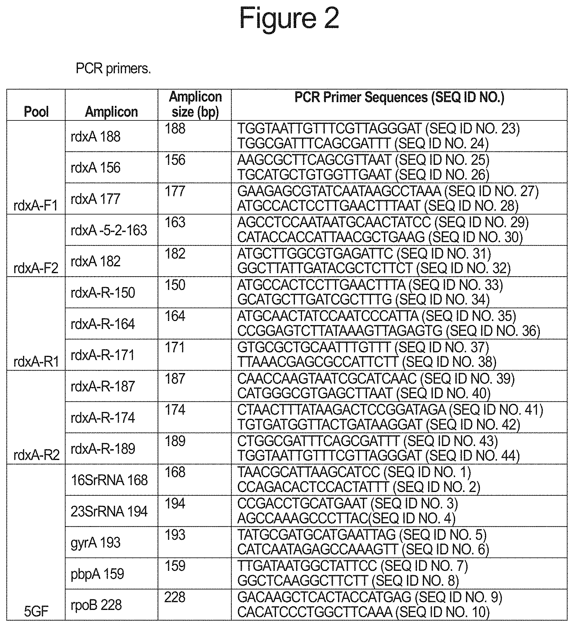
In one aspect the invention provides a set of PCR amplification primers for characterizing the presence of drug resistant H. pylori in FFPE preserved tissue. Primers consisting of two pairs of primers (one forward and one reverse in each pair) for separate genes characteristic of known drug resistant H. pylori strains are listed in FIG. 2. The example genes presented here include 16S rRNA (related to tetracycline resistance), 23S rRNA (related to clarithromycin resistance), pbp1 (related to resistance to penicillin antibiotics), gyrA (related to resistance to fluoroquinone antibiotics), rpoB (related to rifabutin resistance) and rdxA (involved in resistance to metronidazole). Those skilled in the art understand that other genetic loci involved in resistance to other antibiotics are known and may be included in or substituted for those described here. Practically, use of two primer pairs for each target gene minimizes the chance that any particular lesion (such as a cross-link or adduct) found in the target DNA sequences will inhibit PCR amplification from both amplicons, since such lesions are unlikely to occur at two different primer binding sites and involve all copies of the target gene within the target (template) DNA. However, the use of two pairs of primers targeting the same region of the bacterial chromosome, yet producing different but overlapping fragments requires that the PCR amplification reactions be carried out separately in order to avoid producing hybrid amplicons that do not match the full length amplicons each primer pair is designed to produce. Thus, at least two PCR amplification reactions must be performed for each set of primer pairs. In contrast, PCR primer pairs targeting different genes and producing amplicons with no homologous sequences can be pooled and thus the limited amount of target bacterial DNA that can be amplified from FFPE-preserved tissue used as efficiently as possible. The invention teaches segregation of primer pairs targeting an overlapping set of amplicons into separate pools and performing a single PCR amplification reaction on the segregated pools to produce the desired amplicons. In one example presented here, as many as 10 amplicons diagnostic for the five different types of drug resistant H. pylori genes listed above can be produced from just two PCR reactions using DNA extracted from FFPE gastric biopsy samples.
One embodiment of the invention is a method for detecting within a sample mutations in a plurality of genes, the method comprising a) identifying PCR primer pairs suitable for producing amplicons comprising regions of each of the genes containing one or more mutations, b) segregating PCR primer pairs comprising one or more primers that interfere with amplicon generation by another PCR primer pair into separate PCR primer pair pools, wherein each of the separate PCR primer pair pools contain a plurality of PCR primer pairs; c) generating amplicons from each of the separate PCR primer pair pools and target DNA isolated from the sample; d) combining all amplicons produced from each of the separate PCR primer pair pools and the target DNA into a sample amplicon pool, adding a unique index sequence to the amplicons within the sample amplicon pool to generate an indexed sample amplicon pool, optionally further combining the indexed sample amplicon pool with one or more differentially indexed sample amplicon pools from different samples, and sequencing all indexed sample amplicons simultaneously; and e) identifying mutations within the indexed sequenced amplicons from a sample by reference to corresponding wild-type gene sequences.
In one embodiment of the invention, the sample is a biopsy sample and, in another embodiment, the biopsy is a gastric biopsy. In a further embodiment, the biopsy sample comprises a formalin-fixed paraffin embedden biopsy sample. In another embodiment, the sample contains Helicobacter pylori (H. pylori).
In another embodiment, the plurality of genes comprises genes selected from the group consisting of H. pylori 16S rRNA, 23S rRNA, gyrA, rpoB, pbp1, and rdxA. In further embodiments, the identified mutation is an A2142G, A2143G, and/or A2142C mutation of the H. pylori 23S rRNA gene; an A928C, AG926-927GT, A926G/A928C and/or AGA926-928TTC mutation of the H. pylori 16S rRNA gene; a C261A, C261G, G271A, and/or G271T mutation of the H. pylori gyrA gene encoding DNA gyrase subunit A; between codons 525 and 545 of the H. pylori rpoB gene encoding the beta/beta' subunit of DNA-directed RNA polymerase; a C1242A or C1242G mutation in the H. pylori pbp1 gene encoding penicillin-binding protein 1; or within the H. pylori rdxA gene. In another embodiment, the identified mutation produces a loss of function of H. pylori oxygen-insensitive (Type I) NAPD(P)H nitroreductase encoded by rdxA.
In one embodiment of the invention, the amplicons do not exceed 230 base pairs in length. In another embodiment, the amplicons are greater than 130 base pairs in length. In a further embodiment, the PCR primer pair comprising one or more primers that interfere with amplicon generation by another PCR primer pair interfere by forming cross pair primer-dimers or by forming cross pair truncated amplicons.
Another embodiment of the invention is directed to a method for detecting within a patient derived sample the presence of drug resistant H. pylori, the method comprising: a) generating amplicons from DNA isolated from the patient derived sample and; i) PCR primer pair pool 1 comprising primers SEQ ID NOs. 1-10; ii) PCR primer pair pool 2 comprising primers SEQ ID Nos. 11-22; iii) PCR primer pair pool 3 comprising primers SEQ ID Nos. 23-28; iv) PCR primer pair pool 4 comprising primers SEQ ID Nos. 29-32; v) PCR primer pair pool 5 comprising primers SEQ ID Nos. 33-38; vi) PCR primer pair pool 6 comprising primers SEQ ID Nos. 39-44; b) combining all amplicons produced from PCR primer pair pools 1-6 in step a) into a sample amplicon pool, adding a unique index sequence to the amplicons within the sample amplicon pool to generate an indexed sample amplicon pool, optionally further combining the indexed sample amplicon pool with one or more differentially indexed sample amplicon pools from different patient derived samples, and sequencing all indexed sample amplicons simultaneously; c) identifying mutations within the sequenced indexed sample amplicons by reference to SEQ ID Nos. 47-51, and d) determining the drug-resistant profile of H. pylori present in the patient-derived profile by the presence or absence of mutations identified in step c).
Yet another embodiment of the invention is directed to a method for detecting within a patient derived sample the presence of drug resistant H. pylori, the method comprising: a) generating amplicons from DNA isolated from the patient derived sample and; i) PCR primer pair pool 1 comprising primers SEQ ID Nos. 23-28; ii) PCR primer pair pool 2 comprising primers SEQ ID Nos. 29-32; iii) PCR primer pair pool 3 comprising primers SEQ ID Nos. 33-38; iv) PCR primer pair pool 4 comprising primers SEQ ID Nos. 39-44; b) combining all amplicons produced from PCR primer pair pools 1-4 in step a) into a sample amplicon pool, adding a unique index sequence to the amplicons within the sample amplicon pool to generate an indexed sample amplicon pool, optionally further combining the indexed sample amplicon pool with one or more differentially indexed sample amplicon pools from different patient derived samples, and sequencing all indexed sample amplicons simultaneously; c) identifying mutations within the sequenced indexed sample amplicons by reference to SEQ ID Nos. 47-51; and d) determining the drug-resistant profile of H. pylori present in the patient-derived profile by the presence or absence of mutations identified in step c).
In a further embodiment any of the amplicon pools described here can be sequenced by classical Sanger sequencing methods using one of the terminal primers to a single amplicon within the pool as a forward sequencing primer and the other terminal primer to that amplicon as a reverse sequencing primer. Alternatively, unique sequencing primers specific to each desired reaction for each individual amplicon within the amplicon pool can be used for the same purpose. In this way each of the amplicons can be directly sequenced from an amplicon pool. The amplicon pool may or may not be combined with other amplicon pools from the same FFPE extracted biopsy sample and the combined amplicons prepared for sequencing by addition of adaptors and indexing tags in preparation for Next Generation sequencing (NGS). Tagged amplicon pools derived from a single FFPE biopsy sample can be further combined with differentially tagged amplicon pools from different FFPE biopsy samples and the combined sample amplicon pools directly sequenced by high-throughput multiplex sequencing methods.
DESCRIPTION OF THE DRAWINGS
FIG. 1 depicts the H. pylori rdxA gene and illustrates the relative arrangement of the described amplicons.
FIG. 2 is a table of the individual primers disclosed in the Descriptions and Examples.
FIG. 3 is an electrophoresis gel demonstrating that pooling all rdxA specific primers generates incorrect sized amplicons, whereas the segregated primer pools described here generate correct sized amplicons.
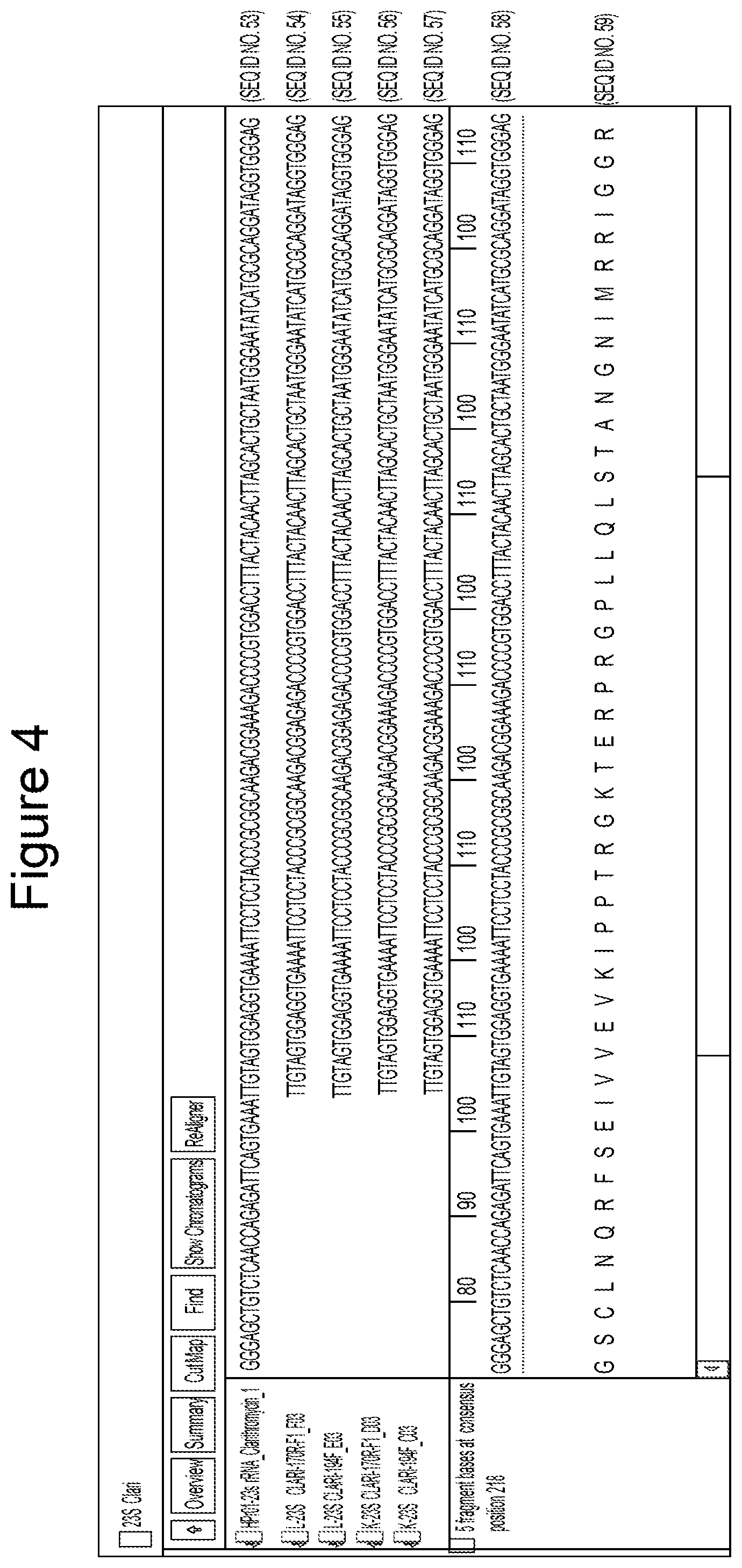
FIG. 4 identifies a representative mutation in 23S rRNA (in FFPE samples FFPE_K and L) 23 S rRNA Clarithromycin sequences alignment sequenced with 23S rRNA 194-F and 170R-F. The red box shows the mutation loci on 23S rRNA
FIG. 5 is a summary data table of 16 FFPE samples analyzed by Next Generation Sequencing (NGS) data from 16 FFPE samples using the segregational pooling and pooled amplicon strategy with mutations in each of six different genes characteristic of drug-resistant H. pylori identified. All mutations identified by NGS were confirmed by Sanger sequencing.
FIG. 6 is a summary data table of 24 FFPE samples analyzed by NGS data from 24 FFPE samples using the segregational pooling and pooled amplicon strategy with mutation in each of six different genes characteristic of drug-resistant H. pylori identified. The data table lists the gene mutations identified by NGS as well as their mutation frequency.
DETAILED DESCRIPTION OF THE INVENTION
The present invention provides methods and compositions for amplifying trace DNA sequences present in FFPE biopsy samples. These methods and compositions are useful for determining the presence of and characterizing the drug and multi-drug resistance profiles of H. pylori from FFPE gastric biopsy samples.
The methods and compositions described herein are directed to generating amplicons from trace bacterial DNA present in FFPE biopsy samples. Such DNA is especially subject to damage and PCR amplification of damaged DNA requires special steps to ensure that the desired sequences are amplified from the target DNA. These steps include minimizing the length of each amplicon recovered from the amplification reactions, determining the optimal primer sequences within the targeted DNA sequence, and strategically pooling overlapping amplicons and primers to avoid interference with one another. A process referred to as segregational pooling. In a typical PCR amplification scheme, where DNA integrity can be assumed to be relatively high, primer selection is not as limited as it is when the template DNA is known to be damaged by cross-linking or the presence of base adducts, as with FFPE derived DNA. Furthermore, the absolute levels of bacterial DNA that are found in typical biopsy samples are many orders of magnitude lower than the DNA of the host organism. This means that the sequences that are targeted for amplification are a minority of the total DNA in the sample and successfully amplifying one or more amplicons from such rare DNA sequences requires the most optimal primers and the most efficient use of the target DNA possible. Furthermore, to be generally useful in the commercial clinical laboratory, such methods should be compatible with and make use of established techniques and reagents to the greatest extent possible.
Total DNA was extracted from FFPE gastric biopsy samples using the Qiagen QIAamp DSP DNA FFPE Tissue Kit (Qiagen, Cat. No. 60404) following the manufacturer's instructions. Briefly, five 5 .mu.m thick sections of a single FFPE biopsy sample were placed in a single tube and de-paraffinized with xylene, treated with proteinase K at 65.degree. C. for one hour to lyse the cells within the de-paraffinized samples, and then held at 90.degree. C. for an additional hour to de-cross-link cellular structures and to denature proteinase K. The de-paraffinized and lysed samples are cooled to room temperature applied to MiniElute PCR Purification column (Qiagen, Cat. No. 28004) following the manufacturers recommended procedure. The MiniElute column is washed twice and the DNA recovered from the FFPE biopsy sample recovered from the column with elution buffer. The recovered DNA was quantified with a Qubit 2.0 fluorometer (Thermo Fisher Scientific) and the DNA purity determined with a Nanodrop instrument (Thermo Fisher Scientific).
To verify the presence of H. pylori sequences within the total DNA samples from each biopsy sample, PCR amplification of a specific 125 base-pair fragment unique highly conserved region of the 23S rRNA gene of H. pylori was performed using PCR primers SEQ ID Nos: 45 and 46. The PCR product was purified and sequenced and confirmed to be specific to H. pylori by BLAST analysis [Altschul, S. F., Gish, W., Miller, W., Myers, E. W. & Lipman, D. J. (1990) "Basic local alignment search tool." J. Mol. Biol. 215:403-410]. The absence of the correct amplified product (amplicon) indicated no usable H. pylori DNA was present in the sample.
Samples that did produce the 125 base-pair H. pylori specific PCR amplicon were further investigated to determine the quality of the recovered DNA. To determine the suitability of the extracted DNA from FFPE gastric biopsy samples containing H. pylori for PCR amplification and sequencing, a multiplex PCR qualification assay was developed. This qualification assay involves PCR amplification of the human GAPDH gene encoding glyceraldehyde-3-phosphate dehydrogenase with a set of PCR primers capable of producing an amplicon ladder of 100, 200, 300, 400 and 500 base-pair fragments. FFPE DNA with no significant damage, with large fragment sizes and at relatively high concentration produces all five "rungs" of the amplicon ladder, whereas highly damaged and significantly fragmented DNA will not produce any of the expected amplicons. With our accumulated experience with this method, we rate DNA recovered from FFPE gastric biopsy samples as good if this test produces 2 to 5 bands of the amplicon ladder, intermediate if it produces only a single band, and poor if no bands are observed at all. The overall frequency of the number of amplicon bands observed across numerous FFPE extracted biopsy samples indicates that limiting analytical amplicon size to about 200 base-pairs or less provides the best balance between producing as much contiguous sequence as possible and avoiding PCR amplification terminating damage in the template DNA.
PCR amplification reactions using both freshly prepared or frozen H. pylori chromosomal DNA (as a positive control) and samples extracted from FFPE gastric biopsy samples (experimental samples) were performed with 1 U of Taq DNA Polymerase, 10 mM dNTP mix in a 100 mM Tris-HCl, 500 mM KCl and 25 mM MgCl2 buffer.
For samples analyzed by Sanger sequencing methods the PCR primers in FIG. 1 (SEQ ID NOs 1-44) were used as indicated with the thermal cycling parameters are: initial denaturing at 95.degree. C. for 10 minutes, followed by 35 cycles of 30 seconds at 95.degree. C., 30 seconds at 55.degree. C. and 30 seconds at 72.degree. C., then a final extension at 72.degree. C. for 10 minutes. The resulting amplicons from each amplification reaction were purified with a MiniElute PCR Purification column following the manufacturer's instructions.
The purified amplicons were processed for Sanger sequencing with the BigDye Terminator v3.1 Cycle Sequencing Kit (Applied Biosystems, Cat. No. 4337455) following the manufacturer's protocol. The PCR sequencing reaction was executed with the following thermal parameters: 95.degree. C. for 1 minute, then 25 cycles of 95.degree. C. for 30 seconds, 56.degree. C. for 30 seconds and 60.degree. C. for 1 minute. The primer extension reactions were processed with the Agencourt CleanSEQ kit (Beckman Coulter Life Sciences Cat. No. A29151) following the manufacturer's instructions. Samples were loaded and analyzed on an ABI 3500 Genetic Analyzer (Applied Biosystems). Raw sequence data was collected with 3500 Series Data Collection Software (Applied Biosystems) and assembled and aligned against reference sequences using Sequencher v 5.4 software (Gene Codes Corp.).
In the case of samples destined for NGS sequencing the Illumina overhang adapter sequence is added to the locus-specific primer sequences listed in FIG. 2 (SEQ ID NOs 1-44). The forward overhang sequence (added to the 5' side of the locus specific forward primer sequence) is TCGTCGGCAGCGTCAGATGTGTAT-AAGAGACAG (SEQ ID NO. 47) and the reverse overhang sequence (added to the 5' side of the locus specific reverse primer sequence) is GTCTCGTGGGCTCGGAG-ATGTGTATAAGAGACAG (SEQ ID NO. 48). The first round PCR amplification thermal cycling parameters are: initial denaturing at 95.degree. C. for 11 minutes, followed by 35 cycles of 30 seconds at 95.degree. C., 1 minute at 59.degree. C. and 1 minute at 72.degree. C., then a final extension at 72.degree. C. for 10 minutes. Second stage PCR reactions (involving addition of multiplex index adapters) involved thermal cycling parameters: 98.degree. C. for 30 seconds, 17 cycles of 98.degree. C. for 20 seconds, 60.degree. C. for 30 seconds and 72.degree. C. for 45 seconds and a final extension at 72.degree. C. for 5 minutes. The libraries were then processed with the Agencourt AMPure XP kit (Beckman Coulter Life Sciences Cat. No. A63880) following the manufacturer's instructions, quantitated on a 2100 BioAnalyzer (Agilent Technologies) diluted as necessary and loaded onto the Illumina MiSeq sequencing instrument (Illumina, Inc.). Data analysis was performed using NextGENe V 2.4.1.1 software (SoftGenetics).
The present invention is illustrated in the following Examples, which are set forth to aid in understanding the invention, but should not be construed to limit in any way the scope of the invention as defined in the claims that follow.
Example 1
Segregational Pooling of PCR Primers and Amplicons to Characterize Drug Resistance from Multiple Genetic Loci
In one aspect of the invention, multiple loci encoding different forms of drug-resistance can be simultaneously characterized by segregating the PCR primers used to generate the diagnostic amplicons covering each of the targeted loci. PCR primer pairs comprising SEQ ID NOs: 1 and 2 for producing a 168 base-pair amplicon (16SrRNA 168) spanning positions 926-928 of the 16S rRNA gene of H. pylori. Virtually any mutation in these positions produces a low level of tetracycline resistance, while the triple mutation AGA926-928TTC is associated with very high levels of tetracycline resistance. A second primer pair comprising SEQ ID NOs: 11 and 12 produce a 162 base-pair amplicon (16SrRNA 162) also encompassing positions 926-928 of the H. pylori 16S rRNA. Another PCR primer pair comprising SEQ ID NOs: 3 and 4 produces a 194 base pair amplicon (23SrRNA 194) spanning positions 2142 and 2143 of the 23S rRNA gene of H. pylori. Mutation of these positions, particularly A2142G, A2142C and A2143G mutations are associated with clarithromycin resistance. A second primer pair comprising SEQ ID NOs: 13 and 14 produces a 170 base-pair amplicon (23SrRNA 170) which also spans positions 2142 and 2143 of the H. pylori 23S rRNA. Another PCR primer pair comprising SEQ ID NOs: 5 and 6 produces a 193 base-pair amplicon (gyrA 193) which spans the region encoding amino acid positions 87 to 91 of the A subunit of H. pylori gyrase, encoded by the gyrA gene. Mutations of Asn87 to Lys or Tyr and mutation of Asp91 to Gly, Asn or Tyr, individually or together are known to produce resistance to fluoroquinone antibiotics. A second PCR primer pair spanning this region of the H. pylori gyrA gene, comprising SEQ ID NOs: 15 and 16 produces a 139 base-pair amplicon (gyrA 139). A third PCR primer pair spanning the same region comprising SEQ ID NOs: 17 and 18 produce a 137 base-pair amplicon (gyrA 137). Another PCR primer pair comprising SEQ ID NOs: 7 and 8 produce a 159 base-pair amplicon (pbpA 159) which encompasses the sequence encoding amino acid position 414 of the H. pylori pbp1 gene. Mutation of the serine normally found at position 414 of penicillin-binding protein 1 to an arginine produces resistance to amoxicillin and other penicillin antibiotics. Another PCR primer pair, SEQ ID NOs: 19 and 20 produce a 140 base-pair amplicon (pbpA 140) that also encompasses the sequence encoding position 414 of H. pylori penicillin-binding protein 1. Another PCR primer pair comprising SEQ ID NOs: 9 and 10 produce a 228 base-pair amplicon encompassing codons 525 to 545 of the H. pylori rpoB gene encoding the .beta./.beta.' subunit of RNA polymerase. Mutation of any of the codons within this region can confer resistance to rifabutin and other rifamycin-like antibiotics. A PCR primer pair comprising SEQ ID NOs: 21 and 22 also produces an amplicon (rpoB-R-167) which is 167 base-pairs and encompasses the critical codons within rpoB.
Each pair of PCR primers targeting a particular gene region potentially encoding a drug-resistant mutation are segregated into separate PCR primer pair pools containing one or more unique primer pairs targeting different genes. Thus, PCR amplification of each pool produces amplicons specific to the plurality of genes within each pool and minimizes the chance of PCR amplification artifacts such as primer-dimers or cross pair amplicon truncation caused by homologous pairing within overlapping amplicon sequences. As shown in FIG. 2, pool 5GF comprises PCR primer pairs 16SrRNA 168, 23SrRNA 194, gyrA193, pbpA 159 and rpoB 228. When FFPE derived H. pylori target DNA is amplified with these primer pairs five unique amplicons of 159, 168, 193, 194 and 228 base-pairs are produced.
Amplicon pool 5GR (FIG. 2) comprises PCR primer pairs 16SrRNA 162R, 23SrRNA 170R, gyrA 139R, gyrA 137R, pbpA 140 and rpoB-R-167. When FFPE derived H. pylori target DNA is amplified with these primer pairs as many as 8 amplicons are produced. Four of these are unique amplicons of 140, 162, 167 and 170 base-pairs (corresponding to the pbpA-specific amplicon, 16S rRNA-specific amplicon, the rpoB-specific amplicon and the 23S rRNA-specific amplicon, respectively). The remaining four amplicons represent permutations of the two pairs of gyrA-specific PCR primer pairs present in pool 5GR. These primer pairs partially overlap one another in the same direction such that four possible amplicons can be produced from the two pairs of primers. Because the partial overlap of primers is limited to primers that have the same strand orientation (direction) there is no risk of primer-dimer formation, and because the amplified sequence between the primers is identical (except for the absolute length of the amplified sequence) no cross-hybridization between amplicons will produce new sequences. Thus, the two pairs of semi-unique primers can be accommodated within the amplicon pool. In this case, these primers may produce as many as four homologous amplicons of 131, 137, 139 and 145 base-pairs. The sequence of these amplicons is identical from end to end with the sequence in each of the other amplicons--with the exception of the few base-pairs missing from the ends of the shorter amplicons, which are present in the longer amplicons.
Alignment of each of the sequenced amplicons allows identification of mutations conferring drug resistance. FIG. 4 is an example of the A2142G mutation in the 23S rRNA identified in two independent FFPE derived sample ("E" and "K") that indicates that treatment of the H. pylori strains afflicting the patients from whom these samples were derived is unlikely to respond to clarithromycin. The top sequence is a known clarithromycin resistant strain of H. pylori while the two sequences immediately below the control are independent PCR amplicons from sample "E" and the last two sequences are independent amplicons from sample "F".
Example 2
Segregational Pooling of PCR Primers and Amplicons to Characterize Drug Resistance Derived from Surveying the Entire Coding Sequence of a Gene
In another aspect of the invention, segregated PCR primer pools allow efficient coverage of an entire gene. Resistance to metronidazole can occur as a result of any loss of function mutation in the H. pylori rdxA gene encoding the bacterial oxygen-insensitive (Type I) NAD(P)H nitroreductase. Metronidazole is activated by the action of this enzyme and thus, any frameshift, or point mutation within the rdxA gene encoding this enzyme has the potential to confer resistance to metronidazole. Unlike the previously described embodiments, no single short amplicon can encompass the known mutational spectrum of metronidazole resistance encountered in H. pylori. To address this problem, two series of PCR primer pairs producing overlapping amplicons were designed such that the primers within each series produce short amplicons that together cover the entire coding region of the rdxA gene. The primers between the two series are located in unique positions, but some may be within the coding sequence of the rdxA gene offset by only a few bases in one direction relative to the analogous primers in the other series (in a manner similar to the partially overlapping gyrA primers within amplicon pool 5GR described in the previous paragraph). This strategy reduces the chance of a single cross-link or adduct present in the target DNA from entirely blocking production of an amplicon. The sequence derived from amplicons produced by one series of PCR primers can be assembled with amplicons produced from the other series of PCR primers to ensure that complete coverage of the rdxA gene is achieved from the total set of primer pools. Each series of primers is placed into one of two amplicon pools for each series so that amplicons within the series that may overlap and which are produced from primers that are prone to form primer-dimers are segregated into separate pools. In the case of the H. pylori rdxA gene, one series of five short amplicons collectively span the entire rdxA coding sequence. These five amplicons, from 5' to 3' comprise the rdxA 188 amplicon (produced from PCR primer pair SEQ ID NOs: 23 and 24), the rdxA-5-2-163 amplicon (produced from PCR primer pair SEQ ID NOs: 29 and 30), the rdxA 156 amplicon (produced from PCR primer pair SEQ ID NOs: 25 and 26), the rdxA 182 amplicon (produced from PCR primer pair SEQ ID NOs: 31 and 32) and the rdxA 177 amplicon (produced from PCR primer pair SEQ ID NOs: 27 and 28). Within this series PCR primer pairs producing amplicons rdxA 188, rdxA156 and rdxA 177 are placed into a single pool designated rdxA-F1, while PCR primer pairs producing amplicons rdxA-5-2-163 and rdxA 182 are combined into a different pool designated rdxA-F2 (FIG. 2). In another series six short amplicons are used to span the entire H. pylori rdxA gene. These amplicons, arrayed 5' to 3' comprise the rdxA-R-150 amplicon (produced from PCR primer pair SEQ ID NOs: 33 and 34), the rdxA-R-187 amplicon (produced from PCR primer pair SEQ ID NOs: 39 and 40), the rdxA-R-164 amplicon (produced from PCR primer pair SEQ ID NOs: 35 and 36), the rdxA-R0174 amplicon (produced from PCR primer pair SEQ ID NOs: 41 and 42, the rdxA-R-171 amplicon (produced from PCR primer pair SEQ ID NOs: 37 and 38) and the rdxA-R-189 amplicon (produced from PCR primer pair SEQ ID NOs: 43 and 44). Within this series PCR primer pairs producing amplicons rdxA-R-150, rdxA-R-164 and rdxA-R-171 are combined into one pool designated rdxA-R1, while PCR primer pairs producing amplicons rdxA-R-187, rdxA-R-174 and rdxA-R-189 are combined into a different pool designated rdxA-R2 (FIG. 2). These PCR primer pools are used to generate their cognate amplicons from H. pylori DNA extracted from FFPE biopsy samples. Depending on how the amplicons are to be sequenced amplicons from each PCR reaction may be further combined so that each series is represented by a single amplicon pool.
As shown in FIG. 3 proper segregational pooling produces the correct (predicted) amplicons, whereas the same primers when present in a single pool produce a series of PCR products comprised of incorrect amplicons and primer dimers. Two FFPE samples. "FFPE_C" and "FFPE E" were processed as described and served as target DNA for analysis of the rdxA gene. Lane 6 (counting from the left-most lane as lane 1) is a double-stranded DNA size marker (Ready-to-Use 100 bp DNA Ladder, Biotium, Inc.). The size marker is flanked on each side by sample specific pooled amplification products. Lanes 1 and 7 labeled "RDX-All" are the result of PCR amplification reactions containing all the PCR primers of the amplicons spanning the entire rdxA gene described above. The lanes marked "F1" (lanes 2 and 8) correspond to the rdxA-F1 pool comprising the rdxA 188, rdxA156 and rdxA 177 amplicon primes and both sample lanes contain amplicons between 150 and 200 base-pairs as predicted. The lanes marked "F2" (lanes 3 and 9) correspond to the rdxA-F2 pool comprising the rdxA-5-2-163 and rdxA 182 amplicon primers and both sample lanes contain amplicons of the correct size. Note that a minor product of about 60 base-pairs is also present, however this amplicon does not interfere in sequencing the correct amplicons and likely represents a product of internal recombination within or between one of the desired amplicons. The lanes marked "R1" (lanes 4 and 10) correspond to the rdxA-R1 pool comprising the rdxA-R-150, rdxA-R-164 and rdxA-R-171 amplicon primers and the desired amplicons are clustered on the gel between 150 and around 170 base-pairs. The lanes marked "R2" (lanes 5 and 11) correspond to the rdxA-R2 pool comprising the rdxA-R-187, rdxA-R-174 and rdxA-R-189 amplicon primers produce the proper sized amplicons.
FIG. 5 illustrates the ability of the method to determine the presence and pattern of drug and multi-drug resistance in multiple isolates derived from FFPE samples based on a single NGS analysis. In this case, the 5 separate genes analyzed as described in Example 1, as well as the rdxA gene analyzed as described in Example 2 are collated into a single report outlining the potential resistance profile of H. pylori in each patient-derived FFPE biopsy sample to each of the six drugs.
FIG. 6 also illustrates the ability of the method to determine the presence and pattern of drug and multi-drug resistance in multiple isolates derived from FFPE samples based on NGS analysis as described in Examples 1 and 2. The table lists the genetic mutations found in each sample as well as the frequency of each mutation. The results show that NGS analysis can distinguish between FFPE samples that do not have any mutations versus those that do. For example, of the 24 samples, 10 (42%) had no mutations (Nos. 5, 6, 8, 9, 13, 17, and 21-24). However, the results also show that NGS analysis can detect both single and multiple mutations within one FFPE sample. For example, 14 samples (58%) had at least one gene mutation (Nos. 1-4, 7, 10-12, 14-16, and 18-20). Of these 14 samples, 11 samples had mutations in a single gene (Nos. 2, 4, 7, 11-12, 14-16, 18, 19, and 20) and 3 samples had mutations in multiple genes (Nos. 1, 3, and 10).
Of the samples with single gene mutations, 5 samples had gyrA gene mutations only (Nos. 2, 4, 15, 18, and 19), 2 samples had rdxA gene mutations only (Nos. 7 and 20), and 4 samples had 23S rRNA gene mutations only (Nos. 11, 12, 14, and 16). Of the samples with gyrA mutations only, one of the samples had two mutations with the gyrA gene (No. 19). As discussed above, the presence of gyrA gene mutations indicates fluoroquinone antibiotic resistance. Of the samples with rdxA gene mutations only, one of the samples had two mutations within the rdxA gene (No. 7). As discussed above, the presence of rdxA gene mutations indicates resistance to metronidazole.
The 3 samples with multiple gene mutations had mutations in both 23S rRNA and gyrA (Nos. 1 and 3) and both gyrA and rdxA (No. 10). The concurrent 23S rRNA and gyrA mutations indicate both clarithromycin and fluoroquinone antibiotic resistance; whereas, the concurrent gyrA and rdxA mutations indicate both fluoroquinone antibiotic resistance and metronidazole resistance.
SEQUENCE LISTINGS
1
59117DNAArtificial SequencePCR primer 1taacgcatta agcatcc 17218DNAArtificial SequencePCR primer 2ccagacactc cactattt 18315DNAArtificial SequencePCR primer 3ccgacctgca tgaat 15415DNAArtificial SequencePCR primer 4agccaaagcc cttac 15519DNAArtificial SequencePCR primer 5tatgcgatgc atgaattag 19619DNAArtificial SequencePCR primer 6catcaataga gccaaagtt 19717DNAArtificial SequencePCR primer 7ttgataatgg ctattcc 17815DNAArtificial SequencePCR primer 8ggctcaaggc ttctt 15920DNAArtificial SequencePCR primer 9gacaagctca ctaccatgag 201018DNAArtificial SequencePCR primer 10cacatccctg gcttcaaa 181117DNAArtificial SequencePCR primer 11ctagcggatt ctctcaa 171217DNAArtificial SequencePCR primer 12cagtaatgca gctaacg 171317DNAArtificial SequencePCR primer 13catcaagggt ggtatct 171417DNAArtificial SequencePCR primer 14ttgtagtgga ggtgaaa 171518DNAArtificial SequencePCR primer 15cgttatcgcc atcaatag 181619DNAArtificial SequencePCR primer 16ggtgatgtga ttggtaaat 191718DNAArtificial SequencePCR primer 17ccatcaatag agccaaag 181815DNAArtificial SequencePCR primer 18atcgtgggtg atgtg 151917DNAArtificial SequencePCR primer 19ttgataatgg ctattcc 172016DNAArtificial SequencePCR primer 20ggttacaagc cctaaa 162120DNAArtificial SequencePCR primer 21tgggacaaat tcggccataa 202217DNAArtificial SequencePCR primer 22tttcatgggc ggtcagc 172322DNAArtificial SequencePCR primer 23tggtaattgt ttcgttaggg at 222418DNAArtificial SequencePCR primer 24tggcgatttc agcgattt 182518DNAArtificial SequencePCR primer 25aagcgcttca gcgttaat 182618DNAArtificial SequencePCR primer 26tgcatgctgt ggttgaat 182724DNAArtificial SequencePCR primer 27gaagagcgta tcaataagcc taaa 242822DNAArtificial SequencePCR primer 28atgccactcc ttgaacttta at 222923DNAArtificial SequencePCR primer 29agcctccaat aatgcaacta tcc 233022DNAArtificial SequencePCR primer 30cataccacca ttaacgctga ag 223118DNAArtificial SequencePCR primer 31atgcttggcg tgagattc 183221DNAArtificial SequencePCR primer 32ggcttattga tacgctcttc t 213320DNAArtificial SequencePCR primer 33atgccactcc ttgaacttta 203418DNAArtificial SequencePCR primer 34gcatgcttga tcgctttg 183522DNAArtificial SequencePCR primer 35atgcaactat ccaatcccat ta 223624DNAArtificial SequencePCR primer 36ccggagtctt ataaagttag agtg 243718DNAArtificial SequencePCR primer 37gtgcgctgca atttgttt 183819DNAArtificial SequencePCR primer 38ttaaacgagc gccattctt 193921DNAArtificial SequencePCR primer 39caaccaagta atcgcatcaa c 214018DNAArtificial SequencePCR primer 40catgggcgtg agcttaat 184125DNAArtificial SequencePCR primer 41ctaactttat aagactccgg ataga 254222DNAArtificial SequencePCR primer 42tgtgatggtt actgataagg at 224319DNAArtificial SequencePCR primers 43ctggcgattt cagcgattt 194422DNAArtificial SequencePCR primer 44tggtaattgt ttcgttaggg at 224522DNAArtificial SequencePCR primer 45acaacccaga ctaccaaata ag 224619DNAArtificial SequencePCR primer 46gtgagctgtt acgctttct 1947500DNAHelicobacter pylori 47gtaatccgta gagatcaaga ggaatactca ttgcgaggcg acctgctgga acattactga 60cgctgattgc gcgaaagcgt ggggagcaaa caggattaga taccctggta gtccacgccc 120taaacgatgg atgctagttg ttggagggct tagttttcca gtaatgcagc taacgcatta 180agcatcccgc ctggggagta cggtcgcaag attaaaatca aaggaataga cggggacccg 240cacaagcggt ggagcatgtg gtttaattcg aagatacacg aagaacctta cctaggcttg 300acattgagag aatccgctag aaatagtgga gtgtctggct tgccagacct tgaaaacagg 360tgctgcacgg ctgtcgtcag ctcgtgtcgt gagatgttgg gttaagtccc gcaacgagcg 420caaccccttt tcttagttgc taacaggtta tgctgagaac tctaaggata ctgcctccgt 480aaggaggagg aaggtgggga 500481500DNAHelicobacter pylori 48caagtgataa taaaaggggg tagagccctg attgggctag ggctgctcgc cgcggtacca 60aaccctatca aacttcgaat accttttatc gtatcttggg agtcaggcgg tgggtgataa 120aatcaatcgt caaaagggga acaacccaga ctaccaaata aggtccctaa gttctattct 180gagtggaaaa agatgtgtgg ctactcaaac aaccaggagg ttggcttaga agcagccatc 240ctttaaagaa agcgtaacag ctcactggtc tagtggtcat gcgctgaaaa tataacgggg 300ctaagataga caccgaattt gtagattgtg ttaaacacag tggtagaaga gcgttcatac 360cagcgttgaa ggtataccgg taaggagtgc tggagcggta tgaagtgagc atgcaggaat 420gagtaacgat aagatatatg agaattgtat ccgccgtaaa tctaaggttt cctacgcgat 480ggtcgtcatc gtagggttag tcgggtccta agccgagtcc gaaaggggta ggtgatggca 540aattggttaa tattccaata ccgactgtgg agcgtgatgg ggggacgcat agggttaagc 600gagctagctg atggaagcgc tagtctaagg gcgtagattg gagggaaggc aaatccacct 660ctgtatttga aacccaaaca ggctctttga gtccttttag gacaaaggga gaatcgctga 720taccgtcgtg ccaagaaaag tctctaagca tatccatagt cgtccgtacc gcaaaccgac 780acaggtagat gagatgagta ttctaaggcg cgtgaaagaa ctctggttaa ggaactctgc 840aaactagcac cgtaagttcg cgataaggtg tgccacagcg atgtggtctc agcaaagagt 900ccctcccgac tgtttaccaa aaacacagca ctttgccaac tcgtaagagg aagtataagg 960tgtgacgcct gcccggtgct cgaaggttaa gaggatgcgt cagtcgcaag atgaagcgtt 1020gaattgaagc ccgagtaaac ggcggccgta actataacgg tcctaaggta gcgaaattcc 1080ttgtcggtta aataccgacc tgcatgaatg gcgtaacgag atgggagctg tctcaaccag 1140agattcagtg aaattgtagt ggaggtgaaa attcctccta cccgcggcaa gacggaaaga 1200ccccgtggac ctttactaca acttagcact gctaatggga atatcatgcg caggataggt 1260gggaggcttt gaagtaaggg ctttggctct tatggagcca tccttgagat accacccttg 1320atgtttctgt tagctaactg gcctgtgtta tccacaggca ggacaatgct tggtgggtag 1380tttgactggg gcggtcgcct cctaaaaagt aacggaggct tgcaaaggtt ggctcattgc 1440ggttggaaat cgcaagttga gtgtaatggc acaagccagc ctgactgtaa gacatacaag 150049550DNAHelicobacter pylori 49atgcaagata attcagtcaa tgaaacaaaa aatattgtag aagtggggat tgattcttct 60attgaagaga gctatttagc ttattccatg agcgtgatca tagggcgcgc tttaccggac 120gctagagatg gcttaaagcc cgtgcatagg cgtattttgt atgcgatgca tgaattaggc 180cttacttcaa aagtcgctta caaaaaaagc gctaggatcg tgggtgatgt gattggtaaa 240taccaccccc atggcgataa tgcggtttat gatgcgctag tgagaatggc gcaagatttt 300tccatgcgtt tggaattagt ggatgggcag ggcaactttg gctctattga tggcgataac 360gccgcagcga tgcgttacac tgaagccaga atgactaagg cgagtgaaga aattttaagg 420gatattgata aagacaccat tgattttgtg cctaattatg acgatacctt aaaagagcca 480gatattttac caagccgtct gcctaacctt ttagtcaatg gggctaatgg gatcgctgtg 540gggatggcga 55050500DNAHelicobacter pylori 50aactaacgcg tctaatgaag atgaagacaa cttaaacgct agcatgatcg ttacagacac 60gagcaccggt aagattttag ctttagtggg ggggattgat tataaaaaaa gcgctttcaa 120tcgcgccacg caagccaaac ggcagtttgg gagcgcgata aagccttttg tgtatcagat 180cgcttttgat aatggctatt ccacgacttc taaaatccct gataccgcgc gaaactttga 240aaatggcaat tatagtaaaa acagtgaaca aaaccacgca tggcacccca gcaattattc 300tcgcaagttt ttagggcttg taaccttgca agaagccttg agccattcgt taaatctagc 360cacgatcaat ttaagcgatc agcttggctt tgaaaaaatt tatcaatctt taagcgatat 420ggggtttaaa aacctcccta aggacttgtc tattgtgtta gggagctttg ctatctcacc 480cattgatgca gctgaaaagt 50051600DNAHelicobacter pylori 51atgaagatat tatcaccacc gttaaatacc tcatgaagat caaaaacaat caaggcaaga 60ttgatgacag ggaccacttg ggcaatcgta ggattagggc ggtaggggaa ttgttggcca 120atgaattgca ttcaggttta gtgaaaatgc aaaagaccat taaagacaag ctcactacca 180tgagcggggc ttttgattcg ctcatgcccc atgacttggt caattctaaa atgatcacaa 240gcaccatcat ggaatttttc atgggcggtc agctctcgca atttatggat caaacgaatc 300ccttgagtga ggttacgcac aagcgccgcc tttcagcgct cggcgaaggg gggttggtga 360aagacagagt ggggtttgaa gccagggatg tgcaccccac gcattatggc cgaatttgtc 420ccattgagac cccagaaggt caaaatatcg gtctgatcaa caccctttcc actttcacaa 480gagtgaatga tttaggcttt attgaagccc cttataaaaa ggttgtggat ggcaaggtcg 540tgggtgagac gatttatttg accgctattc aagaagacag ccacatcatc gctcccgcaa 600521032DNAHelicobacter pylori 52atttgagcat ggggcagatt ttaagcttat ttatggtaat tgtttcgtta gggattttat 60tgtatgctac aaaaaattct aaaaaaataa aggaaaatca atgaaatttt tggatcaaga 120aaaaagaaga caattattaa acgagcgcca ttcttgcaag atgtttgata gccattatga 180gttttctagc acagaattag aagaaatcgc tgaaatcgcc aggctatcgc caagctctta 240caacacgcag ccatggcatt ttgtgatggt tactgataag gatttaaaaa aacaaattgc 300agcgcacagc tatttcaatg aagagatgat taaaagcgct tcagcgttaa tggtggtatg 360ctctttaaga cccagcgagt tgttaccaca cggccactac atgcaaaatc tctatccgga 420gtcttataaa gttagagtga tcccctcttt tgctcaaatg cttggcgtga gattcaacca 480cagcatgcaa agattagaaa gctatatttt agagcaatgc tatatcgctg tggggcaaat 540ttgcatgggc gtgagcttaa tgggattgga tagttgcatt attggaggct ttgatccttt 600aaaggtgggc gaagttttag aagagcgtat caataagcct aaaatcgcat gcttgatcgc 660tttgggcaaa gggtggcaga agcgagtcaa aaatcaagaa aatcaaaagt tgatgcgatt 720acttggttgt gattaaacaa aatcaaaaac tttttaacta taatcaaacc taaattaaag 780ttcaaggagt ggcattttgt ttaaaagaat ggttttaatc gctcttttag gggtgttttc 840aagcgtttca ttaagcgcta agagtctttt aagagatgat gggattttag tctctgattt 900aaagggcatg aaatcagaac tatctgatgc tcctgcttgg gtttttgaag acgctaaagc 960cccctacgaa gaaatgggcg tggcgtatat ccctgttaat aataaatatt tagggattga 1020gcaagcgacc tt 103253143DNAArtificialHPr01-23s rRNA_Clarithromycin_1 53gggagctgtc tcaaccagag attcagtgaa attgtagtgg aggtgaaaat tcctcctacc 60cgcggcaaga cggaaagacc ccgtggacct ttactacaac ttagcactgc taatgggaat 120atcatgcgca ggataggtgg gag 14354112DNAArtificialL-23S CLARI-170R-F1_F03 54ttgtagtgga ggtgaaaatt cctcctaccc gcggcaagac ggagagaccc cgtggacctt 60tactacaact tagcactgct aatgggaata tcatgcgcag gataggtggg ag 11255112DNAArtificialL-23S CLARI-194F_E03 55ttgtagtgga ggtgaaaatt cctcctaccc gcggcaagac ggagagaccc cgtggacctt 60tactacaact tagcactgct aatgggaata tcatgcgcag gataggtggg ag 11256112DNAArtificialK-23S CLARI-170R-F1_D03 56ttgtagtgga ggtgaaaatt cctcctaccc gcggcaagac ggaaagaccc cgtggacctt 60tactacaact tagcactgct aatgggaata tcatgcgcag gataggtggg ag 11257112DNAArtificialK-23S CLARI-194F_C03 57ttgtagtgga ggtgaaaatt cctcctaccc gcggcaagac ggaaagaccc cgtggacctt 60tactacaact tagcactgct aatgggaata tcatgcgcag gataggtggg ag 11258142DNAArtificial5 fragment bases at consensusposition 218 58ggagctgtct caaccagaga ttcagtgaaa ttgtagtgga ggtgaaaatt cctcctaccc 60gcggcaagac ggaaagaccc cgtggacctt tactacaact tagcactgct aatgggaata 120tcatgcgcag gataggtggg ag 1425948PRTArtificial5 fragment bases at consensusposition 218 59Gly Ser Cys Leu Asn Gln Arg Phe Ser Glu Ile Val Val Glu Val Lys1 5 10 15Ile Pro Pro Thr Arg Gly Lys Thr Glu Arg Pro Arg Gly Pro Leu Leu 20 25 30Gln Leu Ser Thr Ala Asn Gly Asn Ile Met Arg Arg Ile Gly Gly Arg 35 40 45
* * * * *
D00001
D00002
D00003
D00004
D00005
D00006
D00007
D00008
D00009
S00001
XML
uspto.report is an independent third-party trademark research tool that is not affiliated, endorsed, or sponsored by the United States Patent and Trademark Office (USPTO) or any other governmental organization. The information provided by uspto.report is based on publicly available data at the time of writing and is intended for informational purposes only.
While we strive to provide accurate and up-to-date information, we do not guarantee the accuracy, completeness, reliability, or suitability of the information displayed on this site. The use of this site is at your own risk. Any reliance you place on such information is therefore strictly at your own risk.
All official trademark data, including owner information, should be verified by visiting the official USPTO website at www.uspto.gov. This site is not intended to replace professional legal advice and should not be used as a substitute for consulting with a legal professional who is knowledgeable about trademark law.