2-azabicyclo hexane JAK inhibitor compound
Fatheree , et al. April 6, 2
U.S. patent number 10,968,222 [Application Number 16/656,946] was granted by the patent office on 2021-04-06 for 2-azabicyclo hexane jak inhibitor compound. This patent grant is currently assigned to Theravance Biopharma R&D IP, LLC. The grantee listed for this patent is THERAVANCE BIOPHARMA R&D IP, LLC. Invention is credited to Paul R. Fatheree, Lan Jiang.


View All Diagrams
| United States Patent | 10,968,222 |
| Fatheree , et al. | April 6, 2021 |
2-azabicyclo hexane JAK inhibitor compound
Abstract
The invention provides a compound of formula 1 ##STR00001## or a pharmaceutically-acceptable salt thereof, that is useful as a JAK inhibitor. The invention also provides pharmaceutical compositions comprising the compound, methods of using the compound to treat diseases amenable to a JAK inhibitor, and processes useful for preparing the compound.
| Inventors: | Fatheree; Paul R. (South San Francisco, CA), Jiang; Lan (South San Francisco, CA) | ||||||||||
|---|---|---|---|---|---|---|---|---|---|---|---|
| Applicant: |
|
||||||||||
| Assignee: | Theravance Biopharma R&D IP,
LLC (South San Francisco, CA) |
||||||||||
| Family ID: | 1000005468374 | ||||||||||
| Appl. No.: | 16/656,946 | ||||||||||
| Filed: | October 18, 2019 |
Prior Publication Data
| Document Identifier | Publication Date | |
|---|---|---|
| US 20200131178 A1 | Apr 30, 2020 | |
Related U.S. Patent Documents
| Application Number | Filing Date | Patent Number | Issue Date | ||
|---|---|---|---|---|---|
| 62751967 | Oct 29, 2018 | ||||
| Current U.S. Class: | 1/1 |
| Current CPC Class: | A61K 9/0048 (20130101); A61K 9/0019 (20130101); C07D 471/04 (20130101) |
| Current International Class: | C07D 471/04 (20060101); A61K 9/00 (20060101) |
| Field of Search: | ;546/118 ;514/303 |
References Cited [Referenced By]
U.S. Patent Documents
| 6534524 | March 2003 | Kania et al. |
| 6884890 | April 2005 | Kania et al. |
| 7884109 | February 2011 | Ohlmeyer et al. |
| 8450340 | May 2013 | Hood et al. |
| 8575336 | November 2013 | Coe et al. |
| 8648069 | February 2014 | Akritopoulou-Zanze |
| 8895544 | November 2014 | Coe et al. |
| 10100049 | October 2018 | Fatheree et al. |
| 10183942 | January 2019 | Benjamin et al. |
| 10196393 | February 2019 | Fatheree et al. |
| 10208040 | February 2019 | Fatheree et al. |
| 10251874 | April 2019 | Dabros et al. |
| 10392386 | August 2019 | Fatheree et al. |
| 10406148 | September 2019 | Kleinschek et al. |
| 2005/0090529 | April 2005 | McAlpine et al. |
| 2015/0158864 | June 2015 | Thorarensen et al. |
| 2015/0329542 | November 2015 | Coe et al. |
| 2016/0289196 | October 2016 | Choi et al. |
| 2017/0121327 | May 2017 | Fatheree et al. |
| 2018/0311255 | November 2018 | Fatheree et al. |
| 2019/0106420 | April 2019 | Fatheree et al. |
| 2019/0127371 | May 2019 | Fatheree et al. |
| 2019/0337945 | November 2019 | Fatheree et al. |
| 2019/0350916 | November 2019 | Kleinschek et al. |
| 112279848 | Jan 2021 | CN | |||
| 2010111624 | May 2010 | JP | |||
| WO 2005/009389 | Feb 2005 | WO | |||
| WO 2010/114971 | Oct 2010 | WO | |||
| WO 2013/014567 | Jan 2013 | WO | |||
| WO 2015/173683 | Nov 2015 | WO | |||
| WO 2016/026078 | Feb 2016 | WO | |||
| WO 2017/077283 | May 2017 | WO | |||
| WO 2017/077288 | May 2017 | WO | |||
| 2020/173400 | Sep 2020 | WO | |||
| 2020/181034 | Sep 2020 | WO | |||
Other References
|
Tanaka et al., "New insight into mechanisms of pruritus from molecular studies on familial primary localized cutaneous amyloidosis", British Journal of Dermatology, 161: 1217-1224 (2009). cited by applicant . Trujillo et al., "2-(6-Phenyl-1H-indazol-3-yl)-1H-benzo[d]imidazoles: Design and synthesis of a potent and isoform selective PKC-zeta inhibitor", Bioorganic & Medicinal Chemistry Letters, 19: 908-911 (2009). cited by applicant . Vincenti et al., "Randomized phase 2b trial of tofacitinib (CP-690,550) in de novo kidney transplant patients: Efficacy, renal function and safety at 1 year", American Journal of Transplantation, 12: 2446-2456 (2012). cited by applicant . Weinbrand-Goichberg et al., "Eosinophilic esophagitis: an immune-mediated esophageal disease", Immunol Res, 56: 249-260 (2013). cited by applicant . Welz-Kubiak et al., "IL-31 is overexpressed in lichen planus but its level does not correlate with pruritus severity", Journal of Immunology Research, Article 854747, 6 pages (2015). cited by applicant . Woywodt et al., "Mucosal cytokine expression, cellular markers and adhesion molecules in inflammatory bowel disease", European Journal of Gastroenterology & Hepatology, 11: 267-276 (1999). cited by applicant . Xing et al., "Alopecia areata is driven by cytotoxic T lymphocytes and is reversed by JAK inhibition", Nature Medicine, 20(9): 1043-1049 (Sep. 2014). cited by applicant . Yamamoto et al., "Mucosal inflammation in the terminal ileum of ulcerative colitis patients: Endoscopic findings and cytokine profiles", Digestive and Liver Disease, 40: 253-259 (2008). cited by applicant . Yan et al., "Discovery of 3-(5'-Substituted)-benzimidazol-5-(1-(3,5-dichloropyridin-4-yl)ethoxy)-1H- -indazoles as potent fibroblast growth factor receptor inhibitors: Design, synthesis, and biological evaluation", J. Med. Chem., 59: 6690-6708 (2016). cited by applicant . Yano et al., "Ipilimumab augments antitumor activity of bispecific antibody-armed T cells", Journal of Translational Medicine, 12: 191 (2014). cited by applicant . Zak et al., "Inhaled Janus Kinase (JAK) inhibitors for the treatment of asthma", Bioorganic & Medicinal Chemistry Letters, 29, 126658 (2019). cited by applicant . Zeiser et al., "Ruxolitinib in corticosteroid-refractory graft-versus-host disease after allogeneic stem cell transplantation: a multi-center survey", Leukemia, 29(10): 2062-2068 (Oct. 2015). cited by applicant . Zhou et al., "Cytokines and Behcet's Disease", Autoimmunity Reviews, 11: 699-704 (2012). cited by applicant . International Search Report and the Written Opinion for PCT/US2019/056947 dated Jan. 13, 2020. cited by applicant . U.S. Appl. No. 16/689,706, Fatheree et al. cited by applicant . U.S. Appl. No. 16/701,426, Fatheree et al. cited by applicant . U.S. Appl. No. 16/559,077, Long et al. cited by applicant . U.S. Appl. No. 16/559,091, Colson et al. cited by applicant . U.S. Appl. No. 16/559,138, Long et al. cited by applicant . Abcouwer, "Angiogenic factors and cytokines in diabetic retinopathy", J Clin Cell Immunol, Supplement 1: 1-12 (2013). cited by applicant . Bao et al., "The involvement of the JAK-STAT signaling pathway in chronic inflammatory skin disease atopic dermatitis", JAK-STAT, 2(3): e24137-1-e24137-8 (2013). (1997). cited by applicant . Berastegui et al., "BALF cytokines in different phenotypes of chronic lung allograft dysfunction in lung transplant patients", Clinical Transplantation, 31: e12898 (2017). cited by applicant . Coghill et al., "Effector CD4+ T cells, the cytokines they generate, and GVHD: something old and something new", Blood, 117(12): 3268-3276 (Mar. 24, 2011). cited by applicant . Cottin, "Eosinophilic lung diseases", Clin Chest Med, 37: 535-556 (2016). cited by applicant . Craiglow et al., "Tofacitinib citrate for the treatment of vitiligo: A pathogenesis-directed therapy", JAMA Dermatology, 151: 1110-1112 (2015). cited by applicant . De Nitto et al., "Involvement of interleukin-15 and interleukin-21, two gamma-chain-related cytokines, in celiac disease", World J Gastroenterol, 15(37): 4609-4614 (Oct. 7, 2009). cited by applicant . Del Amo et al., "Current and future ophthalmic drug delivery systems. A shift to the posterior segment", Drug Discovery Today, 13(3/4): 135-143 (Feb. 2008). cited by applicant . Deobhakta et al., "Inflammation in retinal vein occlusion", International Journal of Inflammation, vol. 2013, 6 pages (2013). cited by applicant . El-Hashemite et al., "Interferon-gamma-Jak-Stat signaling in pulmonary lymphangioleiomyomatosis and renal angiomyolipoma", Am J Respir Cell Mol Biol, 33: 227-230 (2005). cited by applicant . El-Hashemite et al., "Perturbed IFN-gamma-Jak-signal transducers and activators of transcription signaling in tuberous sclerosis mouse models: Synergistic effects of rapamycin-IFN-gamma treatment", Cancer Research, 64: 3436-3443 (May 15, 2004). cited by applicant . Fang et al., "Interleukin-6-572C/G polymorphism is associated with serum interleukin-6 levels and risk of idiopathic pulmonary arterial hypertension", Journal of the American Society of Hypertension, 11(3): 171-177 (2017). cited by applicant . Feliciani et al., "A TH2-like cytokine response is involved in bullous pemphigoid. The role of IL-4 and IL-5 in the pathogenesis of the disease", International Journal of Immunopathology and Pharmacology, 12(2): 55-61 (1999). cited by applicant . Fenwick et al., "Effect of JAK inhibitors on release of CXCL9, CXCL10 and CXCL11 from human airway epithelial cells", PLOS One, 10(6): e0128757 (2015). cited by applicant . Foloppe et al., "Identification of a buried pocket for potent and selective inhibition of Chk1: Prediction and verification", Bioorganic & Medicinal Chemistry, 14: 1792-1804 (2006). cited by applicant . Funatsu et al., "Association of vitreous inflammatory factors with diabetic macular edema", Ophthalmology, 116: 73-79 (2009). cited by applicant . Gauthier et al., "Update on chronic lung allograft dysfunction", Curr Transplant Rep, 3(3): 185-191 (Sep. 2016). cited by applicant . Gontcharov et al., "Development of a scalable synthesis for an inhaled pan-JAK inhibitor", Organic Process Research & Development 2019, XXX, XXX-XXX (published online). cited by applicant . Graczyk, "Gini coefficient: a new way to express selectivity of kinase inhibitors against a family of kinases", J Med Chem, 50: 5773-5779 (2007). cited by applicant . Horai et al, "Cytokines in autoimmune uveitis", Journal of Interferon & Cytokine Research, 31(10): 733-744 (2011). cited by applicant . Huang et al., "Glycoprotein 130 inhibitor ameliorates monocrotalline-induced pulmonary hypertension in rats", Canadian Journal of Cardiology, 32: 1356.e1-1356.e10 (2016). cited by applicant . Jones et al., "Design and synthesis of a Pan-Janus kinase inhibitor clinical candidate (PF-06263276) suitable for inhaled and topical delivery for the treatment of inflammatory diseases of the lungs and skin", J. Med. Chem., 60: 767-786 (2017). cited by applicant . Knickelbein et al., "Inflammatory mechanisms of age-related macular degeneration", International Ophthalmology Clinics, 55(3): 63-78 (2015). cited by applicant . Kudlacz et al., "The JAK-3 inhibitor CP-690550 is a potent anti-inflammatory agent in a murine model of pulmonary eosinophilia", European Journal of Pharmacology, 582: 154-161 (2008). cited by applicant . Kumawat et al., "Microscopic colitis patients demonstrate a mixed Th17/Tc17 and Th1/Tc1 mucosal cytokine profile", Molecular Immunology, 55: 355-364 (2013). cited by applicant . Kuno et al., "Recent advances in ocular drug delivery systems", Polymers, 3: 193-221 (2011). cited by applicant . Malaviya et al., "Janus Kinase-3 dependent inflammatory responses in allergic asthma", International Immunopharmacology, 10: 829-836 (2010). cited by applicant . Matsunaga et al., "Effects of a Janus kinase inhibitor, pyridone 6, on airway responses in a murine model of asthma", Biochemical and Biophysical Research Communications, 404: 261-267 (2011). cited by applicant . McBride et al., "Design and structure-activity relationship of 3-benzimidazol-2-yl-1H-indazoles as inhibitors of receptor tyrosine kinases", Bioorganic & Medicinal Chemistry Letters, 16: 3595-3599 (2006). cited by applicant . McBride et al., "3-Benzimidazol-2-yl-1H-indazoles as potent c-ABL inhibitors", Bioorganic & Medicinal Chemistry Letters, 16: 3789-3792 (2006).and renal angiomyolipoma, Am J Respir Cell Mol Biol, 33: 227-230 (2005). cited by applicant . Netchiporouk et al., "Deregulation in STAT signaling is important for cutaneous T-cell lymphoma (CTCL) pathogenesis and cancer progression", Cell Cycle, 13(21): 3331-3335 (Nov. 1, 2014). cited by applicant . Okiyama et al., "Reversal of CD8 T-cell-mediated mucocutaneous graft-versus-host-like disease by the JAK inhibitor tofacitinib", Journal of Investigative Dermatology, 134: 992-1000 (2014). cited by applicant . Owen et al., "Soluble mediators of diabetic macular edema: The diagnostic role of aqueous VEGF and cytokine levels in diabetic macular edema", Curr Diab Rep, 13(4): 476-480 (Aug. 2013). cited by applicant . Reimund et al., "Mucosal inflammatory cytokine production by intestinal biopsies in patients with ulcerative colitis and Crohn's disease", Journal of Clinical Immunology, 16(3): 144-150 (1996). cited by applicant . Shchuko et al., "Intraocular cytokines in retinal vein occlusion and its relation to the efficiency of anti-vascular endothelial growth factor therapy", Indian Journal of Ophthalmology, 63: 905-911 (2015). cited by applicant . Shino et al., "The prognostic importance of CXCR3 chemokine during organizing pneumonia on the risk of chronic lung allograft dysfunction after lung transplantation", PLOS One, 12(7): e0180281 (2017). cited by applicant . Short, "Safety evaluation of ocular drug delivery formulations: techniques and practical considerations", Toxicologic Pathology, 36: 49-62 (2008). cited by applicant . Simov et al., "Structure-based design and development of (benz)imidazole pyridones as JAK1-selective kinase inhibitors", Bioorganic & Medicinal Chemistry Letters, 26: 1803-1808 (2016). cited by applicant . Sohn et al., "Changes in aqueous concentrations of various cytokines after intravitreal triamcinolone versus bevacizumab for diabetic macular edema", Ophthalmology, 152: 686-694 (2011). cited by applicant . Sonkoly et al., "IL-31: A new link between T cells and pruritus in atopic skin inflammation", J Allergy Clin Immunol, 117(2): 411-417 (2006). cited by applicant . Stallmach et al., "Cytokine/chemokine transcript profiles reflect mucosal inflammation in Crohn's disease", Int J Colorectal Dis, 19: 308-315 (2004). cited by applicant . Stevenson et al., "Dry eye disease", Arch Ophthalmol, 130(1): 90-100 (Jan. 2012). cited by applicant. |
Primary Examiner: Morris; Patricia L
Attorney, Agent or Firm: Hagenah; Jeffrey A. Jovic; Florence
Parent Case Text
CROSS-REFERENCE TO RELATED APPLICATIONS
This application claims the benefit of U.S. Provisional Application No. 62/751,967, filed on Oct. 29, 2018, the disclosure of which is incorporated herein by reference in its entirety.
Claims
What is claimed is:
1. A compound of formula: ##STR00021## or a pharmaceutically-acceptable salt thereof.
2. A compound of formula: ##STR00022##
3. A pharmaceutical composition comprising the compound of claim 1, or a pharmaceutically-acceptable salt thereof, and a pharmaceutically-acceptable carrier.
4. The pharmaceutical composition of claim 3, wherein the composition is suitable for application to the eye.
5. The pharmaceutical composition of claim 4, wherein the composition is suitable for intravitreal injection.
6. The pharmaceutical composition of claim 5, wherein the composition is a suspension.
7. The pharmaceutical composition of claim 3, wherein the composition is a suspension.
8. The pharmaceutical composition of claim 3, wherein the composition is a crystalline suspension.
9. The pharmaceutical composition of claim 3, wherein the composition is a sterile aqueous suspension.
10. The pharmaceutical composition of claim 3, wherein the composition is an injectable composition.
11. The pharmaceutical composition of claim 3, wherein the composition is suitable for injection into the eye.
12. The pharmaceutical composition of claim 3, wherein the composition contains from 0.01 to 95% by weight of the compound of formula: ##STR00023## or a pharmaceutically-acceptable salt thereof.
13. The pharmaceutical composition of claim 3, wherein the composition contains from 0.05 to 30% by weight of the compound of formula: ##STR00024## or a pharmaceutically-acceptable salt thereof.
14. The pharmaceutical composition of claim 3, wherein the composition contains from 0.1 to 10% by weight of the compound of formula: ##STR00025## or a pharmaceutically-acceptable salt thereof.
15. The pharmaceutical composition of claim 3, wherein the composition is packaged as a unit dosage form.
Description
BACKGROUND OF THE INVENTION
Field of the Invention
The invention is directed to a JAK kinase inhibitor compound useful for the treatment of inflammatory diseases, particularly ocular diseases. The invention is also directed to pharmaceutical compositions comprising such a compound, methods of using such a compound to treat ocular diseases, and processes useful for preparing the compound.
State of the Art
Cytokines are intercellular signaling molecules which include chemokines, interferons, interleukins, lymphokines, and tumour necrosis factor. Cytokines are critical for normal cell growth and immunoregulation but also drive immune-mediated diseases and contribute to the growth of malignant cells. Elevated levels of many cytokines have been implicated in the pathology of a large number of diseases or conditions, particularly those diseases characterized by inflammation. Many of the cytokines implicated in disease act through signaling pathways dependent upon the Janus family of tyrosine kinases (JAKs), which signal through the Signal Transducer and Activator of Transcription (STAT) family of transcription factors.
The JAK family comprises four members, JAK1, JAK2, JAK3, and tyrosine kinase 2 (TYK2). Binding of cytokine to a JAK-dependent cytokine receptor induces receptor dimerization which results in phosphorylation of tyrosine residues on the JAK kinase, effecting JAK activation. Phosphorylated JAKs, in turn, bind and phosphorylate various STAT proteins which dimerize, internalize in the cell nucleus and directly modulate gene transcription, leading, among other effects, to the downstream effects associated with inflammatory disease. The JAKs usually associate with cytokine receptors in pairs as homodimers or heterodimers. Specific cytokines are associated with specific JAK pairings. Each of the four members of the JAK family is implicated in the signaling of at least one of the cytokines associated with inflammation.
Inflammation plays a prominent role in many ocular diseases, including uveitis, diabetic retinopathy, diabetic macular edema, dry eye disease, age-related macular degeneration, retinal vein occlusion and atopic keratoconjunctivitis. Uveitis encompasses multiple intraocular inflammatory conditions and is often autoimmune, arising without a known infectious trigger. The condition is estimated to affect about 2 million patients in the US. In some patients, the chronic inflammation associated with uveitis leads to tissue destruction, and it is the fifth leading cause of blindness in the US. Cytokines elevated in uveitis patients' eyes that signal through the JAK-STAT pathway include IL-2, IL-4, IL-5, IL-6, IL-10, IL-23, and IFN-.gamma.. (Horai and Caspi, J Interferon Cytokine Res, 2011, 31, 733-744; Ooi et al, Clinical Medicine and Research, 2006, 4, 294-309). Existing therapies for uveitis are often suboptimal, and many patients are poorly controlled. Steroids, while often effective, are associated with cataracts and increased intraocular pressure/glaucoma.
Diabetic retinopathy (DR) is caused by damage to the blood vessels in the retina. It is the most common cause of vision loss among people with diabetes. Angiogenic as well as inflammatory pathways play an important role in the disease. Often, DR will progress to diabetic macular edema (DME), the most frequent cause of visual loss in patients with diabetes. The condition is estimated to affect about 1.5 million patients in the US alone, of whom about 20% have disease affecting both eyes. Cytokines which signal through the JAK-STAT pathway, such as IL-6, as well as other cytokines, such as IP-10 and MCP-1 (alternatively termed CCL2), whose production is driven in part by JAK-STAT pathway signaling, are believed to play a role in the inflammation associated with DR/DME (Abcouwer, J Clin Cell Immunol, 2013, Suppl 1, 1-12; Sohn et al., American Journal of Opthalmology, 2011, 152, 686-694; Owen and Hartnett, Curr Diab Rep, 2013, 13, 476-480; Cheung et al, Molecular Vision, 2012, 18, 830-837; Dong et al, Molecular Vision, 2013, 19, 1734-1746; Funatsu et al, Ophthalmology, 2009, 116, 73-79). The existing therapies for DME are suboptimal: intravitreal anti-VEGF treatments are only effective in a fraction of patients and steroids are associated with cataracts and increased intraocular pressure.
Dry eye disease (DED) is a multifactorial disorder that affects approximately 5 million patients in the US. Ocular surface inflammation is believed to play an important role in the development and propagation of this disease. Elevated levels of cytokines such as IL-1, IL-2, IL-4, IL-5, IL-6, and IFN-.gamma. have been noted in the ocular fluids of patients with DED. (Stevenson et al, Arch Ophthalmol, 2012, 130, 90-100), and the levels often correlated with disease severity. Age-related macular degeneration and atopic keratoconjunctivitis are also thought to be associated with JAK-dependent cytokines.
Retinal vein occlusion (RVO) is a highly prevalent visually disabling disease. Obstruction of retinal blood flow can lead to damage of the retinal vasculature, hemorrhage, and tissue ischemia. Although the causes for RVO are multifactorial, both vascular as well as inflammatory mediators have been shown to be important (Deobhakta et al, International Journal of Inflammation, 2013, article ID 438412). Cytokines which signal through the JAK-STAT pathway, such as IL-6 and IL-13, as well as other cytokines, such as MCP-1, whose production is driven in part by JAK-STAT pathway signaling, have been detected at elevated levels in ocular tissues of patients with RVO (Shchuko et al, Indian Journal of Ophthalmology, 2015, 63(12), 905-911). While many patients with RVO are treated by photocoagulation, this is an inherently destructive therapy. Anti-VEGF agents are also used, but they are only effective in a fraction of patients. Steroid medications that reduce the level of inflammation in the eye (Triamcinolone acetonide and dexamethasone implants) have also been shown to provide beneficial results for patients with certain forms of RVO, but they have also been shown to cause cataracts and increased intraocular pressure/glaucoma.
The need remains for a potent pan-JAK inhibitor for the treatment of ocular diseases.
SUMMARY OF THE INVENTION
In one aspect, the invention provides a JAK inhibitor compound useful for the treatment of ocular inflammatory disease.
In particular, in one aspect, the invention provides a compound of the formula:
##STR00002## hereinafter compound 1, or a pharmaceutically-acceptable salt thereof.
The invention also provides a pharmaceutical composition comprising compound 1, or a pharmaceutically acceptable salt thereof, and a pharmaceutically-acceptable carrier.
In one aspect, the invention provides a method of treating an ocular disease in a mammal, the method comprising administering to the mammal compound 1, or a pharmaceutical composition of the invention. In one aspect the ocular disease is selected from the group consisting of uveitis, diabetic retinopathy, diabetic macular edema, dry eye disease, age-related macular degeneration, retinal vein occlusion and atopic keratoconjunctivitis. In particular, the ocular disease is diabetic macular edema or uveitis.
In separate and distinct aspects, the invention also provides synthetic processes described herein, which are useful for preparing compound 1.
The invention also provides compound 1, or a pharmaceutically acceptable salt thereof, as described herein for use in medical therapy, as well as the use of the compound of the invention in the manufacture of a formulation or medicament for treating ocular diseases.
DETAILED DESCRIPTION OF THE INVENTION
Chemical structures are named herein according to IUPAC conventions as implemented in ChemDraw software (PerkinElmer, Inc., Cambridge, Mass.).
Furthermore, the imidazo portion of the tetrahydroimidazopyridine moiety in the structure of the present compound exists in tautomeric forms. The compound could equivalently be represented as
##STR00003## According to the IUPAC convention, these representations give rise to different numbering of the atoms of the tetrahydroimidazopyridine portion. Accordingly this structure is designated ((1S,5R)-2-azabicyclo[3.1.0]hexan-1-yl)(2-(6-(2-ethyl-5-fluoro-4-hydroxyp- henyl)-4-fluoro-1H-indazol-3-yl)-3,4,6,7-tetrahydro-5H-imidazo[4,5-c]pyrid- in-5-yl)methanone. It can also be designated: ((1S,5R)-2-azabicyclo[3.1.0]hexan-1-yl)(2-(6-(2-ethyl-5-fluoro-4-hydroxyp- henyl)-4-fluoro-1H-indazol-3-yl)-1,4,6,7-tetrahydro-5H-imidazo[4,5-c]pyrid- in-5-yl)methanone. It will be understood that although structures are shown, or named, in a particular form, the invention also includes the tautomer thereof.
The compounds of the invention contain several basic groups and therefore, the compounds can exist as the free base or in various salt forms, such as a mono-protonated salt form, a di-protonated salt form, or mixtures thereof. All such forms are included within the scope of this invention, unless otherwise indicated.
This invention also includes isotopically-labeled compounds of formula 1, i.e., compounds of formula 1 where an atom has been replaced or enriched with an atom having the same atomic number but an atomic mass different from the atomic mass that predominates in nature. Examples of isotopes that may be incorporated into a compound of formula 1 include, but are not limited to, .sup.2H, .sup.3H, .sup.11C, .sup.13C, .sup.14C, .sup.13N, .sup.15N, .sup.15O, .sup.17O, .sup.18O, and .sup.18F. Of particular interest are compounds of formula 1 enriched in tritium or carbon-14, which compounds can be used, for example, in tissue distribution studies. Also of particular interest are compounds of formula 1 enriched in deuterium especially at a site of metabolism, which compounds are expected to have greater metabolic stability. Additionally of particular interest are compounds of formula 1 enriched in a positron emitting isotope, such as .sup.11C, .sup.18F, .sup.15O and .sup.13N, which compounds can be used, for example, in Positron Emission Tomography (PET) studies.
Definitions
When describing this invention including its various aspects and embodiments, the follow
The term "therapeutically effective amount" means an amount sufficient to effect treatment when administered to a patient in need of treatment.
The term "treating" or "treatment" means preventing, ameliorating or suppressing the medical condition, disease or disorder being treated (e.g., a respiratory disease) in a patient (particularly a human); or alleviating the symptoms of the medical condition, disease or disorder.
The term "pharmaceutically acceptable salt" means a salt that is acceptable for administration to a patient or a mammal, such as a human (e.g., salts having acceptable mammalian safety for a given dosage regime). Representative pharmaceutically acceptable salts include salts of acetic, ascorbic, benzenesulfonic, benzoic, camphorsulfonic, citric, ethanesulfonic, edisylic, fumaric, gentisic, gluconic, glucoronic, glutamic, hippuric, hydrobromic, hydrochloric, isethionic, lactic, lactobionic, maleic, malic, mandelic, methanesulfonic, mucic, naphthalenesulfonic, naphthalene-1,5-disulfonic, naphthalene-2,6-disulfonic, nicotinic, nitric, orotic, pamoic, pantothenic, phosphoric, succinic, sulfuric, tartaric, p-toluenesulfonic and xinafoic acid, and the like.
The term "salt thereof" means a compound formed when the hydrogen of an acid is replaced by a cation, such as a metal cation or an organic cation and the like. For example, the cation can be a protonated form of a compound of formula 1, i.e. a form where one or more amino groups have been protonated by an acid. Typically, the salt is a pharmaceutically acceptable salt, although this is not required for salts of intermediate compounds that are not intended for administration to a patient.
The term "amino-protecting group" means a protecting group suitable for preventing undesired reactions at an amino nitrogen. Representative amino-protecting groups include, but are not limited to, formyl; acyl groups, for example alkanoyl groups, such as acetyl and tri-fluoroacetyl; alkoxycarbonyl groups, such as tert butoxycarbonyl (Boc); arylmethoxycarbonyl groups, such as benzyloxycarbonyl (Cbz) and 9-fluorenylmethoxycarbonyl (Fmoc); arylmethyl groups, such as benzyl (Bn), trityl (Tr), and 1,1-di-(4'-methoxyphenyl)methyl; silyl groups, such as trimethylsilyl (TMS), tert-butyldimethylsilyl (TBDMS), [2-(trimethylsilyl)ethoxy]methyl (SEM); and the like.
Numerous protecting groups, and their introduction and removal, are described in T. W. Greene and P. G. M. Wuts, Protecting Groups in Organic Synthesis, Third Edition, Wiley, New York
General Synthetic Procedures
Compound 1, and intermediates thereof, can be prepared according to the following general methods and procedures using commercially-available or routinely-prepared starting materials and reagents. Additionally, compounds having an acidic or basic atom or functional group may be used or may be produced as a salt unless otherwise indicated (in some cases, the use of a salt in a particular reaction will require conversion of the salt to a non-salt form, e.g., a free base, using routine procedures before conducting the reaction).
Although a particular embodiment of the present invention may be shown or described in the following procedures, those skilled in the art will recognize that other embodiments or aspects of the present invention can also be prepared using such procedures or by using other methods, reagents, and starting materials know to those skilled in the art. In particular, it will be appreciated that compound 1 may be prepared by a variety of process routes in which reactants are combined in different orders to provide different intermediates en route to producing final products.
The preparation of compound 1 is described in detail in the appended examples. Key steps are summarized in Scheme 1. R.sup.A may be hydroxyl in which case, compound 7-PG is coupled with intermediate 6 under typical amide bond formation conditions in the presence of an activating reagent such as HATU, HOBT and the like. Alternatively, R.sup.A may be a leaving group such as Cl.
##STR00004##
After coupling compound 6 with 7-PG, the protecting group "PG" is removed to give compound 1. The protecting group can be selected from amino-protecting groups as defined above. For example PG can be a Boc-protecting group, in which case the deprotection can be conducted in the presence of a strong acid such as TFA or HCl.
Intermediate 3 may be prepared as described in the experimental section. An alternative method of preparation of the key protected intermediate 5 is illustrated in Scheme 2.
##STR00005##
The bromoindazole aldehyde 8 may be reacted with the benzyl protected imine compound 2 to provide intermediate 9. The reaction is typically conducted in the presence of sodium bisulfite, at a temperature of between about 130.degree. C. and about 140.degree. C. for between about 1 and about 6 hours or until the reaction is substantially complete. Compound 9 is reduced using a reducing agent such as sodium borohydride to provide compound 10, which is combined with protected phenyltrifluoroborate 11 under typical Suzuki-Miyaura coupling conditions to provide intermediate 5. The reaction is typically conducted at elevated temperature in the presence of a palladium catalyst. The Suzuki partner 11, shown in Scheme 2 as the trifluoroborate potassium salt can be prepared by reacting the corresponding boronate (Intermediate 1-5 in Preparation 1 below) with potassium hydrogen difluoride to provide intermediate 11. Alternatively, the boronate intermediate can be used in place of the trifluoroborate 11.
Accordingly, in a method aspect, the invention provides a process of preparing a compound of formula 1, or a pharmaceutically acceptable salt thereof, the process comprising reacting a compound of formula 6 with a compound of formula 7-PG, followed by removal of the protecting group PG, and optionally preparing a pharmaceutically-acceptable salt of compound 1 to provide a compound of formula 1, or a pharmaceutically acceptable salt thereof.
Pharmaceutical Compositions
Compound 1, and pharmaceutically-acceptable salts thereof are typically used in the form of a pharmaceutical composition or formulation. Such pharmaceutical compositions may advantageously be administered to a patient by any acceptable route of administration including, but not limited to, oral, inhalation, optical injection, topical (including transdermal), rectal, nasal, and parenteral modes of administration.
Accordingly, in one of its compositions aspects, the invention is directed to a pharmaceutical composition comprising a pharmaceutically-acceptable carrier or excipient and compound 1, where, as defined above, "compound 1" means compound 1 or a pharmaceutically-acceptable salt thereof. Optionally, such pharmaceutical compositions may contain other therapeutic and/or formulating agents if desired. When discussing compositions and uses thereof, compound 1 may also be referred to herein as the "active agent".
In some aspects, the disclosure provides a pharmaceutical composition comprising compound 1, or a pharmaceutically acceptable salt thereof, and a pharmaceutically-acceptable carrier. In some aspects, the pharmaceutical composition is suitable for application to the eye. In some aspects, the composition is suitable for injection into the eye. In some aspects, the composition is suitable for intravitreal injection. In some aspects, the composition is a suspension. In some aspects, the composition is a crystalline suspension.
The pharmaceutical compositions of the invention typically contain a therapeutically effective amount of compound 1. Those skilled in the art will recognize, however, that a pharmaceutical composition may contain more than a therapeutically effective amount, i.e., bulk compositions, or less than a therapeutically effective amount, i.e., individual unit doses designed for multiple administration to achieve a therapeutically effective amount.
Typically, such pharmaceutical compositions will contain from about 0.01 to about 95% by weight of the active agent; including, for example, from about 0.05 to about 30% by weight; and from about 0.1% to about 10% by weight of the active agent.
Any conventional carrier or excipient may be used in the pharmaceutical compositions of the invention. The choice of a particular carrier or excipient, or combinations of carriers or excipients, will depend on the mode of administration being used to treat a particular patient or type of medical condition or disease state. In this regard, the preparation of a suitable pharmaceutical composition for a particular mode of administration is well within the scope of those skilled in the pharmaceutical arts. Additionally, the carriers or excipients used in the pharmaceutical compositions of this invention are commercially-available. By way of further illustration, conventional formulation techniques are described in Remington: The Science and Practice of Pharmacy, 20th Edition, Lippincott Williams & White, Baltimore, Md. (2000); and H. C. Ansel et al., Pharmaceutical Dosage Forms and Drug Delivery Systems, 7th Edition, Lippincott Williams & White, Baltimore, Md. (1999).
Representative examples of materials which can serve as pharmaceutically acceptable carriers include, but are not limited to, the following: sugars, such as lactose, glucose and sucrose; starches, such as corn starch and potato starch; cellulose, such as microcrystalline cellulose, and its derivatives, such as sodium carboxymethyl cellulose, ethyl cellulose and cellulose acetate; powdered tragacanth; malt; gelatin; talc; excipients, such as cocoa butter and suppository waxes; oils, such as peanut oil, cottonseed oil, safflower oil, sesame oil, olive oil, corn oil and soybean oil; glycols, such as propylene glycol; polyols, such as glycerin, sorbitol, mannitol and polyethylene glycol; esters, such as ethyl oleate and ethyl laurate; agar; buffering agents, such as magnesium hydroxide and aluminum hydroxide; alginic acid; pyrogen-free water; isotonic saline; Ringer's solution; ethyl alcohol; phosphate buffer solutions; and other non-toxic compatible substances employed in pharmaceutical compositions.
Pharmaceutical compositions are typically prepared by thoroughly and intimately mixing or blending the active agent with a pharmaceutically-acceptable carrier and one or more optional ingredients. The resulting uniformly blended mixture can then be shaped or loaded into tablets, capsules, pills and the like using conventional procedures and equipment.
The pharmaceutical compositions of the invention are preferably packaged in a unit dosage form. The term "unit dosage form" refers to a physically discrete unit suitable for dosing a patient, i.e., each unit containing a predetermined quantity of active agent calculated to produce the desired therapeutic effect either alone or in combination with one or more additional units. For example, such unit dosage forms may be capsules, tablets, pills, and the like, or unit packages suitable for ocular or parenteral administration.
In one embodiment, the pharmaceutical compositions of the invention are suitable for oral administration. Suitable pharmaceutical compositions for oral administration may be in the form of capsules, tablets, pills, lozenges, cachets, dragees, powders, granules; or as a solution or a suspension in an aqueous or non-aqueous liquid; or as an oil-in-water or water-in-oil liquid emulsion; or as an elixir or syrup; and the like; each containing a predetermined amount of compound 1 as an active ingredient.
When intended for oral administration in a solid dosage form (i.e., as capsules, tablets, pills and the like), the pharmaceutical compositions of the invention will typically comprise the active agent and one or more pharmaceutically-acceptable carriers. Optionally, such solid dosage forms may comprise: fillers or extenders, such as starches, microcrystalline cellulose, lactose, dicalcium phosphate, sucrose, glucose, mannitol, and/or silicic acid; binders, such as carboxymethylcellulose, alginates, gelatin, polyvinyl pyrrolidone, sucrose and/or acacia; humectants, such as glycerol; disintegrating agents, such as crosscarmellose sodium, agar-agar, calcium carbonate, potato or tapioca starch, alginic acid, certain silicates, and/or sodium carbonate; solution retarding agents, such as paraffin; absorption accelerators, such as quaternary ammonium compounds; wetting agents, such as cetyl alcohol and/or glycerol monostearate; absorbents, such as kaolin and/or bentonite clay; lubricants, such as talc, calcium stearate, magnesium stearate, solid polyethylene glycols, sodium lauryl sulfate, and/or mixtures thereof; coloring agents; and buffering agents.
Release agents, wetting agents, coating agents, sweetening, flavoring and perfuming agents, preservatives and antioxidants can also be present in the pharmaceutical compositions of the invention. Examples of pharmaceutically-acceptable antioxidants include: water-soluble antioxidants, such as ascorbic acid, cysteine hydrochloride, sodium bisulfate, sodium metabisulfate, sodium sulfite and the like; oil-soluble antioxidants, such as ascorbyl palmitate, butylated hydroxyanisole, butylated hydroxytoluene, lecithin, propyl gallate, alpha-tocopherol, and the like; and metal-chelating agents, such as citric acid, ethylenediamine tetraacetic acid, sorbitol, tartaric acid, phosphoric acid, and the like. Coating agents for tablets, capsules, pills and like, include those used for enteric coatings, such as cellulose acetate phthalate, polyvinyl acetate phthalate, hydroxypropyl methylcellulose phthalate, hydroxypropyl methylcellulose, methacrylic acid, methacrylic acid ester copolymers, cellulose acetate trimellitate, carboxymethyl ethyl cellulose, hydroxypropyl methyl cellulose acetate succinate, and the like.
Pharmaceutical compositions of the invention may also be formulated to provide slow or controlled release of the active agent using, by way of example, hydroxypropyl methyl cellulose in varying proportions; or other polymer matrices, liposomes and/or microspheres. In addition, the pharmaceutical compositions of the invention may optionally contain opacifying agents and may be formulated so that they release the active ingredient only, or preferentially, in a certain portion of the gastrointestinal tract, optionally, in a delayed manner. Examples of embedding compositions which can be used include polymeric substances and waxes. The active agent can also be in micro-encapsulated form, if appropriate, with one or more of the above-described excipients.
Suitable liquid dosage forms for oral administration include, by way of illustration, pharmaceutically-acceptable emulsions, microemulsions, solutions, suspensions, syrups and elixirs. Liquid dosage forms typically comprise the active agent and an inert diluent, such as, for example, water or other solvents, solubilizing agents and emulsifiers, such as ethyl alcohol, isopropyl alcohol, ethyl carbonate, ethyl acetate, benzyl alcohol, benzyl benzoate, propylene glycol, 1,3-butylene glycol, oils (esp., cottonseed, groundnut, corn, germ, olive, castor and sesame oils), oleic acid, glycerol, tetrahydrofuryl alcohol, polyethylene glycols and fatty acid esters of sorbitan, and mixtures thereof. Alternatively, certain liquid formulations can be converted, for example, by spray drying, to a powder, which is used to prepare solid dosage forms by conventional procedures.
Suspensions, in addition to the active ingredient, may contain suspending agents such as, for example, ethoxylated isostearyl alcohols, polyoxyethylene sorbitol and sorbitan esters, microcrystalline cellulose, aluminum metahydroxide, bentonite, agar-agar and tragacanth, and mixtures thereof.
Compound 1 can also be administered parenterally (e.g. by intravenous, subcutaneous, intramuscular or intraperitoneal injection). For parenteral administration, the active agent is typically admixed with a suitable vehicle for parenteral administration including, by way of example, sterile aqueous solutions, saline, low molecular weight alcohols such as propylene glycol, polyethylene glycol, vegetable oils, gelatin, fatty acid esters such as ethyl oleate, and the like. Parenteral formulations may also contain one or more anti-oxidants, solubilizers, stabilizers, preservatives, wetting agents, emulsifiers, buffering agents, or dispersing agents. These formulations may be rendered sterile by use of a sterile injectable medium, a sterilizing agent, filtration, irradiation, or heat.
Compound 1 may also be formulated as a sterile aqueous suspension or solution for ocular injection. Useful excipients that may be included in such an aqueous formulation include polysorbate 80, cellulose polymers such as carboxymethylcellulose, hydroxypropyl methylcellulose, methylcellulose, potassium chloride, calcium chloride, sodium chloride, magnesium chloride, sodium acetate, sodium citrate, histidine, .alpha.-.alpha.-trehalose dihydrate, sucrose, polysorbate 20, hydroxypropyl-.beta.-cyclodextrin, benzalkonium chloride, Amberlite IRP-69, polyoxyethylene glycol ethers (lauryl, stearyl and oleyl), ethylenediaminetetra acetic acid sodium salt, sodium taurocholate, saponins and cremophor EL, polycarbophil-cysteine, Xanthan gum, Gellan gum, hyaluronic acid, liposomes, and sodium phosphate. Permeability enhancers, surfactants, bile acids, cyclodextrins such as 2-hydroxypropyl-.beta.-cyclodextrin, and chelating agents may be included in the formulation. Cylindrical oligonucleotides with a hydrophilic outer surface and a lipophilic inner surface that have the ability of forming complexes with an active agent may also be included in the formulation. Benzyl alcohol may serve as a preservative and sodium chloride may be included to adjust tonicity. In addition, hydrochloric acid and/or sodium hydroxide may be added to the solution for pH adjustment. Aqueous formulations for ocular injection may be prepared as preservative-free.
The ocular formulation may allow sustained release of the active ingredient to the eye. The ocular formulation may be formulated as an emulsion (oil in water or water in oil), a suspension, or an ointment. The suspension formulation may contain compound 1, or a pharmaceutically acceptable salt thereof, as a crystalline form, for example Form 1 or Form 2, or in an amorphous state.
Compound 1 may also be formulated to be suitable for eye drop dosing or as an intravitreal implant. The implant may allow delivering constant therapeutic levels of drug. Reservoir implants are typically made with a pelleted drug core surrounded by nonreactive substances such as silicon, ethylene vinyl acetate (EVA), or polyvinyl alcohol (PVA); these implants are nonbiodegradable and can deliver continuous amounts of a drug for months to year. Matrix implants may also be used. They are typically used to deliver a loading dose followed by tapering doses of the drug during a 1-day to 6-month time period. They are most commonly made from the copolymers poly-lactic-acid (PLA) and/or poly-lactic-glycolic acid (PLGA), which degrade to water and carbon dioxide. Iontophoresis may also be used. It is a noninvasive technique in which a small electric current is applied to enhance ionized drug penetration into tissue.
Encapsulated cell technology (ECT), which is a cell-based delivery system may also be used to deliver the therapeutic agent to the eye. Typically, genetically modified cells are packaged in a hollow tube of semipermeable membrane, which prevents immune-cell entry and allows nutrients and therapeutic molecules to diffuse freely across the membrane. Two ends of the polymer section are sealed, and a titanium loop is placed on the anchoring end, which is implanted at the pars plana and anchored to the sclera.
Compound 1 may be formulated into any form allowing delivery to the back of the eye. Examples of modes of delivery are known in the literature (Kuno et al, Polymers, 2011, 3, 193-221, del Amo et al, Drug Discovery Today, 2008, 13, 135-143, Short, Toxicologic Pathology, 2008, 36, 49-62). Such modes of delivery include but are not limited to suprachoroidal delivery which allows delivery to the choroid and retina through the suprachoroidal space, sub-Tenon delivery, peri-ocular delivery, contact lenses, punctal plugs, and scleral plugs. Compound 1 may also be delivered by periocular, suprascleral, retrobulbar, peribulbar, or subconjunctival injection.
Compound 1 may be delivered as an emulsion, polymeric micro or nanospheres, liposomes, micro or nanoparticles, microspheres, micelles, or dendrimers. Biodegradable and biocompatible polymers, such as polyactide and PLGA can be used. Compound 1 may be encapsulated.
In addition, compound 1 may be formulated for topical administration to the skin as an ointment or cream. Ointment formulations are semisolid preparations having a base of an oily or greasy material that is typically clear. Suitable oily materials for use in ointment formulations include petrolatum (petroleum jelly), beeswax, cocoa butter, shea butter, and cetyl alcohol. Ointments may optionally additionally include emollients and penetration enhancers, if desired.
Cream formulations may be prepared as emulsions comprising an oil phase and aqueous phase, typically including purified water. Components of cream formulations may include: oil bases, such as petrolatrum, mineral oils, vegetable and animal oils, and triglycerides; cream bases, such as lanolin alcohols, stearic acid, and cetostearyl alcohol; a gel base, such as polyvinyl alcohol; solvents, such as, propylene glycol and polyethylene glycol; emulsifiers, such as polysorbates, stearates, such as glyceryl stearate, octylhydroxystearate, polyoxyl stearate, PEG stearyl ethers, isopropyl palmitate, and sorbitan monostearate; stabilizers, such as polysaccharides and sodium sulfite; emollients (i.e. moisturizers), such as medium chain triglycerides, isopropyl myristate, and dimethicone; stiffening agents, such as cetyl alcohol and stearyl alcohol; antimicrobial agents, such as methylparaben, propylparaben, phenoxyethanol, sorbic acid, diazolidinyl urea, and butylated hydroxyanisole; penetration enhancers, such as N-methylpyrrolidone, propylene glycol, polyethylene glycol monolaurate, and the like; and chelating agents, such as edetate disodium.
Alternatively, the pharmaceutical compositions of the invention are formulated for administration by inhalation. Suitable pharmaceutical compositions for administration by inhalation will typically be in the form of an aerosol or a powder. Such compositions are generally administered using well-known delivery devices, such as a metered-dose inhaler, a dry powder inhaler, a nebulizer or a similar delivery device.
When administered by inhalation using a pressurized container, the pharmaceutical compositions of the invention will typically comprise the active ingredient and a suitable propellant, such as dichlorodifluoromethane, trichlorofluoromethane, dichlorotetrafluoroethane, carbon dioxide or other suitable gas. Additionally, the pharmaceutical composition may be in the form of a capsule or cartridge (made, for example, from gelatin) comprising compound 1 and a powder suitable for use in a powder inhaler. Suitable powder bases include, by way of example, lactose or starch.
The following non-limiting examples illustrate representative pharmaceutical compositions of the present invention.
Tablet Oral Solid Dosage Form
Compound 1, or a pharmaceutically-acceptable salt thereof is dry blended with microcrystalline cellulose, polyvinyl pyrrolidone, and crosscarmellose sodium in a ratio of 4:5:1:1 and compressed into tablets to provide a unit dosage of, for example, 5 mg, 20 mg or 40 mg active agent per tablet.
Capsule Oral Solid Dosage Form
Compound 1, or a pharmaceutically-acceptable salt thereof is combined with microcrystalline cellulose, polyvinyl pyrrolidone, and crosscarmellose sodium in a ratio of 4:5:1:1 by wet granulation and loaded into gelatin or hydroxypropyl methylcellulose capsules to provide a unit dosage of, for example, 5 mg, 20 mg or 40 mg active agent per capsule.
Liquid Formulation
A liquid formulation comprising compound 1 (0.1%), water (98.9%) and ascorbic acid (1.0%) is formed by adding a compound of the invention to a mixture of water and ascorbic acid.
Enteric Coated Oral Dosage Form
Compound 1 is dissolved in an aqueous solution containing polyvinyl pyrrolidone and spray coated onto microcrystalline cellulose or sugar beads in a ratio of 1:5 w/w active agent:beads and then an approximately 5% weight gain of an enteric coating comprising an acrylic copolymer is applied. The enteric coated beads are loaded into gelatin or hydroxypropyl methylcellulose capsules to provide a unit dosage of, for example, 30 mg active agent per capsule.
Enteric Coated Oral Dosage Form
An enteric coating comprising a combination of Eudragit-L.RTM. and Eudragit-S.RTM., or hydroxypropyl methylcellulose acetate succinate is applied to a tablet oral dosage form or a capsule oral dosage form described above.
Aqueous Formulation for Ocular Injection
Each mL of a sterile aqueous suspension includes from 5 mg to 50 mg of compound 1, sodium chloride for tonicity, 0.99% (w/v) benzyl alcohol as a preservative, 0.75% carboxymethylcellulose sodium, and 0.04% polysorbate. Sodium hydroxide or hydrochloric acid may be included to adjust pH to 5 to 7.5.
Aqueous formulation for ocular injection A sterile preservative-free aqueous suspension includes from 5 mg/mL to 50 mg/mL of compound 1 in 10 mM sodium phosphate, 40 mM sodium chloride, 0.03% polysorbate 20, and 5% sucrose.
Ointment Formulation for Topical Administration
Compound 1 is combined with petrolatum, C.sub.8-C.sub.10 triglyceride, octylhydroxystearate, and N-methylpyrrolidone in a ratio to provide a composition containing 0.05% to 5% active agent by weight.
Ointment Formulation for Topical Administration
Compound 1 is combined with white petrolatum, propylene glycol, mono- and di-glycerides, paraffin, butylated hydroxytoluene, and edetate calcium disodium in a ratio to provide a composition containing 0.05% to 5% active agent by weight.
Ointment Formulation for Topical Administration
Compound 1 is combined with mineral oil, paraffin, propylene carbonate, white petrolatum and white wax to provide a composition containing 0.05% to 5% active agent by weight.
Cream Formulation for Topical Administration
Mineral oil is combined with compound 1, propylene glycol, isopropyl palmitate, polysorbate 60, cetyl alcohol, sorbitan monostearate, polyoxyl 40 stearate, sorbic acid, methylparaben and propylparaben to form an oil phase, which is combined with purified water by shear blending to provide a composition containing 0.05% to 5% active agent by weight.
Cream Formulation for Topical Administration
A cream formulation comprising compound 1, benzyl alcohol, cetyl alcohol, citric acid anhydrous, mono and di-glycerides, oleyl alcohol, propylene glycol, sodium cetostearyl sulphate, sodium hydroxide, stearyl alcohol, triglycerides, and water contains 0.05% to 5% active agent by weight.
Cream Formulation for Topical Administration
A cream formulation comprising compound 1, cetostearyl alcohol, isopropyl myristate, propylene glycol, cetomacrogol 1000, dimethicone 360, citric acid, sodium citrate, and purified water, with imidurea, methylparaben, and propylparaben, as preservatives, contains 0.05% to 5% active agent by weight.
Dry Powder Composition
Micronized compound 1 (1 g) is blended with milled lactose (25 g). This blended mixture is then loaded into individual blisters of a peelable blister pack in an amount sufficient to provide between about 0.1 mg to about 4 mg of compound 1 per dose. The contents of the blisters are administered using a dry powder inhaler.
Metered-Dose Inhaler Composition Micronized compound 1 (10 g) is dispersed in a solution prepared by dissolving lecithin (0.2 g) in demineralized water (200 mL). The resulting suspension is spray dried and then micronized to form a micronized composition comprising particles having a mean diameter less than about 1.5 .mu.m. The micronized composition is then loaded into metered-dose inhaler cartridges containing pressurized 1,1,1,2-tetrafluoroethane in an amount sufficient to provide about 0.1 mg to about 4 mg of compound 1 per dose when administered by the metered dose inhaler.
Nebulizer Composition
Compound 1 (25 mg) is dissolved in a solution containing 1.5-2.5 equivalents of hydrochloric acid, followed by addition of sodium hydroxide to adjust the pH to 3.5 to 5.5 and 3% by weight of glycerol. The solution is stirred well until all the components are dissolved. The solution is administered using a nebulizer device that provides about 0.1 mg to about 4 mg of compound 1 per dose.
Utility
Compound 1 has been shown to be a potent inhibitor of the JAK family of enzymes: JAK1, JAK2, JAK3, and TYK2.
Ocular Diseases Many ocular diseases have been shown to be associated with elevations of proinflammatory cytokines that rely on the JAK-STAT pathway. Since compound 1 exhibits potent inhibition at all four JAK enzymes, it is expected to potently inhibit the signaling and pathogenic effects of numerous cytokines (such as IL-6, IL-2 and IFN-.gamma.), that signal through JAK, as well as to prevent the increase in other cytokines (such as MCP-1 and IP-10), whose production is driven by JAK-STAT pathway signaling.
As illustrated in the assay section, compound 1 exhibited activity in cellular assays, including assays registering inhibition of the downstream effects of cytokine elevation.
Furthermore, intravitreal dosing of compound 1 has demonstrated significant inhibition of IL-6 induced pSTAT3 in the rat retina/choroid tissue.
It is expected that ocular JAK inhibition in the absence of significant systemic levels will result in potent, local anti-inflammatory activity in the eye without systemically-driven adverse effects. Compound 1 is thus expected to be beneficial in a number of ocular diseases that include, but are not limited to, uveitis, diabetic retinopathy, diabetic macular edema, dry eye disease, age-related macular degeneration, retinal vein occlusion, and atopic keratoconjunctivitis.
In particular, uveitis (Horai and Caspi, J Interferon Cytokine Res, 2011, 31, 733-744), diabetic retinopathy (Abcouwer, J Clin Cell Immunol, 2013, Suppl 1, 1-12), diabetic macular edema (Sohn et al., American Journal of Opthalmology, 2011, 152, 686-694), dry eye disease (Stevenson et al, Arch Ophthalmol, 2012, 130, 90-100), retinal vein occlusion (Shchuko et al, Indian Journal of Ophthalmology, 2015, 63(12), 905-911) and age-related macular degeneration (Knickelbein et al, Int Ophthalmol Clin, 2015, 55(3), 63-78) are characterized by elevation of certain pro-inflammatory cytokines that signal via the JAK-STAT pathway. Accordingly, compound 1 is expected to be able to alleviate the associated ocular inflammation and reverse disease progression or provide symptom relief in these diseases.
In one aspect, therefore, the invention provides a method of treating an ocular disease in a mammal, the method comprising administering a pharmaceutical composition comprising compound 1, or a pharmaceutically-acceptable salt thereof, and a pharmaceutical carrier to the eye of the mammal. In one aspect, the ocular disease is uveitis, diabetic retinopathy, diabetic macular edema, dry eye disease, age-related macular degeneration, retinal vein occlusion or atopic keratoconjunctivitis. In one aspect, the method comprises administering compound 1 by intravitreal injection.
Inflammatory Skin Disease
Inflammatory skin diseases, such as atopic dermatitis, have been associated with elevation of proinflammatory cytokines that rely on the JAK-STAT pathway, in particular, IL-4, IL-5, IL-10, IL-13, and IFN.gamma.. Therefore, compound 1 is expected to be beneficial in a number dermal inflammatory or pruritic conditions that include, but are not limited to atopic dermatitis, alopecia areata, vitiligo, cutaneous T cell lymphoma, prurigo nodularis, lichen planus, primary localized cutaneous amyloidosis, bullous pemphigoid, skin manifestations of graft versus host disease, pemphigoid, discoid lupus, granuloma annulare, lichen simplex chronicus, vulvar/scrotal/perianal pruritus, lichen sclerosus, post herpetic neuralgia itch, lichen planopilaris, and foliculitis decalvans. In particular, alopecia areata (Xing et al., Nat Med. 2014 September; 20(9):1043-9), vitiligo (Craiglow et al, JAMA Dermatol. 2015 October; 151(10): 1110-2), cutaneous T cell lymphoma (Netchiporouk et al., Cell Cycle. 2014; 13(21):3331-5), prurigo nodularis (Sonkoly et al., J Allergy Clin Immunol. 2006 February; 117(2):411-7), lichen planus (Welz-Kubiak et al., J Immunol Res. 2015; 2015:854747), primary localized cutaneous amyloidosis (Tanaka et al., Br J Dermatol. 2009 December; 161(6):1217-24), bullous pemphigoid (Feliciani et al., Int J Immunopathol Pharmacol. 1999 May-August; 12(2):55-61), and dermal manifestations of graft versus host disease (Okiyama et al., J Invest Dermatol. 2014 April; 134(4):992-1000) are characterized by elevation of certain cytokines that signal via JAK activation. Accordingly, compound 1 is expected to be able to alleviate associated dermal inflammation or pruritus driven by these cytokines.
In one aspect, therefore, the invention provides a method of treating an inflammatory skin disease in a mammal (e.g., a human), the method comprising applying a pharmaceutical composition comprising compound 1, or a pharmaceutically-acceptable salt thereof, and a pharmaceutical carrier to the skin of the mammal. In one aspect, the inflammatory skin disease is atopic dermatitis.
Compound 1 may also be used in combination with gram positive antibiotics, such as mupirocin and fusidic acid, to treat inflammatory skin diseases. In one aspect, therefore, the invention provides a method of treating an inflammatory skin disease in a mammal, the method comprising applying compound 1 and a gram positive antibiotic to the skin of the mammal. In another aspect, the invention provides a pharmaceutical composition comprising compound 1, or a pharmaceutically-acceptable salt thereof, a gram positive antibiotic, and a pharmaceutically-acceptable carrier.
Respiratory Diseases
Cytokines which signal through the JAK-STAT pathway, in particular IL-2, IL-3, IL-4, IL-5, IL-6, IL-9, IL-11, IL-13, IL-23, IL-31, IL-27, thymic stromal lymphopoietin (TSLP), interferon-.gamma. (IFN.gamma.) and granulocyte-macrophage colony-stimulating factor (GM-CSF) have also been implicated in asthma inflammation and in other inflammatory respiratory diseases. As described above, compound 1 has been shown to be a potent inhibitor of the JAK1, JAK2, JAK3, and TYK2 enzymes and has also demonstrated potent inhibition of pro-inflammatory cytokines in cellular assays.
The anti-inflammatory activity of JAK inhibitors has been robustly demonstrated in preclinical models of asthma (Malaviya et al., Int Immunopharmacol, 2010, 10, 829-836; Matsunaga et al., Biochem and Biophys Res Commun, 2011, 404, 261-267; Kudlacz et al., Eur J Pharmacol, 2008, 582, 154-161.) Accordingly, compound 1 is expected to be useful for the treatment of inflammatory respiratory disorders, in particular, asthma. Inflammation and fibrosis of the lung is characteristic of other respiratory diseases in addition to asthma such as chronic obstructive pulmonary disease (COPD), cystic fibrosis (CF), pneumonitis, interstitial lung diseases (including idiopathic pulmonary fibrosis), acute lung injury, acute respiratory distress syndrome, bronchitis, emphysema, and bronchiolitis obliterans. Compound 1, therefore, is also expected to be useful for the treatment of chronic obstructive pulmonary disease, cystic fibrosis, pneumonitis, interstitial lung diseases (including idiopathic pulmonary fibrosis), acute lung injury, acute respiratory distress syndrome, bronchitis, emphysema, bronchiolitis obliterans, and sarcoidosis.
In one aspect, therefore, the invention provides a method of treating a respiratory disease in a mammal (e.g., a human), the method comprising administering to the mammal compound 1, or a pharmaceutically-acceptable salt thereof.
In one aspect, the respiratory disease is asthma, chronic obstructive pulmonary disease, cystic fibrosis, pneumonitis, chronic obstructive pulmonary disease (COPD), cystic fibrosis (CF), pneumonitis, interstitial lung diseases (including idiopathic pulmonary fibrosis), acute lung injury, acute respiratory distress syndrome, bronchitis, emphysema, bronchiolitis obliterans, or sarcoidosis. In another aspect, the respiratory disease is asthma or chronic obstructive pulmonary disease.
In a further aspect, the respiratory disease is a lung infection, a helminthic infection, pulmonary arterial hypertension, sarcoidosis, lymphangioleiomyomatosis, bronchiectasis, or an infiltrative pulmonary disease. In yet another aspect, the respiratory disease is drug-induced pneumonitis, fungal induced pneumonitis, allergic bronchopulmonary aspergillosis, hypersensitivity pneumonitis, eosinophilic granulomatosis with polyangiitis, idiopathic acute eosinophilic pneumonia, idiopathic chronic eosinophilic pneumonia, hypereosinophilic syndrome, Liffler syndrome, bronchiolitis obliterans organizing pneumonia, or immune-checkpoint-inhibitor induced pneumonitis.
The invention further provides a method of treating asthma in a mammal, the method comprising administering to the mammal a pharmaceutical composition comprising compound 1, or a pharmaceutically-acceptable salt thereof and a pharmaceutically-acceptable carrier.
Compound 1, or a pharmaceutically acceptable salt thereof, is also expected to be useful to treat eosinophilic lung diseases. Eosinophilic airway inflammation which is a characteristic feature of diseases collectively termed eosinophilic lung diseases (Cottin et al., Clin. Chest. Med., 2016, 37(3), 535-56). Eosinophilic diseases have been associated with IL-4, IL-13 and IL-5 signaling. Eosinophilic lung diseases include infections (especially helminthic infections), drug-induced pneumonitis (induced for example by therapeutic drugs such as antibiotics, phenytoin, or 1-tryptophan), fungal-induced pneumonitis (e.g. allergic bronchopulmonary aspergillosis), hypersensitivity pneumonitis and eosinophilic granulomatosis with polyangiitis (formerly known as Churg-Strauss syndrome). Eosinophilic lung diseases of unknown etiology include idiopathic acute eosinophilic pneumonia, idiopathic chronic eosinophilic pneumonia, hypereosinophilic syndrome, and Liffler syndrome.
Compound 1, or a pharmaceutically acceptable salt thereof, may also be useful to treat PAH. A polymorphism in the IL-6 gene has been associated with elevated IL-6 levels and an increased risk of developing pulmonary arterial hypertension (PAH) (Fang et al., J Am Soc Hypertens., 2017, 11(3), 171-177). Corroborating the role of IL-6 in PAH, inhibition of the IL-6 receptor chain gp130 ameliorated the disease in a rat model of PAH (Huang et al., Can J Cardiol., 2016, 32(11), 1356.e1-1356.e10).
Compound 1, or a pharmaceutically acceptable salt thereof, may also be useful to treat non-allergic lung diseases such as sarcoidosis, and lymphangioleiomyomatosis. Cytokines such as IFN.gamma., IL-12 and IL-6 have been implicated in a range of non-allergic lung diseases such as sarcoidosis, and lymphangioleiomyomatosis (El-Hashemite et al., Am. J. Respir. Cell Mol. Biol., 2005, 33, 227-230, and El-Hashemite et al., Cancer Res., 2004, 64, 3436-3443).
Compound 1, or a pharmaceutically acceptable salt thereof, may also be useful to treat bronchiectasis and infiltrative pulmonary diseases which are diseases associated with chronic neutrophilic inflammation. Certain cytokines are associated with neutrophilic inflammation (e.g. IL-6, IFN.gamma.).
Pathological T cell activation is critical in the etiology of multiple respiratory diseases. Autoreactive T cells play a role in bronchiolitis obliterans organizing pneumonia (also termed COS). Similar to COS the etiology of lung transplant rejections is linked to an aberrant T cell activation of the recipients T cells by the transplanted donor lung. Lung transplant rejections may occur early as Primary Graft Dysfunction (PGD), organizing pneumonia (OP), acute rejection (AR) or lymphocytic bronchiolitis (LB) or they may occur years after lung transplantation as Chronic Lung Allograft Dysfunction (CLAD). CLAD was previously known as bronchiolitis obliterans (BO) but now is considered a syndrome that can have different pathological manifestations including BO, restrictive CLAD (rCLAD or RAS) and neutrophilic allograft dysfunction. Chronic lung allograft dysfunction (CLAD) is a major challenge in long-term management of lung transplant recipients as it causes a transplanted lung to progressively lose functionality (Gauthier et al., Curr Transplant Rep., 2016, 3(3), 185-191). CLAD is poorly responsive to treatment and therefore, there remains a need for effective compounds capable of preventing or treating this condition. Several JAK-dependent cytokines such as IFN.gamma. and IL-5 are up-regulated in CLAD and lung transplant rejection (Berastegui et al, Clin Transplant. 2017, 31, e12898). Moreover, high lung levels of CXCR3 chemokines such as CXCL9 and CXCL10 which are downstream of JAK-dependent IFN signaling, are linked to worse outcomes in lung transplant patients (Shino et al, PLOS One, 2017, 12 (7), e0180281). JAK inhibition has been shown to be effective in kidney transplant rejection (Vicenti et al., American Journal of Transplantation, 2012, 12, 2446-56). Therefore, compound 1 has the potential to be effective in treating or preventing lung transplant rejection and CLAD. Similar T cell activation events as described as the basis for lung transplant rejection also are considered the main driver of lung graft-versus-host disease (GVHD) which can occur post hematopoietic stem cell transplants. Similar to CLAD, lung GVHD is a chronic progressive condition with extremely poor outcomes and no treatments are currently approved. A retrospective, multicenter survey study of 95 patients with steroid-refractory acute or chronic GVHD who received the systemic JAK inhibitor ruxolitinib as salvage therapy demonstrated complete or partial response to ruxolitinib in the majority of patients including those with lung GVHD (Zeiser et al, Leukemia, 2015, 29, 10, 2062-68). More recently, immune-checkpoint inhibitor induced pneumonitis, another T cell mediated lung disease emerged with the increased use of immune-checkpoint inhibitors. In cancer patients treated with these T cell stimulating agents, fatal pneumonitis can develop. Compound 1, or a pharmaceutically acceptable salt thereof, has the potential to present a novel treatment for these underserved serious respiratory diseases.
Gastrointestinal Diseases
As a JAK inhibitor, compound 1, or a pharmaceutically acceptable salt thereof, may also be useful for a variety of other diseases. Compound 1, or a pharmaceutically acceptable salt thereof, may be useful for a variety of gastrointestinal inflammatory indications that include, but are not limited to, inflammatory bowel disease, ulcerative colitis (proctosigmoiditis, pancolitis, ulcerative proctitis and left-sided colitis), Crohn's disease, collagenous colitis, lymphocytic colitis, Behcet's disease, celiac disease, immune checkpoint inhibitor induced colitis, ileitis, eosinophilic esophagitis, graft versus host disease-related colitis, and infectious colitis. Ulcerative colitis (Reimund et al., J Clin Immunology, 1996, 16, 144-150), Crohn's disease (Woywodt et al., Eur J Gastroenterology Hepatology, 1999, 11, 267-276), collagenous colitis (Kumawat et al., Mol Immunology, 2013, 55, 355-364), lymphocytic colitis (Kumawat et al., 2013), eosinophilic esophagitis (Weinbrand-Goichberg et al., Immunol Res, 2013, 56, 249-260), graft versus host disease-related colitis (Coghill et al., Blood, 2001, 117, 3268-3276), infectious colitis (Stallmach et al., Int J Colorectal Dis, 2004, 19, 308-315), Behcet's disease (Zhou et al., Autoimmun Rev, 2012, 11, 699-704), celiac disease (de Nitto et al., World J Gastroenterol, 2009, 15, 4609-4614), immune checkpoint inhibitor induced colitis (e.g., CTLA-4 inhibitor-induced colitis; (Yano et al., J Translation Med, 2014, 12, 191), PD-1- or PD-Li-inhibitor-induced colitis), and ileitis (Yamamoto et al., Dig Liver Dis, 2008, 40, 253-259) are characterized by elevation of certain pro-inflammatory cytokine levels. As many pro-inflammatory cytokines signal via JAK activation, compound 1, or a pharmaceutically acceptable salt thereof, may be able to alleviate the inflammation and provide symptom relief. In particular, compound 1, or a pharmaceutically acceptable salt thereof may be useful for the induction and maintenance of remission of ulcerative colitis, and for the treatment of Crohn's disease, immune checkpoint inhibitor induced colitis, and the gastrointestinal adverse effects in graft versus host disease. In one aspect, therefore, the invention provides a method of treating a gastrointestinal inflammatory disease in a mammal (e.g., a human), the method comprising administering to the mammal, compound 1, or a pharmaceutically acceptable salt thereof, or a pharmaceutical composition comprising a pharmaceutically-acceptable carrier and compound 1, or a pharmaceutically acceptable salt thereof.
Other Diseases
Compound 1, or a pharmaceutically acceptable salt thereof, may also be useful to treat other diseases such as other inflammatory diseases, autoimmune diseases or cancers.
Compound 1, or a pharmaceutically acceptable salt thereof, may be useful to treat one or more of arthritis, rheumatoid arthritis, juvenile rheumatoid arthritis, transplant rejection, xerophthalmia, psoriatic arthritis, diabetes, insulin dependent diabetes, motor neurone disease, myelodysplastic syndrome, pain, sarcopenia, cachexia, septic shock, systemic lupus erythematosus, leukemia, chronic lymphocytic leukemia, chronic myelocytic leukemia, acute lymphoblastic leukemia, acute myelogenous leukemia, ankylosing spondylitis, myelofibrosis, B-cell lymphoma, hepatocellular carcinoma, Hodgkins disease, breast cancer, Multiple myeloma, melanoma, non-Hodgkin lymphoma, non-small-cell lung cancer, ovarian clear cell carcinoma, ovary tumor, pancreas tumor, polycythemia vera, Sjoegrens syndrome, soft tissue sarcoma, sarcoma, splenomegaly, T-cell lymphoma, and thalassemia major.
Combination Therapy
Compound 1, or a pharmaceutically acceptable salt thereof may be used in combination with one or more agents which act by the same mechanism or by different mechanisms to treat a disease. The different agents may be administered sequentially or simultaneously, in separate compositions or in the same composition. Useful classes of agents for combination therapy include, but are not limited to, anti-angiogenic, steroid, anti-inflammatory, plasma kallikrein inhibitor, placenta growth factor ligand inhibitor, VEGF-A ligand inhibitor, angiopoietin ligand-2 inhibitor, protein tyrosine phosphatase beta inhibitor, Tek tyrosine kinase receptor stimulator, calcineurin inhibitor, VEGF ligand inhibitor, mTOR complex 1 inhibitor, mTOR inhibitor, IL-17 antagonist, calmodulin modulator, FGF receptor antagonist, PDGF receptor antagonist, VEGF receptor antagonist, TNF alpha ligand inhibitor, TNF binding agent, proteoglycan 4 stimulator, VEGF-C ligand inhibitor, VEGF-D ligand inhibitor, CD126 antagonist, complement cascade inhibitor, glucocorticoid agonist, complement C5 factor inhibitor, cannabinoid receptor antagonist, sphingosine-1-phosphate receptor-1 modulator, sphingosine-1-phosphate receptor-3 modulator, sphingosine-1-phosphate receptor-4 modulator, sphingosine-1-phosphate receptor-5 modulator, acetaldehyde dehydrogenase inhibitor, Flt3 tyrosine kinase inhibitor, Kit tyrosine kinase inhibitor, Protein kinase C inhibitor, adrenocorticotrophic hormone ligand, stromal cell-derived factor 1 ligand inhibitor, immunoglobulin G1 agonist; Interleukin-1 beta ligand inhibitor, mucin stimulator; Nuclear factor kappa B modulator, cytotoxic T-lymphocyte protein-4 stimulator, T cell surface glycoprotein CD28 inhibitor, lipoprotein lipase stimulator; PPAR alpha agonist, adenosine A3 receptor agonist, angiotensin II receptor antagonist, VEGF receptor antagonist, interferon beta ligand, SMAD-2 modulator; TGF beta 1 ligand inhibitor, somatostatin receptor agonist, IL-2 receptor alpha subunit inhibitor, VEGF-B ligand inhibitor, thymosin beta 4 ligand, angiotensin II AT-1 receptor antagonist, CCR2 chemokine antagonist, membrane copper amine oxidase inhibitor, CD11a antagonist, ICAM-1 inhibitor, insulin-like growth factor 1 antagonist, kallikrein inhibitor, fucosyltransferase 6 stimulator, GDP fucose synthetase modulator, GHR gene inhibitor, IGF1 gene inhibitor, VEGF-1 receptor antagonist, albumin agonist, IL-2 antagonist, CSF-1 antagonist; PDGF receptor antagonist, VEGF-2 receptor antagonist, mTOR inhibitor, PPAR alpha agonist, Rho GTPase inhibitor, Rho associated protein kinase inhibitor, complement C3 inhibitor, EGR-1 transcription factor inhibitor, nuclear erythroid 2-related factor modulator, nuclear factor kappa B inhibitor, integrin alpha-V/beta-3 antagonist, erythropoietin receptor agonist, glucagon-like peptide 1 agonist, TNFRSF1A gene stimulator, angiopoietin ligand-2 inhibitor, alpha-2 antiplasmin inhibitor, collagen antagonist, fibronectin inhibitor, laminin antagonist, plasmin stimulator, nerve growth factor ligand, FGF1 receptor antagonist, FGF3 receptor antagonist, itk tyrosine kinase inhibitor, Lck tyrosine kinase inhibitor, Ltk tyrosine kinase receptor inhibitor, PDGF receptor alpha antagonist, PDGF receptor beta antagonist, protein tyrosine kinase inhibitor, VEGF-3 receptor antagonist, membrane copper amine oxidase inhibitor, somatostatin 2 receptor agonist, somatostatin 4 receptor agonist, somatostatin 5 receptor agonist, protein kinase C alpha inhibitor, protein kinase C beta inhibitor, protein kinase C delta inhibitor protein kinase C epsilon inhibitor protein kinase C eta inhibitor, protein kinase C theta inhibitor, ankyrin modulator, mucin stimulator, P2Y2 purinoceptor agonist, gap junction alpha-1 protein inhibitor, CCR3 chemokine antagonist; eotaxin ligand inhibitor, amiloride sensitive sodium channel inhibitor, PDGF receptor antagonist, protein tyrosine kinase inhibitor, retinal pigment epithelium protein inhibitor, matrix metalloprotease inhibitor, PDGF receptor antagonist, PDGF receptor beta antagonist, PDGF-B ligand inhibitor, growth hormone receptor antagonist, cell adhesion molecule inhibitor, integrin modulator, CXCR4 chemokine antagonist, coiled coil domain containing protein inhibitor, Hsp 90 modulator, Rho associated protein kinase inhibitor, VEGF gene inhibitor, endoglin inhibitor, CCR3 chemokine antagonist, maxi K potassium channel modulator, maxi K potassium channel stimulator, PGF2 alpha agonist, prostanoid receptor agonist, voltage gated chloride channel 2 modulator, complement C5a receptor antagonist, inosine monophosphate dehydrogenase inhibitor, interleukin 18 ligand inhibitor, TRP cation channel M8 stimulator, CNTF receptor agonist, TRPV1 gene inhibitor, deoxyribonuclease I stimulator, IRS 1 gene inhibitor, Rho associated protein kinase inhibitor, poly ADP ribose polymerase 1 inhibitor, poly ADP ribose polymerase 2 inhibitor, poly ADP ribose polymerase 3 inhibitor, vanilloid VR1 agonist, NFAT5 gene stimulator, Mucin stimulator, Syk tyrosine kinase inhibitor, alpha 2 adrenoceptor agonist, cyclooxygenase inhibitor, amyloid protein deposition inhibitor, glycogen synthase kinase-3 inhibitor, PARP stimulator, tau deposition inhibitor, DDIT4 gene inhibitor, hemoglobin synthesis modulator, interleukin-1 beta ligand inhibitor, TNF antagonist, KCNQ voltage-gated potassium channel stimulator, NMDA receptor antagonist, cyclooxygenase 1 inhibitor, cyclooxygenase inhibitor, 5-HT 1a receptor agonist, calcium channel inhibitor, FGF-2 ligand modulator, phosphoinositide 3-kinase inhibitor, CD44 antagonist, hyaluronidase modulator, hyaluronic acid agonist, IL-1 antagonist, type I IL-1 receptor antagonist, complement factor P inhibitor, tubulin antagonist, beta amyloid antagonist, IL2 gene stimulator, I-kappa B kinase beta inhibitor, nuclear factor kappa B modulator, plasminogen activator inhibitor 1 inhibitor, FGF-2 ligand, protease modulator, and corticotropin modulator.
Specific agents that may be used in combination with compound 1 include, but are not limited to lanadelumab, aflibercept, RG-7716, AKB-9778, ciclosporin, bevacizumab, everolimus, secukinumab, fluocinolone acetonide, RP-101, squalamine lactate, recombinant human lubricin, OPT-302, sarilumab, dexamethasone, eculizumab, fingolimod, adalimumab, reproxalap, midostaurin, corticotropin, olaptesed pegol, canakinumab, recoflavone, abatacept, fenofibrate, piclidenoson, OpRegen, candesartan, golimumab, pegaptanib, interferon-beta, disitertide, octreotide acetate, anecortave, basiliximab, suprachoroidal triamcinolone acetonide, RGN-259, difluprednate, HL-036, avacincaptad pegol sodium, irbesartan, propagermanium, triamcinolone acetonide, azithromycin, BI-1467335, lifitegrast, loteprednol etabonate, teprotumumab, KVD-001, TZ-101, atesidorsen, Nov-03, bevacizumab, AVA-101, RU-101, voclosporin, vorolanib, sirolimus, choline fenofibrate, VX-210, APL-2, CPC-551, elamipretide, SF-0166, cibinetide, elamipretide, liraglutide, EYS-606, nesvacumab, aflibercept, ocriplasmin, filgotinib, cenegermin, adipocell, brolucizumab, ranibizumab, aflibercept, padeliporfin photodynamic therapy, pazopanib, ASP-8232, veldoreotide, sotrastaurin, abicipar pegol, diquafosol tetrasodium, HCB-1019, conbercept, bertilimumab, SHP-659, THR-317, ALK-001, PAN-90806, interferon alfa-2b, fluocinolone, sunitinib malate, emixustat, hI-conl, TB-403, minocycline, MA09-hRPE cells, pegpleranib sodium, pegvisomant, luminate, burixafor, H-1129, carotuximab, AXP-1275, ranibizumab, isopropyl unoprostone, tesidolumab, enteric-coated mycophenolate sodium, tadekinig alfa, triamcinolone acetonide, cyclosporine, ST-266, AVX-012, NT-501-ECT, tivanisiran, verteporfin, dornase alfa, aganirsen, ripasudil, rucaparib phosphate, zucapsaicin, tetrathiomolybdate, diclofenac, LHA-510, AGN-195263, tacrolimus, rebamipide, R-348, brimonidine tartrate, vizomitin, T-89, LME-636, BI-1026706, rimexolone, tobramycin, TOP-1630, talaporfin, bromfenac sodium, triamcinolone acetonide, davunetide, loteprednol etabonate, XED-60, EG-Mirotin, APD-209, adenovir, PF-04523655, hydroxycarbamide, navamepent, retinalamin, CNTO-2476, ranibizumab, flupirtine, B27PD, S-646240, GLY-230, hydralazine, nepafenac, DexNP, Trehalose, hyaluronic acid, dexamethasone-Ca sustained-release depot, naluzotan, hyaluronidase, sodium hyaluronate, isunakinra, somatostatin, CLG-561, OC-10X, UCA-002, recombinant human epidermal growth factor, pemirolast, VM-100, MB-11316, monosodium alpha luminol, ranibizumab, IMD-1041, LMG-324, HE-10, cinhyaluronate sodium, BDM-E, mesenchymal precursor cells, disulfiram, CTC-96, PG-101, Beifushu, chymotrypsin.
Also provided, herein, is a pharmaceutical composition comprising compound 1, or a pharmaceutically acceptable salt thereof, and one or more other therapeutic agents. The therapeutic agent may be selected from the class of agents specified above and from the list of specific agent described above. In some embodiments, the pharmaceutical composition is suitable for ocular delivery. In some embodiments, the pharmaceutical composition is a liquid or suspension composition.
Further, in a method aspect, the invention provides a method of treating a disease or disorder in a mammal comprising administering to the mammal compound 1, or a pharmaceutically acceptable salt thereof, and one or more other therapeutic agents.
When used in combination therapy, the agents may be formulated in a single pharmaceutical composition, or the agents may be provided in separate compositions that are administered simultaneously or at separate times, by the same or by different routes of administration. Such compositions can be packaged separately or may be packaged together as a kit. The two or more therapeutic agents in the kit may be administered by the same route of administration or by different routes of administration.
EXAMPLES
The following synthetic and biological examples are offered to illustrate the invention, and are not to be construed in any way as limiting the scope of the invention.
In the examples below, the following abbreviations have the following meanings unless otherwise indicated. Abbreviations not defined below have their generally accepted meanings. ACN=acetonitrile Boc=tert-butoxycarbonyl DCC=dicyclohexylcarbodiimide DIPEA=N,N-diisopropylethylamine DMAc=dimethylacetamide DMF=N,N-dimethylformamide DMSO=dimethyl sulfoxide EtOAc=ethyl acetate HATU=N,N,N',N'-tetramethyl-O-(7-azabenzotriazol-1-yl)uronium hexafluorophosphate LDA=lithium diisopropylamide min=minute(s) MTBE=methyl tert-butyl ether NBS=N-bromosuccinimide NMP=N-Methyl-2-pyrrolidone RT=room temperature THF=tetrahydrofuran bis(pinacolato)diboron=4,4,5,5,4',4',5',5'-octamethyl-[2,2']bi[[1,3,2]dio- xaborolanyl] Pd(dppf)Cl.sub.2--CH.sub.2Cl.sub.2=dichloro(1,1'-bis(diphenylphosphino)-f- errocene)-dipalladium(II) complex with dichloromethane
Reagents and solvents were purchased from commercial suppliers (Aldrich, Fluka, Sigma, etc.), and used without further purification. Progress of reaction mixtures was monitored by thin layer chromatography (TLC), analytical high performance liquid chromatography (anal. HPLC), and mass spectrometry. Reaction mixtures were worked up as described specifically in each reaction; commonly they were purified by extraction and other purification methods such as temperature-, and solvent-dependent crystallization, and precipitation. In addition, reaction mixtures were routinely purified by column chromatography or by preparative HPLC, typically using C18 or BDS column packings and conventional eluents. Typical preparative HPLC conditions are described below.
Characterization of reaction products was routinely carried out by mass and .sup.1H-NMR spectrometry. For NMR analysis, samples were dissolved in deuterated solvent (such as CD.sub.3OD, CDCl.sub.3, or d.sub.6-DMSO), and .sup.1H-NMR spectra were acquired with a Varian Gemini 2000 instrument (400 MHz) under standard observation conditions. Mass spectrometric identification of compounds was performed by an electrospray ionization method (ESMS) with an Applied Biosystems (Foster City, Calif.) model API 150 EX instrument or a Waters (Milford, Mass.) 3100 instrument, coupled to autopurification systems.
Preparative HPLC Conditions
Column: C18, 5 .mu.m. 21.2.times.150 mm or C18, 5 .mu.m 21.times.250 or C14, 5 .mu.m 21.times.150 mm
Column temperature: Room Temperature
Flow rate: 20.0 mL/min
Mobile Phases:
A=Water+0.05% TFA B=ACN+0.05% TFA, Injection volume: (100-1500 .mu.L) Detector wavelength: 214 nm
Crude compounds were dissolved in 1:1 water:acetic acid at about 50 mg/mL. A 4 minute analytical scale test run was carried out using a 2.1.times.50 mm C18 column followed by a 15 or 20 minute preparative scale run using 100 .mu.L injection with the gradient based on the % B retention of the analytical scale test run. Exact gradients were sample dependent. Samples with close running impurities were checked with a 21.times.250 mm C18 column and/or a 21.times.150 mm C14 column for best separation. Fractions containing desired product were identified by mass spectrometric analysis.
Preparation 1: 2-(4-(benzyloxy)-2-ethyl-5-fluorophenyl)-4,4,5,5-tetramethyl-1,3,2-dioxab- orolane (1-5)
##STR00006##
(a) 2-(Benzyloxy)-4-bromo-1-fluorobenzene (1-2)
Two reactions were carried out in parallel and combined for work-up. A mixture of 5-bromo-2-fluorophenol (1-1) (850 g, 4.5 mol), benzyl bromide (837 g, 4.9 mol) and potassium carbonate (923 g, 6.7 mol) in ACN (5 L) was stirred at 20.degree. C. for 12 h. The reactions were combined and concentrated, diluted with water (8 L), and extracted with EtOAc (3.times.3 L). The organic layer was separated, washed with brine (3 L), dried over sodium sulfate and concentrated. The crude product was purified through a silica gel pad (eluted with 3:1 petroleum ether:EtOAc) to give the title intermediate (1.83 kg, 73% yield) as a white solid. .sup.1H NMR (400 MHz, CDCl.sub.3) .delta. 7.38-7.46 (m, 5H), 7.15 (dd, J=7.6, 2.0 Hz, 1H), 6.98-7.15 (m, 1H), 5.12 (s, 2H).
(b) 2-(Benzyloxy)-4-ethyl-1-fluorobenzene (1-3)
Six reactions were carried out in parallel and combined for work-up. To a solution of the product of the previous step (200 g, 711 mmol) in THF (100 mL) was added potassium carbonate (197 g, 1.4 mol). The reaction mixture was purged with nitrogen 3 times, followed by addition of Pd(dppf)Cl.sub.2--CH.sub.2Cl.sub.2 (11.6 g, 14.2 mmol). The reaction mixture was cooled to 0.degree. C., diethylzinc (1 M, 1.07 L) was added drop-wise, and the reaction mixture was stirred at 70.degree. C. for 1 h. The reactions were combined, cooled to 20.degree. C. and poured into water (7 L) slowly. To the mixture was added aq. 4 M HCl to pH 6. The organic layer was separated, and the aqueous phase was extracted with EtOAc (3.times.2 L). The combined organic layer was washed with brine (5 L), dried over sodium sulfate, concentrated, and purified through a silica gel pad (eluted with 50:1 petroleum ether:EtOAc)) to give the title intermediate (900 g, 92% yield) as a light yellow oil. .sup.1H NMR (400 MHz, CDCl.sub.3) .delta. 7.29-7.43 (m, 5H), 6.94-6.97 (m, 1H), 6.82 (d, J=8.0 Hz, 1H), 6.70 (m, 1H), 5.09 (s, 2H), 2.52-2.58 (m, 2H), 1.17 (t, J=7.6 Hz, 3H).
(c) 1-(Benzyloxy)-4-bromo-5-ethyl-2-fluorobenzene (1-4)
Four reactions were carried out in parallel and combined. To a solution of 2-(benzyloxy)-4-ethyl-1-fluorobenzene (1-3) (293 g, 1.3 mol) in ACN (1 L) was added NBS (249 g, 1.4 mol) in portions at 20.degree. C. The reaction mixture was stirred at 20.degree. C. for 2 h. The reaction mixtures were combined and concentrated. The residue was diluted with water (5 L) and extracted with EtOAc (2.times.5 L). The organic phase was washed with brine (4 L), dried over anhydrous sodium sulfate, filtered and concentrated in vacuo. The crude product was purified by silica gel chromatography (eluted with petroleum ether:EtOAc 100:1-10:1) to give the title intermediate (1.4 kg, 89% yield) as light yellow oil. .sup.1H NMR (400 MHz, CDCl.sub.3) .delta. 7.29-7.38 (m, 5H), 7.2 (d, J=10.4 Hz, 1H), 6.8 (d, J=8.8 Hz, 1H), 5.06 (s, 2H), 2.6 (q, J=7.6 Hz, 2H), 1.1 (t, J=7.6 Hz, 3H).
(d) 2-(4-(benzyloxy)-2-ethyl-5-fluorophenyl)-4,4,5,5-tetramethyl-1,3,2-dio- xaborolane (1-5)
Seven reactions were carried out in parallel and combined for work-up. To a solution of the product of the previous step (200 g, 647 mmol) in dioxane (2 L) was added potassium acetate (190 g, 1.9 mol), bis(pinacolato)diboron (181 g, 712 mmol), and Pd(dppf)Cl.sub.2--CH.sub.2Cl.sub.2 (10.6 g, 12.9 mmol) under nitrogen at 20.degree. C. The mixture was stirred at 120.degree. C. for 2 h. The reaction mixtures were combined, concentrated, diluted with water (5 L), and extracted with EtOAc (3.times.4 L). The combined organic phase was dried with anhydrous sodium sulfate, filtered and concentrated in vacuo. The crude product was purified by silica gel chromatography (eluted with petroleum ether:EtOAc 1:0-5:1) to give the title compound (1.35 kg, 84% yield) as a white solid. .sup.1H NMR (400 MHz, CDCl.sub.3) .delta. 7.33-7.51 (m, 6H), 6.82 (d, J=7.6 Hz, 1H), 5.17 (s, 2H), 2.85 (q, J=7.6 Hz, 2H), 1.33 (s, 12H), 1.15 (t, J=7.6 Hz, 3H).
Preparation 2: 1-Benzyl-4-imino-1,4-dihydropyridin-3-amine (2)
##STR00007##
To a solution of pyridine-3,4-diamine (400 g, 3.67 mol) in ACN (3 L) was added benzyl bromide (596 g, 3.49 mol) in portions at 0.degree. C. and the reaction mixture was stirred for 30 min and then at 20.degree. C. for 12 h, and filtered. The filter cake was washed with ACN (500 mL) and dried to give the HBr salt of the title compound (600 g, 2.14 mol, 58% yield) as a white powder. .sup.1H NMR (400 MHz, MeOD) .delta. 7.83 (d, J=5.6 Hz, 1H), 7.64 (s, 1H), 7.32-7.40 (m, 5H), 6.76 (d, J=6.8 Hz, 1H), 5.28 (s, 2H).
Preparation 3: 6-(4-(Benzyloxy)-2-ethyl-5-fluorophenyl)-4-fluoro-1H-indazole-3-carbaldeh- yde (3)
##STR00008##
(a) 1-(4-Bromo-2,6-difluorophenyl)-2,2-diethoxyethan-1-one (3-1)
Nine reactions were carried out in parallel and combined for work-up. A solution of 1-bromo-3,5-difluorobenzene (100 g, 518 mmol) in THF (700 mL) was degassed and purged with nitrogen three times. Then 2 M LDA (311 mL) was added at -70.degree. C. and the reaction mixture was stirred at -70.degree. C. for 0.5 h under nitrogen. A solution of ethyl 2,2-diethoxyacetate (96 g, 544 mmol) in THF (200 mL) was added drop-wise at -70.degree. C. under nitrogen and the reaction mixture was stirred for 1 h. The reactions were combined and poured into ice saturated ammonium chloride (10 L) in portions and extracted with EtOAc (3.times.3 L). The organic layer was separated, washed with brine (5 L), dried over sodium sulfate, concentrated, and purified by silica gel chromatography (eluted with petroleum ether EtOAc 1:0-100:1) to give the title compound (1.26 kg, 84% yield) as a yellow oil. .sup.1H NMR (400 MHz, CDCl.sub.3) .delta. 7.12 (d, J=7.2 Hz, 2H), 5.15 (s, 1H), 3.61-3.7 (m, 4H), 1.2 (t, J=7.2 Hz, 6H).
(b) 1-(4'-(benzyloxy)-2'-ethyl-3,5,5'-trifluoro-[1,1'-biphenyl]-4-yl)-2,2-- diethoxyethan-1-one (3-2)
Five reactions were carried out in parallel and combined for work-up. To a mixture of 1-(4-bromo-2,6-difluorophenyl)-2,2-diethoxyethan-1-one (3-1) (189 g, 586 mmol) in ethanol (150 mL) and toluene (1.5 L) was added water (150 mL), sodium carbonate (84.8 g, 800 mmol), and 2-(4-(benzyloxy)-2-ethyl-5-fluorophenyl)-4,4,5,5-tetramethyl-1,3,2-dioxab- orolane (1-5) (190 g, 533 mmol) at 20.degree. C. The suspension was degassed under vacuum and purged with nitrogen several times. Pd(dppf)Cl.sub.2--CH.sub.2Cl.sub.2 (13 g, 16 mmol) was added and the reaction mixture was purged with nitrogen several times and stirred at 120.degree. C. for 2 h. The reactions were combined, cooled to 20.degree. C., poured into water (5 L) and extracted with EtOAc (3.times.4 L). The combined organic layers were washed with brine (5 L), dried over sodium sulfate, filtered, concentrated, and purified by silica gel chromatography (eluted with petroleum ether:EtOAc 100:1-5:1) to give the title intermediate (880 g, 70% yield) as a yellow oil. .sup.1H NMR (400 MHz, CDCl.sub.3) .delta. 7.36-7.48 (m, 5H), 6.94-6.96 (m, 2H), 6.86-6.92 (m, 2H), 5.29 (s, 1H), 5.19 (s, 2H), 3.67-3.77 (m, 4H), 2.52 (q, J=7.6 Hz, 2H), 1.25 (t, J=6.8 Hz, 6H), 1.07 (t, J=7.2 Hz, 3H).
(c) 6-(4-(benzyloxy)-2-ethyl-5-fluorophenyl)-3-(diethoxymethyl)-4-fluoro-1- H-indazole (3-3)
Four reactions were carried out in parallel and combined for work-up. To a solution of the product of the previous step (220 g, 466 mmol) in THF (2 L) was added hydrazine monohydrate (47.6 g, 931 mmol) at 20.degree. C. The reaction mixture was stirred at 100.degree. C. for 12 h. Four reactions were combined and cooled to 20.degree. C. and concentrated. The residue was dissolved in EtOAc (5 L) and washed with 0.1 M HCl (2.times.1.5 L). The combined organic layers were washed with brine (1.5 L), dried over sodium sulfate, filtered and concentrated to give the title intermediate (900 g, crude) as yellow gum, which was used directly in the next step. .sup.1H NMR (400 MHz, CDCl.sub.3) .delta. 7.36-7.48 (m, 5H), 6.94-6.96 (m, 2H), 6.86-6.92 (m, 2H), 5.29 (s, 1H), 5.19 (s, 2H), 3.67-3.77 (m, 4H), 2.52 (q, J=7.6 Hz, 2H), 1.25 (t, J=6.8 Hz, 6H), 1.07 (t, J=7.2 Hz, 3H).
(d) 6-(4-(Benzyloxy)-2-ethyl-5-fluorophenyl)-4-fluoro-1H-indazole-3-carbal- dehyde (3)
Three reactions were carried out in parallel and combined for work-up. To a solution of the product of the previous step (300 g, 643 mmol) in acetone (1.5 L) was added 4 M HCl (16 mL) drop-wise at 20.degree. C. and the reaction mixture was stirred at 20.degree. C. for 0.17 h. The reactions were combined, concentrated, diluted with MTBE (1 L), and filtered. The filter cake was washed with MTBE (2.times.300 mL) and dried under reduced pressure to give the title intermediate (705 g, crude) as a yellow solid, which was used directly in the next step. (m/z): [M+H].sup.+ calcd for C.sub.23H.sub.18F.sub.2N.sub.2O.sub.2 393.13 found 393.1. .sup.1H NMR (400 MHz, DMSO-d.sub.6) .delta. 14.51 (s, 1H), 10.17 (d, J=3.6 Hz, 1H), 7.50 (d, J=7.2 Hz, 2H), 7.40-7.42 (m, 4H), 7.24 (d, J=8.4 Hz, 1H), 7.15 (d, J=12.4 Hz, 1H), 7.06 (d, J=8.4 Hz, 1H), 5.25 (s, 2H), 2.52-2.53 (m, 2H), 1.03 (t, J=7.6 Hz, 3H).
Preparation 4: 5-Benzyl-2-(6-(4-(benzyloxy)-2-ethyl-5-fluorophenyl)-4-fluoro-1H-indazol-- 3-yl)-5H-imidazo[4,5-c]pyridine (4)
##STR00009##
Four reactions were carried out in parallel and combined for work-up. To a solution of 6-(4-(benzyloxy)-2-ethyl-5-fluorophenyl)-4-fluoro-1H-indazole-3-carbaldeh- yde (3), the product of Preparation 3 (172 g, 440 mmol) in DMF (1.1 L) was added sodium bisulfite (68.6 g, 659 mmol) and 1-benzyl-4-imino-1,4-dihydropyridin-3-amine (2) (136 g, 484 mmol) at 20.degree. C. and the reaction mixture was stirred at 150.degree. C. for 2 h. Four reactions were combined and the reaction mixture was concentrated under reduced pressure. The residue was poured into water (10 L) and filtered. The filter cake was dried under reduced pressure to give the title intermediate (990 g, crude) as a yellow solid, which was used directly without purification. (m/z): [M+H].sup.+ calcd for C.sub.35H.sub.27F.sub.2N.sub.5O 572.2 found 572.3.
Preparation 5: 5-Benzyl-2-(6-(4-(benzyloxy)-2-ethyl-5-fluorophenyl)-4-fluoro-1H-indazol-- 3-yl)-4,5,6,7-tetrahydro-1H-imidazo[4,5-c]pyridine (5)
##STR00010##
Three reactions were carried out in parallel and combined for work-up. To a mixture of 5-benzyl-2-(6-(4-(benzyloxy)-2-ethyl-5-fluorophenyl)-4-fluoro-1H-indazol-- 3-yl)-5H-imidazo[4,5-c]pyridine (4), the product of Preparation 4 (330 g, 577 mmol) in methanol (1.5 L) and THF (1 L) was added sodium borohydride (267 g, 6.9 mol) in portions at 20.degree. C. and the reaction mixture was stirred at 20.degree. C. for 24 h. Three reactions were combined and the reaction mixture was added to water (10 L), stirred for 10 min, and filtered. The filtrate was extracted with EtOAc (2.times.5 L) and the combined organic phase was dried with anhydrous sodium sulfate, filtered and concentrated under vacuum. The crude product was diluted with EtOAc (2 L), stirred for 30 min, and filtered. The filter cake was washed with MTBE (3.times.200 mL) to give the title intermediate (275 g, 28% yield) as a light yellow solid. (m/z): [M+H].sup.+ calcd for C.sub.35H.sub.31F.sub.2N.sub.5O 576.25 found 576.3. .sup.1H NMR (400 MHz, DMSO-d.sub.6) .delta. 7.50-7.52 (m, 2H), 7.35-7.43 (m, 7H), 7.23-7.25 (m, 3H), 7.15 (d, J=12.0 Hz, 1H), 6.81 (d, J=12.0 Hz, 1H), 5.25 (s, 2H), 3.72 (s, 2H), 3.43 (br. s, 2H), 2.78 (br. s, 2H), 2.66 (br. s, 2H), 2.55 (q, 2H), 1.04 (t, J=7.6 Hz, 3H).
Preparation 6: 5-Ethyl-2-fluoro-4-(4-fluoro-3-(4,5,6,7-tetrahydro-1H-imidazo[4,5-c]pyrid- in-2-yl)-1H-indazol-6-yl)phenol (6)
##STR00011##
Five reactions were carried out in parallel and combined for work-up. To a mixture of 5-benzyl-2-(6-(4-(benzyloxy)-2-ethyl-5-fluorophenyl)-4-fluoro-1H-indazol-- 3-yl)-4,5,6,7-tetrahydro-1H-imidazo[4,5-c]pyridine (5), the product of Preparation 5 (55 g, 95.5 mmol) in THF (500 mL) and methanol (500 mL) was added palladium on carbon (15 g, 9.6 mmol) and aq. 12 M HCl (10 mL). The suspension was degassed under vacuum, purged with hydrogen several times and stirred under hydrogen (50 psi) at 50.degree. C. for 12 h. The reactions were combined and the reaction mixture was filtered. The filtrate was concentrated under vacuum to provide the HCl salt of the title intermediate (150 g, crude) as an off-white solid. (m/z): [M+H].sup.+ calcd for C.sub.21H.sub.19F.sub.2N.sub.5O 396.16 found 396.2. .sup.1H NMR (400 MHz, MeOD) .delta. 7.43 (s, 1H), 7.07 (d, J=11.6 Hz, 1H), 6.97 (d, J=11.6 Hz, 1H), 6.91 (d, J=8.8 Hz, 1H), 4.57 (s, 2H), 3.74 (s, 2H), 3.24 (s, 2H), 2.55 (q, J=7.6 Hz, 2H), 1.08 (t, J=7.6 Hz, 3H).
Example 1: (1S,5R)-2-azabicyclo[3.1.0]hexan-1-yl(2-(6-(2-ethyl-5-fluoro-4-- hydroxyphenyl)-4-fluoro-1H-indazol-3-yl)-6,7-dihydro-3H-imidazo[4,5-c]pyri- din-5(4H)-yl)methanone (1)
##STR00012##
To a mixture of 5-ethyl-2-fluoro-4-(4-fluoro-3-(4,5,6,7-tetrahydro-3H-imidazo[4,5-c]pyrid- in-2-yl)-1H-indazol-6-yl)phenol (21.4 mg, 0.054 mmol) (6) and (1s,5r)-2-(tert-butoxycarbonyl)-2-azabicyclo[3.1.0]hexane-1-carboxylic acid (24.6 mg, 0.108 mmol) in DMF (0.5 ml) was added HATU (45.3 mg, 0.119 mmol) and DIPEA (0.076 ml, 0.433 mmol), and the resulting solution was stirred at room temperature for 16 hours. To the reaction mixture, was added MeOH (2.00 ml) and water (0.500 ml), then lithium hydroxide (7.78 mg, 0.325 mmol) was added and the solution was stirred at 65.degree. C. for 1 hour. The solution was then concentrated, and TFA (0.500 ml) was added to the residue. The resulting solution was stirred at room temperature for 30 minutes, then was concentrated. The crude product was then purified by preparative HPLC (5-65% acetonitrile in water gradient with 0.1% TFA, Zorbax Bonus-RP column), to provide the TFA salt of the title compound (19.4 mg, 57% yield). (m/z): [M+H].sup.+ calcd for C.sub.27H.sub.26F.sub.2N.sub.6O.sub.2 505.18 found 505.2. .sup.1H NMR (400 MHz, DMSO-d.sub.6) .delta. 13.62 (s, 1H), 12.44 (s, 1H), 9.92 (s, 1H), 7.21 (s, 1H), 7.03 (d, J=11.9 Hz, 1H), 6.90 (d, J=9.1 Hz, 1H), 6.82 (d, J=11.3 Hz, 1H), 4.59 (m, 2H), 3.96 (m, 1H), 3.79 (m, 1H), 3.27 (m, 1H), 2.89 (m, 1H), 2.72 (m, 2H), 2.47 (q, J=7.5 Hz, 2H), 2.06 (m, 2H), 1.92 (m, 1H), 1.30 (m, 2H), 1.00 (t, J=7.5, 3H).
Preparation 7: 1-(Benzyloxy)-4-bromo-5-ethyl-2-fluorobenzene
##STR00013##
(a) 5-Ethyl-2-fluorophenol
A mixture of compound 5-bromo-2-fluorophenol (80 g, 419 mmol) in dry tetrahydrofuran (800 mL) was degassed and purged with nitrogen three times, and Pd(t-Bu.sub.3P).sub.2 (4.28 g, 8.38 mmol) was added. Diethylzinc (114 g, 921 mmol) was added to the mixture dropwise at 25.degree. C., and the reaction mixture was stirred at 50.degree. C. for 12 h under nitrogen and slowly poured into ice-water (1 L). EtOAc (350 mL) was added and the reaction mixture was stirred for 20 min and filtered. The filter cake was washed with EtOAc (3.times.500 mL). The combined organic layers were washed with brine (600 mL), dried over sodium sulfate, concentrated, and purified by silica gel chromatography to give the title intermediate (85 g, crude) as a yellow oil.
(b) 2-(Benzyloxy)-4-ethyl-1-fluorobenzene
To a solution of the product of the previous step (85 g, 606 mmol) in ACN (850 mL) was added benzyl bromide (124 g, 728 mmol) and K.sub.2CO.sub.3 (126 g, 909 mmol). The reaction mixture was stirred at 25.degree. C. for 12 h, poured into water (1 L) and extracted with EtOAc (4.times.500 mL). The combined organic layers were washed with brine (600 mL), dried over sodium sulfate, concentrated, and purified by silica gel chromatography to give the title intermediate (100 g) as a yellow oil.
(c) 1-(Benzyloxy)-4-bromo-5-ethyl-2-fluorobenzene
To a solution of the product of the previous step (100 g, 434 mmol) in ACN (1.0 L) was added N-bromosuccinimide (85 g, 477 mmol) portion wise. The reaction mixture was stirred at 25.degree. C. for 5 h, poured into water (1.3 L) and extracted with EtOAc (3.times.500 mL). The combined organic layers were washed with brine (800 mL), dried over sodium sulfate, concentrated, and purified by silica gel chromatography to give the title compound (83 g) as yellow oil. .sup.1H NMR (CDCl.sub.3, 400 MHz) .delta. (ppm) 7.27-7.43 (m, 6H), 6.86 (d, J=8.4 Hz, 1H), 5.10 (s, 2H), 2.64 (q, J=7.6 Hz, 2H), 1.15 (t, J=7.2 Hz, 1H).
Preparation 8: 2-(4-(Benzyloxy)-2-ethyl-5-fluorophenyl)-4,4,5,5-tetramethyl-1,3,2-dioxab- orolane
##STR00014##
A mixture of the compound of Preparation 7 (83 g, 268 mmol), bis(pinacolato)diboron (102 g, 402 mmol), and KOAc (79.0 g, 805 mmol) in dioxane (830 mL) was degassed and purged with nitrogen 3 times, and Pd(dppf)Cl.sub.2 (3.93 g, 5.37 mmol) was added. The reaction mixture was stirred at 120.degree. C. for 4 h under nitrogen. The mixture was cooled to 25.degree. C., poured into water (1 L), and extracted with EtOAc (3.times.500 mL). The combined organic layers were washed with brine (800 mL), dried over sodium sulfate, and purified by silica gel chromatography. The product was washed with methanol (200 mL), filtered, and the filter cake was dried to give the title compound (65 g) as white solid. .sup.1H NMR (CDCl.sub.3, 400 MHz) .delta. (ppm) 7.26-7.42 (m, 5H), 6.74 (d, J=7.6 Hz, 1H), 5.08 (s, 2H), 2.76 (q, J=7.2 Hz, 2H), 1.25 (s, 12H), 1.06 (t, J=7.6 Hz, 3H).
Preparation 9: 1-Benzyl-4-imino-1,4-dihydropyridin-3-amine
##STR00015##
To a solution of pyridine-3,4-diamine (200 g, 1.8 mol) in ACN (17.0 L) was added benzyl bromide (306 g, 1.79 mol) and the reaction mixture was stirred at 15.degree. C. for 12 h, filtered and the filter cake was dried under vacuum to give the title compound (250 g) as a white solid. .sup.1H NMR (d.sub.6-DMSO, 400 MHz) .delta. (ppm) 8.02 (dd, J=7.2, 1.6 Hz, 1H), 7.66 (s, 1H), 7.34-7.41 (m, 5H), 6.79 (d, J=6.8 Hz, 1H), 5.62 (s, 2H), 5.36 (s, 2H).
Preparation 10: 5-Benzyl-2-(6-bromo-1H-indazol-3-yl)-5H-imidazo[4,5-c]pyridine
##STR00016##
(a) 6-Bromo-1H-indazol-3-yl-carbaldehyde
A solution of NaNO.sub.2 (704 g, 10.2 mol) in water (1 L) was added dropwise to a solution of 6-bromo-1H-indole (400 g, 2.0 mol) in acetone (7 L) at 10.degree. C. The reaction mixture was stirred at 10.degree. C. for 30 min, aqueous 3M HCl (437 mL) was added slowly with vigorous stirring, keeping the internal temperature between 10 and 25.degree. C. The solution was stirred at 20.degree. C. for 3 h, and concentrated while keeping the temperature below 35.degree. C. The solid was collected by filtration. The filter cake was washed with 1:2 petroleum ether:MTBE (800 mL). The solids were collected by filtration and dried under vacuum to afford the title intermediate (450 g) as a black brown solid. .sup.1H NMR (CH.sub.3OD, 400 MHz) .delta. (ppm) 7.77 (d, J=8.8 Hz, 1H), 7.69 (s, 1H), 7.22 (dd, J=8.4, 2.4 Hz, 1H), 5.70 (s, 1H).
(b) 5-Benzyl-2-(6-bromo-1H-indazol-3-yl)-5H-imidazo[4,5-c]pyridine
To a stirred solution of 6-bromo-1H-indazol-3-yl-carbaldehyde (150.0 g, 666 mmol) and 1-benzyl-4-imino-1,4-dihydropyridin-3-amine (127.5 g, 639.9 mmol) in DMF (750 mL) was charged NaHSO.sub.3 (83.2 g, 799.9 mmol) and the reaction mixture was stirred for 6 h at 140.degree. C. and poured into water (3.5 L). The precipitate was filtered and washed with water (1 L) to give the title compound (180 g) as a black brown solid. .sup.1H NMR (d.sub.6-DMSO, 400 MHz) .delta. (ppm) 8.69 (s, 1H) 8.71 (d, J=7.2 Hz, 1H) 8.37 (d, J=8.4 Hz, 1H) 8.07 (d, J=6.4 Hz, 1H) 7.97 (s, 1H) 7.38-7.43 (m, 3H) 7.50-7.54 (m, 4H) 5.87 (s, 2H).
Preparation 11: 5-Benzyl-2-(6-bromo-1H-indazol-3-yl)-4,5,6,7-tetrahydro-1H-imidazo[4,5-c]- pyridine
##STR00017##
To a solution of 5-benzyl-2-(6-bromo-1H-indazol-3-yl)-5H-imidazo[4,5-c]pyridine (23.0 g, 56.9 mmol) in MeOH (200 mL) and THF (1 L) was added NaBH.sub.4 (12.9 g, 341.3 mmol) portion-wise and the reaction mixture was stirred at 50.degree. C. for 2 h. Acetic acid (10 eq) was added, the solution was concentrated to dryness and purified by silica gel chromatography (30 g silica, 0-10% MeOH/DCM with 0.1% TEA) to give the title compound (6.0 g). .sup.1H NMR (d.sub.6-DMSO, 400 MHz) .delta. (ppm) 8.24 (d, J=8.0 Hz, 1H), 7.77 (s, 1H), 7.28-7.37 (m, 7H), 3.74 (s, 2H), 3.48 (br.s, 2H), 2.80 (s, 2H), 2.66 (s, 2H).
Preparation 12: 5-Benzyl-2-(6-(4-(benzyloxy)-2-ethyl-5-fluorophenyl)-1H-indazol-3-yl)-4,5- ,6,7-tetrahydro-1H-imidazo[4,5-c]pyridine
##STR00018##
(a) tert-Butyl 5-benzyl-2-(6-bromo-1-(tert-butoxycarbonyl)-1H-indazol-3-yl)-4,5,6,7-tetr- ahydro-1H-imidazo[4,5-c]pyridine-1-carboxylate
Two reactions were carried out in parallel. A suspension of 5-benzyl-2-(6-bromo-1H-indazol-3-yl)-4,5,6,7-tetrahydro-1H-imidazo[4,5-c]- pyridine (80 g, 196 mmol), di-tert-butyl dicarbonate (128 g, 587.8 mmol, 135 mL) and TEA (79.3 g, 784 mmol, 109 mL) in DCM (1 L) was stirred at 20.degree. C. for 12 h. The two reaction suspensions were combined, concentrated to dryness, and purified by silica gel chromatography (petroleum ether:EtOAc 10:1-0:1) to give the title intermediate (170.0 g).
(b) 5-Benzyl-2-(6-bromo-1H-indazol-3-yl)-4,5,6,7-tetrahydro-1H-imidazo[4,5- -c]pyridine
Two reactions were carried out in parallel. A solution of the product of the previous step (85 g, 140 mmol) and 4M HCl in MeOH (400 mL) in DCM (400 mL) was stirred at 25.degree. C. for 12 h. The reaction mixtures were combined and concentrated to dryness, DCM (250 mL) was added with stirring, and the reaction mixture was stirred for 30 min and filtered. The filter cake was washed with DCM (2.times.20 mL) and dried to give the title compound (85 g) as an off-white solid.
(c) 5-Benzyl-2-(6-(4-(benzyloxy)-2-ethyl-5-fluorophenyl)-1H-indazol-3-yl)-- 4,5,6,7-tetrahydro-1H-imidazo[4,5-c]pyridine
Eighty-five reactions were carried out in parallel. The product of the previous step (1.0 g, 2.5 mmol), 2-(4-(benzyloxy)-2-ethyl-5-fluorophenyl)-4,4,5,5-tetramethyl-1,3,2-dioxab- orolane (873 mg, 2.5 mmol), and Pd(PPh.sub.3).sub.4 (227 mg, 196 .mu.mol) were dissolved in a mixture of water (4 mL) and dioxane (10 mL). The reaction vial was bubbled with nitrogen for 2 min and Na.sub.2CO.sub.3 (779 mg, 7.4 mmol) was added quickly under nitrogen. The reaction mixture was heated at 130.degree. C. for 1.5 h. The 85 reaction mixtures were combined and concentrated under reduced pressure. The residue was dissolved in DCM (500 mL) and purified by silica gel chromatography (150 g silica, eluted with DCM:THF (6:1 to 3:1)) to give compound the title compound (50 g) as an off-white solid.
Preparation 13: 5-Ethyl-2-fluoro-4-(3-(4,5,6,7-tetrahydro-1H-imidazo[4,5-c]pyridin-2-yl)-- 1H-indazol-6-yl)phenol
##STR00019##
A mixture of 5-benzyl-2-(6-(4-(benzyloxy)-2-ethyl-5-fluorophenyl)-1H-indazol-3-yl)-4,5- ,6,7-tetrahydro-1H-imidazo[4,5-c]pyridine (44.5 g, 79.8 mmol), Pd(OH).sub.2/C (25 g, 2.7 mmol, 50% purity) and TFA (44.5 g, 390 mmol, 28.9 mL) in MeOH (500 mL) was stirred under hydrogen (50 Psi) for 4 h and filtered. Pd(OH).sub.2/C (25 g, 2.7 mmol, 50% purity) was added to the filtrate and the resulting suspension was stirred under hydrogen (50 Psi) at 25.degree. C. for 12 h. The suspension was combined with the suspension from a prior reaction at the 5.5 g scale and filtered. The filter cake was washed with 20:1 MeOH:TFA (2.times.200 mL). The combined filtrate was concentrated and 4 M HCl in MeOH (200 mL) was added to the residue with stirring. The resulting suspension was concentrated, slurried with MeOH (80 mL) and stirred for 30 min. A white solid precipitated. The solid was filtered, the filter cake was washed with MeOH (2.times.10 mL) and dried under vacuum to give the HCl salt of the title compound (24.8 g) as an off-white solid. (m/z): [M+H].sup.+ calcd for C.sub.21H.sub.20FN.sub.5O 378.17 found 378.1. .sup.1H NMR (d.sub.6-DMSO, 400 MHz) .delta. (ppm) 8.23 (d, J=8.4 Hz, 1H), 7.59 (s, 1H), 7.35 (d, J=11.2 Hz, 1H), 6.90-6.97 (m, 2H), 4.57 (s, 2H), 3.72 (t, J=6.0 Hz, 2H), 3.22 (t, J=6.0 Hz, 2H), 2.51 (q, J=7.6 Hz, 2H), 1.04 (t, J=7.6 Hz, 3H).
Example 2: ((1S,5R)-2-azabicyclo[3.1.0]hexan-1-yl)(2-(6-(2-ethyl-5-fluoro-- 4-hydroxyphenyl)-1H-indazol-3-yl)-1,4,6,7-tetrahydro-5H-imidazo[4,5-c]pyri- din-5-yl)methanone C-1
##STR00020##
5-ethyl-2-fluoro-4-(3-(4,5,6,7-tetrahydro-1H-imidazo[4,5-c]pyridin-2-yl)-- 1H-indazol-6-yl)phenol (4.0 g, 10.60 mmol), (1s,5r)-2-(tert-butoxycarbonyl)-2-azabicyclo[3.1.0]hexane-1-carboxylic acid (3.61 g, 15.90 mmol), and DIPEA (7.40 ml, 42.4 mmol) were dissolved in DMF (60 ml), then HATU (8.06 g, 21.20 mmol) was added and the reaction mixture was stirred at room temperature for 24 hours. Hydrazine (0.665 ml, 21.20 mmol) was then added to cleave undesired byproducts, then the reaction mixture was concentrated to about 20 mL. This solution was then dripped into 200 mL of water to precipitate out the product, which was then collected by filtration and dried under vacuum. The resulting solid was dissolved in dioxane (40 ml) and water (8 ml), then hydrochloric acid, 4M in dioxane (40 ml, 160 mmol) was added and the reaction mixture was stirred at room temperature for 2 hours. The solution was then frozen and lyophilized, and the resulting solid was purified by preparative HPLC (5-70% acetonitrile/water gradient, C18 column) to provide the TFA salt of the title compound (2.69 g, 42% yield). (m/z): [M+H].sup.+ calcd for C.sub.27H.sub.27FN.sub.6O.sub.2 487.55 found 487.7. .sup.1H NMR (400 MHz, DMSO-d.sub.6) .delta. 13.15 (s, 1H), 12.42 (s, 1H), 9.79 (s, 1H), 8.29 (d, J=8.4 Hz, 1H), 7.34 (s, 1H), 7.07 (d, J=8.4 Hz, 1H), 6.99 (d, J=11.9 Hz, 1H), 6.88 (d, J=9.1 Hz, 1H), 4.51 (m, 2H), 3.92 (m, 2H), 3.99 (m, 1H), 2.65 (m, 3H), 2.47 (q, J=7.5 Hz, 2H), 1.89 (m, 1H), 1.72 (m, 2H), 0.98 (t, J=7.5 Hz, 3H), 0.89 (m, 2H).
Biological Assays
Compound 1 has been characterized in one or more of the following biological assays.
Assay 1: Biochemical JAK Kinase Assays
A panel of four LanthaScreen JAK biochemical assays (JAK1, 2, 3 and Tyk2) were carried in a common kinase reaction buffer (50 mM HEPES, pH 7.5, 0.01% Brij-35, 10 mM MgCl.sub.2, and 1 mM EGTA). Recombinant GST-tagged JAK enzymes and a GFP-tagged STAT1 peptide substrate were obtained from Life Technologies.
Serially diluted compound was pre-incubated with each of the four JAK enzymes and the substrate in white 384-well microplates (Corning) at ambient temperature for 1 h. ATP was subsequently added to initiate the kinase reactions in 10 .mu.L total volume, with 1% DMSO. The final enzyme concentrations for JAK1, 2, 3 and Tyk2 are 4.2 nM, 0.1 nM, 1 nM, and 0.25 nM respectively; the corresponding Km ATP concentrations used are 25 .mu.M, 3 .mu.M, 1.6 .mu.M, and 10 .mu.M; while the substrate concentration is 200 nM for all four assays. Kinase reactions were allowed to proceed for 1 hour at ambient temperature before a 10 .mu.L preparation of EDTA (10 mM final concentration) and Tb-anti-pSTAT1 (pTyr701) antibody (Life Technologies, 2 nM final concentration) in TR-FRET dilution buffer (Life Technologies) was added. The plates were allowed to incubate at ambient temperature for 1 h before being read on the EnVision reader (Perkin Elmer). Emission ratio signals (520 nm/495 nm) were recorded and utilized to calculate the percent inhibition values based on DMSO and background controls.
For dose-response analysis, percent inhibition data were plotted vs. compound concentrations, and IC.sub.50 values were determined from a 4-parameter robust fit model with the Prism software (GraphPad Software). Results were expressed as pIC.sub.50 (negative logarithm of IC.sub.50) and subsequently converted to pK.sub.i (negative logarithm of dissociation constant, Ki) using the Cheng-Prusoff equation.
Compound 1 exhibited the following enzyme potency.
TABLE-US-00001 TABLE 1 JAK 1 pK.sub.i JAK 2 pK.sub.i JAK 3 pK.sub.i Tyk 2 pK.sub.i 9.9 10.2 9.6 8.6
Assay 2: Inhibition of IL-2 Stimulated pSTAT5 in Tall-1 T Cells
The potency of test compounds for inhibition of interleukin-2 (IL-2) stimulated STAT5 phosphorylation was measured in the Tall-1 human T cell line (DSMZ) using AlphaLisa. Because IL-2 signals through JAK1/3, this assay provides a measure of JAK1/3 cellular potency.
Phosphorylated STAT5 was measured via the AlphaLISA SureFire Ultra pSTAT5 (Tyr694/699) kit (PerkinElmer). Human T cells from the Tall-1 cell line were cultured in a 37.degree. C., 5% CO.sub.2 humidified incubator in RPMI (Life Technologies) supplemented with 15% Heat Inactivated Fetal Bovine Serum (FBS, Life Technologies), 2 mM Glutamax (Life Technologies), 25 mM HEPES (Life Technologies) and 1.times. Pen/Strep (Life Technologies). Compounds were serially diluted in DMSO and dispensed acoustically to empty wells. Assay media (phenol red-free DMEM (Life Technologies) supplemented with 10% FBS (ATCC)) was dispensed (4 .mu.L/well) and plates shaken at 900 rpm for 10 mins. Cells were seeded at 45,000 cells/well in assay media (4 .mu.L/well), and incubated at 37.degree. C., 5% CO.sub.2 for 1 hour, followed by the addition of IL-2 (R&D Systems; final concentration 300 ng/mL) in pre-warmed assay media (4 .mu.L) for 30 minutes. After cytokine stimulation, cells were lysed with 6 ul of 3.times. AlphaLisa Lysis Buffer (PerkinElmer) containing 1.times. PhosStop and Complete tablets (Roche). The lysate was shaken at 900 rpm for 10 minutes at room temperature (RT). Phosphorylated STAT5 was measured via the pSTAT5 AlphaLisa kit (PerkinElmer). Freshly prepared acceptor bead mixture was dispensed onto lysate (5 .mu.L) under green filtered <100 lux light. Plates were shaken at 900 rpm for 2 mins, briefly spun down, and incubated for 2 hrs at RT in the dark. Donor beads were dispensed (5 .mu.L) under green filtered <100 lux light. Plates were shaken at 900 rpm for 2 minutes, briefly spun down, and incubated overnight at RT in the dark. Luminescence was measured with excitation at 689 nm and emission at 570 nm using an EnVision plate r reader (PerkinElmer) under green filtered <100 lux light.
To determine the inhibitory potency of test compounds in response to IL-2, the average emission intensity of beads bound to pSTAT5 was measured in a human T cell line. IC.sub.50 values were determined from analysis of the inhibition curves of signal intensity versus compound concentration. Data are expressed as pIC.sub.50 (negative decadic logarithm IC.sub.50) values (mean.+-.standard deviation). Compound 1 exhibited a pIC.sub.50 value of 7.8 in this assay.
Assay 3: Cellular JAK Potency Assay: Inhibition of IL-13 Stimulated pSTAT6 in BEAS-2B Cells
The AlphaScreen JAKI cellular potency assay was carried out by measuring interleukin-13 (IL-13, R&D Systems) induced STAT6 phosphorylation in BEAS-2B human lung epithelial cells (ATCC). The anti-STAT6 antibody (Cell Signaling Technologies) was conjugated to AlphaScreen acceptor beads (Perkin Elmer), while the anti-pSTAT6 (pTyr641) antibody (Cell Signaling Technologies) was biotinylated using EZ-Link Sulfo-NHS-Biotin (Thermo Scientific).
BEAS-2B cells were grown at 37.degree. C. in a 5% CO.sub.2 humidified incubator in 50% DMEM/50% F-12 medium (Life Technologies) supplemented with 10% FBS (Hyclone), 100 U/mL penicillin, 100 .mu.g/mL streptomycin (Life Technologies), and 2 mM GlutaMAX (Life Technologies). On day 1 of the assay, cells were seeded at a 7,500 cells/well density in white poly-D-lysine-coated 384-well plates (Corning) with 25 .mu.L medium, and were allowed to adhere overnight in the incubator. On day 2 of the assay, the medium was removed and replaced with 12 .mu.L of assay buffer (Hank's Balanced Salt Solution/HBSS, 25 mM HEPES, and 1 mg/mL bovine serum albumin/BSA) containing dose-responses of test compounds. The compound was serially diluted in DMSO and then diluted another 1000-fold in media to bring the final DMSO concentration to 0.1%. Cells were incubated with test compounds at 37.degree. C. for 1 h, and followed by the addition of 12 .mu.l of pre-warmed IL-13 (80 ng/mL in assay buffer) for stimulation. After incubating at 37.degree. C. for 30 min, the assay buffer (containing compound and IL-13) was removed, and 10 .mu.L of cell lysis buffer (25 mM HEPES, 0.1% SDS, 1% NP-40, 5 mM MgCl.sub.2, 1.3 mM EDTA, 1 mM EGTA, and supplement with Complete Ultra mini protease inhibitors and PhosSTOP from Roche Diagnostics). The plates were shaken at ambient temperature for 30 min before the addition of detection reagents. A mixture of biotin-anti-pSTAT6 and anti-STAT6 conjugated acceptor beads was added first and incubated at ambient temperature for 2 h, followed by the addition of streptavidin conjugated donor beads (Perkin Elmer). After a minimum of 2 h incubation, the assay plates were read on the EnVision plate reader. AlphaScreen luminescence signals were recorded and utilized to calculate the percent inhibition values based on DMSO and background controls.
For dose-response analysis, percent inhibition data were plotted vs. compound concentrations, and IC.sub.50 values were determined from a 4-parameter robust fit model with the Prism software. Results may also be expressed as the negative logarithm of the IC.sub.50 value, pIC.sub.50. Compound 1 exhibited a pIC.sub.50 value of 8 in this assay.
Assay 4: Cellular JAK Potency Assay: Inhibition of IL-2/Anti-CD3 Stimulated IFN.gamma. in Human PBMCs
The potency of the test compound for inhibition of interleukin-2 (IL-2)/anti-CD3 stimulated interferon gamma (IFN.gamma.) was measured in human peripheral blood mononuclear cells (PBMCs) isolated from human whole blood (Stanford Blood Center). Because IL-2 signals through JAK, this assay provides a measure of JAK cellular potency.
(1) Human peripheral blood mononuclear cells (PBMC) were isolated from human whole blood of healthy donors using a ficoll gradient. Cells were cultured in a 37.degree. C., 5% CO.sub.2 humidified incubator in RPMI (Life Technologies) supplemented with 10% Heat Inactivated Fetal Bovine Serum (FBS, Life Technologies), 2 mM Glutamax (Life Technologies), 25 mM HEPES (Life Technologies) and 1.times. Pen/Strep (Life Technologies). Cells were seeded at 200,000 cells/well in media (50 .mu.L) and cultured for 1 h. The compound was serially diluted in DMSO and then diluted another 500-fold (to a 2.times. final assay concentration) in media. The test compound dilutions (100 .mu.L/well) were added to cells, and incubated at 37.degree. C., 5% CO.sub.2 for 1 h, followed by the addition of IL-2 (R&D Systems; final concentration 100 ng/mL) and anti-CD3 (BD Biosciences; final concentration 1 .mu.g/mL) in pre-warmed assay media (50 .mu.L) for 24 h.
(2) After cytokine stimulation, cells were centrifuged at 500 g for 5 min and supernatants removed and frozen at -80.degree. C. To determine the inhibitory potency of test compounds in response to IL-2/anti-CD3, supernatant IFN.gamma. concentrations were measured via ELISA (R&D Systems). IC.sub.50 values were determined from analysis of the inhibition curves of concentration of IFN.gamma. vs compound concentration. Data are expressed as pIC.sub.50 (negative decadic logarithm IC.sub.50) values. Compound 1 exhibited a pIC.sub.50 value of 6.7 in this assay.
Assay 5: Cellular JAK Potency Assay: Inhibition of IL-2 Stimulated pSTAT5 in CD4+ T Cells
The potency of the test compound for inhibition of interleukin-2 (IL-2)/anti-CD3 stimulated STAT5 phosphorylation was measured in CD4-positive (CD4+) T cells in human peripheral blood mononuclear cells (PBMCs) isolated from human whole blood (Stanford Blood Center) using flow cytometry. Because IL-2 signals through JAK, this assay provides a measure of JAK cellular potency.
CD4+ T cells were identified using a phycoerythrobilin (PE) conjugated anti-CD4 antibody (Clone RPA-T4, BD Biosciences), while an Alexa Fluor 647 conjugated anti-pSTAT5 antibody (pY694, Clone 47, BD Biosciences) was used to detect STAT5 phosphorylation.
(1) The protocol of Assay 4 paragraph (1) was followed with the exception that the cytokine stimulation with IL-2/anti-CD3 was performed for 30 min instead of 24 h.
(2) After cytokine stimulation, cells were fixed with pre warmed fix solution (200 .mu.L; BD Biosciences) for 10 min at 37.degree. C., 5% CO.sub.2, washed twice with DPBS buffer (1 mL, Life Technologies), and resuspended in ice cold Perm Buffer III (1000 .mu.L, BD Biosciences) for 30 min at 4.degree. C. Cells were washed twice with 2% FBS in DPBS (FACS buffer), and then resuspended in FACS buffer (100 .mu.L) containing anti-CD4 PE (1:50 dilution) and anti-CD3 anti-CD3Alexa Fluor 647 (1:5 dilution) for 60 min at room temperature in the dark. After incubation, cells were washed twice in FACS buffer before being analyzed using a LSRII flow cytometer (BD Biosciences). To determine the inhibitory potency of the test compound in response to IL-2/anti-CD3, the median fluorescent intensity (MFI) of pSTAT5 was measured in CD4+ T cells. IC.sub.50 values were determined from analysis of the inhibition curves of MFI vs compound concentration.
Data are expressed as pIC.sub.50 (negative decadic logarithm IC.sub.50) values. Compound 1 exhibited a pIC.sub.50 value of 7.7 in this assay.
Assay 6: Cellular JAK Potency Assay: Inhibition of IL-6 Stimulated CCL2 (MCP-1) in Human PBMCs
The potency of the test compound for inhibition of interleukin-6 (IL-6) stimulated CCL2 (MCP-1) production was measured in human peripheral blood mononuclear cells (PBMCs) isolated from human whole blood (Stanford Blood Center). Because IL-6 signals through JAK, this assay provides a distal measure of JAK cellular potency.
(1) The protocol of Assay 4 paragraph (1) was followed up to the incubation with test compounds. In the present assay, after test compounds were added to wells and incubated, IL-6 (R&D Systems; final concentration 10 ng/ml) in pre-warmed assay media (50 .mu.L) was added.
(2) After cytokine stimulation for 48 h, cells were centrifuged at 500 g for 5 min and supernatants were removed and frozen at -80.degree. C. To determine the inhibitory potency of the test compound in response to IL-6, supernatant CCL2 (MCP-1) concentrations were measured via ELISA (R&D Systems). IC.sub.50 values were determined from analysis of the inhibition curves of concentration of CCL2/MCP-1 vs compound concentration. Data are expressed as pIC.sub.50 (negative decadic logarithm IC.sub.50) values. Compound 1 exhibited a pIC.sub.50 value of 6.5 in this assay.
Assay 7: Pharmacodynamic Assay: Inhibition of IL6-Induced pSTAT3 in Rats
The ability of a single intravitreal administration of test compound to inhibit IL-6 induced pSTAT3 was measured in rat retina/choroid homogenates.
A solution formulation was prepared by dissolving compound 1 in 1% HPMC E5+15% HP.beta.CD, pH7 to attain a target concentration of 10 mg/mL. Female Lewis rats were intravitreally (IVT) dosed (5 .mu.L per eye) with either the vehicle, vehicle plus rat IL-6 (Peprotech; 0.5 .mu.g), or a combination of IL-6 plus compound (0.5 .mu.g IL-6 and 45 .mu.g compound 1). Ocular tissues were dissected one hour after the IVT injection. The retina/choroid tissues were homogenized and pSTAT3 levels were measured using an AlphaLisa (PerkinElmer). The percent inhibition of IL-6-induced pSTAT3 was calculated in comparison to the vehicle and IL-6 groups.
A 45 .mu.g dose of compound 1 inhibited IL-6-induced pSTAT3 by 84% in the retina/choroid homogenates.
Assay 8: Lung and Plasma Pharmacokinetics in Mice
Plasma and lung concentrations of compound 1 and ratios thereof were determined in the following manner. BALB/c mice from Charles River Laboratories were used in the assay. Compound 1 was formulated in 20% propylene glycol in citrate buffer (pH 4) at a concentration of 0.324 mg/mL as a solution. 50 .mu.L of the solution formulation was introduced into the trachea of a mouse by oral aspiration. At various time points (0.167, 2, 6 hr) post dosing, blood samples were removed via cardiac puncture and intact lungs were excised from the mice. Blood samples were centrifuged (Eppendorf centrifuge, 5804R) for 4 minutes at approximately 12,000 rpm at 4.degree. C. to collect plasma. Lungs were padded dry, weighed, and homogenized in 0.6 mL sterile water. Plasma and lung concentrations of compound 1 were determined by LC-MS analysis against analytical standards constructed into a standard curve in the test matrix. Sustained exposure in lungs was found with a lung AUC (0-6 hr) of 43.5 .mu.g hr/g. The lung-to-plasma ratio was determined as the ratio of the lung AUC in .mu.g hr/g to the plasma AUC in .mu.g hr/mL (where AUC is conventionally defined as the area under the curve of test compound concentration vs. time). The lung-to-plasma AUC ratio was 59.6, highlighting the high lung exposure versus plasma.
Assay 9: Kinome Screen and GINI Coefficient
Compounds 1 and C-1 were screened against other kinases to evaluate their selectivity profile.
Kinase-tagged T7 phage strains were grown in parallel in 24-well blocks in an E. coli host derived from the BL21 strain. E. coli were grown to log-phase and infected with T7 phage from a frozen stock (multiplicity of infection=0.4) and incubated with shaking at 32.degree. C. until lysis (90-150 minutes). The lysates were centrifuged (6,000.times.g) and filtered (0.2 .mu.m) to remove cell debris. The remaining kinases were produced in HEK-293 cells and subsequently tagged with DNA for qPCR detection.
Streptavidin-coated magnetic beads were treated with biotinylated small molecule ligands for 30 minutes at room temperature to generate affinity resins for kinase assays. The liganded beads were blocked with excess biotin and washed with blocking buffer (SeaBlock (Pierce), 1% BSA, 0.05% Tween 20, 1 mM DTT) to remove unbound ligand and to reduce non-specific phage binding. Binding reactions were assembled by combining kinases, liganded affinity beads, and test compounds in 1.times. binding buffer (20% SeaBlock, 0.17.times.PBS, 0.05% Tween 20, 6 mM DTT). Test compounds were prepared as 40.times. stocks in 100% DMSO and directly diluted into the assay. All reactions were performed in polypropylene 384-well plates in a final volume of 0.04 ml. The assay plates were incubated at room temperature with shaking for 1 hour and the affinity beads were washed with wash buffer (1.times.PBS, 0.05% Tween 20). The beads were then re-suspended in elution buffer (1.times.PBS, 0.05% Tween 20, 0.5 .mu.M non-biotinylated affinity ligand) and incubated at room temperature with shaking for 30 minutes. The kinase concentration in the eluates was measured by qPCR.
The compounds were screened at 1 .mu.M, and results for primary screen binding interactions in Table 2 and 3 are reported as "% inhibition" (=100-((test compound signal-positive control signal)/((negative control signal)-(positive control signal)).times.100) where the negative control is DMSO and the positive control is a control compound.
TABLE-US-00002 TABLE 2 Kinase Compound ALK AURKA CDK2 CDK7 CDK9 CSF1R EPHB6 GSK3B 1 79 26 1 82 13 98 85 32 C-1 87 54 50 98.8 89 100 96.5 52
TABLE-US-00003 TABLE 3 Kinase PKAC- Compound KIT PAK4 ALPHA PLK4 SLK SRC SYK VEGFR2 CSNK1D 1 83 41 22 39 100 65 44 47 25 C-1 98.5 89 55 40 99.9 98.9 82 59 66
Compound 1 was found to exhibit significantly lower binding inhibition for EPHB6, KIT, PAK4, SRC, CDK7 and CDK9 than compound C-1. Compound 1 also had lower binding inhibition for several other kinases.
Both compounds 1 and C-1 were screened against 35 different kinases. The Gini coefficient was determined for both compounds. Compound 1 had a GINI coefficient of 0.60 and compound C-1 had a GINI coefficient of 0.46. The Gini coefficient is used to express the selectivity of a compound against a panel of kinases (Graczyk, J. Med. Chem., 2007, 50, 5773-5779). A higher number corresponds to a more selective compound.
The only structural difference between compound 1 and compound C-1 is the presence of a fluoro group on the core. This structural difference has been shown to have an important effect on the kinome selectivity of the compound.
Assay 10: Cell Viability Assay
A CellTiter-Glo luminescent cell viability/cytotoxicity assay was carried out in BEAS-2B human lung epithelial cells (ATCC) under the normal growth condition.
Cells were grown at 37.degree. C. in a 5% CO.sub.2 humidified incubator in 50% DMEM/50% F-12 medium (Life Technologies) supplemented with 10% FBS (Hyclone), 100 U/mL penicillin, 100 .mu.g/mL streptomycin (Life Technologies), and 2 mM GlutaMAX (Life Technologies). On day 1 of the assay, cells were seeded at a 500 cells/well density in white 384-well tissue culture plates (Corning) with 25 .mu.L medium, and were allowed to adhere overnight in the incubator. On day 2 of the assay, 5 .mu.L of medium containing dose-responses of test compounds was added, and incubated at 37.degree. C. for 48 h. 30 .mu.L of CellTiter-Glo detection solution (Promega) was subsequently added, mixed on an orbital shaker for 5 min, and incubated for additional 10 min before being read on the EnVision reader. Luminescence signals were recorded and percent DMSO control values were calculated.
For dose-response analysis, percent DMSO control data were plotted vs. compound concentrations to derive dose-response curves by line connecting each data point. The concentration at which each curve crosses the 15% inhibition threshold is defined as CC.sub.15.
It is expected that test compounds exhibiting a higher CC.sub.15 value in this assay have less likelihood to cause cytotoxicity.
Compound 1 exhibited a pCC.sub.15 of <5 (corresponding to a CC.sub.15 of >10 .mu.M whereas compound C-1 exhibited a pCC.sub.15 of 5.8 (corresponding to a CC.sub.15 of 1.58 .mu.M). Therefore, compound 1 is significantly less likely to cause cytotoxicity than compound C-1 based on this assay.
The only structural difference between compound 1 and compound C-1 is the presence of a fluoro group on the core. This structural difference has been shown to have an important effect on the cytotoxicity of the compound.
Assay 10: Cellular JAK Potency Assay: Inhibition of IL-6 Trans-Signaling Stimulated p-STAT3 in Primary Human Retinal Microvascular Endothelial Cells
The cellular potency of the test compound toward inhibition of IL-6 trans-signaling (IL6+sIL6R.alpha.)-induced STAT3 phosphorylation was measured in primary human retinal microvascular endothelial cells (HRMECs). IL-6 trans-signaling is mediated by IL-6 receptor beta (gp130), which is intracellularly coupled to JAK enzymes. Following gp130 engagement and dimerization, JAKs are activated and directly phosphorylate STAT3. Thus, this assay provides a measure of JAK cellular potency. HRMECs (Cell Systems, Kirkland Wash.) were plated in sterile, tissue culture treated, flat-bottom 96-well assay plates coated with Attachment Factor.TM. (Cell Systems, Kirkland Wash.) at a density of 1.times.10.sup.4 cells per well. HRMEC cultures were grown for 3 days at 37.degree. C., 95% humidity, 5% CO.sub.2 in culture media supplemented with 10% fetal bovine serum, Culture Boost.TM. growth factors, and BacOff.RTM. antibiotic (Cell Systems, Kirkland Wash.). The test compound was serially diluted in DMSO and then prepared at 1.11.times. final assay concentration in complete culture media. Cells were incubated with 90 .mu.L of 1.11.times. test compound for 1 hour at 37.degree. C. followed by the addition of 10 .mu.L 10.times.IL-6/sIL6R.alpha. (PeproTech, Inc.; final concentration 500 pM IL-6, 5 nM sIL6R.alpha.) in pre-warmed assay media for 30 minutes. Following stimulation, supernatatants were removed and the cellular material was collected in 50 .mu.L lysis buffer (AlphaLISA.RTM. SureFire.RTM. UltraT.sup.M assay kit, PerkinElmer) containing phosphatase inhibtors (PhosSTOP.TM., Roche) and stored at -80.degree. C. until analysis. To determine the inhibitory potency of the test compound, levels of p-STAT3 were measured using AlphaLISA.RTM. SureFire.RTM. Ultra.TM. p-STAT3 (Tyr705) assay kit (PerkinElmer) according to manufacturer's instructions. IC.sub.50 values were determined from inhibition curves of % p-STAT3 Alpha Signal vs test compound concentration (curve fitting conducted with GraphPad Prism 7.0). Data are expressed as pIC.sub.50 (negative log.sub.10(test compound concentration)). Compound 1 exhibited a pIC.sub.50 of 6.3.+-.0.1 in three independent experiments.
While the present invention has been described with reference to specific aspects or embodiments thereof, it will be understood by those of ordinary skilled in the art that various changes can be made or equivalents can be substituted without departing from the true spirit and scope of the invention. Additionally, to the extent permitted by applicable patent statutes and regulations, all publications, patents and patent applications cited herein are hereby incorporated by reference in their entirety to the same extent as if each document had been individually incorporated by reference herein.
* * * * *
C00001
C00002
C00003
C00004
C00005
C00006
C00007
C00008
C00009
C00010
C00011
C00012
C00013

C00014
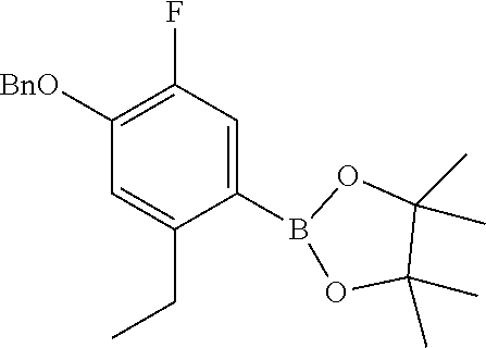
C00015

C00016

C00017

C00018

C00019

C00020

C00021

C00022

C00023

C00024

C00025

XML
uspto.report is an independent third-party trademark research tool that is not affiliated, endorsed, or sponsored by the United States Patent and Trademark Office (USPTO) or any other governmental organization. The information provided by uspto.report is based on publicly available data at the time of writing and is intended for informational purposes only.
While we strive to provide accurate and up-to-date information, we do not guarantee the accuracy, completeness, reliability, or suitability of the information displayed on this site. The use of this site is at your own risk. Any reliance you place on such information is therefore strictly at your own risk.
All official trademark data, including owner information, should be verified by visiting the official USPTO website at www.uspto.gov. This site is not intended to replace professional legal advice and should not be used as a substitute for consulting with a legal professional who is knowledgeable about trademark law.