Electrophilically enhanced phenolic compounds for treating inflammatory related diseases and disorders
Thompson April 6, 2
U.S. patent number 10,966,954 [Application Number 16/302,292] was granted by the patent office on 2021-04-06 for electrophilically enhanced phenolic compounds for treating inflammatory related diseases and disorders. This patent grant is currently assigned to GLOBAL BIOLIFE INC.. The grantee listed for this patent is GLOBAL BIOLIFE INC.. Invention is credited to Daryl Lee Thompson.


View All Diagrams
| United States Patent | 10,966,954 |
| Thompson | April 6, 2021 |
Electrophilically enhanced phenolic compounds for treating inflammatory related diseases and disorders
Abstract
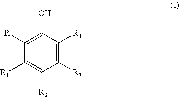
A therapeutic compound has a modified phenolic compound of the general formula (I) wherein at least one of R, R.sub.1, R.sub.2, R.sub.3, and R.sub.4 is an electrophilic group chosen from halogen, aldehyde, haloalkane, alkene, butyryl, flurophenol, sulfonamide, flurophenol sulfoxide and the remaining R, R.sub.1, R.sub.2, R.sub.3, and R.sub.4 is are each independently hydrogen, a hydroxyl group, an alkoxy group, a rutinosyl group, a carboxyl group, chromone, benzopyran, a rhamnosyl group, a substituted alkoxy group or a substituted acyloxy group wherein the substituent is chosen from hydroxyl, alkoxy, aryloxy, phenyl, halogen, and amido group. The compound can be used in a therapeutic treatment of inflammatory related diseases and condition. ##STR00001##
| Inventors: | Thompson; Daryl Lee (Winter Haven, FL) | ||||||||||
|---|---|---|---|---|---|---|---|---|---|---|---|
| Applicant: |
|
||||||||||
| Assignee: | GLOBAL BIOLIFE INC. (Bethesda,
MD) |
||||||||||
| Family ID: | 1000005467217 | ||||||||||
| Appl. No.: | 16/302,292 | ||||||||||
| Filed: | May 16, 2017 | ||||||||||
| PCT Filed: | May 16, 2017 | ||||||||||
| PCT No.: | PCT/US2017/032897 | ||||||||||
| 371(c)(1),(2),(4) Date: | November 16, 2018 | ||||||||||
| PCT Pub. No.: | WO2017/201042 | ||||||||||
| PCT Pub. Date: | November 23, 2017 |
Prior Publication Data
| Document Identifier | Publication Date | |
|---|---|---|
| US 20190343792 A1 | Nov 14, 2019 | |
Related U.S. Patent Documents
| Application Number | Filing Date | Patent Number | Issue Date | ||
|---|---|---|---|---|---|
| 15156021 | May 16, 2016 | 10123991 | |||
| Current U.S. Class: | 1/1 |
| Current CPC Class: | A61K 9/0014 (20130101); A61K 31/353 (20130101); A61K 31/7048 (20130101); A61K 9/0053 (20130101); A61K 31/4525 (20130101) |
| Current International Class: | C07D 311/32 (20060101); A61K 31/7048 (20060101); A61K 31/4525 (20060101); A61K 9/00 (20060101); A61K 31/352 (20060101); A61K 31/353 (20060101) |
| Field of Search: | ;549/403 ;514/456 |
References Cited [Referenced By]
U.S. Patent Documents
| 10123991 | November 2018 | Thompson |
| 2011/0142815 | June 2011 | Yu |
| 2012/0115896 | May 2012 | Bardelli et al. |
| 59046217 | Mar 1984 | JP | |||
| WO-03/060067 | Jul 2003 | WO | |||
Other References
|
Terao et al. "Prenylation modulates the bioavailability and bioaccumulation of dietary flavonoids", Archives of Biochemistry and Biophysics. 2014. vol. 559, pp. 12-16, entire document, especially: p. 14, Fig 1, quercetin; p. 14, Fig 2. cited by applicant . Nair et al. "Delivery of anti-inflammatory nutraceuticals by nanoparticles for the prevention and treatment of cancer", Biochem Pharmacol. 2010. vol. 80(12), pp. 1833-1843, entire document, especially: abstract; p. 10, para 3; p. 21, Table 1, hesperidin. cited by applicant . International Search Report for PCT/US17/32897, dated Jul. 26, 2017. cited by applicant . Written Opinion of the International Searching Authority for PCT/US17/32897, dated Jul. 26, 2017. cited by applicant . Office Action dated May 22, 2020 in corresponding Russian Patent Application No. 2018141590. cited by applicant . Extended European Search Report dated Jan. 8, 2020 in corresponding European Patent Application No. 17800008.9. cited by applicant . Park, Sehee, et al. "Aronia Melanocarpa and Its Components Demonstrate Antiviral Activity against Influenza Viruses." Biochemical and Biophysical Research Communications, vol. 440, No. 1, Sep. 5, 2013, pp. 14-19., doi:10.1016/j.bbrc.2013.08.090. cited by applicant . Garg, A., et al. "Chemistry and Pharmacology of the Citrus Bioflavonoid Hesperidin." Phytotherapy Research, vol. 15, No. 8, Dec. 1, 2001, pp. 655-669., doi:10.1002/ptr.1074. cited by applicant . Shailendra, Wadhwa, et al. "Bioavailability Enhancement by Piperine: A Review", Asian Journal of Biomedical and Pharmaceuticals Sciences, vol. 04, No. 2014, Oct. 15, 2014, pp. 1-8, XP055461844, doi: 10.15272/ajbps.v4i36.576. cited by applicant . Examination Report dated Nov. 6, 2020 in Indian Application No. 201817043242. cited by applicant . Notification of Refusal dated Jan. 26, 2021 in Japanese Application No. 2018-560998. cited by applicant. |
Primary Examiner: Aulakh; Charanjit
Attorney, Agent or Firm: Weyer, Esq.; Stephen J. Stites & Harbison, PLLC
Claims
What is claimed is:
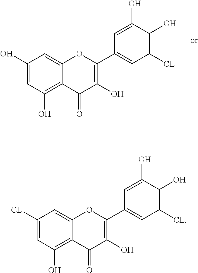
1. A compound having a formula selected from the group consisting of: ##STR00009##
2. A pharmaceutically composition comprising the compound of claim 1 incorporated into a conventional non-invasive systemic dosage form.
3. The pharmaceutically composition of claim 2, wherein the non-invasive systemic dosage form is selected from the group consisting of a capsule, pill, tablet, specialty tablet including buccal, sublingual, and oral disintegrating, thin film, elixir, liquid solution or suspension, powder, or crystals.
4. The compound of claim 1, incorporated into a cream, gel, liniment, balm, lotion, ointment, or skin patch.
5. A method of treating inflammatory related diseases comprising administering to a subject in need thereof a therapeutically effective amount of a compound of claim 1.
6. A method of treating a cancerous disease condition comprising administering a therapeutically effective amount of the compound of claim 1 to patient in need of treatment therefrom.
7. A method of treating an inflammatory/pain disease condition comprising administering a therapeutically effective amount of the compound of claim 1 to patient in need of treatment therefrom.
8. A method of treating a virological/bacteriological disease condition comprising administering a therapeutically effective amount of the compound of claim 1 to patient in need of treatment therefrom.
9. The composition of claim 2, formulated into a time release formulation using liposomes, drug polymer conjugates, hydrogels, microspheres or microencapsulation.
10. The method of claim 5, wherein the compound is further in combination with hesperidin and piperine.
11. The method of claim 10, wherein the compound is formulated with hesperidin and piperine to form equivir.
Description
CROSS REFERENCE TO RELATED APPLICATION
This application claims the benefit of U.S. non-provisional patent application Ser. No. 15/156,021, filed on May 16, 2016, herein incorporated by reference.
FIELD OF INVENTION
The present invention relates to a electrophilically enhanced phenolic compounds which include but are not limited to flavonoid compounds and use of these compounds for treating various disorders including disorders which have an inflammatory nexus including inflammatory related diseases and disorders.
BACKGROUND OF THE INVENTION
Metabolic and enzymatic systems of organisms have evolved to incorporate phenolics, phenolic acids, and flavonoids, readily available in the environment from plant food sources, into the associated biochemical pathways of every major metabolic system. Not only do flavonoids have a physiological impact on a multitude of metabolic systems, but they are inherently part of the normal operation of such systems. As part of natural evolution, a flavonoid binding site has been conserved and distributed universally across these pathways. When a chemical reaction is overloaded with reagents, such as excessive glucose, or if there is an impairment of a metabolic component, such as a deformity of a protein, intermediate metabolites build to a concentration which can lead to the dysfunction of the pathway or associated pathways and the expression of a disease condition. A major role of flavonoids is to down and or up regulate certain enzymes to reduce the accumulation of these intermediates, ameliorating their negative effect on the organism.
Because of the distribution of a flavonoid binding site across several pathways, flavonoids have a regulatory impact on several enzymes in systems associated with disease conditions. Most other therapeutics for a disease condition target one to two target enzymes associated with a disease, while the flavonoid targets several.
Disease Classes
There are sixty-four (64) key enzymes distributed across six (6) disease categories. Most drugs used to treat these diseases either down-regulate or up-regulate a target enzyme associated with these diseases.
A listing of each major disease category and the target enzymes associated with such diseases follows.
Cancer
TARGET ENZYMES: Inositol 1,4,5 Triphosphate Kinase (IP3K), Protein Kinase CK2, DNA Topoisomerase II, Mitogen Activated Protein Kinase, CYP 181, Kinase 1 (MEK), MAPK Kinase 4 (MKK4), Glycoxalase, CYP 1A1, Cyclooxygenase, Thioredoxin Reductase, Nuclear Factor Kappa B (NF-Kappa B), Tyrosine Kinase (MET), Extracellular Signal Regulated Kinase (ERK 1-2), HSP 70 ATPase, Xanthine Oxidase, Phostphatidylinositol-3, RAF Kinase, Matrix Metallo Proteinase 1-18, MRP-1, Urokinase, 3-Phosphoglycerate Kinase, Aromatase (CYP-19), X Protein Kinase (XK), Ornithine Decarboxylase (ODC), and Inositol Polyphosphate Multi Kinase (IPMK).
Diabetes and Metabolic Syndrome (Diabetes Mellitus Type 1, Diabetes Mellitus Type 2, Gestational Diabetes, Metabolic Syndrome, Obesity, Glycogen Storage Disorder)
TARGET ENZYMES: Pyruvate Carboxylase, Fatty Acid Synthase, Alpha Amylase, Mitogen Activated Protein Kinase, MAP K (4), Cholesterol Acyl Transferase, Liver Xanthine Oxidase, Glucokinase, Extracellular Regulated Enzyme (ERK), Aldose Reductase, Lipase, Acetyl Co A, Kinase (1), Glucosidase, Angiotensin Converting Enzyme (ACE), 12-Lipo-oxygenase, and HMG-CoA, Kinase (MEK).
Cardiovascular Disease (Hypercholesterolemia, Ischemia, Hypertension, Endocarditis, Myocarditis, Cerebrovascular Disorder, Vascular Permeability, Ischemia)
TARGET ENZYMES: Aceto Acetyl CoA, Squalene Synthase, Oxidosqualene Cyclase, Angiotensin Converting Enzyme, Acetyl CoA, HMG CoA reductase, HMG CoA, and Cholesterol Transferase.
Inflammation and Pain (Acute Pain Disorder, Migraine, Headache, Arthritis, Psoriasis, Asthma, Alcohol Toxicity, Crohn's Disease, Inflammatory Bowel Syndrome, Colitis, Irritated Bowel Syndrome, Rosacea)
TARGET ENZYMES: Glyoxalase 1, Glyoxalase 2, Kinase (1), Polymerase (PARP-1), Mitogen Activated Protein Kinase (MAP K), Nuclear Enzyme Polymerase (ADP Ribose), Caspase 8, MAP K (4), MKK4, Elastase, COX 1, and COX 2.
Neurological Diseases (Alzheimer's Disease, Huntington's Disease, Schizophrenia, Traumatic Brain Injury, Parkinson's)
TARGET ENZYMES: Tyrosine Kinase, Alpha Secretase, Caspase 3, Protease, Beta Site APP Cleaving Enzyme 1 (BACE-1), Kinase (PLK2), Tau Protein Kinase, Beta Secretase, Gamma Secretase, Intercellular Adhesion Molecule 1 (ICAM-1), Glutathione transferase, M(TOR), Dihydrofolate reductase, Serine protease, cysteine protease, Metallo protease, and Aspartic protease.
Virological/Bacteriological Diseases (Human Rhinovirus, Influenza, SARS, MERS, Ebola, Dengue, Malaria, MRSA, Staph, E. coli, HIV)
TARGET ENZYMES: Reverse transcriptase, RNA Polymerase (all), Helicase, Topoisomerase, Integrase, Protease (viral budding), Caspase (especially caspase 8), Allantoinase, Dihydroorotase, Butyryl Cholinesterase, and Acetyl Cholinesterase.
Specific Example of One Disease: Diabetes
Type 1 Diabetes (formerly known as Juvenile Diabetes) is characterized by an inability of the body to produce or release insulin. Type 2 Diabetes (also known as Adult Onset Diabetes) is characterized by the body's inability to respond to insulin, or being insulin resistant. Both result in hyperglycemia, often followed by complications such as cardiovascular disease, retinopathy, neuropathy, and nephropathy. Type 1 Diabetes requires the administration of exogenous insulin. Type 2 Diabetes can often be initially controlled by diet alone, but usually requires treatment with antihyperglycemic agents. The most widely prescribed drug for the control of Type 2 Diabetes is the hydrochloride salt of metformin, a biguanide. Such compounds work by inhibiting liver glucose output (gluconeogenesis) and increasing the uptake of glucose by peripheral tissue (such as muscle) by stimulating glucose transporter 4 (GLUT4). A list of drugs and their mode of action is shown in Table 1.
TABLE-US-00001 TABLE 1 CLASS MODE AGENT sulfonylureas stimulate insulin release through tolbutamide activation of Ca & K channels acetohexamide tolazamide chlorpropamide glipizide glyburide glimepiride gliclazide meglitinides stimulate insulin release through repaglinide activation of Ca & K channels nateglinide biguanides inhibit gluconeogenesis, stimulate metformin GLUT4 phenformin buformin thiazolidinediones bind to PPARs, regulating adipocyte rosiglitazone differentiation, FA & glucose uptake. pioglitazone troglitazone .alpha.-glucosidase inhibit conversion of starch to miglitol inhibitor glucose acarbose GLP analogues/ bind to GLP receptor, causing insulin exenatide agonists release liraglutide taspoglutide DPP-4 inhibitors inhibit degradation of GLP, indirectly vildagliptin causing insulin release sitagliptin saxagliptin amylin analogues inhibits gluconeogenesis, gastric pramlintide enzymes, & gastric emptying
The leading cause of non-compliance associated with the most widely prescribed antidiabetic drugs is gastro-intestinal discomfort. In particular, .alpha.-glucosidase inhibitors and amylin analogues work by slowing or inhibiting the conversion of complex carbohydrates and fats into readily absorbable simpler molecules. This leaves an environment rich in carbohydrates and lipids, readily available to intestinal micro flora. The result is often diarrhea, flatulence, inflammation, and deterioration of the intestinal lining.
Flavonoids have the basic structure:
##STR00002## and are classified according to IUPC (International Union of Pure and Applied Chemistry) as flavonoids (examples include quercetin and rutin), isoflavonoids (examples include daidzein and genistein), or neoflavonoids (examples include dalbergin and neoflavan).
Flavonoids are effective enzymatic inhibitors or stimulators according to the 1) number of hydroxyl units borne on their phenolic rings, 2) planarity of the molecule, and 3) position of the units on the rings. Planarity and position dictate docking potential. In general, the more hydroxyl units, the more reactive the compound is. At the same time, an increase in reactivity denotes a decrease in stability. The greater the number of hydroxyl units, the more immediate the effect. Fewer units lead to delayed effects. A formulation of flavonoids each with varying numbers of hydroxyl units may be employed as a method to elicit multiple effects. This would be accomplished by alternating ratios of different flavonoids with different numbers and hydroxyl unit orientations.
Scientific literature is replete with the effect of flavonoids on lowering blood glucose via inhibition of .alpha.-glucosidase, inhibition of glucose transporter 2 (glucose transport from the gut to the bloodstream), stimulation of glucose transporter 4 (glucose transport from the bloodstream to muscle tissue), stimulation of insulin release, inhibition of gluconeogenesis, inhibition of adipocyte growth and development, stimulation of lipolysis, inhibition of fatty acid synthesis, and increased insulin sensitivity. Flavonoids have been reported to inhibit the activity of Helicobacter pylori, a major contributor to gastro-intestinal distress, as well as several other flora of the gastro-intestinal tract. In addition, flavonoids show anti-inflammatory activity and support healing of the intestinal lining.
SUMMARY OF THE INVENTION
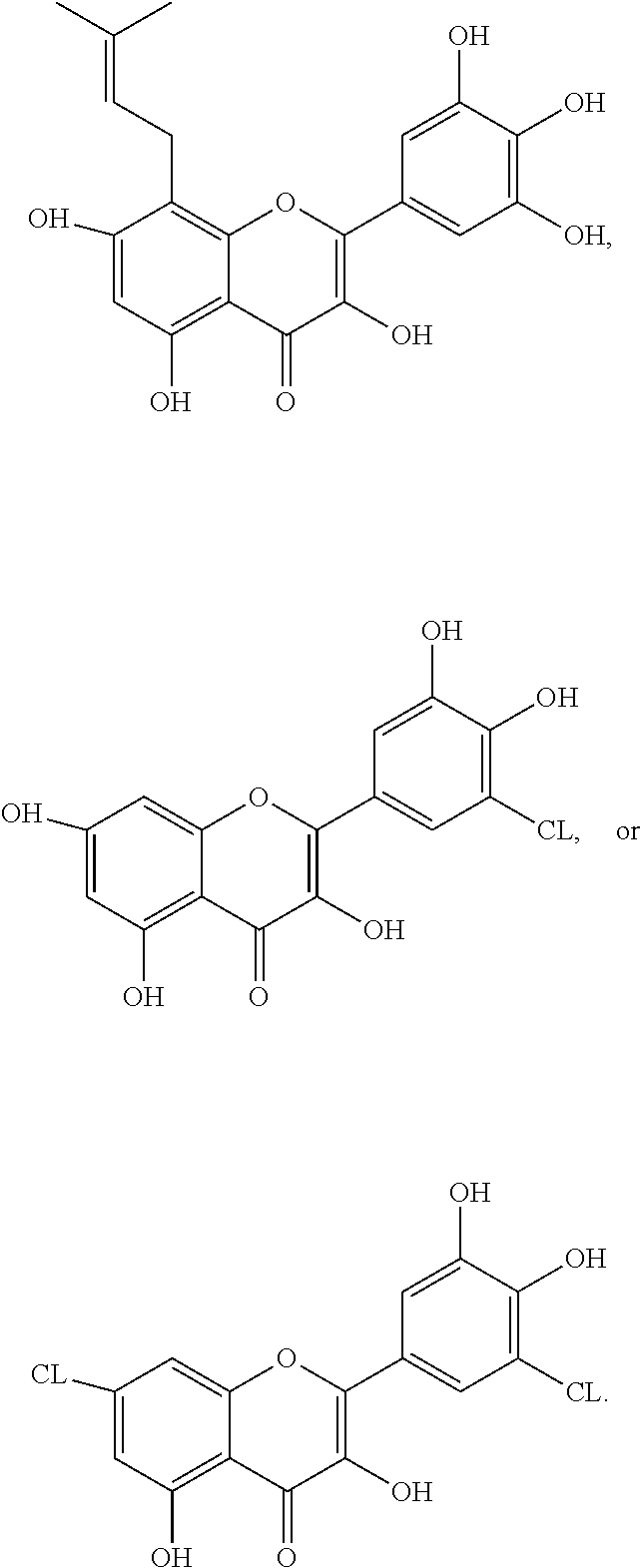
The present invention relates to electrophilically enhanced phenolic compounds. The electrophilically enhanced phenolic compounds, include but are not limited to, flavonoid compounds. The compounds can be used to treat various disorders including disorders which have an inflammatory nexus including inflammatory related diseases and disorders. One, none limiting example is an electrophilically enhanced myricetine. Electrophilically enhancing such flavonoid increases its binding activity, lowers the Km, thus increasing its efficacy.
In one form a composition includes the addition of an electrophilic moiety via covalent bonding to a phenolic compound such as a flavonoid which elicits an increased docking energy, increasing up or down regulation of enzymes relating to six (6) inflammatory-related disease categories (identified in this disclosure below), and/or increasing the efficacy of a flavonoid over its native state. These compounds advantageously are effective for treating inflammatory related diseases and disorders. These disorders include the above-identified six (6) disease categories which have an inflammatory component, genesis or nexus. The electrophilically enhanced phenolic compounds have a general formula (I)
##STR00003##
wherein at least one of R, R.sub.1, R.sub.2, R.sub.3, and R.sub.4 is an electrophilic group chosen from halogen, aldehyde, haloalkane, alkene, butyryl, flurophenol, sulfonamide, flurophenol sulfoxide and the remaining R, R.sub.1, R.sub.2, R.sub.3, and R.sub.4 is are each independently hydrogen, a hydroxyl group, an alkoxy group, a rutinosyl group, a carboxyl group, chromone, benzopyran, a rhamnosyl group, a substituted alkoxy group or a substituted acyloxy group wherein the substituent is chosen from hydroxyl, alkoxy, aryloxy, phenyl, halogen, and amido group. In an advantageous form, the electrophilically enhanced phenolic compound has increased efficacy over its native form when administered to a patient in need of treatment of an inflammatory disease or related disorder. One example in electrophilically enhanced myricetin in which myricetin is substituted with one or more halogens such as chlorine.
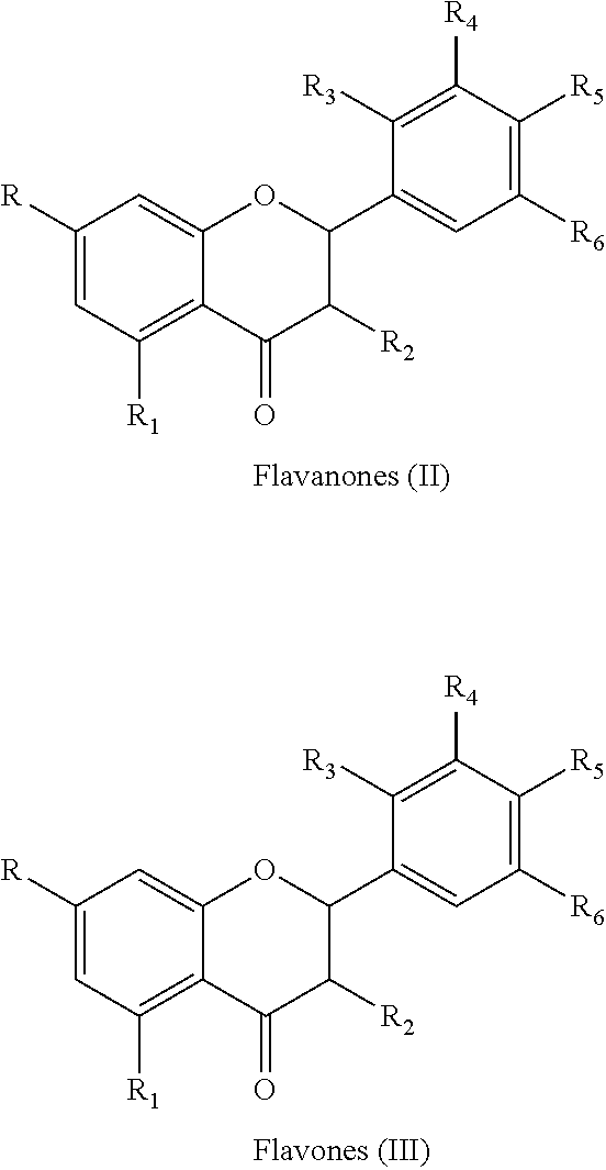
In one advantageous form, R.sub.1 is a chromone having the general flavonoid formulas (II) and (II)
##STR00004##
wherein at least one of R, R.sub.1, R.sub.2, R.sub.3, R.sub.4, R.sub.5, and R.sub.6 is an electrophilic group chosen from halogen, aldehyde, haloalkane, alkene, butyryl, flurophenol, sulfonamide, flurophenol sulfoxide and the remaining R, R.sub.1, R.sub.2, R.sub.3, R.sub.4, R.sub.5, and R.sub.6 are each independently hydrogen, a hydroxyl group, an alkoxy group, a rutinosyl group, a rhamnosyl group, a substituted alkoxy group or a substituted acyloxy group wherein the substituent is chosen from hydroxyl, alkoxy, aryloxy, phenyl, halogen, and amido group. In advantageous form, the compound has increased efficacy over its native, non-electrophilically enhanced form, where the enhanced efficacy is based on its ability to treat target diseases and conditions in accordance with this disclosure.
Still in a further form, the flavonoid is selected from compounds derived from 2-phenylchromen-4-one. Still further, in yet an additional embodiment, the flavonoid includes compounds derived from 3-hydroxy-2-phenylchromen-4-one such as, but not limited to, fisiten, galangin, hesperitin, isorhamnetin, kaempferol, myricetin, naringin, quercetin, and rutin.
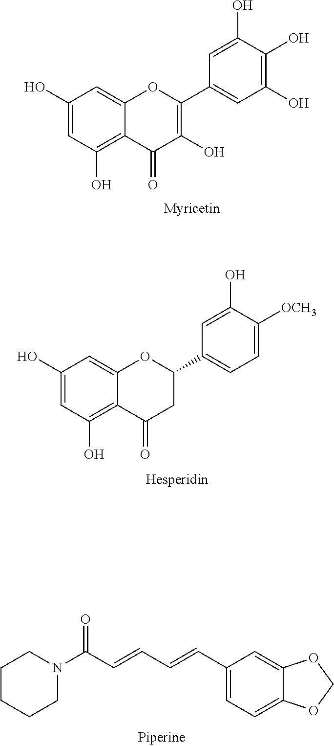
The present invention, in another form thereof relates to a method for treating diseases or medical conditions, which includes but is not limited to inflammatory related diseases which includes administering to a patient in need of treatment therefrom a therapeutically effective amount of a compound of general formula (I) or any of the further refined species of formula (I) presented in this disclosure. In alternative methods, the treatment may include administering any of the aforementioned specific forms of the general formula (I) including but not limited to those of fisiten, galangin, hesperitin, isorhamnetin, kaempferol, myricetin, naringin, quercetin, and rutin. In still alternative methods, the compound of formula (I) (a phenolic compound) is a) myricetin or b) an electrophilically enhanced, modified form of myricetin, and the compound is further in combination with hesperidin and piperine. In one form, myricetin is formulated with hesperidin and piperine to form equivir.
In accordance with the present invention, a combination of compounds, myricetin, hesperidin and piperine form a composition known as Equivir. The myricetin can be native or electrophically enhanced.
##STR00005##
Equivir, in accordance with this disclosure has efficacy to treat various disorders, in accordance with this disclosure which include, but are not limited to dengue fever/dengue virus, influenza, rhinovirus, and ebola virus.
In one advantageous form, the compound having a formula selected from the group consisting of:
##STR00006##
The compound can be formulated into a pharmaceutically composition as a conventional non-invasive systemic dosage form. The non-invasive systemic dosage form can be selected from the group consisting of a capsule, pill, tablet, specialty tablet including buccal, sublingual, and oral disintegrating, thin film, elixir, liquid solution or suspension, powder, or crystals. The compound can also be incorporated into a cream, gel, liniment, balm, lotion, ointment, or skin patch. The aforementioned three compounds can be used to treat various medical conditions, disorders and/or disease. These include, but are not limited to inflammatory related disease, cancerous disease conditions, diabetic/metabolic syndrome disease conditions, a cardiovascular disease conditions, inflammatory/pain disease conditions, neurological disease conditions, and virological/bacteriological disease conditions. The disease condition(s) can be in human and non-humans.
Pharmaceutical compound of the aforementioned three compounds can include physiologically acceptable filler, disintegrants, carrier material, excipient, lubricant, buffer, antibacterial, bulking agent and/or binder, and an anti-oxidant. The disintegrants can be selected from the group consisting of croscarmellose sodium, povidone, crospovidone, sodium starch glycolate, corn starch or microcrystalline cellulose. Further, the composition can be formulated into a time release formulation using liposomes, drug polymer conjugates, hydrogels, microspheres or microencapsulation.
In one particularly advantageous form, the composition comprises an electrophilically enhanced, modified form of myricetin, and the compound is further in combination with hesperidin and piperine. In one example, the electrophilically enhanced, modified form of myricetin (e.g. chlorinated form), is formulated with hesperidin and piperine to form equivir.
BRIEF DESCRIPTION OF THE DRAWINGS
FIG. 1 compares percent of MRC-5 Viability with percent concentration in which percent inhibition of Rhinovirus type 14 is shown following treatment with the test compound, post-infection application.
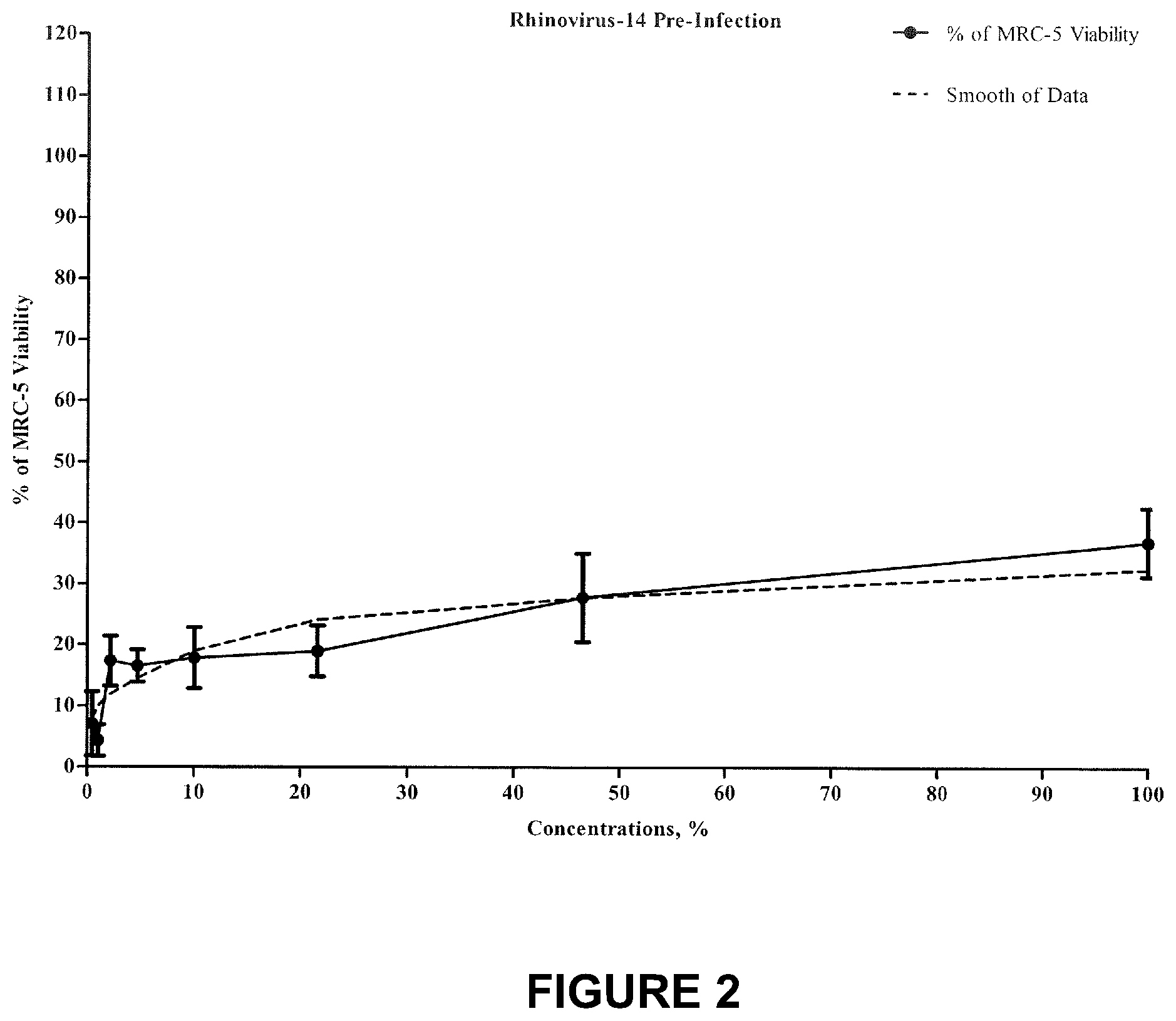
FIG. 2 shows percent inhibition of Rhinovirus type 14 following treatment with the test compound, pre-infection application.
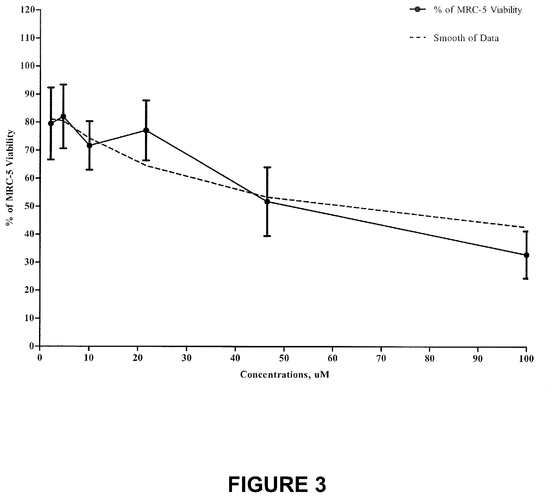
FIG. 3 is a chart showing percent of MRC-5 viability following treatment with the test compound.
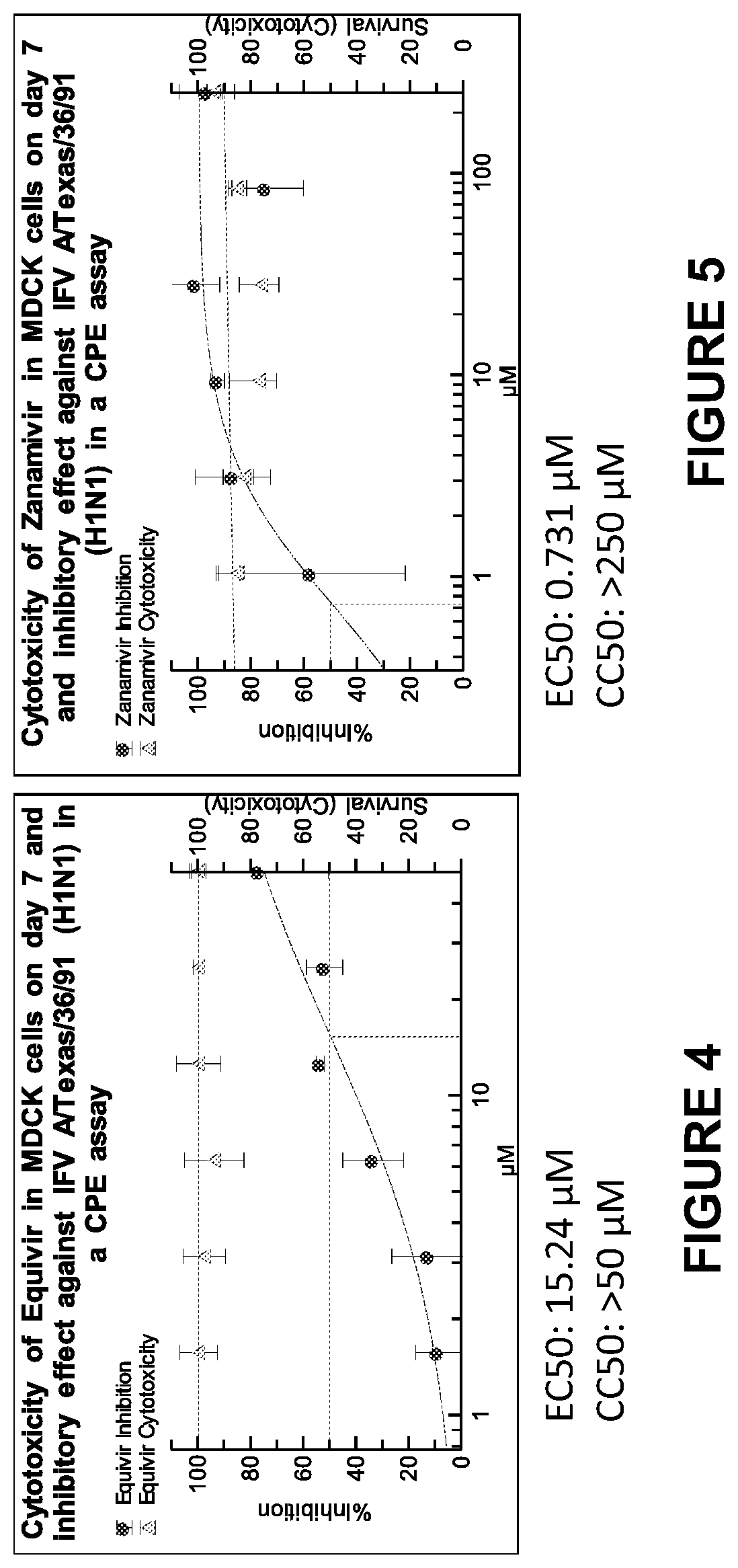
FIG. 4 shows cytotoxicity of Equivir in MDCK cells on day 7 and inhibitory effect against IFVA/Texas/36/91(H1N1) in a CPE assay.
FIG. 5 depicts a toxicity of Zanamivir IR in MDCK cells on day 7 and inhibitory effect against IFV A/Texas/36/91 (H1N1) in a CPE assay.
FIG. 6 shows the cytotoxicity of Equivir in MDCK cells on day 7 and inhibitory effect against IFV A/Perth/265/09 in a CPE assay.
FIG. 7 is a graph showing cytotoxicity of Zanamivir in MDCK cells on day 7 and inhibitory effect against IFV A/Perth/265/09 in a CPE assay.
FIG. 8 is a graph showing cytotoxicity of Equivir in MDCK cells on day 7 and inhibitory effect against IFV A/HK/1/68 (H3N2) in a CPE assay.
FIG. 9 is a graph showing cytotoxicity of Zanamivir in MDCK cells on day 7 and inhibitory effect against IFV A/HK/1/68 (H3N2) in a CPE assay.
FIG. 10 is a chart showing inhibition of DENV-2 in dendritic cells.
FIG. 11 comprises a series of six panels directed to dendritic cell characterization in accordance with the present invention.
FIG. 12 is a chart showing cytokine profile in DCs.
FIG. 13 is a chart showing the effect of Equivir on Infection with WT Ebola virus in HepG2 cells.
FIG. 14 is a chart showing the effect of T-705 on Infection with WT Ebola virus in HepG2 cells.
FIG. 15 is a chart showing the effect of Equivir on infection with WT Ebola virus in THP-1 cells.
FIG. 16 is a chart showing the effect of T-705 on infection with WT Ebola virus in THP-1 cells.
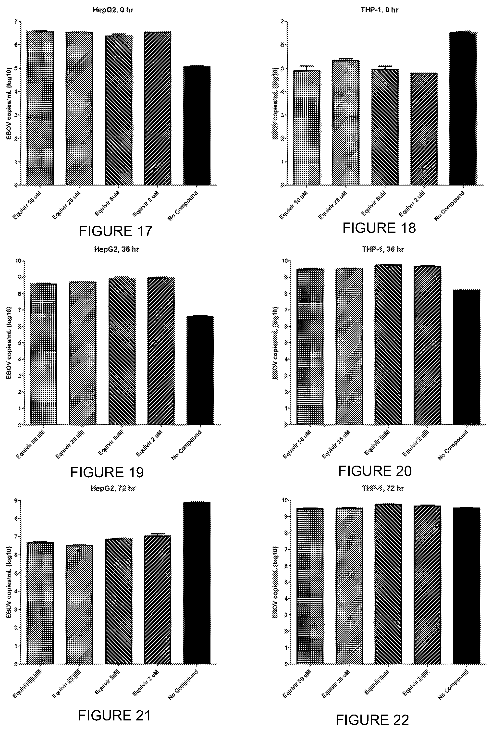
FIG. 17 is a chart with experimental data for anti-EBOV activity in HepG2 shown at zero hours.
FIG. 18 is a chart with experimental data for anti-EBOV activity in THP-1 shown at zero hours.
FIG. 19 is a chart with experimental data for anti-EBOV activity in HepG2 shown at 36 hours.
FIG. 20 is a chart with experimental data for anti-EBOV activity in THP-1 shown at 36 hours.
FIG. 21 is a chart with experimental data for anti-EBOV activity in HepG2 cells, shown at 72 hours.
FIG. 22 is a chart with experimental data for anti-EBOV activity in THP-1 shown at 72 hours.
DETAILED DESCRIPTION
The present invention will now be described with regard to specific embodiments and exemplar examples to provide a further understanding of the present compound and its use to treat various disorders.
In one advantageous form, a therapeutic compound has a modified phenolic compound of the general formula (I)
##STR00007## wherein at least one of R, R.sub.1, R.sub.2, R.sub.3, and R.sub.4 is an electrophilic group chosen from halogen, aldehyde, haloalkane, alkene, butyryl, flurophenol, sulfonamide, flurophenol sulfoxide and the remaining R, R.sub.1, R.sub.2, R.sub.3, and R.sub.4 is are each independently hydrogen, a hydroxyl group, an alkoxy group, a rutinosyl group, a carboxyl group, chromone, benzopyran, a rhamnosyl group, a substituted alkoxy group or a substituted acyloxy group wherein the substituent is chosen from hydroxyl, alkoxy, aryloxy, phenyl, halogen, and amido group, said compound having increased efficacy over its native form.
The therapeutic compound can be used to treat various diseases having an inflammatory component, genesis or nexus including, but not limited to, cancer, diabetic/metabolic syndrome disease conditions, cardiovascular disease, inflammatory/pain, neurological disease, and virological/biological disease conditions.
The therapeutic compound can be formulated as a pharmaceutical composition and may be further modified by a non-evasive systematic dosage form which include but is not limited to a capsule, pill, tablet, specialty tablet (buccal, sublingual, and oral disintegrating), thin film, elixir, liquid solution or suspension, powder, or crystals.
The composition may include a pharmaceutical carrier carried by suitable pharmaceutical carrier which includes any of a series of physical forms and can include a wide variety of pharmaceutically acceptable carriers, diluents and/or excipients that are well known in the art. Accordingly, the suitable pharmaceutical carrier may include solvents, coatings, antibacterial and antifungal agents, isotonic and absorption delaying agents and the like. By "pharmaceutically acceptable" is generally understood to mean that said carrier is substantially compatible with the active ingredient or other ingredients in the composition or formulation and is substantially not deleterious to a patient undergoing treatment thereof. General examples of suitable carriers include magnesium carbonate, magnesium stearate, talc, sugar, lactose, pectin, dextrin, starch, gelatin, tragacanth, methylcellulose, sodium carboxymethyl cellulose, a low melting wax, cocoa butter, and the like.
Depending on the nature of the present composition and its formulation, administering the present compound/composition in accordance with this disclosure can be through transmucosal absorption, incorporated into nasal, buccal/sublingual, vaginal, ocular, rectal (suppository, enema), and inhalation (aerosol, inhaler, nebulizer, vaporizer, smoked) applications.
A preferred dose for administering a compound of general formula (I) and a pharmaceutical composition in accordance with the present disclosure is an amount which limits the occurrence of a condition, reduces symptoms associated with the disease condition, and/or prevents the occurrence for a finite time. One of ordinary skill in the art will readily recognize that the amount will vary greatly depending on the nature of the condition of a patient. "An effective amount" or "therapeutically effective amount" of the compound of general formula (I) or a pharmaceutical composition including the compound of general formula (I) in accordance with the present disclosure is intended to mean any non-toxic but sufficient amount of the compound, composition pharmaceutical composition or anything that produces the desired prophylactic or therapeutic effect. Thus, as one of ordinary skill in the art readily understands, the exact amount of the compound, pharmaceutical composition or particular agent that is required will vary from subject to subject depending on the species of the patient, age, and general condition of the subject, severity of the condition being treated, the particular carrier or adjuvant being used and its mode of administration and the like. Similarly, dosing regimen should also be adjusted to suit the patient to whom the composition is administered and will vary again with age, weight, metabolism, etc. of the subject. Therefore, the "effective amount" of any particular compound, composition or agent will vary based on particular circumstances, and an appropriate effect amount may be determined in each case of application by one of ordinary skill in the art using only routine experimentation. Depending on the nature of the present composition and its formulation, administering the present compound can be through transmucosal absorption, incorporated into nasal, buccal/sublingual, vaginal, ocular, rectal (suppository, enema), and inhalation (aerosol, inhaler, nebulizer, vaporizer, smoked) applications.
A combination of compounds, myricetin, hesperidin and piperine form a composition known as Equivir. Equivir, in accordance with this disclosure has efficacy to treat various disorders in accordance with the present disclosure which include, but are not limited to dengue fever/dengue virus, influenza, rhinovirus, and ebola virus.
##STR00008##
A chlorinated flavonoid according to this disclosure can be incorporated into a conventional non-invasive systemic dosage form, such as a capsule, pill, tablet, specialty tablet (buccal, sublingual, oral disintegrating), thin film, elixir, liquid solution or suspension, powder, or crystals. The above dosage forms will also include the necessary physiologically acceptable filler, disintegrants (such as croscarmellose sodium, povidone, crospovidone, sodium starch glycolate, corn starch or microcrystalline cellulose), carrier material, excipient, lubricant (such as magnesium stearate, stearic acid, palmitic acid, calcium stearate, talc, carnauba wax), buffer, antibacterial, bulking agent and/or binder (such as lactose, sugar, gum acacia, corn starch, modified corn starch, polyvinylpyrrolidone, mannitol, sorbitol, inorganic salts such as calcium carbonate and/or cellulose derivatives such as wood cellulose, microcrystalline cellulose, carnauba wax, paraffin, spermaceti, polyethylenes or microcrystalline wax), anti-oxidants (ascorbic acid or sodium bisulfate) or similar. A chlorinated flavonoid according to this disclosure can be administered by incorporation into a cream, gel, liniment, balm, lotion, ointment, or skin patch.
A chlorinated flavonoid according to this disclosure can be administered through transmucosal absorption, incorporated into nasal, buccal/sublingual, vaginal (douche, pessary), ocular, rectal (suppository, enema), and inhalation (aerosol, inhaler, nebulizer, vaporizer, smoked) applications. A chlorinated flavonoid according to this disclosure can be administered through intradermal, subcutaneous, intramuscular, intraosseous, intraparitoneal, or intravenous injection. A chlorinated flavonoid according to this disclosure can be administered through a time-release formulation by incorporating liposomes, drug polymer conjugates, hydrogels, or microspheres.
EXEMPLAR EXAMPLES
The following examples are provided for exemplary purposes to enhance understanding of the present composition and treatment. From the example, one can extrapolate compounds, therapeutic compositions and therapeutic treatments in accordance with the disclosure.
Example 1
According to this disclosure, one or more flavonoids such as for example myricetin, is chlorinated. The compound can be employed individually, or can be combined in a single formulation, for example as a capsule, liquid solution or suspension, syrup, tablet, powder, as well as controlled release formulations.
In a preferred embodiment, 1 to 1000 mg of a chlorinated flavonoid is administered orally in capsule form.
Example 2
A chlorinated flavonoid according to this disclosure can be administered to various mammalian species, such as domesticated animals, horses, cows, sheep, pigs, dogs, cats, humans, etc., in need of such treatment.
The following example is entitled "Non-GLP Evaluation of One Test Compound for Antiviral Activity Versus Rhinovirus 14".
The test product was prepared as follows: 0.312 g of the test product was dissolved in 10 mL DMSO to obtain 100 mM concentration of the test product; 1/1000 dilution of 100 .mu.M test product was prepared in Maintenance Medium to obtain 100 uM concentration; subsequent serial dilutions were made in Maintenance Medium using the progression factor of 2.15. Cytotoxicity of 1/1000 dilution of DMSO in medium was evaluated. A total of eight different concentrations of the test product were evaluated for antiviral properties versus Rhinovirus type 14 strain 1059 (ATCC #VR-284). Post-infection and Pre-infection antiviral activity of the test product was evaluated. Plating was performed in 6 replicates, testing was conducted in duplicate.
Test Methodology:
Test Product: Test Product descriptions are provided in Table 2. Initial Test Product was prepared in DMSO and subsequent concentrations in cell culture medium.
TABLE-US-00002 TABLE 2 Manufactured Lot Number Exp. Date Date Product Name/Description 1532 March 2017 March 2015 Equivir Myricetin, hesperetin and piperine; small glass vial with black lid containing dark yellow powder
Virus:
Human Rhinovirus type 14 strain 1059 (ATCC #VR-284). Virus dose: 0.1 MOI.
Host Cells:
MRC-5 (Human lung fibroblasts, ATCC 11CCL-171).
Medium:
Maintenance Medium--EMEM (ATCC) with 2% Fetal Bovine Serum (Atlas) and Anti-Anti (penicillin-streptomycin-amphotericin B [Gibco]).
MTT Cell Proliferation Assay:
ATCC #30-1010K.
Methodology:
The antiviral properties and cytotoxicity were assessed using a Cytopathic Effect (CPE)-based assay. CPE was determined using MTT cell proliferation assay.
Post-infection antiviral activity: Approximately 90% confluent cells were washed with PBS. 100 ul aliquots of 0.1 MOI test virus (medium for the cytotoxicity test) were added to the cells and incubated at 33.degree. C..+-.2.degree. C. in a CO.sub.2 incubator for 1 hour for virus adsorption. After incubation, the virus inoculum was removed; infected cells washed with PBS and overlaid with 100 .mu.l of the test products concentrations. The plates were incubated in a CO.sub.2 incubator for 72 hours. Upon completion of incubation, the plates were evaluated for CPE inhibition using MTT assay: cells were washed with PBS; 100 .mu.l of medium and 10 .mu.l of MTT reagent added to each well of the plate; plates were returned to the incubator and incubated for 4 hours at 37.degree. C..+-.2.degree. C.; following incubation, medium was removed and 100 .mu.l of DMSO added to dissolve purple formazan. Pre-infection antiviral activity: Approximately 90% confluent cells were washed with PBS. 100 .mu.l aliquots of the test product concentrations were added to the cells and incubated at 37.degree. C..+-.2.degree. C. in a CO.sub.2 incubator for 1 hour. After incubation, 100 .mu.l aliquots of 0.1 MOI test virus were added and incubated at 33.degree. C..+-.2.degree. C. in a CO.sub.2 incubator for 72 hours. Upon completion of incubation, the plates were evaluated for CPE inhibition using MTT assay: cells were washed with PBS; 100 .mu.l of medium and 10 .mu.l of MTT reagent added to each well of the plate; plates were returned to the incubator and incubated for 4 hours at 37.degree. C..+-.2.degree. C.; following incubation, medium was removed and 100 .mu.l of DMSO added to dissolve purple formazan.
The optical density of the samples was determined using the "VERSAmax" Tunable Microplate Reader (with SOFTmax.RTM. PRO Software) set at 570 nm, with blank wells. The results were calculated as percent inhibition of CPE where 100% inhibition of CPE is approximately equal to the mean of the cell control. IC50 values are presented in .mu.M concentrations and were calculated using non-liner regression analysis, GraphPad Prism 5.0 software.
Table 3 presents the summary of inhibitory concentrations.
TABLE-US-00003 TABLE 3 Summary for Inhibitory Concentrations of the Test Product versus Rhinovirus type 14 and Toxic Concentrations for MRC-5 Cells Cytotoxicity, IC50, .mu.M IC90, .mu.M TC50 .mu.M Test Product Best Fit Best Fit Best Fit Application Value 95% CI Value 95% CI Value 95% CI Post-Infection 9.350 8.559 to 10.21 63.250 49.78 to 80.36 52.21 39.62 to 68.82 Pre-Infection 549.8 332.5 to 909.1 206740 54522 to 783930 52.21 39.62 to 68.82
Tables 4 and 5, and FIG. 1 present Inhibitory Concentrations (IC) for Post-Infection Application of the Test Product.
TABLE-US-00004 TABLE 4 Inhibitory Concentration 50% (1050) for the Post-Infection Product Application (GraphPad Prism 5.0 log(inhibitor) vs. response -- Variable slope (four parameters)) log(inhibitor) vs. response -- Variable slope (four parameters) Results Best-fit values Bottom =0.0 Top =100.0 LogIC50 0.9708 HillSlope 1.149 IC50 9.350 Span =100.0 Std. Error LogIC50 0.01923 Hill Slope 0.06127 95% Confidence Intervals LogIC50 0.9324 to 1.009 Hil Slope 1.027 to 1.272 IC50 8.559 to 10.21 Goodness of Fit Degrees of Freedom 70 R square 0.9295 Absolute Sum of Squares 3535 Sy x 7.106 Constraints Bottom Bottom = 0.0 Top Top = 100.0
TABLE-US-00005 TABLE 5 Inhibitory Concentration 90% (IC90) for the Post- Infection Product Application (GraphPad Prism 5.0) log(agonist) vs. response - Find ECanything Results Best-fit values .cndot. logECF 1.801 HillSlope 1.149 F =90.00 Bottom =0.0 Top =100.0 ECF 63.25 Span =100.0 Std. Error logECF 0.05209 Hi I IS lope 0.06127 95% Confidence Intervals logECF 1.697 to 1.905 HillSlope 1.027 to 1.272 ECF 49.78 to 80.36 Goodness of Fit Degrees of Freedom 70 R square 0.9295 Absolute Sum of Squares 3535 Sy x 7.106 Constraints F F = 90.00 Bottom Bottom = 0.0 Top Top = 100.0 Number of points Analyzed 72
Tables 6 and 7, and FIG. 2 present Inhibitory Concentrations (IC) for Pre-Infection Application of the Test Product.
TABLE-US-00006 TABLE 6 Inhibitory Concentration 50% (IC50) for the Pre-Infection Product Application (GraphPad Prism 5.0 log(inhibitor) vs. response -- Variable slope (four parameters)) log(inhibitor) vs. response -- Variable slope (four parameters) Results Best-fit values Bottom =0.0 Top =100.0 LogIC50 2.740 HillSlope 0.3705 1050 549.8 Span =100.0 Std. Error LogIC50 0.1098 HillSlope 0.02695 95% Confidence Intervals Log1C50 2.522 to 2.959 HillSlope 0.3170 to 0.4241 1050 332.5 to 909.1 Goodness of Fit Degrees of Freedom 94 R square 0.7303 Absolute Sum of Squares 3011 Sy x 5.660 Constraints Bottom Bottom = 0.0 Top Top = 100.0
TABLE-US-00007 TABLE 7 Inhibitory Concentration 90% (IC90) for the Pre-Infection Product Application (GraphPad Prism 5.0) log(agonist) vs. response -- Find ECanything Results Best-fit values logECF 5.315 HillSlope 0.3705 F =90.00 Bottom =0.0 Top =100.0 ECF 206740 Span =100.0 Std. Error logECF 0.2911 HillSlope 0.02095 95% Confidence Intervals logECF 4.737 to 5.894 HillSlope 0.3170 to 0.4241 ECF 54522 to 783930 Goodness of Fit Degrees of Freedom 94 R square 0.7303 Absolute Sum of Squares 3011 Sy x 5.660 Constraints F F = 90.00 Bottom Bottom = 0.0 Top Top = 100.0 Number of points Analyzed 96
Table 8 and FIG. 3 present Toxic Concentration 50% (TC50) for the MRC-5 cells.
TABLE-US-00008 TABLE 8 Toxic Concentration 50% (TC50) for the MRC-5 cells log(inhibitor) vs. response -- Variable slope (four parameters) Results Best-fit values Bottom =0.0 Top =100.0 LogIC50 1.718 HillSlope -0.6397 IC50 52.21 Span =100.0 Std. Error LogIC50 0.06005 HillSlope 0.06918 95% Confidence Intervals LogIC50 1.598 to 1.838 HillSlope -0.7778 to -0.5016 IC50 39.62 to 68.82 Goodness of Fit Degrees of Freedom 70 R square 0.6226 Absolute Sum of Squares 11512 Sy x 12.82 Constraints Bottom Bottom = 0.0 Top Top = 100.0
Example 3
Analysis Of Equivir Against Influenza Virus And Dengue Virus
The following example and experiment had the following goals: Determine the antiviral activity of Equivir against three influenza A viruses in a CPE assay: A/TX/36/91 (H1N1) A/Perth/265/09 (H1N1) and A/HK/1/68 (H3N2); Determine cytotoxicity in MDCK cells on day 7; Determine antiviral activity against DENV-2 New Guinea C in dendritic cells (yield reduction assay); and Determine cytokine profile (IFN-.gamma., IL-1.beta., IL-2, IL-4, IL-6, IL-8, IL-10, IL-12p70, IL-13 and TNF-.alpha.) in dendritic cells after infection (MSD).
TABLE-US-00009 TABLE 9 Efficacy overview EC50[.mu.M] A/TX/36/91 A/Perth/265/09 A/HK/1/68 DENV-2 Equivir 15.24 20.82 49.84 >50 Zanamivir 0.731 1.515 27.68 N/A
Data for influence of virus in MDCK cells.
Data is shown in FIGS. 4-8 relating to influenza virus in MDCK cells. CPE-based EC50 assay. The following experiment was conducted as follows: Cells were seeded in 96-well plates and incubated overnight. The next day serial dilutions of the test article (starting at 50 .mu.M, 2 fold dilutions) as well as a control compound were prepared in medium. The growth medium was aspirated from the cells and the compound dilutions were added for a one-hour incubation period. Afterwards the virus was added at an MOI of 0.01 and the cells were incubated for 7 days. The cells were then fixed and stained with crystal violet in glutaric dialdehyde solution. The optical density was determined and the EC50 was calculated using the uninfected (cells only) control as 0% CPE and the controls without compound (virus only) as 100% CPE using a 4-PL curve fit of the OD.
Cytotoxicity Assay
Cytotoxicity was tested as follows. Cells were seeded in black-walled 96-well plates and incubated overnight. The next day serial dilutions of the test article were prepared. The growth medium was aspirated from the cells and the compound dilutions were added. Cells that were incubated with medium only were used for 0% cytotoxicity data. Medium was aspirated and cells were lysed for evaluation of the ATP content using Promega's CelltiterGlo kit (MDCK cells: day 7). The resulting luciferase luminescence was quantified and used to calculate the CC50 using a 4-PL curve fit.
Dengue Virus in Dendritic Cells
Dengue virus was determined in the following experiment in dendritic cells with data shown in FIG. 10.
The following summarizes the experiment conducted.
Differentiation of Monocytes
Purified, frozen monocytes were received from one donor. Differentiation initiated with GM-CSF and IL-4 in three flasks. Harvest, infection, and characterization occurred after 7 days.
Dendritic Cell Characterization
The following characterization was performed with results shown in panels A-F of FIG. 11. Non-adherent cells (DCs) were counted to 7.0.times.10.sup.6 Recovered DCs showed the characteristic profile
Infection of DC with DENV-2
The following experiment was conducted as follows: 5.times.10.sup.5 of freshly harvested cells were infected with DENV-2 New Guinea C at M011.0 for 2 hours at 37.degree. C. Following infection, the cells were washed and compound dilutions (50, 25, 5, and 1 .mu.M) were added in duplicate. Supernatants were harvested after 2 days and titrated in an immunoplaque assay. Immunoplaque Assay.
The following experiment was conducted as follows: Vero cells were seeded in 24-well plates at 1.times.10.sup.5 cells per well and allowed to adhere overnight. Supernatants were diluted 10-fold starting at 1:100. Vero cells were infected and incubated for 1 h at 37.degree. C. After incubation 1 mL of 0.8% methylcellulose was added to each well. Cells were fixed and plaques were detected after 4 days. After the incubation period the overlay was removed and the cells were fixed with methanol/ethanol. Foci of infection were detected with an antibody against DENV E protein and stained with an HRP substrate. Plaques were counted and the EC50 was calculated based on a 4-PL curve fit. Cytokine Profile
In order to determine cytokine profile, the following was conducted.
MSD
The following characterizes the experiment for MSD as follows: Supernatants from dendritic cells were diluted 1:5 and 1:50 and analyzed for levels of IFN-.gamma., IL-10, I L-2, I.beta. L-4, I L-6, I L-8, IL-10, IL-12p70, IL-13 and TN F-.alpha. using MSD human proinflammatory kit 1 plates. Data was plotted against concentration of Equivir.
The cytokine profile in DCs is shown in a graph in FIG. 12.
It now will be clear in view of the following disclosure including experiments and examples that the present compound including Equivir have enhanced properties in terms of efficacy in treating inflammatory diseases including but not limited to influenza, dengue virus and ebola. This enhanced property of the present compound is relative to prior known compounds including prior know flavonoids.
Example 4: Ebola Virus Screening Assay
The following experiments demonstrate efficacy in treatment of Ebola.
The goal of these experiments are to evaluate the antiviral efficacy, cytotoxicity, and effect on cytokine response of one (1) Global Research and Discovery Group compound, Equivir, against Ebola virus (EBOV) in HepG2 and THP-1 cells.
Drug Preparation
Equivir was prepared as a pre-solubilized stock. The solubilized stocks were stored at 4.degree. C. until the day of assay. The stocks were thawed at room temperature on the day of assay setup and used to generate the working drug dilutions used in the assays. The compound was evaluated in the assays using a 4 different testing concentrations (50 .mu.M, 25 .mu.M, 5 .mu.M and 2 .mu.M). EBOV inhibitor T-705 was used as the positive control using a 400 .mu.M high-test concentration with 5 additional serial half-log dilutions (concentration range=1.26 .mu.M to 400 .mu.M).
Description of the EBOV Antiviral Testing by Yield Reduction Assay
For the yield reduction assay, HepG2 and THP-1 cells maintained in Modified Eagle's medium (MEM) with 10% fetal bovine serum (FBS), and 1.times.GlutaMax were plated in 24-well plates. The confluent monolayer was infected with EBOV at a MOI of 0.1 for 1 hour at 37.degree. C. with rocking every 15 minutes. Following infection, medium containing virus was removed and plates washed three times with PBS to remove residual virus. Following washing, the test article was added in the medium at 4 concentrations in duplicate and plates were incubated at 37.degree. C. for 72 hours. Medium was collected at 0, 36 and 72 hours post-infection for assessment by real time RT-PCR to determine the EBOV genome equivalents compared to a viral RNA standard. Additionally, the effect on cytokine response was evaluated using the ProcartaPlex immunoassay kit (cat. no. EPX180-12165-901, eBioscience, San Diego, Calif.). EBOV inhibitor T-705 was used as the positive control and a media only negative control (no virus) and virus only control (no compound) was included.
Cell Preparation
HepG2 and THP-1 cells were obtained from ATCC and were routinely passaged in T-75 flasks using standard tissue culture techniques based on the specifications provided by the supplier. On the day preceding the assays, the cells were split 1:2 to assure they were in an exponential growth phase at the time of infection. Total cell number and percent viability determinations were performed using a hemacytometer and trypan blue exclusion. Cell viability must be greater than 95% for the cells to be utilized in the assay. For the yield reduction assay, the cells were added to 24-well plates in a volume of 100 .mu.L. For cytotoxicity assays, the cells were added to 96-well plates in a volume of 100 .mu.L on the day preceding the assay.
Virus Preparation
The virus used for this assay is Zaire ebolavirus strain 199510621. For each assay, a pre-titered aliquot of virus was removed from the freezer (-80.degree. C.) and allowed to thaw slowly to room temperature in a biological safety cabinet in a BSL-4 laboratory. The virus was diluted in tissue culture medium and 100 lit added to each well (approximate MOI of 0.1).
Cytotoxicity Analysis
The test articles were added to cells at 4 concentrations in quadruplicate and plates were incubated at 37.degree. C. for 36 and/or 72 hours. Toxicity plates also contain cell control wells (cells only) and drug colorimetric control wells (drug only). At assay termination, the assay plates were stained with the soluble tetrazolium-based dye MTS (CellTiter.RTM.96 Reagent, Promega) to determine cell viability and quantify compound toxicity. MTS is metabolized by the mitochondrial enzymes of metabolically active cells to yield a soluble formazan product, allowing the rapid quantitative analysis of cell viability and compound cytotoxicity. This reagent is a stable, single solution that does not require preparation before use. At termination of the assay, 10-25 .mu.L of MTS reagent was added per well (10% final concentration based on volume) and the microtiter plates were then incubated for 4-6 hours at 37.degree. C., 5% CO.sub.2 to assess cell viability. Adhesive plate sealers were used in place of the lids, the sealed plate was inverted several times to mix the soluble formazan product and the plate was read spectrophotometrically at 490/650 nm with a Molecular Devices SpectraMax i3 plate reader.
Cytokine Analysis
The effect on cytokine response was evaluated using the ProcartaPlex immunoassay kit (cat. no. EPX180-12165-901, eBioscience, San Diego, Calif.) according to the manufacturer's instructions. This assay detects the following antigens: IL-12, IL-23, IL-27, GM-CSF, IFN gamma, IL-1beta, IL-10, IL-13, IL-17A, IL-18, IL-2, IL-21, IL-22, IL-4, IL-5, IL-6, IL-9, and TNF alpha. A no compound control (virus only) and media only (no virus) control was included in the analysis for comparison. The assay was read on a Luminex instrument and the results analyzed with the ProcartaPlex Analyst Software 1.0.
Results
A summary of the results from experiments preformed are provided in Tables 10 and 11 below and with data shown in FIGS. 13-22.
Equivir showed the most antiviral activity against EBOV in THP-1 cells at 0 hours post-infection and in HepG2 cells at 72 hours post-infection. Overall the compound was not toxic in HepG2 or THP-1 cells. The T-705 control worked as expected in HepG2 cells, but was not as active in THP-1 cells. Most of the cytokines evaluated were below the lower limit of quantitation of the assay and therefore the interpretation of the results were inconclusive.
TABLE-US-00010 TABLE 10 Activity of GRDG Compound Against EBOV in He G2 Cells High-Test % EBOV % Cell Compound Concen- Copies Control Control ID tration 0 Hour 36 Hour 72 Hour 36 Hour 72 Hour Equivir 50 .mu.M 120 120 1 111 91 25 .mu.M 120 120 0 116 98 5 .mu.M 120 120 1 120 94 2 .mu.M 120 120 1 120 90 400 .mu.M 120 30 5 ND 75 126 .mu.M 120 85 37 ND 80 40 .mu.M 120 88 83 ND 97 12 .mu.M 120 97 92 ND 91 4 .mu.M 120 118 113 ND 93 1.26 .mu.M 120 120 89 ND 94 ND--Not Done
TABLE-US-00011 TABLE 11 Activity of GRDG Compound Against EBOV in THP-1 Cells High-Test % EBOV % Cell Compound Concen- Copies Control Control ID tration 0 Hour 36 Hour 72 Hour 36 Hour 72 Hour Equivir 50 .mu.M 2 120 91 105 57 25 .mu.M 6 120 93 114 75 5 .mu.M 3 120 120 120 74 2 .mu.M 2 120 112 111 78 T-705 400 .mu.M 57 54 37 ND 46 126 .mu.M 76 85 86 ND 81 40 .mu.M 66 74 71 ND 69 12 .mu.M 55 98 83 ND 62 4 .mu.M 53 99 110 ND 60 1.26 .mu.M 64 92 105 ND 67 ND--Not Done
Discussion
Equivir, was evaluated for antiviral efficacy against EBOV in HepG2 and THP-1 cells. The experimental data and graphical results obtained from these assays are included as in FIGS. 13-22. Equivir was active against EBOV in THP-1 cells at 0 hours post-infection and in HepG2 cells at 72 hours post-infection. It appears that Equivir has an immediate antiviral effect at the early time point in THP-1 cells, but as the virus replicates the effect is lost. In contrast, Equivir is active against EBOV in HepG2 cells at the later time point. This activity was observed for all concentrations evaluated in both cell lines. Overall these results provide evidence that Equivir is active against EBOV. Further testing should be completed to confirm these results and determine a dose response.
The overall assay performance was validated by the control compound T-705, which exhibited the expected levels of antiviral activity in the assay using HepG2 cells. However, the control compound was not as active as expected in THP-1 cells. THP-1 cells are not the standard cell line used for this assay and these results indicate that further optimization of a control compound is needed. Most of the cytokines evaluated were below the lower limit of quantitation of the assay and therefore the interpretation of the results were inconclusive.
One of ordinary skill in the art will recognize that additional embodiments are also possible without departing from the teachings of the presently-disclosed subject matter. This detailed description, and particularly the specific details of the exemplary embodiments disclosed herein, is given primarily for clarity of understanding, and no unnecessary limitations are to be understood therefrom, for modifications will become apparent to those skilled in the art upon reading this disclosure and can be made without departing from the spirit and scope of the presently-disclosed subject matter.
* * * * *
C00001
C00002
C00003
C00004
C00005
C00006
C00007
C00008
C00009
D00001
D00002
D00003
D00004
D00005

D00006
D00007

D00008

D00009
D00010
D00011
D00012
XML
uspto.report is an independent third-party trademark research tool that is not affiliated, endorsed, or sponsored by the United States Patent and Trademark Office (USPTO) or any other governmental organization. The information provided by uspto.report is based on publicly available data at the time of writing and is intended for informational purposes only.
While we strive to provide accurate and up-to-date information, we do not guarantee the accuracy, completeness, reliability, or suitability of the information displayed on this site. The use of this site is at your own risk. Any reliance you place on such information is therefore strictly at your own risk.
All official trademark data, including owner information, should be verified by visiting the official USPTO website at www.uspto.gov. This site is not intended to replace professional legal advice and should not be used as a substitute for consulting with a legal professional who is knowledgeable about trademark law.