Severe acute respiratory syndrome (SARS)--associated coronavirus diagnostics
Van Der Werf , et al. March 16, 2
U.S. patent number 10,948,490 [Application Number 16/944,649] was granted by the patent office on 2021-03-16 for severe acute respiratory syndrome (sars)--associated coronavirus diagnostics. This patent grant is currently assigned to INSTITUT PASTEUR. The grantee listed for this patent is INSTITUT PASTEUR. Invention is credited to Jacques Bellalou, Caroline Demeret, Nicolas Escriou, Pierre Lafaye, Stephane Petres, Sylvie Van Der Werf.


View All Diagrams
| United States Patent | 10,948,490 |
| Van Der Werf , et al. | March 16, 2021 |
Severe acute respiratory syndrome (SARS)--associated coronavirus diagnostics
Abstract
The invention relates to the diagnosis of a SARS-associated coronavirus, such as a SARS CoV-2 infection and SARS-CoV infection, using the SARS-CoV and SARS-CoV-2 nucleocapsid proteins and antibodies binding to these proteins. The invention encompasses reagents, methods and kits for the detection of a SARS-associated coronavirus.
| Inventors: | Van Der Werf; Sylvie (Paris, FR), Escriou; Nicolas (Paris, FR), Demeret; Caroline (Paris, FR), Petres; Stephane (Paris, FR), Lafaye; Pierre (Paris, FR), Bellalou; Jacques (Paris, FR) | ||||||||||
|---|---|---|---|---|---|---|---|---|---|---|---|
| Applicant: |
|
||||||||||
| Assignee: | INSTITUT PASTEUR (Paris,
FR) |
||||||||||
| Family ID: | 1000005107892 | ||||||||||
| Appl. No.: | 16/944,649 | ||||||||||
| Filed: | July 31, 2020 |
Related U.S. Patent Documents
| Application Number | Filing Date | Patent Number | Issue Date | ||
|---|---|---|---|---|---|
| 16936752 | Jul 23, 2020 | ||||
| 63003855 | Apr 1, 2020 | ||||
| Current U.S. Class: | 1/1 |
| Current CPC Class: | G01N 33/54306 (20130101); G01N 33/569 (20130101); G01N 33/6875 (20130101); C12N 7/00 (20130101); G01N 2333/165 (20130101) |
| Current International Class: | G01N 33/569 (20060101); G01N 33/543 (20060101); C12N 7/00 (20060101); G01N 33/68 (20060101) |
References Cited [Referenced By]
U.S. Patent Documents
| 8343718 | January 2013 | Van Der Werf |
| 2016/0015702 | January 2016 | Huber |
Other References
|
Guo et al. (Clinical Infectious Diseases. 2020; 71(15): 778-785, published online Mar. 21, 2020). cited by examiner . Dummler et al. (Microbial Cell Factories. 2005; 4: 34). cited by examiner . Derwent abstract of Chen et al. (CN 1472318) Feb. 2004. cited by examiner . Lau et al. (Journal of Clinical Microbiology. 2004; 42 (7): 2884-2889). cited by examiner . Grzelak et al. (Science Translational Medicine. Sep. 2, 2020; 12 eabc3103). cited by examiner. |
Primary Examiner: Foley; Shanon A.
Attorney, Agent or Firm: Arrigo, Lee, Guttman & Mouta-Bellum LLP
Parent Case Text
CROSS-REFERENCE TO RELATED APPLICATIONS
This application is a continuation-in-part of U.S. application Ser. No. 16/936,752 filed Jul. 23, 2020, and claims the benefit of U.S. Provisional Applications 62/566,907, filed Apr. 1, 2020, which are incorporated herein by reference.
Claims
The invention claimed is:
1. A method for the detection of a severe acute respiratory syndrome (SARS) associated coronavirus infection in a biological sample comprising: providing a severe acute respiratory syndrome-associated coronavirus 2 (SARS-CoV-2) nucleocapsid (N) protein expressed and purified from E. coli strain BL21 (DE3) pDIA17 transformed with recombinant plasmid pETM11/N-nCov E. coli 3-(His)6-Nter or pETM11/N-nCov E. coli 4-(His)6-Nter deposited at the Collection Nationale de Cultures de Microorganismes (CNCM) at the Institut Pasteur, 25, Rue du Docteur Roux, 75724 Paris, FR, on May 11, 2020, under the deposit numbers CNCM 1-5510 and CNCM 1-5511, respectively; providing a biological sample from an individual or a patient suspected to be infected with a severe acute respiratory syndrome-associated coronavirus (SARS-CoV) coronavirus; contacting said SARS-CoV-2 N protein with said biological sample; and visualizing the antigen-antibody complexes formed.
2. The method of claim 1, wherein it comprises detection of IgG or IgM or IgA.
3. The method of claim 1, wherein it comprises detection of IgG, IgM and IgA.
4. The method of claim 1, comprising an ELISA, lateral flow immunoassays, bead based immunoassays, or multiplex bead based immunoassays.
5. The method of claim 1, wherein the SARS-CoV-2 N protein comprises the sequence of SEQ ID NO:1.
6. The method of claim 1, wherein the SARS-CoV-2 N protein consists of the sequence of SEQ ID NO:1.
7. A method for the detection of a SARS-associated coronavirus infection in a biological sample comprising: providing a SARS-CoV-1 N protein and a SARS-CoV-2 N protein, wherein said SARS-CoV-2 N protein is expressed and purified from E. coli strain BL21 (DE3) PDIA17 transformed with recombinant plasmid PETM11/N-nCov E. coli 3-(His)6-Nter or pETM11/N-nCov E. coli 4-(His)6-Nter deposited at the Collection Nationale de Cultures de Microorganismes (CNCM) at the Institut Pasteur, 25, Rue du Docteur Roux, 75724 Pars, FR, on May 11, 2020, under the deposit numbers CNCM I-5510 and CNCM 1-5511, respectively: providing a biological sample from an individual or a patient suspected to be infected with a coronavirus: contacting said SARS-CoV-1 N and SARS-CoV-2 N proteins with said biological sample; and visualizing the antigen-antibody complexes formed.
8. The method of claim 7, comprising an ELISA, lateral flow immunoassays, bead based immunoassays, or multiplex bead based immunoassays.
9. The method of claim 7, wherein it comprises detection of IgG or IgM or IgA.
10. The method of claim 7, wherein it comprises detection of IgG, IgM and IgA.
11. A kit for the detection of a SARS-CoV coronavirus infection in a biological sample comprising a SARS-CoV-2 N protein expressed and purified from E. coli BL21 (DE3) pDIA17 transformed with recombinant plasmid pETM11/N-nCov E. coli 3-(His)6-Nter or pETM11/N-nCov E. coli 4-(His)6-Nter deposited at the Collection Nationale de Cultures de Microorganismes (CNCM) at the Institut Pasteur, 25, Rue du Docteur Roux, 75724 Paris, FR, on May 11, 2020, under the deposit numbers CNCM 1-5510 and CNCM 1-5511, respectively.
12. The kit of claim 11, comprising serum from an animal immunized with a SARS-CoV-2 N protein.
13. The kit of claim 11, comprising a SARS-CoV-1 N protein.
14. The kit of claim 13, comprising serum from an animal immunized with a SARS-CoV-2 N protein.
15. A method for producing a SARS-CoV-2 N protein comprising the following steps: Culturing a bacterium transformed with a recombinant plasmid encoding for a SARS-CoV-2 N protein deposited at the Collection Nationale de Cultures de Microorganismes (CNCM) at the Institut Pasteur, 25, Rue du Docteur Roux, 75724 Paris, FR, on May 11, 2020, under the deposit number CNCM 1-5510 or the deposit number CNCM 1-5511 in a suitable bacterial growth medium; inducing the production of SARS-CoV-2 N protein; recovering and purifying SARS-CoV-2 N protein.
16. The method of claim 15, wherein recovering of SARS-CoV-2 N protein is performed by pelleting and breakage of bacteria.
17. The method of claim 16, wherein purifying of SARS-CoV-2 N protein is performed by metal chelate affinity chromatography, and/or gel filtration.
18. The method of claim 17, wherein purifying of SARS-CoV-2 N protein is performed by metal chelate affinity chromatography, and/or gel filtration.
Description
SEQUENCE LISTING
The instant application contains a Sequence Listing which has been submitted in ASCII format via EFS-Web and is hereby incorporated by reference in its entirety. Said ASCII copy. created on Aug. 13, 2020, is named D12020_20_ST25_2.txt and is 4,193 bytes in size.
FIELD OF THE INVENTION
The invention relates to the diagnosis of syndrome (SARS)-associated coronavirus infections.
BACKGROUND OF THE INVENTION
The present invention relates to a reliable, specific and sensitive serological diagnosis test of syndrome (SARS)-associated coronavirus infections, in particular a novel strain of severe acute respiratory syndrome (SARS)-associated coronavirus (SARS CoV-2) infection and a SARS-CoV infection.
Coronavirus is a virus containing single-stranded RNA, of positive polarity, of approximately 30 kilobases which replicates in the cytoplasm of the host cells; the 5' end of the genome has a capped structure and the 3' end contains a polyA tail. This virus is enveloped and comprises, at its surface, structures called spicules.
The genome comprises the following open reading frames or ORFs, from its 5' end to its 3' end: ORF1a and ORF1b corresponding to the proteins of the transcription-replication complex, and ORF-S, ORF-E, ORF-M and ORF-N corresponding to the structural proteins S, E, M and N. It also comprises ORFs corresponding to proteins of unknown function encoded by: the region situated between ORF-S and ORF-E and overlapping the latter, the region situated between ORF-M and ORF-N, and the region included in ORF-N.
The S protein is a membrane glycoprotein (200-220 kDa) which exists in the form of spicules or spikes emerging from the surface of the viral envelope. It is responsible for the attachment of the virus to the receptors of the host cell and for inducing the fusion of the viral envelope with the cell membrane.
The small envelope protein (E), also called sM (small membrane), which is a nonglycosylated transmembrane protein of about 10 kDa, is the protein present in the smallest quantity in the virion. It plays a powerful role in the coronavirus budding process which occurs at the level of the intermediate compartment in the endoplasmic reticulum and the Golgi apparatus.
The M protein or matrix protein (25-30 kDa) is a more abundant membrane glycoprotein which is integrated into the viral particle by an M/E interaction, whereas the incorporation of S into the particles is directed by an S/M interaction. It appears to be important for the viral maturation of coronaviruses and for the determination of the site where the viral particles are assembled.
The N protein or nucleocapsid protein (45-50 kDa) which is the most conserved among the coronavirus structural proteins is necessary for encapsidating the genomic RNA and then for directing its incorporation into the virion. This protein is probably also involved in the replication of the RNA.
When the host cell is infected, the reading frame (ORF) situated in 5' of the viral genome is translated into a polyprotein which is cleaved by the viral proteases and then releases several nonstructural proteins such as the RNA-dependent RNA polymerase (Rep) and the ATPase helicase (Hel). These two proteins are involved in the replication of the viral genome and in the generation of transcripts which are used in the synthesis of the viral proteins. The mechanisms by which these subgenomic mRNAs are produced are not completely understood; however, recent facts indicate that the sequences for regulation of transcription at the 5' end of each gene represent signals which regulate the discontinuous transcription of the subgenomic mRNAs.
The proteins of the viral membrane (S, E and M proteins) are inserted into the intermediate compartment, whereas the replicated RNA (+strand) is assembled with the N (nucleocapsid) protein. This protein-RNA complex then combines with the M protein contained in the membranes of the endoplasmic reticulum and the viral particles form when the nucleocapsid complex buds into the endoplasmic reticulum. The virus then migrates across the Golgi complex and eventually leaves the cell, for example by exocytosis. The site of attachment of the virus to the host cell is at the level of the S protein.
Coronaviruses are responsible for 15 to 30% of colds in humans and for respiratory and digestive infections in animals, especially cats (FIPV: Feline infectious peritonitis virus), poultry (IBV: Avian infectious bronchitis virus), mice (MHV: Mouse hepatitis virus), pigs (TGEV: Transmissible gastroenterititis virus, PEDV: Porcine Epidemic diarrhea virus, PRCoV: Porcine Respiratory Coronavirus, HEV: Hemagglutinating encephalomyelitis Virus) and bovines (BCoV: Bovine coronavirus).
In 2019, a new coronavirus named SARS CoV-2 that causes COVID-19, was isolated, in association with cases of severe acute respiratory syndrome. Liu et al., Viruses. 2020 Jan. 22; 12(2), which is hereby incorporated by reference. The complete genome sequence of SARS CoV-2 is available at GenBank accession no. MN975262, which is hereby incorporated by reference.
The sequence of SARS CoV-2 has been compared to other coronaviruses. Chan et al., Emerg Microbes Infect. 2020; 9(1): 221-236. Overall, the genome of SARS CoV-2 has 89% nucleotide identity with bat SARS-like-CoVZXC21 and 82% with that of human SARS-CoV (also referred as SARS-CoV-1 in the present application). The organization of the genome is comparable with human SARS-CoV.
New reagents for the detection and diagnosis of SARS CoV-2 and SARS-CoV, which are sufficiently sensitive and specific are needed. The present invention fulfills this need.
BRIEF SUMMARY OF THE INVENTION
The invention encompasses reagents, methods and kits for the diagnosis of a SARS-associated coronavirus, such as a SARS CoV-2 infection and SARS-CoV infection, using the N protein of SARS-CoV, also referred as N_SARS1 protein, SARS-CoV-1 N and N_SARS1 in the present application, and N protein of SARS-CoV-2 also referred as N_SARS2 protein, SARS-CoV-2 N and N_SARS2 in the present application, and antibodies binding to these proteins. The invention encompasses methods and kits for the detection of a SARS-associated coronavirus.
The invention encompasses a method for the detection of a SARS-associated coronavirus in a biological sample comprising providing a N_SARS2 protein; providing a biological sample from a patient infected with a SARS-CoV coronavirus; contacting said N_SARS2 protein with said biological sample; and visualizing the antigen-antibody complexes formed. Preferably, the method comprises an ELISA. Preferably, the N_SARS2 protein comprises or consists of the sequence of SEQ ID NO:1.
The invention encompasses a method for the detection of a SARS-associated coronavirus in a biological sample comprising providing a N_SARS1 protein; providing a biological sample from a patient infected with a SARS CoV-2 coronavirus; contacting said N_SARS1 protein with said biological sample; and visualizing the antigen-antibody complexes formed. Preferably, the method comprises an ELISA.
The invention encompasses a kit for the detection of a SARS-CoV coronavirus in a biological sample comprising a N_SARS2 protein. Preferably, the kit comprises serum from an animal immunized with a N_SARS2 protein.
The invention encompasses a kit for the detection of a SARS CoV-2 coronavirus in a biological sample comprising a N_SARS1 protein. Preferably, the kit comprises serum from an animal immunized with a N_SARS2 protein.
BRIEF DESCRIPTION OF THE DRAWINGS
FIG. 1A-E illustrates the purification of the recombinant N protein of SARS-CoV-2. A. SDS PAGE analysis of fractions collected from 1st PURIFICATION STEP: AFFINITY MAC (AKTAPure) according to example 1. Peak of the fractions from A5 to C12 are boxed (Estimated quantity on unicom 162 mg). B to E. 2ND PURIFICATION STEP: Hiload 16/60 Superdex 200 pg column filtration gel according to example 1. Selection of peaks on histograms and integration of peaks for estimation of protein quantity (B and C). SDS-Page Fraction Removal and Deposition on SDS-Page Gel. GEL FILTRATION 3 on AKTA 1 (D). GEL FILTRATION 6 on AKTA 2 (E).
FIG. 2A-B shows the binding on SARS-CoV-2 nucleoprotein (N) of serum and plasma (A) and purified anti-SARS-CoV-2 N polyclonal antibodies (B) from SARS-CoV-2 nucleoprotein (N) immunized alpaca.
FIG. 3A-C shows the comparison of SARS-CoV-2 N and SARS-CoV-1 N indirect ELISA for detection of SARS-CoV-2 infection in patients sera COVID-01 and COVID-02: 2 COVID patients hospitalized with confirmed SARS-CoV 2 infection (severe cases). Sympt-01 to Sympt-20: 20 symptomatic patients with mild COVID-like symptoms, sampled during symptoms or after symptoms disappearance (NOT tested for viral RNA). Neg Ad-01 and Neg Ad-02: 2 prepandemic adults. The assay was performed with various serum dilutions: 1/200 and 1/800 (A); 1/3200 (B): 1/12800 (C).
FIG. 4A-C shows the comparison of SARS-CoV-2 N and SARS-CoV-1 N indirect ELISA for detection of anti-SARS-CoV-1 antibodies. A. Various dilutions of a first rabbit hyperimmune serum against SARS-CoV-1 virions (serum 1 or P36027) were tested with 1 .mu.g of SARS-CoV-2 N (N2) or 1 .mu.g, 1.5 .mu.g or 2 .mu.g of SARS-CoV-1 N (N1). B. various dilutions of a second rabbit hyperimmune serum against SARS-CoV-1 virions (serum 2 or P38136)) were tested with 1 .mu.g of SARS-CoV-2 N (N2) or 1 .mu.g, 1.5 .mu.g or 2 .mu.g of SARS-CoV-1 N (N1). C. Various dilutions of first serum (S+1) and second (S+2) anti-SARS-CoV-1 were tested with 1 .mu.g of SARS-CoV-2 N (N2) or 1 .mu.g, 1.5 .mu.g or 2 .mu.g of SARS-CoV-1 N (N1). Rabbit pre-immune (pi) and anti-HPV E1 (S) sera were used as negative controls.
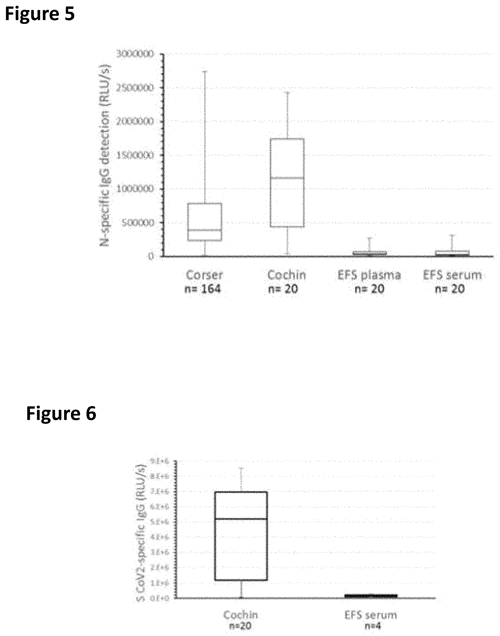
FIG. 5 shows a whisker-plot of ELISA on IgG specific of N protein of SARS-CoV-2 (Whiskers min max; boxes: 2nd and 3rd quartiles separated by a median).
FIG. 6 shows a whisker-plot of ELISA on IgG specific of S protein of SARS-CoV-2.
FIG. 7 shows the correlation between the titration of IgG specific of N protein of SARS-CoV-2 versus IgG specific of S protein of SARS-CoV-2 in serum from APHP-Cochin (n=20) and EFS (n=4).
FIG. 8 shows the correlation between the titration of IgG specific of N protein of SARS-CoV-1 versus IgG specific of N protein of SARS-CoV-2 in serum from APHP-Cochin (n=20) and EFS (n=4).
FIG. 9 shows the correlation between the titration of IgG specific of N protein of SARS-CoV-2 in the quick test (5 min) versus the routine test (120 min)-mean of the values in duplicate.
DETAILED DESCRIPTION OF THE INVENTION
The invention encompasses compositions, kits, uses, and methods for the detection and diagnosis of SARS CoV-2 and SARS and SARS-CoV-2 and SARS infections.
The indirect ELISA test was used with serum from COVID-19 patients with confirmed infections, as well as from various symptomatic patients (untested for SARS CoV-2 RNA) and presumptively negative patients.
The test was able to detect anti-SARS CoV-2 antibodies in the serum of all of COVID-19 patients with confirmed infections, as well as in the serum of many of the symptomatic patients (untested for SARS CoV-2 RNA). Anti-SARS CoV-2 antibodies were not detected in the serum of presumptively negative patients. These experiments showed that the N protein of SARS-CoV ("N_SARS1") was able to bind to antibodies present in the serum of SARS CoV-2 infected individuals. Thus, the antibodies generated against the N protein of SARS CoV-2 ("N_SARS2") by the patients could bind to the N_SARS1 protein. This allows the use of the N_SARS1, for example using an indirect ELISA, for the detection of SARS CoV-2 via immunoassay.
The N_SARS2 protein of SARS CoV-2 was produced recombinantly in E. coli strain BL21 (DE3) pDIA17. To achieve expression. DNA encoding the N_SARS2 was codon optimized for expression in bacteria and modified to express a Hiss N-terminal label. The DNA was transformed into E. coli and the protein produced was purified using a nickel column.
An indirect ELISA was used to compare plates coated with N_SARS2 to plates coated with N_SARS1 using serum from COVID-19 patients with confirmed infections, as well as from various symptomatic patients (untested for SARS CoV-2 RNA) and presumptively negative patients. Both N_SARS2 and N_SARS1 were able to detect anti-SARS CoV-2 antibodies in the serum of all of COVID-19 patients with confirmed infections, as well as in the serum of many of the symptomatic patients (untested for SARS CoV-2 RNA). Anti-SARS CoV-2 antibodies were not detected in the serum of presumptively negative patients. The difference between the two proteins was an increased sensitivity with the N_SARS2 protein (about 25%). These results were surprising.
Rabbit hyper immune serum against N_SARS1 and N_SARS2 proteins was generated. Rabbit hyper immune serum against N_SARS1 and N_SARS2 proteins could bind to both the N_SARS1 and N_SARS2 proteins.
Alpaca were immunized with N_SARS2. Binding of plasma and serum VHH from the immunized alpaca to N_SARS2 protein was also detected.
These results allow for the use of the N_SARS1 and N_SARS2 proteins and antibodies generated against these proteins for the detection of SARS-CoV and SARS CoV-2 via immunoassay. The high correlation of immunoassay tests with N_SARS1 and N_SARS2 proteins with low cross-reactivity and high sensitivity indicates that these proteins can be used for immunodiagnostics of patients with of SARS-CoV and SARS CoV-2. The invention provides expression vectors, proteins, antibodies, and methods and kits containing these reagents.
Expression Vectors
The invention encompasses a recombinant vector for expression of N protein of SARS-CoV or of N protein of SARS-CoV-2. The recombinant vector can be a vector for eukaryotic or prokaryotic expression, such as a plasmid, a phage for bacterium introduction, a YAC able to transform yeast, a viral vector and especially a retroviral vector, or any expression vector. An expression vector as defined herein is chosen to enable the production of an N protein or polyepitope, either in vitro or in vivo.
Preferably, the expression vector expressing the N_SARS1 protein is one of the expression vectors described in U.S. Pat. No. 8,343,718, which is hereby incorporated by reference.
In one embodiment, the recombinant vector expressing the N_SARS1 protein comprises an N cDNA cloned into the Expression Vector pIVEX2.3 or pIVEX2.4, as described in U.S. Pat. No. 8,343,718.
In one embodiment, the expression vector is pIV2.3N, particularly as contained in the bacteria transformed with pIV2.3N. These bacteria pIV2.3N/DH5|alpha| were deposited under the terms of the Budapest Treaty at the Collection Nationale de Culture de Microorganismes (CNCM) on Oct. 23, 2003, under the number I-3117.
In one embodiment, the recombinant vector for expression of N-SARS2 is pETM11/N-nCov E. coli 3-(His)6-Nter as contained in the bacteria strain Bacterium_N-Cov_Ecoli_PETM11_coli3 deposited at the CNCM on May 11, 2020, under the number I-5510. In another embodiment, the recombinant vector for expression of N-SARS2 is pETM11/N-nCov E. coli 4-(HIS)6-Nter as contained in the bacteria strain Bacterium_N-Cov_Ecoli_PETM11_coli4 deposited at the CNCM on May 11, 2020, under the number I-5511.
In one embodiment, the expression vector encodes a protease cleavage site, such as TEV cleavage site, inserted between N protein coding sequence and a protein purification Tag, such as polyHis tag. In a preferred embodiment, the expression vector encodes a His tag. In one embodiment, a protease cleavage site is positioned to remove the His tag, for example, after purification.
The expression vector can comprise transcription regulation regions (including promoter, enhancer, ribosome binding site (RBS), polyA signal), a termination signal, a prokaryotic or eukaryotic origin of replication and/or a selection gene. The features of the promoter can be easily determined by the man skilled in the art in view of the expression needed, i.e., constitutive, transitory or inducible (e.g. IPTG), strong or weak, tissue-specific and/or developmental stage-specific promoter. The vector can also comprise sequence enabling conditional expression, such as sequences of the Cre/Lox system or analogue systems.
In various embodiments, the expression vector is a plasmid, a phage for bacterium introduction, a YAC able to transform yeast, a viral vector, or any expression vector. An expression vector as defined herein is chosen to enable the production of a protein or polyepitope, either in vitro or in vivo.
The nucleic acid molecules according to the invention can be obtained by conventional methods, known per se, following standard protocols such as those described in Current Protocols in Molecular Biology (Frederick M. AUSUBEL, 2000, Wiley and son Inc., Library of Congress, USA). For example, they may be obtained by amplification of a nucleic sequence by PCR or RT-PCR or alternatively by total or partial chemical synthesis.
The vectors are constructed and introduced into host cells by conventional recombinant DNA and genetic engineering methods which are known per se. Numerous vectors into which a nucleic acid molecule of interest may be inserted in order to introduce it and to maintain it in a host cell are known per se; the choice of an appropriate vector depends on the use envisaged for this vector (for example replication of the sequence of interest, expression of this sequence, maintenance of the sequence in extrachromosomal form or alternatively integration into the chromosomal material of the host), and on the nature of the host cell.
N_SARS1 & N_SARS2 Proteins
Preferably, the N_SARS2 protein comprises the amino acid sequence of the SARS CoV-2 protein of GenBank/NCBI accession number QHO62884.1 accessed on Jul. 29, 2020, which is hereby incorporated by reference.
Preferably, the N_SARS1 protein comprises the amino acid sequence of the SARS-CoV N protein of GenBank/NCBI accession number NP_828858.1 accessed on Jun. 2, 2020 or YP_009825061.1 accessed on Jul. 18, 2020, which is hereby incorporated by reference.
Preferably, the N_SARS2 protein comprises the following amino acid sequence.
TABLE-US-00001 (SEQ ID NO: 1) 1 MKHHHHHHPM SDYDIPTTEN LYFQGAMSDN GPQNQRNAPR ITFGGPSDST GSNQNGERSG 61 ARSKQRRPQG LTNNTASWFT ALTQHGKEDL KFPRGQGVPI NTNSSPDDQI GYYRRATRRI 121 RGGDGKMKDL SPRWYFYYLG TGPEAGLPYG ANKDGIIWVA TEGALNTPKD HIGTRNPANN 181 AAIVLQLPQG TTLPKGFYAE GSRGGSQASS RSSSRSRNSS RNSTPGSSRG TSPARMAGNG 241 GDAALALLLL DRLNQLESKM SGKGQQQQGQ TVTKKSAAEA SKKPRQKRTA TKAYNVTQAF 301 GRRGPEQTQG NFGDQELIRQ GTDYKHWPQI AQFAPSASAF FGMSRIGMEV TPSGTWLTYT 361 GAIKLDDKDP NFKDQVILLN KHIDAYKTFP PTEPKKDKKK KADETQALPQ RQKKQQTVLL 421 LPAADLDDFS KQLQQSMSSA DSTQA**.
Preferably, the N_SARS1 protein is one of the N proteins described in U.S. Pat. No. 8,343,718, particularly SEQ ID NO:37, which is hereby incorporated by reference.
In one embodiment, the N_SARS1 protein is produced by bacteria pIV2.3N/DH5|alpha| transformed with pIV2.3N, which were deposited under the terms of the Budapest Treaty at the Collection Nationale de Culture de Microorganismes (CNCM) on Oct. 23, 2003, under the number I-3117. The address of CNCM is: Collection Nationale de Culture de Microorganismes, Institut Pasteur, 28 rue du Dr Roux, 75724 Paris CEDEX 15, France.
In one embodiment, the N_SARS2 protein is produced by bacteria transformed with the expression vector pETM11/N-nCov E. coli 3-(His)6-Nter or with the expression vector pETM11/N-nCov E. coli 4-(His)6-Nter. These two strains (Bacterium_N-Cov_Ecoli_PETM11_coli3 and Bacterium_N-Cov_Ecoli_PETM11_coli4) were deposited under the terms of the Budapest Treaty at the Collection Nationale de Culture de Microorganismes (CNCM) on May 11, 2020, under the numbers I-5510 and I-5511 respectively.
In one embodiment the expression vector is contained within one of the following bacterial strains:
Escherichia coli B BL21 (DE3) pDIA17 pLA13 (insert:NAD kinase Bacillus subtilis): I-2722
Escherichia coli B BL21 (DE3) pDIA17 pLA134 (insert:NAD kinase Bacillus subtilis): I-2723
Escherichia coli B BL21 (DE3) pDIA17 pLA63 (insert:NAD kinase Neisseria meningitidis NMA): I-2724
Escherichia coli B BL21 (DE3) pDIA17 pLA634 (insert:NAD kinase Neisseria meningitidis NMA): I-2725
Escherichia coli B BL21 (DE3) pDIA17 pLA131 (insert:NAD kinase Bacillus subtilis): I-2830.
These strains were deposited under the terms of the Budapest Treaty at the Collection Nationale de Culture de Microorganismes (CNCM) on Oct. 9, 2001, under the numbers I-2722, I-2723, I-2724, and I-2725 on 9 Oct. 2001 and I-2830 on 2 Apr. 2002.
The invention encompasses "isolated or purified" N_SARS1 & N_SARS2 proteins. The terms "isolated or purified" mean modified "by the hand of humans" from the natural state: in other words if an object exists in nature, it is said to be isolated or purified if it is modified or extracted from its natural environment or both. For example, a polynucleotide or a protein/peptide naturally present in a living organism is neither isolated nor purified: on the other hand, the same polynucleotide or protein/peptide separated from coexisting molecules in its natural environment, obtained by cloning, amplification and/or chemical synthesis is isolated for the purposes of the present invention. Furthermore, a polynucleotide or a protein/peptide which is introduced into an organism by transformation, genetic manipulation or by any other method, is "isolated" even if it is present in said organism. The term purified as used in the present invention means that the proteins/peptides according to the invention are essentially free of association with the other proteins or polypeptides, as is for example the product purified from the culture of recombinant host cells or the product purified from a non-recombinant source. Various techniques can be used to obtain purified protein according to the invention as for example metal chelate binding chromatography and gel filtration.
Methods for Making N_SARS1 & N_SARS2
Production of the N_SARS1 & N_SARS2 proteins can be achieved by any technique known to the skilled artisan, for example, as detailed in the examples or as described in in U.S. Pat. No. 8,343,718.
In one embodiment, a method for producing SARS-CoV-2 N protein comprises the following steps: Culturing a bacterium transformed with a recombinant plasmid encoding for SARS-CoV-2 N protein deposited at the Collection Nationale de Cultures de Microorganismes (CNCM) at the Institut Pasteur, 25, Rue du Docteur Roux, 75724 Paris. FR, on May 11, 2020, under the deposit number CNCM I-5510 or the deposit number CNCM I-5511 in a suitable bacterial growth medium such as a medium supplemented with kanamycin and chloramphenicol, in particular with 50 microgram/mL kanamycin and 30 microgram/mIL chloramphenicol; inducing the production of SARS-CoV-2 N protein; recovering and purifying SARS-CoV-2 N protein.
Preferably, the bacterium is E. coli.
Preferably, inducing the production of SARS-CoV-2 N protein is performed by adding IPTG.
Preferably, recovering the SARS-CoV-2 N protein is performed by pelleting and breakage of bacteria (bacteria cells). More preferably, recovering the SARS-CoV-2 N protein is performed by pelleting and breakage of bacteria, and further recovering the soluble fraction of broken bacteria.
Preferably, purifying SARS-CoV-2 N protein is performed by metal chelate affinity chromatography, and/or gel filtration.
Preferably, the SARS-CoV-2 N protein made by the method disclosed herein is soluble.
The invention also encompasses an N_SARS2 protein made by the processes disclosed herein, particularly in Example 1.
Antibodies Against N_SARS1 or N_SARS2 (N1 or N2)
In one embodiment, polyclonal or monoclonal antibodies are generated in rabbits or mice.
In one embodiment VHH antibodies are generated, for example in alpaca.
The invention encompasses a polyclonal or monoclonal antibody or fragment thereof directed against N_SARS1 or N_SARS2 protein.
In one embodiment, antibodies can be obtained by immunization of an animal with a N_SARS1 or N_SARS2 protein.
The antibodies can serve as reagents to bind native N_SARS1 & N_SARS2 proteins of patients in immunoassays.
The antibodies can serve as positive control reagents to bind isolated and purified N_SARS1 & N_SARS2 proteins in immunoassays of patients.
The antibodies can be used to determine and adjust the concentration of SARS-CoV-2 N bound to the ELISA plates for serum dilution.
The invention encompasses the polyclonal antibodies, monoclonal antibodies, chimeric antibodies, and fragments thereof (e.g., Fab, Fv, scFv) directed against the N protein.
For the purposes of the present invention, the expression chimeric antibody is understood to mean, in relation to an antibody of a particular animal species or of a particular class of antibody, an antibody comprising all or part of a heavy chain and/or of a light chain of an antibody of another animal species or of another class of antibody.
In some embodiments, purified proteins are used to produce antibodies by conventional techniques. In some embodiments, recombinant or synthetic proteins or peptides are used to produce antibodies by conventional techniques.
Antibodies can be synthetic, semi-synthetic, monoclonal, or polyclonal and can be made by techniques well known in the art. Such antibodies specifically bind to proteins and polypeptides via the antigen-binding sites of the antibody (as opposed to non-specific binding). Purified or synthetic proteins and peptides can be employed as immunogens in producing antibodies immunoreactive therewith. The proteins and peptides contain antigenic determinants or epitopes that elicit the formation of antibodies.
These antigenic determinants or epitopes can be either linear or conformational (discontinuous). Linear epitopes are composed of a single section of amino acids of the polypeptide, while conformational or discontinuous epitopes are composed of amino acids sections from different regions of the polypeptide chain that are brought into close proximity upon protein folding (C. A. Janeway, Jr. and P. Travers, Immuno Biology 3:9 (Garland Publishing Inc., 2nd ed. 1996)). Because folded proteins have complex surfaces, the number of epitopes available is quite numerous; however, due to the conformation of the protein and steric hinderances, the number of antibodies that actually bind to the epitopes is less than the number of available epitopes (C. A. Janeway, Jr. and P. Travers, Immuno Biology 2:14 (Garland Publishing Inc., 2nd ed. 1996)). Epitopes can be identified by any of the methods known in the art Such epitopes or variants thereof can be produced using techniques well known in the art such as solid-phase synthesis, chemical or enzymatic cleavage of a polypeptide, or using recombinant DNA technology.
Antibodies are defined to be specifically binding if they bind proteins or polypeptides with a Ka of greater than or equal to about 10.sup.7 M.sup.-1. Affinities of binding partners or antibodies can be readily determined using conventional techniques, for example those described by Scatchard et al., Ann. N.Y. Acad. Sci., 51:660 (1949).
Polyclonal antibodies can be readily generated from a variety of sources, for example, horses, cows, goats, sheep, dogs, chickens, alpaca, camels, rabbits, mice, or rats, using procedures that are well known in the art. In general, a purified protein or polypeptide that is appropriately conjugated is administered to the host animal typically through parenteral injection. The immunogenicity can be enhanced through the use of an adjuvant, for example, Freund's complete or incomplete adjuvant. Following booster immunizations, small samples of serum are collected and tested for reactivity to proteins or polypeptides. Examples of various assays useful for such determination include those described in Antibodies: A Laboratory Manual, Harlow and Lane (eds.), Cold Spring Harbor Laboratory Press, 1988: as well as procedures, such as countercurrent immuno-electrophoresis (CIEP), radioimmunoassay, radio-immunoprecipitation, enzyme-linked immunosorbent assays (ELISA), dot blot assays, and sandwich assays. See U.S. Pat. Nos. 4,376,110 and 4,486,530.
Monoclonal antibodies can be readily prepared using well known procedures. See, for example, the procedures described in U.S. Pat. Nos. RE 32,011, 4,902,614, 4,543,439, and 4,411,993; Monoclonal Antibodies, Hybridomas. A New Dimension in Biological Analyses, Plenum Press, Kennett, McKeam, and Bechtol (eds.), 1980.
For example, the host animals, such as mice, can be injected intraperitoneally at least once and preferably at least twice at about 3 week intervals with isolated and purified proteins or conjugated polypeptides, for example a peptide comprising or consisting of the specific amino acids set forth above. Mouse sera are then assayed by conventional dot blot technique or antibody capture (ABC) to determine which animal is best to fuse. Approximately two to three weeks later, the mice are given an intravenous boost of the protein or polypeptide. Mice are later sacrificed, and spleen cells fused with commercially available myeloma cells, such as Ag8.653 (ATCC), following established protocols. Briefly, the myeloma cells are washed several times in media and fused to mouse spleen cells at a ratio of about three spleen cells to one myeloma cell. The fusing agent can be any suitable agent used in the art, for example, polyethylene glycol (PEG). Fusion is plated out into plates containing media that allows for the selective growth of the fused cells. The fused cells can then be allowed to grow for approximately eight days. Supernatants from resultant hybridomas are collected and added to a plate that is first coated with goat anti-mouse Ig. Following washes, a label, such as a labeled protein or polypeptide, is added to each well followed by incubation. Positive wells can be subsequently detected. Positive clones can be grown in bulk culture and supernatants are subsequently purified over a Protein A column (Pharmacia).
The monoclonal antibodies of the invention can be produced using alternative techniques, such as those described by Alting-Mees et al., "Monoclonal Antibody Expression Libraries: A Rapid Alternative to Hybridomas", Strategies in Molecular Biology 3:1-9 (1990), which is incorporated herein by reference. Similarly, binding partners can be constructed using recombinant DNA techniques to incorporate the variable regions of a gene that encodes a specific binding antibody. Such a technique is described in Larrick et al., Biotechnology, 7:394 (1989).
Antigen-binding fragments of such antibodies, which can be produced by conventional techniques, are also encompassed by the present invention. Examples of such fragments include, but are not limited to, Fab and F(ab')2 fragments. Antibody fragments and derivatives produced by genetic engineering techniques are also provided.
The monoclonal antibodies of the present invention include chimeric antibodies, e.g., humanized versions of murine monoclonal antibodies. Such humanized antibodies can be prepared by known techniques, and offer the advantage of reduced immunogenicity when the antibodies are administered to humans. In one embodiment, a humanized monoclonal antibody comprises the variable region of a murine antibody (or just the antigen binding site thereof) and a constant region derived from a human antibody. Alternatively, a humanized antibody fragment can comprise the antigen binding site of a murine monoclonal antibody and a variable region fragment (lacking the antigen-binding site) derived from a human antibody. Procedures for the production of chimeric and further engineered monoclonal antibodies include those described in Riechmann et al. (Nature 332:323, 1988), Liu et al. (PNAS 84:3439, 1987), Larrick et al. (Bio/Technology 7:934, 1989), and Winter and Harris (TIPS 14:139, May. 1993). Procedures to generate antibodies transgenically can be found in GB 2,272,440, U.S. Pat. Nos. 5,569,825 and 5,545,806.
Antibodies produced by genetic engineering methods, such as chimeric and humanized monoclonal antibodies, comprising both human and non-human portions, which can be made using standard recombinant DNA techniques, can be used. Such chimeric and humanized monoclonal antibodies can be produced by genetic engineering using standard DNA techniques known in the art, for example using methods described in Robinson et al. International Publication No. WO 87/02671; Akira, et al. European Patent Application 0184187; Taniguchi, M., European Patent Application 0171496: Morrison et al. European Patent Application 0173494: Neuberger et al. PCT International Publication No. WO 86/01533: Cabilly et al. U.S. Pat. No. 4,816,567; Cabilly et al. European Patent Application 0125023; Better et al., Science 240:1041 1043, 1988; Liu et al., PNAS 84:3439 3443, 1987; Liu et al., J. Immunol. 139:3521 3526, 1987; Sun et al. PNAS 84:214 218, 1987; Nishimura et al., Canc. Res. 47:9991005, 1987; Wood et al., Nature 314:446 449, 1985: and Shaw et al., J. Natl. Cancer Inst. 80:1553 1559, 1988); Morrison, S. L., Science 229:1202 1207, 1985; Oi et al., BioTechniques 4:214, 1986: Winter U.S. Pat. No. 5,225,539; Jones et al., Nature 321:552 525, 1986; Verhoeyan et al., Science 239:1534, 1988, and Beidler et al., J. Immunol. 141:4053 4060, 1988.
In connection with synthetic and semi-synthetic antibodies, such terms are intended to cover but are not limited to antibody fragments, isotype switched antibodies, humanized antibodies (e.g., mouse-human, human-mouse), hybrids, antibodies having plural specificities, and fully synthetic antibody-like molecules.
In one embodiment, the invention encompasses single-domain antibodies (sdAb), also known as NANOBODIES. A sdAb is a fragment consisting of a single monomeric variable antibody domain that can bind selectively to a specific antigen.
In one embodiment, the sdAbs are from heavy-chain antibodies found in camelids (VHH fragments), or cartilaginous fishes (VNAR fragments), or are obtained by splitting dimeric variable domains into monomers.
Methods for Detection and Diagnosis
The N_SARS2 and N_SARS1 proteins and the peptides derived from these proteins and antibodies generated against them, can be used for the detection and diagnosis of a SARS-associated coronavirus infection (serological diagnosis (detection of specific antibodies) or virological diagnosis (detection of viral nucleocapsid (N) protein), in particular by an immunoassay, such as an immunoenzymatic method (e.g., ELISA).
The invention encompasses methods for identifying a patient infected with a SARS-associated coronavirus, comprising providing a serum sample from the patient, contacting the serum with an N_SARS2 or N_SARS1 protein, and visualizing the antigen-antibody complexes. Preferably, the antigen-antibody complexes are visualized by EIA, ELISA, RIA, or by immunofluorescence. In a preferred embodiment, the SARS-associated coronavirus infection is identified as SARS CoV-2 infection. In some embodiments, the patient has been shown to be infected by SARS-CoV or SARS CoV-2 by a nucleic acid detection test, such as a PCR or other nucleic acid amplification test In some embodiments, the patient has been identified as being infected with a SARS-associated coronavirus, but lacks detection of the virus by PCR or another nucleic acid amplification technique.
The invention encompasses a composition comprising an N_SARS1 protein or the use of an N_SARS1 protein for detection of antibodies against a SARS CoV-2 coronavirus and diagnosis of a SARS CoV-2 coronavirus infection in a biological sample.
The invention encompasses a composition comprising an N_SARS2 protein or the use of an N_SARS2 protein for detection of antibodies against a SARS coronavirus and diagnosis of a SARS coronavirus infection, in a biological sample. Preferably the SARS coronavirus is a SARS CoV-2 coronavirus.
The ability of N_SARS2 to bind with high affinity to antibodies directed against a SARS-CoV allows its use in such methods, particularly for diagnostics of a SARS-CoV infection. Preferably the SARS CoV is a SARS-CoV-1 (SARS-CoV) coronavirus or SARS-CoV-2 coronavirus.
The ability of N_SARS1 to bind with high affinity to anti-SARS CoV-2 antibodies allows its use in such methods, particularly for diagnostics of a SARS CoV-2 infection.
The antibodies and antibody fragments according to the invention, in particular those directed to N_SARS2 and N_SARS1 proteins and the derived peptides, are useful for the direct detection and diagnosis of a SARS-associated coronavirus infection and for the detection of a SARS-CoV in a biological sample; the detection of the N protein of a SARS coronavirus is carried out by an appropriate technique, in particular EIA, ELISA, RIA, immunofluorescence, in a biological sample collected from an individual likely to be infected.
Preferably, the patient has been shown to be infected by SARS-CoV or SARS CoV-2 by a nucleic acid detection test, such as a PCR or other nucleic acid amplification test.
In one embodiment, the invention comprises a method for the detection of a SARS-associated coronavirus, from a biological sample, which method is characterized in that it comprises contacting a biological sample from a patient infected with a SARS-CoV coronavirus with an anti-N_SARS2 antibody, and visualizing the antigen-antibody complexes formed. Preferably, the antigen-antibody complexes are visualized by EIA, ELISA, RIA, or by immunofluorescence.
In one embodiment, the invention comprises a method for the detection of a SARS-associated coronavirus, from a biological sample, which method is characterized in that it comprises contacting a biological sample from a patient infected with a SARS CoV-2 coronavirus with an anti-N_SARS1 antibody, and visualizing the antigen-antibody complexes formed. Preferably, the antigen-antibody complexes are visualized by EIA, ELISA, RIA, or by immunofluorescence.
In one embodiment, the invention comprises a method for the detection of a SARS-associated coronavirus, from a biological sample, which method is characterized in that it comprises contacting a biological sample from a patient infected with a SARS-CoV or SARS CoV-2 coronavirus with an anti-N_SARS2 antibody and an anti-N_SARS1 antibody, and visualizing the antigen-antibody complexes formed. Preferably, the antigen-antibody complexes are visualized by EIA. ELISA, RIA, or by immunofluorescence using a detection reagent.
In one embodiment, the N_SARS2 and N_SARS1 protein is attached to an appropriate support, in particular a microplate or a bead.
In one embodiment, the method comprises bringing a biological sample from a subject, preferably a human, infected with a SARS-CoV or SARS CoV-2 coronavirus into contact with an N_SARS2 and/or N_SARS1 protein, which is attached to an appropriate support, in particular a microplate or bead, to allow binding to occur; washing the support to remove unbound antibodies; adding a detection reagent that binds to the immunoglobulins bound to N_SARS2 and/or N_SARS1 protein; and detecting the N_SARS2 and/or N_SARS1 protein-antibody complexes formed.
In one embodiment, the method for the detection of antibodies directed to a SARS-associated coronavirus in a biological sample comprises providing a N_SARS2 protein: providing a biological sample from a patient infected with a SARS-CoV coronavirus; contacting said N_SARS2 protein with said biological sample; and visualizing the antigen-antibody complexes formed. Preferably, the method comprises an ELISA. Preferably, the N_SARS2 protein comprises or consists of the sequence of SEQ ID NO:1. Preferably the patient is infected with a SARS-CoV-2 coronavirus.
In one embodiment, the method for the detection of antibodies against a SARS-associated coronavirus in a biological sample comprises providing a N_SARS1 protein; providing a biological sample from a patient infected with a SARS CoV-2 coronavirus; contacting said N_SARS1 protein with said biological sample: and visualizing the antigen-antibody complexes formed. Preferably, the method comprises an ELISA.
Preferably, the protein-antibody complexes are detected with an antibody or an antibody fragment that binds to human immunoglobulins.
Preferably, the detection reagent comprises a label is selected from a chemiluminescent label, an enzyme label, a fluorescence label, and a radioactive (e.g., iodine) label. Most preferably, the detection reagent is a labeled antibody or antibody fragment that binds to human immunoglobulins.
Preferred labels include a fluorescent label, such as FITC, a chromophore label, an affinity-ligand label, an enzyme label, such as alkaline phosphatase, horseradish peroxidase, luciferase or .beta. galactosidase, an enzyme cofactor label, a hapten conjugate label, such as digoxigenin or dinitrophenyl, a Raman signal generating label, a magnetic label, a spin label, an epitope label, such as the FLAG or HA epitope, a luminescent label, a heavy atom label, a nanoparticle label, an electrochemical label, a light scattering label, a spherical shell label, semiconductor nanocrystal label, wherein the label can allow visualization with or without a secondary detection molecule.
Preferred labels include suitable enzymes such as horseradish peroxidase, alkaline phosphatase, beta-galactosidase, luciferase or acetylcholinesterase; members of a binding pair that are capable of forming complexes such as streptavidin/biotin, avidin/biotin or an antigen/antibody complex including, for example, rabbit IgG and anti-rabbit IgG; fluorophores such as umbelliferone, fluorescein, fluorescein isothiocyanate, rhodamine, tetramethyl rhodamine, eosin, green fluorescent protein, erythrosin, coumarin, methyl coumarin, pyrene, malachite green, stilbene, lucifer yellow, Cascade Blue, Texas Red, dichlorotriazinylamine fluorescein, dansyl chloride, phycoerythrin, fluorescent lanthanide complexes such as those including Europium and Terbium, cyanine dye family members, such as Cy3 and Cy5, molecular beacons and fluorescent derivatives thereof, as well as others known in the art; a luminescent material such as luminol: light scattering or plasmon resonant materials such as gold or silver particles or quantum dots; or radioactive material include .sup.14C, .sup.123I, .sup.124I, .sup.125I, .sup.32P, .sup.33P, .sup.35S, or .sup.3H.
In one embodiment, the antibody or an antibody fragment that binds to human immunoglobulins binds specifically to IgG, IgA, and IgM. In one embodiment, the antibody or an antibody fragment that binds to human immunoglobulins binds specifically to IgG, IgA, or IgM.
The term "antibodies" is meant to include polyclonal antibodies, monoclonal antibodies, fragments thereof, such as F(ab')2 and Fab fragments, single-chain variable fragments (scFvs), single-domain antibody fragments (VHHs or Nanobodies), bivalent antibody fragments (diabodies), as well as any recombinantly and synthetically produced binding partners.
In a preferred embodiment, the antibody is a VHH, preferably an alpaca serum.
Preferably, the method comprises comparing the results obtained with a patient serum to positive and negative controls.
Positive controls can include: Serum from animals (e.g., rabbit, alpaca, etc.) immunized with N_SARS2 or N_SARS1 protein as described above N_SARS2 or N_SARS1 protein as described above.
The method can comprise the use of N_SARS2 and N_SARS1 proteins to detect novel coronaviruses that do not cross-react with seasonal (non-pathogenic) coronaviruses.
Preferably, the N_SARS2 protein comprises the amino acid sequence of the SARS CoV-2 N protein of GenBank/NCBI accession number QHO62884.1 accessed on Jul. 29, 2020.
Preferably, the N_SARS1 protein comprises the amino acid sequence of the SARS-CoV N protein of GenBank/NCBI accession number NP_828858.1 accessed on Jun. 2, 2020 or YP_009825061.1 accessed on Jul. 18, 2020.
In one embodiment, the N_SARS2 protein comprises or consists of the amino acid sequence of SEQ ID NO:1.
In one embodiment, the N_SARS1 protein is one of the N proteins described in U.S. Pat. No. 8,343,718, particularly that of SEQ ID NO:37.
In one embodiment, the method comprises an immunocapture method.
In one embodiment, the method comprises attaching a first monoclonal or polyclonal antibody or a fragment thereof, directed against the N_SARS2 or N_SARS1 protein (capture antibody), incubating the antibody with a biological sample containing a N_SARS2 or N_SARS1 protein, and detecting the antigen-antibody complexes formed, preferably with a monoclonal antibody (visualizing antibody).
In one embodiment, the biological sample is mixed with the visualizing monoclonal antibody prior to its being brought into contact with the capture antibody. According to one embodiment of said test, the antibody used for the capture of the N_SARS2 protein is the monoclonal antibody mAb87, produced by the hybridoma SRAS N1-87 A13 A16 A24 30 Nov. 2004 deposited at the CNCM on Dec. 1, 2004 under the number I-3328 or the monoclonal antibody mAb86, produced by the hybridoma SRAS N3-86 A17 A1 A1 30 Nov. 2004 deposited at the CNCM on Dec. 1, 2004 under the number I-3329.
In the immunocapture tests according to the invention, it is possible to use, for visualizing the N_SARS2 or N_SARS1 protein, the monoclonal antibody mAb57, produced by the hybridoma SRAS N3-57 A6 A1 A5 30 Nov. 2004 deposited at the CNCM on Dec. 1, 2004 under the number I-3330, said antibody being conjugated with a visualizing molecule or particle.
In accordance with said immunocapture test, a combination of the antibodies mAb57 and mAb87, conjugated with a visualizing molecule or particle, is used for the visualization of the N_SARS2 or N_SARS1 protein.
A visualizing molecule may be a radioactive atom, a dye, a fluorescent molecule, a fluorophore, an enzyme: a visualizing particle may be for example: colloidal gold, a magnetic particle or a latex bead.
The subject of the present invention is also a method for the detection of a SARS-associated coronavirus infection, from a biological sample, by indirect IgG ELISA using the N_SARS2 or N_SARS1 protein, which method is characterized in that the plates are sensitized with an N_SARS2 or N_SARS1 protein solution at a concentration of between 0.5 and 4 .mu.g/ml, preferably to 2 .mu.g/ml, in a 10 mM PBS buffer pH 7.2, phenol red at 0.25 ml/l.
In one embodiment, microtiter plates are coated by incubation overnight at 4.degree. C. with 5 .mu.g/ml of N proteins. Plates are washed with 0.1% Tween 20 in PBS buffer. Serum or purified Igs are diluted in PBS containing 0.5% gelatin and 0.1% Tween. After 1 h incubation at 37.degree. C., plates are washed again. The bound antibodies are detected by adding a rabbit polyclonal anti-IgGs (obtained by immunizing rabbits with IgGs isolated on protein A and protein G columns) followed by Alkaline Phosphatase labeled goat anti-rabbit immunoglobulins. Enzymatic activity is quantified using pNPP (para-NitroPhenylPhosphate, SigmaAldrich) substrate according to the manufacturer's protocol.
According to one variant of the tests for detecting SA RS-associated coronaviruses, these tests combine an ELISA using the N protein, and another ELISA using the S protein.
The subject of the present invention is also an immune complex formed of a polyclonal or monoclonal antibody or antibody fragment as defined above, and of a N_SARS2 or N_SARS1 protein.
Kits Containing N_SARS2 and N_SARS1 Proteins
The invention encompasses a SARS-associated coronavirus detection kit, characterized in that it comprises a N_SARS2 and N_SARS1 protein, as described above, and/or antibodies generated against them.
Preferably, the N_SARS2 protein comprises the amino acid sequence of the SARS CoV-2 N protein of GenBank/NCBI accession number QHO62884.1 accessed on Jul. 29, 2020.
Preferably, the N_SARS1 protein comprises the amino acid sequence of the SARS-CoV N protein of GenBank/NCBI accession number NP_828858.1 accessed on Jun. 2, 2020 or YP_009825061.1 accessed on Jul. 18, 2020.
In one embodiment, the invention comprises a kit for the detection of a SARS-CoV coronavirus infection, which kit contains a N_SARS2 protein and reagents for detection of antigen-antibody complexes.
Preferably, the kit contains a serum of an animal immunized with N_SARS2 and/or N_SARS1 proteins.
Most preferably, the serum is a rabbit or alpaca serum from an animal immunized with N_SARS2 and/or N_SARS1 proteins.
In one embodiment, the invention comprises a kit for the detection of a SARS CoV-2 coronavirus infection, which kit contains a N_SARS1 protein and reagents for detection of antigen-antibody complexes.
Preferably, the kit contains a serum of an animal immunized with N_SARS2 and/or N_SARS1 proteins.
Most preferably, the serum is a rabbit or alpaca serum from an animal immunized with N_SARS2.
Preferably, the kit of the invention comprises an N_SARS2 protein comprising the amino acid sequence of the SARS CoV-2 N protein of GenBank/NCBI accession number QHO62884.1 accessed on Jul. 29, 2020.
Preferably, the kit of the invention comprises an N_SARS1 protein comprising the amino acid sequence of the SARS-CoV N protein of GenBank/NCBI accession number NP_828858.1 accessed on Jun. 2, 2020 or YP_009825061.1 accessed on Jul. 18, 2020.
Preferably, the kit of the invention comprises an N_SARS2 protein that comprises or consists of the amino acid sequence of SEQ ID NO:1.
Preferably, the kit of the invention comprises an N_SARS1 protein that is one of the N proteins described in U.S. Pat. No. 8,343,718, particularly that of SEQ ID NO:37.
In one embodiment, the kit comprises both N_SARS1 and N_SARS2 proteins and an N_SARS1 (N1) and/or N_SARS2 (N2) immune serum.
In one embodiment, the kit is a Simple/Rapid test designed for use where a preliminary screening test result is required. The tests can be a test based on agglutination, immuno-dot, immuno-chromatographic and/or immuno-filtration techniques. Preferably, the test is quick and easy to perform, preferably from about 10 minutes to about 2 hours, and requires little or no additional equipment.
Preferably, the kit can be stored at room temperature for extended period of time.
EXAMPLES
Example 1: Production and Purification of SARS-CoV-1 and SARS-CoV-2 Recombinant N Protein
SARS-CoV-1 recombinant N protein (N_SARS1) was produced using E. coli strain BL21 (DE3) pDIA17 transformed with the expression vector pIV2.3N deposited under at the Collection Nationale de Cultures de Microorganismes (CNCM) at the Institut Pasteur, 25, Rue du Docteur Roux, 75724 Paris, FR, on Oct. 23, 2003, under the deposit number CNCM I-3117 and purified as previously disclosed in U.S. Pat. No. 8,343,718 (see in particular the protocol disclosed in example 2).
cDNAs encoding the native nucleoprotein antigen (N_SARS2) from 2019-nCoV (SARS-CoV-2) was designed base on the Genbank MN908947 sequence publicly available from NBCBI on 20 Jan. 2020. This sequence was then processed to generate an optimized nucleotide sequences for high expression in E coli. Optimization process includes codon adaptation, mRNA de novo synthesis and stability, transcription and translation efficiency. Bsa1 and Xho1/EcoR1/Not1 restriction sites were then added at the 5' and 3 ends, respectively, of the nucleotide sequences. The resulting optimised cDNA named "N-Ecoli optimized gene" was synthesized. The Bsa1-Xho1 fragment of the "N-Ecoli optimized gene" has been inserted into Nco1/Xho1-digested pETM-11 vector and the resulting pETM11-Necoli_2019-nCoV (=pETM11/N-nCov E. coli) has been used to produce a fusion polypeptide between the SARS-CoV-2 protein and a N-terminally located poly-histidine tag (6 histidine), separated by a TEV cleavage site.
The resulting His6-N_2019-nCoV (N_SARS2) polypeptide has the sequence:
TABLE-US-00002 (SEQ ID NO: 1) 1 MKHHHHHHPM SDYDIPTTEN LYFQGAMSDN GPQNQRNAPR ITFGGPSDST GSNQNGERSG 61 ARSKQRRPQG LPNNTASWFT ALTQHGKEDL KFPRGQGVPI NTNSSPDDQI GYYRRATRRI 121 RGGDGKMKDL SPRWYFYYLG TGPEAGLPYG ANKDGIIWVA TEGALNTPKD HIGTRNPANN 181 AAIVLQLPQG TTLPKGFYAE GSRGGSQASS RSSSRSRNSS RNSTPGSSRG TSPARMAGNG 241 GDAALALLLL DRLNQLESKM SGKGQQQQGQ TVTKKSAAEA SKKPRQKRTA TKAYNVTQAF 301 GRRGPEQTQG NFGDQELIRQ GTDYKHWPQI AQFAPSASAF FGMSRIGMEV TPSGTWLTYT 361 GAIKLDDKDP NFKDQVILLN KHIDAYKTFP PTEPKKDKKK KADETQALPQ RQKKQQTVTL 421 LPAADLDDFS KQLQQSMSSA DSTQA**
Nucleoprotein coding sequences (WT-CoV-2 SARS DNA and E. coli optimized CoV-2 SARS DNA) are cloned into pETM11 vector (EMBL; Dummler et al (2005), Microb Cell Fact 13; 4:34) or pIVEX2-3 (Roche vector) vectors. The N-recombinant Nucleoprotein of CoV-2-SARS is produced in E. coli BL21 (DE3) pDA17 as a fusion protein comprising an N- or C-terminal (His).sub.6 polyhistidine label. Concerning the production of N-recombinant Nucleoprotein with a (His).sub.6 N-terminal label, the following recombinant vectors are used for the transformation of E. coli strain BL21 (DE3) pDIA17: pETM11/N-nCov WT4-(His).sub.6-Nter pETM11/N-nCov WT6-(His).sub.6-Nter pETM11/N-nCov E. coli 3-(His).sub.6-Nter pETM11/N-nCov E. coli 4-(His).sub.6-Nter pIVEX/nCov WT-(His).sub.6-Nter Clone 1 pIVEX/nCov WT-(His).sub.6-Nter Clone 2
E. coli strain BL21 (DE3) pDIA17 transformed with recombinant plasmid pETM11/N-nCov E. coli 3-(His).sub.6-Nter or pETM11/N-nCov E. coli 4-(His).sub.6-Nter (Bacterium_N-Cov_Ecoli_PETM11_coli3 and Bacterium_N-Cov_Ecoli_PETM11_coli4) were deposited at the Collection Nationale de Cultures de Microorganismes (CNCM) at the Institut Pasteur, 25, Rue du Docteur Roux, 75724 Paris, FR, on May 11, 2020, under the deposit numbers CNCM 1-5510 and CNCM 1-5511, respectively.
Cultures in Thomson Flasks Shaken in LB Medium (IPTG Induction) and NZytech Medium (Self-Inducible) of E. coli BL21 (DE3) pDIA17 Strains Transformed by the pETM11 Vector or by the pIVEX 2.3 Vector.
The Thomson flasks are 2.5 L notched flasks allowing cultures of 1 litre of medium to be aerated under good aeration conditions in stirrers.
Production in LB Environment (IPTG Induction)
The 4 strains of E. coli BL21 (DE3) pDIA17 transformed by the pETM11 vector (DMSO no. 1535, 1536, 1537, 1538) are spread on an agar LB Petri dish containing 50 .mu.g/ml kanamycin and 30 .mu.g/ml chloramphenicol. The 2 strains of E. coli BL21 (DE3) pDIA17 transformed by the vector pIVEX2.3 (DMSO n.degree. 1539, 1540) are spread on an agar LB Petri dish containing 100 .mu.g/ml ampicillin and 30 .mu.g/ml chloramphenicol. All plates of LB Agar Petri LB are incubated overnight at 37.degree. C. in an oven.
From each of the 6 Petri LB agar plates are inoculated with a platinum handle, 6 pre-cultures of 500 ml of LB medium in 2.5 L Thomson flasks (LB medium plus antibiotics appropriate to each recombinant vector pETM11 and pIVEX2.3). These pre-cultures are shaken at 180 rpm in a Multitron Infors shaker for 15 h at 30.degree. C.
From the 4 LB pre-cultures of BL21 (DE3) pDIA17 strains transformed by the pETM11 vector (DMSO No. 1535, 1536, 1537, 1538) are seeded at an initial cell density equivalent to DOA600=0.2, cultures of 1 L of LB medium containing 50 .mu.g/ml kanamycin and 30 .mu.g/ml chloramphenicol.
From the 2 LB pre-cultures of BL21 (DE3) pDIA17 strains transformed by the pIVEX 2.3 vector (DMSO No. 1539, 1540) are seeded at an initial cell density equivalent to DOA600=0.2, cultures of 1 L of LB medium containing 100 .mu.g/ml ampicillin and 30 .mu.g/ml chloramphenicol.
All these cultures in LB medium are placed under agitation at 180 rpm and 30.degree. C. When the cell density, equivalent to DOA600=0.8 is reached the cultures are induced by addition of 1 mM IPTG and the temperature is maintained at 30.degree. C.
After 2 hours at 30.degree. C. in the presence of the inducer the cultures are stopped. A 10 ml sample of each culture is centrifuged and will be used for analysis on SDS-Page of the total soluble and insoluble protein fractions. The remainder of each culture is centrifuged (15 min at 6000 rpm) and the pellets stored at -80.degree. C.
Production in NZvtech Medium (Self-Inducible)
From the 4 LB pre-cultures of BL21 (DE3) pDIA17 strains transformed by the pETM11 vector (DMSO No. 1535, 1536, 1537, 1538) are seeded at an initial cell density equivalent to DOA600=0.2, 1 L cultures in NZytech (self-inducible) medium containing 50 .mu.g/ml kanamycin and 30 .mu.g/ml chloramphenicol.
From the 2 LB pre-cultures of BL21 (DE3) pDIA17 strains transformed by the pIVEX 2.3 vector (DMSO No. 1539, 1540) are seeded at an initial cell density equivalent to DOA600=0.2, 1 L cultures in NZytech (self-inducible) medium containing 100 .mu.g/ml ampicillin and 30 .mu.g/ml chloramphenicol.
Cultures in NZytech medium (self-inducible) are carried out at 37.degree. C. with stirring at 180 rpm.
After 4 hours at 37.degree. C., the cultures are placed at 18.degree. C.
After 15 hours of culture at this temperature of 18.degree. C., the bacterial cultures are stopped. A 10 ml sample of each culture is centrifuged and will be used for analysis on SDS-Page of the total soluble and insoluble protein fractions. The remainder of each culture is centrifuged (15 min at 6000 rpm) and the pellets stored at -80.degree. C.
Cultures in BioPod F200 Microfermenters in High Cell Density HDM Medium (IPTG Induction) of E. coli BL21 (DE3) pDIA17 Strains Transformed by the pETM11 Vector or by the pIVEX 2.3 Vector:
The HDM medium is a complex culture medium developed by our Platform specifically designed for the large production of E. coli biomass in a bioreactor during batch culture. This buffered medium does not require a regulation of the pH value in culture.
Microfermenters are miniaturized bioreactors allowing to realize 100 ml cultures in high density medium (HDM medium). These micro-fermenters are equipped with mass flow meters and sinter allowing a very efficient micro-bubbling by air progressively enriched with oxygen according to the bacterial growth. These bioreactors are also equipped with Peltier system and PT1000 probe which allow a very reliable regulation of the growth temperature and fast passages from 37.degree. C. to 16.degree. C. during the induction phase. This system of miniaturized bioreactors is a tool for optimizing the culture conditions allowing with a high rate of reliability a scale-up of 100 ml cultures to larger volume reactors (4 L and 16 L in our Platform).
The 2 strains of E. coli BL21 (DE3) pDIA17 transformed by the pETM11 vector (DMSO n.degree. 1535 and 1537) are spread on an agar LB Petri dish containing 50 .mu.g/ml kanamycin and 30 .mu.g/ml chloramphenicol.
The 2 strains of E. coli BL21 (DE3) pDIA17 transformed by the vector pIVEX2.3 (DMSO no. 1539, 1540) are spread on an agar LB Petri dish containing 100 .mu.g/ml ampicillin and 30 .mu.g/ml chloramphenicol. All LB agar plates are incubated overnight at 37.degree. C. in an oven.
About 1.5 ml of antibiotic-free LB medium is deposited on each of the agar plates. The bacterial mat of each LB plate is scraped off with a sterile rake. Each bacterial suspension collected is used to inoculate a micro-fermentor containing 100 ml of HDM medium plus antibiotics appropriate for E. coli BL21 (DE3) pDIA17 strains transformed by the recombinant pETM11 or pIVEX2.3 vectors. The initial cell density of the bioreactors is equivalent to .sub.A600=0.8 to 1.
The cultures are grown at a temperature of 37.degree. C., and aeration is set at 0.5 WM. When the cell density equivalent to DOA600=18 to 20 is reached, the temperature is lowered to 16.degree. C. and IPTG (1 mM) is added to the cultures.
After 15 hours of culture at 16.degree. C. in the presence of the inducer, the bacterial cultures are stopped. A 1 ml sample of each culture is centrifuged and will be used for analysis on SDS-Page of the total soluble and insoluble protein fractions. The remainder of each culture is centrifuged (15 min at 6000 rpm) and the pellets stored at -80.degree. C.
Purification from pETM11/N-nCov E. coli-(his).sub.6-Nter/NZytech Cultures
Data prot param: MW=48.7 KDa pI=9.9 Ext. coefficient Abs 0.1% (=1 g/l): 0.961 Bacterial Pellet Breakage
Take the 9 g pellet with 50 ml buffer A: 50 mM phosphate, 300 mM NaCl, 20 mM imidazole pH8 with 1 Roche EDTA free protease tablet and 5 .mu.l benzonase in the blender/wait incubation at room temperature for approx. 20 min. 1) Cold breaking with the Cell D 1.3 kbar Cell Disrupter. 2) Addition of eNASR A (250 .mu.l to 10 mg/ml or 2.5 mg). Incubation at room temperature for about 20 min. 3) Centrifugation 19000 rpm rotor SS341 hour 4.degree. C. 4) Recovery of the soluble fraction=supernatant for affinity purification on nickel resin. Treatment of the Soluble Fraction
1st STEP OF PURIFICATION: AFFINITY IMAC (AKTAPure): 1 column Nickel 5 ml 1 New 5 ml Protino Ni-NTA column (Macherey Nagel) mounted on AKTA Pure (room temperature) Washing of the column in .sub.H2O: 10CV Column equilibration buffer: Phosphate 50 mM, NaCl 300 mM, imidazole 20 mM pH8: 10 CV Loading the 60 ml crude extract onto the 5 ml IMAC column at a rate of 1 ml/min with the AKTA pump Flow rate: 1 ml/min Washing with Phosphate buffer 50 mM, NaCl 300 mM, imidazole 20 mM, pH8: 10CV Elution: Elution Buffer Phosphate 50 mM, NaCl 300 mM, imidazole 250 mM pH8 Gradient from 20 to 250 mM imidazole=100% buffer B at 2 ml/min on 10 HP. Fractions of 1.5 ml were recovered Histogram peak integration for protein quantity estimation Peak of the fractions from A5 to C12, i.e. 48 ml at 3.3 mg/ml. Estimated quantity on unicorn162 mg Fraction analysis at this stage on SDS-Page (FIG. 1A)
TABLE-US-00003 Well 1 2 3 4 5 6 7 8 9 Fraction PM EB FT LAV A5 A6 A8 A10 Al2 Qty 28 .mu.l 28 .mu.l 28 .mu.l 28 .mu.l 28 .mu.l 28 .mu.l 28 .mu.l 28 .mu.l
TABLE-US-00004 Well 10 11 12 13 14 15 16 17 18 Fraction B4 B6 B8 B12 C3 C6 C9 C12 D5 Qty 28 .mu.l 28 .mu.l 28 .mu.l 28 .mu.l 28 .mu.l 28 .mu.l 28 .mu.l 28 .mu.l 28.mu.l
DO280 nm Measurement at 1/10 of the A5 to C9 Pool
DO280=0.364. For 1 g/l, the OD is 0.96 3.64/0.96=3.7 mg/m. Either for 42 ml: 42 ml.times.3.7 mg/ml=159 mg total
The elution volume will be injected in 8.times.5 ml on 2 gel filtration columns with 5 ml loops. The columns are installed on the 2 pure AKTAs.
4 runs of gel filtration will be performed per column.
2ND PURIFICATION STEP: Hiload 16/60 Superdex 200 .mu.g (120 ml) column filtration gel
Equilibration of the columns with 50 mM Phosphate buffer, 500 mM NaCl pH8
Flow rate 0.5 ml/min
Protein injection with 5 ml and 10 ml loops.
Elution volume for each run 1.4 column volume. Fractions of 1.8 ml.
Selection of peaks on histograms and integration of peaks for estimation of protein quantity (FIGS. 1B and 1C) SDS-Page Fraction Removal and Deposition on SDS-Page Gel
Gel Filtration 3 on AKTA 1 (FIG. 1D)
TABLE-US-00005 Well 1 2 3 4 5 6 7 8 9 Fraction PM pool 2H1 2H3 2H4 2H5 2H6 2H7 2H8 Qty 28 .mu.l 28 .mu.l 28 .mu.l 28 .mu.l 28 .mu.l 28 .mu.l 28 .mu.l 28 .mu.l
TABLE-US-00006 Well 10 11 12 13 14 15 16 17 18 Fraction 2H9 2H10 2H11 2H12 3A1 3A5 3A8 3A10 3B1 Qty 28 .mu.l 28 .mu.l 28 .mu.l 28 .mu.l 28 .mu.l 28 .mu.l 28 .mu.l 28 .mu.l 28 .mu.l
Gel Filtration 6 on AKTA 2 (FIG. 1E)
TABLE-US-00007 Well 1 2 3 4 5 6 7 8 9 Fraction pool PM 1H4 1H6 1H7 1H8 1H9 1H10 1H11 Qty 28 .mu.l 28 .mu.l 28 .mu.l 28 .mu.l 28 .mu.l 28 .mu.l 28 .mu.l 28 .mu.l
TABLE-US-00008 Well 10 11 12 13 14 15 16 17 18 Fraction 1H12 2A1 2A2 2A3 2A4 2A5 2A9 2Al2 2B2 Qty 28 .mu.l 28 .mu.l 28 .mu.l 28 .mu.l 28 .mu.l 28 .mu.l 28 .mu.l 28 .mu.l 28.mu.l
Formation of the Gel Fraction Pool Filtration
GF 1: 1B TO 1B8
GF 2: 2A6 TO 2A12
GF 3: 2H6 TO 2H12
GF 4: 3G6 TO 3G12
GF 5: 1A11 TO 1B6
GF 6: 1H8 TO 2A2
GF 7: 2G5 TO 2G11
GF 8: 3F2 TO 3F8
280 nm OD measurement of the pool
DO280=0.597. For 1 g/1, the OD is 0.96. 0.597/0.96=0.62 mg/ml. Either for 100 ml: 100 ml.times.0.6 mg/ml=60 mg total
Purification and Storage Balance
N_SARS2 purified protein (concentration 0.62 mg/ml in phosphate 50 mM NaCl 500 mM pH8). Pool gel filtration 80 ml split into 4 parts: 20 ml filtered without antiproteases 37 aliquots of 0.5 ml stored at +4.degree. C. (box in cold room; 20 ml filtered with antiproteases 36 aliquots of 0.5 ml stored at +4.degree. C. (box in cold room); 20 ml unfiltered 40 aliquots of 0.5 ml stored at -80.degree. C. (box in freezer room); 20 ml unfiltered+glycerol 50% final 42 aliquots of 1 ml stored at -20.degree. C. (box in freezer room).
E. coli optimized CoV-2 SARS DNA cloned into pETM-11 expression vector gave highest protein production yields in E. coli. Unexpectedly, the clones pETM11/N-nCov E. coli 3-(His)6-Nter and pETM11/N-nCov E. coli 4-(His)6-Nter were able to achieve high level production without protein aggregation.
Example 2: Preparation of Monospecific Polyclonal Antibodies Directed Against SARS-CoV-1 and SARS-CoV-2 Recombinant N Proteins
The preparation of monospecific polyclonal antibodies directed against SARS-CoV-1 recombinant N protein (N1 protein) has been disclosed previously in U.S. Pat. No. 8,343,718 (see in particular the rabbit immunization protocol disclosed in example 4). Monospecific polyclonal antibodies directed against SARS-CoV-2 recombinant N protein (N2 protein) were prepared in alpaca (Lama pacos) according to the following protocol.
Monospecific polyclonal antibodies directed against SARS-CoV-1 and SARS-CoV-2 recombinant N proteins are useful as positive controls in SARS-CoV immunodiagnostic assays.
1. Material and Methods
1.1 Immunization
125 .mu.g of SARS-CoV-2 recombinant Nucleoprotein (N) prepared according to example 1, in a total volume of 250 .mu.l was mixed with 250 .mu.l of Freund complete adjuvant for the first immunization, and with 250 .mu.l of Freund incomplete adjuvant for the following immunizations. One young adult alpaca (Lama pacos) named ANU was immunized at days 1, 19 and 26 with the immunogen. The alpaca was bled at day 33. The immune response was monitored by titration of serum and plasma samples by ELISA on coated Nucleoprotein as disclosed below.
1.2 Purification of Anti-N Alpaca Polyclonal Antibodies
Five mgs of SARS-CoV-2 recombinant N protein were bound on Cyanogen Bromide-activated sepharose 4B (GE Healthcare Life science) according to the manufacturer's instructions. Twenty ml of plasma were incubated with the beads for 2 hours, the beads were washed extensively with PBS until the OD280=0. Then Glycine/HCl 0.1M pH 2.8 was added and the OD280 was monitored. Each fractions of 1 ml were neutralized with 100 .mu.l of Tris 2M. The positive fractions were pooled and dialysed against PBS. An ELISA was performed by coating the Nucleoprotein as disclosed below.
1.3 ELISA
Microtiter plates were coated by incubation overnight at 4.degree. C. with 5 .mu.g/ml of SARS-CoV-2 recombinant N protein. Plates were washed with 0.1% Tween 20 in PBS buffer. Serum or purified Igs were diluted in PBS containing 0.5% gelatin and 0.1% Tween. After 1 h incubation at 37.degree. C., plates were washed again. The bound alpaca antibodies were detected by adding a rabbit polyclonal anti-alpaca IgGs (obtained by immunizing rabbits with alpaca IgGs isolated on protein A and protein G columns) followed by Alkaline Phosphatase labeled goat anti-rabbit immunoglobulins. Enzymatic activity was quantified using pNPP (para-NitroPhenylPhosphate, SigmaAldrich) substrate according to the manufacturer's protocol.
2. Results
FIG. 2A shows the binding of serum and plasma of alpaca ANU and FIG. 2B the binding of purified anti-N2 polyclonal antibodies on SARS-CoV-2 nucleoprotein (N2 protein). The titer of the serum is very high (about 1/243000) and as low as 4 .mu.g of polyclonal Ig can detect the bound SARS-CoV-2 nucleoprotein. The lecture was performed after 15 min.
Example 3: Comparison of SARS-CoV-2 N and SARS-CoV-1 N Indirect ELISA for Detection of SARS-CoV-2 Infection in Patients Sera
1. Material and Methods
Plates are coated with 50 .mu.l of SARS-CoV-2 N protein at 1 .mu.g/ml or SARS-CoV-1 N protein at 1 .mu.g/ml on PBS1.times. over-night at 4.degree. C. Plates are washed 4 times with 300 .mu.l PBS Tween 0.1% (PBST), blocked with 50 .mu.l PBST 3% fat-free milk (PBSTL) for 1 h at 37.degree. C. and blocking solution is removed. 50 .mu.l of patient test serum diluted in PBSTL, at varying dilutions (1/200 generally) is then added and incubated for 1 hr at 37.degree. C. Plates are washed 3 times with 300 .mu.l PBST. 50 .mu.l peroxidase-conjugated anti-human Ig (Ig total, IgG specific, IgM specific or IgA specific) diluted according to manufacturer's instructions in PBSTL is then added and incubated for 1 h at 37.degree. C. Plates are washed 3 times with 300 .mu.l PBST. 50 .mu.l TMB substrate prepared according to manufacturer's instructions is then added and incubated for 10 minutes RT in the dark. 50 .mu.l H3PO4 are then added on the plate are read at 450 nm and 620 nm.
2. Results
Human serum samples were tested in indirect ELISA using SARS-CoV-2 N protein compared to SARS-CoV-1 N protein.
Tested samples were from the following groups:
2 COVID patients (COVID-01 and COVID-02): hospitalized patients with confirmed SARS-CoV 2 infection (severe cases) 20 symptomatic (Sympt-01 to Sympt-20): patients with mild COVID-like symptoms, sampled during symptoms or after symptoms disappearance (NOT tested for viral RNA) 2 prepandemic adults (Neg Ad-01 and Neg Ad-02).
Various serum dilutions were tested (1/200; 1/800; 1/3200; 1/12800).
The results presented in FIG. 3A to 3C show that both SARS-CoV-2 N and SARS-CoV-1 N can detect SARS-CoV 2 infection with good sensitivity and specificity in patients with mild or severe SARS CoV-2 infection. SARS-CoV-1 N which has already been confirmed to have no cross reactivity with other seasonal coronaviruses is unexpectedly able to cross react with serum of patients with mild or severe SARS-CoV-2 (COVID) infection. The robustness of the indirect ELISA assay was confirmed using SARS-CoV-2 N protein on cohorts of 180 COVID patients and 200 negative individuals (Table 1).
TABLE-US-00009 TABLE 1 Indirect ELISA assay using SARS-CoV-2 N on COVID patients cohort Positivity Percentile threshold negative Negative > Patients > (OD) cohort threshold threshold 1.788 99% 2 (1%) 139 (76%) 0.97 95%-96% 9 (4%) 147 (80%) Number of 203 183 tested sera
The indirect ELISA assays allows the detection of 80% of the COVID patients with a good specificity.
Example 4: Comparison of SARS-CoV-2 N and SARS-CoV-1 N Indirect ELISA for Detection of Anti SARS-CoV-1 Antibodies
Rabbit hyperimmune monospecific polyclonal antibodies directed against SARS-CoV-1 virions were tested in the indirect ELISA assay disclosed in example 3 using 1 .mu.g/ml, 1.5 .mu.g/ml or 2 .mu.g/ml of SARS-CoV-1 or 1 .mu.g/ml of SARS-CoV-2 recombinant N protein. The results presented in FIGS. 4A to 4C shows that SARS-CoV-2 N is unexpectedly able to cross react with 10 anti SARS-CoV-1 antibodies and can detect anti SARS-CoV-1 antibodies with a good sensitivity and a good specificity.
Example 5: Detection of IgG Specific of Protein N and S of SARS-CoV-2 by IgG Luciferase Immunosorbent Assay Protocol
White 96- or 384-well plates with flat bottom (Fluoronunc C96 Maxisorp, Nunc) were coated by adsorption with either 1 .mu.g/mL of Nucleoprotein produced as in Example 1, or Spike protein, in 50 .mu.L/well of phosphate buffer saline pH 7.4 (Sigma) for 2 hours at room temperature or overnight at 4.degree. C. Adsorption on Maxisorp.RTM. plates favours charge interaction. Alternatives to adsorption are covalent binding of targets through carboxylic, amine or sulfhydryl moieties or glycosylation at the functionalized well surface or coating with poly-lysine. Wells were emptied eventually but not necessarily neutralize with BSA (1 mg/mL) or skimmed milk 3%. Wells were washed 3 to 6 times with 100 .mu.L of PBS/Tween 20 0.1%. Dilutions of serum (typically 1/200), plasma or body fluid were incubated from 30 min to 1 hour at room temperature in their respective antigen-coated wells, 50 .mu.L/well in phosphate buffer saline with eventually skimmed milk 3%, bovine serum albumin 1 mg/mL, bovine serum 1-3% and/or Tween 20 0.1%. Wells were washed three to six times with 100 .mu.L PBS/Tween 20 0.1%. Purified anti-IgG nanobody-nanoKAZ fusion protein at 1 ng/mL (5.107 RLU.s-1.mL-1) in phosphate buffer saline with eventually skimmed milk 3%, bovine serum albumin 1 mg/mL or bovine serum 1-3% and/or Tween 20 0.1%, was loaded (50 .mu.L/well) and incubated 20-30 min at room temperature. Wells were washed four times with 100 .mu.L of PBS/Tween 20 0.1%. Plates can be stored at this step in PBS until measurements. Just before reading, each wells were emptied and loaded with 50 .mu.L of furimazine (8-benzyl-2-(furan-2-ylmethyl)-6-phenylimidazo[1,2-a]pyrazin-3(7H)-one) at 27 .mu.M. The plate was orbitally shaked for 5 seconds and the light emission intensity was integrated 0.5-1 sec per well using a multi-well plate luminometer (LB960 Centro, Berthold).
IgGs specific of protein N of SARS-CoV-2 have been titrated by luciferase linked immunosorbent assay in serums of the CORSER cohort (n=164; IcareB), in serums of the observation and monitoring protocol of the Intensive Medicine and Resuscitation service of Assistance Publique des Hopitaux de Paris (APHP) Cochin (n=20) in longitudinal follow-up and in serums and plasma of healthy donors of Etablissement Francais du Sang (EFS) sampled in December 2019 (frozen plasmas n=20, frozen serums n=20). Results are shown in FIG. 5.
IgGs specific of protein Spike (S) of SARS-CoV-2 have been titrated in serums from the observation and monitoring protocol of the Intensive Medicine and Resuscitation service of Assistance Publique des Hopitaux de Pans (APHP) Cochin (n=20) and in frozen serums from healthy donors of EFS (n=4). Results are shown in FIG. 6.
The correlation between the titration of IgG specific of protein N of SARS-CoV-2 and IgG specific of protein S of SARS-CoV-2 in serum from APHP-Cochin (n=20) and EFS (n=4) is shown in FIG. 7.
Then it was compared the IgG of serums from patients (APHP-Cochin n=20) and frozen serums of healthy donors (EFS n=4) which were specific for SARS-CoV-1 N protein and SARS CoV-2 N protein. Results are shown in FIG. 8.
An emergency test on a 96 well-plate which may be performed in 5 minutes has been assayed on serum from APHP-Cochin (n=20) and EFS (n=4) in duplicate by reducing the incubations time: 3 minutes for the incubation of sera at room temperature or even better at 37.degree. C., 17 seconds per washing step (plate washer Zoom HT, Berthold) and 1 minute for the incubation of the anti-IgG VHH-nanoKAZ. The coefficient of determination R2 was 0.75. Results are shown in FIG. 9.
SEQUENCE LISTINGS
1
SEQUENCE LISTING <160> NUMBER OF SEQ ID NOS: 1 <210> SEQ ID NO 1 <211> LENGTH: 445 <212> TYPE: PRT <213> ORGANISM: Severe acute respiratory syndrome (SARS)-associated coronavirus-2 <400> SEQUENCE: 1 Met Lys His His His His His His Pro Met Ser Asp Tyr Asp Ile Pro 1 5 10 15 Thr Thr Glu Asn Leu Tyr Phe Gln Gly Ala Met Ser Asp Asn Gly Pro 20 25 30 Gln Asn Gln Arg Asn Ala Pro Arg Ile Thr Phe Gly Gly Pro Ser Asp 35 40 45 Ser Thr Gly Ser Asn Gln Asn Gly Glu Arg Ser Gly Ala Arg Ser Lys 50 55 60 Gln Arg Arg Pro Gln Gly Leu Pro Asn Asn Thr Ala Ser Trp Phe Thr 65 70 75 80 Ala Leu Thr Gln His Gly Lys Glu Asp Leu Lys Phe Pro Arg Gly Gln 85 90 95 Gly Val Pro Ile Asn Thr Asn Ser Ser Pro Asp Asp Gln Ile Gly Tyr 100 105 110 Tyr Arg Arg Ala Thr Arg Arg Ile Arg Gly Gly Asp Gly Lys Met Lys 115 120 125 Asp Leu Ser Pro Arg Trp Tyr Phe Tyr Tyr Leu Gly Thr Gly Pro Glu 130 135 140 Ala Gly Leu Pro Tyr Gly Ala Asn Lys Asp Gly Ile Ile Trp Val Ala 145 150 155 160 Thr Glu Gly Ala Leu Asn Thr Pro Lys Asp His Ile Gly Thr Arg Asn 165 170 175 Pro Ala Asn Asn Ala Ala Ile Val Leu Gln Leu Pro Gln Gly Thr Thr 180 185 190 Leu Pro Lys Gly Phe Tyr Ala Glu Gly Ser Arg Gly Gly Ser Gln Ala 195 200 205 Ser Ser Arg Ser Ser Ser Arg Ser Arg Asn Ser Ser Arg Asn Ser Thr 210 215 220 Pro Gly Ser Ser Arg Gly Thr Ser Pro Ala Arg Met Ala Gly Asn Gly 225 230 235 240 Gly Asp Ala Ala Leu Ala Leu Leu Leu Leu Asp Arg Leu Asn Gln Leu 245 250 255 Glu Ser Lys Met Ser Gly Lys Gly Gln Gln Gln Gln Gly Gln Thr Val 260 265 270 Thr Lys Lys Ser Ala Ala Glu Ala Ser Lys Lys Pro Arg Gln Lys Arg 275 280 285 Thr Ala Thr Lys Ala Tyr Asn Val Thr Gln Ala Phe Gly Arg Arg Gly 290 295 300 Pro Glu Gln Thr Gln Gly Asn Phe Gly Asp Gln Glu Leu Ile Arg Gln 305 310 315 320 Gly Thr Asp Tyr Lys His Trp Pro Gln Ile Ala Gln Phe Ala Pro Ser 325 330 335 Ala Ser Ala Phe Phe Gly Met Ser Arg Ile Gly Met Glu Val Thr Pro 340 345 350 Ser Gly Thr Trp Leu Thr Tyr Thr Gly Ala Ile Lys Leu Asp Asp Lys 355 360 365 Asp Pro Asn Phe Lys Asp Gln Val Ile Leu Leu Asn Lys His Ile Asp 370 375 380 Ala Tyr Lys Thr Phe Pro Pro Thr Glu Pro Lys Lys Asp Lys Lys Lys 385 390 395 400 Lys Ala Asp Glu Thr Gln Ala Leu Pro Gln Arg Gln Lys Lys Gln Gln 405 410 415 Thr Val Thr Leu Leu Pro Ala Ala Asp Leu Asp Asp Phe Ser Lys Gln 420 425 430 Leu Gln Gln Ser Met Ser Ser Ala Asp Ser Thr Gln Ala 435 440 445
* * * * *
D00001
D00002
D00003
D00004
D00005
D00006
D00007
D00008
D00009
D00010
D00011
D00012
S00001
XML
uspto.report is an independent third-party trademark research tool that is not affiliated, endorsed, or sponsored by the United States Patent and Trademark Office (USPTO) or any other governmental organization. The information provided by uspto.report is based on publicly available data at the time of writing and is intended for informational purposes only.
While we strive to provide accurate and up-to-date information, we do not guarantee the accuracy, completeness, reliability, or suitability of the information displayed on this site. The use of this site is at your own risk. Any reliance you place on such information is therefore strictly at your own risk.
All official trademark data, including owner information, should be verified by visiting the official USPTO website at www.uspto.gov. This site is not intended to replace professional legal advice and should not be used as a substitute for consulting with a legal professional who is knowledgeable about trademark law.