Compositions and methods for identification, assessment, prevention, and treatment of cancer using NFS1 biomarkers and modulators
Bittinger , et al. March 16, 2
U.S. patent number 10,947,596 [Application Number 15/308,268] was granted by the patent office on 2021-03-16 for compositions and methods for identification, assessment, prevention, and treatment of cancer using nfs1 biomarkers and modulators. This patent grant is currently assigned to Dana-Farber Cancer Institute, Inc.. The grantee listed for this patent is Dana-Farber Cancer Institute, Inc.. Invention is credited to Mark Bittinger, Jessie M. English, Kwok-Kin Wong, Sima Zacharek.


View All Diagrams
| United States Patent | 10,947,596 |
| Bittinger , et al. | March 16, 2021 |
Compositions and methods for identification, assessment, prevention, and treatment of cancer using NFS1 biomarkers and modulators
Abstract
The present invention is based, in part, on the identification, of novel mitochondrial iron-sulfur (Fe--S) cluster biosynthesis pathway biomarkers and modulators, and methods of use thereof, for identifying, assessing, preventing, and treating cancer.
| Inventors: | Bittinger; Mark (Dover, MA), English; Jessie M. (Cambridge, MA), Wong; Kwok-Kin (Arlington, MA), Zacharek; Sima (Jamaica Plain, MA) | ||||||||||
|---|---|---|---|---|---|---|---|---|---|---|---|
| Applicant: |
|
||||||||||
| Assignee: | Dana-Farber Cancer Institute,
Inc. (Boston, MA) |
||||||||||
| Family ID: | 1000005423649 | ||||||||||
| Appl. No.: | 15/308,268 | ||||||||||
| Filed: | May 6, 2015 | ||||||||||
| PCT Filed: | May 06, 2015 | ||||||||||
| PCT No.: | PCT/US2015/029439 | ||||||||||
| 371(c)(1),(2),(4) Date: | November 01, 2016 | ||||||||||
| PCT Pub. No.: | WO2015/171741 | ||||||||||
| PCT Pub. Date: | November 12, 2015 |
Prior Publication Data
| Document Identifier | Publication Date | |
|---|---|---|
| US 20170051358 A1 | Feb 23, 2017 | |
Related U.S. Patent Documents
| Application Number | Filing Date | Patent Number | Issue Date | ||
|---|---|---|---|---|---|
| 61989037 | May 6, 2014 | ||||
| Current U.S. Class: | 1/1 |
| Current CPC Class: | G01N 33/6848 (20130101); C12Q 1/6886 (20130101); C12Q 2600/136 (20130101); C12Q 2600/118 (20130101); G01N 2333/91194 (20130101); C12Q 2600/156 (20130101); C12Q 2600/158 (20130101); C12Q 2600/106 (20130101) |
| Current International Class: | C12Q 1/6886 (20180101); G01N 33/68 (20060101) |
| Field of Search: | ;514/44A ;435/6.1,91.1,91.31,455,458 ;536/23.1,24.5 |
References Cited [Referenced By]
U.S. Patent Documents
| 2010/0028334 | February 2010 | Cottarel |
| 2010/0190656 | July 2010 | Li et al. |
Other References
|
Pai et al, Gene Therapy 2005; 1-14. cited by examiner . Ryther et al, Gene Therapy 2005; 12: 5-11. cited by examiner . Schiffelers et al., Nucleic Acids Research 2004; 32: e49. cited by examiner . Abruzzo et al.,"Frataxin mRNA Isoforms in FRDA Patients and Normal Subjects: Effect of Tocotrienol Supplementation," BioMed Res. Intl., 2013: 276808 (2013). cited by applicant . Anthony et al., "New Classes of Alanine Racemase Inhibitors Identified by High-Throughput Screening Show Antimicrobial Activity against Mycobacterium tuberculosis," PLoS One, 6: e20374 (2011). cited by applicant . Biderbick et al., "Role of Human Mitochondrial Nfs1 in Cytosolic Iron-Sulfur Protein Biogenesis and Iron Regulation," Mol Cell Biol, 26: 5675-5687 (2006). cited by applicant . Colin et al., "Mammalian Frataxin Controls Sulfur Production and Iron Entry during de Novo Fe4S4 Cluster Assembly," J Am Chem Soc, 135: 733-740 (2013). cited by applicant . Cupp-Vickery et al., "Crystal Structure of IscS, a Cysteine Desulfurase from Escherichia coli," J Mol Biol, 330:1049-1059 (2003). cited by applicant . Dixon et al., "Ferroptosis: An Iron-Dependent Form of Nonapoptotic Cell Death," Cell 149:1060-1072 (2012). cited by applicant . Farhan et al., "Exome Sequencing Identifies NFS1 Deficiency in a Novel Fe--S Cluster Disease, Infantile Mitochondrial Complex II/III Deficiency," Mol Genet Genom Med, 2:73-80 (2014). cited by applicant . Kurihara et al., "Assembly of Iron-Sulfur Mediated by Cysteine Desulfurases, IscS, CsdB and CSD, from Escherichia coli," Biochim Biophys Acta, 1647:303-309 (2003). cited by applicant . Li et al., "Yeast Mitochondrial Protein, Nfs1p, Coordinately Regulates Iron-Sulfur Cluster Proteins, Cellular IronUptake, and Iron Distribution," J Biol Chem, 274: 33025-33034 (1999). cited by applicant . Li et al., "Detection of Intracellular Iron by Its Regulatory Effect," Am J Physiol Cell Physiol, 287: C1547-C1559 (2004). cited by applicant . Li et al., "Thermodynamic and Structural Analysis of Human NFU Conformational Chemistry," Biochem, 52: 4904-4913 (2013). cited by applicant . Lill et al., "Iron-Sulfur-Protein Biogenesis in Eukaryotes," Trends Biochem Sci, 30: 133-141 (2005). cited by applicant . Lim et al., "Mutations in LYRM4, Encoding Iron-Sulfur Cluster Biogenesis Factor ISD11, Cause Deficiency of Multiple Respiratory Chain Complexes," Hum Mol Genet, 22: 4460-4473 (2013). cited by applicant . Majewska et al.,"Binding of the Chaperone Jacl Protein and Cysteine Desulfurase Nfs1 to the Iron-Sulfur Cluster Scaffold Isu Protein Is Mutually Exclusive," J Biol Chem, 288: 29134-29142 (2013). cited by applicant . Marelja et al., "A Novel Role for Human Nfs1 in the Cytoplasm: Nfs1 Acts as a Sultur Donor for MOCS3, a Protein Involved in Molybdenum Cofactor Biosynthesis," J Biol Chem, 283: 25178-25185 (2008). cited by applicant . Olson et al., "Characterization of the NifU and NifS Fe--S Cluster Formation Proteins Essential for Viability in Helicobacter pylori," Biochem, 39: 16213-16219 (2000). cited by applicant . Pandey et al., "Isd11p Protein Activates the Mitochondrial Cysteine Desulfurase Nfs1p Protein," J Biol Chem, 286: 38242-38252 (2011). cited by applicant . Pandey et al., "Fraxatin Directly Stimulates Mitochondrial Cysteine Desulfurase by Exposing Substrate-binding Sites, and a Mutant Fe--S Cluster Scaffold Protein with Frataxin-bypassing Ability Acts Similarly," J Biol Chem, 288: 36773-36786 (2013). cited by applicant . Prischi et al., "Structural Bases for the Interaction of Frataxin with the Central Components of Iron-Sulphur Cluster Assembly," Nat Commun, 1: 95 (2010). cited by applicant . Rouault et al., "Iron-Sulphur Cluster Biogenesis and Mitochondrial Iron Homeostasis," Nat Rev Mol Cell Biol, 6: 345-351 (2005). cited by applicant . Rybniker et al., "The Cysteine Desulfurase IscS of Mycobacterium Tuberculosis is Involved in Iron-Sulfur Cluster Biogenesis and Oxidative Stress Defence," Biochem J, 459: 467-478 (2014). cited by applicant . Schmucker et al., "Mammalian Frataxin: An Essential Function for Cellular Viability through an Interaction with a Preformed ISCU/NFS1/IDS11 Iron-Sulfur Assembly Complex," PLoS One, 6: e16199 (2011). cited by applicant . Stemmler et al., "Frataxin and Mitochondrial FeS Cluster Biogenesis," J Biol Chem, 285: 26737-26743 (2010). cited by applicant . Thompson et al., "Hypoxia-inducible Factor 2beta Regulates Key Neutrophil Functions in Humans, Mice, and Zebrafish," Blood, 123: 366-376 (2014). cited by applicant . Thorson et al., "Identification of Cystathionine Beta-Synthase Inhibitors Using a Hydrogen Sulfide Selective Probe," Angew Chem. Int. Ed. Engl., 52: 4641-4644 (2013). cited by applicant . Tsai et al., "Human Frataxin Is an Allosteric Switch that Activates the Fe--S Cluster Biosynthetic Complex," Biochem., 49: 9132-9139 (2010). cited by applicant . Weerapana et al., "Quantitative Reactivity Profiling Predicts Functional Cysteines in Proteomes," Nature, 468: 790-795 (2010). cited by applicant . Yang et al., "Regulation of Ferroptotic Cancer Cell Death by GPX4," Cell, 156: 317-331 (2014). cited by applicant . Ye et al., "Human Iron-Sulfur Cluster Assembly, Cellular Iron Homeostasis, and Disease," Biochem., 49: 4945-4956 (2010). cited by applicant . Zimmer et al., "Small-Molecule Inhibitors of HIF-2a Translation Link Its 5'UTR Iron-Responsive Element of Oxygen Sensing," Mol. Cell, 32: 838-848 (2008). cited by applicant . International Search Report for International Application No. PCT/US2015/029439 dated Sep. 29, 2015. cited by applicant. |
Primary Examiner: Zara; Jane J
Attorney, Agent or Firm: Foley Hoag LLP
Parent Case Text
CROSS-REFERENCE TO RELATED APPLICATION
This application claims the benefit of U.S. Provisional Application No. 60/989,037, filed on 6 May 2014; the entire contents of said application are incorporated herein in their entirety by this reference.
Claims
What is claimed is:
1. A method of inhibiting hyperproliferative growth of a cancer cell or cells, the method comprising contacting the cancer cell or cells with an shRNA or an siRNA targeting NFS1, thereby inhibiting hyperproliferative growth of the cancer cell or cells, wherein the step of contacting occurs ex vivo or in vitro, optionally wherein i) the shRNA or siRNA is administered in a pharmaceutically acceptable formulation; ii) the NFS1 is human NFS1; and/or iii) the cancer is selected from the group consisting of paragangliomas, colorectal cancer, cervical cancer, lung adenocarcinoma, ovarian cancer, and myeloid cancer within a hypoxic tumor microenvironment.
Description
BACKGROUND OF THE INVENTION
Despite advances in understanding the etiology of cancer and effective methods for treating cancer, malignant neoplasms represent the second most frequent cause of death worldwide surpassed only by heart diseases. Although effective anti-cancer treatments exist for many malignancies, such treatments are directed against well-known targets that do not fully control such malignancies. Accordingly, there is a great need to identify new cancer-related targets and biomarkers useful for the identification, assessment, prevention, and treatment of cancer.
SUMMARY OF THE INVENTION
The present invention is based, at least in part, on the discovery that the iron-sulfur cluster biosynthesis pathway plays a significant role in driving hyperproliferative cell growth and that modulating the pathway (e.g., inhibiting the function of one or more iron-sulfur cluster biosynthesis pathway members) can inhibit such hyperproliferative cell growth. In addition, biomarkers related to the iron-sulfur cluster biosynthesis pathway have been identified that are useful for identifying and assessing modulation of such hyperproliferative cell growth.
In one aspect, a method of treating a subject afflicted with a cancer comprising administering to the subject an agent that inhibits the copy number, amount, and/or activity of at least one biomarker listed in Table 1, thereby treating the subject afflicted with the cancer, is provided. In one embodiment, the agent is administered in a pharmaceutically acceptable formulation. In another embodiment, the agent directly binds the at least one biomarker listed in Table 1. In still another embodiment, the at least one biomarker listed in Table 1 is human NFS1 or an ortholog thereof. In yet another embodiment, the method further comprises administering one or more additional anti-cancer agents, optionally comprising mitochondrial cofactor therapy.
In another aspect, a method of inhibiting hyperproliferative growth of a cancer cell or cells, the method comprising contacting the cancer cell or cells with an agent that inhibits the copy number, amount, and/or activity of at least one biomarker listed in Table 1, thereby inhibiting hyperproliferative growth of the cancer cell or cells, is provided. In one embodiment, the step of contacting occurs in vivo, ex vivo, or in vitro. In another embodiment, the agent is administered in a pharmaceutically acceptable formulation. In still another embodiment, the agent directly binds the at least one biomarker listed in Table 1. In yet another embodiment, the at least one biomarker listed in Table 1 is human NFS1 or an ortholog thereof. In another embodiment, the method further comprises administering one or more additional anti-cancer agents, optionally comprising mitochondrial cofactor therapy.
In still another aspect, a method of determining whether a subject afflicted with a cancer or at risk for developing a cancer would benefit from iron-sulfur cluster (ISC) biosynthesis pathway inhibitor therapy, the method comprising: a) obtaining a biological sample from the subject; b) determining the copy number, amount, and/or activity of at least one biomarker listed in Table 1 in a subject sample; c) determining the copy number, amount, and/or activity of the at least one biomarker in a control; and d) comparing the copy number, amount, and/or activity of the at least one biomarker detected in steps b) and c); wherein a significant increase in the copy number, amount, and/or activity of the at least one biomarker in the subject sample relative to the control copy number, amount, and/or activity of the at least one biomarker indicates that the subject afflicted with the cancer or at risk for developing the cancer would benefit from ISC biosynthesis pathway inhibitor therapy, is provided. In one embodiment, the method further comprises recommending, prescribing, or administering ISC biosynthesis pathway inhibitor therapy if the cancer is determined to benefit from ISC biosynthesis pathway inhibitor therapy. In another embodiment, the method further composes recommending, prescribing, or administering anti-cancer therapy other than ISC biosynthesis pathway inhibitor therapy if the cancer is determined to not benefit from ISC biosynthesis pathway inhibitor therapy. In still another embodiment, the anti-cancer therapy is selected from the group consisting of targeted therapy, chemotherapy, radiation therapy, and/or hormonal therapy. In yet another embodiment, the control sample is determined from a cancerous or non-cancerous sample from either the patient or a member of the same species to which the patient belongs. In another embodiment, the control sample comprises cells. In still another embodiment, the method further comprises determining responsiveness to ISC biosynthesis pathway inhibitor therapy measured by at least one criteria selected from the group consisting of clinical benefit rate, survival until mortality, pathological complete response, semi-quantitative measures of pathologic response, clinical complete remission, clinical partial remission, clinical stable disease, recurrence-free survival, metastasis free survival, disease free survival, circulating tumor cell decrease, circulating marker response, and RECIST criteria.
In yet another aspect, a method of assessing the efficacy of an agent for treating a cancer in a subject, comprising: a) detecting, in a first subject sample and maintained in the presence of the agent the copy number, amount or activity of at least one biomarker listed in Table 1; b) detecting the copy number, amount, and/or activity of the at least one biomarker listed in Table 1 in a second subject sample and maintained in the absence of the test compound; and c) comparing the copy number, amount, and/or activity of the at least one biomarker listed in Table 1 from steps a) and b), wherein a significantly increased copy number, amount, and/or activity of the at least one biomarker listed in Table 1 in the first subject sample relative to the second subject sample, indicates that the agent treats the cancer in the subject, is provided.
In another aspect, a method of monitoring the progression of a cancer in a subject, comprising: a) detecting in a subject sample at a first point in time the copy number, amount, and/or activity of at least one biomarker listed in Table 1; b) repeating step a) during at least one subsequent point in time after administration of a therapeutic agent; and c) comparing the copy number, amount, and/or activity detected in steps a) and b), wherein a significantly increased copy number, amount, and/or activity of the at least one biomarker listed in Table 1 in the first subject sample relative to at least one subsequent subject sample, indicates that the agent treats the cancer in the subject, is provided. In one embodiment, the subject has undergone treatment, completed treatment, and/or is in remission for the cancer in between the first point in time and the subsequent point in time.
In another embodiment, the subject has undergone ISC biosynthesis pathway inhibitor therapy in between the first point in time and the subsequent point in time. In still another embodiment, the first and/or at least one subsequent sample is selected from the group consisting of ex vivo and in vivo samples. In yet another embodiment, the first and/or at least one subsequent sample is obtained from an animal model of the cancer. In another embodiment, the first and/or at least one subsequent sample is a portion of a single sample or pooled samples obtained from the subject.
In still another aspect, a cell-based method for identifying an agent which inhibits a cancer, the method comprising: a) contacting a cell expressing at least one biomarker listed in Table 1 with a test agent; and b) determining the effect of the test agent on the copy number, level of expression, or level of activity of the at least one biomarker listed in Table 1 to thereby identify an agent that inhibits the cancer, is provided. In one embodiment, the cells are isolated from an animal model of a cancer. In another embodiment, the cells are from a subject afflicted with a cancer. In still another embodiment, the cells are unresponsive to ISC biosynthesis pathway inhibitor therapy. In yet another embodiment, the step of contacting occurs in vivo, ex vivo, or in vitro. In another embodiment, the method further comprises determining the ability of the test agent to bind to the at least one biomarker listed in Table 1 before or after determining the effect of the test agent on the copy number, level of expression, or level of activity of the at least one biomarker listed in Table 1. In another aspect, the sample comprises cells, cell lines, histological slides, paraffin embedded tissue, fresh frozen tissue, fresh tissue, biopsies, blood, plasma, serum, buccal scrape, saliva, cerebrospinal fluid, urine, stool, mucus, or bone marrow, obtained from the subject.
In yet another aspect, a cell-free method for identifying a compound which inhibits a cancer, the method comprising: a) determining the effect of a test compound on the amount or activity of at least one biomarker listed in Table 1 contacted with a test compound; b) determining the amount or activity of the at least one biomarker listed in Table 1 maintained in the absence of the test compound; and c) comparing the amount and/or activity of the at least one biomarker listed in Table 1 from steps a) and b), wherein a significantly increased amount, and/or activity of the at least one biomarker listed in Table 1 in step a) relative to step b), identifies a compound which inhibits the cancer, is provided. In one embodiment, the method further comprises determining the ability of the test compound to bind to the at least one biomarker listed in Table 1 before or after determining the effect of the test compound on the amount or activity of the at least one biomarker. In another embodiment, the steps a) and b) are selected from the group consisting of a methylene blue assay, a 7-azido-4-methylcoumarin (AzMC) assay, an alanine assay, and a mass spectrometry assay. In still another embodiment, the methylene blue assay comprises i) reacting the at least one biomarker listed in Table 1 in a buffer comprising a) cysteine, b) a pyridoxal phosphate cofactor, and c) optionally the test compound; ii) stopping the reaction by adding N,N-dimethyl-p-phenylenediamine and iron chloride (FeCl3)(in hydrogen chloride (HCl) solution, and iii) determining the production of methylene blue via absorbance of light having a wavelength of 670 nm. In yet another embodiment, the AzMC assay comprises i) reacting the at least one biomarker listed in Table 1 in a buffer comprising a) cysteine, b) a pyridoxal phosphate cofactor, glutathione as reducing agent, d) bovine serum albumin, e) 7-azido-4-methylcoumarin, and f) optionally, the test compound; and ii) fluorometrically monitoring the reaction product, 7-amino-4-methylcoumarin. In another embodiment, the alanine assay comprises i) reacting the at least one biomarker listed in Table 1 in a buffer comprising a) cysteine, b) a pyridoxal phosphate cofactor, c) DTT as reducing agent, and d) optionally, the test compound; ii) performing a secondary reaction to measure alanine production in a buffer containing a) NAD (nicotinamide adenine dinucleotide) and b) alanine dehydrogenase enzyme; and iii) fluorometrically measuring the reaction product, NADH. In still another embodiment, the mass spectrometry assay comprises i) reacting the at least one biomarker listed in Table 1 in a buffer comprising a) cysteine, b) a pyridoxal phosphate cofactor, and c) optionally the test compound; and ii) determining, the production of alanine using mass spectrometry.
Other embodiments of the present invention are applicable to any of the methods, compositions, assays, and the like presented herein. For example, in one embodiment, the copy number is assessed by microarray, quantitative PCR (qPCR), high-throughput sequencing, comparative genomic hybridization (CGH), or fluorescent in situ hybridization (FISH). In another embodiment, the amount of the at least one biomarker is assessed by detecting the presence in the samples of a polynucleotide molecule encoding the biomarker or a portion of said polynucleotide molecule. In still another embodiment, the polynucleotide molecule is a mRNA, cDNA, or functional variants or fragments thereof. In yet another embodiment, the step of detecting further comprises amplifying the polynucleotide molecule. In another embodiment, the amount of the at least one biomarker is assessed by annealing a nucleic acid probe with the sample of the polynucleotide encoding the one or more biomarkers or a portion of said polynucleotide molecule under stringent hybridization conditions. In still another embodiment, the amount of the at least one biomarker is assessed by detecting the presence a polypeptide of the at least one biomarker. In yet another embodiment, the presence of a polypeptide is detected using a reagent which specifically binds with the polypeptide (e.g., a reagent selected from the group consisting of an antibody, an antibody derivative, and an antibody fragment). In another embodiment, the activity of the at least one biomarker is assessed by determining the magnitude of modulation of at least one NFS1 pharmacodynamic biomarker listed in Table 1. In still another embodiment, the activity of the at least one biomarker is assessed by determining the magnitude of modulation of the activity or expression level of at least one downstream target of the at least one biomarker. In yet another embodiment, the ISC biosynthesis pathway inhibitor agent or test compound modulates a biomarker selected from the group consisting of human NFS1, human LYRM4, human ISCU, human FXN, human NFU1, human GLRX5, human BOLA3, human HSCB, human HSPA9, human ISCA1, human ISCA2, human IBA57, human NUBPL, human SLC25A28, human FDXR, human FDX2, and orthologs of said biomarkers thereof. In another embodiment, the ISC biosynthesis pathway inhibitor agent or test compound is an inhibitor selected from the group consisting of a small molecule, antisense nucleic acid, interfering RNA, shRNA, siRNA, aptamer, ribozyme, dominant-negative protein binding partner, and combinations thereof. In another embodiment, the at least one biomarker is selected from the group consisting of 2, 3, 4, 5, 6, 7, 8, 9, 10, or more biomarkers. In still another embodiment, the at least one biomarker is selected from the group of ISC biosynthesis pathway biomarkers listed in Table 1. In yet another embodiment, the ISC biosynthesis pathway biomarkers listed in Table 1 are selected from the group consisting of human NFS1, human LYRM4, human ISCU, human FXN, human NFU1, human GLRX5, human BOLA3, human HSCB, human HSPA9, human ISCA1, human ISCA2, human IBA57, human NUBPL, human SLC25A28, human FDXR, human FDX2, and orthologs of said biomarkers thereof. In another embodiment, the at least one biomarker is selected from the group of NFS1 pharmacodynamic biomarkers listed in Table 1. In still another embodiment, the NFS1 pharmacodynamic biomarkers listed in Table 1 are selected from the group consisting of human aconitase, human succinate dehydrogenase, human ferritin, human transferrin-receptor, human Hif2alpha, human PTGS2, and lipid reactive oxygen species (ROS). In yet another embodiment, the cancer is selected from the group consisting of paragangliomas, colorectal cancer, cervical cancer, lung adenocarcinoma, ovarian cancer, and myeloid cancer within a hypoxic tumor microenvironment. In another embodiment, the subject is a mammal, such as an animal model of cancer or a human.
BRIEF DESCRIPTION OF THE DRAWINGS
FIG. 1 shows a schematic diagram of the iron-sulfur cluster biogenesis pathway as adapted from Lim et al. (2013) Hum. Mol. Genet. 22:4460-4473.
FIG. 2 shows the results of NFS1 amplification assessed across available TCGA (The Cancer Genome Atlas) datasets using the eBioPortal for Cancer Genomics (available on the World Wide Web at cbioportal.org/public-portal/). The inset shows the correlation between copy-number alterations (x-axis, as determined by GISTIC) and mRNA expression (y-axis, by RNASeq) from a representative dataset (colorectal cancer). All histograms shown represent amplifications.
FIG. 3 shows the results of collective alterations of NFS1, LYRM4/ISD11, ISCU, and FXN evaluated across available TCGA datasets using the eBioPortal for Cancer Genomics (available on the World Wide Web at cbioportal.org/public-portal/). Top bar of histogram: amplification; middle bar of histogram; deletion; bottom bar of histogram: mutation; gray: multiple alterations. The upper inset shows the distribution of alterations between NFS1, LYRM4, ISCU, and FXN in ovarian cancers. The lower insets show the correlation between copy-number alterations (x-axis, as determined by GISTIC) and mRNA expression (y-axis, by RNASeq) for NFS1 (left) and LYRM4 (right) from a representative dataset.
FIG. 4 shows the results of mutation analyses of solute carrier family 25 mitochondrial iron transporter, member 28 (SLC25A28), a mediator of iron uptake, assessed across TCGA samples using datasets from the cBioPortal for Cancer Genomics (available on the World Wide Web at cbioportal.org/public-portal/) and the Memorial Sloan-Kettering Cancer Center (MSKCC). All markers shown represent missense mutations, except for the seventh marker from the left located at the N-terminus of the Mito_carr domain, which represents a frameshift deletion.
FIG. 5 shows that inducible knockdown of NFS1 using shRNAs causes clonogenic growth inhibition in MKN74 cells.
FIG. 6 shows the results of cDNA rescue experiments confirming on-target activity of NFS1 shRNAs.
FIG. 7 shows the results of NFS1 knockdown and resulting cell growth effects across a panel of cell lines.
FIG. 8 shows the results of NFS1 shRN A shut-off experiments confirming restoration of clonogenic growth following depression of NFS1 function.
FIG. 9 shows the results of aconitase activity and SDH activity measured in lysates of mitochondria isolated from GAL-NFS1 cells harboring plasmid-borne copies of WT NFS1, nfs1M/AA, or vector without insert, as indicated, and grown for 40 hours in glucose-containing medium. Enzymatic activities were measured and plotted relative to the non-iron-sulfur cluster protein, malate dehydrogenase. The figure is adapted from Majewska al. (2013) J. Biol. Chem. 288:29134-29142.
FIG. 10 shows that transient knockdown of NFS1 in Hela cells shows alterations in mitochondrial structure and significantly decreased activity of iron-sulfur-dependent enzymes, including aconitase and SDH. The figure is adapted from Biederbick et al. (2006) Mol. Cell. Biol. 26:5675-5687.
FIG. 11 shows a schematic diagram illustrating an iron-dependent form of non-apoptotic cell death known as ferroptosis. The figure is adapted from Dixon et al. (2012) Cell 149:1060-1072.
FIG. 12 includes 2 panels, identified as panels A and B, which show data adapted from Yang et al. (2014) Cell 156:317-331 indicating that upregulation of PTGS2 expression occurs upon erastin and (1S,3R)-RSL3 treatment (panel A) and further showing that PTGS2 expression is induced by PE (panel B). The oncogenic RAS-selective lethal small molecule crastin triggers ferroptosis, which is dependent upon intracellular iron, but not other metals, and is morphologically, biochemically, and genetically distinct from apoptosis, necrosis, and autophagy. Erastin, like glutamate, inhibits cysteine uptake by cysteine/glutainate antiporter (system xc-), creating a void in the antioxidant defenses of the cell and ultimately leading to iron-dependent, oxidative death.
FIG. 13 shows that IRP-1 represses HIF2a translation and activity. The figure is adapted from Zimmer et al. (2008) Mol. Cell. 32:838-848.
FIG. 14 shows data confirming that candidate biomarkers for iron-sulfur cluster biosynthesis pathway modulation and iron-dependent cell death (ferroptosis) correlate with NFS1 inhibition.
FIG. 15 includes 5 panels, identified as panels A, B, C, D, and E, which demonstrate modulation of biomarkers of ISC biosynthesis pathway modulation and iron-dependent cell-death (ferroptosis) associated with NFS1 knockdown. Panel A shows decreases in ferritin protein levels and increases in TFRC protein levels, Panels B and C shows decreases in aconitase activity. Panel E shows decreases in succinate dehydrogenase activity. Panel F confirms knockdown of NFS1 protein levels in the samples analyzed in panels E and F.
FIG. 16 shows that NFS1 knockdown in the C2BBE1 colorectal cell line correlates with down-regulation of HIF2a protein. The asterisk (*) indicates treatment for 24 hours with cobalt chloride, a hypoxia-mimicking agent.
FIG. 17 includes 2 panels, identified as panels A and B, which demonstrate that NFS1 is essential in MKN74, an NFS-1 amplified cell line. Panel A shows that NFS1 is amplified in the MKN74 gastric cell line. Panel B shows the results of cDNA rescue of MKN74 stable inducible NFS1 shRNA lines with either wild type NFS1 or a dominant-negative NFS1 mutant.
FIG. 18 shows that cDNA rescue with WT NFS1 similar to that described in FIG. 17 restores the NFS1-sh5-dependent effect on aconitase activity.
FIG. 19 shows that WT NFS1, but not NFS1.sup.C381A, rescues the NFS1-sh5-dependent inhibition of FTH1 protein levels and the up-regulation of TfRc protein levels.
FIG. 20 shows that the NFS1.sup.C381A catalytic mutant causes a decrease in aconitase activity and ferritin levels comparable to that with NFS1-sh1 or NFS1-sh5.
FIG. 21 shows representative sulfide-based (e.g., methylene blue assays or flurogenic sulfide probes, such as AzMC to AMC detection) or alanine-based alanine dehydrogenase activity) detection methods for analyzing NFS1 activity.
FIG. 22 includes 2 panels, identified as panels A and B, which provide a representative methylene blue assay suitable for high-through put formats (panel A) and representative sulfide detection range analyses (panel B). A Z' value of >0.5 is preferred for enzymatic assays and Z' was calculated as equaling 1-[3 (SD of signal+SD of background)/(Mean of signal-Mean of background)].
FIG. 23 shows the loss of sulfide from solution in a methylene blue assay over time.
FIG. 24 includes 3 panels, identified as panels A, B, and C, which provide a representative AzMC assay suitable for high-through put formats (panel A) and representative sulfide detection range analyses (panel B). Z' values were calculated as equaling 1-[3 (SD of signal+SD of background)/(Mean of signal-Mean of background)]. Panel C shows the enzyme kinetics of IscC using an AzMC assay optimized for high-throughput analyses.
FIG. 25 includes 3 panels, identified as panels A, B, and C, which provide a representative alanine assay suitable for high-through put formats (panel A) and representative sulfide detection range analyses (panel B), Z values were calculated as equaling 1-[3 (SD of signal+SD of background)/(Mean of signal-Mean of background)]. Panel C shows the enzyme kinetics of IscC using an alanine assay optimized for high-throughput analyses.
FIG. 26 shows exemplary reporter constructs useful for screening for NFS1 inhibitors and/or inhibitors of the iron-sulfur duster biosynthesis pathway. The asterisks (*) represent the use of luciferase, GFP, and RFP containing destabilizing sequences from mouse ornithine decarboxylase at their C-terminus.
Note that for every figure containing a histogram, the bars from left to right for each discreet measurement correspond to the figure boxes from top to bottom in the figure legend as indicated.
DETAILED DESCRIPTION OF THE INVENTION
Iron-sulfur cluster biogenesis is necessary for the generation of iron-sulfur containing proteins. It has been determined herein that the presence, absence, amount (e.g., copy number or level of expression), and/or activity of iron-sulfur duster biogenesis pathway members are biomarkers for the diagnosis, prognosis, and treatment of cancers. A variety of cancers can be so analyzed and treated, such as those having overexpression of NFS1 and/or those having activating mutations in the HIF2a pathway.
I. Definitions
The articles "a" and "an" are used herein to refer to one or to more than one (i.e. to at least one) of the grammatical object of the article. By way of example, "an element" means one element or more than one element.
The term "altered amount" or "altered level" refers to increased or decreased copy number (e.g., germline and/or somatic) of a biomarker nucleic acid, e.g., increased or decreased expression level in a cancer sample, as compared to the expression level or copy number of the biomarker nucleic acid in a control sample. The term "altered amount" of a biomarker also includes an increased or decreased protein level of a biomarker protein in a sample, e.g., a cancer sample, as compared to the corresponding protein level in a normal, control sample. Furthermore, an altered amount of a biomarker protein may be determined by detecting posttranslational modification such as methylation status of the marker, which may affect the expression or activity of the biomarker protein.
The amount of a biomarker in a subject is "significantly" higher or lower than the normal amount of the biomarker, if the amount of the biomarker is greater or less, respectively, than the normal level by an amount greater than the standard error of the assay employed to assess amount, and preferably at least 20%, 30%, 40%, 50%, 60%, 70%, 80%, 90%, 100%, 150%, 200%, 300%, 350%, 400%, 500%, 600%, 700%, 800%, 900%, 1000% or than that amount. Alternatively, the amount of the biomarker in the subject can be considered "significantly" higher or lower than the normal amount if the amount is at least about two, and preferably at least about three, four, or five times, higher or lower, respectively, than the normal amount of the biomarker.
The term "altered level of expression" of a biomarker refers to an expression level or copy number of the biomarker in a test sample, e.g., a sample derived from a patient suffering from cancer, that is greater or less than the standard error of the assay employed to assess expression or copy number, and is preferably at least twice, and more preferably three, four, five or ten or more times the expression level or copy number of the biomarker in a control sample (e.g., sample from a healthy subjects not having the associated disease) and preferably, the average expression level or copy number of the biomarker in several control samples. The altered level of expression is greater or less than the standard error of the assay employed to assess expression or copy number, and is preferably at least twice, and more preferably three, four, five or ten or more times the expression level or copy number of the biomarker in a control sample (e.g., sample from a healthy subjects not having the associated disease) and preferably, the average expression level or copy number of the biomarker in several control samples.
The term "altered activity" of a biomarker refers to an activity of the biomarker which is increased or decreased in a disease state, e.g., in a cancer sample, as compared to the activity of the biomarker in a normal, control sample. Altered activity of the biomarker may be the result of, for example, altered expression of the biomarker, altered protein level of the biomarker, altered structure of the biomarker, or, e.g., an altered interaction with other proteins involved in the same or different pathway as the biomarker or altered interaction with transcriptional activators or inhibitors.
The term "altered structure" of a biomarker refers to the presence of mutations or allelic variants within a biomarker nucleic acid or protein, e.g., mutations which affect expression or activity of the biomarker nucleic acid or protein, as compared to the normal or wild-type gene or protein. For example, mutations include, but are not limited to substitutions, deletions, or addition mutations. Mutations may be present in the coding or non-coding region of the biomarker nucleic acid.
Unless otherwise specified here within, the terms "antibody" and "antibodies" broadly encompass naturally-occurring forms of antibodies (e.g. IgG, IgA, IgM, IgE) and recombinant antibodies such as single-chain antibodies, chimeric and humanized antibodies and multi-specific antibodies, as well as fragments and derivatives of all of the foregoing, which fragments and derivatives have at least an antigenic binding site. Antibody derivatives may comprise a protein or chemical moiety conjugated to an antibody.
The term "antibody" as used herein also includes an "antigen-binding portion" of an antibody (or simply "antibody portion"). The term "antigen-binding portion", as used herein, refers to one or more fragments of an antibody that retain the ability to specifically bind to an antigen (e.g., a biomarker polypeptide or fragment thereof). It has been shown that the antigen-binding function of an antibody can be performed by fragments of a full-length antibody. Examples of binding fragments encompassed within the term "antigen-binding portion" of an antibody include (i) a Fab fragment, a monovalent fragment consisting of the VL, VH, CL and CH1 domains; (ii) a F(ab').sub.2 fragment, a bivalent fragment comprising two Fab fragments linked by a disulfide bridge at the hinge region; (iii) a Fd fragment consisting of the VH and CH1 domains; (iv) a Fv fragment consisting of the VL and VH domains of a single arm of an antibody, (v) a dAb fragment (Ward et al., (1989) Nature 341:544-546), which consists of a VH domain; and (vi) an isolated complementarily determining region (CDR). Furthermore, although the two domains of the Fv fragment, VL and VH, are coded for by separate genes, they can be joined, using recombinant methods, by a synthetic linker that enables them to be made as a single protein chain in which the VL and VH regions pair to form monovalent polypeptides (known as single chain Fv (scFv); see Bird et al. (1988) Science 242:423-426; and Huston et. al. (1988) Proc. Natl. Acad Sci. USA 85:5879-5883; and Osbourn et al. 1998, Nature Biotechnology 16: 778). Such single chain antibodies are also intended to be encompassed within the term "antigen-binding portion" of an antibody. Any VH and VL sequences of specific scFv can be linked to human immunoglobulin constant region cDNA or genomic sequences, in order to generate expression vectors encoding complete IgG polypeptides or other isotypes. VH and VL can also be used in the generation of Fab, Fv or other fragments of immunoglobulins using either protein chemistry or recombinant DNA technology. Other forms of single chain antibodies, such as diabodies are also encompassed. Diabodics are bivalent, bispecific antibodies in which VH and VL domains are expressed on a single polypeptide chain, but using a linker that is too short to allow for pairing between the two domains on the same chain, thereby forcing the domains to pair with complementary domains of another chain and creating two antigen binding sites (see e.g., Holliger et al. (1993) Proc. Natl. Acad. Sci. U.S.A. 90:6444-6448; Pollak et al. (1994) Structure 2:1121-1123).
Still further, an antibody or antigen-binding portion thereof may be part of larger immunoadhesion polypeptides, formed by covalent or noncovalent association of the antibody or antibody portion with one or more other proteins or peptides. Examples of such immunoadhesion polypeptides include use of the streptavidin core region to make a tetrameric scFv polypeptide (Kipriyanov et al. (1995) Human Antibodies and Hybridomas 6:93-101) and use of a cysteine residue, biomarker peptide and a C-terminal polyhistidine tag to make bivalent and biotinylated scFv polypeptides (Kipriyanov et al. (1994) Mol. Immunol. 31:104?-1058). Antibody portions, such as Fab and (Fab').sub.2 fragments, can be prepared from whole antibodies using conventional techniques, such as papain or pepsin digestion, respectively, of whole antibodies. Moreover, antibodies, antibody portions and immunoadhesion polypeptides can be obtained using standard recombinant DNA techniques, as described herein.
Antibodies may be polyclonal or monoclonal; xenogeneic, allogeneic, or syngeneic; or modified forms thereof (e.g. humanized, chimeric, etc.). Antibodies may also be fully human. Preferably, antibodies of the invention bind specifically or substantially specifically to a biomarker polypeptide or fragment thereof. The terms "monoclonal antibodies" and "monoclonal antibody composition", as used herein, refer to a population of antibody polypeptides that contain only one species of an antigen binding site capable of immunoreacting with a particular epitope of an antigen, whereas the term "polyclonal antibodies" and "polyclonal antibody composition" refer to a population of antibody polypeptides that contain multiple species of antigen binding sites capable of interacting with a particular antigen. A monoclonal antibody composition typically displays a single binding affinity for a particular antigen with which it immunoreacts.
Antibodies may also be "humanized," which is intended to include antibodies made by a non-human cell having variable and constant regions which have been altered to more closely resemble antibodies that would be made by a human cell. For example, by altering the non-human antibody amino acid sequence to incorporate amino acids found in human germline immunoglobulin sequences. The humanized antibodies of the invention may include amino acid residues not encoded by human germline immunoglobulin sequences (e.g., mutations introduced by random or site-specific mutagenesis h vitro or by somatic mutation in vivo), for example in the CDRs. The term "humanized antibody", as used herein, also includes antibodies in which CDR sequences derived from the germline of another mammalian species, such as a mouse, have been grafted onto human framework sequences.
The term "assigned score" refers to the numerical value designated for each of the biomarkers after being measured in a patient sample. The assigned score correlates to the absence, presence or inferred amount of the biomarker in the sample. The assigned score can be generated manually (e.g., by visual inspection) or with the aid of instrumentation for image acquisition and analysis. In certain embodiments, the assigned score is determined by a qualitative assessment, for example, detection of a fluorescent readout on a graded scale, or quantitative assessment. In one embodiment, an "aggregate score," which refers to the combination of assigned scores from a plurality of measured biomarkers, is determined. In one embodiment the aggregate score is a summation of assigned scores. In another embodiment, combination of assigned scores involves performing mathematical operations on the assigned scores before combining them into an aggregate score. In certain, embodiments, the aggregate score is also referred to herein as the predictive score."
The term "biomarker" refers to a measurable entity of the present invention that has been determined to be predictive of anti-cancer therapy (e.g., iron-sulfur cluster biosynthesis pathway inhibitory therapy) effects on a cancer. Biomarkers can include, without limitation, nucleic acids (e.g., genomic nucleic acids and/or transcribed nucleic acids) and proteins, particularly those involved shown in Table 1. Many biomarkers listed in Table 1 are also useful as therapeutic targets. In one embodiment, such targets are the iron-sulfur cluster biosynthesis pathway members shown in section A of Table 1.
A "blocking" antibody or an antibody "antagonist" is one which inhibits or reduces at least one biological activity of the antigen(s) it binds. In certain embodiments, the blocking antibodies or antagonist antibodies Or fragments thereof described herein substantially or completely inhibit a given biological activity of the antigen(s).
The term "body fluid" refers to fluids that are excreted or secreted from the body as well as fluid that are normally not (e.g., amniotic fluid, aqueous humor, bile, blood and blood plasma, cerebrospinal fluid, cerumen and earwax, cowper's fluid or pre-ejaculatory fluid, chyle, chyme, stool, female ejaculate, interstitial fluid, intracellular fluid, lymph, menses, breast milk, mucus, pleural fluid, pus, saliva, sebum, semen, serum, sweat, synovial fluid, tears, urine, vaginal lubrication, vitreous humor, and vomit).
The terms "cancer" or "tumor" or "hyperproliferative" refer to the presence of cells possessing characteristics typical of cancer-causing cells, such as uncontrolled proliferation, immortality, metastatic potential, rapid growth and proliferation rate, and certain characteristic morphological features. In some embodiments, such cells exhibit such characteristics in part or in full due to the expression and activity of oncogenes, such as c-MYC. Cancer cells are often in the form of a tumor, but such cells may exist alone within an animal, or may be a non-tumorigenic cancer cell, such as a leukemia cell. As used herein, the term "cancer" includes premalignant as well as malignant cancers. Cancers include, hut are not limited to, B cell cancer, e.g., multiple myeloma, Waldenstrom's macroglobulinemia, the heavy chain diseases, such as, for example, alpha chain disease, gamma chain disease, and mu chain disease, benign monoclonal gammopathy, and immunocytic amyloidosis, melanomas, breast cancer, lung cancer, bronchus cancer, colorectal cancer, prostate cancer, pancreatic cancer, stomach cancer, ovarian cancer, urinary bladder cancer, brain or central nervous system cancer, peripheral nervous system cancer, esophageal cancer, cervical cancer, uterine or endometrial cancer, cancer of the oral cavity or pharynx, liver cancer, kidney cancer, testicular cancer, biliary tract cancer, small bowel or appendix cancer, salivary gland cancer, thyroid gland cancer, adrenal gland cancer, osteosarcoma, chondrosarcoma, cancer of hematologic tissues, and the like. Other non-limiting examples of types of cancers applicable to the methods encompassed by the present invention include human sarcomas and carcinomas, fibrosarcoma, myxosarcoma, liposarcoma, chondrosarcoma, osteogenic sarcoma, chordoma, angiosarcoma, endotheliosarcoma, lymphangiosarcoma, lymphangioendotheliosarcoma, synovioma, mesothelioma, Ewing's tumor, leiomyosarcoma, rhabdomyosarcoma, colon carcinoma, colorectal cancer, pancreatic cancer, breast cancer, ovarian cancer, prostate cancer, squamous cell carcinoma, basal cell carcinoma, adenocarcinoma, sweat gland carcinoma, sebaceous gland carcinoma, papillary carcinoma, papillary adenocarcinomas, cystadenocarcinoma, medullary carcinoma, bronchogenic carcinoma, renal cell carcinoma, hepatoma, bile duct carcinoma, liver cancer, choriocarcinoma, seminoma embryonal carcinoma, Wilms' tumor, cervical cancer, bone cancer, brain tumor, testicular cancer, lung carcinoma, small cell lung carcinoma, bladder carcinoma, epithelial carcinoma, glioma, astrocytoma, medulloblastoma, craniopharyngioma, ependymoma, pinealoma, hemangioblastoma, acoustic neuroma, oligodendroglioma, meningioma, melanoma, neuroblastoma, retinoblastoma; leukemias, e.g., acute lymphocytic leukemia and acute myelocytic leukemia (myeloblastic, promyelocytic, myelomonocytic, monocytic and erythroleukemia); chronic leukemia (chronic myelocytic (granulocytic) leukemia and chronic lymphocytic leukemia); and polycythemia vera lymphoma (Hodgkin's disease and non-Hodgkin's disease), multiple myeloma, Waldenstrom's macroglobulineinia, and heavy chain disease. In some embodiments, cancers are epithlelial in nature and include but are not limited to, bladder cancer, breast cancer, cervical cancer, colon cancer, gynecologic cancers, renal cancer, laryngeal cancer, lung cancer, oral cancer, head and neck cancer, ovarian cancer, pancreatic cancer, prostate cancer, or skin cancer. In other embodiments, the cancer is breast cancer, prostate cancer, lung, cancer, or colon cancer. In still other embodiments, the epithelial cancer is non-small-cell lung cancer, nonpapillary renal cell carcinoma, cervical carcinoma, ovarian carcinoma (e.g., serous ovarian carcinoma), or breast carcinoma. The epithelial cancers may be characterized in various other ways including, but not limited to, serous, endometrioid, mucinous, clear cell, Brenner, or undifferentiated.
The term "coding region" refers to regions or a nucleotide sequence comprising codons which are translated into amino acid residues, whereas the term "non-coding region" refers to regions of a nucleotide sequence that are not translated into amino acids (e.g., 5' and 3' untranslated regions).
The term "complementary" refers to the broad concept of sequence complementarily between regions of two nucleic acid strands or between two regions of the same nucleic acid strand. It is known that an adenine residue of a first nucleic acid region is capable of forming specific hydrogen bonds ("base pairing") with a residue of a second nucleic acid region which is antiparallel to the first region if the residue is thymine or uracil. Similarly, it is known that a cytosine residue of a first nucleic acid strand is capable of base pairing with a residue of a second nucleic acid strand which is antiparallel to the first strand if the residue is guanine. A first region of a nucleic acid is complementary to a second region of the same or a different nucleic acid if, when the two regions are arranged in an antiparallel fashion, at least one nucleotide residue of the first region is capable of base pairing with a residue of the second region. Preferably, the first region comprises a first portion and the second region comprises a second portion, whereby, when the first and second portions are arranged in an antiparallel fashion, at least about 50%, and preferably at least about 75%, at least about 90%, or at least about 95% of the nucleotide residues of the first portion are capable of base pairing with nucleotide residues in the second portion. More preferably, all nucleotide residues of the first portion are capable of base pairing with nucleotide residues in the second portion.
The term "control" refers to any reference standard suitable to provide a comparison to the expression products in the test sample. In one embodiment, the control comprises obtaining a "control sample" from which expression product levels are detected and compared to the expression product levels from the test sample. Such a control sample may comprise any suitable sample, including but not limited to a sample From a control cancer patient (can be stored sample or previous sample measurement) with a known outcome; normal tissue or cells isolated from a subject, such as a normal patient or the cancer patient, cultured primary cells/tissues isolated from a subject such as a normal subject or the cancer patient, adjacent normal cells/tissues obtained from the same organ or body location of the cancer patient, a tissue or cell sample isolated from a normal subject, or a primary cells/tissues obtained from a depository. In another preferred embodiment, the control may comprise a reference standard expression product level from any suitable source, including but not limited to housekeeping genes, an expression product level range from normal tissue (or other previously analyzed control sample) a previously determined expression product level range within a test sample from a group of patients, or a set of patients with a certain outcome (for example, survival for one, two, three, four years, etc.) or receiving a certain treatment (for example, standard of care cancer therapy). It will be understood by those of skill in the art that such control samples and reference standard expression product levels can be used in combination as controls in the methods of the present invention. In one embodiment, the control may comprise normal or non-cancerous cell/tissue sample. In another preferred embodiment, the control may comprise an expression level for a set of patients, such as a set of cancer patients, or for a set of cancer patients receiving a certain treatment, or for a set of patients with one outcome versus another outcome. In the former ease, the specific expression product level of each patient can be assigned to a percentile level of expression, or expressed as either higher or lower than the mean or average of the reference standard expression level. In another preferred embodiment, the control may comprise normal cells, cells from patients treated with combination chemotherapy, and cells from patients having benign cancer. In another embodiment, the control may also comprise a measured value for example, average level of expression of a particular gene in a population compared to the level of expression of a housekeeping gene in the same population. Such a population may comprise normal subjects, cancer patients who have not undergone any treatment (i.e., treatment naive), cancer patients undergoing standard of care therapy, or patients having benign cancer. In another preferred embodiment, the control comprises a ratio transformation of expression product levels, including but not limited to determining a ratio of expression product levels of two genes in the test sample and comparing it to any suitable ratio of the same two genes in a reference standard; determining expression product levels of the two or more genes in the test sample and determining a difference in expression product levels in any suitable control; and determining expression product levels of the two or more genes in the test sample, normalizing their expression to expression of housekeeping genes in the test sample, and comparing to any suitable control. In particularly preferred embodiments, the control comprises a control sample which is of the same lineage and/or type as the test sample. In another embodiment, the control may comprise expression product levels grouped as percentiles within or based on a set of patient samples, such as all patients with cancer. In one embodiment a control expression product level is established wherein higher or lower levels of expression product relative to, for instance, a particular percentile, are used as the basis for predicting outcome. In another preferred embodiment, a control expression product level is established using expression product levels from cancer control patients with a known outcome, and the expression product levels from the test sample are compared to the control expression product level as the basis for predicting outcome. As demonstrated by the data below, the methods of the invention are not limited to use of a specific cut-point in comparing the level of expression product in the test sample to the control.
The "copy number" of a biomarker nucleic acid refers to the number of DNA sequences in a cell (e.g., germline and/or somatic) encoding a particular gene product. Generally, for a given gene, a mammal has two copies of each gene. The copy number can be increased, however, by gene amplification or duplication, or reduced by deletion. For example, germline copy number changes include changes at one or more genomic loci, wherein said one or more genomic loci are not accounted for by the number of copies in the normal complement of germline copies in a control (e.g., the normal copy number in germline DNA for the same species as that from which the specific germline DNA and corresponding copy number were determined). Somatic copy number changes include changes at one or more genomic loci, wherein said one or more genomic loci are not accounted for by the number of copies in germline DNA of a control (e.g., copy number in germline DNA for the same subject as that from which the somatic DNA and corresponding copy number were determined).
The "normal" copy number (e.g., germline and/or somatic) of a biomarker nucleic acid or "normal" level of expression of a biomarker nucleic acid, or protein is the activity/level of expression or copy number in a biological sample, e.g., a sample containing tissue, whole blood, serum, plasma, buccal scrape, saliva, cerebrospinal fluid, urine, stool, and bone marrow, from a subject, e.g., a human, not afflicted with cancer, or from a corresponding non-cancerous tissue in the same subject who has cancer.
The term "determining a suitable treatment regimen for the subject" is taken to mean the determination of a treatment regimen (i.e., a single therapy or a combination of different therapies that are used for the prevention and/or treatment of the cancer in the subject) for a subject that is started, modified and/or ended based or essentially based or at least partially based on the results of the analysis according to the present invention. One example is determining whether to provide targeted therapy against a cancer to provide anti-cancer therapy (e.g., iron-sulfur cluster biosynthesis pathway inhibitory therapy). Another example is starting an adjuvant therapy after surgery whose purpose is to decrease the risk of recurrence, another would be to modify the dosage of a particular chemotherapy. The determination can, in addition to the results of the analysis according to the present invention, be based on personal characteristics of the subject to be treated. In most cases, the actual determination of the suitable treatment regimen for the subject will be performed by the attending physician or doctor.
The term "expression signature" or "signature" refers to a group of two or more coordinately expressed biomarkers. For example, the genes, proteins, and the like making up this signature may be expressed in a specific cell lineage, stage of differentiation, or during a particular biological response. The biomarkers can reflect biological aspects of the tumors in which they are expressed, such as the cell of origin of the cancer, the nature of the non-malignant cells in the biopsy, and the oncogenic mechanisms responsible for the cancer. Expression data and gene expression levels can be stored on computer readable media, e.g., the computer readable medium used in conjunction with a microarray or chip reading device. Such expression data can be manipulated to generate expression signatures.
A molecule is "fixed" or "affixed" to a substrate if it is covalently or non-covalently associated with the substrate such that the substrate can be rinsed with a fluid (e.g. standard saline citrate, pH 7.4) without a substantial fraction of the molecule dissociating from the substrate.
The term "highly structured 5' untranslated region (5' UTR)" refers to the region of an mRNA directly upstream from the initiation codon, which) begins at the transcription start site and ends one nucleotide (nt) before the initiation codon (usually AUG) of the coding region and 2) contains a hairpin loop or other secondary structures. Such secondary structures are usually predicted by modeling but there are experimental means to define them more quantitatively, such as by measuring the resistance of the structure to nucleases which do not attack double stranded regions or performing physical techniques, such as measuring the optical density at 260 nm as a function of temperature. In one embodiment, the highly structured 5' UTR renders the mRNA a relatively poor substrate for translation, mRNAs encoding proteins necessary for cell growth and survival typically contain a complex, highly structured 5' UTR in order to limit the availability of the protein. Structured 5' UTRs prevent CAP-dependent initiation of translation. Regulation of translation by structured 5' UTRs typically occurs due to long 5' UTRs and stable secondary structures and sequence segments which comprise a high proportion of guanine and cytosine bases since, when present in the 5' UTR of an mRNA, very efficiently inhibit the CAP-dependent initiation of protein biosynthesis according to the ribosome scanning model. In vitro investigations have shown that a hairpin structure in the 5' UTR of an mRNA having a free energy of 30-70 kcal/mol or less is able to inhibit translation effectively. Thus, it has been possible to show that mRNAs coding for a particular protein and having a 5' UTR exhibiting such a structure are translated only very weakly, whereas mRNAs coding for the same protein and having a shorter 5' UTR with a weaker structure are translated considerably more efficiently. Non-limiting, representative examples of RNAs with a highly structured 5' UTR include transferrin, transferrin receptor, c-MYC, X-linked inhibitor of apoptosis protein (XIAP), and ornithine decarboxylase (ODC1).
The term "homologous" refers to nucleotide sequence similarity between two regions of the same nucleic acid strand or between regions of two different nucleic acid strands. When a nucleotide residue position in both regions is occupied by the same nucleotide residue, then the regions are homologous at that position. A first region is homologous to a second region if at least one nucleotide residue position of each region is occupied by the same residue. Homology between two regions is expressed in terms of the proportion of nucleotide residue positions of the two regions that are occupied by the same nucleotide residue. By way of example, a region having the nucleotide sequence 5'-ATTGCC-3 and a region having the nucleotide sequence 5'-TATGGC-3' share 50% homology. Preferably, the first region comprises a first portion and the second region comprises a second portion, whereby, at least about 50%, and preferably at least about 75%, at least about 90%, or at least about 95% of the nucleotide residue positions of each of the portions are occupied by the same nucleotide residue. More preferably, all nucleotide residue positions of each of the portions are occupied by the same nucleotide residue.
The term "inhibit" includes the decrease, limitation, or blockage, of, for example a particular action, function, or interaction. In some embodiments, cancer is "inhibited" if at least one symptom of the cancer is alleviated, terminated, slowed, or prevented. As used herein, cancer is also "inhibited" if recurrence or metastasis of the cancer is reduced, slowed, delayed, or prevented.
The term "interaction", when referring to an interaction between two molecules, refers to the physical contact (e.g., binding) of the molecules with one another. Generally, such an interaction results in an activity (which produces a biological effect) of one or both of said molecules.
The "iron-sulfur cluster biogenesis pathway" refers to the full set, or relevant subsets thereof, of proteins required for generating iron-sulfur (Fe--S) clusters composed of iron and inorganic sulfur for use as cofactors in generating Fe--S proteins (sec, for example, Lill et al. (2012) Biochim. Biophys. Acta 1823:1491-1508; Lill and Mulenhoff (2005) Trends Biochem. Sci. 30:133-141; Rouault (2012) Dis. Model Mech. 5:155-164, Ye and Touault (2010) Biochem. 49:4945-4956, and Rouault and Tong (2005) Nat. Rev. Mol. Cell Biol. 6:345-351). Iron-sulfur clusters are critical for the production of a subset of enzymes involved in critical cellular processes, such as oxidative phosphorylation, the citric acid cycle, heme biosynthesis, iron homeostasis, and DNA repair. FIG. 1 shows an exemplary schematic diagram of the pathway. Members of the pathway, including terminology, sequences, and function, are well known in the art.
For example, "NFS1" refers to the nitrogen fixation 1 homolog cysteine desulfurase member of the class-V family of pyridoxal phosphate-dependent aminotransferase family and is alternatively known as "IscS," "NIFS," and "HUSSY-08," NFS1, whose structure-function relationship is known, supplies inorganic sulfur to iron-sulfur clusters by removing the sulfur from cysteine thereby creating alanine in the process (Farhan et al. (2014) Mol. Genet. Genom. Med. 2:73-80; Kurihara et al. (2003) Biochim. Biophys. Acta 1647:303-309; Cupp-Vickery et al. (2003) J. Mol. Biol. 330:1049-1059). The NFS1 gene uses alternate in-frame translation initiation sites to generate mitochondrial forms and cytoplasmic/nuclear forms. Selection of the alternative initiation sites is determined by the cytosolic pH. In one embodiment, mitochondrial forms are used according to the present invention. In another embodiment, cytoplasmic/nuclear forms are used according to the present invention. At least two splice variants encoding two distinct human mitochondrial NSF1 isoforms exist and sequences are publicly available on the GenBank database maintained by the U.S. National Center for Biotechnology information. For example, human NSF1 transcript variant 1 (NM_021100.4) encodes the long human NSF1 isoform 1 (NP_066923.3). Human NSF1 transcript variant 2 (NM_001198989.1) lacks an in-frame exon in the 5' coding region compared to variant 1, resulting in an isoform (NP_001185918.1) that is shorter compared to isoform 1. Nucleic acid and polypeptide sequences of NFS orthologs in species other than humans are also well known and include, for example, monkey NFS1 (XM_0010976989.2. XP_001097699.1. XM_001097983.2, and XP_001097983.1), dog NFS1 (XM_534405.4, XP_534405.2, XM_003433251.2, and XP_903433299.1), cow NFS1 (NM_001099001.1 and NP_901092471.1), mouse NFS1 (NM_010911.2 and NP_935041.2), and rat NFS1 (NM_53462.2 and NP_445914.2). Representative sequences of NFS1 orthologs are presented below in Table 1. Anti-NFS1 agents, including antibodies, nucleic acids, and the like are well-known in the art and include, for example, iron, L-alanine, L-cysteine, pyridoxal 5''-phosphate and derivatives. It is to be noted that the term can further be used to refer to any combination of features described herein regarding NFS1 molecules. For example, any combination of sequence composition, percentage identify, sequence length, domain structure, functional activity, etc. can be used to describe an NFS1 molecule of the present invention.
As used herein, "LYRM4" refers to the LYR motif containing 4 and is alternatively known as "homolog of yeast Isd11" and "mitochondrial matrix Nfs1 interacting protein." The LYRM4 gene encodes the ISD11 protein that forms a stable complex in vivo with the human cysteine desulfurase ISCS to generate the inorganic sulfur needed for iron-sulfur protein biogenesis (Shi et al. (2009) Hum. Mol. Genet. 18:3014-3025). At least three splice variants encoding three distinct human LYRM4 isoforms exist and sequences are publicly available on the GenBank database maintained by the U.S. National Center for Biotechnology Information. For example, human LYRM4 transcript variant 1 (NM_020408.5) encodes the short human LYRM4 isoform 1 (NP_065141.3). Human LYRM4 transcript variant 2 (NM_001164840.2) contains an alternate 3' terminal exon to create a different 3' coding region and 3' UTR compared to variant 1 and to thereby encode an isoform (NP_001158312.1) having a distinct C-terminus and a longer sequence than that of isoform 1. Human LYRM4 transcript variant 3 (NM_001164841.2) includes an additional exon that results in an alternate 3' coding region and 3' UTR compared to variant 1 to thereby encoded an isoform (NP_3101158313.1) having a distinct C-terminus and a longer sequence than that of isoform 1. Each of the isoforms is functional. Nucleic acid and polypeptide sequences of LYRM4 orthologs in species other than humans are also well known and include, for example, monkey LYRM4 (XM_001095995.2 and XP_001095995.2), dog LYRM4 (XM_005640157.1 and XP_005640214.1), cow LYRM4 (NM_001076306.1 and NP_001069774.1), mouse LYRM4 (NM_201358.2 and NP_958746.1), and chicken LYRM4 (NM_001198888.1 and NP_001185817.1). Representative sequences of LYRM4 orthologs are presented below in Table 1. Anti-LYRM4 agents, including antibodies, nucleic acids, and the like are well-known in the art and include, for example, dominant-negative binding proteins such as versions of NFS1 without a catalytic domain. It is to be noted that the term can further be used to refer to any combination of features described herein regarding LYRM4 molecules. For example, any combination of sequence composition, percentage identify, sequence length, domain structure, functional activity, etc. can be used to describe an LYRM4 molecule of the present invention.
As used herein, "ISCU" refers to the iron-sulfur cluster assembly enzyme and is alternatively known as "ISU2," "NIFU," and "NIFUN." The ISCU gene encodes two isomeric forms, ISCU1 and ISCU2, of the iron-sulfur cluster scaffold protein and the structures of the proteins in complex with other iron-sulfur cluster assembly proteins is known (see, for example, Majewska et al. (2013) J. Biol. Chem. 288:29134-29142). In one embodiment, ISCU1 is used according to the present invention. In another embodiment, ISCU2 is used according to the present invention. In still another embodiment, both ISCU1 and ISCU2 are used in combination according to the present invention. At least two splice variants encoding the two distinct human ISCU isoforms exist and sequences are publicly available on the GenBank database maintained by the U.S. National Center for Biotechnology Information. For example, human ISCU transcript variant 1 (NM_014301.3) contains an alternate segment in the 5' coding region and uses a downstream start codon compared to variant 2 such that the ISCU1 isoform (NP_955116.1) has a shorter and distinct N-terminus compared to the ISCU2 isoform. The ISCU1 isoform is found in the cytosol and nucleus. Human ISCU transcript variant 2 (NM_213595.2) encodes the longer ISCU2 isoform (NP_998760.1), which is found in mitochondria. Nucleic acid and polypeptide sequences of NFS orthologs in species other than humans are also well known and include, for example, monkey ISCU (NM_901261474.1 and NP_01248403.1), cow ISCU (NM_001075683.2 and NP_01069151.1), mouse ISCU (NM_025526.4 and NP_079802.1), rat ISCU (NM_001105936.1 and NP_001099406.1), and chicken ISCU (XM_903642182.2 and XP_003642230.2). Representative sequences of ISCU orthologs are presented below in Table 1. Anti-ISCU agents, including antibodies, nucleic acids, and the like are well-known in the art and include, for example, iron and derivatives thereof. It is to be noted that the term can further be used to refer to any combination of features described herein regarding ISCU molecules. For example, any combination of sequence composition, percentage identify, sequence length, domain structure, functional activity, etc. can be used to describe an ISCU molecule of the present invention.
As used herein, "FXN" refers to frataxin and is alternatively known as "CyaY" and "FARR." The FXN gene encodes the mitochondrial frataxin protein that functions in regulating mitochondrial iron transport and respiration (Stemmler et al. (2010) J. Biol. Chem. 285:26737-26743; Gentry et al. (2013) Biochem. 52:6085-6096; Abruzzo et al. (2013) BioMed Res. Intl. 2013, article ID 276808; and Pastore and Puccio (2013) J. Neurochem. 126:43-52). At least three splice variants encoding three distinct human FXN isoforms exist and sequences are publicly available on the GenBank database maintained by the U.S. National Center for Biotechnology information. For example, human FXN transcript variant 1 (NM_00144.4) encodes the long human FXN isoform 1 (NP_000135.2). The mature peptide is represented by residues 56-210 and the proprotein is represented by residues 42-210. Human FXN transcript variant 2 (NM_181425.2) uses an alternate splice site in the 3' coding region compared to variant 1 resulting in a frameshift and encodes isoform 2 (NP_852090.1) that is shorter and has a distinct C-terminus compared to that of isoform 1. The mature peptide is represented by residues 56-196 and the proprotein is represented by residues 42-196. Human FXN transcript variant 3 (NM_001161706.1) uses an alternate exon in the 3' coding region compared to variant 1 that results in a frameshift and encodes isoform 3 (NP_001155178.1) that is shorter and has a distinct C-terminus compared to that of isoform 1. The mature peptide is represented by residues 56-171 and the proprotein is represented by residues 42-171. Each of the isoforms is functional. Nucleic acid and polypeptide sequences of FXN orthologs in species other than humans are also well known and include, for example, chimpanzee FXN (XM_001137864.2 and XP_001137864.2), monkey FXN (NM_001260741.1 and NP_001247670.1), dog FXN (NM_001109958.1 and NP_01103428.1), cow FXN (NM_001080727.1 and NP_001074196.1), mouse FXN (NM_008044.2 and NP_032070.1), rat FAN (NM_001191952.1 and NP_001178881.1), and chicken FXN (XM_424827.4 and XP_424827.3). Representative sequences of FXN orthologs are presented below in Table 1. Anti-FXN agents, including antibodies, nucleic acids, and the like are well-known in the art and include, for example, iron and heme. It is to be noted that the term can further be used to refer to any combination of features described herein regarding FXN molecules. For example, any combination of sequence composition, percentage identify, sequence length, domain structure, functional activity, etc. can be used to describe an FXN molecule of the present invention.
Other members of the iron-sulfur cluster biosynthetic pathway are well-known. For example, NFU1 encodes a protein that is localized to mitochondria and plays a critical role in iron-sulfur cluster biogenesis (Li et al. (2013) Biochem. 52:4904-4913). The encoded protein assembles and transfers 4Fe-4S clusters to target apoproteins including succinate dehydrogenase and lipoic acid synthase. Nucleic acid and polypeptide sequences of NFU1 orthologs in species including, humans are also well known and include, for example, human NFU1 (NM_015700.3, NP_056515.2, NM_001002755.2, NP_001002755.1 (mature peptide represented by residues 10-254), NM_001002756.2, and NP_001002756.1, all of which isoforms are functional), cow NFU1 (NM_001046566.2 and NP_001040031.1) mouse NFU1 (NM_001170591.1. NP_001164062.1, NM_020045.3, and NP_064429.2), rat NFU1 (NM_001106606.2 and NP_001100076.2), and chicken NFU1 (NM_001006305.2 and NP_001006305.2).
GLRX5 encodes a mitochondrial protein, whose crystal structure-function relationship is known, that is involved in the biogenesis of iron-sulfur clusters and is required for normal homeostasis (Ye et al, (2010) J. Clin. Invest. 120:1749-1761 and Johansson et al. (2011) Biochem J. 433:303-311). Nucleic acid and polypeptide sequences of GLRX5 ortholoas in species including humans are also well known and include, for example, human GLRX5 (NM_016417.2 and NP_057501.2 (mature peptide represented by residues 32-157)), chimpanzee GLRX5 (XM_001154482.1 and XP_001154482.1), monkey GLRX5 (NM_001265635.2 and NP_001252564.1), cow GLRX5 (NM_001100303.1 and NP_901093773.1), mouse GLRX5 (NM_028419.2 and NP_082695.1), rat GLRX5 (NM_001108722.1 and NP_001102192.1), and chicken GLRX5 (NM_101008472.1 and NP_001008472.1).
BOLA3 encodes a protein, for which the structure-function relationship is known that plays an essential role in the production of iron-sulfur (Fe--S) clusters for the normal maturation of lipoate-containing 2-oxoacid dehydrogenases, and for the assembly of the mitochondrial respiratory chain complexes (Cameron et al. (2011) Am. J. Hum. Genet. 89:486-495; Zhou et al. (2008) Mol. Cell. Biochem. 317:61-68; and Kasai et al. (2004) Protein Sci. 13:545-548). Two alternatively spliced transcript variants encoding different isoforms with distinct subcellular localization are known. Isoform 1 (NM_212552.2 and NP_997717.2) are mitochondrial, whereas isoform 2 (NM_001035505.1 and NP_001030582.1) are cytoplasmic. Nucleic acid and polypeptide sequences of BOLA3 ortholoas in species other than humans are also well known and include, for example, chimpanzee BOLA3 (XM_001153666.2, XP_001153666.1, XM_515554.2, and XP_515554.1), monkey BOLA3 (NM_0012.65651.1 and NP_001252580.1), cow BOLA3 (NM_001035452.2 and NP_001030529.1), mouse BOLA3 (NM_175277.4 and NP_780486.1), and rat BOLA3 (NM_001106601.1 and NP_001100071.1).
HSCB, also known as the HscB iron-sulfur cluster co-chaperone homolog, encodes a protein, for which the structure-function relationship is known, that is an integral component of the human iron-sulfur cluster biosynthetic machinery (Uhrigshardt et al. (2010) Hum. Mol. Genet. 19:3816-3834 and Bitto et al. (2008) J. Biol. Chem. 283:30184-30192). Nucleic acid and polypeptide sequences of HSCB orthologs in species including humans are also well known and include, for example, human HSCB (NM_172002.3 and NP_741999.3 (mature peptide represented by residues 30-235)), chimpanzee HSCB (XM_515052.3, XP_515052.2, XM_003953858.1, and XP_003953907.1), monkey HSCB (NM_001194228.1 and NP_001181157.1), dog HSCB (XM_534725.4 and XP_534725.2), cow HSCB (NM_001102340.1 and NP_001095810.1), mouse HSCB (NM_153571.2 and NP_705799.2), rat HSCB (NM_001108340.1 and NP_001101810.1), and chicken BSCB (XM_0036422.07.2 and XP_003642255.1).
HSPA9, also known as mortalin, encodes a member of the heat shock protein 70 gene family. The encoded protein, for which the structure-function relationship is known, is primarily localized to the mitochondria but is also found in the endoplasmic reticulum, plasma membrane and cytoplasmic vesicles (Luo (2010) Protein. Expr. Purif. 72:75-81 and Craig and Marszalek (2002) Cell Mol. Life Sci. 59:1658-1665). Nucleic acid and polypeptide sequences of HSPA9 orthologs in species including humans are also well known and include, for example, human HSPA9 (NM_004134.6 and NP_004125.3 (mature peptide represented by residues 47-679)), chimpanzee HSPA9 (XM_001171426.3 and XP_001171426.2), dog HSPA9 (XM_531923.4 and XP_531923.2), cow HSPA9 (NM_001034524.2 and NP_001029696.1), mouse HSPA9 (NM_010481.2 and NP_034611.2), rat HSPA9 (NM_001100658.2 and NP_001094128.2), and chicken HSPA9 (NM_01006147.1 and NP_001006147.1).
ISCA1, also known as iron-sulfur cluster assembly 1, encodes a mitochondrial protein involved in the biogenesis and assembly of iron-sulfur clusters, which play a role in electron-transfer reactions. The encoded protein, for which the structure-function relationship is known, is primarily localized to the mitochondria but is also found in the endoplasmic reticulum, plasma membrane and cytoplasmic vesicles (Cozar-Castellano et al. (2004) Biochim. Biophys. Acta. 1700:179-188; Lu et al. (2010) Biochem. J. 428:125-131; and Song et al. (2009) J. Biol. Chem. 284:35297-35307). Nucleic acid and polypeptide sequences of ISCA1 orthologs in species including humans are also well known and include, for example, human ISCA1 (NM_030940.3 and NP_112202.2 (mature peptide represented by residues 13-129)), chimpanzee ISCA1 (NM_001242612.1 and NP_001229541.1), dog ISCA1 (XM_844342.3 and XP_849435.2), cow ISCA1 (NM_001034470.2 and NP_001029642.1), mouse ISCA1 (NM_926921.4 and NP_081197.1), rat ISCA1 (NM_181626.3 and NP_853657.1), and chicken ISCA1 (NM_001271936.1 and NP_001258865.1).
ISCA2, also known as iron-sulfur cluster assembly 2, encodes an A-type iron-sulfur cluster mitochondrial protein involved in the maturation of mitochondrial iron-sulfur proteins (Sheftel et al. (2012) Mol. Biol. Cell. 23:1157-1166 and Hendrickson et al. (2010) PLoS 5:e12862). Two alternatively spliced human transcript variants encoding different isoforms are known. Isoform 1 (NM_194279.3 and NP_919255.2 (mature peptide represented by residues 9-154)) represents the longer isoform and isoform 2 (NM_001272007.1 and NP_001258936.1 (mature peptide represented by residues 9-60)) is encoded by a nucleic acid that lacks an alternate coding exon compared to transcript variant 1 resulting in a frameshift and a shorter isoform having a distinct C-terminus relative to isoform 1. Each isoform is functional. Nucleic acid and polypeptide sequences of ISCA2 orthologs in species other than humans are also well known and include, for example, chimpanzee ISCA2 (XM_143075.3 and XP_001143075.2), monkey ISCA2 (NM_001261007.1 and NP_0012547936.1), dog ISCA2 (XM_547905.4 and XP_547905.3), cow ISCA2 (NM_001038683.2 and NP_001033772.1), mouse ISCA2 (NM_028863.1 and NP_083139.1), and rat ISCA2 (NM_001109278.2 and NP_001102748.1).
IBA57, also known as MMDS3, encodes a protein involved in iron-sulfur protein biosynthesis and normal home biosynthesis (Bolar et al. (2013) Hum. Mol. Genet 22:2590-2602; Sheftel et al. (2012) Mol. Biol. Cell. 23:1157-1166; and Nilsson et al. (2009) Cell Metabol. 10:119-130). Nucleic acid and polypeptide sequences of ISCA1 orthologs in species including humans are also well known and include, for example, human ISCA1 (NM_001010867.2 and NP_001010867.1; the mature peptide is represented by residues 40-356 since residues 1-39 represent a transit peptide), chimpanzee IBA57 (XM_514253.3 and XP_514253.2), monkey IBA57 (XM_00108340.2 and XP_001083460.1), cow IBA57 (NM_001205580.1 and NP_001192509.1), mouse IBA57 (NM_73785.6. NP_776146.1, NM_901270791.1, and NP_001257720.1), rat IBA57 (NM_001008827.1 and NP_001102297.1), and chicken IBA57 (NM_001030958.2 and NP_001026129.2).
NUBPL, also known as IND1 and HULND1, encodes a member of the Mrp/NBP35 ATP-binding proteins family. The encoded protein is required for the assembly of the respiratory chain NADH dehydrogenase (complex I), an oligomeric enzymatic complex located in the inner mitochondrial membrane. The respiratory complex I consists of 45 subunits and 8 iron-sulfur (Fe/S) clusters. This protein is an Fe/S protein that plays a critical role in the assembly of respiratory complex I, likely by transferring Fe/S into the Fe/S-containing complex I subunits (Sheftel et al. (2009) Mol. Biol. Cell. 29:6059-6073; Calvo et al. (2010) Nat. Genet. 42:851-858; and Kevelam at al. (2013) Neural. 80:1577-1583). Three alternatively spliced human transcript variants encoding different isoforms are known. Isoform 1 (NM_025152.2 and NP_079428.2 (mature peptide represented by residues 39-319)) represents the longest isoform, whereas isoform 2 (NM_001201573.1 and NP_001188502.1) is encoded by a nucleic acid that lacks two exons from the 5' end and has an alternate 5' exon resulting in an isoform having a shorter N-terminus as compared to isoform 1 and isoform 3 (NM_001201574.1 and NP_001188503.1) is encoded by a nucleic acid that lacks several exons from the 5' end and has an alternate 5' exon resulting in an isoform having a much shorter N-terminus as compared to isoform 1. Nucleic acid and polypeptide sequences of NUBPL orthologs in species other than humans are also well known and include, for example, monkey NUBPL (XM_001108145.2 and XP_001108145.2), cow NUBPL (NM_001193042.1 and NP_001179971.1), mouse NUBPL (NM_029760.2 and NP_084036.2) and rat NUBPL (NM_001185025.1 and NP_001171954.1).
SLC25A28, also known as solute carrier family 25 (mitochondrial iron transporter) member 28 and mitoferrin2 and MRS3/4, encodes a mitochondrial iron transporter that mediates iron uptake and is required for heme synthesis of hemoproteins and Fe--S cluster assembly in non-erythroid cells. The iron delivered into the mitochondria, presumably as Fe(2+), is then delivered to ferrochelatase to catalyze Fe(2+) incorporation into protoprophyrin IX to make heme (Li et al. (2001) FEBS Lett. 494-79-84; Palmieri (2013) Mol. Aspects Med. 34:465-484; and Hung et al. (2013) J. Biol. Chem. 288:677-686). Nucleic acid and polypeptide sequences of SLC25A28 orthologs in species including humans are also well known and include, for example, human SLC25A28 (NM_031212.3 and NP_12489.3), monkey SLC25A28 (NM_001265757.1 and NP_001252686.1), dog SLC25A28 (XM_846248.3 and XP_851341.2), cow SLC25A28 (NM_001205552.1 and NP_001192481.1), mouse SLC25A28 (NM_145156.1 and NP_650138.1), rat SLC25A28 (NM_001109515.1 and NP_001102985.1), and chicken SLC25A28 (XM_421702.3 and XM_421702.3).
FDXR, also known as ferrodoxin reductase, encodes a mitochondrial flavoprotein that initiates electron transport for cytochromes P450 receiving electrons from NADPH (Shi et al. (2012) Biochem. Biophys. Acta 1823:484-492; Lin et al. (1990) Proc. Natl. Acad. Sci. USA. 87:8516-8520; and Liu and Chen (2002) Oncogene 21:7195-7204). Seven alternatively spliced human transcript variants encoding seven different isoforms are known (NM_024417.3, NP_077728.2 (mature peptide represented by residues 33-491), NM_004110.4, NP_004101.2 (mature peptide represented by residues 30-493), NM_001258012.2, NP_001244941.1 (mature peptide represented by residues 33-534), NM_001258013.2, NP_001244942.1. NM_001258014.2, NP_001244943.1 (mature peptide represented by residues 33-483), NM_001258015.2, NP_001244944.1 (mature peptide represented by residues 33-451), NM_001258016.2, and NP_001244945.1), each of which is functional. Nucleic acid and polypeptide sequences of FDXR orthologs in species other than humans are also well known and include, for example, chimpanzee FDXR (XM_511666.4 and XP_511666.3), monkey FDXR (XM_11091261.2 and XP_001091261.2), cow FDXR (NM_174691.1 and NP_777116.1), mouse FDXR (NM_07997.1 and NP_032023.1), and rat FDXR (NM_024153.1 and NP_077067.1).
FDX2, also known as ferrodoxin 1 like, encodes a mitochondrial-ferrodoxin required for iron-sulfur protein biogenesis and cellular iron homeostasis as FDX2 deficiency leads to increased cellular iron uptake, iron accumulation in mitochondria, and impaired Fe/S protein biogenesis (Sheftel et al. (2010) Proc. Natl. Acad. Sci. U.S.A. 107:11775-11780 and Qi et al. (2013) Dalton Trans. 42:3088-3091). Nucleic acid and polypeptide sequences of FDX2 orthologs in species including humans are well known and include, for example, human FDX2 (NM_001031734.2 and NP_001026904.1 (mature peptide represented by residues 53-183)), chimpanzee FDX2 (XM_512366.4 and XP_512366.3), monkey FDX2 (XM_001105309.2 and XP_001105309.2), dog FDX2 XM_542073.4 and XP_542073.1), cow FDX2 (NM_001080226.2 and NP_001073695.1), mouse FDX2 (NM_001039824.2 and NP_001034913.1), and rat FDX2 (NM_001108002.1 and NP_001101472.1).
An "isolated protein" refers to a protein that is substantially free of other proteins, cellular material, separation medium, and culture medium when isolated from cells or produced by recombinant DNA techniques, or chemical precursors or other chemicals when chemically synthesized. An "isolated" or "purified" protein or biologically active portion thereof is substantially free of cellular material or other contaminating proteins from the cell or tissue source from which the antibody, polypeptide, peptide or fusion protein is derived, or substantially free from chemical precursors or other chemicals when chemically synthesized. The language "substantially free of cellular material" includes preparations of a biomarker polypeptide or fragment thereof, in which the protein is separated from cellular components of the cells from which it is isolated or recombinantly produced. In one embodiment, the language "substantially free of cellular material" includes preparations of a biomarker protein or fragment thereof, having less than about 30% (by dry weight) of non-biomarker protein (also referred to herein as a "contaminating protein"), more preferably less than about 20% of non-biomarker protein, still more preferably less than about 10% of non-biomarker protein, and most preferably less than about 5% non-biomarker protein. When antibody, polypeptide, peptide or fusion protein or fragment thereof, e.g., a biologically active fragment thereof, is recombinantly produced, it is also preferably substantially free of culture medium, i.e., culture medium represents less than about 20%, more preferably less than about 10%, and most preferably less than about 5% of the volume of the protein preparation.
A "kit" is any manufacture (e.g. a package or container) comprising at least one reagent, e.g., a probe or small molecule, for specifically detecting and/or affecting the expression of a marker of the invention. The kit may be promoted, distributed, or sold as a unit for performing the methods of the present invention. The kit may comprise one or more reagents necessary to express a composition useful in the methods of the present invention. In certain embodiments, the kit may further comprise a reference standard, e.g., a nucleic acid encoding a protein that does not affect or regulate signaling pathways controlling cell growth, division, migration, survival or apoptosis. One skilled in the art can envision many such control proteins, including, but not limited to, common molecular tags fluorescent protein and beta-galactosidase), proteins not classified in any of pathway encompassing cell growth, division, migration, survival or apoptosis by GeneOntology reference, or ubiquitous housekeeping proteins. Reagents in the kit may be provided in individual containers or as mixtures of two or more reagents in a single container. In addition, instructional materials which describe the use of the compositions within the kit can be included.
The term "neoadjuvant therapy" refers to a treatment given before the primary treatment. Examples of neoadjuvant therapy can include chemotherapy, radiation therapy, and hormone therapy. For example, in treating breast cancer, neoadjuvant therapy can allows patients with large breast cancer to undergo breast-conserving surgery.
The term "NFS1 pharmacodynamic biomarkers" refers to biomarkers and related assays whose modulation is correlated with that of NFS1 such that they can be used as surrogates, combinations, or other readouts associated with NFS1 modulation. Representative examples include, without limitation 1) decreased conversion of cysteine to alanine or methylene blue; 2) Induction and/or promotion of mitochondrial dysfunction, such as a) a decrease in aconitase copy number, amount, and/or activity and/or b) a decrease in succinate dehydrogenase copy number, amount, and/or activity; 3) induction and/or promotion of iron regulatory protein dysfunction, such as a) a decrease in ferritin copy number, amount and/or activity and/or b) an increase in transferrin-receptor copy number, amount, and/or activity and/or c) a decrease in Hif2alpha copy number, amount, and/or activity; and 4) induction and/or promotion of ferroptosis, such as a) an increase and/or accumulation of lipid reactive oxygen species (ROS) and/or b) an increase in PTGS2 (COX2) copy number, amount, and/or activity.
Molecules and reagents useful as NFS1 pharmacodynamic biomarkers are well known in the art.
For example, "aconitases" are iron-sulfur proteins that function to catalyze the conversion of citrate to isocitrate. When cellular iron levels are low, the protein binds to iron-responsive elements (IREs), which are stem-loop structures found in the 5' UTR of ferritin mRNA, and in the 3' UTR of transferrin receptor mRNA. When the protein binds to IRE, it results in repression of translation of ferritin mRNA, and inhibition of degradation of the otherwise rapidly degraded transferrin receptor mRNA. There are two forms of aconitases in mammalian cells, including a cytoplasmic aconitase encoded by Aco1 and a mitochondrial aconitase encoded by Aco2, certain embodiments, the term "aconitase" encompasses the combination of nucleic acids and/or proteins of Aco1 and Aco2. In other embodiments, the term "aconitase" encompasses the nucleic acids and/or proteins of Aco1 alone or of Aco2 alone.
Aco1 encodes a bifunctional, cytosolic protein that functions as an essential enzyme in the TCA cycle and interacts with mRNA to control the levels of iron inside cells. When cellular iron levels are blab, this protein binds to a 4Fe-4S cluster and functions as an aconitase Philpott et ed. (1994) Proc. Natl. Acad. Sci. U.S.A. 91:7321-7325, Brazzolotto et al. (1999) J. Biol. Chem. 274:21625-21630; Kaptain et al. (1991) Proc. Natl. Acad. Sci. U.S.A. 88:10109-10113; and Li et al. (2006) J. Biol. Chem. 281:12344-12351). Two alternatively spliced human transcript variants encoding the same isoforms are known. Transcript variant 1 (NM_001278352.1) represents the longer transcript and transcript variant 2 (NM_002197.2) differs from transcript variant 1 by having a different 5' UTR despite the fact that both transcript variants encode the same protein (NP_002188.1 and NP_001265281.1). Nucleic acid and polypeptide sequences of Aco1 orthologs in species other than humans are also well known and include, for example, chimpanzee Aco1 (XM_001156102.3 and XP_001156102.1), monkey Aco1 (NM_001257865.1 and NP_001244794.1), dog Aco1 (XM_538698.4 and XP_538698.2), cow Aco1 (NM_001075591.1 and NP_001069059.1), mouse Aco1 (NM_007386.2 and NP_031412.2), rat Aco1 (NM_017321.1 and NP_059017.1), and chicken Aco1 (NM_001030536.1 and NP_001025707.1).
Aco2 encodes a bifunctional, mitochondrial protein that catalyzes the interconversion of citrate to isocitrate via cis-aconitate in the second step of the TCA cycle. This protein is encoded in the nucleus and functions in the mitochondrion (Mirel et al. (1998) Gene 213:205-218: Klausner and Rouault (1993) Mol. Biol. Cell 4:1-5; and (Gruer et al. (1997) Trend. Biochem. Sci. 22:3-6). Nucleic acid and polypeptide sequences of Aco2 orthologs in species including humans are well known and include, for example, human Aco2 (NM_001098.2 and NP_001089.1 (mature peptide represented by residues 28-780)), monkey Aco2 (NM_001261164.2 and NP_001248093.1), dog Aco2 (XM_844073.3 and XP_849166.1), cow Aco2 (NM_173977.3 and NP_776402.1), mouse Aco2 (NM_080633.2 and NP_542364.1), rat Aco2 (NM_024398.2 and NP_077374.2), chicken Aco2 (NM_204188.2 and NP_989519.1), and zebrafish Aco2 (NM_198908.1 and NP_944590.1).
"Succinate dehydrogenase" also known as SDH, succinate-coenzyme Q reductase (SQR), and respiratory Complex II, is a well-known enzyme complex that exists in bound form on the inner mitochondrial membrane of mammalian mitochondria (Yankovskaya et al. (2003) Science 299:700-704; Cheng et al. (2008) Biochemistry 47: 6107). In the citric acid cycle, SDH catalyzes the oxidation of succinate to fumarate with the reduction of ubiquinone to ubiquinol. Mammalian and mitochondrial SDH are composed of four subunits: two hydrophilic and two hydrophobic. The first two subunits, a flavoprotein (SdhA) and an iron-sulfur protein (SdhB), are hydrophilic. SdhA contains a covalently attached flavin adenine dinucleotide (FAD) cofactor and the succinate binding site and SdhB contains three iron-stain dusters: [2Fe-2S]. [4Fe-4S], and [3Fe-4S]. The second two subunits are hydrophobic membrane anchor subunits, SdhC and SdhD. Human mitochondria contain two distinct isoforms of SdhA (Fp subunits type I and type II). The subunits form a membrane-bound cytochrome b complex with six transmembrane helices containing one home b group and a ubiquinone-binding site. Two phospholipid molecules, one cardiolipin and one phosphatidylethanolamine, are also found in the SdhC and SdhD subunits and serve to occupy the hydrophobic space below the twine. There are two distinct classes of inhibitors of complex II: those that bind in the succinate pocket and those that bind in the ubiquinone pocket. UBQ inhibitors include carboxin and the noyltrifluoroacetone. Succinate-analogue inhibitors include the synthetic compound malonate as well as the TCA cycle intermediates, malate and oxaloacetate. Indeed, oxaloacetate is one of the most potent inhibitors of Complex II. In addition, assays for analyzing SDH activity are well known in the art and include, for example, spectrophotometric analysis of enzyme reactions, analysis of reduction of artificial electron acceptors such as 2,6 dichlorophenolindophenol (DCIP) (see, for example, Jones et al. (2013) Anal. Biochem. 442:19-23).
"Ferritin" refers to a well-known intracellular protein that stores and releases iron and exists as a globular protein complex consisting of 12 or 24 protein subunits wherein the submits associate to form a spherical nanocage. Ferritin that is not combined with iron is referred to "apoferritin." A "ferritin protein subunit" is defined as one of the 12 or 24 polypeptide subunits that make up a ferritin protein. The numbering system used herein for the identification of amino acids within ferritin subunits is based on the original sequence of horse spleen L ferritin (Swiss Protein Database Accession Number P02791). The horse spleen numbering system can be easily converted to a numbering system based on the human H sequence (Swiss Protein Database accession number P02794; the human L sequence accession number is P02792), which has four additional amino acids at the N-terminus. The human H sequence numbering therefore adds 4 to the corresponding amino acid number in horse spleen ferritin. For example, L134 by horse spleen numbering corresponds to L138 by human H sequence numbering. Alignments of ferritin subunit sequences can be found, e.g., in Theil, E. C., in Handbook of Metalloproteins, (Messersehmidt, A. et al., eds.), John Wiley & Sons. Chichester, UK, pp. 771-81, 2001; Waldo, G. S. and Theil, E. C., in Comprehensive Supramolecular Chemistry, Vol. 5, (K. S. Suslick, ed.), Pergamon Press, Oxford, UK, pp. 65-89, 1996; Orino Koichi et al., Veterinary Biochem. 42:7-11 (2005); Accession number: 06A006486; and U.S. Pat. Publs. 2013-0267041 and 2011-0287033. In vertebrates, the subunits are both the light (L) and the heavy (H) type with an apparent molecular weight of 19 kDa or 21 kDa respectively. Some ferritin complexes in vertebrates are hetero-oligomers of two highly related gene products with slightly different physiological properties. The ratio of the two homologous proteins in the complex depends on the relative expression levels of the two genes. Assays for analyzing ferritin present, amount, and activity are well known in the art as described above.
"Transferrin receptors" are carrier proteins for transferrin. When cellular iron levels are low, cells increase the level of transferrin receptor produced in order to increase iron intake and this process is regulated by iron via iron response/regulatory element binding protein (IRE-BP or IRP) that binds to the hairpin structure of the iron response element located in the 3' UTR of the transferrin receptor-encoding gene. When the protein binds to IRE, it results in repression of translation of ferritin mRNA, and inhibition of degradation of the otherwise rapidly degraded transferrin receptor mRNA. There are two forms of transferrin receptor in mammalian cells, TER1 and TER2. In certain embodiments, the term "transferrin receptor" encompasses the combination of nucleic acids and/or proteins of TFR1 and TER2. In other embodiments, the term "transferrin receptor" encompasses the nucleic acids and/or proteins of TFR1 alone or of TFR2 alone.
"TFR1," also known as CD71, encodes a type-II receptor that resides on the outer cell membrane and cycles into acidic endosomes into the cell in a clathrin/dynamin-dependent manner (Worthen and Enns (2014) Front. Pharmacol. 5:34; Frazer and Anderson (2014) Biofactors 40:206-214; Daniels et al. (2012) Biochem. Biophys. Acta 1820:291-317). Two alternatively spliced human transcript variants encoding the same isoforms are known. Transcript variant 1 (NM_003234.2) represents the longer transcript and transcript variant 2 (NM_001128148.1) differs from transcript variant 1 by having a different 5' UTR despite the fact that bath transcript variants encode the same protein (NP_003225.2 and NP_001121620.1; mature peptide represented by residues 101-760)). Nucleic acid and polypeptide sequences of TFR1 orthologs in species other than humans are also well known and include, for example, monkey TFR1 (NM_001257303.1 and NP_001.244232.1), dog TFR1 (NM_001003111.1 and NP_001003111.1), cow TFR1 (NM_001206577.1 and NP_001193506.1), mouse TFR1 (NM_011638.4 and NP_035768.1), and chicken TFR1 (NM_205256.1 and NP_990587.1).
"TFR2" encodes a transferrin receptor that is highly homologous to TFR1 that mediates cellular uptake of transferrin-bound iron but whose expression is largely restricted to hepatocytes (Daniels et al. (2006) Clin. Immunol. 121:144-158 and Zhao et al. (2013) Biochem. 52:3310-3319). Two alternatively spliced human transcript variants encoding different isoforms are known. Isoform 1 (NM_003227.3 and NP_003218.2) represents the longer isoform and isofarfil 2 (NM_001206855.1 and NP_001193784.1) is encoded by a nucleic acid that differs at the 5' end compared to variant 1 and initiates translation from an in-frame downstream AUG resulting in an isoform with a shorter N-terminus and lacking the transmembrane domain relative to isoform 1. Each isoform is functional, Nucleic acid and polypeptide sequences of TFR2 orthologs in species other than humans are also well known and include, for example, chimpanzee TFR2 (XM_003318650.1 and XP_003318698.1), monkey TFR2 (XM_001113151.2 and XP_001113151.1), cow TFR2 (NM_001177741.1 and NP_001171212.1), mouse TFR2 (NM_001289507.1, NP_001276436.1, NM_001289509.1, NM_001289511.1, NM_015799.4, and NP_56614.3), rat TFR2 (NM_001105916.1 and NP_001099386.1), and zebrafish TFR2 (NM_001009916.1 and NP_001009916.1).
"Hif2a," also known as endothelial PAS domain protein 1, encodes a transcription factor involved in the induction of genes regulated by oxygen, which is induced as oxygen levels fall (Mastrogiannaki et al. (2013) Blood 122:885-892 and Haase (2010) Am. J. Physiol. Renal. Physiol. 299:F1-F13). The encoded protein contains a basic-helix-loop-helix domain protein dimerization domain as well as a domain found in proteins in signal transduction pathways which respond to oxygen levels. Nucleic acid and polypeptide sequences of Hif2a orthologs in species including humans are well known and include, for example, human Hif2a (NM_001430.4 and NP_001421.2), chimpanzee Hif2a (XM_001147219.3 and XP_001147219.1), monkey Hif2a (XM_001112947.2 and XP_001112947.2), dog Hif2a (XM_005626080.1 and XP_005626137.1), cow Hif2a (NM_174725.2 and NP_777150.1), mouse Hif2a (NM_010137.3 and NP_034267.3), rat Hif2a (NM_023090.1 and NP_075578.1), and chicken Hif2a (NM_204807.1 and NP_990138.1).
"PTGS2" refers to a specific isozyme of the prostaglandin-endoperoxide synthase (PTGS), also known as cyclooxygenase-2, which is the key enzyme in prostaglandin biosynthesis and acts as both a dioxygenase and as a peroxidase. There are two isozymes of PTGS: a constitutive PTGS1 and an inducible PTGS2, which differ in their regulation of expression and tissue distribution. PTGS2 encodes the inducible isozymes and is regulated by specific stimulatory events, indicating that it is responsible for the prostanoid biosynthesis involved in inflammation and mitogenesis (Percy et al. (1998) Analyst 123:41-50). Nucleic acid and polypeptide sequences of PTGS2 orthologs in species including humans are well known and include, for example, human PTGS2 (NM_000963.3 and NP_000954.1 (signal peptide sequence represents residues 1-23), chimpanzee PTGS2 (XM_524999.4 and XP_524999.3), monkey PTGS2 (XM_001107538.2 and XP_001107538.2), dog PTGS2 (NM_001003354.1 and NP_001003354.1), cow PTGS2 (NM_174445.2 and NP_776870.1), mouse PTGS2 (NM_011198.3 and NP_035328.2), and rat PTGS2 (NM_017232.3 and NP_058928.3).
"Lipid reactive oxygen species (ROS)" refer to lipids that can participate in reactions that give rise to free radicals to thereby cause oxidative damage. Lipids are prone to oxidative damage since ROS species can act on unsaturated lipids to yield reactive unsaturated aldehydes. These unsaturated aldehydes can react with other cellular components, such as membrane-bound or associated proteins and nucleic acids, thereby crosslinking them to the lipid. Oxidized lipids may be identified by presence of lipid peroxides. Exemplary ROS include hydroxyl radicals (OH.), superoxide radical (O2.-), nitric oxide (NO.), thyl (RS.), peroxyl (RO2.), and lipid peroxyl (LOO). Lipids can form lipid ROS when present in conditions of oxidative challenge or stress, wherein lipids are vulnerable to oxidative damage. An oxidative challenge can involve the introduction of free radicals, ROS, or reactive nitrogen species, such as to RBC or lysed RBC, for example in an assay of antioxidant activity. The oxidative challenge may be created by adding a free radical generator, such as hydrogen peroxide or AAPH. Assays for detection of lipid ROS are well known in the art (see, for example, U.S. Pat. Publs. 2014-0017341 and 2013-0260418 and Dixon et al. (2012) Cell 149:1060-1072).
The "normal" level of expression of a biomarker is the level of expression of the biomarker in cells of a subject, a human patient, not afflicted with a cancer. An "overexpression" or "significantly higher level of expression" of a biomarker refers to an expression level in a test sample that is greater than the standard error of the assay employed to assess expression, and is preferably at least twice, and more preferably 2.1, 2.2, 2.3, 2.4, 2.5, 2.6, 2.7, 2.8, 2.9, 3, 3.5, 4, 4.5, 5, 5.5, 6, 6.5, 7.7.5, 8, 8.5.9, 9.5, 10, 10.5, 11, 12, 13, 14, 15, 16, 17, 18, 19, 20 times or more higher than the expression activity or level of the biomarker in a control sample (e.g., sample from a healthy subject not having the biomarker associated disease) and preferably, the average expression level of the biomarker in several control samples. A "significantly lower level of expression" of a biomarker refers to an expression level in a test sample that is at least twice, and more preferably 2.1, 2.2, 2.3, 2.4, 2.5, 2.6, 2.7, 2.8, 2.9, 3, 3.5, 4, 4.5, 5, 5.5, 6, 6.5, 7, 7.5, 8, 8.5, 9, 9.5, 10, 10.5, 11, 12, 13, 14, 15, 16, 17, 18, 19, 20 times or more lower than the expression level of the biomarker in a control sample (e.g., sample front a healthy subject not having the biomarker associated disease) and preferably, the average expression level of the biomarker in several control samples. Such "significance" levels can also be applied to any other measured parameter described herein, such as for expression, inhibition, cytotoxicity, cell growth, and the like.
The term "predictive" includes the use of a biomarker nucleic acid and/or protein status, e.g., over- or under-activity, emergence, expression, growth, remission, recurrence or resistance of tumors before, during or after therapy, for determining the likelihood of response of a cancer to anti-cancer therapy, such as iron-sulfur cluster biosynthesis pathway inhibitor treatment (e.g., NFS1 inhibitors). Such predictive use of the biomarker may be confirmed by, e.g., (1) increased or decreased copy number (e.g., by FISH, FISH plus SKY, single-molecule sequencing, e.g., as described in the art at least at J. Biotechnol., 86:289-301, or qPCR), overexpression or underexpression of a biomarker nucleic acid (e.g., by ISH, Northern Blot, or qPCR), increased or decreased biomarker protein (e.g., by IHC) and/or biomarker target, or increased or decreased activity. e.g., in more than about 5%. 6%, 7%, 8%, 9%, 10%, 11%, 12%, 13%, 14%, 15%, 20%, 25%, 30%, 40%, 50%, 60%, 70%, 80%, 90%, 95%, 100%, or more of assayed human cancers types or cancer samples; (2) its absolute or relatively modulated presence or absence in a biological sample, e.g., a sample containing tissue, whole blood, serum, plasma, buccal scrape, saliva, cerebrospinal fluid, urine, stool, or bone marrow, from a subject, e.g. a human, afflicted with cancer; (3) its absolute or relatively modulated presence or absence in clinical subset of patients with cancer (e.g., those responding to a particular anti-cancer therapy (e.g., iron-sulfur cluster biosynthesis pathway inhibitory therapy) or those developing resistance thereto).
The terms "prevent," "preventing," "prevention," "prophylactic treatment," and the like refer to reducing the probability of developing a disease, disorder, or condition in a subject, who does not have, but is at risk of or susceptible to developing a disease, disorder, or condition.
The term "probe" refers to any molecule which is capable of selectively binding to a specifically intended target molecule, for example, a nucleotide transcript or protein encoded by or corresponding to a biomarker nucleic acid. Probes can be either synthesized by one skilled in the art, or derived from appropriate biological preparations. For purposes of detection of the target molecule, probes may be specifically designed to be labeled, as described herein. Examples of molecules that can be utilized as probes include, but are not limited to, RNA. DNA, proteins, antibodies, and organic molecules.
The term "prognosis" includes a prediction of the probable course and outcome of cancer Or the likelihood of recovery from the disease. In some embodiments, the use of statistical algorithms provides a prognosis of cancer in an individual. For example, the prognosis can be surgery, development of a clinical subtype of cancer (e.g., solid tumors, such as lung cancer, melanoma, and renal cell carcinoma), development of one or more clinical factors, development of intestinal cancer, or recovery from the disease.
The term "response to anti-cancer therapy (e.g., iron-sulfur cluster biosynthesis pathway inhibitory therapy)" relates to any response of the hyperproliferative disorder (e.g., cancer) to an anti-cancer therapy (e.g., iron-sulfur cluster biosynthesis pathway inhibitory therapy), such as anti-NFS1 inhibitor therapy), preferably to a change in tumor mass and/or volume after initiation of neoadjuvant or adjuvant chemotherapy. Hyperproliferative disorder response may be assessed, for example for efficacy or in a neoadjuvant or adjuvant situation, where the size of a tumor after systemic intervention can be compared to the initial size and dimensions as measured by CT, PET, mammogram, ultrasound or palpation. Responses may also be assessed by caliper measurement or pathological examination of the tumor after biopsy or surgical resection. Response may be recorded in a quantitative fashion like percentage change in tumor volume or in a qualitative fashion like "pathological complete response" (pCR), "clinical complete remission" (cCR), "clinical partial remission" (cPR), "clinical stable disease" (cSD), "clinical progressive disease" (cPD) or other qualitative criteria. Assessment of hyperproliferative disorder response may be done early after the onset of neoadjuvant or adjuvant therapy, e.g., after a few hours, days, weeks or preferably after a few months. A typical endpoint for response assessment is upon termination of neoadjuvant chemotherapy or upon surgical removal of residual tumor cells and/or the tumor bed. This is typically three months after initiation of neoadjuvant therapy. In some embodiments, clinical efficacy of the therapeutic treatments described herein may be determined by measuring the clinical benefit rate (CBR). The clinical benefit rate is measured by determining the sum of the percentage of patients who are in complete remission (CR), the number of patients who are in partial remission (PR) and the number of patients having stable disease (SD) at a time point at least 6 months out from the end of therapy. The shorthand for this formula is CBR=CR+PR+SD over 6 months. In some embodiments, the CBR for a particular cancer therapeutic regimen is at least 25%, 30%, 35%, 40%, 45%, 50%, 55%, 60%, 65%, 70%, 75%, 80%, 85%, or more. Additional criteria for evaluating the response to cancer therapies are related to "survival," which includes all of the following: survival until mortality, also known as overall survival (wherein said mortality may be either irrespective of cause or tumor related); "recurrence-free survival" (wherein the term recurrence shall include both localized and distant recurrence); metastasis free survival; disease free survival (wherein the term disease shall include cancer and diseases associated therewith). The length of said survival may be calculated by reference to a defined start point (e.g., time of diagnosis or start of treatment) and end point (e.g., death, recurrence or metastasis). In addition, criteria for efficacy of treatment can be expanded to include response to chemotherapy, probability of survival, probability of metastasis within a given time period, and probability of tumor recurrence. For example, in order to determine appropriate threshold values, a particular cancer therapeutic regimen can be administered to a population of subjects and the outcome can be correlated to biomarker measurements that were determined prior to administration of any cancer therapy. The outcome measurement may be pathologic response to therapy given in the neoadjuvant setting. Alternatively, outcome measures, such as overall survival and disease-free survival can be monitored over a period of time for subjects following cancer therapy for whom biomarker measurement values are known. In certain embodiments, the doses administered are standard doses known in the art for cancer therapeutic agents. The period of time for which subjects are monitored can vary. For example, subjects may be monitored for t least 2, 4, 6, 8, 10, 12, 14, 16, 18, 20, 25, 10, 35, 40, 45, 50, 55, or 60 months. Biomarker measurement threshold values that correlate to outcome of a cancer therapy can be determined using well-known methods in the art, such as those described in the Examples section.
The tarn "resistance" refers to an acquired or natural resistance of a cancer sample or a mammal to a cancer therapy (i.e., being nonresponsive to or having reduced or limited response to the therapeutic treatment), such as having a reduced response to a therapeutic treatment by 25% or more, for example, 30%, 40%, 50%, 60%, 70%, 80%, or more, to 2-fold, 3-fold, 4-fold, 5-fold, 10-fold. 15-fold, 20-fold or more. The reduction in response can be measured by comparing with the same cancer sample or mammal before the resistance is acquired, or by comparing with a different cancer sample or a mammal who is known to have no resistance to the therapeutic treatment. A typical acquired resistance to chemotherapy is called "multidrug resistance." The multidrug resistance can be mediated by P-glycoprotein or can be mediated by other mechanisms, or it can occur when a mammal is infected with a multi-drug-resistant microorganism or a combination of microorganisms. The determination of resistance to a therapeutic treatment is routine in the art and within the skill of an ordinarily skilled clinician, for example, can be measured by cell proliferative assays and cell death assays as described herein as "sensitizing." In some embodiments, the term "reverses resistance" means that the use of a second agent in combination with a primary cancer therapy (e.g., chemotherapeutic or radiation therapy) is able to produce a significant decrease in tumor volume at a level of statistical significance (e.g., p<0.05) when compared to tumor volume of untreated tumor in the circumstance where the primary cancer therapy (e.g., chemotherapeutic or radiation therapy) alone is unable to produce a statistically significant decrease in tumor volume compared to tumor volume of untreated tumor. This generally applies to tumor volume measurements made at a time when the untreated tumor is growing log rhythmically.
The terms "response" or "responsiveness" refers to an anti-cancer response, e.g. in the sense of reduction of tumor size or inhibiting tumor growth. The terms can also refer to an improved prognosis, for example, as reflected by an increased time to recurrence, which is the period to first recurrence censoring for second primary cancer as a first event or death without evidence of recurrence, or an increased overall survival, which is the period from treatment to death from any cause. To respond or to have a response means there is a beneficial endpoint attained when exposed to a stimulus. Alternatively, a negative or detrimental symptom is minimized, mitigated or attenuated on exposure to a stimulus. It will be appreciated that evaluating the likelihood that a tumor or subject will exhibit a favorable response is equivalent to evaluating the likelihood that the tumor or subject will not exhibit favorable response (i.e., will exhibit a lack of response or be non-responsive).
An "RNA interfering agent" as used herein, is defined as any agent which interferes with or inhibits expression of a target biomarker gene by RNA interference (RNAi). Such RNA interfering agents include, but are not limited to, nucleic acid molecules including RNA molecules which are homologous to the target biomarker gene of the invention, or a fragment thereof, short interfering RNA (siRNA), and small molecules which interfere with or inhibit expression of a target biomarker nucleic acid by RNA interference (RNAi).
"RNA interference (RNAi)" is an evolutionally conserved process whereby the expression or introduction of RNA of a sequence that is identical or highly similar to a target biomarker nucleic acid results in the sequence specific degradation or specific post-transcriptional gene silencing (PTGS) of messenger RNA (mRNA) transcribed from that targeted gene (See Coburn, G. and Cullen, B. (2002) J. of Virology 76(18):9225), thereby inhibiting expression of the target biomarker nucleic acid. In one embodiment, the RNA is double stranded RNA (dsRNA). This process has been described in plants, invertebrates, and mammalian cells. In nature, RNAi is initiated by the dsRNA-specific endonuclease Dicer, which promotes processive cleavage of long dsRNA into double-stranded fragments termed siRNAs. siRNAs are incorporated into a protein complex that recognizes and cleaves target mRNAs. RNAi can also be initiated by introducing nucleic acid molecules, e.g., synthetic siRNAs, shRNAs, or other RNA interfering agents, to inhibit or silence the expression of target biomarker nucleic acids. As used herein, "inhibition of target biomarker nucleic acid expression" or "inhibition of marker gene expression" includes any decrease in expression or protein activity or level of the target biomarker nucleic acid or protein encoded by the target biomarker nucleic acid. The decrease may be of at least 30%, 40%, 50%, 60%, 70%, 80%, 90%, 95% or 99% or more as compared to the expression of a target biomarker nucleic acid or the activity or level of the protein encoded by a target biomarker nucleic acid which has not been targeted by an RNA interfering agent.
The term "sample" used for detecting or determining the presence or level of at least one biomarker is typically whole blood, plasma, serum, saliva, urine, stool (e.g., feces), tears, and any other bodily fluid (e.g., as described above under the definition of "body fluids"), or a tissue sample (e.g., biopsy) such as a small intestine, colon sample, or surgical resection tissue. In certain instances, the method of the present invention further comprises obtaining the sample from the individual prior to detecting or determining the presence or level of at least one marker in the sample.
The term "sensitize" means to alter cancer cells or tumor cells in a way that allows for more effective treatment of the associated cancer with a cancer therapy (e.g., iron-sulfur cluster biosynthesis pathway inhibitor, chemotherapeutic, and/or radiation therapy). In some embodiments, normal cells are not affected to an extent that causes the normal cells to be unduly injured by the anti-cancer therapy (e.g., iron-sulfur cluster biosynthesis pathway inhibitory therapy). An increased sensitivity or a reduced sensitivity to a therapeutic treatment is measured according to a known method in the art for the particular treatment and methods described herein below, including, but not limited to, cell proliferative assays (Tanigawa N. Kern D H, Kikasa Y, Morton D L, Cancer Res 1982; 42: 2159-2164), cell death assays (Weisenthal L M. Shoemaker R H, Marsden J A, Dill P L, Baker J A, Moran E M, Cancer Res 1984; 94; 161-173; Weisenthal L M, Lippman M E, Cancer Treat Rep 1985; 69: 615-632; Weisenthal L M, In: Kaspers G J L, Pieters R, Twentyman P R, Weisenthal L M, Veerman A J P, eds. Drug Resistance in Leukemia and Lymphoma, Langhorne, P A: Harwood Academic Publishers, 1993: 415-432; Weisenthal L M, Contrib Gynecol Obstet 1994 19: 82-90). The sensitivity or resistance may also be measured in animal by measuring the tumor size reduction over a period of time, for example, 6 month for human and 4-6 weeks for mouse. A composition or a method sensitizes response to a therapeutic treatment if the increase in treatment sensitivity or the reduction in resistance is 25% or more, for example, 30%, 40%, 50%, 60%, 70%, 80%, or more, to 2-fold, 3-fold, 4-fold, 5-fold, 10-fold, 15-fold, 20-fold or more, compared to treatment sensitivity or resistance in the absence of such composition or method. The determination of sensitivity or resistance to a therapeutic treatment is routine in the art and within the skill of an ordinarily skilled clinician. It is to be understood that any method described herein for enhancing the efficacy of a cancer therapy can be equally applied to methods for sensitizing hyperproliferative or otherwise cancerous cells (e.g., resistant cells) to the cancer therapy.
The term "synergistic effect" refers to the combined effect of two or more iron-sulfur cluster biosynthesis pathway inhibitor agents can be greater than the sum of the separate effects of the anticancer agents alone.
"Short interfering RNA" (siRNA), also referred to herein as "small interfering RNA" is defined as an agent which functions to inhibit expression of a target biomarker nucleic, acid, e.g., by RNAi. An siRNA may be chemically synthesized, may be produced by in vitro transcription, or may be produced within a host cell. In one embodiment, siRNA is a double stranded RNA (dsRNA) molecule of about 15 to about 40 nucleotides in length, preferably about 15 to about 28 nucleotides, more preferably about 19 to about 25 nucleotides in length, and more preferably about 19, 20, 21, or 22 nucleotides in length, and may contain a 3' and/or 5' overhang on each strand having a length of about 0, 1, 2, 3, 4, or 5 nucleotides. The length of the overhang is independent between the two strands, i.e. the length of the overhang on one strand is not dependent on the length of the overhang on the second strand. Preferably the siRNA is capable of promoting RNA interference through degradation or specific post-transcriptional gene silencing (PTGS) of the target messenger RNA (mRNA).
In another embodiment, an siRNA is a small hairpin (also called stem loop) RNA (shRNA). In one embodiment, these shRNAs are composed of a short (e.g., 19-25 nucleotide) antisense strand, followed by a 5-9 nucleotide loop, and the analogous sense strand. Alternatively, the sense strand may precede the nucleotide loop structure and the antisense strand may follow. These shRNAs may be contained in plasmids, retroviruses, and lentiviruses and expressed from, for example, the pol III U6 promoter, or another promoter (see. e.g., Stewart, et al. (2003) RNA April; 9(4):493-501 incorporated by reference herein).
RNA interfering agents, e.g., siRNA molecules, may be administered to a patient having or at risk for having cancer, to inhibit expression of a biomarker gene which is overexpressed in cancer and thereby treat, prevent, or inhibit cancer in the subject.
The term "subject" refers to any healthy animal, mammal or human, or any animal, mammal or human afflicted with a cancer, e.g., lung, ovarian, pancreatic, liver, breast, prostate, and colon carcinomas, as well as melanoma and multiple myeloma. The term "subject" is interchangeable with "patient."
The term "survival" includes all of the following: survival until mortality, also known as overall survival (wherein said mortality may be either irrespective of cause or tumor related); "recurrence-free survival" (wherein the term recurrence shall include both localized and distant recurrence); metastasis free survival; disease free survival (wherein the term disease shall include cancer and diseases associated therewith). The length of said survival may be calculated by reference to a defined start point (e.g. time of diagnosis or start of treatment) and end point (e.g. death, recurrence or metastasis), in addition, criteria for efficacy of treatment can be expanded to include response to chemotherapy, probability of survival, probability of metastasis within a given time period, and probability of tumor recurrence.
The term "therapeutic effect" refers to a local or systemic effect in animals, particularly mammals, and more particularly humans, caused by a pharmacologically active substance. The term thus means any substance intended for use in the diagnosis, cure, mitigation, treatment or prevention of disease or in the enhancement of desirable physical or mental development and conditions in an animal or human. The phrase "therapeutically-effective amount" means that amount of such a substance that produces some desired local or systemic effect at a reasonable benefit/risk ratio applicable to any treatment. In certain embodiments, a therapeutically effective amount of a compound will depend on its therapeutic index, solubility, and the like. For example, certain compounds discovered by the methods of the present invention may be administered in a sufficient amount to produce a reasonable benefit/risk ratio applicable to such treatment.
The terms "therapeutically-effective amount" and "effective amount" as used herein means that amount of a compound, material, or composition comprising a compound of the present invention which is effective for producing some desired therapeutic effect in at least a sub-population of cells in an animal at a reasonable benefit/risk ratio applicable to any medical treatment. Toxicity and therapeutic efficacy of subject compounds may be determined by standard pharmaceutical procedures in cell cultures or experimental animals, e.g., for determining the LD.sub.50 and the ED.sub.50. Compositions that exhibit large therapeutic indices are preferred. In some embodiments, the LD.sub.50 (lethal dosage) can be measured and can be, for example, at least 10%, 20%, 30%, 40%, 50%, 60%, 70%, 80%, 90%, 100%, 200%, 300%, 400%, 500%, 600%, 700%, 800%, 900%, 1000% or more reduced for the agent relative to no administration of the agent. Similarly, the ED.sub.50 (i.e., the concentration which achieves a half-maximal inhibition of symptoms) can be measured and can be, for example, at least 10%, 20%, 30%, 40%, 50%, 50%, 70%, 80%, 90%, 100%, 200%, 300%, 400%, 500%, 600%, 700%, 800%, 900%, 1000% or more increased for the agent relative to no administration of the agent. Also, Similarly, the IC.sub.50 (i.e., the concentration which achieves half-maximal cytotoxic or cytostatic effect on cancer cells) can be measured and can be, for example, at least 10%, 20%, 30%, 40%, 50%, 60%, 70%, 80%, 90%, 100%, 200%, 300%, 400%, 500%, 600%, 700%, 800%, 900%, 1000% or more increased for the agent relative to no administration of the agent. In some embodiments, cancer cell growth in an assay can be inhibited by at least about 10%, 15%, 20%, 25%, 30%, 35%, 40%, 45%, 50%, 55%, 60%, 65%, 70%, 75%, 80%, 85%, 90%, 95%, or even 100%. In another embodiment, at least about a 10%, 15%, 20%, 25%, 30%, 35%, 40%, 45%, 50%, 55%, 60%, 65%, 70%, 75%, 80%, 85%, 90%, 95%, or even 100% decrease in a solid malignancy can be achieved.
A "transcribed polynucleotide" or "nucleotide transcript" is a polynucleotide an mRNA, hnRNA, a cDNA, or an analog of such RNA or cDNA) which is complementary to or homologous with all or a portion of a mature mRNA made by transcription of a biomarker nucleic acid and normal post-transcriptional processing (e.g. splicing), if any, of the RNA transcript, and reverse transcription of the RNA transcript.
There is a known and definite correspondence between the amino acid sequence of a particular protein and the nucleotide sequences that can code for the protein, as defined by the genetic code (shown below). Likewise, there is a known and definite correspondence between the nucleotide sequence of a particular nucleic acid and the amino acid sequence encoded by that nucleic acid, as defined by the genetic code.
TABLE-US-00001 GENETIC CODE Alanine (Ala, A) GCA, GCC, GCG, GCT Arginine (Arg, R) AGA, ACG, CGA, CGC, CGG, CGT Asparagine (Asn, N) AAC, AAT Aspartic acid (Asp, D) GAC, GAT Cysteine (Cys, C) TGC, TGT Glutamic acid (Glu, E) GAA, GAG Glutamine (Gln, Q) CAA, CAG Glycine (Gly, G) GGA, GGC, GGG, GGT Histidine (His, H) CAC, CAT Isoleucine (Ile, I) ATA, ATC, ATT Leucine (Leu, L) CTA, CTC CTG, CTT, TTA, TTG Lysine (Lys, K) AAA, AAG Methionine (Met, M) ATG Phenylalanine (Phe, F) TTC, TTT Proline (Pro, P) CCA, CCC, CCG, CCT Serine (Ser, S) AGC, AGT, TCA, TCC, TCG, TCT Threonine (Thr, T) ACA, ACC, ACG, ACT Tryptophan (Trp, W) TGG Tyrosine (Tyr, Y) TAC, TAT Valine (Val, V) GTA, GTC, GTG, GTT Termination signal (end) TAA, TAG, TGA
An important and well known feature of the genetic code is its redundancy, whereby, for most of the amino acids used to make proteins, more than one coding nucleotide triplet may be employed (illustrated above). Therefore, a number of different nucleotide sequences may code for a given amino acid sequence. Such nucleotide sequences are considered functionally equivalent since they result in the production of the same amino acid sequence in all organisms (although certain organisms may translate some sequences more efficiently than they do others). Moreover, occasionally, a methylated variant of a purine or pyrimidine may be found in a given nucleotide sequence. Such methylations do not affect the coding relationship between the trinucleotide codon and the corresponding amino acid.
In view of the foregoing, the nucleotide sequence of a DNA or RNA encoding a biomarker nucleic acid (or any portion thereof) can be used to derive the polypeptide amino acid sequence, using the genetic code to translate the DNA or RNA into an amino acid sequence. Likewise, for polypeptide amino acid sequence, corresponding nucleotide sequences that can encode the polypeptide can be deduced from the genetic code (which, because of its redundancy, will produce multiple nucleic acid sequences for any given amino acid sequence). Thus, description and/or disclosure herein of a nucleotide sequence which encodes a polypeptide should be considered to also include description and/or disclosure of the amino acid sequence encoded by the nucleotide sequence. Similarly, description and/or disclosure of a polypeptide amino acid sequence herein should be considered to also include description and/or disclosure of all possible nucleotide sequences that can encode the amino acid sequence.
Finally, nucleic acid and amino acid sequence information for the loci and biomarkers of the present invention and related biomarkers listed in Table 1) are well known in the art and readily available on publicly available databases, such as the National Center for Biotechnology Information (NCBI). For example, exemplary nucleic acid and amino acid sequences derived from publicly available sequence databases are provided below.
Representative sequences of the biomarkers described above are presented below in Table 1. It is to be noted that the terms described above can further be used to refer to any combination of features described herein regarding the biomarkers. For example, any combination of sequence composition, percentage identify, sequence length, domain structure, functional activity, etc, can be used to describe a biomarker of the present invention.
TABLE-US-00002 TABLE 1 a) ISC Biosynthesis Pathway Biomarkers SEQ ID NO: 1 Human NFS1 cDNA sequene (transcript variant 1) 1 atgctgctcc gagccgcttg gaggcgggcg gcagtggcgg tgacagcggc tccagggccg 61 aagcccgcgg cgcccactcg ggggctgcgc ctgcgcgttg gagaccgtgc tcctcagtct 121 gcggttcccg cagatacagc cgctgccccg gaggtggggc cagtgctgcg acctctctat 181 atggatgtgc aagctacaac tcctctggac ccccgggtgc ttgatgccat gctcccttac 241 ctaatcaact actatgggaa cccacactcc cggacacatg cttatggctg ggagagtgag 301 gcagccatgg aacgtgctcg tcagcaagta gcatstctga ttggagctga tcctcgtgag 361 atcattttta ctagtggtgc tactgaatcc aacaacatag caattaaggg ggtggcccga 421 ttctacaggt cacggaaaaa gcacttgatc accacccaga cagaacacaa atgtgtcttg 481 gactcctgcc gttcactgga agctgagggc tttcaggtca cctacctccc agtgcagaag 541 agtgggatca ttgacctaaa ggaactagag gctgctatcc agccagatac tagcctggtg 601 tcagtcatga ctgtgaacaa tgagattgga gtgaagcagc ctattgcaga aatagggcgg 661 atttgcagtt ccagaaaggt atatttccat actgatgcag cccaggctgt tggaaaaatc 721 ccacttgatg tcaatgacat gaaaattgat ctcatgagca ttagtggtca caaaatctac 781 ggtcccaaag gggttggtgc catctacatc cgtcgccggc cccgtgtgcg tgtggaggcc 841 ctgcagagtg gaggggggca ggagcggggt atgcggtctg ggacagtgcc cacaccctta 901 gtggtggggc tgggggctgc gtgtgaggtg gcacagcaag agatggagta tgaccacaag 961 cgaatctcaa agttgtcaga gcggctgata cagaatataa tgaagagcct tccagatgtg 1021 gtgatgaatg gggaccctaa gcaccattat cccggctgta tcaacctctc ctttgcatat 1081 gtggaagggg aaagtctgct gatggcactg aaggacgttg ccttatcctc agggagtgcc 1141 tgcacctctg catccctgga gccctcttat gtgcttagag caattggcac tgatgaggat 1201 ttagcgcact cttctatcag gtttggaatt ggccgcttca ctacagagga ggaagtggac 1261 tacacagtgg agaaatgcat tcagcatgtg aagcgtcttc gagaaatgag ccctctctgg 1321 gagatggttc aggatggcat tgacctcaag agcatcaagt ggacccaaca ctag SEQ ID NO: 2 Human NFS1 amino acid sequence (isoform 1) 1 mllraawrra avavtaapgp kpaaptrglr lrvgdrapqs avpadtaaap evgpvlyply 61 mdvqattpld prvldamlpy linyygnphs rthaygwese aamerarqqv asligadpre 121 iiftsgates nniaikgvar fyrsrkkhli ttqtehkcvl dscrsleaeg fqvtylpvqk 181 sgiidlkele aaiqpdtslv svmtvnneig vkqpiaeigr icssvkvyfh tdaaqavgki 241 pldvndmkid lmsisghkiy gpkgvgaiyi rrrprvrvea lqsgggqerg mrsgtvptpl 301 vvglgaacev aqqemeydhk risklserli qnimkslpdv vmngdphkky pgcinlsfay 361 vegesllmal kdvalssgsa ctsaslepsy vlraigtded lahssirgfi grftteeevd 421 ytvekciqhv krlremsplw emvqdgidlk sikwtqh SEQ ID NO: 3 Human FFS1 sequence (transcript variant 2) 1 atgctgctcc gagccgcttg gaggcgggcg gcagtggcgg tgacagcggc tccagggccg 61 aagcccgcgg cgcccactcg ggggctgcgc ctgcgcgttg gagaccgtgc tcctcagtct 121 gcggttcccg cagatacagc cgctgccccg gaggtggggc cagtgctgcg acctctctat 181 atggatgtgc aagctacaac tcctctggac ccccgggtgc ttgatgccat gctcccttac 241 ctaatcaact actatgggaa cccacactcc cggacacatg cttatggctg ggagagtgag 301 gcagccatgg aacgtgctcg tcagcaagta gcatctctga ttggagctga tcctcgtgag 361 atcattttta ctagtggtgc tactgaatcc aacaacatag caattaagga actagaggct 421 gctatccagc cagatactag cctggtgtca gtcatgactg tgaacaatga gattggagtg 481 aagcagccta ttgcagaaat agggcggatt tgcagttcca gaaaggtata tttccatact 541 gatgcagccc aggctgttgg aaaaatccca cttgatgtca atgacatgaa aactgatctc 601 atgagcatta gtggtcacaa aatctacggt cccaaagggg ttggtgccat ctacatccgt 661 cgccggcccc gtgtgcgtgt ggaggccctg cagagtggag gggggcagga gcggggtatg 721 cggtctggga cagtgcccac acccttagtg gtggggctgg gggctgcgtg tgaggtggca 781 cagcaagaga tggagtatga ccacaagcga atctcaaagt tgtcagagcg gctgatacag 841 aatataatga agagccttcc agatgtggtg atgaatgggg accctaagca ccattatccc 901 ggctgtatca acctctcctt tgcatatgtg gaaggggaaa gtctgctgat ggcactgaag 961 gacgttgcct tatcctcagg gagtgcctgc acctctgcat ccctggagcc ctcttatgtg 1021 cttagagcaa ttggcactga tgaggatcta gcgcactctt ctatcaggtt tggaattggc 1081 cgcttcacta cagaggagga agtggactac acagtggaga aatgcattca gcatgtgaag 1141 cgtcttcgag aaatgagccc tctctgggag atggttcagg atggcattga cctcaagagc 1201 atcaagtgga cccaacacta g SEQ ID NO: 4 Human NFS1 amino acid sequence (isoform 2) 1 mllraawrra avavtaapgp kpaaptrglr lrvgdrapqs avpadtaaap evgpvlrply 61 mdvqattpld prvldamlpy linyygnphs rthaygwese aamerarqqv asligadpre 121 iiftsgates nniaikelea aiqpdtslvs vmtvnneigv kqpiaeigri cssrkvyfht 181 daaqavgkip ldvndmkidl msisghkiyg pkgvgaiyir rrprvrveal qsgggqergm 241 rsgtvptplv vglgaaceva qqemeydhkr isklserliq nimkslpdvv mngdpkhhyp 301 gcinlsfayv egesllmalk dvalssgsac tsaslepsyv lraigtdedl ahssirfgig 361 rftteeevdy tvekciqhvk rlremaplwe mvqdgidlks ikwtqh SEQ ID NO: 5 Human LYRM4 cDNA sequence (transcript variant 1) 1 atggcagcct ccagtcgcgc acaagtgtta tctctgtacc gggcgatgct gagagagagc 61 aagcgtttca gcgcctacaa ttacagaaca tatgctgtca ggaggataag agatgccttc 121 agagaaaata aaaatgtaaa ggatcctgta gaaattcaaa ccctagtgaa taaagccaag 181 agagaccttg gagtaattcg tcgacaggtc cacattggcc aactgtattc aactgacaag 241 ctgatcattg agaatcgaga catgcccagg acctag SEQ ID NO: 6 Human LYRM4 amino acid sequence (isoform 1) 1 maassraqvl slyramlres krfsaynyrt yavrrirdaf renknvkdpv eiqtlvnkak 61 rdlgvirrqv higqlystdk liienrdmpr t SEQ ID NO: 7 Human LYRM4 cDNA sequence (transcript variant 2) 1 atggcagcct ccagtcgcgc acaagtgtta tctctgtacc gggcgatgct gagagagagc 61 aagcgtttca gcgcctacaa ttacagaaca tatgctgtca ggaggataag agatgccttc 121 agagaaaata aaaatgtaaa ggatcctgta gaaattcaaa ccctagtgaa taaagccaag 181 agagaccttg gagtaattcg tcgacagatg gactctcact ctgtcgccca ggctggagtg 241 cattggaacg atctcagctc actacaacct ctgcctccct ggttcaagca attctcctgc 301 ctcagcctcc cgagtagctg ggattatagg cgcacgccac cacgcctggc taatttttgt 361 attcttagta gagatgtgat ttcactgtat tag SEQ ID NO: 8 Human LYRM4 amino acid sequence (isoform 2) 1 maassraqvl slyramlres krfsaynyrt yavrrirdaf renknvkdpv eiqtlvnkak 61 rdlgvirrqm dshsvaqagv hwndlsslqp lppwfkqfsc lslpsswdyr rtpprlanfc 121 ilsrdvisly SEQ ID NO: 9 Human LYRM4 cDNA sequence (transcript variant 3) 1 atggcagcct ccagtcgcgc acaagtgtta tctctgtacc gggcgatgct gagagagagc 61 aagcgtttca gcgcctacaa ttacagaaca tatgctgtca ggaggataag agatgccttc 121 agagaaaata aaaatgtaaa ggatcctgta gaaattcaaa ccctagtgaa taaagccaag 181 agagaccttg gagtaattcg tcgacaggta gctgagcaag gcacagccgc caggaggaag 241 tcggggaaca gcagccggag cctggggaag ccctgcacaa gttggccttg a SEQ ID NO: 10 Human LYRM4 amino acid sequence (isoform 3) 1 maassraqvl slyramlres krfsaynyrt yavrrirdaf renknvkdpv eiqtlvnkak 61 rdlgvirrqv aeqgtaarrk sgnssrslgk pctswp SEQ ID NO: 11 Human ISCU cDNA sequence (transcript variant 1) 1 atggttctca ttgacatgag tgtagacctt tctactcagg ttgttgatca ttatgaaaat 61 cctagaaacg tggggtccct tgacaagaca tctaaaaatg ttggaactgg actggtgggg 121 gctccagcat gtggtgacgt aatgaaatta cagattcaag tggatgaaaa ggggaagatt 181 gtggatgcta ggtttaaaac atttggctgt ggttccgcaa ttgcctccag ctcattagcc 241 actgaatggg tgaaaggaaa gacggtggag gaagcctrga ctatcaaaaa cacagatatc 301 gccaaggagc tctgccttcc tcccgtgaaa ctgcactgct ccatgctggc tgaagatgea 361 atcaaggccg ccctggctga ttacaaattg aaacaagaac ccaaaaaagg agaggcagag 421 aagaaatga SEQ ID NO: 12 Human ISCU amino acid seciucncc (isoform 1) 1 mvlidmsvdl stqvvdhyen prnvgsldkt sknvgtglvg apacgdvmkl qiqvdekgki 61 vdarfktfgc gsaiasssla tewvkgktve ealtikntdi akelclppvk lhcsmlaeda 121 ikaaladykl kqepkkgeae kk SEQ ID NO: 13 Human ISCU cDNA sequence (transcript variant 2) 1 atggcggcgg ctggggcttt ccgtctgagg cgggcggcat cggctctgct gctgcggagc 61 ccccgcctgc ccgcccggga gctgtcggcc ccggcccgac tctatcacaa gaaggttgtt 121 gatcattatg aaaatcctag aaacgtgggg tcccttgaca agacatctaa aaatgttgga 181 actggactgg tgggggctcc agcatgtggt gacgtaatga aattacagat tcaagtggat 241 gaaaagggga agattgtgga tgctaggttt aaaacatttg gctgtggttc cgcaattgcc 301 tccagctcat tagccactga atgggtgaaa ggaaagacgg tggaggaagc cctgactatc 361 aaaaacacag atatcgccaa ggagctctgc cttcctcccg tgaaactgca ctgctccatg 421 ctggctgaag atgcaatcaa ggccgccctg gctgattaca aattgaaaca agaacccaaa 481 aaaggagagg cagagaagaa atga SEQ ID NO: 14 Human ISCU amino acid sequence (isoform 2) 1 maaagafrlr raasalllrs prlparelsa parlyhkkvv dhyenprnvg sldktsknvg 61 tglvgapacg dvmklqiqvd ekgkivdarf ktfgcgsaia ssslatewvk gktveealti 121 kntdiakelc lppvklhcsm laedaikaal adyklkqepk kgeaekk SEQ ID NO: 15 Human FXN cDNA sequence (transcript variant 1) 1 atgtggactc tcgggcgccg cgcagtagcc ggcctcctgg cgtcacccag cccagcccag 61 gcccagaccc tcacccgggt cccgcggccg gcagagttgg ccccactctg cggccgccgt 121 ggcctgcgca ccgacaccga tgcgacctgc acgccccgcc gcgcaagtcc gaaccaacgt 181 ggcctcaacc agatttggaa tgtcaaaaag cagagtgtct atttgatgaa tttgaggaaa 241 tctggaactt tgggccaccc aggctctcta gatgagacca cctatgaaag actagcagag 201 gaaacgctgg actctttagc agagtttttt gaagaccttg cagacaagcc atacacgttt 361 gaggactatg atgtctcctt tgggagtggt gtcttaactg tcaaactggg tggagatcta 421 ggaacctatg tgatcaacaa gcagacgcca aacaagcaaa tctggctatc ttctccatcc 481 agtggaccta agcgttatga ctggactggg aaaaactggg tgtactccca cgacggcgtg 541 tccctccatg agctgctggc cgcagagctc actaaagcct taaaaaccaa actggacttg 601 tcttccttgg cctattccgg aaaagatgct tga SEQ ID NO: 16 Human FXN amino acid sequence (isoform 1) 1 mwtlgrrava gllaspspaq aqtltrvprp aelaplcgrr glrtdidatc tprrassnqr 61 glnqiwnvkk qsvylmnlrk sgtlghpgsl dettyerlae etldslaeff edladkpytf 121 edydvsfgsg vltvklggdl gtyvinkqtp nkqiwlssps sgpkrydwtg knwvyshdgv 181 slhellaael tkalktkldl sslaysgkda SEQ ID NO: 17 Human FXN cDNA sequence (transcript variant 2) 1 atgtggactc tcgggcgccg cgcagtagcc ggcctcctgg cgtcacccag cccagcccag 61 gcccagaccc tcacccgggt cccgcggccg gcagagttgg ccccactctg cggccgccgt 121 ggcctgcgca ccgacatcga tgcgacctgc acgccccgcc gcgcaagttc gaaccaacgt 181 ggcctcaacc agatttggaa tgtcaaaaag cagagtgtct atttgatgaa tttgaggaaa 241 tctggaactt tgggccaccc aggctctcta gatgagacca cctatgaaag actagcagag 301 gaaacgctgg actctttagc agagtttttt gaagaccttg cagacaagcc atacacgttt 361 gaggactatg atgtctcctt tgggagtggt gtcttaactg tcaaactggg tggagatcta 421 ggaacctatg tgatcaacaa gcagacgcca aacaagcaaa tctggctatc ttctccatcc 481 aggtatgtag tggacctaag cgttatgact ggactgggaa aaactgggtg tactcccacg 541 acggcgtgtc cctccatgag ctgctggccg cagagctcac taaagcctta a SEQ ID NO: 18 Human FXN amino acid sequence (isoform 2) 1 mwtlgrrava gllaspspaq aqtltrvprp aelaplcgrr glrtdidatc tprrassnqr 61 glnqiwnvkk qsvylmnlrk sgtlghpgsl dettyerlae etldslaeff edladkpytf 121 edydvafgsg vltvklggdl gtyvinkqtp nkqiwlssps ryvvdlsvmt glgktgctpt 181 tacpsmscwp qsslkp SEQ ID NO: 19 Human FXN cDNA sequence (transcript variant 3) 1 atgtggactc tcgggcgccg cgcagtagcc ggcctcctgg cgtcacccag cccagcccag 61 gcccagaccc tcacccgggt cccgcggccg gcagagttgg ccccactctg cggccgccgt 121 ggcctgcgca ccgacatcga tgcgacctgc acgccccgcc gcgcaagttc gaaccaacgt 181 ggcctcaacc agatttggaa tgtcaaaaag cagagtgtct atttgatgaa tttgaggaaa 241 tctggaactt tgggccaccc aggctctcta gatgagacca cctatgaaag actagcagag 301 gaaacgctgg actctttagc agagtttttt gaagaccttg cagacaagcc atacacgttt 361 gaggactatg atgtctcctt tgggagtggt gtcttaactg tcaaactggg tggagatcta 421 ggaacctatg tgatcaacaa gcagacgcca aacaagcaaa tctggctatc ttctccatcc 481 aggttaacgt ggctcctgtg gctgttccat ccctga SEQ ID NO: 20 Human FXN amino acid sequence (isoform 3) 1 mwtlgrrava gllaspspaq aqtltrvprp aelaplcgrr glrtdidatc tprrassnqr 61 glnqiwnvkk qsvylmnlrk sgtlghpgsl dettyerlae etldslaeff edladkpytf 121 edydvsfgsg vltvklggdl gtyvinkqtp nkqiwlssps rltwllwlfh p SEQ ID NO: 21 Human NFU1 cDNA sequence (transcript variant 1) 1 atgttgaaga atccatacac cattaagaaa cagcctctgc atcagtttgt acaaagacca 61 cttttcccac tacctgcagc cttttatcac ccagtgagat acatgtttat tcaaacacaa 121 gataccccaa atccaaacag cttaaagttt ataccaggaa aaccagttct tgagacaagg 181 accatggatt ttcccacccc agctgcagca tttcgctccc ctctggctag gcagttattt 241 aggattgaag gagtaaaaag tgtcttcttt ggaccagatt tcatcactgt cacaaaggaa 301 aatgaagaat tagactggaa tttactgaaa ccagatattt atgcaacaat catggacttc 361 tttgcatctg gcttacccct ggttactgag gaaacacctt caggagaagc aggatctgaa 421 gaagatgatg aagttgtggc aatgattaag gaattgttag atactagaat acggccaact 481 gtgcaggaag atggagggga tgtaatctac aaaggctttg aagatggcat tgtacagctg 541 aaactccagg gttcttgtac cagctgccct agttcaatca ttactctgaa aaatggaatt 601 cagaacatgc tgcagtttta tattccggag gtagaaggcg tagaacaggt tatggatgat 661 gaatcagatg aaaaagaagc aaactcacct taa SEQ ID NO: 22 Human NFU1 amino acid sequence (isoform 1) 1 mlknpytikk qplhqfvqrp lfplpaafyh pvrymfiqtq dtpnpnslkf ipgkpvletr 61 tmdfptpaaa frsplarqlf riegvksvff gpdfitvtke neeldwnllk pdiyatimdf 121 fasglplvte etpsgeagse eddevvamik elldtrirpt vqedggdviy kgfedgivql 181 klqgsctscp ssiitlkngi qnmlqfyipe vegveqvmdd esdekeansp SEQ ID NO: 23 Human NFU1 cDNA sequence (transcript variant 2) 1 atggcggcga cggccaggcg gggctgggga gctgcggctg ttgccgccgg gctgcgcagg 61 cggttctgtc atatgttgaa gaatccatac accattaaga aacagcctct gcatcagttt 121 gtacaaagac cacttttccc actacctgca gccttttatc acccagtgag atacatgttt 181 attcaaacac aagatacccc aaatccaaac agcttaaagt ttataccagg aaaaccagtt 241 cttgagacaa ggaccatgga ttttcccacc ccagctgcag catttcgctc ccctctggct 301 aggcagttat ttaggattga aggagtaaaa agtgtcttct ttggaccaga tttcatcact 361 gtcacaaagg aaaatgaaga attagactgg aatttactga aaccagatat ttatgcaaca 421 atcatggact tctttgcatc tggcttaccc ctggttactg aggaaacacc ttcaggagaa 481 gcaggatctg aagaagatga tgaagttgtg gcaatgatta aggaattgtt agatactaga 541 atacggccaa ctgtgcagga agatggaggg gatgtaatct acaaaggctt tgaagatggc 601 attgtacagc tgaaactcca gggttcttgt accagctgcc ctagttcaat cattactctg 661 aaaaatggaa ttcagaacat gctgcagttt tatattccgg aggtagaagg cgtagaacag 721 gttatggatg atgaatcaga tgaaaaagaa gcaaactcac cttaa SEQ ID NO: 24 Human NFU1 amino acid sequence (isoform 2) 1 maatarrqwg aaavaaglrr rfchmlknpy tikkqplhqf vqrplfplpa afyhpvryraf 61 iqtqdtpnpn slkfipgkpv letrtmdfpt paaafrspla rqlfriegvk svffgpdfit 121 vtkeneeldw nllkpdiyat imdffasglp lvteetpsge agseeddevv amikelldtr 181 irptvqedgg dviykgfedg ivqlklqgsc tscpssiitl kngiqnmlqf yipevegveq 241 vmddesdeke ansp SEQ ID NO: 25 Human NFU1 cDNA sequence (transcript variant 3) 1 atggacttct ttgcatctgg cttacccctg gttactgagg aaacaccttc aggagaagca 61 ggatctgaag aagatgatga agttgtggca atgattaagg aattgttaga tactagaata 121 cggccaactg tgcaggaaga tggaggggat gtaatctaca aaggctttga agatggcatt 181 gtacagctga aactccaggg ttcttgtacc agctgcccta gttcaatcat tactctgaaa 241 aatggaattc agaacatgct gcagttttat attccggagg tagaaggcgt agaacaggtt 301 atggatgatg aatcagatga aaaagaagca aactcacctt aa SEQ ID NO: 26 Human NFU1 amino acid sequence (isoform 3) 1 mdffasglpl vteetpsgea gseeddevva mikelldtri rptvqedggd viykgfedgi 61 vqlklqgsct scpssiitlk ngiqnmlqfy ipevegveqv mddesdekea nsp SEQ ID NO: 27 Human GLRX5 cDNA sequence 1 atgagcgggt ccctcggccg agctgcggcg gctctgctcc gctgggggcg cggcgcgggc 61 ggcggtggcc tttggggtcc gggcgtgcgg geggcgggct cgggcgcggg cggcggcggc 121 tcggcggagc agttggacgc gctggtgaag aaggacaagg tggtggtctt cctcaagggg 181 acgccggagc agccccagtg cggcttcagc aacgccgtgg tgcagatcct gcggctgcac 241 ggcgtccgcg attacgcggc ctacaacgtg ctggacgacc cggagctccg acaaggcatt
301 aaagactatt ccaactggcc caccatcccg caagtgtacc tcaatggcga gtttgtaggg 361 ggctgtgaca ttcttctgca gatgcaccag aacggggact tggtggaaga actgaaaaag 421 ctggggatcc actccgccct tttagatgaa aagaaagacc aagactccaa gtga SEQ ID NO: 28 Human GLRX5 amino acid sequence 1 msgslgraaa allrwgrgag ggglwgpgvr aagsgagggg saeqldaivk kdkvvvflkg 61 tpeqpqcgfs navvqilrlh gvrdyaaynv lddpelrqgi kdysnwptip qvylngefvg 121 gcdillqmhq ngdlveelkk lgihsallde kkdqdsk SEQ ID NO: 29 Human BOLA3 cDNA sequence (transcript variant 1) 1 atggctgcat ggagcccggc cgcggcagcg cctctcctcc gcgggatccg cgggcttcca 61 cttcaccatc ggatgtttgc cactcagact gagggggagc tcagagtgac ccaaattctc 121 aaagaaaagt ttccacgagc tacagctata aaagtcactg acatttcagg aggttgtggg 181 gcgatgtatg aaattaaaat tgaatcagaa gaatttaagg agaagagaac tgtccagcag 241 caccagatgg ttaatcaggc actaaaagaa gaaatcaaag agatgcatgg attgcggata 301 tttacctctg tccccaaacg ctga SEQ ID NO: 30 Human BOLA3 amino acid sequence (isoform 1) 1 maawspaaaa pllrgirglp lhhrmfatqt egelrvtqil kekfpratai kvtdisggcg 61 amyeikiese efkekrtvqq hqmvnqalke eikemhglri ftsvpkr SEQ ID NO: 31 Human BOLA3 cDNA sequence (transcript variant 2) 1 atggctgcat ggagcccggc cgcggcagcg cctctcctcc gcgggatccg cgggcttcca 61 cttcaccatc ggatgtttgc cactcagact gagggggagc tcagagtgac ccaaattctc 121 aaagaaaagt ttccacgagc tacagctata aaagtcactg acatttcagg cactaaaaga 181 agaaatcaaa gagatgcatg gattgcggat atttacctct gtccccaaac gctgaccacg 241 ccctggctgc atagatgctg ctgcttaaga ccttggatga acttcactga catcattctt 301 ccctaa SEQ ID NO: 32 Human BOLA3 amino acid sequence (isoform 2) 1 maawspaaaa pllrgirglp lhhrmfatqt egelrvtqil kekfpratai kvtdisgtkr 61 rnqrdawiad iylcpqtltt pwlhrccclr pwmnftdiil p SEQ ID NO: 33 Human HSCB cDNA sequence 1 atgtggcggg ggagagccgg ggctttgctc cgggtgtggg ggttttggcc gacaggggtt 61 cccagaagga gaccgctaag ctgcgatgct gcgtcgcagg cgggaagcaa ttatccccgc 121 tgttggaact gcggcggccc atggggcccc gggcgggagg acaggttctt ctgcccacag 181 tgccgagcgc tgcaggcacc tgaccccact cgagactact tcagccttat ggactgcaac 241 cgttccttca gagttgatac agcgaagctc cagcacaggt accagcaact gcagcgtctt 301 gtccacccag atttcttcag ccagaggtct cagactgaaa aggacttctc agagaagcat 361 tcgaccctgg tgaatgatgc ctataagacc ctcctggccc ccctgagcag aggactgtac 421 cttctaaagc tccatggaat agagattcct gaaaggacag attatgaaat ggacaggcaa 481 ttcctcatag aaataatgga aatcaatgaa aaactcgcag aagctgaaag tgaagctgcc 541 atgaaagaga ttgaatccat tgtcaaagct aaacagaaag aatttactga caatgtgagc 601 agtgcttttg aacaagatga ctttgaagaa gccaaggaaa ttttgacaaa gatgagatac 661 ttttcaaata tagaagaaaa gatcaagtta aagaagattc ccctttaa SEQ ID NO: 34 Human HSCB amino acid sequence 1 mwrgragall rvwgfwptgv prrrplscda asqagsnypr cwncggpwgp gredrffepq 61 cralqapdpt rdyfslmdcn rsfrvdtakl qhryqqlqrl vhpdffsqrs qtekdfsekh 121 stlvndaykt llaplsrgly llklhgieip ertdyemdrq flieimeine klaeaeseaa 181 mkeiesivka kqkeftdnvs safeqddfee akeiltkmry fsnieekikl kkipl SEQ ID NO: 35 Human HSPA9 cDNA sequence 1 atgacaagtg ccagccgagc tgcagcagcc cgtctcgtgg gcgccgcagc ctcccggggc 61 cctacggccg cccgccacca ggatagctgg aatggcctta gtcatgaggc ttttagactt 121 gtttcaaggc gggattatgc atcagaagca atcaagggag cagttgttgg tattgatttg 181 ggtactacca actcctgcgt ggcagttatg gaaggtaaac aagcaaaggt gctggagaat 241 gccgaaggtg ccagaaccac cccttcagtt gtggccttta cagcagatgg tgagcgactt 301 gttggaatgc cggccaagcg acaggctgtc accaacccaa acaatacatt ttatgctacc 361 aagcgtctca ttggccggcg atatgatgat cctgaagtac agaaagacat taaaaatgtt 421 ccctttaaaa ttgtccgtgc ctccaatggt gatgcctggg ttgaggctca tgggaaattg 481 tattctccga gtcagattgg agcatttgtg ttgatgaaga tgaaagagac tgcagaaaat 541 tacttggggc acacagcaaa aaatgctgtg atcacagtcc cagcttattt caatgactcg 601 cagagacagg ccactaaaga tgctggccag atatctggac tgaatgtgct tcgggtgatt 661 aatgagccca cagctgctgc tcttgcctat ggtctagaca aatcagaaga caaagtcatt 721 gctgtatatg atttaggtgg tggaactttt gatatttcta tcctggaaat tcagaaagga 781 gtatttgagg tgaaatccac aaatggggat accttcttag gtggggaaga ctttgaccag 841 gccttgctac ggcacattgt gaaggagttc aagagagaga caggggttga tttgactaaa 901 gacaacatgg cacttcagag ggtacgggaa gctgctgaaa aggctaaatg tgaactctcc 961 tcatctgtgc agactgacat caatttgccc tatcttacaa tggattcttc tggacccaag 1021 catttgaata tgaagttgac ccgtgctcaa tttgaaggga ttgtcactga tctaatcaga 1081 aggactatcg ctccatgcca aaaagctatg caagatgcag aagtcagcaa gagtgacata 1141 ggagaagtga ttcttgtggg tggcatgact aggatgccca aggttcagca gactgtacag 1201 gatctttttg gcagagcccc aagtaaagct gtcaatcctg atgaggctgt ggccattgga 1261 gctgccattc agggaggtgt gttggccggc gatgtcacgg atgtgctgct ccttgatgtc 1321 actcccctgt ctctgggtat tgaaactcta ggaggtgtct ttaccaaact tattaatagg 1381 aataccacta ttccaaccaa gaagagccag gtattctcta ctgccgctga tggtcaaacg 1441 caagtggaaa ttaaagtgtg tcagggtgaa agagagatgg ctggagacaa caaactcctt 1501 ggacagttta ctttgattgg aattccacca gcccctcgtg gagttcctca gattgaagtt 1561 acatttgaca ttgatgccaa tgggatagta catgtttctg ctaaagataa aggcacagga 1621 cgtgagcagc agattgtaat ccagtcttct ggtggattaa gcaaagatga tattgaaaat 1681 atggttaaaa atgcagagaa atatgctgaa gaagaccggc gaaagaagga acgagttgaa 1741 gcagttaata tggctgaagg aatcattcac gacacagaaa ccaagatgga agaattcaag 1801 gaccaattac ctgctgatga gtgcaacaag ctgaaagaag agatttccaa aatgagggag 1861 ctcctggcta gaaaagacag cgaaacagga gaaaatatta gacaggcagc atcctctctt 1921 cagcaggcat cactgaagct gttcgaaatg gcatacaaaa agatggcatc tgagcgagaa 1981 ggctctggaa gttctggcac tggggaacaa aaggaagatc aaaaggagga aaaacagtaa SEQ ID NO: 36 Human HSPA9 amino acid sequence 1 misasraaaa rlvgaaasrg ptaarhqdsw nglsheafrl vsrrdyasea ikgavvgidl 61 gttnscvavm egkqakvlen aegartxpsv vaftadgerl vgmpakrqav tnpnntlyat 121 krligrrydd pevqkdiknv pfkivrasng dawveahgkl yspsqigafv lmkmketaen 181 ylghtaknav itvpayfnds qrqatkdagq isglnvlrvi neptaaalay gldksedkvi 241 avydlgggtf disileiqkg vfevkstngd tflggedfdq allrhivkef kretgvdltk 301 dnmalqrvre aaekakcels ssvqtdinlp yltmdssgpk hlnmkltraq fegivtdlir 361 rtiapcqkam qdaevsksdi gevilvggmt rmpkvqqtvq dlfgrapska vnpdeavaig 421 aaiqggvlag dvtdvllldv tplslgietl ggvftklinr nttiptkksq vfstaadgqt 481 qveikvcqge remagdnkll gqftligipp aprgvpqiev tfdidangiv hvsakdkgtg 541 reqqiviqss gglskddien mvknaekyae edrrkkerve avnmaegiih dtetkmeefk 601 dqlpadecnk lkeeiskmre llarkdsetg enirqaassl qqaslklfem aykkmasere 661 gsgssgtgeq kedqkeekq SEQ ID NO: 37 Human ISCA1 cDNA sequence 1 atgtcggctt ccttagtccg ggcaactgtc cgggctgtga gcaagaggaa gctgcagccc 61 acccgggcag ccctcaccct gacaccttca gcagtaaaca agataaaaca acttcttaaa 121 gataagcctg agcatgtagg tgtaaaagtt ggtgtccgaa ccaggggctg taatggcctt 181 tcttatactc tagaatatac aaagacaaaa ggagattctg atgaagaagt tattcaagat 241 ggagtcagag tattcatcga aaagaaagca cagctaacac ttttaggaac agaaatggac 301 tatgttgaag acaaattatc cagtgagttt gtgttcaata acccaaacat caaagggact 361 tgtggctgtg gagaaagctt taatatttga SEQ ID NO: 38 Human ISCA2 amino acid sequence 1 msaslvratv ravskrklqp traaltltps avnkikqllk dkpehvgvkv gvrtrgcngl 61 sytleytktk gdsdeeviqd gvrvfiekka qltllgtemd yvedklssef vfnnpnikgt 121 cgcgesfni SEQ ID NO: 39 Human ISCA2 cDNA sequence (transcript variant 1) 1 atggctgccg cctgggggtc gtccctaacg gccgcgacgc agagagcggt cactccctgg 61 ccgaggggca ggctcctcac ggcctccctg ggaccccagg cgcgtcggga ggcgtcgtcc 121 tccagccccg aggccggcga agggcagatc cgcctcacag acagttgcgt ccagaggctt 181 ttggaaatca ccgaagggtc agaattcctc aggctgcaag tggagggagg tggatgctcc 241 ggattccaat acaaattttc actggataca gttatcaacc ccgacgacag ggtatttgaa 301 cagggtgggg caagagtggt ggttgactct gatagcttgg ccttcgtgaa aggggcccag 361 gtggacttca gccaagaact gatccgaagc tcatttcaag tgttgaacaa tcctcaagca 421 cagcaaggct gctcctgtgg gtcatctttc tctatcaaac tttga SEQ ID NO: 40 Human ISCA2 amino acid sequence (isoform 1) 1 maaawgsslt aatqravtpw prgrlltasl gpqarreass sspeagegqi rltdscvqrl 61 leitegsefl rlqvegggcs gfqykfsldt vinpddrvfe qggarwvds dslafvkgaq 121 vdfsqelirs sfqvlnnpqa qqgcscgssf sikl SEQ ID NO: 41 Human ISCA2 cDNA sequence (transcript variant 2) 1 atggctgccg cctgggggtc gtccctaacg gccgcgacgc agagagcggt cactccctgg 61 ccgaggggca ggctcctcac ggcctccctg ggaccccagg cgcgtcggga ggcgtcgtcc 121 tccagccccg aggccggcga agggcagatc cgcctcacag acagttgcgt ccagggtatt 181 tga SEQ ID NO: 42 Human ISCA2 amino acid sequence (isoform 2) 1 maaawgsslt aatqravtpw prgrlltasl gpqarreass sspeagegqi rltdscvqgi SEQ ID NO: 43 Human IBA57 cDNA sequence 1 atggcgaccg cggcgctgct tcgaggcgcc actccggggc gcggcggccc ggtctggcgc 61 tggcggctgc gcgcggcccc aaggtgccgc ctggcccaca gctcctgcag tcctggtggc 121 gacccaacgg ccggagcggc ctgggcctgc ttccggctgg acgggcgcac cctgctgcgc 181 gtgcgtggcc ccgacgcggc gcccttcctg ctagggctgc tgaccaatga actgccgctt 241 ccgagtcctg cggccgcggg ggccccgcct gctgcgcgcg cgggctacgc ccacttcctg 301 aacgtgcagg gccggacgct ctatgacgtc atcttgtacg ggctccagga acactcggag 361 gtgtctggct tccttctgga gtgtgacagc tcggtgcagg gcgcgctgca gaagcacctc 421 gcgctataca ggatccggcg gaaggtcacg gtggagccgc acccggagct gcgagtgtgg 481 gcggtgttgc ccagttcccc tgaggcctgc ggggctgcat cgctgcagga gagggcaggg 541 gctgccgcca tcctcatccg cgacccgcga acagcacgca tggggtggcg gctcctcacc 601 caggatgaag gcccagccct ggtgcccggg ggccggctcg gggacttgtg ggattatcac 661 cagcaccgat acctgcaagg cgttcctgag ggggtccgag acttgcctcc tggggtggcc 721 ctgcccctgg agtccaacct ggccttcatg aacggcgtga gcttcaccaa aggctgctac 781 attggccagg agctgacggc ccgcacccac cacatgggcg tcatccgcaa gcgcctcttc 841 cctgtccggt tcttggaccc ccttcccacc agtggcatca cccctggtgc cacggtgctg 901 actgcctcag gacagactgt gggcaagttc agggctggcc agggcaacgt ggggctggcc 961 ctgctgtggt cagagaagat caagggtcct ctgcacatca gagcctctga gggtgcccag 1021 gtggccttag ccgcatctgt gccagactgg tggcctacag tctccaagta g SEQ ID NO: 44 Human IBA57 amino acid sequence 1 mataallrga tpgrggpvwr wrlraaprcr lahsscspgg dptagaawac frldgrtllr 61 vrgpdaapfl lglltnelpl pspaaagapp aaragyahfl nvqgrtlydv ilyglqehse 121 vsgfllecds svqgalqkhl alyrirrkvt vephpelrvw avlpsspeac gaaslqerag 181 aaailirdpr tarmgwrllt qdegpalvpg grlgdlwdyh qhrylqgvpe gvrdlppgva 241 lplesnlafm ngvsftkgcy igqeltarth hmgvirkxlf pvrfldplpt sgitpgatvl 301 tasgqtvgkf ragqgnvgla llwsekikgp lhirasegaq valaasvpdw wptvsk SEQ ID NO: 45 Human NUBPL cDNA sequence (transcript variant 1) 1 atggggattt ggcagcgtct gctgcttttt ggtggggtgt cgctccgggc tggtggcggg 61 gccactgccc cgcttggggg aagccgagcg atggtttgtg ggcgccagtt gtctggcgcc 121 gggagtgaga ccctaaaaca aagaagaaca caaatcatgt cccgaggact tccaaagcag 181 aaaccgatag aaggtgttaa acaagttata gttgtggctt ctggaaaggg tggagtcgga 241 aaatctacta cagcagtgaa tcttgcactt gcactagcag cgaacgattc gtccaaggcc 301 attggtttgc tagatgtgga tgtgtatgga ccttcagttc caaagatgat gaatctgaaa 361 ggaaatccgg aattatcaca gagcaaccta atgaggcctc tcttgaatta tggtattgct 421 tgtatgtcta tgggctttct ggttgaagaa agtgaaccag tagtttggag aggccttatg 481 gtaatgtcgg ccattgagaa attgttgagg caggtagatt ggggtcaact ggactactta 541 gttgtagaca tgccaccagg aactggagat gtgcagttat cagtctcaca gaatattcct 601 ataacaggtg ctgtgattgt ctccacgccc caggacatcg cattgatgga tgcacacaag 661 ggtgctgaga tgtttcgcag agtccacgtg cccgtccttg gccttgtcca aaacatgagt 721 gttttccagt gtccaaaatg taaacacaaa actcatattt ttggtgctga tggtgcaagg 781 aaactagcac agacccttgg tcttgaagtt ctaggagaca ttcccttaca ccttaatata 841 agggaagctt cagatacagg ccagccaatt gtgttttcac agcctgaaag tgatgaggcc 901 aaagcttact tgaggattgc tgtggaagtg gtaagaagat tgccatcacc ttcagaatga SEQ ID NO: 46 Human NUBPL amino acid sequence (isoform 3) 1 mgiwqrlllf ggvslraggg ataplggsra mvcgrqlsga gsetlkqrrt qimsrglpkq 61 kpiegvkqvi vvasgkggvg ksttavnlal alaandsska iglldvdvyg psvpkmmnlk 121 gnpelsqsnl mrpllnygia cmsmgflvee SEQvvwrglm vmsaiekllr qvdwgqldyl 181 vvdmppgtgd vqlsvsqnip itgavivstp qdialmdahk gaemfrrvhv pvlglvqnms 241 vfqcpkckhk thifgadgar klaqtlglev lgdiplhlni reasdtgqpi vfsqpesdea 301 kaylriavev vrrlpspse SEQ ID NO: 47 Human NUBPL cDNA sequence (transcript variant 2) 1 atgtccaagg ccattggttt gctagatgtg gatgtgtatg gaccttcagt tccaaagatg 61 atgaatctga aaggaaatcc ggaattatca cagagcaacc taatgaggcc tctcttgaat 121 tatggtattg cttgtatgtc tacgggcttt ctggttgaag aaagtgaacc agtagtttgg 181 agaggcctta tggtaatgcc ggccattgag aaattgttga ggcaggtaga ttggggtcaa 241 ctggactact tagttgtaga catgccacca ggaactggag atgtgcagtt atcagtctca 301 cagaatattc ctataacagg tgctgtgatt gtctccacgc cccaggacat cgcattgatg 361 gatgcacaca agggtgctga gatgtttcgc agagtccacg tgcccgtcct tggccttgtc 421 caaaacatga gtgttttcca gtgtccaaaa tgtaaacaca aaactcatat ttttggtgct 481 gatggtgcaa ggaaactagc acagaccctt ggtcttgaag ttctaggaga cattccctta 541 caccttaata taagggaagc ttcagataca ggccagccaa ttgtgttttc acagcctgaa 601 agtgatgagg ccaaagctta cttgaggatt gctgtggaag tggtaagaag attgccatca 661 ccttcagaat ga SEQ ID NO: 48 Human NUBPL amino acid sequence (isoform 2) 1 mskaiglldv dvygpsvpkm mnlkgnpels qsnlmrplln ygiacmsmgf lveeSEQvvw 61 rglmvmsaie kllrqvdwgq ldylvvdmpp gtgdvqlsvs qnipitgavi vstpqdialm 121 dahkgaemfr rvhvpvlglv qnmsvfqcpk ckhkthifga dgarklaqtl glevlgdipl 181 hlnireasdt gqpivfsqpe sdeakaylri avevvrrlps pse SEQ ID NO: 49 Human NUBPL cDNA sequence (transcript variant 3) 1 atgccaccag gaactggaga tgtgcagtta tcagtctcac agaatattcc tataacaggt 61 gctgtgattg tctccacgcc ccaggacatc gcattgatgg atgcacacaa gggtgctgag 121 atgtttcgca gagtccacgt gcccgtcctt ggccttgtcc aaaacatgag tgttttccag 181 tgtccaaaat gtaaacacaa aactcatatt tttggtgctg atggtgcaag gaaactagca 241 cagacccttg gtcttgaagt tctaggagac attcccttac accttaatat aagggaagct 301 tcagatacag gccagccaat tgtgttttca cagcctgaaa gtgatgaggc caaagcttac 361 ttgaggattg ctgtggaagt ggtaagaaga ttgccatcac cttcagaatg a SEQ ID NO: 50 Human NUBPL amino acid sequence (isoform 3) 1 mppgtgdvql svsqnipitg avivstpgdi almdahkgae mfrrvhvpvl glvqnmsvfq 61 cpkckhkthi fgadgarkla qtlglevlgd iplhlnirea sdtgqpivfs qpesdeakay 121 lriavevvrr lpspse SEQ ID NO: 51 Human SLC25A28 cDNA sequence 1 atggagttgg aggggcgggg tgctggcggt gtggcggggg ggccggcggc agggcccggg 61 cggagccccg gggagtcggc gctgctggac gggtggctgc agcggggcgt gggccggggg 121 gccggcggcg gggaggccgg ggcctgcagg cccccggtac gacaagatcc ggactccggc 181 ccggactacg aggcgctgcc ggctggagcc actgtcacca cgcacatggt ggcaggcgcc 241 gtggcaggga tcctggagca ctgcgtgatg taccccatcg actgcgtcaa gacccggatg 301 cagagtctac agcctgaccc agctgcccgc tatcgcaatg tgttggaggc cctctggagg 361 attataagaa cggagggcct atggaggccc atgagggggc tgaacgtcac agcaacaggc 421 gcagggcctg cccacgccct ttattttgcc tgctacgaaa agttaaaaaa gacattgagt 481 gatgtaatcc accctggggg caatagccat attgccaatg gtgcggccgg gtgtgtggca 541 acattacttc atgatgcagc catgaaccct gcggaagtgg tcaagcagag gatgcagatg 601 tacaactcac cataccaccg ggtgacagac tgtgtacggg cagtgtggca aaatgaaggg 661 gccggggcct tttaccgcag ctacaccacc cagctgacca tgaacgttcc tttccaagcc 721 attcacttca tgacctatga attcctgcag gagcacttta acccccagag acggtacaac 781 ccaagctccc acgtcctctc tggagcttgc gcaggagctg tagctgccgc agccacaacc 841 ccactggacg tttgcaaaac actgctcaac acccaggagt ccttggcttt gaactcacac 901 attacaggac atatcacagg catggctagt gccttcagga cggtatatca agtaggtggg 961 gtgaccgcct atttccgagg ggtgcaggcc agagtaattt accagatccc ctccacagcc 1021 atcgcatggt ctgtgtatga gttcttcaaa tacctaatca ctaaaaggca agaagagtgg 1081 agggctggca agtga SEQ ID NO: 52 Human SLC25A28 amino acid sequence
1 melegrgagg vaggpaagpg rspgesalld gwlqrgvgrg agggeagacr ppvrqdpdag 61 pdyealpaga tvtthmvaga vagilehcvm ypidcvktrm qslqpdpaar yrnvlealwr 121 iirteglwrp mrglnvtatg agpahalyfa cyeklkktls dvihpggnsh iangaageva 181 tllhdaamnp aevvkqrmqm ynspyhrvtd cvravwqneg agafyrsytt qltmnvpfqa 241 ihfmtyeflq ehfnpqrryn psshvlsgac agavaaaatt pldvcktlln tqeslalnsh 301 itghitgmas afrtvyqvgg vtayfrgvqa rviyqipsta iawsvyeffk ylitkrqeew 361 ragk SEQ ID NO: 53 Human FDXR cDNA sequence (transcript variant 1) 1 atggcttcgc gctgctggcg ctggtggggc tggtcggcgt ggcctcggac ccggctgcct 61 cccgccggga gcaccccgag cttctgccac catttctcca cacaggagaa gaccccccag 121 atctgtgtgg tgggcagtgg cccagctggc ttctacacgg cccaacacct gctaaagcac 181 ccccaggccc acgtggacat ctacgagaaa cagcctgtgc cctttggcct ggtgcgcttt 241 ggtgtggcgc ctgatcaccc cgaggtgaag aatgtcatca acacatttac ccagacggcc 301 cattctggcc gctgtgcctt ctggggcaac gtggaggtgg gcagggacgt gacggtgccg 361 gagctgcggg aggcctacca cgctgtggtg ctgagctacg gggcagagga ccatcgggcc 421 ctggaaattc ctggtgagga gctgccaggt gtgtgctccg cccgggcctt cgtgggctgg 481 tacaacgggc ttcctgagaa ccaggagctg gagccagacc tgagctgtga cacagccgtg 541 attctggggc aggggaacgt ggctctggac gtggcccgca tcctactgac cccacctgag 601 cacctggaga gaacggacat cacgaaggca gccctgggtg tactgaggca gagtcgagtg 661 aagacagtgt ggctagtggg ccggcgtgga cccctgcaag tggccttcac cattaaggag 721 cttcgggaga tgattcagtt accgggagcc cggcccattt tggatcctgt ggatttcttg 781 ggtctccagg acaagatcaa ggaggtcccc cgcccgagga agcggctgac ggaactgctg 841 cttcgaacgg ccacagagaa gccagggccg gcggaagctg cccgccaggc atcggcctcc 901 cgtgcctggg gcctccgctt tttccgaagc ccccagcagg tgctgccctc accagatggg 961 cggcgggcag caggtgtccg cctagcagtc actagactgg agggtgtcga tgaggccacc 1021 cgtgcagtgc ccacgggaga catggaagac ctcccttgtg ggctggtgct cagcagcatt 1081 gggtataaga gccgccctgt cgacccaagc gtgccctttg actccaagct tggggtcatc 1141 cccaatgtgg agggccgggt tatggatgtg ccaggcctct actgcagcgg ctgggtgaag 1201 agaggaccta caggtgtcat agccacaacc atgactgaca gcttcctcac cggccagatg 1261 ctgccgcagg acctgaaggc tgggttgctc ccctctggcc ccaggcctgg ctacgcagcc 1321 atccaggccc tgctcagcag ccgaggggtc cggccagtct ctttctcaga ctgggagaag 1381 ctggatgccg aggaggtggc ccggggccag ggcacgggga agcccaggga gaagctggtg 1441 gatcctcagg agatgctgcg cctcctgggc cactga SEQ ID NO: 54 Human FDXR amino acid sequence (isoform 1) 1 masrcwrwwg wsawprtrlp pagstpsfch hfstqektpq icvvgsgpag fytaqhllkh 61 pqahvdiyek qpvpfglvrf gvapdhpevk nvintftqta hsgrcafwgn vevgrdvtvp 121 elreayhavv lsygaedhra leipgeelpg vcsarafvgw ynglpenqel epdlscdtav 181 ilgqgnvald varilltppe hlertditka algvlrqsrv ktvwlvgrrg plqvaftike 241 lremiqlpga rpildpvdfl glqdkikevp rprkrltell lrtatekpgp aeaarqasas 301 rawglrffrs pqqvlpspdg rraagvrlav trlegvdeat ravptgdmed lpcglvlssi 361 gyksrpvdps vpldsklgvl pnvegrvmdv pglycsgwvk rgptgviatt mtdsfltgqm 421 llqdlkagll psgprpgyaa iqallssrgv rpvsfsdwek ldaeevargq gtgkpreklv 481 dpqemlrllg h SEQ ID NO: 55 Human FDXR cDNA sequence (transcript variant 2) 1 masrcwrwwg wsawprtrlp pagstpsfch hfstqektpq icvvgsgpag fytaqhllkh 61 pqahvdiyek qpvpfglvrf gvapdhpevk nvintftqta hsgrcafwgn vevgrdvtvp 121 elreayhavv lsygaedhra leipgeelpg vcsarafvgw ynglpenqel epdlscdtav 181 ilgqgnvald varilltppe hlealllcqr tditkaalgv lrqsrvktvw lvgrrgplqv 241 aftikelrem iqlpgarpil dpvdflglqd kikevprprk rltelllrta tekpgpaeaa 301 rqasasrawg lrffrspqqv lpspdgrraa gvrlavtrle gvdeatravp tgdmedlpcg 361 lvlssigyks rpvdpsvpfd sklgvipnve grvmdvpgly csgwvkrgpt gviattmtds 421 fltgqmllqd lkagllpsgp rpgyaaiqal lssrgvrpvs fsdwekldae evargqgtgk 481 preklvdpce mlrllgh SEQ ID NO: 56 Human FDXR amino acid sequence (isoform 2) 1 masrcwrwwg wsawprtrlp pagstpsfch hfstqektpq icvvgsgpag fytaqhllkh 61 pqahvdiyek qpvpfglvrf gvapdhpevk nvintftqta hsgrcafwgn vevgrdvtvp 121 elreayhavv lsygaedhra leipgeelpg vcsarafvgw ynglpenqel epdlscdtav 181 ilgqgnvald varilltppe hlealllcqr tditkaalgv lrqscvktvw lvgrrgplqv 241 aftikelrem iqlpgarpil dpvdflglqd kikevprprk rltelllrta tekpgpaeaa 301 rqasasrawg lrffrspqqv lpspdgrraa gvrlavtrle gvdeatravp tgdmedlpcg 361 lvlssigyks rpvdpsvpfd sklgvipnve grvmdvpgly csgwvkrgpt gviattmtds 421 fltgqmllqd lkagllpsgp rpgyaaiqal lssrgvrpvs fsdwekldae evargqgtgk 481 preklvdpqe mlrllgh SEQ ID NO: 57 Human FDXR cDNA sequence (transcript variant 3) 1 atggcttcgc gctgctggcg ctggtggggc tggtcggcgt ggcctcggac ccggctgcct 61 cccgccggga gcaccccgag cttctgccac catttctcca cacaggagaa gaccccccag 121 atctgtgtgg tgggcagtgg cccagctggc Ctctacacgg cccaacacct gctaaagagg 181 gtggaagcct tgtgttctca gcccagggtc ctgaactctc ctgctctgtc tggggaaggg 241 gaggacctgg gggcgtccca gcctctctct ctcgacccca ccagctgcca ccctgttccc 301 cagcagcacc cccaggccca cgtggacatc tacgagaaac agcctgtgcc ctttggcctg 361 gtgcgccttg gtgtggcgcc tgatcacccc gaggtgaaga atgtcatcaa cacatttacc 421 cagacggccc attctggccg ctgtgccttc tggggcaacg tggaggtggg cagggacgtg 481 acggtgccgg agctgcggga ggcctaccac gctgtggtgc tgagctacgg ggcagaggac 541 catcgggccc tggaaattcc tggtgaggag ctgccaggtg tgtgctccgc ccgggccttc 601 gtgggctggt acaacgggct tcctcagaac caggagctgg agccagacct gagctgtgac 661 acagccgtga ttctggggca ggggaacgtg gctctggacg tggcccgcat cctactgacc 721 ccacctgagc acctggagag aacggacatc acgaaggcag ccctgggtgt actgaggcag 781 agtcgagtga agacagtgtg gctagtgggc cggcgtggac ccctgcaagt ggccttcacc 841 attaaggagc ttcgggagat gattcagtta ccgggagccc ggcccatttt ggatcctgtg 901 gatttcttgg gtctccagga caagatcaag gaggtccccc gcccgaggaa gcggctgacg 961 gaactgctgc ttcgaacggc cacagagaag ccagggccgg cggaagctgc ccgccaggca 1021 tcggcctccc gtgcctgggg cctccgcttt ttccgaagcc cccagcaggt gctgccctca 1081 ccagatgggc ggcgggcagc aggtgtccgc ctagcagtca ctagactgga gggtgtcgat 1141 gaggccaccc gtgcagtgcc cacgggagac atggaagacc tcccttgtgg gctggtgctc 1201 agcagcattg ggtataagag ccgccctgtc gacccaagcg tgccctttga ctccaagctt 1261 ggggtcatcc ccaatgtgga gggccgggtt atggatgtgc caggcctcta ctgcagcggc 1321 tgggtgaaga gaggacctac aggtgtcata gccacaacca tgactgacag cttcctcacc 1381 ggccagatgc tgctgcagga cctgaaggct gggttgctcc cctctggccc caggcctggc 1441 tacgcagcca tccaggccct gctcagcagc cgaggggtcc ggccagtctc tttctcagac 1501 tgggagaagc tggatgccga ggaggtggcc cggggccagg gcacggggaa gcccagggag 1561 aagctggtgg atcctcagga gatgctgcgc ctcctgggcc actga SEQ ID NO: 58 Human FDXR amino acid sequence (isoform 3) 1 masrcwrwwg wsawprtrlp pagstpsfch hfstqektpq icvvgsgpag fytaqhllkr 61 vealcsqprv lnspalsgeg edlgasqpls ldptschpvp qqhpqahvdi yekqpvpfgl 121 vrfgvapdhp evknvintft qtahsgrcaf wgnvevgrdv tvpelreayh avvlsygaed 181 hraleipgee lpgvcsaraf vgwynglpen qelepdlscd tavilgqgnv aldvarillt 241 ppehlertdi tkaalgvlrq arvktvwlvg rrgplqvaft ikelremiql pgarpildpv 301 dflglqdkik evprprkrlt elllrtatek pgpaeaarqa sasrawglrf frspqgvlps 361 pdgrraagvr lavtrlegvd eatravptgd medlpcglvl ssigyksrpv dpsvpfdskl 421 gvipnvegrv mdvpglycsg wvkrgptgvi attmtdsflt gqmllqdlka gllpsgprpg 481 yaaiqallss rgvrpvsfsd wekidaeeva rgqgtgkpre klvdpqemlr llgh SEQ ID NO: 59 Human FDXR cDNA sequence (transcript variant 4) 1 atggcttcgc gctgctggcg ctggtggggc tggtcggcgt ggcctcggac ccggctgcct 61 cccgccggga gcaccccgac ctttgggggt tcagatgaag taagagaccc tgcaaatgcc 121 aaagccttga ggaacaagag aaggaggatg caggtgaggg tgaagcttgg gaagttccag 181 cttctgttgg atattcagga gaagaccccc cagatctgtg tggtgggcag tggcccagct 241 ggcttctaca cggcccaaca cctgctaaag cacccccagg cccacgtgga catctacgag 301 aaacagcctg tgccctttgg cctggtgcgc tttggtgtgg cgcctgatca ccccgaggtg 361 aagaatgtca tcaacacatt tacccagacg gcccattctg gccgctgtgc cttctggggc 421 aacgtggagg tgggcaggga cgtgacggtg ccggagctgc gggaggccta ccacgctgtg 481 gtgctgagct acggggcaga ggaccatcgg gccctggaaa ttcctggtga ggagctgcca 541 ggtgtgtgct ccgcccgggc cttcgtgggc tggtacaacg ggcttcctga gaaccaggag 601 ctggagccag acctgagctg tgacacagcc gtgattctgg ggcaggggaa cgtggctctg 661 gacgtggccc gcatcctact gaccccacct gagcacctgg agagaacgga catcacgaag 721 gcagccctgg gtgtactgag gcagagtcga gtgaagacag tgtggctagt gggccggcgt 781 ggacccctgc aagtggcctt caccattaag gagcttcggg agatgattca gttaccggga 841 gcccggccca ttttggatcc tgtggatttc ttgggtctcc aggacaagat caaggaggtc 901 ccccgcccga ggaagcggct gacggaactg ctgcttcgaa cggccacaga gaagccaggg 961 ccggcggaag ctgcccgcca ggcatcggcc tcccgtgcct ggggcctccg ctttttccga 1021 agcccccagc aggtgctgcc ctcaccagat gggcggcggg cagcaggtgt ccgcctagca 1081 gtcactagac tggagggtgt cgatgaggcc acccgtgcag tgcccacggg agacatggaa 1141 gacctccctt gtgggctggt gctcagcagc attgggtata agagccgccc tgtcgaccca 1201 agcgtgccct ttgactccaa gcttggggtc atccccaatg tggagggccg ggttatggat 1261 gtgccaggcc tctactgcag cggctgggtg aagagaggac ctacaggtgt catagccaca 1321 accatgactg acagcttcct caccggccag atgctgctgc aggacctgaa ggctgggttg 1381 ctcccctctg gccccaggcc tggctacgca gccatccagg ccctgctcag cagccgaggg 1441 gtccggccag tctctttctc agactgggag aagctggatg ccgaggaggt ggcccggggc 1501 cagggcacgg ggaagcccag ggagaagctg gtggatcctc aggagatgct gcgcctcctg 1561 ggccactga SEQ ID NO: 60 Human FDXR amino acid sequence (isoform 4) 1 masrcwrwwg waawprtrlp pagstptfgg sdevrdpana kalrnkrrrm qvrvklgkfq 61 llldiqektp qicvvgsgpa gfytaqhllk hpqahvdiye kqpvpfglvr fgvapdhpev 121 knvintftqt ahsgccafwg nvevgrdvtv pelreayhav vlsygaedhr aleipgeelp 181 gvcsarafvg wynglpenqe lepdlscdta vilgqgnval dvarilltpp ehlertditk 241 aalgvlrqsr vktvwlvgrr gplqvaftik elremiqlpg arpildpvdf lglqdkikev 301 prprkrltel llrtatekpg paeaarqasa srawglrffr spqqvlpspd grraagvrla 361 vtrlegvdea travptgdme dlpcglvlss igyksrpvdp svpfdaklgv ipnvegrvmd 421 vpglycsgwv krgptgviat tmtdsfltgq mllqdlkagl lpsgprpgya aiqallssrg 481 vrpvsfsdwe kldaeevarg qgtgkprekl vdpqemlrll gh SEQ ID NO: 61 Human FDXR cDNA sequence (transcript variant 5) 1 atggcttcgc gctgctggcg ctggtggggc tggtcggcgt ggcctcggac ccggctgcct 61 cccgccggga gcaccccgag cttctgccac catttctcca cacaggagaa gaccccccag 121 atctgtgtgg tgggcagtgg cccagctggc ttctacacgg cccaacacct gctaaagcac 181 ccccaggccc acgtggacat ctacgagaaa cagcctgtgc cctttggcct ggtgcgcttt 241 ggtgtggcgc ctgatcaccc cgaggtgaag acggcccatt ctggccgctg tgccttctgg 301 ggcaacgtgg aggtgggcag ggacgtgacg gtgccggayc tgcgggaggc ccaccacgct 361 gtggtgctga gctacggggc agaggaccat cgggccctgg aaattcctgg tgaggagctg 421 ccaggtgtgt gctccgcccg ggccttcgtg ggctggtaca acgggcttcc tgagaaccag 481 gagctggagc cagacctgag ctgtgacaca gccgtgattc tggggcaggg gaacgtggct 541 ctggacgtgg cccgcatcct actgacccca cctgagcacc tggagagaac ggacatcacg 601 aaggcagccc tgggtgtact gaggcagagt cgagtgaaga cagtgtggct agtgggccgg 661 cgtggacccc tgcaagtggc cttcaccatt aaggagcttc gggagatgat tcagttaccg 721 ggagcccggc ccattttgga tcctgtggat ttcttgggtc tccaggacaa gatcaaggag 781 gtcccccgcc cgaggaagcg gctgacggaa ctgctgcttc gaacggccac agagaagcca 841 gggccggcgg aagccgcccg ccaggcatcg gcctcccgtg cctggggcct ccgctttttc 901 cgaagccccc agcaggtgct gccctcacca gatgggcggc gggcagcagg tgtccgccta 961 gcagtcacta gactggaggg tgtcgatgag gccacccgtg cagtgcccac gggagacatg 1021 gaagacctcc cttgtgggct ggtgctcagc agcattgggt ataagagccg ccctgtcgac 1081 ccaagcgtgc cctttgactc caagcttggg gtcatcccca atgtggaggg ccgggttatg 1141 gatgtgccag gcctctactg cagcggctgg gtgaagagag gacctacagg tgtcatagcc 1201 acaaccatga ctgacagctt cctcaccggc cagatgctgc tgcaggacct gaaggctggg 1261 ttgctcccct ctggccccag gcctggctac gcagccatcc aggccctgct cagcagccga 1321 ggggtccggc cagtctcttt ctcagactgg gagaagctgg atgccgagga ggtggcccgg 1381 ggccagggca cggggaagcc cagggagaag ctggtggatc ctcaggagat gctgcgcctc 1441 ctgggccact ga SEQ ID NO: 62 Human FDXR amino acid sequence (isoform 5) 1 masrcwrwwg wsawprtrlp pagstpsfch hfstqektpq icvvgsgpag fytaqhllkh 61 pqahvdiyek qpvpfglvrf gvapdhpevk tahsgrcafw gnvevgrdvt vpelreayha 121 vvlsygaedh raleipgeel pgvcsarafv gwynglpenq elepdlscdt avilgqgnva 181 ldvarilltp pehlertdit kaalgvlrqs rvktvwlvgr rgplqvafti kelremiqlp 241 garpildpvd flglqdkike vprprkrlte lllrtatekp gpaeaarqas asrawglrff 301 rspqqvlpsp dgrraagvrl avtrlegvde atravptgdm edlpcglvls sigyksrpvd 361 psvpfdsklg vipnvegrvm dvpglycsgw vkrqptgvia ttmtdsfltg qmllqdlkag 421 llpsgprpgy aaiqallssr gvrpvsfsdw ekldaeevar gqgtgkprek lvdpqemlrl 481 lgh SEQ ID NO: 63 Human FDXR cDNA sequence (transcript variant 6) 1 atggcttcgc gctgctggcg ctggtggggc tggtcggcgt ggcctcggac ccggctgcct 61 cccgccggga gcaccccgag cttctgccac catttctcca cacaggagaa gaccccccag 121 atctgtgtgg tgggcagtgg cccagctggc ttctacacgg cccaacacct gctaaagcag 181 cacccccagg cccacgtgga catctacgag aaacagcctg tgccctttgg cctggtgcgc 241 tttggtgtgg cgcctgatca ccccgaggtg aagagctacg gggcagagga ccatcgggcc 301 ctggaaattc ctggtgagga gctgccaggt gtgtgctccg cccgggcctt cgtgggctgg 361 tacaacgggc ttcctgagaa ccaggagctg gagccagacc tgagctgtga cacagccgtg 421 attctggggc aggggaacgt ggctctggac gtggcccgca tcctactgac cccacctgag 481 cacctggaga gaacggacat cacgaaggca gccctgggtg tactgaggca gagtcgagtg 541 aagacagtgt ggctagtggg ccggcgtgga cccctgcaag tggccttcac cattaaggag 601 cttcgggaga tgattcagtt accgggagcc cggcccattt tggatcctgt ggatttcttg 661 ggtctccagg acaagatcaa ggaggtcccc cgcccgagga agcggctgac ggaactgctg 721 cttcgaacgg ccacagagaa gccagggccg gcggaagctg cccgccaggc atcggcctcc 781 cgtgcctggg gcctccgctt tttccgaagc ccccagcagg tgctgccctc accagatggg 841 cggcgggcag caggtgtccg cctagcagtc actagactgg agggtgtcga tgaggccacc 901 cgtgcagtgc ccacgggaga catggaagac ctcccttgtg ggctggtgct cagcagcatt 961 gggtataaga gccgccctgt cgacccaagc gtgccctttg actccaagct tggggtcatc 1021 cccaatgtgg agggccgggt tatggatgtg ccaggcccct actgcagcgg ctgggtgaag 1081 agaggaccta caggtgtcat agccacaacc atgactgaca gcttcctcac cggccagatg 1141 ctgctgcagg acctgaaggc tgggttgctc ccctctggcc ccaggcctgg ctacgcagcc 1201 atccaggccc tgctcagcag ccgaggggtc cggccagtct ctttctcaga ctgggagaag 1261 ctggatgccg aggaggtggc ccggggccag ggcacgggga agcccaggga gaagctggtg 1321 gatcctcagg agatgctgcg cctcctgggc cactga SEQ ID NO: 64 Human FDXR amino acid sequence (isoform 6) 1 masrcwrwwg wsawprtrlp pagstpsfch hfatqektpq icvvgsgpag fytaqhllkq 61 hpqahvdiye kqpvpfglvr fgvapdhpev ksygaedhra leipgeelpg vcsarafvgw 121 ynglpenqel epdlscdtav ilgqgnvald varilltppe hlertditka algvlrqsrv 181 ktvwlvgrrg plqvaftike lremiqlpga rpildpvdfl glqdkikevp rprkrltell 241 lrtatekpgp aeaarqasas rawglrffrs pqqvlpspdg rraagvrlav trlegvdeat 301 ravptgdmed lpcglvlssi gyksrpvdps vpfdsklgvi pnvegrvmdv pglycsgwvk 361 rgptgviatt mtdsfltgqm ilqdlkagll psgprpgyaa iqallssrgv rpvsfsdwek 421 ldaeevargq gtgkpreklv dpqemlrllg h SEQ ID NO: 65 Human FDXR cDNA sequence (transcript variant 7) 1 atggaagata aggacagaga gcacccccag gcccacgtgg acatctacga gaaacagcct 61 gtgccctttg gcctggtgcg ctttggtgtg gcgcctgatc accccgaggt gaagaatgtc 121 atcaacacat ttacccagac ggcccattct ggccgctgtg ccttctgggg caacgtggag 181 gtgggcaggg acgtgacggt gccggagctg cgggaggcct accacgctgt ggtgctgagc 241 tacgggccag aggaccatcg ggccctggaa attcctggtg aggagctgcc aggtgtgtgc 301 tccgcccggg ccttcgtggg ctggtacaac gggcttcctg agaaccagga gctggagcca 361 gacctgagct gtgacacagc cgtgattctg gggcagggga acgtggctct ggacgtggcc 421 cgcatcctac tgaccccacc tgagcacctg gagagaacgg acatcacgaa ggcagccctg 481 ggtgtactga ggcagagtcg agtgaagaca gtgtggctag tgggccggcg tggacccctg 541 caagtggcct tcaccattaa ggagcttcgg gagatgattc agttaccggg agcccggccc 601 attttggatc ctgtggattt cttgggtctc caggacaaga tcaaggaggt cccccgcccg 661 aggaagcggc tgacggaact gctgcttcga acggccacag agaagccagg gccggcggaa 721 gctgcccgcc aggcatcggc ctcccgtgcc tggggcctcc gctttttccg aagcccccag 781 caggtgctgc cctcaccaga tgggcggcgg gcagcaggtg tccgcctagc agtcactaga 841 ctggagggtg tcgatgaggc cacccgtgca gtgcccacgg gagacatgga agacctccct 901 tgtgggctgg tgctcagcag cattgggtat aagagccgcc ctgtcgaccc aagcgtgccc 961 tttgactcca agcttggggt catccccaat gtggagggcc gggttatgga tgtgccaggc 1021 ctctactgca gcggctgggt gaagagagga cctacaggtg tcatagccac aaccatgact 1081 gacagcttcc tcaccggcca gatgctgctg caggacctga aggctgggtt gctcccctct 1141 ggccccaggc ctggctacgc agccatccag gccctgctca gcagccgagg ggtccggcca 1201 gtctctttct cagactggga gaagctggat gccgaggagg tggcccgggg ccagggcacg 1261 gggaagccca gggagaagct ggtggatcct caggagatgc tgcgcctcct gggccactga SEQ ID NO: 66 Human FDXR amino acid sequence (isoform 7) 1 medkdrehpq ahvdiyekqp vpfglvrfgv apdhpevknv intftqtahs grcafwgnve 61 vgrdvtvpel reayhavvls ygaedhrale ipgeelpgvc sarafvgwyn glpenqelep 121 dlscdtavil gqgnvaldva riiltppehl ertditkaal gvlrqsrvkt vwlvgrrgpl 181 qvaftikelr emiqlpgarp ildpvdflgl qdkikevprp rkrltelllr tatekpgpae 241 aarqasasra wglrffrspq qvlpspdgrr aagvrlavtr legvdeatra vptgdmedlp
301 cglvlssigy ksrpvdpsvp fdsklgvipn vegrvmdvpg lycsgwvkrg ptgviattmt 361 dsfltgqmll qdlkagllps gprpgyaaiq allssrgvrp vsfsdwekld aeevargqgt 421 gkpreklvdp qemlrllgh SEQ ID NO: 67 Human FDX2 cDNA sequence 1 atggccgcct ccatggcccg gggaggcgtg agtgccaggg ttctactgca ggctgccagg 61 ggcacctggt ggaacagacc tgggggcact tccgggtcgg gggagggggt ggcgctgggg 121 acaaccagaa agtttcaagc gacaggctcg cgcccggctg gagaggagga cgcgggcggc 181 ccggagcggc ccggggacgt ggtgaacgtg gtgttcgtag accgctcagg ccagcggatc 241 ccagtgagtg gcagagtcgg ggacaatgtt cttcacctgg cccagcgcca cggggtggac 301 ctggaagggg cctgtgaagc ctccctggcc tgctccacct gccatgtgta tgtgagtgaa 361 gaccacctgg atctcctgcc tcctcccgag gagagggaag acgacatgct agacatggcc 421 cccctcctcc aggagaactc gcggctgggc tgccagattg tgctgacacc ggagctggaa 481 ggagcggaat tcaccctgcc caagatcacc aggaacttct acgtggatgg ccatgtcccc 541 aagccccact ga SEQ ID NO: 68 Human FDX2 amino acid sequence 1 maasmarggv sarvllqaar gtwwnrpggt sgsgegvalg ttrkfqatgs rpageedagg 61 perpgdvvnv vfvdrsgqri pvsgrvgdnv lhlaqrhgvd legaceasla cstchvyvse 121 dhldllpppe ereddmldma pllqensrig cqivltpele gaeftlpkit rnfyvdghvp 181 kph b) NFS1 Pharmacodynamic Biomarkcrs SEQ ID NO: 69 Human Aco1 cDNA sequence (transcript variant 1) 1 atgagcaacc cattcgcaca ccttgctgag ccattggatc ctgtacaacc aggaaagaaa 61 ttcttcaatt tgaataaatt ggaggattca agatatgggc gcttaccatt ttcgatcaga 121 gttcttctgg aagcagccat tcggaattgt gatgagtttt tggtgaagaa acaggatatt 181 gaaaatattc tacattggaa tgtcacgcag cacaagaaca tagaagtgcc atttaagcct 241 gctcgtgtca tcctgcagga ctttacgggt gtgcccgctg tggttgactt tgctgcaatg 301 cgtgatgctg tgaaaaagtt aggaggagat ccagagaaaa taaaccctgt ctgccctgct 361 gatcttgtaa tagatcattc catccaggtt gatttcaaca gaagggcaga cagtttacag 421 aagaatcaag acctggaatt tgaaagaaat agagagcgat ttgaattttt aaagtggggt 481 tcccaggctt ttcacaacat gcggattatt ccccctggct caggaatcat ccaccaggtg 541 aatttggaat atttggcaag agtggtattt gatcaggatg gatattatta cccagacagc 601 ctcgtgggca cagactcgca cactaccatg attgatggct tgggcattct tggttggggt 661 gtcggtggta ttgaagcaga agctgtcatg ctgggtcagc caatcagtat ggtgcttcct 721 caggtgactg gctacaggct gatggggaag ccccaccctc tggtaacatc cactgacatc 781 gtgctcacca ttaccaagca cctccgccag gttggggtag tgggcaaatt tgtcgagttc 841 ttcgggcctg gagtagccca gttgtccatt gctgaccgag ctacgattgc taacatgtgt 901 ccagagtacg gagcaactgc tgcctttttc ccagttgatg aagttagtat cacgtacctg 961 gtgcaaacag gtcgtgatga agaaaaatta aagtatatta aaaaatatct tcaggctgta 1021 ggaatgtttc gagatttcaa tgacccttct caagacccag acttcaccca ggttgtggaa 1081 ttagatttga aaacagtagt gccttgctgt agtggaccca aaaggcctca ggacaaagtt 1141 gctgtgtccg acatgaaaaa ggactttgag agctgccttg gagccaagca aggatttaaa 1201 ggattccaag ttgctcctga acatcataat gaccataaga cctttatcta tgataacact 1261 gaattcaccc ttgctcatgg ttctgtggtc attgctgcca ttactagctg cacaaacacc 1321 agtaatccgt ctgtgatgtt aggggcagga ttgttagcaa agaaagctgt ggatgctggc 1381 ctgaacgtga tgccttacat caaaactagc ctgtctcctg ggagtggcgt ggtcacctac 1441 tacctacaaq aaagcggagt catgccttat ctgtctcagc ttgggtttga cgtggtgggc 1501 tatggctgca tgacctgcat tggcaacagt gggcctttac ctgaacctgt ggtagaagcc 1561 atcacacagg gagaccttgt agctgttgga gtactatctg gaaacaggaa ttttgaaggt 1621 cgagttcacc ccaacacccg ggccaactat ttagcctctc cccccttagt aatagcatat 1681 gcaattgctg gaaccatcag aatcgacttt gagaaagagc cattgggagt aaatgcaaag 1741 ggacagcagg tatttctgaa agatatctgg ccgactagag acgagatcca ggcagtggag 1801 cgtcagtatg tcatcccggg gatgtttaag gaagtctatc agaaaataga gactgtgaat 1861 gaaagctgga atgccttagc aaccccatca gataagctgt ttttctggaa ttccaaatct 1921 acgtatatca aatcaccacc attctttgaa aacctgactt tggatcttca gccccctaaa 1981 tctatagtgg atgcctatgt gctgctaaat ttgggagatt cggtaacaac tgaccacatc 2041 tccccagctg gaaatattgc aagaaacagt cctgctgctc gctacttaac taacagaggc 2101 ctaactccac gagaattcaa ctcctatggc tcccgccgag gtaatgacgc cgtcatggca 2161 cggggaacat ttgccaacat tcgcttgtta aacagatttt tgaacaagca ggcaccacag 2221 actatccatc tgccttctgg ggaaatcctt gatgtgtttg atgctgctga gcggtaccag 2281 caggcaggcc ttcccctgat cgttctggct ggcaaagagt acggtgcagg cagctcccga 2341 gactgggcag ctaagggccc tttcctgctg ggaatcaaag ccgtcctggc cgagagctac 2401 gagcgcattc accgcagtaa cctggttggg atgggtgtga tcccacttga atatctccct 2461 ggtgagaatg cagatgccct ggggctcaca gggcaagaac gatacactat cattattcca 2521 gaaaacctca aaccacaaat gaaagtccag gtcaagctgg acactggcaa gaccttccag 2581 gctgtcatga ggtttgacac tgatgtggag ctcacttatt tcctcaacgg gggcatcctc 2641 aactacatga tccgcaagat ggccaagtag SEQ ID NO: 70 Human Aco1 cDNA sequence (transcript variant 2) 1 atgagcaacc cattcgcaca ccttgctgag ccattggatc ctgtacaacc aggaaagaaa 61 ttcttcaatt tgaataaatt ggaggattca agatatgggc gcttaccatt ttcgatcaga 121 gttcttctgg aagcagccat tcggaattgt gatgagtttt tggtgaagaa acaggatatt 181 gaaaatattc tacattggaa tgtcacgcag cacaagaaca tagaagtgcc atttaagcct 241 gctcgtgtca tcctgcagga ctttacgggt gtgcccgctg tggttgactt tgctgcaatg 301 cgtgatgctg tgaaaaagtt aggaggagat ccagagaaaa taaaccctgt ctgccctgct 361 gatcttgtaa tagatcattc catccaggtt gatttcaaca gaagggcaga cagtttacag 421 aagaatcaag acctggaact tgaaagaaat agagagcgat ttgaattttt aaagtggggt 481 tcccaggctt ttcacaacat gcggattatt ccccctggct caggaatcat ccaccaggtg 541 aatttggaat atttggcaag agtggtattt gatcaggatg gatattatta cccagacagc 601 ctcgtgggca cagactcgca cactaccatg attgatggct tgggcattct tggttggggt 661 gtcggtggta ttgaagcaga agctgtcatg ctgggtcagc caatcagtat ggtgcttcct 721 caggtgattg gctacaggct gatggggaag ccccaccctc tggtaacatc cactgacatc 781 gtgctcacca ttaccaagca cctccgccag gttggggtag tgggcaaatt tgtcgagttc 841 ttcgggcctg gagtagccca gttgtccatt gctgaccgag ctacgattgc taacatgtgt 901 ccagagtacg gagcaactgc tgcctttttc ccagttgatg aagttagtat cacgtacctg 961 gtgcaaacag gtcgtgatga agaaaaatta aagtatatta aaaaatatct tcaggctgta 1021 ggaatgtttc gagatttcaa tgacccttct caagacccag acttcaccca ggttgtggaa 1081 ttagatttga aaacagtagt gccttgctgt agtggaccca aaaggcctca ggacaaagtt 1141 gctgtgtccg acatgaaaaa ggactttgag agctgccttg gagccaagca aggatttaaa 1201 ggattccaag ttgctcctga acatcataat gaccataaga cctttatcta tgataacact 1261 gaattcaccc ttgctcatgg ttctgtggtc attgctgcca ttactagctg cacaaacacc 1321 agtaatccgc ctgtgatgtt aggggcagga ttgttagcaa agaaagctgt ggatgctggc 1381 ctgaacgtga tgccttacat caaaactagc ctgtctcctg ggagtggcgt ggtcacctac 1441 tacctacaag aaagcggagt catgccttat ctgtctcagc ttgggtttga cgtggtgggc 1501 tatggctgca tgacctgcat tggcaacagt gggcctttac ctgaacctgt ggtagaagcc 1561 atcacacagg gagaccttgt agctgttgga gtactatctg gaaacaggaa ttttgaaggt 1621 cgagttcacc ccaacacccg ggccaactat ttagcctctc cccccttagt aatagcatat 1681 gcaattgctg gaaccatcag aatcgacttt gagaaagagc cattgggagt aaatgcaaag 1741 ggacagcagg tatttctgaa agatatctgg ccgactagag acgagatcca ggcagtggag 1801 cgtcagtatg tcatcccggg gatgtttaag gaagtctatc agaaaataga gactgtgaat 1861 gaaagctgga atgccttagc aaccccatca gataagctgt ttttctggaa ttccaaatct 1921 acgtatatca aatcaccacc attctttgaa aacctgactt tggatcttca gccccctaaa 1981 tctatagtgg atgcctatgt gctgctaaat ttgggagatt cggtaacaac tgaccacatc 2041 tccccagctg gaaatattgc aagaaacagt cctgctgctc gctacttaac taacagaggc 2101 ctaactccac gagaattcaa ctcctatggc tcccgccgag gtaatgacgc cgtcatggca 2161 cggggaacat ctgccaacat tcgcttgtta aacagatttt tgaacaagca ggcaccacag 2221 actatccatc tgccttctgg ggaaatcctt gatgtgtttg atgctgctga gcggtaccag 2281 caggcaggcc ttcccctgat cgttctggct ggcaaagagt acggtgcagg cagctcccga 2341 gactgggcag ccaagggccc cttcctgctg ggaaccaaag ccgccctggc cgagagctac 2401 gagcgcattc accgcagtaa cctggttggg atgggtgtga tcccacttga atatctccct 2461 ggtgagaatg cagatgccct ggggctcaca gggcaagaac gatacactat cattattcca 2521 gaaaacctca aaccacaaat gaaagtccag gtcaagctgg atactggcaa gaccttccag 2581 gctgtcatga ggtttgacac tgatgtggag ctcacttatt tcctcaacgg gggcatcctc 2641 aactacatga tccgcaagat ggccaagtag SEQ ID NO: 71 Human Aco1 amino acid sequence 1 msnpfahlae pldpvqpgkk ffnlnkleds rygrlpfsir vlleaairnc deflvkkqdi 61 enilhwnvtq hknievpfkp arvilqdftg vpavvdfaam rdavkklggd pekinpvcpa 121 dlvidhsiqv dfnrradslq knqdlefern rerfeflkwg sqafhnmrii ppgsgiihqv 181 nleylarvvf dqdgyyypds lvgtdshttm idglgilgwg vggieaeavm lgqpismvlp 241 qvigyrlmgk phplvtstdi vltitkhlrq vgvvgkfvef fgpgvaqlsi adratianmc 301 peygataaff pvdevsityl vqtgrdeekl kyikkylqav gmfrdfndps qdpdftqvve 361 ldlktvvpcc sgpkrpqdkv avsdmkkdfe sclgakqgfk gfqvapehhn dhktfiydnt 421 eftlahgsvv iaaitsctnt snpsvmlgag llakkavdag lnvmpyikts lspgsgvvty 481 ylqesgvmpy lsqlgfdvvg ygemtcigns gplpepvvea itqgdlvavg vlsgnrnfeg 541 rvhpntrany laspplviay aiagtiridf ekeplgvnak gqqvflkdiw ptrdeiqave 601 rqyvipgmfk evyqkietvn eswnalatps dklffwnsks tyiksppffe nltldlqppk 661 sivdayvlln lgdsvttdhi spagniarns paaryltnrg ltprefnsyg srrgndavma 721 rgtfanirll nrflnkqapq tihlpsgeil dvfdaaeryq qaglplivla gkeygagssr 781 dwaakgpfll gikavlaesy erihrsnlvg mgvlpleylp genadalglt gqerytiiip 841 enlkpqmkvq vkldtgktfq avmrfdtdve ltyflnggil nymirkmak SEQ ID NO: 72 Human Aco2 cDNA sequence 1 atggcgccct acagcctact ggtgactcgg ctgcagaaag ctctgggtgt gcggcagtac 61 catgtggcct cagtcctgtg ccaacgggcc aaggtggcga tgagccactt tgagcccaac 121 gagtacatcc attatgacct gctagagaag aacattaaca ttgttcgcaa acgactgaac 181 cggccgctga cactctcgga gaagattgtg tatggacacc tggatgaccc cgccagccag 241 gaaattgagc gaggcaagtc gtacctgcgg ctgcggccgg accgtgtggc catgcaggat 301 gcgacggccc agatggccat gctccagttc atcagcagcg ggctgtccaa ggtggctgtg 361 ccatccacca tccactgtga ccatctgatt gaagcccagg ttgggggcga gaaagacctg 421 cgccgggcca aggacatcaa ccaggaagtt tataatttcc tggcaactgc aggtgccaaa 481 tatggcgtgg gcttctggaa gcctggatct ggaatcattc accagattat tctggaaaac 541 tatgcgtacc ctggtgttct tctgattggc actgactccc acacccccaa tggtggcggc 601 cttgggggca tctgcattgg agttgggggt gccgatgctg tggatgtcat ggctgggatc 661 ccctgggagc tgaagtgccc caaggtgatt ggcgtgaagc tgacgggctc tctctccggt 721 tggtcctcac ccaaagatgt gatcctgaag gtggcaggca tcctcacggt gaaaggtggc 781 acaggtgcaa tcgtggaata ccacgggcct ggtgtagact ccatctcctg cactggcatg 841 gcgacaatct gcaacatggg tgcagaaatt ggggccacca cttccgtgtt cccttacaac 901 cacaggatga agaagtacct gagcaagacc ggccgggaag acattgccaa tctagctgat 961 gaattcaagg atcacttggt gcctgaccct ggctgccatt atgaccaact aattgaaatt 1021 aacctcagtg agctgaagcc acacatcaat gggcccttca cccctgacct ggctcaccct 1081 gtggcagaag tgggcaaggt ggcagagaag gaaggatggc ctctggacat ccgagtgggt 1141 ctaattggta gctgcaccaa ttcaagctat gaagatatgg ggcgctcagc agctgtggcc 1201 aagcaggcac tggcccatgg cctcaagtgc aagtcccagt tcaccatcac tccaggttct 1261 gagcagatcc gcgccaccat tgagcgggac ggctatgcac agatcttgag ggatctgggt 1321 ggcattgtcc tggccaatgc ttgtggcccc tgcattggcc agtgggacag gaaggacatc 1381 aagaaggggg agaagaacac aatcgtcacc tcctacaaca ggaacttcac gggccgcaac 1441 gacgcaaacc ccgagaccca tgcctttgtc acgtccccag agattgtcac agccctggcc 1501 attgcgggaa ccctcaagtt caacccagag accgactacc tgacgggcac ggatggcaag 1561 aagttcaggc tggaggctcc ggatgcagat gagcttccca aaggggagtt tgacccaggg 1621 caggacaccc accagcaccc acccaaggac agcagcgggc agcatgtgga cgtgagcccc 1681 accagccagc gcctgcagct cctggagcct tttgacaagt gggatggcaa ggacctggag 1741 gacctgcaga tcctcatcaa ggtcaaaggg aagtgtacca ctgaccacat ctcagctgct 1801 ggcccctggc tcaagttccg tgggcacttg gataacatct ccaacaacct gctcattggt 1861 gccatcaaca ttgaaaacgg caaggccaac tccgtgcgca atgccgtcac tcaggagttt 1921 ggccccgtcc ctgacactgc ccgctactac aagaaacatg gcatcaggtg ggtggtgatc 1981 ggagacgaga actacggcga gggctcgagc cgggagcatg cagctctgga gcctcgccac 2041 cttgggggcc gggccatcat caccaagagc tttgccagga tccacgagac caacctgaag 2101 aaacagggcc tgctgcctct gaccttcgct gacccggctg actacaacaa gattcaccct 2161 gtggacaagc tgaccattca gggcctgaag gacttcaccc ctggcaagcc cctgaagtgc 2221 atcatcaagc accccaacgg gacccaggag accatcctcc tgaaccacac cttcaacgag 2281 acgcagattg agtggttccg cgctggcagt gccctcaaca gaatgaagga actgcaacag 2341 tga SEQ ID NO: 73 Human Aco2 amino acid sequence 1 mapysllvtr lqkalgvrqy hvasvlcqra kvamshfepn eyihydllek ninivrkrln 61 rpltlsekiv yghlddpasq eiergksylr lrpdrvamqd ataqmamlqf issglskvav 121 pstihcdhli eaqvggekdl rrakdinqev ynflatagak ygvgfwkpgs giihqiilen 181 yaypgvllig tdshtpnggg lggicigvgg adavdvmagi pwelkcpkvi gvkltgslsg 241 wsspkdvilk vagiltvkgg tgaiveyhgp gvdsisctgm aticnmgaei gattsvfpyn 301 hrmkkylskt gredianlad efkdhlvpdp gchydqliei nlselkphin gpftpdlahp 361 vaevgkvaek egwpldirvg ligsctnssy edmgrsaava kqalahglkc ksqftitpgs 421 eqiratierd gyaqilrdlg givlanacgp cigqwdrkdi kkgekntivt synrnftgrn 481 danpethafv tspeivtala iagtlkfnpe tdyltgtdgk kfrleapdad elpkgefdpg 541 qdtyqhppkd ssgqhvdvsp tsqrlqllep fdkwdgkdle dlqilikvkg kcttdhisaa 601 gpwlkfrghl dnisnnllig ainiengkan svrnavtqef gpvpdtaryy kkhgirwvvi 661 gdenygegss rehaaleprh lggraiitks farihetnlk kqgllpltfa dpadynkihp 721 vdkltiqglk dftpgkplkc iikhpngtqe tillnhtfne tqiewfrags alnrmkelqq SEQ ID NO: 74 Human TFR1 cDNA sequence (transcript variant 1) 1 atgatggatc aagctagatc agcattctct aacttgtttg gtggagaacc attgtcatat 61 acccggttca gcctggctcg gcaagtagat ggcgataaca gtcatgtgga gatgaaactt 121 gctgtagatg aagaagaaaa tgctgacaat aacacaaagg ccaatgtcac aaaaccaaaa 181 aggtgtagtg gaagtatctg ctatgggact attgctgtga tcgtcttttt cttgattgga 241 tttatgattg gctacttggg ctattgtaaa ggggtagaac caaaaactga gtgtgagaga 301 ctggcaggaa ccgagtctcc agtgagggag gagccaggag aggacttccc tgcagcacgt 361 cgcttatatt gggatgacct gaagagaaag ttgtcggaga aactggacag cacagacttc 421 accggcacca tcaagctgct gaatgaaaat tcatatgtcc ctcgtgaggc tggatctcaa 481 aaagatgaaa atcttgcgtt gtatgttgaa aatcaatttc gtgaatttaa actcagcaaa 541 gtctggcgtg atcaacattt tgttaagatt caggtcaaag acagcgctca aaactcggtg 601 atcatagttg ataagaacgg tagacttgtt tacctggtgg agaatcctgg gggttatgtg 661 gcgtatagta aggctgcaac agttactggt aaactggtcc atgctaattt tggtactaaa 721 aaagattttg aggatttata cactcctgtg aatggatcta tagtgattgt cagagcaggg 781 aaaatcacct ttgcagaaaa ggttgcaaat gctgaaagct taaatgcaat tggtgtgttg 841 atatacatgg accagactaa atttcccatt gttaacgcag aactttcatt ctttggacat 901 gctcatctgg ggacaggtga cccttacaca cctggattcc cttccttcaa tcacactcag 961 cttccaccat ctcggtcatc aggattgcct aatatacctg tccagacaat ctccagagct 1021 gctgcagaaa agctgtttgg gaatatggaa ggagactgtc cctctgactg gaaaacagac 1081 tctacatgta ggatggtaac ctcagaaagc aagaatgtga agctcactgt gagcaatgtg 1141 ctgaaagaga taaaaattct taacatcttt ggagttatta aaggctttgt agaaccagat 1201 cactatgttg tagttggggc ccagagagat gcatggggcc ctggagctgc aaaatccggt 1261 gtaggcacag ctctcctatt gaaacttgcc cagatgttct cagatatggt cttaaaagat 1321 gggtttcagc ccagcagaag cattatcttt gccagttgga gtgctggaga ctttggatcg 1381 gttggtgcca ctgaatggct agagggatac ctttcgtccc tgcatttaaa ggctttcact 1441 tatattaatc tggataaagc ggttcttggt accagcaact tcaaggtttc tgccagccca 1501 ctgttgtata cgcttattga gaaaacaatg caaaatgtga agcatccggt tactgggcaa 1561 tttctatatc aggacagcaa ctgggccagc aaagttgaga aactcacttt agacaatgct 1621 gctttccctt tccttgcata tcctggaatc ccagcagttt ctttctgttt ttgcgaggac 1681 acagattatc cttatttggg taccaccatg gacacctata aggaactgat tgagaggatt 1741 cctgagttga acaaagtggc acgagcagct gcagaggtcg ctggtcagtt cgtgattaaa 1801 ctaacccatg atgttgaatt gaacctggac tatgagaggt acaacagcca actgctttca 1861 tttgtgaggg atctgaacca atacagagca gacataaagg aaatgggcct gagtttacag 1921 tggctgtatt ctgctcgtgg agacttcttc cgtgctactt ccagactaac aacagatttc 1981 gggaatgctg agaaaacaga cagatttgtc atgaagaaac tcaatgatcg tgtcatgaga 2041 gtggagtatc acttcctctc tccctacgta tctccaaaag agtctccttt ccgacatgtc 2101 ttctggggct ccggctctca cacgctgcca gctttactgg agaacttgaa actgcgtaaa 2161 caaaataacg gtgcttttaa tgaaacgctg ttcagaaacc agttggctct agctacttgg 2221 actattcagg gagctgcaaa tgccctctct ggtgacgttt gggacattga caatgagttt 2281 taa SEQ ID NO: 75 Human TFR1 cDNA sequence (transcript variant 2) 1 atgatggatc aagctagatc agcattctct aacttgtttg gtggagaacc attgtcatat 61 acccggttca gcctggctcg gcaagtagat ggcgataaca gtcatgtgga gatgaaactt 121 gctgtagatg aagaagaaaa tgctgacaat aacacaaagg ccaatgtcac aaaaccaaaa 181 aggtgtagtg gaagtatctg ctatgggact attgctgtga tcgtcttttt cttgattgga 241 tttatgattg gctacttggg ctattgtaaa ggggtagaac caaaaactga gtgtgagaga 301 ctggcaggaa ccgagtctcc agtgagggag gagccaggag aggacttccc tgcagcacgt 361 cgcttatatt gggatgacct gaagagaaag ttgtcggaga aactggacag cacagacttc 421 accggcacca tcaagctgct gaatgaaaat tcatatgtcc ctcgtgaggc tggatctcaa 481 aaagacgaaa atcttgcgtt gtatgttgaa aatcaatttc gtgaatttaa actcagcaaa 541 gtctggcgtg atcaacattt tgttaagatt caggtcaaag acagcgctca aaactcggtg 601 atcatagttg ataagaacgg tagacttgtt tacctggtgg agaatcctgg gggttatgtg 661 gcgtatagta aggctgcaac agttactggt aaactggtcc atgctaattt tggtactaaa 721 aaagattttg aggatttata cactcctgtg aatggatcta tagtgattgt cagagcaggg 781 aaaatcacct ttgcagaaaa ggttgcaaat gctgaaagct taaatgcaat tggtgtgttg 841 atatacatgg accagactaa atttcccatt gttaacgcag aactttcatt ctttggacat 901 gctcatctgg ggacaggtga cccttacaca cctggattcc cttccttcaa tcacactcag 961 tttccaccat ctcggtcatc aggattgcct aatatacctg tccagacaat ctccagagct 1021 gctgcagaaa agctgtttgg gaatatggaa ggagactgtc cctctgactg gaaaacagac
1081 tctacatgta ggatggtaac ctcagaaagc aagaatgtga agctcactgt gagcaatgtg 1141 ctgaaagaga taaaaattct taacatcttt ggagttatta aaggctttgt agaaccagat 1201 cactatgttg tagttggggc ccagagagat gcatggggcc ctggagctgc aaaatccggt 1261 gtaggcacag ctctcctatt gaaacttgcc cagatgttct cagatatggt cttaaaagat 1321 gggtttcagc ccagcagaag cattatcttt gccagttgga gtgctggaga ctttggatcg 1381 gttggtgcca ctgaatggct agagggatac ctttcgtccc tgcatttaaa ggctttcact 1441 tatattaatc tggataaagc ggttcttggt accagcaact tcaaggtttc tgccagccca 1501 ctgttgtata cgcttattga gaaaacaatg caaaatgtga agcatccggt tactgggcaa 1561 tttctatatc aggacagcaa ctgggccagc aaagttgaga aactcacttt agacaatgct 1621 gctttccctt tccttgcata ttctggaatc ccagcagttt ctttctgttt ttgcgaggac 1681 acagattatc cttatttggg taccaccatg gacacctata aggaactgat tgagaggatt 1741 cctgagttga acaaagtggc acgagcagct gcagaggtcg ctggtcagtt cgtgattaaa 1801 ctaacccatg atgttgaatt gaacctggac tatgagaggt acaacagcca actgctttca 1861 tttgtgaggg atctgaacca atacagagca gacataaagg aaatgggcct gagtttacag 1921 tggctgtatt ctgctcgtgg agacttcttc cgtgctacct ccagactaac aacagatttc 1981 gggaatgctg agaaaacaga cagatttgtc atgaagaaac tcaatgatcg tgtcatgaga 2041 gtggagtacc acttcctctc tccctacgta tctccaaaag agtctccttt ccgacatgtc 2101 ttctggggct ccggctctca cacgctgcca gctttactgg agaacttgaa actgcgtaaa 2161 caaaataacg gtgcttttaa tgaaacgctg ttcagaaacc agttggctct agctacttgg 2221 actattcagg gagctgcaaa tgccctctct ggtgacgttt gggacattga caatgagttt 2281 taa SEQ ID NO: 76 Human TFR1 amino acid sequence 1 mmdqarsafs nlfggcplsy trfslarqvd gdnshvemkl avdeeenadn ntkanvtkpk 61 rcsgsicygt iavivfflig fmigylgyck gvepktecer lagtespvrc epgedfpaar 121 rlywddlkrk lsekldstdf tgtikllnen syvpreagsq kdenlalyve nqfrefklsk 181 vwrdqhfvki qvkdsaqnsv iivdkngrlv ylvenpggyv ayskaatvtg klvhanfgtk 241 kdfedlytpv ngsivivrag kitfaekvan aeslnaigvl iymdqtkfpi vnaelsffgh 301 ahlgtgdpyt pgfpsfnhtq fppsrssglp nipvqtisra aaeklfgnme gdcpsdwktd 361 stcrmvtses knvkltvsnv lkeikilnif gvikgfvepd hyvvvgaqrd awgpgaaksg 421 vgtalllkla qmfsdmvlkd gfqpsrsiif aswsagdfgs vgatewlegy lsslhlkaft 481 yinldkavlg tsnfkvsasp llytliektm qnvkhpvtgq flyqdsnwaa kvekltldna 541 afpflaysgi pavsfefced cdypylgttm dtykelieri pelnkvaraa aevagqfvik 601 lthdvelnld yerynsqlls fvrdlnqyra dikemglslq wlysargdff ratsrlttdf 661 gnaektdrfv mkklndrvmr veyhflspyv spkespfrhv fwgsgshtlp allenlklrk 721 qnngafnetl frnqlalatw tiqgaanals gdvwdidnef SEQ ID NO: 77 Human TFR2 cDNA sequence (transcript variant 1) 1 atggagcggc tttggggtct attccagaga gcgcaacaac tgtccccaag atcctctcag 61 accgtctacc agcgtgtgga aggcccccgg aaagggcacc tggaggagga agaggaagac 121 ggggaggagg gggcggagac attggcccac ttctgcccca tggagctgag gggccctgag 181 cccctgggct ctagacccag gcagccaaac ctcattccct gggcggcagc aggacggagg 241 gctgccccct acctggtcct gacggccctg ctgatcttca ctggggcctt cctactgggc 301 tacgtcgcct tccgagggtc ctgccaggcg tgcggagact ctgtgttggt ggtcagtgag 361 gatgtcaact atgagcctga cctggatttc caccagggca gactctactg gagcgacctc 421 caggccatgt tcctgcagtt cctgggggag gggcgcctgg aggacaccat caggcaaacc 481 agccttcggg aacgggtggc aggctcggcc gggatggccg ctctgactca ggacattcgc 541 gcggcgctct cccgccagaa gctggaccac gtgtggaccg acacgcacta cgtggggctg 601 caattcccgg atccggctca ccccaacacc ctgcactggg tcgatgaggc cgggaaggtc 661 ggagagcagc tgccgctgga ggaccctgac gtctactgcc cctacagcgc catcggcaac 721 gtcacgggag agctggtgta cgcccactac gggcggcccg aagacctgca ggacctgcgg 781 gccaggggcg tggatccagt gggccgcctg ctgctggtgc gcgtgggggt gatcagcttc 841 gcccagaagg tgaccaatgc tcaggacttc ggggctcaag gagtgctcat atacccagag 901 ccagcggact tctcccagga cccacccaag ccaagcctgt ccagccagca ggcagtgtat 961 ggacatgtgc acctgggaac tggagacccc tacacacctg gcttcccttc cttcaatcaa 1021 acccagttcc ctccagttgc atcatcaggc cttcccagca tcccagccca gcccatcagt 1081 gcagacattg cctcccgcct gctgaggaag ctcaaaggcc ctgtggcccc ccaagaatgg 1141 caggggagcc tcctaggctc cccttatcac ctgggccccg ggccacgact gcggctagtg 1201 gtcaacaatc acaggacctc cacccccatc aacaacatct tcggctgcat cgaaggccgc 1261 tcagagccag atcactacgt tgtcatcggg gcccagaggg atgcatgggg cccaggagca 1321 gctaaatccg ctgtggggac ggctatactc ctggagctgg tgcggacctt ttcctccatg 1381 gtgagcaacg gcttccggcc ccgcagaagt ctcctcttca tcagctggga cggtggtgac 1441 tttggaagcg tgggctccac ggagtggcta gagggctacc tcagcgtgct gcacctcaaa 1501 gccgtagtgt acgtgagcct ggacaacgca gtgctggggg atgacaagtt tcatgccaag 1561 accagccccc ttctgacaag tctcattgag agtgtcctga agcaggtgga ttctcccaac 1621 cacagtgggc agactctcca tgaacaggtg gtgttcacca atcccagctg ggatgctgag 1681 gtgatccggc ccctacccat ggacagcagt gcctattcct tcacggcctt tgtgggagtc 1741 cctgccgtcg agttctcctt tatggaggac gaccaggcct acccattcct gcacacaaag 1801 gaggacactt atgagaacct gcataaggtg ctgcaaggcc gcctgcccgc cgtggcccag 1861 gccgtggccc agctcgcagg gcagctcctc atccggctca gccacgatcg cctgctgccc 1921 ctcgacttcg gccgctacgg ggacgtcgtc ctcaggcaca tcgggaacct caacgagttc 1981 tctggggacc tcaaggcccg cgggctgacc ctgcagtggg tgtactcggc gcggggggac 2041 tacatccggg cggcggaaaa gctgcggcag gagatctaca gctcggagga gagagacgag 2101 cgactgacac gcatgtacaa cgtgcgcata atgcgggtgg agttctactt cctttcccag 2161 tacgtgtcgc cagccgactc cccgttccgc cacatcttca tgggccgtgg agaccacacg 2221 ctgggcgccc tgctggacca cctgcggctg ctgcgctcca acagctccgg gacccccggg 2281 gccacctcct ccactggctt ccaggagagc cgtttccggc gtcagctagc cctgctcacc 2341 tggacgctgc aaggggcagc caatgcgctt agcggggatg tctggaacat tgataacaac 2401 ttctga SEQ ID NO: 78 Human TFR2 amino acid sequence (isoform 1) 1 merlwglfqr aqqlsprssq tvyqrvegpr kghleeeeed geegaetlah fcpmelrgpe 61 plgsrprqpn lipwaaagrr aapylvltal liftgafllg yvafrgscqa cgdsvlvvse 121 dvnyepdldf hqgrlywsdl qamflqflge grledtirqt slrervagsa gmaaltqdir 181 aalsrqkldh vwtdthyvgl qfpdpahpnt lhwvdeagkv geqlpledpd vycpysaign 241 vtgelvyahy grpedlqdlr argvdpvgrl llvrvgvisf aqkvtnaqdf gaqgvliype 301 padfsqdppk pslssqqavy ghvhlgtgdp ytpgfpsfnq tqfppvassg lpsipaqpis 361 adiasrllrk lkgpvapqew qgsllgspyh lgpgprlrlv vnnhrtstpi nnifgciegr 421 SEQdhyvvig aqrdawgpga aksavgtail lelvrtfssm vsngfrprrs llfiswdggd 481 fgsvgstewl egylsvlhlk avvyvsldna vlgddkfhak tsplltslie svlkqvdspn 541 hsgqtlyeqv vftnpswdae virplpmdss aysftafvgv pavefsfmed dqaypflhtk 601 edtyenlhkv lqgrlpavaq avaqlagqll irlshdrllp ldfgrygdvv lrhignlnef 661 sgdlkarglt lqwvysargd yiraaeklrq eiysseerde rltrmynvri mrvefyflsq 721 yvspadspfr hifmgrgdht lgalldhlrl lrsnssgtpg atsstgfqes rfrrqlallt 781 wtlqgaanal sgdvwnidnn f SEQ ID NO: 79 Human TFR2 cDNA sequence (transcript variant 2) 1 atggccgctc tgactcagga cattcgcgcg gcgctctccc gccagaagct ggaccacgtg 61 tggaccgaca cgcactacgt ggggctgcaa ttcccggatc cggctcaccc caacaccctg 121 cactgggtcg atgaggccgg gaaggtcgga gagcagctgc cgctggagga ccctgacgtc 181 tactgcccct acagcgccat cggcaacgtc acgggagagc tggtgtacgc ccactacggg 241 cggcccgaag acctgcagga cctgcgggcc aggggcgtgg atccagtggg ccgcctgctg 301 ctggtgcgcg tgggggtgat cagcttcgcc cagaaggtga ccaatgctca ggacttcggg 361 gctcaaggag tgctcatata cccagagcca gcggacttct cccaggaccc acccaagcca 421 agcctgtcca gccagcaggc agtgtatgga catgtgcacc tgggaactgg agacccctac 481 acacctggct tcccttcctt caatcaaacc cagttccctc cagttgcatc atcaggcctt 541 cccagcatcc cagcccagcc catcagtgca gacattgcct cccgcctgct gaggaagctc 601 aaaggccctg tggcccccca agaatggcag gggagcctcc taggctcccc ttatcacctg 661 ggccccgggc cacgactgcg gctagtggtc aacaatcaca ggacctccac ccccatcaac 721 aacatcttcg gctgcatcga aggccgctca gagccagatc actacgttgt catcggggcc 781 cagagggatg catggggccc aggagcagct aaatccgctg tggggacggc tatactcctg 841 gagctggtgc ggaccttttc ctccatggtg agcaacggct tccggccccg cagaagtctc 901 ctcttcatca gctgggacgg tggtgacttt ggaagcgtgg gctccacgga gtggctagaa 961 ggctacctca gcgtgctgca cctcaaagcc gtagtgtacg tgagcctgga caacgcagtg 1021 ctgggggacg acaagtttca tgccaagacc agcccccttc tgacaagtct cattgagagt 1081 gtcctgaagc aggtggattc tcccaaccac agtgggcaga ctctctatga acaggtggtg 1141 ttcaccaatc ccagctggga tgctgaggtg atccggcccc tacccatgga cagcagtgcc 1201 tattccttca cggcctttgt gggagtccct gccgtcgagt tctcctttat ggaggacgac 1261 caggcctacc cattcctgca cacaaaggag gacacttatg agaacctgca taaggtgctg 1321 caaggccgcc tgcccgccgt ggcccaggcc gtggcccagc tcgcagggca gctcctcatc 1381 cggctcagcc acgatcgcct gctgcccctc gacttcggcc gctacgggga cgtcgtcctc 1441 aggcacatcg ggaacctcaa cgagttctct ggggacctca aggcccgcgg gctgaccctg 1501 cagtgggtgt actcggcgcg gggggactac atccgggcgg cggaaaagct gcggcaggag 1561 atctacagct cggaggagag agacgagcga ctgacacgca tgtacaacgt gcgcataatg 1621 cgggtggagt tctacttcct ttcccagtac gtgtcgccag ccgactcccc gttccgccac 1681 atcttcatgg gccgtggaga ccacacgctg ggcgccctgc tggaccacct gcggctgctg 1741 cgctccaaca gctccgggac ccccggggcc acctcctcca ctggcttcca ggagagccgt 1801 ttccggcgtc agctagccct gctcacctgg acgctgcaag gggcagccaa tgcgcttagc 1861 ggggatgtct ggaacattga taacaacttc tga SEQ ID NO: 80 Human TFR2 amino acid sequence (isoform 2) 1 maaltqdira alsrqkldhv vtdthyvglq fpdpahpntl hwvdeagkvg eqlpledpdv 61 yepysaignv tgelvyahyg rpedlqdlra rgvdpvgrll lvrvgvisfa qkvtnaqdfg 121 aqgvliypep adfsqdppkp slsaqqavyg hvhlgtgdpy tpgfpsfnqt qfppvassgl 181 psipaqpisa diasrllrkl kgpvapqewq gsllgspyhl gpgprlrlvv nnhrtstpin 241 nifgeiegrs epdhyvviga qrdawgpgaa ksavgtaill elvrtfssmv sngfrprrsl 301 lfiswdggdf gsvgstewle gylsvlhlka vvyvsldnav lgddkfhakt splltslies 361 vlkqvdspnh sgqtlyeqvv ftnpswdaev irplpmdssa ysftafvgvp avefsfmedd 421 qaypflhtke dtyenlhkvl qgrlpavaqa vaqlagqlli rlshdrllpl dfgrygdvvl 481 rhignlnefs gdlkargltl qwvysargdy iraaeklrqe iysseerdcr ltrmynvrim 541 rvefyflsqy vspadspfrh ifmgrgdhtl galldhlrll rsnssgtpga tsstgfqesr 601 frrqlalltw tlqgaanals gdvwnidnnf SEQ ID NO: 81 Human Hif2a cDNA sequence 1 atgacagctg acaaggagaa gaaaaggagt agctcggaga ggaggaagga gaagtcccgg 61 gatgctgcgc ggtgccggcg gagcaaggag acggaggtgt tctatgagct ggcccatgag 121 ctgcctctgc cccacagtgt gagctcccat ctggacaagg cctccatcat gcgactggca 181 atcagcttcc tgcgaacaca caagctcctc tcctcagttt gctctgaaaa cgagtccgaa 241 gccgaagctg accagcagat ggacaacttg tacctgaaag ccttggaggg tttcattgcc 301 gtggtgaccc aagatggcga catgatcttt ctgtcagaaa acatcagcaa gttcatggga 361 cttacacagg tggagctaac aggacatagt atctttgact tcactcatcc ctgcgaccat 421 gaggagattc gtgagaacct gagtctcaaa aatggctctg gttttgggaa aaaaagcaaa 481 gacatgtcca cagagcggga cttcttcatg aggatgaagt gcacggtcac caacagaggc 541 cgtactgtca acctcaagtc agccacctgg aaggtcttgc actgcacggg ccaggtgaaa 601 gtctacaaca actgccctcc tcacaatagt ctgtgtggct acaaggagcc cctgctgtcc 661 tgcctcatca tcatgtgtga accaatccag cacccatccc acatggacat ccccctggat 721 agcaagacct tcctgagccg ccacagcatg gacatgaagt tcacctactg tgatgacaga 781 atcacagaac tgattggtta ccaccctgag gagctgcttg gccgctcagc ctatgaattc 841 taccatgcgc tagactccga gaacatgacc aagagtcacc agaacttgtg caccaagggt 901 caggtagtaa gtggccagta ccggatgctc gcaaagcatg ggggctacgt gtggctggag 961 acccagggga cggtcatcta caaccctcgc aacctgcagc cccagtgcat catgtgtgtc 1021 aactacgtcc tgagtgagat tgagaagaat gacgtggtgt tctccatgga ccagactgaa 1081 tccctgttca agccccacct gatggccatg aacagcatct ttgatagcag tggcaagggg 1141 gctgtgtctg agaagagtaa cttcctattc accaagctaa aggaggagcc cgaggagctg 1201 gcccagctgg ctcccacccc aggagacgcc atcatctctc tggatttcgg gaatcagaac 1261 ttcgaggagt cctcagccta tggcaaggcc atcctgcccc cgagccagcc atgggccacg 1321 gagttgagga gccacagcac ccagagcgag gctgggagcc tgcctgcctt caccgtgccc 1381 caggcagttg ccccgggcag caccaccccc agtgccacca gcagcagcag cagctgctcc 1441 acgcccaata gccctgaaga ctattacaca tctttggata acgacctgaa gattgaagtg 1501 attgagaagc tcttcgccat ggacacagag gccaaggacc aatgcagtac ccagacggat 1561 ttcaatgagc tggacttgga gacactggca ccctatatcc ccatggacgg ggaagacctc 1621 cagctaagcc ccatctgccc cgaggagcgg ctcttggcgg agaacccaca gtccaccccc 1681 cagcactgct tcagtgccat gacaaacatc ttccagccac tggcccctgt agccccgcac 1741 agtcccttcc tcctggacaa gtttcagcag cagctggaga gcaagaagac agagcccgag 1801 caccggccca tgccctccat cttctttgat gccggaagca aagcatccct gccaccgtgc 1861 tgtggccagg ccagcacccc tctctcttcc atggggggca gatccaatac ccagtggccc 1921 ccagatccac cattacattt tgggcccaca aagtgggccg tcggggatca gcgcacagag 1981 ttcttgggag cagcgccgtt ggggccccct gtctcCccac cccatgtctc caccttcaag 2041 acaaggtctg caaagggttt tggggctcga ggcccagacg tgctgagtcc ggccatggta 2101 gccctctcca acaagctgaa gctgaagcga cagctggagt atgaagagca agccttccag 2161 gacctgagcg ggggggaccc acctggtggc agcacctcac atttgatgtg gaaacggatg 2221 aagaacctca ggggtgggag ctgccctttg atgccggaca agccactgag cgcaaatgta 2281 cccaatgata agttcaccca aaaccccatg aggggcctgg gccatcccct gagacatctg 2341 ccgctgccac agcctccatc tgccatcagt cccggggaga acagcaagag caggttcccc 2401 ccacagtgct acgccaccca gtaccaggac tacagcctgt cgtcagccca caaggtgtca 2461 ggcatggcaa gccggctgct cgggccctca tttgagtcct acctgctgcc cgaactgacc 2521 agatatgact gtgaggtgaa cgtgcccgtg ctgggaagct ccacgctcct gcaaggaggg 2581 gacctcctca gagccctgga ccaggccacc tga SEQ ID NO: 82 Human Hif2a amino acid sequence 1 mtadkekkrs sserrkeksr daaccrrske tevfyelahe lplphsvssh ldkasimria 61 isflrthkll asvesenese aeadqqmdnl ylkalegfia vvtqdgdmif lseniskfmg 121 ltqveltghs ifdfthpcdh eeirenlslk ngsgfgkksk dmsterdffm rmkctvtnrg 181 rtvnlksatw kvlhctgqvk vynncpphns lcgykeplls clilmoepiq hpshmdipld 241 sktflarhsm dmkftycddr iteligyhpe ellgrsayef yhaldsenmt kshqnlctkg 301 qvvsgqyrml akhggyvwle tqgtviynpr nlqpqcimcv nyvlseiekn dwfsmdqte 361 slfkphlmam nsifdssgkg avseksnflf tklkeepeel aqlaptpgda iisldfgnqn 421 feessaygka ilppsqpwat elrshstqse agslpaftvp qaaapgsttp satssssscs 481 tpnspedyyt sldndlkiev ieklfamdte akdqcstqtd fneldletla pyipmdgedf 541 qlspicpeer llaenpqstp qhcfsamtni fqplapvaph spflldkfqq qleskktepe 601 hrpmssiffd agskaslppc cgqastplss mggrsntqwp pdpplhfgpt kwavgdqrte 661 flgaaplgpp vspphvstfk trsakgfgar gpdvlspamv alsnklklkr qleyeeqafq 721 dlsggdppgg stshlmwkrm knlrggscpl mpdkplsanv pndkftqnpm cglghplrhl 781 plpqppsais pgensksrfp pqcyatqyqd yslssahkvs gmasrllgps fesyllpelt 841 rydcevnvpv lgsstllqgg dllraldqat SEQ ID NO: 83 Human PTGS2 cDNA sequence 1 atgctcgccc gcgccctgct gctgtgcgcg gtcctggcgc tcagccatac agcaaatcct 61 tgctgttccc acccatgtca aaaccgaggt gtatgtatga gtgtgggatt tgaccagtat 121 aagtgcgatt gtacccggac aggattctat ggagaaaact gctcaacacc ggaatttttg 181 acaagaataa aattatttct gaaacccact ccaaacacag tgcactacat acttacccac 241 ttcaagggat tttggaacgt tgtgaataac attcccttcc ttcgaaatgc aattatgagt 301 tatgtgttga catccagatc acatttgatt gacagtccac caacttacaa tgctgactat 361 ggctacaaaa gctgggaagc cttctctaac ctctcctatt atactagagc ccttcctcct 421 gtgcctgatg attgcccgac tcccttgggt gtcaaaggta aaaagcagct tcctgattca 481 aatgagattg tggaaaaatt gcttctaaga agaaagttca tccctgatcc ccagggctca 541 aacatgatgt ttgcattctt tgcccagcac ttcacgcatc agtttttcaa gacagatcat 601 aagcgagggc cagctttcac caacgggctg ggccatgggg tggacttaaa tcatatttac 661 ggtgaaactc tggctagaca gcgtaaactg cgccttttca aggatggaaa aatgaaatat 721 cagataattg atggagagat gtatcctccc acagtcaaag atactcaggc agagatgatc 781 taccctcctc aagtccctga gcatctacgg tttgctgtgg ggcaggaggt ctttggtctg 841 gtgcctggtc tgatgatgta tgccacaatc tggctgcggg aacacaacag agtatgcgat 901 gtgcttaaac aggagcatcc tgaatggggt gatgagcagt tgttccagac aagcaggcta 961 atactgatag gagagactat taagattgcg attgaagatt atgtgcaaca cttgagtggc 1021 tatcacttca aactgaaatt tgacccagaa ctacttttca acaaacaatt ccagtaccaa 1081 aatcgtattg ctgctgaatt taacaccctc tatcactggc atccccttct gcctgacacc 1141 tttcaaattc atgaccagaa atacaactat caacagttta tctacaacaa ctctatattg 1201 ctggaacatg gaattaccca gtttgttgaa tcattcacca ggcaaattgc tggcagggtt 1261 gctggtggta ggaacgttcc acccgcagta cagaaagtat cacaggcttc cattgaccag 1321 agcaggcaga tgaaatacca gtcttttaat gagtaccgca aacgctttat gctgaagccc 1381 tatgaatcac ttgaagaact tacaggagaa aaggaaatgt ctgcagagtt ggaagcactc 1441 tatggtgaca tcgatgctgt ggagctgtat ccCgcccttc tggtagaaaa gcctcggcca 1501 gatgccatct ttggtgaaac catggcagaa gttggagcac cactctcctt gaaaggactt 1561 atgggtaatg ttatatgttc tcctgcctac tggaagccaa gcacttttgg tggagaagtg 1621 ggttttcaaa tcatcaacac tgcctcaatt cagtctctca tctgcaataa cgtgaagggc 1681 tgtcccttta cttcattcag tgttccagat ccagagctca ttaaaacagt caccatcaat 1741 gcaagttctt cccgctccgg actagatgat atcaatccca cagtactact aaaagaacgt 1801 tcgactgaac tgtag SEQ ID NO: 84 Human PTGS2 amino acid sequence 1 mlaralllca vlalshtanp ccshpcqnrg vcmsvgfdqy kcdctrtgfy gencstpefl 61 triklflkpt pntvhyilth fkgfwnvvnn ipflrnaims yvltsrshli dspptynady 121 gyksweafsn lsyytralpp vpddcptplg vkgkkqlpds neiveklllr rkfipdpqgs 181 nmmfaffaqh fthqffktdh krgpaftngl ghgvdlnhiy getlarqrkl rlfkdgkmky 241 qiidgemypp tvkdtqaemi yppqvpehlr favgqevfgl vpglmmyati wlrehnrvcd 301 vlkqehpewg deqlfqtsrl iligetikiv iedyvqhlsg yhfklkfdpe llfnkqfqyq 361 nriaaefntl yhwhpllpdt fqihdqkyny qqfiynnsil lehgitqfve sftrqiagrv 421 aggrnvppav qkvsqasidq srqmkyqsfn eyrkrfmlkp yesfeeltge kemsaeleal 481 ygdidavely pallvekprp daifgetmve vgapfslkgl mgnvicspay wkpstfggev 541 gfqiintasi qslicnnvkg cpftsfsvpd peliktvtin asssrsgldd inptvllker 601 stel
Human Succinate Dehydrogenase Human Ferritin Lipid Reactive Oxygen Species 1) Decreased conversion of cysteine to alanine or methylene blue 2) Induction and/or promotion of initochondrial dysfunction: a) Decrease in aconitase copy number, amount, and/or activity b) Decrease in succinate dehydrogenase copy number, amount, and/or activity 3) Induction and/or promotion of iron regulatory protein dysfunction: a) Decrease in ferritin copy number, amount, and/or activity b) Increase in transferrin-receptor copy number, amount, and/or activity c) Decrease in Hif2alpha copy number, amount, and/or activity 4) Induction and/or promotion of ferroptosis a) Increase and/or accumulation of lipid reactive oxygen species (ROS) b) Increase in PTGS2 (COX2) copy number, amount, and/or activity Included in Table 1 are RNA nucleic acid molecules (e.g., thymines replaced with uredines), nucleic acid molecules encoding orthologs of the encoded proteins, as well as DNA or RNA nucleic acid sequences comprising a nucleic acid sequence having at least 80%, 81%, 82%, 83%, 84%, 85%, 86%, 87%, 88%, 89%, 90%, 91%, 92%, 93%, 94%, 95%, 96%, 97%, 98%, 99%, 99.5%, or more identity across their full length with the nucleic acid sequence of any SEQ ID NO listed in Table 1, or a portion thereof. Such nucleic acid molecules can have a function of the full-length nucleic acid as described further herein, Included in Table 1 are orthologs of the proteins, as well as polypeptide molecules comprising an amino acid sequence having at least 80%, 81%, 82%, 83%, 84%, 85%, 86%, 87%, 88%, 89%, 90%, 91%, 92%, 93%, 04%. 95%, 96%, 97%, 98%, 99%, 99.5%, or more identity across their full length with an amino acid sequence of any SEQ ID NO listed in Table 1, or a portion thereof. Such polypeptides can have a function of the full-length polypeptide as described further herein.
II. Subjects
In one embodiment, the subject liar whom cancer treatment is administered or who is predicted likelihood of efficacy of an anti-cancer therapy (e.g., iron-sulfur cluster biosynthesis pathway inhibitory therapy) is determined, is a mammal (e.g., mouse, rat, primate, non-human mammal, domestic animal such as dog, cat, cow, horse), and is preferably a human.
In another embodiment of the methods of the invention, the subject has not undergone treatment, such as chemotherapy, radiation therapy, targeted therapy, and/or anti-cancer therapy (e.g., non-sulfur cluster biosynthesis pathway inhibitory therapy). In still another embodiment, the subject has undergone treatment, such as chemotherapy, radiation therapy, targeted therapy, and/or anti-cancer therapy (e.g., iron-sulfur cluster biosynthesis pathway inhibitory therapy).
In certain embodiments, the subject has had surgery to remove cancerous or precancerous tissue. In other embodiments, the cancerous tissue has not been removed, e.g., the cancerous tissue may be located in an inoperable region of the body, such as in a tissue that is essential for life, or in a region where a surgical procedure would cause considerable risk of harm to the patient.
The methods of the invention can be used to determine the responsiveness to anti-cancer therapy (e.g., iron-sulfur cluster biosynthesis pathway inhibitory therapy) of many different cancers in subjects such as those described above. In one embodiment, the cancers are hematologic cancers, such as leukemia. In another embodiment, the cancers are solid tumors, such as lung cancer, melanoma, and/or renal cell carcinoma. In another embodiment, the cancer is an epithelial cancer such as, but not limited to, brain cancer (e.g., glioblastomas) bladder cancer, breast cancer, cervical cancer, colon cancer, gynecologic cancers, renal cancer, laryngeal cancer, lung cancer, oral cancer, head and neck cancer, ovarian cancer, pancreatic cancer, prostate cancer. OT skin cancer.
III. Sample Collection, Preparation and Separation
In some embodiments, biomarker presence, absence, amount, and/or activity measurement(s) in a sample from a subject is compared to a predetermined control (standard) sample. The sample from the subject is typically from a diseased tissue, such as cancer cells or tissues. The control sample can be from the same subject or from a different subject. The control sample is typically a normal, non-diseased sample. However, in some embodiments, such as for staging of disease or for evaluating the efficacy of treatment, the control sample can be from a diseased tissue. The control sample can be a combination of samples from several different subjects. In some embodiments, the biomarker amount and/or activity measurement(s) from a subject is compared to a pre-determined level. This pre-determined level is typically obtained from normal samples, such as the normal copy number, amount, or activity of a biomarker in the cell or tissue type of a member of the same species as from which the test sample was obtained or a non-diseased cell or tissue from the subject from which the test samples was obtained. As described herein, a "pre-determined" biomarker amount and/or activity measurement(s) may be a biomarker amount and/or activity measurement(s) used to, by way of example only, evaluate a subject that may be selected for treatment, evaluate a response to an anti-cancer therapy (e.g., iron-sulfur duster biosynthesis pathway inhibitory therapy), and/or evaluate a response to a combination anti-cancer therapy (e.g., iron-sulfur cluster biosynthesis pathway inhibitory therapy plus immunoinhibitory inhibitor therapy). A pre-determined biomarker amount and/or activity measurement(s) may be determined in populations of patients with or without cancer. The pre-determined biomarker amount and/or activity measurement(s) can be a single number, equally applicable to every patient, or the pre-determined biomarker amount and/or activity measurement(s) can vary according to specific subpopulations of patients. Age, weight, height, and other Factors of a subject may affect the pre-determined biomarker amount and/or activity measurement(s) of the individual. Furthermore, the pre-determined biomarker amount and/or activity can be determined for each subject individually. In one embodiment, the amounts determined and/or compared in a method described herein are based on absolute measurements. In another embodiment, the amounts determined and/or compared in a method described herein are based on relative measurements, such as ratios (e.g., biomarker expression normalized to the expression of a housekeeping gene, or gene expression at various time points).
The pre-determined biomarker amount and/or activity measurement(s) can be any suitable standard. For example, the pre-determined biomarker amount and/or activity measurement(s) can be obtained from the same or a different human for whom a patient selection is being assessed. In one embodiment, the pre-determined biomarker amount and/or measurement(s) can be obtained from a previous assessment of the same patient. In such a manner, the progress of the selection of the patient can be monitored over time. In addition, the control can be obtained from an assessment of another human or multiple humans, e.g., selected groups of humans, if the subject is a human. In such a manner, the extent of the selection of the human for whom selection is being assessed can be compared to suitable other humans, e.g., other humans who are in a similar situation to the human of interest, such as those suffering from similar or the same condition(s) and/or of the same ethnic group.
In some embodiments of the present invention the change of biomarker amount and/or activity measurement(s) from the pre-determined level is about 0.5 fold, about 1.0 fold, about 1.5 fold, about 2.0 fold, about 2.5 fold, about 10 fold, about 3.5 fold, about 4.0 fold, about 4.5 fold, or about 5.0 fold or greater. In some embodiments, the fold change is less than about 1, less than about 5, less than about 10, less than about 20, less than about 30, less than about 40, or less than about 50. In other embodiments, the fold change in biomarker amount and/or activity measurement(s) compared to a predetermined level is more than about 1, more than about 5, more than about 10, more than about 20, more than about 30, more than about 40, or more than about 50.
Biological samples can be collected from a variety of sources from a patient including a body fluid sample cell sample, or a tissue sample comprising nucleic acids and/or proteins. "Body fluids" refer to fluids that are excreted or secreted from the body as well as fluids that are normally not (e.g., amniotic fluid, aqueous humor, bile, blood and blood plasma, cerebrospinal fluid, cerumen and earwax, cowper's fluid or pre-ejaculatory fluid chyle, chyme, stool, female ejaculate, interstitial fluid, intracellular fluid, lymph, menses, breast milk, mucus, pleural fluid, pus, saliva, sebum, semen, serum, sweat, synovial fluid, tears, urine, vaginal lubrication, vitreous humor, vomit). In a preferred embodiment, the subject and/or control sample is selected from the group consisting of cells, cell lines, histological slides, paraffin embedded tissues, biopsies, whole blood, nipple aspirate, serum, plasma, buccal scrape, saliva, cerebrospinal fluid, urine, stool, and bone marrow. In one embodiment, the sample is serum, plasma, or urine. In another embodiment, the sample is serum.
The samples can be collected from individuals repeatedly over a longitudinal period of time (e.g., once or more on the order of days, weeks, months, annually, biannually, etc.). Obtaining numerous samples from an individual over a period of time can be used to verify results from earlier detections and/or to identify an alteration in biological pattern as a result of, for example, disease progression, drug treatment, etc. For example, subject samples can be taken and monitored every month, every two months, or combinations of one, two, or three month intervals according to the invention. In addition, the biomarker amount and/or activity measurements of the subject obtained over time can be conveniently compared with each other, as well as with those of normal controls during the monitoring period, thereby providing the subject's own values, as an internal, or personal, control for long-term monitoring.
Sample preparation and separation can involve any of the procedures, depending on the type of sample collected and/or analysis of biomarker measurement(s). Such procedures include, by way of example only, concentration, dilution, adjustment of pH, removal of high abundance polypeptides (e.g., albumin, gamma globulin, and transferrin, etc.), addition of preservatives and calibrants, addition of protease inhibitors, addition of denaturants, desalting of samples, concentration of sample proteins, extraction and purification of lipids.
The sample preparation can also isolate molecules that are bound in non-covalent complexes to other protein (e.g., carrier proteins). This process may isolate those molecules bound to a specific carrier protein (e.g., albumin), or use a more general process, such as the release of bound molecules from all carrier proteins via protein denaturation, for example using an acid, followed by removal of the carrier proteins.
Removal of undesired proteins (e.g., high abundance, uninformative, or undetectable proteins) from a sample can be achieved using high affinity reagents, high molecular weight filters, ultracentrifugation and/or electrodialysis. High affinity reagents include antibodies or other reagents (e.g., aptamers) that selectively bind to high abundance proteins. Sample preparation could also include ion exchange chromatography, metal ion affinity chromatography, gel filtration, hydrophobic chromatography, chromatofocusing, adsorption chromatography, isoelectric focusing and related techniques. Molecular weight filters include membranes that separate molecules on the basis of size and molecular weight. Such filters may further employ reverse osmosis, nanofiltration, ultrafiltration and microfiltration.
Ultracentrifugation is a method for removing undesired polypeptides from a sample. Ultracentrifugation is the centrifugation of a sample at about 15,000-60,000 rpm while monitoring with an optical system the sedimentation (or lack thereof) of particles. Electrodialysis is a procedure which uses an electromembrane or semipermeable membrane in a process in which ions are transported through semi-permeable membranes from one solution to another under the influence of a potential gradient. Since the membranes used in electrodialysis may have the ability to selectively transport ions having positive or negative charge, reject ions of the opposite charge, or to allow species to migrate through a semipermeable membrane based on size and charge, it renders electrodialysis useful for concentration, removal, or separation of electrolytes.
Separation and purification in the present invention may include any procedure known in the art, such as capillary electrophoresis (e.g., in capillary or on-chip) or chromatography (e.g., in capillary, column or on a chip). Electrophoresis is a method which can be used to separate ionic molecules under the influence of an electric field. Electrophoresis can be conducted in a gel, capillary, or in a microchannel on a chip. Examples of gels used for electrophoresis include starch, acrylamide, polyethylene oxides, agarose, or combinations thereof. A gel can be modified by its cross-linking, addition of detergents, or denaturants, immobilization of enzymes or antibodies (affinity electrophoresis) or substrates (zymography) and incorporation of a pH gradient. Examples of capillaries used for electrophoresis include capillaries that interface with an electrospray.
Capillary electrophoresis (CE) is preferred liar separating complex hydrophilic molecules and highly charged solutes. CE technology can also be implemented on microfluidic chips. Depending on the types of capillary and buffers used, CE can be further segmented into separation techniques such as capillary zone electrophoresis (CZE), capillary isoelectric focusing (CIEF), capillary isotochophoresis (cITP) and capillary electrochromatography (CEC). An embodiment to couple CE techniques to electrosmy ionization involves the use of volatile solutions, for example, aqueous mixtures containing a volatile acid and/or base and an organic such as an alcohol or acetonitrile.
Capillary isotachophomsis (cITP) is a technique in which the analytes move through the capillary at a constant speed but are nevertheless separated by their respective mobilities. Capillary zone electrophoresis (CZE), also known as free-solution CE (FSCE), is based on differences in the electrophoretic mobility of the species, determined by the charge on the molecule, and the frictional resistance the molecule encounters during migration which is often directly proportional to the size of the molecule. Capillary isoelectric focusing (CIEF) allows weakly-ionizable amphoteric molecules, to be separated by electrophoresis in a pH gradient. CEC is a hybrid technique between traditional high performance liquid chromatography (HPLC) and CE.
Separation and purification techniques used in the present invention include any chromatography procedures known in the art. Chromatography can be based on the differential adsorption and elution of certain analytes or partitioning of analytes between mobile and stationary phases. Different examples of chromatography include, but not limited to, liquid thromatography (LC), gas chromatography (GC), high performance liquid chromatography (HPLC), etc.
IV. Biomarker Nucleic Acids and Polypeptides
One aspect of the present invention pertains to the use of isolated nucleic acid molecules that correspond to biomarker nucleic acids that encode a biomarker polypeptide or a portion of such a polypeptide. As used herein, the term "nucleic acid molecule" is intended to include DNA molecules (e.g., cDNA or genomic DNA) and RNA molecules (e.g., mRNA) and analogs of the DNA or RNA generated using nucleotide analogs. The nucleic acid molecule can be single-stranded or double-stranded, but preferably is double-stranded DNA.
An "isolated" nucleic acid molecule is one which is separated from other nucleic acid molecules which are present in the natural source of the nucleic acid molecule. Preferably, an "isolated" nucleic acid molecule is free of sequences (preferably protein-encoding sequences) which naturally flank the nucleic acid (i.e., sequences located at the 5' and 3' ends of the nucleic acid) in the genomic DNA of the organism from which the nucleic acid is derived. For example, in various embodiments, the isolated nucleic acid molecule can contain less than about 5 kB, 4 kB, 3 kB, 2 kB. 1 kB, 0.5 kB or 0.1 kB of nucleotide sequences which naturally flank the nucleic acid molecule in genomic DNA of the cell from which the nucleic acid is derived. Moreover, an "isolated" nucleic acid molecule, such as a cDNA molecule, can be substantially free of other cellular material or culture medium when produced by recombinant techniques, or substantially free of chemical precursors or other chemicals when chemically synthesized.
A biomarker nucleic acid molecule of the present invention can be isolated using standard molecular biology techniques and the sequence information in the database records described herein. Using all or a portion of such nucleic acid sequences, nucleic acid molecules of the invention can be isolated using standard hybridization and cloning techniques (e.g., as described in Sambrook et al., ed., Molecular Cloning: A Laboratory Manual, 2nd ed., Cold Spring Harbor Laboratory Press, Cold Spring Harbor, N.Y., 1989).
A nucleic acid molecule of the invention can be amplified using cDNA, mRNA, or genomic DNA as a template and appropriate oligonucleotide primers according to standard PCR amplification techniques. The nucleic acid molecules so amplified can be cloned into an appropriate vector and characterized by DNA sequence analysis. Furthermore, oligonucleotides corresponding to all or a portion of a nucleic acid molecule of the invention can be prepared by standard synthetic techniques, e.g., using an automated DNA synthesizer.
Moreover, a nucleic acid molecule of the invention can comprise only a portion of a nucleic acid sequence, wherein the full length nucleic acid sequence comprises a marker of the invention or which encodes a polypeptide corresponding to a marker of the invention. Such nucleic acid molecules can be used, for example, as a probe or primer. The probe/primer typically is used as one or more substantially purified oligonucleotides. The oligonucleotide typically comprises a region of nucleotide sequence that hybridizes under stringent conditions to at least about 7, preferably about 15, more preferably about 25, 50, 75, 100, 125, 150, 175, 200, 250, 300, 350, or 400 or more consecutive nucleotides of a biomarker nucleic, acid sequence. Probes based CM the sequence of a biomarker nucleic acid molecule can be used to detect transcripts or genomic sequences corresponding to one or more markers of the invention. The probe comprises a label group attached thereto, e.g., a radioisotope, a fluorescent compound, an enzyme, or an enzyme co-factor.
A biomarker nucleic acid molecules that differ, due to degeneracy of the genetic code, from the nucleotide sequence of nucleic acid molecules encoding a protein which corresponds to the biomarker, and thus encode the same protein, are also contemplated.
In addition, it will be appreciated by those skilled in the art that DNA sequence polymorphisms that lead to changes in the amino acid sequence can exist within a population (e.g., the human population). Such genetic polymorphisms can exist among individuals within a population due to natural allelic variation. An allele is one of a group of genes which occur alternatively at a given genetic locus, in addition, it will be appreciated that DNA polymorphisms that affect RNA expression levels can also exist that may affect the overall expression level of that gene (e.g., by affecting regulation or degradation).
The term "allele," which is used interchangeably herein with "allelic variant," refers to alternative forms of a gene or portions thereof. Alleles occupy the same locus or position on homologous chromosomes. When a subject has two identical alleles of a gene, the subject is said to be homozygous for the gene or allele. When a subject has two different alleles of a gene, the subject is said to be heterozygous for the gene or allele. For example, biomarker alleles can differ from each other in a single nucleotide, or several nucleotides, and can include substitutions, deletions, and insertions of nucleotides. An allele of a gene can also be a form of a gene containing one or more mutations.
The term "allelic variant of a polymorphic region of gene" or "allelic variant", used interchangeably herein, refers to an alternative form of a gene having one of several possible nucleotide sequences found in that region of the gene in the population. As used herein, allelic variant is meant to encompass functional allelic variants, non-functional allelic variants, SNPs, mutations and polymorphisms.
The term "single nucleotide polymorphism" (SNP) refers to a polymorphic site occupied by a single nucleotide, which is the site of variation between allelic sequences. The site is usually preceded by and followed by highly conserved sequences of the allele (e.g., sequences that vary in less than 1/100 or 1/1000 members of a population). A SNP usually arises due to substitution of one nucleotide for another at the polymorphic site. SNPs can also arise from a deletion of a nucleotide or an insertion of a nucleotide relative to a reference allele. Typically the polymorphic site is occupied by a base other than the reference base. For example, where the reference allele contains the base "T" (thymidine) at the polymorphic site, the altered allele can contain a "C" (cytidine), "G" (guanine), or "A" (adenine) at the polymorphic site. SNP's may occur in protein-coding nucleic acid sequences, in which case they may give rise to a defective or otherwise variant protein, or genetic disease. Such a SNP may alter the coding sequence of the gene and therefore specify another amino acid (a "missense" SNP) or a SNP may introduce a stop codon (a "nonsense" SNP). When a SNP does not alter the amino acid sequence of a protein, the SNP is called "silent." SNP's may also occur in noncoding regions of the nucleotide sequence. This may result in defective protein expression, e.g., as a result of alternative spicing, or it may have no effect on the function of the protein.
As used herein, the terms "gene" and "recombinant gene" refer to nucleic acid molecules comprising an open reading frame encoding a polypeptide corresponding to a marker of the invention. Such natural allelic variations can typically result in 1-5% variance in the nucleotide sequence of a given gene. Alternative alleles can be identified by sequencing the gene of interest in a number of different individuals. This can be readily carried out by using hybridization probes to identify the same genetic locus in a variety of individuals. Any and all such nucleotide variations and resulting amino acid polymorphisms or variations that are the result of natural allelic variation and that do not alter the functional activity are intended to be within the scope of the invention.
In another embodiment, a biomarker nucleic acid molecule is at least 7, 15, 20, 25, 30, 40, 60, 80, 100, 150, 200, 250, 300, 350, 400, 450, 550, 650, 700, 800, 900, 1000, 1100, 1200, 1300, 1400, 1500, 1600, 1700, 1800, 1900, 2000, 2200, 2400, 2600, 2800, 3000, 3500, 4000, 4500, or more nucleotides in length and hybridizes under stringent conditions to a nucleic acid molecule corresponding to a marker of the invention or to a nucleic acid molecule encoding a protein corresponding to a marker of the invention. As used herein, the term "hybridizes under stringent conditions" is intended to describe conditions for hybridization and washing under which nucleotide sequences at least 60% (65%, 70%, 75%, 80%, preferably 85%) identical to each other typically remain hybridized to each other. Such stringent conditions are known to those skilled in the art and can be found in sections 6.3.1-6.3.6 of Current Protocols in Molecular Biology, John Wiley & Sons, N.Y. (1989). A preferred, non-limiting example of stringent hybridization conditions are hybridization in 6.times. sodium chloride/sodium citrate (SSC) at about 45.degree. C., followed by one or more washes in 0.2.times.SSC, 0.1% SDS at 50-65.degree. C.
In addition to naturally-occurring, allelic variants of a nucleic acid molecule of the invention that can exist in the population, the skilled artisan will further appreciate that sequence changes can be introduced by mutation thereby leading to changes in the amino acid sequence of the encoded protein, without altering the biological activity of the protein encoded thereby. For example, one can make nucleotide substitutions leading to amino acid substitutions at "non-essential" amino acid residues. A "non-essential" amino acid residue is a residue that can be altered from the wild-type sequence without altering the biological activity, whereas an "essential" amino acid residue is required for biological activity. For example, amino acid residues that are not conserved or only semi-conserved among homologs of various species may be non-essential for activity and thus would be likely targets for alteration. Alternatively, amino acid residues that are conserved among the homologs of various species (e.g., murine and human) may be essential for activity and thus would not be likely targets for alteration.
Accordingly, another aspect of the invention pertains to nucleic acid molecules encoding a polypeptide of the invention that contain changes in amino acid residues that are not essential for activity. Such polypeptides differ in amino acid sequence from the naturally-occurring proteins which correspond to the markers of the invention, yet retain biological activity. In one embodiment, a biomarker protein has an amino acid sequence that is at least about 40% identical, 50%, 60%, 70%, 75%, 80%, 83%, 85%, 87.5%, 90%, 91%, 92%, 93%, 94%, 95%, 96%, 97%, 98%, 99% or identical to the amino acid sequence of a biomarker protein described herein.
An isolated nucleic acid molecule encoding a variant protein can be created by introducing one or more nucleotide substitutions, additions or deletions into the nucleotide sequence of nucleic acids of the invention, such that one or more amino acid residue substitutions, additions, or deletions are introduced into the encoded protein. Mutations can be introduced by standard techniques, such as site-directed mutagenesis and PCR-mediated mutagenesis. Preferably, conservative amino acid substitutions are made at one or more predicted non-essential amino acid residues. A "conservative amino acid substitution" is one in which the amino acid residue is replaced with an amino acid residue having a similar side chain. Families of amino acid residues having similar side chains have been defined in the art. These families include amino acids with basic side chains (e.g., lysine, arginine, histidine), acidic side chains (e.g., aspartic acid, glutamic acid), uncharged polar side chains glycine, asparagine, glutamine, serine, threonine, tyrosine, cysteine), non-polar side chains (e.g., alanine, valine leucine, isoleucine, proline, phenylalanine, methionine tryptophan), beta-branched side chains (e.g., threonine, valine, isoleucine) and aromatic side chains (e.g., tyrosine, phenylalanine, tryptophan, histidine). Alternatively, mutations can be introduced randomly along all or part of the coding sequence, such as by saturation mutagenesis, and the resultant mutants can be screened for biological activity to identify mutants that retain activity. Following mutagenesis, the encoded protein can be expressed recombinantly and the activity of the protein can be determined.
In some embodiments, the present invention further contemplates the use of anti-biomarker antisense nucleic acid molecules, i.e., molecules which are complementary to a sense nucleic acid of the invention, e.g., complementary to the coding strand of a double-stranded cDNA molecule corresponding to a marker of the invention or complementary to an mRNA sequence corresponding to a marker of the invention. Accordingly, an antisense nucleic acid molecule of the invention can hydrogen bond to (i.e. anneal with) a sense nucleic acid of the invention. The antisense nucleic acid can be complementary to an entire coding strand, or to only a portion thereof, e.g., all or part of the protein coding region (or open reading frame). An antisense nucleic acid molecule can also be antisense to all or part of a non-coding region of the coding strand of a nucleotide sequence encoding a polypeptide of the invention. The non-coding regions ("5' and 3' untranslated regions") are the 5' and 3' sequences which flank the coding region and are not translated into amino acids.
An antisense oligonucleotide can be, for example, about 5, 10, 15, 20, 25, 30, 35, 40, 45, or 50 or more nucleotides in length. An antisense nucleic acid can be constructed using chemical synthesis and enzymatic ligation reactions using procedures known in the art. For example, an antisense nucleic acid (e.g., an antisense oligonucleotide) can be chemically synthesized using naturally occurring nucleotides or variously modified nucleotides designed to increase the biological stability of the molecules or to increase the physical stability of the duplex formed between the antisense and sense nucleic acids, e.g., phosphorothioate derivatives and acridine substituted nucleotides can be used. Examples of modified nucleotides which can be used to generate the antisense nucleic acid include 5-fluorouracil, 5-bromouracil, 5-chlorouracil, 5-iodouracil, hypoxanthine, xanthine, 4-acetylcytosine, 5-(carboxyhydroxylmethyl) uracil, 5-carboxymethylaminomethyl-2-thiouridine, 5-carboxymethylaminomethyluracil, dihydrouracil, beta-D-galactosylqueosine, inosine, N6-isopentenyladenine, 1-methylguanine, 1-methylinosine, 2,2-dimethylguanine, 2-methyladenine, 2-methylguanine, 3-methylcytosine, 5-methylcytosine, N6-adenine, 7-methylguanine, 5-methylaminomethyluracil, 5-methoxyaminomethyl-2-thiouracil, beta-D-mannosylqueosine, 5'-methoxycarboxymethyluracil, 5-methoxyuracil, 2-methylthio-N6-isopentenyladenine, uracil-5-oxyacetic acid (v), wybutoxosine, pseudouracil, queosine, 2-thiocytosine, 5-methyl-2-thiouracil, 2-thiouracil, 4-thiouracil, 5-methyluracil, uracil-5-oxyacetic acid methylester, uracil-5-oxyacetic acid (v), 5-methyl-2-thiouracil, 3-(3-amino-3-N-2-carboxypropyl) uracil (acp3)w, and 2,6-diaminopurine. Alternatively, the antisense nucleic acid can be produced biologically using an expression vector into which a nucleic acid has been sub-cloned in an antisense orientation (i.e., RNA transcribed from the inserted nucleic acid will be of an antisense orientation to a target nucleic acid of interest, described further in the following subsection).
The antisense nucleic acid molecules of the invention are typically administered to a subject or generated in situ such that they hybridize with or bind to cellular mRNA and/or genomic DNA encoding a polypeptide corresponding to a selected marker of the invention to thereby inhibit expression of the marker, e.g., by inhibiting transcription and/or translation. The hybridization can be by conventional nucleotide complementarity to form a stable duplex, or, for example, in the case of an antisense nucleic acid molecule which binds to DNA duplexes, through specific interactions in the major groove of the double helix. Examples of a route of administration of antisense nucleic acid molecules of the invention includes direct injection at a tissue site or infusion of the antisense nucleic acid into a blood- or bone marrow-associated body fluid. Alternatively, antisense nucleic acid molecules can be modified to target selected cells and then administered systemically. For example, for systemic administration, antisense molecules can be modified such that they specifically bind to receptors or antigens expressed on a selected cell surface, e.g., by linking the antisense nucleic acid molecules to peptides or antibodies which bind to cell surface receptors or antigens. The antisense nucleic acid molecules can also be delivered to cells using the vectors described herein. To achieve sufficient intracellular concentrations of the antisense molecules, vector constructs in which the antisense nucleic acid molecule is placed under the control of a strong pol II or pol III promoter are preferred.
An antisense nucleic acid molecule of the invention can be an .alpha.-anomeric nucleic acid molecule. An .alpha.-anomeric nucleic acid molecule forms specific double-stranded hybrids with complementary RNA in which, contrary to the usual .alpha.-units, the strands run parallel to each other (Gaultier et al., 1987, Nucleic Acids Res. 15:6625-6641). The antisense nucleic acid molecule can also comprise a 2'-o-methylribonucleotide (Inoue et al., 1987, Nucleic Acids Res, 15:6131-6148) or a chimeric RNA-DNA analogue (Inoue et al., 1987, FEBS Lett. 215:327-330).
The present invention also encompasses ribozymes. Ribozymes are catalytic RNA molecules with ribonuclease activity which are capable of cleaving a single-stranded nucleic acid, such as an mRNA, to which they have a complementary region. Thus, ribozymes (e.g., hammerhead ribozymes as described in Haselhoff and Gerlach, 1988, Nature 334:585-591) can be used to catalytically cleave mRNA transcripts to thereby inhibit translation of the protein encoded by the mRNA. A ribozyme having specificity for a nucleic acid molecule encoding a polypeptide corresponding to a marker of the invention can be designed based upon the nucleotide sequence of a cDNA corresponding to the marker. For example, a derivative of a Tetrahymena L-19 IVS RNA can be constructed in which the nucleotide sequence of the active site is complementary to the nucleotide sequence to be cleaved (See Cech et al. U.S. Pat. No. 4,987,071; and Cech et al. U.S. Pat. No. 5,116,742). Alternatively, an mRNA encoding a polypeptide of the invention can be used to select a catalytic RNA having a specific ribonuclease activity from a pool of RNA molecules (see, e.g., Bartel and Szostak, 1993, Science 261:1411-1418).
The present invention also encompasses nucleic acid molecules which form triple helical structures. For example, expression of a biomarker protein can be inhibited by targeting nucleotide sequences complementary to the regulatory region of the gene encoding the polypeptide (e.g., the promoter and/or enhancer) to form triple helical structures that prevent transcription of the gene in target cells. See generally Helene (1991) Anticancer Drug Des. 6(6):569-84; Helene (1992) Ann. N.Y. Acad. Sci. 660:27-36; and Maher (1992) Bioassays 14(12):1807-15.
In various embodiments, the nucleic acid molecules of the present invention can be modified at the base moiety, sugar moiety or phosphate backbone to improve, e.g., the stability, hybridization, or solubility of the molecule. For example, the deoxyribose phosphate backbone of the nucleic acid molecules can be modified to generate peptide nucleic acid molecules (see Hyrup et al. 1996, Bioorganic & Medicinal Chemistry 4(1): 5-23). As used herein, the terms "peptide nucleic acids" or "PNAs" refer to nucleic acid mimics, e.g., DNA mimics, in which the deoxyribose phosphate backbone is replaced by a pseudopeptide backbone and only the four natural nucleobases are retained. The neutral backbone of PNAs has been shown to allow for specific hybridization to DNA and RNA under conditions of low ionic strength. The synthesis of PNA oligomers can be performed using standard solid phase peptide synthesis protocols as described in Hyrup et al. (1996), supra; Perry-O'Keefe et al. (1996) Proc. Natl. Acad. Sci. USA 93:14670-675.
PNAs can be used in therapeutic and diagnostic applications. For example, PNAs can be used as antisense or antigene agents for sequence-specific modulation of gene expression by, e.g., inducing transcription or translation arrest or inhibiting replication. PNAs can also be used, e.g., in the analysis of single base pair mutations in a gene by, e.g., PNA directed PCR clamping; as artificial restriction enzymes when used in combination with other enzymes, e.g., S1 nucleases (Hyrup (1996), supra; or as probes or primers for DNA sequence and hybridization (Hyrup, 1996, supra; Perry-O'Keefe et al., 1996, Proc. Natl. Acad. Sci. USA 93:14670-675).
In another embodiment, PNAs can be modified, e.g., to enhance their stability or cellular uptake, by attaching lipophilic or other helper groups to PNA, by the formation of PNA-DNA chimeras, or by the use of liposomes or other techniques of drug delivery known in the art. For example, PNA-DNA chimeras can be generated which can combine the advantageous properties of PNA and DNA. Such chimeras allow DNA recognition enzymes, e.g., RNASE H and DNA polymerases, to interact with the DNA portion while the PNA portion would provide high binding affinity and specificity. PNA-DNA chimeras can be linked using linkers of appropriate lengths selected in terms of base stacking, number of bonds between the nucleobases, and orientation (Hyrup, 1996, supra). The synthesis of PNA-DNA chimeras can be performed as described in Hyrup (1996), supra, and Finn et al. (1996) Nucleic Acids Res. 24(17):3357-63. For example, a DNA chain can be synthesized on a solid support using standard phosphoramidite coupling chemistry and modified nucleoside analog. Compounds such as 5'-(4-methoxytrityl)amino-5'-deoxy-thymidine phosphoramidite can be used as a link between the PNA and the 5' end of DNA (Mag et al., 1989, Nucleic Acids Res. 17:5973-88). PNA monomers are then coupled in a step-wise manner to produce a chimeric molecule with a 5' PNA segment and a 3' DNA segment (Finn at al., 1996, Nucleic Acids Res. 24(17):3357-63). Alternatively, chimeric molecules can be synthesized with a 5' DNA segment and a 3' PNA segment (Peterser et al., 1975, Bioorganic Med. Chem. Lett. 5:1119-11124).
In other embodiments, the oligonucleotide can include other appended groups such as peptides (e.g., for targeting host cell receptors in vivo), or agents facilitating transport across the cell membrane (see, e.g., Letsinger et al., 1989, Proc. Natl. Acad. Sci. USA 86:6553-6556; Lemaitre et al., 1987, Proc. Natl. Acad. Sci. USA 84:648-652; PCT Publication No. WO 88/09810) or the blood-brain barrier (see, e.g., PCT Publication No. WO 89/10134). In addition, oligonucleotides can be modified with hybridization-triggered cleavage agents (see, e.g., Krol et al., 1988, Bio/Techniques 6:958-976) or intercalating agents (see, e.g., Zon 1988, Pharm. Res. 5:539-549). To this end, the oligonucleotide can be conjugated to another molecule, e.g., a peptide, hybridization triggered cross-linking agent, transport agent, hybridization-triggered cleavage agent, etc.
Another aspect of the present invention pertains to the use of biomarker proteins and biologically active portions thereof. In one embodiment, the native polypeptide corresponding to a marker can be isolated from cells or tissue sources by an appropriate purification scheme using standard protein purification techniques. In another embodiment, polypeptides corresponding to a marker of the invention are produced by recombinant DNA techniques. Alternative to recombinant expression, a polypeptide corresponding to a marker of the invention can be synthesized chemically using standard peptide synthesis techniques.
An "isolated" or "purified" protein or biologically active portion thereof is substantially free of cellular material or other contaminating proteins from the cell or tissue source from which the protein is derived, or substantially free of chemical precursors or other chemicals when chemically synthesized. The language "substantially free of cellular material" includes preparations of protein in which the protein is separated from cellular components of the cells from which it is isolated or recombinantly produced. Thus, protein that is substantially free of cellular material includes preparations of protein having less than about 30%, 20%, 10%, or 5% (by dry weight) of heterologous protein (also referred to herein as a "contaminating protein"). When the protein or biologically active portion thereof is recombinantly produced, it is also preferably substantially free of culture medium, i.e., culture medium represents less than about 20%, 10%, or 5% of the volume of the protein preparation. When the protein is produced by chemical synthesis, it is preferably substantially free of chemical precursors or other chemicals, i.e., it is separated from chemical precursors or other chemicals which are involved in the synthesis of the protein. Accordingly such preparations of the protein have less than about 30%, 20%, 10%, 5% (by dry weight) of chemical precursors or compounds other than the polypeptide of interest.
Biologically active portions of a biomarker polypeptide include polypeptides comprising amino acid sequences sufficiently identical to or derived from a biomarker protein amino acid sequence described herein, bat which includes fewer amino acids than the full length protein, and exhibit at least one activity of the corresponding full-length protein. Typically, biologically active portions comprise a domain or motif with at least one activity of the corresponding protein. A biologically active portion of a protein of the invention can be a polypeptide which is, for example, 10, 25, 50, 100 or more amino acids in length. Moreover, other biologically active portions, in which other regions of the protein are deleted. Call be prepared by recombinant techniques and evaluated for one or more of the functional activities of the native form of a polypeptide of the invention.
Preferred polypeptides have an amino acid sequence of a biomarker protein encoded by a nucleic acid molecule described herein. Other useful proteins are substantially identical (e.g., at least about 40%, preferably 50%, 60%, 70%, 75%, 80%, 83%, 85%, 88%, 90%, 92%, 93%, 94%, 95%, 96%, 97%, 98%, or 99%) to one of these sequences and retain the functional activity of the protein of the corresponding naturally-occurring protein yet differ in amino acid sequence due to natural allelic variation or mutagenesis.
To determine the percent identity of two amino acid sequences or of two nucleic acids, the sequences are aligned for optimal comparison purposes (e.g., gaps can be introduced in the sequence of a first amino acid or nucleic acid sequence for optimal alignment with a second amino or nucleic acid sequence). The amino acid residues or nucleotides at corresponding amino acid positions or nucleotide positions are then compared. When a position in the first sequence is occupied by the same amino acid residue or nucleotide as the corresponding position in the second sequence, then the molecules are identical at that position. The percent identity between the two sequences is a function of the number of identical positions shared by the sequences (i.e., identity=# of identical positions/total # of positions (e.g., overlapping positions).times.100). In one embodiment the two sequences are the same length.
The determination of percent identity between two sequences can be accomplished using a mathematical algorithm. A preferred, non-limiting example of a mathematical algorithm utilized for the comparison of two sequences is the algorithm of Karlin and Altschul (1990) Proc. Natl. Acad. Sci. USA 87:2264-2268, modified as in Karlin and Altschul (1993) Proc. Natl. Acad. Sci. USA 90:5873-5877. Such an algorithm is incorporated into the NBLAST and XBLAST programs of Altschul, et al. (1990) J. Mol. Biol. 215:403-410. BLAST nucleotide searches can be performed with the BLAST program, score=100, wordlength=12 to obtain nucleotide sequences homologous to a nucleic acid molecules of the invention. BLAST protein searches can be performed with the XBLAST program, score=50, wordlength=3 to obtain amino acid sequences homologous to a protein molecules of the invention. To obtain gapped alignments for comparison purposes, Gapped BLAST can be utilized as described in Altschul et al. (1997) Nucleic Acids Res. 25:3389-3402. Alternatively, PSI-Blast can be used to perform an iterated search which detects distant relationships between molecules. When utilizing BLAST, Gapped BLAST, and PSI-Blast programs, the default parameters of the respective programs (e.g., XBLAST and NBLAST) can be used. See the National Center for Biotechnology Information (NCBI) website at ncbi.nlm.nih.gov. Another preferred, non-limiting example of a mathematical algorithm utilized for the comparison of sequences is the algorithm of Myers and Miller, (1988) Comput Appl Biosci, 4:11-7. Such an algorithm is incorporated into the ALIGN program (version 2.0) which is part of the GCG sequence alignment software package. When utilizing the ALIGN program for comparing amino acid sequences, a PAM120 weight residue table, a gap length penalty of 12, and a gap penalty of 4 can be used. Yet another useful algorithm for identifying regions of local sequence similarity and alignment is the FASTA algorithm as described in Pearson and Lipman (1988) Proc. Natl. Acad. Sci. USA 85:2444-2448. When using the FASTA algorithm for comparing nucleotide or amino acid sequences, a PAM120 weight residue table can, for example, be used with a k-tuple value of 2.
The percent identity between two sequences can be determined using techniques similar to those described above, with or without allowing gaps. In calculating percent identity, only exact matches are counted.
The invention also provides chimeric or fusion proteins corresponding to a biomarker protein. As used herein, a "chimeric protein" or "fusion protein" comprises all or part (preferably a biologically active part) of a polypeptide corresponding to a marker of the invention operably linked to a heterologous polypeptide (i.e., a polypeptide other than the polypeptide corresponding to the marker). Within the fusion protein, the term "operably linked" is intended to indicate that the polypeptide of the invention and the heterologous polypeptide are fused in-frame to each other. The heterologous polypeptide can be fused to the amino-terminus or the carboxyl-terminus of the polypeptide of the invention.
One useful fusion protein is a GST fusion protein in which a polypeptide corresponding to a marker of the invention is fused to the carboxyl terminus of GST sequences. Such fusion proteins can facilitate the purification of a recombinant polypeptide of the invention.
In another embodiment, the fusion protein contains a heterologous signal sequence, immunoglobulin fusion protein, toxin, or other useful protein sequence. Chimeric and fusion proteins of the invention can be produced by standard recombinant DNA techniques. In another embodiment, the fusion gene can be synthesized by conventional techniques including automated DNA synthesizers. Alternatively, PCR amplification of gene fragments can be carried out using anchor primers which give rise to complementary overhangs between two consecutive gene fragments which can subsequently be annealed and re-amplified to generate a chimeric gene sequence (see, e.g., Ausubel et al., supra). Moreover, many expression vectors are commercially available that already encode a fusion moiety (e.g., a GST polypeptide). A nucleic acid encoding a polypeptide of the invention can be cloned into such an expression vector such that the fusion moiety is linked in-frame to the polypeptide of the invention.
A signal sequence can be used to facilitate secretion and isolation of the secreted protein or other proteins of interest. Signal sequences are typically characterized by a core of hydrophobic amino acids which are generally cleaved from the mature protein during secretion in one or more cleavage events. Such signal peptides contain processing sites that allow cleavage of the signal sequence from the mature proteins as they pass through the secretory pathway. Thus, the invention pertains to the described polypeptides having a signal sequence, as well as to polypeptides from which the signal sequence has been proteolytically cleaved (i.e., the cleavage products). In one embodiment, a nucleic acid sequence encoding a signal sequence can be operably linked in an expression vector to a protein of interest, such as a protein which is ordinarily not secreted or is otherwise difficult to isolate. The signal sequence directs secretion of the protein, such as from a eukaryotic host into Which the expression vector is transformed, and the signal sequence is subsequently or concurrently cleaved. The protein can then be readily purified from the extracellular medium by art recognized methods. Alternatively, the signal sequence can be linked to the protein of interest using a sequence which facilitates purification, such as with GST domain.
The present invention also pertains to variants of the biomarker polypeptides described herein. Such variants have art altered amino acid sequence which can function as either agonists (mimetics) or as antagonists. Variants can be generated by mutagenesis, e.g., discrete point mutation or truncation. An agonist can retain substantially the same, or a subset, of the biological activities of the naturally occurring form of the protein. An antagonist of a protein can inhibit one or more of the activities of the naturally occurring form of the protein by, for example, competitively binding to a downstream or upstream member of a cellular signaling cascade which includes the protein of interest. Thus, specific biological effects can be elicited by treatment with a variant of limited function. Treatment of a subject with a variant having a subset of the biological activities of the naturally occurring form of the protein can have fewer side effects in a subject relative to treatment with the naturally occurring form of the protein.
Variants of a biomarker protein which function as either agonists (mimetics) or as antagonists can be identified by screening combinatorial libraries of mutants, e.g., truncation mutants, of the protein of the invention for agonist or antagonist activity. In one embodiment, a variegated library of variants is generated by combinatorial mutagenesis at the nucleic acid level and is encoded by a variegated gene library. A variegated library of variants can be produced by, for example, enzymatically ligating a mixture of synthetic oligonucleotides into gene sequences such that a degenerate set of potential protein sequences is expressible as individual polypeptides, or alternatively, as a set of larger fusion proteins (e.g., for phage display). There are a variety of methods which can be used to produce libraries of potential variants of the polypeptides of the invention from a degenerate oligonucleotide sequence. Methods for synthesizing degenerate oligonucleotides are known in the art (see, e.g., Narang, 1983, Tetrahedron 39:3, Itakura at al., 1984, Annu. Rev, Biochem. 53:323; Itakura et al., 1984, Science 198:1056; Ike et al., 1983 Nucleic Acid Res. 11:477).
In addition, libraries of fragments of the coding sequence of a polypeptide corresponding, to a marker of the invention can be used to generate a variegated population of polypeptides for screening and subsequent selection of variants. For example, a library of coding sequence fragments can be generated by treating a double stranded PCR fragment of the coding sequence of interest with a nuclease under conditions wherein nicking occurs only about once per molecule, denaturing the double stranded DNA, renaturing the DNA to form double stranded DNA which can include sense/antisense pairs from different nicked products, removing single stranded portions from reformed duplexes by treatment with S1 nuclease, and ligating the resulting fragment library into an expression vector. By this method, an expression library can be derived which encodes amino terminal and internal fragments of various sizes of the protein of interest.
Several techniques are known in the at for screening gene products of combinatorial libraries made by point mutations or truncation, and for screening, cDNA libraries for gene products having a selected property. The most widely used techniques, which are amenable to high throughput analysis, for screening large gene libraries typically include cloning the gene library into replicable expression vectors, transforming appropriate cells with the resulting library of vectors, and expressing the combinatorial genes under conditions in which detection of a desired activity facilitates isolation of the vector encoding the gene whose product was detected. Recursive ensemble mutagenesis (REM), a technique which enhances the frequency of functional mutants in the libraries, can be used in combination with the screening assays to identify variants of a protein of the invention (Arkin and Yourvap, 1992, Proc. Natl. Acad. Sci. USA 89:7811-7815; Delgrave et al., 1993, Protein Engineering 6(3):327-331).
The production and use of biomarker nucleic acid and/or biomarker polypeptide molecules described herein can be facilitated by using standard recombinant techniques. In some embodiments, such techniques use vectors, preferably expression vectors, containing a nucleic acid encoding a biomarker polypeptide or a portion of such a polypeptide. As used herein, the term "vector" refers to a nucleic acid molecule capable of transporting another nucleic acid to which it has been linked. One type of vector is a "plasmid", which refers to a circular double stranded DNA loop into which additional DNA segments can be ligated. Another type of vector is a viral vector, wherein additional DNA segments can be ligated into the viral genome. Certain vectors are capable of autonomous replication in a host cell into which they are introduced bacterial vectors having a bacterial origin of replication and episomal mammalian vectors). Other vectors (e.g., non-episomal mammalian vectors) are integrated into the genome of a host cell upon introduction into the host cell, and thereby are replicated along with the host genome. Moreover, certain vectors, namely expression vectors, are capable of directing the expression of genes to which they are operably linked. In general, expression vectors of utility in recombinant DNA techniques are often in the form of plasmids (vectors). However, the present invention is intended to include such other forms of expression vectors, such as viral vectors (e.g., replication defective retroviruses, adenoviruses and adeno-associated viruses), which serve equivalent functions.
The recombinant expression vectors of the invention comprise a nucleic acid of the invention in a form suitable for expression of the nucleic acid in a host cell. This means that the recombinant expression vectors include one or more regulatory sequences, selected on the basis of the host cells to be used for expression, which is operably linked to the nucleic acid sequence to be expressed. Within a recombinant expression vector, "operably linked" is intended to mean that the nucleotide sequence of interest is linked to the regulatory sequence(s) in a manner which allows for expression of the nucleotide sequence (e.g., in an in vitro transcription/translation system or in a host cell when the vector is introduced into the host cell). The term "regulatory sequence" is intended to include promoters, enhancers and other expression control elements (e.g., polyadenylation signals). Such regulatory sequences are described, for example, in Goeddel, Methods in Enzymology: Gene Expression Technology vol. 185, Academic Press, San Diego, Calif. (1991). Regulatory sequences include those which direct constitutive expression of a nucleotide sequence in many types of host cell and those which direct expression of the nucleotide sequence only in certain host cells (e.g., tissue-specific regulatory sequences). It will be appreciated by those skilled in the art that the design of the expression vector can depend on such factors as the choice of the host cell to be transformed, the level of expression of protein desired, and the like. The expression vectors of the invention can be introduced into host cells to thereby produce proteins or peptides, including fusion proteins or peptides, encoded by nucleic acids as described herein.
The recombinant expression vectors for use in the invention can be designed for expression of a polypeptide corresponding to a marker of the invention in prokaryotic (e.g., E. coli) or eukaryotic cells (e.g., insect cells {using baculovirus expression vectors}, yeast cells or mammalian cells). Suitable host cells are discussed further in Goeddel, supra. Alternatively, the recombinant expression vector can be transcribed and translated in vitro, for example using 17 promoter regulatory sequences and T7 polymerase.
Expression of proteins in prokaryotes is most often carried out in E. coli with vectors containing constitutive or inducible promoters directing the expression of either fusion or non-fusion proteins. Fusion vectors add a number of amino acids to a protein encoded therein, usually to the amino terminus of die recombinant protein. Such fusion vectors typically serve three purposes: 1) to increase expression of recombinant protein; 2) to increase the solubility of the recombinant protein; and 3) to aid in the purification of the recombinant protein by acting as a ligand in affinity purification. Often, in fusion expression vectors, a proteolytic cleavage site is introduced at the junction of the fusion moiety and the recombinant protein to enable separation of the recombinant protein from the fusion moiety subsequent to purification of the fusion protein. Such enzymes, and their cognate recognition sequences, include Factor Xa, thrombin and enterokinase. Typical fusion expression vectors include pGEX (Pharmacia Biotech Inc; Smith and Johnson, 1988, Gene 67:3140), pMAL (New England Biolabs, Beverly, Mass.) and pRIT5 (Pharmacia, Piscataway, N.J.) which fuse glutathione S-transferase (GST), maltose E binding protein, or protein A, respectively, to the target recombinant protein.
Examples of suitable inducible non-fusion E. coli expression vectors include pTre (Amann et al., 1988. Gene 69:301-315) and pET 11d (Stitcher et al., p. 60-89, In Gene Expression Technology: Methods in Enzymology vol. 185, Academic Press, San Diego, Calif., 1991), Target biomarker nucleic acid expression from the pTrc vector relies on host RNA polymerase transcription from a hybrid up-lac fusion promoter. Target biomarker nucleic acid expression from the pET 11d vector relies on transcription from a T7 gn10-lac fusion promoter mediated by a co-expressed viral RNA polymerase (T7 gn1). This viral polymerase is supplied by host strains BL21 (DE3) or HMS174(DE3) from a resident prophage harboring a T7 gn1 gene under the transcriptional control of the lacUV 5 promoter.
One strategy to maximize recombinant protein expression in E. coli is to express the protein in a host bacterium with an impaired capacity to proteolytically cleave the recombinant protein (Gottesman, p. 119-128, In Gene Expression Technology: Methods in Enzymology vol. 185, Academic Press, San Diego, Calif., 1990. Another strategy is to alter the nucleic acid sequence of the nucleic acid to be inserted into an expression vector so that the individual codons for each amino acid are those preferentially utilized in E. coli (Wada et al., 1992, Nucleic Acids Res. 20:2111-2118). Such alteration of nucleic acid sequences of the invention can be carried out by standard DNA synthesis techniques.
In another embodiment, the expression vector is a yeast expression vector. Examples of vectors for expression in yeast S. cerevisiae include pYepSec1 (Baldari et al., 1987, EMBO J. 6:229-234), pMFa (Kurjan and Herskowitz, 1982, Cell 30:933-943), pJRY88 (Schultz et. al., 1987, Gene 54:113-123), pYES2 (Invitrogen Corporation, San Diego, Calif.), and pPicZ (Invitrogen Corp, San Diego, Calif.).
Alternatively, the expression vector is a baculovirus expression vector. Baculovirus vectors available for expression of proteins in cultured insect cells (e.g., Sf 9 cells) include the pAc series (Smith et al., 1983, Mol. Cell. Biol. 3:2156-2165) and the pVL series (Lucklow and Summers, 1989, Virology 170:31-39).
In yet another embodiment, a nucleic acid of the present invention is expressed in mammalian cells using a mammalian expression vector. Examples of mammalian expression vectors include pCDM8 (Seed, 1987, Nature 329:840) and pMT2PC (Kaufman et al., 1987, EMBO J. 6:187-195). When used in mammalian cells, the expression vector's control functions are often provided by viral regulatory elements. For example, commonly used promoters are derived from polyoma, Adenovirus 2, cytomegalovirus and Simian Virus 40. For other suitable expression systems for both prokaryotic and eukaryotic cells see chapters 16 and 17 of Sambrook et al., supra.
In another embodiment, the recombinant mammalian expression vector is capable of directing expression of the nucleic acid preferentially in a particular cell type (e.g., tissue-specific regulatory elements are used to express the nucleic acid). Tissue-specific regulatory elements are known in the art. Non-limiting examples of suitable tissue-specific promoters include the albumin promoter liver-specific; Pinkert et al., 1987, Genes Dev. 1:268-277), lymphoid-specific promoters (Calame and Eaton, 1988, Adv. Immunol. 43:235-275), in particular promoters of T cell receptors (Winoto and Baltimore, 1989, EMBO J. 8:729-733) and immunoglobulins (Banerji et al., 1983, Cell 33:729-740; Queen and Baltimore, 1983, Cell 33:741-748), neuron-specific promoters (e.g., the neurofilament promoter; Byrne and Ruddle, 1989, Proc. Natl. Acad. Sci. USA 86:5473-5477), pancreas-specific promoters (Edlund et al., 1985, Science 230:912-916), and mammary gland-specific promoters (e.g., milk whey promoter, U.S. Pat. No. 4,873,316 and European Application Publication No. 264,166). Developmentally-regulated promoters are also encompassed, for example the murine hox promoters (Kessel and Gruss, 1990, Science 249:374-379) and the .alpha.-fetoprotein promoter (Camper and Tilghman, 1989, Genes Dev. 3:537-546).
The present invention further provides a recombinant expression vector comprising a DNA molecule cloned into the expression vector in an antisense orientation. That is, the DNA molecule is operably linked to a regulatory sequence in a manner which allows for expression (by transcription of the DNA molecule) of an RNA molecule which is antisense to the mRNA encoding a polypeptide of the invention. Regulatory sequences operably linked to a nucleic acid cloned in the antisense orientation can be chosen which direct the continuous expression of the antisense RNA molecule in a variety of cell types, for instance viral promoters and/or enhancers, or regulatory sequences can be chosen which direct constitutive, tissue-specific or cell type specific expression of antisense RNA. The antisense expression vector can be in the form of a recombinant plasmid, phagemid, or attenuated virus in which antisense nucleic acids are produced under the control of a high efficiency regulatory region, the activity of which can be determined by the cell type into which the vector is introduced. For a discussion of the regulation of gene expression using antisense genes (see Weintraub et al., 1986, Trends in Genetics, Vol. 1(1)).
Another aspect of the present invention pertains to host cells into which a recombinant expression vector of the invention has been introduced. The terms "host cell" and "recombinant host cell" are used interchangeably herein. It is understood that such terms refer not only to the particular subject cell but to the progeny or potential progeny of such a cell. Because certain modifications may occur in succeeding generations due to either mutation or environmental influences, such progeny may not, in fact, be identical to the parent cell, but are still included within the scope of the term as used herein.
A host cell can be any prokaryotic (e.g., E. coli) or eukaryotic cell (e.g., insect cells, yeast or mammalian cells).
Vector DNA can be introduced into prokaryotic or eukaryotic cells via conventional transformation or transfection techniques. As used herein, the terms "transformation" and "transfection" are intended to refer to a variety of art-recognized techniques for introducing foreign nucleic acid into a host cell, including calcium phosphate or calcium chloride co-precipitation. DEAE-dextran-mediated transfection, lipofection, or electroporation. Suitable methods for transforming or transfecting host cells can be found in Sambrook, et al. (supra), and other laboratory manuals.
For stable transfection of mammalian cells, it is known that, depending upon the expression vector and transfection technique used, only a small fraction of cells may integrate the foreign DNA into their genome. In order to identify and select these integrants, a gene that encodes a selectable marker (e.g., for resistance to antibiotics) is generally introduced into the host cells along with the gene of interest. Preferred selectable markers include those which confer resistance to drugs, such as G418, hygromycin and methotrexate. Cells stably transfected with the introduced nucleic acid can be identified by drug selection (e.g., cells that have incorporated the selectable marker gene will survive, while the other cells die).
V. Analyzing Biomarker Nucleic Acids and Polypeptides
Biomarker nucleic acids and/or biomarker polypeptides can be analyzed according to the methods described herein and techniques known to the skilled artisan to identify such genetic or expression alterations useful for the present invention including, but not limited to, 1) an alteration in the level of a biomarker transcript or polypeptide, 2) a deletion or addition of one or more nucleotides from a biomarker gene, 4) a substitution of one or more nucleotides of a biomarker gene, 5) aberrant modification of a biomarker gene, such as an expression regulatory region, and the like.
a. Methods for Detection of Copy Number and/or Genomic Nucleic Acid Mutations
Methods of evaluating the copy number and/or genomic nucleic acid status (e.g., mutations) of a biomarker nucleic acid are well known to those of skill in the art. The presence or absence of chromosomal gain or loss can be evaluated simply by a determination of copy number of the regions or markers identified herein.
In one embodiment, a biological sample is tested for the presence of copy number changes in genomic loci containing the genomic marker. In some embodiments, the increased copy number of at least one biomarker listed in Table 1 is predictive of better outcome of iron-sulfur cluster biosynthesis pathway inhibitory therapy. A copy number of at least 3, 4, 5, 6, 7, 8, 9, or 10 of at least one biomarker listed in Table 1 is predictive of likely responsive to iron-sulfur cluster biosynthesis pathway inhibitory therapy.
Methods of evaluating the copy number of a biomarker locus include, but are not limited to, hybridization-based assays. Hybridization-based assays include, but are not limited to, traditional "direct robe" methods, such as Southern blots, in situ, hybridization (e.g., FISH and FISH plus SKY) methods, and "comparative probe" methods, such as comparative genomic hybridization (CGH), e.g., cDNA-based or oligonucleotide-based CGH. The methods can be used in a wide variety of formats including, but not limited to, substrate (e.g. membrane or glass) bound methods or array-based approaches.
In one embodiment, evaluating the biomarker gene copy number in a sample involves a Southern Blot. In a Southern Blot, the genomic DNA (typically fragmented and separated on an electrophoretic gel) is hybridized to a probe specific for the target region. Comparison of the intensity of the hybridization signal from the probe for the target region with control probe signal from analysis of normal genomic DNA (e.g., a non-amplified portion of the same or related cell, tissue, organ, etc.) provides an estimate of the relative copy number of the target nucleic acid. Alternatively, a Northern blot may be utilized for evaluating the copy number of encoding nucleic acid in a sample. In a Northern blot, mRNA is hybridized to a probe specific for the target region. Comparison of the intensity of the hybridization signal from the probe for the target region with control probe signal from analysis of normal RNA (e.g., a non-amplified portion of the same or related cell, tissue, organ, etc.) provides an estimate of the relative copy number of the target nucleic acid. Alternatively, other methods well known in the art to detect RNA can be used, such that higher or lower expression relative to an appropriate control (e.g., a non-amplified portion of the same or related cell tissue, organ, etc.) provides an estimate of the relative copy number of the target nucleic acid.
An alternative means for determining genomic copy number is in situ hybridization (e.g., Angerer (1987) Meth. Enzymol 152: 649). Generally, in situ hybridization comprises the following steps: (1) fixation of tissue or biological structure to be analyzed; (2) prehybridization treatment of the biological structure to increase accessibility of target DNA, and to reduce nonspecific binding; (3) hybridization of the mixture of nucleic acids to the nucleic acid in the biological structure or tissue; (4) post-hybridization washes to remove nucleic acid fragments not bound in the hybridization and (5) detection of the hybridized nucleic acid fragments. The reagent used in each of these steps and the conditions for use vary depending on the particular application. In a typical in situ hybridization assay, cells are fixed to a solid support, typically a glass slide. If a nucleic acid is to be probed, the cells are typically denatured with heat or alkali. The cells are then contacted with a hybridization solution at a moderate temperature to permit annealing of labeled probes specific to the nucleic acid sequence encoding the protein. The targets (e.g., cells) are then typically washed at a predetermined stringency or at an increasing stringency until an appropriate signal to noise ratio is obtained. The probes are typically labeled, e.g., with radioisotopes or fluorescent reporters. In one embodiment, probes are sufficiently long, so as to specifically hybridize with the target nucleic acid(s) under stringent conditions. Probes generally range in length from about 200 bases to about 1000 bases. In some applications it is necessary to block the hybridization capacity of repetitive sequences. Thus, in some embodiments, tRNA, human genomic DNA, or Cot-I DNA is used to block non-specific hybridization.
An alternative means for determining genomic copy number is comparative genomic hybridization. In general, genomic DNA is isolated from normal reference cells, as well as from test cells (e.g., tumor cells) and amplified, if necessary. The two nucleic acids are differentially labeled and then hybridized in situ to metaphase chromosomes of a reference cell. The repetitive sequences in both the reference and test DNAs are either removed or their hybridization capacity is reduced by some means, for example by prehybridization with appropriate blocking nucleic acids and/or including such blocking nucleic acid sequences for said repetitive sequences during said hybridization. The bound, labeled DNA sequences are then rendered in a visualizable form, if necessary. Chromosomal regions in the test cells which are at increased or decreased copy number can be identified by detecting regions where the ratio of signal from the two DNAs is altered. For example, those regions that have decreased in copy number in the test cells will show relatively lower signal from the test DNA than the reference compared to other regions of the genome. Regions that have been increased in copy number in the test cells will show relatively higher signal from the test DNA. Where there are chromosomal deletions or multiplications, differences in the ratio of the signals from the two labels will be detected and the ratio will provide a measure of the copy number. In another embodiment of CGH, array CGH (aCGH), the immobilized chromosome element is replaced with a collection of solid support bound target nucleic acids on an array, allowing for a large or complete percentage of the gnome to be represented in the collection of solid support bound targets. Target nucleic acids may comprise cDNAs, genomic DNAs, oligonucleotides (e.g., to detect single nucleotide polymorphisms) and the like. Array-based CGH may also be performed with single-color labeling (as opposed to labeling the control and the possible tumor sample with two different dyes and mixing them prior to hybridization, which will yield a ratio due to competitive hybridization of probes on the arrays). In single color CGH, the control is labeled and hybridized to one array and absolute signals are read, and the possible tumor sample is labeled and hybridized to a second array (with identical content) and absolute signals are read. Copy number difference is calculated based on absolute signals from the two arrays. Methods of preparing immobilized chromosomes or arrays and performing comparative genomic hybridization are well known in the art (see, e.g., U.S. Pat. Nos. 6,335,167; 6,197,501; 5,830,645; and 5,665,549 and Albertson (1984) EMBO 3: 1227-1234; Pinkel (1988) Proc. Natl. Acad. Sci. USA 85: 9138-9142; EPO Pub. No, 430,402; Methods in Molecular Biology, Vol. 33: In situ Hybridization Protocols, Choo, ed., Humana Press, Totowa, N.J., (1994), etc.) In another embodiment, the hybridization protocol of Pinkel, et al. (1998) Nature Genetics 20: 207-211, or of Kallioniemi (1992). Proc. Natl Acad Sci USA 89:5321-5325 (1992) is used.
In still another embodiment, amplification-based assays can be used to measure copy number. In such amplification-based assays, the nucleic acid sequences act as a template in an amplification reaction (e.g., Polymerase Chain Reaction (PCR)). In a quantitative amplification, the amount of amplification product will be proportional to the amount of template in the original sample. Comparison to appropriate controls, e.g. healthy tissue, provides a measure of the copy number.
Methods of "quantitative" amplification are well known to those of skill in the art. For example, quantitative PCR involves simultaneously co-amplifying a known quantity of a control sequence using the same primers. This provides an internal standard that may be used to calibrate the PCR reaction. Detailed protocols for quantitative PCR are provided in Innis, et al. (1990) PCR Protocols, A Guide to Methods and Applications, Academic Press, Inc, N.Y.). Measurement of DNA copy number at microsatellite loci using quantitative PCR analysis is described in Ginzonger, et al. (2000) Cancer Research 60:5405-5409. The known nucleic acid sequence for the genes is sufficient to enable one of skill in the art to routinely select primers to amplify any portion of the gene. Fluorogenic quantitative PCR may also be used in the methods of the invention. In fluorogenic quantitative PCR, quantitation is based on amount of fluorescence signals, e.g., TaqMan and SYBR green.
Other suitable amplification methods include, but are not limited to, ligase chain reaction (LCR) (see Wu and Wallace (1989) Genomics 4: 560, Landegren, et al. (1988) Science 241:1077, and Barringer et al. (1990) Gene 89: 117), transcription amplification (Kwoh, et al. (1989) Proc. Natl. Acad. Sci. USA 86: 1173), self-sustained sequence replication (Citiatelli, et al. (1990) Proc Nat. Acad. Sci. USA 87: 1874), dot PCR, and linker adapter PCR, etc.
Loss of heterozygosity (LOH) and major copy proportion (MCP) mapping (Wang, Z. C., et al. (2004) Cancer Res 64(1):64-71; Seymour, A. B., et al. (1994) Cancer Res 54, 2761-4; Hahn, S. A., et al. (1995) Cancer Res 55, 4670-5; Kitnura, M., et al. (1996) Genes Chromosomes Cancer 17, 88-93; Li et al., (2008) MBC Bioinform. 9, 204-219) may also be used to identify regions of amplification or deletion.
b. Methods for Detection of Biomarker Nucleic Acid Expression
Biomarker expression may be assessed by any of a wide variety of well-known methods for detecting expression of a transcribed molecule or protein. Non-limiting examples of such methods include immunological methods for detection of secreted, cell-surface, cytoplasmic, or nuclear proteins, protein purification methods, protein function or activity assays, nucleic acid hybridization methods, nucleic acid reverse transcription methods, and nucleic acid amplification methods.
In preferred embodiments, activity of a particular gene is characterized by a measure of gene transcript (e.g. mRNA), by a measure of the quantity of translated protein, or by a measure of gene product activity. Biomarker expression can be monitored in a variety of ways, including by detecting mRNA levels, protein levels, or protein activity, any of which can be measured using standard techniques. Detection can involve quantification of the level of gene expression (e.g., genomic DNA, cDNA, mRNA, protein, or enzyme activity), or, alternatively, can be a qualitative assessment of the level of gene expression, in particular in comparison with a control level. The type of level being detected will be clear from the context.
In another embodiment, detecting or determining expression levels of a biomarker and functionally similar homologs thereof, including a fragment or genetic alteration thereof (e.g., in regulatory or promoter regions thereof) comprises detecting or determining RNA levels for the marker of interest. In one embodiment, one or more cells from the subject to be tested are obtained and RNA is isolated from the cells. In a preferred embodiment, a sample of breast tissue cells is obtained from the subject.
In one embodiment, RNA is obtained from a single cell. For example, a cell can be isolated from a tissue sample by laser capture microdissection (ECM). Using this technique, a cell can be isolated from a tissue section, including a stained tissue section, thereby assuring that the desired cell is isolated (see, e.g., Bonner et al. (1997) Science 278: 1481; Emmert-Buck et al. (1996) Science 274:998; Fend et al. (1999) Am. J. Path. 154: 61 and Murakaini et al. (2000) Kidney Int. 58:1346). For example, Murakami et al., supra, describe isolation of a cell from a previously immunostained tissue section.
It is also be possible to obtain cells from a subject and culture the cells in vitro, such as to obtain a larger population of cells from which RNA can be extracted. Methods for establishing cultures of non-transformed cells, i.e., primary cell cultures, are known in the art.
When isolating RNA from tissue samples or cells from individuals, it may be important to prevent any further changes in gene expression after the tissue or cells has been removed from the subject. Changes in expression levels are known to change rapidly following perturbations, e.g., heat shock or activation with lipopolysaccharide (LPS) or other reagents. In addition, the RNA in the tissue and cells may quickly become degraded. Accordingly, in a preferred embodiment, the tissue or cells obtained from a subject is snap frozen as soon as possible.
RNA can be extracted from the tissue sample by a variety of methods, e.g., the guanidium, thiocyanate lysis followed by CsCl centrifugation (Chirgwin et al., 1979, Biochemistry 18:5294-5299). RNA from single cells can be obtained as described in methods for preparing cDNA libraries from single cells, such as those described in Dulac, C. (1998) Curr. Top. Dev. Biol. 36, 245 and Jena et al. (1996) J. immunol. Methods 190:199. Care to avoid RNA degradation must be taken, e.g., by inclusion of RNAsin.
The RNA sample can then be enriched in particular species. In one embodiment, poly(A)+ RNA is isolated from the RNA sample. In general, such purification takes advantage of the poly-A tails on mRNA. In particular and as noted above, poly-T oligonucleotides may be immobilized within on a solid support to serve as affinity ligands for mRNA. Kits for this purpose are commercially available, e.g., the MessageMaker kit (Life Technologies, Grand island, N.Y.).
In a preferred embodiment, the RNA population is enriched in marker sequences. Enrichment can be undertaken, e.g., by primer-specific cDNA synthesis, or multiple rounds of linear amplification based on cDNA synthesis and template-directed in vitro transcription (see, e.g., Wang et al. (1989) PNAS 86, 9717; Dulac et al., supra, and Jena et al., supra).
The population of RNA, enriched or not in particular species or sequences, can further be amplified. As defined herein, an "amplification process" is designed to strengthen, increase, or augment a molecule within the RNA. For example, where RNA is mRNA, an amplification process such as RT-PCR can be utilized to amplify the mRNA, such that a signal is detectable or detection is enhanced. Such an amplification process is beneficial particularly when the biological, tissue, or tumor sample is of a small size or volume.
Various amplification and detection methods can be used. For example, it is within the scope of the present invention to reverse transcribe mRNA into cDNA followed by polymerase chain reaction (RT-PCR); or, to use a single enzyme for both steps as described in U.S. Pat. No. 5,322,770, or reverse transcribe mRNA into cDNA followed by symmetric gap ligase chain reaction (RT-AGLCR) as described by R. L. Marshall, et al., PCR Methods and Applications 4: 80-84 (1994). Real time PCR may also be used.
Other known amplification methods which can be utilized herein include but are not limited to the so-called "NASBA" or "3SR" technique described in PNAS USA 87: 1874-1878 (1990) and also described in Nature 350 (No. 6313): 91-92 (1991); Q-beta amplification as described in published European Patent Application (EPA) No. 4544610; strand displacement amplification (as described in G. T. Walker et al., Chu. Chem. 42: 9-13 (19%) and European Patent Application No. 684315; target mediated amplification, as described by PCT Publication WO9322461; PCR; ligase chain reaction (LCR) (see, e.g., Wu and Wallace, Genomics 4, 560 (1989), Landegren et al., Science 241, 1077 (1988)); self-sustained sequence replication (SSR) (see, e.g., Guatelli et al., Proc. Nat. Acad. Sci. USA, 87, 1874 (1990)); and transcription amplification (see, e.g., Kwoh et al., Proc. Natl. Acad. Sci. USA 86, 1173 (1989)).
Many techniques are known in the state of the art for determining absolute and relative levels of gene expression, commonly used techniques suitable for use in the present invention include Northern analysis, RNase protection assays (RPA), microarrays and PCR-based techniques, such as quantitative PCR and differential display PCR. For example, Northern blotting involves running a preparation of RNA on a denaturing agarose gel, and transferring it to a suitable support, such as activated cellulose, nitrocellulose or glass or nylon membranes. Radiolabeled cDNA or RNA is then hybridized to the preparation, washed and analyzed by autoradiography.
In situ hybridization visualization may also be employed, wherein a radioactively labeled antisense RNA probe is hybridized with a thin section of a biopsy sample, washed, cleaved with RNase and exposed to a sensitive emulsion for autoradiography. The samples may be stained with hematoxylin to demonstrate the histological composition of the sample, and dark field imaging with a suitable light filter shows the developed emulsion. Non-radioactive labels such as digoxigenin may also be used.
Alternatively, mRNA expression can be detected on a DNA array, chip or a microarray. Labeled nucleic acids of a test sample obtained from a subject may be hybridized to a solid surface comprising biomarker DNA. Positive hybridization signal is obtained with the sample containing biomarker transcripts. Methods of preparing DNA arrays and their use are well known in the art (see, e.g., U.S. Pat. Nos. 6,618,6796; 6,379,897; 6,664,377; 6,451,536; 548,257; U.S. 20030157485 and Schena et al. (1995) Science 20, 467-470; Gerhold et al. (1999) Trends in Biochem. Sci, 24, 168-173; and Lennon et al. (2000) Drug Discovery Today 5, 59-65, which are herein incorporated by reference in their entirety). Serial Analysis of Gene Expression (SAGE) can also be performed (See for example U.S. Patent Application 20030215858).
To monitor mRNA levels, for example, mRNA is extracted from the biological sample to be tested, reverse transcribed, and fluorescently-labeled cDNA probes are generated. The microarrays capable of hybridizing to marker cDNA are then probed with the labeled cDNA probes, the slides scanned and fluorescence intensity measured. This intensity correlates with the hybridization intensity and expression levels.
Types of probes that can be used in the methods described herein include cDNA, riboprobes, synthetic oligonucleotides and genomic probes. The type of probe used will generally be dictated by the particular situation, such as riboprobes for in situ hybridization, and cDNA for Northern blotting, for example. In one embodiment, the probe is directed to nucleotide regions unique to the RNA. The probes may be as short as is required to differentially recognize marker mRNA transcripts, and may be as short as, for example, 15 bases; however, probes of at least 17, 18, 19 or 20 or more bases can be used. In one embodiment, the primers and probes hybridize specifically under stringent conditions to a DNA fragment having the nucleotide sequence corresponding to the marker. As herein used, the term "stringent conditions" means hybridization will occur only if there is at least 95% identity in nucleotide sequences. In another embodiment, hybridization under "stringent conditions" occurs when there is at least 97% identity between the sequences.
The form of labeling of the probes may be any that is appropriate, such as the use of radioisotopes, for example, .sup.32P and .sup.35S. Labeling with radioisotopes may be achieved, whether the probe is synthesized chemically or biologically, by the use of suitably labeled bases.
In one embodiment, the biological sample contains polypeptide molecules from the test subject. Alternatively, the biological sample can contain mRNA molecules from the test subject or genomic DNA molecules from the test subject.
In another embodiment, the methods further involve obtaining a control biological sample from a control subject, contacting the control sample with a compound or agent capable of detecting marker polypeptide, mRNA, genomic DNA, or fragments thereof, such that the presence of the marker polypeptide, mRNA, genomic DNA, or fragments thereof, is detected in the biological sample, and comparing the presence of the marker polypeptide, mRNA, genomic DNA, or fragments thereof, in the control sample with the presence of the marker polypeptide, mRNA, genomic DNA, or fragments thereof in the test sample.
e. Methods for Detection of Biomarker Protein Expression
The activity or level of a biomarker protein can be detected and/or quantified by detecting or quantifying the expressed polypeptide. The polypeptide can be detected and quantified by any of a number of means well known to those of skill in the art. Aberrant levels of polypeptide expression of the polypeptides encoded by a biomarker nucleic acid and functionally similar homologs thereof, including a fragment or genetic alteration thereof (e.g., in regulatory or promoter regions thereof) are associated with the likelihood of response of a cancer to an anti-cancer therapy (e.g., iron-sulfur cluster biosynthesis pathway inhibitory therapy). Any method known in the art for detecting polypeptides can be used. Such methods include, but are not limited to, immunodiffusion, immunoelectrophoresis, radioimmunoassay (RIA), enzyme-linked immunosorbent assays (ELISAs), immunofluorescent assays. Western blotting, hinder-ligand assays, immunohistochemical techniques, agglutination, complement assays, high performance liquid chromatography (HPLC), thin layer chromatography (TLC), hyperdiffusion chromatography, and the like (e.g., Basic and Clinical Immunology, Sites and Terr, eds., Appleton and Lange, Norwalk, Conn. pp 217-262, 1991 which is incorporated by reference). Preferred are binder-ligand immunoassay methods including reacting antibodies with an epitope or epitopes and competitively displacing a labeled polypeptide or derivative thereof.
For example, ELISA and RIA procedures may be conducted such that a desired biomarker protein standard is labeled (with a radioisotope such as .sup.125I or .sup.35S, or an assayable enzyme, such as horseradish peroxidase or alkaline phosphatase), and, together with the unlabelled sample, brought into contact with the corresponding antibody, whereon a second antibody is used to bind the first, and radioactivity or the immobilized enzyme assayed (competitive assay). Alternatively, the biomarker protein in the sample is allowed to react with the corresponding immobilized antibody, radioisotope- or enzyme-labeled anti-biomarker protein antibody is allowed to react with the system, and radioactivity or the enzyme assayed (ELISA-sandwich assay). Other conventional methods may also be employed as suitable.
The above techniques may be conducted essentially as a "one-step" or "two-step" assay. A "one-step" assay involves contacting antigen with immobilized antibody and, without washing, contacting the mixture with labeled antibody. A "two-step" assay involves washing before contacting, the mixture with labeled antibody. Other conventional methods may also be employed as suitable.
In one embodiment, a method for measuring biomarker protein levels comprises the steps of: contacting a biological specimen with an antibody or variant (e.g., fragment) thereof which selectively binds the biomarker protein, and detecting whether said antibody or variant thereof is bound to said sample and thereby measuring the levels of the biomarker protein.
Enzymatic and radiolabeling of biomarker protein and/or the antibodies may be effected by conventional means. Such means will generally include covalent linking of the enzyme to the antigen or the antibody in question, such as by glutaraldehyde, specifically so as not to adversely affect the activity of the enzyme, by which is meant that the enzyme must still be capable of interacting with its substrate, although it is not necessary for all of the enzyme to be active, provided that enough remains active to permit the assay to be effected. Indeed, some techniques for binding enzyme are non-specific (such as using formaldehyde), and will only yield a proportion of active enzyme.
It is usually desirable to immobilize one component of the assay system on a support, thereby allowing other components of the system to be brought into contact with the component and readily removed without laborious and time-consuming labor. It is possible for a second phase to be immobilized away from the first, but one phase is usually sufficient.
It is possible to immobilize the enzyme itself on a support, but if solid-phase enzyme is required, then this is generally best achieved by binding to antibody and affixing the antibody to a support, models and systems for which are well-known in the art. Simple polyethylene may provide a suitable support.
Enzymes employable for labeling are not particularly limited, but may be selected from the members of the oxidase group, for example. These catalyze production of hydrogen peroxide by reaction with their substrates, and glucose oxidase is often used for its good stability, ease of availability and cheapness, as well as the ready availability of its substrate (glucose). Activity of the oxidase may be assayed by measuring the concentration of hydrogen peroxide formed after reaction of the enzyme-labeled antibody with the substrate under controlled conditions well-known in the art.
Other techniques may be used to detect biomarker protein according to a practitioner's preference based upon the present disclosure. One such technique is Western blotting (Towbin et al., Proc. Nat. Acad. Sci. 76:4350 (1979)), wherein a suitably treated sample is run on an SDS-PAGE gel before being transferred to a solid support, such as a nitrocellulose filter. Anti-biomarker protein antibodies (unlabeled) are then brought into contact with the support and assayed by a secondary immunological reagent, such as labeled protein A or anti-immunoglobulin (suitable labels including .sup.125I, horseradish peroxidase and alkaline phosphatase). Chromatographic detection may also be used.
Immunohistochemistry may be used to detect expression of biomarker protein, e.g., in a biopsy sample. A suitable antibody is brought into contact with, for example, a thin layer of cells, washed, and then contacted with a second, labeled antibody. Labeling may be by fluorescent markers, enzymes, such as peroxidase, avidin, or radiolabelling. The assay is scored visually, using microscopy.
Anti-biomarker protein antibodies such as intrabodies, may also be used for imaging purposes, for example, to detect the presence of biomarker protein in cells and tissues of a subject. Suitable labels include radioisotopes, iodine (.sup.125I, .sup.121I), carbon (.sup.14C) sulphur (.sup.35S), tritium (.sup.3H), indium (.sup.112In), and technetium (.sup.99mTc), fluorescent labels, such as fluorescein and rhodamine, and biotin.
For in vivo imaging purposes, antibodies are not detectable, as such, from outside the body, and so must be labeled, or otherwise modified, to permit detection. Markers for this purpose may be any that do not substantially interfere with the antibody binding, but which allow external detection. Suitable markers may include those that may be detected by X-radiography, NMR or MRI. For X-radiographic techniques, suitable markers include any radioisotope that emits detectable radiation but that is not overtly harmful to the subject, such as barium or cesium, for example. Suitable markers for NMR and MRI generally include those with a detectable characteristic spin, such as deuterium, which may be incorporated into the antibody by suitable labeling of nutrients for the relevant hybridoma, for example.
The size of the subject, and the imaging system used, will determine the quantity of imaging moiety needed to produce diagnostic images. In the case of a radioisotope moiety, for a human subject, the quantity of radioactivity injected will normally range from about 5 to 20 millicuries of technetium-99. The labeled antibody or antibody fragment will then preferentially accumulate at the location of cells which contain biomarker protein. The labeled antibody or antibody fragment can then be detected using known techniques.
Antibodies that may be used to detect biomarker protein include any antibody, whether natural or synthetic, full length or a fragment thereof, monoclonal or polyclonal, that binds sufficiently strongly and specifically to the biomarker protein to be detected. An antibody may have a K.sub.d of at most about 10.sup.-6 M, 10.sup.-7 M, 10.sup.-8 M, 10.sup.-9 M, 10.sup.-10 M, 10.sup.-11 M or 10.sup.-2 M. The phrase "specifically binds" refers to binding of, for example, an antibody to an epitope or antigen or antigenic determinant in such a manner that binding can be displaced or competed with a second preparation of identical or similar epitope, antigen or antigenic determinant. An antibody may hind preferentially to the biomarker protein relative to other proteins, such as related proteins.
Antibodies are commercially available or may be prepared according to methods known m the art.
Antibodies and derivatives thereof that may be used encompass polyclonal or monoclonal antibodies, chimeric, human, humanized, primatized (CDR-grafted), veneered or single-chain antibodies as well as functional fragments, biomarker protein binding fragments, of antibodies. For example, antibody fragments capable of binding to a biomarker protein or portions thereof, including, but not limited to, Fv, Fab, Fab' and F(ab') 2 fragments can be used. Such fragments can be produced by enzymatic cleavage or by recombinant techniques. For example, papain or pepsin cleavage can generate Fab or F(ab') 2 fragments, respectively. Other proteases with the requisite substrate specificity can also be used to generate Fab or F(ab') 2 fragments. Antibodies can also be produced in a variety of truncated forms using antibody genes in which one or more stop codons have been introduced upstream of the natural stop site. For example, a chimeric gene encoding a F(ab') 2 heavy chain portion can be designed to include DNA sequences encoding the CH, domain and hinge region of the heavy chain.
Synthetic and engineered antibodies are described in, e.g., Cabilly et al., U.S. Pat. No. 4,816,567 Cabilly et al., European Patent No. 0,125,023 B1; Boss et al., U.S. Pat. No. 4,816,397; Boss et al., European Patent No. 0,120,694 B1; Neuberger, M. S. et al. WO 86/01533; Neuberger, M. S. et al., European Patent No. 0,194,276 B1; Winter, U.S. Pat. No. 5,225,539; Winter, European Patent No. 0,239,400 B1; Queen et al., European Patent No. 0451216 B1; and Padlan, E. A. et al., EP 0519596 A1. See also, Newman, R. et al., BioTechnology, 10: 1455-1460 (1992), regarding primatized antibody, and Ladner et al., U.S. Pat. No. 4,946,778 and Bird, R. E. et al., Science, 242: 423-426 (1988)) regarding single-chain antibodies. Antibodies produced from a library, e.g., phage display library, may also be used.
In some embodiments, agents that specifically bind to a biomarker protein other than antibodies are used, such as peptides. Peptides that specifically bind to a biomarker protein can be identified by any means known in the art. For example, specific peptide binders of a biomarker protein can be screened for using peptide phage display libraries.
d. Methods for Detection of Biomarker Structural Alterations
The following illustrative methods can be used to identify the presence of a structural alteration in a biomarker nucleic acid and/or biomarker polypeptide molecule in order to, for example, identify sequences or agents that affect translation of iron-sulfur cluster biosynthesis-related genes.
In certain embodiments, detection of the alteration involves the use of a probe/primer in a polymerase chain reaction (PCR) (see, e.g., U.S. Pat. Nos. 4,683,195 and 4,683,202), such as anchor PCR or RACE PCR, or, alternatively, in a ligation chain reaction (LCR) (see, e.g., Landegran et al. (1988) Science 241:1077-1080; and Nakazawa et al. (1994) Proc. Natl. Acad. Sci. USA 91:360-364), the latter of which can be particularly useful for detecting point mutations in a biomarker nucleic acid such as a biomarker gene (see Abravaya et al. (1995) Nucleic Acids Res. 23:675-682). This method can include the steps of collecting a sample of cells from a subject, isolating nucleic acid (e.g., genomic, mRNA or both) from the cells of the sample, contacting the nucleic acid sample with one or more primers which specifically hybridize to a biomarker gene under conditions such that hybridization and amplification of the biomarker gene (if present) occurs, and detecting the presence or absence of an amplification product, or detecting the size of the amplification product and comparing the length to a control sample. It is anticipated that PCR and/or LCR may be desirable to use as a preliminary amplification step in conjunction with any of the techniques used for detecting mutations described herein.
Alternative amplification methods include: self sustained sequence replication (Guatelli, J. C. et al. (1990) Proc. Natl. Acad. Sci. USA 87:1874-1878), transcriptional amplification system (Kwoh, D. Y. et al. (1989) Proc. Natl. Acad. Sci. USA 86:1173-1177), Q-Beta Replicase (Lizardi, P. M. et al. (1988) Bio-Technology 6:1197), or any other nucleic acid amplification method, followed by the detection of the amplified molecules using techniques well known to those of skill in the art. These detection schemes are especially useful for the detection of nucleic acid molecules if such molecules are present in very low numbers.
In an alternative embodiment, mutations in a biomarker nucleic acid from a sample cell can be identified by alterations in restriction enzyme cleavage patterns. For example, sample and control DNA is isolated, amplified (optionally), digested with one or more restriction endonucleases, and fragment length sizes are determined by gel electrophoresis and compared. Differences in fragment length sizes between sample and control DNA indicates mutations in the sample DNA. Moreover, the use of sequence specific ribozymes (see, for example, U.S. Pat. No. 5,498,531) can be used to score for the presence of specific mutations by development or loss of a ribozyme cleavage site.
In other embodiments, genetic mutations in biomarker nucleic acid can be identified by hybridizing a sample and control nucleic acids, e.g., DNA or RNA, to high density arrays containing hundreds or thousands of oligonucleotide probes (Cronin, M. T. at al. (1996) Hum. Mutat. 7:244-255; Kozal, M. J. et al. (1996) Nat. Med. 2:753-759). For example, biomarker genetic mutations can be identified in two dimensional arrays containing light-generated DNA probes as described in Cronin et al. (1996) supra. Briefly, a first hybridization array of probes can be used to scan through long stretches of DNA in a sample and control to identify base changes between the sequences by making linear arrays of sequential, overlapping probes. This step allows the identification of point mutations. This step is followed by a second hybridization array that allows the characterization of specific mutations by using smaller, specialized probe arrays complementary to all variants or mutations detected. Each mutation array is composed of parallel probe sets, one complementary to the wild-type gene and the other complementary to the mutant gene. Such biomarker genetic mutations can be identified in a variety of contexts, including, for example, germline and somatic mutations.
In yet another embodiment, any of a variety of sequencing reactions known in the art can be used to directly sequence a biomarker gene and detect mutations by comparing the sequence of the sample biomarker with the corresponding wild-type (control) sequence. Examples of sequencing reactions include those based on techniques developed by Maxam and Gilbert (1977) Proc. Natl. Acad. Sci. USA 74:560 or Sanger (1977) Proc. Natl. Acad Sci. USA 74:5463, it is also contemplated that any of a variety of automated sequencing procedures can be utilized when performing the diagnostic assays (Naeve (1995) Biotechniques 19:448-53), including sequencing by mass spectrometry (see, e.g., PCT International Publication No. WO 94/16101; Cohen et al. (1996) Adv. Chromatogr. 36:127-162; and Griffin et al. (1993) Appl. Biochem. Biotechnol. 38:147-159).
Other methods for detecting mutations in a biomarker gene include methods in which protection from cleavage agents is used to detect mismatched bases in RNA/RNA or RNA/DNA heteroduplexes (Myers et al. (1985) Science 230:1242). In general, the art technique of "mismatch cleavage" starts by providing heteroduplexes formed by hybridizing (labeled) RNA or DNA containing the wild-type biomarker sequence with potentially mutant RNA or DNA obtained from a tissue sample. The double-stranded duplexes are treated with an agent which cleaves single-stranded regions of the duplex such as which will exist due to base pair mismatches between the control and sample strands. For instance, RNA/DNA duplexes can be treated with RNase and DNA/DNA hybrids treated with SI nuclease to enzymatically digest the mismatched regions. In other embodiments, either DNA/DNA or RNA/DNA duplexes can be treated with hydroxylamine or osmium tetroxide and with piperidine in order to digest mismatched regions. After digestion of the mismatched regions, the resulting material is then separated by size on denaturing polyacrylamide gels to determine the site of mutation. See, for example, Cotton et al. (1988) Proc. Natl. Acad. Sci. USA 85:4397 and Saleeba et al. (1992) Methods Enzymol. 217:286-295. In a preferred embodiment, the control DNA or RNA can be labeled for detection.
In still another embodiment, the mismatch cleavage reaction employs one or more proteins that recognize mismatched base pairs in double-stranded DNA (so called "DNA mismatch repair" enzymes) in defined systems for detecting and mapping point mutations in biomarker cDNAs obtained from samples of cells. For example, the mutY enzyme of E. coli cleaves A at G/A mismatches and the thymidine DNA glyeosylase from HeLa cells cleaves T at G/T mismatches (Hsu et al. (1994) Carcinogenesis 15:1657-1662). According to an exemplary embodiment, a probe based on a biomarker sequence, e.g., a wild-type biomarker treated with a DNA mismatch repair enzyme, and the cleavage products, if any, can be detected from electrophoresis protocols or the like (e.g., U.S. Pat. No. 5,459,039.)
In other embodiments, alterations in electrophoretic mobility can be used to identify mutations in biomarker genes. For example, single strand conformation polymorphism (SSCP) may be used to detect differences in electrophoretic mobility between mutant and wild type nucleic acids (Orita et al. (1989) Proc. Natl. Acad. Sci USA 86:2766; see also Cotton (1993) Mutat. Res. 285:125-144 and Hayashi (1992) Genet. Anal. Tech. Appl. 9:73-79). Single-stranded DNA fragments of sample and control biomarker nucleic acids will be denatured and allowed to renature. The secondary structure of single-stranded nucleic acids varies according to sequence, the resulting alteration in electrophoretic mobility enables the detection of even a single base change. The DNA fragments may be labeled or detected with labeled probes. The sensitivity of the assay may be enhanced by using RNA (rather than DNA), in which the secondary structure is more sensitive to a change in sequence. In a preferred embodiment, the subject method utilizes heteroduplex analysis to separate double stranded heteroduplex molecules on the basis of changes in electrophoretic mobility (Keen et al. (1991) Trends Genet. 7:5).
In yet another embodiment the movement of mutant or wild-type fragments in polyacrylamide gels containing a gradient of denaturant is assayed using denaturing gradient gel electrophoresis (DGGE) (Myers et al. (1985) Nature 313:495). When DGGE is used as the method of analysis, DNA will be modified to ensure that it does not completely denature, for example by adding a GC clamp of approximately 40 bp of high-melting GC-rich DNA by PCR. In a further embodiment, a temperature gradient is used in place of a denaturing gradient to identify differences in the mobility of control and sample DNA (Rosenbaum and Reissner (1987) Biophys. Chem. 265:12753).
Examples of other techniques for detecting point mutations include, but are not limited to, selective oligonucleotide hybridization, selective amplification, or selective primer extension. For example, oligonucleotide primers may be prepared in which the known mutation is placed centrally and then hybridized to target DNA under conditions which permit hybridization only if a perfect match is found (Saiki et al. (1986) Nature 324:163; Saiki et al. (1989) Proc. Acad. Sci. USA 86:6230). Such allele specific oligonucleotides are hybridized to PCR amplified target DNA or a number of different mutations when the oligonucleotides are attached to the hybridizing membrane and hybridized with labeled target DNA.
Alternatively, allele specific amplification technology which depends on selective PCR amplification may be used in conjunction with the instant invention. Oligonucleotides used as primers for specific amplification may carry the mutation of interest in the center of the molecule (so that amplification depends on differential hybridization) (Gibbs et al. (1989) Nucleic Acids Res. 17:2437-2448) or at the extreme 3' end of one primer where, under appropriate conditions, mismatch can prevent, or reduce polymerase extension (Prossner (1993) Tibech 11:238). In addition it may be desirable to introduce a novel restriction site in the region of the mutation to create cleavage-based detection (Gasparini et al. (1992) Mol. Cell Probes 6:1). It is anticipated that in certain embodiments amplification may also be performed using Taq ligase for amplification (Barany (1991) Proc. Natl. Acad. Sci USA 88:189). In such cases, ligation will occur only if there is a perfect match at the 3' end of the 5' sequence making it possible to detect the presence of a known mutation at a specific site by looking for the presence or absence of amplification.
3. Anti-Cancer Therapies
The efficacy of anti-cancer therapy (e.g., iron-sulfur cluster biosynthesis pathway inhibitory therapy) is predicted according to biomarker presence, absence, amount and/or activity associated with a cancer (e.g., cancer) in a subject according to the methods described herein. In one embodiment, such anti-cancer therapy (e.g., iron-sulfur cluster biosynthesis pathway inhibitory therapy) or combinations of therapies (e.g., anti-PD-1 and anti-immunoinhibitory therapies) can be administered to a desired subject or once a subject is indicated as being a likely responder to anti-cancer therapy (e.g., iron-sulfur cluster biosynthesis pathway inhibitory therapy). In another embodiment, such anti-cancer therapy (e.g., iron-sulfur cluster biosynthesis pathway inhibitory therapy) can be avoided once a subject is indicated as not being a likely responder to the anti-cancer therapy (e.g., iron-sulfur duster biosynthesis pathway inhibitory therapy) and an alternative treatment regimen, such as targeted and/or untargeted anti-cancer therapies can be administered. Combination therapies are also contemplated and can comprise, for example, one or more chemotherapeutic agents and radiation, one or more chemotherapeutic agents and immunotherapy, or one or more chemotherapeutic agents, radiation and chemotherapy, each combination of which can be with or without anti-cancer therapy (e.g., iron-sulfur cluster biosynthesis pathway inhibitory therapy).
The iron-sulfur duster biosynthesis pathway and exemplary agents useful for inhibiting the iron-sulfur cluster biosynthesis pathway, or other biomarkers described herein, have been described above.
The term "targeted therapy" refers to administration of agents that selectively interact with a chosen biomolecule to thereby treat cancer. For example, targeted therepy regarding the inhibition of immune checkpoint inhibitor is useful in combination with the methods of the present invention. The term "immune checkpoint inhibitor" means a group of molecules on the cell surface of CD4+ and/or CD8+ T cells that fine-tune immune responses by down-modulating or inhibiting an anti-tumor immune response. Immune checkpoint proteins are well known in the art and include, without limitation, CTLA-4, PD-1, VISTA, B7-H2, B7-H3, PD-L1, B7-H4, B7-H6, 2B4, ICOS, HVEM, PD-L2, CD160, g49B, PIR-B, KIR family receptors, TIM-1, TIM-3, TIM-4, LAG-3, BTLA, SIRPalpha (CD47), CD48, 2B4 (CD244), B7.1, B7.2, ILT-2, ILT-4, TIGIT, and A2aR (see, for example, WO 2012/177624). Inhibition of one or more immune checkpoint inhibitors can block or otherwise neutralize inhibitory signaling to thereby upregulate an immune response in order to more efficaciously treat cancer.
Immunotherapy is one form of targeted therapy that may comprise, for example, the use of cancer vaccines and/or sensitized antigen presenting cells. For example, an oncolytic virus is a virus that is able to infect and lyse cancer cells, while leaving normal cells unharmed, making them potentially useful in cancer therapy. Replication of oncolytic viruses both facilitates tumor cell destruction and also produces dose amplification at the tumor site. They may also act as vectors for anticancer genes, allowing them to be specifically delivered to the tumor site. The immunotherapy can involve passive immunity for short-term protection of a host, achieved by the administration of pre-formed antibody directed against a cancer antigen or disease antigen (e.g., administration of a monoclonal antibody, optionally linked to a chemotherapeutic agent or toxin, to a tumor antigen). For example, anti-VEGF and mTOR inhibitors are known to be effective in treating renal cell carcinoma. Immunotherapy can also focus on using the cytotoxic lymphocyte-recognized epitopes of cancer cell lines. Alternatively, antisense polynucleotides, ribozymes, RNA interference molecules, triple helix polynucleotides and the like, can be used to selectively modulate biomolecules that are linked to the initiation, progression, and/or pathology of a tumor or cancer.
The term "untargeted therapy" refers to administration of agents that do not selectively interact with a chosen biomolecule yet treat cancer. Representative examples of untargeted therapies include, without limitation, chemotherapy, gene therapy, and radiation therapy.
In one embodiment, mitochondrial cofactor therapy is useful. For example, vitamin E s known to block cell death via ferroptosis such that mitochondria cofactor therapy can alleviate or improve any toxicity associated with ISC biosynthesis pathway inhibition. Mitochondrial cofactor therapies are well known in the art and include, for example, coenzyme Q10 (ubiquinone), riboflavin, thiamin, niacin, vitamin K (phylloquinone and menadione), creatine, carnitine, and other antioxidants such as ascorbic acid and lipoic acid (see, for example. Marriage et al. (2003) J. Am. Diet. Assoc. 103:1029-1038 and Parikh et al. (2009) Curr. Treat. Options Neurol. 11:414-430).
In one embodiment, chemotherapy is used. Chemotherapy includes the administration of a chemotherapeutic agent. Such a chemotherapeutic agent may be, but is not limited to, those selected from among the following groups of compounds: platinum compounds, cytotoxic antibiotics, antimetabolities, anti-mitotic agents alkylating agents, arsenic compounds. DNA topoisomerase inhibitors, taxanes, nucleoside analogues, plant alkaloids, and toxins; and synthetic derivatives thereof. Exemplary compounds include, but are not limited to, alkylating agents: cisplatin, treosulfan, and trofosfamide; plant alkaloids: vinblastine, paclitaxel, docetaxol; DNA topoisomerase inhibitors: teniposide, crisnatol, and mitomycin; anti-folates: methotrexate, mycophenolic acid, and hydroxyurea; pyrimidine analogs: 5-fluorouracil, doxifluridine, and cytosine arabinoside; purine analogs: mercaptopurine and thioguanine; DNA antimetabolites: 2'-deoxy-5-fluorouridine aphidicolin glycinate, and pyrazoloimidazole; and antimitotic agents: halichondrin, colchicine, and rhizoxin. Compositions comprising one or more chemotherapeutic agents (e.g., FLAG, CHOP) may also be used. FLAG comprises fludarabine, cytosine arabinoside (Ara-C) and G-CSF. CHOP comprises cyclophosphamide, vincristine, doxorubicin, and prednisone. In another embodiments, PARP (e.g., PARP-1 and/or PARP-2) inhibitors are used and such inhibitors are well known in the art (e.g., Olaparib, ABT-888, BSI-201, BGP-15 (N-Gene Research Laboratories, Inc.); INO-1001 (Inotek Pharmaceuticals Inc.); PJ34 (Soriano et al., 200.1; Pacher et al., 2002b); 3-aminobenzamide (Trevigen); 4-amino-1,8-naphthalimide; (Trevigen); 6(5H)-phenanthridinone (Trevigen); benzamide (U.S. Pat. Re. 36,397); and NU1025 (Bowman et al.). The mechanism of action is generally related to the ability of PARP inhibitors to bind PARP and decrease its activity. PARP catalyzes the conversion of .beta.-nicotinamide adenine dinucleotide (NAD+) into nicotinamide and poly-ADP-ribose (PAR). Both poly (ADP-ribose) and PARP have been linked to regulation of transcription, cell proliferation, genomic stability, and carcinogenesis (Bouchard V. J. et. al. Experimental Hematology, Volume 31, Number 6, June 2003, pp. 446-454(9); Herceg Z.; Wang Z.-Q. Mutation Research/Fundamental and Molecular Mechanisms of Mutagenesis, Volume 477, Number 1, 2 Jun. 2001, pp. 97-110(14)). Poly(ADP-ribose) polymerase 1 (PARP1) is a key molecule in the repair of DNA single-strand breaks (SSBs) (de Murcia J. et al. 1997. Proc Natl Acad Sci USA 94:7303-7307; Schreiber V, Dantzer F, Ame J C, de Murcia G (2006) Nat Rev Mol Cell Biol 7:517-528; Wang Z Q, et al. (1997) Genes Dev 11:2347-2358). Knockout of SSB repair by inhibition of PARP1 function induces DNA double-strand breaks (DSBs) that can trigger synthetic lethality in cancer cells with defective homology-directed DSB repair (Bryant H E, et al. (2005) Nature 434:913-917; Farmer H. et al. (2005) Nature 434:917-921). The foregoing examples of chemotherapeutic agents are illustrative, and are not intended to be limiting.
In another embodiment, radiation therapy is used. The radiation used in radiation therapy can be ionizing radiation. Radiation therapy can also be gamma rays, X-rays, or proton beams. Examples of radiation therapy include, but are not limited to, external-beam radiation therapy, interstitial implantation of radioisotopes (I-125, palladium, iridium), radioisotopes such as strontium-89, thoracic radiation therapy, intraperitoneal P-32 radiation therapy, and/or total abdominal and pelvic radiation therapy. For a general overview of radiation therapy, see Hellman. Chapter 16: Principles of Cancer Management: Radiation Therapy, 6th edition, 2001, DeVita et al., eds. J. B. Lippencott Company, Philadelphia. The radiation therapy can be administered as external beam radiation or teletherapy wherein the radiation is directed from a remote source. The radiation treatment can also be administered as internal therapy or brachytherapy wherein a radioactive source is placed inside the body close to cancer cells or a tumor mass. Also encompassed is the use of photodynamic therapy comprising the administration of photosensitizers, such as hematoporphyrin and its derivatives, Vertoporfin (BPD-MA), phthalocyanine, photosensitizer Pc4, demethoxy-hypocrellin A; and 2BA-2-DMHA.
In another embodiment, hormone therapy is used. Hormonal therapeutic treatments can comprise, for example, hormonal agonists, hormonal antagonists (e.g., flutamide, bicalutamide, tamoxifen, raloxifene, leuprolide acetate (LUPRON), LH-RH antagonists), inhibitors of hormone biosynthesis and processing, and steroids (e.g., dexamethasone, retinoids, deltoids, betamethasone, cortisol, cortisone, prednisone, dehydrotestosterone, ulucocorticoids, mineralocorticoids, estrogen, testosterone, progestins), vitamin A derivatives (e.g., all-trans retinoic acid (ATRA)); vitamin D3 analogs; antigestagens (e.g., mifepristone, onapristone), or antiandrogens cyproterone acetate).
In another embodiment, hyperthermia, a procedure in which body tissue is exposed to high temperatures (up to 106.degree. F.) is used. Heat may help shrink tumors by damaging cells or depriving them of substances they need to live. Hyperthermia therapy can be local, regional, and whole-body hyperthermia, using external and internal heating devices. Hyperthermia is almost always used with other forms of therapy (e.g., radiation therapy, chemotherapy, and biological therapy) to try to increase their effectiveness. Local hyperthermia refers to heat that is applied to a very small area, such as a tumor. The area may be heated externally with high-frequency waves aimed at a tumor from a device outside the body. To achieve internal heating, one of several types of sterile probes may be used, including thin, heated wires or hollow tubes filled with warm water; implanted microwave antennae; and radiofrequency electrodes. In regional hyperthermia, an organ or a limb is heated. Magnets and devices that produce high energy are placed over the region to be heated. In another approach, called perfusion, some of the patient's blood is removed, heated, and then pumped (perfused) into the region that is to be heated internally. Whole-body heating is used to treat metastatic cancer that has spread throughout the body. It can be accomplished using warm-water blankets, hot wax, inductive coils (like those in electric blankets), or thermal chambers (similar to large incubators). Hyperthermia does not cause any marked increase in radiation side effects or complications. Heat applied directly to the skin, however, can cause discomfort or even significant local pain in about half the patients treated. It can also cause blisters, which generally heal rapidly.
In still another embodiment, photodynamic therapy (also called PDT, photoradiation therapy, phototherapy, or photochemotherapy) is used for the treatment of some types of cancer. It is based on the discovery that certain chemicals known as photosensitizing agents can kill one-celled organisms when the organisms are exposed to a particular type of light. PDT destroys cancer cells through the use of a fixed-frequency laser light in combination with a photosensitizing agent. In PDT, the photosensitizing agent is injected into the bloodstream and absorbed by cells all over the body. The agent remains in cancer cells for a longer time than it does in normal cells. When the treated cancer cells are exposed to laser light, the photosensitizing agent absorbs the light and produces an active form of oxygen that destroys the treated cancer cells. Light exposure must be timed carefully so that it occurs when most of the photosensitizing agent has left healthy cells but is still present in the cancer cells. The laser light used in PDT can be directed through a fiber-optic (a very thin glass strand). The fiber-optic is placed close to the cancer to deliver the proper amount of light. The fiber-optic can be directed through a bronchoscope into the lungs for the treatment of lung cancer or through an endoscope into the esophagus for the treatment of esophageal cancer. An advantage of PDT is that it causes minimal damage to healthy tissue. However, because the laser light currently in use cannot pass through more than about 3 centimeters of tissue (a little more than one and an eighth inch). PDT is mainly used to treat tumors on or just under the skin or on the lining of internal organs. Photodynamic therapy makes the skin and eyes sensitive to light for 6 weeks or more after treatment. Patients are advised to avoid direct sunlight and bright indoor light for at least 6 weeks. If patients must go outdoors, they need to wear protective clothing, including sunglasses. Other temporary side effects, of PDT are related to the treatment of specific areas and can include coughing, trouble swallowing, abdominal pain, and painful breathing or shortness of breath. In December 1995, the U.S. Food and Drug Administration (FDA) approved a photosensitizing agent called porfimer sodium, or Photofrin.RTM., to relieve symptoms of esophageal cancer that is causing an obstruction and for esophageal cancer that cannot be satisfactorily treated with lasers alone. In January 1998, the FDA approved porfimer sodium for the treatment of early nonsmall cell lung cancer in patients for whom the usual treatments for lung cancer are not appropriate. The National Cancer Institute and other institutions are supporting clinical trials (research studies) to evaluate the use of photodynamic therapy for several types of cancer, including cancers of the bladder, brain larynx, and oral cavity.
In yet another embodiment, laser therapy is used to harness high-intensity light to destroy cancer cells. This technique is often used to relieve symptoms of cancer such as bleeding or obstruction, especially when the cancer cannot be cured by other treatments. It may also be used to treat cancer by shrinking or destroying tumors. The term "laser" stands for light amplification by stimulated emission of radiation. Ordinary light, such as that from a light bulb, has many wavelengths and spreads in all directions. Laser light, on the other hand, has a specific wavelength and is focused in a narrow beam. This type of high-intensity light contains a lot of energy. Lasers are very powerful and may be used to cut through steel or to shape diamonds. Lasers also can be used for very precise surgical work, such as repairing a damaged retina in the eye or cutting through tissue (in place of a scalpel). Although there are several different kinds of lasers, only three kinds have gained wide use in medicine: Carbon dioxide (CO.sub.2) laser--This type of laser can remove thin layers from the skin's surface without penetrating the deeper layers. This technique is particularly useful in treating tumors that have not spread deep into the skin and certain precancerous conditions. As an alternative to traditional scalpel surgery, the CO.sub.2 laser is also able to cut the skin. The laser is used in this way to remove skin cancers, Neodymium:yttrium-aluminum-garnet (Nd:YAG) laser--Light from this laser can penetrate deeper into tissue than light from the other types of lasers, and it can cause blood to clot quickly. It can be carried through optical fibers to less accessible parts of the body. This type of laser is sometimes used to treat throat cancers. Argon laser--This laser can pass through only superficial layers of tissue and is therefore useful in dermatology and in eye surgery. It also is used with light-sensitive dyes to treat tumors in a procedure known as photodynamic therapy (PDT). Lasers have several advantages over standard surgical tools, including: Lasers are more precise than scalpels. Tissue near an incision is protected, since there is little contact with surrounding skin or other tissue. The heat produced by lasers sterilizes the surgery site, thus reducing the risk of infection. Less operating time may be needed because the precision of the laser allows for a smaller incision. Healing time is often shortened; since laser heat seals blood vessels, there is less bleeding, swelling, or scarring. Laser surgery may be less complicated. For example, with fiber optics, laser light can be directed to parts of the body without making a large incision. More procedures may be done on an outpatient basis. Lasers can be used in two ways to treat cancer: by shrinking or destroying a tumor with heat, or by activating a chemical--known as a photosensitizing agent--that destroys cancer cells. In PDT, a photosensitizing agent is retained in cancer cells and can be stimulated by light to cause a reaction that kills cancer cells. CO.sub.2 and Nd:YAG lasers are used to shrink or destroy tumors. They may be used with endoscopes, tubes that allow physicians to see into certain areas of the body, such as the bladder. The light from some lasers can be transmitted through a flexible endoscope fitted with fiber optics. This allows physicians to see and work in parts of the body that could not otherwise be reached except by surgery and therefore allows very precise aiming of the laser beam. Lasers also may be used with low-power microscopes, giving the doctor a clear view of the site being treated. Used with other instruments, laser systems can produce a cutting area as small as 200 microns in diameter--less than the width of a very fine thread. Lasers are used to treat many types of cancer. Laser surgery is a standard treatment for certain stages of glottis (vocal cord), cervical, skin, lung, vaginal, vulvar, and penile cancers. In addition to its use to destroy the cancer, laser surgery is also used to help relieve symptoms caused by cancer (palliative care). For example, lasers may be used to shrink or destroy a tumor that is blocking a patient's trachea (windpipe), making it easier to breathe. It is also sometimes used for palliation in colorectal and anal cancer. Laser-induced interstitial thermotherapy (LITT) is one of the most recent developments in laser therapy. LITT uses the same idea as a cancer treatment called hyperthermia; that heat may help shrink tumors by damaging cells or depriving them of substances they need to live. In this treatment, lasers are directed to interstitial areas (areas between organs) in the body. The laser light then raises the temperature of the tumor, which damages or destroys cancer cells.
The duration and/or dose of treatment with anti-cancer therapy (e.g., iron-sulfur cluster biosynthesis pathway inhibitory therapy) may vary according to the particular iron-sulfur cluster biosynthesis pathway inhibitor agent or combination thereof. An appropriate treatment time for a particular cancer therapeutic agent will be appreciated by the skilled artisan. The invention contemplates the continued assessment of optimal treatment schedules for each cancer therapeutic agent, where the phenotype of the cancer of the subject as determined by the methods of the invention is a factor in determining optimal treatment doses and schedules.
Any means for the introduction of a polynucleotide into mammals, human or non-human, or cells thereof may be adapted to the practice of this invention for the delivery of the various constructs of the invention into the intended recipient. In one embodiment of the invention, the DNA constructs are delivered to cells by transfection, i.e., by delivery of "naked" DNA or in a complex with a colloidal dispersion system. A colloidal system includes macromolecule complexes, nanocapsules, microspheres, beads, and lipid-based systems including emulsions, micelles, mixed micelles, and liposomes. The preferred colloidal system of this invention is a lipid-complexed or liposome-formulated DNA. In the former approach, prior to formulation of DNA, e.g., with lipid, a plasmid containing a transgene bearing the desired DNA constructs may first be experimentally optimized for expression (e.g., inclusion of an intron the 5' untranslated region and elimination of unnecessary sequences (Felgner, et al., Ann NY Acad Sci 126-139, 1995). Formulation of DNA, e.g. with various lipid or liposome materials, may then be effected using known methods and materials and delivered to the recipient mammal. See, e.g., Canonico et al, Am J Respir Cell Mol Biol 10:24-29, 1994; Tsan et al, Am J Physiol 268; Alton et al., Nat Genet. 5:135-142, 1993 and U.S. Pat. No. 5,679,647 by Carson et al.
The targeting of liposomes can be classified based on anatomical and mechanistic factors. Anatomical classification is based on the level of selectivity, for example, organ-specific, cell-specific, and organelle-specific. Mechanistic targeting can be distinguished based upon whether it is passive or active. Passive targeting utilizes the natural tendency of liposomes to distribute to cells of the reticulo-endothelial system (RES) in organs, which contain sinusoidal capillaries. Active targeting, on the other hand, involves alteration of the liposome by coupling the liposome to a specific and such as a monoclonal antibody, sugar, glycolipid, or protein, or by changing the composition or size of the liposome in order to achieve targeting to organs and cell types other than the naturally occurring sites of localization.
The surface of the targeted delivery system may be modified in a variety of ways. In the case of a liposomal targeted delivery system, lipid groups can be incorporated into the lipid bilayer of the liposome in order to maintain the targeting ligand in stable association with the liposomal bilayer. Various linking groups can be used for joining the lipid chains to the targeting ligand. Naked DNA or DNA associated with a delivery vehicle, e.g., liposomes, can be administered to several sites M a subject (see below).
Nucleic acids can be delivered in any desired vector. These include viral or non viral vectors, including adenovirus vectors, adeno-associated virus vectors, retrovirus vectors, lentivirus vectors, and plasmid vectors. Exemplary types of viruses include HSV (herpes simplex virus), AAV (adeno associated virus), HIV (human immunodeficiency virus), BIV (bovine immunodeficiency virus), and MLV (murine leukemia virus). Nucleic acids can be administered in any desired format that provides sufficiently efficient delivery levels, including in virus particles, in liposomes, in nanoparticles, and complexed to polymers.
The nucleic acids encoding a protein or nucleic acid of interest may be in a plasmid or viral vector, or other vector as is known in the art. Such vectors are well known and any can be selected for a particular application. In one embodiment of the invention, the gene delivery vehicle comprises a promoter and a demethylase coding sequence. Preferred promoters are tissue-specific promoters and promoters which are activated by cellular proliferation, such as the thymidine kinase and thymidylate synthase promoters. Other preferred promoters include promoters which are activatable by infection with a virus, such as the .alpha.- and .beta.-interferon promoters, and promoters which are activatable by a hormone, such as estrogen. Other promoters which can be used include the Moloney virus LTR, the CMV promoter, and the mouse albumin promoter. A promoter may be constitutive or inducible.
In another embodiment, naked polynucleotide molecules are used as gene delivery vehicles, as described in WO 90/11092 and U.S. Pat. No. 5,580,859. Such gene delivery vehicles can be either growth factor DNA or RNA and, in certain embodiments, are linked to killed adenovirus. Curiel et al., Hum. Gene. Ther. 3:147-154, 1992. Other vehicles which can optionally be used include DNA-ligand (Wu et al., J. Biol. Chem. 264:16985-16987, 1989), lipid-DNA combinations (Felgner et al., Proc. Natl. Acad. Sci. USA 84:7413 7417, 1989), liposomes (Wang et al., Proc. Natl. Acad. Sci. 84:7851-7855, 1987) and microprojectiles (Williams et al., Proc. Natl. Acad. Sci, 88:2726-2730, 1991).
A gene delivery vehicle can optionally comprise Viral sequences such as a viral origin of replication or packaging signal. These viral sequences can be selected from viruses such as astrovirus, coronavirus, orthomyxovirus, papovavirus, paramyxovirus, parvovirus, picornavirus, poxviras, retrovirus, togavirus or adenovirus. In a preferred embodiment, the growth factor gene delivery vehicle is a recombinant retroviral vector. Recombinant retroviruses and various uses thereof have been described in numerous references including, for example, Maim et al., Cell 33:153, 1983, Cane and Mulligan, Proc. Nat'l. Acad. Sci. USA 81:6349, 1984, Miller et al., Human Gene Therapy 1:5-14, 1990, U.S. Pat. Nos. 4,405,712, 4,861,719, and 4,980,289, and PCT Application Nos. WO 89/02,468, WO 89/05,349, and WO 90/02,806. Numerous retroviral gene delivery vehicles can be utilized in the present invention, including for example those described in EP 0,415,731; WO90/07936: WO 94/03622; WO 93/25698; WO 93/25234; U.S. Pat. No. 5,219,740; WO 9311230; WO 9310218; Vile and Hart, Cancer Res. 53:3860-3864, 1993; Vile and Hart, Cancer Res. 53:962-967, 1993; Rain et al., Cancer Res. 53:83-88, 1993; Takamiya et al., S. Neurosci. Res. 33:493-503, 1992; Baba et al., J. Neurosurg. 79:729-735, 1993 (U.S. Pat. No. 4,777,127, GB 2,200,651, EP 0,345242 and WO91/02805).
Other viral vector systems that can be used to deliver a polynucleotide of the invention have been derived from herpes virus, e.g., Herpes Simplex Virus (U.S. Pat. No. 5,631,236 by Woo et al., issued May 20, 1997 and WO 00/08191 by Neurovex), vaccinia virus (Ridgeway (1988) Ridgeway, "Mammalian expression vectors," In: Rodriguez R L, Denhardt D T ed. Vectors: A survey of molecular amino vectors and their uses. Stoneham: Butterworth; Baichwal and Sugden (1986) "Vectors for gene transfer derived from animal DNA viruses: Transient and stable expression of transferred genes," In: Kucherlapati R, ed. Gene transfer, New York: Plenum Press; Coupar et al. (1988) Gene, 68:1-10), and several RNA viruses. Preferred viruses include an alphavinis, a poxivirus, an arena virus, a vaccinia virus, a polio virus, and the like. They offer several attractive features for various mammalian cells (Friedmann (1989) Science, 244:1275-1281; Ridgeway, 1988, supra; Baichwal and Sugden, 1986, supra; Coupar et al., 1988; Horwich et al. (1990) J. Virol., 64:642-650).
In other embodiments, target DNA in the genome can be manipulated using well-known methods in the art. For example, the target DNA in the genome can be manipulated by deletion, insertion, and/or mutation are retroviral insertion, artificial chromosome techniques, gene insertion, random insertion with tissue specific promoters, gene targeting, transposable elements and/or any other method for introducing foreign DNA or producing modified DNA/modified nuclear DNA. Other modification techniques include deleting DNA sequences from a genome and/or altering nuclear DNA sequences. Nuclear DNA sequences, for example, may be altered by site-directed mutagenesis.
In other embodiments, recombinant biomarker polypeptides, and fragments thereof, can be administered to subjects. In some embodiments, fusion proteins can be constructed and administered which have enhanced biological properties. In addition, the biomarker polypeptides, and fragment thereof, can be modified according to well-known pharmacological methods in the art (e.g., pegylation, glycosylation oligomerization, etc.) in order to further enhance desirable biological activities, such as increased bioavailability and decreased proteolytic degradation.
4. Clinical Efficacy
Clinical efficacy can be measured by any method known in the art. For example, the response to an anti-cancer therapy (e.g., iron-sulfur cluster biosynthesis pathway inhibitory therapy), relates to any response of the cancer, e.g., a tumor, to the therapy, preferably to a change in tumor mass and/or volume after initiation of neoadjuvant or adjuvant chemotherapy. Tumor response may be assessed in a neoadjuvant or adjuvant situation where the size of a tumor after systemic intervention can be compared to the initial size and dimensions as measured by CT, PET, mammogram, ultrasound or palpation and the cellularity of a tumor can be estimated histologically and compared to the cellularity of a tumor biopsy taken before initiation of treatment. Response may also be assessed by caliper measurement or pathological examination of the tumor after biopsy or surgical resection. Response may be recorded in a quantitative fashion like percentage change in tumor volume or cellularity or using a semi-quantitative scoring system such as residual cancer burden (Symmans et al., J. Clin. Oncol. (2007) 25:4414-4422) or Miller-Payne score (Ogston et al., (2003) Breast (Edinburgh, Scotland) 12:320-327) in a qualitative fashion like "pathological complete response" (pCR), "clinical complete remission" (cCR), "clinical partial remission" (cPR), "clinical stable disease" (cSD), "clinical progressive disease" (cPD) or other qualitative criteria. Assessment of tumor response may be performed early after the onset of neoadjuvant or adjuvant therapy, e.g., after a few hours, days, weeks or preferably after a few months. A typical endpoint for response assessment is upon termination of neoadjuvant chemotherapy or upon surgical removal of residual tumor cells and/or the tumor bed.
In some embodiments, clinical efficacy of the therapeutic treatments described herein may be determined by measuring the clinical benefit rate (CBR). The clinical benefit rate is measured by determining the sum of the percentage of patients who are in complete remission (CR), the number of patients who are in partial remission (PR) and the number of patients having stable disease (SD) at a time point at least 6 months out from the cud of therapy. The shorthand for this formula is CBR=CR+PR+SD over 6 months. In some embodiments, the CBR for a particular iron-sulfur cluster biosynthesis pathway inhibitor therapeutic regimen is at least 25%; 30%, 40%; 45%, 50%, 55%; 60%, 65%, 70%, 75%, 80%, 85%, or more.
Additional criteria for evaluating the response to anti-cancer therapy (e.g., iron-sulfur cluster biosynthesis pathway inhibitory therapy) are related to "survival," which includes all of the following: survival until mortality, also known as overall survival (wherein said mortality may be either irrespective of cause or tumor related); "recurrence-free survival" (wherein the term recurrence shall include both localized and distant recurrence); metastasis free survival: disease free survival (wherein the term disease shall include cancer and diseases associated therewith). The length of said survival may be calculated by reference to a defined start point (e.g., time of diagnosis or start of treatment) and end point (e.g., death, recurrence or metastasis). In addition, criteria for efficacy of treatment can be expanded to include response to chemotherapy, probability of survival, probability of metastasis within a given time period, and probability of tumor recurrence.
For example, in order to determine appropriate threshold values, a particular iron-sulfur cluster biosynthesis pathway inhibitor therapeutic regimen can be administered to a population of subjects and the outcome can be correlated to biomarker measurements that were determined prior to administration of any anti-cancer therapy iron-sulfur cluster biosynthesis pathway inhibitory therapy). The outcome measurement may be pathologic response to therapy given in the neoadjuvant setting. Alternatively, outcome measures, such as overall survival and disease-free survival can be monitored over a period of time for subjects following anti-cancer therapy (e.g., iron-sulfur cluster biosynthesis pathway inhibitory therapy) for whom biomarker measurement values are known. In certain embodiments, the same doses of iron-sulfur cluster biosynthesis pathway inhibitor agents are administered to each subject. In related embodiments, the doses administered are standard doses known in the art for iron-sulfur cluster biosynthesis pathway inhibitor agents. The period of time for which subjects are monitored can vary. For example, subjects may be monitored for at least 2, 4, 6, 8, 10, 12, 14, 16, 18, 20, 25, 30, 35, 40, 45, 50, 55, or 60 months. Biomarker measurement threshold values that correlate to outcome of an anti-cancer therapy (e.g., iron-sulfur cluster biosynthesis pathway inhibitory therapy) can be determined using methods such as those described in the Examples section.
5. Further Uses and Methods of the Present Invention
The compositions described herein can be used in a variety of diagnostic, prognostic, and therapeutic applications regarding biomarkers described herein, such as those listed in Table 1.
a. Screening Methods
One aspect of the present invention relates to screening assays, including non-cell based assays. In one embodiment, the assays provide a method for identifying whether a cancer is likely to respond to anti-cancer therapy (e.g., iron-sulfur cluster biosynthesis pathway inhibitory therapy) and/or whether an agent can inhibit the growth of or kill a cancer cell that is unlikely to respond to anti-cancer therapy (e.g., iron-sulfur cluster biosynthesis pathway inhibitory therapy).
In one embodiment, the invention relates to assays for screening test agents which bind to, or modulate the biological activity of, at least one biomarker listed in Table 1. In one embodiment, a method for identifying, such an agent entails determining the ability of the agent to modulate, e.g. inhibit, the at least one biomarker listed in Table 1.
In one embodiment, an assay is a cell-free or cell-based assay, comprising contacting at least one biomarker listed in Table 1, with a test agent, and determining the ability of the test agent to modulate (e.g. inhibit) the enzymatic activity of the biomarker, such as by measuring direct binding of substrates or by measuring indirect parameters as described below.
In another embodiment, an assay is a cell-free or cell-based assay, comprising contacting at least one biomarker listed in Table 1, with a test agent, and determining the ability of the test agent to modulate the ability of the biomarker to regulate NFS1 or other iron-sulfur cluster biosynthesis pathway member, such as by measuring direct binding of substrates or by measuring indirect parameters as described below.
For example, in a direct binding assay, biomarker protein (or their respective target polypeptides or molecules) can be coupled with a radioisotope or enzymatic label such that binding can be determined by detecting the labeled protein or molecule in a complex. For example, the targets can be labeled with .sup.125I, .sup.35S, .sup.14C, or .sup.3H, either directly or indirectly, and the radioisotope detected by direct counting of radioemmission or by scintillation counting. Alternatively, the targets can be enzymatically labeled with, for example, horseradish peroxidase, alkaline phosphatase, or luciferase, and the enzymatic label detected by determination of conversion of an appropriate substrate to product. Determining the interaction between biomarker and substrate can also be accomplished using standard binding or enzymatic analysis assays. In one or more embodiments of the above described assay methods, it may be desirable to immobilize polypeptides or molecules to facilitate separation of complexed from uncomplexed forms of one or both of the proteins or molecules, as well as to accommodate automation of the assay.
Binding of a test agent to a target can be accomplished in any vessel suitable for containing the reactants. Non-limiting examples of such vessels include microtiter plates, test tubes, and micro-centrifuge tubes. Immobilized forms of the antibodies of the present invention can also include antibodies bound to a solid phase like a porous, microporous (with an average pore diameter less than about one micron) or macroporous (with an average pore diameter of more than about 10 microns) material, such as a membrane, cellulose, nitrocellulose, or glass fibers; a bead, such as that made of agarose or polyacrylamide or latex; or a surface of a dish, plate, or well, such as one made of polystyrene.
In an alternative embodiment, determining the ability of the agent to modulate the interaction between the biomarker and its natural binding partner can be accomplished by determining the ability of the test agent to modulate the activity of a polypeptide or other product that functions downstream or upstream of its position within the iron-sulfur cluster biosynthesis pathway.
The present invention further pertains to novel agents identified by the above-described screening assays. Accordingly, it is within the scope of this invention to further use an agent identified as described herein in an appropriate animal model. For example, an agent identified as described herein can be used in an animal model to determine the efficacy, toxicity, or side effects of treatment with such an agent. Alternatively, an antibody identified as described herein can be used in an animal model to determine the mechanism of action of such an agent.
b. Predictive Medicine
The present invention also pertains to the field of predictive medicine in which diagnostic assays, prognostic assays, and monitoring clinical trials are used for prognostic (predictive) purposes to thereby treat an individual prophylactically. Accordingly one aspect of the present invention relates to diagnostic assays for determining the presence, absence, amount, and/or activity level of a biomarker described herein, such as those listed in Table 1, in the context of a biological sample (e.g., blood, serum, cells, or tissue) to thereby determine whether an individual afflicted with a cancer is likely to respond to anti-cancer therapy (e.g., iron-sulfur cluster biosynthesis pathway inhibitory the whether in an original or recurrent cancer. Such assays can be used for prognostic or predictive purpose to thereby prophylactically treat an individual prior to the onset or after recurrence of a disorder characterized by or associated with biomarker polypeptide, nucleic acid expression or activity. The skilled artisan will appreciate that any method can use one or more (e.g., combinations) of biomarkers described herein, such as those listed in Table 1.
Another aspect of the present invention pertains to monitoring the influence of agents (e.g., drugs, compounds, and small nucleic acid-based molecules) on the expression or activity of a biomarker listed in Table 1. These and other agents are described in further detail in the following sections.
The skilled artisan will also appreciate that, in certain embodiments, the methods of the present invention implement a computer program and computer system. For example, a computer program can be used to perform the algorithms described herein. A computer system can also store and manipulate data generated by the methods of the present invention which comprises a plurality of biomarker signal changes/profiles which can be used by a computer system in implementing the methods of this invention. In certain embodiments, a computer system receives biomarker expression data; (ii) stores the data; and (iii) compares the data in any number of ways described herein (e.g., analysis relative to appropriate controls) to determine the state of informative biomarkers from cancerous or pre-cancerous tissue. In other embodiments, a computer system (i) compares the determined expression biomarker level to a threshold value; and (ii) outputs an indication of whether said biomarker level is significantly modulated (e.g., above or below) the threshold value, or a phenotype based on said indication.
In certain embodiments, such computer systems are also considered part of the present invention. Numerous types of computer systems can be used to implement the analytic methods of this invention according to knowledge possessed by a skilled artisan in the bioinformatics and/or computer arts. Several software components can be loaded into memory during operation of such a computer system. The software components can comprise both software components that are standard in the art and components that are special to the present invention (e.g., dCHIP software described in Lin et al. (2004) Bioinformatics 20, 1233-1240; radial basis machine learning algorithms (RBM) known in the art).
The methods of the invention can also be programmed or modeled in mathematical software packages that allow symbolic entry of equations and high-level specification of processing, including specific algorithms to be used, thereby freeing a user of the need to procedurally program individual equations and algorithms. Such packages include, e.g., Matlab from Mathworks (Natick, Mass.), Mathematica from Wolfram Research (Champaign, Ill.) or S-Plus From MathSoft (Seattle, Wash.).
In certain embodiments, the computer comprises a database for storage of biomarker data. Such stored profiles can be accessed and used to perform comparisons of interest at a later point in tune. For example, biomarker expression profiles of a sample derived from the non-cancerous tissue of a subject and/or profiles generated from population-based distributions of informative loci of interest in relevant populations of the same species can be stored and later compared to that of a sample derived from the cancerous tissue of the subject or tissue suspected of being cancerous of the subject.
In addition to the exemplary program structures and computer systems described herein, other, alternative program structures and computer systems will be readily apparent to the skilled artisan. Such alternative systems, which do not depart from the above described computer system and programs structures either in spirit or in scope, are therefore intended to be comprehended within the accompanying claims.
c. Diagnostic Assays
The present invention provides, in part, methods, systems, and code for accurately classifying whether a biological sample is associated with a cancer that is likely to respond to anti-cancer therapy (e.g., iron-sulfur cluster biosynthesis pathway inhibitory therapy). In some embodiments, the present invention is useful for classifying a sample (e.g., from a subject) as associated with or at risk for responding to or not responding to anti-cancer therapy (e.g., iron-sulfur cluster biosynthesis pathway inhibitory therapy) using a statistical algorithm and/or empirical data e.g., the amount or activity of a biomarker listed in Table 1).
An exemplary method for detecting the amount or activity of a biomarker listed in Table 1, and thus useful for classifying whether a sample is likely or unlikely to respond to anti-cancer therapy (e.g., iron-sulfur cluster biosynthesis pathway inhibitory therapy) involves obtaining a biological sample from a test subject and contacting the biological sample with an agent, such as a protein-binding agent like an antibody or antigen-binding fragment thereof, or a nucleic acid-binding agent like an oligonucleotide, capable of detecting the amount or activity of the biomarker in the biological sample. In some embodiments, at least one antibody or antigen-binding fragment thereof is used, wherein two, three, four, five, six, seven, eight, nine, ten, or more such antibodies or antibody fragments can be used in combination (e.g., in sandwich ELISAs) or in serial. In certain instances, the statistical algorithm is a single learning statistical classifier system. For example, a single learning statistical classifier system can be used to classify a sample as a based upon a prediction or probability value and the presence or level of the biomarker. The use of a single learning statistical classifier system typically classifies the sample as, for example, a likely anti-cancer therapy (e.g., iron-sulfur cluster biosynthesis pathway inhibitory therapy) responder or progressor sample with a sensitivity, specificity, positive predictive value, negative predictive value, and/or overall accuracy of at least about 75%, 76%, 77%.sub.; 78%, 79%, 80%, 81%, 82%, 83%, 84%, 85%, 86%; 87%, 88%, 89%; 90%, 91%, 92%, 93%, 94%, 95%, 96%, 97%, 98%, or 99%.
Other suitable statistical algorithms are well known to those of skill in the art. For example, learning statistical classifier systems include a machine learning algorithmic technique capable of adapting to complex data sets panel of markers of interest) and making decisions based upon such data sets. In some embodiments, a single learning statistical classifier system such as a classification tree (e.g., random forest) is used. In other embodiments, a combination of 2, 3, 4, 5, 6, 7, 8, 9, 10, or more learning statistical 21) classifier systems are used, preferably in tandem. Examples of learning, statistical classifier systems include, but are not limited to, those using inductive learning (e.g., decision/classification trees such as random forests, classification and regression trees (C&RT), boosted trees, etc.), Probably Approximately Correct (PAC) learning, connectionist learning (e.g., neural networks (NN), artificial neural networks (ANN), neuro fuzzy networks (NFN), network structures, perceptrons such as multi-layer perceptrons, multi-layer feed-forward networks, applications of neural networks, Bayesian learning in belief networks, etc.), reinforcement learning (e.g., passive learning in a known environment such as naive learning, adaptive dynamic learning, and temporal difference learning, passive learning in an unknown environment, active learning in an unknown environment, learning action-value functions, applications of reinforcement learning, etc.), and genetic algorithms and evolutionary programming. Other learning statistical classifier systems include support vector machines (e.g., Kernel methods), multivariate adaptive regression splines (MARS), Levenberg-Marquardt algorithms, Gauss-Newton algorithms, mixtures of Gaussians, gradient descent algorithms, and learning vector quantization (LVQ). In certain embodiments, the method of the present invention further comprises sending the sample classification results to a clinician, e.g., an oncologist.
In another embodiment, the diagnosis of a subject is followed by administering to the individual a therapeutically effective amount of a defined treatment based upon the diagnosis.
In one embodiment, the methods further involve obtaining a control biological sample (e.g., biological sample from a subject who does not have a cancer or whose cancer is susceptible to anti-cancer therapy (e.g., iron-sulfur cluster biosynthesis pathway inhibitory therapy), a biological sample from the subject during remission, or a biological sample from the subject during treatment for developing a cancer progressing despite anti-cancer therapy (e.g., iron-sulfur cluster biosynthesis pathway inhibitory therapy).
d. Prognostic Assays
The diagnostic methods described herein can furthermore be utilized to identify subjects having or at risk of developing a cancer that is likely or unlikely to be responsive to anti-cancer therapy (e.g., iron-sulfur cluster biosynthesis pathway inhibitory therapy). The assays described herein, such as the preceding diagnostic assays of the following assays, can be utilized to identify a subject having or at risk of developing a disorder associated with a misregulation of the amount or activity of at least one biomarker described in Table 1, such as in cancer. Alternatively, the prognostic assays can be utilized to identify a subject having or at risk for developing a disorder associated with a misregulation of the at least one biomarker described in Table 1, such as in cancer. Furthermore, the prognostic assays described herein can be used to determine whether a subject can be administered an agent (e.g., an agonist, antagonist, peptidomimetic, polypeptide, peptide, nucleic acid, small molecule, or other drug candidate) to treat a disease or disorder associated with the aberrant biomarker expression or activity.
e. Treatment Methods
Another aspect of the invention pertains to methods of modulating the expression or activity of one or more biomarkers described herein (e.g., those listed in Table 1 and the Examples or fragments thereof) for therapeutic purposes. The biomarkers of the present invention have been demonstrated to correlate with cancers. Accordingly, the activity and/or expression of the biomarker, as well as the interaction between one or more biomarkers or a fragment thereof and its natural binding partner(s) or a fragment(s) thereof, can be modulated in order to treat cancers.
Modulatory methods of the invention involve contacting a con with one or more biomarkers of the invention, including one or more biomarkers of the invention, including one or more biomarkers listed in Table 1 and the Examples or a fragment thereof or agent that modulates one or more of the activities of biomarker activity associated with the cell. An agent that modulates biomarker activity can be an agent as described herein, such as a nucleic acid or a polypeptide, a naturally-occurring binding partner of the biomarker, an antibody against the biomarker, a combination of antibodies against the biomarker and antibodies against other immune related targets, one or more biomarkers agonist or antagonist, a peptidomimetic of one or more biomarkers agonist or antagonist, one or more biomarkers peptidomimetic, other small molecule, or small RNA directed against or a mimic of one or more biomarkers nucleic acid gene expression product.
An agent that modulates the expression of one or more biomarkers of the present invention, including one or more biomarkers of the invention, including one or more biomarkers listed in Table 1 and the Examples or a fragment thereof is, e.g., an antisense nucleic acid molecule, RNAi molecule, shRNA, mature miRNA, pre-miRNA, pri-miRNA miRNA*, anti-miRNA, or a miRNA binding site, or a variant thereof, or other small RNA molecule, triplex oligonucleotide, ribozyme, or recombinant vector for expression of one or more biomarkers polypeptide. For example, an oligonucleotide complementary to the area around one or more biomarkers polypeptide translation initiation site can be synthesized. One or more antisense oligonucleotides can be added to cell media, typically at 200 .mu.g/ml, or administered to a patient to prevent the synthesis of one or more biomarkers polypeptide. The antisense oligonucleotide is taken up by cells and hybridizes to one or more biomarkers mRNA to prevent translation. Alternatively, an oligonucleotide which binds double-stranded DNA to form a triplex construct to prevent DNA unwinding and transcription can be used. As a result of either, synthesis of biomarker polypeptide is blocked. When biomarker expression is modulated, preferably, such modulation occurs by a means other than by knocking out the biomarker gene.
Agents which modulate expression, by virtue of the fact that they control the amount of biomarker in a cell, also modulate the total amount of biomarker activity in a cell.
In one embodiment, the agent stimulates one or more activities of one or more biomarkers of the invention, including one or more biomarkers listed in Table 1 and the Examples or a fragment thereof. Examples of such stimulatory agents include active biomarker polypeptide or a fragment thereof and a nucleic acid molecule encoding the biomarker or a fragment thereof that has been introduced into the cell (e.g., cDNA, mRNA, shRNAs, siRNAs, small RNAs, mature miRNA, pre-miRNA, pri-miRNA, miRNA*, anti-miRNA, or a miRNA binding site, or a variant thereof, or other functionally equivalent molecule known to a skilled artisan). In another embodiment, the agent inhibits one or more biomarker activities. In one embodiment, the agent inhibits or enhances the interaction of the biomarker with its natural binding partner(s). Examples of such inhibitory agents include antisense nucleic acid molecules, anti-biomarker antibodies, biomarker inhibitors, and compounds identified in the screening assays described herein.
These modulatory methods can be performed in vitro e.g., by contacting the cell with the agent) or, alternatively, by contacting an agent with cells in vivo (e.g., by administering the agent to a subject). As such, the present invention provides methods of treating an individual afflicted with a condition or disorder that would benefit from up- or down-modulation of one or more biomarkers of the present invention listed in Table 1 or 2 and the Examples or a fragment thereof, e.g., a disorder characterized by unwanted, insufficient, or aberrant expression or activity of the biomarker or fragments thereof. In one embodiment, the method involves administering an agent (e.g., an agent identified by a screening assay described herein), or combination of agents that modulates upregulates or downregulates) biomarker expression or activity. In another embodiment, the method involves administering one or more biomarkers polypeptide or nucleic acid molecule as therapy to compensate for reduced, aberrant, or unwanted biomarker expression or activity.
Stimulation of biomarker activity is desirable in situations in which the biomarker is abnormally downregulated and/or in which increased biomarker activity is likely to have a beneficial effect. Likewise, inhibition of biomarker activity is desirable in situations in which biomarker is abnormally upregulated and/or in which decreased biomarker activity is likely to have a beneficial effect.
In addition, these modulatory agents can also be administered in combination therapy with, e.g., chemotherapeutic agents, hormones, antiangiogens, radiolabelled, compounds, or with surgery, cryotherapy, and/or radiotherapy. The preceding treatment methods can be administered in conjunction with other forms of conventional therapy (e.g., standard-of-care treatments for cancer well known to the skilled artisan), either consecutively with, pre- or post-conventional therapy. For example, these modulatory agents can be administered with a therapeutically effective dose of chemotherapeutic agent. In another embodiment, these modulatory agents are administered in conjunction with chemotherapy to enhance the activity and efficacy of the chemotherapeutic agent. The Physicians' Desk Reference (PDR) discloses dosages of chemotherapeutic agents that have been used in the treatment of various cancers. The dosing regimen and dosages of these aforementioned chemotherapeutic drugs that are therapeutically effective will depend on the particular melanoma, being treated, the extent of the disease and other factors familiar to the physician of skill in the art and can be determined by the physician.
6. Pharmaceutical Compositions
In another aspect, the present invention provides pharmaceutically acceptable compositions which comprise a therapeutically-effective amount of an agent that modulates (e.g., decreases) biomarker expression and/or activity, formulated together with one or more pharmaceutically acceptable carriers (additives) and/or diluents. As described in detail below, the pharmaceutical compositions of the present invention may be specially formulated for administration in solid or liquid form, including those adapted for the following: (1) oral administration, for example, drenches (aqueous or non-aqueous solutions or suspensions), tablets, boluses, powders, granules, pastes, (2) parenteral administration, for example, by subcutaneous, intramuscular or intravenous injection as, for example, a sterile solution or suspension; (3) topical application, for example, as a cream, ointment or spray applied to the skin; (4) intravaginally or intrarectally, for example, as a pessary, cream or foam; or (5) aerosol, for example, as an aqueous aerosol, liposomal preparation or solid particles containing the compound.
The phrase "therapeutically-effective amount" as used herein means that amount of an agent that modulates (e.g., inhibits) biomarker expression and/or activity which is effective for producing some desired therapeutic effect, e.g., cancer treatment, at a reasonable benefit/risk ratio.
The phrase "pharmaceutically acceptable" is employed herein to refer to those agents, materials, compositions, and/or dosage forms which are, within the scope of sound medical judgment, suitable for use in contact with the tissues of human beings and animals without excessive toxicity, irritation, allergic response, or other problem or complication, commensurate with a reasonable benefit/risk ratio.
The phrase "pharmaceutically-acceptable carrier" as used herein means a pharmaceutically-acceptable material, composition or vehicle, such as a liquid or so filler, diluent, excipient, solvent or encapsulating material, involved in carrying or transporting the subject chemical from one organ, or portion of the body, to another organ, or portion of the body. Each carrier must be "acceptable" in the sense of being compatible with the other ingredients of the formulation and not injurious to the subject. Some examples of materials which can serve as pharmaceutically-acceptable carriers include: (1) sugars, such as lactose, glucose and sucrose; (2) starches, such as corn starch and potato starch; (3) cellulose, and its derivatives, such as sodium carboxymethyl cellulose, ethyl cellulose and cellulose acetate; (4) powdered tragacanth; (5) malt; (6) gelatin; (7) talc; (8) excipients, such as cocoa butter and suppository; (9) oils, such as peanut oil, cottonseed oil, safflower oil, sesame oil, olive oil, corn oil and soybean oil; (10) glycols, such as propylene glycol; (11) polyols, such as glycerin, sorbitol, mannitol and polyethylene glycol; (12) esters, such as ethyl oleate and ethyl laurate; (13) agar; (14) buffering agents, such as magnesium hydroxide and aluminum hydroxide, (15) alginic acid; (16) pyrogen-free water; (17) isotonic saline; (18) Ringer's solution; (19) ethyl alcohol; (20) phosphate buffer solutions; and (21) other non-toxic compatible substances employed in pharmaceutical formulations.
The term "pharmaceutically-acceptable salts" refers to the relatively non-toxic, inorganic and organic acid addition salts of the agents that modulates (e.g., inhibits) biomarker expression and/or activity. These salts can be prepared in situ during the final isolation and purification of the respiration uncoupling agents, or by separately reacting a purified respiration uncoupling agent in its free base form with a suitable organic or inorganic acid, and isolating the salt thus formed. Representative salts include the hydrobromide, hydrochloride, sulfate, bisulfate, phosphate, nitrate, acetate, valerate, oleate, palmitate, stearate, laurate, benzoate, lactate, phosphate, tosylate, citrate, maleate, fumarate, succinate, tartrate, napthylate, mesylate, glucoheptonate, lactobionate, and laurylsulphonate salts and the like (See, for example, Berge et al. (1977) "Pharmaceutical Salts", J. Pharm. Sci. 66:1-19).
In other cases, the agents useful in the methods of the present invention may contain one or more acidic functional groups and, thus, are capable of forming pharmaceutically-acceptable salts with pharmaceutically-acceptable bases. The term "pharmaceutically-acceptable salts" in these instances refers to the relatively non-toxic, inorganic and organic base addition salts of agents that modulates (e.g., inhibits) biomarker expression. These salts can likewise be prepared in situ during the final isolation and purification of the respiration uncoupling agents, or by separately reacting the purified respiration uncoupling agent in its free acid form with a suitable base, such as the hydroxide, carbonate or bicarbonate of a pharmaceutically-acceptable metal cation, with ammonia, or with a pharmaceutically-acceptable organic primary, secondary or tertiary amine. Representative alkali or alkaline earth salts include the lithium, sodium, potassium, calcium, magnesium, and aluminum salts and the like. Representative organic amines useful for the formation of base addition salts include ethylamine, diethylamine, ethylenediamine, ethanolamine, diethanolamine, piperazine and the like (see, for example. Berge et al, supra).
Wetting agents, emulsifiers and lubricants, such as sodium lauryl sulfite and magnesium stearate, as well as coloring agents, release agents, coating agents, sweetening, flavoring and perfuming agents, preservatives and antioxidants can also be present in the compositions.
Examples of pharmaceutically-acceptable antioxidants include: (1) water soluble antioxidants, such as ascorbic acid, cysteine hydrochloride, sodium bisulfate, sodium metabisulfite, sodium sulfite and the like; (2) oil-soluble antioxidants, such as ascorbyl palmitate, butylated hydroxyanisole (BHA), butylated hydroxytoluene (BHT), lecithin, propyl gallate, alpha-tocopherol, and the like; and (3) metal chelating agents, such as citric acid, ethylenediamine tetraacetic acid (EDTA), sorbitol, tartaric acid, phosphoric acid, and the like.
Formulations useful in the methods of the present invention include those suitable for oral, nasal, topical (including buccal and sublingual), rectal, vaginal, aerosol and/or parenteral administration. The formulations may conveniently be presented in unit dosage form and may be prepared by any methods well known in the art of pharmacy. The amount of active ingredient which can be combined with a carrier material to produce a single dosage form will vary depending upon the host being treated, the particular mode of administration. The amount of active ingredient, which can be combined with a carrier material to produce a single dosage form will generally be that amount of the compound which produces a therapeutic effect. Generally, out of one hundred percent, this amount will range from about 1 percent to about ninety-nine percent of active ingredient, preferably from about 5 percent to about 70 percent, most preferably from about 10 percent to about 30 percent.
Methods of preparing these formulations or compositions include the step of bringing into association an agent that modulates (e.g., inhibits) biomarker expression and/or activity, with the carrier and, optionally, one or more accessory ingredients. In general, the formulations are prepared by uniformly and intimately bringing into association a respiration uncoupling agent with liquid carriers, or finely divided solid, carriers, or both, and then, if necessary, shaping the product.
Formulations suitable for oral administration may be in the form of capsules, cachets, pills, tablets, lozenges (using a flavored basis, usually sucrose and acacia or tragacanth), powders, granules, or as a solution or a suspension in an aqueous or non aqueous liquid, or as an oil-in-water or water-in-oil liquid emulsion, or as an elixir or syrup, or as pastilles (using an inert base, such as gelatin and glycerin, or sucrose and acacia) and/or as mouth washes and the like, each containing a predetermined amount of a respiration uncoupling agent as an active ingredient. A compound may also be administered as a bolus, electuary or paste.
In solid, dosage forms for oral administration (capsules, tablets, pills, dragees, powders, granules and the like), the active ingredient is mixed with one or more pharmaceutically-acceptable carriers, such as sodium citrate or dicalcium phosphate, and/or any of the following: (1) fillers or extenders, such as starches, lactose, sucrose, glucose, mannitol, and/or silicic acid; (2) binders, such as, for example, carboxymethylcellulose, alginates, gelatin, polyvinyl pyrrolidone, sucrose and/or acacia; (3) humectants, such as glycerol; (4) disintegrating agents, such as agar-agar, calcium carbonate, potato or tapioca starch, alginic acid, certain silicates, and sodium carbonate; (5) solution retarding agents, such as paraffin; (6) absorption accelerators, such as quaternary ammonium compounds; (7) wetting agents, such as, for example, acetyl alcohol and glycerol monostearate; (8) absorbents, such as kaolin and bentonite clay; (9) lubricants, such a talc, calcium stearate, magnesium stearate, solid polyethylene glycols, sodium lauryl sulfate, and mixtures thereof; and (10) coloring agents in the ease of capsules, tablets and pills, the pharmaceutical compositions may also comprise buffering agents. Solid compositions of a similar type may also be employed as fillers in soft and hard-filled gelatin capsules using such excipients as lactose or milk sugars, as well as high molecular weight polyethylene glycols and the like.
A tablet may be made by compression or molding, optionally with one or more accessory ingredients. Compressed tablets may be prepared using binder (for example, gelatin or hydroxypropylmethyl cellulose), lubricant, inert diluent, preservative, disintegrant (for example, sodium starch glycolate or cross-linked sodium carboxymethyl cellulose), surface-active or dispersing agent. Molded tablets may be made by molding in a suitable machine a mixture of the powdered peptide or peptidomimetic moistened with an inert liquid diluent.
Tablets, and other solid dosage forms, such as dragees, capsules, pills and granules, may optionally be scored or prepared with coatings and shells, such as enteric coatings and other coatings well known in the pharmaceutical-formulating art. They may also be formulated so as to provide slow or controlled release of the active ingredient therein using, for example, hydroxypropylmethyl cellulose in varying proportions to provide the desired release profile, other polymer matrices, liposomes and/or microspheres. They may be sterilized by, for example, filtration through a bacteria-retaining filter, or by incorporating sterilizing agents in the form of sterile solid compositions, which can be dissolved in sterile water, or some other sterile injectable medium immediately before use. These compositions may also optionally contain opacifying agents and may be of a composition that they release the active ingredient(s) only, or preferentially, in a certain portion of the gastrointestinal tract, optionally, in a delayed manner. Examples of embedding compositions, which can be used include polymeric substances and waxes. The active ingredient can also be in micro-encapsulated form, if appropriate, with one or more of the above-described excipients.
Liquid dosage forms for oral administration include pharmaceutically acceptable emulsions, microemulsions, solutions, suspensions, syrups and elixirs. In addition to the active ingredient, the liquid dosage forms may contain inert diluents commonly used in the art, such as, for example, water or other solvents, solubilizing agents and emulsifiers, such as ethyl alcohol, isopropyl alcohol, ethyl carbonate, ethyl acetate, benzyl alcohol, benzyl benzoate, propylene glycol, 1,3-butylene glycol, oils (in particular, cottonseed, groundnut, corn, germ, olive, castor and sesame oils), glycerol, tetrahydrofuryl alcohol, polyethylene glycols and fatty acid esters of sorbitan, and mixtures thereof.
Besides inert diluents, the oral compositions can also include adjuvants such as wetting agents, emulsifying and suspending agents, sweetening, flavoring, coloring, perfuming and preservative agents.
Suspensions, in addition to the active agent may contain suspending agents as, for example, ethoxylated isostearyl alcohols, polyoxyethylene sorbitol and sorbitan esters, microcrystalline cellulose, aluminum metahydroxide, bentonite, agar-agar and tragacanth, and mixtures thereof.
Formulations for rectal or vaginal administration may be presented as a suppository, which may be prepared by mixing out or more respiration uncoupling agents with one or more suitable nonirritating excipients or carriers comprising, for example, cocoa butter, polyethylene glycol, a suppository wax or a salicylate, and which is solid at room temperature, but liquid at body temperature and, therefore, will well in the rectum or vaginal cavity and release the active agent.
Formulations which are suitable for vaginal administration also include pessaries, tampons, creams, gels, pastes, foams or spray formulations containing such carriers as are known in the art to be appropriate.
Dosage forms for the topical or transdermal administration of an agent that modulates (e.g., inhibits) biomarker expression and/or activity include powders, sprays, ointments, pastes, creams, lotions, gels, solutions, patches and inhalants. The active component may be mixed under sterile conditions with a pharmaceutically-acceptable carrier, and with any preservatives, buffers, or propellants which may be required.
The ointments, pastes, creams and gels may contain, in addition to a respiration uncoupling agent, excipients, such as animal and vegetable fats, oils, waxes, paraffins, starch, tragacanth, cellulose derivatives, polyethylene glycols, silicones, bentonites, silicic acid, talc and zinc oxide, or mixtures thereof.
Powders and sprays can contain, in addition to an agent that modulates (e.g., inhibits) biomarker expression and/or activity, excipients such as lactose, talc, silicic acid, aluminum hydroxide, calcium silicates and polyamide powder, or mixtures of these substances. Sprays can additionally contain customary propellants, such as chlorofluorohydrocarbons and volatile unsubstituted hydrocarbons, such as butane and propane.
The agent that modulates (e.g., inhibits) biomarker expression and/or activity, can be alternatively administered by aerosol. This is accomplished by preparing an aqueous aerosol, liposomal preparation or solid particles containing the compound. A nonaqueous (e.g., fluorocarbon propellant) suspension could be used. Sonic nebulizers are preferred because they minimize exposing the agent to shear, which can result in degradation of the compound.
Ordinarily, an aqueous aerosol is made by formulating an aqueous solution or suspension of the agent together with conventional pharmaceutically acceptable carriers and stabilizers. The carriers and stabilizers vary with the requirements of the particular compound, but typically include nonionic surfactants (Tweens, Pluronics, or polyethylene glycol), innocuous proteins like serum albumin, sorbitan esters, oleic acid, lecithin, amino acids such as glycine, buffers, salts, sugars or sugar alcohols. Aerosols generally are prepared from isotonic solutions.
Transdermal patches have the added advantage of providing controlled delivery of a respiration uncoupling agent to the body. Such dosage forms can be made by dissolving or dispersing the agent in the proper medium. Absorption enhancers can also be used to increase the flux of the peptidomimetic across the skin. The rate of such flux can be controlled by either providing a rate controlling membrane or dispersing the peptidomimetic in a polymer matrix or gel.
Ophthalmic formulations, eye ointments, powders, solutions and the like, are also contemplated as being within the scope of this invention.
Pharmaceutical compositions of this invention suitable for parenteral administration comprise one or more respiration uncoupling agents in combination with one or more pharmaceutically-acceptable sterile isotonic aqueous or nonaqueous solutions, dispersions, suspensions or emulsions, or sterile powders which may be reconstituted into sterile injectable solutions or dispersions just prior to use, which may contain antioxidants, buffers, bacteriostats, solutes which render the formulation isotonic with the blood of the intended recipient or suspending or thickening agents.
Examples of suitable aqueous and nonaqueous carriers which may be employed in the pharmaceutical compositions of the invention include water, ethanol, polyols (such as glycerol, propylene glycol, polyethylene glycol, and the like), and suitable mixtures thereof, vegetable oils, such as olive oil, and injectable organic esters, such as ethyl oleate. Proper fluidity can be maintained, for example, by the use of coating materials, such as lecithin, by the maintenance of the required particle size in the case of dispersions, and by the use of surfactants.
These compositions may also contain adjuvants such as preservatives, wetting agents, emulsifying agents and dispersing agents. Prevention of the action of microorganisms may be ensured by the inclusion of various antibacterial and antifungal agents, for example, paraben, chlorobutanol, phenol sorbic acid, and the like. It may also be desirable to include isotonic agents, such as sugars, sodium chloride, and the like into the compositions. In addition, prolonged absorption of the injectable pharmaceutical form may be brought about by the inclusion of agents which delay absorption such as aluminum monostearate and gelatin.
In some cases, in order to prolong the effect of a drug, it is desirable to slow the absorption of the drug from subcutaneous or intramuscular injection. This may be accomplished by the use of a liquid suspension of crystalline or amorphous material having poor water solubility. The rate of absorption of the drug then depends upon its rate of dissolution, which, in turn, may depend upon crystal size and crystalline form. Alternatively, delayed absorption of a parenterally-administered drug form is accomplished by dissolving or suspending the drug in an oil vehicle.
Injectable depot forms are made by forming microencapsule matrices of an agent that modulates (e.g., inhibits) biomarker expression and/or activity, in biodegradable polymers such as polylactide-polyglycolide. Depending on the ratio of drug to polymer, and the nature of the particular polymer employed, the rate of drug release can be controlled. Examples of other biodegradable polymers include poly(orthoesters) and poly(anhydrides). Depot injectable formulations are also prepared by entrapping the drug in liposomes or microemulsions, which are compatible with body tissue.
When the respiration uncoupling agents of the present invention are administered as pharmaceuticals, to humans and animals, they can be given per se or as a pharmaceutical composition containing, for example, 0.1 to 99.5% more preferably, 0.5 to 90%) of active ingredient in combination with a pharmaceutically acceptable carrier.
Actual dosage levels of the active ingredients in the pharmaceutical compositions of this invention may be determined by the methods of the present invention so as to obtain an amount of the active ingredient, which is effective to achieve the desired therapeutic response for a particular subject, composition, and mode of administration, without being toxic to the subject.
The nucleic acid molecules of the invention can be inserted into vectors and used as gene therapy vectors. Gene therapy vectors can be delivered to a subject by, for example, intravenous injection, local administration (see U.S. Pat. No. 5,328,470) or by stereotactic injection (see e.g., Chen et al. (1994) Proc. Natl. Acad. Sci. USA 91:3054 3057). The pharmaceutical preparation of the gene therapy vector can include the gene therapy vector in an acceptable diluent, or can comprise a slow release matrix in which the gene delivery vehicle is imbedded. Alternatively, where the complete gene delivery vector can be produced intact from recombinant cells, e.g., retroviral vectors, the pharmaceutical preparation can include one or more cells which produce the gene delivery system.
The present invention also encompasses kits for detecting and/or modulating biomarkers described herein. A kit of the present invention may also include instructional materials disclosing or describing the use of the kit or an antibody of the disclosed invention in a method of the disclosed invention as provided herein. A kit may also include additional components to facilitate the particular application for which the kit is designed. For example, a kit may additionally contain means of detecting the label (e.g., enzyme substrates for enzymatic labels, filter sets to detect fluorescent labels, appropriate secondary labels such as a sheep anti-mouse-HRP, etc.) and reagents necessary for controls (e.g., control biological samples or standards). A kit may additionally include buffers and other reagents recognized for use in a method of the disclosed invention. Non-limiting examples include agents to reduce non-specific binding, such as a carrier protein or a detergent.
Other embodiments of the present invention are described in the following Examples. The present invention is further illustrated by the following examples which should not be construed as further limiting.
EXAMPLES
Example 1
Materials and Methods for Examples 2-5
a. Cell Lines and Culture Conditions
YD38 was obtained from the Korean Cell Line Bank; IM95 was from the Japanese Collection of Research Bioresources Cell Bank: MKN1 and KE97 were from the Riken Cell Bank; SNUJ-1, WI38 and MDA-M468 were from the American Type Culture Center; MKN74 and YCC1 were provided by the P. Janne Lab; and NHBE were purchased from Lonza (CC-2540). Cells were cultured in DMEM (YCC1), DMEM plus 10 mg/E insulin (IM95), RPMI (MDA-MB469, MKN1, MKN74, SNU1, KE97, and YD38), or EMEM (WI38). All media was supplemented with 10% FBS. NHBE were cultured in BEGM growth medium bullet kit (Lonza CC-3170).
b. RNAi and cDNA Rescue Constructs
NFS1 shRNAs target sequences and corresponding Broad TRC (The RNAi Consortium) numbers and match region are listed in Table 2. Negative controls included a non-targeting shRNA (NT1), and two separate shRNAs specific for luciferase (Luc2 and Luc3). shKIF11, which disrupts mitotic spindle dynamics, served as a positive control for cell death. Pairs of oligos were synthesized (Eurofins MWG Operons) for each target sequence, following the general structure of: ACCG-Oligo A and CGAA-Oligo B, to introduce sticky Bbs1 ends, and where Oligo A is: ACCGG-sense target sequence-GTTAATATTCATAGC(loop; SEQ ID NO: 112)-antisense sequence-TTTT, and Oligo B is: the reverse complement of Oligo A. All shRNAs were cloned into the Cellecta vectors pRSI6-U6-(sh)-UbiC-TagGFP-2A-Puro (for constitutive shRNA expression) and pRSIT12-U6/TO-(sh)-CMV-TetR-TagRFP-Puro (for Dox-inducible shRNA expression) at the Bbs1 sites. Insertion of shRNAs was confirmed by sequencing and by Ssp1 restriction digests.
Overlap PCR was used to generate NFS1-shRNA-resistant cDNA constructs (see Table 4). Wild-type NFS1 cDNA was mutagenized as listed in Table 3 using the primer pairs listed in Table 4, generating silent mutations in NFS1 that cause resistance to knockdown by NFS1-sh1, sh4, or sh6. The sequence-confirmed NFS1-shRNA-resistant mutant PCR products were introduced into the pDONR223 Gateway donor vector, then transferred into a Gateway-adapted pLVS-neo entry vector (by Gateway Cloning, Life Technologies). NFS1-sh5 is an UTR-targeted shRNA and was rescued using wild-type NFS1 (DNA.
TABLE-US-00003 TABLE 2 NFS1 and control shRNAs shRNA shRNA Broad TRC Match Name Target Sequence (Sense) Library # Region NFS1-1 GCTACTGAATCCAACAACATA TRCN0000148031 CDS SEQ ID NO: 86 NFS1-2 GCGCACTCTTCTATCAGGTTT TRCN0000180881 CDS SEQ ID NO: 87 NFS1-3 CCACAAGCGAATCTCAAAGTT TRCN0000179073 CDS SEQ ID NO: 88 NFS1-4 CAGTTCCAGAAAGGTATATTT TRCN0000229753 CDS SEQ ID NO: 89 NES1-5 CTGTGACTCCACCAGTTATTC TRCN0000229756 3' UTR SEQ ID NO: 90 NFS1-6 AGCGGCTGATACAGAATATAA TRCN0000218827 CDS SEQ ID NO: 91 NFS1-7 GGGACCCTAAGCACCATTATC TRCN0000229754 CDS SEQ ID NO: 92 NFS1-8 TGTGAAGCGTCTTCGAGAAAT TRCN0000229755 CDS SEQ ID NO: 93 NT1 CAACAAGATGAAGAGCACCAA NA non- SEQ ID NO: 94 targeting shLU2 CTTCGAAATGTCCGTTCGGTT TRCN0000072243 CDS SEQ ID NO: 95 shLUC3 CAAATCACAGAATCGTCGTAT TRCN0000072246 CDS SEQ ID NO: 96 shKIF11 GCGTACAAGAACATCTATAAT TRCN0000116500 CDS SEQ ID NO: 97
TABLE-US-00004 TABLE 3 Silent mutations introduced into NFS1 coding sequence NFS1 bp shRNA Target region in CDS Mutated Target region mutated sh1 GCT ACT GAA TCC AAC GCA ACG GAG TCG AAT 7 AAC ATA AAT ATC SEQ ID NO: 98 SEQ ID NO: 99 sh4 TGC AGT TCC AGA AAG TGT AGC TCT CGC AAA 8 GTA TAT TTC GTC TAC TTC SEQ ID NO: 100 SEQ ID NO: 101 sh6 GAG CGG CTG ATA CAG GAA CGC CTT ATC CAA 7 AAT ATA ATG AAC ATT ATG SEQ ID NO: 102 SEQ ID NO: 103 Underlined nucleotides are the mutated nucleotides
TABLE-US-00005 TABLE 4 Primers used for generating shRNA-resistant NFS1 cDNAs Primer Name Primer ID 5' TO 3' SEQUENCE NMS15 NFS1_P1 GGGGACAACTTTGTACAAAAAAGTTGGCATGCT GCTCCGAGCCGCTTG SEQ ID NO: 104 NMS16 NES1_P2 GGGGACAACTTTGTACAAGAAAGTTGGGCTAG TGTTGGGTCCACTTGA SEQ ID NO: 105 NMS11 NFS1_Sh1_P3 GATATTATTCGACTCCGTTGCACCACTAGTA AAAATGATCT SEQ ID NO: 106 NMS07 NFS1_Sh1_P4 GCAACGGAGTCGAATAATATCGCAATTAAG GGGGTGGCCCG SEQ ID NO: 107 NMS13 NFS1_SH4_P3 AAGTAGACTTTGCGAGAGCTACAAATCCGC CCTATTTCTGC SEQ ID NO: 108 NMS09 NFS1_SH4_P4 TAGCTCTCGCAAAGTCTACTTCCATACTGAT GCAGCCCAGG SEQ ID NO: 109 NMS14 NFS1_SH6_P3 TAATGTTTTGGATAAGGCGTTCTGACAACTT TGAGATTCGC SEQ ID NO: 110 NMS10 NFS1_SH6_P4 AACGCCTTATCCAAAACATTATGAAGAGCC TTCCAGATUTG SEQ ID NO: 111 Bold nucleotides are the silent mutations introduced in the shRNA-resistant CDS Italicized nucleotides are Gateway adapter sequences
##STR00001##
c. Virus Production and Infections
293T cells were transfected using Lipofectamine (Life Technologies) and packaging plasmids carrying VSVg and delta 8.2, along with shRNA or (DNA expression vector to generate lentiviral particles. Viral supernatants were collected and cleared by centrifugation. Supernatants containing cDNA virus were concentrated using Lenti-X Concentrator (Clontech), titered by infection in limiting dilution, and used at 8 to 10 uL per 500,000 cells to achieve an MOI of approximately 0.5. Infections using shRNA lentiviruses were carried out using 40 or 50 uL of viral supernatant (using pRS16 or pRSIT12 constructs, respectively) per 100,000 cells, corresponding to an approximate MOI of 3. In generating stable inducible shRNA lines, cells were selected using puromycin (1-3 ug/ml, depending on cell line) for 5-7 days.
d. Proliferation and Enzymatic Assays
Clonogenic assays were performed for longer-term assessment of proliferative phenotypes. Three days following doxycycline (dox)-induction (of stable, inducible shRNA lines using 0.5 ug/mL dox) or infection (for constitutive shRNA expression), cells were trypsinized, counted (Countess Automated Cell Counter. Life Technologies), and re-plated in duplicate or triplicate in 6-well dishes at 750 to 3000 cells per well, depending on cell line. Clonogenic growth was monitored by RFP (pRSIT12 vector) or GFP (pRSI6 vector) expression at 1 week intervals using a laser scanning cytometer to measure colony area and numbers of colonies per well (IsoCyte, ImageXpress Velos). Two to three weeks after plating cells, cells were stained using crystal violet as an additional visualization of colony formation. For shorter-term proliferative assays, cells were also plated onto 96-well plates using the Cell Titer-Glo Luminescent Cell Viability Assay (Promega G7571) to measure cell viability on the day of plating and 6 days post-plating.
Aconitase activity was assessed three to eleven days post-infection or dox induction in one million cells using the Aconitase Activity calorimetric Assay Kit (Biovision K716-100). Succinate dehydrogenase activity was assessed using the Succinate Dehydrogenase Activity Colorimetric Assay Kit (Biovision K660-100).
e. Data Analysis
TCGA (The Cancer Genome Atlas) data were accessed and analyzed via the cBioPortal for Cancer Genomics (through the Computational Biology Center at Memorial Sloan-Kettering Cancer Center (available on the World Wide Web at cbioportal.org/public-portal/). Growth rate normalization was performed when comparing proliferative phenotypes of NFS1 shRNAs and controls across multiple cell lines. Cell Titer-Glo (CTG) measurements were background-corrected, and the specific growth rate, mu, was calculated as (In(aveCTG.sub.day6/aveCTG.sub.day0))/time. The data were plotted as mu/mu.sub.max, where mu.sub.max is the average mu of control shRNA-treated wells.
f. RNA Analysis
RNA was purified using the RNeasy Mini Kit (Qiagen), and cDNA synthesized using the SuperScript.RTM. VILO Kit (Life Technologies). Quantitative RT-PCR was carried out using Taqman.RTM. Assays (Life Technologies, Hs00738907_ml for NFS1, Hs00153133_ml for PTGS2, Hs00824723_ml for Ube, a housekeeping gene control) on a ViiA 7 Real Time PCR System (Life Technologies).
g. Protein Analysis
Cells were lysed in NP-40 lysis buffer (Boston BioProducts) supplemented with 1.times. Halt Protease/Phosphatase Inhibitor Cocktail (Pierce Biotechnology, Inc.), 1 mM Dithiothreitol, and 1 mM Phenylmethanesulfonylfluoride. Lysates were cleared by centrifugation and quantified using a BCA Protein Assay Kit (Pierce Biotechnology, Inc.). Lysates were resolved by SDS-PAGE (using Mini-Protean TGX gradient gels, Bio-Rad Laboratories), transferred to nitrocellulose (using the iBlot Gel, Transfer Device and Dry Blotting System, Life Technologies) and blocked in 5% nonfat milk (Blotting-Grade Blocker, Bio-Rad). Antibodies for NFS1 (mouse monoclonal antibody B-7, Santa Cruz Biotechnology, Inc.), FTH1 (rabbit monoclonal antibody D1D4 Cell Signaling Technology), and GAPDH (rabbit monoclonal antibody 14C10, Cell Signaling Technology) were diluted 1:1000 in 5% milk in TBS/Tween-20 (Boston BioProducts). Peroxidase-conjugated secondary antibodies (Donkey anti-rabbit IgG or Donkey anti-mouse IgG, Jackson ImmunoResearch Laboratories, Inc.) were used at 1:5000 in 5% blocking solution, and protein bands were detected using Pico SuperSignal West Pico Chemiluminescent substrate (Thermo Scientific, Pierce Biotechnology, Inc.).
Example 2
NFS1 and Other Members of the Iron-Sulfur Cluster Biosynthesis Pathway are Biomarkers of Cancer and Targets for Inhibiting Cancer
NFS1 is a pyridoxal-5'-phosphate-dependent cysteine desulfurase that removes inorganic sulfur from cysteine, creating alanine as a byproduct. It is primarily localized to mitochondria and is critical for iron-sulfur cluster biosynthesis and thiomodification of transfer RNAs (tRNAs). The accessory protein ISD11 promotes efficient interaction between NFS1 and its substrate cysteine. NFS1 exists as a heterodimer with ISD11 and binds to ISCU and FXN during iron-sulfur cluster biogenesis. FXN (Frataxin) is an iron-binding protein thought to provide iron for Iron-Sulfur Cluster (ISC) formation. FXN also interacts with NFS1, and may facilitate cysteine binding to NFS1 by exposing its substrate binding sites (Pandey et al. (2013) J. Biol. Chem. 288(52)). ISCU serves as a scaffold on which ISC's assemble. A number of proteins (HSCB, HSPA9, GRPEL 1/2, GLRX5, BOLA3, ISCA1/2, IBA57, NUBPL) act as chaperones to transfer ISCs from ISCU to apoproteins requiring ISCs for activity, which include components of the citric acid cycle and oxidative phosphorylation, amongst others. NFU1 may serve as another scaffolding protein for ISC formation (Li et al. (2013) Biochem. 52). A schematic diagram further illustrating the iron-sulfur cluster biogenesis pathway is shown in FIG. 1.
NFS1 is frequently amplified in various human cancers. For example, FIG. 2 shows the results of NFS1 amplification assessed across available TCGA (The Cancer Genome Atlas) datasets using the cBioPortal for Cancer Genomics (available on the World Wide Web at cbioportal.org/public-portal/). Colorectal and cervical cancers exhibit the highest levels of NFS1 amplification. Notably, 15% colorectal cancers of a total of 212 cases showed NFS1 amplification. Approximately 3% stomach cancers of a total of 219 cases also showed NFS1 amplification. Approximately 2-4% of tested breast invasive carcinoma, cervical squamous cancer, king adenocarcinoma, ovarian cystadenoma, prostate carcinoma, sarcoma, and melanoma cases showed NFS1 amplification. NTS1 amplification within a span of 11 genes was also present in recurrent focal amplification in all lung cancers.
In addition to NFS1 amplification, additional iron-sulfur cluster biogenesis pathway members are amplified in tumors. FIG. 3 shows the results of collective alterations of NFS1, LYRM4/ISD11, ISCU, and FXN evaluated across available TCGA datasets using the cBioPortal for Cancer Genomics (available on the World Wide Web at cbioportal.org/public-portal/). These biomarkers were amplified in many different cancers and an even larger percentage of tumors have overexpression of NFS1 and overexpression of other iron-sulfur cluster biogenesis pathway members despite the fact that they lack amplifications. Colorectal cancer, ovarian cancer, and sarcomas exhibit the highest levels of iron-sulfur cluster biogenesis pathway dysregulation.
Similarly, FIG. 4 shows the results of mutation analyses of solute carrier family 25 mitochondrial iron transporter, member 28 (SLC25A28), a mediator of iron uptake, assessed across TCGA samples using datasets from the cBioPortal for Cancer Genomics (available on the World Wide Web at cbioportal.org/public-portal/) and the Memorial Sloan-Kettering Cancer Center (MSKCC). All markers shown represent missense mutations, except for the seventh marker from the left located at the N-terminus of the Mito_carr domain, which represents a frameshift deletion. SLC25A28 R112C/H is a recurring mutation in colorectal and gastric cancers. SLC25A28 gain-of-function mutations are believed to be particularly dependent on NFS1.
Example 3
Knockdown of NFS1 Inhibits Clonogenic Cell Growth
Knockdown of NFS1 inhibits clonogenic cell growth. For example, inducible knockdown of NFS1 using shRNAs causes potent growth inhibition in cells of the gastric cancer cell line, MKN74. NFS1 is amplified in the MKN74 cell line. Other cell lines that are dependent on NFS1 overexpression are well known in the art and include, for example, the AGS and KE39 cell lines. Such cell lines, including MKN74, can grow as xenografts in NSG mice. MKN74 stable cell lines carrying dox-inducible shRNAs for Kif11 (positive kill control; SEQ ID NO: 97), luciferase (Luc2 or Luc3: negative controls; SEQ ID NOs: 95 and 96, respectively), or NFS1 shRNAs (NFS1-1, -4, -5, -6; SEQ ID NOs: 86, 89, 90, and 91, respectively) were induced to express shRNAs by addition of dox (0.5 ug/mL) to the culture media, or left un-induced (no dox). Three days post-dox-induction, cells were trypsinized, counted, and plated at low density (750 cells per well of a 6-well dish) for assaying clonogenic growth. Additional cells were also collected and assessed by q-RT-PCR for NFS1 knockdown. Clonogenic growth (relative colony area shown in FIG. 5, as well as the number of objects) was monitored by red fluorescent protein (RFP) expression over two weeks scanning the plates on a laser scanning cytometer (IsoCyte, ImageXpress Velos). The average colony area at days 7 or 14 post-plating from 2 replicate wells was normalized to the average of the negative controls (FIG. 5). Clonogenic growth was also visualized by crystal violet (CV) staining at day 14 (FIG. 5; bottom panel).
In order to verify the specificity of the growth inhibition effects to NFS1 knockdown, cDNA rescue experiments were conducted to confirm on-target activity of NFS1 shRNAs. Stable NFS1-shRNA (NFS1-sh5; SEQ ID NO: 90) or shRNA control lines were infected with lentivirus expressing wild-type NFS1 cDNA, and compared to uninfected counterparts. Cells were cultured in the presence or absence of 0.5 .mu.g/mL dox for 3 days, then replated to assay for clonogenic growth. NFS1 knockdown was confirmed by q-RT-PCR, and colony growth was also monitored on a laser scanning cytometer. Cells were fixed and stained with crystal violet at day 14 post-plating. Stable MKN74-shNFS1 cells induced with dox were significantly impaired in colony formation (see, for example, the left boxed images of FIG. 6) relative to the negative controls, while the addition of NFS1 cDNA restores colony formation (see, for example, the right boxed images of FIG. 6). Similar results were obtained from other rescue experiments performed with additional NFS1 shRNAs (sh1, sh4, sh6; SEQ ID NOs: 86, 89, and 91, respectively) with corresponding NFS1 cDNA constructs resistant to knockdown by each shRNA (see Example 1).
NFS1 knockdown was also determined to cause differential effects on cell growth across a panel of different cell lines. A panel of cell lines were infected with constitutive NFS1 shRNAs (sh4, sh5, sh6; SEQ ID NOs: 89, 90, and 91, respectively), negative control shRNAs (for Luc2, Luc3; SEQ ID NOs: 95 and 96, respectively), and positive kill control shRNA (for Kif11; SEQ ID NO: 97). Three days post-infection, cells were trypsinized, counted, and re-plated in 96-well plates. Cell viability was measured on the day of re-plating and at day 6 post-re-plating by Cell-Titer.RTM. Glo, and the data was growth rate normalized (see Example 1). Cells collected on the day of re-plating were assessed for NFS1 KD by q-RT-PCR and FIG. 7 shows that NFS1 knockdown yielded a range of NFS1-dependency with respect to modulation of cell growth.
Finally, it was determined that shutting off NFS1 shRNA function restored clonogenic growth. For example, stable YD38 shRNA lines were cultured in the presence or absence of 1 .mu.g/mL Dox for 3 days, then trypsinized and replated to assay for clonogenic growth with 1) continued Dox, 2) continued absence of Dox, or 3) withdrawal of Dox. Knockdown of NFS1 was assessed on the day of replating. At day 11 post-re-plating, colonies were visualized and quantified with a laser scanning cytometer, and colony area was normalized to the average of the negative controls. FIG. 8 shows that withdrawal of Dox allowed increased clonogenic growth for sh4, sh5, and sh6. Thus, growth inhibition induced by NFS1 knockdown in cell lines like YD38 can be reversed by shutting-off shNFS1 expression.
Example 4
Pharmacodynamic Markers Related to NFS1 Act as Biomarkers for NFS1 Activity and Cell Growth Modulation and Cell-Free and Cell-Based Screening Strategies for Identifying Inhibitors of NFS1 and/or Other Biomarkers Listed in Table 1
Previous studies indicate that iron-sulfur dependent mitochondrial enzymes, such as succinate dehydrogenase (SDH) and aconitase (Aco), act as pharmacodynamic biomarkers for NFS1 function. For example, FIG. 9 shows that SDH and Aco activity are coordinately modulated according to NFS1 activity (Majewska et al. (2013) J. Biol. Chem. 28829134-29142) and FIG. 10 further shows that SDH and Aco activity are significantly decreased following NFS1 knockdown.
In addition, it is believed that cell death due to NFS1 knockdown is due, at least in part, to dysregulation of free iron levels through a non-apoptotic, iron-dependent form of cell death known as ferroptosis (FIG. 11). In particular, Erastin, 1S3R-RSL3, & PE induce ferroptosis and PTGS2 (Cox2) is a known biomarker of ferroptosis, as well as biomarker of oxidative stress, which is a hallmark of ferroptosis (FIG. 12 and Yang et al. (2014) Cell 156:317-331). The implication of modulation of NFS1 and free iron levels are also believed to involve the change in expression of ferritin and transferrin receptors, whose translation is regulated by iron-response-proteins.
Moreover, HIF2a is modulated via NFS1 modulation. HIF2a is an "undruggable" transcription factor in oncology and inflammation that is activated by mutations (either directly or indirectly) in a subset of cancers. For example, Thompson et al. (2014) Blood 16:366-376 demonstrate that HIF2a regulates neutrophil longevity and modulates inflammation. FIG. 13 shows that iron-regulatory protein 1 (IRP1) inhibits HIF2a translation and activity such that IRP1 activators would downregulate the HIF2a pathway. NFS1 inhibition leads to depletion of Fe--S clusters. It has been determined herein that such depletion leads to activation of Iron Response Element Binding Protein (IRE-BP) since modulation of genes regulated by the Iron Response Element, such as the transferrin receptor and ferritin, was observed in the shRNA knockdown lines described. Thus, it is believed that HIF2a is modulated by this same regulatory mechanism and that NFS1 inhibition leads to decreased HIF2a levels. Zimmer (2008) Mol. Cell 32:838-848 demonstrate that compounds that induce IRE-BP activity decrease HIF2a mRNA and protein levels and it is expected that NFS1 inhibition has the same consequences. HIF2a gain-of-function mutations have been described in congenital polycythemia and number of cancers including paragangliomas and these mutations are believed to be driver mutations, Thus, NFS1 inhibition not only affects fundamental iron metabolism in tumors, but also directly shuts down signaling from a mutated driver mutation. Mutations in HIF2a or other mutations that activate HIF levels, such as mutations in succinate dehydrogenase, are thus predictive biomarkers for NFS1 therapy.
Additionally. HIF2a has been demonstrated to be important for myeloid cell function, in particular neutrophils (Thompson et al. (2014) Blood 123:366-376), and it is expected that NFS1 will modulate activity of myeloid cells within the hypoxic tumor microenvironment. Tumor-associated myeloid cells have been demonstrated to inhibit the anti-tumor immune response such that inhibition of NFS1 is expected to lead to increased apoptosis of tumor associated myeloid cells which can have therapeutic benefit. These expected anti-tumor mechanisms of NFS1 inhibition (via modulation of HIF2a) could be achieved without chronic long-term NFS1 inhibition.
In order to confirm that such pharmacodynamic markers associated with NFS1 can be used as adjuncts or surrogate markers for directly assessing NFS1 modulation, several experiments were performed. FIG. 14 shows the results of MKN74 stable cell lines carrying dox-inducible shRNAs for Kif11 (positive kill control), luciferase (Luc2 or Luc3: negative controls), or NFS1 shRNAs (NFS1-4, -5, -6) that were cultured in the presence or absence of doxycycline ((0.5 ug/mL). Three days post-dox-induction, cells were trypsinized, counted, and some cells assessed by Western blot for NFS1 knockdown and ferritin (FTH1: ferritin heavy chain 1) levels with GAPDH served as a loading control (left panel). Aconitase activity was also assessed at this time point using 1 million cells per condition (middle panel). The stable lines were replated at 3 days post-dox, cultured for 3 days further, and assessed for NFS1 mRNA knockdown (top right panel) or PTGS2 (Cox2) mRNA levels (bottom right panel) by q-RT-PCR. FIG. 14 shows that NFS1 knockdown results in decreased aconitase levels and ferritin levels. In addition, PTCS2 (COX2) mRNA was induced and IRP1 was activated as a result of NFS1 inhibition.
Similarly, after 7 days of NFS1 knockdown, decreases in aconitase activity and ferritin protein levels, along with upregulation of TFRC protein we observed (FIG. 15). Specifically, MKN74 cells stably carrying individual inducible NFS1 shRNAs, negative controls (shLuc2 or shLuc3) or positive kill control (shKif11), were induced with doxycycline for 7 days. These cells were then either lysed in RIPA buffer, quantified, and equally loaded on a SDS-PAGE gel for analysis by Western by staining for TfRC, NFS1, FTL, or GAPDH (loading control); or trypsinized, counted, lysed, and analyzed for aconitase activity. Aconitase activity was normalized to the protein concentration of each lysate. After 11 days of NFS1 knockdown, decreases in aconitase activity were maintained along with decreases in succinate dehydrogenase (SDH) activity as determined by the readout of succinate conversion to fumarate and as normalized against protein concentration of each analyzed lysate. Specifically, stable MKN74 shRNA lines were induced to express NFS1 or negative control shRNAs by doxycycline treatment for 11 days. These cells were then either lysed in RIPA buffer, quantified, and equally loaded on a SDS-PAGE gel for analysis by Western and staining for NFS1 and GAPDH; or trypsinized, counted, lysed and analyzed for succinate dehydrogenase activity and aconitase activity. SDH activity correlated with aconitase activity and NFS1 knockdown and was consistent in replicate experiments.
As described above, HIF2a is an "undruggable" transcription factor that is active in a subset of cancers, regulates neutrophil longevity, and modulates inflammation (Thompson et al. (2014) Blood 16:366-376), NFS1 knockdown was determined to be associated with down-regulation of HIF2a protein levels (FIG. 16), which also correlated with the down-regulation of FTH1 protein levels, with the effects on both proteins likely due to the activation of iron-regulatory protein 1 (IRP1). Specifically, stable shRNA lines of the NFS1-amplified colorectal line C2B13E1 were established following infection and puromycin-selection for each respective shRNA constructs (individual inducible. NFS1 shRNAs; sh1, sh2, sh4, sh5, sh6; negative (shLuc2 or shLuc3); or positive kill control (shKif11)). These stable lines were induced with doxycycline for 2 weeks, and split 3 times during the course of culture. These cells were then lysed in the plate with ice cold RIPA buffer, quantified, and equally loaded on a SDS-PAGE gel for analysis by Western analyses. Treatment of parental C2BBE1 cells with the hypoxia mimicking agent, cobalt chloride, showed modulation of HIF2alpha levels relative to untreated parental C2BBE1 cells, as expected, and highlighted the correct molecular weight band to monitor for modulation by NFS1 KD. Western analyses were carried out using antibodies specific for NFS1, FTH1, HIF2alpha, GAPDH and vinculin, with the latter two proteins serving as loading controls.
Thus, candidate biomarkers for iron-sulfur cluster biosynthesis pathway modulation and iron-dependent cell death (ferroptosis) correlate with NFS1 inhibition.
Example 5
cDNA Rescue of NFS1 Confirms On-Target Activity of NFS1 shRNAs
As described above, cDNA rescue experiments are useful for confirming the specificity of effects observed with modulating the expression of a gene such as NFS1 by shRNAs (FIG. 6). FIG. 17A confirms that the MKN74 gastric cell line harbors amplifications of NFS1, as determined by fluorescent in situ hybridization (FISH) analyses relative to CEP20, a chromosome 20 centromere marker, and DAPI used to stain genomic DNA. Stable NFS1 shRNA cell lines were allowed to undergo clonogenic growth and were then analyzed thirteen days post-doxycycline activation of the skRNA expression constructs in addition to cDNA rescue. FIG. 17B shows the results of the cells wherein the cells were either rescued with wild-type NFS1 (WT NFS1) or a mutant NFS1 that is catalytically dead due to a C381A mutation (NFS1.sup.C381A), NFS1.sup.C381A acts as a dominant negative mutant with growth inhibitory effects comparable to that of NFS1-sh5, Specifically, the stable MKN74 shRNA lines (for the negative control shRNAs NT1, Luc2, or Luc3, or the NFS1 shRNA) were each infected with lentivirus (generated using, the pLVX-neo vector) at an approximate MOI of 3 to constitutively over-express GFP as a negative control, WT NFS1, or NFS1.sup.C381A, and selected for one week using neomycin. These selected lines were then treated with doxycycline to induce shRNA expression, and at day 3 post-doxycycline, these cells were trypsinized and replated to assess their clonogenic growth in duplicate in 6-well plates. FIG. 17B shows that impaired clonogenic growth mediated by NFS1-sh5, a UTR-targeted shRNA; KD=87% by q PCR, was restored by WT NFS1, but not by NFS1.sup.C381A. The combination of NFS1-sh5 and NFS1.sup.C381A completely inhibited clonogenic growth. Similarly, FIG. 18 shows that cDNA rescue with WI NFS1 (in the same clonogenic growth experiment described in FIG. 17) restored the NFS1-sh5-dependent effect on aconitase activity. Specifically, cell lysates were prepared from 100 cm plates of the same sets of infected cells described in FIG. 17, and analyzed for aconitase activity ten days after doxycycline treatment. Aconitase activity was normalized to protein concentration of each lysate. Similar experiments with NFS1.sup.C381A caused insufficient number of cells for analyses since the mutant NFS1 strongly inhibited cell growth.
WT NFS1, but not NFS1.sup.C381A, also rescued the NFS1-sh5-dependent inhibition of FTH1 protein levels and the up-regulation of TfRc protein levels (FIG. 1)). Specifically, the same infected cells described in FIG. 17 were used in this experiment. These cells were cultured in 10 cm plates, induced with doxycycline for 7 days to induce shRNA expression, and lysed in cold RIPA buffer. Equal quantities of protein was loaded onto SDS-PAGE gels, and Western analysis done to assess protein levels of NFS1, FTH1 and TfRC, with vinculin and GAPDH staining also done as loading controls. Bands on Westerns were quantified by densitometry, and graphed relative to loading controls. Again, the NFS1.sup.C381A effects were similar to those of MFS1-sh5 on biomarker modulation.
It was further determined that the NFS1.sup.C381A catalytic mutant causes a decrease in aconitase activity and ferritin levels comparable to that with NFS1-sh1 or NFS1-sh5 (FIG. 20). MKN74 cells were infected at an approximate MOI of 3 at time of cell replating into 10 cm dishes, using lentivirus expressing either constitutive shRNAs (from pRS16 vector: NFS1-sh1, NFS1-sh5, or negative control shRNAs, shluc2 or shluc3), or constitutive cDNAs (from pLVX-neo vector: WT NFS1, C381A catalytic mutant NFS1, or GFP (negative control). Selection of infected cells was carried out using puromycin (for pRS16 vectors) or neomycin (for pLVX-neo vectors), beginning 2 days after infection. Seven days post-infection, cells were either lysed for further analysis of aconitase activity, or lysed with cold RIPA buffer, and lysates were then quantified and loaded in equal protein quantities onto an SDS-PAGE gel. Western analysis was carried out to detect NFS1, FTH1, and vinculin (loading control) levels. Aconitase activity was normalized to protein concentrations.
Example 6
Biochemical Assays Reporting NFS1 Activity
As described above in the specification, iron-sulfur cluster biosynthesis pathway members and pharmacodynamic markers related to same can be used in various screening assay to identify modulators (e.g., inhibitors) of iron-sulfur duster biosynthesis pathway members of interest.
In certain embodiments, biochemical screening for evaluating expression and activity of a biomarker listed in Table 1, such as due to application of inhibitors of a biomarker listed in Table 1 (e.g., NFS1 inhibitors), are presented (see, for example, Li et al. (2004) Am. J. Physiol. Cell Physiol. 287:C1547-C1559; Tsai and Barondeau (2010) Biochem. 49:9132-9139; Schmucker et al. (2011) PLoS ONE 6:e16199), Protein for use in the assays can be purified from any number of natural or recombinant sources (see, for example, Majewska et al. (2013) J. Biol. Chem. 288:29134-29142). For example, E. coli expressing a bicistronic expression vector encoding both NFS1 and ISD11 can be used to readily purify a complex of NFS1 and ISD11 by, for example, using a tagged protein (see, for example. Marelja et al. (2008) J. Biol. Chem. 283:25178-25185). In addition, the expression vector or post-translation modifications can be engineered to remove mitochondrial signal peptides and other domains that are not associated with protein function. The activity of a biomarker of interest, such as the effect of a test agent or compound on a purified protein, can be analyzed using direct or indirect enzymatic assays.
In an indirect method, analysis of an enzymatic reaction product can be analyzed as a surrogate for directly measuring enzyme action. For example, sulfide-based (e.g., methylene blue assays or fluorogenic sulfide probes, such as AzMC to AMC detection) or alanine-based (e.g., alanine dehydrogenase activity) detection methods can be used to analyze NFS1 readout (FIG. 21). In one embodiment, a reaction mixture containing a buffer, purified NFS1/ISD11, cysteine, and PLP cofactor can be created in the presence or absence of a test agent or compound and any enzymatic reaction can be stopped with the addition of N,N-dimethyl-p-phenylenediamine and FeCl.sub.3 in HCl solution such that monitoring of the production of methylene blue at an absorbance of 670 nm will indicate the extent of inhibition. The methylene blue-cysteine desulfurase assay is a standard colorimetric assay that is well known in the art (see, for example, Pandey et al. (2011). J. Biol. Chem. 286:38242-38252). However, the assay has not heretofore been adapted for use in high-throughput format, such as for use in high capacity plates containing 96 wells. FIG. 22A provides a representative methylene blue assay suitable for a high-throughput format. This was accomplished by making a reaction buffer having 100 mM Tris, pH 8.0, 200 mM NaCl, 100 uM pyrodoxial phosphate, 100 uM DTT, and 100 mM of L-Cysteine. This mixture was then added to wells of a clear 96-well plate. After, a range of concentrations of sodium sulfide could be added for use as a standard. To detect the sulfide, 10 .mu.L of N,N-dimethyl-p-phenylenediamine and 10 .mu.L of FeCl.sub.3 were added, and after an incubation time of up to one hour, the absorbance of the wells was measured at 670 nm using an EnSpire.RTM. plate reader. FIG. 22B demonstrates a representative sulfide detection range, wherein reactions were run with DMPPDA sulfide probe and a standard curve of sodium sulfide concentration as substrate was used to generate methylene blue. Here, the same protocol that was described for FIG. 22A was used. Briefly, wells were set up with the reaction buffer, sodium sulfide was diluted serially to a range of .mu.M and nM concentrations and added to the wells, then methylene blue production was detected after dimethyl-p-phenylenediamine and FeCl.sub.3 had been added via a plate reader. The "blanks" for the experiment were wells that received the same reagents, without any sodium sulfide. Z' values were calculated for each sulfide concentration as a measurement of how robust the potential assay would be. Sulfide detection and Z' values at 5 uM and above were especially robust.
Since hydrogen sulfide does not remain in solution over time, standard curves can become unreliable such that sulfide production is underestimated and sulfide generated by NFS1/IscS can be lost daring the reaction (FIG. 23), In order to avoid sulfide loss, other indirect detection assays can be used. In one embodiment, a fluorogenic probe can be used, such as 7-azido-4-methylcoumarin (AzMC) (Thorson et al. (2013) Angewandte Chemie. Intl. Ed. 52:4641-4644). AzMC reacts with and detects sulfide as NFS1 produces it, so sulfide loss is minimized relative to a methylene blue assay in which DMPPDA isn't added until the NFS1 reaction is complete (e.g., for at least 20 minutes). AzMC assays have not heretofore been adapted to measure NFS1 activity. FIG. 24A provides a representative AzMC assay suitable for a high-throughput format. This was accomplished by making a reaction buffer having 100 mM Tris, pH 8.0, 200 mM NaCl, 100 .mu.M pyrodoxial phosphate, 5 nM glutathione. 100 .mu.M of L-cysteine and 0.5 mg/mL of BSA. This mixture was then added to wells of a black, clear-bottom 96-well plate. The AzMC probe was then added to each well to a final concentration of 10 .mu.M. Then, a range of Sodium Sulfide concentrations could be added for use as a standard. The reaction was either incubated in the dark at room temperature for at least an hour prior to measuring the fluorescence, or a time coarse protocol was used via the plate reader to measure the fluorescence over time. Fluorescence was measured at an excitation of 365 nm with an emission of 450 nm. FIG. 24B demonstrates a representative sulfide detection range, wherein reactions were run with AzMC sulfide probe and a standard curve of sodium sulfide concentration as substrate was used to generate the fluorophore, AMC. The same protocol that was described for FIG. 24A was used. Briefly, wells were set up with the reaction buffer, sodium sulfide was diluted serially to a range of .mu.M and nM concentrations and added to the wells, the AzMC probe was added, the reaction was incubated at room temperature for one hour in the dark, after which the fluorescence was measured via a plate reader. The "blanks" for the experiment were wells that received the same reagents, without any sodium sulfide. Z' values were calculated for each sulfide concentration as a measurement of how robust the potential assay would be. Sulfide detection and Z' values at 500 nM and above were especially robust. FIG. 24C demonstrates enzyme kinetics of IscS using the AzMC assay optimized for high-throughput analyses. To study the enzyme kinetics of the IscS protein via the AzMC. Assay, the same reaction buffer described for FIG. 24A was made, without L-cysteine. The reaction mixture was then added to a wells of a black, clear-bottom 96-well plate. Then, the AzMC probe was added, sodium sulfide standards were made as described above, and L-cysteine was added to different wells at different concentrations. The reaction was then carried out at 37.degree. C. with different concentrations of L-cysteine and at several time points in order to find the initial velocities for each substrate concentration. The final IscS concentration used was 250 nM. After calculating the initial velocities, a Michaelis-Menten Graph and a Lineweaver-Burk Plot were made, and the K.sub.m and V.sub.max were subsequently determined.
In another embodiment, alanine assays can be used (Colin et al. (2013) J. Amer. Chem. Soc. 135:733-740; Tsai and Barondeau (2010) Biochem. 49:9132-9139; Anthony et al. (2011) PLoS ONE 6:e20374). As with the AzMC assay described herein, alanine assays have not heretofore been adapted to measure NFS1 activity. FIG. 25A provides a representative alanine assay suitable for a high-throughput format. This was accomplished by making an NFS1 reaction buffer having 100 nM Tris, pH 8.0, 200 mM NaCl, 100 .mu.M pyrodoxial phosphate, 100 .mu.M DTT, 100 .mu.M of L-cysteine. This mixture was then added to wells of a clear, UV-transparent 96-well plate. If protein was being tested, it would be added at this point and incubated for the appropriate amount of time. For the alanine dehydrogenase reaction, the pH was increased to 10 by adding a reaction mixture of 100 mM sodium carbonate buffer with the same concentrations of NaCl, pyrodoxial phosphate, and DTT as described. As a co-substrate, 1 mM NAD.sup.+ was added. A range of L-alanine concentrations were used as standards, and were added to the same reaction mixture described above. Alanine dehydrogenase was added to a final concentration of 0.03 units/mL to each well, and this reaction was run for 30 minutes at room temperature. The alanine dehydrogenase converts alanine and NAD.sup.+ to NADH, which fluoresced and could be measured at an excitation of 340 nm and an emission of 460 nm. The blank for this reaction would be the reaction mixture without L-alanine and FIG. 25B demonstrates a representative NADH detection range, wherein reactions were run with alanine dehydrogenase (AlaDH) and a standard curve of alanine concentration as substrate was used to generate NADH, which fluoresces at 460 nm. The same protocol that was described for FIG. 25A was used. Briefly, wells were set up with the NFS1 reaction buffer, L-alanine was diluted serially to a range of uM and nM concentrations and added to the wells, the AlaDH reaction buffer and subsequently the AlaDH were added, and the reaction was incubated at room temperature for 30 minutes, after which the fluorescence was measured via a plate reader. The "blanks" for the experiment were wells that received the same reagents without any L-alanine. Z' values were calculated for each L-alanine concentration as a measurement of how robust the potential assay would be. Alanine detection and Z' values at 500 nM and above were especially robust. FIG. 25C demonstrates enzyme kinetics of IscS using the alanine assay optimized for high-throughput analyses. To study the enzyme kinetics of the IscS protein via the alanine assay, the same NFS1 reaction buffer described for FIG. 25A was made without L-cysteine. The reaction mixture was then added to a series of PCR tubes. Then, L-alanine was added to the standards tubes, and L-cysteine was added to different tubes at different concentrations. The IscS protein was added to a final concentration of 250 nM. The IscS reaction was then carried out at 37.degree. C. with different concentrations of L-cysteine and at different time points, in order to find the initial velocities for each substrate concentration. The reactions at each time point would be halted via beat inactivation at 95.degree. C., and after all reactions were finished, the AlaDH reaction was run under the conditions described in FIG. 25A. After measuring the fluorescence and calculating the alanine concentration, the initial velocities were found, a Michaelis-Menten Graph and a Lineweaver-Burk Plot were made and the K.sub.m and V.sub.max were subsequently determined.
By contrast, a direct enzymatic assay, such as that using the same enzymatic reaction mixture described above, but actively monitoring the production of alanine via a mass-spectrometer (e.g., such as the Agilent RapidFire.RTM. platform), can be used to identify the extent of inhibition. Test agents or compounds that reduce the production of alanine, for example, can be identified as NFS1 inhibitors.
In other embodiments, cell-based screening methods to identify modulators of a biomarker listed in Table 1 (e.g., NFS1 inhibitors) are presented (see, for example. Li et al. (2004) Am. J. Physiol. Cell Physiol, 287:C1547-C1559). Cells treated with or without a test agent or compound can be monitored for the modulation of Fe--S dependent enzymes such as by assaying decreases in aconitase activity or modulation of other activities listed in Table 1. Cells treated with or without a test agent or compound can also be monitored for the modulation of iron-responsive reporter genes. For example, antibodies can be used to detect levels of endogenous IRE-modulated proteins to provide readout of iron-sulfur cluster depletion which will result from NFS1 inhibition. This assay could be configured as a high content screen (Weerapana et al. (2010) Nature 468:790-795). Alternatively, stable cell lines expressing reporter gene (e.g., luciferase, GFP, etc.) with mRNA context that contains iron response elements that will increase mRNA stability and ultimately reporter gene activity can be used. The use of a destabilized reporter gene would likely increase sensitivity. In one embodiment, the mRNA context of transferrin receptor can be used as model, wherein iron response elements in 3' UTR that increase stability of mRNA, under low Fe--S levels.
In still another embodiment, the fact that IREs in 3' UTR stabilize mRNA, while IRE in 5' UTR reduces translation, can be exploited by generating a least two reporters, one that will be upregulated and one that will be down regulated upon inhibition, such as NFS1 inhibition (Fe--S cluster depletion), and the ratio of at least two reporters can be monitored. This can be done with either fluorescent proteins or luciferase proteins or a combination of both, FIG. 26 shows an exemplary schematic diagram illustrating possible reporter constricts. For example, the first reporter can have a 5' UTR arrangement similar to ferritin mRNA that will have low protein levels upon NFS1 inhibition (e.g., 5'UTR of ferritin-firefly luficerase-2A-GFP reporter). The second reporter can have a 3' UTR arrangement similar to transferrin receptor mRNA that will have increased protein levels of the report with increases in inhibition (e.g., Renilla luciferase-2A-RFP reporter-3' UTR of transferrin receptor). Drug selection markers, such as puromycin, can also be used. Appropriate control constructs, such as those illustrated in FIG. 26, can also be used. The readout would be the ratio of at least two reporters. For example, NFS1 inhibition will increase the an of Renilla-to-firefly luciferase ratios and/or RFP-to-GFP ratios. Similarly, simplified reporters with just FTL.iron response element (IRE)-luciferase and a corresponding control can be used. The engineered constructs can be expressed via any number of well-known vectors, including viruses, such as lentiviral vectors shown in FIG. 25, plasmids, and the like. Such a strategy, in addition to allowing the use of the luciferase ratio to identify NFS1 inhibitors, could also be adapted for live cell imaging using the ratio of, for example, two fluorescent markers.
Example 7
Microbial Pathogen Growth Inhibited Using Inhibitors of NFS1 Homologs
Inhibitors of NFS1 homologs in microbial pathogens are believed to have therapeutic potential as inhibitors of microbial pathogenic growth since the catalytic cysteine residue is always conserved. Such NFS1 homologs are essential for growth in a variety of bacterial/fungal species including Helicobacter pylori via its NFS1 homolog, NifS (Olson et al. (2000) Biochem. 30:16213-16219; Mycobacterium tuberculosis via its NFS1 homolog, IseSMtb (Rybniker et al. (2014) Biochem. J. Feb. 19 e-pub; and yeast via its NFS1 homolog, Nfs1p (Li et al. (1999) J. Biol. Chem. 274:33025-22034). Human NFS1 is .about.80% similar to the E. coli homolog and the conserved deep substrate pocket and conservation of the catalytic cysteine support druggability of NFS1 homologs using agents such as small molecules and covalent inhibitors.
INCORPORATION BY REFERENCE
All publications, patents, and patent applications mentioned herein are hereby incorporated by reference in their entirety as if each individual publication, patent or patent application was specifically and individually indicated to be incorporated by reference. In case of conflict, the present application, including any definitions herein, will control.
Also incorporated by reference in their entirety are any polynucleotide and polypeptide sequences which reference an accession number correlating to an entry in a public database, such as those maintained by The Institute for Genomic Research (TIGR) on the World Wide Web and/or the National Center for Biotechnology Information (NCBI) on the World Wide Web.
EQUIVALENTS
Those skilled in the art will recognize, or be able to ascertain using no more than routine experimentation, many equivalents to the specific embodiments of the invention described herein. Such equivalents are intended to be encompassed by the following claims.
SEQUENCE LISTINGS
1
11111374DNAHomo sapiens 1atgctgctcc gagccgcttg gaggcgggcg gcagtggcgg tgacagcggc tccagggccg 60aagcccgcgg cgcccactcg ggggctgcgc ctgcgcgttg gagaccgtgc tcctcagtct 120gcggttcccg cagatacagc cgctgccccg gaggtggggc cagtgctgcg acctctctat 180atggatgtgc aagctacaac tcctctggac ccccgggtgc ttgatgccat gctcccttac 240ctaatcaact actatgggaa cccacactcc cggacacatg cttatggctg ggagagtgag 300gcagccatgg aacgtgctcg tcagcaagta gcatctctga ttggagctga tcctcgtgag 360atcattttta ctagtggtgc tactgaatcc aacaacatag caattaaggg ggtggcccga 420ttctacaggt cacggaaaaa gcacttgatc accacccaga cagaacacaa atgtgtcttg 480gactcctgcc gttcactgga agctgagggc tttcaggtca cctacctccc agtgcagaag 540agtgggatca ttgacctaaa ggaactagag gctgctatcc agccagatac tagcctggtg 600tcagtcatga ctgtgaacaa tgagattgga gtgaagcagc ctattgcaga aatagggcgg 660atttgcagtt ccagaaaggt atatttccat actgatgcag cccaggctgt tggaaaaatc 720ccacttgatg tcaatgacat gaaaattgat ctcatgagca ttagtggtca caaaatctac 780ggtcccaaag gggttggtgc catctacatc cgtcgccggc cccgtgtgcg tgtggaggcc 840ctgcagagtg gaggggggca ggagcggggt atgcggtctg ggacagtgcc cacaccctta 900gtggtggggc tgggggctgc gtgtgaggtg gcacagcaag agatggagta tgaccacaag 960cgaatctcaa agttgtcaga gcggctgata cagaatataa tgaagagcct tccagatgtg 1020gtgatgaatg gggaccctaa gcaccattat cccggctgta tcaacctctc ctttgcatat 1080gtggaagggg aaagtctgct gatggcactg aaggacgttg ccttatcctc agggagtgcc 1140tgcacctctg catccctgga gccctcttat gtgcttagag caattggcac tgatgaggat 1200ttagcgcact cttctatcag gtttggaatt ggccgcttca ctacagagga ggaagtggac 1260tacacagtgg agaaatgcat tcagcatgtg aagcgtcttc gagaaatgag ccctctctgg 1320gagatggttc aggatggcat tgacctcaag agcatcaagt ggacccaaca ctag 13742457PRTHomo sapiens 2Met Leu Leu Arg Ala Ala Trp Arg Arg Ala Ala Val Ala Val Thr Ala1 5 10 15Ala Pro Gly Pro Lys Pro Ala Ala Pro Thr Arg Gly Leu Arg Leu Arg 20 25 30Val Gly Asp Arg Ala Pro Gln Ser Ala Val Pro Ala Asp Thr Ala Ala 35 40 45Ala Pro Glu Val Gly Pro Val Leu Arg Pro Leu Tyr Met Asp Val Gln 50 55 60Ala Thr Thr Pro Leu Asp Pro Arg Val Leu Asp Ala Met Leu Pro Tyr65 70 75 80Leu Ile Asn Tyr Tyr Gly Asn Pro His Ser Arg Thr His Ala Tyr Gly 85 90 95Trp Glu Ser Glu Ala Ala Met Glu Arg Ala Arg Gln Gln Val Ala Ser 100 105 110Leu Ile Gly Ala Asp Pro Arg Glu Ile Ile Phe Thr Ser Gly Ala Thr 115 120 125Glu Ser Asn Asn Ile Ala Ile Lys Gly Val Ala Arg Phe Tyr Arg Ser 130 135 140Arg Lys Lys His Leu Ile Thr Thr Gln Thr Glu His Lys Cys Val Leu145 150 155 160Asp Ser Cys Arg Ser Leu Glu Ala Glu Gly Phe Gln Val Thr Tyr Leu 165 170 175Pro Val Gln Lys Ser Gly Ile Ile Asp Leu Lys Glu Leu Glu Ala Ala 180 185 190Ile Gln Pro Asp Thr Ser Leu Val Ser Val Met Thr Val Asn Asn Glu 195 200 205Ile Gly Val Lys Gln Pro Ile Ala Glu Ile Gly Arg Ile Cys Ser Ser 210 215 220Arg Lys Val Tyr Phe His Thr Asp Ala Ala Gln Ala Val Gly Lys Ile225 230 235 240Pro Leu Asp Val Asn Asp Met Lys Ile Asp Leu Met Ser Ile Ser Gly 245 250 255His Lys Ile Tyr Gly Pro Lys Gly Val Gly Ala Ile Tyr Ile Arg Arg 260 265 270Arg Pro Arg Val Arg Val Glu Ala Leu Gln Ser Gly Gly Gly Gln Glu 275 280 285Arg Gly Met Arg Ser Gly Thr Val Pro Thr Pro Leu Val Val Gly Leu 290 295 300Gly Ala Ala Cys Glu Val Ala Gln Gln Glu Met Glu Tyr Asp His Lys305 310 315 320Arg Ile Ser Lys Leu Ser Glu Arg Leu Ile Gln Asn Ile Met Lys Ser 325 330 335Leu Pro Asp Val Val Met Asn Gly Asp Pro Lys His His Tyr Pro Gly 340 345 350Cys Ile Asn Leu Ser Phe Ala Tyr Val Glu Gly Glu Ser Leu Leu Met 355 360 365Ala Leu Lys Asp Val Ala Leu Ser Ser Gly Ser Ala Cys Thr Ser Ala 370 375 380Ser Leu Glu Pro Ser Tyr Val Leu Arg Ala Ile Gly Thr Asp Glu Asp385 390 395 400Leu Ala His Ser Ser Ile Arg Phe Gly Ile Gly Arg Phe Thr Thr Glu 405 410 415Glu Glu Val Asp Tyr Thr Val Glu Lys Cys Ile Gln His Val Lys Arg 420 425 430Leu Arg Glu Met Ser Pro Leu Trp Glu Met Val Gln Asp Gly Ile Asp 435 440 445Leu Lys Ser Ile Lys Trp Thr Gln His 450 45531221DNAHomo sapiens 3atgctgctcc gagccgcttg gaggcgggcg gcagtggcgg tgacagcggc tccagggccg 60aagcccgcgg cgcccactcg ggggctgcgc ctgcgcgttg gagaccgtgc tcctcagtct 120gcggttcccg cagatacagc cgctgccccg gaggtggggc cagtgctgcg acctctctat 180atggatgtgc aagctacaac tcctctggac ccccgggtgc ttgatgccat gctcccttac 240ctaatcaact actatgggaa cccacactcc cggacacatg cttatggctg ggagagtgag 300gcagccatgg aacgtgctcg tcagcaagta gcatctctga ttggagctga tcctcgtgag 360atcattttta ctagtggtgc tactgaatcc aacaacatag caattaagga actagaggct 420gctatccagc cagatactag cctggtgtca gtcatgactg tgaacaatga gattggagtg 480aagcagccta ttgcagaaat agggcggatt tgcagttcca gaaaggtata tttccatact 540gatgcagccc aggctgttgg aaaaatccca cttgatgtca atgacatgaa aattgatctc 600atgagcatta gtggtcacaa aatctacggt cccaaagggg ttggtgccat ctacatccgt 660cgccggcccc gtgtgcgtgt ggaggccctg cagagtggag gggggcagga gcggggtatg 720cggtctggga cagtgcccac acccttagtg gtggggctgg gggctgcgtg tgaggtggca 780cagcaagaga tggagtatga ccacaagcga atctcaaagt tgtcagagcg gctgatacag 840aatataatga agagccttcc agatgtggtg atgaatgggg accctaagca ccattatccc 900ggctgtatca acctctcctt tgcatatgtg gaaggggaaa gtctgctgat ggcactgaag 960gacgttgcct tatcctcagg gagtgcctgc acctctgcat ccctggagcc ctcttatgtg 1020cttagagcaa ttggcactga tgaggattta gcgcactctt ctatcaggtt tggaattggc 1080cgcttcacta cagaggagga agtggactac acagtggaga aatgcattca gcatgtgaag 1140cgtcttcgag aaatgagccc tctctgggag atggttcagg atggcattga cctcaagagc 1200atcaagtgga cccaacacta g 12214406PRTHomo sapiens 4Met Leu Leu Arg Ala Ala Trp Arg Arg Ala Ala Val Ala Val Thr Ala1 5 10 15Ala Pro Gly Pro Lys Pro Ala Ala Pro Thr Arg Gly Leu Arg Leu Arg 20 25 30Val Gly Asp Arg Ala Pro Gln Ser Ala Val Pro Ala Asp Thr Ala Ala 35 40 45Ala Pro Glu Val Gly Pro Val Leu Arg Pro Leu Tyr Met Asp Val Gln 50 55 60Ala Thr Thr Pro Leu Asp Pro Arg Val Leu Asp Ala Met Leu Pro Tyr65 70 75 80Leu Ile Asn Tyr Tyr Gly Asn Pro His Ser Arg Thr His Ala Tyr Gly 85 90 95Trp Glu Ser Glu Ala Ala Met Glu Arg Ala Arg Gln Gln Val Ala Ser 100 105 110Leu Ile Gly Ala Asp Pro Arg Glu Ile Ile Phe Thr Ser Gly Ala Thr 115 120 125Glu Ser Asn Asn Ile Ala Ile Lys Glu Leu Glu Ala Ala Ile Gln Pro 130 135 140Asp Thr Ser Leu Val Ser Val Met Thr Val Asn Asn Glu Ile Gly Val145 150 155 160Lys Gln Pro Ile Ala Glu Ile Gly Arg Ile Cys Ser Ser Arg Lys Val 165 170 175Tyr Phe His Thr Asp Ala Ala Gln Ala Val Gly Lys Ile Pro Leu Asp 180 185 190Val Asn Asp Met Lys Ile Asp Leu Met Ser Ile Ser Gly His Lys Ile 195 200 205Tyr Gly Pro Lys Gly Val Gly Ala Ile Tyr Ile Arg Arg Arg Pro Arg 210 215 220Val Arg Val Glu Ala Leu Gln Ser Gly Gly Gly Gln Glu Arg Gly Met225 230 235 240Arg Ser Gly Thr Val Pro Thr Pro Leu Val Val Gly Leu Gly Ala Ala 245 250 255Cys Glu Val Ala Gln Gln Glu Met Glu Tyr Asp His Lys Arg Ile Ser 260 265 270Lys Leu Ser Glu Arg Leu Ile Gln Asn Ile Met Lys Ser Leu Pro Asp 275 280 285Val Val Met Asn Gly Asp Pro Lys His His Tyr Pro Gly Cys Ile Asn 290 295 300Leu Ser Phe Ala Tyr Val Glu Gly Glu Ser Leu Leu Met Ala Leu Lys305 310 315 320Asp Val Ala Leu Ser Ser Gly Ser Ala Cys Thr Ser Ala Ser Leu Glu 325 330 335Pro Ser Tyr Val Leu Arg Ala Ile Gly Thr Asp Glu Asp Leu Ala His 340 345 350Ser Ser Ile Arg Phe Gly Ile Gly Arg Phe Thr Thr Glu Glu Glu Val 355 360 365Asp Tyr Thr Val Glu Lys Cys Ile Gln His Val Lys Arg Leu Arg Glu 370 375 380Met Ser Pro Leu Trp Glu Met Val Gln Asp Gly Ile Asp Leu Lys Ser385 390 395 400Ile Lys Trp Thr Gln His 4055276DNAHomo sapiens 5atggcagcct ccagtcgcgc acaagtgtta tctctgtacc gggcgatgct gagagagagc 60aagcgtttca gcgcctacaa ttacagaaca tatgctgtca ggaggataag agatgccttc 120agagaaaata aaaatgtaaa ggatcctgta gaaattcaaa ccctagtgaa taaagccaag 180agagaccttg gagtaattcg tcgacaggtc cacattggcc aactgtattc aactgacaag 240ctgatcattg agaatcgaga catgcccagg acctag 276691PRTHomo sapiens 6Met Ala Ala Ser Ser Arg Ala Gln Val Leu Ser Leu Tyr Arg Ala Met1 5 10 15Leu Arg Glu Ser Lys Arg Phe Ser Ala Tyr Asn Tyr Arg Thr Tyr Ala 20 25 30Val Arg Arg Ile Arg Asp Ala Phe Arg Glu Asn Lys Asn Val Lys Asp 35 40 45Pro Val Glu Ile Gln Thr Leu Val Asn Lys Ala Lys Arg Asp Leu Gly 50 55 60Val Ile Arg Arg Gln Val His Ile Gly Gln Leu Tyr Ser Thr Asp Lys65 70 75 80Leu Ile Ile Glu Asn Arg Asp Met Pro Arg Thr 85 907393DNAHomo sapiens 7atggcagcct ccagtcgcgc acaagtgtta tctctgtacc gggcgatgct gagagagagc 60aagcgtttca gcgcctacaa ttacagaaca tatgctgtca ggaggataag agatgccttc 120agagaaaata aaaatgtaaa ggatcctgta gaaattcaaa ccctagtgaa taaagccaag 180agagaccttg gagtaattcg tcgacagatg gactctcact ctgtcgccca ggctggagtg 240cattggaacg atctcagctc actacaacct ctgcctccct ggttcaagca attctcctgc 300ctcagcctcc cgagtagctg ggattatagg cgcacgccac cacgcctggc taatttttgt 360attcttagta gagatgtgat ttcactgtat tag 3938130PRTHomo sapiens 8Met Ala Ala Ser Ser Arg Ala Gln Val Leu Ser Leu Tyr Arg Ala Met1 5 10 15Leu Arg Glu Ser Lys Arg Phe Ser Ala Tyr Asn Tyr Arg Thr Tyr Ala 20 25 30Val Arg Arg Ile Arg Asp Ala Phe Arg Glu Asn Lys Asn Val Lys Asp 35 40 45Pro Val Glu Ile Gln Thr Leu Val Asn Lys Ala Lys Arg Asp Leu Gly 50 55 60Val Ile Arg Arg Gln Met Asp Ser His Ser Val Ala Gln Ala Gly Val65 70 75 80His Trp Asn Asp Leu Ser Ser Leu Gln Pro Leu Pro Pro Trp Phe Lys 85 90 95Gln Phe Ser Cys Leu Ser Leu Pro Ser Ser Trp Asp Tyr Arg Arg Thr 100 105 110Pro Pro Arg Leu Ala Asn Phe Cys Ile Leu Ser Arg Asp Val Ile Ser 115 120 125Leu Tyr 1309291DNAHomo sapiens 9atggcagcct ccagtcgcgc acaagtgtta tctctgtacc gggcgatgct gagagagagc 60aagcgtttca gcgcctacaa ttacagaaca tatgctgtca ggaggataag agatgccttc 120agagaaaata aaaatgtaaa ggatcctgta gaaattcaaa ccctagtgaa taaagccaag 180agagaccttg gagtaattcg tcgacaggta gctgagcaag gcacagccgc caggaggaag 240tcggggaaca gcagccggag cctggggaag ccctgcacaa gttggccttg a 2911096PRTHomo sapiens 10Met Ala Ala Ser Ser Arg Ala Gln Val Leu Ser Leu Tyr Arg Ala Met1 5 10 15Leu Arg Glu Ser Lys Arg Phe Ser Ala Tyr Asn Tyr Arg Thr Tyr Ala 20 25 30Val Arg Arg Ile Arg Asp Ala Phe Arg Glu Asn Lys Asn Val Lys Asp 35 40 45Pro Val Glu Ile Gln Thr Leu Val Asn Lys Ala Lys Arg Asp Leu Gly 50 55 60Val Ile Arg Arg Gln Val Ala Glu Gln Gly Thr Ala Ala Arg Arg Lys65 70 75 80Ser Gly Asn Ser Ser Arg Ser Leu Gly Lys Pro Cys Thr Ser Trp Pro 85 90 9511429DNAHomo sapiens 11atggttctca ttgacatgag tgtagacctt tctactcagg ttgttgatca ttatgaaaat 60cctagaaacg tggggtccct tgacaagaca tctaaaaatg ttggaactgg actggtgggg 120gctccagcat gtggtgacgt aatgaaatta cagattcaag tggatgaaaa ggggaagatt 180gtggatgcta ggtttaaaac atttggctgt ggttccgcaa ttgcctccag ctcattagcc 240actgaatggg tgaaaggaaa gacggtggag gaagccttga ctatcaaaaa cacagatatc 300gccaaggagc tctgccttcc tcccgtgaaa ctgcactgct ccatgctggc tgaagatgca 360atcaaggccg ccctggctga ttacaaattg aaacaagaac ccaaaaaagg agaggcagag 420aagaaatga 42912142PRTHomo sapiens 12Met Val Leu Ile Asp Met Ser Val Asp Leu Ser Thr Gln Val Val Asp1 5 10 15His Tyr Glu Asn Pro Arg Asn Val Gly Ser Leu Asp Lys Thr Ser Lys 20 25 30Asn Val Gly Thr Gly Leu Val Gly Ala Pro Ala Cys Gly Asp Val Met 35 40 45Lys Leu Gln Ile Gln Val Asp Glu Lys Gly Lys Ile Val Asp Ala Arg 50 55 60Phe Lys Thr Phe Gly Cys Gly Ser Ala Ile Ala Ser Ser Ser Leu Ala65 70 75 80Thr Glu Trp Val Lys Gly Lys Thr Val Glu Glu Ala Leu Thr Ile Lys 85 90 95Asn Thr Asp Ile Ala Lys Glu Leu Cys Leu Pro Pro Val Lys Leu His 100 105 110Cys Ser Met Leu Ala Glu Asp Ala Ile Lys Ala Ala Leu Ala Asp Tyr 115 120 125Lys Leu Lys Gln Glu Pro Lys Lys Gly Glu Ala Glu Lys Lys 130 135 14013504DNAHomo sapiens 13atggcggcgg ctggggcttt ccgtctgagg cgggcggcat cggctctgct gctgcggagc 60ccccgcctgc ccgcccggga gctgtcggcc ccggcccgac tctatcacaa gaaggttgtt 120gatcattatg aaaatcctag aaacgtgggg tcccttgaca agacatctaa aaatgttgga 180actggactgg tgggggctcc agcatgtggt gacgtaatga aattacagat tcaagtggat 240gaaaagggga agattgtgga tgctaggttt aaaacatttg gctgtggttc cgcaattgcc 300tccagctcat tagccactga atgggtgaaa ggaaagacgg tggaggaagc cttgactatc 360aaaaacacag atatcgccaa ggagctctgc cttcctcccg tgaaactgca ctgctccatg 420ctggctgaag atgcaatcaa ggccgccctg gctgattaca aattgaaaca agaacccaaa 480aaaggagagg cagagaagaa atga 50414167PRTHomo sapiens 14Met Ala Ala Ala Gly Ala Phe Arg Leu Arg Arg Ala Ala Ser Ala Leu1 5 10 15Leu Leu Arg Ser Pro Arg Leu Pro Ala Arg Glu Leu Ser Ala Pro Ala 20 25 30Arg Leu Tyr His Lys Lys Val Val Asp His Tyr Glu Asn Pro Arg Asn 35 40 45Val Gly Ser Leu Asp Lys Thr Ser Lys Asn Val Gly Thr Gly Leu Val 50 55 60Gly Ala Pro Ala Cys Gly Asp Val Met Lys Leu Gln Ile Gln Val Asp65 70 75 80Glu Lys Gly Lys Ile Val Asp Ala Arg Phe Lys Thr Phe Gly Cys Gly 85 90 95Ser Ala Ile Ala Ser Ser Ser Leu Ala Thr Glu Trp Val Lys Gly Lys 100 105 110Thr Val Glu Glu Ala Leu Thr Ile Lys Asn Thr Asp Ile Ala Lys Glu 115 120 125Leu Cys Leu Pro Pro Val Lys Leu His Cys Ser Met Leu Ala Glu Asp 130 135 140Ala Ile Lys Ala Ala Leu Ala Asp Tyr Lys Leu Lys Gln Glu Pro Lys145 150 155 160Lys Gly Glu Ala Glu Lys Lys 16515633DNAHomo sapiens 15atgtggactc tcgggcgccg cgcagtagcc ggcctcctgg cgtcacccag cccagcccag 60gcccagaccc tcacccgggt cccgcggccg gcagagttgg ccccactctg cggccgccgt 120ggcctgcgca ccgacatcga tgcgacctgc acgccccgcc gcgcaagttc gaaccaacgt 180ggcctcaacc agatttggaa tgtcaaaaag cagagtgtct atttgatgaa tttgaggaaa 240tctggaactt tgggccaccc aggctctcta gatgagacca cctatgaaag actagcagag 300gaaacgctgg actctttagc agagtttttt gaagaccttg cagacaagcc atacacgttt 360gaggactatg atgtctcctt tgggagtggt gtcttaactg tcaaactggg tggagatcta 420ggaacctatg tgatcaacaa gcagacgcca aacaagcaaa tctggctatc ttctccatcc 480agtggaccta agcgttatga ctggactggg aaaaactggg tgtactccca cgacggcgtg 540tccctccatg agctgctggc cgcagagctc actaaagcct taaaaaccaa actggacttg 600tcttccttgg cctattccgg aaaagatgct tga 63316210PRTHomo sapiens 16Met Trp Thr Leu Gly Arg Arg Ala Val Ala Gly Leu Leu Ala Ser Pro1 5 10 15Ser Pro Ala Gln Ala Gln Thr Leu Thr Arg Val Pro Arg Pro Ala Glu 20 25 30Leu Ala Pro Leu Cys Gly Arg Arg Gly Leu Arg Thr Asp Ile Asp Ala 35 40 45Thr Cys Thr Pro Arg Arg Ala Ser Ser Asn Gln Arg Gly Leu Asn Gln 50 55 60Ile Trp Asn
Val Lys Lys Gln Ser Val Tyr Leu Met Asn Leu Arg Lys65 70 75 80Ser Gly Thr Leu Gly His Pro Gly Ser Leu Asp Glu Thr Thr Tyr Glu 85 90 95Arg Leu Ala Glu Glu Thr Leu Asp Ser Leu Ala Glu Phe Phe Glu Asp 100 105 110Leu Ala Asp Lys Pro Tyr Thr Phe Glu Asp Tyr Asp Val Ser Phe Gly 115 120 125Ser Gly Val Leu Thr Val Lys Leu Gly Gly Asp Leu Gly Thr Tyr Val 130 135 140Ile Asn Lys Gln Thr Pro Asn Lys Gln Ile Trp Leu Ser Ser Pro Ser145 150 155 160Ser Gly Pro Lys Arg Tyr Asp Trp Thr Gly Lys Asn Trp Val Tyr Ser 165 170 175His Asp Gly Val Ser Leu His Glu Leu Leu Ala Ala Glu Leu Thr Lys 180 185 190Ala Leu Lys Thr Lys Leu Asp Leu Ser Ser Leu Ala Tyr Ser Gly Lys 195 200 205Asp Ala 21017591DNAHomo sapiens 17atgtggactc tcgggcgccg cgcagtagcc ggcctcctgg cgtcacccag cccagcccag 60gcccagaccc tcacccgggt cccgcggccg gcagagttgg ccccactctg cggccgccgt 120ggcctgcgca ccgacatcga tgcgacctgc acgccccgcc gcgcaagttc gaaccaacgt 180ggcctcaacc agatttggaa tgtcaaaaag cagagtgtct atttgatgaa tttgaggaaa 240tctggaactt tgggccaccc aggctctcta gatgagacca cctatgaaag actagcagag 300gaaacgctgg actctttagc agagtttttt gaagaccttg cagacaagcc atacacgttt 360gaggactatg atgtctcctt tgggagtggt gtcttaactg tcaaactggg tggagatcta 420ggaacctatg tgatcaacaa gcagacgcca aacaagcaaa tctggctatc ttctccatcc 480aggtatgtag tggacctaag cgttatgact ggactgggaa aaactgggtg tactcccacg 540acggcgtgtc cctccatgag ctgctggccg cagagctcac taaagcctta a 59118196PRTHomo sapiens 18Met Trp Thr Leu Gly Arg Arg Ala Val Ala Gly Leu Leu Ala Ser Pro1 5 10 15Ser Pro Ala Gln Ala Gln Thr Leu Thr Arg Val Pro Arg Pro Ala Glu 20 25 30Leu Ala Pro Leu Cys Gly Arg Arg Gly Leu Arg Thr Asp Ile Asp Ala 35 40 45Thr Cys Thr Pro Arg Arg Ala Ser Ser Asn Gln Arg Gly Leu Asn Gln 50 55 60Ile Trp Asn Val Lys Lys Gln Ser Val Tyr Leu Met Asn Leu Arg Lys65 70 75 80Ser Gly Thr Leu Gly His Pro Gly Ser Leu Asp Glu Thr Thr Tyr Glu 85 90 95Arg Leu Ala Glu Glu Thr Leu Asp Ser Leu Ala Glu Phe Phe Glu Asp 100 105 110Leu Ala Asp Lys Pro Tyr Thr Phe Glu Asp Tyr Asp Val Ser Phe Gly 115 120 125Ser Gly Val Leu Thr Val Lys Leu Gly Gly Asp Leu Gly Thr Tyr Val 130 135 140Ile Asn Lys Gln Thr Pro Asn Lys Gln Ile Trp Leu Ser Ser Pro Ser145 150 155 160Arg Tyr Val Val Asp Leu Ser Val Met Thr Gly Leu Gly Lys Thr Gly 165 170 175Cys Thr Pro Thr Thr Ala Cys Pro Ser Met Ser Cys Trp Pro Gln Ser 180 185 190Ser Leu Lys Pro 19519516DNAHomo sapiens 19atgtggactc tcgggcgccg cgcagtagcc ggcctcctgg cgtcacccag cccagcccag 60gcccagaccc tcacccgggt cccgcggccg gcagagttgg ccccactctg cggccgccgt 120ggcctgcgca ccgacatcga tgcgacctgc acgccccgcc gcgcaagttc gaaccaacgt 180ggcctcaacc agatttggaa tgtcaaaaag cagagtgtct atttgatgaa tttgaggaaa 240tctggaactt tgggccaccc aggctctcta gatgagacca cctatgaaag actagcagag 300gaaacgctgg actctttagc agagtttttt gaagaccttg cagacaagcc atacacgttt 360gaggactatg atgtctcctt tgggagtggt gtcttaactg tcaaactggg tggagatcta 420ggaacctatg tgatcaacaa gcagacgcca aacaagcaaa tctggctatc ttctccatcc 480aggttaacgt ggctcctgtg gctgttccat ccctga 51620171PRTHomo sapiens 20Met Trp Thr Leu Gly Arg Arg Ala Val Ala Gly Leu Leu Ala Ser Pro1 5 10 15Ser Pro Ala Gln Ala Gln Thr Leu Thr Arg Val Pro Arg Pro Ala Glu 20 25 30Leu Ala Pro Leu Cys Gly Arg Arg Gly Leu Arg Thr Asp Ile Asp Ala 35 40 45Thr Cys Thr Pro Arg Arg Ala Ser Ser Asn Gln Arg Gly Leu Asn Gln 50 55 60Ile Trp Asn Val Lys Lys Gln Ser Val Tyr Leu Met Asn Leu Arg Lys65 70 75 80Ser Gly Thr Leu Gly His Pro Gly Ser Leu Asp Glu Thr Thr Tyr Glu 85 90 95Arg Leu Ala Glu Glu Thr Leu Asp Ser Leu Ala Glu Phe Phe Glu Asp 100 105 110Leu Ala Asp Lys Pro Tyr Thr Phe Glu Asp Tyr Asp Val Ser Phe Gly 115 120 125Ser Gly Val Leu Thr Val Lys Leu Gly Gly Asp Leu Gly Thr Tyr Val 130 135 140Ile Asn Lys Gln Thr Pro Asn Lys Gln Ile Trp Leu Ser Ser Pro Ser145 150 155 160Arg Leu Thr Trp Leu Leu Trp Leu Phe His Pro 165 17021693DNAHomo sapiens 21atgttgaaga atccatacac cattaagaaa cagcctctgc atcagtttgt acaaagacca 60cttttcccac tacctgcagc cttttatcac ccagtgagat acatgtttat tcaaacacaa 120gataccccaa atccaaacag cttaaagttt ataccaggaa aaccagttct tgagacaagg 180accatggatt ttcccacccc agctgcagca tttcgctccc ctctggctag gcagttattt 240aggattgaag gagtaaaaag tgtcttcttt ggaccagatt tcatcactgt cacaaaggaa 300aatgaagaat tagactggaa tttactgaaa ccagatattt atgcaacaat catggacttc 360tttgcatctg gcttacccct ggttactgag gaaacacctt caggagaagc aggatctgaa 420gaagatgatg aagttgtggc aatgattaag gaattgttag atactagaat acggccaact 480gtgcaggaag atggagggga tgtaatctac aaaggctttg aagatggcat tgtacagctg 540aaactccagg gttcttgtac cagctgccct agttcaatca ttactctgaa aaatggaatt 600cagaacatgc tgcagtttta tattccggag gtagaaggcg tagaacaggt tatggatgat 660gaatcagatg aaaaagaagc aaactcacct taa 69322230PRTHomo sapiens 22Met Leu Lys Asn Pro Tyr Thr Ile Lys Lys Gln Pro Leu His Gln Phe1 5 10 15Val Gln Arg Pro Leu Phe Pro Leu Pro Ala Ala Phe Tyr His Pro Val 20 25 30Arg Tyr Met Phe Ile Gln Thr Gln Asp Thr Pro Asn Pro Asn Ser Leu 35 40 45Lys Phe Ile Pro Gly Lys Pro Val Leu Glu Thr Arg Thr Met Asp Phe 50 55 60Pro Thr Pro Ala Ala Ala Phe Arg Ser Pro Leu Ala Arg Gln Leu Phe65 70 75 80Arg Ile Glu Gly Val Lys Ser Val Phe Phe Gly Pro Asp Phe Ile Thr 85 90 95Val Thr Lys Glu Asn Glu Glu Leu Asp Trp Asn Leu Leu Lys Pro Asp 100 105 110Ile Tyr Ala Thr Ile Met Asp Phe Phe Ala Ser Gly Leu Pro Leu Val 115 120 125Thr Glu Glu Thr Pro Ser Gly Glu Ala Gly Ser Glu Glu Asp Asp Glu 130 135 140Val Val Ala Met Ile Lys Glu Leu Leu Asp Thr Arg Ile Arg Pro Thr145 150 155 160Val Gln Glu Asp Gly Gly Asp Val Ile Tyr Lys Gly Phe Glu Asp Gly 165 170 175Ile Val Gln Leu Lys Leu Gln Gly Ser Cys Thr Ser Cys Pro Ser Ser 180 185 190Ile Ile Thr Leu Lys Asn Gly Ile Gln Asn Met Leu Gln Phe Tyr Ile 195 200 205Pro Glu Val Glu Gly Val Glu Gln Val Met Asp Asp Glu Ser Asp Glu 210 215 220Lys Glu Ala Asn Ser Pro225 23023765DNAHomo sapiens 23atggcggcga cggccaggcg gggctgggga gctgcggctg ttgccgccgg gctgcgcagg 60cggttctgtc atatgttgaa gaatccatac accattaaga aacagcctct gcatcagttt 120gtacaaagac cacttttccc actacctgca gccttttatc acccagtgag atacatgttt 180attcaaacac aagatacccc aaatccaaac agcttaaagt ttataccagg aaaaccagtt 240cttgagacaa ggaccatgga ttttcccacc ccagctgcag catttcgctc ccctctggct 300aggcagttat ttaggattga aggagtaaaa agtgtcttct ttggaccaga tttcatcact 360gtcacaaagg aaaatgaaga attagactgg aatttactga aaccagatat ttatgcaaca 420atcatggact tctttgcatc tggcttaccc ctggttactg aggaaacacc ttcaggagaa 480gcaggatctg aagaagatga tgaagttgtg gcaatgatta aggaattgtt agatactaga 540atacggccaa ctgtgcagga agatggaggg gatgtaatct acaaaggctt tgaagatggc 600attgtacagc tgaaactcca gggttcttgt accagctgcc ctagttcaat cattactctg 660aaaaatggaa ttcagaacat gctgcagttt tatattccgg aggtagaagg cgtagaacag 720gttatggatg atgaatcaga tgaaaaagaa gcaaactcac cttaa 76524254PRTHomo sapiens 24Met Ala Ala Thr Ala Arg Arg Gly Trp Gly Ala Ala Ala Val Ala Ala1 5 10 15Gly Leu Arg Arg Arg Phe Cys His Met Leu Lys Asn Pro Tyr Thr Ile 20 25 30Lys Lys Gln Pro Leu His Gln Phe Val Gln Arg Pro Leu Phe Pro Leu 35 40 45Pro Ala Ala Phe Tyr His Pro Val Arg Tyr Met Phe Ile Gln Thr Gln 50 55 60Asp Thr Pro Asn Pro Asn Ser Leu Lys Phe Ile Pro Gly Lys Pro Val65 70 75 80Leu Glu Thr Arg Thr Met Asp Phe Pro Thr Pro Ala Ala Ala Phe Arg 85 90 95Ser Pro Leu Ala Arg Gln Leu Phe Arg Ile Glu Gly Val Lys Ser Val 100 105 110Phe Phe Gly Pro Asp Phe Ile Thr Val Thr Lys Glu Asn Glu Glu Leu 115 120 125Asp Trp Asn Leu Leu Lys Pro Asp Ile Tyr Ala Thr Ile Met Asp Phe 130 135 140Phe Ala Ser Gly Leu Pro Leu Val Thr Glu Glu Thr Pro Ser Gly Glu145 150 155 160Ala Gly Ser Glu Glu Asp Asp Glu Val Val Ala Met Ile Lys Glu Leu 165 170 175Leu Asp Thr Arg Ile Arg Pro Thr Val Gln Glu Asp Gly Gly Asp Val 180 185 190Ile Tyr Lys Gly Phe Glu Asp Gly Ile Val Gln Leu Lys Leu Gln Gly 195 200 205Ser Cys Thr Ser Cys Pro Ser Ser Ile Ile Thr Leu Lys Asn Gly Ile 210 215 220Gln Asn Met Leu Gln Phe Tyr Ile Pro Glu Val Glu Gly Val Glu Gln225 230 235 240Val Met Asp Asp Glu Ser Asp Glu Lys Glu Ala Asn Ser Pro 245 25025342DNAHomo sapiens 25atggacttct ttgcatctgg cttacccctg gttactgagg aaacaccttc aggagaagca 60ggatctgaag aagatgatga agttgtggca atgattaagg aattgttaga tactagaata 120cggccaactg tgcaggaaga tggaggggat gtaatctaca aaggctttga agatggcatt 180gtacagctga aactccaggg ttcttgtacc agctgcccta gttcaatcat tactctgaaa 240aatggaattc agaacatgct gcagttttat attccggagg tagaaggcgt agaacaggtt 300atggatgatg aatcagatga aaaagaagca aactcacctt aa 34226113PRTHomo sapiens 26Met Asp Phe Phe Ala Ser Gly Leu Pro Leu Val Thr Glu Glu Thr Pro1 5 10 15Ser Gly Glu Ala Gly Ser Glu Glu Asp Asp Glu Val Val Ala Met Ile 20 25 30Lys Glu Leu Leu Asp Thr Arg Ile Arg Pro Thr Val Gln Glu Asp Gly 35 40 45Gly Asp Val Ile Tyr Lys Gly Phe Glu Asp Gly Ile Val Gln Leu Lys 50 55 60Leu Gln Gly Ser Cys Thr Ser Cys Pro Ser Ser Ile Ile Thr Leu Lys65 70 75 80Asn Gly Ile Gln Asn Met Leu Gln Phe Tyr Ile Pro Glu Val Glu Gly 85 90 95Val Glu Gln Val Met Asp Asp Glu Ser Asp Glu Lys Glu Ala Asn Ser 100 105 110Pro27474DNAHomo sapiens 27atgagcgggt ccctcggccg agctgcggcg gctctgctcc gctgggggcg cggcgcgggc 60ggcggtggcc tttggggtcc gggcgtgcgg gcggcgggct cgggcgcggg cggcggcggc 120tcggcggagc agttggacgc gctggtgaag aaggacaagg tggtggtctt cctcaagggg 180acgccggagc agccccagtg cggcttcagc aacgccgtgg tgcagatcct gcggctgcac 240ggcgtccgcg attacgcggc ctacaacgtg ctggacgacc cggagctccg acaaggcatt 300aaagactatt ccaactggcc caccatcccg caagtgtacc tcaatggcga gtttgtaggg 360ggctgtgaca ttcttctgca gatgcaccag aatggggact tggtggaaga actgaaaaag 420ctggggatcc actccgccct tttagatgaa aagaaagacc aagactccaa gtga 47428157PRTHomo sapiens 28Met Ser Gly Ser Leu Gly Arg Ala Ala Ala Ala Leu Leu Arg Trp Gly1 5 10 15Arg Gly Ala Gly Gly Gly Gly Leu Trp Gly Pro Gly Val Arg Ala Ala 20 25 30Gly Ser Gly Ala Gly Gly Gly Gly Ser Ala Glu Gln Leu Asp Ala Leu 35 40 45Val Lys Lys Asp Lys Val Val Val Phe Leu Lys Gly Thr Pro Glu Gln 50 55 60Pro Gln Cys Gly Phe Ser Asn Ala Val Val Gln Ile Leu Arg Leu His65 70 75 80Gly Val Arg Asp Tyr Ala Ala Tyr Asn Val Leu Asp Asp Pro Glu Leu 85 90 95Arg Gln Gly Ile Lys Asp Tyr Ser Asn Trp Pro Thr Ile Pro Gln Val 100 105 110Tyr Leu Asn Gly Glu Phe Val Gly Gly Cys Asp Ile Leu Leu Gln Met 115 120 125His Gln Asn Gly Asp Leu Val Glu Glu Leu Lys Lys Leu Gly Ile His 130 135 140Ser Ala Leu Leu Asp Glu Lys Lys Asp Gln Asp Ser Lys145 150 15529324DNAHomo sapiens 29atggctgcat ggagcccggc cgcggcagcg cctctcctcc gcgggatccg cgggcttcca 60cttcaccatc ggatgtttgc cactcagact gagggggagc tcagagtgac ccaaattctc 120aaagaaaagt ttccacgagc tacagctata aaagtcactg acatttcagg aggttgtggg 180gcgatgtatg aaattaaaat tgaatcagaa gaatttaagg agaagagaac tgtccagcag 240caccagatgg ttaatcaggc actaaaagaa gaaatcaaag agatgcatgg attgcggata 300tttacctctg tccccaaacg ctga 32430107PRTHomo sapiens 30Met Ala Ala Trp Ser Pro Ala Ala Ala Ala Pro Leu Leu Arg Gly Ile1 5 10 15Arg Gly Leu Pro Leu His His Arg Met Phe Ala Thr Gln Thr Glu Gly 20 25 30Glu Leu Arg Val Thr Gln Ile Leu Lys Glu Lys Phe Pro Arg Ala Thr 35 40 45Ala Ile Lys Val Thr Asp Ile Ser Gly Gly Cys Gly Ala Met Tyr Glu 50 55 60Ile Lys Ile Glu Ser Glu Glu Phe Lys Glu Lys Arg Thr Val Gln Gln65 70 75 80His Gln Met Val Asn Gln Ala Leu Lys Glu Glu Ile Lys Glu Met His 85 90 95Gly Leu Arg Ile Phe Thr Ser Val Pro Lys Arg 100 10531306DNAHomo sapiens 31atggctgcat ggagcccggc cgcggcagcg cctctcctcc gcgggatccg cgggcttcca 60cttcaccatc ggatgtttgc cactcagact gagggggagc tcagagtgac ccaaattctc 120aaagaaaagt ttccacgagc tacagctata aaagtcactg acatttcagg cactaaaaga 180agaaatcaaa gagatgcatg gattgcggat atttacctct gtccccaaac gctgaccacg 240ccctggctgc atagatgctg ctgcttaaga ccttggatga acttcactga catcattctt 300ccctaa 30632101PRTHomo sapiens 32Met Ala Ala Trp Ser Pro Ala Ala Ala Ala Pro Leu Leu Arg Gly Ile1 5 10 15Arg Gly Leu Pro Leu His His Arg Met Phe Ala Thr Gln Thr Glu Gly 20 25 30Glu Leu Arg Val Thr Gln Ile Leu Lys Glu Lys Phe Pro Arg Ala Thr 35 40 45Ala Ile Lys Val Thr Asp Ile Ser Gly Thr Lys Arg Arg Asn Gln Arg 50 55 60Asp Ala Trp Ile Ala Asp Ile Tyr Leu Cys Pro Gln Thr Leu Thr Thr65 70 75 80Pro Trp Leu His Arg Cys Cys Cys Leu Arg Pro Trp Met Asn Phe Thr 85 90 95Asp Ile Ile Leu Pro 10033708DNAHomo sapiens 33atgtggcggg ggagagccgg ggctttgctc cgggtgtggg ggttttggcc gacaggggtt 60cccagaagga gaccgctaag ctgcgatgct gcgtcgcagg cgggaagcaa ttatccccgc 120tgttggaact gcggcggccc atggggcccc gggcgggagg acaggttctt ctgcccacag 180tgccgagcgc tgcaggcacc tgaccccact cgagactact tcagccttat ggactgcaac 240cgttccttca gagttgatac agcgaagctc cagcacaggt accagcaact gcagcgtctt 300gtccacccag atttcttcag ccagaggtct cagactgaaa aggacttctc agagaagcat 360tcgaccctgg tgaatgatgc ctataagacc ctcctggccc ccctgagcag aggactgtac 420cttctaaagc tccatggaat agagattcct gaaaggacag attatgaaat ggacaggcaa 480ttcctcatag aaataatgga aatcaatgaa aaactcgcag aagctgaaag tgaagctgcc 540atgaaagaga ttgaatccat tgtcaaagct aaacagaaag aatttactga caatgtgagc 600agtgcttttg aacaagatga ctttgaagaa gccaaggaaa ttttgacaaa gatgagatac 660ttttcaaata tagaagaaaa gatcaagtta aagaagattc ccctttaa 70834235PRTHomo sapiens 34Met Trp Arg Gly Arg Ala Gly Ala Leu Leu Arg Val Trp Gly Phe Trp1 5 10 15Pro Thr Gly Val Pro Arg Arg Arg Pro Leu Ser Cys Asp Ala Ala Ser 20 25 30Gln Ala Gly Ser Asn Tyr Pro Arg Cys Trp Asn Cys Gly Gly Pro Trp 35 40 45Gly Pro Gly Arg Glu Asp Arg Phe Phe Cys Pro Gln Cys Arg Ala Leu 50 55 60Gln Ala Pro Asp Pro Thr Arg Asp Tyr Phe Ser Leu Met Asp Cys Asn65 70 75 80Arg Ser Phe Arg Val Asp Thr Ala Lys Leu Gln His Arg Tyr Gln Gln 85 90 95Leu Gln Arg Leu Val His Pro Asp Phe Phe Ser Gln Arg Ser Gln Thr 100 105 110Glu Lys Asp Phe Ser Glu Lys His Ser Thr Leu Val Asn Asp Ala Tyr 115 120 125Lys Thr Leu Leu Ala Pro Leu Ser Arg Gly Leu Tyr Leu Leu Lys Leu 130 135 140His Gly
Ile Glu Ile Pro Glu Arg Thr Asp Tyr Glu Met Asp Arg Gln145 150 155 160Phe Leu Ile Glu Ile Met Glu Ile Asn Glu Lys Leu Ala Glu Ala Glu 165 170 175Ser Glu Ala Ala Met Lys Glu Ile Glu Ser Ile Val Lys Ala Lys Gln 180 185 190Lys Glu Phe Thr Asp Asn Val Ser Ser Ala Phe Glu Gln Asp Asp Phe 195 200 205Glu Glu Ala Lys Glu Ile Leu Thr Lys Met Arg Tyr Phe Ser Asn Ile 210 215 220Glu Glu Lys Ile Lys Leu Lys Lys Ile Pro Leu225 230 235352040DNAHomo sapiens 35atgataagtg ccagccgagc tgcagcagcc cgtctcgtgg gcgccgcagc ctcccggggc 60cctacggccg cccgccacca ggatagctgg aatggcctta gtcatgaggc ttttagactt 120gtttcaaggc gggattatgc atcagaagca atcaagggag cagttgttgg tattgatttg 180ggtactacca actcctgcgt ggcagttatg gaaggtaaac aagcaaaggt gctggagaat 240gccgaaggtg ccagaaccac cccttcagtt gtggccttta cagcagatgg tgagcgactt 300gttggaatgc cggccaagcg acaggctgtc accaacccaa acaatacatt ttatgctacc 360aagcgtctca ttggccggcg atatgatgat cctgaagtac agaaagacat taaaaatgtt 420ccctttaaaa ttgtccgtgc ctccaatggt gatgcctggg ttgaggctca tgggaaattg 480tattctccga gtcagattgg agcatttgtg ttgatgaaga tgaaagagac tgcagaaaat 540tacttggggc acacagcaaa aaatgctgtg atcacagtcc cagcttattt caatgactcg 600cagagacagg ccactaaaga tgctggccag atatctggac tgaatgtgct tcgggtgatt 660aatgagccca cagctgctgc tcttgcctat ggtctagaca aatcagaaga caaagtcatt 720gctgtatatg atttaggtgg tggaactttt gatatttcta tcctggaaat tcagaaagga 780gtatttgagg tgaaatccac aaatggggat accttcttag gtggggaaga ctttgaccag 840gccttgctac ggcacattgt gaaggagttc aagagagaga caggggttga tttgactaaa 900gacaacatgg cacttcagag ggtacgggaa gctgctgaaa aggctaaatg tgaactctcc 960tcatctgtgc agactgacat caatttgccc tatcttacaa tggattcttc tggacccaag 1020catttgaata tgaagttgac ccgtgctcaa tttgaaggga ttgtcactga tctaatcaga 1080aggactatcg ctccatgcca aaaagctatg caagatgcag aagtcagcaa gagtgacata 1140ggagaagtga ttcttgtggg tggcatgact aggatgccca aggttcagca gactgtacag 1200gatctttttg gcagagcccc aagtaaagct gtcaatcctg atgaggctgt ggccattgga 1260gctgccattc agggaggtgt gttggccggc gatgtcacgg atgtgctgct ccttgatgtc 1320actcccctgt ctctgggtat tgaaactcta ggaggtgtct ttaccaaact tattaatagg 1380aataccacta ttccaaccaa gaagagccag gtattctcta ctgccgctga tggtcaaacg 1440caagtggaaa ttaaagtgtg tcagggtgaa agagagatgg ctggagacaa caaactcctt 1500ggacagttta ctttgattgg aattccacca gcccctcgtg gagttcctca gattgaagtt 1560acatttgaca ttgatgccaa tgggatagta catgtttctg ctaaagataa aggcacagga 1620cgtgagcagc agattgtaat ccagtcttct ggtggattaa gcaaagatga tattgaaaat 1680atggttaaaa atgcagagaa atatgctgaa gaagaccggc gaaagaagga acgagttgaa 1740gcagttaata tggctgaagg aatcattcac gacacagaaa ccaagatgga agaattcaag 1800gaccaattac ctgctgatga gtgcaacaag ctgaaagaag agatttccaa aatgagggag 1860ctcctggcta gaaaagacag cgaaacagga gaaaatatta gacaggcagc atcctctctt 1920cagcaggcat cactgaagct gttcgaaatg gcatacaaaa agatggcatc tgagcgagaa 1980ggctctggaa gttctggcac tggggaacaa aaggaagatc aaaaggagga aaaacagtaa 204036679PRTHomo sapiens 36Met Ile Ser Ala Ser Arg Ala Ala Ala Ala Arg Leu Val Gly Ala Ala1 5 10 15Ala Ser Arg Gly Pro Thr Ala Ala Arg His Gln Asp Ser Trp Asn Gly 20 25 30Leu Ser His Glu Ala Phe Arg Leu Val Ser Arg Arg Asp Tyr Ala Ser 35 40 45Glu Ala Ile Lys Gly Ala Val Val Gly Ile Asp Leu Gly Thr Thr Asn 50 55 60Ser Cys Val Ala Val Met Glu Gly Lys Gln Ala Lys Val Leu Glu Asn65 70 75 80Ala Glu Gly Ala Arg Thr Thr Pro Ser Val Val Ala Phe Thr Ala Asp 85 90 95Gly Glu Arg Leu Val Gly Met Pro Ala Lys Arg Gln Ala Val Thr Asn 100 105 110Pro Asn Asn Thr Phe Tyr Ala Thr Lys Arg Leu Ile Gly Arg Arg Tyr 115 120 125Asp Asp Pro Glu Val Gln Lys Asp Ile Lys Asn Val Pro Phe Lys Ile 130 135 140Val Arg Ala Ser Asn Gly Asp Ala Trp Val Glu Ala His Gly Lys Leu145 150 155 160Tyr Ser Pro Ser Gln Ile Gly Ala Phe Val Leu Met Lys Met Lys Glu 165 170 175Thr Ala Glu Asn Tyr Leu Gly His Thr Ala Lys Asn Ala Val Ile Thr 180 185 190Val Pro Ala Tyr Phe Asn Asp Ser Gln Arg Gln Ala Thr Lys Asp Ala 195 200 205Gly Gln Ile Ser Gly Leu Asn Val Leu Arg Val Ile Asn Glu Pro Thr 210 215 220Ala Ala Ala Leu Ala Tyr Gly Leu Asp Lys Ser Glu Asp Lys Val Ile225 230 235 240Ala Val Tyr Asp Leu Gly Gly Gly Thr Phe Asp Ile Ser Ile Leu Glu 245 250 255Ile Gln Lys Gly Val Phe Glu Val Lys Ser Thr Asn Gly Asp Thr Phe 260 265 270Leu Gly Gly Glu Asp Phe Asp Gln Ala Leu Leu Arg His Ile Val Lys 275 280 285Glu Phe Lys Arg Glu Thr Gly Val Asp Leu Thr Lys Asp Asn Met Ala 290 295 300Leu Gln Arg Val Arg Glu Ala Ala Glu Lys Ala Lys Cys Glu Leu Ser305 310 315 320Ser Ser Val Gln Thr Asp Ile Asn Leu Pro Tyr Leu Thr Met Asp Ser 325 330 335Ser Gly Pro Lys His Leu Asn Met Lys Leu Thr Arg Ala Gln Phe Glu 340 345 350Gly Ile Val Thr Asp Leu Ile Arg Arg Thr Ile Ala Pro Cys Gln Lys 355 360 365Ala Met Gln Asp Ala Glu Val Ser Lys Ser Asp Ile Gly Glu Val Ile 370 375 380Leu Val Gly Gly Met Thr Arg Met Pro Lys Val Gln Gln Thr Val Gln385 390 395 400Asp Leu Phe Gly Arg Ala Pro Ser Lys Ala Val Asn Pro Asp Glu Ala 405 410 415Val Ala Ile Gly Ala Ala Ile Gln Gly Gly Val Leu Ala Gly Asp Val 420 425 430Thr Asp Val Leu Leu Leu Asp Val Thr Pro Leu Ser Leu Gly Ile Glu 435 440 445Thr Leu Gly Gly Val Phe Thr Lys Leu Ile Asn Arg Asn Thr Thr Ile 450 455 460Pro Thr Lys Lys Ser Gln Val Phe Ser Thr Ala Ala Asp Gly Gln Thr465 470 475 480Gln Val Glu Ile Lys Val Cys Gln Gly Glu Arg Glu Met Ala Gly Asp 485 490 495Asn Lys Leu Leu Gly Gln Phe Thr Leu Ile Gly Ile Pro Pro Ala Pro 500 505 510Arg Gly Val Pro Gln Ile Glu Val Thr Phe Asp Ile Asp Ala Asn Gly 515 520 525Ile Val His Val Ser Ala Lys Asp Lys Gly Thr Gly Arg Glu Gln Gln 530 535 540Ile Val Ile Gln Ser Ser Gly Gly Leu Ser Lys Asp Asp Ile Glu Asn545 550 555 560Met Val Lys Asn Ala Glu Lys Tyr Ala Glu Glu Asp Arg Arg Lys Lys 565 570 575Glu Arg Val Glu Ala Val Asn Met Ala Glu Gly Ile Ile His Asp Thr 580 585 590Glu Thr Lys Met Glu Glu Phe Lys Asp Gln Leu Pro Ala Asp Glu Cys 595 600 605Asn Lys Leu Lys Glu Glu Ile Ser Lys Met Arg Glu Leu Leu Ala Arg 610 615 620Lys Asp Ser Glu Thr Gly Glu Asn Ile Arg Gln Ala Ala Ser Ser Leu625 630 635 640Gln Gln Ala Ser Leu Lys Leu Phe Glu Met Ala Tyr Lys Lys Met Ala 645 650 655Ser Glu Arg Glu Gly Ser Gly Ser Ser Gly Thr Gly Glu Gln Lys Glu 660 665 670Asp Gln Lys Glu Glu Lys Gln 67537390DNAHomo sapiens 37atgtcggctt ccttagtccg ggcaactgtc cgggctgtga gcaagaggaa gctgcagccc 60acccgggcag ccctcaccct gacaccttca gcagtaaaca agataaaaca acttcttaaa 120gataagcctg agcatgtagg tgtaaaagtt ggtgtccgaa ccaggggctg taatggcctt 180tcttatactc tagaatatac aaagacaaaa ggagattctg atgaagaagt tattcaagat 240ggagtcagag tattcatcga aaagaaagca cagctaacac ttttaggaac agaaatggac 300tatgttgaag acaaattatc cagtgagttt gtgttcaata acccaaacat caaagggact 360tgtggctgtg gagaaagctt taatatttga 39038129PRTHomo sapiens 38Met Ser Ala Ser Leu Val Arg Ala Thr Val Arg Ala Val Ser Lys Arg1 5 10 15Lys Leu Gln Pro Thr Arg Ala Ala Leu Thr Leu Thr Pro Ser Ala Val 20 25 30Asn Lys Ile Lys Gln Leu Leu Lys Asp Lys Pro Glu His Val Gly Val 35 40 45Lys Val Gly Val Arg Thr Arg Gly Cys Asn Gly Leu Ser Tyr Thr Leu 50 55 60Glu Tyr Thr Lys Thr Lys Gly Asp Ser Asp Glu Glu Val Ile Gln Asp65 70 75 80Gly Val Arg Val Phe Ile Glu Lys Lys Ala Gln Leu Thr Leu Leu Gly 85 90 95Thr Glu Met Asp Tyr Val Glu Asp Lys Leu Ser Ser Glu Phe Val Phe 100 105 110Asn Asn Pro Asn Ile Lys Gly Thr Cys Gly Cys Gly Glu Ser Phe Asn 115 120 125Ile39465DNAHomo sapiens 39atggctgccg cctgggggtc gtccctaacg gccgcgacgc agagagcggt cactccctgg 60ccgaggggca ggctcctcac ggcctccctg ggaccccagg cgcgtcggga ggcgtcgtcc 120tccagccccg aggccggcga agggcagatc cgcctcacag acagttgcgt ccagaggctt 180ttggaaatca ccgaagggtc agaattcctc aggctgcaag tggagggagg tggatgctcc 240ggattccaat acaaattttc actggataca gttatcaacc ccgacgacag ggtatttgaa 300cagggtgggg caagagtggt ggttgactct gatagcttgg ccttcgtgaa aggggcccag 360gtggacttca gccaagaact gatccgaagc tcatttcaag tgttgaacaa tcctcaagca 420cagcaaggct gctcctgtgg gtcatctttc tctatcaaac tttga 46540154PRTHomo sapiens 40Met Ala Ala Ala Trp Gly Ser Ser Leu Thr Ala Ala Thr Gln Arg Ala1 5 10 15Val Thr Pro Trp Pro Arg Gly Arg Leu Leu Thr Ala Ser Leu Gly Pro 20 25 30Gln Ala Arg Arg Glu Ala Ser Ser Ser Ser Pro Glu Ala Gly Glu Gly 35 40 45Gln Ile Arg Leu Thr Asp Ser Cys Val Gln Arg Leu Leu Glu Ile Thr 50 55 60Glu Gly Ser Glu Phe Leu Arg Leu Gln Val Glu Gly Gly Gly Cys Ser65 70 75 80Gly Phe Gln Tyr Lys Phe Ser Leu Asp Thr Val Ile Asn Pro Asp Asp 85 90 95Arg Val Phe Glu Gln Gly Gly Ala Arg Val Val Val Asp Ser Asp Ser 100 105 110Leu Ala Phe Val Lys Gly Ala Gln Val Asp Phe Ser Gln Glu Leu Ile 115 120 125Arg Ser Ser Phe Gln Val Leu Asn Asn Pro Gln Ala Gln Gln Gly Cys 130 135 140Ser Cys Gly Ser Ser Phe Ser Ile Lys Leu145 15041183DNAHomo sapiens 41atggctgccg cctgggggtc gtccctaacg gccgcgacgc agagagcggt cactccctgg 60ccgaggggca ggctcctcac ggcctccctg ggaccccagg cgcgtcggga ggcgtcgtcc 120tccagccccg aggccggcga agggcagatc cgcctcacag acagttgcgt ccagggtatt 180tga 1834260PRTHomo sapiens 42Met Ala Ala Ala Trp Gly Ser Ser Leu Thr Ala Ala Thr Gln Arg Ala1 5 10 15Val Thr Pro Trp Pro Arg Gly Arg Leu Leu Thr Ala Ser Leu Gly Pro 20 25 30Gln Ala Arg Arg Glu Ala Ser Ser Ser Ser Pro Glu Ala Gly Glu Gly 35 40 45Gln Ile Arg Leu Thr Asp Ser Cys Val Gln Gly Ile 50 55 60431071DNAHomo sapiens 43atggcgaccg cggcgctgct tcgaggcgcc actccggggc gcggcggccc ggtctggcgc 60tggcggctgc gcgcggcccc aaggtgccgc ctggcccaca gctcctgcag tcctggtggc 120gacccaacgg ccggagcggc ctgggcctgc ttccggctgg acgggcgcac cctgctgcgc 180gtgcgtggcc ccgacgcggc gcccttcctg ctagggctgc tgaccaatga actgccgctt 240ccgagtcctg cggccgcggg ggccccgcct gctgcgcgcg cgggctacgc ccacttcctg 300aacgtgcagg gccggacgct ctatgacgtc atcttgtacg ggctccagga acactcggag 360gtgtctggct tccttctgga gtgtgacagc tcggtgcagg gcgcgctgca gaagcacctc 420gcgctataca ggatccggcg gaaggtcacg gtggagccgc acccggagct gcgagtgtgg 480gcggtgttgc ccagttcccc tgaggcctgc ggggctgcat cgctgcagga gagggcaggg 540gctgccgcca tcctcatccg cgacccgcga acagcacgca tggggtggcg gctcctcacc 600caggatgaag gcccagccct ggtgcccggg ggccggctcg gggacttgtg ggattatcac 660cagcaccgat acctgcaagg cgttcctgag ggggtccgag acttgcctcc tggggtggcc 720ctgcccctgg agtccaacct ggccttcatg aacggcgtga gcttcaccaa aggctgctac 780attggccagg agctgacggc ccgcacccac cacatgggcg tcatccgcaa gcgcctcttc 840cctgtccggt tcttggaccc ccttcccacc agtggcatca cccctggtgc cacggtgctg 900actgcctcag gacagactgt gggcaagttc agggctggcc agggcaacgt ggggctggcc 960ctgctgtggt cagagaagat caagggtcct ctgcacatca gagcctctga gggtgcccag 1020gtggccttag ccgcatctgt gccagactgg tggcctacag tctccaagta g 107144356PRTHomo sapiens 44Met Ala Thr Ala Ala Leu Leu Arg Gly Ala Thr Pro Gly Arg Gly Gly1 5 10 15Pro Val Trp Arg Trp Arg Leu Arg Ala Ala Pro Arg Cys Arg Leu Ala 20 25 30His Ser Ser Cys Ser Pro Gly Gly Asp Pro Thr Ala Gly Ala Ala Trp 35 40 45Ala Cys Phe Arg Leu Asp Gly Arg Thr Leu Leu Arg Val Arg Gly Pro 50 55 60Asp Ala Ala Pro Phe Leu Leu Gly Leu Leu Thr Asn Glu Leu Pro Leu65 70 75 80Pro Ser Pro Ala Ala Ala Gly Ala Pro Pro Ala Ala Arg Ala Gly Tyr 85 90 95Ala His Phe Leu Asn Val Gln Gly Arg Thr Leu Tyr Asp Val Ile Leu 100 105 110Tyr Gly Leu Gln Glu His Ser Glu Val Ser Gly Phe Leu Leu Glu Cys 115 120 125Asp Ser Ser Val Gln Gly Ala Leu Gln Lys His Leu Ala Leu Tyr Arg 130 135 140Ile Arg Arg Lys Val Thr Val Glu Pro His Pro Glu Leu Arg Val Trp145 150 155 160Ala Val Leu Pro Ser Ser Pro Glu Ala Cys Gly Ala Ala Ser Leu Gln 165 170 175Glu Arg Ala Gly Ala Ala Ala Ile Leu Ile Arg Asp Pro Arg Thr Ala 180 185 190Arg Met Gly Trp Arg Leu Leu Thr Gln Asp Glu Gly Pro Ala Leu Val 195 200 205Pro Gly Gly Arg Leu Gly Asp Leu Trp Asp Tyr His Gln His Arg Tyr 210 215 220Leu Gln Gly Val Pro Glu Gly Val Arg Asp Leu Pro Pro Gly Val Ala225 230 235 240Leu Pro Leu Glu Ser Asn Leu Ala Phe Met Asn Gly Val Ser Phe Thr 245 250 255Lys Gly Cys Tyr Ile Gly Gln Glu Leu Thr Ala Arg Thr His His Met 260 265 270Gly Val Ile Arg Lys Arg Leu Phe Pro Val Arg Phe Leu Asp Pro Leu 275 280 285Pro Thr Ser Gly Ile Thr Pro Gly Ala Thr Val Leu Thr Ala Ser Gly 290 295 300Gln Thr Val Gly Lys Phe Arg Ala Gly Gln Gly Asn Val Gly Leu Ala305 310 315 320Leu Leu Trp Ser Glu Lys Ile Lys Gly Pro Leu His Ile Arg Ala Ser 325 330 335Glu Gly Ala Gln Val Ala Leu Ala Ala Ser Val Pro Asp Trp Trp Pro 340 345 350Thr Val Ser Lys 35545960DNAHomo sapiens 45atggggattt ggcagcgtct gctgcttttt ggtggggtgt cgctccgggc tggtggcggg 60gccactgccc cgcttggggg aagccgagcg atggtttgtg ggcgccagtt gtctggcgcc 120gggagtgaga ccctaaaaca aagaagaaca caaatcatgt cccgaggact tccaaagcag 180aaaccgatag aaggtgttaa acaagttata gttgtggctt ctggaaaggg tggagtcgga 240aaatctacta cagcagtgaa tcttgcactt gcactagcag cgaacgattc gtccaaggcc 300attggtttgc tagatgtgga tgtgtatgga ccttcagttc caaagatgat gaatctgaaa 360ggaaatccgg aattatcaca gagcaaccta atgaggcctc tcttgaatta tggtattgct 420tgtatgtcta tgggctttct ggttgaagaa agtgaaccag tagtttggag aggccttatg 480gtaatgtcgg ccattgagaa attgttgagg caggtagatt ggggtcaact ggactactta 540gttgtagaca tgccaccagg aactggagat gtgcagttat cagtctcaca gaatattcct 600ataacaggtg ctgtgattgt ctccacgccc caggacatcg cattgatgga tgcacacaag 660ggtgctgaga tgtttcgcag agtccacgtg cccgtccttg gccttgtcca aaacatgagt 720gttttccagt gtccaaaatg taaacacaaa actcatattt ttggtgctga tggtgcaagg 780aaactagcac agacccttgg tcttgaagtt ctaggagaca ttcccttaca ccttaatata 840agggaagctt cagatacagg ccagccaatt gtgttttcac agcctgaaag tgatgaggcc 900aaagcttact tgaggattgc tgtggaagtg gtaagaagat tgccatcacc ttcagaatga 96046319PRTHomo sapiens 46Met Gly Ile Trp Gln Arg Leu Leu Leu Phe Gly Gly Val Ser Leu Arg1 5 10 15Ala Gly Gly Gly Ala Thr Ala Pro Leu Gly Gly Ser Arg Ala Met Val 20 25 30Cys Gly Arg Gln Leu Ser Gly Ala Gly Ser Glu Thr Leu Lys Gln Arg 35 40 45Arg Thr Gln Ile Met Ser Arg Gly Leu Pro Lys Gln Lys Pro Ile Glu 50 55 60Gly Val Lys Gln Val Ile Val Val Ala Ser Gly Lys Gly Gly Val Gly65 70 75 80Lys Ser Thr Thr Ala Val Asn Leu Ala Leu Ala Leu Ala Ala Asn Asp 85 90 95Ser Ser Lys Ala Ile Gly Leu Leu Asp Val Asp Val Tyr Gly Pro Ser 100 105 110Val Pro Lys Met Met Asn
Leu Lys Gly Asn Pro Glu Leu Ser Gln Ser 115 120 125Asn Leu Met Arg Pro Leu Leu Asn Tyr Gly Ile Ala Cys Met Ser Met 130 135 140Gly Phe Leu Val Glu Glu Ser Glu Pro Val Val Trp Arg Gly Leu Met145 150 155 160Val Met Ser Ala Ile Glu Lys Leu Leu Arg Gln Val Asp Trp Gly Gln 165 170 175Leu Asp Tyr Leu Val Val Asp Met Pro Pro Gly Thr Gly Asp Val Gln 180 185 190Leu Ser Val Ser Gln Asn Ile Pro Ile Thr Gly Ala Val Ile Val Ser 195 200 205Thr Pro Gln Asp Ile Ala Leu Met Asp Ala His Lys Gly Ala Glu Met 210 215 220Phe Arg Arg Val His Val Pro Val Leu Gly Leu Val Gln Asn Met Ser225 230 235 240Val Phe Gln Cys Pro Lys Cys Lys His Lys Thr His Ile Phe Gly Ala 245 250 255Asp Gly Ala Arg Lys Leu Ala Gln Thr Leu Gly Leu Glu Val Leu Gly 260 265 270Asp Ile Pro Leu His Leu Asn Ile Arg Glu Ala Ser Asp Thr Gly Gln 275 280 285Pro Ile Val Phe Ser Gln Pro Glu Ser Asp Glu Ala Lys Ala Tyr Leu 290 295 300Arg Ile Ala Val Glu Val Val Arg Arg Leu Pro Ser Pro Ser Glu305 310 31547672DNAHomo sapiens 47atgtccaagg ccattggttt gctagatgtg gatgtgtatg gaccttcagt tccaaagatg 60atgaatctga aaggaaatcc ggaattatca cagagcaacc taatgaggcc tctcttgaat 120tatggtattg cttgtatgtc tatgggcttt ctggttgaag aaagtgaacc agtagtttgg 180agaggcctta tggtaatgtc ggccattgag aaattgttga ggcaggtaga ttggggtcaa 240ctggactact tagttgtaga catgccacca ggaactggag atgtgcagtt atcagtctca 300cagaatattc ctataacagg tgctgtgatt gtctccacgc cccaggacat cgcattgatg 360gatgcacaca agggtgctga gatgtttcgc agagtccacg tgcccgtcct tggccttgtc 420caaaacatga gtgttttcca gtgtccaaaa tgtaaacaca aaactcatat ttttggtgct 480gatggtgcaa ggaaactagc acagaccctt ggtcttgaag ttctaggaga cattccctta 540caccttaata taagggaagc ttcagataca ggccagccaa ttgtgttttc acagcctgaa 600agtgatgagg ccaaagctta cttgaggatt gctgtggaag tggtaagaag attgccatca 660ccttcagaat ga 67248223PRTHomo sapiens 48Met Ser Lys Ala Ile Gly Leu Leu Asp Val Asp Val Tyr Gly Pro Ser1 5 10 15Val Pro Lys Met Met Asn Leu Lys Gly Asn Pro Glu Leu Ser Gln Ser 20 25 30Asn Leu Met Arg Pro Leu Leu Asn Tyr Gly Ile Ala Cys Met Ser Met 35 40 45Gly Phe Leu Val Glu Glu Ser Glu Pro Val Val Trp Arg Gly Leu Met 50 55 60Val Met Ser Ala Ile Glu Lys Leu Leu Arg Gln Val Asp Trp Gly Gln65 70 75 80Leu Asp Tyr Leu Val Val Asp Met Pro Pro Gly Thr Gly Asp Val Gln 85 90 95Leu Ser Val Ser Gln Asn Ile Pro Ile Thr Gly Ala Val Ile Val Ser 100 105 110Thr Pro Gln Asp Ile Ala Leu Met Asp Ala His Lys Gly Ala Glu Met 115 120 125Phe Arg Arg Val His Val Pro Val Leu Gly Leu Val Gln Asn Met Ser 130 135 140Val Phe Gln Cys Pro Lys Cys Lys His Lys Thr His Ile Phe Gly Ala145 150 155 160Asp Gly Ala Arg Lys Leu Ala Gln Thr Leu Gly Leu Glu Val Leu Gly 165 170 175Asp Ile Pro Leu His Leu Asn Ile Arg Glu Ala Ser Asp Thr Gly Gln 180 185 190Pro Ile Val Phe Ser Gln Pro Glu Ser Asp Glu Ala Lys Ala Tyr Leu 195 200 205Arg Ile Ala Val Glu Val Val Arg Arg Leu Pro Ser Pro Ser Glu 210 215 22049411DNAHomo sapiens 49atgccaccag gaactggaga tgtgcagtta tcagtctcac agaatattcc tataacaggt 60gctgtgattg tctccacgcc ccaggacatc gcattgatgg atgcacacaa gggtgctgag 120atgtttcgca gagtccacgt gcccgtcctt ggccttgtcc aaaacatgag tgttttccag 180tgtccaaaat gtaaacacaa aactcatatt tttggtgctg atggtgcaag gaaactagca 240cagacccttg gtcttgaagt tctaggagac attcccttac accttaatat aagggaagct 300tcagatacag gccagccaat tgtgttttca cagcctgaaa gtgatgaggc caaagcttac 360ttgaggattg ctgtggaagt ggtaagaaga ttgccatcac cttcagaatg a 41150136PRTHomo sapiens 50Met Pro Pro Gly Thr Gly Asp Val Gln Leu Ser Val Ser Gln Asn Ile1 5 10 15Pro Ile Thr Gly Ala Val Ile Val Ser Thr Pro Gln Asp Ile Ala Leu 20 25 30Met Asp Ala His Lys Gly Ala Glu Met Phe Arg Arg Val His Val Pro 35 40 45Val Leu Gly Leu Val Gln Asn Met Ser Val Phe Gln Cys Pro Lys Cys 50 55 60Lys His Lys Thr His Ile Phe Gly Ala Asp Gly Ala Arg Lys Leu Ala65 70 75 80Gln Thr Leu Gly Leu Glu Val Leu Gly Asp Ile Pro Leu His Leu Asn 85 90 95Ile Arg Glu Ala Ser Asp Thr Gly Gln Pro Ile Val Phe Ser Gln Pro 100 105 110Glu Ser Asp Glu Ala Lys Ala Tyr Leu Arg Ile Ala Val Glu Val Val 115 120 125Arg Arg Leu Pro Ser Pro Ser Glu 130 135511095DNAHomo sapiens 51atggagttgg aggggcgggg tgctggcggt gtggcggggg ggccggcggc agggcccggg 60cggagccccg gggagtcggc gctgctggac gggtggctgc agcggggcgt gggccggggg 120gccggcggcg gggaggccgg ggcctgcagg cccccggtac gacaagatcc ggactccggc 180ccggactacg aggcgctgcc ggctggagcc actgtcacca cgcacatggt ggcaggcgcc 240gtggcaggga tcctggagca ctgcgtgatg taccccatcg actgcgtcaa gacccggatg 300cagagtctac agcctgaccc agctgcccgc tatcgcaatg tgttggaggc cctctggagg 360attataagaa cggagggcct atggaggccc atgagggggc tgaacgtcac agcaacaggc 420gcagggcctg cccacgccct ttattttgcc tgctacgaaa agttaaaaaa gacattgagt 480gatgtaatcc accctggggg caatagccat attgccaatg gtgcggccgg gtgtgtggca 540acattacttc atgatgcagc catgaaccct gcggaagtgg tcaagcagag gatgcagatg 600tacaactcac cataccaccg ggtgacagac tgtgtacggg cagtgtggca aaatgaaggg 660gccggggcct tttaccgcag ctacaccacc cagctgacca tgaacgttcc tttccaagcc 720attcacttca tgacctatga attcctgcag gagcacttta acccccagag acggtacaac 780ccaagctccc acgtcctctc tggagcttgc gcaggagctg tagctgccgc agccacaacc 840ccactggacg tttgcaaaac actgctcaac acccaggagt ccttggcttt gaactcacac 900attacaggac atatcacagg catggctagt gccttcagga cggtatatca agtaggtggg 960gtgaccgcct atttccgagg ggtgcaggcc agagtaattt accagatccc ctccacagcc 1020atcgcatggt ctgtgtatga gttcttcaaa tacctaatca ctaaaaggca agaagagtgg 1080agggctggca agtga 109552364PRTHomo sapiens 52Met Glu Leu Glu Gly Arg Gly Ala Gly Gly Val Ala Gly Gly Pro Ala1 5 10 15Ala Gly Pro Gly Arg Ser Pro Gly Glu Ser Ala Leu Leu Asp Gly Trp 20 25 30Leu Gln Arg Gly Val Gly Arg Gly Ala Gly Gly Gly Glu Ala Gly Ala 35 40 45Cys Arg Pro Pro Val Arg Gln Asp Pro Asp Ser Gly Pro Asp Tyr Glu 50 55 60Ala Leu Pro Ala Gly Ala Thr Val Thr Thr His Met Val Ala Gly Ala65 70 75 80Val Ala Gly Ile Leu Glu His Cys Val Met Tyr Pro Ile Asp Cys Val 85 90 95Lys Thr Arg Met Gln Ser Leu Gln Pro Asp Pro Ala Ala Arg Tyr Arg 100 105 110Asn Val Leu Glu Ala Leu Trp Arg Ile Ile Arg Thr Glu Gly Leu Trp 115 120 125Arg Pro Met Arg Gly Leu Asn Val Thr Ala Thr Gly Ala Gly Pro Ala 130 135 140His Ala Leu Tyr Phe Ala Cys Tyr Glu Lys Leu Lys Lys Thr Leu Ser145 150 155 160Asp Val Ile His Pro Gly Gly Asn Ser His Ile Ala Asn Gly Ala Ala 165 170 175Gly Cys Val Ala Thr Leu Leu His Asp Ala Ala Met Asn Pro Ala Glu 180 185 190Val Val Lys Gln Arg Met Gln Met Tyr Asn Ser Pro Tyr His Arg Val 195 200 205Thr Asp Cys Val Arg Ala Val Trp Gln Asn Glu Gly Ala Gly Ala Phe 210 215 220Tyr Arg Ser Tyr Thr Thr Gln Leu Thr Met Asn Val Pro Phe Gln Ala225 230 235 240Ile His Phe Met Thr Tyr Glu Phe Leu Gln Glu His Phe Asn Pro Gln 245 250 255Arg Arg Tyr Asn Pro Ser Ser His Val Leu Ser Gly Ala Cys Ala Gly 260 265 270Ala Val Ala Ala Ala Ala Thr Thr Pro Leu Asp Val Cys Lys Thr Leu 275 280 285Leu Asn Thr Gln Glu Ser Leu Ala Leu Asn Ser His Ile Thr Gly His 290 295 300Ile Thr Gly Met Ala Ser Ala Phe Arg Thr Val Tyr Gln Val Gly Gly305 310 315 320Val Thr Ala Tyr Phe Arg Gly Val Gln Ala Arg Val Ile Tyr Gln Ile 325 330 335Pro Ser Thr Ala Ile Ala Trp Ser Val Tyr Glu Phe Phe Lys Tyr Leu 340 345 350Ile Thr Lys Arg Gln Glu Glu Trp Arg Ala Gly Lys 355 360531476DNAHomo sapiens 53atggcttcgc gctgctggcg ctggtggggc tggtcggcgt ggcctcggac ccggctgcct 60cccgccggga gcaccccgag cttctgccac catttctcca cacaggagaa gaccccccag 120atctgtgtgg tgggcagtgg cccagctggc ttctacacgg cccaacacct gctaaagcac 180ccccaggccc acgtggacat ctacgagaaa cagcctgtgc cctttggcct ggtgcgcttt 240ggtgtggcgc ctgatcaccc cgaggtgaag aatgtcatca acacatttac ccagacggcc 300cattctggcc gctgtgcctt ctggggcaac gtggaggtgg gcagggacgt gacggtgccg 360gagctgcggg aggcctacca cgctgtggtg ctgagctacg gggcagagga ccatcgggcc 420ctggaaattc ctggtgagga gctgccaggt gtgtgctccg cccgggcctt cgtgggctgg 480tacaacgggc ttcctgagaa ccaggagctg gagccagacc tgagctgtga cacagccgtg 540attctggggc aggggaacgt ggctctggac gtggcccgca tcctactgac cccacctgag 600cacctggaga gaacggacat cacgaaggca gccctgggtg tactgaggca gagtcgagtg 660aagacagtgt ggctagtggg ccggcgtgga cccctgcaag tggccttcac cattaaggag 720cttcgggaga tgattcagtt accgggagcc cggcccattt tggatcctgt ggatttcttg 780ggtctccagg acaagatcaa ggaggtcccc cgcccgagga agcggctgac ggaactgctg 840cttcgaacgg ccacagagaa gccagggccg gcggaagctg cccgccaggc atcggcctcc 900cgtgcctggg gcctccgctt tttccgaagc ccccagcagg tgctgccctc accagatggg 960cggcgggcag caggtgtccg cctagcagtc actagactgg agggtgtcga tgaggccacc 1020cgtgcagtgc ccacgggaga catggaagac ctcccttgtg ggctggtgct cagcagcatt 1080gggtataaga gccgccctgt cgacccaagc gtgccctttg actccaagct tggggtcatc 1140cccaatgtgg agggccgggt tatggatgtg ccaggcctct actgcagcgg ctgggtgaag 1200agaggaccta caggtgtcat agccacaacc atgactgaca gcttcctcac cggccagatg 1260ctgctgcagg acctgaaggc tgggttgctc ccctctggcc ccaggcctgg ctacgcagcc 1320atccaggccc tgctcagcag ccgaggggtc cggccagtct ctttctcaga ctgggagaag 1380ctggatgccg aggaggtggc ccggggccag ggcacgggga agcccaggga gaagctggtg 1440gatcctcagg agatgctgcg cctcctgggc cactga 147654491PRTHomo sapiens 54Met Ala Ser Arg Cys Trp Arg Trp Trp Gly Trp Ser Ala Trp Pro Arg1 5 10 15Thr Arg Leu Pro Pro Ala Gly Ser Thr Pro Ser Phe Cys His His Phe 20 25 30Ser Thr Gln Glu Lys Thr Pro Gln Ile Cys Val Val Gly Ser Gly Pro 35 40 45Ala Gly Phe Tyr Thr Ala Gln His Leu Leu Lys His Pro Gln Ala His 50 55 60Val Asp Ile Tyr Glu Lys Gln Pro Val Pro Phe Gly Leu Val Arg Phe65 70 75 80Gly Val Ala Pro Asp His Pro Glu Val Lys Asn Val Ile Asn Thr Phe 85 90 95Thr Gln Thr Ala His Ser Gly Arg Cys Ala Phe Trp Gly Asn Val Glu 100 105 110Val Gly Arg Asp Val Thr Val Pro Glu Leu Arg Glu Ala Tyr His Ala 115 120 125Val Val Leu Ser Tyr Gly Ala Glu Asp His Arg Ala Leu Glu Ile Pro 130 135 140Gly Glu Glu Leu Pro Gly Val Cys Ser Ala Arg Ala Phe Val Gly Trp145 150 155 160Tyr Asn Gly Leu Pro Glu Asn Gln Glu Leu Glu Pro Asp Leu Ser Cys 165 170 175Asp Thr Ala Val Ile Leu Gly Gln Gly Asn Val Ala Leu Asp Val Ala 180 185 190Arg Ile Leu Leu Thr Pro Pro Glu His Leu Glu Arg Thr Asp Ile Thr 195 200 205Lys Ala Ala Leu Gly Val Leu Arg Gln Ser Arg Val Lys Thr Val Trp 210 215 220Leu Val Gly Arg Arg Gly Pro Leu Gln Val Ala Phe Thr Ile Lys Glu225 230 235 240Leu Arg Glu Met Ile Gln Leu Pro Gly Ala Arg Pro Ile Leu Asp Pro 245 250 255Val Asp Phe Leu Gly Leu Gln Asp Lys Ile Lys Glu Val Pro Arg Pro 260 265 270Arg Lys Arg Leu Thr Glu Leu Leu Leu Arg Thr Ala Thr Glu Lys Pro 275 280 285Gly Pro Ala Glu Ala Ala Arg Gln Ala Ser Ala Ser Arg Ala Trp Gly 290 295 300Leu Arg Phe Phe Arg Ser Pro Gln Gln Val Leu Pro Ser Pro Asp Gly305 310 315 320Arg Arg Ala Ala Gly Val Arg Leu Ala Val Thr Arg Leu Glu Gly Val 325 330 335Asp Glu Ala Thr Arg Ala Val Pro Thr Gly Asp Met Glu Asp Leu Pro 340 345 350Cys Gly Leu Val Leu Ser Ser Ile Gly Tyr Lys Ser Arg Pro Val Asp 355 360 365Pro Ser Val Pro Phe Asp Ser Lys Leu Gly Val Ile Pro Asn Val Glu 370 375 380Gly Arg Val Met Asp Val Pro Gly Leu Tyr Cys Ser Gly Trp Val Lys385 390 395 400Arg Gly Pro Thr Gly Val Ile Ala Thr Thr Met Thr Asp Ser Phe Leu 405 410 415Thr Gly Gln Met Leu Leu Gln Asp Leu Lys Ala Gly Leu Leu Pro Ser 420 425 430Gly Pro Arg Pro Gly Tyr Ala Ala Ile Gln Ala Leu Leu Ser Ser Arg 435 440 445Gly Val Arg Pro Val Ser Phe Ser Asp Trp Glu Lys Leu Asp Ala Glu 450 455 460Glu Val Ala Arg Gly Gln Gly Thr Gly Lys Pro Arg Glu Lys Leu Val465 470 475 480Asp Pro Gln Glu Met Leu Arg Leu Leu Gly His 485 49055497PRTHomo sapiens 55Met Ala Ser Arg Cys Trp Arg Trp Trp Gly Trp Ser Ala Trp Pro Arg1 5 10 15Thr Arg Leu Pro Pro Ala Gly Ser Thr Pro Ser Phe Cys His His Phe 20 25 30Ser Thr Gln Glu Lys Thr Pro Gln Ile Cys Val Val Gly Ser Gly Pro 35 40 45Ala Gly Phe Tyr Thr Ala Gln His Leu Leu Lys His Pro Gln Ala His 50 55 60Val Asp Ile Tyr Glu Lys Gln Pro Val Pro Phe Gly Leu Val Arg Phe65 70 75 80Gly Val Ala Pro Asp His Pro Glu Val Lys Asn Val Ile Asn Thr Phe 85 90 95Thr Gln Thr Ala His Ser Gly Arg Cys Ala Phe Trp Gly Asn Val Glu 100 105 110Val Gly Arg Asp Val Thr Val Pro Glu Leu Arg Glu Ala Tyr His Ala 115 120 125Val Val Leu Ser Tyr Gly Ala Glu Asp His Arg Ala Leu Glu Ile Pro 130 135 140Gly Glu Glu Leu Pro Gly Val Cys Ser Ala Arg Ala Phe Val Gly Trp145 150 155 160Tyr Asn Gly Leu Pro Glu Asn Gln Glu Leu Glu Pro Asp Leu Ser Cys 165 170 175Asp Thr Ala Val Ile Leu Gly Gln Gly Asn Val Ala Leu Asp Val Ala 180 185 190Arg Ile Leu Leu Thr Pro Pro Glu His Leu Glu Ala Leu Leu Leu Cys 195 200 205Gln Arg Thr Asp Ile Thr Lys Ala Ala Leu Gly Val Leu Arg Gln Ser 210 215 220Arg Val Lys Thr Val Trp Leu Val Gly Arg Arg Gly Pro Leu Gln Val225 230 235 240Ala Phe Thr Ile Lys Glu Leu Arg Glu Met Ile Gln Leu Pro Gly Ala 245 250 255Arg Pro Ile Leu Asp Pro Val Asp Phe Leu Gly Leu Gln Asp Lys Ile 260 265 270Lys Glu Val Pro Arg Pro Arg Lys Arg Leu Thr Glu Leu Leu Leu Arg 275 280 285Thr Ala Thr Glu Lys Pro Gly Pro Ala Glu Ala Ala Arg Gln Ala Ser 290 295 300Ala Ser Arg Ala Trp Gly Leu Arg Phe Phe Arg Ser Pro Gln Gln Val305 310 315 320Leu Pro Ser Pro Asp Gly Arg Arg Ala Ala Gly Val Arg Leu Ala Val 325 330 335Thr Arg Leu Glu Gly Val Asp Glu Ala Thr Arg Ala Val Pro Thr Gly 340 345 350Asp Met Glu Asp Leu Pro Cys Gly Leu Val Leu Ser Ser Ile Gly Tyr 355 360 365Lys Ser Arg Pro Val Asp Pro Ser Val Pro Phe Asp Ser Lys Leu Gly 370 375 380Val Ile Pro Asn Val Glu Gly Arg Val Met Asp Val Pro Gly Leu Tyr385 390 395 400Cys Ser Gly Trp Val Lys Arg Gly Pro Thr Gly Val Ile Ala Thr Thr
405 410 415Met Thr Asp Ser Phe Leu Thr Gly Gln Met Leu Leu Gln Asp Leu Lys 420 425 430Ala Gly Leu Leu Pro Ser Gly Pro Arg Pro Gly Tyr Ala Ala Ile Gln 435 440 445Ala Leu Leu Ser Ser Arg Gly Val Arg Pro Val Ser Phe Ser Asp Trp 450 455 460Glu Lys Leu Asp Ala Glu Glu Val Ala Arg Gly Gln Gly Thr Gly Lys465 470 475 480Pro Arg Glu Lys Leu Val Asp Pro Gln Glu Met Leu Arg Leu Leu Gly 485 490 495His56497PRTHomo sapiens 56Met Ala Ser Arg Cys Trp Arg Trp Trp Gly Trp Ser Ala Trp Pro Arg1 5 10 15Thr Arg Leu Pro Pro Ala Gly Ser Thr Pro Ser Phe Cys His His Phe 20 25 30Ser Thr Gln Glu Lys Thr Pro Gln Ile Cys Val Val Gly Ser Gly Pro 35 40 45Ala Gly Phe Tyr Thr Ala Gln His Leu Leu Lys His Pro Gln Ala His 50 55 60Val Asp Ile Tyr Glu Lys Gln Pro Val Pro Phe Gly Leu Val Arg Phe65 70 75 80Gly Val Ala Pro Asp His Pro Glu Val Lys Asn Val Ile Asn Thr Phe 85 90 95Thr Gln Thr Ala His Ser Gly Arg Cys Ala Phe Trp Gly Asn Val Glu 100 105 110Val Gly Arg Asp Val Thr Val Pro Glu Leu Arg Glu Ala Tyr His Ala 115 120 125Val Val Leu Ser Tyr Gly Ala Glu Asp His Arg Ala Leu Glu Ile Pro 130 135 140Gly Glu Glu Leu Pro Gly Val Cys Ser Ala Arg Ala Phe Val Gly Trp145 150 155 160Tyr Asn Gly Leu Pro Glu Asn Gln Glu Leu Glu Pro Asp Leu Ser Cys 165 170 175Asp Thr Ala Val Ile Leu Gly Gln Gly Asn Val Ala Leu Asp Val Ala 180 185 190Arg Ile Leu Leu Thr Pro Pro Glu His Leu Glu Ala Leu Leu Leu Cys 195 200 205Gln Arg Thr Asp Ile Thr Lys Ala Ala Leu Gly Val Leu Arg Gln Ser 210 215 220Arg Val Lys Thr Val Trp Leu Val Gly Arg Arg Gly Pro Leu Gln Val225 230 235 240Ala Phe Thr Ile Lys Glu Leu Arg Glu Met Ile Gln Leu Pro Gly Ala 245 250 255Arg Pro Ile Leu Asp Pro Val Asp Phe Leu Gly Leu Gln Asp Lys Ile 260 265 270Lys Glu Val Pro Arg Pro Arg Lys Arg Leu Thr Glu Leu Leu Leu Arg 275 280 285Thr Ala Thr Glu Lys Pro Gly Pro Ala Glu Ala Ala Arg Gln Ala Ser 290 295 300Ala Ser Arg Ala Trp Gly Leu Arg Phe Phe Arg Ser Pro Gln Gln Val305 310 315 320Leu Pro Ser Pro Asp Gly Arg Arg Ala Ala Gly Val Arg Leu Ala Val 325 330 335Thr Arg Leu Glu Gly Val Asp Glu Ala Thr Arg Ala Val Pro Thr Gly 340 345 350Asp Met Glu Asp Leu Pro Cys Gly Leu Val Leu Ser Ser Ile Gly Tyr 355 360 365Lys Ser Arg Pro Val Asp Pro Ser Val Pro Phe Asp Ser Lys Leu Gly 370 375 380Val Ile Pro Asn Val Glu Gly Arg Val Met Asp Val Pro Gly Leu Tyr385 390 395 400Cys Ser Gly Trp Val Lys Arg Gly Pro Thr Gly Val Ile Ala Thr Thr 405 410 415Met Thr Asp Ser Phe Leu Thr Gly Gln Met Leu Leu Gln Asp Leu Lys 420 425 430Ala Gly Leu Leu Pro Ser Gly Pro Arg Pro Gly Tyr Ala Ala Ile Gln 435 440 445Ala Leu Leu Ser Ser Arg Gly Val Arg Pro Val Ser Phe Ser Asp Trp 450 455 460Glu Lys Leu Asp Ala Glu Glu Val Ala Arg Gly Gln Gly Thr Gly Lys465 470 475 480Pro Arg Glu Lys Leu Val Asp Pro Gln Glu Met Leu Arg Leu Leu Gly 485 490 495His571605DNAHomo sapiens 57atggcttcgc gctgctggcg ctggtggggc tggtcggcgt ggcctcggac ccggctgcct 60cccgccggga gcaccccgag cttctgccac catttctcca cacaggagaa gaccccccag 120atctgtgtgg tgggcagtgg cccagctggc ttctacacgg cccaacacct gctaaagagg 180gtggaagcct tgtgttctca gcccagggtc ctgaactctc ctgctctgtc tggggaaggg 240gaggacctgg gggcgtccca gcctctctct ctcgacccca ccagctgcca ccctgttccc 300cagcagcacc cccaggccca cgtggacatc tacgagaaac agcctgtgcc ctttggcctg 360gtgcgctttg gtgtggcgcc tgatcacccc gaggtgaaga atgtcatcaa cacatttacc 420cagacggccc attctggccg ctgtgccttc tggggcaacg tggaggtggg cagggacgtg 480acggtgccgg agctgcggga ggcctaccac gctgtggtgc tgagctacgg ggcagaggac 540catcgggccc tggaaattcc tggtgaggag ctgccaggtg tgtgctccgc ccgggccttc 600gtgggctggt acaacgggct tcctgagaac caggagctgg agccagacct gagctgtgac 660acagccgtga ttctggggca ggggaacgtg gctctggacg tggcccgcat cctactgacc 720ccacctgagc acctggagag aacggacatc acgaaggcag ccctgggtgt actgaggcag 780agtcgagtga agacagtgtg gctagtgggc cggcgtggac ccctgcaagt ggccttcacc 840attaaggagc ttcgggagat gattcagtta ccgggagccc ggcccatttt ggatcctgtg 900gatttcttgg gtctccagga caagatcaag gaggtccccc gcccgaggaa gcggctgacg 960gaactgctgc ttcgaacggc cacagagaag ccagggccgg cggaagctgc ccgccaggca 1020tcggcctccc gtgcctgggg cctccgcttt ttccgaagcc cccagcaggt gctgccctca 1080ccagatgggc ggcgggcagc aggtgtccgc ctagcagtca ctagactgga gggtgtcgat 1140gaggccaccc gtgcagtgcc cacgggagac atggaagacc tcccttgtgg gctggtgctc 1200agcagcattg ggtataagag ccgccctgtc gacccaagcg tgccctttga ctccaagctt 1260ggggtcatcc ccaatgtgga gggccgggtt atggatgtgc caggcctcta ctgcagcggc 1320tgggtgaaga gaggacctac aggtgtcata gccacaacca tgactgacag cttcctcacc 1380ggccagatgc tgctgcagga cctgaaggct gggttgctcc cctctggccc caggcctggc 1440tacgcagcca tccaggccct gctcagcagc cgaggggtcc ggccagtctc tttctcagac 1500tgggagaagc tggatgccga ggaggtggcc cggggccagg gcacggggaa gcccagggag 1560aagctggtgg atcctcagga gatgctgcgc ctcctgggcc actga 160558534PRTHomo sapiens 58Met Ala Ser Arg Cys Trp Arg Trp Trp Gly Trp Ser Ala Trp Pro Arg1 5 10 15Thr Arg Leu Pro Pro Ala Gly Ser Thr Pro Ser Phe Cys His His Phe 20 25 30Ser Thr Gln Glu Lys Thr Pro Gln Ile Cys Val Val Gly Ser Gly Pro 35 40 45Ala Gly Phe Tyr Thr Ala Gln His Leu Leu Lys Arg Val Glu Ala Leu 50 55 60Cys Ser Gln Pro Arg Val Leu Asn Ser Pro Ala Leu Ser Gly Glu Gly65 70 75 80Glu Asp Leu Gly Ala Ser Gln Pro Leu Ser Leu Asp Pro Thr Ser Cys 85 90 95His Pro Val Pro Gln Gln His Pro Gln Ala His Val Asp Ile Tyr Glu 100 105 110Lys Gln Pro Val Pro Phe Gly Leu Val Arg Phe Gly Val Ala Pro Asp 115 120 125His Pro Glu Val Lys Asn Val Ile Asn Thr Phe Thr Gln Thr Ala His 130 135 140Ser Gly Arg Cys Ala Phe Trp Gly Asn Val Glu Val Gly Arg Asp Val145 150 155 160Thr Val Pro Glu Leu Arg Glu Ala Tyr His Ala Val Val Leu Ser Tyr 165 170 175Gly Ala Glu Asp His Arg Ala Leu Glu Ile Pro Gly Glu Glu Leu Pro 180 185 190Gly Val Cys Ser Ala Arg Ala Phe Val Gly Trp Tyr Asn Gly Leu Pro 195 200 205Glu Asn Gln Glu Leu Glu Pro Asp Leu Ser Cys Asp Thr Ala Val Ile 210 215 220Leu Gly Gln Gly Asn Val Ala Leu Asp Val Ala Arg Ile Leu Leu Thr225 230 235 240Pro Pro Glu His Leu Glu Arg Thr Asp Ile Thr Lys Ala Ala Leu Gly 245 250 255Val Leu Arg Gln Ser Arg Val Lys Thr Val Trp Leu Val Gly Arg Arg 260 265 270Gly Pro Leu Gln Val Ala Phe Thr Ile Lys Glu Leu Arg Glu Met Ile 275 280 285Gln Leu Pro Gly Ala Arg Pro Ile Leu Asp Pro Val Asp Phe Leu Gly 290 295 300Leu Gln Asp Lys Ile Lys Glu Val Pro Arg Pro Arg Lys Arg Leu Thr305 310 315 320Glu Leu Leu Leu Arg Thr Ala Thr Glu Lys Pro Gly Pro Ala Glu Ala 325 330 335Ala Arg Gln Ala Ser Ala Ser Arg Ala Trp Gly Leu Arg Phe Phe Arg 340 345 350Ser Pro Gln Gln Val Leu Pro Ser Pro Asp Gly Arg Arg Ala Ala Gly 355 360 365Val Arg Leu Ala Val Thr Arg Leu Glu Gly Val Asp Glu Ala Thr Arg 370 375 380Ala Val Pro Thr Gly Asp Met Glu Asp Leu Pro Cys Gly Leu Val Leu385 390 395 400Ser Ser Ile Gly Tyr Lys Ser Arg Pro Val Asp Pro Ser Val Pro Phe 405 410 415Asp Ser Lys Leu Gly Val Ile Pro Asn Val Glu Gly Arg Val Met Asp 420 425 430Val Pro Gly Leu Tyr Cys Ser Gly Trp Val Lys Arg Gly Pro Thr Gly 435 440 445Val Ile Ala Thr Thr Met Thr Asp Ser Phe Leu Thr Gly Gln Met Leu 450 455 460Leu Gln Asp Leu Lys Ala Gly Leu Leu Pro Ser Gly Pro Arg Pro Gly465 470 475 480Tyr Ala Ala Ile Gln Ala Leu Leu Ser Ser Arg Gly Val Arg Pro Val 485 490 495Ser Phe Ser Asp Trp Glu Lys Leu Asp Ala Glu Glu Val Ala Arg Gly 500 505 510Gln Gly Thr Gly Lys Pro Arg Glu Lys Leu Val Asp Pro Gln Glu Met 515 520 525Leu Arg Leu Leu Gly His 530591569DNAHomo sapiens 59atggcttcgc gctgctggcg ctggtggggc tggtcggcgt ggcctcggac ccggctgcct 60cccgccggga gcaccccgac ctttgggggt tcagatgaag taagagaccc tgcaaatgcc 120aaagccttga ggaacaagag aaggaggatg caggtgaggg tgaagcttgg gaagttccag 180cttctgttgg atattcagga gaagaccccc cagatctgtg tggtgggcag tggcccagct 240ggcttctaca cggcccaaca cctgctaaag cacccccagg cccacgtgga catctacgag 300aaacagcctg tgccctttgg cctggtgcgc tttggtgtgg cgcctgatca ccccgaggtg 360aagaatgtca tcaacacatt tacccagacg gcccattctg gccgctgtgc cttctggggc 420aacgtggagg tgggcaggga cgtgacggtg ccggagctgc gggaggccta ccacgctgtg 480gtgctgagct acggggcaga ggaccatcgg gccctggaaa ttcctggtga ggagctgcca 540ggtgtgtgct ccgcccgggc cttcgtgggc tggtacaacg ggcttcctga gaaccaggag 600ctggagccag acctgagctg tgacacagcc gtgattctgg ggcaggggaa cgtggctctg 660gacgtggccc gcatcctact gaccccacct gagcacctgg agagaacgga catcacgaag 720gcagccctgg gtgtactgag gcagagtcga gtgaagacag tgtggctagt gggccggcgt 780ggacccctgc aagtggcctt caccattaag gagcttcggg agatgattca gttaccggga 840gcccggccca ttttggatcc tgtggatttc ttgggtctcc aggacaagat caaggaggtc 900ccccgcccga ggaagcggct gacggaactg ctgcttcgaa cggccacaga gaagccaggg 960ccggcggaag ctgcccgcca ggcatcggcc tcccgtgcct ggggcctccg ctttttccga 1020agcccccagc aggtgctgcc ctcaccagat gggcggcggg cagcaggtgt ccgcctagca 1080gtcactagac tggagggtgt cgatgaggcc acccgtgcag tgcccacggg agacatggaa 1140gacctccctt gtgggctggt gctcagcagc attgggtata agagccgccc tgtcgaccca 1200agcgtgccct ttgactccaa gcttggggtc atccccaatg tggagggccg ggttatggat 1260gtgccaggcc tctactgcag cggctgggtg aagagaggac ctacaggtgt catagccaca 1320accatgactg acagcttcct caccggccag atgctgctgc aggacctgaa ggctgggttg 1380ctcccctctg gccccaggcc tggctacgca gccatccagg ccctgctcag cagccgaggg 1440gtccggccag tctctttctc agactgggag aagctggatg ccgaggaggt ggcccggggc 1500cagggcacgg ggaagcccag ggagaagctg gtggatcctc aggagatgct gcgcctcctg 1560ggccactga 156960522PRTHomo sapiens 60Met Ala Ser Arg Cys Trp Arg Trp Trp Gly Trp Ser Ala Trp Pro Arg1 5 10 15Thr Arg Leu Pro Pro Ala Gly Ser Thr Pro Thr Phe Gly Gly Ser Asp 20 25 30Glu Val Arg Asp Pro Ala Asn Ala Lys Ala Leu Arg Asn Lys Arg Arg 35 40 45Arg Met Gln Val Arg Val Lys Leu Gly Lys Phe Gln Leu Leu Leu Asp 50 55 60Ile Gln Glu Lys Thr Pro Gln Ile Cys Val Val Gly Ser Gly Pro Ala65 70 75 80Gly Phe Tyr Thr Ala Gln His Leu Leu Lys His Pro Gln Ala His Val 85 90 95Asp Ile Tyr Glu Lys Gln Pro Val Pro Phe Gly Leu Val Arg Phe Gly 100 105 110Val Ala Pro Asp His Pro Glu Val Lys Asn Val Ile Asn Thr Phe Thr 115 120 125Gln Thr Ala His Ser Gly Arg Cys Ala Phe Trp Gly Asn Val Glu Val 130 135 140Gly Arg Asp Val Thr Val Pro Glu Leu Arg Glu Ala Tyr His Ala Val145 150 155 160Val Leu Ser Tyr Gly Ala Glu Asp His Arg Ala Leu Glu Ile Pro Gly 165 170 175Glu Glu Leu Pro Gly Val Cys Ser Ala Arg Ala Phe Val Gly Trp Tyr 180 185 190Asn Gly Leu Pro Glu Asn Gln Glu Leu Glu Pro Asp Leu Ser Cys Asp 195 200 205Thr Ala Val Ile Leu Gly Gln Gly Asn Val Ala Leu Asp Val Ala Arg 210 215 220Ile Leu Leu Thr Pro Pro Glu His Leu Glu Arg Thr Asp Ile Thr Lys225 230 235 240Ala Ala Leu Gly Val Leu Arg Gln Ser Arg Val Lys Thr Val Trp Leu 245 250 255Val Gly Arg Arg Gly Pro Leu Gln Val Ala Phe Thr Ile Lys Glu Leu 260 265 270Arg Glu Met Ile Gln Leu Pro Gly Ala Arg Pro Ile Leu Asp Pro Val 275 280 285Asp Phe Leu Gly Leu Gln Asp Lys Ile Lys Glu Val Pro Arg Pro Arg 290 295 300Lys Arg Leu Thr Glu Leu Leu Leu Arg Thr Ala Thr Glu Lys Pro Gly305 310 315 320Pro Ala Glu Ala Ala Arg Gln Ala Ser Ala Ser Arg Ala Trp Gly Leu 325 330 335Arg Phe Phe Arg Ser Pro Gln Gln Val Leu Pro Ser Pro Asp Gly Arg 340 345 350Arg Ala Ala Gly Val Arg Leu Ala Val Thr Arg Leu Glu Gly Val Asp 355 360 365Glu Ala Thr Arg Ala Val Pro Thr Gly Asp Met Glu Asp Leu Pro Cys 370 375 380Gly Leu Val Leu Ser Ser Ile Gly Tyr Lys Ser Arg Pro Val Asp Pro385 390 395 400Ser Val Pro Phe Asp Ser Lys Leu Gly Val Ile Pro Asn Val Glu Gly 405 410 415Arg Val Met Asp Val Pro Gly Leu Tyr Cys Ser Gly Trp Val Lys Arg 420 425 430Gly Pro Thr Gly Val Ile Ala Thr Thr Met Thr Asp Ser Phe Leu Thr 435 440 445Gly Gln Met Leu Leu Gln Asp Leu Lys Ala Gly Leu Leu Pro Ser Gly 450 455 460Pro Arg Pro Gly Tyr Ala Ala Ile Gln Ala Leu Leu Ser Ser Arg Gly465 470 475 480Val Arg Pro Val Ser Phe Ser Asp Trp Glu Lys Leu Asp Ala Glu Glu 485 490 495Val Ala Arg Gly Gln Gly Thr Gly Lys Pro Arg Glu Lys Leu Val Asp 500 505 510Pro Gln Glu Met Leu Arg Leu Leu Gly His 515 520611452DNAHomo sapiens 61atggcttcgc gctgctggcg ctggtggggc tggtcggcgt ggcctcggac ccggctgcct 60cccgccggga gcaccccgag cttctgccac catttctcca cacaggagaa gaccccccag 120atctgtgtgg tgggcagtgg cccagctggc ttctacacgg cccaacacct gctaaagcac 180ccccaggccc acgtggacat ctacgagaaa cagcctgtgc cctttggcct ggtgcgcttt 240ggtgtggcgc ctgatcaccc cgaggtgaag acggcccatt ctggccgctg tgccttctgg 300ggcaacgtgg aggtgggcag ggacgtgacg gtgccggagc tgcgggaggc ctaccacgct 360gtggtgctga gctacggggc agaggaccat cgggccctgg aaattcctgg tgaggagctg 420ccaggtgtgt gctccgcccg ggccttcgtg ggctggtaca acgggcttcc tgagaaccag 480gagctggagc cagacctgag ctgtgacaca gccgtgattc tggggcaggg gaacgtggct 540ctggacgtgg cccgcatcct actgacccca cctgagcacc tggagagaac ggacatcacg 600aaggcagccc tgggtgtact gaggcagagt cgagtgaaga cagtgtggct agtgggccgg 660cgtggacccc tgcaagtggc cttcaccatt aaggagcttc gggagatgat tcagttaccg 720ggagcccggc ccattttgga tcctgtggat ttcttgggtc tccaggacaa gatcaaggag 780gtcccccgcc cgaggaagcg gctgacggaa ctgctgcttc gaacggccac agagaagcca 840gggccggcgg aagctgcccg ccaggcatcg gcctcccgtg cctggggcct ccgctttttc 900cgaagccccc agcaggtgct gccctcacca gatgggcggc gggcagcagg tgtccgccta 960gcagtcacta gactggaggg tgtcgatgag gccacccgtg cagtgcccac gggagacatg 1020gaagacctcc cttgtgggct ggtgctcagc agcattgggt ataagagccg ccctgtcgac 1080ccaagcgtgc cctttgactc caagcttggg gtcatcccca atgtggaggg ccgggttatg 1140gatgtgccag gcctctactg cagcggctgg gtgaagagag gacctacagg tgtcatagcc 1200acaaccatga ctgacagctt cctcaccggc cagatgctgc tgcaggacct gaaggctggg 1260ttgctcccct ctggccccag gcctggctac gcagccatcc aggccctgct cagcagccga 1320ggggtccggc cagtctcttt ctcagactgg gagaagctgg atgccgagga ggtggcccgg 1380ggccagggca cggggaagcc cagggagaag ctggtggatc ctcaggagat gctgcgcctc 1440ctgggccact ga 145262483PRTHomo sapiens 62Met Ala Ser Arg Cys Trp Arg Trp Trp Gly Trp Ser Ala Trp Pro Arg1 5 10 15Thr Arg Leu Pro Pro Ala Gly Ser Thr Pro Ser Phe Cys His His Phe 20 25 30Ser
Thr Gln Glu Lys Thr Pro Gln Ile Cys Val Val Gly Ser Gly Pro 35 40 45Ala Gly Phe Tyr Thr Ala Gln His Leu Leu Lys His Pro Gln Ala His 50 55 60Val Asp Ile Tyr Glu Lys Gln Pro Val Pro Phe Gly Leu Val Arg Phe65 70 75 80Gly Val Ala Pro Asp His Pro Glu Val Lys Thr Ala His Ser Gly Arg 85 90 95Cys Ala Phe Trp Gly Asn Val Glu Val Gly Arg Asp Val Thr Val Pro 100 105 110Glu Leu Arg Glu Ala Tyr His Ala Val Val Leu Ser Tyr Gly Ala Glu 115 120 125Asp His Arg Ala Leu Glu Ile Pro Gly Glu Glu Leu Pro Gly Val Cys 130 135 140Ser Ala Arg Ala Phe Val Gly Trp Tyr Asn Gly Leu Pro Glu Asn Gln145 150 155 160Glu Leu Glu Pro Asp Leu Ser Cys Asp Thr Ala Val Ile Leu Gly Gln 165 170 175Gly Asn Val Ala Leu Asp Val Ala Arg Ile Leu Leu Thr Pro Pro Glu 180 185 190His Leu Glu Arg Thr Asp Ile Thr Lys Ala Ala Leu Gly Val Leu Arg 195 200 205Gln Ser Arg Val Lys Thr Val Trp Leu Val Gly Arg Arg Gly Pro Leu 210 215 220Gln Val Ala Phe Thr Ile Lys Glu Leu Arg Glu Met Ile Gln Leu Pro225 230 235 240Gly Ala Arg Pro Ile Leu Asp Pro Val Asp Phe Leu Gly Leu Gln Asp 245 250 255Lys Ile Lys Glu Val Pro Arg Pro Arg Lys Arg Leu Thr Glu Leu Leu 260 265 270Leu Arg Thr Ala Thr Glu Lys Pro Gly Pro Ala Glu Ala Ala Arg Gln 275 280 285Ala Ser Ala Ser Arg Ala Trp Gly Leu Arg Phe Phe Arg Ser Pro Gln 290 295 300Gln Val Leu Pro Ser Pro Asp Gly Arg Arg Ala Ala Gly Val Arg Leu305 310 315 320Ala Val Thr Arg Leu Glu Gly Val Asp Glu Ala Thr Arg Ala Val Pro 325 330 335Thr Gly Asp Met Glu Asp Leu Pro Cys Gly Leu Val Leu Ser Ser Ile 340 345 350Gly Tyr Lys Ser Arg Pro Val Asp Pro Ser Val Pro Phe Asp Ser Lys 355 360 365Leu Gly Val Ile Pro Asn Val Glu Gly Arg Val Met Asp Val Pro Gly 370 375 380Leu Tyr Cys Ser Gly Trp Val Lys Arg Gly Pro Thr Gly Val Ile Ala385 390 395 400Thr Thr Met Thr Asp Ser Phe Leu Thr Gly Gln Met Leu Leu Gln Asp 405 410 415Leu Lys Ala Gly Leu Leu Pro Ser Gly Pro Arg Pro Gly Tyr Ala Ala 420 425 430Ile Gln Ala Leu Leu Ser Ser Arg Gly Val Arg Pro Val Ser Phe Ser 435 440 445Asp Trp Glu Lys Leu Asp Ala Glu Glu Val Ala Arg Gly Gln Gly Thr 450 455 460Gly Lys Pro Arg Glu Lys Leu Val Asp Pro Gln Glu Met Leu Arg Leu465 470 475 480Leu Gly His631356DNAHomo sapiens 63atggcttcgc gctgctggcg ctggtggggc tggtcggcgt ggcctcggac ccggctgcct 60cccgccggga gcaccccgag cttctgccac catttctcca cacaggagaa gaccccccag 120atctgtgtgg tgggcagtgg cccagctggc ttctacacgg cccaacacct gctaaagcag 180cacccccagg cccacgtgga catctacgag aaacagcctg tgccctttgg cctggtgcgc 240tttggtgtgg cgcctgatca ccccgaggtg aagagctacg gggcagagga ccatcgggcc 300ctggaaattc ctggtgagga gctgccaggt gtgtgctccg cccgggcctt cgtgggctgg 360tacaacgggc ttcctgagaa ccaggagctg gagccagacc tgagctgtga cacagccgtg 420attctggggc aggggaacgt ggctctggac gtggcccgca tcctactgac cccacctgag 480cacctggaga gaacggacat cacgaaggca gccctgggtg tactgaggca gagtcgagtg 540aagacagtgt ggctagtggg ccggcgtgga cccctgcaag tggccttcac cattaaggag 600cttcgggaga tgattcagtt accgggagcc cggcccattt tggatcctgt ggatttcttg 660ggtctccagg acaagatcaa ggaggtcccc cgcccgagga agcggctgac ggaactgctg 720cttcgaacgg ccacagagaa gccagggccg gcggaagctg cccgccaggc atcggcctcc 780cgtgcctggg gcctccgctt tttccgaagc ccccagcagg tgctgccctc accagatggg 840cggcgggcag caggtgtccg cctagcagtc actagactgg agggtgtcga tgaggccacc 900cgtgcagtgc ccacgggaga catggaagac ctcccttgtg ggctggtgct cagcagcatt 960gggtataaga gccgccctgt cgacccaagc gtgccctttg actccaagct tggggtcatc 1020cccaatgtgg agggccgggt tatggatgtg ccaggcctct actgcagcgg ctgggtgaag 1080agaggaccta caggtgtcat agccacaacc atgactgaca gcttcctcac cggccagatg 1140ctgctgcagg acctgaaggc tgggttgctc ccctctggcc ccaggcctgg ctacgcagcc 1200atccaggccc tgctcagcag ccgaggggtc cggccagtct ctttctcaga ctgggagaag 1260ctggatgccg aggaggtggc ccggggccag ggcacgggga agcccaggga gaagctggtg 1320gatcctcagg agatgctgcg cctcctgggc cactga 135664451PRTHomo sapiens 64Met Ala Ser Arg Cys Trp Arg Trp Trp Gly Trp Ser Ala Trp Pro Arg1 5 10 15Thr Arg Leu Pro Pro Ala Gly Ser Thr Pro Ser Phe Cys His His Phe 20 25 30Ser Thr Gln Glu Lys Thr Pro Gln Ile Cys Val Val Gly Ser Gly Pro 35 40 45Ala Gly Phe Tyr Thr Ala Gln His Leu Leu Lys Gln His Pro Gln Ala 50 55 60His Val Asp Ile Tyr Glu Lys Gln Pro Val Pro Phe Gly Leu Val Arg65 70 75 80Phe Gly Val Ala Pro Asp His Pro Glu Val Lys Ser Tyr Gly Ala Glu 85 90 95Asp His Arg Ala Leu Glu Ile Pro Gly Glu Glu Leu Pro Gly Val Cys 100 105 110Ser Ala Arg Ala Phe Val Gly Trp Tyr Asn Gly Leu Pro Glu Asn Gln 115 120 125Glu Leu Glu Pro Asp Leu Ser Cys Asp Thr Ala Val Ile Leu Gly Gln 130 135 140Gly Asn Val Ala Leu Asp Val Ala Arg Ile Leu Leu Thr Pro Pro Glu145 150 155 160His Leu Glu Arg Thr Asp Ile Thr Lys Ala Ala Leu Gly Val Leu Arg 165 170 175Gln Ser Arg Val Lys Thr Val Trp Leu Val Gly Arg Arg Gly Pro Leu 180 185 190Gln Val Ala Phe Thr Ile Lys Glu Leu Arg Glu Met Ile Gln Leu Pro 195 200 205Gly Ala Arg Pro Ile Leu Asp Pro Val Asp Phe Leu Gly Leu Gln Asp 210 215 220Lys Ile Lys Glu Val Pro Arg Pro Arg Lys Arg Leu Thr Glu Leu Leu225 230 235 240Leu Arg Thr Ala Thr Glu Lys Pro Gly Pro Ala Glu Ala Ala Arg Gln 245 250 255Ala Ser Ala Ser Arg Ala Trp Gly Leu Arg Phe Phe Arg Ser Pro Gln 260 265 270Gln Val Leu Pro Ser Pro Asp Gly Arg Arg Ala Ala Gly Val Arg Leu 275 280 285Ala Val Thr Arg Leu Glu Gly Val Asp Glu Ala Thr Arg Ala Val Pro 290 295 300Thr Gly Asp Met Glu Asp Leu Pro Cys Gly Leu Val Leu Ser Ser Ile305 310 315 320Gly Tyr Lys Ser Arg Pro Val Asp Pro Ser Val Pro Phe Asp Ser Lys 325 330 335Leu Gly Val Ile Pro Asn Val Glu Gly Arg Val Met Asp Val Pro Gly 340 345 350Leu Tyr Cys Ser Gly Trp Val Lys Arg Gly Pro Thr Gly Val Ile Ala 355 360 365Thr Thr Met Thr Asp Ser Phe Leu Thr Gly Gln Met Leu Leu Gln Asp 370 375 380Leu Lys Ala Gly Leu Leu Pro Ser Gly Pro Arg Pro Gly Tyr Ala Ala385 390 395 400Ile Gln Ala Leu Leu Ser Ser Arg Gly Val Arg Pro Val Ser Phe Ser 405 410 415Asp Trp Glu Lys Leu Asp Ala Glu Glu Val Ala Arg Gly Gln Gly Thr 420 425 430Gly Lys Pro Arg Glu Lys Leu Val Asp Pro Gln Glu Met Leu Arg Leu 435 440 445Leu Gly His 450651320DNAHomo sapiens 65atggaagata aggacagaga gcacccccag gcccacgtgg acatctacga gaaacagcct 60gtgccctttg gcctggtgcg ctttggtgtg gcgcctgatc accccgaggt gaagaatgtc 120atcaacacat ttacccagac ggcccattct ggccgctgtg ccttctgggg caacgtggag 180gtgggcaggg acgtgacggt gccggagctg cgggaggcct accacgctgt ggtgctgagc 240tacggggcag aggaccatcg ggccctggaa attcctggtg aggagctgcc aggtgtgtgc 300tccgcccggg ccttcgtggg ctggtacaac gggcttcctg agaaccagga gctggagcca 360gacctgagct gtgacacagc cgtgattctg gggcagggga acgtggctct ggacgtggcc 420cgcatcctac tgaccccacc tgagcacctg gagagaacgg acatcacgaa ggcagccctg 480ggtgtactga ggcagagtcg agtgaagaca gtgtggctag tgggccggcg tggacccctg 540caagtggcct tcaccattaa ggagcttcgg gagatgattc agttaccggg agcccggccc 600attttggatc ctgtggattt cttgggtctc caggacaaga tcaaggaggt cccccgcccg 660aggaagcggc tgacggaact gctgcttcga acggccacag agaagccagg gccggcggaa 720gctgcccgcc aggcatcggc ctcccgtgcc tggggcctcc gctttttccg aagcccccag 780caggtgctgc cctcaccaga tgggcggcgg gcagcaggtg tccgcctagc agtcactaga 840ctggagggtg tcgatgaggc cacccgtgca gtgcccacgg gagacatgga agacctccct 900tgtgggctgg tgctcagcag cattgggtat aagagccgcc ctgtcgaccc aagcgtgccc 960tttgactcca agcttggggt catccccaat gtggagggcc gggttatgga tgtgccaggc 1020ctctactgca gcggctgggt gaagagagga cctacaggtg tcatagccac aaccatgact 1080gacagcttcc tcaccggcca gatgctgctg caggacctga aggctgggtt gctcccctct 1140ggccccaggc ctggctacgc agccatccag gccctgctca gcagccgagg ggtccggcca 1200gtctctttct cagactggga gaagctggat gccgaggagg tggcccgggg ccagggcacg 1260gggaagccca gggagaagct ggtggatcct caggagatgc tgcgcctcct gggccactga 132066439PRTHomo sapiens 66Met Glu Asp Lys Asp Arg Glu His Pro Gln Ala His Val Asp Ile Tyr1 5 10 15Glu Lys Gln Pro Val Pro Phe Gly Leu Val Arg Phe Gly Val Ala Pro 20 25 30Asp His Pro Glu Val Lys Asn Val Ile Asn Thr Phe Thr Gln Thr Ala 35 40 45His Ser Gly Arg Cys Ala Phe Trp Gly Asn Val Glu Val Gly Arg Asp 50 55 60Val Thr Val Pro Glu Leu Arg Glu Ala Tyr His Ala Val Val Leu Ser65 70 75 80Tyr Gly Ala Glu Asp His Arg Ala Leu Glu Ile Pro Gly Glu Glu Leu 85 90 95Pro Gly Val Cys Ser Ala Arg Ala Phe Val Gly Trp Tyr Asn Gly Leu 100 105 110Pro Glu Asn Gln Glu Leu Glu Pro Asp Leu Ser Cys Asp Thr Ala Val 115 120 125Ile Leu Gly Gln Gly Asn Val Ala Leu Asp Val Ala Arg Ile Leu Leu 130 135 140Thr Pro Pro Glu His Leu Glu Arg Thr Asp Ile Thr Lys Ala Ala Leu145 150 155 160Gly Val Leu Arg Gln Ser Arg Val Lys Thr Val Trp Leu Val Gly Arg 165 170 175Arg Gly Pro Leu Gln Val Ala Phe Thr Ile Lys Glu Leu Arg Glu Met 180 185 190Ile Gln Leu Pro Gly Ala Arg Pro Ile Leu Asp Pro Val Asp Phe Leu 195 200 205Gly Leu Gln Asp Lys Ile Lys Glu Val Pro Arg Pro Arg Lys Arg Leu 210 215 220Thr Glu Leu Leu Leu Arg Thr Ala Thr Glu Lys Pro Gly Pro Ala Glu225 230 235 240Ala Ala Arg Gln Ala Ser Ala Ser Arg Ala Trp Gly Leu Arg Phe Phe 245 250 255Arg Ser Pro Gln Gln Val Leu Pro Ser Pro Asp Gly Arg Arg Ala Ala 260 265 270Gly Val Arg Leu Ala Val Thr Arg Leu Glu Gly Val Asp Glu Ala Thr 275 280 285Arg Ala Val Pro Thr Gly Asp Met Glu Asp Leu Pro Cys Gly Leu Val 290 295 300Leu Ser Ser Ile Gly Tyr Lys Ser Arg Pro Val Asp Pro Ser Val Pro305 310 315 320Phe Asp Ser Lys Leu Gly Val Ile Pro Asn Val Glu Gly Arg Val Met 325 330 335Asp Val Pro Gly Leu Tyr Cys Ser Gly Trp Val Lys Arg Gly Pro Thr 340 345 350Gly Val Ile Ala Thr Thr Met Thr Asp Ser Phe Leu Thr Gly Gln Met 355 360 365Leu Leu Gln Asp Leu Lys Ala Gly Leu Leu Pro Ser Gly Pro Arg Pro 370 375 380Gly Tyr Ala Ala Ile Gln Ala Leu Leu Ser Ser Arg Gly Val Arg Pro385 390 395 400Val Ser Phe Ser Asp Trp Glu Lys Leu Asp Ala Glu Glu Val Ala Arg 405 410 415Gly Gln Gly Thr Gly Lys Pro Arg Glu Lys Leu Val Asp Pro Gln Glu 420 425 430Met Leu Arg Leu Leu Gly His 43567552DNAHomo sapiens 67atggccgcct ccatggcccg gggaggcgtg agtgccaggg ttctactgca ggctgccagg 60ggcacctggt ggaacagacc tgggggcact tccgggtcgg gggagggggt ggcgctgggg 120acaaccagaa agtttcaagc gacaggctcg cgcccggctg gagaggagga cgcgggcggc 180ccggagcggc ccggggacgt ggtgaacgtg gtgttcgtag accgctcagg ccagcggatc 240ccagtgagtg gcagagtcgg ggacaatgtt cttcacctgg cccagcgcca cggggtggac 300ctggaagggg cctgtgaagc ctccctggcc tgctccacct gccatgtgta tgtgagtgaa 360gaccacctgg atctcctgcc tcctcccgag gagagggaag acgacatgct agacatggcc 420cccctcctcc aggagaactc gcggctgggc tgccagattg tgctgacacc ggagctggaa 480ggagcggaat tcaccctgcc caagatcacc aggaacttct acgtggatgg ccatgtcccc 540aagccccact ga 55268183PRTHomo sapiens 68Met Ala Ala Ser Met Ala Arg Gly Gly Val Ser Ala Arg Val Leu Leu1 5 10 15Gln Ala Ala Arg Gly Thr Trp Trp Asn Arg Pro Gly Gly Thr Ser Gly 20 25 30Ser Gly Glu Gly Val Ala Leu Gly Thr Thr Arg Lys Phe Gln Ala Thr 35 40 45Gly Ser Arg Pro Ala Gly Glu Glu Asp Ala Gly Gly Pro Glu Arg Pro 50 55 60Gly Asp Val Val Asn Val Val Phe Val Asp Arg Ser Gly Gln Arg Ile65 70 75 80Pro Val Ser Gly Arg Val Gly Asp Asn Val Leu His Leu Ala Gln Arg 85 90 95His Gly Val Asp Leu Glu Gly Ala Cys Glu Ala Ser Leu Ala Cys Ser 100 105 110Thr Cys His Val Tyr Val Ser Glu Asp His Leu Asp Leu Leu Pro Pro 115 120 125Pro Glu Glu Arg Glu Asp Asp Met Leu Asp Met Ala Pro Leu Leu Gln 130 135 140Glu Asn Ser Arg Leu Gly Cys Gln Ile Val Leu Thr Pro Glu Leu Glu145 150 155 160Gly Ala Glu Phe Thr Leu Pro Lys Ile Thr Arg Asn Phe Tyr Val Asp 165 170 175Gly His Val Pro Lys Pro His 180692670DNAHomo sapiens 69atgagcaacc cattcgcaca ccttgctgag ccattggatc ctgtacaacc aggaaagaaa 60ttcttcaatt tgaataaatt ggaggattca agatatgggc gcttaccatt ttcgatcaga 120gttcttctgg aagcagccat tcggaattgt gatgagtttt tggtgaagaa acaggatatt 180gaaaatattc tacattggaa tgtcacgcag cacaagaaca tagaagtgcc atttaagcct 240gctcgtgtca tcctgcagga ctttacgggt gtgcccgctg tggttgactt tgctgcaatg 300cgtgatgctg tgaaaaagtt aggaggagat ccagagaaaa taaaccctgt ctgccctgct 360gatcttgtaa tagatcattc catccaggtt gatttcaaca gaagggcaga cagtttacag 420aagaatcaag acctggaatt tgaaagaaat agagagcgat ttgaattttt aaagtggggt 480tcccaggctt ttcacaacat gcggattatt ccccctggct caggaatcat ccaccaggtg 540aatttggaat atttggcaag agtggtattt gatcaggatg gatattatta cccagacagc 600ctcgtgggca cagactcgca cactaccatg attgatggct tgggcattct tggttggggt 660gtcggtggta ttgaagcaga agctgtcatg ctgggtcagc caatcagtat ggtgcttcct 720caggtgattg gctacaggct gatggggaag ccccaccctc tggtaacatc cactgacatc 780gtgctcacca ttaccaagca cctccgccag gttggggtag tgggcaaatt tgtcgagttc 840ttcgggcctg gagtagccca gttgtccatt gctgaccgag ctacgattgc taacatgtgt 900ccagagtacg gagcaactgc tgcctttttc ccagttgatg aagttagtat cacgtacctg 960gtgcaaacag gtcgtgatga agaaaaatta aagtatatta aaaaatatct tcaggctgta 1020ggaatgtttc gagatttcaa tgacccttct caagacccag acttcaccca ggttgtggaa 1080ttagatttga aaacagtagt gccttgctgt agtggaccca aaaggcctca ggacaaagtt 1140gctgtgtccg acatgaaaaa ggactttgag agctgccttg gagccaagca aggatttaaa 1200ggattccaag ttgctcctga acatcataat gaccataaga cctttatcta tgataacact 1260gaattcaccc ttgctcatgg ttctgtggtc attgctgcca ttactagctg cacaaacacc 1320agtaatccgt ctgtgatgtt aggggcagga ttgttagcaa agaaagctgt ggatgctggc 1380ctgaacgtga tgccttacat caaaactagc ctgtctcctg ggagtggcgt ggtcacctac 1440tacctacaag aaagcggagt catgccttat ctgtctcagc ttgggtttga cgtggtgggc 1500tatggctgca tgacctgcat tggcaacagt gggcctttac ctgaacctgt ggtagaagcc 1560atcacacagg gagaccttgt agctgttgga gtactatctg gaaacaggaa ttttgaaggt 1620cgagttcacc ccaacacccg ggccaactat ttagcctctc cccccttagt aatagcatat 1680gcaattgctg gaaccatcag aatcgacttt gagaaagagc cattgggagt aaatgcaaag 1740ggacagcagg tatttctgaa agatatctgg ccgactagag acgagatcca ggcagtggag 1800cgtcagtatg tcatcccggg gatgtttaag gaagtctatc agaaaataga gactgtgaat 1860gaaagctgga atgccttagc aaccccatca gataagctgt ttttctggaa ttccaaatct 1920acgtatatca aatcaccacc attctttgaa aacctgactt tggatcttca gccccctaaa 1980tctatagtgg atgcctatgt gctgctaaat ttgggagatt cggtaacaac tgaccacatc 2040tccccagctg gaaatattgc aagaaacagt cctgctgctc gctacttaac taacagaggc 2100ctaactccac gagaattcaa ctcctatggc tcccgccgag gtaatgacgc cgtcatggca 2160cggggaacat ttgccaacat tcgcttgtta aacagatttt tgaacaagca ggcaccacag 2220actatccatc tgccttctgg ggaaatcctt gatgtgtttg atgctgctga gcggtaccag 2280caggcaggcc ttcccctgat cgttctggct ggcaaagagt acggtgcagg cagctcccga 2340gactgggcag ctaagggccc tttcctgctg
ggaatcaaag ccgtcctggc cgagagctac 2400gagcgcattc accgcagtaa cctggttggg atgggtgtga tcccacttga atatctccct 2460ggtgagaatg cagatgccct ggggctcaca gggcaagaac gatacactat cattattcca 2520gaaaacctca aaccacaaat gaaagtccag gtcaagctgg atactggcaa gaccttccag 2580gctgtcatga ggtttgacac tgatgtggag ctcacttatt tcctcaacgg gggcatcctc 2640aactacatga tccgcaagat ggccaagtag 2670702670DNAHomo sapiens 70atgagcaacc cattcgcaca ccttgctgag ccattggatc ctgtacaacc aggaaagaaa 60ttcttcaatt tgaataaatt ggaggattca agatatgggc gcttaccatt ttcgatcaga 120gttcttctgg aagcagccat tcggaattgt gatgagtttt tggtgaagaa acaggatatt 180gaaaatattc tacattggaa tgtcacgcag cacaagaaca tagaagtgcc atttaagcct 240gctcgtgtca tcctgcagga ctttacgggt gtgcccgctg tggttgactt tgctgcaatg 300cgtgatgctg tgaaaaagtt aggaggagat ccagagaaaa taaaccctgt ctgccctgct 360gatcttgtaa tagatcattc catccaggtt gatttcaaca gaagggcaga cagtttacag 420aagaatcaag acctggaatt tgaaagaaat agagagcgat ttgaattttt aaagtggggt 480tcccaggctt ttcacaacat gcggattatt ccccctggct caggaatcat ccaccaggtg 540aatttggaat atttggcaag agtggtattt gatcaggatg gatattatta cccagacagc 600ctcgtgggca cagactcgca cactaccatg attgatggct tgggcattct tggttggggt 660gtcggtggta ttgaagcaga agctgtcatg ctgggtcagc caatcagtat ggtgcttcct 720caggtgattg gctacaggct gatggggaag ccccaccctc tggtaacatc cactgacatc 780gtgctcacca ttaccaagca cctccgccag gttggggtag tgggcaaatt tgtcgagttc 840ttcgggcctg gagtagccca gttgtccatt gctgaccgag ctacgattgc taacatgtgt 900ccagagtacg gagcaactgc tgcctttttc ccagttgatg aagttagtat cacgtacctg 960gtgcaaacag gtcgtgatga agaaaaatta aagtatatta aaaaatatct tcaggctgta 1020ggaatgtttc gagatttcaa tgacccttct caagacccag acttcaccca ggttgtggaa 1080ttagatttga aaacagtagt gccttgctgt agtggaccca aaaggcctca ggacaaagtt 1140gctgtgtccg acatgaaaaa ggactttgag agctgccttg gagccaagca aggatttaaa 1200ggattccaag ttgctcctga acatcataat gaccataaga cctttatcta tgataacact 1260gaattcaccc ttgctcatgg ttctgtggtc attgctgcca ttactagctg cacaaacacc 1320agtaatccgt ctgtgatgtt aggggcagga ttgttagcaa agaaagctgt ggatgctggc 1380ctgaacgtga tgccttacat caaaactagc ctgtctcctg ggagtggcgt ggtcacctac 1440tacctacaag aaagcggagt catgccttat ctgtctcagc ttgggtttga cgtggtgggc 1500tatggctgca tgacctgcat tggcaacagt gggcctttac ctgaacctgt ggtagaagcc 1560atcacacagg gagaccttgt agctgttgga gtactatctg gaaacaggaa ttttgaaggt 1620cgagttcacc ccaacacccg ggccaactat ttagcctctc cccccttagt aatagcatat 1680gcaattgctg gaaccatcag aatcgacttt gagaaagagc cattgggagt aaatgcaaag 1740ggacagcagg tatttctgaa agatatctgg ccgactagag acgagatcca ggcagtggag 1800cgtcagtatg tcatcccggg gatgtttaag gaagtctatc agaaaataga gactgtgaat 1860gaaagctgga atgccttagc aaccccatca gataagctgt ttttctggaa ttccaaatct 1920acgtatatca aatcaccacc attctttgaa aacctgactt tggatcttca gccccctaaa 1980tctatagtgg atgcctatgt gctgctaaat ttgggagatt cggtaacaac tgaccacatc 2040tccccagctg gaaatattgc aagaaacagt cctgctgctc gctacttaac taacagaggc 2100ctaactccac gagaattcaa ctcctatggc tcccgccgag gtaatgacgc cgtcatggca 2160cggggaacat ttgccaacat tcgcttgtta aacagatttt tgaacaagca ggcaccacag 2220actatccatc tgccttctgg ggaaatcctt gatgtgtttg atgctgctga gcggtaccag 2280caggcaggcc ttcccctgat cgttctggct ggcaaagagt acggtgcagg cagctcccga 2340gactgggcag ctaagggccc tttcctgctg ggaatcaaag ccgtcctggc cgagagctac 2400gagcgcattc accgcagtaa cctggttggg atgggtgtga tcccacttga atatctccct 2460ggtgagaatg cagatgccct ggggctcaca gggcaagaac gatacactat cattattcca 2520gaaaacctca aaccacaaat gaaagtccag gtcaagctgg atactggcaa gaccttccag 2580gctgtcatga ggtttgacac tgatgtggag ctcacttatt tcctcaacgg gggcatcctc 2640aactacatga tccgcaagat ggccaagtag 267071889PRTHomo sapiens 71Met Ser Asn Pro Phe Ala His Leu Ala Glu Pro Leu Asp Pro Val Gln1 5 10 15Pro Gly Lys Lys Phe Phe Asn Leu Asn Lys Leu Glu Asp Ser Arg Tyr 20 25 30Gly Arg Leu Pro Phe Ser Ile Arg Val Leu Leu Glu Ala Ala Ile Arg 35 40 45Asn Cys Asp Glu Phe Leu Val Lys Lys Gln Asp Ile Glu Asn Ile Leu 50 55 60His Trp Asn Val Thr Gln His Lys Asn Ile Glu Val Pro Phe Lys Pro65 70 75 80Ala Arg Val Ile Leu Gln Asp Phe Thr Gly Val Pro Ala Val Val Asp 85 90 95Phe Ala Ala Met Arg Asp Ala Val Lys Lys Leu Gly Gly Asp Pro Glu 100 105 110Lys Ile Asn Pro Val Cys Pro Ala Asp Leu Val Ile Asp His Ser Ile 115 120 125Gln Val Asp Phe Asn Arg Arg Ala Asp Ser Leu Gln Lys Asn Gln Asp 130 135 140Leu Glu Phe Glu Arg Asn Arg Glu Arg Phe Glu Phe Leu Lys Trp Gly145 150 155 160Ser Gln Ala Phe His Asn Met Arg Ile Ile Pro Pro Gly Ser Gly Ile 165 170 175Ile His Gln Val Asn Leu Glu Tyr Leu Ala Arg Val Val Phe Asp Gln 180 185 190Asp Gly Tyr Tyr Tyr Pro Asp Ser Leu Val Gly Thr Asp Ser His Thr 195 200 205Thr Met Ile Asp Gly Leu Gly Ile Leu Gly Trp Gly Val Gly Gly Ile 210 215 220Glu Ala Glu Ala Val Met Leu Gly Gln Pro Ile Ser Met Val Leu Pro225 230 235 240Gln Val Ile Gly Tyr Arg Leu Met Gly Lys Pro His Pro Leu Val Thr 245 250 255Ser Thr Asp Ile Val Leu Thr Ile Thr Lys His Leu Arg Gln Val Gly 260 265 270Val Val Gly Lys Phe Val Glu Phe Phe Gly Pro Gly Val Ala Gln Leu 275 280 285Ser Ile Ala Asp Arg Ala Thr Ile Ala Asn Met Cys Pro Glu Tyr Gly 290 295 300Ala Thr Ala Ala Phe Phe Pro Val Asp Glu Val Ser Ile Thr Tyr Leu305 310 315 320Val Gln Thr Gly Arg Asp Glu Glu Lys Leu Lys Tyr Ile Lys Lys Tyr 325 330 335Leu Gln Ala Val Gly Met Phe Arg Asp Phe Asn Asp Pro Ser Gln Asp 340 345 350Pro Asp Phe Thr Gln Val Val Glu Leu Asp Leu Lys Thr Val Val Pro 355 360 365Cys Cys Ser Gly Pro Lys Arg Pro Gln Asp Lys Val Ala Val Ser Asp 370 375 380Met Lys Lys Asp Phe Glu Ser Cys Leu Gly Ala Lys Gln Gly Phe Lys385 390 395 400Gly Phe Gln Val Ala Pro Glu His His Asn Asp His Lys Thr Phe Ile 405 410 415Tyr Asp Asn Thr Glu Phe Thr Leu Ala His Gly Ser Val Val Ile Ala 420 425 430Ala Ile Thr Ser Cys Thr Asn Thr Ser Asn Pro Ser Val Met Leu Gly 435 440 445Ala Gly Leu Leu Ala Lys Lys Ala Val Asp Ala Gly Leu Asn Val Met 450 455 460Pro Tyr Ile Lys Thr Ser Leu Ser Pro Gly Ser Gly Val Val Thr Tyr465 470 475 480Tyr Leu Gln Glu Ser Gly Val Met Pro Tyr Leu Ser Gln Leu Gly Phe 485 490 495Asp Val Val Gly Tyr Gly Cys Met Thr Cys Ile Gly Asn Ser Gly Pro 500 505 510Leu Pro Glu Pro Val Val Glu Ala Ile Thr Gln Gly Asp Leu Val Ala 515 520 525Val Gly Val Leu Ser Gly Asn Arg Asn Phe Glu Gly Arg Val His Pro 530 535 540Asn Thr Arg Ala Asn Tyr Leu Ala Ser Pro Pro Leu Val Ile Ala Tyr545 550 555 560Ala Ile Ala Gly Thr Ile Arg Ile Asp Phe Glu Lys Glu Pro Leu Gly 565 570 575Val Asn Ala Lys Gly Gln Gln Val Phe Leu Lys Asp Ile Trp Pro Thr 580 585 590Arg Asp Glu Ile Gln Ala Val Glu Arg Gln Tyr Val Ile Pro Gly Met 595 600 605Phe Lys Glu Val Tyr Gln Lys Ile Glu Thr Val Asn Glu Ser Trp Asn 610 615 620Ala Leu Ala Thr Pro Ser Asp Lys Leu Phe Phe Trp Asn Ser Lys Ser625 630 635 640Thr Tyr Ile Lys Ser Pro Pro Phe Phe Glu Asn Leu Thr Leu Asp Leu 645 650 655Gln Pro Pro Lys Ser Ile Val Asp Ala Tyr Val Leu Leu Asn Leu Gly 660 665 670Asp Ser Val Thr Thr Asp His Ile Ser Pro Ala Gly Asn Ile Ala Arg 675 680 685Asn Ser Pro Ala Ala Arg Tyr Leu Thr Asn Arg Gly Leu Thr Pro Arg 690 695 700Glu Phe Asn Ser Tyr Gly Ser Arg Arg Gly Asn Asp Ala Val Met Ala705 710 715 720Arg Gly Thr Phe Ala Asn Ile Arg Leu Leu Asn Arg Phe Leu Asn Lys 725 730 735Gln Ala Pro Gln Thr Ile His Leu Pro Ser Gly Glu Ile Leu Asp Val 740 745 750Phe Asp Ala Ala Glu Arg Tyr Gln Gln Ala Gly Leu Pro Leu Ile Val 755 760 765Leu Ala Gly Lys Glu Tyr Gly Ala Gly Ser Ser Arg Asp Trp Ala Ala 770 775 780Lys Gly Pro Phe Leu Leu Gly Ile Lys Ala Val Leu Ala Glu Ser Tyr785 790 795 800Glu Arg Ile His Arg Ser Asn Leu Val Gly Met Gly Val Ile Pro Leu 805 810 815Glu Tyr Leu Pro Gly Glu Asn Ala Asp Ala Leu Gly Leu Thr Gly Gln 820 825 830Glu Arg Tyr Thr Ile Ile Ile Pro Glu Asn Leu Lys Pro Gln Met Lys 835 840 845Val Gln Val Lys Leu Asp Thr Gly Lys Thr Phe Gln Ala Val Met Arg 850 855 860Phe Asp Thr Asp Val Glu Leu Thr Tyr Phe Leu Asn Gly Gly Ile Leu865 870 875 880Asn Tyr Met Ile Arg Lys Met Ala Lys 885722343DNAHomo sapiens 72atggcgccct acagcctact ggtgactcgg ctgcagaaag ctctgggtgt gcggcagtac 60catgtggcct cagtcctgtg ccaacgggcc aaggtggcga tgagccactt tgagcccaac 120gagtacatcc attatgacct gctagagaag aacattaaca ttgttcgcaa acgactgaac 180cggccgctga cactctcgga gaagattgtg tatggacacc tggatgaccc cgccagccag 240gaaattgagc gaggcaagtc gtacctgcgg ctgcggccgg accgtgtggc catgcaggat 300gcgacggccc agatggccat gctccagttc atcagcagcg ggctgtccaa ggtggctgtg 360ccatccacca tccactgtga ccatctgatt gaagcccagg ttgggggcga gaaagacctg 420cgccgggcca aggacatcaa ccaggaagtt tataatttcc tggcaactgc aggtgccaaa 480tatggcgtgg gcttctggaa gcctggatct ggaatcattc accagattat tctggaaaac 540tatgcgtacc ctggtgttct tctgattggc actgactccc acacccccaa tggtggcggc 600cttgggggca tctgcattgg agttgggggt gccgatgctg tggatgtcat ggctgggatc 660ccctgggagc tgaagtgccc caaggtgatt ggcgtgaagc tgacgggctc tctctccggt 720tggtcctcac ccaaagatgt gatcctgaag gtggcaggca tcctcacggt gaaaggtggc 780acaggtgcaa tcgtggaata ccacgggcct ggtgtagact ccatctcctg cactggcatg 840gcgacaatct gcaacatggg tgcagaaatt ggggccacca cttccgtgtt cccttacaac 900cacaggatga agaagtacct gagcaagacc ggccgggaag acattgccaa tctagctgat 960gaattcaagg atcacttggt gcctgaccct ggctgccatt atgaccaact aattgaaatt 1020aacctcagtg agctgaagcc acacatcaat gggcccttca cccctgacct ggctcaccct 1080gtggcagaag tgggcaaggt ggcagagaag gaaggatggc ctctggacat ccgagtgggt 1140ctaattggta gctgcaccaa ttcaagctat gaagatatgg ggcgctcagc agctgtggcc 1200aagcaggcac tggcccatgg cctcaagtgc aagtcccagt tcaccatcac tccaggttcc 1260gagcagatcc gcgccaccat tgagcgggac ggctatgcac agatcttgag ggatctgggt 1320ggcattgtcc tggccaatgc ttgtggcccc tgcattggcc agtgggacag gaaggacatc 1380aagaaggggg agaagaacac aatcgtcacc tcctacaaca ggaacttcac gggccgcaac 1440gacgcaaacc ccgagaccca tgcctttgtc acgtccccag agattgtcac agccctggcc 1500attgcgggaa ccctcaagtt caacccagag accgactacc tgacgggcac ggatggcaag 1560aagttcaggc tggaggctcc ggatgcagat gagcttccca aaggggagtt tgacccaggg 1620caggacacct accagcaccc acccaaggac agcagcgggc agcatgtgga cgtgagcccc 1680accagccagc gcctgcagct cctggagcct tttgacaagt gggatggcaa ggacctggag 1740gacctgcaga tcctcatcaa ggtcaaaggg aagtgtacca ctgaccacat ctcagctgct 1800ggcccctggc tcaagttccg tgggcacttg gataacatct ccaacaacct gctcattggt 1860gccatcaaca ttgaaaacgg caaggccaac tccgtgcgca atgccgtcac tcaggagttt 1920ggccccgtcc ctgacactgc ccgctactac aagaaacatg gcatcaggtg ggtggtgatc 1980ggagacgaga actacggcga gggctcgagc cgggagcatg cagctctgga gcctcgccac 2040cttgggggcc gggccatcat caccaagagc tttgccagga tccacgagac caacctgaag 2100aaacagggcc tgctgcctct gaccttcgct gacccggctg actacaacaa gattcaccct 2160gtggacaagc tgaccattca gggcctgaag gacttcaccc ctggcaagcc cctgaagtgc 2220atcatcaagc accccaacgg gacccaggag accatcctcc tgaaccacac cttcaacgag 2280acgcagattg agtggttccg cgctggcagt gccctcaaca gaatgaagga actgcaacag 2340tga 234373780PRTHomo sapiens 73Met Ala Pro Tyr Ser Leu Leu Val Thr Arg Leu Gln Lys Ala Leu Gly1 5 10 15Val Arg Gln Tyr His Val Ala Ser Val Leu Cys Gln Arg Ala Lys Val 20 25 30Ala Met Ser His Phe Glu Pro Asn Glu Tyr Ile His Tyr Asp Leu Leu 35 40 45Glu Lys Asn Ile Asn Ile Val Arg Lys Arg Leu Asn Arg Pro Leu Thr 50 55 60Leu Ser Glu Lys Ile Val Tyr Gly His Leu Asp Asp Pro Ala Ser Gln65 70 75 80Glu Ile Glu Arg Gly Lys Ser Tyr Leu Arg Leu Arg Pro Asp Arg Val 85 90 95Ala Met Gln Asp Ala Thr Ala Gln Met Ala Met Leu Gln Phe Ile Ser 100 105 110Ser Gly Leu Ser Lys Val Ala Val Pro Ser Thr Ile His Cys Asp His 115 120 125Leu Ile Glu Ala Gln Val Gly Gly Glu Lys Asp Leu Arg Arg Ala Lys 130 135 140Asp Ile Asn Gln Glu Val Tyr Asn Phe Leu Ala Thr Ala Gly Ala Lys145 150 155 160Tyr Gly Val Gly Phe Trp Lys Pro Gly Ser Gly Ile Ile His Gln Ile 165 170 175Ile Leu Glu Asn Tyr Ala Tyr Pro Gly Val Leu Leu Ile Gly Thr Asp 180 185 190Ser His Thr Pro Asn Gly Gly Gly Leu Gly Gly Ile Cys Ile Gly Val 195 200 205Gly Gly Ala Asp Ala Val Asp Val Met Ala Gly Ile Pro Trp Glu Leu 210 215 220Lys Cys Pro Lys Val Ile Gly Val Lys Leu Thr Gly Ser Leu Ser Gly225 230 235 240Trp Ser Ser Pro Lys Asp Val Ile Leu Lys Val Ala Gly Ile Leu Thr 245 250 255Val Lys Gly Gly Thr Gly Ala Ile Val Glu Tyr His Gly Pro Gly Val 260 265 270Asp Ser Ile Ser Cys Thr Gly Met Ala Thr Ile Cys Asn Met Gly Ala 275 280 285Glu Ile Gly Ala Thr Thr Ser Val Phe Pro Tyr Asn His Arg Met Lys 290 295 300Lys Tyr Leu Ser Lys Thr Gly Arg Glu Asp Ile Ala Asn Leu Ala Asp305 310 315 320Glu Phe Lys Asp His Leu Val Pro Asp Pro Gly Cys His Tyr Asp Gln 325 330 335Leu Ile Glu Ile Asn Leu Ser Glu Leu Lys Pro His Ile Asn Gly Pro 340 345 350Phe Thr Pro Asp Leu Ala His Pro Val Ala Glu Val Gly Lys Val Ala 355 360 365Glu Lys Glu Gly Trp Pro Leu Asp Ile Arg Val Gly Leu Ile Gly Ser 370 375 380Cys Thr Asn Ser Ser Tyr Glu Asp Met Gly Arg Ser Ala Ala Val Ala385 390 395 400Lys Gln Ala Leu Ala His Gly Leu Lys Cys Lys Ser Gln Phe Thr Ile 405 410 415Thr Pro Gly Ser Glu Gln Ile Arg Ala Thr Ile Glu Arg Asp Gly Tyr 420 425 430Ala Gln Ile Leu Arg Asp Leu Gly Gly Ile Val Leu Ala Asn Ala Cys 435 440 445Gly Pro Cys Ile Gly Gln Trp Asp Arg Lys Asp Ile Lys Lys Gly Glu 450 455 460Lys Asn Thr Ile Val Thr Ser Tyr Asn Arg Asn Phe Thr Gly Arg Asn465 470 475 480Asp Ala Asn Pro Glu Thr His Ala Phe Val Thr Ser Pro Glu Ile Val 485 490 495Thr Ala Leu Ala Ile Ala Gly Thr Leu Lys Phe Asn Pro Glu Thr Asp 500 505 510Tyr Leu Thr Gly Thr Asp Gly Lys Lys Phe Arg Leu Glu Ala Pro Asp 515 520 525Ala Asp Glu Leu Pro Lys Gly Glu Phe Asp Pro Gly Gln Asp Thr Tyr 530 535 540Gln His Pro Pro Lys Asp Ser Ser Gly Gln His Val Asp Val Ser Pro545 550 555 560Thr Ser Gln Arg Leu Gln Leu Leu Glu Pro Phe Asp Lys Trp Asp Gly 565 570 575Lys Asp Leu Glu Asp Leu Gln Ile Leu Ile Lys Val Lys Gly Lys Cys 580 585 590Thr Thr Asp His Ile Ser Ala Ala Gly Pro Trp Leu Lys Phe Arg Gly 595 600 605His Leu Asp Asn Ile Ser Asn Asn Leu Leu Ile Gly Ala Ile Asn Ile 610 615 620Glu Asn Gly Lys Ala Asn Ser Val Arg Asn Ala Val Thr Gln Glu Phe625 630 635 640Gly Pro Val Pro Asp Thr Ala Arg Tyr Tyr Lys Lys His Gly Ile Arg 645 650 655Trp Val Val Ile Gly Asp Glu Asn Tyr Gly Glu Gly Ser Ser Arg Glu 660 665 670His Ala Ala Leu Glu Pro Arg His Leu Gly Gly Arg Ala Ile Ile Thr 675
680 685Lys Ser Phe Ala Arg Ile His Glu Thr Asn Leu Lys Lys Gln Gly Leu 690 695 700Leu Pro Leu Thr Phe Ala Asp Pro Ala Asp Tyr Asn Lys Ile His Pro705 710 715 720Val Asp Lys Leu Thr Ile Gln Gly Leu Lys Asp Phe Thr Pro Gly Lys 725 730 735Pro Leu Lys Cys Ile Ile Lys His Pro Asn Gly Thr Gln Glu Thr Ile 740 745 750Leu Leu Asn His Thr Phe Asn Glu Thr Gln Ile Glu Trp Phe Arg Ala 755 760 765Gly Ser Ala Leu Asn Arg Met Lys Glu Leu Gln Gln 770 775 780742283DNAHomo sapiens 74atgatggatc aagctagatc agcattctct aacttgtttg gtggagaacc attgtcatat 60acccggttca gcctggctcg gcaagtagat ggcgataaca gtcatgtgga gatgaaactt 120gctgtagatg aagaagaaaa tgctgacaat aacacaaagg ccaatgtcac aaaaccaaaa 180aggtgtagtg gaagtatctg ctatgggact attgctgtga tcgtcttttt cttgattgga 240tttatgattg gctacttggg ctattgtaaa ggggtagaac caaaaactga gtgtgagaga 300ctggcaggaa ccgagtctcc agtgagggag gagccaggag aggacttccc tgcagcacgt 360cgcttatatt gggatgacct gaagagaaag ttgtcggaga aactggacag cacagacttc 420accggcacca tcaagctgct gaatgaaaat tcatatgtcc ctcgtgaggc tggatctcaa 480aaagatgaaa atcttgcgtt gtatgttgaa aatcaatttc gtgaatttaa actcagcaaa 540gtctggcgtg atcaacattt tgttaagatt caggtcaaag acagcgctca aaactcggtg 600atcatagttg ataagaacgg tagacttgtt tacctggtgg agaatcctgg gggttatgtg 660gcgtatagta aggctgcaac agttactggt aaactggtcc atgctaattt tggtactaaa 720aaagattttg aggatttata cactcctgtg aatggatcta tagtgattgt cagagcaggg 780aaaatcacct ttgcagaaaa ggttgcaaat gctgaaagct taaatgcaat tggtgtgttg 840atatacatgg accagactaa atttcccatt gttaacgcag aactttcatt ctttggacat 900gctcatctgg ggacaggtga cccttacaca cctggattcc cttccttcaa tcacactcag 960tttccaccat ctcggtcatc aggattgcct aatatacctg tccagacaat ctccagagct 1020gctgcagaaa agctgtttgg gaatatggaa ggagactgtc cctctgactg gaaaacagac 1080tctacatgta ggatggtaac ctcagaaagc aagaatgtga agctcactgt gagcaatgtg 1140ctgaaagaga taaaaattct taacatcttt ggagttatta aaggctttgt agaaccagat 1200cactatgttg tagttggggc ccagagagat gcatggggcc ctggagctgc aaaatccggt 1260gtaggcacag ctctcctatt gaaacttgcc cagatgttct cagatatggt cttaaaagat 1320gggtttcagc ccagcagaag cattatcttt gccagttgga gtgctggaga ctttggatcg 1380gttggtgcca ctgaatggct agagggatac ctttcgtccc tgcatttaaa ggctttcact 1440tatattaatc tggataaagc ggttcttggt accagcaact tcaaggtttc tgccagccca 1500ctgttgtata cgcttattga gaaaacaatg caaaatgtga agcatccggt tactgggcaa 1560tttctatatc aggacagcaa ctgggccagc aaagttgaga aactcacttt agacaatgct 1620gctttccctt tccttgcata ttctggaatc ccagcagttt ctttctgttt ttgcgaggac 1680acagattatc cttatttggg taccaccatg gacacctata aggaactgat tgagaggatt 1740cctgagttga acaaagtggc acgagcagct gcagaggtcg ctggtcagtt cgtgattaaa 1800ctaacccatg atgttgaatt gaacctggac tatgagaggt acaacagcca actgctttca 1860tttgtgaggg atctgaacca atacagagca gacataaagg aaatgggcct gagtttacag 1920tggctgtatt ctgctcgtgg agacttcttc cgtgctactt ccagactaac aacagatttc 1980gggaatgctg agaaaacaga cagatttgtc atgaagaaac tcaatgatcg tgtcatgaga 2040gtggagtatc acttcctctc tccctacgta tctccaaaag agtctccttt ccgacatgtc 2100ttctggggct ccggctctca cacgctgcca gctttactgg agaacttgaa actgcgtaaa 2160caaaataacg gtgcttttaa tgaaacgctg ttcagaaacc agttggctct agctacttgg 2220actattcagg gagctgcaaa tgccctctct ggtgacgttt gggacattga caatgagttt 2280taa 2283752283DNAHomo sapiens 75atgatggatc aagctagatc agcattctct aacttgtttg gtggagaacc attgtcatat 60acccggttca gcctggctcg gcaagtagat ggcgataaca gtcatgtgga gatgaaactt 120gctgtagatg aagaagaaaa tgctgacaat aacacaaagg ccaatgtcac aaaaccaaaa 180aggtgtagtg gaagtatctg ctatgggact attgctgtga tcgtcttttt cttgattgga 240tttatgattg gctacttggg ctattgtaaa ggggtagaac caaaaactga gtgtgagaga 300ctggcaggaa ccgagtctcc agtgagggag gagccaggag aggacttccc tgcagcacgt 360cgcttatatt gggatgacct gaagagaaag ttgtcggaga aactggacag cacagacttc 420accggcacca tcaagctgct gaatgaaaat tcatatgtcc ctcgtgaggc tggatctcaa 480aaagatgaaa atcttgcgtt gtatgttgaa aatcaatttc gtgaatttaa actcagcaaa 540gtctggcgtg atcaacattt tgttaagatt caggtcaaag acagcgctca aaactcggtg 600atcatagttg ataagaacgg tagacttgtt tacctggtgg agaatcctgg gggttatgtg 660gcgtatagta aggctgcaac agttactggt aaactggtcc atgctaattt tggtactaaa 720aaagattttg aggatttata cactcctgtg aatggatcta tagtgattgt cagagcaggg 780aaaatcacct ttgcagaaaa ggttgcaaat gctgaaagct taaatgcaat tggtgtgttg 840atatacatgg accagactaa atttcccatt gttaacgcag aactttcatt ctttggacat 900gctcatctgg ggacaggtga cccttacaca cctggattcc cttccttcaa tcacactcag 960tttccaccat ctcggtcatc aggattgcct aatatacctg tccagacaat ctccagagct 1020gctgcagaaa agctgtttgg gaatatggaa ggagactgtc cctctgactg gaaaacagac 1080tctacatgta ggatggtaac ctcagaaagc aagaatgtga agctcactgt gagcaatgtg 1140ctgaaagaga taaaaattct taacatcttt ggagttatta aaggctttgt agaaccagat 1200cactatgttg tagttggggc ccagagagat gcatggggcc ctggagctgc aaaatccggt 1260gtaggcacag ctctcctatt gaaacttgcc cagatgttct cagatatggt cttaaaagat 1320gggtttcagc ccagcagaag cattatcttt gccagttgga gtgctggaga ctttggatcg 1380gttggtgcca ctgaatggct agagggatac ctttcgtccc tgcatttaaa ggctttcact 1440tatattaatc tggataaagc ggttcttggt accagcaact tcaaggtttc tgccagccca 1500ctgttgtata cgcttattga gaaaacaatg caaaatgtga agcatccggt tactgggcaa 1560tttctatatc aggacagcaa ctgggccagc aaagttgaga aactcacttt agacaatgct 1620gctttccctt tccttgcata ttctggaatc ccagcagttt ctttctgttt ttgcgaggac 1680acagattatc cttatttggg taccaccatg gacacctata aggaactgat tgagaggatt 1740cctgagttga acaaagtggc acgagcagct gcagaggtcg ctggtcagtt cgtgattaaa 1800ctaacccatg atgttgaatt gaacctggac tatgagaggt acaacagcca actgctttca 1860tttgtgaggg atctgaacca atacagagca gacataaagg aaatgggcct gagtttacag 1920tggctgtatt ctgctcgtgg agacttcttc cgtgctactt ccagactaac aacagatttc 1980gggaatgctg agaaaacaga cagatttgtc atgaagaaac tcaatgatcg tgtcatgaga 2040gtggagtatc acttcctctc tccctacgta tctccaaaag agtctccttt ccgacatgtc 2100ttctggggct ccggctctca cacgctgcca gctttactgg agaacttgaa actgcgtaaa 2160caaaataacg gtgcttttaa tgaaacgctg ttcagaaacc agttggctct agctacttgg 2220actattcagg gagctgcaaa tgccctctct ggtgacgttt gggacattga caatgagttt 2280taa 228376760PRTHomo sapiens 76Met Met Asp Gln Ala Arg Ser Ala Phe Ser Asn Leu Phe Gly Gly Glu1 5 10 15Pro Leu Ser Tyr Thr Arg Phe Ser Leu Ala Arg Gln Val Asp Gly Asp 20 25 30Asn Ser His Val Glu Met Lys Leu Ala Val Asp Glu Glu Glu Asn Ala 35 40 45Asp Asn Asn Thr Lys Ala Asn Val Thr Lys Pro Lys Arg Cys Ser Gly 50 55 60Ser Ile Cys Tyr Gly Thr Ile Ala Val Ile Val Phe Phe Leu Ile Gly65 70 75 80Phe Met Ile Gly Tyr Leu Gly Tyr Cys Lys Gly Val Glu Pro Lys Thr 85 90 95Glu Cys Glu Arg Leu Ala Gly Thr Glu Ser Pro Val Arg Glu Glu Pro 100 105 110Gly Glu Asp Phe Pro Ala Ala Arg Arg Leu Tyr Trp Asp Asp Leu Lys 115 120 125Arg Lys Leu Ser Glu Lys Leu Asp Ser Thr Asp Phe Thr Gly Thr Ile 130 135 140Lys Leu Leu Asn Glu Asn Ser Tyr Val Pro Arg Glu Ala Gly Ser Gln145 150 155 160Lys Asp Glu Asn Leu Ala Leu Tyr Val Glu Asn Gln Phe Arg Glu Phe 165 170 175Lys Leu Ser Lys Val Trp Arg Asp Gln His Phe Val Lys Ile Gln Val 180 185 190Lys Asp Ser Ala Gln Asn Ser Val Ile Ile Val Asp Lys Asn Gly Arg 195 200 205Leu Val Tyr Leu Val Glu Asn Pro Gly Gly Tyr Val Ala Tyr Ser Lys 210 215 220Ala Ala Thr Val Thr Gly Lys Leu Val His Ala Asn Phe Gly Thr Lys225 230 235 240Lys Asp Phe Glu Asp Leu Tyr Thr Pro Val Asn Gly Ser Ile Val Ile 245 250 255Val Arg Ala Gly Lys Ile Thr Phe Ala Glu Lys Val Ala Asn Ala Glu 260 265 270Ser Leu Asn Ala Ile Gly Val Leu Ile Tyr Met Asp Gln Thr Lys Phe 275 280 285Pro Ile Val Asn Ala Glu Leu Ser Phe Phe Gly His Ala His Leu Gly 290 295 300Thr Gly Asp Pro Tyr Thr Pro Gly Phe Pro Ser Phe Asn His Thr Gln305 310 315 320Phe Pro Pro Ser Arg Ser Ser Gly Leu Pro Asn Ile Pro Val Gln Thr 325 330 335Ile Ser Arg Ala Ala Ala Glu Lys Leu Phe Gly Asn Met Glu Gly Asp 340 345 350Cys Pro Ser Asp Trp Lys Thr Asp Ser Thr Cys Arg Met Val Thr Ser 355 360 365Glu Ser Lys Asn Val Lys Leu Thr Val Ser Asn Val Leu Lys Glu Ile 370 375 380Lys Ile Leu Asn Ile Phe Gly Val Ile Lys Gly Phe Val Glu Pro Asp385 390 395 400His Tyr Val Val Val Gly Ala Gln Arg Asp Ala Trp Gly Pro Gly Ala 405 410 415Ala Lys Ser Gly Val Gly Thr Ala Leu Leu Leu Lys Leu Ala Gln Met 420 425 430Phe Ser Asp Met Val Leu Lys Asp Gly Phe Gln Pro Ser Arg Ser Ile 435 440 445Ile Phe Ala Ser Trp Ser Ala Gly Asp Phe Gly Ser Val Gly Ala Thr 450 455 460Glu Trp Leu Glu Gly Tyr Leu Ser Ser Leu His Leu Lys Ala Phe Thr465 470 475 480Tyr Ile Asn Leu Asp Lys Ala Val Leu Gly Thr Ser Asn Phe Lys Val 485 490 495Ser Ala Ser Pro Leu Leu Tyr Thr Leu Ile Glu Lys Thr Met Gln Asn 500 505 510Val Lys His Pro Val Thr Gly Gln Phe Leu Tyr Gln Asp Ser Asn Trp 515 520 525Ala Ser Lys Val Glu Lys Leu Thr Leu Asp Asn Ala Ala Phe Pro Phe 530 535 540Leu Ala Tyr Ser Gly Ile Pro Ala Val Ser Phe Cys Phe Cys Glu Asp545 550 555 560Thr Asp Tyr Pro Tyr Leu Gly Thr Thr Met Asp Thr Tyr Lys Glu Leu 565 570 575Ile Glu Arg Ile Pro Glu Leu Asn Lys Val Ala Arg Ala Ala Ala Glu 580 585 590Val Ala Gly Gln Phe Val Ile Lys Leu Thr His Asp Val Glu Leu Asn 595 600 605Leu Asp Tyr Glu Arg Tyr Asn Ser Gln Leu Leu Ser Phe Val Arg Asp 610 615 620Leu Asn Gln Tyr Arg Ala Asp Ile Lys Glu Met Gly Leu Ser Leu Gln625 630 635 640Trp Leu Tyr Ser Ala Arg Gly Asp Phe Phe Arg Ala Thr Ser Arg Leu 645 650 655Thr Thr Asp Phe Gly Asn Ala Glu Lys Thr Asp Arg Phe Val Met Lys 660 665 670Lys Leu Asn Asp Arg Val Met Arg Val Glu Tyr His Phe Leu Ser Pro 675 680 685Tyr Val Ser Pro Lys Glu Ser Pro Phe Arg His Val Phe Trp Gly Ser 690 695 700Gly Ser His Thr Leu Pro Ala Leu Leu Glu Asn Leu Lys Leu Arg Lys705 710 715 720Gln Asn Asn Gly Ala Phe Asn Glu Thr Leu Phe Arg Asn Gln Leu Ala 725 730 735Leu Ala Thr Trp Thr Ile Gln Gly Ala Ala Asn Ala Leu Ser Gly Asp 740 745 750Val Trp Asp Ile Asp Asn Glu Phe 755 760772406DNAHomo sapiens 77atggagcggc tttggggtct attccagaga gcgcaacaac tgtccccaag atcctctcag 60accgtctacc agcgtgtgga aggcccccgg aaagggcacc tggaggagga agaggaagac 120ggggaggagg gggcggagac attggcccac ttctgcccca tggagctgag gggccctgag 180cccctgggct ctagacccag gcagccaaac ctcattccct gggcggcagc aggacggagg 240gctgccccct acctggtcct gacggccctg ctgatcttca ctggggcctt cctactgggc 300tacgtcgcct tccgagggtc ctgccaggcg tgcggagact ctgtgttggt ggtcagtgag 360gatgtcaact atgagcctga cctggatttc caccagggca gactctactg gagcgacctc 420caggccatgt tcctgcagtt cctgggggag gggcgcctgg aggacaccat caggcaaacc 480agccttcggg aacgggtggc aggctcggcc gggatggccg ctctgactca ggacattcgc 540gcggcgctct cccgccagaa gctggaccac gtgtggaccg acacgcacta cgtggggctg 600caattcccgg atccggctca ccccaacacc ctgcactggg tcgatgaggc cgggaaggtc 660ggagagcagc tgccgctgga ggaccctgac gtctactgcc cctacagcgc catcggcaac 720gtcacgggag agctggtgta cgcccactac gggcggcccg aagacctgca ggacctgcgg 780gccaggggcg tggatccagt gggccgcctg ctgctggtgc gcgtgggggt gatcagcttc 840gcccagaagg tgaccaatgc tcaggacttc ggggctcaag gagtgctcat atacccagag 900ccagcggact tctcccagga cccacccaag ccaagcctgt ccagccagca ggcagtgtat 960ggacatgtgc acctgggaac tggagacccc tacacacctg gcttcccttc cttcaatcaa 1020acccagttcc ctccagttgc atcatcaggc cttcccagca tcccagccca gcccatcagt 1080gcagacattg cctcccgcct gctgaggaag ctcaaaggcc ctgtggcccc ccaagaatgg 1140caggggagcc tcctaggctc cccttatcac ctgggccccg ggccacgact gcggctagtg 1200gtcaacaatc acaggacctc cacccccatc aacaacatct tcggctgcat cgaaggccgc 1260tcagagccag atcactacgt tgtcatcggg gcccagaggg atgcatgggg cccaggagca 1320gctaaatccg ctgtggggac ggctatactc ctggagctgg tgcggacctt ttcctccatg 1380gtgagcaacg gcttccggcc ccgcagaagt ctcctcttca tcagctggga cggtggtgac 1440tttggaagcg tgggctccac ggagtggcta gagggctacc tcagcgtgct gcacctcaaa 1500gccgtagtgt acgtgagcct ggacaacgca gtgctggggg atgacaagtt tcatgccaag 1560accagccccc ttctgacaag tctcattgag agtgtcctga agcaggtgga ttctcccaac 1620cacagtgggc agactctcta tgaacaggtg gtgttcacca atcccagctg ggatgctgag 1680gtgatccggc ccctacccat ggacagcagt gcctattcct tcacggcctt tgtgggagtc 1740cctgccgtcg agttctcctt tatggaggac gaccaggcct acccattcct gcacacaaag 1800gaggacactt atgagaacct gcataaggtg ctgcaaggcc gcctgcccgc cgtggcccag 1860gccgtggccc agctcgcagg gcagctcctc atccggctca gccacgatcg cctgctgccc 1920ctcgacttcg gccgctacgg ggacgtcgtc ctcaggcaca tcgggaacct caacgagttc 1980tctggggacc tcaaggcccg cgggctgacc ctgcagtggg tgtactcggc gcggggggac 2040tacatccggg cggcggaaaa gctgcggcag gagatctaca gctcggagga gagagacgag 2100cgactgacac gcatgtacaa cgtgcgcata atgcgggtgg agttctactt cctttcccag 2160tacgtgtcgc cagccgactc cccgttccgc cacatcttca tgggccgtgg agaccacacg 2220ctgggcgccc tgctggacca cctgcggctg ctgcgctcca acagctccgg gacccccggg 2280gccacctcct ccactggctt ccaggagagc cgtttccggc gtcagctagc cctgctcacc 2340tggacgctgc aaggggcagc caatgcgctt agcggggatg tctggaacat tgataacaac 2400ttctga 240678801PRTHomo sapiens 78Met Glu Arg Leu Trp Gly Leu Phe Gln Arg Ala Gln Gln Leu Ser Pro1 5 10 15Arg Ser Ser Gln Thr Val Tyr Gln Arg Val Glu Gly Pro Arg Lys Gly 20 25 30His Leu Glu Glu Glu Glu Glu Asp Gly Glu Glu Gly Ala Glu Thr Leu 35 40 45Ala His Phe Cys Pro Met Glu Leu Arg Gly Pro Glu Pro Leu Gly Ser 50 55 60Arg Pro Arg Gln Pro Asn Leu Ile Pro Trp Ala Ala Ala Gly Arg Arg65 70 75 80Ala Ala Pro Tyr Leu Val Leu Thr Ala Leu Leu Ile Phe Thr Gly Ala 85 90 95Phe Leu Leu Gly Tyr Val Ala Phe Arg Gly Ser Cys Gln Ala Cys Gly 100 105 110Asp Ser Val Leu Val Val Ser Glu Asp Val Asn Tyr Glu Pro Asp Leu 115 120 125Asp Phe His Gln Gly Arg Leu Tyr Trp Ser Asp Leu Gln Ala Met Phe 130 135 140Leu Gln Phe Leu Gly Glu Gly Arg Leu Glu Asp Thr Ile Arg Gln Thr145 150 155 160Ser Leu Arg Glu Arg Val Ala Gly Ser Ala Gly Met Ala Ala Leu Thr 165 170 175Gln Asp Ile Arg Ala Ala Leu Ser Arg Gln Lys Leu Asp His Val Trp 180 185 190Thr Asp Thr His Tyr Val Gly Leu Gln Phe Pro Asp Pro Ala His Pro 195 200 205Asn Thr Leu His Trp Val Asp Glu Ala Gly Lys Val Gly Glu Gln Leu 210 215 220Pro Leu Glu Asp Pro Asp Val Tyr Cys Pro Tyr Ser Ala Ile Gly Asn225 230 235 240Val Thr Gly Glu Leu Val Tyr Ala His Tyr Gly Arg Pro Glu Asp Leu 245 250 255Gln Asp Leu Arg Ala Arg Gly Val Asp Pro Val Gly Arg Leu Leu Leu 260 265 270Val Arg Val Gly Val Ile Ser Phe Ala Gln Lys Val Thr Asn Ala Gln 275 280 285Asp Phe Gly Ala Gln Gly Val Leu Ile Tyr Pro Glu Pro Ala Asp Phe 290 295 300Ser Gln Asp Pro Pro Lys Pro Ser Leu Ser Ser Gln Gln Ala Val Tyr305 310 315 320Gly His Val His Leu Gly Thr Gly Asp Pro Tyr Thr Pro Gly Phe Pro 325 330 335Ser Phe Asn Gln Thr Gln Phe Pro Pro Val Ala Ser Ser Gly Leu Pro 340 345 350Ser Ile Pro Ala Gln Pro Ile Ser Ala Asp Ile Ala Ser Arg Leu Leu 355 360 365Arg Lys Leu Lys Gly Pro Val Ala Pro Gln Glu Trp Gln Gly Ser Leu 370 375 380Leu Gly Ser Pro Tyr His Leu Gly Pro Gly Pro Arg Leu Arg Leu Val385 390 395 400Val Asn Asn His Arg Thr Ser Thr Pro Ile Asn Asn Ile Phe Gly Cys 405 410 415Ile Glu Gly Arg Ser Glu Pro Asp His Tyr Val Val Ile Gly Ala Gln 420
425 430Arg Asp Ala Trp Gly Pro Gly Ala Ala Lys Ser Ala Val Gly Thr Ala 435 440 445Ile Leu Leu Glu Leu Val Arg Thr Phe Ser Ser Met Val Ser Asn Gly 450 455 460Phe Arg Pro Arg Arg Ser Leu Leu Phe Ile Ser Trp Asp Gly Gly Asp465 470 475 480Phe Gly Ser Val Gly Ser Thr Glu Trp Leu Glu Gly Tyr Leu Ser Val 485 490 495Leu His Leu Lys Ala Val Val Tyr Val Ser Leu Asp Asn Ala Val Leu 500 505 510Gly Asp Asp Lys Phe His Ala Lys Thr Ser Pro Leu Leu Thr Ser Leu 515 520 525Ile Glu Ser Val Leu Lys Gln Val Asp Ser Pro Asn His Ser Gly Gln 530 535 540Thr Leu Tyr Glu Gln Val Val Phe Thr Asn Pro Ser Trp Asp Ala Glu545 550 555 560Val Ile Arg Pro Leu Pro Met Asp Ser Ser Ala Tyr Ser Phe Thr Ala 565 570 575Phe Val Gly Val Pro Ala Val Glu Phe Ser Phe Met Glu Asp Asp Gln 580 585 590Ala Tyr Pro Phe Leu His Thr Lys Glu Asp Thr Tyr Glu Asn Leu His 595 600 605Lys Val Leu Gln Gly Arg Leu Pro Ala Val Ala Gln Ala Val Ala Gln 610 615 620Leu Ala Gly Gln Leu Leu Ile Arg Leu Ser His Asp Arg Leu Leu Pro625 630 635 640Leu Asp Phe Gly Arg Tyr Gly Asp Val Val Leu Arg His Ile Gly Asn 645 650 655Leu Asn Glu Phe Ser Gly Asp Leu Lys Ala Arg Gly Leu Thr Leu Gln 660 665 670Trp Val Tyr Ser Ala Arg Gly Asp Tyr Ile Arg Ala Ala Glu Lys Leu 675 680 685Arg Gln Glu Ile Tyr Ser Ser Glu Glu Arg Asp Glu Arg Leu Thr Arg 690 695 700Met Tyr Asn Val Arg Ile Met Arg Val Glu Phe Tyr Phe Leu Ser Gln705 710 715 720Tyr Val Ser Pro Ala Asp Ser Pro Phe Arg His Ile Phe Met Gly Arg 725 730 735Gly Asp His Thr Leu Gly Ala Leu Leu Asp His Leu Arg Leu Leu Arg 740 745 750Ser Asn Ser Ser Gly Thr Pro Gly Ala Thr Ser Ser Thr Gly Phe Gln 755 760 765Glu Ser Arg Phe Arg Arg Gln Leu Ala Leu Leu Thr Trp Thr Leu Gln 770 775 780Gly Ala Ala Asn Ala Leu Ser Gly Asp Val Trp Asn Ile Asp Asn Asn785 790 795 800Phe791893DNAHomo sapiens 79atggccgctc tgactcagga cattcgcgcg gcgctctccc gccagaagct ggaccacgtg 60tggaccgaca cgcactacgt ggggctgcaa ttcccggatc cggctcaccc caacaccctg 120cactgggtcg atgaggccgg gaaggtcgga gagcagctgc cgctggagga ccctgacgtc 180tactgcccct acagcgccat cggcaacgtc acgggagagc tggtgtacgc ccactacggg 240cggcccgaag acctgcagga cctgcgggcc aggggcgtgg atccagtggg ccgcctgctg 300ctggtgcgcg tgggggtgat cagcttcgcc cagaaggtga ccaatgctca ggacttcggg 360gctcaaggag tgctcatata cccagagcca gcggacttct cccaggaccc acccaagcca 420agcctgtcca gccagcaggc agtgtatgga catgtgcacc tgggaactgg agacccctac 480acacctggct tcccttcctt caatcaaacc cagttccctc cagttgcatc atcaggcctt 540cccagcatcc cagcccagcc catcagtgca gacattgcct cccgcctgct gaggaagctc 600aaaggccctg tggcccccca agaatggcag gggagcctcc taggctcccc ttatcacctg 660ggccccgggc cacgactgcg gctagtggtc aacaatcaca ggacctccac ccccatcaac 720aacatcttcg gctgcatcga aggccgctca gagccagatc actacgttgt catcggggcc 780cagagggatg catggggccc aggagcagct aaatccgctg tggggacggc tatactcctg 840gagctggtgc ggaccttttc ctccatggtg agcaacggct tccggccccg cagaagtctc 900ctcttcatca gctgggacgg tggtgacttt ggaagcgtgg gctccacgga gtggctagaa 960ggctacctca gcgtgctgca cctcaaagcc gtagtgtacg tgagcctgga caacgcagtg 1020ctgggggatg acaagtttca tgccaagacc agcccccttc tgacaagtct cattgagagt 1080gtcctgaagc aggtggattc tcccaaccac agtgggcaga ctctctatga acaggtggtg 1140ttcaccaatc ccagctggga tgctgaggtg atccggcccc tacccatgga cagcagtgcc 1200tattccttca cggcctttgt gggagtccct gccgtcgagt tctcctttat ggaggacgac 1260caggcctacc cattcctgca cacaaaggag gacacttatg agaacctgca taaggtgctg 1320caaggccgcc tgcccgccgt ggcccaggcc gtggcccagc tcgcagggca gctcctcatc 1380cggctcagcc acgatcgcct gctgcccctc gacttcggcc gctacgggga cgtcgtcctc 1440aggcacatcg ggaacctcaa cgagttctct ggggacctca aggcccgcgg gctgaccctg 1500cagtgggtgt actcggcgcg gggggactac atccgggcgg cggaaaagct gcggcaggag 1560atctacagct cggaggagag agacgagcga ctgacacgca tgtacaacgt gcgcataatg 1620cgggtggagt tctacttcct ttcccagtac gtgtcgccag ccgactcccc gttccgccac 1680atcttcatgg gccgtggaga ccacacgctg ggcgccctgc tggaccacct gcggctgctg 1740cgctccaaca gctccgggac ccccggggcc acctcctcca ctggcttcca ggagagccgt 1800ttccggcgtc agctagccct gctcacctgg acgctgcaag gggcagccaa tgcgcttagc 1860ggggatgtct ggaacattga taacaacttc tga 189380630PRTHomo sapiens 80Met Ala Ala Leu Thr Gln Asp Ile Arg Ala Ala Leu Ser Arg Gln Lys1 5 10 15Leu Asp His Val Trp Thr Asp Thr His Tyr Val Gly Leu Gln Phe Pro 20 25 30Asp Pro Ala His Pro Asn Thr Leu His Trp Val Asp Glu Ala Gly Lys 35 40 45Val Gly Glu Gln Leu Pro Leu Glu Asp Pro Asp Val Tyr Cys Pro Tyr 50 55 60Ser Ala Ile Gly Asn Val Thr Gly Glu Leu Val Tyr Ala His Tyr Gly65 70 75 80Arg Pro Glu Asp Leu Gln Asp Leu Arg Ala Arg Gly Val Asp Pro Val 85 90 95Gly Arg Leu Leu Leu Val Arg Val Gly Val Ile Ser Phe Ala Gln Lys 100 105 110Val Thr Asn Ala Gln Asp Phe Gly Ala Gln Gly Val Leu Ile Tyr Pro 115 120 125Glu Pro Ala Asp Phe Ser Gln Asp Pro Pro Lys Pro Ser Leu Ser Ser 130 135 140Gln Gln Ala Val Tyr Gly His Val His Leu Gly Thr Gly Asp Pro Tyr145 150 155 160Thr Pro Gly Phe Pro Ser Phe Asn Gln Thr Gln Phe Pro Pro Val Ala 165 170 175Ser Ser Gly Leu Pro Ser Ile Pro Ala Gln Pro Ile Ser Ala Asp Ile 180 185 190Ala Ser Arg Leu Leu Arg Lys Leu Lys Gly Pro Val Ala Pro Gln Glu 195 200 205Trp Gln Gly Ser Leu Leu Gly Ser Pro Tyr His Leu Gly Pro Gly Pro 210 215 220Arg Leu Arg Leu Val Val Asn Asn His Arg Thr Ser Thr Pro Ile Asn225 230 235 240Asn Ile Phe Gly Cys Ile Glu Gly Arg Ser Glu Pro Asp His Tyr Val 245 250 255Val Ile Gly Ala Gln Arg Asp Ala Trp Gly Pro Gly Ala Ala Lys Ser 260 265 270Ala Val Gly Thr Ala Ile Leu Leu Glu Leu Val Arg Thr Phe Ser Ser 275 280 285Met Val Ser Asn Gly Phe Arg Pro Arg Arg Ser Leu Leu Phe Ile Ser 290 295 300Trp Asp Gly Gly Asp Phe Gly Ser Val Gly Ser Thr Glu Trp Leu Glu305 310 315 320Gly Tyr Leu Ser Val Leu His Leu Lys Ala Val Val Tyr Val Ser Leu 325 330 335Asp Asn Ala Val Leu Gly Asp Asp Lys Phe His Ala Lys Thr Ser Pro 340 345 350Leu Leu Thr Ser Leu Ile Glu Ser Val Leu Lys Gln Val Asp Ser Pro 355 360 365Asn His Ser Gly Gln Thr Leu Tyr Glu Gln Val Val Phe Thr Asn Pro 370 375 380Ser Trp Asp Ala Glu Val Ile Arg Pro Leu Pro Met Asp Ser Ser Ala385 390 395 400Tyr Ser Phe Thr Ala Phe Val Gly Val Pro Ala Val Glu Phe Ser Phe 405 410 415Met Glu Asp Asp Gln Ala Tyr Pro Phe Leu His Thr Lys Glu Asp Thr 420 425 430Tyr Glu Asn Leu His Lys Val Leu Gln Gly Arg Leu Pro Ala Val Ala 435 440 445Gln Ala Val Ala Gln Leu Ala Gly Gln Leu Leu Ile Arg Leu Ser His 450 455 460Asp Arg Leu Leu Pro Leu Asp Phe Gly Arg Tyr Gly Asp Val Val Leu465 470 475 480Arg His Ile Gly Asn Leu Asn Glu Phe Ser Gly Asp Leu Lys Ala Arg 485 490 495Gly Leu Thr Leu Gln Trp Val Tyr Ser Ala Arg Gly Asp Tyr Ile Arg 500 505 510Ala Ala Glu Lys Leu Arg Gln Glu Ile Tyr Ser Ser Glu Glu Arg Asp 515 520 525Glu Arg Leu Thr Arg Met Tyr Asn Val Arg Ile Met Arg Val Glu Phe 530 535 540Tyr Phe Leu Ser Gln Tyr Val Ser Pro Ala Asp Ser Pro Phe Arg His545 550 555 560Ile Phe Met Gly Arg Gly Asp His Thr Leu Gly Ala Leu Leu Asp His 565 570 575Leu Arg Leu Leu Arg Ser Asn Ser Ser Gly Thr Pro Gly Ala Thr Ser 580 585 590Ser Thr Gly Phe Gln Glu Ser Arg Phe Arg Arg Gln Leu Ala Leu Leu 595 600 605Thr Trp Thr Leu Gln Gly Ala Ala Asn Ala Leu Ser Gly Asp Val Trp 610 615 620Asn Ile Asp Asn Asn Phe625 630812613DNAHomo sapiens 81atgacagctg acaaggagaa gaaaaggagt agctcggaga ggaggaagga gaagtcccgg 60gatgctgcgc ggtgccggcg gagcaaggag acggaggtgt tctatgagct ggcccatgag 120ctgcctctgc cccacagtgt gagctcccat ctggacaagg cctccatcat gcgactggca 180atcagcttcc tgcgaacaca caagctcctc tcctcagttt gctctgaaaa cgagtccgaa 240gccgaagctg accagcagat ggacaacttg tacctgaaag ccttggaggg tttcattgcc 300gtggtgaccc aagatggcga catgatcttt ctgtcagaaa acatcagcaa gttcatggga 360cttacacagg tggagctaac aggacatagt atctttgact tcactcatcc ctgcgaccat 420gaggagattc gtgagaacct gagtctcaaa aatggctctg gttttgggaa aaaaagcaaa 480gacatgtcca cagagcggga cttcttcatg aggatgaagt gcacggtcac caacagaggc 540cgtactgtca acctcaagtc agccacctgg aaggtcttgc actgcacggg ccaggtgaaa 600gtctacaaca actgccctcc tcacaatagt ctgtgtggct acaaggagcc cctgctgtcc 660tgcctcatca tcatgtgtga accaatccag cacccatccc acatggacat ccccctggat 720agcaagacct tcctgagccg ccacagcatg gacatgaagt tcacctactg tgatgacaga 780atcacagaac tgattggtta ccaccctgag gagctgcttg gccgctcagc ctatgaattc 840taccatgcgc tagactccga gaacatgacc aagagtcacc agaacttgtg caccaagggt 900caggtagtaa gtggccagta ccggatgctc gcaaagcatg ggggctacgt gtggctggag 960acccagggga cggtcatcta caaccctcgc aacctgcagc cccagtgcat catgtgtgtc 1020aactacgtcc tgagtgagat tgagaagaat gacgtggtgt tctccatgga ccagactgaa 1080tccctgttca agccccacct gatggccatg aacagcatct ttgatagcag tggcaagggg 1140gctgtgtctg agaagagtaa cttcctattc accaagctaa aggaggagcc cgaggagctg 1200gcccagctgg ctcccacccc aggagacgcc atcatctctc tggatttcgg gaatcagaac 1260ttcgaggagt cctcagccta tggcaaggcc atcctgcccc cgagccagcc atgggccacg 1320gagttgagga gccacagcac ccagagcgag gctgggagcc tgcctgcctt caccgtgccc 1380caggcagctg ccccgggcag caccaccccc agtgccacca gcagcagcag cagctgctcc 1440acgcccaata gccctgaaga ctattacaca tctttggata acgacctgaa gattgaagtg 1500attgagaagc tcttcgccat ggacacagag gccaaggacc aatgcagtac ccagacggat 1560ttcaatgagc tggacttgga gacactggca ccctatatcc ccatggacgg ggaagacttc 1620cagctaagcc ccatctgccc cgaggagcgg ctcttggcgg agaacccaca gtccaccccc 1680cagcactgct tcagtgccat gacaaacatc ttccagccac tggcccctgt agccccgcac 1740agtcccttcc tcctggacaa gtttcagcag cagctggaga gcaagaagac agagcccgag 1800caccggccca tgtcctccat cttctttgat gccggaagca aagcatccct gccaccgtgc 1860tgtggccagg ccagcacccc tctctcttcc atggggggca gatccaatac ccagtggccc 1920ccagatccac cattacattt tgggcccaca aagtgggccg tcggggatca gcgcacagag 1980ttcttgggag cagcgccgtt ggggccccct gtctctccac cccatgtctc caccttcaag 2040acaaggtctg caaagggttt tggggctcga ggcccagacg tgctgagtcc ggccatggta 2100gccctctcca acaagctgaa gctgaagcga cagctggagt atgaagagca agccttccag 2160gacctgagcg ggggggaccc acctggtggc agcacctcac atttgatgtg gaaacggatg 2220aagaacctca ggggtgggag ctgccctttg atgccggaca agccactgag cgcaaatgta 2280cccaatgata agttcaccca aaaccccatg aggggcctgg gccatcccct gagacatctg 2340ccgctgccac agcctccatc tgccatcagt cccggggaga acagcaagag caggttcccc 2400ccacagtgct acgccaccca gtaccaggac tacagcctgt cgtcagccca caaggtgtca 2460ggcatggcaa gccggctgct cgggccctca tttgagtcct acctgctgcc cgaactgacc 2520agatatgact gtgaggtgaa cgtgcccgtg ctgggaagct ccacgctcct gcaaggaggg 2580gacctcctca gagccctgga ccaggccacc tga 261382870PRTHomo sapiens 82Met Thr Ala Asp Lys Glu Lys Lys Arg Ser Ser Ser Glu Arg Arg Lys1 5 10 15Glu Lys Ser Arg Asp Ala Ala Arg Cys Arg Arg Ser Lys Glu Thr Glu 20 25 30Val Phe Tyr Glu Leu Ala His Glu Leu Pro Leu Pro His Ser Val Ser 35 40 45Ser His Leu Asp Lys Ala Ser Ile Met Arg Leu Ala Ile Ser Phe Leu 50 55 60Arg Thr His Lys Leu Leu Ser Ser Val Cys Ser Glu Asn Glu Ser Glu65 70 75 80Ala Glu Ala Asp Gln Gln Met Asp Asn Leu Tyr Leu Lys Ala Leu Glu 85 90 95Gly Phe Ile Ala Val Val Thr Gln Asp Gly Asp Met Ile Phe Leu Ser 100 105 110Glu Asn Ile Ser Lys Phe Met Gly Leu Thr Gln Val Glu Leu Thr Gly 115 120 125His Ser Ile Phe Asp Phe Thr His Pro Cys Asp His Glu Glu Ile Arg 130 135 140Glu Asn Leu Ser Leu Lys Asn Gly Ser Gly Phe Gly Lys Lys Ser Lys145 150 155 160Asp Met Ser Thr Glu Arg Asp Phe Phe Met Arg Met Lys Cys Thr Val 165 170 175Thr Asn Arg Gly Arg Thr Val Asn Leu Lys Ser Ala Thr Trp Lys Val 180 185 190Leu His Cys Thr Gly Gln Val Lys Val Tyr Asn Asn Cys Pro Pro His 195 200 205Asn Ser Leu Cys Gly Tyr Lys Glu Pro Leu Leu Ser Cys Leu Ile Ile 210 215 220Met Cys Glu Pro Ile Gln His Pro Ser His Met Asp Ile Pro Leu Asp225 230 235 240Ser Lys Thr Phe Leu Ser Arg His Ser Met Asp Met Lys Phe Thr Tyr 245 250 255Cys Asp Asp Arg Ile Thr Glu Leu Ile Gly Tyr His Pro Glu Glu Leu 260 265 270Leu Gly Arg Ser Ala Tyr Glu Phe Tyr His Ala Leu Asp Ser Glu Asn 275 280 285Met Thr Lys Ser His Gln Asn Leu Cys Thr Lys Gly Gln Val Val Ser 290 295 300Gly Gln Tyr Arg Met Leu Ala Lys His Gly Gly Tyr Val Trp Leu Glu305 310 315 320Thr Gln Gly Thr Val Ile Tyr Asn Pro Arg Asn Leu Gln Pro Gln Cys 325 330 335Ile Met Cys Val Asn Tyr Val Leu Ser Glu Ile Glu Lys Asn Asp Val 340 345 350Val Phe Ser Met Asp Gln Thr Glu Ser Leu Phe Lys Pro His Leu Met 355 360 365Ala Met Asn Ser Ile Phe Asp Ser Ser Gly Lys Gly Ala Val Ser Glu 370 375 380Lys Ser Asn Phe Leu Phe Thr Lys Leu Lys Glu Glu Pro Glu Glu Leu385 390 395 400Ala Gln Leu Ala Pro Thr Pro Gly Asp Ala Ile Ile Ser Leu Asp Phe 405 410 415Gly Asn Gln Asn Phe Glu Glu Ser Ser Ala Tyr Gly Lys Ala Ile Leu 420 425 430Pro Pro Ser Gln Pro Trp Ala Thr Glu Leu Arg Ser His Ser Thr Gln 435 440 445Ser Glu Ala Gly Ser Leu Pro Ala Phe Thr Val Pro Gln Ala Ala Ala 450 455 460Pro Gly Ser Thr Thr Pro Ser Ala Thr Ser Ser Ser Ser Ser Cys Ser465 470 475 480Thr Pro Asn Ser Pro Glu Asp Tyr Tyr Thr Ser Leu Asp Asn Asp Leu 485 490 495Lys Ile Glu Val Ile Glu Lys Leu Phe Ala Met Asp Thr Glu Ala Lys 500 505 510Asp Gln Cys Ser Thr Gln Thr Asp Phe Asn Glu Leu Asp Leu Glu Thr 515 520 525Leu Ala Pro Tyr Ile Pro Met Asp Gly Glu Asp Phe Gln Leu Ser Pro 530 535 540Ile Cys Pro Glu Glu Arg Leu Leu Ala Glu Asn Pro Gln Ser Thr Pro545 550 555 560Gln His Cys Phe Ser Ala Met Thr Asn Ile Phe Gln Pro Leu Ala Pro 565 570 575Val Ala Pro His Ser Pro Phe Leu Leu Asp Lys Phe Gln Gln Gln Leu 580 585 590Glu Ser Lys Lys Thr Glu Pro Glu His Arg Pro Met Ser Ser Ile Phe 595 600 605Phe Asp Ala Gly Ser Lys Ala Ser Leu Pro Pro Cys Cys Gly Gln Ala 610 615 620Ser Thr Pro Leu Ser Ser Met Gly Gly Arg Ser Asn Thr Gln Trp Pro625 630 635 640Pro Asp Pro Pro Leu His Phe Gly Pro Thr Lys Trp Ala Val Gly Asp 645 650 655Gln Arg Thr Glu Phe Leu Gly Ala Ala Pro Leu Gly Pro Pro Val Ser 660 665 670Pro Pro His Val Ser Thr Phe Lys Thr Arg Ser Ala Lys Gly Phe Gly 675 680 685Ala Arg Gly Pro Asp Val Leu Ser Pro Ala Met Val Ala Leu Ser Asn 690 695 700Lys Leu Lys Leu Lys Arg Gln Leu Glu Tyr
Glu Glu Gln Ala Phe Gln705 710 715 720Asp Leu Ser Gly Gly Asp Pro Pro Gly Gly Ser Thr Ser His Leu Met 725 730 735Trp Lys Arg Met Lys Asn Leu Arg Gly Gly Ser Cys Pro Leu Met Pro 740 745 750Asp Lys Pro Leu Ser Ala Asn Val Pro Asn Asp Lys Phe Thr Gln Asn 755 760 765Pro Met Arg Gly Leu Gly His Pro Leu Arg His Leu Pro Leu Pro Gln 770 775 780Pro Pro Ser Ala Ile Ser Pro Gly Glu Asn Ser Lys Ser Arg Phe Pro785 790 795 800Pro Gln Cys Tyr Ala Thr Gln Tyr Gln Asp Tyr Ser Leu Ser Ser Ala 805 810 815His Lys Val Ser Gly Met Ala Ser Arg Leu Leu Gly Pro Ser Phe Glu 820 825 830Ser Tyr Leu Leu Pro Glu Leu Thr Arg Tyr Asp Cys Glu Val Asn Val 835 840 845Pro Val Leu Gly Ser Ser Thr Leu Leu Gln Gly Gly Asp Leu Leu Arg 850 855 860Ala Leu Asp Gln Ala Thr865 870831815DNAHomo sapiens 83atgctcgccc gcgccctgct gctgtgcgcg gtcctggcgc tcagccatac agcaaatcct 60tgctgttccc acccatgtca aaaccgaggt gtatgtatga gtgtgggatt tgaccagtat 120aagtgcgatt gtacccggac aggattctat ggagaaaact gctcaacacc ggaatttttg 180acaagaataa aattatttct gaaacccact ccaaacacag tgcactacat acttacccac 240ttcaagggat tttggaacgt tgtgaataac attcccttcc ttcgaaatgc aattatgagt 300tatgtgttga catccagatc acatttgatt gacagtccac caacttacaa tgctgactat 360ggctacaaaa gctgggaagc cttctctaac ctctcctatt atactagagc ccttcctcct 420gtgcctgatg attgcccgac tcccttgggt gtcaaaggta aaaagcagct tcctgattca 480aatgagattg tggaaaaatt gcttctaaga agaaagttca tccctgatcc ccagggctca 540aacatgatgt ttgcattctt tgcccagcac ttcacgcatc agtttttcaa gacagatcat 600aagcgagggc cagctttcac caacgggctg ggccatgggg tggacttaaa tcatatttac 660ggtgaaactc tggctagaca gcgtaaactg cgccttttca aggatggaaa aatgaaatat 720cagataattg atggagagat gtatcctccc acagtcaaag atactcaggc agagatgatc 780taccctcctc aagtccctga gcatctacgg tttgctgtgg ggcaggaggt ctttggtctg 840gtgcctggtc tgatgatgta tgccacaatc tggctgcggg aacacaacag agtatgcgat 900gtgcttaaac aggagcatcc tgaatggggt gatgagcagt tgttccagac aagcaggcta 960atactgatag gagagactat taagattgtg attgaagatt atgtgcaaca cttgagtggc 1020tatcacttca aactgaaatt tgacccagaa ctacttttca acaaacaatt ccagtaccaa 1080aatcgtattg ctgctgaatt taacaccctc tatcactggc atccccttct gcctgacacc 1140tttcaaattc atgaccagaa atacaactat caacagttta tctacaacaa ctctatattg 1200ctggaacatg gaattaccca gtttgttgaa tcattcacca ggcaaattgc tggcagggtt 1260gctggtggta ggaatgttcc acccgcagta cagaaagtat cacaggcttc cattgaccag 1320agcaggcaga tgaaatacca gtcttttaat gagtaccgca aacgctttat gctgaagccc 1380tatgaatcat ttgaagaact tacaggagaa aaggaaatgt ctgcagagtt ggaagcactc 1440tatggtgaca tcgatgctgt ggagctgtat cctgcccttc tggtagaaaa gcctcggcca 1500gatgccatct ttggtgaaac catggtagaa gttggagcac cattctcctt gaaaggactt 1560atgggtaatg ttatatgttc tcctgcctac tggaagccaa gcacttttgg tggagaagtg 1620ggttttcaaa tcatcaacac tgcctcaatt cagtctctca tctgcaataa cgtgaagggc 1680tgtcccttta cttcattcag tgttccagat ccagagctca ttaaaacagt caccatcaat 1740gcaagttctt cccgctccgg actagatgat atcaatccca cagtactact aaaagaacgt 1800tcgactgaac tgtag 181584604PRTHomo sapiens 84Met Leu Ala Arg Ala Leu Leu Leu Cys Ala Val Leu Ala Leu Ser His1 5 10 15Thr Ala Asn Pro Cys Cys Ser His Pro Cys Gln Asn Arg Gly Val Cys 20 25 30Met Ser Val Gly Phe Asp Gln Tyr Lys Cys Asp Cys Thr Arg Thr Gly 35 40 45Phe Tyr Gly Glu Asn Cys Ser Thr Pro Glu Phe Leu Thr Arg Ile Lys 50 55 60Leu Phe Leu Lys Pro Thr Pro Asn Thr Val His Tyr Ile Leu Thr His65 70 75 80Phe Lys Gly Phe Trp Asn Val Val Asn Asn Ile Pro Phe Leu Arg Asn 85 90 95Ala Ile Met Ser Tyr Val Leu Thr Ser Arg Ser His Leu Ile Asp Ser 100 105 110Pro Pro Thr Tyr Asn Ala Asp Tyr Gly Tyr Lys Ser Trp Glu Ala Phe 115 120 125Ser Asn Leu Ser Tyr Tyr Thr Arg Ala Leu Pro Pro Val Pro Asp Asp 130 135 140Cys Pro Thr Pro Leu Gly Val Lys Gly Lys Lys Gln Leu Pro Asp Ser145 150 155 160Asn Glu Ile Val Glu Lys Leu Leu Leu Arg Arg Lys Phe Ile Pro Asp 165 170 175Pro Gln Gly Ser Asn Met Met Phe Ala Phe Phe Ala Gln His Phe Thr 180 185 190His Gln Phe Phe Lys Thr Asp His Lys Arg Gly Pro Ala Phe Thr Asn 195 200 205Gly Leu Gly His Gly Val Asp Leu Asn His Ile Tyr Gly Glu Thr Leu 210 215 220Ala Arg Gln Arg Lys Leu Arg Leu Phe Lys Asp Gly Lys Met Lys Tyr225 230 235 240Gln Ile Ile Asp Gly Glu Met Tyr Pro Pro Thr Val Lys Asp Thr Gln 245 250 255Ala Glu Met Ile Tyr Pro Pro Gln Val Pro Glu His Leu Arg Phe Ala 260 265 270Val Gly Gln Glu Val Phe Gly Leu Val Pro Gly Leu Met Met Tyr Ala 275 280 285Thr Ile Trp Leu Arg Glu His Asn Arg Val Cys Asp Val Leu Lys Gln 290 295 300Glu His Pro Glu Trp Gly Asp Glu Gln Leu Phe Gln Thr Ser Arg Leu305 310 315 320Ile Leu Ile Gly Glu Thr Ile Lys Ile Val Ile Glu Asp Tyr Val Gln 325 330 335His Leu Ser Gly Tyr His Phe Lys Leu Lys Phe Asp Pro Glu Leu Leu 340 345 350Phe Asn Lys Gln Phe Gln Tyr Gln Asn Arg Ile Ala Ala Glu Phe Asn 355 360 365Thr Leu Tyr His Trp His Pro Leu Leu Pro Asp Thr Phe Gln Ile His 370 375 380Asp Gln Lys Tyr Asn Tyr Gln Gln Phe Ile Tyr Asn Asn Ser Ile Leu385 390 395 400Leu Glu His Gly Ile Thr Gln Phe Val Glu Ser Phe Thr Arg Gln Ile 405 410 415Ala Gly Arg Val Ala Gly Gly Arg Asn Val Pro Pro Ala Val Gln Lys 420 425 430Val Ser Gln Ala Ser Ile Asp Gln Ser Arg Gln Met Lys Tyr Gln Ser 435 440 445Phe Asn Glu Tyr Arg Lys Arg Phe Met Leu Lys Pro Tyr Glu Ser Phe 450 455 460Glu Glu Leu Thr Gly Glu Lys Glu Met Ser Ala Glu Leu Glu Ala Leu465 470 475 480Tyr Gly Asp Ile Asp Ala Val Glu Leu Tyr Pro Ala Leu Leu Val Glu 485 490 495Lys Pro Arg Pro Asp Ala Ile Phe Gly Glu Thr Met Val Glu Val Gly 500 505 510Ala Pro Phe Ser Leu Lys Gly Leu Met Gly Asn Val Ile Cys Ser Pro 515 520 525Ala Tyr Trp Lys Pro Ser Thr Phe Gly Gly Glu Val Gly Phe Gln Ile 530 535 540Ile Asn Thr Ala Ser Ile Gln Ser Leu Ile Cys Asn Asn Val Lys Gly545 550 555 560Cys Pro Phe Thr Ser Phe Ser Val Pro Asp Pro Glu Leu Ile Lys Thr 565 570 575Val Thr Ile Asn Ala Ser Ser Ser Arg Ser Gly Leu Asp Asp Ile Asn 580 585 590Pro Thr Val Leu Leu Lys Glu Arg Ser Thr Glu Leu 595 6008521DNAArtificial Sequence/note="Description of Artificial Sequence Oligo A sequence" 85tttgcctctt ctccacagac a 218621DNAArtificial Sequence/note="Description of Artificial Sequence sh-NFS1-1 shRNA target sequence" 86gctactgaat ccaacaacat a 218721DNAArtificial Sequence/note="Description of Artificial Sequence sh-NFS1-2 shRNA target sequence" 87gcgcactctt ctatcaggtt t 218821DNAArtificial Sequence/note="Description of Artificial Sequence sh-NFS1-3 shRNA target sequence" 88ccacaagcga atctcaaagt t 218921DNAArtificial Sequence/note="Description of Artificial Sequence sh-NFS1-4 shRNA target sequence" 89cagttccaga aaggtatatt t 219021DNAArtificial Sequence/note="Description of Artificial Sequence sh-NFS1-5 shRNA target sequence" 90ctgtgactcc accagttatt c 219121DNAArtificial Sequence/note="Description of Artificial Sequence sh-NFS1-6 shRNA target sequence" 91agcggctgat acagaatata a 219221DNAArtificial Sequence/note="Description of Artificial Sequence sh-NFS1-7 shRNA target sequence" 92gggaccctaa gcaccattat c 219321DNAArtificial Sequence/note="Description of Artificial Sequence sh-NFS1-8 shRNA target sequence" 93tgtgaagcgt cttcgagaaa t 219421DNAArtificial Sequence/note="Description of Artificial Sequence sh-NT1 shRNA target sequence" 94caacaagatg aagagcacca a 219521DNAArtificial Sequence/note="Description of Artificial Sequence sh-LU2 shRNA targetsequence" 95cttcgaaatg tccgttcggt t 219621DNAArtificial Sequence/note="Description of Artificial Sequence sh-LUC3 shRNA target sequence" 96caaatcacag aatcgtcgta t 219721DNAArtificial Sequence/note="Description of Artificial Sequence sh-KIF11 shRNA target sequence" 97gcgtacaaga acatctataa t 219821DNAArtificial Sequence/note="Description of Artificial Sequence sh-NFS1_sh1 target sequence" 98gctactgaat ccaacaacat a 219921DNAArtificial Sequence/note="Description of Artificial Sequence sh-NFS1_sh1 mutated target sequence" 99gcaacggagt cgaataatat c 2110024DNAArtificial Sequence/note="Description of Artificial Sequence sh-NFS1_sh4 target sequence" 100tgcagttcca gaaaggtata tttc 2410124DNAArtificial Sequence/note="Description of Artificial Sequence sh-NFS1_sh4 mutated target sequence" 101tgtagctctc gcaaagtcta cttc 2410224DNAArtificial Sequence/note="Description of Artificial Sequence sh-NFS1_sh6 target sequence" 102gagcggctga tacagaatat aatg 2410324DNAArtificial Sequence/note="Description of Artificial Sequence sh-NFS1_sh6 mutated target sequence" 103gaacgcctta tccaaaacat tatg 2410448DNAArtificial Sequence/note="Description of Artificial Sequence NFS1_P1 synthetic primer" 104ggggacaact ttgtacaaaa aagttggcat gctgctccga gccgcttg 4810548DNAArtificial Sequence/note="Description of Artificial Sequence NFS1_P2 synthetic primer" 105ggggacaact ttgtacaaga aagttgggct agtgttgggt ccacttga 4810641DNAArtificial Sequence/note="Description of Artificial Sequence NFS1_SH1_P3 synthetic primer" 106gatattattc gactccgttg caccactagt aaaaatgatc t 4110741DNAArtificial Sequence/note="Description of Artificial Sequence NFS1_SH1_P4 synthetic primer" 107gcaacggagt cgaataatat cgcaattaag ggggtggccc g 4110841DNAArtificial Sequence/note="Description of Artificial Sequence NFS1_SH4_P3 synthetic primer" 108aagtagactt tgcgagagct acaaatccgc cctatttctg c 4110941DNAArtificial Sequence/note="Description of Artificial Sequence NFS1_SH4_P4 synthetic primer" 109tagctctcgc aaagtctact tccatactga tgcagcccag g 4111041DNAArtificial Sequence/note="Description of Artificial Sequence NFS1_SH6_P3 synthetic primer" 110taatgttttg gataaggcgt tctgacaact ttgagattcg c 4111141DNAArtificial Sequence/note="Description of Artificial Sequence NFS1_SH6_P4 synthetic primer" 111aacgccttat ccaaaacatt atgaagagcc ttccagatgt g 41
* * * * *
C00001
D00001
D00002
D00003
D00004
D00005
D00006
D00007
D00008
D00009
D00010
D00011
D00012

D00013

D00014

D00015

D00016

D00017

D00018
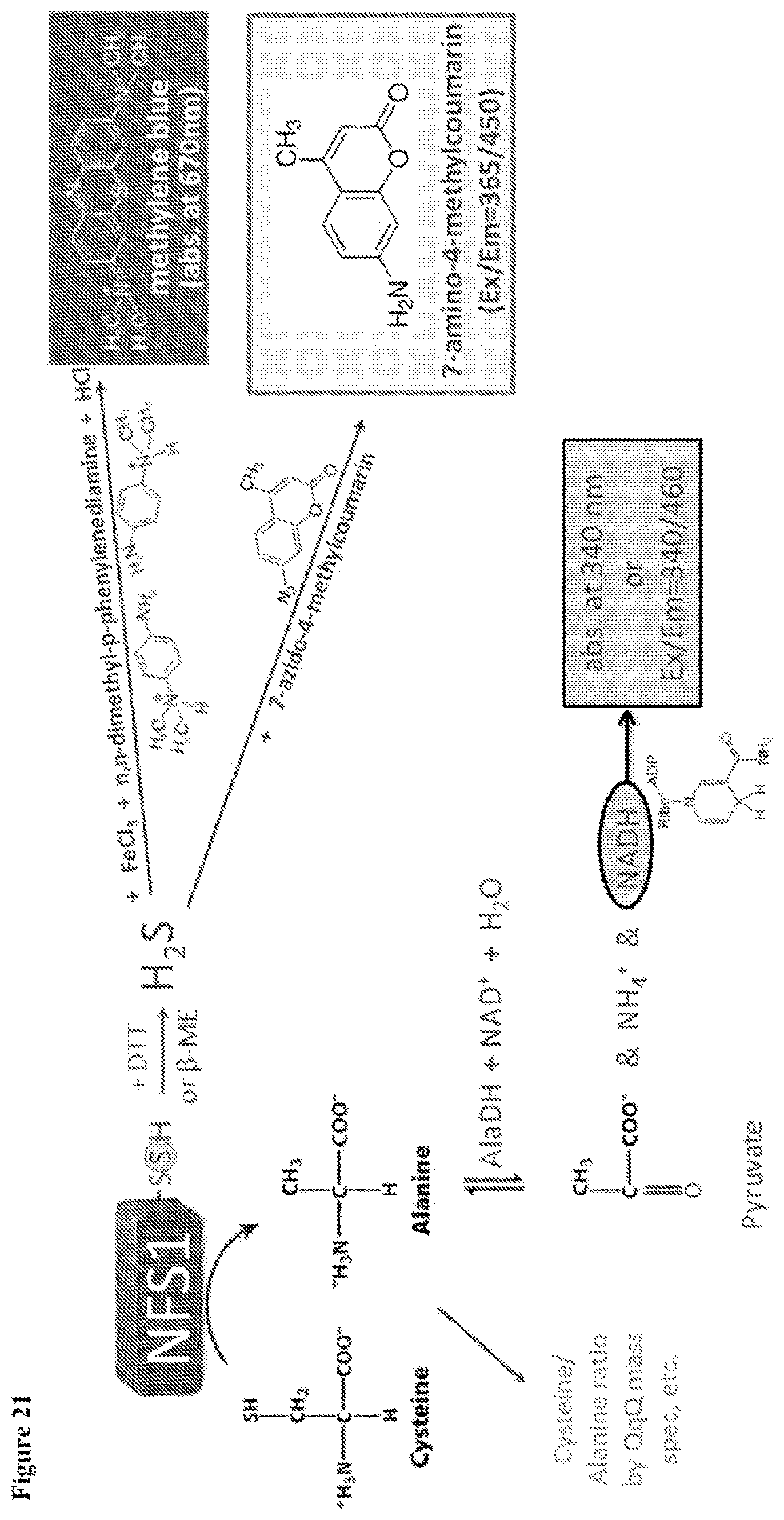
D00019

D00020

D00021

D00022

D00023

D00024

S00001
XML
uspto.report is an independent third-party trademark research tool that is not affiliated, endorsed, or sponsored by the United States Patent and Trademark Office (USPTO) or any other governmental organization. The information provided by uspto.report is based on publicly available data at the time of writing and is intended for informational purposes only.
While we strive to provide accurate and up-to-date information, we do not guarantee the accuracy, completeness, reliability, or suitability of the information displayed on this site. The use of this site is at your own risk. Any reliance you place on such information is therefore strictly at your own risk.
All official trademark data, including owner information, should be verified by visiting the official USPTO website at www.uspto.gov. This site is not intended to replace professional legal advice and should not be used as a substitute for consulting with a legal professional who is knowledgeable about trademark law.