Porcine epidemic diarrhea virus S protein and subunit vaccine thereof as well as method for preparing subunit vaccine and application thereof
Qian , et al. February 23, 2
U.S. patent number 10,925,959 [Application Number 16/600,334] was granted by the patent office on 2021-02-23 for porcine epidemic diarrhea virus s protein and subunit vaccine thereof as well as method for preparing subunit vaccine and application thereof. The grantee listed for this patent is NOVO BIOTECH CORP.. Invention is credited to Zhijun Bai, Guanglin Bian, Ying Che, Yangping Lv, Hong Qian, Sufang Wu, Youqiang Wu, Yulan Xu, Yinhe Zha, Qiang Zhang.


| United States Patent | 10,925,959 |
| Qian , et al. | February 23, 2021 |
Porcine epidemic diarrhea virus S protein and subunit vaccine thereof as well as method for preparing subunit vaccine and application thereof
Abstract
The disclosure discloses a porcine epidemic diarrhea virus S protein and a subunit vaccine thereof as well as a method for preparing the subunit vaccine and application thereof. The vaccine contains 30.about.220 .mu.g of a recombinant porcine epidemic diarrhea virus S protein and a pharmaceutically acceptable ISA 201 VG adjuvant. A method for preparing the subunit vaccine comprises the following steps: (1) cloning the recombinant porcine epidemic diarrhea virus S protein; (2) expressing and purifying the recombinant porcine epidemic diarrhea virus S protein; (3) preparing the recombinant porcine epidemic diarrhea virus S protein prepared in (2) into a water phase; (4) emulsifying the water phase and the ISA 201 VG adjuvant in a volume ratio of 46:54 to obtain a vaccine. The vaccine is high in safety, good in immunogenicity, stable in batches, low in production cost and strong in immunogenicity. On the other hand, CHO cell strains suspending and stably and efficiently expressing the PEDV-S protein are successfully constructed and screened for the first time. The CHO cell strain can express the PEDV-S protein in high yield, the yield can reach 1 g/L, and the expressed PEDV-S protein is easy to purify.
| Inventors: | Qian; Hong (Shaoxing, CN), Wu; Youqiang (Shaoxing, CN), Bian; Guanglin (Shaoxing, CN), Zhang; Qiang (Shaoxing, CN), Xu; Yulan (Shaoxing, CN), Bai; Zhijun (Shaoxing, CN), Wu; Sufang (Shaoxing, CN), Che; Ying (Shaoxing, CN), Lv; Yangping (Shaoxing, CN), Zha; Yinhe (Shaoxing, CN) | ||||||||||
|---|---|---|---|---|---|---|---|---|---|---|---|
| Applicant: |
|
||||||||||
| Family ID: | 1000005375230 | ||||||||||
| Appl. No.: | 16/600,334 | ||||||||||
| Filed: | October 11, 2019 |
Prior Publication Data
| Document Identifier | Publication Date | |
|---|---|---|
| US 20200188508 A1 | Jun 18, 2020 | |
Related U.S. Patent Documents
| Application Number | Filing Date | Patent Number | Issue Date | ||
|---|---|---|---|---|---|
| PCT/CN2018/082908 | Apr 13, 2018 | ||||
Foreign Application Priority Data
| Apr 13, 2017 [CN] | 201710242260.3 | |||
| Apr 3, 2018 [CN] | 201810310540.8 | |||
| Current U.S. Class: | 1/1 |
| Current CPC Class: | C12N 7/00 (20130101); A61K 39/215 (20130101); C12N 2710/14143 (20130101); C12N 2710/14043 (20130101); A61K 2039/5252 (20130101); A61K 2039/552 (20130101) |
| Current International Class: | A61K 39/00 (20060101); A61K 39/12 (20060101); C12N 7/00 (20060101); A61K 39/215 (20060101); C07H 21/02 (20060101) |
References Cited [Referenced By]
U.S. Patent Documents
| 9950061 | April 2018 | Hernandez et al. |
| 2015/0283229 | October 2015 | Hernandez |
| 2018/0064804 | March 2018 | Zhang et al. |
| 2018/0207260 | July 2018 | Hernandez et al. |
| 103992989 | Aug 2014 | CN | |||
| 105311630 | Feb 2016 | CN | |||
| 106085969 | Nov 2019 | CN | |||
Other References
|
Internation Search Report of PCT/CN2018/082908, dated Jul. 20, 2018. cited by applicant. |
Primary Examiner: Chestnut; Barry A
Attorney, Agent or Firm: Erson IP (Nelson IP)
Parent Case Text
CROSS-REFERENCE TO RELATED APPLICATIONS
This application is a continuation of International Patent Application NO: 1 PCT/CN2018/082908 with a filing date of Apr. 13 28, 2018, designating the United States, and further claims priority to Chinese Patent Application No. 201810310540.8 with a filing date of Apr. 3, 2018. The content of the aforementioned applications, including any intervening amendments thereto, are incorporated herein by reference.
Claims
We claim:
1. A porcine epidemic diarrhea virus S protein, having an amino acid sequence of SEQ ID NO:2, and a molecular weight of 210 kDa, wherein the porcine epidemic diarrhea virus S protein is expressed by CHO cells and has a glycosylated modification accounting for 33.3% of the molecular weight thereof.
2. A porcine epidemic diarrhea virus S protein subunit vaccine, the vaccine comprising: the porcine epidemic diarrhea virus S protein according to claim 1 and a pharmaceutically acceptable ISA 201 VG adjuvant, wherein the porcine epidemic diarrhea virus S protein has a concentration of 30-200 .mu.g/vaccine.
3. The vaccine according to claim 2, the vaccine further comprising an immunopotentiator, wherein the immunopotentiator is Quil A having a concentration of 300-500 .mu.g/vaccine.
4. A method for preparing a vaccine comprising the porcine epidemic diarrhea virus S protein according to claim 1, the method comprising the following steps: (1) cloning an OPTI-S having a nucleotide sequence of SEQ ID NO:1, comprising: 1-1) performing codon optimization on a PEDV-S having a nucleotide sequence of SEQ ID NO:3 to obtain the OPTI-S; and 1-2) cloning the OPTI-S to an eukaryotic expression vector to obtain a recombinant plasmid; (2) expressing and purifying the porcine epidemic diarrhea virus S protein; the expressing and purifying the porcine epidemic diarrhea virus S protein comprising the following steps: 2-1) transfecting the recombinant plasmid to a CHO cell strain; 2-2) culturing, screening and acclimating the CHO cell strain in step 2-1) to obtain a highly-expressed cell strain; and 2-3) performing fermentation culture on the cell strain in step 2-2), and purifying the supernatant of the cell to obtain the porcine epidemic diarrhea virus S protein; (3) preparing the porcine epidemic diarrhea virus S protein into a water phase; and (4) emulsifying the water phase and an ISA 201 VG adjuvant as an oil phase in a volume ratio of 46:54 to obtain the vaccine.
5. The method according to claim 4, wherein the water phase further comprises an immunopotentiator which is Quil-A.
6. The method according to claim 4, wherein the eukaryotic expression vector is pEE12.4.
7. The method according to claim 4, wherein the CHO cell is a CHO-K1 cell.
8. The porcine epidemic diarrhea virus S protein according to claim 1, wherein the porcine epidemic diarrhea virus S protein is encoded by a nucleotide sequence of SEQ ID NO: 1.
9. A polynucleotide for encoding the porcine epidemic diarrhea virus S protein according to claim 1, having a nucleotide sequence of SEQ ID NO: 1.
Description
TECHNICAL FIELD
The disclosure relates to a porcine epidemic diarrhea virus S protein and a subunit vaccine thereof, a method for preparing the subunit vaccine and application thereof, and a CHO cell strain suspending and stably and efficiently expressing the porcine epidemic diarrhea virus S protein and a method for constructing and screening the cell strain, belonging to the technical field of animal vaccines and veterinary biological products.
BACKGROUND OF THE PRESENT INVENTION
Porcine epidemic diarrhea (PED) is an intestinal infectious disease which is caused by porcine epidemic diarrhea viruses (PEDV) and has main symptoms such as emesis, diarrhea and dehydration. This disease is susceptible to pigs at various ages, especially, nursed piglets within 7 days old, with the death rate of up to 50%.about.90% after infection. In recent years, the morbidity and the death rate of this disease in China are both increasing, which causes huge economic losses for pig raising industry.
PEDV belongs to coronavirus 1 group of coronaviridae, and has a typical coronavirus morphology. The PEDV particle is polymorphism and trends to be spherical, the periphery of the PEDV particle is enveloped with capsules, the capsule is covered with radial fibrils having an average diameter of about 130 nm. The S protein is a 20 mm ball-arm-shaped glycoprotein extending out of the virus particle capsule, having a molecular weight of about 180-220 kDa and consisting of about 1383 amino acids. This protein is rich in cysteine and contains 29 potential N-glycosylation sites, however, the N-glycosylation sites cannot be cleaved by protease after the virus particles are matured, which greatly reduces the cell fusion and infection of this virus and is also one of reasons why PEDV artificial cells are difficultly cultured. According to similarity of PEDV to conserved sequences of other coronavirus S proteins, the PEDV S protein is divided into two structural domains S1 (1-789 aa) and S2 (790-1383 aa) in which S1 is located on the surface of the virus, has main effects of identifying a receptor and binding to a host cell receptor, and mediating the generation of a neutralizing antibody. S2 is mainly in charge of fusion of a virus capsule and a host cell membrane and conduction of virus RNA into a host cell, thereby causing the infection of cells. Furthermore, the PEDV S protein is also an immunogen protein inducing a humoral immune response of a host. Therefore, the PEDV S protein is a candidate protein for developing a genetic engineering subunit vaccine at present.
Currently, vaccines for preventing and controlling porcine epidemic diarrhea are all almost traditional PEDV attenuated vaccines and inactivated vaccines. But, the inactivated vaccine has weak autoimmune protection force, and has incomplete inactivation to cause a risk of detoxification; the attenuated active vaccine has a risk of reversion to virulence of toxic strains. With quick development and continuous deepening research of a molecular biological technique, researchers start to convert development of PEDV vaccines into development of genetic engineering vaccines having potential advantages. This vaccine has the advantage of safety, efficiency, small side effect, high expression quantity, industrial production and the like. In recent years, many researches focus on expression of a PEDV S1 protein and research of vaccines (for example proteins at sites 21-789 in an expression S1 region of an insect rhabdovirus used in the Chinese invention patent with application number 201610348237.8), and tandem expression of core regions of the S protein (for example 3 core regions of Escherichia coli expressing the S protein in series used in the Chinese invention patent with application number 201610256701.0). This is mainly because the PEDV S protein is too large (about 1383aa), gene cloning or protein expression and preparation is difficultly implemented in the technical level of molecular biology. However, a truncated or incomplete PEDV protein is only used as an antigen to result in shortages of incomplete epitope, poor overall immunogenicity relative to a full-length protein and the like, and cause inadequate immune protection. In addition, most of the current expression systems are protokaryon or insect rhabdovirus expression systems that generally have no galactosylated modification or have inadequate galactosylated modification, thereby leading to a fact that the immunogenicity of the expressed protein is inferior to that of the protein of virus particle itself and possibly resulting in inadequate protection strength.
The CHO cell was isolated from an ovary of an adult female hamster in 1957 by Dr. Theodore T. Puck from University of Colorado, and is an epithelial adherent cell. This cell is immortalized, can be passed for more than one hundred generations, and is widely used in bioengineering at present. Relative to other expression systems, the CHO cell has the following advantages: (1) it has an accurate post-transcriptional modification function, and the expressed protein is closest to a natural protein in molecular structure, physicochemical properties and biological functions; (2) it can not only grow adherently but also undergo suspension culture, and has high shear and osmotic pressure tolerance; (3) it has efficient amplification and expression capability of recombinant genes, and exogenous protein genes are stably integrated; (4) it has a product extracellular secretion function, and seldom secretes its own endogenous protein, which facilitates the isolation and purification of downstream protein products; (5) it can be cultured at high density in a suspension culture manner or in a serum-free culture medium, and can be produced on large scale since the culture volume can reach more than 1,000 L.
There are many types of CHO cells, such as DG44, DXB11, HO K1 and CHO-S. Since 1980s.about.1990s, a DHFR (dihydrofolate reductase-deficient) gene amplification and screening system is industrially used at early stage, and its host cell strain is DG44. When the cell culture medium contains methotrexate (MTX), the dihydrofolate reductase is inhibited, and then this gene is amplified by feedback regulation. The genes within the upstream and downstream range of 100-1,000 kb can be amplified accordingly, and therefore the target genes are inserted into this site range so as to be amplified. Now, many monoclonal antibody production systems are still DG44 DHFR systems. A GS (glutamine synthase) amplification system, which takes CHO-K1 as a host cell, is a novel gene amplification and screening system developed in recent years. It has obvious superiority over the DHFR system and is widely recognized and used throughout the world. Its principle is that GS utilizes ammonia in the cell and glutamic acid to synthesize glutamine while providing energy for ATP hydrolysis. Addition of GS inhibitor L-methionine sulfoxide ammonium (MSX) in a glutamine-deficient culture medium can effectively amplify the GS gene and its bound target gene, thus achieving a purpose of improving the expression level of the target gene. This system has the main advantages: (1) the gene-deficient CHO-K1 cell strain does not need to be used as the host cell; (2) the CHO-K1 cell is stronger and easy to culture; (3) glutamine does not need to be added in the culture medium to avoid a problem that the level of ammonia in the culture system is high due to glutamine decomposition, which reduces the difficulty of process control, and effectively improves cell fermentation density and prolongs cell survival time,
However, when the inventor of the disclosure first uses CHO cells to express the PEDV-S protein, it was found that the CHO cells basically do not express the PEDV-S protein when the gene of the PEDV-S protein is not optimized. Therefore, the inventor of the disclosure takes a note that when the CHO cells are used to express the PEDV-S protein, the optimization of a gene sequence is an urgent problem to be solved.
SUMMARY OF PRESENT INVENTION
The technical problem to be solved by the disclosure is, first, to provide a porcine epidemic diarrhea virus S protein which can be industrially produced on large scale and a subunit vaccine thereof as well as a method for preparing the subunit vaccine; second, to overcome the problem that the full-length PEDV S protein is difficult to efficiently express in mammalian cells at present; third, to overcome the defects and risks of inactivated vaccines and attenuated vaccines in preventing and controlling porcine epidemic diarrhea.
According to one aspect of the disclosure, the disclosure provides a porcine epidemic diarrhea virus S protein, wherein the porcine epidemic diarrhea virus S protein is a protein consisting of amino acids shown in SEQ ID NO:2, or a derived protein obtained by performing substitution, deletion or addition of one amino acid or several amino acids on an amino acid sequence shown in SEQ ID NO:2 and having porcine epidemic diarrhea virus S protein immunogenicity. Preferably, the porcine epidemic diarrhea virus S protein is a protein which is expressed by CHO cells and contains high glycosylation. The molecular weight of the glycosylated protein accounts for about 33.3% of that of the porcine epidemic diarrhea virus S protein.
In the technical solution of the disclosure, preferably, the molecular weight of the porcine epidemic diarrhea virus S protein in SDS-PAGE is 210 kDa.
In the technical solution of the disclosure, preferably, the molecular weight of the deglycosylated porcine epidemic diarrhea virus S protein in SDS-PAGE is 140 kDa.
According to another aspect of the disclosure, the disclosure provides a porcine epidemic diarrhea virus S protein subunit vaccine, the vaccine comprising 30.about.200 .mu.g of a porcine epidemic diarrhea virus S protein according to any one of claims 1.about.3 and a pharmaceutically acceptable ISA 201 VG adjuvant.
In the technical solution of the disclosure, preferably, the pharmaceutically acceptable adjuvant is an oil in water adjuvant (such as ISA 28 VG adjuvant), a water in oil in water adjuvant (such as ISA 206 VG adjuvant), an oil in water adjuvant (such as ISA 660 VG adjuvant), or a water adjuvant (such as alumina gel adjuvant and ISA 251 VG adjuvant), preferably, ISA 201 VG adjuvant.
In the technical solution of the disclosure, the vaccine also comprises an immunopotentiator; preferably, the immunopotentiator is Quil-A; preferably, the concentration of Quil-A is 300.about.500 .mu.g/vaccine, preferably, the concentration of Quil-A is 400 .mu.g/vaccine.
According to another aspect of the disclosure, the disclosure also provides a method for preparing a porcine epidemic diarrhea virus S protein subunit vaccine, the method comprising the following steps: (1) cloning a gene of a porcine epidemic diarrhea virus S protein; the cloning a gene of a porcine epidemic diarrhea virus S protein comprising the following steps: 1-1) performing codon optimization on the nucleotide sequence of the porcine epidemic diarrhea virus S protein to obtain OPTI-S; and 1-2) cloning the OPTI-S to an eukaryotic expression vector to obtain a recombinant plasmid; (2) expressing and purifying the recombinant porcine epidemic diarrhea virus S protein; the expressing and purifying the recombinant porcine epidemic diarrhea virus S protein comprising the following steps: 2-1) transfecting the recombinant plasmid containing the porcine epidemic diarrhea virus S protein coding gent to a CHO cell strain; 2-2) culturing, screening and acclimating the CHO cell strain in step 2-1) to obtain a highly-expressed cell strain; and 2-3) performing fermentation culture on the cell strain in step 2-2), and purifying to obtain a recombinant porcine epidemic diarrhea virus S protein; (3) preparing the recombinant porcine epidemic diarrhea virus S protein prepared in step (2) into a water phase; and (4) emulsifying the water phase and an ISA 201 VG adjuvant in a volume ratio of 46:54 to obtain a vaccine.
In the technical solution of the disclosure, preferably, the water phase also comprises an immunopotentiator; preferably, the immunopotentiator is Quil-A, and the concentration of Quil-A is 400 .mu.g/vaccine.
In the technical solution of the disclosure, preferably, the nucleotide sequence of the OPTI-S is as shown in SEQ ID NO:1.
In the technical solution of the disclosure, preferably, the eukaryotic expression vector can be pEE 6.4, pEE12.4, Pgl4.13 or pcDNA3.1, preferably, the eukaryotic expression vector is pEE12.4.
In the technical solution of the disclosure, preferably, the CHO cell can be DG44, DXB11, CHO-K1 or CHO-S cell strains. Preferably, the CHO cell is a CHO-K1 cell.
The disclosure also provides an application of a porcine epidemic diarrhea virus S protein in preparation of a porcine epidemic diarrhea virus S protein subunit vaccine and a relevant diagnostic reagent.
The disclosure also provides an application of a porcine epidemic diarrhea virus S protein recombinant subunit vaccine in preparation of a drug for preventing and treating porcine epidemic diarrhea.
In embodiments of the disclosure, the molecular weight of the purified PEDV-S protein is about 210 kDa; after the PEDV-S protein is digested using deglycosylase, it is found that the molecular weight of the deglycosylated PEDV-S protein is only about 140 kDa, which is consistent to the molecular weight, which is about 143 kDa, of the S protein obtained through amino acid sequence analysis of the S protein. This result indicates that there is a large amount of glycosylated modification, which accounts for about 33.3% of the molecular weight of the whole PEDV-S protein, in the PEDV-S protein expressed by using our CHO eukaryotic expression system.
In embodiments of the disclosure, when the purified PEDV-S protein is deglycosylated and then subjected to Western blot detection using PEDV pig high immune serum, it is found that the deglycosylated PDEV-S protein can not bind to the serum or weakly binds to the serum, indicating that glycosylation of the PEDV-S protein is essential for maintaining the immunogenicity of the PDEV-S protein.
On the one hand, the disclosure explicitly proposes the subunit vaccine prepared by using the PEDV-S protein for the first time. This vaccine overcomes the disadvantages of the prior art that the truncated or incomplete PEDV-S protein is only used as the antigen so as to possibly result in incomplete antigen epitope and relatively poor overall immunogenicity in addition to the advantages of high safety, good immunogenicity, stable batches, low production cost and the like; on the other hand, the CHO cell strain suspending and efficiently secreting and expressing the porcine epidemic diarrhea virus S protein is successfully constructed and screened for the first time, this cell strain can express the porcine epidemic diarrhea virus S protein in high yield, with the yield of up to 1 g/L. The expressed porcine epidemic diarrhea virus S protein is easy to purify (the target protein can be purified only from cell supernatant, because the cell supernatant contains a few of hybrid proteins, purification is relatively convenient and fast; if it is needed to break cells when in purification, purification of the target protein is not facilitated due to many hybrid proteins existing in the cells), and therefore it is easy to produce the porcine epidemic diarrhea virus S protein on a large scale to meet the needs of industrialization, and the subunit vaccine prepared by this protein has good immunogenicity and can induce the pigs to only produce good immune response.
DESCRIPTION OF THE DRAWINGS
FIG. 1 shows that THMM software predicts a PEDV-S protein transmembrane domain.
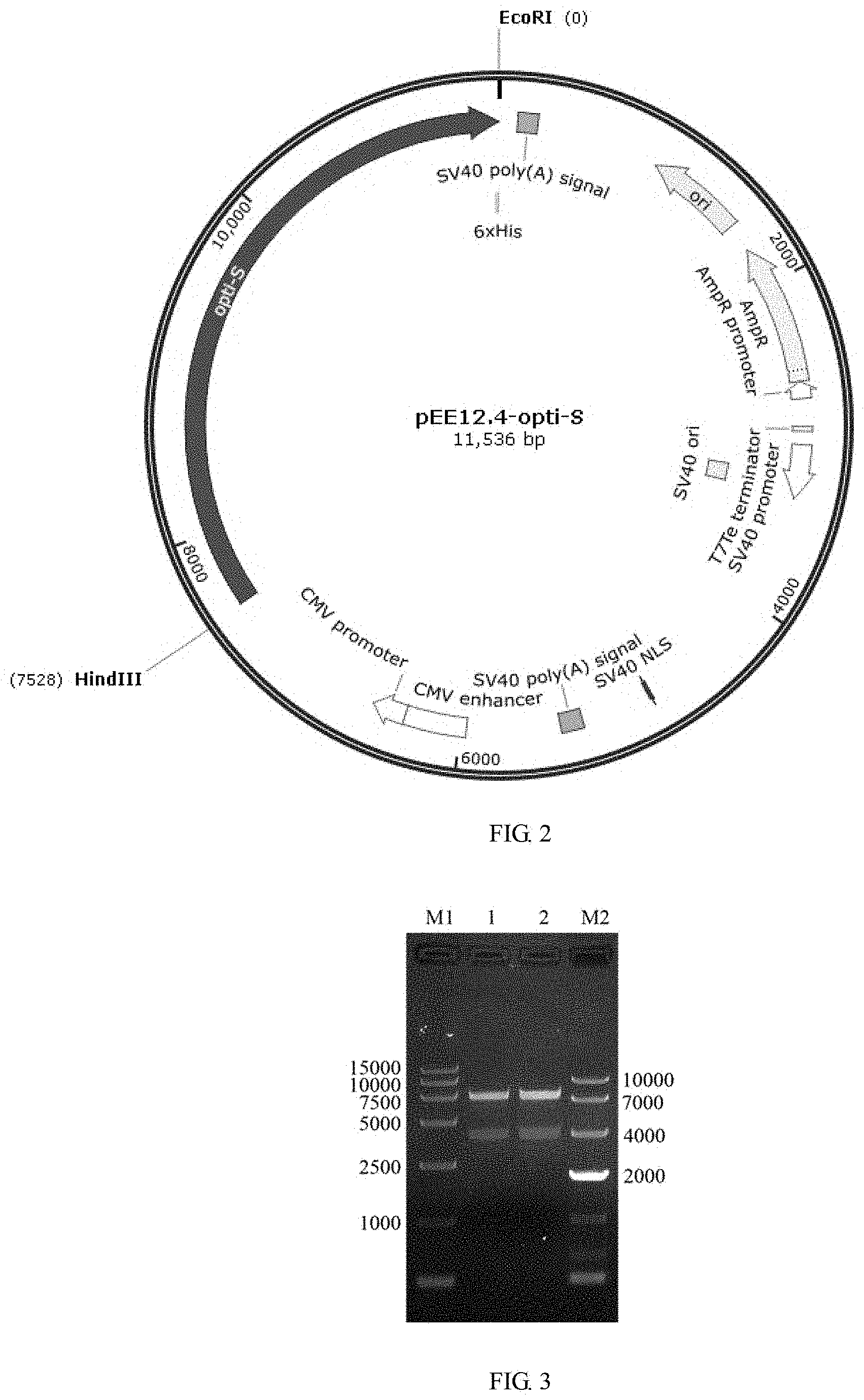
FIG. 2 shows a pEE12.4-OPTI plasmid profile.
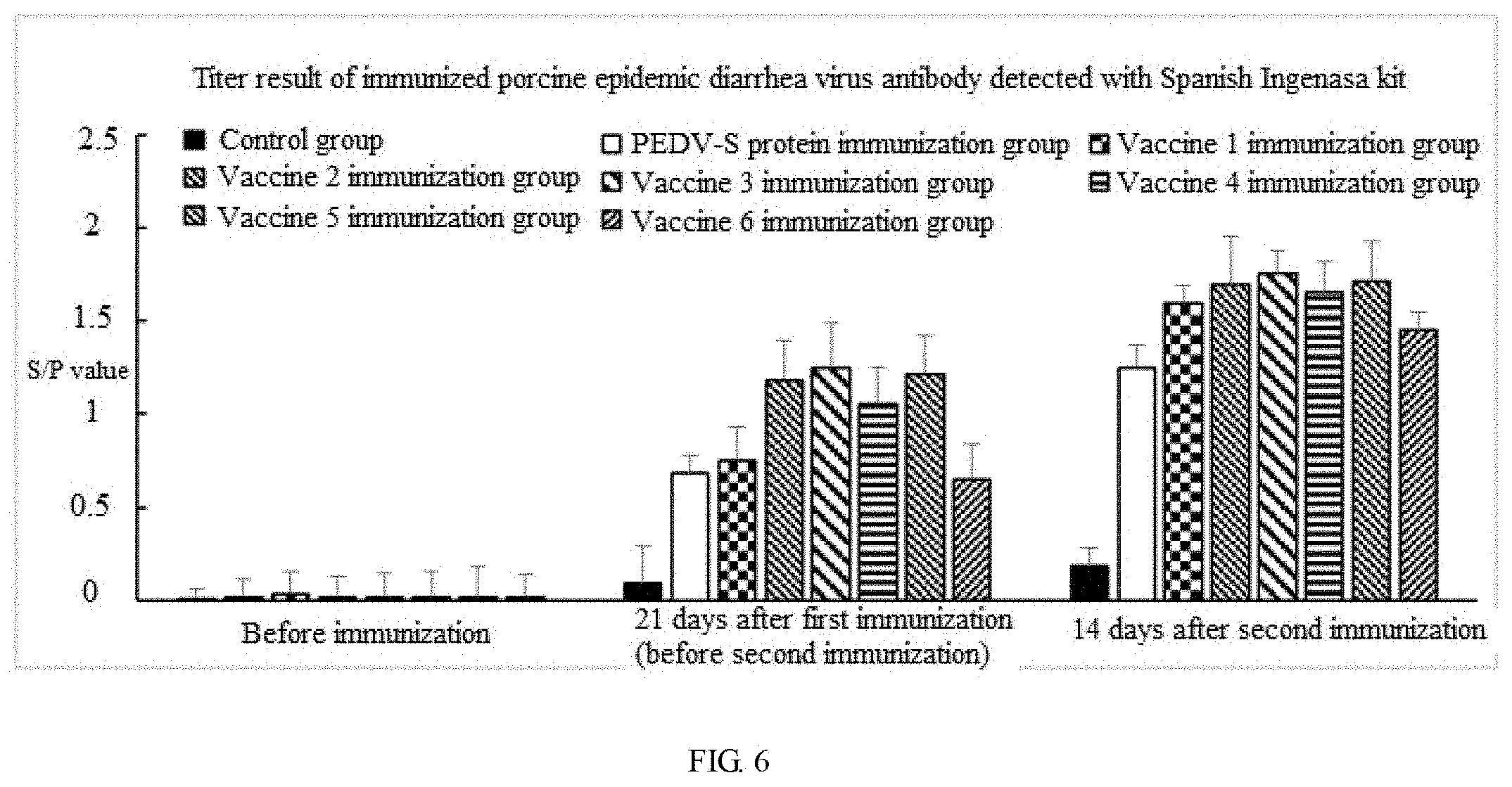
FIG. 3 shows a pEE12.4-OPTI-S double-digestion identification result. 1 and 2 represent PEDV-opti-S plasmid utilizes EcoRI/HindIII double digestion, the size of a vector is about 7,528 kb, the size of a target segment is about 3,930 bp, and the digestion is correct; M1:DL5,000 marker, and M2:DL10,000 marker.
FIG. 4 shows a purification result of a PEDV-S protein expressed by fermentation of a 3C5 monoclonal cell strain detected by SDS-PAGE. Fermentation verification is carried out on the 3C5 monoclonal cell strain, cell culture supernatant is collected, protein purification is performed, and the protein expression level of PEDV-S is detected by SDS-PAGE.
FIG. 5A shows results of deglycosylation of purified PEDV-S protein detected by SDS-PAGE and Western Blot. 1-5 are SDS-PAGE detection results, 1 is Marker, 2 is negative control 1.times.PBS, 3 and 5 are non-deglycosylated PEDV-S proteins, and 4 is deglycosylated PEDV-S protein; 6-10 are Western Blot results, 6 is Marker, 7 is negative control 1.times.PBS, 8 and 10 are non-deglycosylated PEDV-S proteins, and 9 is deglycosylated PEDV-S protein.
FIG. 5B shows a molecular sieve detection result of PEDV-S protein.
FIG. 5C shows a chromatogram of a superdex 200 PG column standard sample, in which the ferritin peak volume is 54.1 ml, the molecular weight is 440 kDa, the aldolase peak volume is 65.4 ml, the molecular weight is 158 kDa, the conalbumin peak volume is 73.0 ml, the molecular weight is 75 kDa, the ovalbumin peak volume is 80.0 ml, the molecular weight is 43 kDa, the carbonic anhydrase peak volume is 87.9 ml, the molecular weight is 29 kDa, the ribonuclease A peak volume is 95.7 ml, t hmolecular weight is 13.7 kDa, the aprotinin peak volume is 104.3 ml and the molecular weight is 6.5 kDa.
FIG. 6 shows a titer detection result after immunization.
FIG. 7A shows comparison results of a PEDV-S protein nucleotide sequence before and after optimization, where OPTI-S (having a nucleotide sequence of SEQ ID NO: 1) represents a sequence after optimization, and PEDV-S (having a nucleotide sequence of SEQ ID NO: 3) represents a sequence before optimization.
FIG. 7B is a continuation of FIG. 7A, and shows comparison result of a PEDV-S protein nucleotide sequence before and after optimization.
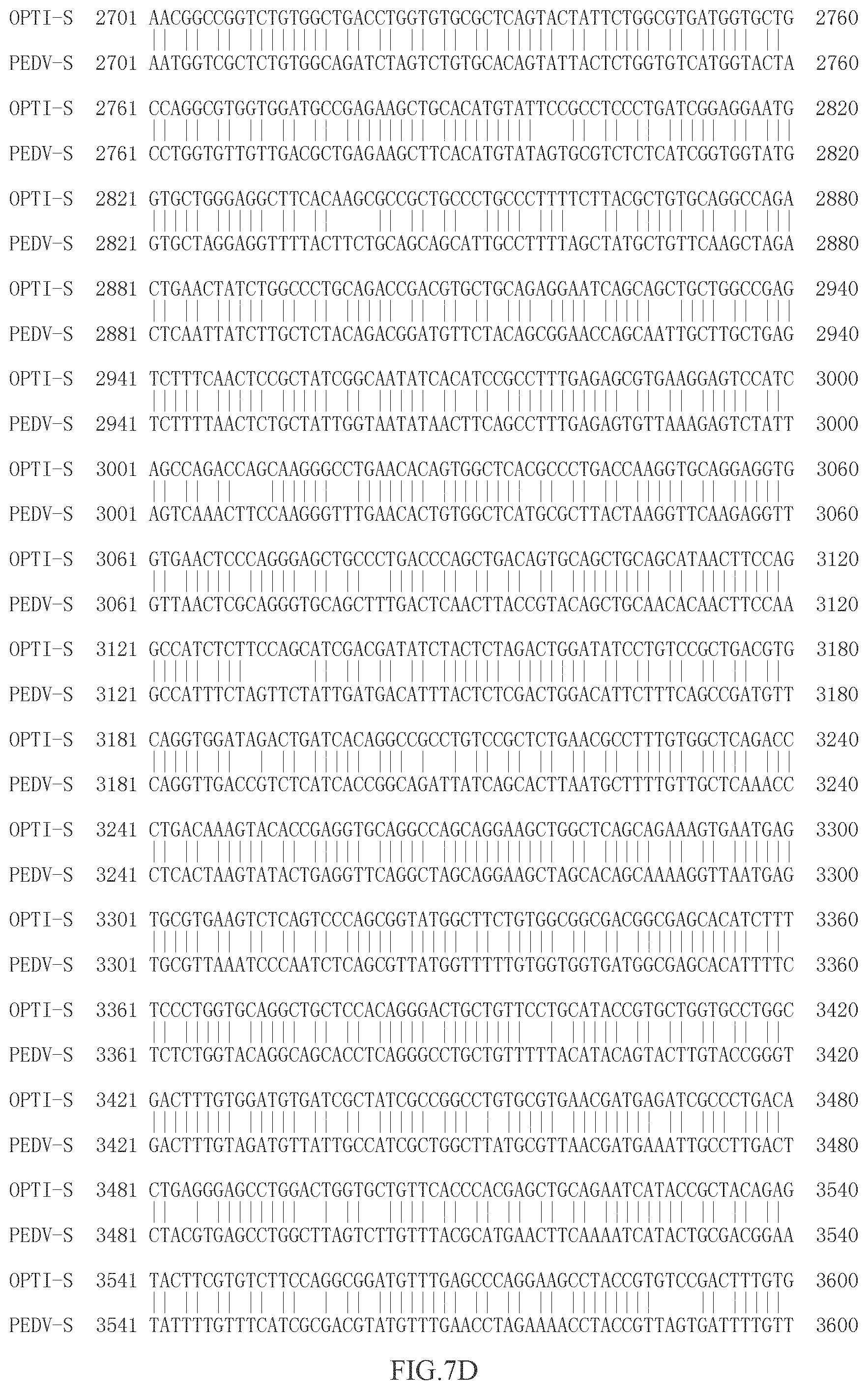
FIG. 7C is a continuation of FIG. 7B, and shows comparison result of a PEDV-S protein nucleotide sequence before and after optimization.
FIG. 7D is a continuation of FIG. 7C, and shows comparison result of a PEDV-S protein nucleotide sequence before and after optimization.
FIG. 7E is a continuation of FIG. 7D and shows comparison result of a PEDV-S protein nucleotide sequence before and after optimization.
DETAILED DESCRIPTION OF PREFERRED EMBODIMENTS
The disclosure will be further described in combination with accompany drawings and embodiments. Embodiments of the disclosure are only for illustrating the technical solution of the disclosure, but not limiting the disclosure.
Strains, plasmids and reagents used in embodiments of the disclosure are all commercially available products.
Reagents and medicines used in the disclosure are listed as follows:
CHO-K1 cells are derived from the cell bank of the Institute of life sciences, Chinese Academy of Sciences, and the cell bank of the Institute of life sciences, Chinese Academy of Sciences.
Cell culture mediums and serums are purchased from Gibco Company.
The eukaryotic expression vector pEE12.4 is purchased from the Lin Yuan Biotechnology Co., Ltd.
Ammonium methionine sulfoxide (MSX) is purchased from Sigma Company.
The BCA protein quantitative kit is purchased from Thermo Fisher, USA.
Glycosidase F is purchased from New England Biolabs (UK) Ltd.
HRP-labeled sheep anti-pig IgG II is purchased from EarthOx Life Science.
Quil-A is purchased from Brenntag Biosector.
ISA 201 VG is purchased from France's cyber company.
Example 1: Selection and Codon Optimization of Porcine Epidemic Diarrhea Virus S Protein Gene
A PEDV-S protein is a transmembrane protein, which contains an extracellular region, a transmembrane region and an intracellular region. With a typical toxic strain CV777 as reference, the transmembrane region was predicted by TMHMM software (see FIG. 1), a PEDV-S toxic strain that was prevalent in Zhejiang Province in recent years and had been published was used as a template (GenBank: KF840553.1) to design a primer, the sequence of the PEDV-S extracellular region (20D-1320T) was cloned from a pig farm in Zhejiang Province to obtain a PEDV-S nucleotide sequence. The PEDV-S nucleotide sequence was subjected to codon optimization to obtain an OPTI-S sequence as shown in SEQ ID NO: 1, and this work was authorized to Kingsy Biotechnology Co., Ltd.
The sequence (OPTI-S) after optimization was compared with the sequence (PEDV-S) before optimization, it was found that their homology was only 74.1% (see FIG. 7A-7D). We also used the sequence (PEDV-S) before optimization to perform CHO expression, but cannot detect expression of PEDV-S protein, or expression was extremely low and basically cannot be detected. Thus, optimization of the sequence of the PEDV-S protein is an essential step of expression of PEDV-S protein in CHO.
Example 2: Construction of pEE12.4-OPTI-S Recombinant Plasmid
2.1 Amplification of Target Segment OPTI-S Via PCR
2.1.1 PCR Reaction
(1) Design and Synthesis of a Primer
TABLE-US-00001 Upstream primer: 5'-CGAAGCTTGCCGCCACCATGGACGTGACCAGGTGCTCTG-3' Downstream primer: 5'-CGGGAATTCTCAATGGTGATGGTGATGGTGGATATAGGTCTC CAC-3'
(2) Loading System 50 .mu.L, as Shown in a Table Below:
TABLE-US-00002 Loading components Volume (.mu.L) Q5 Mix 25 Upstream primer (10 .mu.M) 2.5 Downstream primer (10 .mu.M) 2.5 OPTI-S 1 dd H.sub.2O 19 Total volume 50
PCR Amplification Process:
TABLE-US-00003 95.degree. C. 2 min 95.degree. C. 30 s 55.degree. C. 45 s {close oversize brace} 30 cycles 72.degree. C. 1 min 72.degree. C. 10 min 8.degree. C. forever
2.1.2 Gel Recovery of PCR Products
(1) A sample collection tube, an adsorption column and a collection tube were marked;
(2) the marked hollow EP tube was weighed and its weight was recorded;
(3) a single target DNA strip was carefully cleaved using a scalpel from agarose gel on a gel cleaving instrument and then put into a 1.5 mL clean centrifuge tube;
(4) 600 .mu.L of PC buffer was added to the 1.5 mL of clean centrifuge tube in step (3), and then the tube was placed in 50.degree. C. water bath for about 5 min;
(5) column equilibrium: 500 .mu.L of balance liquid BL was added to an adsorption column CB2 (the adsorption column was put in the collection tube in advance) and then centrifuged for 1 min at 12,000 rpm/min, waste liquid in the collection tube was discarded, and then the adsorption column was put in the collection tube again;
(6) the solution obtained in step (5) was added to the adsorption column CB2 to stand for 2 minutes, the adsorption column was centrifuged for 30 s at 10,000 rpm/min, the waste liquid in the collection tube was discarded, and then the adsorption column CB2 was put into the collection tube;
(7) 600 .mu.L of rinsing solution PW buffer was added into the adsorption column to stand for 3 min, the adsorption column was centrifuged for 30 s at 10,000 rpm/min, the waste liquid in the collection tube was discarded, and the adsorption column CB2 was put into the collection tube;
(8) the step (7) was repeated;
(9) the empty adsorption column was centrifuged for 2 min at 12000 rpm/min, rinsing solution was removed as much as possible, and the adsorption column was placed at room temperature for 10 min to be thoroughly dried in air;
(10) the adsorption column CB2 was put in the collection tube, and 50 .mu.L of Elution buffer (65.degree. C. preheating) was dropwise added in the middle position of an adsorption membrane in a suspending manner to stand for 3 min, and the collection tube was centrifuged for 2 min at 12,000 rpm/min;
(11) the centrifugal tube in step (10) was taken out from a centrifugal machine, the adsorption column CB2 in the middle was discarded, the cap of the centrifugal tube is covered, and the DNA sample in the centrifugal tube is retained;
(12) the DNA samples in step 11 were stored at 4.degree. C., and agarose gel electrophoresis identification gel is prepared to recover the DNA segments.
2.2 PCR Products and Vector Double-Digestion Reaction
(1) a 1.5 mL EP tube required to be used was marked, loading and mixing were carried out in the 1.5 mL EP tube according to a table below: 50 .mu.L reaction system
TABLE-US-00004 Loading component names Volume (.mu.L) dd H.sub.2O Supplement to 50 10 .times. buffer 5 DNA sample Volume in 2 .mu.g Hind III 2.5 EcoR I 2.5
(2) the 1.5 mL EP tube in step (1) was placed in a thermostat water bath pot with a corresponding enzyme optimum temperature to carry out water bath for 2-3 h;
(3) recovery of double-digestion product gel: the above double-digestion system was taken out to undergo agarose gel electrophoresis to recover the DNA segment therein. The method is the same as the method for recovery of PCR product gel in 1.2.1.
2.3 Ligation Reaction
(1) Several 1.5 mL EP tubes were prepared to be placed on an EP tube shelve for standby;
(2) loading and uniform mixing were carried out in the 1.5 mL EP tubes according to a table below;
TABLE-US-00005 Loading Experiment Blank group component names group (.mu.L) (.mu.L) dd H.sub.2O / 6 10 .times. T4 ligation buffer 1 1 Target segment 6 -- Vector 2 2 T4 ligase 1 1
(3) after loading was completed according to the table in step (2), each of 10 .mu.l reaction system was placed in 16.degree. C. low-temperature cooling liquid circulator to undergo water bath for 10-16 h;
(4) the EP tubes in step (3) were taken out and placed in 65.degree. C. water bath pot to undergo water bath for 15 min;
(5) the EP tubes in step (4) were taken out and stored at 4.degree. C.
1.2.4 Transformation Reaction
(1) 10 .mu.L of ligation reaction solution was quickly added to 100 .mu.L of competent cells and uniformly mixed by blowing, and the mixture was subjected to ice bath for 30 min;
(2) the sample tube was taken out and placed in 42.degree. C. water bath for 100 s, and then immediately subjected to ice bath for 2 min;
(3) the sample tube was taken out, add 600 .mu.L of liquid LB culture medium was added in the sample tube on a super-clean workbench, and then the sample tube was placed in a 37.degree. C. constant temperature shaker to be cultured for 1 h at 220 rpm/min;
(4) paving: the sample tube in step (3) was taken out and centrifuged for 2 min at 8,000 rpm/min under room temperature, 600 .mu.L of supernatant was removed, then bacteria at the bottom of the tube were re-suspended with the remained supernatant, the re-suspended bacteria solution was put in the center of a corresponding transformation plate, and the bacteria solution in the center of the transformation plate was evenly paved with a coating rod;
(5) the plate in transformation step (4) was placed in a biochemical incubator to be cultured for 1 h at 37.degree. C., and then the transformation plate was inverted and cultured for 15 h;
(6) the transformation results were observed.
2.5 Plasmid Extraction and Double-Digestion Identification
2.5.1 Plasmid Extraction
(1) Monoclonal antibodies were picked with a 10 .mu.L pipette gun head from the transformation plate to 5 mL of ampicillin-resistant LB liquid culture medium to be shaken under 37 C at 220 rpm/min for overnight;
(2) the bacterial liquid was transferred to the 1.5 mL EP tube and centrifuged for 2 min at 12,000 rpm/min under room temperature, and the supernatant was discarded;
(3) 250 .mu.L of plasmid extraction reagent P1 buffer was added to the EP tube in step (2) to completely suspend the bacteria;
(4) 250 .mu.L of P2 buffer was added to the solution in step (3), the centrifugal tube was immediately and gently inverted for 5-10 times to be uniformly mixed, and the centrifugal tube stood for 2-4 min at room temperature;
(5) 350 .mu.L of P3 buffer was added to the solution in step (4), and the centrifugal tube was immediately and gently inverted for 5-10 times to be uniformly mixed, and the centrifugal tube stood for 2-4 min at room temperature;
(6) the solution in step (5) was centrifuged for 10 min at 14,000 rpm/min under room temperature;
(7) the supernatant solution in step (6) was transferred to the center of the adsorption column, the adsorption column was centrifuged for 30 s at 12,000 rpm/min under the room temperature, and the liquid in the collection tube was discarded;
(8) 500 .mu.L of Buffer DW1 was added to the center of the adsorption column, the adsorption column was centrifuged for 30 s at 12,000 rpm/min under room temperature, and the liquid in the collection tube was discarded;
(9) 500 .mu.L of wash solution was added to the center of the adsorption column, the adsorption column was centrifuged for 30 s at 12,000 rpm/min under room temperature, and the liquid in the collection tube was discarded and then this step was repeated again;
(10) the empty adsorption column was centrifuged for 2 min at 12,000 rpm under room temperature;
(11) the adsorption column was placed in a 1.5 .mu.L clean centrifugal tube, and 30 .mu.L of Elution buffer was added to the center of the adsorption membrane, the centrifugal tube stood for 5 min at room temperature and then centrifuged for 2 min at 12,000 rpm under room temperature, and the DNA solution in the tube was preserved.
2.5.2 Double-Digestion Identification
(1) a 1.5 mL EP tube required to be used was marked, loading was performed according to a table below: 20 .mu.L reaction system;
TABLE-US-00006 Loading component names Volume (.mu.L) dd H.sub.2O Supplement to 20 .mu.L 10 .times. buffer 2 DNA sample Volume when mass is 1 .mu.g HindIII 1 EcoR I 1
(2) the 20 .mu.L EP tube reaction system in step (1) was placed in a 37.degree. C. constant temperature water bath pot to undergo water bath for 2 h;
(3) the double-digestion system sample in step (2) was subjected to agarose gel electrophoresis to check whether the size of the inserted segment is correct. The experimental results are shown in FIG. 3, 1 and 2 indicate that the PEDV-opti-S plasmid utilizes EcoRI/HindIII double digestion, the size of the vector is about 7,528 bp, the size of the target segment is about 3,930 bp, and the digestion is correct; M1:DL15,000 maker, M2:DL10,000 maker.
(4) the clones where the inserted segment was correct were selected to be sequenced in a sequencing company.
2.6 Maxi Extraction of Endotoxin-Free Plasmids
2.6.1 Extraction of Endotoxin-Free Plasmid
(1) Clones which were correctly sequenced were inoculated into 100 mL ampicillin-resistant culture medium to be cultured in a 37.degree. C. constant temperature shaker for 15 h at 220 rpm/min;
(2) the cultured bacteria liquid in step (1) was transferred to a 50 mL centrifugal tube to be centrifuged for 5 min at 8,000 rpm/min under room temperature, the bacteria were collected and the supernatant medium was discarded;
(3) 8 mL of solution P1 was added to the centrifugal tube in step (2) and the bacteria was fully re-suspended with a pipette;
(4) 8 mL of solution P2 was added to the centrifugal tube in step (3), the centrifugal tube was immediately and gently inverted for 6-8 times, and stood for 5 min at room temperature;
(5) 8 mL of solution P4 was added to the centrifugal tube in step (4) and the centrifuge tube was immediately inversed up and down for 6-8 times to be sufficiently and uniformly mixed until white flocculent precipitates occur in the solution, the centrifugal tube was placed for about 10 min at room temperature and centrifuged for 5-10 min at 8,000 rpm/min under room temperature, so that the white precipitates are separated at the bottom of the tube;
(6) the supernatant in step (5) was completely and carefully transferred into a filter CS1, the handle of the filter was slowly pushed, and the filtrate was collected in the 50 mL clean centrifugal tube;
(7) column equilibrium: 2.5 mL of balance liquid BL was added to the adsorption column CP6 (the adsorption column was put into 50 mL collection tube), the adsorption column was centrifuged for 2 min at 8,000 rpm/min under room temperature, the waste liquid in the collection tube was discarded, and the adsorption column was re-placed in the collection tube;
(8) isopropanol whose volume is 0.3 time that of filtrate was added to the filtrate in step (6), the isopropanol and the filtrate were inversed up and down and uniformly mixed and then transferred to the adsorption column CP6, the adsorption column was centrifuged for 2 min at 8,000 rpm/min under room temperature, the liquid in the collection tube was discarded, and the adsorption column CP6 was put into the same collection tube again;
(9) 10 mL of rinsing solution PW was added to the adsorption column CP6 in step (8), the adsorption column was centrifuged for 2 min at 8,000 rpm/min under room temperature, the waste liquid in the collection tube was discarded, and the adsorption column was placed into the collection tube again;
(10) operation step (9) was repeated once;
(11) 3 mL of absolute ethanol was added to the adsorption column CP6 in step (10), the adsorption column was centrifuged for 2 min at 8,000 rpm/min under room temperature, and the waste liquid was discarded;
(12) the adsorption column CP6 in step (11) was placed in the collection tube again, the collection tube was centrifuged for 5 minutes at 8,000 rpm/min under room temperature, the adsorption column CP6 was uncovered and placed at room temperature for several minutes to be dried in air;
(13) the adsorption column in step (12) was put into the 50 mL clean centrifugal tube, 1-2 mL of buffer TB was added into the center of the adsorption membrane, the centrifugal tube stood for 5 min at room temperature and then centrifuged for 2 min at 8,000 rpm/min under room temperature, the eluent in the 50 mL centrifugal tube was completely transferred into the 1.5 mL clean centrifugal tube, the concentration was measured, and the centrifugal tube was stored at -20 C;
(14) 1-2 ml of the obtained plasmid DNA solution was taken and subjected to agarose gel electrophoresis, and electrophoretic result data were preserved.
Example 3: Transfection of CHO-K1 Cells with pEE12.4-OPTI-S Recombinant Plasmids and Establishment of Monoclonal Screening
3.1 CHO-K1 Cell Transfection
(1) Preparation: ultraviolet sterilization for 30 minutes in a bio-safety cabinet; DMEM/F, 12 (containing 10% of serum and 1% of double antibody), DMEM/F 12 and PBS were preheated to 37.degree. C. in a 37.degree. C. water bath pot.
(2) Cells (10 cm cell culture dish) were taken out from a 37.degree. C. incubator, the supernatant culture medium was discarded, the cells were washed once with 8 mL of PBS, and PBS was discarded.
(3) 1-2 mL of 0.25% trypsin-EDTA was added to each 10 cm cell culture dish and digested at room temperature for about 2 min. Cells were observed to be shrunk and become round under a microscope, showing a single cell.
(4) 4 mL of DMEM/F12 (containing 10% of serum and 1% of double antibody) was added to end the digestive reaction, and the cells were dispersed by pipette.
(5) The digested cells were transferred to a 15 mL centrifugal tube and the centrifugal tube was centrifuged for 5 min at 200 g under room temperature.
(6) Cells were re-suspended with DMEM/F12 (containing 10% of serum and 1% of double antibody) and counted.
(7) The cells were diluted to 2.times.10.sup.5 cells/mL, 2 mL of uniformly mixed cells were taken and added to a six-well plate. The six-well plate was placed in a 37.degree. C. 5% CO2 cell incubator to be incubated for overnight.
(8) The cell culture dish in step (7) was taken out to observe the cell state. When the cell confluence degree reaches 80%-90%, transfection is started. Before transfection, the culture medium is changed into antibiotic-free serum-free DMEMIF12, 2 mL/well.
(9) Dilution of plasmids: the plasmids were diluted with OPTI-MEM, and 2.5 .mu.g of plasmids were added into 125 .mu.L of OPTI-MEM, then 2.5 .mu.L plus was added, and then the above substances were blended, and the mixture stood for 5 min at room temperature.
(10) Dilution with Lipofectamine LTX: 9 .mu.L of Lipofectamine LTX was added in 125 .mu.L of OPTI-MEM, then 2.5 .mu.L plus was added, the above substances were gently and uniformly mixed, and the mixture stood for 5 min at room temperature.
(11) the mixtures in step (10) and step (11) are gently and uniformly mixed, and then the obtained mixture was placed at room temperature for 5 minutes and dropwise added to in a six-well plate to be evenly paved.
(12) The six-well plate was placed in a 37.degree. C., 5% CO.sub.2 cell incubator to be incubated for 4-6 h.
(13) Solution change: the supernatant medium was discarded, 2 mL of DMEM/F12 (containing 10% of serum 1% of double antibody) was added, and the six-well plate was placed in 37.degree. C., 5% CO.sub.2 cell incubator to be incubated.
3.2 Pressurized Screening
Pressurization was started 24 hours after transfection: cells on the six-well plate were taken out from the 37.degree. C. incubator, the supernatant medium was discarded, and 2 mL of DMEM/F12 (containing 10% of serum+25 .mu.M MSX) was added. Pressurization lasted for 7d. Cells were observed during the pressurization, and solution is changed if there were too many dead cells.
3.3 Monoclonal Screening
(1) Monoclonal screening began about 7 days when negative control cells were basically dead by pressurized screening.
(2) The six-well plate was taken out, the culture medium was discarded, the remained substances were washed once with PBS, then 300 .mu.L of 0.25% trypsin-EDTA was added, the above substances were digested at room temperature for about 2 min, 2 mL of DMEM/F12 (containing 10% of serum+25 .mu.M of MSX) was added to end the digestive reaction, and the cells were blown away with a pipette.
(3) The digested cells were transferred to a 15 mL centrifugal tube and the centrifugal tube was centrifuged for 5 min at 200 g under room temperature.
(4) Cells were re-suspended with DMEM/F12 (containing 10% of serum+25 .mu.M MSX) and counted.
(5) Paving: cells were diluted to 5 cells/mL, 200 .mu.L of uniformly mixed cells were taken and added into a 96-well plate, the 96-well plate was placed at 37.degree. C., 5% CO.sub.2 cell incubator to be incubated for 4-6 h.
(6) The holes of a single cell were recorded.
(7) When the holes of the single cell in the 96-well plate grow up, the culture medium was discarded, the remained substance was washed once with PBS, 100 .mu.L of 0.25% trypsin-EDTA was added, the mixture was digested at room temperature for about 2 min, 2 mL of DMEM/F12 (containing 10% of serum+25 .mu.M of MSX) was added to end the digestive reaction, and the cells were blown away with a pipette. Cell solution was transferred to a 12-well plate. When the 12-well plate was full, the supernatant was taken out, whether the clone was positive is detected with ELISA. The highly expressed positive clones continued to be expanded and frozen, wherein, the protein expression quantities of 3G12, 3C5 and 9G12 strains were high, and 3C5 strain were all high, wherein, the protein expression quantity of 3C5 is maximum.
Example 4: Domestication of CHO-K1 Cell Strains into Suspension Culture
(1) Preparation: Ultraviolet sterilization for 30 min in a biosafety cabinet; DMEM/F12 (containing 10% of serum, 25 .mu.M MSX) was preheated to 37.degree. C. in a 37.degree. C. water bath pot.
(2) Cells (10 cm cell culture dish) were taken out from a 37.degree. C. incubator, the supernatant culture medium was discarded, the cells were washed once with 8 mL of PBS, and the PBS was discarded.
(3) 1-2 mL of 0.25% trypsin-EDTA was added to each 10 cm cell culture dish and digested at room temperature for about 2 min. Cells were observed to be shrunk and become round under a microscope, showing a single cell.
(4) 4 mL of DMEM/F12 (containing 10% of serum, 25 .mu.M MSX) was added to end the digestive reaction and the cells were dispersed with a pipette gun.
(5) The digested cells were transferred to a 15 mL centrifugal tube and centrifuged for 5 min at 200 g under room temperature.
(6) Cells were suspended with 100% DMEM/F12 (containing 10% of serum, 25 .mu.M MSX) and counted.
(7) The cells were diluted to 5.times.10.sup.5 cells/mL to be inoculated to 30 mL culture medium in a 125 mL shaking flask. Cell culture flasks were placed on an orbital oscillator in 37.degree. C., 5% CO.sub.2 cell incubator to be incubated at 120 rpm/min for overnight.
(8) The surface of the biosafety cabinet was wiped and sterilized with 75% alcohol and subjected to ultraviolet radiation for 30 minutes.
(9) Cell density and viability were counted every 24 hours.
(10) When the survival rate of the first generation cells reached 94-97% after they are cultured once, the second-generation cells were cultured.
(11) Preparation: Ultraviolet sterilization for 30 min in a biosafety cabinet; 100% DMEM/F, 12 (containing 10% of serum, 25 .mu.M MSX) and EX-CELL 302 were preheated to 37.degree. C. in a 37.degree. C., CO.sub.2 cell incubator.
(12) Cells were taken out from the 37.degree. C. incubator and transferred to 50 mL centrifugal tube, and the centrifugal tube was centrifuged for 5 min at 200 g under room temperature.
(13) DMEM/F12 (containing 10% of serum, 25 .mu.M MSX) and EX-CELL 302 were mixed in a ratio of 1:1 and MSX having a corresponding concentration was added to be uniformly mixed. The cells were re-suspended and counted.
(14) Cells were diluted to 5.times.10.sup.5 cells/mL to be inoculated in 30 mL culture medium in a 125 mL shaking flask. Cell culture flasks were placed an orbital oscillator in 37.degree. C., 5% CO.sub.2 cell incubator to be incubated at 120 rpm/min for overnight.
(15) The surface of the biosafety cabinet was wiped and sterilized with 75% alcohol and subjected to ultraviolet radiation for 30 min.
(16) Cell density and viability were counted every 24 hours.
(17) The survival rate of the cells obtained after the second-generation culture was carried out twice was larger than 95%; the survival rate of the cells obtained after the third to sixth-generation culture was carried out three times was larger than 95%. After 7 weeks, three generations of cells were propagated after 3 days of inoculation, with a density of 1.times.10.sup.6 cells/mL and a cell survival rate of 95%. It was considered that the cells had been adapted to suspension culture. The inoculation density was reduced to 3.times.10.sup.5 cells/mL.
(18) After domestication, 3G12 strains and 3C5 strains both satisfied requirements, indicating domestication of 3G12 strains and 3C5 strains was successful.
Example 5: Shaking Flask Fermentation of Cells (3C5 Strains were Fermented as an Experiment Subject)
(1) Preparation of passage culture medium (3C5 strains were fermented as an experiment subject): 60% CD-CHO+40% Ex-cell 302 was preheated to 37.degree. C. in a 37.degree. C. water bath pot.
(2) The shaken cells were taken out from a CO.sub.2 thermostatic shaker and counted.
(3) The cells were diluted to 2.5-3.5.times.10.sup.5 cells/mL to be inoculated in a 30 mL culture medium in a 125 mL shaking flask. Cell culture flasks were placed at 37.degree. C., 5% CO2 isothermal shaker to be incubated for overnight at 100 rpm/min.
(4) Cell density and viability were counted every 24 hours, and glucose was measured. When glucose was below 2 g/L, glucose was added to 4 g/L; 1 mL of samples was taken every day, and the supernatant was used to detect protein expression.
(5) Supplement (about 4d): 70 g/L CB5 was supplemented, and 10% of basic culture medium was added.
(6) Starting from the 5th day, the temperature of CO.sub.2 isothermal shaker was adjusted to 32.degree. C.
(7) On the ninth day, 70 g/L CB5 was supplemented and 10% of the basic medium was added.
(8) On the twelfth day, the supernatant of cells was collected.
Example 6: Purification of Protein
Cell culture liquid was collected and centrifuged for 30 min at 8,000 g under 4.degree. C., the supernatant was taken and passed through a 0.8 m filter membrane, loaded, 80 .mu.L samples were reserved and added into 20 .mu.L of 5.times.SDS-sample buffer for SDS-PAGE detection.
Column equilibrium: 2-3 CV (column volume) super-pure water equilibrium, and ethanol preservation solution was discharged; then Buffer A (20 mM NaH2PO4 (pH 7.4), 500 mM NaCl) was used to be balanced by 2-3 CV for 4-7 mL/min.
Loading: if a 5 mL pre-packed column is used, loading was carried out at 1 mL/min (the flow rate of loading was adjusted according to the volume of the pre-packed column, and the retention time was 5 min). Flow through (FT) was collected, and the 80 .mu.L of sample was taken and added into 20 .mu.L of 5.times.SDS-sample buffer for SDS-PAGE detection.
Washing: the column was washed with 4% buffer B (20 mM NaH2PO4 (pH 7.4), 500 mM NaCl, 20 mM imidazole) at a flow rate of 4 mL/min. The unbounded proteins and hybrid proteins with weak binding ability were washed clean until the OD280 nm baseline was stable.
Elution: 50% buffer B (20 mM NaH2PO4 (pH 7.4), 500 mM NaCl, 250 mM imidazole) was used to elute the target protein at 2 mL/min until the baseline was eluted to be flat; collection: 10 mL/tube; after collected samples were mixed (Elutethrough-ET), 80 .mu.L of samples were taken and added to 20 .mu.L 5.times.SDS-sample buffer for SDS-PAGE detection. See FIG. 4.
Washing: 100% buffer B (20 mM NaH2PO4 (pH 7.4), 500 mM NaCl, 500 mM imidazole), 4 mL/min, no collection, washing 2-3 column volumes until UV baseline is flat. Balance was performed by 2.about.3 CV with ultra pure water. HisTrap excel column can be preserved to be balanced by 2.about.3 CV with 20% ethanol solution.
Dialysis and solution change: the imidazole eluent containing the target protein was poured into a dialysis bag to be dialyzed by at least 1,000 folds with 1.times.PBS, and 80 .mu.l of sample was taken for detection.
Sterilization and filtration: in the biosafety cabinet, the sample passed through a 0.22 .mu.m low protein binding needle filter or a Nalgene filter with 0.22 micron filtration member sterilized by a large amount of protein solution. The filtered protein solution sample is stored in a -80.degree. C. refrigerator.
Determination of protein concentration and purity: protein concentration was determined by using a BCA method, and then the yield of protein was calculated according to the volume of supernatant used when in purification and the total amount of proteins obtained after purification. For example, the cell supernatant used in this example was 250 ml in volume, the volume of the purified protein was 250 ml, the concentration was 1050 .mu.g/ml, and the protein yield was about 1 g/L by calculation. The purities were detected by using a HPLC method, and all can reach 90% or more. As shown in FIG. 4 (6% separating gum), the expression yield of 3C5 strains can reach about 1 g/L via detection of SDS-PAGE, and the 3C5 strains were suitable for large-scale production.
Example 7: Detection of Purified PEDV-S Protein
7.1 Deglycosylation, SDS-PAGE Detection and Western Blot Detection of Purified PEDV-S Protein
(1) 9 .mu.l of purified PEDV-S protein (about 3 .mu.g) was taken and added into 200 .mu.l EP tube, and 1 .mu.l of 10.times. glycoprotein degeneration buffer (commercial enzyme self contained a reagent) was added.
(2) The above mixture was boiled for 10 min at 100.degree. C. so that the protein is degenerated.
(3) 2 .mu.l 10.times.G7 buffer (commercial enzyme self-contained the reagent), 2 .mu.l of 10% NP-40 (commercial enzyme self-contained reagent) and 1-2 .mu.l of PNGaseF (glycosidase F) were added, and water was supplemented, so that the volume of the reaction system reached 20 .mu.l.
(4) Incubation was carried out at 37 C for 1 h.
(5) After the reaction is ended, 5 .mu.l of 5.times. loading buffer was added, and the mixture was boiled for 10 min for standby.
(6) Deglycosylated proteins were detected by SDS-PAGE. The results are as showed in FIG. 5A (8% separation gel): the molecular weight of purified PEDV-S protein was about 210 kDa. However, after the PEDV-S protein was digested with deglycosylase, it was found that the molecular weight of the deglycosylated PEDV-S protein was only about 140 kDa, which was consistent with the molecular weight, which is about 143 kDa, of the S protein analyzed by the amino acid sequence of the S protein. This result showed that the PEDV-S protein expressed by using our CHO eukaryotic expression system had a large amount of glycosylated modification which accounted for about 33.3% of the total molecular weight of PEDV-S protein. Therefore, when considering the S protein or truncated protein or core region of the S protein was expressed and used for preparation of a vaccine, the glycosylated modification of the expression system must be considered, otherwise the immunogenicity and immune effect of the vaccine will be affected.
(7) The deglycosylated proteins were detected by Western Blot. A primary antibody used PEDV pig high immune serum (1:100 fold diluted and then incubated at room temperature for 1 h), the second antibody used sheep anti-pig second antibody (1:5000 fold diluted and then incubated at room temperature for 1 h), and finally ECL was used for color developing. The result is as shown in FIG. 5A (8% separation gel): the deglycosylated PEDV-S protein can not bind to PEDV pig high immune serum or weakly binds to PEDV pig high immune serum, indicating that glycosylation of the PEDV-S protein is essential for maintaining the immunogenicity of PEDV-S protein.
7.2 Molecular Sieve Analysis of Purified PEDV-S Protein
7.2.1 Superdex 200 PG Column Equilibrium
Ultra-pure water was used to balance two column volumes, and the ethanol preservation solution was discharged. Then two column volumes were balanced with a mobile phase. The flow rate was 1 mL/min, and the pressure was controlled within 0.5 MPa.
7.2.2 Injection
2 mL of PEDV-S protein (concentration is 3.153 mg/mL) was injected with an injection ring at a flow rate of 1 mL/min and the pressure was controlled to 0.5 MPa.
1.6.3 Operation
After injection was completed, the inject state was changed to a load state, and operation was carried out at the flow rate of 1 mL/min. After peaks occurred, the sample was collected with 0.5 mL/tube.
Molecular sieve results were as shown in FIG. 5B: it can be seen from comparison of the PEDV-S protein molecular sieve peak result with a standard column chromatogram (FIG. 5C) that the volume of peak 1 was 48.47 ml, the molecular weight of peak 1 was more than 440 kDa, and thus peak 1 represented a trimer of purified PEDV-S protein; the volume of peak 2 was 57.22 ml, and the molecular weight of peak 2 was between 158 kDa and 440 kDa, and thus peak 2 represented a monomer of PEDV-S protein; and the molecular weights of proteins corresponding to other peaks were all less than 158 kDa and hybrid proteins.
As can be seen from the drawing that the area (302.6616) of peak 1 accounts for 69% of the total area (438.5518), indicating that 69% of purified PEDV-S protein is the trimer before the buffer system was not further optimized, which met predictive analysis (PEDV-S protein existed in the form of the trimer in a PEDV virus particle).
Example 8: Preparation of Vaccines and Immunological Experiment
8.1 Preparation of Vaccines (Take 2 ml/Vaccine, Total 200 ml as an Example)
The consumables and materials used to prepare vaccines were all needed to be sterilized in advance. The preparation process was completed in a biosafety cabinet or other instruments or environments that can ensure the sterilization of the whole preparation process.
(1) Preparation of oil phase (ISA 201 VG adjuvant): according to a volume ratio of water phase to oil phase of 46:54, 108 ml of oil phase was weighed to be placed in a reagent bottle prepared in advance, sealed and then preheated for about 30 min in a 33.degree. C. water bath pot.
(2) Preparation of water phase: according to a volume ratio of water phase to oil phase of 46:54, the total volume of the water phase is 92 ml.
The volume of the used S protein was calculated according to the concentration of the porcine epidemic diarrhea virus S protein and the concentration of the S protein in the vaccine; if the immuopotentiator Quil-A was also added in the water phase, the volume of the used Quil-A was calculated according to the original concentration of Quil-A and the content of Quil-A in the vaccine; the total volume of the water phase was supplemented to 92 ml with PBS or other buffer solution, and then the water phase was uniformly mixed and preheated for about 30 min in a 33.degree. C. water bath pot. For example, the concentration of PEDV-S protein was 5 mg/ml and the original concentration of Quil-A was 10 mg/ml. The particular preparation was shown in a table below.
TABLE-US-00007 Vaccine 1 Vaccine 2 Vaccine 3 ( total protein ( total protein (total protein concentration 15 concentration 50 concentration 100 .mu.g/ml: Quil-A .mu.g/ml: Quil-A .mu.g/ml: Quil-A Volume 200 .mu.g/ml) 200 .mu.g/ml) 200 .mu.g/ml) PEDV-S 0.6 ml 2 ml 4 ml Quil-A 4 ml 4 ml 4 ml PBS 87.4 ml 86 ml 84 ml Total volume 92 ml 92 ml 92 ml
TABLE-US-00008 Vaccine 2 Vaccine 4 Vaccine 5 Vaccine 6 ( total protein ( total protein (total protein (total protein concentration concentration concentration concentration 50 .mu.g/ml: 50 .mu.g/ml: 50 .mu.g/ml: 50 .mu.g/ml: Quil-A Quil-A 150 Quil-A 250 Quil-A 0 Volume 200 .mu.g/ml) .mu.g/ml) .mu.g/ml) .mu.g/ml) PEDV-S 2 ml 2 ml 2 ml 2 ml Quit-A 4 ml 3 ml 5 ml 0 ml PBS 86 ml 87 ml 85 ml 90 ml Total 92 ml 92 ml 92 l 93 l volume
(3) Stirring: the preheated oil phase was added into a prepared beaker, the height and speed of a mixer were adjusted, then the preheated water phase was quickly added to the oil phase, and stirring was continued for 10-20 min. Generally, the stirring speed and stirring time were chosen according to the volume of preparation, for example, 200 ml of vaccine was generally prepared by selecting the stirring speed of 350 rpm/min and stirring time of 10 min, or 1500 ml of vaccine was typically prepared by selecting the stirring speed of 600 rpm/min and stirring time of 20 min.
(4) Stabilization: the vaccine stirred in (3) was put in a 20.degree. C. water bath pot and stood for 1 h, and then put in a 4.degree. C. refrigerator for overnight.
(5) Sub-package: the stabilized vaccines were sub-packaged and labeled as needed.
8.2 Preparation of PEDV-S1 protein according to invention patent with Chinese patent application number 201610348237.8, and then the vaccine was prepared according to the method in 8.1. In addition to PEDV-S1 protein, the other components and concentrations of this vaccine and the prepared vaccine in 8.1 were completely the same, that is, the concentration of PEDV-S1 protein in this vaccine was 100 .mu.g/vaccine, the concentration of Quil-A was 400 .mu.g/vaccine, and the volume ratio of the adjuvant to the water phase was 54:46.
8.3 Immunological Experiment
Forty piglets aged 28-35 days (PEDV, TGEV and RV antigen negative, PEDV antibody negative) were screened and randomly divided into five groups, and each group had 5 piglets. One group served as a blank control group, six groups were immunized with PEDV-S protein subunit vaccines (vaccines 1-6 prepared in 8.1), and one group was immunized with PEDV-S1 protein subunit vaccine (the vaccine prepared in 8.2). The blank control group was injected with 2 ml of saline intramuscularly each time and the immunization group was injected with 2 ml of corresponding vaccine intramuscularly each time. The immunization was strengthened once after three weeks of preliminary immunization. Serums were collected before immunization, before secondary immunization and 14 days after secondary immunization. The titer of the antibody was detected by indirect ELISA kit of Spanish Ingenasa porcine epidemic diarrhea virus.
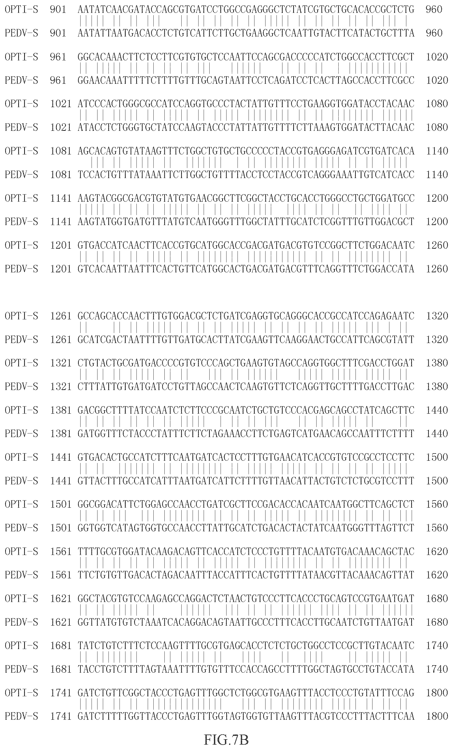
The results are shown in FIG. 6:
(1) The average S/P value of vaccination group 2 (containing PEDV-S protein) can reach about 1.1 21 days after first immunization, and can reach 1.7 14 days after second immunization, while the average S/P value of PEDV-S1 protein vaccination group was only about 0.7 21 days after first immunization, and only about 1.2 14 days after second immunization. This indicates that the immunogenicity of full-length PEDV-S is better than that of PEDV-S1.
(2) After vaccines (namely vaccine 1-vaccine 3) prepared with PEDV-S proteins having different concentrations are immunized, the S/P value after first immunization increase with the rising of concentration, but the S/P value of vaccine 1 can also reach 1.1 or more which is higher than that of PEDV-S1 protein immunization group, while after second immunization, the S/P values of three groups of vaccines are basically the same and all between 1.7 and 0.1, which indicates that the concentration of PEDV-S protein in vaccines hardly influences second immunization, and slightly influences first immunization, but it can achieve the protective effect.
(3) the S/P values of vaccines (i.e. vaccine 2, vaccine 4-vaccine 6) prepared with Quil A having different concentrations increase with the rising of the concentrations, but they are about 0.4 higher than the average S/P values of vaccines without Quil A (the average S/P value of vaccine 6 were about 0.6 after first immunization, and the average S/P values of vaccine 4 were above 1.0 after first immunization); after second immunization, the S/P values of three groups of vaccines (vaccines 1, 4 and 5) containing Quil A are basically the same and all about 1.7, and are not more than 0.2.about.0.3 (the average S/P of vaccine 6 after second immunization is only about 1.4); it indicates that Quil A in vaccines has a very good enhancement effect on immunization, especially for first immunization, and this is a good complement for quick generation of immune protection of vaccines.
Example 9: Clinic Large-Scale Immunological Experiment
(1) Pig farms and immunization situations: this experiment was used for three pig farms from September to November 2017: 1100 sows for A pig farm and 700 sows for B pig farm. Sows were immunized for the first time 40 days before delivery and immunized for the second time 20 days before delivery. Each immunization dose was 2 ml/vaccine, and the immunization vaccine was made as vaccine 6 in example 8, all of which were injected into neck muscles.
(2) Clinical Observation Results
A farm: it was well managed. No diarrhea cases occurred from 2016 to 2017. After the sows were administrated with PEDV subunit vaccine, swinery was in normal condition and there was no diarrhea in sows and piglets.
B farm: the incidence of diarrhea was serious in winter 2016. It was estimated that 3000 newborn piglets died in one year. No diarrhea cases occurred in sows and piglets after PEDV subunit vaccine was used. Similarly, the death of piglets caused by diarrhea was prevented.
The disclosure is exemplified by the above examples. However, it is understood that the disclosure is not limited to the described special examples and embodiments. Here, these special examples and embodiments are intended to help those skilled in the art to implement the disclosure. Any improvements and perfections are made by those skilled in the art without departing from the spirit and scope of the disclosure, and therefore the disclosure is only limited by contents and scope of claims of the disclosure and intended to cover alternative solutions and equivalent solutions included within the spirit and scope of the disclosure defined by appended claims.
SEQUENCE LISTINGS
1
313909DNAArtificial SequenceSynthesized 1gacgtgacca ggtgctctgc caacacaaat ttcaggcggt tctttagcaa gtttaacgtg 60caggctccag ctgtggtggt gctgggagga tacctgccaa tcggcgagaa tcagggcgtg 120aactctacat ggtattgtgc tggacagcac cctaccgcta gcggagtgca tggcatcttc 180gtgtcccaca tcaggggcgg ccatggcttc gagatcggca tctcccagga gccctttgac 240cctagcggct accagctgta tctgcacaag gccaccaacg gcaataccaa cgccacagct 300agactgcgca tctgccagtt cccctctatc aagaccctgg gccctacagc taacaatgat 360gtgaccacag gccggaattg tctgtttaac aaggccatcc cagctcacat gagcgagcat 420tctgtggtgg gcatcacctg ggacaacgat agagtgacag tgttctccga caagatctac 480tatttctact ttaagaacga ttggagccgc gtggccacca agtgctataa ttctggcggc 540tgtgctatgc agtacgtgta tgagccaacc tactatatgc tgaatgtgac atctgccgga 600gaggacggaa tctcctacca gccatgcaca gccaattgta tcggctatgc cgctaacgtg 660ttcgctaccg agcctaatgg ccacatccca gagggcttct cttttaacaa ttggtttctg 720ctgtccaacg atagcaccct ggtgcatggc aaggtggtgt ccaatcagcc tctgctggtg 780aactgcctgc tggctatccc aaagatctac ggcctgggcc agttcttttc cttcaaccag 840acaatcgacg gcgtgtgcaa tggagctgct gtgcagaggg ctccagaggc tctgcggttt 900aatatcaacg ataccagcgt gatcctggcc gagggctcta tcgtgctgca caccgctctg 960ggcacaaact tctccttcgt gtgctccaat tccagcgacc cccatctggc caccttcgct 1020atcccactgg gcgccatcca ggtgccctac tattgtttcc tgaaggtgga tacctacaac 1080agcacagtgt ataagtttct ggctgtgctg ccccctaccg tgagggagat cgtgatcaca 1140aagtacggcg acgtgtatgt gaacggcttc ggctacctgc acctgggcct gctggatgcc 1200gtgaccatca acttcaccgt gcatggcacc gacgatgacg tgtccggctt ctggacaatc 1260gccagcacca actttgtgga cgctctgatc gaggtgcagg gcaccgccat ccagagaatc 1320ctgtactgcg atgaccccgt gtcccagctg aagtgtagcc aggtggcttt cgacctggat 1380gacggctttt atccaatctc ttcccgcaat ctgctgtccc acgagcagcc tatcagcttc 1440gtgacactgc catctttcaa tgatcactcc tttgtgaaca tcaccgtgtc cgcctccttc 1500ggcggacatt ctggagccaa cctgatcgct tccgacacca caatcaatgg cttcagctct 1560ttttgcgtgg atacaagaca gttcaccatc tccctgtttt acaatgtgac aaacagctac 1620ggctacgtgt ccaagagcca ggactctaac tgtcccttca ccctgcagtc cgtgaatgat 1680tatctgtctt tctccaagtt ttgcgtgagc acctctctgc tggcctccgc ttgtacaatc 1740gatctgttcg gctaccctga gtttggctct ggcgtgaagt ttacctccct gtatttccag 1800tttacaaagg gcgagctgat caccggcaca ccaaagcccc tggagggcgt gaccgacgtg 1860agcttcatga cactggacgt gtgcaccaag tacacaatct atggctttaa gggcgagggc 1920atcatcaccc tgacaaactc cagcttcctg gccggctttt actatacatc cgacagcggc 1980cagctgctgg ccttcaagaa tgtgaccagc ggcgccgtgt actctgtgac accctgttct 2040ttttccgagc aggccgctta tgtggatgac gatatcgtgg gcgtgatctc ttccctgagc 2100aattctacat tcaacagcac cagagagctg cccggcttct tttaccatag caatgatggc 2160tctaactgca ccgagcctgt gctggtgtac tccaacatcg gcgtgtgcaa gtccggcagc 2220atcggctatg tgccaagcca gccaggacag gtgaagatcg ctccaaccgt gacaggcaat 2280atctctatcc ctaccaactt ttctatgtcc atccgcacag agtacctgca gctgtataac 2340acacccgtga tcgtggactg cgctacctac gtgtgcaacg gcaattccag atgcaagcag 2400ctgctgaccc agtatacagc cgcttgtaag accatcgagt ctgccctgca gctgtccgct 2460aggctggaga gcgtggaggt gaactctatg ctgacaatct ccgaggagag cctgcagctg 2520gccaccatct ccagcttcaa tggcggcggc tacaacttta ccaatgtgct gggcgtgtcc 2580gtgtatgacc ctgctagcgg cagagtggtg cagaagcgca gcttcatcga ggatctgctg 2640tttaacaagg tggtgaccaa tggcctgggc acagtggacg aggattacaa gaggtgcagc 2700aacggccggt ctgtggctga cctggtgtgc gctcagtact attctggcgt gatggtgctg 2760ccaggcgtgg tggatgccga gaagctgcac atgtattccg cctccctgat cggaggaatg 2820gtgctgggag gcttcacaag cgccgctgcc ctgccctttt cttacgctgt gcaggccaga 2880ctgaactatc tggccctgca gaccgacgtg ctgcagagga atcagcagct gctggccgag 2940tctttcaact ccgctatcgg caatatcaca tccgcctttg agagcgtgaa ggagtccatc 3000agccagacca gcaagggcct gaacacagtg gctcacgccc tgaccaaggt gcaggaggtg 3060gtgaactccc agggagctgc cctgacccag ctgacagtgc agctgcagca taacttccag 3120gccatctctt ccagcatcga cgatatctac tctagactgg atatcctgtc cgctgacgtg 3180caggtggata gactgatcac aggccgcctg tccgctctga acgcctttgt ggctcagacc 3240ctgacaaagt acaccgaggt gcaggccagc aggaagctgg ctcagcagaa agtgaatgag 3300tgcgtgaagt ctcagtccca gcggtatggc ttctgtggcg gcgacggcga gcacatcttt 3360tccctggtgc aggctgctcc acagggactg ctgttcctgc ataccgtgct ggtgcctggc 3420gactttgtgg atgtgatcgc tatcgccggc ctgtgcgtga acgatgagat cgccctgaca 3480ctgagggagc ctggactggt gctgttcacc cacgagctgc agaatcatac cgctacagag 3540tacttcgtgt cttccaggcg gatgtttgag cccaggaagc ctaccgtgtc cgactttgtg 3600cagatcgaga gctgcgtggt gacctacgtg aacctgacac gggaccagct gcctgatgtg 3660atcccagact atatcgatgt gaacaagaca ctggatgaga tcctggcctc tctgccaaat 3720aggaccggac catccctgcc actggacgtg ttcaacgcca cctatctgaa tctgacaggc 3780gagatcgctg atctggagca gaggagcgag tctctgcgga acaccacaga ggagctgcag 3840tccctgatct acaatatcaa caatacactg gtggatctgg agtggctgaa tcgggtggag 3900acctatatc 390921303PRTArtificial SequenceSynthesized 2Asp Val Thr Arg Cys Ser Ala Asn Thr Asn Phe Arg Arg Phe Phe Ser1 5 10 15Lys Phe Asn Val Gln Ala Pro Ala Val Val Val Leu Gly Gly Tyr Leu 20 25 30Pro Ile Gly Glu Asn Gln Gly Val Asn Ser Thr Trp Tyr Cys Ala Gly 35 40 45Gln His Pro Thr Ala Ser Gly Val His Gly Ile Phe Val Ser His Ile 50 55 60Arg Gly Gly His Gly Phe Glu Ile Gly Ile Ser Gln Glu Pro Phe Asp65 70 75 80Pro Ser Gly Tyr Gln Leu Tyr Leu His Lys Ala Thr Asn Gly Asn Thr 85 90 95Asn Ala Thr Ala Arg Leu Arg Ile Cys Gln Phe Pro Ser Ile Lys Thr 100 105 110Leu Gly Pro Thr Ala Asn Asn Asp Val Thr Thr Gly Arg Asn Cys Leu 115 120 125Phe Asn Lys Ala Ile Pro Ala His Met Ser Glu His Ser Val Val Gly 130 135 140Ile Thr Trp Asp Asn Asp Arg Val Thr Val Phe Ser Asp Lys Ile Tyr145 150 155 160Tyr Phe Tyr Phe Lys Asn Asp Trp Ser Arg Val Ala Thr Lys Cys Tyr 165 170 175Asn Ser Gly Gly Cys Ala Met Gln Tyr Val Tyr Glu Pro Thr Tyr Tyr 180 185 190Met Leu Asn Val Thr Ser Ala Gly Glu Asp Gly Ile Ser Tyr Gln Pro 195 200 205Cys Thr Ala Asn Cys Ile Gly Tyr Ala Ala Asn Val Phe Ala Thr Glu 210 215 220Pro Asn Gly His Ile Pro Glu Gly Phe Ser Phe Asn Asn Trp Phe Leu225 230 235 240Leu Ser Asn Asp Ser Thr Leu Val His Gly Lys Val Val Ser Asn Gln 245 250 255Pro Leu Leu Val Asn Cys Leu Leu Ala Ile Pro Lys Ile Tyr Gly Leu 260 265 270Gly Gln Phe Phe Ser Phe Asn Gln Thr Ile Asp Gly Val Cys Asn Gly 275 280 285Ala Ala Val Gln Arg Ala Pro Glu Ala Leu Arg Phe Asn Ile Asn Asp 290 295 300Thr Ser Val Ile Leu Ala Glu Gly Ser Ile Val Leu His Thr Ala Leu305 310 315 320Gly Thr Asn Phe Ser Phe Val Cys Ser Asn Ser Ser Asp Pro His Leu 325 330 335Ala Thr Phe Ala Ile Pro Leu Gly Ala Ile Gln Val Pro Tyr Tyr Cys 340 345 350Phe Leu Lys Val Asp Thr Tyr Asn Ser Thr Val Tyr Lys Phe Leu Ala 355 360 365Val Leu Pro Pro Thr Val Arg Glu Ile Val Ile Thr Lys Tyr Gly Asp 370 375 380Val Tyr Val Asn Gly Phe Gly Tyr Leu His Leu Gly Leu Leu Asp Ala385 390 395 400Val Thr Ile Asn Phe Thr Val His Gly Thr Asp Asp Asp Val Ser Gly 405 410 415Phe Trp Thr Ile Ala Ser Thr Asn Phe Val Asp Ala Leu Ile Glu Val 420 425 430Gln Gly Thr Ala Ile Gln Arg Ile Leu Tyr Cys Asp Asp Pro Val Ser 435 440 445Gln Leu Lys Cys Ser Gln Val Ala Phe Asp Leu Asp Asp Gly Phe Tyr 450 455 460Pro Ile Ser Ser Arg Asn Leu Leu Ser His Glu Gln Pro Ile Ser Phe465 470 475 480Val Thr Leu Pro Ser Phe Asn Asp His Ser Phe Val Asn Ile Thr Val 485 490 495Ser Ala Ser Phe Gly Gly His Ser Gly Ala Asn Leu Ile Ala Ser Asp 500 505 510Thr Thr Ile Asn Gly Phe Ser Ser Phe Cys Val Asp Thr Arg Gln Phe 515 520 525Thr Ile Ser Leu Phe Tyr Asn Val Thr Asn Ser Tyr Gly Tyr Val Ser 530 535 540Lys Ser Gln Asp Ser Asn Cys Pro Phe Thr Leu Gln Ser Val Asn Asp545 550 555 560Tyr Leu Ser Phe Ser Lys Phe Cys Val Ser Thr Ser Leu Leu Ala Ser 565 570 575Ala Cys Thr Ile Asp Leu Phe Gly Tyr Pro Glu Phe Gly Ser Gly Val 580 585 590Lys Phe Thr Ser Leu Tyr Phe Gln Phe Thr Lys Gly Glu Leu Ile Thr 595 600 605Gly Thr Pro Lys Pro Leu Glu Gly Val Thr Asp Val Ser Phe Met Thr 610 615 620Leu Asp Val Cys Thr Lys Tyr Thr Ile Tyr Gly Phe Lys Gly Glu Gly625 630 635 640Ile Ile Thr Leu Thr Asn Ser Ser Phe Leu Ala Gly Phe Tyr Tyr Thr 645 650 655Ser Asp Ser Gly Gln Leu Leu Ala Phe Lys Asn Val Thr Ser Gly Ala 660 665 670Val Tyr Ser Val Thr Pro Cys Ser Phe Ser Glu Gln Ala Ala Tyr Val 675 680 685Asp Asp Asp Ile Val Gly Val Ile Ser Ser Leu Ser Asn Ser Thr Phe 690 695 700Asn Ser Thr Arg Glu Leu Pro Gly Phe Phe Tyr His Ser Asn Asp Gly705 710 715 720Ser Asn Cys Thr Glu Pro Val Leu Val Tyr Ser Asn Ile Gly Val Cys 725 730 735Lys Ser Gly Ser Ile Gly Tyr Val Pro Ser Gln Pro Gly Gln Val Lys 740 745 750Ile Ala Pro Thr Val Thr Gly Asn Ile Ser Ile Pro Thr Asn Phe Ser 755 760 765Met Ser Ile Arg Thr Glu Tyr Leu Gln Leu Tyr Asn Thr Pro Val Ile 770 775 780Val Asp Cys Ala Thr Tyr Val Cys Asn Gly Asn Ser Arg Cys Lys Gln785 790 795 800Leu Leu Thr Gln Tyr Thr Ala Ala Cys Lys Thr Ile Glu Ser Ala Leu 805 810 815Gln Leu Ser Ala Arg Leu Glu Ser Val Glu Val Asn Ser Met Leu Thr 820 825 830Ile Ser Glu Glu Ser Leu Gln Leu Ala Thr Ile Ser Ser Phe Asn Gly 835 840 845Gly Gly Tyr Asn Phe Thr Asn Val Leu Gly Val Ser Val Tyr Asp Pro 850 855 860Ala Ser Gly Arg Val Val Gln Lys Arg Ser Phe Ile Glu Asp Leu Leu865 870 875 880Phe Asn Lys Val Val Thr Asn Gly Leu Gly Thr Val Asp Glu Asp Tyr 885 890 895Lys Arg Cys Ser Asn Gly Arg Ser Val Ala Asp Leu Val Cys Ala Gln 900 905 910Tyr Tyr Ser Gly Val Met Val Leu Pro Gly Val Val Asp Ala Glu Lys 915 920 925Leu His Met Tyr Ser Ala Ser Leu Ile Gly Gly Met Val Leu Gly Gly 930 935 940Phe Thr Ser Ala Ala Ala Leu Pro Phe Ser Tyr Ala Val Gln Ala Arg945 950 955 960Leu Asn Tyr Leu Ala Leu Gln Thr Asp Val Leu Gln Arg Asn Gln Gln 965 970 975Leu Leu Ala Glu Ser Phe Asn Ser Ala Ile Gly Asn Ile Thr Ser Ala 980 985 990Phe Glu Ser Val Lys Glu Ser Ile Ser Gln Thr Ser Lys Gly Leu Asn 995 1000 1005Thr Val Ala His Ala Leu Thr Lys Val Gln Glu Val Val Asn Ser 1010 1015 1020Gln Gly Ala Ala Leu Thr Gln Leu Thr Val Gln Leu Gln His Asn 1025 1030 1035Phe Gln Ala Ile Ser Ser Ser Ile Asp Asp Ile Tyr Ser Arg Leu 1040 1045 1050Asp Ile Leu Ser Ala Asp Val Gln Val Asp Arg Leu Ile Thr Gly 1055 1060 1065Arg Leu Ser Ala Leu Asn Ala Phe Val Ala Gln Thr Leu Thr Lys 1070 1075 1080Tyr Thr Glu Val Gln Ala Ser Arg Lys Leu Ala Gln Gln Lys Val 1085 1090 1095Asn Glu Cys Val Lys Ser Gln Ser Gln Arg Tyr Gly Phe Cys Gly 1100 1105 1110Gly Asp Gly Glu His Ile Phe Ser Leu Val Gln Ala Ala Pro Gln 1115 1120 1125Gly Leu Leu Phe Leu His Thr Val Leu Val Pro Gly Asp Phe Val 1130 1135 1140Asp Val Ile Ala Ile Ala Gly Leu Cys Val Asn Asp Glu Ile Ala 1145 1150 1155Leu Thr Leu Arg Glu Pro Gly Leu Val Leu Phe Thr His Glu Leu 1160 1165 1170Gln Asn His Thr Ala Thr Glu Tyr Phe Val Ser Ser Arg Arg Met 1175 1180 1185Phe Glu Pro Arg Lys Pro Thr Val Ser Asp Phe Val Gln Ile Glu 1190 1195 1200Ser Cys Val Val Thr Tyr Val Asn Leu Thr Arg Asp Gln Leu Pro 1205 1210 1215Asp Val Ile Pro Asp Tyr Ile Asp Val Asn Lys Thr Leu Asp Glu 1220 1225 1230Ile Leu Ala Ser Leu Pro Asn Arg Thr Gly Pro Ser Leu Pro Leu 1235 1240 1245Asp Val Phe Asn Ala Thr Tyr Leu Asn Leu Thr Gly Glu Ile Ala 1250 1255 1260Asp Leu Glu Gln Arg Ser Glu Ser Leu Arg Asn Thr Thr Glu Glu 1265 1270 1275Leu Gln Ser Leu Ile Tyr Asn Ile Asn Asn Thr Leu Val Asp Leu 1280 1285 1290Glu Trp Leu Asn Arg Val Glu Thr Tyr Ile 1295 130033909DNAArtificial SequenceSynthesized 3gatgtcacta ggtgctcagc taacactaat tttaggcgtt tcttttcaaa atttaatgtt 60caggcgcctg cagttgttgt actgggcggt tatctaccta ttggtgaaaa ccagggtgtc 120aattcaactt ggtactgtgc tggccaacat ccaactgcta gtggcgttca tggtatcttt 180gttagccata ttagaggtgg tcatggcttt gagattggca tttcgcaaga gccttttgac 240cctagtggtt accagcttta tttacataag gctactaacg gtaacactaa tgctactgcg 300cgactgcgca tttgccagtt tcctagcatt aaaacattgg gccccactgc taataatgat 360gttacaacag gtcgtaattg cctatttaac aaagccatcc cagctcatat gagtgaacat 420agtgttgtcg gaataacatg ggataatgat cgtgtcactg tcttttctga caagatctat 480tatttttatt ttaaaaatga ttggtcccgt gttgcgacaa agtgttacaa cagtggaggt 540tgtgccatgc aatatgttta cgaacccacc tattacatgc ttaatgttac tagtgctggt 600gaggatggta tttcttatca accctgtaca gctaattgca ttggttatgc tgccaatgta 660tttgctactg agcccaatgg ccacatacca gaaggtttta gttttaataa ttggtttctt 720ttgtccaatg attccacttt ggtgcatggt aaggtggttt ccaaccaacc attgttggtc 780aattgtcttt tggccattcc taagatttat ggactaggcc aatttttctc ctttaatcaa 840acgatcgatg gtgtttgtaa tggagctgct gtgcagcgtg caccagaggc tctgaggttt 900aatattaatg acacctctgt cattcttgct gaaggctcaa ttgtacttca tactgcttta 960ggaacaaatt tttcttttgt ttgcagtaat tcctcagatc ctcacttagc caccttcgcc 1020atacctctgg gtgctatcca agtaccctat tattgttttc ttaaagtgga tacttacaac 1080tccactgttt ataaattctt ggctgtttta cctcctaccg tcagggaaat tgtcatcacc 1140aagtatggtg atgtttatgt caatgggttt ggctatttgc atctcggttt gttggacgct 1200gtcacaatta atttcactgt tcatggcact gacgatgacg tttcaggttt ctggaccata 1260gcatcgacta attttgttga tgcacttatc gaagttcaag gaactgccat tcagcgtatt 1320ctttattgtg atgatcctgt tagccaactc aagtgttctc aggttgcttt tgaccttgac 1380gatggtttct accctatttc ttctagaaac cttctgagtc atgaacagcc aatttctttt 1440gttactttgc catcatttaa tgatcattct tttgttaaca ttactgtctc tgcgtccttt 1500ggtggtcata gtggtgccaa ccttattgca tctgacacta ctatcaatgg gtttagttct 1560ttctgtgttg acactagaca atttaccatt tcactgtttt ataacgttac aaacagttat 1620ggttatgtgt ctaaatcaca ggacagtaat tgccctttca ccttgcaatc tgttaatgat 1680tacctgtctt ttagtaaatt ttgtgtttcc accagccttt tggctagtgc ctgtaccata 1740gatctttttg gttaccctga gtttggtagt ggtgttaagt ttacgtccct ttactttcaa 1800ttcacaaagg gtgagttgat tactggcacg cctaaaccac ttgaaggtgt cacggacgtt 1860tcttttatga ctctggatgt ttgtaccaag tatactatct atggctttaa aggtgagggt 1920atcattaccc ttacaaattc tagctttttg gcaggttttt attacacatc tgattctgga 1980cagttgttag cctttaagaa tgtcactagt ggtgctgttt attctgttac gccatgttct 2040ttttcagagc aggctgcata tgttgatgat gatatagtgg gtgttatttc tagtttgtct 2100aactccactt ttaacagtac tagggagttg cctggtttct tctaccattc taatgatggc 2160tctaattgta cagagcctgt gttggtgtat agtaacatag gtgtttgtaa atctggcagt 2220attggctacg ttccatccca gcctggccaa gtcaagattg cacccacggt tactgggaat 2280attagtattc ccaccaactt tagtatgagt attaggacag aatatttaca gctttacaac 2340acgcctgtta ttgttgattg tgccacatat gtttgtaatg gtaactctcg ttgtaaacaa 2400ttactcaccc agtacactgc agcatgtaag accatagagt cagcattaca actcagcgct 2460aggcttgagt ctgttgaagt taactctatg cttactattt ctgaagagtc tctacagtta 2520gctaccatca gttcgtttaa tggtggtgga tataatttta ctaacgtgct gggtgtttct 2580gtgtatgatc ctgcaagtgg cagggtggta caaaaaaggt cttttattga agacctgctt 2640tttaataaag tggttactaa tggccttggt actgttgatg aagactataa gcgctgttct 2700aatggtcgct ctgtggcaga tctagtctgt gcacagtatt actctggtgt catggtacta 2760cctggtgttg ttgacgctga gaagcttcac atgtatagtg cgtctctcat cggtggtatg 2820gtgctaggag gttttacttc tgcagcagca ttgcctttta gctatgctgt tcaagctaga 2880ctcaattatc ttgctctaca gacggatgtt ctacagcgga accagcaatt gcttgctgag 2940tcttttaact ctgctattgg taatataact tcagcctttg agagtgttaa agagtctatt 3000agtcaaactt ccaagggttt
gaacactgtg gctcatgcgc ttactaaggt tcaagaggtt 3060gttaactcgc agggtgcagc tttgactcaa cttaccgtac agctgcaaca caacttccaa 3120gccatttcta gttctattga tgacatttac tctcgactgg acattctttc agccgatgtt 3180caggttgacc gtctcatcac cggcagatta tcagcactta atgcttttgt tgctcaaacc 3240ctcactaagt atactgaggt tcaggctagc aggaagctag cacagcaaaa ggttaatgag 3300tgcgttaaat cccaatctca gcgttatggt ttttgtggtg gtgatggcga gcacattttc 3360tctctggtac aggcagcacc tcagggcctg ctgtttttac atacagtact tgtaccgggt 3420gactttgtag atgttattgc catcgctggc ttatgcgtta acgatgaaat tgccttgact 3480ctacgtgagc ctggcttagt cttgtttacg catgaacttc aaaatcatac tgcgacggaa 3540tattttgttt catcgcgacg tatgtttgaa cctagaaaac ctaccgttag tgattttgtt 3600caaattgaga gttgtgtggt cacctatgtc aatttgacta gagaccaact accagatgta 3660atcccagatt acatcgatgt taacaaaaca cttgatgaga ttttagcttc tctgcccaat 3720agaactggtc caagtcttcc tttagatgtt tttaatgcca cttatctcaa tctcactggt 3780gaaattgcag atttagagca gcgttcagag tctctccgta atactacaga ggagctccaa 3840agtcttatat ataatatcaa caacacacta gttgaccttg agtggctcaa ccgagttgag 3900acatacatc 3909
* * * * *
D00001
D00002
D00003
D00004
D00005
D00006
D00007
D00008
D00009
D00010
D00011
S00001
XML
uspto.report is an independent third-party trademark research tool that is not affiliated, endorsed, or sponsored by the United States Patent and Trademark Office (USPTO) or any other governmental organization. The information provided by uspto.report is based on publicly available data at the time of writing and is intended for informational purposes only.
While we strive to provide accurate and up-to-date information, we do not guarantee the accuracy, completeness, reliability, or suitability of the information displayed on this site. The use of this site is at your own risk. Any reliance you place on such information is therefore strictly at your own risk.
All official trademark data, including owner information, should be verified by visiting the official USPTO website at www.uspto.gov. This site is not intended to replace professional legal advice and should not be used as a substitute for consulting with a legal professional who is knowledgeable about trademark law.