GLA domains as therapeutic agents
Bauzon , et al. February 23, 2
U.S. patent number 10,925,926 [Application Number 16/170,131] was granted by the patent office on 2021-02-23 for gla domains as therapeutic agents. This patent grant is currently assigned to GLADIATOR BIOSCIENCES, INC.. The grantee listed for this patent is GLADIATOR BIOSCIENCES, INC.. Invention is credited to Maxine Bauzon, Terry Hermiston.


| United States Patent | 10,925,926 |
| Bauzon , et al. | February 23, 2021 |
GLA domains as therapeutic agents
Abstract
The disclosure relates to the recombinant Gla domain proteins and their use targeting phosphatidylserine (PtdS) moieties on the surface of cells, particularly those expressing elevated levels of PtdS, such as cells undergoing apoptosis.
| Inventors: | Bauzon; Maxine (Hercules, CA), Hermiston; Terry (Mill Valley, CA) | ||||||||||
|---|---|---|---|---|---|---|---|---|---|---|---|
| Applicant: |
|
||||||||||
| Assignee: | GLADIATOR BIOSCIENCES, INC.
(Mill Valley, CA) |
||||||||||
| Family ID: | 1000005381066 | ||||||||||
| Appl. No.: | 16/170,131 | ||||||||||
| Filed: | October 25, 2018 |
Prior Publication Data
| Document Identifier | Publication Date | |
|---|---|---|
| US 20190105370 A1 | Apr 11, 2019 | |
Related U.S. Patent Documents
| Application Number | Filing Date | Patent Number | Issue Date | ||
|---|---|---|---|---|---|
| 15631937 | Jun 23, 2017 | ||||
| 14773068 | Jul 4, 2017 | 9694048 | |||
| PCT/US2014/026237 | Mar 13, 2014 | ||||
| 61787753 | Mar 15, 2013 | ||||
| 61791537 | Mar 15, 2013 | ||||
| Current U.S. Class: | 1/1 |
| Current CPC Class: | C07K 14/485 (20130101); A61N 5/10 (20130101); A61K 45/06 (20130101); A61K 38/1808 (20130101); C12N 9/6429 (20130101); A61K 38/1709 (20130101); C12N 9/6437 (20130101); C12N 9/6424 (20130101); A61K 38/16 (20130101); C07K 14/745 (20130101); A61K 47/642 (20170801); C07K 2319/035 (20130101); Y02A 50/30 (20180101); C12Y 304/21021 (20130101); C12Y 304/21005 (20130101) |
| Current International Class: | A61K 38/17 (20060101); A61K 38/18 (20060101); C12N 9/74 (20060101); C07K 14/745 (20060101); C07K 14/485 (20060101); A61K 47/64 (20170101); C12N 9/64 (20060101); A61K 38/16 (20060101); A61N 5/10 (20060101); A61K 45/06 (20060101) |
References Cited [Referenced By]
U.S. Patent Documents
| 5440013 | August 1995 | Kahn |
| 5446128 | August 1995 | Kahn |
| 5475085 | December 1995 | Kahn |
| 5597457 | January 1997 | Craig et al. |
| 5618914 | April 1997 | Kahn |
| 5670155 | September 1997 | Kahn |
| 5672681 | September 1997 | Kahn |
| 5674976 | October 1997 | Kahn |
| 5710245 | January 1998 | Kahn |
| 5790421 | August 1998 | Osslund |
| 5840833 | November 1998 | Kahn |
| 5859184 | January 1999 | Kahn et al. |
| 5889155 | March 1999 | Ashkenazi et al. |
| 5929237 | July 1999 | Kahn |
| 6093573 | July 2000 | Beamer et al. |
| 6261569 | July 2001 | Comis et al. |
| 6312694 | November 2001 | Thorpe et al. |
| 6459996 | October 2002 | Somers et al. |
| 6631332 | October 2003 | Skolnick et al. |
| 6801860 | October 2004 | Dessen et al. |
| 7511016 | March 2009 | Reutelingsperger |
| 8283167 | October 2012 | Simon |
| 9023604 | May 2015 | Schmidt et al. |
| 2003/0104578 | June 2003 | Balance |
| 2003/0220490 | November 2003 | Kuriyama et al. |
| 2004/0001827 | January 2004 | Dennis |
| 2005/0015232 | January 2005 | Reinherz et al. |
| 2009/0098103 | April 2009 | Madison et al. |
| 2009/0130060 | May 2009 | Weimer et al. |
| 2011/0159571 | June 2011 | Barry et al. |
| 2016/0008482 | January 2016 | Bauzon et al. |
| 2280024 | Feb 2011 | EP | |||
| WO 2017/118764 | Jul 1917 | WO | |||
| WO 2019/050997 | Mar 1919 | WO | |||
| WO 2019/050998 | Mar 1919 | WO | |||
| WO 2019/051002 | Mar 1919 | WO | |||
| WO 2012/087241 | Jun 2012 | WO | |||
| WO 2013/151665 | Oct 2013 | WO | |||
| WO 2014/018535 | Jan 2014 | WO | |||
Other References
|
Office Communication issued in U.S. Appl. No. 14/773,068, dated Nov. 29, 2016. cited by applicant . Office Communication issued in U.S. Appl. No. 14/773,068, dated Aug. 2, 2016. cited by applicant . Office Communication issued in U.S. Appl. No. 15/631,937, dated Jul. 25, 2018. cited by applicant . Office Communication issued in U.S. Appl. No. 15/631,937, dated Jan. 10, 2018. cited by applicant . Mille-Baker et al., "Deletion or replacement of the second EGF-like domain of protein S results in loss of APC cofactor activity," Blood, 101:1416-1418, 2003. cited by applicant . Okada et al., "A novel splice site mutation in intron C of PROS1 leads to markedly reduced mutant mRNA level, absence of thrombin-sensitive region, and impaired secretion and cofactor activity of mutant protein S," Thrombosis Research, 125:e246-250, 2010. cited by applicant . Stenflo, "Contributions of Gla and EGF-like domains to the function of vitamin K-dependent coagulation factors," Critical Reviews in Eukaryotic Gene Expression, 9(1):59-88, 1999. cited by applicant . Van Wijnen et al., "Characterization of mini-protein S, a recombinant variant of protein S that lacks the sex hormone binding globulin-like domain," Biochem. J., 330:389-396, 1998. cited by applicant . Raina D., et al., "Direct Targeting of the Mucin 1 Oncoprotein Blocks Survival and Tumorigenicity of Human Breast Carcinoma Cells," Cancer Research, vol. 69 (12), pp. 5133-5141, Jun. 15, 2009. cited by applicant . Office Communication issued in U.S. Appl. No. 14/772,971, dated Feb. 24, 2020. cited by applicant . Office Communication issued in U.S. Appl. No. 14/772,971, dated Aug. 9, 2019. cited by applicant . Office Communication issued in U.S. Appl. No. 14/772,971, dated Jan. 11, 2019. cited by applicant . Office Communication issued in U.S. Appl. No. 14/772,971, dated Jun. 29, 2018. cited by applicant . Office Communication issued in U.S. Appl. No. 14/772,971, dated Mar. 15, 2018. cited by applicant . Ghosh et al., "Rapid isolation of extracellular vesicles from cell culture and biological fluids using a synthetic peptide with specific affinity for heat shock proteins," PLoS ONE, 9(10):e110443, 2014, 12 pages. cited by applicant . Kenis et al., "Cell surface-expressed phosphatidylserine and annexin A5 open a novel portal of cell entry," J. Biol. Chemistry, 279(50):52623-52629, 2004. cited by applicant . Lemke, "Phosphatidyleserine is the signal for TAM receptors and their ligands," Trends in Biochemical Sciences, 42(9):738-748, 2017. cited by applicant . Nakai et al., "A novel affinity-based method for the isolation of highly purified extracellular vesicles," Scientific Reports, 6(1):1-2, 2016. cited by applicant . PCT International Preliminary Report on Patentability issued in International Application No. PCT/US2018/049618, dated Sep. 30, 2019. cited by applicant . PCT International Preliminary Report on Patentability issued in International Application No. PCT/US2018/049624, dated Jan. 10, 2020. cited by applicant . PCT International Search Report and Written Opinion issued in International Application No. PCT/US2018/049618, dated Dec. 6, 2018. cited by applicant . PCT International Search Report and Written Opinion issued in International Application No. PCT/US2018/049624, dated Dec. 5, 2018. cited by applicant . PCT International Search Report and Written Opinion issued in International Application No. PCT/US2018/049619, dated Dec. 6, 2018. cited by applicant . Soares et al., "Targeting Inside-Out Phosphatidylserine as a Therapeutic Strategy for Viral Diseases," Nat. Med., 14(12):1357-1362, 2008. cited by applicant . Tietjen et al., "Molecular mechanism for differential recognition of membrane phosphtidylserine by the immune regulatory receptor Tim4," PNAS, 111(15):E1463-E1472, 2014. cited by applicant . Amara and Mercer, "Viral apoptotic mimicry," Nature Reviews Microbiology, 13(8)461-9, 2015. cited by applicant . Andaloussi et al., "Extracellular vesicles: Biology and emerging therapeutic opportunities," Nature Reviews Drug Discovery, 12(5):347-357, 2013. cited by applicant . Beck et al., "Strategies and challenges for the next generation of antibody-drug conjugates," Nature Reviews Drug Discovery, 16(5):315-337, 2017. cited by applicant . Belhocine et al., "99mTc-Annexin A5 quantification of apoptotic tumor response: a systematic review and meta-analysis of clinical imaging trials," European Journal of Nuclear Medicine and Molecular Imaging, 42(13):2083-2097, 2015. cited by applicant . Belzile et al., "Antibody targeting of phosphatidylserine for the detection and immunotherapy of cancer, "ImmunoTargets and Therapy, 7:1014, 2018. cited by applicant . Benabdellah et al, "Genome-edited adult stem cells: Next-generation advanced therapy medicinal products," Stem Cells Translational Medicine, 9(6):674-685, 2020. cited by applicant . Birge et al., "Phosphatidylserine is a infectious disease, and cancer," Cell Death global immunosuppressive and Differentiation, 23(6):962-978, signal in efferocytosis, 2016. cited by applicant . Burstyn-Cohen and Maimon, "TAM receptors, Signaling, 17(1):156, 2019. phosphatidylserine, inflammation and cancer, " Cell Communication and Signaling, 17(1):156, 2019. cited by applicant . Calianese and Birge, "Biology of phosphatidylserine implications in immunology, infectious disease and cancer," Communication 18(1):41, 2020. (PS): basic physiology and and Signaling, 18(1):41, 2020. cited by applicant . Colombo et al., "Biogenesis, Secretion, and Intercellular Interactions of Exosomes and Other Extracellular Vesicles," Annual Review of Cell and Developmental Biology, 30(1):255-289, 2014. cited by applicant . Conlan, "Early pathogenesis of Listeria monocytogenes infection in the mouse spleen," Journal of Medical Microbiology, 44(4):295-302, 1996. cited by applicant . Crescitelli et al., "Distinct RNA profiles in subpopulations of extracellular vesicles: Apoptotic bodies, microvesicles and exosomes," Journal of Extracellular Vesicles, 2(1):2013. 11 pages. cited by applicant . Dahlback., "The tale of protein S and C4b-binding protein, a story of affection," Thrombosis and Haemostasis, 98(1):756-764, 2007. cited by applicant . Dayoub and Brekken, immunotherapy," "Tims, Cell Communication TAMS, and PS-antibody targeting: implications for cancer and Signaling, 18(1):29, 2020. cited by applicant . Derose et al., "Development of bavituximab, a vascular targeting agent with immune-modulating properties, for lung cancer treatment," Immunotherapy, 3(8):933-944, 2011. cited by applicant . Elmore et al., "Apoptosis: A Review of Programmed Cell Death," Toxicol. Pathol., 29(6):997-1003, 2007. cited by applicant . Gerber et al., "Randomized phase III study of docetaxel plus bavituximab in previously treated advanced non-squamous non-small-cell lung cancer," Annals of Oncology, 29(7):15481553, 2018. cited by applicant . Graca and Willem, "Extracellular vesicles: exosomes, microvesicles, and friends," Journal of Cell Biology, 200(4):373-383, 2013. cited by applicant . Graner et al. Seminars in Immunopathology, "Tumor-derived exosomes, microRNAs, and cancer immune suppression," 40(5):505-515, 2018. cited by applicant . Hemberger et al., "Trophoblast stem cells differentiate in vitro into invasive trophoblast giant cells," Developmental Biology, 271:362-371, 2004. cited by applicant . Hoen et al., "Extracellular vesicles and viruses: Are they close relatives?" Proceedings of the National Academy of Sciences of the United States of America, 113(33):9155-9161, 2016. cited by applicant . Huang and Lai, therapeutic tool," "The potential Annals of Translational roles of stem cell-derived extracellular vesicles as a Medicine, 7(22):693, 2019. cited by applicant . Kanada et al., "Signaling by Extracellular Vesicles Advances Cancer, 2(2):84-94, 2018. Cancer Hallmarks," Trends in. cited by applicant . Li et al., "Targeting phosphatidy Iserine with calcium-dependent protein-drug conjugates for the treatment of cancer, " Molecular Cancer Therapeutics, 17(2):169-182, 2018. cited by applicant . Mann et al., "Surface-dependent reactions of the vitamin K-dependent enzyme complexes, " Journal of American Society of Hematology, 76:1-16, 1990. cited by applicant . Murphy et al., "Extracellular vesicle-based therapeutics: natural versus engineered targeting and trafficking, " Experimental and Molecular Medicine, 51(3):1-12, 2019. cited by applicant . N'Guessan et al., "SapC-DOPS--a phosphatidylserine-targeted nanovesicle for selective cancer therapy, " Cell Communication and Signaling, 18(6) (2020), 5 pages. cited by applicant . Oguro et al., "SLAM family markers resolve functionally distinct subpopulations of hematopoietic stem cells and multipotent progenitors," Cell Stem Cell, 13:102-116, 2013. cited by applicant . Oling et al., "SLAM family markers resolve functionally distinct subpopulations of hemtopoietic stem cells and multipotent progenitors, " Cell Stem Cell, 13:102-116, 2013. cited by applicant . Rezende et al., Coagulation, inflammation, and apoptosis: Different roles for protein S and the protein S-C4b binding protein complex, Blood, 103(4):1192-1201, 2004. cited by applicant . Schorey et al., "Exosomes and other extracellular vesicles in host-pathogen interactions, " EMBO Reports, 16(1):24-43, 2015. cited by applicant . Shelke et al., "Importance of exosome depletion protocols to eliminate functional and RNA-containing extracellular vesicles from fetal bovine serum," Journal of Extracellular Vesicles, 3(1), 24783, 2014, 9 pages. cited by applicant . Shlomovitz et al., "Flipping the dogma--phosphatidylserine in non-apoptotic cell death, " Cell Communication and Signaling, 17(1): 139, 2019. cited by applicant . Suzuki et al., "Xk-related protein 8 and CED-8 promote phosphtidylserine exposure in apoptotic cells, " Sciene, 341(6144):403-406, 2013. cited by applicant . Valadi et al., "Exosome-mediated transfer of mRNAs and microRNAs in a novel mechanism of genetic exchange between cells," Nature Cell Biology, 9(6):654-659, 2007. cited by applicant . Vaupel and Multhoff, "Accomplices of hypoxic tumor microenvironment comprising antitumor immunity: adenosine, lactate, acidosis, vascular endothelial growth factor, potassium ions and phosphatidylserine, " Frontiers in Immunology, 8:1887, 2017. cited by applicant . Vermeer, ".gamma.-Carboxyglutamate-containing proteins and the vitamin K-dependent carboxylase," Biochemical Journal, 266:625-636, 1990. cited by applicant . Wanderley et al., "Apoptotic mimicry as a strategy for the establishment of parasitic infection: parasite- and host derived phosphtidylersine as key molecule," Cell Communication and Signaling, 18:10(1), 2020, 10 pages. cited by applicant . Wang et al., "SPECT and PET radiopharmacueticals for molecular imaging of apoptosis: From bench to clinic," Oncotarget, 8(12):20479-20495, 2017. cited by applicant . Yanez-Moet al. "Biological properties of extracellular vesicles and their physiological functions," Journal of Extracellular Vesicles, 4:27066, 2015, 62 pages. cited by applicant . English translation of Office Communciation issued in Japanese Patent Application No. 2019-176325, dated Aug. 28, 2020, 8 pages. cited by applicant . GenBank Accession No. AAH15801, "PROS1 protein [Homo sapiens]," Aug. 11, 2006. cited by applicant . McDonald et al., "Ionic properties of membrane association by vitamin K-dependent proteins: The case for univalency," Biochemistry, 36:15589-15598, 1997. cited by applicant . Office Communication issued in New Zealand Patent Application No. 710958, dated Sep. 9, 2020, 5 pages. cited by applicant . Office Communication issued in New Zealand Patent Application No. 751494, dated Sep. 8, 2020, 7 pages. cited by applicant. |
Primary Examiner: Ulm; John D
Attorney, Agent or Firm: Parker Highlander PLLC
Parent Case Text
This application is a continuation application of U.S. patent application Ser. No. 15/631,937, filed Jun. 23, 2017, which is a continuation application of U.S. patent application Ser. No. 14/773,068, which adopts the international filing date of Mar. 13, 2014, which is the National Phase application under 35 U.S.C. .sctn. 371 of International Application No. PCT/US2014/26237, filed Mar. 13, 2014, which claims the benefit of priority to U.S. Provisional Application Ser. No. 61/791,537 filed Mar. 15, 2013, and U.S. Provisional Application Ser. No. 61/787,753, filed Mar. 15, 2013, the disclosures of each of which are hereby incorporated by reference in their entireties for all purposes.
Claims
The invention claimed is:
1. A polypeptide that binds phosphatidyl serine (PS) on cell membranes, said polypeptide comprising: (a) a protein S gamma-carboxyglutamic-acid (Gla) domain, wherein the Gla domain has the sequence of SEQ ID NO: 1; and (b) an EGF domain from human Protein S; wherein said polypeptide lacks a protease domain and a hormone-binding domain, wherein the polypeptide is linked to a therapeutic agent.
2. The polypeptide of according to claim 1, wherein said polypeptide comprises at least one disulfide bond.
3. The polypeptide according to claim 2, wherein said polypeptide comprises 2-5 disulfide bonds.
4. The polypeptide according to claim 1, wherein the polypeptide is 300 amino acids or less.
5. The polypeptide according to claim 1, comprising SEQ ID NO: 6 or a derivative differing therefrom only by the absence of the HIS-tag.
6. The polypeptide according to claim 1, wherein the therapeutic agent is an antibody Fc region.
7. The polypeptide according to claim 1, further comprising a detectable label.
8. The polypeptide according to claim 7, wherein the detectable label is selected from a fluorescent label, a chemiluminescent label, a radiolabel, an enzyme, a dye and a ligand.
9. The polypeptide according to claim 1, wherein the polypeptide is a genetic fusion with a proteinaceous therapeutic agent.
10. The polypeptide of claim 9, wherein the fusion protein comprises a proteinaceous sequence encoding transferrin or albumin.
11. The polypeptide according to claim 6, wherein the Fc region is conjugated.
12. The polypeptide according to claim 6, wherein the Fc region is genetically fused to said polypeptide.
13. The polypeptide according to claim 7, wherein the label is conjugated to said polypeptide.
Description
BACKGROUND
Pursuant to 37 C.F.R. 1.821(c), a sequence listing is submitted herewith as an ASCII compliant text file named "STIPP0003USC2_ST25.txt", created on Oct. 22, 2018 and having a size of .about.10 kilobytes. The content of the aforementioned file is hereby incorporated by reference in its entirety.
1. Field
This disclosure relates to the targeting of phosphatidylserine (PtdS) on cell membranes using Gla domain peptides and polypeptides. The use of these peptides and polypeptides as therapeutic agents is disclosed.
2. Related Art
Phosphatidlyserine (PtdS) is a negatively charged phospholipid component usually localized to the inner-leaflet (the cytoplasmic side) of the cell membrane. However, PtdS can be transported by scramblase (a member of the flippase family) from the inner-leaflet to the outer-leaflet and exposed on the cell surface. With very few exceptions, this active externalization of PtdS is a response to cellular damage (van den Eijnde et al., 2001; Erwig and Henson, 2008). For example, tissue injury signals platelets, leukocytes, and endothelial cells to rapidly and reversibly redistribute PtdS which leads to the promotion of coagulation and complement activation on cell surfaces. Similarly, apoptotic signals result in the externalization of PtdS however in a more gradual and sustained manner. This external PtdS provides a key recognition marker that enables macrophages to ingest dying cells from surrounding tissue while suppressing a full and detrimental immune response (Erwig and Henson, 2008). This removal process is essential for tissue homeostasis and in a "healthy" environment it is extremely efficient. In fact, despite the loss of >10.sup.9 cells per day, the histological detection of apoptotic cells is a rare event in normal tissues (Elltiot and Ravichandran, 2010; Elltiot et al., 2009). However, there is evidence that in many pathological conditions the process of apoptotic cell removal is overwhelmed, delayed or absent (Elltiot and Ravichandran, 2010; Lahorte et al., 2004). For example several oncology studies suggest that a high apoptotic index is associated with higher grade tumors, increased rate of metastasis and a poor prognosis for the patient (Naresh et al., 2001; Loose et al., 2007; Kurihara et al., 2008; Kietselaer et al., 2002). These studies, and others like them, suggest that apoptosis and external PtdS expression can be a powerful marker of disease (Elltiot and Ravichandran, 2010).
There are several proteins with a high affinity for anionic phospholipid surfaces with Annexin-V being the most widely utilized as a PtdS targeting probe (Lahorte et al., 2004). With a high affinity for PtdS containing vesicles (K.sub.d=0.5-7 nM) and a molecular weight (37 kDa) that falls below the threshold for kidney filtration (approx. 60 kDa) Annexin-V has shown promise in the clinic as an apoptosis-probe (Lin et al., 2010; Tait and Gibson, 1992). Moreover, it has been utilized for a wide range of indications including those in oncology, neurology and cardiology (Lahorte et al., 2004; Boersma et al., 2005; Blankenberg, 2009; Reutelingsperger et al., 2002). The use of biologic probes which target PtdS cell-surface expression has been shown both in vitro and in vivo. While their utility in the clinic is promising, they have, for the most part, not yet been exploited.
SUMMARY
Thus, in accordance with the present disclosure, there is provided a method of targeting cell membrane phosphatidylserine (PtdS) comprising (a) providing an isolated polypeptide comprising a gamma-carboxyglutamic-acid (Gla) domain and lacking a protease or hormone-binding domain; and (b) contacting the peptide with a cell surface, wherein the polypeptide binds to PtdS on the cell membrane. The cell membrane may be a cardiac cell membrane, a neuronal cell membrane, an endothelial cell membrane, a virus-infected cell membrane, an apoptotic cell membrane, a platelet membrane, a plasma membrane-derived veriscle (PMV) or a cancer cell membrane. The polypeptide may further comprise an EGF binding domain, a Kringle domain, and/or an aromatic amino acid stack domain. The Gla domain may be from Factor II, Factor VII, Factor IX, Factor X, protein S or protein C. The polypeptide may further comprises a detectable label, such as a fluorescent label, a chemiluminescent label, a radiolabel, an enzyme, a dye or a ligand.
The polypeptide may be 300 residues or less, 200 residues or less, or 100 residues or less, including ranges of 100-200 and 100-300 residues. The polypeptide may comprise 5-15 Gla residues, 9-13 Gla residues, including 5, 6, 7, 8, 9, 10, 11, 12, 13, 14, or 15 Gla residues. The polypeptide may comprise more than 13 Gla residues, but less than 30% total Gla residues. The polypeptide may be between about 4.5 and 30 kD in size. The polypeptide may comprise at least one disulfide bond, or 2-5 disulfide bonds. The polypeptide may comprise a protein S Gla domain. The polypeptide may comprise a protein S Gla domain, a protein S EGF domain, a prothrombin Gla domain, a prothrombin Gla domain plus prothrombin Kringle domain, a protein Z Gla domain, a protein Z Gla domain plus prothrombin Kringle domain, a Factor VII Gla domain, or a Factor VII Gla domain plus prothrombin Kringle domain. The polypeptide may further comprise an antibody Fc region. Any of the foregoing may contain conservative substitutions of the native sequences for the foregoing proteins, and/or exhibit a percentage homology to the native domains set forth.
In another embodiment, there is provided a method of treating cancer in a subject comprising administering to the subject an isolated polypeptide comprising a gamma-carboxyglutamic-acid (Gla) domain and lacking a protease or hormone-binding domain. The cancer may be breast cancer, brain cancer, stomach cancer, lung cancer, prostate cancer, ovarian cancer, testicular cancer, colon cancer, skin cancer, rectal cancer, cervical cancer, uterine cancer, liver cancer, pancreatic cancer, head & neck cancer or esophageal cancer. The method may further comprise treating the subject with a second cancer therapy, such as an immunotherapy, a radiotherapy, a chemotherapy, a toxin therapy, a cytokine therapy or a hormone therapy.
Yet another embodiment includes a method of treating an autoimmune disease in a subject comprising administering to the subject an isolated polypeptide comprising a gamma-carboxyglutamic-acid (Gla) domain and lacking a protease or hormone-binding domain. The autoimmune disease may be spondyloarthropathy, ankylosing spondylitis, psoriatic arthritis, reactive arthritis, enteropathic arthritis, ulcerative colitis, Crohn's disease, irritable bowel disease, inflammatory bowel disease, rheumatoid arthritis, juvenile rheumatoid arthritis, familial Mediterranean fever, amyotrophic lateral sclerosis, Sjogren's syndrome, early arthritis, viral arthritis, multiple sclerosis, systemic lupus erythematosus, psoriasis, vasculitis, Wegener's granulomatosis, Addison's disease, alopecia, antiphospholipid syndrome, Behcet's disease, celiac disease, chronic fatigue syndrome, ulcerative colitis, type I diabetes, fibromyalgia, autoimmune gastritis, Goodpasture syndrome, Graves' disease, idiopathic thrombocytopenic purpura (ITP), myasthenia gravis, pemphigus vulgaris, primary biliary cirrhosis, rheumatic fever, sarcoidosis, scleroderma, vitiligo, vasculitis, small vessel vasculitis, hepatitis, primary biliary cirrhosis, sarcoidosis, scleroderma, graft versus host disease (acute and chronic), aplastic anemia, or cyclic neutropenia. The method may further comprise treating the subject with a second autoimmune disease therapy, such as prednisone, methylprednisone, Venipred, Celestone, hydrocortisone, triamcinoclone, Aristonpan Intra-Articular injection, Methapred, Rayos oral, betamethasone, or etanercept.
In still another embodiment, there is provided a method of treating a viral disease in a subject comprising administering to the subject an isolated polypeptide comprising a gamma-carboxyglutamic-acid (Gla) domain and lacking a protease or hormone-binding domain. The viral disease may be influenza, human immunodeficiency virus, dengue virus, West Nile virus, smallpox virus, respiratory syncytial virus, Korean hemorrhagic fever virus, chickenpox, varicella zoster virus, herpes simplex virus 1 or 2, Epstein-Barr virus, Marburg virus, hantavirus, yellow fever virus, hepatitis A, B, C or E, Ebola virus, human papilloma virus, rhinovirus, Coxsackie virus, polio virus, measles virus, rubella virus, rabies virus, Newcastle disease virus, rotavirus, HTLV-1 and -2. The method may further comprise treating the subject with a second anti-viral therapy, such as Abacavir, Aciclovir, Acyclovir, Adefovir, Amantadine, Amprenavir, Ampligen, Arbidol, Atazanavir, Atripla, Boceprevirertet, Cidofovir, Combivir, Darunavir, Delavirdine, Didanosine, Docosanol, Edoxudine, Efavirenz, Emtricitabine, Enfuvirtide, Entecavir, Entry inhibitors, Famciclovir, Fomivirsen, Fosamprenavir, Foscarnet, Fosfonet, Ganciclovir, Ibacitabine, Imunovir, Idoxuridine, Imiquimod, Indinavir, Inosine, Integrase inhibitor, Interferon type III, Interferon type II, Interferon type I, Interferon, Lamivudine, Lopinavir, Loviride, Maraviroc, Moroxydine, Methisazone, Nelfinavir, Nevirapine, Nexavir, Nucleoside analogues, Oseltamivir, Peginterferon alfa-2a, Penciclovir, Peramivir, Pleconaril, Podophyllotoxin, Protease inhibitor, Raltegravir, Reverse transcriptase inhibitor, Ribavirin, Rimantadine, Ritonavir, Pyramidine, Saquinavir, Stavudine, Synergistic enhancer (antiretroviral), Tea tree oil, Telaprevir, Tenofovir, Tenofovir disoproxil, Tipranavir, Trifluridine, Trizivir, Tromantadine, Truvada, Valaciclovir, Valganciclovir, Vicriviroc, Vidarabine, Viramidine, Zalcitabine, Zanamivir or Zidovudine.
In still a further embodiment, there is provided a method of treating a hypercoagulation disorder in a subject comprising administering to the subject an isolated polypeptide comprising a gamma-carboxyglutamic-acid (Gla) domain and lacking a protease or hormone-binding domain. The method may further comprise treating the subject with one or more additional anti-coagulants.
Also provided are methods of modulating clotting in a subject comprising administering to the subject an isolated polypeptide comprising a gamma-carboxyglutamic-acid (Gla) domain and lacking a protease or hormone-binding domain. The method may further comprise administering to the subject a clotting factor.
Yet another embodiment comprises treating sepsis in a subject comprising administering to the subject an isolated polypeptide comprising a gamma-carboxyglutamic-acid (Gla) domain and lacking a protease or hormone-binding domain. A still further embodiment includes a method of treating vaso-occlusive crisis in a sickle cell subject comprising administering to the subject an isolated polypeptide comprising a gamma-carboxyglutamic-acid (Gla) domain and lacking a protease or hormone-binding domain.
Finally, there is provided a method treating a disorder characterized by pathologic expression of phosphatidylserine on the surface of a cell comprising administering to the subject an isolated polypeptide comprising a gamma-carboxyglutamic-acid (Gla) domain and lacking a protease or hormone-binding domain.
It is contemplated that any method or composition described herein can be implemented with respect to any other method or composition described herein.
Other objects, features and advantages of the present disclosure will become apparent from the following detailed description. It should be understood, however, that the detailed description and the specific examples, while indicating specific embodiments of the disclosure, are given by way of illustration only, since various changes and modifications within the spirit and scope of the disclosure will become apparent to those skilled in the art from this detailed description.
BRIEF DESCRIPTION OF THE FIGURES
The following drawings form part of the present specification and are included to further demonstrate certain aspects of the present disclosure. The disclosure may be better understood by reference to one or more of these drawings in combination with the detailed.
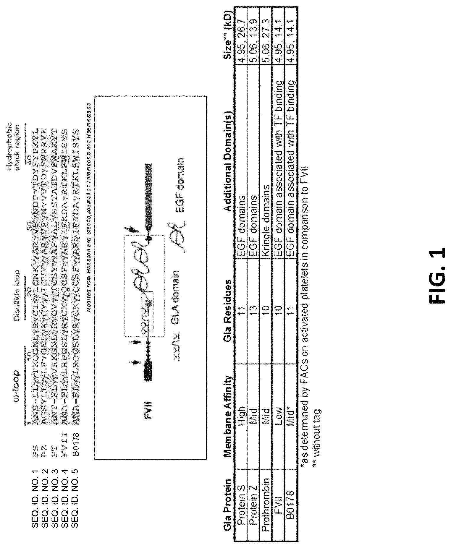
FIG. 1--Construction of a panel of Gla and Gla-EGF/Kringle domain proteins. Sequences shown from top to bottom are SEQ ID NOS: 1-5.
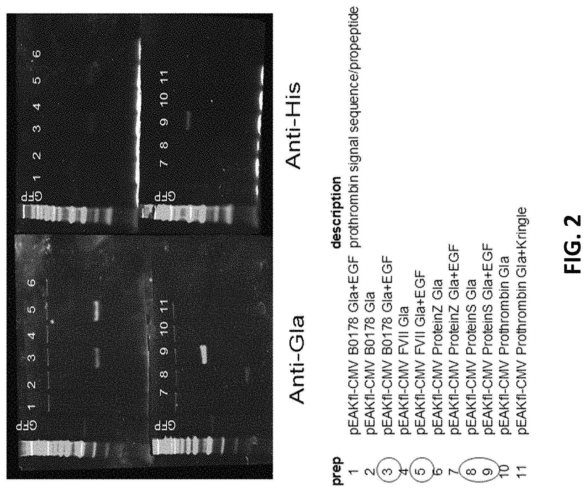
FIG. 2--Testing of Gla domain protein constructs for expression. Transient transfection into 293 cells using 293cellFectin. 10% gels with reduced samples, 23.3 .mu.l of media loaded.
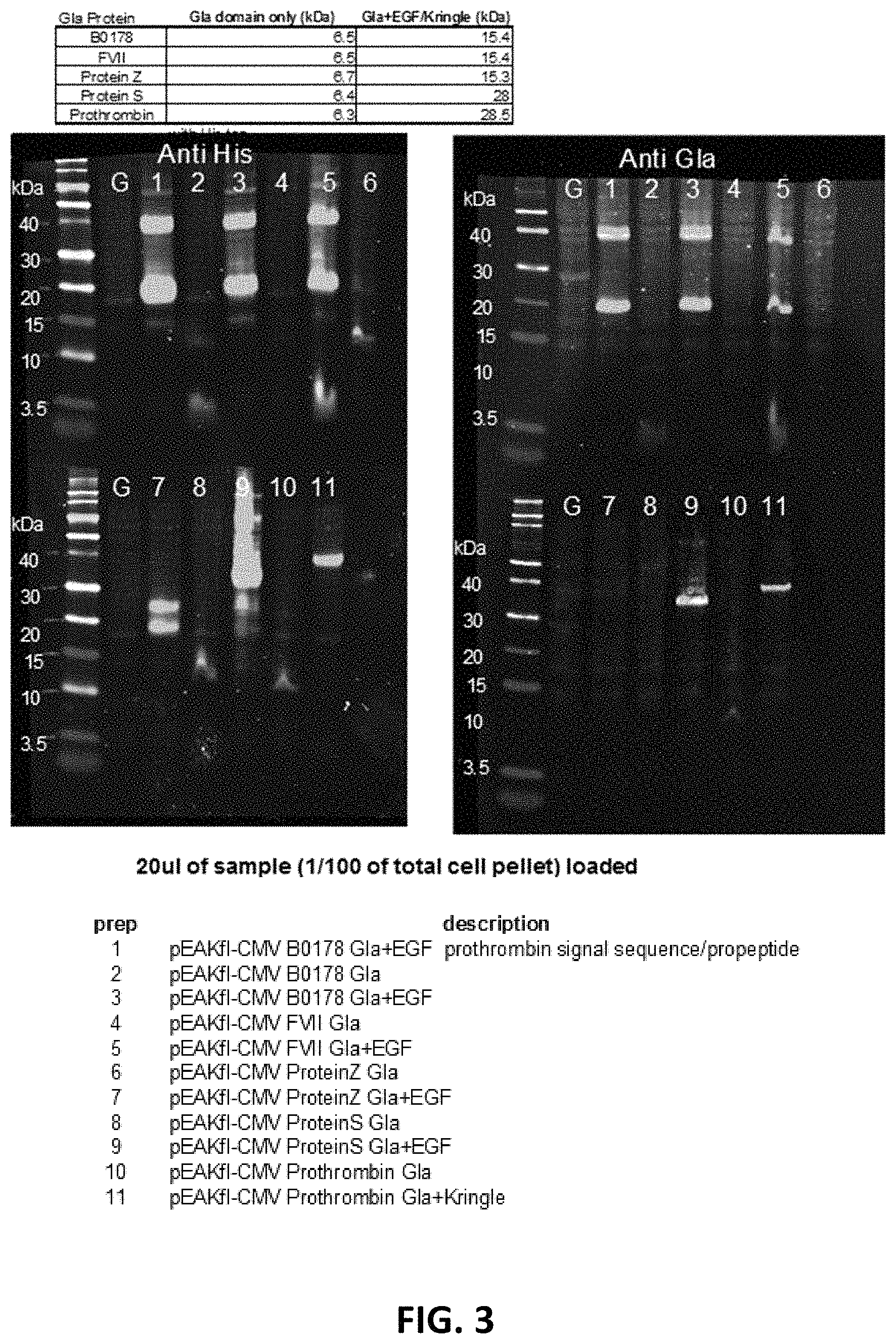
FIG. 3--Testing of Gla domain protein constructs for expression. Transient transfection in BHK21 cells. 10% gels with reduced samples, 20 .mu.l ( 1/100 total cell pellet) loaded.
FIG. 4--Changing signal sequence alter secretion. Transient transfection in BHK21 cells. 10% gels with reduced samples, 13.3 .mu.l loaded.
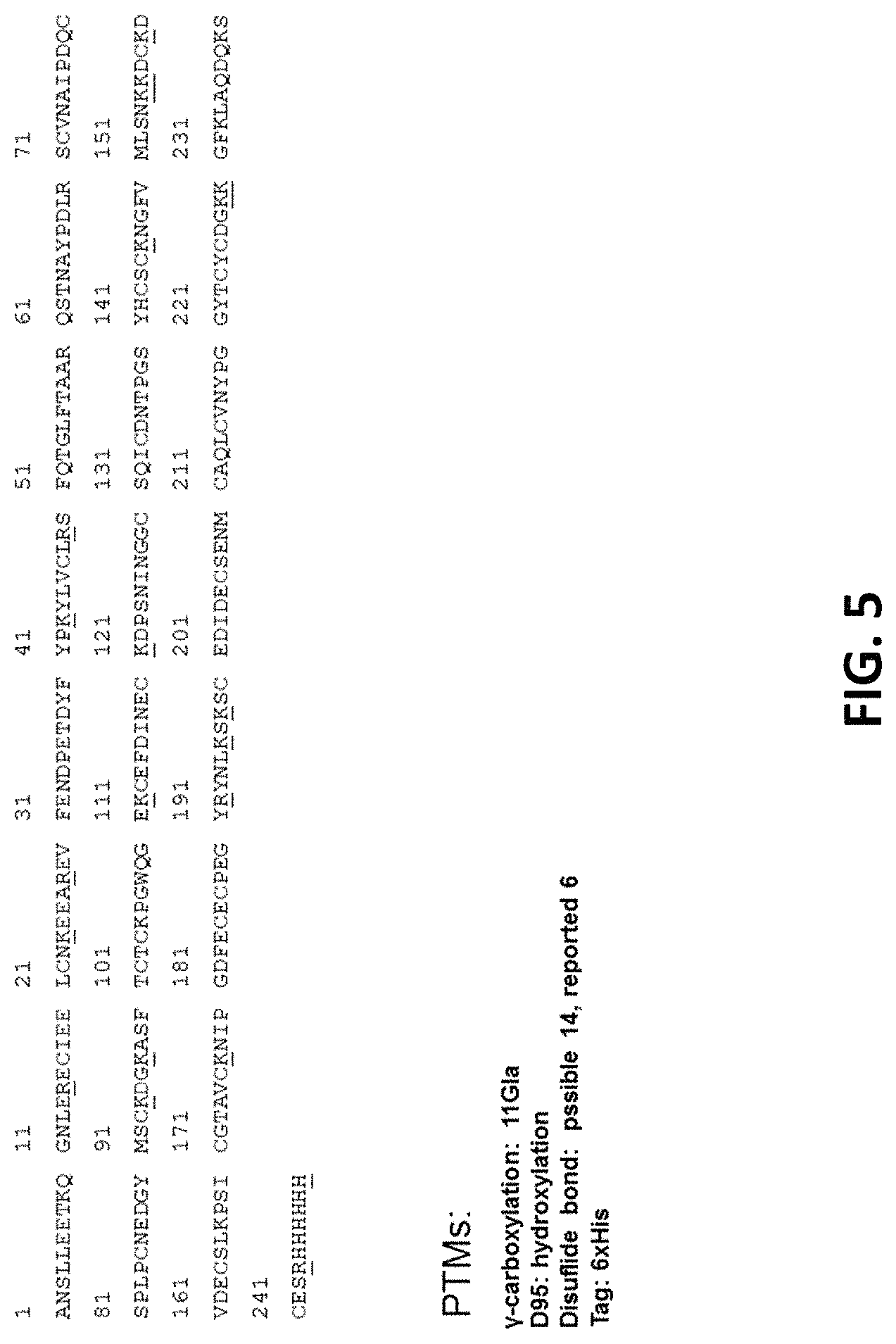
FIG. 5--Protein S Gla+EGF sequence. Sequence is SEQ ID NO: 6.
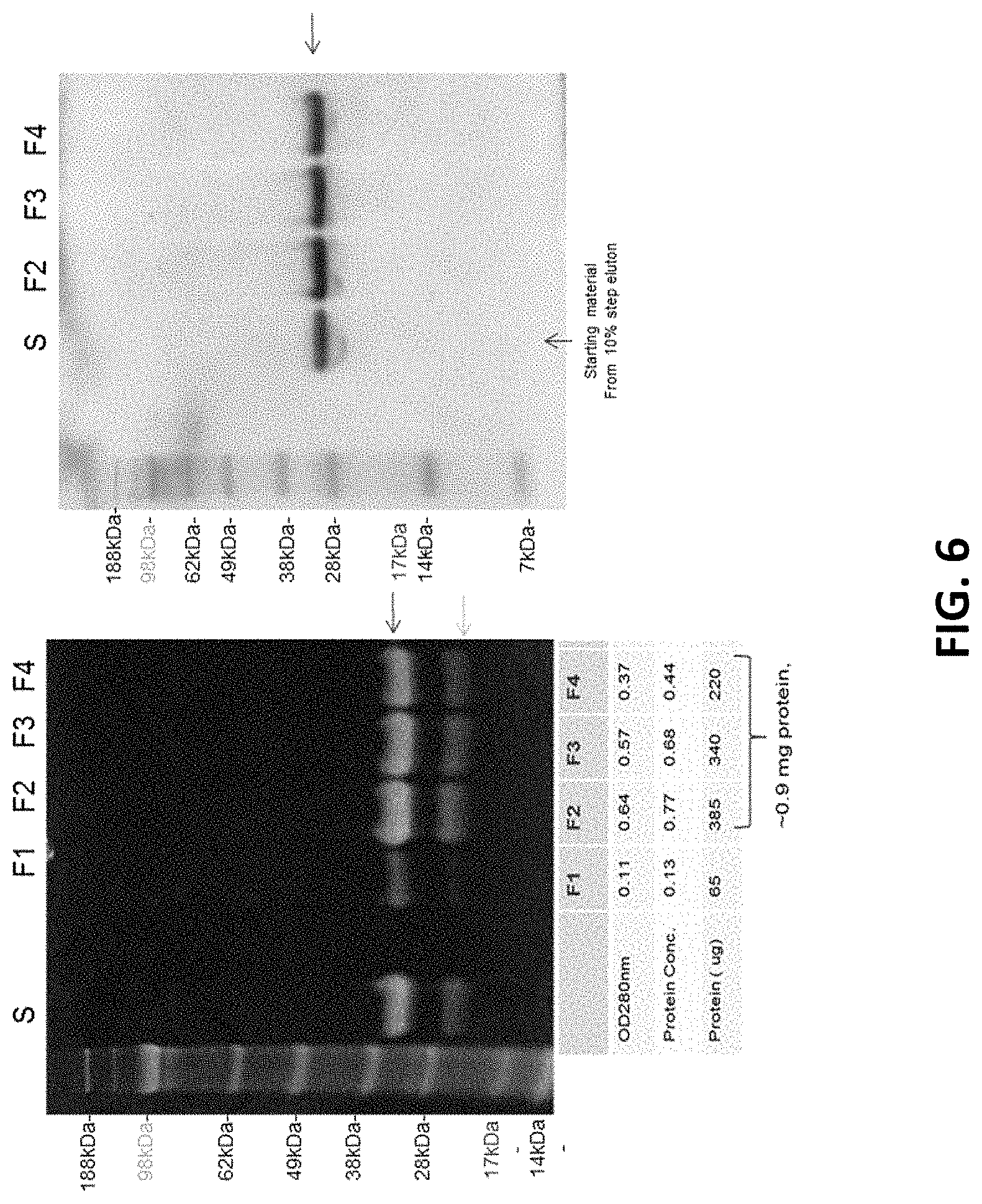
FIG. 6--Purification of Protein S Gla+EGF. F1-F4 are column chromatography fractions. 10% gels, non-reducing conditions.
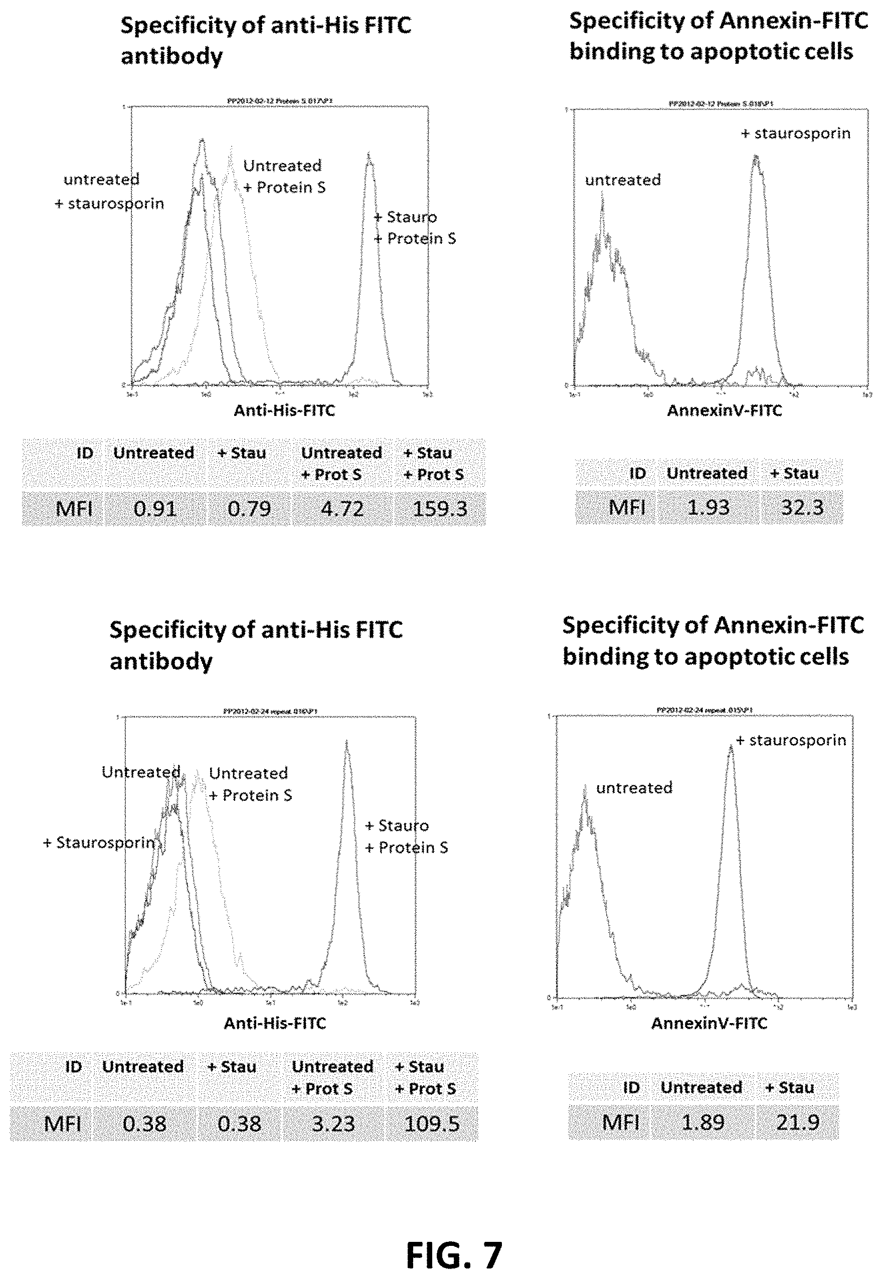
FIG. 7--Apoptosis Assays for Protein S Gla+EGF. Top and bottom panels represent identical duplicate procedures except that amounts of Protein S Gla+EGF was reduced, and the amount of anti-His domain antibody was reduced.
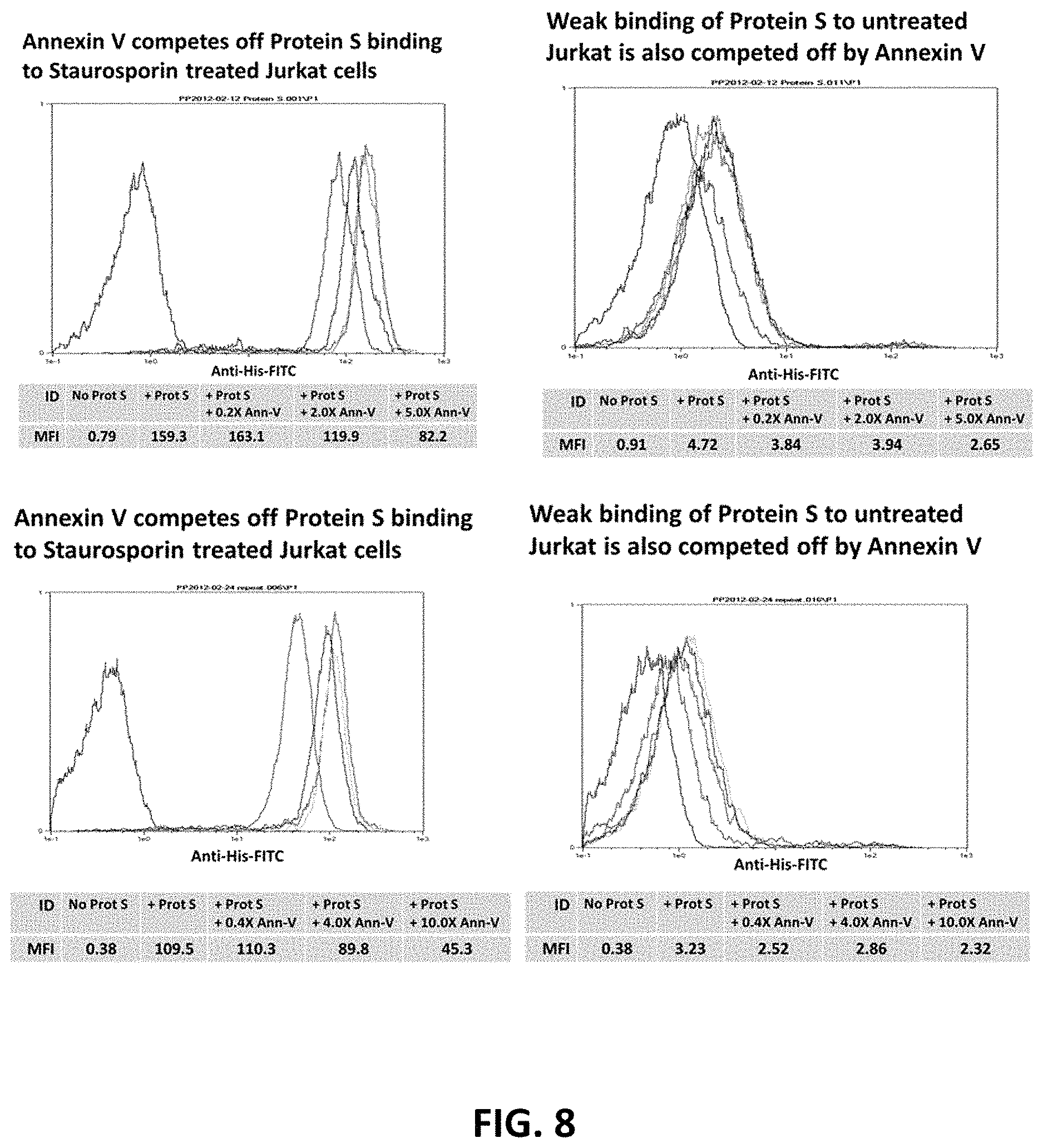
FIG. 8--Apoptosis Assays for Protein S Gla+EGF. Top and bottom panels represent identical duplicate procedures except for amounts of Annexin V used, which are double in the bottom panels.
DESCRIPTION OF ILLUSTRATIVE EMBODIMENTS
Like annexin, gamma-carboxyglutamic-acid (Gla)-domain proteins such as Factors II, VII, IX, X, protein C, and protein S bind anionic membranes. In fact, the Gla-domain has been used as a model for a small molecule that was rationally designed to be an apoptosis-specific probe (Cohen et al., 2009). Here, the inventors propose the utilization of the membrane targeting portions of these Gla-domain proteins as a novel class of biological probes specific for apoptosis and disease. The use of these naturally-occurring and targeted proteins may lead to enhanced specificity relative to current probes with the added advantage of a smaller size (<30 kDa). Even in larger embodiments, which would include EGF and/or Kringle domains, these proteins can still be smaller than Annexin V (37 kDa), and potentially as small as <5 kDa. These biologic probes can target PtdS cell-surface expression both in vitro and in vivo. Thus, it is possible to develop an apoptosis/disease targeting probe that is superior to Annexin V in affinity, specificity and size with the added potential for use as a therapeutic. These and other aspects of the disclosure are described in greater detail below.
Whenever appropriate, terms used in the singular will also include the plural and vice versa. In the event that any definition set forth below conflicts with the usage of that word in any other document, including any document incorporated herein by reference, the definition set forth below shall always control for purposes of interpreting this specification and its associated claims unless a contrary meaning is clearly intended (for example in the document where the term is originally used). The use of "or" means "and/or" unless stated otherwise. The use of "a" herein means "one or more" unless stated otherwise or where the use of "one or more" is clearly inappropriate. The use of "comprise," "comprises," "comprising," "include," "includes," and "including" are interchangeable and are not limiting. For example, the term "including" shall mean "including, but not limited to." The word "about" means plus or minus 5% of the stated number.
An "isolated peptide or polypeptide," as used herein, is intended to refer to a peptide or polypeptide which is substantially free of other biological molecules, including peptides or polypeptides having distinct sequences. In some embodiments, the isolated peptide or polypeptide is at least about 75%, about 80%, about 90%, about 95%, about 97%, about 99%, about 99.9% or about 100% pure by dry weight. In some embodiments, purity can be measured by a method such as column chromatography, polyacrylamide gel electrophoresis, or HPLC analysis.
As used herein, "conservative substitutions" refers to modifications of a polypeptide that involve the substitution of one or more amino acids for amino acids having similar biochemical properties that do not result in loss of a biological or biochemical function of the polypeptide. A "conservative amino acid substitution" is one in which the amino acid residue is replaced with an amino acid residue having a similar side chain. Families of amino acid residues having similar side chains have been defined in the art. These families include amino acids with basic side chains (e.g., lysine, arginine, histidine), acidic side chains (e.g., aspartic acid, glutamic acid), uncharged polar side chains (e.g., glycine, asparagine, glutamine, serine, threonine, tyrosine, cysteine), nonpolar side chains (e.g., alanine, valine, leucine, isoleucine, proline, phenylalanine, methionine, tryptophan), .beta.-branched side chains (e.g., threonine, valine, isoleucine), and aromatic side chains (e.g., tyrosine, phenylalanine, tryptophan, histidine). Antibodies of the present disclosure can have one or more conservative amino acid substitutions yet retain antigen binding activity.
For nucleic acids and polypeptides, the term "substantial homology" indicates that two nucleic acids or two polypeptides, or designated sequences thereof, when optimally aligned and compared, are identical, with appropriate nucleotide or amino acid insertions or deletions, in at least about 80% of the nucleotides or amino acids, usually at least about 85%, in some embodiments about 90%, 91%, 92%, 93%, 94%, or 95%, in at least one embodiment at least about 96%, 97%, 98%, 99%, 99.1%, 99.2%, 99.3%, 99.4%, or 99.5% of the nucleotides or amino acids. Alternatively, substantial homology for nucleic acids exists when the segments will hybridize under selective hybridization conditions to the complement of the strand. Also included are nucleic acid sequences and polypeptide sequences having substantial homology to the specific nucleic acid sequences and amino acid sequences recited herein.
The percent identity between two sequences is a function of the number of identical positions shared by the sequences (i.e., % homology=# of identical positions/total # of positions .times.100), taking into account the number of gaps, and the length of each gap, which need to be introduced for optimal alignment of the two sequences. Comparison of sequences and determination of percent identity between two sequences can be accomplished using a mathematical algorithm, such as without limitation the AlignX.TM. module of VectorNTI.TM. (Invitrogen Corp., Carlsbad, Calif.). For AlignX.TM., the default parameters of multiple alignment are: gap opening penalty: 10; gap extension penalty: 0.05; gap separation penalty range: 8; % identity for alignment delay: 40. (further details at world-wide-web at invitrogen.com/site/us/en/home/LINNEA-Online-Guides/LINNEA-CommunitiesNec- tor-NTI-Community/Sequence-analysis-and-data-management-software-for-PCs/A- lignX-Module-for-Vector-NTI-Advance.reg.us.html).
Another method for determining the best overall match between a query sequence (a sequence of the present disclosure) and a subject sequence, also referred to as a global sequence alignment, can be determined using the CLUSTALW computer program (Thompson et al., Nucleic Acids Res, 1994, 2(22): 4673-4680), which is based on the algorithm of Higgins et al., Computer Applications in the Biosciences (CABIOS), 1992, 8(2): 189-191). In a sequence alignment the query and subject sequences are both DNA sequences. The result of the global sequence alignment is in percent identity. Parameters that can be used in a CLUSTALW alignment of DNA sequences to calculate percent identity via pairwise alignments are: Matrix=IUB, k-tuple=1, Number of Top Diagonals=5, Gap Penalty=3, Gap Open Penalty=10, Gap Extension Penalty=0.1. For multiple alignments, the following CLUSTALW parameters can be used: Gap Opening Penalty=10, Gap Extension Parameter=0.05; Gap Separation Penalty Range=8; % Identity for Alignment Delay=40.
The nucleic acids can be present in whole cells, in a cell lysate, or in a partially purified or substantially pure form. A nucleic acid is "isolated" or "rendered substantially pure" when purified away from other cellular components with which it is normally associated in the natural environment. To isolate a nucleic acid, standard techniques such as the following can be used: alkaline/SDS treatment, CsCl banding, column chromatography, agarose gel electrophoresis and others well known in the art.
I. PHOSPHATIDYLSERINE (PTDS)
A. Structure and Synthesis
Phosphatidylserine (abbreviated PtdS, Ptd-L-Ser or PS) is a phospholipid component, usually kept on the inner-leaflet (the cytosolic side) of cell membranes by an enzyme called flippase. When a cell undergoes apoptosis, phosphatidylserine is no longer restricted to the cytosolic part of the membrane, but becomes exposed on the surface of the cell. The chemical formula of PtdS is C.sub.13H.sub.24NO.sub.10P and has a molecular mass of 385.304. The structure is shown below:
##STR00001##
Phosphatidylserine is biosynthesized in bacteria by condensing the amino acid serine with CDP (cytidine diphosphate)-activated phosphatidic acid. In mammals, phosphatidylserine is produced by base-exchange reactions with phosphatidylcholine and phosphatidylethanolamine. Conversely, phosphatidylserine can also give rise to phosphatidylethanolamine and phosphatidylcholine, although in animals the pathway to generate phosphatidylcholine from phosphatidylserine only operates in the liver.
B. Function
Early studies of phosphatidylserine distilled the chemical from bovine brain. Modern studies and commercially available products are made from soybeans, because of concerns about mad cow disease. The fatty acids attached to the serine in the soy product are not identical to those in the bovine product and is also impure. Preliminary studies in rats indicate that the soy product is at least as potent as that of bovine origin.
The U.S. FDA has given "qualified health claim" status to phosphatidylserine, stating that, "Consumption of phosphatidylserine may reduce the risk of dementia in the elderly" and "Consumption of phosphatidylserine may reduce the risk of cognitive dysfunction in the elderly."
Phosphatidylserine has been demonstrated to speed up recovery, prevent muscle soreness, improve well-being, and might possess ergogenic properties in athletes involved in cycling, weight training and endurance running. Soy-PtdS, in a dose dependent manner (400 mg), has been reported to be an effective supplement for combating exercise-induced stress by blunting the exercise-induced increase in cortisol levels. PtdS supplementation promotes a desirable hormonal balance for athletes and might attenuate the physiological deterioration that accompanies overtraining and/or overstretching. In recent studies, PtdS has been shown to enhance mood in a cohort of young people during mental stress and to improve accuracy during tee-off by increasing the stress resistance of golfers. First pilot studies indicate that PtdS supplementation might be beneficial for children with attention-deficit hyperactivity disorder.
Traditionally, PtdS supplements were derived from bovine cortex (BC-PS); however, due to the potential transfer of infectious diseases, soy-derived PS (S-PS) has been established as a potential safe alternative. Soy-derived PS is Generally Recognized As Safe (GRAS) and is a safe nutritional supplement for older persons if taken up to a dosage of 200 mg three times daily. Phosphatidylserine has been shown to reduce specific immune response in mice.
PtdS can be found in meat, but is most abundant in the brain and in innards such as liver and kidney. Only small amounts of PS can be found in dairy products or in vegetables, with the exception of white beans.
Annexin-A5 is a naturally-occurring protein with avid binding affinity for PtdS. Labeled-annexin-A5 enables visualization of cells in the early- to mid-apoptotic state in vitro or in vivo. Another PtdS binding protein is Mfge8. Technetium-labeled annexin-A5 enables distinction between malignant and benign tumors whose pathology includes a high rate of cell division and apoptosis in malignant compared with a low rate of apoptosis in benign tumors.
II. GLA DOMAIN PROTEINS
A. Gla Domains
The general structure for the Gla-domain proteins is that of a Gla domain followed by EGF domains and then a C terminal serine protease domain. The exceptions are prothrombin, which contains Kringle domains in place of EGF domains, and protein S, which does not have a serine protease domain but rather sex hormone-binding globulin-like (SHBG) domains (Hansson and Stenflo, 2005). The affinities of Gla-domain proteins to anionic membranes vary. Roughly, they fall into 3 categories 1) high affinity binders with a Kd of 30-50 nM, 2) mid-affinity binders with a K.sub.d of 100-200 nM and 3) low affinity binders with a Kd of 1000-2000 nM. The high affinity Gla domain proteins have been shown to bind anionic membranes with Protein S specifically demonstrating binding to apoptotic cells via its interaction with PtdS (Webb et al., 2002). The low affinity Gla domain proteins use a secondary receptor to bind to the cell membrane. For example, FVII utilizes Tissue Factor (TF). The Gla domain/1.sup.st EGF domain is believed to constitute the high affinity TF binding domain of FVII. Importantly for this approach, there are many studies that have shown TF up-regulation on the surface of cancer cells including colorectal cancer, NSCL carcinoma, and breast cancer and these high TF levels have been associated with a poor prognosis (Yu et al., 2004). Although the affinity for anionic membranes is relatively low for FVII, the addition of the high affinity TF interaction along with the documented up-regulation of TF in cancer makes it a potentially interesting cancer specific probe.
B. Gla Domain Containing Proteins
1. Factor II
Prothrombin, also known as coagulation factor II, is proteolytically cleaved to form thrombin in the coagulation cascade, which ultimately results in the stemming of blood loss. Thrombin in turn acts as a serine protease that converts soluble fibrinogen into insoluble strands of fibrin, as well as catalyzing many other coagulation-related reactions. It is primarily expressed in the liver.
The gene encoding prothrombin is located on chromosome 11 in the region of the centromere. It is composed of 14 exons and contains 24 kilobases of DNA. The gene encodes a signal region, a propeptide region, a glutamic acid domain, 2 Kringle regions, and a catalytic domain. The enzyme gamma-glutamyl carboxylase, in the presence of vitamin K, converts the N-terminal glutamic acid residues to gamma-carboxyglutamic acid residues. These gamma-carboxyglutamic acid residues are necessary for the binding of prothrombin to phospholipids on platelet membranes.
Inherited factor II deficiency is an autosomal recessive disorder that can manifest as hypoprothrombinemia, a decrease in the overall synthesis of prothrombin, or as dysprothrombinemia, the synthesis of dysfunctional prothrombin. Homozygous individuals are generally asymptomatic and have functional prothrombin levels of 2-25%. However, symptomatic individuals may experience easy bruising, epistaxis, soft-tissue hemorrhage, excessive postoperative bleeding, and/or menorrhagia.
Prothrombin plays a role in a role in chronic urticaria, an autoimmune disease, and various vascular disorders. Livedo vasculopathy is associated with immunoglobulin (Ig)M antiphosphatidylserine-prothrombin complex antibody. The presence of antiphosphatidylserine-prothrombin complex antibodies and histopathological necrotizing vasculitis in the upper-to-middle dermis indicates cutaneous leukocytoclastic angiitis rather than cutaneous polyarteritis nodosa.
Aside from the prothrombin deficiencies, another disorder of prothrombin is the prothrombin 20210a mutation. A familial cause of venous thromboembolism, the prothrombin 20210a mutation results in increased levels of plasma prothrombin and a concurrent increased risk for the development of thrombosis. Although the exact mechanism of this disorder has not been elucidated, the prothrombin 20210a mutation involves the substitution of an adenine for a guanine at position 20210 within the 3' untranslated region of the prothrombin gene. This mutation alters the polyadenylation site of the gene and results in increased mRNA synthesis, with a subsequent increase in protein expression.
2. Factor VII
Factor VII (formerly known as proconvertin) is one of the proteins that causes blood to clot in the coagulation cascade. The gene for factor VII is located on chromosome 13 (13q34). It is an enzyme of the serine protease class, and recombinant form of human factor VIIa (NovoSeven) has U.S. Food and Drug Administration approval for uncontrolled bleeding in hemophilia patients. It is sometimes used unlicensed in severe uncontrollable bleeding, although there have been safety concerns. A Biosimilar form of recombinant activated factor VII (AryoSeven) is manufactured by AryoGen Biopharma.
The main role of factor VII (FVII) is to initiate the process of coagulation in conjunction with tissue factor (TF/factor III). Tissue factor is found on the outside of blood vessels--normally not exposed to the bloodstream. Upon vessel injury, tissue factor is exposed to the blood and circulating factor VII. Once bound to TF, FVII is activated to FVIIa by different proteases, among which are thrombin (factor IIa), factor Xa, IXa, XIIa, and the FVIIa-TF complex itself. The most important substrates for FVIIa-TF are Factor X and Factor IX. Factor VII has been shown to interact with Tissue factor (TF).
The action of the factor is impeded by tissue factor pathway inhibitor (TFPI), which is released almost immediately after initiation of coagulation. Factor VII is vitamin K dependent; it is produced in the liver. Use of warfarin or similar anticoagulants decreases hepatic synthesis of FVII.
Deficiency is rare (congenital proconvertin deficiency) and inherits recessively. Factor VII deficiency presents as a hemophilia-like bleeding disorder. It is treated with recombinant factor VIIa (NovoSeven or AryoSeven). Recombinant factor VIIa is also used for people with hemophilia (with Factor VIII or IX deficiency) who have developed inhibitors against replacement coagulation factor. It has also been used in the setting of uncontrollable hemorrhage, but its role in this setting is controversial with insufficient evidence to support its use outside of clinical trials. The first report of its use in hemorrhage was in an Israeli soldier with uncontrollable bleeding in 1999. Risks of its use include an increase in arterial thrombosis.
3. Factor IX
Factor IX (or Christmas factor) is one of the serine proteases of the coagulation system; it belongs to peptidase family S1. The gene for factor IX is located on the X chromosome (Xq27.1-q27.2) and is therefore X-linked recessive: mutations in this gene affect males much more frequently than females. Deficiency of this protein causes hemophilia B. Factor IX is produced as a zymogen, an inactive precursor. It is processed to remove the signal peptide, glycosylated and then cleaved by factor XIa (of the contact pathway) or factor VIIa (of the tissue factor pathway) to produce a two-chain form where the chains are linked by a disulfide bridge. When activated into factor IXa, in the presence of Ca.sup.2+, membrane phospholipids, and a Factor VIII cofactor, it hydrolyses one arginine-isoleucine bond in factor X to form factor Xa. Factor IX is inhibited by antithrombin.
Factors VII, IX, and X all play key roles in blood coagulation and also share a common domain architecture. The factor IX protein is composed of four protein domains. These are the Gla domain, two tandem copies of the EGF domain and a C-terminal trypsin-like peptidase domain which carries out the catalytic cleavage. The N-terminal EGF domain has been shown to at least in part be responsible for binding Tissue factor. Wilkinson et al. conclude that residues 88 to 109 of the second EGF domain mediate binding to platelets and assembly of the Factor X activating complex. The structures of all four domains have been solved. A structure of the two EGF domains and trypsin like domain was determined for the pig protein. The structure of the Gla domain, which is responsible for Ca(II)-dependent phospholipid binding, was also determined by NMR. Several structures of "super active" mutants have been solved which reveal the nature of Factor IX activation by other proteins in the clotting cascade.
Deficiency of factor IX causes Christmas disease (hemophilia B). Over 100 mutations of factor IX have been described; some cause no symptoms, but many lead to a significant bleeding disorder. Recombinant factor IX is used to treat Christmas disease, and is commercially available as BeneFIX. Some rare mutations of factor IX result in elevated clotting activity, and can result in clotting diseases, such as deep vein thrombosis.
4. Factor X
Factor X (Stuart-Prower factor; prothrombinase) is an enzyme of the coagulation cascade. The human factor X gene is located on the thirteenth chromosome (13q34). It is a serine endopeptidase (protease group S1). Factor X is synthesized in the liver and requires vitamin K for its synthesis. Factor X is activated into factor Xa by both factor IX (with its cofactor, factor VIII in a complex known as intrinsic Xase) and factor VII with its cofactor, tissue factor (a complex known as extrinsic Xase). The half life of factor X is 40-45 hours. It is therefore the first member of the final common pathway or thrombin pathway. It acts by cleaving prothrombin in two places (an arg-thr and then an arg-ile bond), which yields the active thrombin. This process is optimized when factor Xa is complexed with activated cofactor V in the prothrombinase complex. Factor X is part of fresh frozen plasma and the prothrombinase complex. The only commercially available concentrate is "Factor X P Behring" manufactured by CSL Behring.
Factor Xa is inactivated by protein Z-dependent protease inhibitor (ZPI), a serine protease inhibitor (serpin). The affinity of this protein for factor Xa is increased 1000-fold by the presence of protein Z, while it does not require protein Z for inactivation of factor XI. Defects in protein Z lead to increased factor Xa activity and a propensity for thrombosis.
Inborn deficiency of factor X is very rare (1:500,000), and may present with epistaxis (nosebleeds), hemarthrosis (bleeding into joints) and gastrointestinal blood loss. Apart from congenital deficiency, low factor X levels may occur occasionally in a number of disease states. For example, factor X deficiency may be seen in amyloidosis, where factor X is adsorbed to the amyloid fibrils in the vasculature. Also, deficiency of vitamin K or antagonism by warfarin (or similar medication) leads to the production of an inactive factor X. In warfarin therapy, this is desirable to prevent thrombosis. As of late 2007, four out of five emerging anti-coagulation therapeutics targeted this enzyme. Direct Xa inhibitors are popular anticoagulants.
Traditional models of coagulation developed in the 1960s envisaged two separate cascades, the extrinsic (tissue factor (TF)) pathway and the intrinsic pathway. These pathways converge to a common point, the formation of the Factor Xa/Va complex which together with calcium and bound on a phospholipids surface generate thrombin (Factor IIa) from prothrombin (Factor II). A new model, the cell-based model of anticoagulation appears to explain more fully the steps in coagulation. This model has three stages: 1) initiation of coagulation on TF-bearing cells, 2) amplification of the procoagulant signal by thrombin generated on the TF-bearing cell and 3) propagation of thrombin generation on the platelet surface. Factor Xa plays a key role in all three of these stages.
In stage 1, Factor VII binds to the transmembrane protein TF on the surface of cells and is converted to Factor VIIa. The result is a Factor VIIa/TF complex which catalyzes the activation of Factor X and Factor IX. Factor Xa formed on the surface of the TF-bearing cell interacts with Factor Va to form the prothrombinase complex which generates small amounts of thrombin on the surface of TF-bearing cells. In stage 2, the amplification stage, if enough thrombin has been generated, then activation of platelets and platelet associated cofactors occurs. In stage 3, thrombin generation, Factor XIa activates free Factor IX on the surface of activated platelets. The activated Factor IXa with Factor VIIIa forms the "tenase" complex. This complex activates more Factor X, which in turn forms new prothrombinase complexes with Factor Va. Factor Xa is the prime component of the prothrombinase complex which converts large amounts of prothrombin--the "thrombin burst." Each molecule of Factor Xa can generate 1000 molecules of thrombin. This large burst of thrombin is responsible for fibrin polymerization to form a thrombus.
Inhibition of the synthesis or activity of Factor X is the mechanism of action for many anticoagulants in use today. Warfarin, a synthetic derivative of coumarin, is the most widely used oral anticoagulant in the U.S. In some European countries, other coumarin derivatives (phenprocoumon and acenocoumarol) are used. These agents are vitamin K antagonists (VKA). Vitamin K is essential for the hepatic synthesis of Factors II (prothrombin), VII, IX and X. Heparin (unfractionated heparin) and its derivatives low molecular weight heparin (LMWH) bind to a plasma cofactor, antithrombin (AT) to inactivate several coagulation factors IIa, Xa, XIa and XIIa.
Recently a new series of specific, direct acting inhibitors of Factor Xa has been developed. These include the drugs rivaroxaban, apixaban, betrixaban, LY517717, darexaban (YM150), edoxaban and 813893. These agents have several theoretical advantages over current therapy. They may be given orally. They have rapid onset of action. And they may be more effective against Factor Xa in that they inhibit both free Factor Xa and Factor Xa in the prothrombinase complex.
5. Protein S
Protein S is a vitamin K-dependent plasma glycoprotein synthesized in the endothelium. In the circulation, Protein S exists in two forms: a free form and a complex form bound to complement protein C4b-binding protein (C4BP). In humans, Protein S is encoded by the PROS1 gene. The best characterized function of Protein S is its role in the anti coagulation pathway, where it functions as a cofactor to Protein C in the inactivation of Factors Va and VIIIa. Only the free form has cofactor activity.
Protein S can bind to negatively charged phospholipids via the carboxylated GLA domain. This property allows Protein S to function in the removal of cells which are undergoing apoptosis. Apoptosis is a form of cell death that is used by the body to remove unwanted or damaged cells from tissues. Cells which are apoptotic (i.e., in the process of apoptosis) no longer actively manage the distribution of phospholipids in their outer membrane and hence begin to display negatively charged phospholipids, such as phosphatidyl serine, on the cell surface. In healthy cells, an ATP (Adenosine triphosphate)-dependent enzyme removes these from the outer leaflet of the cell membrane. These negatively charged phospholipids are recognized by phagocytes such as macrophages. Protein S can bind to the negatively charged phospholipids and function as a bridging molecule between the apoptotic cell and the phagocyte. The bridging property of Protein S enhances the phagocytosis of the apoptotic cell, allowing it to be removed `cleanly` without any symptoms of tissue damage such as inflammation occurring.
Mutations in the PROS1 gene can lead to Protein S deficiency which is a rare blood disorder which can lead to an increased risk of thrombosis. Protein S has been shown to interact with Factor V.
6. Protein C
Protein C, also known as autoprothrombin IIA and blood coagulation factor XIV, is a zymogenic (inactive) protein, the activated form of which plays an important role in regulating blood clotting, inflammation, cell death, and maintaining the permeability of blood vessel walls in humans and other animals. Activated protein C (APC) performs these operations primarily by proteolytically inactivating proteins Factor V.sub.a and Factor VII.sub.a. APC is classified as a serine protease as it contains a residue of serine in its active site. In humans, protein C is encoded by the PROC gene, which is found on chromosome 2.
The zymogenic form of protein C is a vitamin K-dependent glycoprotein that circulates in blood plasma. Its structure is that of a two-chain polypeptide consisting of a light chain and a heavy chain connected by a disulfide bond. The protein C zymogen is activated when it binds to thrombin, another protein heavily involved in coagulation, and protein C's activation is greatly promoted by the presence of thrombomodulin and endothelial protein C receptors (EPCRs). Because of EPCR's role, activated protein C is found primarily near endothelial cells (i.e., those that make up the walls of blood vessels), and it is these cells and leukocytes (white blood cells) that APC affects. Because of the crucial role that protein C plays as an anticoagulant, those with deficiencies in protein C, or some kind of resistance to APC, suffer from a significantly increased risk of forming dangerous blood clots (thrombosis).
Research into the clinical use of activated protein C also known as drotrecogin alfa-activated (branded Xigris) has been surrounded by controversy. The manufacturer Eli Lilly and Company ran an aggressive marketing campaign to promote its use in people with severe sepsis and septic shock including the sponsoring of the 2004 Surviving Sepsis Campaign Guidelines. A 2011 Cochrane review however found that its use cannot be recommended as it does not improve survival (and increases bleeding risk).
Human protein C is a vitamin K-dependent glycoprotein structurally similar to other vitamin K-dependent proteins affecting blood clotting, such as prothrombin, Factor VII, Factor IX and Factor X. Protein C synthesis occurs in the liver and begins with a single-chain precursor molecule: a 32 amino acid N-terminus signal peptide preceding a propeptide. Protein C is formed when a dipeptide of Lys.sup.198 and Arg.sup.199 is removed; this causes the transformation into a heterodimer with N-linked carbohydrates on each chain. The protein has one light chain (21 kDa) and one heavy chain (41 kDa) connected by a disulfide bond between Cys.sup.183 and Cys.sup.319.
Inactive protein C comprises 419 amino acids in multiple domains: one Gla domain (residues 43-88); a helical aromatic segment (89-96); two epidermal growth factor (EGF)-like domains (97-132 and 136-176); an activation peptide (200-211); and a trypsin-like serine protease domain (212-450). The light chain contains the Gla- and EGF-like domains and the aromatic segment. The heavy chain contains the protease domain and the activation peptide. It is in this form that 85-90% of protein C circulates in the plasma as a zymogen, waiting to be activated. The remaining protein C zymogen comprises slightly modified forms of the protein. Activation of the enzyme occurs when a thrombin molecule cleaves away the activation peptide from the N-terminus of the heavy chain. The active site contains a catalytic triad typical of serine proteases (His.sup.253, Asp.sup.299 and Ser.sup.402).
The activation of protein C is strongly promoted by thrombomodulin and endothelial protein C receptor (EPCR), the latter of which is found primarily on endothelial cells (cells on the inside of blood vessels). The presence of thrombomodulin accelerates activation by several orders of magnitude, and EPCR speeds up activation by a factor of 20. If either of these two proteins is absent in murine specimens, the mouse dies from excessive blood-clotting while still in an embryonic state. On the endothelium, APC performs a major role in regulating blood clotting, inflammation, and cell death (apoptosis). Because of the accelerating effect of thrombomodulin on the activation of protein C, the protein may be said to be activated not by thrombin but the thrombin-thrombomodulin (or even thrombin-thrombomodulin-EPCR) complex. Once in active form, APC may or may not remain bound to EPCR, to which it has approximately the same affinity as the protein zymogen.
The Gla domain is particularly useful for binding to negatively-charged phospholipids for anticoagulation and to EPCR for cytoprotection. One particular exosite augments protein C's ability to inactivate Factor V.sub.a efficiently. Another is necessary for interacting with thrombomodulin.
Protein C in zymogen form is present in normal adult human blood plasma at concentrations between 65-135 IU/dL. Activated protein C is found at levels approximately 2000 times lower than this. Mild protein C deficiency corresponds to plasma levels above 20 IU/dL, but below the normal range. Moderately severe deficiencies describe blood concentrations between 1 and 20 IU/dL; severe deficiencies yield levels of protein C that are below 1 IU/dL or are undetectable. Protein C levels in a healthy term infant average 40 IU/dL. The concentration of protein C increases until six months, when the mean level is 60 IU/dL; the level stays low through childhood until it reaches adult levels after adolescence. The half-life of activated protein C is around 15 minutes.
The protein C pathways are the specific chemical reactions that control the level of expression of APC and its activity in the body. Protein C is pleiotropic, with two main classes of functions: anticoagulation and cytoprotection (its direct effect on cells). Which function protein C performs depends on whether or not APC remains bound to EPCR after it is activated; the anticoagulative effects of APC occur when it does not. In this case, protein C functions as an anticoagulant by irreversibly proteolytically inactivating Factor V.sub.a and Factor VIII.sub.a, turning them into Factor V.sub.i and Factor VIII.sub.i respectively. When still bound to EPCR, activated protein C performs its cytoprotective effects, acting on the effector substrate PAR-1, protease-activated receptor-1. To a degree, APC's anticoagulant properties are independent of its cytoprotective ones, in that expression of one pathway is not affected by the existence of the other.
The activity of protein C may be down-regulated by reducing the amount either of available thrombomodulin or of EPCR. This may be done by inflammatory cytokines, such as interleukin-1.beta. (IL-1.beta.) and tumor necrosis factor-.alpha. (TNF-.alpha.). Activated leukocytes release these inflammatory mediators during inflammation, inhibiting the creation of both thrombomodulin and EPCR, and inducing their shedding from the endothelial surface. Both of these actions down-regulate protein C activation. Thrombin itself may also have an effect on the levels of EPCR. In addition, proteins released from cells can impede protein C activation, for example eosinophil, which may explain thrombosis in hypereosinophilic heart disease. Protein C may be up-regulated by platelet factor 4. This cytokine is conjectured to improve activation of protein C by forming an electrostatic bridge from protein C's Gla domain to the glycosaminoglycan (GAG) domain of thrombomodulin, reducing the Michaelis constant (K.sub.M) for their reaction. In addition, Protein C is inhibited by protein C inhibitor.
A genetic protein C deficiency, in its mild form associated with simple heterozygosity, causes a significantly increased risk of venous thrombosis in adults. If a fetus is homozygous or compound heterozygous for the deficiency, there may be a presentation of purpura fulminans, severe disseminated intravascular coagulation and simultaneous venous thromboembolism in the womb; this is very severe and usually fatal. Deletion of the protein C gene in mice causes fetal death around the time of birth. Fetal mice with no protein C develop normally at first, but experience severe bleeding, coagulopathy, deposition of fibrin and necrosis of the liver. The frequency of protein C deficiency among asymptomatic individuals is between 1 in 200 and 1 in 500. In contrast, significant symptoms of the deficiency are detectable in 1 in 20,000 individuals. No racial nor ethnic biases have been detected.
Activated protein C resistance occurs when APC is unable to perform its functions. This disease has similar symptoms to protein C deficiency. The most common mutation leading to activated protein C resistance among Caucasians is at the cleavage site in Factor V for APC. There, Arg.sup.506 is replaced with Gln, producing Factor V Leiden. This mutation is also called a R506Q. The mutation leading to the loss of this cleavage site actually stops APC from effectively inactivating both Factor V.sub.a and Factor VIII.sub.a. Thus, the person's blood clots too readily, and he is perpetually at an increased risk for thrombosis. Individuals heterozygous for the Factor V.sub.Leiden mutation carry a risk of venous thrombosis 5-7 times higher than in the general population. Homozygous subjects have a risk 80 times higher. This mutation is also the most common hereditary risk for venous thrombosis among Caucasians.
Around 5% of APC resistance is not associated with the above mutation and Factor V.sub.Leiden. Other genetic mutations cause APC resistance, but none to the extent that Factor V.sub.Leiden does. These mutations include various other versions of Factor V, spontaneous generation of autoantibodies targeting Factor V, and dysfunction of any of APC's cofactors. Also, some acquired conditions may reduce the efficacy of APC in performing its anticoagulative functions. Studies suggest that between 20% and 60% of thrombophilic patients suffer from some form of APC resistance.
C. Gla Domain Peptides and Polypeptide
The present disclosure contemplates the design, production and use of various Gla domain-containing peptides and polypeptides. The structural features of these molecules are as follows. First, the peptides or polypeptides have a Gla domain containing about 30-45 consecutive residues comprising a Gla domain. Thus, the term "a peptide having no more than "X" consecutive residues," even when including the term "comprising," cannot be understood to comprise a greater number of consecutive residues. Second, the peptides and polypeptides may contain additional non-Gla domain residues, such as EGF domains, Kringle domains, Fc domains, etc.
In general, the peptides and polypeptides will be 300 residues or less, again, comprising 30-45 consecutive residues of Gla domain. The overall length may be 30, 40, 50, 60, 70, 80, 90, 100, 125, 150, 175, 200, 225, 250, 275 and up to 300 residues. Ranges of peptide length of 50-300 residues, 100-300 residues, 150-300 residues 200-300, residues, 50-200 residues, 100-200 residues, and 150-300 residues, and 150-200 residues are contemplated. The number of consecutive Gla residues may be 3, 4, 5, 6, 7, 8, 9, 10, 11, 12, 13, 14 or 15.
The present disclosure may utilize L-configuration amino acids, D-configuration amino acids, or a mixture thereof. While L-amino acids represent the vast majority of amino acids found in proteins, D-amino acids are found in some proteins produced by exotic sea-dwelling organisms, such as cone snails. They are also abundant components of the peptidoglycan cell walls of bacteria. D-serine may act as a neurotransmitter in the brain. The L and D convention for amino acid configuration refers not to the optical activity of the amino acid itself, but rather to the optical activity of the isomer of glyceraldehyde from which that amino acid can theoretically be synthesized (D-glyceraldehyde is dextrorotary; L-glyceraldehyde is levorotary).
One form of an "all-D" peptide is a retro-inverso peptide. Retro-inverso modification of naturally occurring polypeptides involves the synthetic assemblage of amino acids with .alpha.-carbon stereochemistry opposite to that of the corresponding L-amino acids, i.e., D-amino acids in reverse order with respect to the native peptide sequence. A retro-inverso analogue thus has reversed termini and reversed direction of peptide bonds (NH--CO rather than CO--NH) while approximately maintaining the topology of the side chains as in the native peptide sequence. See U.S. Pat. No. 6,261,569, incorporated herein by reference.
D. Synthesis
It will be advantageous to produce peptides and polypeptides using the solid-phase synthetic techniques (Merrifield, 1963). Other peptide synthesis techniques are well known to those of skill in the art (Bodanszky et al., 1976; Peptide Synthesis, 1985; Solid Phase Peptide Synthelia, 1984). Appropriate protective groups for use in such syntheses will be found in the above texts, as well as in Protective Groups in Organic Chemistry (1973). These synthetic methods involve the sequential addition of one or more amino acid residues or suitable protected amino acid residues to a growing peptide chain. Normally, either the amino or carboxyl group of the first amino acid residue is protected by a suitable, selectively removable protecting group. A different, selectively removable protecting group is utilized for amino acids containing a reactive side group, such as lysine.
Using solid phase synthesis as an example, the protected or derivatized amino acid is attached to an inert solid support through its unprotected carboxyl or amino group. The protecting group of the amino or carboxyl group is then selectively removed and the next amino acid in the sequence having the complementary (amino or carboxyl) group suitably protected is admixed and reacted with the residue already attached to the solid support. The protecting group of the amino or carboxyl group is then removed from this newly added amino acid residue, and the next amino acid (suitably protected) is then added, and so forth. After all the desired amino acids have been linked in the proper sequence, any remaining terminal and side group protecting groups (and solid support) are removed sequentially or concurrently, to provide the final peptide. The peptides and polypeptides of the disclosure are preferably devoid of benzylated or methylbenzylated amino acids. Such protecting group moieties may be used in the course of synthesis, but they are removed before the peptides and polypeptides are used. Additional reactions may be necessary, as described elsewhere, to form intramolecular linkages to restrain conformation.
Aside from the twenty standard amino acids can be used, there are a vast number of "non-standard" amino acids. Two of these can be specified by the genetic code, but are rather rare in proteins. Selenocysteine is incorporated into some proteins at a UGA codon, which is normally a stop codon. Pyrrolysine is used by some methanogenic archaea in enzymes that they use to produce methane. It is coded for with the codon UAG. Examples of non-standard amino acids that are not found in proteins include lanthionine, 2-aminoisobutyric acid, dehydroalanine and the neurotransmitter gamma-aminobutyric acid. Non-standard amino acids often occur as intermediates in the metabolic pathways for standard amino acids--for example ornithine and citrulline occur in the urea cycle, part of amino acid catabolism. Non-standard amino acids are usually formed through modifications to standard amino acids. For example, homocysteine is formed through the transsulfuration pathway or by the demethylation of methionine via the intermediate metabolite S-adenosyl methionine, while hydroxyproline is made by a posttranslational modification of proline.
E. Linkers
Linkers or cross-linking agents may be used to fuse Gla domain peptides or polypeptides to other proteinaceous sequences (e.g., antibody Fc domains). Bifunctional cross-linking reagents have been extensively used for a variety of purposes including preparation of affinity matrices, modification and stabilization of diverse structures, identification of ligand and receptor binding sites, and structural studies. Homobifunctional reagents that carry two identical functional groups proved to be highly efficient in inducing cross-linking between identical and different macromolecules or subunits of a macromolecule, and linking of polypeptide ligands to their specific binding sites. Heterobifunctional reagents contain two different functional groups. By taking advantage of the differential reactivities of the two different functional groups, cross-linking can be controlled both selectively and sequentially. The bifunctional cross-linking reagents can be divided according to the specificity of their functional groups, e.g., amino-, sulfhydryl-, guanidino-, indole-, or carboxyl-specific groups. Of these, reagents directed to free amino groups have become especially popular because of their commercial availability, ease of synthesis and the mild reaction conditions under which they can be applied. A majority of heterobifunctional cross-linking reagents contains a primary amine-reactive group and a thiol-reactive group.
In another example, heterobifunctional cross-linking reagents and methods of using the cross-linking reagents are described in U.S. Pat. No. 5,889,155, specifically incorporated herein by reference in its entirety. The cross-linking reagents combine a nucleophilic hydrazide residue with an electrophilic maleimide residue, allowing coupling in one example, of aldehydes to free thiols. The cross-linking reagent can be modified to cross-link various functional groups and is thus useful for cross-linking polypeptides. In instances where a particular peptide does not contain a residue amenable for a given cross-linking reagent in its native sequence, conservative genetic or synthetic amino acid changes in the primary sequence can be utilized.
F. Additional Peptide/Polypeptide Sequences
One factor drug development is to achieve adequate circulating half-lives, which impact dosing, drug administration and efficacy, and this has particular important to biotherapeutics. Small proteins below 60 kD are cleared rapidly by the kidney and therefore do not reach their target. This means that high doses are needed to reach efficacy. The modifications currently used to increase the half-life of proteins in circulation include: PEGylation; conjugation or genetic fusion with proteins, e.g., transferrin (WO06096515A2), albumin, growth hormone (U.S. Patent Publication 2003104578AA); conjugation with cellulose (Levy and Shoseyov, 2002); conjugation or fusion with Fc fragments; glycosylation and mutagenesis approaches (Carter, 2006).
In the case of PEGylation, polyethylene glycol (PEG) is conjugated to the protein, which can be for example a plasma protein, antibody or antibody fragment. The first studies regarding the effect of PEGylation of antibodies were performed in the 1980s. The conjugation can be done either enzymatically or chemically and is well established in the art (Chapman, 2002; Veronese and Pasut, 2005). With PEGylation the total size can be increased, which reduces the chance of renal filtration. PEGylation further protects from proteolytic degradation and slows the clearance from the blood. Further, it has been reported that PEGylation can reduce immunogenicity and increase solubility. The improved pharmacokinetics by the addition of PEG is due to several different mechanisms: increase in size of the molecule, protection from proteolysis, reduced antigenicity, and the masking of specific sequences from cellular receptors. In the case of antibody fragments (Fab), a 20-fold increase in plasma half-life has been achieved by PEGylation (Chapman, 2002).
To date there are several approved PEGylated drugs, e.g., PEG-interferon alpha2b (PEG-INTRON) marketed in 2000 and alpha2a (Pegasys) marketed in 2002. A PEGylated antibody fragment against TNF alpha, called Cimzia or Certolizumab Pegol, was filed for FDA approval for the treatment of Crohn's disease in 2007 and has been approved on Apr. 22, 2008. A limitation of PEGylation is the difficulty in synthesizing long monodisperse species, especially when PEG chains over 1000 kD are needed. For many applications, polydisperse PEG with a chain length over 10000 kD is used, resulting in a population of conjugates having different length PEG chains, which need extensive analytics to ensure equivalent batches between productions. The different length of the PEG chains may result in different biological activities and therefore different pharmacokinetics. Another limitation of PEGylation is a decrease in affinity or activity as it has been observed with alpha-interferon Pegasys, which has only 7% of the antiviral activity of the native protein, but has improved pharmacokinetics due to the enhanced plasma half-life.
Another approach is to conjugate the drug with a long lived protein, e.g., albumin, which is 67 kD and has plasma half-life of 19 days in human. Albumin is the most abundant protein in plasma and is involved in plasma pH regulation, but also serves as a carrier of substances in plasma. In the case of CD4, increased plasma half-life has been achieved after fusing it to human serum albumin (Yeh et al., 1992). Other examples for fusion proteins are insulin, human growth hormone, transferrin and cytokines (Duttaroy et al., 2005; Melder et al., 2005; Osborn et al., 2002a; Osborn et al., 2002b; Sung et al., 2003) and see (U.S. Patent Publication 2003104578A1, WO06096515A2, and WO07047504A2, herein incorporated in entirety by reference).
The effect of glycosylation on plasma half-life and protein activity has also been extensively studied. In the case of tissue plasminogen activator (tPA), the addition of new glycosylation sites decreased the plasma clearance, and improved the potency (Keyt et al., 1994). Glycoengineering has been successfully applied for a number of recombinant proteins and immunoglobulins (Elliott et al., 2003; Raju and Scallon, 2007; Sinclair and Elliott, 2005; Umana et al., 1999). Further, glycosylation influences the stability of immunoglobulins (Mimura et al., 2000; Raju and Scallon, 2006).
Another molecule used for fusion proteins is the Fc fragment of an IgG (Ashkenazi and Chamow, 1997). The Fc fusion approach has been utilized, for example in the Trap Technology developed by Regeneron (e.g., IL1 trap and VEGF trap). The use of albumin to extend the half-life of peptides has been described in U.S. Patent Publication 2004001827A1. Positive effects of albumin have also been reported for Fab fragments and scFv-HSA fusion protein. It has been demonstrated that the prolonged serum half-life of albumin is due to a recycling process mediated by the FcRn (Anderson et al., 2006; Chaudhury et al., 2003).
Another strategy is to use directed mutagenesis techniques targeting the interaction of immunoglobulins to their receptor to improve binding properties, i.e., affinity maturation in the Fc region. With an increased affinity to FcRn a prolonged half-life can be achieved in vivo (Ghetie et al., 1997; Hinton et al., 2006; Jain et al., 2007; Petkova et al., 2006a; Vaccaro et al., 2005). However, affinity maturation strategies require several rounds of mutagenesis and testing. This takes time, is costly and is limited by the number of amino acids that when mutated result in prolonged half-lives. Therefore, simple alternative approaches are needed to improve the in vivo half-life of biotherapeutics. Therapeutics with extended half-lives in vivo are especially important for the treatment of chronic diseases, autoimmune disorders, inflammatory, metabolic, infectious, and eye diseases, and cancer, especially when therapy is required over a long time period. Accordingly, a need still exists for the development of therapeutic agents (e.g., antibodies and Fc fusion proteins) with enhanced persistence and half-lives in circulation, in order to reduce the dosage and/or the frequency of injections of a variety of therapeutic agents.
G. Labels
The peptides and polypeptides of the present disclosure may be conjugated to labels for diagnostic purposes. A label in accordance with the present disclosure is defined as any moiety which may be detected using an assay. Non-limiting examples of reporter molecules include enzymes, radiolabels, haptens, fluorescent labels, phosphorescent molecules, chemiluminescent molecules, chromophores, photoaffinity molecules, colored particles or ligands, such as biotin.
Label conjugates are generally preferred for use as diagnostic agents. Diagnostic agents generally fall within two classes, those for use in in vitro diagnostics, and those for use in vivo diagnostic protocols, generally known as "directed imaging." Many appropriate imaging agents are known in the art, as are methods for their attachment to peptides and polypeptides (see, for e.g., U.S. Pat. Nos. 5,021,236, 4,938,948, and 4,472,509). The imaging moieties used can be paramagnetic ions, radioactive isotopes, fluorochromes, NMR-detectable substances, and X-ray imaging agents.
In the case of paramagnetic ions, one might mention by way of example ions such as chromium (III), manganese (II), iron (III), iron (II), cobalt (II), nickel (II), copper (II), neodymium (III), samarium (III), ytterbium (III), gadolinium (III), vanadium (II), terbium (III), dysprosium (III), holmium (III) and/or erbium (III), with gadolinium being particularly preferred. Ions useful in other contexts, such as X-ray imaging, include but are not limited to lanthanum (III), gold (III), lead (II), and especially bismuth (III).
In the case of radioactive isotopes for therapeutic and/or diagnostic application, one might mention astatine.sup.211, .sup.14carbon, .sup.51chromium, .sup.36chlorine, .sup.57cobalt, .sup.58cobalt, copper.sup.67, .sup.152Eu, gallium.sup.67, .sup.3hydrogen, iodine.sup.123, iodine.sup.125, iodine.sup.131, indium.sup.111, .sup.59iron, .sup.32phosphorus, rhenium.sup.186, rhenium.sup.188, .sup.75selenium, .sup.35sulphur, technicium.sup.99m and/or yttrium.sup.90. .sup.125I is often being preferred for use in certain embodiments, and technicium.sup.99m and/or indium.sup.111 are also often preferred due to their low energy and suitability for long range detection. Radioactively labeled peptides and polypeptides may be produced according to well-known methods in the art. For instance, peptides and polypeptides can be iodinated by contact with sodium and/or potassium iodide and a chemical oxidizing agent such as sodium hypochlorite, or an enzymatic oxidizing agent, such as lactoperoxidase. Petides may be labeled with technetium.sup.99m by ligand exchange process, for example, by reducing pertechnate with stannous solution, chelating the reduced technetium onto a Sephadex column and applying the peptide to this column. Alternatively, direct labeling techniques may be used, e.g., by incubating pertechnate, a reducing agent such as SNCl.sub.2, a buffer solution such as sodium-potassium phthalate solution, and the peptide. Intermediary functional groups which are often used to bind radioisotopes which exist as metallic ions to peptide are diethylenetriaminepentaacetic acid (DTPA) or ethylene diaminetetracetic acid (EDTA).
Among the fluorescent labels contemplated for use as conjugates include Alexa 350, Alexa 430, AMCA, BODIPY 630/650, BODIPY 650/665, BODIPY-FL, BODIPY-R6G, BODIPY-TMR, BODIPY-TRX, Cascade Blue, Cy3, Cy5,6-FAM, Fluorescein Isothiocyanate, HEX, 6-JOE, Oregon Green 488, Oregon Green 500, Oregon Green 514, Pacific Blue, REG, Rhodamine Green, Rhodamine Red, Renographin, ROX, TAMRA, TET, Tetramethylrhodamine, and/or Texas Red.
Another type of conjugate contemplated is that intended primarily for use in vitro, where the peptide is linked to a secondary binding ligand and/or to an enzyme (an enzyme tag) that will generate a colored product upon contact with a chromogenic substrate. Examples of suitable enzymes include urease, alkaline phosphatase, (horseradish) hydrogen peroxidase or glucose oxidase. Preferred secondary binding ligands are biotin and avidin and streptavidin compounds. The use of such labels is well known to those of skill in the art and is described, for example, in U.S. Pat. Nos. 3,817,837, 3,850,752, 3,939,350, 3,996,345, 4,277,437, 4,275,149 and 4,366,241.
Other methods are known in the art for the attachment or conjugation of a peptide to its conjugate moiety. Some attachment methods involve the use of a metal chelate complex employing, for example, an organic chelating agent such a diethylenetriaminepentaacetic acid anhydride (DTPA); ethylenetriaminetetraacetic acid; N-chloro-p-toluenesulfonamide; and/or tetrachloro-3.alpha.-6.alpha.-diphenylglycouril-3 attached to the antibody (U.S. Pat. Nos. 4,472,509 and 4,938,948). Peptides or polypeptides may also be reacted with an enzyme in the presence of a coupling agent such as glutaraldehyde or periodate. Conjugates with fluorescein markers are prepared in the presence of these coupling agents or by reaction with an isothiocyanate.
IV. THERAPIES
A. Pharmaceutical Formulations and Routes of Administration
Where clinical applications are contemplated, it will be necessary to prepare pharmaceutical compositions in a form appropriate for the intended application. Generally, this will entail preparing compositions that are essentially free of pyrogens, as well as other impurities that could be harmful to humans or animals.
One will generally desire to employ appropriate salts and buffers to render delivery vectors stable and allow for uptake by target cells. Buffers also will be employed when recombinant cells are introduced into a patient. Aqueous compositions of the present disclosure comprise an effective amount of the vector to cells, dissolved or dispersed in a pharmaceutically acceptable carrier or aqueous medium. Such compositions also are referred to as inocula. The phrase "pharmaceutically or pharmacologically acceptable" refer to molecular entities and compositions that do not produce adverse, allergic, or other untoward reactions when administered to an animal or a human. As used herein, "pharmaceutically acceptable carrier" includes any and all solvents, dispersion media, coatings, antibacterial and antifungal agents, isotonic and absorption delaying agents and the like. The use of such media and agents for pharmaceutically active substances is well known in the art. Except insofar as any conventional media or agent is incompatible with the vectors or cells of the present disclosure, its use in therapeutic compositions is contemplated. Supplementary active ingredients also can be incorporated into the compositions.
The active compositions of the present disclosure may include classic pharmaceutical preparations. Administration of these compositions according to the present disclosure will be via any common route so long as the target tissue is available via that route. Such routes include oral, nasal, buccal, rectal, vaginal or topical route. Alternatively, administration may be by orthotopic, intradermal, subcutaneous, intramuscular, intratumoral, intraperitoneal, or intravenous injection. Such compositions would normally be administered as pharmaceutically acceptable compositions, described supra.
The active compounds may also be administered parenterally or intraperitoneally. Solutions of the active compounds as free base or pharmacologically acceptable salts can be prepared in water suitably mixed with a surfactant, such as hydroxypropylcellulose. Dispersions can also be prepared in glycerol, liquid polyethylene glycols, and mixtures thereof and in oils. Under ordinary conditions of storage and use, these preparations contain a preservative to prevent the growth of microorganisms.
The pharmaceutical forms suitable for injectable use include sterile aqueous solutions or dispersions and sterile powders for the extemporaneous preparation of sterile injectable solutions or dispersions. In all cases the form must be sterile and must be fluid to the extent that easy syringability exists. It must be stable under the conditions of manufacture and storage and must be preserved against the contaminating action of microorganisms, such as bacteria and fungi. The carrier can be a solvent or dispersion medium containing, for example, water, ethanol, polyol (for example, glycerol, propylene glycol, and liquid polyethylene glycol, and the like), suitable mixtures thereof, and vegetable oils. The proper fluidity can be maintained, for example, by the use of a coating, such as lecithin, by the maintenance of the required particle size in the case of dispersion and by the use of surfactants. The prevention of the action of microorganisms can be brought about by various antibacterial and antifungal agents, for example, parabens, chlorobutanol, phenol, sorbic acid, thimerosal, and the like. In many cases, it will be preferable to include isotonic agents, for example, sugars or sodium chloride. Prolonged absorption of the injectable compositions can be brought about by the use in the compositions of agents delaying absorption, for example, aluminum monostearate and gelatin.
Sterile injectable solutions are prepared by incorporating the active compounds in the required amount in the appropriate solvent with various other ingredients as enumerated above, as required, followed by filtered sterilization. Generally, dispersions are prepared by incorporating the various sterilized active ingredients into a sterile vehicle which contains the basic dispersion medium and the required other ingredients from those enumerated above. In the case of sterile powders for the preparation of sterile injectable solutions, the preferred methods of preparation are vacuum-drying and freeze-drying techniques which yield a powder of the active ingredient plus any additional desired ingredient from a previously sterile-filtered solution thereof.
As used herein, "pharmaceutically acceptable carrier" includes any and all solvents, dispersion media, coatings, antibacterial and antifungal agents, isotonic and absorption delaying agents and the like. The use of such media and agents for pharmaceutical active substances is well known in the art. Except insofar as any conventional media or agent is incompatible with the active ingredient, its use in the therapeutic compositions is contemplated. Supplementary active ingredients can also be incorporated into the compositions.
For oral administration the peptides and polypeptides of the present disclosure may be incorporated with excipients and used in the form of non-ingestible mouthwashes and dentifrices. A mouthwash may be prepared incorporating the active ingredient in the required amount in an appropriate solvent, such as a sodium borate solution (Dobell's Solution). Alternatively, the active ingredient may be incorporated into an antiseptic wash containing sodium borate, glycerin and potassium bicarbonate. The active ingredient may also be dispersed in dentifrices, including: gels, pastes, powders and slurries. The active ingredient may be added in a therapeutically effective amount to a paste dentifrice that may include water, binders, abrasives, flavoring agents, foaming agents, and humectants.
The compositions of the present disclosure may be formulated in a neutral or salt form. Pharmaceutically-acceptable salts include the acid addition salts (formed with the free amino groups of the protein) and which are formed with inorganic acids such as, for example, hydrochloric or phosphoric acids, or such organic acids as acetic, oxalic, tartaric, mandelic, and the like. Salts formed with the free carboxyl groups can also be derived from inorganic bases such as, for example, sodium, potassium, ammonium, calcium, or ferric hydroxides, and such organic bases as isopropylamine, trimethylamine, histidine, procaine and the like.
Upon formulation, solutions will be administered in a manner compatible with the dosage formulation and in such amount as is therapeutically effective. The formulations are easily administered in a variety of dosage forms such as injectable solutions, drug release capsules and the like. For parenteral administration in an aqueous solution, for example, the solution should be suitably buffered if necessary and the liquid diluent first rendered isotonic with sufficient saline or glucose. These particular aqueous solutions are especially suitable for intravenous, intramuscular, subcutaneous and intraperitoneal administration. In this connection, sterile aqueous media which can be employed will be known to those of skill in the art in light of the present disclosure. For example, one dosage could be dissolved in 1 ml of isotonic NaCl solution and either added to 1000 ml of hypodermoclysis fluid or injected at the proposed site of infusion, (see for example, "Remington's Pharmaceutical Sciences," 15th Edition, pages 1035-1038 and 1570-1580). Some variation in dosage will necessarily occur depending on the condition of the subject being treated. The person responsible for administration will, in any event, determine the appropriate dose for the individual subject. Moreover, for human administration, preparations should meet sterility, pyrogenicity, general safety and purity standards as required by FDA Office of Biologics standards.
B. Disease States and Conditions
1. Cancer
Background.
Cancer results from the outgrowth of a clonal population of cells from tissue. The development of cancer, referred to as carcinogenesis, can be modeled and characterized in a number of ways. An association between the development of cancer and inflammation has long-been appreciated. The inflammatory response is involved in the host defense against microbial infection, and also drives tissue repair and regeneration. Considerable evidence points to a connection between inflammation and a risk of developing cancer, i.e., chronic inflammation can lead to dysplasia. There are hundreds of different forms of human cancers, and with an increasing understanding of the underlying genetics and biology of cancer, these forms are being further subdivided and reclassifed.
Determining what causes cancer is complex. Many things are known to increase the risk of cancer, including tobacco use, certain infections, radiation, lack of physical activity, obesity, and environmental pollutants. These can directly damage genes or combine with existing genetic faults within cells to cause the disease. Approximately five to ten percent of cancers are entirely hereditary.
Cancer can be detected in a number of ways, including the presence of certain signs and symptoms, screening tests, or medical imaging. Once a possible cancer is detected it is diagnosed by microscopic examination of a tissue sample. Cancer is usually treated with chemotherapy, radiation therapy and surgery. The chances of surviving the disease vary greatly by the type and location of the cancer and the extent of disease at the start of treatment. While cancer can affect people of all ages, and a few types of cancer are more common in children, the risk of developing cancer generally increases with age. In 2007, cancer caused about 13% of all human deaths worldwide (7.9 million). Rates are rising as more people live to an old age and as mass lifestyle changes occur in the developing world.
Treatments fall in to five general categories: surgery, chemotherapy, radiation, alternative medicine and palliative care. Surgery is the primary method of treatment of most isolated solid cancers and may play a role in palliation and prolongation of survival. It is typically an important part of making the definitive diagnosis and staging the tumor as biopsies are usually required. In localized cancer surgery typically attempts to remove the entire mass along with, in certain cases, the lymph nodes in the area. For some types of cancer this is all that is needed to eliminate the cancer.
Chemotherapy in addition to surgery has proven useful in a number of different cancer types including: breast cancer, colorectal cancer, pancreatic cancer, osteogenic sarcoma, testicular cancer, ovarian cancer, and certain lung cancers. The effectiveness of chemotherapy is often limited by toxicity to other tissues in the body.
Radiation therapy involves the use of ionizing radiation in an attempt to either cure or improve the symptoms of cancer. It is used in about half of all cases and the radiation can be from either internal sources in the form of brachytherapy or external sources. Radiation is typically used in addition to surgery and or chemotherapy but for certain types of cancer such as early head and neck cancer may be used alone. For painful bone metastasis it has been found to be effective in about 70% of people.
Alternative and complementary treatments include a diverse group of health care systems, practices, and products that are not part of conventional medicine "Complementary medicine" refers to methods and substances used along with conventional medicine, while "alternative medicine" refers to compounds used instead of conventional medicine. Most complementary and alternative medicines for cancer have not been rigorously studied or tested. Some alternative treatments have been investigated and shown to be ineffective but still continue to be marketed and promoted.
Finally, palliative care refers to treatment which attempts to make the patient feel better and may or may not be combined with an attempt to attack the cancer. Palliative care includes action to reduce the physical, emotional, spiritual, and psycho-social distress experienced by people with cancer. Unlike treatment that is aimed at directly killing cancer cells, the primary goal of palliative care is to improve the patient's quality of life.
Gla Domain Activity.
In the context of the present disclosure, it is contemplated that the engineered Gla proteins may be used as anti-cancer agents in several different fashions. First, cancer patients often suffer from a hypercoagulable state which can lead to embolism and death. In this embodiment, the Gla domain proteins (optionally linked to an Fc region) are administered into the vasculature of the subject in amounts sufficient to reduce emboli formation and pathological coagulation. Such Gla domain proteins disclosed herein are useful in reducing, blocking and/or inhibiting pathologic coagulation in such patients.
Also, it is known that PtdS plays a role in suppressing the immune response to cancer. Thus, in another embodiment, the inventors envision the use of Gla domain proteins to bind and block/mask PtdS on cancer cells. This serves to prevent tolerogenic signaling through PtdS that induces anti-inflammatory cytokines and suppresses pro-inflammatory cytokines, thereby facilitating immune escape of the cancer cells. This may prove a particularly useful approach when combined with other "standard" cancer therapeutics, as discussed in detail below.
2. Autoimmune/Inflammatory Disease
Background.
The present disclosure contemplates the treatment of a variety of autoimmune and/or inflammatory disease states such as spondyloarthropathy, ankylosing spondylitis, psoriatic arthritis, reactive arthritis, enteropathic arthritis, ulcerative colitis, Crohn's disease, irritable bowel disease, inflammatory bowel disease, rheumatoid arthritis, juvenile rheumatoid arthritis, familial Mediterranean fever, amyotrophic lateral sclerosis, Sjogren's syndrome, early arthritis, viral arthritis, multiple sclerosis, systemic lupus erythematosus, psoriasis, vasculitis, Wegener's granulomatosis, Addison's disease, alopecia, antiphospholipid syndrome, Behcet's disease, celiac disease, chronic fatigue syndrome, ulcerative colitis, type I diabetes, fibromyalgia, autoimmune gastritis, Goodpasture syndrome, Graves' disease, idiopathic thrombocytopenic purpura (ITP), myasthenia gravis, pemphigus vulgaris, primary biliary cirrhosis, rheumatic fever, sarcoidosis, scleroderma, vitiligo, vasculitis, small vessel vasculitis, hepatitis, primary biliary cirrhosis, sarcoidosis, scleroderma, graft versus host disease (acute and chronic), aplastic anemia, or cyclic neutropenia. The diagnosis and treatment of these diseases are well documented in the literature.
Gla Domain Activity.
It is known that certain immune disorders are characterized by deficient phagocytosis of apoptotic cells, leading to secondary necrosis, pro-inflammatory signaling and autoantibody production. Further, plasma membrane-derived vesicles (PMVs) containing PtdS on their surface are shed from apoptotic cells and are thought to bind to immune cells, such as macrophages (DeRose et al., 2011). Gla domain proteins can be used to mask the PtdS found on the surface of PMVs, thereby reducing the "decoy" effect of the PMVs and permitting phagocytosis of apoptotic cells. This should break the cycle of continued inflammatory signaling and reduce immune hyperactivation.
3. Hypercoagulation Disorders
Background.
Hypercoagulation disorders give rise to an increased tendency for clotting of the blood, which put a patient at risk for obstruction of veins and arteries. In normal hemostasis, or the stoppage of bleeding, clots form at the site of the blood vessel's injury. In hypercoagulation, clots develop in circulating blood. When this occurs throughout the body's blood vessels, a condition known as thrombosis can arise. Thrombosis can lead to infarction, or death of tissue. Hypercoagulation disorders include hyperhomocystinemia, antithrombin III deficiency, factor V leyden, and protein C or protein S deficiency.
The diagnosis of hypercoagulation disorders is completed with a combination of physical examination, medical history, and blood tests. When found, coumadin and heparin anticoagulants may be administered to reduce the clotting effects and maintain fluidity in the blood. The prognosis for patients with hypercoagulation disorders varies depending on the severity of the clotting and thrombosis, but if undetected and untreated, thrombosis could lead to pulmonary embolism and death.
Sickle cell disease (SCD), though not a clotting disorder, has some of the same manifestations. Interestingly, an increased level of surface expressed PtdS on a subpopulation of erythrocytes is a universal feature of this disease (Wood et al., Blood, Vol 88, No 5 (September 1), 1996). SCD is an autosomal recessive genetic blood disorder with overdominance, characterized by red blood cells that assume an abnormal, rigid, sickle shape. Sickling decreases the cells' flexibility and results in a risk of various complications. The sickling occurs because of a mutation in the hemoglobin gene. Life expectancy is shortened. Sickle-cell disease occurs more commonly in people (or their descendants) from parts of tropical and sub-tropical sub-Saharan regions where malaria is or was common. In areas where malaria is common, there is a fitness benefit in carrying only a single sickle-cell gene (sickle cell trait). Those with only one of the two alleles of the sickle-cell disease, while not totally resistant, are more tolerant to the infection and thus show less severe symptoms when infected.
Sickle-cell anaemia is the name of a specific form of sickle-cell disease in which there is homozygosity for the mutation that causes HbS. Sickle-cell anaemia is also referred to as "HbSS," "SS disease," "hemoglobin S" or permutations thereof. In heterozygous people, who have only one sickle gene and one normal adult haemoglobin gene, it is referred to as "HbAS" or "sickle cell trait." Other, rarer forms of sickle-cell disease include sickle-hemoglobin C disease (HbSC), sickle beta-plus-thalassemia (HbS/.beta..sup.+) and sickle beta-zero-thalassaemia)(HbS/.beta..sup.0). These other forms of sickle-cell disease are compound heterozygous states in which the person has only one copy of the mutation that causes HbS and one copy of another abnormal hemoglobin allele.
In particular aspects, SCD can relate to blood flow. The vaso-occlusive crisis is caused by sickle-shaped red blood cells that obstruct capillaries and restrict blood flow to an organ, resulting in ischemia, pain, necrosis and often organ damage. The frequency, severity, and duration of these crises vary considerably. Painful crises are treated with hydration, analgesics, and blood transfusion; pain management requires opioid administration at regular intervals until the crisis has settled. For milder crises, a subgroup of patients manage on NSAIDs (such as diclofenac or naproxen). For more severe crises, most patients require inpatient management for intravenous opioids; patient-controlled analgesia (PCA) devices are commonly used in this setting. Vaso-occlusive crisis involving organs such as the penis or lungs are considered an emergency and treated with red-blood cell transfusions. Diphenhydramine is sometimes effective for the itching associated with the opioid use. Incentive spirometry, a technique to encourage deep breathing to minimize the development of atelectasis, is recommended.
Gla Domain Activity.
The present invention contemplates the use of Gla domains to control or modulate the pro-coagulant actions of PtdS in hypercoagulation. PtdS surface expression is a potent stimulator of the coagulation cascade. Masking exposed PtdS via the use of Gla domain polypeptides could inhibit or dampen this cascade. This treatment could be particularly beneficial for SCA individuals where it is believed that only a small population of RBCs may be responsible for the hypercoagulability associated with the disease
4. Viral Infection
Background.
A virus is a small infectious agent that can replicate only inside the living cells of an organism. Viruses can infect all types of organisms, from animals and plants to bacteria and archaea. About 5,000 viruses have been described in detail, although there are millions of different types. Viruses are found in almost every ecosystem on Earth and are the most abundant type of biological entity.
Virus particles (known as virions) consist of two or three parts: i) the genetic material made from either DNA or RNA, long molecules that carry genetic information; ii) a protein coat that protects these genes; and in some cases iii) an envelope of lipids that surrounds the protein coat when they are outside a cell. The shapes of viruses range from simple helical and icosahedral forms to more complex structures. The average virus is about one one-hundredth the size of the average bacterium. Most viruses are too small to be seen directly with an optical microscope.
Viruses spread in many ways; viruses in plants are often transmitted from plant to plant by insects that feed on plant sap, such as aphids; viruses in animals can be carried by blood-sucking insects. These disease-bearing organisms are known as vectors. Influenza viruses are spread by coughing and sneezing. Norovirus and rotavirus, common causes of viral gastroenteritis, are transmitted by the fecal-oral route and are passed from person to person by contact, entering the body in food or water. HIV is one of several viruses transmitted through sexual contact and by exposure to infected blood. The range of host cells that a virus can infect is called its "host range." This can be narrow or, as when a virus is capable of infecting many species, broad.
Viral infections in animals provoke an immune response that usually eliminates the infecting virus. Immune responses can also be produced by vaccines, which confer an artificially acquired immunity to the specific viral infection. However, some viruses including those that cause AIDS and viral hepatitis evade these immune responses and result in chronic infections. Antibiotics have no effect on viruses, but several antiviral drugs have been developed.
A variety of diseases are fostered by virus infections, including influenza, human immunodeficiency virus, dengue virus, West Nile virus, smallpox virus, respiratory syncytial virus, Korean hemorrhagic fever virus, chickenpox, varicella zoster virus, herpes simplex virus 1 or 2, Epstein-Barr virus, Marburg virus, hantavirus, yellow fever virus, hepatitis A, B, C or E, Ebola virus, human papilloma virus, rhinovirus, Coxsackie virus, polio virus, measles virus, rubella virus, rabies virus, Newcastle disease virus, rotavirus, HTLV-1 and -2.
Gla Domain Activity.
PtdS is segregated to the inner leaflet of the plasma membrane of resting mammalian cells. Loss of phosphatidylserine asymmetry occurs during apoptosis, cell injury and cell activation such as observed during viral infection. This results from inhibition of the translocases or activation of phosphatidylserine exporters, or lipid-scrambling enzymes, such as scramblases (Soares et al., 2008). Viruses activate host cells to replicate efficiently, and virus-induced cell activation may lead to rises in intracellular Ca.sup.2+ that, in turn, cause phosphatidylserine externalization by activating phosphatidylserine exporters and by inhibiting phosphatidylserine import by translocases. Moreover, phosphatidylserine translocation to the external host cell surface could be an early event associated with virus-induced apoptosis (Soares et al., 2008).
PtdS on the surface of infected cells, budding virions or microvesicles derived from pre-apoptotic cells leads to immune evasion. Using the Gla domain polypeptides of the present disclosure to mask the PtdS on these membranes, normal immune function can more effectively address the viral challenge, thus reducing viral load, limiting infection, and destroying virions and infected cells.
5. Sepsis
Background.
In sepsis, inflammation and thrombosis are both cause and result of interactions between circulating (for example, leukocytes and platelets), endothelial and smooth muscle cells. Microparticles are pro-inflammatory and procoagulant fragments originating from plasma membrane generated after cellular activation and released in body fluids. In the vessel, they constitute a pool of bioactive effectors pulled from diverse cellular origins and may act as intercellular messengers. Microparticles expose phosphatidylserine, a procoagulant phospholipid made accessible after membrane remodeling, and tissue factor (TF), the initiator of blood coagulation at the endothelial and leukocyte surface. They constitute a secretion pathway for IL-1.beta. and upregulate the pro-inflammatory response of target cells. Microparticles circulate at low levels in healthy individuals, but undergo phenotypic and quantitative changes that could play a pathophysiological role in inflammatory diseases. Microparticles (MPs) may participate in the pathogenesis of sepsis through multiple ways. They are able to regulate vascular tone and are potent vascular pro-inflammatory and pro-coagulant mediators. Microparticles' abilities are of increasing interest in deciphering the mechanisms underlying the multiple organ dysfunction of septic shock.
The plasma membrane of endothelial cells and monocytes is reorganized with externalisation of phosphatidylserine and encrypted tissue factor TF expression, allowing factor VII (FVIIa) activation and thrombin (FIIa) generation at the cell surface. Blebbing occurs, with release of microparticles bearing TF, resulting in an increased surface for procoagulant reactions. Platelet adhesion and aggregation also occur with the release of MPs; platelets and MPs bear GPIb.alpha., a cofactor for factor XI activation by thrombin, leading to the propagation phase with high levels of thrombin generation and fibrin formation. Endothelial TF-bearing MPs allow transfer of TF to PMNs, increasing TF dissemination and thrombotic microangiopathy or disseminated intravascular coagulopathy. TF initiation of blood coagulation is quickly down-regulated by tissue factor pathway inhibitor (TFPI) on endothelial and monocytic cell surfaces, as on MPs. Endothelial protein C receptor (EPCR)-bound protein C is activated by the thrombin-thrombomodulin complex and activated protein C (APC) inhibits factor Va and factor VIIIa, limiting the propagation phase of thrombin generation. EPCR-bound APC also regulates NF-KB, with cytoprotective effects on endothelial cells and monocytes. APC induces blebbing, with emission of EPCR-bearing MPs able to activate protein C, resulting in the dissemination of anticoagulant and antiapoptotic activities.
Gla Domain Activity.
The present invention contemplates the use of Gla domains to control or modulate the pro-coagulant actions of microparticles containing PtdS and TF on their surface. They can also down-regulate the related pro-inflammatory signaling that occurs in sepsis, for example, through IL-1.beta., as well as modulating the effects of PtdS-bearing microparticles on vascular tone.
C. Treatment Methods
Peptides and polypeptides can be administered to mammalian subjects (e.g., human patients) alone or in conjunction with other drugs that treat the diseases set forth above. The dosage required depends on the choice of the route of administration; the nature of the formulation, including additional agents attached to the polypeptide; the nature of the patient's illness; the subject's size, weight, surface area, age, and sex; further combination therapies; and the judgment of the attending physician. Suitable dosages are in the range of 0.0001-100 mg/kg. Wide variations in the needed dosage are to be expected in view of the variety of compounds available and the differing efficiencies of various routes of administration. For example, oral administration would be expected to require higher dosages than administration by intravenous injection. Variations in these dosage levels can be adjusted using standard empirical routines for optimization as is well understood in the art. Administrations can be single or multiple (e.g., 2-, 3-, 4-, 6-, 8-, 10-, 20-, 50-, 100-, 150-, or more times). Encapsulation of the polypeptide in a suitable delivery vehicle (e.g., polymeric microparticles or implantable devices) may increase the efficiency of delivery, particularly for oral delivery.
D. Combination Therapies
It is common in many fields of medicine to treat a disease with multiple therapeutic modalities, often called "combination therapies." To treat disorders using the methods and compositions of the present disclosure, one would generally contact a target cell or subject with a Gla domain polypeptide and at least one other therapy. These therapies would be provided in a combined amount effective to achieve a reduction in one or more disease parameter. This process may involve contacting the cells/subjects with the both agents/therapies at the same time, e.g., using a single composition or pharmacological formulation that includes both agents, or by contacting the cell/subject with two distinct compositions or formulations, at the same time, wherein one composition includes the Gla domain polypeptide and the other includes the other agent.
Alternatively, the Gla domain polypeptide may precede or follow the other treatment by intervals ranging from minutes to weeks. One would generally ensure that a significant period of time did not expire between the time of each delivery, such that the therapies would still be able to exert an advantageously combined effect on the cell/subject. In such instances, it is contemplated that one would contact the cell with both modalities within about 12-24 hours of each other, within about 6-12 hours of each other, or with a delay time of only about 12 hours. In some situations, it may be desirable to extend the time period for treatment significantly; however, where several days (2, 3, 4, 5, 6 or 7) to several weeks (1, 2, 3, 4, 5, 6, 7 or 8) lapse between the respective administrations.
It also is conceivable that more than one administration of either the Gla domain protein or the other therapy/agent will be desired. Various combinations may be employed, where the Gla domain protein is "A," and the other therapy is "B," as exemplified below:
TABLE-US-00001 A/B/A B/A/B B/B/A A/A/B B/A/A A/B/B B/B/B/A B/B/A/B A/A/B/B A/B/A/B A/B/B/A B/B/A/A B/A/B/A B/A/A/B B/B/B/A A/A/A/B B/A/A/A A/B/A/A A/A/B/A A/B/B/B B/A/B/B B/B/A/B
Other combinations are contemplated.
Agents or factors suitable for use in a combined therapy against a cancer include radionuclides, chemotherapeutic agents or toxins. Specific chemotherapeutics include temozolomide, epothilones, melphalan, carmustine, busulfan, lomustine, cyclophosphamide, dacarbazine, polifeprosan, ifosfamide, chlorambucil, mechlorethamine, busulfan, cyclophosphamide, carboplatin, cisplatin, thiotepa, capecitabine, streptozocin, bicalutamide, flutamide, nilutamide, leuprolide acetate, doxorubicin hydrochloride, bleomycin sulfate, daunorubicin hydrochloride, dactinomycin, liposomal daunorubicin citrate, liposomal doxorubicin hydrochloride, epirubicin hydrochloride, idarubicin hydrochloride, mitomycin, doxorubicin, valrubicin, anastrozole, toremifene citrate, cytarabine, fluorouracil, fludarabine, floxuridine, interferon .alpha.-2b, plicamycin, mercaptopurine, methotrexate, interferon .alpha.-2a, medroxyprogersterone acetate, estramustine phosphate sodium, estradiol, leuprolide acetate, megestrol acetate, octreotide acetate, deithylstilbestrol diphosphate, testolactone, goserelin acetate, etoposide phosphate, vincristine sulfate, etoposide, vinblastine, etoposide, vincristine sulfate, teniposide, trastuzumab, gemtuzumab ozogamicin, rituximab, exemestane, irinotecan hydrocholride, asparaginase, gemcitabine hydrochloride, altretamine, topotecan hydrochloride, hydroxyurea, cladribine, mitotane, procarbazine hydrochloride, vinorelbine tartrate, pentrostatin sodium, mitoxantrone, pegaspargase, denileukin diftitix, altretinoin, porfimer, bexarotene, paclitaxel, docetaxel, arsenic trioxide, or tretinoin. Toxins include Pseudomonas exotoxin (PE38), ricin A chain, diphtheria toxin, Besides PE and RT, Pokeweed antiviral protein (PAP), saporin and gelonin. Radionuclides for cancer therapy include Y-90, P-32, I-131, In-111, Sr-89, Re-186, Sm-153, and Sn-117m.
Agents or factors suitable for use in a combined therapy against an autoimmune disorder include anti-inflammatories and immune-modulating compositions. These include prednisone, methylprednisone, Venipred, Celestone, hydrocortisone, triamcinoclone, Aristonpan Intra-Articular injection, Methapred, Rayos oral, betamethasone, and etanercept.
Agents or factors suitable for use in a combined therapy against cardiovascular disease include beta blockers, anti-hypertensives, cardiotonics, anti-thrombotics, vasodilators, hormone antagonists, endothelin antagonists, cytokine inhibitors/blockers, calcium channel blockers, phosphodiesterase inhibitors and angiotensin type 2 antagonists.
Agents or factors suitable for use in a combined therapy against a hypercoagulation disorder include anti-coagulants such as heparin and warfarin.
Agents or factors suitable for use in a combined therapy against a viral infections include Abacavir, Aciclovir, Acyclovir, Adefovir, Amantadine, Amprenavir, Ampligen, Arbidol, Atazanavir, Atripla, Boceprevirertet, Cidofovir, Combivir, Darunavir, Delavirdine, Didanosine, Docosanol, Edoxudine, Efavirenz, Emtricitabine, Enfuvirtide, Entecavir, Entry inhibitors, Famciclovir, Fomivirsen, Fosamprenavir, Foscarnet, Fosfonet, Ganciclovir, Ibacitabine, Imunovir, Idoxuridine, Imiquimod, Indinavir, Inosine, Integrase inhibitor, Interferon type III, Interferon type II, Interferon type I, Interferon, Lamivudine, Lopinavir, Loviride, Maraviroc, Moroxydine, Methisazone, Nelfinavir, Nevirapine, Nexavir, Nucleoside analogues, Oseltamivir, Peginterferon alfa-2a, Penciclovir, Peramivir, Pleconaril, Podophyllotoxin, Protease inhibitor, Raltegravir, Reverse transcriptase inhibitor, Ribavirin, Rimantadine, Ritonavir, Pyramidine, Saquinavir, Stavudine, Synergistic enhancer (antiretroviral), Tea tree oil, Telaprevir, Tenofovir, Tenofovir disoproxil, Tipranavir, Trifluridine, Trizivir, Tromantadine, Truvada, Valaciclovir, Valganciclovir, Vicriviroc, Vidarabine, Viramidine, Zalcitabine, Zanamivir and Zidovudine.
The skilled artisan is directed to "Remington's Pharmaceutical Sciences" 15th Edition, chapter 33, in particular pages 624-652. Some variation in dosage will necessarily occur depending on the condition of the subject being treated. The person responsible for administration will, in any event, determine the appropriate dose for the individual subject. Moreover, for human administration, preparations should meet sterility, pyrogenicity, general safety and purity standards as required by FDA Office of Biologics standards.
It also should be pointed out that any of the foregoing therapies may prove useful by themselves in treatment.
V. EXAMPLES
The following examples are included to demonstrate preferred embodiments of the disclosure. It should be appreciated by those of skill in the art that the techniques disclosed in the examples which follow represent techniques discovered by the inventor to function well in the practice of the disclosure, and thus can be considered to constitute preferred modes for its practice. However, those of skill in the art should, in light of the present disclosure, appreciate that many changes can be made in the specific embodiments which are disclosed and still obtain a like or similar result without departing from the spirit and scope of the disclosure.
Example 1
The affinities of Gla-domain proteins for cell membranes have been determined in vitro by using prepared phospholipid vesicles (Shah et al., 1998; Nelsestuen, 1999). How these in vitro values translate to an in vivo context, however, has not been fully elucidated. The interaction of FVII with TF, for example, underscores the fact that although the Gla domains of these proteins are very homologous, additional differences in their cell membrane binding specificity and affinity may be mediated through their EGF and/or Kringle domains. Unfortunately, these interactions cannot be recapitulated by studies based solely on phospholipid vesicles and may remain unidentified.
Therefore, inventors proposed making and testing the Gla+EGF/Kringle domains as well as the Gla domain alone from the following panel of proteins: hS (high affinity binder), hZ (mid affinity binder), hPT (mid affinity-kringle containing), hFVII (low affinity-utilizes secondary "receptor" that is also up-regulated in cancer), and B0178 (hFVII with increased phospholipid affinity). These proteins potentially have varying in vivo binding characteristics that may be beneficial to their use as probes (and, if validated and selective, potentially as therapeutics) and that to date have gone unrecognized.
The general approach was to construct recombinant proteins and test them for expression. Assays would then be develop to assess binding. Then, expression and purification methods would be optimized, followed by quality control of gamma-carboxylation.
FIG. 1 shows sequences from a variety of Gla domain proteins including carboxylation sites. FIG. 2 shows the expression of a variety of different Gla domain proteins that were engineered and transiently expressed in 293 cells. FIG. 3 shows a similar study in BHK21 cells. Given that one of the best expressing constructs was a Protein S+EGF construct, the signal sequence from Protein S was utilized with Prothrombin Gla+Kringle and Protein Z+EGF. However, expression was only observed intracellularly (FIG. 4).
Protein S Gla+EGF was selected for further study. The sequence is shown in FIG. 5. Protein was produced in BHK21 cells using RF286 medium. 600 ml was harvested and concentrated 4.times.. Purification utilized three steps: 1. Ni-NTA column, 10 ml, fresh packed. The medium are loaded to column and eluted with Imidazole gradient. All the fractions are subject to Gla western blot to identify the His tagged Gla protein S G+E. 2. Hitrap Q with CaCl.sub.2) step elution. The Gla positive fractions are pooled and subject to 1 ml Hitrap Q with 10 mM CaCl.sub.2) elution. 3. Hitrap Q with CaCl.sub.2) gradient (0-10 mM shadow gradient). The step purified Gla proteins were applied to Q and eluted with gradient CaCl.sub.2) (up to 10 mM). A total of 0.9 mg of protein at a 95% purity level was produced. FIG. 6 shows the purification fractions under both reducing and non-reducing conditions. FIGS. 7 and 8 show different FACs-based apoptosis assays. Both show that the Protein S Gla+EGF construct is specific for cells undergoing apoptosis just like Annexin V (FIG. 7), and that Annexin V can compete off the Protein S Gla+EGF binding.
In summary, Protein S Gla+EGF was expressed and purified. Analysis on the purified material suggested that it was highly gamma-carboxylated. FACs-based Apoptosis Assays demonstrated that Protein S G+E (11 Gla) could bind to "apoptotic" cells, and that this binding was to cells was via targeting of phosphatidylserine, as demonstrated by Annexin V competition assays.
All of the compositions and/or methods disclosed and claimed herein can be made and executed without undue experimentation in light of the present disclosure. While the compositions and methods of this disclosure have been described in terms of preferred embodiments, it will be apparent to those of skill in the art that variations may be applied to the compositions and/or methods and in the steps or in the sequence of steps of the method described herein without departing from the concept, spirit and scope of the disclosure. More specifically, it will be apparent that certain agents which are both chemically and physiologically related may be substituted for the agents described herein while the same or similar results would be achieved. All such similar substitutes and modifications apparent to those skilled in the art are deemed to be within the spirit, scope and concept of the disclosure as defined by the appended claims.
VI. REFERENCES
The following references, to the extent that they provide exemplary procedural or other details supplementary to those set forth herein, are specifically incorporated herein by reference: U.S. Pat. No. 5,440,013 U.S. Pat. No. 5,446,128 U.S. Pat. No. 5,475,085 U.S. Pat. No. 5,597,457 U.S. Pat. No. 5,618,914 U.S. Pat. No. 5,670,155 U.S. Pat. No. 5,672,681 U.S. Pat. No. 5,674,976 U.S. Pat. No. 5,710,245 U.S. Pat. No. 5,790,421 U.S. Pat. No. 5,840,833 U.S. Pat. No. 5,859,184 U.S. Pat. No. 5,889,155 U.S. Pat. No. 5,929,237 U.S. Pat. No. 6,093,573 U.S. Pat. No. 6,261,569 U.S. Patent Appln. 2005/0015232 U.S. Patent Appln. 2004/001827A1 U.S. Patent Appln. 2003/104578A2 U.S. Patent Appln. 2003/104578A1 WO06096515A2 WO07047504A2 WO06096515A2 Aaronson and Horvath, Science, 296(5573):1653-5, 2002. Abe and Kufe, Cancer Res., 49(11):2834-2839, 1989. Agata et al., Cancer Res., 68:6136-44, 2008. Ahmad et al., Cancer Res., 68:2920-2926, 2008. Ahmad et al., J. Biol. Chem., 281:35764-9, 2006. Ahmad et al., Nat. Cell Biol., 9:1419-1427, 2007. Alvarez et al., Cancer Res., 65(12):5054-62, 2005. Alvarez et al., Cancer Res., 66(6):3162-8, 2006. Anderson et al., Trends Immunol 27, 343-348, 2006. Ashkenazi and Chamow, Curr Op Immunol 9, 195-200, 1997. Baldus et al., Clin. Cancer Res., 10(8):2790-2796, 2004. Blankenberg, Proc Am Thorac Soc, 6, p 469-476, 2009. Bodanszky et al., J Antibiot., 29(5):549-53, 1976. Boersma et al., J Nuclear Med, 46 (12), p 2035-2050, 2005. Bowman et al., Oncogene, 19(21):2474-88, 2000. Bromberg et al., Cell, 98(3):295-303, 1999. Buerger et al., J. Biol. Chem., 278(39):37610-21, 2003. Carter, Nature Reviews Immunol 6, 343-357, 2006. Chapman, Advanced Drug Delivery Reviews 54, 531-545, 2002. Chaudhury et al., J Exper Med 197, 315-322, 2003. Chen & Greene, Mol. Cell. Biol. 5:392-401, 2004. Cohen et al., J. Med. Chem., 33:883-894, 1990. Cohen et al., Cell Res, 19 p 625-637, 2009. Duraisamy et al., Gene, 373:28-34, 2006. Duttaroy et al., Diabetes 54, 251-258, 2005. Elliott et al., Nat Biotechnol 21, 414-421, 2003. Elltiot et al., Nature, 461, p 2-286, 2009. Elltiot and Ravichandran, J Cell Biol, 189 (7) p 1059-1070, 2010. Erwig and Henson, Cell Death Differentiation 15, p 243-250, 2008. Fischer, Med. Res. Rev., 27(6):755-796, 2007. Gaemers et al., J. Biol. Chem., 276:6191-6199, 2001. Gendler et al., J. Biol. Chem., 263:12820-12823, 1988. Germain and Frank, Clin. Cancer Res., 13(19):5665-9, 2007. Gerondakis et al., Oncogene 25(51):6781-99, 2006. Ghetie et al., Nature Biotechnol 15, 637-640, 1997. Ghosh et al., Annu. Rev. Cell. Dev. Biol., 16:225-60, 1998. Gilmore, available from NF-kB.org, 2008. Grillot et al., J. Immunol., 158:4750-7, 1997. Gronenbom et al., Anal. Chem., 62(1):2-15, 1990. Hansson and Stenflo. J Thrombosis Haemostasis, 3, P 2633-2648, 2005. Hayden and Ghosh, Cell, 132:344-62, 2008. Hinton et al., J Immunol 176, 346-356, 2006. Hodel et al., Mol. Cell, 10(2):347-58, 2002. Hoffman et al., Oncogene, 25:6706-16, 2006. Huang et al., Cancer Biol Ther., 2:702-706, 2003. Huang et al., Cancer Res., 65:10413-10422, 2005. Huxford et al., Cell 95(6):759-70, 1998. Jackson, Seminars in Oncology, 24:L164-172, 1997. Jacobs et al., Cell, 95:749-58, 1998. Jain et al., Trends Biotechnol 25, 307-316, 2007. Johnson et al., In: Biotechnology And Pharmacy, Pezzuto et al. (Eds.), Chapman and Hall, NY, 1993. Jones et al., J. Med. Chem., 39:904-917, 1996. Karin & Lin, Nat. Immunol., 3:221-7, 2002. Kau et al., Nat. Rev. Cancer, 4(2):106-17, 2004. Kawano et al., Cancer Res., 67:11576-84, 2007. Keyt et al., Proc Natl Acad Sci USA 91, 3670-3674, 1994. Kietselaer et al., Netherlands Heart J, 10(7/8), p 313-317(002. Kinlough et al., J. Biol. Chem., 279(51):53071-53077, 2004. Kufe et al., Hybridoma, 3:223-232, 1984. Kurihara et al., Appl Radiat Isot, 66(9); p 1175-1182, 2008. Lagow and Carson, J. Cell. Biochem., 86:759-72, 2002. Lahorte et al., Eur J Nuclear Medicine Mol Imaging, 31 (6), p 887-919, 2004. Lee et al., Cancer Cell, 15(4):283-293, 2009. Leng et al., J. Biol. Chem., 282:19321-19330, 2007. Levitan et al., J. Biol. Chem., 280:33374-33386, 2005. Levy and Shoseyov, Biotechnol Advances 20, 191-213, 2002. Li et al., Cancer Biol. Ther., 2:187-193, 2003b. Li et al., J. Biol. Chem., 276:35239-35242, 2001. Li et al., J. Biol. Chem., 276:6061-6064, 2001. Li et al., Mol. Cancer Res., 1:765-775, 2003c. Li et al., Mol. Cell Biol., 18:7216-7224, 1998. Li et al., Oncogene, 22:6107-6110, 2003a. Ligtenberg et al., J. Biol. Chem., 267, 6171-6177, 1992. Lin et al., Amino Acids, Published online 17 Mar. 2010. Loose et al., Eur J Nucl Med Mol Imaging, 2007. Macao, Nat. Struct. Mol. Biol., 13, 71-76, 2006. McPherson, J. Biol. Chem., 251:6300-6306, 1976. Melder et al., Cancer Immunol Immunother 54, 535-5475, 2005. Merlo et al., Cancer Res., 49, 6966-6971, 1989. Merrifield, J. Am. Chem. Soc., 85:2149-2154, 1963. Micheau & Tschopp, Cell, 114:181-90, 2003. Mimura et al., Mol Immunol 37, 697-706, 2000. Muthuswamy, Nat. Cell Biol., 3(9):785-92, 2001. Naresh et al., Cancer, 91(3), p 578-548, 2001. Natoli et al., Nat. Immunol., 6:439-45, 2005. Navia et al., Curr. Opin. Struct. Biol., 2:202-210, 1992. Nelsestuen, Trends Cardiovasc Med, 9(6), p 162-16'7, 1999. Osborn et al., J Pharmacol Experimental Therapeutics 303, 540-548, 2002a. Osborn et al., Eur J Pharmacol 456, 149-158, 2002b. Pasparakis et al., Cell Death Differ. 13:861-72, 2006. PCT Appln. PCT/US00/03745 PCT Appln. PCT/US00/14667 PCT Appln. PCT/US99/11913 PCT Appln. PCT/US99/18441 Peptide Synthesis, 1985 Percipalle et al., J. Mol. Biol., (4):722-32, 1997. Perey et al., Cancer Res., 52(22):6365-6370, 1992. Petkova et al., International Immunol 18, 1759-1769, 2006a. Protective Groups in Organic Chemistry, 1973 Protein NMR Spectroscopy, Principles and Practice, J. Cavanagh et al., Academic Press, San Diego, 1996. Raina et al., Direct targeting of the GLA DOMAIN oncoprotein blocks survival and tumorigenicity of human breast carcinoma cells. Cancer Res., 2009 (IN PRESS). Raina et al., EMBO J, 25:3774-3783, 2006. Raina et al., J. Biol. Chem., 279:20607-20612, 2004. Raju and Scallon, Biotechnol Prog 23, 964-971, 2007. Raju, and Scallon, Biochem Biophys Res Commun 341, 797-803, 2006. Ramasamy et al., Mol. Cell, 27:992-1004, 2007. Remington's Pharmaceutical Sciences, 15.sup.th Ed., 1035-1038 and 1570-1580, 1990. Remington's Pharmaceutical Sciences, 15.sup.th Ed., 3:624-652, 1990. Ren et al., Cancer Cell, 5:163-175, 2004. Ren et al., J. Biol. Chem., 277:17616-17622, 2002. Reutelingsperger et al., J Immunol Methods 265, 123-132, 2002. Ryan and Wente, Curr. Opin. Cell Biol., 12(3):361-71, 2000. Schneider-Brachert et al., Immunity, 21:415-28, 2004. Schroeder et al., J. Biol. Chem., 276(16):13057-13064 2001. Schroeder et al., Oncogene, 23:5739-5747, 2004. Shah et al., Proc. Natl. Acad. Sci. USA 95, p 4229-4234, 1998. Shuai, Oncogene, 19(21):2638-44, 2000. Siddiquee et al., Proc. Natl. Acad. Sci. USA, 104(18):7391-6, 2007. Siddiqui et al., Proc. Natl. Acad. Sci. USA, 85:2320-2323, 1988. Sinclair and Elliott, J Pharmaceutical Sci 94, 1626-1635, 2005. Solid Phase Peptide Synthelia, 1984 Song et al., Proc. Natl. Acad. Sci. USA, 102(13):4700-5, 2005. Soule et al., Cancer Res., 50(18):6075-6086, 1990. Suh and Gumbiner, Exp. Cell Res., 290(2):447-56, 2003. Sung et al., J Interferon Cytokine Res 23, 25-36, 2003. Tait and Gibson. Arch Biochem Biophys. 298(1), p 187-191, 1992. Truscott et al., J Cell Biol., 163(4):707-713, 2003. Umana et al., Nat Biotechnol 17, 176-180, 1999. Vaccaro et al., Nature Biotechnol 23, 1283-1288, 2005. van den Eijnde et al., J Cell Science, 114, p 3631-3642, 2001. Vermeer et al., Nature, 422(6929):322-6, 2003. Veronese and Pasut, Drug Discovery Today 10, 1451-1458, 2005. Webb et al., J Immunol, 169 p 2580-2586, 2002. Weber, Advances Protein Chem., 41:1-36, 1991. Wegenka et al., Mol. Cell Biol., 14(5):3186-96, 1994. Wei et al., Cancer Cell, 7:167-178, 2005. Wei et al., Cancer Res., 67(4):1853-8, 2007. Wei et al., Mol. Cell., 21:295-305, 2006. Weis, Cell, 112(4):441-51, 2003. Wen et al., J. Biol. Chem., 278:38029-38039, 2003. Wider, BioTechniques, 29:1278-1294, 2000. Yamamoto et al., J. Biol. Chem., 272:12492-12494, 1997. Yeh et al., Proc. Nat'l Acad. Sci. USA 89, 1904-1908, 1992. Yin et al., J. Biol. Chem., 278:35458-35464, 2003. Yin et al., J. Biol. Chem., 279:45721-45727, 2004. Yin et al., J. Biol. Chem., 282:257-266, 2007. Young et al., Cell. 112(1):41-50, 2003. Yu et al., Seminars Thrombosis Hemostasis, 30(1), p 21-30, 2004. Yu and Jove, Nat. Rev. Cancer, 4(2):97-105, 2004. Zhang et al., Mol. Cell. Biol., 19:7138-7146, 1999.
SEQUENCE LISTINGS
1
6145PRTArtificial SequenceSynthetic peptideMISC_FEATURE(6)..(7)Xaa is gamma-carboxyglutamic acid residueMISC_FEATURE(14)..(14)Xaa is gamma-carboxyglutamic acid residueMISC_FEATURE(16)..(16)Xaa is gamma-carboxyglutamic acid residueMISC_FEATURE(19)..(20)Xaa is gamma-carboxyglutamic acid residueMISC_FEATURE(25)..(26)Xaa is gamma-carboxyglutamic acid residueMISC_FEATURE(29)..(29)Xaa is gamma-carboxyglutamic acid residueMISC_FEATURE(32)..(32)Xaa is gamma-carboxyglutamic acid residueMISC_FEATURE(36)..(36)Xaa is gamma-carboxyglutamic acid residue 1Ala Asn Ser Leu Leu Xaa Xaa Thr Lys Gln Gly Asn Leu Xaa Arg Xaa1 5 10 15Cys Ile Xaa Xaa Leu Cys Asn Lys Xaa Xaa Ala Arg Xaa Val Phe Xaa 20 25 30Asn Asp Pro Xaa Thr Asp Tyr Phe Tyr Pro Lys Tyr Leu 35 40 45246PRTArtificial SequenceSynthetic peptideMISC_FEATURE(7)..(8)Xaa is gamma-carboxyglutamic acid residueMISC_FEATURE(11)..(11)Xaa is gamma-carboxyglutamic acid residueMISC_FEATURE(15)..(15)Xaa is gamma-carboxyglutamic acid residueMISC_FEATURE(17)..(17)Xaa is gamma-carboxyglutamic acid residueMISC_FEATURE(20)..(21)Xaa is gamma-carboxyglutamic acid residueMISC_FEATURE(26)..(27)Xaa is gamma-carboxyglutamic acid residueMISC_FEATURE(30)..(30)Xaa is gamma-carboxyglutamic acid residueMISC_FEATURE(33)..(33)Xaa is gamma-carboxyglutamic acid residueMISC_FEATURE(35)..(35)Xaa is gamma-carboxyglutamic acid residueMISC_FEATURE(40)..(40)Xaa is gamma-carboxyglutamic acid residue 2Ala Gly Ser Tyr Leu Leu Xaa Xaa Leu Phe Xaa Gly Asn Leu Xaa Lys1 5 10 15Xaa Cys Tyr Xaa Xaa Ile Cys Val Tyr Xaa Xaa Ala Arg Xaa Val Phe 20 25 30Xaa Asn Xaa Val Val Thr Asp Xaa Phe Trp Arg Arg Tyr Lys 35 40 45345PRTArtificial SequenceSynthetic peptideMISC_FEATURE(6)..(7)Xaa is gamma-carboxyglutamic acid residueMISC_FEATURE(14)..(14)Xaa is gamma-carboxyglutamic acid residueMISC_FEATURE(16)..(16)Xaa is gamma-carboxyglutamic acid residueMISC_FEATURE(19)..(20)Xaa is gamma-carboxyglutamic acid residueMISC_FEATURE(25)..(26)Xaa is gamma-carboxyglutamic acid residueMISC_FEATURE(29)..(29)Xaa is gamma-carboxyglutamic acid residueMISC_FEATURE(32)..(32)Xaa is gamma-carboxyglutamic acid residue 3Ala Asn Thr Phe Leu Xaa Xaa Val Arg Lys Gly Asn Leu Xaa Arg Xaa1 5 10 15Cys Val Xaa Xaa Thr Cys Ser Tyr Xaa Xaa Ala Phe Xaa Ala Leu Xaa 20 25 30Ser Ser Thr Ala Thr Asp Val Phe Trp Ala Lys Tyr Thr 35 40 45445PRTArtificial SequenceSynthetic peptideMISC_FEATURE(6)..(7)Xaa is gamma-carboxyglutamic acid residueMISC_FEATURE(14)..(14)Xaa is gamma-carboxyglutamic acid residueMISC_FEATURE(16)..(16)Xaa is gamma-carboxyglutamic acid residueMISC_FEATURE(19)..(20)Xaa is gamma-carboxyglutamic acid residueMISC_FEATURE(25)..(26)Xaa is gamma-carboxyglutamic acid residueMISC_FEATURE(29)..(29)Xaa is gamma-carboxyglutamic acid residueMISC_FEATURE(35)..(35)Xaa is gamma-carboxyglutamic acid residue 4Ala Asn Ala Phe Leu Xaa Xaa Leu Arg Pro Gly Ser Leu Xaa Arg Xaa1 5 10 15Cys Lys Xaa Xaa Gln Cys Ser Phe Xaa Xaa Ala Arg Xaa Ile Phe Lys 20 25 30Asp Ala Xaa Arg Thr Lys Leu Phe Trp Ile Ser Tyr Ser 35 40 45545PRTArtificial SequenceSynthetic peptideMISC_FEATURE(6)..(7)Xaa is gamma-carboxyglutamic acid residueMISC_FEATURE(14)..(14)Xaa is gamma-carboxyglutamic acid residueMISC_FEATURE(16)..(16)Xaa is gamma-carboxyglutamic acid residueMISC_FEATURE(19)..(20)Xaa is gamma-carboxyglutamic acid residueMISC_FEATURE(25)..(26)Xaa is gamma-carboxyglutamic acid residueMISC_FEATURE(29)..(29)Xaa is gamma-carboxyglutamic acid residueMISC_FEATURE(32)..(32)Xaa is gamma-carboxyglutamic acid residueMISC_FEATURE(35)..(35)Xaa is gamma-carboxyglutamic acid residue 5Ala Asn Ala Phe Leu Xaa Xaa Leu Arg Gln Gly Ser Leu Xaa Arg Xaa1 5 10 15Cys Lys Xaa Xaa Gln Cys Ser Phe Xaa Xaa Ala Arg Xaa Ile Phe Xaa 20 25 30Asp Ala Xaa Arg Thr Lys Leu Phe Trp Ile Ser Tyr Ser 35 40 456250PRTArtificial SequenceSynthetic peptideMISC_FEATURE(6)..(7)Xaa is gamma-carboxyglutamic acid residueMISC_FEATURE(14)..(14)Xaa is gamma-carboxyglutamic acid residueMISC_FEATURE(16)..(16)Xaa is gamma-carboxyglutamic acid residueMISC_FEATURE(19)..(20)Xaa is gamma-carboxyglutamic acid residueMISC_FEATURE(25)..(26)Xaa is gamma-carboxyglutamic acid residueMISC_FEATURE(29)..(29)Xaa is gamma-carboxyglutamic acid residueMISC_FEATURE(32)..(32)Xaa is gamma-carboxyglutamic acid residueMISC_FEATURE(36)..(36)Xaa is gamma-carboxyglutamic acid residue 6Ala Asn Ser Leu Leu Xaa Xaa Thr Lys Gln Gly Asn Leu Xaa Arg Xaa1 5 10 15Cys Ile Xaa Xaa Leu Cys Asn Lys Xaa Xaa Ala Arg Xaa Val Phe Xaa 20 25 30Asn Asp Pro Xaa Thr Asp Tyr Phe Tyr Pro Lys Tyr Leu Val Cys Leu 35 40 45Arg Ser Phe Gln Thr Gly Leu Phe Thr Ala Ala Arg Gln Ser Thr Asn 50 55 60Ala Tyr Pro Asp Leu Arg Ser Cys Val Asn Ala Ile Pro Asp Gln Cys65 70 75 80Ser Pro Leu Pro Cys Asn Glu Asp Gly Tyr Met Ser Cys Lys Asp Gly 85 90 95Lys Ala Ser Phe Thr Cys Thr Cys Lys Pro Gly Trp Gln Gly Glu Lys 100 105 110Cys Glu Phe Asp Ile Asn Glu Cys Lys Asp Pro Ser Asn Ile Asn Gly 115 120 125Gly Cys Ser Gln Ile Cys Asp Asn Thr Pro Gly Ser Tyr His Cys Ser 130 135 140Cys Lys Asn Gly Phe Val Met Leu Ser Asn Lys Lys Asp Cys Lys Asp145 150 155 160Val Asp Glu Cys Ser Leu Lys Pro Ser Ile Cys Gly Thr Ala Val Cys 165 170 175Lys Asn Ile Pro Gly Asp Phe Glu Cys Glu Cys Pro Glu Gly Tyr Arg 180 185 190Tyr Asn Leu Lys Ser Lys Ser Cys Glu Asp Ile Asp Glu Cys Ser Glu 195 200 205Asn Met Cys Ala Gln Leu Cys Val Asn Tyr Pro Gly Gly Tyr Thr Cys 210 215 220Tyr Cys Asp Gly Lys Lys Gly Phe Lys Leu Ala Gln Asp Gln Lys Ser225 230 235 240Cys Glu Ser Arg His His His His His His 245 250
* * * * *
C00001
D00001
D00002
D00003
D00004
D00005
D00006
D00007
D00008
S00001
XML
uspto.report is an independent third-party trademark research tool that is not affiliated, endorsed, or sponsored by the United States Patent and Trademark Office (USPTO) or any other governmental organization. The information provided by uspto.report is based on publicly available data at the time of writing and is intended for informational purposes only.
While we strive to provide accurate and up-to-date information, we do not guarantee the accuracy, completeness, reliability, or suitability of the information displayed on this site. The use of this site is at your own risk. Any reliance you place on such information is therefore strictly at your own risk.
All official trademark data, including owner information, should be verified by visiting the official USPTO website at www.uspto.gov. This site is not intended to replace professional legal advice and should not be used as a substitute for consulting with a legal professional who is knowledgeable about trademark law.