Genetically modified non-human animal with human or chimeric LAG-3
Shen , et al. February 23, 2
U.S. patent number 10,925,264 [Application Number 16/409,683] was granted by the patent office on 2021-02-23 for genetically modified non-human animal with human or chimeric lag-3. This patent grant is currently assigned to Biocytogen Pharmaceuticals (Beijing) Co., Ltd. The grantee listed for this patent is Biocytogen Pharmaceuticals (Beijing) Co., Ltd. Invention is credited to Yang Bai, Chaoshe Guo, Yanan Guo, Rui Huang, Yuelei Shen, Jiawei Yao, Lei Zhao.


View All Diagrams
| United States Patent | 10,925,264 |
| Shen , et al. | February 23, 2021 |
Genetically modified non-human animal with human or chimeric LAG-3
Abstract
The present disclosure relates to the genetically modified non-human animals that express a human or chimeric (e.g., humanized) Lymphocyte Activation Gene 3 (LAG-3), and methods of use thereof.
| Inventors: | Shen; Yuelei (Beijing, CN), Guo; Yanan (Beijing, CN), Guo; Chaoshe (Beijing, CN), Bai; Yang (Beijing, CN), Yao; Jiawei (Beijing, CN), Zhao; Lei (Beijing, CN), Huang; Rui (Beijing, CN) | ||||||||||
|---|---|---|---|---|---|---|---|---|---|---|---|
| Applicant: |
|
||||||||||
| Assignee: | Biocytogen Pharmaceuticals
(Beijing) Co., Ltd (Beijing, CN) |
||||||||||
| Family ID: | 1000005374617 | ||||||||||
| Appl. No.: | 16/409,683 | ||||||||||
| Filed: | May 10, 2019 |
Prior Publication Data
| Document Identifier | Publication Date | |
|---|---|---|
| US 20190364860 A1 | Dec 5, 2019 | |
Related U.S. Patent Documents
| Application Number | Filing Date | Patent Number | Issue Date | ||
|---|---|---|---|---|---|
| PCT/CN2017/110435 | Nov 10, 2017 | ||||
Foreign Application Priority Data
| Nov 11, 2016 [CN] | 201610993414.8 | |||
| Jun 9, 2017 [CN] | 201710431882.0 | |||
| Nov 10, 2017 [CN] | 201711103773.2 | |||
| Current U.S. Class: | 1/1 |
| Current CPC Class: | A01K 67/0278 (20130101); C12N 15/11 (20130101); C12N 15/8509 (20130101); A61K 49/0008 (20130101); C07K 14/70503 (20130101); C12N 2310/20 (20170501); C12N 2810/10 (20130101); C12N 15/89 (20130101); A01K 2207/15 (20130101); A01K 2227/105 (20130101); A01K 2217/072 (20130101); C12N 2800/107 (20130101); A01K 2267/0331 (20130101); C07K 2319/03 (20130101) |
| Current International Class: | A01K 67/027 (20060101); C12N 15/89 (20060101); C12N 15/85 (20060101); A61K 49/00 (20060101); C07K 14/705 (20060101); C12N 15/11 (20060101) |
| Field of Search: | ;800/13,18,3 ;435/455,463 |
References Cited [Referenced By]
U.S. Patent Documents
| 4683195 | July 1987 | Mullis et al. |
| 10306874 | June 2019 | Mujica |
| 2015/0106961 | April 2015 | Rojas et al. |
| 2017/0142943 | May 2017 | Mujica |
| 104561095 | Apr 2015 | CN | |||
| WO2004078928 | Sep 2004 | WO | |||
| WO2017087780 | May 2017 | WO | |||
| WO2018001241 | Jan 2018 | WO | |||
| WO2018041119 | Mar 2018 | WO | |||
| WO2018041120 | Mar 2018 | WO | |||
| WO2018041121 | Mar 2018 | WO | |||
| WO2018068756 | Apr 2018 | WO | |||
| WO2018041118 | Mar 2019 | WO | |||
Other References
|
Auerbach et al., "Establishment and Chimera Analysis of 129/SvEv- and C57BL/6-Derived Mouse Embryonic Stem Cell Lines," BioTechniques, 2000, 29:1024-1032. cited by applicant . Burova et al., "Combined treatment with anti-LAG-3 and anti-PD-1 fully human monoclonal antibodies inhibits tumor growth and immunocompetent double humanized LAG-3/PD-1 mice," Cancer Research, 2016, 76(14):1484, abstract. cited by applicant . Burugu et al., "Emerging targets in cancer immunotherapy." Seminars in Cancer Biology. Academic Press, 2017, 52(2):39-52. cited by applicant . Festing et al., "Revised nomenclature for strain 129 mice," Mammalian Genome, 1999, 10(8):836. cited by applicant . GenBank accession: NP_032506, "Laminin subunit alpha-1 precursor [Mus musculus]," Mar. 19, 2019, 9 pages. cited by applicant . Hemon et al. "MHC class II engagement by its ligand LAG-3 (CD223) contributes to melanoma resistance to apoptosis." The Journal of Immunology, 2011, 186(9):5173-5183. cited by applicant . International Search Report and Written Opinion in Appln. No. PCT/CN2017/110435, dated Feb. 9, 2018, 14 pages. cited by applicant . Joller et al., "Tim-3, Lag-3, and TIGIT," Curr. Top Microbiol. Immunol., 2017, 410:127-156. cited by applicant . Workman et al., "The CD4-related molecule, LAG-3 (CD223), regulates the expansionof activated T cells," European journal of immunology, 2003, 33(4):970-979. cited by applicant . Huard et al., "Characterization of the major histocompatibility complex class II binding site on LAG-3 protein," Proc. Natl. Acad. Sci. USA, 1997, 94:5744-5749. cited by applicant . GenBank Accession No. X51985.3, "Human LAG-3 mRNA for CD4-related protein involved in lymphocyte activation," GenBank, Feb. 26, 1990, 4 pages. cited by applicant . GenBank accession No. NM_008479.2, "Mus musculus lymphocyte-activation gene 3 (Lag3), mRNA," May 2, 2019, 5 pages. cited by applicant. |
Primary Examiner: Wehbe; Anne Marie S
Attorney, Agent or Firm: Fish & Richardson P.C.
Claims
What is claimed is:
1. A genetically-modified, non-human animal whose genome comprises at least one chromosome comprising a sequence encoding a chimeric Lymphocyte Activation Gene 3 (LAG-3), wherein the chimeric LAG-3 comprises an amino acid sequence that is identical to SEQ ID NO: 31.
2. The animal of claim 1, wherein the sequence encoding the chimeric LAG-3 is operably linked to an endogenous regulatory element at the endogenous LAG-3 gene locus in the at least one chromosome.
3. The animal of claim 1, wherein the chimeric LAG-3 consists of an amino acid sequence that is identical to SEQ ID NO: 31.
4. The animal of claim 1, wherein the animal is a rodent.
5. The animal of claim 1, wherein the animal is a mouse.
6. The animal of claim 1, wherein the animal does not express endogenous LAG-3.
7. The animal of claim 1, wherein the animal has one or more cells expressing the chimeric LAG-3.
8. The animal of claim 1, wherein the animal further comprises a sequence encoding an additional human or chimeric protein.
9. The animal of claim 8, wherein the additional human or chimeric protein is programmed cell death protein 1 (PD-1), cytotoxic T-lymphocyte-associated protein 4 (CTLA-4), T-cell immunoglobulin and mucin-domain containing-3 (TIM-3), B And T Lymphocyte Associated (BTLA), Programmed Cell Death 1 Ligand 1 (PD-L1), TNF Receptor Superfamily Member 9 (4-1BB), CD27, CD28, CD47, T-Cell Immunoreceptor With Ig And ITIM Domains (TIGIT), CD27, Glucocorticoid-Induced TNFR-Related Protein (GITR), or TNF Receptor Superfamily Member 4 (OX40).
10. A method of determining effectiveness of an anti-LAG-3 antibody for treating cancer, comprising: administering the anti-LAG-3 antibody to the animal of claim 1, wherein the animal has a tumor; and determining inhibitory effects of the anti-LAG-3 antibody to the tumor.
11. The method of claim 10, wherein the tumor comprises one or more human cancer cells that are injected into the animal.
12. The animal of claim 1, wherein the animal is modified and the modification is performed by CRISPR associated protein 9 (Cas9) with sgRNAs that target SEQ ID NO: 5 and SEQ ID NO: 12.
Description
CLAIM OF PRIORITY
This application claims benefit of PCT/CN2017/110435, which further claims the benefit of Chinese Patent Application App. No. 201610993414.8, filed on Nov. 11, 2016, and Chinese Patent Application App. No. 201710431882.0, filed on Jun. 9, 2017, and Chinese Patent Application App. No. 201711103773.2, filed on Nov. 10, 2017. The entire contents of the foregoing are incorporated herein by reference.
TECHNICAL FIELD
This disclosure relates to genetically modified animal expressing human or chimeric (e.g., humanized) Lymphocyte Activation Gene 3 (LAG-3), and methods of use thereof.
BACKGROUND
The immune system has developed multiple mechanisms to prevent deleterious activation of T cells. One such mechanism is the intricate balance between positive and negative costimulatory signals delivered to T cells. Targeting the inhibitory pathways for the immune system is considered to be a potential approach for the treatment of various diseases, e.g., cancers, and autoimmune diseases.
The traditional drug research and development for these inhibitory receptors typically use in vitro screening approaches. However, these screening approaches cannot provide the body environment (such as tumor microenvironment, stromal cells, extracellular matrix components and immune cell interaction, etc.), resulting in a higher rate of failure in drug development. In addition, in view of the differences between humans and animals, the test results obtained from the use of conventional experimental animals for in vivo pharmacological test may not be able to reflect the real disease state and the identification and interaction at the targeting sites, resulting in that the results in many clinical trials are significantly different from the animal experimental results. Therefore, the development of humanized animal models that are suitable for human antibody screening and evaluation will significantly improve the efficiency of new drug development and reduce the costs for drug research and development.
SUMMARY
This disclosure is related to an animal model with humanized LAG-3. The animal model can express human LAG-3 or chimeric LAG-3 (e.g., humanized LAG-3) protein in its body. It can be used in the studies on the function of LAG-3 gene, and can be used in the screening and evaluation of anti-human LAG-3 antibodies. In addition, the animal models prepared by the methods described herein can be used in drug screening, pharmacodynamics studies, treatments for immune-related diseases (e.g., autoimmune disease), and cancer therapy for human LAG-3 target sites; in addition, they can be used to facilitate the development and design of new drugs, and save time and cost. In summary, this disclosure provides a powerful tool for studying the function of LAG-3 protein and screening for cancer drugs.
Furthermore, the disclosure also provides LAG-3 gene knockout mice. Moreover, the mice described in the present disclosure can be mated with the mice containing other human or chimeric genes (e.g., chimeric CTLA-4, chimeric PD-1, or other immunomodulatory factors), so as to obtain a mouse expressing two or more human or chimeric proteins. The mice can also, e.g., be used for screening antibodies in the case of a combined use of drugs, as well as evaluating the efficacy of the combination therapy.
In one aspect, the disclosure relates to genetically-modified, non-human animals whose genome comprises at least one chromosome comprising a sequence encoding a human or chimeric Lymphocyte Activation Gene 3 (LAG-3). In some embodiments, the sequence encoding the human or chimeric LAG-3 is operably linked to an endogenous regulatory element at the endogenous LAG-3 gene locus in the at least one chromosome. In some embodiments, the sequence encoding a human or chimeric LAG-3 comprises a sequence encoding an amino acid sequence that is at least 50%, 55%, 65%, 70%, 75%, 80%, 85%, 90%, 95%, 99%, or 100% identical to human LAG-3 (NP 002277.4 (SEQ ID NO: 27)). In some embodiments, the sequence encoding a human or chimeric LAG-3 comprises a sequence encoding an amino acid sequence that is at least 50%, 55%, 65%, 70%, 75%, 80%, 85%, 90%, 95%, 99%, or 100% identical to SEQ ID NO: 31. In some embodiments, the sequence encoding a human or chimeric LAG-3 comprises a sequence encoding an amino acid sequence that corresponds to amino acids 25-166 of SEQ ID NO: 27.
In some embodiments, the animal is a mammal, e.g., a monkey, a rodent or a mouse. In some embodiments, the animal is a C57BL/6 mouse. In some embodiments, the animal does not express endogenous LAG-3. In some embodiments, the animal has one or more cells expressing human or chimeric LAG-3. In some embodiments, the animal has one or more cells expressing human or chimeric LAG-3, and the expressed human or chimeric LAG-3 can bind to or interact with human protein MHC II (Major Histocompatibility Complex Class II), human CD3, human L-selectin or human galectin-3. In some embodiments, the animal has one or more cells expressing human or chimeric LAG-3, and the expressed human or chimeric LAG-3 can bind to or interact with endogenous MHC II, endogenous CD3, endogenous L-selectin or endogenous galectin-3.
In one aspect, the disclosure relates to genetically-modified, non-human animals, wherein the genome of the animals comprises a replacement, at an endogenous LAG-3 gene locus, of a sequence encoding a region of endogenous LAG-3 with a sequence encoding a corresponding region of human LAG-3. In some embodiments, the sequence encoding the corresponding region of human LAG-3 is operably linked to an endogenous regulatory element at the endogenous LAG-3 locus, and one or more cells of the animal expresses a chimeric LAG-3. In some embodiments, the animal does not express endogenous LAG-3. In some embodiments, the region of endogenous LAG-3 is the extracellular region of LAG-3. In some embodiments, the animal has one or more cells expressing a chimeric LAG-3 having an extracellular region, a transmembrane region, and a cytoplasmic region, wherein the extracellular region comprises a sequence that is at least 50%, 60%, 70%, 80%, 90%, 95%, or 99% identical to the extracellular region of human LAG-3. In some embodiments, the extracellular region of the chimeric LAG-3 has a sequence that has at least 10, 20, 30, 40, 50, 60, 70, 80, 90, 100, 110, 120, 130, 140, 150, or 160 contiguous amino acids that are identical to a contiguous sequence present in the extracellular region of human LAG-3. In some embodiments, the animal is a mouse, and the sequence encoding the region of endogenous LAG-3 is exon 1, exon 2, exon 3, exon 4, exon 5, exon 6, exon 7 and/or exon 8 of the endogenous mouse LAG-3 gene (e.g., exon 2, exon 3, or part of exon 2 and exon 3). In some embodiments, the animal is heterozygous with respect to the replacement at the endogenous LAG-3 gene locus. In some embodiments, the animal is homozygous with respect to the replacement at the endogenous LAG-3 gene locus.
In one aspect, the disclosure relates to methods for making a genetically-modified, non-human animal, including: replacing in at least one cell of the animal, at an endogenous LAG-3 gene locus, a sequence encoding a region of an endogenous LAG-3 with a sequence encoding a corresponding region of human LAG-3. In some embodiments, the sequence encoding the corresponding region of human LAG-3 comprises exon 1, exon 2, exon 3, exon 4, exon 5, exon 6, exon 7 and/or exon 8 of a human LAG-3 gene. In some embodiments, the sequence encoding the corresponding region of LAG-3 comprises exon 2 and exon 3 of a human LAG-3 gene, and/or a part of exon 2 and/or exon 3 of a human LAG-3 gene. In some embodiments, the sequence encoding the corresponding region of human LAG-3 encodes amino acids 25-166 of SEQ ID NO: 27. In some embodiments, the region is located within the extracellular region of LAG-3. In some embodiments, the animal is a mouse, and the sequence encoding the region of the endogenous LAG-3 locus is exon 2 and exon 3 of mouse LAG-3 gene.
In one aspect, the disclosure relates to non-human animals comprising at least one cell comprising a nucleotide sequence encoding a chimeric LAG-3 polypeptide, wherein the chimeric LAG-3 polypeptide comprises at least 50 contiguous amino acid residues that are identical to the corresponding contiguous amino acid sequence of a human LAG-3, wherein the animal expresses the chimeric LAG-3. In some embodiments, the chimeric LAG-3 polypeptide has at least 50, 60, 70, 80, 90, 100, 110, 120, 130, 140, 150, or 160 contiguous amino acid residues that are identical to the corresponding contiguous amino acid sequence of a human LAG-3 extracellular region. In some embodiments, the chimeric LAG-3 polypeptide comprises a sequence that is at least 90%, 95%, or 99% identical to amino acids 25-166 of SEQ ID NO: 27. In some embodiments, the nucleotide sequence is operably linked to an endogenous LAG-3 regulatory element of the animal. In some embodiments, the chimeric LAG-3 polypeptide comprises an endogenous LAG-3 transmembrane region and/or an endogenous LAG-3 cytoplasmic region. In some embodiments, the nucleotide sequence is integrated to an endogenous LAG-3 gene locus of the animal. In some embodiments, the chimeric LAG-3 has at least one mouse LAG-3 activity (e.g., interacting with mouse MHC II, inhibiting mouse T-cell immune responses, binding to mouse CD3, L-selectin or galectin-3) and/or at least one human LAG-3 activity (e.g., interacting with human MHC II, inhibiting human T-cell immune responses, binding to human CD3, L-selectin or galectin-3).
In one aspect, the disclosure relates to methods of making a genetically-modified mouse cell that expresses a chimeric LAG-3, the method including: replacing, at an endogenous mouse LAG-3 gene locus, a nucleotide sequence encoding a region of mouse LAG-3 with a nucleotide sequence encoding a corresponding region of human LAG-3, thereby generating a genetically-modified mouse cell that includes a nucleotide sequence that encodes the chimeric LAG-3, wherein the mouse cell expresses the chimeric LAG-3. In some embodiments, the chimeric LAG-3 comprises an extracellular region of mouse LAG-3 comprising a mouse signal peptide sequence, an extracellular region of human LAG-3, a transmembrane and/or a cytoplasmic region of a mouse LAG-3. In some embodiments, the nucleotide sequence encoding the chimeric LAG-3 is operably linked to an endogenous LAG-3 regulatory region, e.g., promoter.
In some embodiments, the animals further comprise a sequence encoding an additional human or chimeric protein. In some embodiments, the additional human or chimeric protein is programmed cell death protein 1 (PD-1), cytotoxic T-lymphocyte-associated protein 4 (CTLA-4), T-cell immunoglobulin and mucin-domain containing-3 (TIM-3), B And T Lymphocyte Associated (BTLA), Programmed Cell Death 1 Ligand 1 (PD-L1), TNF Receptor Superfamily Member 9 (4-1BB), CD27, CD28, CD47, T-Cell Immunoreceptor With Ig And ITIM Domains (TIGIT), CD27, Glucocorticoid-Induced TNFR-Related Protein (GITR), or TNF Receptor Superfamily Member 4 (TNFRSF4 or OX40). In some embodiments, the animal or mouse further comprises a sequence encoding an additional human or chimeric protein. In some embodiments, the additional human or chimeric protein is programmed cell death protein 1 (PD-1), CTLA-4, TIM-3, BTLA, PD-L1, 4-1BB, CD27, CD28, CD47, TIGIT, CD27, GITR, or OX40.
In one aspect, the disclosure relates to methods of determining effectiveness of an anti-LAG-3 antibody for the treatment of cancer, including: administering the anti-LAG-3 antibody to the animal as described herein, wherein the animal has a tumor, and determining the inhibitory effects of the anti-LAG-3 antibody to the tumor. In some embodiments, the animal comprises one or more cells (e.g., tumor cells, antigen presenting cells, dendritic cells, mononuclear phagocytes, endothelial cells, thymic epithelial cells, and B cells) that express MHC II, CD3, L-selectin or galectin-3.
In some embodiments, the tumor comprises one or more cancer cells that are injected into the animal. In some embodiments, determining the inhibitory effects of the anti-LAG-3 antibody to the tumor involves measuring the tumor volume in the animal. In some embodiments, the tumor cells are melanoma cells, non-small cell lung carcinoma (NSCLC) cells, small cell lung cancer (SCLC) cells, bladder cancer cells, breast cancer cells, and/or prostate cancer cells (e.g., metastatic hormone-refractory prostate cancer).
In one aspect, the disclosure relates to methods of determining effectiveness of an anti-LAG-3 antibody for the treatment of various immune-related disorders, e.g., autoimmune diseases (e.g., psoriasis).
In one aspect, the disclosure relates to methods of determining effectiveness of an anti-LAG-3 antibody and an additional therapeutic agent for the treatment of a tumor, including administering the anti-LAG-3 antibody and the additional therapeutic agent to the animal as described herein, wherein the animal has a tumor, and determining the inhibitory effects on the tumor. In some embodiments, the animal further comprises a sequence encoding a human or chimeric programmed cell death protein 1 (PD-1) or cytotoxic T-lymphocyte-associated protein 4 (CTLA-4). In some embodiments, the additional therapeutic agent is an anti-PD-1 antibody. In some embodiments, the additional therapeutic agent is an anti-CTLA-4 antibody. In some embodiments, the animal comprises one or more cells (e.g., tumor cells, antigen presenting cells, dendritic cells, mononuclear phagocytes, endothelial cells, thymic epithelial cells, and B cells) that express MHC II, CD3, L-selectin or galectin-3. In some embodiments, the tumor comprises one or more tumor cells that express PD-L1 or PD-L2. In some embodiments, the tumor comprises one or more tumor cells that express CD80 or CD86. In some embodiments, the tumor is caused by injection of one or more cancer cells into the animal. In some embodiments, determining the inhibitory effects of the treatment involves measuring the tumor volume in the animal. In some embodiments, the tumor comprises melanoma cells, non-small cell lung carcinoma (NSCLC) cells, small cell lung cancer (SCLC) cells, bladder cancer cells, breast cancer cells, and/or prostate cancer cells (e.g., metastatic hormone-refractory prostate cancer cells).
In one aspect, the disclosure relates to proteins comprising an amino acid sequence, wherein the amino acid sequence is one of the following: (a) an amino acid sequence set forth in SEQ ID NO: 31; (b) an amino acid sequence that is at least 90% identical to SEQ ID NO: 31; (c) an amino acid sequence that is at least 91%, 92%, 93%, 94%, 95%, 96%, 97%, 98%, or 99% identical to SEQ ID NO: 31; (d) an amino acid sequence that is different from the amino acid sequence set forth in SEQ ID NO: 31 by no more than 10, 9, 8, 7, 6, 5, 4, 3, 2 or 1 amino acid; and (e) an amino acid sequence that comprises a substitution, a deletion and/or insertion of one, two, three, four, five or more amino acids to the amino acid sequence set forth in SEQ ID NO: 31. In some embodiments, provided herein are cells comprising the proteins disclosed herein. In some embodiments, provided herein are animals having the proteins disclosed herein.
In one aspect, the disclosure relates to nucleic acids comprising a nucleotide sequence, wherein the nucleotide sequence is one of the following: (a) a sequence that encodes the protein as described herein; (b) SEQ ID NO: 29; (c) SEQ ID NO: 30; (d) a sequence that is at least 90% identical to SEQ ID NO: 29 or SEQ ID NO: 30; (e) a sequence that is at least 91%, 92%, 93%, 94%, 95%, 96%, 97%, 98%, or 99% identical to SEQ ID NO: 29; and (f) a sequence that is at least 91%, 92%, 93%, 94%, 95%, 96%, 97%, 98%, or 99% identical to SEQ ID NO: 30. In some embodiments, provided herein are cells comprising the nucleic acids disclosed herein. In some embodiments, provided herein are animals having the nucleic acids disclosed herein.
In one aspect, the disclosure relates to a targeting vector, including a) a DNA fragment homologous to the 5' end of a region to be altered (5' arm), which is selected from the LAG-3 gene genomic DNAs in the length of 100 to 10,000 nucleotides; b) a desired/donor DNA sequence encoding a donor region; and c) a second DNA fragment homologous to the 3' end of the region to be altered (3' arm), which is selected from the LAG-3 gene genomic DNAs in the length of 100 to 10,000 nucleotides.
In some embodiments, a) the DNA fragment homologous to the 5' end of a region to be altered (5' arm/receptor) is selected from the nucleotide sequences that have at least 90% homology to the NCBI accession number NC_000072.6; c) the DNA fragment homologous to the 3' end of the region to be altered (3' arm/receptor) is selected from the nucleotide sequences that have at least 90% homology to the NCBI accession number NC_000072.6.
In some embodiments, a) the DNA fragment homologous to the 5' end of a region to be altered (5' arm/receptor) is selected from the nucleotides from the position 124911766 to the position 124910898 of the NCBI accession number NC_000072.6; c) the DNA fragment homologous to the 3' end of the region to be altered (3' arm/receptor) is selected from the nucleotides from the position 124910116 to the position 124908702 of the NCBI accession number NC_000072.6.
In some embodiments, a length of the selected genomic nucleotide sequence is about 1.2 kb, 1.5 kb or 1 kb. In some embodiments, the length is about 869 bp or 1415 bp. In some embodiments, the region to be altered is exon 2, exon 3, and/or part of exon 2 and exon 3 of LAG-3 gene.
In some embodiments, the sequence of the 5' arm is shown in SEQ ID NO: 32. In some embodiments, the sequence of the 3' arm is shown in SEQ ID NO: 38.
In some embodiments, the targeting vector further includes a selectable gene marker.
In some embodiments, the target region is derived from human. In some embodiments, the target region is a part or entirety of the nucleotide sequence of a humanized LAG-3. In some embodiments, the nucleotide sequence is shown as one or more of the first exon, the second exon, the third exon, the fourth exon, the fifth exon, the sixth exon, the seventh exon, and the eighth exon of the DNA sequence of the human LAG-3.
In some embodiments, the nucleotide sequence of the human LAG-3 encodes the human LAG-3 protein with the NCBI accession number NP 002277.4 (SEQ ID NO: 27).
The disclosure also relates to a cell including the targeting vector as described herein.
In another aspect, the disclosure relates to an sgRNA sequence for constructing a humanized animal model, wherein the sgRNA sequence targets the LAG-3 gene, the sgRNA is unique on the target sequence of the LAG-3 gene to be altered, and meets the sequence arrangement rule of 5'-NNN (20)-NGG3' or 5'-CCN-N (20)-3'. In some embodiments, the targeting site of the sgRNA in the mouse LAG-3 gene is located on exon 2 or exon 3 of the mouse LAG-3 gene.
In another aspect, the disclosure relates to an sgRNA sequence for constructing a humanized animal model, wherein an upstream sequence thereof is shown as SEQ ID NO: 15, and a downstream sequence thereof is shown as SEQ ID NO: 17, and the sgRNA sequence recognizes a 5' targeting site.
The disclosure also relates to an sgRNA sequence for constructing a humanized animal model, wherein an upstream sequence thereof is shown as SEQ ID NO: 16, which is obtained by adding TAGG to the 5' end of SEQ ID NO: 15; a downstream sequence thereof is shown as SEQ ID NO: 18, which is obtained by adding AAAC to the 5' end of SEQ ID NO: 17, and the sgRNA sequence recognizes a 5' targeting site.
The disclosure also relates to an sgRNA sequence for constructing a humanized animal model, wherein an upstream sequence thereof is shown as SEQ ID NO: 19, and a downstream sequence thereof is shown as SEQ ID NO: 21, and the sgRNA sequence recognizes a 3' targeting site.
The disclosure further relates to an sgRNA sequence for constructing a humanized animal model, wherein an upstream sequence thereof is shown as SEQ ID NO: 20, which is obtained by adding TAGG to the 5' end of SEQ ID NO: 19; a downstream sequence thereof is shown as SEQ ID NO: 22, which is obtained by adding AAAC to the 5' end of SEQ ID NO: 21, and the sgRNA sequence recognizes a 3' targeting site.
In one aspect, the disclosure relates to a construct including the sgRNA sequence as described herein.
The disclosure also relates to a cell comprising the construct as described herein.
In another aspect, the disclosure relates to a non-human mammalian cell, comprising the targeting vector as described herein, and one or more in vitro transcripts of the sgRNA construct.
In some embodiments, the cell includes Cas9 mRNA or an in vitro transcript thereof.
In some embodiments, the genes in the cell are heterozygous. In some embodiments, the genes in the cell are homozygous.
In some embodiments, the non-human mammalian cell is a mouse cell. In some embodiments, the cell is a fertilized egg cell. In some embodiments, the cell is a germ cell. In some embodiments, the cell is a blastocyst. In some embodiments, the cell is a lymphocyte (e.g., a B-cell or a T-cell).
In another aspect, the disclosure relates to methods for establishing a LAG-3 gene humanized animal model. The methods include the steps of:
(a) providing the cell, and preferably the cell is a fertilized egg cell;
(b) culturing the cell in a liquid culture medium;
(c) transplanting the cultured cell to the fallopian tube or uterus of the recipient female non-human mammal, allowing the cell to develop in the uterus of the female non-human mammal;
(d) identifying the germline transmission in the offspring genetically modified humanized non-human mammal of the pregnant female in step (c).
In some embodiments, the establishment of a humanized animal model of LAG-3 gene using a gene editing technique is based on CRISPR/Cas9.
In some embodiments, the non-human mammal is mouse. In some embodiments, the mouse is a C57BL/6 mouse. In some embodiments, the non-human mammal in step (c) is a female with false pregnancy.
The disclosure also relates to a method for establishing a genetically-modified non-human animal expressing two human or chimeric (e.g., humanized) genes. The method includes the steps of
(a) using the method for establishing a LAG-3 gene humanized animal model to obtain a LAG-3 gene genetically modified humanized mouse;
(b) mating the LAG-3 gene genetically modified humanized mouse obtained in step (a) with another humanized mouse, and then screening to obtain a double humanized mouse model.
In some embodiments, in step (b), the LAG-3 gene genetically modified humanized mouse obtained in step (a) is mated with a PD-1 or CTLA-4 humanized mouse to obtain a LAG-3 and PD-1 double humanized mouse model or a LAG-3 and CTLA-4 double humanized mouse model.
The disclosure also relates to non-human mammal generated through the methods as described herein.
In some embodiments, the genome thereof contains human gene(s).
In some embodiments, the non-human mammal is a rodent. In some embodiments, the non-human mammal is a mouse.
In some embodiments, the non-human mammal expresses a protein encoded by a humanized LAG-3 gene.
The disclosure also relates to an offspring of the non-human mammal.
In another aspect, the disclosure relates to a tumor bearing non-human mammal model, characterized in that the non-human mammal model is obtained through the method as described herein.
In some embodiments, the non-human mammal is a rodent. In some embodiments, the non-human mammal is a mouse.
The disclosure also relates to a cell or cell line, or a primary cell culture thereof derived from the non-human mammal or an offspring thereof, or the tumor bearing non-human mammal.
The disclosure further relates to the tissue, organ or a culture thereof derived from the non-human mammal or an offspring thereof, or the tumor bearing non-human mammal.
In another aspect, the disclosure relates to a tumor tissue derived from the non-human mammal or an offspring thereof when it bears a tumor, or the tumor bearing non-human mammal.
In one aspect, the disclosure relates to a LAG-3 amino acid sequence of a humanized animal, wherein the amino acid sequence is selected from the group consisting of:
a) an amino acid sequence shown in SEQ ID NO: 31;
b) an amino acid sequence having a homology of at least 90% with the amino acid sequence shown in SEQ ID NO: 31;
c) an amino acid sequence encoded by a nucleic acid sequence, wherein the nucleic acid sequence is able to hybridize to a nucleotide sequence encoding the amino acid shown in SEQ ID NO: 31 under a low stringency condition;
d) an amino acid sequence having a homology of at least 90%, 91%, 92%, 93%, 94%, 95%, 96%, 97%, 98%, or at least 99% with the amino acid sequence shown in SEQ ID NO: 31;
e) an amino acid sequence that is different from the amino acid sequence shown in SEQ ID NO: 31 by no more than 10, 9, 8, 7, 6, 5, 4, 3, 2 or no more than 1 amino acid; or
f) an amino acid sequence that comprises a substitution, a deletion and/or insertion of one or more amino acids to the amino acid sequence shown in SEQ ID NO: 31.
The disclosure also relates to a LAG-3 nucleotide sequence of a humanized animal, wherein the nucleotide sequence is selected from the group consisting of:
a) a nucleotide sequence that encodes the LAG-3 amino acid sequence of a humanized animal as described herein;
b) a nucleotide sequence that is set forth in SEQ ID NO: 30;
c) a nucleotide sequence having a coding DNA sequence (CDS) as shown in SEQ ID NO: 29;
d) a nucleotide sequence that is able to hybridize to the nucleotide sequence as shown in SEQ ID NO: 30 or SEQ ID NO: 29 under a low stringency condition;
e) a nucleotide sequence that has a homology of at least 90% with the nucleotide sequence as shown in SEQ ID NO: 29 or SEQ ID NO: 30;
f) a nucleotide sequence that encodes an amino acid sequence, wherein the amino acid sequence has a homology of at least 90% with the amino acid sequence shown in SEQ ID NO: 31;
g) a nucleotide sequence that encodes an amino acid sequence, wherein the amino acid sequence has a homology of at least 90%, 91%, 92%, 93%, 94%, 95%, 96%, 97%, 98%, or at least 99% with the amino acid sequence shown in SEQ ID NO: 31;
h) a nucleotide sequence that encodes an amino acid sequence, wherein the amino acid sequence is different from the amino acid sequence shown in SEQ ID NO: 31 by no more than 10, 9, 8, 7, 6, 5, 4, 3, 2 or no more than 1 amino acid; and/or
i) a nucleotide sequence that encodes an amino acid sequence, wherein the amino acid sequence comprises a substitution, a deletion and/or insertion of 1, 2, 3, 4, 5, 6, 7, 8, 9, or more amino acids to the amino acid sequence shown in SEQ ID NO: 31.
j) and optimized SEQ ID NO: 30.
The disclosure further relates to a LAG-3 genomic DNA sequence of a humanized mouse, a DNA sequence obtained by a reverse transcription of the mRNA obtained by transcription thereof is consistent with or complementary to the DNA sequence; a construct expressing the amino acid sequence thereof; a cell comprising the construct thereof; a tissue comprising the cell thereof.
The disclosure further relates to the use of the non-human mammal or an offspring thereof, or the tumor bearing non-human mammal, the animal model generated through the method as described herein in the development of a product related to an immunization processes of human cells, the manufacture of a human antibody, or the model system for a research in pharmacology, immunology, microbiology and medicine.
The disclosure also relates to the use of the non-human mammal or an offspring thereof, or the tumor bearing non-human mammal, the animal model generated through the method as described herein in the production and utilization of an animal experimental disease model of an immunization processes involving human cells, the study on a pathogen, or the development of a new diagnostic strategy and/or a therapeutic strategy.
The disclosure further relates to the use of the non-human mammal or an offspring thereof, or the tumor bearing non-human mammal, the animal model generated through the methods as described herein, in the screening, verifying, evaluating or studying the LAG-3 gene function, human LAG-3 antibodies, the drugs or efficacies for human LAG-3 targeting sites, and the drugs for immune-related diseases and antitumor drugs.
Unless otherwise defined, all technical and scientific terms used herein have the same meaning as commonly understood by one of ordinary skill in the art to which this invention belongs. Methods and materials are described herein for use in the present invention; other, suitable methods and materials known in the art can also be used. The materials, methods, and examples are illustrative only and not intended to be limiting. All publications, patent applications, patents, sequences, database entries, and other references mentioned herein are incorporated by reference in their entirety. In case of conflict, the present specification, including definitions, will control.
Other features and advantages of the invention will be apparent from the following detailed description and figures, and from the claims.
DESCRIPTION OF DRAWINGS
FIG. 1A is a graph showing activity testing results for sgRNA1-sgRNA7 (Con is a negative control; PC is a positive control; and blank is a blank control).
FIG. 1B is a graph showing activity testing results for sgRNA8-sgRNA14 (Con is a negative control; and PC is a positive control).
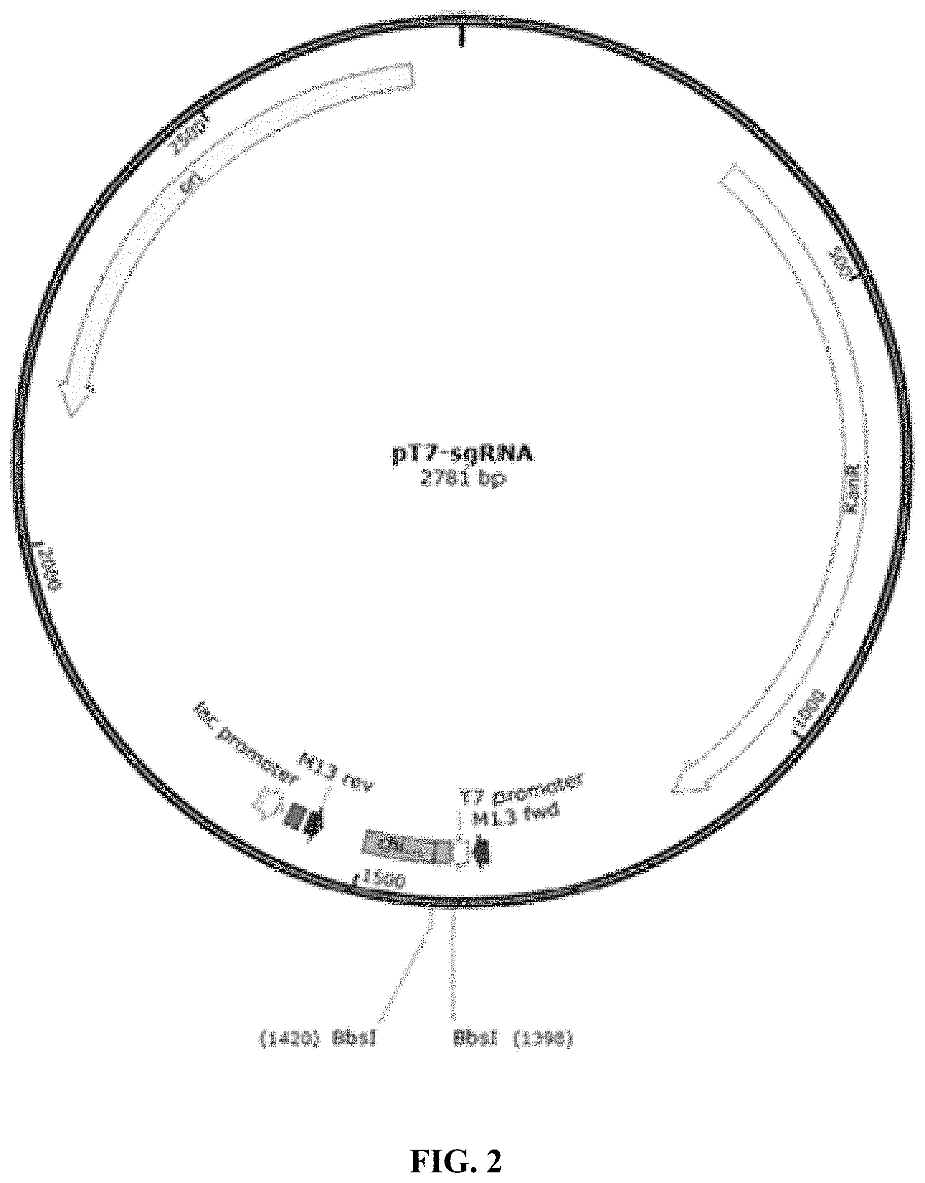
FIG. 2 is a schematic diagram showing pT7-sgRNA plasmid map.
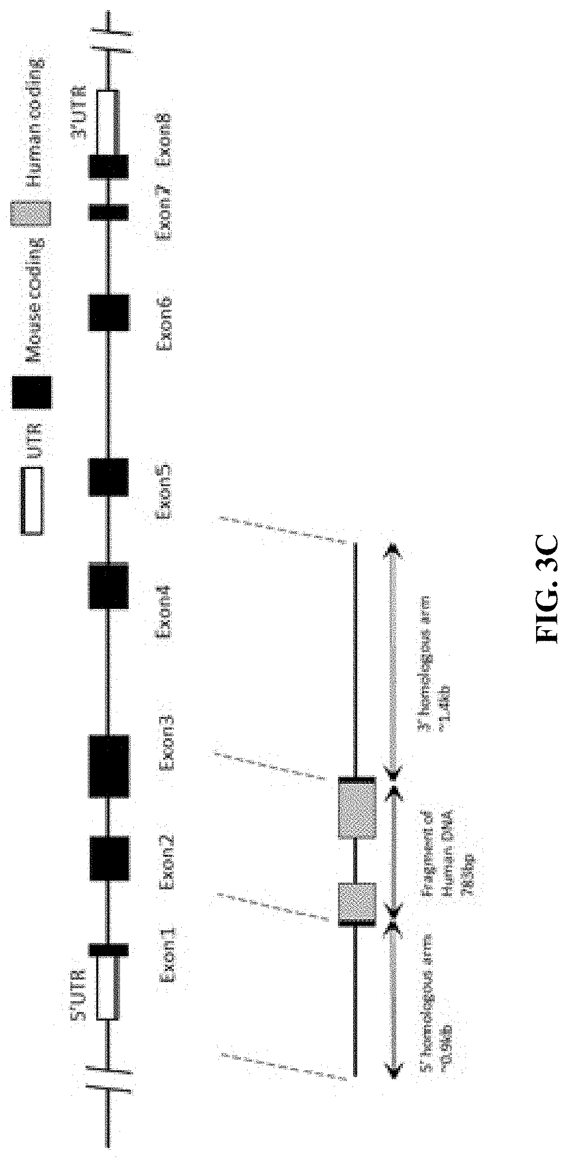
FIG. 3A is a schematic diagram showing comparison of human and mouse LAG-3 genes.
FIG. 3B is a schematic diagram showing a humanized LAG-3 mouse gene map.
FIG. 3C is a schematic diagram showing a mouse LAG-3 gene targeting strategy.
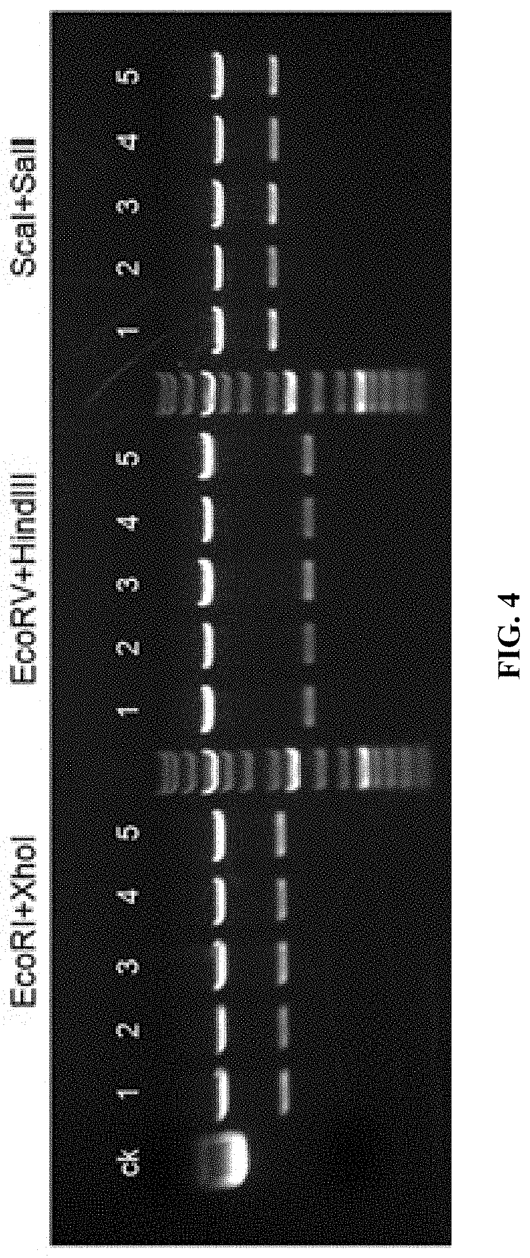
FIG. 4 shows pClon-4G-LAG plasmid digestion result (CK is undigested plasmid).
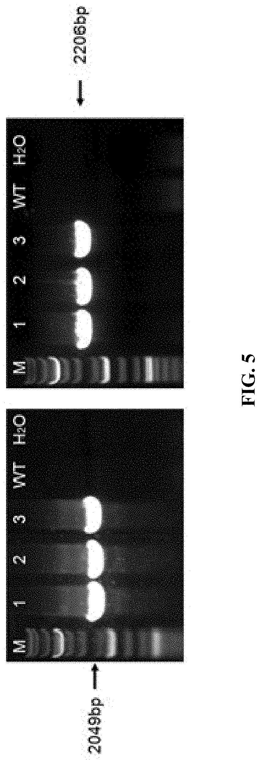
FIG. 5 shows PCR identification result of samples collected from tails of F0 generation mice (M is the Marker; WT is wildtype; mice labeled with No. 1, 2 and 3 are positive).
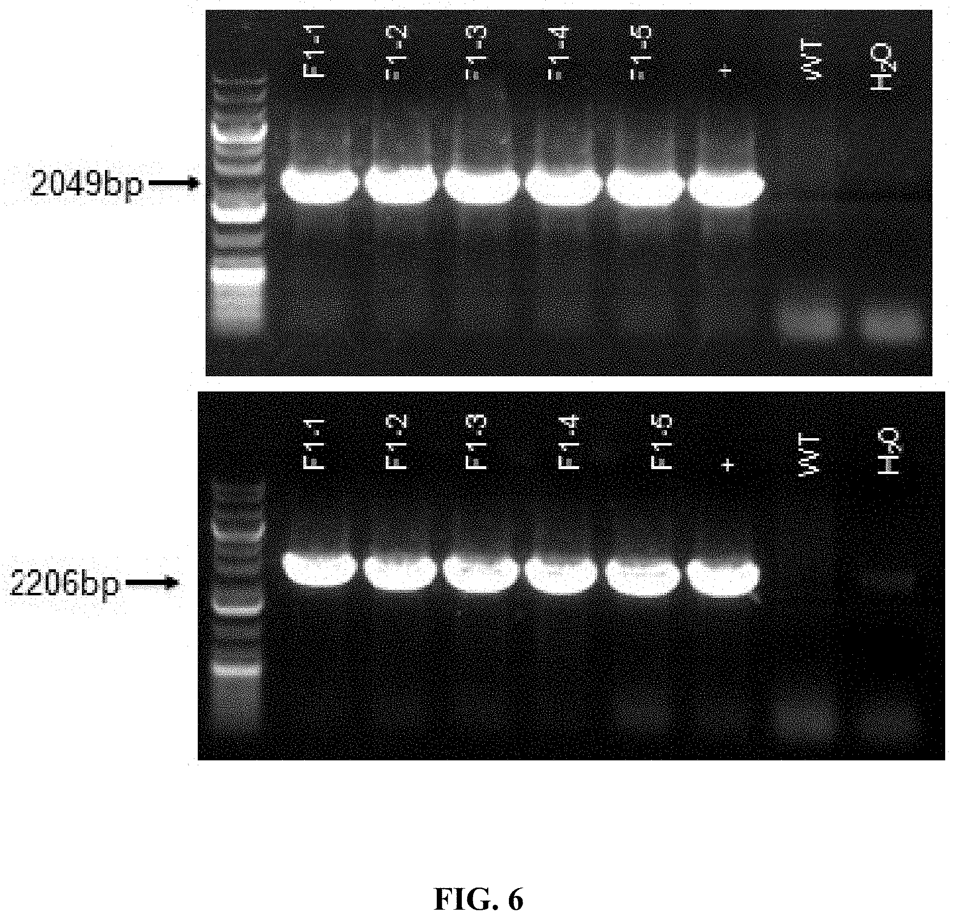
FIG. 6 shows PCR identification result of samples collected from tails of F1 generation mice (WT is wildtype; + is positive control; mice labeled with F1-1, F1-2, F1-3, F1-4, F1-5 are all positive).
FIG. 7 shows Southern blot results for F1 generation mice by P1 and P2 probes (WT is wildtype). The results show that the mice labeled with F1-2, F1-3, and F1-4 had no random insertion.
FIGS. 8A-8I are results of flow cytometry analysis for C57BL/6 mice and LAG-3 humanized mice. The anti-mouse CD3 antibody was used to stimulate the T cells in the spleens. Then the cells were labeled with anti-mouse LAG-3 antibody (mLAG3 PE; FIGS. 8A-8C), anti-human LAG-3 antibody (hLAG3 Alexa Fluor 647; FIGS. 8D-8F), or anti-human LAG-3 antibody (hLAG3 APC; FIGS. 8G-8I) and analyzed using flow cytometry. Compared to the control group (FIGS. 8A, 8D, 8G), cells expressing humanized LAG-3 protein can be detected in the spleens of humanized LAG-3 F1 heterozygous mouse; whereas in the spleens of C57BL/6 mice, no cell expressing humanized LAG-3 protein was detected.
FIG. 9A-9I are results of flow cytometry analysis of C57BL/6 mice and homozygous humanized LAG-3 mice. The anti-mouse CD3 antibody was used to stimulate the T cells in the spleens. Then the cells were labeled with anti-mouse LAG-3 antibody (mLAG3 PE; FIGS. 9A-9C), anti-human LAG-3 antibody (hLAG3 APC; FIGS. 9D-9F), or anti-human LAG-3 antibody (hLAG3 Alexa Fluor 647; FIGS. 9G-9I), and analyzed using flow cytometry. Cells expressing humanized LAG-3 protein can be detected in the spleens of humanized LAG-3 F1 homozygotes (FIGS. 9F, 9I).
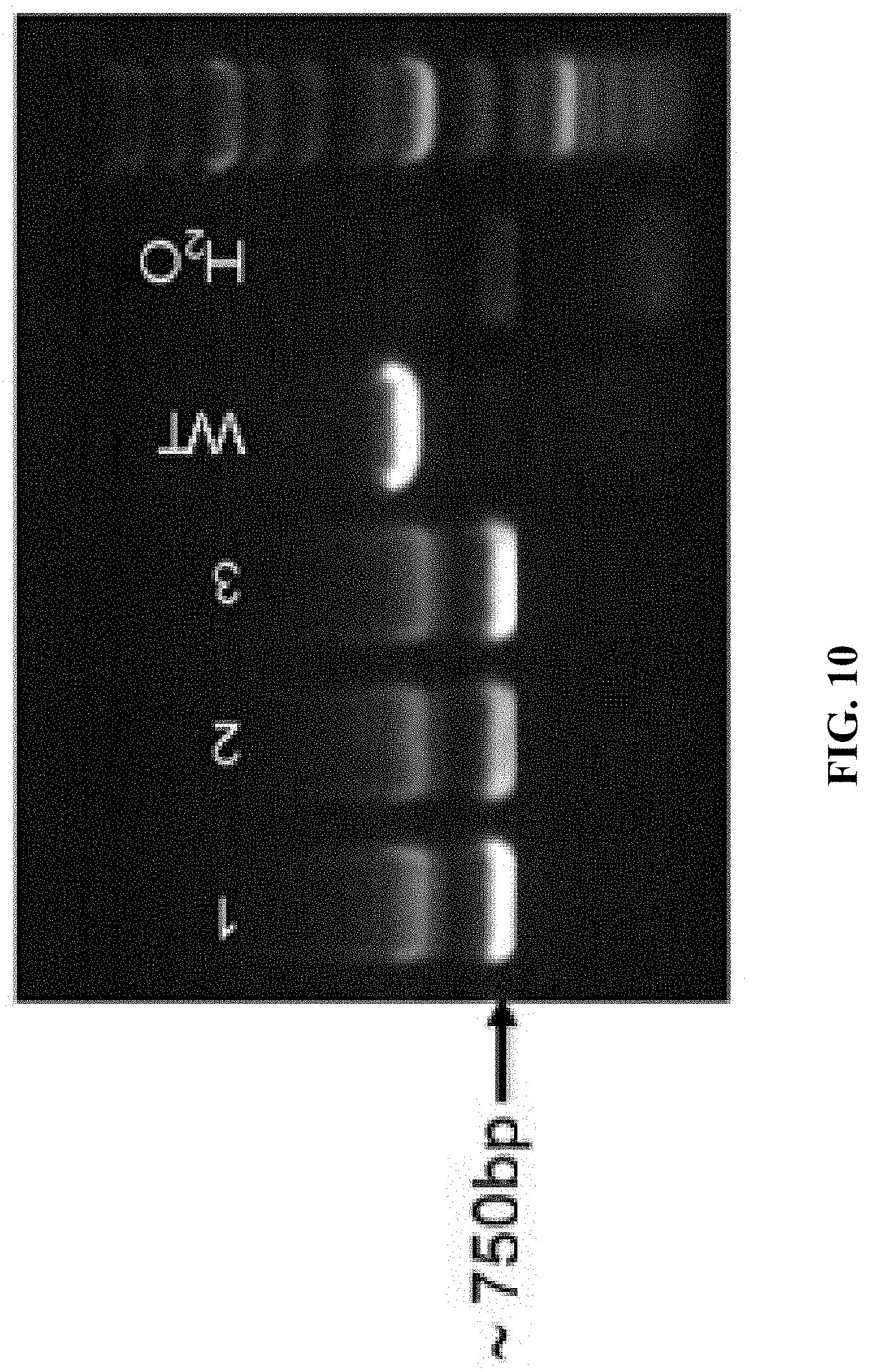
FIG. 10 shows PCR analysis results, wherein WT is wildtype C57BL/6 mouse; mice labeled No. 1, 2, and 3 were LAG-3 knockout mice.
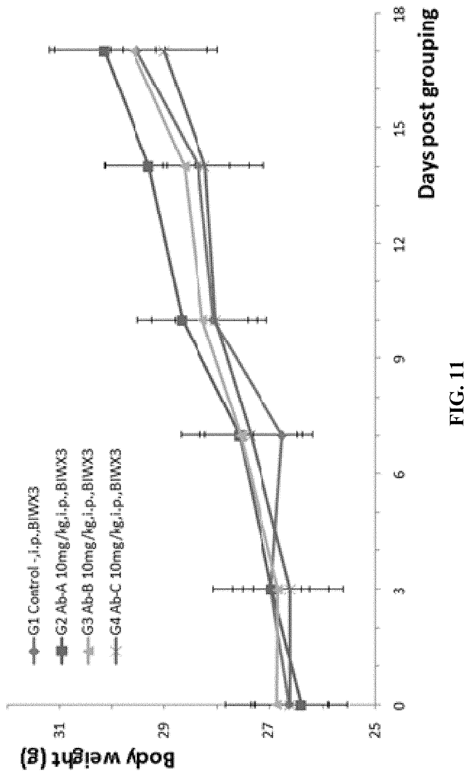
FIG. 11 shows results from experiments where mouse colon cancer cells MC38 were injected into B-hLAG-3 mice, followed by studies on antitumor efficacy of three anti-human LAG-3 antibodies (Ab-A, Ab-B, and Ab-C, 10 mg/kg). There was no significant difference in the average weight between the G1 control group and the G2-G4 treatment groups.
FIG. 12 shows results from experiments where mouse colon cancer cells MC38 were injected into B-hLAG-3 mice, followed by studies on antitumor efficacy of three anti-human LAG-3 antibodies (Ab-A, Ab-B, and Ab-C, 10 mg/kg). While the decease of tumor size in G2 and G3 groups were not significant, the tumor size in G4 group (treated with Ab-C) was significantly smaller compared to the G1 control group.
FIGS. 13A-13D show PCR detection results. In FIGS. 13A, 13B, + is a known humanized LAG-3 homozygous mouse, - is wildtype control, WT is wildtype, "mut" indicates humanized LAG-3. In FIGS. 13C, 13D, -/- is a humanized PD-1 homozygous mouse, +/- is humanized PD-1 heterozygous mouse, WT is wildtype, "mut" indicates humanized PD-1. FIGS. 13A and 13B show that the mice numbered 3020 to 3023 were humanized LAG-3 homozygous mice. FIGS. 13C and 13D show that the mice numbered 3019 to 3027 were humanized PD-1 homozygous mice. FIGS. 13A-13D show that the four mice numbered 3020 to 3023 are double humanized mice that are homozygous for both humanized PD-1 and humanized LAG-3.
FIGS. 14A-14F are results of flow cytometry analysis for C57BL/6 mice and double humanized LAG-3/PD-1 homozygous mice. Anti-mouse CD3 antibody was used to stimulate T cell activation in the spleens of the mice. The cells were then stained with either (1) mouse LAG-3 antibody (mLAG-3 PE) and anti-mTCR.beta. antibody (mTcR.beta. PerCP) (FIGS. 14A, 14B, 14C), or (2) human LAG-3 antibody (hLAG-3 APC) and anti-mTCR.beta. antibody (mTcR.beta. PerCP) (FIGS. 14D, 14E, 14F). The results showed that the cells expressing humanized LAG-3 proteins were detected in the spleens of double humanized LAG-3/PD-1 homozygous mice, while no cell expressing humanized LAG-3 protein was detected in the spleens of C57BL/6 control mice.
FIGS. 15A-15F are results of flow cytometry analysis for C57BL/6 mice and double humanized LAG-3/PD-1 homozygous mice. Anti-mouse CD3 antibody was used to stimulate T cell activation in the spleens of the mice. The cells were then stained with either mouse PD-1 antibody (mPD-1 PE) and anti-mTCR.beta. antibody (mTcR.beta. PerCP) (FIGS. 15A, 15B, 15C), or human PD-1 antibody (hPD-1 FITC) and anti-mTCR.beta. antibody (mTcR.beta. PerCP) (FIGS. 15D, 15E, 15F). The results showed that the cells expressing humanized PD-1 proteins were detected in the spleens of double humanized LAG-3/PD-1 homozygous mice, while no cell expressing humanized PD-1 protein was detected in the spleens of C57BL/6 control mice.
FIGS. 16A-16D are PCR results identifying double humanized LAG-3/CTLA-4 homozygous mice. In FIGS. 16A and 16B, + is humanized CTLA-4 homozygous mouse, - is wildtype, WT is wildtype, "mut" indicates humanized CTLA-4. In FIGS. 16C and 16D, + is LAG-3 heterozygous mouse, - is wildtype, WT is wildtype, "mut" indicates humanized LAG-3. FIGS. 16A and 16B show that the mice numbered 1106 to 1117 were humanized CTLA-4 homozygous mice. FIGS. 16C and 16D show that the mice numbered 1106 to 1117 were humanized LAG-3 homozygous mice. FIGS. 16A-16D show that the twelve mice numbered 1106 to 1117 were double humanized mice that are homozygous for both humanized CTLA-4 and humanized LAG-3.
FIG. 17. Mouse colon cancer cells MC38 were injected into double humanized homozygous LAG-3/PD-1 mice. Antitumor efficacy studies were performed with Keytruda (anti-human PD-1 antibody), and Keytruda in combination with anti-human LAG-3 antibody (Ab-C). There was no significant difference in the average weight between the G1 control group and the G2-G3 treatment groups.
FIG. 18. Mouse colon cancer cells MC38 were injected into double humanized homozygous LAG-3/PD-1 mice. Antitumor efficacy studies were performed with Keytruda, and Keytruda in combination with anti-human LAG-3 antibody (Ab-C). There was no significant difference in the percentage of body weight change among the different groups.
FIG. 19. Mouse colon cancer cells MC38 were injected into double humanized homozygous LAG-3/PD-1 mice. Antitumor efficacy studies were performed with Keytruda, and Keytruda in combination with anti-human LAG-3 antibody (Ab-C). The average volumes of tumors in the G2-G3 treatment groups were significantly smaller than the G1 control group. Furthermore, the tumor size in mice treated with the combination of Keytruda and Ab-C is significantly smaller than the tumor size in mice treated with Keytruda alone.
FIG. 20 is a schematic diagram of a targeting strategy for embryonic stem cells.
FIG. 21 shows the alignment between mouse LAG-3 amino acid sequence (NP 032505.1; SEQ ID NO: 25) and human LAG-3 amino acid sequence (NP_002277.4; SEQ ID NO: 27) by NCBI Basic Local Alignment Search Tool (BLAST).
SEQUENCE LISTING
The instant application contains a Sequence Listing which has been submitted electronically in ASCII format and is hereby incorporated by reference in its entirety. Said ASCII copy, created on Aug. 19, 2019, is named Revised Sequence Listing.txt and is 38,985 bytes in size.
DETAILED DESCRIPTION
This disclosure relates to transgenic non-human animal with human or chimeric (e.g., humanized) Lymphocyte Activation Gene 3 (LAG-3), and methods of use thereof.
LAG-3 (CD223) is a co-inhibitory receptor of T cells. Expression of LAG-3 has been reported in activated CD4+ and CD8+ effector T cells, CD4+Foxp3+ Treg, Tr1 cells, B cells, plasmacytoid DCs, and NK cells. LAG-3 associates with CD3 in the TCR complex and crosslinking of LAG-3 together with CD3 negatively regulates signal transduction leading to reduced T cell proliferation and cytokine production. LAG-3 deficient OVA-specific CD4+ T cells show uncontrolled expansion upon immunization with their cognate antigen (Workman, Creg J., and Dario A A Vignali. "The CD4-related molecule, LAG-3 (CD223), regulates the expansion of activated T cells." European journal of immunology 33.4 (2003): 970-979). Similarly, increased proliferation of LAG-3 deficient donor T cells causes more severe acute GVHD. On CD8+ T cells, LAG-3 expression is induced by T cell activation and, like in CD4+ T cells, blockade of LAG-3 improves cytotoxic T cell (CTL) proliferation and effector function. In Tregs, loss of LAG-3 reduced the suppressive function of Tregs, while forced expression of LAG-3 conferred effector T cells with suppressive capacity. LAG-3 thus plays an important role in dampening immune responses by functionally contributing to immune suppression by regulatory T cells. (Nicole Joller and Vijay K. Kuchroo, "Tim-3, Lag-3, and TIGIT," Curr Top Microbiol Immunol. 2017 Sep. 13.)
As LAG-3 is involved in T cell inhibitory pathway, it thus can be expected that the LAG-3 antibody has great application values, e.g., as a tumor immunotherapy or a treatment for autoimmune disease (e.g., psoriasis and plaque psoriasis). In order to make the animal experiments more effective and more relevant, the present disclosure provides humanized LAG-3 genetically modified animal models and methods of establishing such animal models.
Experimental animal models are an indispensable research tool for studying the etiology, pathogenesis of the disease, as well as the development of prevention and control techniques and therapeutic drugs for the disease. Common experimental animals include mice, rats, guinea pigs, hamsters, rabbits, dogs, monkeys, pigs, fish and so on. However, there are many differences between human and animal genes and protein sequences, and many human proteins cannot bind to the animal's homologous proteins to produce biological activity, leading to that the results of many clinical trials do not match the results obtained from animal experiments. A large number of clinical studies are in urgent need of better animal models. With the continuous development and maturation of genetic engineering technologies, the use of human cells or genes to replace or substitute an animal's endogenous similar cells or genes to establish a biological system or disease model closer to human, and establish the humanized experimental animal models (humanized animal model) has provided an important tool for new clinical approaches or means. In this context, the genetically engineered animal model, that is, the use of genetic manipulation techniques, the use of human normal or mutant genes to replace animal homologous genes, can be used to establish the genetically modified animal models that are closer to human gene systems. The humanized animal models have various important applications. For example, due to the presence of human or humanized genes, the animals can express or express in part of the proteins with human functions, so as to greatly reduce the differences in clinical trials between humans and animals, and provide the possibility of drug screening at animal levels.
Unless otherwise specified, the practice of the methods described herein can take advantage of the techniques of cell biology, cell culture, molecular biology, transgenic biology, microbiology, recombinant DNA and immunology. These techniques are explained in detail in the following literature, for examples: Molecular Cloning A Laboratory Manual, 2nd Ed., ed. By Sambrook, Fritsch and Maniatis (Cold Spring Harbor Laboratory Press: 1989); DNA Cloning, Volumes I and II (D. N. Glovered., 1985); Oligonucleotide Synthesis (M. J. Gaited., 1984); Mullisetal U.S. Pat. No. 4,683,195; Nucleic Acid Hybridization (B. D. Hames & S. J. Higginseds. 1984); Transcription And Translation (B. D. Hames & S. J. Higginseds. 1984); Culture Of Animal Cell (R. I. Freshney, Alan R. Liss, Inc., 1987); Immobilized Cells And Enzymes (IRL Press, 1986); B. Perbal, A Practical Guide To Molecular Cloning (1984), the series, Methods In ENZYMOLOGY (J. Abelson and M. Simon, eds.-in-chief, Academic Press, Inc., New York), specifically, Vols. 154 and 155 (Wuetal. eds.) and Vol. 185, "Gene Expression Technology" (D. Goeddel, ed.); Gene Transfer Vectors For Mammalian Cells (J. H. Miller and M. P. Caloseds., 1987, Cold Spring Harbor Laboratory); Immunochemical Methods In Cell And Molecular Biology (Mayer and Walker, eds., Academic Press, London, 1987); Hand book Of Experimental Immunology, Volumes V (D. M. Weir and C. C. Blackwell, eds., 1986); and Manipulating the Mouse Embryo, (Cold Spring Harbor Laboratory Press, Cold Spring Harbor, N. Y., 1986), each of which is incorporated herein in its entirety by reference.
LAG-3 (Lymphocyte Activation Gene 3)
LAG-3 (also known as CD223) is an immunoglobulin superfamily member composed of four extracellular Ig-like domains and a type I transmembrane domain. Major histocompatibility complex class II (MHC-II) is a ligand for LAG-3; additional ligands including L-selectin and galectin-3 have also been identified. LAG-3+ tumor-infiltrating lymphocytes (TILs) have been reported in melanoma, colon, pancreatic, breast, lung, hematopoietic, and head and neck cancer patients, in association with aggressive clinical features. (Nicole Joller and Vijay K. Kuchroo, "Tim-3, Lag-3, and TIGIT," Curr Top Microbiol Immunol. 2017; Burugu, Samantha, Amanda R. Dancsok, and Torsten O. Nielsen. "Emerging targets in cancer immunotherapy." Seminars in Cancer Biology. Academic Press, 2017; Hemon, Patrice, et al. "MHC class II engagement by its ligand LAG-3 (CD223) contributes to melanoma resistance to apoptosis." The Journal of Immunology 186.9 (2011): 5173-5183).
In human genomes, LAG-3 gene locus has eight exons, exon 1, exon 2, exon 3, exon 4, exon 5, exon 6, exon 7, and exon 8 (FIG. 3A). The LAG-3 protein also has an extracellular region, a transmembrane region, and a cytoplasmic region, and the signal peptide is located at the extracellular region of LAG-3. The nucleotide sequence for human LAG-3 mRNA is NM_002286.5 (SEQ ID NO: 26), and the amino acid sequence for human LAG-3 is NP_002277.4 (SEQ ID NO: 27). The location for each exon and each region in human LAG-3 nucleotide sequence and amino acid sequence is listed below:
TABLE-US-00001 TABLE 1 NM_002286.5 NP_002277.4 Human LAG-3 1995 bp 525 aa (approximate location) (SEQ ID NO: 26) (SEQ ID NO: 27) Exon 1 1-407 1-19 Exon 2 408-555 20-69 Exon 3 556-860 70-170 Exon 4 861-1130 171-260 Exon 5 1131-1406 261-352 Exon 6 1407-1649 353-433 Exon 7 1650-1780 434-477 Exon 8 1781-1992 478-525 Signal peptide 350-415 1-22 Extracellular region 416-1699 23-450 (excluding signal peptide region) Transmembrane region 1700-1762 451-471 Cytoplasmic region 1763-1924 472-525 Donor region in Example 422-847 25-166
Similarly, in mice, LAG-3 gene locus has eight exons, exon 1, exon 2, exon 3, exon 4, exon 5, exon 6, exon 7, and exon 8 (FIG. 3A). The LAG-3 protein also has an extracellular region, a transmembrane region, and a cytoplasmic region, and the signal peptide is located at the extracellular region of LAG-3. The nucleotide sequence for mouse LAG-3 cDNA is NM_008479.2 (SEQ ID NO: 24), the amino acid sequence for mouse LAG-3 is NP_032505.1 (SEQ ID NO: 25). The location for each exon and each region in the mouse LAG-3 nucleotide sequence and amino acid sequence is listed below:
TABLE-US-00002 TABLE 2 NM_008479.2 NP_032505.1 Mouse LAG-3 2020 bp 521 aa (approximate location) (SEQ ID NO: 24) (SEQ ID NO: 25) Exon 1 1-412 1-19 Exon 2 413-560 20-69 Exon 3 561-853 70-166 Exon 4 854-1117 167-254 Exon 5 1118-1393 255-346 Exon 6 1394-1630 347-425 Exon 7 1631-1761 426-469 Exon 8 1762-2003 470-521 Signal peptide 355-420 1-22 Extracellular region 421-1680 23-442 (excluding signal peptide region) Transmembrane region 1681-1743 443-463 Cytoplasmic region 1744-1917 464-521 Replaced region in Example 427-840 25-162
The mouse LAG-3 gene (Gene ID: 16768) is located in Chromosome 11 of the mouse genome, which is located from 124904359-124912434 of NC_000072.6 (GRCm38.p4 (GCF 000001635.24)). The approximate locations of 5'-UTR, 3'-URT, exons and introns are listed below (LAG-3 has a reverse orientation on the chromosome). The 5'-UTR is from 124,911,705 to 124,911,352, exon 1 is from 124911351 to 124911294, the first intron is from 124,911,293 to 124,910,912, exon 2 is from 124,910,911 to 124,910,764, the second intron is from 124,910,763 to 124,910,397, exon 3 is from 124,910,396 to 124,910,104, the third intron is from 124,910,103 to 124,909,490, exon 4 is from 124,909,489 to 124,909,226, the fourth intron is from 124,909,225 to 124,908,653, exon 5 is from 24,908,652 to 124,908,377, the fifth intron is from 124,908,376 to 124,905,493, exon 6 is from 124,905,492 to 124,905,256, the sixth intron is from 124,905,255 to 124,904,969, exon 7 is from 124,904,968 to 124,904,838, the seventh intron is from 124,904,837 to 124,904,601, exon 8 is from 124,904,600 to 124904442, the 3'-UTR is from 124904441 to 124,904,361 of NC_000072.6, based on transcript NM_008479.2. All relevant information for mouse LAG-3 locus can be found in the NCBI website with Gene ID: 16768, which is incorporated by reference herein in its entirety.
FIG. 22 shows the alignment between mouse LAG-3 amino acid sequence (NP_032505.1; SEQ ID NO: 25) and human LAG-3 amino acid sequence (NP_002277.4; SEQ ID NO: 27). Thus, the corresponding amino acid residue or region between human and mouse LAG-3 can also be found in FIG. 22.
LAG-3 genes, proteins, and locus of the other species are also known in the art. For example, the gene ID for LAG-3 in Rattus norvegicus is 297596, the gene ID for LAG-3 in Macaca mulatta (Rhesus monkey) is 713737, the gene ID for LAG-3 in Sus scrofa (pig) is 100125962. The relevant information for these genes (e.g., intron sequences, exon sequences, amino acid residues of these proteins) can be found, e.g., in NCBI database.
The present disclosure provides human or chimeric (e.g., humanized) LAG-3 nucleotide sequence and/or amino acid sequences. In some embodiments, the entire sequence of mouse exon 1, exon 2, exon 3, exon 4, exon 5, exon 6, exon 7, exon 8, signal peptide, extracellular region, transmembrane region, and/or cytoplasmic region are replaced by the corresponding human sequence. In some embodiments, a "region" or "portion" of mouse exon 1, exon 2, exon 3, exon 4, exon 5, exon 6, exon 7, exon 8, signal peptide, extracellular region, transmembrane region, and/or cytoplasmic region are replaced by the corresponding human sequence. The term "region" or "portion" can refer to at least 1, 2, 3, 4, 5, 6, 7, 8, 9, 10, 20, 30, 40, 50, 60, 70, 80, 90, 100, 110, 120, 130, 150, 200, 250, 300, 350, or 400 nucleotides, or at least 1, 2, 3, 4, 5, 6, 7, 8, 9, 10, 20, 30, 40, 50, 60, 70, 80, 90, 100, 110, 120, 130, or 150 amino acid residues. In some embodiments, the "region" or "portion" can be at least 50%, 55%, 60%, 65%, 70%, 75%, 80%, 85%, 90%, 95%, or 99% identical to exon 1, exon 2, exon 3, exon 4, exon 5, exon 6, exon 7, exon 8, signal peptide, extracellular region, transmembrane region, or cytoplasmic region. In some embodiments, a region, a portion, or the entire sequence of mouse exon 1, exon 2, exon 3, exon 4, exon 5, exon 6, exon 7, and/or exon 8 (e.g., exon 2 and exon 3) are replaced by the human exon 1, exon 2, exon 3, exon 4, exon 5, exon 6, exon 7, and/or exon 8 (e.g., exon 2 and exon 3) sequence.
In some embodiments, the present disclosure also provides a chimeric (e.g., humanized) LAG-3 nucleotide sequence and/or amino acid sequences, wherein in some embodiments, at least 1%, 2%, 3%, 4%, 5%, 6%, 7%, 8%, 9%, 10%, 15%, 20%, 25%, 30%, 35%, 40%, 45%, 50%, 55%, 60%, 65%, 70%, 75%, 80%, 85%, 90%, 91%, 92%, 93%, 94%, 95%, 96%, 97%, 98%, 99% of the sequence are identical to or derived from mouse LAG-3 mRNA sequence (e.g., SEQ ID NO: 24), or mouse LAG-3 amino acid sequence (e.g., SEQ ID NO: 25); and in some embodiments, at least 1%, 2%, 3%, 4%, 5%, 6%, 7%, 8%, 9%, 10%, 15%, 20%, 25%, 30%, 35%, 40%, 45%, 50%, 55%, 60%, 65%, 70%, 75%, 80%, 85%, 90%, 91%, 92%, 93%, 94%, 95%, 96%, 97%, 98%, 99% of the sequence are identical to or derived from human LAG-3 mRNA sequence (e.g., SEQ ID NO: 26), or human LAG-3 amino acid sequence (e.g., SEQ ID NO: 27).
In some embodiments, the sequence encoding amino acids 25-162 of mouse LAG-3 (SEQ ID NO: 25) is replaced. In some embodiments, the sequence is replaced by a sequence encoding a corresponding region of human LAG-3 (e.g., amino acids 25-166 of human LAG-3 (SEQ ID NO: 27).
In some embodiments, the nucleic acids as described herein are operably linked to a promotor or regulatory element, e.g., an endogenous mouse LAG-3 promotor, an inducible promoter, an enhancer, and/or mouse or human regulatory elements.
In some embodiments, the nucleic acid sequence has at least a portion (e.g., at least 1, 2, 3, 4, 5, 6, 7, 8, 9, 10, 11, 12, 13, 14, 15, 20, 30, 40, 50, 60, 70, 80, 90, or 100 nucleotides, e.g., contiguous or non-contiguous nucleotides) that are different from a portion of or the entire mouse LAG-3 nucleotide sequence (e.g., exon 1, exon 2, exon 3, exon 4, exon 5, exon 6, exon 7, exon 8, or NM_008479.2 (SEQ ID NO: 24)).
In some embodiments, the nucleic acid sequence has at least a portion (e.g., at least 1, 2, 3, 4, 5, 6, 7, 8, 9, 10, 11, 12, 13, 14, 15, 20, 30, 40, 50, 60, 70, 80, 90, or 100 nucleotides, e.g., contiguous or non-contiguous nucleotides) that is the same as a portion of or the entire mouse LAG-3 nucleotide sequence (e.g., exon 1, exon 2, exon 3, exon 4, exon 5, exon 6, exon 7, exon 8, or NM_008479.2 (SEQ ID NO: 24)).
In some embodiments, the nucleic acid sequence has at least a portion (e.g., at least 1, 2, 3, 4, 5, 6, 7, 8, 9, 10, 11, 12, 13, 14, 15, 20, 30, 40, 50, 60, 70, 80, 90, or 100 nucleotides, e.g., contiguous or non-contiguous nucleotides) that is different from a portion of or the entire human LAG-3 nucleotide sequence (e.g., exon 1, exon 2, exon 3, exon 4, exon 5, exon 6, exon 7, exon 8, or NM_002286.5 (SEQ ID NO: 26)).
In some embodiments, the nucleic acid sequence has at least a portion (e.g., at least 1, 2, 3, 4, 5, 6, 7, 8, 9, 10, 11, 12, 13, 14, 15, 20, 30, 40, 50, 60, 70, 80, 90, or 100 nucleotides, e.g., contiguous or non-contiguous nucleotides) that is the same as a portion of or the entire human LAG-3 nucleotide sequence (e.g., exon 1, exon 2, exon 3, exon 4, exon 5, exon 6, exon 7, exon 8, or NM_002286.5 (SEQ ID NO: 26)).
In some embodiments, the amino acid sequence has at least a portion (e.g., at least 1, 2, 3, 4, 5, 6, 7, 8, 9, 10, 11, 12, 13, 14, 15, 20, 30, 40, 50, 60, 70, 80, 90, or 100 amino acid residues, e.g., contiguous or non-contiguous amino acid residues) that is different from a portion of or the entire mouse LAG-3 amino acid sequence (e.g., exon 1, exon 2, exon 3, exon 4, exon 5, exon 6, exon 7, exon 8 or NP_032505.1 (SEQ ID NO: 25)).
In some embodiments, the amino acid sequence has at least a portion (e.g., at least 1, 2, 3, 4, 5, 6, 7, 8, 9, 10, 11, 12, 13, 14, 15, 20, 30, 40, 50, 60, 70, 80, 90, or 100 amino acid residues, e.g., contiguous or non-contiguous amino acid residues) that is the same as a portion of or the entire mouse LAG-3 amino acid sequence (e.g., exon 1, exon 2, exon 3, exon 4, exon 5, exon 6, exon 7, exon 8, or NP_032505.1 (SEQ ID NO: 25)).
In some embodiments, the amino acid sequence has at least a portion (e.g., at least 1, 2, 3, 4, 5, 6, 7, 8, 9, 10, 11, 12, 13, 14, 15, 20, 30, 40, 50, 60, 70, 80, 90, or 100 amino acid residues, e.g., contiguous or non-contiguous amino acid residues) that is different from a portion of or the entire human LAG-3 amino acid sequence (e.g., exon 1, exon 2, exon 3, exon 4, exon 5, exon 6, exon 7, exon 8, or NP_002277.4 (SEQ ID NO: 27)).
In some embodiments, the amino acid sequence has at least a portion (e.g., at least 1, 2, 3, 4, 5, 6, 7, 8, 9, 10, 11, 12, 13, 14, 15, 20, 30, 40, 50, 60, 70, 80, 90, or 100 amino acid residues, e.g., contiguous or non-contiguous amino acid residues) that is the same as a portion of or the entire human LAG-3 amino acid sequence (e.g., exon 1, exon 2, exon 3, exon 4, exon 5, exon 6, exon 7, exon 8, or NP_002277.4 (SEQ ID NO: 27)).
The present disclosure also provides a humanized LAG-3 mouse amino acid sequence, wherein the amino acid sequence is selected from the group consisting of:
a) an amino acid sequence shown in SEQ ID NO: 31;
b) an amino acid sequence having a homology of at least 90% with or at least 90% identical to the amino acid sequence shown in SEQ ID NO: 31;
c) an amino acid sequence encoded by a nucleic acid sequence, wherein the nucleic acid sequence is able to hybridize to a nucleotide sequence encoding the amino acid shown in SEQ ID NO: 31 under a low stringency condition;
d) an amino acid sequence having a homology of at least 90%, 91%, 92%, 93%, 94%, 95%, 96%, 97%, 98%, or 99%, or at least 90%, 91%, 92%, 93%, 94%, 95%, 96%, 97%, 98%, or 99% identical to the amino acid sequence shown in SEQ ID NO: 31;
e) an amino acid sequence that is different from the amino acid sequence shown in SEQ ID NO: 31 by no more than 10, 9, 8, 7, 6, 5, 4, 3, 2 or no more than 1 amino acid; or
f) an amino acid sequence that comprises a substitution, a deletion and/or insertion of one or more amino acids to the amino acid sequence shown in SEQ ID NO: 31.
The present disclosure also relates to a LAG-3 nucleotide (e.g., DNA or RNA) sequence, wherein the nucleotide sequence can be selected from the group consisting of:
a) a nucleotide sequence as shown in SEQ ID NO: 29, or a nucleotide sequence encoding a homologous LAG-3 amino acid sequence of a humanized mouse;
b) a nucleotide sequence that is shown in SEQ ID NO: 30;
c) a nucleotide sequence that is able to hybridize to the nucleotide sequence as shown in SEQ ID NO: 29 or SEQ ID NO: 30 under a low stringency condition;
d) a nucleotide sequence that has a homology of at least 90% or at least 90% identical to the nucleotide sequence as shown in SEQ ID NO: 29 or SEQ ID NO: 30;
e) a nucleotide sequence that encodes an amino acid sequence, wherein the amino acid sequence has a homology of at least 90% with or at least 90% identical to the amino acid sequence shown in SEQ ID NO: 31;
f) a nucleotide sequence that encodes an amino acid sequence, wherein the amino acid sequence has a homology of at least 90%, 91%, 92%, 93%, 94%, 95%, 96%, 97%, 98%, or 99% with, or at least 90%, 91%, 92%, 93%, 94%, 95%, 96%, 97%, 98%, or 99% identical to the amino acid sequence shown in SEQ ID NO: 31;
g) a nucleotide sequence that encodes an amino acid sequence, wherein the amino acid sequence is different from the amino acid sequence shown in SEQ ID NO: 31 by no more than 10, 9, 8, 7, 6, 5, 4, 3, 2 or no more than 1 amino acid; and/or
h) a nucleotide sequence that encodes an amino acid sequence, wherein the amino acid sequence comprises a substitution, a deletion and/or insertion of one or more amino acids to the amino acid sequence shown in SEQ ID NO: 31.
The present disclosure further relates to a LAG-3 genomic DNA sequence of a humanized mouse. The DNA sequence is obtained by a reverse transcription of the mRNA obtained by transcription thereof is consistent with or complementary to the DNA sequence homologous to the sequence shown in SEQ ID NO: 29 or SEQ ID NO: 30.
The disclosure also provides an amino acid sequence that has a homology of at least 90% with, or at least 90% identical to the sequence shown in SEQ ID NO: 31, and has protein activity. In some embodiments, the homology with the sequence shown in SEQ ID NO: 31 is at least about 90%, 91%, 92%, 93%, 94%, 95%, 96%, 97%, 98%, or at least 99%. In some embodiments, the foregoing homology is at least about 50%, 51%, 52%, 53%, 54%, 55%, 56%, 57%, 58%, or at least about 59%.
In some embodiments, the percentage identity with the sequence shown in SEQ ID NO: 31 is at least about 90%, 91%, 92%, 93%, 94%, 95%, 96%, 97%, 98%, or at least 99%. In some embodiments, the foregoing percentage identity is at least about 50%, 51%, 52%, 53%, 54%, 55%, 56%, 57%, 58%, or at least about 59%.
The disclosure also provides a nucleotide sequence that has a homology of at least 90%, or at least 90% identical to the sequence shown in SEQ ID NO: 30, and encodes a polypeptide that has protein activity. In some embodiments, the homology with the sequence shown in SEQ ID NO: 30 is at least about 90%, 91%, 92%, 93%, 94%, 95%, 96%, 97%, 98%, or at least 99%. In some embodiments, the foregoing homology is at least about 50%, 51%, 52%, 53%, 54%, 55%, 56%, 57%, 58%, or at least about 59%.
In some embodiments, the percentage identity with the sequence shown in SEQ ID NO: 30 is at least about 90%, 91%, 92%, 93%, 94%, 95%, 96%, 97%, 98%, or at least 99%. In some embodiments, the foregoing percentage identity is at least about 50%, 51%, 52%, 53%, 54%, 55%, 56%, 57%, 58%, or at least about 59%.
The disclosure also provides a nucleic acid sequence that is at least 1%, 2%, 3%, 4%, 5%, 6%, 7%, 8%, 9%, 10%, 15%, 20%, 25%, 30%, 35%, 40%, 45%, 50%, 55%, 60%, 65%, 70%, 75%, 80%, 85%, 90%, 91%, 92%, 93%, 94%, 95%, 96%, 97%, 98%, 99% identical to any nucleotide sequence as described herein, and an amino acid sequence that is at least 1%, 2%, 3%, 4%, 5%, 6%, 7%, 8%, 9%, 10%, 15%, 20%, 25%, 30%, 35%, 40%, 45%, 50%, 55%, 60%, 65%, 70%, 75%, 80%, 85%, 90%, 91%, 92%, 93%, 94%, 95%, 96%, 97%, 98%, 99% identical to any amino acid sequence as described herein. In some embodiments, the disclosure relates to nucleotide sequences encoding any peptides that are described herein, or any amino acid sequences that are encoded by any nucleotide sequences as described herein. In some embodiments, the nucleic acid sequence is less than 10, 20, 30, 40, 50, 60, 70, 80, 90, 100, 110, 120, 130, 150, 200, 250, 300, 350, 400, or 500 nucleotides. In some embodiments, the amino acid sequence is less than 5, 6, 7, 8, 9, 10, 20, 30, 40, 50, 60, 70, 80, 90, 100, 110, 120, 130, or 150 amino acid residues.
In some embodiments, the amino acid sequence (i) comprises an amino acid sequence; or (ii) consists of an amino acid sequence, wherein the amino acid sequence is any one of the sequences as described herein.
In some embodiments, the nucleic acid sequence (i) comprises a nucleic acid sequence; or (ii) consists of a nucleic acid sequence, wherein the nucleic acid sequence is any one of the sequences as described herein.
To determine the percent identity of two amino acid sequences, or of two nucleic acid sequences, the sequences are aligned for optimal comparison purposes (e.g., gaps can be introduced in one or both of a first and a second amino acid or nucleic acid sequence for optimal alignment and non-homologous sequences can be disregarded for comparison purposes). The length of a reference sequence aligned for comparison purposes is at least 80% of the length of the reference sequence, and in some embodiments is at least 90%, 95%, or 100%. The amino acid residues or nucleotides at corresponding amino acid positions or nucleotide positions are then compared. When a position in the first sequence is occupied by the same amino acid residue or nucleotide as the corresponding position in the second sequence, then the molecules are identical at that position. The percent identity between the two sequences is a function of the number of identical positions shared by the sequences, taking into account the number of gaps, and the length of each gap, which need to be introduced for optimal alignment of the two sequences. For purposes of the present disclosure, the comparison of sequences and determination of percent identity between two sequences can be accomplished using a Blossum 62 scoring matrix with a gap penalty of 12, a gap extend penalty of 4, and a frameshift gap penalty of 5.
The term "percent homology" is often used to mean "sequence similarity." The percentage of identical residues (percent identity) and the percentage of residues conserved with similar physicochemical properties (percent similarity), e.g. leucine and isoleucine, are both used to "quantify the homology". Residues conserved with similar physicochemical properties are well known in the art. The percent homology, in many cases, is higher than the percent identity.
Cells, tissues, and animals (e.g., mouse) are also provided that comprise the nucleotide sequences as described herein, as well as cells, tissues, and animals (e.g., mouse) that express human or chimeric (e.g., humanized) LAG-3 from an endogenous non-human LAG-3 locus.
Genetically Modified Animals
As used herein, the term "genetically-modified non-human animal" refers to a non-human animal having exogenous DNA in at least one chromosome of the animal's genome. In some embodiments, at least one or more cells, e.g., at least 1%, 2%, 3%, 4%, 5%, 10%, 20%, 30%, 40%, 50% of cells of the genetically-modified non-human animal have the exogenous DNA in its genome. The cell having exogenous DNA can be various kinds of cells, e.g., an endogenous cell, a somatic cell, an immune cell, a T cell, a B cell, a germ cell, a blastocyst, or an endogenous tumor cell. In some embodiments, genetically-modified non-human animals are provided that comprise a modified endogenous LAG-3 locus that comprises an exogenous sequence (e.g., a human sequence), e.g., a replacement of one or more non-human sequences with one or more human sequences. The animals are generally able to pass the modification to progeny, i.e., through germline transmission.
As used herein, the term "chimeric gene" or "chimeric nucleic acid" refers to a gene or a nucleic acid, wherein two or more portions of the gene or the nucleic acid are from different species, or at least one of the sequences of the gene or the nucleic acid does not correspond to the wildtype nucleic acid in the animal. In some embodiments, the chimeric gene or chimeric nucleic acid has at least one portion of the sequence that is derived from two or more different sources, e.g., sequences encoding different proteins or sequences encoding the same (or homologous) protein of two or more different species. In some embodiments, the chimeric gene or the chimeric nucleic acid is a humanized gene or humanized nucleic acid.
As used herein, the term "chimeric protein" or "chimeric polypeptide" refers to a protein or a polypeptide, wherein two or more portions of the protein or the polypeptide are from different species, or at least one of the sequences of the protein or the polypeptide does not correspond to wildtype amino acid sequence in the animal. In some embodiments, the chimeric protein or the chimeric polypeptide has at least one portion of the sequence that is derived from two or more different sources, e.g., same (or homologous) proteins of different species. In some embodiments, the chimeric protein or the chimeric polypeptide is a humanized protein or a humanized polypeptide.
In some embodiments, the chimeric gene or the chimeric nucleic acid is a humanized LAG-3 gene or a humanized LAG-3 nucleic acid. In some embodiments, at least one or more portions of the gene or the nucleic acid is from the human LAG-3 gene, at least one or more portions of the gene or the nucleic acid is from a non-human LAG-3 gene. In some embodiments, the gene or the nucleic acid comprises a sequence that encodes a LAG-3 protein. The encoded LAG-3 protein is functional or has at least one activity of the human LAG-3 protein or the non-human LAG-3 protein, e.g., binding to human or non-human MHC II, CD3, L-selectin or galectin-3, and/or inhibiting immune responses.
In some embodiments, the chimeric protein or the chimeric polypeptide is a humanized LAG-3 protein or a humanized LAG-3 polypeptide. In some embodiments, at least one or more portions of the amino acid sequence of the protein or the polypeptide is from a human LAG-3 protein, and at least one or more portions of the amino acid sequence of the protein or the polypeptide is from a non-human LAG-3 protein. The humanized LAG-3 protein or the humanized LAG-3 polypeptide is functional or has at least one activity of the human LAG-3 protein or the non-human LAG-3 protein
The genetically modified non-human animal can be various animals, e.g., a mouse, rat, rabbit, pig, bovine (e.g., cow, bull, buffalo), deer, sheep, goat, chicken, cat, dog, ferret, primate (e.g., marmoset, rhesus monkey). For the non-human animals where suitable genetically modifiable ES cells are not readily available, other methods are employed to make a non-human animal comprising the genetic modification. Such methods include, e.g., modifying a non-ES cell genome (e.g., a fibroblast or an induced pluripotent cell) and employing nuclear transfer to transfer the modified genome to a suitable cell, e.g., an oocyte, and gestating the modified cell (e.g., the modified oocyte) in a non-human animal under suitable conditions to form an embryo. These methods are known in the art, and are described, e.g., in A. Nagy, et al., "Manipulating the Mouse Embryo: A Laboratory Manual (Third Edition)," Cold Spring Harbor Laboratory Press, 2003, which is incorporated by reference herein in its entirety.
In one aspect, the animal is a mammal, e.g., of the superfamily Dipodoidea or Muroidea. In some embodiments, the genetically modified animal is a rodent. The rodent can be selected from a mouse, a rat, and a hamster. In some embodiment, the rodent is selected from the superfamily Muroidea. In some embodiments, the genetically modified animal is from a family selected from Calomyscidae (e.g., mouse-like hamsters), Cricetidae (e.g., hamster, New World rats and mice, voles), Muridae (true mice and rats, gerbils, spiny mice, crested rats), Nesomyidae (climbing mice, rock mice, with-tailed rats, Malagasy rats and mice), Platacanthomyidae (e.g., spiny dormice), and Spalacidae (e.g., mole rates, bamboo rats, and zokors). In some embodiments, the genetically modified rodent is selected from a true mouse or rat (family Muridae), a gerbil, a spiny mouse, and a crested rat. In one embodiment, the non-human animal is a mouse.
In some embodiments, the animal is a mouse of a C57BL strain selected from C57BL/A, C57BL/An, C57BL/GrFa, C57BL/KaLwN, C57BL/6, C57BL/6J, C57BL/6ByJ, C57BL/6NJ, C57BL/10, C57BL/10ScSn, C57BL/10Cr, and C57BL/01a. In some embodiments, the mouse is a 129 strain selected from the group consisting of a strain that is 129P1, 129P2, 129P3, 129X1, 129S1 (e.g., 12951/SV, 12951/SvIm), 129S2, 129S4, 129S5, 12959/SvEvH, 129S6 (129/SvEvTac), 129S7, 129S8, 129T1, 129T2. These mice are described, e.g., in Festing et al., Revised nomenclature for strain 129 mice, Mammalian Genome 10:836 (1999); Auerbach et al., Establishment and Chimera Analysis of 129/SvEv- and C57BL/6-Derived Mouse Embryonic Stem Cell Lines (2000), both of which are incorporated herein by reference in the entirety. In some embodiments, the genetically modified mouse is a mix of the 129 strain and the C57BL/6 strain. In some embodiments, the mouse is a mix of the 129 strains, or a mix of the BL/6 strains. In some embodiment, the mouse is a BALB strain, e.g., BALB/c strain. In some embodiments, the mouse is a mix of a BALB strain and another strain. In some embodiments, the mouse is from a hybrid line (e.g., 50% BALB/c-50% 12954/Sv; or 50% C57BL/6-50% 129).
In some embodiments, the animal is a rat. The rat can be selected from a Wistar rat, an LEA strain, a Sprague Dawley strain, a Fischer strain, F344, F6, and Dark Agouti. In some embodiments, the rat strain is a mix of two or more strains selected from the group consisting of Wistar, LEA, Sprague Dawley, Fischer, F344, F6, and Dark Agouti.
The animal can have one or more other genetic modifications, and/or other modifications, that are suitable for the particular purpose for which the humanized LAG-3 animal is made. For example, suitable mice for maintaining a xenograft (e.g., a human cancer or tumor), can have one or more modifications that compromise, inactivate, or destroy the immune system of the non-human animal in whole or in part. Compromise, inactivation, or destruction of the immune system of the non-human animal can include, for example, destruction of hematopoietic cells and/or immune cells by chemical means (e.g., administering a toxin), physical means (e.g., irradiating the animal), and/or genetic modification (e.g., knocking out one or more genes). Non-limiting examples of such mice include, e.g., NOD mice, SCID mice, NOD/SCID mice, IL2R.gamma. knockout mice, NOD/SCID/.gamma.cnull mice (Ito, M. et al., NOD/SCID/.gamma.cnull mouse: an excellent recipient mouse model for engraftment of human cells, Blood 100(9):3175-3182, 2002), nude mice, and Rag1 and/or Rag2 knockout mice. These mice can optionally be irradiated, or otherwise treated to destroy one or more immune cell type. Thus, in various embodiments, a genetically modified mouse is provided that can include a humanization of at least a portion of an endogenous non-human LAG-3 locus, and further comprises a modification that compromises, inactivates, or destroys the immune system (or one or more cell types of the immune system) of the non-human animal in whole or in part. In some embodiments, modification is, e.g., selected from the group consisting of a modification that results in NOD mice, SCID mice, NOD/SCID mice, IL-2R.gamma. knockout mice, NOD/SCID/.gamma.c null mice, nude mice, Rag1 and/or Rag2 knockout mice, and a combination thereof. These genetically modified animals are described, e.g., in US20150106961, which is incorporated herein by reference in its entirety. In some embodiments, the mouse can include a replacement of all or part of mature LAG-3 coding sequence with human mature LAG-3 coding sequence.
Genetically modified non-human animals that comprise a modification of an endogenous non-human LAG-3 locus. In some embodiments, the modification can comprise a human nucleic acid sequence encoding at least a portion of a mature LAG-3 protein (e.g., at least 10%, 20%, 30%, 40%, 50%, 60%, 70%, 80%, 90%, 95%, 96%, 97%, 98%, or 99% identical to the mature LAG-3 protein sequence). Although genetically modified cells are also provided that can comprise the modifications described herein (e.g., ES cells, somatic cells), in many embodiments, the genetically modified non-human animals comprise the modification of the endogenous LAG-3 locus in the germline of the animal.
Genetically modified animals can express a human LAG-3 and/or a chimeric (e.g., humanized) LAG-3 from endogenous mouse loci, wherein the endogenous mouse LAG-3 gene has been replaced with a human LAG-3 gene and/or a nucleotide sequence that encodes a region of human LAG-3 sequence or an amino acid sequence that is at least 10%, 20%, 30%, 40%, 50%, 60%, 70&, 80%, 90%, 95%, 96%, 97%, 98%, or 99% identical to the human LAG-3 sequence. In various embodiments, an endogenous non-human LAG-3 locus is modified in whole or in part to comprise human nucleic acid sequence encoding at least one protein-coding sequence of a mature LAG-3 protein.
In some embodiments, the genetically modified mice express the human LAG-3 and/or chimeric LAG-3 (e.g., humanized LAG-3) from endogenous loci that are under control of mouse promoters and/or mouse regulatory elements. The replacement(s) at the endogenous mouse loci provide non-human animals that express human LAG-3 or chimeric LAG-3 (e.g., humanized LAG-3) in appropriate cell types and in a manner that does not result in the potential pathologies observed in some other transgenic mice known in the art. The human LAG-3 or the chimeric LAG-3 (e.g., humanized LAG-3) expressed in animal can maintain one or more functions of the wildtype mouse or human LAG-3 in the animal. For example, human or non-human MHC II can bind to the expressed LAG-3 and downregulate immune response, e.g., downregulate immune response by at least 10%, 20%, 30%, 40%, or 50%. Furthermore, in some embodiments, the animal does not express endogenous LAG-3. As used herein, the term "endogenous LAG-3" refers to LAG-3 protein that is expressed from an endogenous LAG-3 nucleotide sequence of the genetically modified non-human animal (e.g., mouse) before the genetic modification.
The genome of the animal can comprise a sequence encoding an amino acid sequence that is at least 70%, 75%, 80%, 85%, 90%, 95%, 99%, or 100% identical to human LAG-3 (NP_002277.4) (SEQ ID NO: 27). In some embodiments, the genome comprises a sequence encoding an amino acid sequence that is at least 70%, 75%, 80%, 85%, 90%, 95%, 99%, or 100% identical to SEQ ID NO: 31.
The genome of the genetically modified animal can comprise a replacement at an endogenous LAG-3 gene locus of a sequence encoding a region of endogenous LAG-3 with a sequence encoding a corresponding region of human LAG-3. In some embodiments, the sequence that is replaced is any sequence within the endogenous LAG-3 gene locus, e.g., exon 1, exon 2, exon 3, exon 4, exon 5, exon 6, exon 7, exon 8, 5'-UTR, 3'UTR, the first intron, the second intron, and the third intron, the fourth intron, the fifth intron, the sixth intron, the seventh intron etc. In some embodiments, the sequence that is replaced is within the regulatory region of the endogenous LAG-3 gene. In some embodiments, the sequence that is replaced is exon 2 and exon 3, or part thereof of an endogenous mouse LAG-3 gene locus.
The genetically modified animal can have one or more cells expressing a human or chimeric LAG-3 (e.g., humanized LAG-3) having an extracellular region and a cytoplasmic region, wherein the extracellular region comprises a sequence that is at least 50%, 60%, 70%, 80%, 90%, 95%, 99% identical to the extracellular region of human LAG-3. In some embodiments, the extracellular region of the humanized LAG-3 has a sequence that has at least 10, 20, 30, 40, 50, 60, 70, 80, 90, or 100 amino acids (e.g., contiguously or non-contiguously) that are identical to human LAG-3. Because human LAG-3 and non-human LAG-3 (e.g., mouse LAG-3) sequences, in many cases, are different, antibodies that bind to human LAG-3 will not necessarily have the same binding affinity with mouse LAG-3 or have the same effects to mouse LAG-3. Therefore, the genetically modified animal having a human or a humanized extracellular region can be used to better evaluate the effects of anti-human LAG-3 antibodies in an animal model. In some embodiments, the genome of the genetically modified animal comprises a sequence encoding an amino acid sequence that corresponds to part or the entire sequence of exon 1, exon 2, exon 3, exon 4, exon 5, exon 6, exon 7, and/or exon 8 of human LAG-3, part or the entire sequence of extracellular region of human LAG-3 (with or without signal peptide), or part or the entire sequence of amino acids 25-166 of SEQ ID NO: 27.
In some embodiments, the non-human animal can have, at an endogenous LAG-3 gene locus, a nucleotide sequence encoding a chimeric human/non-human LAG-3 polypeptide, wherein a human portion of the chimeric human/non-human LAG-3 polypeptide comprises a portion of human LAG-3 extracellular domain, and wherein the animal expresses a functional LAG-3 on a surface of a cell of the animal. The human portion of the chimeric human/non-human LAG-3 polypeptide can comprise a portion of exon 1, exon 2, exon 3, exon 4, exon 5, exon 6, exon 7, and/or exon 8 of human LAG-3. In some embodiments, the human portion of the chimeric human/non-human LAG-3 polypeptide can comprise a sequence that is at least 80%, 85%, 90%, 95%, or 99% identical to amino acids 25-166 of SEQ ID NO: 27.
In some embodiments, the non-human portion of the chimeric human/non-human LAG-3 polypeptide comprises transmembrane and/or cytoplasmic regions of an endogenous non-human LAG-3 polypeptide. There may be several advantages that are associated with the transmembrane and/or cytoplasmic regions of an endogenous non-human LAG-3 polypeptide. For example, once a LAG-3 ligand (e.g., MHC II) binds to LAG-3, they can properly transmit extracellular signals into the cells and regulate the downstream pathway. A human or humanized transmembrane and/or cytoplasmic regions may not function properly in non-human animal cells. In some embodiments, a few extracellular amino acids that are close to the transmembrane region of LAG-3 are also derived from endogenous sequence.
Furthermore, the genetically modified animal can be heterozygous with respect to the replacement at the endogenous LAG-3 locus, or homozygous with respect to the replacement at the endogenous LAG-3 locus.
In some embodiments, the humanized LAG-3 locus lacks a human LAG-3 5'-UTR. In some embodiment, the humanized LAG-3 locus comprises a rodent (e.g., mouse) 5'-UTR. In some embodiments, the humanization comprises a human 3'-UTR. In appropriate cases, it may be reasonable to presume that the mouse and human LAG-3 genes appear to be similarly regulated based on the similarity of their 5'-flanking sequence. As shown in the present disclosure, humanized LAG-3 mice that comprise a replacement at an endogenous mouse LAG-3 locus, which retain mouse regulatory elements but comprise a humanization of LAG-3 encoding sequence, do not exhibit pathologies. Both genetically modified mice that are heterozygous or homozygous for human LAG-3 are grossly normal.
The present disclosure further relates to a non-human mammal generated through the methods as described herein. In some embodiments, the genome thereof contains human gene(s).
In some embodiments, the non-human mammal is a rodent, and preferably, the non-human mammal is a mouse.
In some embodiments, the non-human mammal expresses a protein encoded by a humanized LAG-3 gene.
In addition, the present disclosure also relates to a tumor bearing non-human mammal model, characterized in that the non-human mammal model is obtained through the methods as described herein. In some embodiments, the non-human mammal is a rodent (e.g., a mouse).
The present disclosure further relates to a cell or cell line, or a primary cell culture thereof derived from the non-human mammal or an offspring thereof, or the tumor bearing non-human mammal; the tissue, organ or a culture thereof derived from the non-human mammal or an offspring thereof, or the tumor bearing non-human mammal; and the tumor tissue derived from the non-human mammal or an offspring thereof when it bears a tumor, or the tumor bearing non-human mammal.
The present disclosure also provides non-human mammals produced by any of the methods described herein. In some embodiments, a non-human mammal is provided; and the genetically modified animal contains the DNA encoding human or humanized LAG-3 in the genome of the animal.
In some embodiments, the non-human mammal comprises the genetic construct as described herein (e.g., gene construct as shown in FIG. 3). In some embodiments, a non-human mammal expressing human or humanized LAG-3 is provided. In some embodiments, the tissue-specific expression of human or humanized LAG-3 protein is provided.
In some embodiments, the expression of human or humanized LAG-3 in a genetically modified animal is controllable, as by the addition of a specific inducer or repressor substance.
Non-human mammals can be any non-human animal known in the art and which can be used in the methods as described herein. Preferred non-human mammals are mammals, (e.g., rodents). In some embodiments, the non-human mammal is a mouse.
Genetic, molecular and behavioral analyses for the non-human mammals described above can performed. The present disclosure also relates to the progeny produced by the non-human mammal provided by the present disclosure mated with the same or other genotypes.
The present disclosure also provides a cell line or primary cell culture derived from the non-human mammal or a progeny thereof. A model based on cell culture can be prepared, for example, by the following methods. Cell cultures can be obtained by way of isolation from a non-human mammal, alternatively cell can be obtained from the cell culture established using the same constructs and the standard cell transfection techniques. The integration of genetic constructs containing DNA sequences encoding human LAG-3 protein can be detected by a variety of methods.
There are many analytical methods that can be used to detect exogenous DNA expression, including methods at the level of RNA (including the mRNA quantification approaches using reverse transcriptase polymerase chain reaction (RT-PCR) or Southern blotting, and in situ hybridization) and methods at the protein level (including histochemistry, immunoblot analysis and in vitro binding studies). In addition, the expression level of the gene of interest can be quantified by ELISA techniques well known to those skilled in the art. Many standard analysis methods can be used to complete quantitative measurements. For example, transcription levels can be measured using RT-PCR and hybridization methods including RNase protection, Southern blot analysis, RNA dot analysis (RNAdot) analysis. Immunohistochemical staining, flow cytometry, Western blot analysis can also be used to assess the presence of human LAG-3 protein.
Vectors
The present disclosure relates to a targeting vector, comprising: a) a DNA fragment homologous to the 5' end of a region to be altered (5' arm), which is selected from the LAG-3 gene genomic DNAs in the length of 100 to 10,000 nucleotides; b) a desired/donor DNA sequence encoding a donor region; and c) a second DNA fragment homologous to the 3' end of the region to be altered (3' arm), which is selected from the LAG-3 gene genomic DNAs in the length of 100 to 10,000 nucleotides.
In some embodiments, a) the DNA fragment homologous to the 5' end of a conversion region to be altered (5' arm) is selected from the nucleotide sequences that have at least 90% homology to the NCBI accession number NC_000072.6; c) the DNA fragment homologous to the 3' end of the region to be altered (3' arm) is selected from the nucleotide sequences that have at least 90% homology to the NCBI accession number NC_000072.6.
In some embodiments, a) the DNA fragment homologous to the 5' end of a region to be altered (5' arm) is selected from the nucleotides from the position 124911766 to the position 124910898 of the NCBI accession number NC_000072.6; c) the DNA fragment homologous to the 3' end of the region to be altered (3' arm) is selected from the nucleotides from the position 124910116 to the position 124908702 of the NCBI accession number NC_000072.6.
In some embodiments, the length of the selected genomic nucleotide sequence in the targeting vector can be about 1.2 kb, about 1.5 kb, or about 1 kb. In some embodiments, the length is about 869 bp or about 1415 bp.
In some embodiments, the region to be altered is exon 1, exon 2, exon 3, exon 4, exon 5, exon 6, exon 7, and/or exon 8 of LAG-3 gene (e.g., exon 2 and/or exon 3 of LAG-3 gene).
The targeting vector can further include a selected gene marker.
In some embodiments, the sequence of the 5' arm is shown in SEQ ID NO: 32; and the sequence of the 3' arm is shown in SEQ ID NO: 38.
In some embodiments, the target region is derived from human (e.g., 6773206-6773988 of NC_000012.12). For example, the target region in the targeting vector is a part or entirety of the nucleotide sequence of a human LAG-3, preferably the nucleotide sequence is shown as a first exon, a second exon, a third exon, a fourth exon, a fifth exon, a sixth exon, a seventh exon and/or an eighth exon of the DNA sequence of the human LAG-3. In some embodiments, the nucleotide sequence of the humanized LAG-3 encodes the humanized LAG-3 protein with the NCBI accession number NP_002277.4 (SEQ ID NO: 27).
The disclosure also relates to a cell comprising the targeting vectors as described above.
Moreover, the disclosure also relates to an sgRNA sequence for constructing a humanized animal model, wherein the sgRNA sequence targets the LAG-3 gene, the sgRNA is unique on the target sequence of the LAG-3 gene to be altered, and meets the sequence arrangement rule of 5'-NNN (20)-NGG3' or 5'-CCN-N (20)-3'; and in some embodiments, the targeting site of the sgRNA in the mouse LAG-3 gene is located on the exon 1, exon 2, exon 3, exon 4, exon 5, exon 6, exon 7, or exon 8 of the mouse LAG-3 gene (e.g., exon 2 and/or exon 3 of the mouse LAG-3 gene).
In some embodiments, an upstream sequence thereof is shown as SEQ ID NO: 15, and a downstream sequence thereof is shown as SEQ ID NO: 17, and the sgRNA sequence recognizes a 5' targeting site. In some embodiments, the forward oligonucleotide sequence is obtained by adding TAGG to the 5' end of SEQ ID NO: 15; and the reverse oligonucleotide sequence is obtained by adding AAAC to the 5' end of SEQ ID NO: 17.
In some embodiments, the disclosure provides an sgRNA sequence for constructing a humanized animal model, wherein an upstream sequence thereof is shown as SEQ ID NO: 19, and a downstream sequence thereof is shown as SEQ ID NO: 21, and the sgRNA sequence recognizes a 3' targeting site. In some embodiments, the forward oligonucleotide sequence is obtained by adding TAGG to the 5' end of SEQ ID NO: 19; and the reverse oligonucleotide sequence is obtained by adding AAAC to the 5' end of SEQ ID NO: 21.
In some embodiments, the disclosure relates to a construct including the sgRNA sequence, and/or a cell including the construct.
In addition, the present disclosure further relates to a non-human mammalian cell, having any one of the foregoing targeting vectors, and one or more in vitro transcripts of the sgRNA construct as described herein. In some embodiments, the cell includes Cas9 mRNA or an in vitro transcript thereof.
In some embodiments, the genes in the cell are heterozygous. In some embodiments, the genes in the cell are homozygous.
In some embodiments, the non-human mammalian cell is a mouse cell. In some embodiments, the cell is a fertilized egg cell.
Methods of Making Genetically Modified Animals
Genetically modified animals can be made by several techniques that are known in the art, including, e.g., nonhomologous end-joining (NHEJ), homologous recombination (HR), zinc finger nucleases (ZFNs), transcription activator-like effector-based nucleases (TALEN), and the clustered regularly interspaced short palindromic repeats (CRISPR)-Cas system. In some embodiments, homologous recombination is used. In some embodiments, CRISPR-Cas9 genome editing is used to generate genetically modified animals. Many of these genome editing techniques are known in the art, and is described, e.g., in Yin et al., "Delivery technologies for genome editing," Nature Reviews Drug Discovery 16.6 (2017): 387-399, which is incorporated by reference in its entirety. Many other methods are also provided and can be used in genome editing, e.g., micro-injecting a genetically modified nucleus into an enucleated oocyte, and fusing an enucleated oocyte with another genetically modified cell.
Thus, in some embodiments, the disclosure provides replacing in at least one cell of the animal, at an endogenous LAG-3 gene locus, a sequence encoding a region of an endogenous LAG-3 with a sequence encoding a corresponding region of human or chimeric LAG-3. In some embodiments, the replacement occurs in a germ cell, a somatic cell, a blastocyst, or a fibroblast, etc. The nucleus of a somatic cell or the fibroblast can be inserted into an enucleated oocyte.
FIG. 3C shows a humanization strategy for a mouse LAG-3 locus. In FIG. 3C, the targeting strategy involves a vector comprising the 5' end homologous arm, human LAG-3 gene fragment, 3' homologous arm. The process can involve replacing endogenous LAG-3 sequence with human sequence by homologous recombination. In some embodiments, the cleavage at the upstream and the downstream of the target site (e.g., by zinc finger nucleases, TALEN or CRISPR) can result in DNA double strands break, and the homologous recombination is used to replace endogenous LAG-3 sequence with human LAG-3 sequence.
Thus, in some embodiments, the methods for making a genetically modified, humanized animal, can include the step of replacing at an endogenous LAG-3 locus (or site), a nucleic acid encoding a sequence encoding a region of endogenous LAG-3 with a sequence encoding a corresponding region of human LAG-3. The sequence can include a region (e.g., a part or the entire region) of exon 1, exon 2, exon 3, exon 4, exon 5, exon 6, exon 7, and/or exon 8, of a human LAG-3 gene. In some embodiments, the sequence includes a region of exon 2 and exon 3 of a human LAG-3 gene (e.g., amino acids 25-166 of SEQ ID NO: 27). In some embodiments, the region is located within the extracellular region of LAG-3. In some embodiments, the endogenous LAG-3 locus is exon 2 and exon 3 of mouse LAG-3.
In some embodiments, the methods of modifying a LAG-3 locus of a mouse to express a chimeric human/mouse LAG-3 peptide can include the steps of replacing at the endogenous mouse LAG-3 locus a nucleotide sequence encoding a mouse LAG-3 with a nucleotide sequence encoding a human LAG-3, thereby generating a sequence encoding a chimeric human/mouse LAG-3.
In some embodiments, the nucleotide sequence encoding the chimeric human/mouse LAG-3 can include a first nucleotide sequence encoding an extracellular region of mouse LAG-3 (with or without the mouse signal peptide sequence); a second nucleotide sequence encoding an extracellular region of human LAG-3; a third nucleotide sequence encoding a transmembrane and a cytoplasmic region of a mouse LAG-3.
In some embodiments, the nucleotide sequences as described herein do not overlap with each other (e.g., the first nucleotide sequence, the second nucleotide sequence, and/or the third nucleotide sequence do not overlap). In some embodiments, the amino acid sequences as described herein do not overlap with each other.
The present disclosure further provides a method for establishing a LAG-3 gene humanized animal model, involving the following steps:
(a) providing the cell (e.g. a fertilized egg cell) based on the methods described herein;
(b) culturing the cell in a liquid culture medium;
(c) transplanting the cultured cell to the fallopian tube or uterus of the recipient female non-human mammal, allowing the cell to develop in the uterus of the female non-human mammal;
(d) identifying the germline transmission in the offspring genetically modified humanized non-human mammal of the pregnant female in step (c).
In some embodiments, the non-human mammal in the foregoing method is a mouse (e.g., a C57BL/6 mouse).
In some embodiments, the non-human mammal in step (c) is a female with pseudopregnancy (or false pregnancy).
In some embodiments, the fertilized eggs for the methods described above are C57BL/6 fertilized eggs. Other fertilized eggs that can also be used in the methods as described herein include, but are not limited to, FVB/N fertilized eggs, BALB/c fertilized eggs, DBA/1 fertilized eggs and DBA/2 fertilized eggs.
Fertilized eggs can come from any non-human animal, e.g., any non-human animal as described herein. In some embodiments, the fertilized egg cells are derived from rodents. The genetic construct can be introduced into a fertilized egg by microinjection of DNA. For example, by way of culturing a fertilized egg after microinjection, a cultured fertilized egg can be transferred to a false pregnant non-human animal, which then gives birth of a non-human mammal, so as to generate the non-human mammal mentioned in the method described above.
Methods of Using Genetically Modified Animals
Replacement of non-human genes in a non-human animal with homologous or orthologous human genes or human sequences, at the endogenous non-human locus and under control of endogenous promoters and/or regulatory elements, can result in a non-human animal with qualities and characteristics that may be substantially different from a typical knockout-plus-transgene animal. In the typical knockout-plus-transgene animal, an endogenous locus is removed or damaged and a fully human transgene is inserted into the animal's genome and presumably integrates at random into the genome. Typically, the location of the integrated transgene is unknown; expression of the human protein is measured by transcription of the human gene and/or protein assay and/or functional assay. Inclusion in the human transgene of upstream and/or downstream human sequences are apparently presumed to be sufficient to provide suitable support for expression and/or regulation of the transgene.
In some cases, the transgene with human regulatory elements expresses in a manner that is unphysiological or otherwise unsatisfactory, and can be actually detrimental to the animal. The disclosure demonstrates that a replacement with human sequence at an endogenous locus under control of endogenous regulatory elements provides a physiologically appropriate expression pattern and level that results in a useful humanized animal whose physiology with respect to the replaced gene are meaningful and appropriate in the context of the humanized animal's physiology.
Genetically modified animals that express human or humanized LAG-3 protein, e.g., in a physiologically appropriate manner, provide a variety of uses that include, but are not limited to, developing therapeutics for human diseases and disorders, and assessing the efficacy of these human therapeutics in the animal models.
In various aspects, genetically modified animals are provided that express human or humanized LAG-3, which are useful for testing agents that can decrease or block the interaction between LAG-3 and MHC II or the interaction between LAG-3 and other ligands (e.g., L-selectin, galectin-3), testing whether an agent can increase or decrease the immune response, and/or determining whether an agent is an LAG-3 agonist or antagonist. The genetically modified animals can be, e.g., an animal model of a human disease, e.g., the disease is induced genetically (a knock-in or knockout). In various embodiments, the genetically modified non-human animals further comprise an impaired immune system, e.g., a non-human animal genetically modified to sustain or maintain a human xenograft, e.g., a human solid tumor or a blood cell tumor (e.g., a lymphocyte tumor, e.g., a B or T cell tumor).
In some embodiments, the genetically modified animals can be used for determining effectiveness of an anti-LAG-3 antibody for the treatment of cancer. The methods involving administering the anti-LAG-3 antibody to the animal as described herein, wherein the animal has a tumor; and determining the inhibitory effects of the anti-LAG-3 antibody to the tumor. The inhibitor effects that can be determined include, e.g., a decrease of tumor size or tumor volume, a decrease of tumor growth, a reduction of the increase rate of tumor volume in a subject (e.g., as compared to the rate of increase in tumor volume in the same subject prior to treatment or in another subject without such treatment), a decrease in the risk of developing a metastasis or the risk of developing one or more additional metastasis, an increase of survival rate, and an increase of life expectancy, etc. The tumor volume in a subject can be determined by various methods, e.g., as determined by direct measurement, MRI or CT.
In some embodiments, the tumor comprises one or more tumor cells that express MHC II (Hemon, Patrice, et al. "MHC class II engagement by its ligand LAG-3 (CD223) contributes to melanoma resistance to apoptosis." The Journal of Immunology 186.9 (2011): 5173-5183). In some embodiments, the tumor comprises one or more cancer cells (e.g., human or mouse cancer cells) that are injected into the animal. In some embodiments, the anti-LAG-3 antibody or anti-MHC II antibody prevents MHC II from binding to LAG-3. In some embodiments, the anti-LAG-3 antibody or anti-MHC II antibody does not prevent MHC II from binding to LAG-3.
In some embodiments, the genetically modified animals can be used for determining whether an anti-LAG-3 antibody is an LAG-3 agonist or antagonist. In some embodiments, the methods as described herein are also designed to determine the effects of the agent (e.g., anti-LAG-3 antibodies) on LAG-3, e.g., whether the agent can stimulate T cells or inhibit T cells, whether the agent can upregulate the immune response or downregulate immune response. In some embodiments, the genetically modified animals can be used for determining the effective dosage of a therapeutic agent for treating a disease in the subject, e.g., cancer, or autoimmune diseases.
The inhibitory effects on tumors can also be determined by methods known in the art, e.g., measuring the tumor volume in the animal, and/or determining tumor (volume) inhibition rate (TGI.sub.TV). The tumor growth inhibition rate can be calculated using the formula TGI.sub.TV (%)=(1-TVt/TVc).times.100, where TVt and TVc are the mean tumor volume (or weight) of treated and control groups.
In some embodiments, the anti-LAG-3 antibody is designed for treating various cancers. As used herein, the term "cancer" refers to cells having the capacity for autonomous growth, i.e., an abnormal state or condition characterized by rapidly proliferating cell growth. The term is meant to include all types of cancerous growths or oncogenic processes, metastatic tissues or malignantly transformed cells, tissues, or organs, irrespective of histopathologic type or stage of invasiveness. The term "tumor" as used herein refers to cancerous cells, e.g., a mass of cancerous cells. Cancers that can be treated or diagnosed using the methods described herein include malignancies of the various organ systems, such as affecting lung, breast, thyroid, lymphoid, gastrointestinal, and genito-urinary tract, as well as adenocarcinomas which include malignancies such as most colon cancers, renal-cell carcinoma, prostate cancer and/or testicular tumors, non-small cell carcinoma of the lung, cancer of the small intestine and cancer of the esophagus. In some embodiments, the agents described herein are designed for treating or diagnosing a carcinoma in a subject. The term "carcinoma" is art recognized and refers to malignancies of epithelial or endocrine tissues including respiratory system carcinomas, gastrointestinal system carcinomas, genitourinary system carcinomas, testicular carcinomas, breast carcinomas, prostatic carcinomas, endocrine system carcinomas, and melanomas. In some embodiments, the cancer is renal carcinoma or melanoma. Exemplary carcinomas include those forming from tissue of the cervix, lung, prostate, breast, head and neck, colon and ovary. The term also includes carcinosarcomas, e.g., which include malignant tumors composed of carcinomatous and sarcomatous tissues. An "adenocarcinoma" refers to a carcinoma derived from glandular tissue or in which the tumor cells form recognizable glandular structures. The term "sarcoma" is art recognized and refers to malignant tumors of mesenchymal derivation.
In some embodiments, the anti-LAG-3 antibody is designed for the treating melanoma, non-small cell lung carcinoma (NSCLC), small cell lung cancer (SCLC), bladder cancer, breast cancer, and/or prostate cancer (e.g., metastatic hormone-refractory prostate cancer).
The present disclosure also relates to the use of the animal model generated through the methods as described herein in the development of a product related to an immunization processes of human cells, the manufacturing of a human antibody, or the model system for a research in pharmacology, immunology, microbiology and medicine.
In some embodiments, the disclosure provides the use of the animal model generated through the methods as described herein in the production and utilization of an animal experimental disease model of an immunization processes involving human cells, the study on a pathogen, or the development of a new diagnostic strategy and/or a therapeutic strategy.
The disclosure also relates to the use of the animal model generated through the methods as described herein in the screening, verifying, evaluating or studying the LAG-3 gene function, human LAG-3 antibodies, drugs for human LAG-3 targeting sites, the drugs or efficacies for human LAG-3 targeting sites, the drugs for immune-related diseases and antitumor drugs.
Genetically Modified Animal Model with Two or More Human or Chimeric Genes
The present disclosure further relates to methods for generating genetically modified animal model with two or more human or chimeric genes. The animal can comprise a human or chimeric LAG-3 gene and a sequence encoding an additional human or chimeric protein.
In some embodiments, the additional human or chimeric protein can be programmed cell death protein 1 (PD-1), cytotoxic T-lymphocyte-associated protein 4 (CTLA-4), T-cell immunoglobulin and mucin-domain containing-3 (TIM-3), B And T Lymphocyte Associated (BTLA), Programmed Cell Death 1 Ligand 1 (PD-L1), TNF Receptor Superfamily Member 9 (4-1BB), CD27, CD28, CD47, T-Cell Immunoreceptor With Ig And ITIM Domains (TIGIT), CD27, Glucocorticoid-Induced TNFR-Related Protein (GITR), or TNF Receptor Superfamily Member 4 (TNFRSF4; or OX40).
The methods of generating genetically modified animal model with two or more human or chimeric genes (e.g., humanized genes) can include the following steps:
(a) using the methods of introducing human LAG-3 gene or chimeric LAG-3 gene as described herein to obtain a genetically modified non-human animal;
(b) mating the genetically modified non-human animal with another genetically modified non-human animal, and then screening the progeny to obtain a genetically modified non-human animal with two or more human or chimeric genes.
In some embodiments, in step (b) of the method, the genetically modified animal can be mated with a genetically modified non-human animal with human or chimeric PD-1, CTLA-4, TIM-3, BTLA, PD-L1, 4-1BB, CD27, CD28, CD47, TIGIT, CD27, GITR, or OX40. Some of these genetically modified non-human animals are described, e.g., in PCT/CN2017/090320, PCT/CN2017/099577, PCT/CN2017/099575, PCT/CN2017/099576, PCT/CN2017/099574, PCT/CN2017/106024; each of which is incorporated herein by reference in its entirety.
In some embodiments, the LAG-3 humanization is directly performed on a genetically modified animal having a human or chimeric PD-1, CTLA-4, BTLA, TIM-3, PD-L1, 4-1BB, CD27, CD28, CD47, TIGIT, CD27, GITR, or OX40 gene.
As these proteins may involve different mechanisms, a combination therapy that targets two or more of these proteins thereof may be a more effective treatment. In fact, many related clinical trials are in progress and have shown a good effect. The genetically modified animal model with two or more human or humanized genes can be used for determining effectiveness of a combination therapy that targets two or more of these proteins, e.g., an anti-LAG-3 antibody and an additional therapeutic agent for the treatment of cancer. The methods include administering the anti-LAG-3 antibody and the additional therapeutic agent to the animal, wherein the animal has a tumor; and determining the inhibitory effects of the combined treatment to the tumor.
In some embodiments, the animal further comprises a sequence encoding a human or humanized programmed cell death protein 1 (PD-1), or a sequence encoding a human or humanized CTLA4. In some embodiments, the additional therapeutic agent is an anti-PD-1 antibody (e.g., nivolumab, pembrolizumab, avelumab, durvalumab, atezolizumab) or an CTLA-4 antibody. In some embodiments, the tumor comprises one or more tumor cells that express MHC II, CD80, CD86, PD-L1 or PD-L2.
In some embodiments, the combination treatment is designed for treating various cancer as described herein, e.g., melanoma, non-small cell lung carcinoma (NSCLC), small cell lung cancer (SCLC), bladder cancer, and/or prostate cancer (e.g., metastatic hormone-refractory prostate cancer).
In some embodiments, the methods described herein can be used to evaluate the combination treatment with some other methods. The methods of treating a cancer that can be used alone or in combination with methods described herein, include, e.g., treating the subject with chemotherapy, e.g., campothecin, doxorubicin, cisplatin, carboplatin, procarbazine, mechlorethamine, cyclophosphamide, adriamycin, ifosfamide, melphalan, chlorambucil, bisulfan, nitrosurea, dactinomycin, daunorubicin, bleomycin, plicomycin, mitomycin, etoposide, verampil, podophyllotoxin, tamoxifen, taxol, transplatinum, 5-flurouracil, vincristin, vinblastin, and/or methotrexate. Alternatively or in addition, the methods can include performing surgery on the subject to remove at least a portion of the cancer, e.g., to remove a portion of or all of a tumor(s), from the patient.
EXAMPLES
The invention is further described in the following examples, which do not limit the scope of the invention described in the claims.
Materials and Methods
The following materials were used in the following examples.
Ambion.TM. in vitro transcription kit was purchased from Ambion. Catalog number is AM1354.
E. coli TOP10 competent cells were purchased from the Tiangen Biotech (Beijing) Co. Catalog number is CB104-02.
EcoRI, Scat, HindIII, BamHI, XhoI, EcoRV, SaiI, and BbsI were purchased from NEB. Catalog numbers are R3101M, R3122M, R3104M, R3136M, R0146M, R3195M, R3138M, and R0539L.
Kanamycin was purchased from Amresco. Catalog number is 0408.
Cas9 mRNA was obtained from SIGMA. Catalog number is CAS9MRNA-1EA.
AIO kit was obtained from Beijing Biocytogen Co., Ltd. Catalog number is BCG-DX-004.
UCA kit was obtained from Beijing Biocytogen Co., Ltd. Catalog number is BCG-DX-001.
C57BL/6 mice were purchased from the China Food and Drugs Research Institute National Rodent Experimental Animal Center.
B-hPD-1 mice, B-hCTLA-4 mice were obtained from Beijing Biocytogen Co., Ltd.
Mouse colon cancer cell line MC38 was purchased from Shanghai Enzyme Research Biotechnology Co., Ltd.
Anti-mouse CD3 antibody was obtained from BD. Catalog number is 563123.
PE labeled anti-mouse CD223 (LAG-3) antibody (mLAG3 PE) was obtained from Biolegend. Catalog number is 125208.
Alexa Fluor.RTM. 647 labeled anti-human CD223 (LAG-3) antibody (hLAG3 Alexa Fluor 647) was obtained from Biolegend. Catalog number is 369304.
Monoclonal anti-human LAG-3 antibody 3DS223H (hLAG3 APC) was obtained from eBioscience. Catalog number is 17-2239-42.
PerCP/Cy5.5 labeled anti-mouse TCR .beta. chain antibody (mTcR.beta. PerCP) was obtained from Biolegend. Catalog number is 109228.
PE labeled anti-mouse CD279 (PD-1) antibody (mPD-1 PE) was obtained from Biolegend. Catalog number is 109104.
FITC labeled anti-human CD279 (PD-1) antibody (hPD-1 FITC) was obtained from Biolegend. Catalog number is 329904.
The Flow Cytometer was purchased from BD Biosciences; model FACS Calibur.TM..
Example 1: Construction of pT7-LAG-5 and pT7-LAG-12
The target sequence determines the targeting specificity of small guide RNA (sgRNA) and the efficiency of Cas9 cleavage at the target gene. Therefore, target sequence selection is important for sgRNA vector construction.
The 5'-terminal targeting sites (sgRNA1 to sgRNA7) and the 3'-terminal targeting sites (sgRNA8 to sgRNA14) were designed and synthesized. The 5'-terminal targeting sites and the 3'-terminal targeting sites are located on exon 2 and exon 3 of mouse LAG-3 gene respectively, and the targeting site sequences on LAG-3 for each sgRNA is as follows:
TABLE-US-00003 sgRNA-1 targeting sequence: (SEQ ID NO: 1) 5'-GTTCCACTAGTTGTGTCTTCAGG-3' sgRNA-2 targeting sequence: (SEQ ID NO: 2) 5'-GGCCCTGAAGACACAACTAG TGG-3' sgRNA-3 targeting sequence: (SEQ ID NO: 3) 5'-ACCACGGGGAGCTCTTTCCCAGG-3' sgRNA-4 targeting sequence: (SEQ ID NO: 4) 5'-CTAGTTGTGTCTTCAGGGCCTGG-3' sgRNA-5 targeting sequence: (SEQ ID NO: 5) 5'-GCCTGGGAAAGAGCTCCCCGTGG-3' sgRNA-6 targeting sequence: (SEQ ID NO: 6) 5'-CCTCCTGGGCCCACACCACGGGG-3' sgRNA-7 targeting sequence: (SEQ ID NO: 7) 5'-GGAAAGAGCTCCCCGTGGTGTGG-3' sgRNA-8 targeting sequence: (SEQ ID NO: 8) 5'-TCGAGGCCTGGCCGACGCGCAGG-3' sgRNA-9 targeting sequence: (SEQ ID NO: 9) 5'-TCTCCGCCTGCGCGTCGGCCAGG-3' sgRNA-10 targeting sequence: (SEQ ID NO: 10) 5'-AGGCCTGGCCGACGCGCAGGCGG-3' sgRNA-11 targeting sequence: (SEQ ID NO: 11) 5'-TGCAGTCTCCGCCTGCGCGTCGG-3' sgRNA-12 targeting sequence: (SEQ ID NO: 12) 5'-AGGAGAGGGCGCGGTTCGGGAGG-3' sgRNA-13 targeting sequence: (SEQ ID NO: 13) 5'-GCGGTTCGGGAGGCGCACGGTGG-3' sgRNA-14 targeting sequence: (SEQ ID NO: 14) 5'-TGCAGGAGAGGGCGCGGTTCGGG-3'
The UCA kit was used to detect the activities of sgRNAs (FIGS. 1A and 1B), sgRNA9 was not detected due to the incorrect sequence. The results show that the guide sgRNAs have different activities. Two of them (sgRNA5 and sgRNA12) were selected for follow-up experiments. TAGG was added to the 5' end to obtain a forward oligonucleotide sequence, and its complementary strand was added with AAAC to obtain a reverse oligonucleotide sequence. After annealing, they were respectively digested by restriction enzyme (BbsI) and ligated to pT7-sgRNA plasmid to obtain the expression vectors pT7-LAG-5 and pT7-LAG-12.
TABLE-US-00004 TABLE 3 sgRNA5 and sgRNA12 sequences sgRNA5 sequences SEQ ID NO: 15 Upstream: 5'-CCTGGGAAAGAGCTC CCCG-3' SEQ ID NO: 16 Upstream: 5'-TAGGCCTGGGAAAGA (adding TAGG to GCTCCCCG-3' obtain a forward oligonucleotide sequence) SEQ ID NO: 17 Downstream: 5'-CGGGGAGCTCTTT CCCAGG-3' SEQ ID NO: 18 Downstream: 5'-AAACCGGGGAGCT (complementary CTTTCCCAGG-3' strand was added with AAAC to obtain a reverse oligonucleotide sequence) sgRNA12 sequences SEQ ID NO: 19 Upstream: 5'-AGAGGGCGCGGTTCG GG-3' SEQ ID NO: 20 Upstream: 5'-TAGGAGAGGGCGCGG (adding TAGG to TTCGGG-3' obtain a forward oligonucleotide sequence) SEQ ID NO: 21 Downstream: 5'-CCCGAACCGCGCC CTCT-3' SEQ ID NO: 22 Downstream: 5'-AAACCCCGAACCG (complementary CGCCCTCT-3' strand was added with AAAC to obtain a reverse oligonucleotide sequence)
TABLE-US-00005 TABLE 4 The ligation reaction conditions (10 .mu.L) Double stranded fragment 1 .mu.L (0.5 .mu.M) pT7-sgRNA vector 1 .mu.L (10 ng) T4 DNA Ligase 1 .mu.L (5 U) 10 .times. T4 DNA Ligase buffer 1 .mu.L 50% PEG4000 1 .mu.L H.sub.2O Add to 10 .mu.L
Reaction Conditions:
The ligation reaction was carried out at room temperature for 10 to 30 minutes. The ligation product was then transferred to 304 of TOP10 competent cells. The cells were then plated on a petri dish with Kanamycin, and then cultured at 37.degree. C. for at least 12 hours and then two clones were selected and added to LB medium with Kanamycin (5 ml), and then cultured at 37.degree. C. at 250 rpm for at least 12 hours.
Randomly selected clones were sequenced, so as to verify their sequences. The correct expression vectors pT7-LAG-5 and pT7-LAG-12 were selected for subsequent experiments.
Source of pT7-sgRNA Plasmid
PT7-sgRNA vector map is shown in FIG. 2. The plasmid backbone was obtained from Takara (Catalog No. 3299). The DNA fragment containing T7 promoter and sgRNA scaffold was synthesized by a plasmid synthesis company, and linked to the backbone vector by restriction enzyme digestion (EcoRI and BamHI) and ligation. The target plasmid was confirmed by the sequencing results.
The DNA fragment containing the T7 promoter and sgRNA scaffold (SEQ ID NO: 23):
TABLE-US-00006 GAATTCTAATACGACTCACTATAGGGGGTCTTCGAGAAGACCTGTTTTAG AGCTAGAAATAGCAAGTTAAAATAAGGCTAGTCCGTTATCAACTTGAAAA AGTGGCACCGAGTCGGTGCTTTTAAAGGATCC
Example 2. Construction of Vector pClon-4G-LAG
A partial coding sequence of the mouse LAG-3 gene (Gene ID: 16768) from exon 2 and exon 3 (based on the transcript of NCBI accession number NM_008479.2.fwdarw.NP_032505.1 whose mRNA sequence is shown in SEQ ID NO: 24, and the corresponding protein sequence is shown in SEQ ID NO: 25) was replaced with a corresponding coding sequence of human homologous LAG-3 gene (Gene ID: 3902) (based on the transcript of NCBI accession number NM_002286.5-*NP_002277.4, whose mRNA sequence was shown in SEQ ID NO: 26, and the corresponding protein sequence is shown in SEQ ID NO: 27). The comparison between the mouse LAG-3 and human LAG-3 is shown in FIG. 3A, and the finally obtained humanized LAG-3 gene is shown in FIG. 3B, the humanized mouse LAG-3 gene DNA sequence (chimeric LAG-3 gene DNA) is shown in SEQ ID NO: 28.
TABLE-US-00007 TTGTGTCTTCAGGGCCAGGGGCTGAGGTCCCGGTGGTGTGGGCCCAGGAG GGGGCTCCTGCCCAGCTCCCCTGCAGCCCCACAATCCCCCTCCAGGATCT CAGCCTTCTGCGAAGAGCAGGGGTCACTTGGCAGCATCAGCCAGACAGGT ATGCACCCCAAACTTGGGCAACAGGACCTCCGAATCCAGCACTCAACCCC ACACCCGTGCCGGTCCTCTGTCCCCTGCCCTGAGGTGTCACTCCCTCTGA AGCCAGTGACCCAGTCTCCCTGCCCTCGCTTGCACCGTTCCTGCCCTTGC TCTGCAATCAGCGACCCTCACGCCAGCATCCCTTCTCTCCAGAAGTGGAT GCGGCCAGTCCAACAGAGGGGTCGGGCGTGAGGGGACGGTTGGTGGTCAA GAGAACTCTTGGGGCGGGCTTTCTCATCCTCAACGGGTGGCTGCCTGCAT CCTCCCGGGCTTCCTACCCCTGGAGCTTCTCAACTCCATTCTCTTTCCCG CCCAGTGGCCCGCCCGCTGCCGCCCCCGGCCATCCCCTGGCCCCCGGCCC TCACCCGGCGGCGCCCTCCTCCTGGGGGCCCAGGCCCCGCCGCTACACGG TGCTGAGCGTGGGTCCCGGAGGCCTGCGCAGCGGGAGGCTGCCCCTGCAG CCCCGCGTCCAGCTGGATGAGCGCGGCCGGCAGCGCGGGGACTTCTCGCT ATGGCTGCGCCCAGCCCGGCGCGCGGACGCCGGCGAGTACCGCGCCGCGG TGCACCTCAGGGACCGCGCCCTCTCCTGCCGCCTCCGTCTGCGCCTGGGC CAGGCCTCGA
SEQ ID NO: 28 lists only the portion of DNA sequence involved in the modification, wherein the italicized underlined region is the human LAG-3 gene sequence fragment.
The coding region sequence, mRNA sequence and the encoded protein sequence thereof of the modified humanized LAG-3 are respectively shown in SEQ ID NO: 29, SEQ ID NO: 30, and SEQ ID NO: 31.
To the extent that either human LAG-3 or mouse LAG-3 has more than one isoforms or transcripts, the methods as described herein can be applied to other isoforms or transcripts.
A targeting strategy involving a vector comprising the 5' end homologous arm, human LAG-3 gene fragment, 3' homologous arm as shown in FIG. 3C is also developed. The process is as follows:
(1). Design upstream primers of homologous recombination fragments, and downstream primers matching therewith, as well as other related sequences. Specifically:
5' end homologous arm (SEQ ID NO: 32), nucleotide sequence of the positions from 124911766 to 124910898 of the NCBI accession number NC_000072.6 as follows:
TABLE-US-00008 Upstream primer (SEQ ID NO: 33): F: 5'-tttaagaaggagatatacatggaattcgaatcagccccctcaca ctttccac-3' Downstream primer (SEQ ID NO: 34): R: 5'-cctcagcccctggccctgaagacacaactagtggaaca-3'
(2). Design the primers and related sequences of the desired conversion region. Human DNA fragment (783 bp) (SEQ ID NO: 35) is the nucleotide sequence from positions 6773206 to 6773988 of the NCBI accession number NC_000012.12.
TABLE-US-00009 The upstream primer (SEQ ID NO: 36) is: F: 5'-gtgtcttcagggccaggggctgaggtcccggtggtg-3' The downstream primer (SEQ ID NO: 37) is: R: 5'-tcgaggcctggcccaggcgcagacggaggcggcag-3'
(3). Design the upstream primers of the homologous recombination fragment and the downstream primers matching therewith, as well as other related sequences. Specifically:
3' homologous arm (SEQ ID NO: 38), which was the nucleotide sequence from positions 124910116 to 124908702 of the NCBI accession number NC_000072.6.
TABLE-US-00010 Upstream primer (SEQ ID NO: 39): F: 5'-cgtctgcgcctgggccaggcctcgagtaggtggg-3' Downstream primer (SEQ ID NO: 40): R: 5'-ttgttagcagccggatctcagggatccccagagggtggagacat caaggaag-3'
C57BL/6 mouse DNA is used as the template to carry out PCR amplification for the 5'-terminal homologous arm fragment and the 3'-terminal homologous arm fragment. Human DNA is used as the template to carry out PCR amplification for the DNA fragment, and the AIO kit is used to ligate the fragments to the pClon-4G plasmid provided by the kit, so as to obtain the vector pClon-4G-LAG.
Example 3. Verification of Vector pClon-4G-LAG
Five pClon-4G-LAG clones were randomly selected and identified by three sets of enzymes. Among them, EcoRI+XhoI should generate 1666 bp+4137 bp fragments, EcoRV+HindIII should generate 1097 bp+4706 bp fragments, ScaI+SalI should generate 1784 bp+4019 bp fragments. The results were in line with the expectations (FIG. 4). The sequences of Plasmids 1 and 2 were further verified by sequencing.
Example 4. Microinjection and Embryo Transfer
The pre-mixed Cas9 mRNA, pClon-4G-LAG plasmid and in vitro transcription products of pT7-LAG-5, pT7-LAG-12 plasmids were injected into the cytoplasm or nucleus of mouse fertilized eggs (C57BL/6 background) with a microinjection instrument (using Ambion in vitro transcription kit to carry out the transcription according to the method provided in the product instruction). The embryo microinjection was carried out according to the method described, e.g., in A. Nagy, et al., "Manipulating the Mouse Embryo: A Laboratory Manual (Third Edition)," Cold Spring Harbor Laboratory Press, 2003. The injected fertilized eggs were then transferred to a culture medium for a short time culture, and then was transplanted into the oviduct of the recipient mouse to produce the genetically modified humanized mice (F0 generation). The mice population was further expanded by cross-mating and self-mating to establish stable mouse lines. The humanized mouse was named as B-hLAG-3 mouse.
Example 5. Verification of Genetically Modified Humanized Mouse Model
1. Genotype Determination for F0 Generation Mice
PCR analysis was performed for mouse tail genomic DNA of F0 generation mice. The primers for PCR-1 were located on the left side of the 5' homologous arm, the primers for PCR-4 were located on the right side of the 3' homologous arm; in addition, the primers for PCR-2 and PCR-3 were located on the humanized fragment, which are shown below:
TABLE-US-00011 5' terminus primers: PCR-1 (SEQ ID NO: 41): 5'-agcattcacacagggtggggaatt t-3' PCR-2 (SEQ ID NO: 42): 5'-ggcgtgagggtcgctgattg-3' 3' terminus primers: PCR-3 (SEQ ID NO: 43): 5'-gacctccgaatccagcactcaac c-3' PCR-4 (SEQ ID NO: 44): 5'-aggagtccacttggcaatgagcaa a-3'
If the recombinant vector has the correct insertion, there should be one PCR band from 5' terminus primers and one PCR band from the 3' terminus primers. The length of the 5' terminus product should be 2049 bp, and the length of the 3' terminus product should be 2206 bp.
TABLE-US-00012 TABLE 5 The PCR reaction system (20 .mu.L) 10 .times. buffer 2 .mu.L dNTP (2 mM) 2 .mu.L MgSO.sub.4 (25 mM) 0.8 .mu.L Upstream primer (10 .mu.M) 0.6 .mu.L Downstream primer (10 .mu.M) 0.6 .mu.L Mouse tail genomic DNA 200 ng KOD-Plus-(1 U/.mu.L) 0.6 .mu.L
TABLE-US-00013 TABLE 6 The PCR reaction conditions Temperature Time Cycles 94.degree. C. 5 min 1 94.degree. C. 30 sec 15 67.degree. C. (-0.7.degree. C./cycle) 30 sec 68.degree. C. 1 kb/min 94.degree. C. 30 sec 25 56.degree. C. 30 sec 68.degree. C. 1 kb/min 68.degree. C. 10 min 1 4.degree. C. 10 min 1
The verification results for three F0 generation mice are shown in FIG. 5.
2. Genotype Determination for F1 Generation Mice
F1 generation mice were obtained by cross-mating F0 generation mice with C57BL/6 mice. PCR was performed for four F1 generation mice with the same primers under the same conditions used for F0 verification. The results showed that all five F1 generation mice were positive (FIG. 6).
Five mice were further examined by Southern blotting to determine whether they had a random insertion. The genomic DNA was extracted from the mouse tail, and BamHI was used to digest the genomic DNA. The digestion products were transferred to membrane and hybridized. The probes P1 and P2 were located respectively outside of the 5' homologous arm, and on the 3' homologous arm. The primers for probe synthesis are as follows:
TABLE-US-00014 P1-F (SEQ ID NO: 45): 5'-ggccacttatcatcacttgccc-3' P1-R (SEQ ID NO: 46): 5'-ggtggtaaaggggcctaggag-3' P2-F (SEQ ID NO: 47): 5'-cagggagagcaatggctaggg-3' P2-R (SEQ ID NO: 48): 5'-gctgaaaaactcatagaaatgggg c-3'
The wildtype C57BL/6 mice would have a 6.3 kb band and a 10.2 kb band for probe P1 and probe P2 respectively. The genetically engineered homozygous mice should have a 16.6 kb band for either P1 or P2. The genetically engineered heterozygous mice should have a 6.3 kb band and a 16.6 kb band (P1 probe), or a 10.2 kb and a 16.6 kb band (P2 probe). No other band should be present.
The results are shown in FIG. 7. F1-2, F1-3, and F1-4 showed the expected bands for both the P1 probe and the P2 probe, with no other band, and without any random insertion. The results confirmed that F1-2, F1-3, and F1-4 were positive heterozygous mice with no random insertion.
It thus shows that this method can be used to generate humanized B-hLAG-3 mice with stable genetic modifications.
3. Protein Expression Analysis for Heterozygous F1 Generation Mouse
A humanized heterozygous F1 generation mouse was selected for this experiment. Wildtype C57BL/6 mice used as the control. 7.5 .mu.g of mouse anti-CD3 antibody was injected intraperitoneally to the mice. The spleens were collected 24 hours after the injection, and the spleen samples were grinded. The ground samples were then passed through 70 .mu.m cell mesh. The filtered cell suspensions were centrifuged and the supernatants were discarded. Erythrocyte lysis solution was added to the sample, which was lysed for 5 min and neutralized with PBS solution. The solution was centrifuged again and the supernatants were discarded. The cells were washed once with PBS.
The cells were then stained with (1) an anti-mTCR.beta. antibody and an anti-mouse LAG-3 antibody (mLAG3 PE; FIGS. 8A-8C); (2) an anti-mTCR.beta. antibody and an anti-human LAG-3 antibody (hLAG3 Alexa Fluor 647; FIGS. 8D-8F); or (3) an anti-mTCR.beta. antibody and an anti-human LAG-3 antibody (hLAG3 APC; FIGS. 8G-8I). The stained samples were washed in PBS once and analyzed by flow cytometry.
Results in FIGS. 8A-8I show that cells expressing humanized LAG-3 proteins were detected by anti-human LAG-3 antibodies in the spleens of humanized LAG-3 mice (FIGS. 8F and 8I). Cells expressing humanized LAG-3 proteins were not detected in the spleens of C57BL/6 mice (FIGS. 8D, 8E, 8G, and 8H).
Furthermore, the heterozygous humanized LAG-3 mice (F1) were mated with each other, producing homozygous humanized LAG-3 mice (B-hLAG-3). One homozygous B-hLAG-3 mouse (6 weeks) and two wildtype C57BL/6 mice were selected for flow cytometry analysis (FIGS. 9A-9I).
7.5 .mu.g of mouse CD3 antibody was injected intraperitoneally to the homozygous B-hLAG-3 mouse and the control mice. The spleens were collected 24 hours after the injection, and the spleen samples were grinded. The ground samples were then passed through 70 .mu.m cell mesh. The filtered cell suspensions were centrifuged and the supernatants were discarded. Erythrocyte lysis solution was added to the sample, which was lysed for 5 min and neutralized with PBS solution. The solution was centrifuged again and the supernatants were discarded. The cells were washed once with PBS.
The cells were then stained with (1) an anti-mTCR.beta. antibody and an anti-mouse LAG-3 antibody (mLAG3 PE; FIGS. 9A-9C); (2) an anti-mTCR.beta. antibody and an anti-human LAG-3 antibody (hLAG-3 APC; FIGS. 9D-9F); or (3) an anti-mTCR.beta. antibody and an anti-human LAG-3 antibody (hLAG3 Alexa Fluor 647; FIGS. 9G-9I).
FIGS. 9A-9I show that cells expressing humanized LAG-3 were detected with anti-human LAG-3 antibody in the spleens of homozygous B-hLAG-3 mice stimulated with CD3 antibody (FIGS. 9F, 9I). No cell expressing humanized LAG-3 was detected in the spleens of C57BL/6 mice, either stimulated with CD3 antibody (FIGS. 9E and 9H), or without CD3 antibody stimulation (FIGS. 9D and 9G).
Example 6. LAG-3 Knockout Mice
Since the cleavage of Cas9 results in DNA double strands break, and the homologous recombination repair may result in insertion/deletion mutations, it is possible to obtain LAG-3 knockout mice when preparing the humanized LAG-3 mouse. A pair of primers was thus designed, located on the left side of the 5' end target site, and to the right side of the 3' end target site, which are shown as follows:
TABLE-US-00015 (SEQ ID NO: 49) F: 5'-GCTTTGGGAAGCTCCAGGTAAG-3' (SEQ ID NO: 50) R: 5'-CAAGGGATGGCACTCCCGCAGTAG-3'
The PRC reaction systems and conditions are listed in Table 5 and Table 6. Under these conditions, the wild type mice should have only one PCR band at approximately 1494 bp. The homozygous knockout mice should have only one band at approximately 750 bp. The results are shown in FIG. 10. The results confirmed successful generation of LAG-3 knockout mice.
Example 7. Pharmacological Validation of B-hLAG-3 Humanized Animal Model
B-hLAG-3 homozygous mice (4-6 weeks) were subcutaneously injected with mouse colon cancer cell MC38. When the tumor volume grew to about 100 mm.sup.3, the mice were randomly divided to a control group and three treatment groups (n=5/group). The treatment groups were randomly selected for treatment with anti-human LAG-3 antibodies (Ab-A, Ab-B, Ab-C) at 10 mg/kg; the control group was injected with an equal volume of blank solvent. The antibodies and control were administered through intraperitoneal injection once every three days for a total of three times. The tumor volume was measured twice a week and the body weight of the mice was weighed as well. Euthanasia was performed when the tumor volume of the mouse reached 3000 mm.sup.3.
Table 7 shows results for this experiment, including the tumor volumes at the day of grouping (Day 0), 10 days after grouping (Day 10), and at the end of the experiment (Day 17); the survival rate of the mice; the number of tumor-free mice (non-existence of tumor); the Tumor Growth Inhibition value (TGI.sub.TV).
TABLE-US-00016 TABLE 7 Tumor volume (mm.sup.3) Non-existence of Day 0 Day 10 Day 17 Survival tumor TGI.sub.TV % Control G1 131 .+-. 7 527 .+-. 136 1344 .+-. 524 5/5 0/5 N/A Treatment G2 (Ab-A) 132 .+-. 3 472 .+-. 100 1762 .+-. 239 5/5 0/5 N/A G3 (Ab-B) 132 .+-. 5 421 .+-. 83 1558 .+-. 434 5/5 0/5 N/A G4 (Ab-C) 131 .+-. 11 396 .+-. 72 926 .+-. 141 5/5 0/5 34.5
Overall, the animals in each group were healthy, with normal weight increase over the course of the experiment. The body weights of the treatment group mice and the control group mice were not significantly different from each other, indicating that the antibodies were well tolerated and safe to use without obvious toxicity (FIG. 11). The tumors in the control group mice continued growing during the experimental period (FIG. 12). Tumor sizes in the treatment groups G2 (Ab-A) and G3 (Ab-B) were not significantly different from the control group G1, while the tumor sizes in group G4 (Ab-C) mice were significantly smaller as compared to the control group mice (FIG. 12). The results indicated that the anti-human LAG-3 antibody Ab-C was able to inhibit tumor growth in vivo, with higher efficacy than the antibodies Ab-A and Ab-B.
The above results demonstrated that the humanized LAG-3 mouse model (B-hLAG-3) can be used as an in vivo animal model for investigating in vivo efficacy and toxicity of potential anti-cancer drugs. The humanized LAG-3 mouse model could also be used to screen for potential modulators of the LAG-3 signaling pathway, to evaluate the efficacy and toxicity of these modulators, and to design treatment regimens using these modulators. The humanized LAG-3 mouse model could additionally be used to evaluate and predict the efficacy and toxicity of anti-human LAG-3 antibodies, and the potential treatment results of drug candidates targeting human LAG-3.
Example 8. Preparing Mice with Double Humanized or Multiple Humanized Genes
Mice containing the humanized LAG-3 gene (such as the B-hLAG-3 animal model prepared using the methods described in previous sections) can also be used to prepare an animal model with double-humanized or multi-humanized genes. For example, in Example 4, the fertilized eggs used in the microinjection and embryo transfer process can be selected from the fertilized eggs of other genetically modified mice or the fertilized eggs of B-hLAG-3 mice, so as to obtain double- or multiple-gene modified mouse models.
In addition, the B-hLAG-3 animal model homozygote or heterozygote can be mated with other genetically modified homozygous or heterozygous animal models, and the progeny is then screened; according to the Mendelian law, there is a chance to obtain the double-gene or multiple-gene modified heterozygous animal models, and then the obtained heterozygous can be mated with each other to finally obtain the double-gene or multiple-gene modified homozygotes.
In the case of the generating double humanized LAG-3/PD-1 mice, since the mouse LAG-3 gene and the mouse PD-1 gene are located on different chromosomes (chromosome 6 and chromosome 2 respectively), the double humanized LAG-3/PD-1 mice were obtained by mating B-hLAG-3 mice with humanized PD-1 mice. The progeny mice were screened and further mated to obtain double humanized LAG-3/PD-1 mice.
PCR analysis was performed on the mouse tail genomic DNA of double humanized LAG-3/PD-1 mice using four pairs of primers. The specific sequences and product lengths are shown in Table 8. The reaction system and reaction conditions are shown in Table 9 and Table 10.
The results for a number of double humanized LAG-3/PD-1 mice are shown in FIGS. 13A-13D, wherein FIGS. 13A and 13B show that the mice numbered 3020-3023 were homozygous for humanized LAG-3, FIGS. 13C and 13D show that the mice numbered 3019-3027 were homozygous for humanized PD-1. The combined results show that the four mice numbered 3020-3023 were homozygous for both humanized LAG-3 and humanized PD-1.
TABLE-US-00017 TABLE 8 Primer sequences Product Primer Sequence length LAG-3 F: 5'-gctttgggaagctccaggtaa WT: WT g-3' (SEQ ID NO: 49) 615 bp R: 5'-gcttggacagggttttatcca ttag-3' (SEQ ID NO: 51) LAG-3 F: 5'-gctttgggaagctccaggtaa Mut: MUT g-3' (SEQ ID NO: 49) 723 bp R: 5'-ggcgtgagggtcgctgatt g-3' (SEQ ID NO: 42) PD-1 F: 5'-cttccacatgagcgtggtcag Mut: MUT ggcc-3' (SEQ ID NO: 52) 325 bp R: 5'-ccaagggactattttagatgg gcag-3' (SEQ ID NO: 53) PD-1 F: 5'-gaagctacaagctcctaggta WT: WT ggggg-3' (SEQ ID NO: 54) 345 bp R: 5'-acgggttggctcaaaccatta ca-3' (SEQ ID NO: 55)
TABLE-US-00018 TABLE 9 PCT reaction 2 .times. Master Mix 10 .mu.L Upstream primer (10 .mu.M) 0.5 .mu.L Downstream primer (10 .mu.M) 0.5 .mu.L Mouse tail genomic DNA 200 ng KOD-Plus-(1 U/.mu.L) 0.6 .mu.L ddH.sub.2O Add to 20 .mu.L
TABLE-US-00019 TABLE 10 PCR amplification reaction condition Temperature Time Cycles 95.degree. C. 5 min 1 95.degree. C. 30 sec 30 62.degree. C. 30 sec 72.degree. C. 30 sec 72.degree. C. 10 min 1 4.degree. C. 10 min 1
The double humanized LAG-3/PD-1 mice were further analyzed for protein expression using flow cytometry. One homozygous double humanized LAG-3/PD-1 mice (6-7 weeks), and two wildtype C57BL/6 mice were selected for the analysis. 7.5 .mu.g of anti-mouse CD3 antibody was injected intraperitoneally to the mice. The spleens were collected 24 hours after the injection, and the spleen samples were grinded. The ground samples were then passed through 70 .mu.m cell mesh. The filtered cell suspensions were centrifuged and the supernatants were discarded. Erythrocyte lysis solution was added to the sample, which was lysed for 5 min and neutralized with PBS solution. The solution was centrifuged again and the supernatants were discarded. The cells were washed once with PBS.
The cells were then stained with either (1) an anti-mouse LAG-3 antibody (mLAG3 PE) and an anti-mTcR.beta. antibody (mTcR.beta. PerCP) (FIGS. 14A-14C), or (2) an anti-human LAG-3 antibody (hLAG3 APC) and an anti-mTcR.beta. antibody (mTcR.beta. PerCP) (FIGS. 14D-14F). The cells were also stained with (1) an anti-mouse PD-1 antibody (mPD-1 PE) and an anti-mTcR.beta. antibody (mTcR.beta. PerCP) (FIGS. 15A-15C), or (2) an anti-human PD-1 antibody (hPD-1 FITC) and anti-mTcR.beta. antibody (mTcR.beta. PerCP) (FIGS. 15D-15E).
Results in FIGS. 14A-14F show that cells expressing humanized LAG-3 proteins were detected by anti-human LAG-3 antibodies in the spleens of homozygous double humanized LAG-3/PD-1 mice. No cells expressing humanized LAG-3 proteins were detected in the spleens of C57BL/6 mice with or without anti-CD3 antibody stimulation.
Results in FIGS. 15A-15F show that cells expressing humanized PD-1 proteins were detected by anti-human PD-1 antibodies in the spleens of homozygous double humanized LAG-3/PD-1 mice. No cells expressing humanized PD-1 proteins were to detected in the spleens of C57BL/6 mice either stimulated with or without anti-CD3 antibody stimulation.
Experiments were also performed to generate double humanized LAG-3/CTLA-4 mice. Since the mouse LAG-3 gene and CTLA-4 gene are located on different chromosomes (chromosome 6 and chromosome 1 respectively), the double humanized LAG-3/CTLA-4 mice were obtained by mating B-hLAG-3 mice with humanized CTLA-4 mice (e.g. B-hCTLA-4, mice with humanized CTLA-4 gene). The progeny mice were screened and further mated to eventually obtain double humanized LAG-3/CTLA-4 mice.
PCR analysis was performed on the mouse tail genomic DNA of double humanized LAG-3/CTLA-4 mice using four pairs of primers. The specific sequences and product lengths are shown in Table 11. The reaction system and reaction conditions are shown in Table 9 and Table 10. The results for a number of double humanized LAG-3/CTLA-4 mice are shown in FIGS. 16A-16D, wherein FIGS. 16A and 16B show that the mice numbered 1106-1117 were homozygous for humanized CTLA-4, FIGS. 16C and 16D show that the mice numbered 1106-1117 were homozygous for humanized LAG-3. These results show that the twelve mice numbered 1106-1117 were all homozygous for both humanized LAG-3 and humanized CTLA-4.
TABLE-US-00020 TABLE 11 Primer sequences Product Primer Sequence length LAG-3 WT F: SEQ ID NO: 49 WT: R: SEQ ID NO: 51 615 bp LAG-3 MUT F: SEQ ID NO: 49 Mut: R: SEQ ID NO: 42 723 bp PD-1 MUT F: 5'-acagctgaaagatgggaagtgg Mut: agt-3' (SEQ ID NO: 56) 375 bp R: 5'-tcaactcattccccatcatgt aggttgc-3' (SEQ ID NO: 57) PD-1 WT F: 5'-ccatcacacaacactgatgagg WT: tcc-3' (SEQ ID NO: 58) 478 bp R: 5'-cacatccccaaatgcgtttcat tgc-3' (SEQ ID NO: 59)
Example 9. Pharmacological Validation of Double Humanized LAG-3/PD-1 Animal Model
Combinations of more than one monoclonal antibody were widely used in tumor treatments. In this example, Keytruda (pembrolizumab) was combined with antibody Ab-C to demonstrate that double humanized LAG-3/PD-1 mice can be used to evaluate the efficacy of combination therapy.
Homozygous double humanized LAG-3/PD-1 mice (4-8 weeks) were subcutaneously injected with mouse colon cancer cell MC38. When the tumor volume grew to about 100 mm.sup.3, the mice were randomly divided to a control group and two treatment groups. The treatment groups were randomly selected for treatment with Keytruda (0.1 mg/kg) or with both of Keytruda (0.1 mg/kg) and Ab-C (10 mg/kg). The control group was injected with an equal volume of blank solvent. The tumor volume and the body weight were measured twice a week. Euthanasia was performed when the tumor volume of the mouse reached 3000 mm.sup.3. Table 12 provides more details on the method of administration.
FIG. 17 and FIG. 18 show that the mice in the control group and the treatment groups had no significant difference in body weight or body weight percentage change. FIG. 19 shows that the tumors in control group mice (G1) kept growing during the experimental period, while tumor sizes in the treatment group mice (G2 and G3) decreased, indicating that tumor growth were inhibited.
Table 13 shows the tumor volumes at the day of grouping (Day 0), 17 days after grouping (Day 17), and at the end of the experiment (day 24); the survival rate of the mice; the number of tumor-free mice (non-existence of tumor); the Tumor Growth Inhibition value (TGI.sub.TV); and the statistical differences (P value) in mouse body weight and tumor volume between the treatment and control groups.
At the end of the experiment, all mice survived. The average tumor size of the control group (G1) was 2038.+-.423 mm.sup.3; the average tumor size in the treatment group treated with Keytruda alone (G2) was 1234.+-.142 mm.sup.3; and the average tumor size in the treatment group treated with both Keytruda and Ab-C (G3) was 771.+-.164 mm.sup.3. Thus, the combination treatment group (G3) had significantly smaller tumor size as compared to G2. The TGI.sub.TV values were 42.3% and 66.8% for group G2 and G3 respectively, indicating that the efficacy of using a combination of these two antibodies is better than that of using only one antibody. The efficacy of the treatment in group G3 was also significant (P=0.030; TGI.sub.TV value >60%). Accordingly, using a combination of an anti-human PD-1 antibody (e.g., Keytruda), and an anti-human LAG-3 antibody effectively inhibited tumor growth.
TABLE-US-00021 TABLE 12 Treatment or Groups Control Administration G1 Blank solvent Intraperitoneal injection; twice a week; six administrations in total. G2 Keytruda 0.1 mg/kg; intraperitoneal injection; twice a week; six administrations in total. G3 Keytruda + Keytruda: 0.1 mg/kg; intraperitoneal injection; Ab-C twice a week; six administrations in total. Ab-C: 10 mg/kg; intraperitoneal injection; twice a week; six administrations in total.
TABLE-US-00022 TABLE 13 P value Tumor volume (mm.sup.3) Non-existence Body Tumor Day 0 Day 17 Day 24 Survival of tumor TGI.sub.TV % weight Volume Control G1 140 .+-. 10 949 .+-. 128 2038 .+-. 423 5/5 0/5 N/A N/A N/A Treatment G2 140 .+-. 8 561 .+-. 79 1234 .+-. 142 5/5 0/5 42.3 0.844 0.129 G3 140 .+-. 7 404 .+-. 86 771 .+-. 164 5/5 0/5 66.8 0.598 0.030
Example 10. Methods Based on Embryonic Stem Cell Technologies
The non-human mammals described herein can also be prepared through other gene editing systems and approaches, including but not limited to: gene homologous recombination techniques based on embryonic stem cells (ES), zinc finger nuclease (ZFN) techniques, transcriptional activator-like effector factor nuclease (TALEN) technique, homing endonuclease (megakable base ribozyme), or other techniques.
In this example, the conventional ES cell gene homologous recombination technique is used as an example to describe how to obtain LAG-3 gene humanized mice by other methods. Based on the humanized mouse LAG-3 gene map (FIG. 3B), a gene editing strategy and methods are shown in FIG. 20, which also shows the design of the recombinant vector. Since the objective is to replace exon 2 and exon 3 of the mouse LAG-3 gene in whole or in part with the corresponding human LAG-3 gene fragment, a recombinant vector that contains a 5' homologous arm (5068 bp), a 3' homologous arm (5180 bp) and a humanized gene fragment (783 bp) is also designed. The vector can also contain a resistance gene for positive clone screening, such as neomycin phosphotransferase coding sequence Neo. On both sides of the resistance gene, two site-specific recombination systems in the same orientation, such as Frt or LoxP, can be added. Furthermore, a coding gene with a negative screening marker, such as the diphtheria toxin A subunit coding gene (DTA), can be constructed downstream of the recombinant vector 3' homologous arm. Vector construction can be carried out using methods known in the art, such as enzyme digestion and so on. The recombinant vector with correct sequence can be next transfected into mouse embryonic stem cells, such as C57BL/6 mouse embryonic stem cells, and then the recombinant vector can be screened by positive clone screening gene. The cells transfected with the recombinant vector are next screened by using the positive clone marker gene, and Southern Blot technique can be used for DNA recombination identification. For the selected correct positive clones, the positive clonal cells (black mice) are injected into the isolated blastocysts (white mice) by microinjection according to the method described in the book A. Nagy, et al., "Manipulating the Mouse Embryo: A Laboratory Manual (Third Edition)," Cold Spring Harbor Laboratory Press, 2003. The resulting chimeric blastocysts formed following the injection are transferred to the culture medium for a short time culture and then transplanted into the fallopian tubes of the recipient mice (white mice) to produce F0 generation chimeric mice (black and white). The F0 generation chimeric mice with correct gene recombination are then selected by extracting the mouse tail genome and detecting by PCR for subsequent breeding and identification. The F1 generation mice are obtained by mating the F0 generation chimeric mice with wildtype mice. Stable gene recombination positive F1 heterozygous mice are selected by extracting rat tail genome and PCR detection. Next, the F1 heterozygous mice are mated to each other to obtain genetically recombinant positive F2 generation homozygous mice. In addition, the F1 heterozygous mice can also be mated with Flp or Cre mice to remove the positive clone screening marker gene (neo, etc.), and then the LAG-3 gene humanized homozygous mice can be obtained by mating these mice with each other. The methods of genotyping and phenotypic detection of the obtained F1 heterozygous mice or F2 homozygous mice are similar to those used in Example 5 described above.
Other Embodiments
It is to be understood that while the invention has been described in conjunction with the detailed description thereof, the foregoing description is intended to illustrate and not limit the scope of the invention, which is defined by the scope of the appended claims. Other aspects, advantages, and modifications are within the scope of the following claims.
SEQUENCE LISTINGS
1
59123DNAArtificial SequencesgRNA targeting sequence 1gttccactag ttgtgtcttc agg 23223DNAArtificial SequencesgRNA targeting sequence 2ggccctgaag acacaactag tgg 23323DNAArtificial SequencesgRNA targeting sequence 3accacgggga gctctttccc agg 23423DNAArtificial SequencesgRNA targeting sequence 4ctagttgtgt cttcagggcc tgg 23523DNAArtificial SequencesgRNA targeting sequence 5gcctgggaaa gagctccccg tgg 23623DNAArtificial SequencesgRNA targeting sequence 6cctcctgggc ccacaccacg ggg 23723DNAArtificial SequencesgRNA targeting sequence 7ggaaagagct ccccgtggtg tgg 23823DNAArtificial SequencesgRNA targeting sequence 8tcgaggcctg gccgacgcgc agg 23923DNAArtificial SequencesgRNA targeting sequence 9tctccgcctg cgcgtcggcc agg 231023DNAArtificial SequencesgRNA targeting sequence 10aggcctggcc gacgcgcagg cgg 231123DNAArtificial SequencesgRNA targeting sequence 11tgcagtctcc gcctgcgcgt cgg 231223DNAArtificial SequencesgRNA targeting sequence 12aggagagggc gcggttcggg agg 231323DNAArtificial SequencesgRNA targeting sequence 13gcggttcggg aggcgcacgg tgg 231423DNAArtificial SequencesgRNA targeting sequence 14tgcaggagag ggcgcggttc ggg 231519DNAArtificial SequencesgRNA sequence 15cctgggaaag agctccccg 191623DNAArtificial SequencesgRNA sequence 16taggcctggg aaagagctcc ccg 231719DNAArtificial SequencesgRNA sequence 17cggggagctc tttcccagg 191823DNAArtificial SequencesgRNA sequence 18aaaccgggga gctctttccc agg 231917DNAArtificial SequencesgRNA sequence 19agagggcgcg gttcggg 172021DNAArtificial SequencesgRNA sequence 20taggagaggg cgcggttcgg g 212117DNAArtificial SequencesgRNA sequence 21cccgaaccgc gccctct 172221DNAArtificial SequencesgRNA sequence 22aaaccccgaa ccgcgccctc t 2123132DNAArtificial Sequencesynthesized sequence 23gaattctaat acgactcact atagggggtc ttcgagaaga cctgttttag agctagaaat 60agcaagttaa aataaggcta gtccgttatc aacttgaaaa agtggcaccg agtcggtgct 120tttaaaggat cc 132242020DNAMus musculus 24gggcagtggg gaggagaagc agaaggactg ggtctggagg agcagctcaa gttctagcta 60gctgcagtgg gtttgcctgc actctgctct gggtcccagc ccgggcctct gatcattatc 120catcctgctg tctccagtcc ccactcctgg ggcgtcctct tcaccctaca ttctttccct 180ccgcctcacc tcctccttgt agaacttctc tctctctctc tctctctctc tctctctctc 240tctctctctc tctgtgtgtg tgtgtgtgtc tgtctgtctg tctgtctctc tctcctccca 300ggaccttttt ctaacctccc ttggagggct ggggaggccc gggccataga ggagatgagg 360gaggacctgc tccttggctt tttgcttctg ggactgcttt gggaagctcc agttgtgtct 420tcagggcctg ggaaagagct ccccgtggtg tgggcccagg agggagctcc cgtccatctt 480ccctgcagcc tcaaatcccc caacctggat cctaactttc tacgaagagg aggggttatc 540tggcaacatc aaccagacag tggccaaccc actcccatcc cggcccttga ccttcaccag 600gggatgccct cgcctagaca acccgcaccc ggtcgctaca cggtgctgag cgtggctcca 660ggaggcctgc gcagcgggag gcagcccctg catccccacg tgcagctgga ggagcgcggc 720ctccagcgcg gggacttctc tctgtggttg cgcccagctc tgcgcaccga tgcgggcgag 780taccacgcca ccgtgcgcct cccgaaccgc gccctctcct gcagtctccg cctgcgcgtc 840ggccaggcct cgatgattgc tagtccctca ggagtcctca agctgtctga ttgggtcctt 900ttgaactgct ccttcagccg tcctgaccgc ccagtctctg tgcactggtt ccagggccag 960aaccgagtgc ctgtctacaa ctcaccgcgt cattttttag ctgaaacttt cctgttactg 1020ccccaagtca gccccctgga ctctgggacc tggggctgtg tcctcaccta cagagatggc 1080ttcaatgtct ccatcacgta caacctcaag gttctgggtc tggagcccgt agcccctctg 1140acagtgtacg ctgctgaagg ttctagggtg gagctgccct gtcatttgcc cccaggagtg 1200gggacccctt ctttgctcat tgccaagtgg actcctcctg gaggaggtcc tgagctcccc 1260gtggctggaa agagtggcaa ttttaccctt caccttgagg ctgtgggtct ggcacaggct 1320gggacctaca cctgtagcat ccatctgcag ggacagcagc tcaatgccac tgtcacgttg 1380gcggtcatca cagtgactcc caaatccttc gggttacctg gctcccgggg gaagctgttg 1440tgtgaggtaa ccccggcatc tggaaaggaa agatttgtgt ggcgtcccct gaacaatctg 1500tccaggagtt gcccgggccc tgtgctggag attcaggagg ccaggctcct tgctgagcga 1560tggcagtgtc agctgtacga gggccagagg cttcttggag cgacagtgta cgccgcagag 1620tctagctcag gcgcccacag tgctaggaga atctcaggtg accttaaagg aggccatctc 1680gttctcgttc tcatccttgg tgccctctcc ctgttccttt tggtggccgg ggcctttggc 1740tttcactggt ggagaaaaca gttgctactg agaagatttt ctgccttaga acatgggatt 1800cagccatttc cggctcagag gaagatagag gagctggagc gagaactgga gacggagatg 1860ggacaggagc cggagcccga gccggagcca cagctggagc cagagcccag gcagctctga 1920cctggagccg aggcagccag caggtctcag cagctccgcc cgcccgcccg cccgcccgaa 1980taaactccct gtcagcagca tcaaaaaaaa aaaaaaaaaa 202025521PRTMus musculus 25Met Arg Glu Asp Leu Leu Leu Gly Phe Leu Leu Leu Gly Leu Leu Trp1 5 10 15Glu Ala Pro Val Val Ser Ser Gly Pro Gly Lys Glu Leu Pro Val Val 20 25 30Trp Ala Gln Glu Gly Ala Pro Val His Leu Pro Cys Ser Leu Lys Ser 35 40 45Pro Asn Leu Asp Pro Asn Phe Leu Arg Arg Gly Gly Val Ile Trp Gln 50 55 60His Gln Pro Asp Ser Gly Gln Pro Thr Pro Ile Pro Ala Leu Asp Leu65 70 75 80His Gln Gly Met Pro Ser Pro Arg Gln Pro Ala Pro Gly Arg Tyr Thr 85 90 95Val Leu Ser Val Ala Pro Gly Gly Leu Arg Ser Gly Arg Gln Pro Leu 100 105 110His Pro His Val Gln Leu Glu Glu Arg Gly Leu Gln Arg Gly Asp Phe 115 120 125Ser Leu Trp Leu Arg Pro Ala Leu Arg Thr Asp Ala Gly Glu Tyr His 130 135 140Ala Thr Val Arg Leu Pro Asn Arg Ala Leu Ser Cys Ser Leu Arg Leu145 150 155 160Arg Val Gly Gln Ala Ser Met Ile Ala Ser Pro Ser Gly Val Leu Lys 165 170 175Leu Ser Asp Trp Val Leu Leu Asn Cys Ser Phe Ser Arg Pro Asp Arg 180 185 190Pro Val Ser Val His Trp Phe Gln Gly Gln Asn Arg Val Pro Val Tyr 195 200 205Asn Ser Pro Arg His Phe Leu Ala Glu Thr Phe Leu Leu Leu Pro Gln 210 215 220Val Ser Pro Leu Asp Ser Gly Thr Trp Gly Cys Val Leu Thr Tyr Arg225 230 235 240Asp Gly Phe Asn Val Ser Ile Thr Tyr Asn Leu Lys Val Leu Gly Leu 245 250 255Glu Pro Val Ala Pro Leu Thr Val Tyr Ala Ala Glu Gly Ser Arg Val 260 265 270Glu Leu Pro Cys His Leu Pro Pro Gly Val Gly Thr Pro Ser Leu Leu 275 280 285Ile Ala Lys Trp Thr Pro Pro Gly Gly Gly Pro Glu Leu Pro Val Ala 290 295 300Gly Lys Ser Gly Asn Phe Thr Leu His Leu Glu Ala Val Gly Leu Ala305 310 315 320Gln Ala Gly Thr Tyr Thr Cys Ser Ile His Leu Gln Gly Gln Gln Leu 325 330 335Asn Ala Thr Val Thr Leu Ala Val Ile Thr Val Thr Pro Lys Ser Phe 340 345 350Gly Leu Pro Gly Ser Arg Gly Lys Leu Leu Cys Glu Val Thr Pro Ala 355 360 365Ser Gly Lys Glu Arg Phe Val Trp Arg Pro Leu Asn Asn Leu Ser Arg 370 375 380Ser Cys Pro Gly Pro Val Leu Glu Ile Gln Glu Ala Arg Leu Leu Ala385 390 395 400Glu Arg Trp Gln Cys Gln Leu Tyr Glu Gly Gln Arg Leu Leu Gly Ala 405 410 415Thr Val Tyr Ala Ala Glu Ser Ser Ser Gly Ala His Ser Ala Arg Arg 420 425 430Ile Ser Gly Asp Leu Lys Gly Gly His Leu Val Leu Val Leu Ile Leu 435 440 445Gly Ala Leu Ser Leu Phe Leu Leu Val Ala Gly Ala Phe Gly Phe His 450 455 460Trp Trp Arg Lys Gln Leu Leu Leu Arg Arg Phe Ser Ala Leu Glu His465 470 475 480Gly Ile Gln Pro Phe Pro Ala Gln Arg Lys Ile Glu Glu Leu Glu Arg 485 490 495Glu Leu Glu Thr Glu Met Gly Gln Glu Pro Glu Pro Glu Pro Glu Pro 500 505 510Gln Leu Glu Pro Glu Pro Arg Gln Leu 515 520261995DNAHomo sapiens 26acaggggtga aggcccagag accagcagaa cggcatccca gccacgacgg ccactttgct 60ctgtctgctc tccgccacgg ccctgctctg ttccctggga cacccccgcc cccacctcct 120caggctgcct gatctgccca gctttccagc tttcctctgg attccggcct ctggtcatcc 180ctccccaccc tctctccaag gccctctcct ggtctccctt cttctagaac cccttcctcc 240acctccctct ctgcagaact tctcctttac cccccacccc ccaccactgc cccctttcct 300tttctgacct ccttttggag ggctcagcgc tgcccagacc ataggagaga tgtgggaggc 360tcagttcctg ggcttgctgt ttctgcagcc gctttgggtg gctccagtga agcctctcca 420gccaggggct gaggtcccgg tggtgtgggc ccaggagggg gctcctgccc agctcccctg 480cagccccaca atccccctcc aggatctcag ccttctgcga agagcagggg tcacttggca 540gcatcagcca gacagtggcc cgcccgctgc cgcccccggc catcccctgg cccccggccc 600tcacccggcg gcgccctcct cctgggggcc caggccccgc cgctacacgg tgctgagcgt 660gggtcccgga ggcctgcgca gcgggaggct gcccctgcag ccccgcgtcc agctggatga 720gcgcggccgg cagcgcgggg acttctcgct atggctgcgc ccagcccggc gcgcggacgc 780cggcgagtac cgcgccgcgg tgcacctcag ggaccgcgcc ctctcctgcc gcctccgtct 840gcgcctgggc caggcctcga tgactgccag ccccccagga tctctcagag cctccgactg 900ggtcattttg aactgctcct tcagccgccc tgaccgccca gcctctgtgc attggttccg 960gaaccggggc cagggccgag tccctgtccg ggagtccccc catcaccact tagcggaaag 1020cttcctcttc ctgccccaag tcagccccat ggactctggg ccctggggct gcatcctcac 1080ctacagagat ggcttcaacg tctccatcat gtataacctc actgttctgg gtctggagcc 1140cccaactccc ttgacagtgt acgctggagc aggttccagg gtggggctgc cctgccgcct 1200gcctgctggt gtggggaccc ggtctttcct cactgccaag tggactcctc ctgggggagg 1260ccctgacctc ctggtgactg gagacaatgg cgactttacc cttcgactag aggatgtgag 1320ccaggcccag gctgggacct acacctgcca tatccatctg caggaacagc agctcaatgc 1380cactgtcaca ttggcaatca tcacagtgac tcccaaatcc tttgggtcac ctggatccct 1440ggggaagctg ctttgtgagg tgactccagt atctggacaa gaacgctttg tgtggagctc 1500tctggacacc ccatcccaga ggagtttctc aggaccttgg ctggaggcac aggaggccca 1560gctcctttcc cagccttggc aatgccagct gtaccagggg gagaggcttc ttggagcagc 1620agtgtacttc acagagctgt ctagcccagg tgcccaacgc tctgggagag ccccaggtgc 1680cctcccagca ggccacctcc tgctgtttct catccttggt gtcctttctc tgctcctttt 1740ggtgactgga gcctttggct ttcacctttg gagaagacag tggcgaccaa gacgattttc 1800tgccttagag caagggattc accctccgca ggctcagagc aagatagagg agctggagca 1860agaaccggag ccggagccgg agccggaacc ggagcccgag cccgagcccg agccggagca 1920gctctgacct ggagctgagg cagccagcag atctcagcag cccagtccaa ataaactccc 1980tgtcagcagc aaaaa 199527525PRTHomo sapiens 27Met Trp Glu Ala Gln Phe Leu Gly Leu Leu Phe Leu Gln Pro Leu Trp1 5 10 15Val Ala Pro Val Lys Pro Leu Gln Pro Gly Ala Glu Val Pro Val Val 20 25 30Trp Ala Gln Glu Gly Ala Pro Ala Gln Leu Pro Cys Ser Pro Thr Ile 35 40 45Pro Leu Gln Asp Leu Ser Leu Leu Arg Arg Ala Gly Val Thr Trp Gln 50 55 60His Gln Pro Asp Ser Gly Pro Pro Ala Ala Ala Pro Gly His Pro Leu65 70 75 80Ala Pro Gly Pro His Pro Ala Ala Pro Ser Ser Trp Gly Pro Arg Pro 85 90 95Arg Arg Tyr Thr Val Leu Ser Val Gly Pro Gly Gly Leu Arg Ser Gly 100 105 110Arg Leu Pro Leu Gln Pro Arg Val Gln Leu Asp Glu Arg Gly Arg Gln 115 120 125Arg Gly Asp Phe Ser Leu Trp Leu Arg Pro Ala Arg Arg Ala Asp Ala 130 135 140Gly Glu Tyr Arg Ala Ala Val His Leu Arg Asp Arg Ala Leu Ser Cys145 150 155 160Arg Leu Arg Leu Arg Leu Gly Gln Ala Ser Met Thr Ala Ser Pro Pro 165 170 175Gly Ser Leu Arg Ala Ser Asp Trp Val Ile Leu Asn Cys Ser Phe Ser 180 185 190Arg Pro Asp Arg Pro Ala Ser Val His Trp Phe Arg Asn Arg Gly Gln 195 200 205Gly Arg Val Pro Val Arg Glu Ser Pro His His His Leu Ala Glu Ser 210 215 220Phe Leu Phe Leu Pro Gln Val Ser Pro Met Asp Ser Gly Pro Trp Gly225 230 235 240Cys Ile Leu Thr Tyr Arg Asp Gly Phe Asn Val Ser Ile Met Tyr Asn 245 250 255Leu Thr Val Leu Gly Leu Glu Pro Pro Thr Pro Leu Thr Val Tyr Ala 260 265 270Gly Ala Gly Ser Arg Val Gly Leu Pro Cys Arg Leu Pro Ala Gly Val 275 280 285Gly Thr Arg Ser Phe Leu Thr Ala Lys Trp Thr Pro Pro Gly Gly Gly 290 295 300Pro Asp Leu Leu Val Thr Gly Asp Asn Gly Asp Phe Thr Leu Arg Leu305 310 315 320Glu Asp Val Ser Gln Ala Gln Ala Gly Thr Tyr Thr Cys His Ile His 325 330 335Leu Gln Glu Gln Gln Leu Asn Ala Thr Val Thr Leu Ala Ile Ile Thr 340 345 350Val Thr Pro Lys Ser Phe Gly Ser Pro Gly Ser Leu Gly Lys Leu Leu 355 360 365Cys Glu Val Thr Pro Val Ser Gly Gln Glu Arg Phe Val Trp Ser Ser 370 375 380Leu Asp Thr Pro Ser Gln Arg Ser Phe Ser Gly Pro Trp Leu Glu Ala385 390 395 400Gln Glu Ala Gln Leu Leu Ser Gln Pro Trp Gln Cys Gln Leu Tyr Gln 405 410 415Gly Glu Arg Leu Leu Gly Ala Ala Val Tyr Phe Thr Glu Leu Ser Ser 420 425 430Pro Gly Ala Gln Arg Ser Gly Arg Ala Pro Gly Ala Leu Pro Ala Gly 435 440 445His Leu Leu Leu Phe Leu Ile Leu Gly Val Leu Ser Leu Leu Leu Leu 450 455 460Val Thr Gly Ala Phe Gly Phe His Leu Trp Arg Arg Gln Trp Arg Pro465 470 475 480Arg Arg Phe Ser Ala Leu Glu Gln Gly Ile His Pro Pro Gln Ala Gln 485 490 495Ser Lys Ile Glu Glu Leu Glu Gln Glu Pro Glu Pro Glu Pro Glu Pro 500 505 510Glu Pro Glu Pro Glu Pro Glu Pro Glu Pro Glu Gln Leu 515 520 52528810DNAArtificial Sequencehuman and mouse 28ttgtgtcttc agggccaggg gctgaggtcc cggtggtgtg ggcccaggag ggggctcctg 60cccagctccc ctgcagcccc acaatccccc tccaggatct cagccttctg cgaagagcag 120gggtcacttg gcagcatcag ccagacaggt atgcacccca aacttgggca acaggacctc 180cgaatccagc actcaacccc acacccgtgc cggtcctctg tcccctgccc tgaggtgtca 240ctccctctga agccagtgac ccagtctccc tgccctcgct tgcaccgttc ctgcccttgc 300tctgcaatca gcgaccctca cgccagcatc ccttctctcc agaagtggat gcggccagtc 360caacagaggg gtcgggcgtg aggggacggt tggtggtcaa gagaactctt ggggcgggct 420ttctcatcct caacgggtgg ctgcctgcat cctcccgggc ttcctacccc tggagcttct 480caactccatt ctctttcccg cccagtggcc cgcccgctgc cgcccccggc catcccctgg 540cccccggccc tcacccggcg gcgccctcct cctgggggcc caggccccgc cgctacacgg 600tgctgagcgt gggtcccgga ggcctgcgca gcgggaggct gcccctgcag ccccgcgtcc 660agctggatga gcgcggccgg cagcgcgggg acttctcgct atggctgcgc ccagcccggc 720gcgcggacgc cggcgagtac cgcgccgcgg tgcacctcag ggaccgcgcc ctctcctgcc 780gcctccgtct gcgcctgggc caggcctcga 810291578DNAArtificial Sequencehuman and mouse 29atgagggagg acctgctcct tggctttttg cttctgggac tgctttggga agctccagtt 60gtgtcttcag ggccaggggc tgaggtcccg gtggtgtggg cccaggaggg ggctcctgcc 120cagctcccct gcagccccac aatccccctc caggatctca gccttctgcg aagagcaggg 180gtcacttggc agcatcagcc agacagtggc ccgcccgctg ccgcccccgg ccatcccctg 240gcccccggcc ctcacccggc ggcgccctcc tcctgggggc ccaggccccg ccgctacacg 300gtgctgagcg tgggtcccgg aggcctgcgc agcgggaggc tgcccctgca gccccgcgtc 360cagctggatg agcgcggccg gcagcgcggg gacttctcgc tatggctgcg cccagcccgg 420cgcgcggacg ccggcgagta ccgcgccgcg gtgcacctca gggaccgcgc cctctcctgc 480cgcctccgtc tgcgcctggg ccaggcctcg atgattgcta gtccctcagg agtcctcaag 540ctgtctgatt gggtcctttt gaactgctcc ttcagccgtc ctgaccgccc agtctctgtg 600cactggttcc agggccagaa ccgagtgcct gtctacaact caccgcgtca ttttttagct 660gaaactttcc tgttactgcc ccaagtcagc cccctggact ctgggacctg gggctgtgtc 720ctcacctaca gagatggctt caatgtctcc atcacgtaca acctcaaggt tctgggtctg 780gagcccgtag cccctctgac agtgtacgct gctgaaggtt ctagggtgga gctgccctgt 840catttgcccc caggagtggg gaccccttct ttgctcattg ccaagtggac tcctcctgga 900ggaggtcctg agctccccgt ggctggaaag agtggcaatt ttacccttca ccttgaggct
960gtgggtctgg cacaggctgg gacctacacc tgtagcatcc atctgcaggg acagcagctc 1020aatgccactg tcacgttggc ggtcatcaca gtgactccca aatccttcgg gttacctggc 1080tcccggggga agctgttgtg tgaggtaacc ccggcatctg gaaaggaaag atttgtgtgg 1140cgtcccctga acaatctgtc caggagttgc ccgggccctg tgctggagat tcaggaggcc 1200aggctccttg ctgagcgatg gcagtgtcag ctgtacgagg gccagaggct tcttggagcg 1260acagtgtacg ccgcagagtc tagctcaggc gcccacagtg ctaggagaat ctcaggtgac 1320cttaaaggag gccatctcgt tctcgttctc atccttggtg ccctctccct gttccttttg 1380gtggccgggg cctttggctt tcactggtgg agaaaacagt tgctactgag aagattttct 1440gccttagaac atgggattca gccatttccg gctcagagga agatagagga gctggagcga 1500gaactggaga cggagatggg acaggagccg gagcccgagc cggagccaca gctggagcca 1560gagcccaggc agctctga 1578302032DNAArtificial Sequencehuman and mouse 30gggcagtggg gaggagaagc agaaggactg ggtctggagg agcagctcaa gttctagcta 60gctgcagtgg gtttgcctgc actctgctct gggtcccagc ccgggcctct gatcattatc 120catcctgctg tctccagtcc ccactcctgg ggcgtcctct tcaccctaca ttctttccct 180ccgcctcacc tcctccttgt agaacttctc tctctctctc tctctctctc tctctctctc 240tctctctctc tctgtgtgtg tgtgtgtgtc tgtctgtctg tctgtctctc tctcctccca 300ggaccttttt ctaacctccc ttggagggct ggggaggccc gggccataga ggagatgagg 360gaggacctgc tccttggctt tttgcttctg ggactgcttt gggaagctcc agttgtgtct 420tcagggccag gggctgaggt cccggtggtg tgggcccagg agggggctcc tgcccagctc 480ccctgcagcc ccacaatccc cctccaggat ctcagccttc tgcgaagagc aggggtcact 540tggcagcatc agccagacag tggcccgccc gctgccgccc ccggccatcc cctggccccc 600ggccctcacc cggcggcgcc ctcctcctgg gggcccaggc cccgccgcta cacggtgctg 660agcgtgggtc ccggaggcct gcgcagcggg aggctgcccc tgcagccccg cgtccagctg 720gatgagcgcg gccggcagcg cggggacttc tcgctatggc tgcgcccagc ccggcgcgcg 780gacgccggcg agtaccgcgc cgcggtgcac ctcagggacc gcgccctctc ctgccgcctc 840cgtctgcgcc tgggccaggc ctcgatgatt gctagtccct caggagtcct caagctgtct 900gattgggtcc ttttgaactg ctccttcagc cgtcctgacc gcccagtctc tgtgcactgg 960ttccagggcc agaaccgagt gcctgtctac aactcaccgc gtcatttttt agctgaaact 1020ttcctgttac tgccccaagt cagccccctg gactctggga cctggggctg tgtcctcacc 1080tacagagatg gcttcaatgt ctccatcacg tacaacctca aggttctggg tctggagccc 1140gtagcccctc tgacagtgta cgctgctgaa ggttctaggg tggagctgcc ctgtcatttg 1200cccccaggag tggggacccc ttctttgctc attgccaagt ggactcctcc tggaggaggt 1260cctgagctcc ccgtggctgg aaagagtggc aattttaccc ttcaccttga ggctgtgggt 1320ctggcacagg ctgggaccta cacctgtagc atccatctgc agggacagca gctcaatgcc 1380actgtcacgt tggcggtcat cacagtgact cccaaatcct tcgggttacc tggctcccgg 1440gggaagctgt tgtgtgaggt aaccccggca tctggaaagg aaagatttgt gtggcgtccc 1500ctgaacaatc tgtccaggag ttgcccgggc cctgtgctgg agattcagga ggccaggctc 1560cttgctgagc gatggcagtg tcagctgtac gagggccaga ggcttcttgg agcgacagtg 1620tacgccgcag agtctagctc aggcgcccac agtgctagga gaatctcagg tgaccttaaa 1680ggaggccatc tcgttctcgt tctcatcctt ggtgccctct ccctgttcct tttggtggcc 1740ggggcctttg gctttcactg gtggagaaaa cagttgctac tgagaagatt ttctgcctta 1800gaacatggga ttcagccatt tccggctcag aggaagatag aggagctgga gcgagaactg 1860gagacggaga tgggacagga gccggagccc gagccggagc cacagctgga gccagagccc 1920aggcagctct gacctggagc cgaggcagcc agcaggtctc agcagctccg cccgcccgcc 1980cgcccgcccg aataaactcc ctgtcagcag catcaaaaaa aaaaaaaaaa aa 203231525PRTArtificial Sequencehuman and mouse 31Met Arg Glu Asp Leu Leu Leu Gly Phe Leu Leu Leu Gly Leu Leu Trp1 5 10 15Glu Ala Pro Val Val Ser Ser Gly Pro Gly Ala Glu Val Pro Val Val 20 25 30Trp Ala Gln Glu Gly Ala Pro Ala Gln Leu Pro Cys Ser Pro Thr Ile 35 40 45Pro Leu Gln Asp Leu Ser Leu Leu Arg Arg Ala Gly Val Thr Trp Gln 50 55 60His Gln Pro Asp Ser Gly Pro Pro Ala Ala Ala Pro Gly His Pro Leu65 70 75 80Ala Pro Gly Pro His Pro Ala Ala Pro Ser Ser Trp Gly Pro Arg Pro 85 90 95Arg Arg Tyr Thr Val Leu Ser Val Gly Pro Gly Gly Leu Arg Ser Gly 100 105 110Arg Leu Pro Leu Gln Pro Arg Val Gln Leu Asp Glu Arg Gly Arg Gln 115 120 125Arg Gly Asp Phe Ser Leu Trp Leu Arg Pro Ala Arg Arg Ala Asp Ala 130 135 140Gly Glu Tyr Arg Ala Ala Val His Leu Arg Asp Arg Ala Leu Ser Cys145 150 155 160Arg Leu Arg Leu Arg Leu Gly Gln Ala Ser Met Ile Ala Ser Pro Ser 165 170 175Gly Val Leu Lys Leu Ser Asp Trp Val Leu Leu Asn Cys Ser Phe Ser 180 185 190Arg Pro Asp Arg Pro Val Ser Val His Trp Phe Gln Gly Gln Asn Arg 195 200 205Val Pro Val Tyr Asn Ser Pro Arg His Phe Leu Ala Glu Thr Phe Leu 210 215 220Leu Leu Pro Gln Val Ser Pro Leu Asp Ser Gly Thr Trp Gly Cys Val225 230 235 240Leu Thr Tyr Arg Asp Gly Phe Asn Val Ser Ile Thr Tyr Asn Leu Lys 245 250 255Val Leu Gly Leu Glu Pro Val Ala Pro Leu Thr Val Tyr Ala Ala Glu 260 265 270Gly Ser Arg Val Glu Leu Pro Cys His Leu Pro Pro Gly Val Gly Thr 275 280 285Pro Ser Leu Leu Ile Ala Lys Trp Thr Pro Pro Gly Gly Gly Pro Glu 290 295 300Leu Pro Val Ala Gly Lys Ser Gly Asn Phe Thr Leu His Leu Glu Ala305 310 315 320Val Gly Leu Ala Gln Ala Gly Thr Tyr Thr Cys Ser Ile His Leu Gln 325 330 335Gly Gln Gln Leu Asn Ala Thr Val Thr Leu Ala Val Ile Thr Val Thr 340 345 350Pro Lys Ser Phe Gly Leu Pro Gly Ser Arg Gly Lys Leu Leu Cys Glu 355 360 365Val Thr Pro Ala Ser Gly Lys Glu Arg Phe Val Trp Arg Pro Leu Asn 370 375 380Asn Leu Ser Arg Ser Cys Pro Gly Pro Val Leu Glu Ile Gln Glu Ala385 390 395 400Arg Leu Leu Ala Glu Arg Trp Gln Cys Gln Leu Tyr Glu Gly Gln Arg 405 410 415Leu Leu Gly Ala Thr Val Tyr Ala Ala Glu Ser Ser Ser Gly Ala His 420 425 430Ser Ala Arg Arg Ile Ser Gly Asp Leu Lys Gly Gly His Leu Val Leu 435 440 445Val Leu Ile Leu Gly Ala Leu Ser Leu Phe Leu Leu Val Ala Gly Ala 450 455 460Phe Gly Phe His Trp Trp Arg Lys Gln Leu Leu Leu Arg Arg Phe Ser465 470 475 480Ala Leu Glu His Gly Ile Gln Pro Phe Pro Ala Gln Arg Lys Ile Glu 485 490 495Glu Leu Glu Arg Glu Leu Glu Thr Glu Met Gly Gln Glu Pro Glu Pro 500 505 510Glu Pro Glu Pro Gln Leu Glu Pro Glu Pro Arg Gln Leu 515 520 52532869DNAMus musculus 32gaatcagccc cctcacactt tccactgcga agcgaaaccc cgcgccttgg tctggggggg 60cgggcagtgg ggaggagaag cagaaggact gggtctggag gagcagctca agttctagct 120agctgcagtg ggtttgcctg cactctgctc tgggtcccag cccgggcctc tgatcattat 180ccatcctgct gtctccagtc cccactcctg gggcgtcctc ttcaccctac attctttccc 240tccgcctcac ctcctccttg tagaacttct ctctctctct ctctctctct ctctctctct 300ctctctctct ctctgtgtgt gtgtgtgtgt ctgtctgtct gtctgtctct ctctcctccc 360aggacctttt tctaacctcc cttggagggc tggggaggcc cgggccatag aggagatgag 420ggaggacctg ctccttggct ttttgcttct gggactgctt tgggaagctc caggtaaggt 480ggagagtcca gcagggacct ctatggctgt ctctttagct gtggtgatat tccaatgctt 540ttgttgaggg gaggggtgtg tgtgtgtgtg tgtgtgtgtg ttttgagaca gggtttcttt 600gtgttgccct ggctgtcctg ggactggctc tgatgaccag gctggccttg aatttatagt 660gatccacttg cctctgcctc cccagtgctg ggattagagg cgtgcaccat cactttgtat 720aagggcagat cccaaagctg cctcagcctc ccttcaacag ggaggcatga tgtttctttc 780ttaggaaagc cagggcattt ctctattctc caatctcttg gctcaatgcc cttggcctct 840cttttgttcc actagttgtg tcttcaggg 8693352DNAArtificial SequencePCR primer 33tttaagaagg agatatacat ggaattcgaa tcagccccct cacactttcc ac 523438DNAArtificial SequencePCR primer 34cctcagcccc tggccctgaa gacacaacta gtggaaca 3835783DNAHomo sapiens 35ccaggggctg aggtcccggt ggtgtgggcc caggaggggg ctcctgccca gctcccctgc 60agccccacaa tccccctcca ggatctcagc cttctgcgaa gagcaggggt cacttggcag 120catcagccag acaggtatgc accccaaact tgggcaacag gacctccgaa tccagcactc 180aaccccacac ccgtgccggt cctctgtccc ctgccctgag gtgtcactcc ctctgaagcc 240agtgacccag tctccctgcc ctcgcttgca ccgttcctgc ccttgctctg caatcagcga 300ccctcacgcc agcatccctt ctctccagaa gtggatgcgg ccagtccaac agaggggtcg 360ggcgtgaggg gacggttggt ggtcaagaga actcttgggg cgggctttct catcctcaac 420gggtggctgc ctgcatcctc ccgggcttcc tacccctgga gcttctcaac tccattctct 480ttcccgccca gtggcccgcc cgctgccgcc cccggccatc ccctggcccc cggccctcac 540ccggcggcgc cctcctcctg ggggcccagg ccccgccgct acacggtgct gagcgtgggt 600cccggaggcc tgcgcagcgg gaggctgccc ctgcagcccc gcgtccagct ggatgagcgc 660ggccggcagc gcggggactt ctcgctatgg ctgcgcccag cccggcgcgc ggacgccggc 720gagtaccgcg ccgcggtgca cctcagggac cgcgccctct cctgccgcct ccgtctgcgc 780ctg 7833636DNAArtificial SequencePCR primer 36gtgtcttcag ggccaggggc tgaggtcccg gtggtg 363735DNAArtificial SequencePCR primer 37tcgaggcctg gcccaggcgc agacggaggc ggcag 35381415DNAMus musculus 38ggccaggcct cgagtaggtg gggccggaca aagggagaga gagggcctgg gaagcagatc 60cctgcttccc ccggggcagg gagagcaatg gctaggggtt gtgatttaga ccctgtctga 120ggacctccag aagtgtagaa gaagggatgg ccccctgatg gcaggatgga aaggcaggtt 180ggaaagtgcc ccctgaaacg ctcgccttta ggcggggatg agggggctat ggagaaattg 240tatcacccca cccccttcca actccctttc ccccacctac tgcgggagtg ccatcccttg 300tagaggtaga gatggggaag cacctcttct ttggggggct agagtgattc atctaagata 360tctgtagctc gtccttaggc caggtctgcc caaagccaag taaagtagag gcaggaggcc 420gtggaaaaga gtcccgggtc tcttggagat ccacccataa acatccccag gtttcaagaa 480ctgacttgga atgagcgggt gtgtggagaa tctgaaagca atgccccatt tctatgagtt 540tttcagcttg gtttggccgg ctgcgttgag aaagcatttc catctattgg actgattttt 600tttttttttt ttttaacttt tgcacagtga ttgctagtcc ctcaggagtc ctcaagctgt 660ctgattgggt ccttttgaac tgctccttca gccgtcctga ccgcccagtc tctgtgcact 720ggttccaggg ccagaaccga gtgcctgtct acaactcacc gcgtcatttt ttagctgaaa 780ctttcctgtt actgccccaa gtcagccccc tggactctgg gacctggggc tgtgtcctca 840cctacagaga tggcttcaat gtctccatca cgtacaacct caaggttctg ggtaactctt 900ctaagcagcc ttgaccacaa ccttcctgct caccacctct cctgactcat gcatggaccc 960ccaaaacttt ctcagctgcg tgtggtctca ctccacatca ctttgtttca gtgtccaaac 1020cattttctct ctgggcatct tttagctgct gtctctctta cttttattta tttatttgtg 1080tgtttattta tttattttca ttttagcgtg cgttggtgtt ttgcctgcat agatgtctgt 1140gtcagggtat tggattccct ggaacttgac ctacagacag tcatgagata ccatatgggt 1200gctgggaatt gaacccagct cctctggaag gacagccagt gttctaatct gccatctctc 1260actgtttatc ccttggctgt tcagcctcct gagcctttgg tctcttgctg cctcagtttc 1320cctagtttct ctgctttgct ctgtttcttt ctgtgttaca gccaaatgcc tccttccccc 1380ttctgcctta cttccttgat gtctccaccc tctgg 14153934DNAArtificial SequencePCR primer 39cgtctgcgcc tgggccaggc ctcgagtagg tggg 344052DNAArtificial SequencePCR primer 40ttgttagcag ccggatctca gggatcccca gagggtggag acatcaagga ag 524125DNAArtificial SequencePCR primer 41agcattcaca cagggtgggg aattt 254220DNAArtificial SequencePCR primer 42ggcgtgaggg tcgctgattg 204324DNAArtificial SequencePCR primer 43gacctccgaa tccagcactc aacc 244425DNAArtificial SequencePCR primer 44aggagtccac ttggcaatga gcaaa 254522DNAArtificial SequencePCR primer 45ggccacttat catcacttgc cc 224621DNAArtificial SequencePCR primer 46ggtggtaaag gggcctagga g 214721DNAArtificial SequencePCR primer 47cagggagagc aatggctagg g 214825DNAArtificial SequencePCR primer 48gctgaaaaac tcatagaaat ggggc 254922DNAArtificial SequencePCR primer 49gctttgggaa gctccaggta ag 225024DNAArtificial SequencePCR primer 50caagggatgg cactcccgca gtag 245125DNAArtificial SequencePCR primer 51gcttggacag ggttttatcc attag 255225DNAArtificial SequencePCR primer 52cttccacatg agcgtggtca gggcc 255325DNAArtificial SequencePCR primer 53ccaagggact attttagatg ggcag 255426DNAArtificial SequencePCR primer 54gaagctacaa gctcctaggt aggggg 265523DNAArtificial SequencePCR primer 55acgggttggc tcaaaccatt aca 235625DNAArtificial SequencePCR primer 56acagctgaaa gatgggaagt ggagt 255728DNAArtificial SequencePCR primer 57tcaactcatt ccccatcatg taggttgc 285825DNAArtificial SequencePCR primer 58ccatcacaca acactgatga ggtcc 255925DNAArtificial SequencePCR primer 59cacatcccca aatgcgtttc attgc 25
* * * * *
D00001
D00002
D00003
D00004
D00005
D00006
D00007
D00008
D00009
D00010
D00011
D00012
D00013
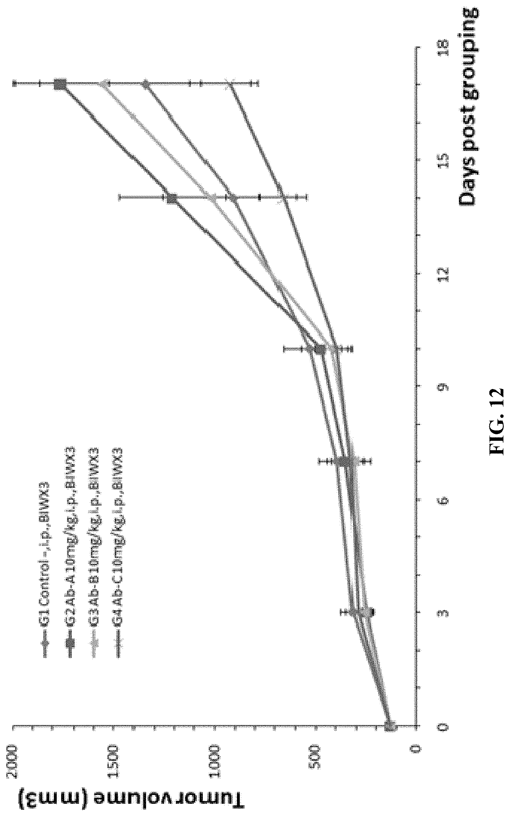
D00014

D00015

D00016
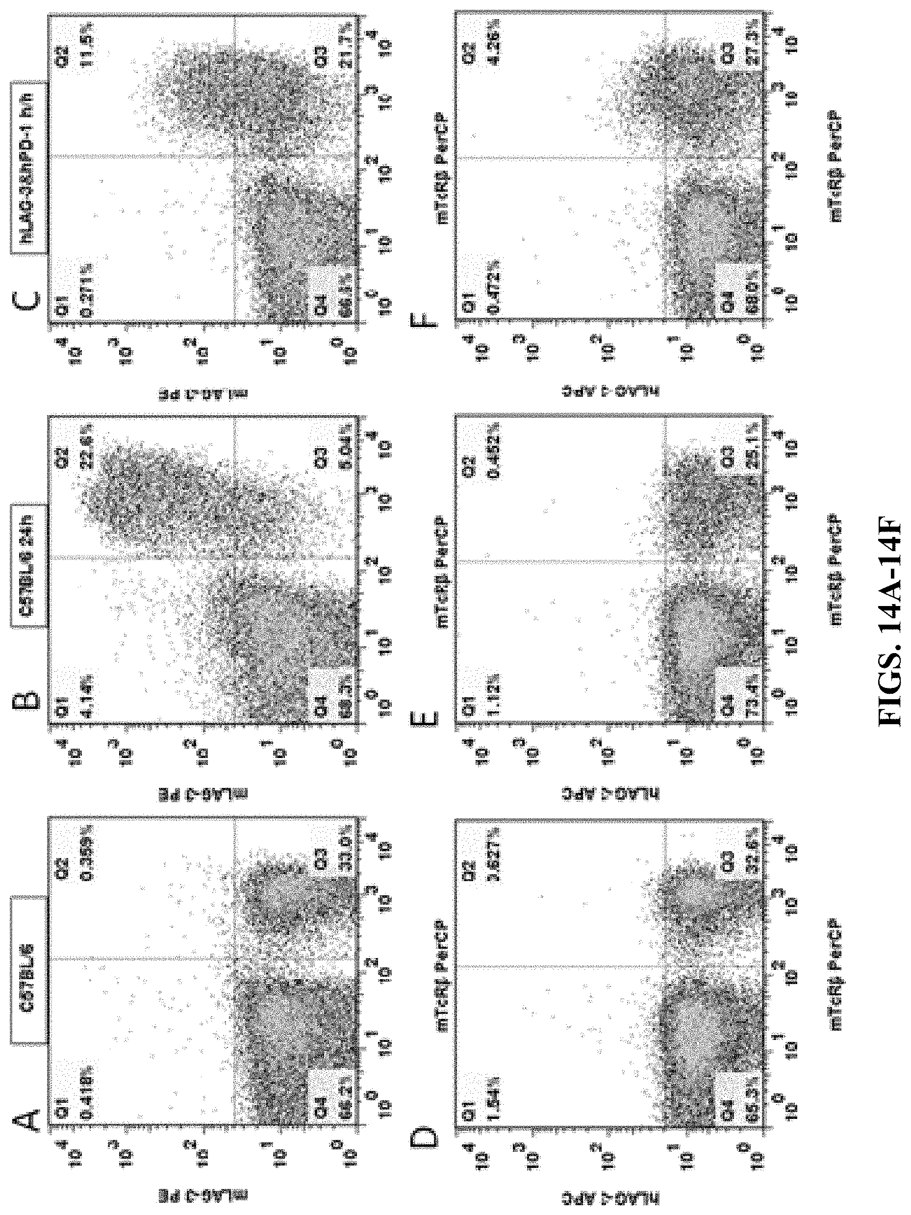
D00017
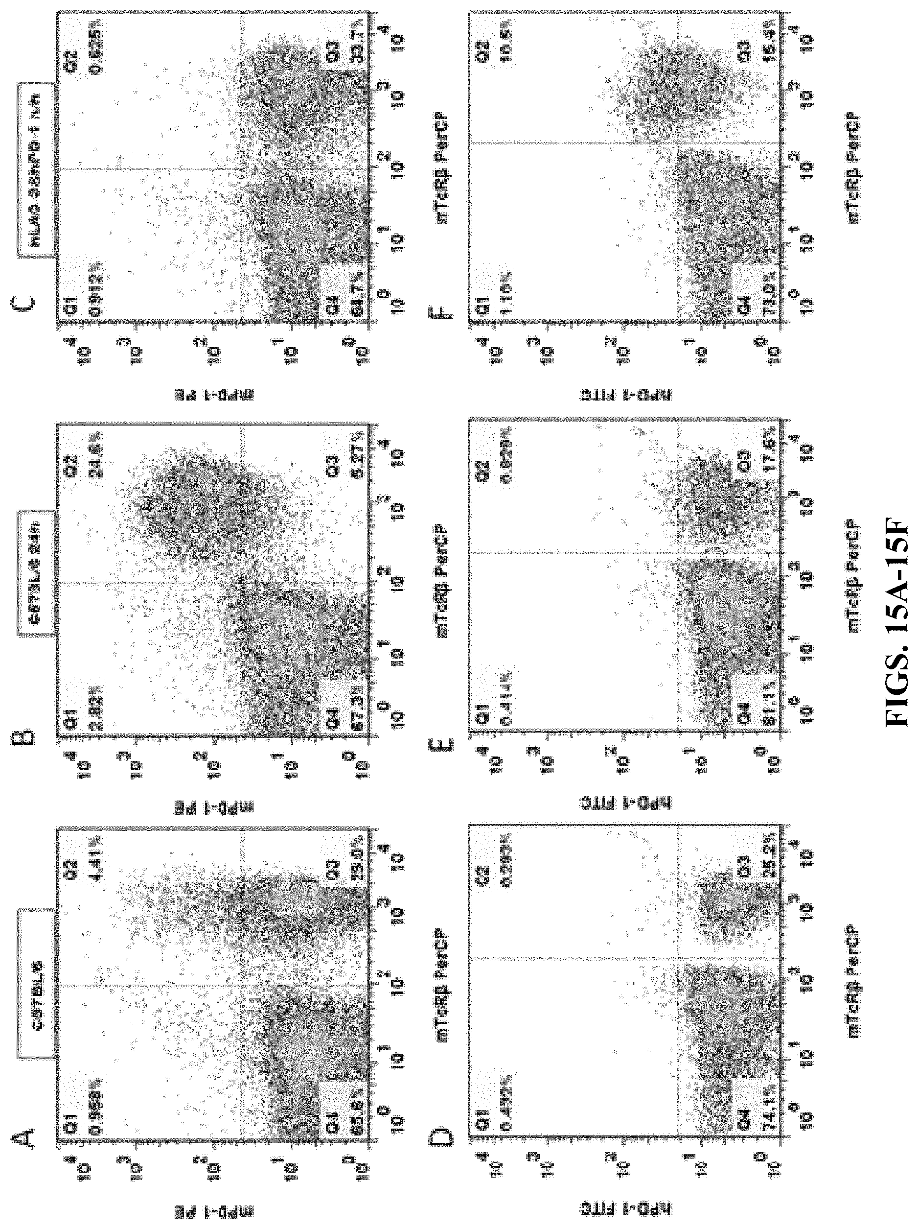
D00018

D00019
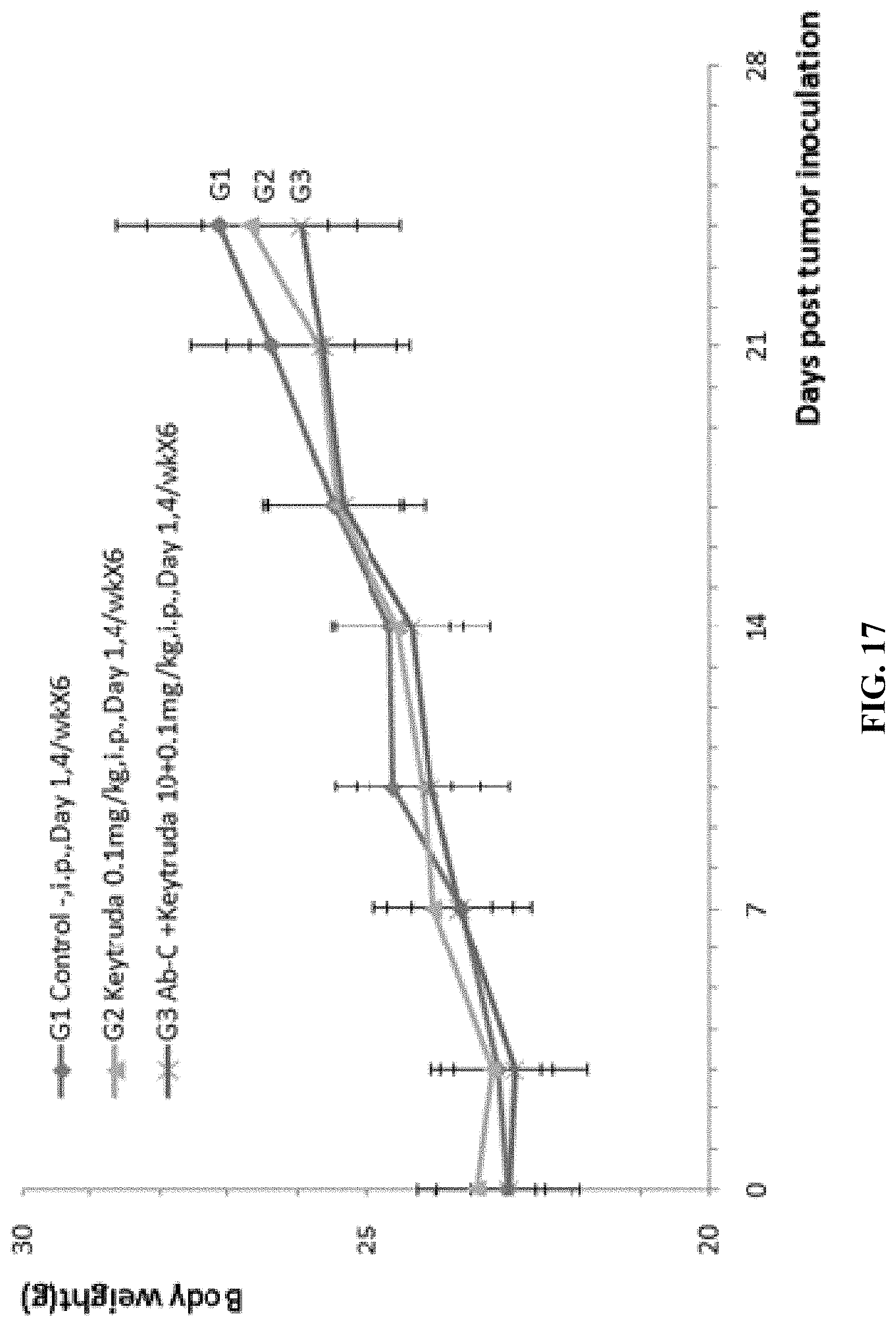
D00020

D00021

D00022
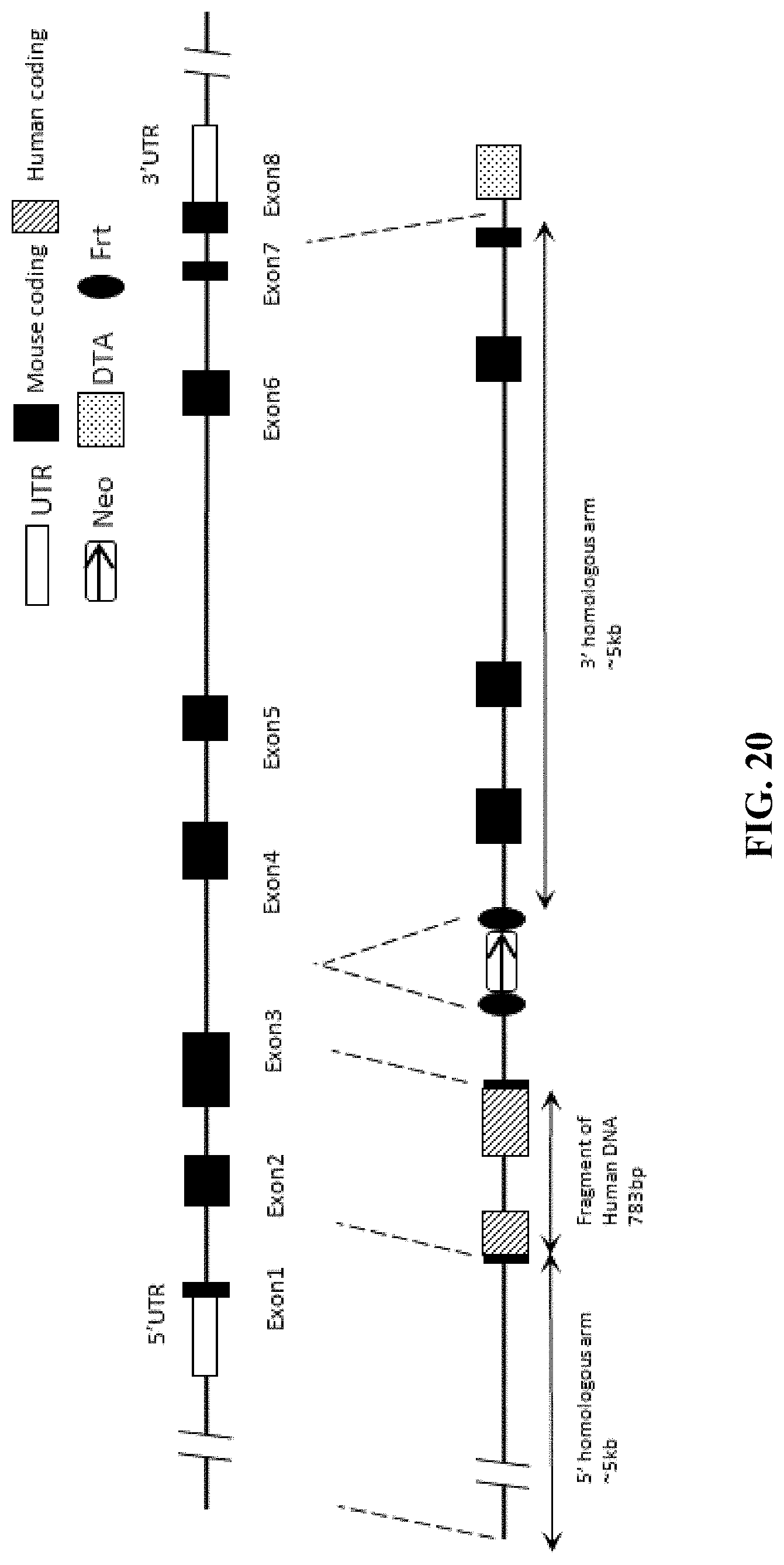
D00023
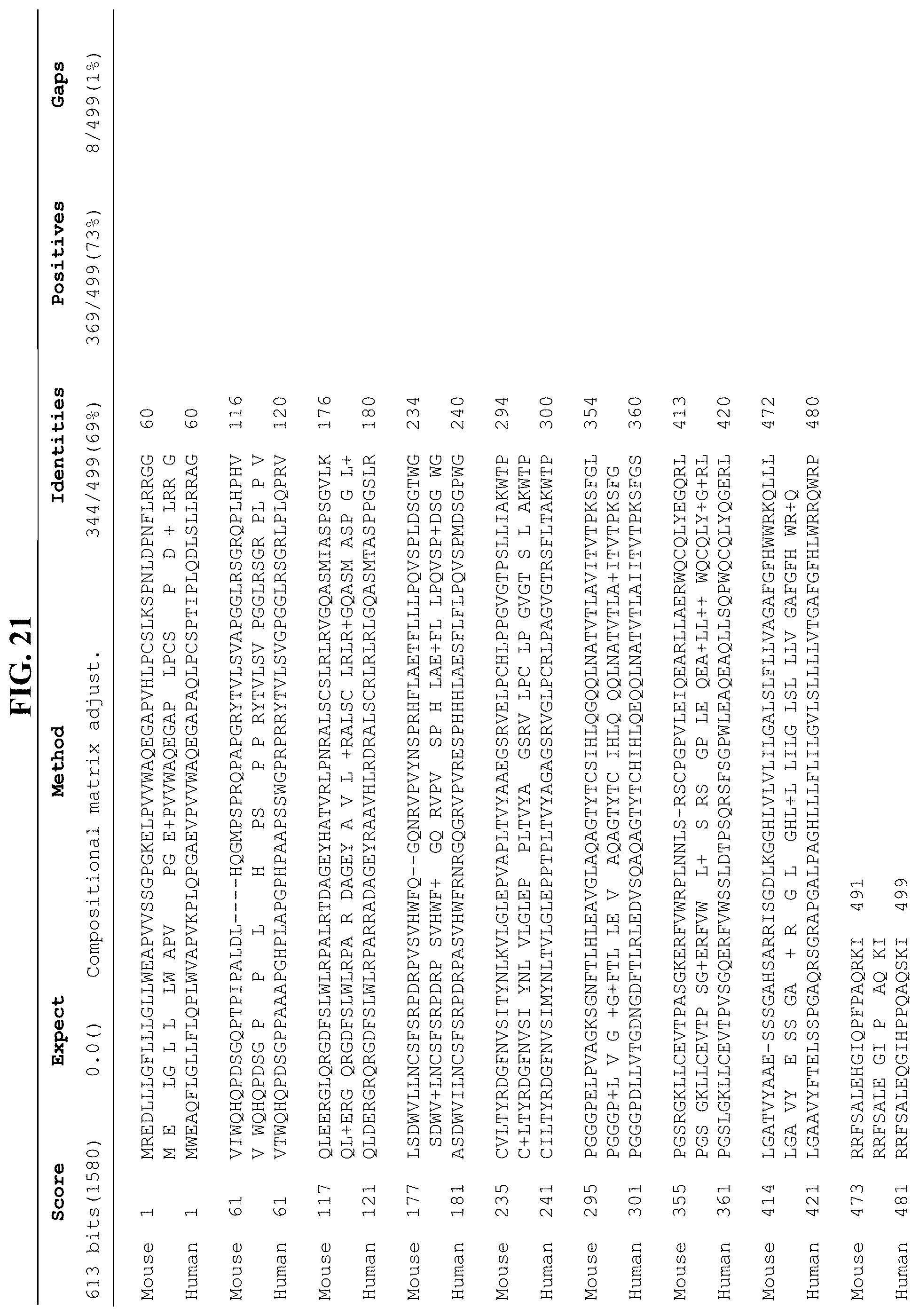
S00001
XML
uspto.report is an independent third-party trademark research tool that is not affiliated, endorsed, or sponsored by the United States Patent and Trademark Office (USPTO) or any other governmental organization. The information provided by uspto.report is based on publicly available data at the time of writing and is intended for informational purposes only.
While we strive to provide accurate and up-to-date information, we do not guarantee the accuracy, completeness, reliability, or suitability of the information displayed on this site. The use of this site is at your own risk. Any reliance you place on such information is therefore strictly at your own risk.
All official trademark data, including owner information, should be verified by visiting the official USPTO website at www.uspto.gov. This site is not intended to replace professional legal advice and should not be used as a substitute for consulting with a legal professional who is knowledgeable about trademark law.