Cyclic peptide binder against oncogenic K-Ras
Henning , et al. February 9, 2
U.S. patent number 10,913,774 [Application Number 16/031,840] was granted by the patent office on 2021-02-09 for cyclic peptide binder against oncogenic k-ras. This patent grant is currently assigned to California Institute of Technology. The grantee listed for this patent is California Institute of Technology. Invention is credited to Samir Das, James R. Heath, Ryan K. Henning, Arundhati Nag, Ashwin N. Ram.


View All Diagrams
| United States Patent | 10,913,774 |
| Henning , et al. | February 9, 2021 |
Cyclic peptide binder against oncogenic K-Ras
Abstract
Cyclic peptides represented by (Formula 1) ##STR00001## selectively bind the oncoprotein K-Ras G12D in vitro and in cellulo, where Z1 and Z2 are each L-propargylglycine (Pm), azidoornithine (OrnN3), or L-azidolysine (Az4), and V1-V2-V3-V4-V5 is an amino acid variable region having a sequence selected from the group consisting of SEQ ID NOs: 1-20.
| Inventors: | Henning; Ryan K. (Pasadena, CA), Ram; Ashwin N. (Pasadena, CA), Das; Samir (Pasadena, CA), Nag; Arundhati (Pasadena, CA), Heath; James R. (South Pasadena, CA) | ||||||||||
|---|---|---|---|---|---|---|---|---|---|---|---|
| Applicant: |
|
||||||||||
| Assignee: | California Institute of
Technology (Pasadena, CA) |
||||||||||
| Family ID: | 1000005350182 | ||||||||||
| Appl. No.: | 16/031,840 | ||||||||||
| Filed: | July 10, 2018 |
Prior Publication Data
| Document Identifier | Publication Date | |
|---|---|---|
| US 20180319844 A1 | Nov 8, 2018 | |
Related U.S. Patent Documents
| Application Number | Filing Date | Patent Number | Issue Date | ||
|---|---|---|---|---|---|
| 15068528 | Mar 11, 2016 | 10017540 | |||
| 62131670 | Mar 11, 2015 | ||||
| Current U.S. Class: | 1/1 |
| Current CPC Class: | C07K 14/82 (20130101); C07K 7/06 (20130101); G01N 33/5748 (20130101); C07K 7/64 (20130101); G01N 2500/04 (20130101); A61K 38/00 (20130101); G01N 2333/914 (20130101); G01N 2333/82 (20130101) |
| Current International Class: | A61K 38/00 (20060101); C07K 7/64 (20060101); G01N 33/574 (20060101); C07K 14/82 (20060101); C07K 7/06 (20060101) |
| WO 2014205317 | Dec 2014 | WO | |||
Other References
|
Oh et al., ChemMedChem, 2014, 2449-2453. (Year: 2014). cited by examiner . Baines, Antonio T. et al.; "Inhibition of Ras for cancer treatment: the search continues"; Future Med Chem.; Oct. 2011; 3(14); pp. 1787-1808. cited by applicant . Bamford, S. et al.; "The COSMIC (Catalogue of Somatic Mutations in Cancer) database and website"; British Journal of Cancer; 2004; 91; pp. 355-358. cited by applicant . Das, Samir et al.; "A General Synthetic Approach for Designing Epitope Targeted Macrocyclic Peptide Ligands"; Angew. Chem. Int. Ed.; 2015; 54; 7pp. cited by applicant . Downward, Julian; "Targeting Ras Signalling Pathways in Cancer Therapy"; Nature Reviews; Cancer; vol. 3; Jan. 2003; pp. 11-22. cited by applicant . Nag, Arundhati et al.; "A Chemical Epitope-Targeting Strategy for Protein Capture Agents: The Serine 474 Epitope of the Kinase Akt2**"; Angew. Chem. Int. Ed. Engl.; Dec. 23, 2013; 52(52); pp. 13975-13979. cited by applicant . Ostrem, Jonathan M. et al.; "K-Ras(G12C) inhibitors allosterically control GTP affinity and effector interactions"; Nature; vol. 503; Nov. 28, 2013; 14pp. cited by applicant . Schubbert, Suzanne et al.; "Hyperactive Ras in developmental disorders and cancer"; Nature Reviews; Cancer; vol. 7; Apr. 2007; pp. 295-308. cited by applicant . Terpe, K.; "Overview of tag protein fusions: from molecular and biochemical fundamentals to commercial systems"; Appl. Microbiol. Biotechnol.; Jan. 2003; 60(5); pp. 523-533. cited by applicant . Toure, Momar et al.; "Small-Molecule PROTACS: New Approaches to Protein Degradation and Crews"; Angew. Chem. Int. Ed.; 55; 2016; pp. 2-10. cited by applicant. |
Primary Examiner: Alstrum-Acevedo; James H
Assistant Examiner: Yang; Kaipeen E
Attorney, Agent or Firm: Pabst Patent Group LLP
Government Interests
STATEMENT REGARDING FEDERALLY SPONSORED RESEARCH OR DEVELOPMENT
This invention was made with government support under Grant No. CA151819 awarded by the National Institutes of Health. The government has certain rights in the invention.
Parent Case Text
CROSS-REFERENCE TO RELATED APPLICATIONS
The present application is a continuation of U.S. application Ser. No. 15/068,528 filed Mar. 11, 2016, which claims priority to and the benefit of U.S. Provisional Application No. 62/131,670 filed on Mar. 11, 2015. The entire contents of U.S. application Ser. No. 15/068,528 and U.S. Application No. 62/131,670 are incorporated herein by reference.
Claims
We claim:
1. A cyclic peptide represented by Formula I: ##STR00004## wherein: Z1 and Z2 are each non-naturally occurring amino acids; V1-V2-V3-V4-V5 is a five amino acid variable region, wherein V1 is L or V, V2 is R, homoR, or guanidinoF, V3 is G, V4 is D, P, E, A, N, or Q, and V5 is guanidinoF or R; L is a linker moiety; and B.sub.m is an optional group, wherein m is 0 or 1.
2. The cyclic peptide of claim 1, wherein Z1 and Z2 are not the same.
3. The cyclic peptide of claim 1, wherein when Z1 is Pra, Z2 is OrnN3 or Az4, when Z1 is OrnN3 or Az4, Z2 is Pra.
4. The cyclic peptide of claim 1, wherein Z1 is Pra and Z2 is Az4.
5. The cyclic peptide of claim 1, wherein Z1 is Pra and Z2 is OrnN3.
6. The cyclic peptide of claim 1, wherein Z1 is Az4 and Z2 is Pra.
7. The cyclic peptide of claim 1, wherein V1-V2-V3-V4-V5 have a sequence selected from the group consisting of SEQ ID NOs:10-20.
8. The cyclic peptide of claim 1, wherein V1-V2-V3-V4-V5 is SEQ ID NO: 7, 11, or 16.
9. The cyclic peptide of claim 8, wherein V1-V2-V3-V4-V5 is SEQ ID NO:16.
10. The cyclic peptide of claim 1, wherein L is a triazole group.
11. The cyclic peptide of claim 1, wherein L is 1,4-triazole.
12. The cyclic peptide of claim 1, wherein Z1 is Pra and Z2 is Az4, and wherein V1-V2-V3-V4-V5 is SEQ ID NO:16.
13. The cyclic peptide of claim 1, wherein Z1 is Pra and Z2 is Az4, and wherein L is 1,4-triazole.
14. The cyclic peptide of claim 1, wherein Z1 is Pra and Z2 is Az4, wherein m is 1, wherein B is a combination of a spacer group and a detection tag.
15. The cyclic peptide of claim 1, wherein Z1 is Pra and Z2 is Az4, wherein m is 1, wherein B is a combination of a spacer group and a detection tag, and wherein the spacer group is PEG and the detection tag is biotin.
16. The cyclic peptide of claim 1, wherein V1-V2-V3-V4-V5 is SEQ ID NO:16, and wherein L is 1,4-triazole.
17. The cyclic peptide of claim 1, wherein V1-V2-V3-V4-V5 is SEQ ID NO:16, wherein m is 1, and wherein B is a combination of a spacer group and a detection tag.
18. The cyclic peptide of claim 1, wherein V1-V2-V3-V4-V5 is SEQ ID NO:16, wherein m is 1, wherein B is a combination of a spacer group and a detection tag, and wherein the spacer group is PEG and the detection tag is biotin.
19. The cyclic peptide of claim 1, wherein Z1 is Pra and Z2 is Az4, wherein V1-V2-V3-V4-V5 is SEQ ID NO:16, and wherein L is 1,4-triazole.
20. The cyclic peptide of claim 1, wherein Z1 is Pra and Z2 is Az4, wherein V1-V2-V3-V4-V5 is SEQ ID NO:16, wherein m is 1, and wherein B is a combination of a spacer group and a detection tag.
21. The cyclic peptide of claim 1, wherein Z1 is Pra and Z2 is Az4, wherein V1-V2-V3-V4-V5 is SEQ ID NO:16, wherein m is 1, wherein B is a combination of a spacer group and a detection tag, and wherein the spacer group is PEG and the detection tag is biotin.
22. The cyclic peptide of claim 1, wherein Z1 is Pra and Z2 is Az4, wherein V1-V2-V3-V4-V5 is SEQ ID NO:16, wherein L is 1,4-triazole, wherein m is 1, and wherein B is a combination of a spacer group and a detection tag.
23. The cyclic peptide of claim 1, wherein Z1 is Pra and Z2 is Az4, wherein V1-V2-V3-V4-V5 is SEQ ID NO:16, wherein L is 1,4-triazole, wherein m is 1, wherein B is a combination of a spacer group and a detection tag, and wherein the spacer group is PEG and the detection tag is biotin.
24. The cyclic peptide of claim 1 having the structure: ##STR00005##
25. The cyclic peptide of claim 1, wherein B is conjugated to any group of the cyclic peptide.
26. The cyclic peptide of claim 25, wherein B is conjugated to the C-terminus of Z2.
27. The cyclic peptide of claim 1, wherein when m is 1, and wherein B is a spacer group, a detection tag, or a combination of a spacer group and a detection tag.
28. The cyclic peptide of claim 27, wherein B is a spacer group.
29. The cyclic peptide of claim 27, wherein the detection tag is a chemical moiety that can be selectively bound.
30. The cyclic peptide of claim 27, wherein the detection tag is an affinity tag, a fluorescent tag, or a fluorescently labeled affinity tag.
31. The cyclic peptide of claim 27, wherein the detection tag is an affinity tag selected from the group consisting of biotin, streptavidin, poly-histidine, poly-arginine, FLAG, cyclodextrin, adamantane, and combinations thereof.
32. The cyclic peptide of claim 27, wherein the spacer group is polyethylene glycol (PEG) or 6-aminohexanoic acid (Ahx).
33. The cyclic peptide of claim 32, wherein the spacer group includes 2 to 24 PEG units.
34. The cyclic peptide of claim 32, wherein the spacer group includes 2 to 15 PEG units.
35. The cyclic peptide of claim 32, wherein the spacer group includes 2 to 10 PEG units.
36. The cyclic peptide of claim 32, wherein the spacer group includes 2 to 5 PEG units.
37. The cyclic peptide of claim 32, wherein the spacer group includes 2 to 4 PEG units.
38. The cyclic peptide of claim 32, wherein the spacer group includes 2 to 3 PEG units.
39. The cyclic peptide of claim 27, wherein B is a combination of a spacer group and a detection tag.
40. The cyclic peptide of claim 37, wherein the spacer group is PEG and the detection tag is biotin.
41. A cyclic peptide comprising the structure: ##STR00006##
42. The cyclic peptide of claim 1, wherein the cyclic peptide is conjugated with a moiety.
43. A method of inhibiting K-Ras G12D oncoprotein in a cancer cell expressing K-Ras G12D, the method comprising: incubating the cancer cell with the cyclic peptide of claim 1.
44. The method of claim 43, wherein the cancer cell is pancreatic, colorectal, lung, biliary tract, or ovarian cancer.
45. A method of inhibiting K-Ras G12D oncoprotein in a cancer cell expressing K-Ras G12D, the method comprising: incubating the cancer cell with the cyclic peptide of claim 42.
46. The method of claim 45, wherein the cancer cell is pancreatic, colorectal, lung, biliary tract, or ovarian cancer.
Description
REFERENCE TO SEQUENCE LISTING
The Sequence Listing submitted Jul. 10, 2018 as a text file named "INDI_33_1_AMD_AFD_Sequence_Listing.txt," created on Jul. 10, 2018, and having a size of 12,636 bytes is hereby incorporated by reference pursuant to 37 C.F.R. .sctn. 1.52(e)(5).
BACKGROUND
The KRAS oncogene is a member of the Ras family of GTPases that are involved in numerous cellular signaling processes. K-Ras mutations are gain-of-function mutations that are present in up to 30% of all tumors, including as many as 90% of pancreatic cancers. Due to the clinical significance of this protein, many attempts have been made to develop Ras inhibitors, but such attempts have been mostly unsuccessful. This is largely due to the difficulty in outcompeting GTP for the K-Ras binding pocket in cells, and the lack of known allosteric regulatory sites.
SUMMARY
In some embodiments of the present invention, a cyclic peptide that selectively binds K-Ras G12D oncogenic protein is represented by Formula I:
##STR00002##
wherein:
Z1 and Z2 are each L-propargylglycine (Pra), azidoornithine (OrnN3), or L-azidolysine (Az4), wherein when L1 is Pra, L2 is OrnN3 or Az4, when L1 is OrnN3 or Az4, L2 is Pra; and
V1-V2-V3-V4-V5 is a five amino acid variable region having a sequence selected from the group consisting of SEQ ID NOs: 1-20;
L is a linker moiety; and
B.sub.m is an optional detection group, wherein m is 0 or 1.
In some embodiments of the present invention, a composition for screening peptides that bind to a WT K-Ras epitope using click chemistry includes TEYKLVVVGAGGV[Z1]GKSALTIQ (SEQ ID NO: 25), where Z1 is L-propargylglycine (Pra), azidoornithine (OrnN3), or L-azidolysine (Az4).
In some embodiments of the present invention, a composition for screening peptides that bind to a K-Ras G12D epitope using click chemistry includes TEYKLVVVGADGV[Z1]GKSALTIQ (SEQ ID NO: 26), where Z1 is L-propargylglycine (Pra), azidoornithine (OrnN3), or L-azidolysine (Az4).
In some embodiments of the present invention, a method of inhibiting K-Ras G12D oncoprotein in a cancer cell expressing K-Ras G12D includes incubating the cancer cell with cyclic peptide of Formula 1 as disclosed herein.
BRIEF DESCRIPTION OF THE DRAWINGS
The patent or application file contains at least one drawing executed in color. Copies of this patent or patent application publication with color drawings will be provided by the Office upon request and payment of the necessary fee.
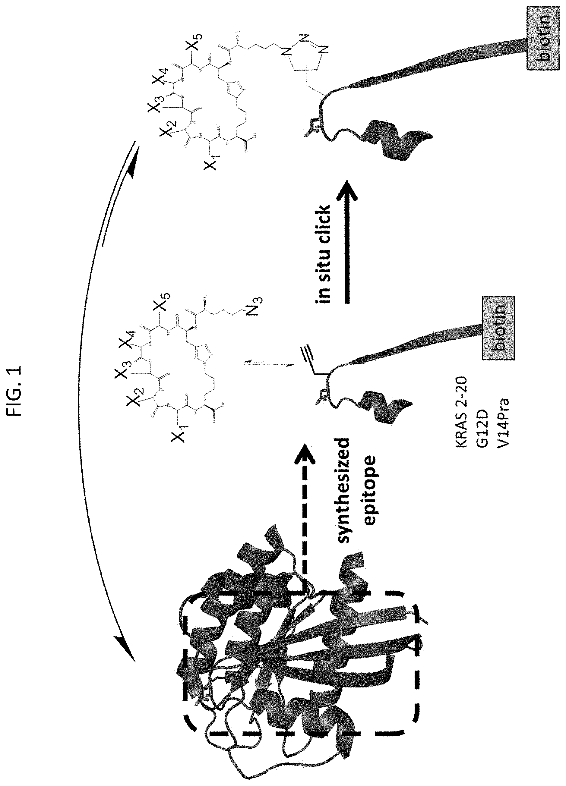
FIG. 1 is a schematic depicting an in situ click chemistry reaction, according to embodiments of the present invention, using a synthetic epitope containing K-Ras residues 2-20 synthesized with a G12D mutation and a propargylglycine (Pra) substitution at valine 14 (V14Pra) conjugated to biotin to identify cyclic peptides that bind the K-Ras epitope. The binding of a peptide from the azide (N3)-terminated one-bead-one-compound (OBOC) cyclic peptide library allows for the bound peptide to covalently bind via azide click chemistry to the biotin tagged K-Ras epitope, allowing for isolation and identification of the cyclic peptide hit. The X1, X2, X3, X4, and X5 represent the variable amino acids, in which the carboxy and amino linkages are shown.
FIG. 2 is a schematic, for the in situ click chemistry screen as shown in FIG. 1, where the cyclic peptide library is first precleared in an "antiscreen" with a wild-type (WT) K-Ras epitope fragment to remove the peptide hits that bind to this non-cancerous (WT) form of the K-Ras protein, with the remaining cyclic peptide library beads used in the "product screen" against the K-Ras G12D epitope to identify cyclic peptides (7 hits) that bind selectively to this oncogenic epitope of K-Ras.
FIG. 3A is a table listing the amino acid sequences (SEQ ID NOs: 1-9, respectively, in order of appearance) of the cyclic peptides corresponding to the 7 hits isolated in the screen of FIG. 2, where SEQ ID NOs: 2 and 3 were two possible sequences from one bead hit and SEQ ID NOs: 5 and 6 were two possible sequences from one bead hit.
FIG. 3B is a graph of the absorbance units (A.U.) corresponding to fluorescence from an ELISA assay measuring the binding of K-Ras G12D to cyclic peptides (1a, 1b,1c, 2, 3a, 3b, 5a, 5b, 5c, 6a, 6b, 6c, 7a, 7b, 8, 9a, and 9b) synthesized with a polyethylene glycol (PEG) spacer group and biotin tag of Formula 1 having a variable region (V1-V2-V3-V4-V5) selected from SEQ ID NOs. 1-9, according to embodiments of the present invention, in which the synthesized cyclic peptides had up to three (a, b, c) fractions corresponding to epimers; the asterisk highlighted cyclic peptide 7b showed the strongest binding fluorescence.
FIG. 4A shows the Formula 1 structure of cyclic peptide 7b (Pra-LRGDR-Az4 (SEQ ID NO: 27)), according to embodiments of the present invention.
FIG. 4B is a graph of an ELISA binding assay in which cyclic peptide 7b has an EC.sub.50 binding value of 33.3 .mu.M against K-Ras G12D, according to embodiments of the present invention.
FIG. 4C is a graph of a single point ELISA assay comparing the binding (measured in absorbance units, A.U.) of cyclic peptide 7b to WT K-Ras (red bar) and K-Ras G12D (blue bar), according to embodiments of the present invention.
FIG. 5A shows the Formula 1 structure of cyclic peptide 7b1 (Pra-LRGDR-Az4-PEG-Biotin), according to embodiments of the present invention.
FIG. 5B is a MALDI-TOF mass spectrum for cyclic peptide 7b1 of FIG. 5A, according to embodiments of the present invention.
FIG. 6A shows the Formula 1 structure of cyclic peptide 7b2 (Az4-LRGDR-Pra-PEG-Biotin (SEQ ID NO: 28)), according to embodiments of the present invention.
FIG. 6B is a MALDI-TOF mass spectrum for cyclic peptide 7b2 of FIG. 6A, according to embodiments of the present invention.
FIG. 7A shows the Formula 1 structure of cyclic peptide 7b3 (Pra-LRGDR-OrnN3-PEG-Biotin (SEQ ID NO: 29)), according to embodiments of the present invention.
FIG. 7B is a MALDI-TOF mass spectrum for cyclic peptide 7b3 of FIG. 7A, according to embodiments of the present invention.
FIG. 8A shows the Formula 1 structure of cyclic peptide 7b4 (Pra-VRGDR-Az4-PEG-Biotin (SEQ ID NO: 30)), according to embodiments of the present invention.
FIG. 8B is a MALDI-TOF mass spectrum for cyclic peptide 7b4 of FIG. 8A, according to embodiments of the present invention.
FIG. 9A shows the Formula 1 structure of cyclic peptide 7b5 (Pra-LRGPR-Az4-PEG-Biotin (SEQ ID NO: 31)), according to embodiments of the present invention.
FIG. 9B is a MALDI-TOF mass spectrum for cyclic peptide 7b5 of FIG. 9A, according to embodiments of the present invention.
FIG. 10A shows the Formula 1 structure of cyclic peptide 7b6 (Pra-LRGER-Az4-PEG-Biotin (SEQ ID NO: 32)), according to embodiments of the present invention.
FIG. 10B is a MALDI-TOF mass spectrum for cyclic peptide 7b6 of FIG. 10A, according to embodiments of the present invention.
FIG. 11A shows the Formula 1 structure of cyclic peptide 7b7 (Pra-L(homoR)GDR-Az4-PEG-Biotin (SEQ ID NO: 41)), according to embodiments of the present invention.
FIG. 11B is a MALDI-TOF mass spectrum for cyclic peptide 7b7 of FIG. 11A, according to embodiments of the present invention.
FIG. 12A shows the Formula 1 structure of cyclic peptide 7b8 (Pra-LRGD(homoR)-Az4-PEG-Biotin (SEQ ID NO: 33)), according to embodiments of the present invention.
FIG. 12B is a MALDI-TOF mass spectrum for cyclic peptide 7b8 of FIG. 12A, according to embodiments of the present invention.
FIG. 13A shows the Formula 1 structure of cyclic peptide 7b9 (Pra-L(guanidinoF)GDR-Az4-PEG-Biotin (SEQ ID NO: 34)), according to embodiments of the present invention.
FIG. 13B is a MALDI-TOF mass spectrum for cyclic peptide 7b9 of FIG. 13A, according to embodiments of the present invention.
FIG. 14A shows the Formula 1 structure of cyclic peptide 7b10 (Pra-LRGD(guanidinoF)-Az4-PEG-Biotin (SEQ ID NO: 35)), according to embodiments of the present invention.
FIG. 14B is a MALDI-TOF mass spectrum for cyclic peptide 7b10 of FIG. 14A, according to embodiments of the present invention.
FIG. 15A shows the Formula 1 structure of cyclic peptide 7b11 (Pra-LRGNR-Az4-PEG-Biotin (SEQ ID NO: 36)), according to embodiments of the present invention.
FIG. 15B is a MALDI-TOF mass spectrum for cyclic peptide 7b11 of FIG. 15A, according to embodiments of the present invention.
FIG. 16A shows the Formula 1 structure of cyclic peptide 7b12 (Pra-LRGQR-Az4-PEG-Biotin (SEQ ID NO: 37)), according to embodiments of the present invention.
FIG. 16B is a MALDI-TOF mass spectrum for cyclic peptide 7b12 of FIG. 16A, according to embodiments of the present invention.
FIG. 17A shows the Formula 1 structure of cyclic peptide 7b14 (Pra-LRGAR-Az4-PEG-Biotin (SEQ ID NO: 38)), according to embodiments of the present invention.
FIG. 17B is a MALDI-TOF mass spectrum for cyclic peptide 7b14 of FIG. 17A, according to embodiments of the present invention.
FIG. 18A is a graph of the relative absorbance units (A.U.) from an ELISA assay measuring binding to K-Ras G12D in the presence of the indicated biotin-tagged cyclic peptide, no ligand/peptide control, and biotin alone, in which various HPLC fractions (f) of the cyclic peptides were assayed as indicated, according to embodiments of the present invention.
FIG. 18B is a graph of the relative absorbance units (A.U.) from an ELISA assay measuring binding to K-Ras G12D in the presence of the indicated biotin-tagged cyclic peptide, no ligand/peptide control, and biotin alone, in which various HPLC fractions (f) of the cyclic peptides were assayed as indicated, and the "Original" cyclic peptide is 7b, also referred to herein as 7b1, according to embodiments of the present invention.
FIG. 18C is a graph of the relative absorbance units (A.U.) from an ELISA assay measuring binding to K-Ras G12D in the presence of the indicated biotin-tagged cyclic peptide, no ligand/peptide control, and biotin alone, in which various HPLC fractions (f) of the cyclic peptides were assayed as indicated, according to embodiments of the present invention.
FIG. 19A is a graph of the relative absorbance units (A.U.) from an ELISA assay measuring binding to K-Ras G12D in the presence of the indicated biotin-tagged cyclic peptide, according to embodiments of the present invention.
FIG. 19B shows the Formula 1 structure of cyclic peptide 7b10 (Pra-LRGD(guanidinoF)-Az4), according to embodiments of the present invention.
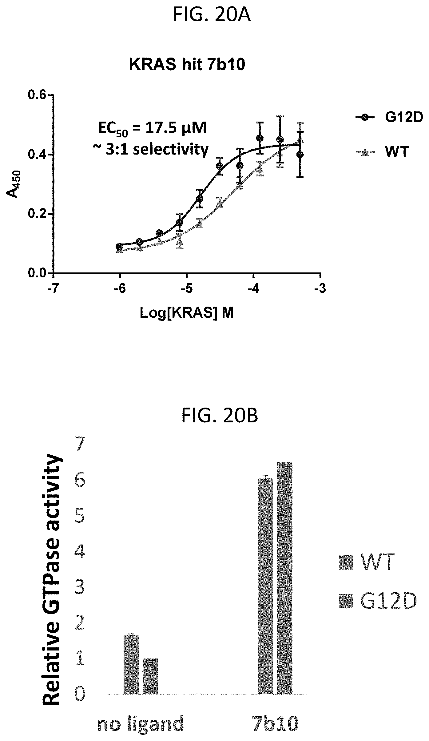
FIG. 20A is a graph of an ELISA binding assay of K-Ras G12D (blue) and WT K-Ras (red) with cyclic peptide 7b10, in which cyclic peptide 7b10 has an EC50 binding value of 17.5 .mu.M against K-Ras G12D and 55.6 .mu.M for WT K-Ras, for a 3:1 selectivity of K-Ras G12D, according to embodiments of the present invention.
FIG. 20B is a graph showing the relative GTP hydrolysis (GTPase activity) of WT K-Ras (blue) and K-Ras G12D (red) proteins incubated with GTP alone (no ligand) or GTP and cyclic peptide 7b10 (7b10), in which the GTP hydrolysis was measured by detecting free phosphate with malachite green and measuring the absorbance at 620 nm, according to embodiments of the present invention.
FIG. 21 is a graph of the amount of absorbance units (A.U.) in an ELISA assay measuring the binding to K-Ras G12D in the presence of alanine-substituted cyclic peptide 7b10, in which alanine (A) was substituted in each position of the X variable region, giving cyclic peptides 7b10-A1 (A1) (red), 7b10-A2 (A2) (green), 7b10-A3 (A3)(purple), 7b10-A4 (A4)(blue), and 7b10-A5 (A5)(orange), where only 7b10-A4 maintained its binding affinity for K-Ras G12D, according to embodiments of the present invention.
FIG. 22A shows the Formula 1 structure of cyclic peptide 7b5 (Pra-LRGPR-Az4 (SEQ ID NO: 31)), according to embodiments of the present invention.
FIG. 22B is a graph of an ELISA binding assay in which cyclic peptide 7b5 has an EC.sub.50 binding value of 56.6 .mu.M against K-Ras G12D, according to embodiments of the present invention.
FIG. 23 shows the structure of cyclic peptide 7b5 (blue) alone and conjugated to 7-amino acid sequence ALAPYIP (SEQ ID NO: 23) (Hif)(green) via a 6-aminohexanoic acid linker (Ahx)(black), and 7b5 (blue) conjugated to Hif (green) via the Ahx linker (black) with the Hif moiety conjugated to HIV-TAT peptide (GRKKRRQRRRPPQQ) (SEQ ID NO: 24)(red), according to embodiments of the present invention.
FIG. 24A shows a Western blot analysis of Ras G12D protein in Panc 08.13 cancer cells (homozygous for K-Ras G12D) after treatment with DMSO (control), 7b5-Hif-TAT, 7b5-Hif, and 7b5, as indicated, in which the cells treated with 7b5-Hif and 7b5-Hif-TAT show depletion of Ras protein, according to embodiments of the present invention.
FIG. 24B shows a Western blot analysis of Ras G12D protein in Panc 08.13 cancer cells after similar treatment conditions as in FIG. 24A, except that where indicated, cells were pretreated with the proteasome inhibitor MG132 followed by the incubation with DMSO, 7b5-Hif, or 7b5, showing that when the proteasome is inhibited, the Ras protein is not depleted in the presence of 7b6-Hif, according to embodiments of the present invention.
DETAILED DESCRIPTION
According to aspects of embodiments of the present invention, cyclic peptides of Formula 1 selectively bind the most frequent K-Ras mutation--the oncogenic K-Ras protein having a glycine 12 to aspartic acid (G12D) mutation. The K-Ras G12D mutant accounts for nearly half of all K-Ras oncoproteins. Cyclic peptides according to embodiments of the present invention are capable of binding to the surface of K-Ras G12D, thus circumventing difficulties encountered with inhibitors requiring a binding pocket in K-Ras.
Abbreviations for amino acids are used throughout this disclosure and follow the standard nomenclature known in the art. For example, as would be understood by those of ordinary skill in the art, Alanine is Ala or A; Arginine is Arg or R; Asparagine is Asn or N; Aspartic Acid is Asp or D; Cysteine is Cys or C; Glutamic acid is Glu or E; Glutamine is Gln or Q; Glycine is Gly or G; Histidine is His or H; Isoleucine is Ile or I; Leucine is Leu or L; Lysine is Lys or K; Methionine is Met or M; Phenylalanine is Phe or F; Proline is Pro or P; Serine is Ser or S; Theonine is Thr or T; Tryptophan is Trp or W; Tyrosine is Tyr or Y; and Valine is Val or V. Synthetic amino acids include L-propargylglycine (Fra); homoarginine (homoArg) or (homoR); guanidinophenylalanine (guanidinoF); L-azidolysine (Az4); and azidoornithine (OrnN3) or (Az3).
Cyclic Peptides that Bind K-Ras G12D
According to some embodiments of the present invention, cyclic peptides that selectively bind to K-Ras G12D may be structurally represented by Formula 1.
##STR00003##
For a cyclic peptide of Formula 1 in which Z1 is a non-naturally occurring amino acid at the amino (N)-terminus, Z2 is a non-naturally occurring amino acid at the carboxy (C)-terminus, and V1-V5 is a 5-amino acid variable region. The 5-amino acid variable region may also be represented as V1-V2-V3-V4-V5.
In some embodiments of the present invention, Z1 and Z2 are not the same and are linked by a triazole group. Z1 and Z2 may each represent a non-naturally occurring amino acid selected from L-propargylglycine (Pra), azidoornithine (OrnN3), or L-azidolysine (Az4), where when Z1 is Pra, Z2 is OrnN3 or Az4, and when Z1 is OrnN3 or Az4, Z2 is Pra. L is a linker moiety. In some embodiments, L is 1,4-triazole linking Z1 and Z2 amino acid groups.
In some embodiments of the present invention, V1-V5 is a five amino acid region having an amino acid sequence selected from SEQ ID NOs: 1-9 as shown in Table 1 or SEQ ID NOs: 10-20 as shown in Table 2.
TABLE-US-00001 TABLE 1 SEQ ID NO Variable Region (V1-V5) Example Cyclic Peptide 1 NDETY 1a, 1b, 1c 2 PSEEG 2 3 SEEGG 3a, 3b 4 EGTGT K-Ras G12D Epitope Hit 5 YEQGE 5a, 5b, 5c 6 YGEQE 6a, 6b, 6c 7 LRGDR 7a, 7b 8 QEKPP 8 9 ELTFG 9a, 9b
TABLE-US-00002 TABLE 2 SEQ ID Example Cyclic NO Variable Region (V1-V5) Z1 Z2 Peptide 7 LRGDR Pra Az4 7b1 7 LRGDR Az4 Pra 7b2 7 LRGDR Pra OrnN3 7b3 10 VRGDR Pra Az4 7b4 11 LRGPR Pra Az4 7b5 12 LRGER Pra Az4 7b6 13 L(homoR)GDR Pra Az4 7b7 14 LRGD(homoR) Pra Az4 7b8 15 L(guanidinoF)GDR Pra Az4 7b9 16 LRGD(guanidinoF) Pra Az4 7b10 17 LRGA(guanidinoF) Pra Az4 7b10-alanine 18 LRGNR Pra Az4 7b11 19 LRGQR Pra Az4 7b12 20 LRGAR Pra Az4 7b14
In some embodiments of the present invention, the cyclic peptide of Formula 1 may include B.sub.m representing a detection moiety, where m is 0 or 1. The detection moiety includes an optional spacer group and a detection tag. In some embodiments, when m is 1, B may be a spacer group, a detection tag, or a conjugate of both a spacer group and a detection tag. In some embodiments, the detection moiety may be conjugated to any group of the cyclic peptide. In some embodiments, the detection moiety (B) is conjugated to the C-terminus of Z2. For example, as shown in FIG. 5A, the detection moiety is conjugated to the C-terminus of Az4.
In some embodiments of the present invention, non-limiting examples of a spacer group include polyethylene glycol (PEG)n or 6-aminohexanoic acid (Ahx). As known to one of ordinary skill in the art, the number of PEG units (n) (or ethylene glycol repeats) determines the length of the spacer group. For example, 2 PEG units provide a spacer length of 18 angstroms. In some embodiments of the present invention, the spacer group includes 2 to 24 PEG units, 2 to 20 PEG units, 2 to 15 PEG units, 2 to 10 PEG units, 2 to 5 PEG units, 2 to 4 PEG units, or 2 to 3 PEG units.
As used herein, "tag," "detection tag," and like terms refer to a covalently linked chemical moiety that may be selectively bound and isolated. In some embodiments, "tag" refers to an "affinity tag" in which the chemical moiety has a specific binding partner. Non-limiting examples of affinity tags include biotin, streptavidin, poly-histidine (6-HIS) (SEQ ID NO: 39), poly-arginine (5-6 R) (SEQ ID NO: 40), FLAG, cyclodextrin, adamantane, and combinations thereof. Affinity tags for labeling peptides are described, for example in K. Terpe, 2003, Appl. Microbiol. Biotechnol, 2003, 60:523-533, the entire contents of which are incorporated herein by reference. In some embodiments, the detection tag may be a fluorescent dye or may be a fluorescent dye conjugated to an affinity tag.
The cyclic peptides according to embodiments of the present invention may be prepared by procedures known to those of skill in the art. For example, the cyclic peptides may be prepared using standard solid-phase peptide synthesis (SPPS) techniques as described for example, in Das et al. 2015, Angew. Chem. Int. Ed., 54: 1329-13224, the entire contents of which is incorporated herein by reference. Methods for synthesizing and cyclizing the peptides using azide/alkyne chemistry are described in more detail in the examples.
Wild Type and G12D K-Ras Synthetic Epitopes for Click Chemistry
In some embodiments of the present invention, synthetic epitopes of wild type K-Ras TEYKLVVVGAGGV[Z1]GK-SALTIQ (SEQ ID NO: 25) and oncogenic K-Ras G12D TEYKLVVVGADGV[Z1]GKSALTIQ(SEQ ID NO: 26) include an azide amino acid (Z1). In some embodiments, Z1 is Pra, OrnN3 or AZ4 as described herein. These azide-modified WT and G12D K-Ras epitopes provide a click handle for covalently linking the epitope with binding partners (e.g., peptides) isolated from the azide cyclic peptide library as disclosed in Das et al. supra, and the examples herein.
Cyclic Peptide Binding to KRas G12D in cellulo
In some embodiments of the present invention, a method for inhibiting the oncoprotein K-Ras G12D includes treating cancer cells having the K-Ras G12D mutation with a cyclic peptide of Formula 1 as disclosed herein. As shown in FIG. 24, cyclic peptide 7b5 conjugated to a proteasome degradation sequence (Hif) is capable of decreasing or depleting levels of K-Ras G12D in pancreatic cancer cells as shown by Western blot analysis of a pancreatic cell lysate after the cells were incubated with 7b5, a 7b5-Hif conjugate, and a 7b5-Hif-HIV TAT conjugate having a cell penetrating sequence as described herein. In some embodiments of the present invention, a cyclic peptide of Formula I may be used to inhibit K-Ras G12D expressed in any cancer cell. For example, the K-Ras G12D oncoprotein has been identified in pancreatic, colorectal, lung, biliary tract, and ovarian cancer cells.
The following Examples are presented for illustrative purposes only, and do not limit the scope or content of the present application.
EXAMPLES
Example 1. In situ click screen. To screen for the cyclic peptides that selectively bind K-Ras G12D, an epitope-targeting strategy was used that identifies high-affinity protein catalyzed capture (PCC) agents against a specific region of interest in a protein using in situ click chemistry as depicted in FIG.1 and described in Nag et al., 2013, Angew. Chem. Int. Ed.52:13975-13979, the entire contents of which are incorporated herein by reference. Modified K-Ras wild type and G12D epitopes were synthesized with acetylene-functionalized amino acids for use in the in situ click screen (FIG. 1). The epitopes were derived from amino acids 2-22 from the K-Ras protein sequence with valinel4 substituted with propargylglycine (Fra). Additionally, an 11-unit polyethylene glycol (PEG) spacer and biotin were added to the C-terminus of the synthesized fragments for detection with streptavidin during the screen. The sequence of the wild type K-Ras epitope fragment was NH2-TEYKLVVVGAGG[Pra]GK-SALTIQ (SEQ ID NO: 21)-Peg11-Biotin. The sequence of the G12D fragment was NH2-TEYKLVVVGADG[Pra]GKSALTIQ(SEQ ID NO: 22)--Peg11-Biotin. The epitopes were synthesized by solid phase peptide synthesis (SPPS) on biotin novatag resin according to standard protocols.
The one-bead-one-compound (OBOC) cyclic peptide library is a 7-amino acid macrocyclic library with a 5-amino acid variable region (FIG. 1) The library peptides contain a C-terminal azidolysine followed by the 5-mer variable region and an N-terminal propargylglycine. A copper catalyzed click reaction was then used to cyclize the resin-bound peptides through the side chains of the N- and C-terminal residues. Finally, azidolysine was added to the N-terminus of the cyclized library to serve as a click handle for the in situ click screen. The library was synthesized by SPPS on tentagel resin according to standard protocols.
This azide (OBOC) peptide library has 1.4.times.10.sup.6 unique members (FIG. 2). Approximately 200 hits (peptides that bind to the target epitope) clicked to the wild-type K-Ras fragment in the antiscreen, and were removed from the screen in order to avoid selecting hits that bind to the wild type K-Ras. The remaining beads were then incubated in a product screen with K-Ras G12D propargyl glycine-functionalized K-Ras G12D fragment (V14Pra), and 7 hits were isolated resulting in 9 possible sequences. The hits were sequenced using N-terminal Edman degradation and two of the beads each resulted in two possible sequences, resulting in SEQ ID NOs: 1-9 as listed in Table 1 and FIG. 3A.
Example 2. Synthesis of hit compounds. Hit peptides were synthesized following the same procedure as the library synthesis using SPPS. Briefly, the linear sequence Pra-X.sub.1X.sub.2X.sub.3X.sub.4X.sub.5-Az4 was synthesized on PEG-Biotin Novatag resin (EMD Millipore) using standard solid phase peptide synthesis followed by copper catalyzed cyclization between Fra and Az4. The dried resin was then treated with a TFA cleavage solution of 95% TFA, 2.5% H2O, and 2.5% triethylsilane for two hours at room temperature. The cleavage solution was filtered to remove the resin and added dropwise to an ice-cooled solution of diethyl ether. All peptides were purified using a preparative or semi-preparative scale high performance liquid chromatography (HPLC) with a C18 reverse phase column. A gradient of double distilled water and HPLC grade acetonitrile and 0.1% TFA was used for all purifications.
Example 3. Screen hit peptides against full-length K-Ras/K-Ras G12D. The nine sequences were tested for binding to K-Ras(G12D) by enzyme-linked immunosorbent assay (ELISA) (FIG. 3B). The cyclic peptide 7b having variable region LRGDR (SEQ ID NO: 7), showed the highest affinity and selectivity binder for G12D K-Ras over WT K-Ras. As shown in FIG. 3B, cyclic peptides having amino acid variable regions SEQ ID NOs: 1-9, bind to K-Ras G12D, and cyclic peptide 7b binds with the highest affinity for K-Ras G12D. Cyclic peptide 7b includes the sequence Pra-LRGDR-Az4 (SEQ ID NO: 27).
Example 4. Modified 7b variants. In order to improve upon the binding of 7b to K-Ras G12D, further variants of 7b were synthesized (7b1-7b12, 7b14) each of which is listed in Table 2 and shown in FIGS. 5A-17A. As shown respectively in each of FIGS. 5A-17A, cyclic peptides 7b1-7b12 and 7b14 include the These 7b variant cyclic peptides were assayed for binding to K-Ras G12D in ELISA assays as shown in FIGS. 18a-19A.
Example 5. Degradation of K-Ras G12D using Cyclic Peptides. Cyclic peptide 7b5 was shown to induce proteasomal degradation of K-Ras G12D in pancreatic cancer cells. The 7b5 peptide (FIGs. 22A-22B) was functionalized with the degradation-inducing sequence ALAPYIP (SEQ ID NO: 23) from the protein Hif-1.alpha.. Proteolysis-targeting chimeras (PROTACs) using Hif and other proteins are described in Toure and Crews, 2016, Angew. Chem. Int. Ed, 55:2-10, the entire contents of which are incorporated herein by reference. The 7b-Hif PROTAC was further conjugated with the HIV-TAT cell-penetrating peptide GRKKRRQRRRPPQQ (SEQ ID NO: 24). Structures of 7b5, 7b5-Hif, 7b5-Hif-TAT are shown in FIG. 23. Panc 08.13 cancer cells which are homozygous for the K-Ras G12D mutation were treated with cyclic peptide 7b5, 7b5-Hif, or 7b6-Hif-TAT for 30 minutes followed by cell lysis. The cell lysates were analyzed by Western blot for levels of K-Ras G12D protein as shown in FIG. 24A. The levels of K-Ras G12D were decreased (or depleted) in the presence of 0.3 mM 7b5-Hif-TAT and 7b5-Hif. By pretreating the Panc 08.13 cells with the proteasome inhibitor MG132, the K-Ras G12D depletion of 7b5-Hif was prevented.\
Example 6. Materials and Methods.
In situ cyclic peptide library screen as previously described in Das et al. 2015, Angew. Chem. Int. Ed., 54: 1329-13224, the entire contents of which is incorporated herein by reference. Anchor screen conditions: screen against 500 mg (approximately 1,400,000) beads of 5-mer (comprehensive library). Library includes Az4-[Pra]-A1A2A3A4A5-[Az4]-[100% Met]-TG, where A is 18 amino acids (no D-Cys, no D-Met), [Pra] and [Az4] are connected via a 1,4-triazole linkage.
Step 1: Preclear: Swell library beads in TBS for 6 hours (h). Block overnight at 4.degree. C. with 1% BSA in TBS with 0.1% Tween 20 (Blocking Buffer). Wash with Blocking Buffer (5 times (.times.)). The following day, add 1:10,000 anti-Biotin-AP Antibody (Sigma Aldrich) in Blocking Buffer. Incubate on shaking arm for 1 hour (h) at room temperature (RT). Wash: 5.times.3 mL Blocking Buffer, 5.times.3 mL Wash 1 buffer (0.1% BSA in TBS+0.1% Tween 20), then 5.times.3 mL Wash 2 buffer (TBS+0.1% Tween 20). Drain. Develop with BCIP: NBT (Promega #S3771). Remove purple beads as false hits (depending on how many beads are purple). Preparation of BCIP:NBT: For every 5 mL of Alkaline Phosphatase Buffer (100 mM Tris-HCl [pH 9.0], 150 mM NaCl, 1 mM MgCl2), add 33 .mu.L NBT (50 mg/mL stock in 70% DMF) and 16.5 .mu.L BCIP (50 mg/mL stock in 70% DMF). For remaining clear beads, wash with 7.5 M Gu-HCl, pH 2.0 for 30 min. Rinse with water 10 times. Incubate clear beads in NMP to remove trace purple coloring (a few hours). Wash with water, then TBS buffer. Block overnight at 4.degree. C. with Blocking Buffer.
Step 2: Anti-screen against wild type KRAS polypeptide target. Use 50 .mu.M (2% DMSO, v/v) concentration of Polypeptide. The Polypeptide will be consumed as a substrate of the in situ click reaction. For beads isolated in Step 1, wash with Blocking Buffer (3.times.3 min each (ea)). Combine Polypeptide solution with beads and incubate on shaking arm for 5 h at RT. Wash: 3 times with Blocking Buffer (3 min ea), then 10.times.TBS (3.times.3 min ea, then 7.times.0 min ea), and drain. Incubate beads for 1 h in 7.5 M Gu-HCl, pH 2.0. Wash 6 times with TBS (3.times.3 min ea, then 3.times.0 min ea). Block for 2 h at RT with Blocking Buffer. Wash with Blocking Buffer (5.times.0 min ea). Add 1:10,000 Anti-Biotin-AP in Blocking Buffer. Incubate on shaking arm for 1 h at RT. Wash with 5.times.3 mL Blocking Buffer, 5.times.3 mL Wash 1 buffer (3 mins ea), then 5.times.3 mL Wash 2 buffer (3 min ea), and drain. Develop with BCIP:NBT as described in Step 1. Pick purple hits. Wash hits with 7.5 M Gu-HCl, pH 2.0 for 30 min. Rinse with water 10 times. Incubate hits in NMP to remove purple coloring (a few hours). Wash with water, and then TBS. Block overnight at 4.degree. C. with Blocking Buffer.
Step 3: Product screen against mutant KRAS G12D polypeptide target. Use 50 .mu.M (2% DMSO, v/v) concentration of Polypeptide. The Polypeptide will be consumed as a substrate of the in situ click reaction. For beads isolated in Step 1, wash with Blocking Buffer (3.times.3 min ea). Combine Polypeptide solution with beads and incubate on shaking arm for 5 h at RT. Wash with Blocking Buffer 3 times, 3 min ea, and then 10 times with TBS (3.times.3 min ea, then 7.times.0 min ea), and drain. Incubate beads for 1 h in 7.5 M Gu-HCl, pH 2.0. Wash 6 times with TBS (3.times.3 min ea, then 3.times.0 min ea). Block for 2 h at RT with Blocking Buffer. Wash with Blocking Buffer (5 times). Add 1:10,000 Anti-Biotin-AP in Blocking Buffer. Incubate on shaking arm for 1 h at RT. Wash: 5.times.3 mL Blocking Buffer, 5.times.3 mL Wash 1 buffer (3 mins ea), then 5.times.3 mL Wash 2 buffer (3 min ea), and drain. Develop with BCIP:NBT as described in Step 1. Pick purple hits. Wash hits with 7.5 M Gu-HCl, pH 2.0 for 30 min, and rinse with water 10 times. Incubate hits in NMP to remove purple coloring (a few hours). Wash with water, and then TBS. Sequence hit beads via Edman degradation
ELISA assays. For ELISA assays, 1 .mu.M of the biotinylated hit peptides were first immobilized onto Neutravidin ELISA plates (Pierce) for 2 hours at room temperature in binding buffer (TBST with 0.1% BSA). The plates were then blocked with 5% BSA for 1 hour, followed by incubating with varying concentrations of wild type or G12D KRAS for 30 minutes. After washing three times with TBST the plate was then treated with a 1:1000 dilution of the anti-RAS rabbit mAb (Cell Signaling Technology) in binding buffer for thirty minutes, washed three times with TBST, incubated with anti-Rabbit-HRP secondary antibody (Cell Signaling Technology) for thirty minutes and developed with TMB substrate (KPL) for five to ten minutes. The absorbance of samples at 450 nm wavelength was measured using a spectrophotometer.
Mass spectrometry analysis. Peptides were characterized via MALDI-TOF-MS using a Voyager DE-PRO MALDI TOF-MS system (Applied Biosystems). Crude or purified samples were dissolved in 50:50 water/acetonitrile with 0.1% trifluoroacetic acid at a final concentration of 10 pmol/.mu.L. 1 .mu.L of the analyte sample was then added to 10 .mu.L of a saturated solution of MALDI matrix, either .alpha.-cyano-4-hydroxycinnamic acid or Sinapinic Acid, in 50:50 water/acetonitrile with 0.1% trifluoroacetic acid and analyzed via MALDI-TOF MS.
Cell culture. Panc 08.13 cell lines were purchased from American Type Culture collection and cultured as specified by the provider.
Immunoblotting. Western blots were performed according to standard protocols. Briefly, cells were lysed with cell lysis buffer (Cell Signaling Technology) containing protease and phosphatase inhibitors (Cell Signaling Technology). Cell lysates were quantified with a Bradford protein assay (Thermo Scientific) and prepared for gel electrophoresis in Laemmli sample buffer and reducing agent. 20 .mu.g of cell lysate were added to precast polyacrylamide gels (Bio-Rad) and proteins were separated by electrophoresis followed by transfer to PVDF membrane. Membranes were then blocked and probed with an anti-Ras or anti-Tubulin primary antibody followed by horseradish peroxidase-conjugated secondary antibody (Cell Signaling Technology). The bands were visualized by chemiluminescence (Thermo Scientific).
As disclosed throughout, for example in Tables 1 and 2, and FIGS. 5A-17A, 19A, and 24A-24B, cyclic peptides of Formula 1 are capable of selectively binding the oncoprotein K-Ras G12D in vitro and in cellulo.
While the present invention has been illustrated and described with reference to certain exemplary embodiments, those of ordinary skill in the art will understand that various modifications and changes may be made to the described embodiments without departing from the spirit and scope of the present invention, as defined in the following claims.
SEQUENCE LISTINGS
1
4115PRTArtificial SequenceDescription of Artificial Sequence Synthetic peptideVariable Region 1 1Asn Asp Glu Thr Tyr1 525PRTArtificial SequenceDescription of Artificial Sequence Synthetic peptideVariable Region 2 2Pro Ser Glu Glu Gly1 535PRTArtificial SequenceDescription of Artificial Sequence Synthetic peptideVariable Region 3 3Ser Glu Glu Gly Gly1 545PRTArtificial SequenceDescription of Artificial Sequence Synthetic peptideVariable Region 3 4Glu Gly Thr Gly Thr1 555PRTArtificial SequenceDescription of Artificial Sequence Synthetic peptideVariable Region 5 5Tyr Glu Gln Gly Glu1 565PRTArtificial SequenceDescription of Artificial Sequence Synthetic peptideVariable Region 6 6Tyr Gly Glu Gln Glu1 575PRTArtificial SequenceDescription of Artificial Sequence Synthetic peptideVariable Region 7 7Leu Arg Gly Asp Arg1 585PRTArtificial SequenceDescription of Artificial Sequence Synthetic peptideVariable Region 8 8Gln Glu Lys Pro Pro1 595PRTArtificial SequenceDescription of Artificial Sequence Synthetic peptideVariable Region 9 9Glu Leu Thr Phe Gly1 5105PRTArtificial SequenceDescription of Artificial Sequence Synthetic peptidePeptide 7b4 10Val Arg Gly Asp Arg1 5115PRTArtificial SequenceDescription of Artificial Sequence Synthetic peptidePeptide 7b5 11Leu Arg Gly Pro Arg1 5125PRTArtificial SequenceDescription of Artificial Sequence Synthetic peptidePeptide 7b6 12Leu Arg Gly Glu Arg1 5135PRTArtificial SequenceDescription of Artificial Sequence Synthetic peptidePeptide 7b7MOD_RES(2)..(2)Homoarginine 13Leu Arg Gly Asp Arg1 5145PRTArtificial SequenceDescription of Artificial Sequence Synthetic peptidePeptide 7b8MOD_RES(5)..(5)Homoarginine 14Leu Arg Gly Asp Arg1 5155PRTArtificial SequenceDescription of Artificial Sequence Synthetic peptidePeptide 7b9MOD_RES(2)..(2)Guanidino phenylalanine 15Leu Phe Gly Asp Arg1 5165PRTArtificial SequenceDescription of Artificial Sequence Synthetic peptidePeptide 7b10MOD_RES(5)..(5)Guanidino phenylalanine 16Leu Arg Gly Asp Phe1 5175PRTArtificial SequenceDescription of Artificial Sequence Synthetic peptidePeptide 7b10-alanineMOD_RES(5)..(5)Guanidino phenylalanine 17Leu Arg Gly Ala Phe1 5185PRTArtificial SequenceDescription of Artificial Sequence Synthetic peptidePeptide 7b11 18Leu Arg Gly Asn Arg1 5195PRTArtificial SequenceDescription of Artificial Sequence Synthetic peptidePeptide 7b12 19Leu Arg Gly Gln Arg1 5205PRTArtificial SequenceDescription of Artificial Sequence Synthetic peptidePeptide 7b14 20Leu Arg Gly Ala Arg1 52121PRTArtificial SequenceDescription of Artificial Sequence Synthetic peptideWT K-Ras epitopeMOD_RES(13)..(13)L-propargylglycine 21Thr Glu Tyr Lys Leu Val Val Val Gly Ala Gly Gly Xaa Gly Lys Ser1 5 10 15Ala Leu Thr Ile Gln 202221PRTArtificial SequenceDescription of Artificial Sequence Synthetic peptideG12D K-Ras epitopeMOD_RES(13)..(13)L-propargylglycine 22Thr Glu Tyr Lys Leu Val Val Val Gly Ala Asp Gly Xaa Gly Lys Ser1 5 10 15Ala Leu Thr Ile Gln 20237PRTArtificial SequenceDescription of Artificial Sequence Synthetic peptideDegradation sequence 23Ala Leu Ala Pro Tyr Ile Pro1 52414PRTArtificial SequenceDescription of Artificial Sequence Synthetic peptideCell penetrating sequence 24Gly Arg Lys Lys Arg Arg Gln Arg Arg Arg Pro Pro Gln Gln1 5 102521PRTArtificial SequenceDescription of Artificial Sequence SyntheticMOD_RES(13)..(13)L-propargylglycine, azidoornithine or L- azidolysine 25Thr Glu Tyr Lys Leu Val Val Val Gly Ala Gly Gly Xaa Gly Lys Ser1 5 10 15Ala Leu Thr Ile Gln 202621PRTArtificial SequenceDescription of Artificial Sequence Synthetic peptideG12D K-Ras with azideMOD_RES(13)..(13)L-propargylglycine, azidoornithine or L- azidolysine 26Thr Glu Tyr Lys Leu Val Val Val Gly Ala Asp Gly Xaa Gly Lys Ser1 5 10 15Ala Leu Thr Ile Gln 20277PRTArtificial SequenceDescription of Artificial Sequence Synthetic peptideMOD_RES(1)..(1)L-propargylglycineMOD_RES(7)..(7)L-azidolysine 27Xaa Leu Arg Gly Asp Arg Lys1 5287PRTArtificial SequenceDescription of Artificial Sequence Synthetic peptideMOD_RES(1)..(1)L-azidolysineMOD_RES(7)..(7)L-propargylglycine 28Lys Leu Arg Gly Asp Arg Xaa1 5297PRTArtificial SequenceDescription of Artificial Sequence Synthetic peptideMOD_RES(1)..(1)L-propargylglycineMOD_RES(7)..(7)Azidoornithine 29Xaa Leu Arg Gly Asp Arg Xaa1 5307PRTArtificial SequenceDescription of Artificial Sequence Synthetic peptideMOD_RES(1)..(1)L-propargylglycineMOD_RES(7)..(7)L-azidolysine 30Xaa Val Arg Gly Asp Arg Lys1 5317PRTArtificial SequenceDescription of Artificial Sequence Synthetic peptideMOD_RES(1)..(1)L-propargylglycineMOD_RES(7)..(7)L-azidolysine 31Xaa Leu Arg Gly Pro Arg Lys1 5327PRTArtificial SequenceDescription of Artificial Sequence Synthetic peptideMOD_RES(1)..(1)L-propargylglycineMOD_RES(7)..(7)L-azidolysine 32Xaa Leu Arg Gly Glu Arg Lys1 5337PRTArtificial SequenceDescription of Artificial Sequence Synthetic peptideMOD_RES(1)..(1)L-propargylglycineMOD_RES(6)..(6)HomoarginineMOD_RE- S(7)..(7)L-azidolysine 33Xaa Leu Arg Gly Asp Arg Lys1 5347PRTArtificial SequenceDescription of Artificial Sequence Synthetic peptideMOD_RES(1)..(1)L-propargylglycineMOD_RES(3)..(3)Guanidino phenylalanineMOD_RES(7)..(7)L-azidolysine 34Xaa Leu Phe Gly Asp Arg Lys1 5357PRTArtificial SequenceDescription of Artificial Sequence Synthetic peptideMOD_RES(1)..(1)L-propargylglycineMOD_RES(6)..(6)Guanidino phenylalanineMOD_RES(7)..(7)L-azidolysine 35Xaa Leu Arg Gly Asp Phe Lys1 5367PRTArtificial SequenceDescription of Artificial Sequence Synthetic peptideMOD_RES(1)..(1)L-propargylglycineMOD_RES(7)..(7)L-azidolysine 36Xaa Leu Arg Gly Asn Arg Lys1 5377PRTArtificial SequenceDescription of Artificial Sequence Synthetic peptideMOD_RES(1)..(1)L-propargylglycineMOD_RES(7)..(7)L-azidolysine 37Xaa Leu Arg Gly Gln Arg Lys1 5387PRTArtificial SequenceDescription of Artificial Sequence Synthetic peptideMOD_RES(1)..(1)L-propargylglycineMOD_RES(7)..(7)L-azidolysine 38Xaa Leu Arg Gly Ala Arg Lys1 5396PRTArtificial SequenceDescription of Artificial Sequence Synthetic 6xHis tag 39His His His His His His1 5406PRTArtificial SequenceDescription of Artificial Sequence Synthetic peptideMOD_RES(6)..(6)May or may not be present 40Arg Arg Arg Arg Arg Arg1 5417PRTArtificial SequenceDescription of Artificial Sequence Synthetic peptideMOD_RES(1)..(1)L-propargylglycineMOD_RES(2)..(2)HomoarginineMOD_RE- S(7)..(7)L-azidolysine 41Xaa Leu Arg Gly Glu Arg Lys1 5
* * * * *
C00001
C00002
C00003
C00004
C00005
C00006
D00001
D00002
D00003
D00004
D00005
D00006
D00007

D00008

D00009

D00010

D00011

D00012

D00013

D00014

D00015

D00016

D00017

D00018

D00019

D00020

D00021

D00022
D00023

D00024

D00025

D00026

S00001
XML
uspto.report is an independent third-party trademark research tool that is not affiliated, endorsed, or sponsored by the United States Patent and Trademark Office (USPTO) or any other governmental organization. The information provided by uspto.report is based on publicly available data at the time of writing and is intended for informational purposes only.
While we strive to provide accurate and up-to-date information, we do not guarantee the accuracy, completeness, reliability, or suitability of the information displayed on this site. The use of this site is at your own risk. Any reliance you place on such information is therefore strictly at your own risk.
All official trademark data, including owner information, should be verified by visiting the official USPTO website at www.uspto.gov. This site is not intended to replace professional legal advice and should not be used as a substitute for consulting with a legal professional who is knowledgeable about trademark law.