Compositions and methods for treating or preventing prostate cancer and for detecting androgen receptor variants
Luo , et al. January 19, 2
U.S. patent number 10,894,986 [Application Number 14/718,955] was granted by the patent office on 2021-01-19 for compositions and methods for treating or preventing prostate cancer and for detecting androgen receptor variants. This patent grant is currently assigned to THE JOHNS HOPKINS UNIVERSITY. The grantee listed for this patent is The Johns Hopkins University. Invention is credited to George Steven Bova, Thomas Dunn, Rong Hu, William Isaacs, Jun Luo.


View All Diagrams
| United States Patent | 10,894,986 |
| Luo , et al. | January 19, 2021 |
Compositions and methods for treating or preventing prostate cancer and for detecting androgen receptor variants
Abstract
The invention features diagnostic and therapeutic methods and compositions featuring androgen receptor variant proteins and nucleic acid molecules whose expression is increased in androgen related diseases or disorders.
| Inventors: | Luo; Jun (Clarksville, MD), Bova; George Steven (Baltimore, MD), Isaacs; William (Reisterstown, MD), Dunn; Thomas (Baltimore, MD), Hu; Rong (Catonsville, MD) | ||||||||||
|---|---|---|---|---|---|---|---|---|---|---|---|
| Applicant: |
|
||||||||||
| Assignee: | THE JOHNS HOPKINS UNIVERSITY
(Baltimore, MD) |
||||||||||
| Appl. No.: | 14/718,955 | ||||||||||
| Filed: | May 21, 2015 |
Prior Publication Data
| Document Identifier | Publication Date | |
|---|---|---|
| US 20150344965 A1 | Dec 3, 2015 | |
Related U.S. Patent Documents
| Application Number | Filing Date | Patent Number | Issue Date | ||
|---|---|---|---|---|---|
| 12988299 | 9146238 | ||||
| PCT/US2009/002392 | Apr 16, 2009 | ||||
| 61114153 | Nov 13, 2008 | ||||
| 61124328 | Apr 16, 2008 | ||||
| Current U.S. Class: | 1/1 |
| Current CPC Class: | C12Q 1/6886 (20130101); C07K 16/2869 (20130101); C12Q 2600/136 (20130101); G01N 2500/00 (20130101); G01N 2800/50 (20130101); G01N 2800/56 (20130101); A61K 38/00 (20130101); C07K 2317/34 (20130101); C12Q 2600/156 (20130101); C12Q 2600/16 (20130101); G01N 2333/723 (20130101); G01N 33/57434 (20130101); G01N 2800/54 (20130101); C12Q 2600/158 (20130101); G01N 2800/52 (20130101); C12Q 2600/118 (20130101) |
| Current International Class: | G01N 33/53 (20060101); C12Q 1/6886 (20180101); C07K 16/28 (20060101); A61K 38/00 (20060101); G01N 33/574 (20060101) |
References Cited [Referenced By]
U.S. Patent Documents
| 4816567 | March 1989 | Cabilly et al. |
| 4877611 | October 1989 | Cantrell |
| 5223409 | June 1993 | Ladner et al. |
| 5225539 | July 1993 | Winter |
| 5399346 | March 1995 | Anderson et al. |
| 5530101 | June 1996 | Queen et al. |
| 5556752 | September 1996 | Lockhart et al. |
| 5565332 | October 1996 | Hoogenboom et al. |
| 5578832 | November 1996 | Trulson et al. |
| 5585089 | December 1996 | Queen et al. |
| 5593839 | January 1997 | Hubbell et al. |
| 5599695 | February 1997 | Pease et al. |
| 5631734 | May 1997 | Stern et al. |
| 5693762 | December 1997 | Queen et al. |
| 5721367 | February 1998 | Kay et al. |
| 5837243 | November 1998 | Deo et al. |
| 5837832 | November 1998 | Chee et al. |
| 5843708 | December 1998 | Hardman et al. |
| 5859205 | January 1999 | Adair et al. |
| 5939598 | August 1999 | Kucherlapati et al. |
| 6113898 | September 2000 | Anderson et al. |
| 6130364 | October 2000 | Jakobovits et al. |
| 6180377 | January 2001 | Morgan et al. |
| 6423511 | July 2002 | Nakamura et al. |
| 6436665 | August 2002 | Kuimelis |
| 6458592 | October 2002 | Jakobovits et al. |
| 9671405 | June 2017 | Giannakakou et al. |
| 2002/0165381 | November 2002 | Ahrens-Fath et al. |
| 2007/0248535 | October 2007 | Buttyan |
| 2015/0233927 | August 2015 | Giannakakou |
| 2721506 | Oct 2009 | CA | |||
| 0721016 | Jul 1996 | EP | |||
| 0728520 | Aug 1996 | EP | |||
| 0785280 | Jul 1997 | EP | |||
| 0799897 | Oct 1997 | EP | |||
| 2300041 | Mar 2011 | EP | |||
| WO-86/01533 | Mar 1986 | WO | |||
| WO-95/22058 | Aug 1995 | WO | |||
| WO-97/02357 | Jan 1997 | WO | |||
| WO-97/27317 | Jul 1997 | WO | |||
| WO-97/29212 | Aug 1997 | WO | |||
| WO-02/17904 | Mar 2002 | WO | |||
| WO-2009/128936 | Oct 2009 | WO | |||
| WO-2012006241 | Jan 2012 | WO | |||
| WO-2014/018926 | Jan 2014 | WO | |||
| WO-2015/023710 | Feb 2015 | WO | |||
| WO-2015/179404 | Nov 2015 | WO | |||
| WO-2016/033114 | Mar 2016 | WO | |||
Other References
|
Ceraline et al. (2004) Int. J. Cancer 108: 152-157. cited by examiner . Abrahamsson, P.A., Neuroendocrine Cells in Tumor Growth of the Prostate, Endocrine-Related Cancer, 6:503-19 (1999). cited by applicant . Advisory Action dated Apr. 9, 2015 by the US Patent and Trademark Office for U.S. Appl. No. 12/988,299, filed Jan. 18, 2011 and published as US 2011/0110926 on May 12, 2011 (Inventor--Luo et al.; Applicant--The Johns Hopkins University) (5 pages). cited by applicant . Advisory Action dated Apr. 28, 2015 by the US Patent and Trademark Office for U.S. Appl. No. 12/988,299, filed Jan. 18, 2011 and published as US 2011/0110926 on May 12, 2011 (Inventor--Luo et al.; Applicant--The Johns Hopkins University) (3 pages). cited by applicant . Advisory Action dated Dec. 19, 2013 by the US Patent and Trademark Office for U.S. Appl. No. 12/988,299, filed Jan. 18, 2011 and published as US 2011/0110926 on May 12, 2011 (Inventor--Luo et al.; Applicant--The Johns Hopkins University) (3 pages). cited by applicant . Agoulnik, I.U. and N.L. Weigel, Androgen Receptor Action in Hormone-Dependent and Recurrent Prostate Cancer, J Cell BioChem, 99:362-72 (2006). cited by applicant . Armstrong, A.J. and M.A. Carducci, New Drugs in Prostate Cancer, Curr Opin Urol, 16:138-45 (2006). cited by applicant . Barltrop, J.A. et al., 5-(3-Carboxymethoxyphenyl)-2-(4,5-Dimethylthiszolyl)-3-(4-Sulfophenyl)Tet- razolium, Inner salt (MTS) and Related Analogs of 3-(4,5-Dimethylthiazolyl)-2,5-Diphenyltetrazolium Bromide (MTT) Reducing to Purple Water-Soluble Formazans as Cell-Viability Indicators, Bioorg & Med Chem Lett, 1(11): 611-4 (1991). cited by applicant . Bendig, M.M., Humanization of Rodent Monoclonal Antibodies by CDR Grafting, Methods: A Companion Methods in Enzymology, 8:83-93 (1995). cited by applicant . Blomer, U. et al., Highly Efficient and Sustained Gene Transfer in Adult Neurons with a Lentivirus Vector, J Virology, 71(9): 6641-9 (1997). cited by applicant . Brown et al., Deletion of the Steroid-Binding Domain of the Human Androgen Receptor Gene in One Family with Complete Androgen Insensitivity Syndrome: Evidence for Further Genetic Heterogeneity in this Syndrome, Proc Natl Acad Sci USA, 85(21): 8151-5 (1988). cited by applicant . Butler, L.M. et al., Suppression of Androgen Receptor Signaling in Prostate Cancer Cells by an Inhibitory Receptor Variant, Mol Endocrinol, 20(5): 1009-24 (2006). cited by applicant . Carell, T. et al., A Novel Procedure for the Synthesis of Libraries containing Small Organic Molecules, Angew Chem Int Ed Engl, 33(20): 2059-61 (1994). cited by applicant . Carell, T. et al., A Solution-Phase Screening Procedure for the Isolation of Active Compounds from a Library of Molecules, Angew Chem Int Ed Engl, 33(20): 2061-4 (1994). cited by applicant . Casset, F. et al., A Peptide Mimetic of an Anti-CD4 Monoclonal Antibody by Rational Design, Biochem Biophys Res Commun, 307(1): 198-205 (2003). cited by applicant . Catalano, M.G. et al., Altered Expression of Androgen-Receptor Isoforms in Human Colon-Cancer Tissues, Intl J Cancer, 86(3): 325-30 (2000). cited by applicant . Cayouette, M. and C. Gravel, Adenovirus-Mediated Gene Transfer of Ciliary Neurotrophic Factor Can Prevent Photoreceptor Degeneration in the Retinal Degeneration (rd) Mouse, Human Gene Therapy, 8:423-30 (1997). cited by applicant . Chen, C.D. et al., Molecular Determinants of Resisteance to Antiandrogen Therapy, Nature Med, 10(1): 33-9 (2004). cited by applicant . Chen, Y. et al., Targeting the Androgen Receptor Pathway in Prostate Cancer, Curr Opin Pharm, 8: 440-8 (2008). cited by applicant . Chmelar, R. et al., Androgen Receptor Coregulators and Their Involvement in the Development and Progression of Prostate Cancer, Int J Cancer, 120: 719-33 (2006). cited by applicant . Cho, C.Y. et al., An Unnatural Biopolymer, Science, 261(5126): 13303-5 (1993). cited by applicant . Crouch, S.P.M., et al., The Use of ATP Bioluminescence as a Measure of Cell Proliferation and Cytotoxicity, J Immunol Meth, 160: 81-8 (1993). cited by applicant . Cull, M.G. et al., Screening for Receptor Ligands Using Large Libraries of Peptides Linked to the C Terminus of the lac Repressor, Proc Natl Acad Sci USA, 89: 1865-9 (1992). cited by applicant . Cwirla, S.E., et al., Peptides on Phage: A Vast Library of Peptides for Identifying Ligands, Proc Natl Acad Sci USA, 87: 6378-82 (1990). cited by applicant . Ceraline, J. et al., Constitutive Activation of the Androgen Receptor by a Point Mutation in the Hinge Region: A New Mechanism for Androgen-Independent Growth in Prostate Cancer, Int J Cancer, 108:152-7 (2004). cited by applicant . Dehm, S.M. and D.J. Tindall, Androgen Receptor Structural and Functional Elements: Role and Regulation in Prostate Cancer, Mol Endocrinol, 21(12): 2855-63 (2007). cited by applicant . Dehm, S.M. et al., Splicing of a Novel Androgen Receptor Exon Generates a Constitutively Active Androgen Receptor that Mediates Prostate Cancer Therapy Resistance, Cancer Res, 68(13): 5469-77 (2008). cited by applicant . DeWitt, S.H. et al., "Diversomers:" An Approach to Nonpeptide, nonoligometric Chemical Diversity, Proc Natl Acad Sci USA, 90: 6909-13 (1993). cited by applicant . Dhanasekaran, S.M. et al., Delineation of Prognostic Biomarkers in Prostate Cancer, Nature, 412: 822-6 (2001). cited by applicant . Erb, E. et al., Recursive Deconvolution of Combinatorial Chemical Libraries, Proc Natl Acad Sci USA, 91: 11422-6 (1994). cited by applicant . European Search Report and Written Opinion completed on Jun. 6, 2016 by the European Patent Office for European Patent Application No. 16152616.5, which was filed on Apr. 16, 2009 and published as 3062106 on Aug. 31, 2016, 2011 (Inventor--Luo et al.; Applicant--The Johns Hopkins University) (9 pages). cited by applicant . Feigner, P.L. et al., Lipofection: A Highly Efficient, Lipid-Mediated DNA-Transfection Procedure, Proc Natl Acad Sci USA, 84: 7413-7 (1987). cited by applicant . Felici, F. et al., Selection of Antibody Ligands from a Large Library of Oligopeptides Expressed on a Multivalent Exposition Vector, J Mol Biol, 222: 301-10 (1991). cited by applicant . Final Office Action dated Nov. 21, 2014 by the US Patent and Trademark Office for U.S. Appl. No. 12/988,299, filed Jan. 18, 2011 and published as US 2011/0110926 on May 12, 2011 (Inventor--Luo et al.; Applicant--The Johns Hopkins University) (21 pages). cited by applicant . Final Office Action dated Sep. 12, 2013 by the US Patent and Trademark Office for U.S. Appl. No. 12/988,299, filed Jan. 18, 2011 and published as US 2011/0110926 on May 12, 2011 (Inventor--Luo et al.; Applicant--The Johns Hopkins University) (5 pages). cited by applicant . Fleming, M.T. et al., Post-Therapy Changes in PSA as an Outcome Measure in Prostate Cancer Clinical Trials, Nat Clin Pract Oncol, 3(12): 658-67 (2006). cited by applicant . Fodor, S.P.A. et al., Multiplexed Biochemical Assays with Bilogical Chips, Nature, 364: 555-6 (1993). cited by applicant . Ge, H. UPA, a Universal Protein Array System for Quantitative Detection of Protein-Protein, Protein-DNA, Protein-RNA and Prtein-Ligand Interactions, Neucleic Acids Reseach, 28(2): e3i-vii (2000). cited by applicant . Gelmann, E.P., Molecular Biology of the Androgen Receptor, J Clin Oncol, 20(13): 3001-15 (2002). cited by applicant . Gu, Y. et al., Hematopoietic Cell Regulation by Rac1 and Rac2 Guanosine Triphosphatases, Science, 302: 445-9 (2003). cited by applicant . Haapala K et al: Androgen receptor alterations in prostate cancer relapsed during a combined androgen blockade byorchiectomy and bicalutamide, Laboratory Investigation, Nature Publishing Group, The United States Andcanadian Academy of Pathology, Inc., vol. 81, No. 12, Dec. 2001, pp. 1647-1651. cited by applicant . Hara Takahito et al: Novel mutations of androgen receptor: a possible mechanism of bicalutamide withdrawal syndrome, Cancer Research, American Association for Cancer Research, vol. 63, No. 1, Jan. 2003, pp. 149-153. cited by applicant . Heinlein, C.A. and Chawnshang Chang, Androgen Receptor in Prostate Cancer, Endocr Rev, 25(2): 276-308 (2004). cited by applicant . Heller, R.A. et al., Discovery and Analysis of Inflammatory Disease-Related Genes Using cDNA Microarrays, Proc Natl Acad Sci USA, 94:2150-5 (1997). cited by applicant . Hirata, S. et al., Isoform/Variant mRNAs for Sex Steroid Hormone Receptors in Humans, Trends Endocrin Met, 14(3): 124-9 (2003). cited by applicant . Hu, R. et al., Ligand-Independent Androgen Receptor Variants Derived from Spicing of Cryptic Exons Signify Hormone-Refractory Protstate Cancer, Cancer Res, 69(1): 16-22 (2009). cited by applicant . Huggins, M.D., C. and C.V. Hodges, M.D., Studies on Prostatic Cancer: I. The Effect of Castraction, of Estrogen and of Androgen Injection on Serum Phosphates in Metastatic Carcinoma of the Prostate, Cancer Res, 1: 293-7 (1941). cited by applicant . International Preliminary Report on Patentability dated Oct. 19, 2010 by the International Searching Authority for International Patent Application No. PCT/US2009/002392, which was filed on Apr. 6, 2009 and published as WO 2009/128936 on Oct. 22, 2009 (Inventor--Luo et al.; Applicant--The Johns Hopkins University) (8 pages). cited by applicant . International Search Report and Written Opinion dated Dec. 4, 2009 by the International Searching Authority for International Patent Application No. PCT/US2009/002392, which was filed on Apr. 6, 2009 and published as WO 2009/128936 on Oct. 22, 2009 (Inventor--Luo et al.; Applicant--The Johns Hopkins University) (11 pages). cited by applicant . Issue Notification dated Sep. 29, 2015 by the US Patent and Trademark Office for U.S. Appl. No. 12/988,299, filed Jan. 18, 2011 and published as US 2011/0110926 on May 12, 2011 (Inventor--Luo et al.; Applicant--The Johns Hopkins University) (1 pages). cited by applicant . Jagla, M. et al., A Splicing Variant of the Androgen Receptor Detected in a Metastatic Prostate Cancer Exhibits Exclusively Cytoplasmic Actions, Endocrin, 148(9): 4334-4343 (2007). cited by applicant . Jocelyn Ceraline et al: Constitutive activation of the androgen receptor by a point mutation in the hinge region: A new mechanism for androgen-independent growth in prostate cancer, International Journal of Cancer, vol. 108, No. 1, Jan. 2004, pp. 152-157. cited by applicant . Johnson, L.G., Gene Therapy for Cystic Fibrosis, Chest, 107: 77S-83S (1995). cited by applicant . Kaarbo, Mari et al., Androgen Signaling and Its Interactions with Other Signaling Pathways in Prostate Cancer, BioEssays, 29: 1227-38 (2007). cited by applicant . Kittler, R. et al., An Endoribonuclease-Prepared siRNA Screen in Human Cells Identifies Genes Essential for Cell Division, Nature, 432: 1036-40 (2004). cited by applicant . Ko et al., Androgen Receptor Down-Regulation in Prostate Cancer with Phosphorodiamidate Morpholino Antisense Oligomers, J Urol, 172(3): 1140-4 (2004). cited by applicant . Lam, K.S., et al., A New Type of Synthetic Peptide Library for Identifying Ligand-Binding Activity, Nature, 354:82-4 (1991). cited by applicant . Lapouge Gaelle et al: Specific properties of a C-terminal truncated androgen receptor detected in hormone refractory prostate cancer, Advances in Experimental Medicine and Biology, vol. 617, Jan. 2008 pp. 529-534. cited by applicant . Libertini, S.J. et al., Evidence for Calpain-Mediated Androgen Receptor Cleavage as a Mechanism for Androgen Independence, Cancer Res, 67(19): 9001-5 (2007). cited by applicant . Linja, M.J. and Tapio Visakorpi, Alterations of Androgen Receptor in Prostate Cancer, J Steriod Biochem, 92: 255-64 (2004). cited by applicant . Lockhart, D.J. et al., Expression Monitoring by Hybridization to High-Density Oligonucleotide Arrays, Nat Biotechnol, 14: 1675-80 (1996). cited by applicant . Luo, J. et al., Human Prostate Cancer and Benign Prostatic Hyperplasia: Molecular Dissection by Gene Expression Profiling, Cancer Res, 61: 4683-8 (2001). cited by applicant . MacBeath, G. and S.L. Schreiber, Printing Proteins as Microarrays for High-Throughput Function Determination, Science, 289: 1760-3 (2000). cited by applicant . MacCallum, R.M. et al., Antibody-Antigen Interactions: Contact Analysis and Binding Site Topography, J Mol Biol, 262(5): 732-45 (1996). cited by applicant . Maroni, M.D., P.D. and E.D. Crawford, M.D., The Benefits of Early Androgen Blockade, Best Pract Res Cl En, 22(2): 317-29 (2008). cited by applicant . Miller, A.D. and G.J. Rosman, Improved Retroviral Vectors for Gene Transfer and Expression, Biotechniques, 7(9): 980-990 (1989). cited by applicant . Miller, A.D., Retrovirus Packaging Cells, Human Gene Therapy, 1:5-14 (1990). cited by applicant . Miyamoto, A. et al., Increased Proliferation of B Cells and Auto-Immunity in Mice Lacking Protein Kinase Cd, Nature, 416: 865-9 (2002). cited by applicant . Miyoshi, H. et al., Stable and Efficient Gene Transfer into the Retina Using an HIV-Based Lentiviral Vector, Proc Natl Acad Sci USA, 94: 10319-23 (1997). cited by applicant . Montgomery, R.B. et al., Maintenance of Intratumoral Androgens in Metastatic Prostate Cancer: A Mechanism for Castration-Resistant Tumor Growth, Cancer Res, 68(11): 4447-54 (2008). cited by applicant . Mostaghel, E.A. and P.S. Nelson, Intracrine Androgen Metabolism in Prostate Cancer Progression: Mechanisms of Castration Resistance and Therapeutic Implications, Best Pract Res Cl En, 22(2): 243-58 (2008). cited by applicant . Non-Final Office Action dated Jun. 5, 2014 by the US Patent and Trademark Office for U.S. Appl. No. 12/988,299, filed Jan. 18, 2011 and published as US 2011/0110926 on May 12, 2011 (Inventor--Luo et al.; Applicant--The Johns Hopkins University) (9 pages). cited by applicant . Non-Final Office Action dated Jun. 20, 2012 by the US Patent and Trademark Office for U.S. Appl. No. 12/988,299, filed Jan. 18, 2011 and published as US 2011/0110926 on May 12, 2011 (Inventor--Luo et al.; Applicant--The Johns Hopkins University) (5 pages). cited by applicant . Notice of Allowance dated Jun. 26, 2015 by the US Patent and Trademark Office for U.S. Appl. No. 12/988,299, filed Jan. 18, 2011 and published as US 2011/0110926 on May 12, 2011 (Inventor--Luo et al.; Applicant--The Johns Hopkins University) (7 pages). cited by applicant . Ono, T. et al., Plasmid DNAs Directly Injected into Mouse Brain with Lipofectin can be Incorporated and Expressed by Brain Cells, Neurosci Lett, 117:259-63 (1990). cited by applicant . Otto, D. and K. Unsicker, Basic FGF Reverses Chemical and Morphological Deficits in the Nigrostriatal System of MPTP-Treated Mice, J Neurosci, 10(6): 1912-21 (1990). cited by applicant . Pan, Q. et al., Quantitative Microarray Profiling Provides Evidence Against Widespread Coupling of Alternative Splicing with Nonsense-Mediated mRNA Decay to Control Gene Expression, Genes & Dev, 20:153-8 (2006). cited by applicant . Paul, W.E., Fundamental Immunology, 3rd Edition, New York: Raven Press, pp. 292-295 (1993). cited by applicant . Paull, K.D. et al., The Synthesis of XTT: A New Tetrazolium Reagent that is Bioreducible to a Water-Soluble Formazan, J Heterocyclic Chem, 25: 911-4 (1988). cited by applicant . Petty, R.D., Comparison of MTT and ATP-Based Assays for the Measurement of Viable Cell Number, J Biolumin Chemilumin, 10:29-34 (1995). cited by applicant . Preliminary Amendment filed on Oct. 15, 2010 with the US Patent and Trademark Office for U.S. Appl. No. 12/988,299, filed Jan. 18, 2011 and published as US 2011/0110926 on May 12, 2011 (Inventor--Luo et al.; Applicant--The Johns Hopkins University) (11 pages). cited by applicant . Response After Final Office Action filed on Mar. 3, 2015 with the US Patent and Trademark Office for U.S. Appl. No. 12/988,299, filed Jan. 18, 2011 and published as US 2011/0110926 on May 12, 2011 (Inventor--Luo et al.; Applicant--The Johns Hopkins University) (8 pages). cited by applicant . Response to Advisory Action filed on Apr. 19, 2015 with the US Patent and Trademark Office for U.S. Appl. No. 12/988,299, filed Jan. 18, 2011 and published as US 2011/0110926 on May 12, 2011 (Inventor--Luo et al.; Applicant--The Johns Hopkins University) (6 pages). cited by applicant . Response to Advisory Action filed on May 14, 2015 with the US Patent and Trademark Office for U.S. Appl. No. 12/988,299, filed Jan. 18, 2011 and published as US 2011/0110926 on May 12, 2011 (Inventor--Luo et al.; Applicant--The Johns Hopkins University) (6 pages). cited by applicant . Response to Final Office Action filed on Dec. 12, 2013 with the US Patent and Trademark Office for U.S. Appl. No. 12/988,299, filed Jan. 18, 2011 and published as US 2011/0110926 on May 12, 2011 (Inventor--Luo et al.; Applicant--The Johns Hopkins University) (9 pages). cited by applicant . Response to Non-Final Office Action filed on Aug. 7, 2014 with the US Patent and Trademark Office for U.S. Appl. No. 12/988,299, filed Jan. 18, 2011 and published as US 2011/0110926 on May 12, 2011 (Inventor--Luo et al.; Applicant--The Johns Hopkins University) (10 pages). cited by applicant . Response to Non-Final Office Action filed on Sep. 20, 2012 with the US Patent and Trademark Office for U.S. Appl. No. 12/988,299, filed Jan. 18, 2011 and published as US 2011/0110926 on May 12, 2011 (Inventor--Luo et al.; Applicant--The Johns Hopkins University) (12 pages). cited by applicant . Response to Restriction Requirement filed on Mar. 29, 2012 with the US Patent and Trademark Office for U.S. Appl. No. 12/988,299, filed Jan. 18, 2011 and published as US 2011/0110926 on May 12, 2011 (Inventor--Luo et al.; Applicant--The Johns Hopkins University) (4 pages). cited by applicant . Restriction Requirement dated Feb. 29, 2012 by the US Patent and Trademark Office for U.S. Appl. No. 12/988,299, filed Jan. 18, 2011 and published as US 2011/0110926 on May 12, 2011 (Inventor--Luo et al.; Applicant--The Johns Hopkins University) (8 pages). cited by applicant . Rosenberg, S.A. et al., Gene Transfer into Humans--Immunotherapy of Patients with Advanced Melanoma, Using Tumor-Infiltrating Lymphocytes Modified by Retroviral Gene Transduction, New Engl J Med, 323(9): 570-8 (1990). cited by applicant . Ruefli-Brasse, A.A. et al., Regulation of NF-.kappa.B-Dependent Lymphocyte Activation and Development by Paracaspase, Science, 302: 1581-4 (2003). cited by applicant . Saramaki, O.R. et al., Genetic Aberrations in Prostate Cancer by Microarray Analysis, Int J Cancer, 119: 1322-9 (2006). cited by applicant . Schena, M. et al., Parallel Human Genome Analysis: Microarray-Based Expression Monitoring of 1000 Genes, Proc Natl Acad Sci USA, 93: 10614-9 (1996). cited by applicant . Scher, H.I. and C.L. Sawyers, Biology of Progressive, Castration-Resistant Prostate Cancer: Directed Therapies Targeting the Androgen-Receptor Signaling Axis, J Clin Oncol, 23(32): 8253-61 (2005). cited by applicant . Shang, Y. et al., Formation of the Androgen Receptor Transcription Complex, Mol Cell, 9: 601-10 (2002). cited by applicant . Sharp, D., Conference: Gene Therapy, The Lancet, 337: 1277-8 (1991). cited by applicant . Skolnick, J. and J.S. Fetrow, From Genes to Protein Structure and Function: Novel Applications of Computational Approaches in the Genomic Era, Trends Biotechnol, 18(1): 34-9 (2000). cited by applicant . Small, E.J. and C.J. Ryan, The Case for Secondary Hormonal Therapies in the Chemotherapy Age, J Urol, 176: S66-71 (2006). cited by applicant . Straubinger, R.M. and D. Papahadjopoulos, Liposomes as Carriers for Intracellular Delivery of Nucleic Acids, Method Enzymol, 101: 512-27 (1983). cited by applicant . Supplementary European Search Report and Written Opinion completed on Jul. 26, 2011 by the European Patent Office for European Patent Application No. 09733012.0, which was filed on Apr. 16, 2009 and published as 2300041 on Mar. 30, 2011 (Inventor--Luo et al.; Applicant--The Johns Hopkins University) (10 pages). cited by applicant . Suzuki, H. et al., Interfocal Heterogeneity of PTEN/MMAC1 Gene Alterations in Multiple Metastatic Prostate Cancer Tissues, Cancer Res, 58: 204-9 (1998). cited by applicant . Tepper, C.G. et al., Characterization of a Novel Androgen Receptor Mutation in a Relapsed CWR22 Prostate Cancer Xenograft and Cell Line, Cancer Res, 62: 6606-14 (2002). cited by applicant . Tolstoshev, P. and W.F. Anderson, Gene Expression Using Retroviral Vectors, Curr Opin Biotech, 1:55-61 (1990). cited by applicant . Tyagi, S. and F.R. Kramer, Molecular Beacons: Probes that Fluoresce Upon Hybridization, Nat Biotechnol, 14: 303-8 (1996). cited by applicant . Vajdos, F.F. et al., Comprehensive Functional Maps of the Antigen-Binding Site of an Anti-ErbB2 Antibody Obtained with Shotgun Scanning Mutagenesis, J Mol Biol, 320(2): 415-28 (2002). cited by applicant . W D Tilley et al: Mutations in the Androgen Receptor Gene Are Associated with Progression of Human Prostate Cancer to Androgen Independence , Clin Cancer Res, vol. 2, Jan. 1996, pp. 277-285. cited by applicant . Wahl, R.L. et al., Improved Radioimaging and Tumor Localization with Monoclonal F(ab')2, J Nucl Med, 24: 316-25 (1983). cited by applicant . Wu, C.H. et al., Targeting Genes: Delivery and Persistent Expression of a Foreign Gene Driven by Mammalian Regulatory Elements in Vivo, J Biol Chem, 264(29): 16985-7 (1989). cited by applicant . Wu, G.Y. and C.H. Wu, Receptor-Mediated Gene Delivery and Expression in Vivo, J Biol Chem, 263(29): 14621-4 (1988). cited by applicant . Wu, H. et al., Humanization of a murine monoclonal antibody by simultaneous optimization of framework and CDR residues, J Mol Biol, 294(1): 151-62 (1999). cited by applicant . Zhou, Z. et al., A Ligand-Dependent Bipartite Nuclear Targeting Signal in the Human Androgen Receptor, J Biol Chem, 269(18): 13115-23 (1994). cited by applicant . Zhu, H. et al., Analysis of Yeast Protein Kinases Using Protein Chips, Nat Genet, 26: 283-9 (2000). cited by applicant . Zhu, X. et al., Identification of an Exon 3 Deletion Splice Variant Androgen Receptor mRNA in Human Breast Cancer, Intl J Cancer, 72(4): 574-80 (1997). cited by applicant . Zuckermann, R.N. et al., Discovery of Nanomolar Ligands for 7-Transmembrane G-Protein-Coupled Receptors from a Diverse N-(Substituted)glycine Peptoid Library, J Med Chem, 37: 2678-85 (1994). cited by applicant . Anderson, W.F., Prospects for Human Gene Therapy, Science, 226(4673):401-9 (1984). cited by applicant . Balic et al., Androgen Receptor Length Polymorphism Associated with Prostate Cancer Risk in Hispanic Men, J Urol, 168(5): 2245-8 (2002). cited by applicant . Brigham, K.L. et al., In Vivo Transfection of Murine Lungs with a Functioning Prokaryotic Gene Using a Liposome Vehicle, Am J Med Sci, 298(4): 278-81 (1989). cited by applicant . Brinkmann, A.O. et al., Mechanisms of Androgen Receptor Activation and Function, J Steriod Biochem Mol Biol, 69(1-06): 307-13 (1999). cited by applicant . Cordon-Cardo, C., Androgen Receptor Level in the Prostatectomy Specimen Predicts Time to Disease Progression Post Androgen Suppression Therapy, J Clin Oncol, 25(18S): 5065 (2007). cited by applicant . Cornetta, K. et al., Gene Transfer into Primates and Prospects for Gene Therapy in Humans, Prog Nucleic Acid Res Mol Biol, 36: 311-22 (1989). cited by applicant . Cory, A.H. et al., Use of an Aqueous Soluble Tetrazolium/Formazan Assay for Cell Growth Assays in Culture, Cancer Commun, 3(7): 207-12 (1991). cited by applicant . Cree, A.I. et al., Methotrexate Chemosensitivity by ATP Luminescence in Human Leukemia Cell Lines and in Breast Cancer Primary Cultures: Comparison of the TCA-100 Assay with a Clonogenic Assay, Anticancer Drugs, 6(3): 398-404 (1995). cited by applicant . Devlin, J.J. et al., Random Peptide Libraries: A Source of Specific Protein Binding Molecules, Science, 249(4967): 404-6 (1990). cited by applicant . Friedmann, T., Progress Toward Human Gene Therapy, Science, 244(4910) 1275-81 (1989). cited by applicant . Gallop, M.A. et al., Applications of Combinatorial Technologies to Drug Discovery. 1. Background and Peptide Combinatorial Libraries, J Med Chem, 37(9): 1233-51 (1994). cited by applicant . Houghten, R.A. et al., The Use of Synthetic Peptide Combinatorial Libraries for the Identification of Bioactive Peptides, Biotechniques, 13(3): 412-21 (1992). cited by applicant . Jenster, G. et al., Domains of the Human Androgen Receptor Involved in Steroid Binding Transriptional Activation and Subcellular Localization, Mol Endocrin, 5(10): 1396-1404 (1991). cited by applicant . Kangas, L. et al., Bioluminescence of Cellular ATP: A New Method for Evaluating Cytotoxic Agents in Vitro, Med Biol, 62(6): 338-43 (1984). cited by applicant . Kido, M. et al., Use of a Retroviral Vector with an Internal Opsin Promoter to Direct Gene Expression to Retinal Photorecptor Cells, Curr Eye Res, 15(8): 833-44 (1996). cited by applicant . Lam, K.S., Application of Combinatorial Library Methods in Cancer Research and Drug Discovery, Anticancer Drug Des, 12(3): 145-67 (1997). cited by applicant . Le Gal La Salle, G. et al., An Adenovirus Vector for Gene Transfer into Neurons and Glia in the Brain, Science, 259(5097: 988-90 (1993). cited by applicant . Moen, R.C., Directions in Gene Therapy, Blood Cells, 17(2): 407-16 (1991). cited by applicant . Naldini, L. et al., In Vivo Gene Delivery and Stable Transduction of Nondividing Cells by a Lentiviral Vector, Science, 272(5259): 263-7 (1996). cited by applicant . Otto, D. et al., Basic Fibroblast Growth Factor and Nerve Growth Factor Administered in Gel Foam Rescue Medial Septal Neurons after Fimbria Fornix Transection, J Neurosci Res, 22(1): 83-91 (1989). cited by applicant . Quigley, C.A. et al., Complete Androgen Insensitiveity due to Deletion of Exon C of the Androgen Receptor Gene Highlights the Functional Importance of the Second Zinc Finger of the Androgen Receptor in Vivo, Mol Endocrinol, 6(7): 1103-12 (1992). cited by applicant . Scott, J.K. and G.P. Smith, Searching for Peptide Ligands with an Epitope Library, Science, 249(4967): 386-90 (1990). cited by applicant . Wolff, J.A. et al., Direct Gene Transfer into Mouse Muscle in Vivo, Science, 247(4949 pt. 1): 1465-8 (1990). cited by applicant . U.S. Appl. No. 12/988,299 (U.S. Pat. No. 9,146,238), filed Oct. 15, 2010 (Sep. 29, 2015), Luo et al. (The Johns Hopkins University). cited by applicant . CA, 2721506, Oct. 15, 2010, Luo et al. (The Johns Hopkins University). cited by applicant . DE, 602009035993.2 (2300041), Apr. 16, 2008 (Jan. 27, 2016), Luo et al. (The Johns Hopkins University). cited by applicant . EP, 09733012.0 (2300041), Nov. 15, 2010 (Mar. 30, 2011), Luo et al. (The Johns Hopkins University). cited by applicant . EP, 16152616.5 (3062106), Apr. 16, 2009 (Aug. 31, 2016), Luo et al. (The Johns Hopkins University). cited by applicant . ES, 09733012.0 (2300041), Apr. 16, 2009 (Jan. 27, 2016), Luo et al. (The Johns Hopkins University). cited by applicant . FR, 09733012.0 (2300041), Apr. 16, 2009 (Jan. 27, 2016), Luo et al. (The Johns Hopkins University). cited by applicant . GB, 09733012.0 (2300041), Apr. 16, 2009 (Jan. 27, 2016), Luo et al. (The Johns Hopkins University). cited by applicant . IT, 502016000040894 (2300041), Apr. 16, 2009 (Jan. 27, 2016), Luo et al. (The Johns Hopkins University). cited by applicant . PCT, PCT/US2009/002392 (WO 2009/128936), Apr. 16, 2009 (Oct. 22, 2009), Luo et al. (The Johns Hopkins University). cited by applicant . Anders et al., HTSeq--A Python framework to work with high-throughput sequencing data, Bioinformatics (doi: 10.1093/bioinformatics/btu638) (2014). cited by applicant . Andersen et al., Regression of castrate-recurrent prostate cancer by a small-molecule inhibitor of the amino-terminus domain of the androgen receptor. Cancer Cell, 17:535-46 (2010). cited by applicant . Antonarakis, E.S. et al., AR-V7 and resistance to enzalutamide and abiraterone in prostate cancer. N Engl J Med 371:1028-38 (2014). cited by applicant . Armstrong, A.I. et al., Biomarkers in the management and treatment of men with metastatic castration-resistant prostate cancer. Eur Urol 61:549-59 (2012). cited by applicant . Arora, V.K. et al., Glucocorticoid receptor confers resistance to antiandrogens by bypassing androgen receptor blockade. Cell, 155(6):1309-22 (2013). cited by applicant . Aryee et al., DNA methylation alterations exhibit intraindividual stability and interindividual heterogeneity in prostate cancer metastases. Sci Transl Med, 5(169):169ra10 (2013). cited by applicant . Attard et al., Phase I clinical trial of a selective inhibitor of CYP17, abiraterone acetate, confirms that castration-resistant prostate cancer commonly remains hormone driven. J Clin Oncol, 26:4563-71 (2008). cited by applicant . Balbas et al., Overcoming mutation-based resistance to antiandrogens with rational drug design. eLife, 2:e00499 (2013). cited by applicant . Basch, E. et al, Systemic therapy in men with metastatic castration-resistant prostate cancer: American Society of Clinical Oncology and Cancer Care Ontario clinical practice guideline. J Clin Oncol, 32:3436-48 (2014). cited by applicant . Carver et al., Reciprocal feedback regulation of PI3K and androgen receptor signaling in PTEN-deficient prostate cancer. Cancer Cell, 19:575-86 (2013). cited by applicant . Chang, K. H. et al., A Gain-of-Function Mutation in DHT Synthesis in Castration-Resistant Prostate Cancer. Cell, 154(5):1074-84 (2013). cited by applicant . Chee et al., Accessing Genetic Information with High-Density DNA Arrays. Science, 274(5287):610-4 (1996). cited by applicant . Darshan et al., Taxane-induced blockade to nuclear accumulation of the androgen receptor predicts clinical responses in metastatic prostate cancer. Cancer Res, 71: 6019-29 (2011). cited by applicant . De Bono, J.S. et al., Abiraterone and increased survival in metastatic prostate cancer. N Engl J Med, 364:1995-2005 (2011). cited by applicant . De Bono, J.S. et al: Prednisone plus cabazitaxel or mitoxantrone for metastatic castration-resistant prostate cancer progressing after docetaxel treatment: a randomised open-label trial. Lancet, 376:1147-54 (2010). cited by applicant . De Leeuw et al., Novel actions of next-generation taxanes benefit advanced stages of prostate cancer. Clin Cancer Res, 21:795-807 (2015). cited by applicant . Dehm, S.M. et al., Splicing of a novel androgen receptor exon generates a constitutively active androgen receptor that mediates prostate cancer therapy resistance. Cancer Res, 68: 5469-77 (2008). cited by applicant . Efstathiou, E. et al., MDV3100 effects on androgen receptor (AR) signaling and bone marrow testosterone concentration modulation: A preliminary report. ASCO Meeting Abstracts, 29:4501 (2011). cited by applicant . Efstathiou, E. et al., Effects of abiraterone acetate on androgen signaling in castrate-resistant prostate cancer in bone. J Clin Oncol, 30(6):637-43 (2012). cited by applicant . Eisenhauer, E.A. et al., New response evaluation criteria in solid tumours: revised RECIST guideline (version 1.1). Eur J Cancer, 45:228-247 (2009). cited by applicant . Gan, L. et al., Inhibition of the androgen receptor as a novel mechanism of taxol chemotherapy in prostate cancer. Cancer Res, 69:8386-94 (2009). cited by applicant . Guo, Z. et al. A Novel Androgen Receptor Splice Variant Is Up-regulated during Prostate Cancer Progression and Promotes Androgen Depletion-Resistant Growth. Cancer Res, 69:2305-13 (2009). cited by applicant . Hacia et al., Detection of heterozygous mutations in BRCA1 using high density oligonucleotide arrays and two-colour fluorescence analysis. Nat Genet, 14(4):441 (1996). cited by applicant . Hornberg, E. et al., Expression of Androgen Receptor Splice Variants in Prostate Cancer Bone Metastases is Associated with Castration-Resistance and Short Survival. PLoS ONE, 6:e19059 (2011). cited by applicant . Hu et al., Ligand-independent androgen receptor variants derived from splicing of cryptic exons signify hormone-refractory prostate cancer. Cancer Res, 69:16-22 (2009). cited by applicant . Hu, R. et al., A snapshot of the expression signature of androgen receptor splicing variants and their distinctive transcriptional activities. The Prostate, 71:1656-67 (2011). cited by applicant . Hu, R. et al., Distinct transcriptional programs mediated by the ligand-dependent full-length androgen receptor and its splice variants in castration-resistant prostate cancer. Cancer Res, 72:3457-62 (2012). cited by applicant . Itakura et al., Synthesis and Use of Synthetic Oligonucleotides. Ann Rev Biochem, 53:323-56 (1984). cited by applicant . Joseph, J.D. et al., a clinically relevant androgen receptor mutation confers resistance to second-generation antiandrogens enzalutamide and ARN-509. Cancer Discov, 3(9):1020-9 (2013). cited by applicant . Kantoff et al., Sipuleucel-T immunotherapy for castration-resistant prostate cancer. N Engl J Med, 363: 411-22 (2010). cited by applicant . Karantanos, T. et al., Understanding the mechanisms of androgen deprivation resistance in prostate cancer at the molecular level. Eur Urol, epub ahead of print (doi: 10.1016/j.eururo.2014.09.049) (2014). cited by applicant . Kirby, B.J. et al., Functional characterization of circulating tumor cells with a prostate-cancer-specific microfluidic device. PLoS One 7: e35976 (2012). cited by applicant . Kozal et al., Extensive polymorphisms observed in HIV-1 clade B protease gene using high-density oligonucleotide arrays. Nat Med, 2(7):753-9 (1996). cited by applicant . Li, Y. et al., Androgen receptor splice variants mediate enzalutamide resistance in castration-resistant prostate cancer cell lines. Cancer Res, 73: 483-9 (2013). cited by applicant . Liu, W. et al., Copy number analysis indicates monoclonal origin of lethal metastatic prostate cancer. Nat Med, 15:559-65 (2009). cited by applicant . Lockhart et al., Expression monitoring by hybridization to high-density oligonucleotide arrays. Nat Biotechnol, 14(13):1675-80 (1996). cited by applicant . Longo, D.L., New therapies for castration-resistant prostate cancer. N Engl J Med, 363:479-81 (2010). cited by applicant . Mitsiades, N. et al., Distinct patterns of dysregulated expression of enzymes involved in androgen synthesis and metabolism in metastatic prostate cancer tumors. Cancer Res, 72:6142-52 (2012). cited by applicant . Mostaghel, E.A. et al., Resistance to CYP17A1 inhibition with abiraterone in castration-resistant prostate cancer: induction of steroidogenesis and androgen receptor splice variants. Clin Cancer Res, 17:5913-25 (2011). cited by applicant . Nadiminty, N. et al., NF-kappaB2/p52 induces resistance to enzalutamide in prostate cancer: role of androgen receptor and its variants. Mol cancer Ther, 12:1629-37 (2013). cited by applicant . Narang et al., Chemical Synthesis of Deoxyoligonucleotides by the Modified Triester Method. Methods Enzymol, 65(1):610-20 (1980). cited by applicant . Norris, J.D. et al., The homeodomain protein HOXB13 regulates the cellular response to androgens. Mol Cell, 36(3):405-16 (2009). cited by applicant . O'Donnell, A. et al., Hormonal impact of the 17alpha-hydroxylase/C(17,20)-lyase inhibitor abiraterone acetate (CB7630) in patients with prostate cancer. Br J Cancer, 90(2):2317-25 (2004). cited by applicant . Parker, C. et al, Alpha emitter radium-223 and survival m metastatic prostate cancer. N Engl J Med, 369: 213-23 (2013). cited by applicant . Plymate, S.R. et al., Taxane resistance in prostate cancer mediated by AR-Independent GATA2 regustion of IGF2. Cancer Cell, 27:158-9 (2015). cited by applicant . Ravindranathan, P. et al., Peptidomimetic targeting of critical androgen receptor-coregulator interactions in prostate cancer. Nat Commun, 4:1923 (2013). cited by applicant . Roberts et al., RNA-peptide fusions for the in vitro selection of peptides and proteins. Proc Natl Acad Sci USA, 94(23):12297-302 (1997). cited by applicant . Robinson et al., Integrative genomics viewer. Nat Biotechnol, 29:24-6 (2011). cited by applicant . Ryan, C. J. et al., Abiraterone in Metastatic Prostate Cancer without Previous Chemotherapy. N Engl J Med, 368:138-48 (2013). cited by applicant . Ryan, C. J. et al., Androgen Receptor Rediscovered: The New Biology and Targeting the Androgen Receptor Therapeutically. J Clin Oncol, 29(27):3651-3658 (2011). cited by applicant . Sadar, D. Small Molecule Inhibitors Targeting the "Achilles' Heel" of Androgen Receptor Activity. Cancer Res, 71:1208-13 (2011). cited by applicant . Sahu, B. et al., FoxA1 Specifies Unique Androgen and Glucocorticoid Receptor Binding Events in Prostate Cancer Cells.Cancer Res, 73:1570-80 (2013). cited by applicant . Scher, H.I. et al., Increased survival with enzalutamide in prostate cancer after chemotherapy. N Engl J Med, 367:1187-97 (2012). cited by applicant . Scher, H.I. et al., Design and end points of clinical trials for patients with progressive prostate cancer and castrate levels of testosterone: recommendations of the Prostate Cancer Clinical Trials Working Group. J Clin Oncol, 26: 1148-59 (2008). cited by applicant . Scher, H.I. et al., Antitumour activity of MDV3100 in castration-resistant prostate cancer: a phase 1-2 study. Lancet, 375:1437-46 (2010). cited by applicant . Seruga, B. et al., Drug resistance in metastatic castration-resistant prostate cancer. Nat Rev Clin Oncol, 8:12-23 (2011). cited by applicant . Steplewski et al., Effects of Restraint Stress on Inoculated Tumor Growth and Immune Response in Rats. Cancer Res, 45: 5128-33 (1985). cited by applicant . Subramanan, A. et al., Gene set enrichment analysis: A knowledge-based approach for interpreting genome-wide expression profiles. Proc Natl Acad Sci USA, 102:15545-50 (2005). cited by applicant . Tannock, I.F. et al., Docetaxel plus prednisone or mitoxantrone plus prednisone for advanced prostate cancer. N Engl J Med, 351 (15):1502-12 (2004). cited by applicant . Thadani-Mulero et al., Androgen receptor on the move: boarding the microtubule expressway to the nucleus. Cancer Res, 72:4611-5 (2012). cited by applicant . Thadani-Mulero et al., Androgen receptor splice variants determine taxane sensitivity in prostate cancer. Cancer Res, 74:2270-82 (2014). cited by applicant . Therasse, P.et al., New Guidelines to Evaluate the Response to Treatment in Solid Tumors. J Natl Cancer Inst, 92:205-16 (2000). cited by applicant . Tran, C. et al., Development of a Second-Generation Antiandrogen for Treatment of Advanced Prostate Cancer. Science, 324:787-90 (2009). cited by applicant . Van Soest, R.J. et al., Targeting the androgen receptor confers in vivo cross-resistance between enzalutamide and docetaxel, but not cabazitaxel, in castration-resistant prostate cancer. Eur. Urol.; epub ahead of print (10.1016/j.euro.2014.11.033) (2014). cited by applicant . Verhoeyen et al., Engineering of Antibodies. BioEssays. 8 (2):74-8 (1988). cited by applicant . Watson, P.A. et al., Constitutively active androgen receptor splice variants expressed in castration-resistant prostate cancer require full-length androgen receptor. Proc Natl Acad Sci USA, 107(39):16759-65 (2010). cited by applicant . Yu, Y. et al., Expression and Function of the Progesterone Receptor in Human Prostate Stroma Provide Novel Insights to Cell Proliferation Control. J Clin Endocrinol Metab, 98: 2887-96 (2013). cited by applicant . Yu et al., Rapid Induction of Androgen Receptor Splice Variants by Androgen Deprivation in Prostate Cancer. Clin Cancer Res. 20(6): 1590-1600 (2014). cited by applicant . Zhang, X. et al., Androgen Receptor Variants Occur Frequently in Castration Resistant Prostate Cancer Metastases. PLoS ONE, 6(11): e27970 (2011). cited by applicant . Zhu et al., Tubulin-targeting chemotherapy 1mpairs androgen receptor activity in prostate cancer. Cancer Res, 70: 7992-8002 (2010). cited by applicant . International Search Report and Written Opinion dated Jan. 12, 2016 by the International Searching Authority for International Application No. PCT/US2015/046806, which was filed on Aug. 25, 2015 and published as WO/2016/033114 on Mar. 3, 2016 (Applicant--The John Hopkins University) (13 pages). cited by applicant . International Preliminary Report on Patentability dated Feb. 28, 2017 by the International Searching Authority for International Application No. PCT/US2015/046806, which was filed on Aug. 25, 2015 and published as WO/2016/033114 on Mar. 3, 2016 (Applicant--The John Hopkins University) (9 pages). cited by applicant . Certificate of Patent issued on Jan. 27, 2016 by the European Patent Office for European Patent Application No. 0933012.0, which was filed on Nov. 15, 2010 and granted as 2300041 on Mar. 30, 2011 (Inventor--Luo et al.; Applicant--The Johns Hopkins University) (1 page). cited by applicant . Preliminary Amendment filed on Feb. 22, 2017 by the United States Patent and Trademark Office for U.S. Appl. No. 15/505,882, filed Feb. 22, 2017 (Inventor--Luo et al.; Applicant--The Johns Hopkins University) (11 pages). cited by applicant . Antonarakis, et al., "Androgen Receptor Splice Variant 7 and Efficacy of Taxane Chemotherapy in Patients With Metastatic Castration-Resistant Prostate Cancer",JAMA Oncol. 1(5):582-591 (2015). cited by applicant . Nakazawa et al., "Androgen Receptor Splice Variants in the Era of Enzalutamide and Abiraterone", Horm Cancer.5(5):265-273, (2014). cited by applicant . Extended European Search Report dated Dec. 8, 2017, by the European Patent Office for EP Application No. 15835018.1, which was filed on Aug. 25, 2015 and published as EP 3186393 A1 on Jul. 5, 2017 (Applicant--The Johns Hopkins University) (8 pages). cited by applicant . Jenster G et al: "Functional domains of the human androgen receptor", Journal of Steroid Biochemistry and Molecular Biology, Elsevier Science Ltd. vol. 41, No. 3-8,(1992), pp. 671-675. cited by applicant . Communication pursuant to Article 94(3) EPC dated Apr. 19, 2018 by the European Patent Office for EP Application No. 16152616.5, which was filed on Apr. 16, 2009 and published as EP 3062106 on Aug. 31, 2016 (Applicant--The John Hopkins University) (8 pages). cited by applicant . Jiang et al. "Detection of androgen receptor mutations in circulating tumor cells in castration-resistant prostate cancer" Clin. Chem. 56(9): 1492-1495 (2010). cited by applicant . Non Final Rejection dated Oct. 19, 2018 by the USPTO for U.S. Appl. No. 15/309,986, filed Nov. 9, 2016 and published as US 2017/0268063 A1 on Sep. 21, 2017 (Inventor--Jun Luo, et al.)(10 pages). cited by applicant . Notice of Reasons for Refusal dated Jun. 3, 2019 by the Japanese Patent Office for JP Application No. 2017-511236, which was filed on Aug. 25, 2015 and published as 2017-534248 on Nov. 24, 2017 (Applicant--Unknown) (Original--4 pages//Translation--3 pages). cited by applicant . Androgen receptor splice variant-7 predicts resistance to enzalutamide in patients with castration-resistant prostate cancer, Proceedings: AACR Annual Meeting 2014, Apr. 5-9, 2014, (Abstract No. 2910). cited by applicant . Final Rejection dated May 17, 2019 by the USPTO Office for U.S. Appl. No. 15/309,986, filed Nov. 9, 2016 and published as US-2017-0268063-A1 on Sep. 21, 2017 (Inventor--Luo et al) (10 pages). cited by applicant . Emmanuel S Antonarakis et al: "Androgen receptor splice variant-7 predicts resistance to enzalutamide in patients with castration-resistant prostate cancer | Cancer Research", Cancer Research, (2014) (Abstract). cited by applicant . EP Communication pursuant to Article 94(3) EPC dated Mar. 28, 2019 by the European Patent Office for EP Application No. 15835018.1, which was filed on Aug. 25, 2015 (Applicant--The John Hopkins University) (7 pages). cited by applicant . Non Final Rejection dated Jul. 25, 2019 by the USPTO for U.S. Appl. No. 15/505,882, filed Feb. 22, 2017 and published as US 2017/0275673 A1 on Sep. 28, 2017 (Inventor--Jun Luo) (17 pages). cited by applicant . Commmunication pursuant to Article 94(3) EPC dated Aug. 6, 2019 by the European Patent Office for EP Application No. 16152616.5, which was filed on Apr. 16, 2009 and published as EP 3062106 on Aug. 31, 2016 (Applicant--The John Hopkins University) (4 pages). cited by applicant . Commmunication pursuant to Article 94(3) EPC dated Jul. 8, 2019 by the European Patent Office for EP Application No. 15796390.1, which was filed on May 19, 2015 and published as EP 3146081 on Mar. 29, 2017 (Applicant--The John Hopkins University) (6 pages). cited by applicant . Final Rejection dated Jan. 6, 2020 by the USPTO for U.S. Appl. No. 15/505,882, filed Feb. 22, 2017 and published as US 2017-0275673 A1 on Sep. 28, 2017 (Inventor--Jun Luo) (19 Pages). cited by applicant . EP Communication dated Feb. 21, 2020 by the European Patent Office for EP Application No. 15835018.1, which was filed on Aug. 25, 2015 and published as EP 3186393 A1 on Jul. 5, 2017 (Applicant--The John Hopkins University) (8 pages). cited by applicant. |
Primary Examiner: Pak; Michael D
Attorney, Agent or Firm: Ballard Spahr LLP
Parent Case Text
RELATED APPLICATIONS/PATENTS & INCORPORATION BY REFERENCE
This application is a continuation of and claims priority to U.S. patent application Ser. No. 12/988,299, filed on Oct. 15, 2010, which is a National Stage Application of PCT/US2009/02392, filed on Apr. 16, 2009, which claims the benefit of U.S. Provisional Application No. 61/124,328, filed on Apr. 16, 2008 and U.S. Provisional Application No. 61/114,153, filed on Nov. 13, 2008. The entire contents of the aforementioned applications are hereby incorporated by reference.
Each of the applications and patents cited in this text, as well as each document or reference cited in each of the applications and patents (including during the prosecution of each issued patent; "application cited documents"), and each of the PCT and foreign applications or patents corresponding to and/or claiming priority from any of these applications and patents, and each of the documents cited or referenced in each of the application cited documents, are hereby expressly incorporated herein by reference. More generally, documents or references are cited in this text, either in a Reference List before the claims, or in the text itself; and, each of these documents or references ("herein-cited references"), as well as each document or reference cited in each of the herein-cited references (including any manufacturer's specifications, instructions, etc.), is hereby expressly incorporated herein by reference.
Claims
What is claimed is:
1. A method of detecting an androgen receptor variant 7 (AR-V7) having a nucleic acid sequence comprising SEQ ID NO: 1 or a fragment thereof, wherein the fragment comprises a portion of cryptic exon 3, in a human patient suffering from prostate cancer, the method comprising: a. obtaining a sample from the human patient, wherein the sample comprises mRNA; b. contacting the sample with one or more primers or probes, wherein at least one of the primers or probes specifically binds to the portion of cryptic exon 3 of the nucleic acid sequence comprising SEQ ID NO: 1 or the fragment thereof, and c. detecting the binding of the one or more primers or probes to the portion of cryptic exon 3 of the nucleic acid sequence comprising SEQ ID NO: 1 or a fragment thereof in the sample, thereby detecting AR-V7 in the human patient suffering from prostate cancer.
2. The method of claim 1, wherein the nucleic acid sequence detected comprises SEQ ID NO: 1.
3. The method of claim 1, wherein the step of detecting comprises detecting by nucleic acid amplification.
4. The method of claim 1, wherein the step of detecting comprises detecting by hybridization reaction.
5. The method of claim 4, wherein the hybridization reaction further comprises hybridizing the sample to one or more primer sets.
6. The method of claim 5, wherein the hybridization reaction is a polymerase chain reaction.
7. The method of claim 6, wherein the polymerase chain reaction is reverse transcription polymerase chain reaction.
8. The method of claim 1, wherein the step of detecting comprises detecting by expressed sequence tags.
9. The method of claim 1, wherein the step of detecting comprises detecting by microarray.
10. The method of claim 1, wherein the step of detecting comprises detecting by localization.
11. The method of claim 1, wherein the sample is a biological sample.
12. The method of claim 11, wherein the biological sample is a selected from the group consisting of: prostate cancer tissue biopsy, fresh or archival surgical specimens, circulating prostate tumor cells present in the blood, serum, urine and cell lines; or combinations thereof.
13. The method of claim 12, wherein the prostate cancer tissue biopsy is a tumor cell.
14. The method of claim 13, wherein the tumor cell is a prostate cancer cell.
15. The method of claim 1, wherein the prostate cancer is hormone naive.
16. The method of claim 11, wherein the biological sample is a tumor sample previously treated with hormone therapy.
17. The method of claim 11, wherein the biological sample is from prostate.
18. The method of claim 11, wherein the biological sample is from a patient undergoing treatment for prostate cancer.
19. The method of claim 18, wherein the cancer is androgen refractory prostate cancer.
20. The method of claim 1, wherein the patient is in remission from prostate cancer.
21. The method of claim 1, wherein the expression level of SEQ ID NO: 1 or a fragment thereof is determined relative to that of SF3A3 or GAPDH.
22. The method of claim 1, further comprising after step a. and prior to step b. reverse transcribing the mRNA obtained in step a. to produce a cDNA product.
23. The method of claim 1, wherein the detection of the binding of the one or more primers or probes in step c. is performed by a reverse transcription polymerase chain reaction and wherein the sample is contacted by at least two primers, wherein at least one primer binds to the portion of cryptic exon 3.
24. The method of claim 1, wherein the one or more primers are: CTGTGGATCAGCTACTACCTTCAGCTC (SEQ ID NO: 20); or TTTGAATGAGGCAAGTCAGCCTTTCT (SEQ ID NO: 24).
25. The method of claim 1, wherein the one or more primers are a primer set selected from: TABLE-US-00025 (P3) (SEQ ID NO: 19) TGTCACTATGGAGCTCTCACATGTGG and (SEQ ID NO: 20) CTGTGGATCAGCTACTACCTTCAGCTC; (P5) (SEQ ID NO: 23) GTTGCTCCCGCAAGTTTCCTTCTC and (SEQ ID NO: 24) TTTGAATGAGGCAAGTCAGCCTTTCT; or (P7) (SEQ ID NO: 27) CCATCTTGTCGTCTTCGGAAATGTT ATGAAGC and (SEQ ID NO: 28) TTTGAATGAGGCAAGTCAGCCTTTCT.
Description
SEQUENCE LISTING
The present application contains a sequence listing that has been submitted in ASCII format via EFS-Web on Aug. 3, 2015, containing the file name SL 36406.0009 U2-, which is 40,635 bytes in size, created on Jul. 23, 2015, comprises 47 sequences, and is herein incorporated by reference in its entirety.
BACKGROUND OF THE INVENTION
Prostate cancer (PCa) depends on androgenic signaling for growth and survival. Androgens exert their cellular and physiologic effects through binding to the androgen receptor (AR), a member of the steroid hormone receptor family of transcription factors. The human AR gene is located on chromosome Xq11-12 and spans approximately 180 kb of DNA with eight known exons. The prototype AR protein contains several functional domains. The NH2-terminal domain (NTD), encoded by exon 1, constitutes approximately 60% of the 110-kDa full-length protein and is the transcriptional regulatory region of the protein. The central DNA-binding domain (DBD) is encoded by exons 2 and 3, whereas exons 4 to 8 code for the COOH-terminal ligand-binding domain (LBD). Androgen binding to the AR LBD allows entry of the ligand-bound receptor into the nucleus and subsequent transcriptional regulation of androgen-responsive genes.
Hormonal therapy has been used since 1941 for the treatment of metastatic prostate cancer. Hormone deprivation therapies employing surgical and/or medical castration as well as their combination with anti-androgens have since become the mainstay of systemic treatment for advanced prostate cancer. Hormonal therapies for advanced PCa target AR-mediated functions by suppressing the production of androgens and/or androgen binding to the AR LBD. Although these therapies often result in a period of clinical regression, they are not curative due to progression to hormone-refractory PCa (HRPC) for which effective therapeutic options are limited. In a contemporary clinical setting, the length of clinical remission, often assessed by serum prostate-specific antigen (PSA) measurements, varies substantially due to a wide spectrum of clinical phenotypes among treated patients. Almost invariably, however, prostate cancer develops castration-resistant phenotype and progresses to a life-threatening stage, despite hormone therapies. The widespread use of hormone deprivation therapies is manifested in the observation that almost all patients who die from prostate cancer had received and failed hormone-deprivation therapies.
A few lines of evidence have established that, unlike human breast cancer, prostate cancer progression upon hormone therapy is not due to loss of dependence on hormonal signaling but, instead, characterized by sustained androgenic signaling that bypasses the requirement for physiological levels of androgens. First, with only certain exceptions, prostate cancer patients dying from castration-resistant prostate cancer have very high levels of serum PSA, the production of which is driven by androgenic signaling. Second, castration-resistant prostate cancers have elevated expression levels of the key mediator of androgenic signaling, the AR, and this is a very consistent molecular feature in tissues derived from patients with castration-resistant prostate cancer. Third, a subset of prostate cancers that relapsed following hormone therapy continue to respond to second-line hormone therapies designed to disrupt the AR signaling axis, suggesting that AR-mediated androgenic signaling is still operating among these tumors. While it is possible that AR-negative prostate cancer cells may give rise to androgen-independent prostate carcinoma, prostate tumors comprised of mainly AR-negative malignant cells (i.e., small cells and neuroendocrine cells) are rare.
AR-mediated functions are not completely abrogated by the existing hormone therapies. HRPC continues to depend on AR-mediated functions but bypasses the requirement for physiologic levels of androgens. Molecular alterations involving AR itself, such as AR overexpression and gain-of-function AR LBD mutations, are common in HRPC and allow for continued AR-mediated genomic functions under the presence of reduced or altered ligands. Despite the established clinical relevance of these well-characterized AR alterations in HRPC, only a few previous studies have suggested an alternative mechanism for HRPC and investigated the putative role of AR variants lacking the AR LBD.
Accordingly, a need remains to for more effective compositions and methods for the treatment of prostate cancer.
SUMMARY OF THE INVENTION
Included in the present invention are a number of novel AR variants. These novel AR variants are encoded by spliced transcripts and do not have the protein domains needed to bind to androgens but are constitutively active and drive AR signaling in the complete absence of androgens. The present inventors have performed a comprehensive in silico sequence analysis and tiling expression microarray analysis of the human AR genomic locus and uncovered multiple novel ARDLBD variants with intact coding potential for the full AR NTD and AR DBD. The present inventors have shown that an antibody generated against one of the AR variants detects AR variant protein frequently in HRPC specimens. Accordingly, the expression pattern and the validated androgen-independent function of these newly identified AR variants contribute a new understanding to the molecular mechanism of HRPC that will affect the overall management of patients with advanced PCa.
In one aspect, the invention features a method of determining if a subject will respond to androgen therapy, the method comprising determining the level of expression or biological activity of an androgen receptor variant polypeptide in a subject sample wherein an alteration in the level of expression or biological activity relative to the expression or biological activity in a reference indicates that the subject will respond to androgen therapy.
In another aspect, the invention features a method of determining if a subject will respond to androgen therapy, the method comprising determining the level of expression or biological activity of an androgen receptor variant nucleic acid in a subject sample wherein an alteration in the level of expression relative to the expression in a reference indicates that the subject will respond to androgen therapy.
In one embodiment, the subject is has or has a propensity to develop prostate cancer.
In one aspect, the present invention provides a method of diagnosing a subject as having, or having a propensity to develop prostate cancer, the method comprising determining the level of expression of an androgen receptor variant nucleic acid in a subject sample wherein an alteration in the level of expression relative to the expression in a reference indicates that the subject has or has a propensity to develop an androgen related disease or disorder. In one aspect, the present invention provides a method of diagnosing a subject as having, or having a propensity to develop prostate cancer, the method comprising determining the level of expression or biological activity of an androgen receptor variant polypeptide in a subject sample wherein an alteration in the level of expression or biological activity relative to the expression or biological activity in a reference indicates that the subject has or has a propensity to develop an androgen related disease or disorder.
In another aspect, the invention provides a method of determining the risk of recurrence of prostate cancer, the method comprising determining the level of expression of an androgen receptor variant nucleic acid molecule in a subject sample, wherein an increased level of expression relative to a reference indicates that the subject has an increased risk of recurrence of an androgen related disease or disorder.
In still another aspect, the invention provides a method of determining the risk of recurrence of prostate cancer in a subject, the method comprising determining the level of expression or activity of an androgen receptor variant polypeptide in a subject sample, wherein an increased level of expression or activity relative to the level of expression or activity in a reference indicates that the subject has an increased risk of recurrence of an androgen related disease or disorder.
In another aspect, the invention provides a method of monitoring a subject diagnosed as having prostate cancer, the method comprising determining the expression of an androgen receptor variant nucleic acid molecule in a subject sample, wherein an alteration in the level of expression relative to the level of expression in a reference indicates the severity of the disease or disorder in the subject.
In still another aspect, the invention provides a method of monitoring a subject diagnosed as having prostate cancer, the method comprising determining the level of expression or activity of an androgen receptor variant polypeptide in a subject sample, wherein an alteration in the level of expression or activity relative to the level of activity in a reference indicates the severity of the androgen related disease or disorder in the subject.
In another aspect, the invention provides a method of determining the progression of prostate cancer in a subject, the method comprising determining the expression of an androgen receptor variant nucleic acid molecule in a subject sample, wherein an alteration in the level of expression relative to the level of expression in a reference indicates the progression of the disease or disorder in the subject.
In another aspect, the invention provides a method of diagnosing a subject as having, or having a propensity to develop, an androgen related disease or disorder, the method comprising determining the level of expression of an androgen receptor variant nucleic acid molecule in a subject sample, wherein an increased level of expression relative to a reference indicates that the subject has or has a propensity to develop an androgen related disease or disorder.
In still another aspect, the invention provides a method of diagnosing a subject as having, or having a propensity to develop, an androgen related disease or disorder, the method comprising determining the level of expression of an androgen receptor variant polypeptide in a subject sample, wherein an increased level of expression relative to the level of expression in a reference indicates that the subject has or has a propensity to develop an androgen related disease or disorder.
In one embodiment of any one of the above aspects, the level of expression is determined in an immunological assay.
In one embodiment of any one of the methods described herein, the method is used to determine if a subject will be responsive to androgen therapy.
In another embodiment of any one of the above aspects, the subject is being treated for an androgen related disease or disorder.
In another embodiment of any one of the above aspects, the alteration is an increase. In a related embodiment, the increase corresponds to a failure to respond to androgen therapy.
In another embodiment of any one of the above aspects, the reference is a control subject sample.
In another embodiment of any one of the above aspects, the reference is a subject sample obtained at an earlier time point.
In yet another embodiment of any one of the above aspects, the reference is a subject sample obtained before surgical treatment.
In another embodiment, the reference is the level of androgen receptor variant polypeptide or nucleic acid molecule present in a control sample obtained from subjects with a disease of a lesser severity. In a related embodiment, the disease of lesser severity is an early stage non-aggressive prostate cancer.
In another embodiment of any one of the above aspects, the subject sample is a biological sample.
In another embodiment of any one of the above aspects, the method is used to diagnose a subject as having prostate cancer.
In another embodiment of any one of the above aspects, the method is used to determine the treatment regimen for a subject having prostate cancer.
In another embodiment of any one of the above aspects, the method is used to monitor the condition of a subject being treated for prostate cancer.
In another embodiment of any one of the above aspects, the method is used to determine the prognosis of a subject having prostate cancer. In still another embodiment of any one of the above aspects, the method is used to determine the prognosis of a subject following androgen therapy. In a related embodiment, a poor prognosis determines an aggressive treatment regimen for the subject.
In another embodiment of any one of the above aspects, the method further comprises obtaining a biological sample from the subject.
In another related embodiment, the androgen related disease or disorder is selected from the group consisting of: prostate cancer, androgenic alopecia, infertility, irregular menstrual periods, excessive hair growth, acne, obesity, insulin resistance, and polycystic ovarian syndrome.
In a further embodiment, the androgen related disease or disorder is prostate cancer.
In another embodiment of any one of the above aspects, the prostate cancer is hormone refractory prostate cancer.
In another embodiment of any one of the above aspects, the prostate cancer is hormone naive prostate cancer.
In another embodiment of any one of the above aspects, the expression of an androgen receptor variant nucleic acid molecule detected using a hybridization reaction comprising hybridizing the sample to one or more primer sets.
In one embodiment, the hybridization reaction is a polymerase chain reaction. In another embodiment, each primer set comprises a forward primer and a reverse primer, wherein the forward primer is complementary to a nucleic acid sequence corresponding to a nucleic acid sequence selected from SEQ ID NOs 1-7, or SEQ ID NO: 39 or fragments thereof, and the reverse primer is reverse complementary to a nucleic acid sequence corresponding to a nucleic acid sequence selected from SEQ ID NOs 1-7 or SEQ ID NO: 39.
In a related embodiment, the primer set is selected from the group consisting of: (P1): TGTCACTATGGAGCTCTCACATGTGG (SEQ ID NO: 15) and CACCTCTCAAATATGCTAGACGAATCTGT (SEQ ID NO: 16); (P2) TGTCACTATGGAGCTCTCACATGTGG (SEQ ID NO: 17) and GTACTCATTCAAGTATCAGATATGCGGTATCAT (SEQ ID NO: 18); (P3) TGTCACTATGGAGCTCTCACATGTGG (SEQ ID NO: 19) and CTGTGGATCAGCTACTACCTTCAGCTC (SEQ ID NO: 20); (P4) GTTGCTCCCGCAAGTTTCCTTCTC (SEQ ID NO: 21) and CTGTTGTGGATGAGCAGCTGAGAGTCT (SEQ ID NO: 22); (P5) GTTGCTCCCGCAAGTTTCCTTCTC (SEQ ID NO: 23) and TTTGAATGAGGCAAGTCAGCCTTTCT (SEQ ID NO: 24); (P6) CCATCTTGTCGTCTTCGGAAATGT TATGAAGC (SEQ ID NO: 25) and CTGTTGTGGATGAGCAGCTGAGAGTCT (SEQ ID NO: 26); (P7) CCATCTTGTCGTCTTCGGAAATGTT ATGAAGC (SEQ ID NO: 27) and TTTGAATGAGGCAAGTCAGCCTTTCT (SEQ ID NO: 28); (P8) CCATCTTGTCGTCTTCGGAAATG TTATGAAGC (SEQ ID NO: 29) and AGCTTCTGGGTTGTCTCCTCAGTGG (SEQ ID NO: 30); (P9)-Tgtcactatggagctctcacatgtgg (SEQ ID NO: 37) and Cattgtggccaacatgacacttca (SEQ ID NO: 38).
In another aspect, the invention features a method for identifying a subject as having or having a propensity to develop prostate cancer, the method comprising detecting an alteration in the sequence of an androgen receptor nucleic acid molecule relative to the sequence or expression of a reference molecule.
In one embodiment, the alteration is detected using a hybridization reaction comprising hybridizing the sample to one or more primer sets. In a related embodiment, the hybridization reaction is a polymerase chain reaction. In a further related embodiment, the primer sets are selected from the group consisting of: (P1): TGTCACTATGGAGCTCTCACATGTGG (SEQ ID NO: 15) and CACCTCTCAAATATGCTAGACGAATCTGT (SEQ ID NO: 16); (P2) TGTCACTATGGAGCTCTCACATGTGG (SEQ ID NO: 17) and GTACTCATTCAAGTATCAGATATGCGGTATCAT (SEQ ID NO: 18); (P3) TGTCACTATGGAGCTCTCACATGTGG (SEQ ID NO: 19) and CTGTGGATCAGCTACTACCTTCAGCTC (SEQ ID NO: 20); (P4) GTTGCTCCCGCAAGTTTCCTTCTC (SEQ ID NO: 21) and CTGTTGTGGATGAGCAGCTGAGAGTCT (SEQ ID NO: 22); (P5) GTTGCTCCCGCAAGTTTCCTTCTC (SEQ ID NO: 23) and TTTGAATGAGGCAAGTCAGCCTTTCT (SEQ ID NO: 24); (P6) CCATCTTGTCGTCTTCGGAAATGT TATGAAGC (SEQ ID NO: 25) and CTGTTGTGGATGAGCAGCTGAGAGTCT (SEQ ID NO: 26); (P7) CCATCTTGTCGTCTTCGGAAATGTT ATGAAGC (SEQ ID NO: 27) and TTTGAATGAGGCAAGTCAGCCTTTCT (SEQ ID NO: 28); (P8) CCATCTTGTCGTCTTCGGAAATG TTATGAAGC (SEQ ID NO: 29) and AGCTTCTGGGTTGTCTCCTCAGTGG (SEQ ID NO: 30); (P9) Tgtcactatggagctctcacatgtgg (SEQ ID NO: 37) and Cattgtggccaacatgacacttca (SEQ ID NO: 38).
In another aspect, the invention features an androgen receptor variant antibody that specifically binds to an androgen receptor variant (AR-V) protein or fragment thereof.
In one embodiment, the antibody specifically binds to an androgen receptor variant-7 (AR-V7) protein.
In one embodiment, the antibody specifically binds to an androgen receptor variant-8 (AR-V8) protein.
In another embodiment, the antibody specifically binds to an androgen receptor variant-1 (AR-V 1) protein.
In still another further embodiment, the antibody binds to a CKHLKMRP epitope of an AR-V polypeptide, corresponding to SEQ ID NO: 33.
In another embodiment of any one of the above aspects, the antibody is monoclonal.
In another aspect, the invention features a polypeptide comprising an isolated androgen receptor protein variant, or fragment thereof, having substantial identity to androgen receptor variant 1, 2, 3, 4, 5, 6, 7 or 8 (AR-V1-AR-V8), wherein the variant is upregulated in prostate cancer.
In one embodiment, the androgen receptor protein variant is at least 85% identical to androgen receptor variant 1, 2, 3, 4, 5, 6, 7 or 8.
In another embodiment, the androgen receptor protein variant comprises at least the androgen receptor NH2 terminal domain (NTD) and DNA binding domain (DBD).
In another related embodiment, the polypeptide is linked to a detectable amino acid sequence.
In still another related embodiment, the polypeptide is linked to an affinity tag.
In another embodiment of the above aspects, the nucleic acid molecule encodes a polypeptide of any one of the above.
In another embodiment, the invention features a vector comprising the nucleic acid molecule of any one of the above aspects.
In another aspect, the invention features an isolated androgen receptor variant inhibitory nucleic acid molecule, wherein the inhibitory nucleic acid molecule specifically binds at least a fragment of a nucleic acid molecule encoding an androgen receptor variant protein.
In one embodiment, the vector comprises a nucleic acid molecule encoding the nucleic acid molecule of the above aspects.
In another embodiment, the vector is an expression vector.
In still another embodiment, the nucleic acid molecule is operably linked to a promoter.
In still another embodiment, the promoter is suitable for expression in a mammalian cell.
In another embodiment, the invention features a host cell comprising a nucleic acid molecule of any one of the above aspects.
In one embodiment, the cell expresses an androgen receptor variant protein.
In another embodiment, the cell is in vitro. In another embodiment, the cell is in vivo.
In still another embodiment, the cell is a mammalian cell. In still another embodiment, the cell is a human cell.
In another aspect, the invention features a double-stranded RNA corresponding to at least a portion of an androgen receptor variant nucleic acid molecule that encodes an androgen receptor variant protein, wherein the double-stranded RNA is capable of altering the level of protein encoded by the androgen receptor variant nucleic acid molecule.
In one embodiment, the RNA is an siRNA.
In another aspect, the invention features an antisense nucleic acid molecule, wherein the antisense nucleic acid molecule is complementary to an androgen receptor variant nucleic acid molecule that encodes an androgen receptor variant protein, and wherein the antisense is capable of altering expression from the nucleic acid molecule to which it is complementary.
In another aspect, the invention features a primer capable of binding to an androgen receptor variant nucleic acid molecule encoding an androgen receptor variant protein variant.
In one embodiment, the primer is capable of binding to an androgen receptor variant nucleic acid molecule, wherein the primer is selected from the group consisting of: (SEQ ID NO: 15), (SEQ ID NO: 16), (SEQ ID NO: 17), (SEQ ID NO: 18), (SEQ ID NO: 19), (SEQ ID NO: 20), (SEQ ID NO: 21), (SEQ ID NO: 22), (SEQ ID NO: 23), (SEQ ID NO: 24), (SEQ ID NO: 25), (SEQ ID NO: 26), (SEQ ID NO: 27), (SEQ ID NO: 28), (SEQ ID NO: 29), (SEQ ID NO: 30), (SEQ ID NO: 37) and (SEQ ID NO: 38).
In another aspect, the invention features an androgen receptor biomarker purified on a biochip.
In another aspect, the invention features a microarray comprising at least two nucleic acid molecules, or fragments thereof, fixed to a solid support, wherein at least one of the nucleic acid molecules is an androgen receptor variant nucleic acid molecule.
In another aspect, the invention features a microarray comprising at least two polypeptides, or fragments thereof, bound to a solid support, wherein at least one of the polypeptides on the support is an androgen receptor variant polypeptide.
In another aspect, the invention features a kit for the diagnosis of prostate cancer in a subject comprising a primer set that detects an androgen receptor variant nucleic acid molecule, or fragment thereof, and written instructions for use of the kit for detection of prostate cancer.
In another aspect the invention features a diagnostic kit for the diagnosis of an androgen related disease or disorder in a subject comprising a primer set that detects an androgen receptor variant nucleic acid molecule, or fragment thereof, and written instructions for use of the kit for detection of an androgen related disease or disorder.
In still another aspect, the invention features a diagnostic kit for the diagnosis of prostate cancer in a subject comprising an antibody that specifically binds an androgen receptor variant polypeptide, or fragment thereof, and written instructions for use of the kit for detection of prostate cancer.
In another aspect, the invention features a kit identifying a subject as having or having a propensity to develop prostate cancer, comprising an adsorbent, wherein the adsorbent retains an androgen receptor variant biomarker, and written instructions for use of the kit for detection of prostate cancer.
In another aspect, the invention features a kit for determining if a subject will respond to androgen therapy, the kit comprising a primer set to detect an androgen receptor variant nucleic acid molecule, or fragment thereof, and written instructions for use of the kit for determining if a subject will respond to androgen therapy.
In still another aspect, the invention features a kit for determining if a subject will respond to androgen therapy, the kit comprising an antibody that specifically binds an androgen receptor variant polypeptide, or fragment thereof, and written instructions for use of the kit for determining if a subject will respond to androgen therapy.
In another aspect, the invention features a method of altering the expression of an androgen receptor variant nucleic acid molecule in a cell, the method comprising contacting the cell with an effective amount of a compound capable of altering the expression of the androgen receptor variant nucleic acid molecule.
In one embodiment, the compound is an antisense nucleic acid molecule, a small interfering RNA (siRNA), or a double stranded RNA (dsRNA) that inhibits the expression of an androgen receptor variant nucleic acid molecule.
In one aspect, the invention features a method of altering androgen receptor variant protein expression in a cell, the method comprising contacting the cell with a compound capable of altering the expression of an androgen receptor variant polypeptide.
In another aspect, the invention features a method of treating or preventing prostate cancer, the method comprising administering to a subject in need thereof an effective amount of a pharmaceutical composition that alters expression of an androgen receptor variant polypeptide.
In still another aspect, the invention features a method of identifying a compound that inhibits prostate cancer the method comprising contacting a cell that expresses an androgen receptor variant nucleic acid molecule with a candidate compound, and comparing the level of expression of the nucleic acid molecule in the cell contacted by the candidate compound with the level of expression in a control cell not contacted by the candidate compound, wherein an alteration in expression of the androgen receptor variant nucleic acid molecule identifies the candidate compound as a compound that inhibits prostate cancer.
In one embodiment, the alteration in expression is a decrease in transcription.
In another embodiment, the alteration in expression is a decrease in translation.
Aspect, the invention features a method of identifying a compound that inhibits prostate cancer, the method comprising contacting a cell that expresses an androgen receptor variant polypeptide with a candidate compound, and comparing the level of expression of the polypeptide in the cell contacted by the candidate compound with the level of polypeptide expression in a control cell not contacted by the candidate compound, wherein an alteration in the expression of the androgen receptor variant polypeptide identifies the candidate compound as a compound that inhibits prostate cancer.
In still another aspect, the invention features a method of identifying a compound that inhibits prostate cancer, the method comprising contacting a cell that expresses an androgen receptor variant polypeptide with a candidate compound, and comparing the biological activity of the polypeptide in the cell contacted by the candidate compound with the level of biological activity in a control cell not contacted by the candidate compound, wherein an alteration in the biological activity of the androgen receptor variant polypeptide identifies the candidate compound as a candidate compound that inhibits prostate cancer.
In one embodiment of the above aspects, the cell is a human cell. In another embodiment of the above aspects, the cell is a neoplastic cell.
In another embodiment of the above aspects, the cell is in vitro. In another embodiment of the above aspects, the cell is in vivo.
In still another embodiment of the above aspects, the alteration in expression is assayed using an immunological assay, an enzymatic assay, or a radioimmunoassay.
In one embodiment, the androgen receptor variant polypeptide comprises a sequence selected from the group consisting of: SEQ ID NO: 8, SEQ ID NO: 9, SEQ ID NO: 10, SEQ ID NO: 11, SEQ ID NO: 12, SEQ ID NO: 13, SEQ ID NO: 14, and SEQ ID NO: 40 or fragments thereof.
In one embodiment of any one of the above aspects, the androgen receptor variant polypeptide comprises SEQ ID NO: 8, or a fragment thereof. In still another embodiment, the androgen receptor variant polypeptide comprises SEQ ID NO: 9, or a fragment thereof. In still another embodiment, the androgen receptor variant polypeptide comprises SEQ ID NO: 40, or a fragment thereof.
In one embodiment of any one of the above aspects, the androgen receptor variant nucleic acid comprises a sequence selected from the group consisting of: SEQ ID NO: 1, SEQ ID NO: 2, SEQ ID NO: 3, SEQ ID NO: 4, SEQ ID NO: 5, SEQ ID NO: 6, SEQ ID NO: 7 and SEQ ID NO: 39 or fragments thereof.
In another embodiment, the androgen receptor variant nucleic acid comprises SEQ ID NO: 1, or a fragment thereof. In still another embodiment, the androgen receptor variant nucleic acid comprises SEQ ID NO: 2. In still another embodiment, the androgen receptor variant nucleic acid comprises SEQ ID NO: 39.
In another embodiment of any one of the above aspects, the prostate cancer is hormone refractory prostate cancer. In another embodiment of any one of the above aspects, the prostate cancer is hormone naive prostate cancer.
In another embodiment of any one of the above aspects, SEQ ID NO: 1-SEQ ID NO: 7 and SEQ ID NO: 39 can correspond to a nucleic acid sequence, or fragment thereof, and SEQ ID NO: 8-SEQ ID NO: 15 and SEQ ID NO: 40 can correspond to an amino acid sequence, or fragment thereof, as follows:
TABLE-US-00001 SEQ ID NO: 1 TGTCACTATGGAGCTCTCACATGTGGAAGCTGCAAGGTCTTCTTCAAAAGAGCCG CTGAAGGGAAACAGAAGTACCTGTGCGCCAGCAGAAATGATTGCACTATTGATA AATTCCGAAGGAAAAATTGTCCATCTTGTCGTCTTCGGAAATGTTATGAAGCAGG GATGACTCTGGGA AAAAATTCCGGGTTGGCAATTGCAAGCATCTCAAAATGAC CAGACCCTGAAGAAAGGCTGACTTGCCTCATTCAAAATGAGGGCTCTAGAGGGC TCTAGTGGATAGTCTGGAGAAACCTGGCGTCTGAGGCTTAGGAGCTTAGGTTTTT GCTCCTCAACACAGACTTTGACGTTGGGGTTGGGGGCTACTCTCTTGATTGCTGA CTCCCTCCAGCGGGACCAATAGTGTTTTCCTACCTCACAGGGATGTTGTGAGGAC GGGCTGTAGAAGTAATAGTGGTTACCACTCATGTAGTTGTGAGTATCATGATTAT TGTTTCCTGTAATGTGGCTTGGCATTGGCAAAGTGCTTTTTGATTGTTCTTGATCA CATATGATGGGGGCCAGGCACTGACTCAGGCGGATGCAGTGAAGCTCTGGCTCA GTCGCTTGCTITICGTGGTGTGCTGCCAGGAAGAAACTTTGCTGATGGGACTCAA GGTGTCACCTTGGACAAGAAGCAACTGTGTCTGTCTGAGGTTCCTGTGGCCATCT TTATTTGTGTATTAGGCAATTCGTATTTCCCCCTTAGGTTCTAGCCTTCTGGATCC CAGCCAGTGACCTAGATCTTAGCCTCAGGCCCTGTCACTGAGCTGAAGGTAGTAG CTGATCCACAGAAGTTCAGTAAACAAGGACCAGATTTCTGCTTCTCCAGGAGAA GAAGCCAGCCAACCCCTCTCTTCAAACACACTGAGAGACTACAGTCCGACTTTCC CTCTTACATCTAGCCTTACTGTAGCCACACTCCTTGATTGCTCTCTCACATCACAT GCTTCTCTTCATCAGTTGTAAGCCTCTCATTCTTCTCCCAAGCCAGACTCAAATAT TGTATTGATGTCAAAGAAGAATCACTTAGAGTTTGGAATATCTTGTTCTCTCTCG CTCCATAGCTTCCATATTGACACCAGTTTCTTTCTAGTGGAGAAGTGGAGTCTGT GAAGCCAGGGAAACACACATGTGAGAGTCAGAAGGACTCTCCC SEQ ID NO: 2 TGTCACTATGGAGCTCTCACATGTGGAAGCTGCAAGGTC'TTCTTCAAAAGAGCCG CTGAAGGGAAACAGAAGTACCTGTGCGCCAGCAGAAATGATTGCACTATTGATA AATTCCGAAGGAAAAATTGTCCATCTTGTCGTCTTCGGAAATGTTATGAAGCAGG GATGACTCTGGGA CTGTTGTTGTTTCTGAAAGAATCTTGAGGGTGTTTGGAGTC TCAGAATGGCTTCCTTAAAGACTACCTTCAGACTCTCAGCTGCTCATCCACAACA GAGATCAGCCTTTCTTTGTAGATGATTCATTCCTGGCTGCATTTGAAAACCACAT ATTGTTAATTGCTTGACGAATTTAAATCCCTTGACTACTTTTCATTTCAGAAAACA CTTACAAAAAAAGTCCAAATGAGGACCTTCCCTCCAGTGAATTAGCTGTGGCTTT CTCACAGTCCATAGTTAGGATAAATGTAAAGCCATTTCTCATTTTTCTCCGCACTT TCCAAGGGTACACTCCTTGTTTCCAAGATGGAATGAGAAATAAAGAAGTGCCCTT CCTGCCATCTTCTCCCCTGACCCTTTCCTCCTTCCCACTTTCCTCCTATTCCTCCCC AAACATGATTTATTTCTGCGTTTTGCAACTCTTGAGTTCTCAGCATTTAGTAAATG GTGTTGGTCCCTGTTGATTCCTTCCTCTCCTGGACCATGGAAGGTAGTAGGCCTTT CAGAAATTTCAGGTAGCAGCCAAACCCCAGAAGAAGAGAAGGAACACAGAGAC CTAGACCATGTGAGAACCTGAGGTGTGCAGCATTTACTTCACAGATTCGTCTAGC ATATTTGAGAGGTG SEQ ID NO: 3 TGTCACTATGGAGCTCTCACATGTGGAAGCTGCAAGGTCTTCTTCAAAAGAGCCG CTGAAGGGAAACAGAAGTACCTGTGCGCCAGCAGAAATGATTGCACTATTGATA AATTCCGAAGGAAAAATTGTCCATCTTGTCGTCTTCGGAAATGTTATGAAGCAGG GATGACTCTGGGAGGGAAACAGAAGTACCTGTGCGCCAGCAGAAATGATTGCAC TATTGATAAATTCCGAAGGAAAAATTGTCCATCTTGTCGTCTTCGGAAATGTTAT GAAGCAGGGATGACTCTGGGA CTGTTGTTGTTTCTGAAAGAATCTTGAGGGTGT TTGGAGTCTCAGAATGGCTTCCTTAAAGACTACCTTCAGACTCTCAGCTGCTCAT CCACAACAGAGATCAGCC TTTCTTTGTAGATGATTCATTCCTGGCTGCATTTGAA AACCACATATTGTTAATTGCTTGACGAATTTAAATCCCTTGACTACTTTTCATTTC AGAAAACACTTACAAAAAAAGTCCAAATGAGGACCTTCCCTCCAGTGAATTAGC TGTGGCTTTCTCACAGTCCATAGTTAGGATAAATGTAAAGCCATTTCTCATTTTTC TCCGCACTTTCCAAGGGTACACTCCTTGTTTCCAAGATGGAATGAGAAATAAAGA AGTGCCCTTCCTGCCATCTTCTCCCCTGACCCTTTCCTCCTTCCCACTTTCCTCCTA TTCCTCCCCAAACATGATTTATTTCTGCGTTTTGCAACTCTTGAGTTCTCAGCATT TAGTAAATGGTGTTGGTCCCTGTTGAT CCTTCCTCTCCTGGACCATGGAAGGTA GTAGGCCTTTCAGAAATTTCAGGTAGCAGCCAAACCCCAGAAGAAGAGAAGGAA CACAGAGACCTAGACCATGTGAGAACCTGAGGTGTGCAGCATTTACTTCACAGA TTCGTCTAGCATATTTGAGAGGTG SEQ ID NO: 4 TGTCACTATGGAGCTCTCACATGTGGAAGCTGCAAGGTCTTCTTCAAAAGAGCCG CTGAA GATTTTTCAGAATGAACAAATTAAAAGAATCATCAGACACTAACCCCA AGCCATACTGCATGGCAGCACCAATGGGACTGACAGAAAACAACAGAAATAGG AAGAAATCCTACAGAGAAACAAACTTGAAAGCTGTCTCATGGCCTTTGAATCAT ACTTAAGTTTTATGATGGAAGGATACGACTATGAAGAAAGACACAGAGCAACAT CAGACAGTCAAGAATTTCAGAGCCAGCTGGCATGCAGTGGACCTCATGCCAGCC CATTTTATGACTATTTAGGGAAACAGAAGTACCTGTGCGCCAGCAGAAATGATTG CACTATTGATAAATTCCGAAGGAAAAATTGTCCATCTTGTCGTCTTCGGAAATGT TATGAAGCAGGGATGACTCTGGGAGCAGCTGTTGTTGTTTCTGAAAGAATCTTGA GGGTGTTTGGAGTCTCAGAATGGCTTCCTTAAAGACTACCTTCAGACTCTCAGCT GCTCATCCACAACAGAGATCAGCCTTTCTTTGTAGATGATTCATTCCTGGCTGCAT TTGAAAACCACATATTGTTAATTGCTTGACGAATTTAAATCCCTTGACTACTTTTC ATTTCAGAAAACACTTACAAAAAAAGTCCAAATGAGGACCTTCCCTCCAGTGAA TTAGCTGTGGCTTTCTCACAGTCCATAGTTAGGATAAATGTAAAGCCATTTCTCAT TTTTCTCCGCACTTTCCAAGGGTACACTCCTTGTTTCCAAGATGGAATGAGAAAT AAAGAAGTGCCCTTCCTGCCATCTTCTCCCCTGACCCTTTCCTCCTTCCCACTTTC CTCCTATTCCTCCCCAAACATGATTTATTTCTGCGTTTTGCAACTCTTGAGTTCTC AGCATTTAGTAAATGGTGTTGGTCCCTGTTGATTCCTTCCTCTCCTGGACCATGGA AGGTAGTAGGCCTTTCAGAAATTTCAGGTAGCAGCCAAACCCCAGAAGAAGAGA AGGAACACAGAGACCTAGACCATGTGAGAACCTGAGGTGTGCAGCATTTACTTC ACAGATTCGTCTAGCATATTTGAGAGGTG SEQ ID NO: 5 TGTCACTATGGAGCTCTCACATGTGGAAGCTGCAAGGTCTTCTTCAAAAGAGCCG CTGAAGGGAAACAGAAGTACCTGTGCGCCAGCAGAAATGATTGCACTATTGATA AATTCCGAAGGAAAAATTGTCCATCTTGTCGTCTTCGGAAATGTTATGAAGCAGG GATGACTCTGGGA GATTTTTCAGAATGAACAAATTAAAAGAATCATCAGACAC TAACCCCAAGCCATACTGCATGGCAGCACCAATGGGACTGACAGAAAACAACAG AAATAGGAAGAAATCCTACAGAGAAACAAACTTGAAAGCTGTCTCATGGCCTTT GAATCATACTTAAGTTTTATGATGGAAGGATACGACTATGAAGAAAGACACAGA GCAACATCAGACAGTCAAGAATTTCAGAGCCAGCTGGCATGCAGTGGACCTCAT GCCAGCCCATTTTATGACTATTTAGGGAAACAGAAGTACCTGTGCGCCAGCAGA AATGATTGCACTATTGATAAATTCCGAAGGAAAAATTGTCCATCTTGTCGTCTTC GGAAATGTTATGAAGCAGGGATGACTCTGGGAGCAGCTGTTGTTGTTTCTGAAAG AATCTTGAGGGTGTTTGGAGTCTCAGAATGGCTTCCTTAAAGACTACCTTCAGAC TCTCAGCTGCTCATCCACAACAGAGATCAGCCTTTCTTTGTAGATGATTCATTCCT GGCTGCATTTGAAAACCACATATTGTTAATTGCTTGACGAATTTAAATCCCTTGA CTACTTTTCATTTCAGAAAACACTTACAAAAAAAGTCCAAATGAGGACCTTCCCT CCAGTGAATTAGCTGTGGCTTTCTCACAGTCCATAGTTAGGATAAATGTAAAGCC ATTTCTCATTTTTCTCCGCACTTTCCAAGGGTACACTCCTTGTTTCCAAGATGGAA TGAGAAATAAAGAAGTGCCCTTCCTGCCATCTTCTCCCCTGACCCTTTCCTCCTTC CCACTTTCCTCCTATTCCTCCCCAAACATGATTTATTTCTGCGTTTTGCAACTCTTG AGTTCTCAGCATTTAGTAAATGGTGTTGGTCCCTGTTGAT CCTTCCTCTCCTGGA CCATGGAAGGTAGTAGGCCTTTCAGAAATTTCAGGTAGCAGCCAAACCCCAGAA GAAGAGAAGGAACACAGAGACCTAGACCATGTGAGAACCTGAGGTGTGCAGCA TTTACTTCACAGATTCGTCTAGCATATTTGAGAGGTG SEQ ID NO: 6 GGAAACAGAAGTACCTGTGCGCCAGCAGAAATGATTGCACTATTGATAAATTCC GAAGGAAAAATTGTCCATCTTGTCGTCTTCGGAAATGTTATGAAGCAGGGATGAC TCTGGGA ACTAGAATTCCAAAGACCCTCAGGCTGGTGATGCAAGTGGGAAGTC TCATTTCTGAGAAGTGCTGCTTCCTACCCACAATTCTTTGATAGCTGAGTGCTTTA GCTGATCTGCATAACTGAGGTGTGCACCAAGGAGCAGAATTACTCTATAAATTTT GGCATCAACATGTGCAACTTGTGACTCAGCACTTTGAAACTCTGGGGATTTTTTT GTTTGGTTGGTTTTTGTTTTAAGATGTCCTGTGGTATAGTGGAAATAGTACAATAG ACTCAGATACAGAGAGGCCTTGTTTCTAGTCTTGGTTCTGTCACTTACTATCTTGA TGTCCTTGCACAAATCACCAGACCTCTCTGAGCCTCAGTTTCTCCAACCACACTGT GGGAATAATAAAATCTTTTTTACGGCATTGTTGTAAGTATGCAGAGAAACTGGTA CACAGTAGCCACACAATCAATGTCACCGTACCCTTCAGCCCTTCTTTTGTGGATG AAAAATGGTCTTTGTGCTCCCAGTCACCACTGGGGTCTGTTCTCTCTCTCTCTGCT GTTACAGTGTGGCTTTGGTTCTTGTTTCTTTGTTCTTTGGTCTGTAAATTACCCTTG AAACAACCCTTGAAATTTCCACTCCATGACCTAAATCGTCATCCCTAAATTGGTT ACATACATATTTGGTGACACTTTGGAGGGGAAAAGCTTTATGTCTCTCTAACGTG TAGTTCTTAAGGGAATTTGCATATGGAAAAAACAGAGACTGCGTCTCTTAATTCC TCC SEQ ID NO: 7 GGAAACAGAAGTACCTGTGCGCCAGCAGAAATGATTGCACTATTGATAAATTCC GAAGGAAAAATTGTCCATCTTGTCGTCTTCGGAAATGTTATGAAGCAGGGATGAC TCTGGGAGCAGGCAGCAGAGTGTCATAAAGAATTAACAACGTGGAACTCAGTTA CTGGGATTTCTTCCATTCTCCTTTGATTCTCTAGACTAGAATTCCAAAGACCCTCA
GGCTGGTGATGCAAGTGGGAAGTCTCATTTCTGAGAAGTGCTGCTTCCTACCCAC AATTCTTTGATAGCTGAGTGCTTTAGCTGATCTGCATAACTGAGGTGTGCACCAA GGAGCAGAATTACTCTATAAATTTTGGCATCAACATGTGCAACTTGTGACTCAGC ACTTTGAAACTCTGGGGATTTTTTTGTTTGGTTGGTTTTTGTTTTAAGATGTCCTGT GGTATAGTGGAAATAGTACAATAGACTCAGATACAGAGAGGCCTTGTTTCTAGTC TTGGTTCTGTCACTTACTATCTTGATGTCCTTGCACAAATCACCAGACCTCTCTGA GCCTCAGTTTCTCCAACCACACTGTGGGAATAATAAAATCTTTTTTACGGCATTGT TGTAAGTATGCAGAGAAACTGGTACACAGTAGCCACACAATCAATGTCACCGTA CCCTTCAGCCCTTCTTTTGTGGATGAAAAATGGTCTTTGTGCTCCCAGTCACCACT GGGGTCTGTTCTCTCTCTCTCTGCTGTTACAGTGTGGCTTTGGTTCTTGTTTCTTTG TTCTTTGGTCTGTAAATTACCCTTGAAACAACCCTTGAAATTTCCACTCCATGACC TAAATCGTCATCCCTAAATTGGTTACATACATATTTGGTGACACTTTGGAGGGGA AAAGCTTTATGTCTCTCTAACGTGTAGTTCTTAAGGGAATTTGCATATGGAAAAA ACAGAGACTGCGTCTCTTAATTCCTCC SEQ ID NO: 39 TGTCACTATGGAGCTCTCACATGTGGAAGCTGCAAGGTCTTCTTCAAAAGAGCCG CTGAAGGGAAACAGAAGTACCTGTGCGCCAGCAGAAATGATTGCACTATTGATA AATTCCGAAGGAAAAATTGTCCATCTTGTCGTCTTCGGAAATGTTATGAAGCAGG GATGACTCTGGGA ACAACTTACCTGAGCAAGCTGCTTTTTGGAGACATTTGCAC ATCTTTTGGGATCACGTTGTTAAGAAGTAGAACTAAGGGAAAAACACGCAGCCA CCCAGAAATCGGTAGAGCCTTCAGCTCATCTGTTATTAATATTTCTGTGACAACA GATATCTAGGAAGTAAACAGGAAATTGCATCGCTATCCTGCATCACCTTTTTTGG AATCAGGTTCCATTCTTCTCAGTCCAGTTCAACCTTGTGATACTTTTTAGATCTCA ACCAAGGCATAGAAATATATTTTCCCTTGCTTAATACCCCATGGAACCAATGCCC CTGTGGTTGAAGTAAAAATTGATTGTTGAGGGACATTTCAGCCCTCTAGCAGTCA ACAATTAAAAACATGTAAGCACCGAGCACCTGCAGAAAACTTGGACTGGCATTT GGATCTAAGAAGAAAATCTGCATCTTGACCAAGATGAAAAGTCACCAGCCCAAG CTTGTGCAGTGAAGTGTCATGTTGGCCACAATGAAACTGAAAGAGACTGATGAC TCTCCTCAGGGTGGAAAATGAGGCATGGAAGCTTTGATTAGTGAGCTGTTAGGCA CACAGACATTAATTTCAAAGCATTCTCATCTCCAGTCTGAGTAATAATGCTTATA GTATTATGCAATTGTTTGGCTGCTGCAAGAAATTCAGCAGACTCCAACAAGTAGT CTTTCTTGGTCTCTGAGTGACTGTAACTTAAATTCTACCTCCCTTCTCTTCTCCTAC ATCTTCTCACTCCCCACCCCACCCCCACATACACACAATTCTTGTCCACTATGTTC AGAGAGATGCACGCACACATATATATGTATATATATAGTATATTTGTCAATAAAG CAGAAAAGAAGAAAAAACTCCAAGTAAACAATTTTCCATTTCCCCATCTCACTTC TGTCTTACAAGTGGATAGGAAAAGAAAAACCCCCAGTAAAAAATGGCAACCGCC CACCTCCCCAACTTTACATGCTGCTTCCTATGTTAGAGGATCTGTCTTAGGCATCT GATTATGGAGCCTGCTAGATACAAGCCCGTATTTAGACTGCTACAGTCAACAATG TCTCTCTTTCATACTAGAAAAATTCC SEQ ID NO: 8 C H Y G A L T C G S C K V F F K R A A E G K Q K Y L C A S R N D C T I D K F R R K N C P S C R L R K C Y E A G M T L G E K F R V G N C K H L K M T R P Stop SEQ ID NO: 9 C H Y G A L T C G S C K V F F K R A A E G K Q K Y L C A S R N D C T I D K F R R K N C P S C R L R K C Y E A G M T L G A V V V S E R I L R V F G V S E W L P Stop SEQ ID NO: 10 C H Y G A L T C G S C K V F F K R A A E G K Q K Y L C A S R N D C T I D K F R R K N C P S C R L RV K C Y E A G M T L G G K Q K Y L C A S R N D C T I D K F R R K N C P S C R L R K C Y E A G M T L G A V V V S E R I L R V F G V S E W L P Stop SEQ ID NO: 11 C H Y G A L T C G S C K V F F K R A A E G F F R M N K L K E S S D T N P K P Y C M A A P M G L T E N N R N R K K S Y R E T N L K A V S W P L N H T Stop SEQ ID NO: 12 C H Y G A L T C G S C K V F F K R A A E G K Q K Y L C A S R N D C T I D K F R R K N C P S C R L R K C Y E A G M T L G G F F R M N K L K E S S D T N P K P Y C M A A P M G L T E N N R N R K K S Y R E T N L K A V S W P L N H T Stop SEQ ID NO: 13 G K Q K Y L C A S R N D C T I D K F R R K N C P S C R L 10 R K C Y E A G M T L G D Stop SEQ ID NO: 14 G K Q K Y L C A S R N D C T I D K F R R K N C P S C R L R K C Y E A G M T L G A G S R V S Stop SEQ ID NO: 40 C H Y G A L T C G S C K V F F K R A A E G K Q K Y L C A S R N D C T I D K F R R K N C P S C R L R K C Y E A G M T L G D N L P E Q A A F W R H L H I F W D H V V K K Stop
Other aspects of the invention are described in or are obvious from the following disclosure, and are within the ambit of the invention.
BRIEF DESCRIPTION OF THE DRAWINGS
The following Detailed Description, given by way of example, but not intended to limit the invention to specific embodiments described, may be understood in conjunction with the accompanying drawings, incorporated herein by reference. Various preferred features and embodiments of the present invention will now be described by way of non-limiting example and with reference to the accompanying drawings in which:
FIG. 1A-C shows the cloning of novel AR variants. A, novel AR variants lacking LBD generated by splicing of four cryptic exons. The eight canonical exons of the AR gene were represented by numbered open boxes and shown (not to scale) in relation to the genomic positions of the four cryptic exons (CE1 to CE4) in shaded boxes. The identical forward primer, P1/P2/P3(F), in exon 2 was paired with three reverse primers (P1 R, P2R, and P3R; see Table 2) designed based on Genbank entries for the three transcribed genomic fragments in intron 3 (see Table 1). Sequencing of the amplicons (from CWR22Rv1 cells) defined the 5' junctions of CE1, CE2, and CE3, and 5' and 3' junctions of CE4, as marked by vertical lines with the corresponding genomic coordinates (Human Genome Assembly March 2006, HG1 8). Note that there were four CE1-containing variants (AR-V1, AR-V2, AR-V3, and AR-V4) and that the two CE2-containing variants (AR-V5 and AR-V6) differed by an 80-bp contiguous 5' extension in CE2. Stop codons were marked with the arrowheads in the schematically illustrated transcripts. The seven translated protein sequences corresponding to the seven transcripts were shown, starting from the last four amino acids encoded by exon 3 (AR-V1, AR-V2, AR-V4, AR-V5, AR-V6, and AR-V7) or exon 2 (AR-V3), and followed by variable lengths of variant-specific sequences in light gray that matched the cryptic exons. B, detection of the AR variant transcripts by semiquantitative RT-PCR in clinical prostate specimens using the same sets of P1, P2, and P3 primers. HRPC (autopsy), metastatic HRPC samples from autopsies; HRPC (TURP), HRPC samples from TURP; PCa (RRP), hormone-naive PCa from RRP specimens. C, amplification of full-length coding region for AR-V1 and AR-V7 using primer sets P4 and P5 (Table 2) from one HRPC autopsy sample, one TURP sample, and the CWR22Rv1 cell line. Identical forward primers, P4(F) and P5(F) located upstream of the translation start codon in exon 1, were paired with reverse primers, P4(R) and P5(R), located downstream of the stop codon in cryptic exon 1 and cryptic exon 3.
FIG. 2A-C shows quantification of AR variant transcripts in clinical specimens. A, representative gel images of amplified AR variant transcripts detected using primer sets designed for real-time RT-PCR assays. An identical forward primer, P6/P7/P8(F), in exon 3 was paired with different reverse primers, P6(R), P7(R), and P8(R) (Table 2), to amplify the AR-V1, AR-V7, and prototype AR transcripts, respectively. SF3A3 was used as a reference gene transcript (Materials and Methods). Normal (RRP), normal prostate tissues from RRP specimens; PCa (RRP), hormone-naive PCa from RRP specimens; HRPC (TURP), HRPC samples from TURP; HRPC (autopsy), metastatic HRPC samples from autopsies (Table 3). B, quantitative results of AR-V7 in 124 clinical prostate specimens by real-time PCR. Normalized expression values (in log 2 scale) for AR-V7 derived from comparative threshold analysis were shown in four groups of clinical specimens. Normal (n=17), normal prostate tissues from RRP specimens; Hormone naive PCa (n=82), PCa samples from RRP specimens; HRPC (TURP) (n=4), HRPC samples from TURP; HRPC (autopsy) (n=21), metastatic HRPC samples from autopsies (Table 3). C, Kaplan-Meier plot comparing progression-free survival in patients with less than median AR-V7 expression (n=38) with those with greater than median AR-V7 expression (n=28). The survival curves were compared using the log-rank test. Follow-up years were marked on the X axis. Censored subjects were marked with vertical ticks in blue. Note that the PSA recurrence status was annotated in years, not months.
FIG. 3A-D shows AR-V7 protein detection and analysis using a variant-specific antibody. A, detection of AR-V7 protein product in cell lines expressing high levels of AR-V7 transcript (see FIG. 5). Following immunoblot analysis for AR-V7 (top), the same membrane was stripped and subjected to immunoblot analysis with anti-AR(N20) antibody (middle) to detect the prototype AR. Bottom, loading of total protein was monitored by Ponceau S staining of the polyvinylidene difluoride (PVDF) membrane. B, detection of AR-V7 protein following enrichment of all NTD-containing AR proteins by IP using the anti-AR(441) antibody. Note that following enrichment, AR-V7 was detected in cell lines expressing highest levels of AR-V7 mRNA, VCaP and CWR22Rv1 cells, but not in LNCaP cells, which expressed low levels of AR-V7 (see FIG. 5). Control, mouse IgG; anti-AR, anti-AR(441) monoclonal antibody. C, detection of AR-V7 protein in HRPC. Western blot analysis was performed to detect AR-V7 in whole tissue lysates and enriched AR protein extracts derived from four hormone-naive human PCa tissue (RRP5, RRP6, RRP7, and RRP8) and two hormone-refractory human PCa tissues (TURP1 and TURP2). Middle, protein loading was monitored by Ponceau S staining of the PVDF membrane; bottom, IP with the anti-AR(441) antibody was performed to enrich the AR proteins and immunoblotted (IB) with anti-AR(N20) to detect the prototype AR, and AR-V7 by the anti-AR-V7 antibody. D, biochemical analysis of cellular localization of AR-V7 protein. VCaP and CWR22Rv1 cells were grown in phenol red-free RPMI 1 640 containing CSS with or without 1 0 nmol/L R1 881. The cytosolic fraction (C) and nuclear fraction (N) of lysates with equivalent number of cells were isolated and subjected to immunoblot analysis of AR-V7, prototype AR by anti-AR(N20) antibody, and h-actin.
FIG. 4A-C shows constitutive function of AR-V7. A, constitutive nuclear localization of transfected AR-V7 in the absence of androgen. PC-3 cells were transfected with pEGFP-AR and pEGFP-AR-V7 to express the prototype AR or AR-V7 and examined for the localization of GFP-tagged AR proteins in the presence or absence of 5 nmol/L R1 881. B, AR-V7 constitutively activates an AR luciferase reporter. PC-3 cells were transfected with vector control (EGFP), a LBD-truncated AR mutant (EGFP-Q640X), AR-V7 (EGFP-AR-V7), and prototype AR (EGFP-AR) and subjected to luciferase assays and Western blot analysis following culturing in the presence or absence of R1 881. C, androgen-independent induction of AR-responsive genes by AR-V7 in LNCaP cells. LNCaP cells were transfected with pcDNA-AR-V7 to express the untagged AR-V7 protein or the control pcDNA vector and cultured with or without 10 nmol/L R1 881 before being harvested for Western blot analysis or RNA extraction for expression microarray analysis. The genes shown were the top 20 ranked genes by fold induction following R1 881 treatment in pcDNA empty vector-transfected LNCaP cells. Expression ratios of the test sample versus the common reference (pcDNA empty vector-transfected LNCaP without R1 881) were represented by red (>1) and green colors (<1).
FIG. 5 shows real time RT-PCR analysis of AR variants V1, and V7 in human prostate cancer cell lines. Normalized expression values (in log 2 scale) for AR-V 1 (blue) and AR-V7 (red) derived from comparative threshold analysis were shown in 9 human prostate cancer cells lines. LNCaP95 is an androgen-independent cell line derived from long-term continuous culture of LNCaP cells in androgen-depleted conditions, provided by Dr. Alan K. Meeker (Johns Hopkins University, Baltimore, Md.). VCaP and E006AA prostate cancer cells were provided by Dr. John T. Isaacs (Johns Hopkins University, Baltimore, Md.). Other human prostate cancer cells lines were obtained from the American Type Culture Collection (Rockville, Md.).
FIG. 6A-B shows Quantitative real-time RT-PCR results of prototype AR (A) and AR-V 1 (B) in 124 clinical prostate specimens. Normalized expression values (in log 2 scale) from comparative threshold analysis were centered with the median of measurable values in 82 RRP cases set at zero. Normal (n=17): normal prostate tissues from radical retropubic prostatectomy (RRP) specimens; Hormone Naive PCa (n=82): PCa samples from RRP specimens; HRPC (TURP) (n=4): HRPC samples from transurethral resection of prostate (TURP); HRPC (autopsy) (n=21): metastatic HRPC samples from autopsies (see Table 3).
FIG. 7A-B shows detection of AR-V7 transcripts using cytoplasmic or nuclear RNA extracted from LNCaP cells (A) and CWR22Rv1 cells (B). The Agilent Bioanalyzer electropherograms were shown to the left and the expression fold differences relative to the average value of nuclear RNA (from threshold cycle analysis) were shown to the right. The three samples for each cell line correspond to nuclear and cytoplasmic RNA isolated from equal number of cells, and nuclear RNA from 6 fold excess of cells (6.times.nuclear RNA) to equalize the input nuclear RNA quantity with cytoplasmic RNA, as RNA yield/cell is .about.6 fold higher in the cytoplasm than in then nucleus. Note that nuclear RNA is enriched for precursor rRNA (band above 28S rRNA). Also note that mature rRNA, but not mRNA, should be expected to be present in the nucleolus.
FIG. 8A-B shows Kaplan-Meier plot comparing progression free survival in 66 patients with lower than median and higher than median expression of prototype AR (AR-pt) expression (A) or ratio of AR-V7/AR-pt (B). The median value was identified based on all RRP cases (n=82) with measurable data points to be consistent with all similar analyses including data presented in FIG. 6B. The survival curves were compared using the Log-rank test. And p values of the tests were provided. Follow-up years were marked on the X axis. Censored subjects were marked with vertical ticks in blue. Note that the PSA recurrence status was annotated in years, not months.
FIG. 9A-B shows protein (A) and mRNA (B) expression analysis in 9 hormone naive RRP cases and 14 LuCaP human prostate cancer xenografts. Detection of AR-V7 and prototype AR protein was carried out using standard western immunoblots (IB) following enrichment of AR proteins by immunoprecipitation (IP) using the anti-AR(441) antibody, while detection of the control .beta.-actin protein was carried out using regular protein lysate matched in quantity to the input lysate for IP. Note that data from different protein blots were not cross-comparable as experimental variables were different while the mRNA data should be comparable across the all samples as AR-V7 mRNA expression levels were normalized (in log 2 scale) and centralized to the median of the 82 RRP cases as presented in FIG. 2B. Xenografts specimens ending with AI (n=3) were androgen-independent derivative of the original xenograft following androgen ablation in the host animal. All xenografts originated from HRPC patients except LuCaP 58 and LuCaP 115, which were from hormone naive lymph node metastasis.
FIG. 10A-C shows reduction of the 80 KD protein band following knock down of the AR-V7 transcript or depletion of the AR-V7 protein using anti-AR-V7 antibody. A. Transcript specific knock down of prototype AR (target sequence: UCAAGGAACUCGAUCGUAU; SEQ ID NO: 34) and AR-V7 (target sequence: GUAGUUGUGAGUAUCAUGA; SEQ ID NO: 1). B. Standard immunoblot analysis with anti-AR(N20), anti-AR-V7 and anti-.beta.-actin antibodies following gene knock down. C. Standard immunoblot (IB) analysis with anti-AR(N20), anti-AR-V7 in CWR22Rv1 whole cell lysate following depletion of AR-V7 using anti-AR-V7 antibody. CWR22Rv1 cell lysate was incubated with protein G resin coupled to anti-AR-V7 antibody to deplete AR-V7 (anti-AR-V7 depleted) or protein G resin alone as a control (no depletion).
FIG. 11 shows the ratio of AR-V7 versus prototype AR (AR-pt) in 24 HRPC specimens (red) and 81 hormone naive RRP cases (Blue). The cases were identical to those presented in FIG. 2B, expect that 2 cases were excluded due to uncalculable ratios. Despite a trend of higher AR-V7 versus prototype AR (AR-Pt) in a subset of HRPC specimens, the overall difference between RRP (median ratio 1:389) and HRPC (median ratio 1:238) specimens is not significant (p=0.0841, Mann-Whitney test). The absolute ratio values were calculated based on real-time RT-PCR expression values extrapolated upon standard curves of serial dilutions spanning 9 orders of magnitudes of known quantities of the target amplicons (plasmids harboring either AR-V7 or prototype AR).
FIG. 12A-C shows development of polyclonal mouse anti-human AR-V7 antibody. The panels show testing initial bleeds from 6 mice immunized with peptide sequences specific to AR-V7 (CKHLKMRP; SEQ ID NO: 1). A. ELISA results (plate (A) and log sheet (B)) using two different preparations of coating peptide antigens. JHU014 (top half of the plate) antigen was the same as the immunogen, while JHU016 antigen (bottom half of the plate) has identical sequence but made separately. C. western blot to test the antibody. CWR22Rv1 whole cell lysates were used. Serum was diluted 1:1000. The molecular weight of the AR-V7 antigen is expected to be .about.75-80 KD. The position of the 75 Kda protein marker was indicated by an arrow. Relatively specific positive signals were detected in mouse #2, 4, and 5.
FIG. 13 shows western blot analysis of subsequent bleeds (following boosting) from mouse #1 and #2 (Ab used at 1:1000 dilution). Specific detection of AR-V7 antigen was performed using 293 T cells transfected with control vector (293T no transfection) or vector which over-expresses AR-V7 (293T+AR-V7), as well as PC-3 (negative control) and CWR22Rv1 whole cell lysates (WCL) (positive control). Based on the result, JHU019#2 mouse was chosen for fusion and subsequent hybridoma generation.
FIG. 14A-B shows initial hybridma screening results shown in scanned image (A) of the ELISA plates and a log sheet (B). Strong positive signals were detected in well 4E4 and 2D12. The positive control is polyclonal serum at 1:1000 dilution, on plate 4 in well H5 (4H5).
FIG. 15A-B shows confirmatory ELISA results of selected clones in scanned log sheet (B) and plate image (A). Clone IDs corresponding to the original plate and well designations were shown in the log sheet to indicate their position in this assayed plate. The top half of the plate was used to detect IgG while the bottom half designed to detect IgM. 2D12 was confirmed as a strong IgG positive clone. Other candidate clones were also expanded for downstream analysis. These included 2B6 (IgM), 4F7 (IgM), 4E4 (IgM), and 1A1 (IgG). PC: positive control which was serum at 1:1000 dilution.
FIG. 16 shows selection of the positive monoclonal anti-AR-V7 hybridomas. Further confirmation of the 5 selected clones from FIG. 4. Whole cell lysates harvested from 293T cell transfected with AR-V7 over-expression plasmids were resolved on SDS-PAGE gel and transferred to PDVF membrane. Membrane slices (1 cm.times.1 cm) corresponding to the location of the 75 Kda bands were subjected to immuno blot with each individual hybridoma supernatant diluted 1:2. Ployclonal serum (JHU019) from mouse #2 was used as positive control. Based on this result, clone 2D12 was selected for expansion while the rest were discarded.
FIG. 17 shows confirmation of antibody specificity for clone 2D12. CWR22Rv1 cells were transfected with control siRNA or AR-V7 siRNA yo knockdwon endogenous AR-V7 expression. 96 hours later, whole cell lysates were harvested and subjected to immunoblot with 2D12 supernatantat 1:2 dilution.
FIG. 18A-D shows in vivo staining of AR-V7 in CWR22Rv1 cells (A and B) and clinical prostate cancer specimens (C, D). Panel B shows immunofluorescent detection of AR-V7 predominantly in the nuclei of CWR22Rv1 cells (B). Panel A is DAPI staining to show the nuclei in the same cells. Panel C is a H&E stained section of hormone naive prostate cancer specimen from a patient who later received and failed hormone therapy. This specimen is positive for AR-V7 protein expression as detected by immunoblots (supplemental data FIG. 5A of Hu et al. Cancer Research 69 (1):16-22, 2009). Shown in Panel D is the predominantly nuclear staining of AR-V7 and negative staining in the adjacent normal stromal tissues. Panel C and D are adjacent cuts of the same tissue block.
FIG. 19A-B shows AR-V7 promotes androgen independent growth of LNCaP cells. LNCaP cells were transfected by AR-V7 and the control vector. A. Cell growth in medium supplemented with charcoal stripped serum (CSS) in absence or presence of 0.5 nM R1881 were monitored by MTS assay. Ectopic expression of AR-V7 in LNCaP cells was confirmed by western blot analysis (B). As shown the growth rates of AR-V7 expressing cells surpassed those of parental cells cultured in the presence of synthetic androgen R1881.
FIG. 20 shows tiling array results viewed by the Affyrnetrix Integrated Genome Browser. The canonical exons and intron boundaries are shown in relation to the genomic coordinates (HG18, March 2006 release) in the top strip and data from the CWR22Rv1 (yellow) cells and a hormone refractory prostate cancer specimen (TURP2) (blue) shown with the signal intensities (y axis) across the genomic coordinates (x axis) of the human AR gene. Note intense signal for AR-V7 variant specific sequences (the start position of the AR-V7 variant specific sequence is marked by an arrow), and the intense signal from AR-V8 specific sequences (the start position of the AR-V8 variant specific sequence is marked by an arrow) immediately upstream of AR-V7. The primer used to amplify AR-V8 is as follows: 5'-Tgtcactatggagctctcacatgtgg-3' and 5'-Cattgtggccaacatgacacttca-3'.
DETAILED DESCRIPTION OF THE INVENTION
I. Definitions
Unless defined otherwise, all technical and scientific terms used herein have the meaning commonly understood by a person skilled in the art to which this invention belongs. The following references provide one of skill with a general definition of many of the terms used in this invention: Singleton et al., Dictionary of Microbiology and Molecular Biology (2nd ed. 1994); The Cambridge Dictionary of Science and Technology (Walker ed., 1988); The Glossary of Genetics, 5th Ed., R. Rieger et al. (eds.), Springer Verlag (1991); and Hale & Marham, The Harper Collins Dictionary of Biology (1991). As used herein, the following terms have the meanings ascribed to them unless specified otherwise.
By "antibody" is meant any immunoglobulin polypeptide, or fragment thereof, having immunogen binding ability.
By "androgen receptor" (AR) is meant a member of the steroid hormone receptor family of molecules. AR mediates the physiologic effects of androgens by binding to DNA sequences that influence transcription or androgen-responsive genes. The wild-type AR mRNA reference sequence corresponds to GenBank database Accession No. NM 000044 (corresponding to SEQ ID NO: 34).
By "androgen receptor polypeptide" is meant a protein or protein variant, or fragment thereof, that is substantially identical to at least a portion of GenBank Accession No. NP 000035 (Corresponding to SEQ ID NO: 35) and that has an androgen receptor biological activity.
By "androgen receptor nucleic acid molecule" is meant a polynucleotide encoding an androgen receptor polypeptide or variant, or fragment thereof.
By "androgen related disease or disorder" is meant to refer to any disease or disorder that results from an imbalance of androgen in the body. Examples of androgen related diseases or disorders include prostate cancer, androgenic alopecia, infertility, irregular menstrual periods, excessive hair growth, acne, obesity and insulin resistance, and polycystic ovarian syndrome.
The term "amino acid" refers to naturally occurring and synthetic amino acids, as well as amino acid analogs and amino acid mimetics that function in a manner similar to the naturally occurring amino acids. Naturally occurring amino acids are those encoded by the genetic code, as well as those amino acids that are later modified, for example, hydroxyproline, gamma-carboxyglutamate, and O-phosphoserine, phosphothreonine.
By "biomarker" is meant any protein or polynucleotide having an alteration in expression level or activity that is associated with a disease or disorder, for example an androgen related disease or disorder.
By "detectable amino acid sequence" or "detectable moiety" is meant a composition that when linked with the nucleic acid or protein molecule of interest renders the latter detectable, via any means, including spectroscopic, photochemical, biochemical, immunochemical, or chemical means. For example, useful labels include radioactive isotopes, magnetic beads, metallic beads, colloidal particles, fluorescent dyes, electron-dense reagents, enzymes (for example, as commonly used in an ELISA), biotin, digoxigenin, or haptens.
A "labeled nucleic acid or oligonucleotide probe" is one that is bound, either covalently, through a linker or a chemical bond, or noncovalently, through ionic bonds, van der Waals forces, electrostatic attractions, hydrophobic interactions, or hydrogen bonds, to a label such that the presence of the nucleic acid or probe may be detected by detecting the presence of the label bound to the nucleic acid or probe.
An "expression vector" is a nucleic acid construct, generated recombinantly or synthetically, bearing a series of specified nucleic acid elements that enable transcription of a particular gene in a host cell. Typically, gene expression is placed under the control of certain regulatory elements, including constitutive or inducible promoters, tissue-preferred regulatory elements, and enhancers.
By "fragment" is meant a portion (e.g., at least 10, 25, 50, 100, 125, 150, 200, 250, 300, 350, 400, or 500 amino acids or nucleic acids) of a protein or nucleic acid molecule that is substantially identical to a reference protein or nucleic acid and retains the biological activity of the reference. In some embodiments the portion retains at least 50%, 75%, or 80%, or more preferably 90%, 95%, or even 99% of the biological activity of the reference protein or nucleic acid described herein.
A "host cell" is any prokaryotic or eukaryotic cell that contains either a cloning vector or an expression vector. This term also includes those prokaryotic or eukaryotic cells that have been genetically engineered to contain the cloned gene(s) in the chromosome or genome of the host cell.
By "inhibitory nucleic acid" is meant a double-stranded RNA, siRNA (short interfering RNA), shRNA (short hairpin RNA), or antisense RNA, or a portion thereof, or a mimetic thereof, that when administered to a mammalian cell results in a decrease (e.g., by 10%, 25%, 50%, 75%, or even 90-100%) in the expression of a target gene. Typically, a nucleic acid inhibitor comprises at least a portion of a target nucleic acid molecule, or an ortholog thereof, or comprises at least a portion of the complementary strand of a target nucleic acid molecule.
The terms "isolated," "purified," or "biologically pure" refer to material that is free to varying degrees from components which normally accompany it as found in its native state. Various levels of purity may be applied as needed according to this invention in the different methodologies set forth herein; the customary purity standards known in the art may be used if no standard is otherwise specified.
By "isolated nucleic acid molecule" is meant a nucleic acid (e.g., a DNA, RNA, or analog thereof) that is free of the genes which, in the naturally-occurring genome of the organism from which the nucleic acid molecule of the invention is derived, flank the gene. The term therefore includes, for example, a recombinant DNA that is incorporated into a vector; into an autonomously replicating plasmid or virus; or into the genomic DNA of a prokaryote or eukaryote; or that exists as a separate molecule (for example, a cDNA or a genomic or cDNA fragment produced by PCR or restriction endonuclease digestion) independent of other sequences. In addition, the term includes an RNA molecule which is transcribed from a DNA molecule, as well as a recombinant DNA which is part of a hybrid gene encoding additional polypeptide sequence.
"Microarray" is meant to refer to a collection of nucleic acid molecules or polypeptides from one or more organisms arranged on a solid support (for example, a chip, plate, or bead).
By "nucleic acid" is meant an oligomer or polymer of ribonucleic acid or deoxyribonucleic acid, or analog thereof. This term includes oligomers consisting of naturally occurring bases, sugars, and intersugar (backbone) linkages as well as oligomers having non-naturally occurring portions which function similarly. Such modified or substituted oligonucleotides are often preferred over native forms because of properties such as, for example, enhanced stability in the presence of nucleases.
"Complimentary nucleic acid sequences" refer to contiguous DNA or RNA sequences which have compatible nucleotides (e.g., A/T, G/C) in corresponding positions, such that base pairing between the sequences occurs. For example, the sense and anti-sense strands of a double-stranded DNA helix are known in the art to be complimentary.
By "protein" is meant any chain of amino acids, or analogs thereof, regardless of length or post-translational modification.
By "reference" is meant a standard or control condition.
By "siRNA" is meant a double stranded RNA. Optimally, an siRNA is 18, 19, 20, 21, 22, 23 or 24 nucleotides in length and has a 2 base overhang at its 3' end. These dsRNAs can be introduced to an individual cell or to a whole animal; for example, they may be introduced systemically via the bloodstream. Such siRNAs are used to downregulate mRNA levels or promoter activity.
By "specifically binds" is meant a molecule (e.g., peptide, polynucleotide) that recognizes and binds a protein or nucleic acid molecule of the invention, but which does not substantially recognize and bind other molecules in a sample, for example, a biological sample, which naturally includes a protein of the invention.
By "substantially identical" is meant a protein or nucleic acid molecule exhibiting at least 50% identity to a reference amino acid sequence (for example, any one of the amino acid sequences described herein) or nucleic acid sequence (for example, any one of the nucleic acid sequences described herein). Preferably, such a sequence is at least 60%, more preferably 80% or 85%, and most preferably 90%, 95% or even 99% identical at the amino acid level or nucleic acid to the sequence used for comparison.
Sequence identity is typically measured using sequence analysis software (for example, Sequence Analysis Software Package of the Genetics Computer Group, University of Wisconsin Biotechnology Center, 1710 University Avenue, Madison, Wis. 53705, BLAST, BESTFIT, GAP, or PILEUP/PRETTYBOX programs). Such software matches identical or similar sequences by assigning degrees of homology to various substitutions, deletions, and/or other modifications. Conservative substitutions typically include substitutions within the following groups: glycine, alanine; valine, isoleucine, leucine; aspartic acid, glutamic acid, asparagine, glutamine; serine, threonine; lysine, arginine; and phenylalanine, tyrosine. In an exemplary approach to determining the degree of identity, a BLAST program may be used, with a probability score between e-3 and e-100 indicating a closely related sequence.
Other definitions appear in context throughout the disclosure.
METHODS OF THE INVENTION
The invention features compositions and methods useful for the diagnosis and prognosis of androgen related diseases or disorders in a subject. The invention features compositions and methods useful for detecting, treating or preventing prostate cancer. These methods and compositions are based, in part, on the discovery that expression of certain androgen receptor variants is elevated in certain prostate cancers. The invention also provides methods and compositions for altering androgen receptor variant expression, and may be useful, for example, for the treatment of androgen related diseases, such as prostate cancer.
In particular, the invention is based on the finding that particular androgen receptor variants lacking the ligand binding domain (LBD), but that retained intact coding potential for the full androgen receptor NH2-terminal domain (NTD) and DNA-binding domain (DBD), were overexpressed in hormone refractory prostate cancer. One of the variants, AR-V7, was expressed at elevated levels in a subset of hormone naive prostate cancers that recurred after surgical treatment.
Androgen Receptor Variants
The androgen receptor (AR) is a member of the steroid hormone receptor family of molecules. The AR primarily is responsible for mediating the physiologic effects of androgens by binding to specific DNA sequences that influence transcription or androgen-responsive genes. The human AR gene is located on chromosome Xq11-12 and spans approximately 180 kb of DNA containing eight exons that code for an approximately 2,757 base pair open reading frame within a 10.6 kb mRNA (Gelmann 2002). This gene structure is evolutionarily conserved among the sex steroid hormone receptors. The AR protein product is approximately 919 amino acids long and has a number of functional domains. The first exon codes for the N-terminal domain (NTD), which is the transcriptional regulatory region of the protein, exons 2 and 3 code for the central DNA binding domain (DBD), the first part of exon 4 encodes a hinge region, and exons 4-8 code for the C-terminal ligand-binding domain (LBD). A schematic diagram of the AR gene and protein can be seen in FIG. 1A. Genomic sequence for the human AR gene was obtained from the 2006 NCBI human genome assembly (HG 18). The sequence spans nucleotides 66680599-66860844 on chromosome X. The wild-type AR mRNA reference sequence corresponds to GenBank database Accession No. NM_000044.2, shown below, and corresponding to SEQ ID NO: 34.
TABLE-US-00002 SEQ ID NO: 34 1 cgagatcccg gggagccagc ttgctgggag agcgggacgg tccggagcaa gcccagaggc 61 agaggaggcg acagagggaa aaagggccga gctagccgct ccagtgctgt acaggagccg 121 aagggacgca ccacgccagc cccagcccgg ctccagcgac agccaacgcc tcttgcagcg 181 cggcggcttc gaagccgccg cccggagctg ccctttcctc ttcggtgaag tttttaaaag 241 ctgctaaaga ctcggaggaa gcaaggaaag tgcctggtag gactgacggc tgcctttgtc 301 ctcctcctct ccaccccgcc tccccccacc ctgccttccc cccctccccc gtcttctctc 361 ccgcagctgc ctcagtcggc tactctcagc caacccccct caccaccctt ctccccaccc 421 gcccccccgc ccccgtcggc ccagcgctgc cagcccgagt ttgcagagag gtaactccct 481 ttggctgcga gcgggcgagc tagctgcaca ttgcaaagaa ggctcttagg agccaggcga 541 ctggggagcg gcttcagcac tgcagccacg acccgcctgg ttaggctgca cgcggagaga 601 accctctgtt ttcccccact ctctctccac ctcctcctgc cttccccacc ccgagtgcgg 661 agccagagat caaaagatga aaaggcagtc aggtcttcag tagccaaaaa acaaaacaaa 721 caaaaacaaa aaagccgaaa taaaagaaaa agataataac tcagttctta tttgcaccta 781 cttcagtgga cactgaattt ggaaggtgga ggattttgtt tttttctttt aagatctggg 841 catcttttga atctaccctt caagtattaa gagacagact gtgagcctag cagggcagat 901 cttgtccacc gtgtgtcttc ttctgcacga gactttgagg ctgtcagagc gctttttgcg 961 tggttgctcc cgcaagtttc cttctctgga gcttcccgca ggtgggcagc tagctgcagc 1021 gactaccgca tcatcacagc ctgttgaact cttctgagca agagaagggg aggcggggta 1081 agggaagtag gtggaagatt cagccaagct caaggatgga agtgcagtta gggctgggaa 1141 gggtctaccc tcggccgccg tccaagacct accgaggagc tttccagaat ctgttccaga 1201 gcgtgcgcga agtgatccag aacccgggcc ccaggcaccc agaggccgcg agcgcagcac 1261 ctcccggcgc cagtttgctg ctgctgcagc agcagcagca gcagcagcag cagcagcagc 1321 agcagcagca gcagcagcag cagcagcagc agcaagagac tagccccagg cagcagcagc 1381 agcagcaggg tgaggatggt tctccccaag cccatcgtag aggccccaca ggctacctgg 1441 tcctggatga ggaacagcaa ccttcacagc cgcagtcggc cctggagtgc caccccgaga 1501 gaggttgcgt cccagagcct ggagccgccg tggccgccag caaggggctg ccgcagcagc 1561 tgccagcacc tccggacgag gatgactcag ctgccccatc cacgttgtcc ctgctgggcc 1621 ccactttccc cggcttaagc agctgctccg ctgaccttaa agacatcctg agcgaggcca 1681 gcaccatgca actccttcag caacagcagc aggaagcagt atccgaaggc agcagcagcg 1741 ggagagcgag ggaggcctcg ggggctccca cttcctccaa ggacaattac ttagggggca 1801 cttcgaccat ttctgacaac gccaaggagt tgtgtaaggc agtgtcggtg tccatgggcc 1861 tgggtgtgga ggcgttggag catctgagtc caggggaaca gcttcggggg gattgcatgt 1921 acgccccact tttgggagtt ccacccgctg tgcgtcccac tccttgtgcc ccattggccg 1981 aatgcaaagg ttctctgcta gacgacagcg caggcaagag cactgaagat actgctgagt 2041 attccccttt caagggaggt tacaccaaag ggctagaagg cgagagccta ggctgctag 2101 gcagcgctgc agcagggagc tccgggacac ttgaactgcc gtctaccctg tctctctaca 2161 agtccggagc actggacgag gcagctgcgt accagagtcg cgactactac aactttccac 2221 tggctctggc cggaccgccg ccccctccgc cgcctcccca tccccacgct cgcatcaagc 2281 tggagaaccc gctggactac ggcagcgcct gggcggctgc ggcggcgcag tgccgctatg 2341 gggacctggc gagcctgcat ggcgcgggtg cagcgggacc cggttctggg tcaccctcag 2401 ccgccgcttc ctcatcctgg cacactctct tcacagccga agaaggccag ttgtatggac 2461 cgtgtggtgg tggtgggggt ggtggcggcg gcggcggcgg cggcggcggc ggcggcggcg 2521 gcggcggcgg cggcgaggcg ggagctgtag ccccctacgg ctacactcgg ccccctcagg 2581 ggctggcggg ccaggaaagc gacttcaccg cacctgatgt gtggtaccct ggcggcatgg 2641 tgagcagagt gccctatccc agtcccactt gtgtcaaaag cgaaatgggc ccctggatgg 2701 atagctactc cggaccttac ggggacatgc gtttggagac tgccagggac catgttttgc 2761 ccattgacta ttactttcca ccccagaaga cctgcctgat ctgtggagat gaagcttctg 2821 ggtgtcacta tggagctctc acatgtggaa gctgcaaggt cttcttcaaa agagccgctg 2881 aagggaaaca gaagtacctg tgcgccagca gaaatgattg cactattgat aaattccgaa 2941 ggaaaaattg tccatcttgt cgtcttcgga aatgttatga agcagggatg actctgggag 3001 cccggaagct gaagaaactt ggtaatctga aactacagga ggaaggagag gcttccagca 3061 ccaccagccc cactgaggag acaacccaga agctgacagt gtcacacatt gaaggctatg 3121 aatgtcagcc catctttctg aatgtcctgg aagccattga gccaggtgta gtgtgtgctg 3181 gacacgacaa caaccagccc gactcctttg cagccttgct ctctagcctc aatgaactgg 3241 gagagagaca gcttgtacac gtggtcaagt gggccaaggc cttgcctggc ttccgcaact 3301 tacacgtgga cgaccagatg gctgtcattc agtactcctg gatggggctc atggtgtttg 3361 ccatgggctg gcgatccttc accaatgtca actccaggat gctctacttc gcccctgatc 3421 tggttttcaa tgagtaccgc atgcacaagt cccggatgta cagccagtgt gtccgaatga 3481 ggcacctctc tcaagagttt ggatggctcc aaatcacccc ccaggaattc ctgtgcatga 3541 aagcactgct actcttcagc attattccag tggatgggct gaaaaatcaa aaattctttg 3601 atgaacttcg aatgaactac atcaaggaac tcgatcgtat cattgcatgc aaaagaaaaa 3661 atcccacatc ctgctcaaga cgcttctacc agctcaccaa gctcctggac tccgtgcagc 3721 ctattgcgag agagctgcat cagttcactt ttgacctgct aatcaagtca cacatggtga 3781 gcgtggactt tccggaaatg atggcagaga tcatctctgt gcaagtgccc aagatccttt 3841 ctgggaaagt caagcccatc tatttccaca cccagtgaag cattggaaac cctatttccc 3901 caccccagct catgccccct ttcagatgtc ttctgcctgt tataactctg cactactcct 3961 ctgcagtgcc ttggggaatt tcctctattg atgtacagtc tgtcatgaac atgttcctga 4021 attctatttg ctgggctttt tttttctctt tctctccttt ctttttcttc ttccctccct 4081 atctaaccct cccatggcac cttcagactt tgcttcccat tgtggctcct atctgtgttt 4141 tgaatggtgt tgtatgcctt taaatctgtg atgatcctca tatggcccag tgtcaagttg 4201 tgcttgttta cagcactact ctgtgccagc cacacaaacg tttacttatc ttatgccacg 4261 ggaagtttag agagctaaga ttatctgggg aaatcaaaac aaaaacaagc aaac
The wild-type AR protein reference sequence corresponds to GenBank database Accession No. NP 000035, shown below, and corresponding to SEQ ID NO: 35.
TABLE-US-00003 SEQ ID NO: 35 1 mevqlglgrv yprppsktyr gafqnlfqsv reviqnpgpr hpeaasaapp gasllllqqq 61 qqqqqqqqqq qqqqqqqqqq etsprqqqqq qgedgspqah rrgptgylvl deeqqpsqpq 121 salechperg cvpepgaava askglpqqlp appdeddsaa pstlsllgpt fpglsscsad 181 lkdilseast mqllqqqqqe aysegsssgr areasgapts skdnylggts tisdnakelc 241 kaysysmglg vealehlspg eqlrgdcmya pllgvppavr ptpcaplaec kgsllddsag 301 kstedtaeys pfkggytkgl egeslgcsgs aaagssgtle lpstlslyks galdeaaayq 361 srdyynfpla lagppppppp phpharikle npldygsawa aaaaqcrygd laslhgagaa 421 gpgsgspsaa assswhtlft aeegqlygpc gggggggggg gggggggggg gggeagavap 481 ygytrppqgl agqesdftap dvwypggmvs rvpypsptcv ksemgpwmds ysgpygdmrl 541 etardhvlpi dyyfppqktc licgdeasgc hygaltcgsc kvffkraaeg kqkylcasrn 601 dctidkfrrk ncpscrlrkc yeagmtlgar klkklgnlkl qeegeasstt spteettqkl 661 tvshiegyec qpiflnvlea iepgvvcagh dnnqpdsfaa llsslnelge rqlvhvvkwa 721 kalpgfrnlh vddqmaviqy swmglmvfam gwrsftnvns rmlyfapdlv fneyrmhksr 781 mysqcvrrnrh lsqefgwlqi tpqeflcmka lllfsiipvd glknqkffde lrmnyikeld 841 riiackrknp tscsrrfyql tklldsvqpi arelhqftfd llikshmvsv dfpemmaeii 901 svqvpkilsg kvkpiyfhtq
The present invention describes novel androgen receptor variants that lack the androgen receptor ligand binding domain (LBD). The present invention describes multiple novel androgen receptor LBD transcript variants with intact coding potential for the full androgen receptor NTD and androgen receptor DBD, but impaired coding potential for the androgen receptor LBD. Each of the variants can be uniquely identified by its variant-specific sequence. It is a finding of the present invention that these novel AR transcripts were overexpressed in hormone refractory prostate cancer (HPRC) and one of the most abundant variants, AR-V7, was expressed at elevated levels in a subset of hormone-naive PCa that recurred after surgical treatment.
Accordingly, the invention features polypeptides comprising an isolated androgen receptor protein variant, or fragment thereof, having substantial identity to androgen receptor variant 1, 2, 3, 4, 5, 6, 7 or 8 (AR-V1-AR-V8), wherein the variant is upregulated in an androgen related disease or disorder. In particular examples, the polypeptide comprising an isolated androgen receptor protein variant, or fragment thereof, having substantial identity to androgen receptor variant 1, 2, 3, 4, 5, 6, 7 or 8 (AR-V1-AR-V8) is upregulated in prostate cancer.
Preferably, the androgen receptor protein variant is at least 85% identical to androgen receptor variant 1, 2, 3, 4, 5, 6, 7 or 8.
As described herein the androgen receptor protein variant comprises the androgen receptor NH2 terminal domain (NTD), DNA binding domain (DBD), and the c-terminal variant specific peptide sequence that uniquely identifies each variant.
In certain preferred examples, the androgen receptor variant nucleic acid comprises a sequence selected from any one or more of SEQ ID NO: 1, SEQ ID NO: 39, SEQ ID NO: 2, SEQ ID NO: 3, SEQ ID NO: 4, SEQ ID NO: 5, SEQ ID NO: 6 and SEQ ID NO: 7 or fragments thereof.
SEQ ID NO: 1 corresponds to the nucleotide sequence of transcript AR V7. Most of the upstream sequence common to all androgen receptors, corresponding to nucleotide 1-2822 of SEQ ID NO: 34, is not included. The first nucleotide of the variant specific sequences is shaded.
TABLE-US-00004 SEQ ID NO: 2 TGTCACTATGGAGCTCTCACATGTGGAAGCTGCAAGGTCTTCTTCAAAAG AGCCGCTGAAGGGAAACAGAAGTACCTGTGCGCCAGCAGAAATGATTGCA CTATTGATAAATTCCGAAGGAAAAATTGTCCATCTTGTCGTCTTCGGAAA TGTTATGAAGCAGGGATGACTCTGGGA AAAAATTCCGGGTTGGC AATTGCAAGCATCTCAAAATGACCAGACCCTGAAGAAAGGCTGACTTGCC TCATTCAAAATGAGGGCTCTAGAGGGCTCTAGTGGATAGTCTGGAGAAAC CTGGCGTCTGAGGCTTAGGAGCTTAGGTTTTTGCTCCTCAACACAGACTT TGACGTTGGGGTTGGGGGCTACTCTCTTGATTGCTGACTCCCTCCAGCGG GACCAATAGTGTTTTCCTACCTCACAGGGATGTTGTGAGGACGGGCTGTA GAAGTAATAGTGGTTACCACTCATGTAGTTGTGAGTATCATGATTATTGT TTCCTGTAATGTGGCTTGGCATTGGCAAAGTGCTTTTTGATTGTTCTTGA TCACATATGATGGGGGCCAGGCACTGACTCAGGCGGATGCAGTGAAGCTC TGGCTCAGTCGCTTGCTTTTCGTGGTGTGCTGCCAGGAAGAAACTTTGCT GATGGGACTCAAGGTGTCACCTTGGACAAGAAGCAACTGTGTCTGTCTGA GGTTCCTGTGGCCATCTTTATTTGTGTATTAGGCAATTCGTATTTCCCCC TTAGGTTCTAGCCTTCTGGATCCCAGCCAGTGACCTAGATCTTAGCCTCA GGCCCTGTCACTGAGCTGAAGGTAGTAGCTGATCCACAGAAGTTCAGTAA ACAAGGACCAGATTTCTGCTTCTCCAGGAGAAGAAGCCAGCCAACCCCTC TCTTCAAACACACTGAGAGACTACAGTCCGACTTTCCCTCTTACATCTAG CCTTACTGTAGCCACACTCCTTGATTGCTCTCTCACATCACATGCTTCTC TTCATCAGTTGTAAGCCTCTCATTCTTCTCCCAAGCCAGACTCAAATATT GTATTGATGTCAAAGAAGAATCACTTAGAGTTTGGAATATCTTGTTCTCT CTCTGCTCCATAGCTTCCATATTGACACCAGTTTCTTTCTAGTGGAGAAG TGGAGTCTGTGAAGCCAGGGAAACACACATGTGAGAGTCAGAAGGACTCT CCC
SEQ ID NO: 2 corresponds to the nucleotide sequence of transcript AR V1. Most of the upstream sequence common to all androgen receptors, corresponding to nucleotide 1-2822 of SEQ ID NO: 34, is not included. The first nucleotide of the variant specific sequences is shaded.
TABLE-US-00005 SEQ ID NO: 3 TGTCACTATGGAGCTCTCACATGTGGAAGCTGCAAGGTCTTCTTCAAAAG AGCCGCTGAAGGGAAACAGAAGTACCTGTGCGCCAGCAGAAATGATTGCA CTATTGATAAATTCCGAAGGAAAAATTGTCCATCTTGTCGTCTTCGGAAA TGTTATGAAGCAGGGATGACTCTGGGA CTGTTGTTGTTTCTGAA AGAATCTTGAGGGTGTTTGGAGTCTCAGAATGGCTTCCTTAAAGACTACC TTCAGACTCTCAGCTGCTCATCCACAACAGAGATCAGCCTTTCTTTGTAG ATGATTCATTCCTGGCTGCATTTGAAAACCACATATTGTTAATTGCTTGA CGAATTTAAATCCCTTGACTACTTTTCATTTCAGAAAACACTTACAAAAA AAGTCCAAATGAGGACCTTCCCTCCAGTGAATTAGCTGTGGCTTTCTCAC AGTCCATAGTTAGGATAAATGTAAAGCCATTTCTCATTTTTCTCCGCACT TTCCAAGGGTACACTCCTTGTTTCCAAGATGGAATGAGAAATAAAGAAGT GCCCTTCCTGCCATCTTCTCCCCTGACCCTTTCCTCCTTCCCACTTTCCT CCTATTCCTCCCCAAACATGATTTATTTCTGCGTTTTGCAACTCTTGAGT TCTCAGCATTTAGTAAATGGTGTTGGTCCCTGTTGATTCCTTCCTCTCCT GGACCATGGAAGGTAGTAGGCCTTTCAGAAATTTCAGGTAGCAGCCAAAC CCCAGAAGAAGAGAAGGAACACAGAGACCTAGACCATGTGAGAACCTGAG GTGTGCAGCATTTACTTCACAGATTCGTCTAGCATATTTGAGAGGTG
SEQ ID NO: 3 corresponds to the nucleotide sequence of transcript AR V2. Most of the upstream sequence common to all androgen receptors, corresponding to nucleotide 1-2822 of SEQ ID NO: 34, is not included. The first nucleotide of the variant specific sequences is shaded.
TABLE-US-00006 SEQ ID NO: 4 TGTCACTATGGAGCTCTCACATGTGGAAGCTGCAAGGTCTTCTTCAAAAG AGCCGCTGAAGGGAAACAGAAGTACCTGTGCGCCAGCAGAAATGATTGCA CTATTGATAAATTCCGAAGGAAAAATTGTCCATCTTGTCGTCTTCGGAAA TGTTATGAAGCAGGGATGACTCTGGGAGGGAAACAGAAGTACCTGTGCGC CAGCAGAAATGATTGCACTATTGATAAATTCCGAAGGAAAAATTGTCCAT CTTGTCGTCTTCGGAAATGTTATGAAGCAGGGATGACTCTGGGA CTGTTGTTGTTTCTGAAAGAATCTTGAGGGTGTTTGGAGTCTCAGAATGG CTTCCTTAAAGACTACCTTCAGACTCTCAGCTGCTCATCCACAACAGAGA TCAGCCTTTCTTTGTAGATGATTCATTCCTGGCTGCATTTGAAAACCACA TATTGTTAATTGCTTGACGAATTTAAATCCCTTGACTACTTTTCATTTCA GAAAACACTTACAAAAAAAGTCCAAATGAGGACCTTCCCTCCAGTGAATT AGCTGTGGCTTTCTCACAGTCCATAGTTAGGATAAATGTAAAGCCATTTC TCATTTTTCTCCGCACTTTCCAAGGGTACACTCCTTGTTTCCAAGATGGA ATGAGAAATAAAGAAGTGCCCTTCCTGCCATCTTCTCCCCTGACCCTTTC CTCCTTCCCACTTTCCTCCTATTCCTCCCCAAACATGATTTATTTCTGCG TTTTGCAACTCTTGAGTTCTCAGCATTTAGTAAATGGTGTTGGTCCCTGT TGAT CCTTCCTCTCCTGGACCATGGAAGGTAGTAGGCCTTTCAG AAATTTCAGGTAGCAGCCAAACCCCAGAAGAAGAGAAGGAACACAGAGAC CTAGACCATGTGAGAACCTGAGGTGTGCAGCATTTACTTCACAGATTCGT CTAGCATATTTGAGAGGTG
SEQ ID NO: 4 corresponds to the nucleotide sequence of transcript AR V3. Most of the upstream sequence common to all androgen receptors, corresponding to nucleotide 1-2822 of SEQ ID NO: 34, is not included. The first nucleotide of the variant specific sequences is shaded.
TABLE-US-00007 SEQ ID NO: 5 TGTCACTATGGAGCTCTCACATGTGGAAGCTGCAAGGTCTTCTTCAAAAG AGCCGCTGAA GATTTTTCAGAATGAACAAATTAAAAGAATCATC AGACACTAACCCCAAGCCATACTGCATGGCAGCACCAATGGGACTGACAG AAAACAACAGAAATAGGAAGAAATCCTACAGAGAAACAAACTTGAAAGCT GTCTCATGGCCTTTGAATCATACTTAAGTTTTATGATGGAAGGATACGAC TATGAAGAAAGACACAGAGCAACATCAGACAGTCAAGAATTTCAGAGCCA GCTGGCATGCAGTGGACCTCATGCCAGCCCATTTTATGACTATTTAGGGA AACAGAAGTACCTGTGCGCCAGCAGAAATGATTGCACTATTGATAAATTC CGAAGGAAAAATTGTCCATCTTGTCGTCTTCGGAAATGTTATGAAGCAGG GATGACTCTGGGAGCAGCTGTTGTTGTTTCTGAAAGAATCTTGAGGGTGT TTGGAGTCTCAGAATGGCTTCCTTAAAGACTACCTTCAGACTCTCAGCTG CTCATCCACAACAGAGATCAGCCTTTCTTTGTAGATGATTCATTCCTGGC TGCATTTGAAAACCACATATTGTTAATTGCTTGACGAATTTAAATCCCTT GACTACTTTTCATTTCAGAAAACACTTACAAAAAAAGTCCAAATGAGGAC CTTCCCTCCAGTGAATTAGCTGTGGCTTTCTCACAGTCCATAGTTAGGAT AAATGTAAAGCCATTTCTCATTTTTCTCCGCACTTTCCAAGGGTACACTC CTTGTTTCCAAGATGGAATGAGAAATAAAGAAGTGCCCTTCCTGCCATCT TCTCCCCTGACCCTTTCCTCCTTCCCACTTTCCTCCTATTCCTCCCCAAA CATGATTTATTTCTGCGTTTTGCAACTCTTGAGTTCTCAGCATTTAGTAA ATGGTGTTGGTCCCTGTTGATTCCTTCCTCTCCTGGACCATGGAAGGTAG TAGGCCTTTCAGAAATTTCAGGTAGCAGCCAAACCCCAGAAGAAGAGAAG GAACACAGAGACCTAGACCATGTGAGAACCTGAGGTGTGCAGCATTTACT TCACAGATTCGTCTAGCATATTTGAGAGGTG
SEQ ID NO: 5 corresponds to the nucleotide sequence of transcript AR V4. Most of the upstream sequence common to all androgen receptors, corresponding to nucleotide 1-2822 of SEQ ID NO: 34, is not included. The first nucleotide of the variant specific sequences is shaded.
TABLE-US-00008 SEQ ID NO: 6 TGTCACTATGGAGCTCTCACATGTGGAAGCTGCAAGGTCTTCTTCAAAAG AGCCGCTGAAGGGAAACAGAAGTACCTGTGCGCCAGCAGAAATGATTGCA CTATTGATAAATTCCGAAGGAAAAATTGTCCATCTTGTCGTCTTCGGAAA TGTTATGAAGCAGGGATGACTCTGGGA GATTTTTCAGAATGAAC AAATTAAAAGAATCATCAGACACTAACCCCAAGCCATACTGCATGGCAGC ACCAATGGGACTGACAGAAAACAACAGAAATAGGAAGAAATCCTACAGAG AAACAAACTTGAAAGCTGTCTCATGGCCTTTGAATCATACTTAAGTTTTA TGATGGAAGGATACGACTATGAAGAAAGACACAGAGCAACATCAGACAGT CAAGAATTTCAGAGCCAGCTGGCATGCAGTGGACCTCATGCCAGCCCATT TTATGACTATTTAGGGAAACAGAAGTACCTGTGCGCCAGCAGAAATGATT GCACTATTGATAAATTCCGAAGGAAAAATTGTCCATCTTGTCGTCTTCGG AAATGTTATGAAGCAGGGATGACTCTGGGAGCAGCTGTTGTTGTTTCTGA AAGAATCTTGAGGGTGTTTGGAGTCTCAGAATGGCTTCCTTAAAGACTAC CTTCAGACTCTCAGCTGCTCATCCACAACAGAGATCAGCCTTTCTTTGTA GATGATTCATTCCTGGCTGCATTTGAAAACCACATATTGTTAATTGCTTG ACGAATTTAAATCCCTTGACTACTTTTCATTTCAGAAAACACTTACAAAA AAAGTCCAAATGAGGACCTTCCCTCCAGTGAATTAGCTGTGGCTTTCTCA CAGTCCATAGTTAGGATAAATGTAAAGCCATTTCTCATTTTTCTCCGCAC TTTCCAAGGGTACACTCCTTGTTTCCAAGATGGAATGAGAAATAAAGAAG TGCCCTTCCTGCCATCTTCTCCCCTGACCCTTTCCTCCTTCCCACTTTCC TCCTATTCCTCCCCAAACATGATTTATTTCTGCGTTTTGCAACTCTTGAG TTCTCAGCATTTAGTAAATGGTGTTGGTCCCTGTTGAT CCTTCC TCTCCTGGACCATGGAAGGTAGTAGGCCTTTCAGAAATTTCAGGTAGCAG CCAAACCCCAGAAGAAGAGAAGGAACACAGAGACCTAGACCATGTGAGAA CCTGAGGTGTGCAGCATTTACTTCACAGATTCGTCTAGCATATTTGAGAG GTG
SEQ ID NO: 6 corresponds to the nucleotide sequence of transcript AR V5. Most of the upstream sequence common to all androgen receptors, corresponding to nucleotide 1-2883 of SEQ ID NO: 34, is not included. The first nucleotide of the variant specific sequences is shaded.
TABLE-US-00009 SEQ ID NO: 7 GGAAACAGAAGTACCTGTGCGCCAGCAGAAATGATTGCACTATTGATAAA TTCCGAAGGAAAAATTGTCCATCTTGTCGTCTTCGGAAATGTTATGAAGC AGGGATGACTCTGGGA ACTAGAATTCCAAAGACCCTCAGGCTGG TGATGCAAGTGGGAAGTCTCATTTCTGAGAAGTGCTGCTTCCTACCCACA ATTCTTTGATAGCTGAGTGCTTTAGCTGATCTGCATAACTGAGGTGTGCA CCAAGGAGCAGAATTACTCTATAAATTTTGGCATCAACATGTGCAACTTG TGACTCAGCACTTTGAAACTCTGGGGATTTTTTTGTTTGGTTGGTTTTTG TTTTAAGATGTCCTGTGGTATAGTGGAAATAGTACAATAGACTCAGATAC AGAGAGGCCTTGTTTCTAGTCTTGGTTCTGTCACTTACTATCTTGATGTC CTTGCACAAATCACCAGACCTCTCTGAGCCTCAGTTTCTCCAACCACACT GTGGGAATAATAAAATCTTTTTTACGGCATTGTTGTAAGTATGCAGAGAA ACTGGTACACAGTAGCCACACAATCAATGTCACCGTACCCTTCAGCCCTT CTTTTGTGGATGAAAAATGGTCTTTGTGCTCCCAGTCACCACTGGGGTCT GTTCTCTCTCTCTCTGCTGTTACAGTGTGGCTTTGGTTCTTGTTTCTTTG TTCTTTGGTCTGTAAATTACCCTTGAAACAACCCTTGAAATTTCCACTCC ATGACCTAAATCGTCATCCCTAAATTGGTTACATACATATTTGGTGACAC TTTGGAGGGGAAAAGCTTTATGTCTCTCTAACGTGTAGTTCTTAAGGGAA TTTGCATATGGAAAAAACAGAGACTGCGTCTCTTAATTCCTCC
SEQ ID NO: 7 corresponds to the nucleotide sequence of transcript AR V6. Most of the upstream sequence common to all androgen receptors, corresponding to nucleotide 1-2883 of SEQ ID NO: 34, is not included. The first nucleotide of the variant specific sequences is shaded.
TABLE-US-00010 SEQ ID NO: 39 GGAAACAGAAGTACCTGTGCGCCAGCAGAAATGATTGCACTATTGATAAA TTCCGAAGGAAAAATTGTCCATCTTGTCGTCTTCGGAAATGTTATGAAGC AGGGATGACTCTGGGA CAGGCAGCAGAGTGTCATAAAGAATTAA CAACGTGGAACTCAGTTACTGGGATTTCTTCCATTCTCCTTTGATTCTCT AGACTAGAATTCCAAAGACCCTCAGGCTGGTGATGCAAGTGGGAAGTCTC ATTTCTGAGAAGTGCTGCTTCCTACCCACAATTCTTTGATAGCTGAGTGC TTTAGCTGATCTGCATAACTGAGGTGTGCACCAAGGAGCAGAATTACTCT ATAAATTTTGGCATCAACATGTGCAACTTGTGACTCAGCACTTTGAAACT CTGGGGATTTTTTTGTTTGGTTGGTTTTTGTTTTAAGATGTCCTGTGGTA TAGTGGAAATAGTACAATAGACTCAGATACAGAGAGGCCTTGTTTCTAGT CTTGGTTCTGTCACTTACTATCTTGATGTCCTTGCACAAATCACCAGACC TCTCTGAGCCTCAGTTTCTCCAACCACACTGTGGGAATAATAAAATCTTT TTTACGGCATTGTTGTAAGTATGCAGAGAAACTGGTACACAGTAGCCACA CAATCAATGTCACCGTACCCTTCAGCCCTTCTTTTGTGGATGAAAAATGG TCTTTGTGCTCCCAGTCACCACTGGGGTCTGTTCTCTCTCTCTCTGCTGT TACAGTGTGGCTTTGGTTCTTGTTTCTTTGTTCTTTGGTCTGTAAATTAC CCTTGAAACAACCCTTGAAATTTCCACTCCATGACCTAAATCGTCATCCC TAAATTGGTTACATACATATTTGGTGACACTTTGGAGGGGAAAAGCTTTA TGTCTCTCTAACGTGTAGTTCTTAAGGGAATTTGCATATGGAAAAAACAG AGACTGCGTCTCTTAATTCCTCC
SEQ ID NO: 39 corresponds to the nucleotide sequence of transcript AR V8. Most of the upstream sequence common to all androgen receptors, corresponding to nucleotide 1-2822 of SEQ ID NO: 34, is not included. The first nucleotide of the variant specific sequences is shaded.
TABLE-US-00011 TGTCACTATGGAGCTCTCACATGTGGAAGCTGCAAGGTCTTCTTCAAAAG AGCCGCTGAAGGGAAACAGAAGTACCTGTGCGCCAGCAGAAATGATTGCA CTATTGATAAATTCCGAAGGAAAAATTGTCCATCTTGTCGTCTTCGGAAA TGTTATGAAGCAGGGATGACTCTGGGA ACAACTTACCTGAGCAA GCTGCTTTTTGGAGACATTTGCACATCTTTTGGGATCACGTTGTTAAGAA GTAGAACTAAGGGAAAAACACGCAGCCACCCAGAAATCGGTAGAGCCTTC AGCTCATCTGTTATTAATATTTCTGTGACAACAGATATCTAGGAAGTAAA CAGGAAATTGCATCGCTATCCTGCATCACCTTTTTTGGAATCAGGTTCCA TTCTTCTCAGTCCAGTTCAACCTTGTGATACTTTTTAGATCTCAACCAAG GCATAGAAATATATTTTCCCTTGCTTAATACCCCATGGAACCAATGCCCC TGTGGTTGAAGTAAAAATTGATTGTTGAGGGACATTTCAGCCCTCTAGCA GTCAACAATTAAAAACATGTAAGCACCGAGCACCTGCAGAAAACTTGGAC TGGCATTTGGATCTAAGAAGAAAATCTGCATCTTGACCAAGATGAAAAGT CACCAGCCCAAGCTTGTGCAGTGAAGTGTCATGTTGGCCACAATGAAACT GAAAGAGACTGATGACTCTCCTCAGGGTGGAAAATGAGGCATGGAAGCTT TGATTAGTGAGCTGTTAGGCACACAGACATTAATTTCAAAGCATTCTCAT CTCCAGTCTGAGTAATAATGCTTATAGTATTATGCAATTGTTTGGCTGCT GCAAGAAATTCAGCAGACTCCAACAAGTAGTCTTTCTTGGTCTCTGAGTG ACTGTAACTTAAATTCTACCTCCCTTCTCTTCTCCTACATCTTCTCACTC CCCACCCCACCCCCACATACACACAATTCTTGTCCACTATGTTCAGAGAG ATGCACGCACACATATATATGTATATATATAGTATATTTGTCAATAAAGC AGAAAAGAAGAAAAAACTCCAAGTAAACAATTTTCCATTTCCCCATCTCA CTTCTGTCTTACAAGTGGATAGGAAAAGAAAAACCCCCAGTAAAAAATGG CAACCGCCCACCTCCCCAACTTTACATGCTGCTTCCTATGTTAGAGGATC TGTCTTAGGCATCTGATTATGGAGCCTGCTAGATACAAGCCCGTATTTAG ACTGCTACAGTCAACAATGTCTCTCTTTCATACTAGAAAAATTCC
In certain examples, the androgen receptor variant nucleic acid comprises SEQ ID NO: 1, or fragments thereof. In other examples, the androgen receptor variant nucleic acid comprises SEQ ID NO: 39. In other examples, the androgen receptor variant nucleic acid comprises SEQ ID NO: 2.
In certain examples, the androgen receptor variant polypeptide comprises a sequence selected from one or more of SEQ ID NO: 8, SEQ ID NO: 40, SEQ ID NO: 9, SEQ ID NO: 10, SEQ ID NO; 11, SEQ ID NO; 12, SEQ ID NO: 13 and SEQ ID NO: 14 or fragments thereof. The sequences are shown below:
SEQ ID NO: 8
SEQ ID NO: 8 corresponds to the AR-V7 protein sequence. In SEQ ID NO: 8, most of the n-terminal AR NTD and AR DBD sequences common to all AR proteins (amino acid 1-569 of SEQ ID NO: 35) are not included. The bold sequence corresponds to amino acids encoded by exon 2, the underlined sequence corresponds to amino acids encoded by exon 3, followed by variant specific sequence in italics.
TABLE-US-00012 SEQ ID NO: 9 C H Y G A L T C G S C K V F F K R A A E G K O K Y L C A S R N D C T I D K F R R K N C P S C R L R K C Y E A G M T L G E K F R V G N C K H L K M T R P Stop
SEQ ID NO: 9 corresponds to the AR-V1 protein sequence. In SEQ ID NO: 9, most of the N-terminal AR NTD and AR DBD sequences common to all AR proteins (amino acid 1-569 of SEQ ID NO: 35) are not included. The bold sequence corresponds to amino acids encoded by exon 2, the underlined sequence corresponds to amino acids encoded by exon 3, followed by variant specific sequence in italics.
TABLE-US-00013 SEQ ID NO: 10 C H Y G A L T C G S C K V F F K R A A E G K Q K Y L C A S R N D C T I D K F R R K N C P S C R L R K c Y E A G M T L G A V V V S E R I L R V F G V S E W L P Stop
SEQ ID NO: 10 corresponds to the AR-V2 protein sequence. In SEQ ID NO: 10, most of the n-terminal AR NTD and AR DBD sequences common to all AR proteins (amino acid 1-569 of SEQ ID NO: 35) are not included. The bold sequence corresponds to amino acids encoded by exon 2, the underlined sequence corresponds to amino acids encoded by exon 3, followed by variant specific sequence in italics. Peptide sequences encoded by exon 3 are duplicated.
TABLE-US-00014 SEQ ID NO: 11 C H Y G A L T C G S C K V F F K R A A E G K Q K Y L C A S R N D C T I D K F R R K N C P S C R L R K c Y E A G M T L G G K Q K Y L C A S R N D C T I D K F R R K N C P S C R L R K C Y E A G M T L G A V V V S E R I L R V F G V S E W L P Stop
SEQ ID NO: 11 corresponds to the AR-V3 protein sequence. In SEQ ID NO: 11, most of the n-terminal AR NTD and AR DBD sequences common to all AR proteins (amino acid 1-569 of SEQ ID NO: 35) are not included. The first amino acid of the variant specific sequence is shaded. The bold sequence corresponds to amino acids encoded by exon 2, followed by variant specific sequence in italics.
TABLE-US-00015 SEQ ID NO: 12 C H Y G A L T C G S C K V F F K R A A E G F F R M N K L K E S S D T N P K P Y C M A A P M G L T E N N R N R K K S Y R E T N L K A V S W P L N H T Stop
SEQ ID NO: 12 corresponds to the AR-V4 protein sequence. In SEQ ID NO: 12, most of the n-terminal AR NTD and AR DBD sequences common to all AR proteins (amino acid 1-569 of SEQ ID NO: 35) are not included. The bold sequence corresponds to amino acids encoded by exon 2, the underlined sequence corresponds to amino acids encoded by exon 3, followed by variant specific sequence in italics.
TABLE-US-00016 SEQ ID NO: 13 C H Y G A L T C G S C K V F F K R A A E G K Q K Y L C A S R N D C T I D K F R R K N C P S C R L R K C Y E A G M T L G G F F R M N K L K E S S D T N P K P Y C M A A P M G L T E N N R N R K K S Y R E T N L K A V S W P L N H T Stop
SEQ ID NO: 13 corresponds to the AR-V5 protein sequence. In SEQ ID NO: 13 most of the n-terminal AR NTD and AR DBD sequences common to all AR proteins (amino acid 1-589 of SEQ ID NO: 35) are not included. Underlined sequence corresponds to amino acids encoded by exon 3, followed by variant specific sequence in italics.
TABLE-US-00017 SEQ ID NO: 14 G K O K Y L C A S R N D C T I D K F R R K N C P S C R L R K C Y E A G M T L GD Stop
SEQ ID NO: 14 corresponds to AR-V6 protein sequence. In SEQ ID NO: 14. most of the n-terminal AR NTD and AR DBD sequences common to all AR proteins (amino acid 1-589 of SEQ ID NO: 35) are not included. Underlined sequence corresponds to amino acids encoded by exon 3, followed by variant specific sequence in italics.
TABLE-US-00018 SEQ ID NO: 40 G K Q K Y L C A S R N D C T I D K F R R K N C P S C R L R K C Y E A G M T L G A G S R V S Stop
SEQ ID NO: 40 corresponds to the AR-V8 protein sequence. In SEQ ID NO: 40, most of the n-terminal AR NTD and AR DBD sequences common to all AR proteins (amino acid 1-569 of SEQ ID NO: 35) are not included. The bold sequence corresponds to amino acids encoded by exon 2, the underlined sequence corresponds to amino acids encoded by exon 3, followed by variant specific sequence in italics.
TABLE-US-00019 C H Y G A L T C G S C K V F F K R A A E G K Q K Y L C A S R N D C T I D K F R R K N C P S C R L R K C Y E A G M T L G D N L P E Q A A F W R H L H I F W D H V V K K Stop
Diagnostics and Prognostics
Prostate cancer depends on androgenic signaling for growth and survival. Androgens exert their cellular and physiologic effects through binding to the androgen receptor. It is a finding of the present invention that certain prostate cancer cells express higher levels of androgen receptor variants, in particular AR-V I-AR-V7 than corresponding normal tissues. Accordingly, expression levels of an androgen receptor variant nucleic acid molecule or polypeptide are correlated with a particular androgen related disease state (e.g., prostate cancer), and thus are useful in diagnosis. Accordingly, the present invention provides a number of diagnostic assays that are useful for the identification or characterization of an androgen related disease or disorder, e.g. prostate cancer.
In embodiments of the invention, a patient having an androgen related disease or disorder, e.g. prostate cancer, will show an increase in the expression of an androgen receptor variant nucleic acid molecule. Alterations in gene expression are detected using methods known to the skilled artisan and described herein. Such information can be used to diagnose a androgen related disease or disorder, e.g. prostate cancer. In another embodiment, an alteration in the expression of an androgen receptor variant nucleic acid molecule is detected using polymerase chain reaction (PCR), for example, real time PCR or semi quantitative real time PCR to detect changes in gene expression.
Primers used for amplification of an androgen receptor variant nucleic acid molecule, including but not limited to those primer sequences described herein, are useful in diagnostic methods of the invention. The primers of the invention embrace oligonucleotides of sufficient length and appropriate sequence so as to provide specific initiation of polymerization on a significant number of nucleic acids. Specifically, the term "primer" as used herein refers to a sequence comprising two or more deoxyribonucleotides or ribonucleotides, preferably more than three, and most preferably more than 8, which sequence is capable of initiating synthesis of a primer extension product, which is substantially complementary to a locus strand. The primer must be sufficiently long to prime the synthesis of extension products in the presence of the inducing agent for polymerization. The exact length of primer will depend on many factors, including temperature, buffer, and nucleotide composition. Primers of the invention are designed to be "substantially" complementary to each strand of the genomic locus to be amplified and include the appropriate G or C nucleotides as discussed above. This means that the primers must be sufficiently complementary to hybridize with their respective strands under conditions that allow the agent for polymerization to perform. In other words, the primers should have sufficient complementarity with the 5' and 3' flanking sequences to hybridize therewith and permit amplification of the genomic locus. While exemplary primers are provided herein, it is understood that any primer that hybridizes with the target sequences of the invention are useful in the method of the invention for detecting androgen receptor variant nucleic acid molecules.
Exemplary primer sets useful in the invention are shown below:
TABLE-US-00020 (P1): (SEQ ID NO: 15) TGTCACTATGGAGCTCTCACATGTGG and (SEQ ID NO: 16) CACCTCTCAAATATGCTAGACGAATCTGT; (P2) (SEQ ID NO: 17) TGTCACTATGGAGCTCTCACATGTGG and (SEQ ID NO: 18) GTACTCATTCAAGTATCAGATATGCGGTATCAT; (P3) (SEQ ID NO: 19) TGTCACTATGGAGCTCTCACATGTGG and (SEQ ID NO: 20) CTGTGGATCAGCTACTACCTTCAGCTC; (P4) (SEQ ID NO: 21) GTTGCTCCCGCAAGTTTCCTTCTC and (SEQ ID NO: 22) CTGTTGTGGATGAGCAGCTGAGAGTCT; (P5) (SEQ ID NO: 23) GTTGCTCCCGCAAGTTTCCTTCTC and (SEQ ID NO: 24) TTTGAATGAGGCAAGTCAGCCTTTCT; (P6) (SEQ ID NO: 25) CCATCTTGTCGTCTTCGGAAATGTTATGAAGC and (SEQ ID NO: 26) CTGTTGTGGATGAGCAGCTGAGAGTCT; (P7) (SEQ ID NO: 27) CCATCTTGTCGTCTTCGGAAATGTTATGAAGC and (SEQ ID NO: 28) TTTGAATGAGGCAAGTCAGCCTTTCT; (P8) (SEQ ID NO: 29) CCATCTTGTCGTCTTCGGAAATGTTATGAAGC and (SEQ ID NO: 30) AGCTTCTGGGTTGTCTCCTCAGTGG; and (P9) (SEQ ID NO: 37) Tgtcactatggagctctcacatgtgg and (SEQ ID NO: 38) Cattgtggccaacatgacacttca.
In one embodiment, androgen receptor variant-specific primers amplify a desired genomic target using the polymerase chain reaction (PCR), in particular semi quantitative RT-PCR. The amplified product is then detected using standard methods known in the art. In one embodiment, a PCR product (i.e., amplicon) or real-time PCR product is detected by probe binding. In one embodiment, probe binding generates a fluorescent signal, for example, by coupling a fluorogenic dye molecule and a quencher moiety to the same or different oligonucleotide substrates (e.g., TaqMan.RTM. (Applied Biosystems, Foster City, Calif., USA), Molecular Beacons (see, for example, Tyagi et al., Nature Biotechnology 14(3):303-8, 1996), Scorpions.RTM. (Molecular Probes Inc., Eugene, Oreg., USA)). In another example, a PCR product is detected by the binding of a fluorogenic dye that emits a fluorescent signal upon binding (e.g., SYBR.RTM. Green (Molecular Probes)). Such detection methods are useful for the detection of an androgen receptor variant PCR product.
In another embodiment, hybridization with PCR probes that are capable of detecting an androgen receptor variant nucleic acid molecule, including genomic sequences, or closely related molecules, may be used to hybridize to a nucleic acid sequence derived from a patient having an androgen related disease or disorder, e.g. prostate cancer. The specificity of the probe determines whether the probe hybridizes to a naturally occurring sequence, allelic variants, or other related sequences. Hybridization techniques may be used to identify mutations indicative of a androgen related disease or disorder, e.g. prostate cancer, or may be used to monitor expression levels of these genes (for example, by Northern analysis (Ausubel et al., supra).
The invention features methods of determining if a subject will respond to androgen therapy, the method comprising determining the level of expression or biological activity of an androgen receptor variant polypeptide in a subject sample wherein an alteration in the level of expression or biological activity relative to the expression or biological activity in a reference indicates that the subject will respond to androgen therapy.
The invention also features methods of determining if a subject will respond to androgen therapy, the method comprising determining the level of expression or biological activity of an androgen receptor variant nucleic acid in a subject sample wherein an alteration in the level of expression relative to the expression in a reference indicates that the subject will respond to androgen therapy.
In preferred embodiments, the subject has prostate cancer. In other embodiments, the subject is in remission from prostate cancer.
In certain embodiments the invention features diagnostic methods. For example a subject, for example a patient, may be diagnosed for a propensity to develop a androgen related disease or disorder, e.g. prostate cancer, by direct analysis of the sequence of an androgen receptor variant nucleic acid molecule. The sequence of an androgen receptor variant nucleic acid molecule derived from a subject is compared to a reference sequence. An alteration in the sequence of the androgen receptor variant nucleic acid molecule relative to the reference indicates that the patient has or has a propensity to develop an androgen related disease or disorder, e.g. prostate cancer.
In another approach, diagnostic methods of the invention are used to assay the expression of an androgen receptor variant polypeptide in a biological sample relative to a reference (e.g., the level of androgen receptor variant polypeptide present in a corresponding control sample, or in a sample taken before a treatment, such as surgical treatment). In one embodiment, the level of an androgen receptor variant polypeptide is detected using an antibody that specifically binds an androgen receptor variant polypeptide. Exemplary antibodies that specifically bind an androgen receptor variant polypeptide are described herein. Such antibodies are useful for the diagnosis of an androgen related disease or disorder. Methods for measuring an antibody-androgen receptor variant complex include, for example, detection of fluorescence, luminescence, chemiluminescence, absorbance, reflectance, transmittance, birefringence or refractive index. Optical methods include microscopy (both confocal and non-confocal), imaging methods and non-imaging methods. Methods for performing these assays are readily known in the art. Useful assays include, for example, an enzyme immune assay (EIA) such as enzyme-linked immunosorbent assay (ELISA), a radioimmune assay (RIA), a Western blot assay, or a slot blot assay. These methods are also described in, e.g., Methods in Cell Biology: Antibodies in Cell Biology, volume 37 (Asai, ed. 1993); Basic and Clinical Immunology (Stites & Terr, eds., 7th ed. 1991); and Harlow & Lane, supra. Immunoassays can be used to determine the quantity of androgen receptor variant in a sample, where an increase in the level of the androgen receptor variant polypeptide is diagnostic of a patient having an androgen related disease or disorder, e.g. prostate cancer.
In general, the measurement of an androgen receptor variant polypeptide or nucleic acid molecule in a subject sample is compared with a diagnostic amount present in a reference. A diagnostic amount distinguishes between a diseased tissue or, for example a neoplastic tissue, and a control tissue. The skilled artisan appreciates that the particular diagnostic amount used can be adjusted to increase sensitivity or specificity of the diagnostic assay depending on the preference of the diagnostician. In general, any significant increase (e.g., at least about 10%, 15%, 30%, 50%, 60%, 75%, 80%, or 90%) in the level of an androgen receptor variant polypeptide or nucleic acid molecule in the subject sample relative to a reference may be used to diagnose an androgen related disease or disorder, e.g. prostate cancer. In one embodiment, the reference is the level of androgen receptor variant polypeptide or nucleic acid molecule present in a control sample obtained from a patient that does not have an androgen related disease or disorder, e.g. prostate cancer. In another embodiment, the reference is the level of androgen receptor variant polypeptide or nucleic acid molecule present in a control sample obtained from subjects with a disease of less severity, e.g., early stage non-aggressive prostate cancer. In another embodiment, the reference is a baseline level of androgen receptor variant present in a biologic sample derived from a patient prior to, during, or after treatment for an androgen related disease or disorder, e.g. prostate cancer. In yet another embodiment, the reference is a standardized curve.
Types of Biological Samples
The level of an androgen receptor variant polypeptide or nucleic acid molecule can be measured in different types of biologic samples. In one embodiment, the biologic sample is a tissue sample that includes cells of a tissue or organ. Such tissue is obtained, for example, from a biopsy. In another embodiment, the biologic sample is a biologic fluid sample (e.g., blood, blood plasma, serum, urine, seminal fluids, ascites, or cerebrospinal fluid).
In certain exemplary embodiments, the sample is from prostate.
In other certain exemplary embodiments, the sample is from a subject undergoing treatment for prostate cancer.
Patient Monitoring
The disease state or treatment of a patient having prostate cancer can be monitored using the methods and compositions of the invention. In one embodiment, a microarray is used to assay the expression profile of androgen receptor variant nucleic acid molecule. Such monitoring may be useful, for example, in assessing response of a patient to androgen therapy, in assessing the remission status of a patient, or in assessing the response of a particular drug in a patient.
Therapeutics that alter the expression of an androgen receptor variant nucleic acid molecule or androgen receptor variant polypeptide (e.g., an androgen receptor variant, for example AR-V1, AR-V2, AR-V3, AR-V4, AR-V5, AR-V6, AR-V7, AR-V8, or fragments thereof), may be useful in the invention.
Kits
The invention also provides kits for the diagnosis or monitoring of an androgen related disease or disorder, e.g. prostate cancer, in a biological sample obtained from a subject. In one embodiment, the kit detects an increase in the expression of an androgen receptor variant nucleic acid molecule or polypeptide relative to a reference level of expression. In another embodiment, the kit detects an alteration in the sequence of an androgen receptor variant nucleic acid molecule derived from a subject relative to a reference sequence. In related embodiments, the kit includes reagents for monitoring the expression of an androgen receptor variant nucleic acid molecule, such as primers or probes that hybridize to an androgen receptor variant nucleic acid molecule. In other embodiments, the kit includes an antibody that binds to an androgen receptor variant polypeptide.
Optionally, the kit includes directions for monitoring an androgen receptor variant nucleic acid molecule or polypeptide levels in a biological sample derived from a subject. In other embodiments, the kit comprises a sterile container which contains the primer, probe, antibody, or other detection regents; such containers can be boxes, ampules, bottles, vials, tubes, bags, pouches, blister-packs, or other suitable container form known in the art. Such containers can be made of plastic, glass, laminated paper, metal foil, or other materials suitable for holding nucleic acids. The instructions will generally include information about the use of the primers or probes described herein and their use in diagnosing an androgen related disease or disorder, e.g. prostate cancer. Preferably, the kit further comprises any one or more of the reagents described in the diagnostic assays described herein. In other embodiments, the instructions include at least one of the following: description of the primer or probe; methods for using the enclosed materials for the diagnosis of an androgen related disease or disorder, e.g. prostate cancer; precautions; warnings; indications; clinical or research studies; and/or references. The instructions may be printed directly on the container (when present), or as a label applied to the container, or as a separate sheet, pamphlet, card, or folder supplied in or with the container.
Androgen Receptor Variant Antibodies
Antibodies are well known to those of ordinary skill in the science of immunology. As used herein, the term "antibody" means not only intact antibody molecules, but also fragments of antibody molecules that retain immunogen binding ability. Such fragments are also well known in the art and are regularly employed both in vitro and in vivo. Accordingly, as used herein, the term "antibody" means not only intact immunoglobulin molecules but also the well-known active fragments F(ab').sub.2, and Fab. F(ab').sub.2, and Fab fragments which lack the Fc fragment of intact antibody, clear more rapidly from the circulation, and may have less non-specific tissue binding of an intact antibody (Wahl et al., J. Nucl. Med. 24:316-325 (1983). The antibodies of the invention comprise whole native antibodies, bispecific antibodies; chimeric antibodies; Fab, Fab', single chain V region fragments (scFv) and fusion polypeptides.
In one embodiment, an antibody that binds an androgen receptor variant polypeptide (e.g., an androgen receptor variant, for example AR-V1, AR-V2, AR-V3, AR-V4, AR-V5, AR-V6, AR-V7, AR-V8 or fragments thereof) is monoclonal. Alternatively, the antiandrogen receptor variant antibody is a polyclonal antibody. The preparation and use of polyclonal antibodies are also known the skilled artisan. The invention also encompasses hybrid antibodies, in which one pair of heavy and light chains is obtained from a first antibody, while the other pair of heavy and light chains is obtained from a different second antibody. Such hybrids may also be formed using humanized heavy and light chains. Such antibodies are often referred to as "chimeric" antibodies.
In general, intact antibodies are said to contain "Fc" and "Fab" regions. The Fc regions are involved in complement activation and are not involved in antigen binding. An antibody from which the Fc' region has been enzymatically cleaved, or which has been produced without the Fc' region, designated an "F(ab').sub.2" fragment, retains both of the antigen binding sites of the intact antibody. Similarly, an antibody from which the Fc region has been enzymatically cleaved, or which has been produced without the Fc region, designated an "Fab'" fragment, retains one of the antigen binding sites of the intact antibody. Fab' fragments consist of a covalently bound antibody light chain and a portion of the antibody heavy chain, denoted "Fd." The Fd fragments are the major determinants of antibody specificity (a single Fd fragment may be associated with up to ten different light chains without altering antibody specificity). Isolated Fd fragments retain the ability to specifically bind to immunogenic epitopes.
Antibodies can be made by any of the methods known in the art utilizing androgen receptor variant polypeptides unique to each of the variants (e.g., an androgen receptor variant, for example AR-V1, AR-V2, AR-V3, AR-V4, AR-V5, AR-V6, AR-V7, AR-V8, or fragments thereof), or immunogenic fragments thereof, as an immunogen. One method of obtaining antibodies is to immunize suitable host animals with an immunogen and to follow standard procedures for polyclonal or monoclonal antibody production. The immunogen will facilitate presentation of the immunogen on the cell surface. Immunization of a suitable host can be carried out in a number of ways. Nucleic acid sequences encoding an androgen receptor variant polypeptide (e.g., an androgen receptor variant, for example AR-V1, AR-V2, AR-V3, AR-V4, AR-V5, AR-V6, AR-V7, or fragments thereof), or immunogenic fragments thereof, can be provided to the host in a delivery vehicle that is taken up by immune cells of the host. The cells will in turn express the receptor on the cell surface generating an immunogenic response in the host. Alternatively, nucleic acid sequences encoding an androgen receptor variant polypeptide (e.g., an androgen receptor variant, for example AR-V1, AR-V2, AR-V3, AR-V4, AR-V5, AR-V6, AR-V7, AR-V-8 or fragments thereof), or immunogenic fragments thereof, can be expressed in cells in vitro, followed by isolation of the receptor and administration of the receptor to a suitable host in which antibodies are raised.
Using either approach, antibodies can then be purified from the host. Antibody purification methods may include salt precipitation (for example, with ammonium sulfate), ion exchange chromatography (for example, on a cationic or anionic exchange column preferably run at neutral pH and eluted with step gradients of increasing ionic strength), gel filtration chromatography (including gel filtration HPLC), and chromatography on affinity resins such as protein A, protein G, hydroxyapatite, and anti-immunoglobulin.
Antibodies can be conveniently produced from hybridoma cells engineered to express the antibody. Methods of making hybridomas are well known in the art. The hybridoma cells can be cultured in a suitable medium, and spent medium can be used as an antibody source. Polynucleotides encoding the antibody of interest can in turn be obtained from the hybridoma that produces the antibody, and then the antibody may be produced synthetically or recombinantly from these DNA sequences. For the production of large amounts of antibody, it is generally more convenient to obtain an ascites fluid. The method of raising ascites generally comprises injecting hybridoma cells into an immunologically naive histocompatible or immunotolerant mammal, especially a mouse. The mammal may be primed for ascites production by prior administration of a suitable composition; e.g., Pristane.
Monoclonal antibodies (Mabs) produced by methods of the invention can be "humanized" by methods known in the art. "Humanized" antibodies are antibodies in which at least part of the sequence has been altered from its initial form to render it more like human immunoglobulins. Techniques to humanize antibodies are particularly useful when non-human animal (e.g., murine) antibodies are generated. Examples of methods for humanizing a murine antibody are provided in U.S. Pat. Nos. 4,816,567, 5,530,101, 5,225,539, 5,585,089, 5,693,762 and 5,859,205.
In certain preferred embodiments, the antibody specifically binds to an androgen receptor variant-7 (AR-V7) protein. In other embodiments, the antibody specifically binds to an androgen receptor variant-1 (AR-V1) protein. In other certain preferred embodiments, the antibody specifically binds to an androgen receptor variant-8 (AR-V8) protein.
Preferably, the antibody binds to a CKHLKMRP epitope of an AR-V7 polypeptide, corresponding to SEQ ID NO: 33.
Androgen Receptor Variant Polypeptide Expression
In general, androgen receptor variant polypeptides, variants, and fragments thereof may be produced by transformation of a suitable host cell with all or part of a polypeptide-encoding nucleic acid molecule or fragment thereof in a suitable expression vehicle.
Those skilled in the field of molecular biology will understand that any of a wide variety of expression systems may be used to provide the recombinant protein. The precise host cell used is not critical to the invention. A polypeptide of the invention may be produced in a prokaryotic host (e.g., E. coli) or in a eukaryotic host (e.g., Saccharomyces cerevisiae, insect cells, e.g., Sf21 cells, or mammalian cells, e.g., NIH 3T3, HeLa, or preferably COS cells). Such cells are available from a wide range of sources (e.g., the American Type Culture Collection, Rockland, Md.; also, see, e.g., Ausubel et al., supra). The method of transformation or transfection and the choice of expression vehicle will depend on the host system selected. Transformation and transfection methods are described, e.g., in Ausubel et al. (supra); expression vehicles may be chosen from those provided, e.g., in Cloning Vectors: A Laboratory Manual (P. H. Pouwels et al., 1985, Supp. 1987).
A variety of expression systems exist for the production of the polypeptides of the invention. Expression vectors useful for producing such polypeptides include, without limitation, chromosomal, episomal, and virus-derived vectors, e.g., vectors derived from bacterial plasmids, from bacteriophage, from transposons, from yeast episomes, from insertion elements, from yeast chromosomal elements, from viruses such as baculoviruses, papova viruses, such as SV40, vaccinia viruses, adenoviruses, fowl pox viruses, pseudorabies viruses and retroviruses, and vectors derived from combinations thereof.
For example, one particular bacterial expression system for polypeptide production is the E. coli pET expression system (Novagen, Inc., Madison, Wis.). According to this expression system, DNA encoding a polypeptide is inserted into a pET vector in an orientation designed to allow expression. Since the gene encoding such a polypeptide is under the control of the T7 regulatory signals, expression of the polypeptide is achieved by inducing the expression of T7 RNA polymerase in the host cell. This is typically achieved using host strains that express T7 RNA polymerase in response to IPTG induction. Once produced, recombinant polypeptide is then isolated according to standard methods known in the art, for example, those described herein.
Another bacterial expression system for polypeptide production is the pGEX expression system (Pharmacia). This system employs a GST gene fusion system that is designed for high-level expression of genes or gene fragments as fusion proteins with rapid purification and recovery of functional gene products. The protein of interest is fused to the carboxyl terminus of the glutathione S-transferase protein from Schistosoma japonicum and is readily purified from bacterial lysates by affinity chromatography using Glutathione Sepharose 4B. Fusion proteins can be recovered under mild conditions by elution with glutathione. Cleavage of the glutathione S-transferase domain from the fusion protein is facilitated by the presence of recognition sites for site-specific proteases upstream of this domain. For example, proteins expressed in pGEX-2T plasmids may be cleaved with thrombin; those expressed in pGEX-3X may be cleaved with factor Xa.
Once the recombinant polypeptide of the invention is expressed, it is isolated, e.g., using affinity chromatography. In one example, an antibody (e.g., produced as described herein) raised against a polypeptide of the invention may be attached to a column and used to isolate the recombinant polypeptide. Lysis and fractionation of polypeptide-harboring cells prior to affinity chromatography may be performed by standard methods (see, e.g., Ausubel et al., supra).
Once isolated, the recombinant protein can, if desired, be further purified, e.g., by high performance liquid chromatography (see, e.g., Fisher, Laboratory Techniques In Biochemistry and Molecular Biology, eds., Work and Burdon, Elsevier, 1980). Polypeptides of the invention, particularly short peptide fragments, can also be produced by chemical synthesis (e.g., by the methods described in Solid Phase Peptide Synthesis, 2nd ed., 1984 The Pierce Chemical Co., Rockford, Ill.). These general techniques of polypeptide expression and purification can also be used to produce and isolate useful peptide fragments or analogs (described herein).
Androgen Receptor Variant Polypeptides and Analogs
Also included in the invention are androgen receptor variant polypeptides, variants, or fragments thereof containing at least one alteration relative to a reference sequence. Such alterations include certain polymorphic variations, mutations, deletions, insertions, or post-translational modifications. The invention further includes analogs of any naturally-occurring polypeptide of the invention. Analogs can differ from naturally-occurring polypeptides of the invention by amino acid sequence differences, by post-translational modifications, or by both. Analogs of the invention will generally exhibit at least 85%, more preferably 90%, and most preferably 95% or even 99% identity with all or part of a naturally-occurring amino acid sequence of the invention. The length of sequence comparison is at least 10, 13, 15 amino acid residues, preferably at least 25 amino acid residues, and more preferably more than 35 amino acid residues. Again, in an exemplary approach to determining the degree of identity, a BLAST program may be used, with a probability score between e.sup.-3 and e-.sup.100 indicating a closely related sequence. Modifications include in vivo and in vitro chemical derivatization of polypeptides, e.g., acetylation, carboxylation, phosphorylation, or glycosylation; such modifications may occur during polypeptide synthesis or processing or following treatment with isolated modifying enzymes. Analogs can also differ from the naturally-occurring polypeptides of the invention by alterations in primary sequence. These include genetic variants, both natural and induced (for example, resulting from random mutagenesis by irradiation or exposure to ethanemethylsulfate or by site-specific mutagenesis as described in Sambrook, Fritsch and Maniatis, Molecular Cloning: A Laboratory Manual (2d ed.), CSH Press, 1989, or Ausubel et al., supra). Also included are cyclized peptides, molecules, and analogs which contain residues other than L-amino acids, e.g., D-amino acids or non-naturally occurring or synthetic amino acids.
In addition to full-length polypeptides, the invention also includes fragments of any one of the polypeptides of the invention. As used herein, the term "a fragment" means at least 5, 10, 13, or 15 amino acids. In other embodiments a fragment is at least 20 contiguous amino acids, at least 30 contiguous amino acids, or at least 50 contiguous amino acids, and in other embodiments at least 60 to 80 or more contiguous amino acids. Fragments of the invention can be generated by methods known to those skilled in the art or may result from normal protein processing (e.g., removal of amino acids from the nascent polypeptide that are not required for biological activity or removal of amino acids by alternative mRNA splicing or alternative protein processing events).
Androgen Receptor Variant Polynucleotides
In general, the invention includes any nucleic acid sequence encoding an androgen receptor variant polypeptide (e.g., androgen receptor variant, for example AR-V1, AR-V2, AR-V3, AR-V4, AR-V5, AR-V6, AR-V7, AR-V8 or fragments thereof). Also included in the methods of the invention are any nucleic acid molecule containing at least one strand that hybridizes with such a nucleic acid sequence (e.g., an inhibitory nucleic acid molecule, such as a dsRNA, siRNA, shRNA, or antisense molecule). An isolated nucleic acid molecule can be manipulated using recombinant DNA techniques well known in the art. Thus, a nucleotide sequence contained in a vector in which 5' and 3' restriction sites are known, or for which polymerase chain reaction (PCR) primer sequences have been disclosed, is considered isolated, but a nucleic acid sequence existing in its native state in its natural host is not. An isolated nucleic acid may be substantially purified, but need not be. For example, a nucleic acid molecule that is isolated within a cloning or expression vector may comprise only a tiny percentage of the material in the cell in which it resides. Such a nucleic acid is isolated, however, as the term is used herein, because it can be manipulated using standard techniques known to those of ordinary skill in the art.
Androgen Receptor Variant Polynucleotide Therapy
Polynucleotide therapy featuring a polynucleotide encoding an androgen receptor variant protein, variant, or fragment thereof is another therapeutic approach for treating a androgen related disease or disorder, e.g. prostate cancer. Such nucleic acid molecules can be delivered to cells of a subject having an androgen related disease or disorder, e.g. prostate cancer. The nucleic acid molecules must be delivered to the cells of a subject in a form in which they can be taken up so that therapeutically effective levels of an androgen receptor variant protein (e.g., androgen receptor variant, for example AR-V1, AR-V2, AR-V3, AR-V4, AR-V5, AR-V6, AR-V7, AR-V8 or fragments thereof) or fragment thereof can be produced.
Transducing viral (e.g., retroviral, adenoviral, and adeno-associated viral) vectors can be used for somatic cell gene therapy, especially because of their high efficiency of infection and stable integration and expression (see, e.g., Cayouette et al., Human Gene Therapy 8:423-430, 1997; Kido et al., Current Eye Research 15:833-844, 1996; Bloomer et al., Journal of Virology 71:6641-6649, 1997; Naldini et al., Science 272:263-267, 1996; and Miyoshi et al., Proc. Natl. Acad. Sci. U.S.A. 94:10319, 1997). For example, a polynucleotide encoding an androgen receptor variant protein, variant, or a fragment thereof, can be cloned into a retroviral vector and expression can be driven from its endogenous promoter, from the retroviral long terminal repeat, or from a promoter specific for a target cell type of interest. Other viral vectors that can be used include, for example, a vaccinia virus, a bovine papilloma virus, or a herpes virus, such as Epstein-Barr Virus (also see, for example, the vectors of Miller, Human Gene Therapy 15-14, 1990; Friedman, Science 244:1275-1281, 1989; Eglitis et al., BioTechniques 6:608-614, 1988; Tolstoshev et al., Current Opinion in Biotechnology 1:5561, 1990; Sharp, The Lancet 337:1277-1278, 1991; Cornetta et al., Nucleic Acid Research and Molecular Biology 36:311-322, 1987; Anderson, Science 226:401-409, 1984; Moen, Blood Cells 17:407-416, 1991; Miller et al., Biotechnology 7:980-990, 1989; Le Gal La Salle et al., Science 259:988-990, 1993; and Johnson, Chest 107:77S-83S, 1995). Retroviral vectors are particularly well developed and have been used in clinical settings (Rosenberg et al., N. Engl. J. Med 323:370, 1990; Anderson et al., U.S. Pat. No. 5,399,346). Most preferably, a viral vector is used to administer an androgen receptor variant polynucleotide systemically.
Non-viral approaches can also be employed for the introduction of therapeutic to a cell of a patient diagnosed as having an androgen related disease or disorder, e.g. prostate cancer. For example, a nucleic acid molecule can be introduced into a cell by administering the nucleic acid in the presence of lipofection (Feigner et al., Proc. Natl. Acad. Sci. U.S.A. 84:7413, 1987; Ono et al., Neuroscience Letters 17:259, 1990; Brigham et al., Am. J. Med. Sci. 298:278, 1989; Staubinger et al., Methods in Enzymology 101:512, 1983), asialoorosomucoid-polylysine conjugation (Wu et al., Journal of Biological Chemistry 263:14621, 1988; Wu et al., Journal of Biological Chemistry 264:16985, 1989), or by micro-injection under surgical conditions (Wolff et al., Science 247:1465, 1990). Preferably the nucleic acids are administered in combination with a liposome and protamine.
Gene transfer can also be achieved using non-viral means involving transfection in vitro. Such methods include the use of calcium phosphate, DEAE dextran, electroporation, and protoplast fusion. Liposomes can also be potentially beneficial for delivery of DNA into a cell. Transplantation of normal genes into the affected tissues of a patient can also be accomplished by transferring a normal nucleic acid into a cultivatable cell type ex vivo (e.g., an autologous or heterologous primary cell or progeny thereof), after which the cell (or its descendants) are injected into a targeted tissue.
cDNA expression for use in polynucleotide therapy methods can be directed from any suitable promoter (e.g., the human cytomegalovirus (CMV), simian virus 40 (SV40), or metallothionein promoters), and regulated by any appropriate mammalian regulatory element. For example, if desired, enhancers known to preferentially direct gene expression in specific cell types can be used to direct the expression of a nucleic acid. The enhancers used can include, without limitation, those that are characterized as tissue- or cell-specific enhancers. Alternatively, if a genomic clone is used as a therapeutic construct, regulation can be mediated by the cognate regulatory sequences or, if desired, by regulatory sequences derived from a heterologous source, including any of the promoters or regulatory elements described above.
Another therapeutic approach included in the invention involves administration of a recombinant therapeutic, such as a recombinant androgen receptor variant protein, variant, or fragment thereof, either directly to the site of a potential or actual disease-affected tissue or systemically (for example, by any conventional recombinant protein administration technique). The dosage of the administered protein depends on a number of factors, including the size and health of the individual patient. For any particular subject, the specific dosage regimes should be adjusted over time according to the individual need and the professional judgment of the person administering or supervising the administration of the compositions.
Screening Assays
As reported herein, the expression of an androgen receptor variant polypeptide is increased in neoplastic tissues, and in particular examples in neoplastic tissues from patients with progressive diseases. Accordingly, compounds that modulate the expression or activity of an androgen receptor variant polypeptide, variant, or fragment thereof are useful in the methods of the invention for the treatment or prevention of an androgen related disease or disorder, such as prostate cancer, or advanced prostate cancer. Any number of methods are available for carrying out screening assays to identify such compounds. In one approach, candidate compounds are identified that specifically bind to and alter the activity of a polypeptide of the invention (e.g., an androgen receptor variant activity). Methods of assaying such biological activities are known in the art and are described herein. The efficacy of such a candidate compound is dependent upon its ability to interact with an androgen receptor variant polypeptide, variant, or fragment. Such an interaction can be readily assayed using any number of standard binding techniques and functional assays (e.g., those described in Ausubel et al., supra). For example, a candidate compound may be tested in vitro for interaction and binding with a polypeptide of the invention. Standard methods for perturbing or reducing androgen receptor variant expression include mutating or deleting an endogenous androgen receptor variant sequence, interfering with androgen receptor variant expression using RNAi, or microinjecting an androgen receptor variant-expressing cell with an antibody that binds androgen receptor variant and interferes with its function.
Potential agonists and antagonists of an androgen receptor variant polypeptide include organic molecules, peptides, peptide mimetics, polypeptides, nucleic acid molecules (e.g., double-stranded RNAs, siRNAs, antisense polynucleotides), and antibodies that bind to a nucleic acid sequence or polypeptide of the invention and thereby inhibit or decrease its activity. Potential antagonists also include small molecules that bind to the androgen receptor variant polypeptide thereby preventing binding to cellular molecules with which the androgen receptor variant polypeptide normally interacts, such that the normal biological activity of the androgen receptor variant polypeptide is reduced or inhibited. Small molecules of the invention preferably have a molecular weight below 2,000 daltons, more preferably between 300 and 1,000 daltons, and most preferably between 400 and 700 daltons. It is preferred that these small molecules are organic molecules.
For example, a recombinant polypeptide of the invention may be purified by standard techniques from cells engineered to express the polypeptide (e.g., those described above) and may be immobilized on a column. A solution of candidate compounds is then passed through the column, and a compound specific for the androgen receptor variant polypeptide is identified on the basis of its ability to bind to the androgen receptor variant polypeptide and be immobilized on the column. To isolate the compound, the column is washed to remove non-specifically bound molecules, and the compound of interest is then released from the column and collected.
In one particular example, methods may be used to isolate a compound bound to a polypeptide microarray. Compounds isolated by this method (or any other appropriate method) may, if desired, be further purified (e.g., by high performance liquid chromatography). In addition, these candidate compounds may be tested for their ability to alter the biological activity of an androgen receptor variant polypeptide (e.g., androgen receptor variant, for example AR-V1, AR-V2, AR-V3, AR-V4, AR-V5, AR-V6, AR-V7, AR-V8 or fragments thereof).
Any in vivo protein interaction detection system, for example, any two-hybrid assay may be utilized to identify compounds that interact with an androgen receptor variant polypeptide. Interacting compounds isolated by this method (or any other appropriate method) may, if desired, be further purified (e.g., by high performance liquid chromatography). Compounds isolated by any approach described herein may be used as therapeutics to treat a androgen related disease or disorder, e.g. prostate cancer in a human patient.
In addition, compounds that inhibit the expression of an androgen receptor variant nucleic acid molecule whose expression is increased in a patient having a androgen related disease or disorder, e.g. prostate cancer, are also useful in the methods of the invention. Any number of methods are available for carrying out screening assays to identify new candidate compounds that alter the expression of an androgen receptor variant nucleic acid molecule. In one working example, candidate compounds are added at varying concentrations to the culture medium of cultured cells expressing one of the nucleic acid sequences of the invention. Gene expression is then measured, for example, by microarray analysis, Northern blot analysis (Ausubel et al., supra), or RT-PCR, using any appropriate fragment prepared from the nucleic acid molecule as a hybridization probe. The level of gene expression in the presence of the candidate compound is compared to the level measured in a control culture medium lacking the candidate molecule. A compound that promotes an alteration in the expression of an androgen receptor variant gene, or a functional equivalent thereof, is considered useful in the invention; such a molecule may be used, for example, as a therapeutic to treat a androgen related disease or disorder, e.g. prostate cancer in a human patient.
In another approach, the effect of candidate compounds is measured at the level of polypeptide production to identify those that promote an alteration in an androgen receptor variant polypeptide level. The level of androgen receptor variant polypeptide can be assayed using any standard method. Standard immunological techniques include Western blotting or immunoprecipitation with an antibody specific for an androgen receptor variant polypeptide (e.g., an androgen receptor variant, for example AR-V1, AR-V2, AR-V3, AR-V4, AR-V5, AR-V6, AR-V7, AR-V8 or fragments thereof). For example, immunoassays may be used to detect or monitor the expression of at least one of the polypeptides of the invention in an organism. Polyclonal or monoclonal antibodies (produced as described above) that are capable of binding to such a polypeptide may be used in any standard immunoassay format (e.g., ELISA, Western blot, or RIA assay) to measure the level of the polypeptide. In some embodiments, a compound that promotes a decrease in the expression or biological activity of the polypeptide is considered particularly useful. Again, such a molecule may be used, for example, as a therapeutic to delay, ameliorate, or treat a androgen related disease or disorder, e.g. prostate cancer in a human patient.
In another embodiment, a nucleic acid described herein (e.g., an androgen receptor variant nucleic acid) is expressed as a transcriptional or translational fusion with a detectable reporter, and expressed in an isolated cell (e.g., mammalian or insect cell) under the control of a heterologous promoter, such as an inducible promoter. The cell expressing the fusion protein is then contacted with a candidate compound, and the expression of the detectable reporter in that cell is compared to the expression of the detectable reporter in an untreated control cell. A candidate compound that alters the expression of the detectable reporter is a compound that is useful for the treatment of a androgen related disease or disorder, e.g. prostate cancer. In one embodiment, the compound decreases the expression of the reporter.
Each of the DNA sequences listed herein may also be used in the discovery and development of a therapeutic compound for the treatment of androgen related disease or disorder, e.g. prostate cancer. The encoded protein, upon expression, can be used as a target for the screening of drugs. Additionally, the DNA sequences encoding the amino terminal regions of the encoded protein or Shine-Delgarno or other translation facilitating sequences of the respective mRNA can be used to construct sequences that promote the expression of the coding sequence of interest. Such sequences may be isolated by standard techniques (Ausubel et al., supra).
The invention also includes novel compounds identified by the above-described screening assays. Optionally, such compounds are characterized in one or more appropriate animal models to determine the efficacy of the compound for the treatment of a androgen related disease or disorder, e.g. prostate cancer. Desirably, characterization in an animal model can also be used to determine the toxicity, side effects, or mechanism of action of treatment with such a compound. Furthermore, novel compounds identified in any of the above-described screening assays may be used for the treatment of a androgen related disease or disorder, e.g. prostate cancer in a subject. Such compounds are useful alone or in combination with other conventional therapies known in the art.
Test Compounds and Extracts
In general, compounds capable of inhibiting the growth or proliferation of a androgen related disease or disorder, e.g. prostate cancer by altering the expression or biological activity of an androgen receptor variant polypeptide, variant, or fragment thereof are identified from large libraries of either natural product or synthetic (or semi-synthetic) extracts or chemical libraries according to methods known in the art. Numerous methods are also available for generating random or directed synthesis (e.g., semi-synthesis or total synthesis) of any number of chemical compounds, including, but not limited to, saccharide-, lipid-, peptide-, and nucleic acid-based compounds. Synthetic compound libraries are commercially available from Brandon Associates (Merrimack, N.H.) and Aldrich Chemical (Milwaukee, Wis.). Alternatively, libraries of natural compounds in the form of bacterial, fungal, plant, and animal extracts are commercially available from a number of sources, including Biotics (Sussex, UK), Xenova (Slough, UK), Harbor Branch Oceangraphics Institute (Ft. Pierce, Fla.), and PharmaMar, U.S.A. (Cambridge, Mass.).
In one embodiment, test compounds of the invention are present in any combinatorial library known in the art, including: biological libraries; peptoid libraries (libraries of molecules having the functionalities of peptides, but with a novel, non-peptide backbone which are resistant to enzymatic degradation but which nevertheless remain bioactive; see, e.g., Zuckermann, R. N. et al., J. Med. Chem. 37:2678-85, 1994); spatially addressable parallel solid phase or solution phase libraries; synthetic library methods requiring deconvolution; the `one-bead one-compound` library method; and synthetic library methods using affinity chromatography selection. The biological library and peptoid library approaches are limited to peptide libraries, while the other four approaches are applicable to peptide, non-peptide oligomer or small molecule libraries of compounds (Lam, Anticancer Drug Des. 12:145, 1997).
Examples of methods for the synthesis of molecular libraries can be found in the art, for example in: DeWitt et al., Proc. Natl. Acad. Sci. U.S.A. 90:6909, 1993; Erb et al., Proc. Natl. Acad. Sci. USA 91:11422, 1994; Zuckermann et al., J. Med. Chem. 37:2678, 1994; Cho et al., Science 261:1303, 1993; Carrell et al., Angew. Chem. Int. Ed. Engl. 33:2059, 1994; Carell et al., Angew. Chem. Int. Ed. Engl. 33:2061, 1994; and Gallop et al., J. Med. Chem. 37:1233, 1994.
Libraries of compounds may be presented in solution (e.g., Houghten, Biotechniques 13:412-421, 1992), or on beads (Lam, Nature 354:82-84, 1991), chips (Fodor, Nature 364:555-556, 1993), bacteria (Ladner, U.S. Pat. No. 5,223,409), spores (Ladner U.S. Pat. No. 5,223,409), plasmids (Cull et al., Proc Natl Acad Sci USA 89:1865-1869, 1992) or on phage (Scott and Smith, Science 249:386-390, 1990; Devlin, Science 249:404-406, 1990; Cwirla et al. Proc. Natl. Acad. Sci. 87:6378-6382, 1990; Felici, J. Mol. Biol. 222:301-310, 1991; Ladner supra.).
Those skilled in the field of drug discovery and development will understand that the precise source of a compound or test extract is not critical to the screening procedure(s) of the invention. Accordingly, virtually any number of chemical extracts or compounds can be screened using the methods described herein. Examples of such extracts or compounds include, but are not limited to, plant-, fungal-, prokaryotic- or animal-based extracts, fermentation broths, and synthetic compounds, as well as modification of existing compounds.
When a crude extract is found to alter the biological activity of an androgen receptor variant polypeptide, variant, or fragment thereof, further fractionation of the positive lead extract is necessary to isolate chemical constituents responsible for the observed effect. Thus, the goal of the extraction, fractionation, and purification process is the careful characterization and identification of a chemical entity within the crude extract having anti-neoplastic activity. Methods of fractionation and purification of such heterogenous extracts are known in the art. If desired, compounds shown to be useful agents for the treatment of a neoplasm are chemically modified according to methods known in the art.
Methods of Assaying Androgen Receptor Variant Biological Activity
Therapeutics useful in the methods of the invention include, but are not limited to, those that alter an androgen receptor variant biological activity associated with, for example cell proliferation, cell survival, cell secretion, gene expression. For example, in the case of prostate cancer, neoplastic cell growth is not subject to the same regulatory mechanisms that govern the growth or proliferation of normal cells and, accordingly, compounds that reduce the growth or proliferation of prostate cancer are useful for the treatment of prostate cancer. Methods of assaying cell growth and proliferation are known in the art. See, for example, Kittler et al. (Nature. 432 (7020):1036-40, 2004) and by Miyamoto et al. (Nature 416(6883):865-9, 2002). Assays for cell proliferation generally involve the measurement of DNA synthesis during cell replication. In one embodiment, DNA synthesis is detected using labeled DNA precursors, such as ([.sup.3H]-Thymidine or 5-bromo-2'-deoxyuridine [BrdU], which are added to cells (or animals) and then the incorporation of these precursors into genomic DNA during the S phase of the cell cycle (replication) is detected (Ruefli-Brasse et al., Science 302(5650):1581-4, 2003; Gu et al., Science 302 (5644):445-9, 2003).
Assays for measuring cell viability are known in the art, and are described, for example, by Crouch et al. (J. Immunol. Meth. 160, 81-8); Kangas et al. (Med. Biol. 62, 338-43, 1984); Lundin et al., (Meth. Enzymol. 133, 27-42, 1986); Petty et al. (Comparison of J. Biolum. Chemilum. 10, 29-34, 1995); and Cree et al. (AntiCancer Drugs 6: 398-404, 1995). Cell viability can be assayed using a variety of methods, including MTT (3-(4,5-dimethylthiazolyl)-2,5-diphenyltetrazolium bromide) (Barltrop, Bioorg. & Med. Chem. Lett. 1: 611, 1991; Cory et al., Cancer Comm. 3, 207-12, 1991; Paull J. Heterocyclic Chem. 25, 911, 1988). Assays for cell viability are also available commercially. These assays include CELLTITER-GLO Luminescent Cell Viability Assay (Promega), which uses luciferase technology to detect ATP and quantify the health or number of cells in culture, and the CellTiter-Glo Luminescent Cell Viability Assay, which is a lactate dehyrodgenase (LDH) cytotoxicity assay.
Assays for measuring cell apoptosis are known to the skilled artisan. Apoptotic cells are characterized by characteristic morphological changes, including chromatin condensation, cell shrinkage and membrane blebbing, which can be clearly observed using light microscopy. The biochemical features of apoptosis include DNA fragmentation, protein cleavage at specific locations, increased mitochondrial membrane permeability, and the appearance of phosphatidylserine on the cell membrane surface. Assays for apoptosis are known in the art. Exemplary assays include TUNEL (Terminal deoxynucleotidyl
Transferase Biotin-dUTP Nick End Labeling) assays, caspase activity (specifically caspase-3) assays, and assays for fas-ligand and annexin V. Commercially available products for detecting apoptosis include, for example, Apo-ONE.RTM. Homogeneous Caspase-3/7 Assay, FragEL TUNEL kit (ONCOGENE RESEARCH PRODUCTS, San Diego, Calif.), the ApoBrdU DNA Fragmentation Assay (BIOVISION, Mountain View, Calif.), and the Quick Apoptotic DNA Ladder Detection Kit (BIOVISION, Mountain View, Calif.).
Microarrays
The methods of the invention may also be used for microarray-based assays that provide for the high-throughput analysis of biomarkers. The androgen receptor variant nucleic acid molecules or polypeptides of the invention are useful as hybridizable array elements in such a microarray. The array elements are organized in an ordered fashion such that each element is present at a specified location on the substrate. Useful substrate materials include membranes, composed of paper, nylon or other materials, filters, chips, glass slides, and other solid supports. The ordered arrangement of the array elements allows hybridization patterns and intensities to be interpreted as expression levels of particular genes or proteins. Methods for making nucleic acid microarrays are known to the skilled artisan and are described, for example, in U.S. Pat. No. 5,837,832, Lockhart, et al. (Nat. Biotech. 14:1675-1680, 1996), and Schena, et al. (Proc. Natl. Acad. Sci. 93:10614-10619, 1996), herein incorporated by reference. Methods for making polypeptide microarrays are described, for example, by Ge (Nucleic Acids Res. 28:e3.i-e3.vii, 2000), MacBeath et al., (Science 289:1760-1763, 2000), Zhu et al. (Nature Genet. 26:283-289), and in U.S. Pat. No. 6,436,665, hereby incorporated by reference.
Nucleic Acid Microarrays
To produce a nucleic acid microarray oligonucleotides may be synthesized or bound to the surface of a substrate using a chemical coupling procedure and an ink jet application apparatus, as described in PCT application WO95/251116 (Baldeschweiler et al.), incorporated herein by reference. Alternatively, a gridded array may be used to arrange and link cDNA fragments or oligonucleotides to the surface of a substrate using a vacuum system, thermal, UV, mechanical or chemical bonding procedure.
A nucleic acid molecule (e.g. RNA or DNA) derived from a biological sample may be used to produce a hybridization probe as described herein. The biological samples are generally derived from a patient, preferably as a bodily fluid (such as blood, cerebrospinal fluid, phlegm, saliva, or urine) or tissue sample (e.g. a tissue sample obtained by biopsy, e.g. prostate tissue). For some applications, cultured cells or other tissue preparations may be used. The mRNA is isolated according to standard methods, and cDNA is produced and used as a template to make complementary RNA suitable for hybridization. Such methods are described herein. The RNA is amplified in the presence of fluorescent nucleotides, and the labeled probes are then incubated with the microarray to allow the probe sequence to hybridize to complementary oligonucleotides (e.g., androgen receptor variant nucleic acid molecules) bound to the microarray.
Incubation conditions are adjusted such that hybridization occurs with precise complementary matches or with various degrees of less complementarity depending on the degree of stringency employed. For example, stringent salt concentration will ordinarily be less than about 750 mM NaCl and 75 mM trisodium citrate, preferably less than about 500 mM NaCl and 50 mM trisodium citrate, and most preferably less than about 250 mM NaCl and 25 mM trisodium citrate. Low stringency hybridization can be obtained in the absence of organic solvent, e.g., formamide, while high stringency hybridization can be obtained in the presence of at least about 35% formamide, and most preferably at least about 50% formamide. Stringent temperature conditions will ordinarily include temperatures of at least about 30.degree. C., more preferably of at least about 37.degree. C., and most preferably of at least about 42.degree. C. Varying additional parameters, such as hybridization time, the concentration of detergent, e.g., sodium dodecyl sulfate (SDS), and the inclusion or exclusion of carrier DNA, are well known to those skilled in the art. Various levels of stringency are accomplished by combining these various conditions as needed. In one embodiment, hybridization will occur at 30.degree. C. in 750 mM NaCl, 75 mM trisodium citrate, and 1% SDS. In another embodiment, hybridization will occur at 37.degree. C. in 500 mM NaCl, 50 mM trisodium citrate, 1% SDS, 35% formamide, and 100 .mu.g/ml denatured salmon sperm DNA (ssDNA). In yet another embodiment, hybridization will occur at 42.degree. C. in 250 mM NaCl, 25 mM trisodium citrate, 1% SDS, 50% formamide, and 200 .mu.g/ml ssDNA. Useful variations on these conditions will be readily apparent to those skilled in the art.
The removal of nonhybridized probes may be accomplished, for example, by washing. The washing steps that follow hybridization can also vary in stringency. Wash stringency conditions can be defined by salt concentration and by temperature. As above, wash stringency can be increased by decreasing salt concentration or by increasing temperature. For example, stringent salt concentration for the wash steps will preferably be less than about 30 mM NaCl and 3 mM trisodium citrate, and most preferably less than about 15 mM NaCl and 1.5 mM trisodium citrate. Stringent temperature conditions for the wash steps will ordinarily include a temperature of at least about 25.degree. C., at least about 42.degree. C., or at least about 68.degree. C. In one embodiment, wash steps will occur at 25.degree. C. in 30 mM NaCl, 3 mM trisodium citrate, and 0.1% SDS. In another embodiment, wash steps will occur at 42.degree. C. in 15 mM NaCl, 1.5 mM trisodium citrate, and 0.1% SDS. In yet another embodiment, wash steps will occur at 68.degree. C. in 15 mM NaCl, 1.5 mM trisodium citrate, and 0.1% SDS. Additional variations on these conditions will be readily apparent to those skilled in the art.
A detection system may be used to measure the absence, presence, and amount of hybridization for all of the distinct sequences simultaneously (e.g., Heller et al., Proc. Natl. Acad. Sci. 94:2150-2155, 1997). Preferably, a scanner is used to determine the levels and patterns of fluorescence.
Protein Microarrays
Androgen receptor variant polypeptides (e.g., androgen receptor variant, for example AR-V1, AR-V2, AR-V3, AR-V4, AR-V5, AR-V6, AR-V7, or fragments thereof), such as those described herein, may also be analyzed using protein microarrays. Such arrays are useful in high-throughput low-cost screens to identify peptide or candidate compounds that bind a polypeptide of the invention, or fragment thereof. Typically, protein microarrays feature a protein, or fragment thereof, bound to a solid support. Suitable solid supports include membranes (e.g., membranes composed of nitrocellulose, paper, or other material), polymer-based films (e.g., polystyrene), beads, or glass slides. For some applications, androgen receptor variant polypeptides (e.g., androgen receptor variant, for example AR-V1, AR-V2, AR-V3, AR V4, AR-V5, AR-V6, AR-V7, AR-V8 or fragments thereof) are spotted on a substrate using any convenient method known to the skilled artisan (e.g., by hand or by inkjet printer). Preferably, such methods retain the biological activity or function of the protein bound to the substrate (e.g., androgen receptor variant antibody binding).
The protein microarray is hybridized with a detectable probe. Such probes can be polypeptide (e.g., an androgen receptor variant antibody), nucleic acid, or small molecules. For some applications, polypeptide and nucleic acid probes are derived from a biological sample taken from a patient, such as a bodily fluid (such as blood, urine, saliva, or phlegm); a homogenized tissue sample (e.g. a tissue sample obtained by biopsy, e.g. from the prostate); or cultured cells (e.g., lymphocytes). Probes can also include antibodies, candidate peptides, nucleic acids, or small molecule compounds derived from a peptide, nucleic acid, or chemical library. Hybridization conditions (e.g., temperature, pH, protein concentration, and ionic strength) are optimized to promote specific interactions. Such conditions are known to the skilled artisan and are described, for example, in Harlow, E. and Lane, D., Using Antibodies: A Laboratory Manual. 1998, New York: Cold Spring Harbor Laboratories. After removal of non-specific probes, specifically bound probes are detected, for example, by fluorescence, enzyme activity (e.g., an enzyme-linked calorimetric assay), direct immunoassay, radiometric assay, or any other suitable detectable method known to the skilled artisan.
Detection of an increase in the amount of an androgen receptor variant polypeptide (e.g., androgen receptor variant, for example AR-V1, AR-V2, AR-V3, AR-V4, AR-V5, AR-V6, AR-V7, AR-V8 or fragments thereof) or an androgen receptor variant polynucleotide present in a patient sample is useful as a diagnostic for the presence of a androgen related disease or disorder, e.g. prostate cancer. Optionally, androgen receptor variant detection may be combined with the detection of other biomarkers, where the presence or level of the biomarker is correlated with the presence of a androgen related disease or disorder, e.g. prostate cancer.
Pharmaceutical Compositions
The present invention contemplates pharmaceutical preparations comprising an androgen receptor variant protein, a polynucleotide that encodes an androgen receptor variant protein, or an androgen receptor variant inhibitory nucleic acid molecule (e.g., a polynucleotide that hybridizes to and interferes with the expression of an androgen receptor variant polynucleotide), together with a pharmaceutically acceptable carrier. Polynucleotides of the invention may be administered as part of a pharmaceutical composition. The compositions should be sterile and contain a therapeutically effective amount of the polypeptides or nucleic acid molecules in a unit of weight or volume suitable for administration to a subject.
These compositions ordinarily will be stored in unit or multi-dose containers, for example, sealed ampoules or vials, as an aqueous solution or as a lyophilized formulation for reconstitution. As an example of a lyophilized formulation, 10 mL vials are filled with 5 mL of sterile-filtered 1% (w/v) aqueous androgen receptor variant polynucleotide solution, such as an aqueous solution of androgen receptor variant polynucleotide or polypeptide, and the resulting mixture can then be lyophilized. The infusion solution can be prepared by reconstituting the lyophilized material using sterile Water-for-Injection (WFI).
The androgen receptor variant polynucleotide, or polypeptide, or analogs may be combined, optionally, with a pharmaceutically acceptable excipient. The term "pharmaceutically-acceptable excipient" as used herein means one or more compatible solid or liquid filler, diluents or encapsulating substances that are suitable for administration into a human. The term "carrier" denotes an organic or inorganic ingredient, natural or synthetic, with which the active ingredient is combined to facilitate administration. The components of the pharmaceutical compositions also are capable of being co-mingled with the molecules of the present invention, and with each other, in a manner such that there is no interaction that would substantially impair the desired pharmaceutical efficacy.
The compositions can be administered in effective amounts. The effective amount will depend upon the mode of administration, the particular condition being treated and the desired outcome. It may also depend upon the stage of the condition, the age and physical condition of the subject, the nature of concurrent therapy, if any, and like factors well known to the medical practitioner. For therapeutic applications, it is that amount sufficient to achieve a medically desirable result.
With respect to a subject having an androgen related disease or disorder, an effective amount is sufficient to stabilize, slow, or reduce the progression of the disease or disorder, for example the progression of prostate cancer. Generally, doses of active polynucleotide compositions of the present invention would be from about 0.01 mg/kg per day to about 1000 mg/kg per day. It is expected that doses ranging from about 50 to about 2000 mg/kg will be suitable. Lower doses will result from certain forms of administration, such as intravenous administration. In the event that a response in a subject is insufficient at the initial doses applied, higher doses (or effectively higher doses by a different, more localized delivery route) may be employed to the extent that patient tolerance permits. Multiple doses per day are contemplated to achieve appropriate systemic levels of the androgen receptor variant polynucleotide or polypeptide compositions of the present invention.
A variety of administration routes are available. The methods of the invention, generally speaking, may be practiced using any mode of administration that is medically acceptable, meaning any mode that produces effective levels of the active compounds without causing clinically unacceptable adverse effects. Other modes of administration include oral, rectal, topical, intraocular, buccal, intravaginal, intracisternal, intracerebroventricular, intratracheal, nasal, transdermal, within/on implants, e.g., fibers such as collagen, osmotic pumps, or grafts comprising appropriately transformed cells, etc., or parenteral routes. Other useful approaches are described in Otto, D. et al., J. Neurosci. Res. 22: 83-91 and in Otto, D. and Unsicker, K. J. Neurosci. 10: 1912-1921.
Combination Therapies
Compositions and methods of the invention may be used in combination with any conventional therapy known in the art. In one embodiment, an androgen receptor variant polynucleotide or polypeptide composition of the invention having anti-neoplastic activity may be used in combination with any anti-neoplastic therapy known in the art. Exemplary anti-neoplastic therapies include, for example, chemotherapy, cryotherapy, hormone therapy, radiotherapy, and surgery. A androgen receptor variant polynucleotide composition of the invention may, if desired, include one or more chemotherapeutics typically used in the treatment of a neoplasm, such as abiraterone acetate, altretamine, anhydrovinblastine, auristatin, bexarotene, bicalutamide, BMS184476, 2,3,4,5,6-pentafluoro-N-(3-fluoro-4-methoxyphenyl)benzene sulfonamide, bleomycin, N,N-dimethyl-L-valyl-L-valyl-N-methyl-L-valyl-L-proly-1-Lproline-t-butyla- mide, cachectin, cemadotin, chlorambucil, cyclophosphamide, 3',4'-didehydro-4'-deoxy-8'-norvin-caleukoblastine, docetaxol, doxetaxel, cyclophosphamide, carboplatin, carmustine (BCNU), cisplatin, cryptophycin, cyclophosphamide, cytarabine, dacarbazine (DTIC), dactinomycin, daunorubicin, dolastatin, doxorubicin (adriamycin), etoposide, 5-fluorouracil, finasteride, flutamide, hydroxyurea and hydroxyureataxanes, ifosfamide, liarozole, lonidamine, lomustine (CCNU), mechlorethamine (nitrogen mustard), melphalan, mivobulin isethionate, rhizoxin, sertenef, streptozocin, mitomycin, methotrexate, 5-fluorouracil, nilutamide, onapristone, paclitaxel, prednimustine, procarbazine, RPR109881, stramustine phosphate, tamoxifen, tasonermin, taxol, tretinoin, vinblastine, vincristine, vindesine sulfate, and vinflunine Other examples of chemotherapeutic agents can be found in Cancer Principles and Practice of Oncology by V. T. Devita and S. Hellman (editors), 6th edition (Feb. 15, 2001), Lippincott Williams & Wilkins Publishers.
The following examples are offered by way of illustration, not by way of limitation. While specific examples have been provided, the above description is illustrative and not restrictive. Any one or more of the features of the previously described embodiments can be combined in any manner with one or more features of any other embodiments in the present invention. Furthermore, many variations of the invention will become apparent to those skilled in the art upon review of the specification. The scope of the invention should, therefore, be determined not with reference to the above description, but instead should be determined with reference to the appended claims along with their full scope of equivalents.
EXAMPLES
Studies described herein focus in part on AR-V7, one of the variants with the most abundant expression and also the highest activity. The studies reported here show that AR-V7 was elevated by approximately 20-fold in castration-resistant prostate cancer cells derived from patients who died from metastatic prostate cancer following hormone therapy failure. Interestingly, generally lower but varied AR-V7 expression was also detected in prostate cancers that had not been influenced by hormone ablation, and higher AR-V7 expression predicted PSA recurrence following local therapy in these patients. These results suggest that castration-resistant prostate cancer cells bearing the signatory marker of a constitutively active AR are present prior to hormone therapies, and these cells may propagate under the selection pressure induced by lack of sufficient androgens, leading to progressive castration-resistant prostate cancer.
The results shown herein are particularly useful in methods of determining if a subject with prostate cancer will respond to androgen therapy, where the level of expression or biological activity of an androgen receptor variant polypeptide or the level of expression of an androgen receptor variant nucleic acid is determined, and an alteration in the level of expression or biological activity relative to the expression or biological activity in a reference indicates that the subject will respond to androgen therapy. In certain cases, the method can be used to determine the prognosis of a prostate cancer subject in clinical remission.
The decoding and characterization of novel AR variants make it possible to detect and manipulate prostate cancer cells with constitutively active AR signaling under complete hormone ablation. Future studies will address the relative importance and clinical relevance of ligand-dependent versus ligand-independent routes toward hormone therapy failure and focus on the development of methods and approaches to detect and modify the ligand-independent AR-signaling pathway.
Example 1
Identification of Cryptic AR Exons
BLAST searches were performed of the .about.170-kb AR intron sequences against the National Center for Biotechnology Information human expressed sequence tag database. High quality hits (99% identity) were found in intron 1 (6 hits), intron 2 (3 hits), and intron 3 (3 hits) but not in the remaining four introns (See Table 1, below). Table 1 shows a summary of transcribed genomic fragments within human AR gene introns.
TABLE-US-00021 TABLE 1 Intron Accession ID Size (bp) Identity Start* End** 1 AA886614 231 99.6% 66722674 66722904 1 AA577938 293 99.0% 66723711 66724004 1 AW5973726 294 100.0% 66723711 66724004 1 R89771 382 100.0% 66725430 66725814 1 AI827337 490 100.0% 66750976 66751465 1 AW028775 437 99.8% 66772546 66772983 2 BF327858 202 99.6% 66791497 66791698 2 BE007634 450 99.6% 66791497 66791950 2 BE006793 355 100.0% 66819126 66819482 3 CV379421 270 100.0% 66826610 66826880 3 CN283227 674 99.3% 66829412 66830085 3 BF846156 538 99.7% 66831722 66832259 4 None 5 None 6 None 7 None *Starting position coordinates on human chromosome X according to Reference Human Genome Assembly (March 2006 release, HG18) **Ending position coordinates on human chromosome X according to Reference Human Genome Assembly (March 2006 release, HG18)
These transcribed "intronic" genomic fragments, considered as putative cryptic exons, were not spliced as currently annotated, and therefore, their exon-intron junctions were undefined. Because a functional AR would most likely retain the AR DBD encoded by exons 2 and 3, three putative cryptic exons in intron 3 were the focus in these studies in order to determine whether and how they were joined (i.e., spliced) with the upstream exon 3, and their potential to disrupt the AR open reading frame (ORF). Primers (P1, P2, and P3; Table 2, shown below) were designed to amplify and sequence mRNA transcripts containing exons encoding AR DBD and the putative cryptic exons. Table 2 shows the primer sets used in the study and the corresponding amplicon data.
TABLE-US-00022 TABLE 2 Amplified Primer Transcript Sets Forward Primer Reverse Primer Size (bp) P1 TGTCACTATGGAGCTCTCA CACCTCTCAAATATGCTAGACGAATCTGT AR-V1: 842 (FIG. 1A) CATGTGG AR-V2: 959 AR-V3: 1126 AR-V4: 1243 P2 TGTCACTATGGAGCTCTCA GTACTCATTCAAGTATCAGATATGCGGTA AR-V5: 888 (FIG. 1A) CATGTGG TCAT AR-V6: 968 P3 TGTCACTATGGAGCTCTCA CTGTGGATCAGCTACTACCTTCAGCTC AR V7: 834 (FIG. 1A) CATGTGG P4 GTTGCTCCCGCAAGTTTCC CTGTTGTGGATGAGCAGCTGAGAGTCT AR-V1 full- (FIG. 1C) TTCTC length ORF: 2134 P5 GTTGCTCCCGCAAGTTTCC TTTGAATGAGGCAAGTCAGCCTTTCT AR-V7 full- (FIG. 1C) TTCTC length ORF: 2113 P6 CCATCTTGTCGTCTTCGGA CTGTTGTGGATGAGCAGCTGAGAGTCT AR-V1: 145 (FIG. 2A) AATGTTATGAAGC P7 CCATCTTGTCGTCTTCGGA TTTGAATGAGGCAAGTCAGCCTTTCT AR-V7: 125 (FIG. 2A) AATGTTATGAAGC P8 CCATCTTGTCGTCTTCGGA AGCTTCTGGGTTGTCTCCTCAGTGG AR (FIG. 2A) AATGTTATGAAGC prototype: 143 SF3A3 TACGAAAGGAGGAGCTCA AGATCTCATTTGGGTGCTTCCGGT SF3A3: 107 (FIG. 2A) ATGCAA
All primers, forward and reverse (corresponding to the complementary strand), are shown in the 5' to 3' direction.
TABLE-US-00023 Primer set 1 (P1) corresponds to (SEQ ID NO: 15) TGTCACTATGGAGCTCTCACATGTGG and (SEQ ID NO: 16) CACCTCTCAAATATGCTAGACGAATCTGT. Primer set 2 (P2) corresponds to (SEQ ID NO: 17) TGTCACTATGGAGCTCTCACATGTGG and (SEQ ID NO: 18) GTACTCATTCAAGTATCAGATATGCGGTATCAT. Primer set 3 (P3) corresponds to (SEQ ID NO: 19) TGTCACTATGGAGCTCTCACATGTGG and (SEQ ID NO: 20) CTGTGGATCAGCTACTACCTTCAGCTC. Primer set 4 (P4) corresponds to (SEQ ID NO: 21) GTTGCTCCCGCAAGTTTCCTTCTC and (SEQ ID NO: 22) CTGTTGTGGATGAGCAGCTGAGAGTCT. Primer set 5 (P5) corresponds to (SEQ ID NO: 23) GTTGCTCCCGCAAGTTTCCTTCTC and (SEQ ID NO: 24) TTTGAATGAGGCAAGTCAGCCTTTCT. Primer set 6 (P6) corresponds to (SEQ ID NO: 25) CCATCTTGTCGTCTTCGGAAATGTTATGAAGC and (SEQ ID NO: 26) CTGTTGTGGATGAGCAGCTGAGAGTCT. Primer set 7 (P7) corresponds to (SEQ ID NO: 27) CCATCTTGTCGTCTTCGGAAATGTTATGAAGC and (SEQ ID NO: 28) TTTGAATGAGGCAAGTCAGCCTTTCT. Primer set 8 (P8) corresponds to (SEQ ID NO: 29) CCATCTTGTCGTCTTCGGAAATGTTATGAAGC and (SEQ ID NO: 30) AGCTTCTGGGTTGTCTCCTCAGTGG. Primer set SF3A3 corresponds to (SEQ ID NO: 31) TACGAAAGGAGGAGCTCAATGCAA and (SEQ ID NO: 32) AGATCTCATTTGGGTGCTTCCGGT. Primer set 9 (P9) corresponds to (SEQ ID NO: 37) Tgtcactatggagctctcacatgtgg- and (SEQ ID NO: 38) Cattgtggccaacatgacacttca.
The detection and subsequent sequencing of the amplicons derived from the CWR22Rv1 cells confirmed that all three cryptic exons (CE1, CE2, and CE3) were joined with exon 3 (FIG. 1A). These sequencing results were used to construct seven AR transcript variants, named AR-V 1 to AR-V7, each containing one of the three original cryptic exons (FIG. 1A). Analysis of transcripts containing cryptic exon 1 (CE1) also uncovered an additional cryptic exon in intron 2, named CE4 (FIG. 1A), which was spliced in both AR-V3 and AR-V4 (FIG. 1A). The genomic position of CE4 is identical to the novel exon recently published by Dehm and colleagues (9), but the specific sequence reported differed from the consensus sequences that were detected in the two CE4-containing variants (AR-V3 and AR-V4; FIG. 1A). CWR22Rv1 is a human PCa cell line derived from a serially transplanted PCa xenograft that relapsed after castration-induced regression and is known to have a unique duplicated exon 3 (13). The duplicated exon 3 was reflected in AR-V2 and AR-V4 transcripts (FIG. 1A). AR-V5 and AR-V6 contained cryptic exon 2 (CE2) and differed by a contiguous 80-bp sequence at the 5' junction of CE2 due to alternative 5' splicing sites spaced 80 by apart in CE2 (data not shown). Of importance, all seven AR variants harbor premature termination codons (PTC) downstream of AR DBD, generating AR LBD-truncated AR proteins if translated (FIG. 1A).
In preferred examples P9 is used to amplify AR-V8.
Example 2
Cloning of the Full-Length ORFs of AR-V1 and AR-V7
Semiquantitative RT-PCR analysis in a small set of clinical specimens detected the variant transcripts prevalently in HRPC samples (FIG. 1B). The full-length ORFs of AR-V1 and AR-V7 were then amplified from two clinical HRPC specimens and CWR22Rv1 cells (FIG. 1C). Sequence analysis of the full-length amplicons confirmed the intact ORF of AR NTD and DBD and, thus, the transcript structure for AR-V 1 and AR-V7. Due to their relative lower abundance (FIG. 1B), AR-V5 and AR-V6 were not further pursued for full-length ORF cloning. AR-V2 and AR-V4 were specific to CWR22Rv1 (data not shown) due to the presence of exon 3 duplication and therefore also not pursued further. AR-V3 harbors a stop codon in CE4 and would lack the second zinc finger of AR DBD encoded by exon 3. Such variants may not be functional according to a previous study (14), although the study by Dehm and colleagues (9) suggested otherwise. In addition, the full-length ORF for AR-V3 thus far has not been detected in sequenced clones (data not shown). For these reasons, only AR-V 1 and AR-V7 were pursued further.
Example 3
Expression Analysis of AR-V1 and AR-V7
HRPC specimens expressed consistently higher levels of AR-V1, AR-V7, and the prototype AR detected using optimized primer sets specific to each target transcript (FIG. 2A). Expression of the prototype AR can be readily detected at 28 PCR cycles, whereas detection of the AR AR variants, at mRNA levels, relative to the prototype AR (FIG. 2A). Quantitative real-time RT-PCR of AR-V1, AR-V7, and prototype AR was performed on an expanded series of human prostate tissues (n=124) and cell lines (n=9; see FIG. 5). Expression levels of AR-V1, AR-V7, and prototype AR were significantly higher in HRPC (n=25) than in hormone-naive PCa (n=82; P<0.0001, Mann-Whitney test). Adjusted for amplification efficiency, the average expression values for prototype AR (see FIG. 6A), AR-V 1 (see FIG. 6B), and AR-V7 (FIG. 2B) were elevated by 11-, 22-, and 20-fold, respectively, when compared with hormone-naive PCa. It is unlikely that nuclear splicing intermediates of the prototype AR gene contributed to the detected AR variant signals because nRNA contributed <5% of the signal when compared with cytoplasmic RNA on a per cell basis (see FIG. 7). A subset of hormone-naive PCa expressed AR variants at levels comparable with those in HRPC specimens (FIG. 2B). This elevated AR-V7 expression was associated with worse clinical outcome (log-rank P=0.012), as defined by prostate-specific antigen (PSA) recurrence following surgical treatment (FIG. 2C), in 66 RRP cases for which long-term clinical follow-up data were available. In this same sample set (n=66), higher prototype AR mRNA levels did not predict PSA failure (see FIG. 8A). Similarly, higher ratio of V7/AR did not predict PSA failure (see FIG. 8B), although there seemed to be a trend. AR-V 1 expression was not associated with this clinical outcome (log-rank P=0.498; data not shown). It is unknown why AR-V 1 and AR-V7, although both overexpressed in HRPC specimens, differed in their association with PSA recurrence. It is worth noting that our preliminary analysis predicted that AR-V 1 variant-specific sequences (FIG. 1A) lack the basic amino acids characteristic of the bipartite nuclear localizing sequence (15) and therefore may not be a fully functional nuclear receptor (data not shown).
Table 3, shown below, shows androgen therapies and the metastatic sites of the assayed HPRC cases.
TABLE-US-00024 TABLE 3 Gleason Score at A# Dx Androgen-targeted therapies Assayed Mets 2 7 leuprolide, Flutamide, Liver 7 9 leuprolide, Flutamide, Subdural 8 6 goserelin, Flutamide Liver 9 7 leuprolide, flutamide Periportal LN 10 8 goserelin, flutamide Perigastric LN 16 7 leuprolide, flutamide Adrenal 17 7 leuprolide, flutamide Hilar LN 19** 8 leuprolide, flutamide Pelvic LN 19** 8 leuprolide, flutamide Bone (Humerus) 21 7 leuprolide, flutamide Iliac crest soft tissue 23 7 goserelin Liver 24 6 leuprolide, flutamide Pericardial Met 26 8 goserelin, flutamide Bone (T12) 27 7 leuprolide, flutamide Axillary LN 28 7 leuprolide, flutamide, Anterior Mediastinal LN orchiectomy 29 6 goserelin, flutamide Inguinal LN 30 7 leuprolide, flutamide Liver 31 6 goserelin, flutamide Subdural 32 8 orchiectomy Bone (Rib) 33 7 orchiectomy Subdural 34 5 leuprolide, flutamide Liver *A total of 21 sectioned, pathologically and anatomically validated metastatic hormone refractory prostate cancer lesions derived from 20 autopsy cases were prepared and assayed. **Two distant mets from case number 19 were assayed.
Example 4
AR-V7 is Translated and Constitutively Active
Transcript variants harboring PTC may be subjected to nonsense-mediated decay (16). Indeed, although similar transcript variants have been previously characterized for other steroid hormone receptor family members (17), no corresponding protein product has been reliably shown. Using the unique peptide sequence encoded by AR CE3, polyclonal antibodies were generated specifically against AR-V7. The antibodies recognized a single band of expected size (80 kDa) in VCaP and CWR22Rv1 cells (FIG. 3A), which expressed highest levels of AR-V7 mRNA (FIG. 5). Similarly, AR-V7 protein was detected in protein extracts from these two cell lines that were enriched for AR proteins by IP with an antibody against the AR NTD (FIG. 3B). In addition, the antibody detected the AR-V7 antigen in two clinical HRPC specimens using both whole tissue lysates and IP concentrated extracts (FIG. 3C). Moreover, using IP concentrated protein extracts, AR-V7 protein expression was detected in 10 of 14 human PCa xenografts, 12 of which were derived from HRPC patients (18), but in only 1 of the 9 hormone-naive radical prostatectomy specimens (FIG. 9). In CWR22Rv1 cells, small interfering RNA-mediated knockdown of AR-V7 expression or depletion of AR-V7 using anti-AR-V7 both resulted in significant reduction of the commonly observed f 80-kDa protein band but did not affect prototype AR expression (FIG. 10), suggesting nearly equivalent AR-V7 and prototype AR protein levels in this cell line. Although the prototype AR responded to the treatment of androgen by localizing to the nucleus, a large fraction of endogenous AR-V7 was localized in the nucleus in the absence of androgen and the proportion of nuclear AR-V7 did not change on androgen stimulation (FIG. 3D). The putative functional role of AR-V7 was investigated using exogenously transfected AR-V7 in AR-negative PC-3 cells. AR-V7 localized to the nucleus (FIG. 4A) and induced PSA reporter gene expression in an androgen-independent manner (FIG. 4B). Furthermore, in androgen-responsive LNCaP cells AR-V7 induced canonical androgen-responsive genes, such as KLK3, KLK2, NKX3-1, FKBP5, and TMPRSS2, in the absence of androgens, as shown by global gene expression analysis following transfection of the exogenous AR-V7 cDNA in LNCaP cells (FIG. 4C).
Hormonal therapy for advanced PCa is most commonly achieved by orchiectomy, systemic administration of LHRH agonists (e.g., leuprolide), and/or antiandrogens (e.g., bicalutamide). There are significant drawbacks associated with all existing androgen manipulation approaches. First, a variable period of clinical regression is followed by progression to HRPC, a lethal manifestation of the disease that is resistant to further therapies (4). Second, there are debilitating consequences from these treatments that must be considered when deciding whether and when to commence hormone therapy (2). Furthermore, sufficient levels of local androgens continue to be present in patients treated with combined androgen blockade (19). In spite of these challenges, hormone therapies remain the mainstay of treatment for patients with advanced PCa primarily due to the often dramatic clinical responses. The discovery of multiple LBD-truncated AR variants that mediate androgen-independent AR functions in HRPC and a subset of advanced but hormone-naive PCa adds another level of detail to the complex molecular mechanisms underlying the development of HRPC and may suggest new diagnostic and therapeutic approaches targeting this lethal disease. Indeed, these findings reinforce arguments for specific targeting of the AR NTD to achieve complete abrogation of AR signaling (20). Our quantitative mRNA data suggested that AR-V7 is a low-abundance variant relative to the prototype AR in the vast majority of clinical specimens, including HRPC (FIG. 11). The relative contribution of the prototype AR and the less abundant yet androgen-independent AR variants to the development of HRPC is currently unknown and will require detailed investigation. Nevertheless, the detection of such variants in proper target tissues or cells, on further refinement of the detection methods, may predict or monitor hormone therapy efficacy and could potentially help guide the decision-making process about the type and timing of therapies given to patients with advanced PCa.
Example 5
Development and Testing of the AR-V7 Monoclonal Antibody
It has been shown that AR-V7 is elevated by 20 fold following hormone therapy failure, and higher AR-V7 levels predict PSA recurrence (Hu et al. Cancer Research 69(1):16-22, 2009). However, these findings were based on mRNA levels. In a clinical setting, determination of mRNA levels can be difficult. In addition, although polyclonal antibodies have been generated (Hu et al. Cancer Research 69(1):16-22, 2009), the polyclonal antibodies only worked for western blot and immunoprecipitation. The results described herein describe experiments focused on generating monoclonal antibodies against AR-V7. The results shown in FIGS. 12-19 demonstrate that clone 2D12 is highly specific for AR-V7, that 2D12 works in western blot, immunofluorescence, and immunohistochemistry. The availability of 2D12 made it possible to detect the antigen a clinically relevant setting. Diagnostic and prognostic assays using this antibody are to be developed and validated in prospective clinical trials.
Example 6
Discovery of AR-V8 Using Tiling Expression Microarray
The in silico based methods described above relied on deposited sequences in the public domain in the discovery phase, therefore are not comprehensive. It is possible that we had only captured a fraction of the AR variants. This incomplete profile of AR variant could limit the choices for biomarker validation and therapeutic development. To address these limitations, we interrogated the entire human androgen receptor gene and the immediate vicinity, .about.200 kb in length, using genomic tiling arrays. This comprehensive approach, so far performed in two samples (CWR22Rv1 and TURP2), confirmed some of the previously characterized AR variants, and discovered a novel AR variant, AR-V8, that is abundantly expressed based on the overall signal intensity shown in FIG. 20. The splicing junctions for AR-V8 has been defined and variant specific nucleotide sequence has been validated (SEQ ID NO: 39) and the variant specific peptide sequence similarly deduced (SEQ ID NO: 40).
Materials and Methods
The Examples described herein were performed using, but not limited to, the following materials and methods.
Human Prostate Tissue Samples
Hormone-naive prostate tissue specimens used in this study (n=82) were collected and fresh frozen at the time of radical retropubic prostatectomy (RRP), from 1993 to 2001, at the Johns Hopkins Hospital. Prostate specimens were processed as described previously before RNA extraction (10). HRPC specimens were either collected at the time of the transurethral resection of the prostate (TURP) operation in patients who failed hormone therapies (n=4) or metastatic HRPC tissues (n=21) collected from 20 patients who died from PCa, as part of the Johns Hopkins Autopsy Study of lethal PCa (Supplementary Table 51; ref 11). The use of surgical and autopsy specimens for molecular analysis was approved by the Johns Hopkins Medicine Institutional Review Boards.
Cloning and Sequencing of AR Variants
First-strand cDNA synthesis was performed using 500 ng total RNA, 0.5 Ag oligo(dT), and 200 units of SuperScript II reverse transcriptase (Invitrogen) in a volume of 20 AL. PCR products derived from the primer pairs (Supplementary Table S2) were cloned into TopoTA vector (Invitrogen) and subjected to sequencing analysis using the Applied Biosystems 3730.times.1 DNA analyzer. To facilitate the amplification and sequencing of GC-rich AR NTD, DMSO (10%) was added in the PCR for full-length variant cloning and subsequent sequencing analysis.
AR Variant mRNA Expression Analysis
For semiquantitative reverse transcription-PCR (RT-PCR) analysis, 2.5% of the cDNA product from 500 ng input total RNA was used for each sample and each transcript. For real-time quantitative RT-PCR, 0.125% of the cDNA product was used in the iQ SYBR Green Supermix assays (Bio-Rad). Given the highly variable expression of many genes among clinical specimens, we analyzed previously published expression microarray data and identified SF3A3, which encodes a splicing factor, as a reference gene for normalization due to its stable expression levels among various prostate specimens, including HRPC, primary PCa, normal prostate samples, and cell lines (12). Only primer pairs with validated amplification specificity were used (Supplementary Table S2). Following validation of equal amplification efficiencies for both target transcripts and SF3A3, the average threshold cycle (Ct) numbers from reactions run in triplicate were used for comparative threshold analysis. For presentation purposes and for comparison among different figures, all expression values were log 2 transformed with measurable values for the RRP cases centered at zero.
AR Variant Protein Analysis
Whole-cell lysates were prepared using radioimmunoprecipitation assay buffer (Pierce) according to the vendor's recommendations. Nuclear and cytosolic extracts were prepared using the Nuclear and Cytoplasmic Extraction Reagents (Pierce). Protein samples were resolved on 4% to 12% gradient SDS-PAGE gels and subjected to standard immunoblot analysis with anti-AR(N20) (Santa Cruz Biotechnology), anti-AR-V7, or anti-beta-actin (Sigma-Aldrich) antibodies. The mouse polyclonal anti-AR-V7 antibody was developed using the COOH-terminal peptide (CKHLKMTRP) specific to the AR-V7 protein by a commercial vendor (A&G Pharmaceutical). For immunoprecipitation (IP), a total of 300 .mu.g input whole-cell lysates from cell lines or human tissues was precipitated with 4 .mu.g of monoclonal anti-AR(441) (Santa Cruz Biotechnology) or control mouse IgG, followed by the addition of protein G-agarose (GE Healthcare), and subjected to standard immunoblot analysis.
Luciferase Reporter Assay
pEGFP-AR and pEGFP-Q640X, which contain the full-length prototype AR and AR Q640X LBD-truncated mutant cDNA, were kind gifts of Dr. Jocelyn Ceraline (Universite Strasbourg, Strasbourg, France). The cDNA encoding the full-length AR-V7 was inserted into the pEGFP-C3 vector to express the GFP-AR-V7 fusion protein. Each of these constructs was cotransfected together with the PSAP1 luciferase reporter plasmid and pRL-CMV plasmid, an internal Renila luciferase transfection control. Transfected cells were cultured in phenol red-free RPMI 1640 containing 10% charcoal-stripped serum (CSS) for 24 h and cultured for another 24 h in the presence or absence of R1881 (NEN) before being harvested and subjected to the Dual-Luciferase Reporter Assay (Promega).
Tiling Array Analysis
Tiling expression microarrays were designed to cover a 200 kb interval of the X chromosome (chrX:66,680,000-66,880,000) encompassing the entire human AR gene and the immediate vicinity, at 50 bp spacing with 10 bp overlap. Probes from both sense and antisense strands were included. Probes with repetitive elements and multiple hits in the human genome were excluded. The genomic sequences in FASTA text format were loaded to the Agilent eArray server under the simple tiling tab and processed for the manufacturing of this custom array. The routine labeling method involves incorporation of aminoallyl-dUTP during cDNA synthesis followed by coupling with monofunctional NHS-CyeS. The labeled products would hybridize against probes corresponding to the sense strand DNA. This method requires at least 20 .mu.g of input RNA and is often limited by the low labeling efficiency especially for target transcripts with long 3' untranslated region (UTR). In addition, all transcripts (not just AR) would be labeled, increasing the likelihood of non-specific hybridization. The described method takes advantage of known distance between exon 1 and the start of cryptic exons. A modified T7 Eberwine primer with the core T7 promoter was used in second strand cDNA synthesis. Following an additional round of polyT primed DNA synthesis, double strand DNA templates with 5' binding sites for standard RNA linear amplification were generated. These labeled sense RNA will hybridize with antisense probes on the tiling array. The tiling array data was viewed with the Affymetrix Integrated Genome Browser.
Statistical Analysis
All data were analyzed using Stata v10.0 statistical analyses software (Stata Corp.). The Mann-Whitney test was used to evaluate distribution difference across two groups. Cox proportional hazard regression was used to identify significant prognostic factors for prediction of PCa progression-free survival. The proportional hazard assumption was verified by examination of residual plots and Schoenfeld residuals. Log rank was used to test equality of survivor functions across two groups. Statistical significance in this study was set as P V 0.05.
The present invention has been described in detail, including the preferred embodiments thereof. However, it will be appreciated that those skilled in the art, upon consideration of the present disclosure, may make modifications and/or improvements of this invention and still be within the scope and spirit of this invention as set forth in the following claims.
All publications and patent documents cited in this application are incorporated by reference in their entirety for all purposes to the same extent as if each individual publication or patent document were so individually denoted. By their citation of various references in this document, Applicants do not admit any particular reference is "prior art" to their invention.
The following specific references, also incorporated by reference, are indicated above by corresponding reference number. 1. Heinlein C A, Chang C. Androgen receptor in prostate cancer. Endocr Rev 2004; 25:276-308. 2. Gelmann E P. Molecular biology of the androgen receptor. J Clin Oncol 2002; 20:3001-15. 3. Shang Y, Myers M, Brown M. Formation of the androgen receptor transcription complex. Mol Cell 2002; 9:601-10. 4. Armstrong A J, Carducci M A. New drugs in prostate cancer. Curr Opin Urol 2006; 16:138-45. 5. Scher H I, Sawyers C L. Biology of progressive, castration-resistant prostate cancer: directed therapies targeting the androgen-receptor signaling axis. J Clin Oncol 2005; 23:8253-61. 6. Agoulnik I U, Weigel N L. Androgen receptor action in hormone-dependent and recurrent prostate cancer. J Cell Biochem 2006; 99:362-72. 7. Ceraline J, Cruchant M D, Erdmann E, et al. Constitutive activation of the androgen receptor by a point mutation in the hinge region: a new mechanism for androgen-independent growth in prostate cancer. Int J Cancer 2004; 108:152. 8. Libertini S J, Tepper C G, Rodriguez V, Asmuth D M, Kung H J, Mudryj M. Evidence for calpain-mediated androgen receptor cleavage as a mechanism for androgen independence. Cancer Res 2007; 67:9001-5. 9. Dehm S M, Schmidt L J, Heemers H V, Vessella R L, Tindall D J. Splicing of a novel androgen receptor exon generates a constitutively active androgen receptor that mediates prostate cancer therapy resistance. Cancer Res 2008; 68:5469-77. 10. Luo J, Duggan D J, Chen Y, et al. Human prostate cancer and benign prostatic hyperplasia: molecular dissection by gene expression profiling. Cancer Res 2001; 61:4683-8. 11. Suzuki H, Freije D, Nusskern D R, et al. Interfocal heterogeneity of PTEN/MMAC1 gene alterations in multiple metastatic prostate cancer tissues. Cancer Res 1998; 58:204-9. 12. Dhanasekaran S M, Barrette T R, Ghosh D, et al. Delineation of prognostic biomarkers in prostate cancer. Nature 2001; 412:822-6. 13. Tepper C G, Boucher D L, Ryan P E, et al. Characterization of a novel androgen receptor mutation in a relapsed CWR22 prostate cancer xenograft and cell line. Cancer Res 2002; 62:6606-14. 14. Quigley C A, Evans B A, Simental J A, et al. Complete androgen insensitivity due to deletion of exon C of the androgen receptor gene highlights the functional importance of the second zinc finger of the androgen receptor in vivo. Mol Endocrinol 1992; 6:1103-12. 15. Zhou Z X, Sar M, Simental J A, Lane M V, Wilson E M. A ligand-dependent bipartite nuclear targeting signal in the human androgen receptor. Requirement for the DNA-binding domain and modulation by N H2-terminal and carboxyl-terminal sequences. J Biol Chem 1994; 269:13115-23. 16. Pan Q, Saltzman A L, Kim Y K, et al. Quantitative microarray profiling provides evidence against widespread coupling of alternative splicing with nonsense-mediated mRNA decay to control gene expression. Genes Dev 2006; 20:153-8. 17. Hirata S, Shoda T, Kato J, Hoshi K. Isoform/variant mRNAs for sex steroid hormone receptors in humans. Trends Endocrinol Metab 2003; 14:124-9. 18. Saramaki O R, Porkka K P, Vessella R L, Visakorpi T. Genetic aberrations in prostate cancer by microarray analysis. IntJ Cancer 2006; 119:1322-9. 19. Montgomery R B, Mostaghel E A, Vessella R, et al. Maintenance of intratumoral androgens in metastatic prostate cancer: a mechanism for castration-resistant tumor growth. Cancer Res 2008; 68:4447-54. 20. Dehm S M, Tindall D J. Androgen receptor structural and functional elements: role and regulation in prostate cancer Mol Endocrinol 2007; 21:2855-63. 21. Huggins C, Hodges C V. Studies on prostatic cancer: I. The effect of castration, of estrogen and of androgen injection on serum phosphatases in metastatic carcinoma of the prostate. 1941. J. Uro1.168(1), 9-12 (2002). 22. Maroni P D, Crawford E D. The benefits of early androgen blockade. Best Pract. Res. Clin. Endocrinol. Metab. 22(2), 317-329 (2008). 23. Fleming M T, Morris M J, Heller G, Scher H I. Post-therapy changes in PSA as an outcome measure in prostate cancer clinical trials. Nat. Clin. Pract. Oncol. 3(12), 658-667 (2006). 24. Chen Y, Sawyers C L, Scher H I. Targeting the androgen receptor pathway in prostate cancer. Curr. Opin. Pharmacol. 8(4), 440-448 (2008). 25. Small E J, Ryan C J. The case for secondary hormonal therapies in the chemotherapy age. J. Uro1.176(6 Pt 2), 566-571 (2006). 26. Abrahamsson P A. Neuroendocrine cells in tumour growth of the prostate. Endocr. Relat. Cancer 6(4), 503-519 (1999). 27. Chen C D, Welsbie D S, Tran C et al. Molecular determinants of resistance to antiandrogen therapy. Nat. Med. 10(1), 33-39 (2004). 28. Linja M J, Visakorpi T. Alterations of androgen receptor in prostate cancer. J. Steroid Biochem. Mol. Biol. 92(4), 255-264 (2004). 29. Chmelar R, Buchanan G, Need E F, Tilley W, Greenberg N M. Androgen receptor coregulators and their involvement in the development and progression of prostate cancer. Int. J. Cancer 120(4), 719-733 (2007). 30. Kaarbo M, Klokk T I, Saatcioglu F. Androgen signaling and its interactions with other signaling pathways in prostate cancer. Bioessays 29(12), 1227-1238 (2007). 31. Mostaghel E A, Nelson P S. Intracrine androgen metabolism in prostate cancer progression: mechanisms of castration resistance and therapeutic implications. Best Pract. Res. Clin. Endocrinol. Metab. 22(2), 243-258 (2008). 32. Hu R, Dunn T A, Wei S et al. Ligand-independent androgen receptor variants derived from splicing of cryptic exons signify hormone-refractory prostate cancer. Cancer Res. 69(1), 16-22 (2009).
SEQUENCE LISTINGS
1
4711198DNAArtificial SequenceDescription of Artificial Sequence Synthetic polynucleotide 1tgtcactatg gagctctcac atgtggaagc tgcaaggtct tcttcaaaag agccgctgaa 60gggaaacaga agtacctgtg cgccagcaga aatgattgca ctattgataa attccgaagg 120aaaaattgtc catcttgtcg tcttcggaaa tgttatgaag cagggatgac tctgggagaa 180aaattccggg ttggcaattg caagcatctc aaaatgacca gaccctgaag aaaggctgac 240ttgcctcatt caaaatgagg gctctagagg gctctagtgg atagtctgga gaaacctggc 300gtctgaggct taggagctta ggtttttgct cctcaacaca gactttgacg ttggggttgg 360gggctactct cttgattgct gactccctcc agcgggacca atagtgtttt cctacctcac 420agggatgttg tgaggacggg ctgtagaagt aatagtggtt accactcatg tagttgtgag 480tatcatgatt attgtttcct gtaatgtggc ttggcattgg caaagtgctt tttgattgtt 540cttgatcaca tatgatgggg gccaggcact gactcaggcg gatgcagtga agctctggct 600cagtcgcttg cttttcgtgg tgtgctgcca ggaagaaact ttgctgatgg gactcaaggt 660gtcaccttgg acaagaagca actgtgtctg tctgaggttc ctgtggccat ctttatttgt 720gtattaggca attcgtattt cccccttagg ttctagcctt ctggatccca gccagtgacc 780tagatcttag cctcaggccc tgtcactgag ctgaaggtag tagctgatcc acagaagttc 840agtaaacaag gaccagattt ctgcttctcc aggagaagaa gccagccaac ccctctcttc 900aaacacactg agagactaca gtccgacttt ccctcttaca tctagcctta ctgtagccac 960actccttgat tgctctctca catcacatgc ttctcttcat cagttgtaag cctctcattc 1020ttctcccaag ccagactcaa atattgtatt gatgtcaaag aagaatcact tagagtttgg 1080aatatcttgt tctctctctg ctccatagct tccatattga caccagtttc tttctagtgg 1140agaagtggag tctgtgaagc cagggaaaca cacatgtgag agtcagaagg actctccc 11982842DNAArtificial SequenceDescription of Artificial Sequence Synthetic polynucleotide 2tgtcactatg gagctctcac atgtggaagc tgcaaggtct tcttcaaaag agccgctgaa 60gggaaacaga agtacctgtg cgccagcaga aatgattgca ctattgataa attccgaagg 120aaaaattgtc catcttgtcg tcttcggaaa tgttatgaag cagggatgac tctgggagct 180gttgttgttt ctgaaagaat cttgagggtg tttggagtct cagaatggct tccttaaaga 240ctaccttcag actctcagct gctcatccac aacagagatc agcctttctt tgtagatgat 300tcattcctgg ctgcatttga aaaccacata ttgttaattg cttgacgaat ttaaatccct 360tgactacttt tcatttcaga aaacacttac aaaaaaagtc caaatgagga ccttccctcc 420agtgaattag ctgtggcttt ctcacagtcc atagttagga taaatgtaaa gccatttctc 480atttttctcc gcactttcca agggtacact ccttgtttcc aagatggaat gagaaataaa 540gaagtgccct tcctgccatc ttctcccctg accctttcct ccttcccact ttcctcctat 600tcctccccaa acatgattta tttctgcgtt ttgcaactct tgagttctca gcatttagta 660aatggtgttg gtccctgttg attccttcct ctcctggacc atggaaggta gtaggccttt 720cagaaatttc aggtagcagc caaaccccag aagaagagaa ggaacacaga gacctagacc 780atgtgagaac ctgaggtgtg cagcatttac ttcacagatt cgtctagcat atttgagagg 840tg 8423959DNAArtificial SequenceDescription of Artificial Sequence Synthetic polynucleotide 3tgtcactatg gagctctcac atgtggaagc tgcaaggtct tcttcaaaag agccgctgaa 60gggaaacaga agtacctgtg cgccagcaga aatgattgca ctattgataa attccgaagg 120aaaaattgtc catcttgtcg tcttcggaaa tgttatgaag cagggatgac tctgggaggg 180aaacagaagt acctgtgcgc cagcagaaat gattgcacta ttgataaatt ccgaaggaaa 240aattgtccat cttgtcgtct tcggaaatgt tatgaagcag ggatgactct gggagctgtt 300gttgtttctg aaagaatctt gagggtgttt ggagtctcag aatggcttcc ttaaagacta 360ccttcagact ctcagctgct catccacaac agagatcagc ctttctttgt agatgattca 420ttcctggctg catttgaaaa ccacatattg ttaattgctt gacgaattta aatcccttga 480ctacttttca tttcagaaaa cacttacaaa aaaagtccaa atgaggacct tccctccagt 540gaattagctg tggctttctc acagtccata gttaggataa atgtaaagcc atttctcatt 600tttctccgca ctttccaagg gtacactcct tgtttccaag atggaatgag aaataaagaa 660gtgcccttcc tgccatcttc tcccctgacc ctttcctcct tcccactttc ctcctattcc 720tccccaaaca tgatttattt ctgcgttttg caactcttga gttctcagca tttagtaaat 780ggtgttggtc cctgttgatt ccttcctctc ctggaccatg gaaggtagta ggcctttcag 840aaatttcagg tagcagccaa accccagaag aagagaagga acacagagac ctagaccatg 900tgagaacctg aggtgtgcag catttacttc acagattcgt ctagcatatt tgagaggtg 95941126DNAArtificial SequenceDescription of Artificial Sequence Synthetic polynucleotide 4tgtcactatg gagctctcac atgtggaagc tgcaaggtct tcttcaaaag agccgctgaa 60ggatttttca gaatgaacaa attaaaagaa tcatcagaca ctaaccccaa gccatactgc 120atggcagcac caatgggact gacagaaaac aacagaaata ggaagaaatc ctacagagaa 180acaaacttga aagctgtctc atggcctttg aatcatactt aagttttatg atggaaggat 240acgactatga agaaagacac agagcaacat cagacagtca agaatttcag agccagctgg 300catgcagtgg acctcatgcc agcccatttt atgactattt agggaaacag aagtacctgt 360gcgccagcag aaatgattgc actattgata aattccgaag gaaaaattgt ccatcttgtc 420gtcttcggaa atgttatgaa gcagggatga ctctgggagc agctgttgtt gtttctgaaa 480gaatcttgag ggtgtttgga gtctcagaat ggcttcctta aagactacct tcagactctc 540agctgctcat ccacaacaga gatcagcctt tctttgtaga tgattcattc ctggctgcat 600ttgaaaacca catattgtta attgcttgac gaatttaaat cccttgacta cttttcattt 660cagaaaacac ttacaaaaaa agtccaaatg aggaccttcc ctccagtgaa ttagctgtgg 720ctttctcaca gtccatagtt aggataaatg taaagccatt tctcattttt ctccgcactt 780tccaagggta cactccttgt ttccaagatg gaatgagaaa taaagaagtg cccttcctgc 840catcttctcc cctgaccctt tcctccttcc cactttcctc ctattcctcc ccaaacatga 900tttatttctg cgttttgcaa ctcttgagtt ctcagcattt agtaaatggt gttggtccct 960gttgattcct tcctctcctg gaccatggaa ggtagtaggc ctttcagaaa tttcaggtag 1020cagccaaacc ccagaagaag agaaggaaca cagagaccta gaccatgtga gaacctgagg 1080tgtgcagcat ttacttcaca gattcgtcta gcatatttga gaggtg 112651243DNAArtificial SequenceDescription of Artificial Sequence Synthetic polynucleotide 5tgtcactatg gagctctcac atgtggaagc tgcaaggtct tcttcaaaag agccgctgaa 60gggaaacaga agtacctgtg cgccagcaga aatgattgca ctattgataa attccgaagg 120aaaaattgtc catcttgtcg tcttcggaaa tgttatgaag cagggatgac tctgggagga 180tttttcagaa tgaacaaatt aaaagaatca tcagacacta accccaagcc atactgcatg 240gcagcaccaa tgggactgac agaaaacaac agaaatagga agaaatccta cagagaaaca 300aacttgaaag ctgtctcatg gcctttgaat catacttaag ttttatgatg gaaggatacg 360actatgaaga aagacacaga gcaacatcag acagtcaaga atttcagagc cagctggcat 420gcagtggacc tcatgccagc ccattttatg actatttagg gaaacagaag tacctgtgcg 480ccagcagaaa tgattgcact attgataaat tccgaaggaa aaattgtcca tcttgtcgtc 540ttcggaaatg ttatgaagca gggatgactc tgggagcagc tgttgttgtt tctgaaagaa 600tcttgagggt gtttggagtc tcagaatggc ttccttaaag actaccttca gactctcagc 660tgctcatcca caacagagat cagcctttct ttgtagatga ttcattcctg gctgcatttg 720aaaaccacat attgttaatt gcttgacgaa tttaaatccc ttgactactt ttcatttcag 780aaaacactta caaaaaaagt ccaaatgagg accttccctc cagtgaatta gctgtggctt 840tctcacagtc catagttagg ataaatgtaa agccatttct catttttctc cgcactttcc 900aagggtacac tccttgtttc caagatggaa tgagaaataa agaagtgccc ttcctgccat 960cttctcccct gaccctttcc tccttcccac tttcctccta ttcctcccca aacatgattt 1020atttctgcgt tttgcaactc ttgagttctc agcatttagt aaatggtgtt ggtccctgtt 1080gattccttcc tctcctggac catggaaggt agtaggcctt tcagaaattt caggtagcag 1140ccaaacccca gaagaagaga aggaacacag agacctagac catgtgagaa cctgaggtgt 1200gcagcattta cttcacagat tcgtctagca tatttgagag gtg 12436888DNAArtificial SequenceDescription of Artificial Sequence Synthetic polynucleotide 6ggaaacagaa gtacctgtgc gccagcagaa atgattgcac tattgataaa ttccgaagga 60aaaattgtcc atcttgtcgt cttcggaaat gttatgaagc agggatgact ctgggagact 120agaattccaa agaccctcag gctggtgatg caagtgggaa gtctcatttc tgagaagtgc 180tgcttcctac ccacaattct ttgatagctg agtgctttag ctgatctgca taactgaggt 240gtgcaccaag gagcagaatt actctataaa ttttggcatc aacatgtgca acttgtgact 300cagcactttg aaactctggg gatttttttg tttggttggt ttttgtttta agatgtcctg 360tggtatagtg gaaatagtac aatagactca gatacagaga ggccttgttt ctagtcttgg 420ttctgtcact tactatcttg atgtccttgc acaaatcacc agacctctct gagcctcagt 480ttctccaacc acactgtggg aataataaaa tcttttttac ggcattgttg taagtatgca 540gagaaactgg tacacagtag ccacacaatc aatgtcaccg tacccttcag cccttctttt 600gtggatgaaa aatggtcttt gtgctcccag tcaccactgg ggtctgttct ctctctctct 660gctgttacag tgtggctttg gttcttgttt ctttgttctt tggtctgtaa attacccttg 720aaacaaccct tgaaatttcc actccatgac ctaaatcgtc atccctaaat tggttacata 780catatttggt gacactttgg aggggaaaag ctttatgtct ctctaacgtg tagttcttaa 840gggaatttgc atatggaaaa aacagagact gcgtctctta attcctcc 8887968DNAArtificial SequenceDescription of Artificial Sequence Synthetic polynucleotide 7ggaaacagaa gtacctgtgc gccagcagaa atgattgcac tattgataaa ttccgaagga 60aaaattgtcc atcttgtcgt cttcggaaat gttatgaagc agggatgact ctgggagcag 120gcagcagagt gtcataaaga attaacaacg tggaactcag ttactgggat ttcttccatt 180ctcctttgat tctctagact agaattccaa agaccctcag gctggtgatg caagtgggaa 240gtctcatttc tgagaagtgc tgcttcctac ccacaattct ttgatagctg agtgctttag 300ctgatctgca taactgaggt gtgcaccaag gagcagaatt actctataaa ttttggcatc 360aacatgtgca acttgtgact cagcactttg aaactctggg gatttttttg tttggttggt 420ttttgtttta agatgtcctg tggtatagtg gaaatagtac aatagactca gatacagaga 480ggccttgttt ctagtcttgg ttctgtcact tactatcttg atgtccttgc acaaatcacc 540agacctctct gagcctcagt ttctccaacc acactgtggg aataataaaa tcttttttac 600ggcattgttg taagtatgca gagaaactgg tacacagtag ccacacaatc aatgtcaccg 660tacccttcag cccttctttt gtggatgaaa aatggtcttt gtgctcccag tcaccactgg 720ggtctgttct ctctctctct gctgttacag tgtggctttg gttcttgttt ctttgttctt 780tggtctgtaa attacccttg aaacaaccct tgaaatttcc actccatgac ctaaatcgtc 840atccctaaat tggttacata catatttggt gacactttgg aggggaaaag ctttatgtct 900ctctaacgtg tagttcttaa gggaatttgc atatggaaaa aacagagact gcgtctctta 960attcctcc 968875PRTArtificial SequenceDescription of Artificial Sequence Synthetic polypeptide 8Cys His Tyr Gly Ala Leu Thr Cys Gly Ser Cys Lys Val Phe Phe Lys1 5 10 15Arg Ala Ala Glu Gly Lys Gln Lys Tyr Leu Cys Ala Ser Arg Asn Asp 20 25 30Cys Thr Ile Asp Lys Phe Arg Arg Lys Asn Cys Pro Ser Cys Arg Leu 35 40 45Arg Lys Cys Tyr Glu Ala Gly Met Thr Leu Gly Glu Lys Phe Arg Val 50 55 60Gly Asn Cys Lys His Leu Lys Met Thr Arg Pro65 70 75978PRTArtificial SequenceDescription of Artificial Sequence Synthetic polypeptide 9Cys His Tyr Gly Ala Leu Thr Cys Gly Ser Cys Lys Val Phe Phe Lys1 5 10 15Arg Ala Ala Glu Gly Lys Gln Lys Tyr Leu Cys Ala Ser Arg Asn Asp 20 25 30Cys Thr Ile Asp Lys Phe Arg Arg Lys Asn Cys Pro Ser Cys Arg Leu 35 40 45Arg Lys Cys Tyr Glu Ala Gly Met Thr Leu Gly Ala Val Val Val Ser 50 55 60Glu Arg Ile Leu Arg Val Phe Gly Val Ser Glu Trp Leu Pro65 70 7510117PRTArtificial SequenceDescription of Artificial Sequence Synthetic polypeptide 10Cys His Tyr Gly Ala Leu Thr Cys Gly Ser Cys Lys Val Phe Phe Lys1 5 10 15Arg Ala Ala Glu Gly Lys Gln Lys Tyr Leu Cys Ala Ser Arg Asn Asp 20 25 30Cys Thr Ile Asp Lys Phe Arg Arg Lys Asn Cys Pro Ser Cys Arg Leu 35 40 45Arg Lys Cys Tyr Glu Ala Gly Met Thr Leu Gly Gly Lys Gln Lys Tyr 50 55 60Leu Cys Ala Ser Arg Asn Asp Cys Thr Ile Asp Lys Phe Arg Arg Lys65 70 75 80Asn Cys Pro Ser Cys Arg Leu Arg Lys Cys Tyr Glu Ala Gly Met Thr 85 90 95Leu Gly Ala Val Val Val Ser Glu Arg Ile Leu Arg Val Phe Gly Val 100 105 110Ser Glu Trp Leu Pro 1151173PRTArtificial SequenceDescription of Artificial Sequence Synthetic polypeptide 11Cys His Tyr Gly Ala Leu Thr Cys Gly Ser Cys Lys Val Phe Phe Lys1 5 10 15Arg Ala Ala Glu Gly Phe Phe Arg Met Asn Lys Leu Lys Glu Ser Ser 20 25 30Asp Thr Asn Pro Lys Pro Tyr Cys Met Ala Ala Pro Met Gly Leu Thr 35 40 45Glu Asn Asn Arg Asn Arg Lys Lys Ser Tyr Arg Glu Thr Asn Leu Lys 50 55 60Ala Val Ser Trp Pro Leu Asn His Thr65 7012112PRTArtificial SequenceDescription of Artificial Sequence Synthetic polypeptide 12Cys His Tyr Gly Ala Leu Thr Cys Gly Ser Cys Lys Val Phe Phe Lys1 5 10 15Arg Ala Ala Glu Gly Lys Gln Lys Tyr Leu Cys Ala Ser Arg Asn Asp 20 25 30Cys Thr Ile Asp Lys Phe Arg Arg Lys Asn Cys Pro Ser Cys Arg Leu 35 40 45Arg Lys Cys Tyr Glu Ala Gly Met Thr Leu Gly Gly Phe Phe Arg Met 50 55 60Asn Lys Leu Lys Glu Ser Ser Asp Thr Asn Pro Lys Pro Tyr Cys Met65 70 75 80Ala Ala Pro Met Gly Leu Thr Glu Asn Asn Arg Asn Arg Lys Lys Ser 85 90 95Tyr Arg Glu Thr Asn Leu Lys Ala Val Ser Trp Pro Leu Asn His Thr 100 105 1101340PRTArtificial SequenceDescription of Artificial Sequence Synthetic polypeptide 13Gly Lys Gln Lys Tyr Leu Cys Ala Ser Arg Asn Asp Cys Thr Ile Asp1 5 10 15Lys Phe Arg Arg Lys Asn Cys Pro Ser Cys Arg Leu Arg Lys Cys Tyr 20 25 30Glu Ala Gly Met Thr Leu Gly Asp 35 401445PRTArtificial SequenceDescription of Artificial Sequence Synthetic polypeptide 14Gly Lys Gln Lys Tyr Leu Cys Ala Ser Arg Asn Asp Cys Thr Ile Asp1 5 10 15Lys Phe Arg Arg Lys Asn Cys Pro Ser Cys Arg Leu Arg Lys Cys Tyr 20 25 30Glu Ala Gly Met Thr Leu Gly Ala Gly Ser Arg Val Ser 35 40 451526DNAArtificial SequenceDescription of Artificial Sequence Synthetic primer 15tgtcactatg gagctctcac atgtgg 261629DNAArtificial SequenceDescription of Artificial Sequence Synthetic primer 16cacctctcaa atatgctaga cgaatctgt 291726DNAArtificial SequenceDescription of Artificial Sequence Synthetic primer 17tgtcactatg gagctctcac atgtgg 261833DNAArtificial SequenceDescription of Artificial Sequence Synthetic primer 18gtactcattc aagtatcaga tatgcggtat cat 331926DNAArtificial SequenceDescription of Artificial Sequence Synthetic primer 19tgtcactatg gagctctcac atgtgg 262027DNAArtificial SequenceDescription of Artificial Sequence Synthetic primer 20ctgtggatca gctactacct tcagctc 272124DNAArtificial SequenceDescription of Artificial Sequence Synthetic primer 21gttgctcccg caagtttcct tctc 242227DNAArtificial SequenceDescription of Artificial Sequence Synthetic primer 22ctgttgtgga tgagcagctg agagtct 272324DNAArtificial SequenceDescription of Artificial Sequence Synthetic primer 23gttgctcccg caagtttcct tctc 242426DNAArtificial SequenceDescription of Artificial Sequence Synthetic primer 24tttgaatgag gcaagtcagc ctttct 262532DNAArtificial SequenceDescription of Artificial Sequence Synthetic primer 25ccatcttgtc gtcttcggaa atgttatgaa gc 322627DNAArtificial SequenceDescription of Artificial Sequence Synthetic primer 26ctgttgtgga tgagcagctg agagtct 272732DNAArtificial SequenceDescription of Artificial Sequence Synthetic primer 27ccatcttgtc gtcttcggaa atgttatgaa gc 322826DNAArtificial SequenceDescription of Artificial Sequence Synthetic primer 28tttgaatgag gcaagtcagc ctttct 262932DNAArtificial SequenceDescription of Artificial Sequence Synthetic primer 29ccatcttgtc gtcttcggaa atgttatgaa gc 323025DNAArtificial SequenceDescription of Artificial Sequence Synthetic primer 30agcttctggg ttgtctcctc agtgg 253124DNAArtificial SequenceDescription of Artificial Sequence Synthetic primer 31tacgaaagga ggagctcaat gcaa 243224DNAArtificial SequenceDescription of Artificial Sequence Synthetic primer 32agatctcatt tgggtgcttc cggt 24339PRTArtificial SequenceDescription of Artificial Sequence Synthetic peptide 33Cys Lys His Leu Lys Met Thr Arg Pro1 5344314DNAHomo sapiens 34cgagatcccg gggagccagc ttgctgggag agcgggacgg tccggagcaa gcccagaggc 60agaggaggcg acagagggaa aaagggccga gctagccgct ccagtgctgt acaggagccg 120aagggacgca ccacgccagc cccagcccgg ctccagcgac agccaacgcc tcttgcagcg 180cggcggcttc gaagccgccg cccggagctg ccctttcctc ttcggtgaag tttttaaaag 240ctgctaaaga ctcggaggaa gcaaggaaag tgcctggtag gactgacggc tgcctttgtc 300ctcctcctct ccaccccgcc tccccccacc ctgccttccc cccctccccc gtcttctctc 360ccgcagctgc ctcagtcggc tactctcagc caacccccct caccaccctt ctccccaccc 420gcccccccgc ccccgtcggc ccagcgctgc cagcccgagt ttgcagagag gtaactccct 480ttggctgcga gcgggcgagc tagctgcaca ttgcaaagaa ggctcttagg agccaggcga 540ctggggagcg gcttcagcac tgcagccacg acccgcctgg ttaggctgca cgcggagaga 600accctctgtt ttcccccact ctctctccac ctcctcctgc cttccccacc ccgagtgcgg 660agccagagat caaaagatga aaaggcagtc aggtcttcag tagccaaaaa acaaaacaaa 720caaaaacaaa aaagccgaaa taaaagaaaa agataataac tcagttctta tttgcaccta 780cttcagtgga cactgaattt
ggaaggtgga ggattttgtt tttttctttt aagatctggg 840catcttttga atctaccctt caagtattaa gagacagact gtgagcctag cagggcagat 900cttgtccacc gtgtgtcttc ttctgcacga gactttgagg ctgtcagagc gctttttgcg 960tggttgctcc cgcaagtttc cttctctgga gcttcccgca ggtgggcagc tagctgcagc 1020gactaccgca tcatcacagc ctgttgaact cttctgagca agagaagggg aggcggggta 1080agggaagtag gtggaagatt cagccaagct caaggatgga agtgcagtta gggctgggaa 1140gggtctaccc tcggccgccg tccaagacct accgaggagc tttccagaat ctgttccaga 1200gcgtgcgcga agtgatccag aacccgggcc ccaggcaccc agaggccgcg agcgcagcac 1260ctcccggcgc cagtttgctg ctgctgcagc agcagcagca gcagcagcag cagcagcagc 1320agcagcagca gcagcagcag cagcagcagc agcaagagac tagccccagg cagcagcagc 1380agcagcaggg tgaggatggt tctccccaag cccatcgtag aggccccaca ggctacctgg 1440tcctggatga ggaacagcaa ccttcacagc cgcagtcggc cctggagtgc caccccgaga 1500gaggttgcgt cccagagcct ggagccgccg tggccgccag caaggggctg ccgcagcagc 1560tgccagcacc tccggacgag gatgactcag ctgccccatc cacgttgtcc ctgctgggcc 1620ccactttccc cggcttaagc agctgctccg ctgaccttaa agacatcctg agcgaggcca 1680gcaccatgca actccttcag caacagcagc aggaagcagt atccgaaggc agcagcagcg 1740ggagagcgag ggaggcctcg ggggctccca cttcctccaa ggacaattac ttagggggca 1800cttcgaccat ttctgacaac gccaaggagt tgtgtaaggc agtgtcggtg tccatgggcc 1860tgggtgtgga ggcgttggag catctgagtc caggggaaca gcttcggggg gattgcatgt 1920acgccccact tttgggagtt ccacccgctg tgcgtcccac tccttgtgcc ccattggccg 1980aatgcaaagg ttctctgcta gacgacagcg caggcaagag cactgaagat actgctgagt 2040attccccttt caagggaggt tacaccaaag ggctagaagg cgagagccta ggctgctctg 2100gcagcgctgc agcagggagc tccgggacac ttgaactgcc gtctaccctg tctctctaca 2160agtccggagc actggacgag gcagctgcgt accagagtcg cgactactac aactttccac 2220tggctctggc cggaccgccg ccccctccgc cgcctcccca tccccacgct cgcatcaagc 2280tggagaaccc gctggactac ggcagcgcct gggcggctgc ggcggcgcag tgccgctatg 2340gggacctggc gagcctgcat ggcgcgggtg cagcgggacc cggttctggg tcaccctcag 2400ccgccgcttc ctcatcctgg cacactctct tcacagccga agaaggccag ttgtatggac 2460cgtgtggtgg tggtgggggt ggtggcggcg gcggcggcgg cggcggcggc ggcggcggcg 2520gcggcggcgg cggcgaggcg ggagctgtag ccccctacgg ctacactcgg ccccctcagg 2580ggctggcggg ccaggaaagc gacttcaccg cacctgatgt gtggtaccct ggcggcatgg 2640tgagcagagt gccctatccc agtcccactt gtgtcaaaag cgaaatgggc ccctggatgg 2700atagctactc cggaccttac ggggacatgc gtttggagac tgccagggac catgttttgc 2760ccattgacta ttactttcca ccccagaaga cctgcctgat ctgtggagat gaagcttctg 2820ggtgtcacta tggagctctc acatgtggaa gctgcaaggt cttcttcaaa agagccgctg 2880aagggaaaca gaagtacctg tgcgccagca gaaatgattg cactattgat aaattccgaa 2940ggaaaaattg tccatcttgt cgtcttcgga aatgttatga agcagggatg actctgggag 3000cccggaagct gaagaaactt ggtaatctga aactacagga ggaaggagag gcttccagca 3060ccaccagccc cactgaggag acaacccaga agctgacagt gtcacacatt gaaggctatg 3120aatgtcagcc catctttctg aatgtcctgg aagccattga gccaggtgta gtgtgtgctg 3180gacacgacaa caaccagccc gactcctttg cagccttgct ctctagcctc aatgaactgg 3240gagagagaca gcttgtacac gtggtcaagt gggccaaggc cttgcctggc ttccgcaact 3300tacacgtgga cgaccagatg gctgtcattc agtactcctg gatggggctc atggtgtttg 3360ccatgggctg gcgatccttc accaatgtca actccaggat gctctacttc gcccctgatc 3420tggttttcaa tgagtaccgc atgcacaagt cccggatgta cagccagtgt gtccgaatga 3480ggcacctctc tcaagagttt ggatggctcc aaatcacccc ccaggaattc ctgtgcatga 3540aagcactgct actcttcagc attattccag tggatgggct gaaaaatcaa aaattctttg 3600atgaacttcg aatgaactac atcaaggaac tcgatcgtat cattgcatgc aaaagaaaaa 3660atcccacatc ctgctcaaga cgcttctacc agctcaccaa gctcctggac tccgtgcagc 3720ctattgcgag agagctgcat cagttcactt ttgacctgct aatcaagtca cacatggtga 3780gcgtggactt tccggaaatg atggcagaga tcatctctgt gcaagtgccc aagatccttt 3840ctgggaaagt caagcccatc tatttccaca cccagtgaag cattggaaac cctatttccc 3900caccccagct catgccccct ttcagatgtc ttctgcctgt tataactctg cactactcct 3960ctgcagtgcc ttggggaatt tcctctattg atgtacagtc tgtcatgaac atgttcctga 4020attctatttg ctgggctttt tttttctctt tctctccttt ctttttcttc ttccctccct 4080atctaaccct cccatggcac cttcagactt tgcttcccat tgtggctcct atctgtgttt 4140tgaatggtgt tgtatgcctt taaatctgtg atgatcctca tatggcccag tgtcaagttg 4200tgcttgttta cagcactact ctgtgccagc cacacaaacg tttacttatc ttatgccacg 4260ggaagtttag agagctaaga ttatctgggg aaatcaaaac aaaaacaagc aaac 431435920PRTHomo sapiens 35Met Glu Val Gln Leu Gly Leu Gly Arg Val Tyr Pro Arg Pro Pro Ser1 5 10 15Lys Thr Tyr Arg Gly Ala Phe Gln Asn Leu Phe Gln Ser Val Arg Glu 20 25 30Val Ile Gln Asn Pro Gly Pro Arg His Pro Glu Ala Ala Ser Ala Ala 35 40 45Pro Pro Gly Ala Ser Leu Leu Leu Leu Gln Gln Gln Gln Gln Gln Gln 50 55 60Gln Gln Gln Gln Gln Gln Gln Gln Gln Gln Gln Gln Gln Gln Gln Gln65 70 75 80Glu Thr Ser Pro Arg Gln Gln Gln Gln Gln Gln Gly Glu Asp Gly Ser 85 90 95Pro Gln Ala His Arg Arg Gly Pro Thr Gly Tyr Leu Val Leu Asp Glu 100 105 110Glu Gln Gln Pro Ser Gln Pro Gln Ser Ala Leu Glu Cys His Pro Glu 115 120 125Arg Gly Cys Val Pro Glu Pro Gly Ala Ala Val Ala Ala Ser Lys Gly 130 135 140Leu Pro Gln Gln Leu Pro Ala Pro Pro Asp Glu Asp Asp Ser Ala Ala145 150 155 160Pro Ser Thr Leu Ser Leu Leu Gly Pro Thr Phe Pro Gly Leu Ser Ser 165 170 175Cys Ser Ala Asp Leu Lys Asp Ile Leu Ser Glu Ala Ser Thr Met Gln 180 185 190Leu Leu Gln Gln Gln Gln Gln Glu Ala Val Ser Glu Gly Ser Ser Ser 195 200 205Gly Arg Ala Arg Glu Ala Ser Gly Ala Pro Thr Ser Ser Lys Asp Asn 210 215 220Tyr Leu Gly Gly Thr Ser Thr Ile Ser Asp Asn Ala Lys Glu Leu Cys225 230 235 240Lys Ala Val Ser Val Ser Met Gly Leu Gly Val Glu Ala Leu Glu His 245 250 255Leu Ser Pro Gly Glu Gln Leu Arg Gly Asp Cys Met Tyr Ala Pro Leu 260 265 270Leu Gly Val Pro Pro Ala Val Arg Pro Thr Pro Cys Ala Pro Leu Ala 275 280 285Glu Cys Lys Gly Ser Leu Leu Asp Asp Ser Ala Gly Lys Ser Thr Glu 290 295 300Asp Thr Ala Glu Tyr Ser Pro Phe Lys Gly Gly Tyr Thr Lys Gly Leu305 310 315 320Glu Gly Glu Ser Leu Gly Cys Ser Gly Ser Ala Ala Ala Gly Ser Ser 325 330 335Gly Thr Leu Glu Leu Pro Ser Thr Leu Ser Leu Tyr Lys Ser Gly Ala 340 345 350Leu Asp Glu Ala Ala Ala Tyr Gln Ser Arg Asp Tyr Tyr Asn Phe Pro 355 360 365Leu Ala Leu Ala Gly Pro Pro Pro Pro Pro Pro Pro Pro His Pro His 370 375 380Ala Arg Ile Lys Leu Glu Asn Pro Leu Asp Tyr Gly Ser Ala Trp Ala385 390 395 400Ala Ala Ala Ala Gln Cys Arg Tyr Gly Asp Leu Ala Ser Leu His Gly 405 410 415Ala Gly Ala Ala Gly Pro Gly Ser Gly Ser Pro Ser Ala Ala Ala Ser 420 425 430Ser Ser Trp His Thr Leu Phe Thr Ala Glu Glu Gly Gln Leu Tyr Gly 435 440 445Pro Cys Gly Gly Gly Gly Gly Gly Gly Gly Gly Gly Gly Gly Gly Gly 450 455 460Gly Gly Gly Gly Gly Gly Gly Gly Gly Glu Ala Gly Ala Val Ala Pro465 470 475 480Tyr Gly Tyr Thr Arg Pro Pro Gln Gly Leu Ala Gly Gln Glu Ser Asp 485 490 495Phe Thr Ala Pro Asp Val Trp Tyr Pro Gly Gly Met Val Ser Arg Val 500 505 510Pro Tyr Pro Ser Pro Thr Cys Val Lys Ser Glu Met Gly Pro Trp Met 515 520 525Asp Ser Tyr Ser Gly Pro Tyr Gly Asp Met Arg Leu Glu Thr Ala Arg 530 535 540Asp His Val Leu Pro Ile Asp Tyr Tyr Phe Pro Pro Gln Lys Thr Cys545 550 555 560Leu Ile Cys Gly Asp Glu Ala Ser Gly Cys His Tyr Gly Ala Leu Thr 565 570 575Cys Gly Ser Cys Lys Val Phe Phe Lys Arg Ala Ala Glu Gly Lys Gln 580 585 590Lys Tyr Leu Cys Ala Ser Arg Asn Asp Cys Thr Ile Asp Lys Phe Arg 595 600 605Arg Lys Asn Cys Pro Ser Cys Arg Leu Arg Lys Cys Tyr Glu Ala Gly 610 615 620Met Thr Leu Gly Ala Arg Lys Leu Lys Lys Leu Gly Asn Leu Lys Leu625 630 635 640Gln Glu Glu Gly Glu Ala Ser Ser Thr Thr Ser Pro Thr Glu Glu Thr 645 650 655Thr Gln Lys Leu Thr Val Ser His Ile Glu Gly Tyr Glu Cys Gln Pro 660 665 670Ile Phe Leu Asn Val Leu Glu Ala Ile Glu Pro Gly Val Val Cys Ala 675 680 685Gly His Asp Asn Asn Gln Pro Asp Ser Phe Ala Ala Leu Leu Ser Ser 690 695 700Leu Asn Glu Leu Gly Glu Arg Gln Leu Val His Val Val Lys Trp Ala705 710 715 720Lys Ala Leu Pro Gly Phe Arg Asn Leu His Val Asp Asp Gln Met Ala 725 730 735Val Ile Gln Tyr Ser Trp Met Gly Leu Met Val Phe Ala Met Gly Trp 740 745 750Arg Ser Phe Thr Asn Val Asn Ser Arg Met Leu Tyr Phe Ala Pro Asp 755 760 765Leu Val Phe Asn Glu Tyr Arg Met His Lys Ser Arg Met Tyr Ser Gln 770 775 780Cys Val Arg Met Arg His Leu Ser Gln Glu Phe Gly Trp Leu Gln Ile785 790 795 800Thr Pro Gln Glu Phe Leu Cys Met Lys Ala Leu Leu Leu Phe Ser Ile 805 810 815Ile Pro Val Asp Gly Leu Lys Asn Gln Lys Phe Phe Asp Glu Leu Arg 820 825 830Met Asn Tyr Ile Lys Glu Leu Asp Arg Ile Ile Ala Cys Lys Arg Lys 835 840 845Asn Pro Thr Ser Cys Ser Arg Arg Phe Tyr Gln Leu Thr Lys Leu Leu 850 855 860Asp Ser Val Gln Pro Ile Ala Arg Glu Leu His Gln Phe Thr Phe Asp865 870 875 880Leu Leu Ile Lys Ser His Met Val Ser Val Asp Phe Pro Glu Met Met 885 890 895Ala Glu Ile Ile Ser Val Gln Val Pro Lys Ile Leu Ser Gly Lys Val 900 905 910Lys Pro Ile Tyr Phe His Thr Gln 915 9203620PRTArtificial SequenceDescription of Artificial Sequence Synthetic peptide 36Met Thr Leu Gly Glu Lys Phe Arg Val Gly Asn Cys Lys His Leu Lys1 5 10 15Met Thr Arg Pro 203726DNAArtificial SequenceDescription of Artificial Sequence Synthetic primer 37tgtcactatg gagctctcac atgtgg 263824DNAArtificial SequenceDescription of Artificial Sequence Synthetic primer 38cattgtggcc aacatgacac ttca 24391290DNAArtificial SequenceDescription of Artificial Sequence Synthetic polynucleotide 39tgtcactatg gagctctcac atgtggaagc tgcaaggtct tcttcaaaag agccgctgaa 60gggaaacaga agtacctgtg cgccagcaga aatgattgca ctattgataa attccgaagg 120aaaaattgtc catcttgtcg tcttcggaaa tgttatgaag cagggatgac tctgggagac 180aacttacctg agcaagctgc tttttggaga catttgcaca tcttttggga tcacgttgtt 240aagaagtaga actaagggaa aaacacgcag ccacccagaa atcggtagag ccttcagctc 300atctgttatt aatatttctg tgacaacaga tatctaggaa gtaaacagga aattgcatcg 360ctatcctgca tcaccttttt tggaatcagg ttccattctt ctcagtccag ttcaaccttg 420tgatactttt tagatctcaa ccaaggcata gaaatatatt ttcccttgct taatacccca 480tggaaccaat gcccctgtgg ttgaagtaaa aattgattgt tgagggacat ttcagccctc 540tagcagtcaa caattaaaaa catgtaagca ccgagcacct gcagaaaact tggactggca 600tttggatcta agaagaaaat ctgcatcttg accaagatga aaagtcacca gcccaagctt 660gtgcagtgaa gtgtcatgtt ggccacaatg aaactgaaag agactgatga ctctcctcag 720ggtggaaaat gaggcatgga agctttgatt agtgagctgt taggcacaca gacattaatt 780tcaaagcatt ctcatctcca gtctgagtaa taatgcttat agtattatgc aattgtttgg 840ctgctgcaag aaattcagca gactccaaca agtagtcttt cttggtctct gagtgactgt 900aacttaaatt ctacctccct tctcttctcc tacatcttct cactccccac cccaccccca 960catacacaca attcttgtcc actatgttca gagagatgca cgcacacata tatatgtata 1020tatatagtat atttgtcaat aaagcagaaa agaagaaaaa actccaagta aacaattttc 1080catttcccca tctcacttct gtcttacaag tggataggaa aagaaaaacc cccagtaaaa 1140aatggcaacc gcccacctcc ccaactttac atgctgcttc ctatgttaga ggatctgtct 1200taggcatctg attatggagc ctgctagata caagcccgta tttagactgc tacagtcaac 1260aatgtctctc tttcatacta gaaaaattcc 12904082PRTArtificial SequenceDescription of Artificial Sequence Synthetic polypeptide 40Cys His Tyr Gly Ala Leu Thr Cys Gly Ser Cys Lys Val Phe Phe Lys1 5 10 15Arg Ala Ala Glu Gly Lys Gln Lys Tyr Leu Cys Ala Ser Arg Asn Asp 20 25 30Cys Thr Ile Asp Lys Phe Arg Arg Lys Asn Cys Pro Ser Cys Arg Leu 35 40 45Arg Lys Cys Tyr Glu Ala Gly Met Thr Leu Gly Asp Asn Leu Pro Glu 50 55 60Gln Ala Ala Phe Trp Arg His Leu His Ile Phe Trp Asp His Val Val65 70 75 80Lys Lys4119RNAHomo sapiens 41ucaaggaacu cgaucguau 194219RNAArtificial SequenceDescription of Artificial Sequence Synthetic oligonucleotide 42guaguuguga guaucauga 194323PRTArtificial SequenceDescription of Artificial Sequence Synthetic peptide 43Met Thr Leu Gly Ala Val Val Val Ser Glu Arg Ile Leu Arg Val Phe1 5 10 15Gly Val Ser Glu Trp Leu Pro 204457PRTArtificial SequenceDescription of Artificial Sequence Synthetic polypeptide 44Arg Ala Ala Glu Gly Phe Phe Arg Met Asn Lys Leu Lys Glu Ser Ser1 5 10 15Asp Thr Asn Pro Lys Pro Tyr Cys Met Ala Ala Pro Met Gly Leu Thr 20 25 30Glu Asn Asn Arg Asn Arg Lys Lys Ser Tyr Arg Glu Thr Asn Leu Lys 35 40 45Ala Val Ser Trp Pro Leu Asn His Thr 50 554557PRTArtificial SequenceDescription of Artificial Sequence Synthetic polypeptide 45Met Thr Leu Gly Gly Phe Phe Arg Met Asn Lys Leu Lys Glu Ser Ser1 5 10 15Asp Thr Asn Pro Lys Pro Tyr Cys Met Ala Ala Pro Met Gly Leu Thr 20 25 30Glu Asn Asn Arg Asn Arg Lys Lys Ser Tyr Arg Glu Thr Asn Leu Lys 35 40 45Ala Val Ser Trp Pro Leu Asn His Thr 50 55465PRTArtificial SequenceDescription of Artificial Sequence Synthetic peptide 46Met Thr Leu Gly Asp1 54710PRTArtificial SequenceDescription of Artificial Sequence Synthetic peptide 47Met Thr Leu Gly Ala Gly Ser Arg Val Ser1 5 10
* * * * *
D00001
D00002
D00003
D00004
D00005
D00006
D00007
D00008
D00009
D00010
D00011
D00012
D00013

D00014
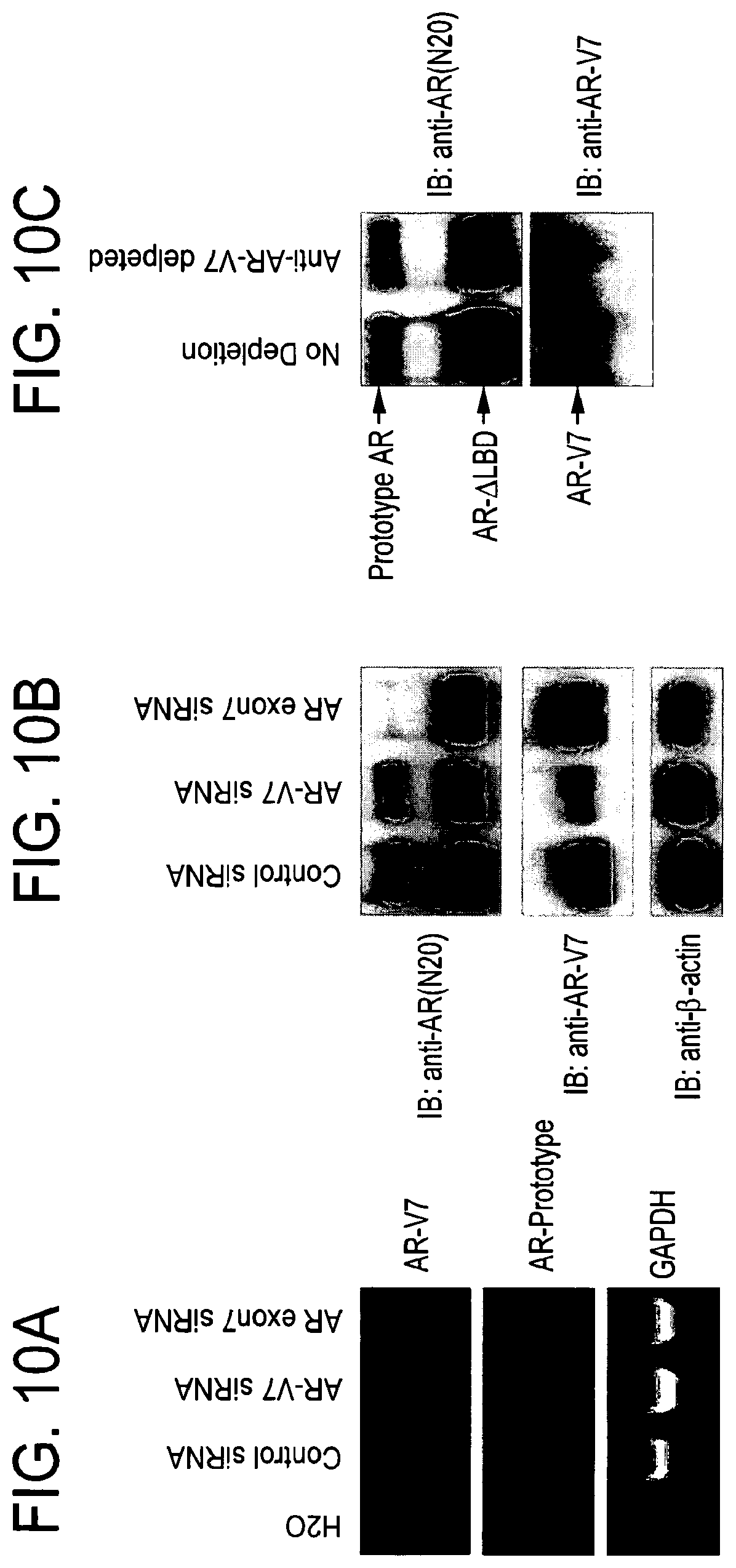
D00015

D00016

D00017

D00018

D00019

D00020

D00021

D00022

D00023

D00024

D00025

D00026

D00027

P00001

P00002

P00003

P00004

P00005

P00006

P00007

P00008

P00009

P00010

P00011

P00012

P00013

P00014

P00015

P00016

P00017

P00018

P00019

S00001
XML
uspto.report is an independent third-party trademark research tool that is not affiliated, endorsed, or sponsored by the United States Patent and Trademark Office (USPTO) or any other governmental organization. The information provided by uspto.report is based on publicly available data at the time of writing and is intended for informational purposes only.
While we strive to provide accurate and up-to-date information, we do not guarantee the accuracy, completeness, reliability, or suitability of the information displayed on this site. The use of this site is at your own risk. Any reliance you place on such information is therefore strictly at your own risk.
All official trademark data, including owner information, should be verified by visiting the official USPTO website at www.uspto.gov. This site is not intended to replace professional legal advice and should not be used as a substitute for consulting with a legal professional who is knowledgeable about trademark law.