Antibodies recognizing tau
Barbour , et al. January 12, 2
U.S. patent number 10,889,638 [Application Number 16/091,060] was granted by the patent office on 2021-01-12 for antibodies recognizing tau. This patent grant is currently assigned to PROTHENA BIOSCIENCES LIMITED. The grantee listed for this patent is PROTHENA BIOSCIENCES LIMITED. Invention is credited to Svetlana Alexander, Robin Barbour, Philip James Dolan, III, Yue Liu, Mark E. Renz.


View All Diagrams
| United States Patent | 10,889,638 |
| Barbour , et al. | January 12, 2021 |
Antibodies recognizing tau
Abstract
The invention provides antibodies that specifically bind tau. The antibodies inhibit or delay tau-associated pathologies and associated symptomatic deterioration.
| Inventors: | Barbour; Robin (Walnut Creek, CA), Dolan, III; Philip James (Foster City, CA), Liu; Yue (Foster City, CA), Alexander; Svetlana (Sunnyvale, CA), Renz; Mark E. (Millbrae, CA) | ||||||||||
|---|---|---|---|---|---|---|---|---|---|---|---|
| Applicant: |
|
||||||||||
| Assignee: | PROTHENA BIOSCIENCES LIMITED
(Dublin, IE) |
||||||||||
| Family ID: | 1000005295033 | ||||||||||
| Appl. No.: | 16/091,060 | ||||||||||
| Filed: | May 2, 2017 | ||||||||||
| PCT Filed: | May 02, 2017 | ||||||||||
| PCT No.: | PCT/IB2017/052544 | ||||||||||
| 371(c)(1),(2),(4) Date: | October 03, 2018 | ||||||||||
| PCT Pub. No.: | WO2017/191560 | ||||||||||
| PCT Pub. Date: | November 09, 2017 |
Prior Publication Data
| Document Identifier | Publication Date | |
|---|---|---|
| US 20190330314 A1 | Oct 31, 2019 | |
Related U.S. Patent Documents
| Application Number | Filing Date | Patent Number | Issue Date | ||
|---|---|---|---|---|---|
| 62330789 | May 2, 2016 | ||||
| Current U.S. Class: | 1/1 |
| Current CPC Class: | G01N 33/6896 (20130101); C07K 16/18 (20130101); C07K 2317/24 (20130101); C07K 2317/34 (20130101); G01N 2333/4709 (20130101); G01N 2800/2821 (20130101); C07K 2317/565 (20130101); G01N 2800/52 (20130101); C07K 2317/567 (20130101); C07K 2317/76 (20130101); C07K 2317/92 (20130101) |
| Current International Class: | C07K 16/18 (20060101); G01N 33/68 (20060101) |
References Cited [Referenced By]
U.S. Patent Documents
| 8012936 | September 2011 | Sigurdsson et al. |
| 8987419 | March 2015 | Barghorn et al. |
| 9321841 | April 2016 | Jones et al. |
| 10501531 | December 2019 | Seubert et al. |
| 10752679 | August 2020 | Seubert et al. |
| 2007/0042359 | February 2007 | Throsby et al. |
| 2008/0050383 | February 2008 | Sigurdsson et al. |
| 2009/0028851 | January 2009 | Stuhmer et al. |
| 2010/0022026 | January 2010 | Rump et al. |
| 2010/0267927 | October 2010 | Garrett et al. |
| 2010/0316564 | December 2010 | Sigurdsson |
| 2011/0053264 | March 2011 | Kashmiri et al. |
| 2011/0206702 | August 2011 | Polakis et al. |
| 2012/0023911 | February 2012 | Liu et al. |
| 2012/0100152 | April 2012 | Roberts et al. |
| 2012/0142602 | June 2012 | Brady et al. |
| 2012/0149880 | June 2012 | Cheung et al. |
| 2012/0204275 | August 2012 | Schenk et al. |
| 2012/0288507 | November 2012 | Qian et al. |
| 2012/0301473 | November 2012 | Binder et al. |
| 2012/0308480 | December 2012 | Smith et al. |
| 2013/0189289 | July 2013 | Inoue |
| 2013/0295021 | November 2013 | Chen et al. |
| 2014/0056901 | February 2014 | Agadjanyan |
| 2014/0086921 | March 2014 | Griswold-Prenner et al. |
| 2014/0171373 | June 2014 | Ashe et al. |
| 2014/0294731 | October 2014 | Pfeifer |
| 2015/0050215 | February 2015 | Novak |
| 2015/0050270 | February 2015 | Li et al. |
| 2015/0056721 | February 2015 | Siman |
| 2015/0166661 | June 2015 | Chen et al. |
| 2015/0175682 | June 2015 | Pfeifer et al. |
| 2015/0196663 | July 2015 | Shusta |
| 2015/0253341 | September 2015 | McAvoy et al. |
| 2015/0266947 | September 2015 | Sierks |
| 2016/0031976 | February 2016 | Seubert et al. |
| 2016/0376341 | December 2016 | Kumar et al. |
| 2017/0355756 | December 2017 | Julien |
| 2019/0322728 | October 2019 | Seubert et al. |
| 2019/0330316 | October 2019 | Barbour et al. |
| 2009-056790 | Feb 2009 | JP | |||
| 2010-511388 | Apr 2010 | JP | |||
| 2011-501655 | Jan 2011 | JP | |||
| 2012-500020 | Jan 2012 | JP | |||
| 2015520685 | Jul 2015 | JP | |||
| 215-530971 | Oct 2015 | JP | |||
| WO 1996/15452 | May 1996 | WO | |||
| WO 2008068048 | Jun 2008 | WO | |||
| WO 2011/154321 | Dec 2011 | WO | |||
| WO 2012/049570 | Apr 2012 | WO | |||
| WO 2013/004717 | Jan 2013 | WO | |||
| WO 2013/007839 | Jan 2013 | WO | |||
| WO 2013/028810 | Feb 2013 | WO | |||
| WO 2013/041962 | Mar 2013 | WO | |||
| WO 2014/008404 | Jan 2014 | WO | |||
| WO 2014/1006000 | Jun 2014 | WO | |||
| WO 2014/152157 | Sep 2014 | WO | |||
| WO 2014/165271 | Oct 2014 | WO | |||
| WO 2016/079597 | May 2016 | WO | |||
| WO 2015/200806 | Dec 2016 | WO | |||
| WO 2017/062672 | Apr 2017 | WO | |||
| WO 2014/165271 | Nov 2017 | WO | |||
| WO 2017/191559 | Nov 2017 | WO | |||
| WO 2017/191560 | Nov 2017 | WO | |||
| WO 2017/191561 | Nov 2017 | WO | |||
| WO 2018/156250 | Aug 2018 | WO | |||
| WO 2018/204546 | Nov 2018 | WO | |||
| WO 2020/096608 | May 2020 | WO | |||
| WO 2020/097561 | May 2020 | WO | |||
| WO 2020/163817 | Aug 2020 | WO | |||
Other References
|
Chen "Enhancement and destruction of antibody function by somatic mutation: unequal occurrence is controlled by V gene combinatorial associations" EMBO 14(12):2784-2794 (Year: 1995). cited by examiner . Kussie "A Single Engineered Amino Acid Substitution Changes Antibody Fine Specificity" J immunol 152(1):146-52 (Year: 1994). cited by examiner . Goedert, et al., "Cloning and sequencing of the cDNA encoding a core protein of the paired helical filament of Alzheimer disease: Identification as the microtubule-associated protein tau" Proc. Natl. Acad. Sci. USA, vol. 85, pp. 4051-4055, (Jun. 1998). cited by applicant . PCT/US2014/025044 International Search Report and Written Opinion dated Nov. 3, 2014. cited by applicant . Vigo-Pelfrey, et al., "Elevation of microtubule-associated protein tau in the cerebrospinal fluid of patients with Alzheimer's disease", Neurology, 45:788-793 (1995). cited by applicant . PCT/US2014/025044 Invitation to Pay Additional Fees and, Where Applicable, Protest Fee dated Aug. 15, 2014. cited by applicant . EP 14778358.2 European Supplementary Search Report completed Nov. 3, 2016. cited by applicant . Castillo-Carranza, et al., "Tau aggregates as immunotherapeutic targets," Frontiers in Bioscience, Scholar, 5, 426-438 (Jan. 1, 2013). cited by applicant . Ghoshal, et al., "Tau Conformational Changes Correspond to Impairments of Episodic Memory in Mild Cognitive Impairment and Alzheimer's Disease," Experimental Neurology, 177, 475-493, (2002). cited by applicant . Jicha, et al., "Sequence Requirements for Formation of Conformational Variants of Tau Similar to Those Found in Alzheimer's Disease," Journal of Neuroscience Research, 55:713-723 (1999). cited by applicant . Morris, "Epitope Mapping of Protein Antigens by Competition ELISA," The Protein Protocols Handbook, edited by J. M. Walker, Humana Press Inc., Totowa, NJ, pp. 595-600, (Jan. 1, 1996). cited by applicant . Dubel, "Molecular Engineering I: Humanization," Handbook of Therapeutic Antibodies, Chapter 6:119-144, (2007). cited by applicant . Yanamandra, et al., "Anti-Tau Antibodies that Block Tau Aggregate Seeding In Vitro Markedly Decrease Pathology and Improve Cognition in Vivo," Neuron, 80, 402-414 (Oct. 15, 2013). cited by applicant . PCT/US2014/025044 International Preliminary Report on Patentability completed Oct. 9, 2014. cited by applicant . U.S. Appl. No. 14/776,724 Restriction Requirement dated Jan. 19, 2017. cited by applicant . U.S. Appl. No. 14/776,724 Non-Final Office Action dated Jun. 1, 2017. cited by applicant . Chen, et al., "Enhancement and destruction of antibody function by somatic mutation: unequal occurrence is controlled by V gene combinatorial associations," the EMBO Journal, vol. 14, No. 12, pp. 2784-2794 (1995). cited by applicant . Kussie, et al., "A Single Engineered Amino Acid Substitution Changes Antibody Fine Specificity," J Immunol, 152(1):146-52 (1994). cited by applicant . Oddo, et al., "Reduction of Soluble A.beta. and Tau, but Not Soluble A.beta. Alone, Ameliorates Cognitive Decline in Transgenic Mice with Plaques and Tangles," The Journal of Biological Chemistry, vol. 281, No. 51, pp. 39413-39423 (Dec. 22, 2016). cited by applicant . Hasegawa, et al., "Characterization of Two distinct Monoclonal Antibodies to Paired Helical Filaments: Further Evidence for Fetal-Type Phosphorylation of the .tau. in Paired Helical Filaments", Journal of Neurochemistry, vol. 60, No. 6, (1993). cited by applicant . Leger, et al., "Antibody Drug Discovery Chapter 1: Humanization of Antibodies", Molecular medicine and Medicinal Chemistry, pp. 1-23 XP055119233 (Jan. 1, 2011). cited by applicant . Almagro, et al., "Humanization of antibodies", Frontiers in Bioscience, 13, 1619-1653, (Jan. 1, 2008). cited by applicant . Lazar, et al., "A molecular immunology approach to antibody humanization and functional optimization", Molecular Immunology, 44:1986-1998 (2007). cited by applicant . Wu, et al., "Simultaneous Humanization and Affinity Optimization of Monoclonal Antibodies", Methods of Molecular Biology, vol. 207: Recombinant Antibodies for Cancer Therapy: Methods and Protocols, Edited by M. Weischof and J. Krauss @ Humana Press Inc., Tolowa NJ, pp. 197-212 (Jan. 1, 2003). cited by applicant . PCT/IB2017/052544 Search Report and Written Opinion dated Jul. 31, 2017. cited by applicant . PCT/IB2017/052545 Search Report and Written Opinion dated Aug. 1, 2017. cited by applicant . Bacskai, et al., "Imaging of amyloid-.beta. deposits in brains of living mice permits direct observation of clearance of plaques with immunotherapy," Nature Medicine, vol. 7, No. 3, pp: 369-372, (Mar. 2001). cited by applicant . PCT/IB2017/052544 Search Report and Written Opinion dated Jul. 19, 2017. cited by applicant . Agadjanyan, et al., "Humanized monoclonal antibody armanezumab specific to N-terminus of pathological tau: characterization and therapeutic potency," Molecular Neurodegeneration, 12:33, DOI 10.1186/s13024-017-0172-1, (2017). cited by applicant . Kontsekova, et al., "First-in-man tau vaccine targeting structural determinants essential for pathological tau-tau interaction reduces tau oligomerisation and neurofibrillary degeneration in an Alzheimer's disease model," Alzheimer's Research & Therapy, 6:44, (2014). cited by applicant . Rosseels, et al., "Tau Monoclonal Antibody Generation Based on Humanized Yeast Models," Journal of Biological Chemistry, vol. 290, No. 7, pp. 4059-4074, (Dec. 24, 2014). cited by applicant . PCT/IB2017/052543 International Report on Patentability dated Nov. 6, 2018. cited by applicant . PCT/IB2017/052544 International Report on Patentability dated Nov. 6, 2018. cited by applicant . PCT/IB2017/052545 International Report on Patentability dated Nov. 6, 2018. cited by applicant . U.S. Appl. No. 14/776,724 Final Office Action dated Oct. 31, 2018. cited by applicant . U.S. Appl. No. 14/776,724 Advisory Action dated Mar. 12, 2019. cited by applicant . U.S. Appl. No. 14/776,724 Notice of Allowance dated Apr. 10, 2019. cited by applicant . PCT/US2018/030739 International Search Report and Written Opinion dated Nov. 5, 2018. cited by applicant . U.S. Appl. No. 14/776,724 Notice of Allowance dated Jul. 29, 2019. cited by applicant . U.S. Appl. No. 16/092,439 Notice of Allowance dated Oct. 16, 2019. cited by applicant . Pedersen, et al., "Tau immunotherapy for Alzheimer's disease," Trends in Molecular Medicine, vol. 21, No. 6, pp. 394-402, (Jun. 2015). cited by applicant . PCT/US2018/030739 International Preliminary Report on Patentability dated Nov. 5, 2019. cited by applicant . PCT/US2018/059895 International Search Report and Written Opinion dated Apr. 12, 2019. cited by applicant . PCT/US2018/030739 International Search Report and Written Opinion dated Sep. 18, 2018. cited by applicant . EP 19213368 Extended European Search Report dated Jun. 24, 2020. cited by applicant . Florenzano, et al., "Extracellular truncated tau causes early presynaptic dysfunction associated with Alzheimer's disease and other tauopathies," Oncotarget, vol. 8, No. 29, pp: 64745-46778, (2017). cited by applicant . Gershoni, et al., "Epitope Mapping, The First Step in Developing Epitope-Based Vaccines," Biodrugs, 21:(3), p. 145-156. (2007). cited by applicant . U.S. Appl. No. 16/097,445 Restriction Requirement dated Feb. 18, 2020. cited by applicant . U.S. Appl. No. 16/092,439 Notice of Allowance and Interview Summary dated Apr. 10, 2020. cited by applicant . PCT/US2019/060616 International Search Report and Written Opinion dated Mar. 20, 2020. cited by applicant . PCT/US2020/017357 Invitation to Pay Additional Fees dated Apr. 23, 2020. cited by applicant . U.S. Appl. No. 16/097,445 Non-Final Office Action dated May 27, 2020. cited by applicant . PCT/US2020/017357 International Search Report and Written Opinion dated Jun. 17, 2020. cited by applicant . Kawahara, et al., "The Novel Monoclonal Antibody 9F5 Reveals Expression of a Fragment of GPNMB/Osteoactivin Processed by Furin-like Protease(s) in a Subpopulation of Microglia in Neonatal Rat Brain," GLIA, vol. 64, No. 11, pp. 1938-1961, (Nov. 2016). cited by applicant . Strang, et al., "Generation and characterization of new monoclonal antibodies targeting the PHF1 and AT8 epitopes on human tau," Acta Neuropathologica Communications, 5:58, (2017). cited by applicant . Croft, et al., "Novel monoclonal antibodies targeting the microtubule-binding domain of human tau," PLoS ONE, 13(4): e0195211, (2018). cited by applicant . PCT/US2020/020704 Invitation to Pay Additional Fees dated Jun. 3, 2020. cited by applicant . PCT/US2020/020704 Search Report and Written Opinion dated Aug. 4, 2020. cited by applicant. |
Primary Examiner: Weidner; Adam
Attorney, Agent or Firm: Alston & Bird LLP
Parent Case Text
CROSS REFERENCE TO RELATED APPLICATIONS
This application is the US national stage of PCT/IB2017/052544 filed May 2, 2017, which claims the benefit of U.S. Provisional Application No. 62/330,789 filed May 2, 2016, each of which is incorporated by reference in its entirety for all purposes.
Claims
What is claimed is:
1. An isolated monoclonal antibody that binds human tau, comprising three Kabat/Chothia Composite heavy chain CDRs, CDR-H1 of SEQ ID NO:8, CDR-H2 of SEQ ID NO:9, and CDR-H3 of amino acid sequence LDF, and three Kabat/Chothia Composite light chain CDRs, CDR-L1 of SEQ ID NO:12, CDR-L2 of SEQ ID NO:13, and CDR-L3 of SEQ ID NO:14, except that position H27 is occupied by F or Y, H28 is occupied by N or T, H29 is occupied by I or F, H30 is occupied by K or T, position H51 is occupied by I or V, position H54 is occupied by N or D, position H60 is occupied by D or A, H61 is occupied by P or E, and H102 is occupied by F or Y.
2. The antibody of claim 1, comprising three Kabat/Chothia Composite heavy chain CDRs, CDR-H1 of SEQ ID NO:8, CDR-H2 of SEQ ID NO:9, and CDR-H3 of amino acid sequence LDF, and three Kabat/Chothia Composite light chain CDRs, CDR-L1 of SEQ ID NO:12, CDR-L2 of SEQ ID NO:13, and CDR-L3 of SEQ ID NO:14.
3. The antibody of claim 1 comprising mature heavy chain variable region of SEQ ID NO: 7 and a mature light chain variable region of SEQ ID NO:11, or a chimeric, veneered, or humanized form thereof.
4. The antibody of claim 1, wherein CDR-H1 has an amino acid sequence comprising SEQ ID NO: 42, 58, 59 or 60.
5. The antibody of claim 1, wherein CDR-H2 has an amino acid sequence comprising SEQ ID NO: 43, 61, 62, 63 or 64.
6. The antibody of claim 1, wherein CDR-H3 has an amino acid sequence LDY.
7. The antibody of claim 1, wherein the antibody is a humanized antibody.
8. The humanized antibody of claim 7, wherein CDR-H1 has an amino acid sequence comprising SEQ ID NO: 42 and CDR-H2 has an amino acid sequence comprising SEQ ID NO: 43.
9. The humanized antibody of claim 7, wherein CDR-H1 has an amino acid sequence comprising SEQ ID NO: 42 and CDR-H2 has an amino acid sequence comprising SEQ ID NO:61.
10. The humanized antibody of claim 7, wherein CDR-H1 has an amino acid sequence comprising SEQ ID NO: 42 and CDR-H2 has an amino acid sequence comprising SEQ ID NO:64.
11. The humanized antibody of claim 7, wherein CDR-H1 has an amino acid sequence comprising SEQ ID NO: 42, CDR-H2 has an amino acid sequence comprising SEQ ID NO:63, and CDR-H3 has amino acid sequence LDY.
12. The humanized antibody of claim 7, wherein CDR-H1 has an amino acid sequence comprising SEQ ID NO: 58 and CDR-H2 has an amino acid sequence comprising SEQ ID NO:62.
13. The humanized antibody of claim 7, wherein CDR-H1 has an amino acid sequence comprising SEQ ID NO: 59 and CDR-H2 has an amino acid sequence comprising SEQ ID NO:63.
14. The humanized antibody of claim 7, wherein CDR-H1 has an amino acid sequence comprising SEQ ID NO: 60 and CDR-H2 has an amino acid sequence comprising SEQ ID NO:62.
15. The antibody of claim 3, that is humanized or chimeric.
16. The humanized antibody of claim 15 comprising a humanized mature heavy chain variable region comprising, CDR-H1 of SEQ ID NO:8, CDR-H2 of SEQ ID NO:9, and CDR-H3 of amino acid sequence LDF, and a humanized mature light chain variable region comprising, CDR-L1 of SEQ ID NO:12, CDR-L2 of SEQ ID NO:13, and CDR-L3 of SEQ ID NO:14.
17. A humanized antibody that binds human tau comprising a mature heavy chain variable region comprising three heavy chain CDRs of SEQ ID NO:7 and a mature light chain variable region comprising three light chain CDRs of SEQ ID NO:11, wherein the CDRs are of a definition selected from the group consisting of Kabat, Chothia, Kabat/Chothia Composite, AbM and Contact.
18. The humanized antibody of claim 17 wherein the humanized mature heavy chain variable region comprises the three Kabat/Chothia Composite heavy chain CDRs, CDR-H1 of SEQ ID NO:8, CDR-H2 of SEQ ID NO:9, and CDR-H3 of amino acid sequence LDF, and the humanized mature light chain variable region comprises the three Kabat/Chothia Composite light chain CDRs, CDR-L1 of SEQ ID NO:12, CDR-L2 of SEQ ID NO:13, and CDR-L3 of SEQ ID NO:14.
19. The humanized antibody of claim 17 wherein the humanized mature heavy chain variable region comprises the three Kabat heavy chain CDRs, CDR-H1 of SEQ ID NO:32, CDR-H2 of SEQ ID NO:9, and CDR-H3 of amino acid sequence LDF, and the humanized mature light chain variable region comprises the three Kabat light chain CDRs, CDR-L1 of SEQ ID NO:12, CDR-L2 of SEQ ID NO:13, and CDR-L3 of SEQ ID NO:14.
20. The humanized antibody of claim 17 wherein the humanized mature heavy chain variable region comprises the three Chothia heavy chain CDRs, CDR-H1 of SEQ ID NO:33, CDR-H2 of SEQ ID NO:34, and CDR-H3 of amino acid sequence LDF, and the humanized mature light chain variable region comprises the three Chothia light chain CDRs, CDR-L1 of SEQ ID NO:12, CDR-L2 of SEQ ID NO:13, and CDR-L3 of SEQ ID NO:14.
21. The humanized antibody of claim 17 wherein the humanized mature heavy chain variable region comprises the three AbM heavy chain CDRs, CDR-H1 of SEQ ID NO:8, CDR-H2 of SEQ ID NO:35, and CDR-H3 of amino acid sequence LDF, and the humanized mature light chain variable region comprises the three AbM light chain CDRs, CDR-L1 of SEQ ID NO:12, CDR-L2 of SEQ ID NO:13, and CDR-L3 of SEQ ID NO:14.
22. The humanized antibody of claim 17 wherein the humanized mature heavy chain variable region comprises the three Contact heavy chain CDRs, CDR-H1 of SEQ ID NO:39, CDR-H2 of SEQ ID NO:40, and CDR-H3 of SEQ ID NO:41, and the humanized mature light chain variable region comprises the three Contact light chain CDRs, CDR-L1 of SEQ ID NO:36, CDR-L2 of SEQ ID NO:37, and CDR-L3 of SEQ ID NO:38.
23. The humanized antibody of claim 15, comprising a humanized mature heavy chain variable region having an amino acid sequence at least 90% identical to any one of SEQ ID NOs:15-19 and SEQ ID NOs:46-57 and a humanized mature light chain variable region having an amino acid sequence at least 90% identical to any one of SEQ ID NOs: 20-23, except that position H17 can be T or S, and position H2O can be I or V.
24. The humanized antibody of claim 23, wherein at least one of the following positions in the VH region is occupied by the amino acid as specified: H38 is occupied by R and H93 is occupied by S.
25. The humanized antibody of claim 23, provided positions H38 and H93 in the VH region are occupied by R and S, respectively.
26. The humanized antibody of claim 23 wherein at least one of the following positions in the VH region is occupied by the amino acid as specified: H38 is occupied by R, H43 is occupied by Q, H83 is occupied by T, and H93 is occupied by S.
27. The humanized antibody of claim 26, provided positions H38, H43, H83, and H93 in the VH region are occupied by R, Q, T, and S, respectively.
28. The humanized antibody of claim 26, wherein at least one of the following positions in the VH region is occupied by the amino acid as specified: H12 is occupied by V, H24 is occupied by A, H48 is occupied by I, H67 is occupied by A, H80 is occupied by L, H81 is occupied by Q, and H91 is occupied by F.
29. The humanized antibody of claim 28, provided positions H12, H24, H48, H67, H80, H81, and H91 in the VH region are occupied by V, A, I, A, L, Q, and F, respectively.
30. The humanized antibody of claim 28, wherein at least one of the following positions in the VH region is occupied by the amino acid as specified: H13 is occupied by R and H66 is occupied by K.
31. The humanized antibody of claim 30, provided positions H13 and H66 in the VH region are occupied by R and K, respectively.
32. The humanized antibody of claim 28, wherein at least one of the following positions in the VH region is occupied by the amino acid as specified: H40 is occupied by R and H82a is occupied by G.
33. The humanized antibody of claim 32, provided positions H40 and H82a in the VH region are occupied by R and G, respectively.
34. The humanized antibody of claim 26, wherein at least one of the following positions in the VH region is occupied by the amino acid as specified: H42 is occupied by E and H76 is occupied by N.
35. The humanized antibody of claim 34, provided positions H42 and H76 in the VH region are occupied by E and N, respectively.
36. The humanized antibody of claim 24, wherein at least one of the following positions in the VH region is occupied by the amino acid as specified: H40 is occupied by R, H82a is occupied by G, and H83 is occupied by T.
37. The humanized antibody of claim 36, provided positions H40, H82a, and H83 in the VH region are occupied by R, G, and T, respectively.
38. The humanized antibody of claim 36, wherein H12 in the VH region is occupied by V.
39. The humanized antibody of claim 38, wherein H80 in the VH region is occupied by L.
40. The humanized antibody of claim 39, wherein at least one of the following positions in the VH region is occupied by the amino acid as specified: H24 is occupied by A, H48 is occupied by I, H67 is occupied by A, H80 is occupied by L, and H91 is occupied by F.
41. The humanized antibody of claim 40, provided positions H24, H48, H67, H80, and H91 in the VH region are occupied by A, I, A, L, and F, respectively.
42. The humanized antibody of claim 39, wherein at least one of the following positions in the VH region is occupied by the amino acid as specified: H43 is occupied by Q, and H81 is occupied by Q.
43. The humanized antibody of claim 42, provided positions H43 and H81 in the VH region are occupied by Q, and Q, respectively.
44. The humanized antibody of claim 36, wherein at least one of the following positions in the VH region is occupied by the amino acid as specified: H24 is occupied by A, and H91 is occupied by F.
45. The humanized antibody of claim 44, provided positions H24 and H91 in the VH region are occupied by A and F, respectively.
46. The humanized antibody of claim 44, wherein at least one of the following positions in the VH region is occupied by the amino acid as specified: H13 is occupied by R, H17 is occupied by L, H29 is occupied by F, H42 is occupied by E, H43 is occupied by Q, H61 is occupied by E, H76 is occupied by N, H80 is occupied by L, H81 is occupied by Q.
47. The humanized antibody of claim 46, provided positions H13, H17, H29, H42, H43, H61, H76, H80, and H81 in the VH region are occupied by R, L, F, E, Q, E, N, L, and Q, respectively.
48. The humanized antibody of claim 38, wherein at least one of the following positions in the VH region is occupied by the amino acid as specified: H24 is occupied by A, H28 is occupied by T, H48 is occupied by I, H54 is occupied by D, H60 is occupied by A, H67 is occupied by A, H80 is occupied by L, and H91 is occupied by F.
49. The humanized antibody of claim 48, provided positions H24, H28, H48, H54, H60, H67, H80, and H91 in the VH region are occupied by A, T, I, D, A, A, L, and F, respectively.
50. The humanized antibody of claim 38, wherein at least one of the following positions in the VH region is occupied by the amino acid as specified: H10 is occupied by D, H17 is occupied by L, H24 is occupied by A, H28 is occupied by T, H43 is occupied by Q, H48 is occupied by I, H60 is occupied by A, H61 is occupied by E, H91 is occupied by F, H108 is occupied by T, and H109 is occupied by L.
51. The humanized antibody of claim 50, provided positions H10, H17, H24, H28, H43, H48, H60, H61, H91, H108, and H109 in the VH region are occupied by D, L, A, T, Q, I, A, E, F, T, and L, respectively.
52. The humanized antibody of claim 39, wherein at least one of the following positions in the VH region is occupied by the amino acid as specified: H17 is occupied by L, H27 is occupied by Y, H29 is occupied by F, and H61 is occupied by E.
53. The humanized antibody of claim 52, provided positions H17, H27, H29, and H61 in the VH region are occupied by L, Y, F, and E, respectively.
54. The humanized antibody of claim 24, wherein at least one of the following positions in the VH region is occupied by the amino acid as specified: H17 is occupied by L, H27 is occupied by Y, H29 is occupied by F, H61 is occupied by E, H76 is occupied by N, and H82a is occupied by G.
55. The humanized antibody of claim 54, provided positions H17, H27, H29, H61, H76, and H82a in the VH region are occupied by L, Y, F, E, N, and G, respectively.
56. The humanized antibody of claim 24, wherein at least one of the following positions in the VH region is occupied by the amino acid as specified: H12 is occupied by V, H17 is occupied by L, H24 is occupied by A, H43 is occupied by Q, H48 is occupied by I, H83 is occupied by T, and H91 is occupied by F.
57. The humanized antibody of claim 56, provided positions H12, H17, H24, H43, H48, H83, and H9 lin the VH region are occupied by V, L, A, Q, I, T, F, respectively.
58. The humanized antibody of claim 24, wherein at least one of the following positions in the VH region is occupied by the amino acid as specified: H12 is occupied by V, H24 is occupied by A, H48 is occupied by I, H67 is occupied b A, H80 is occupied by L, H83 is occupied by T, and H91 is occupied by F.
59. The humanized antibody of claim 58, provided positions H12, H24, H48, H67, H80, H83, and H91 in the VH region are occupied by V, A, I, A, L, T, and F, respectively.
60. The humanized antibody of claim 23 wherein at least one of the following positions in the VH region is occupied by the amino acid as specified: H10 is occupied by E or D, H12 is occupied by K or V, H13 is occupied by K or R, H17 is occupied by T, L or S, H24 is occupied by V or A, H27 is occupied by F or Y, H28 is occupied by N or T, H29 is occupied by I or F, H30 is occupied by K or T, H38 is occupied by Q or R, H40 is occupied by A or R, H42 is occupied by G or E, H43 is occupied by K or Q, H48 is occupied by M or I, H51 is occupied by V or I, H54 is occupied by N or D, H60 is occupied by D or A, H61 is occupied by P or E, H66 is occupied by R or K, H67 is occupied by V or A, H76 is occupied by D or N, H80 is occupied by M or L, H81 is occupied by E or Q, H82a is occupied by S or G, H83 is occupied by T or R, H91 is occupied by Y or F, H93 is occupied by A or S, H102 is occupied by F or Y, H108 is occupied by T or L, H109 is occupied by L or V.
61. The humanized antibody of claim 60, provided positions H12, H13, H17, H24, H38, H42, H43, H48, H66, H67, H76, H80, H81, H83, H91, and H93 in the VH region are occupied by V, R, L, A, R, E, Q, I, K, A, N, L, Q, T, F, and S, respectively.
62. The humanized antibody of claim 60, provided positions H38, H42, H43, H76, H83, and H93 in the VH region are occupied by R, E, Q, N, T, and S, respectively.
63. The humanized antibody of claim 60, provided positions H12, H13, H17, H24, H38, H40, H42, H43, H48, H66, H67, H76, H80, H81, H82A, H83, H91, and H93 in the VH region are occupied by V, R, L, A, R, R, E, Q, I, K, A, N, L, Q, G, T, F, and S, respectively.
64. The humanized antibody of claim 60, provided positions H12, H24, H38, H40, H43, H48, H67, H80, H81, H82A, H83, H91, and H93 in the VH region are occupied by V, A, R, R, Q, I, A, L, Q, G, T, F, and S, respectively.
65. The humanized antibody of claim 60 provided positions H12, H24, H28, H38, H40, H43, H48, H54, H60, H67, H80, H81, H82A, H83, H91, and H93 in the VH region are occupied by V, A, T, R, R, Q, I, D, A, A, L, Q, G, T, F, and S, respectively.
66. The humanized antibody of claim 60 provided positions H12, H24, H28, H38, H40, H48, H51, H54, H60, H67, H80, H82A, H83, H91, and H93 in the VH region are occupied by V, A, T, R, R, I, V, D, A, A, L, G, T, F, and S, respectively.
67. The humanized antibody of claim 60 provided positions H12, H24, H28, H38, H40, H48, H54, H60, H67, H80, H82A, H83, H91, and H93 in the VH region are occupied by V, A, T, R, R, I, D, A, A, L, G, T, F, and S, respectively.
68. The humanized antibody of claim 60 provided positions H13, H17, H24, H29, H38, H40, H42, H43, H54, H61, H76, H80, H81, H82A, H83, H91, and H93 in the VH region are occupied by R, L, A, F, R, R, E, Q, N, E, N, L, Q, G, T, F, and S, respectively.
69. The humanized antibody of claim 60 provided positions H13, H17, H24, H27, H28, H29, H30, H38, H40, H42, H43, H51, H54, H60, H61, H76, H80, H81, H82A, H83, H91, and H93 in the VH region are occupied by R, L, A, Y, T, F, T, R, R, E, Q, V, D, A, E, N, L, Q, G, T, F, and S, respectively.
70. The humanized antibody of claim 60 provided positions H10, H12, H17, H24, H28, H38, H40, H42, H43, H48, H54, H60, H61, H76, H80, H82A, H83, H91, H93, H108, and H109 in the VH region are occupied by D, V, L, A, T, R, R, E, Q, I, N, A, E, N, L, G, T, F, S, T, and L, respectively.
71. The humanized antibody of claim 60 provided positions H10, H12, H17, H24, H28, H38, H40, H43, H48, H51, H54, H60, H61, H82A, H83, H91, H93, H102, H108, and H109 in the VH region are occupied by D, V, L, A, T, R, R, Q, I, V, D, A, E, G, T, F, S, Y, T, and L, respectively.
72. The humanized antibody of claim 60 provided positions H38 and H93 in the VH region are occupied by R and S, respectively.
73. The humanized antibody of claim 60 provided positions H17, H27, H29, H38, H61, H76, H82A, and H93 in the VH region are occupied by L, Y, F, R, E, N, G, and S, respectively.
74. The humanized antibody of claim 60 provided positions H17, H27, H28, H29, H30, H38, H51, H54, H60, H61, H76, H82A, and H93 in the VH region are occupied by L, Y, T, F, T, R, V, D, A, E, N, G, and S, respectively.
75. The humanized antibody of claim 60 provided positions H12, H38, H40, H48, H66, H67, H76, H80, H82A, H83, and H93 in the VH region are occupied by V, R, R, I, K, A, N, L, G, T, and S, respectively.
76. The humanized antibody of claim 60 provided positions H12, H17, H27, H29, H38, H40, H61, H80, H82A, H83, and H93 in the VH region are occupied by V, L, Y, F, R, R, E, L, G, T, and S, respectively.
77. The humanized antibody of claim 60 provided positions H12, H17, H27, H28, H29, H30, H38, H40, H51, H54, H60, H61, H80, H82A, H83, and H93 in the VH region are occupied by V, L, Y, T, F, T, R, R, V, D, A, E, L, G, T, and S, respectively.
78. The humanized antibody of claim 23, wherein at least one of the following positions in the VL region is occupied by the amino acid as specified: L36 is occupied by L, L37 is occupied by L, and L100 is occupied by G.
79. The humanized antibody of claim 78, provided positions L36, L37, and L100 in the VL region are occupied by L, L, and G, respectively.
80. The humanized antibody of claim 78, wherein at least one of the following positions in the VL region is occupied by the amino acid as specified: L12 is occupied by S and L45 is occupied by K.
81. The humanized antibody of claim 80, provided positions L12 and L45 in the VL region are occupied by S and K, respectively.
82. The humanized antibody of claim 23, wherein at least one of the following positions in the VL region is occupied by the amino acid as specified: L2 is V or I, L7 is S or T, L12 is P or S, L15 is L or I, L36 is L, L37 is L, L45 is R or K, L60 is D or S, L100 is G.
83. The humanized antibody of claim 82, provided positions L12, L36, L37, L45, and L100 in the VL region are occupied by S, L, L, K, and G, respectively.
84. The humanized antibody of claim 82, provided positions L36, L37, and L100 in the VL region are occupied by L, L and G, respectively.
85. The humanized antibody of claim 82, provided positions L36, L37, L60, and L100 in the VL region are occupied by L, L, S, and G, respectively.
86. The humanized antibody of claim 82, provided positions L2, L7, L12, L15, L36, L37, L45, and L100 in the VL region are occupied by I, T, S, I, L, L, K, and G, respectively.
87. The humanized antibody of claim 82, provided positions L2, L7, L12, L15, L36, L37, L45, and L100 in the VL region are occupied by V, S, P, L, L, L, R, and G, respectively.
88. The humanized antibody of claim 23, comprising a mature heavy chain variable region having an amino acid sequence at least 95% identical to any one of SEQ ID NOs:15-19 and SEQ ID NOs:46-57 and a mature light chain variable region having an amino acid sequence at least 95% identical to any one of SEQ ID NOs: 20-23, except that position H17 can be T or S, and position H2O can be I or V.
89. The humanized antibody of claim 88, comprising a mature heavy chain variable region having an amino acid sequence at least 98% identical to any one of SEQ ID NOs:15-19 and SEQ ID NOs:46-57 and a mature light chain variable region having an amino acid sequence at least 98% identical to any one of SEQ ID NOs: 20-23, except that position H17 can be T or S, and position H2O can be I or V.
90. The humanized antibody of claim 89 wherein the mature heavy chain variable region has an amino acid sequence of any of SEQ ID NOs:15-19 and SEQ ID NOs:46-57 and the mature light chain variable region has an amino acid sequence of any one of SEQ ID NOs:20-23.
91. The humanized antibody of claim 90 wherein the mature heavy chain variable region has an amino acid sequence of SEQ ID NO:15 and the mature light chain variable region has an amino acid sequence of SEQ ID NO:20.
92. The humanized antibody of claim 90, wherein the mature heavy chain variable region has an amino acid sequence of SEQ ID NO:15 and the mature light chain variable region has an amino acid sequence of SEQ ID NO:21.
93. The humanized antibody of claim 90, wherein the mature heavy chain variable region has an amino acid sequence of SEQ ID NO:15 and the mature light chain variable region has an amino acid sequence of SEQ ID NO:22.
94. The humanized antibody of claim 90, wherein the mature heavy chain variable region has an amino acid sequence of SEQ ID NO:15 and the mature light chain variable region has an amino acid sequence of SEQ ID NO:23.
95. The humanized antibody of claim 90, wherein the mature heavy chain variable region has an amino acid sequence of SEQ ID NO:16 and the mature light chain variable region has an amino acid sequence of SEQ ID NO:20.
96. The humanized antibody of claim 90, wherein the mature heavy chain variable region has an amino acid sequence of SEQ ID NO:16 and the mature light chain variable region has an amino acid sequence of SEQ ID NO:21.
97. The humanized antibody of claim 90, wherein the mature heavy chain variable region has an amino acid sequence of SEQ ID NO:16 and the mature light chain variable region has an amino acid sequence of SEQ ID NO:22.
98. The humanized antibody of claim 90, wherein the mature heavy chain variable region has an amino acid sequence of SEQ ID NO:16 and the mature light chain variable region has an amino acid sequence of SEQ ID NO:23.
99. The humanized antibody of claim 90, wherein the mature heavy chain variable region has an amino acid sequence of SEQ ID NO:17 and the mature light chain variable region has an amino acid sequence of SEQ ID NO:20.
100. The humanized antibody of claim 90, wherein the mature heavy chain variable region has an amino acid sequence of SEQ ID NO:17 and the mature light chain variable region has an amino acid sequence of SEQ ID NO:21.
101. The humanized antibody of claim 90, wherein the mature heavy chain variable region has an amino acid sequence of SEQ ID NO:17 and the mature light chain variable region has an amino acid sequence of SEQ ID NO:22.
102. The humanized antibody of claim 90, wherein the mature heavy chain variable region has an amino acid sequence of SEQ ID NO:17 and the mature light chain variable region has an amino acid sequence of SEQ ID NO:23.
103. The humanized antibody of claim 90, wherein the mature heavy chain variable region has an amino acid sequence of SEQ ID NO:18 and the mature light chain variable region has an amino acid sequence of SEQ ID NO:20.
104. The humanized antibody of claim 90, wherein the mature heavy chain variable region has an amino acid sequence of SEQ ID NO:18 and the mature light chain variable region has an amino acid sequence of SEQ ID NO:21.
105. The humanized antibody of claim 90, wherein the mature heavy chain variable region has an amino acid sequence of SEQ ID NO:18 the mature light chain variable region has an amino acid sequence of SEQ ID NO:22.
106. The humanized antibody of claim 90, wherein the mature heavy chain variable region has an amino acid sequence of SEQ ID NO:18 and the mature light chain variable region has an amino acid sequence of SEQ ID NO:23.
107. The humanized antibody of claim 90, wherein the mature heavy chain variable region has an amino acid sequence of SEQ ID NO:19 and the mature light chain variable region has an amino acid sequence of SEQ ID NO:20.
108. The humanized antibody of claim 90, wherein the mature heavy chain variable region has an amino acid sequence of SEQ ID NO:19 and the mature light chain variable region has an amino acid sequence of SEQ ID NO:21.
109. The humanized antibody of claim 90, wherein the mature heavy chain variable region has an amino acid sequence of SEQ ID NO:19 and the mature light chain variable region has an amino acid sequence of SEQ ID NO:22.
110. The humanized antibody of claim 90, wherein the mature heavy chain variable region has an amino acid sequence of SEQ ID NO:19 and the mature light chain variable region has an amino acid sequence of SEQ ID NO:23.
111. The humanized antibody of claim 90, wherein the mature heavy chain variable region has an amino acid sequence of SEQ ID NO:46 and the mature light chain variable region has an amino acid sequence of SEQ ID NO:20.
112. The humanized antibody of claim 90, wherein the mature heavy chain variable region has an amino acid sequence of SEQ ID NO:46 and the mature light chain variable region has an amino acid sequence of SEQ ID NO:21.
113. The humanized antibody of claim 90, wherein the mature heavy chain variable region has an amino acid sequence of SEQ ID NO:46 and the mature light chain variable region has an amino acid sequence of SEQ ID NO:22.
114. The humanized antibody of claim 90, wherein the mature heavy chain variable region has an amino acid sequence of SEQ ID NO:46 and the mature light chain variable region has an amino acid sequence of SEQ ID NO:23.
115. The humanized antibody of claim 90, wherein the mature heavy chain variable region has an amino acid sequence of SEQ ID NO:47 and the mature light chain variable region has an amino acid sequence of SEQ ID NO:20.
116. The humanized antibody of claim 90, wherein the mature heavy chain variable region has an amino acid sequence of SEQ ID NO:47 and the mature light chain variable region has an amino acid sequence of SEQ ID NO:21.
117. The humanized antibody of claim 90, wherein the mature heavy chain variable region has an amino acid sequence of SEQ ID NO:47 and the mature light chain variable region has an amino acid sequence of SEQ ID NO:22.
118. The humanized antibody of claim 90, wherein the mature heavy chain variable region has an amino acid sequence of SEQ ID NO:47 and the mature light chain variable region has an amino acid sequence of SEQ ID NO:23.
119. The humanized antibody of claim 90, wherein the mature heavy chain variable region has an amino acid sequence of SEQ ID NO:48 and the mature light chain variable region has an amino acid sequence of SEQ ID NO:20.
120. The humanized antibody of claim 90, wherein the mature heavy chain variable region has an amino acid sequence of SEQ ID NO:48 and the mature light chain variable region has an amino acid sequence of SEQ ID NO:21.
121. The humanized antibody of claim 90, wherein the mature heavy chain variable region has an amino acid sequence of SEQ ID NO:48 and the mature light chain variable region has an amino acid sequence of SEQ ID NO:22.
122. The humanized antibody of claim 90, wherein the mature heavy chain variable region has an amino acid sequence of SEQ ID NO:48 and the mature light chain variable region has an amino acid sequence of SEQ ID NO:23.
123. The humanized antibody of claim 90, wherein the mature heavy chain variable region has an amino acid sequence of SEQ ID NO:49 and the mature light chain variable region has an amino acid sequence of SEQ ID NO:20.
124. The humanized antibody of claim 90, wherein the mature heavy chain variable region has an amino acid sequence of SEQ ID NO:49 and the mature light chain variable region has an amino acid sequence of SEQ ID NO:21.
125. The humanized antibody of claim 90, wherein the mature heavy chain variable region has an amino acid sequence of SEQ ID NO:49 and the mature light chain variable region has an amino acid sequence of SEQ ID NO:22.
126. The humanized antibody of claim 90, wherein the mature heavy chain variable region has an amino acid sequence of SEQ ID NO:49 and the mature light chain variable region has an amino acid sequence of SEQ ID NO:23.
127. The humanized antibody of claim 90, wherein the mature heavy chain variable region has an amino acid sequence of SEQ ID NO:50 and the mature light chain variable region has an amino acid sequence of SEQ ID NO:20.
128. The humanized antibody of claim 90, wherein the mature heavy chain variable region has an amino acid sequence of SEQ ID NO:50 and the mature light chain variable region has an amino acid sequence of SEQ ID NO:21.
129. The humanized antibody of claim 90, wherein the mature heavy chain variable region has an amino acid sequence of SEQ ID NO:50 and the mature light chain variable region has an amino acid sequence of SEQ ID NO:22.
130. The humanized antibody of claim 90, wherein the mature heavy chain variable region has an amino acid sequence of SEQ ID NO:50 and the mature light chain variable region has an amino acid sequence of SEQ ID NO:23.
131. The humanized antibody of claim 90, wherein the mature heavy chain variable region has an amino acid sequence of SEQ ID NO:51 and the mature light chain variable region has an amino acid sequence of SEQ ID NO:20.
132. The humanized antibody of claim 90, wherein the mature heavy chain variable region has an amino acid sequence of SEQ ID NO:51 and the mature light chain variable region has an amino acid sequence of SEQ ID NO:21.
133. The humanized antibody of claim 90, wherein the mature heavy chain variable region has an amino acid sequence of SEQ ID NO:51 and the mature light chain variable region has an amino acid sequence of SEQ ID NO:22.
134. The humanized antibody of claim 90, wherein the mature heavy chain variable region has an amino acid sequence of SEQ ID NO:51 and the mature light chain variable region has an amino acid sequence of SEQ ID NO:23.
135. The humanized antibody of claim 90, wherein the mature heavy chain variable region has an amino acid sequence of SEQ ID NO:52 and the mature light chain variable region has an amino acid sequence of SEQ ID NO:20.
136. The humanized antibody of claim 90, wherein the mature heavy chain variable region has an amino acid sequence of SEQ ID NO:52 and the mature light chain variable region has an amino acid sequence of SEQ ID NO:21.
137. The humanized antibody of claim 90, wherein the mature heavy chain variable region has an amino acid sequence of SEQ ID NO:52 and the mature light chain variable region has an amino acid sequence of SEQ ID NO:22.
138. The humanized antibody of claim 90, wherein the mature heavy chain variable region has an amino acid sequence of SEQ ID NO:52 and the mature light chain variable region has an amino acid sequence of SEQ ID NO:23.
139. The humanized antibody of claim 90, wherein the mature heavy chain variable region has an amino acid sequence of SEQ ID NO:53 and the mature light chain variable region has an amino acid sequence of SEQ ID NO:20.
140. The humanized antibody of claim 90, wherein the mature heavy chain variable region has an amino acid sequence of SEQ ID NO:53 and the mature light chain variable region has an amino acid sequence of SEQ ID NO:21.
141. The humanized antibody of claim 90, wherein the mature heavy chain variable region has an amino acid sequence of SEQ ID NO:53 and the mature light chain variable region has an amino acid sequence of SEQ ID NO:22.
142. The humanized antibody of claim 90, wherein the mature heavy chain variable region has an amino acid sequence of SEQ ID NO:53 and the mature light chain variable region has an amino acid sequence of SEQ ID NO:23.
143. The humanized antibody of claim 90, wherein the mature heavy chain variable region has an amino acid sequence of SEQ ID NO:54 and the mature light chain variable region has an amino acid sequence of SEQ ID NO:20.
144. The humanized antibody of claim 90, wherein the mature heavy chain variable region has an amino acid sequence of SEQ ID NO:54 and the mature light chain variable region has an amino acid sequence of SEQ ID NO:21.
145. The humanized antibody of claim 90, wherein the mature heavy chain variable region has an amino acid sequence of SEQ ID NO:54 and the mature light chain variable region has an amino acid sequence of SEQ ID NO:22.
146. The humanized antibody of claim 90, wherein the mature heavy chain variable region has an amino acid sequence of SEQ ID NO:54 and the mature light chain variable region has an amino acid sequence of SEQ ID NO:23.
147. The humanized antibody of claim 90, wherein the mature heavy chain variable region has an amino acid sequence of SEQ ID NO:55 and the mature light chain variable region has an amino acid sequence of SEQ ID NO:20.
148. The humanized antibody of claim 90, wherein the mature heavy chain variable region has an amino acid sequence of SEQ ID NO:55 and the mature light chain variable region has an amino acid sequence of SEQ ID NO:21.
149. The humanized antibody of claim 90, wherein the mature heavy chain variable region has an amino acid sequence of SEQ ID NO:55 and the mature light chain variable region has an amino acid sequence of SEQ ID NO:22.
150. The humanized antibody of claim 90, wherein the mature heavy chain variable region has an amino acid sequence of SEQ ID NO:55 and the mature light chain variable region has an amino acid sequence of SEQ ID NO:23.
151. The humanized antibody of claim 90, wherein the mature heavy chain variable region has an amino acid sequence of SEQ ID NO:56 and the mature light chain variable region has an amino acid sequence of SEQ ID NO:20.
152. The humanized antibody of claim 90, wherein the mature heavy chain variable region has an amino acid sequence of SEQ ID NO:56 and the mature light chain variable region has an amino acid sequence of SEQ ID NO:21.
153. The humanized antibody of claim 90, wherein the mature heavy chain variable region has an amino acid sequence of SEQ ID NO:56 and the mature light chain variable region has an amino acid sequence of SEQ ID NO:22.
154. The humanized antibody of claim 90, wherein the mature heavy chain variable region has an amino acid sequence of SEQ ID NO:56 and the mature light chain variable region has an amino acid sequence of SEQ ID NO:23.
155. The humanized antibody of claim 90, wherein the mature heavy chain variable region has an amino acid sequence of SEQ ID NO:57 and the mature light chain variable region has an amino acid sequence of SEQ ID NO:20.
156. The humanized antibody of claim 90, wherein the mature heavy chain variable region has an amino acid sequence of SEQ ID NO:57 and the mature light chain variable region has an amino acid sequence of SEQ ID NO:21.
157. The humanized antibody of claim 90, wherein the mature heavy chain variable region has an amino acid sequence of SEQ ID NO:57 and the mature light chain variable region has an amino acid sequence of SEQ ID NO:22.
158. The humanized antibody of claim 90, wherein the mature heavy chain variable region has an amino acid sequence of SEQ ID NO:57 and the mature light chain variable region has an amino acid sequence of SEQ ID NO:23.
159. The antibody of claim 1, wherein the antibody is a chimeric antibody.
160. The antibody of claim 1, wherein the antibody is a veneered antibody.
161. The antibody of claim 1, that is an intact antibody.
162. The antibody of claim 1, that is a binding fragment.
163. The antibody of claim 162, wherein the binding fragment is a single-chain antibody, Fab, or Fab'2 fragment.
164. The antibody of claim 1, wherein the isotype is human IgG1.
165. The humanized antibody of claim 15 wherein the mature light chain variable region is fused to a light chain constant region and the mature heavy chain variable region is fused to a heavy chain constant region.
166. The humanized antibody of claim 165, wherein the heavy chain constant region is a mutant form of a natural human heavy chain constant region which has reduced binding to a Fc.gamma. receptor relative to the natural human heavy chain constant region.
167. The humanized antibody of claim 166, wherein the heavy chain constant region is of IgG1 isotype.
168. The antibody of claim 1 having at least one mutation in the constant region.
169. The antibody of claim 168, wherein the mutation reduces complement fixation or activation by the constant region.
170. The antibody of claim 169 having a mutation at one or more of positions 241, 264, 265, 270, 296, 297, 318, 320, 322, 329 and 331 by EU numbering.
171. The antibody of claim 170 having alanine at positions 318, 320 and 322.
172. The antibody of claim 1 wherein the isotype is of human IgG2 or IgG4 isotype.
173. The antibody of claim 1, wherein the antibody is at least 95% w/w pure.
174. The antibody of claim 1, wherein the antibody is conjugated to a therapeutic, cytotoxic, cytostatic, neurotrophic, or neuroprotective agent.
175. A pharmaceutical composition comprising the antibody of claim 1 and a pharmaceutically-acceptable carrier.
176. A nucleic acid encoding the heavy chain and/or light chain of the antibody of claim 1.
177. A recombinant expression vector comprising a nucleic acid of claim 176.
178. A host cell transformed with the recombinant expression vector of claim 177.
179. A method of humanizing a mouse antibody, the method comprising: (a) selecting one or more acceptor antibodies; (b) identifying the amino acid residues of the mouse antibody to be retained; (c) synthesizing a nucleic acid encoding a humanized heavy chain comprising CDRs of the mouse antibody heavy chain and a nucleic acid encoding a humanized light chain comprising CDRs of the mouse antibody light chain; and (d) expressing the nucleic acids in a host cell to produce a humanized antibody; wherein the mouse antibody is characterized by a mature heavy chain variable region of SEQ ID NO: 7 and a mature light chain variable region of SEQ ID NO:11.
180. A method of producing the humanized, chimeric, or veneered antibody of claim 3, the method comprising: (a) culturing cells transformed with nucleic acids encoding the heavy and light chains of the antibody, so that the cells secrete the antibody; and (b) purifying the antibody from cell culture media.
181. A method of producing a cell line producing the humanized, chimeric, or veneered antibody of claim 3, the method comprising: (a) introducing a vector encoding heavy and light chains of the antibody and a selectable marker into cells; (b) propagating the cells under conditions to select for cells having increased copy number of the vector; (c) isolating single cells from the selected cells; and (d) banking cells cloned from a single cell selected based on yield of antibody.
182. The method of claim 181 further comprising propagating the cells under selective conditions and screening for cell lines naturally expressing and secreting at least 100 mg/L/10.sup.6 cells/24 h.
183. A method of inhibiting or reducing aggregation of tau in a subject having or at risk of developing a tau-mediated amyloidosis, comprising administering to the subject an effective regime of the antibody of claim 1, thereby inhibiting or reducing aggregation of tau in the subject.
184. A method of treating or effecting prophylaxis of a tau-related disease in a subject, comprising administering an effective regime of the antibody of claim 1 and thereby treating or effecting prophylaxis of the disease.
185. A method of reducing aberrant transmission of tau, inducing phagocytosis of tau, inhibiting tau aggregation or deposition, or inhibiting formation of tau tangles, comprising administering an effective regime of the antibody of claim 1 and thereby reducing transmission of tau, inducing phagocytosis of tau, inhibiting tau aggregation or deposition, or inhibiting formation of tau tangles.
186. A method of detecting tau protein deposits in a subject having or at risk of a disease associated with tau aggregation or deposition, comprising administering to a subject the antibody of claim 1, and detecting the antibody bound to tau in the subject.
187. A method of measuring efficacy of treatment in a subject being treated for a disease associated with tau aggregation or deposition, comprising (a) measuring a first level of tau protein deposits in the subject prior to treatment by administering to a subject the antibody of claim 1, and detecting a first amount of the antibody bound to tau in the subject, (b) administering the treatment to the subject, (c) measuring a second level of tau protein deposits in the subject after treatment by administering to a subject the antibody, and detecting the antibody bound to tau in the subject, wherein a decrease in the level of tau protein deposits indicates a positive response to treatment.
188. A method of measuring efficacy of treatment in a subject being treated for a disease associated with tau aggregation or deposition, comprising (a) measuring a first level of tau protein deposits in the subject prior to treatment by administering to a subject the antibody of claim 1, and detecting a first amount of antibody bound to tau in the subject, (b) administering the treatment to the subject, (c) measuring a second level of tau protein deposits in the subject after treatment by administering to a subject the antibody, and detecting a second amount of antibody bound to tau in the subject, wherein no change in the level of tau protein deposits indicates a positive response to treatment.
189. The antibody of claim 1, wherein CDR-L1 has the amino acid sequence comprising SEQ ID NO:12 or 36.
190. The antibody of claim 1, wherein CDR-L2 has the amino acid sequence comprising SEQ ID NO:13 or 37.
191. The antibody of claim 1, wherein CDR-L3 has the amino acid sequence comprising SEQ ID NO:14 or 38.
192. The antibody of claim 5, wherein CDR-H3 has the amino acid sequence LDY.
193. The antibody of claim 192, wherein CDR-L1 has the amino acid sequence comprising SEQ ID NO:12 or 36.
194. The antibody of claim 192, wherein CDR-L2 has the amino acid sequence comprising SEQ ID NO: 13 or 37.
195. The antibody of claim 192, wherein CDR-L3 has the amino acid sequence comprising SEQ ID NO: 14 or 38.
196. A method of generating an antibody that competes with a reference antibody for binding to human tau comprising immunizing an animal or B cells with an immunogen comprising human tau or a fragment thereof including residues 257-271 or 320-334 of SEQ ID NO:1 to produce antibodies, and performing a competition assay with the produced antibodies, the reference antibody, and human tau to identify a subset of produced antibodies that inhibit binding of the reference antibody to the human tau; wherein the reference antibody comprises a heavy chain variable region having an amino acid sequence comprising SEQ ID NO: 7 and a light chain variable region having an amino acid sequence comprising SEQ ID NO: 11.
Description
REFERENCE TO A SEQUENCE LISTING
The Sequence Listing written in file 516721 SEQLST.TXT is 67,621 bytes, was created on Oct. 2, 2018, and is hereby incorporated by reference.
BACKGROUND OF THE INVENTION
Tau is a well-known human protein that can exist in phosphorylated forms (see, e.g., Goedert, Proc. Natl. Acad. Sci. U.S.A. 85:4051-4055(1988); Goedert, EMBO J. 8:393-399(1989); Lee, Neuron 2:1615-1624(1989); Goedert, Neuron 3:519-526(1989); Andreadis, Biochemistry 31:10626-10633(1992). Tau has been reported to have a role in stabilizing microtubules, particularly in the central nervous system. Total tau (t-tau, i.e., phosphorylated and unphosphorylated forms) and phospho-tau (p-tau, i.e., phosphorylated tau) are released by the brain in response to neuronal injury and neurodegeneration and have been reported to occur at increased levels in the CSF of Alzheimer's patients relative to the general population (Jack et al., Lancet Neurol 9: 119-28 (2010)).
Tau is the principal constituent of neurofibrillary tangles, which together with plaques are a hallmark characteristic of Alzheimer's disease. The tangles constitute abnormal fibrils measuring 10 nm in diameter occurring in pairs wound in a helical fashion with a regular periodicity of 80 nm. The tau within neurofibrillary tangles is abnormally phosphorylated (hyperphosphorylated) with phosphate groups attached to specific sites on the molecule. Severe involvement of neurofibrillary tangles is seen in the layer II neurons of the entorhinal cortex, the CAl and subicular regions of the hippocampus, the amygdala, and the deeper layers (layers III, V, and superficial VI) of the neocortex in Alzheimer's disease. Hyperphosphorylated tau has also been reported to interfere with microtubule assembly, which may promote neuronal network breakdown.
Tau inclusions are part of the defining neurophathology of several neurodegenerative diseases including Alzheimer's disease, frontotemporal lobar degeneration, progressive supranuclear palsy and Pick's disease.
BRIEF SUMMARY OF THE CLAIMED INVENTION
In one aspect, the invention provides an isolated monoclonal antibody that specifically binds to tau. Examples of such antibodies bind to an epitope within amino acid residues 199-213 or 262-276 of SEQ ID NO:3 (corresponding to amino acid residues 257-271 or 320-334, respectively, of SEQ ID NO:1).
Some such antibodies compete for binding to human tau with antibody 3D6. Some such antibodies bind to the same epitope on human tau as 3D6.
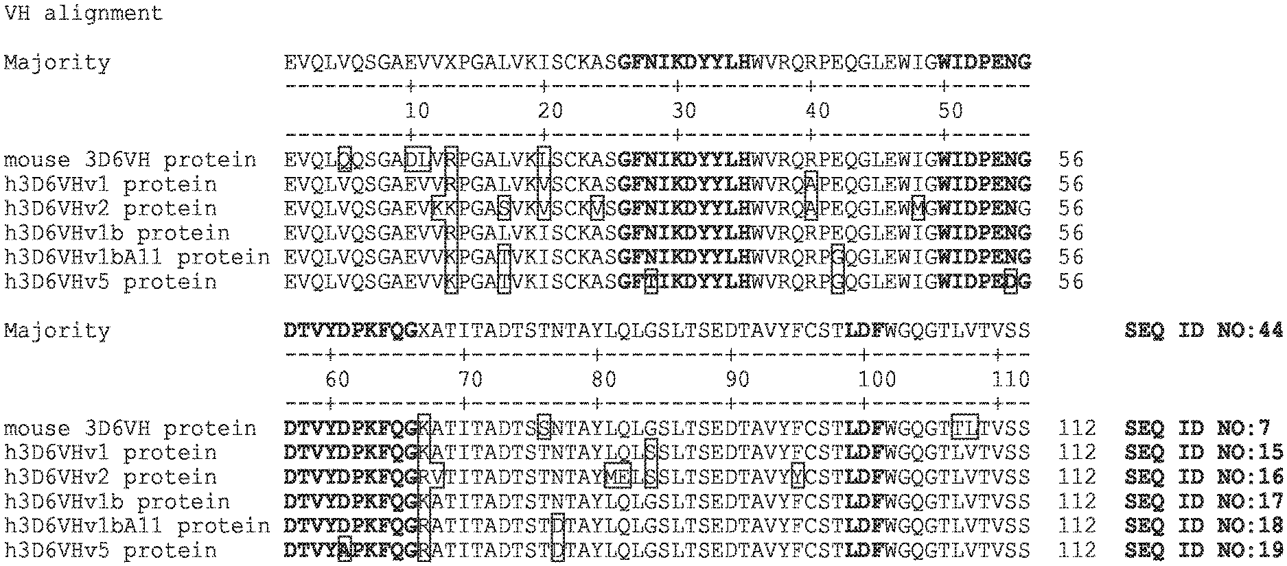
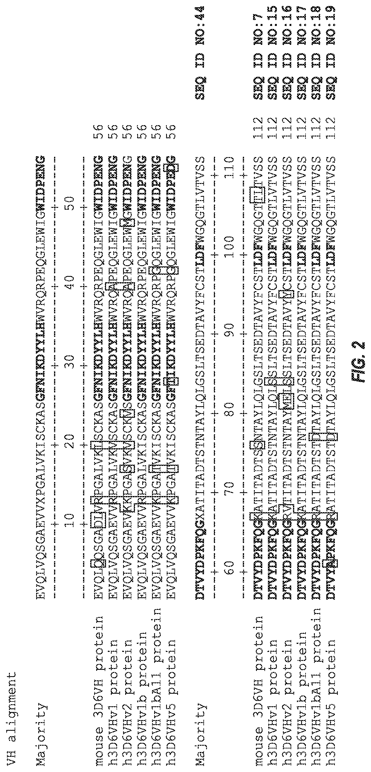
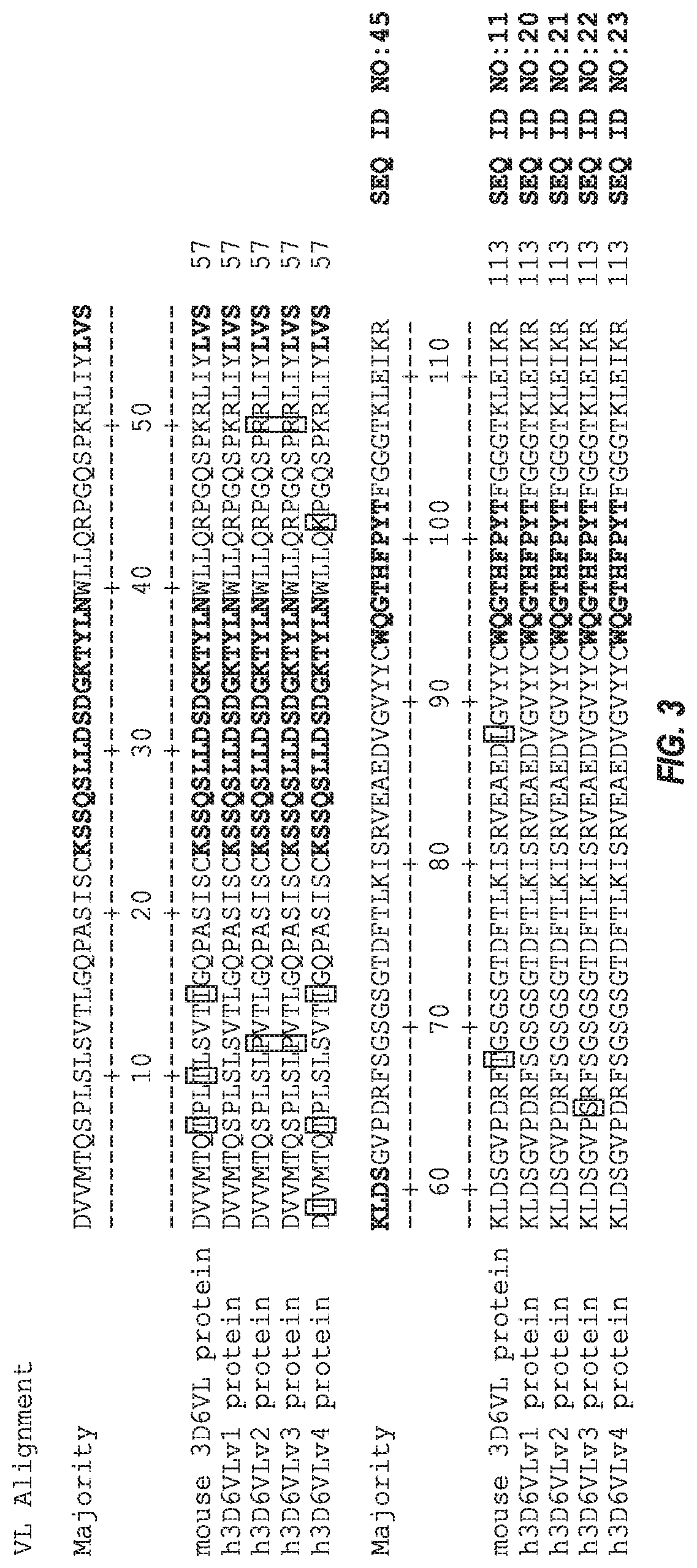
Some antibodies comprise three light chain CDRs as and three heavy chain CDRs of monoclonal antibody 3D6, wherein 3D6 is a mouse antibody characterized by a heavy chain variable region having an amino acid sequence comprising SEQ ID NO: 7 and a light chain variable region having an amino acid sequence comprising SEQ ID NO: 11. In some antibodies, the three heavy chain CDRs are as defined by Kabat/Chothia Composite (SEQ ID NOs: 8, 9, and 10) and the three light chain CDRs are as defined by Kabat/Chothia Composite (SEQ ID NOs: 12, 13, and 14).
For example, the antibody can be 3D6 or a chimeric, veneered, or humanized form thereof. In some such antibodies, the variable heavy chain has .gtoreq.85% identity to human sequence. In some such antibodies, the variable light chain has .gtoreq.85% identity to human sequence. In some such antibodies, each of the variable heavy chain and variable light chain has .gtoreq.85% identity to human germline sequence.
In some such antibodies, the mature heavy chain variable region comprises the three heavy chain CDRs are as defined by Kabat/Chothia Composite (SEQ ID NOs: 8, 9, and 10) and the three light chain CDRs are as defined by Kabat/Chothia Composite (SEQ ID NOs: 12, 13, and 14); provided that position H27 is occupied by F or Y, H28 is occupied by N or T, H29 is occupied by I or F, H30 is occupied by K or T, position H51 is occupied by I or V, position H54 is occupied by N or D, position H60 is occupied by D or A, H61 is occupied by P or E, and H102 is occupied by F or Y. In some such antibodies, CDR-H1 has an amino acid sequence comprising SEQ ID NO: 42. In some such antibodies, CDR-H1 has an amino acid sequence comprising SEQ ID NO: 58. In some such antibodies, CDR-H1 has an amino acid sequence comprising SEQ ID NO: 59. In some such antibodies, CDR-H1 has an amino acid sequence comprising SEQ ID NO: 60. In some such antibodies, CDR-H2 has an amino acid sequence comprising SEQ ID NO: 43. In some such antibodies, CDR-H2 has an amino acid sequence comprising SEQ ID NO: 61. In some such antibodies, CDR-H2 has an amino acid sequence comprising SEQ ID NO: 62. In some such antibodies, CDR-H2 has an amino acid sequence comprising SEQ ID NO: 63. In some such antibodies, CDR-H2 has an amino acid sequence comprising SEQ ID NO: 64. In some such antibodies, CDR-H3 has an amino acid sequence comprising SEQ ID NO: 65.
In some such antibodies, the antibody is a humanized antibody. In some such antibodies, CDR-H1 has an amino acid sequence comprising SEQ ID NO: 42 and CDR-H2 has an amino acid sequence comprising SEQ ID NO: 43. In some such antibodies, CDR-H1 has an amino acid sequence comprising SEQ ID NO: 42 and CDR-H2 has an amino acid sequence comprising SEQ ID NO:61. In some such antibodies, CDR-H1 has an amino acid sequence comprising SEQ ID NO: 42 and CDR-H2 has an amino acid sequence comprising SEQ ID NO:64. In some such antibodies, CDR-H1 has an amino acid sequence comprising SEQ ID NO: 42, CDR-H2 has an amino acid sequence comprising SEQ ID NO:63, and CDR-H3 has an amino acid sequence comprising SEQ ID NO:65. In some such antibodies, CDR-H1 has an amino acid sequence comprising SEQ ID NO: 58 and CDR-H2 has an amino acid sequence comprising SEQ ID NO:62. In some such antibodies, CDR-H1 has an amino acid sequence comprising SEQ ID NO: 59 and CDR-H2 has an amino acid sequence comprising SEQ ID NO:63. In some such antibodies, CDR-H1 has an amino acid sequence comprising SEQ ID NO: 60 and CDR-H2 has an amino acid sequence comprising SEQ ID NO:62.
Some antibodies are a humanized or chimeric 3D6 antibody that specifically binds to human tau, wherein 3D6 is a mouse antibody characterized by a mature heavy chain variable region of SEQ ID NO:7 and a mature light chain variable region of SEQ ID NO: 11. Some such antibodies are a humanized antibody comprising a humanized mature heavy chain variable region comprising the three heavy chain CDRs of 3D6 and a humanized mature light chain variable region comprising the three light chain CDRs of 3D6. In some such antibodies, the CDRs are of a definition selected from the group of Kabat, Chothia, Kabat/Chothia Composite, AbM and Contact.
In some such antibodies the humanized mature heavy chain variable region comprises the three Kabat/Chothia Composite heavy chain CDRs of 3D6 (SEQ ID NOs: 8-10) and the humanized mature light chain variable region comprises the three Kabat/Chothia Composite light chain CDRs of 3D6 (SEQ ID NOs: 12-14).
In some such antibodies, the humanized mature heavy chain variable region comprises the three Kabat heavy chain CDRs of 3D6 (SEQ ID NO:32, SEQ ID NO:9, and SEQ ID NO:10) and the humanized mature light chain variable region comprises the three Kabat light chain CDRs of 3D6 (SEQ ID NOs: 12-14).
In some such antibodies, the humanized mature heavy chain variable region comprises the three Chothia heavy chain CDRs of 3D6 (SEQ ID NO:33, SEQ ID NO:34, and SEQ ID NO:10) and the humanized mature light chain variable region comprises the three Chothia light chain CDRs of 3D6 (SEQ ID NOs: 12-14).
In some such antibodies, the humanized mature heavy chain variable region comprises the three AbM heavy chain CDRs of 3D6 (SEQ ID NO:8, SEQ ID NO:35, and SEQ ID NO:10)) and the humanized mature light chain variable region comprises the three AbM light chain CDRs of 3D6 (SEQ ID NOs: 12-14).
In some such antibodies, the humanized mature heavy chain variable region comprises the three Contact heavy chain CDRs of 3D6 (SEQ ID NOs:39-41) and the humanized mature light chain variable region comprises the three Contact light chain CDRs of 3D6 (SEQ ID NOs:36-38).
In some antibodies, the humanized mature heavy chain variable region has an amino acid sequence at least 90% identical to any one of SEQ ID NOs:15-19 and SEQ ID NOs:46-57 and a humanized mature light chain variable region having an amino acid sequence at least 90% identical to any one of SEQ ID NOs: 20-23, except that position H17 can be T or S, and position H2O can be I or V.
In some such antibodies, at least one of the following positions in the VH region is occupied by the amino acid as specified: H38 is occupied by R and H93 is occupied by S. In some such antibodies, positions H38 and H93 in the VH region are occupied by R and S, respectively.
In some such antibodies, at least one of the following positions in the VH region is occupied by the amino acid as specified: H38 is occupied by R, H43 is occupied by Q, H83 is occupied by T, and H93 is occupied by S. In some such antibodies, positions H38, H43, H83, and H93 in the VH region are occupied by R, Q, T, and S, respectively.
In some antibodies, at least one of the following positions in the VH region is occupied by the amino acid as specified: H12 is occupied by V, H24 is occupied by A, H48 is occupied by I, H67 is occupied by A, H80 is occupied by L, H81 is occupied by Q, and H91 is occupied by F. In some antibodies, positions H12, H24, H48, H67, H80, H81, and H91 in the VH region are occupied by V, A, I, A, L, Q, and F, respectively.
In some antibodies at least one of the following positions in the VH region is occupied by the amino acid as specified: H13 is occupied by R and H66 is occupied by K. In some antibodies, positions H13 and H66 in the VH region are occupied by R and K, respectively.
In some antibodies, at least one of the following positions in the VH region is occupied by the amino acid as specified: H40 is occupied by R and H82a is occupied by G. In some antibodies, positions H40 and H82a in the VH region are occupied by R and G, respectively.
In some antibodies, at least one of the following positions in the VH region is occupied by the amino acid as specified: H42 is occupied by E and H76 is occupied by N. In some antibodies, positions H42 and H76 in the VH region are occupied by E and N, respectively.
In some antibodies, at least one of the following positions in the VH region is occupied by the amino acid as specified: H40 is occupied by R, H82a is occupied by G, and H83 is occupied by T. In some antibodies, positions H40, H82a, and H83 in the VH region are occupied by R, G, and T, respectively.
In some antibodies, position H12 in the VH region is occupied by V.
In some antibodies, position H80 in the VH region is occupied by L.
In some antibodies, at least one of the following positions in the VH region is occupied by the amino acid as specified: H24 is occupied by A, H48 is occupied by I, H67 is occupied by A, H80 is occupied by L, and H91 is occupied by F. In some antibodies, positions H24, H48, H67, H80, and H91 in the VH region are occupied by A, I, A, L, and F, respectively.
In some antibodies, at least one of the following positions in the VH region is occupied by the amino acid as specified: H43 is occupied by Q, and H81 is occupied by Q. In some antibodies, positions H43 and H81 in the VH region are occupied by Q, and Q, respectively.
In some antibodies, at least one of the following positions in the VH region is occupied by the amino acid as specified: H24 is occupied by A, and H91 is occupied by F. In some antibodies, positions H24 and H91 in the VH region are occupied by A and F, respectively.
In some antibodies, at least one of the following positions in the VH region is occupied by the amino acid as specified: H13 is occupied by R, H17 is occupied by L, H29 is occupied by F, H42 is occupied by E, H43 is occupied by Q, H61 is occupied by E, H76 is occupied by N, H80 is occupied by L, H81 is occupied by Q. In some antibodies, positions H13, H17, H29, H42, H43, H61, H76, H80, and H81 in the VH region are occupied by R, L, F, E, Q, E, N, L, and Q, respectively.
In some antibodies, at least one of the following positions in the VH region is occupied by the amino acid as specified: H24 is occupied by A, H28 is occupied by T, H48 is occupied by I, H54 is occupied by D, H60 is occupied by A, H67 is occupied by A, H80 is occupied by L, and H91 is occupied by F. In some antibodies, positions H24, H28, H48, H54, H60, H67, H80, and H91 in the VH region are occupied by A, T, I, D, A, A, L, and F, respectively.
In some antibodies, at least one of the following positions in the VH region is occupied by the amino acid as specified:: H10 is occupied by D, H17 is occupied by L, H24 is occupied by A, H28 is occupied by T, H43 is occupied by Q, H48 is occupied by I, H60 is occupied by A, H61 is occupied by E, H91 is occupied by F, H108 is occupied by T, and H109 is occupied by L. In some antibodies, positions H10, H17, H24, H28, H43, H48, H60, H61, H91, H108, and H109 in the VH region are occupied by D, L, A, T, Q, I, A, E, F, T, and L, respectively.
In some antibodies, at least one of the following positions in the VH region is occupied by the amino acid as specified: H17 is occupied by L, H27 is occupied by Y, H29 is occupied by F, and H61 is occupied by E. In some antibodies, positions H17, H27, H29, and H61 in the VH region are occupied by L, Y, F, and E, respectively.
In some antibodies, at least one of the following positions in the VH region is occupied by the amino acid as specified: H17 is occupied by L, H27 is occupied by Y, H29 is occupied by F, H61 is occupied by E, H76 is occupied by N, and H82a is occupied by G. In some antibodies, positions H17, H27, H29, H61, H76, and H82a in the VH region are occupied by L, Y, F, E, N, and G, respectively.
In some antibodies, at least one of the following positions in the VH region is occupied by the amino acid as specified: H12 is occupied by V, H17 is occupied by L, H24 is occupied by A, H43 is occupied by Q, H48 is occupied by I, H83 is occupied by T, and H91 is occupied by F. In some antibodies, positions H12, H17, H24, H43, H48, H83, and H91 in the VH region are occupied by V, L, A, Q, I, T, F, respectively.
In some antibodies, at least one of the following positions in the VH region is occupied by the amino acid as specified: H12 is occupied by V, H24 is occupied by A, H48 is occupied by I, H67 is occupied b A, H80 is occupied by L, H83 is occupied by T, and H91 is occupied by F. In some antibodies, positions H12, H24, H48, H67, H80, H83, and H91 in the VH region are occupied by V, A, I, A, L, T, and F, respectively.
In some antibodies, at least one of the following positions in the VH region is occupied by the amino acid as specified: H10 is occupied by E or D, H12 is occupied by K or V, H13 is occupied by K or R, H17 is occupied by T, L or S, H24 is occupied by V or A, H27 is occupied by F or Y, H28 is occupied by N or T, H29 is occupied by I or F, H30 is occupied by K or T, H38 is occupied by Q or R, H40 is occupied by A or R, H42 is occupied by G or E, H43 is occupied by K or Q, H48 is occupied by M or I, H51 is occupied by V or I, H54 is occupied by N or D, H60 is occupied by D or A, H61 is occupied by P or E, H66 is occupied by R or K, H67 is occupied by V or A, H76 is occupied by D or N, H80 is occupied by M or L, H81 is occupied by E or Q, H82a is occupied by S or G, H83 is occupied by T or R, H91 is occupied by Y or F, H93 is occupied by A or S, H102 is occupied by F or Y, H108 is occupied by T or L, H109 is occupied by L or V. In some antibodies, positions H12, H13, H17, H24, H38, H42, H43, H48, H66, H67, H76, H80, H81, H83, H91, and H93 in the VH region are occupied by V, R, L, A, R, E, Q, I, K, A, N, L, Q, T, F, and S, respectively.
In some antibodies, positions H38, H42, H43, H76, H83, and H93 in the VH region are occupied by R, E, Q, N, T, and S, respectively. In some antibodies, positions H12, H13, H17, H24, H38, H40, H42, H43, H48, H66, H67, H76, H80, H81, H82A, H83, H91, and H93 in the VH region are occupied by V, R, L, A, R, R, E, Q, I, K, A, N, L, Q, G, T, F, and S, respectively. In some antibodies, positions H12, H24, H38, H40, H43, H48, H67, H80, H81, H82A, H83, H91, and H93 in the VH region are occupied by V, A, R, R, Q, I, A, L, Q, G, T, F, and S, respectively. In some antibodies, positions H12, H24, H28, H38, H40, H43, H48, H54, H60, H67, H80, H81, H82A, H83, H91, and H93 in the VH region are occupied by V, A, T, R, R, Q, I, D, A, A, L, Q, G, T, F, and S, respectively.
In some antibodies, positions H12, H24, H28, H38, H40, H48, H51, H54, H60, H67, H80, H82A, H83, H91, and H93 in the VH region are occupied by V, A, T, R, R, I, V, D, A, A, L, G, T, F, and S, respectively. In some antibodies, positions H12, H24, H28, H38, H40, H48, H54, H60, H67, H80, H82A, H83, H91, and H93 in the VH region are occupied by V, A, T, R, R, I, D, A, A, L, G, T, F, and S, respectively. In some antibodies, positions H13, H17, H24, H29, H38, H40, H42, H43, H54, H61, H76, H80, H81, H82A, H83, H91, and H93 in the VH region are occupied by R, L, A, F, R, R, E, Q, N, E, N, L, Q, G, T, F, and S, respectively.
In some antibodies, positions H13, H17, H24, H27, H28, H29, H30, H38, H40, H42, H43, H51, H54, H60, H61, H76, H80, H81, H82A, H83, H91, and H93 in the VH region are occupied by R, L, A, Y, T, F, T, R, R, E, Q, V, D, A, E, N, L, Q, G, T, F, and S, respectively. In some antibodies, positions H10, H12, H17, H24, H28, H38, H40, H42, H43, H48, H54, H60, H61, H76, H80, H82A, H83, H91, H93, H108, and H109 in the VH region are occupied by D, V, L, A, T, R, R, E, Q, I, N, A, E, N, L, G, T, F, S, T, and L, respectively. In some antibodies, positions H10, H12, H17, H24, H28, H38, H40, H43, H48, H51, H54, H60, H61, H82A, H83, H91, H93, H102, H108, and H109 in the VH region are occupied by D, V, L, A, T, R, R, Q, I, V, D, A, E, G, T, F, S, Y, T, and L, respectively.
In some antibodies, positions H38 and H93 in the VH region are occupied by R and S, respectively. In some antibodies, positions H17, H27, H29, H38, H61, H76, H82A, and H93 in the VH region are occupied by L, Y, F, R, E, N, G, and S, respectively. In some antibodies, positions H17, H27, H28, H29, H30, H38, H51, H54, H60, H61, H76, H82A, and H93 in the VH region are occupied by L, Y, T, F, T, R, V, D, A, E, N, G, and S, respectively.
In some antibodies, positions H12, H38, H40, H48, H66, H67, H76, H80, H82A, H83, and H93 in the VH region are occupied by V, R, R, I, K, A, N, L, G, T, and S, respectively.
In some antibodies, positions H12, H17, H27, H29, H38, H40, H61, H80, H82A, H83, and H93 in the VH region are occupied by V, L, Y, F, R, R, E, L, G, T, and S, respectively. In some antibodies, positions H12, H17, H27, H28, H29, H30, H38, H40, H51, H54, H60, H61, H80, H82A, H83, and H93 in the VH region are occupied by V, L, Y, T, F, T, R, R, V, D, A, E, L, G, T, and S, respectively.
In some antibodies, at least one of the following positions in the VL region is occupied by the amino acid as specified: L36 is occupied by L, L37 is occupied by L, and L100 is occupied by G. In some antibodies, positions L36, L37, and L100 in the VL region are occupied by L, L, and G, respectively.
In some antibodies, at least one of the following positions in the VL region is occupied by the amino acid as specified: L12 is occupied by S and L45 is occupied by K. In some antibodies, positions L12 and L45 in the VL region are occupied by S and K, respectively.
In some antibodies, at least one of the following positions in the VL region is occupied by the amino acid as specified: L2 is V or I, L7 is S or T, L12 is P or S, L15 is L or I, L36 is L, L37 is L, L45 is R or K, L60 is D or S, L100 is G.
In some antibodies, positions L12, L36, L37, L45, and L100 in the VL region are occupied by S, L, L, K, and G, respectively. In some antibodies, positions L36, L37, and L100 in the VL region are occupied by L, L and G, respectively. In some antibodies, positions L36, L37, L60, and L100 in the VL region are occupied by L, L, S, and G, respectively. In some antibodies, positions L2, L7, L12, L15, L36, L37, L45, and L100 in the VL region are occupied by I, T, S, I, L, L, K, and G, respectively. In some antibodies, positions L2, L7, L12, L15, L36, L37, L45, and L100 in the VL region are occupied by V, S, P, L, L, L, R, and G, respectively.
Some antibodies comprise a mature heavy chain variable region having an amino acid sequence at least 95% identical to any one of SEQ ID NOs: 15-19, 46-57 and a mature light chain variable region having an amino acid sequence at least 95% identical to any one of SEQ ID NOs: 20-23, except that position H17 can be T or S, and position H.sub.2O can be I or V. Some antibodies comprise a mature heavy chain variable region having an amino acid sequence at least 98% identical to any one of SEQ ID NOs: 15-19, 46-57 and a mature light chain variable region having an amino acid sequence at least 98% identical to any one of SEQ ID NOs: 20-23, except that position H17 can be T or S, and position H.sub.2O can be I or V.
In some antibodies, the mature heavy chain variable region has an amino acid sequence of any one of SEQ ID NOs:15-19 and SEQ ID NOs:46-57 and the mature light chain variable region has an amino acid sequence of any one of SEQ ID NOs:20-23. In some antibodies, the mature heavy chain variable region has an amino acid sequence of SEQ ID NO:15 and the mature light chain variable region has an amino acid sequence of SEQ ID NO:20. In some antibodies, the mature heavy chain variable region has an amino acid sequence of SEQ ID NO:15 and the mature light chain variable region has an amino acid sequence of SEQ ID NO:21. In some antibodies, the mature heavy chain variable region has an amino acid sequence of SEQ ID NO:15 and the mature light chain variable region has an amino acid sequence of SEQ ID NO:22. In some antibodies, the mature heavy chain variable region has an amino acid sequence of SEQ ID NO:15 and the mature light chain variable region has an amino acid sequence of SEQ ID NO:23.
In some antibodies, the mature heavy chain variable region has an amino acid sequence of SEQ ID NO:16 and the mature light chain variable region has an amino acid sequence of SEQ ID NO:20. In some antibodies, the mature heavy chain variable region has an amino acid sequence of SEQ ID NO:16 and the mature light chain variable region has an amino acid sequence of SEQ ID NO:21. In some antibodies, the mature heavy chain variable region has an amino acid sequence of SEQ ID NO:16 and the mature light chain variable region has an amino acid sequence of SEQ ID NO:22. In some antibodies, the mature heavy chain variable region has an amino acid sequence of SEQ ID NO:16 and the mature light chain variable region has an amino acid sequence of SEQ ID NO:23.
In some antibodies, the mature heavy chain variable region has an amino acid sequence of SEQ ID NO:17 and the mature light chain variable region has an amino acid sequence of SEQ ID NO:20. In some antibodies, the mature heavy chain variable region has an amino acid sequence of SEQ ID NO:17 and the mature light chain variable region has an amino acid sequence of SEQ ID NO:21. In some antibodies, the mature heavy chain variable region has an amino acid sequence of SEQ ID NO:17 and the mature light chain variable region has an amino acid sequence of SEQ ID NO:22. In some antibodies, the mature heavy chain variable region has an amino acid sequence of SEQ ID NO:17 and the mature light chain variable region has an amino acid sequence of SEQ ID NO:23.
In some antibodies, the mature heavy chain variable region has an amino acid sequence of SEQ ID NO:18 and the mature light chain variable region has an amino acid sequence of SEQ ID NO:20. In some antibodies, the mature heavy chain variable region has an amino acid sequence of SEQ ID NO:18 and the mature light chain variable region has an amino acid sequence of SEQ ID NO:21. In some antibodies, the mature heavy chain variable region has an amino acid sequence of SEQ ID NO:18 the mature light chain variable region has an amino acid sequence of SEQ ID NO:22. In some antibodies, the mature heavy chain variable region has an amino acid sequence of SEQ ID NO:18 and the mature light chain variable region has an amino acid sequence of SEQ ID NO:23.
In some antibodies, the mature heavy chain variable region has an amino acid sequence of SEQ ID NO:19 and the mature light chain variable region has an amino acid sequence of SEQ ID NO:20. In some antibodies, the mature heavy chain variable region has an amino acid sequence of SEQ ID NO:19 and the mature light chain variable region has an amino acid sequence of SEQ ID NO:21. In some antibodies, the mature heavy chain variable region has an amino acid sequence of SEQ ID NO:19 and the mature light chain variable region has an amino acid sequence of SEQ ID NO:22. In some antibodies, the mature heavy chain variable region has an amino acid sequence of SEQ ID NO:19 and the mature light chain variable region has an amino acid sequence of SEQ ID NO:23.
In some antibodies, the mature heavy chain variable region has an amino acid sequence of SEQ ID NO:46 and the mature light chain variable region has an amino acid sequence of SEQ ID NO:20. In some antibodies, the mature heavy chain variable region has an amino acid sequence of SEQ ID NO:46 and the mature light chain variable region has an amino acid sequence of SEQ ID NO:21. In some antibodies, the mature heavy chain variable region has an amino acid sequence of SEQ ID NO:46 and the mature light chain variable region has an amino acid sequence of SEQ ID NO:22. In some antibodies, the mature heavy chain variable region has an amino acid sequence of SEQ ID NO:46 and the mature light chain variable region has an amino acid sequence of SEQ ID NO:23.
In some antibodies, the mature heavy chain variable region has an amino acid sequence of SEQ ID NO:47 and the mature light chain variable region has an amino acid sequence of SEQ ID NO:20. In some antibodies, the mature heavy chain variable region has an amino acid sequence of SEQ ID NO:47 and the mature light chain variable region has an amino acid sequence of SEQ ID NO:21. In some antibodies, the mature heavy chain variable region has an amino acid sequence of SEQ ID NO:47 and the mature light chain variable region has an amino acid sequence of SEQ ID NO:22. In some antibodies, the mature heavy chain variable region has an amino acid sequence of SEQ ID NO:47 and the mature light chain variable region has an amino acid sequence of SEQ ID NO:23.
In some antibodies, the mature heavy chain variable region has an amino acid sequence of SEQ ID NO:48 and the mature light chain variable region has an amino acid sequence of SEQ ID NO:20. In some antibodies, the mature heavy chain variable region has an amino acid sequence of SEQ ID NO:48 and the mature light chain variable region has an amino acid sequence of SEQ ID NO:21. In some antibodies, the mature heavy chain variable region has an amino acid sequence of SEQ ID NO:48 and the mature light chain variable region has an amino acid sequence of SEQ ID NO:22. In some antibodies, the mature heavy chain variable region has an amino acid sequence of SEQ ID NO:48 and the mature light chain variable region has an amino acid sequence of SEQ ID NO:23.
In some antibodies, the mature heavy chain variable region has an amino acid sequence of SEQ ID NO:49 and the mature light chain variable region has an amino acid sequence of SEQ ID NO:20. In some antibodies, the mature heavy chain variable region has an amino acid sequence of SEQ ID NO:49 and the mature light chain variable region has an amino acid sequence of SEQ ID NO:21. In some antibodies, the mature heavy chain variable region has an amino acid sequence of SEQ ID NO:49 and the mature light chain variable region has an amino acid sequence of SEQ ID NO:22. In some antibodies, the mature heavy chain variable region has an amino acid sequence of SEQ ID NO:49 and the mature light chain variable region has an amino acid sequence of SEQ ID NO:23.
In some antibodies, the mature heavy chain variable region has an amino acid sequence of SEQ ID NO:50 and the mature light chain variable region has an amino acid sequence of SEQ ID NO:20. In some antibodies, the mature heavy chain variable region has an amino acid sequence of SEQ ID NO:50 and the mature light chain variable region has an amino acid sequence of SEQ ID NO:21. In some antibodies, the mature heavy chain variable region has an amino acid sequence of SEQ ID NO:50 and the mature light chain variable region has an amino acid sequence of SEQ ID NO:22. In some antibodies, the mature heavy chain variable region has an amino acid sequence of SEQ ID NO:50 and the mature light chain variable region has an amino acid sequence of SEQ ID NO:23.
In some antibodies, the mature heavy chain variable region has an amino acid sequence of SEQ ID NO:51 and the mature light chain variable region has an amino acid sequence of SEQ ID NO:20. In some antibodies, the mature heavy chain variable region has an amino acid sequence of SEQ ID NO:51 and the mature light chain variable region has an amino acid sequence of SEQ ID NO:21. In some antibodies, the mature heavy chain variable region has an amino acid sequence of SEQ ID NO:51 and the mature light chain variable region has an amino acid sequence of SEQ ID NO:22. In some antibodies, the mature heavy chain variable region has an amino acid sequence of SEQ ID NO:51 and the mature light chain variable region has an amino acid sequence of SEQ ID NO:23.
In some antibodies, the mature heavy chain variable region has an amino acid sequence of SEQ ID NO:52 and the mature light chain variable region has an amino acid sequence of SEQ ID NO:20. In some antibodies, the mature heavy chain variable region has an amino acid sequence of SEQ ID NO:52 and the mature light chain variable region has an amino acid sequence of SEQ ID NO:21. In some antibodies, the mature heavy chain variable region has an amino acid sequence of SEQ ID NO:52 and the mature light chain variable region has an amino acid sequence of SEQ ID NO:22. In some antibodies, the mature heavy chain variable region has an amino acid sequence of SEQ ID NO:52 and the mature light chain variable region has an amino acid sequence of SEQ ID NO:23.
In some antibodies, the mature heavy chain variable region has an amino acid sequence of SEQ ID NO:53 and the mature light chain variable region has an amino acid sequence of SEQ ID NO:20. In some antibodies, the mature heavy chain variable region has an amino acid sequence of SEQ ID NO:53 and the mature light chain variable region has an amino acid sequence of SEQ ID NO:21. In some antibodies, the mature heavy chain variable region has an amino acid sequence of SEQ ID NO:53 and the mature light chain variable region has an amino acid sequence of SEQ ID NO:22. In some antibodies, the mature heavy chain variable region has an amino acid sequence of SEQ ID NO:53 and the mature light chain variable region has an amino acid sequence of SEQ ID NO:23.
In some antibodies, the mature heavy chain variable region has an amino acid sequence of SEQ ID NO:54 and the mature light chain variable region has an amino acid sequence of SEQ ID NO:20. In some antibodies, the mature heavy chain variable region has an amino acid sequence of SEQ ID NO:54 and the mature light chain variable region has an amino acid sequence of SEQ ID NO:21. In some antibodies, the mature heavy chain variable region has an amino acid sequence of SEQ ID NO:54 and the mature light chain variable region has an amino acid sequence of SEQ ID NO:22. In some antibodies, the mature heavy chain variable region has an amino acid sequence of SEQ ID NO:54 and the mature light chain variable region has an amino acid sequence of SEQ ID NO:23.
In some antibodies, the mature heavy chain variable region has an amino acid sequence of SEQ ID NO:55 and the mature light chain variable region has an amino acid sequence of SEQ ID NO:20. In some antibodies, the mature heavy chain variable region has an amino acid sequence of SEQ ID NO:55 and the mature light chain variable region has an amino acid sequence of SEQ ID NO:21. In some antibodies, the mature heavy chain variable region has an amino acid sequence of SEQ ID NO:55 and the mature light chain variable region has an amino acid sequence of SEQ ID NO:22. In some antibodies, the mature heavy chain variable region has an amino acid sequence of SEQ ID NO:55 and the mature light chain variable region has an amino acid sequence of SEQ ID NO:23.
In some antibodies, the mature heavy chain variable region has an amino acid sequence of SEQ ID NO:56 and the mature light chain variable region has an amino acid sequence of SEQ ID NO:20. In some antibodies, the mature heavy chain variable region has an amino acid sequence of SEQ ID NO:56 and the mature light chain variable region has an amino acid sequence of SEQ ID NO:21. In some antibodies, the mature heavy chain variable region has an amino acid sequence of SEQ ID NO:56 and the mature light chain variable region has an amino acid sequence of SEQ ID NO:22. In some antibodies, the mature heavy chain variable region has an amino acid sequence of SEQ ID NO:56 and the mature light chain variable region has an amino acid sequence of SEQ ID NO:23.
In some antibodies, the mature heavy chain variable region has an amino acid sequence of SEQ ID NO:57 and the mature light chain variable region has an amino acid sequence of SEQ ID NO:20. In some antibodies, the mature heavy chain variable region has an amino acid sequence of SEQ ID NO:57 and the mature light chain variable region has an amino acid sequence of SEQ ID NO:21. In some antibodies, the mature heavy chain variable region has an amino acid sequence of SEQ ID NO:57 and the mature light chain variable region has an amino acid sequence of SEQ ID NO:22. In some antibodies, the mature heavy chain variable region has an amino acid sequence of SEQ ID NO:57 and the mature light chain variable region has an amino acid sequence of SEQ ID NO:23.
For example, the antibody can be a chimeric antibody. For example, the antibody can be a veneered antibody.
The antibody can be an intact mouse, chimeric, veneered or humanized antibody or a binding fragment, single-chain antibody Fab fragment, Fab'2 fragment, or single chain Fv. Some of the antibodies have a human IgG1 isotype, while others may have a human IgG2 or IgG4 isotype. Some antibodies have the mature light chain variable region fused to a light chain constant region and the mature heavy chain variable region fused to a heavy chain constant region. The heavy chain constant region of some antibodies is a mutant form of a natural human heavy chain constant region which has reduced binding to a Fc.gamma. receptor relative to the natural human heavy chain constant region.
Some antibodies may have at least one mutation in the constant region, such as a mutation that reduces complement fixation or activation by the constant region, for example, a mutation at one or more of positions 241, 264, 265, 270, 296, 297, 318, 320, 322, 329 and 331 by EU numbering. Some antibodies have an alanine at positions 318, 320 and 322. Some antibodies can be at least 95% w/w pure. The antibody can be conjugated to a therapeutic, cytotoxic, cytostatic, neurotrophic, or neuroprotective agent.
In another aspect, the invention provides a pharmaceutical composition comprising any of the antibodies disclosed herein and a pharmaceutically-acceptable carrier.
In another aspect, the invention provides a nucleic acid encoding the heavy chain and/or light chain of any of the antibodies disclosed herein, a recombinant expression vector comprising the nucleic acid and a host cell transformed with the recombinant expression vector.
In yet another aspect, the invention provides methods of humanizing any non-human antibody described herein, for example, mouse antibody 3D6, wherein 3D6 is characterized by a mature heavy chain variable region of SEQ ID NO:7 and a mature light chain variable region of SEQ ID NO:11. Such methods can involve selecting one or more acceptor antibodies, synthesizing a nucleic acid encoding a humanized heavy chain comprising CDRs of the mouse heavy chain and a nucleic acid encoding a humanized light chain comprising CDRs of the mouse antibody light chain, and expressing the nucleic acids in a host cell to produce a humanized antibody.
Methods of producing antibodies, such as a humanized, chimeric or veneered antibody, for example humanized, chimeric or veneered forms of 3D6, are also provided. In such methods, cells transformed with nucleic acids encoding the heavy and light chains of the antibody are cultured so that the cells secrete the antibody. The antibody can then be purified from the cell culture media.
Cell lines producing any of the antibodies disclosed herein can be produced by introducing a vector encoding heavy and light chains of the antibody and a selectable marker into cells, propagating the cells under conditions to select for cells having increased copy number of the vector, isolating single cells from the selected cells; and banking cells cloned from a single cell selected based on yield of antibody.
Some cells can be propagated under selective conditions and screened for cell lines naturally expressing and secreting at least 100 mg/L/10.sup.6 cells/24 hours. Single cells can be isolated from the selected cells. Cells cloned from a single cell can then be banked Single cells can be selected based on desirable properties, such as the yield of the antibody. Exemplary cell lines are cell lines expressing 3D6.
The invention also provides methods of inhibiting or reducing aggregation of tau in a subject having or at risk of developing a tau-mediated amyloidosis, comprising administering to the subject an effective regime of an antibody disclosed herein, thereby inhibiting or reducing aggregation of tau in the subject. Exemplary antibodies include humanized versions of 3D6.
Also provided are methods of treating or effecting prophylaxis of a tau-related disease in a subject, comprising administering an effective regime of an antibody disclosed herein and thereby treating or effecting prophylaxis of the disease. Examples of such a disease are Alzheimer's disease, Down's syndrome, mild cognitive impairment, primary age-related tauopathy, postencephalitic parkinsonism, posttraumatic dementia or dementia pugilistica, Pick's disease, type C Niemann-Pick disease, supranuclear palsy, frontotemporal dementia, frontotemporal lobar degeneration, argyrophilic grain disease, globular glial tauopathy, amyotrophic lateral sclerosis/parkinsonism dementia complex of Guam, corticobasal degeneration (CBD), dementia with Lewy bodies, Lewy body variant of Alzheimer disease (LBVAD), or progressive supranuclear palsy (PSP). In some methods, the tau-related disease is Alzheimer's disease. In some methods the patient is an ApoE4 carrier.
Also provided are methods of reducing aberrant transmission of tau comprising administering an effective regime of an antibody disclosed herein and thereby reducing transmission of tau.
Also provided are methods of inducing phagocytosis of tau comprising administering an effective regime of an antibody disclosed herein and thereby inducing phagocytosis of tau.
Also provided are methods of inhibiting tau aggregation or deposition comprising administering an effective regime of an antibody disclosed herein thereby inhibiting tau aggregation or deposition.
Also provided are methods of inhibiting formation of tau tangles comprising administering an effective regime of an antibody disclosed herein.
The invention also provides a method of detecting tau protein deposits in a subject having or at risk of a disease associated with tau aggregation or deposition comprising administering to a subject an antibody disclosed herein, and detecting the antibody bound to tau in the subject. Examples of such a disease are Alzheimer's disease, Down's syndrome, mild cognitive impairment, primary age-related tauopathy, postencephalitic parkinsonism, posttraumatic dementia or dementia pugilistica, Pick's disease, type C Niemann-Pick disease, supranuclear palsy, frontotemporal dementia, frontotemporal lobar degeneration, argyrophilic grain disease, globular glial tauopathy, amyotrophic lateral sclerosis/parkinsonism dementia complex of Guam, corticobasal degeneration (CBD), dementia with Lewy bodies, Lewy body variant of Alzheimer disease (LBVAD), or progressive supranuclear palsy (PSP). In some embodiments, the antibody is administered by intravenous injection into the body of the subject. In some embodiments, the antibody is administered directly to the brain of the subject by intracranial injection or by drilling a hole through the skull of the subject. In some embodiments, the antibody is labeled. In some embodiments, the antibody is labeled with a fluorescent label, a paramagnetic label, or a radioactive label. In some embodiments, the radioactive label is detected using positron emission tomography (PET) or single-photon emission computed tomography (SPECT).
The invention also provides a method of measuring efficacy of treatment in a subject being treated for a disease associated with tau aggregation or deposition, comprising measuring a first level of tau protein deposits in the subject prior to treatment by administering to a subject an antibody disclosed herein, and detecting a first amount of the antibody bound to tau in the subject, administering the treatment to the subject, measuring a second level of tau protein deposits in the in subject after treatment by administering to a subject the antibody, and detecting the antibody bound to tau in the subject, wherein a decrease in the level of tau protein deposits indicates a positive response to treatment.
The invention also provides a method of measuring efficacy of treatment in a subject being treated for a disease associated with tau aggregation or deposition, comprising measuring a first level of tau protein deposits in the subject prior to treatment by administering to a subject an antibody disclosed herein, and detecting a first amount of antibody bound to tau in the subject, administering the treatment to the subject, measuring a second level of tau protein deposits in the in subject after treatment by administering to a subject the antibody, and detecting a second amount of antibody bound to tau in the subject, wherein no change in the level of tau protein deposits or a small increase in tau protein deposits indicates a positive response to treatment.
BRIEF DESCRIPTION OF THE DRAWINGS
FIG. 1 depicts the results of experiments designed to map the epitope(s) bound by the murine 3D6 monoclonal antibody.
FIG. 2 depicts an alignment of heavy chain variable regions of the mouse 3D6 antibody and humanized versions of the 3D6 antibody (VHv1, VHv2, VHv1b, VHv1bA11, and VHv5). The consensus amino acid sequence among the heavy chain variable regions of the mouse 3D6 antibody and humanized versions of the 3D6 antibody (VHv1, VHv2, VHv1b, VHv1bA11, and VHv5) is labeled 37 Majority.` The CDRs as defined by Kabat/Chothia Composite are in boldface. Positions in heavy chain variable regions of the mouse 3D6 antibody and humanized versions of the 3D6 antibody (VHv1, VHv2, VHv1b, VHv1 bA11, and VHv5) where amino acid residues differ from the "Majority" sequence are boxed.
FIG. 3 depicts an alignment of light chain variable regions of the mouse 3D6 antibody and humanized versions of the 3D6 antibody. The consensus amino acid sequence between the light chain variable regions of the mouse 3D6 antibody and selected humanized 3D6 antibodies is labeled "Majority.` The CDRs as defined by Kabat are in boldface. Positions in light chain variable regions of the mouse 3D6 antibody and humanized versions of the 3D6 antibody where amino acid residues differ from the "Majority" sequence are boxed.
FIGS. 4A and 4B depict an alignment of heavy chain variable regions of the mouse 3D6 antibody and humanized versions of the 3D6 antibody (VHv1, VHv1b, VHv1bA11, VHv1bA11B6G2, VHv1bA11B6H3, VHv1c, VHv1d, VHv1e, VHv1f, VHv2, VHv3, VHv3b, VHv3c, VHv4, VHv4b, VHv4c, and VHv5). The consensus amino acid sequence among the heavy chain variable regions of the humanized versions of the 3D6 antibody (VHv1, VHv1b, VHv1bA11, VHv1bA11B6G2, VHv1bA11B6H3, VHv1c, VHv1d, VHv1e, VHv1f, VHv2, VHv3, VHv3b, VHv3c, VHv4, VHv4b, VHv4c, and VHv5) is labeled "Majority.` The CDRs as defined by Kabat/Chothia Composite are in boldface. Positions in heavy chain variable regions of the humanized versions of the 3D6 antibody (VHv1, VHv1b, VHv1bA11, VHv1bA11B6G2, VHv1bA11B6H3, VHv1c, VHv1d, VHv1e, VHv1f, VHv2, VHv3, VHv3b, VHv3c, VHv4, VHv4b, VHv4c, and VHv5) where amino acid residues differ from the "Majority" sequence are boxed.
FIGS. 5A, 5B, and 5C depict results of ELISA screening assays for selected mouse monoclonal anti-tau antibodies.
FIG. 6 depicts binding kinetics for selected mouse monoclonal anti-tau antibodies to recombinant human tau.
FIG. 7 depicts results of functional blocking assays for selected mouse monoclonal anti-tau antibodies.
FIG. 8 depicts results of disaggregation assays for selected mouse monoclonal anti-tau antibodies.
FIG. 9 depicts results of experiments showing that 3D6 and 5G8 immunocapture tau from human Alzheimer's disease tissue.
FIGS. 10A, 10B, and 10C depict results of experiments showing affinity of humanized 3D6 variants toward tau.
FIGS. 11A-11I show exemplary humanized Vh designs, with backmutations and other mutations based on selected human frameworks. The bolded areas indicate the CDRs as defined by Kabat/Chothia Composite.
FIGS. 12A-12D show exemplary humanized Vk designs, with backmutations and other mutations based on selected human frameworks. The bolded areas indicate the CDRs as defined by Kabat/Chothia Composite.
BRIEF DESCRIPTION OF THE SEQUENCES
SEQ ID NO:1 sets forth the amino acid sequence of an isoform of human tau (Swiss-Prot P10636-8).
SEQ ID NO:2 sets forth the amino acid sequence of an isoform of human tau (Swiss-Prot P10636-7).
SEQ ID NO:3 sets forth the amino acid sequence of an isoform of human tau (Swiss-Prot P10636-6), (4R0N human tau).
SEQ ID NO:4 sets forth the amino acid sequence of an isoform of human tau (Swiss-Prot P10636-5)
SEQ ID NO:5 sets forth the amino acid sequence of an isoform of human tau (Swiss-Prot P10636-4).
SEQ ID NO:6 sets forth the amino acid sequence of an isoform of human tau (Swiss-Prot P10636-2).
SEQ ID NO: 7 sets forth the amino acid sequence of the heavy chain variable region of the mouse 3D6 antibody.
SEQ ID NO: 8 sets forth the amino acid sequence of Kabat/Chothia composite CDR-H1 of the mouse 3D6 antibody.
SEQ ID NO:9 sets forth the amino acid sequence of Kabat CDR-H2 of the mouse 3D6 antibody.
SEQ ID NO: 10 sets forth the amino acid sequence of Kabat CDR-H3 of the mouse 3D6 antibody.
SEQ ID NO: 11 sets forth the amino acid sequence of the light chain variable region of the mouse 3D6 antibody and of the mouse 6A10 antibody.
SEQ ID NO: 12 sets forth the amino acid sequence of Kabat CDR-L1 of the mouse 3D6 antibody and of the mouse 6A10 antibody.
SEQ ID NO: 13 sets forth the amino acid sequence of Kabat CDR-L2 of the mouse 3D6 antibody and of the mouse 6A10 antibody.
SEQ ID NO: 14 sets forth the amino acid sequence of Kabat CDR-L3 of the mouse 3D6 antibody and of the mouse 6A10 antibody.
SEQ ID NO:15 sets forth the amino acid sequence of heavy chain variable region of the humanized 3D6 antibody hu3D6VHv1.
SEQ ID NO:16 sets forth the amino acid sequence of heavy chain variable region of the humanized 3D6 antibody hu3D6VHv2.
SEQ ID NO:17 sets forth the amino acid sequence of heavy chain variable region of the humanized 3D6 antibody hu3D6VHv1b.
SEQ ID NO:18 sets forth the amino acid sequence of heavy chain variable region of the humanized 3D6 antibody hu3D6VHv1bA11.
SEQ ID NO:19 sets forth the amino acid sequence of heavy chain variable region of the humanized 3D6 antibody hu3D6VHv5:
SEQ ID NO:20 sets forth the amino acid sequence of the light chain variable region of the humanized 3D6 antibody hu3D6VLv1.
SEQ ID NO:21 sets forth the amino acid sequence of the light chain variable region of the humanized 3D6 antibody hu3D6VLv2.
SEQ ID NO:22 sets forth the amino acid sequence of the light chain variable region of the humanized 3D6 antibody hu3D6VLv3.
SEQ ID NO:23 sets forth the amino acid sequence of the light chain variable region of the humanized 3D6 antibody hu3D6VLv4.
SEQ ID NO:24 sets forth the amino acid sequence of the heavy chain variable acceptor Acc. #BAC01986.1.
SEQ ID NO:25 sets forth the amino acid sequence of the heavy chain variable acceptor Acc. #IMGT #IGHV1-69-2*01.
SEQ ID NO:26 sets forth the amino acid sequence of the heavy chain variable acceptor Acc. #IMGT #IGKJ1*01.
SEQ ID NO:27 sets forth the amino acid sequence of the light chain variable acceptor Acc. #IMGT #IGKV2-30*02
SEQ ID NO:28 sets forth the amino acid sequence of the light chain variable acceptor Acc. #IMGT #IGKJ2*01.
SEQ ID NO:29 sets forth the amino acid sequence of the light chain variable acceptor Acc. #AAZ09048.1.
SEQ ID NO: 30 sets forth a nucleic acid sequence encoding the heavy chain variable region of the mouse 3D6 antibody.
SEQ ID NO: 31 sets forth a nucleic acid sequence encoding the light chain variable region of the mouse 3D6 antibody.
SEQ ID NO: 32 sets forth the amino acid sequence of Kabat CDR-H1 of the mouse 3D6 antibody.
SEQ ID NO: 33 sets forth the amino acid sequence of Chothia CDR-H1 of the mouse 3D6 antibody.
SEQ ID NO: 34 sets forth the amino acid sequence of Chothia CDR-H2 of the mouse 3D6 antibody.
SEQ ID NO: 35 sets forth the amino acid sequence of AbM CDR-H2 of the mouse 3D6 antibody.
SEQ ID NO: 36 sets forth the amino acid sequence of Contact CDR-L1 of the mouse 3D6 antibody.
SEQ ID NO: 37 sets forth the amino acid sequence of Contact CDR-L2 of the mouse 3D6 antibody.
SEQ ID NO: 38 sets forth the amino acid sequence of Contact CDR-L3 of the mouse 3D6 antibody.
SEQ ID NO: 39 sets forth the amino acid sequence of Contact CDR-H1 of the mouse 3D6 antibody.
SEQ ID NO: 40 sets forth the amino acid sequence of Contact CDR-H2 of the mouse 3D6 antibody.
SEQ ID NO: 41 sets forth the amino acid sequence of Contact CDR-H3 of the mouse 3D6 antibody.
SEQ ID NO: 42 sets forth the amino acid sequence of an alternate Kabat-Chothia Composite CDR-H1 of a humanized 3D6 antibody (derived from hu3D6VHv5, hu3D6VHv1bA11B6G2, hu3D6VHv1bA11B6H3, hu3D6VHv1e, and hu3D6VHv1f).
SEQ ID NO: 43 sets forth the amino acid sequence of an alternate Kabat CDR-H2 of a humanized 3D6 antibody (derived from hu3D6VHv5 and hu3D6VHv1bA11B6H3).
SEQ ID NO:44 sets forth the consensus amino acid sequence among the heavy chain variable regions of the mouse 3D6 and selected humanized 3D6 antibodies (VHv1, VHv2, VHv1b, VHv1bA11, and VHv5) (labeled "Majority` in FIG. 2).
SEQ ID NO:45 sets forth the consensus amino acid sequence between the light chain variable regions of the mouse 3D6 and selected humanized 3D6 antibodies (labeled "Majority` in FIG. 3).
SEQ ID NO:46 sets forth the amino acid sequence of heavy chain variable region of the humanized 3D6 antibody hu3D6VHv1bA11B6G2.
SEQ ID NO:47 sets forth the amino acid sequence of heavy chain variable region of the humanized 3D6 antibody hu3D6VHv1bA11B6H3.
SEQ ID NO:48 sets forth the amino acid sequence of heavy chain variable region of the humanized 3D6 antibody hu3D6VHv1c.
SEQ ID NO:49 sets forth the amino acid sequence of heavy chain variable region of the humanized 3D6 antibody hu3D6VHv1d.
SEQ ID NO:50 sets forth the amino acid sequence of heavy chain variable region of the humanized 3D6 antibody hu3D6VHv1e.
SEQ ID NO:51 sets forth the amino acid sequence of heavy chain variable region of the humanized 3D6 antibody hu3D6VHv1f.
SEQ ID NO:52 sets forth the amino acid sequence of heavy chain variable region of the humanized 3D6 antibody hu3D6VHv3.
SEQ ID NO:53 sets forth the amino acid sequence of heavy chain variable region of the humanized 3D6 antibody hu3D6VHv3b.
SEQ ID NO:54 sets forth the amino acid sequence of heavy chain variable region of the humanized 3D6 antibody hu3D6VHv3c.
SEQ ID NO:55 sets forth the amino acid sequence of heavy chain variable region of the humanized 3D6 antibody hu3D6VHv4.
SEQ ID NO:56 sets forth the amino acid sequence of heavy chain variable region of the humanized 3D6 antibody hu3D6VHv4b.
SEQ ID NO:57 sets forth the amino acid sequence of heavy chain variable region of the humanized 3D6 antibody hu3D6VHv4c.
SEQ ID NO: 58 sets forth the amino acid sequence of an alternate Kabat-Chothia Composite CDR-H1 of a humanized 3D6 antibody (derived from hu3D6VH1c).
SEQ ID NO: 59 sets forth the amino acid sequence of an alternate Kabat-Chothia Composite CDR-H1 of a humanized 3D6 antibody (derived from hu3D6VHv1d, hu3D6VHv3c, and hu3D6VHv4c).
SEQ ID NO: 60 sets forth the amino acid sequence of an alternate Kabat-Chothia Composite CDR-H1 of a humanized 3D6 antibody (derived from hu3D6VHv3b and hu3D6VHv4b).
SEQ ID NO: 61 sets forth the amino acid sequence of an alternate Kabat CDR-H2 of a humanized 3D6 antibody (derived from hu3D6VHv1bA11B6G2).
SEQ ID NO: 62 sets forth the amino acid sequence of an alternate Kabat CDR-H2 of a humanized 3D6 antibody (derived from hu3D6VHv1c, hu3D6VHv3b, AND hu3D6VHv4b.
SEQ ID NO: 63 sets forth the amino acid sequence of an alternate Kabat CDR-H2 of a humanized 3D6 antibody (derived from hu3D6VHv1d, hu3D6VHv1f, hu3D6VHv3c, and hu3D6VHv4c).
SEQ ID NO: 64 sets forth the amino acid sequence of an alternate Kabat CDR-H2 of a humanized 3D6 antibody (derived from hu3D6VHv1e).
SEQ ID NO: 65 sets forth the amino acid sequence of an alternate Kabat CDR-H3 of a humanized 3D6 antibody (derived from hu3D6VHv1f).
SEQ ID NO:66 sets forth the amino acid sequence of the heavy chain variable region of the mouse 6A10 antibody.
SEQ ID NO: 67 sets forth the amino acid sequence of Kabat/Chothia composite CDR-H1 of the mouse 6A10 antibody.
SEQ ID NO:68 sets forth the amino acid sequence of Kabat CDR-H2 of the mouse 6A10 antibody.
SEQ ID NO: 69 sets forth the amino acid sequence of Kabat CDR-H3 of the mouse 6A10 antibody.
SEQ ID NO:70 sets for the amino acid sequence of the VH region of mouse antibody (pdb code 1CR9) used as a structure template for heavy chain humanization.
SEQ ID NO:71 sets forth the consensus amino acid sequence among the heavy chain variable regions of the selected humanized 3D6 antibodies (VHv1, VHv1b, VHv1bA11, VHv1bA11B6G2, VHv1bA11B6H3, VHv1c, VHv1d, VHv1e, VHv1f, VHv2, VHv3, VHv3b, VHv3c, VHv4, VHv4b, VHv4c, and VHv5) (labeled "Majority` in FIGS. 4A and 4B).
SEQ ID NO: 72 sets forth the amino acid sequence of the heavy chain of a chimeric 3D6 antibody.
SEQ ID NO: 73 sets forth the amino acid sequence of the light chain of a chimeric 3D6 antibody.
Definitions
Monoclonal antibodies or other biological entities are typically provided in isolated form. This means that an antibody or other biologically entity is typically at least 50% w/w pure of interfering proteins and other contaminants arising from its production or purification but does not exclude the possibility that the monoclonal antibody is combined with an excess of pharmaceutically acceptable carrier(s) or other vehicle intended to facilitate its use. Sometimes monoclonal antibodies are at least 60%, 70%, 80%, 90%, 95% or 99% w/w pure of interfering proteins and contaminants from production or purification. Often an isolated monoclonal antibody or other biological entity is the predominant macromolecular species remaining after its purification.
Specific binding of an antibody to its target antigen means an affinity and/or avidity of at least 10.sup.6, 10.sup.7, 10.sup.8, 10.sup.9, 10.sup.10, 10.sup.11, or 10.sup.12 M.sup.-1. Specific binding is detectably higher in magnitude and distinguishable from non-specific binding occurring to at least one unrelated target. Specific binding can be the result of formation of bonds between particular functional groups or particular spatial fit (e.g., lock and key type) whereas nonspecific binding is usually the result of van der Waals forces. Specific binding does not however necessarily imply that an antibody binds one and only one target.
The basic antibody structural unit is a tetramer of subunits. Each tetramer includes two identical pairs of polypeptide chains, each pair having one "light" (about 25 kDa) and one "heavy" chain (about 50-70 kDa). The amino-terminal portion of each chain includes a variable region of about 100 to 110 or more amino acids primarily responsible for antigen recognition. This variable region is initially expressed linked to a cleavable signal peptide. The variable region without the signal peptide is sometimes referred to as a mature variable region. Thus, for example, a light chain mature variable region means a light chain variable region without the light chain signal peptide. The carboxy-terminal portion of each chain defines a constant region primarily responsible for effector function.
Light chains are classified as either kappa or lambda. Heavy chains are classified as gamma, mu, alpha, delta, or epsilon, and define the antibody's isotype as IgG, IgM, IgA, IgD and IgE, respectively. Within light and heavy chains, the variable and constant regions are joined by a "J" region of about 12 or more amino acids, with the heavy chain also including a "D" region of about 10 or more amino acids. See generally, Fundamental Immunology, Paul, W., ed., 2nd ed. Raven Press, N.Y., 1989, Ch. 7 (incorporated by reference in its entirety for all purposes).
An immunoglobulin light or heavy chain variable region (also referred to herein as a "light chain variable domain" ("VL domain") or "heavy chain variable domain" ("VH domain"), respectively) consists of a "framework" region interrupted by three "complementarity determining regions" or "CDRs." The framework regions serve to align the CDRs for specific binding to an epitope of an antigen. The CDRs include the amino acid residues of an antibody that are primarily responsible for antigen binding. From amino-terminus to carboxyl-terminus, both VL and VH domains comprise the following framework (FR) and CDR regions: FR1, CDR1, FR2, CDR2, FR3, CDR3, and FR4. CDRs 1, 2, and 3 of a VL domain are also referred to herein, respectively, as CDR-L1, CDR-L2, and CDR-L3; CDRs 1, 2, and 3 of a VH domain are also referred to herein, respectively, as CDR-H1, CDR-H2, and CDR-H3.
The assignment of amino acids to each VL and VH domain is in accordance with any conventional definition of CDRs. Conventional definitions include, the Kabat definition (Kabat, Sequences of Proteins of Immunological Interest (National Institutes of Health, Bethesda, Md., 1987 and 1991), the Chothia definition (Chothia & Lesk, J. Mol. Biol. 196:901-917, 1987; Chothia et al., Nature 342:878-883, 1989); a composite of Chothia Kabat CDR in which CDR-H1 is a composite of Chothia and Kabat CDRs; the AbM definition used by Oxford Molecular's antibody modelling software; and, the contact definition of Martin et al (bioinfo.org.uk/abs) (see Table 1). Kabat provides a widely used numbering convention (Kabat numbering) in which corresponding residues between different heavy chains or between different light chains are assigned the same number. When an antibody is said to comprise CDRs by a certain definition of CDRs (e.g., Kabat) that definition specifies the minimum number of CDR residues present in the antibody (i.e., the Kabat CDRs). It does not exclude that other residues falling within another conventional CDR definition but outside the specified definition are also present. For example, an antibody comprising CDRs defined by Kabat includes among other possibilities, an antibody in which the CDRs contain Kabat CDR residues and no other CDR residues, and an antibody in which CDR H1 is a composite Chothia-Kabat CDR H1 and other CDRs contain Kabat CDR residues and no additional CDR residues based on other definitions.
TABLE-US-00001 TABLE 1 Conventional Definitions of CDRs Using Kabat Numbering Composite of Loop Kabat Chothia Chothia & Kabat AbM Contact L1 L24--L34 L24--L34 L24--L34 L24--L34 L30--L36 L2 L50--L56 L50--L56 L50--L56 L50--L56 L46--L55 L3 L89--L97 L89--L97 L89--L97 L89--L97 L89--L96 H1 H31--H35B H26--H32 . . . H34* H26--H35B* H26--H35B H30--H35B H2 H50--H65 H52--H56 H50--H65 H50--H58 H47--H58 H3 H95--H102 H95--H102 H95--H102 H95--H102 H93--H101 *CDR-H1 by Chothia can end at H32, H33, or H34 (depending on the length of the loop). This is because the Kabat numbering scheme places insertions of extra residues at 35A and 35B, whereas Chothia numbering places them at 31A and 31B. If neither H35A nor H35B (Kabat numbering) is present, the Chothia CDR-H1 loop ends at H32. If only H35A is present, it ends at H33. If both H35A and H35B are present, it ends at H34.
The term "antibody" includes intact antibodies and binding fragments thereof. Typically, fragments compete with the intact antibody from which they were derived for specific binding to the target including separate heavy chains, light chains Fab, Fab', F(ab').sub.2, F(ab)c, Dabs, nanobodies, and Fv. Fragments can be produced by recombinant DNA techniques, or by enzymatic or chemical separation of intact immunoglobulins. The term "antibody" also includes a bispecific antibody and/or a humanized antibody. A bispecific or bifunctional antibody is an artificial hybrid antibody having two different heavy/light chain pairs and two different binding sites (see, e.g., Songsivilai and Lachmann, Clin. Exp. Immunol., 79:315-321 (1990); Kostelny et al., J. Immunol., 148:1547-53 (1992)). In some bispecific antibodies, the two different heavy/light chain pairs include a humanized 3D6 heavy chain/light chain pair and a heavy chain/light chain pair specific for a different epitope on tau than that bound by 3D6.
In some bispecific antibodies, one heavy chain/light chain pair is a humanized 3D6 antibody as further disclosed below and the other heavy chain/light chain pair is from an antibody that binds to a receptor expressed on the blood brain barrier, such as an insulin receptor, an insulin-like growth factor (IGF) receptor, a leptin receptor, or a lipoprotein receptor, or a transferrin receptor (Friden et al., Proc. Natl. Acad. Sci. USA 88:4771-4775, 1991; Friden et al., Science 259:373-377, 1993). Such a bispecific antibody can be transferred cross the blood brain barrier by receptor-mediated transcytosis. Brain uptake of the bispecific antibody can be further enhanced by engineering the bi-specific antibody to reduce its affinity to the blood brain barrier receptor. Reduced affinity for the receptor resulted in a broader distribution in the brain (see, e.g., Atwal et al., Sci. Trans. Med. 3, 84ra43, 2011; Yu et al., Sci. Trans. Med. 3, 84ra44, 2011).
Exemplary bispecific antibodies can also be: (1) a dual-variable-domain antibody (DVD-Ig), where each light chain and heavy chain contains two variable domains in tandem through a short peptide linkage (Wu et al., Generation and Characterization of a Dual Variable Domain Immunoglobulin (DVD-Ig.TM.) Molecule, In: Antibody Engineering, Springer Berlin Heidelberg (2010)); (2) a Tandab, which is a fusion of two single chain diabodies resulting in a tetravalent bispecific antibody that has two binding sites for each of the target antigens; (3) a flexibody, which is a combination of scFvs with a diabody resulting in a multivalent molecule; (4) a so-called "dock and lock" molecule, based on the "dimerization and docking domain" in Protein Kinase A, which, when applied to Fabs, can yield a trivalent bispecific binding protein consisting of two identical Fab fragments linked to a different Fab fragment; or (5) a so-called Scorpion molecule, comprising, e.g., two scFvs fused to both termini of a human Fc-region. Examples of platforms useful for preparing bispecific antibodies include BiTE (Micromet), DART (MacroGenics), Fcab and Mab2 (F-star), Fc-engineered IgGl (Xencor) or DuoBody (based on Fab arm exchange, Genmab).
The term "epitope" refers to a site on an antigen to which an antibody binds. An epitope can be formed from contiguous amino acids or noncontiguous amino acids juxtaposed by tertiary folding of one or more proteins. Epitopes formed from contiguous amino acids (also known as linear epitopes) are typically retained on exposure to denaturing solvents whereas epitopes formed by tertiary folding (also known as conformational epitopes) are typically lost on treatment with denaturing solvents. An epitope typically includes at least 3, and more usually, at least 5 or 8-10 amino acids in a unique spatial conformation. Methods of determining spatial conformation of epitopes include, for example, x-ray crystallography and 2-dimensional nuclear magnetic resonance. See, e.g., Epitope Mapping Protocols, in Methods in Molecular Biology, Vol. 66, Glenn E. Morris, Ed. (1996).
Antibodies that recognize the same or overlapping epitopes can be identified in a simple immunoassay showing the ability of one antibody to compete with the binding of another antibody to a target antigen. The epitope of an antibody can also be defined X-ray crystallography of the antibody bound to its antigen to identify contact residues. Alternatively, two antibodies have the same epitope if all amino acid mutations in the antigen that reduce or eliminate binding of one antibody reduce or eliminate binding of the other. Two antibodies have overlapping epitopes if some amino acid mutations that reduce or eliminate binding of one antibody reduce or eliminate binding of the other.
Competition between antibodies is determined by an assay in which an antibody under test inhibits specific binding of a reference antibody to a common antigen (see, e.g., Junghans et al., Cancer Res. 50:1495, 1990). A test antibody competes with a reference antibody if an excess of a test antibody (e.g., at least 2.times., 5.times., 10.times., 20.times. or 100.times.) inhibits binding of the reference antibody by at least 50% as measured in a competitive binding assay. Some test antibodies inhibit binding of the references antibody by at least 75%, 90% or 99%. Antibodies identified by competition assay (competing antibodies) include antibodies binding to the same epitope as the reference antibody and antibodies binding to an adjacent epitope sufficiently proximal to the epitope bound by the reference antibody for steric hindrance to occur.
The term "pharmaceutically acceptable" means that the carrier, diluent, excipient, or auxiliary is compatible with the other ingredients of the formulation and not substantially deleterious to the recipient thereof.
The term "patient" includes human and other mammalian subjects that receive either prophylactic or therapeutic treatment.
An individual is at increased risk of a disease if the subject has at least one known risk-factor (e.g., genetic, biochemical, family history, and situational exposure) placing individuals with that risk factor at a statistically significant greater risk of developing the disease than individuals without the risk factor.
The term "biological sample" refers to a sample of biological material within or obtainable from a biological source, for example a human or mammalian subject. Such samples can be organs, organelles, tissues, sections of tissues, bodily fluids, peripheral blood, blood plasma, blood serum, cells, molecules such as proteins and peptides, and any parts or combinations derived therefrom. The term biological sample can also encompass any material derived by processing the sample. Derived material can include cells or their progeny. Processing of the biological sample may involve one or more of filtration, distillation, extraction, concentration, fixation, inactivation of interfering components, and the like.
The term "control sample" refers to a biological sample not known or suspected to include tau-related disease-affected regions, or at least not known or suspect to include diseased regions of a given type. Control samples can be obtained from individuals not afflicted with the tau-related disease. Alternatively, control samples can be obtained from patients afflicted with the tau-related disease. Such samples can be obtained at the same time as a biological sample thought to comprise the tau-related disease or on a different occasion. A biological sample and a control sample can both be obtained from the same tissue. Preferably, control samples consist essentially or entirely of normal, healthy regions and can be used in comparison to a biological sample thought to comprise tau-related disease-affected regions. Preferably, the tissue in the control sample is the same type as the tissue in the biological sample. Preferably, the tau-related disease-affected cells thought to be in the biological sample arise from the same cell type (e.g., neurons or glia) as the type of cells in the control sample.
The term "disease" refers to any abnormal condition that impairs physiological function. The term is used broadly to encompass any disorder, illness, abnormality, pathology, sickness, condition, or syndrome in which physiological function is impaired, irrespective of the nature of the etiology.
The term "symptom" refers to a subjective evidence of a disease, such as altered gait, as perceived by the subject. A "sign" refers to objective evidence of a disease as observed by a physician.
The term "positive response to treatment" refers to a more favorable response in an individual patient or average response in a population of patients relative to an average response in a control population not receiving treatment.
For purposes of classifying amino acids substitutions as conservative or nonconservative, amino acids are grouped as follows: Group I (hydrophobic side chains): met, ala, val, leu, ile; Group II (neutral hydrophilic side chains): cys, ser, thr; Group III (acidic side chains): asp, glu; Group IV (basic side chains): asn, gln, his, lys, arg; Group V (residues influencing chain orientation): gly, pro; and Group VI (aromatic side chains): trp, tyr, phe. Conservative substitutions involve substitutions between amino acids in the same class. Non-conservative substitutions constitute exchanging a member of one of these classes for a member of another.
Percentage sequence identities are determined with antibody sequences maximally aligned by the Kabat numbering convention. After alignment, if a subject antibody region (e.g., the entire mature variable region of a heavy or light chain) is being compared with the same region of a reference antibody, the percentage sequence identity between the subject and reference antibody regions is the number of positions occupied by the same amino acid in both the subject and reference antibody region divided by the total number of aligned positions of the two regions, with gaps not counted, multiplied by 100 to convert to percentage.
Compositions or methods "comprising" or "including" one or more recited elements may include other elements not specifically recited. For example, a composition that "comprises" or "includes" an antibody may contain the antibody alone or in combination with other ingredients.
Designation of a range of values includes all integers within or defining the range, and all subranges defined by integers within the range.
Unless otherwise apparent from the context, the term "about" encompasses insubstantial variations, such as values within a standard margin of error of measurement (e.g., SEM) of a stated value.
Statistical significance means p.ltoreq.0.05.
The singular forms of the articles "a," "an," and "the" include plural references unless the context clearly dictates otherwise. For example, the term "a compound" or "at least one compound" can include a plurality of compounds, including mixtures thereof.
DETAILED DESCRIPTION
I. General
The invention provides antibodies that bind to tau. Some antibodies specifically bind to an epitope within residues 199-213 or 262-276 of SEQ ID NO:3 (corresponding to residues 257-271 or 320-334, respectively, of SEQ ID NO:1). Some antibodies bind to tau irrespective of phosphorylation state. Some antibodies of the invention serve to inhibit or delay tau-associated pathologies and associated symptomatic deterioration. Although an understanding of mechanism is not required for practice of the invention, a reduction in toxicity may occur as a result of the antibody inducing phagocytosis of tau, inhibiting tau from inter or intramolecular aggregation, or from binding to other molecules, by stabilizing a non-toxic conformation, by inhibiting intercellular or intracellular transmission of pathogenic tau forms, by blockade of tau phosphorylation, by preventing binding of tau to cells, or by inducing proteolytic cleavage of tau, among other mechanisms. The antibodies of the invention or agents that induce such antibodies can be used in methods of treating or effecting prophylaxis of Alzheimer's and other diseases associated with tau.
II. Target Molecules
Unless otherwise apparent from the context, reference to tau means a natural human form of tau including all isoforms irrespective of whether posttranslational modification (e.g., phosphorylation, glycation, or acetylation) is present. There are six major isoforms (splice variants) of tau occurring in the human brain. The longest of these variants has 441 amino acids, of which the initial met residue is cleaved. Residues are numbered according to the 441 isoform. Thus, for example, reference to a phosphorylation at position 404 means position 404 of the 441 isoform, or corresponding position of any other isoform when maximally aligned with the 441 isoform. The amino acid sequences of the isoforms and Swiss-Prot numbers are indicated below.
TABLE-US-00002 P10636-8 (SEQ ID NO: 1) 10 20 30 40 50 60 MAEPRQEFEV MEDHAGTYGL GDRKDQGGYT MHQDQEGDTD AGLKESPLQT PTEDGSEEPG 70 80 90 100 110 120 SETSDAKSTP TAEDVTAPLV DEGAPGKQAA AQPHTEIPEG TTAEEAGIGD TPSLEDEAAG 130 140 150 160 170 180 HVTQARMVSK SKDGTGSDDK KAKGADGKTK IATPRGAAPP GQKGQANATR IPAKTPPAPK 190 200 210 220 230 240 TPPSSGEPPK SGDRSGYSSP GSPGTPGSRS RTPSLPTPPT REPKKVAVVR RPPKSPSSAK 250 260 270 280 290 300 SRLQTAPVPM PDLKNVKSKI GSTENLKHQP GGGKVQIINK KLDLSNVQSK CGSKDNIKHV 310 320 330 340 350 360 PGGGSVQIVY KPVDLSKVTS KCGSLGNIHH KPGGGQVEVK SEKLDFKDRV QSKIGSLDNI 370 380 390 400 410 420 THVPGGGNKK IETHKLTFRE NAKAKTDHGA EIVYKSPVVS GDTSPRHLSN VSSTGSIDMV 430 440 DSPQLATLAD EVSASLAKQG L P10636-7 (SEQ ID NO: 2) 10 20 30 40 50 60 MAEPRQEFEV MEDHAGTYGL GDRKDQGGYT MHQDQEGDTD AGLKESPLQT PTEDGSEEPG 70 80 90 100 110 120 SETSDAKSTP TAEAEEAGIG DPTSLEDEAA GHVTQARMVS KSKDGTGSDD KKAKGADGKT 130 140 150 160 170 180 KIATPRGAAP PGQKGQANAT RIPAKTPPAP KTPPSSGEPP KSGDRSGYSS PGSPGTPGSR 190 200 210 220 230 240 SRTPSLPTPP TREPKKVAVV RTPPKSPSSA KSRLQTAPVP MPDLKNVKSK IGSTENLKHQ 250 260 270 280 290 300 PGGGKVQIIN KKLDLSNVQS KCGSKDNIKH VPGGGSVQIV YKPVDLSKVT SKCGSLGNIH 310 320 330 340 350 360 HKPGGGQVEV KSEKLDFKDR VQSKIGSLDN ITHVPGGGNK KIETHKLTFR ENAKAKTDHG 370 380 390 400 410 AEIVYKSPVV SGDTSPRHLS NVSSTGSIDM VDSPQLATLA DEVSASLAKQ GL P10636-6 (4R0N human tau) (SEQ ID NO: 3) 10 20 30 40 50 60 MAEPRQEFEV MEDHAGTYGL GDRKDQGGYT MHQDQEGDTD AGLKAEEAGI GDTPSLEDEA 70 80 90 100 110 120 AGHVTQARMV SKSKDGTGSD DKKAKGADGK TKIATPRGAA PPGQKGQANA TRIPAKTPPA 130 140 150 160 170 180 PKTPPSSGEP PKSGDRSGYS SPGSPGTPGS RSRTPSLPTP PTREPKKVAV VRTPPKSPSS 190 200 210 220 230 240 AKSRLQTAPV PMPDLKNVKS KIGSTENLKH QPGGGKVQII NKKLDLSNVQ SKCGSKDNIK 250 260 270 280 290 300 HVPGGGSVQI VYKPVDLSKV TSKCGSLGNI HHKPGGGQVE VKSEKLDFKD RVQSKIGSLD 310 320 330 340 350 360 NITHVPGGGN KKIETHKLTF RENAKAKTDH GAEIVYKSPV VSGDTSPRHL SNVSSTGSID 370 380 MVDSPQLATL ADEVSASLAK QGL P10636-5 (SEQ ID NO: 4) 10 20 30 40 50 60 MAEPRQEFEV MEDHAGTYGL GDRKDQGGYT MHQDQEGDTD AGLKESPLQT PTEDGSEEPG 70 80 90 100 110 120 SETSDAKSTP TAEDVTAPLV DEGAPGKQAA AQPHTEIPEG TTAEEAGIGD TPSLEDEAAG 130 140 150 160 170 180 HVTQARMVSK SKDGTGSDDK KAKGADGKTK IATPRGAAPP GQKGQANATR IPAKTPPAPK 190 200 210 220 230 240 TPPSSGEPPK SGDRSGYSSP GSPGTPGSRS RTPSLPTPPT REPKKVAVVR TPPKSPSSAK 250 260 270 280 290 300 SRLQTAPVPM PDLKNVKSKI GSTENLKHQP GGGKVQIVYK PVDLSKVTSK CGSLGNIHHK 310 320 330 340 350 360 PGGGQVEVKS EKLDFKDRVQ SKIGSLDNIT HVPGGGNKKI ETHKLTFREN AKAKTDHGAE 370 380 390 400 410 IVYKSPVVSG DTSPRHLSNV SSTGSIDMVD SPQLATLADE VSASLAKQGL P10636-4 (SEQ ID NO: 5) 10 20 30 40 50 60 MAEPRQEFEV MEDHAGTYGL GDRKDQGGYT MHQDQEGDTD AGLKESPLQT PTEDGSEEPG 70 80 90 100 110 120 SETSDAKSTP TAEAEEAGIG DTPSLEDEAA GHVTQARMVS KSKDGTGSDD KKAKGADGKT 130 140 150 160 170 180 KIATPRGAAP PGQKGQANAT RIPAKTPPAP KTPPSSGEPP KSGDRSGYSS PGSPGTPGSR 190 200 210 220 230 240 SRTPSLPTPP TREPKKVAVV RTPPKSPSSA KSRLQTAPVP MPDLKNVKSK IGSTENLKHQ 250 260 270 280 290 300 PGGGKVQIVY KPVDLSKVTS KCGSLGNIHH KPGGGQVEVK SEKLDFKDRV QSKIGSLDNI 310 320 330 340 350 360 THVPGGGNKK IETHKLTFRE NAKAKTDHGA EIVYKSPVVS GDTSPRHLSN VSSTGSIDMV 370 380 DSPQLATLAD EVSASLAKQG L P10636-2 (SEQ ID NO: 6) 10 20 30 40 50 60 MAEPRQEFEV MEDHAGTYGL GDRKDQGGYT MHQDQEGDTD AGLKAEEAGI GDTPSLEDEA 70 80 90 100 110 120 AGHVTQARMV SKSKDGTGSD DKKAKGADGK TKIATPRGAA PPGQKGQANA TRIPAKTPPA 130 140 150 160 170 180 PKTPPSSGEP PKSGDRSGYS SPGSPGTPGS RSRTPSLPTP PTREPKKVAV VRTPPKSPSS 190 200 210 220 230 240 AKSRLQTAPV PMPDLKNVKS KIGSTENLKH QPGGGKVQIV YKPVDLSKVT SKCGSLGNIH 250 260 270 280 290 300 HKPGGGQVEV KSEKLDFKDR VQSKIGSLDN ITHVPGGGNK KIETHKLTFR ENAKAKTDHG 310 320 330 340 350 AEIVYKSPVV SGDTSPRHLS NVSSTGSIDM VDSPQLATLA DEVSASLAKQ GL
Reference to tau includes known natural variations about 30 of which are listed in the Swiss-Prot database and permutations thereof, as well as mutations associated with tau pathologies, such as dementia, Pick's disease, supranuclear palsy, etc. (see, e.g., Swiss-Prot database and Poorkaj, et al. Ann Neurol. 43:815-825 (1998)). Some examples of tau mutations numbered by the 441 isoform are a lysine to threonine mutation at amino acid residue 257 (K257T), an isoleucine to valine mutation at amino acid position 260 (1260V); a glycine to valine mutation at amino acid position 272 (G272V); an asparagine to lysine mutation at amino acid position 279 (N279K); an asparagine to histidine mutation at amino acid position 296 (N296H); a proline to serine mutation at amino acid position 301 (P301S); a proline to leucine mutation at amino acid 301 (P301L); a glycine to valine mutation at amino acid position 303 (G303V); a serine to asparagine mutation at position 305 (5305N); a glycine to serine mutation at amino acid position 335 (G335S); a valine to methionine mutation at position 337 (V337M); a glutamic acid to valine mutation at position 342 (E342V); a lysine to isoleucine mutation at amino acid position 369 (K3691); a glycine to arginine mutation at amino acid position 389 (G389R); and an arginine to tryptophan mutation at amino acid position 406 (R406W).
Tau can be phosphorylated at one or more amino acid residues including tyrosine at amino acid positions 18, 29, 97, 310, and 394 serine at amino acid positions 184, 185, 198, 199, 202, 208, 214, 235, 237, 238, 262, 293, 324, 356, 396, 400, 404, 409, 412, 413, and 422; and threonine at amino acids positions 175, 181, 205, 212, 217, 231, and 403.
Unless otherwise apparent from context, reference to tau, or their fragments includes the natural human amino acid sequences including isoforms, mutants, and allelic variants thereof.
III. Antibodies
A. Binding Specificity and Functional Properties
The invention provides antibodies that bind to tau. Some antibodies specifically bind to an epitope within residues 199-213 of 383 amino acid 4R0N human tau protein (SEQ ID NO:3) (corresponding to residues 257-271 of SEQ ID NO:1). Some antibodies specifically bind to an epitope within residues 262-276 of 383 amino acid 4R0N human tau protein (SEQ ID NO:3) (corresponding to residues 320-334 of SEQ ID NO:1). Some antibodies bind to tau irrespective of phosphorylation state. Some antibodies bind to an epitope not including a residue subject to phosphorylation. These antibodies can be obtained by immunizing with a tau polypeptide purified from a natural source or recombinantly expressed. Antibodies can be screened for binding tau in unphosphorylated form as well as a form in which one or more residues susceptible to phosphorylation are phosphorylated. Such antibodies preferably bind with indistinguishable affinities or at least within a factor of 1.5, 2 or 3-fold to phosphorylated tau compared to non-phosphorylated tau (i.e., are "pan-specific"). 3D6 is an example of a pan-specific monoclonal antibody. The invention also provides antibodies binding to the same epitope as any of the foregoing antibodies, such as, for example, the epitope of 3D6. Also included are antibodies competing for binding to tau with any of the foregoing antibodies, such as, for example, competing with 3D6.
The above-mentioned antibodies can be generated de novo by immunizing with a peptide including residues 199-213 or 262-276 of SEQ ID NO:3 (corresponding to residues 257-271 or 320-334, respectively, of SEQ ID NO:1) or by immunizing with a full length tau polypeptide or fragment thereof comprising such residues and screening for specific binding to a peptide including such residues. Such peptides are preferably attached to a heterologous conjugate molecule that helps elicit an antibody response to the peptide. Attachment can be direct or via a spacer peptide or amino acid. Cysteine is used as a spacer amino acid because its free SH group facilitates attachment of a carrier molecule. A polyglycine linker (e.g., 2-6 glycines), with or without a cysteine residue between the glycines and the peptide can also be used. The carrier molecule serves to provide a T-cell epitope that helps elicit an antibody response against the peptide. Several carriers are commonly used particularly keyhole limpet hemocyanin (KLH), ovalbumin and bovine serum albumin (BSA). Peptide spacers can be added to peptide immunogen as part of solid phase peptide synthesis. Carriers are typically added by chemical cross-linking Some examples of chemical crosslinkers that can be used include cross-N-maleimido-6-aminocaproyl ester or m-maleimidobenzoyl-N-hydroxysuccinimide ester (MBS) (see for example, Harlow, E. et al., Antibodies: A Laboratory Manual, Cold Spring Harbor Laboratory Press, Cold Spring Harbor, N.Y. 1988; Sinigaglia et al., Nature, 336:778-780 (1988); Chicz et al., J. Exp. Med., 178:27-47 (1993); Hammer et al., Cell 74:197-203 (1993); Falk K. et al., Immunogenetics, 39:230-242 (1994); WO 98/23635; and, Southwood et al. J. Immunology, 160:3363-3373 (1998)). The carrier and spacer if present can be attached to either end of the immunogen.
A peptide with optional spacer and carrier can be used to immunize laboratory animals or B-cells as described in more detail below. Hybridoma supernatants can be tested for ability to bind one or more peptides including residues 199-213 or 262-276 of SEQ ID NO:3 (corresponding to residues 257-271 or 320-334, respectively, of SEQ ID NO:1) and/or phosphorylated and non-phosphorylated forms of tau, such as, for example, a full-length isoform of tau with position 404 in phosphorylated form. The peptide can be attached to a carrier or other tag to facilitate the screening assay. In this case, the carrier or tag is preferentially different than the combination of spacer and carrier molecule used for immunization to eliminate antibodies specific for the spacer or carrier rather than the tau peptide. Any of the tau isoforms can be used.
The invention provides monoclonal antibodies binding to epitopes within tau. An antibody designated 3D6 is one such exemplary mouse antibody. Unless otherwise apparent from context, reference to 3D6 should be understood as referring to any of the mouse, chimeric, veneered, and humanized forms of this antibody. The antibody has been deposited as [DEPOSIT NUMBER]. This antibody specifically binds within amino acid residues 199-213 or 262-276 of the 383 amino acid 4R0N human tau protein (SEQ ID NO:3) (corresponding to amino acid residues 257-271 or 320-334, respectively, of SEQ ID NO:1). This antibody is further characterized by its ability to bind both phosphorylated and unphosphorylated tau, both non-pathological and pathological forms and conformations of tau, and misfolded/aggregated forms of tau. An antibody designated 6A10 is one such exemplary mouse antibody. Unless otherwise apparent from context, reference to 6A10 should be understood as referring to any of the mouse, chimeric, veneered, and humanized forms of this antibody. Kabat/Chothia Composite CDRs of the heavy chain of 6A10 are designated SEQ ID NOs: 67, 68, and 69, respectively, and Kabat CDRs of the light chain of 6A10 are designated SEQ ID NOs: 12, 13, and 14, respectively. Mouse 6A10 shares 82.1% of VH sequence identity and 100% VL sequence identity with the VH chain and VL chain, respectively, of mouse 3D6.
Some antibodies of the invention bind to the same or overlapping epitope as an antibody designated 3D6. The sequences of the heavy and light chain mature variable regions of this antibody are designated SEQ ID NOs: 7 and 11, respectively. Other antibodies having such a binding specificity can be produced by immunizing mice with tau or a portion thereof including the desired epitope (e.g. 199-213 or 262-276 of SEQ ID NO:3, corresponding to residues 257-271 or 320-334, respectively, of SEQ ID NO:1) and screening resulting antibodies for binding to tau optionally in competition with an antibody having the variable regions of mouse 3D6 (IgG1 kappa). Fragments of tau including the desired epitope can be linked to a carrier that helps elicit an antibody response to the fragment and/or be combined with an adjuvant the helps elicit such a response. Such antibodies can be screened for differential binding to tau or a fragment thereof compared with mutants of specified residues. Screening against such mutants more precisely defines the binding specificity to allow identification of antibodies whose binding is inhibited by mutagenesis of particular residues and which are likely to share the functional properties of other exemplified antibodies. The mutations can be systematic replacement substitution with alanine (or serine if an alanine is present already) one residue at a time, or more broadly spaced intervals, throughout the target or throughout a section thereof in which an epitope is known to reside. If the same set of mutations significantly reduces the binding of two antibodies, the two antibodies bind the same epitope.
Antibodies having the binding specificity of a selected murine antibody (e.g., 3D6) can also be produced using a variant of the phage display method. See Winter, WO 92/20791. This method is particularly suitable for producing human antibodies. In this method, either the heavy or light chain variable region of the selected murine antibody is used as a starting material. If, for example, a light chain variable region is selected as the starting material, a phage library is constructed in which members display the same light chain variable region (i.e., the murine starting material) and a different heavy chain variable region. The heavy chain variable regions can for example be obtained from a library of rearranged human heavy chain variable regions. A phage showing strong specific binding for tau or a fragment thereof (e.g., at least 10.sup.8 and preferably at least 10.sup.9 M.sup.-1) is selected. The heavy chain variable region from this phage then serves as a starting material for constructing a further phage library. In this library, each phage displays the same heavy chain variable region (i.e., the region identified from the first display library) and a different light chain variable region. The light chain variable regions can be obtained for example from a library of rearranged human variable light chain regions. Again, phage showing strong specific binding for tau or a fragment thereof are selected. The resulting antibodies usually have the same or similar epitope specificity as the murine starting material.
Kabat/Chothia Composite CDRs of the heavy chain of 3D6 are designated SEQ ID NOs: 8, 9, and 10, respectively, and Kabat CDRs of the light chain of 3D6 are designated SEQ ID NOs: 12, 13, and 14, respectively.
Table 2 indicates the 3D6 CDRs as defined by Kabat, Chothia, Composite of Chothia and Kabat (also referred to herein as "Kabat/Chothia Composite"), AbM, and Contact.
TABLE-US-00003 TABLE 2 3D6 CDRs as defined by Kabat, Chothia, Composite of Chothia and Kabat, AbM, and Contact Composite of Loop Kabat Chothia Chothia & Kabat AbM Contact L1 L24--L34 L24--L34 L24--L34 L24--L34 L30--L36 SEQ ID NO: 12 SEQ ID NO: 12 SEQ ID NO: 12 SEQ ID NO: 12 SEQ ID NO: 36 L2 L50--L56 L50--L56 L50--L56 L50--L56 L46--L55 SEQ ID NO: 13 SEQ ID NO: 13 SEQ ID NO: 13 SEQ ID NO: 13 SEQ ID NO: 37 L3 L89--L97 L89--L97 L89--L97 L89--L97 L89--L96 SEQ ID NO: 14 SEQ ID NO: 14 SEQ ID NO: 14 SEQ ID NO: 14 SEQ ID NO: 38 H1 H31--H35B H26--H32 H26--H35B H26--H35B H30--H35B SEQ ID NO: 32 SEQ ID NO: 33 SEQ ID NO: 8 SEQ ID NO: 8 SEQ ID NO: 39 H2 H50--H65 H52--H56 H50--H65 H50--H58 H47--H58 SEQ ID NO: 9 SEQ ID NO: 34 SEQ ID NO: 9 SEQ ID NO: 35 SEQ ID NO: 40 H3 H95--H102 H95--H102 H95--H102 H95--H102 H93--H101 SEQ ID NO: 10 SEQ ID NO: 10 SEQ ID NO: 10 SEQ ID NO: 10 SEQ ID NO: 41
Other antibodies can be obtained by mutagenesis of cDNA encoding the heavy and light chains of an exemplary antibody, such as 3D6. Monoclonal antibodies that are at least 70%, 80%, 90%, 95%, 96%, 97%, 98%, or 99% identical to 3D6 in amino acid sequence of the mature heavy and/or light chain variable regions and maintain its functional properties, and/or which differ from the respective antibody by a small number of functionally inconsequential amino acid substitutions (e.g., conservative substitutions), deletions, or insertions are also included in the invention. Monoclonal antibodies having at least one or all six CDR(s) as defined by any conventional definition, but preferably Kabat, that are 90%, 95%, 99% or 100% identical to corresponding CDRs of 3D6 are also included.
The invention also provides antibodies having some or all (e.g., 3, 4, 5, and 6) CDRs entirely or substantially from 3D6. Such antibodies can include a heavy chain variable region that has at least two, and usually all three, CDRs entirely or substantially from the heavy chain variable region of 3D6 and/or a light chain variable region having at least two, and usually all three, CDRs entirely or substantially from the light chain variable region of3D6. The antibodies can include both heavy and light chains. A CDR is substantially from a corresponding 3D6 CDR when it contains no more than 4, 3, 2, or 1 substitutions, insertions, or deletions, except that CDR-H2 (when defined by Kabat) can have no more than 6, 5, 4, 3, 2, or 1 substitutions, insertions, or deletions. Such antibodies can have at least 70%, 80%, 90%, 95%, 96%, 97%, 98%, or 99% identity to 3D6 in the amino acid sequence of the mature heavy and/or light chain variable regions and maintain their functional properties, and/or differ from 3D6 by a small number of functionally inconsequential amino acid substitutions (e.g., conservative substitutions), deletions, or insertions.
Some antibodies identified by such assays can bind to monomeric, misfolded, aggregated, phosphorylated, or unphosphorylated forms of tau or otherwise. Likewise, some antibodies are immunoreactive on non-pathological and pathological forms and conformations of tau.
B. Non-Human Antibodies
The production of other non-human antibodies, e.g., murine, guinea pig, primate, rabbit or rat, against tau or a fragment thereof (e.g., amino acid residues 199-213 or 262-276 of SEQ ID NO:3) (corresponding to amino acid residues 257-271 or 320-334, respectively, of SEQ ID NO:1) can be accomplished by, for example, immunizing the animal with tau or a fragment thereof. See Harlow & Lane, Antibodies, A Laboratory Manual (CSHP NY, 1988) (incorporated by reference for all purposes). Such an immunogen can be obtained from a natural source, by peptide synthesis, or by recombinant expression. Optionally, the immunogen can be administered fused or otherwise complexed with a carrier protein. Optionally, the immunogen can be administered with an adjuvant. Several types of adjuvant can be used as described below. Complete Freund's adjuvant followed by incomplete adjuvant is preferred for immunization of laboratory animals. Rabbits or guinea pigs are typically used for making polyclonal antibodies. Mice are typically used for making monoclonal antibodies. Antibodies are screened for specific binding to tau or an epitope within tau (e.g., an epitope comprising one or more of amino acid residues 199-213 or 262-276 of SEQ ID NO:3) (corresponding to amino acid residues 257-271 or 320-334, respectively, of SEQ ID NO:1). Such screening can be accomplished by determining binding of an antibody to a collection of tau variants, such as tau variants containing amino acid residues 199-213 or 262-276 of SEQ ID NO: 3 (corresponding to amino acid residues 257-271 or 320-334, respectively, of SEQ ID NO:1) or mutations within these residues, and determining which tau variants bind to the antibody. Binding can be assessed, for example, by Western blot, FACS or ELISA.
C. Humanized Antibodies
A humanized antibody is a genetically engineered antibody in which CDRs from a non-human "donor" antibody are grafted into human "acceptor" antibody sequences (see, e.g., Queen, U.S. Pat. Nos. 5,530,101 and 5,585,089; Winter, U.S. Pat. No. 5,225,539; Carter, U.S. Pat. No. 6,407,213; Adair, U.S. Pat. No. 5,859,205; and Foote, U.S. Pat. No. 6,881,557). The acceptor antibody sequences can be, for example, a mature human antibody sequence, a composite of such sequences, a consensus sequence of human antibody sequences, or a germline region sequence. Thus, a humanized antibody is an antibody having at least three, four, five or all CDRs entirely or substantially from a donor antibody and variable region framework sequences and constant regions, if present, entirely or substantially from human antibody sequences. Similarly a humanized heavy chain has at least one, two and usually all three CDRs entirely or substantially from a donor antibody heavy chain, and a heavy chain variable region framework sequence and heavy chain constant region, if present, substantially from human heavy chain variable region framework and constant region sequences. Similarly a humanized light chain has at least one, two and usually all three CDRs entirely or substantially from a donor antibody light chain, and a light chain variable region framework sequence and light chain constant region, if present, substantially from human light chain variable region framework and constant region sequences. Other than nanobodies and dAbs, a humanized antibody comprises a humanized heavy chain and a humanized light chain. A CDR in a humanized antibody is substantially from a corresponding CDR in a non-human antibody when at least 85%, 90%, 95% or 100% of corresponding residues (as defined by any conventional definition but preferably defined by Kabat) are identical between the respective CDRs. The variable region framework sequences of an antibody chain or the constant region of an antibody chain are substantially from a human variable region framework sequence or human constant region respectively when at least 85%, 90%, 95% or 100% of corresponding residues defined by Kabat are identical. To be classified as humanized under the 2014 World Health Organization (WHO) International non-proprietary names (INN) definition of humanized antibodies, an antibody must have at least 85% identity to human germline antibody sequences (i.e., prior to somatic hypermutation). Mixed antibodies are antibodies for which one antibody chain (e.g., heavy chain) meets the threshold but the other chain (e.g., light chain) does not meet the threshold. An antibody is classified as chimeric if neither chain meets the threshold, even though the variable framework regions for both chains were substantially human with some murine backmutations. See, Jones et al. (2016) The INNs and outs of antibody nonproprietary names, mAbs 8:1, 1-9, DOI: 10.1080/19420862.2015.1114320. See also "WHO-INN: International nonproprietary names (INN) for biological and biotechnological substances (a review)" (Internet) 2014. Available from: http://www.whoint/medicines/services/inn/BioRev2014.pdf), incorporated herein by reference. For the avoidance of doubt, the term "humanized" as used herein is not intended to be limited to the 2014 WHO INN definition of humanized antibodies. Some of the humanized antibodies provided herein have at least 85% sequence identity to human germline sequences and some of the humanized antibodies provided herein have less than 85% sequence identity to human germline sequences. Some of the heavy chains of the humanized antibodies provided herein have from about 60% to 100% sequence identity to human germ line sequences, such as, for example, in the range of about 60% to 69%, 70% to 79%, 80% to 84%, or 85% to 89%. Some heavy chains fall below the 2014 WHO INN definition and have, for example, about 64%, 65%, 66%, 67%, 68%, 69%, 70%, 71%, 72%, 73%, 74%, 75%, 76%, 77%, 78%, 79%, 80%, 81%, or 82%, 83%, or 84% sequence identity to human germ line sequences, while other heavy chains meet the 2014 WHO INN definition and have about 85%, 86%, 87%, 88%, 89% or greater sequence identity to human germ line sequences. Some of the light chains of the humanized antibodies provided herein have from about 60% to 100% sequence identity to human germ line sequences, such as, for example, in the range of about 80% to 84% or 85% to 89%. Some light chains fall below the 2014 WHO INN definition and have, for example, about 81%, 82%, 83% or 84% sequence identity to human germ line sequences, while other light chains meet the 2014 WHO INN definition and have about 85%, 86%, 87%, 88%, 89% or greater sequence identity to human germ line sequences. Some humanized antibodies provided herein that are "chimeric" under the 2014 WHO INN definition have heavy chains with less than 85% identity to human germ line sequences paired with light chains having less than 85% identity to human germ line sequences. Some humanized antibodies provided herein are "mixed" under the 2014 WHO INN definition, for example, having a heavy chain with at least 85% sequence identity to human germ line sequences paired with a light chain having less than 85% sequence identity to human germ line sequences, or vice versa. Some humanized antibodies provided herein meet the 2014 WHO INN definition of "humanized" and have a heavy chain with at least 85% sequence identity to human germ line sequences paired with a light chain having at least 85% sequence identity to human germ line sequences. Additional humanized antibodies of the invention meet the 2014 WHO INN definition of "mixed" and include antibodies having a mature heavy chain having an amino acid sequence of any of SEQ ID NOs:15-19 and SEQ ID NOs:46-57 paired with a mature light chain having an amino acid sequence of any of SEQ ID NOs:20-23.
Although humanized antibodies often incorporate all six CDRs (defined by any conventional definition but preferably as defined by Kabat) from a mouse antibody, they can also be made with less than all CDRs (e.g., at least 3, 4, or 5 CDRs) from a mouse antibody (e.g., Pascalis et al., J. Immunol. 169:3076, 2002; Vajdos et al., J. of Mol. Biol., 320: 415-428, 2002; Iwahashi et al., Mol. Immunol. 36:1079-1091, 1999; Tamura et al, J. Immunol., 164:1432-1441, 2000).
In some antibodies only part of the CDRs, namely the subset of CDR residues required for binding, termed the SDRs, are needed to retain binding in a humanized antibody. CDR residues not contacting antigen and not in the SDRs can be identified based on previous studies (for example residues H60-H65 in CDR H2 are often not required), from regions of Kabat CDRs lying outside Chothia hypervariable loops (Chothia, J. Mol. Biol. 196:901, 1987), by molecular modeling and/or empirically, or as described in Gonzales et al., Mol. Immunol. 41: 863, 2004. In such humanized antibodies at positions in which one or more donor CDR residues is absent or in which an entire donor CDR is omitted, the amino acid occupying the position can be an amino acid occupying the corresponding position (by Kabat numbering) in the acceptor antibody sequence. The number of such substitutions of acceptor for donor amino acids in the CDRs to include reflects a balance of competing considerations. Such substitutions are potentially advantageous in decreasing the number of mouse amino acids in a humanized antibody and consequently decreasing potential immunogenicity and/or for meeting the WHO INN definition of "humanized". However, substitutions can also cause changes of affinity, and significant reductions in affinity are preferably avoided. Positions for substitution within CDRs and amino acids to substitute can also be selected empirically.
The human acceptor antibody sequences can optionally be selected from among the many known human antibody sequences to provide a high degree of sequence identity (e.g., 65-85% identity) between a human acceptor sequence variable region frameworks and corresponding variable region frameworks of a donor antibody chain.
An example of an acceptor sequence for the heavy chain is the human mature heavy chain variable region with NCBI accession code BAC01986.1 (SEQ ID NO:24). An example of an acceptor sequence for the heavy chain is the human mature heavy chain variable region with NCBI accession code IMGT #IGHV1-69-2*01 (SEQ ID NO:25). An example of an acceptor sequence for the heavy chain is the human mature heavy chain variable region with NCBI accession code IMGT #IGKJ1*01 (SEQ ID NO:26). IMGT #IGHV1-69-2*01 (SEQ ID NO:25) shares the canonical form of mouse 3D6 heavy chain CDR-H1 and H2. IMGT #IGHV1-69-2*01 (SEQ ID NO:25) and IMGT #IGKJ1*01 (SEQ ID NO:26) and both belong to human heavy chain subgroup 1. An example of an acceptor sequence for the light chain is the human mature light chain variable region with NCBI accession code IMGT #IGKV2-30*02 (SEQ ID NO:27). An example of an acceptor sequence for the light chain is the human mature light chain variable region with NCBI accession code IMGT #IGKJ2*01 (SEQ ID NO:28). An example of an acceptor sequence for the light chain is the human mature light chain variable region with NCBI accession code AAZ09048.1 (SEQ ID NO:29). IMGT #IGKV2-30*02 (SEQ ID NO:27) has the same canonical classes for CDR-L1, CDR-L2 and L3 as mouse 3D6. IMGT #IGKV2-30*02 (SEQ ID NO:27) and IMGT #IGKJ2*01 (SEQ ID NO:28) belong to human kappa subgroup 2.
If more than one human acceptor antibody sequence is selected, a composite or hybrid of those acceptors can be used, and the amino acids used at different positions in the humanized light chain and heavy chain variable regions can be taken from any of the human acceptor antibody sequences used. For example, the human mature heavy chain variable regions with NCBI accession codes IMGT #IGHV1-69-2*01 (SEQ ID NO:25) and BAC01986.1 (SEQ ID NO:24) were used as acceptor sequences for humanization of the 3D6 mature heavy chain variable region. Examples of positions in which these two acceptors differ include positions H17 (T or S) and H2O (I or V). Humanized versions of the 3D6 heavy chain variable region can include either amino acid at either of these positions.
Certain amino acids from the human variable region framework residues can be selected for substitution based on their possible influence on CDR conformation and/or binding to antigen. Investigation of such possible influences is by modeling, examination of the characteristics of the amino acids at particular locations, or empirical observation of the effects of substitution or mutagenesis of particular amino acids.
For example, when an amino acid differs between a murine variable region framework residue and a selected human variable region framework residue, the human framework amino acid can be substituted by the equivalent framework amino acid from the mouse antibody when it is reasonably expected that the amino acid: (1) noncovalently binds antigen directly; (2) is adjacent to a CDR region or within a CDR as defined by Chothia but not Kabat; (3) otherwise interacts with a CDR region (e.g., is within about 6 .ANG. of a CDR region), (e.g., identified by modeling the light or heavy chain on the solved structure of a homologous known immunoglobulin chain); or (4) is a residue participating in the VL-VH interface.
The invention provides humanized forms of the murine 3D6 antibody including 17 exemplified humanized heavy chain mature variable regions (hu3D6VHv1 (SEQ ID NO:15), hu3D6VHv2 (SEQ ID NO:16), hu3D6VHv1b (SEQ ID NO:17), hu3D6VHv1bA11 (SEQ ID NO:18), hu3D6VHv5 (SEQ ID NO:19), hu3D6VHv1bA11B6G2 (SEQ ID NO:46), hu3D6VHv1bA11B6H3 (SEQ ID NO:47), hu3D6VHv1c (SEQ ID NO:48), hu3D6VHv1d (SEQ ID NO:49), hu3D6VHv1e (SEQ ID NO:50), hu3D6VHv1f (SEQ ID NO:51), hu3D6VHv3 (SEQ ID NO:52), hu3D6VHv3b (SEQ ID NO:53), hu3D6VHv3c (SEQ ID NO:54), hu3D6VHv4 (SEQ ID NO:55), hu3D6VHv4b (SEQ ID NO:56), and hu3D6VHv4c (SEQ ID NO:57)) and 4 exemplified humanized light chain mature variable regions (hu3D6VLv1 (SEQ ID NO:20), hu3D6VLv2 (SEQ ID NO:21, hu3D6VLv3 (SEQ ID NO:22), and hu3D6VLv4 (SEQ ID NO:23)).
In an embodiment, humanized sequences are generated using a two-stage PCR protocol that allows introduction of multiple mutations, deletions, and insertions using QuikChange site-directed mutagenesis [Wang, W. and Malcolm, B. A. (1999) BioTechniques 26:680-682)].
Framework residues from classes (1) through (3) as defined by Queen, U.S. Pat. No. 5,530,101, are sometimes alternately referred to as canonical and vernier residues. Framework residues that help define the conformation of a CDR loop are sometimes referred to as canonical residues (Chothia & Lesk, J. Mol. Biol. 196:901-917 (1987); Thornton & Martin, J. Mol. Biol. 263:800-815 (1996)). Framework residues that support antigen-binding loop conformations and play a role in fine-tuning the fit of an antibody to antigen are sometimes referred to as vernier residues (Foote & Winter, J. Mol. Biol 224:487-499 (1992)).
Other framework residues that are candidates for substitution are residues creating a potential glycosylation site. Still other candidates for substitution are acceptor human framework amino acids that are unusual for a human immunoglobulin at that position. These amino acids can be substituted with amino acids from the equivalent position of the mouse donor antibody or from the equivalent positions of more typical human immunoglobulins.
Other framework residues that are candidates for substitution are N-terminal glutamine residues (Q) that may be replaced with glutamic acid (E) to minimize potential for pyroglutamate conversion [Y. Diana Liu, et al., 2011, J. Biol. Chem., 286: 11211-11217]. Glutamic acid (E) conversion to pyroglutamate (pE) occurs more slowly than from glutamine (Q). Because of the loss of a primary amine in the glutamine to pE conversion, antibodies become more acidic. Incomplete conversion produces heterogeneity in the antibody that can be observed as multiple peaks using charge-based analytical methods. Heterogeneity differences may indicate a lack of process control.
Exemplary humanized antibodies are humanized forms of the mouse 3D6, designated Hu3D6.
The mouse antibody 3D6 comprises mature heavy and light chain variable regions having amino acid sequences comprising SEQ ID NO: 7 and SEQ ID NO:11, respectively. The invention provides 17 exemplified humanized mature heavy chain variable regions: hu3D6VHv1, hu3D6VHv2, hu3D6VHv1b, hu3D6VHv1bA11, hu3D6VHv5, hu3D6VHv1bA11B6G2, hu3D6VHv1bA11B6H3, hu3D6VHv1c, hu3D6VHv1d, hu3D6VHv1e, hu3D6VHv1f, hu3D6VHv3, hu3D6VHv3b, hu3D6VHv3c, hu3D6VHv4, hu3D6VHv4b, and hu3D6VHv4c. The invention further provides 4 exemplified human mature light chain variable regions: hu3D6VLv1, hu3D6VLv2, hu3D6VLv3, and VLv4. FIGS. 2 and 3 show alignments of the heavy chain variable region and light chain variable region, respectively, of murine 3D6 and various humanized antibodies.
For reasons such as possible influence on CDR conformation and/or binding to antigen, mediating interaction between heavy and light chains, interaction with the constant region, being a site for desired or undesired post-translational modification, being an unusual residue for its position in a human variable region sequence and therefore potentially immunogenic, getting aggregation potential, and other reasons, the following 30 variable region framework positions were considered as candidates for substitutions in the 4 exemplified human mature light chain variable regions and the 17 exemplified human mature heavy chain variable regions, as further specified in the examples: L2 (V2I), L7 (S7T), L12 (P12S), L15 (L15I), L36 (F36L), L37 (Q37L), L45 (R45K), L60 (D60S), L100 (Q100G), H10 (E10D), H12 (K12V), H13 (K13R), H17 (T17L), H24 (V24A), H38 (Q38R), H40 (A40R), H42 (G42E), H43 (K43Q), H48 (M48I), H66 (R66K), H67 (V67A), H76 (D76N), H80 (M80L), H81 (E81Q), H82a (S82a-G), H83 (R83T), H91 (Y91F), H93 (A93S), H108 (L108T), and H109 (V109L). The following 9 variable region CDR positions were considered as candidates for substitutions in the 17 exemplified human mature heavy chain variable regions, as further specified in the examples: H27 (F27Y), H28 (N28T), H29 (I29F), H30 (K30T), H51 (I51V), H54 (N54D), H60 (D60A), H61 (P61E), and H102 (F102Y). In some humanized 3D6 antibodies, Kabat-Chothia Composite CDR-H1 has an amino acid sequence comprising SEQ ID NO: 42, and Kabat CDR-H2 has an amino acid sequence comprising SEQ ID NO: 43. In some humanized 3D6 antibodies, Kabat-Chothia Composite CDR-H1 has an amino acid sequence comprising SEQ ID NO: 42, and Kabat CDR-H2 has an amino acid sequence comprising SEQ ID NO: 61. In some humanized 3D6 antibodies, Kabat-Chothia Composite CDR-H1 has an amino acid sequence comprising SEQ ID NO: 58 and Kabat CDR-H2 has an amino acid sequence comprising SEQ ID NO: 62. In some humanized 3D6 antibodies, Kabat-Chothia Composite CDR-H1 has an amino acid sequence comprising SEQ ID NO: 42, Kabat CDR-H2 has an amino acid sequence comprising SEQ ID NO: 64, and Kabat CDR-H3 has an amino acid sequence comprising SEQ ID NO:65. In some humanized 3D6 antibodies, Kabat-Chothia Composite CDR-H1 has an amino acid sequence comprising SEQ ID NO: 42 and Kabat CDR-H2 has an amino acid sequence comprising SEQ ID NO:64. In some humanized 3D6 antibodies, Kabat-Chothia Composite CDR-H1 has an amino acid sequence comprising SEQ ID NO: 59 and Kabat CDR-H2 has an amino acid sequence comprising SEQ ID NO:63. In some humanized 3D6 antibodies, Kabat-Chothia Composite CDR-H1 has an amino acid sequence comprising SEQ ID NO: 60 and Kabat CDR-H2 has an amino acid sequence comprising SEQ ID NO:62.
Here, as elsewhere, the first-mentioned residue is the residue of a humanized antibody formed by grafting Kabat CDRs or a composite Chothia-Kabat CDR in the case of CDR-H1 into a human acceptor framework, and the second-mentioned residue is a residue being considered for replacing such residue. Thus, within variable region frameworks, the first mentioned residue is human, and within CDRs, the first mentioned residue is mouse.
Exemplified antibodies include any permutations or combinations of the exemplified mature heavy and light chain variable regions VHv1/VLv1, VHv1/VLv2, VHv1/VLv3, VHv1/VLv4, VHv2/VLv1, VHv2/VLv2, VHv2/VLv3, VHv2/VLv4, VHv1b/VLv1, VHv1b/VLv2, VHv1b/VLv3, VHv1b/VLv4, VHv1bA11/VLv1, VHv1bA11/VLv2, VHv1bA11/VLv3, VHv1bA11/VLv4, VHv5/VLv1, VHv5/VLv2, VHv5/VLv3, VHv5/VLv4, VHv1bA11B6G2/VLv1, VHv1bA11B6G2/VLv2, VHv1bA11B6G2/VLv3, VHv1bA11B6G2/VLv4, VHv1bA11B6H3/VLv1, VHv1bA11B6H3/VLv2, VHv1bA11B6H3/VLv3, VHv1bA11B6H3/VLv4, VHv1c/VLv1, VHv1c/VLv2, VHv1c/VLv3, VHv1c/VLv4, VHv1d/VLv1, VHv1d/VLv2, VHv1d/VLv3, VHv1d/VLv4, VHv1e/VLv1, VHv1e/VLv2, VHv1e/VLv3, VHv1e/VLv4, VHv1f/VLv1, VHv1f/VLv2, VHv1f/VLv3, VHv1f/VLv4, VHv3/VLv1, VHv3/VLv2, VHv3/VLv3, VHv3/VLv4, VHv3b/VLv1, VHv3b/VLv2, VHv3b/VLv3, VHv3b/VLv4, VHv3c/VLv1, VHv3c/VLv2, VHv3c/VLv3, VHv3c/VLv4, VHv4/VLv1, VHv4/VLv2, VHv4/VLv3, VHv4/VLv4, VHv4b/VLv1, VHv4b/VLv2, VHv4b/VLv3, VHv4b/VLv4, VHv4c/VLv1, VHv4c/VLv2, VHv4c/VLv3, or VHv4c/VLv4.
The invention provides variants of the 3D6 humanized antibody in which the humanized mature heavy chain variable region shows at least 90%, 95%, 96%, 97%, 98%, or 99% identity to hu3D6VHv1 (SEQ ID NO:15), hu3D6VHv2 (SEQ ID NO:16), hu3D6VHv1b (SEQ ID NO:17), hu3D6VHv1bA11 (SEQ ID NO:18), hu3D6VHv5 (SEQ ID NO:19), hu3D6VHv1bA11B6G2 (SEQ ID NO:46), hu3D6VHv1bA11B6H3 (SEQ ID NO:47), hu3D6VHv1c (SEQ ID NO:48), hu3D6VHv1d (SEQ ID NO:49), hu3D6VHv1e (SEQ ID NO:50), hu3D6VHv1f (SEQ ID NO:51), hu3D6VHv3 (SEQ ID NO:52), hu3D6VHv3b (SEQ ID NO:53), hu3D6VHv3c (SEQ ID NO:54), hu3D6VHv4 (SEQ ID NO:55), hu3D6VHv4b (SEQ ID NO:56), and hu3D6VHv4c (SEQ ID NO:57) and the humanized mature light chain variable region shows at least 90%, 95%, 96%, 97%, 98%, or 99% identity to hu3D6VLv1 (SEQ ID NO:20), hu3D6VLv2 (SEQ ID NO:21, hu3D6VLv3 (SEQ ID NO:22), and hu3D6VLv4 (SEQ ID NO:23). In some such antibodies at least 1, 2, 3, 4, 5, 6, 7, 8, 9, 10, 11, 12, 13, 14, 15, 16, 17, 18, 19, 20, 21, 22, 23, 24, 25, 26, 27, 28, 29, 30, 31, 32, 33, 34, 35, 36, 37, 38, or all 39 of the backmutations or other mutations in SEQ ID NOs:15-19, SEQ ID NO:20-23, and SEQ ID NO:46-57) are retained.
In some humanized 3D6 antibodies, at least one of the following positions in the VH region is occupied by the amino acid as specified: H38 is occupied by R and H93 is occupied by S. In some humanized 3D6 antibodies, positions H38 and H93 in the VH region are occupied by, R and S, respectively.
In some humanized 3D6 antibodies, at least one of the following positions in the VH region is occupied by the amino acid as specified: H38 is occupied by R, H43 is occupied by Q, H83 is occupied by T, and H93 is occupied by S. In some humanized 3D6 antibodies, positions H38, H43, H83, and H93 in the VH region are occupied by, R, Q, T, and S, respectively.
In some humanized 3D6 antibodies, at least one of the following positions in the VH region is occupied by the amino acid as specified: H12 is occupied by V, H24 is occupied by A, H48 is occupied by I, H67 is occupied by A, H80 is occupied by L, H81 is occupied by Q, and H91 is occupied by F. In some humanized 3D6 antibodies, positions H12, H24, H48, H67, H80, H81, and H91 in the VH region are occupied by V, A, I, A, L, Q, and F, respectively.
In some humanized 3D6 antibodies, at least one of the following positions in the VH region is occupied by the amino acid as specified: H13 is occupied by R and H66 is occupied by K. In some humanized 3D6 antibodies, positions H13 and H66 in the VH region are occupied by R and K, respectively.
In some humanized 3D6 antibodies, at least one of the following positions in the VH region is occupied by the amino acid as specified: H40 is occupied by R and H82a is occupied by G. In some humanized 3D6 antibodies, positions H40 and H82a in the VH region are occupied by R and G, respectively.
In some humanized 3D6 antibodies, at least one of the following positions in the VH region is occupied by the amino acid as specified: H42 is occupied by E and H76 is occupied by N. In some humanized 3D6 antibodies, positions H42 and H76 in the VH region are occupied by E and N, respectively.
In some humanized 3D6 antibodies, at least one of the following positions in the VH region is occupied by the amino acid as specified: H40 is occupied by R, H82a is occupied by G, and H83 is occupied by T. In some humanized 3D6 antibodies, positions H40, H82a, and H83 in the VH region are occupied by R, G, and T, respectively.
In some humanized 3D6 antibodies, H12 in the VH region is occupied by V.
In some humanized 3D6 antibodies, H80 in the VH region is occupied by L.
In some humanized 3D6 antibodies, at least one of the following positions in the VH region is occupied by the amino acid as specified: H24 is occupied by A, H48 is occupied by I, H67 is occupied by A, H80 is occupied by L, and H91 is occupied by F In some humanized 3D6 antibodies, positions H24, H48, H67, H80, and H91 in the VH region are occupied by A, I, A, L, and F, respectively.
In some humanized 3D6 antibodies, at least one of the following positions in the VH region is occupied by the amino acid as specified: H43 is occupied by Q, and H81 is occupied by Q. In some humanized 3D6 antibodies, positions H43 and H81 in the VH region are occupied by Q, and Q, respectively.
In some humanized 3D6 antibodies, at least one of the following positions in the VH region is occupied by the amino acid as specified: H24 is occupied by A, and H91 is occupied by F. In some humanized 3D6 antibodies, positions H24 and H91 in the VH region are occupied by A and F, respectively.
In some humanized 3D6 antibodies, at least one of the following positions in the VH region is occupied by the amino acid as specified: H13 is occupied by R, H17 is occupied by L, H29 is occupied by F, H42 is occupied by E, H43 is occupied by Q, H61 is occupied by E, H76 is occupied by N, H80 is occupied by L, H81 is occupied by Q. In some humanized 3D6 antibodies, positions H13, H17, H29, H42, H43, H61, H76, H80, and H81 in the VH region are occupied by R, L, F, E, Q, E, N, L, and Q, respectively.
In some humanized 3D6 antibodies, at least one of the following positions in the VH region is occupied by the amino acid as specified: H24 is occupied by A, H28 is occupied by T, H48 is occupied by I, H54 is occupied by D, H60 is occupied by A, H67 is occupied by A, H80 is occupied by L, and H91 is occupied by F. In some humanized 3D6 antibodies, positions H24, H28, H48, H54, H60, H67, H80, and H91 in the VH region are occupied by A, T, I, D, A, A, L, and F, respectively.
In some humanized 3D6 antibodies, at least one of the following positions in the VH region is occupied by the amino acid as specified: H10 is occupied by D, H17 is occupied by L, H24 is occupied by A, H28 is occupied by T, H43 is occupied by Q, H48 is occupied by I, H60 is occupied by A, H61 is occupied by E, H91 is occupied by F, H108 is occupied by T, and H109 is occupied by L. In some humanized 3D6 antibodies, positions H10, H17, H24, H28, H43, H48, H60, H61, H91, H108, and H109 in the VH region are occupied by D, L, A, T, Q, I, A, E, F, T, and L, respectively.
In some humanized 3D6 antibodies, at least one of the following positions in the VH region is occupied by the amino acid as specified: H17 is occupied by L, H27 is occupied by Y, H29 is occupied by F, and H61 is occupied by E In some humanized 3D6 antibodies, positions H17, H27, H29, and H61 in the VH region are occupied by L, Y, F, and E, respectively.
In some humanized 3D6 antibodies, at least one of the following positions in the VH region is occupied by the amino acid as specified: H17 is occupied by L, H27 is occupied by Y, H29 is occupied by F, H61 is occupied by E, H76 is occupied by N, and H82a is occupied by G.
In some humanized 3D6 antibodies, positions H17, H27, H29, H61, H76, and H82a in the VH region are occupied by L, Y, F, E, N, and G, respectively.
In some humanized 3D6 antibodies, at least one of the following positions in the VH region is occupied by the amino acid as specified: H12 is occupied by V, H17 is occupied by L, H24 is occupied by A, H43 is occupied by Q, H48 is occupied by I, H83 is occupied by T, and H91 is occupied by F. In some humanized 3D6 antibodies, positions H12, H17, H24, H43, H48, H83, and H91 in the VH region are occupied by V, L, A, Q, I, T, F, respectively.
In some humanized 3D6 antibodies, at least one of the following positions in the VH region is occupied by the amino acid as specified: H12 is occupied by V, H24 is occupied by A, H48 is occupied by I, H67 is occupied b A, H80 is occupied by L, H83 is occupied by T, and H91 is occupied by F. In some humanized 3D6 antibodies, positions H12, H24, H48, H67, H80, H83, and H91 in the VH region are occupied by V, A, I, A, L, T, and F, respectively.
In some humanized 3D6 antibodies, at least one of the following positions in the VH region is occupied by the amino acid as specified: H10 is occupied by E or D, H12 is occupied by K or V, H13 is occupied by K or R, H17 is occupied by T, L or S, H24 is occupied by V or A, H27 is occupied by F or Y, H28 is occupied by N or T, H29 is occupied by I or F, H30 is occupied by K or T, H38 is occupied by Q or R, H40 is occupied by A or R, H42 is occupied by G or E, H43 is occupied by K or Q, H48 is occupied by M or I, H51 is occupied by V or I, H54 is occupied by N or D, H60 is occupied by D or A, H61 is occupied by P or E, H66 is occupied by R or K, H67 is occupied by V or A, H76 is occupied by D or N, H80 is occupied by M or L, H81 is occupied by E or Q, H82a is occupied by S or G, H83 is occupied by T or R, H91 is occupied by Y or F, H93 is occupied by A or S, H102 is occupied by F or Y, H108 is occupied by T or L, H109 is occupied by L or V.
In some humanized 3D6 antibodies, positions H12, H13, H17, H24, H38, H42, H43, H48, H66, H67, H76, H80, H81, H83, H91, and H93 in the VH region are occupied by V, R, L, A, R, E, Q, I, K, A, N, L, Q, T, F, and S, respectively, as in hu3D6VHv1. In some humanized 3D6 antibodies, positions H38, H42, H43, H76, H83, and H93 in the VH region are occupied by R, E, Q, N, T, and S, respectively, as in hu3D6VHv2. In some humanized 3D6 antibodies, positions H12, H13, H17, H24, H38, H40, H42, H43, H48, H66, H67, H76, H80, H81, H82A, H83, H91, and H93 in the VH region are occupied by V, R, L, A, R, R, E, Q, I, K, A, N, L, Q, G, T, F, and S, respectively, as in hu3D6VHv1b. In some humanized 3D6 antibodies, positions H12, H24, H38, H40, H43, H48, H67, H80, H81, H82A, H83, H91, and H93 in the VH region are occupied by V, A, R, R, Q, I, A, L, Q, G, T, F, and S, respectively, as in hu3D6VHv1bA11. In some humanized 3D6 antibodies, positions H12, H24, H28, H38, H40, H43, H48, H54, H60, H67, H80, H81, H82A, H83, H91, and H93 in the VH region are occupied by V, A, T, R, R, Q, I, D, A, A, L, Q, G, T, F, and S, respectively, as in hu3D6VHv5. In some humanized 3D6 antibodies, positions H12, H24, H28, H38, H40, H48, H51, H54, H60, H67, H80, H82A, H83, H91, and H93 in the VH region are occupied by V, A, T, R, R, I, V, D, A, A, L, G, T, F, and S, respectively, as in hu3D6VHv1bA11B6G2. In some humanized 3D6 antibodies, positions H12, H24, H28, H38, H40, H48, H54, H60, H67, H80, H82A, H83, H91, and H93 in the VH region are occupied by V, A, T, R, R, I, D, A, A, L, G, T, F, and S, respectively, as in hu3D6VHv1bA11B6H3. In some humanized 3D6 antibodies, positions H13, H17, H24, H29, H38, H40, H42, H43, H54, H61, H76, H80, H81, H82A, H83, H91, and H93 in the VH region are occupied by R, L, A, F, R, R, E, Q, N, E, N, L, Q, G, T, F, and S, respectively, as in hu3D6VHv1c. In some humanized 3D6 antibodies, positions H13, H17, H24, H27, H28, H29, H30, H38, H40, H42, H43, H51, H54, H60, H61, H76, H80, H81, H82A, H83, H91, and H93 in the VH region are occupied by R, L, A, Y, T, F, T, R, R, E, Q, V, D, A, E, N, L, Q, G, T, F, and S, respectively, as in hu3D6VHv1d. In some humanized 3D6 antibodies, positions H10, H12, H17, H24, H28, H38, H40, H42, H43, H48, H54, H60, H61, H76, H80, H82A, H83, H91, H93, H108, and H109 in the VH region are occupied by D, V, L, A, T, R, R, E, Q, I, N, A, E, N, L, G, T, F, S, T, and L, respectively, as in, hu3D6VHv1e. In some humanized 3D6 antibodies, positions H10, H12, H17, H24, H28, H38, H40, H43, H48, H51, H54, H60, H61, H82A, H83, H91, H93, H102, H108, and H109 in the VH region are occupied by D, V, L, A, T, R, R, Q, I, V, D, A, E, G, T, F, S, Y, T, and L, respectively, as in hu3D6VHv1f. In some humanized 3D6 antibodies, positions H38 and H93 in the VH region are occupied by R and S, respectively, as in hu3D6VHv3. In some humanized 3D6 antibodies, positions H17, H27, H29, H38, H61, H76, H82A, and H93 in the VH region are occupied by L, Y, F, R, E, N, G, and S, respectively, as in hu3D6VHv3b. In some humanized 3D6 antibodies, positions H17, H27, H28, H29, H30, H38, H51, H54, H60, H61, H76, H82A, and H93 in the VH region are occupied by L, Y, T, F, T, R, V, D, A, E, N, G, and S, respectively, as in hu3D6VHv3c. In some humanized 3D6 antibodies, positions H12, H38, H40, H48, H66, H67, H76, H80, H82A, H83, and H93 in the VH region are occupied by V, R, R, I, K, A, N, L, G, T, and S, respectively, as in hu3D6VHv4. In some humanized 3D6 antibodies, positions H12, H17, H27, H29, H38, H40, H61, H80, H82A, H83, and H93 in the VH region are occupied by V, L, Y, F, R, R, E, L, G, T, and S, respectively, as in hu3D6VHv4b. In some humanized 3D6 antibodies, positions H12, H17, H27, H28, H29, H30, H38, H40, H51, H54, H60, H61, H80, H82A, H83, and H93 in the VH region are occupied by V, L, Y, T, F, T, R, R, V, D, A, E, L, G, T, and S, respectively, as in hu3D6VHv4c.
In some humanized 3D6 antibodies, at least one of the following positions in the VL region is occupied by the amino acid as specified: L36 is occupied by L, L37 is occupied by L, and L100 is occupied by G. In some humanized 3D6 antibodies, positions L36, L37, and L100 region are occupied by L, L, and G, respectively.
In some humanized 3D6 antibodies, at least one of the following positions in the VL region is occupied by the amino acid as specified: L12 is occupied by S and L45 is occupied by K. In some humanized 13D6 antibodies, positions L12 and L45 in the VL region are occupied by S and K, respectively.
In some humanized 3D6 antibodies, at least one of the following positions in the VL region is occupied by the amino acid as specified: L2 is V or I, L7 is S or T, L12 is P or S, L15 is L or I, L36 is L, L37 is L, L45 is R or K, L60 is D or S, L100 is G.
In some humanized 3D6 antibodies, positions L12, L36, L37, L45, and L100 in the VL region are occupied by S, L, L, K, and G, respectively, as in hu3D6VLv1. In some humanized 3D6 antibodies, positions L36, L37, and L100 in the VL region are occupied by L, L and G, respectively, as in hu3D6VLv2. In some humanized 3D6 antibodies, positions L36, L37, L60, and L100 in the VL region are occupied by L, L, S, and G, respectively, as in hu3D6VLv3. In some humanized 3D6 antibodies, positions L2, L7, L12, L15, L36, L37, L45, and L100 in the VL region are occupied by I, T, S, I, L, L, K, and G, respectively, as in hu3D6VLv4. In some humanized 3D6 antibodies, positions L2, L7, L12, L15, L36, L37, L45, and L100 in the VL region are occupied by V, S, P, L, L, L, R, and G, respectively.
In some humanized 3D6 antibodies, the variable heavy chain has .gtoreq.85% identity to human sequence. In some humanized 3D6 antibodies, the variable light chain has .gtoreq.85% identity to human sequence. In some humanized 3D6 antibodies, each of the variable heavy chain and variable light chain has .gtoreq.85% identity to human germline sequence. In some humanized 3D6 antibodies, the three heavy chain CDRs are as defined by Kabat/Chothia Composite (SEQ ID NOs: 8, 9, and 10) and the three light chain CDRs are as defined by Kabat/Chothia Composite (SEQ ID NOs: 12, 13, and 14); provided that position H27 is occupied by F or Y, H28 is occupied by N or T, position H29 is occupied by I or F, position H30 is occupied by K or T, position H51 is occupied by I or V, position H54 is occupied by N or D, position H60 is occupied by D or A, position H61 is occupied by P or E, and position H102 is occupied by F or Y. In some humanized 3D6 antibodies, Kabat/Chothia Composite CDR-H1 has an amino acid sequence comprising SEQ ID NO: 42, SEQ ID NO:58, SEQ ID NO:69, or SEQ ID NO:60. In some humanized 3D6 antibodies, Kabat CDR-H2 has an amino acid sequence comprising SEQ ID NO: 43, SEQ ID NO:61, SEQ ID NO:62, SEQ ID NO:63, or SEQ ID NO:64. In some humanized 3D6 antibodies, Kabat CDR-H3 has an amino acid sequence comprising SEQ ID NO:65.
The CDR regions of such humanized antibodies can be identical or substantially identical to the CDR regions of 3D6, The CDR regions can be defined by any conventional definition (e.g., Chothia, or composite of Chothia and Kabat) but are preferably as defined by Kabat.
Variable regions framework positions are in accordance with Kabat numbering unless otherwise stated. Other such variants typically differ from the sequences of the exemplified Hu3D6 heavy and light chains by a small number (e.g., typically no more than 1, 2, 3, 5, 10, or 15) of replacements, deletions or insertions. Such differences are usually in the framework but can also occur in the CDRs;
A possibility for additional variation in humanized 3D6 variants is additional backmutations in the variable region frameworks. Many of the framework residues not in contact with the CDRs in the humanized mAb can accommodate substitutions of amino acids from the corresponding positions of the donor mouse mAb or other mouse or human antibodies, and even many potential CDR-contact residues are also amenable to substitution. Even amino acids within the CDRs may be altered, for example, with residues found at the corresponding position of the human acceptor sequence used to supply variable region frameworks. In addition, alternate human acceptor sequences can be used, for example, for the heavy and/or light chain. If different acceptor sequences are used, one or more of the backmutations recommended above may not be performed because the corresponding donor and acceptor residues are already the same without backmutations.
Preferably, replacements or backmutations in humanized 3D6 variants (whether or not conservative) have no substantial effect on the binding affinity or potency of the humanized mAb, that is, its ability to bind to tau.
The humanized 3D6 antibodies are further characterized by their ability to bind both phosphorylated and unphosphorylated tau and misfolded/aggregated forms of tau.
D. Chimeric and Veneered Antibodies
The invention further provides chimeric and veneered forms of non-human antibodies, particularly the 3D6 antibodies of the examples.
A chimeric antibody is an antibody in which the mature variable regions of light and heavy chains of a non-human antibody (e.g., a mouse) are combined with human light and heavy chain constant regions. Such antibodies substantially or entirely retain the binding specificity of the mouse antibody, and are about two-thirds human sequence. In an embodiment, a chimeric 3D6 antibody has a heavy chain amino acid sequence of SEQ ID NO: 72 and a light chain amino acid sequence of SEQ ID NO:73.
A veneered antibody is a type of humanized antibody that retains some and usually all of the CDRs and some of the non-human variable region framework residues of a non-human antibody but replaces other variable region framework residues that may contribute to B- or T-cell epitopes, for example exposed residues (Padlan, Mol. Immunol. 28:489, 1991) with residues from the corresponding positions of a human antibody sequence. The result is an antibody in which the CDRs are entirely or substantially from a non-human antibody and the variable region frameworks of the non-human antibody are made more human-like by the substitutions. Veneered forms of the 3D6 antibody are included in the invention.
E. Human Antibodies
Human antibodies against tau or a fragment thereof (e.g., amino acid residues 199-213 or 262-276 of SEQ ID NO:3, corresponding to amino acid residues 257-271 or 320-334, respectively, of SEQ ID NO:1) are provided by a variety of techniques described below. Some human antibodies are selected by competitive binding experiments, by the phage display method of Winter, above, or otherwise, to have the same epitope specificity as a particular mouse antibody, such as one of the mouse monoclonal antibodies described in the examples. Human antibodies can also be screened for a particular epitope specificity by using only a fragment of tau, such as a tau fragment containing only amino acid residues 199-213 or 262-276 of SEQ ID NO:3 (corresponding to amino acid residues 257-271 or 320-334, respectively, of SEQ ID NO:1) as the target antigen, and/or by screening antibodies against a collection of tau variants, such as tau variants containing various mutations within amino acid residues 199-213 or 262-276 of SEQ ID NO:3 (corresponding to amino acid residues 257-271 or 320-334, respectively, of SEQ ID NO:1).
Methods for producing human antibodies include the trioma method of Oestberg et al., Hybridoma 2:361-367 (1983); Oestberg, U.S. Pat. No. 4,634,664; and Engleman et al., U.S. Pat. No. 4,634,666, use of transgenic mice including human immunoglobulin genes (see, e.g., Lonberg et al., WO93/12227 (1993); U.S. Pat. Nos. 5,877,397; 5,874,299; 5,814,318; 5,789,650; 5,770,429; 5,661,016; 5,633,425; 5,625,126; 5,569,825; 5,545,806; Neuberger, Nat. Biotechnol. 14:826 (1996); and Kucherlapati, WO 91/10741 (1991)) phage display methods (see, e.g., Dower et al., WO 91/17271; McCafferty et al., WO 92/01047; U.S. Pat. Nos. 5,877,218; 5,871,907; 5,858,657; 5,837,242; 5,733,743; and 5,565,332); and methods described in WO 2008/081008 (e g, immortalizing memory B cells isolated from humans, e.g., with EBV, screening for desired properties, and cloning and expressing recombinant forms).
F. Selection of Constant Region
The heavy and light chain variable regions of chimeric, veneered or humanized antibodies can be linked to at least a portion of a human constant region. The choice of constant region depends, in part, whether antibody-dependent cell-mediated cytotoxicity, antibody dependent cellular phagocytosis and/or complement dependent cytotoxicity are desired. For example, human isotypes IgG1 and IgG3 have complement-dependent cytotoxicity and human isotypes IgG2 and IgG4 do not. Human IgG1 and IgG3 also induce stronger cell mediated effector functions than human IgG2 and IgG4. Light chain constant regions can be lambda or kappa. Numbering conventions for constant regions include EU numbering (Edelman, G. M. et al., Proc. Natl. Acad. USA, 63, 78-85 (1969)), Kabat numbering (Kabat, Sequences of Proteins of Immunological Interest (National Institutes of Health, Bethesda, Md., 1991, IMGT unique numbering (Lefranc M.-P. et al., IMGT unique numbering for immunoglobulin and T cell receptor constant domains and Ig superfamily C-like domains, Dev. Comp. Immunol., 29, 185-203 (2005), and IMGT exon numbering (Lefranc, supra).
One or several amino acids at the amino or carboxy terminus of the light and/or heavy chain, such as the C-terminal lysine of the heavy chain, may be missing or derivatized in a proportion or all of the molecules. Substitutions can be made in the constant regions to reduce or increase effector function such as complement-mediated cytotoxicity or ADCC (see, e.g., Winter et al., U.S. Pat. No. 5,624,821; Tso et al., U.S. Pat. No. 5,834,597; and Lazar et al., Proc. Natl. Acad. Sci. USA 103:4005, 2006), or to prolong half-life in humans (see, e.g., Hinton et al., J. Biol. Chem. 279:6213, 2004). Exemplary substitutions include a Gln at position 250 and/or a Leu at position 428 (EU numbering is used in this paragraph for the constant region) for increasing the half-life of an antibody. Substitution at any or all of positions 234, 235, 236 and/or 237 reduce affinity for Fc.gamma. receptors, particularly Fc.gamma.RI receptor (see, e.g., U.S. Pat. No. 6,624,821). An alanine substitution at positions 234, 235, and 237 of human IgG1 can be used for reducing effector functions. Some antibodies have alanine substitution at positions 234, 235 and 237 of human IgG1 for reducing effector functions. Optionally, positions 234, 236 and/or 237 in human IgG2 are substituted with alanine and position 235 with glutamine (see, e.g., U.S. Pat. No. 5,624,821). In some antibodies, a mutation at one or more of positions 241, 264, 265, 270, 296, 297, 322, 329, and 331 by EU numbering of human IgG1 is used. In some antibodies, a mutation at one or more of positions 318, 320, and 322 by EU numbering of human IgG1 is used. In some antibodies, positions 234 and/or 235 are substituted with alanine and/or position 329 is substituted with glycine. In some antibodies, positions 234 and 235 are substituted with alanine. In some antibodies, the isotype is human IgG2 or IgG4.
Antibodies can be expressed as tetramers containing two light and two heavy chains, as separate heavy chains, light chains, as Fab, Fab', F(ab').sub.2, and Fv, or as single chain antibodies in which heavy and light chain mature variable domains are linked through a spacer.
Human constant regions show allotypic variation and isoallotypic variation between different individuals, that is, the constant regions can differ in different individuals at one or more polymorphic positions. Isoallotypes differ from allotypes in that sera recognizing an isoallotype bind to a non-polymorphic region of a one or more other isotypes. Thus, for example, another heavy chain constant region is of IgG1 Glm3 with or without the C-terminal lysine. Reference to a human constant region includes a constant region with any natural allotype or any permutation of residues occupying positions in natural allotypes.
G. Expression of Recombinant Antibodies
A number of methods are known for producing chimeric and humanized antibodies using an antibody-expressing cell line (e.g., hybridoma). For example, the immunoglobulin variable regions of antibodies can be cloned and sequenced using well known methods. In one method, the heavy chain variable VH region is cloned by RT-PCR using mRNA prepared from hybridoma cells. Consensus primers are employed to the VH region leader peptide encompassing the translation initiation codon as the 5' primer and a g2b constant regions specific 3' primer. Exemplary primers are described in U.S. patent publication US 2005/0009150 by Schenk et al. (hereinafter "Schenk"). The sequences from multiple, independently derived clones can be compared to ensure no changes are introduced during amplification. The sequence of the VH region can also be determined or confirmed by sequencing a VH fragment obtained by 5' RACE RT-PCR methodology and the 3' g2b specific primer.
The light chain variable VL region can be cloned in an analogous manner. In one approach, a consensus primer set is designed for amplification of VL regions using a 5' primer designed to hybridize to the VL region encompassing the translation initiation codon and a 3' primer specific for the Ck region downstream of the V-J joining region. In a second approach, 5'RACE RT-PCR methodology is employed to clone a VL encoding cDNA. Exemplary primers are described in Schenk, supra. The cloned sequences are then combined with sequences encoding human (or other non-human species) constant regions.
In one approach, the heavy and light chain variable regions are re-engineered to encode splice donor sequences downstream of the respective VDJ or VJ junctions and are cloned into a mammalian expression vector, such as pCMV-h.gamma.1 for the heavy chain and pCMV-Mcl for the light chain. These vectors encode human .gamma.1 and Ck constant regions as exonic fragments downstream of the inserted variable region cassette. Following sequence verification, the heavy chain and light chain expression vectors can be co-transfected into CHO cells to produce chimeric antibodies. Conditioned media is collected 48 hours post-transfection and assayed by western blot analysis for antibody production or ELISA for antigen binding. The chimeric antibodies are humanized as described above.
Chimeric, veneered, humanized, and human antibodies are typically produced by recombinant expression. Recombinant polynucleotide constructs typically include an expression control sequence operably linked to the coding sequences of antibody chains, including naturally associated or heterologous expression control elements, such as a promoter. The expression control sequences can be promoter systems in vectors capable of transforming or transfecting eukaryotic or prokaryotic host cells. Once the vector has been incorporated into the appropriate host, the host is maintained under conditions suitable for high level expression of the nucleotide sequences and the collection and purification of the crossreacting antibodies.
These expression vectors are typically replicable in the host organisms either as episomes or as an integral part of the host chromosomal DNA. Commonly, expression vectors contain selection markers, e.g., ampicillin resistance or hygromycin resistance, to permit detection of those cells transformed with the desired DNA sequences.
E. coli is one prokaryotic host useful for expressing antibodies, particularly antibody fragments. Microbes, such as yeast, are also useful for expression. Saccharomyces is a yeast host with suitable vectors having expression control sequences, an origin of replication, termination sequences, and the like as desired. Typical promoters include 3-phosphoglycerate kinase and other glycolytic enzymes. Inducible yeast promoters include, among others, promoters from alcohol dehydrogenase, isocytochrome C, and enzymes responsible for maltose and galactose utilization.
Mammalian cells can be used for expressing nucleotide segments encoding immunoglobulins or fragments thereof. See Winnacker, From Genes to Clones, (VCH Publishers, N Y, 1987). A number of suitable host cell lines capable of secreting intact heterologous proteins have been developed, and include CHO cell lines, various COS cell lines, HeLa cells, HEK293 cells, L cells, and non-antibody-producing myelomas including Sp2/0 and NSO. The cells can be nonhuman. Expression vectors for these cells can include expression control sequences, such as an origin of replication, a promoter, an enhancer (Queen et al., Immunol. Rev. 89:49 (1986)), and necessary processing information sites, such as ribosome binding sites, RNA splice sites, polyadenylation sites, and transcriptional terminator sequences. Expression control sequences can include promoters derived from endogenous genes, cytomegalovirus, SV40, adenovirus, bovine papillomavirus, and the like. See Co et al., J. Immunol. 148:1149 (1992).
Alternatively, antibody coding sequences can be incorporated in transgenes for introduction into the genome of a transgenic animal and subsequent expression in the milk of the transgenic animal (see, e.g., U.S. Pat. Nos. 5,741,957; 5,304,489; and 5,849,992). Suitable transgenes include coding sequences for light and/or heavy chains operably linked with a promoter and enhancer from a mammary gland specific gene, such as casein or beta lactoglobulin.
The vectors containing the DNA segments of interest can be transferred into the host cell by methods depending on the type of cellular host. For example, calcium chloride transfection is commonly utilized for prokaryotic cells, whereas calcium phosphate treatment, electroporation, lipofection, biolistics, or viral-based transfection can be used for other cellular hosts. Other methods used to transform mammalian cells include the use of polybrene, protoplast fusion, liposomes, electroporation, and microinjection. For production of transgenic animals, transgenes can be microinjected into fertilized oocytes or can be incorporated into the genome of embryonic stem cells, and the nuclei of such cells transferred into enucleated oocytes.
Having introduced vector(s) encoding antibody heavy and light chains into cell culture, cell pools can be screened for growth productivity and product quality in serum-free media. Top-producing cell pools can then be subjected of FACS-based single-cell cloning to generate monoclonal lines. Specific productivities above 50 pg or 100 pg per cell per day, which correspond to product titers of greater than 7.5 g/L culture, can be used. Antibodies produced by single cell clones can also be tested for turbidity, filtration properties, PAGE, IEF, UV scan, HP-SEC, carbohydrate-oligosaccharide mapping, mass spectrometry, and binding assay, such as ELISA or Biacore. A selected clone can then be banked in multiple vials and stored frozen for subsequent use.
Once expressed, antibodies can be purified according to standard procedures of the art, including protein A capture, HPLC purification, column chromatography, gel electrophoresis and the like (see generally, Scopes, Protein Purification (Springer-Verlag, NY, 1982)).
Methodology for commercial production of antibodies can be employed, including codon optimization, selection of promoters, selection of transcription elements, selection of terminators, serum-free single cell cloning, cell banking, use of selection markers for amplification of copy number, CHO terminator, or improvement of protein titers (see, e.g., U.S. Pat. Nos. 5,786,464; 6,114,148; 6,063,598; 7,569,339; WO2004/050884; WO2008/012142; WO2008/012142; WO2005/019442; WO2008/107388; WO2009/027471; and U.S. Pat. No. 5,888,809).
IV. Active Immunogens
An agent used for active immunization serves to induce in a patient the same types of antibody described in connection with passive immunization above. Agents used for active immunization can be the same types of immunogens used for generating monoclonal antibodies in laboratory animals, e.g., a peptide of 3-15 or 3-12 or 5-12, or 5-8 contiguous amino acids from a region of tau corresponding to residues 199-213 or 262-276 of SEQ ID NO:3 (corresponding to residues 257-271 or 320-334, respectively, of SEQ ID NO:1), such as, for example, a peptide including residues 199-213 or 262-276 of SEQ ID NO:3 (corresponding to residues 257-271 or 320-334, respectively, of SEQ ID NO:1). For inducing antibodies binding to the same or overlapping epitope as 3D6, the epitope specificity of these antibodies can be mapped (e.g., by testing binding to a series of overlapping peptides spanning tau). A fragment of tau consisting of or including or overlapping the epitope can then be used as an immunogen. Such fragments are typically used in unphosphorylated form.
The heterologous carrier and adjuvant, if used may be the same as used for generating monoclonal antibody, but may also be selected for better pharmaceutical suitability for use in humans. Suitable carriers include serum albumins, keyhole limpet hemocyanin, immunoglobulin molecules, thyroglobulin, ovalbumin, tetanus toxoid, or a toxoid from other pathogenic bacteria, such as diphtheria (e.g., CRM197), E. coli, cholera, or H. pylori, or an attenuated toxin derivative. T cell epitopes are also suitable carrier molecules. Some conjugates can be formed by linking agents of the invention to an immunostimulatory polymer molecule (e.g., tripalmitoyl-S-glycerine cysteine (Pam.sub.3Cys), mannan (a mannose polymer), or glucan (a .beta.1.fwdarw.2 polymer)), cytokines (e.g., IL-1, IL-1 alpha and .beta. peptides, IL-2, .gamma.-INF, IL-10, GM-CSF), and chemokines (e.g., MIP1-.alpha. and .beta., and RANTES) Immunogens may be linked to the carriers with or without spacers amino acids (e.g., gly-gly). Additional carriers include virus-like particles. Virus-like particles (VLPs), also called pseudovirions or virus-derived particles, represent subunit structures composed of multiple copies of a viral capsid and/or envelope protein capable of self-assembly into VLPs of defined spherical symmetry in vivo. (Powilleit, et al., (2007) PLoS ONE 2(5):e415.) Alternatively, peptide immunogens can be linked to at least one artificial T-cell epitope capable of binding a large proportion of MHC Class II molecules, such as the pan DR epitope ("PADRE"). PADRE is described in U.S. Pat. No. 5,736,142, WO 95/07707, and Alexander J et al, Immunity, 1:751-761 (1994). Active immunogens can be presented in multimeric form in which multiple copies of an immunogen and/or its carrier are presented as a single covalent molecule.
Fragments are often administered with pharmaceutically acceptable adjuvants. The adjuvant increases the titer of induced antibodies and/or the binding affinity of induced antibodies relative to the situation if the peptide were used alone. A variety of adjuvants can be used in combination with an immunogenic fragment of tau to elicit an immune response. Preferred adjuvants augment the intrinsic response to an immunogen without causing conformational changes in the immunogen that affect the qualitative form of the response. Preferred adjuvants include aluminum salts, such as aluminum hydroxide and aluminum phosphate, 3 De-O-acylated monophosphoryl lipid A (MPL.TM.) (see GB 2220211 (RIBI ImmunoChem Research Inc., Hamilton, Mont., now part of Corixa). Stimulon.TM. QS-21 is a triterpene glycoside or saponin isolated from the bark of the Quillaja Saponaria Molina tree found in South America (see Kensil et al., in Vaccine Design: The Subunit and Adjuvant Approach (eds. Powell & Newman, Plenum Press, N Y, 1995); U.S. Pat. No. 5,057,540), (Aquila BioPharmaceuticals, Framingham, Mass.; now Antigenics, Inc., New York, N.Y.). Other adjuvants are oil in water emulsions (such as squalene or peanut oil), optionally in combination with immune stimulants, such as monophosphoryl lipid A (see Stoute et al., N. Engl. J. Med. 336, 86-91 (1997)), pluronic polymers, and killed mycobacteria. Ribi adjuvants are oil-in-water emulsions. Ribi contains a metabolizable oil (squalene) emulsified with saline containing Tween 80. Ribi also contains refined mycobacterial products which act as immunostimulants and bacterial monophosphoryl lipid A. Another adjuvant is CpG (WO 98/40100). Adjuvants can be administered as a component of a therapeutic composition with an active agent or can be administered separately, before, concurrently with, or after administration of the therapeutic agent.
Analogs of natural fragments of tau that induce antibodies against tau can also be used. For example, one or more or all L-amino acids can be substituted with D amino acids in such peptides. Also the order of amino acids can be reversed (retro peptide). Optionally a peptide includes all D-amino acids in reverse order (retro-inverso peptide). Peptides and other compounds that do not necessarily have a significant amino acid sequence similarity with tau peptides but nevertheless serve as mimetics of tau peptides and induce a similar immune response, Anti-idiotypic antibodies against monoclonal antibodies to tau as described above can also be used, Such anti-Id antibodies mimic We antigen and generate an immune response to it (see Essential Immunology, Roit ed Blackwell Scientific Publications, Palo Alto, Calif. 6th ed., p. 181).
Peptides (and optionally a carrier fused to the peptide) can also be administered in the form of a nucleic acid encoding the peptide and expressed in situ in a patient. A nucleic acid segment encoding an immunogen is typically linked to regulatory elements, such as a promoter and enhancer that allow expression of the DNA segment in the intended target cells of a patient. For expression in blood cells, as is desirable for induction of an immune response, promoter and enhancer elements from light or heavy chain immunoglobulin genes or the CMV major intermediate early promoter and enhancer are suitable to direct expression. The linked regulatory elements and coding sequences are often cloned into a vector. Antibodies can also be administered in the form of nucleic acids encoding the antibody heavy and/or light chains. If both heavy and light chains are present, the chains are preferably linked as a single chain antibody. Antibodies for passive administration can also be prepared e.g., by affinity chromatography from sera of patients treated with peptide immunogens.
The DNA can be delivered in naked form (i.e., without colloidal or encapsulating materials). Alternatively a number of viral vector systems can be used including retroviral systems (see, e.g., Lawrie and Tumin, Cur. Opin. Genet. Develop. 3, 102-109 (1993)); adenoviral vectors {see, e.g., Bett et al, J. Virol. 67, 5911 (1993)); adeno-associated virus vectors {see, e.g., Zhou et al., J. Exp. Med. 179, 1867 (1994)), viral vectors from the pox family including vaccinia virus and the avian pox viruses, viral vectors from the alpha virus genus such as those derived from Sindbis and Semliki Forest Viruses (see, e.g., Dubensky et al., J. Virol. 70, 508-519 (1996)), Venezuelan equine encephalitis virus (see U.S. Pat. No. 5,643,576) and rhabdoviruses, such as vesicular stomatitis virus (see WO 96/34625) and papillomaviruses (Ohe et al., Human Gene Therapy 6, 325-333 (1995); Woo et al, WO 94/12629 and Xiao & Brandsma, Nucleic Acids. Res. 24, 2630-2622 (1996)).
DNA encoding an immunogen, or a vector containing the same, can be packaged into liposomes. Suitable lipids and related analogs are described by U.S. Pat. Nos. 5,208,036, 5,264,618, 5,279,833, and 5,283,185. Vectors and DNA encoding an immunogen can also be adsorbed to or associated with particulate carriers, examples of which include polymethyl methacrylate polymers and polylactides and poly(lactide-co-glycolides), (see, e.g., McGee et al., J. Micro Encap. 1996).
H. Antibody Screening Assays
Antibodies can be initially screened for the intended binding specificity as described above. Active immunogens can likewise be screened for capacity to induce antibodies with such binding specificity. In this case, an active immunogen is used to immunize a laboratory animal and the resulting sera tested for the appropriate binding specificity.
Antibodies having the desired binding specificity can then be tested in cellular and animal models. The cells used for such screening are preferentially neuronal cells. A cellular model of tau pathology has been reported in which neuroblastoma cells are transfected with a four-repeat domain of tau, optionally with a mutation associated with tau pathology (e.g., delta K280, see Khlistunova, Current Alzheimer Research 4, 544-546 (2007)). In another model, tau is induced in the neuroblastoma N2a cell line by the addition of doxycyclin. The cell models enable one to study the toxicity of tau to cells in the soluble or aggregated state, the appearance of tau aggregates after switching on tau gene expression, the dissolution of tau aggregates after switching the gene expression off again, and the efficiency of antibodies in inhibiting formation of tau aggregates or disaggregating them.
Antibodies or active immunogens can also be screened in transgenic animal models of diseases associated with tau. Such transgenic animals can include a tau transgene (e.g., any of the human isoforms) and optionally a human APP transgene among others, such as a kinase that phosphorylates tau, ApoE, presenilin or alpha synuclein. Such transgenic animals are disposed to develop at least one sign or symptom of a disease associated with tau.
An exemplary transgenic animal is the K3 line of mice (Itner et al., Proc. Natl. Acad. Sci. USA 105(41):15997-6002 (2008)). These mice have a human tau transgene with a K 369 I mutation (the mutation is associated with Pick's disease) and a Thy 1.2 promoter. This model shows a rapid course of neurodegeneration, motor deficit and degeneration of afferent fibers and cerebellar granule cells. Another exemplary animal is the JNPL3 line of mice. These mice have a human tau transgene with a P301L mutation (the mutation is associated with frontotemporal dementia) and a Thy 1.2 promoter (Taconic, Germantown, N.Y., Lewis, et al., Nat Genet. 25:402-405 (2000)). These mice have a more gradual course of neurodegeneration. The mice develop neurofibrillary tangles in several brain regions and spinal cord, which is hereby incorporated by reference in its entirety). This is an excellent model to study the consequences of tangle development and for screening therapy that may inhibit the generation of these aggregates. Another advantage of these animals is the relatively early onset of pathology. In the homozygous line, behavioral abnormalities associated with tau pathology can be observed at least as early as 3 months, but the animals remain relatively healthy at least until 8 months of age. In other words, at 8 months, the animals ambulate, feed themselves, and can perform the behavioral tasks sufficiently well to allow the treatment effect to be monitored. Active immunization of these mice for 6-13 months with--AI wI KLH-PHF-1 generated titers of about 1,000 and showed fewer neurofibrillary tangles, less pSer422, and reduced weight loss relative to untreated control ice.
The activity of antibodies or active agents can be assessed by various criteria including reduction in amount of total tau or phosphorylated tau, reduction in other pathological characteristics, such as amyloid deposits of A.beta., and inhibition or delay or behavioral deficits. Active immunogens can also be tested for induction of antibodies in the sera. Both passive and active immunogens can be tested for passage of antibodies across the blood brain barrier into the brain of a transgenic animal. Antibodies or fragments inducing an antibody can also be tested in non-human primates that naturally or through induction develop symptoms of diseases characterized by tau. Tests on an antibody or active agent are usually performed in conjunction with a control in which a parallel experiment is conduct except that the antibody or active agent is absent (e.g., replaced by vehicle). Reduction, delay or inhibition of signs or symptoms disease attributable to an antibody or active agent under test can then be assessed relative to the control.
V. Patients Amenable to Treatment
The presence of neurofibrillary tangles has been found in several diseases including Alzheimer's disease, Down's syndrome, mild cognitive impairment, primary age-related tauopathy, postencephalitic parkinsonism, posttraumatic dementia or dementia pugilistica, Pick's disease, type C Niemann-Pick disease, supranuclear palsy, frontotemporal dementia, frontotemporal lobar degeneration, argyrophilic grain disease, globular glial tauopathy, amyotrophic lateral sclerosis/parkinsonism dementia complex of Guam, corticobasal degeneration (CBD), dementia with Lewy bodies, Lewy body variant of Alzheimer disease (LBVAD), and progressive supranuclear palsy (PSP). The present regimes can also be used in treatment or prophylaxis of any of these diseases. Because of the widespread association between neurological diseases and conditions and tau, the present regimes can be used in treatment or prophylaxis of any subject showing elevated levels of tau or phosphorylated tau (e.g., in the CSF) compared with a mean value in individuals without neurological disease. The present regimes can also be used in treatment or prophylaxis of neurological disease in individuals having a mutation in tau associated with neurological disease. The present methods are particularly suitable for treatment or prophylaxis of Alzheimer's disease, and especially in patients.
Patients amenable to treatment include individuals at risk of disease but not showing symptoms, as well as patients presently showing symptoms. Patients at risk of disease include those having a known genetic risk of disease. Such individuals include those having relatives who have experienced this disease, and those whose risk is determined by analysis of genetic or biochemical markers. Genetic markers of risk include mutations in tau, such as those discussed above, as well as mutations in other genes associated with neurological disease. For example, the ApoE4 allele in heterozygous and even more so in homozygous form is associated with risk of Alzheimer's disease. Other markers of risk of Alzheimer's disease include mutations in the APP gene, particularly mutations at position 717 and positions 670 and 671 referred to as the Hardy and Swedish mutations respectively, mutations in the presenilin genes, PS1 and PS2, a family history of AD, hypercholesterolemia or atherosclerosis. Individuals presently suffering from Alzheimer's disease can be recognized by PET imaging, from characteristic dementia, as well as the presence of risk factors described above. In addition, a number of diagnostic tests are available for identifying individuals who have AD. These include measurement of CSF tau or phospho-tau and A.beta.42 levels. Elevated tau or phospho-tau and decreased A.beta.42 levels signify the presence of AD. Some mutations associated with Parkinson's disease. Ala30Pro or Ala53, or mutations in other genes associated with Parkinson's disease such as leucine-rich repeat kinase, PARKS. Individuals can also be diagnosed with any of the neurological diseases mentioned above by the criteria of the DSM IV TR.
In asymptomatic patients, treatment can begin at any age (e.g., 10, 20, 30). Usually, however, it is not necessary to begin treatment until a patient reaches 40, 50, 60 or 70 years of age. Treatment typically entails multiple dosages over a period of time. Treatment can be monitored by assaying antibody levels over time. If the response falls, a booster dosage is indicated. In the case of potential Down's syndrome patients, treatment can begin antenatally by administering therapeutic agent to the mother or shortly after birth.
I. Nucleic Acids
The invention further provides nucleic acids encoding any of the heavy and light chains described above (e.g., SEQ ID NOs: 7, 11, 15-19, and SEQ ID NOs: 20-23, SEQ ID NOs: 46-57, and SEQ ID NO:66). Optionally, such nucleic acids further encode a signal peptide and can be expressed with the signal peptide linked to the constant region. Coding sequences of nucleic acids can be operably linked with regulatory sequences to ensure expression of the coding sequences, such as a promoter, enhancer, ribosome binding site, transcription termination signal, and the like. The nucleic acids encoding heavy and light chains can occur in isolated form or can be cloned into one or more vectors. The nucleic acids can be synthesized by, for example, solid state synthesis or PCR of overlapping oligonucleotides. Nucleic acids encoding heavy and light chains can be joined as one contiguous nucleic acid, e.g., within an expression vector, or can be separate, e.g., each cloned into its own expression vector.
J. Conjugated Antibodies
Conjugated antibodies that specifically bind to antigens, such as tau, are useful in detecting the presence of tau; monitoring and evaluating the efficacy of therapeutic agents being used to treat patients diagnosed with Alzheimer's disease, Down's syndrome, mild cognitive impairment, primary age-related tauopathy, postencephalitic parkinsonism, posttraumatic dementia or dementia pugilistica, Pick's disease, type C Niemann-Pick disease, supranuclear palsy, frontotemporal dementia, frontotemporal lobar degeneration, argyrophilic grain disease, globular glial tauopathy, amyotrophic lateral sclerosis/parkinsonism dementia complex of Guam, corticobasal degeneration (CBD), dementia with Lewy bodies, Lewy body variant of Alzheimer disease (LBVAD), or progressive supranuclear palsy (PSP); inhibiting or reducing aggregation of tau; inhibiting or reducing tau fibril formation; reducing or clearing tau deposits; stabilizing non-toxic conformations of tau; or treating or effecting prophylaxis of Alzheimer's disease, Down's syndrome, mild cognitive impairment, primary age-related tauopathy, postencephalitic parkinsonism, posttraumatic dementia or dementia pugilistica, Pick's disease, type C Niemann-Pick disease, supranuclear palsy, frontotemporal dementia, frontotemporal lobar degeneration, argyrophilic grain disease, globular glial tauopathy, amyotrophic lateral sclerosis/parkinsonism dementia complex of Guam, corticobasal degeneration (CBD), dementia with Lewy bodies, Lewy body variant of Alzheimer disease (LBVAD), or progressive supranuclear palsy (PSP) in a patient. For example, such antibodies can be conjugated with other therapeutic moieties, other proteins, other antibodies, and/or detectable labels. See WO 03/057838; U.S. Pat. No. 8,455,622. Such therapeutic moieties can be any agent that can be used to treat, combat, ameliorate, prevent, or improve an unwanted condition or disease in a patient, such as Alzheimer's disease, Down's syndrome, mild cognitive impairment, primary age-related tauopathy, postencephalitic parkinsonism, posttraumatic dementia or dementia pugilistica, Pick's disease, type C Niemann-Pick disease, supranuclear palsy, frontotemporal dementia, frontotemporal lobar degeneration, argyrophilic grain disease, globular glial tauopathy, amyotrophic lateral sclerosis/parkinsonism dementia complex of Guam, corticobasal degeneration (CBD), dementia with Lewy bodies, Lewy body variant of Alzheimer disease (LBVAD), or progressive supranuclear palsy (PSP).
Conjugated therapeutic moieties can include cytotoxic agents, cytostatic agents, neurotrophic agents, neuroprotective agents, radiotherapeutic agents, immunomodulators, or any biologically active agents that facilitate or enhance the activity of the antibody. A cytotoxic agent can be any agent that is toxic to a cell. A cytostatic agent can be any agent that inhibits cell proliferation. A neurotrophic agent can be any agent, including chemical or proteinaceous agents, that promotes neuron maintenance, growth, or differentiation. A neuroprotective agent can be agent, including chemical or proteinaceous agents, that protects neurons from acute insult or degenerative processes. An immunomodulator can be any agent that stimulates or inhibits the development or maintenance of an immunologic response. A radiotherapeutic agent can be any molecule or compound that emits radiation. If such therapeutic moieties are coupled to a tau-specific antibody, such as the antibodies described herein, the coupled therapeutic moieties will have a specific affinity for tau-related disease-affected cells over normal cells. Consequently, administration of the conjugated antibodies directly targets cancer cells with minimal damage to surrounding normal, healthy tissue. This can be particularly useful for therapeutic moieties that are too toxic to be administered on their own. In addition, smaller quantities of the therapeutic moieties can be used.
Some such antibodies can be modified to act as immunotoxins. See, e.g., U.S. Pat. No. 5,194,594. For example, ricin, a cellular toxin derived from plants, can be coupled to antibodies by using the bifunctional reagents S-acetylmercaptosuccinic anhydride for the antibody and succinimidyl 3-(2-pyridyldithio)propionate for ricin. See Pietersz et al., Cancer Res. 48(16):4469-4476 (1998). The coupling results in loss of B-chain binding activity of ricin, while impairing neither the toxic potential of the A-chain of ricin nor the activity of the antibody. Similarly, saporin, an inhibitor of ribosomal assembly, can be coupled to antibodies via a disulfide bond between chemically inserted sulfhydryl groups. See Polito et al., Leukemia 18:1215-1222 (2004).
Some such antibodies can be linked to radioisotopes. Examples of radioisotopes include, for example, yttrium.sup.90 (90Y), indium.sup.111 (111In), .sup.131I, .sup.99mTc, radiosilver-111, radiosilver-199, and Bismuth.sup.213. Linkage of radioisotopes to antibodies may be performed with conventional bifunction chelates. For radiosilver-111 and radiosilver-199 linkage, sulfur-based linkers may be used. See Hazra et al., Cell Biophys. 24-25:1-7 (1994). Linkage of silver radioisotopes may involve reducing the immunoglobulin with ascorbic acid. For radioisotopes such as 111In and 90Y, ibritumomab tiuxetan can be used and will react with such isotopes to form 111In-ibritumomab tiuxetan and 90Y-ibritumomab tiuxetan, respectively. See Witzig, Cancer Chemother. Pharmacol., 48 Suppl 1:S91-S95 (2001).
Some such antibodies can be linked to other therapeutic moieties. Such therapeutic moieties can be, for example, cytotoxic, cytostatic, neurotrophic, or neuroprotective. For example, antibodies can be conjugated with toxic chemotherapeutic drugs such as maytansine, geldanamycin, tubulin inhibitors such as tubulin binding agents (e.g., auristatins), or minor groove binding agents such as calicheamicin. Other representative therapeutic moieties include agents known to be useful for treatment, management, or amelioration of Alzheimer's disease, Down's syndrome, mild cognitive impairment, primary age-related tauopathy, postencephalitic parkinsonism, posttraumatic dementia or dementia pugilistica, Pick's disease, type C Niemann-Pick disease, supranuclear palsy, frontotemporal dementia, frontotemporal lobar degeneration, argyrophilic grain disease, globular glial tauopathy, amyotrophic lateral sclerosis/parkinsonism dementia complex of Guam, corticobasal degeneration (CBD), dementia with Lewy bodies, Lewy body variant of Alzheimer disease (LBVAD), or progressive supranuclear palsy (PSP).
Antibodies can also be coupled with other proteins. For example, antibodies can be coupled with Fynomers. Fynomers are small binding proteins (e.g., 7 kDa) derived from the human Fyn SH3 domain They can be stable and soluble, and they can lack cysteine residues and disulfide bonds. Fynomers can be engineered to bind to target molecules with the same affinity and specificity as antibodies. They are suitable for creating multi-specific fusion proteins based on antibodies. For example, Fynomers can be fused to N-terminal and/or C-terminal ends of antibodies to create bi- and tri-specific FynomAbs with different architectures. Fynomers can be selected using Fynomer libraries through screening technologies using FACS, Biacore, and cell-based assays that allow efficient selection of Fynomers with optimal properties. Examples of Fynomers are disclosed in Grabulovski et al., J. Biol. Chem. 282:3196-3204 (2007); Bertschinger et al., Protein Eng. Des. Sel. 20:57-68 (2007); Schlatter et al., MAbs. 4:497-508 (2011); Banner et al., Acta. Crystallogr. D. Biol. Crystallogr. 69(Pt6):1124-1137 (2013); and Brack et al., Mol. Cancer Ther. 13:2030-2039 (2014).
The antibodies disclosed herein can also be coupled or conjugated to one or more other antibodies (e.g., to form antibody heteroconjugates). Such other antibodies can bind to different epitopes within tau or can bind to a different target antigen.
Antibodies can also be coupled with a detectable label. Such antibodies can be used, for example, for diagnosing Alzheimer's disease, Down's syndrome, mild cognitive impairment, primary age-related tauopathy, postencephalitic parkinsonism, posttraumatic dementia or dementia pugilistica, Pick's disease, type C Niemann-Pick disease, supranuclear palsy, frontotemporal dementia, frontotemporal lobar degeneration, argyrophilic grain disease, globular glial tauopathy, amyotrophic lateral sclerosis/parkinsonism dementia complex of Guam, corticobasal degeneration (CBD), dementia with Lewy bodies, Lewy body variant of Alzheimer disease (LBVAD), or progressive supranuclear palsy (PSP), and/or for assessing efficacy of treatment. Such antibodies are particularly useful for performing such determinations in subjects having or being susceptible to Alzheimer's disease, Down's syndrome, mild cognitive impairment, primary age-related tauopathy, postencephalitic parkinsonism, posttraumatic dementia or dementia pugilistica, Pick's disease, type C Niemann-Pick disease, supranuclear palsy, frontotemporal dementia, frontotemporal lobar degeneration, argyrophilic grain disease, globular glial tauopathy, amyotrophic lateral sclerosis/parkinsonism dementia complex of Guam, corticobasal degeneration (CBD), dementia with Lewy bodies, Lewy body variant of Alzheimer disease (LBVAD), or progressive supranuclear palsy (PSP), or in appropriate biological samples obtained from such subjects. Representative detectable labels that may be coupled or linked to an antibody include various enzymes, such as horseradish peroxidase, alkaline phosphatase, beta-galactosidase, or acetylcholinesterase; prosthetic groups, such streptavidin/biotin and avidin/biotin; fluorescent materials, such as umbelliferone, fluorescein, fluorescein isothiocyanate, rhodamine, dichlorotriazinylamine fluorescein, dansyl chloride or phycoerythrin; luminescent materials, such as luminol; bioluminescent materials, such as luciferase, luciferin, and aequorin; radioactive materials, such as radiosilver-111, radiosilver-199, Bismuth.sup.213, iodine (.sup.131I .sup.125I, .sup.123I, .sup.121I,), carbon (.sup.14C), sulfur (.sup.5S), tritium (.sup.3H), indium (.sup.115In, .sup.113In, .sup.112In, .sup.111In,), technetium (.sup.99Tc), thallium (.sup.201Ti), gallium (.sup.68Ga, .sup.67Ga), palladium (.sup.103Pd), molybdenum (.sup.99Mo), xenon (.sup.133Xe), fluorine (.sup.18F), .sup.153Sm, .sup.177Lu, .sup.159Gd, .sup.149Pm, .sup.140La, .sup.175Yb, .sup.166Ho, .sup.90Y, .sup.47Sc, .sup.186Re, .sup.188Re, .sup.142Pr, .sup.105Rh, .sup.97Ru, .sup.68Ge, .sup.85Sr, .sup.32P, .sup.153Gd, .sup.169Yb, .sup.51Cr, .sup.54Mn, .sup.75Se, .sup.113Sn, .sup.113Sn, and .sup.117Tin; positron emitting metals using various positron emission tomographies; nonradioactive paramagnetic metal ions; and molecules that are radiolabelled or conjugated to specific radioisotopes.
Linkage of radioisotopes to antibodies may be performed with conventional bifunction chelates. For radiosilver-111 and radiosilver-199 linkage, sulfur-based linkers may be used. See Hazra et al., Cell Biophys. 24-25:1-7 (1994). Linkage of silver radioisotopes may involve reducing the immunoglobulin with ascorbic acid. For radioisotopes such as 111In and 90Y, ibritumomab tiuxetan can be used and will react with such isotopes to form 111In-ibritumomab tiuxetan and 90Y-ibritumomab tiuxetan, respectively. See Witzig, Cancer Chemother. Pharmacol., 48 Suppl 1:S91-S95 (2001).
Therapeutic moieties, other proteins, other antibodies, and/or detectable labels may be coupled or conjugated, directly or indirectly through an intermediate (e.g., a linker), to an antibody of the invention. See e.g., Arnon et al., "Monoclonal Antibodies For Immunotargeting Of Drugs In Cancer Therapy," in Monoclonal Antibodies And Cancer Therapy, Reisfeld et al. (eds.), pp. 243-56 (Alan R. Liss, Inc. 1985); Hellstrom et al., "Antibodies For Drug Delivery," in Controlled Drug Delivery (2nd Ed.), Robinson et al. (eds.), pp. 623-53 (Marcel Dekker, Inc. 1987); Thorpe, "Antibody Carriers Of Cytotoxic Agents In Cancer Therapy: A Review," in Monoclonal Antibodies 84: Biological And Clinical Applications, Pinchera et al. (eds.), pp. 475-506 (1985); "Analysis, Results, And Future Prospective Of The Therapeutic Use Of Radiolabeled Antibody In Cancer Therapy," in Monoclonal Antibodies For Cancer Detection And Therapy, Baldwin et al. (eds.), pp. 303-16 (Academic Press 1985); and Thorpe et al., Immunol. Rev., 62:119-58 (1982). Suitable linkers include, for example, cleavable and non-cleavable linkers. Different linkers that release the coupled therapeutic moieties, proteins, antibodies, and/or detectable labels under acidic or reducing conditions, on exposure to specific proteases, or under other defined conditions can be employed.
VI. Pharmaceutical Compositions and Methods of Use
In prophylactic applications, an antibody or agent for inducing an antibody or a pharmaceutical composition the same is administered to a patient susceptible to, or otherwise at risk of a disease (e.g., Alzheimer's disease) in regime (dose, frequency and route of administration) effective to reduce the risk, lessen the severity, or delay the onset of at least one sign or symptom of the disease. In particular, the regime is preferably effective to inhibit or delay tau or phospho-tau and paired filaments formed from it in the brain, and/or inhibit or delay its toxic effects and/or inhibit/or delay development of behavioral deficits. In therapeutic applications, an antibody or agent to induce an antibody is administered to a patient suspected of, or already suffering from a disease (e.g., Alzheimer's disease) in a regime (dose, frequency and route of administration) effective to ameliorate or at least inhibit further deterioration of at least one sign or symptom of the disease. In particular, the regime is preferably effective to reduce or at least inhibit further increase of levels of tau, phosphor-tau, or paired filaments formed from it, associated toxicities and/or behavioral deficits.
A regime is considered therapeutically or prophylactically effective if an individual treated patient achieves an outcome more favorable than the mean outcome in a control population of comparable patients not treated by methods of the invention, or if a more favorable outcome is demonstrated in treated patients versus control patients in a controlled clinical trial (e.g., a phase II, phase II/III or phase III trial) at the p<0.05 or 0.01 or even 0.001 level.
Effective doses of vary depending on many different factors, such as means of administration, target site, physiological state of the patient, whether the patient is an ApoE carrier, whether the patient is human or an animal, other medications administered, and whether treatment is prophylactic or therapeutic.
Exemplary dosage ranges for antibodies are from about 0.01 to 60 mg/kg, or from about 0.1 to 3 mg/kg or 0.15-2 mg/kg or 0.15-1.5 mg/kg, of patient body weight. Antibody can be administered such doses daily, on alternative days, weekly, fortnightly, monthly, quarterly, or according to any other schedule determined by empirical analysis. An exemplary treatment entails administration in multiple dosages over a prolonged period, for example, of at least six months. Additional exemplary treatment regimes entail administration once per every two weeks or once a month or once every 3 to 6 months.
The amount of an agent for active administration varies from 0.1-500 .mu.g per patient and more usually from 1-100 or 1-10 .mu.g per injection for human administration. The timing of injections can vary significantly from once a day, to once a year, to once a decade. A typical regimen consists of an immunization followed by booster injections at time intervals, such as 6 week intervals or two months. Another regimen consists of an immunization followed by booster injections 1, 2 and 12 months later. Another regimen entails an injection every two months for life. Alternatively, booster injections can be on an irregular basis as indicated by monitoring of immune response.
Antibodies or agents for inducing antibodies are preferably administered via a peripheral route (i.e., one in which an administered or induced antibody crosses the blood brain barrier to reach an intended site in the brain. Routes of administration include topical, intravenous, oral, subcutaneous, intraarterial, intracranial, intrathecal, intraperitoneal, intranasal, intraocular, or intramuscular. Preferred routes for administration of antibodies are intravenous and subcutaneous. Preferred routes for active immunization are subcutaneous and intramuscular. This type of injection is most typically performed in the arm or leg muscles. In some methods, agents are injected directly into a particular tissue where deposits have accumulated, for example intracranial injection.
Pharmaceutical compositions for parenteral administration are preferably sterile and substantially isotonic and manufactured under GMP conditions. Pharmaceutical compositions can be provided in unit dosage form (i.e., the dosage for a single administration). Pharmaceutical compositions can be formulated using one or more physiologically acceptable carriers, diluents, excipients or auxiliaries. The formulation depends on the route of administration chosen. For injection, antibodies can be formulated in aqueous solutions, preferably in physiologically compatible buffers such as Hank's solution, Ringer's solution, or physiological saline or acetate buffer (to reduce discomfort at the site of injection). The solution can contain formulatory agents such as suspending, stabilizing and/or dispersing agents. Alternatively antibodies can be in lyophilized form for constitution with a suitable vehicle, e.g., sterile pyrogen-free water, before use.
The present regimes can be administered in combination with another agent effective in treatment or prophylaxis of the disease being treated. For example, in the case of Alzheimer's disease, the present regimes can be combined with immunotherapy against AP (WO/2000/072880), cholinesterase inhibitors or memantine or in the case of Parkinson's disease immunotherapy against alpha synuclein WO/2008/103472, Levodopa, dopamine agonists, COMT inhibitors, MAO-B inhibitors, Amantadine, or anticholinergic agents.
Antibodies are administered in an effective regime meaning a dosage, route of administration and frequency of administration that delays the onset, reduces the severity, inhibits further deterioration, and/or ameliorates at least one sign or symptom of a disorder being treated. If a patient is already suffering from a disorder, the regime can be referred to as a therapeutically effective regime. If the patient is at elevated risk of the disorder relative to the general population but is not yet experiencing symptoms, the regime can be referred to as a prophylactically effective regime. In some instances, therapeutic or prophylactic efficacy can be observed in an individual patient relative to historical controls or past experience in the same patient. In other instances, therapeutic or prophylactic efficacy can be demonstrated in a preclinical or clinical trial in a population of treated patients relative to a control population of untreated patients.
Exemplary dosages for an antibody are 0.1-60 mg/kg (e.g., 0.5, 3, 10, 30, or 60 mg/kg), or 0.5-5 mg/kg body weight (e.g., 0.5, 1, 2, 3, 4 or 5 mg/kg) or 10-4000 mg or 10-1500 mg as a fixed dosage. The dosage depends on the condition of the patient and response to prior treatment, if any, whether the treatment is prophylactic or therapeutic and whether the disorder is acute or chronic, among other factors.
Administration can be parenteral, intravenous, oral, subcutaneous, intra-arterial, intracranial, intrathecal, intraperitoneal, topical, intranasal or intramuscular. Some antibodies can be administered into the systemic circulation by intravenous or subcutaneous administration. Intravenous administration can be, for example, by infusion over a period such as 30-90 min.
The frequency of administration depends on the half-life of the antibody in the circulation, the condition of the patient and the route of administration among other factors. The frequency can be daily, weekly, monthly, quarterly, or at irregular intervals in response to changes in the patient's condition or progression of the disorder being treated. An exemplary frequency for intravenous administration is between weekly and quarterly over a continuous cause of treatment, although more or less frequent dosing is also possible. For subcutaneous administration, an exemplary dosing frequency is daily to monthly, although more or less frequent dosing is also possible.
The number of dosages administered depends on whether the disorder is acute or chronic and the response of the disorder to the treatment. For acute disorders or acute exacerbations of a chronic disorder, between 1 and 10 doses are often sufficient. Sometimes a single bolus dose, optionally in divided form, is sufficient for an acute disorder or acute exacerbation of a chronic disorder. Treatment can be repeated for recurrence of an acute disorder or acute exacerbation. For chronic disorders, an antibody can be administered at regular intervals, e.g., weekly, fortnightly, monthly, quarterly, every six months for at least 1, 5 or 10 years, or the life of the patient.
A. Diagnostics and Monitoring Methods
In Vivo Imaging, Diagnostic Methods, and Optimizing Immunotherapy
The invention provides methods of in vivo imaging tau protein deposits (e.g., neurofibrillary tangles and tau inclusions) in a patient. The methods work by administering a reagent, such as antibody that binds tau (e.g., a mouse, humanized, chimeric or veneered 3D6 antibody), to the patient and then detecting the agent after it has bound. Antibodies binding to an epitope of tau within amino acid residues 199-213 or 262-276 of SEQ ID NO:3 (corresponding to amino acid residues 257-271 or 320-334, respectively, of SEQ ID NO:1) are preferred. In some methods, the antibody binds to an epitope within amino acid residues 199-213 of SEQ ID NO:3 (corresponding to amino acid residues 257-271 of SEQ ID NO:1), or within amino acids 262-276 of SEQ ID NO:3 (corresponding to amino acid residues 320-334 of SEQ ID NO:1). A clearing response to the administered antibodies can be avoided or reduced by using antibody fragments lacking a full-length constant region, such as Fabs. In some methods, the same antibody can serve as both a treatment and diagnostic reagent.
Diagnostic reagents can be administered by intravenous injection into the body of the patient, or directly into the brain by intracranial injection or by drilling a hole through the skull. The dosage of reagent should be within the same ranges as for treatment methods. Typically, the reagent is labeled, although in some methods, the primary reagent with affinity for tau is unlabeled and a secondary labeling agent is used to bind to the primary reagent. The choice of label depends on the means of detection. For example, a fluorescent label is suitable for optical detection. Use of paramagnetic labels is suitable for tomographic detection without surgical intervention. Radioactive labels can also be detected using positron emission tomography (PET) or single-photon emission computed tomography (SPECT).
The methods of in vivo imaging of tau protein deposits are useful to diagnose or confirm diagnosis of a tauopathy, such as Alzheimer's disease, frontotemporal lobar degeneration, progressive supranuclear palsy and Pick's disease, or susceptibility to such a disease. For example, the methods can be used on a patient presenting with symptoms of dementia. If the patient has abnormal neurofibrillary tangles, then the patient is likely suffering from Alzheimer's disease. Alternatively, if the patient has abnormal tau inclusions, then depending on the location of the inclusions, the patient may be suffering from frontotemporal lobar degeneration. The methods can also be used on asymptomatic patients. Presence of abnormal tau protein deposits indicates susceptibility to future symptomatic disease. The methods are also useful for monitoring disease progression and/or response to treatment in patients who have been previously diagnosed with a tau-related disease.
Diagnosis can be performed by comparing the number, size, and/or intensity of labeled loci, to corresponding baseline values. The base line values can represent the mean levels in a population of undiseased individuals. Baseline values can also represent previous levels determined in the same patient. For example, baseline values can be determined in a patient before beginning tau immunotherapy treatment, and measured values thereafter compared with the baseline values. A decrease in values relative to baseline signals a positive response to treatment.
In some patients, diagnosis of a tauopathy may be aided by performing a PET scan. A PET scan can be performed using, for example, a conventional PET imager and auxiliary equipment. The scan typically includes one or more regions of the brain known in general to be associated with tau protein deposits and one or more regions in which few if any deposits are generally present to serve as controls.
The signal detected in a PET scan can be represented as a multidimensional image. The multidimensional image can be in two dimensions representing a cross-section through the brain, in three dimensions, representing the three dimensional brain, or in four dimensions representing changes in the three dimensional brain over time. A color scale can be used with different colors indicating different amounts of label and, inferentially, tau protein deposit detected. The results of the scan can also be presented numerically, with numbers relating to the amount of label detected and consequently amount of tau protein deposits. The label present in a region of the brain known to be associated with deposits for a particular tauopathy (e.g., Alzheimer's disease) can be compared with the label present in a region known not to be associated with deposits to provide a ratio indicative of the extent of deposits within the former region. For the same radiolabeled ligand, such ratios provide a comparable measure of tau protein deposits and changes thereof between different patients.
In some methods, a PET scan is performed concurrent with or in the same patient visit as an MRI or CAT scan. An MRI or CAT scan provides more anatomical detail of the brain than a PET scan. However, the image from a PET scan can be superimposed on an MRI or CAT scan image more precisely indicating the location of PET ligand and inferentially tau deposits relative to anatomical structures in the brain. Some machines can perform both PET scanning and MRI or CAT scanning without the patient changing positions between the scans facilitating superimposition of images.
Suitable PET ligands include radiolabeled antibodies of the invention (e.g., a mouse, humanized, chimeric or veneered 3D6 antibody). The radioisotope used can be, for example, C.sup.11, N.sup.13, O.sup.15, F.sup.18, or I.sup.123. The interval between administering the PET ligand and performing the scan can depend on the PET ligand and particularly its rate of uptake and clearing into the brain, and the half-life of its radiolabel.
PET scans can also be performed as a prophylactic measure in asymptomatic patients or in patients who have symptoms of mild cognitive impairment but have not yet been diagnosed with a tauopathy but are at elevated risk of developing a tauopathy. For asymptomatic patients, scans are particularly useful for individuals considered at elevated risk of tauopathy because of a family history, genetic or biochemical risk factors, or mature age. Prophylactic scans can commence for example, at a patient age between 45 and 75 years. In some patients, a first scan is performed at age 50 years.
Prophylactic scans can be performed at intervals of for example, between six months and ten years, preferably between 1-5 years. In some patients, prophylactic scans are performed annually. If a PET scan performed as a prophylactic measure indicates abnormally high levels of tau protein deposits, immunotherapy can be commenced and subsequent PET scans performed as in patients diagnosed with a tauopathy. If a PET scanned performed as a prophylactic measure indicates levels of tau protein deposits within normal levels, further PET scans can performed at intervals of between six months and 10 years, and preferably 1-5 years, as before, or in response to appearance of signs and symptoms of a tauopathy or mild cognitive impairment. By combining prophylactic scans with administration of tau-directed immunotherapy if and when an above normal level of tau protein deposits is detected, levels of tau protein deposits can be reduced to, or closer to, normal levels, or at least inhibited from increasing further, and the patient can remain free of the tauopathy for a longer period than if not receiving prophylactic scans and tau-directed immunotherapy (e.g., at least 5, 10, 15 or 20 years, or for the rest of the patient's life).
Normal levels of tau protein deposits can be determined by the amount of neurofibrillary tangles or tau inclusions in the brains of a representative sample of individuals in the general population who have not been diagnosed with a particular tauopathy (e.g., Alzheimer's disease) and are not considered at elevated risk of developing such disease (e.g., a representative sample of disease-free individuals under 50 years of age). Alternatively, a normal level can be recognized in an individual patient if the PET signal according to the present methods in a region of the brain in which tau protein deposits are known to develop is not different (within the accuracy of measurement) from the signal from a region of the brain in which it is known that such deposits do not normally develop. An elevated level in an individual can be recognized by comparison to the normal levels (e.g., outside mean and variance of a standard deviation) or simply from an elevated signal beyond experimental error in a region of the brain associated with tau protein deposits compared with a region not known to be associated with deposits. For purposes of comparing the levels of tau protein deposits in an individual and population, the tau protein deposits should preferably be determined in the same region(s) of the brain, these regions including at least one region in which tau protein deposits associated with a particular tauopathy (e.g., Alzheimer's disease) are known to form. A patient having an elevated level of tau protein deposits is a candidate for commencing immunotherapy.
After commencing immunotherapy, a decrease in the level of tau protein deposits can be first seen as an indication that the treatment is having the desired effect. The observed decrease can be, for example, in the range of 1-100%, 1-50%, or 1-25% of the baseline value. Such effects can be measured in one or more regions of the brain in which deposits are known to form or can be measured from an average of such regions. The total effect of treatment can be approximated by adding the percentage reduction relative to baseline to the increase in tau protein deposits that would otherwise occur in an average untreated patient.
Maintenance of tau protein deposits at an approximately constant level or even a small increase in tau protein deposits can also be an indication of response to treatment albeit a suboptimal response. Such responses can be compared with a time course of levels of tau protein deposits in patients with a particular tauopathy (e.g., Alzheimer's disease) that did not receive treatment, to determine whether the immunotherapy is having an effect in inhibiting further increases of tau protein deposits.
Monitoring of changes in tau protein deposits allows adjustment of the immunotherapy or other treatment regime in response to the treatment. PET monitoring provides an indication of the nature and extent of response to treatment. Then a determination can be made whether to adjust treatment and if desired treatment can be adjusted in response to the PET monitoring. PET monitoring thus allows for tau-directed immunotherapy or other treatment regime to be adjusted before other biomarkers, MRI or cognitive measures have detectably responded. A significant change means that comparison of the value of a parameter after treatment relative to basement provides some evidence that treatment has or has not resulted in a beneficial effect. In some instances, a change of values of a parameter in a patient itself provides evidence that treatment has or has not resulted in a beneficial effect. In other instances, the change of values, if any, in a patient, is compared with the change of values, if any, in a representative control population of patients not undergoing immunotherapy. A difference in response in a particular patient from the normal response in the control patient (e.g., mean plus variance of a standard deviation) can also provide evidence that an immunotherapy regime is or is not achieving a beneficial effect in a patient.
In some patients, monitoring indicates a detectable decline in tau protein deposits but that the level of tau protein deposits remains above normal. In such patients, if there are no unacceptable side effects, the treatment regime can be continued as is or even increased in frequency of administration and/or dose if not already at the maximum recommended dose.
If the monitoring indicates levels of tau protein deposits in a patient have already been reduced to normal, or near-normal, levels of tau protein deposits, the immunotherapy regime can be adjusted from one of induction (i.e., that reduces the level of tau protein deposits) to one of maintenance (i.e., that maintains tau protein deposits at an approximately constant level). Such a regime can be affected by reducing the dose and or frequency of administering immunotherapy.
In other patients, monitoring can indicate that immunotherapy is having some beneficial effect but a suboptimal effect. An optimal effect can be defined as a percentage reduction in the level of tau protein deposits within the top half or quartile of the change in tau protein deposits (measured or calculated over the whole brain or representative region(s) thereof in which tau protein deposits are known to form) experienced by a representative sample of tauopathy patients undergoing immunotherapy at a given time point after commencing therapy. A patient experiencing a smaller decline or a patient whose tau protein deposits remains constant or even increases, but to a lesser extent than expected in the absence of immunotherapy (e.g., as inferred from a control group of patients not administered immunotherapy) can be classified as experiencing a positive but suboptimal response. Such patients can optionally be subject to an adjustment of regime in which the dose and or frequency of administration of an agent is increased.
In some patients, tau protein deposits may increase in similar or greater fashion to tau deposits in patients not receiving immunotherapy. If such increases persist over a period of time, such as 18 months or 2 years, even after any increase in the frequency or dose of agents, immunotherapy can if desired be discontinued in favor of other treatments.
The foregoing description of diagnosing, monitoring, and adjusting treatment for tauopathies has been largely focused on using PET scans. However, any other technique for visualizing and/or measuring tau protein deposits that is amenable to the use of tau antibodies of the invention (e.g., a mouse, humanized, chimeric or veneered 3D6 antibody) can be used in place of PET scans to perform such methods.
Also provided are methods of detecting an immune response against tau in a patient suffering from or susceptible to diseases associated with tau. The methods can be used to monitor a course of therapeutic and prophylactic treatment with the agents provided herein. The antibody profile following passive immunization typically shows an immediate peak in antibody concentration followed by an exponential decay. Without a further dose, the decay approaches pretreatment levels within a period of days to months depending on the half-life of the antibody administered. For example, the half-life of some human antibodies is of the order of 20 days.
In some methods, a baseline measurement of antibody to tau in the subject is made before administration, a second measurement is made soon thereafter to determine the peak antibody level, and one or more further measurements are made at intervals to monitor decay of antibody levels. When the level of antibody has declined to baseline or a predetermined percentage of the peak less baseline (e.g., 50%, 25% or 10%), administration of a further dose of antibody is administered. In some methods, peak or subsequent measured levels less background are compared with reference levels previously determined to constitute a beneficial prophylactic or therapeutic treatment regime in other subjects. If the measured antibody level is significantly less than a reference level (e.g., less than the mean minus one or, preferably, two standard deviations of the reference value in a population of subjects benefiting from treatment) administration of an additional dose of antibody is indicated.
Also provided are methods of detecting tau in a subject, for example, by measuring tau in a sample from a subject or by in vivo imaging of tau in a subject. Such methods are useful to diagnose or confirm diagnosis of diseases associated with tau, or susceptibility thereto. The methods can also be used on asymptomatic subjects. The presence of tau indicates susceptibility to future symptomatic disease. The methods are also useful for monitoring disease progression and/or response to treatment in subjects who have been previously diagnosed with Alzheimer's disease, Down's syndrome, mild cognitive impairment, primary age-related tauopathy, postencephalitic parkinsonism, posttraumatic dementia or dementia pugilistica, Pick's disease, type C Niemann-Pick disease, supranuclear palsy, frontotemporal dementia, frontotemporal lobar degeneration, argyrophilic grain disease, globular glial tauopathy, amyotrophic lateral sclerosis/parkinsonism dementia complex of Guam, corticobasal degeneration (CBD), dementia with Lewy bodies, Lewy body variant of Alzheimer disease (LBVAD), or progressive supranuclear palsy (PSP).
Biological samples obtained from a subject having, suspected of having, or at risk of having Alzheimer's disease, Down's syndrome, mild cognitive impairment, primary age-related tauopathy, postencephalitic parkinsonism, posttraumatic dementia or dementia pugilistica, Pick's disease, type C Niemann-Pick disease, supranuclear palsy, frontotemporal dementia, frontotemporal lobar degeneration, argyrophilic grain disease, globular glial tauopathy, amyotrophic lateral sclerosis/parkinsonism dementia complex of Guam, corticobasal degeneration (CBD), dementia with Lewy bodies, Lewy body variant of Alzheimer disease (LBVAD), or progressive supranuclear palsy (PSP) can be contacted with the antibodies disclosed herein to assess the presence of tau. For example, levels of tau in such subjects may be compared to those present in healthy subjects. Alternatively, levels of tau in such subjects receiving treatment for the disease may be compared to those of subjects who have not been treated for Alzheimer's disease, Down's syndrome, mild cognitive impairment, primary age-related tauopathy, postencephalitic parkinsonism, posttraumatic dementia or dementia pugilistica, Pick's disease, type C Niemann-Pick disease, supranuclear palsy, frontotemporal dementia, frontotemporal lobar degeneration, argyrophilic grain disease, globular glial tauopathy, amyotrophic lateral sclerosis/parkinsonism dementia complex of Guam, corticobasal degeneration (CBD), dementia with Lewy bodies, Lewy body variant of Alzheimer disease (LBVAD), or progressive supranuclear palsy (PSP). Some such tests involve a biopsy of tissue obtained from such subjects. ELISA assays may also be useful methods, for example, for assessing tau in fluid samples.
VII. Kits
The invention further provides kits (e.g., containers) comprising an antibody disclosed herein and related materials, such as instructions for use (e.g., package insert). The instructions for use may contain, for example, instructions for administration of the antibody and optionally one or more additional agents. The containers of antibody may be unit doses, bulk packages (e.g., multi-dose packages), or sub-unit doses.
Package insert refers to instructions customarily included in commercial packages of therapeutic products that contain information about the indications, usage, dosage, administration, contraindications and/or warnings concerning the use of such therapeutic products
Kits can also include a second container comprising a pharmaceutically-acceptable buffer, such as bacteriostatic water for injection (BWFI), phosphate-buffered saline, Ringer's solution and dextrose solution. It can also include other materials desirable from a commercial and user standpoint, including other buffers, diluents, filters, needles, and syringes.
VIII. Other Applications
The antibodies can be used for detecting tau, or fragments thereof, in the context of clinical diagnosis or treatment or in research. For example, the antibodies can be used to detect the presence of tau in a biological sample as an indication that the biological sample comprises tau deposits. Binding of the antibodies to the biological sample can be compared to binding of the antibodies to a control sample. The control sample and the biological sample can comprise cells of the same tissue origin. Control samples and biological samples can be obtained from the same individual or different individuals and on the same occasion or on different occasions. If desired, multiple biological samples and multiple control samples are evaluated on multiple occasions to protect against random variation independent of the differences between the samples. A direct comparison can then be made between the biological sample(s) and the control sample(s) to determine whether antibody binding (i.e., the presence of tau) to the biological sample(s) is increased, decreased, or the same relative to antibody binding to the control sample(s). Increased binding of the antibody to the biological sample(s) relative to the control sample(s) indicates the presence of tau in the biological sample(s). In some instances, the increased antibody binding is statistically significant. Optionally, antibody binding to the biological sample is at least 1.5-fold, 2-fold, 3-fold, 4-fold, 5-fold, 10-fold, 20-fold, or 100-fold higher than antibody binding to the control sample.
In addition, the antibodies can be used to detect the presence of the tau in a biological sample to monitor and evaluate the efficacy of a therapeutic agent being used to treat a patient diagnosed with Alzheimer's disease, Down's syndrome, mild cognitive impairment, primary age-related tauopathy, postencephalitic parkinsonism, posttraumatic dementia or dementia pugilistica, Pick's disease, type C Niemann-Pick disease, supranuclear palsy, frontotemporal dementia, frontotemporal lobar degeneration, argyrophilic grain disease, globular glial tauopathy, amyotrophic lateral sclerosis/parkinsonism dementia complex of Guam, corticobasal degeneration (CBD), dementia with Lewy bodies, Lewy body variant of Alzheimer disease (LBVAD), or progressive supranuclear palsy (PSP). A biological sample from a patient diagnosed with Alzheimer's disease, Down's syndrome, mild cognitive impairment, primary age-related tauopathy, postencephalitic parkinsonism, posttraumatic dementia or dementia pugilistica, Pick's disease, type C Niemann-Pick disease, supranuclear palsy, frontotemporal dementia, frontotemporal lobar degeneration, argyrophilic grain disease, globular glial tauopathy, amyotrophic lateral sclerosis/parkinsonism dementia complex of Guam, corticobasal degeneration (CBD), dementia with Lewy bodies, Lewy body variant of Alzheimer disease (LBVAD), or progressive supranuclear palsy (PSP) is evaluated to establish a baseline for the binding of the antibodies to the sample (i.e., a baseline for the presence of the tau in the sample) before commencing therapy with the therapeutic agent. In some instances, multiple biological samples from the patient are evaluated on multiple occasions to establish both a baseline and measure of random variation independent of treatment. A therapeutic agent is then administered in a regime. The regime may include multiple administrations of the agent over a period of time. Optionally, binding of the antibodies (i.e., presence of tau) is evaluated on multiple occasions in multiple biological samples from the patient, both to establish a measure of random variation and to show a trend in response to immunotherapy. The various assessments of antibody binding to the biological samples are then compared. If only two assessments are made, a direct comparison can be made between the two assessments to determine whether antibody binding (i.e., presence of tau) has increased, decreased, or remained the same between the two assessments. If more than two measurements are made, the measurements can be analyzed as a time course starting before treatment with the therapeutic agent and proceeding through the course of therapy. In patients for whom antibody binding to biological samples has decreased (i.e., the presence of tau), it can be concluded that the therapeutic agent was effective in treating the Alzheimer's disease, Down's syndrome, mild cognitive impairment, primary age-related tauopathy, postencephalitic parkinsonism, posttraumatic dementia or dementia pugilistica, Pick's disease, type C Niemann-Pick disease, supranuclear palsy, frontotemporal dementia, frontotemporal lobar degeneration, argyrophilic grain disease, globular glial tauopathy, amyotrophic lateral sclerosis/parkinsonism dementia complex of Guam, corticobasal degeneration (CBD), dementia with Lewy bodies, Lewy body variant of Alzheimer disease (LBVAD), or progressive supranuclear palsy (PSP) in the patient. The decrease in antibody binding can be statistically significant. Optionally, binding decreases by at least 1%, 2%, 3%, 4%, 5%, 10%, 15%, 20%, 30%, 40%, 50%, 60%, 70%, 80%, 90%, or 100%. Assessment of antibody binding can be made in conjunction with assessing other signs and symptoms of Alzheimer's disease, Down's syndrome, mild cognitive impairment, primary age-related tauopathy, postencephalitic parkinsonism, posttraumatic dementia or dementia pugilistica, Pick's disease, type C Niemann-Pick disease, supranuclear palsy, frontotemporal dementia, frontotemporal lobar degeneration, argyrophilic grain disease, globular glial tauopathy, amyotrophic lateral sclerosis/parkinsonism dementia complex of Guam, corticobasal degeneration (CBD), dementia with Lewy bodies, Lewy body variant of Alzheimer disease (LBVAD), or progressive supranuclear palsy (PSP).
The antibodies can also be used as research reagents for laboratory research in detecting tau, or fragments thereof. In such uses, antibodies can be labeled with fluorescent molecules, spin-labeled molecules, enzymes, or radioisotopes, and can be provided in the form of kit with all the necessary reagents to perform the detection assay. The antibodies can also be used to purify tau, or binding partners of tau, e.g., by affinity chromatography.
All patent filings, websites, other publications, accession numbers and the like cited above or below are incorporated by reference in their entirety for all purposes to the same extent as if each individual item were specifically and individually indicated to be so incorporated by reference. If different versions of a sequence are associated with an accession number at different times, the version associated with the accession number at the effective filing date of this application is meant. The effective filing date means the earlier of the actual filing date or filing date of a priority application referring to the accession number if applicable. Likewise if different versions of a publication, website or the like are published at different times, the version most recently published at the effective filing date of the application is meant unless otherwise indicated. Any feature, step, element, embodiment, or aspect of the invention can be used in combination with any other unless specifically indicated otherwise. Although the present invention has been described in some detail by way of illustration and example for purposes of clarity and understanding, it will be apparent that certain changes and modifications may be practiced within the scope of the appended claims.
EXAMPLES
Example 1. Identification of Tau Monoclonal Antibodies
Monoclonal antibodies against tau were generated as follows. Immunizations were performed with either recombinant N-terminally His-tagged 383 a.a. human tau (4R0N), containing a P301S mutation [immunogen A] or recombinant 383 a.a. human tau (4R0N), containing a P301S mutation, lacking an N-terminal His-tag [immunogen B]. Immunogens were emulsified in RIBI adjuvant.
Five week old female Balb/c mice were intraperitoneally immunized with 25 .mu.g of immunogen A on day 0, and 10 .mu.g of immunogen A each on days 7, 14, 21, 27, 34, 48, 55, and 62. Mice were immunized with 10 .mu.g of immunogen B on days 76 and 90. On days 43 and 98, mice were bled and titered against immunogen A; on day 101 the animals with highest titers were boosted with a terminal immunization of 50 .mu.g immunogen B, which was delivered 1/2 intraperitoneally and 1/2 intravenously. Fused hybridomas were screened via ELISA against both immunogens, and positives with the highest signal were epitope mapped (see Example 2). 3D6 reacted to peptides corresponding to amino acid residues 199-213 of SEQ ID NO:3 and to amino acid residues. 262-276 of SEQ ID NO:3 (following numbering of the longest CNS isoform of tau, this corresponds to amino acid residues 257-271 of SEQ ID NO:1 and to amino acid residues 320-334 of SEQ ID NO:1).
Example 2. Epitope Mapping of Antibody 3D6
A range of overlapping biotinylated peptides spanning the entire 383aa 4R0N human tau protein were used for mapping the murine 3D6 antibody. Additional peptides were used to model potential post-translational modifications of the C- and N-terminal ends of the protein.
Biotinylated peptides were bound to separate wells of a streptavidin-coated ELISA plate. The plate was blocked and treated with murine 3D6, followed by incubation with a horseradish peroxidase-conjugated anti-mouse antibody. After thorough washing, OPD was applied to the plate and allowed to develop. The plate was read at 450 nm absorbance. Background subtraction was performed with absorbance values from wells containing no primary antibody, and a threshold for positive binding was set to 0.2 absorbance units.
Positive binding was detected for the peptide spanning amino acid residues 199-213 (of SEQ ID NO:3), and amino acid residues 262-276 (of SEQ ID NO:3). Using the numbering of the full-length 4R2N human tau protein (441 amino acids) these peptides correspond to amino acid residues 257-271 (of SEQ ID NO:1) and 320-334 (of SEQ ID NO:1). (FIG. 1)
Example 3. Design of Humanized 3D6 Antibodies
The starting point or donor antibody for humanization was the mouse antibody 3D6. The heavy chain variable amino acid sequence of mature m3D6 is provided as SEQ ID NO:7. The light chain variable amino acid sequence of mature m3D6 is provided as SEQ ID NO:11. The heavy chain Kabat/Chothia Composite CDR1, CDR2, and CDR3 amino acid sequences are provided as SEQ ID NOs:8-10, respectively. The light chain Kabat CDR1, CDR2, and CDR3 amino acid sequences are provided as SEQ ID NOs12-14 respectively. Kabat numbering is used throughout.
The variable kappa (Vk) of 3D6 belongs to mouse Kabat subgroup 2 which corresponds to human Kabat subgroup 2 and the variable heavy (Vh) to mouse Kabat subgroup 2c which corresponds to human Kabat subgroup 1 [Kabat E. A., et al., (1991), Sequences of Proteins of Immunological Interest, Fifth Edition. NIH Publication No. 91-3242]. 16 residue Chothia CDR-L1 belongs to canonical class 4, 7 residue Chothia CDR-L2 to class 1, 9 residue Chothia CDR-L3 to class 1 in Vk [Martin A. C, and Thornton J. M. (1996) J. Mol. Biol. 263:800-15. [Martin & Thornton, 1996]. 10 residue Chothia CDR-H1 belongs to class 1, 17 residue Chothia CDR-H2 to class 2 [Martin & Thornton, 1996]]. CDR-H3 has no canonical classes. A search was made over the protein sequences in the PDB database [Deshpande N, et al., (2005) Nucleic Acids Res. 33: D233-7.] to find structures which would provide a rough structural model of 3D6. To build up a Fv model of 3D6, a. structure of an antibody binding to an epitope that participates in the PrPC to PrPSc conformation change (pdb code 1CR9; SEQ ID NO: 70) [Kanyo, Z. F., et al. (1999). J. Mol. Biol. 293: 855-863] with a resolution of 2.0A was used. It retained the same canonical structure for the loops as 3D6. Following 2015 INN antibody humanization rule [Jones, T. D., et al, (2016), The INNs and outs of antibody nonproprietary names. mAbs (http://dx.doi.org/10.1080/19420862.2015.1114320)], a search of IMGT database allowed selection of suitable human germline frameworks into which to graft the murine CDRs. For Vk, a human kappa light chain with IMGT #IGKV2-30*02 (human germline VH sequence) and IMGT #IGKJ2*01 (human germline VJ sequence) were chosen. IMGT #IGKV2-30*02 has the same canonical classes for CDR-L1, CDR-L2 and L3 as mouse 3D6. IMGT #IGKV2-30*02 and IMGT #IGKJ2*01 belong to human kappa subgroup 2. Human VL acceptor AAZ09048.1 was also applied. For Vh, human heavy chain with IMGT #IGHV1-69-2*01 (human germline VH sequence) and IMGT #IGKJ1*01 (human germline VJ sequence) were chosen, which both belong to human heavy chain subgroup 1. IMGT #IGHV1-69-2*01 shares the canonical form of mouse 3D6 CDR-H1 and H2. For VH design 1 and 2, human VH acceptor BAC01986.1 from a search of the non-redundant protein sequence database from NCBI was also applied (following the previous INN humanization rule).
Following 2015 INN antibody humanization rule [Jones, T. D., et al, (2016), The INNs and outs of antibody nonproprietary names. mAbs (http://dx.doi.org/10.1080/19420862.2015.1114320)], a search of IMGT database allowed selection of suitable human germline frameworks into which to graft the murine CDRs. Heavy and light chain variant sequences resulting from antibody humanization process were further aligned to human germ line sequences using IMGT Domain GapAlign tool to assess the humanness of the heavy and light chain as outlined by WHO INN committee guidelines. (WHO-INN: International nonproprietary names (INN) for biological and biotechnological substances (a review) (Internet) 2014. Available from: http://www. who.int/medicines/services/inn/BioRev2014.pdf) Residues were changed to align with corresponding human germ line sequence, where possible, to enhance humanness.
17 humanized heavy chain variable region variants and 4 humanized light chain variable region variants were constructed containing different permutations of substitutions hu3D6VHv1 (SEQ ID NO:15); hu3D6VHv2 (SEQ ID NO:16); hu3D6VHv1b (SEQ ID NO:17); hu3D6VHv1bA11 (SEQ ID NO:18); hu3D6VHv5 (SEQ ID NO:19); hu3D6VHv1bA11B6G2 (SEQ ID NO:46); hu3D6VHv1bA11B6H3 (SEQ ID NO:47); hu3D6VHv1c (SEQ ID NO:48); hu3D6VHv1d (SEQ ID NO:49); hu3D6VHv1e (SEQ ID NO:50); hu3D6VHv1f (SEQ ID NO:51); hu3D6VHv3 (SEQ ID NO:52); hu3D6VHv3b (SEQ ID NO:53); hu3D6VHv3c (SEQ ID NO:54); hu3D6VHv4 (SEQ ID NO:55); hu3D6VHv4b (SEQ ID NO:56); and hu3D6VHv4c (SEQ ID NO:57); and hu3D6VLv1, hu3D6VLv2, hu3D6VLv3, and hu3D6VLv4 (SEQ ID NOs:20-23, respectively) FIGS. 11A-11I and 12A-12D). The exemplary humanized Vk and Vh designs, with backmutations and other mutations based on selected human frameworks, are shown in FIGS. 11A-11I and 12A-12D, respectively. The bolded areas in FIGS. 11A-11I and 12A-12D indicate the CDRs as defined by Kabat/Chothia Composite. SEQ ID NOs: 15-19, SEQ ID NOs: 20-23, SEQ ID NOs: 46-57 contain backmutations and other mutations as shown in Table 3. The amino acids at positions in hu3D6VHv1, hu3D6VHv2, hu3D6VHv1b, hu3D6VHv1bA11, hu3D6VHv5, hu3D6VHv1bA11B6G2, hu3D6VHv1bA11B6H3, hu3D6VHv1c, hu3D6VHv1d, hu3D6VHv1e, hu3D6VHv1f, hu3D6VHv3, hu3D6VHv3b, hu3D6VHv3c, hu3D6VHv4, hu3D6VHv4b, and hu3D6VHv4c are listed in Table 4. The amino acids at positions in hu3D6VLv1, hu3D6VLv2, hu3D6VLv3, and hu3D6VLv4 at are listed in Table 5. The percentage humanness for humanized VH chains hu3D6VHv1, hu3D6VHv2, hu3D6VHv1b, hu3D6VHv1bA11, hu3D6VHv5, hu3D6VHv1bA11B6G2, hu3D6VHv1bA11B6H3, hu3D6VHv1c, hu3D6VHv1d, hu3D6VHv1e, hu3D6VHv1f, hu3D6VHv3, hu3D6VHv3b, hu3D6VHv3c, hu3D6VHv4, hu3D6VHv4b, and hu3D6VHv4c (SEQ ID NOs: 15-19, 46-57 respectively) and humanized VL chains hu3D6VLv1, hu3D6VLv2, hu3D6VLv3, and hu3D6VLv4 (SEQ ID NOs:20-23, respectively) is shown in Table 6.
TABLE-US-00004 TABLE 3 V.sub.H, V.sub.L Backmutations and Other Mutations for Humanized 3D6 Changes from Acceptor Framework (or CDR) Residues (based on V.sub.H or V.sub.L Variant V.sub.H or V.sub.L Exon Acceptor Sequence Kabat/Chothia Composite CDRs) Hu3D6VHv NCBI accession code IMGT# IGHV1-69- H12, H13, H17, H24, H38, H42, (SEQ ID NO: 15) 2*01 (SEQ (ID NO: 25); NCBI accession H43, H48, H66, H67, H76, H80, code IMGT# IGHJ1*01 (SEQ (ID NO: 26) H81, H83, H91, H93 Hu3D6VHv2 NCBI accession code IMGT# IGHV1-69- H38, H42, H43, H76, H83, H93 (SEQ ID NO: 16) 2*01 (SEQ (ID NO: 25); NCBI accession code IMGT# IGHJ1*01 (SEQ (ID NO: 26) Hu3D6VHv1b NCBI accession code IMGT# IGHV1-69- H12, H13, H17, H24, H38, H40, (SEQ ID NO: 17) 2*01 (SEQ (ID NO: 25); NCBI accession H42, H43, H48, H66, H67, H76, code IMGT# IGHJ1*01 (SEQ (ID NO: 26) H80, H81, H82A, H83, H91, H93 Hu3D6VHv1bA11 NCBI accession code IMGT# IGHV1-69- H12, H24, H38, H40, H43, H48, (SEQ ID NO: 18) 2*01 (SEQ (ID NO: 25); NCBI accession H67, H80, H81, H82A, H83, H91, code IMGT# IGHJ1*01 (SEQ (ID NO: 26) H93 Hu3D6VHv5 NCBI accession code IMGT# IGHV1-69- H12, H24, H28, H38, H40, H43, (SEQ ID NO: 19) 2*01 (SEQ (ID NO: 25); NCBI accession H48, H54, H60, H67, H80, H81, code IMGT# IGHJ1*01 (SEQ (ID NO: 26) H82A, H83, H91, H93 Hu 3D6VHv1bA11B6G2 NCBI accession code IMGT# IGHV1-69- H12, H24, H28, H38, H40, H48, (SEQ ID NO: 46) 2*01 (SEQ (ID NO: 25); NCBI accession H51, H54, H60, H67, H80, H82A, code IMGT# IGHJ1*01 (SEQ (ID NO: 26) H83, H91, H93 Hu 3D6VHv1bA11B6H3 NCBI accession code IMGT# IGHV1-69- H12, H24, H28, H38, H40, H48, (SEQ ID NO: 47) 2*01 (SEQ (ID NO: 25); NCBI accession H54, H60, H67, H80, H82A, H83, code IMGT# IGHJ1*01 (SEQ (ID NO: 26) H91, H93 Hu3D6VHv1c NCBI accession code IMGT# IGHV1-69- H13, H17, H24, H29, H38, H40, (SEQ ID NO: 48) 2*01 (SEQ (ID NO: 25); NCBI accession H42, H43, H54, H61, H76, H80, code IMGT# IGHJ1*01 (SEQ (ID NO: 26) H81, H82A, H83, H91, H93 Hu3D6VHv1d NCBI accession code IMGT# IGHV1-69- H13, H17, H24, H27, H28, H29, (SEQ ID NO: 49) 2*01 (SEQ (ID NO: 25); NCBI accession H30, H38, H40, H42, H43, H51, code IMGT# IGHJ1*01 (SEQ (ID NO: 26) H54, H60, H61, H76, H80, H81, H82A, H83, H91, H93 Hu3D6VHv1e NCBI accession code IMGT# IGHV1-69- H10, H12, H17, H24, H28, H38, (SEQ ID NO: 50) 2*01 (SEQ (ID NO: 25); NCBI accession H40, H42, H43, H48, H54, H60, code IMGT# IGHJ1*01 (SEQ (ID NO: 26) H61, H76, H80, H82A, H83, H91, H93, H108, H109 Hu3D6VHv1f NCBI accession code IMGT# IGHV1-69- H10, H12, H17, H24, H28, H38, (SEQ ID NO: 51) 2*01 (SEQ (ID NO: 25); NCBI accession H40, H43, H48, H51, H54, H60, code IMGT# IGHJ1*01 (SEQ (ID NO: 26) H61, H82A, H83, H91, H93, H102, H108, H109 Hu3D6VHv3 NCBI accession code IMGT# IGHV1-69- H38, H93 (SEQ ID NO: 52) 2*01 (SEQ (ID NO: 25); NCBI accession code IMGT# IGHJ1*01 (SEQ (ID NO: 26) Hu3D6VHv3b NCBI accession code IMGT# IGHV1-69- H17, H27, H29, H38, H61, H76, (SEQ ID NO: 53) 2*01 (SEQ (ID NO: 25); NCBI accession H82A, H93 code IMGT# IGHJ1*01 (SEQ (ID NO: 26) Hu3D6VHv3c NCBI accession code IMGT# IGHV1-69- H17, H27, H28, H29, H30, H38, (SEQ ID NO: 54) 2*01 (SEQ (ID NO: 25); NCBI accession H51, H54, H60, H61, H76, H82A, code IMGT# IGHJ1*01 (SEQ (ID NO: 26) H93 Hu3D6VHv4 NCBI accession code IMGT# IGHV1-69- H12, H38, H40, H48, H66, H67, (SEQ ID NO: 55) 2*01 (SEQ (ID NO: 25); NCBI accession H76, H80, H82A, H83, H93 code IMGT# IGHJ1*01 (SEQ (ID NO: 26) Hu3D6VHv4b NCBI accession code IMGT# IGHV1-69- H12, H17, H27, H29, H38, H40, (SEQ ID NO: 56) 2*01 (SEQ (ID NO: 25); NCBI accession H61, H80, H82A, H83, H93 code IMGT# IGHJ1*01 (SEQ (ID NO: 26) Hu3D6VHv4c NCBI accession code IMGT# IGHV1-69- H12, H17, H27, H28, H29, H30, (SEQ ID NO: 57) 2*01 (SEQ (ID NO: 25); NCBI accession H38, H40, H51, H54, H60, H61, code IMGT# IGHJ1*01 (SEQ (ID NO: 26) H80, H82A, H83, H93 Hu3D6VLv1 NCBI accession code IMGT#IGKV2- L12, L36, L37, L45, L100 (SEQ ID NO: 20) 30*02 (SEQ (ID NO: 27); NCBI accession code IMGT#IGKJ2*01 (SEQ (ID NO: 28) Hu3D6VLv2 NCBI accession code IMGT#IGKV2- L36, L37, L100 (SEQ ID NO: 21) 30*02 (SEQ (ID NO: 27); NCBI accession code IMGT#IGKJ2*01 (SEQ (ID NO: 28) Hu3D6VLv3 NCBI accession code IMGT#IGKV2- L36, L37, L60, L100 (SEQ ID NO: 22) 30*02 (SEQ (ID NO: 27); NCBI accession code IMGT#IGKJ2*01 (SEQ (ID NO: 28) Hu3D6VLv4 NCBI accession code IMGT#IGKV2- L2, L7, L12, L15, L36, L37, L45, (SEQ ID NO: 23) 30*02 (SEQ (ID NO: 27); NCBI accession L100 code IMGT#IGKJ2*01 (SEQ (ID NO: 28)
TABLE-US-00005 TABLE 4 Kabat Numbering of Framework (or CDR) Residues (based on Kabat/Chothia Composite CDRs) for Backmutations and Other Mutations in Heavy Chains of Humanized 3D6 Antibodies (Heavy Chain) (Heavy Chain) IMGT#IGHV1- IMGT# Mouse Hu3D6 Hu3D6 Hu3D6 Hu3D6 Hu3D6 Hu3D6V- Residue 69-2*01 IGHJ1*01 3D6 VHv1 VHv2 VHv1b VHv1bA11 VHv5 Hv1bA11B6G2 H10 E D E E E E E E H12 K V V K V V V V H13 K R R K R K K K H17 T L L S L T T T H24 V A A V A A A A H27 Y F F F F F F F H28 T N N N N N T T H29 F I I I I I I I H30 T K K K K K K K H38 Q R R R R R R R H40 A R A A R R R R H42 G E E E E G G G H43 K Q Q Q Q Q Q K H48 M I I M I I I I H51 V I I I I I I V H54 D N N N N N D D H60 A D D D D D A A H61 E P P P P P P P H66 R K K R K R R R H67 V A A V A A A A H76 D N N N N D D D H80 M L L M L L L L H81 E Q Q E Q Q Q E H82A S G S S G G G G H83 R T T T T T T T H91 Y F F Y F F F F H93 A E S S S S S S S H102 H F F F F F F F H108 L T L L L L L L H109 V L V V V V V V Hu3D6V- Hu 3D6 Hu 3D6 Hu 3D6 Hu 3D6 Hu 3D6 Hu 3D6 Hu 3D6 Hu 3D6 Hu 3D6 Hu 3D6 Residue Hv1bA11B6H3 VHv1 VHv1d VHv1e VHv1f VHv3 VHv3b VHv3c VHv4 VHv4b VH- v4c H10 E E E D D E E E E E E H12 V K K V V K K K V V V H13 K R R K K K K K K K K H17 T L L L L T L L T L L H24 A A A A A V V V V V V H27 F F Y F F F Y Y F Y Y H28 T N T T T N N T N N T H29 I F F I I I F F I F F H30 K K T K K K K T K K T H38 R R R R R R R R R R R H40 R R R R R A A A R R R H42 G E E E G G G G G G G H43 K Q Q Q Q K K K K K K H48 I M M I I M M M I M M H51 I I V I V I I V I I V H54 D N D N D N N D N N D H60 A D A A A D D A D D A H61 P E E E E P E E P E E H66 R R R R R R R R K R R H67 A V V V V V V V A V V H76 D N N N D D N N N D D H80 L L L L M M M M L L L H81 E Q Q E E E E E E E E H82A G G G G G S G G G G G H83 T T T T T R R R T T T H91 F F F F F Y Y Y Y Y Y H93 S S S S S S S S S S S H102 F F F F Y F F F F F F H108 L L L T T L L L L L L H109 V V V L L V V V V V V
TABLE-US-00006 TABLE 5 Kabat Numbering of Framework Residues (based on Kabat/Chothia Composite CDRs) for Backmutations and Other Mutations in Light Chains of Humanized 3D6 Antibodies (Light Chain) (Light Chain) IMGT# IMGT# Mouse Residue IGKV2-30*02 IGKJ2*01 3D6 Hu3D6VLvl Hu3D6VLv2 Hu3D6VLv3 Hu3D6VLv4 L2 V V V V V I L7 S T S S S T L12 P S S P P S L15 L I L L L I L36 F L L L L L L37 Q L L L L L L45 R K K R R K L60 D D D D S D L100 Q G G G G G
TABLE-US-00007 TABLE 6 Percentage Humanness of Heavy and Light Chains of Humanized 3D6 Antibodies V.sub.H or V.sub.L Variant % Humanness Hu3D6VHv1 (SEQ ID NO: 15) 70.40% Hu3D6VHv2 (SEQ ID NO: 16) 79.60% Hu3D6VHv1b (SEQ ID NO: 17) 69.40% Hu3D6VHv1bA11 (SEQ ID NO: 18) 74.50% Hu3D6VHv5 (SEQ ID NO: 19) 77.60% Hu3D6VHv1bA11B6G2 (SEQ ID NO: 46) 80.6% Hu3D6VHv1bA11B6H3 (SEQ ID NO: 47) 79.6% Hu3D6VHv1c (SEQ ID NO: 48) 75.5% Hu3D6VHv1d (SEQ ID NO: 49) 81.6% Hu3D6VHv1e (SEQ ID NO: 50) 75.5% Hu3D6VHv1f (SEQ ID NO: 51) 80.6% Hu3D6VHv3 (SEQ ID NO: 52) 85.7% Hu3D6VHv3b (SEQ ID NO: 53) 85.7% Hu3D6VHv3c (SEQ ID NO: 54) 90.8% Hu3D6VHv4 (SEQ ID NO: 55) 76.5% Hu3D6VHv4b(SEQ ID NO: 56) 82.7% Hu3D6VHv4c (SEQ ID NO: 57) 87.8% Hu3D6VLv1 (SEQ ID NO: 20) .sup. 87% Hu3D6VLv2 (SEQ ID NO: 21) .sup. 89% Hu3D6VLv3 (SEQ ID NO: 22) .sup. 88% Hu3D6VLv4 (SEQ ID NO: 23) .sup. 87%
Positions at which canonical, vernier, or interface residues differ between mouse and human acceptor sequences are candidates for substitution. Examples of canonical/CDR interacting residues include Kabat residues H24, H27, H29, H34, H38, and H67 in 4 FIGS. 11A-11I and 12A-12D. Examples of interface/packing (VH+VL) residues include Kabat residues H40, H43, H48, H91, H93, L36, L98, and L100 in FIGS. 11A-111 and 12A-12D.
The rationales for selection of the positions indicated in FIG. 12A-12D in the light chain variable region as candidates for substitution are as follows.
Q100G is a mutation of a residue that contacts an interface residue. F36L and M89W are mutations of an interface residue. V2I is a frequency/germ-line aligning mutation. S7T is a mutation of a residue that is similar to S and may have similar immunogenicity. P12S is a mutation to try to increase contact with E105. L15I is a mutation to a residue that is similar to L. R45K is a mutation to a residue that is similar to R. D60S is a mutation to S as D is a predicted protease cleavage site.
The rationales for selection of the positions indicated in FIG. 11A-11I in the heavy chain variable region as candidates for substitution are as follows.
V24A is a mutation of a HCDR1 canonical residue. V67A is a mutation of a residue that contacts HCDR2. Q38R is a mutation of a canonical/CDR interacting residue. A40R, K43Q, and M48I are mutations of residues that contact interface residues. Y91F and A93S are mutations of interface residues. K12V, M80L, and E81Q are frequency based back-mutations or germ-line aligning mutations. S82aG is a mutation to test for increased binding.
S at Kabat residue H17 in hu3D6VHv2 was acquired from human VH acceptor Acc. #BAC01986.1.
V at Kabat residue H2O in hu3D6VHv1 and hu3D6VHv2 were acquired from human VH acceptor Acc. #BAC01986.1. Y at Kabat residue H102 in hu3D6VHv1f was acquired from the Mus VH structure template (PDB #1CR9_H; SEQ ID NO:70) and from mouse anti-tau monoclonal antibody 6A10.
Humanized sequences are generated using a two-stage PCR protocol that allows introduction of multiple mutations, deletions, and insertions using QuikChange site-directed mutagenesis [Wang, W. and Malcolm, B. A. (1999) BioTechniques 26:680-682).
The designs based on these human frameworks were:
TABLE-US-00008 VARIABLE KAPPA hu3D6VLv1 (SEQ ID NO: 20): DVVMTQSPLSLSVTLGQPASISCKSSQSLLDSDGKTYLNWLLQRPGQSPKRLIYLVSKLD SGVPDRFSGSGSGTDFTLKISRVEAEDVGVYYCWQGTHFPYTFGGGTKLEIKR hu3D6VLv2 (SEQ ID NO: 21): DVVMTQSPLSLPVTLGQPASISCKSSQSLLDSDGKTYLNWLLQRPGQSPRRLIYLVSKLD SGVPDRFSGSGSGTDFTLKISRVEAEDVGVYYCWQGTHFPYTFGGGTKLEIKR hu3D6VLv3 (SEQ ID NO: 22): DVVMTQSPLSLPVTLGQPASISCKSSQSLLDSDGKTYLNWLLQRPGQSPRRLIYLVSKLD SGVPSRFSGSGSGTDFTLKISRVEAEDVGVYYCWQGTHFPYTFGGGTKLEIKR hu3D6VLv4 (SEQ ID NO: 24): DIVMTQTPLSLSVTIGQPASISCKSSQSLLDSDGKTYLNWLLQKPGQSPKRLIYLVSKLDS GVPDRFSGSGSGTDFTLKISRVEAEDVGVYYCWQGTHFPYTFGGGTKLEIKR VARIABLE HEAVY hu3D6VHv1 (SEQ ID NO: 15): EVQLVQSGAEVVRPGALVKVSCKASGFNIKDYYLHWVRQAPEQGLEWIGWIDPENGDT VYDPKFQGKATITADTSTNTAYLQLSSLTSEDTAVYFCSTLDFWGQGTLVTVSS hu3D6VHv2 (SEQ ID NO: 16): EVQLVQSGAEVKKPGASVKVSCKVSGFNIKDYYLHWVRQAPEQGLEWMGWIDPENGD TVYDPKFQGRVTITADTSTNTAYMELSSLTSEDTAVYYCSTLDFWGQGTLVTVSS hu3D6VHv1b (SEQ ID NO: 17): EVQLVQSGAEVVRPGALVKISCKASGFNIKDYYLHWVRQRPEQGLEWIGWIDPENGDT VYDPKFQGKATITADTSTNTAYLQLGSLTSEDTAVYFCSTLDFWGQGTLVTVSS hu3D6VHv1bA11 (SEQ ID NO: 18): EVQLVQSGAEVVKPGATVKISCKASGFNIKDYYLHWVRQRPGQGLEWIGWIDPENGDT VYDPKFQGRATITADTSTDTAYLQLGSLTSEDTAVYFCSTLDFWGQGTLVTVSS hu3D6VHv5 (SEQ ID NO: 19): EVQLVQSGAEVVKPGATVKISCKASGFTIKDYYLHWVRQRPGQGLEWIGWIDPEDGDT VYAPKFQGRATITADTSTDTAYLQLGSLTSEDTAVYFCSTLDFWGQGTLVTVSS hu3D6VHv1bA11B6G2 (SEQ ID NO: 46): EVQLVQSGAEVVKPGATVKISCKASGFTIKDYYLHWVRQRPGKGLEWIGWVDPEDGDT VYAPKFQGRATITADTSTDTAYLELGSLTSEDTAVYFCSTLDFWGQGTLVTVSS hu3D6VHv1bA11B6H3 (SEQ ID NO: 47): EVQLVQSGAEVVKPGATVKISCKASGFTIKDYYLHWVRQRPGKGLEWIGWIDPEDGDT VYAPKFQGRATITADTSTDTAYLELGSLTSEDTAVYFCSTLDFWGQGTLVTVSS hu3D6VHv1c (SEQ ID NO: 48): EVQLVQSGAEVKRPGALVKISCKASGFNFKDYYLHWVRQRPEQGLEWMGWIDPENGD TVYDEKFQGRVTITADTSTNTAYLQLGSLTSEDTAVYFCSTLDFWGQGTLVTVSS hu3D6VHv1d (SEQ ID NO: 49): EVQLVQSGAEVKRPGALVKISCKASGYTFTDYYLHWVRQRPEQGLEWMGWVDPEDGD TVYAEKFQGRVTITADTSTNTAYLQLGSLTSEDTAVYFCSTLDFWGQGTLVTVSS hu3D6VHv1e (SEQ ID NO: 50: EVQLVQSGADVvkPGALVKISCKASGFTIKDYYLHWVRQRPEQGLEWIGWIDPENGDTV YAEKFQGRVTITADTSTNTAYLeLGSLTSEDTAVYFCSTLDFWGQGTTLTVSS hu3D6VHv1f (SEQ ID NO: 51): EVQLVQSGADVVKPGALVKISCKASGFTIKDYYLHWVRQRPGQGLEWIGWVDPEDGD TVYAEKFQGRVTITADTSTDTAYMELGSLTSEDTAVYFCSTLDYWGQGTTLTVSS hu3D6VHv3 (SEQ ID NO: 52): EVQLVQSGAEVKKPGATVKISCKVSGFNIKDYYLHWVRQAPGKGLEWMGWIDPENGD TVYDPKFQGRVTITADTSTDTAYMELSSLRSEDTAVYYCSTLDFWGQGTLVTVSS hu3D6VHv3b (SEQ ID NO: 53): EVQLVQSGAEVKKPGALVKISCKVSGYNFKDYYLHWVRQAPGKGLEWMGWIDPENG DTVYDEKFQGRVTITADTSTNTAYMELGSLRSEDTAVYYCSTLDFWGQGTLVTVSS hu3D6VHv3c (SEQ ID NO: 54): EVQLVQSGAEVKKPGALVKISCKVSGYTFTDYYLHWVRQAPGKGLEWMGWVDPEDG DTVYAEKFQGRVTITADTSTNTAYMELGSLRSEDTAVYYCSTLDFWGQGTLVTVSS hu3D6VHv4 (SEQ ID NO: 55): EVQLVQSGAEVVKPGATVKISCKVSGFNIKDYYLHWVRQRPGKGLEWIGWIDPENGDT VYDPKFQGKATITADTSTNTAYLELGSLTSEDTAVYYCSTLDFWGQGTLVTVSS hu3D6VHv4b (SEQ ID NO: 56): EVQLVQSGAEVVKPGALVKISCKVSGYNFKDYYLHWVRQRPGKGLEWMGWIDPENGD TVYDEKFQGRVTITADTSTDTAYLELGSLTSEDTAVYYCSTLDFWGQGTLVTVSS hu3D6VHv4c (SEQ ID NO: 57): EVQLVQSGAEVVKPGALVKISCKVSGYTFTDYYLHWVRQRPGKGLEWMGWVDPEDG DTVYAEKFQGRVTITADTSTDTAYLELGSLTSEDTAVYYCSTLDFWGQGTLVTVSS
Example 4. Mouse Monoclonal Antibodies Bind Tau in ELISA Assays
Methods: Indirect ELISA: 96-well polystyrene plates were coated with capture antibodies anti-6.times.His (FIG. 5A) or polyclonal anti-tau (Dako #A0024, FIG. 5B) suspended in 1.times.PBS for 2 hr at RT or 16 hr at 4.degree. C. Coating was removed, and plates were blocked for 1 hr with 1% BSA in 1.times.PBS, followed by incubation with human recombinant tau, either with (FIG. 5A) or without (FIG. 5B) a polyhistidine tag at the N-terminus of the protein. After washing, plates were incubated with indicated antibodies, washed, and incubated with HRP-conjugated goat anti-mouse secondary antibody. Plates were developed with TMB, and A450 was measured with a plate reader.
Sandwich ELISA: 96-well polystyrene plates were coated with anti-mouse antibodies in 1.times.PBS for 2 hr at RT or 16 hr at 4.degree. C. Coating was removed, and plates were blocked for 1 hr with 1% BSA in 1.times.PBS. The plate was next incubated with the Indicated antibodies at identical concentrations, diluted in 0.1% BSA in 1.times.PBS. Plates were successively treated with human tau, polyclonal rabbit anti-tau (Dako #A0024), and HRP-conjugated goat anti-rabbit antibody, all diluted in 0.1% BSA in PBS with washes occurring between each step. Streptavidin-HRP was added, plates were developed with TMB, and A.sub.450 was measured with a plate reader. See FIG. 5C.
Results: A panel of hybridoma-produced antibodies were assayed for binding to tau via a number of different ELISA formats. Detection of tau was confirmed using an indirect format, using tau protein immobilized by its N-terminally fused polyhistidine tag (FIG. 5A). Binding to the native, untagged protein was also confirmed (FIG. 5B). To assess the solution affinity of the various antibodies, a sandwich ELISA format was used in which tested hybridoma antibodies were used as capture reagents (FIG. 5C).
Example 5. Affinity of Mouse Monoclonal Antibodies to Tau
Methods: SPR analysis was performed using a Biacore T200 to determine the binding kinetics of murine antibodies to recombinant human tau. To prepare a sensor surface, anti-mouse antibody (GE Life Sciences) was immobilized on sensor chip CM5 via amine coupling, and antibody was captured at a level to ensure maximum binding of 50 RU. Various concentrations of recombinant tau ranging from 10-0.14 nM were passed over the captured ligand at a flow rate of 50 .mu.L/min in running buffer (HBS+0.05% P-20, 1 mg/mL BSA), for 180 sec association and 900 sec dissociation. Data were double-referenced to both an irrelevant sensor not containing antibody ligand, and 0 nM analyte concentration to account for the dissociation of ligand from the capture moiety. Data was then analyzed using a global 1:1 fit.
Results: Multiple murine antibodies were selected based on their performance in a battery of ELISA assays, and their binding affinities were assessed via SPR. Antibodies were tested in parallel sets, and their binding association and dissociation rates were compared to select the highest binder to recombinant human tau. The highest binding affinity was observed with antibody clone 3D6. Binding affinities are shown in FIG. 6.
Example 6. Mouse Monoclonal Antibodies Prevent Binding of Human Tau to the Surface of Immortalized Neuronal Cells
Methods: Inhibition of Tau Binding to B103 Neuroblastoma Cells with Anti-Tau Monoclonal Antibodies
1. Resuspend B103 cells in PBS at 5.times.10.sup.5 cells/mL. Plate 50 .mu.L of cell suspension per well in a MSD High Bind plate. This results in 25K cells/well. Cover the plate and allow cells to attach at 37.degree. C., 5% CO.sub.2, for 2 hrs.
2. Following cell attachment, remove PBS from wells by inverting plate and gently tapping to remove excess buffer. Add 50 .mu.L of 3% MSD Blocker A in PBS or other suitable blocking buffer to each well and incubate plate at RT for 1 hr without shaking.
3. During the plate blocking step co-incubate Tau and anti-Tau antibodies as follows:
a. Start with anti-Tau antibody at 2 mg/mL and serial dilute in PBS, 1:2, for 7 additional dilutions.
b. Dilute Tau to 20 nM in PBS. The Tau concentration will be constant in each well.
c. Mix the Tau and anti-Tau antibody, 1:1, for a final Tau concentration of 10 nM and a starting concentration of anti-Tau of 1 mg/mL.
d. Incubate the mixture for approximately 1 hr at RT with shaking (600 rpm).
4. After plate blocking, step 2, remove blocking buffer from wells by inverting plate and gently tapping and wash plate 2.times. with PBS using a multichannel pipette. Ensure excess buffer is completely removed. Cool the plated cells to 4.degree. C. prior to adding the Tau: anti-Tau complexes. 5. Add 50 .mu.L of cooled complex, step 3, to the plated cells and incubate on ice for 30 minutes. 6. Wash plate 2.times. with chilled PBS as previously described. 7. Add 50 .mu.L per well of the 16B5.SULFO-TAG for detection of cell surface bound Tau. Incubate for 30 minutes on ice. 8. Wash plate 2.times. with chilled PBS again as previously described. 9. Add 150 .mu.L per well of 1.times. Read Buffer T Without Surfactant (diluted in H.sub.2O) and read immediately on the MSD SECTOR.TM. 600 instrument. Avoid introducing bubbles when adding read buffer. 10. Report the MSD signals vs. concentration of anti-Tau.
Antibodies tested were anti-tau antibodies 3D6, 16G7, 3H9, 4C5, and 5G8, and isotype control.
Results:
Decreasing SulfoTag anti-tau signal occurring with increasing test antibody indicates functional blocking of the binding of tau to neuronal cell surfaces. No blocking was observed with isotype control, 16G7, or 3H9. Increasing amounts of functional blocking activity were observed with 4C5, 5G8, and 3D6. 3D6 demonstrated the deepest blocking activity of the antibodies tested. See FIG. 7.
Example 7. Disaggregation Activity
Methods: Aggregation of recombinant tau--Purified recombinant tau with an N-terminal 6.times.His tag was combined with equimolar amounts of low-molecular weight heparin in 1.times.PBS (pH 7.4), and incubated at 37.degree. C. for 96 hr on a nutator. Aggregation of the sample was confirmed by binding to Thioflavin T.
Incubation with antibodies--Antibodies were incubated with aggregated, recombinant tau at the indicated molar ratios incubated at 37.degree. C. for 96 hr without rotation or nutation. At the end of the experiment, aggregation was measured by incubating samples with 25 mM Thioflavin T, and measuring emitted fluorescence (450/482 ex/em). Signals were background subtracted to buffer samples.
Results: As shown in FIG. 8, 3D6 preferentially disassembles intact tau fibrils. Varying molar ratios of 3D6 (triangles), isotype control (circles) and 16G7 (squares) were incubated with amyloid-containing tau fibrils for 96 hours. At the end of this period, the extent of aggregation was assessed by binding to Thioflavin T. 3D6 preferentially decreases the Thioflavin T signal present in the sample, compared to both an isotype control antibody as well as to 16G7, an anti-tau antibody that binds to a different region of tau.
Example 8. 3D6 and 5G8 Immunocapture Tau from Human Disease Tissue
Methods: High-salt soluble protein fractions were prepared to 1 mg/ml. For each immunoprecipitation, 200 .mu.g of sample was used. 10 .mu.g of the indicated antibody (either an isotype control, 3D6, or anti-tau antibody 5G8) was added to the high-salt sample preparations, and incubated for 2 hr. Protein G magnetic beads were then added to the mixtures, and incubated for a further hour to capture antibody/antigen complexes. Samples were thoroughly washed with 1.times.PBS, and beads were boiled in reducing/denaturing sample buffer to release captured proteins. Resulting samples were resolved by SDS-PAGE and Western blotting was performed using a polyclonal anti-tau antibody (Dako, #A0024).
Results: As shown in FIG. 9, 3D6 and 5G8 immunoprecipitated tau from Alzheimer disease tissue. High-salt soluble fractions were immunoprecipitated with the indicated antibody, and detected with a polyclonal anti-tau antibody directed towards a separate region of the tau molecule from the binding sites for 3D6 and tau antibody A. 3D6 robustly captured tau from this fraction. The input (high-salt soluble sample) is shown at right.
Example 9. Immunohistochemistry Immunoreactivity of 3D6
Frontotemporal cortices were obtained from patients without neurodegenerative disease or with Alzheimer disease, which was confirmed upon post-mortem assessment. Immunohistochemistry was performed on lightly acetone-fixed, 10 um slide-mounted cryosections. All staining steps were performed using a Leica BOND Rx autostainer, using Leica consumables. Either murine or a human form of 3D6 was incubated with tissue sections followed by addition of species-appropriate secondary antibodies conjugated to an HRP polymer. To prevent non-specific binding of endogenous immunoglobulin when using humanized antibodies on human tissue, the antibodies were non-covalently labeled with a biotin-conjugated anti-human monovalent Fab fragment in vitro before incubation on tissue. Tissue labeled with the primary antibody-biotin Fab fragment complex was further amplified using an avidin-biotin amplification system (Vector Laboratories, Burlingame, Calif.). The staining was visualized with a DAB chromogen, which produced a brown deposit. Negative control consisted of performing the entire immunohistochemical procedure on adjacent sections with an IgG isotype control antibody.
Antibodies tested were murine CD6, chimeric 3D6 (which contained VH and VL from the murine antibody with human constant regions, heavy chain SEQ ID NO:72 and light chain SEQ ID NO:73), and humanized variant hu3D6VHv5/hu3D6VLv2.
Staining performed with murine, chimeric, and humanized forms of 3D6 were qualitatively compared and assessed for the strength and intensity of staining, as well as localization of immunoreactivity. Intensity of staining was similar for chimeric and humanized forms of 3D6, and displayed similar localization patterns compared with the murine form of the antibody. Tau was detected in neurofibrillary tangles, fibrils, neuropil threads, and in degenerating axons. There was also notable somal staining detected.
Example 10. Affinity of Humanized Variants Towards Tau
Methods; Indirect ELISA 96-well polystyrene plates were coated with human recombinant tau suspended in 1.times.PBS for 2 h at RT or 16 h at 4.degree. C. Coating was removed, and plates were blocked for 1 h with 1% BSA in 1.times.PBS. Indicated antibodies at 1 .mu.g/mL in 0.1% BSA in 1.times.PBS were added to plates for 1 hour followed by washing, and HRP-conjugated goat anti-human antibody was added. Plates were developed with TMB, and A450 was measured with a plate reader.
Sandwich ELISA 96-well polystyrene plates were coated with anti-human antibodies in 1.times.PBS for 2 hr at RT or 16 hr at 4.degree. C. Coating was removed, and plates were blocked for 1 hr with 1% BSA in 1.times.PBS. Indicated antibodies at varying concentrations as indicated, diluted in 0.1% BSA in 1.times.PBS were added to plates for 1 hour followed by washing, and biotinylated recombinant human tau diluted in 0.1% BSA in 1.times.PBS was added. After washing, Streptavidin-HRP was added, plates were developed with TMB, and A450 was measured with a plate reader.
SPR analysis was performed using a Biacore T200 to determine the binding kinetics of h3D6-VHv5-L2 to recombinant human tau. To prepare a sensor surface, anti-human antibody (GE Life Sciences) was immobilized on sensor chip CM5 via amine coupling, and hu3D6VHv5/hu3D6VLv2 was captured at a level to ensure maximum binding of 50 RU. Various concentrations of recombinant tau ranging from 10-0.14 nM were passed over the captured ligand at a flow rate of 50 .mu.L/min in running buffer (HBS+0.05% P-20, 1 mg/mL BSA), for 180s association and 900s dissociation. Data were double-referenced to both an irrelevant sensor not containing antibody ligand, and 0 nM analyte concentration to account for the dissociation of ligand from the capture moiety. Data was then analyzed using a global 1:1 fit.
Antibodies tested were chimeric 3D6 (comprising murine VH and VL regions with human constant regions; SEQ ID NO:72 (heavy chain) and SEQ ID NO:73 (light chain), and humanized variants (hu3D6VHv1b/hu3D6VLv2, hu3D6VHv1c/hu3D6VLv2, hu3D6VHv1d/hu3D6VLv2, hu3D6VHv1e/hu3D6VLv2, hu3D6VHv1f/hu3D6VLv2, hu3D6VHv2/hu3D6VLv2, hu3D6VHv3/hu3D6VLv2, hu3D6VHv3b/hu3D6VLv2, hu3D6VHv3c/hu3D6VLv2, hu3D6VHv4/hu3D6VLv2, hu3D6VHv4b/hu3D6VLv2, hu3D6VHv4c/hu3D6VLv2, hu3D6VHv5/hu3D6VLv2, hu3D6VHv1bB6A11/hu3D6VLv2, hu3D6VHv1bB6A11B6G2/hu3D6VLv2, hu3D6VHv1bB6A11B6H2/hu3D6VLv2).
FIG. 10A: Multiple candidate sequences from the humanization of 3D6 were tested via ELISA to compare binding to recombinant human tau. Absorbance values were compared and normalized to the value for chimeric-3D6, which contained VH and VL from the murine antibody. Multiple candidate sequences had similar binding when compared to chimeric antibody, with hu3D6VHv5/hu3D6VLv2 displaying the highest reactivity.
FIG. 10B: Binding of the hu3D6VHv5/hu3D6VLv2 was compared with chimeric-3D6 via a concentration curve in a sandwich ELISA, with chimeric-3D6 and hu3D6VHv5/hu3D6VLv2 used as tau capture reagents. Both antibodies displayed similar binding curves, indicating similar affinities.
FIG. 10C: Affinity of hu3D6VHv5/hu3D6VLv2 was determined by SPR analysis. Varying concentrations of recombinant tau were flowed over immobilized antibody, and resulting sensorgrams were analyzed using a global 1:1 fit. The kinetic affinity, association, and dissociation rates are shown here, and compare very favorably with murine 3D6 values.
TABLE-US-00009 Listing of Sequences P10636-8 (SEQ ID NO: 1) MAEPRQEFEVMEDHAGTYGLGDRKDQGGYTMHQDQEGDTDAGLKESPLQT PTEDGSEEPGSETSDAKSTPTAEDVTAPLVDEGAPGKQAAAQPHTEIPEG TTAEEAGIGDTPSLEDEAAGHVTQARMVSKSKDGTGSDDKKAKGADGKTK IATPRGAAPPGQKGQANATRIPAKTPPAPKTPPSSGEPPKSGDRSGYSSP GSPGTPGSRSRTPSLPTPPTREPKKVAVVRTPPKSPSSAKSRLQTAPVPM PDLKNVKSKIGSTENLKHQPGGGKVQIINKKLDLSNVQSKCGSKDNIKHV PGGGSVQIVYKPVDLSKVTSKCGSLGNIHHKPGGGQVEVKSEKLDFKDRV QSKIGSLDNITHVPGGGNKKIETHKLTFRENAKAKTDHGAEIVYKSPVVS GDTSPRHLSNVSSTGSIDMVDSPQLATLADEVSASLAKQGL P10636-7 (SEQ ID NO: 2) MAEPRQEFEVMEDHAGTYGLGDRKDQGGYTMHQDQEGDTDAGLKESPLQT PTEDGSEEPGSETSDAKSTPTAEAEEAGIGDTPSLEDEAAGHVTQARMVS KSKDGTGSDDKKAKGADGKTKIATPRGAAPPGQKGQANATRIPAKTPPAP KTPPSSGEPPKSGDRSGYSSPGSPGTPGSRSRTPSLPTPPTREPKKVAVV RTPPKSPSSAKSRLQTAPVPMPDLKNVKSKIGSTENLKHQPGGGKVQIIN KKLDLSNVQSKCGSKDNIKHVPGGGSVQIVYKPVDLSKVTSKCGSLGNIH HKPGGGQVEVKSEKLDFKDRVQSKIGSLDNITHVPGGGNKKIETHKLTFR ENAKAKTDHGAEIVYKSPVVSGDTSPRHLSNVSSTGSIDMVDSPQLATLA DEVSASLAKQGL P10636-6 (4RON human tau) (SEQ ID NO: 3) MAEPRQEFEVMEDHAGTYGLGDRKDQGGYTMHQDQEGDTDAGLKAEEAGI GDTPSLEDEAAGHVTQARMVSKSKDGTGSDDKKAKGADGKTKIATPRGAA PPGQKGQANATRIPAKTPPAPKTPPSSGEPPKSGDRSGYSSPGSPGTPGS RSRTPSLPTPPTREPKKVAVVRTPPKSPSSAKSRLQTAPVPMPDLKNVKS KIGSTENLKHQPGGGKVQIINKKLDLSNVQSKCGSKDNIKHVPGGGSVQI VYKPVDLSKVTSKCGSLGNIHHKPGGGQVEVKSEKLDFKDRVQSKIGSLD NITHVPGGGNKKIETHKLTFRENAKAKTDHGAEIVYKSPVVSGDTSPRHL SNVSSTGSIDMVDSPQLATLADEVSASLAKQGL P10636-5 (SEQ ID NO: 4) MAEPRQEFEVMEDHAGTYGLGDRKDQGGYTMHQDQEGDTDAGLKESPLQT PTEDGSEEPGSETSDAKSTPTAEDVTAPLVDEGAPGKQAAAQPHTEIPEG TTAEEAGIGDTPSLEDEAAGHVTQARMVSKSKDGTGSDDKKAKGADGKTK IATPRGAAPPGQKGQANATRIPAKTPPAPKTPPSSGEPPKSGDRSGYSSP GSPGTPGSRSRTPSLPTPPTREPKKVAVVRTPPKSPSSAKSRLQTAPVPM PDLKNVKSKIGSTENLKHQPGGGKVQIVYKPVDLSKVTSKCGSLGNIHHK PGGGQVEVKSEKLDFKDRVQSKIGSLDNITHVPGGGNKKIETHKLTFREN AKAKTDHGAEIVYKSPVVSGDTSPRHLSNVSSTGSIDMVDSPQLATLADE VSASLAKQGL P10636-4 (SEQ ID NO: 5) MAEPRQEFEVMEDHAGTYGLGDRKDQGGYTMHQDQEGDTDAGLKESPLQT PTEDGSEEPGSETSDAKSTPTAEAEEAGIGDTPSLEDEAAGHVTQARMVS KSKDGTGSDDKKAKGADGKTKIATPRGAAPPGQKGQANATRIPAKTPPAP KTPPSSGEPPKSGDRSGYSSPGSPGTPGSRSRTPSLPTPPTREPKKVAVV RTPPKSPSSAKSRLQTAPVPMPDLKNVKSKIGSTENLKHQPGGGKVQIVY KPVDLSKVTSKCGSLGNIHHKPGGGQVEVKSEKLDFKDRVQSKIGSLDNI THVPGGGNKKIETHKLTFRENAKAKTDHGAEIVYKSPVVSGDTSPRHLSN VSSTGSIDMVDSPQLATLADEVSASLAKQGL P10636-2 (SEQ ID NO: 6) MAEPRQEFEVMEDHAGTYGLGDRKDQGGYTMHQDQEGDTDAGLKAEEAGI GDTPSLEDEAAGHVTQARMVSKSKDGTGSDDKKAKGADGKTKIATPRGAA PPGQKGQANATRIPAKTPPAPKTPPSSGEPPKSGDRSGYSSPGSPGTPGS RSRTPSLPTPPTREPKKVAVVRTPPKSPSSAKSRLQTAPVPMPDLKNVKS KIGSTENLKHQPGGGKVQIVYKPVDLSKVTSKCGSLGNIHHKPGGGQVEV KSEKLDFKDRVQSKIGSLDNITHVPGGGNKKIETHKLTFRENAKAKTDHG AEIVYKSPVVSGDTSPRHLSNVSSTGSIDMVDSPQLATLADEVSASLAKQ GL Murine 3D6 VH amino acid sequence: SEQ ID NO: 7 EVQLQQSGADLVRPGALVKLSCKASGFNIKDYYLHWVRQRPEQGLEWIGW IDPENGDTVYDPKFQGKATITADTSSNTAYLQLGSLTSEDTAVYFCSTLD FWGQGTTLTVSS; Kabat/Chothia HCDR1: SEQ ID NO: 8 GFNIKDYYLH; Kabat HCDR2: SEQ ID NO: 9 WIDPENGDTVYDPKFQG; Kabat HCDR3: SEQ ID NO: 10 LDF; Murine 3D6 VL amino acid sequence: SEQ ID NO: 11 DVVMTQTPLTLSVTIGQPASISCKSSQSLLDSDGKTYLNWLLQRPGQSPK RLIYLVSKLDSGVPDRFTGSGSGTDFTLKISRVEAEDLGVYYCWQGTHFP YTFGGGTKLEIKR; Murine Kabat LCDR1: SEQ ID NO: 12 KSSQSLLDSDGKTYLN; Murine Kabat LCDR2: SEQ ID NO: 13 LVSKLDS; Murine Kabat LCDR3: SEQ ID NO: 14 WQGTHFPYT; hu3D6VHv1: SEQ ID NO: 15 EVQLVQSGAEVVRPGALVKVSCKASGFNIKDYYLHWVRQAPEQGLEWIGW IDPENGDTVYDPKFQGKATITADTSTNTAYLQLSSLTSEDTAVYFCSTLD FWGQGTLVTVSS; hu3D6VHv2: SEQ ID NO: 16 EVQLVQSGAEVKKPGASVKVSCKVSGFNIKDYYLHWVRQAPEQGLEWMGW IDPENGDTVYDPKFQGRVTITADTSTNTAYMELSSLTSEDTAVYYCSTLD FWGQGTLVTVSS; hu3D6VHv1b: SEQ ID NO: 17 EVQLVQSGAEVVRPGALVKISCKASGFNIKDYYLHWVRQRPEQGLEWIGW IDPENGDTVYDPKFQGKATITADTSTNTAYLQLGSLTSEDTAVYFCSTLD FWGQGTLVTVSS; hu3D6VHv1bA11: SEQ ID NO: 18 EVQLVQSGAEVVKPGATVKISCKASGFNIKDYYLHWVRQRPGQGLEWIGW IDPENGDTVYDPKFQGRATITADTSTDTAYLQLGSLTSEDTAVYFCSTLD FWGQGTLVTVSS; hu3D6VHv5: SEQ ID NO: 19 EVQLVQSGAEVVKPGATVKISCKASGFTIKDYYLHWVRQRPGQGLEWIGW IDPEDGDTVYAPKFQGRATITADTSTDTAYLQLGSLTSEDTAVYFCSTLD FWGQGTLVTVSS; hu3D6VLv1: SEQ ID NO: 20 DVVMTQSPLSLSVTLGQPASISCKSSQSLLDSDGKTYLNWLLQRPGQSPK RLIYLVSKLDSGVPDRFSGSGSGTDFTLKISRVEAEDVGVYYCWQGTHFP YTFGGGTKLEIKR; hu3D6VLv2: SEQ ID NO: 21 DVVMTQSPLSLPVTLGQPASISCKSSQSLLDSDGKTYLNWLLQRPGQSPR RLIYLVSKLDSGVPDRFSGSGSGTDFTLKISRVEAEDVGVYYCWQGTHFP YTFGGGTKLEIKR; hu3D6VLv3: SEQ ID NO: 22 DVVMTQSPLSLPVTLGQPASISCKSSQSLLDSDGKTYLNWLLQRPGQSPR RLIYLVSKLDSGVPSRFSGSGSGTDFTLKISRVEAEDVGVYYCWQGTHFP YTFGGGTKLEIKR; hu3D6VLv4: SEQ ID NO: 23 DIVMTQTPLSLSVTIGQPASISCKSSQSLLDSDGKTYLNWLLQKPGQSPK RLIYLVSKLDSGVPDRFSGSGSGTDFTLKISRVEAEDVGVYYCWQGTHFP YTFGGGTKLEIKR; heavy chain variable acceptor Acc. # BAC01986.1 SEQ ID NO: 24 QVQLQQSGAEVKKPGSSVKVSCKASGGTFGSYAISWVRQAPGQGLEWMGR IIPILGIATYAQKFQGRVTITADKSTSTAYMDLSSLRSEDTAVYYCARGK GEFEGMDVWGQGTTVTVSS; heavy chain variable acceptor Acc. #IMGT# IGHV1-69-2*01 SEQ ID NO: 25 EVQLVQSGAEVKKPGATVKISCKVSGYTFTDYYMHWVQQAPGKGLEWMGL VDPEDGETIYAEKFQGRVTITADTSTDTAYMELSSLRSEDTAVYYCAT; heavy chain variable acceptor Acc. # IMGT#IGKJ1*01 SEQ ID NO: 26 QHWGQGTLVTVSS; light chain variable acceptor Acc. # IMGT#IGKV2-
30*02 Acc. # IMGT#IGKV2-30*02 SEQ ID NO: 27 DVVMTQSPLSLPVTLGQPASISCRSSQSLVHSDGNTYLNWFQQRPGQSPR RLIYKVSNRDSGVPDRFSGSGSGTDFTLKISRVEAEDVGVYYCMQGTHW P; light chain variable acceptor Acc. # IMGT#IGKJ2*01 SEQ ID NO: 28 YTFGQGTKLEIK; Light chain variable acceptor Acc. # AAZ09048.1 SEQ ID NO: 29 DVVMTQSPLSLTVTLGQPASISCRSSQSLVYSDGNTYLNWFQQRPGQSPR RLIYRVSHWDSGVPDRFSGSGSGTDFTLKISRVEAEDVGVYYCMQGTYWP LTFGQGTKLEIK; Murine 3D6 VH nucleic acid sequence: SEQ ID NO: 30 GAGGTTCAGCTGCAGCAGTCTGGGGCTGACCTTGTGAGGCCAGGGGCCTT AGTCAAGTTGTCCTGCAAAGCTTCTGGCTTCAACATTAAAGACTACTATT TGCACTGGGTGAGGCAGAGGCCTGAACAGGGCCTGGAGTGGATTGGATGG ATTGATCCTGAGAATGGTGATACTGTATATGACCCGAAGTTCCAGGGCAA GGCCACTATAACAGCAGACACATCCTCCAATACAGCCTACCTGCAGCTCG GCAGCCTGACATCTGAGGACACTGCCGTCTATTTCTGTTCTACCCTTGAC TTCTGGGGCCAAGGCACCACTCTCACAGTCTCCTCA; Murine 3D6 VL nucleic acid sequence: SEQ ID NO: 31 GATGTTGTGATGACCCAGACTCCACTCACTTTGTCGGTTACCATTGGACA ACCAGCCTCCATCTCTTGCAAGTCAAGTCAGAGCCTCTTAGATAGTGATG GAAAGACATATTTGAATTGGTTGTTACAGAGGCCAGGCCAGTCTCCAAAG CGCCTAATCTATCTGGTGTCTAAACTGGACTCTGGAGTCCCTGACAGGTT CACTGGCAGTGGATCAGGGACAGATTTCACACTGAAAATCAGCAGAGTGG AGGCTGAGGATTTGGGAGTTTATTATTGCTGGCAAGGTACACATTTTCCG TACACGTTCGGAGGGGGGACCAAGCTGGAAATAAAACGT; Murine CDR-H1 Kabat SEQ ID NO: 32 DYYLH; Murine CDR-H1 Chothia SEQ ID NO: 33 GFNIKDY; Murine CDR-H2 Chothia SEQ ID NO: 34 DPENGD; Murine CDR-H2 AbM SEQ ID NO: 35 WIDPENGDTV; Murine CDR-L1 Contact SEQ ID NO: 36 KTYLNWL; Murine CDR-L2 Contact SEQ ID NO: 37 RLIYLVSKLD; Murine CDR-L3 Contact SEQ ID NO: 38 WQGTHFPY; Murine CDR-H1 Contact SEQ ID NO: 39 KDYYLH; Murine CDR-H2 Contact SEQ ID NO: 40 WIGWIDPENGDTV; Murine CDR-H3 Contact SEQ ID NO: 41 STLD; Alternate Kabat-Chothia CDR-H1 SEQ ID NO: 42 GFTIKDYYLH; Alternate Kabat CDR-H2 SEQ ID NO: 43 WIDPEDGDTVYAPKFQG; consensus VH amino acid sequence from Figure 2 SEQ ID NO: 44 EVQLVQSGAEVVXPGALVKISCKASGFNIKDYYLHWVRQRPEQGLEWIGW IDPENGDTVYDPKFQGXATITADTSTNTAYLQLGSLTSEDTAVYFCSTLD FWGQGTLVTVSS; consensus VL amino acid sequence SEQ ID NO: 45 DVVMTQSPLSLSVTLGQPASISCKSSQSLLDSDGKTYLNWLLQRPGQSPK RLIYLVSKLDSGVPDRFSGSGSGTDFTLKISRVEAEDVGVYYCWQGTHFP YTFGGGTKLEIKR; hu3D6VHv1bA11B6G2: SEQ ID NO: 46 EVQLVQSGAEVVKPGATVKISCKASGFTIKDYYLHWVRQRPGKGLEWIGW VDPEDGDTVYAPKFQGRATITADTSTDTAYLELGSLTSEDTAVYFCSTLD FWGQGTLVTVSS; hu3D6VHv1bA11B6H3: SEQ ID NO: 47 EVQLVQSGAEVVKPGATVKISCKASGFTIKDYYLHWVRQRPGKGLEWIGW IDPEDGDTVYAPKFQGRATITADTSTDTAYLELGSLTSEDTAVYFCSTLD FWGQGTLVTVSS; hu3D6VHv1c: SEQ ID NO: 48 EVQLVQSGAEVKRPGALVKISCKASGFNFKDYYLHWVRQRPEQGLEWMGW IDPENGDTVYDEKFQGRVTITADTSTNTAYLQLGSLTSEDTAVYFCSTLD FWGQGTLVTVSS; hu3D6VHv1d: SEQ ID NO: 49 EVQLVQSGAEVKRPGALVKISCKASGYTFTDYYLHWVRQRPEQGLEWMGW VDPEDGDTVYAEKFQGRVTITADTSTNTAYLQLGSLTSEDTAVYFCSTLD FWGQGTLVTVSS; hu3D6VHv1e: SEQ ID NO: 50 EVQLVQSGADVvkPGALVKISCKASGFTIKDYYLHWVRQRPEQGLEWIGW IDPENGDTVYAEKFQGRVTITADTSTNTAYLeLGSLTSEDTAVYFCSTLD FWGQGTTLTVSS; hu3D6VHv1f: SEQ ID NO: 51 EVQLVQSGADVVKPGALVKISCKASGFTIKDYYLHWVRQRPGQGLEWIGW VDPEDGDTVYAEKFQGRVTITADTSTDTAYMELGSLTSEDTAVYFCSTLD YWGQGTTLTVSS; hu3D6VHv3: SEQ ID NO: 52 EVQLVQSGAEVKKPGATVKISCKVSGFNIKDYYLHWVRQAPGKGLEWMGW IDPENGDTVYDPKFQGRVTITADTSTDTAYMELSSLRSEDTAVYYCSTLD FWGQGTLVTVSS; hu3D6VHv3b: SEQ ID NO: 53 EVQLVQSGAEVKKPGALVKISCKVSGYNFKDYYLHWVRQAPGKGLEWMGW IDPENGDTVYDEKFQGRVTITADTSTNTAYMELGSLRSEDTAVYYCSTLD FWGQGTLVTVSS; hu3D6VHv3c: SEQ ID NO: 54 EVQLVQSGAEVKKPGALVKISCKVSGYTFTDYYLHWVRQAPGKGLEWMGW VDPEDGDTVYAEKFQGRVTITADTSTNTAYMELGSLRSEDTAVYYCSTLD FWGQGTLVTVSS; hu3D6VHv4: SEQ ID NO: 55 EVQLVQSGAEVVKPGATVKISCKVSGFNIKDYYLHWVRQRPGKGLEWIGW IDPENGDTVYDPKFQGKATITADTSTNTAYLELGSLTSEDTAVYYCSTLD FWGQGTLVTVSS; hu3D6VHv4b: SEQ ID NO: 56 EVQLVQSGAEVVKPGALVKISCKVSGYNFKDYYLHWVRQRPGKGLEWMGW IDPENGDTVYDEKFQGRVTITADTSTDTAYLELGSLTSEDTAVYYCSTLD FWGQGTLVTVSS; hu3D6VHv4c: SEQ ID NO: 57 EVQLVQSGAEVVKPGALVKISCKVSGYTFTDYYLHWVRQRPGKGLEWMGW VDPEDGDTVYAEKFQGRVTITADTSTDTAYLELGSLTSEDTAVYYCSTLD FWGQGTLVTVSS; Alternate Kabat-Chothia CDR-H1 (derived from hu3D6VH1c). SEQ ID NO: 58 GFNFKDYYLH; Alternate Kabat-Chothia CDR-H1, (derived from hu3D6VHv1d, hu3D6VHv3c, and hu3D6VHv4c). SEQ ID NO: 59 GYTFTDYYLH; Alternate Kabat-Chothia CDR-H1 (derived from hu3D6VHv3b and hu3D6VHv4b) SEQ ID NO: 60 GYNFKDYYLH; Alternate Kabat CDR-H2 (derived from hu3D6VHv1bA11B6G2). SEQ ID NO: 61 WVDPEDGDTVYAPKFQG; Alternate Kabat CDR-H2 (derived from hu3D6VHv1c, hu3D6VHv3b, AND hu3D6VHv4b. SEQ ID NO: 62 WIDPENGDTVYDEKFQG, Alternate Kabat CDR-H2 derived from hu3D6VHv1d, hu3D6VHv1f, hu3D6VHv3c, and hu3D6VHv4c). SEQ ID NO: 63 WVDPEDGDTVYAEKFQG; Alternate Kabat CDR-H2 (derived from hu3D6VHv1e). SEQ ID NO: 64 WIDPENGDTVYAEKFQG; Alternate Kabat CDR-H3 (derived from hu3D6VHv1f) SEQ ID NO: 65 LDY;
heavy chain variable region of the mouse 6A10 antibody. SEQ ID NO: 66 EVQLQQSGAELVRSGASVKLSCTASGLNIKDYYIHWVKQRPEQGLEWIGW IDPENDDTEYAPKFQGRATLTTDTSSNTAYLQLSSLTSEDTAVYYCTPLD YWGQGTSVTVSS; Kabat/Chothia composite CDR-H1 of the mouse 6A10 antibody. SEQ ID NO: 67 GLNIKDYYIH; Kabat CDR-H2 of the mouse 6A10 antibody. SEQ ID NO: 68 WIDPENDDTEYAPKFQG; Kabat CDR-H3 of the mouse 6A10 antibody SEQ ID NO: 69 LDY; Mus VH structure template (PDB#1CR9_H) SEQ ID NO: 70 KVKLQQSGAELVRSGASVKLSCTASGFNIKDYYIQWVKQRPEQGLEWIGW IDPENGNSEYAPRFQGKATMTADTLSNTAYLQLSSLTSEDTAVYYCNADL HDYWGQGTTLTVSS; consensus VH amino acid sequence from Figures 4A and 4B SEQ ID NO: 71 EVQLVQSGAEVVKPGALVKISCKASGFNIKDYYLHWVRQRPGQGLEWIGW IDPENGDTVYDPKFQGRVTITADTSTNTAYLELGSLTSEDTAVYFCSTLD FWGQGTLVTVSS; heavy chain of chimeric 3D6 antibody SEQ ID NO: 72 EVQLQQSGADLVRPGALVKLSCKASGFNIKDYYLHWVRQRPEQGLEWIGW IDPENGDTVYDPKFQGKATITADTSSNTAYLQLGSLTSEDTAVYFCSTLD FWGQGTTLTVSSASTKGPSVFPLAPSSKSTSGGTAALGCLVKDYFPEPVT VSWNSGALTSGVHTFPAVLQSSGLYSLSSVVTVPSSSLGTQTYICNVNHK PSNTKVDKKVEPKSCDKTHTCPPCPAPELLGGPSVFLFPPKPKDTLMISR TPEVTCVVVDVSHEDPEVKFNWYVDGVEVHNAKTKPREEQYNSTYRVVSV LTVLHQDWLNGKEYKCKVSNKALPAPIEKTISKAKGQPREPQVYTLPPSR EEMTKNQVSLTCLVKGFYPSDIAVEWESNGQPENNYKTTPPVLDSDGSFF LYSKLTVDKSRWQQGNVFSCSVMHEALHNHYTQKSLSLSPGK; light chain of chimeric 3D6 antibody SEQ ID NO: 73 DVVMTQTPLTLSVTIGQPASISCKSSQSLLDSDGKTYLNWLLQRPGQSPK RLIYLVSKLDSGVPDRFTGSGSGTDFTLKISRVEAEDLGVYYCWQGTHFP YTFGGGTKLEIKRTVAAPSVFIFPPSDEQLKSGTASVVCLLNNFYPREAK VQWKVDNALQSGNSQESVTEQDSKDSTYSLSSTLTLSKADYEKHKVYACE VTHQGLSSPVTKSFNRGEC;
SEQUENCE LISTINGS
1
SEQUENCE LISTING <160> NUMBER OF SEQ ID NOS: 73 <210> SEQ ID NO 1 <211> LENGTH: 441 <212> TYPE: PRT <213> ORGANISM: Homo sapiens <400> SEQUENCE: 1 Met Ala Glu Pro Arg Gln Glu Phe Glu Val Met Glu Asp His Ala Gly 1 5 10 15 Thr Tyr Gly Leu Gly Asp Arg Lys Asp Gln Gly Gly Tyr Thr Met His 20 25 30 Gln Asp Gln Glu Gly Asp Thr Asp Ala Gly Leu Lys Glu Ser Pro Leu 35 40 45 Gln Thr Pro Thr Glu Asp Gly Ser Glu Glu Pro Gly Ser Glu Thr Ser 50 55 60 Asp Ala Lys Ser Thr Pro Thr Ala Glu Asp Val Thr Ala Pro Leu Val 65 70 75 80 Asp Glu Gly Ala Pro Gly Lys Gln Ala Ala Ala Gln Pro His Thr Glu 85 90 95 Ile Pro Glu Gly Thr Thr Ala Glu Glu Ala Gly Ile Gly Asp Thr Pro 100 105 110 Ser Leu Glu Asp Glu Ala Ala Gly His Val Thr Gln Ala Arg Met Val 115 120 125 Ser Lys Ser Lys Asp Gly Thr Gly Ser Asp Asp Lys Lys Ala Lys Gly 130 135 140 Ala Asp Gly Lys Thr Lys Ile Ala Thr Pro Arg Gly Ala Ala Pro Pro 145 150 155 160 Gly Gln Lys Gly Gln Ala Asn Ala Thr Arg Ile Pro Ala Lys Thr Pro 165 170 175 Pro Ala Pro Lys Thr Pro Pro Ser Ser Gly Glu Pro Pro Lys Ser Gly 180 185 190 Asp Arg Ser Gly Tyr Ser Ser Pro Gly Ser Pro Gly Thr Pro Gly Ser 195 200 205 Arg Ser Arg Thr Pro Ser Leu Pro Thr Pro Pro Thr Arg Glu Pro Lys 210 215 220 Lys Val Ala Val Val Arg Thr Pro Pro Lys Ser Pro Ser Ser Ala Lys 225 230 235 240 Ser Arg Leu Gln Thr Ala Pro Val Pro Met Pro Asp Leu Lys Asn Val 245 250 255 Lys Ser Lys Ile Gly Ser Thr Glu Asn Leu Lys His Gln Pro Gly Gly 260 265 270 Gly Lys Val Gln Ile Ile Asn Lys Lys Leu Asp Leu Ser Asn Val Gln 275 280 285 Ser Lys Cys Gly Ser Lys Asp Asn Ile Lys His Val Pro Gly Gly Gly 290 295 300 Ser Val Gln Ile Val Tyr Lys Pro Val Asp Leu Ser Lys Val Thr Ser 305 310 315 320 Lys Cys Gly Ser Leu Gly Asn Ile His His Lys Pro Gly Gly Gly Gln 325 330 335 Val Glu Val Lys Ser Glu Lys Leu Asp Phe Lys Asp Arg Val Gln Ser 340 345 350 Lys Ile Gly Ser Leu Asp Asn Ile Thr His Val Pro Gly Gly Gly Asn 355 360 365 Lys Lys Ile Glu Thr His Lys Leu Thr Phe Arg Glu Asn Ala Lys Ala 370 375 380 Lys Thr Asp His Gly Ala Glu Ile Val Tyr Lys Ser Pro Val Val Ser 385 390 395 400 Gly Asp Thr Ser Pro Arg His Leu Ser Asn Val Ser Ser Thr Gly Ser 405 410 415 Ile Asp Met Val Asp Ser Pro Gln Leu Ala Thr Leu Ala Asp Glu Val 420 425 430 Ser Ala Ser Leu Ala Lys Gln Gly Leu 435 440 <210> SEQ ID NO 2 <211> LENGTH: 412 <212> TYPE: PRT <213> ORGANISM: Homo sapiens <400> SEQUENCE: 2 Met Ala Glu Pro Arg Gln Glu Phe Glu Val Met Glu Asp His Ala Gly 1 5 10 15 Thr Tyr Gly Leu Gly Asp Arg Lys Asp Gln Gly Gly Tyr Thr Met His 20 25 30 Gln Asp Gln Glu Gly Asp Thr Asp Ala Gly Leu Lys Glu Ser Pro Leu 35 40 45 Gln Thr Pro Thr Glu Asp Gly Ser Glu Glu Pro Gly Ser Glu Thr Ser 50 55 60 Asp Ala Lys Ser Thr Pro Thr Ala Glu Ala Glu Glu Ala Gly Ile Gly 65 70 75 80 Asp Thr Pro Ser Leu Glu Asp Glu Ala Ala Gly His Val Thr Gln Ala 85 90 95 Arg Met Val Ser Lys Ser Lys Asp Gly Thr Gly Ser Asp Asp Lys Lys 100 105 110 Ala Lys Gly Ala Asp Gly Lys Thr Lys Ile Ala Thr Pro Arg Gly Ala 115 120 125 Ala Pro Pro Gly Gln Lys Gly Gln Ala Asn Ala Thr Arg Ile Pro Ala 130 135 140 Lys Thr Pro Pro Ala Pro Lys Thr Pro Pro Ser Ser Gly Glu Pro Pro 145 150 155 160 Lys Ser Gly Asp Arg Ser Gly Tyr Ser Ser Pro Gly Ser Pro Gly Thr 165 170 175 Pro Gly Ser Arg Ser Arg Thr Pro Ser Leu Pro Thr Pro Pro Thr Arg 180 185 190 Glu Pro Lys Lys Val Ala Val Val Arg Thr Pro Pro Lys Ser Pro Ser 195 200 205 Ser Ala Lys Ser Arg Leu Gln Thr Ala Pro Val Pro Met Pro Asp Leu 210 215 220 Lys Asn Val Lys Ser Lys Ile Gly Ser Thr Glu Asn Leu Lys His Gln 225 230 235 240 Pro Gly Gly Gly Lys Val Gln Ile Ile Asn Lys Lys Leu Asp Leu Ser 245 250 255 Asn Val Gln Ser Lys Cys Gly Ser Lys Asp Asn Ile Lys His Val Pro 260 265 270 Gly Gly Gly Ser Val Gln Ile Val Tyr Lys Pro Val Asp Leu Ser Lys 275 280 285 Val Thr Ser Lys Cys Gly Ser Leu Gly Asn Ile His His Lys Pro Gly 290 295 300 Gly Gly Gln Val Glu Val Lys Ser Glu Lys Leu Asp Phe Lys Asp Arg 305 310 315 320 Val Gln Ser Lys Ile Gly Ser Leu Asp Asn Ile Thr His Val Pro Gly 325 330 335 Gly Gly Asn Lys Lys Ile Glu Thr His Lys Leu Thr Phe Arg Glu Asn 340 345 350 Ala Lys Ala Lys Thr Asp His Gly Ala Glu Ile Val Tyr Lys Ser Pro 355 360 365 Val Val Ser Gly Asp Thr Ser Pro Arg His Leu Ser Asn Val Ser Ser 370 375 380 Thr Gly Ser Ile Asp Met Val Asp Ser Pro Gln Leu Ala Thr Leu Ala 385 390 395 400 Asp Glu Val Ser Ala Ser Leu Ala Lys Gln Gly Leu 405 410 <210> SEQ ID NO 3 <211> LENGTH: 383 <212> TYPE: PRT <213> ORGANISM: Homo sapiens <400> SEQUENCE: 3 Met Ala Glu Pro Arg Gln Glu Phe Glu Val Met Glu Asp His Ala Gly 1 5 10 15 Thr Tyr Gly Leu Gly Asp Arg Lys Asp Gln Gly Gly Tyr Thr Met His 20 25 30 Gln Asp Gln Glu Gly Asp Thr Asp Ala Gly Leu Lys Ala Glu Glu Ala 35 40 45 Gly Ile Gly Asp Thr Pro Ser Leu Glu Asp Glu Ala Ala Gly His Val 50 55 60 Thr Gln Ala Arg Met Val Ser Lys Ser Lys Asp Gly Thr Gly Ser Asp 65 70 75 80 Asp Lys Lys Ala Lys Gly Ala Asp Gly Lys Thr Lys Ile Ala Thr Pro 85 90 95 Arg Gly Ala Ala Pro Pro Gly Gln Lys Gly Gln Ala Asn Ala Thr Arg 100 105 110 Ile Pro Ala Lys Thr Pro Pro Ala Pro Lys Thr Pro Pro Ser Ser Gly 115 120 125 Glu Pro Pro Lys Ser Gly Asp Arg Ser Gly Tyr Ser Ser Pro Gly Ser 130 135 140 Pro Gly Thr Pro Gly Ser Arg Ser Arg Thr Pro Ser Leu Pro Thr Pro 145 150 155 160 Pro Thr Arg Glu Pro Lys Lys Val Ala Val Val Arg Thr Pro Pro Lys 165 170 175 Ser Pro Ser Ser Ala Lys Ser Arg Leu Gln Thr Ala Pro Val Pro Met 180 185 190 Pro Asp Leu Lys Asn Val Lys Ser Lys Ile Gly Ser Thr Glu Asn Leu 195 200 205 Lys His Gln Pro Gly Gly Gly Lys Val Gln Ile Ile Asn Lys Lys Leu 210 215 220 Asp Leu Ser Asn Val Gln Ser Lys Cys Gly Ser Lys Asp Asn Ile Lys 225 230 235 240 His Val Pro Gly Gly Gly Ser Val Gln Ile Val Tyr Lys Pro Val Asp 245 250 255 Leu Ser Lys Val Thr Ser Lys Cys Gly Ser Leu Gly Asn Ile His His 260 265 270 Lys Pro Gly Gly Gly Gln Val Glu Val Lys Ser Glu Lys Leu Asp Phe 275 280 285 Lys Asp Arg Val Gln Ser Lys Ile Gly Ser Leu Asp Asn Ile Thr His 290 295 300 Val Pro Gly Gly Gly Asn Lys Lys Ile Glu Thr His Lys Leu Thr Phe 305 310 315 320
Arg Glu Asn Ala Lys Ala Lys Thr Asp His Gly Ala Glu Ile Val Tyr 325 330 335 Lys Ser Pro Val Val Ser Gly Asp Thr Ser Pro Arg His Leu Ser Asn 340 345 350 Val Ser Ser Thr Gly Ser Ile Asp Met Val Asp Ser Pro Gln Leu Ala 355 360 365 Thr Leu Ala Asp Glu Val Ser Ala Ser Leu Ala Lys Gln Gly Leu 370 375 380 <210> SEQ ID NO 4 <211> LENGTH: 410 <212> TYPE: PRT <213> ORGANISM: Homo sapiens <400> SEQUENCE: 4 Met Ala Glu Pro Arg Gln Glu Phe Glu Val Met Glu Asp His Ala Gly 1 5 10 15 Thr Tyr Gly Leu Gly Asp Arg Lys Asp Gln Gly Gly Tyr Thr Met His 20 25 30 Gln Asp Gln Glu Gly Asp Thr Asp Ala Gly Leu Lys Glu Ser Pro Leu 35 40 45 Gln Thr Pro Thr Glu Asp Gly Ser Glu Glu Pro Gly Ser Glu Thr Ser 50 55 60 Asp Ala Lys Ser Thr Pro Thr Ala Glu Asp Val Thr Ala Pro Leu Val 65 70 75 80 Asp Glu Gly Ala Pro Gly Lys Gln Ala Ala Ala Gln Pro His Thr Glu 85 90 95 Ile Pro Glu Gly Thr Thr Ala Glu Glu Ala Gly Ile Gly Asp Thr Pro 100 105 110 Ser Leu Glu Asp Glu Ala Ala Gly His Val Thr Gln Ala Arg Met Val 115 120 125 Ser Lys Ser Lys Asp Gly Thr Gly Ser Asp Asp Lys Lys Ala Lys Gly 130 135 140 Ala Asp Gly Lys Thr Lys Ile Ala Thr Pro Arg Gly Ala Ala Pro Pro 145 150 155 160 Gly Gln Lys Gly Gln Ala Asn Ala Thr Arg Ile Pro Ala Lys Thr Pro 165 170 175 Pro Ala Pro Lys Thr Pro Pro Ser Ser Gly Glu Pro Pro Lys Ser Gly 180 185 190 Asp Arg Ser Gly Tyr Ser Ser Pro Gly Ser Pro Gly Thr Pro Gly Ser 195 200 205 Arg Ser Arg Thr Pro Ser Leu Pro Thr Pro Pro Thr Arg Glu Pro Lys 210 215 220 Lys Val Ala Val Val Arg Thr Pro Pro Lys Ser Pro Ser Ser Ala Lys 225 230 235 240 Ser Arg Leu Gln Thr Ala Pro Val Pro Met Pro Asp Leu Lys Asn Val 245 250 255 Lys Ser Lys Ile Gly Ser Thr Glu Asn Leu Lys His Gln Pro Gly Gly 260 265 270 Gly Lys Val Gln Ile Val Tyr Lys Pro Val Asp Leu Ser Lys Val Thr 275 280 285 Ser Lys Cys Gly Ser Leu Gly Asn Ile His His Lys Pro Gly Gly Gly 290 295 300 Gln Val Glu Val Lys Ser Glu Lys Leu Asp Phe Lys Asp Arg Val Gln 305 310 315 320 Ser Lys Ile Gly Ser Leu Asp Asn Ile Thr His Val Pro Gly Gly Gly 325 330 335 Asn Lys Lys Ile Glu Thr His Lys Leu Thr Phe Arg Glu Asn Ala Lys 340 345 350 Ala Lys Thr Asp His Gly Ala Glu Ile Val Tyr Lys Ser Pro Val Val 355 360 365 Ser Gly Asp Thr Ser Pro Arg His Leu Ser Asn Val Ser Ser Thr Gly 370 375 380 Ser Ile Asp Met Val Asp Ser Pro Gln Leu Ala Thr Leu Ala Asp Glu 385 390 395 400 Val Ser Ala Ser Leu Ala Lys Gln Gly Leu 405 410 <210> SEQ ID NO 5 <211> LENGTH: 381 <212> TYPE: PRT <213> ORGANISM: Homo sapiens <400> SEQUENCE: 5 Met Ala Glu Pro Arg Gln Glu Phe Glu Val Met Glu Asp His Ala Gly 1 5 10 15 Thr Tyr Gly Leu Gly Asp Arg Lys Asp Gln Gly Gly Tyr Thr Met His 20 25 30 Gln Asp Gln Glu Gly Asp Thr Asp Ala Gly Leu Lys Glu Ser Pro Leu 35 40 45 Gln Thr Pro Thr Glu Asp Gly Ser Glu Glu Pro Gly Ser Glu Thr Ser 50 55 60 Asp Ala Lys Ser Thr Pro Thr Ala Glu Ala Glu Glu Ala Gly Ile Gly 65 70 75 80 Asp Thr Pro Ser Leu Glu Asp Glu Ala Ala Gly His Val Thr Gln Ala 85 90 95 Arg Met Val Ser Lys Ser Lys Asp Gly Thr Gly Ser Asp Asp Lys Lys 100 105 110 Ala Lys Gly Ala Asp Gly Lys Thr Lys Ile Ala Thr Pro Arg Gly Ala 115 120 125 Ala Pro Pro Gly Gln Lys Gly Gln Ala Asn Ala Thr Arg Ile Pro Ala 130 135 140 Lys Thr Pro Pro Ala Pro Lys Thr Pro Pro Ser Ser Gly Glu Pro Pro 145 150 155 160 Lys Ser Gly Asp Arg Ser Gly Tyr Ser Ser Pro Gly Ser Pro Gly Thr 165 170 175 Pro Gly Ser Arg Ser Arg Thr Pro Ser Leu Pro Thr Pro Pro Thr Arg 180 185 190 Glu Pro Lys Lys Val Ala Val Val Arg Thr Pro Pro Lys Ser Pro Ser 195 200 205 Ser Ala Lys Ser Arg Leu Gln Thr Ala Pro Val Pro Met Pro Asp Leu 210 215 220 Lys Asn Val Lys Ser Lys Ile Gly Ser Thr Glu Asn Leu Lys His Gln 225 230 235 240 Pro Gly Gly Gly Lys Val Gln Ile Val Tyr Lys Pro Val Asp Leu Ser 245 250 255 Lys Val Thr Ser Lys Cys Gly Ser Leu Gly Asn Ile His His Lys Pro 260 265 270 Gly Gly Gly Gln Val Glu Val Lys Ser Glu Lys Leu Asp Phe Lys Asp 275 280 285 Arg Val Gln Ser Lys Ile Gly Ser Leu Asp Asn Ile Thr His Val Pro 290 295 300 Gly Gly Gly Asn Lys Lys Ile Glu Thr His Lys Leu Thr Phe Arg Glu 305 310 315 320 Asn Ala Lys Ala Lys Thr Asp His Gly Ala Glu Ile Val Tyr Lys Ser 325 330 335 Pro Val Val Ser Gly Asp Thr Ser Pro Arg His Leu Ser Asn Val Ser 340 345 350 Ser Thr Gly Ser Ile Asp Met Val Asp Ser Pro Gln Leu Ala Thr Leu 355 360 365 Ala Asp Glu Val Ser Ala Ser Leu Ala Lys Gln Gly Leu 370 375 380 <210> SEQ ID NO 6 <211> LENGTH: 352 <212> TYPE: PRT <213> ORGANISM: Homo sapiens <400> SEQUENCE: 6 Met Ala Glu Pro Arg Gln Glu Phe Glu Val Met Glu Asp His Ala Gly 1 5 10 15 Thr Tyr Gly Leu Gly Asp Arg Lys Asp Gln Gly Gly Tyr Thr Met His 20 25 30 Gln Asp Gln Glu Gly Asp Thr Asp Ala Gly Leu Lys Ala Glu Glu Ala 35 40 45 Gly Ile Gly Asp Thr Pro Ser Leu Glu Asp Glu Ala Ala Gly His Val 50 55 60 Thr Gln Ala Arg Met Val Ser Lys Ser Lys Asp Gly Thr Gly Ser Asp 65 70 75 80 Asp Lys Lys Ala Lys Gly Ala Asp Gly Lys Thr Lys Ile Ala Thr Pro 85 90 95 Arg Gly Ala Ala Pro Pro Gly Gln Lys Gly Gln Ala Asn Ala Thr Arg 100 105 110 Ile Pro Ala Lys Thr Pro Pro Ala Pro Lys Thr Pro Pro Ser Ser Gly 115 120 125 Glu Pro Pro Lys Ser Gly Asp Arg Ser Gly Tyr Ser Ser Pro Gly Ser 130 135 140 Pro Gly Thr Pro Gly Ser Arg Ser Arg Thr Pro Ser Leu Pro Thr Pro 145 150 155 160 Pro Thr Arg Glu Pro Lys Lys Val Ala Val Val Arg Thr Pro Pro Lys 165 170 175 Ser Pro Ser Ser Ala Lys Ser Arg Leu Gln Thr Ala Pro Val Pro Met 180 185 190 Pro Asp Leu Lys Asn Val Lys Ser Lys Ile Gly Ser Thr Glu Asn Leu 195 200 205 Lys His Gln Pro Gly Gly Gly Lys Val Gln Ile Val Tyr Lys Pro Val 210 215 220 Asp Leu Ser Lys Val Thr Ser Lys Cys Gly Ser Leu Gly Asn Ile His 225 230 235 240 His Lys Pro Gly Gly Gly Gln Val Glu Val Lys Ser Glu Lys Leu Asp 245 250 255 Phe Lys Asp Arg Val Gln Ser Lys Ile Gly Ser Leu Asp Asn Ile Thr 260 265 270 His Val Pro Gly Gly Gly Asn Lys Lys Ile Glu Thr His Lys Leu Thr 275 280 285 Phe Arg Glu Asn Ala Lys Ala Lys Thr Asp His Gly Ala Glu Ile Val 290 295 300 Tyr Lys Ser Pro Val Val Ser Gly Asp Thr Ser Pro Arg His Leu Ser 305 310 315 320 Asn Val Ser Ser Thr Gly Ser Ile Asp Met Val Asp Ser Pro Gln Leu 325 330 335 Ala Thr Leu Ala Asp Glu Val Ser Ala Ser Leu Ala Lys Gln Gly Leu
340 345 350 <210> SEQ ID NO 7 <211> LENGTH: 112 <212> TYPE: PRT <213> ORGANISM: Artificial Sequence <220> FEATURE: <223> OTHER INFORMATION: Synthesized <400> SEQUENCE: 7 Glu Val Gln Leu Gln Gln Ser Gly Ala Asp Leu Val Arg Pro Gly Ala 1 5 10 15 Leu Val Lys Leu Ser Cys Lys Ala Ser Gly Phe Asn Ile Lys Asp Tyr 20 25 30 Tyr Leu His Trp Val Arg Gln Arg Pro Glu Gln Gly Leu Glu Trp Ile 35 40 45 Gly Trp Ile Asp Pro Glu Asn Gly Asp Thr Val Tyr Asp Pro Lys Phe 50 55 60 Gln Gly Lys Ala Thr Ile Thr Ala Asp Thr Ser Ser Asn Thr Ala Tyr 65 70 75 80 Leu Gln Leu Gly Ser Leu Thr Ser Glu Asp Thr Ala Val Tyr Phe Cys 85 90 95 Ser Thr Leu Asp Phe Trp Gly Gln Gly Thr Thr Leu Thr Val Ser Ser 100 105 110 <210> SEQ ID NO 8 <211> LENGTH: 10 <212> TYPE: PRT <213> ORGANISM: Artificial Sequence <220> FEATURE: <223> OTHER INFORMATION: Synthesized <400> SEQUENCE: 8 Gly Phe Asn Ile Lys Asp Tyr Tyr Leu His 1 5 10 <210> SEQ ID NO 9 <211> LENGTH: 17 <212> TYPE: PRT <213> ORGANISM: Artificial Sequence <220> FEATURE: <223> OTHER INFORMATION: Synthesized <400> SEQUENCE: 9 Trp Ile Asp Pro Glu Asn Gly Asp Thr Val Tyr Asp Pro Lys Phe Gln 1 5 10 15 Gly <210> SEQ ID NO 10 <400> SEQUENCE: 10 000 <210> SEQ ID NO 11 <211> LENGTH: 113 <212> TYPE: PRT <213> ORGANISM: Artificial Sequence <220> FEATURE: <223> OTHER INFORMATION: Synthesized <400> SEQUENCE: 11 Asp Val Val Met Thr Gln Thr Pro Leu Thr Leu Ser Val Thr Ile Gly 1 5 10 15 Gln Pro Ala Ser Ile Ser Cys Lys Ser Ser Gln Ser Leu Leu Asp Ser 20 25 30 Asp Gly Lys Thr Tyr Leu Asn Trp Leu Leu Gln Arg Pro Gly Gln Ser 35 40 45 Pro Lys Arg Leu Ile Tyr Leu Val Ser Lys Leu Asp Ser Gly Val Pro 50 55 60 Asp Arg Phe Thr Gly Ser Gly Ser Gly Thr Asp Phe Thr Leu Lys Ile 65 70 75 80 Ser Arg Val Glu Ala Glu Asp Leu Gly Val Tyr Tyr Cys Trp Gln Gly 85 90 95 Thr His Phe Pro Tyr Thr Phe Gly Gly Gly Thr Lys Leu Glu Ile Lys 100 105 110 Arg <210> SEQ ID NO 12 <211> LENGTH: 16 <212> TYPE: PRT <213> ORGANISM: Artificial Sequence <220> FEATURE: <223> OTHER INFORMATION: Synthesized <400> SEQUENCE: 12 Lys Ser Ser Gln Ser Leu Leu Asp Ser Asp Gly Lys Thr Tyr Leu Asn 1 5 10 15 <210> SEQ ID NO 13 <211> LENGTH: 7 <212> TYPE: PRT <213> ORGANISM: Artificial Sequence <220> FEATURE: <223> OTHER INFORMATION: Synthesized <400> SEQUENCE: 13 Leu Val Ser Lys Leu Asp Ser 1 5 <210> SEQ ID NO 14 <211> LENGTH: 9 <212> TYPE: PRT <213> ORGANISM: Artificial Sequence <220> FEATURE: <223> OTHER INFORMATION: Synthesized <400> SEQUENCE: 14 Trp Gln Gly Thr His Phe Pro Tyr Thr 1 5 <210> SEQ ID NO 15 <211> LENGTH: 112 <212> TYPE: PRT <213> ORGANISM: Artificial Sequence <220> FEATURE: <223> OTHER INFORMATION: Synthesized <400> SEQUENCE: 15 Glu Val Gln Leu Val Gln Ser Gly Ala Glu Val Val Arg Pro Gly Ala 1 5 10 15 Leu Val Lys Val Ser Cys Lys Ala Ser Gly Phe Asn Ile Lys Asp Tyr 20 25 30 Tyr Leu His Trp Val Arg Gln Ala Pro Glu Gln Gly Leu Glu Trp Ile 35 40 45 Gly Trp Ile Asp Pro Glu Asn Gly Asp Thr Val Tyr Asp Pro Lys Phe 50 55 60 Gln Gly Lys Ala Thr Ile Thr Ala Asp Thr Ser Thr Asn Thr Ala Tyr 65 70 75 80 Leu Gln Leu Ser Ser Leu Thr Ser Glu Asp Thr Ala Val Tyr Phe Cys 85 90 95 Ser Thr Leu Asp Phe Trp Gly Gln Gly Thr Leu Val Thr Val Ser Ser 100 105 110 <210> SEQ ID NO 16 <211> LENGTH: 112 <212> TYPE: PRT <213> ORGANISM: Artificial Sequence <220> FEATURE: <223> OTHER INFORMATION: Synthesized <400> SEQUENCE: 16 Glu Val Gln Leu Val Gln Ser Gly Ala Glu Val Lys Lys Pro Gly Ala 1 5 10 15 Ser Val Lys Val Ser Cys Lys Val Ser Gly Phe Asn Ile Lys Asp Tyr 20 25 30 Tyr Leu His Trp Val Arg Gln Ala Pro Glu Gln Gly Leu Glu Trp Met 35 40 45 Gly Trp Ile Asp Pro Glu Asn Gly Asp Thr Val Tyr Asp Pro Lys Phe 50 55 60 Gln Gly Arg Val Thr Ile Thr Ala Asp Thr Ser Thr Asn Thr Ala Tyr 65 70 75 80 Met Glu Leu Ser Ser Leu Thr Ser Glu Asp Thr Ala Val Tyr Tyr Cys 85 90 95 Ser Thr Leu Asp Phe Trp Gly Gln Gly Thr Leu Val Thr Val Ser Ser 100 105 110 <210> SEQ ID NO 17 <211> LENGTH: 112 <212> TYPE: PRT <213> ORGANISM: Artificial Sequence <220> FEATURE: <223> OTHER INFORMATION: Synthesized <400> SEQUENCE: 17 Glu Val Gln Leu Val Gln Ser Gly Ala Glu Val Val Arg Pro Gly Ala 1 5 10 15 Leu Val Lys Ile Ser Cys Lys Ala Ser Gly Phe Asn Ile Lys Asp Tyr 20 25 30 Tyr Leu His Trp Val Arg Gln Arg Pro Glu Gln Gly Leu Glu Trp Ile 35 40 45 Gly Trp Ile Asp Pro Glu Asn Gly Asp Thr Val Tyr Asp Pro Lys Phe 50 55 60 Gln Gly Lys Ala Thr Ile Thr Ala Asp Thr Ser Thr Asn Thr Ala Tyr 65 70 75 80 Leu Gln Leu Gly Ser Leu Thr Ser Glu Asp Thr Ala Val Tyr Phe Cys 85 90 95 Ser Thr Leu Asp Phe Trp Gly Gln Gly Thr Leu Val Thr Val Ser Ser 100 105 110 <210> SEQ ID NO 18 <211> LENGTH: 112 <212> TYPE: PRT <213> ORGANISM: Artificial Sequence <220> FEATURE: <223> OTHER INFORMATION: Synthesized <400> SEQUENCE: 18 Glu Val Gln Leu Val Gln Ser Gly Ala Glu Val Val Lys Pro Gly Ala 1 5 10 15 Thr Val Lys Ile Ser Cys Lys Ala Ser Gly Phe Asn Ile Lys Asp Tyr 20 25 30 Tyr Leu His Trp Val Arg Gln Arg Pro Gly Gln Gly Leu Glu Trp Ile 35 40 45
Gly Trp Ile Asp Pro Glu Asn Gly Asp Thr Val Tyr Asp Pro Lys Phe 50 55 60 Gln Gly Arg Ala Thr Ile Thr Ala Asp Thr Ser Thr Asp Thr Ala Tyr 65 70 75 80 Leu Gln Leu Gly Ser Leu Thr Ser Glu Asp Thr Ala Val Tyr Phe Cys 85 90 95 Ser Thr Leu Asp Phe Trp Gly Gln Gly Thr Leu Val Thr Val Ser Ser 100 105 110 <210> SEQ ID NO 19 <211> LENGTH: 112 <212> TYPE: PRT <213> ORGANISM: Artificial Sequence <220> FEATURE: <223> OTHER INFORMATION: Synthesized <400> SEQUENCE: 19 Glu Val Gln Leu Val Gln Ser Gly Ala Glu Val Val Lys Pro Gly Ala 1 5 10 15 Thr Val Lys Ile Ser Cys Lys Ala Ser Gly Phe Thr Ile Lys Asp Tyr 20 25 30 Tyr Leu His Trp Val Arg Gln Arg Pro Gly Gln Gly Leu Glu Trp Ile 35 40 45 Gly Trp Ile Asp Pro Glu Asp Gly Asp Thr Val Tyr Ala Pro Lys Phe 50 55 60 Gln Gly Arg Ala Thr Ile Thr Ala Asp Thr Ser Thr Asp Thr Ala Tyr 65 70 75 80 Leu Gln Leu Gly Ser Leu Thr Ser Glu Asp Thr Ala Val Tyr Phe Cys 85 90 95 Ser Thr Leu Asp Phe Trp Gly Gln Gly Thr Leu Val Thr Val Ser Ser 100 105 110 <210> SEQ ID NO 20 <211> LENGTH: 113 <212> TYPE: PRT <213> ORGANISM: Artificial Sequence <220> FEATURE: <223> OTHER INFORMATION: Synthesized <400> SEQUENCE: 20 Asp Val Val Met Thr Gln Ser Pro Leu Ser Leu Ser Val Thr Leu Gly 1 5 10 15 Gln Pro Ala Ser Ile Ser Cys Lys Ser Ser Gln Ser Leu Leu Asp Ser 20 25 30 Asp Gly Lys Thr Tyr Leu Asn Trp Leu Leu Gln Arg Pro Gly Gln Ser 35 40 45 Pro Lys Arg Leu Ile Tyr Leu Val Ser Lys Leu Asp Ser Gly Val Pro 50 55 60 Asp Arg Phe Ser Gly Ser Gly Ser Gly Thr Asp Phe Thr Leu Lys Ile 65 70 75 80 Ser Arg Val Glu Ala Glu Asp Val Gly Val Tyr Tyr Cys Trp Gln Gly 85 90 95 Thr His Phe Pro Tyr Thr Phe Gly Gly Gly Thr Lys Leu Glu Ile Lys 100 105 110 Arg <210> SEQ ID NO 21 <211> LENGTH: 113 <212> TYPE: PRT <213> ORGANISM: Artificial Sequence <220> FEATURE: <223> OTHER INFORMATION: Synthesized <400> SEQUENCE: 21 Asp Val Val Met Thr Gln Ser Pro Leu Ser Leu Pro Val Thr Leu Gly 1 5 10 15 Gln Pro Ala Ser Ile Ser Cys Lys Ser Ser Gln Ser Leu Leu Asp Ser 20 25 30 Asp Gly Lys Thr Tyr Leu Asn Trp Leu Leu Gln Arg Pro Gly Gln Ser 35 40 45 Pro Arg Arg Leu Ile Tyr Leu Val Ser Lys Leu Asp Ser Gly Val Pro 50 55 60 Asp Arg Phe Ser Gly Ser Gly Ser Gly Thr Asp Phe Thr Leu Lys Ile 65 70 75 80 Ser Arg Val Glu Ala Glu Asp Val Gly Val Tyr Tyr Cys Trp Gln Gly 85 90 95 Thr His Phe Pro Tyr Thr Phe Gly Gly Gly Thr Lys Leu Glu Ile Lys 100 105 110 Arg <210> SEQ ID NO 22 <211> LENGTH: 113 <212> TYPE: PRT <213> ORGANISM: Artificial Sequence <220> FEATURE: <223> OTHER INFORMATION: Synthesized <400> SEQUENCE: 22 Asp Val Val Met Thr Gln Ser Pro Leu Ser Leu Pro Val Thr Leu Gly 1 5 10 15 Gln Pro Ala Ser Ile Ser Cys Lys Ser Ser Gln Ser Leu Leu Asp Ser 20 25 30 Asp Gly Lys Thr Tyr Leu Asn Trp Leu Leu Gln Arg Pro Gly Gln Ser 35 40 45 Pro Arg Arg Leu Ile Tyr Leu Val Ser Lys Leu Asp Ser Gly Val Pro 50 55 60 Ser Arg Phe Ser Gly Ser Gly Ser Gly Thr Asp Phe Thr Leu Lys Ile 65 70 75 80 Ser Arg Val Glu Ala Glu Asp Val Gly Val Tyr Tyr Cys Trp Gln Gly 85 90 95 Thr His Phe Pro Tyr Thr Phe Gly Gly Gly Thr Lys Leu Glu Ile Lys 100 105 110 Arg <210> SEQ ID NO 23 <211> LENGTH: 113 <212> TYPE: PRT <213> ORGANISM: Artificial Sequence <220> FEATURE: <223> OTHER INFORMATION: Synthesized <400> SEQUENCE: 23 Asp Ile Val Met Thr Gln Thr Pro Leu Ser Leu Ser Val Thr Ile Gly 1 5 10 15 Gln Pro Ala Ser Ile Ser Cys Lys Ser Ser Gln Ser Leu Leu Asp Ser 20 25 30 Asp Gly Lys Thr Tyr Leu Asn Trp Leu Leu Gln Lys Pro Gly Gln Ser 35 40 45 Pro Lys Arg Leu Ile Tyr Leu Val Ser Lys Leu Asp Ser Gly Val Pro 50 55 60 Asp Arg Phe Ser Gly Ser Gly Ser Gly Thr Asp Phe Thr Leu Lys Ile 65 70 75 80 Ser Arg Val Glu Ala Glu Asp Val Gly Val Tyr Tyr Cys Trp Gln Gly 85 90 95 Thr His Phe Pro Tyr Thr Phe Gly Gly Gly Thr Lys Leu Glu Ile Lys 100 105 110 Arg <210> SEQ ID NO 24 <211> LENGTH: 119 <212> TYPE: PRT <213> ORGANISM: Homo sapiens <400> SEQUENCE: 24 Gln Val Gln Leu Gln Gln Ser Gly Ala Glu Val Lys Lys Pro Gly Ser 1 5 10 15 Ser Val Lys Val Ser Cys Lys Ala Ser Gly Gly Thr Phe Gly Ser Tyr 20 25 30 Ala Ile Ser Trp Val Arg Gln Ala Pro Gly Gln Gly Leu Glu Trp Met 35 40 45 Gly Arg Ile Ile Pro Ile Leu Gly Ile Ala Thr Tyr Ala Gln Lys Phe 50 55 60 Gln Gly Arg Val Thr Ile Thr Ala Asp Lys Ser Thr Ser Thr Ala Tyr 65 70 75 80 Met Asp Leu Ser Ser Leu Arg Ser Glu Asp Thr Ala Val Tyr Tyr Cys 85 90 95 Ala Arg Gly Lys Gly Glu Phe Glu Gly Met Asp Val Trp Gly Gln Gly 100 105 110 Thr Thr Val Thr Val Ser Ser 115 <210> SEQ ID NO 25 <211> LENGTH: 98 <212> TYPE: PRT <213> ORGANISM: Homo sapiens <400> SEQUENCE: 25 Glu Val Gln Leu Val Gln Ser Gly Ala Glu Val Lys Lys Pro Gly Ala 1 5 10 15 Thr Val Lys Ile Ser Cys Lys Val Ser Gly Tyr Thr Phe Thr Asp Tyr 20 25 30 Tyr Met His Trp Val Gln Gln Ala Pro Gly Lys Gly Leu Glu Trp Met 35 40 45 Gly Leu Val Asp Pro Glu Asp Gly Glu Thr Ile Tyr Ala Glu Lys Phe 50 55 60 Gln Gly Arg Val Thr Ile Thr Ala Asp Thr Ser Thr Asp Thr Ala Tyr 65 70 75 80 Met Glu Leu Ser Ser Leu Arg Ser Glu Asp Thr Ala Val Tyr Tyr Cys 85 90 95 Ala Thr <210> SEQ ID NO 26 <211> LENGTH: 13 <212> TYPE: PRT <213> ORGANISM: Homo sapiens <400> SEQUENCE: 26 Gln His Trp Gly Gln Gly Thr Leu Val Thr Val Ser Ser 1 5 10 <210> SEQ ID NO 27 <211> LENGTH: 100 <212> TYPE: PRT
<213> ORGANISM: Homo sapiens <400> SEQUENCE: 27 Asp Val Val Met Thr Gln Ser Pro Leu Ser Leu Pro Val Thr Leu Gly 1 5 10 15 Gln Pro Ala Ser Ile Ser Cys Arg Ser Ser Gln Ser Leu Val His Ser 20 25 30 Asp Gly Asn Thr Tyr Leu Asn Trp Phe Gln Gln Arg Pro Gly Gln Ser 35 40 45 Pro Arg Arg Leu Ile Tyr Lys Val Ser Asn Arg Asp Ser Gly Val Pro 50 55 60 Asp Arg Phe Ser Gly Ser Gly Ser Gly Thr Asp Phe Thr Leu Lys Ile 65 70 75 80 Ser Arg Val Glu Ala Glu Asp Val Gly Val Tyr Tyr Cys Met Gln Gly 85 90 95 Thr His Trp Pro 100 <210> SEQ ID NO 28 <211> LENGTH: 12 <212> TYPE: PRT <213> ORGANISM: Homo sapiens <400> SEQUENCE: 28 Tyr Thr Phe Gly Gln Gly Thr Lys Leu Glu Ile Lys 1 5 10 <210> SEQ ID NO 29 <211> LENGTH: 112 <212> TYPE: PRT <213> ORGANISM: Homo sapiens <400> SEQUENCE: 29 Asp Val Val Met Thr Gln Ser Pro Leu Ser Leu Thr Val Thr Leu Gly 1 5 10 15 Gln Pro Ala Ser Ile Ser Cys Arg Ser Ser Gln Ser Leu Val Tyr Ser 20 25 30 Asp Gly Asn Thr Tyr Leu Asn Trp Phe Gln Gln Arg Pro Gly Gln Ser 35 40 45 Pro Arg Arg Leu Ile Tyr Arg Val Ser His Trp Asp Ser Gly Val Pro 50 55 60 Asp Arg Phe Ser Gly Ser Gly Ser Gly Thr Asp Phe Thr Leu Lys Ile 65 70 75 80 Ser Arg Val Glu Ala Glu Asp Val Gly Val Tyr Tyr Cys Met Gln Gly 85 90 95 Thr Tyr Trp Pro Leu Thr Phe Gly Gln Gly Thr Lys Leu Glu Ile Lys 100 105 110 <210> SEQ ID NO 30 <211> LENGTH: 336 <212> TYPE: DNA <213> ORGANISM: Artificial Sequence <220> FEATURE: <223> OTHER INFORMATION: Synthesized <400> SEQUENCE: 30 gaggttcagc tgcagcagtc tggggctgac cttgtgaggc caggggcctt agtcaagttg 60 tcctgcaaag cttctggctt caacattaaa gactactatt tgcactgggt gaggcagagg 120 cctgaacagg gcctggagtg gattggatgg attgatcctg agaatggtga tactgtatat 180 gacccgaagt tccagggcaa ggccactata acagcagaca catcctccaa tacagcctac 240 ctgcagctcg gcagcctgac atctgaggac actgccgtct atttctgttc tacccttgac 300 ttctggggcc aaggcaccac tctcacagtc tcctca 336 <210> SEQ ID NO 31 <211> LENGTH: 339 <212> TYPE: DNA <213> ORGANISM: Artificial Sequence <220> FEATURE: <223> OTHER INFORMATION: Synthesized <400> SEQUENCE: 31 gatgttgtga tgacccagac tccactcact ttgtcggtta ccattggaca accagcctcc 60 atctcttgca agtcaagtca gagcctctta gatagtgatg gaaagacata tttgaattgg 120 ttgttacaga ggccaggcca gtctccaaag cgcctaatct atctggtgtc taaactggac 180 tctggagtcc ctgacaggtt cactggcagt ggatcaggga cagatttcac actgaaaatc 240 agcagagtgg aggctgagga tttgggagtt tattattgct ggcaaggtac acattttccg 300 tacacgttcg gaggggggac caagctggaa ataaaacgt 339 <210> SEQ ID NO 32 <211> LENGTH: 5 <212> TYPE: PRT <213> ORGANISM: Artificial Sequence <220> FEATURE: <223> OTHER INFORMATION: Synthesized <400> SEQUENCE: 32 Asp Tyr Tyr Leu His 1 5 <210> SEQ ID NO 33 <211> LENGTH: 7 <212> TYPE: PRT <213> ORGANISM: Artificial Sequence <220> FEATURE: <223> OTHER INFORMATION: Synthesized <400> SEQUENCE: 33 Gly Phe Asn Ile Lys Asp Tyr 1 5 <210> SEQ ID NO 34 <211> LENGTH: 6 <212> TYPE: PRT <213> ORGANISM: Artificial Sequence <220> FEATURE: <223> OTHER INFORMATION: Synthesized <400> SEQUENCE: 34 Asp Pro Glu Asn Gly Asp 1 5 <210> SEQ ID NO 35 <211> LENGTH: 10 <212> TYPE: PRT <213> ORGANISM: Artificial Sequence <220> FEATURE: <223> OTHER INFORMATION: Synthesized <400> SEQUENCE: 35 Trp Ile Asp Pro Glu Asn Gly Asp Thr Val 1 5 10 <210> SEQ ID NO 36 <211> LENGTH: 7 <212> TYPE: PRT <213> ORGANISM: Artificial Sequence <220> FEATURE: <223> OTHER INFORMATION: Synthesized <400> SEQUENCE: 36 Lys Thr Tyr Leu Asn Trp Leu 1 5 <210> SEQ ID NO 37 <211> LENGTH: 10 <212> TYPE: PRT <213> ORGANISM: Artificial Sequence <220> FEATURE: <223> OTHER INFORMATION: Synthesized <400> SEQUENCE: 37 Arg Leu Ile Tyr Leu Val Ser Lys Leu Asp 1 5 10 <210> SEQ ID NO 38 <211> LENGTH: 8 <212> TYPE: PRT <213> ORGANISM: Artificial Sequence <220> FEATURE: <223> OTHER INFORMATION: Synthesized <400> SEQUENCE: 38 Trp Gln Gly Thr His Phe Pro Tyr 1 5 <210> SEQ ID NO 39 <211> LENGTH: 6 <212> TYPE: PRT <213> ORGANISM: Artificial Sequence <220> FEATURE: <223> OTHER INFORMATION: Synthesized <400> SEQUENCE: 39 Lys Asp Tyr Tyr Leu His 1 5 <210> SEQ ID NO 40 <211> LENGTH: 13 <212> TYPE: PRT <213> ORGANISM: Artificial Sequence <220> FEATURE: <223> OTHER INFORMATION: Synthesized <400> SEQUENCE: 40 Trp Ile Gly Trp Ile Asp Pro Glu Asn Gly Asp Thr Val 1 5 10 <210> SEQ ID NO 41 <211> LENGTH: 4 <212> TYPE: PRT <213> ORGANISM: Artificial Sequence <220> FEATURE: <223> OTHER INFORMATION: Synthesized <400> SEQUENCE: 41 Ser Thr Leu Asp 1 <210> SEQ ID NO 42 <211> LENGTH: 10 <212> TYPE: PRT <213> ORGANISM: Artificial Sequence <220> FEATURE: <223> OTHER INFORMATION: Synthesized <400> SEQUENCE: 42 Gly Phe Thr Ile Lys Asp Tyr Tyr Leu His 1 5 10
<210> SEQ ID NO 43 <211> LENGTH: 17 <212> TYPE: PRT <213> ORGANISM: Artificial Sequence <220> FEATURE: <223> OTHER INFORMATION: Synthesized <400> SEQUENCE: 43 Trp Ile Asp Pro Glu Asp Gly Asp Thr Val Tyr Ala Pro Lys Phe Gln 1 5 10 15 Gly <210> SEQ ID NO 44 <211> LENGTH: 112 <212> TYPE: PRT <213> ORGANISM: Artificial Sequence <220> FEATURE: <223> OTHER INFORMATION: Synthesized <220> FEATURE: <221> NAME/KEY: MISC_FEATURE <222> LOCATION: (13)..(13) <223> OTHER INFORMATION: Xaa = Arg or Lys <220> FEATURE: <221> NAME/KEY: MISC_FEATURE <222> LOCATION: (67)..(67) <223> OTHER INFORMATION: Xaa = Lys or Arg <400> SEQUENCE: 44 Glu Val Gln Leu Val Gln Ser Gly Ala Glu Val Val Xaa Pro Gly Ala 1 5 10 15 Leu Val Lys Ile Ser Cys Lys Ala Ser Gly Phe Asn Ile Lys Asp Tyr 20 25 30 Tyr Leu His Trp Val Arg Gln Arg Pro Glu Gln Gly Leu Glu Trp Ile 35 40 45 Gly Trp Ile Asp Pro Glu Asn Gly Asp Thr Val Tyr Asp Pro Lys Phe 50 55 60 Gln Gly Xaa Ala Thr Ile Thr Ala Asp Thr Ser Thr Asn Thr Ala Tyr 65 70 75 80 Leu Gln Leu Gly Ser Leu Thr Ser Glu Asp Thr Ala Val Tyr Phe Cys 85 90 95 Ser Thr Leu Asp Phe Trp Gly Gln Gly Thr Leu Val Thr Val Ser Ser 100 105 110 <210> SEQ ID NO 45 <211> LENGTH: 113 <212> TYPE: PRT <213> ORGANISM: Artificial Sequence <220> FEATURE: <223> OTHER INFORMATION: Synthesized <400> SEQUENCE: 45 Asp Val Val Met Thr Gln Ser Pro Leu Ser Leu Ser Val Thr Leu Gly 1 5 10 15 Gln Pro Ala Ser Ile Ser Cys Lys Ser Ser Gln Ser Leu Leu Asp Ser 20 25 30 Asp Gly Lys Thr Tyr Leu Asn Trp Leu Leu Gln Arg Pro Gly Gln Ser 35 40 45 Pro Lys Arg Leu Ile Tyr Leu Val Ser Lys Leu Asp Ser Gly Val Pro 50 55 60 Asp Arg Phe Ser Gly Ser Gly Ser Gly Thr Asp Phe Thr Leu Lys Ile 65 70 75 80 Ser Arg Val Glu Ala Glu Asp Val Gly Val Tyr Tyr Cys Trp Gln Gly 85 90 95 Thr His Phe Pro Tyr Thr Phe Gly Gly Gly Thr Lys Leu Glu Ile Lys 100 105 110 Arg <210> SEQ ID NO 46 <211> LENGTH: 112 <212> TYPE: PRT <213> ORGANISM: Artificial Sequence <220> FEATURE: <223> OTHER INFORMATION: Synthesized <400> SEQUENCE: 46 Glu Val Gln Leu Val Gln Ser Gly Ala Glu Val Val Lys Pro Gly Ala 1 5 10 15 Thr Val Lys Ile Ser Cys Lys Ala Ser Gly Phe Thr Ile Lys Asp Tyr 20 25 30 Tyr Leu His Trp Val Arg Gln Arg Pro Gly Lys Gly Leu Glu Trp Ile 35 40 45 Gly Trp Val Asp Pro Glu Asp Gly Asp Thr Val Tyr Ala Pro Lys Phe 50 55 60 Gln Gly Arg Ala Thr Ile Thr Ala Asp Thr Ser Thr Asp Thr Ala Tyr 65 70 75 80 Leu Glu Leu Gly Ser Leu Thr Ser Glu Asp Thr Ala Val Tyr Phe Cys 85 90 95 Ser Thr Leu Asp Phe Trp Gly Gln Gly Thr Leu Val Thr Val Ser Ser 100 105 110 <210> SEQ ID NO 47 <211> LENGTH: 112 <212> TYPE: PRT <213> ORGANISM: Artificial Sequence <220> FEATURE: <223> OTHER INFORMATION: Synthesized <400> SEQUENCE: 47 Glu Val Gln Leu Val Gln Ser Gly Ala Glu Val Val Lys Pro Gly Ala 1 5 10 15 Thr Val Lys Ile Ser Cys Lys Ala Ser Gly Phe Thr Ile Lys Asp Tyr 20 25 30 Tyr Leu His Trp Val Arg Gln Arg Pro Gly Lys Gly Leu Glu Trp Ile 35 40 45 Gly Trp Ile Asp Pro Glu Asp Gly Asp Thr Val Tyr Ala Pro Lys Phe 50 55 60 Gln Gly Arg Ala Thr Ile Thr Ala Asp Thr Ser Thr Asp Thr Ala Tyr 65 70 75 80 Leu Glu Leu Gly Ser Leu Thr Ser Glu Asp Thr Ala Val Tyr Phe Cys 85 90 95 Ser Thr Leu Asp Phe Trp Gly Gln Gly Thr Leu Val Thr Val Ser Ser 100 105 110 <210> SEQ ID NO 48 <211> LENGTH: 112 <212> TYPE: PRT <213> ORGANISM: Artificial Sequence <220> FEATURE: <223> OTHER INFORMATION: Synthesized <400> SEQUENCE: 48 Glu Val Gln Leu Val Gln Ser Gly Ala Glu Val Lys Arg Pro Gly Ala 1 5 10 15 Leu Val Lys Ile Ser Cys Lys Ala Ser Gly Phe Asn Phe Lys Asp Tyr 20 25 30 Tyr Leu His Trp Val Arg Gln Arg Pro Glu Gln Gly Leu Glu Trp Met 35 40 45 Gly Trp Ile Asp Pro Glu Asn Gly Asp Thr Val Tyr Asp Glu Lys Phe 50 55 60 Gln Gly Arg Val Thr Ile Thr Ala Asp Thr Ser Thr Asn Thr Ala Tyr 65 70 75 80 Leu Gln Leu Gly Ser Leu Thr Ser Glu Asp Thr Ala Val Tyr Phe Cys 85 90 95 Ser Thr Leu Asp Phe Trp Gly Gln Gly Thr Leu Val Thr Val Ser Ser 100 105 110 <210> SEQ ID NO 49 <211> LENGTH: 112 <212> TYPE: PRT <213> ORGANISM: Artificial Sequence <220> FEATURE: <223> OTHER INFORMATION: Synthesized <400> SEQUENCE: 49 Glu Val Gln Leu Val Gln Ser Gly Ala Glu Val Lys Arg Pro Gly Ala 1 5 10 15 Leu Val Lys Ile Ser Cys Lys Ala Ser Gly Tyr Thr Phe Thr Asp Tyr 20 25 30 Tyr Leu His Trp Val Arg Gln Arg Pro Glu Gln Gly Leu Glu Trp Met 35 40 45 Gly Trp Val Asp Pro Glu Asp Gly Asp Thr Val Tyr Ala Glu Lys Phe 50 55 60 Gln Gly Arg Val Thr Ile Thr Ala Asp Thr Ser Thr Asn Thr Ala Tyr 65 70 75 80 Leu Gln Leu Gly Ser Leu Thr Ser Glu Asp Thr Ala Val Tyr Phe Cys 85 90 95 Ser Thr Leu Asp Phe Trp Gly Gln Gly Thr Leu Val Thr Val Ser Ser 100 105 110 <210> SEQ ID NO 50 <211> LENGTH: 112 <212> TYPE: PRT <213> ORGANISM: Artificial Sequence <220> FEATURE: <223> OTHER INFORMATION: Synthesized <400> SEQUENCE: 50 Glu Val Gln Leu Val Gln Ser Gly Ala Asp Val Val Lys Pro Gly Ala 1 5 10 15 Leu Val Lys Ile Ser Cys Lys Ala Ser Gly Phe Thr Ile Lys Asp Tyr 20 25 30 Tyr Leu His Trp Val Arg Gln Arg Pro Glu Gln Gly Leu Glu Trp Ile 35 40 45 Gly Trp Ile Asp Pro Glu Asn Gly Asp Thr Val Tyr Ala Glu Lys Phe 50 55 60 Gln Gly Arg Val Thr Ile Thr Ala Asp Thr Ser Thr Asn Thr Ala Tyr 65 70 75 80 Leu Glu Leu Gly Ser Leu Thr Ser Glu Asp Thr Ala Val Tyr Phe Cys 85 90 95 Ser Thr Leu Asp Phe Trp Gly Gln Gly Thr Thr Leu Thr Val Ser Ser 100 105 110 <210> SEQ ID NO 51 <211> LENGTH: 112 <212> TYPE: PRT <213> ORGANISM: Artificial Sequence <220> FEATURE: <223> OTHER INFORMATION: Synthesized
<400> SEQUENCE: 51 Glu Val Gln Leu Val Gln Ser Gly Ala Asp Val Val Lys Pro Gly Ala 1 5 10 15 Leu Val Lys Ile Ser Cys Lys Ala Ser Gly Phe Thr Ile Lys Asp Tyr 20 25 30 Tyr Leu His Trp Val Arg Gln Arg Pro Gly Gln Gly Leu Glu Trp Ile 35 40 45 Gly Trp Val Asp Pro Glu Asp Gly Asp Thr Val Tyr Ala Glu Lys Phe 50 55 60 Gln Gly Arg Val Thr Ile Thr Ala Asp Thr Ser Thr Asp Thr Ala Tyr 65 70 75 80 Met Glu Leu Gly Ser Leu Thr Ser Glu Asp Thr Ala Val Tyr Phe Cys 85 90 95 Ser Thr Leu Asp Tyr Trp Gly Gln Gly Thr Thr Leu Thr Val Ser Ser 100 105 110 <210> SEQ ID NO 52 <211> LENGTH: 112 <212> TYPE: PRT <213> ORGANISM: Artificial Sequence <220> FEATURE: <223> OTHER INFORMATION: Synthesized <400> SEQUENCE: 52 Glu Val Gln Leu Val Gln Ser Gly Ala Glu Val Lys Lys Pro Gly Ala 1 5 10 15 Thr Val Lys Ile Ser Cys Lys Val Ser Gly Phe Asn Ile Lys Asp Tyr 20 25 30 Tyr Leu His Trp Val Arg Gln Ala Pro Gly Lys Gly Leu Glu Trp Met 35 40 45 Gly Trp Ile Asp Pro Glu Asn Gly Asp Thr Val Tyr Asp Pro Lys Phe 50 55 60 Gln Gly Arg Val Thr Ile Thr Ala Asp Thr Ser Thr Asp Thr Ala Tyr 65 70 75 80 Met Glu Leu Ser Ser Leu Arg Ser Glu Asp Thr Ala Val Tyr Tyr Cys 85 90 95 Ser Thr Leu Asp Phe Trp Gly Gln Gly Thr Leu Val Thr Val Ser Ser 100 105 110 <210> SEQ ID NO 53 <211> LENGTH: 112 <212> TYPE: PRT <213> ORGANISM: Artificial Sequence <220> FEATURE: <223> OTHER INFORMATION: Synthesized <400> SEQUENCE: 53 Glu Val Gln Leu Val Gln Ser Gly Ala Glu Val Lys Lys Pro Gly Ala 1 5 10 15 Leu Val Lys Ile Ser Cys Lys Val Ser Gly Tyr Asn Phe Lys Asp Tyr 20 25 30 Tyr Leu His Trp Val Arg Gln Ala Pro Gly Lys Gly Leu Glu Trp Met 35 40 45 Gly Trp Ile Asp Pro Glu Asn Gly Asp Thr Val Tyr Asp Glu Lys Phe 50 55 60 Gln Gly Arg Val Thr Ile Thr Ala Asp Thr Ser Thr Asn Thr Ala Tyr 65 70 75 80 Met Glu Leu Gly Ser Leu Arg Ser Glu Asp Thr Ala Val Tyr Tyr Cys 85 90 95 Ser Thr Leu Asp Phe Trp Gly Gln Gly Thr Leu Val Thr Val Ser Ser 100 105 110 <210> SEQ ID NO 54 <211> LENGTH: 112 <212> TYPE: PRT <213> ORGANISM: Artificial Sequence <220> FEATURE: <223> OTHER INFORMATION: Synthesized <400> SEQUENCE: 54 Glu Val Gln Leu Val Gln Ser Gly Ala Glu Val Lys Lys Pro Gly Ala 1 5 10 15 Leu Val Lys Ile Ser Cys Lys Val Ser Gly Tyr Thr Phe Thr Asp Tyr 20 25 30 Tyr Leu His Trp Val Arg Gln Ala Pro Gly Lys Gly Leu Glu Trp Met 35 40 45 Gly Trp Val Asp Pro Glu Asp Gly Asp Thr Val Tyr Ala Glu Lys Phe 50 55 60 Gln Gly Arg Val Thr Ile Thr Ala Asp Thr Ser Thr Asn Thr Ala Tyr 65 70 75 80 Met Glu Leu Gly Ser Leu Arg Ser Glu Asp Thr Ala Val Tyr Tyr Cys 85 90 95 Ser Thr Leu Asp Phe Trp Gly Gln Gly Thr Leu Val Thr Val Ser Ser 100 105 110 <210> SEQ ID NO 55 <211> LENGTH: 112 <212> TYPE: PRT <213> ORGANISM: Artificial Sequence <220> FEATURE: <223> OTHER INFORMATION: Synthesized <400> SEQUENCE: 55 Glu Val Gln Leu Val Gln Ser Gly Ala Glu Val Val Lys Pro Gly Ala 1 5 10 15 Thr Val Lys Ile Ser Cys Lys Val Ser Gly Phe Asn Ile Lys Asp Tyr 20 25 30 Tyr Leu His Trp Val Arg Gln Arg Pro Gly Lys Gly Leu Glu Trp Ile 35 40 45 Gly Trp Ile Asp Pro Glu Asn Gly Asp Thr Val Tyr Asp Pro Lys Phe 50 55 60 Gln Gly Lys Ala Thr Ile Thr Ala Asp Thr Ser Thr Asn Thr Ala Tyr 65 70 75 80 Leu Glu Leu Gly Ser Leu Thr Ser Glu Asp Thr Ala Val Tyr Tyr Cys 85 90 95 Ser Thr Leu Asp Phe Trp Gly Gln Gly Thr Leu Val Thr Val Ser Ser 100 105 110 <210> SEQ ID NO 56 <211> LENGTH: 112 <212> TYPE: PRT <213> ORGANISM: Artificial Sequence <220> FEATURE: <223> OTHER INFORMATION: Synthesized <400> SEQUENCE: 56 Glu Val Gln Leu Val Gln Ser Gly Ala Glu Val Val Lys Pro Gly Ala 1 5 10 15 Leu Val Lys Ile Ser Cys Lys Val Ser Gly Tyr Asn Phe Lys Asp Tyr 20 25 30 Tyr Leu His Trp Val Arg Gln Arg Pro Gly Lys Gly Leu Glu Trp Met 35 40 45 Gly Trp Ile Asp Pro Glu Asn Gly Asp Thr Val Tyr Asp Glu Lys Phe 50 55 60 Gln Gly Arg Val Thr Ile Thr Ala Asp Thr Ser Thr Asp Thr Ala Tyr 65 70 75 80 Leu Glu Leu Gly Ser Leu Thr Ser Glu Asp Thr Ala Val Tyr Tyr Cys 85 90 95 Ser Thr Leu Asp Phe Trp Gly Gln Gly Thr Leu Val Thr Val Ser Ser 100 105 110 <210> SEQ ID NO 57 <211> LENGTH: 112 <212> TYPE: PRT <213> ORGANISM: Artificial Sequence <220> FEATURE: <223> OTHER INFORMATION: Synthesized <400> SEQUENCE: 57 Glu Val Gln Leu Val Gln Ser Gly Ala Glu Val Val Lys Pro Gly Ala 1 5 10 15 Leu Val Lys Ile Ser Cys Lys Val Ser Gly Tyr Thr Phe Thr Asp Tyr 20 25 30 Tyr Leu His Trp Val Arg Gln Arg Pro Gly Lys Gly Leu Glu Trp Met 35 40 45 Gly Trp Val Asp Pro Glu Asp Gly Asp Thr Val Tyr Ala Glu Lys Phe 50 55 60 Gln Gly Arg Val Thr Ile Thr Ala Asp Thr Ser Thr Asp Thr Ala Tyr 65 70 75 80 Leu Glu Leu Gly Ser Leu Thr Ser Glu Asp Thr Ala Val Tyr Tyr Cys 85 90 95 Ser Thr Leu Asp Phe Trp Gly Gln Gly Thr Leu Val Thr Val Ser Ser 100 105 110 <210> SEQ ID NO 58 <211> LENGTH: 10 <212> TYPE: PRT <213> ORGANISM: Artificial Sequence <220> FEATURE: <223> OTHER INFORMATION: Synthesized <400> SEQUENCE: 58 Gly Phe Asn Phe Lys Asp Tyr Tyr Leu His 1 5 10 <210> SEQ ID NO 59 <211> LENGTH: 10 <212> TYPE: PRT <213> ORGANISM: Artificial Sequence <220> FEATURE: <223> OTHER INFORMATION: Synthesized <400> SEQUENCE: 59 Gly Tyr Thr Phe Thr Asp Tyr Tyr Leu His 1 5 10 <210> SEQ ID NO 60 <211> LENGTH: 10 <212> TYPE: PRT <213> ORGANISM: Artificial Sequence <220> FEATURE: <223> OTHER INFORMATION: Synthesized <400> SEQUENCE: 60 Gly Tyr Asn Phe Lys Asp Tyr Tyr Leu His 1 5 10 <210> SEQ ID NO 61 <211> LENGTH: 17
<212> TYPE: PRT <213> ORGANISM: Artificial Sequence <220> FEATURE: <223> OTHER INFORMATION: Synthesized <400> SEQUENCE: 61 Trp Val Asp Pro Glu Asp Gly Asp Thr Val Tyr Ala Pro Lys Phe Gln 1 5 10 15 Gly <210> SEQ ID NO 62 <211> LENGTH: 17 <212> TYPE: PRT <213> ORGANISM: Artificial Sequence <220> FEATURE: <223> OTHER INFORMATION: Synthesized <400> SEQUENCE: 62 Trp Ile Asp Pro Glu Asn Gly Asp Thr Val Tyr Asp Glu Lys Phe Gln 1 5 10 15 Gly <210> SEQ ID NO 63 <211> LENGTH: 17 <212> TYPE: PRT <213> ORGANISM: Artificial Sequence <220> FEATURE: <223> OTHER INFORMATION: Synthesized <400> SEQUENCE: 63 Trp Val Asp Pro Glu Asp Gly Asp Thr Val Tyr Ala Glu Lys Phe Gln 1 5 10 15 Gly <210> SEQ ID NO 64 <211> LENGTH: 17 <212> TYPE: PRT <213> ORGANISM: Artificial Sequence <220> FEATURE: <223> OTHER INFORMATION: Synthesized <400> SEQUENCE: 64 Trp Ile Asp Pro Glu Asn Gly Asp Thr Val Tyr Ala Glu Lys Phe Gln 1 5 10 15 Gly <210> SEQ ID NO 65 <400> SEQUENCE: 65 000 <210> SEQ ID NO 66 <211> LENGTH: 112 <212> TYPE: PRT <213> ORGANISM: Artificial Sequence <220> FEATURE: <223> OTHER INFORMATION: Synthesized <400> SEQUENCE: 66 Glu Val Gln Leu Gln Gln Ser Gly Ala Glu Leu Val Arg Ser Gly Ala 1 5 10 15 Ser Val Lys Leu Ser Cys Thr Ala Ser Gly Leu Asn Ile Lys Asp Tyr 20 25 30 Tyr Ile His Trp Val Lys Gln Arg Pro Glu Gln Gly Leu Glu Trp Ile 35 40 45 Gly Trp Ile Asp Pro Glu Asn Asp Asp Thr Glu Tyr Ala Pro Lys Phe 50 55 60 Gln Gly Arg Ala Thr Leu Thr Thr Asp Thr Ser Ser Asn Thr Ala Tyr 65 70 75 80 Leu Gln Leu Ser Ser Leu Thr Ser Glu Asp Thr Ala Val Tyr Tyr Cys 85 90 95 Thr Pro Leu Asp Tyr Trp Gly Gln Gly Thr Ser Val Thr Val Ser Ser 100 105 110 <210> SEQ ID NO 67 <211> LENGTH: 10 <212> TYPE: PRT <213> ORGANISM: Artificial Sequence <220> FEATURE: <223> OTHER INFORMATION: Synthesized <400> SEQUENCE: 67 Gly Leu Asn Ile Lys Asp Tyr Tyr Ile His 1 5 10 <210> SEQ ID NO 68 <211> LENGTH: 17 <212> TYPE: PRT <213> ORGANISM: Artificial Sequence <220> FEATURE: <223> OTHER INFORMATION: Synthesized <400> SEQUENCE: 68 Trp Ile Asp Pro Glu Asn Asp Asp Thr Glu Tyr Ala Pro Lys Phe Gln 1 5 10 15 Gly <210> SEQ ID NO 69 <400> SEQUENCE: 69 000 <210> SEQ ID NO 70 <211> LENGTH: 114 <212> TYPE: PRT <213> ORGANISM: Homo sapiens <400> SEQUENCE: 70 Lys Val Lys Leu Gln Gln Ser Gly Ala Glu Leu Val Arg Ser Gly Ala 1 5 10 15 Ser Val Lys Leu Ser Cys Thr Ala Ser Gly Phe Asn Ile Lys Asp Tyr 20 25 30 Tyr Ile Gln Trp Val Lys Gln Arg Pro Glu Gln Gly Leu Glu Trp Ile 35 40 45 Gly Trp Ile Asp Pro Glu Asn Gly Asn Ser Glu Tyr Ala Pro Arg Phe 50 55 60 Gln Gly Lys Ala Thr Met Thr Ala Asp Thr Leu Ser Asn Thr Ala Tyr 65 70 75 80 Leu Gln Leu Ser Ser Leu Thr Ser Glu Asp Thr Ala Val Tyr Tyr Cys 85 90 95 Asn Ala Asp Leu His Asp Tyr Trp Gly Gln Gly Thr Thr Leu Thr Val 100 105 110 Ser Ser <210> SEQ ID NO 71 <211> LENGTH: 112 <212> TYPE: PRT <213> ORGANISM: Artificial Sequence <220> FEATURE: <223> OTHER INFORMATION: Synthesized <400> SEQUENCE: 71 Glu Val Gln Leu Val Gln Ser Gly Ala Glu Val Val Lys Pro Gly Ala 1 5 10 15 Leu Val Lys Ile Ser Cys Lys Ala Ser Gly Phe Asn Ile Lys Asp Tyr 20 25 30 Tyr Leu His Trp Val Arg Gln Arg Pro Gly Gln Gly Leu Glu Trp Ile 35 40 45 Gly Trp Ile Asp Pro Glu Asn Gly Asp Thr Val Tyr Asp Pro Lys Phe 50 55 60 Gln Gly Arg Val Thr Ile Thr Ala Asp Thr Ser Thr Asn Thr Ala Tyr 65 70 75 80 Leu Glu Leu Gly Ser Leu Thr Ser Glu Asp Thr Ala Val Tyr Phe Cys 85 90 95 Ser Thr Leu Asp Phe Trp Gly Gln Gly Thr Leu Val Thr Val Ser Ser 100 105 110 <210> SEQ ID NO 72 <211> LENGTH: 442 <212> TYPE: PRT <213> ORGANISM: Artificial Sequence <220> FEATURE: <223> OTHER INFORMATION: Synthesized <400> SEQUENCE: 72 Glu Val Gln Leu Gln Gln Ser Gly Ala Asp Leu Val Arg Pro Gly Ala 1 5 10 15 Leu Val Lys Leu Ser Cys Lys Ala Ser Gly Phe Asn Ile Lys Asp Tyr 20 25 30 Tyr Leu His Trp Val Arg Gln Arg Pro Glu Gln Gly Leu Glu Trp Ile 35 40 45 Gly Trp Ile Asp Pro Glu Asn Gly Asp Thr Val Tyr Asp Pro Lys Phe 50 55 60 Gln Gly Lys Ala Thr Ile Thr Ala Asp Thr Ser Ser Asn Thr Ala Tyr 65 70 75 80 Leu Gln Leu Gly Ser Leu Thr Ser Glu Asp Thr Ala Val Tyr Phe Cys 85 90 95 Ser Thr Leu Asp Phe Trp Gly Gln Gly Thr Thr Leu Thr Val Ser Ser 100 105 110 Ala Ser Thr Lys Gly Pro Ser Val Phe Pro Leu Ala Pro Ser Ser Lys 115 120 125 Ser Thr Ser Gly Gly Thr Ala Ala Leu Gly Cys Leu Val Lys Asp Tyr 130 135 140 Phe Pro Glu Pro Val Thr Val Ser Trp Asn Ser Gly Ala Leu Thr Ser 145 150 155 160 Gly Val His Thr Phe Pro Ala Val Leu Gln Ser Ser Gly Leu Tyr Ser 165 170 175 Leu Ser Ser Val Val Thr Val Pro Ser Ser Ser Leu Gly Thr Gln Thr 180 185 190 Tyr Ile Cys Asn Val Asn His Lys Pro Ser Asn Thr Lys Val Asp Lys 195 200 205 Lys Val Glu Pro Lys Ser Cys Asp Lys Thr His Thr Cys Pro Pro Cys 210 215 220 Pro Ala Pro Glu Leu Leu Gly Gly Pro Ser Val Phe Leu Phe Pro Pro 225 230 235 240 Lys Pro Lys Asp Thr Leu Met Ile Ser Arg Thr Pro Glu Val Thr Cys 245 250 255 Val Val Val Asp Val Ser His Glu Asp Pro Glu Val Lys Phe Asn Trp
260 265 270 Tyr Val Asp Gly Val Glu Val His Asn Ala Lys Thr Lys Pro Arg Glu 275 280 285 Glu Gln Tyr Asn Ser Thr Tyr Arg Val Val Ser Val Leu Thr Val Leu 290 295 300 His Gln Asp Trp Leu Asn Gly Lys Glu Tyr Lys Cys Lys Val Ser Asn 305 310 315 320 Lys Ala Leu Pro Ala Pro Ile Glu Lys Thr Ile Ser Lys Ala Lys Gly 325 330 335 Gln Pro Arg Glu Pro Gln Val Tyr Thr Leu Pro Pro Ser Arg Glu Glu 340 345 350 Met Thr Lys Asn Gln Val Ser Leu Thr Cys Leu Val Lys Gly Phe Tyr 355 360 365 Pro Ser Asp Ile Ala Val Glu Trp Glu Ser Asn Gly Gln Pro Glu Asn 370 375 380 Asn Tyr Lys Thr Thr Pro Pro Val Leu Asp Ser Asp Gly Ser Phe Phe 385 390 395 400 Leu Tyr Ser Lys Leu Thr Val Asp Lys Ser Arg Trp Gln Gln Gly Asn 405 410 415 Val Phe Ser Cys Ser Val Met His Glu Ala Leu His Asn His Tyr Thr 420 425 430 Gln Lys Ser Leu Ser Leu Ser Pro Gly Lys 435 440 <210> SEQ ID NO 73 <211> LENGTH: 219 <212> TYPE: PRT <213> ORGANISM: Artificial Sequence <220> FEATURE: <223> OTHER INFORMATION: Synthesized <400> SEQUENCE: 73 Asp Val Val Met Thr Gln Thr Pro Leu Thr Leu Ser Val Thr Ile Gly 1 5 10 15 Gln Pro Ala Ser Ile Ser Cys Lys Ser Ser Gln Ser Leu Leu Asp Ser 20 25 30 Asp Gly Lys Thr Tyr Leu Asn Trp Leu Leu Gln Arg Pro Gly Gln Ser 35 40 45 Pro Lys Arg Leu Ile Tyr Leu Val Ser Lys Leu Asp Ser Gly Val Pro 50 55 60 Asp Arg Phe Thr Gly Ser Gly Ser Gly Thr Asp Phe Thr Leu Lys Ile 65 70 75 80 Ser Arg Val Glu Ala Glu Asp Leu Gly Val Tyr Tyr Cys Trp Gln Gly 85 90 95 Thr His Phe Pro Tyr Thr Phe Gly Gly Gly Thr Lys Leu Glu Ile Lys 100 105 110 Arg Thr Val Ala Ala Pro Ser Val Phe Ile Phe Pro Pro Ser Asp Glu 115 120 125 Gln Leu Lys Ser Gly Thr Ala Ser Val Val Cys Leu Leu Asn Asn Phe 130 135 140 Tyr Pro Arg Glu Ala Lys Val Gln Trp Lys Val Asp Asn Ala Leu Gln 145 150 155 160 Ser Gly Asn Ser Gln Glu Ser Val Thr Glu Gln Asp Ser Lys Asp Ser 165 170 175 Thr Tyr Ser Leu Ser Ser Thr Leu Thr Leu Ser Lys Ala Asp Tyr Glu 180 185 190 Lys His Lys Val Tyr Ala Cys Glu Val Thr His Gln Gly Leu Ser Ser 195 200 205 Pro Val Thr Lys Ser Phe Asn Arg Gly Glu Cys 210 215
* * * * *
References
D00000
D00001
D00002
D00003
D00004
D00005
D00006
D00007
D00008
D00009
D00010
D00011
D00012
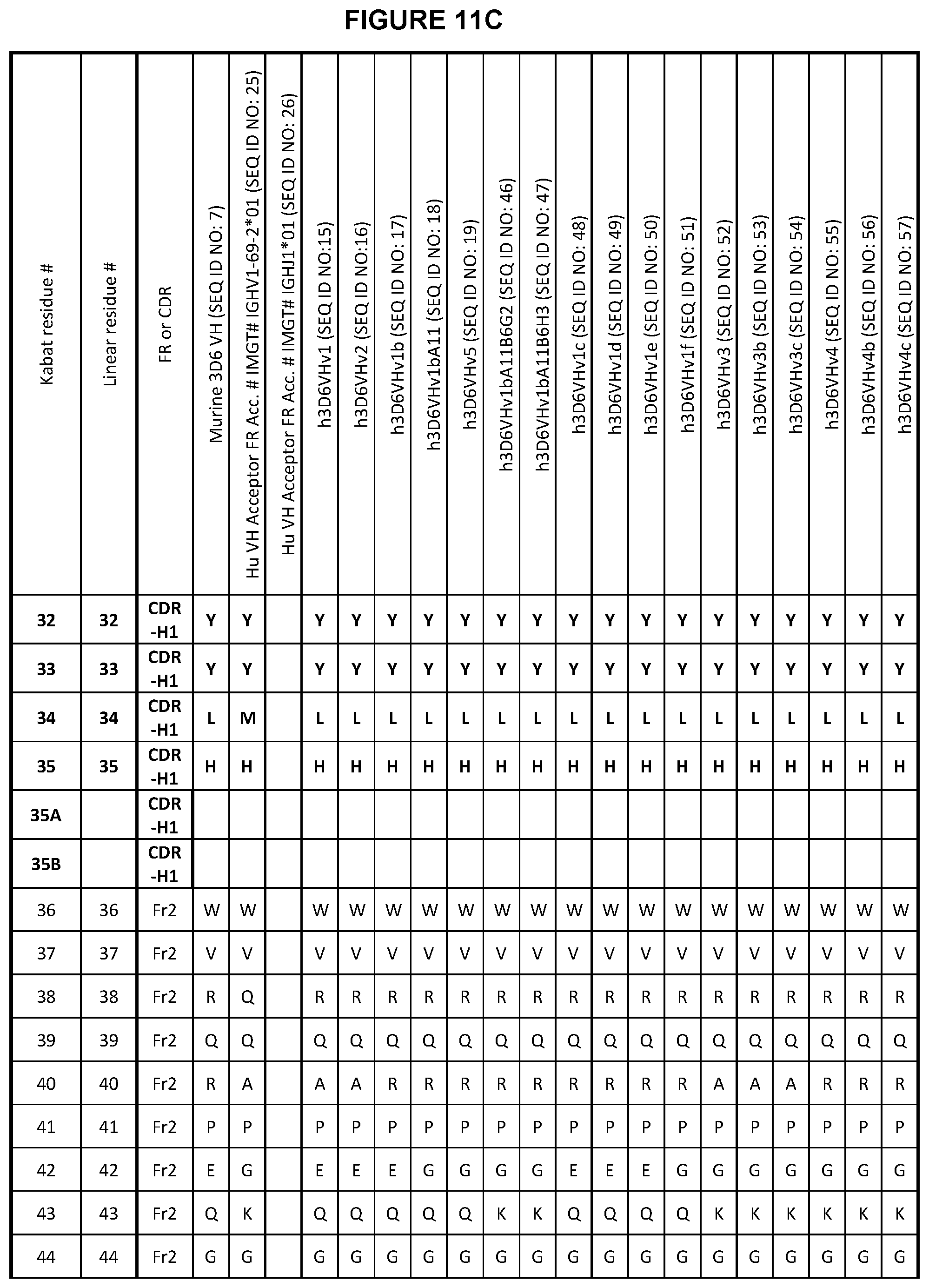
D00013

D00014

D00015
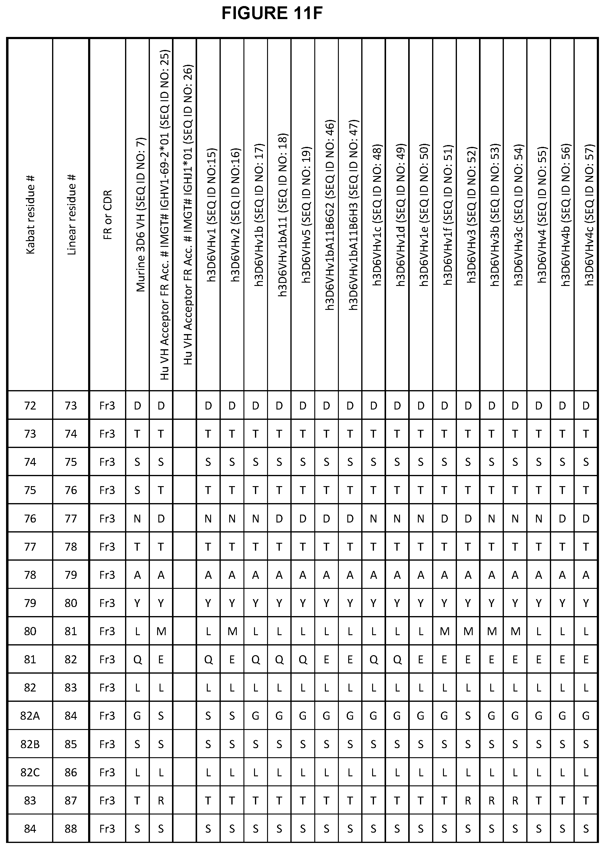
D00016
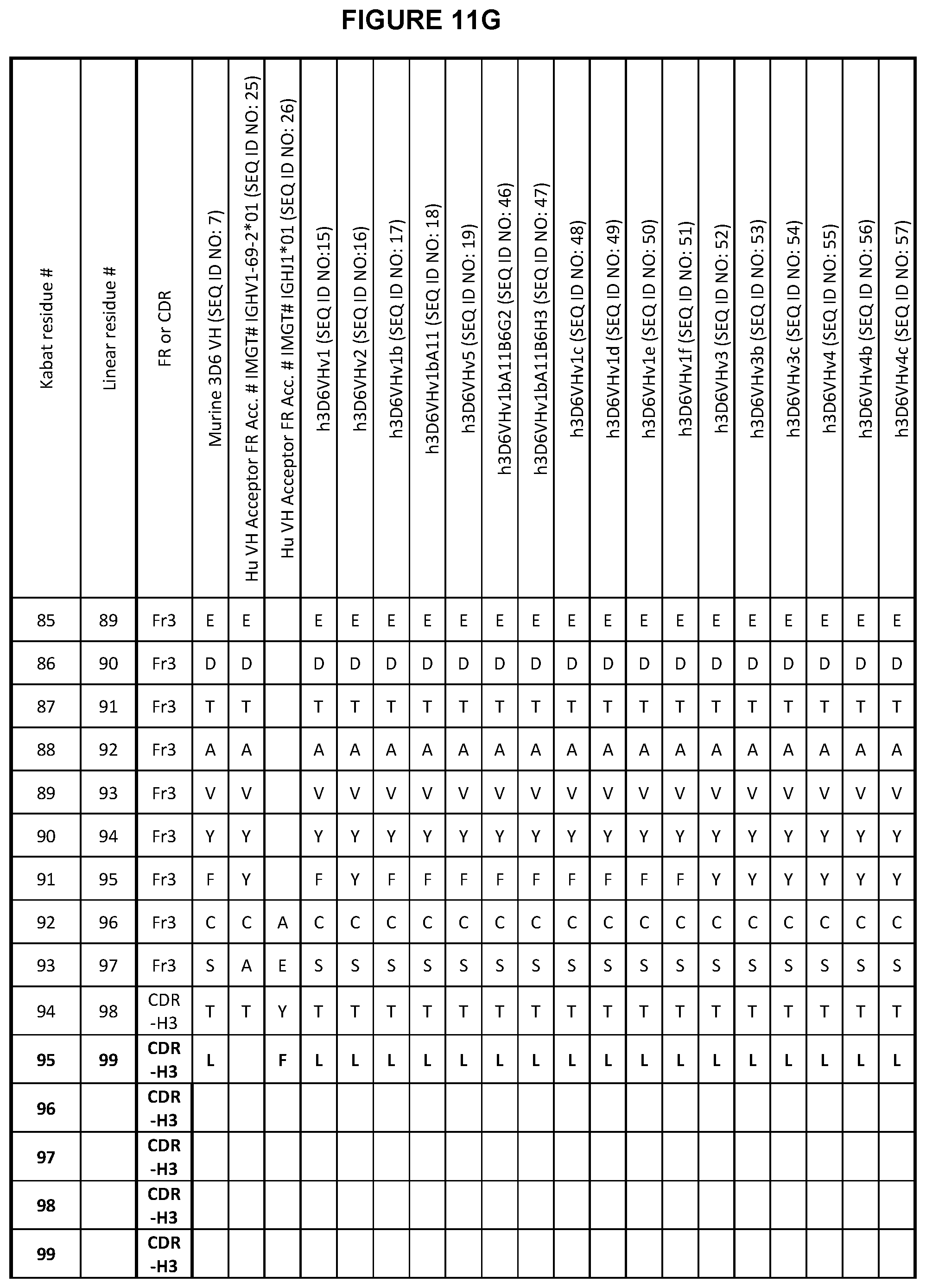
D00017

D00018

D00019

D00020
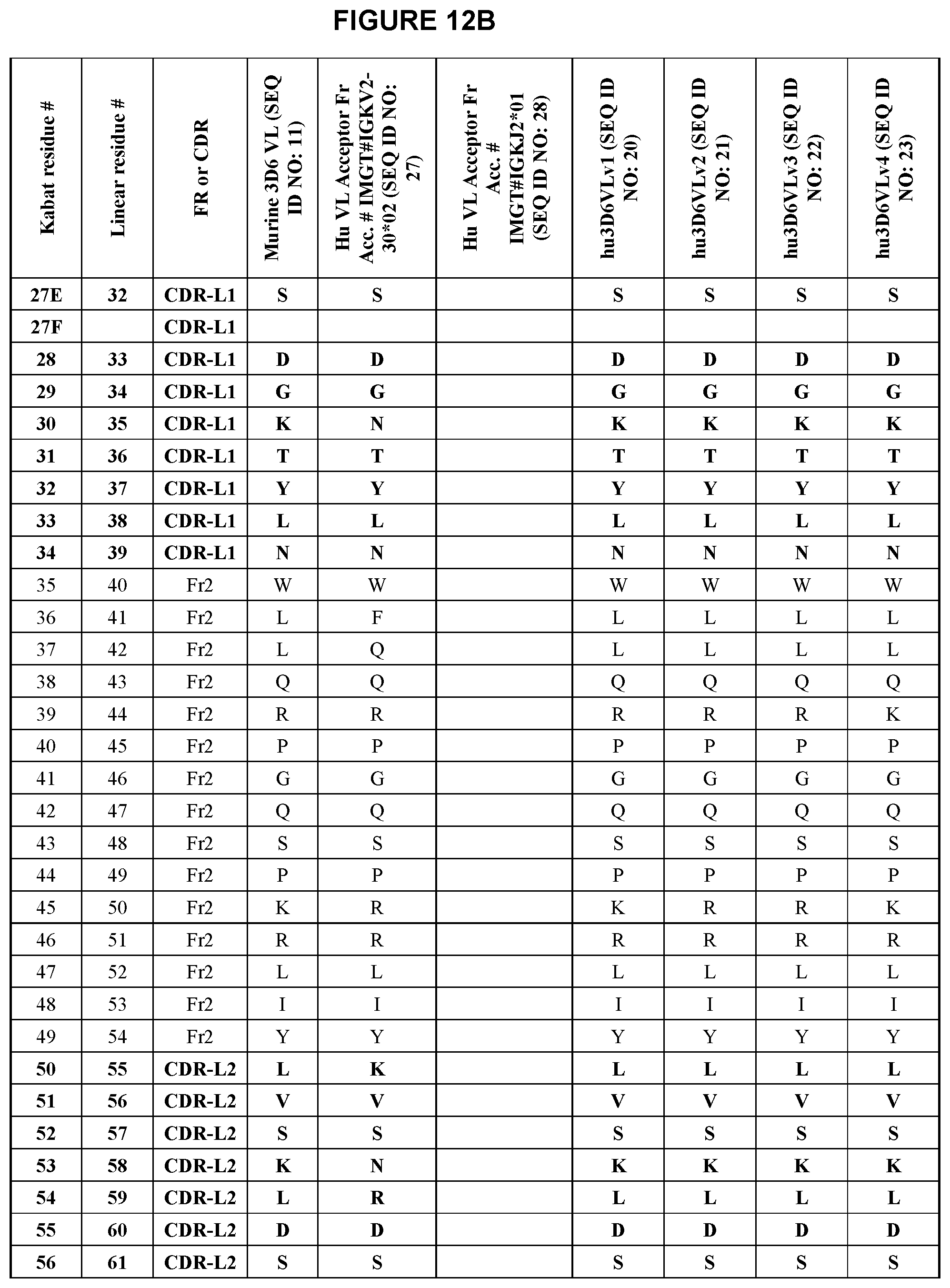
D00021
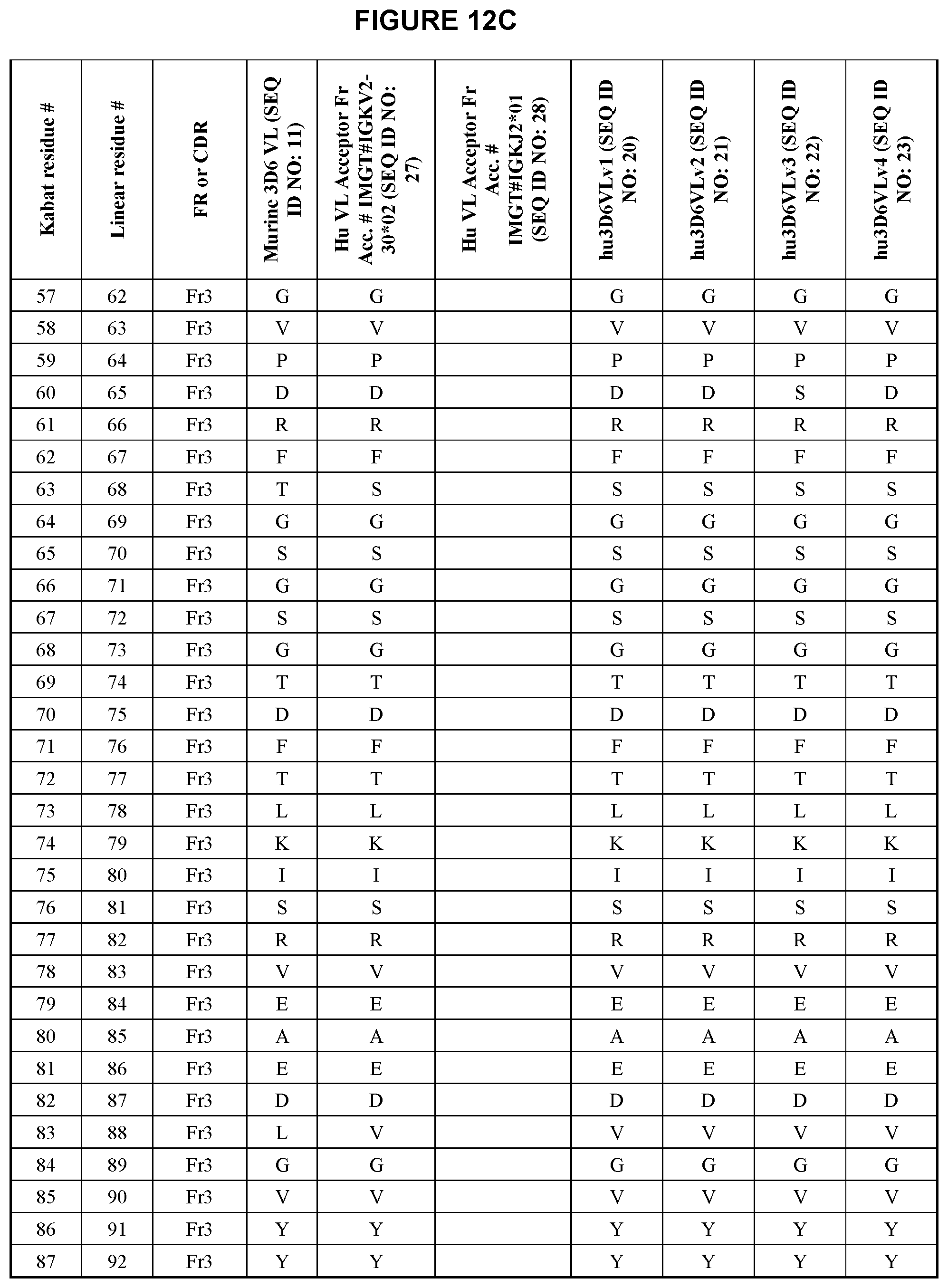
D00022

S00001
XML
uspto.report is an independent third-party trademark research tool that is not affiliated, endorsed, or sponsored by the United States Patent and Trademark Office (USPTO) or any other governmental organization. The information provided by uspto.report is based on publicly available data at the time of writing and is intended for informational purposes only.
While we strive to provide accurate and up-to-date information, we do not guarantee the accuracy, completeness, reliability, or suitability of the information displayed on this site. The use of this site is at your own risk. Any reliance you place on such information is therefore strictly at your own risk.
All official trademark data, including owner information, should be verified by visiting the official USPTO website at www.uspto.gov. This site is not intended to replace professional legal advice and should not be used as a substitute for consulting with a legal professional who is knowledgeable about trademark law.