Serial quantitative PCR assay for detection, species-discrimination and quantification of Leishmania spp. in human samples
Wilson , et al. January 5, 2
U.S. patent number 10,883,147 [Application Number 15/275,117] was granted by the patent office on 2021-01-05 for serial quantitative pcr assay for detection, species-discrimination and quantification of leishmania spp. in human samples. This patent grant is currently assigned to United States of America as represented by the Department of Veteran Affairs. The grantee listed for this patent is The United States of America as represented by the Department of Veterans Affairs, The United States of America as represented by the Department of Veterans Affairs. Invention is credited to Edgar Carvalho, John E. Donelson, Selma M. B. Jeronimo, Albert Schriefer, Jason Weirather, Mary E Wilson.


| United States Patent | 10,883,147 |
| Wilson , et al. | January 5, 2021 |
Serial quantitative PCR assay for detection, species-discrimination and quantification of Leishmania spp. in human samples
Abstract
The invention provides a method for determining the presence, species, and/or quantity of Leishmania in a sample.
| Inventors: | Wilson; Mary E (Iowa City, IA), Weirather; Jason (Iowa, IA), Donelson; John E. (Iowa City, IA), Schriefer; Albert (Bahia, BR), Carvalho; Edgar (Salvador, BR), Jeronimo; Selma M. B. (Rn, BR) | ||||||||||
|---|---|---|---|---|---|---|---|---|---|---|---|
| Applicant: |
|
||||||||||
| Assignee: | United States of America as
represented by the Department of Veteran Affairs (Washington,
DC) |
||||||||||
| Family ID: | 1000005281721 | ||||||||||
| Appl. No.: | 15/275,117 | ||||||||||
| Filed: | September 23, 2016 |
Prior Publication Data
| Document Identifier | Publication Date | |
|---|---|---|
| US 20170260595 A1 | Sep 14, 2017 | |
Related U.S. Patent Documents
| Application Number | Filing Date | Patent Number | Issue Date | ||
|---|---|---|---|---|---|
| 13659746 | Oct 24, 2012 | 9551040 | |||
| 61628144 | Oct 24, 2011 | ||||
| Current U.S. Class: | 1/1 |
| Current CPC Class: | C12Q 1/6816 (20130101); C12Q 1/6893 (20130101); C12Q 2600/16 (20130101); Y02A 50/30 (20180101) |
| Current International Class: | C12Q 1/6893 (20180101); C12Q 1/6816 (20180101) |
Other References
|
Quispe-Tintaya et al. The Journal of Infectious Diseases. 2005. 192:685-692.--Exhibit 1. cited by applicant . Shulz et al. Journal of Clinical Microbiology.2003. 41 (4)1529-1535.--Exhibit 2. cited by applicant . Nasereddin et al. Journal of Clinical Microbiology. 2010. 48(6): 2240-2242--Exhibit 3. cited by applicant . Monbrison et al. Acta Tropica, 2007. 102:97-83--Exhibit 4. cited by applicant . Tupperwar et al. Diagnostic Microbiology and Infectious Disease. 2008. 61:23-30--Exhibit 5. cited by applicant . Talmi-Frank et al. PloS Neglected Tropical Diseases.2100. 4(1):e581--Exhibit 6. cited by applicant . Nicolas et al. Journal of Microbiological Methods. 2002. 51: 295-299--Exhibit 7. cited by applicant . EU370909.1(Mar. 4, 2009) www.ncbi.nlm.nih.gov/nuccore/169160018?sat=17&satkey=18113955--Exhibit 8. cited by applicant . Bretagne et al. Clinical and Diagnostic Laboratory Immunology. 2001. 8(4): 828-831.--Exhibit 9. cited by applicant . Rasmussen et al. BMC Bioinformatics. 2007. 8: 107--Exhibit 10. cited by applicant . GenBank FR799597.1 (Feb. 7, 2011). cited by applicant . www.ncbi.nlm.nih.gov/nuccore/322497052?sat=15&satkey=2401445.--Exhibit 11. cited by applicant . Weirather. Journal of Clinical Microbiology, Nov. 2011, p. 3892-3904--Exhibit 12. cited by applicant . Nicolas. Journal of Clinical Microbiology, May 2002, p. 1666-1669--Exhibit 13. cited by applicant . McCoy. Molecular and Biochemical Parasitology 95 (1998) 251-265--Exhibit 14. cited by applicant . AF058760.1 (Jan. 18, 1999)--Exhibit 15. cited by applicant . Buck. Biotechniques.1999. 27(3): 528-536.--Exhibit 16; and. cited by applicant . Mauricio. Parasitology Today, vol. 16, No. 5, 2000--Exhibit 17. cited by applicant. |
Primary Examiner: Dauner; Joseph G.
Attorney, Agent or Firm: Ballard Spahr LLP
Government Interests
This invention was made with government support under VA Gulf War RFA awarded by Department of Veterans Affairs and Grant Nos. R01-AI48822, AI045540, R01 AI067874-01, R01 AI059451, R01 AI076233-01, P50 AI-30639 and P50 AI-074321 awarded by NIH/NIAID. The government has certain rights in the invention.
Parent Case Text
This patent application is a continuation application of U.S. Ser. No. 13/659,746, filed Oct. 24, 2012, which claims the benefit of the filing date of U.S. Ser. No. 61/628,144, filed Oct. 24, 2011, the contents of all of which are herein incorporated by reference in their entireties into the present patent application.
Claims
What is claimed is:
1. A method for determining the presence and species of a Leishmania in a sample, and distinguishing the Leishmania species so determined from other Leishmania species of interest present in the sample, the method comprising: (a) contacting the sample with the forward-and-reverse primer pairs GGGTAGGGGCGTTCTGC (SEQ ID NO:1) and TACACCAACCCCCAGTTTGC (SEQ ID NO: 2) (kDNA 1 minicircle forward and reverse primers, respectively); TGCTATAAAATCGTACCACCCGACA (SEQ ID NO: 101) and GAACGGGGTTTCTGTATGCCATTT (SEQ ID NO: 102) (L. (Y) braziliensis kDNA 3 forward and reverse primers, respectively); AGAGCGTGCCTTGGATTGTG (SEQ ID NO: 49) and CGCTGCGTTGATTGCGTTG (SEQ ID NO: 50) (MSP Associated Gene I (MAG I) forward and reverse primers, respectively); and GGAGAAACTCACGGCACAGG (SEQ ID NO: 67) and GCGCCTCGTAGGTCACAGTT (SEQ ID NO: 68) (SLACS forward and reverse primers, respectively); thereby forming Leishmania-primer pair complexes; (b) exposing the Leishmania-primer pair complexes of (a) to a thermo-stable polymerase and deoxyribonucleoside triphosphates to produce (1) double-stranded DNAs containing the primer sequences and (2) pyrophosphates; (c) performing temperature cycles to permit further production of additional double-stranded DNAs containing the primer sequences and additional pyrophosphates; (d) determining the melting temperature of the double-stranded DNA product of (c), a characteristic melting temperature being indicative of particular Leishmania species so detected by the forward-and-reverse primer pairs of (a); and (e) comparing the melting temperature of (d) to a characteristic melting temperature for particular Leishmania species so detected by the forward-and-reverse primer pairs of (a); thereby determining the presence and the species of the Leishmania in the sample, wherein the Leishmania species of interest comprises any of Leishmania tropica, Leishmania chagasi/Leishmania infantum, Leishmania donovani, Leishmania major, Leishmania braziliensis, Leishmania guyanensis/Leishmania panamensis, Leishmania mexicana, and Leishmania amazonensis.
2. The method of claim 1, wherein the Leishmania species of interest are species found in a geographic region of North, Central and South America.
3. The method of claim 1, wherein the Leishmania species of interest are species found in a geographic region of Europe.
4. The method of claim 3, wherein the Leishmania species of interest found in a geographic region of Europe are one or more of L. (L.) infantum and L. (L.) donovani.
5. The method of claim 1, wherein the Leishmania species of interest are species found in a geographic region of the Middle East, Northern and sub-Saharan Africa.
6. The method of claim 5, wherein the species found in a geographic region of Middle East, Northern and sub-Saharan Africa are one or more of L. (L.) major, L. (L.) tropica, and L. (L.) infantum and L. (L.) donovani.
7. The method of claim 5, wherein the species found in a geographic region of Middle East, Northern and sub-Saharan Africa are one or more of L. (L.) infantum, L. (L.) donovani and L. (L.) tropica.
8. The method of claim 1, wherein the Leishmania species of interest are species found in a geographic region of Asia.
9. The method of claim 8, wherein the geographic region of Asia is India, Bangladesh, Pakistan, or Nepal.
10. The method of claim 8, wherein the species found in Asia is L. (L.) donovani.
11. The method of claim 1, wherein steps (a) through (f) are repeated using at least one nucleic acid primer pair that is different for each Leishmania species in the group.
12. The method of claim 1, wherein the quantity of Leishmania in the sample is measured relative to a reference standard curve.
13. The method of claim 1, wherein determining the melting temperature comprises using ultraviolet absorption detection, fluorescence detection, light scattering detection, colorimetric detection, or chromogenic detection.
14. The method of claim 1, further comprising repeating the steps in (a) to (e), wherein step (a) comprises contacting the sample with one or more of the forward- and reverse primer pairs selected from: AACTTTTCTGGTCCTCCGGGTAG (SEQ ID NO: 97) and ACCCCCAGTTTCCCGCC (SEQ ID NO: 98) (kDNA 2 forward and reverse primers, respectively); GGGTAGGGGCGTTCTGC (SEQ ID NO:3) and CCCGGCCTATTTTACACCAACC (SEQ ID NO:4) (kDNA 3 minicircle forward and reverse primers, respectively); GGGTGCAGAAATCCCGTTCA (SEQ ID NO:5) and CCCGGCCCTATTTTACACCA (SEQ ID NO: 6) (kDNA 4 minicircle forward and reverse primers, respectively); CTTTTCTGGTCCTCCGGGTAGG (SEQ ID NO: 99) and CCACCCGGCCCTATTTTACACCAA (SEQ ID NO: 100) (kDNA 5 minicircle forward and reverse primers, respectively); AATGGGTGCAGAAATCCCGTTC (SEQ ID NO: 7) and CCACCACCCGGCCCTATTTTAC (SEQ ID NO: 8) (kDNA 7 minicircle forward and reverse primers, respectively); GGTCCCGGCCCAAACTTTTC (SEQ ID NO: 9) and CCGGGGTTTCGCACTCATTT (SEQ ID NO: 10 (L. (L.) amazonensis kDNA 1 forward and reverse primers, respectively); GGTAGGGGCGTTCTGCGAAT (SEQ ID NO: 11) and CCCGGCCTATTTTACACCAACC (SEQ ID NO: 12) (L. (L.) amazonensis kDNA 2 forward and reverse primers, respectively); GGGTAGGGGCGTTCTGC (SEQ ID NO: 13) and TACACCAACCCCCAGTTTGC (SEQ ID NO: 14) (L. (L.) amazonensis kDNA 3 forward and reverse primers, respectively); TGAGTGCAGAAACCCCGTTCATA (SEQ ID NO: 15) and ACACCAACCCCCAGTTGTGA (SEQ ID NO: 16) (L. (L.) amazonensis kDNA 4 forward and reverse primers, respectively); AATTTCGCAGAACGCCCCTAC (SEQ ID NO: 17) and GTACTCCCCGACATGCCTCTG (SEQ ID NO:18) (L. (V.) braziliensis kDNA 1 forward and reverse primers, respectively); TCCGCAGGAGACTTCGTATG (SEQ ID NO:103) and CACGACTATCCACCCCATCC (SEQ ID NO:104) (L. (L.) infantum Minicircle 1 forward and reverse primers, respectively); ACGGGGTTTCTGCACCCATT (SEQ ID NO: 19) and GTAGGGGCGTTCTGCGAAAA (SEQ ID NO: 20) (L. (L.) major Minicircle 1 forward and reverse primers, respectively); AATGCGAGTGTTGCCCTTTTG (SEQ ID NO: 21) and GCCGAACAACGCCATATTAACC (SEQ ID NO:22) (L. (L.) mexicana Minicircle 1 forward and reverse primers, respectively); GGGGGTTGGTGTAAAATAGGG (SEQ ID NO:23) and ACCACCAGCAGAAGGTCAAAG (SEQ ID NO:24) (L. (L.) tropica Minicircle 1 forward and reverse primers, respectively); GCGGTGGCTGGTTTTAGATG (SEQ ID NO:25) and TCCAATGAAGCCAAGCCAGT (SEQ ID NO:26) (L. (L.) donovani Minicircle 1 forward and reverse primers, respectively); ATTTTAGTATGAGTGGTAGGTTTTGTT (SEQ ID NO: 27) and CAATAACTGGGACGGTTGCT (SEQ ID NO:28) (Cytochrome B1 forward and reverse primers, respectively); GCGGAGAGGAAAGAAAAGGCTTA (SEQ ID NO:29) and AAAAGTCATGCTAAACACACACCACA (SEQ ID NO: 30) (L. (L.) amazonensis Cytochrome B1 forward and reverse primers, respectively); CAGGTTGCTTACTACGTGTTTATGGTG (SEQ ID NO: 31) and TCGTATTACAAACCCTAAATCAAAATCTCA (SEQ ID NO:32) (L. (L.) tropica Cytochrome B1 forward and reverse primers, respectively); TCAGGTTGCTTACTACGTGTTTATGGTG (SEQ ID NO: 33) and TGCTAAACAAACACCACATATGATCTGC (SEQ ID NO: 34) (L. (L.) tropica Cytochrome B2 forward and reverse primers, respectively); TGACACACATATTTTAGTGTGGGTGGTAGG (SEQ ID NO: 35) and TCCCCAATAAGACATCATTGTACATGGTAA (SEQ ID NO: 36) (L. (L.) tropica Cytochrome B3 forward and reverse primers, respectively); CACATATTTTAGTGTGGGTGGTAGGTTTTG (SEQ ID NO: 37) and TCCCCAATAAGACATCATTGTACATGGTAA (SEQ ID NO: 38) (L. (L.) tropica Cytochrome B4 forward and reverse primers, respectively); GCTTGGTTGGATTATTTTTGCTG (SEQ ID NO: 39) and AACAACATTTTAACTCTTGTAGGATTCG (SEQ ID NO: 40) (Maxicircle 1 forward and reverse primers, respectively); GAGGTGTTTGCCCGCATC (SEQ ID NO:41) and CTCGCCCATGTCGTCG (SEQ ID NO: 42) (Alpha-tubulin I forward and reverse primers, respectively); TGTCGCTTGCAGACCAGATG (SEQ ID NO:105) and GCATCGCAGGTGTGAGCA (SEQ ID NO: 106) (DNA polymerase I forward and reverse primers, respectively); AGGAGGATGGCAAGCGGAAG (SEQ ID NO: 43) and GCGACGGGTACAGGGAGTTG (SEQ ID NO:44) (DNA polymerase 2 forward and reverse primers, respectively); CGAAACTTCCGGAACCTGTCTT (SEQ ID NO: 45) and CACCACACGCACGCACAC (SEQ ID NO: 46) (Mini-exon forward and reverse primers, respectively); GTGTGGTGGCGGGTGTATGT (SEQ ID NO: 47) and GCCCAGGTCGCTGTGAGG (SEQ ID NO: 48) (Mini-exon 2 forward and reverse primers, respectively); AGTTTTGGTTGGCGCTCCTG (SEQ ID NO: 51) and CCCACTCGCTTTCCTTGGTC (SEQ ID NO:52) (MSP Associated Gene 2 (MAG 2) forward and reverse primers, respectively); CGACCCTGTCACCACCACAG (SEQ ID NO:53) and GAGGCCACCCTATCGCTGAC (SEQ ID NO:54) (SIDER repeat I forward and reverse primers, respectively); TCGTTGAGGGAGGAGGTGTTTC (SEQ ID NO:55) and TCGGCTTTGAGGTTGGCTTC (SEQ ID NO:56) (L. (V) braziliensis DNA polymerase forward and reverse primers, respectively); ACGTCGCCAACTGCTTCACC (SEQ ID NO: 57) and GTGTTCGCACCGCCTTGAC (SEQ ID NO: 58) (L. (V) braziliensis DNA polymerase 2 forward and reverse primers, respectively); GTCGTTGTCCGTGTCGCTGT (SEQ ID NO: 59) and CGCTGTGTGTGTCCGTGTGT (SEQ ID NO:60) (L. (L.) major MSP associated gene I (L. major MAG 1) forward and reverse primers, respectively); GACGACGACGAGGAGGATGG (SEQ ID NO: 61) and GCGACGGGTACAGGGAGTTG (SEQ ID NO: 62) (L. (L.) amazonensis DNA polymerase I forward and reverse primers, respectively); CCAGATGCCGACCAAAGC (SEQ ID NO: 107) and CGCGCACGTGATGGATAAC (SEQ ID NO: 108) (GPI forward and reverse primers, respectively); GAAGGTGCAGTCCCTCGTGT (SEQ ID NO: 63) and CCTCCGTCTGCTTGCTCTTG (SEQ ID NO: 64) (HSP70-1 forward and reverse primers, respectively); or TCGAGATCGACGCGTTGTT (SEQ ID NO: 65) and CCGCACAGCTCCTCGAA (SEQ ID NO: 66) (HSP70-4 forward and reverse primers, respectively).
15. A method for determining whether a subject is suffering from a Leishmania infection by detecting the presence of Leishmania in a sample from the subject by the method of claim 1.
Description
Throughout this application various publications are referenced. The disclosures of these publications in their entireties are hereby incorporated by reference into this application in order to more fully describe the state of the art to which this invention pertains.
BACKGROUND OF THE INVENTION
The Leishmania spp. are kinetoplastid protozoa that are transmitted to humans and other mammalian hosts by, e.g., a sand fly vector. The spectrum of symptomatic human leishmaniasis is wide, and the most important factor determining the clinical outcome of infection seems to be the species of Leishmania. Nonetheless, there are variable clinical presentations of disease due to each species (10, 22, 30), and increasing reports document atypical presentations of leishmaniasis, sometimes but not always in the setting of the immunocompromised (16,53). Differentiation between the Leishmania species is an issue since there are overlapping and dynamic geographic regions of risk, and different susceptibilities to treatment (7,15). Thus, a method of diagnosis that is sensitive enough to detect low levels of the parasite in asymptomatic or early symptomatic infection, and can distinguish between the different Leishmania species, would be of tremendous utility in endemic and non-endemic regions (38).
Procedures for diagnosis of leishmaniasis are often invasive, and isolates are frequently difficult to grow in vitro. Tests to distinguish between the Leishmania species have traditionally involved separation of isoenzymes in culture-derived parasites, which takes several weeks (12). The Leishmania spp. have been detected in and isolated from blood cultures of subjects with all forms of leishmaniasis (20, 39, 40), and in the blood of asymptomatic individuals living in regions of risk (11, 25, 34, 39). The possibility that Leishmania may be present in the bloodstream more often than previously recognized led us to hypothesize that amplification-based methods to detect parasite DNA in blood or serum might be a feasible means of diagnosis.
Nucleic acid-based methods avoid the need for parasite cultivation, replacing this with either hybridization or amplification (24, 24, 35, 39, 67). The latter approaches provide the advantage of increased sensitivity. Amplification methods reported for the detection of individual Leishmania species include conventional PCR (4, 21, 63) or quantitative PCR methods, including reverse transcriptase quantitative polymerase chain reaction (RT-qPCR), DNA-based qPCR, quantitative nucleic acid sequence-based amplification (QT-NASBA) and in situ hybridization to quantify Leishmania spp. in blood or tissue samples (63).
Protozoa belonging to the order Kinetoplastida, including Leishmania spp. and Trypanosoma spp., are characterized by a prominent kinetoplast structure containing the mitochondrial DNA in the parasites' single mitochondrion. Whereas Leishmania spp. have 35-36 chromosomes in their nuclear genomes (52), the kinetoplast contains hundreds of DNA maxicircles encoding genes that are destined for RNA editing, and thousands of DNA minicircles, circular molecules with a conserved origin of replication encoding guide RNA sequences for RNA editing (13). Because of their abundance, specificity and repetitive nature, kinetoplast DNA (kDNA) sequences have frequently been targeted for nucleic acid based detection (33, 42, 43, 48, 49). A drawback of the use of kDNA for parasite quantification is the uncertainty of whether the kDNA copy number differs between Leishmania species, strains, and growth stages.
The goal of this study was to develop a serial nucleic acid amplification based method for diagnosis and speciation of Leishmania spp. parasites in human or animal-derived tissues. As such we developed a set of primers and probes for serial qPCR assays. The assays were sensitive enough to detect low levels of parasites, and to distinguish between Leishmania species in human specimens. Using non-species discriminating probes, we quantified the relative differences in kinetoplastid DNA (kDNA) copy numbers between parasite species, among isolates of the same species, and between stages of the same parasite strain. The serial qPCR assays has potential applications for diagnosis and species discrimination, as well as novel approaches to determining parasite load and following treatment response in infected humans.
The Leishmania species cause a variety of human disease syndromes. Methods for diagnosis and species differentiation are insensitive and many require invasive sampling. Although quantitative PCR (qPCR) methods are reported for Leishmania detection, no systematic method to quantify parasites and determine the species in clinical specimens is established.
We developed a serial qPCR strategy to identify and rapidly differentiate Leishmania species, and quantify parasites in clinical or environmental specimens. SYBR green qPCR may be employed, with corresponding Taqman assays for validation. Screening primers recognize kinetoplast minicircle DNA of all Leishmania species. Species identification employs further qPCR set(s) individualized for geographic regions, combining species discriminating probes with melt curve analysis. The assay was sufficient to detect, speculate and quantify Leishmania spp. in sera, cutaneous biopsies, or cultured isolates from subjects in Bangladesh or Brazil with different forms of leishmaniasis. The multicopy kDNA probes were the most sensitive and useful for quantification based on promastigote standard curves. To test their validity for quantification, kDNA copy numbers were compared between Leishmania species, isolates, and life stages using qPCR. Maxicircle and minicircle copy numbers differed up to 6 fold between Leishmania species, but differences were smaller between strains of the same species. Amastigote and promastigote Leishmania life stages retained similar numbers of kDNA maxi- or minicircles. Thus, serial qPCR is useful for Leishmania detection and speciation, and for absolute quantification when compared to a standard curve from the same Leishmania species.
SUMMARY OF THE INVENTION
The invention provides for methods for determining the presence, species, and/or quantity of a Leishmania in a sample comprising: (a) contacting the sample with a set of nucleic acid primer pairs with primer sequences common to all Leishmania species or to a subset of Leishmania species, so as to make Leishmania-primer pair complexes; (b) exposing the complexes to a thermo-stable polymerase to produce double-stranded DNAs containing the primer sequences; (c) determining the time or number of temperature cycles at which a threshold level of double-stranded DNA product and/or pyrophosphate product is reached being indicative of on-target specificity and/or quantity of Leishmania in the sample; and (d) determining the melting temperature of the double-stranded DNA product of (c), a characteristic melting temperature being indicative of particular Leishmania species so detected by the primer; thereby determining the presence, species, and/or quantity of Leishmania in the sample.
In another embodiment for determining the presence, species, and/or quantity of Leishmania in a sample, the method comprises contacting the sample with a set of nucleic acid primer pairs with primer sequences unique to a particular Leishmania species, so as to make Leishmania-primer pair complexes; exposing the complexes to a thermo-stable polymerase to produce double-stranded DNAs containing the primer sequences; determining the time or number of temperature cycles at which a threshold level of double-stranded DNA product and/or pyrophosphate product is reached which is indicative of on-target specificity and quantity of Leishmania in the sample; and identifying the particular Leishmania species by the unique primers used so as to thereby determine the presence, species, and quantity of Leishmania in the sample. In this embodiment, the step of characterizing the double-stranded DNA product by its melting temperature is not necessary for identifying the particular Leishmania species but may be performed as a control to assure on-target specificity of the reaction.
In another embodiment for determining the presence, species, and/or quantity of a Leishmania in a sample, the method comprises: (a) contacting the sample with a set of nucleic acid primer pairs with primer sequences unique to a particular Leishmania species, so as to make Leishmania-primer pair complexes; (b) exposing the complexes to a thermo-stable polymerase to produce double-stranded DNAs containing the primer sequences; (c) determining the time or number of temperature cycles at which a threshold level of double-stranded DNA product and/or pyrophosphate product is reached being indicative of on-target specificity and/or quantity of Leishmania in the sample; and (d) identifying the particular Leishmania species by the unique primers used; thereby determining the presence, species, and/or quantity of Leishmania in the sample, wherein quantity of Leishmania in the sample is determined by examining a single copy gene or a known copy gene number within a Leishmania species and comparing against a reference standard curve generated with different known amount of the particular Leishmania species.
In one embodiment for detecting the presence of Leishmania, the method comprises (a) contacting a sample with a nucleic acid primer having a complementary sequence so as to produce a complex; (b) contacting the sample with another nucleic acid primer having a different complementary sequence so as to produce a complex; (c) exposing the complexes to a thermo-stable polymerase so as to produce double stranded nucleic acid from the primers in (a) and (b); and (d) detecting the double stranded nucleic acid so produced, the presence of the double stranded nucleic acid being indicative of the presence of Leishmania in the sample, wherein the nucleic acid primers from step (a) and (b) are selected from the group consisting of GGGTAGGGGCGTTCTGC (SEQ ID NO:3) and CCCGGCCTATTTTACACCAACC (SEQ ID NO:4) (kDNA 3 mini-circle forward and reverse primers, respectively); GGGTGCAGAAATCCCGTTCA (SEQ ID NO:5) and CCCGGCCCTATTTTACACCA (SEQ ID NO:6) (kDNA 4 mini-circle forward and reverse primers, respectively); GGGTAGGGGCGTTCTGC (SEQ ID NO:1) and TACACCAACCCCCAGTTTGC (SEQ ID NO:2) (kDNA 1 mini-circle forward and reverse primers, respectively); AATGGGTGCAGAAATCCCGTTC (SEQ ID NO:7) and CCACCACCCGGCCCTATTTTAC (SEQ ID NO:8) (kDNA 7 mini-circle forward and reverse primers, respectively); GGGTAGGGGCGTTCTGC (SEQ ID NO:13) and TACACCAACCCCCAGTTTGC (SEQ ID NO:14) (L. amazonensis kDNA 3 forward and reverse primers, respectively); AGGAGGATGGCAAGCGGAAG (SEQ ID NO:43) and GCGACGGGTACAGGGAGTTG (SEQ ID NO:44) (DNA polymerase 2 forward and reverse primers, respectively); CGAAACTTCCGGAACCTGTCTT (SEQ ID NO:45) and CACCACACGCACGCACAC (SEQ ID NO:46) (mini-exon 1 forward and reverse primers, respectively); GTCGTTGTCCGTGTCGCTGT (SEQ ID NO:59) and CGCTGTGTGTGTCCGTGTGT (SEQ ID NO:60) (L. major MSP associated gene 1 forward and reverse primers, respectively); GACGACGACGAGGAGGATGG (SEQ ID NO:61) and GCGACGGGTACAGGGAGTTG (SEQ ID NO:62) (L. amazonensis DNA polymerase forward and reverse primers, respectively); GAAGGTGCAGTCCCTCGTGT (SEQ ID NO:63) and CCTCCGTCTGCTTGCTCTTG (SEQ ID NO:64) (HSP70-1 forward and reverse primers, respectively); and TCGAGATCGACGCGTTGTT (SEQ ID NO:65) and CCGCACAGCTCCTCGAA (SEQ ID NO:66) (HSP70-4 forward and reverse primers, respectively); or a combination thereof. The rate of the production of multiple double-stranded DNAs containing the primer sequences may be monitored and/or the pyrophosphate product thereof may be monitored, either of which may be indicative of the amount or quantity of Leishmania present in the sample.
In another embodiment for determining the presence, quantity and/or species of Leishmania in a sample, the sample comprises: (a) contacting the sample with a set of nucleic acid primer pairs with primer sequences common to all Leishmania species or to a subset of Leishmania species, so as to make Leishmania-primer pair complexes; (b) contacting the sample with a set of nucleic acid probe or probes modified with a detectable marker and a quencher for the detectable marker and having a sequence complementary to a region bounded by each primer pair in (a); (c) exposing the complexes to a thermo-stable polymerase to produce multiple double-stranded DNAs containing the primer sequences; (d) detecting the presence and/or rate of release of the detectable marker from the modified nucleic acid in (b), which is indicative of quantity and/or particular Leishmania species; thereby determining the presence, quantity and/or species of Leishmania in the sample.
In another embodiment for determining the presence, quantity and/or species of Leishmania in a sample, the method comprises: (a) contacting the sample with a set of nucleic acid primer pairs with primer sequences unique to a particular Leishmania species, so as to make Leishmania-primer pair complexes; (b) contacting the sample with a set of nucleic acid probe or probes modified with a detectable marker and a quencher for the detectable marker and having a sequence complementary to a region bounded by each primer pair in (a); (c) exposing the complexes to a thermo-stable polymerase to produce multiple double-stranded DNAs containing the primer sequences; (d) detecting the presence and/or rate of release of the detectable marker from the modified nucleic acid in (b), which is indicative of quantity and/or presence of Leishmania in the sample; and (e) identifying the particular Leishmania species by the unique primers used; thereby determining the presence, quantity and/or species of Leishmania in the sample.
The invention also provides novel compositions as described herein.
BRIEF DESCRIPTION OF THE FIGURES
FIG. 1. Melt curves of selected qPCR assays useful for species discrimination. Rate of change in the intensity of the fluorescent qPCR signal is plotted across a 10 degree temperature range for each species with each primer set. Data are only shown for sets that amplified with a CT (cycle threshold) of less than 30, and showed peaks of melt curves greater than 1 degree apart between species. Peaks of curves indicate the melting temperature of the amplicon. Each melt curve is color-coded according to the parasite species it represents. Plots with a solid line indicate that the reaction amplified with a CT less than 25. Plots with a dotted line indicate the reaction amplified with a CT greater than or equal to 25 but less than 30.
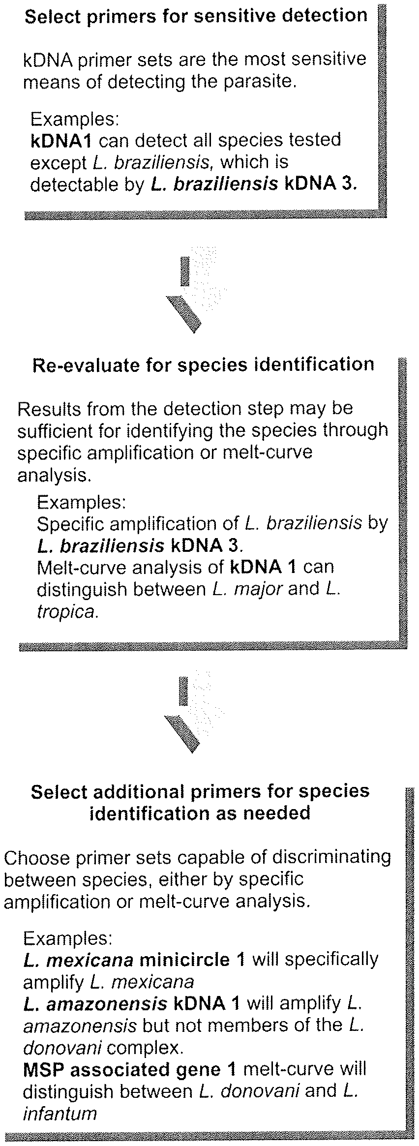
FIG. 2. Flow chart of the serial qPCR assay. The chart shows a minimal application of the serial diagnostic qPCR assay to determine presence and species of Leishmania in a sample. In step 1 (detection), an unknown DNA sample can be tested for the presence of Leishmania spp. DNA using SYBR primers. kDNA 1 amplifies most species. L. (V.) braziliensis Mini-circle 3 amplifies kDNA sequences within L. (V.) braziliensis, a primer set that would be included when testing samples from Latin America to provide the most sensitive detection across species. In step 2, a sample which has tested positive for the presence of parasite kDNA 1, but not L. braziliensis kDNA 3, can be classified according to its species. Melt curve analysis of kDNA1 amplicons can distinguish between several Old World species (L. (L.) tropica, L. (L.) major, L. (L.) infantum). Application of SYBR green melt curve analysis using MSP associated gene (MAG) 1 is capable of separating L. (L.) chagasi and L. (L.) infantum from L. (L.) donovani. The presence or absence of L. (L.) mexicana specific minicircle amplicons is sufficient to differentiate between L. (L.) mexicana and L. (L.) major. Distinguishing L. (L.) amazonensis from the members of the L. (L.) donovani complex requires an additional such as L. (L.) amazonensis kDNA 2 (not shown in flowchart). The inability of any primer pairs tested to distinguish between L. (L.) chagasi and L. (L.) infantum is consistent with the current belief that these species are virtually identical (44). Note: The primer sets listed are minimal sets. It is advisable to select additional primers based upon the expected Leishmania species in the geographic region.
FIG. 3. kDNA copy numbers during stage transition. Maxicircle (left) or minicircle (right) kDNA primer pairs were used to quantify copy numbers in the promastigote and amastigote life stages when converting in vitro between the two life stage forms. Solid bars: Total parasite DNA was extracted from amastigotes to determine basal expression, and then weekly after in vitro conversion to promastigotes for comparison. Striped bars: Total parasite DNA was extracted from promastigotes to use as baseline, and then weekly after in vitro conversion to amastigotes for comparison. kDNA abundance was normalized to the single copy gene DNA polymerase I, and expressed as fold change in promastigotes (sold bars) or amastigotes (striped bars) relative to the pre-conversion stage.
FIG. 4. qPCR detection of L. (L.) donovani in human sera. DNA extracted from 200 .mu.l of serum was used to detect and quantify Leishmania DNA in five VL patients from Bangladesh (VL). The low numbers of parasites detected may either be the result of whole parasites being sampled or lysed parasite DNA floating in peripheral blood. Non-VL endemic control (EC) patients and donor serum without exposure to Leishmania (NC 1) showed no evidence of infection. Other negative controls (NC) were a human DNA template NC 2 and water NC 3.
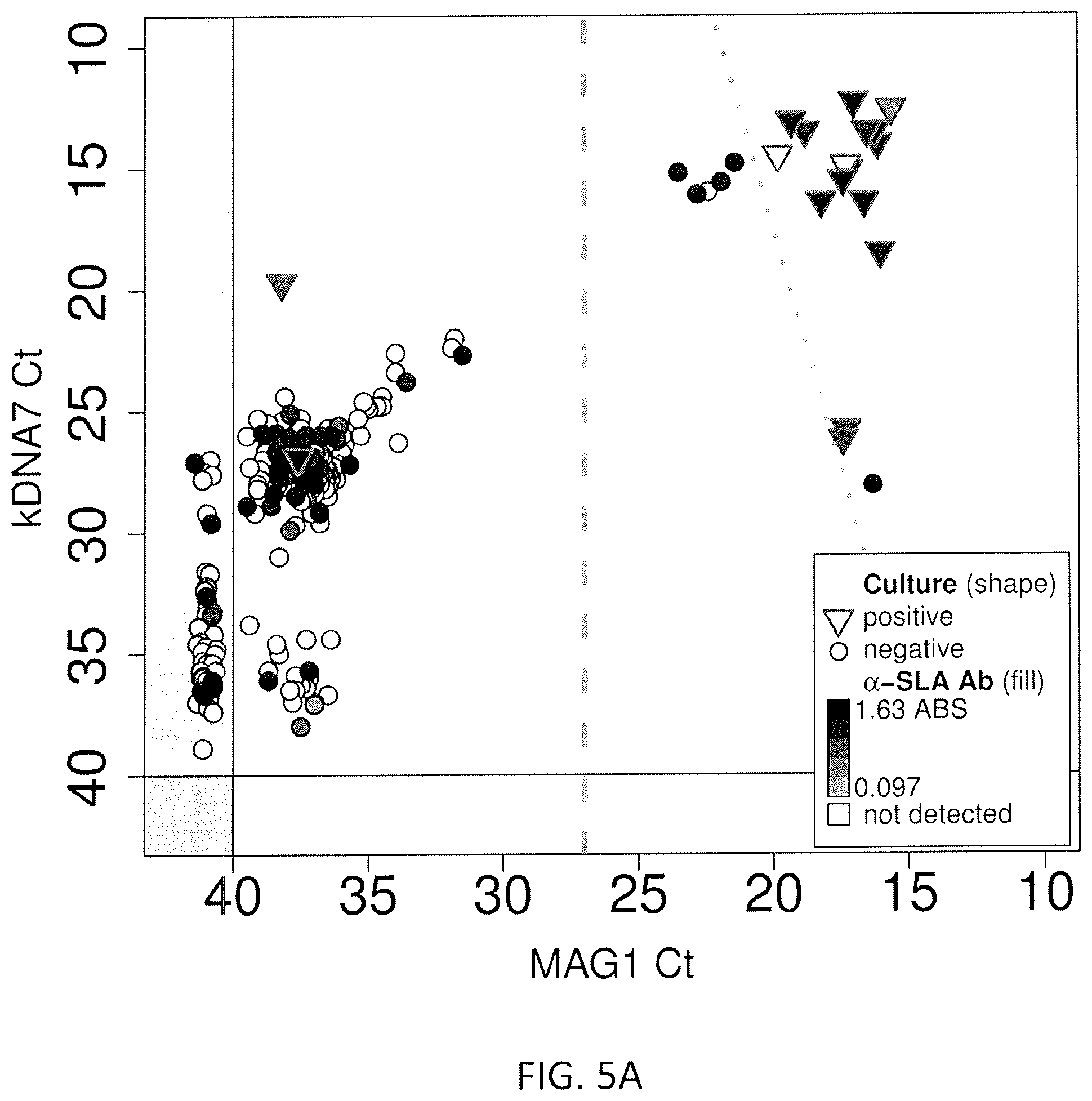
FIGS. 5A, 5B, and 5C: Results of the screening for Leishmania infection in blood samples obtained from apparent healthy donors, whose blood packs were rejected by any of the blood criteria screening. X axis presents the Ct for kDNA 7, Y axis presents the Ct for Mag1. The data was graphed with respect to Leishmania culture results (triangle=positive; sphere=negative). The shade within each symbol represents the intensity of the OD response obtained in the ELISA assay using SLA.
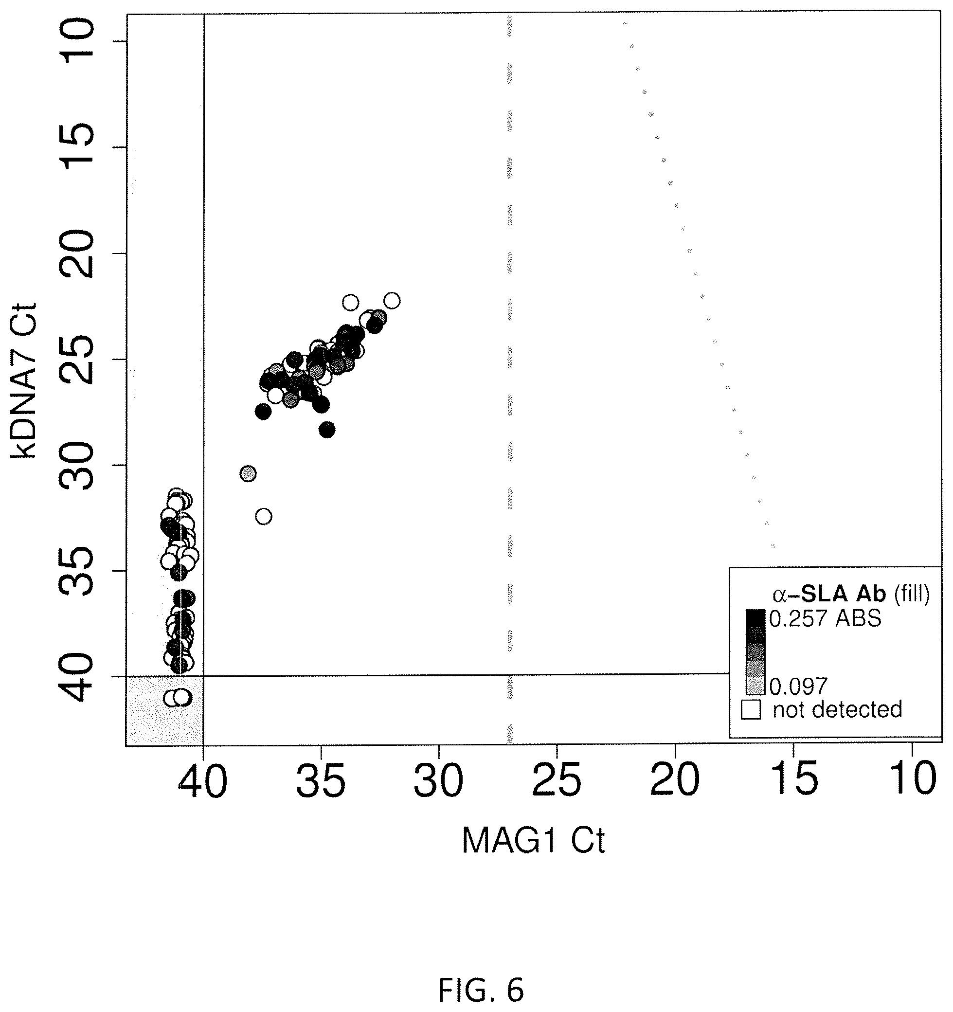
FIG. 6. Results of the screening for Leishmania infection in whole blood sample obtained from healthy blood donors. X axis presents the Ct for kDNA 7, Y axis presents the Ct for Mag1. The shade within each symbol represents the intensity of the OD response obtained in the ELISA assay using SLA.
FIG. 7. Positive predictive value of Leishmania infection in accordance to the local prevalence of infection using 4 of the six logistic models.
DETAILED DESCRIPTION OF THE INVENTION
Compositions of the Invention
The invention provides composition comprising novel nucleic acid sequences of the invention. Suitable examples of the nucleic acid sequence of the invention include but not limited to the following embodiments: (1) a nucleic acid sequence consisting of: GGGTAGGGGCGTTCTGC (SEQ ID NO:1) (kDNA 1 minicircle forward primer); (2) a nucleic acid sequence consisting of: TACACCAACCCCCAGTTTGC (SEQ ID NO:2) (kDNA 1 minicircle reverse primer); (3) a nucleic acid sequence consisting of: GGGTAGGGGCGTTCTGC (SEQ ID NO:3) (kDNA 3 minicircle forward primer); (4) a nucleic acid sequence consisting of: CCCGGCCTATTTTACACCAACC (SEQ ID NO:4) (kDNA 3 minicircle reverse primer); (5) a nucleic acid sequence consisting of: GGGTGCAGAAATCCCGTTCA (SEQ ID NO:5) (kDNA 4 minicircle forward primer); (6) a nucleic acid sequence consisting of: CCCGGCCCTATTTTACACCA (SEQ ID NO:6) (kDNA 4 minicircle reverse primer); (7) a nucleic acid sequence consisting of: AATGGGTGCAGAAATCCCGTTC (SEQ ID NO:7) (kDNA 7 minicircle forward primer); (8) a nucleic acid sequence consisting of: CCACCACCCGGCCCTATTTTAC (SEQ ID NO:8) (kDNA 7 minicircle reverse primer); (9) a nucleic acid sequence consisting of: GGTCCCGGCCCAAACTTTTC (SEQ ID NO:9) (L. (L.) amazonensis kDNA 1 forward primer); (10) a nucleic acid sequence consisting of: CCGGGGTTTCGCACTCATTT (SEQ ID NO:10) (L. (L.) amazonensis kDNA 1 reverse primer); (11) a nucleic acid sequence consisting of: GGTAGGGGCGTTCTGCGAAT (SEQ ID NO:11) (L. (L.) amazonensis kDNA 2 forward primer); (12) a nucleic acid sequence consisting of: CCCGGCCTATTTTACACCAACC (SEQ ID NO:12) (L. (L.) amazonensis kDNA 2 reverse primer); (13) a nucleic acid sequence consisting of: GGGTAGGGGCGTTCTGC (SEQ ID NO:13) (L. (L.) amazonensis kDNA 3 forward primer); (14) a nucleic acid sequence consisting of: TACACCAACCCCCAGTTTGC (SEQ ID NO:14) (L. (L.) amazonensis kDNA 3 reverse primer); (15) a nucleic acid sequence consisting of: TGAGTGCAGAAACCCCGTTCATA (SEQ ID NO:15) (L. (L.) amazonensis kDNA 4 forward primer); (16) a nucleic acid sequence consisting of: ACACCAACCCCCAGTTGTGA (SEQ ID NO:16) (L. (L.) amazonensis kDNA 4 reverse primer); (17) a nucleic acid sequence consisting of: AATTTCGCAGAACGCCCCTAC (SEQ ID NO:17) (L. (V.) braziliensis kDNA 1 forward primer); (18) a nucleic acid sequence consisting of: GTACTCCCCGACATGCCTCTG (SEQ ID NO:18) (L. (V.) braziliensis kDNA 1 reverse primer); (19) a nucleic acid sequence consisting of: ACGGGGTTTCTGCACCCATT (SEQ ID NO:19) (L. (L.) major Minicircle 1 forward primer); (20) a nucleic acid sequence consisting of: GTAGGGGCGTTCTGCGAAAA (SEQ ID NO:20) (L. (L.) major Minicircle 1 reverse primer); (21) a nucleic acid sequence consisting of: AATGCGAGTGTTGCCCTTTTG (SEQ ID NO:21) (L. (L.) mexicana Minicircle 1 forward primer); (22) a nucleic acid sequence consisting of: GCCGAACAACGCCATATTAACC (SEQ ID NO:22) (L. (L.) mexicana Minicircle 1 reverse primer); (23) a nucleic acid sequence consisting of: GGGGGTTGGTGTAAAATAGGG (SEQ ID NO:23) (L. (L.) tropica Minicircle 1 forward primer); (24) a nucleic acid sequence consisting of: ACCACCAGCAGAAGGTCAAAG (SEQ ID NO:24) (L. (L.) tropica Minicircle 1 reverse primer); (25) a nucleic acid sequence consisting of: GCGGTGGCTGGTTTTAGATG (SEQ ID NO:25) (L. (L.) donovani Minicircle 1 forward primer); (26) a nucleic acid sequence consisting of: TCCAATGAAGCCAAGCCAGT (SEQ ID NO:26) (L. (L.) donovani Minicircle 1 reverse primer); (27) a nucleic acid sequence consisting of: ATTTTAGTATGAGTGGTAGGTTTTGTT (SEQ ID NO:27) (Cytochrome B 1 forward primer); (28) a nucleic acid sequence consisting of: CAATAACTGGGACGGTTGCT (SEQ ID NO:28) (Cytochrome B 1 reverse primer); (29) a nucleic acid sequence consisting of: GCGGAGAGGAAAGAAAAGGCTTA (SEQ ID NO:29) (L. (L.) amazonensis Cytochrome B1 forward primer); (30) a nucleic acid sequence consisting of: AAAAGTCATGCTAAACACACACCACA (SEQ ID NO:30) (L. (L.) amazonensis Cytochrome B1 reverse primer); (31) a nucleic acid sequence consisting of: CAGGTTGCTTACTACGTGTTTATGGTG (SEQ ID NO:31) (L. (L.) tropica Cytochrome B 1 forward primer); (32) a nucleic acid sequence consisting of: TCGTATTACAAACCCTAAATCAAAATCTCA (SEQ ID NO:32) (L. (L.) tropica Cytochrome B 1 reverse primer); (33) a nucleic acid sequence consisting of: TCAGGTTGCTTACTACGTGTTTATGGTG (SEQ ID NO:33) (L. (L.) tropica Cytochrome B 2 forward primer); (34) a nucleic acid sequence consisting of: TGCTAAACAAACACCACATATGATCTGC (SEQ ID NO:34) (L. (L.) tropica Cytochrome B 2 reverse primer); (35) a nucleic acid sequence consisting of: TGACACACATATTTTAGTGTGGGTGGTAGG (SEQ ID NO:35) (L. (L.) tropica Cytochrome B 3 forward primer); (36) a nucleic acid sequence consisting of: TCCCCAATAAGACATCATTGTACATGGTAA (SEQ ID NO:36) (L. (L.) tropica Cytochrome B 3 reverse primer); (37) a nucleic acid sequence consisting of: CACATATTTTAGTGTGGGTGGTAGGTTTTG (SEQ ID NO:37) (L. (L.) tropica Cytochrome B 4 forward primer); (38) a nucleic acid sequence consisting of: TCCCCAATAAGACATCATTGTACATGGTAA (SEQ ID NO:38) (L. (L.) tropica Cytochrome B 4 reverse primer); (39) a nucleic acid sequence consisting of: GCTTGGTTGGATTATTTTTGCTG (SEQ ID NO:39) (Maxicircle 1 forward primer); (40) a nucleic acid sequence consisting of: AACAACATTTTAACTCTTGTAGGATTCG (SEQ ID NO:40) (Maxicircle 1 reverse primer); (41) a nucleic acid sequence consisting of: GAGGTGTTTGCCCGCATC (SEQ ID NO:41) (Alpha-tubulin 1 forward primer); (42) a nucleic acid sequence consisting of: CTCGCCCATGTCGTCG (SEQ ID NO:42) (Alpha-tubulin 1 reverse primer); (43) a nucleic acid sequence consisting of: AGGAGGATGGCAAGCGGAAG (SEQ ID NO:43) (DNA polymerase 2 forward primer); (44) a nucleic acid sequence consisting of: GCGACGGGTACAGGGAGTTG (SEQ ID NO:44) (DNA polymerase 2 reverse primer); (45) a nucleic acid sequence consisting of: CGAAACTTCCGGAACCTGTCTT (SEQ ID NO:45) (Mini-exon 1 forward primer); (46) a nucleic acid sequence consisting of: CACCACACGCACGCACAC (SEQ ID NO:46) (Mini-exon 1 reverse primer); (47) a nucleic acid sequence consisting of: GTGTGGTGGCGGGTGTATGT (SEQ ID NO:47) (Mini-exon 2 forward primer); (48) a nucleic acid sequence consisting of: GCCCAGGTCGCTGTGAGG (SEQ ID NO:48) (Mini-exon 2 reverse primer); (49) a nucleic acid sequence consisting of: AGAGCGTGCCTTGGATTGTG (SEQ ID NO:49) (MSP Associated Gene 1 (MAG 1) forward primer); (50) a nucleic acid sequence consisting of: CGCTGCGTTGATTGCGTTG (SEQ ID NO:50) (MSP Associated Gene 1 (MAG 1) reverse primer); (51) a nucleic acid sequence consisting of: AGTTTTGGTTGGCGCTCCTG (SEQ ID NO:51) (MSP Associated Gene 2 (MAG 2) forward primer); (52) a nucleic acid sequence consisting of: CCCACTCGCTTTCCTTGGTC (SEQ ID NO:52) (MSP Associated Gene 2 (MAG 2) reverse primer); (53) a nucleic acid sequence consisting of: CGACCCTGTCACCACCACAG (SEQ ID NO:53) (SIDER repeat 1 forward primer); (54) a nucleic acid sequence consisting of: GAGGCCACCCTATCGCTGAC (SEQ ID NO:54) (SIDER repeat 1 reverse primer); (55) a nucleic acid sequence consisting of: TCGTTGAGGGAGGAGGTGTTTC (SEQ ID NO:55) (L. (V.) braziliensis DNA polymerase 1 forward primer); (56) a nucleic acid sequence consisting of: TCGGCTTTGAGGTTGGCTTC (SEQ ID NO:56) (L. (V.) braziliensis DNA polymerase 1 reverse primer); (57) a nucleic acid sequence consisting of: ACGTCGCCAACTGCTTCACC (SEQ ID NO:57) (L. (V.) braziliensis DNA polymerase 2 forward primer); (58) a nucleic acid sequence consisting of: GTGTTCGCACCGCCTTGAC (SEQ ID NO:58) (L. (V.) braziliensis DNA polymerase 2 reverse primer); (59) a nucleic acid sequence consisting of: GTCGTTGTCCGTGTCGCTGT (SEQ ID NO:59) (L. (L.) major MSP associated gene 1 (L. major MAG 1) forward primer); (60) a nucleic acid sequence consisting of: CGCTGTGTGTGTCCGTGTGT (SEQ ID NO:60) (L. (L.) major MSP associated gene 1 (L. major MAG 1) reverse primer); (61) a nucleic acid sequence consisting of: GACGACGACGAGGAGGATGG (SEQ ID NO:61) (L. (L.) amazonensis DNA polymerase 1 forward primer); (62) a nucleic acid sequence consisting of: GCGACGGGTACAGGGAGTTG (SEQ ID NO:62) (L. (L.) amazonensis DNA polymerase 1 reverse primer); (63) a nucleic acid sequence consisting of: GAAGGTGCAGTCCCTCGTGT (SEQ ID NO:63) (HSP70-1 forward primer); (64) a nucleic acid sequence consisting of: CCTCCGTCTGCTTGCTCTTG (SEQ ID NO:64) (HSP70-1 reverse primer); (65) a nucleic acid sequence consisting of: TCGAGATCGACGCGTTGTT (SEQ ID NO:65) (HSP70-4 forward primer); (66) a nucleic acid sequence consisting of: CCGCACAGCTCCTCGAA (SEQ ID NO:66) (HSP70-4 reverse primer); (67) a nucleic acid sequence consisting of: GGAGAAACTCACGGCACAGG (SEQ ID NO:67) (SLACS forward primer); (68) a nucleic acid sequence consisting of: GCGCCTCGTAGGTCACAGTT (SEQ ID NO:68) (SLACS reverse primer); (69) a nucleic acid sequence consisting of: TGGAGCGGGTGCATTAACTC (SEQ ID NO:69) (Leptomonas Mini-exon 1 forward primer); (70) a nucleic acid sequence consisting of: GGTCTCGAGGTGCCCATGAC (SEQ ID NO:70) (Leptomonas Mini-exon 1 reverse primer); (71) a nucleic acid sequence consisting of: AGAAGCCGGATGTGCTTGTG (SEQ ID NO:71) (Leptomonas GAPDH 2 forward primer); (72) a nucleic acid sequence consisting of: GCCCTCAGCCTTCACCTTGT (SEQ ID NO:72) (Leptomonas GAPDH 2 reverse primer); (73) a nucleic acid sequence consisting of: GCCCTGTGAGGAGGACGAAC (SEQ ID NO:73) (Human TNF alpha 1 forward primer); (74) a nucleic acid sequence consisting of: AAGAGGTTGAGGGTGTCTGAAGGA (SEQ ID NO:74) (Human TNF alpha 1 reverse primer); (75) a nucleic acid sequence consisting of: GCGCTCCCCAAGAAGACAGG (SEQ ID NO:75) (Human TNF alpha 2 forward primer); (76) a nucleic acid sequence consisting of: TGCCACGATCAGGAAGGAGAAG (SEQ ID NO:76) (Human TNF alpha 2 reverse primer); (77) a nucleic acid sequence consisting of: GGGCTCTCCAGAACATCATCC (SEQ ID NO:77) (Human GAPDH 1 forward primer); (78) a nucleic acid sequence consisting of: CCAGTGAGCTTCCCGTTCAG (SEQ ID NO:78) (Human GAPDH 1 reverse primer); (79) a nucleic acid sequence consisting of: CATCAAGAAGGTGGTGAAGCAG (SEQ ID NO:79) (Human GAPDH 2 forward primer); (80) a nucleic acid sequence consisting of: CGTCAAAGGTGGAGGAGTGG (SEQ ID NO:80) (Human GAPDH 2 reverse primer); (81) a nucleic acid sequence consisting of: GCATGGCCTTCCGTGTCC (SEQ ID NO:81) (Human GAPDH 3 forward primer); and (82) a nucleic acid sequence consisting of: CGCCTGCTTCACCACCTTCT (SEQ ID NO:82) (Human GAPDH 3 reverse primer).
The invention further provides compositions comprising novel nucleic acid primer pairs which include but not limited to the following embodiments: (1) GGGTAGGGGCGTTCTGC (SEQ ID NO:1) and TACACCAACCCCCAGTTTGC (SEQ ID NO:2) (kDNA 1 minicircle forward and reverse primers, respectively); (2) GGGTAGGGGCGTTCTGC (SEQ ID NO:3) and CCCGGCCTATTTTACACCAACC (SEQ ID NO:4) (kDNA 3 minicircle forward and reverse primers, respectively); (3) GGGTGCAGAAATCCCGTTCA (SEQ ID NO:5) and CCCGGCCCTATTTTACACCA (SEQ ID NO:6) (kDNA 4 minicircle forward and reverse primers, respectively); (4) AATGGGTGCAGAAATCCCGTTC (SEQ ID NO:7) and CCACCACCCGGCCCTATTTTAC (SEQ ID NO:8) (kDNA 7 minicircle forward and reverse primers, respectively); (5) GGTCCCGGCCCAAACTTTTC (SEQ ID NO:9) and CCGGGGTTTCGCACTCATTT (SEQ ID NO:10) (L. (L.) amazonensis kDNA 1 forward and reverse primers, respectively); (6) GGTAGGGGCGTTCTGCGAAT (SEQ ID NO:11) and CCCGGCCTATTTTACACCAACC (SEQ ID NO:12) (L. (L.) amazonensis kDNA 2 forward and reverse primers, respectively); (7) GGGTAGGGGCGTTCTGC (SEQ ID NO:13) and TACACCAACCCCCAGTTTGC (SEQ ID NO:14) (L. (L.) amazonensis kDNA 3 forward and reverse primers, respectively); (8) TGAGTGCAGAAACCCCGTTCATA (SEQ ID NO:15) and ACACCAACCCCCAGTTGTGA (SEQ ID NO:16) (L. (L.) amazonensis kDNA 4 forward and reverse primers, respectively); (9) AATTTCGCAGAACGCCCCTAC (SEQ ID NO:17) and GTACTCCCCGACATGCCTCTG (SEQ ID NO:18) (L. (V.) braziliensis kDNA 1 forward and reverse primers, respectively); (10) ACGGGGTTTCTGCACCCATT (SEQ ID NO:19) and GTAGGGGCGTTCTGCGAAAA (SEQ ID NO:20) (L. (L.) major Minicircle 1 forward and reverse primers, respectively); (11) AATGCGAGTGTTGCCCTTTTG (SEQ ID NO:21) and GCCGAACAACGCCATATTAACC (SEQ ID NO:22) (L. (L.) mexicana Minicircle 1 forward and reverse primers, respectively); (12) GGGGGTTGGTGTAAAATAGGG (SEQ ID NO:23) and ACCACCAGCAGAAGGTCAAAG (SEQ ID NO:24) (L. (L.) tropica Minicircle 1 forward and reverse primers, respectively); (13) GCGGTGGCTGGTTTTAGATG (SEQ ID NO:25) and TCCAATGAAGCCAAGCCAGT (SEQ ID NO:26) (L. (L.) donovani Minicircle 1 forward and reverse primers, respectively); (14) ATTTTAGTATGAGTGGTAGGTTTTGTT (SEQ ID NO:27) and CAATAACTGGGACGGTTGCT (SEQ ID NO:28) (Cytochrome B 1 forward and reverse primers, respectively); (15) GCGGAGAGGAAAGAAAAGGCTTA (SEQ ID NO:29) and AAAAGTCATGCTAAACACACACCACA (SEQ ID NO:30) (L. (L.) amazonensis Cytochrome B1 forward and reverse primers, respectively); (16) CAGGTTGCTTACTACGTGTTTATGGTG (SEQ ID NO:31) and TCGTATTACAAACCCTAAATCAAAATCTCA (SEQ ID NO:32) (L. (L.) tropica Cytochrome B 1 forward and reverse primers, respectively); (17) TCAGGTTGCTTACTACGTGTTTATGGTG (SEQ ID NO:33) and TGCTAAACAAACACCACATATGATCTGC (SEQ ID NO:34) (L. (L.) tropica Cytochrome B 2 forward and reverse primers, respectively); (18) TGACACACATATTTTAGTGTGGGTGGTAGG (SEQ ID NO:35) and TCCCCAATAAGACATCATTGTACATGGTAA (SEQ ID NO:36) (L. (L.) tropica Cytochrome B 3 forward and reverse primers, respectively); (19) CACATATTTTAGTGTGGGTGGTAGGTTTTG (SEQ ID NO:37) and TCCCCAATAAGACATCATTGTACATGGTAA (SEQ ID NO:38) (L. (L.) tropica Cytochrome B 4 forward and reverse primers, respectively); (20) GCTTGGTTGGATTATTTTTGCTG (SEQ ID NO:39) and AACAACATTTTAACTCTTGTAGGATTCG (SEQ ID NO:40) (Maxicircle 1 forward and reverse primers, respectively); (21) GAGGTGTTTGCCCGCATC (SEQ ID NO:41) and CTCGCCCATGTCGTCG (SEQ ID NO:42) (Alpha-tubulin 1 forward and reverse primers, respectively); (22) AGGAGGATGGCAAGCGGAAG (SEQ ID NO:43) and GCGACGGGTACAGGGAGTTG (SEQ ID NO:44) (DNA polymerase 2 forward and reverse primers, respectively); (23) CGAAACTTCCGGAACCTGTCTT (SEQ ID NO:45) and CACCACACGCACGCACAC (SEQ ID NO:46) (Mini-exon 1 forward and reverse primers, respectively); (24) GTGTGGTGGCGGGTGTATGT (SEQ ID NO:47) and GCCCAGGTCGCTGTGAGG (SEQ ID NO:48) (Mini-exon 2 forward and reverse primers, respectively); (25) AGAGCGTGCCTTGGATTGTG (SEQ ID NO:49) and CGCTGCGTTGATTGCGTTG (SEQ ID NO:50) (MSP Associated Gene 1 (MAG 1) forward and reverse primers, respectively); (26) AGTTTTGGTTGGCGCTCCTG (SEQ ID NO:51) and CCCACTCGCTTTCCTTGGTC (SEQ ID NO:52) (MSP Associated Gene 2 (MAG 2) forward and reverse primers, respectively); (27) CGACCCTGTCACCACCACAG (SEQ ID NO:53) and GAGGCCACCCTATCGCTGAC (SEQ ID NO:54) (SIDER repeat 1 forward and reverse primers, respectively); (28) TCGTTGAGGGAGGAGGTGTTTC (SEQ ID NO:55) and TCGGCTTTGAGGTTGGCTTC (SEQ ID NO:56) (L. (V.) braziliensis DNA polymerase 1 forward and reverse primers, respectively); (29) ACGTCGCCAACTGCTTCACC (SEQ ID NO:57) and GTGTTCGCACCGCCTTGAC (SEQ ID NO:58) (L. (V.) braziliensis DNA polymerase 2 forward and reverse primers, respectively); (30) GTCGTTGTCCGTGTCGCTGT (SEQ ID NO:59) and CGCTGTGTGTGTCCGTGTGT (SEQ ID NO:60) (L. (L.) major MSP associated gene 1 (L. major MAG 1) forward and reverse primers, respectively); (31) GACGACGACGAGGAGGATGG (SEQ ID NO:61) and GCGACGGGTACAGGGAGTTG (SEQ ID NO:62) (L. (L.) amazonensis DNA polymerase 1 forward and reverse primers, respectively); (32) GAAGGTGCAGTCCCTCGTGT (SEQ ID NO:63) and CCTCCGTCTGCTTGCTCTTG (SEQ ID NO:64) (HSP70-1 forward and reverse primers, respectively); (33) TCGAGATCGACGCGTTGTT (SEQ ID NO:65) and CCGCACAGCTCCTCGAA (SEQ ID NO:66) (HSP70-4 forward and reverse primers, respectively); (34) GGAGAAACTCACGGCACAGG (SEQ ID NO:67) and GCGCCTCGTAGGTCACAGTT (SEQ ID NO:68) (SLACS forward and reverse primers, respectively); (35) TGGAGCGGGTGCATTAACTC (SEQ ID NO:69) and GGTCTCGAGGTGCCCATGAC (SEQ ID NO:70) (Leptomonas Mini-exon 1 forward and reverse primers, respectively); (36) AGAAGCCGGATGTGCTTGTG (SEQ ID NO:71) and GCCCTCAGCCTTCACCTTGT (SEQ ID NO:72) (Leptomonas GAPDH 2 forward and reverse primers, respectively); (37) GCCCTGTGAGGAGGACGAAC (SEQ ID NO:73) and AAGAGGTTGAGGGTGTCTGAAGGA (SEQ ID NO:74) (Human TNF alpha 1 forward and reverse primers, respectively); (38) GCGCTCCCCAAGAAGACAGG (SEQ ID NO:75) and TGCCACGATCAGGAAGGAGAAG (SEQ ID NO:76) (Human TNF alpha 2 forward and reverse primers, respectively); (39) GGGCTCTCCAGAACATCATCC (SEQ ID NO:77) and CCAGTGAGCTTCCCGTTCAG (SEQ ID NO:78) (Human GAPDH 1 forward and reverse primers, respectively); (40) CATCAAGAAGGTGGTGAAGCAG (SEQ ID NO:79) and CGTCAAAGGTGGAGGAGTGG (SEQ ID NO:80) (Human GAPDH 2 forward and reverse primers, respectively); and (41) GCATGGCCTTCCGTGTCC (SEQ ID NO:81) and CGCCTGCTTCACCACCTTCT (SEQ ID NO:82) (Human GAPDH 3 forward and reverse primers, respectively).
In another embodiment, the invention provides for composition comprising a novel nucleic acid probe sequences (also referred to as a third nucleic acid probe) which include but are not limited to the following embodiments: (1) a nucleic acid sequence consisting of ACCCCCAGTTTCCCGCCCCG (SEQ ID NO:83); (2) a nucleic acid sequence consisting of CCCCAGTTTCCCGCCCCGGA (SEQ ID NO:84); (3) a nucleic acid sequence consisting of TGGCCTTGGGGCGTGCAAACTGG (SEQ ID NO:85); (4) a nucleic acid sequence consisting of TCCTGGCGGGGGTTTTCGCT (SEQ ID NO:86); (5) a nucleic acid sequence consisting of CCCATACCACCAAACGCAGCCCA (SEQ ID NO:87); (6) a nucleic acid sequence consisting of CCATGTACGATGATGTCGTATTGAGGTCTAACA (SEQ ID NO:88); (7) a nucleic acid sequence consisting of CTTTAGGTAGGGAGTTGTACTACGTTTTTTGACCT (SEQ ID NO:89); (8) a nucleic acid sequence consisting of TGAGGGCATGGAGGAGGGCG (SEQ ID NO:90); (9) a nucleic acid sequence consisting of TGGGGTCGAGCACCATGCCGCC (SEQ ID NO:91); (10) a nucleic acid sequence consisting of CGGCAAGATTTTGGAAGCGCGCA (SEQ ID NO:92); (11) a nucleic acid sequence consisting of TGCGCACTGCACTGTCGCCCCC (SEQ ID NO:93); (12) a nucleic acid sequence consisting of CGCTGAGAGCGAGGCAGGCACGC (SEQ ID NO:94); (13) a nucleic acid sequence consisting of CCTTCCCAAACGCCTCCCCTGCCCC (SEQ ID NO:95); and (14) a nucleic acid sequence consisting of CACCGCCTGGAGCCCTGGGGC (SEQ ID NO:96).
METHODS OF THE INVENTION
The invention provides a method for determining the presence, species, and/or quantity of a Leishmania in a sample.
In one embodiment of the invention, the method comprises contacting the sample with a set of nucleic acid primer pairs with primer sequences so as to obtain Leishmania-primer pair complexes. The primer sequences may be common to all Leishmania species or to a subset of Leishmania species. The nucleic acid primer pairs may be deoxyribonucleic acid (DNA) or a modified deoxyribonucleic acid. In another example, the nucleic acid primer pairs are ribonucleic acid (RNA) or modified ribonucleic acid. In a further example, the nucleic acid primer pairs are mixed RNA-DNA hybrid or modified RNA-DNA hybrid.
Examples of nucleic acid primer pairs include, but are not limited to: GGGTAGGGGCGTTCTGC (SEQ ID NO:1) and TACACCAACCCCCAGTTTGC (SEQ ID NO:2) (kDNA 1 minicircle forward and reverse primers, respectively); AACTTTTCTGGTCCTCCGGGTAG (SEQ ID NO:97) and ACCCCCAGTTTCCCGCC (SEQ ID NO:98) (kDNA 2 forward and reverse primers, respectively); GGGTAGGGGCGTTCTGC (SEQ ID NO:3) and CCCGGCCTATTTTACACCAACC (SEQ ID NO:4) (kDNA 3 minicircle forward and reverse primers, respectively); GGGTGCAGAAATCCCGTTCA (SEQ ID NO:5) and CCCGGCCCTATTTTACACCA (SEQ ID NO:6) (kDNA 4 minicircle forward and reverse primers, respectively); CTTTTCTGGTCCTCCGGGTAGG (SEQ ID NO:99) and CCACCCGGCCCTATTTTACACCAA (SEQ ID NO:100) (kDNA 5 minicircle forward and reverse primers, respectively); AATGGGTGCAGAAATCCCGTTC (SEQ ID NO:7) and CCACCACCCGGCCCTATTTTAC (SEQ ID NO:8) (kDNA 7 minicircle forward and reverse primers, respectively); GGTCCCGGCCCAAACTTTTC (SEQ ID NO:9) and CCGGGGTTTCGCACTCATTT (SEQ ID NO:10) (L. (L.) amazonensis kDNA 1 forward and reverse primers, respectively); GGTAGGGGCGTTCTGCGAAT (SEQ ID NO:11) and CCCGGCCTATTTTACACCAACC (SEQ ID NO:12) (L. (L.) amazonensis kDNA 2 forward and reverse primers, respectively); GGGTAGGGGCGTTCTGC (SEQ ID NO:13) and TACACCAACCCCCAGTTTGC (SEQ ID NO:14) (L. (L.) amazonensis kDNA 3 forward and reverse primers, respectively); TGAGTGCAGAAACCCCGTTCATA (SEQ ID NO:15) and ACACCAACCCCCAGTTGTGA (SEQ ID NO:16) (L. (L.) amazonensis kDNA 4 forward and reverse primers, respectively); AATTTCGCAGAACGCCCCTAC (SEQ ID NO:17) and GTACTCCCCGACATGCCTCTG (SEQ ID NO:18) (L. (V.) braziliensis kDNA 1 forward and reverse primers, respectively); TGCTATAAAATCGTACCACCCGACA (SEQ ID NO:101) and GAACGGGGTTTCTGTATGCCATTT (SEQ ID NO:1102) (L. (V.) braziliensis kDNA 3 forward and reverse primers, respectively); TCCGCAGGAGACTTCGTATG (SEQ ID NO:103) and CACGACTATCCACCCCATCC (SEQ ID NO:104) (L. (L.) infantum Minicircle 1 forward and reverse primers, respectively); ACGGGGTTTCTGCACCCATT (SEQ ID NO:19) and GTAGGGGCGTTCTGCGAAAA (SEQ ID NO:20) (L. (L.) major Minicircle 1 forward and reverse primers, respectively); AATGCGAGTGTTGCCCTTTTG (SEQ ID NO:21) and GCCGAACAACGCCATATTAACC (SEQ ID NO:22) (L. (L.) mexicana Minicircle 1 forward and reverse primers, respectively); GGGGGTTGGTGTAAAATAGGG (SEQ ID NO:23) and ACCACCAGCAGAAGGTCAAAG (SEQ ID NO:24) (L. (L.) tropica Minicircle 1 forward and reverse primers, respectively); GCGGTGGCTGGTTTTAGATG (SEQ ID NO:25) and TCCAATGAAGCCAAGCCAGT (SEQ ID NO:26) (L. (L.) donovani Minicircle 1 forward and reverse primers, respectively); ATTTTAGTATGAGTGGTAGGTTTTGTT (SEQ ID NO:27) and CAATAACTGGGACGGTTGCT (SEQ ID NO:28) (Cytochrome B 1 forward and reverse primers, respectively); GCGGAGAGGAAAGAAAAGGCTTA (SEQ ID NO:29) and AAAAGTCATGCTAAACACACACCACA (SEQ ID NO:30) (L. (L.) amazonensis Cytochrome B1 forward and reverse primers, respectively); CAGGTTGCTTACTACGTGTTTATGGTG (SEQ ID NO:31) and TCGTATTACAAACCCTAAATCAAAATCTCA (SEQ ID NO:32) (L. (L.) tropica Cytochrome B 1 forward and reverse primers, respectively); TCAGGTTGCTTACTACGTGTTTATGGTG (SEQ ID NO:33) and TGCTAAACAAACACCACATATGATCTGC (SEQ ID NO:34) (L. (L.) tropica Cytochrome B 2 forward and reverse primers, respectively); TGACACACATATTTTAGTGTGGGTGGTAGG (SEQ ID NO:35) and TCCCCAATAAGACATCATTGTACATGGTAA (SEQ ID NO:36) (L. (L.) tropica Cytochrome B 3 forward and reverse primers, respectively); CACATATTTTAGTGTGGGTGGTAGGTTTTG (SEQ ID NO:37) and TCCCCAATAAGACATCATTGTACATGGTAA (SEQ ID NO:38) (L. (L.) tropica Cytochrome B 4 forward and reverse primers, respectively); GCTTGGTTGGATTATTTTTGCTG (SEQ ID NO:39) and AACAACATTTTAACTCTTGTAGGATTCG (SEQ ID NO:40) (Maxicircle 1 forward and reverse primers, respectively); GAGGTGTTTGCCCGCATC (SEQ ID NO:41) and CTCGCCCATGTCGTCG (SEQ ID NO:42) (Alpha-tubulin 1 forward and reverse primers, respectively); TGTCGCTTGCAGACCAGATG (SEQ ID NO:105) and GCATCGCAGGTGTGAGCA (SEQ ID NO:106) (DNA polymerase 1 forward and reverse primers, respectively); AGGAGGATGGCAAGCGGAAG (SEQ ID NO:43) and GCGACGGGTACAGGGAGTTG (SEQ ID NO:44) (DNA polymerase 2 forward and reverse primers, respectively); CGAAACTTCCGGAACCTGTCTT (SEQ ID NO:45) and CACCACACGCACGCACAC (SEQ ID NO:46) (Mini-exon 1 forward and reverse primers, respectively); GTGTGGTGGCGGGTGTATGT (SEQ ID NO:47) and GCCCAGGTCGCTGTGAGG (SEQ ID NO:48) (Mini-exon 2 forward and reverse primers, respectively); AGAGCGTGCCTTGGATTGTG (SEQ ID NO:49) and CGCTGCGTTGATTGCGTTG (SEQ ID NO:50) (MSP Associated Gene 1 (MAG 1) forward and reverse primers, respectively); AGTTTTGGTTGGCGCTCCTG (SEQ ID NO:51) and CCCACTCGCTTTCCTTGGTC (SEQ ID NO:52) (MSP Associated Gene 2 (MAG 2) forward and reverse primers, respectively); CGACCCTGTCACCACCACAG (SEQ ID NO:53) and GAGGCCACCCTATCGCTGAC (SEQ ID NO:54) (SIDER repeat 1 forward and reverse primers, respectively); TCGTTGAGGGAGGAGGTGTTTC (SEQ ID NO:55) and TCGGCTTTGAGGTTGGCTTC (SEQ ID NO:56) (L. (V.) braziliensis DNA polymerase 1 forward and reverse primers, respectively); ACGTCGCCAACTGCTTCACC (SEQ ID NO:57) and GTGTTCGCACCGCCTTGAC (SEQ ID NO:58) (L. (V.) braziliensis DNA polymerase 2 forward and reverse primers, respectively); GTCGTTGTCCGTGTCGCTGT (SEQ ID NO:59) and CGCTGTGTGTGTCCGTGTGT (SEQ ID NO:60) (L. (L.) major MSP associated gene 1 (L. major MAG 1) forward and reverse primers, respectively); GACGACGACGAGGAGGATGG (SEQ ID NO:61) and GCGACGGGTACAGGGAGTTG (SEQ ID NO:62) (L. (L.) amazonensis DNA polymerase 1 forward and reverse primers, respectively); CCAGATGCCGACCAAAGC (SEQ ID NO:107) and CGCGCACGTGATGGATAAC (SEQ ID NO:108) (GPI forward and reverse primers, respectively); GAAGGTGCAGTCCCTCGTGT (SEQ ID NO:63) and CCTCCGTCTGCTTGCTCTTG (SEQ ID NO:64) (HSP70-1 forward and reverse primers, respectively); TCGAGATCGACGCGTTGTT (SEQ ID NO:65) and CCGCACAGCTCCTCGAA (SEQ ID NO:66) (HSP70-4 forward and reverse primers, respectively); GGAGAAACTCACGGCACAGG (SEQ ID NO:67) and GCGCCTCGTAGGTCACAGTT (SEQ ID NO:68) (SLACS forward and reverse primers, respectively); TGGAGCGGGTGCATTAACTC (SEQ ID NO:69) and GGTCTCGAGGTGCCCATGAC (SEQ ID NO:70) (Leptomonas Mini-exon 1 forward and reverse primers, respectively); AGAAGCCGGATGTGCTTGTG (SEQ ID NO:71) and GCCCTCAGCCTTCACCTTGT (SEQ ID NO:72) (Leptomonas GAPDH 2 forward and reverse primers, respectively); GCCCTGTGAGGAGGACGAAC (SEQ ID NO:73) and AAGAGGTTGAGGGTGTCTGAAGGA (SEQ ID NO:74) (Human TNF alpha 1 forward and reverse primers, respectively); GCGCTCCCCAAGAAGACAGG (SEQ ID NO:75) and TGCCACGATCAGGAAGGAGAAG (SEQ ID NO:76) (Human TNF alpha 2 forward and reverse primers, respectively); GGGCTCTCCAGAACATCATCC (SEQ ID NO:77) and CCAGTGAGCTTCCCGTTCAG (SEQ ID NO:78) (Human GAPDH 1 forward and reverse primers, respectively); CATCAAGAAGGTGGTGAAGCAG (SEQ ID NO:79) and CGTCAAAGGTGGAGGAGTGG (SEQ ID NO:80) (Human GAPDH 2 forward and reverse primers, respectively); and GCATGGCCTTCCGTGTCC (SEQ ID NO:81) and CGCCTGCTTCACCACCTTCT (SEQ ID NO:82) (Human GAPDH 3 forward and reverse primers, respectively).
Examples of Leishmania include but are not limited to Leishmania tropica, Leishmania chagasi, Leishmania infantum, Leishmania donovani, Leishmania major, Leishmania braziliensis, Leishmania guyanensis, Leishmania panamensis, Leishmania mexicana, and Leishmania amazonensis.
The method may further comprise exposing the Leishmania-primer pair to a thermo-stable polymerase (for example, a thermo-stable DNA polymerase) so as to produce double-stranded DNAs containing the primer sequences. The double stranded nucleic acid so produced may be several kilo-basepairs, less than several kilo-basepairs, less than 1 kilo-basepairs, less than 500 basepairs, less than 300 basepairs, less than 200 basepairs, or, less than 150 basepairs. For example, the double-stranded nucleic acid so produced may be less than 200 basepairs and most are less than 150 basepairs.
Such thermo-stable DNA polymerase are known in the art and include any of Taq polymerase from Thermus aquaticus, Pfu DNA polymerase Pyrococcus furiosus, Vent polymerase from Thermococcus litoralis, Tth polymerase from Thermus thermophilus, or Bst DNA polymerase from Bacillus stearothermophilus but are not limited to these examples.
In addition to being able to replicate DNA, thermo-stable polymerases may have 5'-to-3' exonuclease activity, 3'-to-5' exonuclease activity ("proof-reading" activity), reverse transcriptase activity, or strong strand displacement activity. The polymerase may be purified from its original organism and subsequently modified to obtain desired activity, or alternatively, produced by recombinant DNA methods true to its primary amino acid sequence or alternatively modified or truncated so as to have desired properties. In addition, the purified native or recombinant thermo-stable polymerase may be further treated with an antibody or antibodies or with chemical or chemicals so as to obtain a polymerase characteristic suitable for a particular application, as is known in the art.
Examples of recombinant modified polymerase include (1) AmpliTaq DNA polymerase (Applied Biosystems, Inc.), which is a recombinant, thermostable, 94 kDa DNA polymerase with 5'-to-3' exonuclease activity but no 3'-to-5' exonuclease activity encoded by a modified form of the Thermus aquaticus DNA polymerase gene which is expressed and purified from an Escherichia coli host; (2) AmpliTaq Gold.RTM. DNA polymerase (Applied Biosystems, Inc.), which a chemically modified form of AmpliTaq.RTM. DNA Polymerase that requires heat above the annealing temperature of primer to its template to activate its enzymatic activity; (3) AmpliTaq Gold.RTM. DNA polymerase, LD (Applied Biosystems, Inc.), which is a highly purified form of AmpliTaq Gold.RTM. DNA polymerase; (4) AmpliTaq.RTM. DNA polymerase, Stoffel Fragment, which is a modified form of AmpliTaq.RTM. DNA Polymerase from which the N-terminal 289 amino acids are deleted thereby changing it enzymatic property such that it is twice as thereto-stable with broader range of optimal polymerase activity from 2 mM to 10 mM magnesium and loss of 5'-to-3' exonuclease activity; (5) rTth DNA polymerase (Applied Biosciences, Inc.) is an ultrapure, thermostable recombinant DNA polymerase with ability to reverse transcribe RNA to cDNA in the presence of Mn.sup.+2 ion, and to also act as a DNA polymerase for PCR amplification; and (6) rTth DNA Polymerase, XL (Applied Biosystems, Inc.) which is an ultra-pure, thermostable recombinant DNA polymerase blend, designed for amplification of DNA or RNA targets>5 kb [XL (eXtra Long) PCR] and is formulated to include the optimal amount of 3'-to-5' exonuclease activity.
Examples of suitable primer pairs are found in Table 2. Additionally, primer pairs which are common to Leishmania species, include, but are not limited to: GGGTAGGGGCGTTCTGC (SEQ ID NO:3) and CCCGGCCTATTTTACACCAACC (SEQ ID NO:4) (e.g., referred to in some later examples as kDNA 3 mini-circle forward and reverse primers, respectively); GGGTGCAGAAATCCCGTTCA (SEQ ID NO:5) and CCCGGCCCTATTTTACACCA (SEQ ID NO:6) (e.g., referred to in some later examples as kDNA 4 mini-circle forward and reverse primers, respectively); GGGTAGGGGCGTTCTGC (SEQ ID NO:1) and TACACCAACCCCCAGTTTGC (SEQ ID NO:2) (e.g., referred to in some later examples as kDNA 1 mini-circle forward and reverse primers, respectively); AATGGGTGCAGAAATCCCGTTC (SEQ ID NO:7) and CCACCACCCGGCCCTATTTTAC (SEQ ID NO:8) (e.g., referred to in some later examples as kDNA 7 mini-circle forward and reverse primers, respectively); GGGTAGGGGCGTTCTGC (SEQ ID NO:13) and TACACCAACCCCCAGTTTGC (SEQ ID NO:14) (e.g., referred to in some later examples as L. amazonensis kDNA 3 forward and reverse primers, respectively); AGGAGGATGGCAAGCGGAAG (SEQ ID NO:43) and GCGACGGGTACAGGGAGTTG (SEQ ID NO:44) (e.g., referred to in some later examples as DNA polymerase 2 forward and reverse primers, respectively); CGAAACTTCCGGAACCTGTCTT (SEQ ID NO:45) and CACCACACGCACGCACAC (SEQ ID NO:46) (e.g., referred to in some later examples as mini-exon 1 forward and reverse primers, respectively); GTCGTTGTCCGTGTCGCTGT (SEQ ID NO:59) and CGCTGTGTGTGTCCGTGTGT (SEQ ID NO:60 (e.g., referred to in some later examples as L. major MSP associated gene 1 forward and reverse primers, respectively); GACGACGACGAGGAGGATGG (SEQ ID NO:61) and GCGACGGGTACAGGGAGTTG (SEQ ID NO:62) (e.g., referred to in some later examples as L. amazonensis DNA polymerase I forward and reverse primers, respectively); GAAGGTGCAGTCCCTCGTGT (SEQ ID NO:63) and CCTCCGTCTGCTTGCTCTTG (SEQ ID NO:64) (e.g., referred to in some later examples as HSP70-1 forward and reverse primers, respectively); and TCGAGATCGACGCGTTGTT (SEQ ID NO:65) and CCGCACAGCTCCTCGAA (SEQ ID NO:66) (e.g., referred to in some later examples as HSP70-4 forward and reverse primers, respectively). As is the convention, sequences throughout this application are written 5' to 3' in the same direction that the text is read.
In one embodiment, at least one of the primers may be attached to a detectable marker. In another embodiment, the primer attached to the detectable marker may also be attached to a quencher.
The double-stranded DNAs containing the primer sequences may be maintained in the same temperature or may be cycled between at least two different temperatures over a suitable time period to produce multiple double-stranded DNAs containing the primer sequences. In an embodiment of the invention, when the double-stranded DNAs containing the primer sequences are cycled between at least two different temperatures, the temperature differential is more than about an 18.degree. C. differential. In another embodiment, the temperature is cycled between 95.degree. C. and 60.degree. C. Additionally, in a further embodiment, the temperature is cycled for 40 cycles, but preferably measurements are read from, e.g., SYBR green assays, at 30 cycles or less. This cycle limit is based on the observation that primer dimers often occur after 30 cycles in e.g., SYBR green reactions. In yet another embodiment, the temperature is constant. In an additional embodiment, the temperature may be around 60.degree. C. to 65.degree. C.
Monitoring for the presence of double-stranded DNA or pyrophosphate are well-known in the art and include detection through ultraviolet absorption, fluorescence, light scattering, colorimetric, or chromogenic detection. In one embodiment, fluorescence detection includes the use of SYBR.RTM. Green Dye I as a minor groove binder for double-stranded DNA or ethidium bromide as an intercalator of double-stranded DNA or calcein as an indicator for the presence of pyrophosphate. For example, fluorescence obtained from SYBR.RTM. Green Dye I binding to double-stranded DNA can be used to monitor the production of double-stranded DNA in a real-time PCR system in which a 488 nm laser line from an argon ion laser is used to excite the fluorophore-DNA complex (peak absorption of 497 nm) and subsequent monitoring of its emission at a longer wavelength (peak emission of 520 nm). Other detection means are possible and are well known in the art.
In another example, ethidium bromide can be used to stain and quantify double-stranded DNA, which can be detected or visualized by virtue of its fluorescence following excitation by ultraviolet light.
In a third example, using manganese ion-bound calcein, the presence of pyrophosphate can be detected following displacement of manganese ion from calcein by pyrophosphate resulting in calcein fluorescence further enhanced in the presence of magnesium ion. In one embodiment, light scattering detection includes assessing the turbidity of pyrophosphate salt. The pyrophosphate salt may be magnesium pyrophosphate. In the current invention, the preferred embodiment is the detection of double-stranded DNA through SYBR.RTM. Green Dye I staining. However, other dyes are possible and well known in the art.
The method may further include a step of determining the time or number of temperature cycles at which a threshold level of double-stranded DNA product and/or pyrophosphate product is reached which may be indicative of on-target specificity and/or quantity of Leishmania in the sample. As known in the art (Applied Biosystems, Inc., Application Note, "Real-time PCR: Understanding Ct" at link http://www3.appliedbiosystems.com/cms/groups/mcb_marketing/documents/gene- raldocuments/cms_053906.pdf), the threshold level is set at a level corresponding to the exponential phase of DNA amplification. In one example, the threshold level is 0.2 based on SYBER.RTM. Green Dye I detection method for presence of double-stranded DNA performed in a 7900 fast real-time PCR system from Applied Biosystems, Inc., and the temperature is cycled for less than about 30 cycles. In other examples, the temperature is cycled for 40 cycles but preferably 30 cycles or less. The melting temperature of the double-stranded DNA product may be determined and a characteristic melting temperature is indicative of particular Leishmania species, either one particular species or a group of species, so detected by the primer pair.
Melting temperatures of any of the following probes can be useful for distinguishing between the Leishmania species: DNA polymerase 2, GPI, HSP70-4, kDNA 1 minicircle, kDNA 2 minicircle, kDNA 3 minicircle, kDNA 4 minicircle, kDNA 5 minicircle, L. amazonensis DNA polymerase 1, L. amazonensis kDNA 1, L. amazonensis kDNA 2, L. amazonensis kDNA 3, L. braziliensis DNA polymerase 2, L. major MAG 1, L. tropica Cytochrome B, mini-exon 1, and mini-exon 2, MAG 1. Table 7 and FIG. 1 provide examples.
TABLE-US-00001 TABLE 7 Melting temperature of selected SYBR green assays are listed below to exemplify the relative differences in Tm of double stranded DNA products produced from templates of different Leishmania species. The absolute Tm may vary from lab to lab because of slight differences in reagents used. Also, the below list is not exhaustive. Approximate Melt Temperature Primer set Species .degree. C. (.+-.1-2.degree. C.) DNA polymerase 2 L. (L.) chagasi 82.2 DNA polymerase 2 L. (L.) donovani 82.2 DNA polymerase 2 L. (V.) braziliensis 80.9 HSP70-4 L. (L.) amazonensis 81.8 HSP70-4 L. (L.) tropica 83.2 HSP70-4 L. (V.) braziliensis 82.9 L. (L.) amazonensis kDNA 1 L. (L.) major 80.9 L. (L.) amazonensis kDNA 1 L. (L.) mexicana 82.4 L. (L.) amazonensis kDNA 2 L. (L.) major 80.7 L. (L.) amazonensis kDNA 2 L. (L.) mexicana 82.2 L. (L.) amazonensis kDNA 3 L. (L.) amazonensis 79.6 L. (L.) amazonensis kDNA 3 L. (L.) chagasi 79.6 L. (L.) amazonensis kDNA 3 L. (L.) donovani 79.9 L. (L.) amazonensis kDNA 3 L. (L.) infantum 79.7 L. (L.) amazonensis kDNA 3 L. (L.) major 81.4 L. (L.) amazonensis kDNA 3 L. (L.) mexicana 81.2 L. (L.) amazonensis kDNA 3 L. (L.) tropica 79 L. (L.) major MSP associated L. (L.) chagasi 83.2 gene 1 (L. major MAG 1) L. (L.) major MSP associated L. (L.) donovani 81.65 gene 1 (L. major MAG 1) L. (L.) major MSP associated L. (L.) infantum 83.6 gene 1 (L. major MAG 1) L. (L.) major MSP associated L. (L.) tropica 82.5 gene 1 (L. major MAG 1) L. (L.) tropica Cytochrome B 4 L. (L.) amazonensis 69.7 L. (L.) tropica Cytochrome B 4 L. (L.) donovani 69.4 L. (L.) tropica Cytochrome B 4 L. (L.) major 71.3 Mini-exon 1 L. (L.) donovani 79.6 Mini-exon 1 L. (L.) major 78.45 kDNA 1 minicircle L. (L.) amazonensis 79.3 kDNA 1 minicircle L. (L.) chagasi 79.3 kDNA 1 minicircle L. (L.) donovani 79.6 kDNA 1 minicircle L. (L.) infantum 79.4 kDNA 1 minicircle L. (L.) major 81.1 kDNA 1 minicircle L. (L.) mexicana 80.9 kDNA 1 minicircle L. (L.) tropica 78.5 kDNA 3 minicircle L. (L.) amazonensis 79.5 kDNA 3 minicircle L. (L.) chagasi 79.5 kDNA 3 minicircle L. (L.) donovani 79.4 kDNA 3 minicircle L. (L.) infantum 79.2 kDNA 3 minicircle L. (L.) major 79.6 kDNA 3 minicircle L. (L.) mexicana 80.7 kDNA 3 minicircle L. (L.) tropica 79 kDNA 3 minicircle L. (V.) braziliensis 78.7 kDNA 4 minicircle L. (L.) amazonensis 78.2 kDNA 4 minicircle L. (L.) mexicana 79.4
The embodiment above may be repeated until all Leishmania species of interest have been discriminated and/or examined. In using primer pairs common to all Leishmania species or a subset of Leishmania species, positive results obtained with a single primer pair along with the melting temperature of its double-stranded DNA product is in general not sufficient to establish presence of a particular Leishmania species in the sample; however, each common primer pair along with the melting temperature information of the double-stranded DNA product can be used to either exclude Leishmania species absent from the sample, and at the same time, to include Leishmania species possibly present in the sample. By repeating the same analysis with additional primer pairs shared by more than one Leishmania species, the process of excluding additional Leishmania species finally leads to narrowing of the subset of Leishmania possibly present in the sample and ultimately to one remaining Leishmania species as the particular Leishmania species present in the sample. Where available, the presence of this Leishmania species may be confirmed with a primer pair or primer pairs unique to the particular Leishmania species.
In accordance with the practice of the invention, the quantity of Leishmania in the sample may be determined by examining a single copy gene or a known copy gene number within a Leishmania species and comparing against a reference standard curve generated with different known amount of the particular Leishmania species. The quantity of Leishmania in the sample is measured relative to a standard curve from the same known Leishmania species. Generating a standard curve is well known in the art.
Alternatively, relative differences in the amount of Leishmania between samples may be determined with the primer pairs provided with the greatest sensitivity achieved using primer pairs directed towards multi-copy genes or kinetoplast DNAs (kDNAs either maxi-circle or mini-circle), using SYBR Green Dye I as a staining agent. Quantification as to the amount of Leishmania is desirable when monitoring a patient for changes in the level of Leishmania pathogens with or without a course of medicinal treatment. The patient sample so analyzed may be any material from a subject, including but not limited to skin, tissue, biopsied organ, bone marrow, bodily fluid, saliva, blood, and serum. The sample may be extracted for its nucleic acid content and/or further purified for RNA or DNA content. The sample may be free of host material, pure sample of Leishmania, or contain not only Leishmania material but also host material, such as Leishmania nucleic acid and e.g., human nucleic acid.
The subjects may include but are not limited to humans, monkeys, pigs, horses, cows, dogs and cats.
Known hosts for Leishmania include human and dog. Insect vectors include sand fly.
Examination of organisms not known to serve as a host or hosts known not to be infected by Leishmania can serve as negative controls for the methods described throughout this application.
Leishmania either in promastigote and/or amastigote stages of the Leishmania life cycle may be examined by the present methods.
In another embodiment, the method comprises (a) contacting the sample with a set of nucleic acid primer pairs with primer sequences unique to a particular Leishmania species, so as to make Leishmania-primer pair complexes; (b) exposing the complexes to a thermo-stable polymerase to produce double-stranded DNAs containing the primer sequences; (c) determining the time or number of temperature cycles at which a threshold level of double-stranded DNA product and/or pyrophosphate product is reached which is indicative of on-target specificity and/or quantity of Leishmania in the sample; and (d) identifying the particular Leishmania species by the unique primers used so as to thereby determine the presence, species, and/or quantity of Leishmania in the sample. In this embodiment, characterizing the double-stranded DNA product by its melting temperature is not necessary for identifying the particular Leishmania species but may be performed as a control to assure on-target specificity of the reaction.
In accordance with the practice of the invention, the quantity of Leishmania in the sample may be determined by examining a single copy gene or a known copy gene number within a Leishmania species and comparing against a reference standard curve generated with different known amount of the particular Leishmania species.
In yet another embodiment of the invention, the method comprises (a) contacting a sample with a nucleic acid primer having a complementary sequence so as to produce a complex; (b) contacting the sample with another nucleic acid primer having a different complementary sequence so as to produce a complex; (c) exposing the complexes to a thermo-stable polymerase so as to produce double stranded nucleic acid from the primers above; and (d) detecting the double stranded nucleic acid so produced, wherein the presence of the double stranded nucleic acid is indicative of the presence of Leishmania in the sample. Merely by way of example, the complementary nucleic acid sequences may have the 5' to 3' polarity such that the 3' end is directed toward each other to produce double-stranded nucleic acid corresponding to the primer pair.
In yet another embodiment, the method further comprises (e) contacting a sample with a third nucleic acid probe sequence modified at the 5' end with a detectable marker and at the 3' end with a quencher for the detectable marker. Proximity of the detectable marker to the quencher may permit fluorescence resonance energy transfer (FRET) between the quencher and the detectable marker, so as to lose or reduce the ability to detect a signal from the detectable marker. The third nucleic acid probe has a sequence complementary to a region bounded by the sequences in steps (a) and (b) above. The method further comprises (f) producing double stranded nucleic acid from the primers in (a) and (b) as in step (c). The method may optionally comprise forming primer-probe-Leishmania complexes and using a thermo-stable DNA polymerase with 5'-to-3' exonuclease activity, such as AmpliTaq.RTM. DNA polymerase, AmpliTaq Gold.RTM. DNA polymerase, or AmpliTaq Gold.RTM. DNA polymerase, LD, to permit double stranded nucleic acid synthesis from the primers in steps (a) and (b) and degradation of the probe encountered during the progression of the polymerase on the Leishmania DNA template. The method additionally comprises (g) detecting the detectable marker released from the third nucleic acid probe sequence. The excited detectable marker may no longer return to its ground state by transfer of energy to the quencher dye through FRET but rather may return to its ground state upon release of a photon with a longer wavelength. Detection of fluorescence from the released detectable marker may be indicative of the presence of Leishmania in the sample.
Examples of detectable markers include BioSearch Blue, acridine, coumarin, 6-carboxyfluorescein or FAM, rhodamine green, tetrachlorofluorescein or TET, tetramethylrhodamine (TAMRA), dihydrocyclopyrroloindole tripeptide (MGB), VIC, NED, CAL Fluor Gold 540 (Biosearch Technologies), JOE, HEX, CAL Fluor Orange 560, Quasar 570, Cy3, rhodamine red, CAL Fluor Red 590, Cy3.5, SuperROX (Biosearch Technologies), ROX, CAL Fluor Red 610, Texas Red, LC Red 640, CAL Fluor Red 635, Pulsar 650, Quasar 670, Quasar 705, Cy5, and Cy5.5.
Examples of a quencher for the detectable marker include tetramethylrhodamine or TAMRA as a quencher for FAM, Iowa Black as quencher for TET; BHQ (Biosearch Technologies)(e.g., BHQ-0, BHQ-1, BHQ-2 and BHQ-3) as quencher for FAM, CAL Fluor Gold 540, and SuperROX; and Dabcyl.
A preferred embodiment of the nucleic acid probe is a deoxyoligonucleotide with a detectable marker at the 5' end and/or with a quencher for the detectable marker at the 3' end. The detectable marker may be a fluorescent dye or non-fluorescent chemical entity, e.g., radioisotope, colorimetric compound, etc. The quencher may interfere or prevent the detection of a signal generated by a detectable marker. In the case of fluorescence as a mode of detection, the detectable marker may be a fluorescent dye and the quencher may be a second fluorescent dye or a non-fluorescent chemical entity so long as the quencher is able to accept the energy of the detectable marker returning from an excited energetic state to its ground energy state in a process termed fluorescence resonance energy transfer or FRET. By transferring energy to the quencher, photons that would normally be observed from the detectable marker, following excitation to a higher energetic state and subsequent return to ground energetic state, may be diminished or may no longer be observed. As the efficiency of FRET is dependent on distance between the donor (detectable marker) and acceptor (quencher), release of the detectable marker from the probe may greatly increase the distance between the donor and the acceptor and may result in the loss of FRET, thereby permitting the observance of the photons from the detectable marker.
Examples of nucleic acid primer pairs include but are not limited to GGGTAGGGGCGTTCTGC (SEQ ID NO:3) and CCCGGCCTATTTTACACCAACC (SEQ ID NO. 4) (e.g., referred to in some later examples as kDNA 3 mini-circle forward and reverse primers, respectively); GGGTGCAGAAATCCCGTTCA (SEQ ID NO:5) and CCCGGCCCTATTTTACACCA (SEQ ID NO:6) (e.g., referred to in some later examples as kDNA 4 mini-circle forward and reverse primers, respectively); GGGTAGGGGCGTTCTGC (SEQ ID NO. 1) and TACACCAACCCCCAGTTTGC (SEQ ID NO:2) (e.g., referred to in some later examples as kDNA 1 mini-circle forward and reverse primers, respectively); AATGGGTGCAGAAATCCCGTTC (SEQ ID NO:7) and CCACCACCCGGCCCTATTTTAC (SEQ ID NO. 8) (e.g., referred to in some later examples as kDNA 7 mini-circle forward and reverse primers, respectively); GGGTAGGGGCGTTCTGC (SEQ ID NO:13) and TACACCAACCCCCAGTTTGC (SEQ ID NO:14) (e.g., referred to in some later examples as L. amazonensis kDNA 3 forward and reverse primers, respectively); AGGAGGATGGCAAGCGGAAG (SEQ ID NO:43) and GCGACGGGTACAGGGAGTTG (SEQ ID NO:44) (e.g., referred to in some later examples as DNA polymerase 2 forward and reverse primers, respectively); CGAAACTTCCGGAACCTGTCTT (SEQ ID NO:45) and CACCACACGCACGCACAC (SEQ ID NO:46) (e.g., referred to in some later examples as mini-exon 1 forward and reverse primers, respectively); GTCGTTGTCCGTGTCGCTGT (SEQ ID NO:59) and CGCTGTGTGTGTCCGTGTGT (SEQ ID NO:60) (e.g., referred to in some later examples as L. major MSP associated gene 1 forward and reverse primers, respectively); GACGACGACGAGGAGGATGG (SEQ ID NO:61) and GCGACGGGTACAGGGAGTTG (SEQ ID NO:62) (e.g., referred to in some later examples as L. amazonensis DNA polymerase I forward and reverse primers, respectively); GAAGGTGCAGTCCCTCGTGT (SEQ ID NO:63) and CCTCCGTCTGCTTGCTCTTG (SEQ ID NO:64) (e.g., referred to in some later examples as HSP70-1 forward and reverse primers, respectively); and TCGAGATCGACGCGTTGTT (SEQ ID NO:65) and CCGCACAGCTCCTCGAA (SEQ ID NO:66) (e.g., referred to in some later examples as HSP70-4 forward and reverse primers, respectively); and/or combinations thereof. Additional primers are shown in Table 2.
Examples of nucleic acid probe sequence (also referred to as the third nucleic acid sequence) include but not limited to ACCCCCAGTTTCCCGCCCCG (SEQ ID NO:83); CCCCAGTTTCCCGCCCCGGA (SEQ ID NO:84); TGGCCTTGGGGCGTGCAAACTGG (SEQ ID NO:85); TCCTGGCGGGGGTTTTCGCT (SEQ ID NO:86); CCCATACCACCAAACGCAGCCCA (SEQ ID NO:87); CCATGTACGATGATGTCGTATTGAGGTCTAACA (SEQ ID NO:88); CTTTAGGTAGGGAGTTGTACTACGTTTTTTGACCT (SEQ ID NO:89); TGAGGGCATGGAGGAGGGCG (SEQ ID NO:90); TGGGGTCGAGCACCATGCCGCC (SEQ ID NO:91); CGGCAAGATTTTGGAAGCGCGCA (SEQ ID NO:92); TGCGCACTGCACTGTCGCCCCC_(SEQ_ID_NO:93); CGCTGAGAGCGAGGCAGGCACGC (SEQ ID NO:94); CCTTCCCAAACGCCTCCCCTGCCCC (SEQ ID NO:95); and CACCGCCTGGAGCCCTGGGGC (SEQ ID NO:96).
In a specific embodiment, the first nucleic acid sequence in step (a) is GGGTGCAGAAATCCCGTTCA (SEQ ID NO:5) (kDNA 4 mini-circle forward primer); the second nucleic acid sequence in step (b) is CCCGGCCCTATTTTACACCA (SEQ ID NO:6) (kDNA 4 mini-circle reverse primer); and the sequence of the third nucleic acid probe is ACCCCCAGTTTCCCGCCCCG (SEQ ID NO:83), labeled at its 5' end with e.g., FAM and/or its 3' end with e.g., TAMRA as shown in Table 2.
In a further specific embodiment for detecting the presence of Leishmania braziliensis, Leishmania guyanensis and/or Leishmania panamensis, the first nucleic acid sequence in step (a) may be TGCTATAAAATCGTACCACCCGACA (SEQ ID NO:101) (L. braziliensis mini-circle kDNA 3 forward primer); the second nucleic acid sequence in step (b) may be GAACGGGGTTTCTGTATGCCATTT (SEQ ID NO:102) (L. braziliensis mini-circle kDNA 3 reverse primer); and the sequence of the third nucleic acid probe is TTGCAGAACGCCCCTACCCAGAGGC (SEQ ID NO:109), labeled at its 5' end with e.g., FAM and/or its 3' end with e.g., TAMRA.
In a further specific embodiment for detecting the presence of Leishmania tropica, Leishmania donovani, Leishmania chagasi, and/or Leishmania infantum, the first nucleic acid sequence in step (a) may be AGAGCGTGCCTTGGATTGTG (SEQ ID NO:49) (MSP-associated gene 1 forward primer); the second nucleic acid sequence in step (b) may be (SEQ ID NO:50) CGCTGCGTTGATTGCGTTG (MSP-associated gene 1 reverse primer); and the sequence of the third nucleic acid probe is (SEQ ID NO:93) TGCGCACTGCACTGTCGCCCCC, labeled at its 5' end with e.g., FAM and/or its 3' end with e.g., TAMRA.
In a further specific embodiment for detecting the presence of Leishmania chagasi, Leishmania donovani, Leishmania infantum, Leishmania major, Leishmania mexicana, and/or Leishmania tropica, the first nucleic acid sequence in step (a) may be (SEQ ID NO:99) CTTTTCTGGTCCTCCGGGTAGG (kDNA 5 minicircle forward primer); the second nucleic acid sequence in step (b) is CCACCCGGCCCTATTTTACACCAA (SEQ ID NO:100) (kDNA 5 minicircle reverse primer); and the sequence of the third nucleic acid probe in is TTTTCGCAGAACGCCCCTACCCGC (SEQ ID NO:110), labeled at its 5' end with e.g., FAM and/or its 3' end with e.g., TAMRA.
In a further specific embodiment for detecting the presence of Leishmania chagasi, Leishmania donovani, Leishmania infantum, and/or Leishmania major, the first nucleic acid sequence in step (a) is AATGGGTGCAGAAATCCCGTTC (SEQ ID NO:7) (kDNA 7 minicircle forward primer); the second nucleic acid sequence in step (b) is CCACCACCCGGCCCTATTTTAC (SEQ ID NO:8) (kDNA 7 minicircle reverse primer); and the sequence of the third nucleic acid probe is CCCCAGTTTCCCGCCCCGGA (SEQ ID NO:84), labeled at its 5' end with e.g., FAM and/or its 3' end with e.g., TAMRA.
In a further specific embodiment for detecting the presence of Leishmania amazonensis, Leishmania major, and/or Leishmania mexicana, the first nucleic acid sequence in step (a) is GGTAGGGGCGTTCTGCGAAT (SEQ ID NO:11) (Leishmania amazonensis kDNA 2 forward primer); the second nucleic acid sequence in step (b) is CCCGGCCTATTTTACACCAACC (SEQ ID NO:12) (Leishmania amazonensis kDNA 2 reverse primer); and the sequence of the third nucleic acid probe is TGGCCTTGGGGCGTGCAAACTGG (SEQ ID NO:85), labeled at its 5' end with e.g., FAM and/or its 3' end with e.g., TAMRA.
In a further specific embodiment for detecting the presence of Leishmania chagasi, the first nucleic acid sequence in step (a) is TCCGCAGGAGACTTCGTATG (SEQ ID NO:103) (Leishmania infantum minicircle 1 forward primer); the second nucleic acid sequence in step (b) is CACGACTATCCACCCCATCC (SEQ ID NO:104) (Leishmania infantum minicircle 1 reverse primer); and the sequence of the third nucleic acid probe is CTGAGAGACCCGCCGGGGCG (SEQ ID NO:111), labeled at its 5' end with e.g., FAM and/or its 3' end with e.g., TAMRA.
In a further specific embodiment for detecting the presence of Leishmania tropica, the first nucleic acid sequence in step (a) is GGGGGTTGGTGTAAAATAGGG (SEQ ID NO:23) (Leishmania tropica minicircle 1 forward primer); the second nucleic acid sequence in step (b) is ACCACCAGCAGAAGGTCAAAG (SEQ ID NO:24) (Leishmania tropica minicircle 1 reverse primer); and the sequence of the third nucleic acid probe is TCCTGGCGGGGGTTTTCGCT (SEQ ID NO:86), labeled at its 5' end with e.g., FAM and/or its 3' end with e.g., TAMRA.
In a further specific embodiment for detecting the presence of Leishmania donovani, the first nucleic acid sequence in step (a) is GCGGTGGCTGGTTTTAGATG (SEQ ID NO:25) (Leishmania donovani minicircle 1 forward primer); the second nucleic acid sequence in step (b) is TCCAATGAAGCCAAGCCAGT (SEQ ID NO:26) (Leishmania donovani minicircle 1 reverse primer); and the sequence of the third nucleic acid probe is CCCATACCACCAAACGCAGCCCA (SEQ ID NO:87), labeled at its 5' end with e.g., FAM and/or its 3' end with e.g., TAMRA.
In a further specific embodiment for detecting the presence of Leishmania chagasi, Leishmania donovani, and/or Leishmania infantum, the first nucleic acid sequence in step (a) is ATTTTAGTATGAGTGGTAGGTTTTGTT (SEQ ID NO:27) (cytochrome B 1 forward primer); the second nucleic acid sequence in step (b) is CAATAACTGGGACGGTTGCT (SEQ ID NO:28) (cytochrome B 1 reverse primer); and the sequence of the third nucleic acid probe is CCATGTACGATGATGTCGTATTGAGGTCTAACA (SEQ ID NO:88), labeled at its 5' end with e.g., FAM and/or its 3' end with e.g., TAMRA.
In a further specific embodiment for detecting the presence of Leishmania chagasi, Leishmania donovani, and/or Leishmania info/1 mm, the first nucleic acid sequence in step (a) is GCTTGGTTGGATTATTTTTGCTG (SEQ ID NO:39) (maxicircle 1 forward primer); the second nucleic acid sequence in step (b) is AACAACATTTTAACTCTTGTAGGATTCG (SEQ ID NO:40) (maxicircle 1 reverse primer); and the sequence of the third nucleic acid probe is CTTTAGGTAGGGAGTTGTACTACGTTTTTTGACCT (SEQ ID NO:89), labeled at its 5' end with e.g., FAM and/or its 3' end with e.g., TAMRA.
In a further specific embodiment for detecting the presence of Leishmania amazonensis, Leishmania chagasi, Leishmania donovani, Leishmania infantum, Leishmania major, Leishmania mexicana, and/or Leishmania tropica, the first nucleic acid sequence in step (a) is GAGGTGTTTGCCCGCATC (SEQ ID NO:41) (alpha-tubulin 1 forward primer); the second nucleic acid sequence in step (b) is CTCGCCCATGTCGTCG (SEQ ID NO:42) (alpha-tubulin 1 reverse primer); and the sequence of the third nucleic acid probe is TGAGGGCATGGAGGAGGGCG (SEQ ID NO:90), labeled at its 5' end with e.g., FAM and/or its 3' end with e.g., TAMRA.
In a further specific embodiment for detecting the presence of Leishmania amazonensis, Leishmania chagasi, Leishmania donovani, Leishmania infantum, Leishmania major, Leishmania mexicana, and/or Leishmania tropica, the first nucleic acid sequence in step (a) is TGTCGCTTGCAGACCAGATG (SEQ ID NO:105) (DNA polymerase 1 forward primer); the second nucleic acid sequence in step (b) is GCATCGCAGGTGTGAGCA (SEQ ID NO:106) (DNA polymerase 1 reverse primer); and the sequence of the third nucleic acid probe is CAGCAACAACTTCGAGCCTGGCACC (SEQ ID NO:112), labeled at its 5' end with e.g., FAM and/or its 3' end with e.g., TAMRA.
In a further specific embodiment for detecting the presence of Leishmania amazonensis, Leishmania braziliensis, Leishmania chagasi, Leishmania donovani, Leishmania infantum, Leishmania major, Leishmania mexicana, and/or Leishmania tropica, the first nucleic acid sequence in step (a) is AGGAGGATGGCAAGCGGAAG (SEQ ID NO:43) (DNA polymerase 2 forward primer); the second nucleic acid sequence in step (b) is GCGACGGGTACAGGGAGTTG (SEQ ID NO:44) (DNA polymerase 2 reverse primer); and the sequence of the third nucleic acid probe is TGGGGTCGAGCACCATGCCGCC (SEQ ID NO:91), labeled at its 5' end with e.g., FAM and/or its 3' end with e.g., TAMRA.
In a further specific embodiment for detecting the presence of Leishmania amazonensis, Leishmania chagasi, Leishmania donovani, Leishmania infantum, Leishmania major, and/or Leishmania tropica, the first nucleic acid sequence in step (a) is CGAAACTTCCGGAACCTGTCTT (SEQ ID NO:45) (mini-exon 1 forward primer); the second nucleic acid sequence in step (b) is CACCACACGCACGCACAC (SEQ ID NO:46) (mini-exon 1 reverse primer); and the sequence of the third nucleic acid probe is CGGCAAGATTTTGGAAGCGCGCA (SEQ ID NO:92), labeled at its 5' end with e.g., FAM and/or its 3' end with e.g., TAMRA.
In a further specific embodiment for detecting the presence of Leishmania chagasi, Leishmania donovani, and/or Leishmania infantum, the first nucleic acid sequence in step (a) is AGTTTTGGTTGGCGCTCCTG (SEQ ID NO:51) (MSP-associated gene 2 (MAG 2) forward primer); the second nucleic acid sequence in step (b) is CCCACTCGCTTTCCTTGGTC (SEQ ID NO:52) (MSP-associated gene 2 (MAG 2) reverse primer); and the sequence of the third nucleic acid probe is CGCTGAGAGCGAGGCAGGCACGC (SEQ ID NO:94), labeled at its 5' end with e.g., FAM and/or its 3' end with e.g., TAMRA.
In one embodiment of the invention, the nucleic acid probe sequence is different from the nucleic acid primers of steps (a) and (b) and includes but is not limited to any of ACCCCCAGTTTCCCGCCCCG (SEQ ID NO:83) (e.g., referred to in some later examples as kDNA 4 mini-circle probe), CCCCAGTTTCCCGCCCCGGA (SEQ ID NO:84) (e.g., referred to in some later examples as kDNA 7 mini-circle probe), TGGGGTCGAGCACCATGCCGCC (SEQ ID NO:91) (e.g., referred to in some later examples as DNA polymerase 2 probe), and CGGCAAGATTTTGGAAGCGCGCA (SEQ ID NO:92) (e.g., referred to in some later examples as mini-exon 1 probe), labeled at their 5' end with e.g., FAM and their 3' end with e.g., TAMRA.
Suitable examples of the quenchable detectable marker e.g, a fluorescent reporter dye include but are not limited to 6-carboxyfluorescein (FAM), tetrachlorofluorescein (TET), tetramethylrhodamine (TAMRA), dihydrocyclopyrroloindole tripeptide (MGB), VIC, NED, CAL Fluor Gold 540 (Biosearch Technologies), SuperROX (Biosearch Technologies), BioSearch Blue, acridine, coumarin, rhodamine green, JOE, HEX, CAL Fluor Orange 560, Quasar 570, Cy3, rhodamine red, CAL Fluor Red 590, Cy3.5, ROX, CAL Fluor Red 610, Texas Red, LC Red 640, CAL Fluor Red 635, Pulsar 650, Quasar 670, Quasar 705, Cy5 and Cy5.5.
Suitable examples of the quencher for detectable marker include but not limited to tetramethylrhodamine (TAMRA), Black Hole Quencher (BHQ; Biosearch Technologies) (e.g., BHQ-0, BHQ-1, BHQ-2 and BHQ-3), Dabcyl and Iowa Black. In one embodiment, the fluorescent reporter dye may be detected by fluorescence.
The invention further provides a method for determining the presence of Leishmania braziliensis in a sample from a subject infected with Leishmania. The method comprises (a) contacting the sample with a primer X1 so as to produce a complex; (b) contacting the sample with a primer Y1 so as to produce a complex; (c) producing double stranded nucleic acid from the primers in steps (a) and (b); and (d) detecting the double-stranded DNA so produced, the presence of double-stranded DNA is indicative of the presence of a Leishmania species Leishmania braziliensis. In the method, primer X1 so selected is different from primer Y1.
Examples of primers X1 and Y1, include but are not limited to AATTTCGCAGAACGCCCCTAC (SEQ ID NO:17) (L. braziliensis kDNA 1 forward primer) and GTACTCCCCGACATGCCTCTG (SEQ ID NO:18) (L. braziliensis kDNA 1 reverse primer); and, TGCTATAAAATCGTACCACCCGACA (SEQ ID NO:101) (L. braziliensis kDNA 3 forward primer) and GAACGGGGTTTCTGTATGCCATTT (SEQ ID NO:102) (L. braziliensis kDNA 3 reverse primer); GGGTAGGGGCGTTCTGC (SEQ ID NO:3) and CCCGGCCTATTTTACACCAACC (SEQ ID NO:4) (kDNA 3 mini-circle forward and reverse primers, respectively); and GGGTGCAGAAATCCCGTTCA (SEQ ID NO:5) and CCCGGCCCTATTTTACACCA (SEQ ID NO:6) (kDNA 4 mini-circle forward and reverse primers, respectively); GGGTAGGGGCGTTCTGC (SEQ ID NO:1) and TACACCAACCCCCAGTTTGC (SEQ ID NO:2) (kDNA 1 mini-circle forward and reverse primers, respectively); AATGGGTGCAGAAATCCCGTTC (SEQ ID NO:7) and CCACCACCCGGCCCTATTTTAC (SEQ ID NO:8) (kDNA 7 mini-circle forward and reverse primers, respectively); GGGTAGGGGCGTTCTGC (SEQ ID NO:13) and TACACCAACCCCCAGTTTGC (SEQ ID NO:14) (L. amazonensis kDNA 3 forward and reverse primers, respectively); AGGAGGATGGCAAGCGGAAG (SEQ ID NO:43) and GCGACGGGTACAGGGAGTTG (SEQ ID NO:44) (DNA polymerase 2 forward and reverse primers, respectively); CGAAACTTCCGGAACCTGTCTT (SEQ ID NO:45) and CACCACACGCACGCACAC (SEQ ID NO:46) (mini-exon 1 forward and reverse primers, respectively); GTCGTTGTCCGTGTCGCTGT (SEQ ID NO:59) and CGCTGTGTGTGTCCGTGTGT (SEQ ID NO:60) (L. major MSP associated gene 1 forward and reverse primers, respectively); GACGACGACGAGGAGGATGG (SEQ ID NO:61) and GCGACGGGTACAGGGAGTTG (SEQ ID NO:62) (L. amazonensis DNA polymerase I forward and reverse primers, respectively); GAAGGTGCAGTCCCTCGTGT (SEQ ID NO:63) and CCTCCGTCTGCTTGCTCTTG (SEQ ID NO:64) (HSP70-1 forward and reverse primers, respectively); TCGAGATCGACGCGTTGTT (SEQ ID NO:65) and CCGCACAGCTCCTCGAA (SEQ ID NO:66) (HSP70-4 forward and reverse primers, respectively); and GGAGAAACTCACGGCACAGG (SEQ ID NO:67) and GCGCCTCGTAGGTCACAGTT (SEQ ID NO:68) (SLACS forward and reverse primers, respectively); and/or combinations thereof.
In one embodiment of the invention, the melting temperature of the double-stranded DNA so produced for L. (V.) braziliensis DNA polymerase 2 forward primer and L. (V.) braziliensis DNA polymerase 2 reverse primer is a melting temperature that is approximately 1.5 to 2.degree. C. below the melting temperature of the double-stranded DNA molecule produced using the same primers to amplify DNA from L. amazonensis, L. chagasi, L. donovani, L. infantum, L. major, L. mexicana or L. tropica, when assayed in the same salt, buffer, pH, volume and template conditions in presence of SYBR Green Dye I. Melt temperatures of other SYBR green assays can also be used for distinguishing between species, as illustrated in Table 7.
Additionally, the invention provides a method for determining the presence of Leishmania tropica in a sample. The method comprises contacting the sample with a primer X2 so as to produce a complex; contacting the sample with a primer Y2 so as to produce a complex; producing double stranded nucleic acid from the primers X2 and Y2; and detecting the double-stranded DNA so produced. The presence of double-stranded DNA is indicative of the presence of a Leishmania species Leishmania tropica.
Examples of primers X2 and Y2, include but are not limited to GGGTAGGGGCGTTCTGC (SEQ ID NO:1) and TACACCAACCCCCAGTTTGC (SEQ ID NO:2) (kDNA 1 mini-circle forward and reverse primers, respectively); GGGTAGGGGCGTTCTGC (SEQ ID NO:3) and CCCGGCCTATTTTACACCAACC (SEQ ID NO:4) (kDNA 3 mini-circle forward and reverse primers, respectively); GGGTGCAGAAATCCCGTTCA (SEQ ID NO:5) and CCCGGCCCTATTTTACACCA (SEQ ID NO:6) (kDNA 4 mini-circle forward and reverse primers, respectively); AATGGGTGCAGAAATCCCGTTC (SEQ ID NO:7) and CCACCACCCGGCCCTATTTTAC (SEQ ID NO:8) (kDNA 7 mini-circle forward and reverse primers, respectively); GGGTAGGGGCGTTCTGC (SEQ ID NO:13) and TACACCAACCCCCAGTTTGC (SEQ ID NO:14) (L. amazonensis kDNA 3 forward and reverse primers, respectively); AGGAGGATGGCAAGCGGAAG (SEQ ID NO:43) and GCGACGGGTACAGGGAGTTG (SEQ ID NO:44) (DNA polymerase 2 forward and reverse primers, respectively); CGAAACTTCCGGAACCTGTCTT (SEQ ID NO:45) and CACCACACGCACGCACAC (SEQ ID NO:46) (mini-exon 1 forward and reverse primers, respectively); GTCGTTGTCCGTGTCGCTGT (SEQ ID NO:59) and CGCTGTGTGTGTCCGTGTGT (SEQ ID NO:60) (L. major MSP associated gene 1 forward and reverse primers, respectively); GACGACGACGAGGAGGATGG (SEQ ID NO:61) and GCGACGGGTACAGGGAGTTG (SEQ ID NO:62) (L. amazonensis DNA polymerase I forward and reverse primers, respectively); GAAGGTGCAGTCCCTCGTGT (SEQ ID NO:63) and CCTCCGTCTGCTTGCTCTTG (SEQ ID NO:64) (HSP70-1 forward and reverse primers, respectively); and TCGAGATCGACGCGTTGTT (SEQ ID NO:65) and CCGCACAGCTCCTCGAA (SEQ ID NO:66) (HSP70-4 forward and reverse primers, respectively); and/or a combination thereof. Further, the double-stranded DNA so produced by kDNA 1 mini-circle forward primer and kDNA 1 mini-circle reverse primer may have a lower melting temperature for the Leishmania tropica than the melting temperature of the double-stranded DNA so produced from Leishmania mexicana, Leishmania major, Leishmania tropica, Leishmania amazonensis, Leishmania chagasi, Leishmania infantum, or Leishmania donovani.
In one embodiment of the invention, the melting temperature of the double-stranded DNA so produced by kDNA 1 mini-circle forward primer and kDNA 1 mini-circle reverse primer for Leishmania tropica is a melting temperature about 0.4.degree. C. to 1.5.degree. C. lower than for Leishmania amazonensis, Leishmania chagasi, Leishmania infantum, or Leishmania donovani and/or 2.0.degree. C. to 3.1.degree. C. lower than for Leishmania mexicana or Leishmania major, e.g., as assayed in the presence of SYBR Green Dye I.
In another embodiment, the melting temperature of the double-stranded DNA so produced for kDNA 1 minicircle forward primer and kDNA 1 minicircle reverse primer has a melting temperature that is approximately 1.degree. C. below the melting temperature of the double-stranded DNA molecule produced from the reaction of the same primers with DNA from L. donovani, L. chagasi, L. infantum, and/or L. amazonensis DNA, and/or approximately 2.degree. C. below the melting temperature of the double-stranded DNA molecule produced when the same primers are used to amplify L. mexicana or L. major DNA, when assayed in the same salt, buffer, pH, volume and template conditions in presence of SYBR Green Dye I. Melt temperatures of other SYBR green assays can also be used for distinguishing between species, as illustrated in Table 7.
The invention also provides a method for determining the presence of Leishmania mexicana in a sample from a subject infected with Leishmania. This method comprises (a) contacting the sample with a primer X3 so as to produce a complex; (b) contacting the sample with a primer Y3 so as to produce a complex; (c) producing double stranded nucleic acid from the primers in (a) and (b); and (d) detecting the double-stranded DNA so produced. The presence of double-stranded DNA is indicative of the presence of a Leishmania species Leishmania mexicana.
Examples of primers X3 and Y3 include but are not limited to any of GGGTAGGGGCGTTCTGC (SEQ ID NO:1) and TACACCAACCCCCAGTTTGC (SEQ ID NO:2) (kDNA 1 mini-circle forward and reverse primers, respectively); AATGCGAGTGTTGCCCTTTTG (SEQ ID NO:21) and GCCGAACAACGCCATATTAACC (SEQ ID NO:22) (L. mexicana mini-circle 1 forward and reverse primers, respectively); GGGTAGGGGCGTTCTGC (SEQ ID NO:3) and CCCGGCCTATTTTACACCAACC (SEQ ID NO:4) (kDNA 3 mini-circle forward and reverse primers, respectively); GGGTGCAGAAATCCCGTTCA (SEQ ID NO:5) and CCCGGCCCTATTTTACACCA (SEQ ID NO:6) (kDNA 4 mini-circle forward and reverse primers, respectively); AATGGGTGCAGAAATCCCGTTC (SEQ ID NO:7) and CCACCACCCGGCCCTATTTTAC (SEQ ID NO:8) (kDNA 7 mini-circle forward and reverse primers, respectively); GGGTAGGGGCGTTCTGC (SEQ ID NO:13) and TACACCAACCCCCAGTTTGC (SEQ ID NO:14) (L. amazonensis kDNA 3 forward and reverse primers, respectively); AGGAGGATGGCAAGCGGAAG (SEQ ID NO:43) and GCGACGGGTACAGGGAGTTG (SEQ ID NO:44) (DNA polymerase 2 forward and reverse primers, respectively); CGAAACTTCCGGAACCTGTCTT (SEQ ID NO:45) and CACCACACGCACGCACAC (SEQ ID NO:46) (mini-exon 1 forward and reverse primers, respectively); GTCGTTGTCCGTGTCGCTGT (SEQ ID NO:59) and CGCTGTGTGTGTCCGTGTGT (SEQ ID NO:60) (L. major MSP associated gene 1 forward and reverse primers, respectively); GACGACGACGAGGAGGATGG (SEQ ID NO:61) and GCGACGGGTACAGGGAGTTG (SEQ ID NO:62) (L. amazonensis DNA polymerase I forward and reverse primers, respectively); GAAGGTGCAGTCCCTCGTGT (SEQ ID NO:63) and CCTCCGTCTGCTTGCTCTTG (SEQ ID NO:64) (HSP70-1 forward and reverse primers, respectively); and TCGAGATCGACGCGTTGTT (SEQ ID NO:65) and CCGCACAGCTCCTCGAA (SEQ ID NO:66) (HSP70-4 forward and reverse primers, respectively); and/or a combination thereof.
In an embodiment of the invention, the double-stranded DNA so produced by kDNA 1 mini-circle forward primer and kDNA 1 mini-circle reverse primer has a similar melting temperature for Leishmania mexicana as the melting temperature of the double-stranded DNA so produced from Leishmania major but a higher melting temperature for Leishmania mexicana than the melting temperature of the double-stranded DNA so produced from Leishmania tropica, Leishmania amazonensis, Leishmania chagasi, Leishmania infantum, or Leishmania donovani.
In another embodiment, double-stranded DNA is so produced by L. mexicana mini-circle 1 forward and reverse primers for Leishmania mexicana.
In another embodiment, the melting temperature of the double-stranded DNA so produced by kDNA 1 mini-circle forward primer and kDNA 1 mini-circle reverse primer for Leishmania mexicana is a melting temperature about e.g., 1.5.+-.0.4.degree. C. higher than that of Leishmania donovani, Leishmania amazonensis, Leishmania infantum or Leishmania chagasi, or about 2.6.+-.0.4.degree. C. higher than that of Leishmania tropica, e.g., as assayed in the presence of SYBR Green Dye I.
In another embodiment, the melting temperature of the double-stranded DNA so produced for L. (L.) amazonensis kDNA 1 forward primer and L. (L.) amazonensis kDNA 1 reverse primer is a melting temperature that is approximately 1-2.degree. C. above the melting temperature of the double-stranded DNA molecule produced from a reaction using the same primers to amplify L. amazonensis or L. major DNA, assayed in the same salt, buffer, pH, volume and template conditions in presence of SYBR Green Dye I. Melt temperatures of other SYBR green assays can also be used for distinguishing between species, as illustrated in Table 7.
The invention further provides a method for determining the presence of Leishmania major in a sample from a subject infected with Leishmania. This method comprises (a) contacting the sample with a primer X4 so as to produce a complex; (b) contacting the sample with a primer Y4 so as to produce a complex; (c) contacting the sample with a primer X5 so as not to produce a complex; (d) contacting the sample with a primer Y5 so as not to produce a complex; (e) producing double stranded nucleic acid from the primers in (a) and (b) but not from the primers in (c) and (d); and (f) detecting the double-stranded DNA so produced from the primers in (a) and (b). The presence of double-stranded DNA from the primers in (a) and (b) but not the primers in (c) and (b), is indicative of the presence of a Leishmania species Leishmania major.
Examples of primers X4 and Y4 include but are not limited to any of, GGGTAGGGGCGTTCTGC (SEQ ID NO:1) and TACACCAACCCCCAGTTTGC (SEQ ID NO:2) (kDNA 1 mini-circle forward and reverse primers, respectively); GGGTAGGGGCGTTCTGC (SEQ ID NO:3) and CCCGGCCTATTTTACACCAACC (SEQ ID NO:4) (kDNA 3 mini-circle forward and reverse primers, respectively); GGGTGCAGAAATCCCGTTCA (SEQ ID NO:5) and CCCGGCCCTATTTTACACCA (SEQ ID NO:6) (kDNA 4 mini-circle forward and reverse primers, respectively); AATGGGTGCAGAAATCCCGTTC (SEQ ID NO:7) and CCACCACCCGGCCCTATTTTAC (SEQ ID NO:8) (kDNA 7 mini-circle forward and reverse primers, respectively); GGGTAGGGGCGTTCTGC (SEQ ID NO:13) and TACACCAACCCCCAGTTTGC (SEQ ID NO:14) (L. amazonensis kDNA 3 forward and reverse primers, respectively); AGGAGGATGGCAAGCGGAAG (SEQ ID NO:43) and GCGACGGGTACAGGGAGTTG (SEQ ID NO:44) (DNA polymerase 2 forward and reverse primers, respectively); CGAAACTTCCGGAACCTGTCTT (SEQ ID NO:45) and CACCACACGCACGCACAC (SEQ ID NO:46) (mini-exon 1 forward and reverse primers, respectively); GTCGTTGTCCGTGTCGCTGT (SEQ ID NO:59) nd CGCTGTGTGTGTCCGTGTGT (SEQ ID NO:60) (L. major MSP associated gene 1 forward and reverse primers, respectively); GACGACGACGAGGAGGATGG (SEQ ID NO:61) and GCGACGGGTACAGGGAGTTG (SEQ ID NO:62) (L. amazonensis DNA polymerase I forward and reverse primers, respectively); GAAGGTGCAGTCCCTCGTGT (SEQ ID NO:63) and CCTCCGTCTGCTTGCTCTTG (SEQ ID NO:64) (HSP70-1 forward and reverse primers, respectively); and TCGAGATCGACGCGTTGTT (SEQ ID NO:65) and CCGCACAGCTCCTCGAA (SEQ ID NO:66) (HSP70-4 forward and reverse primers, respectively); and/or combinations thereof.
Examples of primers X5 and Y5 may include AATGCGAGTGTTGCCCTTTTG (SEQ ID NO:21) and GCCGAACAACGCCATATTAACC (SEQ ID NO:22) (L. mexicana mini-circle 1 forward and reverse primers respectively).
In one embodiment, the double-stranded DNA so produced by kDNA 1 mini-circle forward primer and kDNA 1 mini-circle reverse primer has about a similar melting temperature for Leishmania major as the double-stranded DNA so produced from Leishmania mexicana but a higher melting temperature for Leishmania major than the double-stranded DNA so produced from Leishmania tropica, Leishmania amazonensis, Leishmania chagasi, Leishmania infantum, or Leishmania donovani.
In another embodiment, the melting temperature of the double-stranded DNA so produced by kDNA 1 mini-circle forward primer and kDNA 1 mini-circle reverse primer for Leishmania major is a melting temperature about e.g. 1.5.+-.0.4.degree. C. higher than that of Leishmania donovani, Leishmania amazonensis, Leishmania infantum or Leishmania chagasi, or about 2.6.+-.0.4.degree. C. higher than that of Leishmania tropica, e.g., as assayed in the presence of SYBR Green Dye I.
In another embodiment, the melting temperature of the double-stranded DNA so produced for L. (L.) amazonensis kDNA 2 forward primer and L. (L.) amazonensis kDNA 2 reverse primer is a melting temperature that is approximately 1 or 2.degree. C. below the melting temperature of the double-stranded DNA molecule produced from a reaction using the same primers to amplify L. amazonensis, L. mexicana or L. infantum DNA, assayed in the same salt, buffer, pH, volume and template conditions in presence of SYBR Green Dye I. Melt temperatures of other SYBR green assays can also be used for distinguishing between species, as illustrated in Table 7.
The invention additionally provides a method for determining the presence of whether a subject is infected with Leishmania amazonensis. The method comprises (a) contacting the sample with a primer X6 so as to produce a complex; (b) contacting the sample with a primer Y6 so as to produce a complex; (c) producing double stranded nucleic acid from the primers in (a) and (b); and (d) detecting for the double-stranded DNA so produced. The presence of double-stranded DNA or absence of double-stranded DNA is indicative of the presence of a Leishmania species Leishmania amazonensis.
Examples of primers X6 and Y6 include but are not limited to any of GGGTAGGGGCGTTCTGC (SEQ ID NO:1) and TACACCAACCCCCAGTTTGC (SEQ ID NO:2) (kDNA 1 mini-circle forward and reverse primers, respectively); GGTAGGGGCGTTCTGCGAAT (SEQ ID NO:11) and CCCGGCCTATTTTACACCAACC (SEQ ID NO:12) (L. amazonensis kDNA 2 forward and reverse primers, respectively); GGGTAGGGGCGTTCTGC (SEQ ID NO:3) and CCCGGCCTATTTTACACCAACC (SEQ ID NO:4) (kDNA 3 mini-circle forward and reverse primers, respectively); GGGTGCAGAAATCCCGTTCA (SEQ ID NO:5) and CCCGGCCCTATTTTACACCA (SEQ ID NO:6) (kDNA 4 mini-circle forward and reverse primers, respectively); AATGGGTGCAGAAATCCCGTTC (SEQ ID NO:7) and CCACCACCCGGCCCTATTTTAC (SEQ ID NO:8) (kDNA 7 mini-circle forward and reverse primers, respectively); GGGTAGGGGCGTTCTGC (SEQ ID NO:13) and TACACCAACCCCCAGTTTGC (SEQ ID NO:14) (L. amazonensis kDNA 3 forward and reverse primers, respectively); AGGAGGATGGCAAGCGGAAG (SEQ ID NO:43) and GCGACGGGTACAGGGAGTTG (SEQ ID NO:44) (DNA polymerase 2 forward and reverse primers, respectively); CGAAACTTCCGGAACCTGTCTT (SEQ ID NO:45) and CACCACACGCACGCACAC (SEQ ID NO:46) (mini-exon 1 forward and reverse primers, respectively); GTCGTTGTCCGTGTCGCTGT (SEQ ID NO:59) and CGCTGTGTGTGTCCGTGTGT (SEQ ID NO:60) (L. major MSP associated gene 1 forward and reverse primers, respectively); GACGACGACGAGGAGGATGG (SEQ ID NO:61) and GCGACGGGTACAGGGAGTTG (SEQ ID NO:62) (L. amazonensis DNA polymerase I forward and reverse primers, respectively); GAAGGTGCAGTCCCTCGTGT (SEQ ID NO:63) and CCTCCGTCTGCTTGCTCTTG (SEQ ID NO:64) (HSP70-1 forward and reverse primers, respectively); TCGAGATCGACGCGTTGTT (SEQ ID NO:65) and CCGCACAGCTCCTCGAA (SEQ ID NO:66) (HSP70-4 forward and reverse primers, respectively); and AGAGCGTGCCTTGGATTGTG (SEQ ID NO:49) and CGCTGCGTTGATTGCGTTG (SEQ ID NO:50) (MSP-associated gene 1 forward and reverse primers, respectively); and/or, combinations thereof.
In another embodiment, the double-stranded DNA so produced by kDNA 1 mini-circle forward primer and kDNA 1 mini-circle reverse primer has a similar melting temperature for Leishmania amazonensis as the double-stranded DNA so produced from Leishmania chagasi, Leishmania infantum, and/or Leishmania donovani but a lower melting temperature for Leishmania amazonensis than the melting temperature of the double-stranded DNA so produced from Leishmania major and/or Leishmania mexicana, and a higher melting temperature for Leishmania amazonensis than the melting temperature of the double-stranded DNA so produced from Leishmania tropica.
In another embodiment, the double-stranded DNA is so produced by L. amazonensis kDNA 2 forward primer and L. amazonensis kDNA 2 reverse primer for the L. amazonensis, L. donovani, L. infantum, L. major or L. mexicana but not L. braziliensis, L. chagasi or L. tropica.
In another embodiment, the double-stranded DNA is so produced by MSP-associated gene 1 forward primer and MSP-associated gene 1 reverse primer for the L. chagasi, L. donovani, L. infantum, or L. tropica but not L. amazonensis, L. braziliensis, L. major, or L. mexicana.
In a further embodiment, the melting temperature of the double-stranded DNA so produced by kDNA 1 mini-circle forward primer and kDNA 1 mini-circle reverse primer for Leishmania amazonensis is a melting temperature about e.g., 1.5.+-.0.4.degree. C. lower than that of Leishmania major or Leishmania mexicana or 1.0.+-.0.4.degree. C. higher than that of Leishmania tropica, e.g., as assayed in the presence of SYBR Green Dye I.
In another embodiment, the melting temperature of the double-stranded DNA so produced for HSP70-4 forward primer and HSP70-4 reverse primer is a melting temperature that is approximately 1-2.degree. C. below the melting temperature of the double-stranded DNA molecule produced from a reaction using the same primers to amplify DNA from L. donovani, L. mexicana, L. braziliensis or L. tropica, assayed in the same salt, buffer, pH, volume and template conditions in presence of SYBR Green Dye I. Melt temperatures of other SYBR green assays can also be used for distinguishing between species, as illustrated in Table 7.
Moreover, the invention also provides a method for determining the presence of whether a subject is infected with the Leishmania chagasi and/or Leishmania infantum. The method comprises contacting the sample with a primer X7 so as to produce a complex; contacting the sample with a primer Y7 so as to produce a complex; producing double stranded nucleic acid from the X7 and Y7 primers; and detecting the double-stranded DNA so produced. The presence of double-stranded DNA with a characteristic melting temperature is indicative of the presence of a Leishmania species Leishmania chagasi and/or Leishmania infantum. In one example, the melting temperature may be about 1.5.+-.0.4.degree. C. lower than that of Leishmania major or Leishmania mexicana or 1.0.+-.0.4.degree. C. higher than that of Leishmania tropica, e.g., as assayed in the presence of SYBR Green Dye I. In another example, the melting temperature of the double-stranded DNA so produced by MSP-associated gene 1 forward primer and MSP-associated gene 1 reverse primer for Leishmania chagasi and Leishmania infantum is a melting temperature about e.g. 2.1.+-.0.4.degree. C. higher than that of Leishmania donovani, e.g., as assayed in the presence of SYBR Green Dye I.
In another embodiment, the melting temperature of the double-stranded DNA so produced for MAG1 forward primer and MAG1 reverse primer is a melting temperature that is approximately 1 or 2.degree. C. above the melting temperature of the double-stranded DNA molecule produced from a reaction using the same primers to amplify L. tropica or L. donovani, but the same as the melting temperature of the double-stranded DNA molecule produced from a reaction using the same primers to amplify L. infantum DNA, assayed in the same salt, buffer, pH, volume and template conditions in presence of SYBR Green Dye I. Melt temperatures of other SYBR green assays can also be used for distinguishing between species, as illustrated in Table 7.
In another embodiment, the melting temperature of the double-stranded DNA so produced for MAG1 forward primer and MAG1 reverse primer is a melting temperature that is approximately 1 or 2.degree. C. above the melting temperature of the double-stranded DNA molecule produced from a reaction using the same primers to amplify L. tropica or L. donovani, but the same as the melting temperature of the double-stranded DNA molecule produced from a reaction using the same primers to amplify L. chagasi DNA, assayed in the same salt, buffer, pH, volume and template conditions in presence of SYBR Green Dye I. Melt temperatures of other SYBR green assays can also be used for distinguishing between species, as illustrated in Table 7.
The primers X7 and Y7 include but are not limited to any of GGGTAGGGGCGTTCTGC (SEQ ID NO:1) and TACACCAACCCCCAGTTTGC (SEQ ID NO:2) (kDNA 1 mini-circle forward and reverse primers, respectively); AGAGCGTGCCTTGGATTGTG (SEQ ID NO:49) and CGCTGCGTTGATTGCGTTG (SEQ ID NO:50) (MSP-associated gene 1 forward and reverse primers, respectively); GGGTAGGGGCGTTCTGC (SEQ ID NO:3) and CCCGGCCTATTTTACACCAACC (SEQ ID NO:4) (kDNA 3 mini-circle forward and reverse primers, respectively); GGGTGCAGAAATCCCGTTCA (SEQ ID NO:5) and CCCGGCCCTATTTTACACCA (SEQ ID NO:6) (kDNA 4 mini-circle forward and reverse primers, respectively); AATGGGTGCAGAAATCCCGTTC (SEQ ID NO:7) and CCACCACCCGGCCCTATTTTAC (SEQ ID NO:8) (kDNA 7 mini-circle forward and reverse primers, respectively); GGGTAGGGGCGTTCTGC (SEQ ID NO:13) and TACACCAACCCCCAGTTTGC (SEQ ID NO:14) (L. amazonensis kDNA 3 forward and reverse primers, respectively); AGGAGGATGGCAAGCGGAAG (SEQ ID NO:43) and GCGACGGGTACAGGGAGTTG (SEQ ID NO:44) (DNA polymerase 2 forward and reverse primers, respectively); CGAAACTTCCGGAACCTGTCTT (SEQ ID NO:45) and CACCACACGCACGCACAC (SEQ ID NO:46) (mini-exon 1 forward and reverse primers, respectively); GTCGTTGTCCGTGTCGCTGT (SEQ ID NO:59) and CGCTGTGTGTGTCCGTGTGT (SEQ ID NO:60) (L. major MSP associated gene 1 forward and reverse primers, respectively); GACGACGACGAGGAGGATGG (SEQ ID NO:61) and GCGACGGGTACAGGGAGTTG (SEQ ID NO:62) (L. amazonensis DNA polymerase I forward and reverse primers, respectively); GAAGGTGCAGTCCCTCGTGT (SEQ ID NO:63) and CCTCCGTCTGCTTGCTCTTG (SEQ ID NO:64) (HSP70-1 forward and reverse primers, respectively); and TCGAGATCGACGCGTTGTT (SEQ ID NO:65) and CCGCACAGCTCCTCGAA (SEQ ID NO:66) (HSP70-4 forward and reverse primers, respectively); and/or combinations thereof.
In one embodiment, the double-stranded DNA so produced by kDNA 1 mini-circle forward primer and kDNA 1 mini-circle reverse primer has a similar melting temperature for Leishmania chagasi and/or Leishmania infantum as the double-stranded DNA so produced from Leishmania amazonensis and/or Leishmania donovani but a lower melting temperature for Leishmania chagasi and/or Leishmania infantum than the melting temperature of the double-stranded DNA so produced from Leishmania major and/or Leishmania mexicana, and a higher melting temperature for Leishmania chagasi and/or Leishmania infantum than the melting temperature of the double-stranded DNA so produced from Leishmania tropica; and wherein the double-stranded DNA so produced by MSP-associated gene 1 forward primer and MSP-associated gene 1 reverse primer has a higher melting temperature for Leishmania chagasi and/or Leishmania infantum than the melting temperature of the double-stranded DNA so produced from Leishmania donovani.
The invention additionally provides a method for determining the presence of whether a sample contains Leishmania donovani. The method comprises contacting the sample with a primer X8 so as to produce a complex; contacting the sample with a primer Y8 so as to produce a complex; producing double stranded nucleic acid from the primers X8 and Y8; and detecting the double-stranded DNA so produced. The presence of double-stranded DNA with a characteristic melting temperature is indicative of the presence of a Leishmania species Leishmania donovani.
In one embodiment, the double-stranded DNA so produced by kDNA 1 mini-circle forward primer and kDNA 1 mini-circle reverse primer has a similar melting temperature for Leishmania donovani as the double-stranded DNA so produced from Leishmania amazonensis, Leishmania chagasi, and/or Leishmania infantum but a lower melting temperature for Leishmania donovani than the melting temperature of the double-stranded DNA so produced from Leishmania major and/or Leishmania mexicana, and a higher melting temperature for Leishmania donovani than the melting temperature of the double-stranded DNA so produced from Leishmania tropica.
In another embodiment, the double-stranded DNA so produced by MSP-associated gene 1 forward primer and MSP-associated gene 1 reverse primer has a lower melting temperature for Leishmania donovani than the melting temperature of the double-stranded DNA so produced from Leishmania chagasi and/or Leishmania infantum.
In one example, the melting temperature of the double-stranded DNA so produced by kDNA 1 mini-circle forward primer and kDNA 1 mini-circle reverse primer for Leishmania donovani is a melting temperature about e.g., 1.5.+-.0.4.degree. C. lower than that of Leishmania major or Leishmania mexicana or 1.0.+-.0.4.degree. C. higher than that of Leishmania tropica, e.g., as assayed in the presence of SYBR Green Dye I. In another example, the melting temperature of the double-stranded DNA so produced by MSP-associated gene 1 forward primer and MSP-associated gene 1 reverse primer for Leishmania donovani is a melting temperature about e.g., 2.1.+-.0.4.degree. C. lower than that of Leishmania infantum or Leishinani chagasi, e.g., as assayed in the presence of SYBR Green Dye I.
In another embodiment, the melting temperature of the double-stranded DNA so produced for MAG1 forward primer and MAG1 reverse primer is a melting temperature that is approximately 1 or 2.degree. C. below the melting temperature of the double-stranded DNA molecule produced from a reaction using the same primers to amplify L. tropica, L. chagasi or L. infantum DNA, assayed in the same salt, buffer, pH, volume and template conditions in presence of SYBR Green Dye I. Melt temperatures of other SYBR green assays can also be used for distinguishing between species, as illustrated in Table 7.
Examples of primers X8 and Y8 include but are not limited to any of GGGTAGGGGCGTTCTGC (SEQ ID NO:1) and TACACCAACCCCCAGTTTGC (SEQ ID NO:2) (kDNA 1 mini-circle forward and reverse primers, respectively); AGAGCGTGCCTTGGATTGTG (SEQ ID NO:49) and CGCTGCGTTGATTGCGTTG (SEQ ID NO:50) (MSP-associated gene 1 forward and reverse primers, respectively); GGGTAGGGGCGTTCTGC (SEQ ID NO:3) and CCCGGCCTATTTTACACCAACC (SEQ ID NO:4) (kDNA 3 mini-circle forward and reverse primers, respectively); GGGTGCAGAAATCCCGTTCA (SEQ ID NO:5) and CCCGGCCCTATTTTACACCA (SEQ ID NO:6) (kDNA 4 mini-circle forward and reverse primers, respectively); AATGGGTGCAGAAATCCCGTTC (SEQ ID NO:7) and CCACCACCCGGCCCTATTTTAC (SEQ ID NO:8) (kDNA 7 mini-circle forward and reverse primers, respectively); GGGTAGGGGCGTTCTGC (SEQ ID NO:13) and TACACCAACCCCCAGTTTGC (SEQ ID NO:14) (L. amazonensis kDNA 3 forward and reverse primers, respectively); AGGAGGATGGCAAGCGGAAG (SEQ ID NO:43) and GCGACGGGTACAGGGAGTTG (SEQ ID NO:44) (DNA polymerase 2 forward and reverse primers, respectively); CGAAACTTCCGGAACCTGTCTT (SEQ ID NO:45) and CACCACACGCACGCACAC (SEQ ID NO:46) (mini-exon 1 forward and reverse primers, respectively); GTCGTTGTCCGTGTCGCTGT (SEQ ID NO:59) and CGCTGTGTGTGTCCGTGTGT (SEQ ID NO:60) (L. major MSP associated gene 1 forward and reverse primers, respectively); GACGACGACGAGGAGGATGG (SEQ ID NO:61) and GCGACGGGTACAGGGAGTTG (SEQ ID NO:62) (L. amazonensis DNA polymerase I forward and reverse primers, respectively); GAAGGTGCAGTCCCTCGTGT (SEQ ID NO:63) and CCTCCGTCTGCTTGCTCTTG (SEQ ID NO:64) (HSP70-1 forward and reverse primers, respectively); and TCGAGATCGACGCGTTGTT (SEQ ID NO:65) and CCGCACAGCTCCTCGAA (SEQ ID NO:66) (HSP70-4 forward and reverse primers, respectively); and/or a combination thereof.
The invention additionally provides a method for determining the presence of whether a sample contains Leishmania guyanensis, Leishmania panamensis, and/or Leishmania braziliensis, and/or a combination thereof. The method comprises contacting the sample with a primer X9 so as to produce a complex; contacting the sample with a primer Y9 so as to produce a complex; exposing the complexes to a thermo-stable polymerase to produce double stranded nucleic acid from the primers X9 and Y9; and detecting the double-stranded DNA so produced. The presence of double-stranded DNA is indicative of the presence of a Leishmania species Leishmania guyanensis, Leishmania panamensis, and/or Leishmania braziliensis or a combination thereof.
Examples of primers X9 and Y9 include but are not limited to any of AATTTCGCAGAACGCCCCTAC (SEQ ID NO:17) and GTACTCCCCGACATGCCTCTG (SEQ ID NO:18) (L. braziliensis kDNA 1 forward and reverse primers, respectively); and TGCTATAAAATCGTACCACCCGACA (SEQ ID NO:101) and GAACGGGGTTTCTGTATGCCATTT (SEQ ID NO:102) (L. braziliensis kDNA 3 forward and reverse primers, respectively).
In one embodiment, the double-stranded DNA so produced by kDNA 1 mini-circle forward primer and kDNA 1 mini-circle reverse primer has a similar melting temperature for Leishmania donovani as the double-stranded DNA so produced from Leishmania amazonensis, Leishmania chagasi, and/or Leishmania infantum but a lower melting temperature for Leishmania donovani than the melting temperature of the double-stranded DNA so produced from Leishmania major and/or Leishmania mexicana, and a higher melting temperature for Leishmania donovani than the melting temperature of the double-stranded DNA so produced from Leishmania tropica; and wherein the double-stranded DNA so produced by MSP-associated gene 1 forward primer and MSP-associated gene 1 reverse primer has a lower melting temperature for Leishmania donovani than the melting temperature of the double-stranded DNA so produced from Leishmania chagasi and/or Leishmania infantum.
In one embodiment, the invention also provides a method for determining in a sample the presence of Leishmania guyanensis or Leishmania panamensis, and/or combination thereof. But absence of Leishmania braziliensis in the same sample, which requires the methods of the invention, wherein primers X9 and Y9 produced double-stranded DNAs but primers X1 and Y1 failed to produce a double-stranded DNA.
In one embodiment, the invention provides a method for determining the presence, quantity and/or species of Leishmania in a sample. The method comprises (a) contacting the sample with a set of nucleic acid primer pairs with primer sequences common to all Leishmania species or to a subset of Leishmania species, so as to make Leishmania-primer pair complexes; (b) contacting the sample with a set of nucleic acid probe or probes modified with a quenchable detectable marker and a quencher for the detectable marker and having a sequence complementary to a region bounded by each primer pair in (a); (c) exposing the complexes to a thermo-stable polymerase to produce multiple double-stranded DNAs containing the primer sequences; (d) detecting the presence and/or rate of release of the detectable marker from the modified nucleic acid in (b), which is indicative of quantity and/or particular Leishmania species; thereby determining the presence, quantity and/or species of Leishmania in the sample. The steps in (a) to (d) above are repeated until all Leishmania species of interest have been discriminated and examined.
In another embodiment for determining the presence, quantity and/or species of Leishmania in a sample, the method comprises (a) contacting the sample with a set of nucleic acid primer pairs with primer sequences unique to a particular Leishmania species, so as to make Leishmania-primer pair complexes; (b) contacting the sample with a set of nucleic acid probe or probes modified with a quenchable detectable marker and a quencher for the detectable marker and having a sequence complementary to a region bounded by each primer pair in (a); (c) exposing the complexes to a thermo-stable polymerase to produce multiple double-stranded DNAs containing the primer sequences; (d) detecting the presence and/or rate of release of the detectable marker from the modified nucleic acid in (b), which is indicative of quantity and/or presence of Leishmania in the sample; and (e) identifying the particular Leishmania species by the unique primers used; thereby determining the presence, quantity and/or species of Leishmania in the sample.
In another embodiment, the invention provides a method for increasing throughput of analyzing the presence of Leishmania nucleic acid in a sample requiring simultaneous use of more than one method from the list of methods of the invention. The quenchable detectable markers have distinct fluorescent spectrum, thereby allowing one detectable marker associated with one method to be distinguished from other detectable marker or markers associated with other method or methods, respectively.
The invention also provides a method for determining whether a subject is suffering from a Leishmania infection by detecting the presence of Leishmania in a sample from the subject by the methods of the invention.
Kits
According to another aspect of the invention, kits are provided. Kits according to the invention include package(s) comprising the compositions of the invention.
The phrase "package" means any vessel containing compounds or compositions presented herein. In preferred embodiments, the package can be a box or wrapping. Packaging materials for use in packaging pharmaceutical products are well known to those of skill in the art. See, e.g., U.S. Pat. No. 5,323,907. Examples of pharmaceutical packaging materials include, but are not limited to, blister packs, bottles, tubes, inhalers, pumps, bags, vials, containers, syringes, bottles, and any packaging material suitable for a selected formulation and intended mode of administration and treatment.
The kit can also contain items that are not contained within the package but are attached to the outside of the package, for example, pipettes.
Kits may optionally contain instructions for using the diagnostic agents used in the present invention. Kits may also comprise instructions for approved uses of diagnostic agents herein by regulatory agencies, such as the United States Food and Drug Administration. Kits may optionally contain labeling or product inserts for the diagnostic agents useful in the invention. The package(s) and/or any product insert(s) may themselves be approved by regulatory agencies. The kits can include compounds in the solid phase or in a liquid phase (such as buffers provided) in a package. The kits also can include buffers for preparing solutions for conducting the methods, and pipettes for transferring liquids from one container to another.
The kit may optionally also contain one or more other agents for use in diagnosis as described herein. Such agents include but are not limited to any thermo-stable enzyme as described above.
The kit can be directed towards the more prevalent Leishmania species endemic to a particular geographical area, such as that given in Table 5.
The kit can include the sequence provided in this application with primers designed to perform nucleic acid amplification through a temperature cycling process or at a near constant temperature, in this latter case, preferably somewhere around 60.degree. C. to 65.degree. C., with a polymerase with strong strand displacement activity such as Bst DNA polymerase from Bacillus stearothermophilus, in a process called "loop-mediated isothermal amplification of DNA" (Notomi, T. et al., 2000. Loop-mediated isothermal amplification of DNA. Nucleic Acid Res. 28:e63).
The following examples are provided to further illustrate aspects of the invention. These examples are non-limiting and should not be construed as limiting any aspect of the invention.
EXAMPLES
Example 1
Materials and Methods
Leishmania Species and Strains.
Promastigotes were cultivated in modified minimal essential medium [HOMEM; (6)] or in Schneider's Insect Medium with 10% heat-inactivated FCS and 50 .mu.g gentamicin/ml. DNA was extracted from the species and strains listed in Table 1. The Leishmania isolates were originally isolated from humans infected with leishmaniasis.
TABLE-US-00002 TABLE 1 Sources of Leishmania spp. isolates used in this project development. Leishmania species Source Name if any L. (L.) tropica 1 F. Steurer, CDC FJ L. (L.) tropica 2 F. Steurer, CDC You L. (L.) tropica 3 F. Steurer, CDC Sp L. (L.) tropica 4 F. Steurer, CDC KiR L. (L.) tropica 5 F. Steurer, CDC GH L. (L.) tropica 6 F. Steurer, CDC TAM L. (L.) chagasi S. Jeronimo, UFRN, Natal, MHOM/BR/00/1669 Brasil L. (L.) chagasi Above isolate multiply passaged MHOM/BR/00/1669 strain attenuated in vitro, M. Wilson L5 L. (L.) infantum D. McMahon-Pratt, Yale U. (2) L. (L.) donovani Buddy Ullman, OHSU D1700(29) L. (L.) donovani Shyam Sundar, Banaras Hindu LEM Reference University 138(MHOM/IN/DEVI) L. (L.) donovani 1 Shyam Sundar, Banaras Hindu BHU 764 University L. (L.) donovani 2 Shyam Sundar, Banaras Hindu BHU 770 University L. (L.) donovani 3 Shyam Sundar, Banaras Hindu BHU782 University L. (L.) donovani 4 Shyam Sundar, Banaras Hindu BHU796 University L. (L.) donovani 5 Shyam Sundar, Banaras Hindu BHU814 University L. (L.) donovani 6 Shyam Sundar, Banaras Hindu BHU 921 University L. (L.) donovani 7 Shyam Sundar, Banaras Hindu BHU922 University L. (L.) major M. Wilson, University of Iowa CDCID:LW9 L. (V.) braziliensis R. Almeida and E. Carvalho, CDCID:LW10 UFBA, Salvador, Brazil L. (V.) braziliensis A. Schriefer, UFBA, Salvador, 13330 Brazil L. (V.) braziliensis A. Schriefer, UFBA, Salvador, 13968 Brazil L. (V.) guyanensis N. Aronson, WRAIR WR 2853 (French Guyana) L. (V.) guyanensis N. Aronson, WRAIR WR 2334 (French Guyana) L. (V.) panamensis N. Aronson, WRAIR WR 2306 (Panama) L. (V.) panamensis N. Aronson, WRAIR WR 2307 (Panama) L. (L.) mexicana R. Almeida and E. Carvalho, CDCID:LW5 UFBA, Salvador, Brazil L. (L.) amazonensis Diane McMahon-Pratt, Yale 4588 University Leptomonas sp. BHU Shyam Sundar, Banaras Hindu BHU 151(Leptomonas University seymouri like) Crithidia fasciculate ATCC LEM 138(MHOM/IN/DEVI)
qPCR Primers and Probe Design.
Primers targeting kinetoplast minicircle, maxicircle, and the nuclear genome were designed to be used in a SYBR green qPCR assay for detection, species discrimination and quantification (Table 2 Forward and Reverse Primers). In four cases, these assays were designed using primers from a previously reported Taqman assay (marked as "adapted from" in Table 2). All other primer pairs used for SYBR green qPCR assays were designed from sequence databases to amplify either genes that had been targeted by other PCR diagnostic assays of leishmaniasis, or genes targeted uniquely for the current study. In addition to SYBR green assays, we developed primers within a subset of these sequences as Taqman assays. Taqman probes were designed by JW with the exception of the two sequences indicated in Table 2 (kDNA5, DNA polymerase 1).
TABLE-US-00003 TABLE 2 Primers and probes used for Leishmania qPCR diagnosis and speciation. All sequences were developed for use either as SYBR green and/or Taqman assays. Forward Reverse Sequence Refer- Designation Primer Primer Taqman Source.sup.1 ence Probe 5' FAM .TM./3' TAMRA .TM. Mini- kDNA 1 GGGTAGGGGC TACACCAACCCC M94088 JW.sup.1 circle minicircle GTTCTGC CAGTTTGC (SEQ ID (SEQ ID NO: 1) NO: 2) kDNA 2 AACTTTTCTGG ACCCCCAGTTTCC EU437406 Leish1 TCCTCCGGGTA CGCC (SEQ ID and 2 G (SEQ ID NO: 98) in (21) NO: 97) kDNA 3 GGGTAGGGGC CCCGGCCTATTTT M94088 JW minicircle GTTCTGC ACACCAACC (SEQ ID (SEQ ID NO: 3) NO: 4) kDNA 4 GGGTGCAGAA CCCGGCCCTATTT ACCCCCAGTTTCCC Conserved JW minicircle ATCCCGTTCA TACACCA GCCCCG kDNA.sup.2 as (SEQ ID (SEQ ID found in NO: 5) NO: 6) AF027578 kDNA 5 CTTTTCTGGTC CCACCCGGCCCT TTTTCGCAGAACGC Z35500 Taqman .TM. minicircle CTCCGGGTAG ATTTTACACCAA CCCTACCCGC qPCR G (SEQ ID (SEQ ID (42).sup.3 NO: 99) NO: 100) kDNA 7 AATGGGTGCA CCACCACCCGGC CCCCAGTTTCCCGC Conserved JW minicircle GAAATCCCGT CCTATTTTAC CCCGGA kDNA.sup.2 as TC (SEQ ID (SEQ ID found in NO: 7) NO: 8) Z35500 L. (L.) GGTCCCGGCC CCGGGGTTTCGC U19810 JW amazonensis CAAACTTTTC ACTCATTT kDNA 1 (SEQ ID (SEQ ID NO: 9) NO: 10) L. (L.) GGTAGGGGCG CCCGGCCTATTTT TGGCCTTGGGGCGT EU370875 JW amazonensis TTCTGCGAAT ACACCAACC GCAAACTGG kDNA 2 (SEQ ID (SEQ ID NO: 11) NO: 12) L. (L.) GGGTAGGGGC TACACCAACCCC M94089 JW amazonensis GTTCTGC CAGTTTGC kDNA 3 (SEQ ID (SEQ ID NO: 13) NO: 14) L. (L.) TGAGTGCAGA ACACCAACCCCC EU370875 JW amazonensis AACCCCGTTC AGTTGTGA kDNA 4 ATA (SEQ ID (SEQ ID NO: 16) NO: 15) L (V.) AATTTCGCAG GTACTCCCCGAC U19807 JW brazilien- AACGCCCCTA ATGCCTCTG sis C (SEQ ID (SEQ ID kDNA 1 NO: 17) NO: 18) L (V.) TGCTATAAAA GAACGGGGTTTC TTGCAGAACGCCCC AF231100 Adapted brazilien- TCGTACCACCC TGTATGCCATTT TACCCAGAGGC from PCR sis GACA (SEQ ID (58).sup.4 kDNA 3 (SEQ ID NO: 102) NO: 101) L (L.) TCCGCAGGAG CACGACTATCCA CTGAGAGACCCGCC AF032997 Adapted infantum ACTTCGTATG CCCCATCC GGGGCG from Minicircle (SEQ ID (SEQ ID nested 1 NO: 103) NO: 104) PCR (23) L. (L.) ACGGGGTTTCT GTAGGGGCGTTC EU370908; JW major GCACCCATT TGCGAAAA AJ275330; Minicircle (SEQ ID (SEQ ID J04654; 1 NO: 19) NO: 20) LM15_BIN_ Contig406; PMID: 16020728 L. (L.) AATGCGAGTG GCCGAACAACGC AY145437 JW mexicana TTGCCCTTTTG CATATTAACC Minicircle (SEQ ID (SEQ ID 1 NO: 21) NO: 22) L. (L.) GGGGGTTGGT ACCACCAGCAGA TCCTGGCGGGGGTT AF308689 JW tropica GTAAAATAGG AGGTCAAAG TTCGCT Minicircle G (SEQ ID (SEQ ID 1 NO: 23) NO: 24) L. (L.) GCGGTGGCTG TCCAATGAAGCC CCCATACCACCAAA FJ416603 JW donovani GTTTTAGATG AAGCCAGT CGCAGCCCA Minicircle (SEQ ID (SEQ ID 1 NO: 25) NO: 26) Maxi- Cytochrome ATTTTAGTATG CAATAACTGGGA CCATGTACGATGAT AB095960 JW circle B 1 AGTGGTAGGT CGGTTGCT GTCGTATTGAGGTC TTTGTT (SEQ ID TAACA (SEQ ID NO: 28) NO: 27) L. (L.) GCGGAGAGGA AAAAGTCATGCT AB095964 JW amazonensis AAGAAAAGGC AAACACACACCA Cytochrome TTA CA (SEQ ID B1 (SEQ ID NO: 30) NO: 29) L. (L.) CAGGTTGCTTA TCGTATTACAAA AB095960 JW tropica CTACGTGTTTA CCCTAAATCAAA Cytochrome TGGTG ATCTCA B 1 (SEQ ID (SEQ ID NO: 31) NO: 32) L. (L.) TCAGGTTGCTT TGCTAAACAAAC AB095960 JW tropica ACTACGTGTTT ACCACATATGAT Cytochrome ATGGTG CTGC B 2 (SEQ ID (SEQ ID NO: 33) NO: 34) L. (L.) TGACACACAT TCCCCAATAAGA EF579904 JW tropica ATTTTAGTGTG CATCATTGTACAT Cytochrome GGTGGTAGG GGTAA B 3 (SEQ ID (SEQ ID NO: 35) NO: 36) L. (L.) CACATATTTTA TCCCCAATAAGA EF579904 JW tropica GTGTGGGTGG CATCATTGTACAT Cytochrome TAGGTTTTG GGTAA B 4 (SEQ ID (SEQ ID NO: 37) NO: 38) Maxicircle GCTTGGTTGG AACAACATTTTA CTTTAGGTAGGGAG DQ492252 JW 1 ATTATTTTTGC ACTCTTGTAGGAT TTGTACTACGTTTTT TG (SEQ ID TCG (SEQ ID TGACCT NO: 39) NO: 40) Genomic Alpha- GAGGTGTTTG CTCGCCCATGTCG TGAGGGCATGGAGG XM_001681731 JW tubulin 1 CCCGCATC TCG AGGGCG (SEQ ID (SEQ ID NO: 41) NO: 42) DNA TGTCGCTTGCA GCATCGCAGGTG CAGCAACAACTTCG AF009147 Taqman .TM. polymerase GACCAGATG TGAGCA AGCCTGGCACC qPCR 1 (SEQ ID (SEQ ID (9).sup.4 NO: 105) NO: 106) DNA AGGAGGATGG GCGACGGGTACA TGGGGTCGAGCACC AF009136 JW polymerase CAAGCGGAAG GGGAGTTG ATGCCGCC 2 (SEQ ID (SEQ ID NO: 43) NO: 44) Mini-exon CGAAACTTCC CACCACACGCAC CGGCAAGATTTTGG AL389894 JW 1 GGAACCTGTC GCACAC AAGCGCGCA TT (SEQ ID (SEQ ID NO: 45) NO: 46) Mini-exon GTGTGGTGGC GCCCAGGTCGCT LbrM02_ JW 2 GGGTGTATGT GTGAGG V2.0710 (SEQ ID (SEQ ID NO: 47) NO: 48) MSP AGAGCGTGCC CGCTGCGTTGATT TGCGCACTGCACTG AF058760 JW Associated TTGGATTGTG GCGTTG TCGCCCCC Gene 1 (SEQ ID (SEQ ID (MAG 1) NO: 49) NO: 50) MSP AGTTTTGGTTG CCCACTCGCTTTC CGCTGAGAGCGAGG AF058760 JW Associated GCGCTCCTG CTTGGTC CAGGCACGC Gene 2 (SEQ ID (SEQ ID (MAG 2) NO: 51) NO: 52) SIDER CGACCCTGTC GAGGCCACCCTA AM937229 JW repeat 1 ACCACCACAG TCGCTGAC (SEQ ID (SEQ ID NO: 53) NO: 54) L. (V.) TCGTTGAGGG TCGGCTTTGAGGT XM_ JW brazilien- AGGAGGTGTT TGGCTTC 001563712 sis DNA TC (SEQ ID (SEQ ID polymerase NO: 55) NO: 56) 1 L. (V.) ACGTCGCCAA GTGTTCGCACCG XM_ JW brazilien- CTGCTTCACC CCTTGAC 001563712 sis DNA (SEQ ID (SEQ ID polymerase NO: 57) NO: 58) 2 L. (L.) GTCGTTGTCCG CGCTGTGTGTGTC XM_ JW major MSP TGTCGCTGT CGTGTGT 001681328 associated (SEQ ID (SEQ ID gene 1 (L. NO: 59) NO: 60) major MAG 1) L (L.) GACGACGACG GCGACGGGTACA AF009136 JW amazonen- AGGAGGATGG GGGAGTTG sis DNA (SEQ ID (SEQ ID polymerase NO: 61) NO: 62) 1 GPI CCAGATGCCGA CGCGCACGTGATG AM117195 (68) CCAAAGC GATAAC (SEQ ID (SEQ ID NO: 107) NO: 108) HSP70-1 GAAGGTGCAG CCTCCGTCTGCTT FN395029 JW TCCCTCGTGT GCTCTTG (SEQ ID (SEQ ID NO: 63) NO: 64) HSP70-4 TCGAGATCGAC CCGCACAGCTCCT FN395037 JW GCGTTGTT CGAA (SEQ ID (SEQ ID NO: 65) NO: 66) SLACS GGAGAAACTC GCGCCTCGTAGGT XM_ JW ACGGCACAGG CACAGTT 001562078 (SEQ ID (SEQ ID NO: 67) NO: 68) Lepto- Leptomonas TGGAGCGGGT GGTCTCGAGGTGC S78663 JW monas Mini-exon GCATTAACTC CCATGAC DNA 1 (SEQ ID (SEQ ID NO: 69) NO: 70) Leptomonas AGAAGCCGGA GCCCTCAGCCTTC AF053738 JW GAPDH 2 TGTGCTTGTG ACCTTGT (SEQ ID (SEQ ID NO: 71) NO: 72) Human Probe 5' DNA VIC .RTM./3' TAMRA .TM. Human TNF GCCCTGTGAGG AAGAGGTTGAGG CCTTCCCAAACGCC NM_000594 JW alpha 1 AGGACGAAC GTGTCTGAAGGA TCCCCTGCCCC (SEQ ID (SEQ ID NO: 73) NO: 74) Human GCGCTCCCCA TGCCACGATCAG CACCGCCTGGAGCC NM_000594 JW TNF alpha AGAAGACAGG GAAGGAGAAG CTGGGGC 2 (SEQ ID (SEQ ID NO: 75) NO: 76)
Human GGGCTCTCCA CCAGTGAGCTTC NG_007073 JW GAPDH 1 GAACATCATC CCGTTCAG C (SEQ ID (SEQ ID NO: 77) NO: 78) Human CATCAAGAAG CGTCAAAGGTGG NG_007073 JW GAPDH 2 GTGGTGAAGC AGGAGTGG AG (SEQ ID (SEQ ID NO: 79) NO: 80) Human GCATGGCCTTC CGCCTGCTTCACC NG_007073 JW GAPDH 3 CGTGTCC ACCTTCT (SEQ ID (SEQ ID NO: 81) NO: 82) .sup.1JW - Primers and probes were designed by inventor. The Sequence Source is GenBank or NCBI RefSeq, National Center for Biotechnology Information (Bethesda, MD) at www.ncbi.nlm.nih.gov. Sequence provided may be used to search publically available databases through the National Center for Biotechnology Information using BLAST program to obtain additional sequence sources that those listed in the table. PMID - PubMed ID number used to query PubMed database for associated article at www.ncbi.nlm.nih.gov. .sup.2The `conserved kDNA` sequences used multiple sources aligned with CLUSTALW. .sup.3Taqman qPCR - Primers and probes are exact ones used for a Taqman, but not a SYBR green, assay reported in the literature citation. .sup.4"Adapted from (ref)" The targeted sequence in the literature reference was used to re-design primers and probes by JW.
Sequences from kinetoplast minicircles were derived from the NCBI Entrez nucleotide database (http://www.ncbi.nlm.nih.gov/sites/entrez?db=nuccore). Primers and probes suitable for qPCR were designed using the Primer3 website (http://frodo.wi.mit.edu/primer3/) (58). Product sizes were designed to be less than 150 bp.
Minicircle primers were designed against both conserved targets and species-specific sequences. Sequences from the kinetoplast maxicircles were derived from protein coding sequences that are not subject to the extensive RNA editing (26). The maxicircle 1 primer set was specifically designed against a variable region to maximize usefulness in species discrimination. Other maxicircle genes were designed to amplify cytochrome B sequences.
Within the nuclear genome, the assay for the DNA polymerase 1 gene was based upon a reported Taqman.TM. assay (9). The flanking primer set for this Taqman assay was found to be adequate for an independent SYBR green qPCR. The DNA polymerase gene is a single copy gene, raising its utility for quantitative assay of parasites. Mini-exon 1 and 2, alpha-tubulin 1, HSP70 and the SIDER repeat 1 primer sets were chosen because they amplify repetitive sequences and should therefore be sensitive. The MSP Associated Gene (mag) 1 and 2 primer sets were designed against mag gene sequences only known to be present in the Leishmania (L.) infantum and L. (L.) chagasi (46), in anticipation that these sequences could be useful in species discrimination. L. (L.) major contains a hypothetical ortholog to mag (LmjF.10.0483), identified by a shared P-fam B domain (21814) identified by the ADDA algorithm (28). This prompted the design of primers for the L. (L.) major MSP associated gene 1. Finally, the gene encoding glucose phosphate isomerase (GPI) was selected due to prior reports of its use in qPCR assays (68).
The genus Leishmania has two subgenuses, designated L. viannia spp. or L. leishmania spp. Examination of genomes has revealed that L. Viannia braziliensis has retrotransposable elements not found in L. leishmania species (52). We designed a primer set to target the splice-leader associated (SLACS) retrotransopons of L. (V.) braziliensis. Although genome sequences are not available for other members of the Viannia subgenus L. (V.) guyanensis and L. (V.) panamensis, we also tested the ability of this primer set to differentiate between L. (V.) braziliensis and each of these other species.
Two non-Leishmania species members of the Trypanosomatidae (Crithidia fasciculata and Leptomonas) were studied for potential cross-reactivity of primers. The latter organism, Leptomonas sp., has been recovered from the spleens of several individuals with symptoms of visceral leishmaniasis (62). Primers for human TNF alpha 1 and 2 target the single copy human TNF alpha gene (23). These and primers for human GAPDH were designed as positive controls for qPCR amplification.
DNA Extraction.
5-10 ml cultures were suspended in lysis buffer (150 mM NaCl, 4 mg/ml SDS, 10 mM EDTA, 10 mM Tris-HCl, pH 7.5) with 200 .mu.g proteinase K/ml for 1 hr. at 56.degree. C. DNA was extracted in Tris-equilibrated phenol, then Phenol:CHCl.sub.3:IsoAmyl Alcohol (25:24:1) followed by CHCl.sub.3:IsoAmylAlcohol (49:1). Samples were ethanol precipitated and resuspended in water.
qPCR Assay Conditions.
All reactions were conducted on a 7900 Fast Real-Time PCR system from Applied Biosystems (ABI) in 10 ul reaction volumes. SYBR green reactions were composed of ABI Power SYBR green 2.times. master mix, and 500 nM each of forward and reverse primers. TaqMan reactions were performed with ABI TaqMan universal PCR 2.times. master mix, 375 nM each of forward and reverse primers, and 250 nM of probe (label 5'FAM, quench 3' TAMRA). Thermocycling parameters were: hold (95 degrees, 10 min) followed by 40 temperature cycles (95 degrees for 15 sec, 60 degrees for 1 min). A melt curve analysis was performed on all SYBR green reactions. Results were analyzed in SDS 2.4 software with an automatic baseline and a manual cycle threshold (CT) of 0.2 for all reactions. The R software package was used to generate melt curve plots for primer sets in which melting temperatures differed by at least 1 degree between species.
Multiplex TaqMan qPCR.
A multiplex Taqman qPCR assay was designed to detect all tested Leishmania species, and simultaneously differentiate between members of the visceralizing L. donovani complex and other species that cause primarily tegumentary disease. The DNA polymerase 2 primers and probe (label 5' TET, quench Iowa Black), and MSP associated gene 2 (label 5' FAM, quench TAMRA) were used together in 10 .mu.l reaction with 125 nM of each forward and reverse primers and 83 nM of each probe. Thermocycling parameters were identical to the conditions described above.
Results
Detection and Differentiation Between Leishmania Species.
Based on the above designed SYBR green primer sets and Taqman probes, we developed qPCR assays with the dual objectives of developing a sensitive assay that could detect all Leishmania species infections in clinical or experimental specimens, and discriminating between the different Leishmania species. For these purposes, forty one Leishmania specific primer pairs were tested in SYBR green assays. All were tested in the presence of 10-fold excess of human DNA compared to DNA extracted from parasites. Seventeen targeted the kinetoplast minicircle, seven targeted maxicircle sequences and seventeen targeted genes in the nuclear genome. Two primer pairs were developed to identify the related Leptomonas spp. protozoa, which have been reported as co-isolates with L. donovani (62). Five primer pairs were also included as positive controls for human DNA in specimens. These could additionally be used for normalization to a baseline of human DNA in clinical samples tested.
For some but not all primer sets, differences in melt temperature curves were useful in distinguishing between Leishmania species (FIG. 1). For instance, kDNA1 clearly distinguished between L. (L.) amazonensis and L. (L.) mexicana or L. (L.) tropica and L. (L.) major, whereas L. (L.) donovani, L. (L.) chagasi and L. (L.) infantum exhibited very similar Tm peaks. In contrast, the MSP associated gene 1 primer pair distinguished L. (L.) donovani from L. (L.) chagasi/L. (L.) infantum.
Taqman assays corresponding to a subset of the SYBR primers that are listed on Table 2 were experimentally validated. Table 3 spells out the relative efficiency of each SYBR green or Taqman assay for detecting and differentiating between Leishmania spp. CT values listed on this table reflect the cycle in which the intensity exceeded a threshold of 0.2 when amplifying 0.1 ng of total parasite DNA in the presence of 1 ng of human DNA. Products that amplified with a CT greater than 30 were excluded because these extreme values were often caused by primer-dimers or off-target amplification in SYBR green reaction assays. Such artifacts can be discerned using melt curve analyses (not shown) for some primer sets. The cutoff could be raised for Taqman assays, as primer-dimers do not impact those results. The cutoff of 30 cycles is roughly equivalent to 0.005 parasites per well when amplifying minicircle sequences, or 102 parasites when amplifying a genomic sequence with SYBR green reagents.
TABLE-US-00004 TABLE 3 Relative efficiency of SYBR green or Taqman assays for detecting and differentiating Leishmania spp., or for Leptomonas spp. or Crithidia fasciculata. Data indicate the average CT values of 2 replicates for Leptomonas and L. amazonensis, or 4 replicates for all other species. CT values reflect the cycle in which fluorescence intensity reached 0.2 when amplifying 0.1 ng of total parasite DNA in the presence of 1 ng of human DNA. The cutoff of CT = 30 avoids ambiguities caused by primer dimers in SYBR green assays. Please note that CT cutoffs could differ between different strains, or when assays are performed in different labs. L. (L.) L. (V.) L. (L.) L. (L.) L. (L.) L. (L.) L. (L.) L. (L.) amazonensis braziliensis chagasi donovani infantum major mexicana tropica- Leptomonas Crithidia SYBR GREEN Alpha-tubulin 21.4 >30 20.7 20.3 21.1 20.8 21.4 23.4 27.8 28.3 Cytochrome B 1 >30 >30 23.8 20.0 22.0 >30 >30 >30 >30 >30 DNA polymerase 1 25.1 29.0 23.5 23.0 24.7 24.5 25.6 27.5 >30 >30 DNA polymerase 2 24.1 23.4 22.9 22.5 24.3 24.2 24.7 27.9 29.2 26.5 GPI 24.3 29.4 23.1 22.9 24.4 24.6 25.2 27.1 >30 >30 HSP70-1 21.3 21.4 19.8 19.4 21.3 20.8 22.5 23.7 >30 >30 HSP70-4 21.7 22.1 20.1 19.9 21.5 20.9 22.8 24.0 27.0 23.6 kDNA 1 15.6 27.2 15.7 14.4 15.3 19.6 16.5 15.4 26.6 13.2 kDNA 2 >30 29.3 14.6 12.8 14.4 11.8 19.2 29.8 26.4 29.6 kDNA 3 14.7 18.9 14.8 13.0 14.3 11.4 13.7 15.4 23.4 18.3 kDNA 4 16.3 25.6 14.6 12.9 14.4 11.7 14.8 21.5 26.0 27.8 kDNA 5 >30 28.7 14.8 13.1 14.3 11.6 20.3 19.0 26.7 26.5 kDNA 7 19.8 28.9 14.8 12.6 14.2 11.5 18.6 23.9 25.6 29.4 L. (L.) amazonensis 28.4 25.6 29.1 29.8 27.4 >30 >30 >30 >30 &- gt;30 Cytochrome B 1 L. (L.) amazonensis DNA 24.4 23.2 23.4 22.9 24.8 25.4 24.7 28.0 29.1 25.5 polymerase 1 L. (L.) amazonensis kDNA 3 15.6 27.0 15.6 14.5 15.2 19.3 16.5 15.2 27.2 13.1 L. (L.) amazonensis kDNA 4 15.9 >30 >30 >30 >30 >30 19.0 >30 >30 >30 L. (L.) amazonensis kDNA 2 15.2 >30 >30 28.2 29.5 24.3 21.8 >30 >30 >30 L. (L.) amazonensis kDNA 1 15.0 29.8 >30 27.9 >30 23.8 21.6 >30 >30 >30 L. (V.) braziliensis DNA >30 22.9 >30 >30 >30 >30 >30 >30 >30 >30 polymerase 1 L. (V.) braziliensis DNA 28.1 23.1 24.3 23.7 25.5 >30 28.6 28.3 26.6 >30 polymerase 2 L. (V.) braziliensis kDNA 1 >30 13.5 >30 >30 >30 >30 >30 >30 >30 >30 L. (V.) braziliensis kDNA 2 >30 23.9 >30 >30 >30 >30 >30 >30 >30 >30 L. (V.) braziliensis kDNA 3 >30 17.7 >30 >30 >30 >30 >30 >30 >30 >30 L. (L.) chagasi SIDER 1 >30 >30 21.7 21.7 22.4 >30 >30 26.4 >30 >30 L. (L.) infantum minicircle 1 >30 >30 23.8 >30 26.6 >30 >30 >30 >30 >30 L (L.). major MSP 23.2 27.6 22.4 21.3 24.5 24.1 25.1 23.2 >30 >30 associated gene 1 (L. major MAG 1) L. (L.) major minicircle 1 18.6 29.4 16.1 22.6 15.9 22.6 18.9 >30 25.9 29.9 L. (L.) mexicana minicircle 1 >30 >30 >30 >30 >30 >30 20.1 >30 >30 >30 L. (L.) tropica Cytochrome >30 >30 >30 >30 >30 >30 >30 25.1 >30 &- gt;30 B 1 L. (L.) tropica Cytochrome >30 >30 24.6 26.2 22.7 >30 >30 24.3 >30 >30 B 2 L (L.). tropica Cytochrome 28.4 >30 >30 29.4 >30 23.2 >30 26.2 >30 >30 B 3 L. (L.) tropica Cytochrome 24.1 >30 26.7 23.2 24.8 22.0 24.5 25.0 >30 >30 B 4 L. (L.) tropica minicircle 1 >30 >30 >30 >30 >30 >30 >30 19.8* >30 >30 L. (L.) donovani minicircle 1 >30 >30 29.4 15.3 25.9 >30 >30 >30 >30 >30 Leptomonas GAPDH 2 >30 24.1 >30 >30 >30 >30 >30 >30 26.6 23.5 Leptomonas mini-exon 1 >30 >30 >30 >30 >30 >30 >30 >30 20.7 >30 MSP associated gene 1 >30 >30 21.2 20.8 23.6 >30 >30 25.8 >30 >30 (MAG 1) MSP associated gene 2 >30 >30 21.0 20.8 23.4 >30 >30 >30 >30 >30 (MAG 2) maxicircle 1 >30 >30 22.3 19.6 19.2 >30 >30 >30 >30 >- 30 mini-exon 1 21.6 26.3 17.9 16.7 19.6 19.0 25.5 18.9 29.6 29.1 mini-exon 2 29.9 16.9 29.0 29.9 29.3 29.3 30.0 >30 >30 >30 SLACS 29.9 19.7 29.8 30.0 29.9 >30 29.9 >30 >30 29.8 TAQMAN taqman alpha-tubulin 26.1 >30 25.0 24.9 25.9 24.7 26.2 28.8 >30 >- 30 taqman Cytochrome B 1 >30 >30 27.7 24.5 26.1 >30 >30 >30 >30 >30 taqman DNA polymerase 1 27.1 >30 25.8 25.1 27.3 26.7 27.8 29.4 >30 >30 taqman DNA polymerase 2 27.0 26.7 26.5 26.0 27.9 26.9 27.9 28.8 >30 >30 taqman kDNA 4 >30 >30 18.1 17.3 17.8 >30 >30 >30 >30 >30 taqman kDNA 5 >30 >30 18.1 16.1 18.0 16.1 25.5 25.3 >30 >30 taqman kDNA 7 >30 >30 18.4 17.5 18.1 23.6 >30 >30 >30 >30 taqman L. (V.) braziliensis >30 21.9 >30 >30 >30 >30 >30 >30 >30- >30 kDNA 3 taqman L. (L.) infantum >30 >30 28.8 >30 >30 >30 >30 >30 >30 >- ;30 minicircle 1 taqman L. (L.) tropica >30 >30 >30 >30 >30 >30 >30 23.6* >30 >- ;30 minicircle 1 taqman L. (L.) donovani >30 >30 >30 18.1 >30 >30 >30 >30 >30 >- ;30 minicircle 1 taqman MSP associated >30 >30 23.5 24.3 25.9 >30 >30 28.3 >- 30 >30 gene 1 (MAG 1) taqman MSP associated >30 >30 23.8 23.5 26.1 >30 >30 >30 &g- t;30 >30 gene 2 (MAG 2) taqman maxicircle 1 >30 >30 26.7 25.0 24.2 >30 >30 >30 >30 >30 taqman mini-exon 1 23.8 >30 26.5 24.0 26.8 22.0 >30 22.9 >30 >30 Multiplex TAQMAN taqman MSP associated >30 >30 23.7 23.4 26.0 >30 >30 >30 &g- t;30 >30 gene 2 (MAG 2; 5' FAM/3' TAMRA) taqman DNA polymerase 2 25.3 24.9 25.7 24.9 26.6 25.6 25.9 28.8 >30 >30 (5' TET/3'Iowa Black)
Primer pairs targeting the multi-copy minicircle kDNA (kinetoplast DNA) were the most sensitive tests to detect any species of Leishmania. For many of the minicircle primers, exponential amplification occurred at a CT below 15 when amplifying 100 femtograms of parasite DNA using SYBR green. Maxicircle and nuclear genome targets were also effective in detecting parasites, although these primer sets were not as sensitive and required 25 or more cycles to reach detection (Table 3). Some of the late-amplifying markers demonstrated melt curves that were useful for distinguishing between species (Table 3). L. (L.) major MSP associated gene 1 is notable in that it amplifies all Leishmania species without amplifying the Leptomonas or Crithidia negative controls. The CT values in Table 3 can be used to assess the relative performance difference between species, but strains within species may differ slightly. Also, absolute CT values may vary somewhat between different laboratories.
Species in the subgenus Viannia were recognized by the primer set designated L. (V.) braziliensis kDNA 3, which was designed against the L. (V.) braziliensis genome (average CT values were 17.7, 13.5 or 13.3 when amplifying from L. (V.) braziliensis, L. (V.) guyanensis or L. (V.) panamensis genomic DNA, respectively). Therefore we tested additional primers that might differentiate this from the other Viannia subgenus members. The splice-leader associated (SLACS) retrotransposons are found in the L. (V) braziliensis genome, but not the subgenus Leishmania spp. analyzed to date (52). The primer pair SLACS amplified L. (V.) braziliensis DNA as predicted, although the primer set suffered from primer-dimer peaks at CTs greater than 28 in many species (Table 3). Because of a lack of genomic sequence information, it was unknown whether these primers would also amplify DNA from other Viannia subgenus members L. (V.) guyanensis or L. (V.) panamensis. Analyses revealed that despite the background amplification at CT values>28 due to primer-dimers, the SLACS primer set specifically amplified sequences in genomic DNA of L. (V.) braziliensis, but not L. (V.) guyanensis or L. (V.) panamensis DNA (average CT values were 19.7. 30.2 or 32.5 using genomic DNA from L. (V.) braziliensis, L. (V.) guyanensis or L. (V.) panamensis as templates). SLACS, therefore, provided the only set tested that distinguished between species within the Viannia subgenus.
To quantitatively assess the relative sensitivity of SYBR green versus Taqman assays of kDNA or genomic DNA sequences, we used kDNA 5 (minicircle) or DNA polymerase to amplify L. (L.) chagasi DNA. Standard curves were generated from DNA extracted from a known number of parasites. The approximate CT values for detection of a single parasite and the number of parasites detected at the CT cutoff point of 30 were determined (Table 4). The table demonstrates, first, increased sensitivity of the minicircle target sequence compared to the single copy gene target, as expected (20,400 fold enhanced sensitivity in the SYBR green assay). The table also showed increased sensitivity of SYBR green compared to Taqman for detection of either sequence when using earliest amplification as a measure of performance. Taqman is expected to have greater specificity than SYBR green, although when used in combination with melt curve analysis SYBR green can approach the sensitivity of Taqman assays for numbers of parasites exceeding the threshold.
TABLE-US-00005 TABLE 4 Sensitivity of qPCR assays for parasite detection. DNA extracted from known numbers of L. infantum chagasi promastigotes was amplified with kDNA 5 minicircle primers, or with primers hybridizing to the single copy gene DNA polymerase. The average CT values corresponding to numbers of promastigote genomes in a single well are indicated. Please note that this table should not be used for exact quantification, because the efficiency of the PCR reaction is expected to vary between the Leishmania species. Approximate CT for Detection threshold 1 promastigote with CT cutoff of 30 Minicircle, SYBR 24.2 0.005 green Minicircle, Taqman 27.7 0.18 Genomic, SYBR green 37.0 102 Genomic, Taqman 38.5 318
Specificity of Primer Pairs.
The reactions in Table 3 were carried out using 0.1 ng of parasite DNA in the presence of 1.0 ng of human DNA. No primer sets amplified in human DNA alone at a CT lower than 30. alpha-tubulin, DNA polymerase 2, HSP70-4, L. amazonensis DNA polymerase 1, L. (V.) braziliensis DNA polymerase 2, kDNA 1-7, L. (L.) amazonensis kDNA 3, L. (L.) major minicircle 1, and mini-exon 1 showed cross-reactivity with Crithidia fasciculata or Leptomonas but not human DNA, indicating primer hybridization to sequences in other trypanosomatid protozoa. The CT can be used to differentiate species. A low CT serves as a criterion for choosing one marker over another with a higher expected CT for the species of interest. Examination of melt curves for some of these primer sets (e.g. mini-exon 2 in FIG. 1) show low amplification (high CT, shown as dotted lines in the figure) for many different species, migrating at a lower melt temperature than the main peak amplifying at the lowest CT value (L. (V.) braziliensis in FIG. 1). This low amplification could have been caused by the production of primer-dimers, although we cannot rule out a universally shared off-target DNA sequence. The final identification of Leishmania rather than cross-reactive sequence can be made either by examining the melt temperature in SYBR green assays, or by use of the Taqman probe at the validation step.
Leishmania Species Identification.
The above qPCR assays can be used, first, to identify and quantify any organism belonging to the genus Leishmania, and second, to discriminate between Leishmania species. Species identification is accomplished either by observing the melt temperature of the amplicon, or observing the presence or absence of some amplicons. Nine of the designed primer sets were found to be useful in discriminating between Leishmania species based upon the melt temperature of the amplicon (see FIG. 1). L. (L.) infantum and L. (L.) chagasi could not be distinguished based on any melt curve analysis. This provides validity to the melt temperature approach. More distantly related but non-identical Leishmania species could be readily distinguished depending on the primer set used. In some instances, species identification could be attained through the exclusive amplification of one or a few species by a primer set. For example, L. (L.) mexicana minicircle 1 uniquely amplifies L. (L.) mexicana DNA but not other species.
We used the above information to generate a flow chart of qPCR tests recommended to (1) determine whether any Leishmania spp. is present in a clinical or environmental sample and (2) identify which species is present (FIG. 2). The example shows a sequence of tests appropriate for species found in Latin America. According to the figure, the DNA is first amplified using SYBR green qPCR assays for kDNA1 and L. (V.) braziliensis kDNA 3, to detect all Leishmania species. The next step is examination of the melting temperature of kDNA amplicons, a step that will differentiate false from true positives. Samples that test positive for Leishmania DNA would undergo a secondary set of SYBR green qPCR reactions to determine the species of infecting parasite, and to validate the initial result. These include L. (L.) amazonensis kDNA 2 and MSP associated gene 1 for specimens in which L. (L.) amazonensis, L. (L.) chagasi/infantum or L. (L.) donovani are suspected from the kDNA 1 melt curve, and L. mexicana minicircle 1 when L. mexicana or L. major are suspected. The melting temperature differs based on size and GC composition and should be unique to each species. It is recommended to conduct the species test along with appropriate positive control DNAs, as melt curve temperatures may vary slightly between experiments due to subtle differences in buffers or between machines. As a final verification, which could be considered optional especially in settings where cost is a primary issue, a Taqman assay specific for the infecting Leishmania species may be used. The inability of any primers to distinguish between L. chagasi and L. infantum is consistent with the current belief that these species seem to be genetically identical (17,44).
Other series of tests would be recommended for detection/species discrimination in other geographic regions (see Table 5 for a list of markers to distinguish common species). The list is not exhaustive, and existing tests will need to be tested for their capacity to distinguish additional species as needed for some regions.
TABLE-US-00006 TABLE 5 Serial qPCR studies recommended for detection and speciation of Leishmania spp. in clinical or environmental specimens based on geographic region. Expected Clinical Leishmania First step: Second step: Region Syndrome.sup.a species Detection Species discrimination North, Central and LCL L. (L.) amazonensis kDNA 1 kDNA 1 melt curve South America L. (L.) mexicana L. braziliensis MAG 1 melt curve L. (V.).sup.b braziliensis, kDNA 3 L. braziliensis kDNA 3 L. (V.) panamensis, SLACS L. (V.) guyanensis L. (L.) infantum chagasi (rare) North, Central and MCL, DL, L. (V.) braziliensis, L. amazonensis L. amazonensis South America DCL L. (V.) panamensis, kDNA 3 kDNA3 melt curve L. (V.) guyanensis L braziliensis L. braziliensis L. (L.) amazonensis kDNA 3 kDNA 3 L. (L.) mexicana SLACS North, Central and VL L. (L.) infantum kDNA 2 kDNA 2* South America L. (L.) amazonensis L. amazonensis L. amazonensis (rare) kDNA4 kDNA4* Europe VL L. (L.) infantum kDNA 1 MAG 1 melt curve L. (L.) donovani Middle East, CL, DCL, L. (L.) major kDNA 1 kDNA 1 melt curve Northern and sub- LR L. (L.) tropica MSP associated gene 1 Saharan Africa L. (L.) infantum (MAG 1) melt curve (rare) L. tropica Cytochrome L (L.) donovani B 1* (rare) Middle East, VL, PKDL L. (L.) infantum kDNA 1 kDNA 1 melt curve Northern and sub- L. (L.) donovani MAG 1 melt curve Saharan Africa L. (L.) tropica L. tropica (rare) cytochromeB1* India/Bangladesh/ VL, PKDL L. (L.) donovani kDNA 1-7 Pakistan/Nepal (any one) Asia VL L. (L.) donovani kDNA 1-7 (any one) .sup.aClinical syndromes are: DCL--diffuse cutaneous leishmaniasis; DL--disseminated leishmaniasis; LCL--localized cutaneous leishmaniasis; LR--leishmaniasis recidivans; MCL--mucocutaneous leishmaniasis; VL--visceral leishmaniasis .sup.bViannia or V. refers to parasites belonging to the subgenuses Viannia. All other species listed belong to the Leishmania subgenus. *Marker amplifies only one species listed in the 3.sup.rd column.
The combination of DNA polymerase 2 and MSP associated gene 2 taqman qPCR assays was developed into a multiplex reaction that distinguished between visceralizing versus other Leishmania species. DNA polymerase 2 is capable of amplifying DNA from all species, albeit less efficiently in L. (L.) tropica. The latter required several more cycles of amplification compared to other species tested. Among the visceralizing species L. (L.) chagasi, L. (L.) donovani and L. (L.) infantum, the MSP associated gene was amplified at earlier CTs than DNA polymerase, mitigating the possibility that the DNA polymerase 2 amplification could consume the reaction reagents and mask the results (Table 3). L. (L,) tropica did show some amplification at late CTs with the MSP associated gene 2 primer set. However since these occurred late in the reaction, and later than the DNA polymerase 2 amplification, it is easily discernable from the L. (L.) donovani complex members. This finding is of interest due to the ability of L. (L.) tropica to cause not only cutaneous ulcers but also disseminated disease (37), although one cannot draw conclusions without sequence and functional data.
kDNA Copy Numbers.
Real time and Taqman PCR are capable of quantifying parasite numbers in clinical or experimental specimens. Absolute quantification is determined by comparison to standard curves from parasite DNA. Since kDNA probes are most sensitive, it makes sense to perform quantification with these primers. However, the use of kDNA for quantification is complicated by the fact that Leishmania species contain multiple copies of both minicircle and maxicircle kDNA. It is not known whether copy numbers differ between the Leishmania species, between different isolates of the same species, or between parasite stages. We therefore employed minicircle and maxicircle kDNA qPCR assays to determine the relative difference in minicircle and maxicircle kDNA copy numbers between three different species of Leishmania (Table 6), between a recent and a multiply-passaged line of the same L. (L.) chagasi isolate (Table 6A), between eight strains of L. (L.) donovani derived from patients in the same endemic region of Bihar State, India (Table 6B), and between four L. (L.) tropica isolates recently derived from infected humans in the Middle East (Table 6C). L. tropica isolates were kindly provided by Dr. F. Steurer of the Centers for Diseases Control. kDNA copy numbers were normalized to the DNA polymerase I gene, which is a single copy gene in the parasite genome. Relative copy numbers were calculated using the delta delta CT method.
TABLE-US-00007 TABLE 6 (A) The relative copy number difference in kinetoplast mini- and maxicircle sequences between the indicated Leishmania donovani complex species. Expression changes are relative to the (*) indicated sample. (B) The relative copy number of maxi- or minicircle DNA is indicated between 8 isolates of L. donovani and (C) 5 isolates of L. tropica. Leishmania species Maxicircle Minicircle (A) L. donovani complex L. (L.) chagasi 6.75 4.21 L. (L.) chagasi (attenuated) 4.36 1.85 L. (L.) donovani 2.34 7.19 L. (L.) infantum* 1.00 1.00 Mean .+-. SE fold increase 3.6 .+-. 1.45 3.5 .+-. 1.6 (B) L. donovani isolates L. (L.) donovani (reference)* 1.00 1.00 L. (L.) donovani 1 0.38 0.21 L. (L.) donovani 2 0.23 0.16 L. (L.) donovani 3 0.39 0.23 L. (L.) donovani 4 1.72 0.66 L. (L.) donovani 5 0.41 0.23 L. (L.) donovani 6 0.99 1.80 L. (L.) donovani 7 0.56 1.03 Mean .+-. SE fold 2.2 .+-. 0.4 3.1 .+-. 0.8 decrease/increase (C) L. tropica isolates L. (L.) tropica 1* 1.00 1.00 L. (L.) tropica 2 1.99 1.53 L. (L.) tropica 3 4.25 5.64 L. (L.) tropica 4 3.92 2.76 L. (L.) tropica 5 1.04 1.72 Mean .+-. SE fold increase 2.4 .+-. 0.8 2.5 .+-. 0.9
These data showed that there are indeed differences between the copy numbers of maxi- or minicircle DNA both between species and between isolates within a single species. The minicircle or maxicircle copy numbers varied by 2.53.+-.0.92 or 2.44.+-.0.78-fold (average.+-.SE), respectively, between different isolates of L. tropica (Table 6C). Mini- or maxicircle copy numbers differed by 3.13.+-.0.77 or 2.18.+-.0.41-fold, respectively, between different isolates of L. donovani (Table 6B). Although we did not compare all Leishmania species listed in Tables 1 and 3, there were differences in maxi- or minicircle copy numbers between the 3 species tested (3.61.+-.1.45 or 3.56.+-.1.60, respectively).
As a means of addressing changes in kDNA copy numbers during stage transition, we applied the same technique to an L. (L.) chagasi strain capable of transforming between promastigote and axenic amastigote in culture. Mini- and maxicircle copies were quantified weekly during the conversion from promastigote to amastigote, or from amastigote to promastigote, in response to culture conditions inducing stage transition (FIG. 3). At the week one time-point there was no significant difference between minicircle copies as parasites converted either from amastigote to promastigote or from promastigote to amastigote (p>0.1, n=3; two-tailed T-test assuming unequal variances). Maxicircle copies underwent a greater fold change although this did not reach significance. One week after the transition from amastigote to promastigote, newly transformed promastigotes contained 0.74 fold fewer copies of maxicircles than the amastigote baseline (solid bar week 1; p=0.09, N=3). Conversely, one week after conversion from promastigotes, axenic amastigotes contained 1.50 fold higher numbers of maxicircle copies than the baseline in promastigotes (striped bar week 1; p=0.08, N=3). Although axenic amastigotes are not identical to tissue-derived amastigotes, these findings suggest that standard curves generated from promastigote-derived DNA, particularly using minicircle kDNA probes, can be validly applied to quantification of amastigotes of the same species/strain.
Detection of L. donovani in Clinical and Other Unknown Isolates.
SYBR green based qPCR assays were applied to serum samples from patients with visceral leishmaniasis (VL) caused by L. donovani, and control patients undergoing treatment for other diseases (non-VL) (FIG. 4). A priori, serum specimens would be expected to be cell-free, and therefore not contain DNA from intracellular Leishmania spp. parasites. Nonetheless, if there were inapparent lysis of leukocytes during blood collection, DNA could be present. Indeed, there was DNA extracted from serum specimens, and these were positive for Leishmania DNA in all five patients with VL, using the kDNA 3 SYBR green assay (FIG. 4). All negative controls were negative for parasite DNA, including five control Bangladeshi subjects, one individual from Iowa, and control human DNA from a North American subject. Parasites were quantified in all positive subjects, revealing 595.+-.178 (standard error) Leishmania parasites per ml of serum.
Serial qPCR was used to speculate parasites from a different type of clinical specimens, i.e. cutaneous biopsies for diagnosis of cutaneous leishmaniasis. Biopsy specimens are routinely obtained for diagnostic evaluation of cutaneous ulcers that are suspected to be cutaneous leishmaniasis in clinical settings in northeast Brazil. To evaluate the serial qPCR for diagnosis/speciation using these specimens, DNA was extracted from the biopsies of three patients living in a region of the state of Bahia, Brazil where L. (V.) braziliensis and L. (L.) amazonensis have been endemic (5,60). Extracted DNA samples from all three patient biopsies were tested by serial qPCR as in FIG. 2, and all three were positive for the presence L. Viannia spp. according to the L. (V.) braziliensis kDNA 3 probe pair. We tested an additional 11 DNA samples extracted from parasites isolated from cutaneous lesions of subjects living in Manaus, Amazonas, Brazil, as well as 19 DNA samples from different clinical forms of tegumentary leishmaniasis from Corte de Pedra, Bahia, Brazil (6 from cutaneous, 7 from disseminated, and 6 from mucosal leishmaniasis). L. (V.) braziliensis kDNA 3 amplified members of the L. Viannia spp. subgenus regardless of species. Furthermore, all 19 isolates from subjects in Corte de Pedra amplified with the primer sets SLACS (31), which has specifically amplified L. (V.) braziliensis from among the Viannia sub-genus, suggesting that these samples were L. (V.) braziliensis, as opposed to L. (V.) guyanensis or L. (V.) panamensis. These data also suggested that the SLACS genes were present regardless of clinical form. None of the 11 samples from Manaus were amplified by the SLACS primer set, indicating that these 11 isolates were Leishmania Viannia sub-genus, but not L. (V.) braziliensis.
The presence LRV1 RNA viruses in L. (V.) guyanensis has been recently reported to be associated with distinct clinical presentations of disease (31). It was of interest to consider whether retrotransposons might also be a biomarker for the clinical presentation of symptomatic infections with Leishmania subgenus Viannia braziliensis infections. However, no significant differences in SLACS genomic copy numbers were observed between isolates with different clinical presentations (tested via delta-delta CT method relative to the genomic L. (V.) braziliensis DNA polymerase 2 primer set, according to a student's T-tests at p<0.05 between each clinical presentation).
Five DNA samples originally obtained from patients in different regions of the world were tested as unknowns using the serial qPCR assay. No information about geographic location or species was provided at the time of serial qPCR testing. Species identification of the isolates, determined at the Centers for Diseases Control by isoenzyme testing, indicated they were L. (V.) braziliensis, L. (L.) donovani, L. (L.) infantum, L. (L.) mexicana, and L. (L.) major. Using the qPCR primer pair selection strategy outlined in Table 5, the species identified through the qPCR serial testing corresponded to the species designation according to isoenzyme analysis for all 5 unknowns. Primer sets used in this analysis were kDNA 1, L. (V.) braziliensis kDNA 3, MSP associated gene 1 (MAG 1), and L. (L.) mexicana mini-circle 1.
Discussion
The global burden of leishmaniasis is high (19). Visceral leishmaniasis has reached epidemic proportions in three regions (Bangladesh/Nepal/Bihar State, India, southern Sudan-Ethiopia/Afghanistan and Brazil). Drug resistance has further augmented the disease burden in northern India (12), and the HIV epidemic has led to new patterns of visceral leishmaniasis in Spain and Portugal (3). Across the ocean, urbanization has caused spread of visceral leishmaniasis in peri-urban settings of northeast and southern Brazil (32). Cutaneous leishmaniasis is highest in the Middle East, Syria, Brazil and Peru (66), but imported cutaneous leishmaniasis is becoming problematic in the USA particularly in military personnel (1,59). Leishmaniasis in the blood supply is becoming a concern (11,51), and an outbreak of Leishmania (L.) infantum infection has affected foxhounds has introduced a possible reservoir into the USA (54). As such, leishmaniasis is emerging as a concerning disease of both human and veterinary importance.
Diagnostic testing for leishmaniasis is less than optimal. The need for improved diagnostic procedures is prominent in the current setting of expanding disease burden. Clinical presentation and a positive test of immune response to the parasite (serology during visceral leishmaniasis or Leishmania skin testing during cutaneous leishmaniasis) can be suggestive, but definitive diagnosis requires parasite identification. The latter can be in the form of microscopic examination of tissue biopsies, bone marrow or splenic aspirates, and/or cultivation of live organisms from clinical tissue specimens. The sensitivity is variable with the specimen source. Microscopic exam and culture from splenic aspirate has >95% and close to 100% sensitivity for diagnosis of Indian kala azar, respectively, but the procedure is risky in all but a few clinicians' hands. The sensitivity of bone marrow aspirate is lower (52-69%) but the procedure is safer albeit still invasive (8, 45, 47, 69). Microscopic identification and/or culture from biopsied cutaneous lesions is less sensitive and very dependent on the disease form, with parasites isolated from only 30-50% of localized cutaneous ulcers and rarely isolated from lesions mucosal or disseminated leishmaniasis. The species of Leishmania is most commonly differentiated by electrophoretic mobility of isoenzymes from cultured promastigotes (65), a lengthy method that is only feasible if parasites are isolated in culture.
Molecular methods to detect Leishmania spp. DNA include hybridization of infected tissues (18) and amplification using routine PCR (polymerase chain reaction), PCR-ELISA, (36, 55, 57), (14) or quantitative PCR (41, 49, 64). These methods have been successfully applied for detection of individual Leishmania species in clinical samples, including peripheral blood leukocytes of patients with visceral leishmaniasis or asymptomatic infection (39, 50, 61). Discrimination between the Leishmania species has been reported using RAPD (random amplification of polymorphic DNA) (27) and qPCR. Species-discriminating qPCR assays that can distinguish between the two Leishmania subgenuses Viannia and Leishmania, between Leishmania complexes (L. (L.) mexicana, L. (L.) donovani/infantum, L. (L.) major) or between L. (L.) donovani strains (56) have been reported. Despite many reports, a standardized rapid and sensitive test that distinguishes the spectrum of common Leishmania species is still needed (48).
The purpose of this study was to develop a comprehensive series of qPCR assays adequate for detection and speciation of Leishmania spp. in clinical, environmental or experimental samples. To approach this goal, we chose qPCR targets that were multi-copy genes, or reported targets for conventional or quantitative PCR in the literature. Targets included kinetoplast minicircle DNA (49,64), other repetitive sequences such as rRNA coding or intergenic spacer regions, and single copy genes including those encoding DNA polymerase (9) or glucose phosphate isomerase (68). From these we designed a series of primers and probes that were then tested for their sensitivity and specificity to detect eight different Leishmania species. All assays retained specificity in the presence of 10-fold excess human DNA.
As expected, multi-copy targets were the most sensitive assays for detection of Leishmania spp. DNA. The most efficient was minicircle kDNA, although the sensitivity of individual kDNA primer sets differed between Leishmania species. A combination of two kDNA primers was finally chosen (kDNA1, L. (V.) braziliensis kDNA 3) to detect all Leishmania spp., with the latter used to detect L. (V.) braziliensis alone. Species discrimination was approached starting with the melt curve information from the kDNA1 primer set. The MAUI primer set could be used to differentiate L. (L.) donovani from L. (L.) infantum chagasi. Identification of L. (L.) amazonensis could be confirmed with the L. (L.)amazonensis kDNA 2 primer set. The L. mexicana minicircle 1 primer set was found to differentiate L. mexicana from L. major. If the above led to ambiguity regarding species identification, a number of other primer sets could be employed, with melt-curve criteria used for species differentiation as shown in FIG. 1. Thus, detection and speciation can be achieved using SYBR green alone, without a need for expensive TaqMan probes. Thus, the sets should provide enough flexibility to detect and differentiate infections with Leishmania isolates in many clinical specimens.
In addition to the above SYBR green assays, we also tested several Taqman probes and primer sets. These could be used to provide additional validation of findings in problematic cases. Furthermore, at least some of these can be developed into multiplex assays for detection/species determination, illustrated by the ability of a multiplex assay containing MSP associated gene 2 and DNA polymerase 2 Taqman primer pairs to distinguish visceralizing species from all other Leishmania species (Table 3).
There are a few reports documenting individuals with symptoms resembling visceral leishmaniasis, from whom Leptomonas has been isolated alone or in combination with L. donovani (62). Whether these are primary causes of disease or coinfections is not yet clear. Therefore we incorporated Leptomonas into our set of primers and probes. GAPDH and repetitive mini-exon sequences were sufficient to identify and quantify Leptomonas species. Although Leptomonas GAPDH 2 primer sets also amplified L. braziliensis, Leptomonas mini-exon 1 did not, making these primer sets useful even in the case of Leptomonas-Leishmania spp. co-infections.
Because of a need to quantify parasites in clinical specimens using the most sensitive primer sets, we investigated the validity of minicircle primer for comparison of clinical samples with standard curves of promastigote DNA. Therefore we applied qPCR tests using primers for mini- or maxicircles, normalized to a single copy gene (DNA polymerase I), to compare kDNA copy numbers between the promastigote and amastigote life stages, between different strains of individual Leishmania species, and between different species of Leishmania.
There were not significant differences between copy numbers of either mini- or maxicircle DNA between the promastigote and the amastigote stages, suggesting the use of promastigote DNA to quantify amastigotes in mammalian specimens would be valid. However copy numbers differed moderately between strains of the same species, and even more between different species of the Leishmania donovani complex. The latter conclusions are complicated by the expected variability of primer hybridization to kDNA of different species. We conclude that the use of minicircle primer sets for quantification might be most accurate by comparison with a curve generated using DNA from the same species, but absolute numbers would not be as accurate as quantification based on a chromosomal gene target. Logically it seems valid to use the kDNA primer set for relative quantification in a single patient to follow response to therapy, or in a single household or family that might share parasite strains. However caution must be used in comparing absolute parasite numbers between isolates or species using kDNA primers.
In conclusion, we report herein a series of qPCR assays that is sufficient to identify and speculate parasite DNA in serum samples from patients with visceral leishmaniasis or lesion biopsies of subjects with tegumentary leishmaniasis. The validation using SYBR green makes this a rapid and relatively inexpensive means of detecting, quantifying and determining the species of Leishmania in clinical or epidemiologic samples. In addition to diagnosis, additional useful applications could include quantitative analysis of parasites in buffy coat or serum specimens to document response to therapy, species determination in tissue biopsies or tissues scraped from microscopic slides, and studies of parasite loads in sand flies from endemic regions. The qPCR serial testing strategy requires a reference lab with the technical capacity to perform qPCR. This technology is becoming available in many countries and could be developed in a central diagnostic laboratory in endemic countries.
Example 2
L. amazonensis
Melt-Curve Analysis
Melt-curve analysis with the SYBR green primer set TCGAGATCGACGCGTTGTT (SEQ ID NO:65) and CCGCACAGCTCCTCGAA (SEQ ID NO:66) (HSP70-4 forward and reverse, respectively) can be used to distinguish L. amazonensis from the species L. tropica. Melt-curve analysis with the SYBR green primer set GGGTAGGGGCGTTCTGC (SEQ ID NO:13) and TACACCAACCCCCAGTTTGC (SEQ ID NO:14) (L. amazonensis kDNA 3 forward and reverse, respectively) can be used to distinguish L. amazonensis from both L. major and L. mexicana. Melt-curve analysis with the SYBR green primer set CACATATTTTAGTGTGGGTGGTAGGTTTTG (SEQ ID NO:37) and TCCCCAATAAGACATCATTGTACATGGTAA (SEQ ID NO:38) (L. tropica Cytochrome B 4 forward and reverse, respectively) can be used to distinguish L. amazonensis from the species L. major. Melt-curve analysis with the SYBR green primer set GGGTAGGGGCGTTCTGC (SEQ ID NO:1) and TACACCAACCCCCAGTTTGC (SEQ ID NO:2) (kDNA 1 minicircle forward and reverse, respectively) can be used to distinguish L. amazonensis from both L. major and L. mexicana.
SYBR Green Amplification
The SYBR green primer set GAGGTGTTTGCCCGCATC (SEQ ID NO:41) and CTCGCCCATGTCGTCG (SEQ ID NO:42) (Alpha-tubulin 1 forward and reverse, respectively) was capable of discriminating L. amazonensis from the species L. braziliensis, which did not amplify. The primer set, using SYBR green chemistry, GGTCCCGGCCCAAACTTTTC (SEQ ID NO:9) and CCGGGGTTTCGCACTCATTT (SEQ ID NO:10) (L. amazonensis kDNA 1 forward and reverse, respectively) was useful in distinguishing L. amazonensis from the species L. chagasi, and L. tropica, for which no amplification occurred. The SYBR green primer set GGTAGGGGCGTTCTGCGAAT (SEQ ID NO:11) and CCCGGCCTATTTTACACCAACC (SEQ ID NO:12) (L. amazonensis kDNA 2 forward and reverse, respectively) was useful in distinguishing L. amazonensis from the species L. chagasi, and L. braziliensis, for which no amplification occurred. The SYBR green primer set TGAGTGCAGAAACCCCGTTCATA (SEQ ID NO:15) and ACACCAACCCCCAGTTGTGA (SEQ ID NO:16) (L. amazonensis kDNA 4 forward and reverse, respectively) was capable of discriminating L. amazonensis from the species L. chagasi, L. donovani, L. infantum, L. major, and L. braziliensis, none of which amplified. The SYBR green primer set ACGGGGTTTCTGCACCCATT (SEQ ID NO:19) and GTAGGGGCGTTCTGCGAAAA (SEQ ID NO:20) (L. major Minicircle 1 forward and reverse, respectively) was capable of discriminating L. amazonensis from the species L. tropica, which did not amplify. The primer set, using SYBR green chemistry, CACATATTTTAGTGTGGGTGGTAGGTTTTG (SEQ ID NO:37) and TCCCCAATAAGACATCATTGTACATGGTAA (SEQ ID NO:38) (L. tropica Cytochrome B 4 forward and reverse, respectively) was useful in distinguishing L. amazonensis from the species L. braziliensis, which did not amplify.
Taqman Amplification
The Taqman primer/probe set GAGGTGTTTGCCCGCATC (SEQ ID NO:41), CTCGCCCATGTCGTCG (SEQ ID NO:42), and TGAGGGCATGGAGGAGGGCG (SEQ ID NO:90) (Alpha-tubulin 1 forward, reverse, and probe, respectively) was useful in distinguishing L. amazonensis from the species L. braziliensis, which did not amplify.
L. chagasi
Melt-Curve Analysis
Melt-curve analysis with the SYBR green primer set GGGTAGGGGCGTTCTGC (SEQ ID NO:13) and TACACCAACCCCCAGTTTGC (SEQ ID NO:14) (L. amazonensis kDNA 3 forward and reverse, respectively) can be used to distinguish L. chagasi from both L. major and L. mexicana. Melt-curve analysis with the SYBR green primer set GTCGTTGTCCGTGTCGCTGT (SEQ ID NO:59) and CGCTGTGTGTGTCCGTGTGT (SEQ ID NO:60) (L. major MSP associated gene 1 (L. major MAG 1) forward and reverse, respectively) can be used to distinguish L. chagasi from the species L. donovani. Melt-curve analysis with the SYBR green primer set GGGTAGGGGCGTTCTGC (SEQ ID NO:1) and TACACCAACCCCCAGTTTGC (SEQ ID NO:2) (kDNA 1 minicircle forward and reverse, respectively) can be used to distinguish L. chagasi from both L. major and L. mexicana. The SYBR green primer set AGAGCGTGCCTTGGATTGTG (SEQ ID NO:49) and CGCTGCGTTGATTGCGTTG (SEQ ID NO:50) (MSP associated gene 1 forward and reverse, respectively) can be used to distinguish L. chagasi from the species L. donovani.
SYBR Green Amplification
The SYBR green primer set GAGGTGTTTGCCCGCATC (SEQ ID NO:41) and CTCGCCCATGTCGTCG (SEQ ID NO:42) (Alpha-tubulin 1 forward and reverse, respectively) was capable of discriminating L. chagasi from the species L. braziliensis, which did not amplify. The primer set, using SYBR green chemistry, ATTTTAGTATGAGTGGTAGGTTTTGTT (SEQ ID NO:27) and CAATAACTGGGACGGTTGCT (SEQ ID NO:28) (Cytochrome B 1 forward and reverse, respectively) was useful in distinguishing L. chagasi from the species L. amazonensis, L. major, L. mexicana, and L. braziliensis, none of which amplified. The primer set, using SYBR green chemistry, CGACCCTGTCACCACCACAG (SEQ ID NO:53) and GAGGCCACCCTATCGCTGAC (SEQ ID NO:54) (L. chagasi SIDER repeat 1 forward and reverse, respectively) was useful in distinguishing L. chagasi from the species L. amazonensis, L. major, and L. braziliensis, for which no amplification occurred. The SYBR green primer set TCCGCAGGAGACTTCGTATG (SEQ ID NO:103) and CACGACTATCCACCCCATCC (SEQ ID NO:104) (L. infantum Minicircle 1 forward and reverse, respectively) was capable of discriminating L. chagasi from the species L. amazonensis, L. donovani, L. major, L. mexicana, and L. braziliensis, for which no amplification occurred. The SYBR green primer set ACGGGGTTTCTGCACCCATT (SEQ ID NO:19) and GTAGGGGCGTTCTGCGAAAA (SEQ ID NO:20) (L. major Minicircle 1 forward and reverse, respectively) was useful in distinguishing L. chagasi from the species L. tropica, which did not amplify. The primer set, using SYBR green chemistry, TCAGGTTGCTTACTACGTGTTTATGGTG (SEQ ID NO:33) and TGCTAAACAAACACCACATATGATCTGC (SEQ ID NO:34) (L. tropica Cytochrome B 2 forward and reverse, respectively) was useful in distinguishing L. chagasi from the species L. amazonensis, L. major, and L. braziliensis, none of which amplified. The primer set, using SYBR green chemistry, ACGTCGCCAACTGCTTCACC (SEQ ID NO:57) and GTGTTCGCACCGCCTTGAC (SEQ ID NO:58) (L. braziliensis DNA polymerase 2 forward and reverse, respectively) was useful in distinguishing L. chagasi from the species L. major, which did not amplify. The primer set, using SYBR green chemistry, AGAGCGTGCCTTGGATTGTG (SEQ ID NO:49) and CGCTGCGTTGATTGCGTTG (SEQ ID NO:50) (MSP Associated Gene 1 (MAG 1) forward and reverse, respectively) was capable of discriminating L. chagasi from the species L. amazonensis, L. major, and L. braziliensis, for which no amplification occurred. The SYBR green primer set AGTTTTGGTTGGCGCTCCTG (SEQ ID NO:51) and CCCACTCGCTTTCCTTGGTC (SEQ ID NO:52) (MSP Associated Gene 2 (MAG 2) forward and reverse, respectively) was useful in distinguishing L. chagasi from the species L. amazonensis, L. major, L. mexicana, and L. braziliensis, for which no amplification occurred. The SYBR green primer set GCTTGGTTGGATTATTTTTGCTG (SEQ ID NO:39) and AACAACATTTTAACTCTTGTAGGATTCG (SEQ ID NO:40) (Maxicircle 1 forward and reverse, respectively) was useful in distinguishing L. chagasi from the species L. amazonensis, L. major, L. mexicana, and L. braziliensis, none of which amplified.
Taqman Amplification
The Taqman primer and probe set GAGGTGTTTGCCCGCATC (SEQ ID NO:41), CTCGCCCATGTCGTCG (SEQ ID NO:42), and TGAGGGCATGGAGGAGGGCG (SEQ ID NO:90) (Alpha-tubulin 1 forward, reverse, and probe, respectively) was useful in distinguishing L. chagasi from the species L. braziliensis, which did not amplify. The Taqman primer/probe set ATTTTAGTATGAGTGGTAGGTTTTGTT (SEQ ID NO:27), CAATAACTGGGACGGTTGCT (SEQ ID NO:28), and CCATGTACGATGATGTCGTATTGAGGTCTAACA (SEQ ID NO:88) (Cytochrome B 1 forward, reverse, and probe, respectively) was capable of discriminating L. chagasi from the species L. amazonensis, L. major, L. mexicana, and L. braziliensis, for which no amplification occurred. The Taqman primer and probe set GGGTGCAGAAATCCCGTTCA (SEQ ID NO:5), CCCGGCCCTATTTTACACCA (SEQ ID NO:6), and ACCCCCAGTTTCCCGCCCCG (SEQ ID NO:83) (kDNA 4 minicircle forward, reverse, and probe, respectively) was useful in distinguishing L. chagasi from the species L. amazonensis, L. major, L. mexicana, and L. braziliensis, for which no amplification occurred. The Taqman primer/probe set AATGGGTGCAGAAATCCCGTTC (SEQ ID NO:7), CCACCACCCGGCCCTATTTTAC (SEQ ID NO:8), and CCCCAGTTTCCCGCCCCGGA (SEQ ID NO:84) (kDNA 7 minicircle forward, reverse, and probe, respectively) was useful in distinguishing L. chagasi from the species L. amazonensis, L. mexicana, and L. braziliensis, none of which amplified.
L. donovani
Melt-Curve Analysis
Melt-curve analysis with the SYBR green primer set GGGTAGGGGCGTTCTGC (SEQ ID NO:13) and TACACCAACCCCCAGTTTGC (SEQ ID NO:14) (L. amazonensis kDNA 3 forward and reverse, respectively) can be used to distinguish L. donovani from the species L. major. Melt-curve analysis with the SYBR green primer set GTCGTTGTCCGTGTCGCTGT (SEQ ID NO:59) and CGCTGTGTGTGTCCGTGTGT (SEQ ID NO:60) (L. major MSP associated gene 1 (L. major MAG 1) forward and reverse, respectively) can be used to distinguish L. donovani from both L. chagasi and L. infantum. Melt-curve analysis with the SYBR green primer set CACATATTTTAGTGTGGGTGGTAGGTTTTG (SEQ ID NO:37) and TCCCCAATAAGACATCATTGTACATGGTAA (SEQ ID NO:38) (L. tropica Cytochrome B 4 forward and reverse, respectively) can be used to distinguish L. donovani from the species L. major. Melt-curve analysis with the SYBR green primer set GGGTAGGGGCGTTCTGC (SEQ ID NO:1) and TACACCAACCCCCAGTTTGC (SEQ ID NO:2) (kDNA 1 minicircle forward and reverse, respectively) can be used to distinguish L. donovani from the species L. major. The SYBR green primer set AGAGCGTGCCTTGGATTGTG (SEQ ID NO:49) and CGCTGCGTTGATTGCGTTG (SEQ ID NO:50) (MSP associated gene 1 forward and reverse, respectively) can be used to distinguish L. donovani from both L. chagasi and L. infantum.
SYBR Green Amplification
The SYBR green primer set GAGGTGTTTGCCCGCATC (SEQ ID NO:41) and CTCGCCCATGTCGTCG (SEQ ID NO:42) (Alpha-tubulin 1 forward and reverse, respectively) was useful in distinguishing L. donovani from the species L. braziliensis, which did not amplify. The primer set, using SYBR green chemistry, ATTTTAGTATGAGTGGTAGGTTTTGTT (SEQ ID NO:27) and CAATAACTGGGACGGTTGCT (SEQ ID NO:28) (Cytochrome B 1 forward and reverse, respectively) was useful in distinguishing L. donovani from the species L. amazonensis, L. major, L. mexicana, and L. braziliensis, for which no amplification occurred. The primer set, using SYBR green chemistry, CGACCCTGTCACCACCACAG (SEQ ID NO:53) and GAGGCCACCCTATCGCTGAC (SEQ ID NO:54) (L. chagasi SIDER repeat 1 forward and reverse, respectively) was capable of discriminating L. donovani from the species L. amazonensis, L. major, and L. braziliensis, for which no amplification occurred. The primer set, using SYBR green chemistry, GCGGTGGCTGGTTTTAGATG (SEQ ID NO:25) and TCCAATGAAGCCAAGCCAGT (SEQ ID NO:26) (L. donovani Minicircle 1 forward and reverse, respectively) was useful in distinguishing L. donovani from the species L. amazonensis, L. major, L. mexicana, and L. braziliensis, none of which amplified. The primer set, using SYBR green chemistry, ACGGGGTTTCTGCACCCATT (SEQ ID NO:19) and GTAGGGGCGTTCTGCGAAAA (SEQ ID NO:20) (L. major Minicircle 1 forward and reverse, respectively) was capable of discriminating L. donovani from the species L. tropica, which did not amplify. The primer set, using SYBR green chemistry, CACATATTTTAGTGTGGGTGGTAGGTTTTG (SEQ ID NO:37) and TCCCCAATAAGACATCATTGTACATGGTAA (SEQ ID NO:38) (L. tropica Cytochrome B 4 forward and reverse, respectively) was capable of discriminating L. donovani from the species L. braziliensis, which did not amplify. The primer set, using SYBR green chemistry, ACGTCGCCAACTGCTTCACC (SEQ ID NO:57) and GTGTTCGCACCGCCTTGAC (SEQ ID NO:58) (L. braziliensis DNA polymerase 2 forward and reverse, respectively) was capable of discriminating L. donovani from the species L. major, which did not amplify. The SYBR green primer set AGAGCGTGCCTTGGATTGTG (SEQ ID NO:49) and CGCTGCGTTGATTGCGTTG (SEQ ID NO:50) (MSP Associated Gene 1 (MAG 1) forward and reverse, respectively) was useful in distinguishing L. donovani from the species L. amazonensis, L. major, and L. braziliensis, none of which amplified. The SYBR green primer set AGTTTTGGTTGGCGCTCCTG (SEQ ID NO:51) and CCCACTCGCTTTCCTTGGTC (SEQ ID NO:52) (MSP Associated Gene 2 (MAG 2) forward and reverse, respectively) was capable of discriminating L. donovani from the species L. amazonensis, L. major, L. mexicana, and L. braziliensis, for which no amplification occurred. The primer set, using SYBR green chemistry, GCTTGGTTGGATTATTTTTGCTG (SEQ ID NO:39) and AACAACATTTTAACTCTTGTAGGATTCG (SEQ ID NO:40) (Maxicircle 1 forward and reverse, respectively) was capable of discriminating L. donovani from the species L. amazonensis, L. major, L. mexicana, and L. braziliensis, none of which amplified.
Taqman Amplification
The Taqman primer/probe set GAGGTGTTTGCCCGCATC (SEQ ID NO:41), CTCGCCCATGTCGTCG (SEQ ID NO:42), and TGAGGGCATGGAGGAGGGCG (SEQ ID NO:90) (Alpha-tubulin 1 forward, reverse, and probe, respectively) was capable of discriminating L. donovani from the species L. braziliensis, which did not amplify. The Taqman primer/probe set ATTTTAGTATGAGTGGTAGGTTTTGTT (SEQ ID NO:27), CAATAACTGGGACGGTTGCT (SEQ ID NO:28), and CCATGTACGATGATGTCGTATTGAGGTCTAACA (SEQ ID NO:88) (Cytochrome B 1 forward, reverse, and probe, respectively) was useful in distinguishing L. donovani from the species L. amazonensis, L. major, L. mexicana, and L. braziliensis, none of which amplified. The Taqman primer and probe set GGGTGCAGAAATCCCGTTCA (SEQ ID NO:5), CCCGGCCCTATTTTACACCA (SEQ ID NO:6), and ACCCCCAGTTTCCCGCCCCG (SEQ ID NO:83) (kDNA 4 minicircle forward, reverse, and probe, respectively) was useful in distinguishing L. donovani from the species L. amazonensis, L. major, L. mexicana, and L. braziliensis, for which no amplification occurred. The Taqman primer and probe set AATGGGTGCAGAAATCCCGTTC (SEQ ID NO:7), CCACCACCCGGCCCTATTTTAC (SEQ ID NO:8), and CCCCAGTTTCCCGCCCCGGA (SEQ ID NO:84) (kDNA 7 minicircle forward, reverse, and probe, respectively) was capable of discriminating L. donovani from the species L. amazonensis, L. mexicana, and L. braziliensis, none of which amplified.
L. infantum
Melt-Curve Analysis
Melt-curve analysis with the SYBR green primer set GGGTAGGGGCGTTCTGC (SEQ ID NO:13) and TACACCAACCCCCAGTTTGC (SEQ ID NO:14) (L. amazonensis kDNA 3 forward and reverse, respectively) can be used to distinguish L. infantum from both L. major and L. mexicana. Melt-curve analysis with the SYBR green primer set GTCGTTGTCCGTGTCGCTGT (SEQ ID NO:59) and CGCTGTGTGTGTCCGTGTGT (SEQ ID NO:60) (L. major MSP associated gene 1 (L. major MAG 1) forward and reverse, respectively) can be used to distinguish L. infantum from the species L. donovani. Melt-curve analysis with the SYBR green primer set GGGTAGGGGCGTTCTGC (SEQ ID NO:1) and TACACCAACCCCCAGTTTGC (SEQ ID NO:2) (kDNA 1 minicircle forward and reverse, respectively) can be used to distinguish L. infantum from both L. major and L. mexicana. Melt-curve analysis with the SYBR green primer set GGGTAGGGGCGTTCTGC (SEQ ID NO:3) and CCCGGCCTATTTTACACCAACC (SEQ ID NO:4) (kDNA 3 minicircle forward and reverse, respectively) can be used to distinguish L. infantum from the species L. mexicana. The SYBR green primer set AGAGCGTGCCTTGGATTGTG (SEQ ID NO:49) and CGCTGCGTTGATTGCGTTG (SEQ ID NO:50) (MSP associated gene 1 forward and reverse, respectively) can be used to distinguish L. infantum from the species L. donovani.
SYBR Green Amplification
The SYBR green primer set GAGGTGTTTGCCCGCATC (SEQ ID NO:41) and CTCGCCCATGTCGTCG (SEQ ID NO:42) (Alpha-tubulin 1 forward and reverse, respectively) was useful in distinguishing L. infantum from the species L. braziliensis, which did not amplify. The SYBR green primer set ATTTTAGTATGAGTGGTAGGTTTTGTT (SEQ ID NO:27) and CAATAACTGGGACGGTTGCT (SEQ ID NO:28) (Cytochrome B 1 forward and reverse, respectively) was useful in distinguishing L. infantum from the species L. amazonensis, L. major, L. mexicana, and L. braziliensis, for which no amplification occurred. The SYBR green primer set CGACCCTGTCACCACCACAG (SEQ ID NO:53) and GAGGCCACCCTATCGCTGAC (SEQ ID NO:54) (L. chagasi SIDER repeat 1 forward and reverse, respectively) was capable of discriminating L. infantum from the species L. amazonensis, L. major, and L. braziliensis, none of which amplified. The SYBR green primer set ACGGGGTTTCTGCACCCATT (SEQ ID NO:19) and GTAGGGGCGTTCTGCGAAAA (SEQ ID NO:20) (L. major Minicircle 1 forward and reverse, respectively) was capable of discriminating L. infantum from the species L. tropica, which did not amplify. The SYBR green primer set TCAGGTTGCTTACTACGTGTTTATGGTG (SEQ ID NO:33) and TGCTAAACAAACACCACATATGATCTGC (SEQ ID NO:34) (L. tropica Cytochrome B 2 forward and reverse, respectively) was useful in distinguishing L. infantum from the species L. amazonensis, L. major, and L. braziliensis, for which no amplification occurred. The SYBR green primer set CACATATTTTAGTGTGGGTGGTAGGTTTTG (SEQ ID NO:37) and TCCCCAATAAGACATCATTGTACATGGTAA (SEQ ID NO:38) (L. tropica Cytochrome B 4 forward and reverse, respectively) was useful in distinguishing L. infantum from the species L. braziliensis, which did not amplify. The primer set, using SYBR green chemistry, AGAGCGTGCCTTGGATTGTG (SEQ ID NO:49) and CGCTGCGTTGATTGCGTTG (SEQ ID NO:50) (MSP Associated Gene 1 (MAG 1) forward and reverse, respectively) was useful in distinguishing L. infantum from the species L. amazonensis, L. major, and L. braziliensis, for which no amplification occurred. The SYBR green primer set AGTTTTGGTTGGCGCTCCTG (SEQ ID NO:51) and CCCACTCGCTTTCCTTGGTC (SEQ ID NO:52) (MSP Associated Gene 2 (MAG 2) forward and reverse, respectively) was useful in distinguishing L. infantum from the species L. amazonensis, L. major, L. mexicana, and L. braziliensis, for which no amplification occurred. The primer set, using SYBR green chemistry, GCTTGGTTGGATTATTTTTGCTG (SEQ ID NO:39) and AACAACATTTTAACTCTTGTAGGATTCG (SEQ ID NO:40) (Maxicircle 1 forward and reverse, respectively) was useful in distinguishing L. infantum from the species L. amazonensis, L. major, L. mexicana, and L. braziliensis, for which no amplification occurred.
Taqman Amplification
The Taqman primer and probe set GAGGTGTTTGCCCGCATC (SEQ ID NO:41), CTCGCCCATGTCGTCG (SEQ ID NO:42), and TGAGGGCATGGAGGAGGGCG (SEQ ID NO:90) (Alpha-tubulin 1 forward, reverse, and probe, respectively) was useful in distinguishing L. infantum from the species L. braziliensis, which did not amplify. The Taqman primer and probe set ATTTTAGTATGAGTGGTAGGTTTTGTT (SEQ ID NO:27), CAATAACTGGGACGGTTGCT (SEQ ID NO:28), and CCATGTACGATGATGTCGTATTGAGGTCTAACA (SEQ ID NO:88) (Cytochrome B 1 forward, reverse, and probe, respectively) was capable of discriminating L. infantum from the species L. amazonensis, L. major, L. mexicana, and L. braziliensis, none of which amplified. The Taqman primer/probe set GGGTGCAGAAATCCCGTTCA (SEQ ID NO:5), CCCGGCCCTATTTTACACCA (SEQ ID NO:6), and ACCCCCAGTTTCCCGCCCCG (SEQ ID NO:83) (kDNA 4 minicircle forward, reverse, and probe, respectively) was useful in distinguishing L. infantum from the species L. amazonensis, L. major, L. mexicana, and L. braziliensis, none of which amplified. The Taqman primer and probe set AATGGGTGCAGAAATCCCGTTC (SEQ ID NO:7), CCACCACCCGGCCCTATTTTAC (SEQ ID NO:8), and CCCCAGTTTCCCGCCCCGGA (SEQ ID NO:84) (kDNA 7 minicircle forward, reverse, and probe, respectively) was capable of discriminating L. infantum from the species L. amazonensis, L. mexicana, and L. braziliensis, for which no amplification occurred.
L. major
Melt-Curve Analysis
Melt-curve analysis with the SYBR green primer set GGTCCCGGCCCAAACTTTTC (SEQ ID NO:9) and CCGGGGTTTCGCACTCATTT (SEQ ID NO:10) (L. amazonensis kDNA 1 forward and reverse, respectively) can be used to distinguish L. major from the species L. mexicana. Melt-curve analysis with the SYBR green primer set GGTAGGGGCGTTCTGCGAAT (SEQ ID NO:11) and CCCGGCCTATTTTACACCAACC (SEQ ID NO:12) (L. amazonensis kDNA 2 forward and reverse, respectively) can be used to distinguish L. major from the species L. mexicana. Melt-curve analysis with the SYBR green primer set GGGTAGGGGCGTTCTGC (SEQ ID NO:13) and TACACCAACCCCCAGTTTGC (SEQ ID NO:14) (L. amazonensis kDNA 3 forward and reverse, respectively) can be used to distinguish L. major from L. amazonensis, L. chagasi, L. donovani, and L. tropica. Melt-curve analysis with the SYBR green primer set CACATATTTTAGTGTGGGTGGTAGGTTTTG (SEQ ID NO:37) and TCCCCAATAAGACATCATTGTACATGGTAA (SEQ ID NO:38) (L. tropica Cytochrome B 4 forward and reverse, respectively) can be used to distinguish L. major from both L. amazonensis and L. donovani. Melt-curve analysis with the SYBR green primer set GGGTAGGGGCGTTCTGC (SEQ ID NO:1) and TACACCAACCCCCAGTTTGC (SEQ ID NO:2) (kDNA 1 minicircle forward and reverse, respectively) can be used to distinguish L. major from L. amazonensis, L. chagasi, L. donovani, and L. tropica.
SYBR Green Amplification
The SYBR green primer set GAGGTGTTTGCCCGCATC (SEQ ID NO:41) and CTCGCCCATGTCGTCG (SEQ ID NO:42) (Alpha-tubulin 1 forward and reverse, respectively) was capable of discriminating L. major from the species L. braziliensis, which did not amplify. The SYBR green primer set GGTCCCGGCCCAAACTTTTC (SEQ ID NO:9) and CCGGGGTTTCGCACTCATTT (SEQ ID NO:10) (L. amazonensis kDNA 1 forward and reverse, respectively) was useful in distinguishing L. major from the species L. chagasi, and L. tropica, none of which amplified. The primer set, using SYBR green chemistry, GGTAGGGGCGTTCTGCGAAT (SEQ ID NO:11) and CCCGGCCTATTTTACACCAACC (SEQ ID NO:12) (L. amazonensis kDNA 2 forward and reverse, respectively) was useful in distinguishing L. major from the species L. chagasi, and L. braziliensis, for which no amplification occurred. The SYBR green primer set ACGGGGTTTCTGCACCCATT (SEQ ID NO:19) and GTAGGGGCGTTCTGCGAAAA (SEQ ID NO:20) (L. major Minicircle 1 forward and reverse, respectively) was useful in distinguishing L. major from the species L. tropica, which did not amplify. The primer set, using SYBR green chemistry, TGACACACATATTTTAGTGTGGGTGGTAGG (SEQ ID NO:35) and TCCCCAATAAGACATCATTGTACATGGTAA (SEQ ID NO:36) (L. tropica Cytochrome B 3 forward and reverse, respectively) was capable of discriminating L. major from the species L. chagasi, L. infantum, and L. braziliensis, for which no amplification occurred. The primer set, using SYBR green chemistry, CACATATTTTAGTGTGGGTGGTAGGTTTTG (SEQ ID NO:37) and TCCCCAATAAGACATCATTGTACATGGTAA (SEQ ID NO:38) (L. tropica Cytochrome B 4 forward and reverse, respectively) was capable of discriminating L. major from the species L. braziliensis, which did not amplify.
Taqman Amplification
The Taqman primer and probe set GAGGTGTTTGCCCGCATC (SEQ ID NO:41), CTCGCCCATGTCGTCG (SEQ ID NO:42), and TGAGGGCATGGAGGAGGGCG (SEQ ID NO:90) (Alpha-tubulin 1 forward, reverse, and probe, respectively) was useful in distinguishing L. major from the species L. braziliensis, which did not amplify. The Taqman primer/probe set AATGGGTGCAGAAATCCCGTTC (SEQ ID NO:7), CCACCACCCGGCCCTATTTTAC (SEQ ID NO:8), and CCCCAGTTTCCCGCCCCGGA (SEQ ID NO:84) (kDNA 7 minicircle forward, reverse, and probe, respectively) was useful in distinguishing L. major from the species L. amazonensis, L. mexicana, and L. braziliensis, for which no amplification occurred.
L. mexicana
Melt-Curve Analysis
Melt-curve analysis with the SYBR green primer set GGTCCCGGCCCAAACTTTTC (SEQ ID NO:9) and CCGGGGTTTCGCACTCATTT (SEQ ID NO:10) (L. amazonensis kDNA 1 forward and reverse, respectively) can be used to distinguish L. mexicana from the species L. major. Melt-curve analysis with the SYBR green primer set GGTAGGGGCGTTCTGCGAAT (SEQ ID NO:11) and CCCGGCCTATTTTACACCAACC (SEQ ID NO:12) (L. amazonensis kDNA 2 forward and reverse, respectively) can be used to distinguish L. mexicana from the species L. major. Melt-curve analysis with the SYBR green primer set GGGTAGGGGCGTTCTGC (SEQ ID NO:13) and TACACCAACCCCCAGTTTGC (SEQ ID NO:14) (L. amazonensis kDNA 3 forward and reverse, respectively) can be used to distinguish L. mexicana from L. amazonensis, L. chagasi, and L. tropica. Melt-curve analysis with the SYBR green primer set GGGTAGGGGCGTTCTGC (SEQ ID NO:1) and TACACCAACCCCCAGTTTGC (SEQ ID NO:2) (kDNA 1 minicircle forward and reverse, respectively) can be used to distinguish L. mexicana from L. amazonensis, L. chagasi, and L. tropica. Melt-curve analysis with the SYBR green primer set GGGTAGGGGCGTTCTGC (SEQ ID NO:3) and CCCGGCCTATTTTACACCAACC (SEQ ID NO:4) (kDNA 3 minicircle forward and reverse, respectively) can be used to distinguish L. mexicana from L. infantum, and L. braziliensis.
SYBR Green Amplification
The primer set, using SYBR green chemistry, GAGGTGTTTGCCCGCATC (SEQ ID NO:41) and CTCGCCCATGTCGTCG (SEQ ID NO:42) (Alpha-tubulin 1 forward and reverse, respectively) was useful in distinguishing L. mexicana from the species L. braziliensis, which did not amplify. The primer set, using SYBR green chemistry, GGTCCCGGCCCAAACTTTTC (SEQ ID NO:9) and CCGGGGTTTCGCACTCATTT (SEQ ID NO:10) (L. amazonensis kDNA 1 forward and reverse, respectively) was useful in distinguishing L. mexicana from the species L. chagasi, and L. tropica, for which no amplification occurred. The SYBR green primer set GGTAGGGGCGTTCTGCGAAT (SEQ ID NO:11) and CCCGGCCTATTTTACACCAACC (SEQ ID NO:12) (L. amazonensis kDNA 2 forward and reverse, respectively) was capable of discriminating L. mexicana from the species L. chagasi, and L. braziliensis, none of which amplified. The SYBR green primer set TGAGTGCAGAAACCCCGTTCATA (SEQ ID NO:15) and ACACCAACCCCCAGTTGTGA (SEQ ID NO:16) (L. amazonensis kDNA 4 forward and reverse, respectively) was capable of discriminating L. mexicana from the species L. chagasi, L. donovani, L. infantum, L. major, and L. braziliensis, for which no amplification occurred. The SYBR green primer set ACGGGGTTTCTGCACCCATT (SEQ ID NO:19) and GTAGGGGCGTTCTGCGAAAA (SEQ ID NO:20) (L. major Minicircle 1 forward and reverse, respectively) was useful in distinguishing L. mexicana from the species L. tropica, which did not amplify. The SYBR green primer set AATGCGAGTGTTGCCCTTTTG (SEQ ID NO:21) and GCCGAACAACGCCATATTAACC (SEQ ID NO:22) (L. mexicana Minicircle 1 forward and reverse, respectively) was capable of discriminating L. mexicana from the species L. amazonensis, L. chagasi, L. donovani, L. infantum, L. major, and L. braziliensis, none of which amplified. The SYBR green primer set CACATATTTTAGTGTGGGTGGTAGGTTTTG (SEQ ID NO:37) and TCCCCAATAAGACATCATTGTACATGGTAA (SEQ ID NO:38) (L. tropica Cytochrome B 4 forward and reverse, respectively) was capable of discriminating L. mexicana from the species L. braziliensis, which did not amplify.
Taqman Amplification
The Taqman primer/probe set GAGGTGTTTGCCCGCATC (SEQ ID NO:41), CTCGCCCATGTCGTCG (SEQ ID NO:42), and TGAGGGCATGGAGGAGGGCG (SEQ ID NO:90) (Alpha-tubulin 1 forward, reverse, and probe, respectively) was capable of discriminating L. mexicana from the species L. braziliensis, which did not amplify.
L. tropica
Melt-Curve Analysis
Melt-curve analysis with the SYBR green primer set TCGAGATCGACGCGTTGTT (SEQ ID NO:65) and CCGCACAGCTCCTCGAA (SEQ ID NO:66) (HSP70-4 forward and reverse, respectively) can be used to distinguish L. tropica from the species L. amazonensis. Melt-curve analysis with the SYBR green primer set GGGTAGGGGCGTTCTGC (SEQ ID NO:13) and TACACCAACCCCCAGTTTGC (SEQ ID NO:14) (L. amazonensis kDNA 3 forward and reverse, respectively) can be used to distinguish L. tropica from both L. major and L. mexicana. Melt-curve analysis with the SYBR green primer set GGGTAGGGGCGTTCTGC (SEQ ID NO:1) and TACACCAACCCCCAGTTTGC (SEQ ID NO:2) (kDNA 1 minicircle forward and reverse, respectively) can be used to distinguish L. tropica from both L. major and L. mexicana. Melt-curve analysis with the SYBR green primer set GGGTAGGGGCGTTCTGC (SEQ ID NO:3) and CCCGGCCTATTTTACACCAACC (SEQ ID NO:4) (kDNA 3 minicircle forward and reverse, respectively) can be used to distinguish L. tropic from the species L. mexicana.
SYBR Amplification
The primer set, using SYBR green chemistry, GAGGTGTTTGCCCGCATC (SEQ ID NO:41) and CTCGCCCATGTCGTCG (SEQ ID NO:42) (Alpha-tubulin 1 forward and reverse, respectively) was useful in distinguishing L. tropica from the species L. braziliensis, which did not amplify. The primer set, using SYBR green chemistry, TCAGGTTGCTTACTACGTGTTTATGGTG (SEQ ID NO:33) and TGCTAAACAAACACCACATATGATCTGC (SEQ ID NO:34) (L. tropica Cytochrome B 2 forward and reverse, respectively) was useful in distinguishing L. tropica from the species L. amazonensis, L. major, and L. braziliensis, for which no amplification occurred. The primer set, using SYBR green chemistry, CACATATTTTAGTGTGGGTGGTAGGTTTTG (SEQ ID NO:37) and TCCCCAATAAGACATCATTGTACATGGTAA (SEQ ID NO:38) (L. tropica Cytochrome B 4 forward and reverse, respectively) was useful in distinguishing L. tropica from the species L. braziliensis, which did not amplify. The primer set, using SYBR green chemistry, GGGGGTTGGTGTAAAATAGGG (SEQ ID NO:23) and ACCACCAGCAGAAGGTCAAAG (SEQ ID NO:24) (L. tropica Minicircle 1 forward and reverse, respectively) was capable of discriminating L. tropica from the species L. amazonensis, L. chagasi, L. donovani, L. infantum, L. major, and L. braziliensis, for which no amplification occurred.
L. braziliensis
Melt-Curve Analysis
Melt-curve analysis with the SYBR green primer set GGGTAGGGGCGTTCTGC (SEQ ID NO:3) and CCCGGCCTATTTTACACCAACC (SEQ ID NO:4) (kDNA 3 minicircle forward and reverse, respectively) can be used to distinguish L. braziliensis from the species L. mexicana.
SYBR Green Amplification
The primer set, using SYBR green chemistry, TCGTTGAGGGAGGAGGTGTTTC (SEQ ID NO:55) and TCGGCTTTGAGGTTGGCTTC (SEQ ID NO:56) (L. braziliensis DNA polymerase 1 forward and reverse, respectively) was capable of discriminating L. braziliensis from the species L. amazonensis, L. chagasi, L. donovani, L. infantum, L. major, and L. tropica, for which no amplification occurred. The SYBR green primer set ACGTCGCCAACTGCTTCACC (SEQ ID NO:57) and GTGTTCGCACCGCCTTGAC (SEQ ID NO:58) (L. braziliensis DNA polymerase 2 forward and reverse, respectively) was capable of discriminating L. braziliensis from the species L. major, which did not amplify. The SYBR green primer set AATTTCGCAGAACGCCCCTAC (SEQ ID NO:17) and GTACTCCCCGACATGCCTCTG (SEQ ID NO:18) (L. braziliensis kDNA 1 forward and reverse, respectively) was useful in distinguishing L. braziliensis from the species L. amazonensis, L. chagasi, L. donovani, L. infantum, L. major, and L. tropica, none of which amplified. The primer set, using SYBR green chemistry, TGCTATAAAATCGTACCACCCGACA (SEQ ID NO:101) and GAACGGGGTTTCTGTATGCCATTT (SEQ ID NO:102) (L. braziliensis kDNA 3 forward and reverse, respectively) was capable of discriminating L. braziliensis from the species L. amazonensis, L. chagasi, L. donovani, L. infantum, L. major, and L. tropica, for which no amplification occurred. The primer set, using SYBR green chemistry, AGAAGCCGGATGTGCTTGTG (SEQ ID NO:71) and GCCCTCAGCCTTCACCTTGT (SEQ ID NO:72) (Leptomonas GAPDH 2 forward and reverse, respectively) was capable of discriminating L. braziliensis from the species L. amazonensis, L. chagasi, L. donovani, L. infantum, L. major, and L. tropica, none of which amplified. The SYBR green primer set GTGTGGTGGCGGGTGTATGT (SEQ ID NO:47) and GCCCAGGTCGCTGTGAGG (SEQ ID NO:48) (Mini-exon 2 forward and reverse, respectively) was capable of discriminating L. braziliensis from the species L. tropica, which did not amplify. The primer set, using SYBR green chemistry, GGAGAAACTCACGGCACAGG (SEQ ID NO:67) and GCGCCTCGTAGGTCACAGTT (SEQ ID NO:68) (SLACS forward and reverse, respectively) was useful in distinguishing L. braziliensis from the species L. panamensis and L. guyanensis.
Taqman Amplification
The Taqman primer and probe set TGCTATAAAATCGTACCACCCGACA (SEQ ID NO:101), GAACGGGGTTTCTGTATGCCATTT (SEQ ID NO:102), and TTGCAGAACGCCCCTACCCAGAGGC (SEQ ID NO:109) (L. braziliensis kDNA 3 forward, reverse, and probe, respectively) was capable of discriminating L. braziliensis from the species L. amazonensis, L. chagasi, L. donovani, L. infantum, L. major, and L. tropica, none of which amplified.
Leptomonas
SYBR Green Amplification
The primer set, using SYBR green chemistry, TGGAGCGGGTGCATTAACTC (SEQ ID NO:69) and GGTCTCGAGGTGCCCATGAC (SEQ ID NO:70) (Leptomonas Mini-exon 1 forward and reverse, respectively) was useful in distinguishing Leptomonas from the species L. amazonensis, L. chagasi, L. donovani, L. infantum, L. major, L. mexicana, and L. braziliensis, for which no amplification occurred. The SYBR green primer set AGAAGCCGGATGTGCTTGTG (SEQ ID NO:71) and GCCCTCAGCCTTCACCTTGT (SEQ ID NO:72) (Leptomonas GAPDH 2 forward and reverse, respectively) was useful in distinguishing Leptomonas from the species L. amazonensis, L. chagasi, L. donovani, L. infantum, L. major, and L. tropica, none of which amplified.
REFERENCE LIST FOR BACKGROUND AND EXAMPLES 1 AND 2
1. 2004. Leishmaniasis, US Armed Forces, 2003. M.S.M.R. 10:2-5. 2. Ahmed, S., M. Colmenares, L. Soong, K. Goldsmith-Pestana, L. Mustnermann, R. Molina, and D. McMahon-Pratt. 2003. Intradermal Infection Model for Pathogenesis and Vaccine Studies of Murine Visceral Leishmaniasis. Infect. Immun. 71:401-410. 3. Alvar, J., C. Canavate, B. Gutierrez-Solar, M. Jimenez, F. Laguna, R. Lopex-Velez, R. Molina, and J. Moreno. 1997. Leishmania and human immunodeficiency virus coinfection: the first 10 years. Clin. Microbiol. Rev. 10:298-319. 4. Aoun, O., C. Mary, C. Roqueplo, J. L. Marie, O. Terrier, A. Levieuge, and B. Davoust. 2009. Canine leishmaniasis in south-east of France: screening of Leishmania Miamian antibodies (western blotting, ELISA) and parasitaemia levels by PCR quantification. Vet. Parasitol. 166:27-31. 5. Barral, A., D. Pedral-Sampaio, G. Grimaldi, Jr., H. Momen, D. McMahon-Pratt, A. Ribeiro de Jesus, R. Almeida, R. Badaro, M. Barral-Netto, E. M. Carvalho, and W. D. Johnson, Jr. 1991. Leishmaniasis in Bahia, Brazil: evidence that Leishmania amazonensis produces a wide spectrum of clinical disease. Am. J. Trop. Med. Hyg. 44:536-546. 6. Berens, R. L., R. Brun, and S. M. Krassner. 1976. A simple monophasic medium for axenic culture of hemoflagellates. J. Parasitol. 62:360-365. 7. Berman, J. 2005. Clinical status of agents being developed for leishmaniasis. Expert Opin. Invest. Drugs 14:1337-1346. 8. Boelaert, M., S. El-Safi, A. Hailu, M. Mukhtar, S. Rijal, S. Sundar, M. Wasunna, A. Aseffa, J. Mbui, J. Menten, P. Desjeux, and R. W. Peeling. 2008. Diagnostic tests for kala-azar: a multi-centre study of the freeze-dried DAT, rK39 strip test and KAtex in East Africa and the Indian subcontinent. Trans. R. Soc. Trop. Med. Hyg. 102:32-40. doi:S0035-9203(07)00287-8 [pii]; 10.1016/j.trstmh.2007.09.003 [doi]. 9. Bretagne, S., R. Durand, M. Olivi, J.-F. Garin, A. Sulahian, D. Rivollet, M. Vidaud, and M. Deniau. 2001. Real-time PCR as a new tool for quantifying Leishmania infantum in liver in infected mice. Clin. Diagn. Lab. Immunol. 8:828-831. 10. Bryceson, A. D. M. 1970. Immunological aspects of clinical leishmaniasis. Proc. Roy. Soc. Med. 63:40-44. 11. Cardo, L. J. 2006. Leishmania: risk to the blood supply. Transfusion 46:1641-1645. 12. Chappius, F., S. Sundar, A. Hailu, H. W. Ghalib, S. Rijal, R. W. Peeling, J. Alvar, and M. Boelaert. 2007. Visceral leishmaniasis: what are the needs for diagnosis, treatment and control? Nature Rev. Microbiol. 5:S7-S16. 13. Chen, J., C. A. Rauch, J. H. White, P. T. Englund, and N. R. Cozzarelli. 1995. The topology of the kinetoplast DNA network. Cell 80:61-69. 14. Costa, J. M., R. Durand, M. Deniau, D. Rivollet, M. Izri, R. Houin, M. Vidaud, and S. Bretagne. 1996. PCR enzyme-linked immunosorbent assay for diagnosis of leishmaniasis in Human Immunodeficiency Virus-infected patients. J. Clin. Microbiol. 34:1831-1833. 15. Croft, S. L., S. Sundar, and A. H. Fairlamb. 2006. Drug resistance in leishmaniasis. Clin. Microbiol. Rev. 19:111-126. 16. Davies, C. R., E. A. Lianos-Cuentas, S. J. Sharp, J. Canales, E. Leon, E. Alvarez, N. Roncal, and C. Dye. 1997. Cutaneous leishmaniasis in the peruvian andes: factors associated with variability in clinical symptoms, response to treatment, and parasite isolation rate. Clin. Infect. Dis. 25:302-310. 17. de Almeida, M. E., F. J. Steurer, O. Koru, B. L. Herwaldt, N. J. Pieniazek, and A. J. da Silva. 2011. Identification of Leishmania spp. by Molecular Amplification and DNA Sequencing Analysis of a Fragment of the rRNA Internal Transcribed Spacer 2 (ITS2). J. Clin. Microbiol. doi:JCM.01177-11 [pii]; 10.1128/JCM.01177-11 [doi]. 18. De Bruijn, M. H. L. and D. C. Barker. 1992. Diagnosis of New World leishmaniasis: Specific detection of species of the Leishmania braziliensis complex by amplification of kinetoplast DNA. Acta Trop. (Basel) 52:45-58. 19. Desjeux, P. 1996. Leishmaniasis: public aspects and control. Clin. Dermatol. 14:417-423. 20. Fillola, G., J. X. Corberand, P. F. Laharrague, H. Levenes, P. Massip, and P. Recco. 1992. Peripheral intramonocytic leishmanias in an AIDS patient. J. Clin. Microbiol. 30:3284-3285. 21. Francino, O., L. Altet, E. Sanchez-Robert, A. Rodriguez, L. Solano-Gallego, J. Alberola, L. Ferrer, A. Sanchez, and X. Roura. 2006. Advantages of real-time PCR assay for diagnosis and monitoring of canine leishmaniosis. Vet. Parasitol. 137:214-221. doi:S0304-4017(06)00028-8 [pii]; 10.1016/j.vetpar.2006.01.011 [doi]. 22. Gardner, P. J. 1977. Taxonomy of the genus Leishmania: a review of nomenclature and classification. Trop. Dis. Bull. 74:1069-1088. 23. Goeddel, D. V., D. Pennica, G. E. Nedwin, J. S. Hayflick, P. H. Seeburg, R. Derynck, M. A. Palladino, W. J. Kohr, B. B. Aggarwal, and D. V. Goeddel. 1984. Human tumour necrosis factor: precursor structure, expression and homology to lymphotoxin. Nature 312:724-729. 24. Gramiccia, M., D. F. Smith, M. C. Angelici, P. D. Ready, and L. Gradoni. 1992. A kinetoplast DNA probe diagnostic for Leishmania infantum. Parasitology 105:29-34. 25. Guevara, P., J. L. Ramirez, E. Rojas, J. V. Scorza, N. Gonzalez, and N. Anez. 1993. Leishmania braziliensis in blood 30 years after cure. Lancet 341:1341. 26. Hajduk, S. L., M. E. Harris, and V. W. Pollard. 1993. RNA editing in kinetoplastid mitochondria. FASEB J. 7:54-63. 27. Hanafi, R., M. Barhoumi, S. B. Ali, and I. Guizani. 2001. Molecular analyses of Old World Leishmania RAPD markers and development of a PCR assay selective for parasites of the L. donovani species Complex. Exp. Parasitol. 98:90-99. doi:10.1006/expr.2001.4617 [doi]; S0014-4894(01)94617-X [pii]. 28. Heger, A., C. A. Wilton, A. Sivakumar, and L. Holm. 2005. ADDA: a domain database with global coverage of the protein universe. Nucleic Acids Res. 33:D133-D191. 29. Henderson, D. M., S. Hanson, T. Allen, K. Wilson, D. E. Coulter-Karis, M. L. Greenberg, M. S. Hershfield, and B. Ullman. 1992. Cloning of the gene encoding Leishmania donovani S-adenosylhomocysteine hydrolase, a potential target for antiparasitic chemotherapy. Mol. Biochem. Parasitol. 53:169-184. 30. Herwaldt, B. L. 1999. Leishmaniasis. Lancet 354:1191-1199. 31. Ives, A., C. Ronet, F. Prevel, G. Ruzzante, S. Fuertes-Marraco, F. Schutz, H. Zangger, M. Revaz-Breton, L. F. Lye, S. M. Hickerson, S. M. Beverley, H. Acha-Orbea, P. Launois, N. Fasel, and S. Masina. 2011. Leishmania RNA virus controls the severity of mucocutaneous leishmaniasis. Science 331:775-778. doi:331/6018/775 [pii]; 10.1126/science.1199326 [doi]. 32. Jeronimo, S. M. B., P. Duggal, R. F. S. Braz, C. Cheng, G. R. G. Monteiro, E. T. Nascimento, F. L. Bezerra, T. M. Karplus, M. F. F. M. Ximenes, C. G. C. Oliveira, V. G. Pinheiro, D. R. A. Martins, J. M. Peralta, J. M. Sousa, I. M. Medeiros, R. D. Pearson, T. L. Burns, E. W. Pugh, and M. E. Wilson. 2004. An emerging peri-urban pattern of infection with Leishmania chagasi, the protozoan causing visceral leishmaniasis in northeast Brazil. Scand. J. Infect. Dis. 36:443-449. 33. Laskay, T., T. Gemetchu, H. Teferedegn, and D. Frommel. 1991. The use of DNA hybridization for the detection of Leishmania aethiopica in naturally infected sandfly vectors. Trans. R. Soc. Trop. Med. Hyg. 85:599-602. 34. Le Fichoux, Y., J.-F. Quaranta, J.-P. Aufeuvre, A. Lelievre, P. Marty, I. Suffia, D. Rousseau, and J. Kubar. 1999. Occurrence of Leishmania infantum parasitemia in asymptomatic blood donors living in an area of endemicity in Southern France. J. Clin. Micro. 37:1953-1957. 35. Lee, S. T., S. C. Chiang, A. K. Singh, and H. Y. Liu. 1995. Identification of Leishmania species by a specific DNA probe that is conserved only in the maxicircle DNA of human-infective Leishmania parasites. J. Infect. Dis. 172:891-894. 36. Lopez, M., R. Inga, M. Cangalaya, J. Echevarria, A. Llanos-Cuentas, C. Orrego, and J. Arevalo. 1993. Diagnosis of Leishmania using the polymerase chain reaction: A simplified procedure for field work. Am. J. Trop. Med. Hyg. 49:348-356. 37. Magill, A. J., R. A. Gasser, C. N. Oster, and M. Grogl. 1992. Viscerotropic leishmaniasis in persons returning from Operation Desert Storm--1990-1991. M.M.W.R. 41:131-134. 38. Magill, A. J., M. Grogl, R. A. Gasser, S. Wellington, and C. N. Oster. 1993. Visceral infection caused by Leishmania tropica in veterans of Operation Desert Storm. N. Engl. J. Med. 328:1383-1387. 39. Martin-Sanchez, J., J. A. Pineda, F. Morillas-Marquez, J. A. Garcia-Garcia, C. Acedo, and J. Macias. 2004. Detection of Leishmania infantum kinetoplast DNA in peripheral blood from asymptomatic individuals at risk for parenterally transmitted infections: relationship between polymerase chain reaction results and other Leishmania infection markers. Am. J. Trop. Med. Hyg. 70:545-548. 40. Martinez, P., E. De la Vega, F. Laguna, V. Soriano, S. Puente, V. Moreno, M. J. Sentchordi, C. Garcia-Aguado, and J. Gonzalez-Lahoz. 1993. Diagnosis of visceral leishmaniasis in HIV-infected individuals using peripheral blood smears. AIDS 7:227-230. 41. Mary, C., F. Faraut, L. Lascombe, and H. Dumon. 2004. Quantification of Leishmania infantum DNA by a real-time PCR assay with high sensitivity. J. Clin. Micro. 42:5249-5255. 42. Mary, C., F. Faraut, L. Lascombe, and H. Dumon. 2004. Quantification of Leishmania infantum DNA by a real-time PCR assay with high sensitivity. J. Clin. Micro. 42:5249-5255. 43. Massamba, N. N. and M. J. Mutinga. 1992. Recombinant kinetoplast DNA (kDNA) probe for identifying Leishmania tropica. Acta Trop. (Basel) 52:1-15. 44. Mauricio, I. L., J. R. Stothard, and M. A. Miles. 2000. The strange case of Leishmania chagasi. Parasitol. Today 16:188-189. 45. Maurya, R., S. Mehrotra, V. K. Prajapati, S. Nylen, D. Sacks, and S. Sundar. 2010. Evaluation of blood agar microtiter plates for culturing Leishmania parasites to titrate parasite burden in spleen and peripheral blood of patients with visceral leishmaniasis. J. Clin. Microbiol. 48:1932-1934. doi:JCM.01733-09 [pii]; 10.1128/JCM.01733-09 [doi]. 46. McCoy, J. J., J. K. Beetham, K. S. Myung, M. E. Wilson, and J. E. Donelson. 1998. Regulatory sequences and a novel gene in the MSP (GP63) gene cluster of Leishmania chagasi. Mol. Biochem. Parasitol. 95:251-265. 47. Ministerio da Sa de, Secretaria de Vigilancia em Sa de Brazil. http://portal.saude.gov.br. 2006. Ref Type: Generic 48. Myjak, P., J. Szulta, M. E. de Almeida, A. J. da Silva, F. Steurer, A. Lass, H. Pietkiewicz, W. L. Nahorski, J. Goljan, J. Knap, and B. Szostakowska. 2009. Usefulness of PCR method for detection of Leishmania in Poland. Pol. J. Microbiol. 58:219-222. 49. Nicolas, L., E. Prina, T. Lang, and G. Milon. 2002. Real-time PCR for detection and quantitation of Leishmania in mouse tissues. J. Clin. Micro. 40:1666-1669. 50. Nuzum, E., F. I. White, T. R. Dietze, J. Wages, M. Grogi, and J. Berman. 1995. Diagnosis of symptomatic visceral leishmaniasis by use of the polymerase chain reaction on patient blood. J. Infect. Dis. 171:751-754. 51. Otero, A. C. S., V. O. da Silva, K. G. Luz, M. Palatnik, C. Pirmez, O. Fernandes, and C. B. Palatnik de Sousa. 2000. Short report: Occurrence of Leishmania donovani DNA in donated blood from seroreactive Brazilian blood donors. Am. J. Trop. Med. Hyg. 62:128-131. 52. Peacock, C. S., K. Seeger, D. Harris, L. Murphy, j. C. Ruiz, M. A. Quail, N. Peters, E. Adlem, A. Tivey, M. Aslett, A. Kerhornou, A. Ivens, A. Fraser, M. A. Rajandream, T. Carver, H. Norbertczak, T. Chillingworth, Z. Hance, K. Jagels, S. Moule, D. Ormond, S. Rutter, R. Squares, S. Whitehead, E. Rabbinowitsch, C. Arrowsmith, B. White, S. Thurston, F. Bringaud, S. L. Baldauf, A. Faulconbridge, D. Jeffares, D. P. Depledge, S. O. Oyola, J. D. Hilley, L. O. Brito, Tosi, L. R., B. Barrell, A. K. Cruz, J. C. Mottram, D. F. Smith, and M. Berriman. 2007. Comparative genomic analysis of three Leishmania species that cause diverse human disease. Nat. Genet. 39:839-847. 53. Pearson, R. D. and A. d. Q. Sousa. 1996. Clinical Spectrum of Leishmaniasis. Clin. Infect. Dis. 22:1-13. 54. Petersen, C. A. 2009. Leishmaniasis, an Emerging Disease Found in Companion Animals in the United States. Topics in Companion Animal Medicine 24:182-188. 55. Piarroux, R., R. Azaiez, A. M. Lossi, P. Reynier, F. Muscatelli, F. Gambarelli, M. Fontes, H. Dumon, and M. Quilici. 1993. Isolation and characterization of a repetitive DNA sequence from Leishmania infantum: Development of a visceral leishmaniasis polymerase chain reaction. Am. J. Trop. Med. Hyg. 49:364-369. 56. Quispe-Tintaya, K. W., T. Laurent, S. Decuypere, M. Hide, A. L. Banuls, S. De Doncker, S. Rijal, C. Canavate, L. Campino, and J.-C. Dujardin. 2005. Fluorogenic assay for molecular typing of the Leishmania donvani complex: taxonomic and clinical applications. J. Infect. Dis. 192:685-692. 57. Ramos, A., D. A. Maslov, O. Fernandes, D. A. Campbell, and L. Simpson. 1996. Detection and identification of human pathogenic Leishmania and Trypanosoma species by hybridization of PCR-amplified mini-exon repeats. Exp. Parasitol. 82:242-250. 58. Rozen, S. and H. Skaletsky. 2000. Primer3 on the WWW for general users and for biologist programmers. Methods Mol. Biol. 132:356-386. 59. Sanders, J. W., S. D. Pugnam, C. Frankart, R. W. Frenck, M. R. Monteville, M. S. Riddle, D. M. Rockabrand, T. W. Sharp, and D. R. Tribble. 2005. Impact of illness and non-combat injury during Operations Iraqi Freedom and Enduring Freedom (Afghanistan). Am. J. Trop. Med. Hyg. 73:713-719. 60. Schriefer, A., L. H. Guimaraes, P. R. L. Machado, M. Lessa, H. A. Lessa, E. Lago, G. Ritt, A. Goes-Neto, A. L. F. Schriefer, L. W. Riley, and E. M. Carvalho. 2009. Geographic clustering of leishmaniasis in northeastern Brazil. Emerg. Infect. Dis. 15:871-876. 61. Smyth, A. J., A. Ghosh, M. Q. Hassan, D. Basu, M. H. L. De Bruijn, S. Adhya, K. K. Mallik, and D. C. Barker. 1992. Rapid and sensitive detection of Leishmania kinetoplast DNA from spleen and blood samples of kala-azar patients. Parasitology 105:183-192. 62. Srivastava, P., V. K. Prajapati, M. Vanaerschot, G. Van der Auwera, J. C. Dujardin, and S. Sundar. 2010. Detection of Leptomonas sp. parasites in clinical isolates of Kala-azar patients from India. Infect. Genet. Evol. 10:1145-1150. doi:S1567-1348(10)00197-8 [pii]; 10.1016/j.meegid.2010.07.009 [doi]. 63. van der Meide, W., J. Guerra, G. Schoone, M. Farenhorst, L. Coelho, W. Faber, I. Peekel, and H. Schallig. 2008. Comparison between quantitative nucleic acid sequence-based amplification, real-time reverse transcriptase PCR, and real-time PCR for quantification of Leishmania parasites. J. Clin. Micro. 46:73-78. 64. Vitale, F., S. Reale, M. Vitale, E. Petrotta, A. Torina, and S. Caracappa. 2004. TaqMan-based detection of Leishmania infantum DNA using canine samples. Ann. N. Y. Acad. Sci. 1026:139-143. 65. WHO Expert Committee. 1990. Control of Leishmaniasis. WHO. 66. WHO website. Leishmaniasis. http://www.who.int/leishmaniasis/en/. 2009. Ref Type: Generic 67. Wirth, D. F. and D. McMahon-Pratt. 1982. Rapid identification of Leishmania species by specific hybridization of kinetoplast DNA in cutaneous lesions. Proc. Natl. Acad. Sci. USA 79:6999-7003. 68. Wortmann, G., L. Hochberg, H. H. Houng, C. Sweeney, M. Zapor, N. Aronson, P. Weina, and C. F. Ockenhouse. 2005. Rapid identification of Leishmania complexes by a real-time PCR assay. Am. J. Trop. Med. Hyg. 73:999-1004. doi:73/6/999 [pii]. 69. Zijlstra, E. E., M. S. Ali, A. M. El-Hassan, I. A. El-Toum, M. Satti, H. W. Ghalib, and P. A. Kager. 1992. Kala-azar: a comparative study of parasitological methods and the direct agglutination test in diagnosis. Trans. R. Soc. Trop. Med. Hyg. 86:505-507.
Example 3
Materials and Methods
Study Design and Population
Two subject cohorts were studied: (1) voluntary blood bank donors and (2) subjects living in a known endemic region. (1) The group of blood donors included 300 de-identified blood donations to the blood bank in Natal in the state of Rio Grande do Norte, Northeast Brazil, and 140 consenting blood bank donors who were specifically recruited and enrolled in the current study, while at the blood bank for a routine donation.
The 300 de-identified blood packs were donated in 2011 and rejected because of positive screening tests for infectious diseases [Chagas disease (n=59), Chagas disease and HIV (n=2); HIV (n=59), HCV (n=21), HBV (n=80), HTLV-1 (n=4), positive VDRL (n=42)] or because of low volume (n=30). Leishmania infantum, Leishmania braziliensis and Trypanosoma cruzi are all endemic in the draw region for blood donors for this facility. The blood bank screening test results for HCV, HIV-1, VDRL, HTVL-1 and T. cruzi serology were available for all samples. Samples were additionally tested for anti-Leishmania antibodies using an ELISA assays based on a soluble Leishmania lysate obtained from a local Leishmania isolate (IOC 563) as published..sup.9 In addition to these anonymous blood packs, subjects were recruited after they voluntarily presented to the blood bank for donation.
Specimens from 140 consenting blood bank donors were used to compare parasite DNA detection in whole blood versus the leukocyte fraction. (2) Blood samples from subjects living in neighborhoods with recent VL cases were also tested to estimate the rate of parasitemia in whole blood. These included 30 subjects with active VL and 30 individuals with asymptomatic Leishmania infection detected with a skin test. VL cases were recruited at the beginning of the treatment. They presented symptoms of VL and the laboratory findings compatible with VL, in addition to parasitological confirmation, both by culture and giemsa staining of bone marrow aspirate. People with Leishmania skin test positive without symptoms or signs suggestive of visceral leishmaniasis were considered with asymptomatic L. infantum infection, as described previously. Anti-Leishmania antibodies were determined by ELISA using a soluble Leishmania antigens lysate (SLA) from a local Leishmania isolate (IOC 3071). The Leishmania skin test was used to assess the DTH response and was performed by using 0.1 ml of antigen at 3 cm from the cubital fold on the forearm of studied people. The antigens were kindly donated by Centro de Pesquisa e Producao de Imunobiologicos (CPPI, Curitiba, Parana, Brazil). The skin tests were read between 48 to 72 hours after placement, by the ball point pen technique and an induration greater than 5 mm was considered positive..sup.12, 13
Chagas Disease Screening
The Brazilian blood banks screened for anti-T. cruzi antibodies using ELISA Chagas III Test (Bios, Rio de Janeiro, RJ, Brazil). We re-tested sera that were positive by ELISA using an IFI-Chagas Kit (BioManguinhos, FIOCRUZ, Rio de Janeiro, Brazil). Samples were considered positive when uniform fluorescence was present in the membrane of the parasite at a titer of .gtoreq.80.
Leishmania Antibody Quantification
Antibodies were quantified by ELISA using a soluble lysate of a local strain of Leishmania infantum as previously reported..sup.9
Leishmania Culture and Typing
An aliquot of buffy coat (20 .mu.l) obtained from 10 ml from each blood pack was centrifuged at 1000.times.g for 15 min. The buffy coat was inoculated into both NNN blood agar and Schneider's medium..sup.18, 19 Capped tubes were cultured at 26.degree. C., and samples were examined every 2 days for up to 6 weeks. The Leishmania species of Isolates was kindly determined by isoenzyme analysis by Elisa Cupollilo, PhD (Fiocruz, Rio de Janeiro, Brazil, a World Health Organization approved Leishmania reference Laboratory). Leishmania species were also determined by PCR for the Leishmania ITS1 and HSP 70 genes,.sup.14 and by sequencing the PCR product (Macrogen Inc, Seoul, Korea).
Leishmania Real Time PCR
Quantitative PCR was used to measure the parasite loads. Total DNA was extracted from 10 ml of blood collected from the blood pack using an in-house protocol as previously described..sup.21 All reactions were conducted on a 7500 Real Time PCR system from Applied Biosystems in a 10 .mu.l reaction mixture, using the Mag1 chromosomal, and/or kDNA7 kinetoplast DNA primers and probes described in a serial testing system..sup.11 TaqMan reactions were performed with ABI TaqMan universal PCR 2.times. master mix or ABI TaqMan Gene Expression 2.times. master mix with 375 nM of each forward and reverse primer, 250 nM of probe (5'FAM label, 3'TAMRA quench), and 80 ng of template DNA. The thermocycling parameters were 50.degree. C. incubation for 2 min, 95.degree. C. denaturation for 10 min, and 40 cycles of 95.degree. C. (15 sec) and 60.degree. C. (1 min). A standard curve was generated from DNA extracted from a known number of cultured L. infantum promastigotes ranging from 10.sup.6 to 10.sup.-1 promastigotes per well. A cutoff of 10 promastigotes per well was used for Mag1 and 1 promastigote per well for kDNA7, based on the linear range of the standard curve. The CTs were analyzed with SDS v1.3.1 software with the threshold for fluorescence detection set manually at 0.2 for all reactions.
Statistical Analysis
Logistic regression models were fit to predict the results of blood culture for Leishmania considering the results of presence of antibodies or detection of Leishmania DNA. All statistical analyses were performed with SPSS software (SPSS Inc., Chicago, Ill., USA) and R System 2.15.0. Results were considered statistically significant at p<0.05.
Ethical Considerations
Protocols for anonymous blood investigations and for consenting blood donors were reviewed and approved by the Universidade Federal do Rio Grande do Norte Ethical Committee (CEP-UFRN 081/11). The certificate of ethical approval is 0093.0.051.000-11 (http://www.saude.rn.gov.br). This IRB committee is registered with the US National Institutes of Health. The protocol and consent form were verbally explained to all encountered subjects, after which subjects read and signed the informed consent form.
Results
Prevalence of all Infections in Donated Blood Packs in the State of Rio Grande do Norte
Review of blood bank records indicated that a total of 46,624 people donated blood in the state of Rio Grande do Norte in the year 2011. Of these donors, 2.4% had a positive screen for one or more infections (Table 8). The majority of the blood donations occurred in Natal, the largest city in Rio Grande do Norte. Hepatitis B was the most commonly detected infection, followed by HIV, HCV, syphilis, Chagas disease and HTLV-1.
TABLE-US-00008 TABLE 8 Prevalence of infections detected through routine screening of blood donations in the state of Rio Grande do Norte during 2011, grouped by city of blood donation. Numbers indicated the total number of donations in each category, followed by the (percent of total) or [percent of infected] donations in parentheses. blood donations with positive infection screenong s n (% of all blood donations) Total Blood Total positive Donations HBC HCV HIV VDRL Chagas HTLV1 HbsAg infection 46624 (100) 390 (0.84) 177 (0.38) 181 (0.39) 170 (0.36) 77 (0.17) 63 (0.14) 50 (0.11) 1108 (2.4) VDRL = Venereal disease research laboratory test for syphilis; ELISA = enzyme-linked immunosorbent assay; HIVS PS = synthetic peptide; HIV REC = recombinant protein; HTLV1 = human T-lymphotropic virus type 1; HBSAG = hepatitis B virus surface antigen; HCV = hepatitis C virus; HBC = IgG anti-hepatitis B
A subset of blood packs that were rejected from the blood bank in Rio Grande do Norte were screened for Leishmania infection (n=300). Amongst the donors of these packs, 83% listed Rio Grande do Norte as their state of residence and 100% lived or had traveled in areas endemic for L. infantum and VL (Table 9). Males comprised the majority of infected blood donors in all groups, representing an average of 74% of rejected blood donations, but males comprised the majority of the blood donors (75%). The average age of all donors was 35.8 years. Among the 300 donated packs, 61 tested positive in the Chagas screening ELISA performed by the blood bank. All ELISA positive packs were rejected for use, and were subjected to a secondary confirmatory test (radioimmunofluorescence assay or RIFI) by blood banks in the state of Rio Grande do Norte. Of the 61 positive, 36 (59%) were negative by RIFI.
TABLE-US-00009 TABLE 9 Reasons for rejection of donated blood units. Reason for donation rejection Chagas Chagas Chagas ELISA ELISA Low ELISA & HIV & VDRL HIV HCV HBV HTLV VDRL volume Total Number of 56 2 3 59 21 80 4 39 36 300 blood donations Age 35.8 +/- 40.9 +/- 40.3 +/- 34.1 +/- 32.5 +/- 36.2 +/- 37.2 +/- 36.0 +/- 35.7 +/- 35.8 +/- (years +/- SD) 10.3 15.2 11.8 9.9 11.0 10.1 9.2 8.4 10.2 10.3 Male, n (%) 40 (71) 2 (100) 2 (67) 47 (80) 14 (67) 58 (73) 3 (75) 31 (79) 26 (72) 223 (74) RN as state 50 (89) 1 (50) 3 (100) 55 (93) 17 (81) 61 (76) 2 (50) 32 (82) 29 (81) 250 (83) of residence, n (%) SD = standard deviation; RN = Rio Grande do Norte state in northeastern Brazil; VDRL = Venereal disease research laboratory test for syphilis; ELISA = enzyme-linked immunosorbent assay; HIVS PS = synthetic peptide; HIV REC = recombinant protein; HTLV1 = human T-lymphotropic virus type 1; HBSAG = hepatitis B virus surface antigen; HCV = hepatitis C virus; HBV = IgG anti-hepatitis B.
Leishmania Infection
All 300 samples were screened for Leishmania according to three assays. 67 samples (22%) had detectable anti-L. infantum antibodies. Amongst these, 38/67 were also positive for Chagas ELISA and 29/67 were Chagas ELISA negative (Table 10). Eighteen of the 300 samples were Leishmania culture positive, of which 14/18 were Chagas ELISA positive and 16/18 were positive in the SLA ELISA for Leishmania. The 18 Leishmania isolates were typed by isoenzymes and by sequencing Leishmania ITS1. All positive cultures were identified as L. infantum. Blood samples were collected and processed in different days.
TABLE-US-00010 TABLE 10 Tests of blood packs for Chagas disease and leishmania using the different diagnostic methods. Reason for blood donation rejection Chagas Chagas Chagas ELISA ELISA & Low ELISA & HIV VDRL HIV HCV HBV HTLV VDRL volume Total (n = 56) (n = 2) (n = 3) (n = 59) (n = 21) (n = 80) (n = 4) (n = 39) (n = 36) (n = 300) Assays ELISA-L. infantum, n 36 1 1 7 4 8 1 1 8 67 Leishmania 13 0 1 0 0 0 0 0 4 18 Culture Mag1 qPCR 17 0 0 2 0 1 0 1 4 25 kDNA7 18 0 0 3 1 2 0 2 4 30 qPCR RIFI 22 2 1 0 0 0 0 0 0 25 ELISA = enzyme-linked immunosorbent assay.
To consider the validity of Leishmania serology, we performed qPCR for Leishmania DNA using two Taqman assays that did not cross-react with T. cruzi One primer-probe set amplifies the nuclear Mag1 gene and the other set amplifies a region of minicircle kinetoplast DNA (kDNA7) (Table 10). Both recognize the visceralizing species L. infantum as described..sup.11. When compared with Leishmania culture as the gold standard, the Mag1 test had a sensitivity of 89% and specificity of 96%, and the kDNA7 test had a sensitivity of 100% and specificity of 21% (Table 11). Using a regression analysis, we found that the Chagas ELISA used had a significant prediction of positive Leishmania culture (p<0.0001) and classified correctly the blood sample as Leishmania positive in 94%, however, it had a low sensitivity (0.0%) and high specificity (100%). When the molecular markers kDNA7 and Mag1 were used, the sensitivity was (97.9%), high specificity (88.9%) and high correct classification (97.3). However, kDNA7 did not contribute significantly to the classification performance, since Mag1 alone was a better predictor (Table 11). FIGS. 5A, 5B and 5C show the results of these two primers sets and presence of Leishmania antibodies or results of Leishmania culture for all subjects tested.
TABLE-US-00011 TABLE 11 Logistic regression models to predict the results of blood culture for Leishmania. % Correct Ord Predictors p-value Sensitivity Specificity prediction 1 Chagas <0.0001 77.8 83.3 83.0 ELISA(*) 2 Mag1Ct <0.0001 97.9 88.9 97.3 kDNA7Ct 0.161 3 Mag1Ct <0.0001 98.9 88.9 98.3 4 Chagas ELISA 0.142 98.2 88.9 97.7 Mag1Ct 0.002 kDNA7Ct 0.116 5 Chagas ELISA 0.194 98.6 88.9 98.0 Mag1Ct <0.0001 6 Chagas ELISA 0.326 83.3 98.2 97.3 Leish SLA 0.232 Mag1Ct <0.0001 7 Leish SLA 0.147 83.3 98.6 97.7 Mag1Ct <0.0001 (*)The model classify as negative. Sensitivity, specificity and classification are estimated by crossing the results.
TABLE-US-00012 TABLE 12 Cross tabulation assessing anti_SLA results between the blood samples tested (whole blood versus blood packs) Anti-SLA Groups Neg Pos Total Whole blood n (%) 99 (71.2) 40 (28.8) 139 (100) Blood packs 233 (77.7) 67 (22.3) 300 (100) Total 332 (75.6) 107 (24.4) 439 (100) Odds-Ratio (whole blood/blood pack) = 1.4 (p = 0.147)
Leishmania Detection in Positive and Negative Donors
Blood from a second group of donors (n=144) who were negative for the blood bank panel, including Chagas, were also tested (FIG. 6). The prevalence of Leishmania infection by SLA serology was 28.8% versus (22.3%) for the blood packs (p=0.147). However, the absorption (OD) values on the ELISA, indicating the abundance antibodies, were lower in these samples and correlated with higher Ct values observed for both kDNA and Mag1. Similarly, we tested DNA isolated from whole blood from people with symptomatic VL at the time of diagnosis, the mean Ct was 25 for kDNA and 27 for Mag1. DNA for these subjects were extracted at the time of the diagnosis. Those patients presented with severe anemia. Culture of the bone marrow aspirate was positive for Leishmania.
Discussion
The urbanization and the widening of visceral leishmaniasis endemic areas in Latin America and particularly in Brazil led to increased risk of exposure and infection to a larger population and therefore potential for visceral leishmaniasis development or asymptomatic Leishmania infection.
Brazil is endemic for Chagas disease and screening for Chagas has been in place in all Brazilian blood banks since the mid-1970s. The serologic assays used are sensitive, but can cross react with other pathogens particularly the Leishmania species (reviewed by Kirchhoff.sup.14). Chagas disease transmission has apparently decreased in Brazil in recent years due to improvements in housing although infections by the oral route from contaminated foods have been reported..sup.22, 23 In contrast, geographic areas in which individuals have acquired visceral leishmaniasis have widened. With movement of the population into major metropolitan there has been simultaneous migration of VL cases to urban regions,.sup.1, 3, 5, 24, 25. This has led to a situation in which large populations are at increased risk of exposure to L. infantum. In this study, 0.17% of the donors had a positive first screening for Chagas, approximately half of which were positive by confirming RIPA. The major cause of blood contamination was by hepatitis B and C infections.
Leishmania endemic areas have widening in South America, with a large number of people residing in metropolitan areas now at risk of infection..sup.1, 3, 25, 26 A characteristic of L. infantum infection in South America.sup.15 and in Europe.sup.27 is that there is a high ratio of asymptomatic to symptomatic infections. Asymptomatic people can harbor Leishmania and have self resolving infections, but the identification of these subjects is not easy. VL in Latin America is most likely to develop in young children and people who have some type of immunossuppression due to HIV/AIDS, neoplastic disorders and organ transplants..sup.28-32 This study shows that there is a high rate of parasitemia among apparently healthy people in Leishmania endemic areas. These people could easily be a source of infection for sand flies as well as recipients of blood transfusions or organ donations. Of the blood tested, 22.3% had positive antileishmania antibodies using a soluble Leishmania antigen preparation from a local L. infantum isolate. Of interest, 62% of the subjects who tested positive for Chagas also were positive for Leishmania. Of the blood samples that were Chagas positive at first Chagas ELISA screening, 22.1% grew Leishmania and because of the first screening for Chagas was positive, the blood was discarded. However, 4 blood samples in which blood was discarded for low volume and had no apparent infectious agent identified by blood bank screening grew Leishmania.
The presence of Leishmania in blood supplies in endemic areas of Europe and India have been reported,.sup.11, 11, 12, 33 Because Leishmania can be transmitted by blood transfusion, screening for Leishmania should be considered in endemic areas. Of importance, since the serological assay used for detecting T. cruzi infection cross reacts with Leishmania, this type of screening probably reduces the risk of acquiring Leishmania, as the data presented herein showed. However, since both L. infantum and T. cruzi can overlap, it is important that a confirmatory T. cruzi infection assay be performed to better follow people with T. cruzi infections. In Brazil, subjects who are shown to have anti-T. cruzi antibodies are followed as outpatient and some are treated with benznidazole. In addition, the psychological burden of knowing the potential risk of developing Chagas disease might be avoided if the positive serology is due to due to asymptomatic Leishmania infection.
Leishmania culture is impractical for blood banks, but PCR assays that are highly sensitive and specific for Leishmania could be used to identify and exclude donations from persons in endemic regions with Leishmania parasitemia. Of importance, people with symptomatic VL not necessarily present high parasitemia, which brings up the fact that some people are able to successfully control high parasitemia as the data presented herein without allowing disrupture of the host metabolism nor bone marrow depression, which is normally seen in patients with symptomatic VL. The understanding of the mechanisms by which some people are able to control infection without presenting symptoms will be crucial for prevention. Finally, it is needed to understand the role of asymptomatic infection as reservoir of Leishmania in periurban areas.
REFERENCE LIST FOR EXAMPLE 3
1. Sherlock I A. Rev Bras Malariol Doencas Trop 1964; 16:19-26. 2. Laison R, Shaw J J, Ward R D, Fraiha H. Leishmaniasis in Brazil. IX. Considerations on the Leishmania braziliensis complex. Importance of sandflies of the genus Psychodopygus (Mangabeira) in the transmission of L. braziliensis braziliensis in north Brazil. Trans R Soc Trop Med Hyg 1973; 67(2):184-196. 3. Carvalho E M, Reed S G, Johnson W D, Jr. Cross reactivity between Trypanosoma cruzi and Leishmania antigens in the lymphocyte blastogenesis assay. Trans R Soc Trop Med Hyg 1987; 81(1):82-84. 4. Maia-Elkhoury A N, Alves W A, Sousa-Gomes M L, Sena J M, Luna E A. Visceral leishmaniasis in Brazil: trends and challenges. Cad Saude Publica 2008; 24(12):2941-2947. 5. Reed S G, Shreffler W G, Burns J M, Jr. et al. An improved serodiagnostic procedure for visceral leishmaniasis. Am J Trop Med Hyg 1990; 43(6):632-639. 6. Burns J M, Jr., Scott J M, Carvalho E M et al. Characterization of a membrane antigen of Leishmania amazonensis that stimulates human immune responses. J Immunol 1991; 146(2):742-748. 7. Braz R F, Nascimento E T, Martins D R et al. The sensitivity and specificity of Leishmania chagasi recombinant K39 antigen in the diagnosis of American visceral leishmaniasis and in differentiating active from subclinical infection. Am J Trop Med Hyg 2002; 67(4):344-348. 8. Harhay M O, Olliaro P L, Costa D L, Costa C H. Urban parasitology: visceral leishmaniasis in Brazil. Trends Parasitol 2011; 27(9):403-409. 9. Gouvea M V, Werneck G L, Costa C H, de Amorim Carvalho F A. Factors associated to Montenegro skin test positivity in Teresina, Brazil. Acta Trop 2007; 104(2-3):99-107. 10. Lima I D, Queiroz J W, Lacerda H G et al. Leishmania infantum chagasi in Northeastern Brazil: Asymptomatic Infection at the Urban Perimeter. Am J Trop Med Hyg 2012; 86(1):99-107. 11. Weirather J L, Jeronimo S M, Gautam S et al. Serial quantitative PCR assay for detection, species discrimination, and quantification of Leishmania spp, in human samples. J Clin Microbiol 2011; 49(11):3892-3904. 12. Sokal J E. Editorial: Measurement of delayed skin-test responses. N Engl J Med 1975; 293(10):501-502. 13. Jose F F, da Silva I M, Araujo M I, Almeida R P, Bacellar O, Carvalho E M. [Evaluation of the sensitization power of Montenegro skin test]. Rev Soc Bras Med Trop 2001; 34(6):537-542. 14. Kuhls K, Alam M Z, Cupolillo E et al. Comparative Microsatellite Typing of New World Leishmania infantum Reveals Low Heterogeneity among Populations and Its Recent Old World Origin. PLoS Negl Trop Dis 2011; 5(6):e1155. 15. Salomon O D, Quintana M G, Zaidenberg M. Urban distribution of Phlebotominae in a cutaneous leishmaniasis focus, Argentina. Mem Inst Oswaldo Cruz 2008; 103(3):282-287.
SEQUENCE LISTINGS
1
112117DNAArtificial SequencekDNA 1 minicircle forward primer 1gggtaggggc gttctgc 17220DNAArtificial SequencekDNA 1 minicircle reverse primer 2tacaccaacc cccagtttgc 20317DNAArtificial SequencekDNA 3 minicircle forward primer 3gggtaggggc gttctgc 17422DNAArtificial SequencekDNA 3 minicircle reverse primer 4cccggcctat tttacaccaa cc 22520DNAArtificial SequencekDNA 4 minicircle forward primer 5gggtgcagaa atcccgttca 20620DNAArtificial SequencekDNA 4 minicircle reverse primer 6cccggcccta ttttacacca 20722DNAArtificial SequencekDNA 7 minicircle forward primer 7aatgggtgca gaaatcccgt tc 22822DNAArtificial SequencekDNA 7 minicircle reverse primer 8ccaccacccg gccctatttt ac 22920DNAArtificial SequenceL.(L.) amazonensis kDNA 1 forward primer 9ggtcccggcc caaacttttc 201020DNAArtificial SequenceL.(L.) amazonensis kDNA 1 reverse primer 10ccggggtttc gcactcattt 201120DNAArtificial SequenceL. (L.) amazonensis kDNA 2 forward primer 11ggtaggggcg ttctgcgaat 201222DNAArtificial SequenceL. (L.) amazonensis kDNA 2 reverse primer 12cccggcctat tttacaccaa cc 221317DNAArtificial SequenceL. (L.) amazonensis kDNA 3 forward primer 13gggtaggggc gttctgc 171420DNAArtificial SequenceL. (L.) amazonensis kDNA 3 reverse primer 14tacaccaacc cccagtttgc 201523DNAArtificial SequenceL. (L.) amazonensis kDNA 4 forward primer 15tgagtgcaga aaccccgttc ata 231620DNAArtificial SequenceL. (L.) amazonensis kDNA 4 reverse primer 16acaccaaccc ccagttgtga 201721DNAArtificial SequenceL (V.) braziliensis kDNA 1 forward primer 17aatttcgcag aacgccccta c 211821DNAArtificial SequenceL (V.) braziliensis kDNA 1 reverse primer 18gtactccccg acatgcctct g 211920DNAArtificial SequenceL. (L.) major Minicircle 1 forward primer 19acggggtttc tgcacccatt 202020DNAArtificial SequenceL. (L.) major Minicircle 1 reverse primer 20gtaggggcgt tctgcgaaaa 202121DNAArtificial SequenceL. (L.) mexicana Minicircle 1 forward primer 21aatgcgagtg ttgccctttt g 212222DNAArtificial SequenceL. (L.) mexicana Minicircle 1 reverse primer 22gccgaacaac gccatattaa cc 222321DNAArtificial SequenceL. (L.) tropica Minicircle 1 forward primer 23gggggttggt gtaaaatagg g 212421DNAArtificial SequenceL. (L.) tropica Minicircle 1 reverse primer 24accaccagca gaaggtcaaa g 212520DNAArtificial SequenceL. (L.) donovani Minicircle 1 forward primer 25gcggtggctg gttttagatg 202620DNAArtificial SequenceL. (L.) donovani Minicircle 1 reverse primer 26tccaatgaag ccaagccagt 202727DNAArtificial SequenceCytochrome B 1 forward primer 27attttagtat gagtggtagg ttttgtt 272820DNAArtificial SequenceCytochrome B 1 reverse primer 28caataactgg gacggttgct 202923DNAArtificial SequenceL. (L.) amazonensis Cytochrome B1 forward primer 29gcggagagga aagaaaaggc tta 233026DNAArtificial SequenceL. (L.) amazonensis Cytochrome B1 reverse primer 30aaaagtcatg ctaaacacac accaca 263127DNAArtificial SequenceL. (L.) tropica Cytochrome B 1 forward primer 31caggttgctt actacgtgtt tatggtg 273230DNAArtificial SequenceL. (L.) tropica Cytochrome B 1 reverse primer 32tcgtattaca aaccctaaat caaaatctca 303328DNAArtificial SequenceL. (L.) tropica Cytochrome B 2 forward primer 33tcaggttgct tactacgtgt ttatggtg 283428DNAArtificial SequenceL. (L.) tropica Cytochrome B 2 reverse primer 34tgctaaacaa acaccacata tgatctgc 283530DNAArtificial SequenceL. (L.) tropica Cytochrome B 3 forward primer 35tgacacacat attttagtgt gggtggtagg 303630DNAArtificial SequenceL. (L.) tropica Cytochrome B 3 reverse primer 36tccccaataa gacatcattg tacatggtaa 303730DNAArtificial SequenceL. (L.) tropica Cytochrome B 4 forward primer 37cacatatttt agtgtgggtg gtaggttttg 303830DNAArtificial SequenceL. (L.) tropica Cytochrome B 4 reverse primer 38tccccaataa gacatcattg tacatggtaa 303923DNAArtificial SequenceMaxicircle 1 forward primer 39gcttggttgg attatttttg ctg 234028DNAArtificial SequenceMaxicircle 1 reverse primer 40aacaacattt taactcttgt aggattcg 284118DNAArtificial SequenceAlpha-tubulin 1 forward primer 41gaggtgtttg cccgcatc 184216DNAArtificial SequenceAlpha-tubulin 1 reverse primer 42ctcgcccatg tcgtcg 164320DNAArtificial SequenceDNA polymerase 2 forward primer 43aggaggatgg caagcggaag 204420DNAArtificial SequenceDNA polymerase 2 reverse primer 44gcgacgggta cagggagttg 204522DNAArtificial SequenceMini-exon 1 forward primer 45cgaaacttcc ggaacctgtc tt 224618DNAArtificial SequenceMini-exon 1 reverse primer 46caccacacgc acgcacac 184720DNAArtificial SequenceMini-exon 2 forward primer 47gtgtggtggc gggtgtatgt 204818DNAArtificial SequenceMini-exon 2 reverse primer 48gcccaggtcg ctgtgagg 184920DNAArtificial SequenceMSP Associated Gene 1 (MAG 1) forward primer 49agagcgtgcc ttggattgtg 205019DNAArtificial SequenceMSP Associated Gene 1 (MAG 1) reverse primer 50cgctgcgttg attgcgttg 195120DNAArtificial SequenceMSP Associated Gene 2 (MAG 2) forward primer 51agttttggtt ggcgctcctg 205220DNAArtificial SequenceMSP Associated Gene 2 (MAG 2) reverse primer 52cccactcgct ttccttggtc 205320DNAArtificial SequenceSIDER repeat 1 forward primer 53cgaccctgtc accaccacag 205420DNAArtificial SequenceSIDER repeat 1 reverse primer 54gaggccaccc tatcgctgac 205522DNAArtificial SequenceL. (V.) braziliensis DNA polymerase 1 forward primer 55tcgttgaggg aggaggtgtt tc 225620DNAArtificial SequenceL. (V.) braziliensis DNA polymerase 1 reverse primer 56tcggctttga ggttggcttc 205720DNAArtificial SequenceL. (V.) braziliensis DNA polymerase 2 forward primer 57acgtcgccaa ctgcttcacc 205819DNAArtificial SequenceL. (V.) braziliensis DNA polymerase 2 reverse primer 58gtgttcgcac cgccttgac 195920DNAArtificial SequenceL.(L.) major MSP associated gene 1 (L. major MAG 1) forward primer 59gtcgttgtcc gtgtcgctgt 206020DNAArtificial SequenceL.(L.) major MSP associated gene 1 (L. major MAG 1) reverse primer 60cgctgtgtgt gtccgtgtgt 206120DNAArtificial SequenceL (L.) amazonensis DNA polymerase 1 forward primer 61gacgacgacg aggaggatgg 206220DNAArtificial SequenceL (L.) amazonensis DNA polymerase 1 reverse primer 62gcgacgggta cagggagttg 206320DNAArtificial SequenceHSP70-1 forward primer 63gaaggtgcag tccctcgtgt 206420DNAArtificial SequenceHSP70-1 reverse primer 64cctccgtctg cttgctcttg 206519DNAArtificial SequenceHSP70-4 forward primer 65tcgagatcga cgcgttgtt 196617DNAArtificial SequenceHSP70-4 reverse primer 66ccgcacagct cctcgaa 176720DNAArtificial SequenceSLACS forward primer 67ggagaaactc acggcacagg 206820DNAArtificial SequenceSLACS reverse primer 68gcgcctcgta ggtcacagtt 206920DNAArtificial SequenceLeptomonas Mini-exon 1 forward primer 69tggagcgggt gcattaactc 207020DNAArtificial SequenceLeptomonas Mini-exon 1 reverse primer 70ggtctcgagg tgcccatgac 207120DNAArtificial SequenceLeptomonas GAPDH 2 forward primer 71agaagccgga tgtgcttgtg 207220DNAArtificial SequenceLeptomonas GAPDH 2 reverse primer 72gccctcagcc ttcaccttgt 207320DNAArtificial SequenceHuman TNF alpha 1 forward primer 73gccctgtgag gaggacgaac 207424DNAArtificial SequenceHuman TNF alpha 1 reverse primer 74aagaggttga gggtgtctga agga 247520DNAArtificial SequenceHuman TNF alpha 2 forward primer 75gcgctcccca agaagacagg 207622DNAArtificial SequenceHuman TNF alpha 2 reverse primer 76tgccacgatc aggaaggaga ag 227721DNAArtificial SequenceHuman GAPDH 1 forward primer 77gggctctcca gaacatcatc c 217820DNAArtificial SequenceHuman GAPDH 1 reverse primer 78ccagtgagct tcccgttcag 207922DNAArtificial SequenceHuman GAPDH 2 forward primer 79catcaagaag gtggtgaagc ag 228020DNAArtificial SequenceHuman GAPDH 2 reverse primer 80cgtcaaaggt ggaggagtgg 208118DNAArtificial SequenceHuman GAPDH 3 forward primer 81gcatggcctt ccgtgtcc 188220DNAArtificial SequenceHuman GAPDH 3 reverse primer 82cgcctgcttc accaccttct 208320DNAArtificial Sequencethird nucleic acid probe; kDNA 4 minicircle Taqman probe 83acccccagtt tcccgccccg 208420DNAArtificial Sequencethird nucleic acid probe; kDNA 7 minicircle Taqman probe 84ccccagtttc ccgccccgga 208523DNAArtificial Sequencethird nucleic acid probe; L. (L.) amazonensis kDNA 2 Taqman probe 85tggccttggg gcgtgcaaac tgg 238620DNAArtificial Sequencethird nucleic acid probe; L. (L.) tropica Minicircle 1 Taqman probe 86tcctggcggg ggttttcgct 208723DNAArtificial Sequencethird nucleic acid probe; L. (L.) donovani Minicircle 1 Taqman probe 87cccataccac caaacgcagc cca 238833DNAArtificial Sequencethird nucleic acid probe; Cytochrome B 1 Taqman probe 88ccatgtacga tgatgtcgta ttgaggtcta aca 338935DNAArtificial Sequencethird nucleic acid probe; Maxicircle 1 Taqman probe 89ctttaggtag ggagttgtac tacgtttttt gacct 359020DNAArtificial Sequencethird nucleic acid probe; Alpha-tubulin 1 Taqman probe 90tgagggcatg gaggagggcg 209122DNAArtificial Sequencethird nucleic acid probe; DNA polymerase 2 Taqman probe 91tggggtcgag caccatgccg cc 229223DNAArtificial Sequencethird nucleic acid probe; Mini-exon 1 Taqman probe 92cggcaagatt ttggaagcgc gca 239322DNAArtificial Sequencethird nucleic acid probe; MSP Associated Gene 1 (MAG 1) Taqman probe 93tgcgcactgc actgtcgccc cc 229423DNAArtificial Sequencethird nucleic acid probe; MSP Associated Gene 2 (MAG 2) Taqman probe 94cgctgagagc gaggcaggca cgc 239525DNAArtificial Sequencethird nucleic acid probe; Human TNF alpha 1 Taqman probe 95ccttcccaaa cgcctcccct gcccc 259621DNAArtificial Sequencethird nucleic acid probe; Human TNF alpha 2 Taqman probe 96caccgcctgg agccctgggg c 219723DNAArtificial SequencekDNA 2 forward primer 97aacttttctg gtcctccggg tag 239817DNAArtificial SequencekDNA 2 reverse primer 98acccccagtt tcccgcc 179922DNAArtificial SequencekDNA 5 minicircle forward primer 99cttttctggt cctccgggta gg 2210024DNAArtificial SequencekDNA 5 minicircle reverse primer 100ccacccggcc ctattttaca ccaa 2410125DNAArtificial SequenceL (V.) braziliensis kDNA 3 forward primer 101tgctataaaa tcgtaccacc cgaca 2510224DNAArtificial SequenceL (V.) braziliensis kDNA 3 reverse primer 102gaacggggtt tctgtatgcc attt 2410320DNAArtificial SequenceL (L.) infantum Minicircle 1 forward primer 103tccgcaggag acttcgtatg 2010420DNAArtificial SequenceL (L.) infantum Minicircle 1 reverse primer 104cacgactatc caccccatcc 2010520DNAArtificial SequenceDNA polymerase 1 forward primer 105tgtcgcttgc agaccagatg 2010618DNAArtificial SequenceDNA polymerase 1 reverse primer 106gcatcgcagg tgtgagca 1810718DNAArtificial SequenceGPI forward primer 107ccagatgccg accaaagc 1810819DNAArtificial SequenceGPI reverse primer 108cgcgcacgtg atggataac 1910925DNAArtificial Sequencethird nucleic acid probe; L. (V.) braziliensis kDNA 3 Taqman probe 109ttgcagaacg cccctaccca gaggc 2511024DNAArtificial Sequencethird nucleic acid probe; kDNA 5 minicircle Taqman probe 110ttttcgcaga acgcccctac ccgc 2411120DNAArtificial Sequencethird nucleic acid probe; L (L.) infantum Minicircle 1 Taqman probe 111ctgagagacc cgccggggcg 2011225DNAArtificial Sequencethird nucleic acid probe; DNA polymerase 1 Taqman probe 112cagcaacaac ttcgagcctg gcacc 25
* * * * *
References
-
ncbi.nlm.nih.gov/nuccore/169160018?sat=17&satkey=18113955--Exhibit8
-
-
www3.appliedbiosystems.com/cms/groups/mcb_marketing/documents/generaldocuments/cms_053906.pdf
-
-
-
frodo.wi.mit.edu/primer3
-
portal.saude.gov.br
-
who.int/leishmaniasis/en
-
saude.rn.gov.br
D00000
D00001
D00002
D00003
D00004
D00005
D00006
D00007
D00008
D00009
S00001
XML
uspto.report is an independent third-party trademark research tool that is not affiliated, endorsed, or sponsored by the United States Patent and Trademark Office (USPTO) or any other governmental organization. The information provided by uspto.report is based on publicly available data at the time of writing and is intended for informational purposes only.
While we strive to provide accurate and up-to-date information, we do not guarantee the accuracy, completeness, reliability, or suitability of the information displayed on this site. The use of this site is at your own risk. Any reliance you place on such information is therefore strictly at your own risk.
All official trademark data, including owner information, should be verified by visiting the official USPTO website at www.uspto.gov. This site is not intended to replace professional legal advice and should not be used as a substitute for consulting with a legal professional who is knowledgeable about trademark law.