Bispecific T cell activating antigen binding molecules
Klein , et al. January 5, 2
U.S. patent number 10,882,918 [Application Number 15/718,818] was granted by the patent office on 2021-01-05 for bispecific t cell activating antigen binding molecules. This patent grant is currently assigned to Hoffmann-La Roche Inc.. The grantee listed for this patent is Hoffmann-La Roche Inc.. Invention is credited to Joaquin Arribas, Marina Bacac, Anne Freimoser-Grundschober, Sylvia Herter, Christian Klein, Rocio Vicario.


View All Diagrams
| United States Patent | 10,882,918 |
| Klein , et al. | January 5, 2021 |
Bispecific T cell activating antigen binding molecules
Abstract
The present invention generally relates to novel bispecific antigen binding molecules for T cell activation and re-direction to specific target cells. In addition, the present invention relates to polynucleotides encoding such bispecific antigen binding molecules, and vectors and host cells comprising such polynucleotides. The invention further relates to methods for producing the bispecific antigen binding molecules of the invention, and to methods of using these bispecific antigen binding molecules in the treatment of disease.
| Inventors: | Klein; Christian (Schlieren, CH), Bacac; Marina (Schlieren, CH), Freimoser-Grundschober; Anne (Schlieren, CH), Herter; Sylvia (Schlieren, CH), Arribas; Joaquin (Barcelona, ES), Vicario; Rocio (Barcelona, ES) | ||||||||||
|---|---|---|---|---|---|---|---|---|---|---|---|
| Applicant: |
|
||||||||||
| Assignee: | Hoffmann-La Roche Inc. (Little
Falls, NJ) |
||||||||||
| Family ID: | 1000005281498 | ||||||||||
| Appl. No.: | 15/718,818 | ||||||||||
| Filed: | September 28, 2017 |
Prior Publication Data
| Document Identifier | Publication Date | |
|---|---|---|
| US 20180118849 A1 | May 3, 2018 | |
Foreign Application Priority Data
| Sep 30, 2016 [EP] | 16191933 | |||
| Current U.S. Class: | 1/1 |
| Current CPC Class: | C07K 16/468 (20130101); A61P 35/00 (20180101); C07K 16/2809 (20130101); C07K 16/28 (20130101); C07K 16/46 (20130101); C07K 16/32 (20130101); C07K 16/2803 (20130101); C07K 16/30 (20130101); C07K 2317/24 (20130101); A61K 2039/505 (20130101); C07K 2317/66 (20130101); C07K 2317/14 (20130101); C07K 2317/565 (20130101); C07K 2317/21 (20130101); C07K 2317/34 (20130101); C07K 2317/55 (20130101); C07K 2317/522 (20130101); C07K 2317/515 (20130101); C07K 2317/56 (20130101); C07K 2317/31 (20130101); C07K 2317/52 (20130101) |
| Current International Class: | C07K 16/30 (20060101); C07K 16/32 (20060101); A61P 35/00 (20060101); C07K 16/46 (20060101); C07K 16/28 (20060101); A61K 39/00 (20060101) |
References Cited [Referenced By]
U.S. Patent Documents
| 5571894 | November 1996 | Wels et al. |
| 5587458 | December 1996 | King et al. |
| 5591828 | January 1997 | Bosslet et al. |
| 5731168 | March 1998 | Carter et al. |
| 5821337 | October 1998 | Carter et al. |
| 5869046 | February 1999 | Presta et al. |
| 6248516 | June 2001 | Winter et al. |
| 6737056 | May 2004 | Presta |
| 6809185 | October 2004 | Schoonjans et al. |
| 7332581 | February 2008 | Presta |
| 7695936 | April 2010 | Carter et al. |
| 8227577 | July 2012 | Klein et al. |
| 8242247 | August 2012 | Klein et al. |
| 8703132 | April 2014 | Imhof-Jung et al. |
| 8709421 | April 2014 | Heiss et al. |
| 8741586 | June 2014 | Arribas Lopez et al. |
| 8796424 | August 2014 | Croasdale et al. |
| 8969526 | March 2015 | Baehner et al. |
| 9068008 | June 2015 | Mossner et al. |
| 9266938 | February 2016 | Ast et al. |
| 9266967 | February 2016 | Klein et al. |
| 9382323 | July 2016 | Brinkmann et al. |
| 9447159 | September 2016 | Ast et al. |
| 9526797 | December 2016 | Gerdes et al. |
| 2007/0111281 | May 2007 | Sondermann et al. |
| 2008/0241152 | October 2008 | Alitalo et al. |
| 2009/0252683 | October 2009 | Kischel et al. |
| 2010/0015133 | January 2010 | Igawa et al. |
| 2010/0150918 | June 2010 | Kufer et al. |
| 2010/0316645 | December 2010 | Imhof-Jung et al. |
| 2011/0293613 | December 2011 | Brinkmann et al. |
| 2012/0225071 | September 2012 | Klein et al. |
| 2012/0276125 | November 2012 | Ast et al. |
| 2013/0022601 | January 2013 | Brinkmann et al. |
| 2013/0058936 | March 2013 | Bruenker et al. |
| 2013/0058937 | March 2013 | Auer et al. |
| 2013/0060011 | March 2013 | Bruenker et al. |
| 2013/0078249 | March 2013 | Ast et al. |
| 2013/0171095 | July 2013 | Bernett et al. |
| 2014/0088295 | March 2014 | Smith et al. |
| 2014/0112914 | April 2014 | Nezu et al. |
| 2014/0154254 | June 2014 | Kannan et al. |
| 2014/0242079 | August 2014 | Bacac et al. |
| 2014/0242080 | August 2014 | Jaeger et al. |
| 2014/0288275 | September 2014 | Moore et al. |
| 2014/0294823 | October 2014 | Moore et al. |
| 2014/0294833 | October 2014 | Desjarlais et al. |
| 2014/0302064 | October 2014 | Moore |
| 2014/0322217 | October 2014 | Moore et al. |
| 2014/0363426 | December 2014 | Moore et al. |
| 2014/0370013 | December 2014 | Desjarlais et al. |
| 2014/0377270 | December 2014 | Moore et al. |
| 2015/0166661 | June 2015 | Chen et al. |
| 2015/0274845 | October 2015 | Bruenker et al. |
| 2015/0315296 | November 2015 | Schaefer et al. |
| 2015/0368351 | December 2015 | Vu et al. |
| 2015/0376287 | December 2015 | Vu et al. |
| 2016/0075785 | March 2016 | Ast et al. |
| 2016/0130347 | May 2016 | Bruenker et al. |
| 2016/0145354 | May 2016 | Bacac et al. |
| 2016/0175397 | June 2016 | Umana et al. |
| 2016/0208017 | July 2016 | Ast et al. |
| 2016/0208019 | July 2016 | Bacac et al. |
| 2016/0263240 | September 2016 | Ast et al. |
| 2016/0297881 | October 2016 | Vu et al. |
| 2016/0368985 | December 2016 | Hotzel et al. |
| 2017/0008971 | January 2017 | Dennis et al. |
| 2017/0096485 | April 2017 | Bacac et al. |
| 2017/0096495 | April 2017 | Bacac et al. |
| 2017/0114146 | April 2017 | Klein et al. |
| 2017/0174786 | June 2017 | Bacac et al. |
| 2017/0190783 | July 2017 | Bacac et al. |
| 2017/0209573 | July 2017 | Bacac et al. |
| 2017/0253670 | September 2017 | Klein et al. |
| 2017/0267783 | September 2017 | Nezu et al. |
| 2017/0306018 | October 2017 | Vu et al. |
| 2017/0306036 | October 2017 | Vu et al. |
| 2017/0306044 | October 2017 | Vu et al. |
| 2017/0327579 | November 2017 | Vu et al. |
| 2017/0327580 | November 2017 | Vu et al. |
| 0404097 | Sep 1996 | EP | |||
| 1870459 | Dec 2007 | EP | |||
| 1870459 | Sep 2010 | EP | |||
| 2578230 | Apr 2013 | EP | |||
| 2647707 | Oct 2013 | EP | |||
| 2647707 | Apr 2014 | EP | |||
| 1870459 | Jun 2016 | EP | |||
| WO-91/03493 | Mar 1991 | WO | |||
| WO-93/16185 | Aug 1993 | WO | |||
| WO-96/01126 | Jan 1996 | WO | |||
| WO-96/27011 | Sep 1996 | WO | |||
| WO-96/40210 | Dec 1996 | WO | |||
| WO-98/50431 | Nov 1998 | WO | |||
| WO-98/50431 | Jan 1999 | WO | |||
| WO-02/09573 | Feb 2002 | WO | |||
| WO-2005/011607 | Feb 2005 | WO | |||
| WO-2005/044859 | May 2005 | WO | |||
| WO-2006/082515 | Aug 2006 | WO | |||
| WO-2007/024715 | Mar 2007 | WO | |||
| WO-2007/042261 | Apr 2007 | WO | |||
| WO-2007/075270 | Jul 2007 | WO | |||
| WO-2007/110205 | Oct 2007 | WO | |||
| WO-2007/146968 | Dec 2007 | WO | |||
| WO-2007/147901 | Dec 2007 | WO | |||
| WO-2007/024715 | Oct 2008 | WO | |||
| WO-2008/119566 | Oct 2008 | WO | |||
| WO-2008/119567 | Oct 2008 | WO | |||
| WO-2007/024715 | Apr 2009 | WO | |||
| WO-2009/070642 | Jun 2009 | WO | |||
| WO-2009/080251 | Jul 2009 | WO | |||
| WO-2009/080252 | Jul 2009 | WO | |||
| WO-2009/080253 | Jul 2009 | WO | |||
| WO-2009/080254 | Jul 2009 | WO | |||
| WO-2009/089004 | Jul 2009 | WO | |||
| WO-2010/000565 | Jan 2010 | WO | |||
| WO-2010/065568 | Jun 2010 | WO | |||
| WO-2010/083463 | Jul 2010 | WO | |||
| WO-2010/115589 | Oct 2010 | WO | |||
| WO-2010/129304 | Nov 2010 | WO | |||
| WO-2010/132723 | Nov 2010 | WO | |||
| WO-2010/136172 | Dec 2010 | WO | |||
| WO-2010/141543 | Dec 2010 | WO | |||
| WO-2010/145792 | Dec 2010 | WO | |||
| WO-2010/145793 | Dec 2010 | WO | |||
| WO-2010/129304 | Feb 2011 | WO | |||
| WO-2011/028952 | Mar 2011 | WO | |||
| WO-2011/090754 | Jul 2011 | WO | |||
| WO-2011/090762 | Jul 2011 | WO | |||
| WO-2011/143545 | Nov 2011 | WO | |||
| WO-2012/058768 | May 2012 | WO | |||
| WO-2012/058768 | Jun 2012 | WO | |||
| WO-2012/073985 | Jun 2012 | WO | |||
| WO-2012/130831 | Oct 2012 | WO | |||
| WO-2012/158818 | Nov 2012 | WO | |||
| WO-2012/162067 | Nov 2012 | WO | |||
| WO-2013/026831 | Feb 2013 | WO | |||
| WO-2013/026833 | Feb 2013 | WO | |||
| WO-2013/026837 | Feb 2013 | WO | |||
| WO-2013/096291 | Jun 2013 | WO | |||
| WO-2013/157953 | Oct 2013 | WO | |||
| WO-2013/157954 | Oct 2013 | WO | |||
| WO-2014/022540 | Feb 2014 | WO | |||
| WO-2014/028560 | Feb 2014 | WO | |||
| WO-2014/047231 | Mar 2014 | WO | |||
| WO-2014/028560 | May 2014 | WO | |||
| WO-2014/081955 | May 2014 | WO | |||
| WO-2014/122143 | Aug 2014 | WO | |||
| WO-2014/122144 | Aug 2014 | WO | |||
| WO-2014/122251 | Aug 2014 | WO | |||
| WO-2014/141152 | Sep 2014 | WO | |||
| WO-2014/153002 | Sep 2014 | WO | |||
| WO-2014/122251 | Oct 2014 | WO | |||
| WO-2014/167022 | Oct 2014 | WO | |||
| WO-2014/141152 | Dec 2014 | WO | |||
| WO-2014/191113 | Dec 2014 | WO | |||
| WO-2014/191113 | Feb 2015 | WO | |||
| WO-2015/150447 | Oct 2015 | WO | |||
| WO-2016/020065 | Feb 2016 | WO | |||
| WO-2016/020309 | Feb 2016 | WO | |||
| WO-2016/020332 | Feb 2016 | WO | |||
| WO-2016/036678 | Mar 2016 | WO | |||
| WO-2016/055592 | Apr 2016 | WO | |||
| WO-2016/055593 | Apr 2016 | WO | |||
| WO-2016/079076 | May 2016 | WO | |||
| WO-2016/079081 | May 2016 | WO | |||
| WO-2016/079177 | May 2016 | WO | |||
| WO-2016/087531 | Jun 2016 | WO | |||
| WO-2016/179003 | Nov 2016 | WO | |||
| WO-2017/021450 | Feb 2017 | WO | |||
| WO-2018/108759 | Jun 2018 | WO | |||
Other References
|
Atwell et al., "Stable heterodimers from remodeling the domain interface of a homodimer using a phage display library," J Mol Biol. 270(1):26-35 (1997). cited by applicant . Booy et al., "Monoclonal and bispecific antibodies as novel therapeutics," Arch Immunol Ther Exp (Warsz). 54(2):85-101 (2006). cited by applicant . Bosch et al., "MCSP/CD3-bispecific single-chain antibody construct engages CD4+ and CD8+ T cells for lysis of MCSP-expressing human uveal melanoma cells," AACR 101st Annual Meeting. Apr. 17-21, Washington, DC. 70(8 Suppl) Abstract 5621 (2010). cited by applicant . Carter, "Bispecific human IgG by design," J Immunol Methods. 248(1-2):7-15 (2001). cited by applicant . Chan et al., "Variable region domain exchange in human IgGs promotes antibody complex formation with accompanying structural changes and altered effector functions," Mol Immunol. 41(5):527-38 (2004). cited by applicant . Edelman et al., "The covalent structure of an entire gammaG immunoglobulin molecule," Proc Natl Acad Sci U S A. 63(1):78-85 (1969). cited by applicant . Holliger et al., "`Diabodies`: small bivalent and bispecific antibody fragments," Proc Natl Acad Sci U S A. 90(14):6444-8 (1993). cited by applicant . Holliger et al., "Specific killing of lymphoma cells by cytotoxic T-cells mediated by a bispecific diabody," Protein Eng. 9(3):299-305 (1996). cited by applicant . Honeychurch et al., "Bispecific Ab therapy of B-cell lymphoma: target cell specificity of antibody derivatives appears critical in determining therapeutic outcome," Cancer Immunol Immunother. 45(3-4):171-3 (1997). cited by applicant . Hudson et al., "Engineered antibodies," Nat Med. 9(1):129-34 (2003). cited by applicant . Kipriyanov et al., "Bispecific tandem diabody for tumor therapy with improved antigen binding and pharmacokinetics," J Mol Biol. 293(1):41-56 (1999). cited by applicant . Klein et al., "Progress in overcoming the chain association issue in bispecific heterodimeric IgG antibodies," MAbs. 4(6):653-63 (2012). cited by applicant . Kontermann, "Dual targeting strategies with bispecific antibodies," MAbs. 4(2):182-97 (2012). cited by applicant . Merchant et al., "An efficient route to human bispecific IgG," Nat Biotechnol. 16(7):677-81 (1998). cited by applicant . Miller et al., "Design, construction, and in vitro analyses of multivalent antibodies," J Immunol. 170(9):4854-61 (2003). cited by applicant . Moore et al., "A novel bispecific antibody format enables simultaneous bivalent and monovalent co-engagement of distinct target antigens," MAbs. 3(6):546-57 (2011). cited by applicant . Moore et al., "Application of dual affinity retargeting molecules to achieve optimal redirected T-cell killing of B-cell lymphoma," Blood. 117(17):4542-51 (2011) (11 pages). cited by applicant . Nagorsen et al., "Immunomodulatory therapy of cancer with T cell-engaging BiTE antibody blinatumomab," Exp Cell Res. 317(9):1255-60 (2011). cited by applicant . Oshimi et al., "Increased lysis of patient CD10-positive leukemic cells by T cells coated with anti-CD3 Fab' antibody cross-linked to anti-CD10 Fab' antibody," Blood. 77(5):1044-9 (1991). cited by applicant . Pessano et al., "The T3/T cell receptor complex: antigenic distinction between the two 20-kd T3 (T3-delta and T3-epsilon) subunits," EMBO J. 4(2):337-44 (1985). cited by applicant . Ridgway et al., "`Knobs-into-holes` engineering of antibody CH3 domains for heavy chain heterodimerization," Protein Eng. 9(7):617-21 (1996). cited by applicant . Riedle et al., "In vivo activation and expansion of T cells by a bi-specific antibody abolishes metastasis formation of human melanoma cells in SCID mice," Int J Cancer. 75(6):908-18 (1998). cited by applicant . Schaefer et al., "Immunoglobulin domain crossover as a generic approach for the production of bispecific IgG antibodies," Proc Natl Acad Sci U S A. 108(27):11187-92 (2011). cited by applicant . Seimetz et al., "Development and approval of the trifunctional antibody catumaxomab (anti-EpCAM x anti-CD3) as a targeted cancer immunotherapy," Cancer Treat Rev. 36(6):458-67 (2010). cited by applicant . Stubenrauch et al., "Impact of molecular processing in the hinge region of therapeutic IgG4 antibodies on disposition profiles in cynomolgus monkeys," Drug Metab Dispos. 38(1):84-91 (2010). cited by applicant . Sun et al., "Anti-CD20/CD3 T cell-dependent bispecific antibody for the treatment of B cell malignancies," Sci Transl Med. 7(287):287ra70 (2015) (11 pages). cited by applicant . Torisu-Itakura et al., "Redirected lysis of human melanoma cells by a MCSP/CD3-bispecific BiTE antibody that engages patient-derived T cells," J Immunother. 34(8):597-605 (2011). cited by applicant . Tutt et al., "Trispecific F(ab')3 derivatives that use cooperative signaling via the TCR/CD3 complex and CD2 to activate and redirect resting cytotoxic T cells," J Immunol. 147(1):60-9 (1991). cited by applicant . Wolf et al., "BiTEs: bispecific antibody constructs with unique anti-tumor activity," Drug Discov Today. 10(18):1237-44 (2005). cited by applicant . Zhu et al., "Identification of heavy chain residues in a humanized anti-CD3 antibody important for efficient antigen binding and T cell activation," J Immunol. 155(4):1903-10 (1995). cited by applicant . International Search Report for International Patent Application No. PCT/EP2017/074576, dated Dec. 11, 2017 (7 pages). cited by applicant . Communication pursuant to Article 94(3) EPC for European Application No. 17781423.3, dated May 13, 2020 (7 pages). cited by applicant . Arribas et al., "p95HER2 and Breast Cancer," Cancer Res. 71(5):1515-9 (2011). cited by applicant . Parra-Palau et al., "A major role of p95/611-CTF, a carboxy-terminal fragment of HER2, in the down-modulation of the estrogen receptor in HER2-positive breast cancers," Cancer Res. 70(21):8537-46 (2010). cited by applicant . Pedersen et al., "A naturally occurring HER2 carboxy-terminal fragment promotes mammary tumor growth and metastasis," Mol Cell Biol. 29(12):3319-31 (2009). cited by applicant . Ruiz et al., "p95HER2-T cell bispecific antibody for breast cancer treatment," Sci Transl Med. 10(461):eaat1445 (2018) (25 pages). cited by applicant . Sperinde et al., "Quantitation of p95HER2 in paraffin sections by using a p95-specific antibody and correlation with outcome in a cohort of trastuzumab-treated breast cancer patients," Clin Cancer Res. 16(16):4226-35 (2010) (11 pages). cited by applicant. |
Primary Examiner: Yao; Lei
Attorney, Agent or Firm: Clark & Elbing LLP Elbing; Karen L.
Claims
The invention claimed is:
1. A T cell activating bispecific antigen-binding molecule comprising: (a) a first antigen-binding moiety which specifically binds to a first antigen; (b) a second antigen-binding moiety which specifically binds to a second antigen; wherein the first antigen is an activating T cell antigen and the second antigen is p95HER2, or the first antigen is p95HER2 and the second antigen is an activating T cell antigen; and wherein the antigen-binding moiety which specifically binds to p95HER2 comprises: (a) a heavy chain variable region comprising a heavy chain complementarity determining region (CDR) 1 comprising the amino acid sequence of SEQ ID NO: 14, a heavy chain CDR 2 comprising the amino acid sequence of SEQ ID NO: 15, and a heavy chain CDR 3 comprising the amino acid sequence of SEQ ID NO: 16; and (b) a light chain variable region comprising a light chain CDR 1 comprising the amino acid sequence of SEQ ID NO: 17, a light chain CDR 2 comprising the amino acid sequence of SEQ ID NO: 18, and a light chain CDR 3 comprising the amino acid sequence of SEQ ID NO: 19.
2. The T cell activating bispecific antigen-binding molecule of claim 1, wherein the antigen-binding moiety which specifically binds to p95HER2 comprises a heavy chain variable region comprising an amino acid sequence that is at least about 95%, 96%, 97%, 98%, 99%, or 100% identical to the amino acid sequence of SEQ ID NO: 20 and a light chain variable region comprising an amino acid sequence that is at least about 95%, 96%, 97%, 98%, 99%, or 100% identical to the amino acid sequence of SEQ ID NO: 21.
3. The T cell activating bispecific antigen-binding molecule of claim 1, wherein the first antigen-binding moiety and/or the second antigen-binding moiety is a Fab molecule.
4. The T cell activating bispecific antigen-binding molecule of claim 3, wherein the second antigen-binding moiety is a Fab molecule which specifically binds to a second antigen, and wherein the variable domains VL and VH or the constant domains CL and CH1 of the Fab light chain and the Fab heavy chain are replaced by each other.
5. The T cell activating bispecific antigen-binding molecule of claim 1, wherein the first antigen is p95HER2 and the second antigen is an activating T cell antigen.
6. The T cell activating bispecific antigen-binding molecule of claim 1, wherein the activating T cell antigen is CD3.
7. The T cell activating bispecific antigen-binding molecule of claim 1, wherein the antigen-binding moiety which specifically binds to the activating T cell antigen comprises a heavy chain CDR 1 comprising the amino acid sequence of SEQ ID NO: 4, a heavy chain CDR 2 comprising the amino acid sequence of SEQ ID NO: 5, a heavy chain CDR 3 comprising the amino acid sequence of SEQ ID NO: 6, a light chain CDR 1 comprising the amino acid sequence of SEQ ID NO: 8, a light chain CDR 2 comprising the amino acid sequence of SEQ ID NO: 9, and a light chain CDR 3 comprising the amino acid sequence of SEQ ID NO: 10.
8. The T cell activating bispecific antigen-binding molecule of claim 7, wherein the antigen-binding moiety which specifically binds to the activating T cell antigen comprises a heavy chain variable region comprising an amino acid sequence that is at least about 95%, 96%, 97%, 98%, 99%, or 100% identical to the amino acid sequence of SEQ ID NO: 3 and a light chain variable region comprising an amino acid sequence that is at least about 95%, 96%, 97%, 98%, 99%, or 100% identical to the amino acid sequence of SEQ ID NO: 7.
9. The T cell activating bispecific antigen-binding molecule of claim 1, wherein the first antigen-binding moiety is a first Fab molecule which specifically binds to a first antigen, and the second antigen-binding moiety is a second Fab molecule which specifically binds to a second antigen, wherein the variable domains VL and VH of the Fab light chain and the Fab heavy chain of the second Fab molecule are replaced by each other; and (a) in the constant domain CL of the first Fab molecule the amino acid at position 124 is substituted independently by lysine (K), arginine (R), or histidine (H) (numbering according to Kabat), and wherein in the constant domain CH1 of the first Fab molecule the amino acid at position 147 or the amino acid at position 213 is substituted independently by glutamic acid (E) or aspartic acid (D) (numbering according to Kabat EU index); or (b) in the constant domain CL of the second Fab molecule the amino acid at position 124 is substituted independently by lysine (K), arginine (R), or histidine (H) (numbering according to Kabat), and wherein in the constant domain CH1 of the second Fab molecule the amino acid at position 147 or the amino acid at position 213 is substituted independently by glutamic acid (E) or aspartic acid (D) (numbering according to Kabat EU index).
10. The T cell activating bispecific antigen-binding molecule of claim 9, wherein in the constant domain CL of the first Fab molecule the amino acid at position 124 is substituted independently by lysine (K), arginine (R), or histidine (H) (numbering according to Kabat), and wherein in the constant domain CH1 of the first Fab molecule the amino acid at position 147 or the amino acid at position 213 is substituted independently by glutamic acid (E) or aspartic acid (D) (numbering according to Kabat EU index).
11. The T cell activating bispecific antigen-binding molecule of claim 10, wherein in the constant domain CL of the first Fab molecule the amino acid at position 124 is substituted independently by lysine (K), arginine (R), or histidine (H) (numbering according to Kabat), and the amino acid at position 123 is substituted independently by lysine (K), arginine (R), or histidine (H) (numbering according to Kabat), and wherein in the constant domain CH1 of the first Fab molecule the amino acid at position 147 is substituted independently by glutamic acid (E) or aspartic acid (D) (numbering according to Kabat EU index), and the amino acid at position 213 is substituted independently by glutamic acid (E) or aspartic acid (D) (numbering according to Kabat EU index).
12. The T cell activating bispecific antigen-binding molecule of claim 11, wherein in the constant domain CL of the first Fab molecule the amino acid at position 124 is substituted by lysine (K) (numbering according to Kabat), and the amino acid at position 123 is substituted by arginine (R) or lysine (K) (numbering according to Kabat), and wherein in the constant domain CH1 of the first Fab molecule the amino acid at position 147 is substituted by glutamic acid (E) (numbering according to Kabat EU index), and the amino acid at position 213 is substituted by glutamic acid (E) (numbering according to Kabat EU index).
13. The T cell activating bispecific antigen-binding molecule of claim 1, further comprising a third antigen-binding moiety, wherein the first antigen-binding moiety and the third antigen-binding moiety specifically bind to p95HER2, and the second antigen-binding moiety specifically binds to an activating T cell antigen.
14. The T cell activating bispecific antigen-binding molecule of claim 1, additionally comprising an Fc domain composed of a first subunit and a second subunit capable of stable association.
15. The T cell activating bispecific antigen-binding molecule of claim 14, wherein the first antigen-binding moiety and the second antigen-binding moiety are Fab molecules, and wherein: (a) the second antigen-binding moiety is fused at the C-terminus of the Fab heavy chain to the N-terminus of the first subunit or the second subunit of the Fc domain; (b) the first antigen-binding moiety is fused at the C-terminus of the Fab heavy chain to the N-terminus of the first subunit or the second subunit of the Fc domain; or (c) the first antigen-binding moiety and the second antigen-binding moiety are each fused at the C-terminus of the Fab heavy chain to the N-terminus of one of the subunits of the Fc domain.
16. The T cell activating bispecific antigen-binding molecule of claim 14, further comprising a third antigen-binding moiety which specifically binds to p95HER2, wherein the third antigen-binding moiety is a Fab molecule and is fused at the C-terminus of the Fab heavy chain to the N-terminus of the first subunit or the second subunit of the Fc domain.
17. The T cell activating bispecific antigen-binding molecule of claim 14, further comprising a third antigen-binding moiety which specifically binds to p95HER2, wherein the first antigen-binding moiety, the second antigen-binding moiety, and third antigen-binding moiety are Fab molecules, and wherein: (a) the second antigen-binding moiety and the third antigen-binding moiety are each fused at the C-terminus of the Fab heavy chain to the N-terminus of one of the subunits of the Fc domain, and the first antigen-binding moiety is fused at the C-terminus of the Fab heavy chain to the N-terminus of the Fab heavy chain of the second antigen-binding moiety; or (b) the first antigen-binding moiety and the third antigen-binding moiety are each fused at the C-terminus of the Fab heavy chain to the N-terminus of one of the subunits of the Fc domain, and the second antigen-binding moiety is fused at the C-terminus of the Fab heavy chain to the N-terminus of the Fab heavy chain of the first antigen-binding moiety.
18. The T cell activating bispecific antigen-binding molecule of claim 14, wherein the Fc domain is an IgG Fc domain and/or a human Fc domain.
19. The T cell activating bispecific antigen-binding molecule of claim 14, wherein the Fc domain comprises a modification promoting the association of the first subunit and the second subunit of the Fc domain.
20. The T cell activating bispecific antigen-binding molecule of claim 19, wherein in the CH3 domain of the first subunit of the Fc domain an amino acid residue is replaced with an amino acid residue having a larger side chain volume, thereby generating a protuberance within the CH3 domain of the first subunit which is positionable in a cavity within the CH3 domain of the second subunit, and in the CH3 domain of the second subunit of the Fc domain an amino acid residue is replaced with an amino acid residue having a smaller side chain volume, thereby generating a cavity within the CH3 domain of the second subunit within which the protuberance within the CH3 domain of the first subunit is positionable.
21. The T cell activating bispecific antigen-binding molecule of claim 20, wherein said amino acid residue having a larger side chain volume is selected from the group consisting of arginine (R), phenylalanine (F), tyrosine (Y), and tryptophan (W), and said amino acid residue having a smaller side chain volume is selected from the group consisting of alanine (A), serine (S), threonine (T), and valine (V).
22. The T cell activating bispecific antigen-binding molecule of claim 21, wherein in the CH3 domain of the first subunit of the Fc domain the threonine residue at position 366 is replaced with a tryptophan residue (T366W), and in the CH3 domain of the second subunit of the Fc domain the tyrosine residue at position 407 is replaced with a valine residue (Y407V), and/or in the second subunit of the Fc domain additionally the threonine residue at position 366 is replaced with a serine residue (T366S) and the leucine residue at position 368 is replaced with an alanine residue (L368A) (numberings according to Kabat EU index).
23. The T cell activating bispecific antigen-binding molecule of claim 22, wherein in the first subunit of the Fc domain the serine residue at position 354 is replaced with a cysteine residue (S354C) or the glutamic acid residue at position 356 is replaced with a cysteine residue (E356C), and in the second subunit of the Fc domain the tyrosine residue at position 349 is replaced by a cysteine residue (Y3490) (numberings according to Kabat EU index).
24. The T cell activating bispecific antigen-binding molecule of claim 23, wherein the first subunit of the Fc domain comprises amino acid substitutions S354C and T366W, and the second subunit of the Fc domain comprises amino acid substitutions Y3490, T366S, L368A, and Y407V (numbering according to Kabat EU index).
25. The T cell activating bispecific antigen-binding molecule of claim 14, wherein the Fc domain exhibits reduced binding affinity to an Fc receptor and/or reduced effector function, as compared to a native IgG.sub.1 Fc domain, and/or wherein the Fc domain comprises one or more amino acid substitution that reduces binding to an Fc receptor and/or reduces effector function.
26. The T cell activating bispecific antigen-binding molecule of claim 25, wherein the Fc receptor is an Fc.gamma. receptor, and/or wherein the effector function is antibody-dependent cell-mediated cytotoxicity (ADCC).
27. The T cell activating bispecific antigen-binding molecule of claim 14, wherein the Fc domain comprises one or more amino acid substitution that reduces binding to an Fc receptor and/or reduces effector function, wherein said one or more amino acid substitution is at one or more position selected from the group consisting of L234, L235, and P329 (numbering according to Kabat EU index).
28. The T cell activating bispecific antigen binding molecule of claim 14, wherein each subunit of the Fc domain comprises the three amino acid substitutions of L234A, L235A, and P329G (numbering according to Kabat EU index).
29. The T cell activating bispecific antigen-binding molecule of claim 1, wherein the first antigen-binding moiety and the second antigen-binding moiety are fused to each other.
30. The T cell activating bispecific antigen-binding molecule of claim 1, wherein the first antigen-binding moiety and the second antigen-binding moiety are Fab molecules; wherein (a) the second antigen-binding moiety is fused at the C-terminus of the Fab heavy chain to the N-terminus of the Fab heavy chain of the first antigen-binding moiety, or (b) the first antigen-binding moiety is fused at the C-terminus of the Fab heavy chain to the N-terminus of the Fab heavy chain of the second antigen-binding moiety.
31. A pharmaceutical composition comprising the T cell activating bispecific antigen-binding molecule of claim 1 and a pharmaceutically acceptable carrier.
32. A pharmaceutical composition comprising the T cell activating bispecific antigen-binding molecule of claim 13 and a pharmaceutically acceptable carrier.
33. A pharmaceutical composition comprising the T cell activating bispecific antigen-binding molecule of claim 17 and a pharmaceutically acceptable carrier.
Description
CROSS-REFERENCE TO RELATED APPLICATION
This application claims priority to European Patent Application No. EP 16191933.7, filed Sep. 30, 2016, the disclosure of which is incorporated herein by reference.
SEQUENCE LISTING
The present application contains a Sequence Listing, which has been submitted in ASCII format via EFS-Web and is hereby incorporated by reference in its entirety. Said ASCII copy, created on Sep. 26, 2017, is named 51177-021001_Sequence_Listing 9_26_17_ST25.txt and is 57,627 bytes in size.
FIELD OF THE INVENTION
The present invention generally relates to bispecific antigen binding molecules for activating T cells. In addition, the present invention relates to polynucleotides encoding such bispecific antigen binding molecules, and vectors and host cells comprising such polynucleotides. The invention further relates to methods for producing the bispecific antigen binding molecules of the invention, and to methods of using these bispecific antigen binding molecules in the treatment of disease.
BACKGROUND
The selective destruction of an individual cell or a specific cell type is often desirable in a variety of clinical settings. For example, it is a primary goal of cancer therapy to specifically destroy tumor cells, while leaving healthy cells and tissues intact and undamaged.
An attractive way of achieving this is by inducing an immune response against the tumor, to make immune effector cells such as natural killer (NK) cells or cytotoxic T lymphocytes (CTLs) attack and destroy tumor cells. CTLs constitute the most potent effector cells of the immune system, however they cannot be activated by the effector mechanism mediated by the Fc domain of conventional therapeutic antibodies.
In this regard, bispecific antibodies designed to bind with one "arm" to a surface antigen on target cells, and with the second "arm" to an activating, invariant component of the T cell receptor (TCR) complex, have become of interest in recent years. The simultaneous binding of such an antibody to both of its targets will force a temporary interaction between target cell and T cell, causing activation of any cytotoxic T cell and subsequent lysis of the target cell. Hence, the immune response is re-directed to the target cells and is independent of peptide antigen presentation by the target cell or the specificity of the T cell as would be relevant for normal MHC-restricted activation of CTLs. In this context it is crucial that CTLs are only activated when a target cell is presenting the bispecific antibody to them, i.e. the immunological synapse is mimicked. Particularly desirable are bispecific antibodies that do not require lymphocyte preconditioning or co-stimulation in order to elicit efficient lysis of target cells.
Several bispecific antibody formats have been developed and their suitability for T cell mediated immunotherapy investigated. Out of these, the so-called BiTE (bispecific T cell engager) molecules have been very well characterized and already shown some promise in the clinic (reviewed in Nagorsen and Bauerle, Exp Cell Res 317, 1255-1260 (2011)). BiTEs are tandem scFv molecules wherein two scFv molecules are fused by a flexible linker. Further bispecific formats being evaluated for T cell engagement include diabodies (Holliger et al., Prot Eng 9, 299-305 (1996)) and derivatives thereof, such as tandem diabodies (Kipriyanov et al., J Mol Biol 293, 41-66 (1999)). A more recent development are the so-called DART (dual affinity retargeting) molecules, which are based on the diabody format but feature a C-terminal disulfide bridge for additional stabilization (Moore et al., Blood 117, 4542-51 (2011)). The so-called triomabs, which are whole hybrid mouse/rat IgG molecules and also currently being evaluated in clinical trials, represent a larger sized format (reviewed in Seimetz et al., Cancer Treat Rev 36, 458-467 (2010)).
The variety of formats that are being developed shows the great potential attributed to T cell re-direction and activation in immunotherapy. The task of generating bispecific antibodies suitable therefor is, however, by no means trivial, but involves a number of challenges that have to be met related to efficacy, toxicity, applicability and produceability of the antibodies.
Small constructs such as, for example, BiTE molecules--while being able to efficiently crosslink effector and target cells--have a very short serum half life requiring them to be administered to patients by continuous infusion. IgG-like formats on the other hand--while having the great benefit of a long half life--suffer from toxicity associated with the native effector functions inherent to IgG molecules. Their immunogenic potential constitutes another unfavorable feature of IgG-like bispecific antibodies, especially non-human formats, for successful therapeutic development. Finally, a major challenge in the general development of bispecific antibodies has been the production of bispecific antibody constructs at a clinically sufficient quantity and purity, due to the mispairing of antibody heavy and light chains of different specificities upon co-expression, which decreases the yield of the correctly assembled construct and results in a number of non-functional side products from which the desired bispecific antibody may be difficult to separate.
The choice of target antigens and appropriate binders for both the T cell antigen and the target cell antigen is a further crucial aspect in the generation of T cell bispecific (TCB) antibodies for therapeutic application.
The tyrosine kinase receptor HER2 is overexpressed in approximately 20% of breast cancers. Currently HER2-positive breast cancers are treated with regimens that include monoclonal antibodies against the extracellular domain of HER2, such as trastuzumab, or synthetic molecules that inhibit its tyrosine kinase activity. Despite the success of currently available drugs targeting HER2, most cases of advanced breast cancer eventually progress. Furthermore, redirecting T lymphocytes to tumors via recognition of HER2 may result in severe toxicities, likely due to the physiologic levels of HER2 in normal epithelia. For example, severe adverse events were observed during a phase I clinical trial with a T cell activating bispecific antibody targeting HER2 (Kiewe et al., Clin Cancer Res (2006) 12, 3085-3091). Therefore, additional anti-HER2 therapies are needed.
A subgroup of breast tumors express detectable levels of a heterogeneous group of HER2 fragments collectively known as p95HER2 (reviewed in Arribas et al., Cancer Res 71 (2011) 1515-1519). One of these fragments, known as 611-CTF or 100-115 kDa p95HER2 (for simplicity hereafter referred to as p95HER2), is particularly oncogenic because of its ability to form homodimers maintained by intermolecular disulfide bonds (Pedersen et al., Mol Cell Biol 29, 3319-31 (2009)). p95HER2 seems to be expressed in an homogeneous subgroup of HER2-positive breast cancers (Parra-Palau et al, J Natl Cancer Inst 106, dju291 (2014)), but is not recognized by e.g. trastuzumab because the HER2 fragment lacks the epitope recognized by the antibody (Pedersen et al., Mol Cell Biol 29, 3319-31 (2009)).
The present invention provides novel bispecific antigen binding molecules designed for T cell activation and re-direction, targeting CD3 and p95HER2, that combine good efficacy and produceability with low toxicity and favorable pharmacokinetic properties.
SUMMARY OF THE INVENTION
The present inventors have developed a novel T cell activating bispecific antigen binding molecule targeting p95HER2.
Thus, the present invention provides a T cell activating bispecific antigen binding molecule comprising
(a) a first antigen binding moiety which specifically binds to a first antigen;
(b) a second antigen binding moiety which specifically binds to a second antigen;
wherein the first antigen is an activating T cell antigen and the second antigen is p95HER2, or the first antigen is p95HER2 and the second antigen is an activating T cell antigen.
In one aspect the present invention provides a T cell activating bispecific antigen binding molecule comprising
(a) a first antigen binding moiety which specifically binds to a first antigen;
(b) a second antigen binding moiety which specifically binds to a second antigen;
wherein the first antigen is an activating T cell antigen and the second antigen is p95HER2, or the first antigen is p95HER2 and the second antigen is an activating T cell antigen; and
wherein the antigen binding moiety which specifically binds to p95HER2 comprises a heavy chain variable region, particularly a humanized heavy chain variable region, comprising the heavy chain complementarity determining region (HCDR) 1 of SEQ ID NO: 14, the HCDR 2 of SEQ ID NO: 15 and the HCDR 3 of SEQ ID NO: 16, and a light chain variable region, particularly a humanized light chain variable region, comprising the light chain complementarity determining region (LCDR) 1 of SEQ ID NO: 17, the LCDR 2 of SEQ ID NO: 18 and the LCDR 3 of SEQ ID NO: 19.
In one embodiment, the antigen binding moiety which specifically binds to p95HER2 comprises a heavy chain variable region comprising an amino acid sequence that is at least about 95%, 96%, 97%, 98%, 99% or 100% identical to the amino acid sequence of SEQ ID NO: 20 and a light chain variable region comprising an amino acid sequence that is at least about 95%, 96%, 97%, 98%, 99% or 100% identical to the amino acid sequence of SEQ ID NO: 21.
In particular embodiments, the first and/or the second antigen binding moiety is a Fab molecule. In a particular embodiment, the second antigen binding moiety is a Fab molecule which specifically binds to a second antigen, and wherein the variable domains VL and VH or the constant domains CL and CH1 of the Fab light chain and the Fab heavy chain are replaced by each other (i.e. according to such embodiment, the second Fab molecule is a crossover Fab molecule wherein the variable or constant domains of the Fab light chain and the Fab heavy chain are exchanged).
In particular embodiments, the first (and the third, if any) Fab molecule is a conventional Fab molecule. In a further particular embodiment, not more than one Fab molecule capable of specific binding to an activating T cell antigen is present in the T cell activating bispecific antigen binding molecule (i.e. the T cell activating bispecific antigen binding molecule provides monovalent binding to the activating T cell antigen).
In one embodiment, the first antigen is p95HER2 and the second antigen is an activating T cell antigen. In a more specific embodiment, the activating T cell antigen is CD3, particularly CD3 epsilon.
In a particular embodiment, the T cell activating bispecific antigen binding molecule of the invention comprises
(a) a first Fab molecule which specifically binds to a first antigen;
(b) a second Fab molecule which specifically binds to a second antigen, and wherein the variable domains VL and VH or the constant domains CL and CH1 of the Fab light chain and the Fab heavy chain are replaced by each other;
wherein the first antigen is p95HER2 and the second antigen is an activating T cell antigen;
wherein the first Fab molecule under (a) comprises a heavy chain variable region, particularly a humanized heavy chain variable region, comprising the heavy chain complementarity determining region (HCDR) 1 of SEQ ID NO: 14, the HCDR 2 of SEQ ID NO: 15 and the HCDR 3 of SEQ ID NO: 16, and a light chain variable region, particularly a humanized light chain variable region, comprising the light chain complementarity determining region (LCDR) 1 of SEQ ID NO: 17, the LCDR 2 of SEQ ID NO: 18 and the LCDR 3 of SEQ ID NO: 19.
According to a further aspect of the invention, the ratio of a desired bispecific antibody compared to undesired side products, in particular Bence Jones-type side products occurring in bispecific antibodies with a VH/VL domain exchange in one of their binding arms, can be improved by the introduction of charged amino acids with opposite charges at specific amino acid positions in the CH1 and CL domains (sometimes referred to herein as "charge modifications").
Thus, in some embodiments the first antigen binding moiety under (a) is a first Fab molecule which specifically binds to a first antigen, the second antigen binding moiety under (b) is a second Fab molecule which specifically binds to a second antigen wherein the variable domains VL and VH of the Fab light chain and the Fab heavy chain are replaced by each other;
and i) in the constant domain CL of the first Fab molecule under a) the amino acid at position 124 is substituted independently by lysine (K), arginine (R) or histidine (H) (numbering according to Kabat), and wherein in the constant domain CH1 of the first Fab molecule under a) the amino acid at position 147 or the amino acid at position 213 is substituted independently by glutamic acid (E), or aspartic acid (D) (numbering according to Kabat EU index); or ii) in the constant domain CL of the second Fab molecule under b) the amino acid at position 124 is substituted independently by lysine (K), arginine (R) or histidine (H) (numbering according to Kabat), and wherein in the constant domain CH1 of the second Fab molecule under b) the amino acid at position 147 or the amino acid at position 213 is substituted independently by glutamic acid (E), or aspartic acid (D) (numbering according to Kabat EU index).
In one such embodiment, in the constant domain CL of the first Fab molecule under a) the amino acid at position 124 is substituted independently by lysine (K), arginine (R) or histidine (H) (numbering according to Kabat) (in one preferred embodiment independently by lysine (K) or arginine (R)), and in the constant domain CH1 of the first Fab molecule under a) the amino acid at position 147 or the amino acid at position 213 is substituted independently by glutamic acid (E), or aspartic acid (D) (numbering according to Kabat EU index).
In a further embodiment, in the constant domain CL of the first Fab molecule under a) the amino acid at position 124 is substituted independently by lysine (K), arginine (R) or histidine (H) (numbering according to Kabat), and in the constant domain CH1 of the first Fab molecule under a) the amino acid at position 147 is substituted independently by glutamic acid (E), or aspartic acid (D) (numbering according to Kabat EU index).
In yet another embodiment, in the constant domain CL of the first Fab molecule under a) the amino acid at position 124 is substituted independently by lysine (K), arginine (R) or histidine (H) (numbering according to Kabat) (in one preferred embodiment independently by lysine (K) or arginine (R)) and the amino acid at position 123 is substituted independently by lysine (K), arginine (R) or histidine (H) (numbering according to Kabat) (in one preferred embodiment independently by lysine (K) or arginine (R)), and in the constant domain CH1 of the first Fab molecule under a) the amino acid at position 147 is substituted independently by glutamic acid (E), or aspartic acid (D) (numbering according to Kabat EU index) and the amino acid at position 213 is substituted independently by glutamic acid (E), or aspartic acid (D) (numbering according to Kabat EU index).
In a particular embodiment, in the constant domain CL of the first Fab molecule under a) the amino acid at position 124 is substituted by lysine (K) (numbering according to Kabat) and the amino acid at position 123 is substituted by lysine (K) (numbering according to Kabat), and in the constant domain CH1 of the first Fab molecule under a) the amino acid at position 147 is substituted by glutamic acid (E) (numbering according to Kabat EU index) and the amino acid at position 213 is substituted by glutamic acid (E) (numbering according to Kabat EU index).
In another particular embodiment, in the constant domain CL of the first Fab molecule under a) the amino acid at position 124 is substituted by lysine (K) (numbering according to Kabat) and the amino acid at position 123 is substituted by arginine (R) (numbering according to Kabat), and in the constant domain CH1 of the first Fab molecule under a) the amino acid at position 147 is substituted by glutamic acid (E) (numbering according to Kabat EU index) and the amino acid at position 213 is substituted by glutamic acid (E) (numbering according to Kabat EU index).
In an alternative embodiment, in the constant domain CL of the second Fab molecule under b) the amino acid at position 124 is substituted independently by lysine (K), arginine (R) or histidine (H) (numbering according to Kabat) (in one preferred embodiment independently by lysine (K) or arginine (R)), and in the constant domain CH1 of the second Fab molecule under b) the amino acid at position 147 or the amino acid at position 213 is substituted independently by glutamic acid (E), or aspartic acid (D) (numbering according to Kabat EU index).
In a further embodiment, in the constant domain CL of the second Fab molecule under b) the amino acid at position 124 is substituted independently by lysine (K), arginine (R) or histidine (H) (numbering according to Kabat), and in the constant domain CH1 of the second Fab molecule under b) the amino acid at position 147 is substituted independently by glutamic acid (E), or aspartic acid (D) (numbering according to Kabat EU index).
In still another embodiment, in the constant domain CL of the second Fab molecule under b) the amino acid at position 124 is substituted independently by lysine (K), arginine (R) or histidine (H) (numbering according to Kabat) (in one preferred embodiment independently by lysine (K) or arginine (R)) and the amino acid at position 123 is substituted independently by lysine (K), arginine (R) or histidine (H) (numbering according to Kabat) (in one preferred embodiment independently by lysine (K) or arginine (R)), and in the constant domain CH1 of the second Fab molecule under b) the amino acid at position 147 is substituted independently by glutamic acid (E), or aspartic acid (D) (numbering according to Kabat EU index) and the amino acid at position 213 is substituted independently by glutamic acid (E), or aspartic acid (D) (numbering according to Kabat EU index).
In one embodiment, in the constant domain CL of the second Fab molecule under b) the amino acid at position 124 is substituted by lysine (K) (numbering according to Kabat) and the amino acid at position 123 is substituted by lysine (K) (numbering according to Kabat), and in the constant domain CH1 of the second Fab molecule under b) the amino acid at position 147 is substituted by glutamic acid (E) (numbering according to Kabat EU index) and the amino acid at position 213 is substituted by glutamic acid (E) (numbering according to Kabat EU index).
In another embodiment, in the constant domain CL of the second Fab molecule under b) the amino acid at position 124 is substituted by lysine (K) (numbering according to Kabat) and the amino acid at position 123 is substituted by arginine (R) (numbering according to Kabat), and in the constant domain CH1 of the second Fab molecule under b) the amino acid at position 147 is substituted by glutamic acid (E) (numbering according to Kabat EU index) and the amino acid at position 213 is substituted by glutamic acid (E) (numbering according to Kabat EU index).
In a particular embodiment, the T cell activating bispecific antigen binding molecule of the invention comprises
(a) a first Fab molecule which specifically binds to a first antigen;
(b) a second Fab molecule which specifically binds to a second antigen, and wherein the variable domains VL and VH of the Fab light chain and the Fab heavy chain are replaced by each other;
wherein the first antigen is p95HER2 and the second antigen is an activating T cell antigen;
wherein the first Fab molecule under (a) comprises a heavy chain variable region, particularly a humanized heavy chain variable region, comprising the heavy chain complementarity determining region (HCDR) 1 of SEQ ID NO: 14, the HCDR 2 of SEQ ID NO: 15 and the HCDR 3 of SEQ ID NO: 16, and a light chain variable region, particularly a humanized light chain variable region, comprising the light chain complementarity determining region (LCDR) 1 of SEQ ID NO: 17, the LCDR 2 of SEQ ID NO: 18 and the LCDR 3 of SEQ ID NO: 19; and
wherein in the constant domain CL of the first Fab molecule under a) the amino acid at position 124 is substituted independently by lysine (K), arginine (R) or histidine (H) (numbering according to Kabat) (in one preferred embodiment independently by lysine (K) or arginine (R)) and the amino acid at position 123 is substituted independently by lysine (K), arginine (R) or histidine (H) (numbering according to Kabat) (in one preferred embodiment independently by lysine (K) or arginine (R)), and in the constant domain CH1 of the first Fab molecule under a) the amino acid at position 147 is substituted independently by glutamic acid (E), or aspartic acid (D) (numbering according to Kabat EU index) and the amino acid at position 213 is substituted independently by glutamic acid (E), or aspartic acid (D) (numbering according to Kabat EU index).
In some embodiments, the T cell activating bispecific antigen binding molecule according to the invention further comprises a third antigen binding moiety which specifically binds to the first antigen. In particular embodiments, the third antigen binding moiety is identical to the first antigen binding moiety. In one embodiment, the third antigen binding moiety is a Fab molecule.
In particular embodiments, the third and the first antigen binding moiety are each a Fab molecule and the third Fab molecule is identical to the first Fab molecule. In these embodiments, the third Fab molecule thus comprises the same amino acid substitutions, if any, as the first Fab molecule. Like the first Fab molecule, the third Fab molecule particularly is a conventional Fab molecule.
If a third antigen binding moiety is present, in a particular embodiment the first and the third antigen moiety specifically bind to p95HER2, and the second antigen binding moiety specifically binds to an activating T cell antigen, particularly CD3, more particularly CD3 epsilon.
In some embodiments of the T cell activating bispecific antigen binding molecule according to the invention the first antigen binding moiety under a) and the second antigen binding moiety under b) are fused to each other, optionally via a peptide linker. In particular embodiments, the first and the second antigen binding moiety are each a Fab molecule. In a specific such embodiment, the second Fab molecule is fused at the C-terminus of the Fab heavy chain to the N-terminus of the Fab heavy chain of the first Fab molecule. In an alternative such embodiment, the first Fab molecule is fused at the C-terminus of the Fab heavy chain to the N-terminus of the Fab heavy chain of the second Fab molecule. In embodiments wherein either (i) the second Fab molecule is fused at the C-terminus of the Fab heavy chain to the N-terminus of the Fab heavy chain of the first Fab molecule or (ii) the first Fab molecule is fused at the C-terminus of the Fab heavy chain to the N-terminus of the Fab heavy chain of the second Fab molecule, additionally the Fab light chain of the Fab molecule and the Fab light chain of the second Fab molecule may be fused to each other, optionally via a peptide linker.
In particular embodiments, the T cell activating bispecific antigen binding molecule according to the invention additionally comprises an Fc domain composed of a first and a second subunit capable of stable association.
The T cell activating bispecific antigen binding molecule according to the invention can have different configurations, i.e. the first, second (and optionally third) antigen binding moiety may be fused to each other and to the Fc domain in different ways. The components may be fused to each other directly or, preferably, via one or more suitable peptide linkers. Where fusion of a Fab molecule is to the N-terminus of a subunit of the Fc domain, it is typically via an immunoglobulin hinge region.
In one embodiment, the first and the second antigen binding moiety are each a Fab molecule and the second antigen binding moiety is fused at the C-terminus of the Fab heavy chain to the N-terminus of the first or the second subunit of the Fc domain. In such embodiment, the first antigen binding moiety may be fused at the C-terminus of the Fab heavy chain to the N-terminus of the Fab heavy chain of the second antigen binding moiety or to the N-terminus of the other one of the subunits of the Fc domain.
In one embodiment, the first and the second antigen binding moiety are each a Fab molecule and the first and the second antigen binding moiety are each fused at the C-terminus of the Fab heavy chain to the N-terminus of one of the subunits of the Fc domain. In this embodiment, the T cell activating bispecific antigen binding molecule essentially comprises an immunoglobulin molecule, wherein in one of the Fab arms the heavy and light chain variable regions VH and VL (or the constant regions CH1 and CL in embodiments wherein no charge modifications as described herein are introduced in CH1 and CL domains) are exchanged/replaced by each other (see FIG. 1A, D).
In alternative embodiments, a third antigen binding moiety, particularly a third Fab molecule, is fused at the C-terminus of the Fab heavy chain to the N-terminus of the first or second subunit of the Fc domain. In a particular such embodiment, the second and the third antigen binding moiety are each fused at the C-terminus of the Fab heavy chain to the N-terminus of one of the subunits of the Fc domain, and the first antigen binding moiety is fused at the C-terminus of the Fab heavy chain to the N-terminus of the Fab heavy chain of the second Fab molecule. In this embodiment, the T cell activating bispecific antigen binding molecule essentially comprises an immunoglobulin molecule, wherein in one of the Fab arms the heavy and light chain variable regions VH and VL (or the constant regions CH1 and CL in embodiments wherein no charge modifications as described herein are introduced in CH1 and CL domains) are exchanged/replaced by each other, and wherein an additional (conventional) Fab molecule is N-terminally fused to said Fab arm (see FIG. 1B, E). In another such embodiment, the first and the third antigen binding moiety are each fused at the C-terminus of the Fab heavy chain to the N-terminus of one of the subunits of the Fc domain, and the second antigen binding moiety is fused at the C-terminus of the Fab heavy chain to the N-terminus of the Fab heavy chain of the first antigen binding moiety. In this embodiment, the T cell activating bispecific antigen binding molecule essentially comprises an immunoglobulin molecule with an additional Fab molecule N-terminally fused to one of the immunoglobulin Fab arms, wherein in said additional Fab molecule the heavy and light chain variable regions VH and VL (or the constant regions CH1 and CL in embodiments wherein no charge modifications as described herein are introduced in CH1 and CL domains) are exchanged/replaced by each other (see FIG. 1C, F).
In a particular embodiment, the immunoglobulin molecule comprised in the T cell activating bispecific antigen binding molecule according to the invention is an IgG class immunoglobulin. In an even more particular embodiment the immunoglobulin is an IgG.sub.1 subclass immunoglobulin. In another embodiment, the immunoglobulin is an IgG.sub.4 subclass immunoglobulin.
In a particular embodiment, the invention provides a T cell activating bispecific antigen binding molecule comprising
a) a first Fab molecule which specifically binds to a first antigen;
b) a second Fab molecule which specifically binds to a second antigen, and wherein the variable domains VL and VH or the constant domains CL and CH1 of the Fab light chain and the Fab heavy chain are replaced by each other;
c) a third Fab molecule which specifically binds to the first antigen; and
d) an Fc domain composed of a first and a second subunit capable of stable association;
wherein the first antigen is p95HER2 and the second antigen is an activating T cell antigen, particularly CD3, more particularly CD3 epsilon;
wherein the third Fab molecule under c) is identical to the first Fab molecule under a); wherein
(i) the first Fab molecule under a) is fused at the C-terminus of the Fab heavy chain to the N-terminus of the Fab heavy chain of the second Fab molecule under b), and the second Fab molecule under b) and the third Fab molecule under c) are each fused at the C-terminus of the Fab heavy chain to the N-terminus of one of the subunits of the Fc domain under d), or
(ii) the second Fab molecule under b) is fused at the C-terminus of the Fab heavy chain to the N-terminus of the Fab heavy chain of the first Fab molecule under a), and the first Fab molecule under a) and the third Fab molecule under c) are each fused at the C-terminus of the Fab heavy chain to the N-terminus of one of the subunits of the Fc domain under d); and
wherein the first Fab molecule under a) and the third Fab molecule under c) comprise a heavy chain variable region, particularly a humanized heavy chain variable region, comprising the heavy chain complementarity determining region (HCDR) 1 of SEQ ID NO: 14, the HCDR 2 of SEQ ID NO: 15 and the HCDR 3 of SEQ ID NO: 16, and a light chain variable region, particularly a humanized light chain variable region, comprising the light chain complementarity determining region (LCDR) 1 of SEQ ID NO: 17, the LCDR 2 of SEQ ID NO: 18 and the LCDR 3 of SEQ ID NO: 19.
In another embodiment, the invention provides a T cell activating bispecific antigen binding molecule comprising
a) a first Fab molecule which specifically binds to a first antigen;
b) a second Fab molecule which specifically binds to a second antigen, and wherein the variable domains VL and VH or the constant domains CL and CH1 of the Fab light chain and the Fab heavy chain are replaced by each other;
c) an Fc domain composed of a first and a second subunit capable of stable association;
wherein the first antigen is p95HER2 and the second antigen is an activating T cell antigen, particularly CD3, more particularly CD3 epsilon; wherein
(i) the first Fab molecule under a) is fused at the C-terminus of the Fab heavy chain to the N-terminus of the Fab heavy chain of the second Fab molecule under b), and the second Fab molecule under b) is fused at the C-terminus of the Fab heavy chain to the N-terminus of one of the subunits of the Fc domain under c), or
(ii) the second Fab molecule under b) is fused at the C-terminus of the Fab heavy chain to the N-terminus of the Fab heavy chain of the first Fab molecule under a), and the first Fab molecule under a) is fused at the C-terminus of the Fab heavy chain to the N-terminus of one of the subunits of the Fc domain under c); and
wherein the first Fab molecule under a) comprises a heavy chain variable region, particularly a humanized heavy chain variable region, comprising the heavy chain complementarity determining region (HCDR) 1 of SEQ ID NO: 14, the HCDR 2 of SEQ ID NO: 15 and the HCDR 3 of SEQ ID NO: 16, and a light chain variable region, particularly a humanized light chain variable region, comprising the light chain complementarity determining region (LCDR) 1 of SEQ ID NO: 17, the LCDR 2 of SEQ ID NO: 18 and the LCDR 3 of SEQ ID NO: 19.
In a further embodiment, the invention provides a T cell activating bispecific antigen binding molecule comprising
a) a first Fab molecule which specifically binds to a first antigen;
b) a second Fab molecule which specifically binds to a second antigen, and wherein the variable domains VL and VH or the constant domains CL and CH1 of the Fab light chain and the Fab heavy chain are replaced by each other; and
c) an Fc domain composed of a first and a second subunit capable of stable association;
wherein
(i) the first antigen is p95HER2 and the second antigen is an activating T cell antigen, particularly CD3, more particularly CD3 epsilon; or
(ii) the second antigen is p95HER2 and the first antigen is an activating T cell antigen, particularly CD3, more particularly CD3 epsilon;
wherein the first Fab molecule under a) and the second Fab molecule under b) are each fused at the C-terminus of the Fab heavy chain to the N-terminus of one of the subunits of the Fc domain under c); and
wherein the Fab molecule which specifically binds to p95HER2 comprises a heavy chain variable region, particularly a humanized heavy chain variable region, comprising the heavy chain complementarity determining region (HCDR) 1 of SEQ ID NO: 14, the HCDR 2 of SEQ ID NO: 15 and the HCDR 3 of SEQ ID NO: 16, and a light chain variable region, particularly a humanized light chain variable region, comprising the light chain complementarity determining region (LCDR) 1 of SEQ ID NO: 17, the LCDR 2 of SEQ ID NO: 18 and the LCDR 3 of SEQ ID NO: 19.
In all of the different configurations of the T cell activating bispecific antigen binding molecule according to the invention, the amino acid substitutions described herein, if present, may either be in the CH1 and CL domains of the first and (if present) the third Fab molecule, or in the CH1 and CL domains of the second Fab molecule. Preferably, they are in the CH1 and CL domains of the first and (if present) the third Fab molecule. In accordance with the concept of the invention, if amino acid substitutions as described herein are made in the first (and, if present, the third) Fab molecule, no such amino acid substitutions are made in the second Fab molecule. Conversely, if amino acid substitutions as described herein are made in the second Fab molecule, no such amino acid substitutions are made in the first (and, if present, the third) Fab molecule. No amino acid substitutions are made in T cell activating bispecific antigen binding molecules comprising a Fab molecule wherein the constant domains CL and CH1 of the Fab light chain and the Fab heavy chain are replaced by each other.
In particular embodiments of the T cell activating bispecific antigen binding molecule according to the invention, particularly wherein amino acid substitutions as described herein are made in the first (and, if present, the third) Fab molecule, the constant domain CL of the first (and, if present, the third) Fab molecule is of kappa isotype. In other embodiments of the T cell activating bispecific antigen binding molecule according to the invention, particularly wherein amino acid substitutions as described herein are made in the second Fab molecule, the constant domain CL of the second Fab molecule is of kappa isotype. In some embodiments, the constant domain CL of the first (and, if present, the third) Fab molecule and the constant domain CL of the second Fab molecule are of kappa isotype.
In a particular embodiment, the invention provides a T cell activating bispecific antigen binding molecule comprising
a) a first Fab molecule which specifically binds to a first antigen;
b) a second Fab molecule which specifically binds to a second antigen, and wherein the variable domains VL and VH of the Fab light chain and the Fab heavy chain are replaced by each other;
c) a third Fab molecule which specifically binds to the first antigen; and
d) an Fc domain composed of a first and a second subunit capable of stable association;
wherein the first antigen is p95HER2 and the second antigen is an activating T cell antigen, particularly CD3, more particularly CD3 epsilon;
wherein the third Fab molecule under c) is identical to the first Fab molecule under a);
wherein in the constant domain CL of the first Fab molecule under a) and the third Fab molecule under c) the amino acid at position 124 is substituted by lysine (K) (numbering according to Kabat) and the amino acid at position 123 is substituted by lysine (K) or arginine (R) (numbering according to Kabat), and wherein in the constant domain CH1 of the first Fab molecule under a) and the third Fab molecule under c) the amino acid at position 147 is substituted by glutamic acid (E) (numbering according to Kabat EU index) and the amino acid at position 213 is substituted by glutamic acid (E) (numbering according to Kabat EU index); wherein
(i) the first Fab molecule under a) is fused at the C-terminus of the Fab heavy chain to the N-terminus of the Fab heavy chain of the second Fab molecule under b), and the second Fab molecule under b) and the third Fab molecule under c) are each fused at the C-terminus of the Fab heavy chain to the N-terminus of one of the subunits of the Fc domain under d), or
(ii) the second Fab molecule under b) is fused at the C-terminus of the Fab heavy chain to the N-terminus of the Fab heavy chain of the first Fab molecule under a), and the first Fab molecule under a) and the third Fab molecule under c) are each fused at the C-terminus of the Fab heavy chain to the N-terminus of one of the subunits of the Fc domain under d); and
wherein the first Fab molecule under a) and the third Fab molecule under c) comprise a heavy chain variable region, particularly a humanized heavy chain variable region, comprising the heavy chain complementarity determining region (HCDR) 1 of SEQ ID NO: 14, the HCDR 2 of SEQ ID NO: 15 and the HCDR 3 of SEQ ID NO: 16, and a light chain variable region, particularly a humanized light chain variable region, comprising the light chain complementarity determining region (LCDR) 1 of SEQ ID NO: 17, the LCDR 2 of SEQ ID NO: 18 and the LCDR 3 of SEQ ID NO: 19.
In an even more particular embodiment, the invention provides a T cell activating bispecific antigen binding molecule comprising
a) a first Fab molecule which specifically binds to a first antigen;
b) a second Fab molecule which specifically binds to a second antigen, and wherein the variable domains VL and VH of the Fab light chain and the Fab heavy chain are replaced by each other;
c) a third Fab molecule which specifically binds to the first antigen; and
d) an Fc domain composed of a first and a second subunit capable of stable association;
wherein the first antigen is p95HER2 and the second antigen is an activating T cell antigen, particularly CD3, more particularly CD3 epsilon;
wherein the third Fab molecule under c) is identical to the first Fab molecule under a);
wherein in the constant domain CL of the first Fab molecule under a) and the third Fab molecule under c) the amino acid at position 124 is substituted by lysine (K) (numbering according to Kabat) and the amino acid at position 123 is substituted by arginine (R) (numbering according to Kabat), and wherein in the constant domain CH1 of the first Fab molecule under a) and the third Fab molecule under c) the amino acid at position 147 is substituted by glutamic acid (E) (numbering according to Kabat EU index) and the amino acid at position 213 is substituted by glutamic acid (E) (numbering according to Kabat EU index);
wherein the first Fab molecule under a) is fused at the C-terminus of the Fab heavy chain to the N-terminus of the Fab heavy chain of the second Fab molecule under b), and the second Fab molecule under b) and the third Fab molecule under c) are each fused at the C-terminus of the Fab heavy chain to the N-terminus of one of the subunits of the Fc domain under d); and
wherein the first Fab molecule under a) and the third Fab molecule under c) comprise a heavy chain variable region, particularly a humanized heavy chain variable region, comprising the heavy chain complementarity determining region (HCDR) 1 of SEQ ID NO: 14, the HCDR 2 of SEQ ID NO: 15 and the HCDR 3 of SEQ ID NO: 16, and a light chain variable region, particularly a humanized light chain variable region, comprising the light chain complementarity determining region (LCDR) 1 of SEQ ID NO: 17, the LCDR 2 of SEQ ID NO: 18 and the LCDR 3 of SEQ ID NO: 19.
In another embodiment, the invention provides a T cell activating bispecific antigen binding molecule comprising
a) a first Fab molecule which specifically binds to a first antigen;
b) a second Fab molecule which specifically binds to a second antigen, and wherein the variable domains VL and VH of the Fab light chain and the Fab heavy chain are replaced by each other;
c) an Fc domain composed of a first and a second subunit capable of stable association;
wherein the first antigen is p95HER2 and the second antigen is an activating T cell antigen, particularly CD3, more particularly CD3 epsilon;
wherein in the constant domain CL of the first Fab molecule under a) the amino acid at position 124 is substituted by lysine (K) (numbering according to Kabat) and the amino acid at position 123 is substituted by lysine (K) or arginine (R) (numbering according to Kabat), and wherein in the constant domain CH1 of the first Fab molecule under a) the amino acid at position 147 is substituted by glutamic acid (E) (numbering according to Kabat EU index) and the amino acid at position 213 is substituted by glutamic acid (E) (numbering according to Kabat EU index); wherein
(i) the first Fab molecule under a) is fused at the C-terminus of the Fab heavy chain to the N-terminus of the Fab heavy chain of the second Fab molecule under b), and the second Fab molecule under b) is fused at the C-terminus of the Fab heavy chain to the N-terminus of one of the subunits of the Fc domain under c), or
(ii) the second Fab molecule under b) is fused at the C-terminus of the Fab heavy chain to the N-terminus of the Fab heavy chain of the first Fab molecule under a), and the first Fab molecule under a) is fused at the C-terminus of the Fab heavy chain to the N-terminus of one of the subunits of the Fc domain under c); and
wherein the first Fab molecule under a) comprises a heavy chain variable region, particularly a humanized heavy chain variable region, comprising the heavy chain complementarity determining region (HCDR) 1 of SEQ ID NO: 14, the HCDR 2 of SEQ ID NO: 15 and the HCDR 3 of SEQ ID NO: 16, and a light chain variable region, particularly a humanized light chain variable region, comprising the light chain complementarity determining region (LCDR) 1 of SEQ ID NO: 17, the LCDR 2 of SEQ ID NO: 18 and the LCDR 3 of SEQ ID NO: 19.
In a further embodiment, the invention provides a T cell activating bispecific antigen binding molecule comprising
a) a first Fab molecule which specifically binds to a first antigen;
b) a second Fab molecule which specifically binds to a second antigen, and wherein the variable domains VL and VH of the Fab light chain and the Fab heavy chain are replaced by each other; and
c) an Fc domain composed of a first and a second subunit capable of stable association;
wherein
(i) the first antigen is p95HER2 and the second antigen is an activating T cell antigen, particularly CD3, more particularly CD3 epsilon; or
(ii) the second antigen is p95HER2 and the first antigen is an activating T cell antigen, particularly CD3, more particularly CD3 epsilon;
wherein in the constant domain CL of the first Fab molecule under a) the amino acid at position 124 is substituted by lysine (K) (numbering according to Kabat) and the amino acid at position 123 is substituted by lysine (K) or arginine (R) (numbering according to Kabat), and wherein in the constant domain CH1 of the first Fab molecule under a) the amino acid at position 147 is substituted by glutamic acid (E) (numbering according to Kabat EU index) and the amino acid at position 213 is substituted by glutamic acid (E) (numbering according to Kabat EU index);
wherein the first Fab molecule under a) and the second Fab molecule under b) are each fused at the C-terminus of the Fab heavy chain to the N-terminus of one of the subunits of the Fc domain under c); and
wherein the Fab molecule which specifically binds to p95HER2 comprises a heavy chain variable region, particularly a humanized heavy chain variable region, comprising the heavy chain complementarity determining region (HCDR) 1 of SEQ ID NO: 14, the HCDR 2 of SEQ ID NO: 15 and the HCDR 3 of SEQ ID NO: 16, and a light chain variable region, particularly a humanized light chain variable region, comprising the light chain complementarity determining region (LCDR) 1 of SEQ ID NO: 17, the LCDR 2 of SEQ ID NO: 18 and the LCDR 3 of SEQ ID NO: 19.
In particular embodiments of the T cell activating bispecific antigen binding molecule, the Fc domain is an IgG Fc domain. In a specific embodiment, the Fc domain is an IgG.sub.1 Fc domain. In another specific embodiment, the Fc domain is an IgG.sub.4 Fc domain. In an even more specific embodiment, the Fc domain is an IgG.sub.4 Fc domain comprising the amino acid substitution S228P (Kabat numbering). In particular embodiments the Fc domain is a human Fc domain.
In particular embodiments, the Fc domain comprises a modification promoting the association of the first and the second Fc domain subunit. In a specific such embodiment, an amino acid residue in the CH3 domain of the first subunit of the Fc domain is replaced with an amino acid residue having a larger side chain volume, thereby generating a protuberance within the CH3 domain of the first subunit which is positionable in a cavity within the CH3 domain of the second subunit, and an amino acid residue in the CH3 domain of the second subunit of the Fc domain is replaced with an amino acid residue having a smaller side chain volume, thereby generating a cavity within the CH3 domain of the second subunit within which the protuberance within the CH3 domain of the first subunit is positionable.
In a particular embodiment the Fc domain exhibits reduced binding affinity to an Fc receptor and/or reduced effector function, as compared to a native IgG.sub.1 Fc domain. In certain embodiments the Fc domain is engineered to have reduced binding affinity to an Fc receptor and/or reduced effector function, as compared to a non-engineered Fc domain. In one embodiment, the Fc domain comprises one or more amino acid substitution that reduces binding to an Fc receptor and/or effector function. In one embodiment, the one or more amino acid substitution in the Fc domain that reduces binding to an Fc receptor and/or effector function is at one or more position selected from the group of L234, L235, and P329 (Kabat EU index numbering). In particular embodiments, each subunit of the Fc domain comprises three amino acid substitutions that reduce binding to an Fc receptor and/or effector function wherein said amino acid substitutions are L234A, L235A and P329G (Kabat EU index numbering). In one such embodiment, the Fc domain is an IgG.sub.1 Fc domain, particularly a human IgG.sub.1 Fc domain. In other embodiments, each subunit of the Fc domain comprises two amino acid substitutions that reduce binding to an Fc receptor and/or effector function wherein said amino acid substitutions are L235E and P329G (Kabat EU index numbering). In one such embodiment, the Fc domain is an IgG.sub.4 Fc domain, particularly a human IgG.sub.4 Fc domain. In one embodiment, the Fc domain of the T cell activating bispecific antigen binding molecule is an IgG.sub.4 Fc domain and comprises the amino acid substitutions L235E and S228P (SPLE) (Kabat EU index numbering).
In one embodiment the Fc receptor is an Fc.gamma. receptor. In one embodiment the Fc receptor is a human Fc receptor. In one embodiment, the Fc receptor is an activating Fc receptor. In a specific embodiment, the Fc receptor is human Fc.gamma.RIIa, Fc.gamma.RI, and/or Fc.gamma.RIIIa. In one embodiment, the effector function is antibody-dependent cell-mediated cytotoxicity (ADCC).
In a specific embodiment of the T cell activating bispecific antigen binding molecule according to the invention, the antigen binding moiety which specifically binds to an activating T cell antigen, particularly CD3, more particularly CD3 epsilon, comprises a heavy chain variable region comprising the heavy chain complementarity determining region (HCDR) 1 of SEQ ID NO: 4, the HCDR 2 of SEQ ID NO: 5, the HCDR 3 of SEQ ID NO: 6, and a light chain variable region comprising the light chain complementarity determining region (LCDR) 1 of SEQ ID NO: 8, the LCDR 2 of SEQ ID NO: 9 and the LCDR 3 of SEQ ID NO: 10. In an even more specific embodiment, the antigen binding moiety which specifically binds to an activating T cell antigen, particularly CD3, more particularly CD3 epsilon, comprises a heavy chain variable region comprising an amino acid sequence that is at least about 95%, 96%, 97%, 98%, 99% or 100% identical to the amino acid sequence of SEQ ID NO: 3 and a light chain variable region comprising an amino acid sequence that is at least about 95%, 96%, 97%, 98%, 99% or 100% identical to the amino acid sequence of SEQ ID NO: 7. In some embodiments, the antigen binding moiety which specifically binds to an activating T cell antigen is a Fab molecule. In one specific embodiment, the second antigen binding moiety, particularly Fab molecule, comprised in the T cell activating bispecific antigen binding molecule according to the invention specifically binds to CD3, more particularly CD3 epsilon, and comprises the heavy chain complementarity determining region (CDR) 1 of SEQ ID NO: 4, the heavy chain CDR 2 of SEQ ID NO: 5, the heavy chain CDR 3 of SEQ ID NO: 6, the light chain CDR 1 of SEQ ID NO: 8, the light chain CDR 2 of SEQ ID NO: 9 and the light chain CDR 3 of SEQ ID NO: 10. In an even more specific embodiment, said second antigen binding moiety, particularly Fab molecule, comprises a heavy chain variable region comprising the amino acid sequence of SEQ ID NO: 3 and a light chain variable region comprising the amino acid sequence of SEQ ID NO: 7.
In a further specific embodiment of the T cell activating bispecific antigen binding molecule according to the invention, the antigen binding moiety, particularly Fab molecule, which specifically binds to p95HER2 comprises the heavy chain complementarity determining region (CDR) 1 of SEQ ID NO: 14, the heavy chain CDR 2 of SEQ ID NO: 15, the heavy chain CDR 3 of SEQ ID NO: 16, the light chain CDR 1 of SEQ ID NO: 17, the light chain CDR 2 of SEQ ID NO: 18 and the light chain CDR 3 of SEQ ID NO: 19. In an even more specific embodiment, the antigen binding moiety, particularly Fab molecule, which specifically binds to p95HER2 comprises a heavy chain variable region comprising an amino acid sequence that is at least about 95%, 96%, 97%, 98%, 99% or 100% identical to the amino acid sequence of SEQ ID NO: 20 and a light chain variable region comprising an amino acid sequence that is at least about 95%, 96%, 97%, 98%, 99% or 100% identical to the amino acid sequence of SEQ ID NO: 21. In one specific embodiment, the first (and, if present, the third) antigen binding moiety, particularly Fab molecule, comprised in the T cell activating bispecific antigen binding molecule according to the invention specifically binds to p95HER2, and comprises the heavy chain complementarity determining region (CDR) 1 of SEQ ID NO: 14, the heavy chain CDR 2 of SEQ ID NO: 15, the heavy chain CDR 3 of SEQ ID NO: 16, the light chain CDR 1 of SEQ ID NO: 17, the light chain CDR 2 of SEQ ID NO: 18 and the light chain CDR 3 of SEQ ID NO: 19. In an even more specific embodiment, said first (and, if present, said third) antigen binding moiety, particularly Fab molecule, comprises a heavy chain variable region comprising the amino acid sequence of SEQ ID NO: 20 and a light chain variable region comprising the amino acid sequence of SEQ ID NO: 21.
In a particular aspect, the invention provides a T cell activating bispecific antigen binding molecule comprising
a) a first Fab molecule which specifically binds to a first antigen;
b) a second Fab molecule which specifically binds to a second antigen, and wherein the variable domains VL and VH or the constant domains CL and CH1 of the Fab light chain and the Fab heavy chain are replaced by each other;
c) a third Fab molecule which specifically binds to the first antigen; and
d) an Fc domain composed of a first and a second subunit capable of stable association;
wherein
(i) the first antigen is p95HER2 and the second antigen is CD3, particularly CD3 epsilon;
(ii) the first Fab molecule under a) and the third Fab molecule under c) each comprise the heavy chain complementarity determining region (CDR) 1 of SEQ ID NO: 14, the heavy chain CDR 2 of SEQ ID NO: 15, the heavy chain CDR 3 of SEQ ID NO: 16, the light chain CDR 1 of SEQ ID NO: 17, the light chain CDR 2 of SEQ ID NO: 18 and the light chain CDR 3 of SEQ ID NO: 19, and the second Fab molecule under b) comprises the heavy chain CDR 1 of SEQ ID NO: 4, the heavy chain CDR 2 of SEQ ID NO: 5, the heavy chain CDR 3 of SEQ ID NO: 6, the light chain CDR 1 of SEQ ID NO: 8, the light chain CDR 2 of SEQ ID NO: 9 and the light chain CDR 3 of SEQ ID NO: 10; and
(iii) the first Fab molecule under a) is fused at the C-terminus of the Fab heavy chain to the N-terminus of the Fab heavy chain of the second Fab molecule under b), and the second Fab molecule under b) and the third Fab molecule under c) are each fused at the C-terminus of the Fab heavy chain to the N-terminus of one of the subunits of the Fc domain under d).
In one embodiment, in the second Fab molecule under b) the variable domains VL and VH are replaced by each other and further (iv) in the constant domain CL of the first Fab molecule under a) and the third Fab molecule under c) the amino acid at position 124 is substituted by lysine (K) (numbering according to Kabat) and the amino acid at position 123 is substituted by lysine (K) or arginine (R), particularly by arginine (R) (numbering according to Kabat), and in the constant domain CH1 of the first Fab molecule under a) and the third Fab molecule under c) the amino acid at position 147 is substituted by glutamic acid (E) (numbering according to Kabat EU index) and the amino acid at position 213 is substituted by glutamic acid (E) (numbering according to Kabat EU index).
According to another aspect of the invention there is provided one or more isolated polynucleotide(s) encoding a T cell activating bispecific antigen binding molecule of the invention. The invention further provides one or more expression vector(s) comprising the isolated polynucleotide(s) of the invention, and a host cell comprising the isolated polynucleotide(s) or the expression vector(s) of the invention. In some embodiments the host cell is a eukaryotic cell, particularly a mammalian cell.
In another aspect is provided a method of producing the T cell activating bispecific antigen binding molecule of the invention, comprising the steps of a) culturing the host cell of the invention under conditions suitable for the expression of the T cell activating bispecific antigen binding molecule and b) recovering the T cell activating bispecific antigen binding molecule. The invention also encompasses a T cell activating bispecific antigen binding molecule produced by the method of the invention.
The invention further provides a pharmaceutical composition comprising the T cell activating bispecific antigen binding molecule of the invention and a pharmaceutically acceptable carrier.
Also encompassed by the invention are methods of using the T cell activating bispecific antigen binding molecule and pharmaceutical composition of the invention. In one aspect the invention provides a T cell activating bispecific antigen binding molecule or a pharmaceutical composition of the invention for use as a medicament. In one aspect is provided a T cell activating bispecific antigen binding molecule or a pharmaceutical composition according to the invention for use in the treatment of a disease in an individual in need thereof. In a specific embodiment the disease is cancer.
Also provided is the use of a T cell activating bispecific antigen binding molecule of the invention for the manufacture of a medicament for the treatment of a disease in an individual in need thereof; as well as a method of treating a disease in an individual, comprising administering to said individual a therapeutically effective amount of a composition comprising the T cell activating bispecific antigen binding molecule according to the invention in a pharmaceutically acceptable form. In a specific embodiment the disease is cancer. In any of the above embodiments the individual preferably is a mammal, particularly a human.
The invention also provides a method for inducing lysis of a target cell, particularly a tumor cell, comprising contacting a target cell with a T cell activating bispecific antigen binding molecule of the invention in the presence of a T cell, particularly a cytotoxic T cell.
BRIEF DESCRIPTION OF THE DRAWINGS
FIG. 1. Exemplary configurations of the T cell activating bispecific antigen binding molecules (TCBs) of the invention. (A, D) Illustration of the "1+1 CrossMab" molecule. (B, E) Illustration of the "2+1 IgG Crossfab" molecule with alternative order of Crossfab and Fab components ("inverted"). (C, F) Illustration of the "2+1 IgG Crossfab" molecule. (G, K) Illustration of the "1+1 IgG Crossfab" molecule with alternative order of Crossfab and Fab components ("inverted"). (H, L) Illustration of the "1+1 IgG Crossfab" molecule. (I, M) Illustration of the "2+1 IgG Crossfab" molecule with two CrossFabs. (J, N) Illustration of the "2+1 IgG Crossfab" molecule with two CrossFabs and alternative order of Crossfab and Fab components ("inverted"). (O, S) Illustration of the "Fab-Crossfab" molecule. (P, T) Illustration of the "Crossfab-Fab" molecule. (Q, U) Illustration of the "(Fab).sub.2-Crossfab" molecule. (R, V) Illustration of the "Crossfab-(Fab).sub.2" molecule. (W, Y) Illustration of the "Fab-(Crossfab).sub.2" molecule. (X, Z) Illustration of the "(Crossfab).sub.2-Fab" molecule. Black dot: optional modification in the Fc domain promoting heterodimerization. ++, --: amino acids of opposite charges optionally introduced in the CH1 and CL domains. Crossfab molecules are depicted as comprising an exchange of VH and VL regions, but may--in embodiments wherein no charge modifications are introduced in CH1 and CL domains--alternatively comprise an exchange of the CH1 and CL domains.
FIG. 2. Illustration of the TCBs prepared in the Examples. (A) Illustration of "2+1 IgG CrossFab, inverted" anti-p95HER2/anti-CD3 TCB molecule with charge modifications (VH/VL exchange in CD3 binder, charge modification in p95HER2 binder, molecule A). (B) Illustration of "2+1 IgG CrossFab, inverted" anti-p95HER2/anti-CD3 TCB molecule without charge modifications (CH1/CL exchange in CD3 binder, molecule B). EE=147E, 213E; RK=123R, 124K.
FIG. 3. CE-SDS analyses of the TCB molecules prepared in the Examples (final purified preparations). (A) Electropherogram of "2+1 IgG CrossFab, inverted" with charge modifications (VH/VL exchange in CD3 binder, charge modification in p95HER2 binder; molecule A). (B) Electropherogram of "2+1 IgG CrossFab, inverted" anti-p95HER2/anti-CD3 TCB molecule without charge modifications (CH1/CL exchange in CD3 binder, molecule B). Lane A=non reduced, lane B=reduced.
FIG. 4. Non-reduced SDS PAGE of fractions from Protein A chromatography of the TCB molecule A prepared in Example 1 (4-12% Bis/Tris, NuPage (Invitrogen); Coomassie stained; lane 1=size marker Mark 12 (Invitrogen)); lanes 2 to 7: fractions from Protein A chromatography of molecule A.
FIG. 5. (A) Western blot of MCF10A cells stably expressing Vector, p95HER2, HER2 or the combination. (B) Cells shown in (A) were incubated with 2 .mu.g/ml of p95HER2 TCB and analyzed by flow cytometry. (C) Comparison of p95HER2 expression levels on MCF10A cells stably expressing p95HER2 ("MCF10A_p95Her2") and HCC-1954 cells analyzed by flow cytometry.
FIG. 6. Lysis of MCF10A cells stably expressing Vector, p95HER2, HER2 or the combination after 48 hours incubation with PBMCs (effector: target ratio 10:1) and increasing concentrations of p95HER2 TCB antibody ("p95-TCB").
FIG. 7. Lysis of (A, B) MCF10A cells stably expressing p95HER2 (A) or Vector (B), or (C) HCC-1954 cells, after incubation for 46 hours with PBMCs (effector: target ratio 10:1) and increasing concentrations of p95HER2 TCB or untargeted control TCB.
FIG. 8. CD25 (A, B) and CD69 (C, D) expression on CD4+ T cells upon killing of MCF10A cells expressing p95HER2 or Vector, or HCC-1954 cells after 46 hours mediated by p95HER2 TCB, analyzed by flow cytometry. (A, C) Mean fluorescence intensity (MFI), (B, D) % positive CD4+ T cells.
FIG. 9. CD25 (A, B) and CD69 (C, D) expression on CD8+ T cells upon killing of MCF10A expressing p95HER2 or Vector or HCC-1954 after 46 hours mediated by p95HER2 TCB, analyzed by flow cytometry. (A, C) Mean fluorescence intensity (MFI), (B, D) % positive CD8+ T cells.
FIG. 10. Binding of p95HER2-TCB, HER2-TCB and untargeted control TCB on MCF10A_p95HER2, MCF10A_Vector, MCF10A_HER2 and cardiomyocytes analyzed by flow cytometry.
FIG. 11. CD3 stimulation of Jurkat NFAT cells mediated by p95HER2 TCB or HER2 TCB in the presence of cardiomyocytes.
FIG. 12. Lysis of MCF10A cells stably expressing p95HER2 (MCF10A-p95Her2) or HER2 (MCF10A-Her2), and cardiomyocytes after incubation for .about.48 hours with PBMCs (effector:target ratio 10:1) and with increasing concentrations of p95HER2 TCB or HER2 TCB.
FIG. 13. CD25 expression level on CD8+ (A) and CD4+ (B) T cells upon lysis of MCF10A-p95HER2 or MCF10A-HER2 and cardiomyocytes after incubation for .about.48 hours with PBMCs (effector:target ratio 10:1) and increasing concentrations of p95HER2 TCB or HER2 TCB.
FIG. 14. (A) Immunohistochemistry with p95HER2 antibody of the p95HER2 positive (PDX67) and p95HER2 negative (PDX118) patient-derived xenografts (PDXs) from which primary cultures were generated. (B) Primary cultures from PDXs shown in (A) and from a Triple Negative Breast Cancer (TNBC) PDX were incubated with 2 .mu.g/ml of p95HER2-TCB and analyzed by flow cytometry. (C) Primary cultures from PDXs in (A) were incubated with PBMCs (effector:target ratio 10:1) and with increasing concentrations of p95HER2 TCB for 48 hours. Cell lysis was determined by LDH release.
FIG. 15. (A) Experimental workflow for in vivo testing of p95HER2 TCB. PDX173, p95HER2+, was implanted into the mammary fat pad of NSG mice. When tumors reached a media of 200 mm.sup.3 animals were humanized with 10.sup.7 freshly isolated PBMCs and randomized into the 4 experimental groups. Treatments started 48 h after PBMC injection. (B) Media of the percentage change in tumor volume for each of the experimental groups and for individual animals. (C and D) Tumor weights at the end of the experiment. t-test, *p<0.05, **p<0.01, ***p<0.001. ns, non-significant.
FIG. 16. (A) Determination of tumor-infiltrating immune cells. Tumors were excised and single cell suspensions were stained with an antibody mixture containing: msCD45, huCD45 (A), huCD3 (B), huCD8 (C) and huCD4 (D). Samples were analyzed by flow cytometry. t-test, *p<0.05, **p<0.01, ***p<0.001. ns, non-significant. (E) Percentage of huCD8 in FFPE tumor samples determined by immunohistochemical analysis.
FIG. 17. Effect of p95HER2 TCB on the growth of xenografts of cells expressing p95HER2. (A-C) MCF7 cells were stably transduced with p95HER2 under the control of a doxycycline-inducible promoter, and a non-targeting shRNA (sh-NT) or a shRNA targeting p21 (sh-p21) under the control of an independent doxycycline-inducible promoter. Cell lysates were evaluated by Western blotting (A) and proliferation assays were performed (B). Representative bright field images of the cells are shown in (C). (D) NSG mice (n=6 per group) were injected with 10.sup.6 MCF7 Tet-On p95HER2 sh-p21 cells. When tumors reached .about.200 mm.sup.3 (shadowed), human PBMCs from a healthy volunteer were transferred by i.p. injection (10.sup.7 cells/mouse). Mice were treated with vehicle (control) or 1 mg/kg of p95HER2 TCB (arrows). Tumor volumes were measured by caliper. Graphs display averages; error bars correspond to 95% confidence intervals. P values were calculated using the two-sided Student's t test; *P<0.05, **P<0.01. (E) Levels of human CD45.sup.+ after PBMCs injection. Leukocytes from mice analyzed in (D) were obtained 15 days after PBMC injection, stained with anti-huCD45 and quantified by flow cytometry. Box plots show the percentage CD45.sup.+ cells in the mice analyzed in (D). Lower and higher whiskers indicate 10.sup.th and 90.sup.th percentiles, respectively; lower and higher edges of box indicate 25.sup.th and 75.sup.th percentiles, respectively; the inner line in the box indicates 50.sup.th percentile. (F) Effect of p95HER2 TCB on tumor weight. At the end of the experiment shown in (D), tumors were removed and weighted. The results are expressed as averages; error bars correspond to 95% confidence intervals. P values were calculated using the two-sided Student's t test; *P<0.05.
FIG. 18. Effect of p95HER2 TCB on the growth of a p95HER2-positive patient-derived xenograft (PDX). (A) Analysis of the expression of p95HER2 by immunohistochemistry in the PDX used for the study. (B) Effect of p95HER2 TCB on tumor growth. NSG mice (n=6 per group) carrying the indicated PDX were monitored until tumors reached .about.300 mm.sup.3 (shadowed). Then, human PBMCs from a healthy volunteer were transferred by i.p. injection (10.sup.7 cells/mouse). Mice were treated with vehicle (control) or 1 mg/kg of p95HER2 TCB (arrows). Tumor volumes were measured by caliper. Graphs display averages; error bars correspond to 95% confidence intervals. P values were calculated using the two-sided Student's t test; *P<0.05. (C) Levels of human CD45.sup.+ after PBMC injection. Leukocytes from mice analyzed in (B) were obtained 15 days after PBMC injection, stained with anti-huCD45 and quantified by flow cytometry. Box plots show the percentage CD45.sup.+ cells in the mice analyzed in (B). Lower and higher whiskers indicate 10.sup.th and 90.sup.th percentiles, respectively; lower and higher edges of box indicate 25.sup.th and 75.sup.th percentiles, respectively; the inner line in the box indicates 50.sup.th percentile. (D) Effect of p95HER2 TCB on tumor weight. At the end of the experiment shown in (B), tumors were removed and weighted. The results are expressed as averages; error bars correspond to 95% confidence intervals. P values were calculated using the two-sided Student's t test; *P<0.05. (E) Effect of p95HER2 TCB on T cell infiltration. At the end of the experiment shown in (B), tumor samples corresponding to the experiment shown in (B) were disaggregated to single cells, stained with anti-human CD3, CD8 and CD4, and the number of positive cells was quantified by flow cytometry.
Box plots show the percentage of the cells positive for the indicated marker. Lower and higher whiskers indicate 10.sup.th and 90.sup.th percentiles, respectively; lower and higher edges of box indicate 25.sup.th and 75.sup.th percentiles, respectively; the inner line in the box indicates 50.sup.th percentile. P values were calculated using the two-sided Student's t test; *P<0.05, **P<0.01. (F) Effect of p95HER2 TCB on tumor cells. At the end of the experiment shown in (B), sagittal cuts of tumors were stained with anti-human cytokeratine antibodies and the % of positive cells was quantified. The results are expressed as averages; error bars correspond to 95% confidence intervals. P values were calculated using the two-sided Student's t test; ***P<0.001. Representative stainings are shown in (G).
FIG. 19. HER2 TCB and p95HER-TCB-induced T cell activation and tumor cell lysis. (A,B) MCF10A cells and MCF7 cells were incubated with increasing concentrations of HER2 TCB (A) or p95HER2 TCB (B) and with PBMCs in a 10:1 ratio. After 48 h supernatants were collected and lysis was measured by LDH release. (C) PBMCs from MCF10A-Vector in (A) and (B) were collected, stained for huCD45/CD8/CD4/CD69/CD25 and analyzed by flow cytometry. Graph represents the mean of three independent experiments and show the percentage of CD8 or CD4 cells that are positive for the activation markers CD25 or CD69.
FIG. 20. (A) Lysates from MCF10A cells transfected with wild type HER2 or HER2 bearing a M611A mutation were analyzed by Western blot with anti-HER2 antibodies. (B) The same cells as in (A) were analyzed by flow cytometry with the indicated antibodies.
DETAILED DESCRIPTION OF THE INVENTION
Definitions
Terms are used herein as generally used in the art, unless otherwise defined in the following.
As used herein, the term "antigen binding molecule" refers in its broadest sense to a molecule that specifically binds an antigenic determinant. Examples of antigen binding molecules are immunoglobulins and derivatives, e.g. fragments, thereof.
The term "bispecific" means that the antigen binding molecule is able to specifically bind to at least two distinct antigenic determinants. Typically, a bispecific antigen binding molecule comprises two antigen binding sites, each of which is specific for a different antigenic determinant. In certain embodiments the bispecific antigen binding molecule is capable of simultaneously binding two antigenic determinants, particularly two antigenic determinants expressed on two distinct cells.
The term "valent" as used herein denotes the presence of a specified number of antigen binding sites in an antigen binding molecule. As such, the term "monovalent binding to an antigen" denotes the presence of one (and not more than one) antigen binding site specific for the antigen in the antigen binding molecule.
An "antigen binding site" refers to the site, i.e. one or more amino acid residues, of an antigen binding molecule which provides interaction with the antigen. For example, the antigen binding site of an antibody comprises amino acid residues from the complementarity determining regions (CDRs). A native immunoglobulin molecule typically has two antigen binding sites, a Fab molecule typically has a single antigen binding site.
As used herein, the term "antigen binding moiety" refers to a polypeptide molecule that specifically binds to an antigenic determinant. In one embodiment, an antigen binding moiety is able to direct the entity to which it is attached (e.g. a second antigen binding moiety) to a target site, for example to a specific type of tumor cell or tumor stroma bearing the antigenic determinant. In another embodiment an antigen binding moiety is able to activate signaling through its target antigen, for example a T cell receptor complex antigen. Antigen binding moieties include antibodies and fragments thereof as further defined herein. Particular antigen binding moieties include an antigen binding domain of an antibody, comprising an antibody heavy chain variable region and an antibody light chain variable region. In certain embodiments, the antigen binding moieties may comprise antibody constant regions as further defined herein and known in the art. Useful heavy chain constant regions include any of the five isotypes: .alpha., .delta., .epsilon., .gamma., or .mu.. Useful light chain constant regions include any of the two isotypes: .kappa. and .lamda..
As used herein, the term "antigenic determinant" is synonymous with "antigen" and "epitope," and refers to a site (e.g. a contiguous stretch of amino acids or a conformational configuration made up of different regions of non-contiguous amino acids) on a polypeptide macromolecule to which an antigen binding moiety binds, forming an antigen binding moiety-antigen complex. Useful antigenic determinants can be found, for example, on the surfaces of tumor cells, on the surfaces of virus-infected cells, on the surfaces of other diseased cells, on the surface of immune cells, free in blood serum, and/or in the extracellular matrix (ECM). The proteins referred to as antigens herein (e.g. CD3) can be any native form of the proteins from any vertebrate source, including mammals such as primates (e.g. humans) and rodents (e.g. mice and rats), unless otherwise indicated. In a particular embodiment the antigen is a human protein. Where reference is made to a specific protein herein, the term encompasses the "full-length", unprocessed protein as well as any form of the protein that results from processing in the cell. The term also encompasses naturally occurring variants of the protein, e.g. splice variants or allelic variants. An exemplary human protein useful as antigen is CD3, particularly the epsilon subunit of CD3 (see UniProt no. P07766 (version 130), NCBI RefSeq no. NP_000724.1, SEQ ID NO: 1 for the human sequence; or UniProt no. Q95LI5 (version 49), NCBI GenBank no. BAB71849.1, SEQ ID NO: 2 for the cynomolgus [Macaca fascicularis] sequence), or p95HER2 (see below). In certain embodiments the T cell activating bispecific antigen binding molecule of the invention binds to an epitope of CD3 or p95HER2 that is conserved among the CD3 or p95HER2 antigens from different species. In particular embodiments, the T cell activating bispecific antigen binding molecule of the invention binds to human CD3 and human p95HER2.
The proto-oncogene HER2 (human epidermal growth factor receptor 2) encodes a protein tyrosine kinase (p1.85HER2) that is related to and somewhat homologous to the human epidermal growth factor receptor (see Coussens, L. et al., Science 230:1132-1139 (1985); Yamamoto, T. et al., Nature 319:230-234 (1986); King, C. R. et al., Science 229:974-976 (1985)). HER2 is also known in the field as c-erbB-2, and sometimes by the name of the rat homolog, neu. Amplification and/or overexpression of HER2 is associated with multiple human malignancies and appears to be integrally involved in progression of 25-30% of human breast and ovarian cancers (Slamon, D. J. et al., Science 235:177-182 (1987), Slamon, D. J. et al., Science 244:707-712 (1989)). Furthermore, the extent of amplification is inversely correlated with the observed median patient survival time (Slamon, supra, Science 1989).
The term "p95HER2" as used herein refers to a carboxy terminal fragment (CTF) of the HER2 receptor protein, which is also known as "611-CTF" or "100-115 kDa p95HER2". The p95HER2 fragment is generated in the cell through initiation of translation of the HER2 mRNA at codon position 611 of the full-length HER2 molecule (Anido et al, EMBO J 25; 3234-44 (2006)). It has a molecular weight of 100 to 115 kDa and is expressed at the cell membrane, where it can form homodimers maintained by intermolecular disulfide bonds (Pedersen et al., Mol Cell Biol 29, 3319-31 (2009)). An exemplary sequence of human p95HER2 is given in SEQ ID NO: 30.
By "specific binding" is meant that the binding is selective for the antigen and can be discriminated from unwanted or non-specific interactions. The ability of an antigen binding moiety to bind to a specific antigenic determinant can be measured either through an enzyme-linked immunosorbent assay (ELISA) or other techniques familiar to one of skill in the art, e.g. surface plasmon resonance (SPR) technique (analyzed e.g. on a BIAcore instrument) (Liljeblad et al., Glyco J 17, 323-329 (2000)), and traditional binding assays (Heeley, Endocr Res 28, 217-229 (2002)). In one embodiment, the extent of binding of an antigen binding moiety to an unrelated protein is less than about 10% of the binding of the antigen binding moiety to the antigen as measured, e.g., by SPR. In certain embodiments, an antigen binding moiety that binds to the antigen, or an antigen binding molecule comprising that antigen binding moiety, has a dissociation constant (K.sub.D) of .ltoreq.1 .mu.M, .ltoreq.100 nM, .ltoreq.10 nM, .ltoreq.1 nM, .ltoreq.0.1 nM, .ltoreq.0.01 nM, or .ltoreq.0.001 nM (e.g. 10.sup.-8 M or less, e.g. from 10.sup.-8 M to 10.sup.-13 M, e.g., from 10.sup.-9 M to 10.sup.-13 M). "Affinity" refers to the strength of the sum total of non-covalent interactions between a single binding site of a molecule (e.g., a receptor) and its binding partner (e.g., a ligand). Unless indicated otherwise, as used herein, "binding affinity" refers to intrinsic binding affinity which reflects a 1:1 interaction between members of a binding pair (e.g., an antigen binding moiety and an antigen, or a receptor and its ligand). The affinity of a molecule X for its partner Y can generally be represented by the dissociation constant (K.sub.D), which is the ratio of dissociation and association rate constants (k.sub.off and k.sub.on, respectively). Thus, equivalent affinities may comprise different rate constants, as long as the ratio of the rate constants remains the same. Affinity can be measured by well established methods known in the art, including those described herein. A particular method for measuring affinity is Surface Plasmon Resonance (SPR).
"Reduced binding", for example reduced binding to an Fc receptor, refers to a decrease in affinity for the respective interaction, as measured for example by SPR. For clarity the term includes also reduction of the affinity to zero (or below the detection limit of the analytic method), i.e. complete abolishment of the interaction. Conversely, "increased binding" refers to an increase in binding affinity for the respective interaction.
An "activating T cell antigen" as used herein refers to an antigenic determinant expressed on the surface of a T lymphocyte, particularly a cytotoxic T lymphocyte, which is capable of inducing T cell activation upon interaction with an antigen binding molecule. Specifically, interaction of an antigen binding molecule with an activating T cell antigen may induce T cell activation by triggering the signaling cascade of the T cell receptor complex. In a particular embodiment the activating T cell antigen is CD3, particularly the epsilon subunit of CD3 (see UniProt no. P07766 (version 130), NCBI RefSeq no. NP_000724.1, SEQ ID NO: 1 for the human sequence; or UniProt no. Q95LI5 (version 49), NCBI GenBank no. BAB71849.1, SEQ ID NO: 2 for the cynomolgus [Macaca fascicularis] sequence).
"T cell activation" as used herein refers to one or more cellular response of a T lymphocyte, particularly a cytotoxic T lymphocyte, selected from: proliferation, differentiation, cytokine secretion, cytotoxic effector molecule release, cytotoxic activity, and expression of activation markers. The T cell activating bispecific antigen binding molecules of the invention are capable of inducing T cell activation. Suitable assays to measure T cell activation are known in the art and described herein.
A "target cell antigen" as used herein refers to an antigenic determinant presented on the surface of a target cell, for example a cell in a tumor such as a cancer cell or a cell of the tumor stroma. In a particular embodiment, the target cell antigen is p95HER2, particularly human p95HER2.
As used herein, the terms "first", "second" or "third" with respect to Fab molecules etc., are used for convenience of distinguishing when there is more than one of each type of moiety. Use of these terms is not intended to confer a specific order or orientation of the T cell activating bispecific antigen binding molecule unless explicitly so stated.
A "Fab molecule" refers to a protein consisting of the VH and CH1 domain of the heavy chain (the "Fab heavy chain") and the VL and CL domain of the light chain (the "Fab light chain") of an immunoglobulin.
By "fused" is meant that the components (e.g. a Fab molecule and an Fc domain subunit) are linked by peptide bonds, either directly or via one or more peptide linkers.
As used herein, the term "single-chain" refers to a molecule comprising amino acid monomers linearly linked by peptide bonds. In certain embodiments, one of the antigen binding moieties is a single-chain Fab molecule, i.e. a Fab molecule wherein the Fab light chain and the Fab heavy chain are connected by a peptide linker to form a single peptide chain. In a particular such embodiment, the C-terminus of the Fab light chain is connected to the N-terminus of the Fab heavy chain in the single-chain Fab molecule.
By a "crossover" Fab molecule (also termed "Crossfab") is meant a Fab molecule wherein the variable domains or the constant domains of the Fab heavy and light chain are exchanged (i.e. replaced by each other), i.e. the crossover Fab molecule comprises a peptide chain composed of the light chain variable domain VL and the heavy chain constant domain 1 CH1 (VL-CH1, in N- to C-terminal direction), and a peptide chain composed of the heavy chain variable domain VH and the light chain constant domain CL (VH-CL, in N- to C-terminal direction). For clarity, in a crossover Fab molecule wherein the variable domains of the Fab light chain and the Fab heavy chain are exchanged, the peptide chain comprising the heavy chain constant domain 1 CH1 is referred to herein as the "heavy chain" of the (crossover) Fab molecule. Conversely, in a crossover Fab molecule wherein the constant domains of the Fab light chain and the Fab heavy chain are exchanged, the peptide chain comprising the heavy chain variable domain VH is referred to herein as the "heavy chain" of the (crossover) Fab molecule.
In contrast thereto, by a "conventional" Fab molecule is meant a Fab molecule in its natural format, i.e. comprising a heavy chain composed of the heavy chain variable and constant domains (VH-CH1, in N- to C-terminal direction), and a light chain composed of the light chain variable and constant domains (VL-CL, in N- to C-terminal direction).
The term "immunoglobulin molecule" refers to a protein having the structure of a naturally occurring antibody. For example, immunoglobulins of the IgG class are heterotetrameric glycoproteins of about 150,000 daltons, composed of two light chains and two heavy chains that are disulfide-bonded. From N- to C-terminus, each heavy chain has a variable domain (VH), also called a variable heavy domain or a heavy chain variable region, followed by three constant domains (CH1, CH2, and CH3), also called a heavy chain constant region. Similarly, from N- to C-terminus, each light chain has a variable domain (VL), also called a variable light domain or a light chain variable region, followed by a constant light (CL) domain, also called a light chain constant region. The heavy chain of an immunoglobulin may be assigned to one of five types, called .alpha. (IgA), .delta. (IgD), .epsilon. (IgE), .gamma. (IgG), or .mu. (IgM), some of which may be further divided into subtypes, e.g. .gamma..sub.1 (IgG.sub.1), .gamma..sub.2 (IgG.sub.2), .gamma..sub.3 (IgG.sub.3), .gamma..sub.4 (IgG.sub.4), .alpha..sub.1 (IgA.sub.1) and .alpha..sub.2 (IgA.sub.2). The light chain of an immunoglobulin may be assigned to one of two types, called kappa (.kappa.) and lambda (.lamda.), based on the amino acid sequence of its constant domain. An immunoglobulin essentially consists of two Fab molecules and an Fc domain, linked via the immunoglobulin hinge region.
The term "antibody" herein is used in the broadest sense and encompasses various antibody structures, including but not limited to monoclonal antibodies, polyclonal antibodies, and antibody fragments so long as they exhibit the desired antigen-binding activity.
An "antibody fragment" refers to a molecule other than an intact antibody that comprises a portion of an intact antibody that binds the antigen to which the intact antibody binds. Examples of antibody fragments include but are not limited to Fv, Fab, Fab', Fab'-SH, F(ab')2, diabodies, linear antibodies, single-chain antibody molecules (e.g. scFv), and single-domain antibodies. For a review of certain antibody fragments, see Hudson et al., Nat Med 9, 129-134 (2003). For a review of scFv fragments, see e.g. Pluckthun, in The Pharmacology of Monoclonal Antibodies, vol. 113, Rosenburg and Moore eds., Springer-Verlag, New York, pp. 269-315 (1994); see also WO 93/16185; and U.S. Pat. Nos. 5,571,894 and 5,587,458. For discussion of Fab and F(ab')2 fragments comprising salvage receptor binding epitope residues and having increased in vivo half-life, see U.S. Pat. No. 5,869,046. Diabodies are antibody fragments with two antigen-binding sites that may be bivalent or bispecific. See, for example, EP 404,097; WO 1993/01161; Hudson et al., Nat Med 9, 129-134 (2003); and Hollinger et al., Proc Natl Acad Sci USA 90, 6444-6448 (1993). Triabodies and tetrabodies are also described in Hudson et al., Nat Med 9, 129-134 (2003). Single-domain antibodies are antibody fragments comprising all or a portion of the heavy chain variable domain or all or a portion of the light chain variable domain of an antibody. In certain embodiments, a single-domain antibody is a human single-domain antibody (Domantis, Inc., Waltham, Mass.; see e.g. U.S. Pat. No. 6,248,516 B1). Antibody fragments can be made by various techniques, including but not limited to proteolytic digestion of an intact antibody as well as production by recombinant host cells (e.g. E. coli or phage), as described herein.
The term "antigen binding domain" refers to the part of an antibody that comprises the area which specifically binds to and is complementary to part or all of an antigen. An antigen binding domain may be provided by, for example, one or more antibody variable domains (also called antibody variable regions). Particularly, an antigen binding domain comprises an antibody light chain variable domain (VL) and an antibody heavy chain variable domain (VH).
The term "variable region" or "variable domain" refers to the domain of an antibody heavy or light chain that is involved in binding the antibody to antigen. The variable domains of the heavy chain and light chain (VH and VL, respectively) of a native antibody generally have similar structures, with each domain comprising four conserved framework regions (FRs) and three hypervariable regions (HVRs). See, e.g., Kindt et al., Kuby Immunology, 6.sup.th ed., W.H. Freeman and Co., page 91 (2007). A single VH or VL domain may be sufficient to confer antigen-binding specificity.
The term "hypervariable region" or "HVR", as used herein, refers to each of the regions of an antibody variable domain which are hypervariable in sequence and/or form structurally defined loops ("hypervariable loops"). Generally, native four-chain antibodies comprise six HVRs; three in the VH (H1, H2, H3), and three in the VL (L1, L2, L3). HVRs generally comprise amino acid residues from the hypervariable loops and/or from the complementarity determining regions (CDRs), the latter being of highest sequence variability and/or involved in antigen recognition. With the exception of CDR1 in VH, CDRs generally comprise the amino acid residues that form the hypervariable loops. Hypervariable regions (HVRs) are also referred to as "complementarity determining regions" (CDRs), and these terms are used herein interchangeably in reference to portions of the variable region that form the antigen binding regions. This particular region has been described by Kabat et al., Sequences of Proteins of Immunological Interest, 5th Ed. Public Health Service, National Institutes of Health, Bethesda, Md. (1991) and by Chothia et al., J Mol Biol 196:901-917 (1987), where the definitions include overlapping or subsets of amino acid residues when compared against each other. Nevertheless, application of either definition to refer to a CDR of an antibody or variants thereof is intended to be within the scope of the term as defined and used herein. The appropriate amino acid residues which encompass the CDRs as defined by each of the above cited references are set forth below in Table A as a comparison. The exact residue numbers which encompass a particular CDR will vary depending on the sequence and size of the CDR. Those skilled in the art can routinely determine which residues comprise a particular CDR given the variable region amino acid sequence of the antibody. The CDR sequences given herein are generally according to the Kabat definition.
TABLE-US-00001 TABLE A CDR Definitions.sup.1 CDR Kabat Chothia AbM.sup.2 V.sub.H CDR1 31-35 26-32 26-35 V.sub.H CDR2 50-65 52-58 50-58 V.sub.H CDR3 95-102 95-102 95-102 V.sub.L CDR1 24-34 26-32 24-34 V.sub.L CDR2 50-56 50-52 50-56 V.sub.L CDR3 89-97 91-96 89-97 .sup.1Numbering of all CDR definitions in Table A is according to the numbering conventions set forth by Kabat et al. (see below). .sup.2AbM with a lowercase b as used in Table A refers to the CDRs as defined by Oxford Molecular's AbM antibody modeling software.
Kabat et al. also defined a numbering system for variable region sequences that is applicable to any antibody. One of ordinary skill in the art can unambiguously assign this system of "Kabat numbering" to any variable region sequence, without reliance on any experimental data beyond the sequence itself. As used herein in connection with variable region seqeunces, "Kabat numbering" refers to the numbering system set forth by Kabat et al., Sequences of Proteins of Immunological Interest, 5th Ed. Public Health Service, National Institutes of Health, Bethesda, Md. (1991). Unless otherwise specified, references to the numbering of specific amino acid residue positions in an antibody variable region are according to the Kabat numbering system.
As used herein, the amino acid positions of all constant regions and domains of the heavy and light chain are numbered according to the Kabat numbering system described in Kabat, et al., Sequences of Proteins of Immunological Interest, 5th ed., Public Health Service, National Institutes of Health, Bethesda, Md. (1991), referred to as "numbering according to Kabat" or "Kabat numbering" herein. Specifically the Kabat numbering system (see pages 647-660 of Kabat, et al., Sequences of Proteins of Immunological Interest, 5th ed., Public Health Service, National Institutes of Health, Bethesda, Md. (1991)) is used for the light chain constant domain CL of kappa and lambda isotype and the Kabat EU index numbering system (see pages 661-723) is used for the heavy chain constant domains (CH1, Hinge, CH2 and CH3), which is herein further clarified by referring to "numbering according to Kabat EU index" in this case.
The polypeptide sequences of the sequence listing are not numbered according to the Kabat numbering system. However, it is well within the ordinary skill of one in the art to convert the numbering of the sequences of the Sequence Listing to Kabat numbering.
"Framework" or "FR" refers to variable domain residues other than hypervariable region (HVR) residues. The FR of a variable domain generally consists of four FR domains: FR1, FR2, FR3, and FR4. Accordingly, the HVR and FR sequences generally appear in the following order in VH (or VL): FR1-H1 (L1)-FR2-H2(L2)-FR3-H3 (L3)-FR4.
A "humanized" antibody refers to a chimeric antibody comprising amino acid residues from non-human HVRs and amino acid residues from human FRs. In certain embodiments, a humanized antibody will comprise substantially all of at least one, and typically two, variable domains, in which all or substantially all of the HVRs (e.g., CDRs) correspond to those of a non-human antibody, and all or substantially all of the FRs correspond to those of a human antibody. Such variable domains are referred to herein as "humanized variable region". A humanized antibody optionally may comprise at least a portion of an antibody constant region derived from a human antibody. In some embodiments, some FR residues in a humanized antibody are substituted with corresponding residues from a non-human antibody (e.g., the antibody from which the HVR residues are derived), e.g., to restore or improve antibody specificity or affinity. A "humanized form" of an antibody, e.g., a non-human antibody, refers to an antibody that has undergone humanization. Other forms of "humanized antibodies" encompassed by the present invention are those in which the constant region has been additionally modified or changed from that of the original antibody to generate the properties according to the invention, especially in regard to C1q binding and/or Fc receptor (FcR) binding.
The "class" of an antibody or immunoglobulin refers to the type of constant domain or constant region possessed by its heavy chain. There are five major classes of antibodies: IgA, IgD, IgE, IgG, and IgM, and several of these may be further divided into subclasses (isotypes), e.g., IgG.sub.1, IgG.sub.2, IgG.sub.3, IgG.sub.4, IgA.sub.1, and IgA.sub.2. The heavy chain constant domains that correspond to the different classes of immunoglobulins are called .alpha., .delta., .epsilon., .gamma., and .mu., respectively.
The term "Fc domain" or "Fc region" herein is used to define a C-terminal region of an immunoglobulin heavy chain that contains at least a portion of the constant region. The term includes native sequence Fc regions and variant Fc regions. Although the boundaries of the Fc region of an IgG heavy chain might vary slightly, the human IgG heavy chain Fc region is usually defined to extend from Cys226, or from Pro230, to the carboxyl-terminus of the heavy chain. However, antibodies produced by host cells may undergo post-translational cleavage of one or more, particularly one or two, amino acids from the C-terminus of the heavy chain. Therefore an antibody produced by a host cell by expression of a specific nucleic acid molecule encoding a full-length heavy chain may include the full-length heavy chain, or it may include a cleaved variant of the full-length heavy chain (also referred to herein as a "cleaved variant heavy chain"). This may be the case where the final two C-terminal amino acids of the heavy chain are glycine (G446) and lysine (K447, numbering according to Kabat EU index). Therefore, the C-terminal lysine (Lys447), or the C-terminal glycine (Gly446) and lysine (K447), of the Fc region may or may not be present. Amino acid sequences of heavy chains including Fc domains (or a subunit of an Fc domain as defined herein) are denoted herein without C-terminal glycine-lysine dipeptide if not indicated otherwise. In one embodiment of the invention, a heavy chain including a subunit of an Fc domain as specified herein, comprised in a T cell activating bispecific antigen binding molecule according to the invention, comprises an additional C-terminal glycine-lysine dipeptide (G446 and K447, numbering according to EU index of Kabat). In one embodiment of the invention, a heavy chain including a subunit of an Fc domain as specified herein, comprised in a T cell activating bispecific antigen binding molecule according to the invention, comprises an additional C-terminal glycine residue (G446, numbering according to EU index of Kabat). Compositions of the invention, such as the pharmaceutical compositions described herein, comprise a population of T cell activating bispecific antigen binding molecules of the invention. The population of T cell activating bispecific antigen binding molecule may comprise molecules having a full-length heavy chain and molecules having a cleaved variant heavy chain. The population of T cell activating bispecific antigen binding molecules may consist of a mixture of molecules having a full-length heavy chain and molecules having a cleaved variant heavy chain, wherein at least 50%, at least 60%, at least 70%, at least 80% or at least 90% of the T cell activating bispecific antigen binding molecules have a cleaved variant heavy chain. In one embodiment of the invention a composition comprising a population of T cell activating bispecific antigen binding molecules of the invention comprises an T cell activating bispecific antigen binding molecule comprising a heavy chain including a subunit of an Fc domain as specified herein with an additional C-terminal glycine-lysine dipeptide (G446 and K447, numbering according to EU index of Kabat). In one embodiment of the invention a composition comprising a population of T cell activating bispecific antigen binding molecules of the invention comprises an T cell activating bispecific antigen binding molecule comprising a heavy chain including a subunit of an Fc domain as specified herein with an additional C-terminal glycine residue (G446, numbering according to EU index of Kabat). In one embodiment of the invention such a composition comprises a population of T cell activating bispecific antigen binding molecules comprised of molecules comprising a heavy chain including a subunit of an Fc domain as specified herein; molecules comprising a heavy chain including a subunit of a Fc domain as specified herein with an additional C-terminal glycine residue (G446, numbering according to EU index of Kabat); and molecules comprising a heavy chain including a subunit of an Fc domain as specified herein with an additional C-terminal glycine-lysine dipeptide (G446 and K447, numbering according to EU index of Kabat). Unless otherwise specified herein, numbering of amino acid residues in the Fc region or constant region is according to the EU numbering system, also called the EU index, as described in Kabat et al., Sequences of Proteins of Immunological Interest, 5th Ed. Public Health Service, National Institutes of Health, Bethesda, Md., 1991 (see also above). A "subunit" of an Fc domain as used herein refers to one of the two polypeptides forming the dimeric Fc domain, i.e. a polypeptide comprising C-terminal constant regions of an immunoglobulin heavy chain, capable of stable self-association. For example, a subunit of an IgG Fc domain comprises an IgG CH2 and an IgG CH3 constant domain.
A "modification promoting the association of the first and the second subunit of the Fc domain" is a manipulation of the peptide backbone or the post-translational modifications of an Fc domain subunit that reduces or prevents the association of a polypeptide comprising the Fc domain subunit with an identical polypeptide to form a homodimer. A modification promoting association as used herein particularly includes separate modifications made to each of the two Fc domain subunits desired to associate (i.e. the first and the second subunit of the Fc domain), wherein the modifications are complementary to each other so as to promote association of the two Fc domain subunits. For example, a modification promoting association may alter the structure or charge of one or both of the Fc domain subunits so as to make their association sterically or electrostatically favorable, respectively. Thus, (hetero)dimerization occurs between a polypeptide comprising the first Fc domain subunit and a polypeptide comprising the second Fc domain subunit, which might be non-identical in the sense that further components fused to each of the subunits (e.g. antigen binding moieties) are not the same. In some embodiments the modification promoting association comprises an amino acid mutation in the Fc domain, specifically an amino acid substitution. In a particular embodiment, the modification promoting association comprises a separate amino acid mutation, specifically an amino acid substitution, in each of the two subunits of the Fc domain.
The term "effector functions" refers to those biological activities attributable to the Fc region of an antibody, which vary with the antibody isotype. Examples of antibody effector functions include: C1q binding and complement dependent cytotoxicity (CDC), Fc receptor binding, antibody-dependent cell-mediated cytotoxicity (ADCC), antibody-dependent cellular phagocytosis (ADCP), cytokine secretion, immune complex-mediated antigen uptake by antigen presenting cells, down regulation of cell surface receptors (e.g. B cell receptor), and B cell activation.
As used herein, the terms "engineer, engineered, engineering", are considered to include any manipulation of the peptide backbone or the post-translational modifications of a naturally occurring or recombinant polypeptide or fragment thereof. Engineering includes modifications of the amino acid sequence, of the glycosylation pattern, or of the side chain group of individual amino acids, as well as combinations of these approaches.
The term "amino acid mutation" as used herein is meant to encompass amino acid substitutions, deletions, insertions, and modifications. Any combination of substitution, deletion, insertion, and modification can be made to arrive at the final construct, provided that the final construct possesses the desired characteristics, e.g., reduced binding to an Fc receptor, or increased association with another peptide. Amino acid sequence deletions and insertions include amino- and/or carboxy-terminal deletions and insertions of amino acids. Particular amino acid mutations are amino acid substitutions. For the purpose of altering e.g. the binding characteristics of an Fc region, non-conservative amino acid substitutions, i.e. replacing one amino acid with another amino acid having different structural and/or chemical properties, are particularly preferred. Amino acid substitutions include replacement by non-naturally occurring amino acids or by naturally occurring amino acid derivatives of the twenty standard amino acids (e.g. 4-hydroxyproline, 3-methylhistidine, ornithine, homoserine, 5-hydroxylysine). Amino acid mutations can be generated using genetic or chemical methods well known in the art. Genetic methods may include site-directed mutagenesis, PCR, gene synthesis and the like. It is contemplated that methods of altering the side chain group of an amino acid by methods other than genetic engineering, such as chemical modification, may also be useful. Various designations may be used herein to indicate the same amino acid mutation. For example, a substitution from proline at position 329 of the Fc domain to glycine can be indicated as 329G, G329, G329, P329G, or Pro329Gly.
As used herein, term "polypeptide" refers to a molecule composed of monomers (amino acids) linearly linked by amide bonds (also known as peptide bonds). The term "polypeptide" refers to any chain of two or more amino acids, and does not refer to a specific length of the product. Thus, peptides, dipeptides, tripeptides, oligopeptides, "protein," "amino acid chain," or any other term used to refer to a chain of two or more amino acids, are included within the definition of "polypeptide," and the term "polypeptide" may be used instead of, or interchangeably with any of these terms. The term "polypeptide" is also intended to refer to the products of post-expression modifications of the polypeptide, including without limitation glycosylation, acetylation, phosphorylation, amidation, derivatization by known protecting/blocking groups, proteolytic cleavage, or modification by non-naturally occurring amino acids. A polypeptide may be derived from a natural biological source or produced by recombinant technology, but is not necessarily translated from a designated nucleic acid sequence. It may be generated in any manner, including by chemical synthesis. A polypeptide of the invention may be of a size of about 3 or more, 5 or more, 10 or more, 20 or more, 25 or more, 50 or more, 75 or more, 100 or more, 200 or more, 500 or more, 1,000 or more, or 2,000 or more amino acids. Polypeptides may have a defined three-dimensional structure, although they do not necessarily have such structure. Polypeptides with a defined three-dimensional structure are referred to as folded, and polypeptides which do not possess a defined three-dimensional structure, but rather can adopt a large number of different conformations, and are referred to as unfolded.
By an "isolated" polypeptide or a variant, or derivative thereof is intended a polypeptide that is not in its natural milieu. No particular level of purification is required. For example, an isolated polypeptide can be removed from its native or natural environment. Recombinantly produced polypeptides and proteins expressed in host cells are considered isolated for the purpose of the invention, as are native or recombinant polypeptides which have been separated, fractionated, or partially or substantially purified by any suitable technique.
"Percent (%) amino acid sequence identity" with respect to a reference polypeptide sequence is defined as the percentage of amino acid residues in a candidate sequence that are identical with the amino acid residues in the reference polypeptide sequence, after aligning the sequences and introducing gaps, if necessary, to achieve the maximum percent sequence identity, and not considering any conservative substitutions as part of the sequence identity. Alignment for purposes of determining percent amino acid sequence identity can be achieved in various ways that are within the skill in the art, for instance, using publicly available computer software such as BLAST, BLAST-2, ALIGN or Megalign (DNASTAR) software. Those skilled in the art can determine appropriate parameters for aligning sequences, including any algorithms needed to achieve maximal alignment over the full length of the sequences being compared. For purposes herein, however, % amino acid sequence identity values are generated using the sequence comparison computer program ALIGN-2. The ALIGN-2 sequence comparison computer program was authored by Genentech, Inc., and the source code has been filed with user documentation in the U.S. Copyright Office, Washington D.C., 20559, where it is registered under U.S. Copyright Registration No. TXU510087. The ALIGN-2 program is publicly available from Genentech, Inc., South San Francisco, Calif., or may be compiled from the source code. The ALIGN-2 program should be compiled for use on a UNIX operating system, including digital UNIX V4.0D. All sequence comparison parameters are set by the ALIGN-2 program and do not vary. In situations where ALIGN-2 is employed for amino acid sequence comparisons, the % amino acid sequence identity of a given amino acid sequence A to, with, or against a given amino acid sequence B (which can alternatively be phrased as a given amino acid sequence A that has or comprises a certain % amino acid sequence identity to, with, or against a given amino acid sequence B) is calculated as follows: 100 times the fraction X/Y where X is the number of amino acid residues scored as identical matches by the sequence alignment program ALIGN-2 in that program's alignment of A and B, and where Y is the total number of amino acid residues in B. It will be appreciated that where the length of amino acid sequence A is not equal to the length of amino acid sequence B, the % amino acid sequence identity of A to B will not equal the % amino acid sequence identity of B to A. Unless specifically stated otherwise, all % amino acid sequence identity values used herein are obtained as described in the immediately preceding paragraph using the ALIGN-2 computer program.
The term "polynucleotide" refers to an isolated nucleic acid molecule or construct, e.g. messenger RNA (mRNA), virally-derived RNA, or plasmid DNA (pDNA). A polynucleotide may comprise a conventional phosphodiester bond or a non-conventional bond (e.g. an amide bond, such as found in peptide nucleic acids (PNA). The term "nucleic acid molecule" refers to any one or more nucleic acid segments, e.g. DNA or RNA fragments, present in a polynucleotide.
By "isolated" nucleic acid molecule or polynucleotide is intended a nucleic acid molecule, DNA or RNA, which has been removed from its native environment. For example, a recombinant polynucleotide encoding a polypeptide contained in a vector is considered isolated for the purposes of the present invention. Further examples of an isolated polynucleotide include recombinant polynucleotides maintained in heterologous host cells or purified (partially or substantially) polynucleotides in solution. An isolated polynucleotide includes a polynucleotide molecule contained in cells that ordinarily contain the polynucleotide molecule, but the polynucleotide molecule is present extrachromosomally or at a chromosomal location that is different from its natural chromosomal location. Isolated RNA molecules include in vivo or in vitro RNA transcripts of the present invention, as well as positive and negative strand forms, and double-stranded forms. Isolated polynucleotides or nucleic acids according to the present invention further include such molecules produced synthetically. In addition, a polynucleotide or a nucleic acid may be or may include a regulatory element such as a promoter, ribosome binding site, or a transcription terminator.
By a nucleic acid or polynucleotide having a nucleotide sequence at least, for example, 95% "identical" to a reference nucleotide sequence of the present invention, it is intended that the nucleotide sequence of the polynucleotide is identical to the reference sequence except that the polynucleotide sequence may include up to five point mutations per each 100 nucleotides of the reference nucleotide sequence. In other words, to obtain a polynucleotide having a nucleotide sequence at least 95% identical to a reference nucleotide sequence, up to 5% of the nucleotides in the reference sequence may be deleted or substituted with another nucleotide, or a number of nucleotides up to 5% of the total nucleotides in the reference sequence may be inserted into the reference sequence. These alterations of the reference sequence may occur at the 5' or 3' terminal positions of the reference nucleotide sequence or anywhere between those terminal positions, interspersed either individually among residues in the reference sequence or in one or more contiguous groups within the reference sequence. As a practical matter, whether any particular polynucleotide sequence is at least 80%, 85%, 90%, 95%, 96%, 97%, 98% or 99% identical to a nucleotide sequence of the present invention can be determined conventionally using known computer programs, such as the ones discussed above for polypeptides (e.g. ALIGN-2).
"Isolated polynucleotide (or nucleic acid) encoding [e.g. a T cell activating bispecific antigen binding molecule of the invention]" refers to one or more polynucleotide molecules encoding antibody heavy and light chains (or fragments thereof), including such polynucleotide molecule(s) in a single vector or separate vectors, and such nucleic acid molecule(s) present at one or more locations in a host cell.
The term "expression cassette" refers to a polynucleotide generated recombinantly or synthetically, with a series of specified nucleic acid elements that permit transcription of a particular nucleic acid in a target cell. The recombinant expression cassette can be incorporated into a plasmid, chromosome, mitochondrial DNA, plastid DNA, virus, or nucleic acid fragment. Typically, the recombinant expression cassette portion of an expression vector includes, among other sequences, a nucleic acid sequence to be transcribed and a promoter. In certain embodiments, the expression cassette comprises polynucleotide sequences that encode bispecific antigen binding molecules of the invention or fragments thereof.
The term "vector" or "expression vector" is synonymous with "expression construct" and refers to a DNA molecule that is used to introduce and direct the expression of a specific gene to which it is operably associated in a cell. The term includes the vector as a self-replicating nucleic acid structure as well as the vector incorporated into the genome of a host cell into which it has been introduced. The expression vector of the present invention comprises an expression cassette. Expression vectors allow transcription of large amounts of stable mRNA. Once the expression vector is inside the cell, the ribonucleic acid molecule or protein that is encoded by the gene is produced by the cellular transcription and/or translation machinery. In one embodiment, the expression vector of the invention comprises an expression cassette that comprises polynucleotide sequences that encode bispecific antigen binding molecules of the invention or fragments thereof.
The terms "host cell", "host cell line," and "host cell culture" are used interchangeably and refer to cells into which exogenous nucleic acid has been introduced, including the progeny of such cells. Host cells include "transformants" and "transformed cells," which include the primary transformed cell and progeny derived therefrom without regard to the number of passages. Progeny may not be completely identical in nucleic acid content to a parent cell, but may contain mutations. Mutant progeny that have the same function or biological activity as screened or selected for in the originally transformed cell are included herein. A host cell is any type of cellular system that can be used to generate the bispecific antigen binding molecules of the present invention. Host cells include cultured cells, e.g. mammalian cultured cells, such as CHO cells, HEK cells, BHK cells, NS0 cells, SP2/0 cells, YO myeloma cells, P3X63 mouse myeloma cells, PER cells, PER.C6 cells or hybridoma cells, yeast cells, insect cells, and plant cells, to name only a few, but also cells comprised within a transgenic animal, transgenic plant or cultured plant or animal tissue.
An "activating Fc receptor" is an Fc receptor that following engagement by an Fc domain of an antibody elicits signaling events that stimulate the receptor-bearing cell to perform effector functions. Human activating Fc receptors include Fc.gamma.RIIIa (CD16a), Fc.gamma.RI (CD64), Fc.gamma.RIIa (CD32), and Fc.alpha.RI (CD89).
Antibody-dependent cell-mediated cytotoxicity (ADCC) is an immune mechanism leading to the lysis of antibody-coated target cells by immune effector cells. The target cells are cells to which antibodies or derivatives thereof comprising an Fc region specifically bind, generally via the protein part that is N-terminal to the Fc region. As used herein, the term "reduced ADCC" is defined as either a reduction in the number of target cells that are lysed in a given time, at a given concentration of antibody in the medium surrounding the target cells, by the mechanism of ADCC defined above, and/or an increase in the concentration of antibody in the medium surrounding the target cells, required to achieve the lysis of a given number of target cells in a given time, by the mechanism of ADCC. The reduction in ADCC is relative to the ADCC mediated by the same antibody produced by the same type of host cells, using the same standard production, purification, formulation and storage methods (which are known to those skilled in the art), but that has not been engineered. For example the reduction in ADCC mediated by an antibody comprising in its Fc domain an amino acid substitution that reduces ADCC, is relative to the ADCC mediated by the same antibody without this amino acid substitution in the Fc domain. Suitable assays to measure ADCC are well known in the art (see e.g. PCT publication no. WO 2006/082515 or PCT publication no. WO 2012/130831).
An "effective amount" of an agent refers to the amount that is necessary to result in a physiological change in the cell or tissue to which it is administered.
A "therapeutically effective amount" of an agent, e.g. a pharmaceutical composition, refers to an amount effective, at dosages and for periods of time necessary, to achieve the desired therapeutic or prophylactic result. A therapeutically effective amount of an agent for example eliminates, decreases, delays, minimizes or prevents adverse effects of a disease.
An "individual" or "subject" is a mammal. Mammals include, but are not limited to, domesticated animals (e.g. cows, sheep, cats, dogs, and horses), primates (e.g. humans and non-human primates such as monkeys), rabbits, and rodents (e.g. mice and rats). Particularly, the individual or subject is a human.
The term "pharmaceutical composition" refers to a preparation which is in such form as to permit the biological activity of an active ingredient contained therein to be effective, and which contains no additional components which are unacceptably toxic to a subject to which the composition would be administered.
A "pharmaceutically acceptable carrier" refers to an ingredient in a pharmaceutical composition, other than an active ingredient, which is nontoxic to a subject. A pharmaceutically acceptable carrier includes, but is not limited to, a buffer, excipient, stabilizer, or preservative.
As used herein, "treatment" (and grammatical variations thereof such as "treat" or "treating") refers to clinical intervention in an attempt to alter the natural course of a disease in the individual being treated, and can be performed either for prophylaxis or during the course of clinical pathology. Desirable effects of treatment include, but are not limited to, preventing occurrence or recurrence of disease, alleviation of symptoms, diminishment of any direct or indirect pathological consequences of the disease, preventing metastasis, decreasing the rate of disease progression, amelioration or palliation of the disease state, and remission or improved prognosis. In some embodiments, T cell activating bispecific antigen binding molecules of the invention are used to delay development of a disease or to slow the progression of a disease.
The term "package insert" is used to refer to instructions customarily included in commercial packages of therapeutic products, that contain information about the indications, usage, dosage, administration, combination therapy, contraindications and/or warnings concerning the use of such therapeutic products.
Detailed Description of the Embodiments
The invention provides a T cell activating bispecific antigen binding molecule with favorable properties for therapeutic application, in particular with improved efficacy and safety (e.g. with respect to selectivity towards tumor cells over normal cells such as cardiomyocytes), and improved produceability (e.g. with respect to purity, yield).
The inventors have discovered that surprisingly the T cell activating antigen binding molecule of the invention is able to induce killing of p95HER2+ tumor cells while sparing cardiomyocytes that are characterized by expression of low levels of HER2. Such low levels of HER2 on cardiomyocytes are believed to be prohibitive for the use of highly potent T cell activating antigen binding molecules, as HER2 targeted T cell activating bispecific antigen binding molecules e.g. based on trastuzumab are able to kill cardiomyocytes in vitro. In fact, current drugs targeting HER2 are known to induce cardiotoxicity, likely because the expression of HER2 in cardiomyocytes (Valachis et al., Int. J. Cancer (2013) 133, 2245-2252). Similarly, MCF10A cells, a human epithelial cell line that is immortalized but not transformed, which express normal levels of HER2 can be killed by a HER2-targeted T cell activating bispecific antigen binding molecule but not by the T cell activating bispecific antigen binding molecule of the present invention. The T cell activating bispecific antigen binding molecule of the invention may also be of use for the killing of tumor cells that are positive for p95HER2, but have lost extracellular HER2 expresison and thus are resistant towards conventional HER2 targeted therapies.
Charge Modifications
The T cell activating bispecific antigen binding molecules of the invention may comprise amino acid substitutions in Fab molecules comprised therein which are particularly efficient in reducing mispairing of light chains with non-matching heavy chains (Bence-Jones-type side products), which can occur in the production of Fab-based bi-/multispecific antigen binding molecules with a VH/VL exchange in one (or more, in case of molecules comprising more than two antigen-binding Fab molecules) of their binding arms (see also PCT publication no. WO 2015/150447, particularly the examples therein, incorporated herein by reference in its entirety).
Accordingly, in particular embodiments, the T cell activating bispecific antigen binding molecule of the invention comprises
(a) a first Fab molecule which specifically binds to a first antigen
(b) a second Fab molecule which specifically binds to a second antigen, and wherein the variable domains VL and VH of the Fab light chain and the Fab heavy chain are replaced by each other,
wherein the first antigen is an activating T cell antigen and the second antigen is p95HER2, or the first antigen is p95HER2 and the second antigen is an activating T cell antigen; and
wherein i) in the constant domain CL of the first Fab molecule under a) the amino acid at position 124 is substituted by a positively charged amino acid (numbering according to Kabat), and wherein in the constant domain CH1 of the first Fab molecule under a) the amino acid at position 147 or the amino acid at position 213 is substituted by a negatively charged amino acid (numbering according to Kabat EU index); or ii) in the constant domain CL of the second Fab molecule under b) the amino acid at position 124 is substituted by a positively charged amino acid (numbering according to Kabat), and wherein in the constant domain CH1 of the second Fab molecule under b) the amino acid at position 147 or the amino acid at position 213 is substituted by a negatively charged amino acid (numbering according to Kabat EU index).
The T cell activating bispecific antigen binding molecule does not comprise both modifications mentioned under i) and ii). The constant domains CL and CH1 of the second Fab molecule are not replaced by each other (i.e. remain unexchanged).
In one embodiment of the T cell activating bispecific antigen binding molecule according to the invention, in the constant domain CL of the first Fab molecule under a) the amino acid at position 124 is substituted independently by lysine (K), arginine (R) or histidine (H) (numbering according to Kabat) (in one preferred embodiment independently by lysine (K) or arginine (R)), and in the constant domain CH1 of the first Fab molecule under a) the amino acid at position 147 or the amino acid at position 213 is substituted independently by glutamic acid (E), or aspartic acid (D) (numbering according to Kabat EU index).
In a further embodiment, in the constant domain CL of the first Fab molecule under a) the amino acid at position 124 is substituted independently by lysine (K), arginine (R) or histidine (H) (numbering according to Kabat), and in the constant domain CH1 of the first Fab molecule under a) the amino acid at position 147 is substituted independently by glutamic acid (E), or aspartic acid (D) (numbering according to Kabat EU index).
In a particular embodiment, in the constant domain CL of the first Fab molecule under a) the amino acid at position 124 is substituted independently by lysine (K), arginine (R) or histidine (H) (numbering according to Kabat) (in one preferred embodiment independently by lysine (K) or arginine (R)) and the amino acid at position 123 is substituted independently by lysine (K), arginine (R) or histidine (H) (numbering according to Kabat) (in one preferred embodiment independently by lysine (K) or arginine (R)), and in the constant domain CH1 of the first Fab molecule under a) the amino acid at position 147 is substituted independently by glutamic acid (E), or aspartic acid (D) (numbering according to Kabat EU index) and the amino acid at position 213 is substituted independently by glutamic acid (E), or aspartic acid (D) (numbering according to Kabat EU index).
In a more particular embodiment, in the constant domain CL of the first Fab molecule under a) the amino acid at position 124 is substituted by lysine (K) (numbering according to Kabat) and the amino acid at position 123 is substituted by lysine (K) or arginine (R) (numbering according to Kabat), and in the constant domain CH1 of the first Fab molecule under a) the amino acid at position 147 is substituted by glutamic acid (E) (numbering according to Kabat EU index) and the amino acid at position 213 is substituted by glutamic acid (E) (numbering according to Kabat EU index).
In an even more particular embodiment, in the constant domain CL of the first Fab molecule under a) the amino acid at position 124 is substituted by lysine (K) (numbering according to Kabat) and the amino acid at position 123 is substituted by arginine (R) (numbering according to Kabat), and in the constant domain CH1 of the first Fab molecule under a) the amino acid at position 147 is substituted by glutamic acid (E) (numbering according to Kabat EU index) and the amino acid at position 213 is substituted by glutamic acid (E) (numbering according to Kabat EU index).
In particular embodiments, the constant domain CL of the first Fab molecule under a) is of kappa isotype.
Alternatively, the amino acid substitutions according to the above embodiments may be made in the constant domain CL and the constant domain CH1 of the second Fab molecule under b) instead of in the constant domain CL and the constant domain CH1 of the first Fab molecule under a). In particular such embodiments, the constant domain CL of the second Fab molecule under b) is of kappa isotype.
The T cell activating bispecific antigen binding molecule according to the invention may further comprise a third Fab molecule which specifically binds to the first antigen. In particular embodiments, said third Fab molecule is identical to the first Fab molecule under a). In these embodiments, the amino acid substitutions according to the above embodiments will be made in the constant domain CL and the constant domain CH1 of each of the first Fab molecule and the third Fab molecule. Alternatively, the amino acid substitutions according to the above embodiments may be made in the constant domain CL and the constant domain CH1 of the second Fab molecule under b), but not in the constant domain CL and the constant domain CH1 of the first Fab molecule and the third Fab molecule. In particular embodiments, the T cell activating bispecific antigen binding molecule according to the invention further comprises an Fc domain composed of a first and a second subunit capable of stable association.
T Cell Activating Bispecific Antigen Binding Molecule Formats
The components of the T cell activating bispecific antigen binding molecule can be fused to each other in a variety of configurations. Exemplary configurations are depicted in FIG. 1.
In particular embodiments, the antigen binding moieties comprised in the T cell activating bispecific antigen binding molecule are Fab molecules. In such embodiments, the first, second, third etc. antigen binding moiety may be referred to herein as first, second, third etc. Fab molecule, respectively. Furthermore, in particular embodiments, the T cell activating bispecific antigen binding molecule comprises an Fc domain composed of a first and a second subunit capable of stable association.
In some embodiments, the second Fab molecule is fused at the C-terminus of the Fab heavy chain to the N-terminus of the first or the second subunit of the Fc domain.
In one such embodiment, the first Fab molecule is fused at the C-terminus of the Fab heavy chain to the N-terminus of the Fab heavy chain of the second Fab molecule. In a specific such embodiment, the T cell activating bispecific antigen binding molecule essentially consists of the first and the second Fab molecule, the Fc domain composed of a first and a second subunit, and optionally one or more peptide linkers, wherein the first Fab molecule is fused at the C-terminus of the Fab heavy chain to the N-terminus of the Fab heavy chain of the second Fab molecule, and the second Fab molecule is fused at the C-terminus of the Fab heavy chain to the N-terminus of the first or the second subunit of the Fc domain. Such a configuration is schematically depicted in FIGS. 1G and 1K. Optionally, the Fab light chain of the first Fab molecule and the Fab light chain of the second Fab molecule may additionally be fused to each other.
In another such embodiment, the first Fab molecule is fused at the C-terminus of the Fab heavy chain to the N-terminus of the first or second subunit of the Fc domain. In a specific such embodiment, the T cell activating bispecific antigen binding molecule essentially consists of the first and the second Fab molecule, the Fc domain composed of a first and a second subunit, and optionally one or more peptide linkers, wherein the first and the second Fab molecule are each fused at the C-terminus of the Fab heavy chain to the N-terminus of one of the subunits of the Fc domain. Such a configuration is schematically depicted in FIGS. 1A and 1D. The first and the second Fab molecule may be fused to the Fc domain directly or through a peptide linker. In a particular embodiment the first and the second Fab molecule are each fused to the Fc domain through an immunoglobulin hinge region. In a specific embodiment, the immunoglobulin hinge region is a human IgG.sub.1 hinge region, particularly where the Fc domain is an IgG.sub.1 Fc domain.
In other embodiments, the first Fab molecule is fused at the C-terminus of the Fab heavy chain to the N-terminus of the first or second subunit of the Fc domain.
In one such embodiment, the second Fab molecule is fused at the C-terminus of the Fab heavy chain to the N-terminus of the Fab heavy chain of the first Fab molecule. In a specific such embodiment, the T cell activating bispecific antigen binding molecule essentially consists of the first and the second Fab molecule, the Fc domain composed of a first and a second subunit, and optionally one or more peptide linkers, wherein the second Fab molecule is fused at the C-terminus of the Fab heavy chain to the N-terminus of the Fab heavy chain of the first Fab molecule, and the first Fab molecule is fused at the C-terminus of the Fab heavy chain to the N-terminus of the first or the second subunit of the Fc domain. Such a configuration is schematically depicted in FIGS. 1H and 1L. Optionally, the Fab light chain of the first Fab molecule and the Fab light chain of the second Fab molecule may additionally be fused to each other.
The Fab molecules may be fused to the Fc domain or to each other directly or through a peptide linker, comprising one or more amino acids, typically about 2-20 amino acids. Peptide linkers are known in the art and are described herein. Suitable, non-immunogenic peptide linkers include, for example, (G.sub.4S).sub.n, (SG.sub.4).sub.n, (G.sub.4S).sub.n or G.sub.4(SG.sub.4).sub.n peptide linkers. "n" is generally an integer from 1 to 10, typically from 2 to 4. In one embodiment said peptide linker has a length of at least 5 amino acids, in one embodiment a length of 5 to 100, in a further embodiment of 10 to 50 amino acids. In one embodiment said peptide linker is (GxS).sub.n or (GxS).sub.nG.sub.m, with G=glycine, S=serine, and (x=3, n=3, 4, 5 or 6, and m=0, 1, 2 or 3) or (x=4, n=2, 3, 4 or 5 and m=0, 1, 2 or 3), in one embodiment x=4 and n=2 or 3, in a further embodiment x=4 and n=2. In one embodiment said peptide linker is (G.sub.4S).sub.2. A particularly suitable peptide linker for fusing the Fab light chains of the first and the second Fab molecule to each other is (G.sub.4S).sub.2. An exemplary peptide linker suitable for connecting the Fab heavy chains of the first and the second Fab fragments comprises the sequence (D)-(G.sub.4S).sub.2 (SEQ ID NOs 11 and 12). Another suitable such linker comprises the sequence (G.sub.4S).sub.4. Additionally, linkers may comprise (a portion of) an immunoglobulin hinge region. Particularly where a Fab molecule is fused to the N-terminus of an Fc domain subunit, it may be fused via an immunoglobulin hinge region or a portion thereof, with or without an additional peptide linker.
A T cell activating bispecific antigen binding molecule with a single antigen binding moiety (such as a Fab molecule) capable of specific binding to a target cell antigen (for example as shown in FIG. 1A, D, G, H, K, L) is useful, particularly in cases where internalization of the target cell antigen is to be expected following binding of a high affinity antigen binding moiety. In such cases, the presence of more than one antigen binding moiety specific for the target cell antigen may enhance internalization of the target cell antigen, thereby reducing its availablity.
In many other cases, however, it will be advantageous to have a T cell activating bispecific antigen binding molecule comprising two or more antigen binding moieties (such as Fab moelcules) specific for a target cell antigen (see examples shown in FIG. 1B, 1C, 1E, 1F, 1I, 1J. 1M or 1N), for example to optimize targeting to the target site or to allow crosslinking of target cell antigens.
Accordingly, in particular embodiments, the T cell activating bispecific antigen binding molecule of the invention further comprises a third Fab molecule which specifically binds to the first antigen. The first antigen preferably is the target cell antigen, i.e. p95HER2. In one embodiment, the third Fab molecule is a conventional Fab molecule. In one embodiment, the third Fab molecule is identical to the first Fab molecule (i.e. the first and the third Fab molecule comprise the same heavy and light chain amino acid sequences and have the same arrangement of domains (i.e. conventional or crossover)). In a particular embodiment, the second Fab molecule specifically binds to an activating T cell antigen, particularly CD3, and the first and third Fab molecule specifically bind to p95HER2.
In alternative embodiments, the T cell activating bispecific antigen binding molecule of the invention further comprises a third Fab molecule which specifically binds to the second antigen. In these embodiments, the second antigen preferably is the target cell antigen, i.e. p95HER2. In one such embodiment, the third Fab molecule is a crossover Fab molecule (a Fab molecule wherein the variable domains VH and VL or the constant domains CL and CH1 of the Fab heavy and light chains are exchanged/replaced by each other). In one such embodiment, the third Fab molecule is identical to the second Fab molecule (i.e. the second and the third Fab molecule comprise the same heavy and light chain amino acid sequences and have the same arrangement of domains (i.e. conventional or crossover)). In one such embodiment, the first Fab molecule specifically binds to an activating T cell antigen, particularly CD3, and the second and third Fab molecule specifically bind to p95HER2.
In one embodiment, the third Fab molecule is fused at the C-terminus of the Fab heavy chain to the N-terminus of the first or second subunit of the Fc domain.
In a particular embodiment, the second and the third Fab molecule are each fused at the C-terminus of the Fab heavy chain to the N-terminus of one of the subunits of the Fc domain, and the first Fab molecule is fused at the C-terminus of the Fab heavy chain to the N-terminus of the Fab heavy chain of the second Fab molecule. In a specific such embodiment, the T cell activating bispecific antigen binding molecule essentially consists of the first, the second and the third Fab molecule, the Fc domain composed of a first and a second subunit, and optionally one or more peptide linkers, wherein the first Fab molecule is fused at the C-terminus of the Fab heavy chain to the N-terminus of the Fab heavy chain of the second Fab molecule, and the second Fab molecule is fused at the C-terminus of the Fab heavy chain to the N-terminus of the first subunit of the Fc domain, and wherein the third Fab molecule is fused at the C-terminus of the Fab heavy chain to the N-terminus of the second subunit of the Fc domain. Such a configuration is schematically depicted in FIGS. 1B and 1E (particular embodiments, wherein the third Fab molecule is a conventional Fab molecule and preferably identical to the first Fab molecule), and FIGS. 1I and 1M (alternative embodiments, wherein the third Fab molecule is a crossover Fab molecule and preferably identical to the second Fab molecule). The second and the third Fab molecule may be fused to the Fc domain directly or through a peptide linker. In a particular embodiment the second and the third Fab molecule are each fused to the Fc domain through an immunoglobulin hinge region. In a specific embodiment, the immunoglobulin hinge region is a human IgG.sub.1 hinge region, particularly where the Fc domain is an IgG.sub.1 Fc domain. Optionally, the Fab light chain of the first Fab molecule and the Fab light chain of the second Fab molecule may additionally be fused to each other.
In another embodiment, the first and the third Fab molecule are each fused at the C-terminus of the Fab heavy chain to the N-terminus of one of the subunits of the Fc domain, and the second Fab molecule is fused at the C-terminus of the Fab heavy chain to the N-terminus of the Fab heavy chain of the first Fab molecule. In a specific such embodiment, the T cell activating bispecific antigen binding molecule essentially consists of the first, the second and the third Fab molecule, the Fc domain composed of a first and a second subunit, and optionally one or more peptide linkers, wherein the second Fab molecule is fused at the C-terminus of the Fab heavy chain to the N-terminus of the Fab heavy chain of the first Fab molecule, and the first Fab molecule is fused at the C-terminus of the Fab heavy chain to the N-terminus of the first subunit of the Fc domain, and wherein the third Fab molecule is fused at the C-terminus of the Fab heavy chain to the N-terminus of the second subunit of the Fc domain. Such a configuration is schematically depicted in FIGS. 1C and 1F (particular embodiments, wherein the third Fab molecule is a conventional Fab molecule and preferably identical to the first Fab molecule) and in FIGS. 1J and 1N (alternative embodiments, wherein the third Fab molecule is a crossover Fab molecule and preferably identical to the second Fab molecule). The first and the third Fab molecule may be fused to the Fc domain directly or through a peptide linker. In a particular embodiment the first and the third Fab molecule are each fused to the Fc domain through an immunoglobulin hinge region. In a specific embodiment, the immunoglobulin hinge region is a human IgG.sub.1 hinge region, particularly where the Fc domain is an IgG.sub.1 Fc domain. Optionally, the Fab light chain of the first Fab molecule and the Fab light chain of the second Fab molecule may additionally be fused to each other.
In configurations of the T cell activating bispecific antigen binding molecule wherein a Fab molecule is fused at the C-terminus of the Fab heavy chain to the N-terminus of each of the subunits of the Fc domain through an immunoglobulin hinge regions, the two Fab molecules, the hinge regions and the Fc domain essentially form an immunoglobulin molecule. In a particular embodiment the immunoglobulin molecule is an IgG class immunoglobulin. In an even more particular embodiment the immunoglobulin is an IgG.sub.1 subclass immunoglobulin. In another embodiment the immunoglobulin is an IgG.sub.4 subclass immunoglobulin. In a further particular embodiment the immunoglobulin is a human immunoglobulin. In other embodiments the immunoglobulin is a chimeric immunoglobulin or a humanized immunoglobulin.
In some of the T cell activating bispecific antigen binding molecule of the invention, the Fab light chain of the first Fab molecule and the Fab light chain of the second Fab molecule are fused to each other, optionally via a peptide linker. Depending on the configuration of the first and the second Fab molecule, the Fab light chain of the first Fab molecule may be fused at its C-terminus to the N-terminus of the Fab light chain of the second Fab molecule, or the Fab light chain of the second Fab molecule may be fused at its C-terminus to the N-terminus of the Fab light chain of the first Fab molecule. Fusion of the Fab light chains of the first and the second Fab molecule further reduces mispairing of unmatched Fab heavy and light chains, and also reduces the number of plasmids needed for expression of some of the T cell activating bispecific antigen binding molecules of the invention.
In certain embodiments the T cell activating bispecific antigen binding molecule according to the invention comprises a polypeptide wherein the Fab light chain variable region of the second Fab molecule shares a carboxy-terminal peptide bond with the Fab heavy chain constant region of the second Fab molecule (i.e. the second Fab molecule comprises a crossover Fab heavy chain, wherein the heavy chain variable region is replaced by a light chain variable region), which in turn shares a carboxy-terminal peptide bond with an Fc domain subunit (VL.sub.(2)-CH1.sub.(2)-CH2-CH3(-CH4)), and a polypeptide wherein the Fab heavy chain of the first Fab molecule shares a carboxy-terminal peptide bond with an Fc domain subunit (VH.sub.(1)-CH1.sub.(1)-CH2-CH3(-CH4)). In some embodiments the T cell activating bispecific antigen binding molecule further comprises a polypeptide wherein the Fab heavy chain variable region of the second Fab molecule shares a carboxy-terminal peptide bond with the Fab light chain constant region of the second Fab molecule (VH.sub.(2)-CL.sub.(2)) and the Fab light chain polypeptide of the first Fab molecule (VL.sub.(1)-CL.sub.(1). In certain embodiments the polypeptides are covalently linked, e.g., by a disulfide bond.
In certain embodiments the T cell activating bispecific antigen binding molecule according to the invention comprises a polypeptide wherein the Fab heavy chain variable region of the second Fab molecule shares a carboxy-terminal peptide bond with the Fab light chain constant region of the second Fab molecule (i.e. the second Fab molecule comprises a crossover Fab heavy chain, wherein the heavy chain constant region is replaced by a light chain constant region), which in turn shares a carboxy-terminal peptide bond with an Fc domain subunit (VH.sub.(2)-CL.sub.(2)-CH2-CH3(-CH4)), and a polypeptide wherein the Fab heavy chain of the first Fab molecule shares a carboxy-terminal peptide bond with an Fc domain subunit (VH.sub.(1)-CH1.sub.(1)-CH2-CH3(-CH4)). In some embodiments the T cell activating bispecific antigen binding molecule further comprises a polypeptide wherein the Fab light chain variable region of the second Fab molecule shares a carboxy-terminal peptide bond with the Fab heavy chain constant region of the second Fab molecule (VL.sub.(2)-CH1.sub.(2)) and the Fab light chain polypeptide of the first Fab molecule (VL.sub.(1)-CL.sub.(1)). In certain embodiments the polypeptides are covalently linked, e.g., by a disulfide bond.
In some embodiments, the T cell activating bispecific antigen binding molecule comprises a polypeptide wherein the Fab light chain variable region of the second Fab molecule shares a carboxy-terminal peptide bond with the Fab heavy chain constant region of the second Fab molecule (i.e. the second Fab molecule comprises a crossover Fab heavy chain, wherein the heavy chain variable region is replaced by a light chain variable region), which in turn shares a carboxy-terminal peptide bond with the Fab heavy chain of the first Fab molecule, which in turn shares a carboxy-terminal peptide bond with an Fc domain subunit (VL.sub.(2)-CH1.sub.(2)-VH.sub.(1)-CH1.sub.(1)-CH2-CH3(-CH4)). In other embodiments, the T cell activating bispecific antigen binding molecule comprises a polypeptide wherein the Fab heavy chain of the first Fab molecule shares a carboxy-terminal peptide bond with the Fab light chain variable region of the second Fab molecule which in turn shares a carboxy-terminal peptide bond with the Fab heavy chain constant region of the second Fab molecule (i.e. the second Fab molecule comprises a crossover Fab heavy chain, wherein the heavy chain variable region is replaced by a light chain variable region), which in turn shares a carboxy-terminal peptide bond with an Fc domain subunit (VH.sub.(1)-CH1.sub.(1)-VL.sub.(2)-CH1.sub.(2)-CH2-CH3 (-CH4)).
In some of these embodiments the T cell activating bispecific antigen binding molecule further comprises a crossover Fab light chain polypeptide of the second Fab molecule, wherein the Fab heavy chain variable region of the second Fab molecule shares a carboxy-terminal peptide bond with the Fab light chain constant region of the second Fab molecule (VH.sub.(2)-CL.sub.(2)), and the Fab light chain polypeptide of the first Fab molecule (VL.sub.(1)-CL.sub.(1)). In others of these embodiments the T cell activating bispecific antigen binding molecule further comprises a polypeptide wherein the Fab heavy chain variable region of the second Fab molecule shares a carboxy-terminal peptide bond with the Fab light chain constant region of the second Fab molecule which in turn shares a carboxy-terminal peptide bond with the Fab light chain polypeptide of the first Fab molecule (VH.sub.(2)-CL.sub.(2)-VL.sub.(1)-CL.sub.(1)), or a polypeptide wherein the Fab light chain polypeptide of the first Fab molecule shares a carboxy-terminal peptide bond with the Fab heavy chain variable region of the second Fab molecule which in turn shares a carboxy-terminal peptide bond with the Fab light chain constant region of the second Fab molecule (VL.sub.(1)-CL.sub.(1)-VH.sub.(2)-CL.sub.(2)), as appropriate.
The T cell activating bispecific antigen binding molecule according to these embodiments may further comprise (i) an Fc domain subunit polypeptide (CH2-CH3(-CH4)), or (ii) a polypeptide wherein the Fab heavy chain of a third Fab molecule shares a carboxy-terminal peptide bond with an Fc domain subunit (VH.sub.(3)-CH1.sub.(3)-CH2-CH3(-CH4)) and the Fab light chain polypeptide of a third Fab molecule (VL.sub.(3)-CL.sub.(3)). In certain embodiments the polypeptides are covalently linked, e.g., by a disulfide bond.
In some embodiments, the T cell activating bispecific antigen binding molecule comprises a polypeptide wherein the Fab heavy chain variable region of the second Fab molecule shares a carboxy-terminal peptide bond with the Fab light chain constant region of the second Fab molecule (i.e. the second Fab molecule comprises a crossover Fab heavy chain, wherein the heavy chain constant region is replaced by a light chain constant region), which in turn shares a carboxy-terminal peptide bond with the Fab heavy chain of the first Fab molecule, which in turn shares a carboxy-terminal peptide bond with an Fc domain subunit (VH.sub.(2)-CL.sub.(2)-VH.sub.(1)-CH1.sub.(1)-CH2-CH3(-CH4)). In other embodiments, the T cell activating bispecific antigen binding molecule comprises a polypeptide wherein the Fab heavy chain of the first Fab molecule shares a carboxy-terminal peptide bond with the Fab heavy chain variable region of the second Fab molecule which in turn shares a carboxy-terminal peptide bond with the Fab light chain constant region of the second Fab molecule (i.e. the second Fab molecule comprises a crossover Fab heavy chain, wherein the heavy chain constant region is replaced by a light chain constant region), which in turn shares a carboxy-terminal peptide bond with an Fc domain subunit (VH.sub.(1)-CH1.sub.(1)-VH.sub.(2)-CL.sub.(2)-CH2-CH3 (-CH4)).
In some of these embodiments the T cell activating bispecific antigen binding molecule further comprises a crossover Fab light chain polypeptide of the second Fab molecule, wherein the Fab light chain variable region of the second Fab molecule shares a carboxy-terminal peptide bond with the Fab heavy chain constant region of the second Fab molecule (VL.sub.(2)-CH1.sub.(2)), and the Fab light chain polypeptide of the first Fab molecule (VL.sub.(1)-CL.sub.(1)). In others of these embodiments the T cell activating bispecific antigen binding molecule further comprises a polypeptide wherein the Fab light chain variable region of the second Fab molecule shares a carboxy-terminal peptide bond with the Fab heavy chain constant region of the second Fab molecule which in turn shares a carboxy-terminal peptide bond with the Fab light chain polypeptide of the first Fab molecule (VL.sub.(2)-CH1.sub.(2)-VL.sub.(1)-CL.sub.(1)), or a polypeptide wherein the Fab light chain polypeptide of the first Fab molecule shares a carboxy-terminal peptide bond with the Fab heavy chain variable region of the second Fab molecule which in turn shares a carboxy-terminal peptide bond with the Fab light chain constant region of the second Fab molecule (VL.sub.(1)-CL.sub.(1)-VH.sub.(2)-CL.sub.(2)), as appropriate.
The T cell activating bispecific antigen binding molecule according to these embodiments may further comprise (i) an Fc domain subunit polypeptide (CH2-CH3(-CH4)), or (ii) a polypeptide wherein the Fab heavy chain of a third Fab molecule shares a carboxy-terminal peptide bond with an Fc domain subunit (VH.sub.(3)-CH1.sub.(3)-CH2-CH3(-CH4)) and the Fab light chain polypeptide of a third Fab molecule (VL.sub.(3)-CL.sub.(3)). In certain embodiments the polypeptides are covalently linked, e.g., by a disulfide bond.
In some embodiments, the first Fab molecule is fused at the C-terminus of the Fab heavy chain to the N-terminus of the Fab heavy chain of the second Fab molecule. In certain such embodiments, the T cell activating bispecific antigen binding molecule does not comprise an Fc domain. In certain embodiments, the T cell activating bispecific antigen binding molecule essentially consists of the first and the second Fab molecule, and optionally one or more peptide linkers, wherein the first Fab molecule is fused at the C-terminus of the Fab heavy chain to the N-terminus of the Fab heavy chain of the second Fab molecule. Such a configuration is schematically depicted in FIGS. 1O and 1S.
In other embodiments, the second Fab molecule is fused at the C-terminus of the Fab heavy chain to the N-terminus of the Fab heavy chain of the first Fab molecule. In certain such embodiments, the T cell activating bispecific antigen binding molecule does not comprise an Fc domain. In certain embodiments, the T cell activating bispecific antigen binding molecule essentially consists of the first and the second Fab molecule, and optionally one or more peptide linkers, wherein the second Fab molecule is fused at the C-terminus of the Fab heavy chain to the N-terminus of the Fab heavy chain of the first Fab molecule. Such a configuration is schematically depicted in FIGS. 1P and 1T.
In some embodiments, the first Fab molecule is fused at the C-terminus of the Fab heavy chain to the N-terminus of the Fab heavy chain of the second Fab molecule, and the T cell activating bispecific antigen binding molecule further comprises a third Fab molecule, wherein said third Fab molecule is fused at the C-terminus of the Fab heavy chain to the N-terminus of the Fab heavy chain of the first Fab molecule. In particular such embodiments, said third Fab molecule is a conventional Fab molecule. In other such embodiments, said third Fab molecule is a crossover Fab molecule as described herein, i.e. a Fab molecule wherein the variable domains VH and VL or the constant domains CL and CH1 of the Fab heavy and light chains are exchanged/replaced by each other. In certain such embodiments, the T cell activating bispecific antigen binding molecule essentially consists of the first, the second and the third Fab molecule, and optionally one or more peptide linkers, wherein the first Fab molecule is fused at the C-terminus of the Fab heavy chain to the N-terminus of the Fab heavy chain of the second Fab molecule, and the third Fab molecule is fused at the C-terminus of the Fab heavy chain to the N-terminus of the Fab heavy chain of the first Fab molecule. Such a configuration is schematically depicted in FIGS. 1Q and 1U (particular embodiments, wherein the third Fab molecule is a conventional Fab molecule and preferably identical to the first Fab molecule).
In some embodiments, the first Fab molecule is fused at the C-terminus of the Fab heavy chain to the N-terminus of the Fab heavy chain of the second Fab molecule, and the T cell activating bispecific antigen binding molecule further comprises a third Fab molecule, wherein said third Fab molecule is fused at the N-terminus of the Fab heavy chain to the C-terminus of the Fab heavy chain of the second Fab molecule. In particular such embodiments, said third Fab molecule is a crossover Fab molecule as described herein, i.e. a Fab molecule wherein the variable domains VH and VL or the constant domains CH1 and CL of the Fab heavy and light chains are exchanged/replaced by each other. In other such embodiments, said third Fab molecule is a conventional Fab molecule. In certain such embodiments, the T cell activating bispecific antigen binding molecule essentially consists of the first, the second and the third Fab molecule, and optionally one or more peptide linkers, wherein the first Fab molecule is fused at the C-terminus of the Fab heavy chain to the N-terminus of the Fab heavy chain of the second Fab molecule, and the third Fab molecule is fused at the N-terminus of the Fab heavy chain to the C-terminus of the Fab heavy chain of the second Fab molecule. Such a configuration is schematically depicted in FIGS. 1W and 1Y (particular embodiments, wherein the third Fab molecule is a crossover Fab molecule and preferably identical to the second Fab molecule).
In some embodiments, the second Fab molecule is fused at the C-terminus of the Fab heavy chain to the N-terminus of the Fab heavy chain of the first Fab molecule, and the T cell activating bispecific antigen binding molecule further comprises a third Fab molecule, wherein said third Fab molecule is fused at the N-terminus of the Fab heavy chain to the C-terminus of the Fab heavy chain of the first Fab molecule. In particular such embodiments, said third Fab molecule is a conventional Fab molecule. In other such embodiments, said third Fab molecule is a crossover Fab molecule as described herein, i.e. a Fab molecule wherein the variable domains VH and VL or the constant domains CH1 and CL of the Fab heavy and light chains are exchanged/replaced by each other. In certain such embodiments, the T cell activating bispecific antigen binding molecule essentially consists of the first, the second and the third Fab molecule, and optionally one or more peptide linkers, wherein the second Fab molecule is fused at the C-terminus of the Fab heavy chain to the N-terminus of the Fab heavy chain of the first Fab molecule, and the third Fab molecule is fused at the N-terminus of the Fab heavy chain to the C-terminus of the Fab heavy chain of the first Fab molecule. Such a configuration is schematically depicted in FIGS. 1R and 1V (particular embodiments, wherein the third Fab molecule is a conventional Fab molecule and preferably identical to the first Fab molecule).
In some embodiments, the second Fab molecule is fused at the C-terminus of the Fab heavy chain to the N-terminus of the Fab heavy chain of the first Fab molecule, and the T cell activating bispecific antigen binding molecule further comprises a third Fab molecule, wherein said third Fab molecule is fused at the C-terminus of the Fab heavy chain to the N-terminus of the Fab heavy chain of the second Fab molecule. In particular such embodiments, said third Fab molecule is a crossover Fab molecule as described herein, i.e. a Fab molecule wherein the variable domains VH and VL or the constant domains CH1 and CL of the Fab heavy and light chains are exchanged/replaced by each other. In other such embodiments, said third Fab molecule is a conventional Fab molecule. In certain such embodiments, the T cell activating bispecific antigen binding molecule essentially consists of the first, the second and the third Fab molecule, and optionally one or more peptide linkers, wherein the second Fab molecule is fused at the C-terminus of the Fab heavy chain to the N-terminus of the Fab heavy chain of the first Fab molecule, and the third Fab molecule is fused at the C-terminus of the Fab heavy chain to the N-terminus of the Fab heavy chain of the second Fab molecule. Such a configuration is schematically depicted in FIGS. 1X and 1Z (particular embodiments, wherein the third Fab molecule is a crossover Fab molecule and preferably identical to the first Fab molecule).
In certain embodiments the T cell activating bispecific antigen binding molecule according to the invention comprises a polypeptide wherein the Fab heavy chain of the first Fab molecule shares a carboxy-terminal peptide bond with the Fab light chain variable region of the second Fab molecule, which in turn shares a carboxy-terminal peptide bond with the Fab heavy chain constant region of the second Fab molecule (i.e. the second Fab molecule comprises a crossover Fab heavy chain, wherein the heavy chain variable region is replaced by a light chain variable region) (VH.sub.(1)-CH1.sub.(1)-VL.sub.(2)-CH1.sub.(2)). In some embodiments the T cell activating bispecific antigen binding molecule further comprises a polypeptide wherein the Fab heavy chain variable region of the second Fab molecule shares a carboxy-terminal peptide bond with the Fab light chain constant region of the second Fab molecule (VH.sub.(2)-CL.sub.(2)) and the Fab light chain polypeptide of the first Fab molecule (VL.sub.(1)-CL.sub.(1)).
In certain embodiments the T cell activating bispecific antigen binding molecule according to the invention comprises a polypeptide wherein the Fab light chain variable region of the second Fab molecule shares a carboxy-terminal peptide bond with the Fab heavy chain constant region of the second Fab molecule (i.e. the second Fab molecule comprises a crossover Fab heavy chain, wherein the heavy chain variable region is replaced by a light chain variable region), which in turn shares a carboxy-terminal peptide bond with the Fab heavy chain of the first Fab molecule (VL.sub.(2)-CH1.sub.(2)-VH.sub.(1)-CH1.sub.(1)). In some embodiments the T cell activating bispecific antigen binding molecule further comprises a polypeptide wherein the Fab heavy chain variable region of the second Fab molecule shares a carboxy-terminal peptide bond with the Fab light chain constant region of the second Fab molecule (VH.sub.(2)-CL.sub.(2)) and the Fab light chain polypeptide of the first Fab molecule (VL.sub.(1)-CL.sub.(1)).
In certain embodiments the T cell activating bispecific antigen binding molecule according to the invention comprises a polypeptide wherein the Fab heavy chain variable region of the second Fab molecule shares a carboxy-terminal peptide bond with the Fab light chain constant region of the second Fab molecule (i.e. the second Fab molecule comprises a crossover Fab heavy chain, wherein the heavy chain constant region is replaced by a light chain constant region), which in turn shares a carboxy-terminal peptide bond with the Fab heavy chain of the first Fab molecule (VH.sub.(2)-CL.sub.(2)-VH.sub.(1)-CH1.sub.(1)). In some embodiments the T cell activating bispecific antigen binding molecule further comprises a polypeptide wherein the Fab light chain variable region of the second Fab molecule shares a carboxy-terminal peptide bond with the Fab heavy chain constant region of the second Fab molecule (VL.sub.(2)-CH1.sub.(2)) and the Fab light chain polypeptide of the first Fab molecule (VL.sub.(1)-CL.sub.(1)).
In certain embodiments the T cell activating bispecific antigen binding molecule according to the invention comprises a polypeptide wherein the Fab heavy chain of a third Fab molecule shares a carboxy-terminal peptide bond with the Fab heavy chain of the first Fab molecule, which in turn shares a carboxy-terminal peptide bond with the Fab light chain variable region of the second Fab molecule, which in turn shares a carboxy-terminal peptide bond with the Fab heavy chain constant region of the second Fab molecule (i.e. the second Fab molecule comprises a crossover Fab heavy chain, wherein the heavy chain variable region is replaced by a light chain variable region) (VH.sub.(3)-CH1.sub.(3)-VH.sub.(1)-CH1.sub.(1)-VL.sub.(2)-CH1.sub.(2)). In some embodiments the T cell activating bispecific antigen binding molecule further comprises a polypeptide wherein the Fab heavy chain variable region of the second Fab molecule shares a carboxy-terminal peptide bond with the Fab light chain constant region of the second Fab molecule (VH.sub.(2)-CL.sub.(2)) and the Fab light chain polypeptide of the first Fab molecule (VL.sub.(1)-CL.sub.(1)). In some embodiments the T cell activating bispecific antigen binding molecule further comprises the Fab light chain polypeptide of a third Fab molecule (VL.sub.(3)-CL.sub.(3)).
In certain embodiments the T cell activating bispecific antigen binding molecule according to the invention comprises a polypeptide wherein the Fab heavy chain of a third Fab molecule shares a carboxy-terminal peptide bond with the Fab heavy chain of the first Fab molecule, which in turn shares a carboxy-terminal peptide bond with the Fab heavy chain variable region of the second Fab molecule, which in turn shares a carboxy-terminal peptide bond with the Fab light chain constant region of the second Fab molecule (i.e. the second Fab molecule comprises a crossover Fab heavy chain, wherein the heavy chain constant region is replaced by a light chain constant region) (VH.sub.(3)-CH1.sub.(3)-VH.sub.(1)-CH1.sub.(1)-VH.sub.(2)-CL.sub.(2)). In some embodiments the T cell activating bispecific antigen binding molecule further comprises a polypeptide wherein the Fab light chain variable region of the second Fab molecule shares a carboxy-terminal peptide bond with the Fab heavy chain constant region of the second Fab molecule (VL.sub.(2)-CH1.sub.(2)) and the Fab light chain polypeptide of the first Fab molecule (VL.sub.(1)-CL.sub.(1)). In some embodiments the T cell activating bispecific antigen binding molecule further comprises the Fab light chain polypeptide of a third Fab molecule (VL.sub.(3)-CL.sub.(3)).
In certain embodiments the T cell activating bispecific antigen binding molecule according to the invention comprises a polypeptide wherein the Fab light chain variable region of the second Fab molecule shares a carboxy-terminal peptide bond with the Fab heavy chain constant region of the second Fab molecule (i.e. the second Fab molecule comprises a crossover Fab heavy chain, wherein the heavy chain variable region is replaced by a light chain variable region), which in turn shares a carboxy-terminal peptide bond with the Fab heavy chain of the first Fab molecule, which in turn shares a carboxy-terminal peptide bond with the Fab heavy chain of a third Fab molecule (VL.sub.(2)-CH1.sub.(2)-VH.sub.(1)-CH1.sub.(1)-VH.sub.(3)-CH1.sub.(3)). In some embodiments the T cell activating bispecific antigen binding molecule further comprises a polypeptide wherein the Fab heavy chain variable region of the second Fab molecule shares a carboxy-terminal peptide bond with the Fab light chain constant region of the second Fab molecule (VH.sub.(2)-CL.sub.(2)) and the Fab light chain polypeptide of the first Fab molecule (VL.sub.(1)-CL.sub.(1)). In some embodiments the T cell activating bispecific antigen binding molecule further comprises the Fab light chain polypeptide of a third Fab molecule (VL.sub.(3)-CL.sub.(3)).
In certain embodiments the T cell activating bispecific antigen binding molecule according to the invention comprises a polypeptide wherein the Fab heavy chain variable region of the second Fab molecule shares a carboxy-terminal peptide bond with the Fab light chain constant region of the second Fab molecule (i.e. the second Fab molecule comprises a crossover Fab heavy chain, wherein the heavy chain constant region is replaced by a light chain constant region), which in turn shares a carboxy-terminal peptide bond with the Fab heavy chain of the first Fab molecule, which in turn shares a carboxy-terminal peptide bond with the Fab heavy chain of a third Fab molecule (VH.sub.(2)-CL.sub.(2)-VH.sub.(1)-CH1.sub.(1)-VH.sub.(3)-CH1.sub.(3)). In some embodiments the T cell activating bispecific antigen binding molecule further comprises a polypeptide wherein the Fab light chain variable region of the second Fab molecule shares a carboxy-terminal peptide bond with the Fab heavy chain constant region of the second Fab molecule (VL.sub.(2)-CH1.sub.(2)) and the Fab light chain polypeptide of the first Fab molecule (VL.sub.(1)-CL.sub.(1)). In some embodiments the T cell activating bispecific antigen binding molecule further comprises the Fab light chain polypeptide of a third Fab molecule (VL.sub.(3)-CL.sub.(3)).
In certain embodiments the T cell activating bispecific antigen binding molecule according to the invention comprises a polypeptide wherein the Fab heavy chain of the first Fab molecule shares a carboxy-terminal peptide bond with the Fab light chain variable region of the second Fab molecule, which in turn shares a carboxy-terminal peptide bond with the Fab heavy chain constant region of the second Fab molecule (i.e. the second Fab molecule comprises a crossover Fab heavy chain, wherein the heavy chain variable region is replaced by a light chain variable region), which in turn shares a carboxy-terminal peptide bond with the Fab light chain variable region of a third Fab molecule, which in turn shares a carboxy-terminal peptide bond with the Fab heavy chain constant region of a third Fab molecule (i.e. the third Fab molecule comprises a crossover Fab heavy chain, wherein the heavy chain variable region is replaced by a light chain variable region) (VH.sub.(1)-CH1.sub.(1)-VL.sub.(2)-CH1.sub.(2)-VL.sub.(3)-CH1.sub.(3)). In some embodiments the T cell activating bispecific antigen binding molecule further comprises a polypeptide wherein the Fab heavy chain variable region of the second Fab molecule shares a carboxy-terminal peptide bond with the Fab light chain constant region of the second Fab molecule (VH.sub.(2)-CL.sub.(2)) and the Fab light chain polypeptide of the first Fab molecule (VL.sub.(1)-CL.sub.(1)). In some embodiments the T cell activating bispecific antigen binding molecule further comprises a polypeptide wherein the Fab heavy chain variable region of a third Fab molecule shares a carboxy-terminal peptide bond with the Fab light chain constant region of a third Fab molecule (VH.sub.(3)-CL.sub.(3)).
In certain embodiments the T cell activating bispecific antigen binding molecule according to the invention comprises a polypeptide wherein the Fab heavy chain of the first Fab molecule shares a carboxy-terminal peptide bond with the Fab heavy chain variable region of the second Fab molecule, which in turn shares a carboxy-terminal peptide bond with the Fab light chain constant region of the second Fab molecule (i.e. the second Fab molecule comprises a crossover Fab heavy chain, wherein the heavy chain constant region is replaced by a light chain constant region), which in turn shares a carboxy-terminal peptide bond with the Fab heavy chain variable region of a third Fab molecule, which in turn shares a carboxy-terminal peptide bond with the Fab light chain constant region of a third Fab molecule (i.e. the third Fab molecule comprises a crossover Fab heavy chain, wherein the heavy chain constant region is replaced by a light chain constant region) (VH.sub.(1)-CH1.sub.(1)-VH.sub.(2)-CL.sub.(2)-VH.sub.(3)-CL.sub.(3)). In some embodiments the T cell activating bispecific antigen binding molecule further comprises a polypeptide wherein the Fab light chain variable region of the second Fab molecule shares a carboxy-terminal peptide bond with the Fab heavy chain constant region of the second Fab molecule (VL.sub.(2)-CH1.sub.(2)) and the Fab light chain polypeptide of the first Fab molecule (VL.sub.(1)-CL.sub.(1)). In some embodiments the T cell activating bispecific antigen binding molecule further comprises a polypeptide wherein the Fab light chain variable region of a third Fab molecule shares a carboxy-terminal peptide bond with the Fab heavy chain constant region of a third Fab molecule (VL.sub.(3)-CH1.sub.(3)).
In certain embodiments the T cell activating bispecific antigen binding molecule according to the invention comprises a polypeptide wherein the Fab light chain variable region of a third Fab molecule shares a carboxy-terminal peptide bond with the Fab heavy chain constant region of a third Fab molecule (i.e. the third Fab molecule comprises a crossover Fab heavy chain, wherein the heavy chain variable region is replaced by a light chain variable region), which in turn shares a carboxy-terminal peptide bond with the Fab light chain variable region of the second Fab molecule, which in turn shares a carboxy-terminal peptide bond with the Fab heavy chain constant region of the second Fab molecule (i.e. the second Fab molecule comprises a crossover Fab heavy chain, wherein the heavy chain variable region is replaced by a light chain variable region), which in turn shares a carboxy-terminal peptide bond with the Fab heavy chain of the first Fab molecule (VL.sub.(3)-CH1.sub.(3)-VL.sub.(2)-CH1.sub.(2)-VH.sub.(1)-CH1.sub.(1)). In some embodiments the T cell activating bispecific antigen binding molecule further comprises a polypeptide wherein the Fab heavy chain variable region of the second Fab molecule shares a carboxy-terminal peptide bond with the Fab light chain constant region of the second Fab molecule (VH.sub.(2)-CL.sub.(2)) and the Fab light chain polypeptide of the first Fab molecule (VL.sub.(1)-CL.sub.(1)). In some embodiments the T cell activating bispecific antigen binding molecule further comprises a polypeptide wherein the Fab heavy chain variable region of a third Fab molecule shares a carboxy-terminal peptide bond with the Fab light chain constant region of a third Fab molecule (VH.sub.(3)-CL.sub.(3)).
In certain embodiments the T cell activating bispecific antigen binding molecule according to the invention comprises a polypeptide wherein the Fab heavy chain variable region of a third Fab molecule shares a carboxy-terminal peptide bond with the Fab light chain constant region of a third Fab molecule (i.e. the third Fab molecule comprises a crossover Fab heavy chain, wherein the heavy chain constant region is replaced by a light chain constant region), which in turn shares a carboxy-terminal peptide bond with the Fab heavy chain variable region of the second Fab molecule, which in turn shares a carboxy-terminal peptide bond with the Fab light chain constant region of the second Fab molecule (i.e. the second Fab molecule comprises a crossover Fab heavy chain, wherein the heavy chain constant region is replaced by a light chain constant region), which in turn shares a carboxy-terminal peptide bond with the Fab heavy chain of the first Fab molecule (VH.sub.(3)-CL.sub.(3)-VH.sub.(2)-CL.sub.(2)-VH.sub.(1)-CH1.sub.(1)). In some embodiments the T cell activating bispecific antigen binding molecule further comprises a polypeptide wherein the Fab light chain variable region of the second Fab molecule shares a carboxy-terminal peptide bond with the Fab heavy chain constant region of the second Fab molecule (VL.sub.(2)-CH1.sub.(2)) and the Fab light chain polypeptide of the first Fab molecule (VL.sub.(1)-CL.sub.(1)). In some embodiments the T cell activating bispecific antigen binding molecule further comprises a polypeptide wherein the Fab light chain variable region of a third Fab molecule shares a carboxy-terminal peptide bond with the Fab heavy chain constant region of a third Fab molecule (VL.sub.(3)-CH1.sub.(3)).
According to any of the above embodiments, components of the T cell activating bispecific antigen binding molecule (e.g. Fab molecules, Fc domain) may be fused directly or through various linkers, particularly peptide linkers comprising one or more amino acids, typically about 2-20 amino acids, that are described herein or are known in the art. Suitable, non-immunogenic peptide linkers include, for example, (G.sub.4S).sub.n, (SG.sub.4).sub.n, (G.sub.4S).sub.n or G.sub.4(SG.sub.4).sub.n peptide linkers, wherein n is generally an integer from 1 to 10, typically from 2 to 4.
Fc Domain
The Fc domain of the T cell activating bispecific antigen binding molecule consists of a pair of polypeptide chains comprising heavy chain domains of an immunoglobulin molecule. For example, the Fc domain of an immunoglobulin G (IgG) molecule is a dimer, each subunit of which comprises the CH2 and CH3 IgG heavy chain constant domains. The two subunits of the Fc domain are capable of stable association with each other. In one embodiment the T cell activating bispecific antigen binding molecule of the invention comprises not more than one Fc domain.
In one embodiment according the invention the Fc domain of the T cell activating bispecific antigen binding molecule is an IgG Fc domain. In a particular embodiment the Fc domain is an IgG.sub.1 Fc domain. In another embodiment the Fc domain is an IgG.sub.4 Fc domain. In a more specific embodiment, the Fc domain is an IgG.sub.4 Fc domain comprising an amino acid substitution at position S228 (Kabat EU index numbering), particularly the amino acid substitution S228P. This amino acid substitution reduces in vivo Fab arm exchange of IgG.sub.4 antibodies (see Stubenrauch et al., Drug Metabolism and Disposition 38, 84-91 (2010)). In a further particular embodiment the Fc domain is a human Fc domain. In an even more particular embodiment, the Fc domain is a human IgG.sub.1 Fc domain. An exemplary sequence of a human IgG.sub.1 Fc region is given in SEQ ID NO: 13.
Fc Domain Modifications Promoting Heterodimerization
T cell activating bispecific antigen binding molecules according to the invention comprise different antigen binding moieties, which may be fused to one or the other of the two subunits of the Fc domain, thus the two subunits of the Fc domain are typically comprised in two non-identical polypeptide chains. Recombinant co-expression of these polypeptides and subsequent dimerization leads to several possible combinations of the two polypeptides. To improve the yield and purity of T cell activating bispecific antigen binding molecules in recombinant production, it will thus be advantageous to introduce in the Fc domain of the T cell activating bispecific antigen binding molecule a modification promoting the association of the desired polypeptides.
Accordingly, in particular embodiments the Fc domain of the T cell activating bispecific antigen binding molecule according to the invention comprises a modification promoting the association of the first and the second subunit of the Fc domain. The site of most extensive protein-protein interaction between the two subunits of a human IgG Fc domain is in the CH3 domain of the Fc domain. Thus, in one embodiment said modification is in the CH3 domain of the Fc domain.
There exist several approaches for modifications in the CH3 domain of the Fc domain in order to enforce heterodimerization, which are well described e.g. in WO 96/27011, WO 98/050431, EP 1870459, WO 2007/110205, WO 2007/147901, WO 2009/089004, WO 2010/129304, WO 2011/90754, WO 2011/143545, WO 2012058768, WO 2013157954, WO 2013096291. Typically, in all such approaches the CH3 domain of the first subunit of the Fc domain and the CH3 domain of the second subunit of the Fc domain are both engineered in a complementary manner so that each CH3 domain (or the heavy chain comprising it) can no longer homodimerize with itself but is forced to heterodimerize with the complementarily engineered other CH3 domain (so that the first and second CH3 domain heterodimerize and no homdimers between the two first or the two second CH3 domains are formed). These different approaches for improved heavy chain heterodimerization are contemplated as different alternatives in combination with the heavy-light chain modifications (VH and VL exchange/replacement in one binding arm and the introduction of substitutions of charged amino acids with opposite charges in the CH1/CL interface) in the T cell activating bispecific antigen binding molecule according to the invention which reduce light chain mispairing and Bence Jones-type side products.
In a specific embodiment said modification promoting the association of the first and the second subunit of the Fc domain is a so-called "knob-into-hole" modification, comprising a "knob" modification in one of the two subunits of the Fc domain and a "hole" modification in the other one of the two subunits of the Fc domain.
The knob-into-hole technology is described e.g. in U.S. Pat. Nos. 5,731,168; 7,695,936; Ridgway et al., Prot Eng 9, 617-621 (1996) and Carter, J Immunol Meth 248, 7-15 (2001). Generally, the method involves introducing a protuberance ("knob") at the interface of a first polypeptide and a corresponding cavity ("hole") in the interface of a second polypeptide, such that the protuberance can be positioned in the cavity so as to promote heterodimer formation and hinder homodimer formation. Protuberances are constructed by replacing small amino acid side chains from the interface of the first polypeptide with larger side chains (e.g. tyrosine or tryptophan). Compensatory cavities of identical or similar size to the protuberances are created in the interface of the second polypeptide by replacing large amino acid side chains with smaller ones (e.g. alanine or threonine).
Accordingly, in a particular embodiment, in the CH3 domain of the first subunit of the Fc domain of the T cell activating bispecific antigen binding molecule an amino acid residue is replaced with an amino acid residue having a larger side chain volume, thereby generating a protuberance within the CH3 domain of the first subunit which is positionable in a cavity within the CH3 domain of the second subunit, and in the CH3 domain of the second subunit of the Fc domain an amino acid residue is replaced with an amino acid residue having a smaller side chain volume, thereby generating a cavity within the CH3 domain of the second subunit within which the protuberance within the CH3 domain of the first subunit is positionable.
Preferably said amino acid residue having a larger side chain volume is selected from the group consisting of arginine (R), phenylalanine (F), tyrosine (Y), and tryptophan (W).
Preferably said amino acid residue having a smaller side chain volume is selected from the group consisting of alanine (A), serine (S), threonine (T), and valine (V).
The protuberance and cavity can be made by altering the nucleic acid encoding the polypeptides, e.g. by site-specific mutagenesis, or by peptide synthesis.
In a specific embodiment, in (the CH3 domain of) the first subunit of the Fc domain (the "knobs" subunit) the threonine residue at position 366 is replaced with a tryptophan residue (T366W), and in (the CH3 domain of) the second subunit of the Fc domain (the "hole" subunit) the tyrosine residue at position 407 is replaced with a valine residue (Y407V). In one embodiment, in the second subunit of the Fc domain additionally the threonine residue at position 366 is replaced with a serine residue (T366S) and the leucine residue at position 368 is replaced with an alanine residue (L368A) (numberings according to Kabat EU index).
In yet a further embodiment, in the first subunit of the Fc domain additionally the serine residue at position 354 is replaced with a cysteine residue (S354C) or the glutamic acid residue at position 356 is replaced with a cysteine residue (E356C) (particularly the serine residue at position 354 is replaced with a cysteine residue), and in the second subunit of the Fc domain additionally the tyrosine residue at position 349 is replaced by a cysteine residue (Y349C) (numberings according to Kabat EU index). Introduction of these two cysteine residues results in formation of a disulfide bridge between the two subunits of the Fc domain, further stabilizing the dimer (Carter, J Immunol Methods 248, 7-15 (2001)).
In a particular embodiment, the first subunit of the Fc domain comprises the amino acid substitutions S354C and T366W, and the second subunit of the Fc domain comprises the amino acid substitutions Y349C, T366S, L368A and Y407V (numbering according to Kabat EU index).
In a particular embodiment the Fab molecule which specifically binds an activating T cell antigen is fused (optionally via a Fab molecule which specifically binds to a target cell antigen) to the first subunit of the Fc domain (comprising the "knob" modification). Without wishing to be bound by theory, fusion of the Fab molecule which specifically binds an activating T cell antigen to the knob-containing subunit of the Fc domain will (further) minimize the generation of antigen binding molecules comprising two Fab molecules which bind to an activating T cell antigen (steric clash of two knob-containing polypeptides).
Other techniques of CH3-modification for enforcing the heterodimerization are contemplated as alternatives according to the invention and are described e.g. in WO 96/27011, WO 98/050431, EP 1870459, WO 2007/110205, WO 2007/147901, WO 2009/089004, WO 2010/129304, WO 2011/90754, WO 2011/143545, WO 2012/058768, WO 2013/157954, WO 2013/096291.
In one embodiment the heterodimerization approach described in EP 1870459 A1, is used alternatively. This approach is based on the introduction of charged amino acids with opposite charges at specific amino acid positions in the CH3/CH3 domain interface between the two subunits of the Fc domain. One preferred embodiment for the T cell activating bispecific antigen binding molecule of the invention are amino acid mutations R409D; K370E in one of the two CH3 domains (of the Fc domain) and amino acid mutations D399K; E357K in the other one of the CH3 domains of the Fc domain (numbering according to Kabat EU index).
In another embodiment the T cell activating bispecific antigen binding molecule of the invention comprises amino acid mutation T366W in the CH3 domain of the first subunit of the Fc domain and amino acid mutations T366S, L368A, Y407V in the CH3 domain of the second subunit of the Fc domain, and additionally amino acid mutations R409D; K370E in the CH3 domain of the first subunit of the Fc domain and amino acid mutations D399K; E357K in the CH3 domain of the second subunit of the Fc domain (numberings according to Kabat EU index).
In another embodiment the T cell activating bispecific antigen binding molecule of the invention comprises amino acid mutations S354C, T366W in the CH3 domain of the first subunit of the Fc domain and amino acid mutations Y349C, T366S, L368A, Y407V in the CH3 domain of the second subunit of the Fc domain, or said T cell activating bispecific antigen binding molecule comprises amino acid mutations Y349C, T366W in the CH3 domain of the first subunit of the Fc domain and amino acid mutations S354C, T366S, L368A, Y407V in the CH3 domains of the second subunit of the Fc domain and additionally amino acid mutations R409D; K370E in the CH3 domain of the first subunit of the Fc domain and amino acid mutations D399K; E357K in the CH3 domain of the second subunit of the Fc domain (all numberings according to Kabat EU index).
In one embodiment the heterodimerization approach described in WO 2013/157953 is used alternatively. In one embodiment a first CH3 domain comprises amino acid mutation T366K and a second CH3 domain comprises amino acid mutation L351D (numberings according to Kabat EU index). In a further embodiment the first CH3 domain comprises further amino acid mutation L351K. In a further embodiment the second CH3 domain comprises further an amino acid mutation selected from Y349E, Y349D and L368E (preferably L368E) (numberings according to Kabat EU index).
In one embodiment the heterodimerization approach described in WO 2012/058768 is used alternatively. In one embodiment a first CH3 domain comprises amino acid mutations L351Y, Y407A and a second CH3 domain comprises amino acid mutations T366A, K409F. In a further embodiment the second CH3 domain comprises a further amino acid mutation at position T411, D399, 5400, F405, N390, or K392, e.g. selected from a) T411N, T411R, T411Q, T411K, T411D, T411E or T411W, b) D399R, D399W, D399Y or D399K, c) S400E, S400D, S400R, or S400K, d) F4051, F405M, F405T, F405S, F405V or F405W, e) N390R, N390K or N390D, f) K392V, K392M, K392R, K392L, K392F or K392E (numberings according to Kabat EU index). In a further embodiment a first CH3 domain comprises amino acid mutations L351Y, Y407A and a second CH3 domain comprises amino acid mutations T366V, K409F. In a further embodiment a first CH3 domain comprises amino acid mutation Y407A and a second CH3 domain comprises amino acid mutations T366A, K409F. In a further embodiment the second CH3 domain further comprises amino acid mutations K392E, T411E, D399R and S400R (numberings according to Kabat EU index).
In one embodiment the heterodimerization approach described in WO 2011/143545 is used alternatively, e.g. with the amino acid modification at a position selected from the group consisting of 368 and 409 (numbering according to Kabat EU index).
In one embodiment the heterodimerization approach described in WO 2011/090762, which also uses the knobs-into-holes technology described above, is used alternatively. In one embodiment a first CH3 domain comprises amino acid mutation T366W and a second CH3 domain comprises amino acid mutation Y407A. In one embodiment a first CH3 domain comprises amino acid mutation T366Y and a second CH3 domain comprises amino acid mutation Y407T (numberings according to Kabat EU index).
In one embodiment the T cell activating bispecific antigen binding molecule or its Fc domain is of IgG.sub.2 subclass and the heterodimerization approach described in WO 2010/129304 is used alternatively.
In an alternative embodiment a modification promoting association of the first and the second subunit of the Fc domain comprises a modification mediating electrostatic steering effects, e.g. as described in PCT publication no. WO 2009/089004. Generally, this method involves replacement of one or more amino acid residues at the interface of the two Fc domain subunits by charged amino acid residues so that homodimer formation becomes electrostatically unfavorable but heterodimerization electrostatically favorable. In one such embodiment a first CH3 domain comprises amino acid substitution of K392 or N392 with a negatively charged amino acid (e.g. glutamic acid (E), or aspartic acid (D), preferably K392D or N392D) and a second CH3 domain comprises amino acid substitution of D399, E356, D356, or E357 with a positively charged amino acid (e.g. lysine (K) or arginine (R), preferably D399K, E356K, D356K, or E357K, and more preferably D399K and E356K). In a further embodiment the first CH3 domain further comprises amino acid substitution of K409 or R409 with a negatively charged amino acid (e.g. glutamic acid (E), or aspartic acid (D), preferably K409D or R409D). In a further embodiment the first CH3 domain further or alternatively comprises amino acid substitution of K439 and/or K370 with a negatively charged amino acid (e.g. glutamic acid (E), or aspartic acid (D)) (all numberings according to Kabat EU index).
In yet a further embodiment the heterodimerization approach described in WO 2007/147901 is used alternatively. In one embodiment a first CH3 domain comprises amino acid mutations K253E, D282K, and K322D and a second CH3 domain comprises amino acid mutations D239K, E240K, and K292D (numberings according to Kabat EU index).
In still another embodiment the heterodimerization approach described in WO 2007/110205 can be used alternatively.
In one embodiment, the first subunit of the Fc domain comprises amino acid substitutions K392D and K409D, and the second subunit of the Fc domain comprises amino acid substitutions D356K and D399K (numbering according to Kabat EU index).
Fc Domain Modifications Reducing Fc Receptor Binding and/or Effector Function
The Fc domain confers to the T cell activating bispecific antigen binding molecule favorable pharmacokinetic properties, including a long serum half-life which contributes to good accumulation in the target tissue and a favorable tissue-blood distribution ratio. At the same time it may, however, lead to undesirable targeting of the T cell activating bispecific antigen binding molecule to cells expressing Fc receptors rather than to the preferred antigen-bearing cells. Moreover, the co-activation of Fc receptor signaling pathways may lead to cytokine release which, in combination with the T cell activating properties and the long half-life of the antigen binding molecule, results in excessive activation of cytokine receptors and severe side effects upon systemic administration. Activation of (Fc receptor-bearing) immune cells other than T cells may even reduce efficacy of the T cell activating bispecific antigen binding molecule due to the potential destruction of T cells e.g. by NK cells.
Accordingly, in particular embodiments, the Fc domain of the T cell activating bispecific antigen binding molecule according to the invention exhibits reduced binding affinity to an Fc receptor and/or reduced effector function, as compared to a native IgG.sub.1 Fc domain. In one such embodiment the Fc domain (or the T cell activating bispecific antigen binding molecule comprising said Fc domain) exhibits less than 50%, preferably less than 20%, more preferably less than 10% and most preferably less than 5% of the binding affinity to an Fc receptor, as compared to a native IgG.sub.1 Fc domain (or a T cell activating bispecific antigen binding molecule comprising a native IgG.sub.1 Fc domain), and/or less than 50%, preferably less than 20%, more preferably less than 10% and most preferably less than 5% of the effector function, as compared to a native IgG.sub.1 Fc domain domain (or a T cell activating bispecific antigen binding molecule comprising a native IgG.sub.1 Fc domain). In one embodiment, the Fc domain domain (or the T cell activating bispecific antigen binding molecule comprising said Fc domain) does not substantially bind to an Fc receptor and/or induce effector function. In a particular embodiment the Fc receptor is an Fc.gamma. receptor. In one embodiment the Fc receptor is a human Fc receptor. In one embodiment the Fc receptor is an activating Fc receptor. In a specific embodiment the Fc receptor is an activating human Fc.gamma. receptor, more specifically human Fc.gamma.RIIIa, Fc.gamma.RI or Fc.gamma.RIIa, most specifically human Fc.gamma.RIIIa. In one embodiment the effector function is one or more selected from the group of CDC, ADCC, ADCP, and cytokine secretion. In a particular embodiment the effector function is ADCC. In one embodiment the Fc domain domain exhibits substantially similar binding affinity to neonatal Fc receptor (FcRn), as compared to a native IgG.sub.1 Fc domain domain. Substantially similar binding to FcRn is achieved when the Fc domain (or the T cell activating bispecific antigen binding molecule comprising said Fc domain) exhibits greater than about 70%, particularly greater than about 80%, more particularly greater than about 90% of the binding affinity of a native IgG.sub.1 Fc domain (or the T cell activating bispecific antigen binding molecule comprising a native IgG.sub.1 Fc domain) to FcRn.
In certain embodiments the Fc domain is engineered to have reduced binding affinity to an Fc receptor and/or reduced effector function, as compared to a non-engineered Fc domain. In particular embodiments, the Fc domain of the T cell activating bispecific antigen binding molecule comprises one or more amino acid mutation that reduces the binding affinity of the Fc domain to an Fc receptor and/or effector function. Typically, the same one or more amino acid mutation is present in each of the two subunits of the Fc domain. In one embodiment the amino acid mutation reduces the binding affinity of the Fc domain to an Fc receptor. In one embodiment the amino acid mutation reduces the binding affinity of the Fc domain to an Fc receptor by at least 2-fold, at least 5-fold, or at least 10-fold. In embodiments where there is more than one amino acid mutation that reduces the binding affinity of the Fc domain to the Fc receptor, the combination of these amino acid mutations may reduce the binding affinity of the Fc domain to an Fc receptor by at least 10-fold, at least 20-fold, or even at least 50-fold. In one embodiment the T cell activating bispecific antigen binding molecule comprising an engineered Fc domain exhibits less than 20%, particularly less than 10%, more particularly less than 5% of the binding affinity to an Fc receptor as compared to a T cell activating bispecific antigen binding molecule comprising a non-engineered Fc domain. In a particular embodiment the Fc receptor is an Fc.gamma. receptor. In some embodiments the Fc receptor is a human Fc receptor. In some embodiments the Fc receptor is an activating Fc receptor. In a specific embodiment the Fc receptor is an activating human Fc.gamma. receptor, more specifically human Fc.gamma.RIIIa, Fc.gamma.RI or Fc.gamma.Rlla, most specifically human Fc.gamma.RIIIa. Preferably, binding to each of these receptors is reduced. In some embodiments binding affinity to a complement component, specifically binding affinity to C1q, is also reduced. In one embodiment binding affinity to neonatal Fc receptor (FcRn) is not reduced. Substantially similar binding to FcRn, i.e. preservation of the binding affinity of the Fc domain to said receptor, is achieved when the Fc domain (or the T cell activating bispecific antigen binding molecule comprising said Fc domain) exhibits greater than about 70% of the binding affinity of a non-engineered form of the Fc domain (or the T cell activating bispecific antigen binding molecule comprising said non-engineered form of the Fc domain) to FcRn. The Fc domain, or T cell activating bispecific antigen binding molecules of the invention comprising said Fc domain, may exhibit greater than about 80% and even greater than about 90% of such affinity. In certain embodiments the Fc domain of the T cell activating bispecific antigen binding molecule is engineered to have reduced effector function, as compared to a non-engineered Fc domain. The reduced effector function can include, but is not limited to, one or more of the following: reduced complement dependent cytotoxicity (CDC), reduced antibody-dependent cell-mediated cytotoxicity (ADCC), reduced antibody-dependent cellular phagocytosis (ADCP), reduced cytokine secretion, reduced immune complex-mediated antigen uptake by antigen-presenting cells, reduced binding to NK cells, reduced binding to macrophages, reduced binding to monocytes, reduced binding to polymorphonuclear cells, reduced direct signaling inducing apoptosis, reduced crosslinking of target-bound antibodies, reduced dendritic cell maturation, or reduced T cell priming. In one embodiment the reduced effector function is one or more selected from the group of reduced CDC, reduced ADCC, reduced ADCP, and reduced cytokine secretion. In a particular embodiment the reduced effector function is reduced ADCC. In one embodiment the reduced ADCC is less than 20% of the ADCC induced by a non-engineered Fc domain (or a T cell activating bispecific antigen binding molecule comprising a non-engineered Fc domain).
In one embodiment the amino acid mutation that reduces the binding affinity of the Fc domain to an Fc receptor and/or effector function is an amino acid substitution. In one embodiment the Fc domain comprises an amino acid substitution at a position selected from the group of E233, L234, L235, N297, P331 and P329 (numberings according to Kabat EU index). In a more specific embodiment the Fc domain comprises an amino acid substitution at a position selected from the group of L234, L235 and P329 (numberings according to Kabat EU index). In some embodiments the Fc domain comprises the amino acid substitutions L234A and L235A (numberings according to Kabat EU index). In one such embodiment, the Fc domain is an IgG.sub.1 Fc domain, particularly a human IgG.sub.1 Fc domain. In one embodiment the Fc domain comprises an amino acid substitution at position P329. In a more specific embodiment the amino acid substitution is P329A or P329G, particularly P329G (numberings according to Kabat EU index). In one embodiment the Fc domain comprises an amino acid substitution at position P329 and a further amino acid substitution at a position selected from E233, L234, L235, N297 and P331 (numberings according to Kabat EU index). In a more specific embodiment the further amino acid substitution is E233P, L234A, L235A, L235E, N297A, N297D or P331S. In particular embodiments the Fc domain comprises amino acid substitutions at positions P329, L234 and L235(numberings according to Kabat EU index). In more particular embodiments the Fc domain comprises the amino acid mutations L234A, L235A and P329G ("P329G LALA", "PGLALA" or "LALAPG"). Specifically, in particular embodiments, each subunit of the Fc domain comprises the amino acid substitutions L234A, L235A and P329G (Kabat EU index numbering), i.e. in each of the first and the second subunit of the Fc domain the leucine residue at position 234 is replaced with an alanine residue (L234A), the leucine residue at position 235 is replaced with an alanine residue (L235A) and the proline residue at position 329 is replaced by a glycine residue (P329G) (numbering according to Kabat EU index). In one such embodiment, the Fc domain is an IgG.sub.1 Fc domain, particularly a human IgG.sub.1 Fc domain. The "P329G LALA" combination of amino acid substitutions almost completely abolishes Fc.gamma. receptor (as well as complement) binding of a human IgG.sub.1 Fc domain, as described in PCT publication no. WO 2012/130831, incorporated herein by reference in its entirety. WO 2012/130831 also describes methods of preparing such mutant Fc domains and methods for determining its properties such as Fc receptor binding or effector functions.
IgG.sub.4 antibodies exhibit reduced binding affinity to Fc receptors and reduced effector functions as compared to IgG.sub.1 antibodies. Hence, in some embodiments the Fc domain of the T cell activating bispecific antigen binding molecules of the invention is an IgG.sub.4 Fc domain, particularly a human IgG.sub.4 Fc domain. In one embodiment the IgG.sub.4 Fc domain comprises amino acid substitutions at position S228, specifically the amino acid substitution S228P (numberings according to Kabat EU index). To further reduce its binding affinity to an Fc receptor and/or its effector function, in one embodiment the IgG.sub.4 Fc domain comprises an amino acid substitution at position L235, specifically the amino acid substitution L235E (numberings according to Kabat EU index). In another embodiment, the IgG.sub.4 Fc domain comprises an amino acid substitution at position P329, specifically the amino acid substitution P329G (numberings according to Kabat EU index). In a particular embodiment, the IgG.sub.4 Fc domain comprises amino acid substitutions at positions S228, L235 and P329, specifically amino acid substitutions S228P, L235E and P329G (numberings according to Kabat EU index). Such IgG.sub.4 Fc domain mutants and their Fc.gamma. receptor binding properties are described in PCT publication no. WO 2012/130831, incorporated herein by reference in its entirety.
In a particular embodiment the Fc domain exhibiting reduced binding affinity to an Fc receptor and/or reduced effector function, as compared to a native IgG.sub.1 Fc domain, is a human IgG.sub.1 Fc domain comprising the amino acid substitutions L234A, L235A and optionally P329G, or a human IgG.sub.4 Fc domain comprising the amino acid substitutions S228P, L235E and optionally P329G (numberings according to Kabat EU index).
In certain embodiments N-glycosylation of the Fc domain has been eliminated. In one such embodiment the Fc domain comprises an amino acid mutation at position N297, particularly an amino acid substitution replacing asparagine by alanine (N297A) or aspartic acid (N297D) (numberings according to Kabat EU index).
In addition to the Fc domains described hereinabove and in PCT publication no. WO 2012/130831, Fc domains with reduced Fc receptor binding and/or effector function also include those with substitution of one or more of Fc domain residues 238, 265, 269, 270, 297, 327 and 329 (U.S. Pat. No. 6,737,056) (numberings according to Kabat EU index). Such Fc mutants include Fc mutants with substitutions at two or more of amino acid positions 265, 269, 270, 297 and 327, including the so-called "DANA" Fc mutant with substitution of residues 265 and 297 to alanine (U.S. Pat. No. 7,332,581).
Mutant Fc domains can be prepared by amino acid deletion, substitution, insertion or modification using genetic or chemical methods well known in the art. Genetic methods may include site-specific mutagenesis of the encoding DNA sequence, PCR, gene synthesis, and the like. The correct nucleotide changes can be verified for example by sequencing.
Binding to Fc receptors can be easily determined e.g. by ELISA, or by Surface Plasmon Resonance (SPR) using standard instrumentation such as a BIAcore instrument (GE Healthcare), and Fc receptors such as may be obtained by recombinant expression. Alternatively, binding affinity of Fc domains or cell activating bispecific antigen binding molecules comprising an Fc domain for Fc receptors may be evaluated using cell lines known to express particular Fc receptors, such as human NK cells expressing Fc.gamma.IIIa receptor.
Effector function of an Fc domain, or a T cell activating bispecific antigen binding molecule comprising an Fc domain, can be measured by methods known in the art. Examples of in vitro assays to assess ADCC activity of a molecule of interest are described in U.S. Pat. No. 5,500,362; Hellstrom et al. Proc Natl Acad Sci USA 83, 7059-7063 (1986) and Hellstrom et al., Proc Natl Acad Sci USA 82, 1499-1502 (1985); U.S. Pat. No. 5,821,337; Bruggemann et al., J Exp Med 166, 1351-1361 (1987). Alternatively, non-radioactive assays methods may be employed (see, for example, ACTI.TM. non-radioactive cytotoxicity assay for flow cytometry (CellTechnology, Inc. Mountain View, Calif.); and CytoTox 96.RTM. non-radioactive cytotoxicity assay (Promega, Madison, Wis.)). Useful effector cells for such assays include peripheral blood mononuclear cells (PBMC) and Natural Killer (NK) cells. Alternatively, or additionally, ADCC activity of the molecule of interest may be assessed in vivo, e.g. in a animal model such as that disclosed in Clynes et al., Proc Natl Acad Sci USA 95, 652-656 (1998).
In some embodiments, binding of the Fc domain to a complement component, specifically to C1q, is reduced. Accordingly, in some embodiments wherein the Fc domain is engineered to have reduced effector function, said reduced effector function includes reduced CDC. C1q binding assays may be carried out to determine whether the Fc domain, or the T cell activating bispecific antigen binding molecule comprising the Fc domain, is able to bind C1q and hence has CDC activity. See e.g., C1q and C3c binding ELISA in WO 2006/029879 and WO 2005/100402. To assess complement activation, a CDC assay may be performed (see, for example, Gazzano-Santoro et al., J Immunol Methods 202, 163 (1996); Cragg et al., Blood 101, 1045-1052 (2003); and Cragg and Glennie, Blood 103, 2738-2743 (2004)).
Antigen Binding Moieties
The antigen binding molecule of the invention is bispecific, i.e. it comprises at least two antigen binding moieties capable of specific binding to two distinct antigenic determinants. According to particular embodiments of the invention, the antigen binding moieties are Fab molecules (i.e. antigen binding domains composed of a heavy and a light chain, each comprising a variable and a constant domain). In one embodiment said Fab molecules are human. In another embodiment said Fab molecules are humanized. In yet another embodiment said Fab molecules comprise human heavy and light chain constant domains.
Preferably, at least one of the antigen binding moieties is a crossover Fab molecule. Such modification reduces mispairing of heavy and light chains from different Fab molecules, thereby improving the yield and purity of the T cell activating bispecific antigen binding molecule of the invention in recombinant production. In a particular crossover Fab molecule useful for the T cell activating bispecific antigen binding molecule of the invention, the variable domains of the Fab light chain and the Fab heavy chain (VL and VH, respectively) are exchanged. Even with this domain exchange, however, the preparation of the T cell activating bispecific antigen binding molecule may comprise certain side products due to a so-called Bence Jones-type interaction between mispaired heavy and light chains (see Schaefer et al, PNAS, 108 (2011) 11187-11191). To further reduce mispairing of heavy and light chains from different Fab molecules and thus increase the purity and yield of the desired T cell activating bispecific antigen binding molecule, according to the present invention charged amino acids with opposite charges may be introduced at specific amino acid positions in the CH1 and CL domains of either the Fab molecule(s) specifically binding to a target cell antigen, or the Fab molecule specifically binding to an activating T cell antigen. Charge modifications are made either in the conventional Fab molecule(s) comprised in the T cell activating bispecific antigen binding molecule (such as shown e.g. in FIGS. 1A-C, G-J), or in the VH/VL crossover Fab molecule(s) comprised in the T cell activating bispecific antigen binding molecule (such as shown e.g. in FIG. 1D-F, K-N) (but not in both). In particular embodiments, the charge modifications are made in the conventional Fab molecule(s) comprised in the T cell activating bispecific antigen binding molecule (which in particular embodiments specifically bind(s) to the target cell antigen).
In a particular embodiment according to the invention, the T cell activating bispecific antigen binding molecule is capable of simultaneous binding to a target cell antigen, particularly a tumor cell antigen, and an activating T cell antigen, particularly CD3. In one embodiment, the T cell activating bispecific antigen binding molecule is capable of crosslinking a T cell and a target cell by simultaneous binding to a target cell antigen and an activating T cell antigen. In an even more particular embodiment, such simultaneous binding results in lysis of the target cell, particularly a tumor cell. In one embodiment, such simultaneous binding results in activation of the T cell. In other embodiments, such simultaneous binding results in a cellular response of a T lymphocyte, particularly a cytotoxic T lymphocyte, selected from the group of: proliferation, differentiation, cytokine secretion, cytotoxic effector molecule release, cytotoxic activity, and expression of activation markers. In one embodiment, binding of the T cell activating bispecific antigen binding molecule to the activating T cell antigen, particularly CD3, without simultaneous binding to the target cell antigen does not result in T cell activation.
In one embodiment, the T cell activating bispecific antigen binding molecule is capable of redirecting cytotoxic activity of a T cell to a target cell. In a particular embodiment, said re-direction is independent of MHC-mediated peptide antigen presentation by the target cell and and/or specificity of the T cell.
Particularly, a T cell according to any of the embodiments of the invention is a cytotoxic T cell. In some embodiments the T cell is a CD4.sup.+ or a CD8.sup.+ T cell, particularly a CD8.sup.+ T cell.
Activating T Cell Antigen Binding Moiety
The T cell activating bispecific antigen binding molecule of the invention comprises at least one antigen binding moiety, particularly a Fab molecule, which specifically binds to an activating T cell antigen (also referred to herein as an "activating T cell antigen binding moiety, or activating T cell antigen binding Fab molecule"). In a particular embodiment, the T cell activating bispecific antigen binding molecule comprises not more than one antigen binding moiety capable of specific binding to an activating T cell antigen. In one embodiment the T cell activating bispecific antigen binding molecule provides monovalent binding to the activating T cell antigen.
In particular embodiments, the antigen binding moiety which specifically binds an activating T cell antigen is a crossover Fab molecule as described herein, i.e. a Fab molecule wherein the variable domains VH and VL or the constant domains CH1 and CL of the Fab heavy and light chains are exchanged/replaced by each other. In such embodiments, the antigen binding moiety(ies) which specifically binds a target cell antigen is preferably a conventional Fab molecule. In embodiments where there is more than one antigen binding moiety, particularly Fab molecule, which specifically binds to a target cell antigen comprised in the T cell activating bispecific antigen binding molecule, the antigen binding moiety which specifically binds to an activating T cell antigen preferably is a crossover Fab molecule and the antigen binding moieties which specifically bind to a target cell antigen are conventional Fab molecules.
In alternative embodiments, the antigen binding moiety which specifically binds an activating T cell antigen is a conventional Fab molecule. In such embodiments, the antigen binding moiety(ies) which specifically binds a target cell antigen is a crossover Fab molecule as described herein, i.e. a Fab molecule wherein the variable domains VH and VL or the constant domains CH1 and CL of the Fab heavy and light chains are exchanged/replaced by each other.
In a particular embodiment the activating T cell antigen is CD3, particularly human CD3 (SEQ ID NO: 1) or cynomolgus CD3 (SEQ ID NO: 2), most particularly human CD3. In a particular embodiment the activating T cell antigen binding moiety is cross-reactive for (i.e. specifically binds to) human and cynomolgus CD3. In some embodiments, the activating T cell antigen is the epsilon subunit of CD3 (CD3 epsilon).
In some embodiments, the activating T cell antigen binding moiety specifically binds to CD3, particularly CD3 epsilon, and comprises at least one heavy chain complementarity determining region (CDR) selected from the group consisting of SEQ ID NO: 4, SEQ ID NO: 5 and SEQ ID NO: 6 and at least one light chain CDR selected from the group of SEQ ID NO: 8, SEQ ID NO: 9, SEQ ID NO: 10.
In one embodiment the CD3 binding antigen binding moiety, particularly Fab molecule, comprises a heavy chain variable region comprising the heavy chain CDR1 of SEQ ID NO: 4, the heavy chain CDR2 of SEQ ID NO: 5, the heavy chain CDR3 of SEQ ID NO: 6, and a light chain variable region comprising the light chain CDR1 of SEQ ID NO: 8, the light chain CDR2 of SEQ ID NO: 9, and the light chain CDR3 of SEQ ID NO: 10.
In one embodiment the CD3 binding antigen binding moiety, particularly Fab molecule, comprises a heavy chain variable region sequence that is at least about 95%, 96%, 97%, 98%, 99% or 100% identical to SEQ ID NO: 3 and a light chain variable region sequence that is at least about 95%, 96%, 97%, 98%, 99% or 100% identical to SEQ ID NO: 7.
In one embodiment the CD3 binding antigen binding moiety, particularly Fab molecule, comprises a heavy chain variable region comprising the amino acid sequence of SEQ ID NO: 3 and a light chain variable region comprising the amino acid sequence of SEQ ID NO: 7.
In one embodiment the CD3 binding antigen binding moiety, particularly Fab molecule, comprises the heavy chain variable region sequence of SEQ ID NO: 3 and the light chain variable region sequence of SEQ ID NO: 7.
Target Cell Antigen Binding Moiety
The T cell activating bispecific antigen binding molecule of the invention comprises at least one antigen binding moiety, particularly a Fab molecule, which specifically binds to p95HER2 (target cell antigen). In certain embodiments, the T cell activating bispecific antigen binding molecule comprises two antigen binding moieties, particularly Fab molecules, which specifically bind to p95HER2. In a particular such embodiment, each of these antigen binding moieties specifically binds to the same antigenic determinant. In an even more particular embodiment, all of these antigen binding moieties are identical, i.e. they comprise the same amino acid sequences including the same amino acid substitutions in the CH1 and CL domain as described herein (if any). In one embodiment, the T cell activating bispecific antigen binding molecule comprises an immunoglobulin molecule which specifically binds to p95HER2. In one embodiment the T cell activating bispecific antigen binding molecule comprises not more than two antigen binding moieties, particularly Fab molecules, which specifically bind to p95HER2.
In particular embodiments, the antigen binding moiety(ies) which specifically bind to p95HER2 is/are a conventional Fab molecule. In such embodiments, the antigen binding moiety(ies) which specifically binds an activating T cell antigen is a crossover Fab molecule as described herein, i.e. a Fab molecule wherein the variable domains VH and VL or the constant domains CH1 and CL of the Fab heavy and light chains are exchanged/replaced by each other.
In alternative embodiments, the antigen binding moiety(ies) which specifically bind to p95HER2 is/are a crossover Fab molecule as described herein, i.e. a Fab molecule wherein the variable domains VH and VL or the constant domains CH1 and CL of the Fab heavy and light chains are exchanged/replaced by each other. In such embodiments, the antigen binding moiety(ies)which specifically binds an activating T cell antigen is a conventional Fab molecule.
The p95HER2 binding moiety is able to direct the T cell activating bispecific antigen binding molecule to a target site, for example to a specific type of tumor cell that expresses p95HER2.
In one embodiment, the antigen binding moiety, particularly Fab molecule, which specifically binds to p95HER2 comprises a heavy chain variable region comprising the heavy chain complementarity determining region (CDR) 1 of SEQ ID NO: 14, the heavy chain CDR 2 of SEQ ID NO: 15, and the heavy chain CDR 3 of SEQ ID NO: 16, and a light chain variable region comprising the light chain CDR 1 of SEQ ID NO: 17, the light chain CDR 2 of SEQ ID NO: 18 and the light chain CDR 3 of SEQ ID NO: 19. In a further embodiment, the antigen binding moiety, particularly Fab molecule, which specifically binds to p95HER2 comprises a heavy chain variable region that is at least 95%, 96%, 97%, 98%, or 99% identical to the sequence of SEQ ID NO: 20, and a light chain variable region that is at least 95%, 96%, 97%, 98%, or 99% identical to the sequence of SEQ ID NO: 21. In still a further embodiment, the antigen binding moiety, particularly Fab molecule, which specifically binds to p95HER2 comprises the heavy chain variable region sequence of SEQ ID NO: 20, and the light chain variable region sequence of SEQ ID NO: 21. In another embodiment, the p95HER2 antigen binding moiety comprises a humanized version of the heavy chain variable region sequence of SEQ ID NO: 20 and a humanized version of the light chain variable region sequence of SEQ ID NO: 21. In one embodiment, the p95HER2 antigen binding moiety comprises the heavy chain CDR1 of SEQ ID NO: 14, the heavy chain CDR2 of SEQ ID NO: 15, the heavy chain CDR3 of SEQ ID NO: 16, the light chain CDR1 of SEQ ID NO: 17, the light chain CDR2 of SEQ ID NO: 18, the light chain CDR3 of SEQ ID NO: 19, and human heavy and light chain variable region framework sequences.
In a particular embodiment, the T cell activating bispecific antigen binding molecule comprises a polypeptide that is at least 95%, 96%, 97%, 98%, or 99% identical to the sequence of SEQ ID NO: 22, a polypeptide that is at least 95%, 96%, 97%, 98%, or 99% identical to the sequence of SEQ ID NO: 23, a polypeptide that is at least 95%, 96%, 97%, 98%, or 99% identical to the sequence of SEQ ID NO: 24, and a polypeptide that is at least 95%, 96%, 97%, 98%, or 99% identical to the sequence of SEQ ID NO: 25. In a further particular embodiment, the T cell activating bispecific antigen binding molecule comprises a polypeptide sequence of SEQ ID NO: 22, a polypeptide sequence of SEQ ID NO: 23, a polypeptide sequence of SEQ ID NO: 24 and a polypeptide sequence of SEQ ID NO: 25. In another embodiment, the T cell activating bispecific antigen binding molecule comprises a polypeptide that is at least 95%, 96%, 97%, 98%, or 99% identical to the sequence of SEQ ID NO: 26, a polypeptide that is at least 95%, 96%, 97%, 98%, or 99% identical to the sequence of SEQ ID NO: 27, a polypeptide that is at least 95%, 96%, 97%, 98%, or 99% identical to the sequence of SEQ ID NO: 28, and a polypeptide that is at least 95%, 96%, 97%, 98%, or 99% identical to the sequence of SEQ ID NO: 29. In a further embodiment, the the T cell activating bispecific antigen binding molecule comprises a polypeptide sequence of SEQ ID NO: 26, a polypeptide sequence of SEQ ID NO: 27, a polypeptide sequence of SEQ ID NO: 28 and a polypeptide sequence of SEQ ID NO: 29.
Polynucleotides
The invention further provides isolated polynucleotides encoding a T cell activating bispecific antigen binding molecule as described herein or a fragment thereof. In some embodiments, said fragment is an antigen binding fragment.
The polynucleotides encoding T cell activating bispecific antigen binding molecules of the invention may be expressed as a single polynucleotide that encodes the entire T cell activating bispecific antigen binding molecule or as multiple (e.g., two or more) polynucleotides that are co-expressed. Polypeptides encoded by polynucleotides that are co-expressed may associate through, e.g., disulfide bonds or other means to form a functional T cell activating bispecific antigen binding molecule. For example, the light chain portion of a Fab molecule may be encoded by a separate polynucleotide from the portion of the T cell activating bispecific antigen binding molecule comprising the heavy chain portion of the Fab molecule, an Fc domain subunit and optionally (part of) another Fab molecule. When co-expressed, the heavy chain polypeptides will associate with the light chain polypeptides to form the Fab molecule. In another example, the portion of the T cell activating bispecific antigen binding molecule comprising one of the two Fc domain subunits and optionally (part of) one or more Fab molecules could be encoded by a separate polynucleotide from the portion of the T cell activating bispecific antigen binding molecule comprising the the other of the two Fc domain subunits and optionally (part of) a Fab molecule. When co-expressed, the Fc domain subunits will associate to form the Fc domain.
In some embodiments, the isolated polynucleotide encodes the entire T cell activating bispecific antigen binding molecule according to the invention as described herein. In other embodiments, the isolated polynucleotide encodes a polypeptide comprised in the T cell activating bispecific antigen binding molecule according to the invention as described herein.
In certain embodiments the polynucleotide or nucleic acid is DNA. In other embodiments, a polynucleotide of the present invention is RNA, for example, in the form of messenger RNA (mRNA). RNA of the present invention may be single stranded or double stranded.
Recombinant Methods
T cell activating bispecific antigen binding molecules of the invention may be obtained, for example, by solid-state peptide synthesis (e.g. Merrifield solid phase synthesis) or recombinant production. For recombinant production one or more polynucleotide encoding the T cell activating bispecific antigen binding molecule (fragment), e.g., as described above, is isolated and inserted into one or more vectors for further cloning and/or expression in a host cell. Such polynucleotide may be readily isolated and sequenced using conventional procedures. In one embodiment a vector, preferably an expression vector, comprising one or more of the polynucleotides of the invention is provided. Methods which are well known to those skilled in the art can be used to construct expression vectors containing the coding sequence of a T cell activating bispecific antigen binding molecule (fragment) along with appropriate transcriptional/translational control signals. These methods include in vitro recombinant DNA techniques, synthetic techniques and in vivo recombination/genetic recombination. See, for example, the techniques described in Maniatis et al., MOLECULAR CLONING: A LABORATORY MANUAL, Cold Spring Harbor Laboratory, N.Y. (1989); and Ausubel et al., CURRENT PROTOCOLS IN MOLECULAR BIOLOGY, Greene Publishing Associates and Wiley Interscience, N.Y (1989). The expression vector can be part of a plasmid, virus, or may be a nucleic acid fragment. The expression vector includes an expression cassette into which the polynucleotide encoding the T cell activating bispecific antigen binding molecule (fragment) (i.e. the coding region) is cloned in operable association with a promoter and/or other transcription or translation control elements. As used herein, a "coding region" is a portion of nucleic acid which consists of codons translated into amino acids. Although a "stop codon" (TAG, TGA, or TAA) is not translated into an amino acid, it may be considered to be part of a coding region, if present, but any flanking sequences, for example promoters, ribosome binding sites, transcriptional terminators, introns, 5' and 3' untranslated regions, and the like, are not part of a coding region. Two or more coding regions can be present in a single polynucleotide construct, e.g. on a single vector, or in separate polynucleotide constructs, e.g. on separate (different) vectors. Furthermore, any vector may contain a single coding region, or may comprise two or more coding regions, e.g. a vector of the present invention may encode one or more polypeptides, which are post- or co-translationally separated into the final proteins via proteolytic cleavage. In addition, a vector, polynucleotide, or nucleic acid of the invention may encode heterologous coding regions, either fused or unfused to a polynucleotide encoding the T cell activating bispecific antigen binding molecule (fragment) of the invention, or variant or derivative thereof. Heterologous coding regions include without limitation specialized elements or motifs, such as a secretory signal peptide or a heterologous functional domain. An operable association is when a coding region for a gene product, e.g. a polypeptide, is associated with one or more regulatory sequences in such a way as to place expression of the gene product under the influence or control of the regulatory sequence(s). Two DNA fragments (such as a polypeptide coding region and a promoter associated therewith) are "operably associated" if induction of promoter function results in the transcription of mRNA encoding the desired gene product and if the nature of the linkage between the two DNA fragments does not interfere with the ability of the expression regulatory sequences to direct the expression of the gene product or interfere with the ability of the DNA template to be transcribed. Thus, a promoter region would be operably associated with a nucleic acid encoding a polypeptide if the promoter was capable of effecting transcription of that nucleic acid. The promoter may be a cell-specific promoter that directs substantial transcription of the DNA only in predetermined cells. Other transcription control elements, besides a promoter, for example enhancers, operators, repressors, and transcription termination signals, can be operably associated with the polynucleotide to direct cell-specific transcription. Suitable promoters and other transcription control regions are disclosed herein. A variety of transcription control regions are known to those skilled in the art. These include, without limitation, transcription control regions, which function in vertebrate cells, such as, but not limited to, promoter and enhancer segments from cytomegaloviruses (e.g. the immediate early promoter, in conjunction with intron-A), simian virus 40 (e.g. the early promoter), and retroviruses (such as, e.g. Rous sarcoma virus). Other transcription control regions include those derived from vertebrate genes such as actin, heat shock protein, bovine growth hormone and rabbit a-globin, as well as other sequences capable of controlling gene expression in eukaryotic cells. Additional suitable transcription control regions include tissue-specific promoters and enhancers as well as inducible promoters (e.g. promoters inducible tetracyclins). Similarly, a variety of translation control elements are known to those of ordinary skill in the art. These include, but are not limited to ribosome binding sites, translation initiation and termination codons, and elements derived from viral systems (particularly an internal ribosome entry site, or IRES, also referred to as a CITE sequence). The expression cassette may also include other features such as an origin of replication, and/or chromosome integration elements such as retroviral long terminal repeats (LTRs), or adeno-associated viral (AAV) inverted terminal repeats (ITRs).
Polynucleotide and nucleic acid coding regions of the present invention may be associated with additional coding regions which encode secretory or signal peptides, which direct the secretion of a polypeptide encoded by a polynucleotide of the present invention. For example, if secretion of the T cell activating bispecific antigen binding molecule is desired, DNA encoding a signal sequence may be placed upstream of the nucleic acid encoding a T cell activating bispecific antigen binding molecule of the invention or a fragment thereof. According to the signal hypothesis, proteins secreted by mammalian cells have a signal peptide or secretory leader sequence which is cleaved from the mature protein once export of the growing protein chain across the rough endoplasmic reticulum has been initiated. Those of ordinary skill in the art are aware that polypeptides secreted by vertebrate cells generally have a signal peptide fused to the N-terminus of the polypeptide, which is cleaved from the translated polypeptide to produce a secreted or "mature" form of the polypeptide. In certain embodiments, the native signal peptide, e.g. an immunoglobulin heavy chain or light chain signal peptide is used, or a functional derivative of that sequence that retains the ability to direct the secretion of the polypeptide that is operably associated with it. Alternatively, a heterologous mammalian signal peptide, or a functional derivative thereof, may be used. For example, the wild-type leader sequence may be substituted with the leader sequence of human tissue plasminogen activator (TPA) or mouse .beta.-glucuronidase.
DNA encoding a short protein sequence that could be used to facilitate later purification (e.g. a histidine tag) or assist in labeling the T cell activating bispecific antigen binding molecule may be included within or at the ends of the T cell activating bispecific antigen binding molecule (fragment) encoding polynucleotide.
In a further embodiment, a host cell comprising one or more polynucleotides of the invention is provided. In certain embodiments a host cell comprising one or more vectors of the invention is provided. The polynucleotides and vectors may incorporate any of the features, singly or in combination, described herein in relation to polynucleotides and vectors, respectively. In one such embodiment a host cell comprises (e.g. has been transformed or transfected with) one or more vector comprising one or more polynucleotide that encodes (part of) a T cell activating bispecific antigen binding molecule of the invention. As used herein, the term "host cell" refers to any kind of cellular system which can be engineered to generate the T cell activating bispecific antigen binding molecules of the invention or fragments thereof. Host cells suitable for replicating and for supporting expression of T cell activating bispecific antigen binding molecules are well known in the art. Such cells may be transfected or transduced as appropriate with the particular expression vector and large quantities of vector containing cells can be grown for seeding large scale fermenters to obtain sufficient quantities of the T cell activating bispecific antigen binding molecule for clinical applications. Suitable host cells include prokaryotic microorganisms, such as E. coli, or various eukaryotic cells, such as Chinese hamster ovary cells (CHO), insect cells, or the like. For example, polypeptides may be produced in bacteria in particular when glycosylation is not needed. After expression, the polypeptide may be isolated from the bacterial cell paste in a soluble fraction and can be further purified. In addition to prokaryotes, eukaryotic microbes such as filamentous fungi or yeast are suitable cloning or expression hosts for polypeptide-encoding vectors, including fungi and yeast strains whose glycosylation pathways have been "humanized", resulting in the production of a polypeptide with a partially or fully human glycosylation pattern. See Gerngross, Nat Biotech 22, 1409-1414 (2004), and Li et al., Nat Biotech 24, 210-215 (2006). Suitable host cells for the expression of (glycosylated) polypeptides are also derived from multicellular organisms (invertebrates and vertebrates). Examples of invertebrate cells include plant and insect cells. Numerous baculoviral strains have been identified which may be used in conjunction with insect cells, particularly for transfection of Spodoptera frugiperda cells. Plant cell cultures can also be utilized as hosts. See e.g. U.S. Pat. Nos. 5,959,177, 6,040,498, 6,420,548, 7,125,978, and 6,417,429 (describing PLANTIBODIES.TM. technology for producing antibodies in transgenic plants). Vertebrate cells may also be used as hosts. For example, mammalian cell lines that are adapted to grow in suspension may be useful. Other examples of useful mammalian host cell lines are monkey kidney CV1 line transformed by SV40 (COS-7); human embryonic kidney line (293 or 293T cells as described, e.g., in Graham et al., J Gen Virol 36, 59 (1977)), baby hamster kidney cells (BHK), mouse sertoli cells (TM4 cells as described, e.g., in Mather, Biol Reprod 23, 243-251 (1980)), monkey kidney cells (CV1), African green monkey kidney cells (VERO-76), human cervical carcinoma cells (HELA), canine kidney cells (MDCK), buffalo rat liver cells (BRL 3A), human lung cells (W138), human liver cells (Hep G2), mouse mammary tumor cells (MMT 060562), TRI cells (as described, e.g., in Mather et al., Annals N.Y. Acad Sci 383, 44-68 (1982)), MRC 5 cells, and FS4 cells. Other useful mammalian host cell lines include Chinese hamster ovary (CHO) cells, including dhfr.sup.- CHO cells (Urlaub et al., Proc Natl Acad Sci USA 77, 4216 (1980)); and myeloma cell lines such as YO, NS0, P3X63 and Sp2/0. For a review of certain mammalian host cell lines suitable for protein production, see, e.g., Yazaki and Wu, Methods in Molecular Biology, Vol. 248 (B. K. C. Lo, ed., Humana Press, Totowa, N.J.), pp. 255-268 (2003). Host cells include cultured cells, e.g., mammalian cultured cells, yeast cells, insect cells, bacterial cells and plant cells, to name only a few, but also cells comprised within a transgenic animal, transgenic plant or cultured plant or animal tissue. In one embodiment, the host cell is a eukaryotic cell, preferably a mammalian cell, such as a Chinese Hamster Ovary (CHO) cell, a human embryonic kidney (HEK) cell or a lymphoid cell (e.g., YO, NS0, Sp20 cell).
Standard technologies are known in the art to express foreign genes in these systems. Cells expressing a polypeptide comprising either the heavy or the light chain of an antigen binding domain such as an antibody, may be engineered so as to also express the other of the antibody chains such that the expressed product is an antibody that has both a heavy and a light chain.
In one embodiment, a method of producing a T cell activating bispecific antigen binding molecule according to the invention is provided, wherein the method comprises culturing a host cell comprising a polynucleotide encoding the T cell activating bispecific antigen binding molecule, as provided herein, under conditions suitable for expression of the T cell activating bispecific antigen binding molecule, and optionally recovering the T cell activating bispecific antigen binding molecule from the host cell (or host cell culture medium).
The components of the T cell activating bispecific antigen binding molecule may be genetically fused to each other. T cell activating bispecific antigen binding molecule can be designed such that its components are fused directly to each other or indirectly through a linker sequence. The composition and length of the linker may be determined in accordance with methods well known in the art and may be tested for efficacy. Examples of linker sequences between different components of T cell activating bispecific antigen binding molecules are found in the sequences provided herein. Additional sequences may also be included to incorporate a cleavage site to separate the individual components of the fusion if desired, for example an endopeptidase recognition sequence.
In certain embodiments the one or more antigen binding moieties of the T cell activating bispecific antigen binding molecules comprise at least an antibody variable region capable of binding an antigenic determinant. Variable regions can form part of and be derived from naturally or non-naturally occurring antibodies and fragments thereof. Methods to produce polyclonal antibodies and monoclonal antibodies are well known in the art (see e.g. Harlow and Lane, "Antibodies, a laboratory manual", Cold Spring Harbor Laboratory, 1988). Non-naturally occurring antibodies can be constructed using solid phase-peptide synthesis, can be produced recombinantly (e.g. as described in U.S. Pat. No. 4,186,567) or can be obtained, for example, by screening combinatorial libraries comprising variable heavy chains and variable light chains (see e.g. U.S. Pat. No. 5,969,108 to McCafferty).
Any animal species of antibody, antibody fragment, antigen binding domain or variable region may be used in the T cell activating bispecific antigen binding molecules of the invention. Non-limiting antibodies, antibody fragments, antigen binding domains or variable regions useful in the present invention can be of murine, primate, or human origin. If the T cell activating bispecific antigen binding molecule is intended for human use, a chimeric form of antibody may be used wherein the constant regions of the antibody are from a human. A humanized or fully human form of the antibody can also be prepared in accordance with methods well known in the art (see e.g. U.S. Pat. No. 5,565,332 to Winter). Humanization may be achieved by various methods including, but not limited to (a) grafting the non-human (e.g., donor antibody) CDRs onto human (e.g. recipient antibody) framework and constant regions with or without retention of critical framework residues (e.g. those that are important for retaining good antigen binding affinity or antibody functions), (b) grafting only the non-human specificity-determining regions (SDRs or a-CDRs; the residues critical for the antibody-antigen interaction) onto human framework and constant regions, or (c) transplanting the entire non-human variable domains, but "cloaking" them with a human-like section by replacement of surface residues. Humanized antibodies and methods of making them are reviewed, e.g., in Almagro and Fransson, Front Biosci 13, 1619-1633 (2008), and are further described, e.g., in Riechmann et al., Nature 332, 323-329 (1988); Queen et al., Proc Natl Acad Sci USA 86, 10029-10033 (1989); U.S. Pat. Nos. 5,821,337, 7,527,791, 6,982,321, and 7,087,409; Jones et al., Nature 321, 522-525 (1986); Morrison et al., Proc Natl Acad Sci 81, 6851-6855 (1984); Morrison and Oi, Adv Immunol 44, 65-92 (1988); Verhoeyen et al., Science 239, 1534-1536 (1988); Padlan, Molec Immun 31(3), 169-217 (1994); Kashmiri et al., Methods 36, 25-34 (2005) (describing SDR (a-CDR) grafting); Padlan, Mol Immunol 28, 489-498 (1991) (describing "resurfacing"); Dall'Acqua et al., Methods 36, 43-60 (2005) (describing "FR shuffling"); and Osbourn et al., Methods 36, 61-68 (2005) and Klimka et al., Br J Cancer 83, 252-260 (2000) (describing the "guided selection" approach to FR shuffling). Human antibodies and human variable regions can be produced using various techniques known in the art. Human antibodies are described generally in van Dijk and van de Winkel, Curr Opin Pharmacol 5, 368-74 (2001) and Lonberg, Curr Opin Immunol 20, 450-459 (2008). Human variable regions can form part of and be derived from human monoclonal antibodies made by the hybridoma method (see e.g. Monoclonal Antibody Production Techniques and Applications, pp. 51-63 (Marcel Dekker, Inc., New York, 1987)). Human antibodies and human variable regions may also be prepared by administering an immunogen to a transgenic animal that has been modified to produce intact human antibodies or intact antibodies with human variable regions in response to antigenic challenge (see e.g. Lonberg, Nat Biotech 23, 1117-1125 (2005). Human antibodies and human variable regions may also be generated by isolating Fv clone variable region sequences selected from human-derived phage display libraries (see e.g., Hoogenboom et al. in Methods in Molecular Biology 178, 1-37 (O'Brien et al., ed., Human Press, Totowa, N.J., 2001); and McCafferty et al., Nature 348, 552-554; Clackson et al., Nature 352, 624-628 (1991)). Phage typically display antibody fragments, either as single-chain Fv (scFv) fragments or as Fab fragments.
In certain embodiments, the antigen binding moieties useful in the present invention are engineered to have enhanced binding affinity according to, for example, the methods disclosed in U.S. Pat. Appl. Publ. No. 2004/0132066, the entire contents of which are hereby incorporated by reference. The ability of the T cell activating bispecific antigen binding molecule of the invention to bind to a specific antigenic determinant can be measured either through an enzyme-linked immunosorbent assay (ELISA) or other techniques familiar to one of skill in the art, e.g. surface plasmon resonance technique (analyzed on a BIACORE T100 system) (Liljeblad, et al., Glyco J 17, 323-329 (2000)), and traditional binding assays (Heeley, Endocr Res 28, 217-229 (2002)). Competition assays may be used to identify an antibody, antibody fragment, antigen binding domain or variable domain that competes with a reference antibody for binding to a particular antigen, e.g. an antibody that competes with the V9 antibody for binding to CD3. In certain embodiments, such a competing antibody binds to the same epitope (e.g. a linear or a conformational epitope) that is bound by the reference antibody. Detailed exemplary methods for mapping an epitope to which an antibody binds are provided in Morris (1996) "Epitope Mapping Protocols," in Methods in Molecular Biology vol. 66 (Humana Press, Totowa, N.J.). In an exemplary competition assay, immobilized antigen (e.g. CD3) is incubated in a solution comprising a first labeled antibody that binds to the antigen (e.g. V9 antibody, described in U.S. Pat. No. 6,054,297) and a second unlabeled antibody that is being tested for its ability to compete with the first antibody for binding to the antigen. The second antibody may be present in a hybridoma supernatant. As a control, immobilized antigen is incubated in a solution comprising the first labeled antibody but not the second unlabeled antibody. After incubation under conditions permissive for binding of the first antibody to the antigen, excess unbound antibody is removed, and the amount of label associated with immobilized antigen is measured. If the amount of label associated with immobilized antigen is substantially reduced in the test sample relative to the control sample, then that indicates that the second antibody is competing with the first antibody for binding to the antigen. See Harlow and Lane (1988) Antibodies: A Laboratory Manual ch.14 (Cold Spring Harbor Laboratory, Cold Spring Harbor, N.Y.).
T cell activating bispecific antigen binding molecules prepared as described herein may be purified by art-known techniques such as high performance liquid chromatography, ion exchange chromatography, gel electrophoresis, affinity chromatography, size exclusion chromatography, and the like. The actual conditions used to purify a particular protein will depend, in part, on factors such as net charge, hydrophobicity, hydrophilicity etc., and will be apparent to those having skill in the art. For affinity chromatography purification an antibody, ligand, receptor or antigen can be used to which the T cell activating bispecific antigen binding molecule binds. For example, for affinity chromatography purification of T cell activating bispecific antigen binding molecules of the invention, a matrix with protein A or protein G may be used. Sequential Protein A or G affinity chromatography and size exclusion chromatography can be used to isolate a T cell activating bispecific antigen binding molecule essentially as described in the Examples. The purity of the T cell activating bispecific antigen binding molecule can be determined by any of a variety of well known analytical methods including gel electrophoresis, high pressure liquid chromatography, and the like. For example, the heavy chain fusion proteins expressed as described in the Examples were shown to be intact and properly assembled as demonstrated by reducing SDS-PAGE (see e.g. FIG. 4). Three bands were resolved at approximately Mr 25,000, Mr 50,000 and Mr 75,000, corresponding to the predicted molecular weights of the T cell activating bispecific antigen binding molecule light chain, heavy chain and heavy chain/light chain fusion protein.
Assays
T cell activating bispecific antigen binding molecules provided herein may be identified, screened for, or characterized for their physical/chemical properties and/or biological activities by various assays known in the art.
Affinity Assays
The affinity of the T cell activating bispecific antigen binding molecule for an Fc receptor or a target antigen can be determined for example by surface plasmon resonance (SPR), using standard instrumentation such as a BIAcore instrument (GE Healthcare), and receptors or target proteins such as may be obtained by recombinant expression. Alternatively, binding of T cell activating bispecific antigen binding molecules for different receptors or target antigens may be evaluated using cell lines expressing the particular receptor or target antigen, for example by flow cytometry (FACS). A specific illustrative and exemplary embodiment for measuring binding affinity is described in the following.
According to one embodiment, K.sub.D is measured by surface plasmon resonance using a BIACORE.RTM. T100 machine (GE Healthcare) at 25.degree. C.
To analyze the interaction between the Fc-portion and Fc receptors, His-tagged recombinant Fc-receptor is captured by an anti-Penta His antibody (Qiagen) immobilized on CM5 chips and the bispecific constructs are used as analytes. Briefly, carboxymethylated dextran biosensor chips (CM5, GE Healthcare) are activated with N-ethyl-N'-(3-dimethylaminopropyl)-carbodiimide hydrochloride (EDC) and N-hydroxysuccinimide (NHS) according to the supplier's instructions. Anti Penta-His antibody is diluted with 10 mM sodium acetate, pH 5.0, to 40 .mu.g/ml before injection at a flow rate of 5 .mu.l/min to achieve approximately 6500 response units (RU) of coupled protein. Following the injection of the ligand, 1 M ethanolamine is injected to block unreacted groups. Subsequently the Fc-receptor is captured for 60 s at 4 or 10 nM. For kinetic measurements, four-fold serial dilutions of the bispecific antigen binding molecule (range between 500 nM and 4000 nM) are injected in HBS-EP (GE Healthcare, 10 mM HEPES, 150 mM NaCl, 3 mM EDTA, 0.05% Surfactant P20, pH 7.4) at 25.degree. C. at a flow rate of 30 .mu.l/min for 120 s.
To determine the affinity to the target antigen, bispecific antigen binding molecules are captured by an anti human Fab specific antibody (GE Healthcare) that is immobilized on an activated CM5-sensor chip surface as described for the anti Penta-His antibody. The final amount of coupled protein is approximately 12000 RU. The bispecific antigen binding molecules are captured for 90 s at 300 nM. The target antigens are passed through the flow cells for 180 s at a concentration range from 250 to 1000 nM with a flowrate of 30 .mu.l/min. The dissociation is monitored for 180 s.
Bulk refractive index differences are corrected for by subtracting the response obtained on reference flow cell. The steady state response was used to derive the dissociation constant K.sub.D by non-linear curve fitting of the Langmuir binding isotherm. Association rates (k.sub.on) and dissociation rates (k.sub.off) are calculated using a simple one-to-one Langmuir binding model (BIACORE.RTM. T100 Evaluation Software version 1.1.1) by simultaneously fitting the association and dissociation sensorgrams. The equilibrium dissociation constant (K.sub.D) is calculated as the ratio k.sub.off/k.sub.on. See, e.g., Chen et al., J Mol Biol 293, 865-881 (1999).
Activity Assays
Biological activity of the T cell activating bispecific antigen binding molecules of the invention can be measured by various assays as described in the Examples. Biological activities may for example include the induction of proliferation of T cells, the induction of signaling in T cells, the induction of expression of activation markers in T cells, the induction of cytokine secretion by T cells, the induction of lysis of target cells such as tumor cells, and the induction of tumor regression and/or the improvement of survival.
Compositions, Formulations, and Routes of Administration
In a further aspect, the invention provides pharmaceutical compositions comprising any of the T cell activating bispecific antigen binding molecules provided herein, e.g., for use in any of the below therapeutic methods. In one embodiment, a pharmaceutical composition comprises any of the T cell activating bispecific antigen binding molecules provided herein and a pharmaceutically acceptable carrier. In another embodiment, a pharmaceutical composition comprises any of the T cell activating bispecific antigen binding molecules provided herein and at least one additional therapeutic agent, e.g., as described below.
Further provided is a method of producing a T cell activating bispecific antigen binding molecule of the invention in a form suitable for administration in vivo, the method comprising (a) obtaining a T cell activating bispecific antigen binding molecule according to the invention, and (b) formulating the T cell activating bispecific antigen binding molecule with at least one pharmaceutically acceptable carrier, whereby a preparation of T cell activating bispecific antigen binding molecule is formulated for administration in vivo.
Pharmaceutical compositions of the present invention comprise a therapeutically effective amount of one or more T cell activating bispecific antigen binding molecule dissolved or dispersed in a pharmaceutically acceptable carrier. The phrases "pharmaceutical or pharmacologically acceptable" refers to molecular entities and compositions that are generally non-toxic to recipients at the dosages and concentrations employed, i.e. do not produce an adverse, allergic or other untoward reaction when administered to an animal, such as, for example, a human, as appropriate. The preparation of a pharmaceutical composition that contains at least one T cell activating bispecific antigen binding molecule and optionally an additional active ingredient will be known to those of skill in the art in light of the present disclosure, as exemplified by Remington's Pharmaceutical Sciences, 18th Ed. Mack Printing Company, 1990, incorporated herein by reference. Moreover, for animal (e.g., human) administration, it will be understood that preparations should meet sterility, pyrogenicity, general safety and purity standards as required by FDA Office of Biological Standards or corresponding authorities in other countries. Preferred compositions are lyophilized formulations or aqueous solutions. As used herein, "pharmaceutically acceptable carrier" includes any and all solvents, buffers, dispersion media, coatings, surfactants, antioxidants, preservatives (e.g. antibacterial agents, antifungal agents), isotonic agents, absorption delaying agents, salts, preservatives, antioxidants, proteins, drugs, drug stabilizers, polymers, gels, binders, excipients, disintegration agents, lubricants, sweetening agents, flavoring agents, dyes, such like materials and combinations thereof, as would be known to one of ordinary skill in the art (see, for example, Remington's Pharmaceutical Sciences, 18th Ed. Mack Printing Company, 1990, pp. 1289-1329, incorporated herein by reference). Except insofar as any conventional carrier is incompatible with the active ingredient, its use in the therapeutic or pharmaceutical compositions is contemplated.
The composition may comprise different types of carriers depending on whether it is to be administered in solid, liquid or aerosol form, and whether it need to be sterile for such routes of administration as injection. T cell activating bispecific antigen binding molecules of the present invention (and any additional therapeutic agent) can be administered intravenously, intradermally, intraarterially, intraperitoneally, intralesionally, intracranially, intraarticularly, intraprostatically, intrasplenically, intrarenally, intrapleurally, intratracheally, intranasally, intravitreally, intravaginally, intrarectally, intratumorally, intramuscularly, intraperitoneally, subcutaneously, subconjunctivally, intravesicularlly, mucosally, intrapericardially, intraumbilically, intraocularally, orally, topically, locally, by inhalation (e.g. aerosol inhalation), injection, infusion, continuous infusion, localized perfusion bathing target cells directly, via a catheter, via a lavage, in cremes, in lipid compositions (e.g. liposomes), or by other method or any combination of the forgoing as would be known to one of ordinary skill in the art (see, for example, Remington's Pharmaceutical Sciences, 18th Ed. Mack Printing Company, 1990, incorporated herein by reference). Parenteral administration, in particular intravenous injection, is most commonly used for administering polypeptide molecules such as the T cell activating bispecific antigen binding molecules of the invention.
Parenteral compositions include those designed for administration by injection, e.g. subcutaneous, intradermal, intralesional, intravenous, intraarterial intramuscular, intrathecal or intraperitoneal injection. For injection, the T cell activating bispecific antigen binding molecules of the invention may be formulated in aqueous solutions, preferably in physiologically compatible buffers such as Hanks' solution, Ringer's solution, or physiological saline buffer. The solution may contain formulatory agents such as suspending, stabilizing and/or dispersing agents. Alternatively, the T cell activating bispecific antigen binding molecules may be in powder form for constitution with a suitable vehicle, e.g., sterile pyrogen-free water, before use. Sterile injectable solutions are prepared by incorporating the T cell activating bispecific antigen binding molecules of the invention in the required amount in the appropriate solvent with various of the other ingredients enumerated below, as required. Sterility may be readily accomplished, e.g., by filtration through sterile filtration membranes. Generally, dispersions are prepared by incorporating the various sterilized active ingredients into a sterile vehicle which contains the basic dispersion medium and/or the other ingredients. In the case of sterile powders for the preparation of sterile injectable solutions, suspensions or emulsion, the preferred methods of preparation are vacuum-drying or freeze-drying techniques which yield a powder of the active ingredient plus any additional desired ingredient from a previously sterile-filtered liquid medium thereof. The liquid medium should be suitably buffered if necessary and the liquid diluent first rendered isotonic prior to injection with sufficient saline or glucose. The composition must be stable under the conditions of manufacture and storage, and preserved against the contaminating action of microorganisms, such as bacteria and fungi. It will be appreciated that endotoxin contamination should be kept minimally at a safe level, for example, less that 0.5 ng/mg protein. Suitable pharmaceutically acceptable carriers include, but are not limited to: buffers such as phosphate, citrate, and other organic acids; antioxidants including ascorbic acid and methionine; preservatives (such as octadecyldimethylbenzyl ammonium chloride; hexamethonium chloride; benzalkonium chloride; benzethonium chloride; phenol, butyl or benzyl alcohol; alkyl parabens such as methyl or propyl paraben; catechol; resorcinol; cyclohexanol; 3-pentanol; and m-cresol); low molecular weight (less than about 10 residues) polypeptides; proteins, such as serum albumin, gelatin, or immunoglobulins; hydrophilic polymers such as polyvinylpyrrolidone; amino acids such as glycine, glutamine, asparagine, histidine, arginine, or lysine; monosaccharides, disaccharides, and other carbohydrates including glucose, mannose, or dextrins; chelating agents such as EDTA; sugars such as sucrose, mannitol, trehalose or sorbitol; salt-forming counter-ions such as sodium; metal complexes (e.g. Zn-protein complexes); and/or non-ionic surfactants such as polyethylene glycol (PEG). Aqueous injection suspensions may contain compounds which increase the viscosity of the suspension, such as sodium carboxymethyl cellulose, sorbitol, dextran, or the like. Optionally, the suspension may also contain suitable stabilizers or agents which increase the solubility of the compounds to allow for the preparation of highly concentrated solutions. Additionally, suspensions of the active compounds may be prepared as appropriate oily injection suspensions. Suitable lipophilic solvents or vehicles include fatty oils such as sesame oil, or synthetic fatty acid esters, such as ethyl cleats or triglycerides, or liposomes.
Active ingredients may be entrapped in microcapsules prepared, for example, by coacervation techniques or by interfacial polymerization, for example, hydroxymethylcellulose or gelatin-microcapsules and poly-(methylmethacylate) microcapsules, respectively, in colloidal drug delivery systems (for example, liposomes, albumin microspheres, microemulsions, nano-particles and nanocapsules) or in macroemulsions. Such techniques are disclosed in Remington's Pharmaceutical Sciences (18th Ed. Mack Printing Company, 1990). Sustained-release preparations may be prepared. Suitable examples of sustained-release preparations include semipermeable matrices of solid hydrophobic polymers containing the polypeptide, which matrices are in the form of shaped articles, e.g. films, or microcapsules. In particular embodiments, prolonged absorption of an injectable composition can be brought about by the use in the compositions of agents delaying absorption, such as, for example, aluminum monostearate, gelatin or combinations thereof.
In addition to the compositions described previously, the T cell activating bispecific antigen binding molecules may also be formulated as a depot preparation. Such long acting formulations may be administered by implantation (for example subcutaneously or intramuscularly) or by intramuscular injection. Thus, for example, the T cell activating bispecific antigen binding molecules may be formulated with suitable polymeric or hydrophobic materials (for example as an emulsion in an acceptable oil) or ion exchange resins, or as sparingly soluble derivatives, for example, as a sparingly soluble salt.
Pharmaceutical compositions comprising the T cell activating bispecific antigen binding molecules of the invention may be manufactured by means of conventional mixing, dissolving, emulsifying, encapsulating, entrapping or lyophilizing processes. Pharmaceutical compositions may be formulated in conventional manner using one or more physiologically acceptable carriers, diluents, excipients or auxiliaries which facilitate processing of the proteins into preparations that can be used pharmaceutically. Proper formulation is dependent upon the route of administration chosen.
The T cell activating bispecific antigen binding molecules may be formulated into a composition in a free acid or base, neutral or salt form. Pharmaceutically acceptable salts are salts that substantially retain the biological activity of the free acid or base. These include the acid addition salts, e.g., those formed with the free amino groups of a proteinaceous composition, or which are formed with inorganic acids such as for example, hydrochloric or phosphoric acids, or such organic acids as acetic, oxalic, tartaric or mandelic acid. Salts formed with the free carboxyl groups can also be derived from inorganic bases such as for example, sodium, potassium, ammonium, calcium or ferric hydroxides; or such organic bases as isopropylamine, trimethylamine, histidine or procaine. Pharmaceutical salts tend to be more soluble in aqueous and other protic solvents than are the corresponding free base forms.
Therapeutic Methods and Compositions
Any of the T cell activating bispecific antigen binding molecules provided herein may be used in therapeutic methods. T cell activating bispecific antigen binding molecules of the invention may be used as immunotherapeutic agents, for example in the treatment of cancers.
For use in therapeutic methods, T cell activating bispecific antigen binding molecules of the invention would be formulated, dosed, and administered in a fashion consistent with good medical practice. Factors for consideration in this context include the particular disorder being treated, the particular mammal being treated, the clinical condition of the individual patient, the cause of the disorder, the site of delivery of the agent, the method of administration, the scheduling of administration, and other factors known to medical practitioners.
In one aspect, T cell activating bispecific antigen binding molecules of the invention for use as a medicament are provided. In further aspects, T cell activating bispecific antigen binding molecules of the invention for use in treating a disease are provided. In certain embodiments, T cell activating bispecific antigen binding molecules of the invention for use in a method of treatment are provided. In one embodiment, the invention provides a T cell activating bispecific antigen binding molecule as described herein for use in the treatment of a disease in an individual in need thereof. In certain embodiments, the invention provides a T cell activating bispecific antigen binding molecule for use in a method of treating an individual having a disease comprising administering to the individual a therapeutically effective amount of the T cell activating bispecific antigen binding molecule. In certain embodiments the disease to be treated is a proliferative disorder. In a particular embodiment the disease is cancer. In certain embodiments the method further comprises administering to the individual a therapeutically effective amount of at least one additional therapeutic agent, e.g., an anti-cancer agent if the disease to be treated is cancer. In further embodiments, the invention provides a T cell activating bispecific antigen binding molecule as described herein for use in inducing lysis of a target cell, particularly a tumor cell. In certain embodiments, the invention provides a T cell activating bispecific antigen binding molecule for use in a method of inducing lysis of a target cell, particularly a tumor cell, in an individual comprising administering to the individual an effective amount of the T cell activating bispecific antigen binding molecule to induce lysis of a target cell. An "individual" according to any of the above embodiments is a mammal, preferably a human.
In a further aspect, the invention provides for the use of a T cell activating bispecific antigen binding molecule of the invention in the manufacture or preparation of a medicament. In one embodiment the medicament is for the treatment of a disease in an individual in need thereof. In a further embodiment, the medicament is for use in a method of treating a disease comprising administering to an individual having the disease a therapeutically effective amount of the medicament. In certain embodiments the disease to be treated is a proliferative disorder. In a particular embodiment the disease is cancer. In one embodiment, the method further comprises administering to the individual a therapeutically effective amount of at least one additional therapeutic agent, e.g., an anti-cancer agent if the disease to be treated is cancer. In a further embodiment, the medicament is for inducing lysis of a target cell, particularly a tumor cell. In still a further embodiment, the medicament is for use in a method of inducing lysis of a target cell, particularly a tumor cell, in an individual comprising administering to the individual an effective amount of the medicament to induce lysis of a target cell. An "individual" according to any of the above embodiments may be a mammal, preferably a human.
In a further aspect, the invention provides a method for treating a disease. In one embodiment, the method comprises administering to an individual having such disease a therapeutically effective amount of a T cell activating bispecific antigen binding molecule of the invention. In one embodiment a composition is administered to said invididual, comprising the T cell activating bispecific antigen binding molecule of the invention in a pharmaceutically acceptable form. In certain embodiments the disease to be treated is a proliferative disorder. In a particular embodiment the disease is cancer. In certain embodiments the method further comprises administering to the individual a therapeutically effective amount of at least one additional therapeutic agent, e.g., an anti-cancer agent if the disease to be treated is cancer. An "individual" according to any of the above embodiments may be a mammal, preferably a human.
In a further aspect, the invention provides a method for inducing lysis of a target cell, particularly a tumor cell. In one embodiment the method comprises contacting a target cell with a T cell activating bispecific antigen binding molecule of the invention in the presence of a T cell, particularly a cytotoxic T cell. In a further aspect, a method for inducing lysis of a target cell, particularly a tumor cell, in an individual is provided. In one such embodiment, the method comprises administering to the individual an effective amount of a T cell activating bispecific antigen binding molecule to induce lysis of a target cell. In one embodiment, an "individual" is a human.
In certain embodiments the disease to be treated is a proliferative disorder, particularly cancer. Non-limiting examples of cancers include bladder cancer, brain cancer, head and neck cancer, pancreatic cancer, lung cancer, breast cancer, ovarian cancer, uterine cancer, cervical cancer, endometrial cancer, esophageal cancer, colon cancer, colorectal cancer, rectal cancer, gastric cancer, prostate cancer, blood cancer, skin cancer, squamous cell carcinoma, bone cancer, and kidney cancer. Other cell proliferation disorders that can be treated using a T cell activating bispecific antigen binding molecule of the present invention include, but are not limited to neoplasms located in the: abdomen, bone, breast, digestive system, liver, pancreas, peritoneum, endocrine glands (adrenal, parathyroid, pituitary, testicles, ovary, thymus, thyroid), eye, head and neck, nervous system (central and peripheral), lymphatic system, pelvic, skin, soft tissue, spleen, thoracic region, and urogenital system. Also included are pre-cancerous conditions or lesions and cancer metastases. In certain embodiments the cancer is chosen from the group consisting of renal cell cancer, skin cancer, lung cancer, colorectal cancer, breast cancer, brain cancer, head and neck cancer, gastric cancer, pancreatic cancer, ovarian cancer. In one embodiment, the cancer is a solid tumor. In one embodiment, the cancer is a HER2-positive cancer (i.e. a cancer expressing HER2). In a particular embodiment, the cancer is breast cancer, particularly HER2-positive breast cancer. In another embodiment, the cancer is gastric cancer, particularly HER2-positive gastric cancer. In still another embodiment, the cancer is colorectal cancer, particularly HER2-positive colorectal cancer. A skilled artisan readily recognizes that in many cases the T cell activating bispecific antigen binding molecule may not provide a cure but may only provide partial benefit. In some embodiments, a physiological change having some benefit is also considered therapeutically beneficial. Thus, in some embodiments, an amount of T cell activating bispecific antigen binding molecule that provides a physiological change is considered an "effective amount" or a "therapeutically effective amount". The subject, patient, or individual in need of treatment is typically a mammal, more specifically a human.
In some embodiments, an effective amount of a T cell activating bispecific antigen binding molecule of the invention is administered to a cell. In other embodiments, a therapeutically effective amount of a T cell activating bispecific antigen binding molecule of the invention is administered to an individual for the treatment of disease.
For the prevention or treatment of disease, the appropriate dosage of a T cell activating bispecific antigen binding molecule of the invention (when used alone or in combination with one or more other additional therapeutic agents) will depend on the type of disease to be treated, the route of administration, the body weight of the patient, the type of T cell activating bispecific antigen binding molecule, the severity and course of the disease, whether the T cell activating bispecific antigen binding molecule is administered for preventive or therapeutic purposes, previous or concurrent therapeutic interventions, the patient's clinical history and response to the T cell activating bispecific antigen binding molecule, and the discretion of the attending physician. The practitioner responsible for administration will, in any event, determine the concentration of active ingredient(s) in a composition and appropriate dose(s) for the individual subject. Various dosing schedules including but not limited to single or multiple administrations over various time-points, bolus administration, and pulse infusion are contemplated herein.
The T cell activating bispecific antigen binding molecule is suitably administered to the patient at one time or over a series of treatments. Depending on the type and severity of the disease, about 1 .mu.g/kg to 15 mg/kg (e.g. 0.1 mg/kg-10 mg/kg) of T cell activating bispecific antigen binding molecule can be an initial candidate dosage for administration to the patient, whether, for example, by one or more separate administrations, or by continuous infusion. One typical daily dosage might range from about 1 .mu.g/kg to 100 mg/kg or more, depending on the factors mentioned above. For repeated administrations over several days or longer, depending on the condition, the treatment would generally be sustained until a desired suppression of disease symptoms occurs. One exemplary dosage of the T cell activating bispecific antigen binding molecule would be in the range from about 0.005 mg/kg to about 10 mg/kg. In other non-limiting examples, a dose may also comprise from about 1 microgram/kg body weight, about 5 microgram/kg body weight, about 10 microgram/kg body weight, about 50 microgram/kg body weight, about 100 microgram/kg body weight, about 200 microgram/kg body weight, about 350 microgram/kg body weight, about 500 microgram/kg body weight, about 1 milligram/kg body weight, about 5 milligram/kg body weight, about 10 milligram/kg body weight, about 50 milligram/kg body weight, about 100 milligram/kg body weight, about 200 milligram/kg body weight, about 350 milligram/kg body weight, about 500 milligram/kg body weight, to about 1000 mg/kg body weight or more per administration, and any range derivable therein. In non-limiting examples of a derivable range from the numbers listed herein, a range of about 5 mg/kg body weight to about 100 mg/kg body weight, about 5 microgram/kg body weight to about 500 milligram/kg body weight, etc., can be administered, based on the numbers described above. Thus, one or more doses of about 0.5 mg/kg, 2.0 mg/kg, 5.0 mg/kg or 10 mg/kg (or any combination thereof) may be administered to the patient. Such doses may be administered intermittently, e.g. every week or every three weeks (e.g. such that the patient receives from about two to about twenty, or e.g. about six doses of the T cell activating bispecific antigen binding molecule). An initial higher loading dose, followed by one or more lower doses may be administered. However, other dosage regimens may be useful. The progress of this therapy is easily monitored by conventional techniques and assays.
The T cell activating bispecific antigen binding molecules of the invention will generally be used in an amount effective to achieve the intended purpose. For use to treat or prevent a disease condition, the T cell activating bispecific antigen binding molecules of the invention, or pharmaceutical compositions thereof, are administered or applied in a therapeutically effective amount. Determination of a therapeutically effective amount is well within the capabilities of those skilled in the art, especially in light of the detailed disclosure provided herein.
For systemic administration, a therapeutically effective dose can be estimated initially from in vitro assays, such as cell culture assays. A dose can then be formulated in animal models to achieve a circulating concentration range that includes the IC.sub.50 as determined in cell culture. Such information can be used to more accurately determine useful doses in humans.
Initial dosages can also be estimated from in vivo data, e.g., animal models, using techniques that are well known in the art. One having ordinary skill in the art could readily optimize administration to humans based on animal data.
Dosage amount and interval may be adjusted individually to provide plasma levels of the T cell activating bispecific antigen binding molecules which are sufficient to maintain therapeutic effect. Usual patient dosages for administration by injection range from about 0.1 to 50 mg/kg/day, typically from about 0.5 to 1 mg/kg/day. Therapeutically effective plasma levels may be achieved by administering multiple doses each day. Levels in plasma may be measured, for example, by HPLC.
In cases of local administration or selective uptake, the effective local concentration of the T cell activating bispecific antigen binding molecules may not be related to plasma concentration. One having skill in the art will be able to optimize therapeutically effective local dosages without undue experimentation.
A therapeutically effective dose of the T cell activating bispecific antigen binding molecules described herein will generally provide therapeutic benefit without causing substantial toxicity. Toxicity and therapeutic efficacy of a T cell activating bispecific antigen binding molecule can be determined by standard pharmaceutical procedures in cell culture or experimental animals. Cell culture assays and animal studies can be used to determine the LD.sub.50 (the dose lethal to 50% of a population) and the ED.sub.50 (the dose therapeutically effective in 50% of a population). The dose ratio between toxic and therapeutic effects is the therapeutic index, which can be expressed as the ratio LD.sub.50/ED.sub.50. T cell activating bispecific antigen binding molecules that exhibit large therapeutic indices are preferred. In one embodiment, the T cell activating bispecific antigen binding molecule according to the present invention exhibits a high therapeutic index. The data obtained from cell culture assays and animal studies can be used in formulating a range of dosages suitable for use in humans. The dosage lies preferably within a range of circulating concentrations that include the ED.sub.50 with little or no toxicity. The dosage may vary within this range depending upon a variety of factors, e.g., the dosage form employed, the route of administration utilized, the condition of the subject, and the like. The exact formulation, route of administration and dosage can be chosen by the individual physician in view of the patient's condition (see, e.g., Fingl et al., 1975, in: The Pharmacological Basis of Therapeutics, Ch. 1, p. 1, incorporated herein by reference in its entirety).
The attending physician for patients treated with T cell activating bispecific antigen binding molecules of the invention would know how and when to terminate, interrupt, or adjust administration due to toxicity, organ dysfunction, and the like. Conversely, the attending physician would also know to adjust treatment to higher levels if the clinical response were not adequate (precluding toxicity). The magnitude of an administered dose in the management of the disorder of interest will vary with the severity of the condition to be treated, with the route of administration, and the like. The severity of the condition may, for example, be evaluated, in part, by standard prognostic evaluation methods. Further, the dose and perhaps dose frequency will also vary according to the age, body weight, and response of the individual patient.
Other Agents and Treatments
The T cell activating bispecific antigen binding molecules of the invention may be administered in combination with one or more other agents in therapy. For instance, a T cell activating bispecific antigen binding molecule of the invention may be co-administered with at least one additional therapeutic agent. The term "therapeutic agent" encompasses any agent administered to treat a symptom or disease in an individual in need of such treatment. Such additional therapeutic agent may comprise any active ingredients suitable for the particular indication being treated, preferably those with complementary activities that do not adversely affect each other. In certain embodiments, an additional therapeutic agent is an immunomodulatory agent, a cytostatic agent, an inhibitor of cell adhesion, a cytotoxic agent, an activator of cell apoptosis, or an agent that increases the sensitivity of cells to apoptotic inducers. In a particular embodiment, the additional therapeutic agent is an anti-cancer agent, for example a microtubule disruptor, an antimetabolite, a topoisomerase inhibitor, a DNA intercalator, an alkylating agent, a hormonal therapy, a kinase inhibitor, a receptor antagonist, an activator of tumor cell apoptosis, or an antiangiogenic agent.
Such other agents are suitably present in combination in amounts that are effective for the purpose intended. The effective amount of such other agents depends on the amount of T cell activating bispecific antigen binding molecule used, the type of disorder or treatment, and other factors discussed above. The T cell activating bispecific antigen binding molecules are generally used in the same dosages and with administration routes as described herein, or about from 1 to 99% of the dosages described herein, or in any dosage and by any route that is empirically/clinically determined to be appropriate.
Such combination therapies noted above encompass combined administration (where two or more therapeutic agents are included in the same or separate compositions), and separate administration, in which case, administration of the T cell activating bispecific antigen binding molecule of the invention can occur prior to, simultaneously, and/or following, administration of the additional therapeutic agent and/or adjuvant. T cell activating bispecific antigen binding molecules of the invention can also be used in combination with radiation therapy.
Articles of Manufacture
In another aspect of the invention, an article of manufacture containing materials useful for the treatment, prevention and/or diagnosis of the disorders described above is provided. The article of manufacture comprises a container and a label or package insert on or associated with the container. Suitable containers include, for example, bottles, vials, syringes, IV solution bags, etc. The containers may be formed from a variety of materials such as glass or plastic. The container holds a composition which is by itself or combined with another composition effective for treating, preventing and/or diagnosing the condition and may have a sterile access port (for example the container may be an intravenous solution bag or a vial having a stopper pierceable by a hypodermic injection needle). At least one active agent in the composition is a T cell activating bispecific antigen binding molecule of the invention. The label or package insert indicates that the composition is used for treating the condition of choice. Moreover, the article of manufacture may comprise (a) a first container with a composition contained therein, wherein the composition comprises a T cell activating bispecific antigen binding molecule of the invention; and (b) a second container with a composition contained therein, wherein the composition comprises a further cytotoxic or otherwise therapeutic agent. The article of manufacture in this embodiment of the invention may further comprise a package insert indicating that the compositions can be used to treat a particular condition. Alternatively, or additionally, the article of manufacture may further comprise a second (or third) container comprising a pharmaceutically-acceptable buffer, such as bacteriostatic water for injection (BWFI), phosphate-buffered saline, Ringer's solution and dextrose solution. It may further include other materials desirable from a commercial and user standpoint, including other buffers, diluents, filters, needles, and syringes.
EXAMPLES
The following are examples of methods and compositions of the invention. It is understood that various other embodiments may be practiced, given the general description provided above.
General Methods
Recombinant DNA Techniques
Standard methods were used to manipulate DNA as described in Sambrook et al., Molecular cloning: A laboratory manual; Cold Spring Harbor Laboratory Press, Cold Spring Harbor, N.Y., 1989. The molecular biological reagents were used according to the manufacturers' instructions. General information regarding the nucleotide sequences of human immunoglobulins light and heavy chains is given in: Kabat, E. A. et al., (1991) Sequences of Proteins of Immunological Interest, 5.sup.th ed., NIH Publication No. 91-3242.
DNA Sequencing
DNA sequences were determined by double strand sequencing.
Gene Synthesis
Desired gene segments where required were either generated by PCR using appropriate templates or were synthesized by Geneart AG (Regensburg, Germany) from synthetic oligonucleotides and PCR products by automated gene synthesis. In cases where no exact gene sequence was available, oligonucleotide primers were designed based on sequences from closest homologues and the genes were isolated by RT-PCR from RNA originating from the appropriate tissue. The gene segments flanked by singular restriction endonuclease cleavage sites were cloned into standard cloning/sequencing vectors. The plasmid DNA was purified from transformed bacteria and concentration determined by UV spectroscopy. The DNA sequence of the subcloned gene fragments was confirmed by DNA sequencing. Gene segments were designed with suitable restriction sites to allow sub-cloning into the respective expression vectors. All constructs were designed with a 5'-end DNA sequence coding for a leader peptide which targets proteins for secretion in eukaryotic cells.
Example 1
Preparation of Anti-p95HER2/Anti-CD3 T Cell Bispecific (TCB) Molecules
The following molecules were prepared in this example; schematic illustrations thereof are shown in FIG. 2: A. "2+1 IgG CrossFab, inverted" with charge modifications (VH/VL exchange in CD3 binder, charge modification in p95HER2 binder) (FIG. 2A, SEQ ID NOs 22-25). B. "2+1 IgG CrossFab, inverted" without charge modifications (CH1/CL exchange in CD3 binder) (FIG. 2B, SEQ ID NOs 26-29).
The DNA sequences encoding the variable heavy and light chain regions of the CD3 and p95HER2 binders were subcloned in frame with the respective constant regions which are pre-inserted into the respective recipient mammalian expression vector. Protein expression is driven by an MPSV promoter. Polyadenylation is driven by a synthetic polyA signal sequence located at the 3' end of the CDS. In addition each vector contains an EBV OriP sequence for autosomal replication.
For production of the molecules, CHO-K1 cells growing in suspension were co-transfected with the respective expression vectors using eviFect (Evitria) as transfection reagent. The cells were transfected with the corresponding expression vectors in a 1:2:1:1 ratio (A: "vector heavy chain (VH-CH1-VL-CH1-CH2-CH3)":"vector light chain (VL-CL)":"vector heavy chain (VH-CH1-CH2-CH3)":"vector light chain (VH-CL)"; B: "vector heavy chain (VH-CH1-VH-CL-CH2-CH3)":"vector light chain (VL-CL)":"vector heavy chain (VH-CH1-CH2-CH3)":"vector light chain (VL-CH)").
For transfection, CHO-K1 cells were cultivated in suspension serum free in eviMake culture medium (Evitria). After 7 days at 37.degree. C. in an incubator with a 5% CO2 atmosphere, supernatant was collected for purification by centrifugation, sterile filtered (0.22 .mu.m filter) and kept at 4.degree. C.
The titer of the molecules in the culture medium was determined by Protein A-HPLC (Table 2). Calculation of the titer is based on a two-step process and includes binding of Fc-containing molecules to Protein A at pH 8.0 and release in a step elution at pH 2.5. Both buffers used for the analysis contained Tris (10 mM), glycine (50 mM), and NaCl (100 mM) and were adjusted to the respective pHs (8 and 2.5). The column body was an Upchurch 2.times.20 mm pre-column with an internal volume of .about.63 .mu.l packed with POROS 20A. After initial calibration, 100 .mu.l of each sample was injected with a flow rate of 0.5 ml/min. After 0.67 minutes the sample was eluted with a pH step to pH 2.5. Quantitation was done by determination of 280 nm absorbance and calculation using a standard curve with a concentration range of human IgG1 from 16 to 166 mg/l.
The secreted proteins were purified from cell culture supernatants by affinity chromatography using Protein A affinity chromatography, followed by a size exclusion chromatographic step.
For affinity chromatography supernatant was loaded on a HiTrap ProteinA HP column (CV=5 mL, GE Healthcare) equilibrated with 25 ml 20 mM sodium phosphate, 20 mM sodium citrate, pH 7.5. Unbound protein was removed by washing with at least 10 column volumes 20 mM sodium phosphate, 20 mM sodium citrate, pH 7.5, and target protein was eluted in 6 column volumes 20 mM sodium citrate, 100 mM sodium chloride, 100 mM glycine, pH 3.0. Protein solution was neutralized by adding 1/10 of 0.5 M sodium phosphate, pH 8.0. For in-process analytics after Protein A chromatography, the purity and molecular weight of the molecules in the single fractions were analyzed by SDS-PAGE in the absence of a reducing agent and staining with Coomassie (InstantBlue.TM., Expedeon). The NuPAGE.RTM. Pre-Cast gel system (4-12% Bis-Tris, Invitrogen) was used according to the manufacturer's instruction. Selected fractions of target protein were concentrated and filtrated prior to loading on a HiLoad Superdex 200 column (GE Healthcare) equilibrated with 20 mM histidine, 140 mM sodium chloride, 0.01% Tween-20, pH 6.0 (for molecule A) or 20 mM histidine, 140 mM sodium chloride, pH 6.0 (for molecule B).
The protein concentration of purified protein samples was determined by measuring the optical density (OD) at 280 nm, using the molar extinction coefficient calculated on the basis of the amino acid sequence.
The aggregate content of the molecules was analyzed using a TSKgel G3000 SW XL analytical size-exclusion column (Tosoh) in 25 mM K.sub.2HPO.sub.4, 125 mM NaCl, 200 mM L-arginine monohydrocloride, 0.02% (w/v) NaN.sub.3, pH 6.7 running buffer at 25.degree. C.
Purity and molecular weight of molecules after the final purification step were analyzed by CE-SDS analyses in the presence and absence of a reducing agent. The Caliper LabChip GXII system (Caliper lifescience) was used according to the manufacturer's instruction (FIG. 3 and Table 2).
Mass spectrometry analysis of the molecules was performed on an Agilent LC-MS system (Agilent Technologies, Santa Clara, Calif., USA). The chromatography system (Agilent 1260 Infinity) was coupled on an Agilent 6224 TOF LC/MS ESI device. About 5 .mu.g of sample were injected on a NUCLEOGEL RP1000-8, 250 mm.times.4.6 mm column (MACHEREY-NAGEL GmbH & Co. KG, Duren, Germany) at a flow rate of 1 ml/min at 40.degree. C. The mobile phase was as follows A: 5% acetonitrile, 0.05% formic acid, and B: 95% acetonitrile, 0.05% formic acid. To apply an elution gradient, 15% B was raised to 60% B within 10 min, then to 100% B in 2.5 min. The mass spectrometer was measuring in high resolution mode 4 GHz positive, and recorded a range from 500 to 3200 m/z. The m/z spectra were deconvoluted manually with the MassAnalyzer 2.4.1 from Roche (Hoffman-La Roche, Ltd).
Molecules A and B were produced and purified essentially following the same method. The final recovery was highest for molecule B (13%, see Table 1), but the LC-MS analysis of light chain mispairing revealed that only 10-20% of that molecule was correctly assembled. The quality of the molecule A was clearly better, with around 95% of correctly assembled molecule in the LC-MS analysis.
The non-reduced CE-SDS profile was also better for molecule A with less side products than in molecule B (Table 2, FIG. 3) and the final quality was very good with 99% monomer content (Table 1). The SDS-PAGE of the fractions after the Protein A purification showed also little side products for molecule A (FIG. 4, this analysis is not available for molecule B).
TABLE-US-00002 TABLE 1 Summary of production and purification of anti-p95HER2/anti-CD3 TCB molecules with and without charge modifications. Analytical SEC (HMW/ Titer Recovery Yield Monomer/ Molecule [mg/l] [%] [mg/l] LMW) [%] A 14 7.8 1.07 0.5/99.5/0 B 30 13 3.85 0.6/99.4/0
TABLE-US-00003 TABLE 2 CE-SDS analyses (non-reduced) of anti-p95HER2/anti-CD3 TCB molecules with and without charge modifications. Molecule Peak # Size [kDa] Purity [%] A 1 205 96 2 214 4 B 1 173 10 2 190 11 3 204 75 4 215 4
Molecule A prepared in this Example will be referred to as p95HER2 TCB in the following.
Example 2
Expression Levels of p95HER2 on Different Target Cells and Binding of p95HER2 TCB to p95HER2 Positive Target Cells
The expression of p95HER2 on p95HER2 transfected MCF10A (MCF10A_p95Her2) and HCC-1954 was determined by flow cytometry using the anti-p95HER2 IgG clone 32H2 (Parra-Palau et al., Cancer Res 70, 8537-46 (2010)). The expression of p95HER2 or HER2 of different target cells (MCF10A transfected to express either p95HER2, HER2, both or an empty vector) was determined by Western Blot using an anti-HER2 antibody. The binding of p95HER2 TCB to MCF10A transfected to express either p95HER2, HER2 or both or an empty vector was tested by flow cytometry.
For Western Blot analysis, Protein extracts were obtained from lysed MCF10A transfectants. Samples were mixed with loading buffer containing DTT (250 mM Tris-HCl pH 6.8, 10% SDS, 30% glycerol, 0.5 mM 1.4-dithiothreitol DTT (Roche, #10780984001), 0.2% bromophenol blue) and incubated at 99.degree. C. for 5 min before resolving proteins by SDS-polyacrylamide gel electrophoresis (PAGE) and transferring them to nitrocellulose membranes. Proteins were detected by autoradiography upon addition of Immobilon western chemiluminescent HRP substrate (# WBKLS0500, Millipore). HER2 (c-erbB-2) (CB11) (mouse monoclonal, 1:1000), (# MU134-UCE, BioGenex) was used as primary antibody. Secondary antibody: ECL Mouse IgG, HRP-linked whole Ab (1:4000) (Amersham GE Healthcare, # NA931). For analysis of p95HER2 TCB binding by flow cytometry MCF10A transfectants were harvested with StemPro Accutase (Invitrogen, # A11105-01) and incubated with p95HER2 TCB for one hour before staining with an anti-human Alexa488 secondary antibody for 30 min. Samples were acquired in a FACSCalibur flow cytometer (BD Biosciences). The expression levels on MCF10A_p95HER2 and HCC-1954 were determined by staining of the cells with anti-p95HER2 clone 32H2 for 30 min at 4.degree. C. followed by staining with FITC-conjugated goat anti-mouse IgG (AbD Serotec) for 30 min at 4.degree. C. The fluorescence was measured using a BD FACS CantoII.
Western blot of MCF10A transfectants verified the expression of p95HER2, HER2 or the combination (FIG. 5A). Cells shown in FIG. 5A were incubated with 2 .mu.g/ml of p95HER2 TCB and analyzed by flow cytometry (FIG. 5B). There was significant binding of p95HER2 TCB to p95HER2 and p95HER2-HER2 expressing MCF10A detectable, whereas there was no binding to MCF10A-Vector cells. FIG. 5C shows the comparison of p95HER2 expression levels on MCF10A_p95Her2 and HCC-1954 cells analyzed by flow cytometry. MCF10A_p95Her2 expressed approximately 5.times. higher p95HER2 on the cell surface compared to the HER2 overexpressing mammary gland derived cell line HCC-1954 (ATCC, CRL-2238).
Example 3
Lysis of MCF10A Transfectants and HCC-1954 and Subsequent T Cell Activation Mediated by p95HER2 TCB
The lysis of target cells and subsequent T cell activation mediated by p95HER2 TCB was assessed using MCF10A_p95Her2, MCF10A_Her2, MCF10A_p95Her2-Her2 and MCF10A_Vector as well as HCC-1954. Human PBMCs were used as effectors and tumor lysis was detected at 46-48 h of incubation with p95HER2 TCB or an untargeted control TCB. Briefly, target cells were harvested with either StemPro Accutase or Trypsin/EDTA, washed and seeded into flat-bottom 96-well plates. Cells were left to adhere overnight. Peripheral blood mononuclear cells (PBMCs) were prepared by Histopaque density centrifugation of enriched lymphocyte preparations (buffy coats) or fresh heparinized blood obtained from healthy human donors. Fresh blood was diluted with sterile PBS and layered over Histopaque gradient or Ficoll-Paque. After centrifugation (450.times.g or 400.times.g, 30 minutes, room temperature, w/o break), the plasma above the PBMC-containing interphase was discarded and PBMCs transferred in a new falcon tube subsequently filled with PBS. The mixture was centrifuged (400.times.g for 5 min or 350.times.g for 10 min at room temperature, with break), the supernatant was discarded and the PBMC pellet washed twice with sterile PBS. The resulting PBMC population was counted and stored in RPMI1640 medium containing 10% FCS and 1% L-alanyl-L-glutamine (Biochrom, K0302) at 37.degree. C., 5% CO2 in cell incubator until further use (no longer than 24 h). For the tumor lysis assay, the p95HER2 TCB or control TCB were added at the indicated concentrations (range of 1 pM-100 nM in triplicates). PBMCs were added to target cells at final E:T ratio of 10:1. Tumor cell lysis was assessed after 46-48 h of incubation at 37.degree. C., 5% CO2 by quantification of LDH released into cell supernatants by apoptotic/necrotic cells (LDH detection kit, Roche Applied Science, #11 644 793 001 or CytoTox 96 Non-Radioactive Cytotoxicity Assay, Promega, # G1780). Maximal lysis of the target cells (=100%) was achieved by incubation of target cells with 1% Triton X-100. Minimal lysis (=0%) refers to target cells co-incubated with effector cells without bispecific construct. For the assessment of T cell activation occurring upon tumor cell lysis, PBMCs were transferred to a round-bottom 96-well plate, centrifuged at 400.times.g for 4 min and washed with PBS containing 0.1% BSA. Surface staining for CD8 (APCCy7 anti-human CD8, Biolegend #301016), CD4 (FITC anti-human CD4, Biolegend #300506), CD69 (BV421 anti-human CD69, Biolegend #310930) and CD25 (PECy7 anti-human CD25, Biolegend #302612) was performed according to the suppliers' indications. Cells were washed twice with 150 .mu.l/well PBS containing 0.1% BSA and fixed using 2% PFA. Samples were analyzed using a BD FACS Cantoll.
Determination of lysis of MCF10A cells stably expressing Vector, p95HER2, HER2 or the combination after 48 hours incubation with PBMCs and increasing concentrations of p95HER2 TCB antibody showed no lysis of the p95HER2 negative control cell line MCF10A_Vector and only very minor lysis of MCF10A_Her2 cells (FIG. 6). In contrast to that, there was significant lysis of MCF10A_p95Her2 and MCF10A_p95Her2-Her target cells by p95HER2 TCB detectable. These results show that p95HER2 TCB induces target-specific tumor cell lysis.
Lysis of MCF10A_p95Her2 (FIG. 7A) and HCC-1954 (FIG. 7C) after incubation for 46 hours with PBMCs (effector: target 10:1) and increasing concentrations of p95HER2 TCB showed significant lysis of MCF10A_p95Her2 target cells (EC50 370 pM, maximal release 70%). There was no lysis induced by the untargeted control TCB. For HCC-1954 cells which express 5.times. lower p95HER2 than MCF10A_p95Her2, TCB showed some tumor cell lysis (up to 40%) at high p95HER2 TCB concentrations (25-100 nM). The EC50 could not be determined. Again, there was no lysis by the untargeted TCB detectable. Furthermore, there was no lysis of the negative control cell line MCF10A_Vector (FIG. 7B). These results suggest that the efficacy of p95HER2 TCB mediated tumor cell lysis is dependent on the p95HER2 expression level. Cells which express naturally p95HER2 at lower amounts are also suitable targets whereas p95HER2 negative cells are not lysed.
CD25 and CD69 expression on CD4+ T cells (FIG. 8) and CD8+ T cells (FIG. 9) upon killing of MCF10A expressing p95HER2 or Vector or HCC-1954 after 46 hours mediated by p95HER2 TCB is shown in FIGS. 8 and 9. In line with tumor cell lysis (FIG. 7) there was significant T cell activation detectable upon p95HER2 TCB-mediated lysis of MCF10A_p95Her2 cells. CD4+ as well as CD8+ T cells upregulated CD25 and CD69 dependent on the TCB concentration. Whereas p95HER2 TCB induced some lysis of HC-1954 (FIG. 7C), there was no specific T cell activation detectable compared to the negative control cell line MCF10A_Vector.
Example 4
Lysis of Cardiomyocytes and Subsequent T Cell Activation Induced by p95HER2 TCB in Comparison to HER2 TCB
The binding of p95HER2 TCB and HER2 TCB (SEQ ID NOs 24, 31, 32 and 33; analogous structure to p95HER2 TCB) to cardiomyocytes and MCF10A transfectants was determined prior to functional activity assays using these cell types as targets for both TCBs (FIG. 10). The induction of CD3 signaling in Jurkat NFAT (FIG. 11) as well as the lysis of cardiomyocytes in comparison to MCF10A_p95Her2 cells (FIG. 12) and subsequent T cell activation (FIG. 13) mediated by p95HER2 TCB or HER2 TCB was investigated.
To determine the binding of the TCBs, cardiomyocytes (iCell Cardiomyocytes, Cellular Dynamics # CMC-100-110-00) were cultured according to the supplier's instructions. After 10 days of cultivation, the cardiomyocytes as well as MCF10A transfectants were harvested with Cell Dissociation Buffer and stained with 20 nM p95HER2 TCB or HER2 TCB for 30 min at 4.degree. C. After washing, a 1:20 dilution of AF647-conjugated AffiniPure F(ab')2 Fragment goat anti-human IgG Fcg Fragment Specific (Jackson Immuno Research Lab #109-606-098) was added to the cells for 30 min at 4.degree. C. in the dark. After washing, the cells were resuspended in FACS Buffer containing PI to exclude dead cells from the following measurement by flow cytometry. Side by side to the binding experiment, cardiomyocytes grown for 10 days in 96-well plates were used as targets to determine CD3 signaling, target cell lysis and T cell activation. Briefly, two days before the CD3 signaling assay, MCF10A_p95Her2 were harvested with Cell Dissociation Buffer and seeded (25000 cells per well) in the same plates next to the cardiomyocytes. After 2 days, Jurkat-NFAT-luc cells were harvested and 100000 cells were added to the seeded target cells. After addition of p95HER2 TCB or HER2 TCB (0.4 pM-100 nM, final volume 100 .mu.l/well), the cells were incubated for 5.5 h at 37.degree. C. in the incubator. 100 .mu.l/well of cell suspension containing the non-adherent Jurkat-NFAT-luc cells was transferred into white-walled 96-well plates and 100 .mu.l/well of One Glo substrate (Promega) was added to the cells. After 5 min incubation at RT, the luminescence was determined using a Victor Wallac Pro. One day before the target cell lysis and T cell activation assay, MCF10A_p95Her2 were harvested with Cell Dissociation Buffer and seeded (25000 cells per well) in the same plates next to the cardiomyocytes. PBMCs isolated as described in example 3 were added with a final E:T ratio of 10:1. TCBs were diluted in assay medium (RPMI1640+2% FCS+1% Glutamax) and added to the cells. Target cell lysis was assessed after 47 h of incubation at 37.degree. C., 5% CO2 by quantification of LDH released into cell supernatants by apoptotic/necrotic cells (LDH detection kit, Roche Applied Science, #11 644 793 001). Maximal lysis of the target cells (=100%) was achieved by incubation of target cells with 1% Triton X-100. Minimal lysis (=0%) refers to target cells co-incubated with effector cells without bispecific construct. For the assessment of T cell activation occurring upon tumor cell lysis, PBMCs were transferred to a round-bottom 96-well plate, centrifuged at 400.times.g for 4 min and washed with PBS containing 0.1% BSA. Surface staining for CD8 (APC anti-human CD8, BD #55536), CD4 (FITC anti-human CD4, Biolegend #300506), CD69 (BV421 anti-human CD69, Biolegend #310930) and CD25 (PECy7 anti-human CD25, Biolegend #302612) was performed according to the suppliers' indications. Cells were washed twice with 150 .mu.l/well FACS Buffer and fixed using BD FACS Lysing solution. Samples were analyzed using a BD FACS CantoII.
FIG. 10 shows binding of p95HER2-TCB, HER2-TCB and untargeted control TCB on MCF10A_p95Her2, MCF10A_Vector, MCF10A_Her2 and cardiomyocytes analyzed by flow cytometry. HER2 TCB showed significant binding to cardiomyocytes in contrast to p95HER2 TCB.
FIG. 11 shows CD3 stimulation of Jurkat NFAT cells mediated by p95HER2 TCB or HER2 TCB in the presence of cardiomyocytes. In contrast to p95HER2 TCB, HER2 TCB induced significant CD3 signaling in Jurkat-NFAT-luc using cardiomyocytes as target cells in contrast to p95HER2 TCB.
FIG. 12 shows lysis of MCF10A cells stably expressing p95HER2 and cardiomyocytes after incubation for 47 hours with PBMCs (effector: target ratio 10:1) and with increasing concentrations of p95HER2 TCB or HER2 TCB. HER2 TCB induced significant lysis of MCF10A_Her2 cells as well as cardiomyocytes, in contrast to p95HER2 TCB which only induced detectable lysis of MCF10A_p95Her2 targets.
FIG. 13 shows CD25 expression level on CD4+ and CD8+ T cells upon lysis of MCF10A_p95Her2 and cardiomyocytes after incubation for 47 hours with PBMCs (effector: target ratio 10:1) and increasing concentrations of p95HER2 TCB or HER2 TCB. HER2 TCB induced significant T cell activation upon lysis of MCF10A_Her2 cells as well as cardiomyocytes, in contrast to p95HER2 TCB which only induced detectable T cell activation in the presence of MCF10A_p95Her2 target cells but not cardiomyocytes.
These in vitro results suggest that p95HER2 TCB might lead to reduced cardiac problems in vivo compared to HER2 TCB.
Example 5
In Vitro and In Vivo PDX (Patient-Derived Xenograft) Models for p95HER2 TCB Characterization
Human breast tumors used in this study were from surgical resections at Vall d'Hebron University Hospital (Spain) and were obtained following institutional guidelines. The institutional review boards (IRB) at Vall d'Hebron Hospital provided approval for this study in accordance with the Declaration of Helsinki. Written informed consent for the performance of tumor molecular studies was obtained from all patients who provided tissue. For Breast Cancer PDXs, fragments of patient samples were implanted into the number four fat pad of the mice. 17 .beta.-estradiol (1 .mu.M) (Sigma-Aldrich) and Baytril was added to drinking water. NOD.CB17-Prkdcscid (NOD/SCID) mice were purchased from Charles River Laboratories (Paris, France). For the establishment of cell cultures derived from PDXs, tumors were excised and cut into the smallest pieces possible with scalpel, incubated for 30 minutes with collagenase IA (Sigma-Aldrich, # C9891-1G), washed and resuspended in DMEM:F-12, 10% FBS, 4 mmol/L L-glutamine, Penicillin/Streptomycin (# P4333-Gibco), 10 mM HEPES (Santa Cruz Biotechnology, # sc-286961) and 1.75 .mu.g/ml Amphotericin B (Gibco, #15240062) for 6 hours. Then, medium was carefully removed and changed to 10% FBS-supplemented Mammocult human medium (StemCell Technologies, #5620) with Penicillin/Streptomycin, 10 mM HEPES and 1.75 .mu.g/ml Amphotericin B for one week in order to facilitate the growth of epithelial cells with respect to contaminating mouse fibroblasts. Cells from p95HER2 positive and negative PDXs were used as targets for p95HER2 TCB-mediated lysis in vitro. Briefly, target cells were incubated with PBMCs (E:T 10:1) and p95HER2 TCB for 48 h at 37.degree. C., 5% CO2 in humidified incubator. LDH release was determined with CytoTox 96 Non-Radioactive Cytotoxicity Assay (Promega, # G1780). 45 min before the assay, 20 .mu.l of lysis solution was added to the maximum release controls. After the 45 min each 96 well plate was centrifuged for 5 min at 420.times.g and 50 .mu.l of each supernatant was transferred to a new 96 well plate. 50 .mu.l of CytoTox Reagent was added to each well and the plate was incubated for 30 min, at RT covered from light. Then 50 .mu.l of the Stop solution was added and the absorbance was measured at 490 nm. Culture medium background was subtracted from each measure. The percentage of cytotoxicity was calculated as follows: % Cytotoxicity=(Experimental abs.-Effector Spontaneous abs.-Target Spontaneous abs.)/(Target Maximum abs.-Target Spontaneous abs.). For in vivo testing of p95HER2 TCB, PDX173 Tumors were implanted as previously described in NOD.Cg-Prkdcscid Il2rgtm1WjI/SzJ mice (Charles River Laboratories, Paris, France). When tumors reached an average size of 150-200 mm.sup.3, NSG mice were intraperitonally injected with 10.times.10.sup.6 PBMCs resuspened in 200 .mu.l of 1.times.PBS. Tumor xenografts were measured with calipers 3 times a week, and tumor volume was determined using the formula: (length.times.width.sup.2).times.(pi/6). Body weight was monitored twice a week. At the end of the in vivo studies, tumors were weighted and then excised. To generate single cell suspensions, tumors were cut in small pieces and passed to a 50 ml tube containing 50 .mu.l of collagenase (100 mg/ml) and DNAse 50 .mu.l (2 mg/ml) in 5 ml of media RPMI. This was incubated at 37.degree. for 1 hour. The mixture was filtered in a 100 .mu.l cell strainer and then centrifuged for 5 min at 400.times.g. Then, red blood cell lysis was performed and after a wash with PBS the cells were resuspened in PBS, 2.5 mM EDTA, 1% BSA and 5% Horse Serum. Twenty minutes later, samples were centrifuged and cells were incubated for 45 min with the following antibody mixture: huCD45-PE, clone HI30, (#304008); msCD45AF488, clone 30-F11 (#103122); huCD3Percpcy5.5, clone UCHT1 (#300430); CD8 PE-Cy7, clone SK1 (#344712); CD4BV421, clone OKT4 (#317434); all used at 1:300 dilutions (all from BioLegend). After a wash with PBS, samples were acquired in a LSR Fortessa (BD Bioscience). Data was analyzed in FlowJo software.
FIG. 14A shows immunohistochemistry with p95HER2 antibody of the p95HER2 positive and p95HER2 negative PDXs from which primary cultures were generated. Primary cultures from PDXs as shown in FIG. 14A and from a Triple Negative Breast Cancer (TNBC) PDX were incubated with 2 .mu.g/ml of p95HER2-TCB and analyzed by flow cytometry (FIG. 14B). Primary cultures from PDXs as shown in FIG. 14A were incubated with PBMCs (effector:target=10:1) and with increasing concentrations of p95HER2 TCB for 48 hours (FIG. 14C). Cell lysis was determined by LDH release. The p95HER2 positive PDX67 was lysed at high concentrations of p95HER2 TCB in the presence of PBMCs whereas there was no lysis of p95HER2 negative PDX118. These results confirm the target antigen-specific lysis by p95HER2 TCB.
FIG. 15A shows the experimental workflow for in vivo testing of p95HER2 TCB. PDX173, p95HER2+, was implanted into the mammary fat pad of NSG mice. When tumors reached an average volume of 200 mm.sup.3 animals were humanized with 10.sup.7 freshly isolated PBMCs and randomized into the 4 experimental groups. Treatments started 48 h after PBMC injection. FIG. 15B shows the media of the percentage change in tumor volume for each of the experimental groups and for individual animals. FIGS. 15C and 15D show tumor weights at the end of the experiment. t-test, *p<0.05, **p<0.01, ***p<0.001. ns, non-significant. Trastuzumab and p95HER2 TCB both significantly reduced the tumor size in treated mice.
FIG. 16A-D shows determination of tumor-infiltrating immune cells. Tumors were excised and single cell suspensions were stained with an antibody mixture containing: msCD45, huCD45, huCD3, huCD4 and huCD8. Samples were analyzed by flow cytometry. t-test, *p<0.05, **p<0.01, ***p<0.001. ns, non-significant. FIG. 16E shows the percentage of huCD8 in FFPE tumor samples determined by immunohistochemical analysis. Treatment with p95HER2 TCB and with Trastuzumab led to enhanced infiltration of CD45 cells as well as CD8+ and CD4+ T cells into the tumors.
Example 6
Effect of p95HER2 TCB on the Growth of Xenografts of Cells Expressing p95HER2 and of a Patient-Derived Xenograft (PDX)
Material and Methods
Cell Lines
MCF7 were obtained from ATCC-LGC Standard and were mantained at 37.degree. C. and 5% CO2 within Dulbecco's minimal essential medium: F12 (DMEM:F12) (1:1) #21331-046 (Gibco-Life Technologies, Rockville, Md., USA) supplemented with 10% fetal bovine serum (FBS) #10270-106 (Gibco-Life Technologies) and 1% L-glutamine (# M11-004 (PAA Laboratories-GE Healthcare, Pasching, Austria). MCF7 TetOn-p95HER2 cell line was generated by lentiviral transduction of pInducer-p95HER2. Briefly, p95HER2 sequence was cloned into pENTR1A Dual Selection Vector from Invitrogen, using BglII/BamHI (5') and NotI (3') restriction sites, and used as the Entry vector to introduce the sequence into the Destination vector pInducer20-Neo-Luc (#44012, Addgene) using GateWay LR reaction. Polyclonal population was selected and maintained with 200 .mu.g/ml Geneticin. MCF7 double TetOn for p95HER2 and shp21 or p95HER2 and sh-nt cell lines were generated by lentiviral transduction of the previously generated MCF7 TetOn-p95HER2 with pTRIPz-shp21 or pTRIPz-empty vector. Briefly, short-hairpin sequence for targeting p21 mRNA (NM_000389) was removed from pGIPZ CDKN1A shRNA from Open Biosystems-Thermo Scientific (RHS4430-200281172 clone V3LHS-322234) using XhoI and MluI restriction enzimes, and inserted into the same sites of pTRIPz vector (Open Biosystems, Thermo Scientific) using standard cloning techniques. Polyclonal population was selected for at least 48 h with 1 .mu.g/ml puromycin, double resistant population maintained with 200 .mu.g/ml geneticin and 1 .mu.g/ml puromycin.
Proliferation Assay
Proliferation was analyzed by cell counting. Briefly, 1.times.10.sup.5 cells per well were seeded in 6-well plates. At the indicated times: 0 (10 h was counted as time 0), 24 h, 48 h, 72 h and 144 h cells were detached with trypsin-EDTA and viable cells were determined by trypan blue dye exclusion and counted on a Neubauer-chamber.
In Vivo Assay in MCF7-p95HER2 Xenograft
NSG mice were injected orthotopically with 3.times.10.sup.6 MCF7 Tet-On-p95HER2 sh-p21 cells. Once tumors reached 200 mm.sup.3 animals were injected with 1.times.10.sup.7 PBMCs obtained from healthy donors. After 48 h animals begun to be treated bi-weekly with 2.5 mg/kg of p95HER2 TCB (i.v.). The animals were maintained in the presence of doxycycline (1 g/L) in the drinking water.
In Vivo Assay in Patient-Derived Xenograft (PDX)
Human tumors used to establish PDXs were from biopsies or surgical resections at Vall d'Hebron University Hospital (Spain) and were obtained following institutional guidelines. The institutional review boards (IRB) at Vall d'Hebron Hospital provided approval for this study. Written informed consent for the performance of tumor molecular studies was obtained from all patients who provided tissue.
Fragments of patient tumor samples were implanted into the number four fat pad of NOD.Cg-Prkdc.sup.scid Il2rg.sup.tmlWjI/SzJ (NSG) (Charles River Laboratories (Paris, France). 17 .beta.-estradiol (1 .mu.M) (# E8875-1G, Sigma-Aldrich) and. Baytril was added to drinking water. Tumor xenografts were measured with calipers three times a week, and tumor volume was determined using the formula: (length.times.width).times.(pi/6). Body weight was monitored twice a week. Once tumors reached a media of 250 mm.sup.3, mice were randomized into the experimental groups and 10.sup.7 PBMCs obtained from healthy donors were injected into mice (i.p). Treatments were initiated 48 h post-PBMCs injections.
Immune Cell Infiltration in Tumors
At the end of the in vivo studies, tumors were weighted and then excised. To generate single cell suspensions, tumors were cut in small pieces and passed to a 50 ml tube containing 50 .mu.l of collagenase (100 mg/ml) and DNAse 50 .mu.l (2 mg/ml) in 5 ml of media RPMI. This was incubated at 37.degree. C. for 1 hour. The mixture was filtered in a 100 .mu.m cell strainer and then centrifuged for 5 min at 400 g. Then, red blood cell lysis was performed and after a wash with 1.times.PBS the cells were resuspended in 1.times.PBS, 2.5 mM EDTA, 1% BSA and 5% Horse Serum. Twenty minutes later, samples were centrifuged and cells were incubated for 45 min with the following antibody mixture: huCD45-PE, clone HI30, (#304008); msCD45AF488, clone 30-F11 (#103122); huCD3Percpcy5.5, clone UCHT1 (#300430); CD8 PE-Cy7, clone SK1 (#344712); CD4BV421, clone OKT4 (#317434); all used at 1:300 dilutions (all from BioLegend). After a wash with 1.times.PBS, samples were acquired in a LSR Fortessa (BD Bioscience). Data was analyzed in FlowJo software.
Immunohistochemistry (IHC)
Tumor samples were fixed in 4% formaldehyde (#2529311315, Panreac) and then paraffin included. Stainings were performed on 5 .mu.m paraffin cuts in a Dako Autostainer Plus machine (Dako) according to the manufacturer's instructions. Antibodies: anti-CD8 (rabbit monoclonal) clone SP57 (#5937248001, Ventana, Roche). Certified pathologists quantified the percentage of CD8 in tumor samples, Anti-Human Cytokeratin (monoclonal mouse), clones AE1/AE3 (# M3515, Dako).
Results
FIG. 17 shows the effect of p95HER2 TCB on the growth of xenografts of cells expressing p95HER2.
MCF7 cells were stably transduced with p95HER2 under the control of a doxycycline-inducible promoter, and a non-targeting shRNA (sh-NT) or a shRNA targeting p21 (sh-p21) under the control of an independent doxycycline-inducible promoter (FIG. 17A). As previously shown (Angelini et al., Cancer Res 73, 450-8 (2013)), expression of p95HER2 in control MCF7 cells leads to oncogene induced senescence, and thus, to the inhibition of cell proliferation (FIG. 17B, sh-NT+Dox) and a profound morphological change (FIG. 17C, sh-NT+Dox). Downmodulation of p21 overcomes senescence (FIG. 17B, sh-p21; FIG. 17C, shp21).
FIG. 17D shows the effect of p95HER2 TCB on the growth MCF7 p95HER2 cells as xenografts. NSG mice (n=6 per group) were injected with 10.sup.6 MCF7 Tet-On p95HER2 sh-p21 cells. When tumors reached .about.200 mm.sup.3 (shadowed), human PBMCs from a healthy volunteer were transferred by i.p. injection (10.sup.7 cells/mouse). Mice were treated with vehicle (control) or 1 mg/kg of p95HER2 TCB (arrows). Tumor volumes were measured by caliper. Graphs display averages; error bars correspond to 95% confidence intervals. P values were calculated using the two-sided Student's t test; *P<0.05, **P<0.01.
FIG. 17E shows levels of human CD45.sup.+ after PBMCs injection. Leukocytes from mice analyzed in FIG. 17D were obtained 15 days after injection and they were stained with anti-huCD45 and quantified by flow cytometry. Box plots show the percentage CD45.sup.+ cells in the mice analyzed in FIG. 17D. Lower and higher whiskers indicate 10.sup.th and 90.sup.th percentiles, respectively; lower and higher edges of box indicate 25.sup.th and 75.sup.th percentiles, respectively; the inner line in the box indicates 50.sup.th percentile.
FIG. 17F shows the effect of p95HER2 TCB on tumor weight. At the end of the experiment shown in FIG. 17D, tumors were removed and weighted. The results are expressed as averages; error bars correspond to 95% confidence intervals. P values were calculated using the two-sided Student's t test; *P<0.05.
FIG. 18 shows the effect of p95HER2 TCB on the growth of a patient-derived xenograft (PDX).
FIG. 18A shows the analysis of the expression of p95HER2 by immunohistochemistry. A sample from the indicated PDX was stained with specific anti-p95HER2 antibodies as previously described (ParraPalau et al., Cancer Res 70, 8537-46 (2010)).
FIG. 18B shows the effect of p95HER2 TCB on tumor growth. NSG mice (n=6 per group) carrying the indicated PDX were monitored until tumors reached .about.300 mm.sup.3 (shadowed). Then, human PBMCs from a healthy volunteer were transferred by i.p. injection (10.sup.7 cells/mouse). Mice were treated with vehicle (control) or 1 mg/kg of p95HER2 TCB (arrows). Tumor volumes were measured by caliper. Graphs display averages; error bars correspond to 95% confidence intervals. P values were calculated using the two-sided Student's t test; *P<0.05.
FIG. 18C shows levels of human CD45.sup.+ after PBMCs injection. Leukocytes from mice analyzed in B were obtained 15 days after injection and they were stained with anti-huCD45 and quantified by flow cytometry. Box plots show the percentage CD45.sup.+ cells in the mice analyzed in FIG. 18B. Lower and higher whiskers indicate 10.sup.th and 90.sup.th percentiles, respectively; lower and higher edges of box indicate 25.sup.th and 75.sup.th percentiles, respectively; the inner line in the box indicates 50.sup.th percentile.
FIG. 18D shows the effect of p95HER2 TCB on tumor weight. At the end of the experiment shown in FIG. 18B, tumors were removed and weighted. The results are expressed as averages; error bars correspond to 95% confidence intervals. P values were calculated using the two-sided Student's t test; *P<0.05.
FIG. 18E shows the effect of p95HER2 TCB in T cell infiltration. At the end of the experiment shown in FIG. 18B, tumor samples corresponding to the experiment shown in FIG. 18B were disaggregated to single cells, stained with anti-human CD3, CD8 and CD4, and the number of positive cells was quantified by flow cytometry. Box plots show the percentage of the cells positive for the indicated marker. Lower and higher whiskers indicate 10.sup.th and 90.sup.th percentiles, respectively; lower and higher edges of box indicate 25.sup.th and 75.sup.th percentiles, respectively; the inner line in the box indicates 50.sup.th percentile. P values were calculated using the two-sided Student's t test; *P<0.05, **P<0.01.
FIG. 18F shows the effect of p95HER2 TCB on tumor cells. At the end of the experiment shown in FIG. 18B, sagittal cuts of tumors were stained with anti-human cytokeratine antibodies and the percentage of positive cells was quantified. The results are expressed as averages; error bars correspond to 95% confidence intervals. P values were calculated using the two-sided Student's t test; ***P<0.001. Representative stainings are shown in FIG. 18G.
These data allow us to conclude that p95HER2 TCB impairs the growth of a patient-derived xenograft (FIGS. 18B and D) without increasing the levels of circulating leukocytes (FIG. 18C). p95HER2 TCB effectively recruits T lymphocytes (both CD4- and CD8-positive T lymphocytes to tumors) (FIG. 18E).
The levels of cytokeratin (a marker of mammary tumor cells in the tumors after the treatment) (FIGS. 18F and 18G) shows that the measure of tumor volume (FIG. 18B) or tumor weight (FIG. 18D) leads to the underestimation of the anti-tumor effect of p95HER2 TCB since the remaining lesions in p95HER2 TCB are largely devoid of tumoral cells (FIGS. 18F and G).
Example 7
Lysis of MCF10A Transfectants and Subsequent T Cell Activation Induced by p95HER2 TCB in Comparison to HER2 TCB
The lysis of target cells and subsequent T cell activation mediated by p95HER2 TCB was assessed using MCF10A_p95Her2, MCF10A_Her2, MCF10A_p95Her2-Her2 and MCF10A_Vector transfectants as well as MCF7 cells, essentially as described in the previous examples. Human PBMCs were used as effectors and tumor lysis was detected at 46-48 h of incubation with p95HER2 TCB or HER2 TCB. MCF10A and MCF7 cells were incubated with increasing concentrations of HER2 TCB (FIG. 19A) or p95HER2 TCB (FIG. 19B) and with PBMCs in a 10:1 ratio. After 48 h supernatants were collected and lysis was measured by LDH release. PBMCs from the experiment with MCF10A-Vector cells were collected, stained for huCD45/CD8/CD4/CD69/CD25 and analyzed by flow cytometry (FIG. 19C).
Example 8
Binding of p95HER2 TCB to MCF10A Cells Expressing HER2 or HER2 (M611A)
Despite the clear preference of binding to cells expressing p95HER2, p95HER2 TCB also bound to HER2 expressing cells (FIG. 5B). This residual binding could be due to the interaction with the full-length receptor or, alternatively, to low levels of expression of p95HER2 in cells overexpressing HER2. Since p95HER2 is synthesized from the mRNA encoding HER2 through alternative initiation of translation from the AUG codon encoding methionine 611 (Pedersen et al. (2009) Mol Cell Biol 29, 3319-3331), we analyzed the binding of p95HER2 TCB to MCF10A cells expressing a cDNA construct with a methionine to alanine mutation in position 611 (M611A). The results clearly showed that the binding of the p95HER2-TCB to cells overexpressing HER2 is largely due to the generation of low levels of p95HER2 synthesized by alternative initiation of translation from methionine 611 (FIGS. 20A and B). This result highlights the specificity of the p95HER2 TCB.
Although the foregoing invention has been described in some detail by way of illustration and example for purposes of clarity of understanding, the descriptions and examples should not be construed as limiting the scope of the invention. The disclosures of all patent and scientific literature cited herein are expressly incorporated in their entirety by reference.
SEQUENCE LISTINGS
1
331207PRTHomo sapiens 1Met Gln Ser Gly Thr His Trp Arg Val Leu Gly Leu Cys Leu Leu Ser1 5 10 15Val Gly Val Trp Gly Gln Asp Gly Asn Glu Glu Met Gly Gly Ile Thr 20 25 30Gln Thr Pro Tyr Lys Val Ser Ile Ser Gly Thr Thr Val Ile Leu Thr 35 40 45Cys Pro Gln Tyr Pro Gly Ser Glu Ile Leu Trp Gln His Asn Asp Lys 50 55 60Asn Ile Gly Gly Asp Glu Asp Asp Lys Asn Ile Gly Ser Asp Glu Asp65 70 75 80His Leu Ser Leu Lys Glu Phe Ser Glu Leu Glu Gln Ser Gly Tyr Tyr 85 90 95Val Cys Tyr Pro Arg Gly Ser Lys Pro Glu Asp Ala Asn Phe Tyr Leu 100 105 110Tyr Leu Arg Ala Arg Val Cys Glu Asn Cys Met Glu Met Asp Val Met 115 120 125Ser Val Ala Thr Ile Val Ile Val Asp Ile Cys Ile Thr Gly Gly Leu 130 135 140Leu Leu Leu Val Tyr Tyr Trp Ser Lys Asn Arg Lys Ala Lys Ala Lys145 150 155 160Pro Val Thr Arg Gly Ala Gly Ala Gly Gly Arg Gln Arg Gly Gln Asn 165 170 175Lys Glu Arg Pro Pro Pro Val Pro Asn Pro Asp Tyr Glu Pro Ile Arg 180 185 190Lys Gly Gln Arg Asp Leu Tyr Ser Gly Leu Asn Gln Arg Arg Ile 195 200 2052198PRTMacaca fascicularis 2Met Gln Ser Gly Thr Arg Trp Arg Val Leu Gly Leu Cys Leu Leu Ser1 5 10 15Ile Gly Val Trp Gly Gln Asp Gly Asn Glu Glu Met Gly Ser Ile Thr 20 25 30Gln Thr Pro Tyr Gln Val Ser Ile Ser Gly Thr Thr Val Ile Leu Thr 35 40 45Cys Ser Gln His Leu Gly Ser Glu Ala Gln Trp Gln His Asn Gly Lys 50 55 60Asn Lys Glu Asp Ser Gly Asp Arg Leu Phe Leu Pro Glu Phe Ser Glu65 70 75 80Met Glu Gln Ser Gly Tyr Tyr Val Cys Tyr Pro Arg Gly Ser Asn Pro 85 90 95Glu Asp Ala Ser His His Leu Tyr Leu Lys Ala Arg Val Cys Glu Asn 100 105 110Cys Met Glu Met Asp Val Met Ala Val Ala Thr Ile Val Ile Val Asp 115 120 125Ile Cys Ile Thr Leu Gly Leu Leu Leu Leu Val Tyr Tyr Trp Ser Lys 130 135 140Asn Arg Lys Ala Lys Ala Lys Pro Val Thr Arg Gly Ala Gly Ala Gly145 150 155 160Gly Arg Gln Arg Gly Gln Asn Lys Glu Arg Pro Pro Pro Val Pro Asn 165 170 175Pro Asp Tyr Glu Pro Ile Arg Lys Gly Gln Gln Asp Leu Tyr Ser Gly 180 185 190Leu Asn Gln Arg Arg Ile 1953125PRTArtificial SequenceCD3 VH 3Glu Val Gln Leu Leu Glu Ser Gly Gly Gly Leu Val Gln Pro Gly Gly1 5 10 15Ser Leu Arg Leu Ser Cys Ala Ala Ser Gly Phe Thr Phe Ser Thr Tyr 20 25 30Ala Met Asn Trp Val Arg Gln Ala Pro Gly Lys Gly Leu Glu Trp Val 35 40 45Ser Arg Ile Arg Ser Lys Tyr Asn Asn Tyr Ala Thr Tyr Tyr Ala Asp 50 55 60Ser Val Lys Gly Arg Phe Thr Ile Ser Arg Asp Asp Ser Lys Asn Thr65 70 75 80Leu Tyr Leu Gln Met Asn Ser Leu Arg Ala Glu Asp Thr Ala Val Tyr 85 90 95Tyr Cys Val Arg His Gly Asn Phe Gly Asn Ser Tyr Val Ser Trp Phe 100 105 110Ala Tyr Trp Gly Gln Gly Thr Leu Val Thr Val Ser Ser 115 120 12545PRTArtificial SequenceCD3 HCDR1 4Thr Tyr Ala Met Asn1 5519PRTArtificial SequenceCD3 HCDR2 5Arg Ile Arg Ser Lys Tyr Asn Asn Tyr Ala Thr Tyr Tyr Ala Asp Ser1 5 10 15Val Lys Gly614PRTArtificial SequenceCD3 HCDR3 6His Gly Asn Phe Gly Asn Ser Tyr Val Ser Trp Phe Ala Tyr1 5 107109PRTArtificial SequenceCD3 VL 7Gln Ala Val Val Thr Gln Glu Pro Ser Leu Thr Val Ser Pro Gly Gly1 5 10 15Thr Val Thr Leu Thr Cys Gly Ser Ser Thr Gly Ala Val Thr Thr Ser 20 25 30Asn Tyr Ala Asn Trp Val Gln Glu Lys Pro Gly Gln Ala Phe Arg Gly 35 40 45Leu Ile Gly Gly Thr Asn Lys Arg Ala Pro Gly Thr Pro Ala Arg Phe 50 55 60Ser Gly Ser Leu Leu Gly Gly Lys Ala Ala Leu Thr Leu Ser Gly Ala65 70 75 80Gln Pro Glu Asp Glu Ala Glu Tyr Tyr Cys Ala Leu Trp Tyr Ser Asn 85 90 95Leu Trp Val Phe Gly Gly Gly Thr Lys Leu Thr Val Leu 100 105814PRTArtificial SequenceCD3 LCDR1 8Gly Ser Ser Thr Gly Ala Val Thr Thr Ser Asn Tyr Ala Asn1 5 1097PRTArtificial SequenceCD3 LCDR2 9Gly Thr Asn Lys Arg Ala Pro1 5109PRTArtificial SequenceCD3 LCDR3 10Ala Leu Trp Tyr Ser Asn Leu Trp Val1 51110PRTArtificial Sequencelinker 1 11Gly Gly Gly Gly Ser Gly Gly Gly Gly Ser1 5 101211PRTArtificial Sequencelinker 2 12Asp Gly Gly Gly Gly Ser Gly Gly Gly Gly Ser1 5 1013225PRTHomo sapiens 13Asp Lys Thr His Thr Cys Pro Pro Cys Pro Ala Pro Glu Leu Leu Gly1 5 10 15Gly Pro Ser Val Phe Leu Phe Pro Pro Lys Pro Lys Asp Thr Leu Met 20 25 30Ile Ser Arg Thr Pro Glu Val Thr Cys Val Val Val Asp Val Ser His 35 40 45Glu Asp Pro Glu Val Lys Phe Asn Trp Tyr Val Asp Gly Val Glu Val 50 55 60His Asn Ala Lys Thr Lys Pro Arg Glu Glu Gln Tyr Asn Ser Thr Tyr65 70 75 80Arg Val Val Ser Val Leu Thr Val Leu His Gln Asp Trp Leu Asn Gly 85 90 95Lys Glu Tyr Lys Cys Lys Val Ser Asn Lys Ala Leu Pro Ala Pro Ile 100 105 110Glu Lys Thr Ile Ser Lys Ala Lys Gly Gln Pro Arg Glu Pro Gln Val 115 120 125Tyr Thr Leu Pro Pro Ser Arg Asp Glu Leu Thr Lys Asn Gln Val Ser 130 135 140Leu Thr Cys Leu Val Lys Gly Phe Tyr Pro Ser Asp Ile Ala Val Glu145 150 155 160Trp Glu Ser Asn Gly Gln Pro Glu Asn Asn Tyr Lys Thr Thr Pro Pro 165 170 175Val Leu Asp Ser Asp Gly Ser Phe Phe Leu Tyr Ser Lys Leu Thr Val 180 185 190Asp Lys Ser Arg Trp Gln Gln Gly Asn Val Phe Ser Cys Ser Val Met 195 200 205His Glu Ala Leu His Asn His Tyr Thr Gln Lys Ser Leu Ser Leu Ser 210 215 220Pro225145PRTArtificial Sequencep95HER2 HCDR1 14Asp Phe Gly Met Ser1 51517PRTArtificial Sequencep95HER2 HCDR2 15Thr Ile Asn Thr Asn Gly Gly Thr Thr His Tyr Pro Asp Asn Val Lys1 5 10 15Gly165PRTArtificial Sequencep95HER2 HCDR3 16Glu Gly Leu Asp Tyr1 51711PRTArtificial Sequencep95HER2 LCDR1 17Lys Ala Ser Gln Ser Val Gly Thr Ala Val Ala1 5 10187PRTArtificial Sequencep95HER2 LCDR2 18Ser Ala Ser Asn Arg Phe Thr1 5199PRTArtificial Sequencep95HER2 LCDR3 19Gln Gln Tyr Ser Thr Tyr Pro Leu Ala1 520114PRTArtificial Sequencep95HER2 VH 20Glu Val Gln Leu Val Glu Ser Gly Gly Gly Ile Val Gln Pro Gly Gly1 5 10 15Ser Leu Lys Leu Ser Cys Ala Ala Ser Gly Phe Thr Phe Asn Asp Phe 20 25 30Gly Met Ser Trp Ile Arg Gln Thr Pro Asp Lys Arg Leu Glu Leu Val 35 40 45Ala Thr Ile Asn Thr Asn Gly Gly Thr Thr His Tyr Pro Asp Asn Val 50 55 60Lys Gly Arg Phe Ser Ile Ser Arg Asp Asn Ala Lys Lys Phe Val Tyr65 70 75 80Leu Gln Met Ser Ser Leu Lys Ser Asp Asp Thr Ala Ile Tyr Tyr Cys 85 90 95Pro Arg Glu Gly Leu Asp Tyr Trp Gly Gln Gly Thr Thr Leu Thr Val 100 105 110Ser Ser21107PRTArtificial Sequencep95HER2 VL 21Asp Ile Val Leu Thr Gln Ser Gln Lys Phe Met Ser Thr Ser Val Gly1 5 10 15Asp Arg Val Ser Ile Ile Cys Lys Ala Ser Gln Ser Val Gly Thr Ala 20 25 30Val Ala Trp Tyr Gln Leu Lys Ala Gly Gln Ser Pro Lys Leu Leu Ile 35 40 45Tyr Ser Ala Ser Asn Arg Phe Thr Gly Val Pro Asp Arg Phe Thr Gly 50 55 60Ser Gly Ser Gly Thr Asp Phe Thr Leu Thr Ile Ser Asn Val Gln Ser65 70 75 80Glu Asp Leu Ala Asp Tyr Phe Cys Gln Gln Tyr Ser Thr Tyr Pro Leu 85 90 95Ala Phe Gly Ala Gly Thr Lys Leu Glu Leu Lys 100 10522442PRTArtificial Sequencep95HER2 VH-CH1(EE)-Fc (hole, P329G LALA) 22Glu Val Gln Leu Val Glu Ser Gly Gly Gly Ile Val Gln Pro Gly Gly1 5 10 15Ser Leu Lys Leu Ser Cys Ala Ala Ser Gly Phe Thr Phe Asn Asp Phe 20 25 30Gly Met Ser Trp Ile Arg Gln Thr Pro Asp Lys Arg Leu Glu Leu Val 35 40 45Ala Thr Ile Asn Thr Asn Gly Gly Thr Thr His Tyr Pro Asp Asn Val 50 55 60Lys Gly Arg Phe Ser Ile Ser Arg Asp Asn Ala Lys Lys Phe Val Tyr65 70 75 80Leu Gln Met Ser Ser Leu Lys Ser Asp Asp Thr Ala Ile Tyr Tyr Cys 85 90 95Pro Arg Glu Gly Leu Asp Tyr Trp Gly Gln Gly Thr Thr Leu Thr Val 100 105 110Ser Ser Ala Ser Thr Lys Gly Pro Ser Val Phe Pro Leu Ala Pro Ser 115 120 125Ser Lys Ser Thr Ser Gly Gly Thr Ala Ala Leu Gly Cys Leu Val Glu 130 135 140Asp Tyr Phe Pro Glu Pro Val Thr Val Ser Trp Asn Ser Gly Ala Leu145 150 155 160Thr Ser Gly Val His Thr Phe Pro Ala Val Leu Gln Ser Ser Gly Leu 165 170 175Tyr Ser Leu Ser Ser Val Val Thr Val Pro Ser Ser Ser Leu Gly Thr 180 185 190Gln Thr Tyr Ile Cys Asn Val Asn His Lys Pro Ser Asn Thr Lys Val 195 200 205Asp Glu Lys Val Glu Pro Lys Ser Cys Asp Lys Thr His Thr Cys Pro 210 215 220Pro Cys Pro Ala Pro Glu Ala Ala Gly Gly Pro Ser Val Phe Leu Phe225 230 235 240Pro Pro Lys Pro Lys Asp Thr Leu Met Ile Ser Arg Thr Pro Glu Val 245 250 255Thr Cys Val Val Val Asp Val Ser His Glu Asp Pro Glu Val Lys Phe 260 265 270Asn Trp Tyr Val Asp Gly Val Glu Val His Asn Ala Lys Thr Lys Pro 275 280 285Arg Glu Glu Gln Tyr Asn Ser Thr Tyr Arg Val Val Ser Val Leu Thr 290 295 300Val Leu His Gln Asp Trp Leu Asn Gly Lys Glu Tyr Lys Cys Lys Val305 310 315 320Ser Asn Lys Ala Leu Gly Ala Pro Ile Glu Lys Thr Ile Ser Lys Ala 325 330 335Lys Gly Gln Pro Arg Glu Pro Gln Val Cys Thr Leu Pro Pro Ser Arg 340 345 350Asp Glu Leu Thr Lys Asn Gln Val Ser Leu Ser Cys Ala Val Lys Gly 355 360 365Phe Tyr Pro Ser Asp Ile Ala Val Glu Trp Glu Ser Asn Gly Gln Pro 370 375 380Glu Asn Asn Tyr Lys Thr Thr Pro Pro Val Leu Asp Ser Asp Gly Ser385 390 395 400Phe Phe Leu Val Ser Lys Leu Thr Val Asp Lys Ser Arg Trp Gln Gln 405 410 415Gly Asn Val Phe Ser Cys Ser Val Met His Glu Ala Leu His Asn His 420 425 430Tyr Thr Gln Lys Ser Leu Ser Leu Ser Pro 435 44023667PRTArtificial Sequencep95HER2 VH-CH1(EE)-CD3 VL-CH1-Fc (knob, P329G LALA) 23Glu Val Gln Leu Val Glu Ser Gly Gly Gly Ile Val Gln Pro Gly Gly1 5 10 15Ser Leu Lys Leu Ser Cys Ala Ala Ser Gly Phe Thr Phe Asn Asp Phe 20 25 30Gly Met Ser Trp Ile Arg Gln Thr Pro Asp Lys Arg Leu Glu Leu Val 35 40 45Ala Thr Ile Asn Thr Asn Gly Gly Thr Thr His Tyr Pro Asp Asn Val 50 55 60Lys Gly Arg Phe Ser Ile Ser Arg Asp Asn Ala Lys Lys Phe Val Tyr65 70 75 80Leu Gln Met Ser Ser Leu Lys Ser Asp Asp Thr Ala Ile Tyr Tyr Cys 85 90 95Pro Arg Glu Gly Leu Asp Tyr Trp Gly Gln Gly Thr Thr Leu Thr Val 100 105 110Ser Ser Ala Ser Thr Lys Gly Pro Ser Val Phe Pro Leu Ala Pro Ser 115 120 125Ser Lys Ser Thr Ser Gly Gly Thr Ala Ala Leu Gly Cys Leu Val Glu 130 135 140Asp Tyr Phe Pro Glu Pro Val Thr Val Ser Trp Asn Ser Gly Ala Leu145 150 155 160Thr Ser Gly Val His Thr Phe Pro Ala Val Leu Gln Ser Ser Gly Leu 165 170 175Tyr Ser Leu Ser Ser Val Val Thr Val Pro Ser Ser Ser Leu Gly Thr 180 185 190Gln Thr Tyr Ile Cys Asn Val Asn His Lys Pro Ser Asn Thr Lys Val 195 200 205Asp Glu Lys Val Glu Pro Lys Ser Cys Asp Gly Gly Gly Gly Ser Gly 210 215 220Gly Gly Gly Ser Gln Ala Val Val Thr Gln Glu Pro Ser Leu Thr Val225 230 235 240Ser Pro Gly Gly Thr Val Thr Leu Thr Cys Gly Ser Ser Thr Gly Ala 245 250 255Val Thr Thr Ser Asn Tyr Ala Asn Trp Val Gln Glu Lys Pro Gly Gln 260 265 270Ala Phe Arg Gly Leu Ile Gly Gly Thr Asn Lys Arg Ala Pro Gly Thr 275 280 285Pro Ala Arg Phe Ser Gly Ser Leu Leu Gly Gly Lys Ala Ala Leu Thr 290 295 300Leu Ser Gly Ala Gln Pro Glu Asp Glu Ala Glu Tyr Tyr Cys Ala Leu305 310 315 320Trp Tyr Ser Asn Leu Trp Val Phe Gly Gly Gly Thr Lys Leu Thr Val 325 330 335Leu Ser Ser Ala Ser Thr Lys Gly Pro Ser Val Phe Pro Leu Ala Pro 340 345 350Ser Ser Lys Ser Thr Ser Gly Gly Thr Ala Ala Leu Gly Cys Leu Val 355 360 365Lys Asp Tyr Phe Pro Glu Pro Val Thr Val Ser Trp Asn Ser Gly Ala 370 375 380Leu Thr Ser Gly Val His Thr Phe Pro Ala Val Leu Gln Ser Ser Gly385 390 395 400Leu Tyr Ser Leu Ser Ser Val Val Thr Val Pro Ser Ser Ser Leu Gly 405 410 415Thr Gln Thr Tyr Ile Cys Asn Val Asn His Lys Pro Ser Asn Thr Lys 420 425 430Val Asp Lys Lys Val Glu Pro Lys Ser Cys Asp Lys Thr His Thr Cys 435 440 445Pro Pro Cys Pro Ala Pro Glu Ala Ala Gly Gly Pro Ser Val Phe Leu 450 455 460Phe Pro Pro Lys Pro Lys Asp Thr Leu Met Ile Ser Arg Thr Pro Glu465 470 475 480Val Thr Cys Val Val Val Asp Val Ser His Glu Asp Pro Glu Val Lys 485 490 495Phe Asn Trp Tyr Val Asp Gly Val Glu Val His Asn Ala Lys Thr Lys 500 505 510Pro Arg Glu Glu Gln Tyr Asn Ser Thr Tyr Arg Val Val Ser Val Leu 515 520 525Thr Val Leu His Gln Asp Trp Leu Asn Gly Lys Glu Tyr Lys Cys Lys 530 535 540Val Ser Asn Lys Ala Leu Gly Ala Pro Ile Glu Lys Thr Ile Ser Lys545 550 555 560Ala Lys Gly Gln Pro Arg Glu Pro Gln Val Tyr Thr Leu Pro Pro Cys 565 570 575Arg Asp Glu Leu Thr Lys Asn Gln Val Ser Leu Trp Cys Leu Val Lys 580 585 590Gly Phe Tyr Pro Ser Asp Ile Ala Val Glu Trp Glu Ser Asn Gly Gln 595 600 605Pro Glu Asn Asn Tyr Lys Thr Thr Pro Pro Val Leu Asp Ser Asp Gly 610 615 620Ser Phe Phe Leu Tyr Ser Lys Leu Thr Val Asp Lys Ser Arg Trp Gln625 630 635 640Gln Gly Asn Val Phe Ser Cys Ser Val Met His Glu Ala Leu His Asn 645 650 655His Tyr Thr Gln Lys Ser Leu Ser Leu Ser Pro 660 66524232PRTArtificial SequenceCD3 VH-CL 24Glu Val Gln Leu Leu Glu Ser Gly Gly Gly Leu Val Gln Pro Gly Gly1 5 10 15Ser Leu Arg Leu Ser Cys Ala Ala Ser Gly Phe Thr Phe
Ser Thr Tyr 20 25 30Ala Met Asn Trp Val Arg Gln Ala Pro Gly Lys Gly Leu Glu Trp Val 35 40 45Ser Arg Ile Arg Ser Lys Tyr Asn Asn Tyr Ala Thr Tyr Tyr Ala Asp 50 55 60Ser Val Lys Gly Arg Phe Thr Ile Ser Arg Asp Asp Ser Lys Asn Thr65 70 75 80Leu Tyr Leu Gln Met Asn Ser Leu Arg Ala Glu Asp Thr Ala Val Tyr 85 90 95Tyr Cys Val Arg His Gly Asn Phe Gly Asn Ser Tyr Val Ser Trp Phe 100 105 110Ala Tyr Trp Gly Gln Gly Thr Leu Val Thr Val Ser Ser Ala Ser Val 115 120 125Ala Ala Pro Ser Val Phe Ile Phe Pro Pro Ser Asp Glu Gln Leu Lys 130 135 140Ser Gly Thr Ala Ser Val Val Cys Leu Leu Asn Asn Phe Tyr Pro Arg145 150 155 160Glu Ala Lys Val Gln Trp Lys Val Asp Asn Ala Leu Gln Ser Gly Asn 165 170 175Ser Gln Glu Ser Val Thr Glu Gln Asp Ser Lys Asp Ser Thr Tyr Ser 180 185 190Leu Ser Ser Thr Leu Thr Leu Ser Lys Ala Asp Tyr Glu Lys His Lys 195 200 205Val Tyr Ala Cys Glu Val Thr His Gln Gly Leu Ser Ser Pro Val Thr 210 215 220Lys Ser Phe Asn Arg Gly Glu Cys225 23025214PRTArtificial Sequencep95HER2 VL-CL(RK) 25Asp Ile Val Leu Thr Gln Ser Gln Lys Phe Met Ser Thr Ser Val Gly1 5 10 15Asp Arg Val Ser Ile Ile Cys Lys Ala Ser Gln Ser Val Gly Thr Ala 20 25 30Val Ala Trp Tyr Gln Leu Lys Ala Gly Gln Ser Pro Lys Leu Leu Ile 35 40 45Tyr Ser Ala Ser Asn Arg Phe Thr Gly Val Pro Asp Arg Phe Thr Gly 50 55 60Ser Gly Ser Gly Thr Asp Phe Thr Leu Thr Ile Ser Asn Val Gln Ser65 70 75 80Glu Asp Leu Ala Asp Tyr Phe Cys Gln Gln Tyr Ser Thr Tyr Pro Leu 85 90 95Ala Phe Gly Ala Gly Thr Lys Leu Glu Leu Lys Arg Thr Val Ala Ala 100 105 110Pro Ser Val Phe Ile Phe Pro Pro Ser Asp Arg Lys Leu Lys Ser Gly 115 120 125Thr Ala Ser Val Val Cys Leu Leu Asn Asn Phe Tyr Pro Arg Glu Ala 130 135 140Lys Val Gln Trp Lys Val Asp Asn Ala Leu Gln Ser Gly Asn Ser Gln145 150 155 160Glu Ser Val Thr Glu Gln Asp Ser Lys Asp Ser Thr Tyr Ser Leu Ser 165 170 175Ser Thr Leu Thr Leu Ser Lys Ala Asp Tyr Glu Lys His Lys Val Tyr 180 185 190Ala Cys Glu Val Thr His Gln Gly Leu Ser Ser Pro Val Thr Lys Ser 195 200 205Phe Asn Arg Gly Glu Cys 21026442PRTArtificial Sequencep95HER2 VH-CH1-Fc (hole, P329G LALA) 26Glu Val Gln Leu Val Glu Ser Gly Gly Gly Ile Val Gln Pro Gly Gly1 5 10 15Ser Leu Lys Leu Ser Cys Ala Ala Ser Gly Phe Thr Phe Asn Asp Phe 20 25 30Gly Met Ser Trp Ile Arg Gln Thr Pro Asp Lys Arg Leu Glu Leu Val 35 40 45Ala Thr Ile Asn Thr Asn Gly Gly Thr Thr His Tyr Pro Asp Asn Val 50 55 60Lys Gly Arg Phe Ser Ile Ser Arg Asp Asn Ala Lys Lys Phe Val Tyr65 70 75 80Leu Gln Met Ser Ser Leu Lys Ser Asp Asp Thr Ala Ile Tyr Tyr Cys 85 90 95Pro Arg Glu Gly Leu Asp Tyr Trp Gly Gln Gly Thr Thr Leu Thr Val 100 105 110Ser Ser Ala Ser Thr Lys Gly Pro Ser Val Phe Pro Leu Ala Pro Ser 115 120 125Ser Lys Ser Thr Ser Gly Gly Thr Ala Ala Leu Gly Cys Leu Val Lys 130 135 140Asp Tyr Phe Pro Glu Pro Val Thr Val Ser Trp Asn Ser Gly Ala Leu145 150 155 160Thr Ser Gly Val His Thr Phe Pro Ala Val Leu Gln Ser Ser Gly Leu 165 170 175Tyr Ser Leu Ser Ser Val Val Thr Val Pro Ser Ser Ser Leu Gly Thr 180 185 190Gln Thr Tyr Ile Cys Asn Val Asn His Lys Pro Ser Asn Thr Lys Val 195 200 205Asp Lys Lys Val Glu Pro Lys Ser Cys Asp Lys Thr His Thr Cys Pro 210 215 220Pro Cys Pro Ala Pro Glu Ala Ala Gly Gly Pro Ser Val Phe Leu Phe225 230 235 240Pro Pro Lys Pro Lys Asp Thr Leu Met Ile Ser Arg Thr Pro Glu Val 245 250 255Thr Cys Val Val Val Asp Val Ser His Glu Asp Pro Glu Val Lys Phe 260 265 270Asn Trp Tyr Val Asp Gly Val Glu Val His Asn Ala Lys Thr Lys Pro 275 280 285Arg Glu Glu Gln Tyr Asn Ser Thr Tyr Arg Val Val Ser Val Leu Thr 290 295 300Val Leu His Gln Asp Trp Leu Asn Gly Lys Glu Tyr Lys Cys Lys Val305 310 315 320Ser Asn Lys Ala Leu Gly Ala Pro Ile Glu Lys Thr Ile Ser Lys Ala 325 330 335Lys Gly Gln Pro Arg Glu Pro Gln Val Cys Thr Leu Pro Pro Ser Arg 340 345 350Asp Glu Leu Thr Lys Asn Gln Val Ser Leu Ser Cys Ala Val Lys Gly 355 360 365Phe Tyr Pro Ser Asp Ile Ala Val Glu Trp Glu Ser Asn Gly Gln Pro 370 375 380Glu Asn Asn Tyr Lys Thr Thr Pro Pro Val Leu Asp Ser Asp Gly Ser385 390 395 400Phe Phe Leu Val Ser Lys Leu Thr Val Asp Lys Ser Arg Trp Gln Gln 405 410 415Gly Asn Val Phe Ser Cys Ser Val Met His Glu Ala Leu His Asn His 420 425 430Tyr Thr Gln Lys Ser Leu Ser Leu Ser Pro 435 44027685PRTArtificial Sequencep95HER2 VH-CH1-CD3 VH-CL-Fc (knob, P329G LALA) 27Glu Val Gln Leu Val Glu Ser Gly Gly Gly Ile Val Gln Pro Gly Gly1 5 10 15Ser Leu Lys Leu Ser Cys Ala Ala Ser Gly Phe Thr Phe Asn Asp Phe 20 25 30Gly Met Ser Trp Ile Arg Gln Thr Pro Asp Lys Arg Leu Glu Leu Val 35 40 45Ala Thr Ile Asn Thr Asn Gly Gly Thr Thr His Tyr Pro Asp Asn Val 50 55 60Lys Gly Arg Phe Ser Ile Ser Arg Asp Asn Ala Lys Lys Phe Val Tyr65 70 75 80Leu Gln Met Ser Ser Leu Lys Ser Asp Asp Thr Ala Ile Tyr Tyr Cys 85 90 95Pro Arg Glu Gly Leu Asp Tyr Trp Gly Gln Gly Thr Thr Leu Thr Val 100 105 110Ser Ser Ala Ser Thr Lys Gly Pro Ser Val Phe Pro Leu Ala Pro Ser 115 120 125Ser Lys Ser Thr Ser Gly Gly Thr Ala Ala Leu Gly Cys Leu Val Lys 130 135 140Asp Tyr Phe Pro Glu Pro Val Thr Val Ser Trp Asn Ser Gly Ala Leu145 150 155 160Thr Ser Gly Val His Thr Phe Pro Ala Val Leu Gln Ser Ser Gly Leu 165 170 175Tyr Ser Leu Ser Ser Val Val Thr Val Pro Ser Ser Ser Leu Gly Thr 180 185 190Gln Thr Tyr Ile Cys Asn Val Asn His Lys Pro Ser Asn Thr Lys Val 195 200 205Asp Lys Lys Val Glu Pro Lys Ser Cys Asp Gly Gly Gly Gly Ser Gly 210 215 220Gly Gly Gly Ser Glu Val Gln Leu Leu Glu Ser Gly Gly Gly Leu Val225 230 235 240Gln Pro Gly Gly Ser Leu Arg Leu Ser Cys Ala Ala Ser Gly Phe Thr 245 250 255Phe Ser Thr Tyr Ala Met Asn Trp Val Arg Gln Ala Pro Gly Lys Gly 260 265 270Leu Glu Trp Val Ser Arg Ile Arg Ser Lys Tyr Asn Asn Tyr Ala Thr 275 280 285Tyr Tyr Ala Asp Ser Val Lys Gly Arg Phe Thr Ile Ser Arg Asp Asp 290 295 300Ser Lys Asn Thr Leu Tyr Leu Gln Met Asn Ser Leu Arg Ala Glu Asp305 310 315 320Thr Ala Val Tyr Tyr Cys Val Arg His Gly Asn Phe Gly Asn Ser Tyr 325 330 335Val Ser Trp Phe Ala Tyr Trp Gly Gln Gly Thr Leu Val Thr Val Ser 340 345 350Ser Ala Ser Val Ala Ala Pro Ser Val Phe Ile Phe Pro Pro Ser Asp 355 360 365Glu Gln Leu Lys Ser Gly Thr Ala Ser Val Val Cys Leu Leu Asn Asn 370 375 380Phe Tyr Pro Arg Glu Ala Lys Val Gln Trp Lys Val Asp Asn Ala Leu385 390 395 400Gln Ser Gly Asn Ser Gln Glu Ser Val Thr Glu Gln Asp Ser Lys Asp 405 410 415Ser Thr Tyr Ser Leu Ser Ser Thr Leu Thr Leu Ser Lys Ala Asp Tyr 420 425 430Glu Lys His Lys Val Tyr Ala Cys Glu Val Thr His Gln Gly Leu Ser 435 440 445Ser Pro Val Thr Lys Ser Phe Asn Arg Gly Glu Cys Asp Lys Thr His 450 455 460Thr Cys Pro Pro Cys Pro Ala Pro Glu Ala Ala Gly Gly Pro Ser Val465 470 475 480Phe Leu Phe Pro Pro Lys Pro Lys Asp Thr Leu Met Ile Ser Arg Thr 485 490 495Pro Glu Val Thr Cys Val Val Val Asp Val Ser His Glu Asp Pro Glu 500 505 510Val Lys Phe Asn Trp Tyr Val Asp Gly Val Glu Val His Asn Ala Lys 515 520 525Thr Lys Pro Arg Glu Glu Gln Tyr Asn Ser Thr Tyr Arg Val Val Ser 530 535 540Val Leu Thr Val Leu His Gln Asp Trp Leu Asn Gly Lys Glu Tyr Lys545 550 555 560Cys Lys Val Ser Asn Lys Ala Leu Gly Ala Pro Ile Glu Lys Thr Ile 565 570 575Ser Lys Ala Lys Gly Gln Pro Arg Glu Pro Gln Val Tyr Thr Leu Pro 580 585 590Pro Cys Arg Asp Glu Leu Thr Lys Asn Gln Val Ser Leu Trp Cys Leu 595 600 605Val Lys Gly Phe Tyr Pro Ser Asp Ile Ala Val Glu Trp Glu Ser Asn 610 615 620Gly Gln Pro Glu Asn Asn Tyr Lys Thr Thr Pro Pro Val Leu Asp Ser625 630 635 640Asp Gly Ser Phe Phe Leu Tyr Ser Lys Leu Thr Val Asp Lys Ser Arg 645 650 655Trp Gln Gln Gly Asn Val Phe Ser Cys Ser Val Met His Glu Ala Leu 660 665 670His Asn His Tyr Thr Gln Lys Ser Leu Ser Leu Ser Pro 675 680 68528214PRTArtificial SequenceCD3 VL-CH1 28Gln Ala Val Val Thr Gln Glu Pro Ser Leu Thr Val Ser Pro Gly Gly1 5 10 15Thr Val Thr Leu Thr Cys Gly Ser Ser Thr Gly Ala Val Thr Thr Ser 20 25 30Asn Tyr Ala Asn Trp Val Gln Glu Lys Pro Gly Gln Ala Phe Arg Gly 35 40 45Leu Ile Gly Gly Thr Asn Lys Arg Ala Pro Gly Thr Pro Ala Arg Phe 50 55 60Ser Gly Ser Leu Leu Gly Gly Lys Ala Ala Leu Thr Leu Ser Gly Ala65 70 75 80Gln Pro Glu Asp Glu Ala Glu Tyr Tyr Cys Ala Leu Trp Tyr Ser Asn 85 90 95Leu Trp Val Phe Gly Gly Gly Thr Lys Leu Thr Val Leu Ser Ser Ala 100 105 110Ser Thr Lys Gly Pro Ser Val Phe Pro Leu Ala Pro Ser Ser Lys Ser 115 120 125Thr Ser Gly Gly Thr Ala Ala Leu Gly Cys Leu Val Lys Asp Tyr Phe 130 135 140Pro Glu Pro Val Thr Val Ser Trp Asn Ser Gly Ala Leu Thr Ser Gly145 150 155 160Val His Thr Phe Pro Ala Val Leu Gln Ser Ser Gly Leu Tyr Ser Leu 165 170 175Ser Ser Val Val Thr Val Pro Ser Ser Ser Leu Gly Thr Gln Thr Tyr 180 185 190Ile Cys Asn Val Asn His Lys Pro Ser Asn Thr Lys Val Asp Lys Lys 195 200 205Val Glu Pro Lys Ser Cys 21029214PRTArtificial Sequencep95HER2 VL-CL 29Asp Ile Val Leu Thr Gln Ser Gln Lys Phe Met Ser Thr Ser Val Gly1 5 10 15Asp Arg Val Ser Ile Ile Cys Lys Ala Ser Gln Ser Val Gly Thr Ala 20 25 30Val Ala Trp Tyr Gln Leu Lys Ala Gly Gln Ser Pro Lys Leu Leu Ile 35 40 45Tyr Ser Ala Ser Asn Arg Phe Thr Gly Val Pro Asp Arg Phe Thr Gly 50 55 60Ser Gly Ser Gly Thr Asp Phe Thr Leu Thr Ile Ser Asn Val Gln Ser65 70 75 80Glu Asp Leu Ala Asp Tyr Phe Cys Gln Gln Tyr Ser Thr Tyr Pro Leu 85 90 95Ala Phe Gly Ala Gly Thr Lys Leu Glu Leu Lys Arg Thr Val Ala Ala 100 105 110Pro Ser Val Phe Ile Phe Pro Pro Ser Asp Glu Gln Leu Lys Ser Gly 115 120 125Thr Ala Ser Val Val Cys Leu Leu Asn Asn Phe Tyr Pro Arg Glu Ala 130 135 140Lys Val Gln Trp Lys Val Asp Asn Ala Leu Gln Ser Gly Asn Ser Gln145 150 155 160Glu Ser Val Thr Glu Gln Asp Ser Lys Asp Ser Thr Tyr Ser Leu Ser 165 170 175Ser Thr Leu Thr Leu Ser Lys Ala Asp Tyr Glu Lys His Lys Val Tyr 180 185 190Ala Cys Glu Val Thr His Gln Gly Leu Ser Ser Pro Val Thr Lys Ser 195 200 205Phe Asn Arg Gly Glu Cys 21030645PRTHomo sapiens 30Met Pro Ile Trp Lys Phe Pro Asp Glu Glu Gly Ala Cys Gln Pro Cys1 5 10 15Pro Ile Asn Cys Thr His Ser Cys Val Asp Leu Asp Asp Lys Gly Cys 20 25 30Pro Ala Glu Gln Arg Ala Ser Pro Leu Thr Ser Ile Ile Ser Ala Val 35 40 45Val Gly Ile Leu Leu Val Val Val Leu Gly Val Val Phe Gly Ile Leu 50 55 60Ile Lys Arg Arg Gln Gln Lys Ile Arg Lys Tyr Thr Met Arg Arg Leu65 70 75 80Leu Gln Glu Thr Glu Leu Val Glu Pro Leu Thr Pro Ser Gly Ala Met 85 90 95Pro Asn Gln Ala Gln Met Arg Ile Leu Lys Glu Thr Glu Leu Arg Lys 100 105 110Val Lys Val Leu Gly Ser Gly Ala Phe Gly Thr Val Tyr Lys Gly Ile 115 120 125Trp Ile Pro Asp Gly Glu Asn Val Lys Ile Pro Val Ala Ile Lys Val 130 135 140Leu Arg Glu Asn Thr Ser Pro Lys Ala Asn Lys Glu Ile Leu Asp Glu145 150 155 160Ala Tyr Val Met Ala Gly Val Gly Ser Pro Tyr Val Ser Arg Leu Leu 165 170 175Gly Ile Cys Leu Thr Ser Thr Val Gln Leu Val Thr Gln Leu Met Pro 180 185 190Tyr Gly Cys Leu Leu Asp His Val Arg Glu Asn Arg Gly Arg Leu Gly 195 200 205Ser Gln Asp Leu Leu Asn Trp Cys Met Gln Ile Ala Lys Gly Met Ser 210 215 220Tyr Leu Glu Asp Val Arg Leu Val His Arg Asp Leu Ala Ala Arg Asn225 230 235 240Val Leu Val Lys Ser Pro Asn His Val Lys Ile Thr Asp Phe Gly Leu 245 250 255Ala Arg Leu Leu Asp Ile Asp Glu Thr Glu Tyr His Ala Asp Gly Gly 260 265 270Lys Val Pro Ile Lys Trp Met Ala Leu Glu Ser Ile Leu Arg Arg Arg 275 280 285Phe Thr His Gln Ser Asp Val Trp Ser Tyr Gly Val Thr Val Trp Glu 290 295 300Leu Met Thr Phe Gly Ala Lys Pro Tyr Asp Gly Ile Pro Ala Arg Glu305 310 315 320Ile Pro Asp Leu Leu Glu Lys Gly Glu Arg Leu Pro Gln Pro Pro Ile 325 330 335Cys Thr Ile Asp Val Tyr Met Ile Met Val Lys Cys Trp Met Ile Asp 340 345 350Ser Glu Cys Arg Pro Arg Phe Arg Glu Leu Val Ser Glu Phe Ser Arg 355 360 365Met Ala Arg Asp Pro Gln Arg Phe Val Val Ile Gln Asn Glu Asp Leu 370 375 380Gly Pro Ala Ser Pro Leu Asp Ser Thr Phe Tyr Arg Ser Leu Leu Glu385 390 395 400Asp Asp Asp Met Gly Asp Leu Val Asp Ala Glu Glu Tyr Leu Val Pro 405 410 415Gln Gln Gly Phe Phe Cys Pro Asp Pro Ala Pro Gly Ala Gly Gly Met 420 425 430Val His His Arg His Arg Ser Ser Ser Thr Arg Ser Gly Gly Gly Asp 435 440 445Leu Thr Leu Gly Leu Glu Pro Ser Glu Glu Glu Ala Pro Arg Ser Pro 450
455 460Leu Ala Pro Ser Glu Gly Ala Gly Ser Asp Val Phe Asp Gly Asp Leu465 470 475 480Gly Met Gly Ala Ala Lys Gly Leu Gln Ser Leu Pro Thr His Asp Pro 485 490 495Ser Pro Leu Gln Arg Tyr Ser Glu Asp Pro Thr Val Pro Leu Pro Ser 500 505 510Glu Thr Asp Gly Tyr Val Ala Pro Leu Thr Cys Ser Pro Gln Pro Glu 515 520 525Tyr Val Asn Gln Pro Asp Val Arg Pro Gln Pro Pro Ser Pro Arg Glu 530 535 540Gly Pro Leu Pro Ala Ala Arg Pro Ala Gly Ala Thr Leu Glu Arg Pro545 550 555 560Lys Thr Leu Ser Pro Gly Lys Asn Gly Val Val Lys Asp Val Phe Ala 565 570 575Phe Gly Gly Ala Val Glu Asn Pro Glu Tyr Leu Thr Pro Gln Gly Gly 580 585 590Ala Ala Pro Gln Pro His Pro Pro Pro Ala Phe Ser Pro Ala Phe Asp 595 600 605Asn Leu Tyr Tyr Trp Asp Gln Asp Pro Pro Glu Arg Gly Ala Pro Pro 610 615 620Ser Thr Phe Lys Gly Thr Pro Thr Ala Glu Asn Pro Glu Tyr Leu Gly625 630 635 640Leu Asp Val Pro Val 64531448PRTArtificial SequenceHER2 VH-CH1(EE)-Fc (hole, P329G LALA) 31Glu Val Gln Leu Val Glu Ser Gly Gly Gly Leu Val Gln Pro Gly Gly1 5 10 15Ser Leu Arg Leu Ser Cys Ala Ala Ser Gly Phe Asn Ile Lys Asp Thr 20 25 30Tyr Ile His Trp Val Arg Gln Ala Pro Gly Lys Gly Leu Glu Trp Val 35 40 45Ala Arg Ile Tyr Pro Thr Asn Gly Tyr Thr Arg Tyr Ala Asp Ser Val 50 55 60Lys Gly Arg Phe Thr Ile Ser Ala Asp Thr Ser Lys Asn Thr Ala Tyr65 70 75 80Leu Gln Met Asn Ser Leu Arg Ala Glu Asp Thr Ala Val Tyr Tyr Cys 85 90 95Ser Arg Trp Gly Gly Asp Gly Phe Tyr Ala Met Asp Tyr Trp Gly Gln 100 105 110Gly Thr Leu Val Thr Val Ser Ser Ala Ser Thr Lys Gly Pro Ser Val 115 120 125Phe Pro Leu Ala Pro Ser Ser Lys Ser Thr Ser Gly Gly Thr Ala Ala 130 135 140Leu Gly Cys Leu Val Glu Asp Tyr Phe Pro Glu Pro Val Thr Val Ser145 150 155 160Trp Asn Ser Gly Ala Leu Thr Ser Gly Val His Thr Phe Pro Ala Val 165 170 175Leu Gln Ser Ser Gly Leu Tyr Ser Leu Ser Ser Val Val Thr Val Pro 180 185 190Ser Ser Ser Leu Gly Thr Gln Thr Tyr Ile Cys Asn Val Asn His Lys 195 200 205Pro Ser Asn Thr Lys Val Asp Glu Lys Val Glu Pro Lys Ser Cys Asp 210 215 220Lys Thr His Thr Cys Pro Pro Cys Pro Ala Pro Glu Ala Ala Gly Gly225 230 235 240Pro Ser Val Phe Leu Phe Pro Pro Lys Pro Lys Asp Thr Leu Met Ile 245 250 255Ser Arg Thr Pro Glu Val Thr Cys Val Val Val Asp Val Ser His Glu 260 265 270Asp Pro Glu Val Lys Phe Asn Trp Tyr Val Asp Gly Val Glu Val His 275 280 285Asn Ala Lys Thr Lys Pro Arg Glu Glu Gln Tyr Asn Ser Thr Tyr Arg 290 295 300Val Val Ser Val Leu Thr Val Leu His Gln Asp Trp Leu Asn Gly Lys305 310 315 320Glu Tyr Lys Cys Lys Val Ser Asn Lys Ala Leu Gly Ala Pro Ile Glu 325 330 335Lys Thr Ile Ser Lys Ala Lys Gly Gln Pro Arg Glu Pro Gln Val Cys 340 345 350Thr Leu Pro Pro Ser Arg Asp Glu Leu Thr Lys Asn Gln Val Ser Leu 355 360 365Ser Cys Ala Val Lys Gly Phe Tyr Pro Ser Asp Ile Ala Val Glu Trp 370 375 380Glu Ser Asn Gly Gln Pro Glu Asn Asn Tyr Lys Thr Thr Pro Pro Val385 390 395 400Leu Asp Ser Asp Gly Ser Phe Phe Leu Val Ser Lys Leu Thr Val Asp 405 410 415Lys Ser Arg Trp Gln Gln Gly Asn Val Phe Ser Cys Ser Val Met His 420 425 430Glu Ala Leu His Asn His Tyr Thr Gln Lys Ser Leu Ser Leu Ser Pro 435 440 44532673PRTArtificial SequenceHER2 VH-CH1(EE)-CD3 VL-CH1-Fc (knob, P329G LALA) 32Glu Val Gln Leu Val Glu Ser Gly Gly Gly Leu Val Gln Pro Gly Gly1 5 10 15Ser Leu Arg Leu Ser Cys Ala Ala Ser Gly Phe Asn Ile Lys Asp Thr 20 25 30Tyr Ile His Trp Val Arg Gln Ala Pro Gly Lys Gly Leu Glu Trp Val 35 40 45Ala Arg Ile Tyr Pro Thr Asn Gly Tyr Thr Arg Tyr Ala Asp Ser Val 50 55 60Lys Gly Arg Phe Thr Ile Ser Ala Asp Thr Ser Lys Asn Thr Ala Tyr65 70 75 80Leu Gln Met Asn Ser Leu Arg Ala Glu Asp Thr Ala Val Tyr Tyr Cys 85 90 95Ser Arg Trp Gly Gly Asp Gly Phe Tyr Ala Met Asp Tyr Trp Gly Gln 100 105 110Gly Thr Leu Val Thr Val Ser Ser Ala Ser Thr Lys Gly Pro Ser Val 115 120 125Phe Pro Leu Ala Pro Ser Ser Lys Ser Thr Ser Gly Gly Thr Ala Ala 130 135 140Leu Gly Cys Leu Val Glu Asp Tyr Phe Pro Glu Pro Val Thr Val Ser145 150 155 160Trp Asn Ser Gly Ala Leu Thr Ser Gly Val His Thr Phe Pro Ala Val 165 170 175Leu Gln Ser Ser Gly Leu Tyr Ser Leu Ser Ser Val Val Thr Val Pro 180 185 190Ser Ser Ser Leu Gly Thr Gln Thr Tyr Ile Cys Asn Val Asn His Lys 195 200 205Pro Ser Asn Thr Lys Val Asp Glu Lys Val Glu Pro Lys Ser Cys Asp 210 215 220Gly Gly Gly Gly Ser Gly Gly Gly Gly Ser Gln Ala Val Val Thr Gln225 230 235 240Glu Pro Ser Leu Thr Val Ser Pro Gly Gly Thr Val Thr Leu Thr Cys 245 250 255Gly Ser Ser Thr Gly Ala Val Thr Thr Ser Asn Tyr Ala Asn Trp Val 260 265 270Gln Glu Lys Pro Gly Gln Ala Phe Arg Gly Leu Ile Gly Gly Thr Asn 275 280 285Lys Arg Ala Pro Gly Thr Pro Ala Arg Phe Ser Gly Ser Leu Leu Gly 290 295 300Gly Lys Ala Ala Leu Thr Leu Ser Gly Ala Gln Pro Glu Asp Glu Ala305 310 315 320Glu Tyr Tyr Cys Ala Leu Trp Tyr Ser Asn Leu Trp Val Phe Gly Gly 325 330 335Gly Thr Lys Leu Thr Val Leu Ser Ser Ala Ser Thr Lys Gly Pro Ser 340 345 350Val Phe Pro Leu Ala Pro Ser Ser Lys Ser Thr Ser Gly Gly Thr Ala 355 360 365Ala Leu Gly Cys Leu Val Lys Asp Tyr Phe Pro Glu Pro Val Thr Val 370 375 380Ser Trp Asn Ser Gly Ala Leu Thr Ser Gly Val His Thr Phe Pro Ala385 390 395 400Val Leu Gln Ser Ser Gly Leu Tyr Ser Leu Ser Ser Val Val Thr Val 405 410 415Pro Ser Ser Ser Leu Gly Thr Gln Thr Tyr Ile Cys Asn Val Asn His 420 425 430Lys Pro Ser Asn Thr Lys Val Asp Lys Lys Val Glu Pro Lys Ser Cys 435 440 445Asp Lys Thr His Thr Cys Pro Pro Cys Pro Ala Pro Glu Ala Ala Gly 450 455 460Gly Pro Ser Val Phe Leu Phe Pro Pro Lys Pro Lys Asp Thr Leu Met465 470 475 480Ile Ser Arg Thr Pro Glu Val Thr Cys Val Val Val Asp Val Ser His 485 490 495Glu Asp Pro Glu Val Lys Phe Asn Trp Tyr Val Asp Gly Val Glu Val 500 505 510His Asn Ala Lys Thr Lys Pro Arg Glu Glu Gln Tyr Asn Ser Thr Tyr 515 520 525Arg Val Val Ser Val Leu Thr Val Leu His Gln Asp Trp Leu Asn Gly 530 535 540Lys Glu Tyr Lys Cys Lys Val Ser Asn Lys Ala Leu Gly Ala Pro Ile545 550 555 560Glu Lys Thr Ile Ser Lys Ala Lys Gly Gln Pro Arg Glu Pro Gln Val 565 570 575Tyr Thr Leu Pro Pro Cys Arg Asp Glu Leu Thr Lys Asn Gln Val Ser 580 585 590Leu Trp Cys Leu Val Lys Gly Phe Tyr Pro Ser Asp Ile Ala Val Glu 595 600 605Trp Glu Ser Asn Gly Gln Pro Glu Asn Asn Tyr Lys Thr Thr Pro Pro 610 615 620Val Leu Asp Ser Asp Gly Ser Phe Phe Leu Tyr Ser Lys Leu Thr Val625 630 635 640Asp Lys Ser Arg Trp Gln Gln Gly Asn Val Phe Ser Cys Ser Val Met 645 650 655His Glu Ala Leu His Asn His Tyr Thr Gln Lys Ser Leu Ser Leu Ser 660 665 670Pro33214PRTArtificial SequenceHER2 VL-CL(RK) 33Asp Ile Gln Met Thr Gln Ser Pro Ser Ser Leu Ser Ala Ser Val Gly1 5 10 15Asp Arg Val Thr Ile Thr Cys Arg Ala Ser Gln Asp Val Asn Thr Ala 20 25 30Val Ala Trp Tyr Gln Gln Lys Pro Gly Lys Ala Pro Lys Leu Leu Ile 35 40 45Tyr Ser Ala Ser Phe Leu Tyr Ser Gly Val Pro Ser Arg Phe Ser Gly 50 55 60Ser Arg Ser Gly Thr Asp Phe Thr Leu Thr Ile Ser Ser Leu Gln Pro65 70 75 80Glu Asp Phe Ala Thr Tyr Tyr Cys Gln Gln His Tyr Thr Thr Pro Pro 85 90 95Thr Phe Gly Gln Gly Thr Lys Val Glu Ile Lys Arg Thr Val Ala Ala 100 105 110Pro Ser Val Phe Ile Phe Pro Pro Ser Asp Arg Lys Leu Lys Ser Gly 115 120 125Thr Ala Ser Val Val Cys Leu Leu Asn Asn Phe Tyr Pro Arg Glu Ala 130 135 140Lys Val Gln Trp Lys Val Asp Asn Ala Leu Gln Ser Gly Asn Ser Gln145 150 155 160Glu Ser Val Thr Glu Gln Asp Ser Lys Asp Ser Thr Tyr Ser Leu Ser 165 170 175Ser Thr Leu Thr Leu Ser Lys Ala Asp Tyr Glu Lys His Lys Val Tyr 180 185 190Ala Cys Glu Val Thr His Gln Gly Leu Ser Ser Pro Val Thr Lys Ser 195 200 205Phe Asn Arg Gly Glu Cys 210
* * * * *
D00001
D00002
D00003
D00004
D00005
D00006
D00007
D00008
D00009
D00010
D00011
D00012
D00013

D00014

D00015

D00016

D00017

D00018

D00019

D00020
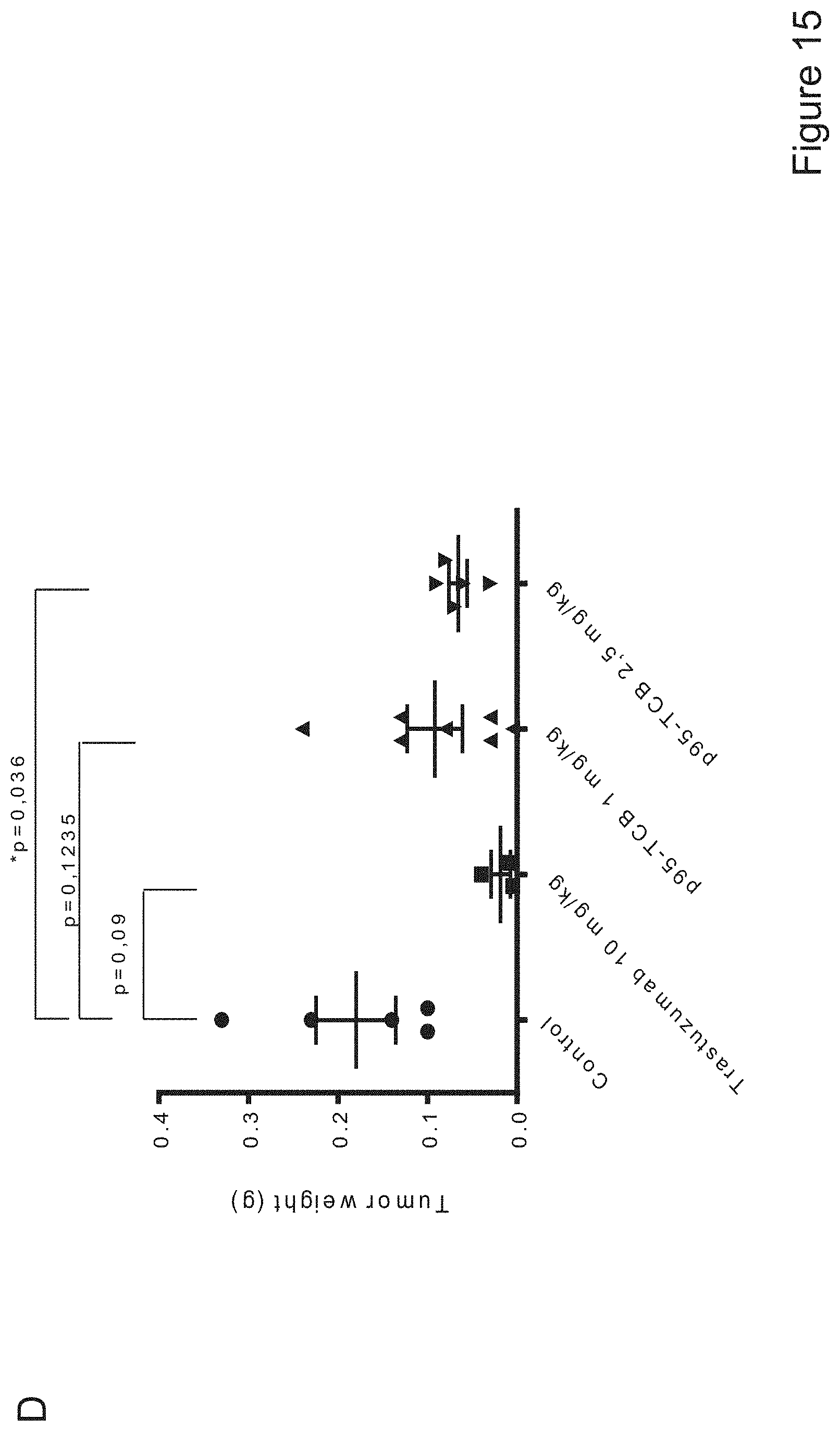
D00021
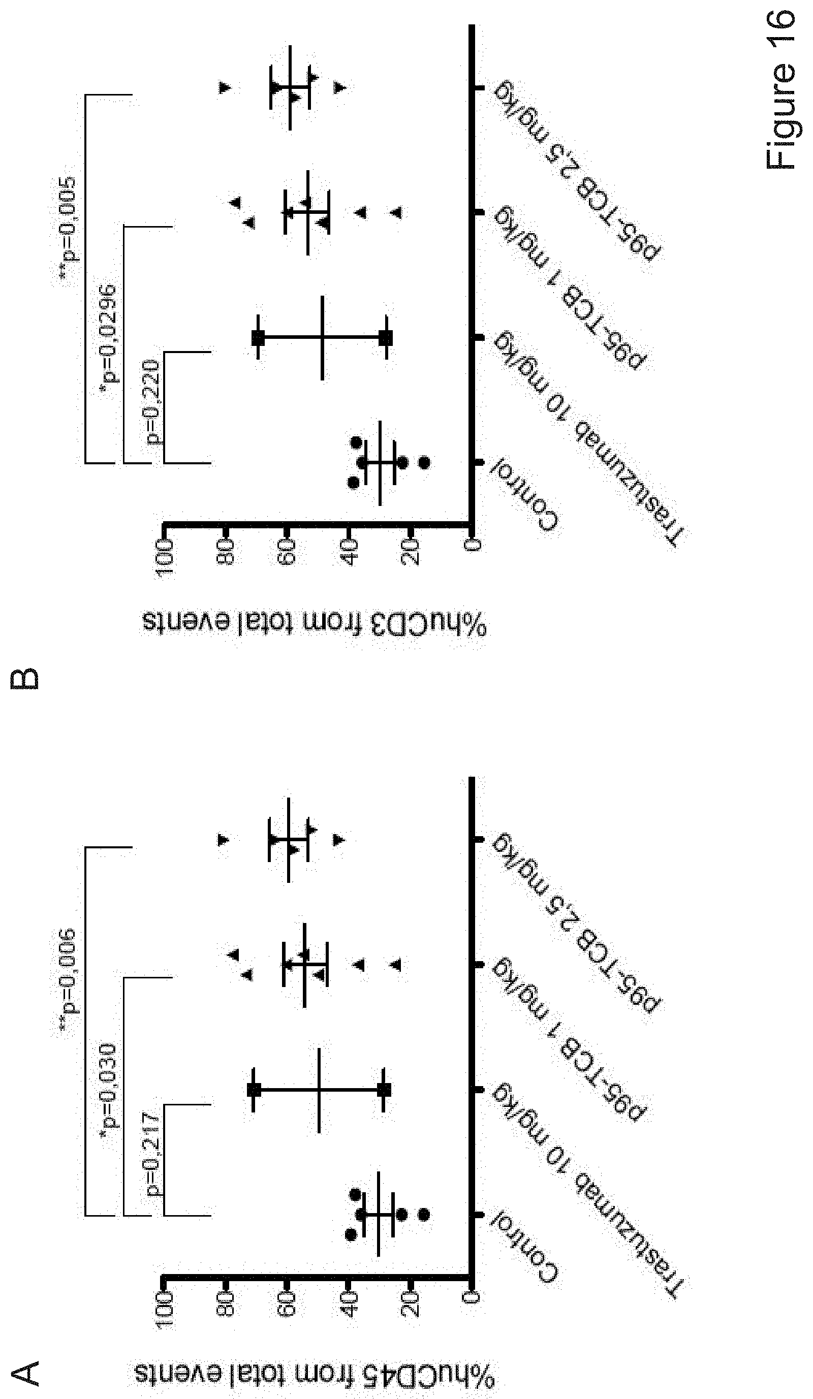
D00022

D00023

D00024

D00025

D00026

D00027

S00001
XML
uspto.report is an independent third-party trademark research tool that is not affiliated, endorsed, or sponsored by the United States Patent and Trademark Office (USPTO) or any other governmental organization. The information provided by uspto.report is based on publicly available data at the time of writing and is intended for informational purposes only.
While we strive to provide accurate and up-to-date information, we do not guarantee the accuracy, completeness, reliability, or suitability of the information displayed on this site. The use of this site is at your own risk. Any reliance you place on such information is therefore strictly at your own risk.
All official trademark data, including owner information, should be verified by visiting the official USPTO website at www.uspto.gov. This site is not intended to replace professional legal advice and should not be used as a substitute for consulting with a legal professional who is knowledgeable about trademark law.