Extracellular vesicle-based diagnostics and engineered exosomes for targeted therapeutics against cancer
Liu , et al. December 29, 2
U.S. patent number 10,874,610 [Application Number 15/788,709] was granted by the patent office on 2020-12-29 for extracellular vesicle-based diagnostics and engineered exosomes for targeted therapeutics against cancer. This patent grant is currently assigned to Case Western Reserve University, Northwestern University. The grantee listed for this patent is Case Western Reserve University, Northwestern University. Invention is credited to Nurmaa K. Dashzeveg, Golam Kibria, Huiping Liu, Erika K. Ramos.


View All Diagrams
| United States Patent | 10,874,610 |
| Liu , et al. | December 29, 2020 |
Extracellular vesicle-based diagnostics and engineered exosomes for targeted therapeutics against cancer
Abstract
This invention is related to use of exosomes for biomarker analysis for early detecting and characterizing of disease progression of cancer. Further, the invention provides bioengineered exosomes for use in methods of targeting and treating cancer.
| Inventors: | Liu; Huiping (Chicago, IL), Ramos; Erika K. (Chicago, IL), Dashzeveg; Nurmaa K. (Chicago, IL), Kibria; Golam (Chicago, IL) | ||||||||||
|---|---|---|---|---|---|---|---|---|---|---|---|
| Applicant: |
|
||||||||||
| Assignee: | Northwestern University
(Evanston, IL) Case Western Reserve University (Cleveland, OH) |
||||||||||
| Family ID: | 1000005266910 | ||||||||||
| Appl. No.: | 15/788,709 | ||||||||||
| Filed: | October 19, 2017 |
Prior Publication Data
| Document Identifier | Publication Date | |
|---|---|---|
| US 20180104187 A1 | Apr 19, 2018 | |
Related U.S. Patent Documents
| Application Number | Filing Date | Patent Number | Issue Date | ||
|---|---|---|---|---|---|
| 62409921 | Oct 19, 2016 | ||||
| Current U.S. Class: | 1/1 |
| Current CPC Class: | C07K 14/70596 (20130101); A61K 9/127 (20130101); A61K 31/7105 (20130101); G01N 33/57415 (20130101); A61K 9/1277 (20130101); C07K 14/70503 (20130101); G01N 33/57484 (20130101); G01N 33/57488 (20130101); A61K 38/00 (20130101); C07K 2319/70 (20130101); C07K 2319/03 (20130101) |
| Current International Class: | A61K 9/127 (20060101); G01N 33/574 (20060101); A61K 31/7105 (20060101); C07K 14/705 (20060101); A61K 38/00 (20060101) |
| Field of Search: | ;435/7.23 |
References Cited [Referenced By]
U.S. Patent Documents
| 2015/0024961 | January 2015 | Klass et al. |
| 2889623 | Jul 2015 | EP | |||
| 2010056337 | May 2010 | WO | |||
| 2010065968 | Jun 2010 | WO | |||
| 2010070276 | Jun 2010 | WO | |||
| 2012115885 | Aug 2012 | WO | |||
| 2016024918 | Feb 2016 | WO | |||
| 2016033695 | Mar 2016 | WO | |||
| 2016201064 | Dec 2016 | WO | |||
| 2017087940 | May 2017 | WO | |||
Other References
|
Ohno et al (Molecular Therapy, 2013, 21(1): 185-191). cited by examiner . Xitong et al (Gene, 2016, 575: 377-384). cited by examiner . Wang Z, Gerstein M, & Snyder M (2009) RNA-Seq: a revolutionary tool for transcriptomics. Nature reviews 10(1):57-63. cited by applicant . Weiskopf, K., et al., Engineered SIRPalpha variants as immunotherapeutic adjuvants to anticancer antibodies. Science, 2013. 341(6141): p. 88-91. cited by applicant . Willingham, S.B., et al., The CD47-signal regulatory protein alpha (SIRPa) interaction is a therapeutic target for human solid tumors. Proc Natl Acad Sci U S A, 2012. 109(17): p. 6662-6667. cited by applicant . Wisniewski JR, Zougman A, Nagaraj N, & Mann M (2009) Universal sample preparation method for proteome analysis. Nat Methods 6(5):359-362. cited by applicant . Zhang, L., et al., Nanoparticles in medicine: therapeutic applications and developments. Clin Pharmacol Ther, 2008. 83(5): p. 761-769. cited by applicant . International Search Report and Written Opinion for PCT Appl. PCT/US2017/057479, dated Mar. 8, 2018, 22 pages. cited by applicant . Al-Hajj, M., et al., Prospective identification of tumorigenic breast cancer cells. Proc Natl Acad Sci U S A, 2003. 100(7): p. 3983-3988. cited by applicant . Alvarez-Erviti, L., et al., Delivery of siRNA to the mouse brain by systemic injection of targeted exosomes. Nat Biotechnol, 2011. 29(4): p. 341-345. cited by applicant . Azmi AS, Bao B, & Sarkar FH (2013) Exosomes in cancer development, metastasis, and drug resistance: a comprehensive review. Cancer Metastasis Rev 32(3-4):623-642. cited by applicant . Bockhorn, J., et al., MicroRNA-30c inhibits human breast tumour chemotherapy resistance by regulating TWF1 and IL-11. Nat Commun, 2013. 4: p. 1393. cited by applicant . Bockhorn, J., et al., MicroRNA-30c targets cytoskeleton genes involved in breast cancer cell invasion. Breast Cancer Res Treat, 2013. 137(2): p. 373-382. cited by applicant . Chao, M. P. et al. Anti-CD47 antibody synergizes with rituximab to promote phagocytosis and eradicate non-Hodgkin lymphoma. Cell 142, 699-713, doi:10.1016/j.cell.2010.07.044 (2010). cited by applicant . Chao, M. P. et al. Calreticulin is the dominant pro-phagocytic signal on multiple human cancers and is counterbalanced by CD47. Sci Transl Med 2, 63ra94, doi:10.1126/scitranslmed.3001375 (2010). cited by applicant . Chao, M.P., et al., Extranodal dissemination of non-Hodgkin lymphoma requires CD47 and is inhibited by anti-CD47 antibody therapy. Blood, 2011. 118(18): p. 4890-4901. cited by applicant . Chao, M. P., Weissman, I. L. & Majeti, R. The CD47-SIRPalpha pathway in cancer immune evasion and potential terapeutic implications. Curr Opin Immunol 24, 225-232, doi:10.1016/j.coi.2012.01.010 (2012). cited by applicant . Chao, M. P., Majeti, R. & Weissman, I. L. Programmed cell removal: a new obstacle in the road to developing cancer. Nature reviews. Cancer 12, 58-67, doi:10.1038/nrc3171 (2012). cited by applicant . Dalerba, P. et al. Phenotypic characterization of human colorectal cancer stem cells. Proc Natl Acad Sci U S A 104, 10158-10163, doi:10.1073/pnas.0703478104 (2007). cited by applicant . Dragovic, R. A. et al. Sizing and phenotyping of cellular vesicles using Nanoparticle Tracking Analysis. Nanomedicine: nanotechnology, biology, and medicine 7, 780-788, doi:10.1016/j.nano.2011.04.003 (2011). cited by applicant . Godar, S. et al. Growth-inhibitory and tumor-suppressive functions of p53 depend on its repression of CD44 expression. Cell 134, 62-73, doi:10.1016/j.cell.2008.06.006 (2008). cited by applicant . Harding, C.V., J.E. Heuser, and P.D. Stahl, Exosomes: looking back three decades and into the future. J Cell Biol, 2013. 200(4): p. 367-371. cited by applicant . Hoshino A, et al. (2015) Tumour exosome integrins determine organotropic metastasis. Nature 527(7578):329-335. cited by applicant . Iero, M. et al. Tumour-released exosomes and their implications in cancer immunity. Cell death and differentiation 15, 80-88, doi:10.1038/sj.cdd.4402237 (2008). cited by applicant . Jaiswal, S. et al. CD47 is upregulated on circulating hematopoietic stem cells and leukemia cells to avoid phagocytosis. Cell 138, 271-285, doi:10.1016/j.cell.2009.05.046 (2009). cited by applicant . Kahlert, C. & Kalluri, R. Exosomes in tumor microenvironment influence cancer progression and metastasis. Journal of molecular medicine 91, 431-437, doi:10.1007/s00109-013-1020-6 (2013). cited by applicant . Kershaw, M. H. & Smyth, M. J. Immunology. Making macrophages eat cancer. Science 341, 41-42, doi:10.1126/science.1241716 (2013). cited by applicant . Kibria G, et al. (2016) A rapid, automated surface protein profiling of single circulating exosomes in human blood. Sci Rep 6:36502. cited by applicant . Lacroix, R. et al. Standardization of platelet-derived microparticle enumeration by flow cytometry with calibrated beads: results of the International Society on Thrombosis and Haemostasis SSC Collaborative workshop. Journal of thrombosis and haemostasis : JTH 8, 2571-2574, doi:10.1111/j.1538-7836.2010.04047.x (2010). cited by applicant . Li, C. et al. Identification of pancreatic cancer stem cells. Cancer research 67, 1030-1037, doi:10.1158/0008-5472.CAN-06-2030 (2007). cited by applicant . Liu H., et al., Cancer stem cells from human breast tumors are involved in spontaneous metastases in orthotopic mouse models. Proc Natl Acad Sci U S A, 2010. 107(42): p. 18115-18120. cited by applicant . Liu, H., MicroRNAs in breast cancer initiation and progression. Cell Mol Life Sci, 2012. cited by applicant . Lotvall, J. et al. Minimal experimental requirements for definition of extracellular vesicles and their functions: a position statement from the International Society for Extracellular Vesicles. Journal of extracellular vesicles 3, 26913, doi:10.3402/jev.v3.26913 (2014). cited by applicant . Mani, S. A. et al. The epithelial-mesenchymal transition generates cells with properties of stem cells. Cell 133, 704-715, doi:10.1016/j.cell.2008.03.027 (2008). cited by applicant . Marcucci F, Bellone M, Caserta CA, & Corti A (2014) Pushing tumor cells towards a malignant phenotype: stimuli from the microenvironment, intercellular communications and alternative roads. Int J Cancer 135(6):1265-1276. cited by applicant . Majeti, R., et al., CD47 is an adverse prognostic factor and therapeutic antibody target on human acute myeloid leukemia stem cells. Cell, 2009. 138(2): p. 286-299. cited by applicant . McCabe MT, et al. (2012) EZH2 inhibition as a therapeutic strategy for lymphoma with EZH2-activating mutations. Nature 492(7427):108-112. cited by applicant . Melo SA, et al. (2015) Glypican-1 identifies cancer exosomes and detects early pancreatic cancer. Nature 523 (7559):177-182. cited by applicant . Momen-Heravi, E et al. Current methods for the isolation of extracellular vesicles. Biological chemistry 394, 1253-1262, doi:10.1515/hsz-2013-0141 (2013). cited by applicant . Paltridge JL, Belle L, & Khew-Goodall Y (2013) The secretome in cancer progression. Biochim Biophys Acta 1834 (11):2233-2241. cited by applicant . Pospichalova, V. et al. Simplified protocol for flow cytometry analysis of fluorescently labeled exosomes and microvesicles using dedicated flow cytometer. Journal of extracellular vesicles 4, 25530, doi:10.3402/jev.v4.25530 (2015). cited by applicant . Prince, M. E. et al. Identification of a subpopulation of cells with cancer stem cell properties in head and neck squamous cell carcinoma. Proc Natl Acad Sci U S A 104, 973-978, doi:10.1073/pnas.0610117104 (2007). cited by applicant . Ramos, E. K., et al. (2017) New opportunities and challenges to defeat cancer stem cells. Tends in Cancer, 3(11):780-796. cited by applicant . Ruoslahti, E., S.N. Bhatia, and M.J. Sailor, Targeting of drugs and nanoparticles to tumors. J Cell Biol, 2010. 188(6): p. 759-768. cited by applicant . Samaeekia R, et al. miR-206 Inhibits Stemness and Metastasis of Breast Cancer by Targeting MKL1/IL11 Pathway. Clin Cancer Res. 2017;23(4):1091-1103. cited by applicant . Schlatzer DM, Sugalski J, Dazard JE, Chance MR, & Anthony DD (2012) A quantitative proteomic approach for detecting protein profiles of activated human myeloid dendritic cells. J Immunol Methods 375(1-2):39-45. cited by applicant . Schorey JS & Bhatnagar S (2008) Exosome function: from tumor immunology to pathogen biology. Traffic 9(6):871-881. cited by applicant . Shimono, Y., et al., Downregulation of miRNA-200c links breast cancer stem cells with normal stem cells. Cell, 2009. 138(3): p. 592-603. cited by applicant . Silva, J. et al. Analysis of exosome release and its prognostic value in human colorectal cancer. Genes, chromosomes & cancer 51, 409-418 (2012). cited by applicant . Simhadri, V. R. et al. Dendritic cells release HLA-B-associated transcript-3 positive exosomes to regulate natural killer function. PloS one 3, e3377, doi:10.1371/journal.pone.0003377 (2008). cited by applicant . Skog, J. et al., Glioblastoma microvesicles transport RNA and proteins that promote tumour growth and provide diagnostic biomarkers. Nat Cell Biol, 2008. 10(12): p. 1470-1476. cited by applicant . Svensson KJ & Belting M (2013) Role of extracellular membrane vesicles in intercellular communication of the tumour microenvironment. Biochem Soc Trans 41(1):273-276. cited by applicant . Thery, C., Amigorena, S., Raposo, G. & Clayton, A. Isolation and characterization of exosomes from cell culture supernatants and biological fluids. Current protocols in cell biology/editorial board, Juan S. Bonifacino . . . [et al.] chapter 3, Unit 3 22, doi:10.1002/0471143030.cb0322s30 (2006). cited by applicant . Valadi, H., et al., Exosome-mediated transfer of mRNAs and microRNAs is a novel mechanism of genetic exchange between cells. Nat Cell Biol, 2007. 9(6): p. 654-659. cited by applicant . van der Pol, E. et al. Optical and non-optical methods for detection and characterization of microparticles and exosomes. Journal of thrombosis and haemostasis: JTH 8, 2596-2607, doi:10.1111/j.1538-7836.2010.04074.x (2010). cited by applicant . van der Pol, E. et al. Particle size distribution of exosomes and microvesicles determined by transmission electron microscopy, flow cytometry, nanoparticle tracking analysis, and resistive pulse sensing. Journal of thrombosis and haemostasis : JTH 12, 1182-1192, doi:10.1111/jth.12602 (2014). cited by applicant . Verweij, F.J., et al., LMP1 association with CD63 in endosomes and secretion via exosomes limits constitutive NF-kappaB activation. EMBO J, 2011. 30(11): p. 2115-2129. cited by applicant . Vire E, et al. (2006) The Polycomb group protein EZH2 directly controls DNA methylation. Nature 439(7078):871-874. cited by applicant. |
Primary Examiner: Aeder; Sean E
Attorney, Agent or Firm: Quarles & Brady LLP
Government Interests
STATEMENT REGARDING FEDERALLY SPONSORED RESEARCH
This invention was made with government support under R00 CA160638 awarded by the National Institutes of Health and W81XWH-16-1-0021 awarded by the U.S. Army Medical Research and Materiel Command (Army/MRMC). The government has certain rights in the invention.
Parent Case Text
CROSS-REFERENCE TO RELATED APPLICATIONS
This application claims priority to U.S. Provisional Application 62/409,921 filed Oct. 19, 2016, the contents of which are incorporated by reference in its entirety.
Claims
The invention claimed is:
1. A method of bioengineering exosomes for targeting cancer cells, the method comprising: (a) expressing a XPep.alpha. fusion protein encoded by SEQ ID NO:2 in a host cell; (b) isolating secreted exosomes comprising the fusion protein; and (c) loading the isolated exosomes with a least one RNA oligonucleotide or chemotherapeutic agent.
2. The method of claim 1, wherein step (a) comprises transducing the host cell with a viral vector encoding the fusion protein.
3. A bioengineered exosome made by the method of claim 1.
4. A therapeutic bioengineered exosome comprising a XPep.alpha. fusion protein encoded by SEQ ID NO:2.
5. The therapeutic bioengineered exosome of claim 4 further comprising at least one RNA oligonucleotide or chemotherapeutic agent.
6. The therapeutic bioengineered exosome of claim 5, wherein the RNA oligonucleotide is a siRNA or miRNA specific for cancer.
7. The therapeutic bioengineered exosome of claim 6, wherein the cancer is breast cancer and wherein the miRNA is miR-200, miR30c, or miR206.
8. A method of treating a patient with cancer, the method comprising administering an effective amount of the therapeutic bioengineered exosome of claim 5 to the patient with cancer.
9. The method of claim 8, wherein the patient has a solid tumor cancer.
10. The method of claim 1, wherein step (c) comprises loading the at least one RNA oligonucleotide or chemotherapeutic agent by electroporation of the exosomes.
11. The method of claim 1, wherein the RNA oligonucleotide is a siRNA or miRNA specific for breast cancer.
12. The method of claim 8, wherein the cancer is breast cancer and wherein the at least one RNA oligonucleotide is a miRNA selected from the group consisting of miR-200, miR30c, and miR206.
Description
BACKGROUND OF THE INVENTION
The field of the invention is related to cancer diagnostics and treatment. More particularly, the invention relates to exosomes and/or other extracellular vesicles (EVs) specific for cancer.
EVs are cell-derived vesicles with a closed double-layer membrane structure.sup.1-3. They carry various molecules (proteins, lipids, and RNAs) on their surface as well as in the lumen.sup.1-3. Exosomes and other EVs play a critical role in intercellular communication and cellular content transfer, e.g. mRNAs and microRNAs, in both physiological and pathological settings, such as tumor development and progression.sup.4-7. The exosomal surface proteins can mediate organ-specific homing of circulating exosomes, and their contents show potential to serve as novel biomarkers.sup.8-10, thereby assisting the diagnosis and prognosis prediction of human diseases, such as cancer. Approaches to detect and characterize exosomes and other EVs may include: (1) electron microscopy (EM) to assess structure and size; (2) nanoparticle tracking analysis (NTA).sup.3 to reveal size and zeta potential; (3) protein analysis via immunofluorescence staining, western blotting, ELISA, and mass spectrometry, (4) RNA analysis using array platforms, RNA sequencing, and PCR, and (5) analysis of lipids, sugar, and other components by biochemical assays. Among these approaches, EM provides high-resolution imaging but is neither convenient nor affordable for high throughput molecular profiling of large numbers of circulating exosome samples for potential clinical applications. NTA utilizes light scattering and Brownian motion.sup.3 to measure particle size but does not differentiate between vesicles within a size range of 5.times. orders of magnitude due to the low dynamic range of the camera.sup.11. In addition, NTA is not suitable for molecular profiling of exosomes because of low sensitivity to fluorescent signals.
Based on the statistics documented by American Cancer Society (http://www.cancer.org), the lifetime risk of developing cancer is 1 in 2 for men and 1 in 3 for women. And the lifetime risk of dying from cancer is 1 in 4 in men and 1 in 5 in women. The most frequent cancers are breast cancer in women and prostate cancer in men which are second leading cancers causing deaths. Breast cancer accounts for an estimated 230,000 newly diagnosed cases and about 40,000 deaths annually in USA (1.3 million new cases and 450,000 deaths every year worldwide). Lung cancer is the second leading cancer but the most devastating cancer in both men and women. Metastasis causes 90% of solid tumor-related deaths. Therefore prevention, detection and treatment of early and advanced diseases hold the key to reduce cancer mortality. To achieve those goals, better understanding of cancer and metastasis, novel biomarkers, and effective targeted therapies are demanded.
Cancer stem cells (CSCs) are a subset of cancer cells with tumor initiating capacity and stem cell properties, and considered the roots of cancer, seeds of metastasis, and sources of therapy resistance. However, it is challenging to detect CSCs, monitor residual CSC activities and remove CSCs through existing diagnostic and therapeutic approaches. There still is a need to identify CSC-related biomarkers in liquid biopsies such as circulating exosomes/EVs as well as to develop innovative CSC-targeted therapeutics.
SUMMARY OF THE INVENTION
The present invention overcomes the aforementioned drawbacks by providing single exosome/EV micro flow profile-based cancer biomarkers as well as engineered extracellular vesicles and exosomes to target specific cancer cell populations.
In one aspect, the invention provides a method of detecting cancer in a patient comprising: (a) obtaining a sample from the patient; (b) isolating extracellular vesicles from the sample; and (c) detecting expression of at least one cancer marker in the isolated extracellular vesicles.
In another aspect, the invention provides a method of bioengineering EVs or exosomes for targeting cancer cells, the method comprising: (a) expressing a fusion protein comprising a segment of an exosome protein fused to a cancer stem cell (CSC) targeting peptide in a host cell; and (b) isolating secreted EVs or exosomes comprising the fusion protein.
In another aspect, the invention provides bioengineered exosome made by the methods described herein.
In yet another aspect, the invention provides a bioengineered exosome comprising a fusion protein comprising a segment of an exosome protein fused to a CSC targeting peptide. In some aspects, the segment of the exosome protein comprises the transmembrane domain.
In a further aspect, the invention provides a therapeutic bioengineered exosome comprising a bioengineered exosome comprising a fusion protein comprising a segment of an exosome protein including the transmembrane domain fused to a CSC targeting peptide and at least one RNA oligonucleotide or chemotherapeutic agent.
In yet another aspect, the invention provides a method of treating a patient with cancer, the method comprising administering an effective amount of the therapeutic bioengineered exosome described herein.
The foregoing and other aspects and advantages of the invention will appear from the following description. In the description, reference is made to the accompanying drawings which form a part hereof, and in which there are shown, by way of illustration, preferred embodiments of the invention. Such embodiments do not necessarily represent the full scope of the invention, however, and reference is made therefore to the claims and herein for interpreting the scope of the invention.
BRIEF DESCRIPTION OF THE DRAWINGS
The patent or application file contains at least one drawing executed in color. Copies of this patent or patent application publication with color drawing(s) will be provided by the Office upon request and payment of the necessary fee.
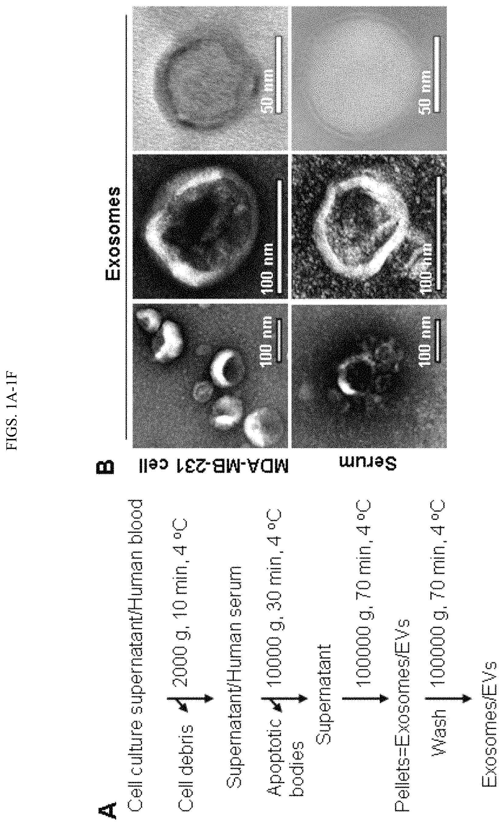
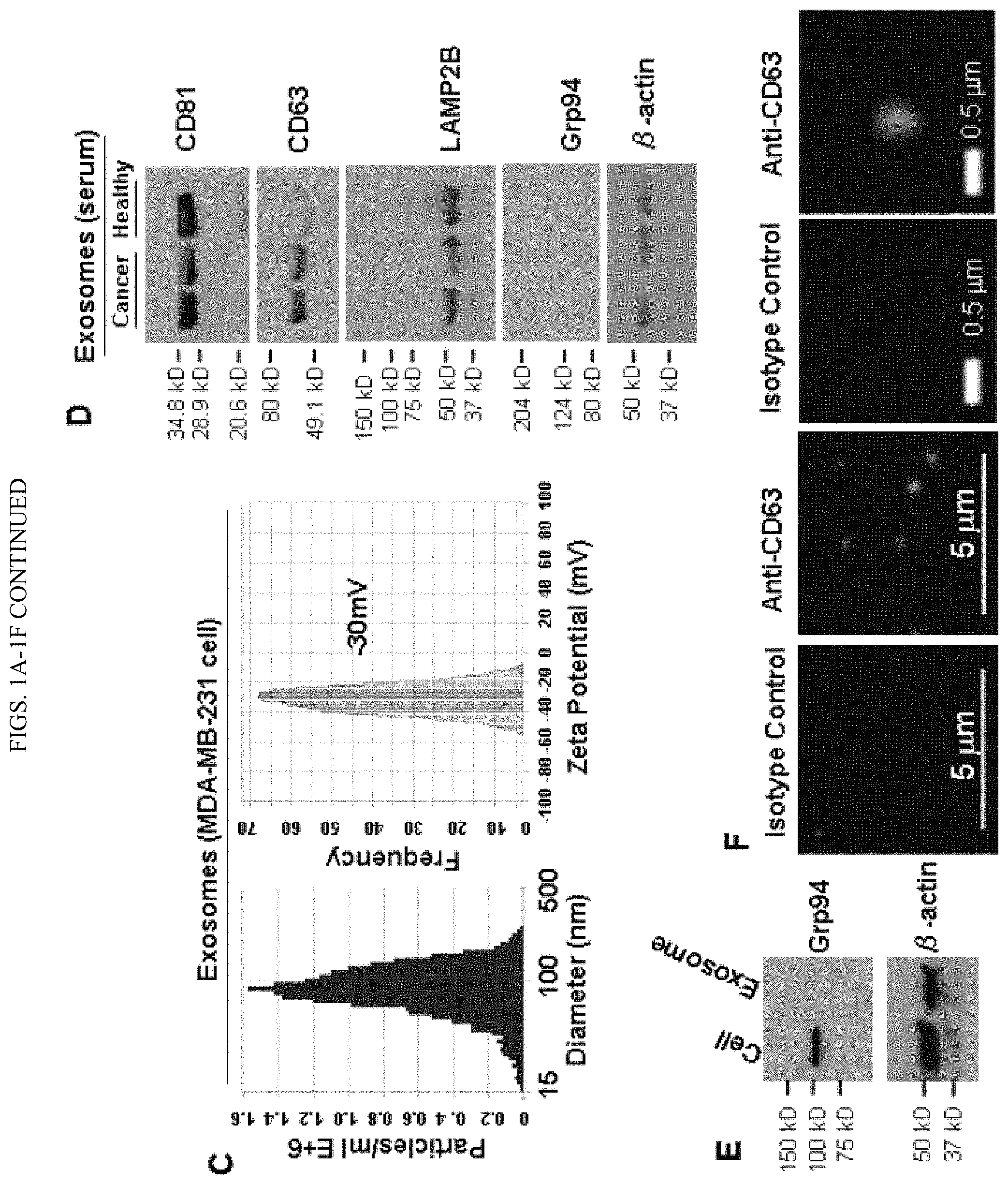
FIGS. 1A-1F. Isolation and characterization of exosomes from cell culture supernatant and human blood. A. Depicts the exosome purification procedure by ultracentrifugation. Blood was centrifuged at 3200 RPM for 15 min at 4.degree. C. to collect serum or for 15 minutes at 2,000.times.g to collect plasma sample plasma. As mentioned in the first step, the cell culture supernatant was centrifuged at 2000 g, 10 min, 4.degree. C. to remove cell debris. B. Picture representation of observation of the morphology of exosomes under Transmission Electron Microscopy (TEM), indicating the diameter of isolated exosomes in the 50-100 nm range. C. ZetaView NTA analysis of MDA-MB-231 cell-derived exosomes with the size distribution (mean diameter 89.+-.33 nm and mode 87 nm) and surface charge (-30 mV). D. Immunoblot of exosomal markers CD81 (.about.30 kDa), CD63 (.about.55 kDa), and LAMP2B (.about.50 kDa) in exosomes (5 .mu.g lysates) isolated from the serum of breast cancer patients and healthy control. Grp94 (.about.100 kDa) and .beta.-actin (.about.42 kDa) serve as a negative control and a loading control, respectively. E. Immunoblot of Grp94 with 5 .mu.g protein of MDA-MB-231 cell lysates (Cell) and the exosomes derived from these cells (Exosome, no Grp94 detection). F. Picture representation of detection of CD63 in intact MDA-MB-231 exosomes by immunofluorescence staining.
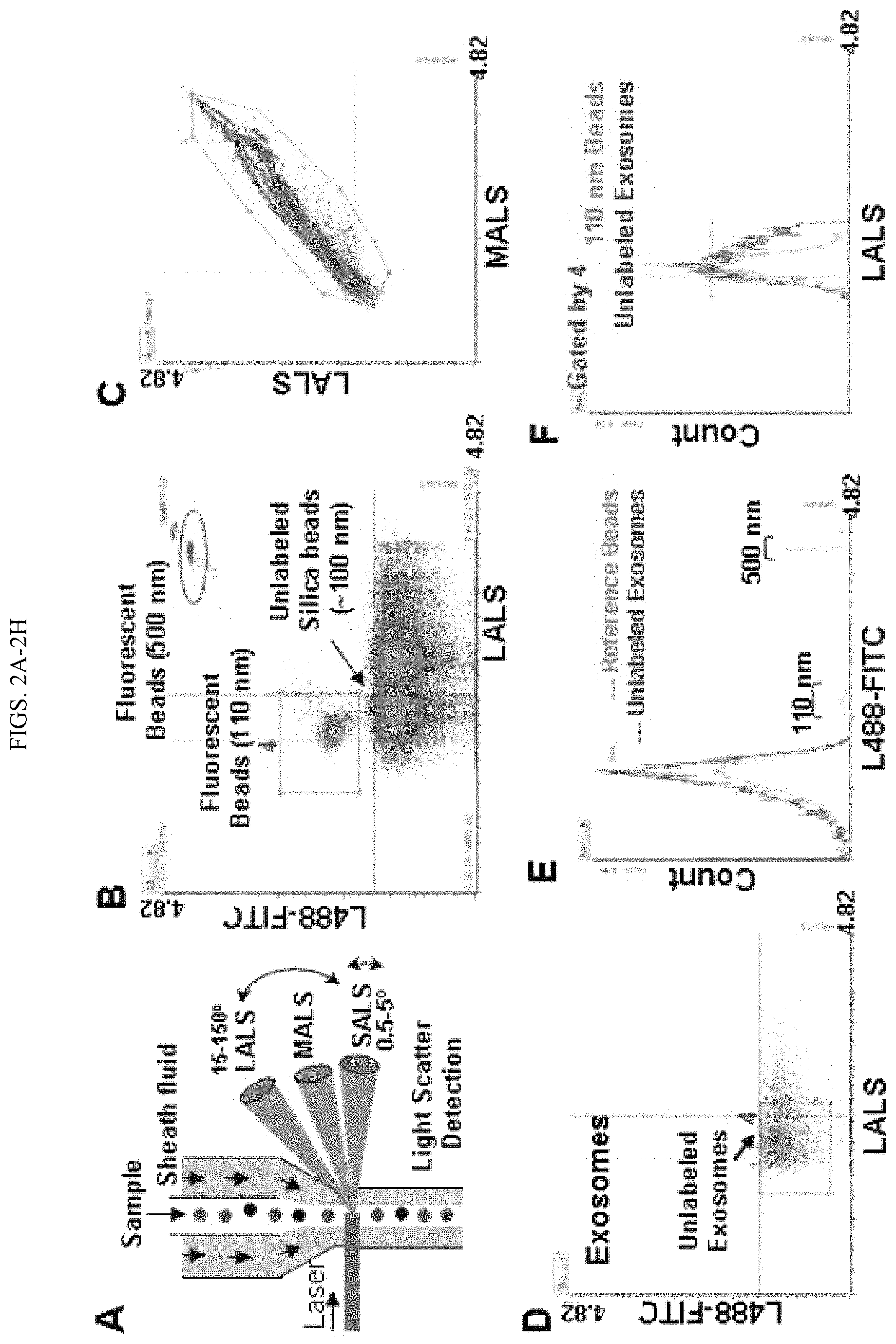
FIGS. 2A-2H. Micro flow cytometry (MFC) detection of the ApogeeMix Beads and blood-derived exosomes. A. Schematic representation of Apogee A50 MFC denoting the mode of action of nanoparticle detection. The sample flows from top to bottom and is surrounded by sheath fluid. The laser intersects with the sample stream, generating 3 different light scatters: Large angle light scatter (LAS); Middle angle light scatter (MALS); Small angle light scatter (SALS); and fluorescence signals. B. Cytogram of the reference beads, an aqueous mixture of the green fluorescent latex and non-fluorescent silica (Si) spheres in 110-1300 nm plotted at green fluorescence (L488-FITC) and LALS signals with minimized background noise. The red rectangle and circled box represent the 110 nm and 500 fluorescence beads respectively. C. Cytogram of the reference ApogeeMix Beads in 110-1300 nm with similar MALS and LALS signals at the modified high threshold setting. D. Cytogram of the unlabeled exosomes from human blood at green fluorescence and LALS signals. E. Fluorescent signal comparison between the ApogeeMix beads from B and the unlabeled exosomes from D shown at the L488-FITC channel. F. Histogram comparing the size of the gated exosomes from D with that of the gated 110 nm fluorescence beads in B (red rectangles). G. Flow analysis of CD44 expression on exosomes derived from MDA-MB-231 and MCF-12A cells using A50 MFC. H. Immunoblot of CD44 expression in the exosome lysates derived from MDA-MB-231 and MCF-12A cells. .beta.-actin (.about.42 kDa) serves as a loading control.
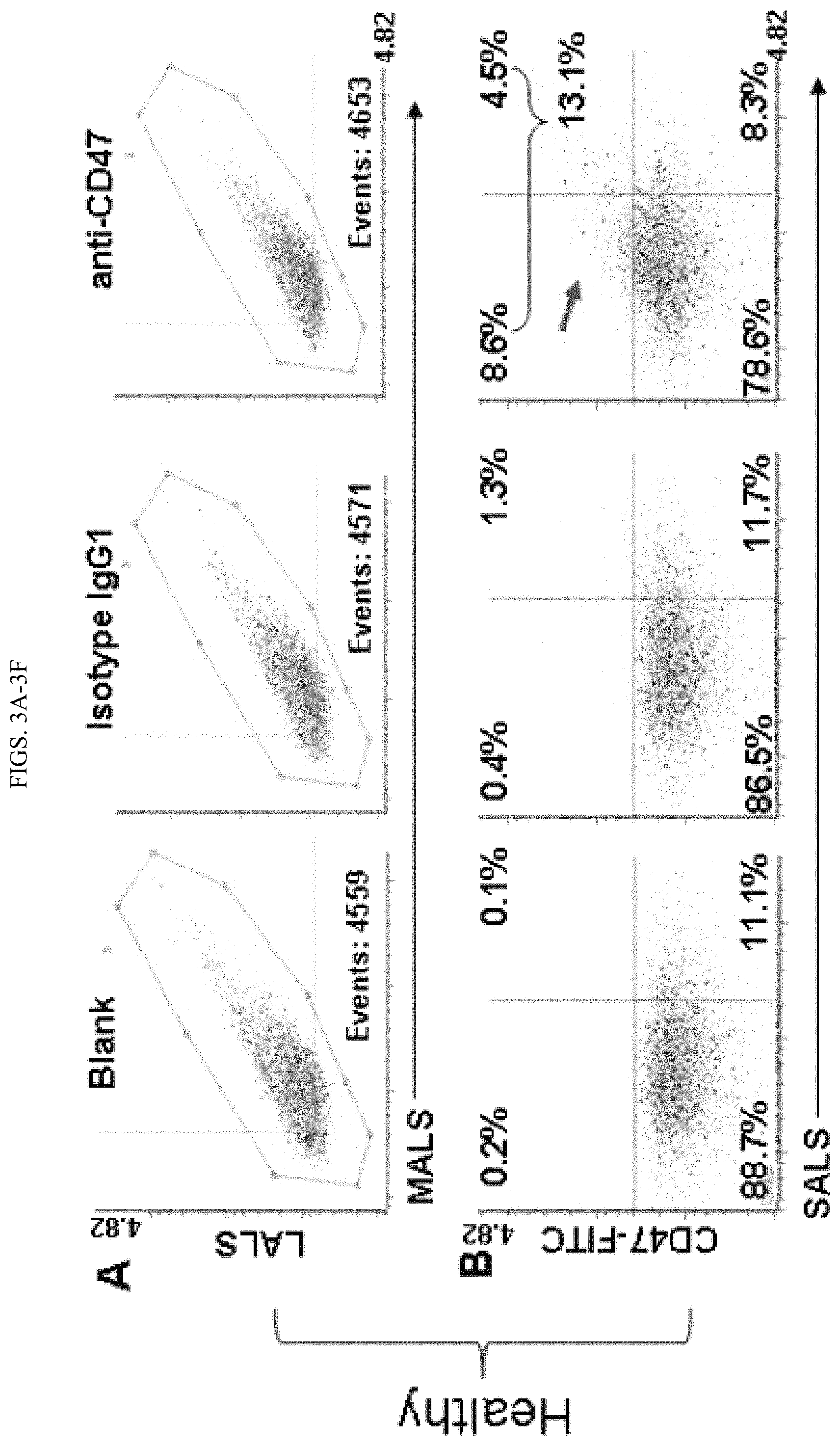
FIGS. 3A-3F. Detection of the surface protein CD47 in circulating exosomes and its correlation with breast cancer. A,C: Representative scattered plots showing MALS and LALS signals of the circulating exosomes isolated from the blood of healthy control and (A) breast cancer patients, (C) 4500 total events collected for each sample: unstained blank control, isotype IgG1, and anti-CD47. B,D: Representative cytograms showing differential expression of CD47 on the exosomes isolated from the blood of healthy control (B) or that of a breast cancer patient (D). Exosomes were stained with FITC-CD47 antibody or isotype control FITC-IgG1, or unstained (control). E. Comparison of MFC-analyzed CD47 expression levels on circulating exosome specimens isolated from the blood of healthy people (representative in B, n=60) and breast cancer patients (representative in D, n=60). Unpaired Student's t-test, p value<0.05. F. Expression of CD47 on circulating exosomes measured by the ELISA. Exosomes isolated from the blood of 40 healthy control and 50 breast cancer patients were treated with the anti-CD47 ELISA antibody. Unpaired Student's t-test, p value<0.01.
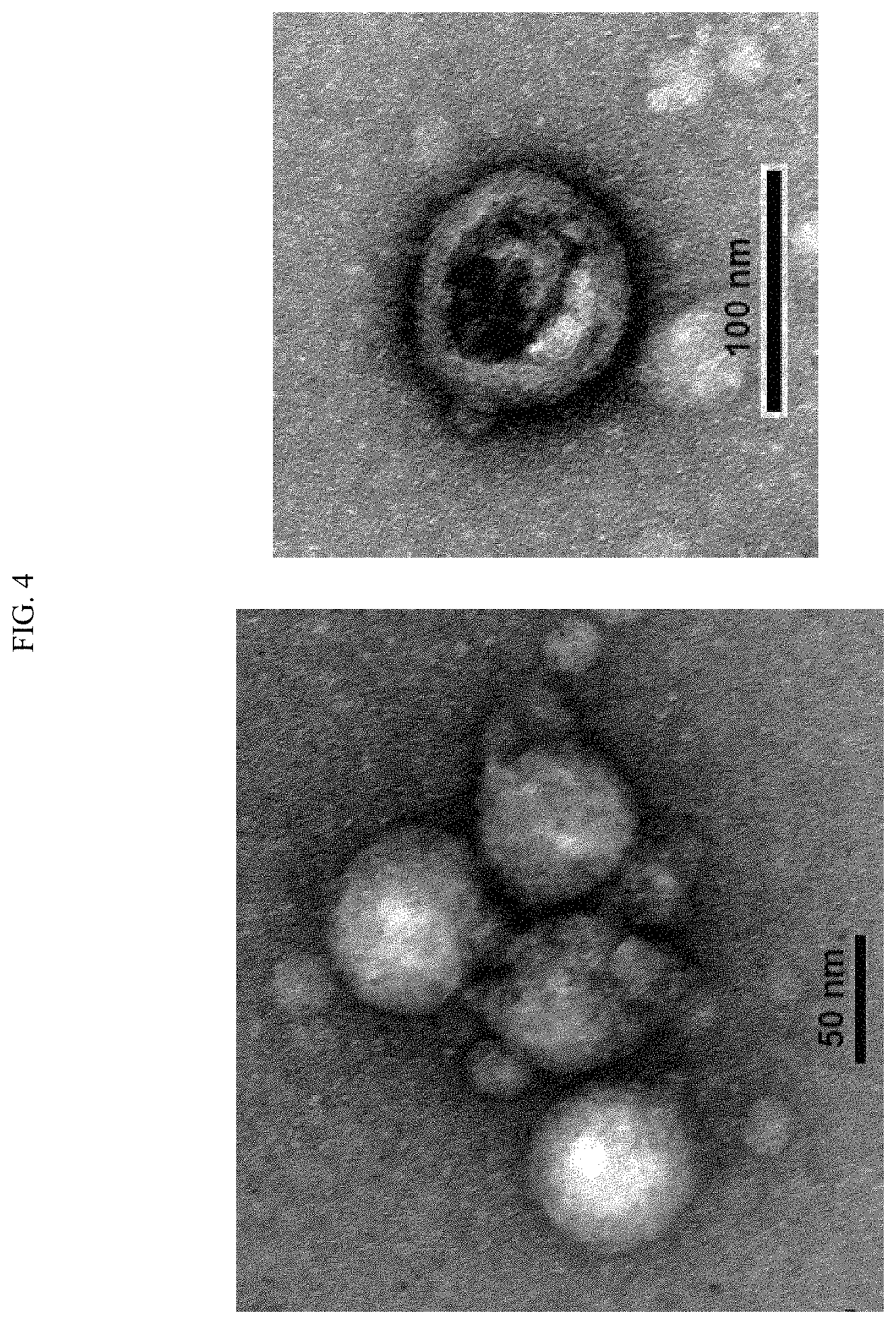
FIG. 4. Picture depicting the exosome-isolation kit-purified exosomes from human serum (healthy) under Transmission Electron Microscopy (TEM). Exosomes were isolated from the serum after depletion of cell debris and apoptotic bodies per manufacturer's manual.
FIG. 5. Picture of gel depicting evaluation of the expression of exosomal markers on cell derived exosomes. Western blots of 5 .mu.g exosome lysates for the exosomal markers LAMP2B (.about.100 kDa) and CD63 (.about.55 kDa). Grp94 (.about.100 kDa) and .beta.-actin (.about.42 kDa) serves as a negative control and loading control, respectively.
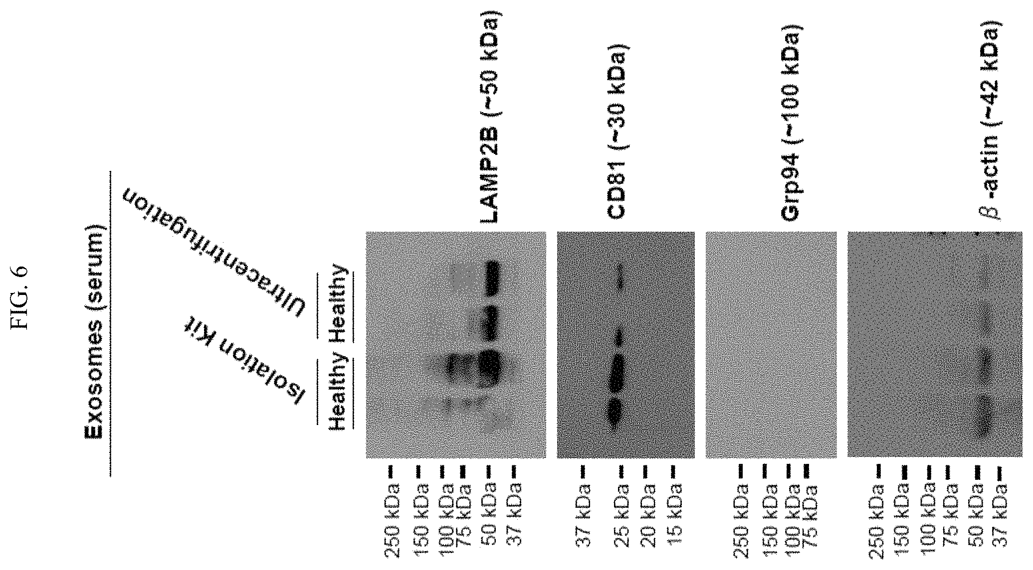
FIG. 6. Pictures depicting immunoblot comparison of the expression of markers on exosomes isolated from human serum (healthy) by differential ultracentrifugation and by the exosome-isolation kit. Exosomal markers LAMP2B (.about.50 kDa) and CD81 (.about.30 kDa) were detected in exosomes (5 mg lysates). Grp94 (.about.100 kDa) and .beta.-actin (.about.42 kDa) serve as a negative control and loading control, respectively.
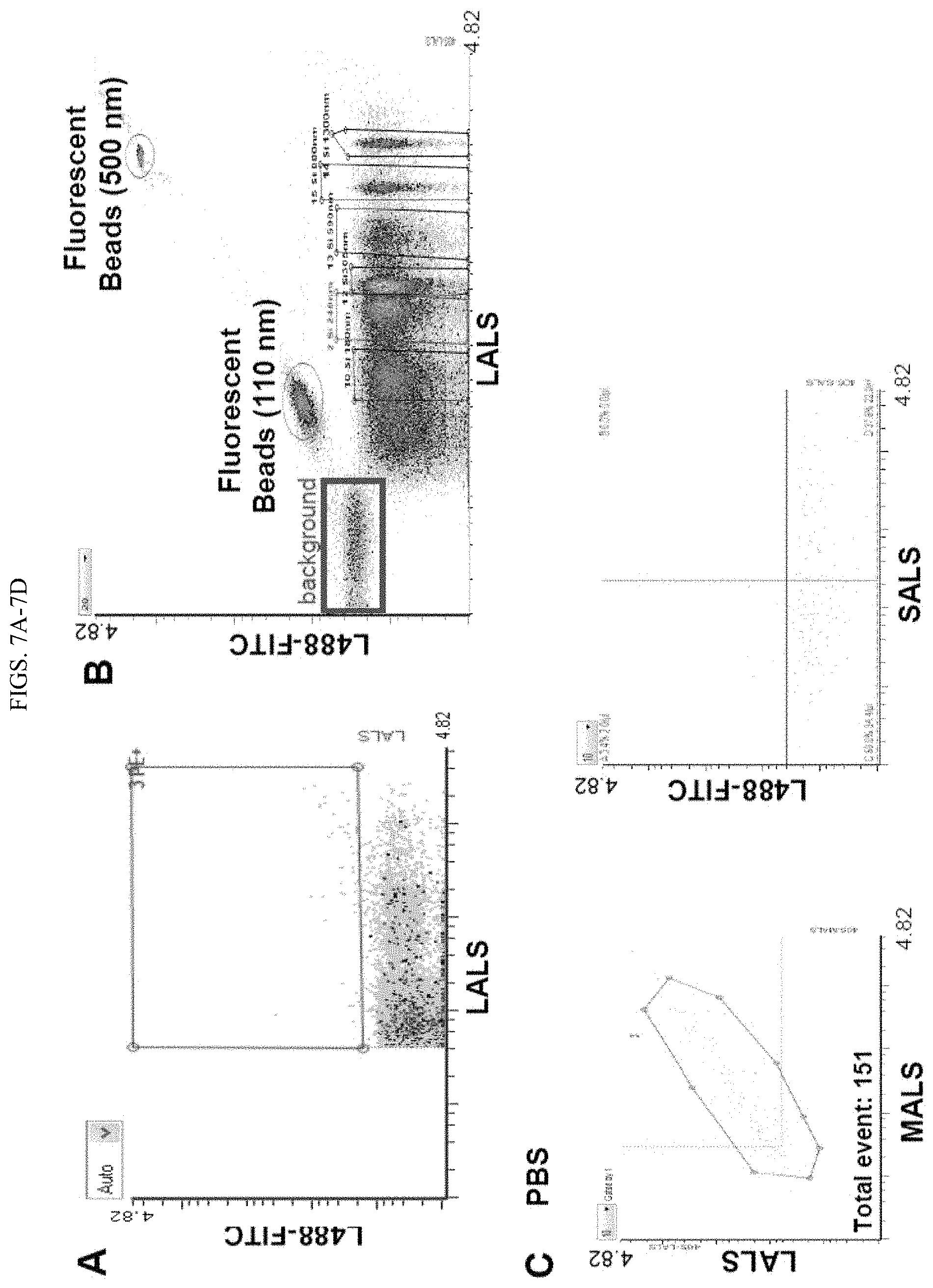
FIGS. 7A-7D. Exosome flow analysis optimizations. A-B. Background particles shown in PBS (A) and the reference beads solution (B) at the default setting of A50 MFC. The reference ApogeeMix beads is an aqueous mixture of 110 nm and 500 nm green fluorescent latex beads with refractive index (RI) .eta.=1.59, and non-fluorescent silica (Si) beads with 180-1300 nm diameter and RI .eta.=1.43. The RI of Si beads is closer to the RI of biological particles (RI of cells and EVs is 1.4). C-D. Analyses of PBS and antibodies using A50 MFC. C. PBS, used for exosome sample preparations, was run at high-threshold setting. Minimal particles in PBS were observed in multiple light scatter and fluorescence L488 channels. D. Cytogram of FITC-CD47 antibody as well as Isotope control FITC-IgG1 before and after centrifugation. Before staining exosomes, antibody bulk solutions were centrifuged at 14000.times.g for 1 h at 4.degree. C. to remove particles or precipitates.
FIGS. 8A-8B. MFC analyses of exosomal CD63 and CD47. A. Detection of CD63 expression on exosomes derived from MDA-MB-231 cells by A50 MFC. Exosomes were stained with the FITC-conjugated mouse anti-human CD63 antibody, followed by dilution with PBS and then detected under Apogee MFC. FITC Isotype IgG staining or unstained were used as background controls. The expression of CD63 was detected (red arrow) in 15.6% of exosomes. B. Evaluation of the effect of total events counted on CD47 detection on circulating exosomes. Cytograms showing the expression of CD47 in 5000 and 10000 counted exosomes isolated from the blood of healthy control. Exosomes were stained with FITC-CD47 antibody, isotype control FITCIgG1, or unstained (control). Between the analyses with 5000 and 10000 counted events, similar % of CD47+ exosomes were observed in the upper two quadrants (Q1+Q4).
FIG. 9. Detection of CD47 expression on exosomes isolated from human serum (healthy) by using the exosome-isolation kit. Exosomes were stained with FITC-CD47 antibody or isotype control FITC-IgG1, or unstained (control) followed by detection under Apogee MFC.
FIGS. 10A-10B. Detection of CD44 on circulating exosomes by MFC. A. Cytograms showing the expression of CD44 on exosomes isolated from the blood of a healthy control. Exosomes were stained with either FITC-CD44 antibody, isotype control FITC-IgG, or unstained (control). Compared to controls, about 20% CD44 positive exosomes were detected (red arrow). B. Comparison of the expression of CD44 in circulating exosomes isolated from the blood of fifteen healthy controls and twenty breast cancer patients. Statistical analysis was done by Unpaired Student's t-test. N.S.: non-significant.
FIG. 11. Table depicting Clinical Information of the female breast cancer patient and healthy control (female) used in this study.
FIGS. 12A-12C is a depiction of one embodiment of the present disclosure describing the design and synthesis of bio-engineered exosomes targeting CD47+ BCSCs. A. Structure of fusion gene (XPep.alpha.) in a lentiviral vector tagged with Luc2-eGFP. The BCSC targeted fusion gene XPep.alpha. is comprised of the transmembrane and cytoplasmic tail of exosome specific peptide Lamp 2B which extracellular domain was replaced by mutant SIRP.alpha. extracellular domain (See SEQ ID NO:2, FIG. 25). B. Basic principle showing the transfection and lentiviral based transduction from HEK293FT or MSC cells for the biogenesis of engineered exosomes. C. Culture of transfected (green) cells for 48 h in exosome-depleted media to synthesize the bio-engineered exosomes.
FIGS. 13A-13E demonstrates the exosome isolation, purification and characterization of bioengineered exosomes. A. Flow chart representing the isolation and purification of exosomes from the cell culture supernatant. B. ZetaView measurement of the size distribution of reference Polystyrene beads (60 nm, 200 nm) and HEK293FT cell-derived exosomes. C. Expression of Protein X (50 kDa), CD63 (43 kDa) in isolated exosomes observed by western blotting, .beta.-actin (42 kDa) serves as background control. D. Evaluation of the expression of Protein X (50 kDa), and fusion protein XPep.alpha. (20 kDa) in control (untransfected) as well as fusion gene transfected HEK293FT cells and their secreted exosomes observed by western blotting. The expression of XPep.alpha. was observed only in exosomes secreted by the transfected cells, as compared to control. E. Immunofluorescence assay showing the expression of fusion protein XPep.alpha. (green) and exosomal protein X on the bioengineered exosome.
FIGS. 14A-14C demonstrate the ability to load and target bioengineered exosomes. A. Expression of CD47 in BCSCs, no CD47 was detected in NIH3T3 cells. B. Specific uptake of bio-engineered exosomes (XPep.alpha.+) by CD44+CD47+ BCSCs. BCSCs show higher cellular uptake shift (red line, left panel) as compared to NIH3T3 cells observed by flow cytometer. C. Qualitative analysis showing a substantial amount of bio-engineered exosomes (XPep.alpha.+) internalization into BCSCs over the regular exosomes (XPep.alpha.-), indicating the specific targeting of CD44+CD47+ BCSCs by Xpep.alpha.+ bio-engineered exosomes.
FIGS. 15A-15B demonstrate the mechanism of internalization of bio-engineered exosomes (XPep.alpha.+) into CD44+CD47+ BCSCs. BCSCs were treated with different inhibitors to specifically inhibit different endocytosis routes. Both of the qualitative by confocal microscope (A) and quantitative by flow cytometry (B) analyses indicated that the cellular uptake of XPep.alpha.+ exosomes was inhibited by Amiloride and Sucrose, indicating that the endocytosis of XPep.alpha.+ Exosomes is dependent on clathrin and Na(+)/H(+) exchange.
FIG. 16 demonstrates in vivo biodistribution and targeting CD44+CD47+ BCSCs by XPep.alpha.+ bio-engineered exosomes. Regular exosomes (Xpep.alpha.-) from HEK293FT, THP1 cells as well as bio-engineered exosomes (XPep.alpha.+) were labeled with PKH67 dye and injected into patient-tumor-derived xenograft (M1) mice via tail vein. IVIS images of mice were captured at 1 h and 2 h post-injection of exosomes. Compared to other exosomes, bio-engineered exosomes show its promise to home to specific cancer cells.
FIG. 17 demonstrates loading of RNA oligo into bio-engineered exosomes. Cy5 labeled RNA oligo (red) was loaded into exosomes by electroporation in presence of Bio-Rad electroporation buffer. The RNA loaded exosomes were labeled with PKH67 dye (green). The co-localization of green and red colors indicated successful loading of RNA into exosomes.
FIGS. 18A-18D show mass spec-based proteomic profiling of cancer cell-secreted exosomes versus other cell-produced exosomes. Exosomes were isolated from 9 different cell lines, and proteomics analyses of exosomes were performed. A. Heatmap analysis of exosomal proteins. B. Cell-clustering assay showing the clusters of cells of different phenotypes. C. Mass-spectrometry analysis of exosomes revealed the presence of .about.1500 proteins out of which 35 proteins up-regulated in exosomes from breast cancer cells as compared to exosomes from immortalized normal cells (the list of 35 proteins is presented in FIG. 23). D. Histograms showing relative mass spec expression of HER2 in cancer exosomes from SKBR-3 (HER2+), BT-474 (HER2+), MDAMB-231 (TNBC), and MCF7 (ER+) cells compared to normal healthy exosomes from MCF-12A, MCF-10A, and MSCs. N=2 for each cell line, p=0.0003 for the signature comparison.
FIGS. 19A-19E shows evaluation of the expression of exosomal/cellular surface proteins by flow cytometry (FC). A. Bead-assisted flow cytometry. A diagram showing the process of (1) exosomes binding to 4 .mu.m latex bead, (2) blocking of the bead/exosome complex with BSA, and (3) recognition of surface proteins with specific antibodies. B. TEM of bead and MDAMB-231 exosome conjugated bead representing the successful conjugation exosome around the surface of the beads as evidenced by the lipid bilayer of exosomes. C. Bead assisted FC detection of CD63 on the surface of MDAMB-231 exosomes. D. Expression of HER2 on SKBR-3 cell and its exosomes as compared to MCF-12A, detected by FC. E. Correlation of HER2 expression observed on various cells and their secreted exosomes, detected by FC and MFC respectively. P<0.0001.
FIG. 20 shows detection of circulating EV/exosome surface markers using plasma from human breast cancer patients. Apogee A50 Micro Flow Cytometer (MFC) analyses demonstrates the detection of the expression of exosome specific marker CD81 and cancer specific markers CD47, CD109, HER2 in the crude plasma of human breast cancer patients in the absence of the tedious exosome purification procedure.
FIGS. 21A-21J show the effect of breast cancer (BC) exosomes on normal breast epithelial cells (BECs). A. Schematic representation of the BC exosome-mediated delivery of genetic materials/signals and subsequent induction of protein molecules in the recipient MCF-12A cells. B-C. Internalization of PKH67 labeled (green) SKBR-3 exosomes (B) to MCF-12A cells (C). D. Phenotypic changes of MCF-12A cells induced by the SKBR-3 exosomes. E-F. Induction of cell growth and migration of MCF-12A cells by SKBR-3 exosomes. Qualitative and quantitative immunoblot (G-H) and Mass-spec (I) analyses using the lysates of MCF12A cells (SKBR-3 exosome treated or untreated) determine the expression of CD44, HER2, F-actin, Vimentin etc. in MCF-12A cells treated with SKBR-3 exosomes as compared to the untreated cells (the list shown in FIG. 23). J. Pathway analysis using MetaCore from Thomson Reuters (version 6.32) identifies five pathways significantly enriched in MCF-12A cells treated with BC exosomes, compared to untreated cells.
FIG. 22A-22D shows isolation and characterization of exosomes by TEM, ZetaView, and Western blots. A. Flow chart representing the isolation and purification of exosomes from the cell culture supernatant. B. Electron microscopy of exosomes secreted by MDAMB-231 cells. The size of the exosomes is .about.100 nm in diameter, and are surrounded by a lipid bilayer. C. ZetaView measurement of the size distribution of MDAMB-231 exosomes and reference Polystyrene beads (60 nm). D. Expression of CD81 (.about.30 kDa), CD63 (.about.55 kDa), and LAMP2B (.about.50 kDa) in exosomes isolated from 9 different cell lines observed by western blotting of exosome lysates (5 .mu.g lysates), .beta.-actin (.about.42 kDa) serves as background control.
FIG. 23 is a table showing the genes up-regulated in exosomes secreted by breast cancer cells (MDAMB-231, MCF-7, SKBR-3, BT-474) as compared to normal breast epithelial cells (MCF-10A, MCF-12A). The expression of HER2 was detected only in exosomes secreted by SKBR-3 and BT-474.
FIG. 24 shows representative CSC markers expressed in different cancer types.
FIG. 25 shows the sequence of the fusion protein LAMP2B-mSIRP.alpha. (XPep.alpha.) designed and used for the development of breast cancer stem cell targeted therapeutic bioengineered exosomes.
DETAILED DESCRIPTION OF THE INVENTION
Embodiments of the present disclosure provide the use of extracellular vesicles and exosomes for cancer detection and diagnosis, cancer targeting and treatment. In one embodiment, the present disclosure describes a newly identified list of proteins differentially contained in cancer cell EVs/exosomes versus the normal cell EVs/exosomes as shown in FIG. 23. The inventors have also developed a novel method of detecting and profiling circulating exosomes and EVs from a subject at single vesicle levels in order to detect, diagnose and monitor disease development and progression. In one example, an expression profile of proteins on EVs and exosomes from patients with cancer can be used to distinguish the population with cancer from the healthy controls.
EVs are cell-derived vesicles with a closed double-layer membrane structure.sup.1-3. According to their size and density, EVs mainly include exosomes (30-150 nm), micro vesicles (MVs) (100-1000 nm), and apoptotic bodies or cancer related oncocomes (1-10 .mu.m). Exosomes include multi-vesicle body (MVB)-derived EVs carrying specific markers such as CD63, CD9, CD81 and/or TSG101. EVs exist in virtually all body fluids of human, animals, bacteria, and plants, such as blood, urine, saliva, beer, milk, etc. EVs and exosomes are able to carry various molecules, such as proteins, lipids and RNAs on their surface as well as within their lumen. The EV and exosomal surface proteins can mediate organ-specific homing of circulating EVs and exosomes. As used herein, the term "extracellular vesicles" or "EVs" includes all cell-derived vesicles with a closed double-layer membrane structure derived from multivescular bodies or from the plasma membrane, including exosomes, microvesicles, and oncocomes.
As demonstrated in the Examples, the contents of EVs and exosomes are able to serve as novel biomarkers for assisting in the diagnosis, prognosis, and prediction of human diseases, such as cancer. The methods described herein may be used to monitor dynamic changes of exosome and EV contents to provide new ways of monitoring diseases. Further, the ability of EVs and exosomes to carry various molecules on their surface and within their lumen make them a useful tool for targeted drug delivery within a subject. Notably, the specific features of nano size, biological compatibility, small RNA content, immune regulating molecules, and structural stability make exosomes a novel class of nano drug delivery systems or immune regulation systems.
Approaches to detect and characterize exosomes and other EVs include: (1) electron microscopy (EM) to assess structure and size; (2) nanoparticle tracking analysis (NTA).sup.3 to reveal size and zeta potential; (3) protein analysis via immunofluorescence staining, western blotting, ELISA, and mass spectrometry, (4) RNA analysis using array platforms, RNA sequencing, and PCR, and (5) analysis of lipids, sugar, and other components by biochemical assays. Among these approaches, EM provides high-resolution imaging but is neither convenient nor affordable for high throughput molecular profiling of large numbers of circulating exosome samples for potential clinical applications. NTA utilizes light scattering and Brownian motion.sup.3 to measure particle size but does not differentiate between vesicles within a size range of 5.times. orders of magnitude due to the low dynamic range of the camera.sup.11. In addition, NTA is not suitable for molecular profiling of exosomes because of low sensitivity to fluorescent signals. While all the EV or exosomal components potentially serve as molecular biomarkers of circulating vesicles in human disease, it is pivotal and necessary to improve high throughput profiling of surface molecules such as proteins in exosomes and other EVs, which could be readily detectable and serve as clinically relevant biomarkers. The present invention provides a method for readily detecting and profiling surface molecules of EVs and exosomes which can be used as biomarkers for specific diseases, specifically cancer.
The present invention provides a rapid and high throughput profiling of surface molecules at a single exosome/EV level. Although flow cytometry is a commonly used optical method to analyze cells based on the light scattering and fluorescence-activated mechanisms, conventional flow cytometers are only capable of detecting particles at a minimal size of 200-500 nm that is beyond the size of exosomes and small MVs. In addition, they are ineffective at discriminating particles that differ by 100-200 nm or less.sup.2,12. In conventional flow cytometry, the background signal is often high in the <200 nm size range, due to contaminating particles in the sheath buffer. Furthermore, the detectable level of immunolabeling signal is limiting in such small particles. Recently, latex beads in micrometer sizes have been used to bind to multiple exosomes to enhance the ability to detect exosomes stained with fluorophore-conjugated antibodies by conventional flow cytometry.sup.10. However, this bead-based approach does not provide single exosome profiling and therefore fails to discriminate between different subsets of exosomes, which may result in the loss of distinctive signatures with potential diagnostic importance. The present invention provides methods of detecting and profiling circulating exosomes and EVs from a subject at single vesicle levels in order to detect, diagnose and monitor disease development and progression. In one example, an expression profile of proteins on EVs and exosomes from patients with cancer can be used to distinguish the population with cancer from the healthy controls.
Cancer stem cells (CSCs) are a subset of cancer cells with tumor initiating capacity and stem cell properties, and considered the roots of cancer, seeds of metastasis, and sources of therapy resistance. Our laboratory has contributed to the findings that CD44.sup.+CD24.sup.-/low breast cancer stem cells (BCSCs), which play a major role in breast tumor metastasis and therapy resistance. However, it has been challenging to detect CSCs, monitor residual CSC activities and remove CSCs through existing diagnostic and therapeutic approaches. The current invention provides in one embodiment a means for identifying CSC-related biomarkers in liquid biopsies such as circulating exosomes/EVs as well as innovative CSC-targeted therapeutics using CSC-specific EVs and exosomes.
In one embodiment, the disclosure provides bioengineered exosomes and EVs that express a fusion protein containing a cancer targeting peptide, e.g. a cancer stem cell targeting peptide and a segment of an exosome protein. In one embodiment, the fusion protein is XPep.alpha. comprising the transmembrane and cytoplasmic tail of exosome specific peptide Lamp2B with the extracellular domain replaced by mutant SIRP.alpha. extracellular domain, as demonstrated in the vector sequence in FIG. 25. The exosomes may be loaded with a RNA oligonucleotide or chemotherapeutic agent to provide a therapeutic exosome.
In one embodiment, the disclosure provides a method of detecting cancer specific exosomes/EVs from patients comprising: (a) obtaining a sample from the patient; (b) isolating the extracellular vesicles from the sample; and (c) detecting expression of at least one cancer marker in the isolated EVs. In one embodiment, the extracelluar vesicles are exosomes.
The terms "subject" and "patient" are used interchangeably and refer to any animal (e.g., a mammal), including, but not limited to, humans, non-human primates, rodents, and the like, which is to be the recipient of a particular treatment. Typically, the terms "subject" and "patient" are used interchangeably herein in reference to a human subject. In a preferred embodiment, the subject is a human having or suspected of having cancer.
The term sample refers to a sample obtained from a subject. Suitable samples include a body fluid sample, such as, for example, blood, urine, cerebral spinal fluid, plasma, breast milk, saliva, or tissue samples (biopsy sample, tumor sample, breast tumor, other tumor tissues or normal tissues, among others).
In some embodiments, the EVs or exosomes that are isolated express one or more proteins that are preferentially expressed on cancer cells, for example, proteins that are preferentially expressed in cancer stem cells (CSCs). Suitable markers that are associated with cancer stem cells are known in the art and include, but are not limited to, for example, CD24, CD29, CD41, CD44, CD44V6, CD47, CD49b, CD49f, CD59, CD66, CD109 etc. Further, suitable CSC markers include the markers present in FIG. 24, for example, but not limited to the following: for solid tumors: e.g. brain cancer or tumors comprise markers CD133, CD49f, CD90, CD44, for solid breast cancer or tumors comprise markers CD44, CD24 (negative/low), EpCAM, ALDH; for solid colon cancer or tumors CD133, CD44, CD44v6, CD166, EpCAM, CD24, Lgr5; for gastric cancer or tumors comprise markers ALDH1, CD24/CD44, CD54/CD44, EpCAM/CD44, CD71-negative, CD90, CD133; for lung cancer comprise markers Sca1, CD34, CCA, CD133, ABCG2; for melanoma comprise markers CD271, CD20; for pancreatic cancer comprise markers CD133, CD44, EpCAM, CD24; for prostate cancer comprise markers CD133, CD44; for skin cancer comprise markers SOX2; for hematologic malignancies, for example, but not limited to, acute myeloid leukemia comprise markers CD34, CD38; for leukemia comprise markers CD34, CD38-negative, CD71-negative, CD90-negative, CD117-negative, CD123; for metastases, including but not limited to, for example, pancreas metastases include markers CD133, CXCR4; breast metastases include markers CD44, CD36; melanoma metastases include markers CD44, CD36; lung metastases include markers CD109 and colon metastases include markers CD110, LGR5. (See Erika K. Ramos et al. New Opportunities and Challenges to Defeat Cancer Stem Cells. Trends in Cancer, 2017, article in press, incorporated by reference in its entirety).
Suitable CSC markers for breast cancer include the markers listed in FIG. 23, including, but not limited to, ACTN4, AGR4, AHNAK, AN A6, ARF6, ATP1B1, CLTC, CTNNB1, CTNND1, C FIP1, DIP2B, D NC1H1, EHD1, EHD4, EPCAM, ERBB2, FAM129B, FASN, FKBP4, GDI2, GNA11, GNAS, IGSF3, IQGAP1, KRT8, M 1C, NCKAP1, NRAS, PGK1, PL NB2, RABSB, RAC1, RAP1B, RAP2B, SNAP23.
Suitably, in one method, the proteins that are preferentially expressed in cancer EVs are determined or identified by comparing EVs isolated from patients having cancer with EVs isolated from healthy, non-cancerous patients. In some embodiments, comparing EVs of healthy and cancer patients allows for identification of an EV protein profile that is associated with such cancer.
In some embodiments, the EVs are derived from and isolated from cancer stem cells (CSCs). In some embodiments, the EVs are exosomes, and in some embodiments, the exosomes are about 30 to about 150 nm in size, alternatively about 30 to about 100 nm in size.
Suitable methods of isolating EVs and exosomes are known in the art and include, but are not limited to, for example ultracentrifugation or exosome isolation kits which are commercially available (e.g. Total Exosome Isolation Kit from ThermoFisher Scientific).
In one embodiment, the EVs are isolated from a sample using beads or microspheres. The beads or microspheres are allowed to bind to the EVs and an antibody specific to a cancer antigen can be use for flow analysis of the sample. In some embodiments, bead-assisted flow cytometry is used to characterize the EVs derived from the subject.
EVs may be identified by the expression of one or more exosomal markers on the exosomes surface. Suitable exosomal markers include, but are not limited to, for example, CD63, CD81, CD9, LAMP2B, Tsg101, or Alix. In some embodiments, the methods are used with extracellular vesicles besides exosomes, in which case one or more of the exosome-specific markers may not be present.
In some embodiments, the EVs do not express Grp94.
In some embodiments, the detection of one or more cancer cell marker on the one or more EVs is by the novel micro flow cytometer (MFC) described in the Examiner which is performed in an automatic, sensitive, and high throughput manner, wherein the protein expression on individual EVs/exosomes is quantitatively measured and its association with cancer status analyzed. The MFC complements systemic mass spectrometry analysis, RNA sequencing, low-throughput but high resolution TEM known in the art.
In some embodiments, step (c) comprises determining a differential expression profile for at least one cancer marker in the samples from patients having cancer as compared to control healthy population known to not have cancer. In some embodiments, the differential expression profile includes at least two cancer cell markers, alternatively at least three cancer cell markers.
The methods described herein can be used for the detection, diagnosis, targeting and treatment of a subject having cancer, suitably solid tumor cancers, hematologic cancers and metastatic cancers. The terms "cancer," "tumor" and "cancerous" refer to or describe the physiological condition in mammals in which a population of cells are characterized by unregulated cell growth. A cancer may be a non-solid tumor type or a solid tumor. Examples of cancer include, but are not limited to, carcinoma, lymphoma, blastoma, sarcoma, and leukemia. More particular examples of such cancers include breast cancer, prostate cancer, squamous cell cancer, small-cell lung cancer, non-small cell lung cancer, adenocarcinoma of the lung, squamous carcinoma of the lung, cancer of the peritoneum, hepatocellular cancer, gastrointestinal cancer, pancreatic cancer, glioblastoma, cervical cancer, ovarian cancer, liver cancer, bladder cancer, hepatoma, colon cancer, colorectal cancer, gastric cancer, endometrial or uterine carcinoma, salivary gland carcinoma, kidney cancer, liver cancer, vulval cancer, thyroid cancer, hepatic carcinoma and various types of head and neck cancer, hematologic malignancies, acute myeloid leukemia, lymphoma and leukemia, metastases of the pancreas, breast, lung, colon, and melanoma, among others.
In a preferred embodiment, patients have or are suspected of having cancer. The circulating exosome/EV profiling approaches at single vesicle levels and collective levels (mass spectrometry) as described herein can be used for the identification of surface markers associated with diagnoses, prognoses and treatment of cancer. In a preferred embodiment, the cancer is selected from breast cancer, brain cancer, colon cancer, gastic cancer, lung cancer, melanoma, pancreatic cancer, prostate cancer, and skin cancer. In another embodiment, the cancer is a hematologic malignancy, for example, acute myeloid leukemia or leukemia.
In a preferred embodiment, the patient is suspected of or has breast cancer. In this embodiment, the chosen CSC relevant markers are CD47, CD44, HER2, EGFR, EpCAM and others.
In some embodiments, the cancer stem cell markers on individual EVs/exosomes within the isolated EVs/exosomes are detected. Suitable methods of detecting markers on individual exosomes include but are not limited to micro flow cytometry as described herein. This novel method of micro flow cytometry allows for the detection of markers on individual exosomes and EVs. This new method has advantages over prior methods of detecting EVs, such as florescent microscopy (FM), Transmission Electron Microscopy (TEM), nanoparticle tracking analysis (NTA). While these methods may also be used to detect EVs, these methods are time consuming, expensive for high-throughput molecular profiling of large number of circulating exosomes. FM is time consuming and provides false positive signal. TEM provides high-resolution imaging but is neither convenient nor affordable for high-throughput molecular profiling of large numbers of circulating exosome samples for potential clinical applications. NTA requires a very specific density of nanoparticles and it is not suitable for molecular profiling of exosomes. It is only with the methods of the present invention that a fast, high throughput system of profiling individual EVs has been developed.
The present disclosure also provides bioengineering EVs/exosomes and methods of making bioengineered EVs/exosomes for targeting cancer cells. A suitable method of making bioengineered EVs/exosomes comprises the steps of: (a) expressing a fusion protein comprising a segment of an exosome protein fused to a cancer targeting peptide (e.g. cancer stem cell targeting peptide) in a host cell; and (b) isolating secreted EVs/exosomes comprising the fusion protein. In a preferred embodiment, the EVs are exosomes but the methods can be used to produce bioengineered EVs in the same manner as producing exosomes.
One skilled in the art would be able design a suitable fusion protein for making of bioengineered EVs/exosomes using known cancer targeting peptides, including known CSC targeting peptides and segment of an exosome protein.
Suitably, the segment of the exosome protein includes at least a portion of the transmembrane domain of the exosome protein and a portion of the extracellular domain that is fused to the cancer targeting peptide, allowing for the fusion protein to be expressed on the surface of the exosome with the cancer targeting peptide on the extracellular side of the lipid bilayer. For example, a suitable transmembrane fragment of the LAMP2 peptide or other exosome specific transmembrane proteins can be used in the making of fusion proteins. The sequence of the fusion protein within an expression vector (20 kDa) is presented in FIG. 25.
In one embodiment, a fusion protein CSC targeting peptide is the mutant form of signal-regulatory protein alpha (SIRP.alpha.) fused to a fragment of LAMP2 exosome protein comprising at least a portion of the transmembrane domain for specifically targeting breast cancer cells. An example of the fusion gene sequence is found in FIG. 25.
In some embodiments, the method of producing bioengineered EVs/exosomes comprises transducing the host cell with a vector encoding for the fusion protein, for example, in a preferred embodiment, a viral vector. The vector allows for expression of the fusion protein within the host cell, allowing for the isolation from the host cell supernatant of bioengineered EVs/exosomes comprising the fusion protein. The lentiviral vector sequence is found in FIG. 25.
The present disclosure also provides a recombinant expression cassette comprising a polynucleotide according to embodiments of the present disclosure under the control of a transcriptional promoter allowing the regulation of the transcription of the polynucleotide in a host cell. The polynucleotide can also be linked to appropriate control sequences allowing the regulation of its translation in a host cell.
The present disclosure also provides a recombinant vector (e.g., a recombinant expression vector) comprising a polynucleotide fusion protein according to the present disclosure. Advantageously, the recombinant vector is a recombinant expression vector comprising an expression cassette according to the present disclosure.
The term "vector," as used herein, refers to a nucleic acid molecule capable of propagating another nucleic acid to which it is linked. The term includes the vector as a self-replicating nucleic acid structure as well as the vector incorporated into the genome of a host cell into which it has been introduced. Certain vectors are capable of directing the expression of nucleic acids to which they are operatively linked. Such vectors are referred to herein as "expression vectors."
In some embodiments, the expression vector is a viral vector. Suitable viral vectors are known in the art and include, but are not limited to, for example, an adenovirus vector; an adeno-associated virus vector; a pox virus vector, such as a fowlpox virus vector; an alpha virus vector; a bacloviral vector; a herpes virus vector; a retrovirus vector, such as a lentivirus vector; a Modified Vaccinia virus Ankara vector; a Ross River virus vector; a Sindbis virus vector; a Semliki Forest virus vector; and a Venezuelan Equine Encephalitis virus vector. In a preferred embodiment, the viral vector is a lentiviral vector, an adenovirus vector or an adeno-associated virus vector.
The term "host cell" refers to a cell that is able to express the fusion protein via the expression vector. Suitable host cells include, for example, human primary cells or human cell lines. Suitable host cells include, but are not limited to, for example, mesenchymal stem cells (MSCs) or HEK293 cells, immature dendritic cells.
The present disclosure provides bioengineered EVs/exosomes comprising a fusion protein comprising a segment of an exosome protein fused to a cancer targeting peptide, e.g. a CSC targeting peptide as described herein. For example, the bioengineered EVs/exosome may express on its surface the fusion protein comprising a mutant form of signal-regulatory protein alpha (mSIRP.alpha.) and the exosome protein is a fragment of LAMP2 comprising at least the transmembrane domain. In a preferred embodiment, the exosome protein is LAMP2B.
In one embodiment, the fusion protein comprises SEQ ID NO:2. A suitable vector containing and able to express XPep.alpha. fusion protein in cells to produce extracellular vesicles is shown in FIG. 25.
In some embodiments, the bioengineered EVs/exosomes are therapeutic bioengineered EVs/exosomes that comprise the bioengineered exosomes described herein and at least one RNA oligonucleotide or chemotherapeutic agent.
The RNA oligonucleotides may be an asRNA, siRNA, or miRNA. A single-stranded RNA (antisense RNA (asRNA)) is complementary to a messenger RNA (mRNA) strand transcribed within a cell, the asRNA and are from about 15 to about 30 bp long. siRNA consists of two RNA strands, an antisense (or guide) strand and a sense (or passenger) strand, which form a duplex from about 19 to about 25 bp in length, usually with a 3' dinucleotide overhang. A microRNA (miRNA) is a small non-coding RNA molecule (containing about 20 to about 30 nucleotides) that functions in RNA silencing and post-transcriptional regulation of gene expression. Suitable aSRNA, siRNA and miRNA that can be used to specifically target cancers are known in the art. For example, in one embodiment of treating breast cancer, the miRNA is miR-200 or miR30c, miR206. Other suitable siRNA and miRNA for use in the present invention are disclosed in the following papers, the contents of which are incorporated by reference in their entirety: Bockhorn J, et al. MicroRNA-30c inhibits Human Breast Tumor Chemotherapy Resistance by regulating TWF1 and IL-11. Nature Communications. 2013; 4:1393, Samaeekia R, et al. miR-206 Inhibits Stemness and Metastasis of Breast Cancer by Targeting MKL1/IL11 Pathway. Clin Cancer Res. 2017; 23(4):1091-1103., Shimono Y, et al. Downregulation of miRNA-200c links breast cancer stem cells with normal stem cells. Cell. 2009; 138(3):592-603, Liu H. MicroRNAs in breast cancer initiation and progression. Cell Mol Life Sci. 2012; 69(21):3587-99.
In another embodiment, the therapeutic bioengineered EVs/exosomes may be loaded with a chemotherapeutic agent. By the term "loaded" the EVs/exosomes may contain the chemotherapeutic agent within its lipid bilayer or may be covalently or non-covalently linked to the chemotherapeutic agent by means known in the art. Suitable chemotherapeutic agents for use in the therapeutic bioengineered EVs/exosomes include, but are not limited to, for example. doxorubicin, cisplatin, paclitaxel, 5-fluorouracil, bevacizumab. Among others. Suitable chemotherapeutic agents would be identifiable and used in the present methods by one skilled in the art.
In one embodiment, suitable chemotherapeutic agents for treatment of breast cancer include, but are not limited to, for example, anastrozole (Arimidex.RTM.), bevacizumab (Avastin.RTM.), capecitabine (Xeloda.RTM.), cisplatin (Platinol.RTM.), cyclophosphamide (Cytoxan.RTM.), doxorubicin (Adriamycin.RTM.), exemestane (Aromasin.RTM.), 5-fluorouracil (5-FU), gemcitabine (Gemzar.RTM.), ixabepilone (Ixempra.RTM.), letrozole (Ferrara.RTM.), paclitaxel (Taxol.RTM.) and trastuzumab (Herceptin.RTM.).
The therapeutic bioengineered exosomes can specifically target a cancer cell by the cancer targeting peptide which is part of the fusion protein expressed by such exosomes. Thus, such bioengineered exosomes can be specifically targeted to carry the chemotherapeutic agent or RNA oligonucleotides to the specific cancer cells within a patient. Not to be bound by any theory, but the ability to specifically target RNA oligonucleotides or chemotherapeutic agents to the CSCs will allow for the reduction the cancer stem cells, which are believed to be the cells that result in the overproliferation of the cancer within a patient.
The present disclosure also provides methods of treating a patient with cancer. The method comprises administering an effective amount of the therapeutic bioengineered exosome in order to reduce, inhibit or limit the growth of the cancer.
The term "treat," "treating" or "treatment" of cancer encompasses, but is not limited to, reducing, inhibiting or limiting the growth of cancer cells, reducing, inhibiting or limiting metastasis of the cancer cells or invasiveness of the cancer cells or metastasis or reducing, inhibiting or limiting one or more symptoms of the cancer or metastasis thereof. As used herein, the term "inhibits growth of cancer cells" or "inhibiting growth of cancer cells" refers to any slowing of the rate of cancer cell proliferation and/or migration, arrest of cancer cell proliferation and/or migration, killing of cancer cells, or reducing cell viability, such that the rate of cancer cell growth is reduced in comparison with the observed or predicted rate of growth of an untreated control cancer cell. The term "inhibits growth" can also refer to a reduction in size or disappearance of a cancer cell or tumor, as well as to a reduction in its metastatic potential. Preferably, such an inhibition at the cellular level may reduce the size, deter the growth, and reduce the presence of a tumor.
The terms "effective amount" or "therapeutically effective amount" refer to an amount sufficient to effect beneficial or desirable biological and/or clinical results. In one embodiment, the "effective amount" is an amount sufficient to inhibit, reduce or limit the growth cancer cells as compare with the observed or predicted rate of growth of an untreated control cancer.
In some embodiments, the bioengineered EVs/exosomes are administered with a pharmaceutically acceptable carrier. The term "pharmaceutically acceptable carrier" refers any carrier, diluent or excipient that is compatible with the other ingredients of the formulation and not deleterious to the recipient. A pharmaceutically acceptable carrier can be selected on the basis of the selected route of administration and standard pharmaceutical practice. The exosomes may be formulated into dosage forms according to standard practices in the field of pharmaceutical preparations. See Alphonso Gennaro, ed., Remington's Pharmaceutical Sciences, 18th Ed., (1990) Mack Publishing Co., Easton, Pa. Suitable dosage forms may comprise, but are not limited to, for example, solutions, parenteral solutions, injectable solutions, troches, suppositories, or suspensions. In a preferred embodiment, the exosomes are administered by intravenous or parenteral administration. In another embodiment, the exosomes are administered by direct injection into the tumor.
For parenteral administration, the active agent may be mixed with a suitable carrier or diluent such as, but not limited to, water, an oil (e.g., a vegetable oil), ethanol, saline solution (e, g., phosphate buffer saline or saline), aqueous dextrose (glucose) and related sugar solutions, glycerol, or a glycol such as propylene glycol or polyethylene glycol, or a carrier that is suitable for maintaining the viability of the dendritic cells. Stabilizing agents, antioxidant agents and preservatives may also be added. Suitable antioxidant agents include, but are not limited to, sulfite, ascorbic acid, citric acid and its salts, and sodium EDTA. Suitable preservatives include, but are not limited to, benzalkonium chloride, methyl- or propyl-paraben, and chlorbutanol. The composition for parenteral administration may take the form of an aqueous or nonaqueous solution, dispersion, suspension or emulsion.
The pharmaceutical composition is preferably in unit dosage form. In such form the preparation is divided into unit doses containing appropriate quantities of the active component.
Kits
This disclosure provides kits. The kits can be suitable for use in the methods described herein.
In one aspect, a kit can include a first vector configured to express a fusion protein described herein comprising a cancer targeting peptide fused to a segment of an exosome protein. The kit may further comprise host cells capable of expressing the fusion protein from the vector. In one embodiment, the vector is a viral vector. In some embodiments, instructions on how to produced bioengineered exosomes are provided.
A further aspect provides a kit for treating a patient with cancer, the kit comprising a therapeutic bioengineered exosome described herein.
The present invention has been described in terms of one or more preferred embodiments, and it should be appreciated that many equivalents, alternatives, variations, and modifications, aside from those expressly stated, are possible and within the scope of the invention.
The following non-limiting examples are included for purposes of illustration only, and are not intended to limit the scope of the range of techniques and protocols in which the compositions and methods of the present invention may find utility, as will be appreciated by one of skill in the art and can be readily implemented.
EXAMPLES
Example 1: Rapid, Automated Surface Protein Profiling of Single Circulating Exosomes in Human Blood
Circulating exosomes provide a promising approach to assess novel and dynamic biomarkers in human disease, due to their stability, accessibility and representation of molecules from source cells. However, this potential has been stymied by lack of approaches for molecular profiling of individual exosomes, which have a diameter of 30-150 nm. This Example demonstrates a rapid analysis approach to evaluate heterogeneous surface protein expression in single circulating exosomes from human blood. A differential CD47 expression in blood-derived individual circulating exosomes is correlated with breast cancer status, demonstrating a great potential of individual exosome profiles in biomarker discovery. The sensitive and high throughput platform of single exosome analysis can also be applied to characterizing exosomes derived from other patient fluids.
This Example provides a new, automated analytic approach utilizing a micro flow cytometer.sup.13, and present data on its use to profile protein expressions of individual exosomes isolated from cell lines and human blood of breast cancer patients and healthy controls, as a proof of principle. We first assessed the expression of an exosomal marker, CD63, in cell-line derived exosomes following a rapid staining preparation and automated reading/counting procedure. Then we expanded to measure two cancer-related surface proteins, CD44.sup.14-10 and CD47.sup.20-24 in human blood-derived exosome specimens to assess correlations of these markers on exosomes with cancer status.sup.14. CD44 is a known marker for breast tumor initiating cells and is involved in tumor progression.sup.14-19. The expression of CD47 on the surface of the cancer cells prevents recognition by macrophages and natural killers, thereby inhibiting their ability to engulf and destroy those cancer cells.sup.25,26.
Results
Exosomes from breast cancer MDA-MB-231 cells and human serum samples were mainly isolated by differential ultracentrifugation.sup.27 (FIG. 1A) unless specified in this report, which remains the most widely used and unbiased purification method.sup.28. In addition to differential ultracentrifugation method, we also isolated exosomes following a different method using the exosome-isolation kit from ThermoFisher Scientific. Both methods consistently purified exosomes with a double-layer membrane structure and a size range of about 50 to about 100 nm as observed by TEM (FIG. 1B, FIG. 4). According to the guidelines of the International Society of Extracellular Vesicles (ISEV) for the characterization of exosomes.sup.29, multiple approaches were used to characterize the physical features and molecular markers of the isolated extracellular vesicles in order to identify these as exosomes. Measured by NTA (ZetaView), the mean size of exosomes was 89.+-.33 nm and the surface charge of exosomes was about -30 mV (FIG. 1C), indicating the presence of negatively charged molecules on the surface of exosomes. Immunoblotting and immunofluorescence staining analyses of purified exosomes (without bead conjugation) confirmed the presence of at least three exosomal markers such as CD63, CD81 and LAMP2B.sup.28,30, as well as the absence of Grp94 expression in the exosomes isolated from the serum (FIG. 1D) and cultured cells (FIG. 1E-F, FIG. 5). Additionally, the presence of CD81 and LAMP2B, as well as the absence of Grp94 markers were also observed in the exosomes isolated from human serum using the exosome-isolation kit (FIG. 6).
To profile individual circulating exosomes, we utilized the Apogee A50 Micro flow cytometer (MFC) that detects smaller particles with three light scatters: small angle light scatter (SALS), middle angle light scatter (MALS), and large angle light scatter (LALS).sup.31 (FIG. 2A) as well as fluorescent channels based on the laser(s) of choice. To minimize the background noise of PBS and the reference ApogeeMix beads shown at the default setting (FIG. 7A and the purple box in 7B), we then modified the settings to a higher threshold to run PBS alone (FIG. 7C) and the beads (FIG. 2B-C). We then evaluated the fluorescence and size features of the blood-derived unlabeled circulating exosomes using L488-FITC and LALS signals (FIG. 2D-E). Based on the comparison histograms in FIG. 2F, the size curve of the gated circulating exosomes in FIG. 2D (red rectangle) was found close to or largely overlapping with that of the 110 nm fluorescence beads gated in FIG. 2B (red rectangle).
Using fluorophore-conjugated antibodies, we set out to measure expression levels of the exosomal surface marker CD63 and other cancer-related proteins shown in our mass spectrometry analyses of exosomes, such as CD44 and CD47. Prior to incubation or staining with the exosomes, the antibody solutions were centrifuged to eliminate any existing background particles (FIG. 7D). CD63 expression was detectable by MFC in the cell line-derived exosomes that served as a positive control (FIG. 8A). The differential expression of CD44 was detected by A50 MFC on the exosomes derived from MDA-MB-231 cells (mainly CD44 positive) and MCF-12A cells (mainly CD44-negative) (FIG. 2G), and consistently validated by CD44 immunoblotting of these exosomes (FIG. 2H).
We then optimized the detection procedure to measure CD47 and CD44 levels in the circulating exosomes from human blood (cancer patients n=60 and healthy controls n=60). A summary of the clinical characteristics of the breast cancer patients was provided in FIG. 23. For each sample, an aliquot of purified exosomes (based on a total protein of 2 .mu.g) was incubated with a specific antibody, its isotype control, or a blank buffer control for 45 min at 4.degree. C. to avoid aggregations. Upon 25-fold volume dilution, the exosomes were then immediately analyzed on MFC (4500 events collected).
The exosomes in the three staining conditions had similar profiles of MALS/LALS (FIGS. 3A and C). From the healthy control samples, CD47 expression was remarkably detected in .about.10% of individual circulating exosomes whereas minimal CD47 expression (0.7%, after deducting the background signal) was shown in the circulating exosomes from breast cancer patients (FIGS. 3B and D). A significant difference of CD47 expression was observed between exosomes from cancer patients (n=60) versus exosomes from healthy control (n=60, p=0.037) (FIG. 3E). The exosomal CD47 expression profiles were similar between the analyses from 5,000 and 10,000 collected exosome counts (FIG. 8B). Furthermore, the expression of CD47 was detected on exosomes isolated from the human serum by using the exosome-isolation kit, but it required additional clean-up via ultracentrifugation in order to reduce the false-positive noise background for flow analyses (FIG. 9B).
We further validated CD47 expression in exosomes using a second approach, CD47-ELISA which required a minimum of 20 .mu.g proteins of collected exosomes. We also observed a significant difference of CD47 protein levels between two groups of exosome samples derived from healthy people and age-matched breast cancer patients (p=0.004) (FIG. 3F). In contrast to the differential CD47 expression profiles, we did not observe a significant difference of CD44 protein levels between the exosomes from the healthy control group and those from the breast cancer patients (FIG. 10A-B). These results suggest a great potential for single exosome profiling technology in discovering specific novel diagnostic biomarkers in cancer.
DISCUSSION
Due to improved ability to rapidly analyze small particles at an unprecedented sensitivity for fluorescent signals and extreme light scatter performance using three distinct angle ranges, the MFC is capable of measuring surface protein profiles of single exosomes isolated from cell culture or human blood. It may greatly improve high throughput, dynamic analyses of human body fluid-derived extracellular vesicles (such as exosomes and micro vesicles from blood and urine) and will expedite discoveries of novel diagnostic and prognostic biomarkers. The functional importance of differential CD47 expression detected in circulating exosomes from healthy versus cancer populations will be the subject of further study. The related molecular mechanisms contributing to this differential expression profile may involve differential rates of exosome production or exosome clearance.
In addition to proteins, nucleic acids and lipids in exosomes can also be analyzed after appropriate staining with suitable fluorescent reagents. Upon protocol optimization, it might also be possible to detect proteins in the lumen. In this study, we present a unique, sensitive, high throughput platform that can be applied to evaluate exosomes for clinical cancer diagnosis. Future studies can evaluate the potential of this method to characterize proteins in exosomes derived from other patient fluids, such as urine or saliva.
Methods
Human Studies:
All human blood studies were performed in compliance with the US Department of Health and Human Services and approved by The University Hospitals Case Medical Center (UHCMC) Institutional Review Board CASE 9114 (IRB number 01-15-35C) "The role of exosomes in breast cancer". Informed consent was obtained from all subjects when the blood was originally collected.
Cell Culture:
The human breast adenocarcinoma cells (MDA-MB-231) and breast epithelial cells (MCF-12A) were purchased from the American Type Culture Collection, ATCC (Manassas, Va., USA). Before culturing, the cells were tested for mycoplasma contamination. The cells were grown in Dulbecco's Modified Eagle's Medium (DMEM) supplemented with 5% (v/v) fetal bovine serum (FBS), 100 U/mL penicillin and 100 mg/mL streptomycin. MCF-12A cells were cultured in a mixture of DMEM and Ham's F12 medium (1:1 v/v) with 20 ng/ml human epidermal growth factor, 100 ng/ml cholera toxin, 0.01 mg/ml bovine insulin, 500 ng/ml hydrocortisone and 5% horse serum (v/v). To prepare the complete medium for cell culture, FBS or horse serum was exosome-depleted by ultracentrifugation at 100,000.times.g for 16 h at 4.degree. C.
Isolation and Purification of Exosomes from Cells:
Exosomes were isolated from the cell culture supernatant, as described, previously. A scheme of the isolation protocol is given in FIG. 1A. Briefly, the cells were cultured as monolayers for 48 h in respective complete medium under an atmosphere of 5% CO.sub.2 at 37.degree. C. When cells reached a confluency of approximately 80% after 48 h, exosomes were isolated by differential centrifugation. First, the culture supernatant was centrifuged at 2,000.times.g for 10 min followed by 30 min centrifugation at 10,000.times.g to remove dead cells and cell debris. The clarified supernatant was ultracentrifuged for 2 h at 100,000.times.g using an SW28 rotor to pellet the exosomes. Exosomes were washed by resuspension in 30 mL of sterile PBS (Hyclone, Utah, USA), and pelleted by ultracentrifugation for 2 h at 100,000.times.g. The final exosome pellet was resuspended in 100 .mu.l PBS and stored at -80.degree. C.
Isolation and Purification of Exosomes from Human Blood:
Serum was derived from human blood (of healthy volunteers and/or of breast cancer patients) by centrifugation using Thermo IEC Centra-GP8R Centrifuge (Artisan Technology Group, Champaign, Ill., USA) at 3200 RPM for 15 min at 4.degree. C. Blood was drawn from healthy population or age and sex-matched breast cancer patients before they were treated. The samples were then aliquoted and frozen at -80.degree. C. until needed. Serum (0.5 ml) was diluted to 4 ml with PBS and centrifuged at 15,000.times.g for 30 min at 4.degree. C. using TLA50.1 rotor to remove any remaining cells and debris followed by ultracentrifugation at 100000.times.g for 2 h at 4.degree. C. to pellet the exosomes. The exosomes were resuspended in 2 ml of PBS and further pelleted by ultracentrifugation for 2 h at 100,000.times.g. Finally, the exosome pellet was resuspended in 50 .mu.l of PBS and frozen at -80.degree. C.
For isolation of exosomes from serum using the exosome-isolation kit (Thermo Fisher), serum was centrifuged at 2000.times.g for 30 minutes at 4.degree. C. to remove the cell debris. The supernatant was collected and re-centrifuged at 10,000.times.g for 30 minutes to remove the apoptotic bodies. The supernatant was transferred to a new tube and incubated with the exosome-isolation kit (Cat #4478360, Life Technologies, Carlsbad, Calif., USA) for 30 min at 4.degree. C. After incubation, the mixture was centrifuged at 10,000.times.g for 10 min at room temperature to get the exosome pellet. For further purification, exosomes were resuspended in 4 ml of PBS and pelleted by ultracentrifugation for 2 h at 100,000.times.g. Finally, the pellet was resuspended in PBS and frozen at -80.degree. C.
Electron Microscopy:
Formvar/carbon-coated EM grids (Gilder Nickel Grid, Electron microscopy Sciences, PA, USA) were placed Formvar/carbon side down on top of the exosome sample drops for 10 minutes at room temperature. The grids were removed, blotted with filter paper and placed onto drops of freshly prepared 2.0% uranylacetate aqueous solution for one minute. The excess uranylacetate solution was removed, and air-dried. The images of the exosomes were captured using the FEI Tecnai.TM. Spirit (T12) transmission electron microscopes (Hillsboro, Oreg., USA) with a Gatan US4000 4k.times.4k charge coupled device (CCD) camera.
Size Distribution and Zeta-Potential of Exosomes:
The size and Zeta-potential of the exosomes were measured using ZetaView.RTM. nanoparticle tracking analyzer (Particle Metrix GmbH, Meerbusch, Germany).
Immunostaining:
Exosomes were mixed with 2% PFA (1:1, v/v in PBS) and placed on a coverslip for 20 min, followed by washing twice with PBS. Exosomes were blocked with 10% Normal Donkey Serum (Cat #017-000-121, Jackson Immunoresearch Labs Inc., West Grove, Pa., USA) for 30 min and washed with PBS. Following this, exosomes were treated with the mouse anti-human CD63 antibody (Cat # ab8219, Abcam, Cambridge, Mass., USA) at a concentration of 4 .mu.g/ml for 20 min at room temperature and then washed with PBS. Finally, the exosomes were incubated with the Alexa 488 goat anti-mouse IgG (Cat # A11001, Invitrogen, Eugene, Oreg., USA) at a dilution of 1:200 for 20 min followed by washing with PBS, and fixed using ProLong.RTM. Gold Antifade Mountant (Cat # P10144, Carlsbad, Calif., USA). The treated exosomes were observed under a confocal microscope (DeltaVision Elite, GE Healthcare Life Sciences, PA, USA). Exosomes those were either untreated or treated only with Alexa 488 goat anti-mouse IgG served as background controls.
Rapid Micro Flow Cytometer Analysis of Exosomes:
Before treating the exosomes with the antibodies, the antibody solutions were centrifuged at 14000.times.g for 1 h at 4.degree. C. to remove any aggregates. To detect surface proteins, exosomes (2 .mu.g protein equivalent amount of exosomes in 20 .mu.l of PBS) were blocked by using 1 .mu.g of IgG from mouse serum (Cat #15381, Sigma, St. Louis, Mo., USA) for 10 min at 4.degree. C. and incubated with 0.1 .mu.g of FITC mouse anti-human CD47 (Cat #556045, BD Biosciences, San Jose, Calif., USA) for 45 min at 4.degree. C. Consequently, the solution was diluted to 500 .mu.L with PBS (Hyclone, Utah, USA). Finally, without any further washing, the samples were run on Apogee A50 Micro Flow Cytometer (MFC) (Apogee Flow Systems, Hertfordshire, UK), a dedicated FC specially developed for the analysis of nanoparticles (http://www.apogeeflow.com/products.php). Exosomes were left untreated or treated with 0.1 .mu.g of FITC Mouse IgG1 .kappa. Isotype Control (Cat #555748, BD Biosciences, San Jose, Calif., USA) was used as background control. Same protocol was followed for the detection of CD44 and CD63 on circulating exosomes and on exosomes from the cell culture supernatants, respectively. For the detection of CD44, after blocking, exosomes were treated with 0.1 .mu.g of FITC mouse anti-human CD44 (Cat #555478, BD Biosciences, San Jose, Calif., USA) or with 0.1 .mu.g of FITC Mouse IgG2b .kappa. Isotype Control (Cat #555742, BD Biosciences, San Jose, Calif., USA) for 45 min at 4.degree. C., followed by dilution to 500 .mu.L with PBS. Similarly, for the detection of CD63, exosomes were incubated with 0.025 .mu.g of FITC mouse anti-human CD63 (Cat #557288, BD Biosciences, San Jose, Calif., USA) or with 0.025 .mu.g of FITC Mouse IgG1 .kappa. Isotype Control (Cat #555748, BD Biosciences, San Jose, Calif., USA) for the same time period followed by dilution to 500 .mu.L with PBS and analysis on Apogee MFC.
The reference ApogeeMix beads (Cat #1493), which are composed of an aqueous mixture of 110 nm and 500 nm green fluorescent latex beads, have refractive index .eta.=1.59, and non-fluorescent silica (Si) beads with diameters 180 nm, 240 nm, 300 nm, 590 nm, 880 nm and 1300 nm diameter which have a refractive index .eta.=1.43, (http://www.apogeeflow.com/products.php), were used to assess the performance of Apogee MFC, and to compare the size distribution of the exosomes. The PBS was run as a background control.
The reference beads and exosomes samples were run following two different settings in Apogee MFC. i). Regular default settings: sample flow rate 0.75 .mu.l/min (total: 130 .mu.l), numerical value set for Laser 405-LALS was 28, numerical value and voltage set for Laser 488-Gre were 26 and 525V, respectively. ii). High-threshold settings (minimizes the background noise): sample flow rate 1.5 .mu.l/min (total: 130 .mu.l), numerical values set for Thresholds & Lasers 405-SALS, 405-MALS, 405-LALS were 1, 31, 67, respectively; numerical value and voltage set for Laser 488-Gre were 1 and 560V, respectively.
Western Blot Analysis:
Cells and exosomes were lysed using lysis buffer. Protein lysates of exosomes (5 .mu.g) were run on 4-20% Mini-PROTEIN TGX gel (Bio-Rad, Hercules, Calif., USA) and transferred to PVDF membrane. The blots were incubated separately either with mouse monoclonal anti-human CD63 antibody (Cat # ab8219, Abcam, Cambridge, Mass., USA) at a dilution of 1:10000, mouse monoclonal anti-human CD81 (Cat # NB100-65805, Novus Biologicals, Littleton, Colo., USA) at a dilution of 15:10000, rabbit polyclonal anti-human LAMP2B (Cat # ab18529, Abcam, Cambridge, Mass., USA) at a dilution of 1:5000, rabbit polyclonal anti-human Grp94 (Cat #2104P, Cell Signaling Technology, Danvers, Mass., USA) at a dilution of 5:10000, or with mouse monoclonal anti-human .beta.-actin (Cat # ab8224, Abcam, Cambridge, Mass., USA) at a dilution of 2.5:10000 (in TBS buffer containing 2% BSA) at room temperature for 1 h followed by washing with TBS buffer. The blots were incubated with secondary antibody (horseradish peroxidase, HRP-conjugated goat anti-mouse (Cat # W402B) or goat anti-rabbit IgG (W401B) from Promega, Madison, Wis., USA) at a dilution of 1:10000 (2% Milk containing TBS buffer) for 1 h at room temperature. The blots were treated with the ECL kit according to the user manual, developed on X-ray film and finally observed using Konica Minolta SRX-101A Medical Film Processor (Konica Minolta Medical & Graphic Inc., (Shanghai, China).
Enzyme-Linked Immunosorbent Assay (ELISA) for CD47
The level of CD47 in the exosomes isolated from serum samples were determined using a DuoSet.RTM. ELISA assay kit for human CD47 (R&D Systems, Minneapolis, Minn., USA; Cat # DY4670-05) according to the manufacturer's instruction. Briefly, the flat-bottom polystyrene 96-well microplates were incubated and coated with the capture antibody overnight at room temperature. On the next day, the plate was washed, blocked with reagent diluent for 1 h at room temperature and washed again. The exosome samples (20 .mu.g protein equivalent amount in 100 .mu.l) or standards were then added for 2 h incubation at room temperature and washed prior to the secondary detection antibody incubation for 2 h at room temperature. After extensive washing, the wells were incubated with Sterptavidin-HRP for 20 min, after washing with the substrate solution for 20 min at room temperature in the dark. After adding the Stop solution, the optical density was measured spectrophotometrically at 450 nm and 560 nm using a microplate reader (GloMax.RTM.-Multi Detection System, Promega Corporation, Madison, Wis., USA). The concentration of CD47 in the samples was determined based on the standard calibration curve.
Statistical Analysis.
Statistical analysis was done by following Unpaired Student's t-test, Differences among the means were considered to be statistically significant at a p value of P<0.05 and P<0.01.
REFERENCES
Harding, C. V., Heuser, J. E. & Stahl, P. D. Exosomes: looking back three decades and into the future. The Journal of cell biology 200, 367-371, doi:10.1083/jcb.201212113 (2013). van der Pol, E. et al. Optical and non-optical methods for detection and characterization of microparticles and exosomes. Journal of thrombosis and haemostasis: JTH 8, 2596-2607, doi:10.1111/j.1538-7836.2010.04074.x (2010). Dragovic, R. A. et al. Sizing and phenotyping of cellular vesicles using Nanoparticle Tracking Analysis. Nanomedicine: nanotechnology, biology, and medicine 7, 780-788, doi:10.1016/j.nano.2011.04.003 (2011). Valadi, H. et al. Exosome-mediated transfer of mRNAs and microRNAs is a novel mechanism of genetic exchange between cells. Nature cell biology 9, 654-659, doi:10.1038/ncb1596 (2007). Schorey, J. S. & Bhatnagar, S. Exosome function: from tumor immunology to pathogen biology. Traffic 9, 871-881, doi:10.1111/j.1600-0854.2008.00734.x (2008). Iero, M. et al. Tumour-released exosomes and their implications in cancer immunity. Cell death and differentiation 15, 80-88, doi:10.1038/sj.cdd.4402237 (2008). Kahlert, C. & Kalluri, R. Exosomes in tumor microenvironment influence cancer progression and metastasis. Journal of molecular medicine 91, 431-437, doi:10.1007/500109-013-1020-6 (2013). Hoshino, A. et al. Tumour exosome integrins determine organotropic metastasis. Nature 527, 329-335, doi:10.1038/nature15756 (2015). Silva, J. et al. Analysis of exosome release and its prognostic value in human colorectal cancer. Genes, chromosomes & cancer 51, 409-418 (2012). Melo, S. A. et al. Glypican-1 identifies cancer exosomes and detects early pancreatic cancer. Nature 523, 177-182, doi:10.1038/nature14581 (2015). van der Pol, E. et al. Particle size distribution of exosomes and microvesicles determined by transmission electron microscopy, flow cytometry, nanoparticle tracking analysis, and resistive pulse sensing. Journal of thrombosis and haemostasis: JTH 12, 1182-1192, doi:10.1111/jth.12602 (2014). Lacroix, R. et al. Standardization of platelet-derived microparticle enumeration by flow cytometry with calibrated beads: results of the International Society on Thrombosis and Haemostasis SSC Collaborative workshop. Journal of thrombosis and haemostasis: JTH 8, 2571-2574, doi:10.1111/j.1538-7836.2010.04047.x (2010). Pospichalova, V. et al. Simplified protocol for flow cytometry analysis of fluorescently labeled exosomes and microvesicles using dedicated flow cytometer. Journal of extracellular vesicles 4, 25530, doi:10.3402/jev.v4.25530 (2015). Al-Hajj, M., Wicha, M. S., Benito-Hernandez, A., Morrison, S. J. & Clarke, M. F. Prospective identification of tumorigenic breast cancer cells. Proc Natl Acad Sci USA 100, 3983-3988 (2003). Godar, S. et al. Growth-inhibitory and tumor-suppressive functions of p53 depend on its repression of CD44 expression. Cell 134, 62-73, doi:10.1016/j.cell.2008.06.006 (2008). Mani, S. A. et al. The epithelial-mesenchymal transition generates cells with properties of stem cells. Cell 133, 704-715, doi:10.1016/j.cell.2008.03.027 (2008). Dalerba, P. et al. Phenotypic characterization of human colorectal cancer stem cells. Proc Natl Acad Sci USA 104, 10158-10163, doi:10.1073/pnas.0703478104 (2007). Li, C. et al. Identification of pancreatic cancer stem cells. Cancer research 67, 1030-1037, doi:10.1158/0008-5472.CAN-06-2030(2007). Prince, M. E. et al. Identification of a subpopulation of cells with cancer stem cell properties in head and neck squamous cell carcinoma. Proc Natl Acad Sci USA 104, 973-978, doi:10.1073/pnas.0610117104(2007). Chao, M. P. et al. Anti-CD47 antibody synergizes with rituximab to promote phagocytosis and eradicate non-Hodgkin lymphoma. Cell 142, 699-713, doi:10.1016/j.cell.2010.07.044 (2010). Chao, M. P. et al. Calreticulin is the dominant pro-phagocytic signal on multiple human cancers and is counterbalanced by CD47. Sci Transl Med 2, 63ra94, doi:10.1126/scitranslmed.3001375 (2010). Chao, M. P., Weissman, I. L. & Majeti, R. The CD47-SIRPalpha pathway in cancer immune evasion and potential therapeutic implications. Curr Opin Immunol 24, 225-232, doi:10.1016/j.coi.2012.01.010 (2012). Jaiswal, S. et al. CD47 is upregulated on circulating hematopoietic stem cells and leukemia cells to avoid phagocytosis. Cell 138, 271-285, doi:10.1016/j.cell.2009.05.046 (2009). Majeti, R. et al. CD47 is an adverse prognostic factor and therapeutic antibody target on human acute myeloid leukemia stem cells. Cell 138, 286-299, doi:10.1016/j.cell.2009.05.045 (2009). Chao, M. P., Majeti, R. & Weissman, I. L. Programmed cell removal: a new obstacle in the road to developing cancer. Nature reviews. Cancer 12, 58-67, doi:10.1038/nrc3171 (2012). Kershaw, M. H. & Smyth, M. J. Immunology. Making macrophages eat cancer. Science 341, 41-42, doi:10.1126/science.1241716 (2013). Thery, C., Amigorena, S., Raposo, G. & Clayton, A. Isolation and characterization of exosomes from cell culture supernatants and biological fluids. Current protocols in cell biology/editorial board, Juan S. Bonifacino . . . [et al.] Chapter 3, Unit 3 22, doi:10.1002/0471143030.cb0322530(2006). Momen-Heravi, F. et al. Current methods for the isolation of extracellular vesicles. Biological chemistry 394, 1253-1262, doi:10.1515/hsz-2013-0141 (2013). Lotvall, J. et al. Minimal experimental requirements for definition of extracellular vesicles and their functions: a position statement from the International Society for Extracellular Vesicles. Journal of extracellular vesicles 3, 26913, doi:10.3402/jev.v3.26913 (2014). Simhadri, V. R. et al. Dendritic cells release HLA-B-associated transcript-3 positive exosomes to regulate natural killer function. PloS one 3, e3377, doi:10.1371/journal.pone.0003377 (2008). Shapiro, H. M. Practical flow cytometry. 4th edn, (Wiley-Liss, 2003).
Each publication, patent, and patent publication cited in this disclosure is incorporated by reference herein in its entirety. The present disclosure is not intended to be limited to the foregoing examples, but encompasses all such modifications and variations as come within the scope of the appended claims.
Example 2: Engineered Exosomes as a Novel Nano Weapon Targeting Cancer Stem Cells (CSCs) for the Treatment of Breast Cancer and its Metastasis
This Example demonstrates how to effectively control and target cancer stem cells for the treatment of cancer. Our previous studies have identified microRNAs (miRNAs), such as miR-200, miR206, and miR-30c, as one of the key factors that regulate breast cancer stem cells (BCSC) functions in tumor initiation, therapy resistance, and metastasis [3-6]. Based on the stability and small molecular weight (.about.6,000) of miRNAs, targeting CSCs with novel miRNA therapeutics is a promising approach for the treatment of breast cancer metastasis. This example demonstrates the use of bioengineered exosomes for the specific delivery of miRNAs or other chemotherapeutics to CSCs.
Exosomes remain stable natural products in the systemic circulation, therefore they hold a promising potential to overcome the off-targeting issues and toxicity of other traditional nanoparticles [12, 13]. More importantly, exosomes can be biologically engineered to express fused targeting peptides to the exosome surface protein LAMP2B, for targeted therapies [10]. Exosomes are also able to preferentially load and deliver RNAs to other cells [7, 9, 10].
This Example demonstrates the strategic selection a CSC marker for exosome-based targeting. Although CD44 is one the most characterized BCSC markers, it is also highly expressed on normal T cells. In addition to the BCSC markers CD44 and CD24, we recently found that CD47, a common CSC marker for both leukemia and solid tumors [14-16], is also highly expressed in BCSCs. CD47 protects CSCs from macrophage-induced phagocytosis through binding to its natural ligand SIRP.alpha. which is expressed on macrophages [14-16]. While the natural ligand displays relatively weak binding affinity to CD47, the mutated extracellular domain of SIRP.alpha. (mSIRP.alpha.) can increase the binding affinity and inhibit CD47 functions (See, Weiskopf K, Ring A M, Ho C C M, et al. Engineered SIRP.alpha. variants as immunotherapeutic adjuvants to anti-cancer antibodies. Science (New York, N.Y.). 2013; 341(6141):10.1126/science.1238856. doi:10.1126/science.1238856., incorporated by reference in its entirety).
In this example, we engineered exosomes to express the fusion product LAMP2B-mSIRP.alpha. (XPep.alpha., FIG. 12), able to load and deliver miRNA therapeutics to CD47.sup.+ CSCs. This strategy utilizes the mutated version of the single domain SIRP.alpha., which has 50,000 times improved binding affinity to CD47 compared to the wild-type. The SIRP.alpha. domain allows for targeting of CSCs while the LAMP2B domain localizes the fusion peptide to the exosome membrane during exosome production. The bioengineered exosomes can be loaded with therapeutic miRNAs (miR-200, miR-30c, miR206) or chemotherapeutics to specifically and effectively block the function of BCSCs. Through the development of bioengineered exosomes that target breast CSCs, we can use such exosomes in patient-derived breast cancer metastasis models in vivo through inhibiting metastasis as well as in improving the life span of breast cancer patients in the clinics. Collectively, CSC targeted exosome-based nanomedicine is an innovative and effective tool for the treatment of breast cancer clinically.
Results
Development and Characterization of Bio-Engineered Exosomes for Targeting BCSCs In Vitro
A lentiviral vector that expresses the fused LAMP2B-mSIRP.alpha. (XPep.alpha.) along with a Luc2-eGFP reporter gene was cloned (FIG. 12A-B). We utilized HEK293FT cell-produced lentiviruses to transduce HEK293FT or mesenchymal stromal cells (MSCs) cells followed by sorting and culture in exosome-depleted medium for 48 h (FIG. 12C) for the production of exosomes. Exosomes from cell culture supernatant were isolated and purified following differential ultracentrifugation method (FIG. 13A). The mean diameter of the isolated exosomes was found .about.80 nm (range 40-120 nm) as measured by the ZetaView nanoparticle analyzer (FIG. 13B). Size of the reference polystyrene beads (60 nm, 200 nm) was found .about.65 nm and .about.185 nm, respectively. The immunoblotting analysis confirmed the expression of exosome specific markers ProteinX and CD63 (FIG. 13C) in the exosome lysate (3 .mu.g protein). Furthermore, we performed immunoblotting assay using the cell and exosome lysates (control and transfected) where the expression of wildtype Protein X was mostly found in the lysates of control cells and its secreted exosomes (FIG. 13D). On the other hand, the expression of fusion Protein XPep.alpha. was found only in the lysates of transfected cells and its secreted exosomes (bioengineered) (FIG. 13D). Moreover, the expression of XPep.alpha. (green) along with wildtype Protein X (red) were confirmed on the surface of bioengineered exosomes by immunofluorescence assay (FIG. 13E).
Targeting and Internalization of Bioengineered Exosomes (XPep.alpha.+) into BCSCs
The internalization efficiency of the prepared bioengineered exosomes was evaluated using two different cell lines BCSCs and NIH3T3, where CD47 expression was found in BCSCs but not in the NIH3T3 cells (FIG. 14A). The bioengineered exosomes were labeled with PKH67 green dye followed by incubation with BCSCs and NIH3T3 cells. Based on flow cytometry analyses, a specific cellular uptake shift of bioengineered exosomes was observed in BCSCs as compared to that observed in NIH3T3 cells (FIG. 14B). Furthermore, bioengineered exosomes resulted in strong fluorescence signals inside the BCBSCs as compared to the regular exosomes (FIG. 14C), indicating that bioengineered exosomes was efficiently recognized and internalized by BCSCs. This result indicated the specific targeting of BCSCs by the bioengineered exosomes.
Cellular Internalization Pathway of Bioengineered Exosomes
The mode of cellular internalization of the bioengineered exosomes was examined by using different endocytic pathway inhibitors. BCSCs were pretreated with different inhibitors including sucrose (inhibitor of clathrin-mediated endocytosis), amiloride (inhibitor of macropinocytosis), and filipin (inhibitor of caveolar uptake pathway), followed by the incubation with PKH67 labeled exosomes. The cellular internalization of exosomes was inhibited by amiloride as well as by sucrose as indicated by microscopic observation as we all flow cytometry (FIG. 15A-B), demonstrating that the developed bioengineered exosomes are internalized into BCSCs by macropinocytosis and clathrin-mediated endocytosis pathways.
Biodistribution and Tumor Accumulation Study of Bioengineered Exosomes
After in vitro characterization, the biodistribution, including the intra-tumoral accumulation of bioengineered, was evaluated in mice bearing the human-in-mouse metastatic breast tumors. In this regard, control exosomes (from HEK293FT, THP1 cells) as well as bioengineered exosomes were labeled with PKH67 green dye and were injected into mice via the tail vein. Compared to the control exosomes, only the bioengineered exosomes show its promise to accumulate into the tumors (FIG. 16), indicating the possibility of homing and targeting of BCSCs by the bioengineered exosomes.
Loading of miRNA into Bio-Engineered Exosomes
Electroporation has been used to load RNA into bio-engineered exosomes (FIG. 17). Exosomes (3 .mu.g protein equivalent amount) were mixed with cy5 labeled RNA oligo (0.77 .mu.g) and electroporation was performed in 10 .mu.l electroporation buffer (Bio-Rad). The RNA loaded exosomes were labeled with PKH67 dye (green). The co-localization of green and red colors indicated successful loading of RNA into exosomes. We expect that miR-30c, as a potential tumor suppressing therapeutic, will be efficiently loaded into bio-engineered exosomes and specifically delivered to BCSCs.
The therapeutic effect of the exosomes are to be evaluated by observing the inhibition of tumor metastasis via selective inhibition of the function of BCSCs present either in the breast tumor tissues or in the systemic circulation. We expect that miR-30c-exosomes will have a great therapeutic effect on breast cancer metastasis compared to the scramble-exosome control or negative-treatment control.
Significance
BCSCs play an important role in generating breast tumors through the processes of self-renewal and differentiation into multiple cell types. In the solid tumor tissues, BCSCs remain a distinct population that can cause relapse and metastasis of tumor cells. By selectively targeting BCSCs, it would be possible to treat patients with aggressive, non-resectable tumors, as well as preventing patients from metastasizing. Therefore, the development of BCSC targeted exosome-based therapy holds great promise for its implication in clinics, such as the improvement of survival and quality of life for cancer patients, especially those with metastasis.
REFERENCES FROM EXAMPLE 2
1. Liu, H., et al., Cancer stem cells from human breast tumors are involved in spontaneous metastases in orthotopic mouse models. Proc Natl Acad Sci USA, 2010. 107(42): p. 18115-20. 2. Al-Hajj, M., et al., Prospective identification of tumorigenic breast cancer cells. Proc Natl Acad Sci USA, 2003. 100(7): p. 3983-8. 3. Bockhorn, J., et al., MicroRNA-30c inhibits human breast tumour chemotherapy resistance by regulating TWF1 and IL-11. Nat Commun, 2013. 4: p. 1393. 4. Bockhorn, J., et al., MicroRNA-30c targets cytoskeleton genes involved in breast cancer cell invasion. Breast Cancer Res Treat, 2013. 137(2): p. 373-82. 5. Liu, H., MicroRNAs in breast cancer initiation and progression. Cell Mol Life Sci, 2012. 6. Shimono, Y., et al., Downregulation of miRNA-200c links breast cancer stem cells with normal stem cells. Cell, 2009. 138(3): p. 592-603. 7. Valadi, H., et al., Exosome-mediated transfer of mRNAs and microRNAs is a novel mechanism of genetic exchange between cells. Nat Cell Biol, 2007. 9(6): p. 654-9. 8. Harding, C. V., J. E. Heuser, and P. D. Stahl, Exosomes: looking back three decades and into the future. J Cell Biol, 2013. 200(4): p. 367-71. 9. Skog, J., et al., Glioblastoma microvesicles transport RNA and proteins that promote tumour growth and provide diagnostic biomarkers. Nat Cell Biol, 2008. 10(12): p. 1470-6. 10. Alvarez-Erviti, L., et al., Delivery of siRNA to the mouse brain by systemic injection of targeted exosomes. Nat Biotechnol, 2011. 29(4): p. 341-5. 11. Verweij, F. J., et al., LMP1 association with CD63 in endosomes and secretion via exosomes limits constitutive NF-kappaB activation. EMBO J, 2011. 30(11): p. 2115-29. 12. Ruoslahti, E., S. N. Bhatia, and M. J. Sailor, Targeting of drugs and nanoparticles to tumors. J Cell Biol, 2010. 188(6): p. 759-68. 13. Zhang, L., et al., Nanoparticles in medicine: therapeutic applications and developments. Clin Pharmacol Ther, 2008. 83(5): p. 761-9. 14. Chao, M. P., et al., Extranodal dissemination of non-Hodgkin lymphoma requires CD47 and is inhibited by anti-CD47 antibody therapy. Blood, 2011. 118(18): p. 4890-901. 15. Majeti, R., et al., CD47 is an adverse prognostic factor and therapeutic antibody target on human acute myeloid leukemia stem cells. Cell, 2009. 138(2): p. 286-99. 16. Willingham, S. B., et al., The CD47-signal regulatory protein alpha (SIRP.alpha.) interaction is a therapeutic target for human solid tumors. Proc Natl Acad Sci USA, 2012. 109(17): p. 6662-7. 17. Weiskopf, K., et al., Engineered SIRPalpha variants as immunotherapeutic adjuvants to anticancer antibodies. Science, 2013. 341(6141): p. 88-91.
Example 3: Blood Biomarker Analysis for Early Detection, Treatment Response and Disease Progression of Breast Cancer
Since 90% of women diagnosed with BC at the earliest stage have a 5 year survival rate compared to about 15% for women diagnosed at the most advanced stage, there is a need for early diagnosis of BC by routine screening. In this example, we focus on discovering biomarkers using "Exosomes" for diagnosis of BC.
Exosomes, the cell secreted about 30 to about 150 nm vesicles, carry different kinds of functional molecules (proteins, lipids and nucleic acids such as mRNA, miRNA etc.), play a critical role in intercellular communication and modulation of several physiological and pathological functions including, for example, tumor progression and metastasis (2-7). Recent studies demonstrate that enrichment of glypican-1 (GPC1), a cell surface proteoglycan, on pancreatic cancer-cell-derived exosomes is correlated with the tumor burden and the survival of pre- and post-surgical patients (8). Moreover, expression of exosomal integrins dictate the organ-specific metastasis of cancer cells (9). BC cell-derived exosomes may carry specific protein molecules that can be applied into clinics for diagnosis of BC, and are related to phenotypes and status of BC. Circulating exosomes provide a promising approach to assess novel and dynamic biomarkers in human disease, due to their stability, accessibility and representation of molecules from source cells.
The purpose of this example is to analyze the exosomes secreted by the BC cells as well as normal breast epithelial cells (BECs) as well as to explore the protein molecules specifically carried out by the BC exosomes those can be utilized into clinics for the diagnosis of BC.
Methods
Isolation of Human Bone Marrow-Derived Mesenchymal Stem Cells (hMSCs)
Bone marrow is isolated. The marrow aspirate is transferred from the syringes to sterile 50 ml centrifuge tubes and add 20 ml of PBS and mix. Centrifuge suspensions at 2000 RPM for 10 minutes at 20.+-.3.degree. C. Aspirate supernatant and resuspend the pellets using 10 ml of PBS. Load cell suspension gently using a 10 ml pipette onto a sterile tube containing 1.073 g/ml Percoll gradient. Centrifuge at 2500 RPM (1300 g) for 30 minutes at 20.+-.3.degree. C. Remove top layer and cell band (approximate 5 ml volume) and transfer into a sterile 50 ml centrifuge tube. Add 25 ml of PBS, mix gently and centrifuge cell suspensions at 2500 RPM for 10 minutes at 20.+-.3.degree. C. Aspirate supernatant, resuspend pellets in pre-warmed complete hMSC culture medium, and incubate the cells in 175 cm.sup.2 flask at 37.degree. C. in 5% CO.sub.2 incubator.
Cell Culture
Before starting cell culture, all cell lines were tested for mycoplasma contamination. The human breast adenocarcinoma cell lines SKBR-3, BT-474, MDA-MB-231, and MCF-7, and breast epithelial cell lines MCF-10A and MCF-12A were obtained from the American Type Culture Collection, ATCC (Manassas, Va., USA). MCF-7, MDA-MB-231, BT-474, and HEK293FT cells were cultured in Dulbecco's Modified Eagle's Medium (DMEM) supplemented with 5% (v/v) fetal bovine serum (FBS), 100 U/ml penicillin and 100 mg/ml streptomycin. SKBR-3 cells were cultured in ATCC-formulated McCoy's SA Medium supplemented with 5% (v/v) FBS and 100 U/ml penicillin and 100 mg/ml streptomycin. MCF-10A cells were cultured in Mammary Epithelium Basal Medium (MEBM) supplemented with additive kits ((MEGM, Lonza CC-3150, without no gentamicin-amphotericin B mix (GA-1000) was added)) 100 U/ml penicillin, 100 mg/ml streptomycin and 100 .mu.g/ml cholera toxin. MCF-12A cells were cultured in a mixture of DMEM and Ham's F12 medium (1:1 v/v) with 20 .mu.g/ml human epidermal growth factor, 100 .mu.g/ml cholera toxin, 0.01 mg/ml bovine insulin, 500 .mu.g/ml hydrocortisone and 5% horse serum (v/v). Human bone marrow-derived Mesenchymal Stem Cells (MSCs) were grown in low glucose containing DMEM (Gibco) supplemented with 5% (v/v) MSC Qualified Plus Cell Culture Supplement (Compass Biomedical, Cleveland, Ohio, USA), and 1% (v/v) L-Glutamine. For the preparation of all of the above complete medium; FBS, horse serum or MSC Qualified Plus Cell Culture Supplement was pre-depleted of exosomes by ultracentrifugation at 100,000.times.g for 16 hours at 4.degree. C.
Isolation and Purification of Exosomes from Cells
Exosomes were isolated from the cell culture supernatant, as described previously in our article (10), incorporated by reference herein. Briefly, the cultured cells monolayers were cultured as monolayers were maintained for 48 h in respective complete medium under an atmosphere of 5% CO.sub.2 at 37.degree. C. for 48 h. When the cells reached confluency of approximately 80% confluent after 48 h, exosomes were isolated by differential centrifugation. First, the culture supernatant was centrifuged at 2,000.times.g for 10 min followed by 30 min centrifugation at 10,000.times.g to remove dead cells and cell debris, respectively. The clarified supernatant was ultracentrifuged for 2 h at 100,000.times.g for 2 h using a SW28 rotor to pellet the exosomes. Exosomes were washed by resuspending them in with 30 ml of sterile PBS (Hyclone, Utah, USA), and then ultracentrifuged pelleted by at 100,000.times.g for 2 h. The final resulting exosome pellet was resuspended in 100 .mu.l PBS and stored at -80.degree. C. A scheme of the isolation process is represented in FIG. 18A.
FACS Analysis of Exosomes
For the detection of surface proteins, exosomes were attached on the surface of incubated and thoroughly mixed with about 3 .mu.g of exosomal protein equivalent amounts of exosomes with and 0.1 .mu.l of beads Aldehyde/Sulfate Latex Beads (4 .mu.m, Thermo Fisher Scientific Inc., MA USA) by at room temperature for 1 h with and continuous mixing. The reaction is stopped by adding Glycine to block the remaining binding sites. The mixer was then diluted with PBS to 200 .mu.l in PBS and centrifuged at 3000.times.g, for 5 min at 4.degree. C. The exosome-bound beads were blocked with 10% BSA at room temperature for 30 min, and centrifuged at 3000.times.g for 5 min at 4.degree. C., followed by washing with PBS, and were recentrifuged. Exosomes-bound beads were incubated with the corresponding fluorescent tagged antibodies in PBS for 20 min at 4.degree. C. and washed twice with PBS. A final round of centrifugation was performed at 3000.times.g for 5 min at 4.degree. C. Finally, the exosomes were diluted in PBS and flow cytometry analysis was performed using BD-LSR II (BD Biosciences, CA, USA). Exosome-bound beads remain untreated or treated with fluorescent tagged Isotype control IgG were used served as background controls.
Rapid Micro Flow Cytometer Analysis of Human Plasma
Plasma samples from the breast cancer patient were diluted with PBS (1:100) followed by incubation with the respective antibodies for the detection of FITC or PE labeled CD81, CD47, CD109, HER2 (BD Biosciences, San Jose, Calif., USA) for 30 min on ice. The samples were run on Apogee A50 Micro Flow Cytometer (MFC) (Apogee Flow Systems, Hertfordshire, UK), a dedicated FC specifically developed for the analysis of nanoparticles (http://www.apogeeflow.com/products.php). The above diluted plasma samples were left untreated or treated with FITC or PE labeled corresponding Mouse IgG .kappa. Isotype Controls (BD Biosciences, San Jose, Calif., USA) for the same time and used as background controls.
Western Blot Analysis
Cells and exosomes were lysed using a lysis buffer. Protein lysates of exosomes (5 .mu.g) were run on 4-20% Mini-PROTEIN TGX gel (Bio-Rad, Hercules, Calif., USA) and transferred to a PVDF membrane. The blots were incubated either with a mouse monoclonal anti-human CD63 antibody (Cat # ab8219, Abcam, Cambridge, Mass., USA) at a dilution of 1:10000, or with a mouse monoclonal anti-human .beta.-actin (Cat # ab8224, Abcam, Cambridge, Mass., USA) at a dilution of 2.5:10000 (in TBS buffer containing 2% BSA) for 1 h at room temperature. After washing with a TBS buffer, the blots were incubated with secondary antibody (horseradish peroxidase, HRP-conjugated goat anti-mouse (Cat # W402B) from Promega, Madison, Wis., USA) at a dilution of 1:10000 (2% Milk containing TBS buffer) for 1 h at room temperature. The blots were treated with the ECL kit following the user manual. Protein bands were detected on X-ray film using Konica Minolta SRX-101A Medical Film Processor (Konica Minolta Medical & Graphic Inc., Shanghai, China).
Electron Microscopy
Formvar/carbon-coated EM grids (Gilder Nickel Grid, Electron microscopy Sciences, PA, USA) were placed on Formvar/carbon side down on top of the exosome sample drops for 10 minutes at room temperature. Once the grids were removed and blotted with filter paper, they were placed onto drops of freshly prepared 2% uranyl acetate aqueous solution for one minute. The excess uranylacetate solution was removed, and then air-dried. The images of the exosomes were captured using the FEI Tecnai.TM. Spirit (T12) transmission electron microscope (Hillsboro, Oreg., USA) with a Gatan US4000 4k.times.4k charge coupled device (CCD) camera.
Size Distribution of Exosomes
The size and Zeta-potential of the exosomes were measured using ZetaView.RTM. nanoparticle tracking analyzer (Particle Metrix GmbH, Meerbusch, Germany).
Liquid Chromatography and Mass Spectrometry (LC/MS) Analysis of Exosomes
Samples were diluted 1:1 with 2% SDS followed by 30 minute incubation on ice. Aliquots were sonicated at 50% amplitude followed by vortexing; this cycle was repeated 2 times with samples sitting on ice between each round. SDS detergent removal and reduction and alkylation were performed using the Filter Assisted Sample Preparation method (FASP) as reported before (11). Total protein concentration was determined using a modified Bradford assay (Bio-Rad Laboratories), and 5 .mu.g of protein was digested as previously described (12). LC/MS/MS was performed using a Waters ultra high-pressure liquid chromatography NanoAcquity (Waters Corporation, Milford, Mass.) and an LTQ Orbitrap Velos (ThermoFisher Scientific, Waltham, Mass.). The instrument was mass calibrated immediately before the analysis using the instrument protocol. Mobile phase A (aqueous) contained 0.1% formic acid and mobile phase B (organic) contained 0.1% formic acid. Samples were trapped and desalted on-line in mobile phase A at 10 .mu.L/min for 10 minutes using a Waters UPLC PST C18 nanoACQUITY 300 (75 .mu.l.times.25 cm) reversed phase column with 5% mobile phase B. Separation was obtained by employing a gradient of 0% to 99% mobile B at 0.300 .mu.L/min over 120 minutes. The column was washed at 99% mobile phase B for 10 minutes, followed by a re-equilibration at 100% A for 15 minutes. Positive mode electrospray was conducted using a nanospray source and the mass spectrometer was operated at a resolution of 60,000. Quantitative and qualitative data were acquired using alternating full MS scan and MS/MS scans in normal mode. Survey data were acquired from m/z of 300 to 1800 and up to 20 precursors based on intensity were interrogated by MS/MS per switch. Two micro scans were acquired for every precursor interrogated and MS/MS was acquired as centroid data. The MS/MS peak lists were searched by Mascot (version 2.4.1, Uniprot_12_2012) (Matrix Science, London, UK). The database used was mouse Uniprot_12_2012 (20233 sequences). Search settings were as follows: trypsin enzyme specificity; mass accuracy window for precursor ion, 10 ppm; mass accuracy window for fragment ions, 0.8 Da; variable modifications including carbamidomethlylation of cysteins, 1 missed cleavage and oxidation of methoinine.
Cellular Uptake of Exosomes
For the cellular uptake study of the exosomes, 100,000 MCF-12A cells were seeded on a 35-mm glass-bottom dish for 24 h. The next day, the cells were washed with PBS and then incubated with PKH67 labeled SKBR-3 exosomes (50 .mu.g protein equivalent amount) for 1 h at 37.degree. C. After 50 min of incubation, Hoechst 33342 was added to stain the nuclei and the suspension reincubated for an additional 10 min. The cells were washed twice with PBS and were observed under Leica DMI6000 confocal microscope (Leica Microsystems Inc., Buffalo Grove, Ill., USA) in presence of culture media. The cells remain untreated with exosomes was used as control.
Cell Count and Transwell Cell Migration Assay
For these studies, 50,000 MCF-12A cells were seeded on a 12-well plate for 24 h. The next day, the cells were washed with PBS and then incubated with SKBR-3 exosomes (50 .mu.g protein equivalent amount) for the next 48 h at 37.degree. C. The same process was followed at every 3.sup.rd day and continued for 30 days. For cell count analysis, cells were stained with Trypan blue and counted on hemocytometer at day 30. Same process of exosome treatment was followed for transwell cell migration assay. In vitro transwell cell migration assay for MCF-12A cells (control, SKBR-3 exosome treated was performed according to the migration assay reagent manufacturer's protocol.
Results
Characterization of Exosomes
In this study, we used a differential centrifugation approach (FIG. 22A) to isolate and purify exosomes from 9 different cell lines having different phenotypes. The cell-derived exosomes were characterized based on their size, shape and the presence of exosome-specific markers. We confirmed our exosome isolation technique using transmission electron microscopy (TEM) where MDAMB-231 cell-derived exosomes measured .about.100 nm in diameter where lipid bilayer was evident around the surface of exosomes, a hallmark of exosomes (FIG. 22B). Additionally, we used ZetaView nanoparticle analyzer to compare size of exosomes and the reference 60 nm polystyrene beads, finding each ranged 79+/-35 nm and 65+/-28 nm in mean diameter, respectively (FIG. 22C). Finally, we identified 3 common exosome markers, CD81 and CD63, LAMP2B, in most of the isolated exosomes by western blot analysis (FIG. 22D), indicated the presence of exosomes in the pellets observed at the end of the differential centrifugation.
MS Analysis of Exosomes
To profile the protein content of the exosomes secreted by different types of cells, we perform proteomics analyses using Mascot database system, and a heatmap analysis was performed showing the differential expression of exosomal proteins (FIG. 18A). The non-supervised clustering of proteomic data on exosomes revealed that the normal breast epithelial cells (MCF-10A, MCF-12A) derived exosomes are clustered together (FIG. 18B), so do the HER2+ cells (SKBR-3, BT-474) derived exosomes have clustered/grouped profiling. The linkage of MDAMB-231 cells derived exosomes to MSC exosomes might indicate the stem-like feature of MDAMB-231 cells derived exosomes. This study revealed the presence of about 1500 different types of proteins differentially expressed in the isolated exosomes (FIG. 18C). Based on 2-fit change FDR<0.05, we found 35 proteins exclusively up-regulated in the exosomes derived from the breast cancer (BC) cells (MCF-7, MDA-MB-231, BT-474, SKBR-3) as compared to the normal breast epithelial cells (BECs) (MCF-10A, MCF-12A) (FIG. 23). Among the above 35 proteins, the HER2 expression was found specifically up-regulated in exosomes secreted by the HER2+BC cells (FIG. 18D), indicating the possibility of its specific expression on HER2+BC cell-secreted exosomes.
Expression of HER2 on Cells and Cell-Secreted Exosomes, and it's Specificity to Cancer Exosome
For this analysis, exosomes were conjugated to the surface of the latex beads (4 .mu.m), as illustrated in FIG. 19A, and the conjugation was confirmed by the presence of exosome bilayer observed around the surface of the bead (FIG. 19B). We then validated exosome detection using this bead-assisted flow cytometry approach by identifying expression of the exosome marker, CD63, in MDA-MB-231 cell-derived exosomes by incubating the exosome-bead conjugation with FITC-anti hCD63 (FIG. 19C). Similarly, for the detection of HER2, the cells and cell-secreted exosomes were incubated with FITC-anti hHER2 and analyzed for evaluating the expression of HER2 by regular flow cytometry (BD-LSR-II). Compared to other tested cell lines, the expression of HER2 was found only in SKBR-3 and BT-474 cells and their secreted exosomes (FIG. 1D).
Correlation Study on her-2 Expression by Cells and Cell-Secreted Exosomes
We further analyzed the flow cytometry data in order to observe the correlation between the expression of Her-2 on the cell lines and their corresponding exosomes. After statistical analysis, R.sup.2 values greater than 0.30 were selected as significant. Her-2/ErbB2 positive BC cell lines, SKBR-3 and BT-474 predictably showed high levels of Her2 expression on both their corresponding cells and exosomes. A good correlation (R.sup.2=0.7, p<0.0001) was observed in the expression of Her2 on the cell and exosome surface (FIG. 19E).
Detection of Circulating Breast Cancer Biomarkers Using Human Plasma
For the detection of circulating breast cancer biomarkers using plasma from breast cancer patient, plasma samples were diluted with PBS and treated with the antibodies for the detection of CD81, CD47, CD109, HER2 by Apogee A50 Micro Flow Cytometer (MFC). The expression of exosome specific marker CD81 was detected on the plasma samples as observed by MFC analyses (FIG. 20). Furthermore, the MFC analyses demonstrated the presence of cancer specific markers CD47, CD109, HER2 in the plasma (FIG. 20), indicating the suitability and detection of cancer biomarkers directly using the plasma from human breast cancer patients by advance MFC technology.
Role of Cancer Exosome on Phenotypic Changes of Normal Breast Epithelial Cells
Cancer cells and tumor microenvironment (TME) are known to communicate or "cross talk" with each other through several mediators including exosomes (EVs) (13-15). In TME, cancer cell secreted exosomes transfer their content to other cell types, thereby modifying, for example, immune responses, cell growth, angiogenesis and metastasis (16). In the breast TME, the BC exosomes may be taken up by the BECs that may lead the phenotypic changes and the expression of certain molecules in the recipient cells, as postulated in the FIG. 21A. The purpose of this study is to evaluate the role of cancer exosomes on the phenotypic changes and functional properties of BECs as well as the identification of protein molecules in BC exosomes responsible for the changes of BECs.
Intracellular Delivery of Breast Cancer Exosomes to Normal Breast Epithelial Cells
The SKBR-3 cell (HER2+, metastatic to lung) derived exosomes were labeled with PKH67 (green) lipid marker (FIG. 21B) followed by incubation of MCF-12A cells with the labeled-exosomes for 1 h at 37.degree. C., where the cancer exosomes were found readily taken up and internalized into the MCF-12A cells (FIG. 21C). To evaluate the effect of cancer exosomes on phenotypic changes of normal cells, MCF-12A cells were treated and incubated with SKBR-3 exosomes (50 .mu.g protein) at every 3.sup.rd day for a month at 37.degree. C. Exosome treatment induces the phenotypic changes of MCF-12A cells (from oval to spindle) as compared to the control (no treatment with exosomes) (FIG. 21D). Furthermore, exosomes treatment significantly increases the MCF-12A cell growth and cell migration (FIG. 21E).
Identification of Potential Protein Molecules Responsible for Phenotypic Changes of Normal Breast Epithelial Cells
To explore the possible mechanism by which BC exosome induces the phenotypic changes of normal BECs, the expression of several predominating protein molecules including the epithelial to mesenchymal transition (EMT) markers .beta.-Catenin, Vimentin, E-cadherin, F-actin as well as stemness-related markers CD44, CD47, EGFR, HER2 etc. on exosome treated MCF-12 cells were evaluated. The immunoblot and mass-spectrometry analyses using the cell lysates indicated the upregulated expression of HER2, CD44, Vimentin, F-actin in MCF-12A cells treated with BC exosomes, as compared to the untreated cells (FIGS. 21G, and 4H, quantitative data of 4G), where .beta.-Catenin and EGFR expressions on exosome treated cells were down-regulated and no expression of CD47 was observed (data not shown). Furthermore, the proteomics analyses using the lysates of the MCF-12A cells (untreated or exosome-treated) also revealed the overexpression of Actin, Vimentin and CD44 in the exosome-treated MCF-12A cells as compared to the untreated control (FIG. 21I). Using mass-spectrometry data, pathway analysis by following MetaCore from Thomson Reuters (version 6.32) was performed identifying the top five pathways, including the pathways related to nuclear transportation of protein molecules significantly enriched in BC exosome treated MCF-12A cells (FIG. 21J). These results suggest the potential role of BC exosomes on phenotypic changes of non-malignant BECs to stem like or pre-tumorigenic cells which can be triggered by the proteins (HER2, CD44 etc.) or signals carried by the BC exosomes.
CONCLUSION
This study reveals that cancer cell-derived exosomes contain various cancer-associated surface markers. Compared to all other tested cell lines and the exosomes, the expression of HER2 was found in SKBR-3 and BT-474 cells as well as in their secreted exosomes, indicating the utilization of HER2 as potential diagnostic and screening tool to detect BC in clinics. Furthermore, this study will help to elucidate the pathophysiological functions of tumor-derived exosomes in the tumor microenvironment. Therefore, this study demonstrates that BC cell-derived exosomes may provide diagnostic information and aid in therapeutic decisions for cancer patients through a blood test.
REFERENCES FOR EXAMPLE 3
1. Anonymous (2015) Breast cancer survival statistics 2015, Cancer Research UK. 2. Valadi H, et al. (2007) Exosome-mediated transfer of mRNAs and microRNAs is a novel mechanism of genetic exchange between cells. Nat Cell Biol 9(6):654-659. 3. Harding C V, Heuser J E, & Stahl P D (2013) Exosomes: looking back three decades and into the future. The Journal of cell biology 200(4):367-371. 4. Wang Z, Gerstein M, & Snyder M (2009) RNA-Seq: a revolutionary tool for transcriptomics. Nature reviews 10(1):57-63. 5. McCabe M T, et al. (2012) EZH2 inhibition as a therapeutic strategy for lymphoma with EZH2-activating mutations. Nature 492(7427):108-112. 6. Vire E, et al. (2006) The Polycomb group protein EZH2 directly controls DNA methylation. Nature 439(7078):871-874. 7. Schorey J S & Bhatnagar S (2008) Exosome function: from tumor immunology to pathogen biology. Traffic 9(6):871-881. 8. Melo S A, et al. (2015) Glypican-1 identifies cancer exosomes and detects early pancreatic cancer. Nature 523(7559):177-182. 9. Hoshino A, et al. (2015) Tumour exosome integrins determine organotropic metastasis. Nature 527(7578):329-335. 10. Kibria G, et al. (2016) A rapid, automated surface protein profiling of single circulating exosomes in human blood. Sci Rep 6:36502. 11. Wisniewski J R, Zougman A, Nagaraj N, & Mann M (2009) Universal sample preparation method for proteome analysis. Nat Methods 6(5):359-362. 12. Schlatzer D M, Sugalski J, Dazard J E, Chance M R, & Anthony D D (2012) A quantitative proteomic approach for detecting protein profiles of activated human myeloid dendritic cells. J Immunol Methods 375(1-2):39-45. 13. Marcucci F, Bellone M, Caserta C A, & Corti A (2014) Pushing tumor cells towards a malignant phenotype: stimuli from the microenvironment, intercellular communications and alternative roads. Int J Cancer 135(6):1265-1276. 14. Paltridge J L, Belle L, & Khew-Goodall Y (2013) The secretome in cancer progression. Biochim Biophys Acta 1834(11):2233-2241. 15. Svensson K J & Belting M (2013) Role of extracellular membrane vesicles in intercellular communication of the tumour microenvironment. Biochem Soc Trans 41(1):273-276. 16. Azmi A S, Bao B, & Sarkar F H (2013) Exosomes in cancer development, metastasis, and drug resistance: a comprehensive review. Cancer Metastasis Rev 32(3-4):623-642.
Each publication, patent, and patent publication cited in this disclosure is incorporated in reference herein in its entirety. The present invention is not intended to be limited to the foregoing examples, but encompasses all such modifications and variations as come within the scope of the appended claims.
SEQUENCE LISTINGS
1
5125DNAArtificial Sequencesynthetic 1acaagtttgt acaaaaaagc aggct 252606DNAArtificial Sequencesynthetic - fusion protein 2accatggtgt gcttccgcct cttcccggtt ccgggctcag ggctcgttct ggtctgccta 60gtcctgggag ctgtgcggtc ttatgcagga ggtggcagtg gaggtggcag tgaggaggag 120ctgcagatta ttcagcctga caagtccgtg ttggttgcag ctggagagac agccactctg 180cgctgcacta ttacctctct gtttcctgtg gggcccatcc agtggttcag aggagctgga 240ccaggccggg ttttaatcta caatcaacgc caaggccctt tcccccgggt aacaactgtt 300tcagacacta caaagagaaa caacatggac ttttccatcc gcatcggtaa catcacccca 360gcagatgccg gcacctacta ctgtattaag ttccggaaag ggagccccga tgacgtggag 420tttaagtctg gagcaggcac tgagctgtct gtgcgcgcca aaccctctgc ccccggaggt 480agtggcggag gtagtggcct aatcccaatt atagttggtg ctggtctttc aggcttgatt 540atcgttatag tgattgctta cgtaattggc agaagaaaaa gttatgctgg atatcagact 600ctgtaa 606325DNAArtificial Sequencesynthetic 3acccagcttt cttgtacaaa gtggt 2543150DNAArtificial Sequencesynthetic 4ctttcctgcg ttatcccctg attctgtgga taaccgtatt accgcctttg agtgagctga 60taccgctcgc cgcagccgaa cgaccgagcg cagcgagtca gtgagcgagg aagcggaaga 120gcgcccaata cgcaaaccgc ctctccccgc gcgttggccg attcattaat gcagctggca 180cgacaggttt cccgactgga aagcgggcag tgagcgcaac gcaattaata cgcgtaccgc 240tagccaggaa gagtttgtag aaacgcaaaa aggccatccg tcaggatggc cttctgctta 300gtttgatgcc tggcagttta tggcgggcgt cctgcccgcc accctccggg ccgttgcttc 360acaacgttca aatccgctcc cggcggattt gtcctactca ggagagcgtt caccgacaaa 420caacagataa aacgaaaggc ccagtcttcc gactgagcct ttcgttttat ttgatgcctg 480gcagttccct actctcgcgt taacgctagc atggatgttt tcccagtcac gacgttgtaa 540aacgacggcc agtcttaagc tcgggcccca aataatgatt ttattttgac tgatagtgac 600ctgttcgttg caacacattg atgagcaatg cttttttata atgccaactt tgtacaaaaa 660agcaggctac catggtgtgc ttccgcctct tcccggttcc gggctcaggg ctcgttctgg 720tctgcctagt cctgggagct gtgcggtctt atgcaggagg tggcagtgga ggtggcagtg 780aggaggagct gcagattatt cagcctgaca agtccgtgtt ggttgcagct ggagagacag 840ccactctgcg ctgcactatt acctctctgt ttcctgtggg gcccatccag tggttcagag 900gagctggacc aggccgggtt ttaatctaca atcaacgcca aggccctttc ccccgggtaa 960caactgtttc agacactaca aagagaaaca acatggactt ttccatccgc atcggtaaca 1020tcaccccagc agatgccggc acctactact gtattaagtt ccggaaaggg agccccgatg 1080acgtggagtt taagtctgga gcaggcactg agctgtctgt gcgcgccaaa ccctctgccc 1140ccggaggtag tggcggaggt agtggcctaa tcccaattat agttggtgct ggtctttcag 1200gcttgattat cgttatagtg attgcttacg taattggcag aagaaaaagt tatgctggat 1260atcagactct gtaaacccag ctttcttgta caaagttggc attataagaa agcattgctt 1320atcaatttgt tgcaacgaac aggtcactat cagtcaaaat aaaatcatta tttgccatcc 1380agctgatatc ccctatagtg agtcgtatta catggtcata gctgtttcct ggcagctctg 1440gcccgtgtct caaaatctct gatgttacat tgcacaagat aaaataatat catcatgaac 1500aataaaactg tctgcttaca taaacagtaa tacaaggggt gttatgagcc atattcaacg 1560ggaaacgtcg aggccgcgat taaattccaa catggatgct gatttatatg ggtataaatg 1620ggctcgcgat aatgtcgggc aatcaggtgc gacaatctat cgcttgtatg ggaagcccga 1680tgcgccagag ttgtttctga aacatggcaa aggtagcgtt gccaatgatg ttacagatga 1740gatggtcaga ctaaactggc tgacggaatt tatgcctctt ccgaccatca agcattttat 1800ccgtactcct gatgatgcat ggttactcac cactgcgatc cccggaaaaa cagcattcca 1860ggtattagaa gaatatcctg attcaggtga aaatattgtt gatgcgctgg cagtgttcct 1920gcgccggttg cattcgattc ctgtttgtaa ttgtcctttt aacagcgatc gcgtatttcg 1980tctcgctcag gcgcaatcac gaatgaataa cggtttggtt gatgcgagtg attttgatga 2040cgagcgtaat ggctggcctg ttgaacaagt ctggaaagaa atgcataaac ttttgccatt 2100ctcaccggat tcagtcgtca ctcatggtga tttctcactt gataacctta tttttgacga 2160ggggaaatta ataggttgta ttgatgttgg acgagtcgga atcgcagacc gataccagga 2220tcttgccatc ctatggaact gcctcggtga gttttctcct tcattacaga aacggctttt 2280tcaaaaatat ggtattgata atcctgatat gaataaattg cagtttcatt tgatgctcga 2340tgagtttttc taatcagaat tggttaattg gttgtaacac tggcagagca ttacgctgac 2400ttgacgggac ggcgcaagct catgaccaaa atcccttaac gtgagttacg cgtcgttcca 2460ctgagcgtca gaccccgtag aaaagatcaa aggatcttct tgagatcctt tttttctgcg 2520cgtaatctgc tgcttgcaaa caaaaaaacc accgctacca gcggtggttt gtttgccgga 2580tcaagagcta ccaactcttt ttccgaaggt aactggcttc agcagagcgc agataccaaa 2640tactgttctt ctagtgtagc cgtagttagg ccaccacttc aagaactctg tagcaccgcc 2700tacatacctc gctctgctaa tcctgttacc agtggctgct gccagtggcg ataagtcgtg 2760tcttaccggg ttggactcaa gacgatagtt accggataag gcgcagcggt cgggctgaac 2820ggggggttcg tgcacacagc ccagcttgga gcgaacgacc tacaccgaac tgagatacct 2880acagcgtgag ctatgagaaa gcgccacgct tcccgaaggg agaaaggcgg acaggtatcc 2940ggtaagcggc agggtcggaa caggagagcg cacgagggag cttccagggg gaaacgcctg 3000gtatctttat agtcctgtcg ggtttcgcca cctctgactt gagcgtcgat ttttgtgatg 3060ctcgtcaggg gggcggagcc tatggaaaaa cgccagcaac gcggcctttt tacggttcct 3120ggccttttgc tggccttttg ctcacatgtt 3150512865DNAArtificial Sequencesynthetic 5gtcgacggat cgggagatct cccgatcccc tatggtgcac tctcagtaca atctgctctg 60atgccgcata gttaagccag tatctgctcc ctgcttgtgt gttggaggtc gctgagtagt 120gcgcgagcaa aatttaagct acaacaaggc aaggcttgac cgacaattgc atgaagaatc 180tgcttagggt taggcgtttt gcgctgcttc gcgatgtacg ggccagatat acgcgttgac 240attgattatt gactagttat taatagtaat caattacggg gtcattagtt catagcccat 300atatggagtt ccgcgttaca taacttacgg taaatggccc gcctggctga ccgcccaacg 360acccccgccc attgacgtca ataatgacgt atgttcccat agtaacgcca atagggactt 420tccattgacg tcaatgggtg gactatttac ggtaaactgc ccacttggca gtacatcaag 480tgtatcatat gccaagtacg ccccctattg acgtcaatga cggtaaatgg cccgcctggc 540attatgccca gtacatgacc ttacgggact ttcctacttg gcagtacatc tacgtattag 600tcatcgctat taccatggtg atgcggtttt ggcagtacac caatgggcgt ggatagcggt 660ttgactcacg gggatttcca agtctccacc ccattgacgt caatgggagt ttgttttggc 720accaaaatca acgggacttt ccaaaatgtc gtaacaactc cgccccattg acgcaaatgg 780gcggtaggcg tgtacggtgg gaggtctctg tactgggtct ctctggttag accagatctg 840agcctgggag ctctctggct aactagggaa cccactgctt aagcctcaat aaagcttgcc 900ttgagtgctt caagtagtgt gtgcccgtct gttgtgtgac tctggtaact agagatccct 960cagacccttt tagtcagtgt ggaaaatctc tagcagtggc gcccgaacag ggacttgaaa 1020gcgaaaggga aaccagagga gctctctcga cgcaggactc ggcttgctga agcgcgcacg 1080gcaagaggcg aggggcggcg actggtgagt acgccaaaaa ttttgactag cggaggctag 1140aaggagagag atgggtgcga gagcgtcagt attaagcggg ggagaattag atcgcgatgg 1200gaaaaaattc ggttaaggcc agggggaaag aaaaaatata aattaaaaca tatagtatgg 1260gcaagcaggg agctagaacg attcgcagtt aatcctggcc tgttagaaac atcagaaggc 1320tgtagacaaa tactgggaca gctacaacca tcccttcaga caggatcaga agaacttaga 1380tcattatata atacagtagc aaccctctat tgtgtgcatc aaaggataga gataaaagac 1440accaaggaag ctttagacaa gatagaggaa gagcaaaaca aaagtaagac caccgcacag 1500caagcggccg ctgatcttca gacctggagg aggagatatg agggacaatt ggagaagtga 1560attatataaa tataaagtag taaaaattga accattagga gtagcaccca ccaaggcaaa 1620gagaagagtg gtgcagagag aaaaaagagc agtgggaata ggagctttgt tccttgggtt 1680cttgggagca gcaggaagca ctatgggcgc agcgtcaatg acgctgacgg tacaggccag 1740acaattattg tctggtatag tgcagcagca gaacaatttg ctgagggcta ttgaggcgca 1800acagcatctg ttgcaactca cagtctgggg catcaagcag ctccaggcaa gaatcctggc 1860tgtggaaaga tacctaaagg atcaacagct cctggggatt tggggttgct ctggaaaact 1920catttgcacc actgctgtgc cttggaatgc tagttggagt aataaatctc tggaacagat 1980ttggaatcac acgacctgga tggagtggga cagagaaatt aacaattaca caagcttaat 2040acactcctta attgaagaat cgcaaaacca gcaagaaaag aatgaacaag aattattgga 2100attagataaa tgggcaagtt tgtggaattg gtttaacata acaaattggc tgtggtatat 2160aaaattattc ataatgatag taggaggctt ggtaggttta agaatagttt ttgctgtact 2220ttctatagtg aatagagtta ggcagggata ttcaccatta tcgtttcaga cccacctccc 2280aaccccgagg ggacccgaca ggcccgaagg aatagaagaa gaaggtggag agagagacag 2340agacagatcc attcgattag tgaacggatc ggcactgcgt gcgccaattc tgcagacaaa 2400tggcagtatt catccacaat tttaaaagaa aaggggggat tggggggtac agtgcagggg 2460aaagaatagt agaaataata gcaacagaca tacaaactaa agaattacaa aaacaaatta 2520caaaaattca aaattttcgg gtttattaca gggacagcag agatccagtt tggttaatta 2580acccgtgtcg gctccagatc tggcctccgc gccgggtttt ggcgcctccc gcgggcgccc 2640ccctcctcac ggcgagcgct gccacgtcag acgaagggcg cagcgagcgt cctgatcctt 2700ccgcccggac gctcaggaca gcggcccgct gctcataaga ctcggcctta gaaccccagt 2760atcagcagaa ggacatttta ggacgggact tgggtgactc tagggcactg gttttctttc 2820cagagagcgg aacaggcgag gaaaagtagt cccttctcgg cgattctgcg gagggatctc 2880cgtggggcgg tgaacgccga tgattatata aggacgcgcc gggtgtggca cagctagttc 2940cgtcgcagcc gggatttggg tcgcggttct tgtttgtgga tcgctgtgat cgtcacttgg 3000tgagtagcgg gctgctgggc tggccggggc tttcgtggcc gccgggccgc tcggtgggac 3060ggaagcgtgt ggagagaccg ccaagggctg tagtctgggt ccgcgagcaa ggttgccctg 3120aactgggggt tggggggagc gcacaaaatg gcggctgttc ccgagtcttg aatggaagac 3180gcttgtgagg cgggctgtga ggtcgttgaa acaaggtggg gggcatggtg ggcggcaaga 3240acccaaggtc ttgaggcctt cgctaatgcg ggaaagctct tattcgggtg agatgggctg 3300gggcaccatc tggggacccc tgacgtgaag tttgtcactg actggagaaa ctcgggtttg 3360tcgtctgttg cgggggcggc agttatggcg gtgccgttgg gcagtgcacc cgtacctttg 3420ggagcgcgcg ccctcgtcgt gtcgtgacgt cacccgttct gttggcttat aatgcagggt 3480ggggccacct gccggtaggt gtgcggtagg cttttctccg tcgcaggacg cagggttcgg 3540gcctagggta ggctctcctg aatcgacagg cgccggacct ctggtgaggg gagggataag 3600tgaggcgtca gtttctttgg tcggttttat gtacctatct tcttaagtag ctgaagctcc 3660ggttttgaac tatgcgctcg gggttggcga gtgtgttttg tgaagttttt taggcacctt 3720ttgaaatgta atcatttggg tcaatatgta attttcagtg ttagactagt aaattgtccg 3780ctaaattctg gccgtttttg gcttttttgt tagacgaagc ttgggctgca ggtcgactct 3840agagcggccc cgaattatca caagtttgta caaaaaagca ggctaccatg gtgtgcttcc 3900gcctcttccc ggttccgggc tcagggctcg ttctggtctg cctagtcctg ggagctgtgc 3960ggtcttatgc aggaggtggc agtggaggtg gcagtgagga ggagctgcag attattcagc 4020ctgacaagtc cgtgttggtt gcagctggag agacagccac tctgcgctgc actattacct 4080ctctgtttcc tgtggggccc atccagtggt tcagaggagc tggaccaggc cgggttttaa 4140tctacaatca acgccaaggc cctttccccc gggtaacaac tgtttcagac actacaaaga 4200gaaacaacat ggacttttcc atccgcatcg gtaacatcac cccagcagat gccggcacct 4260actactgtat taagttccgg aaagggagcc ccgatgacgt ggagtttaag tctggagcag 4320gcactgagct gtctgtgcgc gccaaaccct ctgcccccgg aggtagtggc ggaggtagtg 4380gcctaatccc aattatagtt ggtgctggtc tttcaggctt gattatcgtt atagtgattg 4440cttacgtaat tggcagaaga aaaagttatg ctggatatca gactctgtaa acccagcttt 4500cttgtacaaa gtggtgatcg cgttctaccg ggtaggggag gcgcttttcc caaggcagtc 4560tggagcatgc gctttagcag ccccgctggg cacttggcgc tacacaagtg gcctctggcc 4620tcgcacacat tccacatcca ccggtaggcg ccaaccggct ccgttctttg gtggcccctt 4680cgcgccacct tctactcctc ccctagtcag gaagttcccc cccgccccgc agctcgcgtc 4740gtgcaggacg tgacaaatgg aagtagcacg tctcactagt ctcgtgcaga tggacagcac 4800cgctgagcaa tggaagcggg taggcctttg gggcagcggc caatagcagc tttgctcctt 4860cgctttctgg gctcagaggc tgggaagggg tgggtccggg ggcgggctca ggggcgggct 4920caggggcggg gcgggcgccc gaaggtcctc cggaggcccg gcattctgca cgcttcaaaa 4980gcgcacgtct gccgcgctgt tctcctcttc ctcatctccg ggcctttcga cctgcagccc 5040aagcttactc tagaggatca aacttaagct tggcaatccg gtactgttgg taaagccacc 5100atggaagatg ccaaaaacat taagaagggc ccagcgccat tctacccact cgaagacggg 5160accgccggcg agcagctgca caaagccatg aagcgctacg ccctggtgcc cggcaccatc 5220gcctttaccg acgcacatat cgaggtggac attacctacg ccgagtactt cgagatgagc 5280gttcggctgg cagaagctat gaagcgctat gggctgaata caaaccatcg gatcgtggtg 5340tgcagcgaga atagcttgca gttcttcatg cccgtgttgg gtgccctgtt catcggtgtg 5400gctgtggccc cagctaacga catctacaac gagcgcgagc tgctgaacag catgggcatc 5460agccagccca ccgtcgtatt cgtgagcaag aaagggctgc aaaagatcct caacgtgcaa 5520aagaagctac cgatcataca aaagatcatc atcatggata gcaagaccga ctaccagggc 5580ttccaaagca tgtacacctt cgtgacttcc catttgccac ccggcttcaa cgagtacgac 5640ttcgtgcccg agagcttcga ccgggacaaa accatcgccc tgatcatgaa cagtagtggc 5700agtaccggat tgcccaaggg cgtagcccta ccgcaccgca ccgcttgtgt ccgattcagt 5760catgcccgcg accccatctt cggcaaccag atcatccccg acaccgctat cctcagcgtg 5820gtgccatttc accacggctt cggcatgttc accacgctgg gctacttgat ctgcggcttt 5880cgggtcgtgc tcatgtaccg cttcgaggag gagctattct tgcgcagctt gcaagactat 5940aagattcaat ctgccctgct ggtgcccaca ctatttagct tcttcgctaa gagcactctc 6000atcgacaagt acgacctaag caacttgcac gagatcgcca gcggcggggc gccgctcagc 6060aaggaggtag gtgaggccgt ggccaaacgc ttccacctac caggcatccg ccagggctac 6120ggcctgacag aaacaaccag cgccattctg atcacccccg aaggggacga caagcctggc 6180gcagtaggca aggtggtgcc cttcttcgag gctaaggtgg tggacttgga caccggtaag 6240acactgggtg tgaaccagcg cggcgagctg tgcgtccgtg gccccatgat catgagcggc 6300tacgttaaca accccgaggc tacaaacgct ctcatcgaca aggacggctg gctgcacagc 6360ggcgacatcg cctactggga cgaggacgag cacttcttca tcgtggaccg gctgaagagc 6420ctgatcaaat acaagggcta ccaggtagcc ccagccgaac tggagagcat cctgctgcaa 6480caccccaaca tcttcgacgc cggggtcgcc ggcctgcccg acgacgatgc cggcgagctg 6540cccgccgcag tcgtcgtgct ggaacacggt aaaaccatga ccgagaagga gatcgtggac 6600tatgtggcca gccaggttac aaccgccaag aagctgcgcg gtggtgttgt gttcgtggac 6660gaggtgccta aaggactgac cggcaagttg gacgcccgca agatccgcga gattctcatt 6720aaggccaaga agggcggcaa gatcgccgtg gcctctgctg cctctgccat ggtgagcaag 6780ggcgaggagc tgttcaccgg ggtggtgccc atcctggtcg agctggacgg cgacgtaaac 6840ggccacaagt tcagcgtgtc cggcgagggc gagggcgatg ccacctacgg caagctgacc 6900ctgaagttca tctgcaccac cggcaagctg cccgtgccct ggcccaccct cgtgaccacc 6960ctgacctacg gcgtgcagtg cttcagccgc taccccgacc acatgaagca gcacgacttc 7020ttcaagtccg ccatgcccga aggctacgtc caggagcgca ccatcttctt caaggacgac 7080ggcaactaca agacccgcgc cgaggtgaag ttcgagggcg acaccctggt gaaccgcatc 7140gagctgaagg gcatcgactt caaggaggac ggcaacatcc tggggcacaa gctggagtac 7200aactacaaca gccacaacgt ctatatcatg gccgacaagc agaagaacgg catcaaggtg 7260aacttcaaga tccgccacaa catcgaggac ggcagcgtgc agctcgccga ccactaccag 7320cagaacaccc ccatcggcga cggccccgtg ctgctgcccg acaaccacta cctgagcacc 7380cagtccgccc tgagcaaaga ccccaacgag aagcgcgatc acatggtcct gctggagttc 7440gtgaccgccg ccgggatcac tctcggcatg gacgagctgt acaagtaaag cggccgcgac 7500gctagagggc ccgtttaatt cgatatcaag cttatcgata atcaacctct ggattacaaa 7560atttgtgaaa gattgactgg tattcttaac tatgttgctc cttttacgct atgtggatac 7620gctgctttaa tgcctttgta tcatgctatt gcttcccgta tggctttcat tttctcctcc 7680ttgtataaat cctggttgct gtctctttat gaggagttgt ggcccgttgt caggcaacgt 7740ggcgtggtgt gcactgtgtt tgctgacgca acccccactg gttggggcat tgccaccacc 7800tgtcagctcc tttccgggac tttcgctttc cccctcccta ttgccacggc ggaactcatc 7860gcccgcctgc cttgcccgct gctggacagg ggctcggctg ttgggcactg acaattccgt 7920ggtgttgtcg gggaaatcat cgtcctttcc ttggctgctc gcctgtgttg ccacctggat 7980tctgcgcggg acgtccttct gctacgtcct tcggccctca atccaagcgg accttccttc 8040ccgcggcctg ctgccggctc tgcgggcctc ttccgcgtct ttcgccttcg ccctcagacg 8100agtcggatct ccctttgggc gctccccgca tcgatgtcga cctcgagacc ggccgaactc 8160gaagacctag aaaaaacatt ggagcaatca caagtagcaa tacagcagct accaatgctg 8220attgtgcctg gctagaagca caagaggagg aggaggtggg ttttccagtc acacctcagg 8280tacctttaag accaatgact tacaaggcag ctgtagatct tagccacttt ttaaaagaaa 8340aggggggact ggaagggcta attcactccc aacgaagaca agatatcctt gatctgtgga 8400tctaccacac acaaggctac ttccctgatt ggcagaacta cacaccaggg ccagggatca 8460gatatccact gacctttgga tggtgctaca agctagtacc agttgagcaa gagaaggtag 8520aagaagccaa tgaaggagag aacacccgct tgttacaccc tgtgagcctg catgggatgg 8580atgacccgga gagagaagta ttagagtgga ggtttgacag ccgcctagca tttcatcaca 8640tggcccgaga gctgcatccg gactgtactg ggtctctctg gttagaccag atctgagcct 8700gggagctctc tggctaacta gggaacccac tgcttaagcc tcaataaagc ttgccttgag 8760tgcttcaagt agtgtgtgcc cgtctgttgt gtgactctgg taactagaga tccctcagac 8820ccttttagtc agtgtggaaa atctctagca gggcccgttt aaacccgctg atcagcctcg 8880actgtgcctt ctagttgcca gccatctgtt gtttgcccct cccccgtgcc ttccttgacc 8940ctggaaggtg ccactcccac tgtcctttcc taataaaatg aggaaattgc atcgcattgt 9000ctgagtaggt gtcattctat tctggggggt ggggtggggc aggacagcaa gggggaggat 9060tgggaagaca atagcaggca tgctggggat gcggtgggct ctatggcttc tgaggcggaa 9120agaaccagct ggggctctag ggggtatccc cacgcgccct gtagcggcgc attaagcgcg 9180gcgggtgtgg tggttacgcg cagcgtgacc gctacacttg ccagcgccct agcgcccgct 9240cctttcgctt tcttcccttc ctttctcgcc acgttcgccg gctttccccg tcaagctcta 9300aatcggggca tccctttagg gttccgattt agtgctttac ggcacctcga ccccaaaaaa 9360cttgattagg gtgatggttc acgtagtggg ccatcgccct gatagacggt ttttcgccct 9420ttgacgttgg agtccacgtt ctttaatagt ggactcttgt tccaaactgg aacaacactc 9480aaccctatct cggtctattc ttttgattta taagggattt tggggatttc ggcctattgg 9540ttaaaaaatg agctgattta acaaaaattt aacgcgaatt aattctgtgg aatgtgtgtc 9600agttagggtg tggaaagtcc ccaggctccc cagcaggcag aagtatgcaa agcatgcatc 9660tcaattagtc agcaaccagg tgtggaaagt ccccaggctc cccagcaggc agaagtatgc 9720aaagcatgca tctcaattag tcagcaacca tagtcccgcc cctaactccg cccatcccgc 9780ccctaactcc gcccagttcc gcccattctc cgccccatgg ctgactaatt ttttttattt 9840atgcagaggc cgaggccgcc tcggcctctg agctattcca gaagtagtga ggaggctttt 9900ttggaggcct aggcttttgc aaaaagctcc cgggagcttg tatatccatt ttcggatctg 9960atcagcacgt gttgacaatt aatcatcggc atagtatatc ggcatagtat aatacgacaa 10020ggtgaggaac taaaccatgg ccaagttgac cagtgccgtt ccggtgctca ccgcgcgcga 10080cgtcgccgga gcggtcgagt tctggaccga ccggctcggg ttctcccggg acttcgtgga 10140ggacgacttc gccggtgtgg tccgggacga cgtgaccctg ttcatcagcg cggtccagga 10200ccaggtggtg ccggacaaca ccctggcctg ggtgtgggtg cgcggcctgg acgagctgta 10260cgccgagtgg tcggaggtcg tgtccacgaa cttccgggac gcctccgggc cggccatgac 10320cgagatcggc gagcagccgt gggggcggga gttcgccctg cgcgacccgg ccggcaactg 10380cgtgcacttc gtggccgagg agcaggactg acacgtgcta cgagatttcg attccaccgc 10440cgccttctat gaaaggttgg gcttcggaat cgttttccgg gacgccggct ggatgatcct 10500ccagcgcggg gatctcatgc tggagttctt cgcccacccc aacttgttta ttgcagctta 10560taatggttac aaataaagca atagcatcac aaatttcaca aataaagcat ttttttcact 10620gcattctagt tgtggtttgt ccaaactcat caatgtatct tatcatgtct gtataccgtc 10680gacctctagc tagagcttgg cgtaatcatg gtcatagctg tttcctgtgt gaaattgtta 10740tccgctcaca attccacaca acatacgagc cggaagcata aagtgtaaag cctggggtgc 10800ctaatgagtg agctaactca cattaattgc gttgcgctca ctgcccgctt tccagtcggg 10860aaacctgtcg tgccagctgc attaatgaat cggccaacgc gcggggagag gcggtttgcg 10920tattgggcgc
tcttccgctt cctcgctcac tgactcgctg cgctcggtcg ttcggctgcg 10980gcgagcggta tcagctcact caaaggcggt aatacggtta tccacagaat caggggataa 11040cgcaggaaag aacatgtgag caaaaggcca gcaaaaggcc aggaaccgta aaaaggccgc 11100gttgctggcg tttttccata ggctccgccc ccctgacgag catcacaaaa atcgacgctc 11160aagtcagagg tggcgaaacc cgacaggact ataaagatac caggcgtttc cccctggaag 11220ctccctcgtg cgctctcctg ttccgaccct gccgcttacc ggatacctgt ccgcctttct 11280cccttcggga agcgtggcgc tttctcaatg ctcacgctgt aggtatctca gttcggtgta 11340ggtcgttcgc tccaagctgg gctgtgtgca cgaacccccc gttcagcccg accgctgcgc 11400cttatccggt aactatcgtc ttgagtccaa cccggtaaga cacgacttat cgccactggc 11460agcagccact ggtaacagga ttagcagagc gaggtatgta ggcggtgcta cagagttctt 11520gaagtggtgg cctaactacg gctacactag aaggacagta tttggtatct gcgctctgct 11580gaagccagtt accttcggaa aaagagttgg tagctcttga tccggcaaac aaaccaccgc 11640tggtagcggt ggtttttttg tttgcaagca gcagattacg cgcagaaaaa aaggatctca 11700agaagatcct ttgatctttt ctacggggtc tgacgctcag tggaacgaaa actcacgtta 11760agggattttg gtcatgagat tatcaaaaag gatcttcacc tagatccttt taaattaaaa 11820atgaagtttt aaatcaatct aaagtatata tgagtaaact tggtctgaca gttaccaatg 11880cttaatcagt gaggcaccta tctcagcgat ctgtctattt cgttcatcca tagttgcctg 11940actccccgtc gtgtagataa ctacgatacg ggagggctta ccatctggcc ccagtgctgc 12000aatgataccg cgagacccac gctcaccggc tccagattta tcagcaataa accagccagc 12060cggaagggcc gagcgcagaa gtggtcctgc aactttatcc gcctccatcc agtctattaa 12120ttgttgccgg gaagctagag taagtagttc gccagttaat agtttgcgca acgttgttgc 12180cattgctaca ggcatcgtgg tgtcacgctc gtcgtttggt atggcttcat tcagctccgg 12240ttcccaacga tcaaggcgag ttacatgatc ccccatgttg tgcaaaaaag cggttagctc 12300cttcggtcct ccgatcgttg tcagaagtaa gttggccgca gtgttatcac tcatggttat 12360ggcagcactg cataattctc ttactgtcat gccatccgta agatgctttt ctgtgactgg 12420tgagtactca accaagtcat tctgagaata gtgtatgcgg cgaccgagtt gctcttgccc 12480ggcgtcaata cgggataata ccgcgccaca tagcagaact ttaaaagtgc tcatcattgg 12540aaaacgttct tcggggcgaa aactctcaag gatcttaccg ctgttgagat ccagttcgat 12600gtaacccact cgtgcaccca actgatcttc agcatctttt actttcacca gcgtttctgg 12660gtgagcaaaa acaggaaggc aaaatgccgc aaaaaaggga ataagggcga cacggaaatg 12720ttgaatactc atactcttcc tttttcaata ttattgaagc atttatcagg gttattgtct 12780catgagcgga tacatatttg aatgtattta gaaaaataaa caaatagggg ttccgcgcac 12840atttccccga aaagtgccac ctgac 12865
* * * * *
References
D00001
D00002
D00003
D00004
D00005
D00006
D00007
D00008
D00009
D00010
D00011
D00012
D00013
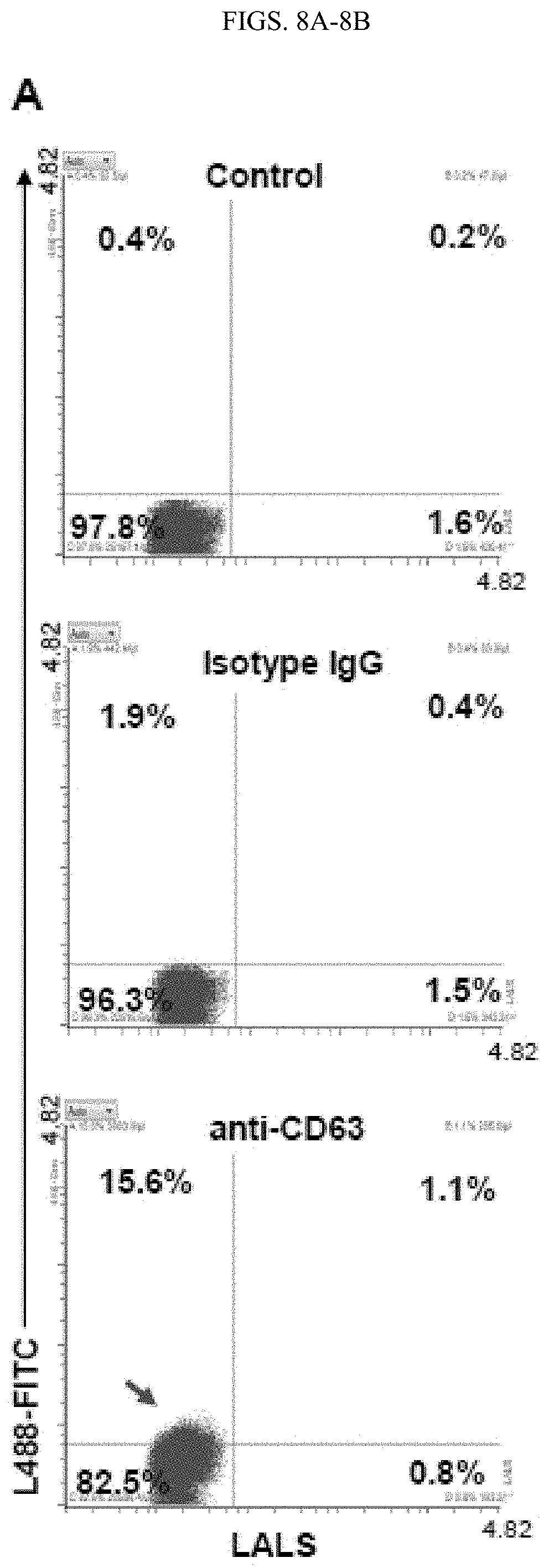
D00014

D00015

D00016
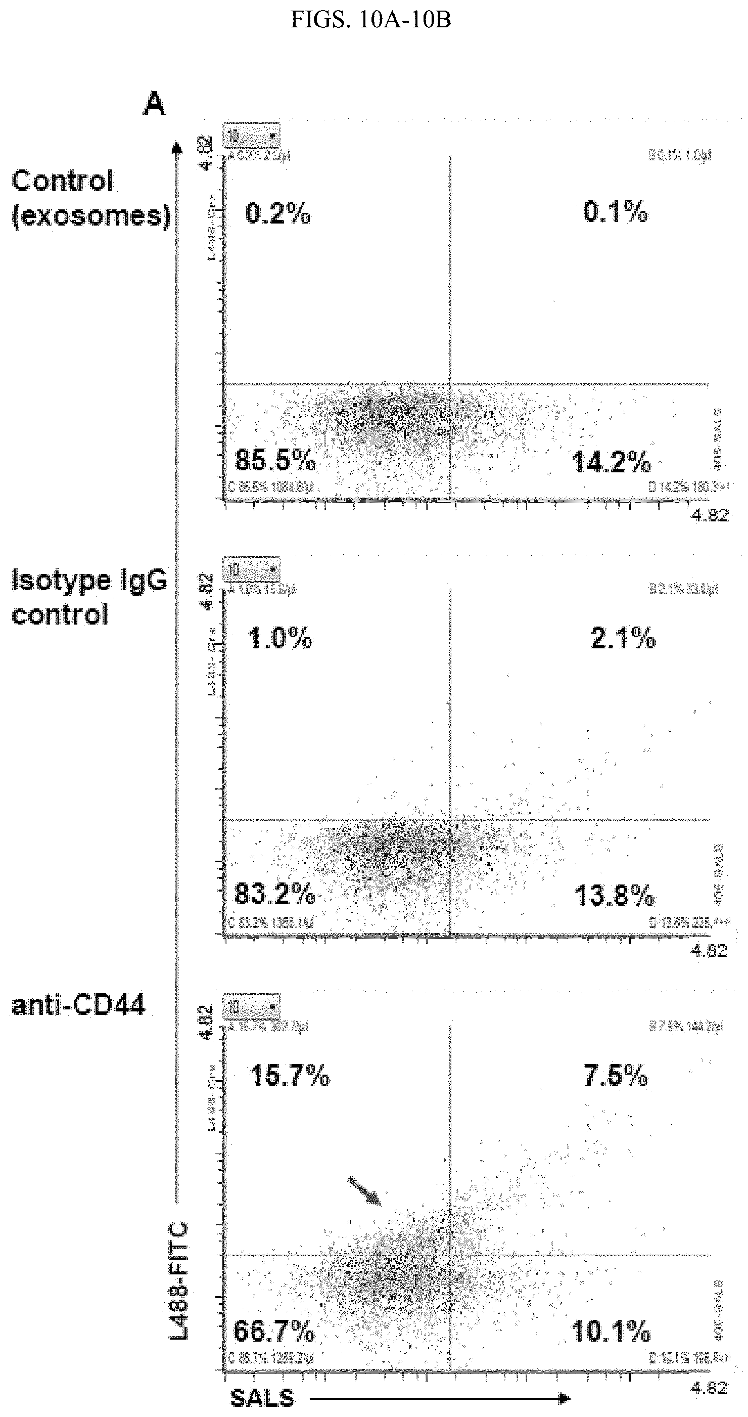
D00017

D00018

D00019
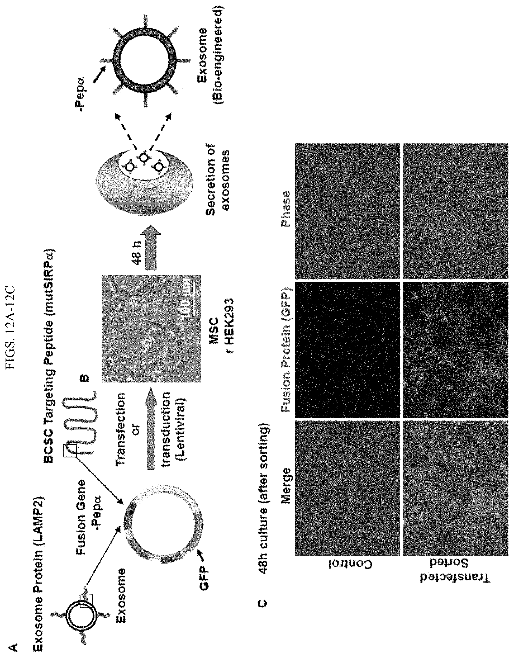
D00020

D00021

D00022
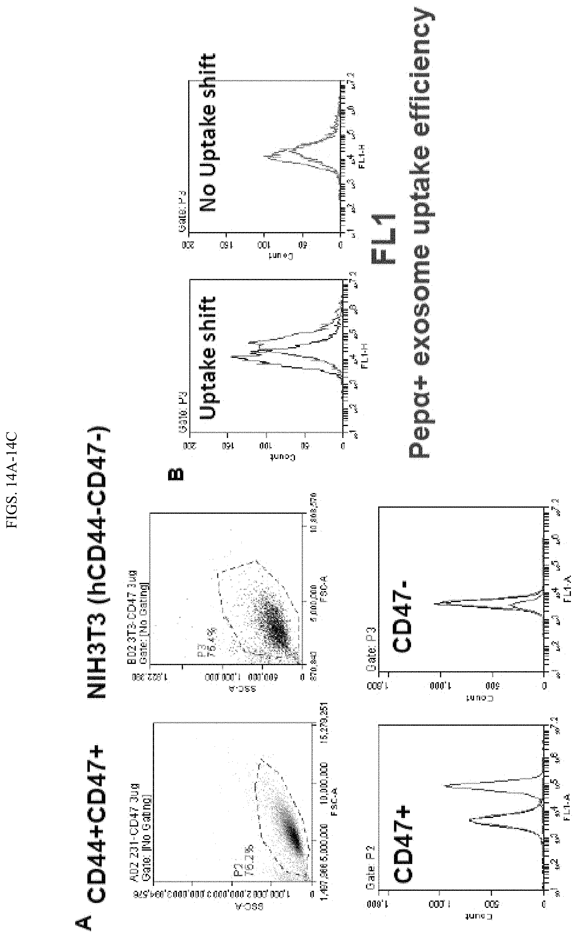
D00023

D00024

D00025

D00026
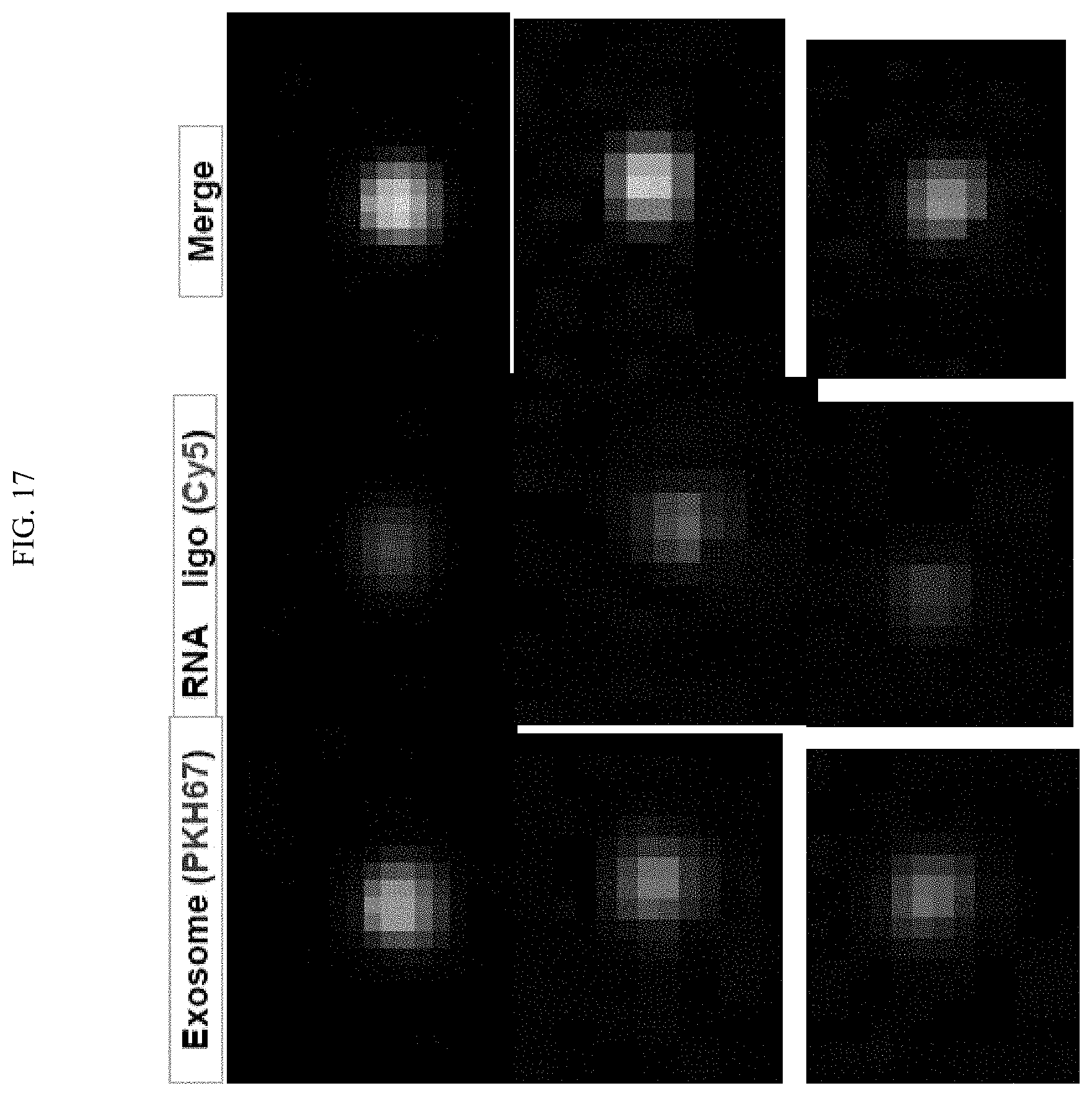
D00027

D00028
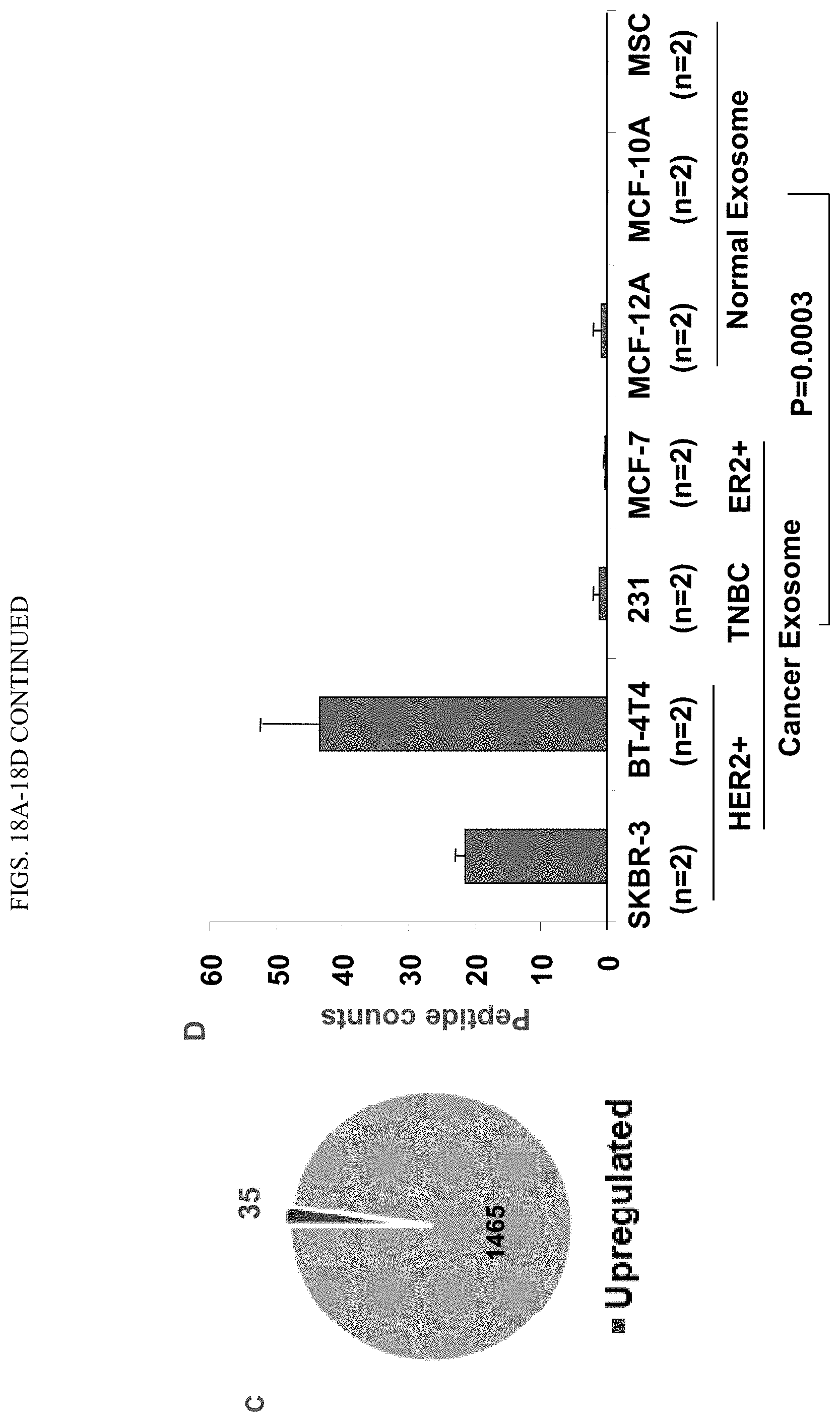
D00029
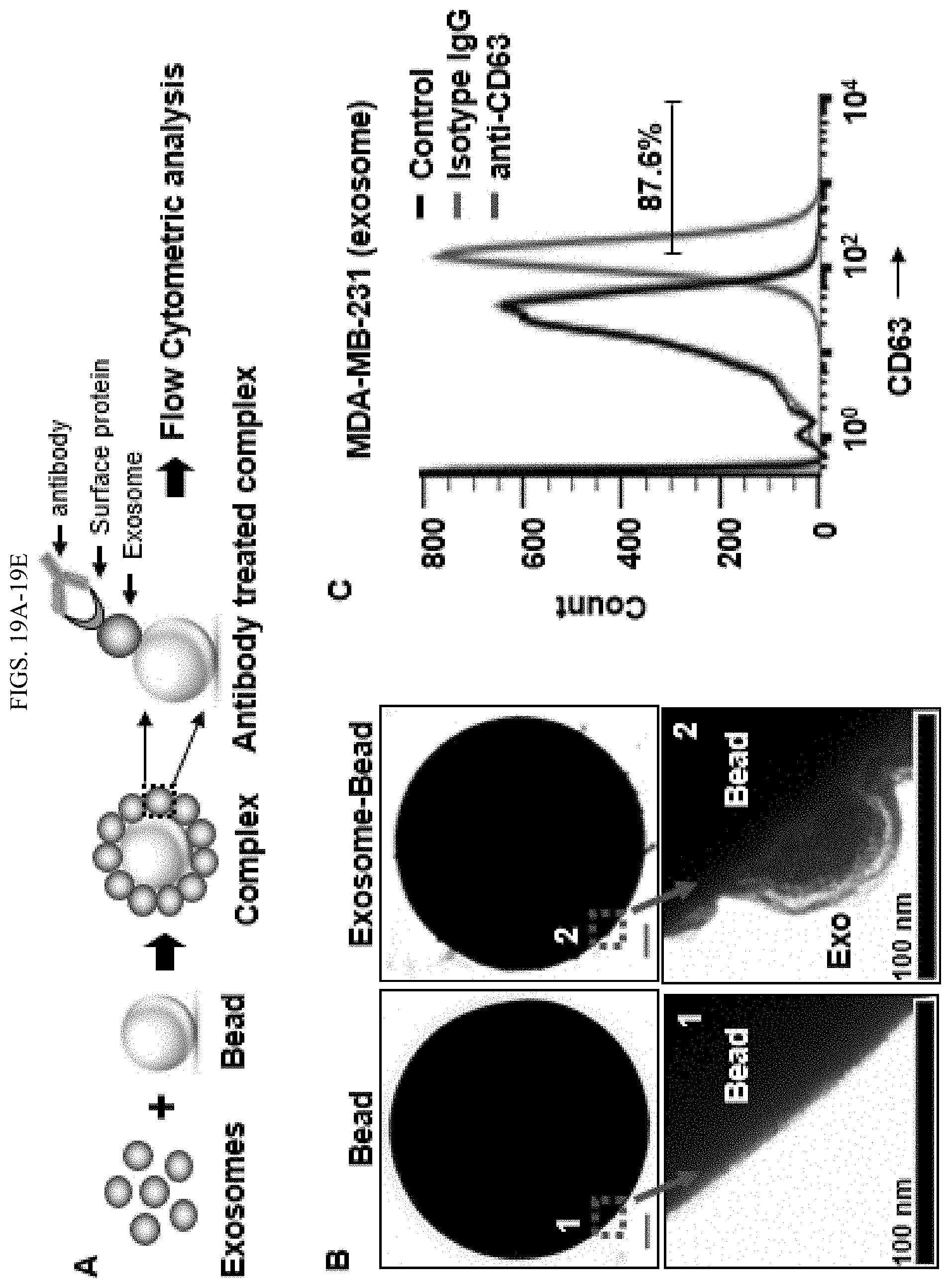
D00030

D00031

D00032

D00033

D00034

D00035
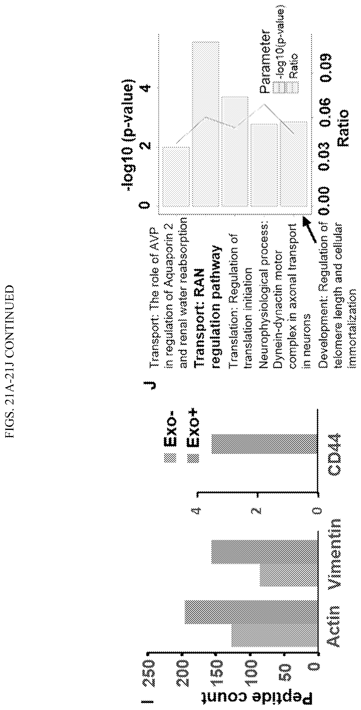
D00036
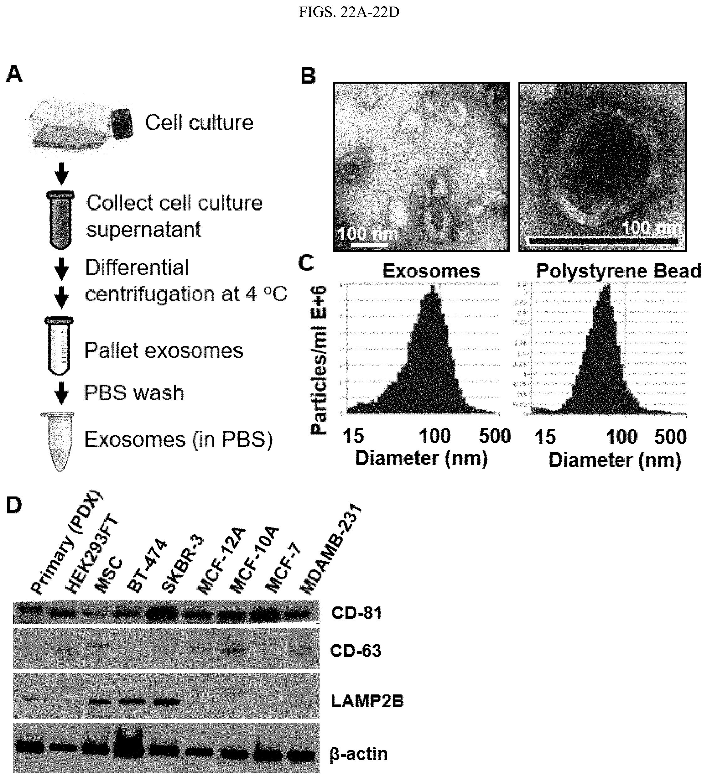
D00037

D00038
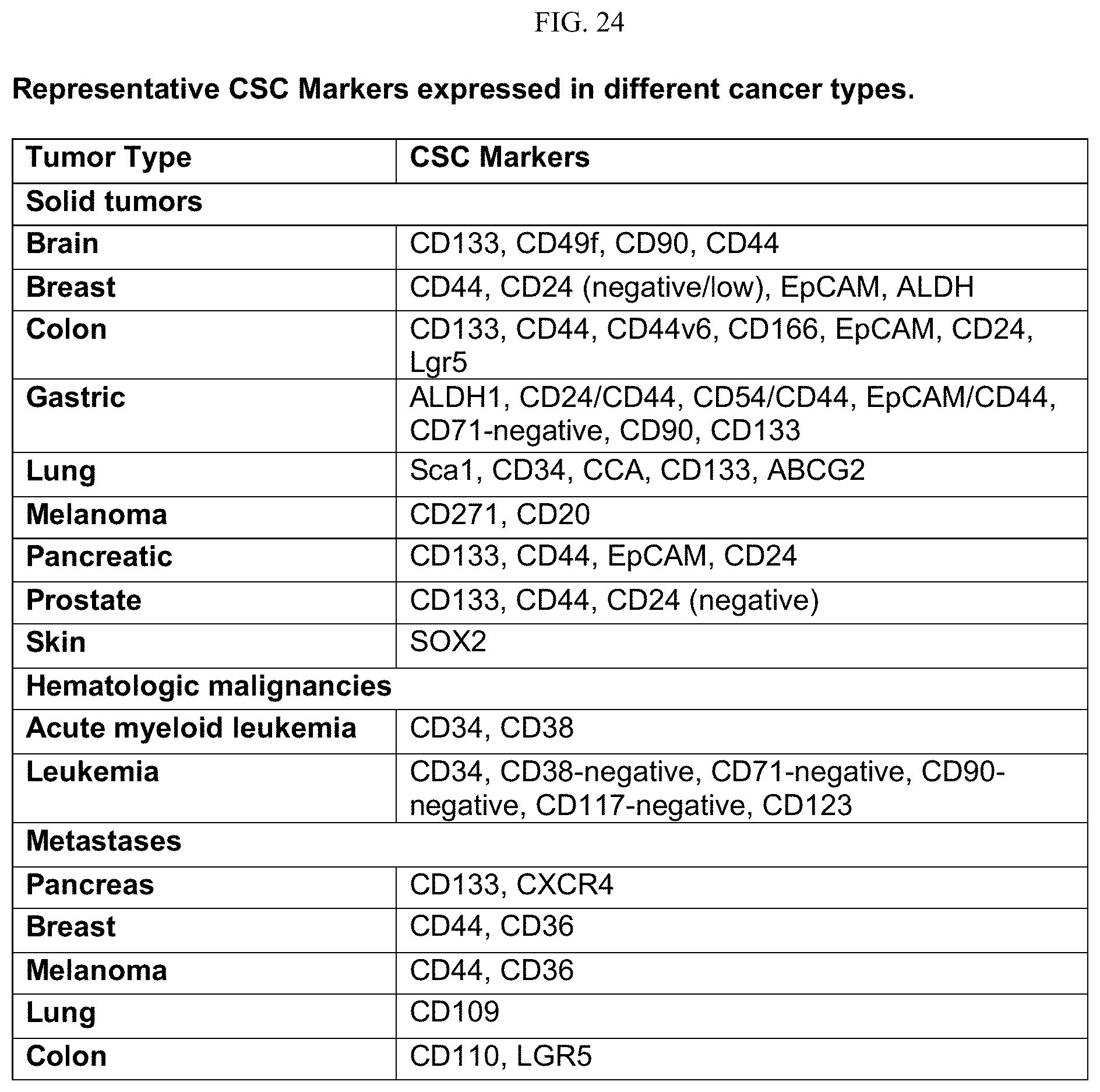
D00039

D00040
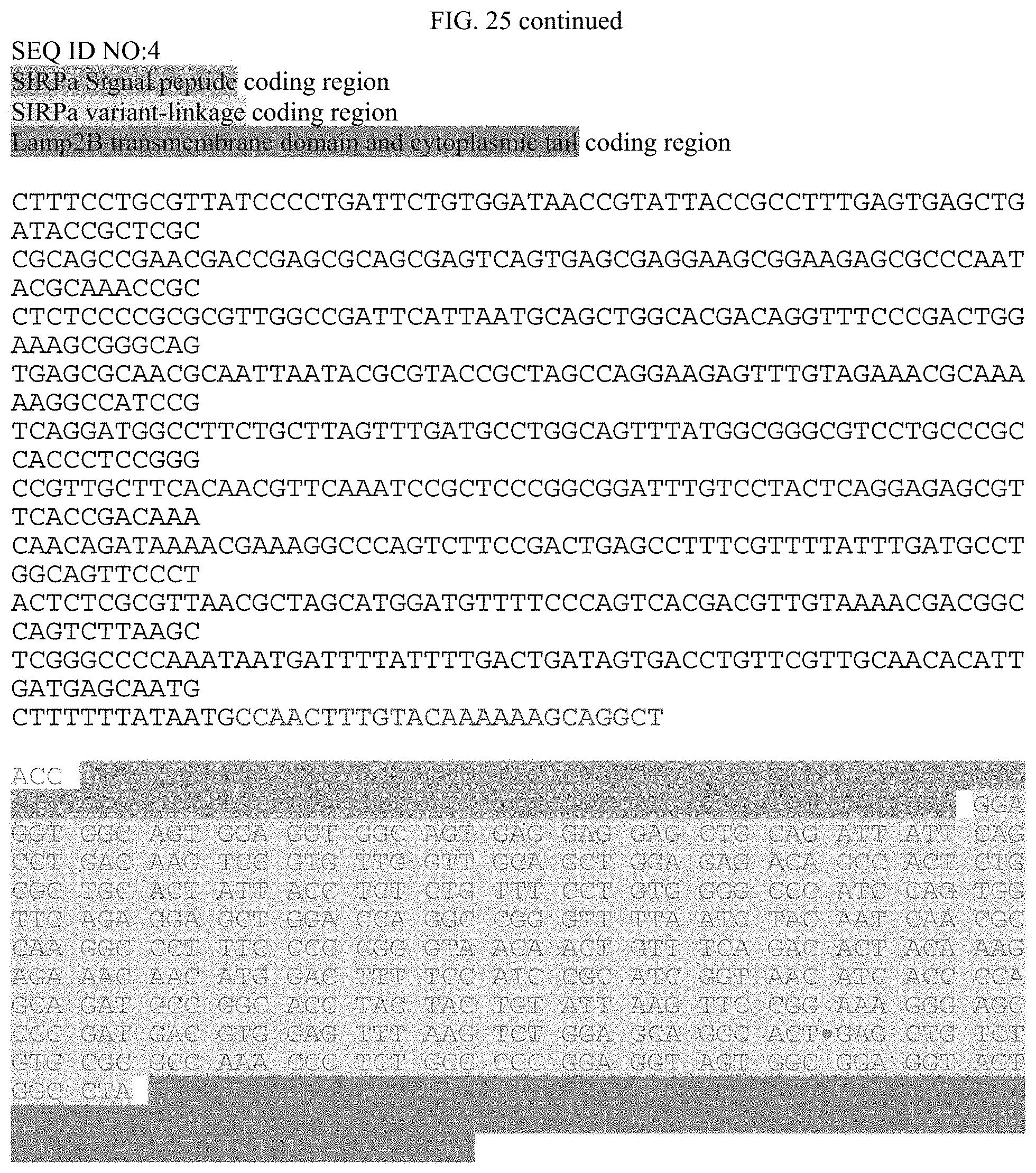
D00041
D00042
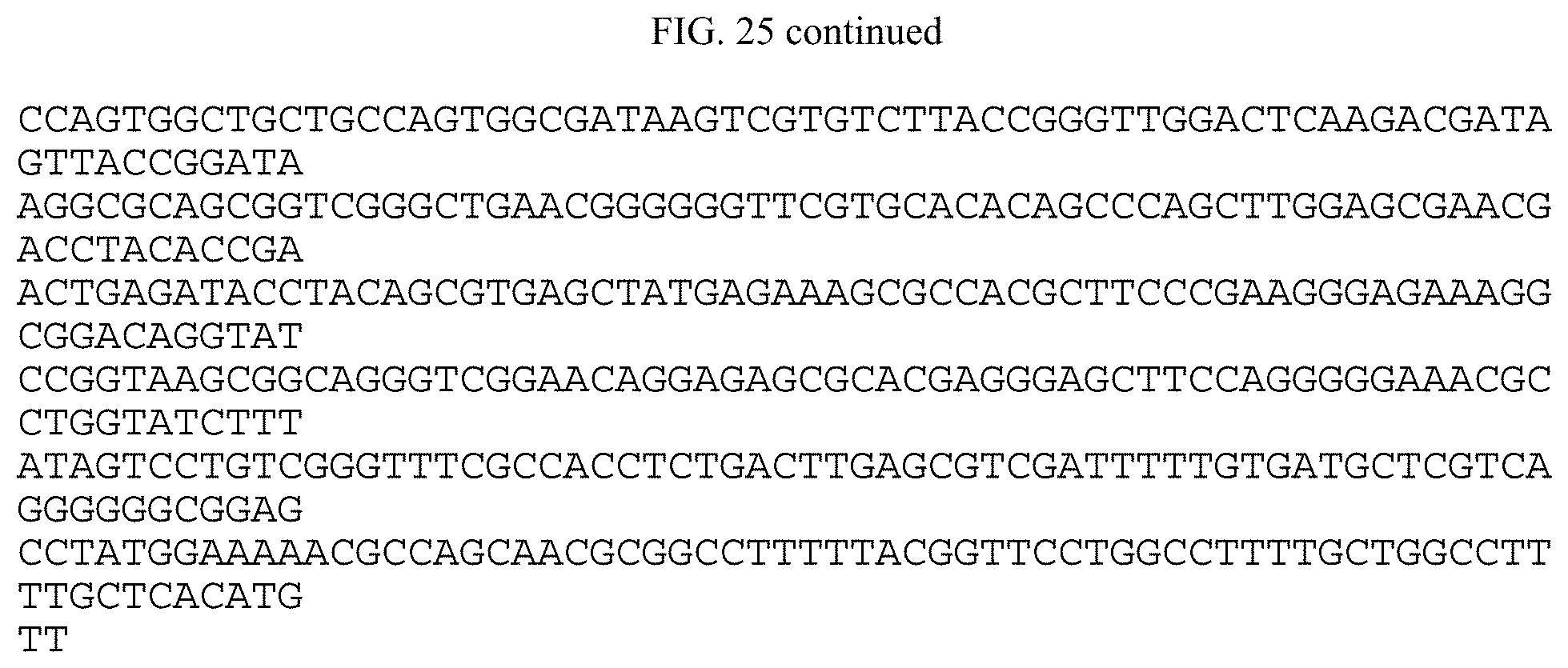
D00043

D00044

D00045
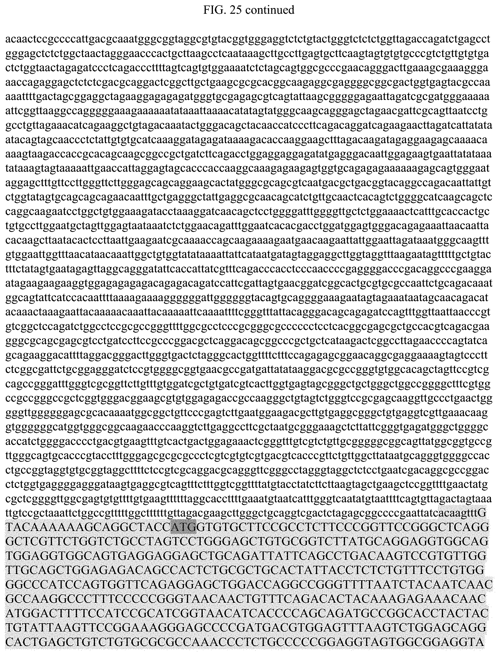
D00046

D00047
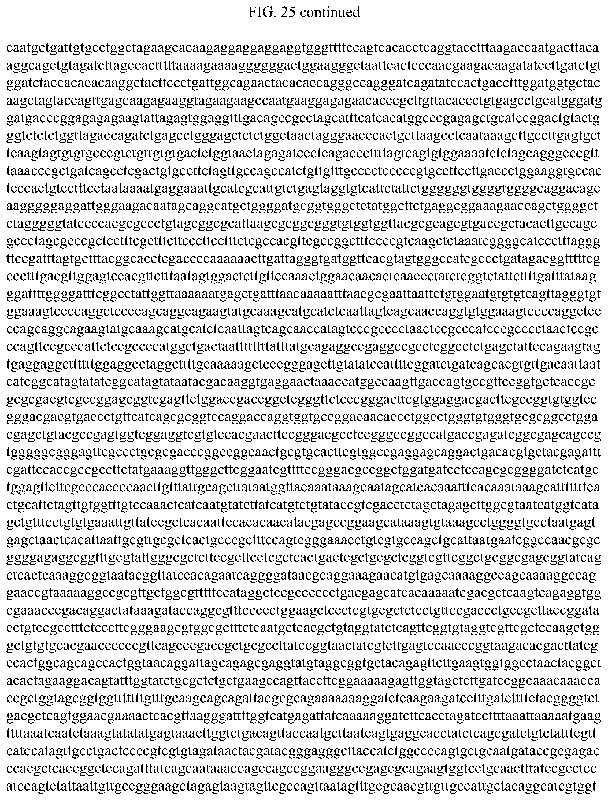
D00048

S00001
XML
uspto.report is an independent third-party trademark research tool that is not affiliated, endorsed, or sponsored by the United States Patent and Trademark Office (USPTO) or any other governmental organization. The information provided by uspto.report is based on publicly available data at the time of writing and is intended for informational purposes only.
While we strive to provide accurate and up-to-date information, we do not guarantee the accuracy, completeness, reliability, or suitability of the information displayed on this site. The use of this site is at your own risk. Any reliance you place on such information is therefore strictly at your own risk.
All official trademark data, including owner information, should be verified by visiting the official USPTO website at www.uspto.gov. This site is not intended to replace professional legal advice and should not be used as a substitute for consulting with a legal professional who is knowledgeable about trademark law.