Methods for inhibiting angiogenesis in a subject in need thereof
Demopulos , et al. December 22, 2
U.S. patent number 10,870,708 [Application Number 15/476,154] was granted by the patent office on 2020-12-22 for methods for inhibiting angiogenesis in a subject in need thereof. This patent grant is currently assigned to University of Leicester. The grantee listed for this patent is Omeros Corporation, University of Leicester. Invention is credited to Gregory A. Demopulos, Thomas Dudler, Hans-Wilhelm Schwaeble, Larry Tjoelker.


View All Diagrams
| United States Patent | 10,870,708 |
| Demopulos , et al. | December 22, 2020 |
Methods for inhibiting angiogenesis in a subject in need thereof
Abstract
In one aspect, the present invention provides methods for preventing, treating, reverting and/or delaying angiogenesis in a mammalian subject suffering from, or at risk for developing, an angiogenesis-dependent disease or condition, comprising administering to the subject an amount of a MASP-2 inhibitory agent effective to inhibit angiogenesis. In some embodiments of these aspects of the invention, the MASP-2 inhibitory agent is a MASP-2 antibody or fragment thereof.
| Inventors: | Demopulos; Gregory A. (Mercer Island, WA), Schwaeble; Hans-Wilhelm (Mountsorrel, GB), Dudler; Thomas (Bellevue, WA), Tjoelker; Larry (Kirkland, WA) | ||||||||||
|---|---|---|---|---|---|---|---|---|---|---|---|
| Applicant: |
|
||||||||||
| Assignee: | University of Leicester
(Leicester, GB) |
||||||||||
| Family ID: | 1000005256372 | ||||||||||
| Appl. No.: | 15/476,154 | ||||||||||
| Filed: | March 31, 2017 |
Prior Publication Data
| Document Identifier | Publication Date | |
|---|---|---|
| US 20170283508 A1 | Oct 5, 2017 | |
Related U.S. Patent Documents
| Application Number | Filing Date | Patent Number | Issue Date | ||
|---|---|---|---|---|---|
| 62315857 | Mar 31, 2016 | ||||
| Current U.S. Class: | 1/1 |
| Current CPC Class: | A61P 35/04 (20180101); C07K 16/40 (20130101); A61P 27/02 (20180101); C07K 2317/24 (20130101); C07K 2317/94 (20130101); C07K 2317/21 (20130101); A61K 2039/505 (20130101); C07K 2317/54 (20130101); C07K 2317/622 (20130101); C07K 2317/76 (20130101); C07K 2317/92 (20130101) |
| Current International Class: | C07K 16/40 (20060101); A61K 39/00 (20060101); A61P 35/04 (20060101); A61P 27/02 (20060101) |
References Cited [Referenced By]
U.S. Patent Documents
| 4331647 | May 1982 | Goldenberg |
| 4816567 | March 1989 | Cabilly |
| 4946778 | August 1990 | Ladner et al. |
| 5211657 | May 1993 | Yamada et al. |
| 5223409 | June 1993 | Ladner et al. |
| 5403484 | April 1995 | Ladner et al. |
| 5552157 | September 1996 | Yagi et al. |
| 5565213 | October 1996 | Nakamori |
| 5567434 | October 1996 | Szoka |
| 5571698 | November 1996 | Ladner et al. |
| 5610288 | March 1997 | Rubenstein |
| 5693762 | December 1997 | Queen et al. |
| 5718709 | February 1998 | Considine et al. |
| 5738868 | April 1998 | Shinkarenko |
| 5739119 | April 1998 | Galli et al. |
| 5741516 | April 1998 | Webb et al. |
| 5759829 | June 1998 | Shewmaker et al. |
| 5789573 | August 1998 | Baker et al. |
| 5795587 | August 1998 | Gao et al. |
| 5801154 | September 1998 | Baracchini et al. |
| 6649592 | November 2003 | Larson |
| 8951522 | February 2015 | Demopulos et al. |
| 2002/0019369 | February 2002 | Li et al. |
| 2007/0238654 | October 2007 | Deschatelets |
| 2011/0311549 | December 2011 | Schwaeble |
| 2012/0282263 | November 2012 | Dudler et al. |
| 2012/0315279 | December 2012 | Medof et al. |
| 2013/0266559 | October 2013 | Demopulos et al. |
| 2013/0344073 | December 2013 | Schwaeble et al. |
| 2014/0134641 | May 2014 | Jensenius et al. |
| 2015/0064176 | March 2015 | Schwaeble et al. |
| 0 321 201 | Jun 1989 | EP | |||
| WO 88/04300 | Jun 1988 | WO | |||
| WO 91/11465 | Aug 1991 | WO | |||
| WO 2004/009664 | Jan 2004 | WO | |||
| WO 2004/106384 | Dec 2004 | WO | |||
| WO 2007/117996 | Oct 2007 | WO | |||
Other References
|
Crawford et al (Current Diabetes Reviews 5: 8-13, 2009 (Year: 2009). cited by examiner . Chen, C. B., et al., "Stoichiometry of complexes between mannose-binding protein and its associated serine proteases. Defining functional units for complement activation," J Biol Chem, 276(28): 25894-25902, (2001). cited by applicant . Feinberg, H., et al., "Crystal structure of the CUB1-EGF-CUB2 region of mannose-binding protein associated serine protease-2," EMBO J, 22(10): 2348-2359, (2003). cited by applicant . Lynch, N. J., et al., "L-ficolin specifically binds to lipoteichoic acid, a cell wall constituent of Gram-positive bacteria, and activates the lectin pathway of complement," J Immunol, 172(2): 1198-1202, (2004). cited by applicant . Stover, C. M., et al., "The rat and mouse homologues of MASP-2 and MAp19, components of the lectin activation pathway of complement," J Immunol, 163(12): 6848-6859, (1999). cited by applicant . Thiel, S., et al., "A second serine protease associated with mannan-binding lectin that activates complement," Nature, 386(6624): 506-510, (1997). cited by applicant . Thiel, S., et al., "Interaction of C1q and mannan-binding lectin (MBL) with C1r, C1s, MBL-associated serine proteases 1 and 2, and the MBL-associated protein MAp19," J Immunol, 165(2): 878-887, (2000). cited by applicant . Vorup-Jensen, T., et al., "Distinct pathways of mannan-binding lectin (MBL)- and C1-complex autoactivation revealed by reconstitution of MBL with recombinant MBL-associated serine protease-2," J Immunol, 165(4): 2093-2100, (2000). cited by applicant . Thielens, N. M., et al., "Interaction properties of human mannan-binding lectin (MBL)-associated serine proteases-1 and -2, MBL-associated protein 19, and MBL," J Immunol, 166(8): 5068-5077, (2001). cited by applicant . Matsushita, M., et al., "Cutting edge: complement-activating complex of ficolin and mannose-binding lectin-associated serine protease," J Immunol, 164(5): 2281-2284, (2000). cited by applicant . Rodrigues, M. L., et al., "Engineering Fab' fragments for efficient F(ab)2 formation in Escherichia coli and for improved in vivo stability," J Immunol, 151(12): 6954-6961, (1993). cited by applicant . Lachmann, P. J., et al., "Initiation of complement activation," Springer Semin Immunopathol, 7(2-3): 143-162, (1984). cited by applicant . Riedemann, N. C., et al., "Complement in ischemia reperfusion injury," Am J Pathol, 162(2): 363-367, (2003). cited by applicant . Matsushita, M., et al., "Activation of the lectin complement pathway by H-ficolin (Hakata antigen)," J Immunol, 168(7): 3502-3506, (2002). cited by applicant . Takahashi, M., et al., "A truncated form of mannose-binding lectin-associated serine protease (MASP)-2 expressed by alternative polyadenylation is a component of the lectin complement pathway," Int Immunol, 11(5): 859-863, (1999). cited by applicant . Ambrus, G., et al., "Natural substrates and inhibitors of mannan-binding lectin-associated serine protease-1 and -2: a study on recombinant catalytic fragments," J Immunol, 170(3): 1374-1382, (2003). cited by applicant . Moller-Kristensen, M., et al., "Levels of mannan-binding lectin-associated serine protease-2 in healthy individuals," J Immunol Methods, 282(1-2): 159-167, (2003). cited by applicant . Petersen, S. V., et al., "Control of the classical and the MBL pathway of complement activation," Mol Immunol, 37(14): 803-811, (2000). cited by applicant . Dahl, M. R., et al., "MASP-3 and its association with distinct complexes of the mannan-binding lectin complement activation pathway," Immunity, 15(1): 127-135, (2001). cited by applicant . Petersen, S. V., et al., "An assay for the mannan-binding lectin pathway of complement activation," J Immunol Methods, 257(1-2): 107-116, (2001). cited by applicant . Liszewski, M. K., et al., "The Complement System," In: E.Paul W., editor, Fundamental Immunology, Third Edition ed., New York, Raven Press, Ltd., 26: 917-939, (1993). cited by applicant . Collard, C. D., et al., "Complement activation after oxidative stress: role of the lectin complement pathway," Am J Pathol, 156(5): 1549-1556, (2000). cited by applicant . Lu, J., et al., "Collectins and ficolins: sugar pattern recognition molecules of the mammalian innate immune system," Biochim Biophys Acta, 1572(2-3): 387-400, (2002). cited by applicant . Jordan, J. E., et al., "Inhibition of mannose-binding lectin reduces postischemic myocardial reperfusion injury," Circulation, 104(12): 1413-1418, (2001). cited by applicant . Maynard, Y., et al., "Characterization of a mannose and N-acetylglucosamine-specific lectin present in rat hepatocytes," J Biol Chem, 257(7): 3788-3794, (1982). cited by applicant . Ambati, J., et al., "Immunology of age-related macular degeneration," Nat Rev Immunol, 13(6): 438-451, (2013). cited by applicant . Lee, R. T., et al., "Multivalent ligand binding by serum mannose-binding protein," Arch Biochem Biophys, 299(1): 129-136, (1992). cited by applicant . Ji, Y. H., et al., "Activation of the C4 and C2 components of complement by a proteinase in serum bactericidal factor, Ra reactive factor," J Immunol, 150(2): 571-578, (1993). cited by applicant . Kilpatrick, D. C., "Mannan-binding lectin: clinical significance and applications," Biochim Biophys Acta, 1572(2-3): 401-413, (2002). cited by applicant . Weis, W. I., et al., "Structure of a C-type mannose-binding protein complexed with an oligosaccharide," Nature, 360(6400): 127-134, (1992). cited by applicant . Kalli, K. R., et al., "Therapeutic uses of recombinant complement protein inhibitors," Springer Semin Immunopathol, 15(4): 417-431, (1994). cited by applicant . Pangburn, M. K., et al., "Formation of the initial C3 convertase of the alternative complement pathway. Acquisition of C3b-like activities by spontaneous hydrolysis of the putative thioester in native C3," J Exp Med, 154(3): 856-867, (1981). cited by applicant . Wallis, R., et al., "Localization of the serine protease-binding sites in the collagen-like domain of mannose-binding protein: indirect effects of naturally occurring mutations on protease binding and activation," J Biol Chem, 279(14): 14065-14073, (2004). cited by applicant . Wallis, R., et al., "Interaction of mannose-binding protein with associated serine proteases: effects of naturally occurring mutations," J Biol Chem, 275(40): 30962-30969, (2000). cited by applicant . Sim, R. B., et al., "Innate Immunity," Biochemical Society Transactions, 28(5): 545-550, (2000). cited by applicant . Petersen, S. V., et al., "Generation of antibodies Towards MASP-1 and MASP-2 Using Bacterial Expression Systems," Molecular Immunology, 35(6-7): 409-409, (1998). cited by applicant . Cech, T. R., et al., "Biological catalysis by RNA," Annu Rev Biochem, 55: 599-629, (1986). cited by applicant . Clackson, T., et al., "Making antibody fragments using phage display libraries," Nature, 352(6336): 624-628, (1991). cited by applicant . Chen, P. F., et al., "Development of the non-palindromic adaptor polymerase chain reaction (NPA-PCR) for the amplification of alpha- and beta-chain T-cell receptor cDNAs," Scand J Immunol, 35(5): 539-549, (1992). cited by applicant . Colligan, "Production of Monoclonal Antibodies," In: Coligan J. E., editor, Current Protocols in Immunology, New York, 2.5: 2.5.1-2.6.7, (1991). cited by applicant . Bird, R. E., et al., "Single-chain antigen-binding proteins," Science, 242(4877): 423-426, (1988). cited by applicant . Climie, S., et al., "Chemical synthesis of the thymidylate synthase gene," Proc Natl Acad Sci U S A, 87(2): 633-637, (1990). cited by applicant . Carter, P., et al., "Humanization of an anti-p185HER2 antibody for human cancer therapy," Proc Natl Acad Sci U S A, 89(10): 4285-4289, (1992). cited by applicant . Altschul, S. F., et al., "Gapped Blast and PSI-Blast: a new generation of protein database search programs," Nucleic Acids Res, 25(17): 3389-3402, (1997). cited by applicant . Johnson, L. V., et al., "Complement activation and inflammatory processes in Drusen formation and age related macular degeneration," Exp Eye Res, 73(6): 887-896, (2001). cited by applicant . Makino, K., et al., "A Microcapsule Self-Regulating Delivery System for Insulin," Journal of Controlled Release, 12: 235-239, (1990). cited by applicant . Lee, V. H. L., "Protease Inhibitors and Penetration Enhancers as Approaches to Modify Peptide Absorption," Journal of Controlled Release, 13: 213-223, (1990). cited by applicant . Jolliffe, L. K., "Humanized antibodies: enhancing therapeutic utility through antibody engineering," Int Rev Immunol, 10(2-3): 241-250, (1993). cited by applicant . Jackson, D. Y., et al., "Potent alpha 4 beta 1 peptide antagonists as potential anti-inflammatory agents," J Med Chem, 40(21): 3359-3368, (1997). cited by applicant . Hori, R., et al., "Enhanced bioavailability of subcutaneously injected insulin coadministered with collagen in rats and humans," Pharm Res, 6(9): 813-816, (1989). cited by applicant . Hageman, G. S., et al., "An integrated hypothesis that considers drusen as biomarkers of immune-mediated processes at the RPE-Bruch's membrane interface in aging and age-related macular degeneration," Prog Retin Eye Res, 20(6): 705-732, (2001). cited by applicant . Greenspan, N. S., et al., "Idiotypes: structure and immunogenicity," FASEB J, 7(5): 437-444, (1993). cited by applicant . De Boer, A. G., et al., "Rectal Absorption Enhancement of Peptide Drugs," Journal of Controlled Release, 13: 241-246, (1990). cited by applicant . Fuertges, R., et al., "The Clinical Efficacy of Poly(ethylene Glycol)-modified Proteins," Journal of Controlled Release, 11: 139-148, (1990). cited by applicant . Singer, I. I., et al., "Optimal humanization of 1B4, an anti-CD18 murine monoclonal antibody, is achieved by correct choice of human V-region framework sequences," J Immunol, 150(7): 2844-2857, (1993). cited by applicant . Schwaeble, W., et al., "The mannan-binding lectin-associated serine proteases (MASPs) and MAp19: four components of the lectin pathway activation complex encoded by two genes," Immunobiology, 205(4-5): 455-466, (2002). cited by applicant . Sandhu, J. S., "Protein engineering of antibodies," Crit Rev Biotechnol, 12(5-6): 437-462, (1992). cited by applicant . Ravetch, J. V., et al., "Fc receptors," Annu Rev Immunol, 9: 457-492, (1991). cited by applicant . Rosenblatt, J., et al., "The Effect of Collagen Fiber Size Distribution on the Release Rate of Proteins from Collagen Matrices by Diffusion," Journal of Controlled Release, 9: 195-203, (1989). cited by applicant . Porter, R. R., "The hydrolysis of rabbit y-globulin and antibodies with crystalline papain," Biochem J, 73: 119-126, (1959). cited by applicant . Merrifield, R. B., "Solid Phase Peptide Synthesis. I. The Synthesis of a Tetrapeptide," J Amer Chem Soc, 85: 2149-2154, (1963). cited by applicant . Presta, L. G., "Antibody engineering," Current Opinion in Structural Biology, 2: 593-596, (1992). cited by applicant . Lee, V. H. L., "Enzymatic Barriers to Peptide and Protein Absorption," CRC Critical Reviews in Therapeutic Durg Carrier Systems, 5(2): 69- 97, (1988). cited by applicant . Yamakawa, I., et al., "Sustained release of insulin by double-layered implant using poly(D,L-lactic acid)," J Pharm Sci, 79(6): 505-509, (1990). cited by applicant . Ohman, E. M., et al., "Early clinical experience with integrelin, an inhibitor of the platelet glycoprotein IIb/IIIa integrin receptor," Eur Heart J, 16 Suppl L: 50-55, (1995). cited by applicant . Pack, P., et al., "Improved bivalent miniantibodies, with identical avidity as whole antibodies, produced by high cell density fermentation of Escherichia coli," Biotechnology (N Y ), 11(11): 1271-1277, (1993). cited by applicant . Zhang, L., et al., "A discrete site modulates activation of I domains. Application to integrin alphaMbeta2," J Biol Chem, 271(47): 29953-29957, (1996). cited by applicant . Taylor, L. D., et al., "Human immunoglobulin transgenes undergo rearrangement, somatic mutation and class switching in mice that lack endogenous IgM," Int Immunol, 6(4): 579-591, (1994). cited by applicant . Takakura, Y., et al., "Control of pharmaceutical properties of soybean trypsin inhibitor by conjugation with dextran. II: Biopharmaceutical and pharmacological properties," J Pharm Sci, 78(3): 219-222, (1989). cited by applicant . Van de Winkel, J. G., et al., "Human IgG Fc receptor heterogeneity: molecular aspects and clinical implications," Immunol Today, 14(5): 215-221, (1993). cited by applicant . Vaughan, T. J., et al., "Human antibodies by design," Nat Biotechnol, 16(6): 535-539, (1998). cited by applicant . Scatchard, G., "The Attraction of Proteins for Small Molecules and Ions," Ann NY Cad Sci, 51: 660-672, (1949). cited by applicant . Green, L. L., et al., "Antigen-specific human monoclonal antibodies from mice engineered with human Ig heavy and light chain YACs," Nat Genet, 7(1): 13-21, (1994). cited by applicant . Glover, G. I., et al., "Synthetic peptide inhibitors of complement serine proteases--I. Identification of functionally equivalent protease inhibitor sequences in serpins and inhibition of C1s and D," Mol Immunol, 25(12): 1261-1267, (1988). cited by applicant . Fedor, M. J., et al., "Substrate sequence effects on "hammerhead" RNA catalytic efficiency," Proc Natl Acad Sci U S A, 87(5): 1668-1672, (1990. cited by applicant . Duncan, A. R., et al., "The binding site for C1q on IgG," Nature, 332(6166): 738-740, (1988). cited by applicant . Dodds, A. W., "Small-scale preparation of complement components C3 and C4," Methods Enzymol,223: 46-61, (1993). cited by applicant . Haseloff, J., et al., "Simple RNA enzymes with new and highly specific endoribonuclease activities," Nature, 334(6183): 585-591, (1988). cited by applicant . Matsushita, M., et al., "Activation of the classical complement pathway by mannose-binding protein in association with a novel C1s-like serine protease," J Exp Med, 176(6): 1497-1502, (1992). cited by applicant . Morgan, B. P., "Clinical complementology: recent progress and future trends," Eur J Clin Invest, 24(4): 219-228, (1994). cited by applicant . Itakura, K., et al., "Synthesis and use of synthetic oligonucleotides," Annu Rev Biochem, 53: 323-356, (1984). cited by applicant . Kuntz, I. D., "Structure-based strategies for drug design and discovery," Science, 257(5073): 1078-1082, (1992). cited by applicant . Holmskov, U., et al., "Collections and ficolins: humoral lectins of the innate immune defense," Annu Rev Immunol, 21: 547-578, (2003). cited by applicant . Ikeda, K., et al., "Serum lectin with known structure activates complement through the classical pathway," J Biol Chem, 262(16): 7451-7454, (1987). cited by applicant . Jensen, S., et al., "Taming of transposable elements by homology-dependent gene silencing," Nat Genet, 21(2): 209-212, (1999). cited by applicant . Lloyd, B. H., et al., "Determination of optimal sites of antisense oligonucleotide cleavage within TNFalpha mRNA," Nucleic Acids Res, 29(17): 3664-3673, (2001). cited by applicant . DesJarlais, R. L., et al., "Structure-based design of nonpeptide inhibitors specific for the human immunodeficiency virus 1 protease," Proc Natl Acad Sci U S A, 87(17): 6644-6648, (1990). cited by applicant . Bae, Y. H., et al., "Insulin Permeation Through Thermo-Sensitive Hydrogels," Journal of Controlled Release, 9: 271-279, (1989). cited by applicant . Asano, M., et al., "In Vivo Characteristics of Low Molecular Weight Copoly(L-Lactice Acid/Glycolic Acid) Formulations with Controlled Release of Luteinizing Hormone-Releasing Hormone Agonist," Journal of Controlled Release, 9: 111-122, (1989). cited by applicant . Kohler, G., et al., "Continuous cultures of fused cells secreting antibody of predefined specificity," Nature, 256(5517): 495-497, (1975). cited by applicant . Kuntz, I. D., et al., "A geometric approach to macromolecule-ligand interactions," J Mol Biol, 161(2): 269-288, (1982). cited by applicant . Kuhlman, M., et al., "The human mannose-binding protein functions as an opsonin," J Exp Med, 169(5): 1733-1745, (1989). cited by applicant . Losman, M. J., et al., "Baboon anti-idiotype antibodies mimic a carcinoembryonic antigen epitope," Int J Cancer, 46(2): 310-314, (1990). cited by applicant . Lonberg, N., et al., "Antigen-specific human antibodies from mice comprising four distinct genetic modifications," Nature, 368(6474): 856-859, (1994). cited by applicant . Marks, J. D., et al., "By-passing immunization. Human antibodies from V-gene libraries displayed on phage," J Mol Biol, 222(3): 581-597, (1991). cited by applicant . Matsushita, M., et al., "A novel human serum lectin with collagen- and fibrinogen-like domains that functions as an opsonin," J Biol Chem, 271(5): 2448-2454, (1996). cited by applicant . Mariani, M., et al., "A new enzymatic method to obtain high-yield F(ab)2 suitable for clinical use from mouse IgG1," Mol Immunol, 28(1-2): 69-77, (1991). cited by applicant . Morrison, S. L., et al., "Chimeric human antibody molecules: mouse antigen-binding domains with human constant region domains," Proc Natl Acad Sci U S A, 81(21): 6851-6855, (1984). cited by applicant . Murayama, O., et al., "Novel peptide ligands for integrin alpha 6 beta 1 selected from a phage display library," J Biochem (Tokyo), 120(2): 445-451, (1996). cited by applicant . Nisonoff, A., et al., "Separation of univalent fragments from the bivalent rabbit antibody molecule by reduction of disulfide bonds," Arch Biochem Biophys, 89: 230-244, (1960). cited by applicant . Niculescu, F., et al., "Rapid Communication: Persistent complement activation on tumor cells in breast cancer," Am J Pathol, 140(5): 1039-1043, (1992). cited by applicant . Scherr, M., et al., "Rapid determination and quantitation of the accessibility to native RNAs by antisense oligodeoxynucleotides in murine cell extracts," Nucleic Acids Res, 26(22): 5079-5085, (1998). cited by applicant . Isaacs, J. D., et al., "Therapy with monoclonal antibodies. An in vivo model for the assessment of therapeutic potential," J Immunol, 148(10): 3062-3071, (1992). cited by applicant . Whitlow, M., et al., "Single-chain Fv Proteins and Their Fusion Proteins," Methods: A companion to Methods in Enzymology, 2(2): 97-105, (1991). cited by applicant . Larrick, J. W., et al., "PCR Amplification of Antibody Genes," Methods: A companion to Methods in Enzymology, 2(2): 106-110, (1991). cited by applicant . Jones, P. T., et al., "Replacing the complementarity-determining regions in a human antibody with those from a mouse," Nature, 321(6069): 522-525, (1986). cited by applicant . Ward, E. S., et al., "Genetic Manipulation and Expression of Antibodies," In: al. B. e., editor, Monoclonal Antibodies: Principles and Applications, Wiley-Liss, Inc., 3: 137-185, (1995). cited by applicant . Courtenay-Luck, N. S., "Genetic manipulation of monoclonal antibodies," In: Ritter M. A., et al., editors, Monoclonal Antibodies: Production, engineering and clinical application, Cambridge, Press Syndicate of the University of Cambridge, Eight: 166-179, (1995). cited by applicant . Kelley, R. F., "Engineering therapeutic Antibodies," In: Cleland j. L. a. C., Charles S., editor, Protein Engineering: Principles and Practice, Wiley-Liss, Inc., 15: 399-434, (1996). cited by applicant . Baines et al., "Purification of Immunoglobulin G (IgG)," in Methods in Molecular Biology, The Humana Press, Inc., vol. 10, pp. 79-104, 1992. cited by applicant . Matsushita, M., et al., "The role of ficolins in innate immunity," Immunobiology, 205(4-5): 490-497, (2002). cited by applicant . Green, J. A., et al., "Production of Polyclonal Antisera," In: Manson M. m., editor, Immunochemical Protocols, Methods in Molecular Biology 10, Carlshalton, Surrey, UK, Humana Press, 1: 1-5, (1992). cited by applicant . Klein, R. J., et al., "Complement Factor H Polymorphism in Age-Related Macular Degeneration," Science, 308(5720): 362-364, (2005). cited by applicant . Haines, J. L., et al., "Complement Factor H Variant Increases the Risk of Age-Related Macular Degeneration," Science, 308: 362-364, (2005). cited by applicant . Edwards, A. O., et al., "Complement Factor H Polymorphism and Age-Related Macular Degeneration," ScienceExpress, 308: 421-424, (2005). cited by applicant . Nozaki, M., et al., "Drusen complement components C3a and C5a promote choroidal neovascularization," Proc Natl Acad Sci U S A, 103(7): 2328-2333, (2006). cited by applicant . Bora, P. S., et al., "Role of complement and complement membrane attack complex in laser-induced choroidal neovascularization," J Immunol, 174(1): 491-497, (2005). cited by applicant . Ambati, J., et al., "Age-related macular degeneration: etiology, pathogenesis, and therapeutic strategies," Surv Ophthalmol, 48(3): 257-293, (2003). cited by applicant . Takahashi, M., et al., "Essential role of mannose-binding lectin-associated serine protease-1 in activation of the complement factor D," J Exp Med, 207(1): 29-37, (2010). cited by applicant . Harlow and Land, Antibodies: A Laboratory Manual, Cold Spring Harbor Laboratory Press, 1988. cited by applicant . Ytting, H., et al., "Serum mannan-binding lectin-associated serine protease 2 levels in colorectal cancer: relation to recurrence and mortality," Clin Cancer Res, 11(4): 1441-1446, (2005). cited by applicant . Ytting, H., et al., "Increased activity of the mannan-binding lectin complement activation pathway in patients with colorectal cancer," Scand J Gastroenterol, 39(7): 674-679, (2004). cited by applicant . Verma, A., et al., "Clinical significance of mannose-binding lectin-associated serine protease-2 expression in esophageal squamous cell carcinoma," Int J Cancer, 118(12): 2930-2935, (2006). cited by applicant . Teh, C., et al., "M-ficolin is expressed on monocytes and is a lectin binding to N-acetyl-D-glucosamine and mediates monocyte adhesion and phagocytosis of Escherichia coli," Immunology, 101(2): 225-232, (2000). cited by applicant . Hansen, S., et al., "Collectin 11 (CL-11, CL-K1) is a MASP-1/3-associated plasma collectin with microbial-binding activity," J Immunol, 185(10): 6096-6104, (2010). cited by applicant . Jack, D. L., et al., "Mannose-binding lectin enhances phagocytosis and killing of Neisseria meningitidis by human macrophages," J Leukoc Biol, 77(3): 328-336, (2005). cited by applicant . Aoyagi, Y., et al., "Role of L-ficolin/mannose-binding lectin-associated serine protease complexes in the opsonophagocytosis of type III group B streptococci," J Immunol, 174(1): 418-425, (2005). cited by applicant . Degn, S. E., et al., "MAp19, the alternative splice product of the MASP2 gene," J Immunol Methods, 373(1-2): 89-101, (2011). cited by applicant . Schwaeble, W. J., et al., "Does properdin crosslink the cellular and the humoral immune response?," Immunol Today, 20(1): 17-21, (1999). cited by applicant . Schultz, D. W., et al., "Analysis of the ARMD1 locus: evidence that a mutation in Hemicentin-1 is associated with age-related macular degeneration in a large family," Hum Mol Genet, 12(24): 3315-3323, (2003). cited by applicant . Mullins, R. F., et al., "Drusen associated with aging and age-related macular degeneration contain proteins common to extracellular deposits associated with atherosclerosis, elastosis, amyloidosis, and dense deposit disease," FASEB J, 14(7): 835-846, (2000). cited by applicant . Johnson, L. V., et al., "A potential role for immune complex pathogenesis in drusen formation," Exp Eye Res, 70(4): 441-449, (2000). cited by applicant . Sakurai, E., et al., "Macrophage depletion inhibits experimental choroidal neovascularization," Invest Ophthalmol Vis Sci, 44(8): 3578-3585, (2003). cited by applicant . Espinosa-Heidmann, D. G., et al., "Macrophage depletion diminishes lesion size and severity in experimental choroidal neovascularization," Invest Ophthalmol Vis Sci, 44(8): 3586-3592, (2003). cited by applicant . Kaufman, R. J., et al., "Improved vectors for stable expression of foreign genes in mammalian cells by use of the untranslated leader sequence from EMC virus," Nucleic Acids Res, 19(16): 4485-4490, (1991). cited by applicant . Kaufman, R. J., "Selection and coamplification of heterologous genes in mammalian cells," Methods Enzymol, 185: 537-566, (1990). cited by applicant . Maniatis, A., et al., "Intermediate-dose melphalan for refractory myeloma," Blood, 74(3): 1177, (1989). cited by applicant . Shea, K. J., "Molecular imprinting of synthetic network polymers; the de novo synthesis of macromolemular binding and catalytic sties," TRIP, 2(5): 166-173, (1994). cited by applicant . Gal, P., et al., "A true autoactivating enzyme. Structural insight into mannose-binding lectin-associated serine protease-2 activations," J Biol Chem, 280(39): 33435-33444, (2005). cited by applicant . Ryan, S. J., "The development of an experimental model of subretinal neovascularization in disciform macular degeneration," Trans Am Ophthalmol Soc, 77: 707-745, (1979). cited by applicant . Tobe, T., et al., "Targeted disruption of the FGF2 gene does not prevent choroidal neovascularization in a murine model," Am J Pathol, 153(5): 1641-1646, (1998). cited by applicant . Ng, E. W., et al., "Pegaptanib, a targeted anti-VEGF aptamer for ocular vascular disease," Nat Rev Drug Discov, 5(2): 123-132, (2006). cited by applicant . Reichmann, L., et al., "Reshaping human antibodies for therapy," Nature, 332(6162): 323-327, (1988). cited by applicant . Lee, W. A., "Permeation enhancers for the nasal delivery of protein and peptide therapeutics," Bio Pharm, 3: 22-25, (1990). cited by applicant . Rohrer, B., et al., "The alternative pathway is required, but not alone sufficient, for retinal pathology in mouse laser-induced choroidal neovascularization," Mol Immunol, 48(6-7): e1-e8, (2011). cited by applicant . Ebrahimi, K. B., et al., "Lipids, lipoproteins, and age-related macular degeneration," J Lipids, 2011: 802059, (2011). cited by applicant . Joseph, K., et al., "Oxidative Stress Sensitizes RPE Cells to Complement-Mediated Injury in a Natural Antibody-, Lectin Pathway-and Phospholipid Epitope-Dependent Manner," Journal of Biological Chemistry, 288(18): 12753-12765, (2013). cited by applicant . Van Lookeren-Campagne, M., et al., "Mechanisms of age-related macular degeneration and therapeutic opportunities," J. Pathol. 232(2):151-64, (2014). cited by applicant . Kunchithapautham, K., et al., "Sublytic membrane-attack-complex (MAC) activation alters regulated rather than constitutive vascular endothelial growth factor (VEGF) secretion in retinal pigment epithelium monolayers," J Biol Chem, 286(27): 23717-23724, (2011). cited by applicant . NCBI Reference Sequence: NM_015838--Homo sapiens ficolin (collagen/fibrinogen domain containing lectin) 2 (hucolin) (FCN2), transcript variant SV2, mRNA 2015 [updated 2015]. cited by applicant . NCBI, Accession No. O00602--RecName: Full=Ficolin-1; AltName: Full=Collagen/fibrinogen domain-containing protein 1; AltName: Full=Ficolin-A; AltName: Full=Ficolin-alpha; AltName: Full=M-ficolin; Flags: Precursor [Homo sapiens] 2015 [updated 2015]. cited by applicant . Yoshihiro, I., et al., "An Insulin-Releasing System that is Responsive to Glucose," Journal of Controlled Release, 10: 195-203, (1989). cited by applicant . King and Possee, in The Baculovirus Expression System: A Laboratory Guide, Chapman and Hall Ltd., London, pp. 111-114, 1992. cited by applicant . Stengaard-Pedersen, K., et al., "Inherited Deficiency of Mannan-binding Lectin-Associated Serine Protease 2," N Engl J Med, 349: 554-560, (2003). cited by applicant . Amit, L., et al., "The Impact of Bevacizumab (Avastin) on Survival in Metastatic Solid Tumors--A Meta-Analysis and Systematic Review," PLoS One 8(1):e51780 (2013). cited by applicant . Baccarelli, A., et al., "Mannose-binding lectin-2 genetic variation and stomach cancer risk," International J Cancer 119:1970-1975, (2006). cited by applicant . Bernig, T., et al., "The mannose-binding lectin (MBL2) haplotype and breast cancer: an association study in African-American and Caucasian women," Carcinogenesis 28:828-836, (2007). cited by applicant . Bjorge, L., et al., "Ascitic complement system in ovarian cancer," Br J Cancer 92(5):895-905, (2005). cited by applicant . Bock, F., et al., "Novel anti(lymph)angiogenic treatment strategies for corneal and ocular surface diseases," Prog Retin Eye Res 34:89-124, (2013). cited by applicant . Corrales, L., et al., "Anaphylatoxin C5a Creates a Favorable Microenvironment for Lung Cancer Progression," J Immunol 189:4674-4683, (2012). cited by applicant . Eurich, D. et al., "Association of mannose-binding lectin-2 gene polymorphism with the development of hepatitis C-induced hepatocellular carcinoma," Liver International 31(7):1006-1012, (2011). cited by applicant . Fan, Y., et al., "Detection and identification of potential biomarkers of breast cancer," J Can Res Clin Oncol 136:1243-54, (2010). cited by applicant . Farrar, C., et al., "Collectin-11 detects stress-induced L-fucose pattern to trigger renal epithelial injury," J Clin Invest 126(5):1911-1925, (2016). cited by applicant . Fentz et al., "Detection of Colorectal Adenoma and Cancer Based on Transthyretin and C3a-desArg Serum Levels," Proteomics Clin Appl 1(6):536-544, (2007). cited by applicant . Ferrara, N., "Vascular endothelial growth factor: molecular and biological aspects," Curr Top Microbiol Immunol 237:1-30, (1999). cited by applicant . Ferrara, N., et al., "Clinical applications of angiogenic growth factors and their inhibitors," Nature Medicine 5(12):1359-1364, (1999). cited by applicant . Fisch, U., et al., "Mannan-binding lectin (MBL) and MBL-associated serine protease-2 in children with cancer," Swiss Med Wkly 141:w13191, (2011). cited by applicant . Gunn, L., et al. "Opposing Roles for Complement Component C5a in Tumor Progression and the Tumor Microenvironment," J Immunol 189:2985, (2012). cited by applicant . Habermann, J., et al., "Increased serum levels of complement C3a anaphylatoxin indicate the presence of colorectal tumors," Gastroenterol 131:1020-1029, (2006). cited by applicant . Henriksen, M., et al., "Heteromeric complexes of native collectin kidney 1 and collectin liver 1 are found in the circulation with MASPs and activate the complement system," J Immunol 191(12):6117-27, (2013). cited by applicant . Hosseini, H., et al., "Anti-VEGF Therapy With Bevacizumab for Anterior Segment Eye Disease," Cornea 31(3):322-34, 2012. cited by applicant . Kanmura, S., et al., "The complement C3a fragment is a potential biomarker for hepatitis C virus-related hepatocellular carcinoma," J Gastroenterol 45(4):459-67, (2010). cited by applicant . Kim, L., et al., "A brief history of anti-VEGF for the treatment of ocular angiogenesis," Am J Pathol 181(2):376-9, (2012). cited by applicant . Klagsbrun, M., et al., "Regulators of Angiogenesis," Annu. Rev. Physiol. 53:217-39, (1991). cited by applicant . Lee, I., et al., "Identification of complement C3a as a candidate biomarker in human chronic hepatitis C and HCV-related hepatocellular carcinoma using a proteomics approach," Proteomics 6(9):2865-73, 2006. cited by applicant . Lee, S.W., et al., "Use of MDLC-DIGE and LC-MS/MS to identify serum biomarkers for complete remission in patients with acute myeloid leukemia," Electrophoresis 33(12):1863-1872, (2012). cited by applicant . Leyvraz, S., et al., "Ocular melanoma: what's new?" Curr Opin Oncol 24:162-9, (2012). cited by applicant . Li, J., et al., "Independent validation of candidate breast cancer serum biomarkers identified by mass spectrometry," Clin Chem 51(12):2229-35, 2005. cited by applicant . Lu, R., et al., "Tumor Angiogenesis Mediated by Myeloid Cells Is Negatively Regulated by CEACAM1," Cancer Res 72(9):2239-50, (2012). cited by applicant . Mahner, S., et al., "TIMP-1 and VEGF-165 serum concentration during first-line therapy of ovarian cancer patients," BMC Cancer 10:139, (2010). cited by applicant . Markiewski, M., et al. "Modulation of the antitumor immune response by complement," Nature Immunol 9(11):1225-1235, (2008). cited by applicant . Mattei, M., et al., "Assignment of Vascular Endothelial Growth Factor (VEGF) and Placenta Growth Factor (P1GF) Genes to Human Chromosome 6p12-p21 and 14q24-q31 Regions, Respectively," Genomics 32(1):168-169, (1996). cited by applicant . Miguet, L., et al., "Discovery and Identification of Potential Biomarkers in a Prospective Study of Chronic Lymphoid Malignancies Using SELDI-TOF--MS," J Proteome Res 5(9):2258-2269, (2006). cited by applicant . Miki, K., et al., "Effects of Intraocular Ranibizumab and Bevacizumab in Transgenic Mice Expressing Human Vascular Endothelial Growth Factor," Ophthalmology 116(9): 1748-1754, (2009). cited by applicant . Nevadunsky, N.S., et al., "Mannose-binding lectin codon 54 genetic polymorphism and vaginal protein levels in women with gynecologic malignancies," European J of Obstetrics and Gynecology and Reproductive Biology 163:216-218, (2012). cited by applicant . Nunez-Cruz, S., et al., "Genetic and Pharmacologic Inhibition of Complement Impairs Endothelial Cell Function and Ablates Ovarian Cancer Neovascularization," Neoplasia 14(11):994-1004, (2012). cited by applicant . Olivo-Marston, S., et al., "Childhood Exposure to Secondhand Smoke and Functional Mannose Binding Lectin Polymorphisms Are Associated with Increased Lung Cancer Risk," Cancer Epidemiology, Biomarkers and Prevention 18(12):3375-3383, (2009). cited by applicant . Perren, T., et al., "A Phase 3 Trial of Bevacizumab in Ovarian Cancer," N Engl J Med 365:2484-2496, (2011). cited by applicant . Pine, S., et al., "Lung cancer survival and functional polymorphisms in MBL2, an innate-immunity gene," Journal of NCI 99:1401-1409, (2007). cited by applicant . Pio, R., et al., "Complement Factor H Is Elevated in Bronchoalveolar Lavage Fluid and Sputum from Patients with Lung Cancer," Cancer Epidemiol Biomarkers Prev 9(10):2665-2672, (2010). cited by applicant . Ribatti, D., et al., "The Role of Angiogenesis in Human Non-Hodgkin Lymphomas," Neoplasia 15(3):231-238, (2013). cited by applicant . Rivera, J.C., et al., "Understanding retinopathy of prematurity: update on pathogenesis," Neonatology 100(4):343-53, (2011). cited by applicant . Rong, Y., et al., "Proteomics analysis of serum protein profiling in pancreatic cancer patients by DIGE: up-regulation of mannose-binding lectin 2 and myosin light chain kinase 2," BMC Gastroenterology 10:68, 2010. cited by applicant . Rutkowski, M.J., et al. "Cancer and the complement cascade," Mol Cancer Res 8(11):1453-65, (2010). cited by applicant . Sato, Y., "Molecular diagnosis of tumor angiogenesis and anti-angiogenic cancer therapy," Int. J. Clin. Oncol. 8(4):200-206, (2003). cited by applicant . Schmiegelow, K., et al., "Increased frequency of mannose-binding lectin insufficiency among children with acute lymphoblastic leukemia," Blood, 100(10): 3757-3760, (2002). cited by applicant . Scudiero, O., et al., "A mannose-binding lectin-defective haplotype is a risk factor for gastric cancer," Clin Chem 52(8):1625-1626, 2006. cited by applicant . Shen, J., et al., "Suppression of ocular neovascularization with siRNA targeting VEGF receptor 1," Gene therapy 13(3): 225-234, (2006). cited by applicant . Solassol, J., et al., "Serum protein signature may improve detection of ductal carcinoma in situ of the breast," Oncogene 29:550, (2010). cited by applicant . Streit, M., "Angiogenesis, lymphangiogenesis, and melanoma metastasis," Oncogene 22:3172-3179, (2003). cited by applicant . Swierzko, A., et al. "Mannan-binding lectin (MBL) in women with tumours of the reproductive system," Cancer Immunology Immunotherapy 56(7):959-971, (2007). cited by applicant . Swierzko, A., et al., "Mannan-binding lectin in malignancy," Mol Immunol 55:16-21, (2013). cited by applicant . Tokunaga, T., et al., "Vascular endothelial growth factor (VEGF) mRNA isoform expression pattern is correlated with liver metastasis and poor prognosis in colon cancer," Br J Cancer 77(6):998-1002, (1998) cited by applicant . Tonini, T., et al. "Molecular basis of angiogenesis and cancer," Oncogene 22(42):6549-6556 , (2003). cited by applicant . Vacca, A., et al., "Bone marrow angiogenesis and progression in multiple myeloma," Br J Haematol 87(3):503-508, (1994). cited by applicant . Van Beijnum, J., et al., "Isolation of endothelial cells from fresh tissues," Nat Protoc. 3(5):1085-91, (2008). cited by applicant . Wang, F.Y., et al., "Mannan-binding Lectin (MBL) Polymorphism and Gastric Cancer Risk in Japanese Population," Digestive Diseases and Sciences 53(11):2904-2908, (2008). cited by applicant . Wuest, T.R., et al., "VEGF-A expression by HSV-1--infected cells drives corneal lymphangiogenesis," J Exp Med 207:101-115, (2009). cited by applicant . Ytting, H., et al., "Mannan-binding lectin (MBL) and MBL-associated serine protease 2 (MASP-2) genotypes in colorectal cancer," Scand J Immunology 73(2):122-127, (2011). cited by applicant . Yuan, A., et al., "Vascular endothelial growth factor 189 mRNA isoform expression specifically correlates with tumor angiogenesis, patient survival, and postoperative relapse in non-small-cell lung cancer," J Clin Oncol 19:432-441, (2001). cited by applicant . Zanetti, K.A., et al., "3'-UTR and Functional Secretor Haplotypes in Mannose-Binding Lectin 2 Are Associated with Increased Colon Cancer Risk in African Americans," Cancer Res 72(6):1467-1477, 2012. cited by applicant . Khan, M. A., et al., "Complement and macrophage crosstalk during process of angiogenesis in tumor progression," J Biomed Sci 22(58 (2015). cited by applicant. |
Primary Examiner: Yao; Lei
Attorney, Agent or Firm: Quinton; Tineka J.
Parent Case Text
CROSS-REFERENCE TO RELATED APPLICATIONS
This application claims the benefit of Provisional Application No. 62/315,857, filed Mar. 31, 2016, all of which are hereby incorporated by reference in their entirety.
Claims
The invention claimed is:
1. A method for treating a mammalian subject suffering from an ocular angiogenesis-dependent disease selected from the group consisting of: proliferative diabetic retinopathy, vitreous hemorrhage secondary to proliferative diabetic retinopathy, neovascular glaucoma, corneal neovascularization, and retinopathy of prematurity, comprising administering to the subject an amount of a MASP-2 inhibitory agent effective to inhibit angiogenesis, wherein the MASP-2 inhibitory agent is a MASP-2 monoclonal antibody or antigen-binding fragment thereof comprising a heavy chain variable region comprising SEQ ID NO:67 and a light chain variable region comprising SEQ ID NO:69.
2. The method of claim 1, wherein the monoclonal antibody or fragment thereof is selected from the group consisting of a recombinant antibody, an antibody having reduced effector function, and a human antibody.
3. The method of claim 1, wherein the composition is administered subcutaneously, intraperitoneally, intra-muscularly, intra-arterially, intravenously, or as an inhalant.
Description
STATEMENT REGARDING SEQUENCE LISTING
The sequence listing associated with this application is provided in text format in lieu of a paper copy and is hereby incorporated by reference into the specification. The name of the text file containing the sequence listing is MP_1_0239_US2_Sequence_Listing_20170321_ST25. The text file is 115 KB, was created on Mar. 21, 2017, and is being submitted via EFS-Web with the filing of the specification.
BACKGROUND
The complement system provides an early acting mechanism to initiate, amplify and orchestrate the immune response to microbial infection and other acute insults (M. K. Liszewski and J. P. Atkinson, 1993, in Fundamental Immunology, Third Edition, edited by W. E. Paul, Raven Press, Ltd., New York), in humans and other vertebrates. While complement activation provides a valuable first-line defense against potential pathogens, the activities of complement that promote a protective immune response can also represent a potential threat to the host (K. R. Kalli, et al., Springer Semin. Immunopathol. 15:417-431, 1994; B. P. Morgan, Eur. J. Clinical Investig. 24:219-228, 1994). For example, C3 and C5 proteolytic products recruit and activate neutrophils. While indispensable for host defense, activated neutrophils are indiscriminate in their release of destructive enzymes and may cause organ damage. In addition, complement activation may cause the deposition of lytic complement components on nearby host cells as well as on microbial targets, resulting in host cell lysis.
The complement system has also been implicated in the pathogenesis of numerous acute and chronic disease states, including: myocardial infarction, stroke, ARDS, reperfusion injury, septic shock, capillary leakage following thermal burns, postcardiopulmonary bypass inflammation, transplant rejection, rheumatoid arthritis, multiple sclerosis, myasthenia gravis, and Alzheimer's disease. In almost all of these conditions, complement is not the cause but is one of several factors involved in pathogenesis. Nevertheless, complement activation may be a major pathological mechanism and represents an effective point for clinical control in many of these disease states. The growing recognition of the importance of complement-mediated tissue injury in a variety of disease states underscores the need for effective complement inhibitory drugs. To date, Eculizumab (Solaris.RTM.), an antibody against complement component C5, is the only complement-targeting drug that has been approved for use in man. Yet, C5 is one of several effector molecules located "downstream" in the complement activation cascade, and blockade of C5 does not inhibit activation of the complement system. Therefore, an inhibitor of the initiation steps of complement activation would have significant advantages over a "downstream" complement inhibitor.
Currently, it is widely accepted that the complement system can be activated through three distinct pathways: the classical pathway, the lectin pathway, and the alternative pathway. The classical pathway is usually triggered by a complex composed of host antibodies bound to a foreign particle (i.e., an antigen) and thus requires prior exposure to an antigen for the generation of a specific antibody response. Since activation of the classical pathway depends on a prior adaptive immune response by the host, the classical pathway is part of the acquired immune system. In contrast, both the lectin and alternative pathways are independent of adaptive immunity and are part of the innate immune system.
The activation of the complement system results in the sequential activation of serine protease zymogens. The first step in activation of the classical pathway is the binding of a specific recognition molecule, C1q, to antigen-bound IgG and IgM molecules. C1q is associated with the C1r and C1s serine protease proenzymes as a complex called C1. Upon binding of C1q to an immune complex, autoproteolytic cleavage of the Arg-Ile site of C1r is followed by C1r-mediated cleavage and activation of C1s, which thereby acquires the ability to cleave C4 and C2. C4 is cleaved into two fragments, designated C4a and C4b, and, similarly, C2 is cleaved into C2a and C2b. C4b fragments are able to form covalent bonds with adjacent hydroxyl or amino groups and generate the C3 convertase (C4b2a) through noncovalent interaction with the C2a fragment of activated C2. C3 convertase (C4b2a) activates C3 by proteolytic cleavage into C3a and C3b subcomponents leading to generation of the C5 convertase (C4b2a3b), which, by cleaving C5 leads to the formation of the membrane attack complex (C5b combined with C6, C7, C8 and C-9 polymers, also referred to as "MAC") that can disrupt cellular membranes leading to cell lysis. The activated forms of C3 and C4 (C3b and C4b) are covalently deposited on the foreign target surfaces, which are recognized by complement receptors on multiple phagocytes.
The first step in activation of the complement system through the lectin pathway is the binding of lectin pathway-specific pattern recognition molecules to their target ligands. This process initiates the activation of lectin pathway-specific serine protease proenzymes that in turn initiate the complement cascade. The pattern recognition molecules in the lectin pathway comprise a group of carbohydrate-binding C-type lectins, i.e., mannan-binding lectin (MBL), collectin-11 (CL-11, also known as CL-K1), collectin-10 (CL-10, also known as CL-L1), and three different ficolins, i.e., H-ficolin, M-ficolin and L-ficolin that bind to acetylated structures of carbohydrates and proteins through fibrinogen-like binding domains (J. Lu et al., Biochim. Biophys. Acta 1572:387-400, (2002); Holmskov et al., Annu. Rev. Immunol. 21:547-578 (2003); Teh et al., Immunology 101:225-232 (2000), J. Luet et al., Biochim Biophys Acta 1572:387-400 (2002); Hansen et al, J. Immunol 185(10):6096-6104 (2010), and Hendriksen et al., J Immunol 191(12):6117-27, 2013).
Ikeda et al. first demonstrated that, like C1q, MBL could activate the complement system upon binding to yeast mannan-coated erythrocytes in a C4-dependent manner (Ikeda et al., J Biol. Chem. 262:7451-7454, (1987)). MBL, a member of the collectin protein family, is a calcium-dependent lectin that binds carbohydrates with 3- and 4-hydroxy groups oriented in the equatorial plane of the pyranose ring. Prominent ligands for MBL are thus D-mannose and N-acetyl-D-glucosamine, while carbohydrates not fitting this steric requirement have undetectable affinity for MBL (Weis et al., Nature 360:127-134, (1992)). The interaction between MBL and monovalent sugars is extremely weak, with dissociation constants typically in the single-digit millimolar range. MBL achieves tight, specific binding to glycan ligands by avidity, i.e., by interacting simultaneously with multiple monosaccharide residues located in close proximity to each other (Lee et al., Archiv. Biochem. Biophys. 299:129-136, (1992)). MBL recognizes the carbohydrate patterns that commonly decorate microorganisms such as bacteria, yeast, parasites and certain viruses. In contrast, MBL does not recognize D-galactose and sialic acid, the penultimate and ultimate sugars that usually decorate "mature" complex glycoconjugates present on mammalian plasma and cell surface glycoproteins. This binding specificity is thought to promote recognition of "foreign" surfaces and help protect from "self-activation." However, MBL does bind with high affinity to clusters of high-mannose "precursor" glycans on N-linked glycoproteins and glycolipids sequestered in the endoplasmic reticulum and Golgi of mammalian cells (Maynard et al., J. Biol. Chem. 257:3788-3794, (1982)). Therefore, damaged cells are potential targets for lectin pathway activation via MBL binding and more recent work has shown that CL-11 is another lectin pathway recognition subcomponent that initiates lectin pathway activation on distressed or damaged cells (Farar et al., J Clin Invest 126:1911-1925, 2016).
The ficolins possess a different type of lectin domain than MBL, called the fibrinogen-like domain. Ficolins bind sugar residues in a Ca.sup.++-independent manner. In humans, three kinds of ficolins (L-ficolin, M-ficolin and H-ficolin) have been identified. The two serum ficolins, L-ficolin and H-ficolin, have in common a specificity for N-acetyl-D-glucosamine; however, H-ficolin also binds N-acetyl-D-galactosamine. The difference in sugar specificity of L-ficolin, H-ficolin, CL-11, and MBL means that the different lectins may be complementary and target different, though overlapping, glycoconjugates. This concept is supported by the recent report that, of the known lectins in the lectin pathway, only L-ficolin binds specifically to lipoteichoic acid, a cell wall glycoconjugate found on all Gram-positive bacteria (Lynch et al., J. Immunol. 172:1198-1202, (2004)). The collectins (i.e., MBL, CL-11, CL-10 and CL-11/CL-10 complexes) and the ficolins bear no significant similarity in amino acid sequence. However, the two groups of proteins have similar domain organizations and, like C1q, assemble into oligomeric structures, which maximize the possibility of multisite binding.
The serum concentrations of MBL are highly variable in healthy populations and this is genetically controlled by polymorphisms/mutations in both the promoter and coding regions of the MBL gene. As an acute phase protein, the expression of MBL is further upregulated during inflammation. L-ficolin is present in serum at concentrations similar to those of MBL. Therefore, the L-ficolin branch of the lectin pathway is potentially comparable to the MBL arm in physiological importance. MBL and ficolins can also function as opsonins, which allow phagocytes to target MBL- and ficolin-decorated surfaces (see Jack et al., J Leukoc Biol., 77(3):328-36 (2004), Matsushita and Fujita, Immunobiology, 205(4-5):490-7 (2002), Aoyagi et al., J Immunol, 174(1):418-25(2005). This opsonization requires the interaction of these proteins with phagocyte receptors (Kuhlman et al., J Exp. Med. 169:1733, (1989); Matsushita et al., J Biol. Chem. 271:2448-54, (1996)), the identity of which has not been established.
Human MBL forms a specific and high-affinity interaction through its collagen-like domain with unique C1r/C1s-like serine proteases, termed MBL-associated serine proteases (MASPs). To date, three MASPs have been described. First, a single enzyme "MASP" was identified and characterized as the enzyme responsible for the initiation of the complement cascade (i.e., cleaving C2 and C4) (Matsushita et al., J Exp Med 176(6):1497-1502 (1992); Ji et al., J Immunol. 150:571-578, (1993)). It was subsequently determined that the MASP activity was, in fact, a mixture of two proteases: MASP-1 and MASP-2 (Thiel et al., Nature 386:506-510, (1997)). However, it was demonstrated that the MBL-MASP-2 complex alone is sufficient for complement activation (Vorup-Jensen et al., J Immunol. 165:2093-2100, (2000)). Furthermore, only MASP-2 cleaved C2 and C4 at high rates (Ambrus et al., Immunol. 170:1374-1382, (2003)). Therefore, MASP-2 is the protease responsible for activating C4 and C2 to generate the C3 convertase, C4b2a. This is a significant difference from the C1 complex of the classical pathway, where the coordinated action of two specific serine proteases (C1r and C1s) leads to the activation of the complement system. In addition, a third novel protease, MASP-3, has been isolated (Dahl, M. R., et al., Immunity 15:127-35, 2001). MASP-1 and MASP-3 are alternatively spliced products of the same gene.
MASPs share identical domain organizations with those of C1r and C1s, the enzymatic components of the C1 complex (Sim et al., Biochem. Soc. Trans. 28:545, (2000)). These domains include an N-terminal C1r/C1s/sea urchin VEGF/bone morphogenic protein (CUB) domain, an epidermal growth factor-like domain, a second CUB domain, a tandem of complement control protein domains, and a serine protease domain. As in the C1 proteases, activation of MASP-2 occurs through cleavage of an Arg-Ile bond adjacent to the serine protease domain, which splits the enzyme into disulfide-linked A and B chains, the latter consisting of the serine protease domain.
MBL can also associate with an alternatively spliced form of MASP-2, known as MBL-associated protein of 19 kDa (MAp19) or small MBL-associated protein (sMAP), which lacks the catalytic activity of MASP-2. (Stover, J Immunol. 162:3481-90, (1999); Takahashi et al., Int. Immunol. 11:859-863, (1999)). MAp19 comprises the first two domains of MASP-2, followed by an extra sequence of four unique amino acids. The function of Map19 is unclear (Degn et al., J Immunol. Methods, 2011). The MASP-1 and MASP-2 genes are located on human chromosomes 3 and 1, respectively (Schwaeble et al., Immunobiology 205:455-466, (2002)).
Several lines of evidence suggest that there are different MBL-MASP complexes and a large fraction of the MASPs in serum is not complexed with MBL (Thiel, et al., Immunol. 165:878-887, (2000)). Both H- and L-ficolin bind to all MASPs and activate the lectin complement pathway, as does MBL (Dahl et al., Immunity 15:127-35, (2001); Matsushita et al., J Immunol. 168:3502-3506, (2002)). Both the lectin and classical pathways form a common C3 convertase (C4b2a) and the two pathways converge at this step.
The lectin pathway is widely thought to have a major role in host defense against infection in the naive host. Strong evidence for the involvement of MBL in host defense comes from analysis of patients with decreased serum levels of functional MBL (Kilpatrick, Biochim. Biophys. Acta 1572:401-413, (2002)). Such patients display susceptibility to recurrent bacterial and fungal infections. These symptoms are usually evident early in life, during an apparent window of vulnerability as maternally derived antibody titer wanes, but before a full repertoire of antibody responses develops. This syndrome often results from mutations at several sites in the collagenous portion of MBL, which interfere with proper formation of MBL oligomers. However, since MBL can function as an opsonin independent of complement, it is not known to what extent the increased susceptibility to infection is due to impaired complement activation.
In contrast to the classical and lectin pathways, no initiators of the alternative pathway have been found to fulfill the recognition functions that C1q and lectins perform in the other two pathways. Currently it is widely accepted that the alternative pathway spontaneously undergoes a low level of turnover activation, which can be readily amplified on foreign or other abnormal surfaces (bacteria, yeast, virally infected cells, or damaged tissue) that lack the proper molecular elements that keep spontaneous complement activation in check. There are four plasma proteins directly involved in the activation of the alternative pathway: C3, factors B and D, and properdin.
Although there is extensive evidence implicating both the classical and alternative complement pathways in the pathogenesis of non-infectious human diseases, the role of the lectin pathway is just beginning to be evaluated. Recent studies provide evidence that activation of the lectin pathway can be responsible for complement activation and related inflammation in ischemia/reperfusion injury. Collard et al. (2000) reported that cultured endothelial cells subjected to oxidative stress bind MBL and show deposition of C3 upon exposure to human serum (Collard et al., Am. J Pathol. 156:1549-1556, (2000)). In addition, treatment of human sera with blocking anti-MBL monoclonal antibodies inhibited MBL binding and complement activation. These findings were extended to a rat model of myocardial ischemia-reperfusion in which rats treated with a blocking antibody directed against rat MBL showed significantly less myocardial damage upon occlusion of a coronary artery than rats treated with a control antibody (Jordan et al., Circulation 104:1413-1418, (2001)). The molecular mechanism of MBL binding to the vascular endothelium after oxidative stress is unclear; a recent study suggests that activation of the lectin pathway after oxidative stress may be mediated by MBL binding to vascular endothelial cytokeratins, and not to glycoconjugates (Collard et al., Am. J. Pathol. 159:1045-1054, (2001)). Other studies have implicated the classical and alternative pathways in the pathogenesis of ischemia/reperfusion injury and the role of the lectin pathway in this disease remains controversial (Riedermann, N.C., et al., Am. J. Pathol. 162:363-367, 2003).
A recent study has shown that MASP-1 (and possibly also MASP-3) is required to convert the alternative pathway activation enzyme Factor D from its zymogen form into its enzymatically active form (see Takahashi M. et al., J Exp Med 207(1):29-37 (2010)). The physiological importance of this process is underlined by the absence of alternative pathway functional activity in plasma of MASP-1/3-deficient mice. Proteolytic generation of C3b from native C3 is required for the alternative pathway to function. Since the alternative pathway C3 convertase (C3bBb) contains C3b as an essential subunit, the question regarding the origin of the first C3b via the alternative pathway has presented a puzzling problem and has stimulated considerable research.
C3 belongs to a family of proteins (along with C4 and .alpha.-2 macroglobulin) that contain a rare posttranslational modification known as a thioester bond. The thioester group is composed of a glutamine whose terminal carbonyl group forms a covalent thioester linkage with the sulfhydryl group of a cysteine three amino acids away. This bond is unstable and the electrophilic glutamyl-thioester can react with nucleophilic moieties such as hydroxyl or amino groups and thus form a covalent bond with other molecules. The thioester bond is reasonably stable when sequestered within a hydrophobic pocket of intact C3. However, proteolytic cleavage of C3 to C3a and C3b results in exposure of the highly reactive thioester bond on C3b and, following nucleophilic attack by adjacent moieties comprising hydroxyl or amino groups, C3b becomes covalently linked to a target. In addition to its well-documented role in covalent attachment of C3b to complement targets, the C3 thioester is also thought to have a pivotal role in triggering the alternative pathway. According to the widely accepted "tick-over theory", the alternative pathway is initiated by the generation of a fluid-phase convertase, iC3Bb, which is formed from C3 with hydrolyzed thioester (iC3; C3(H.sub.2O)) and factor B (Lachmann, P. J., et al., Springer Semin. Immunopathol. 7:143-162, (1984)). The C3b-like C3(H.sub.2O) is generated from native C3 by a slow spontaneous hydrolysis of the internal thioester in the protein (Pangburn, M. K., et al., J. Exp. Med. 154:856-867, 1981). Through the activity of the C3(H.sub.2O)Bb convertase, C3b molecules are deposited on the target surface thereby initiating the alternative pathway.
Very little is known about the initiators of activation of the alternative pathway. Activators are thought to include yeast cell walls (zymosan), many pure polysaccharides, rabbit erythrocytes, certain immunoglobulins, viruses, fungi, bacteria, animal tumor cells, parasites, and damaged cells. The only feature common to these activators is the presence of carbohydrate, but the complexity and variety of carbohydrate structures has made it difficult to establish the shared molecular determinants which are recognized. It has been widely accepted that alternative pathway activation is controlled through the fine balance between inhibitory regulatory components of this pathway, such as Factor H, Factor I, DAF, and CR1, and properdin, which is the only positive regulator of the alternative pathway (see Schwaeble W. J. and Reid K. B., Immunol Today 20(1):17-21 (1999)).
In addition to the apparently unregulated activation mechanism described above, the alternative pathway can also provide a powerful amplification loop for the lectin/classical pathway C3 convertase (C4b2a) since any C3b generated can participate with factor B in forming additional alternative pathway C3 convertase (C3bBb). The alternative pathway C3 convertase is stabilized by the binding of properdin. Properdin extends the alternative pathway C3 convertase half-life six to ten fold. Addition of C3b to the alternative pathway C3 convertase leads to the formation of the alternative pathway C5 convertase.
All three pathways (i.e., the classical, lectin and alternative) have been thought to converge at C5, which is cleaved to form products with multiple proinflammatory effects. The converged pathway has been referred to as the terminal complement pathway. C5a is the most potent anaphylatoxin, inducing alterations in smooth muscle and vascular tone, as well as vascular permeability. It is also a powerful chemotaxin and activator of both neutrophils and monocytes. C5a-mediated cellular activation can significantly amplify inflammatory responses by inducing the release of multiple additional inflammatory mediators, including cytokines, hydrolytic enzymes, arachidonic acid metabolites, and reactive oxygen species. C5 cleavage leads to the formation of C5b-9, also known as the membrane attack complex (MAC). There is now strong evidence that sublytic MAC deposition may play an important role in inflammation in addition to its role as a lytic pore-forming complex.
In addition to its essential role in immune defense, the complement system contributes to tissue damage in many clinical conditions. Thus, there is a pressing need to develop therapeutically effective complement inhibitors to prevent these adverse effects.
It is well established that angiogenesis is implicated in the pathogenesis of a variety of disorders including solid tumors and metastases, and ocular neovascular diseases such as age-related macular degeneration (AMD), proliferative diabetic retinopathy and neovascular glaucoma.
In view of the role of angiogenesis in many diseases and disorders, there is also a pressing need to develop therapeutically effective angiogenesis inhibitors.
SUMMARY
This summary is provided to introduce a selection of concepts in a simplified form that are further described below in the Detailed Description. This summary is not intended to identify key features of the claimed subject matter, nor is it intended to be used as an aid in determining the scope of the claimed subject matter.
In one aspect, the present invention provides methods for preventing, treating, reverting and/or delaying angiogenesis in a mammalian subject suffering from, or at risk for developing, an angiogenesis-dependent disease or condition, comprising administering to the subject an amount of a MASP-2 inhibitory agent effective to inhibit angiogenesis. In some embodiments of these aspects of the invention, the MASP-2 inhibitory agent is a MASP-2 antibody or fragment thereof. In further embodiments, the MASP-2 antibody has reduced effector function. In some embodiments, the MASP-2 inhibitory agent is a MASP-2 inhibitory peptide or a non-peptide MASP-2 inhibitor.
In another aspect, the present invention provides compositions for inhibiting the adverse effects of angiogenesis, comprising a therapeutically effective amount of a MASP-2 inhibitory agent and a pharmaceutically acceptable carrier. Methods are also provided for manufacturing a medicament for use in inhibiting the adverse effects of angiogenesis in living subjects in need thereof, comprising a therapeutically effective amount of a MASP-2 inhibitory agent in a pharmaceutical carrier. Methods are also provided for manufacturing medicaments for use in inhibiting angiogenesis for treatment of each of the conditions, diseases and disorders described herein below.
The methods, compositions and medicaments of the invention are useful for inhibiting the adverse effects of angiogenesis in vivo in mammalian subjects, including humans suffering from an acute or chronic pathological condition or injury as further described herein.
In another aspect of the invention, methods are provided for inhibiting angiogenesis in a mammalian subject suffering from an angiogenesis-dependent disease or condition comprising administering to the subject a composition comprising an amount of a MASP-2 inhibitory agent effective to inhibit angiogenesis. In some embodiments, the angiogenesis-dependent disease or condition is an angiogenesis-dependent cancer, such as, for example, an angiogenesis-dependent cancer selected from the group consisting of solid tumor(s), blood borne tumors, high-risk carcinoid tumors, and tumor metastases. In some embodiments, the angiogenesis-dependent disease or condition is an angiogenesis-dependent benign tumor, such as, for example, an angiogenesis-dependent benign tumor selected from the group consisting of hemangiomas, acoustic neuromas, neurofibromas, trachomas, carcinoid tumors, and pyogenic granulomas. In some embodiments, the angiogenesis-dependent disease or condition is an ocular angiogenic disease or condition, such as, for example, an ocular angiogenic disease or condition selected from the group consisting of age-related macular degeneration (AMD), uveitis, ocular melanoma, corneal neovascularization, primary pterygium, HSV stromal keratitis, HSV-1-induced corneal lymphangiogenesis, proliferative diabetic retinopathy, retinopathy of prematurity, retinal vein occlusion, corneal graft rejection, neovascular glaucoma, and rubeosis.
In another aspect, the present invention provides methods of treating a subject suffering from an ocular angiogenic disease or condition selected from the group consisting of AMD, uveitis, ocular melanoma, corneal neovascularization, primary pterygium, HSV stromal keratitis, HSV-1-induced corneal lymphangiogenesis, proliferative diabetic retinopathy, diabetic macular edema, retinopathy of prematurity, retinal vein occlusion, corneal graft rejection, neovascular glaucoma, vitreous hemorrhage secondary to proliferative diabetic retinopathy, neuromyelitis optica and rubeosis, comprising administering to the subject an amount of a MASP-2 inhibitory agent effective to inhibit angiogenesis.
In another aspect, the present invention provides methods of inhibiting tumor angiogenesis comprising administering to a subject with cancer an amount of a MASP-2 inhibitory agent effective to inhibit angiogenesis.
DESCRIPTION OF THE DRAWINGS
The foregoing aspects and many of the attendant advantages of this invention will become more readily appreciated as the same become better understood by reference to the following detailed description, when taken in conjunction with the accompanying drawings, wherein:
FIG. 1 is a diagram illustrating the genomic structure of human MASP-2;
FIG. 2A is a schematic diagram illustrating the domain structure of human MASP-2 protein;
FIG. 2B is a schematic diagram illustrating the domain structure of human MAp19 protein;
FIG. 3 is a diagram illustrating the murine MASP-2 knockout strategy;
FIG. 4 is a diagram illustrating the human MASP-2 minigene construct;
FIG. 5A presents results demonstrating that MASP-2-deficiency leads to the loss of lectin-pathway-mediated C4 activation as measured by lack of C4b deposition on mannan, as described in Example 2;
FIG. 5B presents results demonstrating that MASP-2-deficiency leads to the loss of lectin-pathway-mediated C4 activation as measured by lack of C4b deposition on zymosan, as described in Example 2;
FIG. 5C presents results demonstrating the relative C4 activation levels of serum samples obtained from MASP-2+/-; MASP-2-/- and wild-type strains as measure by C4b deposition on mannan and on zymosan, as described in Example 2;
FIG. 6 presents results demonstrating that the addition of murine recombinant MASP-2 to MASP-2-/- serum samples recovers lectin-pathway-mediated C4 activation in a protein concentration dependant manner, as measured by C4b deposition on mannan, as described in Example 2;
FIG. 7 presents results demonstrating that the classical pathway is functional in the MASP-2-/- strain, as described in Example 8;
FIG. 8A presents results demonstrating that anti-MASP-2 Fab2 antibody #11 inhibits C3 convertase formation, as described in Example 10;
FIG. 8B presents results demonstrating that anti-MASP-2 Fab2 antibody #11 binds to native rat MASP-2, as described in Example 10;
FIG. 8C presents results demonstrating that anti-MASP-2 Fab2 antibody #41 inhibits C4 cleavage, as described in Example 10;
FIG. 9 presents results demonstrating that all of the anti-MASP-2 Fab2 antibodies tested that inhibited C3 convertase formation also were found to inhibit C4 cleavage, as described in Example 10;
FIG. 10 is a diagram illustrating the recombinant polypeptides derived from rat MASP-2 that were used for epitope mapping of the MASP-2 blocking Fab2 antibodies, as described in Example 11;
FIG. 11 presents results demonstrating the binding of anti-MASP-2 Fab2 #40 and #60 to rat MASP-2 polypeptides, as described in Example 11;
FIG. 12A presents results showing the baseline VEGF protein levels in RPE-choroid complex isolated from wild type (+/+) and MASP-2 (-/-) mice, as described in Example 12;
FIG. 12B presents results showing the VEGF protein levels in RPE-choroid complex at day 3 in wild type (+/+) and MASP-2 (-/-) mice following laser induced injury in a macular degeneration model, as described in Example 12;
FIG. 13 presents results showing the mean choroidal neovascularization (CNV) volume at day seven following laser induced injury in wild type (+1+) and MASP-2 (-/-) mice, as described in Example 12;
FIG. 14 graphically illustrates the level of C4b deposition, measured as % of control, in samples taken at various time points after subcutaneous (SC) dosing of either 0.3 mg/kg or 1.0 mg/kg of mouse anti-MASP-2 monoclonal antibody in WT mice, as described in Example 13;
FIG. 15 graphically illustrates the level of C4b deposition, measured as % of control, in samples taken at various time points after intraperitoneal (IP) dosing of 0.6 mg/kg of mouse anti-MASP-2 monoclonal antibody in WT mice, as described in Example 13;
FIG. 16 graphically illustrates the mean choroidal neovascularization (CNV) volume at day seven following laser induced injury in WT (+/+) mice pre-treated with a single IP injection of 0.3 mg/kg or 1.0 mg/kg mouse anti-MASP-2 monoclonal antibody; as described in Example 14;
FIG. 17A graphically illustrates the level of MAC deposition in the presence or absence of human MASP-2 monoclonal antibody (OMS646) under lectin pathway-specific assay conditions, demonstrating that OMS646 inhibits lectin-mediated MAC deposition with an IC.sub.50 value of approximately 1 nM, as described in Example 15;
FIG. 17B graphically illustrates the level of MAC deposition in the presence or absence of human MASP-2 monoclonal antibody (OMS646) under classical pathway-specific assay conditions, demonstrating that OMS646 does not inhibit classical pathway-mediated MAC deposition, as described in Example 15;
FIG. 17C graphically illustrates the level of MAC deposition in the presence or absence of human MASP-2 monoclonal antibody (OMS646) under alternative pathway-specific assay conditions, demonstrating that OMS646 does not inhibit alternative pathway-mediated MAC deposition, as described in Example 15;
FIG. 18 graphically illustrates the pharmacokinetic (PK) profile of human MASP-2 monoclonal antibody (OMS646) in mice, showing the OMS646 concentration (mean of n=3 animals/groups) as a function of time after administration at the indicated dose, as described in Example 15;
FIG. 19A graphically illustrates the pharmacodynamic (PD) response of human MASP-2 monoclonal antibody (OMS646), measured as a drop in systemic lectin pathway activity, in mice following intravenous administration, as described in Example 15;
FIG. 19B graphically illustrates the pharmacodynamic (PD) response of human MASP-2 monoclonal antibody (OMS646), measured as a drop in systemic lectin pathway activity, in mice following subcutaneous administration, as described in Example 15; and
FIG. 20 graphically illustrates the choroidal neovascularization (CNV) area as a percentage of the area of laser-induced lesions at day seven following injury in WT (+/+) mice pre-treated with 2 mg/kg, 5 mg/kg or 20 mg/kg human MASP-2 monoclonal antibody (OMS646) administered SC, or anti-VEGF antibody administered IP, as described in Example 16.
DESCRIPTION OF THE SEQUENCE LISTING
SEQ ID NO:1 human MAp19 cDNA SEQ ID NO:2 human MAp19 protein (with leader) SEQ ID NO:3 human MAp19 protein (mature) SEQ ID NO:4 human MASP-2 cDNA SEQ ID NO:5 human MASP-2 protein (with leader) SEQ ID NO:6 human MASP-2 protein (mature) SEQ ID NO:7 human MASP-2 gDNA (exons 1-6) Antigens: (in Reference to the Masp-2 Mature Protein) SEQ ID NO:8 CUBI sequence (aa 1-121) SEQ ID NO:9 CUBEGF sequence (aa 1-166) SEQ ID NO:10 CUBEGFCUBII (aa 1-293) SEQ ID NO:11 EGF region (aa 122-166) SEQ ID NO:12 serine protease domain (aa 429-671) SEQ ID NO:13 serine protease domain inactive (aa 610-625 with Ser618 to Ala mutation) SEQ ID NO:14 TPLGPKWPEPVFGRL (CUB1 peptide) SEQ ID NO:15 TAPPGYRLRLYFTHFDLELSHLCEYDFVKLSSGAKVLATLCGQ (CUBI peptide) SEQ ID NO:16 TFRSDYSN (MBL binding region core) SEQ ID NO:17 FYSLGSSLDITFRSDYSNEKPFTGF (MBL binding region) SEQ ID NO:18 IDECQVAPG (EGF PEPTIDE) SEQ ID NO:19 ANMLCAGLESGGKDSCRGDSGGALV (serine protease binding core) Detailed Description Peptide Inhibitors: SEQ ID NO:20 MBL full length cDNA SEQ ID NO:21 MBL full length protein SEQ ID NO:22 OGK-X-GP (consensus binding) SEQ ID NO:23 OGKLG SEQ ID NO:24 GLR GLQ GPO GKL GPO G SEQ ID NO:25 GPO GPO GLR GLQ GPO GKL GPO GPO GPO SEQ ID NO:26 GKDGRDGTKGEKGEPGQGLRGLQGPOGKLGPOG SEQ ID NO:27 GAOGSOGEKGAOGPQGPOGPOGKMGPKGEOGDO (human h-ficolin) SEQ ID NO:28 GCOGLOGAOGDKGEAGTNGKRGERGPOGPOGKAGPOGPNGA OGEO (human ficolin p35) SEQ ID NO:29 LQRALEILPNRVTIKANRPFLVFI (C4 cleavage site) Expression Inhibitors: SEQ ID NO:30 cDNA of CUBI-EGF domain (nucleotides 22-680 of SEQ ID NO:4) SEQ ID NO:31 5' CGGGCACACCATGAGGCTGCTGACCCTCCTGGGC 3' Nucleotides 12-45 of SEQ ID NO:4 including the MASP-2 translation start site (sense) SEQ ID NO:32 5'GACATTACCTTCCGCTCCGACTCCAACGAGAAG3' Nucleotides 361-396 of SEQ ID NO:4 encoding a region comprising the MASP-2 MBL binding site (sense) SEQ ID NO:33 5'AGCAGCCCTGAATACCCACGGCCGTATCCCAAA3' Nucleotides 610-642 of SEQ ID NO:4 encoding a region comprising the CUBII domain Cloning Primers: SEQ ID NO:34 CGGGATCCATGAGGCTGCTGACCCTC (5' PCR for CUB) SEQ ID NO:35 GGAATTCCTAGGCTGCATA (3' PCR FOR CUB) SEQ ID NO:36 GGAATTCCTACAGGGCGCT (3' PCR FOR CUBIEGF) SEQ ID NO:37 GGAATTCCTAGTAGTGGAT (3' PCR FOR CUBIEGFCUBII) SEQ ID NOS:38-47 are cloning primers for humanized antibody SEQ ID NO:48 is 9 aa peptide bond Expression Vector: SEQ ID NO:49 is the MASP-2 minigene insert SEQ ID NO: 50 is the murine MASP-2 cDNA SEQ ID NO: 51 is the murine MASP-2 protein (w/leader) SEQ ID NO: 52 is the mature murine MASP-2 protein SEQ ID NO: 53 the rat MASP-2 cDNA SEQ ID NO: 54 is the rat MASP-2 protein (w/leader) SEQ ID NO: 55 is the mature rat MASP-2 protein SEQ ID NO: 56-59 are the oligonucleotides for site-directed mutagenesis of human MASP-2 used to generate human MASP-2A SEQ ID NO: 60-63 are the oligonucleotides for site-directed mutagenesis of murine MASP-2 used to generate murine MASP-2A SEQ ID NO: 64-65 are the oligonucleotides for site-directed mutagenesis of rat MASP-2 used to generate rat MASP-2A SEQ ID NO:66 DNA encoding 17D20_dc35VH21N11VL (OMS646) heavy chain variable region (VH) (without signal peptide) SEQ ID NO:67 17D20_dc35VH21N11VL (OMS646) heavy chain variable region (VH) polypeptide SEQ ID NO:68 17N16mc heavy chain variable region (VH) polypeptide SEQ ID NO:69 17D20_dc21N11VL (OMS644) light chain variable region (VL) polypeptide SEQ ID NO:70 DNA encoding 17N16 dc_17N9 (OMS641) light chain variable region (VL) (without signal peptide) SEQ ID NO:71 17N16_dc17N9 (OMS641) light chain variable region (VL) polypeptide
DETAILED DESCRIPTION
The present invention is based upon the surprising discovery by the present inventors that it is possible to inhibit the lectin mediated MASP-2 pathway while leaving the classical pathway intact. The present invention also describes the use of MASP-2 as a therapeutic target for inhibiting cellular injury associated with lectin-mediated complement pathway activation while leaving the classical (C1 q-dependent) pathway component of the immune system intact.
I. Definitions
Unless specifically defined herein, all terms used herein have the same meaning as would be understood by those of ordinary skill in the art of the present invention. The following definitions are provided in order to provide clarity with respect to the terms as they are used in the specification and claims to describe the present invention.
As used herein, the term "MASP-2-dependent complement activation" comprises MASP-2-dependent activation of the lectin pathway, which occurs under physiological conditions (i.e., in the presence of Ca.sup.++) leading to the formation of the lectin pathway C3 convertase C4b2a and upon accumulation of the C3 cleavage product C3b subsequently to the C5 convertase C4b2a(C3b)n, which has been determined to primarily cause opsonization.
As used herein, the term "alternative pathway" refers to complement activation that is triggered, for example, by zymosan from fungal and yeast cell walls, lipopolysaccharide (LPS) from Gram negative outer membranes, and rabbit erythrocytes, as well as from many pure polysaccharides, rabbit erythrocytes, viruses, bacteria, animal tumor cells, parasites and damaged cells, and which has traditionally been thought to arise from spontaneous proteolytic generation of C3b from complement factor C3.
As used herein, the term "lectin pathway" refers to complement activation that occurs via the specific binding of serum and non-serum carbohydrate-binding proteins including mannan-binding lectin (MBL), CL-11 and the ficolins (H-ficolin, M-ficolin, or L-ficolin).
As used herein, the term "classical pathway" refers to complement activation that is triggered by antibody bound to a foreign particle and requires binding of the recognition molecule C1q.
As used herein, the term "MASP-2 inhibitory agent" refers to any agent that binds to or directly interacts with MASP-2 and effectively inhibits MASP-2-dependent complement activation, including anti-MASP-2 antibodies and MASP-2 binding fragments thereof, natural and synthetic peptides, small molecules, soluble MASP-2 receptors, expression inhibitors and isolated natural inhibitors, and also encompasses peptides that compete with MASP-2 for binding to another recognition molecule (e.g., MBL, H-ficolin, M-ficolin, or L-ficolin) in the lectin pathway, but does not encompass antibodies that bind to such other recognition molecules. MASP-2 inhibitory agents useful in the method of the invention may reduce MASP-2-dependent complement activation by greater than 20%, such as greater than 50%, such as greater than 90%. In one embodiment, the MASP-2 inhibitory agent reduces MASP-2-dependent complement activation by greater than 90% (i.e., resulting in MASP-2 complement activation of only 10% or less).
As used herein, the term "angiogenesis" refers to the growth of new microvessels out of pre-existing blood vessels.
As used herein, the term "neo-angiogenesis" refers to angiogenesis when it is involved in a disease or condition that is not physiological or is pathological.
As used herein, the term "antibody" encompasses antibodies and antibody fragments thereof, derived from any antibody-producing mammal (e.g., mouse, rat, rabbit, and primate including human), or from a hybridoma, phage selection, recombinant expression or transgenic animals (or other methods of producing antibodies or antibody fragments"), that specifically bind to a target polypeptide, such as, for example, MASP-2, polypeptides or portions thereof. It is not intended that the term "antibody" limited as regards to the source of the antibody or the manner in which it is made (e.g., by hybridoma, phage selection, recombinant expression, transgenic animal, peptide synthesis, etc). Exemplary antibodies include polyclonal, monoclonal and recombinant antibodies; pan-specific, multi specific antibodies (e.g., bispecific antibodies, trispecific antibodies); humanized antibodies; murine antibodies; chimeric, mouse-human, mouse-primate, primate-human monoclonal antibodies; and anti-idiotype antibodies, and may be any intact antibody or fragment thereof. As used herein, the term "antibody" encompasses not only intact polyclonal or monoclonal antibodies, but also fragments thereof (such as dAb, Fab, Fab', F(ab').sub.2, Fv), single chain (ScFv), synthetic variants thereof, naturally occurring variants, fusion proteins comprising an antibody portion with an antigen-binding fragment of the required specificity, humanized antibodies, chimeric antibodies, and any other modified configuration of the immunoglobulin molecule that comprises an antigen-binding site or fragment (epitope recognition site) of the required specificity.
A "monoclonal antibody" refers to a homogeneous antibody population wherein the monoclonal antibody is comprised of amino acids (naturally occurring and non-naturally occurring) that are involved in the selective binding of an epitope. Monoclonal antibodies are highly specific for the target antigen. The term "monoclonal antibody" encompasses not only intact monoclonal antibodies and full-length monoclonal antibodies, but also fragments thereof (such as Fab, Fab', F(ab').sub.2, Fv), single chain (ScFv), variants thereof, fusion proteins comprising an antigen-binding portion, humanized monoclonal antibodies, chimeric monoclonal antibodies, and any other modified configuration of the immunoglobulin molecule that comprises an antigen-binding fragment (epitope recognition site) of the required specificity and the ability to bind to an epitope. It is not intended to be limited as regards the source of the antibody or the manner in which it is made (e.g., by hybridoma, phage selection, recombinant expression, transgenic animals, etc.). The term includes whole immunoglobulins as well as the fragments etc. described above under the definition of "antibody".
As used herein, the term "antibody fragment" refers to a portion derived from or related to a full-length antibody, such as, for example, an anti-MASP-2 antibody, generally including the antigen binding or variable region thereof. Illustrative examples of antibody fragments include Fab, Fab', F(ab).sub.2, F(ab').sub.2 and Fv fragments, scFv fragments, diabodies, linear antibodies, single-chain antibody molecules and multispecific antibodies formed from antibody fragments.
As used herein, a "single-chain Fv" or "scFv" antibody fragment comprises the V.sub.H and V.sub.L domains of an antibody, wherein these domains are present in a single polypeptide chain. Generally, the Fv polypeptide further comprises a polypeptide linker between the V.sub.H and V.sub.L domains, which enables the scFv to form the desired structure for antigen binding.
As used herein, a "chimeric antibody" is a recombinant protein that contains the variable domains and complementarity-determining regions derived from a non-human species (e.g., rodent) antibody, while the remainder of the antibody molecule is derived from a human antibody.
As used herein, a "humanized antibody" is a chimeric antibody that comprises a minimal sequence that conforms to specific complementarity-determining regions derived from non-human immunoglobulin that is transplanted into a human antibody framework. Humanized antibodies are typically recombinant proteins in which only the antibody complementarity-determining regions are of non-human origin.
As used herein, the term "mannan-binding lectin" ("MBL") is equivalent to mannan-binding protein ("MBP").
As used herein, the "membrane attack complex" ("MAC") refers to a complex of the terminal five complement components (C5b combined with C6, C7, C8 and C-9) that inserts into and disrupts membranes (also referred to as C5b-9).
As used herein, "a subject" includes all mammals, including without limitation humans, non-human primates, dogs, cats, horses, sheep, goats, cows, rabbits, pigs and rodents.
As used herein, the amino acid residues are abbreviated as follows: alanine (Ala;A), asparagine (Asn;N), aspartic acid (Asp;D), arginine (Arg;R), cysteine (Cys;C), glutamic acid (Glu;E), glutamine (Gln;Q), glycine (Gly;G), histidine (His;H), isoleucine (Ile;I), leucine (Leu;L), lysine (Lys;K), methionine (Met;M), phenylalanine (Phe;F), proline (Pro;P), serine (Ser; S), threonine (Thr;T), tryptophan (Trp;W), tyrosine (Tyr;Y), and valine (Val;V).
In the broadest sense, the naturally occurring amino acids can be divided into groups based upon the chemical characteristic of the side chain of the respective amino acids. By "hydrophobic" amino acid is meant either Ile, Leu, Met, Phe, Trp, Tyr, Val, Ala, Cys or Pro. By "hydrophilic" amino acid is meant either Gly, Asn, Gln, Ser, Thr, Asp, Glu, Lys, Arg or His. This grouping of amino acids can be further subclassed as follows. By "uncharged hydrophilic" amino acid is meant either Ser, Thr, Asn or Gln. By "acidic" amino acid is meant either Glu or Asp. By "basic" amino acid is meant either Lys, Arg or His.
As used herein the term "conservative amino acid substitution" is illustrated by a substitution among amino acids within each of the following groups: (1) glycine, alanine, valine, leucine, and isoleucine, (2) phenylalanine, tyrosine, and tryptophan, (3) serine and threonine, (4) aspartate and glutamate, (5) glutamine and asparagine, and (6) lysine, arginine and histidine.
The term "oligonucleotide" as used herein refers to an oligomer or polymer of ribonucleic acid (RNA) or deoxyribonucleic acid (DNA) or mimetics thereof. This term also covers those oligonucleobases composed of naturally-occurring nucleotides, sugars and covalent internucleoside (backbone) linkages as well as oligonucleotides having non-naturally-occurring modifications.
As used herein, an "epitope" refers to the site on a protein (e.g., a human MASP-2 protein) that is bound by an antibody. "Overlapping epitopes" include at least one (e.g., two, three, four, five, or six) common amino acid residue(s), including linear and non-linear epitopes.
As used herein, the terms "polypeptide," "peptide," and "protein" are used interchangeably and mean any peptide-linked chain of amino acids, regardless of length or post-translational modification. The MASP-2 protein described herein can contain or be wild-type proteins or can be variants that have not more than 50 (e.g., not more than one, two, three, four, five, six, seven, eight, nine, ten, 12, 15, 20, 25, 30, 35, 40, or 50) conservative amino acid substitutions. Conservative substitutions typically include substitutions within the following groups: glycine and alanine; valine, isoleucine, and leucine; aspartic acid and glutamic acid; asparagine, glutamine, serine and threonine; lysine, histidine and arginine; and phenylalanine and tyrosine.
In some embodiments, the human MASP-2 protein can have an amino acid sequence that is, or is greater than, 70 (e.g., 71, 72, 73, 74, 75, 76, 77, 78, 79, 80, 81, 82, 83, 84, 85, 86, 87, 88, 89, 90, 91, 92, 93, 94, 95, 96, 97, 98, 99, or 100) % identical to the human MASP-2 protein having the amino acid sequence set forth in SEQ ID NO: 5.
In some embodiments, peptide fragments can be at least 6 (e.g., at least 7, 8, 9, 10, 11, 12, 13, 14, 15, 16, 17, 18, 19, 20, 21, 22, 23, 24, 25, 26, 27, 28, 29, 30, 31, 32, 33, 34, 35, 36, 37, 38, 39, 40, 41, 42, 43, 44, 45, 46, 47, 48, 49, 50, 55, 60, 65, 70, 75, 80, 85, 90, 95, 100, 110, 120, 130, 140, 150, 160, 170, 180, 190, 200, 250, 300, 350, 400, 450, 500, or 600 or more) amino acid residues in length (e.g., at least 6 contiguous amino acid residues of SEQ ID NO: 5). In some embodiments, an antigenic peptide fragment of a human MASP-2 protein is fewer than 500 (e.g., fewer than 450, 400, 350, 325, 300, 275, 250, 225, 200, 190, 180, 170, 160, 150, 140, 130, 120, 110, 100, 95, 90, 85, 80, 75, 70, 65, 60, 55, 50, 49, 48, 47, 46, 45, 44, 43, 42, 41, 40, 39, 38, 37, 36, 35, 34, 33, 32, 31, 30, 29, 28, 27, 26, 25, 24, 23, 22, 21, 20, 19, 18, 17, 16, 15, 14, 13, 12, 11, 10, 9, 8, 7, or 6) amino acid residues in length (e.g., fewer than 500 contiguous amino acid residues in any one of SEQ ID NOS: 5).
Percent (%) amino acid sequence identity is defined as the percentage of amino acids in a candidate sequence that are identical to the amino acids in a reference sequence, after aligning the sequences and introducing gaps, if necessary, to achieve the maximum percent sequence identity. Alignment for purposes of determining percent sequence identity can be achieved in various ways that are within the skill in the art, for instance, using publicly available computer software such as BLAST, BLAST-2, ALIGN, ALIGN-2 or Megalign (DNASTAR) software. Appropriate parameters for measuring alignment, including any algorithms needed to achieve maximal alignment over the full-length of the sequences being compared can be determined by known methods.
II. Overview of the Invention
Lectins (MBL, M-ficolin, H-ficolin, L-ficolin and CL-11) are the specific recognition molecules that trigger the innate complement system and the system includes the lectin initiation pathway and the associated terminal pathway amplification loop that amplifies lectin-initiated activation of terminal complement effector molecules. C1 q is the specific recognition molecule that triggers the acquired complement system and the system includes the classical initiation pathway and associated terminal pathway amplification loop that amplifies C1q-initiated activation of terminal complement effector molecules. We refer to these two major complement activation systems as the lectin-dependent complement system and the C1q-dependent complement system, respectively.
In addition to its essential role in immune defense, the complement system contributes to tissue damage in many clinical conditions. Thus, there is a pressing need to develop therapeutically effective complement inhibitors to prevent these adverse effects. With the recognition that it is possible to inhibit the lectin mediated MASP-2 pathway while leaving the classical pathway intact comes the realization that it would be highly desirable to specifically inhibit only the complement activation system causing a particular pathology without completely shutting down the immune defense capabilities of complement. For example, in disease states in which complement activation is mediated predominantly by the lectin-dependent complement system, it would be advantageous to specifically inhibit only this system. This would leave the C1q-dependent complement activation system intact to handle immune complex processing and to aid in host defense against infection.
The preferred protein component to target in the development of therapeutic agents to specifically inhibit the lectin-dependent complement system is MASP-2. Of all the known protein components of the lectin-dependent complement system (MBL, H-ficolin, M-ficolin, L-ficolin, MASP-2, C2-C9, Factor B, Factor D, and properdin), only MASP-2 is both unique to the lectin-dependent complement system and required for the system to function. The lectins (MBL, H-ficolin, M-ficolin, L-ficolin and CL-11) are also unique components in the lectin-dependent complement system. However, loss of any one of the lectin components would not necessarily inhibit activation of the system due to lectin redundancy. It would be necessary to inhibit all five lectins in order to guarantee inhibition of the lectin-dependent complement activation system. Furthermore, since MBL and the ficolins are also known to have opsonic activity independent of complement, inhibition of lectin function would result in the loss of this beneficial host defense mechanism against infection. In contrast, this complement-independent lectin opsonic activity would remain intact if MASP-2 was the inhibitory target. An added benefit of MASP-2 as the therapeutic target to inhibit the lectin-dependent complement activation system is that the plasma concentration of MASP-2 is among the lowest of any complement protein (.apprxeq.500 ng/ml); therefore, correspondingly low concentrations of high-affinity inhibitors of MASP-2 may be sufficient to obtain full inhibition (Moller-Kristensen, M., et al., J. Immunol Methods 282:159-167, 2003).
As described herein, it was unexpectedly determined that a MASP-2 inhibitor, such as a human MASP-2 antibody (OMS646), is at least as effective as an anti-VEGF antibody at reducing chorodial neovascularization (CNV) in a mouse model of age-related macular degeneration (AMD) when delivered systemically to mice. Therefore, it is expected that a MASP-2 inhibitory agent such as a MASP-2 inhibitory antibody will also be effective as an anti-angiogenesis agent for use in inhibiting an angiogenesis-dependent cancer, such as, for example, an angiogenesis-dependent cancer selected from the group consisting of solid tumor(s), blood borne tumors, high-risk carcinoid tumors, and tumor metastases. It is also expected that a MASP-2 inhibitory agent, such as MASP-2 inhibitory antibody will be effective as an anti-angiogenesis agent for inhibiting an angiogenesis-dependent benign tumor, such as, for example, an angiogenesis-dependent benign tumor selected from the group consisting of hemangiomas, acoustic neuromas, neurofibromas, trachomas, carcinoid tumors, and pyogenic granulomas. It is also expected that a MASP-2 inhibitory agent such as a MASP-2 inhibitory antibody will be effective as an anti-angiogenesis agent for use in inhibiting angiogenesis in AMD and other ocular angiogenic diseases or disorders such as uveitis, ocular melanoma, corneal neovascularization, primary (corneal) pterygium, HSV stromal keratitis, HSV-1-induced corneal lymphangiogenesis, proliferative diabetic retinopathy, retinopathy of prematurity, retinal vein occlusion, corneal graft rejection, neovascular glaucoma, and rubeosis.
III. Role of MASP-2 in Angiogenesis-Dependent Diseases and Conditions and Therapeutic Methods Using MASP-2 Inhibitory Agents
Angiogenesis-dependent diseases or conditions result when new blood vessels grow excessively at inappropriate locations (such as retinal pigmented epithelium) or when new blood vessels have undesirable characteristics such as leakiness and include diseases such as cancer and diseases of the eye. In these conditions, new blood vessels feed diseased tissue, may destroy new tissue and, in the case of cancer, new blood vessels allow the tumor to grow and the tumor cells to enter the circulation and metastasize to other organs. Excessive angiogenesis may occur when diseased cells produce abnormal amounts of angiogenic growth factors, thereby overwhelming the effects of naturally occurring angiogenesis inhibitors.
The potential role for complement activation in angiogenesis has been shown in age-related macular degeneration (AMD). AMD is a blinding disease that afflicts millions of adults, yet the sequelae of biochemical, cellular, and/or molecular events leading to the development of AMD are poorly understood. AMD results in the progressive destruction of the macula, which has been correlated with the formation of protein and lipid-rich extracellular deposits called drusen located in and around the macula, behind the retina and between the retina pigment epithelium (RPE) and the choroid. Drusen are characteristic of early and intermediate AMD. Many patients progress to advanced AMD, which includes two forms, geographic atrophy and neovascular or "wet" AMD. The term "dry AMD" commonly refers to early and intermediate AMD, as well as geographic atrophy. While present and potentially pathologic in early and intermediate forms of the disease, drusen persists in both advanced forms as well (van Lookeren-Campagne et al., J. Pathol. 232:151, 2014; Ambati et al., Nat. Rev. Immunol. 13:438, 2013). Recent studies have revealed that proteins associated with inflammation and immune-mediated processes are prevalent among drusen-associated constituents. Transcripts that encode a number of these molecules have been detected in retinal, RPE, and choroidal cells. These data also demonstrate that dendritic cells, which are potent antigen-presenting cells, are intimately associated with drusen development, and that complement activation is a key pathway that is active both within drusen and along the RPE-choroid interface (Hageman, G. S., et al., Prog. Retin. Eye Res. 20:705-732, 2001); Ebrahimi and Handa, J. Lipid 2011:802059, 2011). These observations indicate that local inflammation is likely a significant factor in the early pathogenesis of AMD.
Several independent studies have shown a strong association between AMD and a genetic polymorphism in the gene for complement factor H (CFH) in which the likelihood of AMD is increased by a factor of 7.4 in individuals homozygous for the risk allele (Klein, R. J. et al., Science 308:362-364, 2005; Haines et al., Science 308:362-364. 2005; Edwards et al., Science 308:263-264, 2005). The CFH gene has been mapped to chromosome 1q31, a region that had been implicated in AMD by six independent linkage scans (see, e.g., Schultz, D. W., et al., Hum. Mol. Genet. 12:3315, 2003). CFH is known to be a key regulator of the complement system. It has been shown that CFH on cells and in circulation regulates complement activity by inhibiting the activation of C3 to C3a and C3b, and by inactivating existing C3b. Deposition of C5b-9 has been observed in Bruch's membrane, the intercapillary pillars and within drusen in patients with AMD (Klein et al., Science 308:362-364, 2005). Immunofluorescence experiments suggest that in AMD the polymorphism of CFH may give rise to complement deposition in chorodial capillaries and choroidal vessels (Klein et al., Science 308:362-364, 2005).
The membrane-associated complement receptor 1 is also localized in drusen, but it is not detected in RPE cells immunohistochemically. In contrast, a second membrane-associated complement inhibitor, membrane cofactor protein, is present in drusen-associated RPE cells as well as in small, spherical substructural elements within drusen. These previously unidentified elements also show strong immunoreactivity for proteolytic fragments of complement component C3 that are characteristically deposited at sites of complement activation. It is proposed that these structures represent residual debris from degenerating RPE cells that are the targets of complement attack (Johnson, L. V., et al., Exp. Eye Res. 73:887-896, 2001).
Identification and localization of these multiple complement regulators as well as complement activation products (C3a, C5a, C3b, C5b-9) have led investigators to conclude that chronic complement activation plays an important role in the process of drusen biogenesis and the etiology of AMD (Hageman et al., Progress Retinal Eye Res. 20:705-32, 2001). Identification of C3 and C5 activation products in drusen provides no insight into whether complement is activated via the classical pathway, the lectin pathway or the alternative amplification loop, as understood in accordance with the present invention, since both C3 and C5 are common to all three. However, two studies have looked for drusen immuno-labeling using antibodies specific to C1q, the essential recognition component for activation of the classical pathway (Mullins et al., FASEB J. 14:835-846, 2000; Johnson et al., Exp. Eye Res. 70:441-449, 2000). Both studies concluded that C1q immuno-labelling in drusen was not generally observed. These negative results with C1q suggest that complement activation in drusen does not occur via the classical pathway. In addition, immuno-labeling of drusen for immune-complex constituents (IgG light chains, IgM) is reported in the Mullins et al., 2000 study as being weak to variable, further indicating that the classical pathway plays a minor role in the complement activation that occurs in this disease process. Therefore, the lectin and/or alternative pathways are likely to account for most if not all of the complement-mediated drusen biogenesis associated with AMD.
The relationship between drusen and complement activation is strong, particularly in early and intermediate AMD as well as in geographic atrophy. In fact, large and confluent drusen represent a significant risk factor for geographic atrophy (van Lookeren-Campagne et al., ibid). However, complement activation is not limited to the drusen environment. Two recent published studies have evaluated the role of complement in the development of laser-induced choroidal neovascularization (CNV) in mice, a model of human CNV. Using immunohistological methods, Bora and colleagues (2005) found significant deposition of the complement activation products C3b and C5b-9 (MAC) in the neovascular complex following laser treatment (Bora et al., J. Immunol. 174:491-7, 2005). Importantly, CNV did not develop in mice genetically deficient in C3 (C3-/- mice), the essential component required in all complement activation pathways. RNA message levels for VEGF, TGF-.beta..sub.2, and .beta.-FGF, three angiogenic factors implicated in CNV, were elevated in eye tissue from mice after laser-induced CNV. Significantly, complement depletion resulted in a marked reduction in the RNA levels of these angiogenic factors.
Using ELISA methods, Nozaki and colleagues demonstrated that the potent anaphylatoxins C3a and C5a are generated early in the course of laser-induced CNV (Nozaki et al., Proc. Natl. Acad. Sci. U.S.A. 103:2328-33, 2006). Furthermore, these two bioactive fragments of C3 and C5 induced VEGF expression following intravitreal injection in wild-type mice. Consistent with these results, Nozaki and colleagues also showed that genetic ablation of receptors for C3a and C5a reduces VEGF expression and CNV formation after laser injury and that antibody-mediated neutralization of C3a or C5a or pharmacologic blockade of their receptors also reduces CNV. Previous studies have established that recruitment of leukocytes, and macrophages in particular, plays a pivotal role in laser-induced CNV (Sakurai et al., Invest. Opthomol. Vis. Sci. 44:3578-85, 2003; Espinosa-Heidmann, et al., Invest. Opthomol. Vis. Sci. 44:3586-92, 2003). In their 2006 paper, Nozaki and colleagues report that leukocyte recruitment is markedly reduced in C3aR(-/-) and C5aR(-/-) mice after laser injury.
The lectin pathway appears responsible for initiating the complement cascade in the CNV model following natural antibody recognition of oxidatively modified phospholipids on the retinal pigment epithelium (Joseph et al. J. Biol. Chem. 288:12753, 2013). The alternative pathway is also critical for the retinal injury in this model, but it is not alone sufficient (Rohrer et al., Mol Immunol. 48:e1, 2011). Importantly, Kunchithapautham and Rohrer (I Biol. Chem. 286:23717, 2011) demonstrated that this complement activation triggers VEGF secretion by the retinal pigment epithelial cells. The VEGF is a key mediator of the neovascularization.
As described herein in Example 12, in a murine macular degeneration model in MASP-2(-/-) mice it was determined that there was a decrease in baseline levels of VEGF in the MASP-2 (-/-) mice versus the wild-type control mice and, further, that while VEGF levels were significantly increased in the wild-type mice following laser induced injury, surprisingly low levels of VEGF were seen in the MASP-2 (-/-) mice following laser induced injury. In addition, it was determined that the MASP-2 (-/-) mice displayed about a 30% reduction in the CNV area following laser induced damage at day 7 in comparison to the wild-type mice. As further described in Example 14, in mice pre-treated with an anti-MASP-2 monoclonal antibody that specifically blocks the lectin pathway of complement activation, a statistically significant (p<0.01) approximately 50% reduction in CNV was observed seven days post-laser treatment as compared to untreated mice, demonstrating that blockade of MASP-2 with an inhibitor, such as MASP-2 monoclonal antibody, has a preventative and/or therapeutic effect in the treatment of macular degeneration. As further described in Example 16, in mice pre-treated with a human MASP-2 monoclonal antibody that specifically blocks the lectin pathway of complement activation, a statistically significant reduction in CNV was observed at all dose levels tested with relative CNV area reductions ranging from 20% to 50%, whereas the VEGF antibody showed a modest (approximately 15%) relative reduction in CNV area. In view of the unexpected results disclosed in Example 16 that a MASP-2 inhibitor, such as a MASP-2 antibody, is at least as effective as VEGF antibody at reducing CNV in a mouse model of AMD when delivered systemically, it is expected that a MASP-2 inhibitory agent will be effective as an anti-angiogenesis agent for use in treating angiogenesis-dependent diseases and conditions, such as ocular angiogenic diseases or disorders, angiogenesis-dependent cancers, and angiogenesis-dependent benign tumors, as described below.
MASP-2 Inhibitors for the Treatment of Ocular Angiogenic Diseases or Disorders
An ocular angiogenic disease or disorder is an eye disease or disorder wherein abnormal or excessive angiogenesis occurs in the eye, which may contribute to loss of vision, hemorrhage, or other functional disorders of the eye, such as, for example, AMD, or an ocular angiogenic disease or disorder selected from the group consisting of uveitis, ocular melanoma, corneal neovascularization, primary (corneal) pterygium, HSV stromal keratitis, HSV-1-induced corneal lymphangiogenesis, proliferative diabetic retinopathy, diabetic macular edema, retinopathy of prematurity, retinal vein occlusion, corneal graft rejection, neovascular glaucoma, vitreous hemorrhage secondary to proliferative diabetic retinopathy, neuromyelitis optica, and rubeosis (see for example Rivera et al., Neonatology 100(4):343-53, 2011; Hosseini et al., Cornea 31:322-34, 2012; Leyvraz et al., Curr Opin Oncol 162-9 (2012); Bock et al., Prog Retin Eye Res 34:89-124, 2013 and Kim et al., Am J Pathol 181(2):376-9, 2012).
As described in Examples 14 and 16, the present application demonstrates that systemic administration of a MASP-2 antibody that specifically inhibits the lectin pathway of complement activation provides an effective therapy for treating neovascular AMD. Presently approved anti-angiogenic therapies for ophthalmic conditions are biologic agents that inhibit VEGF. There are currently three approved anti-angiogenic therapeutics for ophthalmic diseases: an anti-VEGF aptamer (pegaptanib, Macugen.RTM.), a Fab fragment of a monoclonal antibody directed against VEGF-A (ranibizumab, Lucentis.RTM.), and a fusion protein that binds to VEGF-A, VEGF-B and Placental Growth Factor (aflibercept, Eylea.RTM.), all of which are administered via intravitreal injection. Therefore, unlike current and emerging therapeutics for AMD and other ocular angiogenic diseases and disorders, which require intravitreal injection, MASP-2 antibody treatment is effective upon subcutaneous administration.
An aspect of the invention thus provides a method for inhibiting angiogenesis to treat an ocular angiogenic disease or disorder comprising administering a composition comprising a therapeutically effective amount of a MASP-2 inhibitory agent in a pharmaceutical carrier to a subject in need thereof. In some embodiments, the ocular angiogenic disease or disorder is selected from the group consisting of AMD, uveitis, ocular melanoma, corneal neovascularization, primary pterygium, HSV stromal keratitis, HSV-1-induced corneal lymphangiogenesis, proliferative diabetic retinopathy, diabetic macular edema, retinopathy of prematurity, retinal vein occlusion, corneal graft rejection, neovascular glaucoma, vitreous hemorrhage secondary to proliferative diabetic retinopathy, neuromyelitis optica and rubeosis. The MASP-2 inhibitory composition may be administered locally to the eye, such as by direct injection, irrigation or application of the composition in the form of a gel, salve or drops. Alternately, the MASP-2 inhibitory agent may be administered to the subject systemically, such as by intra-arterial, intravenous, intramuscular, inhalational, nasal, subcutaneous or other parenteral administration, or potentially by oral administration for non-peptidergic agents. The MASP-2 inhibitory agent composition may be combined with one or more additional therapeutic agents, such as an additional anti-angiogenic agent. Administration may be repeated as determined by a physician until the condition has been resolved or is controlled.
MASP-2 Inhibitors for the Treatment of Angiogenesis-Dependent Cancer
It is well established that angiogenesis plays a critical role in the development of cancer. Tumors produce pro-angiogenic factors to stimulate neovascularization, which is one of the main mechanisms for the progression of solid tumors and also allows for the migration of tumor cells to establish distant metastases by accessing the systemic circulation. The process of tumor angiogenesis is primarily activated when a growing tumor mass surpasses the maximal volume that can be maintained by diffusion of oxygen and nutrients. A correlation between increased angiogenesis and tumor aggressiveness has been observed (Ferrara et al., Curr Top Microbiol Immunol 237:1-30, 1999). Angiogenesis is also known to play a role in the growth and survival of leukemias and other hematological malignancies (Ribatti et al., Neoplasia 15(3):231-238, 2013; Vacca et al., Br J Haematol 87:503-508, 1994). While different cell types contribute to neovascularization, the endothelial cell is generally acknowledged to be the central player in the angiogenesis process.
It is well established that VEGF plays an important role in tumor angiogenesis. VEGF was identified as a vascular permeability factor secreted by tumor cells (Mattei et al., Genomics 32:168-169, 1996), and has been demonstrated to play a role in angiogenesis by stimulating endothelial cell migration and proliferation, as well as by stimulating expression of angiogenesis-related genes in endothelial cells. For example, soluble VEGF isoform 189 expression in human colon, renal and lung cancers have been strongly associated with increased microvessels, cancer metastases and poor prognoses (Tokunaga et al., Br J Cancer 77:998-1002, 1998; Yuan et al., J Clin Oncol 19:432-441, 2001). High levels of VEGF isoform 165 have been associated with poor survival rates in ovarian cancer (Mahner et al., BMC Cancer 10:139, 2010). In a phase 3 clinical trial, it was demonstrated that bevacizumab, a humanized monoclonal antibody that inhibits VEGF-A, improved progression-free survival in women with ovarian cancer (Perren et al., N Engl J Med 365:2484-2496, 2011).
In the context of cancer, researchers have traditionally focused on the role of complement in tagging and elimination of tumor cells. However, recent studies have challenged this view. For example, Markiewski et al. (Nature Immunol vol 9:1225-1235, 2008), reported the unexpected finding that complement proteins C3, C4 and C5a may aid tumor growth by promoting an immunosuppressive microenvironment. As described in Markiewski et al., the generation of complement C5a in a tumor microenvironment enhanced tumor growth by suppressing the anti-tumor CD8+ T cell-mediated response. As further described in Markiewski et al., a C5aR antagonist, the hexapeptide AcF(OP(D)ChaWr), was as effective as paclitaxel (Taxol) in impairing tumor growth in wild type mice, thereby establishing a therapeutic function for complement inhibition in the treatment of cancer. As described in Gunn et al. (J Immunol 189:2985, 2012), wild-type mice with high C5a-producing syngeneic lymphoma cells had significantly accelerated tumor progression with more myeloid-derived suppressor cells (MDSC) in the spleen and overall decreased CD4+ and CD8+ T cells in the tumor, tumor-draining lymph nodes, and the spleen. In contrast, tumor-bearing mice with low C5a-producing lymphoma cells had a significantly reduced tumor burden with increased interferon-.gamma.-producing CD4+ and CD8+ T cells in the spleen and tumor-draining lymph nodes. As further described in Corrales et al. (J Immunol 189:4674-4683, 2012) a significant increase in C5a in plasma from patients with non-small cell lung cancer (NSCLC) was found as compared to healthy subjects. It was also determined that C5a induced endothelial cell chemotaxis and blood-vessel formation. In a Lewis lung cancer model, syngeneic tumors of mouse Lewis lung carcinoma (3LL) cells grew slower in mice treated with an antagonist of the C5a receptor.
As further described in Nunez-Cruz et al. (Neoplasia 14:994-1004, 2012), to assess the role of complement during ovarian cancer progression, a strain of mice with a complement deficiency in C3, or a strain of mice with a complement deficiency in C5a receptor (C5aR) were crossed with a strain of mice that develop epithelial ovarian cancer (TgMISIIR-TAg). The TgMISIIR-Tag mice that were fully or partially deficient in C3 or fully deficient for C5aR either developed no ovarian tumors or tumors that were small and poorly vascularized as compared to wild-type TgMISIIR-TAg littermates, thereby demonstrating that deficiency of C3 or C5aR significantly attenuated the ovarian tumor phenotype. It was further demonstrated that CD31+ endothelial cell function in angiogenesis was impaired in both the C3 (-/-) and the C5aR (-/-) mice.
Activation of the complement system may also be implicated in the pathogenesis of malignancies. The neoantigens of the C5b-9 complement complex, IgG, C3, C4, S-protein/vitronectin, fibronectin, and macrophages were localized on 17 samples of breast cancer and on 6 samples of benign breast tumors using polyclonal or monoclonal antibodies and the streptavidin-biotin-peroxidase technique. All the tissue samples with carcinoma in each the TNM stages presented C5b-9 deposits on the membranes of tumor cells, thin granules on cell remnants, and diffuse deposits in the necrotic areas (Niculescu, F., et al., Am. J. Pathol. 140:1039-1043, 1992). As further described in Rutkowski et al. (Mol Cancer Res 8:1453, 2010), potential oncogenic roles have been described for complement proteins C3, C3a, C5a and MAC, including tumor angiogenesis, invasion and migration. The lectin pathway of complement activation was found to be significantly elevated in the serum of colorectal cancer patients when compared to healthy subjects (Ytting et al., 2004, Scand Gastroenterol 39:674) and high levels of MASP-2 activity has been reported to be an independent prognostic biomarker predicting colon cancer recurrence and poor survival (Ytting et al., Clin Cancer Res 11:1441, 2005).
It has also been determined that serum MBL and/or MASP-2 are elevated in certain pediatric cancers, including acute lymphoblastic leukaemia (ALL), non-Hodgkin lymphoma, CNS-tumors, and solid tumors outside the CNS (Fisch et al., 2011, Swiss Med Wkly 141:w13191). It has also been determined that MASP-2 is overexpressed in esophageal squamous cell carcinoma (ESCC) and dysplasia (premalignant) tissue samples (Verma et al., Int J Cancer 118:2930, 2006).
In addition to the above-mentioned studies, numerous studies have reported an association of MBL polymorphisms and cancer. For example, as summarized in Swierzko et al., Mol Immunol 55:16, 2013, an association of MBL and MBL2 gene polymorphisms have been reported for gastric cancer (Baccarelli et al, International J Cancer 119:1970-1975, 2006; Scudiero et al., Clin Chem 52:1625-1626, 2006; Wang et al., Digestive Diseases and Sciences 53:2904-2908, 2008); hepatic cancer (Eurich et al., Liver International 31:1006-1012, 2011); pancreatic cancer (Rong et al., BMC Gastroenterology 10:68, 2010); colon/colorectal cancer (Ytting et al., Scan J Gastroenterology 39:670-674, 2004; Ytting et al., Scan J Gastroenterology 73:122-127, 2011; Zanetti et al., Cancer Res 72:1467-1677, 2012); ovarian cancer (Swierzko et al., Immunotherapy 56:959-971, 2007); Nevadunsky et al., European J of Obstetrics and Gynecology and Reproductive Biology 163:216-218, 2012); breast cancer (Bernig et al., Carcinogenesis 28:828-836, 2007); lung cancer (Pine et al., Journal of NCI 99:1401-1409, 2007; Olivo-Marston et al., Cancer Epidemiology, Biomarkers and Prevention 18:3375-3383, 2009); and acute lymphoblastic leukaemia (Schmiegelow et al., Blood 100:3757-3760, 2002).
It has also been determined that complement components are upregulated in human cancer patient biofluids, as shown below in TABLE 1.
TABLE-US-00001 TABLE 1 Complement Components Upregulated in Human Cancer Patient Biofluids Complement Component Cancer Biospecimen Reference C3a/C3a Breast Serum Fan et al., J Can Res Clin Oncol (desArg) 136: 1243, 2010; Solassol et al., Oncogene 29: 550, 2010; Li et al., Clin Chem 51: 2229, 2005 C3a/C3a HCV-related Serum Kanmura et al., J Gastroenterol 45: 459, (desArg) Hepatocellular 2010; Carcinoma Lee et al., Proteomics 6: 2865, 2006 C3a/C3a Colorectal Serum Fenz et al., Proteomics Clin Appl 1: 536, (desArg) 2007; Habermann et al., Gastroenterol 131: 1020, 2006 C3a Chronic Serum Miguet et al., J Proteome Res 5: 2258, Lymphocytic 2006; leukemia (CLL) C4a CLL Serum Miguet et al., J Proteome Res 5: 2258, 2006; C3a Ovarian Ascites vs. Bjorge et al., Br J Cancer 92(5): 895-905, serum 2005 C5b-9 Ovarian Ascites vs. Bjorge et al., Br J Cancer 92(5): 895-905, serum 2005 C5a Non-small cell Serum Corrales et al., J Immunol 189: 4674, lung cancer 2012 (NSCLC) C1 inhibitor, Ovarian Ascites vs. Bjorge et al., Br J Cancer 92(5): 895-905, CD59, CD46, serum 2005 Factor H Factor H Acute myeloid Serum Lee et al., Electrophoresis 33: 1863, 2012 leukemia Factor H Lung Bronchoaveolar Pio et al., Cancer Epidemiol Biomarkers lavage (BAL), Prev 19: 2665, 2010 sputum
In addition, complement activation may be a consequence of chemotherapy or radiation therapy and thus inhibition of complement activation would be useful as an adjunct in the treatment of malignancies to reduce iatrogenic inflammation. When chemotherapy and radiation therapy preceded surgery, C5b-9 deposits were more intense and extended. The C5b-9 deposits were absent in all the samples with benign lesions. S-protein/vitronectin was present as fibrillar deposits in the connective tissue matrix and as diffuse deposits around the tumor cells, less intense and extended than fibronectin. IgG, C3, and C4 deposits were present only in carcinoma samples. The presence of C5b-9 deposits is indicative of complement activation and its subsequent pathogenetic effects in breast cancer (Niculescu, et al., Am. J. Pathol. 140:1039-1043, 1992).
In view of the data described in Example 16 that systemic administration of a MASP-2 antibody that specifically inhibits the lectin pathway of complement activation inhibits neovascularization at least as effectively as an anti-VEGF antibody, it is expected that systemic delivery of a MASP-2 inhibitory agent will be effective in inhibiting tumor angiogenesis, thereby reducing tumor growth and/or metastases in a subject suffering from angiogenesis-dependent cancer.
Angiogenesis-dependent cancers include a cancer of epithelial origin or neuronal origin or a carcinoma or a solid tumor or a sarcoma or a liquid tumor such as aleukemia or a lymphoma. Any cancer that is already known to be treated with, or in development to be treated with, an angiostatic compound (e.g., a VEGF antagonist) is encompassed within the scope of the methods of the invention. Preferred cancers in this context include: colorectal, breast (including metastatic breast cancer, inflammatory breast carcinoma), lung, renal, hepatic, esophageal, ovarian, pancreatic, prostate and gastric cancers, as well as glioma, gastrointestinal stromal tumors, lymphoma, melanoma and carcinoid tumors (NCI clinical trials database: found at www_cancer_gov_clinicaltrials/search, accessed on Mar. 3, 2014). Many of these cancers have been shown to be responsive to treatment with bevacizumab (Avastin.RTM.), a humanized monoclonal antibody that blocks the binding of VEGF to its receptors and inhibits tumor angiogenesis (e.g., Amit et al., PLoS One 8(1):e51780 (2013).
In accordance with the foregoing, in another aspect of the invention, methods are provided for inhibiting tumor angiogenesis and/or tumor metastases in a subject suffering from an angiogenesis-dependent cancer. This method includes administering a composition comprising an amount of a MASP-2 inhibitor effective to inhibit tumor angiogenesis and/or tumor metastases to a subject suffering from an angiogenesis-dependent cancer. In some embodiments, the subject is suffering from an angiogenesis-dependent cancer selected from the group consisting of colorectal, breast, lung, renal, hepatic, esophageal, ovarian, pancreatic, prostate and gastric cancers, as well as glioma, gastrointestinal stromal tumors, lymphoma, melanoma and carcinoid tumor. In some embodiments, the angiogenesis-dependent cancers are cancer types that are expected to benefit by treatment by an anti-VEGF agent, such as the anti-VEGF antibody Avastin.RTM. (bevacizumab, Genentech, CA), such as, for example, any cancer that is already known to be treated with, or in development to be treated with, an angiostatic compound (e.g., a VEGF antagonist), including advanced cancers metastatitic to liver, melanoma, ovarian cancer, neuroblastoma, pancreatic cancer, hepatocellular carcinoma, endometrial cancer, prostate cancer, angiosarcoma, metastatic or unresectable angiosarcoma, relapsed ovarian sex-cord stromal tumours, esophageal cancer, gastric cancer, non-Hodgkin's lymphoma, Hodgkin lymphoma, diffuse large B-cell lymphoma, recurrent or metastatic head and neck cancer, neoplastic meningitis, cervical cancer, uterine cancer, advanced peritoneal carcinomatosis, gliosarcoma, neuroendocrine carcinoma, extracranial Ewing sarcoma, acute myeloid leukemia, chronic myelogenous leukemia, intracranial meningioma, advanced Kaposi's sarcoma, mesothelioma, biliary tract cancer, metastatic carcinoid tumors, and advanced urinary tract cancer. Preferred cancers in this context include: colorectal, breast (including metastatic breast cancer, inflammatory breast carcinoma), lung, renal, hepatic, esophageal, ovarian, pancreatic, prostate and gastric cancers, as well as glioma, gastrointestinal stromal tumors, lymphoma, melanoma and carcinoid tumors.
The MASP-2 inhibitory composition may be administered locally to the region of tumor(s), such as by local application of the composition during surgery or local injection, either directly or remotely, for example, by catheter. Alternately, the MASP-2 inhibitory agent may be administered to the subject systemically, such as by intra-arterial, intravenous, intramuscular, inhalational, nasal, subcutaneous or other parenteral administration, or potentially by oral administration for non-peptidergic agents. The MASP-2 inhibitory agent composition may be combined with one or more additional therapeutic agents, such as an additional anti-angiogenic agent and/or an additional chemotherapeutic agent. Administration may be repeated as determined by a physician until the condition has been resolved or is controlled.
In view of the data in the present study demonstrating that OMS646 is at least as effective as the anti-VEGF antibody at reducing CNV when delivered systemically to mice at all dose levels tested, it is also expected that a MASP-2 inhibitory agent such as OMS646 will also be effective as an anti-angiogenesis agent for use in inhibiting an angiogenesis-dependent condition such as myelofibrosis and hereditary hemorrhagic telangiesctasia.
IV. MASP-2 Inhibitory Agents
In various aspects, the present invention provides methods of inhibiting the adverse effects of angiogenesis by administering a MASP-2 inhibitory agent to a subject in need thereof. MASP-2 inhibitory agents are administered in an amount effective to inhibit MASP-2-dependent complement activation in a living subject. In the practice of this aspect of the invention, representative MASP-2 inhibitory agents include: molecules that inhibit the biological activity of MASP-2 (such as small molecule inhibitors, anti-MASP-2 antibodies or blocking peptides which interact with MASP-2 or interfere with a protein-protein interaction), and molecules that decrease the expression of MASP-2 (such as MASP-2 antisense nucleic acid molecules, MASP-2 specific RNAi molecules and MASP-2 ribozymes), thereby preventing MASP-2 from activating the lectin complement pathway. The MASP-2 inhibitory agents can be used alone as a primary therapy or in combination with other therapeutics as an adjuvant therapy to enhance the therapeutic benefits of other medical treatments.
The inhibition of MASP-2-dependent complement activation is characterized by at least one of the following changes in a component of the complement system that occurs as a result of administration of a MASP-2 inhibitory agent in accordance with the methods of the invention: the inhibition of the generation or production of MASP-2-dependent complement activation system products C4b, C3a, C5a and/or C5b-9 (MAC) (measured, for example, as described in Example 2), the reduction of C4 cleavage and C4b deposition (measured, for example as described in Example 2), or the reduction of C3 cleavage and C3b deposition (measured, for example, as described in Example 2).
According to the present invention, MASP-2 inhibitory agents are utilized that are effective in inhibiting angiogenesis and exhibit a detectable anti-angiogenesis activity and/or induce a decrease of neo-angiogenesis. Within the context of the invention, an anti-angiogenic activity may comprise at least one or more of the following: reduction or decrease of neo-angiogenesis, normalization of vessels, and/or reduction in the number of vessels in a pathogenic area.
Neo-angiogenesis and assessment of an anti-angiogenic agent, such as a MASP-2 inhibitory agent, may be detected using any technique known to the skilled person. For example, neo-angiogenesis and assessment of an anti-angiogenic agent may be assessed in a laser-induced injury model of CNV in animals (as described in Examples 12, 14 and 16 herein), or in situ in a patient or in a tumor by non-invasive techniques such as PET (Positron Emission Tomography), MRI (Magnetic Resonance Imaging), DCE-MRI (Dynamic Contrast Enhanced, MM) or CT (Computed Tomography) imaging. These techniques may be used to monitor tumor burden based on increased leakage of the vasculature in tumors. Using MRI or PET, one could follow the presence of angiogenesis markers such as, for example, .alpha.5.beta.3-integrin, plasma VEGF or bFGF.
Alternatively, neo-angiogenesis may be assessed using a tumor biopsy or section taken from a pathogenic area of a patient suffering from an angiogenesis-dependent condition and subsequent immune-histochemical analyses on endothelial cells to assess their activity and compare it to the activity of normal endothelial cells from a healthy subject or from endothelial cells from the patient but isolated at a different place in the body. Such immune-histochemical analyses may be done using pan-endothelial cell antibodies such as anti-CD31 and anti-CD34 to assess microvessel density. Tissue sections can be stained with markers for endothelial cells, combined with proliferation markers, to explore the ratio between tumor endothelial cells and tumor proliferating cells in the tissue. Examples of endothelial markers are CD31 and CD34. An example of a proliferation marker is Ki67, which is an excellent marker to determine the growth fraction of a given cell population. The fraction of Ki-67-positive tumor cells (the Ki-67 labelling index) is often correlated with the clinical course of cancer. The microvessel density (MVD) may be assessed, for example, in a tumor section stained with an anti-CD31 and using the intensity of the staining to quantify MVD. Quantification of MVD is preferably done by counting the positively stained luminal structures in four to five representative images per tumor section. A decrease, preferably a statistically significant decrease, of the MVD assessed in at least four to five representative images per tumor section is preferably seen as an indication that the molecule administered has an anti-angiogenesis activity or is able to induce a decrease of neo-angiogenesis.
Neo-angiogenesis may also be assessed using cells, preferably endothelial cells from a tumor, a healthy subject, or endothelial cell lines. Endothelial cells from a tumor are preferably designated as tumor endothelium. Tumor endothelial cells may be isolated by FACS (Fluoresence Activated Cell Sorting) of tumor tissue using CD31 as an endothelial marker. This could be carried out as described in van Beijnum et al., Nat Protoc. 3(6):1085-91, 2008. Preferred endothelial cell to assess neo-angiogenesis in vitro are HUVEC and RF24. The assessment of neo-angiogenesis activity in vitro may be carried out using a MTS (3-(4,5-dimethylthiazol-2-yl)-5-(3-carboxymethoxyphenyl)-2-(4-sulfophenyl- -)-2H-tetrazolium) assay for the assessment of the proliferative activity of endothelial cells. Alternatively, other viability assays known to the skilled person may be used such as MTT (3-(4,5-Dimethylthiazol-2-yl)-2,5-diphenyltetrazolium bromide), Crystal Violet and WST-1 (Water Soluble Tetrazolium).
In addition, other types of angiogenesis activity assays could be used such as spheroid sprouting assay and matrigel tube formation assay. In the matrigel tube formation assay, cells, especially endothelial cells, are seeded on a synthetic semi-natural gel matrix (such as Matrigel from BD Biosciences or collagen-gel, or in some cases fibrin gels). In both assays, endothelial cells, preferably HUVECs, are being used. After a certain period of time, depending on cell culture conditions, cells begin to form tube-like structures. The formation of tube-like structures is regarded as a first step towards the generation of new vessels. The read-out parameter is the number of vessel-knots per area unit. For the spheroid sprouting assay, cell spheroids (e.g., endothelial cells) are placed on a gel (e.g., matrigel and collagen gels). After a certain period of time sprout formation can be observed. The extent of sprouting is considered as a criterion for the evaluation of the angiogenic potential of cells. The read-out parameter is the number of sprouts per spheroid. An anti-angiogenic activity may be present when the number of sprouts per spheroid is reduced or decreased in treated cells for a given period of time by comparison to the number of sprouts per spheroid in untreated cells A decrease or a reduction may be a decrease of 5%, 10%, 20%, 30%, 40%, 50%, 60%, 70%, 80%, 90% or 100%. An anti-angiogenic activity in a tumor tissue may also be present when a normalization of vessels is visualized and/or when the number of vessels in the pathogenic area is reduced.
In a preferred embodiment, as soon as the number of vessels in the pathogenic area is found to be decreased by comparison to the number of vessels at the onset of the treatment, there is a detectable anti-angiogenic activity. A decrease may be a detectable decrease in the number of vessels in the pathogenic area or a decrease of 5%, 10%, 20%, 30%, 40%, 50%, 60%, 70%, 80% of the vessels in the pathogenic area. Pathogenic area is the area of the tumor including the surrounding tissue, located close to the tumor area. Close in this context may mean up to a few centimetres.
A normalization of vessels is preferably a change in the three-dimensional structure of a vessel or microvessel. For example, a pathological vessel or microvessel associated with neo-angiogenesis activity in a tumor endothelium may be less regular and/or may appear more tortuous and/or may appear more leaky than a control vessel or microvessel. A control vessel may be a vessel from a healthy individual or a vessel from the patient but not located in the pathogenic area from said patient. In a preferred embodiment, as soon as the three-dimensional structure of a vessel appears more regular, less tortuous and/or less leaky than a control vessel, an anti-angiogenic activity is said to have been detected. Preferably, less irregular, tortuous and/or leaky vessels are detected in the pathogenic area than at the onset of the treatment. More preferably, less means 5% less, 10%, 20%, 30%, 40%, 50%, 60%, 70%, 80%, 90% or 100% less. Most preferably, no irregular, tortuous and/or leaky vessels are detected in the pathogenic area. A normalization of vessels and/or the number of vessels in the pathogenic area may be assessed using a non-invasive imaging technique such as PET, MRI or CT imaging.
In the case of an eye disease or condition associated with neo-angiogenesis, several assays have been developed for assessing a detectable anti-angiogenesis activity and/or a reduction or decrease of neo-angiogenesis induced by a drug to be tested, such as a MASP-2 inhibitory agent. In these different disease models, the angiogenesis can be triggered by different stimuli such as physical injury (laser induced rupture of Bruch's membrane) (Shen et al, 2006 Gene therapy 13: 225-234) or by the overexpression of specific blood vessel growth factors such as VEGF in transgenic mice (Miki et al, 2009, Ophthalmology 2009 September 116(9): 1748-1754). If a detectable anti-angiogenesis activity and/or a reduction or decrease of angiogenesis is assessed using a MASP-2 inhibitory agent, such MASP-2 inhibitory agent is said to be used as a medicament for preventing, treating, reverting, curing and/or delaying angiogenesis or a disease or a condition associated with angiogenesis.
The assessment of neo-angiogenesis and/or anti-angiogenic activity may be carried out periodically, e.g., each week or each month. The increase/decrease of neo-angiogenesis and/or presence of an anti-angiogenic activity may therefore be assessed periodically, e.g., each week or month. This assessment is preferably carried out at several time points for a given subject or at one or several time points for a given subject and a healthy control. The assessment may be carried out at regular time intervals, e.g. each week, or each month. When one assessment of neo-angiogenesis or angiogenic activity related to a MASP-2 inhibitory agent has led to the finding of a decrease of neo-angiogenesis or to the presence of an anti-angiogenic activity, a MASP-2 inhibitory agent, such as an anti-MASP-2 antibody, is said is exhibit a detectable anti-angiogenesis activity and/or inducing a reduction or decrease of neo-angiogenesis.
A detectable decrease of neo-angiogenesis activity and/or the presence of an anti-angiogenic activity has been preferably detected when, for at least one time point, a decrease of neo-angiogenesis and/or the presence of an anti-angiogenic activity has been detected. Preferably, a decrease of neo-angiogenesis and/or the presence of an anti-angiogenic activity has been detected for at least two, three, four, five time points.
MASP-2 inhibitory agents useful in the practice of this aspect of the invention include, for example, MASP-2 antibodies and fragments thereof, MASP-2 inhibitory peptides, small molecules, MASP-2 soluble receptors and expression inhibitors. MASP-2 inhibitory agents may inhibit the MASP-2-dependent complement activation system by blocking the biological function of MASP-2. For example, an inhibitory agent may effectively block MASP-2 protein-to-protein interactions, interfere with MASP-2 dimerization or assembly, block Ca.sup.2+ binding, interfere with the MASP-2 serine protease active site, or may reduce MASP-2 protein expression.
In some embodiments, the MASP-2 inhibitory agents selectively inhibit MASP-2 complement activation, leaving the C1q-dependent complement activation system functionally intact.
In one embodiment, a MASP-2 inhibitory agent useful in the methods of the invention is a specific MASP-2 inhibitory agent that specifically binds to a polypeptide comprising SEQ ID NO:6 with an affinity of at least ten times greater than to other antigens in the complement system. In another embodiment, a MASP-2 inhibitory agent specifically binds to a polypeptide comprising SEQ ID NO:6 with a binding affinity of at least 100 times greater than to other antigens in the complement system. In one embodiment, the MASP-2 inhibitory agent specifically binds to at least one of (i) the CCP1-CCP2 domain (aa 300-431 of SEQ ID NO:6) or the serine protease domain of MASP-2 (aa 445-682 of SEQ ID NO:6) and inhibits MASP-2-dependent complement activation. In one embodiment, the MASP-2 inhibitory agent is a MASP-2 monoclonal antibody, or fragment thereof that specifically binds to MASP-2. The binding affinity of the MASP-2 inhibitory agent can be determined using a suitable binding assay.
The MASP-2 polypeptide exhibits a molecular structure similar to MASP-1, MASP-3, and C1r and C1s, the proteases of the C1 complement system. The cDNA molecule set forth in SEQ ID NO:4 encodes a representative example of MASP-2 (consisting of the amino acid sequence set forth in SEQ ID NO:5) and provides the human MASP-2 polypeptide with a leader sequence (aa 1-15) that is cleaved after secretion, resulting in the mature form of human MASP-2 (SEQ ID NO:6). As shown in FIG. 2, the human MASP 2 gene encompasses twelve exons. The human MASP-2 cDNA is encoded by exons B, C, D, F, G, H, I, J, K AND L. An alternative splice results in a 20 kDa protein termed MBL-associated protein 19 ("MAp19", also referred to as "sMAP") (SEQ ID NO:2), encoded by (SEQ ID NO:1) arising from exons B, C, D and E as shown in FIG. 2. The cDNA molecule set forth in SEQ ID NO:50 encodes the murine MASP-2 (consisting of the amino acid sequence set forth in SEQ ID NO:51) and provides the murine MASP-2 polypeptide with a leader sequence that is cleaved after secretion, resulting in the mature form of murine MASP-2 (SEQ ID NO:52). The cDNA molecule set forth in SEQ ID NO:53 encodes the rat MASP-2 (consisting of the amino acid sequence set forth in SEQ ID NO:54) and provides the rat MASP-2 polypeptide with a leader sequence that is cleaved after secretion, resulting in the mature form of rat MASP-2 (SEQ ID NO:55).
Those skilled in the art will recognize that the sequences disclosed in SEQ ID NO:4, SEQ ID NO:50 and SEQ ID NO:53 represent single alleles of human, murine and rat MASP-2 respectively, and that allelic variation and alternative splicing are expected to occur. Allelic variants of the nucleotide sequences shown in SEQ ID NO:4, SEQ ID NO:50 and SEQ ID NO:53, including those containing silent mutations and those in which mutations result in amino acid sequence changes, are within the scope of the present invention. Allelic variants of the MASP-2 sequence can be cloned by probing cDNA or genomic libraries from different individuals according to standard procedures.
The domains of the human MASP-2 protein (SEQ ID NO:6) are shown in FIGS. 1 and 2A and include an N-terminal C1r/C1s/sea urchin Vegf/bone morphogenic protein (CUBI) domain (aa 1-121 of SEQ ID NO:6), an epidermal growth factor-like domain (aa 122-166), a second CUBI domain (aa 167-293), as well as a tandem of complement control protein domains and a serine protease domain. Alternative splicing of the MASP 2 gene results in MAp19 shown in FIG. 1. MAp19 is a nonenzymatic protein containing the N-terminal CUB1-EGF region of MASP-2 with four additional residues (EQSL) derived from exon E as shown in FIG. 1.
Several proteins have been shown to bind to, or interact with MASP-2 through protein-to-protein interactions. For example, MASP-2 is known to bind to, and form Ca.sup.2+ dependent complexes with, the lectin proteins MBL, H-ficolin and L-ficolin. Each MASP-2/lectin complex has been shown to activate complement through the MASP-2-dependent cleavage of proteins C4 and C2 (Ikeda, K., et al., J. Biol. Chem. 262:7451-7454, 1987; Matsushita, M., et al., J. Exp. Med. 176:1497-2284, 2000; Matsushita, M., et al., J. Immunol. 168:3502-3506, 2002). Studies have shown that the CUB1-EGF domains of MASP-2 are essential for the association of MASP-2 with MBL (Thielens, N. M., et al., J. Immunol. 166:5068, 2001). It has also been shown that the CUB1EGFCUBII domains mediate dimerization of MASP-2, which is required for formation of an active MBL complex (Wallis, R., et al., J. Biol. Chem. 275:30962-30969, 2000). Therefore, MASP-2 inhibitory agents can be identified that bind to or interfere with MASP-2 target regions known to be important for MASP-2-dependent complement activation.
Anti-MASP-2 Antibodies
In some embodiments of this aspect of the invention, the MASP-2 inhibitory agent comprises an anti-MASP-2 antibody that inhibits the MASP-2-dependent complement activation system. The anti-MASP-2 antibodies useful in this aspect of the invention include polyclonal, monoclonal or recombinant antibodies derived from any antibody producing mammal and may be multispecific, chimeric, humanized, anti-idiotype, and antibody fragments. Antibody fragments include Fab, Fab', F(ab).sub.2, F(ab').sub.2, Fv fragments, scFv fragments and single-chain antibodies as further described herein.
MASP-2 antibodies can be screened for the ability to inhibit MASP-2-dependent complement activation system and for anti-angiogenic activity using the assays described herein. Several MASP-2 antibodies have been described in the literature and some have been newly generated, some of which are listed below in TABLE 2. For example, as described in Examples 10 and 11 herein, anti-rat MASP-2 Fab2 antibodies have been identified that block MASP-2-dependent complement activation, and as shown in Example 14, a monoclonal antibody derived from the anti-rat MASP-2 Fab2 antibody has anti-angiogenic activity in the mouse model of laser-induced CNV. As further described in Example 15, and as further described in US2012/0282263 which is hereby incorporated herein by reference, fully human MASP-2 scFv antibodies have been identified that block MASP-2-dependent complement activation, and as described in Example 16, a representative human MASP-2 monoclonal antibody (OMS646) that blocks the function of the lectin pathway has anti-angiogenic activity in the mouse model of laser-induced CNV. Accordingly, in one embodiment, the MASP-2 inhibitory agent for use in the methods of the invention comprises a human antibody such as, for example OMS646. Accordingly, in one embodiment, a MASP-2 inhibitory agent for use in the compositions and methods of the claimed invention comprises a human antibody that binds a polypeptide consisting of human MASP-2 (SEQ ID NO:6), wherein the antibody comprises: I) a) a heavy chain variable region comprising: i) a heavy chain CDR1 comprising the amino acid sequence from 31-35 of SEQ ID NO: 67 or SEQ ID NO:68; and ii) a heavy chain CDR2 comprising the amino acid sequence from 50-65 of SEQ ID NO: 67 or SEQ ID NO:68; and iii) a heavy chain CDR3 comprising the amino acid sequence from 95-102 of SEQ ID NO:67 or SEQ ID NO:68; and
b) a light chain variable region comprising: i) a light chain CDR1 comprising the amino acid sequence from 24-34 of either SEQ ID NO:69 or SEQ ID NO:71; and ii) a light chain CDR2 comprising the amino acid sequence from 50-56 of either SEQ ID NO:69 or SEQ ID NO:71; and iii) a light chain CDR3 comprising the amino acid sequence from 89-97 of either SEQ ID NO:69 or SEQ ID NO:71; or II) a variant thereof that is otherwise identical to said variable domains, except for up to a combined total of 6 amino acid substitutions within said CDR regions of said heavy-chain variable region and up to a combined total of 6 amino acid substitutions within said CDR regions of said light-chain variable region, wherein the antibody or variant thereof inhibits MASP-2-dependent complement activation. In one embodiment, the MASP-2 inhibitory agent for use in the methods of the invention comprises the human antibody OMS646.
TABLE-US-00002 TABLE 2 EXEMPLARY MASP-2 SPECIFIC ANTIBODIES ANTIGEN ANTIBODY TYPE REFERENCE Recombinant Rat Polyclonal Peterson, S.V., et al., Mol. MASP-2 Immunol. 37: 803-811, 2000 Recombinant Rat MoAb Moller-Kristensen, M., et al., human (subclass IgG1) J. of Immunol. Methods CCP1/2-SP 282: 159-167, 2003 fragment (MoAb 8B5) Recombinant Rat MoAb Moller-Kristensen, M., et al., human MAp19 (subclass IgG1) J. of Immunol. Methods 282: (MoAb 6G12) 159-167, 2003 (cross reacts with MASP-2) hMASP-2 Mouse MoAb (S/P) Peterson, S.V., et al., Mol. Mouse MoAb (N-term) Immunol. 35: 409, April 1998 hMASP-2 rat MoAb: Nimoab101, WO 2004/106384 (CCP1-CCP2- produced by hybridoma SP domain cell line 03050904 (ECACC) hMASP-2 murine MoAbs: WO 2004/106384 (full length- NimoAb104, produced his tagged) by hybridoma cell line M0545YM035 (DSMZ) NimoAb108, produced by hybridoma cell line M0545YM029 (DSMZ) NimoAb109 produced by hybridoma cell line M0545YM046 (DSMZ) NimoAb110 produced by hybridoma cell line M0545YM048 (DSMZ) Rat MASP-2 MASP-2 Fab2 antibody Example 10 (full-length) fragments hMASP-2 Fully human scFv clones Example 15 and (full-length) US2012/0282263
Anti-MASP-2 Antibodies with Reduced Effector Function
In some embodiments of this aspect of the invention, the anti-MASP-2 antibodies have reduced effector function in order to reduce inflammation that may arise from the activation of the classical complement pathway. The ability of IgG molecules to trigger the classical complement pathway has been shown to reside within the Fc portion of the molecule (Duncan, A. R., et al., Nature 332:738-740 1988). IgG molecules in which the Fc portion of the molecule has been removed by enzymatic cleavage are devoid of this effector function (see Harlow, Antibodies: A Laboratory Manual, Cold Spring Harbor Laboratory, New York, 1988). Accordingly, antibodies with reduced effector function can be generated as the result of lacking the Fc portion of the molecule by having a genetically engineered Fc sequence that minimizes effector function, or being of either the human IgG.sub.2 or IgG.sub.4 isotype.
Antibodies with reduced effector function can be produced by standard molecular biological manipulation of the Fc portion of the IgG heavy chains as described in Example 9 herein and also described in Jolliffe et al., Int'l Rev. Immunol. 10:241-250, 1993, and Rodrigues et al., J. Immunol. 151:6954-6961, 1998. Antibodies with reduced effector function also include human IgG2 and IgG4 isotypes that have a reduced ability to activate complement and/or interact with Fc receptors (Ravetch, J. V., et al., Annu. Rev. Immunol. 9:457-492, 1991; Isaacs, J. D., et al., J. Immunol. 148:3062-3071, 1992; van de Winkel, J. G., et al., Immunol. Today 14:215-221, 1993). Humanized or fully human antibodies specific to human MASP-2 comprised of IgG2 or IgG4 isotypes can be produced by one of several methods known to one of ordinary skilled in the art, as described in Vaughan, T. J., et al., Nature Biotechnical 16:535-539, 1998.
Production of Anti-MASP-2 Antibodies
Anti-MASP-2 antibodies can be produced using MASP-2 polypeptides (e.g., full length MASP-2) or using antigenic MASP-2 epitope-bearing peptides (e.g., a portion of the MASP-2 polypeptide). Immunogenic peptides may be as small as five amino acid residues. For example, the MASP-2 polypeptide including the entire amino acid sequence of SEQ ID NO:6 may be used to induce anti-MASP-2 antibodies useful in the method of the invention. Particular MASP-2 domains known to be involved in protein-protein interactions, such as the CUBI, and CUBIEGF domains, as well as the region encompassing the serine-protease active site, may be expressed as recombinant polypeptides as described in Example 3 and used as antigens. In addition, peptides comprising a portion of at least 6 amino acids of the MASP-2 polypeptide (SEQ ID NO:6) are also useful to induce MASP-2 antibodies. Additional examples of MASP-2 derived antigens useful to induce MASP-2 antibodies are provided below in TABLE 2. The MASP-2 peptides and polypeptides used to raise antibodies may be isolated as natural polypeptides, or recombinant or synthetic peptides and catalytically inactive recombinant polypeptides, such as MASP-2A, as further described in Examples 5-7. In some embodiments of this aspect of the invention, anti-MASP-2 antibodies are obtained using a transgenic mouse strain as described in Examples 8 and 9 and further described below.
Antigens useful for producing anti-MASP-2 antibodies also include fusion polypeptides, such as fusions of MASP-2 or a portion thereof with an immunoglobulin polypeptide or with maltose-binding protein. The polypeptide immunogen may be a full-length molecule or a portion thereof. If the polypeptide portion is hapten-like, such portion may be advantageously joined or linked to a macromolecular carrier (such as keyhole limpet hemocyanin (KLH), bovine serum albumin (BSA) or tetanus toxoid) for immunization.
TABLE-US-00003 TABLE 3 MASP-2 DERIVED ANTIGENS SEQ ID NO: Amino Acid Sequence SEQ ID NO: 6 Human MASP-2 protein SEQ ID NO: 51 Murine MASP-2 protein SEQ ID NO: 8 CUBI domain of human MASP-2 (aa 1-121 of SEQ ID NO: 6) SEQ ID NO: 9 CUBIEGF domains of human MASP-2 (aa 1-166 of SEQ ID NO: 6) SEQ ID NO: 10 CUBIEGFCUBII domains of human MASP-2 (aa 1-293 of SEQ ID NO: 6) SEQ ID NO: 11 EGF domain of human MASP-2 (aa 122-166 of SEQ ID NO: 6) SEQ ID NO: 12 Serine-Protease domain of human MASP-2 (aa 429-671 of SEQ ID NO: 6) SEQ ID NO: 13 Serine-Protease inactivated mutant form GKDSCRGDAGGALVFL (aa 610-625 of SEQ ID NO: 6 with mutated Ser 618) SEQ ID NO: 14 Human CUBI peptide TPLGPKWPEPVFGRL SEQ ID NO: 15: Human CUBI peptide TAPPGYRLRLYFTHFDLEL SHLCEYDFVKLSSGAKVL ATLCGQ SEQ ID NO: 16: MBL binding region in human CUBI domain TFRSDYSN SEQ ID NO: 17: MBL binding region in human CUBI domain FYSLGSSLDITFRSDYSNEK PFTGF SEQ ID NO: 18 EGF peptide IDECQVAPG SEQ ID NO: 19 Peptide from serine-protease active site ANMLCAGLESGGKDSCRG DSGGALV
Polyclonal Antibodies
Polyclonal antibodies against MASP-2 can be prepared by immunizing an animal with MASP-2 polypeptide or an immunogenic portion thereof using methods well known to those of ordinary skill in the art. See, for example, Green et al., "Production of Polyclonal Antisera," in Immunochemical Protocols (Manson, ed.), page 105. The immunogenicity of a MASP-2 polypeptide can be increased through the use of an adjuvant, including mineral gels, such as aluminum hydroxide or Freund's adjuvant (complete or incomplete), surface active substances such as lysolecithin, pluronic polyols, polyanions, oil emulsions, keyhole limpet hemocyanin and dinitrophenol. Polyclonal antibodies are typically raised in animals such as horses, cows, dogs, chicken, rats, mice, rabbits, guinea pigs, goats, or sheep. Alternatively, an anti-MASP-2 antibody useful in the present invention may also be derived from a subhuman primate. General techniques for raising diagnostically and therapeutically useful antibodies in baboons may be found, for example, in Goldenberg et al., International Patent Publication No. WO 91/11465, and in Losman, M. J., et al., Int. J. Cancer 46:310, 1990. Sera containing immunologically active antibodies are then produced from the blood of such immunized animals using standard procedures well known in the art.
Monoclonal Antibodies
In some embodiments, the MASP-2 inhibitory agent is an anti-MASP-2 monoclonal antibody. Anti-MASP-2 monoclonal antibodies are highly specific, being directed against a single MASP-2 epitope. As used herein, the modifier "monoclonal" indicates the character of the antibody as being obtained from a substantially homogenous population of antibodies, and is not to be construed as requiring production of the antibody by any particular method. Monoclonal antibodies can be obtained using any technique that provides for the production of antibody molecules by continuous cell lines in culture, such as the hybridoma method described by Kohler, G., et al., Nature 256:495, 1975, or they may be made by recombinant DNA methods (see, e.g., U.S. Pat. No. 4,816,567 to Cabilly). Monoclonal antibodies may also be isolated from phage antibody libraries using the techniques described in Clackson, T., et al., Nature 352:624-628, 1991, and Marks, J. D., et al., J. Mol. Biol. 222:581-597, 1991. Such antibodies can be of any immunoglobulin class including IgG, IgM, IgE, IgA, IgD and any subclass thereof.
For example, monoclonal antibodies can be obtained by injecting a suitable mammal (e.g., a BALB/c mouse) with a composition comprising a MASP-2 polypeptide or portion thereof. After a predetermined period of time, splenocytes are removed from the mouse and suspended in a cell culture medium. The splenocytes are then fused with an immortal cell line to form a hybridoma. The formed hybridomas are grown in cell culture and screened for their ability to produce a monoclonal antibody against MASP-2. Examples further describing the production of anti-MASP-2 monoclonal antibodies are provided herein (e.g., Examples 10 and 13). (See also Current Protocols in Immunology, Vol. 1., John Wiley & Sons, pages 2.5.1-2.6.7, 1991.)
Human monoclonal antibodies may be obtained through the use of transgenic mice that have been engineered to produce specific human antibodies in response to antigenic challenge. In this technique, elements of the human immunoglobulin heavy and light chain locus are introduced into strains of mice derived from embryonic stem cell lines that contain targeted disruptions of the endogenous immunoglobulin heavy chain and light chain loci. The transgenic mice can synthesize human antibodies specific for human antigens, such as the MASP-2 antigens described herein, and the mice can be used to produce human MASP-2 antibody-secreting hybridomas by fusing B-cells from such animals to suitable myeloma cell lines using conventional Kohler-Milstein technology as further described in Example 7. Transgenic mice with a human immunoglobulin genome are commercially available (e.g., from Abgenix, Inc., Fremont, Calif., and Medarex, Inc., Annandale, N.J.). Methods for obtaining human antibodies from transgenic mice are described, for example, by Green, L. L., et al., Nature Genet. 7:13, 1994; Lonberg, N., et al., Nature 368:856, 1994; and Taylor, L. D., et al., Int. Immun. 6:579, 1994.
Monoclonal antibodies can be isolated and purified from hybridoma cultures by a variety of well-established techniques. Such isolation techniques include affinity chromatography with Protein-A Sepharose, size-exclusion chromatography, and ion-exchange chromatography (see, for example, Coligan at pages 2.7.1-2.7.12 and pages 2.9.1-2.9.3; Baines et al., "Purification of Immunoglobulin G (IgG)," in Methods in Molecular Biology, The Humana Press, Inc., Vol. 10, pages 79-104, 1992).
Once produced, polyclonal, monoclonal or phage-derived antibodies are first tested for specific MASP-2 binding. A variety of assays known to those skilled in the art may be utilized to detect antibodies which specifically bind to MASP-2. Exemplary assays include Western blot or immunoprecipitation analysis by standard methods (e.g., as described in Ausubel et al.), immunoelectrophoresis, enzyme-linked immuno-sorbent assays, dot blots, inhibition or competition assays and sandwich assays (as described in Harlow and Land, Antibodies: A Laboratory Manual, Cold Spring Harbor Laboratory Press, 1988). Once antibodies are identified that specifically bind to MASP-2, the anti-MASP-2 antibodies are tested for the ability to function as a MASP-2 inhibitory agent in one of several assays such as, for example, a lectin-specific C4 cleavage assay (described in Example 2), a C3b deposition assay (described in Example 2) or a C4b deposition assay (described in Example 2).
The affinity of anti-MASP-2 monoclonal antibodies can be readily determined by one of ordinary skill in the art (see, e.g., Scatchard, A., NY Acad. Sci. 51:660-672, 1949). In one embodiment, the anti-MASP-2 monoclonal antibodies useful for the methods of the invention bind to MASP-2 with a binding affinity of <100 nM, preferably <10 nM and most preferably <2 nM.
Chimeric/Humanized Antibodies
Monoclonal antibodies useful in the method of the invention include chimeric antibodies in which a portion of the heavy and/or light chain is identical with or homologous to corresponding sequences in antibodies derived from a particular species or belonging to a particular antibody class or subclass, while the remainder of the chain(s) is identical with or homologous to corresponding sequences in antibodies derived from another species or belonging to another antibody class or subclass, as well as fragments of such antibodies (U.S. Pat. No. 4,816,567, to Cabilly; and Morrison, S. L., et al., Proc. Nat'l Acad. Sci. USA 81:6851-6855, 1984).
One form of a chimeric antibody useful in the invention is a humanized monoclonal anti-MASP-2 antibody. Humanized forms of non-human (e.g., murine) antibodies are chimeric antibodies, which contain minimal sequence derived from non-human immunoglobulin. Humanized monoclonal antibodies are produced by transferring the non-human (e.g., mouse) complementarity determining regions (CDR), from the heavy and light variable chains of the mouse immunoglobulin into a human variable domain. Typically, residues of human antibodies are then substituted in the framework regions of the non-human counterparts. Furthermore, humanized antibodies may comprise residues that are not found in the recipient antibody or in the donor antibody. These modifications are made to further refine antibody performance. In general, the humanized antibody will comprise substantially all of at least one, and typically two variable domains, in which all or substantially all of the hypervariable loops correspond to those of a non-human immunoglobulin and all or substantially all of the Fv framework regions are those of a human immunoglobulin sequence. The humanized antibody optionally also will comprise at least a portion of an immunoglobulin constant region (Fc), typically that of a human immunoglobulin. For further details, see Jones, P. T., et al., Nature 321:522-525, 1986; Reichmann, L., et al., Nature 332:323-329, 1988; and Presta, Curr. Op. Struct. Biol. 2:593-596, 1992.
The humanized antibodies useful in the invention include human monoclonal antibodies including at least a MASP-2 binding CDR3 region. In addition, the Fc portions may be replaced so as to produce IgA or IgM as well as human IgG antibodies. Such humanized antibodies will have particular clinical utility because they will specifically recognize human MASP-2 but will not evoke an immune response in humans against the antibody itself. Consequently, they are better suited for in vivo administration in humans, especially when repeated or long-term administration is necessary.
An example of the generation of a humanized anti-MASP-2 antibody from a murine anti-MASP-2 monoclonal antibody is provided herein in Example 6. Techniques for producing humanized monoclonal antibodies are also described, for example, by Jones, P. T., et al., Nature 321:522, 1986; Carter, P., et al., Proc. Nat'l. Acad. Sci. USA 89:4285, 1992; Sandhu, J. S., Crit. Rev. Biotech. 12:437, 1992; Singer, et al., J. Immun. 150:2844, 1993; Sudhir (ed.), Antibody Engineering Protocols, Humana Press, Inc., 1995; Kelley, "Engineering Therapeutic Antibodies," in Protein Engineering: Principles and Practice, Cleland et al. (eds.), John Wiley & Sons, Inc., pages 399-434, 1996; and by U.S. Pat. No. 5,693,762, to Queen, 1997. In addition, there are commercial entities that will synthesize humanized antibodies from specific murine antibody regions, such as Protein Design Labs (Mountain View, Calif.).
Recombinant Antibodies
Anti-MASP-2 antibodies can also be made using recombinant methods. For example, human antibodies can be made using human immunoglobulin expression libraries (available for example, from Stratagene, Corp., La Jolla, Calif.) to produce fragments of human antibodies (V.sub.H, V.sub.L, Fv, Fd, Fab or F(ab').sub.2). These fragments are then used to construct whole human antibodies using techniques similar to those for producing chimeric antibodies.
Anti-Idiotype Antibodies
Once anti-MASP-2 antibodies are identified with the desired inhibitory activity, these antibodies can be used to generate anti-idiotype antibodies that resemble a portion of MASP-2 using techniques that are well known in the art. See, e.g., Greenspan, N. S., et al., FASEB J. 7:437, 1993. For example, antibodies that bind to MASP-2 and competitively inhibit a MASP-2 protein interaction required for complement activation can be used to generate anti-idiotypes that resemble the MBL binding site on MASP-2 protein and therefore bind and neutralize a binding ligand of MASP-2 such as, for example, MBL.
Immunoglobulin Fragments
The MASP-2 inhibitory agents useful in the method of the invention encompass not only intact immunoglobulin molecules but also the well known fragments including Fab, Fab', F(ab).sub.2, F(ab').sub.2 and Fv fragments, scFv fragments, diabodies, linear antibodies, single-chain antibody molecules and multispecific antibodies formed from antibody fragments.
It is well known in the art that only a small portion of an antibody molecule, the paratope, is involved in the binding of the antibody to its epitope (see, e.g., Clark, W. R., The Experimental Foundations of Modern Immunology, Wiley & Sons, Inc., NY, 1986). The pFc' and Fc regions of the antibody are effectors of the classical complement pathway, but are not involved in antigen binding. An antibody from which the pFc' region has been enzymatically cleaved, or which has been produced without the pFc' region, is designated an F(ab').sub.2 fragment and retains both of the antigen binding sites of an intact antibody. An isolated F(ab').sub.2 fragment is referred to as a bivalent monoclonal fragment because of its two antigen binding sites. Similarly, an antibody from which the Fc region has been enzymatically cleaved, or which has been produced without the Fc region, is designated a Fab fragment, and retains one of the antigen binding sites of an intact antibody molecule.
Antibody fragments can be obtained by proteolytic hydrolysis, such as by pepsin or papain digestion of whole antibodies by conventional methods. For example, antibody fragments can be produced by enzymatic cleavage of antibodies with pepsin to provide a 5S fragment denoted F(ab').sub.2. This fragment can be further cleaved using a thiol reducing agent to produce 3.5S Fab' monovalent fragments. Optionally, the cleavage reaction can be performed using a blocking group for the sulfhydryl groups that result from cleavage of disulfide linkages. As an alternative, an enzymatic cleavage using pepsin produces two monovalent Fab fragments and an Fc fragment directly. These methods are described, for example, U.S. Pat. No. 4,331,647 to Goldenberg; Nisonoff, A., et al., Arch. Biochem. Biophys. 89:230, 1960; Porter, R. R., Biochem. J. 73:119, 1959; Edelman, et al., in Methods in Enzymology 1:422, Academic Press, 1967; and by Coligan at pages 2.8.1-2.8.10 and 2.10.-2.10.4.
In some embodiments, the use of antibody fragments lacking the Fc region are preferred to avoid activation of the classical complement pathway which is initiated upon binding Fc to the Fc.gamma. receptor. There are several methods by which one can produce a MoAb that avoids Fc.gamma. receptor interactions. For example, the Fc region of a monoclonal antibody can be removed chemically using partial digestion by proteolytic enzymes (such as ficin digestion), thereby generating, for example, antigen-binding antibody fragments such as Fab or F(ab).sub.2 fragments (Marian, M., et al., Mol. Immunol. 28:69-71, 1991). Alternatively, the human .gamma.4 IgG isotype, which does not bind Fc.gamma. receptors, can be used during construction of a humanized antibody as described herein. Antibodies, single chain antibodies and antigen-binding domains that lack the Fc domain can also be engineered using recombinant techniques described herein.
Single-Chain Antibody Fragments
Alternatively, one can create single peptide chain binding molecules specific for MASP-2 in which the heavy and light chain Fv regions are connected. The Fv fragments may be connected by a peptide linker to form a single-chain antigen binding protein (scFv). These single-chain antigen binding proteins are prepared by constructing a structural gene comprising DNA sequences encoding the V.sub.H and V.sub.L domains which are connected by an oligonucleotide. The structural gene is inserted into an expression vector, which is subsequently introduced into a host cell, such as E. coli. The recombinant host cells synthesize a single polypeptide chain with a linker peptide bridging the two V domains. Methods for producing scFvs are described for example, by Whitlow, et al., "Methods: A Companion to Methods in Enzymology" 2:97, 1991; Bird, et al., Science 242:423, 1988; U.S. Pat. No. 4,946,778, to Ladner; Pack, P., et al., Bio/Technology 11:1271, 1993.
As an illustrative example, a MASP-2 specific scFv can be obtained by exposing lymphocytes to MASP-2 polypeptide in vitro and selecting antibody display libraries in phage or similar vectors (for example, through the use of immobilized or labeled MASP-2 protein or peptide). Genes encoding polypeptides having potential MASP-2 polypeptide binding domains can be obtained by screening random peptide libraries displayed on phage or on bacteria such as E. coli. These random peptide display libraries can be used to screen for peptides which interact with MASP-2. Techniques for creating and screening such random peptide display libraries are well known in the art (U.S. Pat. No. 5,223,409, to Lardner; U.S. Pat. No. 4,946,778, to Ladner; U.S. Pat. No. 5,403,484, to Lardner; U.S. Pat. No. 5,571,698, to Lardner; and Kay et al., Phage Display of Peptides and Proteins Academic Press, Inc., 1996) and random peptide display libraries and kits for screening such libraries are available commercially, for instance from CLONTECH Laboratories, Inc. (Palo Alto, Calif.), Invitrogen Inc. (San Diego, Calif.), New England Biolabs, Inc. (Beverly, Mass.), and Pharmacia LKB Biotechnology Inc. (Piscataway, N.J.).
Another form of an anti-MASP-2 antibody fragment useful in this aspect of the invention is a peptide coding for a single complementarity-determining region (CDR) that binds to an epitope on a MASP-2 antigen and inhibits MASP-2-dependent complement activation. CDR peptides ("minimal recognition units") can be obtained by constructing genes encoding the CDR of an antibody of interest. Such genes are prepared, for example, by using the polymerase chain reaction to synthesize the variable region from RNA of antibody-producing cells (see, for example, Larrick et al., Methods: A Companion to Methods in Enzymology 2:106, 1991; Courtenay-Luck, "Genetic Manipulation of Monoclonal Antibodies," in Monoclonal Antibodies: Production, Engineering and Clinical Application, Ritter et al. (eds.), page 166, Cambridge University Press, 1995; and Ward et al., "Genetic Manipulation and Expression of Antibodies," in Monoclonal Antibodies: Principles and Applications, Birch et al. (eds.), page 137, Wiley-Liss, Inc., 1995).
The MASP-2 antibodies described herein are administered to a subject in need thereof to inhibit MASP-2-dependent complement activation. In some embodiments, the MASP-2 inhibitory agent is a high-affinity human or humanized monoclonal anti-MASP-2 antibody with reduced effector function.
Peptide Inhibitors
In some embodiments of this aspect of the invention, the MASP-2 inhibitory agent comprises isolated MASP-2 peptide inhibitors, including isolated natural peptide inhibitors and synthetic peptide inhibitors that inhibit the MASP-2-dependent complement activation system. As used herein, the term "isolated MASP-2 peptide inhibitors" refers to peptides that inhibit MASP-2 dependent complement activation by binding to, competing with MASP-2 for binding to another recognition molecule (e.g., MBL, H-ficolin, M-ficolin, or L-ficolin) in the lectin pathway, and/or directly interacting with MASP-2 to inhibit MASP-2-dependent complement activation that are substantially pure and are essentially free of other substances with which they may be found in nature to an extent practical and appropriate for their intended use.
Peptide inhibitors have been used successfully in vivo to interfere with protein-protein interactions and catalytic sites. For example, peptide inhibitors to adhesion molecules structurally related to LFA-1 have recently been approved for clinical use in coagulopathies (Ohman, E. M., et al., European Heart J. 16:50-55, 1995). Short linear peptides (<30 amino acids) have been described that prevent or interfere with integrin-dependent adhesion (Murayama, O., et al., J. Biochem. 120:445-51, 1996). Longer peptides, ranging in length from 25 to 200 amino acid residues, have also been used successfully to block integrin-dependent adhesion (Zhang, L., et al., J. Biol. Chem. 271(47):29953-57, 1996). In general, longer peptide inhibitors have higher affinities and/or slower off-rates than short peptides and may therefore be more potent inhibitors. Cyclic peptide inhibitors have also been shown to be effective inhibitors of integrins in vivo for the treatment of human inflammatory disease (Jackson, D. Y., et al., J. Med. Chem. 40:3359-68, 1997). One method of producing cyclic peptides involves the synthesis of peptides in which the terminal amino acids of the peptide are cysteines, thereby allowing the peptide to exist in a cyclic form by disulfide bonding between the terminal amino acids, which has been shown to improve affinity and half-life in vivo for the treatment of hematopoietic neoplasms (e.g., U.S. Pat. No. 6,649,592, to Larson).
Synthetic MASP-2 Peptide Inhibitors
MASP-2 inhibitory peptides useful in the methods of this aspect of the invention are exemplified by amino acid sequences that mimic the target regions important for MASP-2 function. The inhibitory peptides useful in the practice of the methods of the invention range in size from about 5 amino acids to about 300 amino acids. TABLE 4 provides a list of exemplary inhibitory peptides that may be useful in the practice of this aspect of the present invention. A candidate MASP-2 inhibitory peptide may be tested for the ability to function as a MASP-2 inhibitory agent in one of several assays including, for example, a lectin specific C4 cleavage assay (described in Example 2), and a C3b deposition assay (described in Example 2).
In some embodiments, the MASP-2 inhibitory peptides are derived from MASP-2 polypeptides and are selected from the full length mature MASP-2 protein (SEQ ID NO:6), or from a particular domain of the MASP-2 protein such as, for example, the CUBI domain (SEQ ID NO:8), the CUBIEGF domain (SEQ ID NO:9), the EGF domain (SEQ ID NO:11), and the serine protease domain (SEQ ID NO:12). As previously described, the CUBEGFCUBII regions have been shown to be required for dimerization and binding with MBL (Thielens et al., supra). In particular, the peptide sequence TFRSDYN (SEQ ID NO:16) in the CUBI domain of MASP-2 has been shown to be involved in binding to MBL in a study that identified a human carrying a homozygous mutation at Asp105 to Gly105, resulting in the loss of MASP-2 from the MBL complex (Stengaard-Pedersen, K., et al., New England J. Med. 349:554-560, 2003).
In some embodiments, MASP-2 inhibitory peptides are derived from the lectin proteins that bind to MASP-2 and are involved in the lectin complement pathway. Several different lectins have been identified that are involved in this pathway, including mannan-binding lectin (MBL), L-ficolin, M-ficolin and H-ficolin. (Ikeda, K., et al., J. Biol. Chem. 262:7451-7454, 1987; Matsushita, M., et al., J. Exp. Med. 176:1497-2284, 2000; Matsushita, M., et al., J. Immunol. 168:3502-3506, 2002). These lectins are present in serum as oligomers of homotrimeric subunits, each having N-terminal collagen-like fibers with carbohydrate recognition domains. These different lectins have been shown to bind to MASP-2, and the lectin/MASP-2 complex activates complement through cleavage of proteins C4 and C2. H-ficolin has an amino-terminal region of 24 amino acids, a collagen-like domain with 11 Gly-Xaa-Yaa repeats, a neck domain of 12 amino acids, and a fibrinogen-like domain of 207 amino acids (Matsushita, M., et al., J. Immunol. 168:3502-3506, 2002). H-ficolin binds to GlcNAc and agglutinates human erythrocytes coated with LPS derived from S. typhimurium, S. minnesota and E. coli. H-ficolin has been shown to be associated with MASP-2 and MAp19 and activates the lectin pathway. Id. L-ficolin/P35 also binds to GlcNAc and has been shown to be associated with MASP-2 and MAp19 in human serum and this complex has been shown to activate the lectin pathway (Matsushita, M., et al., J. Immunol. 164:2281, 2000). Accordingly, MASP-2 inhibitory peptides useful in the present invention may comprise a region of at least 5 amino acids selected from the MBL protein (SEQ ID NO:21), the H-ficolin protein (Genbank accession number NM_173452), the M-ficolin protein (Genbank accession number O00602) and the L-ficolin protein (Genbank accession number NM_015838).
More specifically, scientists have identified the MASP-2 binding site on MBL to be within the 12 Gly-X-Y triplets "GKD GRD GTK GEK GEP GQG LRG LQG POG KLG POG NOG PSG SOG PKG QKG DOG KS" (SEQ ID NO:26) that lie between the hinge and the neck in the C-terminal portion of the collagen-like domain of MBP (Wallis, R., et al., J. Biol. Chem. 279:14065, 2004). This MASP-2 binding site region is also highly conserved in human H-ficolin and human L-ficolin. A consensus binding site has been described that is present in all three lectin proteins comprising the amino acid sequence "OGK-X-GP" (SEQ ID NO:22) where the letter "O" represents hydroxyproline and the letter "X" is a hydrophobic residue (Wallis et al., 2004, supra). Accordingly, in some embodiments, MASP-2 inhibitory peptides useful in this aspect of the invention are at least 6 amino acids in length and comprise SEQ ID NO:22. Peptides derived from MBL that include the amino acid sequence "GLR GLQ GPO GKL GPO G" (SEQ ID NO:24) have been shown to bind MASP-2 in vitro (Wallis, et al., 2004, supra). To enhance binding to MASP-2, peptides can be synthesized that are flanked by two GPO triplets at each end ("GPO GPO GLR GLQ GPO GKL GPO GGP OGP O" SEQ ID NO:25) to enhance the formation of triple helices as found in the native MBL protein (as further described in Wallis, R., et al., J. Biol. Chem. 279:14065, 2004).
MASP-2 inhibitory peptides may also be derived from human H-ficolin that include the sequence "GAO GSO GEK GAO GPQ GPO GPO GKM GPK GEO GDO" (SEQ ID NO:27) from the consensus MASP-2 binding region in H-ficolin. Also included are peptides derived from human L-ficolin that include the sequence "GCO GLO GAO GDK GEA GTN GKR GER GPO GPO GKA GPO GPN GAO GEO" (SEQ ID NO:28) from the consensus MASP-2 binding region in L-ficolin.
MASP-2 inhibitory peptides may also be derived from the C4 cleavage site such as "LQRALEILPNRVTIKANRPFLVFI" (SEQ ID NO:29) which is the C4 cleavage site linked to the C-terminal portion of antithrombin III (Glover, G. I., et al., Mol. Immunol. 25:1261 (1988)).
TABLE-US-00004 TABLE 4 EXEMPLARY MASP-2 INHIBITORY PEPTIDES SEQ ID NO Source SEQ ID NO: 6 Human MASP-2 protein SEQ ID NO: 8 CUBI domain of MASP-2 (aa 1-121 of SEQ ID NO: 6) SEQ ID NO: 9 CUBIEGF domains of MASP-2 (aa 1-166 of SEQ ID NO: 6) SEQ ID NO: 10 CUBIEGFCUBII domains of MASP-2 (aa 1-293 of SEQ ID NO: 6) SEQ ID NO: 11 EGF domain of MASP-2 (aa 122-166) SEQ ID NO: 12 Serine-protease domain of MASP-2 (aa 429-671) SEQ ID NO: 16 MBL binding region in MASP-2 SEQ ID NO: 3 Human MAp19 SEQ ID NO: 21 Human MBL protein SEQ ID NO: 22 Synthetic peptide Consensus binding site from Human OGK-X-GP, MBL and Human ficolins Where "O" = hydroxyproline and "X" is a hydrophobic amino acid residue SEQ ID NO: 23 Human MBL core binding site OGKLG SEQ ID NO: 24 Human MBP Triplets 6-10- demonstrated binding to GLR GLQ GPO GKL MASP-2 GPO G SEQ ID NO: 25 Human MBP Triplets with GPO added to enhance GPOGPOGLRGLQGPO formation of triple helices GKLGPOGGPOGPO SEQ ID NO: 26 Human MBP Triplets 1-17 GKDGRDGTKGEKGEP GQGLRGLQGPOGKLG POGNOGPSGSOGPKG QKGDOGKS SEQ ID NO: 27 Human H-Ficolin (Hataka) GAOGSOGEKGAOGPQ GPOGPOGKMGPKGEO GDO SEQ ID NO: 28 Human L-Ficolin P35 GCOGLOGAOGDKGE AGTNGKRGERGPOGP OGKAGPOGPNGAOGE O SEQ ID NO: 29 Human C4 cleavage site LQRALEILPNRVTIKA NRPFLVFI Note: The letter "O" represents hydroxyproline. The letter "X" is a hydrophobic residue.
Peptides derived from the C4 cleavage site as well as other peptides that inhibit the MASP-2 serine protease site can be chemically modified so that they are irreversible protease inhibitors. For example, appropriate modifications may include, but are not necessarily limited to, halomethyl ketones (Br, Cl, I, F) at the C-terminus, Asp or Glu, or appended to functional side chains; haloacetyl (or other .alpha.-haloacetyl) groups on amino groups or other functional side chains; epoxide or imine-containing groups on the amino or carboxy termini or on functional side chains; or imidate esters on the amino or carboxy termini or on functional side chains. Such modifications would afford the advantage of permanently inhibiting the enzyme by covalent attachment of the peptide. This could result in lower effective doses and/or the need for less frequent administration of the peptide inhibitor.
In addition to the inhibitory peptides described above, MASP-2 inhibitory peptides useful in the method of the invention include peptides containing the MASP-2-binding CDR3 region of anti-MASP-2 MoAb obtained as described herein. The sequence of the CDR regions for use in synthesizing the peptides may be determined by methods known in the art. The heavy chain variable region is a peptide that generally ranges from 100 to 150 amino acids in length. The light chain variable region is a peptide that generally ranges from 80 to 130 amino acids in length. The CDR sequences within the heavy and light chain variable regions include only approximately 3-25 amino acid sequences that may be easily sequenced by one of ordinary skill in the art.
Those skilled in the art will recognize that substantially homologous variations of the MASP-2 inhibitory peptides described above will also exhibit MASP-2 inhibitory activity. Exemplary variations include, but are not necessarily limited to, peptides having insertions, deletions, replacements, and/or additional amino acids on the carboxy-terminus or amino-terminus portions of the subject peptides and mixtures thereof. Accordingly, those homologous peptides having MASP-2 inhibitory activity are considered to be useful in the methods of this invention. The peptides described may also include duplicating motifs and other modifications with conservative substitutions. Conservative variants are described elsewhere herein, and include the exchange of an amino acid for another of like charge, size or hydrophobicity and the like.
MASP-2 inhibitory peptides may be modified to increase solubility and/or to maximize the positive or negative charge in order to more closely resemble the segment in the intact protein. The derivative may or may not have the exact primary amino acid structure of a peptide disclosed herein so long as the derivative functionally retains the desired property of MASP-2 inhibition. The modifications can include amino acid substitution with one of the commonly known twenty amino acids or with another amino acid, with a derivatized or substituted amino acid with ancillary desirable characteristics, such as resistance to enzymatic degradation or with a D-amino acid or substitution with another molecule or compound, such as a carbohydrate, which mimics the natural confirmation and function of the amino acid, amino acids or peptide; amino acid deletion; amino acid insertion with one of the commonly known twenty amino acids or with another amino acid, with a derivatized or substituted amino acid with ancillary desirable characteristics, such as resistance to enzymatic degradation or with a D-amino acid or substitution with another molecule or compound, such as a carbohydrate, which mimics the natural confirmation and function of the amino acid, amino acids or peptide; or substitution with another molecule or compound, such as a carbohydrate or nucleic acid monomer, which mimics the natural conformation, charge distribution and function of the parent peptide. Peptides may also be modified by acetylation or amidation.
The synthesis of derivative inhibitory peptides can rely on known techniques of peptide biosynthesis, carbohydrate biosynthesis and the like. As a starting point, the artisan may rely on a suitable computer program to determine the conformation of a peptide of interest. Once the conformation of peptide disclosed herein is known, then the artisan can determine in a rational design fashion what sort of substitutions can be made at one or more sites to fashion a derivative that retains the basic conformation and charge distribution of the parent peptide but which may possess characteristics which are not present or are enhanced over those found in the parent peptide. Once candidate derivative molecules are identified, the derivatives can be tested to determine if they function as MASP-2 inhibitory agents using the assays described herein.
Screening for MASP-2 Inhibitory Peptides
One may also use molecular modeling and rational molecular design to generate and screen for peptides that mimic the molecular structures of key binding regions of MASP-2 and inhibit the complement activities of MASP-2. The molecular structures used for modeling include the CDR regions of anti-MASP-2 monoclonal antibodies, as well as the target regions known to be important for MASP-2 function including the region required for dimerization, the region involved in binding to MBL, and the serine protease active site as previously described. Methods for identifying peptides that bind to a particular target are well known in the art. For example, molecular imprinting may be used for the de novo construction of macromolecular structures such as peptides that bind to a particular molecule. See, for example, Shea, K. J., "Molecular Imprinting of Synthetic Network Polymers: The De Novo synthesis of Macromolecular Binding and Catalytic Sties," TRIP 2(5) 1994.
As an illustrative example, one method of preparing mimics of MASP-2 binding peptides is as follows. Functional monomers of a known MASP-2 binding peptide or the binding region of an anti-MASP-2 antibody that exhibits MASP-2 inhibition (the template) are polymerized. The template is then removed, followed by polymerization of a second class of monomers in the void left by the template, to provide a new molecule that exhibits one or more desired properties that are similar to the template. In addition to preparing peptides in this manner, other MASP-2 binding molecules that are MASP-2 inhibitory agents such as polysaccharides, nucleosides, drugs, nucleoproteins, lipoproteins, carbohydrates, glycoproteins, steroid, lipids and other biologically active materials can also be prepared. This method is useful for designing a wide variety of biological mimics that are more stable than their natural counterparts because they are typically prepared by free radical polymerization of function monomers, resulting in a compound with a nonbiodegradable backbone.
Peptide Synthesis
The MASP-2 inhibitory peptides can be prepared using techniques well known in the art, such as the solid-phase synthetic technique initially described by Merrifield, in J. Amer. Chem. Soc. 85:2149-2154, 1963. Automated synthesis may be achieved, for example, using Applied Biosystems 431A Peptide Synthesizer (Foster City, Calif.) in accordance with the instructions provided by the manufacturer. Other techniques may be found, for example, in Bodanszky, M., et al., Peptide Synthesis, second edition, John Wiley & Sons, 1976, as well as in other reference works known to those skilled in the art.
The peptides can also be prepared using standard genetic engineering techniques known to those skilled in the art. For example, the peptide can be produced enzymatically by inserting nucleic acid encoding the peptide into an expression vector, expressing the DNA, and translating the DNA into the peptide in the presence of the required amino acids. The peptide is then purified using chromatographic or electrophoretic techniques, or by means of a carrier protein that can be fused to, and subsequently cleaved from, the peptide by inserting into the expression vector in phase with the peptide encoding sequence a nucleic acid sequence encoding the carrier protein. The fusion protein-peptide may be isolated using chromatographic, electrophoretic or immunological techniques (such as binding to a resin via an antibody to the carrier protein). The peptide can be cleaved using chemical methodology or enzymatically, as by, for example, hydrolases.
The MASP-2 inhibitory peptides that are useful in the method of the invention can also be produced in recombinant host cells following conventional techniques. To express a MASP-2 inhibitory peptide encoding sequence, a nucleic acid molecule encoding the peptide must be operably linked to regulatory sequences that control transcriptional expression in an expression vector and then introduced into a host cell. In addition to transcriptional regulatory sequences, such as promoters and enhancers, expression vectors can include translational regulatory sequences and a marker gene, which are suitable for selection of cells that carry the expression vector.
Nucleic acid molecules that encode a MASP-2 inhibitory peptide can be synthesized with "gene machines" using protocols such as the phosphoramidite method. If chemically synthesized double-stranded DNA is required for an application such as the synthesis of a gene or a gene fragment, then each complementary strand is made separately. The production of short genes (60 to 80 base pairs) is technically straightforward and can be accomplished by synthesizing the complementary strands and then annealing them. For the production of longer genes, synthetic genes (double-stranded) are assembled in modular form from single-stranded fragments that are from 20 to 100 nucleotides in length. For reviews on polynucleotide synthesis, see, for example, Glick and Pasternak, "Molecular Biotechnology, Principles and Applications of Recombinant DNA", ASM Press, 1994; Itakura, K., et al., Annu. Rev. Biochem. 53:323, 1984; and Climie, S., et al., Proc. Nat'l Acad. Sci. USA 87:633, 1990.
Small Molecule Inhibitors
In some embodiments, MASP-2 inhibitory agents are small molecule inhibitors including natural and synthetic substances that have a low molecular weight, such as for example, peptides, peptidomimetics and nonpeptide inhibitors (including oligonucleotides and organic compounds). Small molecule inhibitors of MASP-2 can be generated based on the molecular structure of the variable regions of the anti-MASP-2 antibodies.
Small molecule inhibitors may also be designed and generated based on the MASP-2 crystal structure using computational drug design (Kuntz I. D., et al., Science 257:1078, 1992). The crystal structure of rat MASP-2 has been described (Feinberg, H., et al., EMBO J. 22:2348-2359, 2003). Using the method described by Kuntz et al., the MASP-2 crystal structure coordinates are used as an input for a computer program such as DOCK, which outputs a list of small molecule structures that are expected to bind to MASP-2. Use of such computer programs is well known to one of skill in the art. For example, the crystal structure of the HIV-1 protease inhibitor was used to identify unique nonpeptide ligands that are HIV-1 protease inhibitors by evaluating the fit of compounds found in the Cambridge Crystallographic database to the binding site of the enzyme using the program DOCK (Kuntz, I. D., et al., J Mol. Biol. 161:269-288, 1982; DesJarlais, R. L., et al., PNAS 87:6644-6648, 1990).
The list of small molecule structures that are identified by a computational method as potential MASP-2 inhibitors are screened using a MASP-2 binding assay such as described in Example 10. The small molecules that are found to bind to MASP-2 are then assayed in a functional assay such as described in Example 2 to determine if they inhibit MASP-2-dependent complement activation.
MASP-2 Soluble Receptors
Other suitable MASP-2 inhibitory agents are believed to include MASP-2 soluble receptors, which may be produced using techniques known to those of ordinary skill in the art.
Expression Inhibitors of MASP-2
In another embodiment of this aspect of the invention, the MASP-2 inhibitory agent is a MASP-2 expression inhibitor capable of inhibiting MASP-2-dependent complement activation. In the practice of this aspect of the invention, representative MASP-2 expression inhibitors include MASP-2 antisense nucleic acid molecules (such as antisense mRNA, antisense DNA or antisense oligonucleotides), MASP-2 ribozymes and MASP-2 RNAi molecules.
Anti-sense RNA and DNA molecules act to directly block the translation of MASP-2 mRNA by hybridizing to MASP-2 mRNA and preventing translation of MASP-2 protein. An antisense nucleic acid molecule may be constructed in a number of different ways provided that it is capable of interfering with the expression of MASP-2. For example, an antisense nucleic acid molecule can be constructed by inverting the coding region (or a portion thereof) of MASP-2 cDNA (SEQ ID NO:4) relative to its normal orientation for transcription to allow for the transcription of its complement.
The antisense nucleic acid molecule is usually substantially identical to at least a portion of the target gene or genes. The nucleic acid, however, need not be perfectly identical to inhibit expression. Generally, higher homology can be used to compensate for the use of a shorter antisense nucleic acid molecule. The minimal percent identity is typically greater than about 65%, but a higher percent identity may exert a more effective repression of expression of the endogenous sequence. Substantially greater percent identity of more than about 80% typically is preferred, though about 95% to absolute identity is typically most preferred.
The antisense nucleic acid molecule need not have the same intron or exon pattern as the target gene, and non-coding segments of the target gene may be equally effective in achieving antisense suppression of target gene expression as coding segments. A DNA sequence of at least about 8 or so nucleotides may be used as the antisense nucleic acid molecule, although a longer sequence is preferable. In the present invention, a representative example of a useful inhibitory agent of MASP-2 is an antisense MASP-2 nucleic acid molecule which is at least ninety percent identical to the complement of the MASP-2 cDNA consisting of the nucleic acid sequence set forth in SEQ ID NO:4. The nucleic acid sequence set forth in SEQ ID NO:4 encodes the MASP-2 protein consisting of the amino acid sequence set forth in SEQ ID NO:5.
The targeting of antisense oligonucleotides to bind MASP-2 mRNA is another mechanism that may be used to reduce the level of MASP-2 protein synthesis. For example, the synthesis of polygalacturonase and the muscarine type 2 acetylcholine receptor is inhibited by antisense oligonucleotides directed to their respective mRNA sequences (U.S. Pat. No. 5,739,119, to Cheng, and U.S. Pat. No. 5,759,829, to Shewmaker). Furthermore, examples of antisense inhibition have been demonstrated with the nuclear protein cyclin, the multiple drug resistance gene (MDG1), ICAM-1, E-selectin, STK-1, striatal GABA.sub.A receptor and human EGF (see, e.g., U.S. Pat. No. 5,801,154, to Baracchini; U.S. Pat. No. 5,789,573, to Baker; U.S. Pat. No. 5,718,709, to Considine; and U.S. Pat. No. 5,610,288, to Reubenstein).
A system has been described that allows one of ordinary skill to determine which oligonucleotides are useful in the invention, which involves probing for suitable sites in the target mRNA using Rnase H cleavage as an indicator for accessibility of sequences within the transcripts. Scherr, M., et al., Nucleic Acids Res. 26:5079-5085, 1998; Lloyd, et al., Nucleic Acids Res. 29:3665-3673, 2001. A mixture of antisense oligonucleotides that are complementary to certain regions of the MASP-2 transcript is added to cell extracts expressing MASP-2, such as hepatocytes, and hybridized in order to create an RNAseH vulnerable site. This method can be combined with computer-assisted sequence selection that can predict optimal sequence selection for antisense compositions based upon their relative ability to form dimers, hairpins, or other secondary structures that would reduce or prohibit specific binding to the target mRNA in a host cell. These secondary structure analysis and target site selection considerations may be performed using the OLIGO primer analysis software (Rychlik, I., 1997) and the BLASTN 2.0.5 algorithm software (Altschul, S. F., et al., Nucl. Acids Res. 25:3389-3402, 1997). The antisense compounds directed towards the target sequence preferably comprise from about 8 to about 50 nucleotides in length. Antisense oligonucleotides comprising from about 9 to about 35 or so nucleotides are particularly preferred. The inventors contemplate all oligonucleotide compositions in the range of 9 to 35 nucleotides (i.e., those of 9, 10, 11, 12, 13, 14, 15, 16, 17, 18, 19, 20, 21, 22, 23, 24, 25, 26, 27, 28, 29, 30, 31, 32, 33, 34, or 35 or so bases in length) are highly preferred for the practice of antisense oligonucleotide-based methods of the invention. Highly preferred target regions of the MASP-2 mRNA are those that are at or near the AUG translation initiation codon, and those sequences that are substantially complementary to 5' regions of the mRNA, e.g., between the -10 and +10 regions of the MASP-2 gene nucleotide sequence (SEQ ID NO:4). Exemplary MASP-2 expression inhibitors are provided in TABLE 5.
TABLE-US-00005 TABLE 5 EXEMPLARY EXPRESSION INHIBITORS OF MASP-2 SEQ ID NO: 30 (nucleotides 22-680 of Nucleic acid sequence of MASP-2 cDNA SEQ ID NO: 4) (SEQ ID NO: 4) encoding CUBIEGF SEQ ID NO: 31 Nucleotides 12-45 of SEQ ID NO: 4 5'CGGGCACACCATGAGGCTGCTG including the MASP-2 translation start site ACCCTCCTGGGC3 (sense) SEQ ID NO: 32 Nucleotides 361-396 of SEQ ID NO: 4 5'GACATTACCTTCCGCTCCGACTC encoding a region comprising the MASP-2 CAACGAGAAG3' MBL binding site (sense) SEQ ID NO: 33 Nucleotides 610-642 of SEQ ID NO: 4 5'AGCAGCCCTGAATACCCACGGCC encoding a region comprising the CUBII GTATCCCAAA3' domain
As noted above, the term "oligonucleotide" as used herein refers to an oligomer or polymer of ribonucleic acid (RNA) or deoxyribonucleic acid (DNA) or mimetics thereof. This term also covers those oligonucleobases composed of naturally occurring nucleotides, sugars and covalent internucleoside (backbone) linkages as well as oligonucleotides having non-naturally occurring modifications. These modifications allow one to introduce certain desirable properties that are not offered through naturally occurring oligonucleotides, such as reduced toxic properties, increased stability against nuclease degradation and enhanced cellular uptake. In illustrative embodiments, the antisense compounds of the invention differ from native DNA by the modification of the phosphodiester backbone to extend the life of the antisense oligonucleotide in which the phosphate substituents are replaced by phosphorothioates. Likewise, one or both ends of the oligonucleotide may be substituted by one or more acridine derivatives that intercalate between adjacent basepairs within a strand of nucleic acid.
Another alternative to antisense is the use of "RNA interference" (RNAi). Double-stranded RNAs (dsRNAs) can provoke gene silencing in mammals in vivo. The natural function of RNAi and co-suppression appears to be protection of the genome against invasion by mobile genetic elements such as retrotransposons and viruses that produce aberrant RNA or dsRNA in the host cell when they become active (see, e.g., Jensen, J., et al., Nat. Genet. 21:209-12, 1999). The double-stranded RNA molecule may be prepared by synthesizing two RNA strands capable of forming a double-stranded RNA molecule, each having a length from about 19 to 25 (e.g., 19-23 nucleotides). For example, a dsRNA molecule useful in the methods of the invention may comprise the RNA corresponding to a sequence and its complement listed in TABLE 4. Preferably, at least one strand of RNA has a 3' overhang from 1-5 nucleotides. The synthesized RNA strands are combined under conditions that form a double-stranded molecule. The RNA sequence may comprise at least an 8 nucleotide portion of SEQ ID NO:4 with a total length of 25 nucleotides or less. The design of siRNA sequences for a given target is within the ordinary skill of one in the art. Commercial services are available that design siRNA sequence and guarantee at least 70% knockdown of expression (Qiagen, Valencia, Calif.).
The dsRNA may be administered as a pharmaceutical composition and carried out by known methods, wherein a nucleic acid is introduced into a desired target cell. Commonly used gene transfer methods include calcium phosphate, DEAE-dextran, electroporation, microinjection and viral methods. Such methods are taught in Ausubel et al., Current Protocols in Molecular Biology, John Wiley & Sons, Inc., 1993.
Ribozymes can also be utilized to decrease the amount and/or biological activity of MASP-2, such as ribozymes that target MASP-2 mRNA. Ribozymes are catalytic RNA molecules that can cleave nucleic acid molecules having a sequence that is completely or partially homologous to the sequence of the ribozyme. It is possible to design ribozyme transgenes that encode RNA ribozymes that specifically pair with a target RNA and cleave the phosphodiester backbone at a specific location, thereby functionally inactivating the target RNA. In carrying out this cleavage, the ribozyme is not itself altered, and is thus capable of recycling and cleaving other molecules. The inclusion of ribozyme sequences within antisense RNAs confers RNA-cleaving activity upon them, thereby increasing the activity of the antisense constructs.
Ribozymes useful in the practice of the invention typically comprise a hybridizing region of at least about nine nucleotides, which is complementary in nucleotide sequence to at least part of the target MASP-2 mRNA, and a catalytic region that is adapted to cleave the target MASP-2 mRNA (see generally, EPA No. 0 321 201; WO88/04300; Haseloff, J., et al., Nature 334:585-591, 1988; Fedor, M. J., et al., Proc. Natl. Acad. Sci. USA 87:1668-1672, 1990; Cech, T. R., et al., Ann. Rev. Biochem. 55:599-629, 1986).
Ribozymes can either be targeted directly to cells in the form of RNA oligonucleotides incorporating ribozyme sequences, or introduced into the cell as an expression vector encoding the desired ribozymal RNA. Ribozymes may be used and applied in much the same way as described for antisense polynucleotides.
Anti-sense RNA and DNA, ribozymes and RNAi molecules useful in the methods of the invention may be prepared by any method known in the art for the synthesis of DNA and RNA molecules. These include techniques for chemically synthesizing oligodeoxyribonucleotides and oligoribonucleotides well known in the art, such as for example solid phase phosphoramidite chemical synthesis. Alternatively, RNA molecules may be generated by in vitro and in vivo transcription of DNA sequences encoding the antisense RNA molecule. Such DNA sequences may be incorporated into a wide variety of vectors that incorporate suitable RNA polymerase promoters such as the T7 or SP6 polymerase promoters. Alternatively, antisense cDNA constructs that synthesize antisense RNA constitutively or inducibly, depending on the promoter used, can be introduced stably into cell lines.
Various well known modifications of the DNA molecules may be introduced as a means of increasing stability and half-life. Useful modifications include, but are not limited to, the addition of flanking sequences of ribonucleotides or deoxyribonucleotides to the 5' and/or 3' ends of the molecule or the use of phosphorothioate or 2' O-methyl rather than phosphodiesterase linkages within the oligodeoxyribonucleotide backbone.
V. Pharmaceutical Compositions and Delivery Methods Dosing
In another aspect, the invention provides compositions for inhibiting the adverse effects of MASP-2-dependent complement activation in a subject suffering from a disease or condition as disclosed herein, comprising administering to the subject a composition comprising a therapeutically effective amount of a MASP-2 inhibitory agent and a pharmaceutically acceptable carrier. The MASP-2 inhibitory agents can be administered to a subject in need thereof, at therapeutically effective doses to treat or ameliorate conditions associated with MASP-2-dependent complement activation. A therapeutically effective dose refers to the amount of the MASP-2 inhibitory agent sufficient to result in amelioration of symptoms associated with the disease or condition.
Toxicity and therapeutic efficacy of MASP-2 inhibitory agents can be determined by standard pharmaceutical procedures employing experimental animal models, such as the murine MASP-2-/- mouse model expressing the human MASP-2 transgene described in Example 1. Using such animal models, the NOAEL (no observed adverse effect level) and the MED (the minimally effective dose) can be determined using standard methods. The dose ratio between NOAEL and MED effects is the therapeutic ratio, which is expressed as the ratio NOAEL/MED MASP-2 inhibitory agents that exhibit large therapeutic ratios or indices are most preferred. The data obtained from the cell culture assays and animal studies can be used in formulating a range of dosages for use in humans. The dosage of the MASP-2 inhibitory agent preferably lies within a range of circulating concentrations that include the MED with little or no toxicity. The dosage may vary within this range depending upon the dosage form employed and the route of administration utilized.
For any compound formulation, the therapeutically effective dose can be estimated using animal models. For example, a dose may be formulated in an animal model to achieve a circulating plasma concentration range that includes the MED Quantitative levels of the MASP-2 inhibitory agent in plasma may also be measured, for example, by high performance liquid chromatography.
In addition to toxicity studies, effective dosage may also be estimated based on the amount of MASP-2 protein present in a living subject and the binding affinity of the MASP-2 inhibitory agent. It has been shown that MASP-2 levels in normal human subjects is present in serum in low levels in the range of 500 ng/ml, and MASP-2 levels in a particular subject can be determined using a quantitative assay for MASP-2 described in Moller-Kristensen M., et al., J. Immunol. Methods 282:159-167, 2003.
Generally, the dosage of administered compositions comprising MASP-2 inhibitory agents varies depending on such factors as the subject's age, weight, height, sex, general medical condition, and previous medical history. As an illustration, MASP-2 inhibitory agents, such as anti-MASP-2 antibodies, can be administered in dosage ranges from about 0.010 to 10.0 mg/kg, preferably 0.010 to 1.0 mg/kg, more preferably 0.010 to 0.1 mg/kg of the subject body weight. In some embodiments the composition comprises a combination of anti-MASP-2 antibodies and MASP-2 inhibitory peptides.
Therapeutic efficacy of MASP-2 inhibitory compositions and methods of the present invention in a given subject, and appropriate dosages, can be determined in accordance with complement assays well known to those of skill in the art. Complement generates numerous specific products. During the last decade, sensitive and specific assays have been developed and are available commercially for most of these activation products, including the small activation fragments C3a, C4a, and C5a and the large activation fragments iC3b, C4d, Bb, and sC5b-9. Most of these assays utilize monoclonal antibodies that react with new antigens (neoantigens) exposed on the fragment, but not on the native proteins from which they are formed, making these assays very simple and specific. Most rely on ELISA technology, although radioimmunoassay is still sometimes used for C3a and C5a. These latter assays measure both the unprocessed fragments and their `desArg` fragments, which are the major forms found in the circulation. Unprocessed fragments and C5a.sub.desArg are rapidly cleared by binding to cell surface receptors and are hence present in very low concentrations, whereas C3a.sub.desArg does not bind to cells and accumulates in plasma. Measurement of C3a provides a sensitive, pathway-independent indicator of complement activation. Alternative pathway activation can be assessed by measuring the Bb fragment. Detection of the fluid-phase product of membrane attack pathway activation, sC5b-9, provides evidence that complement is being activated to completion. Because both the lectin and classical pathways generate the same activation products, C4a and C4d, measurement of these two fragments does not provide any information about which of these two pathways has generated the activation products.
The inhibition of MASP-2-dependent complement activation is characterized by at least one of the following changes in a component of the complement system that occurs as a result of administration of a MASP-2 inhibitory agent in accordance with the methods of the invention: the inhibition of the generation or production of MASP-2-dependent complement activation system products C4b, C3a, C5a and/or C5b-9 (MAC) (measured, for example, as described in measured, for example, as described in Example 2, the reduction of C4 cleavage and C4b deposition (measured, for example as described in Example 10), or the reduction of C3 cleavage and C3b deposition (measured, for example, as described in Example 10).
Additional Agents
The compositions and methods comprising MASP-2 inhibitory agents may optionally comprise one or more additional therapeutic agents, which may augment the activity of the MASP-2 inhibitory agent or that provide related therapeutic functions in an additive or synergistic fashion. For example, in the context of treating a subject suffering from an angiogenesis-dependent disease or condition, one or more MASP-2 inhibitory agents may be administered in combination (including co-administration) with one or more additional anti-angiogenic (also referred to as angiostatic) agents and/or one or more chemotherapeutic agents.
MASP-2 inhibitory agents can be used in combination with other anti-angiogenic agents, such as, for example, VEGF antagonists, such as antibodies that bind to VEGF, such as the antibody known as "bevacizumab (By)" (also known as AVASTIN.RTM.), antibodies that bind to VEGF-A, or VEGF-C, or to the VEGF-A receptor (e.g., KDR receptor or Flt-1 receptor), anti-PDGFR inhibitors such as Gleevec.RTM. (Imatinib Mesylate), small molecules that block VEGF receptor signaling (e.g., PTK787/ZK2284, SU6668, SUTENT.RTM./SU11248 (sunitinib malate)), AMG706, or those described in, e.g., international patent application WO 2004/113304. Anti-angiogensis agents also include native angiogenesis inhibitors, e.g., angiostatin, endostatin, etc. See, e.g., Klagsbrun and D'Amore (1991) Annu. Rev. Physiol. 53:217-39; Streit and Detmar (2003) Oncogene 22:3172-3179 (e.g., Table 3 listing anti-angiogenic therapies in malignant melanoma); Ferrara & Alitalo (1999) Nature Medicine 5(12):1359-1364; Tonini et al. (2003) Oncogene 22:6549-6556 (e.g., Table 2 listing known antiangiogenic factors); and, Sato (2003) Int. J. Clin. Oncol. 8:200-206 (e.g., Table 1 listing anti-angiogenic agents used in clinical trials).
MASP-2 inhibitory agents can be used in combination with other anti-cancer and/or chemotherapeutic agents, such as, for example, abarelix, actinomycin D, adriamycin, aldesleukin, alemtuzumab, alitretinoin, allopurinol, altretamine, amifostine, anakinra, anastrozole, arsenic trioxide, asparaginase, azacitidine, BCG Live, bevacuzimab, bexarotene, bleomycin, bortezomib, busulfan, calusterone, capecitabine, carboplatin, carmustine, celecoxib, cetuximab, chlorambucil, cisplatin, cladribine, clofarabine, cyclophosphamide, cytarabine, dacarbazine, dactinomycin, dalteparin (e.g., sodium), darbepoetin alfa, dasatinib, daunorubicin, daunomycin, decitabine, denileukin, denileukin diftitox, dexrazoxane, docetaxel, doxorubicin, dromostanolone propionate, eculizumab, epirubicin (e.g., HCl), epoetin alfa, erlotinib, estramustine, etoposide (e.g., phosphate), exemestane, fentanyl (e.g., citrate), filgrastim, floxuridine, fludarabine, fluorouracil, 5-FU, fulvestrant, gefitinib, gemcitabine (e.g., HCl), gemtuzumab ozogamicin, goserelin (e.g., acetate), histrelin (e.g., acetate), hydroxyurea, ibritumomab tiuxetan, idarubicin, ifosfamide, imatinib (e.g., mesylate), Interferon alfa-2b, irinotecan, lapatinib ditosylate, lenalidomide, letrozole, leucovorin, leuprolide (e.g., acetate), levamisole, lomustine, CCNU, meclorethamine (nitrogen mustard), megestrol, melphalan (L-PAM), mercaptopurine (6-MP), mesna, methotrexate, methoxsalen, mitomycin C, mitotane, mitoxantrone, nandrolone phenpropionate, nelarabine, nofetumomab, oprelvekin, oxaliplatin, paclitaxel, palifermin, pamidronate, panitumumab, pegademase, pegaspargase, pegfilgrastim, peginterferon alfa-2b, pemetrexed (e.g., disodium), pentostatin, pipobroman, plicamycin (mithramycin), porfimer (e.g., sodium), procarbazine, quinacrine, rasburicase, rituximab, sargramostim, sorafenib, streptozocin, sunitinib (e.g., maleate), talc, tamoxifen, temozolomide, teniposide (VM-26), testolactone, thalidomide, thioguanine (6-TG), thiotepa, thiotepa, thiotepa, topotecan (e.g., hcl), toremifene, Tositumomab/I-131 (tositumomab), trastuzumab, tretinoin (ATRA), uracil mustard, valrubicin, vinblastine, vincristine, vinorelbine, vorinostat, zoledronate, and zoledronic acid.
Pharmaceutical Carriers and Delivery Vehicles
In general, the MASP-2 inhibitory agent compositions of the present invention, combined with any other selected therapeutic agents, are suitably contained in a pharmaceutically acceptable carrier. The carrier is non-toxic, biocompatible and is selected so as not to detrimentally affect the biological activity of the MASP-2 inhibitory agent (and any other therapeutic agents combined therewith). Exemplary pharmaceutically acceptable carriers for peptides are described in U.S. Pat. No. 5,211,657 to Yamada. The anti-MASP-2 antibodies and inhibitory peptides useful in the invention may be formulated into preparations in solid, semi-solid, gel, liquid or gaseous forms such as tablets, capsules, powders, granules, ointments, solutions, depositories, inhalants and injections allowing for oral, parenteral or surgical administration. The invention also contemplates local administration of the compositions by coating medical devices and the like.
Suitable carriers for parenteral delivery via injectable, infusion or irrigation and topical delivery include distilled water, physiological phosphate-buffered saline, normal or lactated Ringer's solutions, dextrose solution, Hank's solution, or propanediol. In addition, sterile, fixed oils may be employed as a solvent or suspending medium. For this purpose any biocompatible oil may be employed including synthetic mono- or diglycerides. In addition, fatty acids such as oleic acid find use in the preparation of injectables. The carrier and agent may be compounded as a liquid, suspension, polymerizable or non-polymerizable gel, paste or salve.
The carrier may also comprise a delivery vehicle to sustain (i.e., extend, delay or regulate) the delivery of the agent(s) or to enhance the delivery, uptake, stability or pharmacokinetics of the therapeutic agent(s). Such a delivery vehicle may include, by way of non-limiting example, microparticles, microspheres, nanospheres or nanoparticles composed of proteins, liposomes, carbohydrates, synthetic organic compounds, inorganic compounds, polymeric or copolymeric hydrogels and polymeric micelles. Suitable hydrogel and micelle delivery systems include the PEO:PHB:PEO copolymers and copolymer/cyclodextrin complexes disclosed in WO 2004/009664 A2 and the PEO and PEO/cyclodextrin complexes disclosed in U.S. Patent Application Publication No. 2002/0019369 A1. Such hydrogels may be injected locally at the site of intended action, or subcutaneously or intramuscularly to form a sustained release depot.
For intra-articular delivery, the MASP-2 inhibitory agent may be carried in above-described liquid or gel carriers that are injectable, above-described sustained-release delivery vehicles that are injectable, or a hyaluronic acid or hyaluronic acid derivative.
For oral administration of non-peptidergic agents, the MASP-2 inhibitory agent may be carried in an inert filler or diluent such as sucrose, cornstarch, or cellulose.
For topical administration, the MASP-2 inhibitory agent may be carried in ointment, lotion, cream, gel, drop, suppository, spray, liquid or powder, or in gel or microcapsular delivery systems via a transdermal patch.
Various nasal and pulmonary delivery systems, including aerosols, metered-dose inhalers, dry powder inhalers, and nebulizers, are being developed and may suitably be adapted for delivery of the present invention in an aerosol, inhalant, or nebulized delivery vehicle, respectively.
For intrathecal (IT) or intracerebroventricular (ICV) delivery, appropriately sterile delivery systems (e.g., liquids; gels, suspensions, etc.) can be used to administer the present invention.
The compositions of the present invention may also include biocompatible excipients, such as dispersing or wetting agents, suspending agents, diluents, buffers, penetration enhancers, emulsifiers, binders, thickeners, flavouring agents (for oral administration).
Pharmaceutical Carriers for Antibodies and Peptides
More specifically with respect to anti-MASP-2 antibodies and inhibitory peptides, exemplary formulations can be parenterally administered as injectable dosages of a solution or suspension of the compound in a physiologically acceptable diluent with a pharmaceutical carrier that can be a sterile liquid such as water, oils, saline, glycerol or ethanol. Additionally, auxiliary substances such as wetting or emulsifying agents, surfactants, pH buffering substances and the like can be present in compositions comprising anti-MASP-2 antibodies and inhibitory peptides. Additional components of pharmaceutical compositions include petroleum (such as of animal, vegetable or synthetic origin), for example, soybean oil and mineral oil. In general, glycols such as propylene glycol or polyethylene glycol are preferred liquid carriers for injectable solutions.
The anti-MASP-2 antibodies and inhibitory peptides can also be administered in the form of a depot injection or implant preparation that can be formulated in such a manner as to permit a sustained or pulsatile release of the active agents.
Pharmaceutically Acceptable Carriers for Expression Inhibitors
More specifically with respect to expression inhibitors useful in the methods of the invention, compositions are provided that comprise an expression inhibitor as described above and a pharmaceutically acceptable carrier or diluent. The composition may further comprise a colloidal dispersion system.
Pharmaceutical compositions that include expression inhibitors may include, but are not limited to, solutions, emulsions, and liposome-containing formulations. These compositions may be generated from a variety of components that include, but are not limited to, preformed liquids, self-emulsifying solids and self-emulsifying semisolids. The preparation of such compositions typically involves combining the expression inhibitor with one or more of the following: buffers, antioxidants, low molecular weight polypeptides, proteins, amino acids, carbohydrates including glucose, sucrose or dextrins, chelating agents such as EDTA, glutathione and other stabilizers and excipients. Neutral buffered saline or saline mixed with non-specific serum albumin are examples of suitable diluents.
In some embodiments, the compositions may be prepared and formulated as emulsions which are typically heterogeneous systems of one liquid dispersed in another in the form of droplets (see, Idson, in Pharmaceutical Dosage Forms, Vol. 1, Rieger and Banker (eds.), Marcek Dekker, Inc., N.Y., 1988). Examples of naturally occurring emulsifiers used in emulsion formulations include acacia, beeswax, lanolin, lecithin and phosphatides.
In one embodiment, compositions including nucleic acids can be formulated as microemulsions. A microemulsion, as used herein refers to a system of water, oil, and amphiphile, which is a single optically isotropic and thermodynamically stable liquid solution (see Rosoff in Pharmaceutical Dosage Forms, Vol. 1). The method of the invention may also use liposomes for the transfer and delivery of antisense oligonucleotides to the desired site.
Pharmaceutical compositions and formulations of expression inhibitors for topical administration may include transdermal patches, ointments, lotions, creams, gels, drops, suppositories, sprays, liquids and powders. Conventional pharmaceutical carriers, as well as aqueous, powder or oily bases and thickeners and the like may be used.
Modes of Administration
The pharmaceutical compositions comprising MASP-2 inhibitory agents may be administered in a number of ways depending on whether a local or systemic mode of administration is most appropriate for the condition being treated. Further, the compositions of the present invention can be delivered by coating or incorporating the compositions on or into an implantable medical device.
Systemic Delivery
As used herein, the terms "systemic delivery" and "systemic administration" are intended to include but are not limited to oral and parenteral routes including intramuscular (IM), subcutaneous, intravenous (IV), intra-arterial, inhalational, sublingual, buccal, topical, transdermal, nasal, rectal, vaginal and other routes of administration that effectively result in dispersement of the delivered agent to a single or multiple sites of intended therapeutic action. Preferred routes of systemic delivery for the present compositions include intravenous, intramuscular, subcutaneous and inhalational. It will be appreciated that the exact systemic administration route for selected agents utilized in particular compositions of the present invention will be determined in part to account for the agent's susceptibility to metabolic transformation pathways associated with a given route of administration. For example, peptidergic agents may be most suitably administered by routes other than oral.
MASP-2 inhibitory antibodies and polypeptides can be delivered into a subject in need thereof by any suitable means. Methods of delivery of MASP-2 antibodies and polypeptides include administration by oral, pulmonary, parenteral (e.g., intramuscular, intraperitoneal, intravenous (IV) or subcutaneous injection), inhalation (such as via a fine powder formulation), transdermal, nasal, vaginal, rectal, or sublingual routes of administration, and can be formulated in dosage forms appropriate for each route of administration.
By way of representative example, MASP-2 inhibitory antibodies and peptides can be introduced into a living body by application to a bodily membrane capable of absorbing the polypeptides, for example the nasal, gastrointestinal and rectal membranes. The polypeptides are typically applied to the absorptive membrane in conjunction with a permeation enhancer. (See, e.g., Lee, V. H. L., Crit. Rev. Ther. Drug Carrier Sys. 5:69, 1988; Lee, V. H. L., J. Controlled Release 13:213, 1990; Lee, V. H. L., Ed., Peptide and Protein Drug Delivery, Marcel Dekker, New York (1991); DeBoer, A. G., et al., J. Controlled Release 13:241, 1990.) For example, STDHF is a synthetic derivative of fusidic acid, a steroidal surfactant that is similar in structure to the bile salts, and has been used as a permeation enhancer for nasal delivery. (Lee, W. A., Biopharm. 22, Nov./Dec. 1990.)
The MASP-2 inhibitory antibodies and polypeptides may be introduced in association with another molecule, such as a lipid, to protect the polypeptides from enzymatic degradation. For example, the covalent attachment of polymers, especially polyethylene glycol (PEG), has been used to protect certain proteins from enzymatic hydrolysis in the body and thus prolong half-life (Fuertges, F., et al., J. Controlled Release 11:139, 1990). Many polymer systems have been reported for protein delivery (Bae, Y. H., et al., J. Controlled Release 9:271, 1989; Hori, R., et al., Pharm. Res. 6:813, 1989; Yamakawa, I., et al., J. Pharm. Sci. 79:505, 1990; Yoshihiro, I., et al., J. Controlled Release 10:195, 1989; Asano, M., et al., J. Controlled Release 9:111, 1989; Rosenblatt, J., et al., J. Controlled Release 9:195, 1989; Makino, K., J. Controlled Release 12:235, 1990; Takakura, Y., et al., J. Pharm. Sci. 78:117, 1989; Takakura, Y., et al., J. Pharm. Sci. 78:219, 1989).
Recently, liposomes have been developed with improved serum stability and circulation half-times (see, e.g., U.S. Pat. No. 5,741,516, to Webb). Furthermore, various methods of liposome and liposome-like preparations as potential drug carriers have been reviewed (see, e.g., U.S. Pat. No. 5,567,434, to Szoka; U.S. Pat. No. 5,552,157, to Yagi; U.S. Pat. No. 5,565,213, to Nakamori; U.S. Pat. No. 5,738,868, to Shinkarenko; and U.S. Pat. No. 5,795,587, to Gao).
For transdermal applications, the MASP-2 inhibitory antibodies and polypeptides may be combined with other suitable ingredients, such as carriers and/or adjuvants. There are no limitations on the nature of such other ingredients, except that they must be pharmaceutically acceptable for their intended administration, and cannot degrade the activity of the active ingredients of the composition. Examples of suitable vehicles include ointments, creams, gels, or suspensions, with or without purified collagen. The MASP-2 inhibitory antibodies and polypeptides may also be impregnated into transdermal patches, plasters, and bandages, preferably in liquid or semi-liquid form.
The compositions of the present invention may be systemically administered on a periodic basis at intervals determined to maintain a desired level of therapeutic effect. For example, compositions may be administered, such as by subcutaneous injection, every two to four weeks or at less frequent intervals. The dosage regimen will be determined by the physician considering various factors that may influence the action of the combination of agents. These factors will include the extent of progress of the condition being treated, the patient's age, sex and weight, and other clinical factors. The dosage for each individual agent will vary as a function of the MASP-2 inhibitory agent that is included in the composition, as well as the presence and nature of any drug delivery vehicle (e.g., a sustained release delivery vehicle). In addition, the dosage quantity may be adjusted to account for variation in the frequency of administration and the pharmacokinetic behavior of the delivered agent(s).
Local Delivery
As used herein, the term "local" encompasses application of a drug in or around a site of intended localized action, and may include for example topical delivery to the skin or other affected tissues, ophthalmic delivery, intrathecal (IT), intracerebroventricular (ICV), intra-articular, intracavity, intracranial or intravesicular administration, placement or irrigation. Local administration may be preferred to enable administration of a lower dose, to avoid systemic side effects, and for more accurate control of the timing of delivery and concentration of the active agents at the site of local delivery. Local administration provides a known concentration at the target site, regardless of interpatient variability in metabolism, blood flow, etc. Improved dosage control is also provided by the direct mode of delivery.
Local delivery of a MASP-2 inhibitory agent may be achieved in the context of surgical methods for treating an angiogenesis-dependent disease or condition, such as for example during procedures such as eye surgery or cancer-related surgery.
Treatment Regimens
In prophylactic applications, the pharmaceutical compositions comprising a MASP-2 inhibitory agent are administered to a subject susceptible to, or otherwise at risk of, developing an angiogenesis-dependent disease or condition in an amount sufficient to inhibit angiogenesis and thereby eliminate or reduce the risk of developing symptoms of the condition. In some embodiments, the pharmaceutical compositions are administered to a subject suspected of, or already suffering from, an angiogenesis-dependent disease or condition in a therapeutically effective amount sufficient to relieve, or at least partially reduce, the symptoms of the condition. In both prophylactic and therapeutic regimens, compositions comprising MASP-2 inhibitory agents may be administered in several dosages until a sufficient therapeutic outcome has been achieved in the subject. Application of the MASP-2 inhibitory compositions of the present invention may be carried out by a single administration of the composition, or a limited sequence of administrations, for treatment of an acute condition associated with angiogenesis. Alternatively, the composition may be administered at periodic intervals over an extended period of time for treatment of chronic conditions associated with angiogenesis.
In both prophylactic and therapeutic regimens, compositions comprising MASP-2 inhibitory agents may be administered in several dosages until a sufficient therapeutic outcome has been achieved in the subject. In one embodiment of the invention, the MASP-2 inhibitory agent comprises a MASP-2 antibody, which suitably may be administered to an adult patient (e.g., an average adult weight of 70 kg) in a dosage of from 0.1 mg to 10,000 mg, more suitably from 1.0 mg to 5,000 mg, more suitably 10.0 mg to 2,000 mg, more suitably 10.0 mg to 1,000 mg and still more suitably from 50.0 mg to 500 mg. For pediatric patients, dosage can be adjusted in proportion to the patient's weight. Application of the MASP-2 inhibitory compositions of the present invention may be carried out by a single administration of the composition, or a limited sequence of administrations, for treatment of a subject suffering from or at risk for developing an angiogenesis-dependent disease or condition, such as an angiogenesis-dependent cancer, an angiogenesis-dependent benign tumor or an ocular angiogenic disease or condition. Alternatively, the composition may be administered at periodic intervals such as daily, twice weekly, weekly, every other week, monthly or bimonthly over an extended period of time for treatment of a subject suffering from or at risk for developing an angiogenesis-dependent disease or condition, such as an angiogenesis-dependent cancer, an angiogenesis-dependent benign tumor or an ocular angiogenic disease or condition.
In both prophylactic and therapeutic regimens, compositions comprising MASP-2 inhibitory agents may be administered in several dosages until a sufficient therapeutic outcome has been achieved in the subject.
In one embodiment, the pharmaceutical composition comprising a MASP-2 inhibitory agent is administered to a subject suffering from an ocular angiogenic disease or condition in an amount effective to inhibit angiogenesis. In one embodiment, the ocular angiogenic disease or condition is selected from the group consisting of AMD, uveitis, ocular melanoma, corneal neovascularization, primary pterygium, HSV stromal keratitis, HSV-1-induced corneal lymphangiogenesis, proliferative diabetic retinopathy, retinopathy of prematurity, retinal vein occlusion, corneal graft rejection, neovascular glaucoma, and rubeosis.
In another embodiment, the pharmaceutical composition comprising a MASP-2 inhibitory agent is administered to a subject suffering from an angiogenesis-dependent cancer in an amount effective to inhibit angiogenesis. In one embodiment, the angiogenesis-dependent cancer is selected from the group consisting of solid tumor(s), blood borne tumors, high-risk carcinoid tumors, and tumor metastases. In one embodiment, the composition is administered in an amount effective to inhibit tumor angiogenesis. In one embodiment, the subject is suffering from or at risk for tumor metastases and the composition is administered in an amount effective to inhibit tumor metastases. In one embodiment, the subject is suffering from an angiogenesis-dependent cancer selected from the group consisting of colorectal, breast, lung, renal, hepatic, esophageal, ovarian, pancreatic, prostate, gastric, glioma, gastrointestinal stromal tumor, lymphoma, melanoma and carcinoid tumor. In one embodiment, the subject is suffering from a benign tumor and the composition is administered in an amount effective to inhibit angiogenesis of the benign tumor.
VI. Examples
The following examples merely illustrate the best mode now contemplated for practicing the invention, but should not be construed to limit the invention. All literature citations herein are expressly incorporated by reference.
Example 1
This example describes the generation of a mouse strain deficient in MASP-2 (MASP-2-/-) but sufficient of MAp19 (MAp19+/+).
Materials and Methods:
The targeting vector pKO-NTKV 1901 was designed to disrupt the three exons coding for the C-terminal end of murine MASP-2, including the exon that encodes the serine protease domain, as shown in FIG. 3. PKO-NTKV 1901 was used to transfect the murine ES cell line E14.1a (SV129 Ola). Neomycin-resistant and Thymidine Kinase-sensitive clones were selected. 600 ES clones were screened and, of these, four different clones were identified and verified by southern blot to contain the expected selective targeting and recombination event as shown in FIG. 3. Chimeras were generated from these four positive clones by embryo transfer. The chimeras were then backcrossed in the genetic background C57BL6 to create transgenic males. The transgenic males were crossed with females to generate F1 s with 50% of the offspring showing heterozygosity for the disrupted MASP-2 gene. The heterozygous mice were intercrossed to generate homozygous MASP-2 deficient offspring, resulting in heterozygous and wild-type mice in the ration of 1:2:1, respectively.
Results and Phenotype:
The resulting homozygous MASP-2-/- (i.e., gene-targeted-deficient) mice were found to be viable and fertile and were verified to be MASP-2 deficient by southern blot to confirm the correct targeting event, by Northern blot to confirm the absence of MASP-2 mRNA, and by Western blot to confirm the absence of MASP-2 protein (data not shown). The presence of MAp19 mRNA and the absence of MASP-2 mRNA were further confirmed using time-resolved RT-PCR on a LightCycler machine. The MASP-2-/- mice do continue to express MAp19, MASP-1, and MASP-3 mRNA and protein as expected (data not shown). The presence and abundance of mRNA in the MASP-2-/- mice for Properdin, Factor B, Factor D, C4, C2, and C3 was assessed by LightCycler analysis and found to be identical to that of the wild-type littermate controls (data not shown). The plasma from homozygous MASP-2-/- mice is totally deficient of lectin-pathway-mediated complement activation as further described in Example 2.
Generation of a MASP-2-/- strain on a pure C57BL6 Background: The MASP-2-/- mice were back-crossed with a pure C57BL6 line for nine generations prior to use of the MASP-2-/- strain as an experimental animal model.
A transgenic mouse strain that is murine MASP-2-/-, MAp19+/+ and that expresses a human MASP-2 transgene (a murine MASP-2 knock-out and a human MASP-2 knock-in) was also generated as follows:
Materials and Methods:
A minigene encoding human MASP-2 called "mini hMASP-2" (SEQ ID NO:49) as shown in FIG. 4 was constructed which includes the promoter region of the human MASP 2 gene, including the first 3 exons (exon 1 to exon 3) followed by the cDNA sequence that represents the coding sequence of the following 8 exons, thereby encoding the full-length MASP-2 protein driven by its endogenous promoter. The mini hMASP-2 construct was injected into fertilized eggs of MASP-2-/- in order to replace the deficient murine MASP 2 gene by transgenically expressed human MASP-2.
Example 2
This example demonstrates that MASP-2 is required for complement activation via the lectin pathway.
Methods and Materials:
Lectin Pathway Specific C4 Cleavage Assay:
A C4 cleavage assay has been described by Petersen, et al., J. Immunol. Methods 257:107 (2001) that measures lectin pathway activation resulting from lipoteichoic acid (LTA) from S. aureus, which binds L-ficolin. The assay described by Petersen et al., (2001) was adapted to measure lectin pathway activation via MBL by coating the plate with LPS and mannan or zymosan prior to adding serum from MASP-2-/- mice as described below. The assay was also modified to remove the possibility of C4 cleavage due to the classical pathway. This was achieved by using a sample dilution buffer containing 1 M NaCl, which permits high affinity binding of lectin pathway recognition components to their ligands but prevents activation of endogenous C4, thereby excluding the participation of the classical pathway by dissociating the C1 complex. Briefly described, in the modified assay serum samples (diluted in high salt (1 M NaCl) buffer) are added to ligand-coated plates, followed by the addition of a constant amount of purified C4 in a buffer with a physiological concentration of salt. Bound recognition complexes containing MASP-2 cleave the C4, resulting in C4b deposition.
Assay Methods: 1) Nunc Maxisorb microtiter plates (Maxisorb, Nunc, Cat. No. 442404, Fisher Scientific) were coated with 1 .mu.g/ml mannan (M7504 Sigma) or any other ligand (e.g., such as those listed below) diluted in coating buffer (15 mM Na.sub.2CO.sub.3, 35 mM NaHCO.sub.3, pH 9.6).
The following reagents were used in the assay: a. mannan (1 .mu.g/well mannan (M7504 Sigma) in 100 .mu.l coating buffer): b. zymosan (1 .mu.g/well zymosan (Sigma) in 100 .mu.l coating buffer); c. LTA (1 .mu.g/well in 100 .mu.l coating buffer or 2 .mu.g/well in 20 .mu.l methanol) d. 1 .mu.g of the H-ficolin specific Mab 4H5 in coating buffer e. PSA from Aerococcus viridans (2 .mu.g/well in 100 .mu.l coating buffer) f. 100 .mu.l/well of formalin-fixed S. aureus DSM20233 (OD.sub.550=0.5) in coating buffer. 2) The plates were incubated overnight at 4.degree. C. 3) After overnight incubation, the residual protein binding sites were saturated by incubated the plates with 0.1% HSA-TBS blocking buffer (0.1% (w/v) HSA in 10 mM Tris-CL, 140 mM NaCl, 1.5 mM NaN.sub.3, pH 7.4) for 1-3 hours, then washing the plates 3.times. with TBS/tween/Ca.sup.2+ (TBS with 0.05% Tween 20 and 5 mM CaCl.sub.2, 1 mM MgCl.sub.2, pH 7.4). 4) Serum samples to be tested were diluted in MBL-binding buffer (1 M NaCl) and the diluted samples were added to the plates and incubated overnight at 4.degree. C. Wells receiving buffer only were used as negative controls. 5) Following incubation overnight at 4.degree. C., the plates were washed 3.times. with TBS/tween/Ca.sup.2+. Human C4 (100 .mu.l/well of 1 .mu.g/ml diluted in BBS (4 mM barbital, 145 mM NaCl, 2 mM CaCl.sub.2, 1 mM MgCl.sub.2, pH 7.4)) was then added to the plates and incubated for 90 minutes at 37.degree. C. The plates were washed again 3.times. with TBS/tween/Ca.sup.2+. 6) C4b deposition was detected with an alkaline phosphatase-conjugated chicken anti-human C4c (diluted 1:1000 in TBS/tween/Ca.sup.2+), which was added to the plates and incubated for 90 minutes at room temperature. The plates were then washed again 3.times. with TBS/tween/Ca.sup.2+. 7) Alkaline phosphatase was detected by adding 100 .mu.l of p-nitrophenyl phosphate substrate solution, incubating at room temperature for 20 minutes, and reading the OD.sub.405 in a microtiter plate reader.
Results:
FIGS. 5A-B show the amount of C4b deposition on mannan (FIG. 5A) and zymosan (FIG. 5B) in serum dilutions from MASP-2+/+(crosses), MASP-2+/-(closed circles) and MASP-2-/- (closed triangles). FIG. 5C shows the relative C4 convertase activity on plates coated with zymosan (white bars) or mannan (shaded bars) from MASP-2-/+ mice (n=5) and MASP-2-/- mice (n=4) relative to wild-type mice (n=5) based on measuring the amount of C4b deposition normalized to wild-type serum. The error bars represent the standard deviation. As shown in FIGS. 5A-C, plasma from MASP-2-/- mice is totally deficient in lectin-pathway-mediated complement activation on mannan and on zymosan coated plates. These results clearly demonstrate that MASP-2 is an effector component of the lectin pathway.
Recombinant MASP-2 Reconstitutes Lectin Pathway-Dependent C4 Activation in Serum from the MASP-2-/- Mice
In order to establish that the absence of MASP-2 was the direct cause of the loss of lectin pathway-dependent C4 activation in the MASP-2-/- mice, the effect of adding recombinant MASP-2 protein to serum samples was examined in the C4 cleavage assay described above. Functionally active murine MASP-2 and catalytically inactive murine MASP-2A (in which the active-site serine residue in the serine protease domain was substituted for the alanine residue) recombinant proteins were produced and purified as described below in Example 3. Pooled serum from 4 MASP-2-/- mice was pre-incubated with increasing protein concentrations of recombinant murine MASP-2 or inactive recombinant murine MASP-2A and C4 convertase activity was assayed as described above.
Results:
As shown in FIG. 6, the addition of functionally active murine recombinant MASP-2 protein (shown as open triangles) to serum obtained from the MASP-2-/- mice restored lectin pathway-dependent C4 activation in a protein concentration dependent manner, whereas the catalytically inactive murine MASP-2A protein (shown as stars) did not restore C4 activation. The results shown in FIG. 6 are normalized to the C4 activation observed with pooled wild-type mouse serum (shown as a dotted line).
Example 3
This example describes the recombinant expression and protein production of recombinant full-length human, rat and murine MASP-2, MASP-2 derived polypeptides, and catalytically inactivated mutant forms of MASP-2.
Expression of Full-Length Human, Murine and Rat MASP-2:
The full length cDNA sequence of human MASP-2 (SEQ ID NO: 4) was also subcloned into the mammalian expression vector pCI-Neo (Promega), which drives eukaryotic expression under the control of the CMV enhancer/promoter region (described in Kaufman R. J. et al., Nucleic Acids Research 19:4485-90, 1991; Kaufman, Methods in Enzymology, 185:537-66 (1991)). The full length mouse cDNA (SEQ ID NO:50) and rat MASP-2 cDNA (SEQ ID NO:53) were each subcloned into the pED expression vector. The MASP-2 expression vectors were then transfected into the adherent Chinese hamster ovary cell line DXB1 using the standard calcium phosphate transfection procedure described in Maniatis et al., 1989. Cells transfected with these constructs grew very slowly, implying that the encoded protease is cytotoxic.
In another approach, the minigene construct (SEQ ID NO:49) containing the human cDNA of MASP-2 driven by its endogenous promoter is transiently transfected into Chinese hamster ovary cells (CHO). The human MASP-2 protein is secreted into the culture media and isolated as described below.
Expression of Full-Length Catalytically Inactive MASP-2:
Rationale: MASP-2 is activated by autocatalytic cleavage after the recognition subcomponents MBL or ficolins (either L-ficolin, H-ficolin or M-ficolin) bind to their respective carbohydrate pattern. Autocatalytic cleavage resulting in activation of MASP-2 often occurs during the isolation procedure of MASP-2 from serum, or during the purification following recombinant expression. In order to obtain a more stable protein preparation for use as an antigen, a catalytically inactive form of MASP-2, designed as MASP-2A was created by replacing the serine residue that is present in the catalytic triad of the protease domain with an alanine residue in rat (SEQ ID NO:55 Ser617 to Ala617); in mouse (SEQ ID NO:52 Ser617 to Ala617); or in human (SEQ ID NO:3 Ser618 to Ala618).
In order to generate catalytically inactive human and murine MASP-2A proteins, site-directed mutagenesis was carried out using the oligonucleotides shown in TABLE 6. The oligonucleotides in TABLE 6 were designed to anneal to the region of the human and murine cDNA encoding the enzymatically active serine and oligonucleotide contain a mismatch in order to change the serine codon into an alanine codon. For example, PCR oligonucleotides SEQ ID NOS:56-59 were used in combination with human MASP-2 cDNA (SEQ ID NO:4) to amplify the region from the start codon to the enzymatically active serine and from the serine to the stop codon to generate the complete open reading from of the mutated MASP-2A containing the Ser618 to Ala618 mutation. The PCR products were purified after agarose gel electrophoresis and band preparation and single adenosine overlaps were generated using a standard tailing procedure. The adenosine tailed MASP-2A was then cloned into the pGEM-T easy vector, transformed into E. coli.
A catalytically inactive rat MASP-2A protein was generated by kinasing and annealing SEQ ID NO:64 and SEQ ID NO:65 by combining these two oligonucleotides in equal molar amounts, heating at 100.degree. C. for 2 minutes and slowly cooling to room temperature. The resulting annealed fragment has Pst1 and Xba1 compatible ends and was inserted in place of the Pst1-Xba1 fragment of the wild-type rat MASP-2 cDNA (SEQ ID NO:53) to generate rat MASP-2A.
TABLE-US-00006 (SEQ ID NO: 64) 5'GAGGTGACGCAGGAGGGGCATTAGTGTTT 3' (SEQ ID NO: 65) 5' CTAGAAACACTAATGCCCCTCCTGCGTCACCTCTGCA 3'
The human, murine and rat MASP-2A were each further subcloned into either of the mammalian expression vectors pED or pCI-Neo and transfected into the Chinese Hamster ovary cell line DXB1 as described below.
In another approach, a catalytically inactive form of MASP-2 is constructed using the method described in Chen et al., J. Biol. Chem., 276(28):25894-25902, 2001. Briefly, the plasmid containing the full-length human MASP-2 cDNA (described in Thiel et al., Nature 386:506, 1997) is digested with Xho1 and EcoR1 and the MASP-2 cDNA (described herein as SEQ ID NO:4) is cloned into the corresponding restriction sites of the pFastBac1 baculovirus transfer vector (Life Technologies, NY). The MASP-2 serine protease active site at Ser618 is then altered to Ala618 by substituting the double-stranded oligonucleotides encoding the peptide region amino acid 610-625 (SEQ ID NO:13) with the native region amino acids 610 to 625 to create a MASP-2 full length polypeptide with an inactive protease domain. Construction of Expression Plasmids Containing Polypeptide Regions Derived from Human Masp-2.
The following constructs are produced using the MASP-2 signal peptide (residues 1-15 of SEQ ID NO:5) to secrete various domains of MASP-2. A construct expressing the human MASP-2 CUBI domain (SEQ ID NO:8) is made by PCR amplifying the region encoding residues 1-121 of MASP-2 (SEQ ID NO:6) (corresponding to the N-terminal CUB1 domain). A construct expressing the human MASP-2 CUBIEGF domain (SEQ ID NO:9) is made by PCR amplifying the region encoding residues 1-166 of MASP-2 (SEQ ID NO:6) (corresponding to the N-terminal CUB1EGF domain). A construct expressing the human MASP-2 CUBIEGFCUBII domain (SEQ ID NO:10) is made by PCR amplifying the region encoding residues 1-293 of MASP-2 (SEQ ID NO:6) (corresponding to the N-terminal CUBIEGFCUBII domain). The above mentioned domains are amplified by PCR using Vent.sub.R polymerase and pBS-MASP-2 as a template, according to established PCR methods. The 5' primer sequence of the sense primer (5'-CGGGATCCATGAGGCTGCTGACCCTC-3' SEQ ID NO:34) introduces a BamHI restriction site (underlined) at the 5' end of the PCR products. Antisense primers for each of the MASP-2 domains, shown below in TABLE 6, are designed to introduce a stop codon (boldface) followed by an EcoRI site (underlined) at the end of each PCR product. Once amplified, the DNA fragments are digested with BamHI and EcoRI and cloned into the corresponding sites of the pFastBac1 vector. The resulting constructs are characterized by restriction mapping and confirmed by dsDNA sequencing.
TABLE-US-00007 TABLE 6 MASP-2 PCR PRIMERS MASP-2 domain 5' PCR Primer 3' PCR Primer SEQ ID NO: 8 5'CGGGATCCATGA 5'GGAATTCCTAGGCTGCAT CUBI (aa 1-121 of SEQ GGCTGCTGACCCT A (SEQ ID NO: 35) ID NO: 6) C-3' (SEQ ID NO: 34) SEQ ID NO: 9 5'CGGGATCCATGA 5'GGAATTCCTACAGGGCGC CUBIEGF (aa 1-166 of GGCTGCTGACCCT T-3' (SEQ ID NO: 36) SEQ ID NO: 6) C-3' (SEQ ID NO: 34) SEQ ID NO: 10 5'CGGGATCCATGA 5'GGAATTCCTAGTAGTGGA CUBIEGFCUBII (aa GGCTGCTGACCCT T 3' (SEQ ID NO: 37) 1-293 of SEQ ID NO: 6) C-3' (SEQ ID NO: 34) SEQ ID NO: 4 5'ATGAGGCTGCTG 5'TTAAAATCACTAATTATG human MASP-2 ACCCTCCTGGGCC TTCTCGATC 3' (SEQ ID NO: TTC 3' (SEQ ID NO: 59) hMASP-2_reverse 56) hMASP-2_forward SEQ ID NO :4 5'CAGAGGTGACGC 5'GTGCCCCTCCTGCGTCAC human MASP-2 cDNA AGGAGGGGCAC 3' CTCTG 3' (SEQ ID NO: 57) (SEQ ID NO: 58) hMASP-2_ala_reverse hMASP-2_ala_forward SEQ ID NO: 50 5'ATGAGGCTACTC 5'TTAGAAATTACTTATTAT Murine MASP-2 cDNA ATCTTCCTGG3' GTTCTCAATCC3' (SEQ ID (SEQ ID NO: 60) NO: 63) mMASP-2_reverse mMASP-2_forward SEQ ID NO: 50 5'CCCCCCCTGCGT 5'CTGCAGAGGTGACGCAG Murine MASP-2 cDNA CACCTCTGCAG3' GGGGGG 3' (SEQ ID NO: 61) (SEQ ID NO: 62) mMASP-2_ala_reverse mMASP-2_ala_forward
Recombinant Eukaryotic Expression of MASP-2 and Protein Production of Enzymatically Inactive Mouse, Rat, and Human MASP-2A.
The MASP-2 and MASP-2A expression constructs described above were transfected into DXB1 cells using the standard calcium phosphate transfection procedure (Maniatis et al., 1989). MASP-2A was produced in serum-free medium to ensure that preparations were not contaminated with other serum proteins. Media was harvested from confluent cells every second day (four times in total). The level of recombinant MASP-2A averaged approximately 1.5 mg/liter of culture medium for each of the three species.
MASP-2A Protein Purification:
The MASP-2A (Ser-Ala mutant described above) was purified by affinity chromatography on MBP-A-agarose columns. This strategy enabled rapid purification without the use of extraneous tags. MASP-2A (100-200 ml of medium diluted with an equal volume of loading buffer (50 mM Tris-Cl, pH 7.5, containing 150 mM NaCl and 25 mM CaCl.sub.2) was loaded onto an MBP-agarose affinity column (4 ml) pre-equilibrated with 10 ml of loading buffer. Following washing with a further 10 ml of loading buffer, protein was eluted in 1 ml fractions with 50 mM Tris-Cl, pH 7.5, containing 1.25 M NaCl and 10 mM EDTA. Fractions containing the MASP-2A were identified by SDS-polyacrylamide gel electrophoresis. Where necessary, MASP-2A was purified further by ion-exchange chromatography on a MonoQ column (HR 5/5). Protein was dialysed with 50 mM Tris-C1 pH 7.5, containing 50 mM NaCl and loaded onto the column equilibrated in the same buffer. Following washing, bound MASP-2A was eluted with a 0.05-1 M NaCl gradient over 10 ml.
Results:
Yields of 0.25-0.5 mg of MASP-2A protein were obtained from 200 ml of medium. The molecular mass of 77.5 kDa determined by MALDI-MS is greater than the calculated value of the unmodified polypeptide (73.5 kDa) due to glycosylation. Attachment of glycans at each of the N-glycosylation sites accounts for the observed mass. MASP-2A migrates as a single band on SDS-polyacrylamide gels, demonstrating that it is not proteolytically processed during biosynthesis. The weight-average molecular mass determined by equilibrium ultracentrifugation is in agreement with the calculated value for homodimers of the glycosylated polypeptide.
Production of Recombinant Human MASP-2 Polypeptides
Another method for producing recombinant MASP-2 and MASP2A derived polypeptides is described in Thielens, N. M., et al., J. Immunol. 166:5068-5077, 2001. Briefly, the Spodoptera frugiperda insect cells (Ready-Plaque Sf9 cells obtained from Novagen, Madison, Wis.) are grown and maintained in Sf900II serum-free medium (Life Technologies) supplemented with 50 IU/ml penicillin and 50 mg/ml streptomycin (Life Technologies). The Trichoplusia ni (High Five) insect cells (provided by Jadwiga Chroboczek, Institut de Biologie Structurale, Grenoble, France) are maintained in TC100 medium (Life Technologies) containing 10% FCS (Dominique Dutscher, Brumath, France) supplemented with 50 IU/ml penicillin and 50 mg/ml streptomycin. Recombinant baculoviruses are generated using the Bac-to-Bac system (Life Technologies). The bacmid DNA is purified using the Qiagen midiprep purification system (Qiagen) and is used to transfect Sf9 insect cells using cellfectin in SP900 II SFM medium (Life Technologies) as described in the manufacturer's protocol. Recombinant virus particles are collected 4 days later, titrated by virus plaque assay, and amplified as described by King and Possee, in The Baculovirus Expression System: A Laboratory Guide, Chapman and Hall Ltd., London, pp. 111-114, 1992.
High Five cells (1.75.times.10.sup.7 cells/175-cm.sup.2 tissue culture flask) are infected with the recombinant viruses containing MASP-2 polypeptides at a multiplicity of infection of 2 in SP900 II SFM medium at 28.degree. C. for 96 h. The supernatants are collected by centrifugation and diisopropyl phosphorofluoridate is added to a final concentration of 1 mM.
The MASP-2 polypeptides are secreted in the culture medium. The culture supernatants are dialyzed against 50 mM NaCl, 1 mM CaCl.sub.2, 50 mM triethanolamine hydrochloride, pH 8.1, and loaded at 1.5 ml/min onto a Q-Sepharose Fast Flow column (Amersham Pharmacia Biotech) (2.8.times.12 cm) equilibrated in the same buffer. Elution is conducted by applying a 1.2 liter linear gradient to 350 mM NaCl in the same buffer. Fractions containing the recombinant MASP-2 polypeptides are identified by Western blot analysis, precipitated by addition of (NH.sub.4).sub.2SO.sub.4 to 60% (w/v), and left overnight at 4.degree. C. The pellets are resuspended in 145 mM NaCl, 1 mM CaCl.sub.2, 50 mM triethanolamine hydrochloride, pH 7.4, and applied onto a TSK G3000 SWG column (7.5.times.600 mm) (Tosohaas, Montgomeryville, Pa.) equilibrated in the same buffer. The purified polypeptides are then concentrated to 0.3 mg/ml by ultrafiltration on Microsep microconcentrators (m.w. cut-off=10,000) (Filtron, Karl stein, Germany).
Example 4
This example describes a method of producing polyclonal antibodies against MASP-2 polypeptides.
Materials and Methods:
MASP-2 Antigens:
Polyclonal anti-human MASP-2 antiserum is produced by immunizing rabbits with the following isolated MASP-2 polypeptides: human MASP-2 (SEQ ID NO:6) isolated from serum; recombinant human MASP-2 (SEQ ID NO:6), MASP-2A containing the inactive protease domain (SEQ ID NO:13), as described in Example 3; and recombinant CUBI (SEQ ID NO:8), CUBEGFI (SEQ ID NO:9), and CUBEGFCUBII (SEQ ID NO:10) expressed as described above in Example 3.
Polyclonal Antibodies:
Six-week old Rabbits, primed with BCG (bacillus Calmette-Guerin vaccine) are immunized by injecting 100 .mu.g of MASP-2 polypeptide at 100 .mu.g/ml in sterile saline solution. Injections are done every 4 weeks, with antibody titer monitored by ELISA assay as described in Example 5. Culture supernatants are collected for antibody purification by protein A affinity chromatography.
Example 5
This example describes a method for producing murine monoclonal antibodies against rat or human MASP-2 polypeptides.
Materials and Methods:
Male A/J mice (Harlan, Houston, Tex.), 8-12 weeks old, are injected subcutaneously with 100 .mu.g human or rat rMASP-2 or rMASP-2A polypeptides (made as described in Example 3) in complete Freund's adjuvant (Difco Laboratories, Detroit, Mich.) in 200 .mu.l of phosphate buffered saline (PBS) pH 7.4. At two-week intervals the mice are twice injected subcutaneously with 50 .mu.g of human or rat rMASP-2 or rMASP-2A polypeptide in incomplete Freund's adjuvant. On the fourth week the mice are injected with 50 .mu.g of human or rat rMASP-2 or rMASP-2A polypeptide in PBS and are fused 4 days later.
For each fusion, single cell suspensions are prepared from the spleen of an immunized mouse and used for fusion with Sp2/0 myeloma cells. 5.times.10.sup.8 of the Sp2/0 and 5.times.10.sup.8 spleen cells are fused in a medium containing 50% polyethylene glycol (M.W. 1450) (Kodak, Rochester, N.Y.) and 5% dimethylsulfoxide (Sigma Chemical Co., St. Louis, Mo.). The cells are then adjusted to a concentration of 1.5.times.10.sup.5 spleen cells per 200 .mu.l of the suspension in Iscove medium (Gibco, Grand Island, N.Y.), supplemented with 10% fetal bovine serum, 100 units/ml of penicillin, 100 .mu.g/ml of streptomycin, 0.1 mM hypoxanthine, 0.4 .mu.M aminopterin and 16 .mu.M thymidine. Two hundred microliters of the cell suspension are added to each well of about twenty 96-well microculture plates. After about ten days culture supernatants are withdrawn for screening for reactivity with purified factor MASP-2 in an ELISA assay.
ELISA Assay:
Wells of Immulon 2 (Dynatech Laboratories, Chantilly, Va.) microtest plates are coated by adding 50 .mu.l of purified hMASP-2 at 50 ng/ml or rat rMASP-2 (or rMASP-2A) overnight at room temperature. The low concentration of MASP-2 for coating enables the selection of high-affinity antibodies. After the coating solution is removed by flicking the plate, 200 .mu.l of BLOTTO (non-fat dry milk) in PBS is added to each well for one hour to block the non-specific sites. An hour later, the wells are then washed with a buffer PBST (PBS containing 0.05% Tween 20). Fifty microliters of culture supernatants from each fusion well is collected and mixed with 50 .mu.l of BLOTTO and then added to the individual wells of the microtest plates. After one hour of incubation, the wells are washed with PB ST. The bound murine antibodies are then detected by reaction with horseradish peroxidase (HRP) conjugated goat anti-mouse IgG (Fc specific) (Jackson ImmunoResearch Laboratories, West Grove, Pa.) and diluted at 1:2,000 in BLOTTO. Peroxidase substrate solution containing 0.1% 3,3,5,5 tetramethyl benzidine (Sigma, St. Louis, Mo.) and 0.0003% hydrogen peroxide (Sigma) is added to the wells for color development for 30 minutes. The reaction is terminated by addition of 50 .mu.l of 2M H.sub.2SO.sub.4 per well. The Optical Density at 450 nm of the reaction mixture is read with a BioTek ELISA Reader (BioTek Instruments, Winooski, Vt.).
MASP-2 Binding Assay:
Culture supernatants that test positive in the MASP-2 ELISA assay described above can be tested in a binding assay to determine the binding affinity the MASP-2 inhibitory agents have for MASP-2. A similar assay can also be used to determine if the inhibitory agents bind to other antigens in the complement system.
Polystyrene microtiter plate wells (96-well medium binding plates, Corning Costar, Cambridge, Mass.) are coated with MASP-2 (20 ng/100 .mu.l/well, Advanced Research Technology, San Diego, Calif.) in phosphate-buffered saline (PBS) pH 7.4 overnight at 4.degree. C. After aspirating the MASP-2 solution, wells are blocked with PBS containing 1% bovine serum albumin (BSA; Sigma Chemical) for 2 h at room temperature. Wells without MASP-2 coating serve as the background controls. Aliquots of hybridoma supernatants or purified anti-MASP-2 MoAbs, at varying concentrations in blocking solution, are added to the wells. Following a 2 h incubation at room temperature, the wells are extensively rinsed with PBS. MASP-2-bound anti-MASP-2 MoAb is detected by the addition of peroxidase-conjugated goat anti-mouse IgG (Sigma Chemical) in blocking solution, which is allowed to incubate for 1 h at room temperature. The plate is rinsed again thoroughly with PBS, and 100 .mu.l of 3,3',5,5'-tetramethyl benzidine (TMB) substrate (Kirkegaard and Perry Laboratories, Gaithersburg, Md.) is added. The reaction of TMB is quenched by the addition of 100 .mu.l of 1M phosphoric acid, and the plate is read at 450 nm in a microplate reader (SPECTRA MAX 250, Molecular Devices, Sunnyvale, Calif.).
The culture supernatants from the positive wells are then tested for the ability to inhibit complement activation in a functional assay such as the C4 cleavage assay as described in Example 2. The cells in positive wells are then cloned by limiting dilution. The MoAbs are tested again for reactivity with hMASP-2 in an ELISA assay as described above. The selected hybridomas are grown in spinner flasks and the spent culture supernatant collected for antibody purification by protein A affinity chromatography.
Example 6
This example describes the generation and production of humanized murine anti-MASP-2 antibodies and antibody fragments.
A murine anti-MASP-2 monoclonal antibody is generated in Male A/J mice as described in Example 5. The murine antibody is then humanized as described below to reduce its immunogenicity by replacing the murine constant regions with their human counterparts to generate a chimeric IgG and Fab fragment of the antibody, which is useful for inhibiting the adverse effects of MASP-2-dependent complement activation in human subjects in accordance with the present invention.
1. Cloning of Anti-MASP-2 Variable Region Genes from Murine Hybridoma Cells.
Total RNA is isolated from the hybridoma cells secreting anti-MASP-2 MoAb (obtained as described in Example 7) using RNAzol following the manufacturer's protocol (Biotech, Houston, Tex.). First strand cDNA is synthesized from the total RNA using oligo dT as the primer. PCR is performed using the immunoglobulin constant C region-derived 3' primers and degenerate primer sets derived from the leader peptide or the first framework region of murine V.sub.H or V.sub.K genes as the 5' primers. Anchored PCR is carried out as described by Chen and Platsucas (Chen, P. F., Scand. J. Immunol. 35:539-549, 1992). For cloning the V.sub.K gene, double-stranded cDNA is prepared using a Notl-MAK1 primer (5'-TGCGGCCGCTGTAGGTGCTGTCTTT-3' SEQ ID NO:38). Annealed adaptors AD1 (5'-GGAATTCACTCGTTATTCTCGGA-3' SEQ ID NO:39) and AD2 (5'-TCCGAGAATAACGAGTG-3' SEQ ID NO:40) are ligated to both 5' and 3' termini of the double-stranded cDNA. Adaptors at the 3' ends are removed by Not1 digestion. The digested product is then used as the template in PCR with the AD1 oligonucleotide as the 5' primer and MAK2 (5'-CATTGAAAGCTTTGGGGTAGAAGTTGTTC-3' SEQ ID NO:41) as the 3' primer. DNA fragments of approximately 500 bp are cloned into pUC19. Several clones are selected for sequence analysis to verify that the cloned sequence encompasses the expected murine immunoglobulin constant region. The Not1-MAK1 and MAK2 oligonucleotides are derived from the V.sub.K region and are 182 and 84 bp, respectively, downstream from the first base pair of the C kappa gene. Clones are chosen that include the complete V.sub.K and leader peptide.
For cloning the V.sub.H gene, double-stranded cDNA is prepared using the Not1 MAG1 primer (5'-CGCGGCCGCAGCTGCTCAGAGTGTAGA-3' SEQ ID NO:42). Annealed adaptors AD1 and AD2 are ligated to both 5' and 3' termini of the double-stranded cDNA. Adaptors at the 3' ends are removed by Not1 digestion. The digested product are used as the template in PCR with the AD1 oligonucleotide and MAG2 (5'-CGGTAAGCTTCACTGGCTCAGGGAAATA-3' SEQ ID NO:43) as primers. DNA fragments of 500 to 600 bp in length are cloned into pUC19. The Not1-MAG1 and MAG2 oligonucleotides are derived from the murine C.gamma..7.1 region, and are 180 and 93 bp, respectively, downstream from the first bp of the murine C.gamma..7.1 gene. Clones are chosen that encompass the complete V.sub.H and leader peptide.
2. Construction of Expression Vectors for Chimeric MASP-2 IgG and Fab.
The cloned V.sub.H and V.sub.K genes described above are used as templates in a PCR reaction to add the Kozak consensus sequence to the 5' end and the splice donor to the 3' end of the nucleotide sequence. After the sequences are analyzed to confirm the absence of PCR errors, the V.sub.H and V.sub.K genes are inserted into expression vector cassettes containing human C..gamma.1 and C. kappa respectively, to give pSV2neoV.sub.H-huC.gamma.1 and pSV2neoV-huC.gamma.. CsCl gradient-purified plasmid DNAs of the heavy- and light-chain vectors are used to transfect COS cells by electroporation. After 48 hours, the culture supernatant is tested by ELISA to confirm the presence of approximately 200 ng/ml of chimeric IgG. The cells are harvested and total RNA is prepared. First strand cDNA is synthesized from the total RNA using oligo dT as the primer. This cDNA is used as the template in PCR to generate the Fd and kappa DNA fragments. For the Fd gene, PCR is carried out using 5'-AAGAAGCTTGCCGCCACCATGGATTGGCTGTGGAACT-3' (SEQ ID NO:44) as the 5' primer and a CH1-derived 3' primer (5'-CGGGATCCTCAAACTTTCTTGTCCACCTTGG-3' SEQ ID NO:45). The DNA sequence is confirmed to contain the complete V.sub.H and the CH1 domain of human IgG1. After digestion with the proper enzymes, the Fd DNA fragments are inserted at the HindIII and BamHI restriction sites of the expression vector cassette pSV2dhfr-TUS to give pSV2dhfrFd. The pSV2 plasmid is commercially available and consists of DNA segments from various sources: pBR322 DNA (thin line) contains the pBR322 origin of DNA replication (pBR ori) and the lactamase ampicillin resistance gene (Amp); SV40 DNA, represented by wider hatching and marked, contains the SV40 origin of DNA replication (SV40 ori), early promoter (5' to the dhfr and neo genes), and polyadenylation signal (3' to the dhfr and neo genes). The SV40-derived polyadenylation signal (pA) is also placed at the 3' end of the Fd gene.
For the kappa gene, PCR is carried out using 5'-AAGAAAGCTTGCCGCCACCATGTTCTCACTAGCTCT-3' (SEQ ID NO:46) as the 5' primer and a C.sub.K-derived 3' primer (5'-CGGGATCCTTCTCCCTCTAACACTCT-3' SEQ ID NO:47). DNA sequence is confirmed to contain the complete V.sub.K and human C.sub.K regions. After digestion with proper restriction enzymes, the kappa DNA fragments are inserted at the HindIII and BamHI restriction sites of the expression vector cassette pSV2neo-TUS to give pSV2neoK. The expression of both Fd and kappa genes are driven by the HCMV-derived enhancer and promoter elements. Since the Fd gene does not include the cysteine amino acid residue involved in the inter-chain disulfide bond, this recombinant chimeric Fab contains non-covalently linked heavy- and light-chains. This chimeric Fab is designated as cFab.
To obtain recombinant Fab with an inter-heavy and light chain disulfide bond, the above Fd gene may be extended to include the coding sequence for additional 9 amino acids (EPKSCDKTH SEQ ID NO:48) from the hinge region of human IgG1. The BstEII-BamHI DNA segment encoding 30 amino acids at the 3' end of the Fd gene may be replaced with DNA segments encoding the extended Fd, resulting in pSV2dhfrFd/9aa.
3. Expression and Purification of Chimeric Anti-MASP-2 IgG
To generate cell lines secreting chimeric anti-MASP-2 IgG, NSO cells are transfected with purified plasmid DNAs of pSV2neoV.sub.H-huC..gamma.1 and pSV2neoV-huC kappa by electroporation. Transfected cells are selected in the presence of 0.7 mg/ml G418. Cells are grown in a 250 ml spinner flask using serum-containing medium.
Culture supernatant of 100 ml spinner culture is loaded on a 10-ml PROSEP-A column (Bioprocessing, Inc., Princeton, N.J.). The column is washed with 10 bed volumes of PBS. The bound antibody is eluted with 50 mM citrate buffer, pH 3.0. Equal volume of 1 M Hepes, pH 8.0 is added to the fraction containing the purified antibody to adjust the pH to 7.0. Residual salts are removed by buffer exchange with PBS by Millipore membrane ultrafiltration (M.W. cut-off: 3,000). The protein concentration of the purified antibody is determined by the BCA method (Pierce).
4. Expression and Purification of Chimeric Anti-MASP-2 Fab
To generate cell lines secreting chimeric anti-MASP-2 Fab, CHO cells are transfected with purified plasmid DNAs of pSV2dhfrFd (or pSV2dhfrFd/9aa) and pSV2neokappa, by electroporation. Transfected cells are selected in the presence of G418 and methotrexate. Selected cell lines are amplified in increasing concentrations of methotrexate. Cells are single-cell subcloned by limiting dilution. High-producing single-cell subcloned cell lines are then grown in 100 ml spinner culture using serum-free medium.
Chimeric anti-MASP-2 Fab is purified by affinity chromatography using a mouse anti-idiotypic MoAb to the MASP-2 MoAb. An anti-idiotypic MASP-2 MoAb can be made by immunizing mice with a murine anti-MASP-2 MoAb conjugated with keyhole limpet hemocyanin (KLH) and screening for specific MoAb binding that can be competed with human MASP-2. For purification, 100 ml of supernatant from spinner cultures of CHO cells producing cFab or cFab/9aa are loaded onto the affinity column coupled with an anti-idiotype MASP-2 MoAb. The column is then washed thoroughly with PBS before the bound Fab is eluted with 50 mM diethylamine, pH 11.5. Residual salts are removed by buffer exchange as described above. The protein concentration of the purified Fab is determined by the BCA method (Pierce).
The ability of the chimeric MASP-2 IgG, cFab, and cFAb/9aa to inhibit MASP-2-dependent complement pathways may be determined by using the inhibitory assays described in Example 2 or Example 7.
Example 7
This example describes an in vitro C4 cleavage assay used as a functional screen to identify MASP-2 inhibitory agents capable of blocking MASP-2-dependent complement activation via L-ficolin/P35, H-ficolin, M-ficolin or mannan.
C4 Cleavage Assay:
A C4 cleavage assay has been described by Petersen, S. V., et al., J. Immunol. Methods 257:107, 2001, which measures lectin pathway activation resulting from lipoteichoic acid (LTA) from S. aureus which binds L-ficolin.
Reagents:
Formalin-fixed S. aureous (DSM20233) is prepared as follows: bacteria is grown overnight at 37.degree. C. in tryptic soy blood medium, washed three times with PBS, then fixed for 1 h at room temperature in PBS/0.5% formalin, and washed a further three times with PBS, before being resuspended in coating buffer (15 mM Na.sub.2Co.sub.3, 35 mM NaHCO.sub.3, pH 9.6).
Assay:
The wells of a Nunc MaxiSorb microtiter plate (Nalgene Nunc International, Rochester, N.Y.) are coated with: 100 .mu.l of formalin-fixed S. aureus DSM20233 (OD.sub.550=0.5) in coating buffer with 1 ug of L-ficolin in coating buffer. After overnight incubation, wells are blocked with 0.1% human serum albumin (HSA) in TBS (10 mM Tris-HCl, 140 mM NaCl, pH 7.4), then are washed with TBS containing 0.05% Tween 20 and 5 mM CaCl.sub.2 (wash buffer). Human serum samples are diluted in 20 mM Tris-HCl, 1 M NaCl, 10 mM CaCl.sub.2, 0.05% Triton X-100, 0.1% HSA, pH 7.4, which prevents activation of endogenous C4 and dissociates the C1 complex (composed of C1q, C1r and C1s). MASP-2 inhibitory agents, including anti-MASP-2 MoAbs and inhibitory peptides are added to the serum samples in varying concentrations. The diluted samples are added to the plate and incubated overnight at 4.degree. C. After 24 hours, the plates are washed thoroughly with wash buffer, then 0.1 .mu.g of purified human C4 (obtained as described in Dodds, A.W., Methods Enzymol. 223:46, 1993) in 100 .mu.l of 4 mM barbital, 145 mM NaCl, 2 mM CaCl.sub.2, 1 mM MgCl.sub.2, pH 7.4 is added to each well. After 1.5 h at 37.degree. C., the plates are washed again and C4b deposition is detected using alkaline phosphatase-conjugated chicken anti-human C4c (obtained from Immunsystem, Uppsala, Sweden) and measured using the colorimetric substrate p-nitrophenyl phosphate.
C4 Assay on Mannan:
The assay described above is adapted to measure lectin pathway activation via MBL by coating the plate with LSP and mannan prior to adding serum mixed with various MASP-2 inhibitory agents.
C4 Assay on H-Ficolin (Hakata Ag):
The assay described above is adapted to measure lectin pathway activation via H-ficolin by coating the plate with LPS and H-ficolin prior to adding serum mixed with various MASP-2 inhibitory agents.
Example 8
The following assay demonstrates the presence of classical pathway activation in wild-type and MASP-2-/- mice.
Methods:
Immune complexes were generated in situ by coating microtiter plates (Maxisorb, Nunc, cat. No. 442404, Fisher Scientific) with 0.1% human serum albumin in 10 mM Tris, 140 mM NaCl, pH 7.4 for 1 hours at room temperature followed by overnight incubation at 4.degree. C. with sheep anti whole serum antiserum (Scottish Antibody Production Unit, Carluke, Scotland) diluted 1:1000 in TBS/tween/Ca.sup.2+. Serum samples were obtained from wild-type and MASP-2-/- mice and added to the coated plates. Control samples were prepared in which C1q was depleted from wild-type and MASP-2-/- serum samples. C1q-depleted mouse serum was prepared using protein-A-coupled Dynabeads (Dynal Biotech, Oslo, Norway) coated with rabbit anti-human C1q IgG (Dako, Glostrup, Denmark), according to the supplier's instructions. The plates were incubated for 90 minutes at 37.degree. C. Bound C3b was detected with a polyclonal anti-human-C3c Antibody (Dako A 062) diluted in TBS/tw/Ca.sup.++ at 1:1000. The secondary antibody is goat anti-rabbit IgG.
Results:
FIG. 7 shows the relative C3b deposition levels on plates coated with IgG in wild-type serum, MASP-2-/- serum, C1q-depleted wild-type and C1q-depleted MASP-2-/- serum. These results demonstrate that the classical pathway is intact in the MASP-2-/- mouse strain.
Example 9
The following assay is used to test whether a MASP-2 inhibitory agent blocks the classical pathway by analyzing the effect of a MASP-2 inhibitory agent under conditions in which the classical pathway is initiated by immune complexes.
Methods:
To test the effect of a MASP-2 inhibitory agent on conditions of complement activation where the classical pathway is initiated by immune complexes, triplicate 50 .mu.l samples containing 90% NHS are incubated at 37.degree. C. in the presence of 10 .mu.g/ml immune complex (IC) or PBS, and parallel triplicate samples (+1-IC) are also included which contain 200 nM anti-properdin monoclonal antibody during the 37.degree. C. incubation. After a two hour incubation at 37.degree. C., 13 mM EDTA is added to all samples to stop further complement activation and the samples are immediately cooled to 5.degree. C. The samples are then stored at -70.degree. C. prior to being assayed for complement activation products (C3a and sC5b-9) using ELISA kits (Quidel, Catalog Nos. A015 and A009) following the manufacturer's instructions.
Example 10
This example describes the identification of high affinity anti-MASP-2 Fab2 antibody fragments that block MASP-2 activity.
Background and Rationale:
MASP-2 is a complex protein with many separate functional domains, including: binding site(s) for MBL and ficolins, a serine protease catalytic site, a binding site for proteolytic substrate C2, a binding site for proteolytic substrate C4, a MASP-2 cleavage site for autoactivation of MASP-2 zymogen, and two Ca.sup.++ binding sites. Fab2 antibody fragments were identified that bind with high affinity to MASP-2, and the identified Fab2 fragments were tested in a functional assay to determine if they were able to block MASP-2 functional activity.
To block MASP-2 functional activity, an antibody or Fab2 antibody fragment must bind and interfere with a structural epitope on MASP-2 that is required for MASP-2 functional activity. Therefore, many or all of the high affinity binding anti-MASP-2 Fab2s may not inhibit MASP-2 functional activity unless they bind to structural epitopes on MASP-2 that are directly involved in MASP-2 functional activity.
A functional assay that measures inhibition of lectin pathway C3 convertase formation was used to evaluate the "blocking activity" of anti-MASP-2 Fab2s. It is known that the primary physiological role of MASP-2 in the lectin pathway is to generate the next functional component of the lectin-mediated complement pathway, namely the lectin pathway C3 convertase. The lectin pathway C3 convertase is a critical enzymatic complex (C4bC2a) that proteolytically cleaves C3 into C3a and C3b. MASP-2 is not a structural component of the lectin pathway C3 convertase (C4bC2a); however, MASP-2 functional activity is required in order to generate the two protein components (C4b, C2a) that comprise the lectin pathway C3 convertase. Furthermore, all of the separate functional activities of MASP-2 listed above appear to be required in order for MASP-2 to generate the lectin pathway C3 convertase. For these reasons, a preferred assay to use in evaluating the "blocking activity" of anti-MASP-2 Fab2s is believed to be a functional assay that measures inhibition of lectin pathway C3 convertase formation.
Generation of High Affinity Fab2s:
A phage display library of human variable light and heavy chain antibody sequences and automated antibody selection technology for identifying Fab2s that react with selected ligands of interest was used to create high affinity Fab2s to rat MASP-2 protein (SEQ ID NO:55). A known amount of rat MASP-2 (.about.1 mg, >85% pure) protein was utilized for antibody screening. Three rounds of amplification were utilized for selection of the antibodies with the best affinity. Approximately 250 different hits expressing antibody fragments were picked for ELISA screening. High affinity hits were subsequently sequenced to determine uniqueness of the different antibodies.
Fifty unique anti-MASP-2 antibodies were purified and 250 .mu.g of each purified Fab2 antibody was used for characterization of MASP-2 binding affinity and complement pathway functional testing, as described in more detail below.
Assays Used to Evaluate the Inhibitory (Blocking) Activity of Anti-MASP-2 Fab2s
1. Assay to Measure Inhibition of Formation of Lectin Pathway C3 Convertase:
Background: The lectin pathway C3 convertase is the enzymatic complex (C4bC2a) that proteolytically cleaves C3 into the two potent proinflammatory fragments, anaphylatoxin C3a and opsonic C3b. Formation of C3 convertase appears to a key step in the lectin pathway in terms of mediating inflammation. MASP-2 is not a structural component of the lectin pathway C3 convertase (C4bC2a); therefore anti-MASP-2 antibodies (or Fab2) will not directly inhibit activity of preexisting C3 convertase. However, MASP-2 serine protease activity is required in order to generate the two protein components (C4b, C2a) that comprise the lectin pathway C3 convertase. Therefore, anti-MASP-2 Fab2 which inhibit MASP-2 functional activity (i.e., blocking anti-MASP-2 Fab2) will inhibit de novo formation of lectin pathway C3 convertase. C3 contains an unusual and highly reactive thioester group as part of its structure. Upon cleavage of C3 by C3 convertase in this assay, the thioester group on C3b can form a covalent bond with hydroxyl or amino groups on macromolecules immobilized on the bottom of the plastic wells via ester or amide linkages, thus facilitating detection of C3b in the ELISA assay.
Yeast mannan is a known activator of the lectin pathway. In the following method to measure formation of C3 convertase, plastic wells coated with mannan were incubated for 30 min at 37.degree. C. with diluted rat serum to activate the lectin pathway. The wells were then washed and assayed for C3b immobilized onto the wells using standard ELISA methods. The amount of C3b generated in this assay is a direct reflection of the de novo formation of lectin pathway C3 convertase. Anti-MASP-2 Fab2s at selected concentrations were tested in this assay for their ability to inhibit C3 convertase formation and consequent C3b generation.
Methods:
96-well Costar Medium Binding plates were incubated overnight at 5.degree. C. with mannan diluted in 50 mM carbonate buffer, pH 9.5 at 1 ug/50 Tl/well. After overnight incubation, each well was washed three times with 200 Tl PBS. The wells were then blocked with 100 Tl/well of 1% bovine serum albumin in PBS and incubated for one hour at room temperature with gentle mixing. Each well was then washed three times with 200 Tl of PBS. The anti-MASP-2 Fab2 samples were diluted to selected concentrations in Ca.sup.++ and Mg.sup.++ containing GVB buffer (4.0 mM barbital, 141 mM NaCl, 1.0 mM MgCl.sub.2, 2.0 mM CaCl.sub.2, 0.1% gelatin, pH 7.4) at 5 C. A 0.5% rat serum was added to the above samples at 5 C and 100 Tl was transferred to each well. Plates were covered and incubated for 30 minutes in a 37 C waterbath to allow complement activation. The reaction was stopped by transferring the plates from the 37 C waterbath to a container containing an ice-water mix. Each well was washed five times with 200 Tl with PBS-Tween 20 (0.05% Tween 20 in PBS), then washed two times with 200 Tl PBS. A 100 Tl/well of 1:10,000 dilution of the primary antibody (rabbit anti-human C3c, DAKO A0062) was added in PBS containing 2.0 mg/ml bovine serum albumin and incubated 1 hr at room temperature with gentle mixing. Each well was washed 5.times.200 Tl PBS. 100 Tl/well of 1:10,000 dilution of the secondary antibody (peroxidase-conjugated goat anti-rabbit IgG, American Qualex A102PU) was added in PBS containing 2.0 mg/ml bovine serum albumin and incubated for one hour at room temperature on a shaker with gentle mixing. Each well was washed five times with 200 Tl with PBS. 100 Tl/well of the peroxidase substrate TMB (Kirkegaard & Perry Laboratories) was added and incubated at room temperature for 10 min. The peroxidase reaction was stopped by adding 100 Tl/well of 1.0 M H.sub.3PO.sub.4 and the OD.sub.450. was measured.
2. Assay to Measure Inhibition of MASP-2-Dependent C4 Cleavage
Background: The serine protease activity of MASP-2 is highly specific and only two protein substrates for MASP-2 have been identified; C2 and C4. Cleavage of C4 generates C4a and C4b. Anti-MASP-2 Fab2 may bind to structural epitopes on MASP-2 that are directly involved in C4 cleavage (e.g., MASP-2 binding site for C4; MASP-2 serine protease catalytic site) and thereby inhibit the C4 cleavage functional activity of MASP-2.
Yeast mannan is a known activator of the lectin pathway. In the following method to measure the C4 cleavage activity of MASP-2, plastic wells coated with mannan were incubated for 30 minutes at 37 C with diluted rat serum to activate the lectin pathway. Since the primary antibody used in this ELISA assay only recognizes human C4, the diluted rat serum was also supplemented with human C4 (1.0 Tg/ml). The wells were then washed and assayed for human C4b immobilized onto the wells using standard ELISA methods. The amount of C4b generated in this assay is a measure of MASP-2 dependent C4 cleavage activity. Anti-MASP-2 Fab2 at selected concentrations were tested in this assay for their ability to inhibit C4 cleavage.
Methods:
96-well Costar Medium Binding plates were incubated overnight at 5 C with mannan diluted in 50 mM carbonate buffer, pH 9.5 at 1.0 Tg/50 Tl/well. Each well was washed 3.times. with 200 Tl PBS. The wells were then blocked with 100 Tl/well of 1% bovine serum albumin in PBS and incubated for one hour at room temperature with gentle mixing. Each well was washed 3.times. with 200 Tl of PBS. Anti-MASP-2 Fab2 samples were diluted to selected concentrations in Ca.sup.++ and Mg.sup.++ containing GVB buffer (4.0 mM barbital, 141 mM NaCl, 1.0 mM MgCl.sub.2, 2.0 mM CaCl.sub.2, 0.1% gelatin, pH 7.4) at 5 C. 1.0 Tg/ml human C4 (Quidel) was also included in these samples. 0.5% rat serum was added to the above samples at 5 C and 100 Tl was transferred to each well. The plates were covered and incubated for 30 min in a 37 C waterbath to allow complement activation. The reaction was stopped by transferring the plates from the 37 C waterbath to a container containing an ice-water mix. Each well was washed 5.times.200 Tl with PBS-Tween 20 (0.05% Tween 20 in PBS), then each well was washed with 2.times. with 200 Tl PBS. 100 Tl/well of 1:700 dilution of biotin-conjugated chicken anti-human C4c (Immunsystem AB, Uppsala, Sweden) was added in PBS containing 2.0 mg/ml bovine serum albumin (BSA) and incubated one hour at room temperature with gentle mixing. Each well was washed 5.times.200 Tl PBS. 100 Tl/well of 0.1 Tg/ml of peroxidase-conjugated streptavidin (Pierce Chemical #21126) was added in PBS containing 2.0 mg/ml BSA and incubated for one hour at room temperature on a shaker with gentle mixing. Each well was washed 5.times.200 Tl with PBS. 100 Tl/well of the peroxidase substrate TMB (Kirkegaard & Perry Laboratories) was added and incubated at room temperature for 16 min. The peroxidase reaction was stopped by adding 100 Tl/well of 1.0 M H.sub.3PO.sub.4 and the OD.sub.450 was measured.
3. Binding Assay of Anti-Rat MASP-2 Fab2 to `Native` Rat MASP-2
Background: MASP-2 is usually present in plasma as a MASP-2 dimer complex that also includes specific lectin molecules (mannose-binding protein (MBL) and ficolins). Therefore, if one is interested in studying the binding of anti-MASP-2 Fab2 to the physiologically relevant form of MASP-2, it is important to develop a binding assay in which the interaction between the Fab2 and `native` MASP-2 in plasma is used, rather than purified recombinant MASP-2. In this binding assay the `native` MASP-2-MBL complex from 10% rat serum was first immobilized onto mannan-coated wells. The binding affinity of various anti-MASP-2 Fab2s to the immobilized `native` MASP-2 was then studied using a standard ELISA methodology.
Methods:
96-well Costar High Binding plates were incubated overnight at 5.degree. C. with mannan diluted in 50 mM carbonate buffer, pH 9.5 at 1 Tg/50 Tl/well. Each well was washed 3.times. with 200 Tl PBS. The wells were blocked with 100 Tl/well of 0.5% nonfat dry milk in PBST (PBS with 0.05% Tween 20) and incubated for one hour at room temperature with gentle mixing. Each well was washed 3.times. with 200 Tl of TB S/Tween/Ca.sup.++ Wash Buffer (Tris-buffered saline, 0.05% Tween 20, containing 5.0 mM CaCl.sub.2, pH 7.4. 10% rat serum in High Salt Binding Buffer (20 mM Tris, 1.0 M NaCl, 10 mM CaCl.sub.2, 0.05% Triton-X100, 0.1% (w/v) bovine serum albumin, pH 7.4) was prepared on ice. 100 Tl/well was added and incubated overnight at 5.degree. C. Wells were washed 3.times. with 200 Tl of TBS/Tween/Ca.sup.++ Wash Buffer. Wells were then washed 2.times. with 200 Tl PBS. 100 Tl/well of selected concentration of anti-MASP-2 Fab2 diluted in Ca.sup.++ and Mg.sup.++ containing GVB Buffer (4.0 mM barbital, 141 mM NaCl, 1.0 mM MgCl.sub.2, 2.0 mM CaCl.sub.2, 0.1% gelatin, pH 7.4) was added and incubated for one hour at room temperature with gentle mixing. Each well was washed 5.times.200 Tl PBS. 100 Tl/well of HRP-conjugated goat anti-Fab2 (Biogenesis Cat No 0500-0099) diluted 1:5000 in 2.0 mg/ml bovine serum albumin in PBS was added and incubated for one hour at room temperature with gentle mixing. Each well was washed 5.times.200 Tl PBS. 100 Tl/well of the peroxidase substrate TMB (Kirkegaard & Perry Laboratories) was added and incubated at room temperature for 70 min. The peroxidase reaction was stopped by adding 100 Tl/well of 1.0 M H.sub.3PO.sub.4 and OD.sub.450 was measured.
Results:
Approximately 250 different Fab2s that reacted with high affinity to the rat MASP-2 protein were picked for ELISA screening. These high affinity Fab2s were sequenced to determine the uniqueness of the different antibodies, and 50 unique anti-MASP-2 antibodies were purified for further analysis. 250 ug of each purified Fab2 antibody was used for characterization of MASP-2 binding affinity and complement pathway functional testing. The results of this analysis is shown below in TABLE 7.
TABLE-US-00008 TABLE 7 ANTI-MASP-2 FAB2 THAT BLOCK LECTIN PATHWAY COMPLEMENT ACTIVATION Fab2 C3 Convertase C4 Cleavage antibody # (IC.sub.50 (nM) K.sub.d IC.sub.50 (nM) 88 0.32 4.1 ND 41 0.35 0.30 0.81 11 0.46 0.86 <2 nM 86 0.53 1.4 ND 81 0.54 2.0 ND 66 0.92 4.5 ND 57 0.95 3.6 <2 nM 40 1.1 7.2 0.68 58 1.3 2.6 ND 60 1.6 3.1 ND 52 1.6 5.8 <2 nM 63 2.0 6.6 ND 49 2.8 8.5 <2 nM 89 3.0 2.5 ND 71 3.0 10.5 ND 87 6.0 2.5 ND 67 10.0 7.7 ND
As shown above in TABLE 7, of the 50 anti-MASP-2 Fab2s tested, seventeen Fab2s were identified as MASP-2 blocking Fab2 that potently inhibit C3 convertase formation with IC.sub.50 equal to or less than 10 nM Fab2s (a 34% positive hit rate). Eight of the seventeen Fab2s identified have IC.sub.50s in the subnanomolar range. Furthermore, all seventeen of the MASP-2 blocking Fab2s shown in TABLE 7 gave essentially complete inhibition of C3 convertase formation in the lectin pathway C3 convertase assay. FIG. 8A graphically illustrates the results of the C3 convertase formation assay for Fab2 antibody #11, which is representative of the other Fab2 antibodies tested, the results of which are shown in TABLE 7. This is an important consideration, since it is theoretically possible that a "blocking" Fab2 may only fractionally inhibit MASP-2 function even when each MASP-2 molecule is bound by the Fab2.
Although mannan is a known activator of the lectin pathway, it is theoretically possible that the presence of anti-mannan antibodies in the rat serum might also activate the classical pathway and generate C3b via the classical pathway C3 convertase. However, each of the seventeen blocking anti-MASP-2 Fab2s listed in this example potently inhibits C3b generation (>95%), thus demonstrating the specificity of this assay for lectin pathway C3 convertase.
Binding assays were also performed with all seventeen of the blocking Fab2s in order to calculate an apparent K.sub.d for each. The results of the binding assays of anti-rat MASP-2 Fab2s to native rat MASP-2 for six of the blocking Fab2s are also shown in TABLE 7. FIG. 8B graphically illustrates the results of a binding assay with the Fab2 antibody #11. Similar binding assays were also carried out for the other Fab2s, the results of which are shown in TABLE 7. In general, the apparent K.sub.ds obtained for binding of each of the six Fab2s to `native` MASP-2 corresponds reasonably well with the IC.sub.50 for the Fab2 in the C3 convertase functional assay. There is evidence that MASP-2 undergoes a conformational change from an `inactive` to an `active` form upon activation of its protease activity (Feinberg et al., EMBO J 22:2348-59 (2003); Gal et al., J. Biol. Chem. 280:33435-44 (2005)). In the normal rat plasma used in the C3 convertase formation assay, MASP-2 is present primarily in the `inactive` zymogen conformation. In contrast, in the binding assay, MASP-2 is present as part of a complex with MBL bound to immobilized mannan; therefore, the MASP-2 would be in the `active` conformation (Petersen et al., J. Immunol Methods 257:107-16, 2001). Consequently, one would not necessarily expect an exact correspondence between the IC.sub.50 and K.sub.d for each of the seventeen blocking Fab2 tested in these two functional assays since in each assay the Fab2 would be binding a different conformational form of MASP-2. Never-the-less, with the exception of Fab2 #88, there appears to be a reasonably close correspondence between the IC.sub.50 and apparent Kd for each of the other sixteen Fab2 tested in the two assays (see TABLE 7).
Several of the blocking Fab2s were evaluated for inhibition of MASP-2 mediated cleavage of C4. FIG. 8C graphically illustrates the results of a C4 cleavage assay, showing inhibition with Fab2 #41, with an IC.sub.50=0.81 nM (see TABLE 7). As shown in FIG. 9, all of the Fab2s tested were found to inhibit C4 cleavage with IC.sub.50s similar to those obtained in the C3 convertase assay (see TABLE 7).
Although mannan is a known activator of the lectin pathway, it is theoretically possible that the presence of anti-mannan antibodies in the rat serum might also activate the classical pathway and thereby generate C4b by C1s-mediated cleavage of C4. However, several anti-MASP-2 Fab2s have been identified which potently inhibit C4b generation (>95%), thus demonstrating the specificity of this assay for MASP-2 mediated C4 cleavage. C4, like C3, contains an unusual and highly reactive thioester group as part of its structure. Upon cleavage of C4 by MASP-2 in this assay, the thioester group on C4b can form a covalent bond with hydroxyl or amino groups on macromolecules immobilized on the bottom of the plastic wells via ester or amide linkages, thus facilitating detection of C4b in the ELISA assay.
These studies clearly demonstrate the creation of high affinity FAB2s to rat MASP-2 protein that functionally block both C4 and C3 convertase activity, thereby preventing lectin pathway activation.
Example 11
This Example describes the epitope mapping for several of the blocking anti-rat MASP-2 Fab2 antibodies that were generated as described in Example 10.
Methods:
As shown in FIG. 10, the following proteins, all with N-terminal 6.times. His tags were expressed in CHO cells using the pED4 vector:
rat MASP-2A, a full length MASP-2 protein, inactivated by altering the serine at the active center to alanine (S613A);
rat MASP-2K, a full-length MASP-2 protein altered to reduce autoactivation (R424K);
CUBI-II, an N-terminal fragment of rat MASP-2 that contains the CUBI, EGF-like and CUBIT domains only; and
CUBI/EGF-like, an N-terminal fragment of rat MASP-2 that contains the CUBI and EGF-like domains only.
These proteins were purified from culture supernatants by nickel-affinity chromatography, as previously described (Chen et al., J. Biol. Chem. 276:25894-02 (2001)).
A C-terminal polypeptide (CCPII-SP), containing CCPII and the serine protease domain of rat MASP-2, was expressed in E. coli as a thioredoxin fusion protein using pTrxFus (Invitrogen). Protein was purified from cell lysates using Thiobond affinity resin. The thioredoxin fusion partner was expressed from empty pTrxFus as a negative control.
All recombinant proteins were dialyzed into TBS buffer and their concentrations determined by measuring the OD at 280 nm.
Dot Blot Analysis:
Serial dilutions of the five recombinant MASP-2 polypeptides described above and shown in FIG. 10 (and the thioredoxin polypeptide as a negative control for CCPII-serine protease polypeptide) were spotted onto a nitrocellulose membrane. The amount of protein spotted ranged from 100 ng to 6.4 pg, in five-fold steps. In later experiments, the amount of protein spotted ranged from 50 ng down to 16 pg, again in five-fold steps. Membranes were blocked with 5% skimmed milk powder in TBS (blocking buffer) then incubated with 1.0 .mu.g/ml anti-MASP-2 Fab2s in blocking buffer (containing 5.0 mM Ca.sup.2+). Bound Fab2s were detected using HRP-conjugated anti-human Fab (AbD/Serotec; diluted 1/10,000) and an ECL detection kit (Amersham). One membrane was incubated with polyclonal rabbit-anti human MASP-2 Ab (described in Stover et al., J Immunol 163:6848-59 (1999)) as a positive control. In this case, bound Ab was detected using HRP-conjugated goat anti-rabbit IgG (Dako; diluted 1/2,000).
MASP-2 Binding Assay
ELISA plates were coated with 1.0 .mu.g/well of recombinant MASP-2A or CUBI-II polypeptide in carbonate buffer (pH 9.0) overnight at 4.degree. C. Wells were blocked with 1% BSA in TBS, then serial dilutions of the anti-MASP-2 Fab2s were added in TBS containing 5.0 mM Ca.sup.2+. The plates were incubated for one hour at RT. After washing three times with TBS/tween/Ca.sup.2+, HRP-conjugated anti-human Fab (AbD/Serotec) diluted 1/10,000 in TBS/Ca.sup.2+ was added and the plates incubated for a further one hour at RT. Bound antibody was detected using a TMB peroxidase substrate kit (Biorad).
Results:
Results of the dot blot analysis demonstrating the reactivity of the Fab2s with various MASP-2 polypeptides are provided below in TABLE 8. The numerical values provided in TABLE 8 indicate the amount of spotted protein required to give approximately half-maximal signal strength. As shown, all of the polypeptides (with the exception of the thioredoxin fusion partner alone) were recognized by the positive control Ab (polyclonal anti-human MASP-2 sera, raised in rabbits).
TABLE-US-00009 TABLE 8 REACTIVITY WITH VARIOUS RECOMBINANT RAT MASP-2 POLYPEPTIDES ON DOT BLOTS Fab2 Antibody # MASP-2A CUBI-II CUBI/EGF-like CCPII-SP Thioredoxin 40 0.16 ng NR NR 0.8 ng NR 41 0.16 ng NR NR 0.8 ng NR 11 0.16 ng NR NR 0.8 ng NR 49 0.16 ng NR NR >20 ng NR 52 0.16 ng NR NR 0.8 ng NR 57 0.032 ng NR NR NR NR 58 0.4 ng NR NR 2.0 ng NR 60 0.4 ng 0.4 ng NR NR NR 63 0.4 ng NR NR 2.0 ng NR 66 0.4 ng NR NR 2.0 ng NR 67 0.4 ng NR NR 2.0 ng NR 71 0.4 ng NR NR 2.0 ng NR 81 0.4 ng NR NR 2.0 ng NR 86 0.4 ng NR NR 10 ng NR 87 0.4 ng NR NR 2.0 ng NR Positive Control <0.032 ng 0.16 ng 0.16 ng <0.032 ng NR NR = No reaction. The positive control antibody is polyclonal anti-human MASP-2 sera, raised in rabbits.
All of the Fab2s reacted with MASP-2A as well as MASP-2K (data not shown). The majority of the Fab2s recognized the CCPII-SP polypeptide but not the N-terminal fragments. The two exceptions are Fab2 #60 and Fab2 #57. Fab2 #60 recognizes MASP-2A and the CUBI-II fragment, but not the CUBI/EGF-like polypeptide or the CCPII-SP polypeptide, suggesting it binds to an epitope in CUBII, or spanning the CUBIT and the EGF-like domain. Fab2 #57 recognizes MASP-2A but not any of the MASP-2 fragments tested, indicating that this Fab2 recognizes an epitope in CCP1. Fab2 #40 and #49 bound only to complete MASP-2A. In the ELISA binding assay shown in FIG. 11, Fab2 #60 also bound to the CUBI-II polypeptide, albeit with a slightly lower apparent affinity.
These finding demonstrate the identification of unique blocking Fab2s to multiple regions of the MASP-2 protein
Example 12
This Example describes the results of MASP-2-/- in a Murine Macular Degeneration Model.
Background/Rationale:
Age-related macular degeneration (AMD) is the leading cause of blindness after age 55 in the industrialized world. AMD occurs in two major forms: neovascular (wet) AMD and atrophic (dry) AMD. The neovascular (wet) form accounts for 90% of severe visual loss associated with AMD, even though only .about.20% of individuals with AMD develop the wet form. Clinical hallmarks of AMD include multiple drusen, geographic atrophy, and choroidal neovascularization (CNV). In December, 2004, the FDA approved Macugen (pegaptanib), a new class of ophthalmic drugs to specifically target and block the effects of vascular endothelial growth factor (VEGF), for treatment of the wet (neovascular) form of AMD (Ng et al., Nat Rev. Drug Discov 5:123-32 (2006)). Although Macugen represents a promising new therapeutic option for a subgroup of AMD patients, there remains a pressing need to develop additional treatments for this complex disease. Multiple, independent lines of investigation implicate a central role for complement activation in the pathogenesis of AMD. The pathogenesis of choroidal neovascularization (CNV), the most serious form of AMD, may involve activation of complement pathways.
Over twenty-five years ago, Ryan described a laser-induced injury model of CNV in animals (Ryan, S. J., Tr. Am. Opth. Soc. LXXVII:707-745, 1979). The model was initially developed using rhesus monkeys, however, the same technology has since been used to develop similar models of CNV in a variety of research animals, including the mouse (Tobe et al., Am. J. Pathol. 153:1641-46, 1998). In this model, laser photocoagulation is used to break Bruch's membrane, an act which results in the formation of CNV-like membranes. The laser-induced model captures many of the important features of the human condition (for a recent review, see Ambati et al., Survey Ophthalmology 48:257-293, 2003). The laser-induced mouse model is now well established, and is used as an experimental basis in a large, and ever increasing, number of research projects. It is generally accepted that the laser-induced model shares enough biological similarity with CNV in humans that preclinical studies of pathogenesis and drug inhibition using this model are relevant to CNV in humans.
Methods:
A MASP-2-/- mouse was generated as described in Example 1 and backcrossed for 10 generations with C57B1/6. The current study compared the results when MASP-2 (-/-) and MASP-2 (+/+) male mice were evaluated in the course of laser-induced CNV, an accelerated model of neovascular AMD focusing on the volume of laser-induced CNV by scanning laser confocal microscopy as a measure of tissue injury and determination of levels of VEGF, a potent angiogenic factor implicated in CNV, in the retinal pigment epithelium (RPE)/choroids by ELISA after laser injury.
Induction of Choroidal Neovascularization (CNV):
Laser photocoagulation (532 nm, 200 mW, 100 ms, 75 .mu.m; Oculight GL, Iridex, Mountain View, Calif.) was performed on both eyes of each animal on day zero by a single individual masked to drug group assignment. Laser spots were applied in a standardized fashion around the optic nerve, using a slit lamp delivery system and a coverslip as a contact lens. The morphologic end point of the laser injury was the appearance of a cavitation bubble, a sign thought to correlate with the disruption of Bruch's membrane. The detailed methods and endpoints that were evaluated are as follows.
Fluorescein Angiography:
Fluorescein angiography was performed with a camera and imaging system (TRC 50 1A camera; ImageNet 2.01 system; Topcon, Paramus, N.J.) at 1 week after laser photocoagulation. The photographs were captured with a 20-D lens in contact with the fundus camera lens after intraperitoneal injection of 0.1 ml of 2.5% fluorescein sodium. A retina expert not involved in the laser photocoagulation or angiography evaluated the fluorescein angiograms at a single sitting in masked fashion.
Volume of Choroidal Neovascularization (CNV):
One week after laser injury, eyes were enucleated and fixed with 4% paraformaldehyde for 30 min at 4.degree. C. Eye cups were obtained by removing anterior segments and were washed three times in PBS, followed by dehydration and rehydration through a methanol series. After blocking twice with buffer (PBS containing 1% bovine serumalbumin and 0.5% Triton X-100) for 30 minutes at room temperature, eye cups were incubated overnight at 4.degree. C. with 0.5% FITC-isolectin B4 (Vector laboratories, Burlingame, Calif.), diluted with PBS containing 0.2% BSA and 0.1% Triton X-100, which binds terminal .beta.-D-galactose residues on the surface of endothelial cells and selectively labels the murine vasculature. After two washings with PBS containing 0.1% Triton X-100, the neurosensory retina was gently detached and severed from the optic nerve. Four relaxing radial incisions were made, and the remaining RPE-choroid-sclera complex was flatmounted in antifade medium (Immu-Mount Vectashield Mounting Medium; Vector Laboratories) and cover-slipped.
Flatmounts were examined with a scanning laser confocal microscope (TCS SP; Leica, Heidelberg, Germany). Vessels were visualized by exciting with blue argon wavelength (488 nm) and capturing emission between 515 and 545 nm. A 40.times. oil-immersion objective was used for all imaging studies. Horizontal optical sections (1 .mu.m step) were obtained from the surface of the RPE-choroid-sclera complex. The deepest focal plane in which the surrounding choroidal vascular network connecting to the lesion could be identified was judged to be the floor of the lesion. Any vessel in the laser-targeted area and superficial to this reference plane was judged as CNV. Images of each section were digitally stored. The area of CNV-related fluorescence was measured by computerized image analysis with the microscope software (TCS SP; Leica). The summation of whole fluorescent area in each horizontal section was used as an index for the volume of CNV. Imaging was performed by an operator masked to treatment group assignment.
Because the probability of each laser lesion developing CNV is influenced by the group to which it belongs (mouse, eye, and laser spot), the mean lesion volumes were compared using a linear mixed model with a split plot repeated-measures design. The whole plot factor was the genetic group to which the animal belongs, whereas the split plot factor was the eye. Statistical significance was determined at the 0.05 level. Post hoc comparisons of means were constructed with a Bonferroni adjustment for multiple comparisons.
VEGF ELISA.
At three days after injury by 12 laser spots, the RPE-choroid complex was sonicated in lysis buffer (20 mM imidazole HCl, 10 mM KCl, 1 mM MgCL.sub.2, 10 mM EGTA, 1% Triton X-100, 10 mM NaF, 1 mM Na molybdate, and 1 mM EDTA with protease inhibitor) on ice for 15 min. VEGF protein levels in the supernatant were determined by an ELISA kit (R&D Systems, Minneapolis, Minn.) that recognizes all splice variants, at 450 to 570 nm (Emax; Molecular Devices, Sunnyvale, Calif.), and normalized to total protein. Duplicate measurements were performed in a masked fashion by an operator not involved in photocoagulation, imaging, or angiography. VEGF numbers were represented as the mean+/- SEM of at least three independent experiments and compared using the Mann-Whitney U test. The null hypothesis was rejected at P<0.05.
Results:
Assessment of VEGF Levels:
FIG. 12A graphically illustrates the VEGF protein levels in RPE-choroid complex isolated from C57B16 wildtype and MASP-2(-/-) mice at day zero. As shown in FIG. 12A, the assessment of VEGF levels indicate a decrease in baseline levels for VEGF in the MASP-2 (-/-) mice versus the C57b1 wildtype control mice. FIG. 12B graphically illustrates VEGF protein levels measured at day three following laser induced injury. As shown in FIG. 12B VEGF levels were significantly increased in the wildtype (+/+) mice three days following laser induced injury, consistent with published studies (Nozaki et al., Proc. Natl. Acad. Sci. USA 103:2328-33 (2006)). However, surprisingly very low levels of VEGF were seen in the MASP-2 (-/-) mice.
Assessment of Choroidal Neovascularization (CNV):
In addition to the reduction in VEGF levels following laser induced macular degeneration, CNV area was determined before and after laser injury. FIG. 13 graphically illustrates the CNV volume measured in C57b1 wildtype mice and MASP-2(-/-) mice at day seven following laser induced injury. As shown in FIG. 13, the MASP-2 (-/-) mice displayed about a 30% reduction in the CNV area following laser induced damage at day seven in comparison to the wildtype control mice.
These findings indicate a reduction in VEGF and CNV as seen in the MASP (-/-) mice versus the wildtype (+/+) control and that blockade of MASP-2 with an inhibitor would have a preventive or therapeutic effect in the treatment of macular degeneration.
Example 13
This Example describes the pharmacodynamic analysis of representative high affinity anti-MASP-2 Fab2 antibodies that were identified as described in Example 10.
Background/Rationale:
As described in Example 10, in order to identify high-affinity antibodies that block the rat lectin pathway, rat MASP-2 protein was utilized to pan a phage display library. This library was designed to provide for high immunological diversity and was constructed using entirely human immunoglobin gene sequences. As described in Example 10, approximately 250 individual phage clones were identified that bound with high affinity to the rat MASP-2 protein by ELISA screening. Sequencing of these clones identified 50 unique MASP-2 antibody encoding phage. Fab2 protein was expressed from these clones, purified and analyzed for MASP-2 binding affinity and lectin complement pathway functional inhibition.
As shown in TABLE 7 of Example 10, 17 anti-MASP-2 Fab2s with functional blocking activity were identified as a result of this analysis (a 34% hit rate for blocking antibodies). Functional inhibition of the lectin complement pathway by Fab2s was apparent at the level of C4 deposition, which is a direct measure of C4 cleavage by MASP-2. Importantly, inhibition was equally evident when C3 convertase activity was assessed, demonstrating functional blockade of the lectin complement pathway. The 17 MASP-2 blocking Fab2s identified as described in Example 10 potently inhibit C3 convertase formation with IC.sub.50 values equal to or less than 10 nM. Eight of the 17 Fab2s identified have IC.sub.50 values in the sub-nanomolar range. Furthermore, all 17 of the MASP-2 blocking Fab2s gave essentially complete inhibition of the C3 convertase formation in the lectin pathway C3 convertase assay, as shown in FIGS. 8A-C, and summarized in TABLE 7 of Example 10. Moreover, each of the 17 blocking anti-MASP-2 Fab2s shown in TABLE 7 potently inhibit C3b generation (>95%), thus demonstrating the specificity of this assay for lectin pathway C3 convertase.
Rat IgG2c and mouse IgG2a full-length antibody isotype variants were derived from Fab2 #11. This Example describes the in vivo characterization of these isotypes for pharmacodynamic parameters.
Methods:
As described in Example 10, rat MASP-2 protein was utilized to pan a Fab phage display library, from which Fab2 #11 was identified. Rat IgG2c and mouse IgG2a full-length antibody isotype variants were derived from Fab2 #11. Both rat IgG2c and mouse IgG2a full length antibody isotypes were characterized in vivo for pharmacodynamic parameters as follows.
In Vivo Study in Mice:
A pharmacodynamic study was carried out in mice to investigate the effect of anti-MASP-2 antibody dosing on the plasma lectin pathway activity in vivo. In this study, C4 deposition was measured ex vivo in a lectin pathway assay at various time points following subcutaneous (SC) and intraperitoneal (IP) administration of 0.3 mg/kg or 1.0 mg/kg of the mouse anti-MASP-2 MoAb (mouse IgG2a full-length antibody isotype derived from Fab2 #11).
FIG. 14 graphically illustrates lectin pathway specific C4b deposition, measured ex vivo in undiluted serum samples taken from mice (n=3 mice/group) at various time points after subcutaneous dosing of either 0.3 mg/kg or 1.0 mg/kg of the mouse anti-MASP-2 MoAb. Serum samples from mice collected prior to antibody dosing served as negative controls (100% activity), while serum supplemented in vitro with 100 nM of the same blocking anti-MASP-2 antibody was used as a positive control (0% activity).
The results shown in FIG. 14 demonstrate a rapid and complete inhibition of C4b deposition following subcutaneous administration of 1.0 mg/kg dose of mouse anti-MASP-2 MoAb. A partial inhibition of C4b deposition was seen following subcutaneous administration of 0.3 mg/kg dose of mouse anti-MASP-2 MoAb.
The time course of lectin pathway recovery was followed for three weeks following a single IP administration of mouse anti-MASP-2 MoAb at 0.6 mg/kg in mice. As shown in FIG. 15, a precipitous drop in lectin pathway activity occurred post antibody dosing followed by complete lectin pathway inhibition that lasted for about 7 days after IP administration. Slow restoration of lectin pathway activity was observed over the second and third weeks, with complete lectin pathway restoration in the mice by 17 days post anti-MA SP-2 MoAb administration.
These results demonstrate that the mouse anti-MASP-2 Moab derived from Fab2 #11 inhibits the lectin pathway of mice in a dose-responsive manner when delivered systemically.
Example 14
This Example describes analysis of the mouse anti-MASP-2 Moab derived from Fab2 #11 for efficacy in a mouse model for age-related macular degeneration.
Background/Rationale:
As described in Example 10, rat MASP-2 protein was utilized to pan a Fab phage display library, from which Fab2 #11 was identified as a functionally active antibody. Full length antibodies of the rat IgG2c and mouse IgG2a isotypes were generated from Fab2 #11. The full length anti-MASP-2 antibody of the mouse IgG2a isotype was characterized for pharmacodynamic parameters as described in Example 13. In this Example, the mouse anti-MASP-2 full-length antibody derived from Fab2 #11 was analyzed in the mouse model of age-related macular degeneration (AMD), described by Bora P. S. et al, J Immunol 174:491-497 (2005).
Methods:
The mouse IgG2a full-length anti-MASP-2 antibody isotype derived from Fab2 #11 as described in Example 13, was tested in the mouse model of age-related macular degeneration (AMD) as described in Example 12 with the following modifications.
Administration of Mouse-Anti-MASP-2 MoAbs
Two different doses (0.3 mg/kg and 1.0 mg/kg) of mouse anti-MASP-2 MoAb along with an isotype control MoAb treatment were injected IP into WT (+/+) mice (n=8 mice per group) 16 hours prior to CNV induction
Induction of Choroidal Neovascularization (CNV)
The induction of choroidal neovascularization (CNV) and measurement of the volume of CNV was carried out using laser photocoagulation as described in Example 12.
Results:
FIG. 16 graphically illustrates the CNV area measured at 7 days post laser injury in mice treated with either isotype control MoAb, or mouse anti-MASP-2 MoAb (0.3 mg/kg and 1.0 mg/kg). As shown in FIG. 16, in the mice pre-treated with 1.0 mg/kg anti-MASP-2 MoAb, a statistically significant (p<0.01) approximately 50% reduction in CNV was observed seven days post-laser treatment. As further shown in FIG. 16, it was observed that a 0.3 mg/kg dose of anti-MASP-2 MoAb was not efficacious in reducing CNV. It is noted that the 0.3 mg/kg dose of anti-MASP-2 MoAb was shown to have a partial and transient inhibition of C4b deposition following subcutaneous administration, as described in Example 13 and shown in FIG. 14.
The results described in this Example demonstrate that blockade of MASP-2 with an inhibitor, such as anti-MASP-2 MoAb, has a preventative and/or therapeutic effect in the treatment of macular degeneration. It is noted that these results are consistent with the results observed in the study carried out in the MASP-2 (-/-) mice, described in Example 12, in which a 30% reduction in the CNV 7 days post-laser treatment was observed in MASP-2 (-/-) mice in comparison to the wild-type control mice. Moreover, the results in this Example further demonstrate that systemically delivered anti-MASP-2 antibody provides local therapeutic benefit in the eye, thereby highlighting the potential for a systemic route of administration to treat AMD patients. In summary, these results provide evidence supporting the use of MASP-2 MoAb in the treatment of AMD.
Example 15
This example describes the identification, using phage display, of fully human scFv antibodies that bind to MASP-2 and inhibit lectin-mediated complement activation while leaving the classical (C1q-dependent) pathway component of the immune system intact.
Overview:
Fully human, high-affinity MASP-2 antibodies were identified by screening a phage display library. The variable light and heavy chain fragments of the antibodies were isolated in both a scFv format and in a full-length IgG format. The human MASP-2 antibodies are useful for inhibiting cellular injury associated with lectin pathway-mediated alternative complement pathway activation while leaving the classical (C1q-dependent) pathway component of the immune system intact. In some embodiments, the subject MASP-2 inhibitory antibodies have the following characteristics: (a) high affinity for human MASP-2 (e.g., a K.sub.D of 10 nM or less), and (b) inhibit MASP-2-dependent complement activity in 90% human serum with an IC.sub.50 of 30 nM or less.
Methods:
Expression of Full-Length Catalytically Inactive MASP-2:
The full-length cDNA sequence of human MASP-2 (SEQ ID NO: 4), encoding the human MASP-2 polypeptide with leader sequence (SEQ ID NO:5) was subcloned into the mammalian expression vector pCI-Neo (Promega), which drives eukaryotic expression under the control of the CMV enhancer/promoter region (described in Kaufman R. J. et al., Nucleic Acids Research 19:4485-90, 1991; Kaufman, Methods in Enzymology, 185:537-66 (1991)). In order to generate catalytically inactive human MASP-2A protein, site-directed mutagenesis was carried out as described in US2007/0172483, hereby incorporated herein by reference. The PCR products were purified after agarose gel electrophoresis and band preparation and single adenosine overlaps were generated using a standard tailing procedure. The adenosine-tailed MASP-2A was then cloned into the pGEM-T easy vector and transformed into E. coli. The human MASP-2A was further subcloned into either of the mammalian expression vectors pED or pCI-Neo.
The MASP-2A expression construct described above was transfected into DXB1 cells using the standard calcium phosphate transfection procedure (Maniatis et al., 1989). MASP-2A was produced in serum-free medium to ensure that preparations were not contaminated with other serum proteins. Media was harvested from confluent cells every second day (four times in total). The level of recombinant MASP-2A averaged approximately 1.5 mg/liter of culture medium. The MASP-2A (Ser-Ala mutant described above) was purified by affinity chromatography on MBP-A-agarose columns
MASP-2A ELISA on ScFv Candidate Clones Identified by Panning/scFv Conversion and Filter Screening
A phage display library of human immunoglobulin light- and heavy-chain variable region sequences was subjected to antigen panning followed by automated antibody screening and selection to identify high-affinity scFv antibodies to human MASP-2 protein. Three rounds of panning the scFv phage library against HIS-tagged or biotin-tagged MASP-2A were carried out. The third round of panning was eluted first with MBL and then with TEA (alkaline). To monitor the specific enrichment of phages displaying scFv fragments against the target MASP-2A, a polyclonal phage ELISA against immobilized MASP-2A was carried out. The scFv genes from panning round 3 were cloned into a pHOG expression vector and run in a small-scale filter screening to look for specific clones against MASP-2A.
Bacterial colonies containing plasmids encoding scFv fragments from the third round of panning were picked, gridded onto nitrocellulose membranes and grown overnight on non-inducing medium to produce master plates. A total of 18,000 colonies were picked and analyzed from the third panning round, half from the competitive elution and half from the subsequent TEA elution. Panning of the scFv phagemid library against MASP-2A followed by scFv conversion and a filter screen yielded 137 positive clones. 108/137 clones were positive in an ELISA assay for MASP-2 binding (data not shown), of which 45 clones were further analyzed for the ability to block MASP-2 activity in normal human serum.
Assay to Measure Inhibition of Formation of Lectin Pathway C3 Convertase
A functional assay that measures inhibition of lectin pathway C3 convertase formation was used to evaluate the "blocking activity" of the MASP-2 scFv candidate clones. MASP-2 serine protease activity is required in order to generate the two protein components (C4b, C2a) that comprise the lectin pathway C3 convertase. Therefore, a MASP-2 scFv that inhibits MASP-2 functional activity (i.e., a blocking MASP-2 scFv), will inhibit de novo formation of lectin pathway C3 convertase. C3 contains an unusual and highly reactive thioester group as part of its structure. Upon cleavage of C3 by C3 convertase in this assay, the thioester group on C3b can form a covalent bond with hydroxyl or amino groups on macromolecules immobilized on the bottom of the plastic wells via ester or amide linkages, thus facilitating detection of C3b in the ELISA assay.
Yeast mannan is a known activator of the lectin pathway. In the following method to measure formation of C3 convertase, plastic wells coated with mannan were incubated with diluted human serum to activate the lectin pathway. The wells were then washed and assayed for C3b immobilized onto the wells using standard ELISA methods. The amount of C3b generated in this assay is a direct reflection of the de novo formation of lectin pathway C3 convertase. MASP-2 scFv clones at selected concentrations were tested in this assay for their ability to inhibit C3 convertase formation and consequent C3b generation.
Methods:
The 45 candidate clones identified as described above were expressed, purified and diluted to the same stock concentration, which was again diluted in Ca.sup.++ and Mg.sup.++ containing GVB buffer (4.0 mM barbital, 141 mM NaCl, 1.0 mM MgCl.sub.2, 2.0 mM CaCl.sub.2, 0.1% gelatin, pH 7.4) to assure that all clones had the same amount of buffer. The scFv clones were each tested in triplicate at the concentration of 2 .mu.g/mL. The positive control was OMS100 Fab2 and was tested at 0.4 .mu.g/mL. C3c formation was monitored in the presence and absence of the scFv/IgG clones.
Mannan was diluted to a concentration of 20 .mu.g/mL (1 .mu.g/well) in 50 mM carbonate buffer (15 mM Na.sub.2CO.sub.3+35 mM NaHCO.sub.3+1.5 mM NaN.sub.3), pH 9.5 and coated on an ELISA plate overnight at 4.degree. C. The next day, the mannan-coated plates were washed 3 times with 200 .mu.l PBS. 100 .mu.l of 1% HSA blocking solution was then added to the wells and incubated for 1 hour at room temperature. The plates were washed 3 times with 200 .mu.l PBS, and stored on ice with 200 .mu.l PBS until addition of the samples.
Normal human serum was diluted to 0.5% in CaMgGVB buffer, and scFv clones or the OMS100 Fab2 positive control were added in triplicates at 0.01 .mu.g/mL; 1 .mu.g/mL (only OMS100 control) and 10 .mu.g/mL to this buffer and preincubated 45 minutes on ice before addition to the blocked ELISA plate. The reaction was initiated by incubation for one hour at 37.degree. C. and was stopped by transferring the plates to an ice bath. C3b deposition was detected with a Rabbit .alpha.-Mouse C3c antibody followed by Goat .alpha.-Rabbit HRP. The negative control was buffer without antibody (no antibody=maximum C3b deposition), and the positive control was buffer with EDTA (no C3b deposition). The background was determined by carrying out the same assay except that the wells were mannan-free. The background signal against plates without mannan was subtracted from the signals in the mannan-containing wells. A cut-off criterion was set at half of the activity of an irrelevant scFv clone (VZV) and buffer alone.
Results:
Based on the cut-off criterion, a total of 13 clones were found to block the activity of MASP-2. All 13 clones producing >50% pathway suppression were selected and sequenced, yielding 10 unique clones. All ten clones were found to have the same light chain subclass, .lamda.3, but three different heavy chain subclasses: VH2, VH3 and VH6. In the functional assay, five out of the ten candidate scFv clones gave IC.sub.50 nM values less than the 25 nM target criteria using 0.5% human serum.
To identify antibodies with improved potency, the three mother scFv clones, identified as described above, were subjected to light-chain shuffling. This process involved the generation of a combinatorial library consisting of the VH of each of the mother clones paired up with a library of naive, human lambda light chains (VL) derived from six healthy donors. This library was then screened for scFv clones with improved binding affinity and/or functionality.
TABLE-US-00010 TABLE 9 Comparison of functional potency in IC.sub.50 (nM) of the lead daughter clones and their respective mother clones (all in scFv format) 1% human 90% human 90% human serum serum serum C3 assay C3 assay C4 assay scFv clone (IC.sub.50 nM) (IC.sub.50 nM) (IC.sub.50 nM) 17D20mc 38 nd nd 17D20m_d3521N11 26 >1000 140 17N16mc 68 nd nd 17N16m_d17N9 48 15 230
Presented below are the heavy-chain variable region (VH) sequences for the mother clones and daughter clones shown above in TABLE 9.
The Kabat CDRs (31-35 (H1), 50-65 (H2) and 95-107 (H3)) are bolded; and the Chothia CDRs (26-32 (H1), 52-56 (H2) and 95-101 (H3)) are underlined.
TABLE-US-00011 17D20 35VH-21N11VL heavy chain variable region (VH) (SEQ ID NO: 67, encoded by SEQ ID NO: 66) QVTLKESGPVLVKPTETLTLTCTVSGFSLSRGKMGVSWIRQPPGKALEWL AHIFSSDEKSYRTSLKSRLTISKDTSKNQVVLTMTNMDPVDTATYYCARI RRGGIDYWGQGTLVTVSS d17N9 heavy chain variable region (VH) (SEQ ID NO: 68) QVQLQQSGPGLVKPSQTLSLTCAISGDSVSSTSAAWNWIRQSPSRGLEWL GRTYYRSKWYNDYAVSVKSRITINPDTSKNQFSLQLNSVTPEDTAVYYCA RDPFGVPFDIWGQGTMVTVSS
Presented below are the light-chain variable region (VL) sequences for the mother clones and daughter clones.
The Kabat CDRs (24-34 (L1); 50-56 (L2); and 89-97 (L3) are bolded; and the Chothia CDRs (24-34 (L1); 50-56 (L2) and 89-97 (L3) are underlined. These regions are the same whether numbered by the Kabat or Chothia system.
TABLE-US-00012 17D20m_d3521N11 light chain variable region (VL) (SEQ ID NO: 69) QPVLTQPPSLSVSPGQTASITCSGEKLGDKYAYWYQQKPGQSPVLVMYQD KQRPSGIPERFSGSNSGNTATLTISGTQAMDEADYYCQAWDSSTAVFGGG TKLTVL 17N16m_d17N9 light chain variable region (VL) (SEQ ID NO: 71, encoded by SEQ ID NO: 70) SYELIQPPSVSVAPGQTATITCAGDNLGKKRVHWYQQRPGQAPVLVIYDD SDRPSGIPDRFSASNSGNTATLTITRGEAGDEADYYCQVWDIATDHVVFG GGTKLTVLAAAGSEQKLISE
The MASP-2 antibodies OMS100 and MoAb_d3521N11VL, (comprising a heavy chain variable region set forth as SEQ ID NO:67 and a light chain variable region set forth as SEQ ID NO:70, also referred to as "OMS646"), which have both been demonstrated to bind to human MASP-2 with high affinity and have the ability to block functional complement activity, were analyzed with regard to epitope binding by dot blot analysis. The results show that OMS646 and OMS100 antibodies are highly specific for MASP-2 and do not bind to MASP-1/3. Neither antibody bound to MAp19 nor to MASP-2 fragments that did not contain the CCP1 domain of MASP-2, leading to the conclusion that the binding sites encompass CCP1.
The MASP-2 antibody OMS646 was determined to avidly bind to recombinant MASP-2 (Kd 60-250 pM) with >5000 fold selectivity when compared to C1s, C1r or MASP-1 (see TABLE 10 below):
TABLE-US-00013 TABLE 10 Affinity and Specificity of OMS646 MASP-2 antibody-MASP-2 interaction as assessed by solid phase ELISA studies Antigen K.sub.D (pM) MASP-1 >500,000 MASP-2 62 .+-. 23* MASP-3 >500,000 Purified human C1r >500,000 Purified human C1s ~500,000 *Mean .+-. SD; n = 12
OMS646 Specifically Blocks Lectin-Dependent Activation of Terminal Complement Components
Methods:
The effect of OMS646 on membrane attack complex (MAC) deposition was analyzed using pathway-specific conditions for the lectin pathway, the classical pathway and the alternative pathway. For this purpose, the Wieslab Comp300 complement screening kit (Wieslab, Lund, Sweden) was used following the manufacturer's instructions.
Results:
FIG. 17A graphically illustrates the level of MAC deposition in the presence or absence of anti-MASP-2 antibody (OMS646) under lectin pathway-specific assay conditions. FIG. 17B graphically illustrates the level of MAC deposition in the presence or absence of anti-MASP-2 antibody (OMS646) under classical pathway-specific assay conditions. FIG. 17C graphically illustrates the level of MAC deposition in the presence or absence of anti-MASP-2 antibody (OMS646) under alternative pathway-specific assay conditions.
As shown in FIG. 17A, OMS646 blocks lectin pathway-mediated activation of MAC deposition with an IC.sub.50 value of approximately 1 nM. However, OMS646 had no effect on MAC deposition generated from classical pathway-mediated activation (FIG. 17B) or from alternative pathway-mediated activation (FIG. 17C).
Pharmacokinetics and Pharmacodynamics of OMS646 Following Intravenous (IV) or Subcutaneous (SC) Administration to Mice
The pharmacokinetics (PK) and pharmacodynamics (PD) of OMS646 were evaluated in a 28 day single dose PK/PD study in mice. The study tested dose levels of 5 mg/kg and 15 mg/kg of OMS646 administered subcutaneously (SC), as well as a dose level of 5 mg/kg OMS646 administered intravenously (IV).
With regard to the PK profile of OMS646, FIG. 18 graphically illustrates the OMS646 concentration (mean of n=3 animals/groups) as a function of time after administration of OMS646 at the indicated dose. As shown in FIG. 18, at 5 mg/kg SC, OMS646 reached the maximal plasma concentration of 5-6 ug/mL approximately 1-2 days after dosing. The bioavailability of OMS646 at 5 mg/kg SC was approximately 60%. As further shown in FIG. 18, at 15 mg/kg SC, OMS646 reached a maximal plasma concentration of 10-12 ug/mL approximately 1 to 2 days after dosing. For all groups, the OMS646 was cleared slowly from systemic circulation with a terminal half-life of approximately 8-10 days. The profile of OMS646 is typical for human antibodies in mice.
The PD activity of OMS646 is graphically illustrated in FIGS. 19A and 19B. FIGS. 19A and 19B show the PD response (drop in systemic lectin pathway activity) for each mouse in the 5 mg/kg IV (FIG. 19A) and 5 mg/kg SC (FIG. 19B) groups. The dashed line indicates the baseline of the assay (maximal inhibition; naive mouse serum spiked in vitro with excess OMS646 prior to assay). As shown in FIG. 19A, following IV administration of 5 mg/kg of OMS646, systemic lectin pathway activity immediately dropped to near undetectable levels, and lectin pathway activity showed only a modest recovery over the 28 day observation period. As shown in FIG. 19B, in mice dosed with 5 mg/kg of OMS646 SC, time-dependent inhibition of lectin pathway activity was observed. Lectin pathway activity dropped to near-undetectable levels within 24 hours of drug administration and remained at low levels for at least 7 days. Lectin pathway activity gradually increased with time, but did not revert to pre-dose levels within the 28 day observation period. The lectin pathway activity versus time profile observed after administration of 15 mg/kg SC was similar to the 5 mg/kg SC dose (data not shown), indicating saturation of the PD endpoint. The data further indicated that weekly doses of 5 mg/kg of OMS646, administered either IV or SC, is sufficient to achieve continuous suppression of systemic lectin pathway activity in mice.
Example 16
This Example describes analysis of the efficacy of MASP-2 monoclonal antibody (OMS646), a human IgG4 antibody that blocks the function of the lectin pathway, in a mouse model of age-related macular degeneration.
Background/Rationale:
As described in Example 15, a fully human monoclonal MASP-2 antibody (OMS646) was generated that specifically blocks the function of the human lectin pathway. In this example, OMS646 was analyzed in the mouse model of laser-induced chorodial neovascularization (CNV), a commonly used model of age-related macular degeneration (AMD), described by Bora et al. (J Immunol 174:491-497, 2005) along with an anti-VEGF antibody as a comparator.
Methods:
This study evaluated the effect of three dose levels of OMS646 (2 mg/kg; 5 mg/kg and 20 mg/kg SC) compared to vehicle treatment. Anti-mouse MASP-2 mAb derived from Fab2 #11 (3 mg/kg SC), generated as described in Example 14, and a rat monoclonal antibody that binds to mouse VEGF-A and blocks VEGF-A function (5 mg/kg IP, clone 2G11-2A05, purchased from BioLegend.RTM., San Diego, Calif.) were included as positive control and comparator treatments, respectively. The study included 9-10 mice per experimental group and was conducted in a blinded fashion. To assess efficacy at consistent and predictable drug levels, all treatments were administered eight days prior to, and then again one day prior to laser induction, except for anti-VEGF antibody which was injected one day before and three days after laser induction. Seven days after laser injury, mice were anesthetized, perfused systemically with 0.75 ml of FITC-dextran and sacrificed. Eyes were fixed in formalin, the posterior part of the eyes containing the injured areas were dissected and flat mounted in ProLong antifade reagent (Invitrogen). Confocal microscopy of injured areas was performed and images were captured from each area. Measurements of CNV and injured areas were performed with the ImageJ program (National Institutes of Health, Bethesda, Md. USA). The CNV area was normalized with respect to the injured spot size for each eye, where % CNV represents the mean neovascularized area per injured spot, calculated as (CNV area/spot area).times.100. The study was conducted in a blinded fashion using coded test article solutions.
Results:
The outcome of this study is shown in FIG. 20. As shown in FIG. 20, compared to the vehicle treated group, OMS646-treated mice showed appreciable inhibition of CNV at all dose levels tested, with relative CNV reductions ranging from 29% to 50%. Anti-VEGF treatment showed a lesser (approximately 15%) reduction in CNV reduction. The anti-mouse MASP-2 mAb derived from Fab2 #11 also reduced CNV by approximately 30% compared to vehicle treatment (data not shown), which is consistent with the results observed in the study carried out in the MASP-2 (-/-) mice, described in Example 12, in which a 30% reduction in the CNV 7 days post-laser treatment was observed in MASP-2 (-/-) mice in comparison to the wild-type control mice.
The results of this study provide evidence that systemic administration of OMS646 provides an effective therapy for treating neovascular AMD. Unlike current and emerging therapeutics for AMD and other ocular angiogenic diseases and disorders, which require intravitreal injection, OMS646 is also effective when administered subcutaneously.
It is further noted that the VEGF-A antibody used in this study (clone 2G11-2A05 from BioLegend.RTM., San Diego, Calif.), has previously been shown to reduce vessel extension into the cornea in a mouse model of HSV-1-induced corneal lymphangiogenesis when administered by subconjunctival injection at a concentration of 100 ug/mL, as described in Wuest et al. (J Exp Med 207:101, 2009). In another study by Lu et al. (Cancer Res 72:2239-50, 2012), anti-VEGF antibody (clone 2G11-2A05) treatment of Ceacam 1-/- mice bearing B16 tumors significantly reduced tumor size as well as tumor vasculature in a colon tumor model when administered IP at approximately 3 mg/kg twice a week. In view of the data in the present study demonstrating that OMS646 is at least as effective as the anti-VEGF antibody at reducing CNV when delivered systemically to mice at all dose levels tested, it is expected that a MASP-2 inhibitory agent such as OMS646 will also be effective as an anti-angiogenesis agent for use in inhibiting an angiogenesis-dependent cancer, such as, for example, an angiogenesis-dependent cancer selected from the group consisting of solid tumor(s), blood borne tumors, high-risk carcinoid tumors, and tumor metastases. Examples of angiogenesis-dependent cancers are cancer types that have been approved for treatment by an anti-VEGF agent, such as the anti-VEGF antibody Avastin.RTM. (bevacizumab, Genentech, CA). For example, bevacizumab has been approved for treatment of the following angiogenic-dependent cancers: metastatic colorectal cancer, non-squamous non-small cell lung cancer, metastatic renal cell carcinoma, and glioblastoma.
Additional examples of angiogenesis-dependent cancers are cancer types that are expected to benefit by treatment by an anti-VEGF agent, such as the anti-VEGF antibody Avastin.RTM. (bevacizumab, Genentech, CA), such as, for example, any cancer that is already known to be treated with, or in development to be treated with, an angiostatic compound (e.g., a VEGF antagonist), including advanced cancers metastatitic to liver, melanoma, ovarian cancer, neuroblastoma, pancreatic cancer, hepatocellular carcinoma, endometrial cancer, prostate cancer, angiosarcoma, metastatic or unresectable angiosarcoma, relapsed ovarian sex-cord stromal tumours, esophageal cancer, gastric cancer, non-Hodgkin's lymphoma, Hodgkin lymphoma, diffuse large B-cell lymphoma, recurrent or metastatic head and neck cancer, neoplastic meningitis, cervical cancer, uterine cancer, advanced peritoneal carcinomatosis, gliosarcoma, neuroendocrine carcinoma, extracranial Ewing sarcoma, acute myeloid leukemia, chronic myelogenous leukemia, intracranial meningioma, advanced Kaposi's sarcoma, mesothelioma, biliary tract cancer, metastatic carcinoid tumors, and advanced urinary tract cancer. Preferred cancers in this context include: colorectal, breast (including metastatic breast cancer, inflammatory breast carcinoma), lung, renal, hepatic, esophageal, ovarian, pancreatic, prostate and gastric cancers, as well as glioma, gastrointestinal stromal tumors, lymphoma, melanoma and carcinoid tumors.
It is also expected that a MASP-2 inhibitory agent, such as OMS646 will be effective as an anti-angiogenesis agent for inhibiting an angiogenesis-dependent benign tumor, such as, for example, an angiogenesis-dependent benign tumor selected from the group consisting of hemangiomas, acoustic neuromas, neurofibromas, trachomas, carcinoid tumors, and pyogenic granulomas. It is also expected that a MASP-2 inhibitory agent such as OMS646 will be effective as an anti-angiogenesis agent for use in inhibiting angiogenesis in AMD and other ocular angiogenic diseases or disorders such as uveitis, ocular melanoma, corneal neovascularization, primary pterygium, HSV stromal keratitis, HSV-1-induced corneal lymphangiogenesis, proliferative diabetic retinopathy, diabetic macular edema, retinopathy of prematurity, retinal vein occlusion, corneal graft rejection, neovascular glaucoma, vitreous hemorrhage secondary to proliferative diabetic retinopathy, neuromyelitis optica and rubeosis.
In view of the data in the present study demonstrating that OMS646 is at least as effective as the anti-VEGF antibody at reducing CNV when delivered systemically to mice at all dose levels tested, it is also expected that a MASP-2 inhibitory agent such as OMS646 will also be effective as an anti-angiogenesis agent for use in inhibiting an angiogenesis-dependent condition such as myelofibrosis and hereditary hemorrhagic telangiesctasia.
Example 17
This Example describes the use of a MASP-2 (-/-) strain and MASP-2 inhibitory antibodies to confirm that inhibition of the MASP-2 dependent lectin pathway of complement activation induces an anti-angiogenic effect in an animal model of femoral artery ligation.
Background/Rationale:
In view of the surprising results described in Example 16 that the human MASP-2 mAb OMS646 inhibits CNV in a model of AMD to at least an equal if not greater extent than a VEGF-A antibody, the following studies are carried out to confirm that angiogenesis is reduced in a MASP-2 deficient mouse, and also that a MASP-2 antibody that blocks the lectin pathway, such as OMS646, is effective for use in vivo as an angiogenesis inhibitory agent when administered systemically.
Methods:
Study #1:
Arteriogenesis is induced in MASP-2 (-/-) mice, wild-type control mice, and wild-type mice pre-treated with MASP-2 inhibitory antibody, by femoral artery ligation, and Laser Doppler perfusion measurements are performed in vivo to see whether the process of collateral artery growth is influenced by MASP-2 deficiency. The perfusion measurements are performed until day 21 after femoral artery ligation.
Immunohistochemistry is performed on day 3 after femoral artery ligation to analyze: (a) In the upper leg, wherein arteriogenesis occurs, for the influence of MASP-2 deficiency on perivascular leukocyte infiltration (arteriogenesis is strongly dependent on leukocyte infiltration given that leukocytes provide the growing collaterals with growth factors, cytokines); and (b) In the lower leg, which gets ischemic due to femoral artery ligation, the severity of ischemic tissue damage, leukocyte infiltration and angiogenesis in the MASP-2 (-/-) mice, anti-MASP-2 antibody-treated wild-type mice, and control wild-type mice. (c) Gene expression studies on RNA and protein levels is also carried out on isolated collaterals 12 h or 24 h after femoral artery ligation in the MASP-2 (-/-) mice, anti-MASP-2 antibody-treated wild-type mice, and control wild-type mice.
On the basis of the anti-angiogenic effect described above, it is expected that MASP-2 inhibition will prevent or reduce arteriogenesis by 25 to 50% in the upper leg. In addition, MASP-2 inhibition has been demonstrated to reduce post-ischemic complement driven pathologic response by 25% to 50% (Schwaeble et al., PNAS 108(18):7523-7528). Thus, it can also be expected that MASP-2 inhibition will inhibit vasculogenesis in the lower leg to a similar degree.
While illustrative embodiments have been illustrated and described, it will be appreciated that various changes can be made therein without departing from the spirit and scope of the invention.
SEQUENCE LISTINGS
1
711725DNAHomo sapiensCDS(27)..(584) 1ggccaggcca gctggacggg cacacc atg agg ctg ctg acc ctc ctg ggc ctt 53 Met Arg Leu Leu Thr Leu Leu Gly Leu 1 5ctg tgt ggc tcg gtg gcc acc ccc ttg ggc ccg aag tgg cct gaa cct 101Leu Cys Gly Ser Val Ala Thr Pro Leu Gly Pro Lys Trp Pro Glu Pro10 15 20 25gtg ttc ggg cgc ctg gca tcc ccc ggc ttt cca ggg gag tat gcc aat 149Val Phe Gly Arg Leu Ala Ser Pro Gly Phe Pro Gly Glu Tyr Ala Asn 30 35 40gac cag gag cgg cgc tgg acc ctg act gca ccc ccc ggc tac cgc ctg 197Asp Gln Glu Arg Arg Trp Thr Leu Thr Ala Pro Pro Gly Tyr Arg Leu 45 50 55cgc ctc tac ttc acc cac ttc gac ctg gag ctc tcc cac ctc tgc gag 245Arg Leu Tyr Phe Thr His Phe Asp Leu Glu Leu Ser His Leu Cys Glu 60 65 70tac gac ttc gtc aag ctg agc tcg ggg gcc aag gtg ctg gcc acg ctg 293Tyr Asp Phe Val Lys Leu Ser Ser Gly Ala Lys Val Leu Ala Thr Leu 75 80 85tgc ggg cag gag agc aca gac acg gag cgg gcc cct ggc aag gac act 341Cys Gly Gln Glu Ser Thr Asp Thr Glu Arg Ala Pro Gly Lys Asp Thr90 95 100 105ttc tac tcg ctg ggc tcc agc ctg gac att acc ttc cgc tcc gac tac 389Phe Tyr Ser Leu Gly Ser Ser Leu Asp Ile Thr Phe Arg Ser Asp Tyr 110 115 120tcc aac gag aag ccg ttc acg ggg ttc gag gcc ttc tat gca gcc gag 437Ser Asn Glu Lys Pro Phe Thr Gly Phe Glu Ala Phe Tyr Ala Ala Glu 125 130 135gac att gac gag tgc cag gtg gcc ccg gga gag gcg ccc acc tgc gac 485Asp Ile Asp Glu Cys Gln Val Ala Pro Gly Glu Ala Pro Thr Cys Asp 140 145 150cac cac tgc cac aac cac ctg ggc ggt ttc tac tgc tcc tgc cgc gca 533His His Cys His Asn His Leu Gly Gly Phe Tyr Cys Ser Cys Arg Ala 155 160 165ggc tac gtc ctg cac cgt aac aag cgc acc tgc tca gag cag agc ctc 581Gly Tyr Val Leu His Arg Asn Lys Arg Thr Cys Ser Glu Gln Ser Leu170 175 180 185tag cctcccctgg agctccggcc tgcccagcag gtcagaagcc agagccagcc 634tgctggcctc agctccgggt tgggctgaga tggctgtgcc ccaactccca ttcacccacc 694atggacccaa taataaacct ggccccaccc c 7252185PRTHomo sapiens 2Met Arg Leu Leu Thr Leu Leu Gly Leu Leu Cys Gly Ser Val Ala Thr1 5 10 15Pro Leu Gly Pro Lys Trp Pro Glu Pro Val Phe Gly Arg Leu Ala Ser 20 25 30Pro Gly Phe Pro Gly Glu Tyr Ala Asn Asp Gln Glu Arg Arg Trp Thr 35 40 45Leu Thr Ala Pro Pro Gly Tyr Arg Leu Arg Leu Tyr Phe Thr His Phe 50 55 60Asp Leu Glu Leu Ser His Leu Cys Glu Tyr Asp Phe Val Lys Leu Ser65 70 75 80Ser Gly Ala Lys Val Leu Ala Thr Leu Cys Gly Gln Glu Ser Thr Asp 85 90 95Thr Glu Arg Ala Pro Gly Lys Asp Thr Phe Tyr Ser Leu Gly Ser Ser 100 105 110Leu Asp Ile Thr Phe Arg Ser Asp Tyr Ser Asn Glu Lys Pro Phe Thr 115 120 125Gly Phe Glu Ala Phe Tyr Ala Ala Glu Asp Ile Asp Glu Cys Gln Val 130 135 140Ala Pro Gly Glu Ala Pro Thr Cys Asp His His Cys His Asn His Leu145 150 155 160Gly Gly Phe Tyr Cys Ser Cys Arg Ala Gly Tyr Val Leu His Arg Asn 165 170 175Lys Arg Thr Cys Ser Glu Gln Ser Leu 180 1853170PRTHomo sapiens 3Thr Pro Leu Gly Pro Lys Trp Pro Glu Pro Val Phe Gly Arg Leu Ala1 5 10 15Ser Pro Gly Phe Pro Gly Glu Tyr Ala Asn Asp Gln Glu Arg Arg Trp 20 25 30Thr Leu Thr Ala Pro Pro Gly Tyr Arg Leu Arg Leu Tyr Phe Thr His 35 40 45Phe Asp Leu Glu Leu Ser His Leu Cys Glu Tyr Asp Phe Val Lys Leu 50 55 60Ser Ser Gly Ala Lys Val Leu Ala Thr Leu Cys Gly Gln Glu Ser Thr65 70 75 80Asp Thr Glu Arg Ala Pro Gly Lys Asp Thr Phe Tyr Ser Leu Gly Ser 85 90 95Ser Leu Asp Ile Thr Phe Arg Ser Asp Tyr Ser Asn Glu Lys Pro Phe 100 105 110Thr Gly Phe Glu Ala Phe Tyr Ala Ala Glu Asp Ile Asp Glu Cys Gln 115 120 125Val Ala Pro Gly Glu Ala Pro Thr Cys Asp His His Cys His Asn His 130 135 140Leu Gly Gly Phe Tyr Cys Ser Cys Arg Ala Gly Tyr Val Leu His Arg145 150 155 160Asn Lys Arg Thr Cys Ser Glu Gln Ser Leu 165 17042460DNAHomo sapiensCDS(22)..(2082) 4ggccagctgg acgggcacac c atg agg ctg ctg acc ctc ctg ggc ctt ctg 51 Met Arg Leu Leu Thr Leu Leu Gly Leu Leu 1 5 10tgt ggc tcg gtg gcc acc ccc ttg ggc ccg aag tgg cct gaa cct gtg 99Cys Gly Ser Val Ala Thr Pro Leu Gly Pro Lys Trp Pro Glu Pro Val 15 20 25ttc ggg cgc ctg gca tcc ccc ggc ttt cca ggg gag tat gcc aat gac 147Phe Gly Arg Leu Ala Ser Pro Gly Phe Pro Gly Glu Tyr Ala Asn Asp 30 35 40cag gag cgg cgc tgg acc ctg act gca ccc ccc ggc tac cgc ctg cgc 195Gln Glu Arg Arg Trp Thr Leu Thr Ala Pro Pro Gly Tyr Arg Leu Arg 45 50 55ctc tac ttc acc cac ttc gac ctg gag ctc tcc cac ctc tgc gag tac 243Leu Tyr Phe Thr His Phe Asp Leu Glu Leu Ser His Leu Cys Glu Tyr 60 65 70gac ttc gtc aag ctg agc tcg ggg gcc aag gtg ctg gcc acg ctg tgc 291Asp Phe Val Lys Leu Ser Ser Gly Ala Lys Val Leu Ala Thr Leu Cys75 80 85 90ggg cag gag agc aca gac acg gag cgg gcc cct ggc aag gac act ttc 339Gly Gln Glu Ser Thr Asp Thr Glu Arg Ala Pro Gly Lys Asp Thr Phe 95 100 105tac tcg ctg ggc tcc agc ctg gac att acc ttc cgc tcc gac tac tcc 387Tyr Ser Leu Gly Ser Ser Leu Asp Ile Thr Phe Arg Ser Asp Tyr Ser 110 115 120aac gag aag ccg ttc acg ggg ttc gag gcc ttc tat gca gcc gag gac 435Asn Glu Lys Pro Phe Thr Gly Phe Glu Ala Phe Tyr Ala Ala Glu Asp 125 130 135att gac gag tgc cag gtg gcc ccg gga gag gcg ccc acc tgc gac cac 483Ile Asp Glu Cys Gln Val Ala Pro Gly Glu Ala Pro Thr Cys Asp His 140 145 150cac tgc cac aac cac ctg ggc ggt ttc tac tgc tcc tgc cgc gca ggc 531His Cys His Asn His Leu Gly Gly Phe Tyr Cys Ser Cys Arg Ala Gly155 160 165 170tac gtc ctg cac cgt aac aag cgc acc tgc tca gcc ctg tgc tcc ggc 579Tyr Val Leu His Arg Asn Lys Arg Thr Cys Ser Ala Leu Cys Ser Gly 175 180 185cag gtc ttc acc cag agg tct ggg gag ctc agc agc cct gaa tac cca 627Gln Val Phe Thr Gln Arg Ser Gly Glu Leu Ser Ser Pro Glu Tyr Pro 190 195 200cgg ccg tat ccc aaa ctc tcc agt tgc act tac agc atc agc ctg gag 675Arg Pro Tyr Pro Lys Leu Ser Ser Cys Thr Tyr Ser Ile Ser Leu Glu 205 210 215gag ggg ttc agt gtc att ctg gac ttt gtg gag tcc ttc gat gtg gag 723Glu Gly Phe Ser Val Ile Leu Asp Phe Val Glu Ser Phe Asp Val Glu 220 225 230aca cac cct gaa acc ctg tgt ccc tac gac ttt ctc aag att caa aca 771Thr His Pro Glu Thr Leu Cys Pro Tyr Asp Phe Leu Lys Ile Gln Thr235 240 245 250gac aga gaa gaa cat ggc cca ttc tgt ggg aag aca ttg ccc cac agg 819Asp Arg Glu Glu His Gly Pro Phe Cys Gly Lys Thr Leu Pro His Arg 255 260 265att gaa aca aaa agc aac acg gtg acc atc acc ttt gtc aca gat gaa 867Ile Glu Thr Lys Ser Asn Thr Val Thr Ile Thr Phe Val Thr Asp Glu 270 275 280tca gga gac cac aca ggc tgg aag atc cac tac acg agc aca gcg cag 915Ser Gly Asp His Thr Gly Trp Lys Ile His Tyr Thr Ser Thr Ala Gln 285 290 295cct tgc cct tat ccg atg gcg cca cct aat ggc cac gtt tca cct gtg 963Pro Cys Pro Tyr Pro Met Ala Pro Pro Asn Gly His Val Ser Pro Val 300 305 310caa gcc aaa tac atc ctg aaa gac agc ttc tcc atc ttt tgc gag act 1011Gln Ala Lys Tyr Ile Leu Lys Asp Ser Phe Ser Ile Phe Cys Glu Thr315 320 325 330ggc tat gag ctt ctg caa ggt cac ttg ccc ctg aaa tcc ttt act gca 1059Gly Tyr Glu Leu Leu Gln Gly His Leu Pro Leu Lys Ser Phe Thr Ala 335 340 345gtt tgt cag aaa gat gga tct tgg gac cgg cca atg ccc gcg tgc agc 1107Val Cys Gln Lys Asp Gly Ser Trp Asp Arg Pro Met Pro Ala Cys Ser 350 355 360att gtt gac tgt ggc cct cct gat gat cta ccc agt ggc cga gtg gag 1155Ile Val Asp Cys Gly Pro Pro Asp Asp Leu Pro Ser Gly Arg Val Glu 365 370 375tac atc aca ggt cct gga gtg acc acc tac aaa gct gtg att cag tac 1203Tyr Ile Thr Gly Pro Gly Val Thr Thr Tyr Lys Ala Val Ile Gln Tyr 380 385 390agc tgt gaa gag acc ttc tac aca atg aaa gtg aat gat ggt aaa tat 1251Ser Cys Glu Glu Thr Phe Tyr Thr Met Lys Val Asn Asp Gly Lys Tyr395 400 405 410gtg tgt gag gct gat gga ttc tgg acg agc tcc aaa gga gaa aaa tca 1299Val Cys Glu Ala Asp Gly Phe Trp Thr Ser Ser Lys Gly Glu Lys Ser 415 420 425ctc cca gtc tgt gag cct gtt tgt gga cta tca gcc cgc aca aca gga 1347Leu Pro Val Cys Glu Pro Val Cys Gly Leu Ser Ala Arg Thr Thr Gly 430 435 440ggg cgt ata tat gga ggg caa aag gca aaa cct ggt gat ttt cct tgg 1395Gly Arg Ile Tyr Gly Gly Gln Lys Ala Lys Pro Gly Asp Phe Pro Trp 445 450 455caa gtc ctg ata tta ggt gga acc aca gca gca ggt gca ctt tta tat 1443Gln Val Leu Ile Leu Gly Gly Thr Thr Ala Ala Gly Ala Leu Leu Tyr 460 465 470gac aac tgg gtc cta aca gct gct cat gcc gtc tat gag caa aaa cat 1491Asp Asn Trp Val Leu Thr Ala Ala His Ala Val Tyr Glu Gln Lys His475 480 485 490gat gca tcc gcc ctg gac att cga atg ggc acc ctg aaa aga cta tca 1539Asp Ala Ser Ala Leu Asp Ile Arg Met Gly Thr Leu Lys Arg Leu Ser 495 500 505cct cat tat aca caa gcc tgg tct gaa gct gtt ttt ata cat gaa ggt 1587Pro His Tyr Thr Gln Ala Trp Ser Glu Ala Val Phe Ile His Glu Gly 510 515 520tat act cat gat gct ggc ttt gac aat gac ata gca ctg att aaa ttg 1635Tyr Thr His Asp Ala Gly Phe Asp Asn Asp Ile Ala Leu Ile Lys Leu 525 530 535aat aac aaa gtt gta atc aat agc aac atc acg cct att tgt ctg cca 1683Asn Asn Lys Val Val Ile Asn Ser Asn Ile Thr Pro Ile Cys Leu Pro 540 545 550aga aaa gaa gct gaa tcc ttt atg agg aca gat gac att gga act gca 1731Arg Lys Glu Ala Glu Ser Phe Met Arg Thr Asp Asp Ile Gly Thr Ala555 560 565 570tct gga tgg gga tta acc caa agg ggt ttt ctt gct aga aat cta atg 1779Ser Gly Trp Gly Leu Thr Gln Arg Gly Phe Leu Ala Arg Asn Leu Met 575 580 585tat gtc gac ata ccg att gtt gac cat caa aaa tgt act gct gca tat 1827Tyr Val Asp Ile Pro Ile Val Asp His Gln Lys Cys Thr Ala Ala Tyr 590 595 600gaa aag cca ccc tat cca agg gga agt gta act gct aac atg ctt tgt 1875Glu Lys Pro Pro Tyr Pro Arg Gly Ser Val Thr Ala Asn Met Leu Cys 605 610 615gct ggc tta gaa agt ggg ggc aag gac agc tgc aga ggt gac agc gga 1923Ala Gly Leu Glu Ser Gly Gly Lys Asp Ser Cys Arg Gly Asp Ser Gly 620 625 630ggg gca ctg gtg ttt cta gat agt gaa aca gag agg tgg ttt gtg gga 1971Gly Ala Leu Val Phe Leu Asp Ser Glu Thr Glu Arg Trp Phe Val Gly635 640 645 650gga ata gtg tcc tgg ggt tcc atg aat tgt ggg gaa gca ggt cag tat 2019Gly Ile Val Ser Trp Gly Ser Met Asn Cys Gly Glu Ala Gly Gln Tyr 655 660 665gga gtc tac aca aaa gtt att aac tat att ccc tgg atc gag aac ata 2067Gly Val Tyr Thr Lys Val Ile Asn Tyr Ile Pro Trp Ile Glu Asn Ile 670 675 680att agt gat ttt taa cttgcgtgtc tgcagtcaag gattcttcat ttttagaaat 2122Ile Ser Asp Phe 685gcctgtgaag accttggcag cgacgtggct cgagaagcat tcatcattac tgtggacatg 2182gcagttgttg ctccacccaa aaaaacagac tccaggtgag gctgctgtca tttctccact 2242tgccagttta attccagcct tacccattga ctcaagggga cataaaccac gagagtgaca 2302gtcatctttg cccacccagt gtaatgtcac tgctcaaatt acatttcatt accttaaaaa 2362gccagtctct tttcatactg gctgttggca tttctgtaaa ctgcctgtcc atgctctttg 2422tttttaaact tgttcttatt gaaaaaaaaa aaaaaaaa 24605686PRTHomo sapiens 5Met Arg Leu Leu Thr Leu Leu Gly Leu Leu Cys Gly Ser Val Ala Thr1 5 10 15Pro Leu Gly Pro Lys Trp Pro Glu Pro Val Phe Gly Arg Leu Ala Ser 20 25 30Pro Gly Phe Pro Gly Glu Tyr Ala Asn Asp Gln Glu Arg Arg Trp Thr 35 40 45Leu Thr Ala Pro Pro Gly Tyr Arg Leu Arg Leu Tyr Phe Thr His Phe 50 55 60Asp Leu Glu Leu Ser His Leu Cys Glu Tyr Asp Phe Val Lys Leu Ser65 70 75 80Ser Gly Ala Lys Val Leu Ala Thr Leu Cys Gly Gln Glu Ser Thr Asp 85 90 95Thr Glu Arg Ala Pro Gly Lys Asp Thr Phe Tyr Ser Leu Gly Ser Ser 100 105 110Leu Asp Ile Thr Phe Arg Ser Asp Tyr Ser Asn Glu Lys Pro Phe Thr 115 120 125Gly Phe Glu Ala Phe Tyr Ala Ala Glu Asp Ile Asp Glu Cys Gln Val 130 135 140Ala Pro Gly Glu Ala Pro Thr Cys Asp His His Cys His Asn His Leu145 150 155 160Gly Gly Phe Tyr Cys Ser Cys Arg Ala Gly Tyr Val Leu His Arg Asn 165 170 175Lys Arg Thr Cys Ser Ala Leu Cys Ser Gly Gln Val Phe Thr Gln Arg 180 185 190Ser Gly Glu Leu Ser Ser Pro Glu Tyr Pro Arg Pro Tyr Pro Lys Leu 195 200 205Ser Ser Cys Thr Tyr Ser Ile Ser Leu Glu Glu Gly Phe Ser Val Ile 210 215 220Leu Asp Phe Val Glu Ser Phe Asp Val Glu Thr His Pro Glu Thr Leu225 230 235 240Cys Pro Tyr Asp Phe Leu Lys Ile Gln Thr Asp Arg Glu Glu His Gly 245 250 255Pro Phe Cys Gly Lys Thr Leu Pro His Arg Ile Glu Thr Lys Ser Asn 260 265 270Thr Val Thr Ile Thr Phe Val Thr Asp Glu Ser Gly Asp His Thr Gly 275 280 285Trp Lys Ile His Tyr Thr Ser Thr Ala Gln Pro Cys Pro Tyr Pro Met 290 295 300Ala Pro Pro Asn Gly His Val Ser Pro Val Gln Ala Lys Tyr Ile Leu305 310 315 320Lys Asp Ser Phe Ser Ile Phe Cys Glu Thr Gly Tyr Glu Leu Leu Gln 325 330 335Gly His Leu Pro Leu Lys Ser Phe Thr Ala Val Cys Gln Lys Asp Gly 340 345 350Ser Trp Asp Arg Pro Met Pro Ala Cys Ser Ile Val Asp Cys Gly Pro 355 360 365Pro Asp Asp Leu Pro Ser Gly Arg Val Glu Tyr Ile Thr Gly Pro Gly 370 375 380Val Thr Thr Tyr Lys Ala Val Ile Gln Tyr Ser Cys Glu Glu Thr Phe385 390 395 400Tyr Thr Met Lys Val Asn Asp Gly Lys Tyr Val Cys Glu Ala Asp Gly 405 410 415Phe Trp Thr Ser Ser Lys Gly Glu Lys Ser Leu Pro Val Cys Glu Pro 420 425 430Val Cys Gly Leu Ser Ala Arg Thr Thr Gly Gly Arg Ile Tyr Gly Gly 435 440 445Gln Lys Ala Lys Pro Gly Asp Phe Pro Trp Gln Val Leu Ile Leu Gly 450 455 460Gly Thr Thr Ala Ala Gly Ala Leu Leu Tyr Asp Asn Trp Val Leu Thr465 470 475 480Ala Ala His Ala Val Tyr Glu Gln Lys His Asp Ala Ser Ala Leu Asp 485 490 495Ile Arg Met Gly Thr Leu Lys Arg Leu Ser Pro His Tyr Thr Gln Ala 500 505 510Trp Ser Glu Ala Val Phe Ile His Glu Gly Tyr Thr His Asp Ala Gly 515 520 525Phe Asp Asn Asp Ile Ala Leu Ile Lys Leu Asn Asn Lys Val Val Ile 530 535 540Asn Ser Asn Ile Thr Pro Ile Cys Leu Pro Arg Lys Glu Ala Glu Ser545 550 555 560Phe Met Arg Thr Asp Asp Ile Gly Thr Ala Ser Gly Trp Gly Leu Thr 565 570 575Gln Arg Gly Phe Leu Ala Arg Asn Leu Met Tyr Val Asp Ile Pro Ile
580 585 590Val Asp His Gln Lys Cys Thr Ala Ala Tyr Glu Lys Pro Pro Tyr Pro 595 600 605Arg Gly Ser Val Thr Ala Asn Met Leu Cys Ala Gly Leu Glu Ser Gly 610 615 620Gly Lys Asp Ser Cys Arg Gly Asp Ser Gly Gly Ala Leu Val Phe Leu625 630 635 640Asp Ser Glu Thr Glu Arg Trp Phe Val Gly Gly Ile Val Ser Trp Gly 645 650 655Ser Met Asn Cys Gly Glu Ala Gly Gln Tyr Gly Val Tyr Thr Lys Val 660 665 670Ile Asn Tyr Ile Pro Trp Ile Glu Asn Ile Ile Ser Asp Phe 675 680 6856671PRTHomo sapiens 6Thr Pro Leu Gly Pro Lys Trp Pro Glu Pro Val Phe Gly Arg Leu Ala1 5 10 15Ser Pro Gly Phe Pro Gly Glu Tyr Ala Asn Asp Gln Glu Arg Arg Trp 20 25 30Thr Leu Thr Ala Pro Pro Gly Tyr Arg Leu Arg Leu Tyr Phe Thr His 35 40 45Phe Asp Leu Glu Leu Ser His Leu Cys Glu Tyr Asp Phe Val Lys Leu 50 55 60Ser Ser Gly Ala Lys Val Leu Ala Thr Leu Cys Gly Gln Glu Ser Thr65 70 75 80Asp Thr Glu Arg Ala Pro Gly Lys Asp Thr Phe Tyr Ser Leu Gly Ser 85 90 95Ser Leu Asp Ile Thr Phe Arg Ser Asp Tyr Ser Asn Glu Lys Pro Phe 100 105 110Thr Gly Phe Glu Ala Phe Tyr Ala Ala Glu Asp Ile Asp Glu Cys Gln 115 120 125Val Ala Pro Gly Glu Ala Pro Thr Cys Asp His His Cys His Asn His 130 135 140Leu Gly Gly Phe Tyr Cys Ser Cys Arg Ala Gly Tyr Val Leu His Arg145 150 155 160Asn Lys Arg Thr Cys Ser Ala Leu Cys Ser Gly Gln Val Phe Thr Gln 165 170 175Arg Ser Gly Glu Leu Ser Ser Pro Glu Tyr Pro Arg Pro Tyr Pro Lys 180 185 190Leu Ser Ser Cys Thr Tyr Ser Ile Ser Leu Glu Glu Gly Phe Ser Val 195 200 205Ile Leu Asp Phe Val Glu Ser Phe Asp Val Glu Thr His Pro Glu Thr 210 215 220Leu Cys Pro Tyr Asp Phe Leu Lys Ile Gln Thr Asp Arg Glu Glu His225 230 235 240Gly Pro Phe Cys Gly Lys Thr Leu Pro His Arg Ile Glu Thr Lys Ser 245 250 255Asn Thr Val Thr Ile Thr Phe Val Thr Asp Glu Ser Gly Asp His Thr 260 265 270Gly Trp Lys Ile His Tyr Thr Ser Thr Ala Gln Pro Cys Pro Tyr Pro 275 280 285Met Ala Pro Pro Asn Gly His Val Ser Pro Val Gln Ala Lys Tyr Ile 290 295 300Leu Lys Asp Ser Phe Ser Ile Phe Cys Glu Thr Gly Tyr Glu Leu Leu305 310 315 320Gln Gly His Leu Pro Leu Lys Ser Phe Thr Ala Val Cys Gln Lys Asp 325 330 335Gly Ser Trp Asp Arg Pro Met Pro Ala Cys Ser Ile Val Asp Cys Gly 340 345 350Pro Pro Asp Asp Leu Pro Ser Gly Arg Val Glu Tyr Ile Thr Gly Pro 355 360 365Gly Val Thr Thr Tyr Lys Ala Val Ile Gln Tyr Ser Cys Glu Glu Thr 370 375 380Phe Tyr Thr Met Lys Val Asn Asp Gly Lys Tyr Val Cys Glu Ala Asp385 390 395 400Gly Phe Trp Thr Ser Ser Lys Gly Glu Lys Ser Leu Pro Val Cys Glu 405 410 415Pro Val Cys Gly Leu Ser Ala Arg Thr Thr Gly Gly Arg Ile Tyr Gly 420 425 430Gly Gln Lys Ala Lys Pro Gly Asp Phe Pro Trp Gln Val Leu Ile Leu 435 440 445Gly Gly Thr Thr Ala Ala Gly Ala Leu Leu Tyr Asp Asn Trp Val Leu 450 455 460Thr Ala Ala His Ala Val Tyr Glu Gln Lys His Asp Ala Ser Ala Leu465 470 475 480Asp Ile Arg Met Gly Thr Leu Lys Arg Leu Ser Pro His Tyr Thr Gln 485 490 495Ala Trp Ser Glu Ala Val Phe Ile His Glu Gly Tyr Thr His Asp Ala 500 505 510Gly Phe Asp Asn Asp Ile Ala Leu Ile Lys Leu Asn Asn Lys Val Val 515 520 525Ile Asn Ser Asn Ile Thr Pro Ile Cys Leu Pro Arg Lys Glu Ala Glu 530 535 540Ser Phe Met Arg Thr Asp Asp Ile Gly Thr Ala Ser Gly Trp Gly Leu545 550 555 560Thr Gln Arg Gly Phe Leu Ala Arg Asn Leu Met Tyr Val Asp Ile Pro 565 570 575Ile Val Asp His Gln Lys Cys Thr Ala Ala Tyr Glu Lys Pro Pro Tyr 580 585 590Pro Arg Gly Ser Val Thr Ala Asn Met Leu Cys Ala Gly Leu Glu Ser 595 600 605Gly Gly Lys Asp Ser Cys Arg Gly Asp Ser Gly Gly Ala Leu Val Phe 610 615 620Leu Asp Ser Glu Thr Glu Arg Trp Phe Val Gly Gly Ile Val Ser Trp625 630 635 640Gly Ser Met Asn Cys Gly Glu Ala Gly Gln Tyr Gly Val Tyr Thr Lys 645 650 655Val Ile Asn Tyr Ile Pro Trp Ile Glu Asn Ile Ile Ser Asp Phe 660 665 67074900DNAHomo sapiens 7cctgtcctgc ctgcctggaa ctctgagcag gctggagtca tggagtcgat tcccagaatc 60ccagagtcag ggaggctggg ggcaggggca ggtcactgga caaacagatc aaaggtgaga 120ccagcgtagg actgcagacc aggccaggcc agctggacgg gcacaccatg aggtaggtgg 180gcgccacagc ctccctgcag ggtgtggggt gggagcacag gcctgggcct caccgcccct 240gccctgccca taggctgctg accctcctgg gccttctgtg tggctcggtg gccaccccct 300taggcccgaa gtggcctgaa cctgtgttcg ggcgcctggc atcccccggc tttccagggg 360agtatgccaa tgaccaggag cggcgctgga ccctgactgc accccccggc taccgcctgc 420gcctctactt cacccacttc gacctggagc tctcccacct ctgcgagtac gacttcgtca 480aggtgccgtc agacgggagg gctggggttt ctcagggtcg gggggtcccc aaggagtagc 540cagggttcag ggacacctgg gagcaggggc caggcttggc caggagggag atcaggcctg 600ggtcttgcct tcactccctg tgacacctga ccccacagct gagctcgggg gccaaggtgc 660tggccacgct gtgcgggcag gagagcacag acacggagcg ggcccctggc aaggacactt 720tctactcgct gggctccagc ctggacatta ccttccgctc cgactactcc aacgagaagc 780cgttcacggg gttcgaggcc ttctatgcag ccgagggtga gccaagaggg gtcctgcaac 840atctcagtct gcgcagctgg ctgtgggggt aactctgtct taggccaggc agccctgcct 900tcagtttccc cacctttccc agggcagggg agaggcctct ggcctgacat catccacaat 960gcaaagacca aaacagccgt gacctccatt cacatgggct gagtgccaac tctgagccag 1020ggatctgagg acagcatcgc ctcaagtgac gcagggactg gccgggcgcg gcagctcacg 1080cctgtaattc cagcactttg ggaggccgag gctggcttga taatttgagg gtcaggagtt 1140caaggccagc cagggcaaca cggtgaaact ctatctccac taaaactaca aaaattagct 1200gggcgtggtg gtgcgcacct ggaatcccag ctactaggga ggctgaggca ggagaattgc 1260ttgaacctgc gaggtggagg ctgcagtgaa cagagattgc accactacac tccacctggg 1320cgacagacta gactccgtct caaaaaacaa aaaacaaaaa ccacgcaggg ccgagggccc 1380atttacaagc tgacaaagtg ggccctgcca gcgggagcgc tgcaggatgt ttgattttca 1440gatcccagtc cctgcagaga ccaactgtgt gacctctggc aagtggctca atttctctgc 1500tccttagaag ctgctgcaag ggttcagcgc tgtagccccg ccccctgggt ttgattgact 1560cccctcatta gctgggtgac ctcggccgga cactgaaact cccactggtt taacagaggt 1620gatgtttgca tctttctccc agcgctgctg ggagcttgca gcgaccctag gcctgtaagg 1680tgattggccc ggcaccagtc ccgcacccta gacaggacct aggcctcctc tgaggtccac 1740tctgaggtca tggatctcct gggaggagtc caggctggat cccgcctctt tccctcctga 1800cggcctgcct ggccctgcct ctcccccaga cattgacgag tgccaggtgg ccccgggaga 1860ggcgcccacc tgcgaccacc actgccacaa ccacctgggc ggtttctact gctcctgccg 1920cgcaggctac gtcctgcacc gtaacaagcg cacctgctca ggtgagggag gctgcctggg 1980ccccaacgca ccctctcctg ggatacccgg ggctcctcag ggccattgct gctctgccca 2040ggggtgcgga gggcctgggc ctggacactg ggtgcttcta ggccctgctg cctccagctc 2100cccttctcag ccctgcttcc cctctcagca gccaggctca tcagtgccac cctgccctag 2160cactgagact aattctaaca tcccactgtg tacctggttc cacctgggct ctgggaaccc 2220ctcatgtagc cacgggagag tcggggtatc taccctcgtt ccttggactg ggttcctgtt 2280ccctgcactg ggggacgggc cagtgctctg gggcgtgggc agccccaccc tgtggcgctg 2340accctgctcc cccgactcgg tttctcctct cggggtctct ccttgcctct ctgatctctc 2400ttccagagca gagcctctag cctcccctgg agctccggct gcccagcagg tcagaagcca 2460gagccaggct gctggcctca gctccgggtt gggctgagat gctgtgcccc aactcccatt 2520cacccaccat ggacccaata ataaacctgg ccccacccca cctgctgccg cgtgtctctg 2580gggtgggagg gtcgggaggc ggtggggcgc gctcctctct gcctaccctc ctcacagcct 2640catgaacccc aggtctgtgg gagcctcctc catggggcca cacggtcctt ggcctcaccc 2700cctgttttga agatggggca ctgaggccgg agaggggtaa ggcctcgctc gagtccaggt 2760ccccagaggc tgagcccaga gtaatcttga accaccccca ttcagggtct ggcctggagg 2820agcctgaccc acagaggaga caccctggga gatattcatt gaggggtaat ctggtccccc 2880gcaaatccag gggtgattcc cactgcccca taggcacagc cacgtggaag aaggcaggca 2940atgttggggc tcctcacttc ctagaggcct cacaactcaa atgcccccca ctgcagctgg 3000gggtggggtg gtggtatggg atggggacca agccttcctt gaaggataga gcccagccca 3060acaccccgcc ccgtggcagc agcatcacgt gttccagcga ggaaggagag caccagactc 3120agtcatgatc actgttgcct tgaacttcca agaacagccc cagggcaagg gtcaaaacag 3180gggaaagggg gtgatgagag atccttcttc cggatgttcc tccaggaacc agggggctgg 3240ctggtcttgg ctgggttcgg gtaggagacc catgatgaat aaacttggga atcactgggg 3300tggctgtaag ggaatttagg ggagctccga aggggccctt aggctcgagg agatgctcct 3360ctcttttccc gaattcccag ggacccagga gagtgtccct tcttcctctt cctgtgtgtc 3420catccacccc cgccccccgc cctggcagag ctggtggaac tcagtgctct agcccctacc 3480ctggggttgc gactctggct caggacacca ccacgctccc tgggggtgtg agtgagggcc 3540tgtgcgctcc atcccgagtg ctgcctgttt cagctaaagc ctcaaagcaa gagaaacccc 3600ctctctaagc ggcccctcag ccatcgggtg ggtcgtttgg tttctgggta ggcctcaggg 3660gctggccacc tgcagggccc agcccaaccc agggatgcag atgtcccagc cacatccctg 3720tcccagtttc ctgctcccca aggcatccac cctgctgttg gtgcgagggc tgatagaggg 3780cacgccaagt cactcccctg cccttccctc cttccagccc tgtgctccgg ccaggtcttc 3840acccagaggt ctggggagct cagcagccct gaatacccac ggccgtatcc caaactctcc 3900agttgcactt acagcatcag cctggaggag gggttcagtg tcattctgga ctttgtggag 3960tccttcgatg tggagacaca ccctgaaacc ctgtgtccct acgactttct caaggtctgg 4020ctcctgggcc cctcatcttg tcccagatcc tcccccttca gcccagctgc accccctact 4080tcctgcagca tggcccccac cacgttcccg tcaccctcgg tgaccccacc tcttcaggtg 4140ctctatggag gtcaaggctg gggcttcgag tacaagtgtg ggaggcagag tggggagggg 4200caccccaatc catggcctgg gttggcctca ttggctgtcc ctgaaatgct gaggaggtgg 4260gttacttccc tccgcccagg ccagacccag gcagctgctc cccagctttc atgagcttct 4320ttctcagatt caaacagaca gagaagaaca tggcccattc tgtgggaaga cattgcccca 4380caggattgaa acaaaaagca acacggtgac catcaccttt gtcacagatg aatcaggaga 4440ccacacaggc tggaagatcc actacacgag cacagtgagc aagtgggctc agatccttgg 4500tggaagcgca gagctgcctc tctctggagt gcaaggagct gtagagtgta gggctcttct 4560gggcaggact aggaagggac accaggttta gtggtgctga ggtctgaggc agcagcttct 4620aaggggaagc acccgtgccc tcctcagcag cacccagcat cttcaccact cattcttcaa 4680ccacccattc acccatcact catcttttac ccacccaccc tttgccactc atccttctgt 4740ccctcatcct tccaaccatt catcaatcac ccacccatcc atcctttgcc acacaaccat 4800ccacccattc ttctacctac ccatcctatc catccatcct tctatcagca tccttctacc 4860acccatcctt cgttcggtca tccatcatca tccatccatc 49008136PRTHomo sapiens 8Met Arg Leu Leu Thr Leu Leu Gly Leu Leu Cys Gly Ser Val Ala Thr1 5 10 15Pro Leu Gly Pro Lys Trp Pro Glu Pro Val Phe Gly Arg Leu Ala Ser 20 25 30Pro Gly Phe Pro Gly Glu Tyr Ala Asn Asp Gln Glu Arg Arg Trp Thr 35 40 45Leu Thr Ala Pro Pro Gly Tyr Arg Leu Arg Leu Tyr Phe Thr His Phe 50 55 60Asp Leu Glu Leu Ser His Leu Cys Glu Tyr Asp Phe Val Lys Leu Ser65 70 75 80Ser Gly Ala Lys Val Leu Ala Thr Leu Cys Gly Gln Glu Ser Thr Asp 85 90 95Thr Glu Arg Ala Pro Gly Lys Asp Thr Phe Tyr Ser Leu Gly Ser Ser 100 105 110Leu Asp Ile Thr Phe Arg Ser Asp Tyr Ser Asn Glu Lys Pro Phe Thr 115 120 125Gly Phe Glu Ala Phe Tyr Ala Ala 130 1359181PRTHomo sapiens 9Met Arg Leu Leu Thr Leu Leu Gly Leu Leu Cys Gly Ser Val Ala Thr1 5 10 15Pro Leu Gly Pro Lys Trp Pro Glu Pro Val Phe Gly Arg Leu Ala Ser 20 25 30Pro Gly Phe Pro Gly Glu Tyr Ala Asn Asp Gln Glu Arg Arg Trp Thr 35 40 45Leu Thr Ala Pro Pro Gly Tyr Arg Leu Arg Leu Tyr Phe Thr His Phe 50 55 60Asp Leu Glu Leu Ser His Leu Cys Glu Tyr Asp Phe Val Lys Leu Ser65 70 75 80Ser Gly Ala Lys Val Leu Ala Thr Leu Cys Gly Gln Glu Ser Thr Asp 85 90 95Thr Glu Arg Ala Pro Gly Lys Asp Thr Phe Tyr Ser Leu Gly Ser Ser 100 105 110Leu Asp Ile Thr Phe Arg Ser Asp Tyr Ser Asn Glu Lys Pro Phe Thr 115 120 125Gly Phe Glu Ala Phe Tyr Ala Ala Glu Asp Ile Asp Glu Cys Gln Val 130 135 140Ala Pro Gly Glu Ala Pro Thr Cys Asp His His Cys His Asn His Leu145 150 155 160Gly Gly Phe Tyr Cys Ser Cys Arg Ala Gly Tyr Val Leu His Arg Asn 165 170 175Lys Arg Thr Cys Ser 18010293PRTHomo sapiens 10Met Arg Leu Leu Thr Leu Leu Gly Leu Leu Cys Gly Ser Val Ala Thr1 5 10 15Pro Leu Gly Pro Lys Trp Pro Glu Pro Val Phe Gly Arg Leu Ala Ser 20 25 30Pro Gly Phe Pro Gly Glu Tyr Ala Asn Asp Gln Glu Arg Arg Trp Thr 35 40 45Leu Thr Ala Pro Pro Gly Tyr Arg Leu Arg Leu Tyr Phe Thr His Phe 50 55 60Asp Leu Glu Leu Ser His Leu Cys Glu Tyr Asp Phe Val Lys Leu Ser65 70 75 80Ser Gly Ala Lys Val Leu Ala Thr Leu Cys Gly Gln Glu Ser Thr Asp 85 90 95Thr Glu Arg Ala Pro Gly Lys Asp Thr Phe Tyr Ser Leu Gly Ser Ser 100 105 110Leu Asp Ile Thr Phe Arg Ser Asp Tyr Ser Asn Glu Lys Pro Phe Thr 115 120 125Gly Phe Glu Ala Phe Tyr Ala Ala Glu Asp Ile Asp Glu Cys Gln Val 130 135 140Ala Pro Gly Glu Ala Pro Thr Cys Asp His His Cys His Asn His Leu145 150 155 160Gly Gly Phe Tyr Cys Ser Cys Arg Ala Gly Tyr Val Leu His Arg Asn 165 170 175Lys Arg Thr Cys Ser Ala Leu Cys Ser Gly Gln Val Phe Thr Gln Arg 180 185 190Ser Gly Glu Leu Ser Ser Pro Glu Tyr Pro Arg Pro Tyr Pro Lys Leu 195 200 205Ser Ser Cys Thr Tyr Ser Ile Ser Leu Glu Glu Gly Phe Ser Val Ile 210 215 220Leu Asp Phe Val Glu Ser Phe Asp Val Glu Thr His Pro Glu Thr Leu225 230 235 240Cys Pro Tyr Asp Phe Leu Lys Ile Gln Thr Asp Arg Glu Glu His Gly 245 250 255Pro Phe Cys Gly Lys Thr Leu Pro His Arg Ile Glu Thr Lys Ser Asn 260 265 270Thr Val Thr Ile Thr Phe Val Thr Asp Glu Ser Gly Asp His Thr Gly 275 280 285Trp Lys Ile His Tyr 2901141PRTHomo sapiens 11Glu Asp Ile Asp Glu Cys Gln Val Ala Pro Gly Glu Ala Pro Thr Cys1 5 10 15Asp His His Cys His Asn His Leu Gly Gly Phe Tyr Cys Ser Cys Arg 20 25 30Ala Gly Tyr Val Leu His Arg Asn Lys 35 4012242PRTHomo sapiens 12Ile Tyr Gly Gly Gln Lys Ala Lys Pro Gly Asp Phe Pro Trp Gln Val1 5 10 15Leu Ile Leu Gly Gly Thr Thr Ala Ala Gly Ala Leu Leu Tyr Asp Asn 20 25 30Trp Val Leu Thr Ala Ala His Ala Val Tyr Glu Gln Lys His Asp Ala 35 40 45Ser Ala Leu Asp Ile Arg Met Gly Thr Leu Lys Arg Leu Ser Pro His 50 55 60Tyr Thr Gln Ala Trp Ser Glu Ala Val Phe Ile His Glu Gly Tyr Thr65 70 75 80His Asp Ala Gly Phe Asp Asn Asp Ile Ala Leu Ile Lys Leu Asn Asn 85 90 95Lys Val Val Ile Asn Ser Asn Ile Thr Pro Ile Cys Leu Pro Arg Lys 100 105 110Glu Ala Glu Ser Phe Met Arg Thr Asp Asp Ile Gly Thr Ala Ser Gly 115 120 125Trp Gly Leu Thr Gln Arg Gly Phe Leu Ala Arg Asn Leu Met Tyr Val 130 135 140Asp Ile Pro Ile Val Asp His Gln Lys Cys Thr Ala Ala Tyr Glu Lys145 150 155 160Pro Pro Tyr Pro Arg Gly Ser Val Thr Ala Asn Met Leu Cys Ala Gly 165 170 175Leu Glu Ser Gly Gly Lys Asp Ser Cys Arg Gly Asp Ser Gly Gly Ala 180 185 190Leu Val Phe Leu Asp Ser Glu Thr Glu Arg Trp Phe Val Gly Gly Ile 195 200 205Val Ser Trp Gly Ser Met Asn Cys Gly Glu Ala Gly Gln Tyr Gly Val 210 215
220Tyr Thr Lys Val Ile Asn Tyr Ile Pro Trp Ile Glu Asn Ile Ile Ser225 230 235 240Asp Phe1316PRTArtificial SequenceSynthetic 13Gly Lys Asp Ser Cys Arg Gly Asp Ala Gly Gly Ala Leu Val Phe Leu1 5 10 151415PRTArtificial SequenceSynthetic 14Thr Pro Leu Gly Pro Lys Trp Pro Glu Pro Val Phe Gly Arg Leu1 5 10 151543PRTArtificial SequenceSynthetic 15Thr Ala Pro Pro Gly Tyr Arg Leu Arg Leu Tyr Phe Thr His Phe Asp1 5 10 15Leu Glu Leu Ser His Leu Cys Glu Tyr Asp Phe Val Lys Leu Ser Ser 20 25 30Gly Ala Lys Val Leu Ala Thr Leu Cys Gly Gln 35 40168PRTArtificial SequenceSynthetic 16Thr Phe Arg Ser Asp Tyr Ser Asn1 51725PRTArtificial SequenceSynthetic 17Phe Tyr Ser Leu Gly Ser Ser Leu Asp Ile Thr Phe Arg Ser Asp Tyr1 5 10 15Ser Asn Glu Lys Pro Phe Thr Gly Phe 20 25189PRTArtificial SequenceSynthetic 18Ile Asp Glu Cys Gln Val Ala Pro Gly1 51925PRTArtificial SequenceSynthetic 19Ala Asn Met Leu Cys Ala Gly Leu Glu Ser Gly Gly Lys Asp Ser Cys1 5 10 15Arg Gly Asp Ser Gly Gly Ala Leu Val 20 2520960DNAHomo sapiensCDS(51)..(797) 20attaactgag attaaccttc cctgagtttt ctcacaccaa ggtgaggacc atg tcc 56 Met Ser 1ctg ttt cca tca ctc cct ctc ctt ctc ctg agt atg gtg gca gcg tct 104Leu Phe Pro Ser Leu Pro Leu Leu Leu Leu Ser Met Val Ala Ala Ser 5 10 15tac tca gaa act gtg acc tgt gag gat gcc caa aag acc tgc cct gca 152Tyr Ser Glu Thr Val Thr Cys Glu Asp Ala Gln Lys Thr Cys Pro Ala 20 25 30gtg att gcc tgt agc tct cca ggc atc aac ggc ttc cca ggc aaa gat 200Val Ile Ala Cys Ser Ser Pro Gly Ile Asn Gly Phe Pro Gly Lys Asp35 40 45 50ggg cgt gat ggc acc aag gga gaa aag ggg gaa cca ggc caa ggg ctc 248Gly Arg Asp Gly Thr Lys Gly Glu Lys Gly Glu Pro Gly Gln Gly Leu 55 60 65aga ggc tta cag ggc ccc cct gga aag ttg ggg cct cca gga aat cca 296Arg Gly Leu Gln Gly Pro Pro Gly Lys Leu Gly Pro Pro Gly Asn Pro 70 75 80ggg cct tct ggg tca cca gga cca aag ggc caa aaa gga gac cct gga 344Gly Pro Ser Gly Ser Pro Gly Pro Lys Gly Gln Lys Gly Asp Pro Gly 85 90 95aaa agt ccg gat ggt gat agt agc ctg gct gcc tca gaa aga aaa gct 392Lys Ser Pro Asp Gly Asp Ser Ser Leu Ala Ala Ser Glu Arg Lys Ala 100 105 110ctg caa aca gaa atg gca cgt atc aaa aag tgg ctc acc ttc tct ctg 440Leu Gln Thr Glu Met Ala Arg Ile Lys Lys Trp Leu Thr Phe Ser Leu115 120 125 130ggc aaa caa gtt ggg aac aag ttc ttc ctg acc aat ggt gaa ata atg 488Gly Lys Gln Val Gly Asn Lys Phe Phe Leu Thr Asn Gly Glu Ile Met 135 140 145acc ttt gaa aaa gtg aag gcc ttg tgt gtc aag ttc cag gcc tct gtg 536Thr Phe Glu Lys Val Lys Ala Leu Cys Val Lys Phe Gln Ala Ser Val 150 155 160gcc acc ccc agg aat gct gca gag aat gga gcc att cag aat ctc atc 584Ala Thr Pro Arg Asn Ala Ala Glu Asn Gly Ala Ile Gln Asn Leu Ile 165 170 175aag gag gaa gcc ttc ctg ggc atc act gat gag aag aca gaa ggg cag 632Lys Glu Glu Ala Phe Leu Gly Ile Thr Asp Glu Lys Thr Glu Gly Gln 180 185 190ttt gtg gat ctg aca gga aat aga ctg acc tac aca aac tgg aac gag 680Phe Val Asp Leu Thr Gly Asn Arg Leu Thr Tyr Thr Asn Trp Asn Glu195 200 205 210ggt gaa ccc aac aat gct ggt tct gat gaa gat tgt gta ttg cta ctg 728Gly Glu Pro Asn Asn Ala Gly Ser Asp Glu Asp Cys Val Leu Leu Leu 215 220 225aaa aat ggc cag tgg aat gac gtc ccc tgc tcc acc tcc cat ctg gcc 776Lys Asn Gly Gln Trp Asn Asp Val Pro Cys Ser Thr Ser His Leu Ala 230 235 240gtc tgt gag ttc cct atc tga agggtcatat cactcaggcc ctccttgtct 827Val Cys Glu Phe Pro Ile 245ttttactgca acccacaggc ccacagtatg cttgaaaaga taaattatat caatttcctc 887atatccagta ttgttccttt tgtgggcaat cactaaaaat gatcactaac agcaccaaca 947aagcaataat agt 96021248PRTHomo sapiens 21Met Ser Leu Phe Pro Ser Leu Pro Leu Leu Leu Leu Ser Met Val Ala1 5 10 15Ala Ser Tyr Ser Glu Thr Val Thr Cys Glu Asp Ala Gln Lys Thr Cys 20 25 30Pro Ala Val Ile Ala Cys Ser Ser Pro Gly Ile Asn Gly Phe Pro Gly 35 40 45Lys Asp Gly Arg Asp Gly Thr Lys Gly Glu Lys Gly Glu Pro Gly Gln 50 55 60Gly Leu Arg Gly Leu Gln Gly Pro Pro Gly Lys Leu Gly Pro Pro Gly65 70 75 80Asn Pro Gly Pro Ser Gly Ser Pro Gly Pro Lys Gly Gln Lys Gly Asp 85 90 95Pro Gly Lys Ser Pro Asp Gly Asp Ser Ser Leu Ala Ala Ser Glu Arg 100 105 110Lys Ala Leu Gln Thr Glu Met Ala Arg Ile Lys Lys Trp Leu Thr Phe 115 120 125Ser Leu Gly Lys Gln Val Gly Asn Lys Phe Phe Leu Thr Asn Gly Glu 130 135 140Ile Met Thr Phe Glu Lys Val Lys Ala Leu Cys Val Lys Phe Gln Ala145 150 155 160Ser Val Ala Thr Pro Arg Asn Ala Ala Glu Asn Gly Ala Ile Gln Asn 165 170 175Leu Ile Lys Glu Glu Ala Phe Leu Gly Ile Thr Asp Glu Lys Thr Glu 180 185 190Gly Gln Phe Val Asp Leu Thr Gly Asn Arg Leu Thr Tyr Thr Asn Trp 195 200 205Asn Glu Gly Glu Pro Asn Asn Ala Gly Ser Asp Glu Asp Cys Val Leu 210 215 220Leu Leu Lys Asn Gly Gln Trp Asn Asp Val Pro Cys Ser Thr Ser His225 230 235 240Leu Ala Val Cys Glu Phe Pro Ile 245226PRTArtificial SequenceSyntheticMISC_FEATURE(1)..(1)Wherein X at position 1 represents hydroxyprolineMISC_FEATURE(4)..(4)Wherein X at position 4 represents hydrophobic residue 22Xaa Gly Lys Xaa Gly Pro1 5235PRTArtificial SequenceSyntheticMISC_FEATURE(1)..(1)Wherein X represents hydroxyproline 23Xaa Gly Lys Leu Gly1 52416PRTArtificial SequenceSyntheticMISC_FEATURE(9)..(15)Wherein X at positions 9 and 15 represents hydroxyproline 24Gly Leu Arg Gly Leu Gln Gly Pro Xaa Gly Lys Leu Gly Pro Xaa Gly1 5 10 152527PRTArtificial SequenceSyntheticMISC_FEATURE(3)..(27)Wherein X at positions 3, 6, 15, 21, 24, 27 represents hydroxyproline 25Gly Pro Xaa Gly Pro Xaa Gly Leu Arg Gly Leu Gln Gly Pro Xaa Gly1 5 10 15Lys Leu Gly Pro Xaa Gly Pro Xaa Gly Pro Xaa 20 252653PRTArtificial SequenceSyntheticmisc_feature(26)..(26)Xaa can be any naturally occurring amino acidmisc_feature(32)..(32)Xaa can be any naturally occurring amino acidmisc_feature(35)..(35)Xaa can be any naturally occurring amino acidmisc_feature(41)..(41)Xaa can be any naturally occurring amino acidmisc_feature(50)..(50)Xaa can be any naturally occurring amino acid 26Gly Lys Asp Gly Arg Asp Gly Thr Lys Gly Glu Lys Gly Glu Pro Gly1 5 10 15Gln Gly Leu Arg Gly Leu Gln Gly Pro Xaa Gly Lys Leu Gly Pro Xaa 20 25 30Gly Asn Xaa Gly Pro Ser Gly Ser Xaa Gly Pro Lys Gly Gln Lys Gly 35 40 45Asp Xaa Gly Lys Ser 502733PRTArtificial SequenceSyntheticMISC_FEATURE(3)..(33)Wherein X at positions 3, 6, 12, 18, 21, 30, 33 represents hydroxyproline 27Gly Ala Xaa Gly Ser Xaa Gly Glu Lys Gly Ala Xaa Gly Pro Gln Gly1 5 10 15Pro Xaa Gly Pro Xaa Gly Lys Met Gly Pro Lys Gly Glu Xaa Gly Asp 20 25 30Xaa2845PRTArtificial SequenceSyntheticMISC_FEATURE(3)..(45)Wherein X at positions 3, 6, 9, 27, 30, 36, 42, 45 represents hydroxyproline 28Gly Cys Xaa Gly Leu Xaa Gly Ala Xaa Gly Asp Lys Gly Glu Ala Gly1 5 10 15Thr Asn Gly Lys Arg Gly Glu Arg Gly Pro Xaa Gly Pro Xaa Gly Lys 20 25 30Ala Gly Pro Xaa Gly Pro Asn Gly Ala Xaa Gly Glu Xaa 35 40 452924PRTArtificial SequenceSynthetic 29Leu Gln Arg Ala Leu Glu Ile Leu Pro Asn Arg Val Thr Ile Lys Ala1 5 10 15Asn Arg Pro Phe Leu Val Phe Ile 2030559DNAHomo sapiens 30atgaggctgc tgaccctcct gggccttctg tgtggctcgg tggccacccc cttgggcccg 60aagtggcctg aacctgtgtt cgggcgcctg gcatcccccg gctttccagg ggagtatgcc 120aatgaccagg agcggcgctg gaccctgact gcaccccccg gctaccgcct gcgcctctac 180ttcacccact tcgacctgga gctctcccac ctctgcgagt acgacttcgt caagctgagc 240tcgggggcca aggtgctggc cacgctgtgc gggcaggaga gcacagacac ggagcgggcc 300cctggcaagg acactttcta ctcgctgggc tccagcctgg acattacctt ccgctccgac 360tactccaacg agaagccgtt cacggggttc gaggccttct atgcagccga ggacattgac 420gagtgccagg tggccccggg agaggcgccc acctgcgacc accactgcca caaccacctg 480ggcggtttct actgctcctg ccgcgcaggc tacgtcctgc accgtaacaa gcgcacctgc 540tcagccctgt gctccggcc 5593134DNAArtificial SequenceSynthetic 31cgggcacacc atgaggctgc tgaccctcct gggc 343233DNAArtificial SequenceSynthetic 32gacattacct tccgctccga ctccaacgag aag 333333DNAArtificial SequenceSynthetic 33agcagccctg aatacccacg gccgtatccc aaa 333426DNAArtificial SequenceSynthetic 34cgggatccat gaggctgctg accctc 263519DNAArtificial SequenceSynthetic 35ggaattccta ggctgcata 193619DNAArtificial SequenceSynthetic 36ggaattccta cagggcgct 193719DNAArtificial SequenceSynthetic 37ggaattccta gtagtggat 193825DNAArtificial SequenceSynthetic 38tgcggccgct gtaggtgctg tcttt 253923DNAArtificial SequenceSynthetic 39ggaattcact cgttattctc gga 234017DNAArtificial SequenceSynthetic 40tccgagaata acgagtg 174129DNAArtificial SequenceSynthetic 41cattgaaagc tttggggtag aagttgttc 294227DNAArtificial SequenceSynthetic 42cgcggccgca gctgctcaga gtgtaga 274328DNAArtificial SequenceSynthetic 43cggtaagctt cactggctca gggaaata 284437DNAArtificial SequenceSynthetic 44aagaagcttg ccgccaccat ggattggctg tggaact 374531DNAArtificial SequenceSynthetic 45cgggatcctc aaactttctt gtccaccttg g 314636DNAArtificial SequenceSynthetic 46aagaaagctt gccgccacca tgttctcact agctct 364726DNAArtificial SequenceSynthetic 47cgggatcctt ctccctctaa cactct 26489PRTArtificial SequenceSynthetic 48Glu Pro Lys Ser Cys Asp Lys Thr His1 5494960DNAHomo Sapiens 49ccggacgtgg tggcgcatgc ctgtaatccc agctactcgg gaggctgagg caggagaatt 60gctcgaaccc cggaggcaga ggtttggtgg ctcacacctg taatcccagc actttgcgag 120gctgaggcag gtgcatcgct ttggctcagg agttcaagac cagcctgggc aacacaggga 180gacccccatc tctacaaaaa acaaaaacaa atataaaggg gataaaaaaa aaaaaaagac 240aagacatgaa tccatgagga cagagtgtgg aagaggaagc agcagcctca aagttctgga 300agctggaaga acagataaac aggtgtgaaa taactgcctg gaaagcaact tctttttttt 360tttttttttt tttgaggtgg agtctcactc tgtcgtccag gctggagtgc agtggtgcga 420tctcggatca ctgcaacctc cgcctcccag gctcaagcaa ttctcctgcc tcagcctccc 480gagtagctgg gattataagt gcgcgctgcc acacctggat gatttttgta tttttagtag 540agatgggatt tcaccatgtt ggtcaggctg gtctcaaact cccaacctcg tgatccaccc 600accttggcct cccaaagtgc tgggattaca ggtataagcc accgagccca gccaaaagcg 660acttctaagc ctgcaaggga atcgggaatt ggtggcacca ggtccttctg acagggttta 720agaaattagc cagcctgagg ctgggcacgg tggctcacac ctgtaatccc agcactttgg 780gaggctaagg caggtggatc acctgagggc aggagttcaa gaccagcctg accaacatgg 840agaaacccca tccctaccaa aaataaaaaa ttagccaggt gtggtggtgc tcgcctgtaa 900tcccagctac ttgggaggct gaggtgggag gattgcttga acacaggaag tagaggctgc 960agtgagctat gattgcagca ctgcactgaa gccggggcaa cagaacaaga tccaaaaaaa 1020agggaggggt gaggggcaga gccaggattt gtttccaggc tgttgttacc taggtccgac 1080tcctggctcc cagagcagcc tgtcctgcct gcctggaact ctgagcaggc tggagtcatg 1140gagtcgattc ccagaatccc agagtcaggg aggctggggg caggggcagg tcactggaca 1200aacagatcaa aggtgagacc agcgtagggc tgcagaccag gccaggccag ctggacgggc 1260acaccatgag gtaggtgggc gcccacagcc tccctgcagg gtgtggggtg ggagcacagg 1320cctgggccct caccgcccct gccctgccca taggctgctg accctcctgg gccttctgtg 1380tggctcggtg gccaccccct tgggcccgaa gtggcctgaa cctgtgttcg ggcgcctggc 1440atcccccggc tttccagggg agtatgccaa tgaccaggag cggcgctgga ccctgactgc 1500accccccggc taccgcctgc gcctctactt cacccacttc gacctggagc tctcccacct 1560ctgcgagtac gacttcgtca aggtgccgtc aggacgggag ggctggggtt tctcagggtc 1620ggggggtccc caaggagtag ccagggttca gggacacctg ggagcagggg ccaggcttgg 1680ccaggaggga gatcaggcct gggtcttgcc ttcactccct gtgacacctg accccacagc 1740tgagctcggg ggccaaggtg ctggccacgc tgtgcgggca ggagagcaca gacacggagc 1800gggcccctgg caaggacact ttctactcgc tgggctccag cctggacatt accttccgct 1860ccgactactc caacgagaag ccgttcacgg ggttcgaggc cttctatgca gccgagggtg 1920agccaagagg ggtcctgcaa catctcagtc tgcgcagctg gctgtggggg taactctgtc 1980ttaggccagg cagccctgcc ttcagtttcc ccacctttcc cagggcaggg gagaggcctc 2040tggcctgaca tcatccacaa tgcaaagacc aaaacagccg tgacctccat tcacatgggc 2100tgagtgccaa ctctgagcca gggatctgag gacagcatcg cctcaagtga cgcagggact 2160ggccgggcgc agcagctcac gcctgtaatt ccagcacttt gggaggccga ggctggctga 2220tcatttgagg tcaggagttc aaggccagcc agggcaacac ggtgaaactc tatctccact 2280aaaactacaa aaattagctg ggcgtggtgg tgcgcacctg gaatcccagc tactagggag 2340gctgaggcag gagaattgct tgaacctgcg aggtggaggc tgcagtgaac agagattgca 2400ccactacact ccagcctggg cgacagagct agactccgtc tcaaaaaaca aaaaacaaaa 2460acgacgcagg ggccgagggc cccatttaca gctgacaaag tggggccctg ccagcgggag 2520cgctgccagg atgtttgatt tcagatccca gtccctgcag agaccaactg tgtgacctct 2580ggcaagtggc tcaatttctc tgctccttag gaagctgctg caagggttca gcgctgtagc 2640cccgccccct gggtttgatt gactcccctc attagctggg tgacctcggg ccggacactg 2700aaactcccac tggtttaaca gaggtgatgt ttgcatcttt ctcccagcgc tgctgggagc 2760ttgcagcgac cctaggcctg taaggtgatt ggcccggcac cagtcccgca ccctagacag 2820gacgaggcct cctctgaggt ccactctgag gtcatggatc tcctgggagg agtccaggct 2880ggatcccgcc tctttccctc ctgacggcct gcctggccct gcctctcccc cagacattga 2940cgagtgccag gtggccccgg gagaggcgcc cacctgcgac caccactgcc acaaccacct 3000gggcggtttc tactgctcct gccgcgcagg ctacgtcctg caccgtaaca agcgcacctg 3060ctcagccctg tgctccggcc aggtcttcac ccagaggtct ggggagctca gcagccctga 3120atacccacgg ccgtatccca aactctccag ttgcacttac agcatcagcc tggaggaggg 3180gttcagtgtc attctggact ttgtggagtc cttcgatgtg gagacacacc ctgaaaccct 3240gtgtccctac gactttctca agattcaaac agacagagaa gaacatggcc cattctgtgg 3300gaagacattg ccccacagga ttgaaacaaa aagcaacacg gtgaccatca cctttgtcac 3360agatgaatca ggagaccaca caggctggaa gatccactac acgagcacag cgcacgcttg 3420cccttatccg atggcgccac ctaatggcca cgtttcacct gtgcaagcca aatacatcct 3480gaaagacagc ttctccatct tttgcgagac tggctatgag cttctgcaag gtcacttgcc 3540cctgaaatcc tttactgcag tttgtcagaa agatggatct tgggaccggc caatgcccgc 3600gtgcagcatt gttgactgtg gccctcctga tgatctaccc agtggccgag tggagtacat 3660cacaggtcct ggagtgacca cctacaaagc tgtgattcag tacagctgtg aagagacctt 3720ctacacaatg aaagtgaatg atggtaaata tgtgtgtgag gctgatggat tctggacgag 3780ctccaaagga gaaaaatcac tcccagtctg tgagcctgtt tgtggactat cagcccgcac 3840aacaggaggg cgtatatatg gagggcaaaa ggcaaaacct ggtgattttc cttggcaagt 3900cctgatatta ggtggaacca cagcagcagg tgcactttta tatgacaact gggtcctaac 3960agctgctcat gccgtctatg agcaaaaaca tgatgcatcc gccctggaca ttcgaatggg 4020caccctgaaa agactatcac ctcattatac acaagcctgg tctgaagctg tttttataca 4080tgaaggttat actcatgatg ctggctttga caatgacata gcactgatta aattgaataa 4140caaagttgta atcaatagca acatcacgcc tatttgtctg ccaagaaaag aagctgaatc 4200ctttatgagg acagatgaca ttggaactgc atctggatgg ggattaaccc aaaggggttt 4260tcttgctaga aatctaatgt atgtcgacat accgattgtt gaccatcaaa aatgtactgc 4320tgcatatgaa aagccaccct atccaagggg aagtgtaact gctaacatgc tttgtgctgg 4380cttagaaagt gggggcaagg acagctgcag aggtgacagc ggaggggcac tggtgtttct 4440agatagtgaa acagagaggt ggtttgtggg aggaatagtg tcctggggtt ccatgaattg 4500tggggaagca ggtcagtatg gagtctacac aaaagttatt aactatattc cctggatcga 4560gaacataatt agtgattttt aacttgcgtg tctgcagtca aggattcttc atttttagaa 4620atgcctgtga agaccttggc agcgacgtgg ctcgagaagc attcatcatt actgtggaca 4680tggcagttgt tgctccaccc aaaaaaacag actccaggtg aggctgctgt catttctcca 4740cttgccagtt taattccagc cttacccatt gactcaaggg gacataaacc acgagagtga 4800cagtcatctt tgcccaccca gtgtaatgtc actgctcaaa ttacatttca
ttaccttaaa 4860aagccagtct cttttcatac tggctgttgg catttctgta aactgcctgt ccatgctctt 4920tgtttttaaa cttgttctta ttgaaaaaaa aaaaaaaaaa 4960502090DNAMurineCDS(33)..(2090) 50ggcgctggac tgcagagcta tggtggcaca cc atg agg cta ctc atc ttc ctg 53 Met Arg Leu Leu Ile Phe Leu 1 5ggt ctg ctg tgg agt ttg gtg gcc aca ctt ctg ggt tca aag tgg cct 101Gly Leu Leu Trp Ser Leu Val Ala Thr Leu Leu Gly Ser Lys Trp Pro 10 15 20gaa cct gta ttc ggg cgc ctg gtg tcc cct ggc ttc cca gag aag tat 149Glu Pro Val Phe Gly Arg Leu Val Ser Pro Gly Phe Pro Glu Lys Tyr 25 30 35gct gac cat caa gat cga tcc tgg aca ctg act gca ccc cct ggc tac 197Ala Asp His Gln Asp Arg Ser Trp Thr Leu Thr Ala Pro Pro Gly Tyr40 45 50 55cgc ctg cgc ctc tac ttc acc cac ttt gac ctg gaa ctc tct tac cgc 245Arg Leu Arg Leu Tyr Phe Thr His Phe Asp Leu Glu Leu Ser Tyr Arg 60 65 70tgc gag tat gac ttt gtc aag ttg agc tca ggg acc aag gtg ctg gcc 293Cys Glu Tyr Asp Phe Val Lys Leu Ser Ser Gly Thr Lys Val Leu Ala 75 80 85aca ctg tgt ggg cag gag agt aca gac act gag cag gca cct ggc aat 341Thr Leu Cys Gly Gln Glu Ser Thr Asp Thr Glu Gln Ala Pro Gly Asn 90 95 100gac acc ttc tac tca ctg ggt ccc agc cta aag gtc acc ttc cac tcc 389Asp Thr Phe Tyr Ser Leu Gly Pro Ser Leu Lys Val Thr Phe His Ser 105 110 115gac tac tcc aat gag aag ccg ttc aca ggg ttt gag gcc ttc tat gca 437Asp Tyr Ser Asn Glu Lys Pro Phe Thr Gly Phe Glu Ala Phe Tyr Ala120 125 130 135gcg gag gat gtg gat gaa tgc aga gtg tct ctg gga gac tca gtc cct 485Ala Glu Asp Val Asp Glu Cys Arg Val Ser Leu Gly Asp Ser Val Pro 140 145 150tgt gac cat tat tgc cac aac tac ttg ggc ggc tac tat tgc tcc tgc 533Cys Asp His Tyr Cys His Asn Tyr Leu Gly Gly Tyr Tyr Cys Ser Cys 155 160 165aga gcg ggc tac att ctc cac cag aac aag cac acg tgc tca gcc ctt 581Arg Ala Gly Tyr Ile Leu His Gln Asn Lys His Thr Cys Ser Ala Leu 170 175 180tgt tca ggc cag gtg ttc aca gga aga tct ggg tat ctc agt agc cct 629Cys Ser Gly Gln Val Phe Thr Gly Arg Ser Gly Tyr Leu Ser Ser Pro 185 190 195gag tac ccg cag cca tac ccc aag ctc tcc agc tgc acc tac agc atc 677Glu Tyr Pro Gln Pro Tyr Pro Lys Leu Ser Ser Cys Thr Tyr Ser Ile200 205 210 215cgc ctg gag gac ggc ttc agt gtc atc ctg gac ttc gtg gag tcc ttc 725Arg Leu Glu Asp Gly Phe Ser Val Ile Leu Asp Phe Val Glu Ser Phe 220 225 230gat gtg gag acg cac cct gaa gcc cag tgc ccc tat gac tcc ctc aag 773Asp Val Glu Thr His Pro Glu Ala Gln Cys Pro Tyr Asp Ser Leu Lys 235 240 245att caa aca gac aag ggg gaa cac ggc cca ttt tgt ggg aag acg ctg 821Ile Gln Thr Asp Lys Gly Glu His Gly Pro Phe Cys Gly Lys Thr Leu 250 255 260cct ccc agg att gaa act gac agc cac aag gtg acc atc acc ttt gcc 869Pro Pro Arg Ile Glu Thr Asp Ser His Lys Val Thr Ile Thr Phe Ala 265 270 275act gac gag tcg ggg aac cac aca ggc tgg aag ata cac tac aca agc 917Thr Asp Glu Ser Gly Asn His Thr Gly Trp Lys Ile His Tyr Thr Ser280 285 290 295aca gca cgg ccc tgc cct gat cca acg gcg cca cct aat ggc agc att 965Thr Ala Arg Pro Cys Pro Asp Pro Thr Ala Pro Pro Asn Gly Ser Ile 300 305 310tca cct gtg caa gcc acg tat gtc ctg aag gac agg ttt tct gtc ttc 1013Ser Pro Val Gln Ala Thr Tyr Val Leu Lys Asp Arg Phe Ser Val Phe 315 320 325tgc aag aca ggc ttc gag ctt ctg caa ggt tct gtc ccc ctg aaa tca 1061Cys Lys Thr Gly Phe Glu Leu Leu Gln Gly Ser Val Pro Leu Lys Ser 330 335 340ttc act gct gtc tgt cag aaa gat gga tct tgg gac cgg ccg atg cca 1109Phe Thr Ala Val Cys Gln Lys Asp Gly Ser Trp Asp Arg Pro Met Pro 345 350 355gag tgc agc att att gat tgt ggc cct ccc gat gac cta ccc aat ggc 1157Glu Cys Ser Ile Ile Asp Cys Gly Pro Pro Asp Asp Leu Pro Asn Gly360 365 370 375cat gtg gac tat atc aca ggc cct caa gtg act acc tac aaa gct gtg 1205His Val Asp Tyr Ile Thr Gly Pro Gln Val Thr Thr Tyr Lys Ala Val 380 385 390att cag tac agc tgt gaa gag act ttc tac aca atg agc agc aat ggt 1253Ile Gln Tyr Ser Cys Glu Glu Thr Phe Tyr Thr Met Ser Ser Asn Gly 395 400 405aaa tat gtg tgt gag gct gat gga ttc tgg acg agc tcc aaa gga gaa 1301Lys Tyr Val Cys Glu Ala Asp Gly Phe Trp Thr Ser Ser Lys Gly Glu 410 415 420aaa ctc ccc ccg gtt tgt gag cct gtt tgt ggg ctg tcc aca cac act 1349Lys Leu Pro Pro Val Cys Glu Pro Val Cys Gly Leu Ser Thr His Thr 425 430 435ata gga gga cgc ata gtt gga ggg cag cct gca aag cct ggt gac ttt 1397Ile Gly Gly Arg Ile Val Gly Gly Gln Pro Ala Lys Pro Gly Asp Phe440 445 450 455cct tgg caa gtc ttg ttg ctg ggt caa act aca gca gca gca ggt gca 1445Pro Trp Gln Val Leu Leu Leu Gly Gln Thr Thr Ala Ala Ala Gly Ala 460 465 470ctt ata cat gac aat tgg gtc cta aca gcc gct cat gct gta tat gag 1493Leu Ile His Asp Asn Trp Val Leu Thr Ala Ala His Ala Val Tyr Glu 475 480 485aaa aga atg gca gcg tcc tcc ctg aac atc cga atg ggc atc ctc aaa 1541Lys Arg Met Ala Ala Ser Ser Leu Asn Ile Arg Met Gly Ile Leu Lys 490 495 500agg ctc tca cct cat tac act caa gcc tgg ccc gag gaa atc ttt ata 1589Arg Leu Ser Pro His Tyr Thr Gln Ala Trp Pro Glu Glu Ile Phe Ile 505 510 515cat gaa ggc tac act cac ggt gct ggt ttt gac aat gat ata gca ttg 1637His Glu Gly Tyr Thr His Gly Ala Gly Phe Asp Asn Asp Ile Ala Leu520 525 530 535att aaa ctc aag aac aaa gtc aca atc aac gga agc atc atg cct gtt 1685Ile Lys Leu Lys Asn Lys Val Thr Ile Asn Gly Ser Ile Met Pro Val 540 545 550tgc cta ccg cga aaa gaa gct gca tcc tta atg aga aca gac ttc act 1733Cys Leu Pro Arg Lys Glu Ala Ala Ser Leu Met Arg Thr Asp Phe Thr 555 560 565gga act gtg gct ggc tgg ggg tta acc cag aag ggg ctt ctt gct aga 1781Gly Thr Val Ala Gly Trp Gly Leu Thr Gln Lys Gly Leu Leu Ala Arg 570 575 580aac cta atg ttt gtg gac ata cca att gct gac cac caa aaa tgt acc 1829Asn Leu Met Phe Val Asp Ile Pro Ile Ala Asp His Gln Lys Cys Thr 585 590 595acc gtg tat gaa aag ctc tat cca gga gta aga gta agc gct aac atg 1877Thr Val Tyr Glu Lys Leu Tyr Pro Gly Val Arg Val Ser Ala Asn Met600 605 610 615ctc tgt gct ggc tta gag act ggt ggc aag gac agc tgc aga ggt gac 1925Leu Cys Ala Gly Leu Glu Thr Gly Gly Lys Asp Ser Cys Arg Gly Asp 620 625 630agt ggg ggg gca tta gtg ttt cta gat aat gag aca cag cga tgg ttt 1973Ser Gly Gly Ala Leu Val Phe Leu Asp Asn Glu Thr Gln Arg Trp Phe 635 640 645gtg gga gga ata gtt tcc tgg ggt tcc att aat tgt ggg gcg gca ggc 2021Val Gly Gly Ile Val Ser Trp Gly Ser Ile Asn Cys Gly Ala Ala Gly 650 655 660cag tat ggg gtc tac aca aaa gtc atc aac tat att ccc tgg aat gag 2069Gln Tyr Gly Val Tyr Thr Lys Val Ile Asn Tyr Ile Pro Trp Asn Glu 665 670 675aac ata ata agt aat ttc taa 2090Asn Ile Ile Ser Asn Phe680 68551685PRTMurine 51Met Arg Leu Leu Ile Phe Leu Gly Leu Leu Trp Ser Leu Val Ala Thr1 5 10 15Leu Leu Gly Ser Lys Trp Pro Glu Pro Val Phe Gly Arg Leu Val Ser 20 25 30Pro Gly Phe Pro Glu Lys Tyr Ala Asp His Gln Asp Arg Ser Trp Thr 35 40 45Leu Thr Ala Pro Pro Gly Tyr Arg Leu Arg Leu Tyr Phe Thr His Phe 50 55 60Asp Leu Glu Leu Ser Tyr Arg Cys Glu Tyr Asp Phe Val Lys Leu Ser65 70 75 80Ser Gly Thr Lys Val Leu Ala Thr Leu Cys Gly Gln Glu Ser Thr Asp 85 90 95Thr Glu Gln Ala Pro Gly Asn Asp Thr Phe Tyr Ser Leu Gly Pro Ser 100 105 110Leu Lys Val Thr Phe His Ser Asp Tyr Ser Asn Glu Lys Pro Phe Thr 115 120 125Gly Phe Glu Ala Phe Tyr Ala Ala Glu Asp Val Asp Glu Cys Arg Val 130 135 140Ser Leu Gly Asp Ser Val Pro Cys Asp His Tyr Cys His Asn Tyr Leu145 150 155 160Gly Gly Tyr Tyr Cys Ser Cys Arg Ala Gly Tyr Ile Leu His Gln Asn 165 170 175Lys His Thr Cys Ser Ala Leu Cys Ser Gly Gln Val Phe Thr Gly Arg 180 185 190Ser Gly Tyr Leu Ser Ser Pro Glu Tyr Pro Gln Pro Tyr Pro Lys Leu 195 200 205Ser Ser Cys Thr Tyr Ser Ile Arg Leu Glu Asp Gly Phe Ser Val Ile 210 215 220Leu Asp Phe Val Glu Ser Phe Asp Val Glu Thr His Pro Glu Ala Gln225 230 235 240Cys Pro Tyr Asp Ser Leu Lys Ile Gln Thr Asp Lys Gly Glu His Gly 245 250 255Pro Phe Cys Gly Lys Thr Leu Pro Pro Arg Ile Glu Thr Asp Ser His 260 265 270Lys Val Thr Ile Thr Phe Ala Thr Asp Glu Ser Gly Asn His Thr Gly 275 280 285Trp Lys Ile His Tyr Thr Ser Thr Ala Arg Pro Cys Pro Asp Pro Thr 290 295 300Ala Pro Pro Asn Gly Ser Ile Ser Pro Val Gln Ala Thr Tyr Val Leu305 310 315 320Lys Asp Arg Phe Ser Val Phe Cys Lys Thr Gly Phe Glu Leu Leu Gln 325 330 335Gly Ser Val Pro Leu Lys Ser Phe Thr Ala Val Cys Gln Lys Asp Gly 340 345 350Ser Trp Asp Arg Pro Met Pro Glu Cys Ser Ile Ile Asp Cys Gly Pro 355 360 365Pro Asp Asp Leu Pro Asn Gly His Val Asp Tyr Ile Thr Gly Pro Gln 370 375 380Val Thr Thr Tyr Lys Ala Val Ile Gln Tyr Ser Cys Glu Glu Thr Phe385 390 395 400Tyr Thr Met Ser Ser Asn Gly Lys Tyr Val Cys Glu Ala Asp Gly Phe 405 410 415Trp Thr Ser Ser Lys Gly Glu Lys Leu Pro Pro Val Cys Glu Pro Val 420 425 430Cys Gly Leu Ser Thr His Thr Ile Gly Gly Arg Ile Val Gly Gly Gln 435 440 445Pro Ala Lys Pro Gly Asp Phe Pro Trp Gln Val Leu Leu Leu Gly Gln 450 455 460Thr Thr Ala Ala Ala Gly Ala Leu Ile His Asp Asn Trp Val Leu Thr465 470 475 480Ala Ala His Ala Val Tyr Glu Lys Arg Met Ala Ala Ser Ser Leu Asn 485 490 495Ile Arg Met Gly Ile Leu Lys Arg Leu Ser Pro His Tyr Thr Gln Ala 500 505 510Trp Pro Glu Glu Ile Phe Ile His Glu Gly Tyr Thr His Gly Ala Gly 515 520 525Phe Asp Asn Asp Ile Ala Leu Ile Lys Leu Lys Asn Lys Val Thr Ile 530 535 540Asn Gly Ser Ile Met Pro Val Cys Leu Pro Arg Lys Glu Ala Ala Ser545 550 555 560Leu Met Arg Thr Asp Phe Thr Gly Thr Val Ala Gly Trp Gly Leu Thr 565 570 575Gln Lys Gly Leu Leu Ala Arg Asn Leu Met Phe Val Asp Ile Pro Ile 580 585 590Ala Asp His Gln Lys Cys Thr Thr Val Tyr Glu Lys Leu Tyr Pro Gly 595 600 605Val Arg Val Ser Ala Asn Met Leu Cys Ala Gly Leu Glu Thr Gly Gly 610 615 620Lys Asp Ser Cys Arg Gly Asp Ser Gly Gly Ala Leu Val Phe Leu Asp625 630 635 640Asn Glu Thr Gln Arg Trp Phe Val Gly Gly Ile Val Ser Trp Gly Ser 645 650 655Ile Asn Cys Gly Ala Ala Gly Gln Tyr Gly Val Tyr Thr Lys Val Ile 660 665 670Asn Tyr Ile Pro Trp Asn Glu Asn Ile Ile Ser Asn Phe 675 680 68552670PRTMurine 52Thr Leu Leu Gly Ser Lys Trp Pro Glu Pro Val Phe Gly Arg Leu Val1 5 10 15Ser Pro Gly Phe Pro Glu Lys Tyr Ala Asp His Gln Asp Arg Ser Trp 20 25 30Thr Leu Thr Ala Pro Pro Gly Tyr Arg Leu Arg Leu Tyr Phe Thr His 35 40 45Phe Asp Leu Glu Leu Ser Tyr Arg Cys Glu Tyr Asp Phe Val Lys Leu 50 55 60Ser Ser Gly Thr Lys Val Leu Ala Thr Leu Cys Gly Gln Glu Ser Thr65 70 75 80Asp Thr Glu Gln Ala Pro Gly Asn Asp Thr Phe Tyr Ser Leu Gly Pro 85 90 95Ser Leu Lys Val Thr Phe His Ser Asp Tyr Ser Asn Glu Lys Pro Phe 100 105 110Thr Gly Phe Glu Ala Phe Tyr Ala Ala Glu Asp Val Asp Glu Cys Arg 115 120 125Val Ser Leu Gly Asp Ser Val Pro Cys Asp His Tyr Cys His Asn Tyr 130 135 140Leu Gly Gly Tyr Tyr Cys Ser Cys Arg Ala Gly Tyr Ile Leu His Gln145 150 155 160Asn Lys His Thr Cys Ser Ala Leu Cys Ser Gly Gln Val Phe Thr Gly 165 170 175Arg Ser Gly Tyr Leu Ser Ser Pro Glu Tyr Pro Gln Pro Tyr Pro Lys 180 185 190Leu Ser Ser Cys Thr Tyr Ser Ile Arg Leu Glu Asp Gly Phe Ser Val 195 200 205Ile Leu Asp Phe Val Glu Ser Phe Asp Val Glu Thr His Pro Glu Ala 210 215 220Gln Cys Pro Tyr Asp Ser Leu Lys Ile Gln Thr Asp Lys Gly Glu His225 230 235 240Gly Pro Phe Cys Gly Lys Thr Leu Pro Pro Arg Ile Glu Thr Asp Ser 245 250 255His Lys Val Thr Ile Thr Phe Ala Thr Asp Glu Ser Gly Asn His Thr 260 265 270Gly Trp Lys Ile His Tyr Thr Ser Thr Ala Arg Pro Cys Pro Asp Pro 275 280 285Thr Ala Pro Pro Asn Gly Ser Ile Ser Pro Val Gln Ala Thr Tyr Val 290 295 300Leu Lys Asp Arg Phe Ser Val Phe Cys Lys Thr Gly Phe Glu Leu Leu305 310 315 320Gln Gly Ser Val Pro Leu Lys Ser Phe Thr Ala Val Cys Gln Lys Asp 325 330 335Gly Ser Trp Asp Arg Pro Met Pro Glu Cys Ser Ile Ile Asp Cys Gly 340 345 350Pro Pro Asp Asp Leu Pro Asn Gly His Val Asp Tyr Ile Thr Gly Pro 355 360 365Gln Val Thr Thr Tyr Lys Ala Val Ile Gln Tyr Ser Cys Glu Glu Thr 370 375 380Phe Tyr Thr Met Ser Ser Asn Gly Lys Tyr Val Cys Glu Ala Asp Gly385 390 395 400Phe Trp Thr Ser Ser Lys Gly Glu Lys Leu Pro Pro Val Cys Glu Pro 405 410 415Val Cys Gly Leu Ser Thr His Thr Ile Gly Gly Arg Ile Val Gly Gly 420 425 430Gln Pro Ala Lys Pro Gly Asp Phe Pro Trp Gln Val Leu Leu Leu Gly 435 440 445Gln Thr Thr Ala Ala Ala Gly Ala Leu Ile His Asp Asn Trp Val Leu 450 455 460Thr Ala Ala His Ala Val Tyr Glu Lys Arg Met Ala Ala Ser Ser Leu465 470 475 480Asn Ile Arg Met Gly Ile Leu Lys Arg Leu Ser Pro His Tyr Thr Gln 485 490 495Ala Trp Pro Glu Glu Ile Phe Ile His Glu Gly Tyr Thr His Gly Ala 500 505 510Gly Phe Asp Asn Asp Ile Ala Leu Ile Lys Leu Lys Asn Lys Val Thr 515 520 525Ile Asn Gly Ser Ile Met Pro Val Cys Leu Pro Arg Lys Glu Ala Ala 530 535 540Ser Leu Met Arg Thr Asp Phe Thr Gly Thr Val Ala Gly Trp Gly Leu545 550 555 560Thr Gln Lys Gly Leu Leu Ala Arg Asn Leu Met Phe Val Asp Ile Pro 565 570 575Ile Ala Asp His Gln Lys Cys Thr Thr Val Tyr Glu Lys Leu Tyr Pro 580 585 590Gly Val Arg Val Ser Ala Asn Met Leu Cys Ala Gly Leu Glu Thr Gly 595 600 605Gly Lys Asp Ser Cys Arg Gly Asp Ser Gly Gly Ala Leu Val Phe Leu 610 615 620Asp Asn Glu Thr Gln Arg Trp Phe Val Gly Gly Ile Val Ser Trp Gly625 630 635 640Ser Ile
Asn Cys Gly Ala Ala Gly Gln Tyr Gly Val Tyr Thr Lys Val 645 650 655Ile Asn Tyr Ile Pro Trp Asn Glu Asn Ile Ile Ser Asn Phe 660 665 670532091DNARatCDS(10)..(2067) 53tggcacaca atg agg cta ctg atc gtc ctg ggt ctg ctt tgg agt ttg gtg 51 Met Arg Leu Leu Ile Val Leu Gly Leu Leu Trp Ser Leu Val 1 5 10gcc aca ctt ttg ggc tcc aag tgg cct gag cct gta ttc ggg cgc ctg 99Ala Thr Leu Leu Gly Ser Lys Trp Pro Glu Pro Val Phe Gly Arg Leu15 20 25 30gtg tcc ctg gcc ttc cca gag aag tat ggc aac cat cag gat cga tcc 147Val Ser Leu Ala Phe Pro Glu Lys Tyr Gly Asn His Gln Asp Arg Ser 35 40 45tgg acg ctg act gca ccc cct ggc ttc cgc ctg cgc ctc tac ttc acc 195Trp Thr Leu Thr Ala Pro Pro Gly Phe Arg Leu Arg Leu Tyr Phe Thr 50 55 60cac ttc aac ctg gaa ctc tct tac cgc tgc gag tat gac ttt gtc aag 243His Phe Asn Leu Glu Leu Ser Tyr Arg Cys Glu Tyr Asp Phe Val Lys 65 70 75ttg acc tca ggg acc aag gtg cta gcc acg ctg tgt ggg cag gag agt 291Leu Thr Ser Gly Thr Lys Val Leu Ala Thr Leu Cys Gly Gln Glu Ser 80 85 90aca gat act gag cgg gca cct ggc aat gac acc ttc tac tca ctg ggt 339Thr Asp Thr Glu Arg Ala Pro Gly Asn Asp Thr Phe Tyr Ser Leu Gly95 100 105 110ccc agc cta aag gtc acc ttc cac tcc gac tac tcc aat gag aag cca 387Pro Ser Leu Lys Val Thr Phe His Ser Asp Tyr Ser Asn Glu Lys Pro 115 120 125ttc aca gga ttt gag gcc ttc tat gca gcg gag gat gtg gat gaa tgc 435Phe Thr Gly Phe Glu Ala Phe Tyr Ala Ala Glu Asp Val Asp Glu Cys 130 135 140aga aca tcc ctg gga gac tca gtc cct tgt gac cat tat tgc cac aac 483Arg Thr Ser Leu Gly Asp Ser Val Pro Cys Asp His Tyr Cys His Asn 145 150 155tac ctg ggc ggc tac tac tgc tcc tgc cga gtg ggc tac att ctg cac 531Tyr Leu Gly Gly Tyr Tyr Cys Ser Cys Arg Val Gly Tyr Ile Leu His 160 165 170cag aac aag cat acc tgc tca gcc ctt tgt tca ggc cag gtg ttc act 579Gln Asn Lys His Thr Cys Ser Ala Leu Cys Ser Gly Gln Val Phe Thr175 180 185 190ggg agg tct ggc ttt ctc agt agc cct gag tac cca cag cca tac ccc 627Gly Arg Ser Gly Phe Leu Ser Ser Pro Glu Tyr Pro Gln Pro Tyr Pro 195 200 205aaa ctc tcc agc tgc gcc tac aac atc cgc ctg gag gaa ggc ttc agt 675Lys Leu Ser Ser Cys Ala Tyr Asn Ile Arg Leu Glu Glu Gly Phe Ser 210 215 220atc acc ctg gac ttc gtg gag tcc ttt gat gtg gag atg cac cct gaa 723Ile Thr Leu Asp Phe Val Glu Ser Phe Asp Val Glu Met His Pro Glu 225 230 235gcc cag tgc ccc tac gac tcc ctc aag att caa aca gac aag agg gaa 771Ala Gln Cys Pro Tyr Asp Ser Leu Lys Ile Gln Thr Asp Lys Arg Glu 240 245 250tac ggc ccg ttt tgt ggg aag acg ctg ccc ccc agg att gaa act gac 819Tyr Gly Pro Phe Cys Gly Lys Thr Leu Pro Pro Arg Ile Glu Thr Asp255 260 265 270agc aac aag gtg acc att acc ttt acc acc gac gag tca ggg aac cac 867Ser Asn Lys Val Thr Ile Thr Phe Thr Thr Asp Glu Ser Gly Asn His 275 280 285aca ggc tgg aag ata cac tac aca agc aca gca cag ccc tgc cct gat 915Thr Gly Trp Lys Ile His Tyr Thr Ser Thr Ala Gln Pro Cys Pro Asp 290 295 300cca acg gcg cca cct aat ggt cac att tca cct gtg caa gcc acg tat 963Pro Thr Ala Pro Pro Asn Gly His Ile Ser Pro Val Gln Ala Thr Tyr 305 310 315gtc ctg aag gac agc ttt tct gtc ttc tgc aag act ggc ttc gag ctt 1011Val Leu Lys Asp Ser Phe Ser Val Phe Cys Lys Thr Gly Phe Glu Leu 320 325 330ctg caa ggt tct gtc ccc ctg aag tca ttc act gct gtc tgt cag aaa 1059Leu Gln Gly Ser Val Pro Leu Lys Ser Phe Thr Ala Val Cys Gln Lys335 340 345 350gat gga tct tgg gac cgg ccg ata cca gag tgc agc att att gac tgt 1107Asp Gly Ser Trp Asp Arg Pro Ile Pro Glu Cys Ser Ile Ile Asp Cys 355 360 365ggc cct ccc gat gac cta ccc aat ggc cac gtg gac tat atc aca ggc 1155Gly Pro Pro Asp Asp Leu Pro Asn Gly His Val Asp Tyr Ile Thr Gly 370 375 380cct gaa gtg acc acc tac aaa gct gtg att cag tac agc tgt gaa gag 1203Pro Glu Val Thr Thr Tyr Lys Ala Val Ile Gln Tyr Ser Cys Glu Glu 385 390 395act ttc tac aca atg agc agc aat ggt aaa tat gtg tgt gag gct gat 1251Thr Phe Tyr Thr Met Ser Ser Asn Gly Lys Tyr Val Cys Glu Ala Asp 400 405 410gga ttc tgg acg agc tcc aaa gga gaa aaa tcc ctc ccg gtt tgc aag 1299Gly Phe Trp Thr Ser Ser Lys Gly Glu Lys Ser Leu Pro Val Cys Lys415 420 425 430cct gtc tgt gga ctg tcc aca cac act tca gga ggc cgt ata att gga 1347Pro Val Cys Gly Leu Ser Thr His Thr Ser Gly Gly Arg Ile Ile Gly 435 440 445gga cag cct gca aag cct ggt gac ttt cct tgg caa gtc ttg tta ctg 1395Gly Gln Pro Ala Lys Pro Gly Asp Phe Pro Trp Gln Val Leu Leu Leu 450 455 460ggt gaa act aca gca gca ggt gct ctt ata cat gac gac tgg gtc cta 1443Gly Glu Thr Thr Ala Ala Gly Ala Leu Ile His Asp Asp Trp Val Leu 465 470 475aca gcg gct cat gct gta tat ggg aaa aca gag gcg atg tcc tcc ctg 1491Thr Ala Ala His Ala Val Tyr Gly Lys Thr Glu Ala Met Ser Ser Leu 480 485 490gac atc cgc atg ggc atc ctc aaa agg ctc tcc ctc att tac act caa 1539Asp Ile Arg Met Gly Ile Leu Lys Arg Leu Ser Leu Ile Tyr Thr Gln495 500 505 510gcc tgg cca gag gct gtc ttt atc cat gaa ggc tac act cac gga gct 1587Ala Trp Pro Glu Ala Val Phe Ile His Glu Gly Tyr Thr His Gly Ala 515 520 525ggt ttt gac aat gat ata gca ctg att aaa ctc aag aac aaa gtc aca 1635Gly Phe Asp Asn Asp Ile Ala Leu Ile Lys Leu Lys Asn Lys Val Thr 530 535 540atc aac aga aac atc atg ccg att tgt cta cca aga aaa gaa gct gca 1683Ile Asn Arg Asn Ile Met Pro Ile Cys Leu Pro Arg Lys Glu Ala Ala 545 550 555tcc tta atg aaa aca gac ttc gtt gga act gtg gct ggc tgg ggg tta 1731Ser Leu Met Lys Thr Asp Phe Val Gly Thr Val Ala Gly Trp Gly Leu 560 565 570acc cag aag ggg ttt ctt gct aga aac cta atg ttt gtg gac ata cca 1779Thr Gln Lys Gly Phe Leu Ala Arg Asn Leu Met Phe Val Asp Ile Pro575 580 585 590att gtt gac cac caa aaa tgt gct act gcg tat aca aag cag ccc tac 1827Ile Val Asp His Gln Lys Cys Ala Thr Ala Tyr Thr Lys Gln Pro Tyr 595 600 605cca gga gca aaa gtg act gtt aac atg ctc tgt gct ggc cta gac cgc 1875Pro Gly Ala Lys Val Thr Val Asn Met Leu Cys Ala Gly Leu Asp Arg 610 615 620ggt ggc aag gac agc tgc aga ggt gac agc gga ggg gca tta gtg ttt 1923Gly Gly Lys Asp Ser Cys Arg Gly Asp Ser Gly Gly Ala Leu Val Phe 625 630 635cta gac aat gaa aca cag aga tgg ttt gtg gga gga ata gtt tcc tgg 1971Leu Asp Asn Glu Thr Gln Arg Trp Phe Val Gly Gly Ile Val Ser Trp 640 645 650ggt tct att aac tgt ggg ggg tca gaa cag tat ggg gtc tac acg aaa 2019Gly Ser Ile Asn Cys Gly Gly Ser Glu Gln Tyr Gly Val Tyr Thr Lys655 660 665 670gtc acg aac tat att ccc tgg att gag aac ata ata aat aat ttc taa 2067Val Thr Asn Tyr Ile Pro Trp Ile Glu Asn Ile Ile Asn Asn Phe 675 680 685tttgcaaaaa aaaaaaaaaa aaaa 209154685PRTRat 54Met Arg Leu Leu Ile Val Leu Gly Leu Leu Trp Ser Leu Val Ala Thr1 5 10 15Leu Leu Gly Ser Lys Trp Pro Glu Pro Val Phe Gly Arg Leu Val Ser 20 25 30Leu Ala Phe Pro Glu Lys Tyr Gly Asn His Gln Asp Arg Ser Trp Thr 35 40 45Leu Thr Ala Pro Pro Gly Phe Arg Leu Arg Leu Tyr Phe Thr His Phe 50 55 60Asn Leu Glu Leu Ser Tyr Arg Cys Glu Tyr Asp Phe Val Lys Leu Thr65 70 75 80Ser Gly Thr Lys Val Leu Ala Thr Leu Cys Gly Gln Glu Ser Thr Asp 85 90 95Thr Glu Arg Ala Pro Gly Asn Asp Thr Phe Tyr Ser Leu Gly Pro Ser 100 105 110Leu Lys Val Thr Phe His Ser Asp Tyr Ser Asn Glu Lys Pro Phe Thr 115 120 125Gly Phe Glu Ala Phe Tyr Ala Ala Glu Asp Val Asp Glu Cys Arg Thr 130 135 140Ser Leu Gly Asp Ser Val Pro Cys Asp His Tyr Cys His Asn Tyr Leu145 150 155 160Gly Gly Tyr Tyr Cys Ser Cys Arg Val Gly Tyr Ile Leu His Gln Asn 165 170 175Lys His Thr Cys Ser Ala Leu Cys Ser Gly Gln Val Phe Thr Gly Arg 180 185 190Ser Gly Phe Leu Ser Ser Pro Glu Tyr Pro Gln Pro Tyr Pro Lys Leu 195 200 205Ser Ser Cys Ala Tyr Asn Ile Arg Leu Glu Glu Gly Phe Ser Ile Thr 210 215 220Leu Asp Phe Val Glu Ser Phe Asp Val Glu Met His Pro Glu Ala Gln225 230 235 240Cys Pro Tyr Asp Ser Leu Lys Ile Gln Thr Asp Lys Arg Glu Tyr Gly 245 250 255Pro Phe Cys Gly Lys Thr Leu Pro Pro Arg Ile Glu Thr Asp Ser Asn 260 265 270Lys Val Thr Ile Thr Phe Thr Thr Asp Glu Ser Gly Asn His Thr Gly 275 280 285Trp Lys Ile His Tyr Thr Ser Thr Ala Gln Pro Cys Pro Asp Pro Thr 290 295 300Ala Pro Pro Asn Gly His Ile Ser Pro Val Gln Ala Thr Tyr Val Leu305 310 315 320Lys Asp Ser Phe Ser Val Phe Cys Lys Thr Gly Phe Glu Leu Leu Gln 325 330 335Gly Ser Val Pro Leu Lys Ser Phe Thr Ala Val Cys Gln Lys Asp Gly 340 345 350Ser Trp Asp Arg Pro Ile Pro Glu Cys Ser Ile Ile Asp Cys Gly Pro 355 360 365Pro Asp Asp Leu Pro Asn Gly His Val Asp Tyr Ile Thr Gly Pro Glu 370 375 380Val Thr Thr Tyr Lys Ala Val Ile Gln Tyr Ser Cys Glu Glu Thr Phe385 390 395 400Tyr Thr Met Ser Ser Asn Gly Lys Tyr Val Cys Glu Ala Asp Gly Phe 405 410 415Trp Thr Ser Ser Lys Gly Glu Lys Ser Leu Pro Val Cys Lys Pro Val 420 425 430Cys Gly Leu Ser Thr His Thr Ser Gly Gly Arg Ile Ile Gly Gly Gln 435 440 445Pro Ala Lys Pro Gly Asp Phe Pro Trp Gln Val Leu Leu Leu Gly Glu 450 455 460Thr Thr Ala Ala Gly Ala Leu Ile His Asp Asp Trp Val Leu Thr Ala465 470 475 480Ala His Ala Val Tyr Gly Lys Thr Glu Ala Met Ser Ser Leu Asp Ile 485 490 495Arg Met Gly Ile Leu Lys Arg Leu Ser Leu Ile Tyr Thr Gln Ala Trp 500 505 510Pro Glu Ala Val Phe Ile His Glu Gly Tyr Thr His Gly Ala Gly Phe 515 520 525Asp Asn Asp Ile Ala Leu Ile Lys Leu Lys Asn Lys Val Thr Ile Asn 530 535 540Arg Asn Ile Met Pro Ile Cys Leu Pro Arg Lys Glu Ala Ala Ser Leu545 550 555 560Met Lys Thr Asp Phe Val Gly Thr Val Ala Gly Trp Gly Leu Thr Gln 565 570 575Lys Gly Phe Leu Ala Arg Asn Leu Met Phe Val Asp Ile Pro Ile Val 580 585 590Asp His Gln Lys Cys Ala Thr Ala Tyr Thr Lys Gln Pro Tyr Pro Gly 595 600 605Ala Lys Val Thr Val Asn Met Leu Cys Ala Gly Leu Asp Arg Gly Gly 610 615 620Lys Asp Ser Cys Arg Gly Asp Ser Gly Gly Ala Leu Val Phe Leu Asp625 630 635 640Asn Glu Thr Gln Arg Trp Phe Val Gly Gly Ile Val Ser Trp Gly Ser 645 650 655Ile Asn Cys Gly Gly Ser Glu Gln Tyr Gly Val Tyr Thr Lys Val Thr 660 665 670Asn Tyr Ile Pro Trp Ile Glu Asn Ile Ile Asn Asn Phe 675 680 68555670PRTRat 55Thr Leu Leu Gly Ser Lys Trp Pro Glu Pro Val Phe Gly Arg Leu Val1 5 10 15Ser Leu Ala Phe Pro Glu Lys Tyr Gly Asn His Gln Asp Arg Ser Trp 20 25 30Thr Leu Thr Ala Pro Pro Gly Phe Arg Leu Arg Leu Tyr Phe Thr His 35 40 45Phe Asn Leu Glu Leu Ser Tyr Arg Cys Glu Tyr Asp Phe Val Lys Leu 50 55 60Thr Ser Gly Thr Lys Val Leu Ala Thr Leu Cys Gly Gln Glu Ser Thr65 70 75 80Asp Thr Glu Arg Ala Pro Gly Asn Asp Thr Phe Tyr Ser Leu Gly Pro 85 90 95Ser Leu Lys Val Thr Phe His Ser Asp Tyr Ser Asn Glu Lys Pro Phe 100 105 110Thr Gly Phe Glu Ala Phe Tyr Ala Ala Glu Asp Val Asp Glu Cys Arg 115 120 125Thr Ser Leu Gly Asp Ser Val Pro Cys Asp His Tyr Cys His Asn Tyr 130 135 140Leu Gly Gly Tyr Tyr Cys Ser Cys Arg Val Gly Tyr Ile Leu His Gln145 150 155 160Asn Lys His Thr Cys Ser Ala Leu Cys Ser Gly Gln Val Phe Thr Gly 165 170 175Arg Ser Gly Phe Leu Ser Ser Pro Glu Tyr Pro Gln Pro Tyr Pro Lys 180 185 190Leu Ser Ser Cys Ala Tyr Asn Ile Arg Leu Glu Glu Gly Phe Ser Ile 195 200 205Thr Leu Asp Phe Val Glu Ser Phe Asp Val Glu Met His Pro Glu Ala 210 215 220Gln Cys Pro Tyr Asp Ser Leu Lys Ile Gln Thr Asp Lys Arg Glu Tyr225 230 235 240Gly Pro Phe Cys Gly Lys Thr Leu Pro Pro Arg Ile Glu Thr Asp Ser 245 250 255Asn Lys Val Thr Ile Thr Phe Thr Thr Asp Glu Ser Gly Asn His Thr 260 265 270Gly Trp Lys Ile His Tyr Thr Ser Thr Ala Gln Pro Cys Pro Asp Pro 275 280 285Thr Ala Pro Pro Asn Gly His Ile Ser Pro Val Gln Ala Thr Tyr Val 290 295 300Leu Lys Asp Ser Phe Ser Val Phe Cys Lys Thr Gly Phe Glu Leu Leu305 310 315 320Gln Gly Ser Val Pro Leu Lys Ser Phe Thr Ala Val Cys Gln Lys Asp 325 330 335Gly Ser Trp Asp Arg Pro Ile Pro Glu Cys Ser Ile Ile Asp Cys Gly 340 345 350Pro Pro Asp Asp Leu Pro Asn Gly His Val Asp Tyr Ile Thr Gly Pro 355 360 365Glu Val Thr Thr Tyr Lys Ala Val Ile Gln Tyr Ser Cys Glu Glu Thr 370 375 380Phe Tyr Thr Met Ser Ser Asn Gly Lys Tyr Val Cys Glu Ala Asp Gly385 390 395 400Phe Trp Thr Ser Ser Lys Gly Glu Lys Ser Leu Pro Val Cys Lys Pro 405 410 415Val Cys Gly Leu Ser Thr His Thr Ser Gly Gly Arg Ile Ile Gly Gly 420 425 430Gln Pro Ala Lys Pro Gly Asp Phe Pro Trp Gln Val Leu Leu Leu Gly 435 440 445Glu Thr Thr Ala Ala Gly Ala Leu Ile His Asp Asp Trp Val Leu Thr 450 455 460Ala Ala His Ala Val Tyr Gly Lys Thr Glu Ala Met Ser Ser Leu Asp465 470 475 480Ile Arg Met Gly Ile Leu Lys Arg Leu Ser Leu Ile Tyr Thr Gln Ala 485 490 495Trp Pro Glu Ala Val Phe Ile His Glu Gly Tyr Thr His Gly Ala Gly 500 505 510Phe Asp Asn Asp Ile Ala Leu Ile Lys Leu Lys Asn Lys Val Thr Ile 515 520 525Asn Arg Asn Ile Met Pro Ile Cys Leu Pro Arg Lys Glu Ala Ala Ser 530 535 540Leu Met Lys Thr Asp Phe Val Gly Thr Val Ala Gly Trp Gly Leu Thr545 550 555 560Gln Lys Gly Phe Leu Ala Arg Asn Leu Met Phe Val Asp Ile Pro Ile 565 570 575Val Asp His Gln Lys Cys Ala Thr Ala Tyr Thr Lys Gln Pro Tyr Pro 580 585 590Gly Ala Lys Val Thr Val Asn Met Leu Cys Ala Gly Leu Asp Arg Gly 595 600 605Gly Lys Asp Ser Cys Arg Gly Asp Ser Gly Gly Ala Leu Val Phe Leu 610 615 620Asp Asn Glu Thr Gln Arg Trp Phe Val Gly Gly Ile Val Ser Trp Gly625 630 635
640Ser Ile Asn Cys Gly Gly Ser Glu Gln Tyr Gly Val Tyr Thr Lys Val 645 650 655Thr Asn Tyr Ile Pro Trp Ile Glu Asn Ile Ile Asn Asn Phe 660 665 6705628DNAArtificial SequenceHomo Sapiens 56atgaggctgc tgaccctcct gggccttc 285723DNAArtificial SequenceHomo Sapiens 57gtgcccctcc tgcgtcacct ctg 235823DNAArtificial SequenceHomo Sapiens 58cagaggtgac gcaggagggg cac 235927DNAArtificial SequenceHomo Sapiens 59ttaaaatcac taattatgtt ctcgatc 276022DNAArtificial SequenceMurine 60atgaggctac tcatcttcct gg 226123DNAArtificial SequenceMurine 61ctgcagaggt gacgcagggg ggg 236223DNAArtificial SequenceMurine 62ccccccctgc gtcacctctg cag 236329DNAArtificial SequenceMurine 63ttagaaatta cttattatgt tctcaatcc 296429DNAArtificial SequenceRat 64gaggtgacgc aggaggggca ttagtgttt 296537DNAArtificial SequenceRat 65ctagaaacac taatgcccct cctgcgtcac ctctgca 3766354DNAArtificial SequenceSynthetic 66caggtcacct tgaaggagtc tggtcctgtg ctggtgaaac ccacagagac cctcacgctg 60acctgcaccg tctctgggtt ctcactcagc aggggtaaaa tgggtgtgag ctggatccgt 120cagcccccag ggaaggccct ggagtggctt gcacacattt tttcgagtga cgaaaaatcc 180tacaggacat cgctgaagag caggctcacc atctccaagg acacctccaa aaaccaggtg 240gtccttacaa tgaccaacat ggaccctgtg gacacagcca cgtattactg tgcacggata 300cgacgtggag gaattgacta ctggggccag ggaaccctgg tcactgtctc ctca 35467118PRTArtificial SequenceSynthetic 67Gln Val Thr Leu Lys Glu Ser Gly Pro Val Leu Val Lys Pro Thr Glu1 5 10 15Thr Leu Thr Leu Thr Cys Thr Val Ser Gly Phe Ser Leu Ser Arg Gly 20 25 30Lys Met Gly Val Ser Trp Ile Arg Gln Pro Pro Gly Lys Ala Leu Glu 35 40 45Trp Leu Ala His Ile Phe Ser Ser Asp Glu Lys Ser Tyr Arg Thr Ser 50 55 60Leu Lys Ser Arg Leu Thr Ile Ser Lys Asp Thr Ser Lys Asn Gln Val65 70 75 80Val Leu Thr Met Thr Asn Met Asp Pro Val Asp Thr Ala Thr Tyr Tyr 85 90 95Cys Ala Arg Ile Arg Arg Gly Gly Ile Asp Tyr Trp Gly Gln Gly Thr 100 105 110Leu Val Thr Val Ser Ser 11568121PRTArtificial SequenceSynthetic 68Gln Val Gln Leu Gln Gln Ser Gly Pro Gly Leu Val Lys Pro Ser Gln1 5 10 15Thr Leu Ser Leu Thr Cys Ala Ile Ser Gly Asp Ser Val Ser Ser Thr 20 25 30Ser Ala Ala Trp Asn Trp Ile Arg Gln Ser Pro Ser Arg Gly Leu Glu 35 40 45Trp Leu Gly Arg Thr Tyr Tyr Arg Ser Lys Trp Tyr Asn Asp Tyr Ala 50 55 60Val Ser Val Lys Ser Arg Ile Thr Ile Asn Pro Asp Thr Ser Lys Asn65 70 75 80Gln Phe Ser Leu Gln Leu Asn Ser Val Thr Pro Glu Asp Thr Ala Val 85 90 95Tyr Tyr Cys Ala Arg Asp Pro Phe Gly Val Pro Phe Asp Ile Trp Gly 100 105 110Gln Gly Thr Met Val Thr Val Ser Ser 115 12069106PRTArtificial SequenceSynthetic 69Gln Pro Val Leu Thr Gln Pro Pro Ser Leu Ser Val Ser Pro Gly Gln1 5 10 15Thr Ala Ser Ile Thr Cys Ser Gly Glu Lys Leu Gly Asp Lys Tyr Ala 20 25 30Tyr Trp Tyr Gln Gln Lys Pro Gly Gln Ser Pro Val Leu Val Met Tyr 35 40 45Gln Asp Lys Gln Arg Pro Ser Gly Ile Pro Glu Arg Phe Ser Gly Ser 50 55 60Asn Ser Gly Asn Thr Ala Thr Leu Thr Ile Ser Gly Thr Gln Ala Met65 70 75 80Asp Glu Ala Asp Tyr Tyr Cys Gln Ala Trp Asp Ser Ser Thr Ala Val 85 90 95Phe Gly Gly Gly Thr Lys Leu Thr Val Leu 100 10570324DNAArtificial SequenceSynthetic 70tcctatgagc tgatacagcc accctcggtg tcagtggccc caggacagac ggccaccatt 60acctgtgcgg gagacaacct tgggaagaaa cgtgtgcact ggtaccagca gaggccaggc 120caggcccctg tgttggtcat ctatgatgat agcgaccggc cctcagggat ccctgaccga 180ttctctgcct ccaactctgg gaacacggcc accctgacca tcactagggg cgaagccggg 240gatgaggccg actattattg tcaggtgtgg gacattgcta ctgatcatgt ggtcttcggc 300ggagggacca agctcaccgt ccta 32471120PRTArtificial SequenceSynthetic 71Ser Tyr Glu Leu Ile Gln Pro Pro Ser Val Ser Val Ala Pro Gly Gln1 5 10 15Thr Ala Thr Ile Thr Cys Ala Gly Asp Asn Leu Gly Lys Lys Arg Val 20 25 30His Trp Tyr Gln Gln Arg Pro Gly Gln Ala Pro Val Leu Val Ile Tyr 35 40 45Asp Asp Ser Asp Arg Pro Ser Gly Ile Pro Asp Arg Phe Ser Ala Ser 50 55 60Asn Ser Gly Asn Thr Ala Thr Leu Thr Ile Thr Arg Gly Glu Ala Gly65 70 75 80Asp Glu Ala Asp Tyr Tyr Cys Gln Val Trp Asp Ile Ala Thr Asp His 85 90 95Val Val Phe Gly Gly Gly Thr Lys Leu Thr Val Leu Ala Ala Ala Gly 100 105 110Ser Glu Gln Lys Leu Ile Ser Glu 115 120
* * * * *
D00001
D00002
D00003
D00004
D00005
D00006
D00007
D00008
D00009
D00010
D00011
D00012
D00013
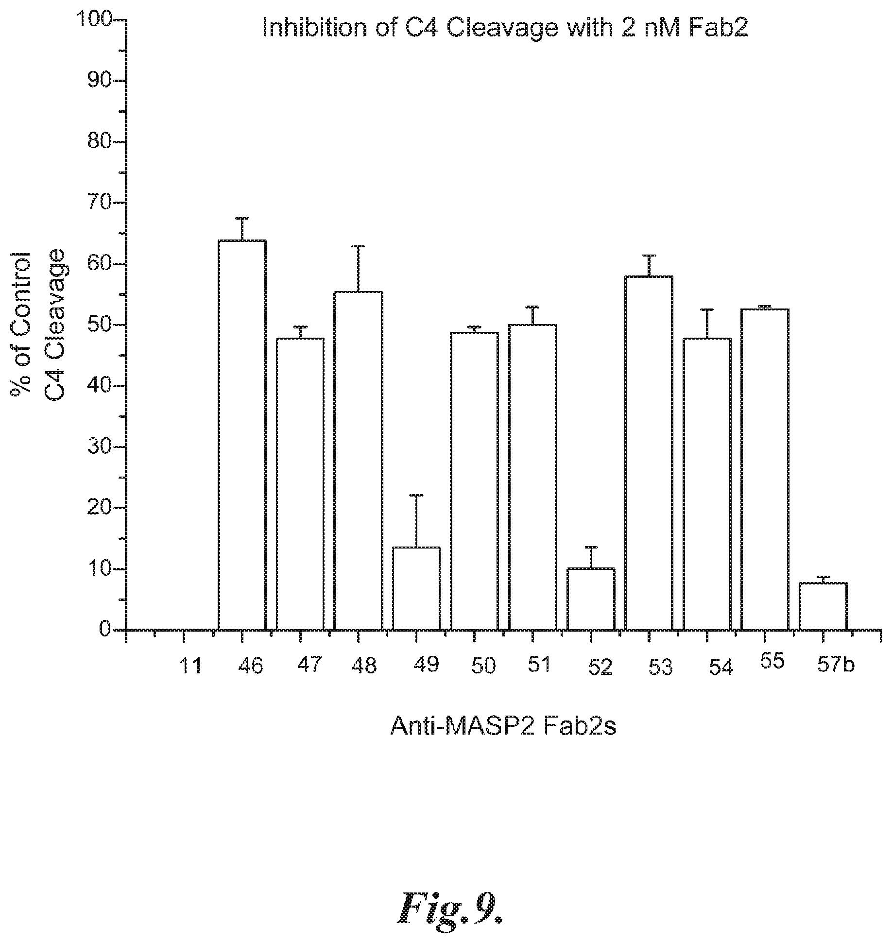
D00014

D00015

D00016

D00017

D00018

D00019

D00020
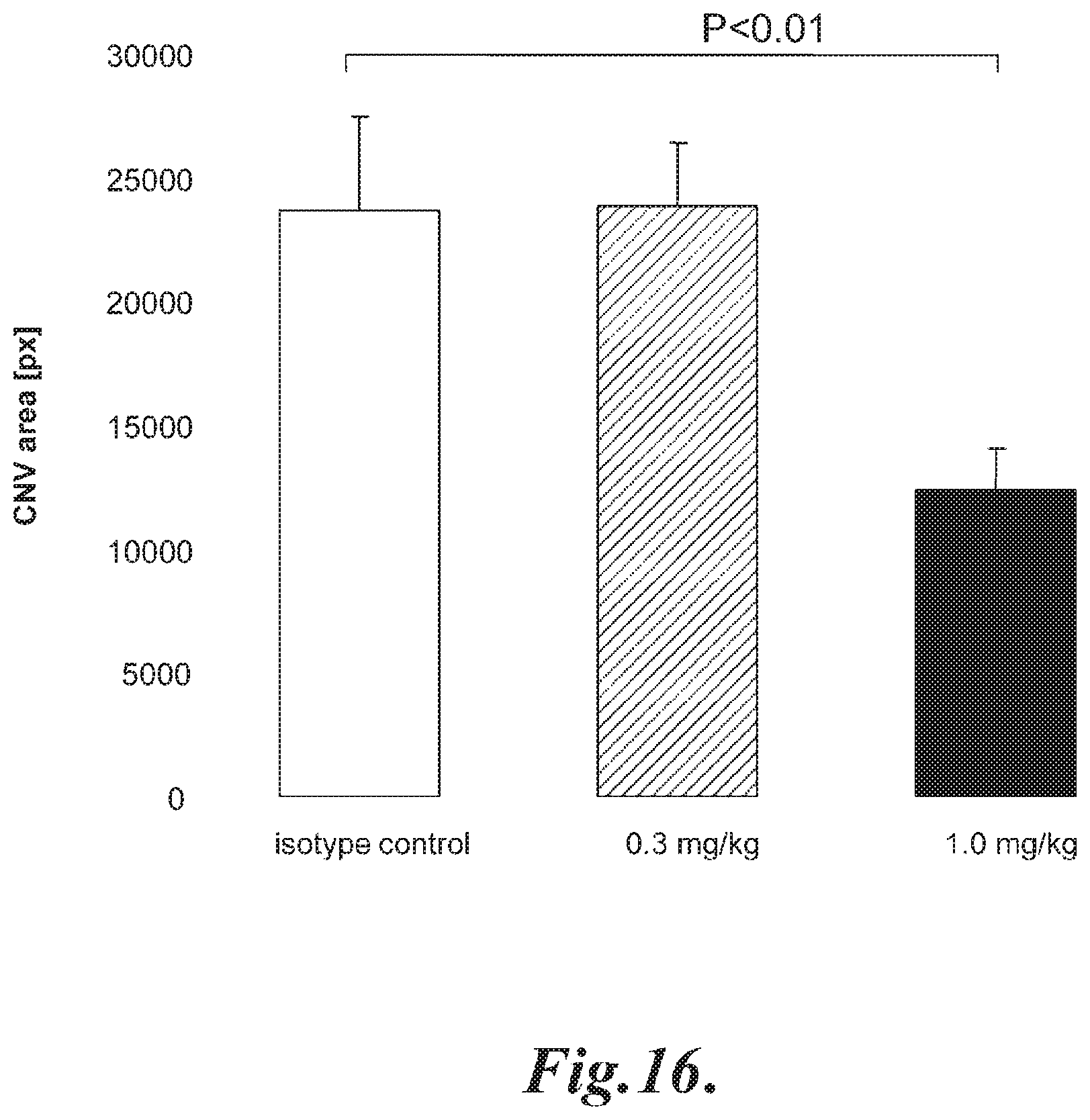
D00021
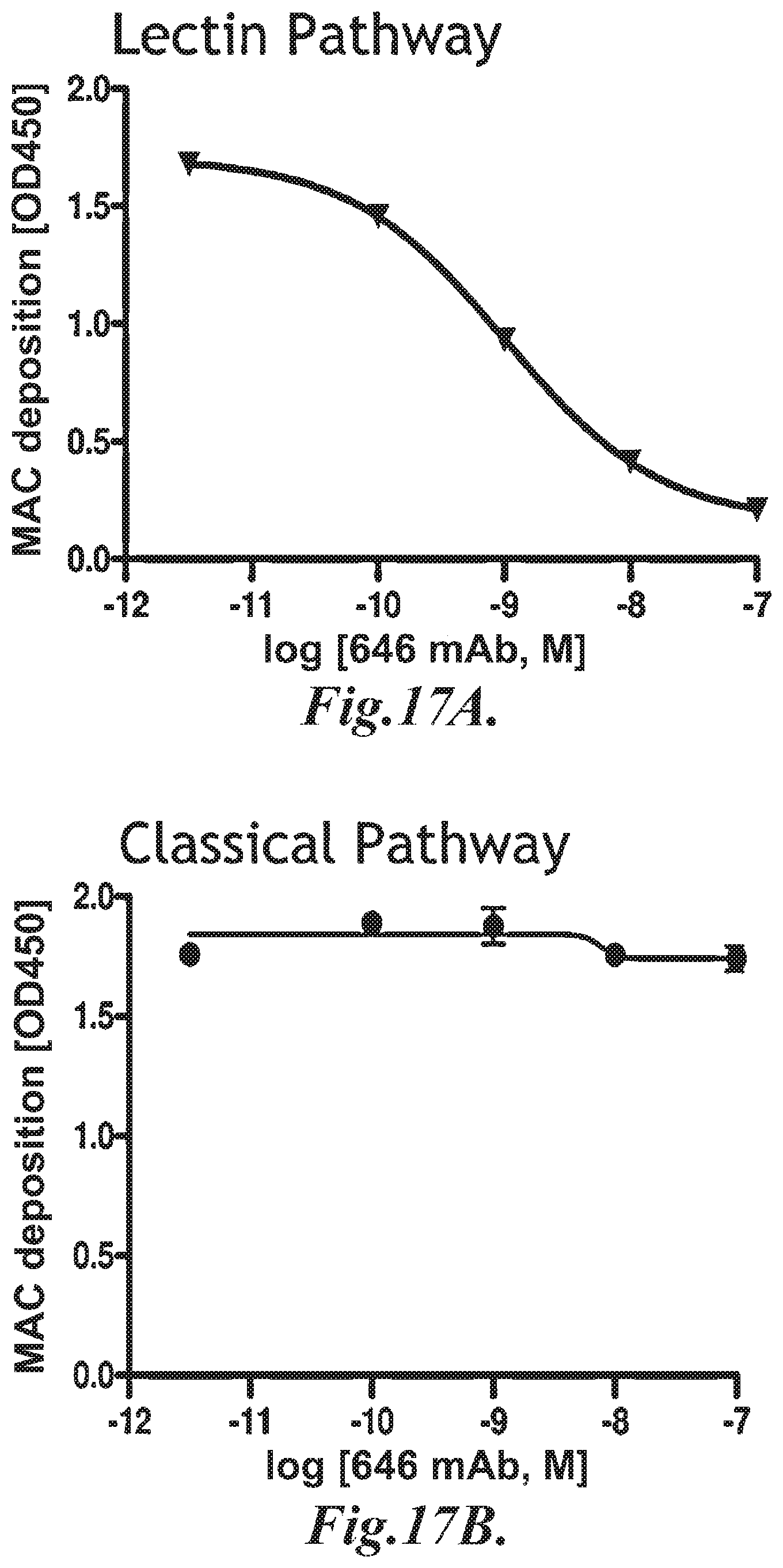
D00022

D00023

D00024

D00025
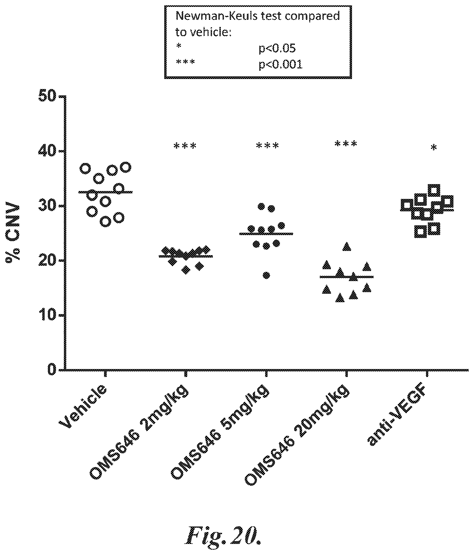
S00001
XML
uspto.report is an independent third-party trademark research tool that is not affiliated, endorsed, or sponsored by the United States Patent and Trademark Office (USPTO) or any other governmental organization. The information provided by uspto.report is based on publicly available data at the time of writing and is intended for informational purposes only.
While we strive to provide accurate and up-to-date information, we do not guarantee the accuracy, completeness, reliability, or suitability of the information displayed on this site. The use of this site is at your own risk. Any reliance you place on such information is therefore strictly at your own risk.
All official trademark data, including owner information, should be verified by visiting the official USPTO website at www.uspto.gov. This site is not intended to replace professional legal advice and should not be used as a substitute for consulting with a legal professional who is knowledgeable about trademark law.