Transgenic mouse model of retinal vascular disease, method of making, and method of using
Fukushima , et al. December 22, 2
U.S. patent number 10,869,465 [Application Number 16/058,568] was granted by the patent office on 2020-12-22 for transgenic mouse model of retinal vascular disease, method of making, and method of using. This patent grant is currently assigned to OSAKA UNIVERSITY. The grantee listed for this patent is OSAKA UNIVERSITY. Invention is credited to Yoko Fukushima, Toru Nakano, Kohji Nishida.


View All Diagrams
| United States Patent | 10,869,465 |
| Fukushima , et al. | December 22, 2020 |
Transgenic mouse model of retinal vascular disease, method of making, and method of using
Abstract
Provided are a non-human model animal of a retinal vascular disease that can favorably show symptoms similar to those of human retinal vascular diseases such as human diabetic retinopathy, and a method for producing the non-human model animal. In particular, provided are a non-human model animal that is suitable for establishing a method for treating, preventing, or diagnosing retinal edema, which causes highly impaired vision, and a method for producing the non-human model animal. A method for screening a drug for treating and preventing a retinal vascular disease, the method using a non-human model animal, is provided. Provided are a non-human model animal of a retinal vascular disease in which constitutively active Akt is expressed, a method for producing a non-human model animal of a retinal vascular disease in which constitutively active Akt is expressed, and a method for screening a drug for treating or preventing a retinal vascular disease.
| Inventors: | Fukushima; Yoko (Osaka, JP), Nishida; Kohji (Osaka, JP), Nakano; Toru (Osaka, JP) | ||||||||||
|---|---|---|---|---|---|---|---|---|---|---|---|
| Applicant: |
|
||||||||||
| Assignee: | OSAKA UNIVERSITY (Osaka,
JP) |
||||||||||
| Family ID: | 1000005255225 | ||||||||||
| Appl. No.: | 16/058,568 | ||||||||||
| Filed: | August 8, 2018 |
Prior Publication Data
| Document Identifier | Publication Date | |
|---|---|---|
| US 20190045759 A1 | Feb 14, 2019 | |
Foreign Application Priority Data
| Aug 10, 2017 [JP] | 2017-156031 | |||
| Current U.S. Class: | 1/1 |
| Current CPC Class: | A61K 49/0008 (20130101); A01K 67/0275 (20130101); C12Y 207/11001 (20130101); C12N 9/12 (20130101); A01K 2267/035 (20130101); A01K 2227/105 (20130101); A01K 2217/052 (20130101) |
| Current International Class: | A01K 67/027 (20060101); C12N 9/12 (20060101); C12N 15/00 (20060101); G01N 33/00 (20060101); A61K 49/00 (20060101) |
| Field of Search: | ;800/3,8,18,21 |
References Cited [Referenced By]
U.S. Patent Documents
| 7112715 | September 2006 | Chambon |
| 2011/0191871 | August 2011 | Walsh |
Other References
|
Egawa (J. Invest. Derm., 2009, vol. 129, p. 2386-2395). cited by examiner . Ouyang (J. Immunol., 2019, vol. 202, p. 1441-1452). cited by examiner . Littlewood (Nucleic acids Research, 1995, vol. 23, No. 10, p. 1686-1690). cited by examiner . Ristevski (Mol. Biotech., 2005, vol. 29, p. 153-163). cited by examiner . McCarthy (Mol. Cell. Biol., 1997, vol. 17, No. 5, p. 2401-2412). cited by examiner . Zhang (Nucleic acids research, 1996, vol. 24, No. 4, p. 543-548). cited by examiner . Condorelli (PNAS, 2002, vol. 99, No. 19, p. 12333-12338). cited by examiner . Andersson (Transgenic Res., 2010, vol. 19, p. 715-725). cited by examiner . Murayama (Oncogene, 2007, vol. 26, p. 4882-4888). cited by examiner . Kita (Genes to Cells, 2008, vol. 13, p. 839-850). cited by examiner . Kimura (Development, 2008, vol. 135, p. 869-879). cited by examiner . Lois E. H. Smith et al., "Oxygen-Induced Retinopathy in the Mouse", Investigative Ophthalmology & Visual Science, vol. 35, No. 1, Jan. 1994, pp. 101-111. cited by applicant. |
Primary Examiner: Wilson; Michael C
Attorney, Agent or Firm: Greenblum & Bernstein, P.L.C.
Claims
The invention claimed is:
1. A transgenic mouse model of a retinal vascular disease, wherein the mouse model has a genome comprising a nucleic acid sequence encoding a constitutively active Akt mutant protein: (i) comprising an E40K or E17K substitution mutation; or (ii) in which a Plekstrin homology (PH) domain at the N-terminus has been replaced with a myristoylation signal sequence, wherein the mouse model shows at least one symptom selected from the group consisting of retinal edema, retinal hemorrhage, a retinal microaneurysm, and retinal vascular expansion.
2. The transgenic mouse model of a retinal vascular disease according to claim 1, wherein the mouse model has a genome comprising a nucleic acid sequence encoding a constitutively active Akt mutant protein in which a PH domain at the N-terminus has been replaced with a myristoylation signal sequence.
3. The transgenic mouse model of a retinal vascular disease according to claim 1, wherein expression of the constitutively active Akt mutant protein of (i) or (ii) is under the control of a Cre-LoxP system.
4. The transgenic mouse model of a retinal vascular disease according to claim 1, wherein the retinal vascular disease is diabetic retinopathy.
5. A method for screening a test substance for treating a retinal vascular disease, comprising: a) administering a test substance to the transgenic mouse model of a retinal vascular disease according to claim 1, and b) determining the effect of the test substance on the at least one symptom, wherein a decrease in the at least one symptom as compared to a control indicates the test substance treats retinal vascular disease.
6. A method for producing a transgenic mouse model of a retinal vascular disease, the method comprising: a) introducing DNA into a fertilized mouse egg, wherein the DNA encodes: (i) a constitutively active Akt mutant protein comprising an E40K or E17K substitution mutation; or (ii) a constitutively active Akt mutant protein in which a Plekstrin homology (PH) domain at the N-terminus has been replaced with a myristoylation signal sequence; and b) implanting the fertilized mouse egg obtained in step a) into a recipient embryo such that a transgenic mouse model of a retinal vascular disease is obtained, wherein the mouse model has a genome comprising a nucleic acid sequence encoding: (i) a constitutively active Akt mutant protein comprising an E40K or E17K substitution mutation; or (ii) a constitutively active Akt mutant protein in which a PH domain at the N-terminus has been replaced with a myristoylation signal sequence, wherein the mouse model shows at least one symptom selected from the group consisting of retinal edema, retinal hemorrhage, a retinal microaneurysm, and retinal vascular expansion.
7. The method for producing a transgenic mouse model of a retinal vascular disease according to claim 6, wherein the mouse model has a genome comprising a nucleic acid sequence encoding a constitutively active Akt mutant protein in which a PH domain at the N-terminus has been replaced with a myristoylation signal sequence, and expression of the constitutively active Akt mutant protein is controlled using 4-hydroxytamoxifen.
8. The method for producing a transgenic mouse model of a retinal vascular disease according to claim 7, wherein the 4-hydroxytamoxifen is administered at a dose of 5 to 50 .mu.g/g weight/day during a desired period between the first day and the fourteenth day after birth.
9. The method for producing a transgenic mouse model of a retinal vascular disease according to claim 6, wherein expression of the constitutively active Akt mutant protein is under the control of a Cre-loxP system.
10. A transgenic mouse whose genome comprises a nucleic acid sequence encoding a constitutively active Akt mutant protein: (i) comprising an E40K or E17K substitution mutation; or (ii) in which a Plekstrin homology (PH) domain at the N-terminus has been replaced with a myristoylation signal sequence, wherein the mouse is capable of developing at least one symptom selected from the group consisting of retinal edema, retinal hemorrhage, a retinal microaneurysm, and retinal vascular expansion after inducing expression of the constitutively active Akt mutant protein.
11. A method for screening a test substance for preventing retinal vascular disease, comprising: a) administering a test substance to the transgenic mouse according to claim 10, b) inducing expression of the constitutively active Akt mutant protein; and c) determining the effect of the test substance on preventing the at least one symptom after inducing expression of the constitutively active Alit mutant protein, wherein a delay in progress or decrease in the at least one symptom as compared to a control indicates the test substance prevents retinal vascular disease.
Description
CROSS REFERENCE TO RELATED APPLICATIONS
This application is based on and claims priority under 35 U.S.C. .sctn. 119 to Japanese Patent Application 2017-156031, filed on Aug. 10, 2017, the entire content of which is incorporated herein by reference.
SEQUENCE LISTING
The instant application contains a Sequence Listing which has been submitted electronically in ASCII format and is hereby incorporated by reference in its entirety. Said ASCII copy, created on Sep. 28, 2018, is named P55331_SL.txt and is 74,053 bytes in size.
TECHNICAL FIELD
The present disclosure relates to a non-human model animal of a retinal vascular disease such as diabetic retinopathy, a method for producing a non-human model animal of a retinal vascular disease, and a method for screening a drug for treating or preventing a retinal vascular disease.
BACKGROUND DISCUSSION
A retinal vascular disease collectively refers to ocular diseases caused by pathological changes such as hemorrhage, effusion, aneurysm, edema, ischemia, and infarct that occur in at least part of retinal blood vessels. Representative cases of the retinal vascular diseases are diabetic retinopathy, retinopathy of prematurity, and retinal venous occlusion. The retinal vascular diseases also include Coats' disease and the like.
Chronic complications of diabetes are caused by microvascular angiopathy occurring in arterioles, capillaries, and the like, and include retinopathy, nephropathy, neuropathy and the like. In particular, diabetic retinopathy is caused by microvascular angiopahty occurring in the retina due to a continuous hyperglycemic state, and may lead to blindness at the age of maturity. The meta-research based on 35 research projects in the world reporting the prevalence rate for diabetic retinopathy has reported that one in every three diabetic patients suffers from some type of retinopathy, and one in every eight or nine diabetic patients suffers from retinopathy that may adversely affect eyesight.
Hypertension or arteriosclerosis is considered as a risk factor for retinal venous occlusion. Retinal venous occlusion occurs at relatively high frequency, and the prevalence rate for retinal venous occlusion in people of forty and above is 2%. Retinal venous occlusion is less likely to lead to blindness, but may lead to a decrease in vision accompanied with anorthopia, and the quality of vision is thus lowered significantly.
Retinopathy of prematurity is a main factor for infant blindness, and it is considered that more than fifty thousand patients lose their eyesight in the world. In this country, retinopathy of prematurity is the first-ranking cause of blindness in the schools for the blind, and severe cases increase with the improvement of neonatal care.
In retinal vascular diseases such as diabetic retinopathy, retinal ischemia caused by vascular occlusion and retinal edema caused by increased vascular permeability lead to a decrease in vision. Ischemic retina falls into a hypoxic state, produces a vascular endothelial growth factor (abbreviated as "VEGF" hereinafter), and induces angiogenesis. However, pathological new blood vessels are formed deviating from the retina, and do not lead to the improvement of ischemia. Furthermore, new blood vessels are weak, and thus hemorrhage is likely to occur. In addition, the contraction of membranous tissues around the blood vessels causes detached retina, leading to blindness. On the other hand, regarding retinal edema, vascular permeability increases based on vasodilatation or an aneurysm following occlusion or inflammation of retinal blood vessels, leading to edema. Retinal edema does not lead to blindness, but causes a significant decrease in vision. If retinal edema lingers for a long period of time, visual cells and retinal nerve cells will undergo irreversible degeneration, and the vision will not be recovered even if edema disappears.
Surgical treatments such as photocoagulation and vitreous surgery, and drug treatments using a drug such as an anti-VEGF drug for inhibiting VEGF protein are known as treatments for ischemia (and subsequent intraocular hemorrhage or retinal detachment) in retinal vascular diseases. An object of these treatments is to inhibit pathological angiogenesis in retinal ischemia, and the treatments do not improve ischemia itself, but a therapeutic effect of stopping the progression of ischemia and preventing blindness is confirmed. On the other hand, a main treatment for retinal edema is a treatment using anti-VEGF drugs that are approved in recent years. An effect of suppressing an increase in permeability, which is exhibited by the anti-VEGF drugs, lasts temporarily, and it is necessary to continuously administer the very expensive drugs intraocularly due to repeated recurrence. Considering that the number of patients suffering from diabetic maculopathy is about one million and one hundred thousand in this country, a further increase in medical cost cannot be avoided in the future. Furthermore, it is difficult to say that a vision improvement effect is not sufficient, and therefore, there has been a demand for the development of a novel therapeutic drug.
Regarding diabetic nephropathy, microvascular sclerosis occurs in the glomeruli, leading to the deterioration of the renal function, due to a continuous hyperglycemic state. In advanced cases, renal sclerosis is promoted by the occurrence of an ischemic state, leading to renal failure. A main treatment for diabetic nephropathy is strict control of blood sugar and blood pressure, and a certain therapeutic effect is confirmed in a treatment using angiotensin converting enzyme inhibitor or an angiotensin II receptor antagonist, but diabetic nephropathy is not completely cured. When renal failure occurs, the treatment is shifted to a treatment using dialysis.
An attempt is made to use non-human disease model animals in the development of a method for treating, preventing, or diagnosing a disease, in the pathologic analysis of a disease, and the like. For example, it is reported that spontaneous model animals, genetically modified model animals, and model animals in which the onset of a disease is induced by drugs or the like have been produced as non-human diabetic model animals. Furthermore, non-human model animals in which retinal ischemia is artificially induced by high oxygen loading to form abnormal new blood vessels have been reported (Non-Patent Document 1).
CITATION LIST
Non Patent Literature
Non-Patent Document 1: L. E. Smith et al., "Oxygen-induced retinopathy in the mouse", Investigative Ophthalmology & Visual Science January 1994, Vol. 35, 101-111
SUMMARY
Technical Problem
A method for treating or preventing diabetic nephropathy and retinal vascular diseases such as diabetic retinopathy is not established, and the establishment of the treatment method or prevention method is an urgent issue. Non-existence of appropriate non-human model animals causes the difficulty in establishing an effective treatment method and an effective prevention method, for example. Non-human model animals that have been reported do not show symptoms similar to those of human diabetic nephropathy and human retinal vascular diseases such as human diabetic retinopathy. In particular, non-human model animals of a type of retinal vascular disease that leads to retinal edema caused by increased vascular permeability have not been reported.
Accordingly, the present disclosure was achieved to provide a non-human model animal of a retinal vascular disease that can favorably show symptoms similar to those of human retinal vascular diseases such as human diabetic retinopathy, and a method for producing the non-human model animal. Specifically, the present disclosure was achieved to provide a non-human model animal of a retinal vascular disease that can be used to develop a method for treating, preventing, or diagnosing a retinal vascular disease and to elucidate the onset mechanism and pathology of a retinal vascular disease, and a method for producing the non-human model animal. In particular, the present disclosure was achieved to provide a non-human model animal that can be favorably used to establish a method for treating, preventing, or diagnosing retinal edema, which causes highly impaired vision, and a method for producing the non-human model animal. Furthermore, the present disclosure was achieved to provide a method for screening a drug for treating and preventing a retinal vascular disease, the method using a non-human model animal.
Solution to Problem
As a result of performing intensive studies to solve the above-mentioned problems, the inventors of the present disclosure focused on Akt protein, which is a serine/threonine kinase, in order to induce symptoms similar to those of human retinal vascular diseases in non-human animals. When Akt was persistently activated in retinal blood vessels in their incipient stage, it was confirmed that the blood vessels increased in their diameter with varicose deformity, the extension of the blood vessels was delayed, and vascular permeability increased. Based on these findings, the inventors succeeded in producing a non-human model animal showing symptoms similar to those of human retinal vascular diseases, and thus achieved the present disclosure.
Specifically, in order to address the foregoing problems, the disclosure including the configurations and methods described in [1] to [10] below is provided.
[1] A non-human model animal of a retinal vascular disease in which constitutively active Akt is expressed.
[2] The non-human model animal of a retinal vascular disease according to [1] above, wherein the constitutively active Akt is constitutively active Akt-Mer.
[3] The non-human model animal of a retinal vascular disease according to [1] above, wherein the expression of the constitutively active Akt is under the control of a Cre-LoxP system.
[4] The non-human model animal of a retinal vascular disease according to any one of [1] to [3] above, wherein the retinal vascular disease is diabetic retinopathy.
[5] The non-human model animal of a retinal vascular disease according to any one of [1] to [4] above, wherein the retinal vascular disease shows at least one symptom selected from retinal edema, retinal hemorrhage, a retinal microaneurysm, and retinal vascular expansion.
With the configurations described in [1] to [5], a non-human model animal of a retinal vascular disease showing symptoms similar to those of human retinal vascular diseases such as human diabetic retinopathy can be provided. Retinal hemorrhage, the structure of a microaneurysm, and the like in the non-human model animal of a retinal vascular disease with these configurations are similar to those of pathological specimens of human diabetic retinopathy, and, in particular, a non-human model animal capable of reproducing the symptoms of retinal edema caused by increased vascular permeability for which no effective treatment method is currently present can be provided. Therefore, the non-human model animal of a retinal vascular disease with these configurations can be favorably used as a research material for searching for a method for treating, preventing, or diagnosing a retinal vascular disease, for the pathologic analysis of a retinal vascular disease, and the like. Furthermore, the degree of severity can be controlled in the non-human model animal of a retinal vascular disease with these configurations, and therefore, a treatment method, prevention method, or diagnosis method appropriate for the degree of progress of symptoms can be searched for, and, in addition, the non-human model animal can be favorably used as a research material for the pathologic analysis of the disease such as the onset and progress of the disease, and the like.
In particular, with the configurations described in [2] and [3] above, the Akt activity of the constitutively active Akt can be controlled by 4-hydroxytamoxifen and Cre recombinase. Therefore, a non-human model animal showing symptoms similar to those of a desired human retinal vascular disease can be provided through a simple artificial manipulation. Since the degree of severity of a retinal vascular disease or the like can be controlled, it is possible to provide a non-human model animal of a retinal vascular disease that can be particularly favorably used as a research material for searching for a treatment method, prevention method, or diagnosis method appropriate for the degree of progress of symptoms, for the pathologic analysis of the disease such as the onset and progress of the disease, and the like. With the configuration described in [4] above, it is possible to provide a non-human model animal that can be favorably used as a research material for searching for a method for treating, preventing, or diagnosing diabetic retinopathy, for the pathologic analysis of diabetic retinopathy, and the like. With the configuration described in [5] above, a non-human model animal showing the symptoms specific to retinal vascular diseases can be provided. In particular, a non-human model animal capable of reproducing the symptoms of retinal edema caused by increased vascular permeability for which no effective treatment method is currently present can be provided.
[6] A method for producing a non-human model animal of a retinal vascular disease, including: a step of producing a transgenic non-human animal into which a construct including a DNA coding for constitutively active Akt so as to enable Akt activity to be controlled by an artificial manipulation is introduced; and a step of inducing a retinal vascular disease by controlling the Akt activity of the constitutively active Akt.
[7] The method for producing a non-human model animal of a retinal vascular disease according to [6] above, wherein the constitutively active Akt is constitutively active Akt-Mer, and the Akt activity of the constitutively active Akt is controlled through administration of 4-hydroxytamoxifen to the transgenic non-human animal.
[8] The method for producing a non-human model animal of a retinal vascular disease according to [7] above, wherein the 4-hydroxytamoxifen is administered at a dose of 5 to 50 .mu.g/g weight/day during a desired period between the first day and the fourteenth day after birth.
[9] The method for producing a non-human model animal of a retinal vascular disease according to [6] above, wherein the expression of the constitutively active Akt is under the control of a Cre-LoxP system, the transgenic non-human animal is produced by introducing a construct in which a DNA coding for the constitutively active Akt is arranged in a state in which the expression of the constitutively active Akt is inhibited due to intervention of the LoxP sequence, and the Akt activity of the constitutively active Akt is controlled by mating the transgenic non-human animal with a transgenic non-human animal in which a Cre recombinase is expressed in a vascular specific manner or time specific manner to obtain an offspring in which the Cre recombinase can be expressed or the activity of the Cre recombinase can be induced in a vascular specific manner or time specific manner in cells including the construct.
With the methods described in [6] to [8] above, a non-human model animal of a retinal vascular disease showing symptoms similar to those of human retinal vascular diseases such as human diabetic retinopathy can be produced. Retinal hemorrhage, the structure of a microaneurysm, and the like in the non-human model animal produced using these methods are similar to those of pathological specimens of human diabetic retinopathy, and, in particular, the symptoms of retinal edema caused by increased vascular permeability for which no effective treatment method is currently present can be reproduced. The non-human model animal of a retinal vascular disease produced using these methods can show the symptoms of a human retinal vascular disease in a very short time after the Akt activity of the constitutively active Akt is controlled. Therefore, these methods can be favorably used in the fields of a search for a method for treating, preventing, or diagnosing a retinal vascular disease, the pathologic analysis of the state of a retinal vascular disease, and the like. Furthermore, the degree of severity in the non-human model animal produced using these methods can be controlled by artificially manipulating the Akt activity of the constitutively active Akt, and therefore, the non-human model animal can be used very favorably as a research material for searching for a treatment method, prevention method, or diagnosis method appropriate for the degree of progress of symptoms, particularly for the pathologic analysis of the disease such as the onset and progress of the disease, and the like.
In particular, with the methods described in [7] to [9] above, the Akt activity of the constitutively active Akt can be controlled by 4-hydroxytamoxifen and Cre recombinase. Therefore, a non-human model animal showing symptoms similar to those of a desired human retinal vascular disease can be provided through a simple artificial manipulation. Since the degree of severity of a retinal vascular disease or the like can be controlled, the non-human model animal can be particularly favorably used as a research material for searching for a treatment method, prevention method, or diagnosis method appropriate for the degree of progress of symptoms, for the pathologic analysis of the disease such as the onset and progress of the disease, and the like.
[10] A method for screening a drug for treating or preventing a retinal vascular disease, comprising: a step of administering a test substance to the non-human model animal of a retinal vascular disease according to any one of [1] to [5] above; and a step of determining a treatment effect or a prevention effect of the test substance.
With the method described in [10] above, it is possible to provide a method for screening a drug for treating or preventing a retinal vascular disease, the method using a non-human model animal showing symptoms similar to those of human retinal vascular diseases. Since a non-human model animal showing symptoms similar to those of human retinal vascular diseases is used, the treatment effect and prevention effect of the test substance can be determined with high reliability. Since the degree of severity of a retinal vascular disease or the like can be controlled by controlling the Akt activity of the constitutively active Akt, a treatment drug and a prevention drug appropriate for the symptoms can be developed. Furthermore, the screening method can be favorably used for the development of treatment drugs and prevention drugs for retinal vascular diseases leading to retinal edema caused by increased vascular permeability for which no effective treatment method is currently present. Therefore, the screening method is expected to significantly contribute to the development in techniques for treating and preventing retinal vascular diseases.
BRIEF DESCRIPTION OF DRAWINGS
FIG. 1 is a schematic diagram illustrating the construction of a non-human transgenic animal in which constitutively active Akt-Mer is expressed in Example 1.
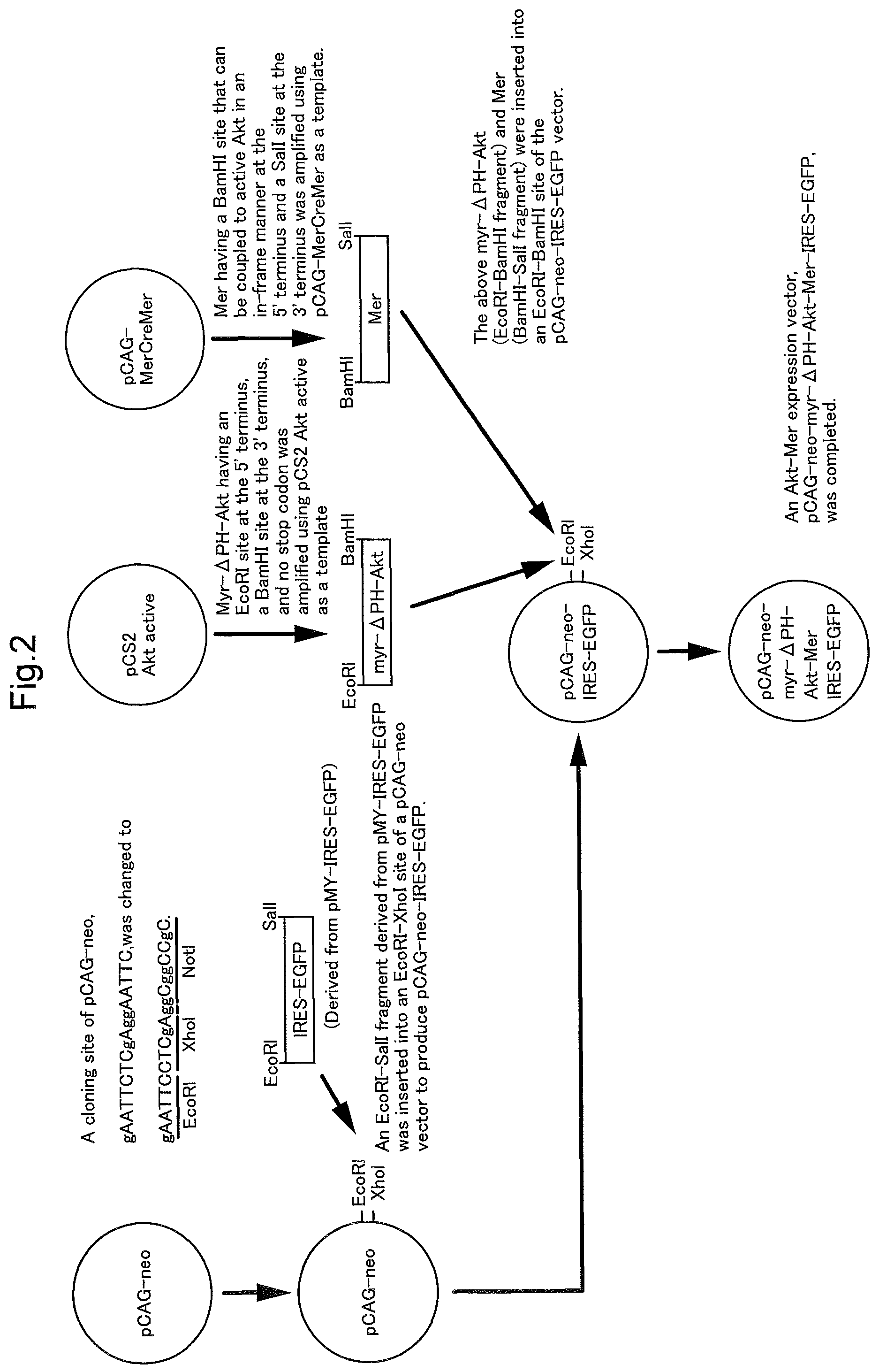
FIG. 2 is a schematic diagram illustrating the construction of an expression vector of constitutively active Akt-Mer in Example 1. FIG. 2 discloses SEQ ID NOS 28-29, respectively, in order of appearance.
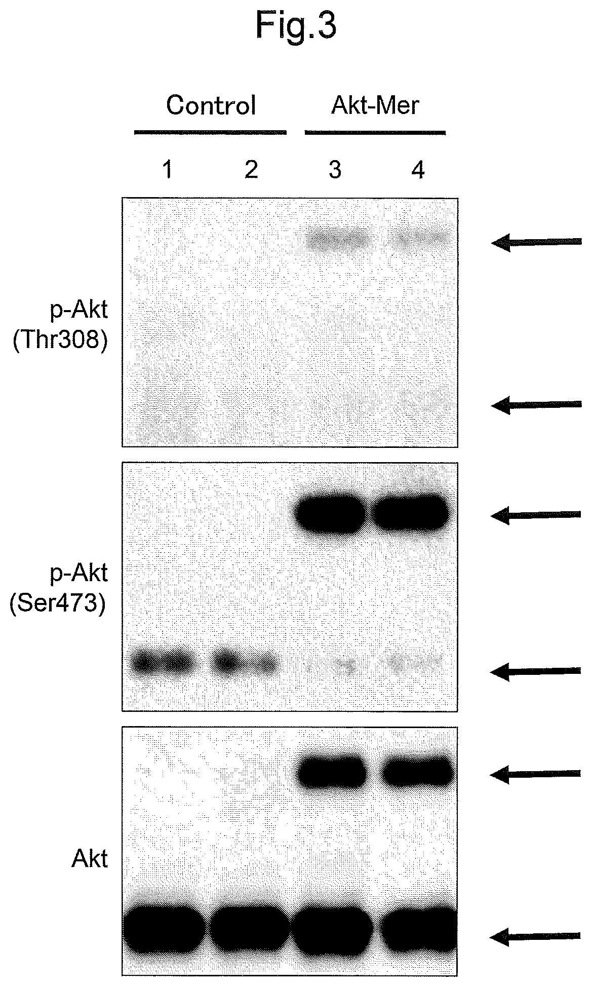
FIG. 3 shows Western blots illustrating the results of the examinations in Example 2 that investigated the Akt activity in retinas.
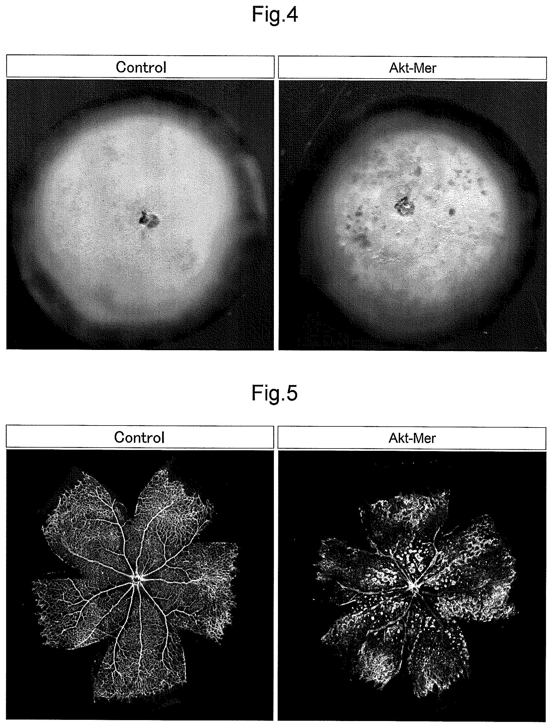
FIG. 4 shows retina cups illustrating the results of the examinations in Example 3 that investigated the influence of the persistent activation of Akt on retinas.
FIG. 5 shows whole-mount immunostaining images of retinas illustrating the results of the examinations in Example 4 that investigated the influence of the persistent activation of Akt on retinal angiogenesis.
FIG. 6 shows whole-mount immunostaining images of retinas illustrating the results of the examinations in Example 4 that investigated the influence of the persistent activation of Akt on the shapes of retinal capillaries, which are enlarged diagrams of FIG. 5.
FIG. 7 is a graph showing the measurement results of the extensions of blood vessels (mm), the diameters of arteries (.mu.m), and the diameters of veins (.mu.m), illustrating the results of the examinations in Example 4 that investigated the influence of the persistent activation of Akt on the shapes of retinal capillaries.
FIG. 8 shows whole-mount immunostaining images of retinas illustrating the results of the examinations in Example 5 that investigated the influence of the persistent activation (low activation) of Akt on the shapes of retinal capillaries.
FIG. 9 is a graph showing the measurement results of the number of microaneurysms per retina, illustrating the results of the examinations in Example 5 that investigated the influence of the persistent activation (low activation) of Akt on the shapes of retinal capillaries.
FIG. 10 shows comparison between whole-mount immunostaining images of retinas illustrating the results of the examinations in Example 5 that investigated the influence of the persistent activation (low activation) of Akt on the shapes of retinal capillaries, and pathological specimens of human diabetic retinopathy.
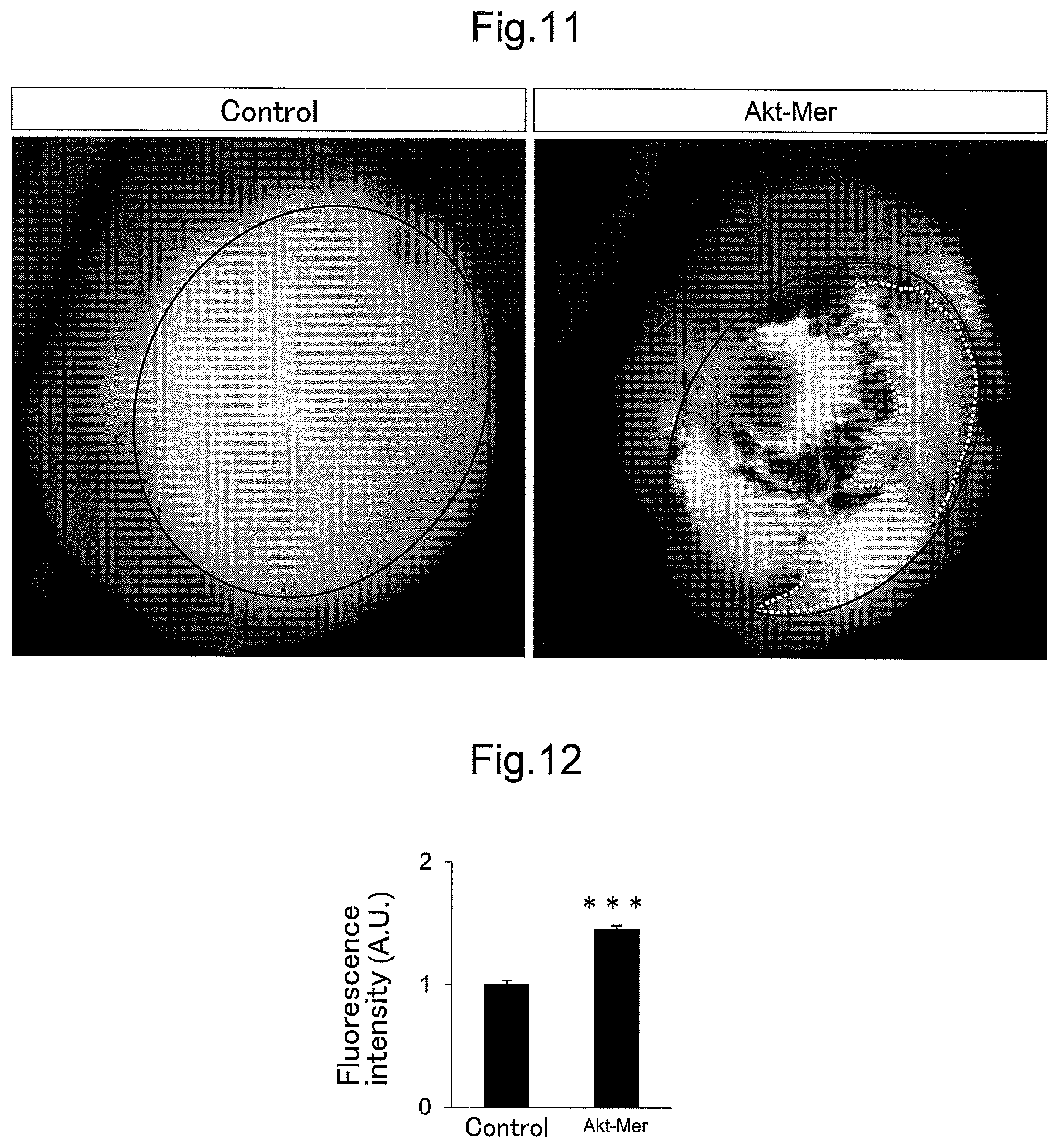
FIG. 11 shows retina cups illustrating the results of the examinations in Example 6 that investigated the influence of the persistent activation of Akt on the vascular permeability in retinas.
FIG. 12 is a graph showing the measurement results of fluorescence intensity of Evans blue leaking from retinal blood vessels, illustrating the results of the examinations in Example 6 that investigated the influence of the persistent activation of Akt on the vascular permeability in retinas.
FIG. 13 shows nucleo-staining in cryosections of retinas illustrating the results of the examinations in Example 7 that investigated the influence of the persistent activation of Akt on retinal surface layers.
FIG. 14 shows whole-mount immunostaining images of retinas illustrating the results of the examinations in Example 8 that analyzed the factors (vascular endothelial cells and astrocytes) in abnormal vascular morphology caused by the persistent activation of Akt.
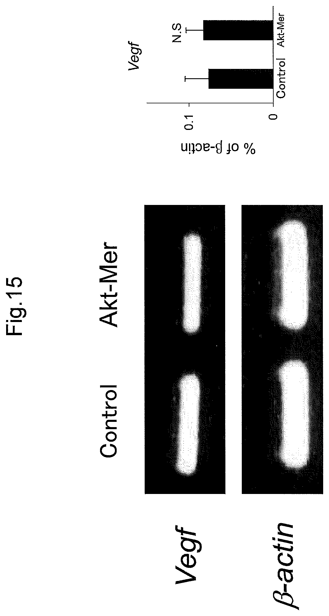
FIG. 15 shows Western bolts and a graph illustrating the results of the examinations in Example 9 that analyzed the factor (VEGF expression level) in abnormal vascular morphology caused by the persistent activation of Akt.
FIG. 16 shows whole-mount immunostaining images of retinas illustrating the results of the examinations in Example 10 that investigated the influence of the long-term persistent activation of Akt on retinas.
FIG. 17 shows whole-mount immunostaining images of retinas illustrating the results of the examinations in Example 11 that investigated the influences of the activation level and activation period of the persistent activation of Akt on retinas.
FIG. 18 is a graph showing the measurement results of the number of microaneurysms per retina, illustrating the results of the examinations in Example 11 that investigated the influences of the activation level and activation period of the persistent activation of Akt on retinas.
FIG. 19 shows whole-mount immunostaining images of retinas after the administration of drugs, illustrating the results of the examinations in Example 12 that used drugs for treating or preventing a retinal vascular disease in screening.
DESCRIPTION OF EMBODIMENTS
Hereinafter, embodiments of the present disclosure will be described in detail. However, the present disclosure is not limited to the embodiments described below.
(Non-Human Model Animal)
(Non-Human Model Animal of Retinal Vascular Disease)
A non-human model animal according to this embodiment relates to a non-human model animal of a retinal vascular disease showing the pathology of a retinal vascular disease. In the non-human model animal of a retinal vascular disease according to this embodiment, constitutively active Akt is expressed.
A mammalian animal other than a human is used for the non-human model animal of a retinal vascular disease according to this embodiment, and there is no particular limitation on the species. Examples thereof include rodents such as mice, guinea pigs, hamsters, and rats, rabbits, dogs, cats, monkeys, sheep, pigs, cows, and horses. Mice are preferable.
Regarding the non-human model animal of a retinal vascular disease according to this embodiment, the "retinal vascular disease" collectively refers to ocular diseases caused by pathological changes such as hemorrhage, effusion, aneurysm, edema, ischemia, and infarct that occur in at least part of retinal blood vessels. Representative cases of the retinal vascular diseases are diabetic retinopathy, retinopathy of prematurity, and retinal venous occlusion. The retinal vascular diseases also include Coats' disease and the like. The non-human model animal of a retinal vascular disease according to this embodiment is targeted for all the cases.
Diabetic retinopathy is one of the diabetic complications and is caused by microvascular disorder occurring in the retina due to a continuous hyperglycemic state. Diabetic retinopathy can be classified into three stages, namely simple retinopathy, preproliferative retinopathy, and proliferative retinopathy, for example, depending on the degree of severity. The non-human model animal of a retinal vascular disease according to this embodiment is targeted for all the cases. In simple retinopathy, microvascular walls are damaged, leading to microaneurysms, petechial retinal hemorrhage, and the like. Specifically, the retinal microvascular walls are weakened through the degeneration of vascular endothelial cells, the decidualization and degeneration of perithelial cells, and the thickening of basement membranes, and petechial hemorrhage thus occurs due to the formation and breakage of microaneurysms. In preproliferative retinopathy, partial microvascular occlusion causes an ischemic portion. In proliferative retinopathy, new blood vessels are formed in order to compensate the retinal ischemic state, and extend to the retinal surface and the vitreous body. New blood vessels are weak due to their immaturity and thus easily break and bleed. In addition, a fibrous proliferative membrane that is also referred to as "proliferative tissue" is formed, and pulls the retina locating therearound, leading to detached retina. Then, irrespective of the degree of severity, blood plasma accumulates in the retina due to an increase in microvascular permeability caused by the formation of retinal microaneurysms and the like, leading to retinal edema.
Retinal venous occlusion is caused by the occlusion of the retinal veins, and includes central retinal venous occlusion caused by the occlusion of the central retinal vein, and branch retinal venous occlusion caused by the occlusion of veins located on the peripheral side with respect to the central retinal vein. The non-human model animal of a retinal vascular disease according to this embodiment is targeted for both cases. In the early stage, an increase in a venous perfusion pressure and the stasis of the blood flow causes retinal hemorrhage and edema. In the chronic stage, retinal edema remains due to the expansion or meandering of capillaries located around the blocked veins and the formation of aneurysms. Severe venous occlusion causes retinal ischemia, and pathological angiogenesis is induced as in other retinal vascular diseases, leading to detached retina.
Retinopathy of prematurity is caused by the immaturity of retinal blood vessels observed in low-birth-weight infants and premature infants born before 37 weeks gestation. Retinopathy of prematurity is classified into type I, which progresses mildly, and type II, which progresses rapidly. Type I is classified into five stages depending on the degree of severity. The non-human model animal of a retinal vascular disease according to this embodiment is targeted for both cases. Type I is classified into five stages depending on the degree of severity. At a first stage, tissues called boundaries are formed at the leading ends of blood vessels that are extending from the optic papilla toward the periphery thereof. At a second stage, vascular proliferation accompanied with fibrous components occurs, and the thicknesses of the boundaries increase. At a third stage, fibrovascular proliferative tissues protrude toward the vitreous body and extends. At a fourth stage and a fifth stage, the surrounding retina is pulled due to the contraction of the fibrovascular proliferative tissues that have increased on the vitreous body side, and is thus detached. At all the stages, the expansion of retinal veins and the meandering of arteries are considered to be signs of aggravation. In type II, retinal blood vessels significantly expand and meander, and the state of the disease does not progress in a stepwise manner and rapidly leads to detached retina.
Examples of the funduscopic findings of such retinal vascular diseases include a microaneurysm, localized hemorrhage such as retinal petechial hemorrhage, retinal ecchymosis, and retinal splinter hemorrhage, hard exudate, retinal edema, retinal thickening, soft exudate, venous disorder, retinal microvascular disorder, retinal and papillary angiogenesis, preretinal and vitreous hemorrhage, a fibrovascular proliferative membrane, and detached retina.
The non-human model animal of a retinal vascular disease according to this embodiment shows one or more of the above-mentioned symptoms. For example, a stereoscopic microscope or a fluorescence microscope can be used to confirm retinal edema, retinal hemorrhage, a retinal microaneurysm, the retinal vascular expansion and the like in the non-human model animal of a retinal vascular disease according to this embodiment. Fluorescence micrographs showing vascular endothelial cells or the like stained through immunostaining or the like can be preferably used to confirm retinal vascular disorder and the like. For example, the non-human model animal a retinal vascular disease according to this embodiment is characterized by the formation of 50 or more microaneurysms, and preferably 80 or more microaneurysms, per retina, or the vascular diameter that is expanded by a factor of 1.5, and particularly preferably a factor of 2 or more, compared with normal retinal blood vessels. Moreover, retinal edema or the like can be preferably confirmed by measuring dye leaking from blood vessels, the thickness of the retina, or the like. For example, the non-human model animal of a retinal vascular disease according to this embodiment is characterized by the thickness of the retina that is 1.2 times or more, and particularly preferably 1.5 times or more, larger than the thickness of a normal retina.
Akt, which is also referred to as "protein kinase B", is a serine/threonine kinase including a PH (Plekstrin homology) domain at the N-terminus, and is known as an important intracellular signaling factor. Akt is activated in a PI3 kinase dependent manner, and phosphorylates various intracellular substrates. The inventors of the present disclosure reported a technique of controlling the Akt activity (kinase activity) to maintain the pluripotency of stem cells and differentiate stem cells as desired (see JP 2005-304487A).
Akt is universally present in any higher animal cells. For example, Akt is conserved in various organisms from yeast to Caenorhabditis elegans, Drosophila, and mammalian animals (see Fabrizio P. et al., Science 2001 Apr. 13: 292(5515): 288-290. Epub 2001 Apr. 5 for yeasts; Paradis S. et al., Genes Dev. 1998 Aug. 15: 12(16): 2488-98 for Caenorhabditis elegans; and Staveley BE. et al., Curr. Biol. 1998 May 7: 8(10): 599-602 for Drosophila). Akt is ubiquitously expressed in various tissues of mammalian animals (see Altomare D. A. et al., Oncogene 11: 1055-1060; Altomare, D. A. et al., Oncogene 116: 2407-2411; Brodbeck D. et al., J. Biol. Chem. 274: 9133-9136; and Nakatani K. et al., Biochem. Biophys. Res. Commun. 257: 906-910).
There is no particular limitation on constitutively active Akt expressed in the non-human model animal of a retinal vascular disease according to this embodiment as long as the Akt activity is constitutively activated. The constitutively active Akt may be derived from any higher animal. Moreover, the constitutively active Akt may be derived from the species that is the same as or different from the non-human animal in which the expression is induced. Akt is preferably derived from a human.
It is known that human-derived Akt includes three isoforms, namely Akt-1, Akt-2, and Akt-3, and the amino acid sequences of these isoforms have very high homology. An example of wild-type human Akt is human wild-type Akt-1 (Accession No. AH011307 or NM_001014432), which is coded for by the base sequence of Sequence ID. No. 3 or Sequence ID No. 5 and consists of the amino acid sequence of Sequence ID. No. 4 or Sequence ID No. 6. In addition, examples thereof include human wild-type Akt-2 (Accession No. NM_001626), which is coded for by the base sequence of Sequence ID No. 7 and consists of the amino acid sequence of Sequence ID No. 8, and human wild-type Akt-3 (Accession No. NM_181690), which is coded for by the base sequence of Sequence ID No. 9 and consists of the amino acid sequence of Sequence ID No. 10. However, there is no limitation thereto, and Akt may have an amino acid sequence with at least a certain ratio of sequence identity with respect to the above-mentioned amino acid sequences, or an amino acid sequence obtained by modifying the above-mentioned amino acid sequences with at least one modification of deletion, substitution, and addition of one or several amino acids, and have the above-mentioned Akt properties.
Phosphorylation at two positions, namely threonine at position 308 (Thr-308) and serine at position 473 (Ser-473), is required to activate Akt-1, phosphorylation at two positions, namely threonine at position 309 (Thr-309) and serine at position 474 (Ser-474), is required to activate Akt-2, and phosphorylation at two positions, namely threonine at position 305 (Thr-305) and serine at position 472 (Ser-472), is required to activate Akt-3. Phosphorylation in the Akt family is controlled on the downstream side of PI3 kinase. Specifically, the PH domain of Akt binds to PI(3,4,5)P3 or the like produced by PI3 kinase, and Akt is thus transferred to the cell membrane. The above-mentioned positions of Akt are phosphorylated by PDK1 and the mTORC2 complex near the cell membrane, and Akt is thereby activated.
The constitutively active Akt includes Akt in which the above-mentioned positions relating to the activation are phosphorylated. Examples thereof include wild-type human Akt-1 in which Thr-308 and Ser-473 are constitutively phosphorylated, wild-type human Akt-2 in which Thr-309 and Ser-474 are constitutively phosphorylated, and wild-type human Akt-3 in which Thr-305 and Ser-472 are constitutively phosphorylated. Amino acid residues to be phosphorylated in Akt-1 are preferably a Thr residue located at a position corresponding to position 308 and a Ser residue located at a position corresponding to position 473 when Sequence ID No. 4 or Sequence ID No. 6 is used as a reference sequence. The same applies to amino acid residues to be phosphorylated in Akt-2 and Akt-3.
An example of the constitutively active Akt is Akt in which a membrane localization signal sequence is added to the N-terminus or C-terminus. Preferable examplse of the constitutively active Akt include wild-type Akt in which the PH domain at the N-terminus is deleted and a myristoylation signal is added to the N-terminus instead, and wild-type Akt in which a myristoylation signal is added to the N-terminus. Akt with a myristoylation signal is of a constitutive membrane-bound type. This type of Akt is constitutively phosphorylated by kinase present on the cell membrane and is brought into a state in which the Akt activity (kinase activity) is activated. The myristoylation signal sequence preferably includes a Met-Gly-Xaa-Xaa-Xaa-Ser/Ala/Thr/Phe-Xaa-Xaa-Xaa motif (Xaa is any amino acid) (Sequence ID No. 11) from its N-terminus (see Utsumi T. et al., The Journal of Biological Chemistry 276(13): 10505-10513 and the like). For example, the N-terminal myritoylation signal sequences (MGSSKSKPKDPSQR (Sequence ID No. 12) and MGSSKSKPKDPSQRRRRIRT (Sequence ID No. 13)) derived from c-Src and the like can be used as the myristoylation signal sequence, but there is no limitation thereto.
Furthermore, constitutively active Akt mutants such as a mutant obtained by substituting glutamic acid at position 40 (Glu-40) of wild-type human Akt-1 with lysine (Lys) (also abbreviated as "E40K-Akt-1" hereinafter) and a mutant obtained by substituting glutamic acid at position 17 (Glu-17) of wild-type human Akt-1 with lysine (Lys) (also abbreviated as "E17K-Akt-1" hereinafter) may also be used. In the same manner, Akt mutants obtained from wild-type human Akt-2 and wild-type human Akt-3 can include mutants having the above-mentioned substitution, namely E40K or E17K. Such mutants can be obtained by screening the natural world or using a gene cloning technique. For example, such mutants can be obtained by using a known mutagenesis technique, and specifically, such mutants can be obtained by inserting a mutation site into a nucleic acid molecule coding for wild-type Akt. There is no particular limitation on the method for inserting a mutation site, and a mutagenesis technique for producing a mutant protein that is known in the art can be used. Examples thereof include known mutagenesis techniques such as site-directed mutagenesis, PCR mutagenesis that uses a PCR or the like to introduce mutation, and transposon insertion mutagenesis. A commercially available mutagenesis kit (e.g., QuikChange (registered trademark) Site-directed Mutagenesis Kit (manufactured by Stratagene)) may also be used.
Amino acid residues to be substituted in Akt-1 are preferably a Glu residue located at a position corresponding to position 40 and a Glu residue located at a position corresponding to position 17 when Sequence ID No. 4 or Sequence ID No. 6 is used as a reference sequence. The substitution positions can be determined based on an alignment between the amino acid sequence of Akt-1 with a different sequence and the amino acid sequence of Akt-1 disclosed in Sequence ID No. 4 or Sequence ID No. 6. In the same manner, amino acid residues to be substituted in Akt-2 and Akt-3 can also be determined using Sequence ID No. 8 and Sequence ID No. 10 as reference sequences. Therefore, the constitutively active Akt mutants may have an amino acid sequence with at least a certain ratio of sequence identity with respect to the above-mentioned amino acid sequences, or an amino acid sequence obtained by modifying the above-mentioned amino acid sequences with at least one modification of deletion, substitution, and addition of one or several amino acids, and have the above-mentioned properties of the constitutively active Akt, as long as the amino acid at the above-mentioned substitution position is substituted.
For example, the constitutively active Akt includes a protein coded for by a nucleic acid molecule consisting of the base sequence of Sequence ID No. 1, and its amino acid sequence is represented by Sequence ID No. 2. However, there is no limitation thereto, and the constitutively active Akt may have an amino acid sequence with at least a certain ratio of sequence identity with respect to the amino acid sequence of Sequence ID No. 2, or an amino acid sequence obtained by modifying the amino acid sequence of Sequence ID No. 2 with at least one modification of deletion, substitution, and addition of one or several amino acids, and have the above-mentioned properties of the constitutively active Akt.
Here, "at least a certain ratio of sequence identity" means preferably having 70%, 75%, 80%, or at least 85% sequence identity, more preferably having at least 90% sequence identity, and particularly preferably 95%, 96%, 97%, 98%, or at least 99% sequence identity.
Such variants can be obtained by screening the natural world or using a gene cloning technique. When an amino acid sequence is modified, a person skilled in the art can easily predict a modification with which the above-mentioned characteristics can be maintained. Specifically, from the viewpoint of maintaining the protein structure, an amino acid whose properties including polarity, electric charge, hydrophilicity, hydrophobicity and the like are similar to those of an amino acid to be substituted can be used in an amino acid substitution, for example. Such substitution is well known to a person skilled in the art as a conservative substitution. Specifically, alanine, valine, leucine, isoleucine, proline, methionine, phenylalanine, and tryptophan are all classified into non-polar amino acids and thus have similar properties, for example. Examples of non-charged amino acids include glycine, serine, threonine, cysteine, tyrosine, asparagine, and glutamine. Examples of acidic amino acids include aspartic acid and glutamic acid. Examples of basic amino acids include lysine, arginine, and histidine. Substitution of amino acids in the same group is allowable because the functions of a protein can be maintained.
It is preferable that the kinase activity of the constitutively active Akt expressed in the non-human model animal of a retinal vascular disease according to this embodiment can be controlled by an artificial manipulation. Examples of the constitutively active Akt whose Akt activity can be controlled by an artificial manipulation include a fusion protein in which constitutively active Akt is coupled to a protein that can adjust the Akt activity with a stimulation from the outside of a living organism in an in-frame manner, constitutively active Akt coexpressed with a protein that can adjust the Akt activity with a stimulation from the outside of a living organism, and a protein that is coded for by a DNA including a DNA coding for constitutively active Akt and a sequence capable of controlling the expression of the DNA coding for constitutively active Akt freely with the action of a site-directed recombinant protein or the like, but there is no limitation thereto. Preferable examples of the constitutively active Akt include active Akt-Mer and constitutively active Akt whose expression is under the control of the Cre-LoxP system, which will be described later.
The constitutively active Akt-Mer, which is an example of the constitutively active Akt, is a fusion protein in which constitutively active Akt is coupled to a modified estrogen receptor (Mer) in an in-frame manner, and a tag sequence may also be inserted between the constitutively active Akt and the Mer. An example of the tag sequence is HA that uses a peptide sequence of hemagglutinin, which is a glycoprotein present on the surface of influenza virus, but there is no limitation thereto.
Mer is a modified estrogen receptor, and does not bind to endogenous estrogen, but binds to 4-hydroxytamoxifen (abbreviated as "4-DHT" hereinafter), which is an artificially synthesized estrogen derivative. Mer binds to a heat shock protein (abbreviated as "Hsp" hereinafter) 90 in the absence of 4-OHT, and is deactivated due to the active site being masked by Hsp 90. However, the masking of Hsp is released in the presence of 4-OHT, and the constitutively active Akt is activated. The Akt activity of the constitutively active Aid to which Mer is coupled in the above-mentioned manner can be freely controlled by 4-OHT.
An example of Mer is a protein coded for by the base sequence of Sequence ID No. 14, and its amino acid sequence is represented by Sequence ID No. 15. However, there is no limitation thereto, and Mer may have an amino acid sequence with at least a certain ratio of sequence identity with respect to the amino acid sequence of Sequence ID No. 15, or an amino acid sequence obtained by modifying the amino acid sequence of Sequence ID No. 15 with at least one modification of deletion, substitution, and addition of one or several amino acids, and have the above-mentioned properties of Mer. Here, "at least a certain ratio of sequence identity" means preferably having 70%, 75%, 85%, or at least 80% sequence identity, more preferably having at least 90% sequence identity, and particularly preferably 95%, 96%, 97%, 98%, or at least 99% sequence identity.
With such a configuration, in the non-human model animal of a retinal vascular disease according to this embodiment, the constitutively active Akt-Mer is expressed, and the Akt activity of the constitutively active Akt is activated in the presence of 4-OHT. The non-human model animal of a retinal vascular disease according to this embodiment thus shows symptoms similar to those of human retinal vascular diseases.
Examples of the constitutively active Akt whose expression is under the control of the Cre-LoxP system, which is an example of the constitutively active Akt, include expression products of DNAs obtained by arranging, in the same direction or opposite directions, a Cre recognition sequence such as a LoxP sequence on the upstream side and downstream side of a DNA coding for the above-mentioned constitutively active Akt. A further example thereof is an expression product of a DNA obtained by arranging the Cre recognition sequences such as the LoxP sequences so as to sandwich a transcription termination sequence or the like and arranging a promoter sequence on the upstream side thereof and a DNA coding for constitutively active Akt on the downstream side thereof.
There is no particular limitation on the DNA coding for constitutively active Akt as long as the DNA codes for constitutively active AKT having the above-mentioned properties. Examples thereof include a DNA having the base sequence of Sequence ID No. 1 coding for constitutively active Akt in which the PH domain of wild-type Akt is deleted and the myristoylation signal is added to the N-terminus instead, and a DNA coding for the amino acid sequence of Sequence ID No. 2 corresponding to the base sequence of Sequence ID No. 1. However, there is no limitation thereto, and the DNA coding for constitutively active Akt may be a DNA having a base sequence with at least a certain ratio of sequence identity with respect to the base sequence of Sequence ID No. 1, a base sequence obtained by modifying the base sequence of Sequence ID No. 1 with at least one modification of deletion, substitution, and addition of one or several bases, or a base sequence that hybridizes to a base sequence complementary to the base sequence of Sequence ID No. 1 under a stringent condition, as long as the DNA codes for a protein having the above-mentioned properties of active Akt.
Here, "at least a certain ratio of sequence identity" means preferably having 70%, 75%, 85%, or at least 85% sequence identity, more preferably having at least 90% sequence identity, and particularly preferably 95%, 96%, 97%, 98%, or at least 99% sequence identity.
The "stringent condition" refers to a condition including the following hybridization condition and washing condition. Hybridization Condition Hybridization in a hybridization solution (100 mM Tris-HCl pH8.0, 1 M NaCl, 10 mM EDTA, 0.2 mM bovine serum albumin, 0.2% Ficoll 400, 0.2% polyvinylpyrrolidone, 100 .mu.g/ml salmon sperm DNA) at 65.degree. C. overnight (at least 16 hours). Washing Condition Washing with a washing solution (0.2.times.SSC (6.67 mM NaCl, 6.67 mM trisodium citrate dihydrate pH7.0, 0.1% SDS)) at 65.degree. C. for 30 minutes, which is repeated twice.
The Cre/LoxP system is a site-directed recombination reaction system in which bacteriophage P1 Cre recombinase (abbreviated as "Cre" hereinafter) acts on the above-mentioned specific DNA sequence called a LoxP sequence. Here, Cre is a site-directed DNA recombination enzyme, which is a member of a phage .lamda. integrase family.
An example of the LoxP sequence is ataacttcgtataatgtatgctatacgaagttat (Sequence ID No. 16). The sequence of Sequence ID No. 16, which is shown as an example of the LoxP sequence, includes 34 bp, namely inverted repeats (Cre binding domain) of 13 bp on both ends and a region of 8 bp located therebetween. For Cre recognition and a site-directed homologous recombination reaction, which will be described below, it is sufficient that 8 to 10 bases in each of the 13-bp Cre binding domain in the LoxP sequence are identical to those in the sequence of Sequence ID No. 16. In addition, it is required that the region of 8 bp sandwiched between the Cre binding domains has at least a certain ratio of sequence identity with respect to that in the sequence of Sequence ID No. 16.
Therefore, the LoxP sequence is not limited to the sequence of Sequence ID. No. 16, and may have a configuration in which at least 8 to 10 bases of 13 bp at both ends are identical to those in the base sequence of Sequence ID No. 16 and the region of 8 bp sandwiched between the Cre binding domains has at least a certain ratio of sequence identity, or may have a base sequence obtained by modifying the base sequence of Sequence ID No. 16 with at least one modification of deletion, substitution, and addition of one or several bases, as long as it has the properties of a LoxP sequence, which will be described below. Here, "at least a certain ratio of sequence identity" means preferably having 70%, 80%, or at least 90% sequence identity.
When the two LoxP sequences are located extending in the same direction, Cre recognizes the LoxP sequences, and the sequence located between the LoxP sequences is cut out. On the other hand, when the two LoxP sequences are located extending in the opposite directions, Cre recognizes the LoxP sequences, and the sequence located between the LoxP sequences is inverted with respect to the sequences located outside the LoxP sequences. That is, Cre is an enzyme that recognizes the LoxP sequences, and when the two LoxP sequences are located extending in the same direction, Cre catalyzes cutting out the sequence between the LoxP sequences, whereas when the two LoxP sequences are located extending in the opposite directions, Cre catalyzes a reaction of inverting the sequence between the LoxP sequences. An example of Cre is a protein coded for by the base sequence of Sequence ID No. 17, and its amino acid sequence is represented by Sequence ID No. 18. However, there is no limitation thereto, and Cre may have an amino acid sequence with at least a certain ratio of sequence identity with respect to the amino acid sequence of Sequence ID No. 18, or an amino acid sequence obtained by modifying the amino acid sequence of Sequence ID No. 18 with at least one modification of deletion, substitution, and addition of one or several amino acids, and have the above-mentioned properties of Cre. Here, "at least a certain ratio of sequence identity" means preferably having 70%, 75%, 85%, or at least 85% sequence identity, more preferably having at least 90% sequence identity, and particularly preferably 95%, 96%, 97%, 98%, or at least 99% sequence identity.
With such a configuration, in the non-human model animal of a retinal vascular disease according to this embodiment, the Akt activity of the constitutively active Akt can be controlled using a site-directed recombination reaction by the Cre/LoxP system. When the constitutively active Akt is activated, the non-human model animal of a retinal vascular disease according to this embodiment shows the above-mentioned symptoms of a retinal vascular disease. Specifically, when the two LoxP sequences are located extending in the same direction, the base sequence coding for the constitutively active Akt that is arranged to extend in the regular direction is maintained in an activated state in the absence of Cre, whereas the base sequence coding for the constitutively active Akt is cut out in the presence of Cre, leading to the deactivation of the constitutively active Akt. On the other hand, when the two LoxP sequences are located extending in the opposite directions, the base sequence coding for the constitutively active Akt is arranged to extend in the opposite direction, and, in this state, the constitutively active Akt is maintained in a deactivated state in the absence of Cre, whereas the constitutively active Akt is maintained in an activated state in the presence of Cre due to the base sequence coding for the constitutively active Akt being inverted.
Furthermore, a preferable example of the constitutively active Akt whose expression is under the control of the Cre-LoxP system, which is an example of the constitutively active Akt, is a protein coded for by a DNA obtained by arranging, in the same direction, the LoxP sequences on the upstream side and downstream side of a base sequence such as a transcription termination sequence that suppresses the expression of a DNA coding for constitutively active Akt, and arranging a promoter sequence that induces the constitutive expression of the DNA coding for constitutively active Akt on the upstream side of the upstream LoxP sequence and the DNA coding for constitutively active Akt on the downstream side of the downstream LoxP sequence. With such an arrangement, when Cre recognizes the LoxP sequences, the base sequence that suppresses the expression of the DNA coding for constitutively active Akt is cut out, and the DNA coding for constitutively active Akt is brought under the control of the promoter sequence. The DNA coding for constitutively active Akt is thus expressed, and the constitutively active Akt is maintained in an activated state. The non-human model animal of a retinal vascular disease according to this embodiment thus shows the above-mentioned symptoms of a retinal vascular disease.
Furthermore, in addition to the Cre/LoxP system, a combination of another site-directed DNA recombination enzyme and another site-directed DNA recombination enzyme recognition sequence, such as an Flp/FRT system or a Rox/Dre system, can also be used.
The non-human model animal of a retinal vascular disease according to this embodiment shows symptoms similar to those of human retinal vascular diseases such as human diabetic retinopathy. A non-human model animal can be provided in which retinal hemorrhage, the structure of a microaneurysm, and the like are similar to those of pathological specimens of human diabetic retinopathy, and, in particular, the symptoms of retinal edema caused by increased vascular permeability for which no effective treatment method is currently present can be reproduced. Therefore, the non-human model animal of a retinal vascular disease according to this embodiment can be favorably used as a research material for searching for a method for treating, preventing, or diagnosing a retinal vascular disease, for the pathologic analysis of a retinal vascular disease, and the like. Furthermore, the degree of severity can be controlled in the non-human model animal of a retinal vascular disease according to this embodiment, and therefore, a treatment method, prevention method, or diagnosis method appropriate for the degree of progress of symptoms can be searched for, and, in particular, the non-human model animal can be favorably used as a research material for the pathologic analysis of the disease such as the onset and progress of the disease, and the like.
(Non-Human Model Animal of Diabetic Retinopathy)
A non-human model animal according to this embodiment relates to a non-human model animal of diabetic nephropathy showing the symptoms of diabetic nephropathy. In the non-human model animal of diabetic nephropathy according to this embodiment, constitutively active Akt is expressed.
Diabetic nephropathy is one of diabetic complications and is caused by microvascular disorder in the glomeruli of the kidneys due to a continuous hyperglycemic state. A continuous hyperglycemic state causes sclerotic lesions in the capillaries included in the glomeruli, and the glomeruli cannot perform the original function, namely filtration of waste products, due to the destruction or occlusion of the capillaries, leading to the deterioration of the renal functions. Examples of the histological findings of diabetic nephropathy include the thickening of glomerular basement membranes, diffuse lesions or nodal lesions due to the expansion of mesangiums, exudative lesions, microstructural failures of mesangiums such as fusion and microaneurysms, glomerular hypertrophy, microvascular proliferation in the glomerular hyla, global, ischemic or segmental glomerular sclerosis, glomerular changes such as changes of glomerular epithelial cells and glomerular endothelial cells, the thickening of renal tubular basement membranes, the atrophy of renal tubules, the expansion or fibrosis of renal tubular stromata, changes in renal tubules or renal tubular stromata such as cellular infiltration, changes in juxtaglomerular apparatuses such as the expansion of juxtaglomerular apparatuses and the infiltration of T cells, and microangiogenesis at vascular poles. Examples of clinical findings include microalbuminuria, overt albuminuria, persistent proteinurea, edema, hypertension, and renal failure. The non-human model animal of diabetic nephropathy according to this embodiment shows one or more of the above-mentioned histological findings and clinical findings.
It should be noted that, regarding the non-human model animal of diabetic nephropathy according to this embodiment, the non-human animal used and the constitutively active Akt are as described in the section of the non-human model animal of a retinal vascular disease according to this embodiment.
The non-human model animal of diabetic nephropathy according to this embodiment shows symptoms similar to those of human diabetic nephropathy and can be favorably used as a research material for searching for a method for treating, preventing, or diagnosing diabetic nephropathy, for the pathologic analysis of diabetic nephropathy, and the like.
(Method for Producing Non-Human Model Animal)
(Method for Producing Non-Human Model Animal of Retinal Vascular Disease)
A method for producing a non-human model animal according to this embodiment relates to a method for producing a non-human model animal of a retinal vascular disease showing symptoms similar to those of human retinal vascular diseases. The method for producing a non-human model animal of a retinal vascular disease according to this embodiment includes a step of producing a transgenic non-human animal into which a construct including a DNA coding for constitutively active Akt so as to enable the Akt activity to be controlled by an artificial manipulation is introduced (step of producing a transgenic non-human animal), and a step of inducing a retinal vascular disease by controlling the Akt activity of the constitutively active Akt (step of inducing a retinal vascular disease). Hereinafter, each step will be described in detail.
(Step of Producing Transgenic Non-Human Animal)
A construct including a DNA coding for constitutively active Akt so as to enable the Akt activity to be controlled by an artificial manipulation is introduced into a transgenic animal in which constitutively active Akt is expressed.
The "transgenic non-human animal" means a non-human animal having an exogenous gene at an individual level by artificially introducing the exogenous gene at an early stage of development, and its offspring having that exogenous gene. A type of non-human animal used in the method for producing a non-human model animal of a retinal vascular disease according to this embodiment is as described in the section of the non-human model animal according to this embodiment.
The DNA coding for constitutively active Akt is as described in the section of the non-human model animal of a retinal vascular disease according to this embodiment, and examples thereof include a DNA having the base sequence of Sequence ID No. 1 coding for constitutively active Akt in which the PH domain of wild-type Akt is deleted and the myristoylation signal is added to the N-terminus instead, and a DNA coding for the amino acid sequence of Sequence ID No. 2 corresponding to the base sequence of Sequence ID No. 1. However, there is no limitation thereto, and the DNA coding for constitutively active Akt includes a DNA having a base sequence with at least a certain ratio of sequence identity with respect to the base sequence of Sequence ID No. 1, a base sequence obtained by modifying the base sequence of Sequence ID No. 1 with at least one modification of deletion, substitution, and addition of one or several bases, or a base sequence that hybridizes to a base sequence complementary to the base sequence of Sequence ID No. 1 under a stringent condition, as long as the DNA codes for a protein having the properties of constitutively active Akt described in the section of the non-human model animal of a retinal vascular disease according to this embodiment.
The construct including a DNA coding for constitutively active Akt may also include a known base sequence that is necessary to express the functions of the DNA coding for constitutively active Akt. Examples thereof include a promoter sequence, a leader sequence, and a signal sequence. Examples of the promoter sequence include the EF1.alpha. promoter, the CAG promoter, the SR.alpha. promoter, the SV40 promoter, the LTR promoter, the CMV (cytomegalovirus) promoter, the RSV (Rous sarcoma virus) promoter, the MoMuLV (Moloney murine leukemia virus) LTR, and the HSV-TK (herpes simplex virus thymidinekinase) promoter. In particular, the EF1.alpha. promoter, the CAG promoter, the MoMuLV LTR, the CMV promoter, the SRa promoter, and the like are preferable.
The construct including a DNA coding for constitutively active Akt may also include an enhancer, a poly-A addition signal, a selective marker gene, an SV40 replication origin, and the like as desired in addition to a promoter. Examples of the selective marker gene include a dihydrofolate reductase gene, a neomycine resistance gene, and a puromycin resistance gene.
The construct including a DNA coding for constitutively active Akt may also include a reporter gene for confirming the expression of constitutively active Akt. There is no particular limitation on the reporter gene as long as the reporter gene can be used to confirm the expression of constitutively active Akt, and a known reporter gene can be used. Examples thereof include a fluorescent protein gene, a luciferase gene, and a .beta.-galactosidase gene. The fluorescent protein gene is preferable, and a green fluorescent protein (abbreviated as "GFP" hereinafter) and variants of GFP such as an enhanced green fluorescent protein (abbreviated as "EGFP" hereinafter) can be used. Furthermore, the reporter gene may also be coupled to the DNA coding for constitutively active Akt via an IRES (internal ribosome entry site), a sequence coding for a self-cleaving peptide, or the like.
An expression vector incorporating a construct is constructed, for example, for the introduction of the construct. Here, a vector can be used that allows the construct including a DNA coding for constitutively active Akt to be introduced into a target host and allows the constitutively active Akt to be expressed in the host. Therefore, the vector includes at least one restriction enzyme site sequence into which the construct including a DNA coding for constitutively active Akt can be inserted. In addition, it is preferable that the vector includes a drug resistance marker or the like for confirming the introduction of the construct. Examples of the vector include pCMV-Tag, pAdEasy, and pCMVLacl, but there is no limitation thereto.
In the step of producing a transgenic non-human animal, the construct including a DNA coding for constitutively active Akt so as to enable the Akt activity to be controlled by an artificial manipulation is introduced into a non-human animal. Thus, the Akt activity of the constitutively active Akt can be freely controlled by an artificial manipulation. Examples of such introduction of the construct including a DNA coding for constitutively active Akt so as to enable the Akt activity to be controlled by an artificial manipulation include introduction of a construct including a chimeric DNA obtained by coupling a DNA coding for constitutively active Akt and a DNA coding for a protein that can freely adjust the Akt activity with a stimulation from the outside of a living organism in an in-frame manner, cointroduction of a construct including a DNA coding for constitutively active Akt and a construct including a DNA coding for a protein that can freely adjust the Akt activity of the constitutively active Akt with a stimulation from the outside of a living organism, and introduction of a construct including a DNA coding for constitutively active Akt and a sequence capable of controlling the expression of the DNA coding for constitutively active Akt freely with the action of a site-directed recombinant protein or the like. Preferable examples include introduction of a construct including a chimeric DNA coding for constitutively active Akt-Mer, and introduction of a construct including a DNA coding for constitutively active Akt whose expression is under the control of the Cre-LoxP system.
The constitutively active Akt-Mer is a fusion protein of constitutively active Akt and Mer as described in the section of the non-human model animal of a retinal vascular disease according to this embodiment. Examples of the DNA coding for Mer include a DNA having the base sequence of Sequence ID No. 14 and a DNA coding for the amino acid sequence of Sequence ID No. 15. However, there is no limitation thereto, and the DNA coding for Mer may be a DNA having a base sequence with at least a ratio of sequence identity with respect to the base sequence of Sequence ID No. 14, a base sequence obtained by modifying the base sequence of Sequence ID No. 14 with at least one modification of deletion, substitution, and addition of one or several bases, or a base sequence that hybridizes to a base sequence complementary to the base sequence of Sequence ID No. 14 under a stringent condition, as long as the DNA codes for a protein having the above-mentioned properties of Mer. Here, "at least a certain ratio of sequence identity" means preferably having 70%, 75%, 85%, or at least 85% sequence identity, more preferably having at least 90% sequence identity, and particularly preferably 95%, 96%, 97%, 98%, or at least 99% sequence identity. The "stringent condition" is as described above.
Therefore, there is no particular limitation on the DNA coding for constitutively active Akt-Mer as long as the DNA codes for the above-mentioned fusion protein of constitutively active Akt and Mer in an in-frame manner. A DNA coding for a tag sequence may also be inserted between the DNA coding for constitutively active Akt and the DNA coding for Mer.
Examples of the DNA coding for constitutively active Akt whose expression is under the control of the Cre-LoxP system include a DNA obtained by arranging, in the same direction or opposite directions, LoxP sequences on the upstream side and downstream side of a DNA coding for constitutively active Akt described in the section of the non-human model animal of a retinal vascular disease according to this embodiment, and a DNA obtained by arranging, in the same direction, the LoxP sequences on the upstream side and downstream side of a base sequence such as a transcription termination sequence that suppresses the expression of a DNA coding for constitutively active Akt, and arranging a promoter sequence that induces the constitutive expression of the DNA coding for constitutively active Akt on the upstream side of the upstream LoxP sequence and the DNA coding for constitutively active Akt on the downstream side of the downstream LoxP sequence. The base sequence of the DNA coding for constitutively active Akt is as described above.
There is no particular limitation on the method for introducing a construct into a non-human animal as long as a desired construct can be introduced stably. Preferable examples of the method for introducing a construct including a DNA coding for constitutively active Akt include microinjection, retrovirus infection, and adenovirus infection.
In common microinjection using a mouse, a fertilized egg is collected from an oviduct of a female mouse that has been subjected to natural mating or artificial insemination, and a construct including a desired DNA is injected into the male pronucleus of this fertilized egg using a microcapillary or the like, for example. There is no particular limitation on the form of the construct to be injected, but the construct preferably has a linear shape or an annular shape. A fertilized egg into which the construct has been injected is introduced into an oviduct of a pseudopregnant mouse (adoptive parent), and an offspring mouse is obtained after the parent mouse is raised for a predetermined period of time. It is confirmed whether or not the chromosome of the offspring mouse incorporates the desired DNA, and the individual incorporating the desired DNA is selected. In addition, a phyletic line can be established by obtaining the offspring. When the introduced construct includes a reporter sequence, the incorporation of the DNA can be confirmed depending on the type of reporter sequence. For example, when a fluorescent protein or the like is included, the incorporation of the DNA can be confirmed through the observation of the fluorescence in the tail of the offspring mouse. Moreover, the incorporation of the DNA may also be confirmed by using a PCR, Southern hybridization, or the like to analyze a DNA extracted from the tail or the like of the offspring mouse.
Moreover, examples of the method for introducing the construct including a DNA coding for constitutively active Akt so as to enable the Akt activity to be controlled by an artificial manipulation also include use of embryonic stem cells (referred to as "ES cells" hereinafter). When embryonic stem cells are used, a transgenic non-human animal can be produced in the same manner by introducing a desired DNA into ES cells in advance and returning these ES cells into blastocytes, for example. There is no particular limitation on the method for introducing a desired DNA into ES cells as long as a desired DNA can be introduced stably, but examples thereof include electroporation, a calcium phosphate method, a DEAE-dextran method, and lipofection.
In a method for introducing a construct including a DNA coding for constitutively active Akt whose expression is under the control of the Cre-LoxP system to obtain a transgenic non-human animal into which constitutively active Akt under the control of the Cre-LoxP system is introduced, it is preferable that a construct including a promoter sequence is introduced into the ROSA26 locus (target locus) in ES cells through homologous recombination to produce an allele from which the constitutively active Akt gene is not transcribed. An example of the above-mentioned construct including a promoter sequence is a construct in which a promoter sequence, a Cre recognition sequence, a transcripition termination sequence, a Cre recognition sequence, and a DNA coding for constitutively active Akt are arranged in this order from the 5' side to the 3' side. The above-mentioned promoter sequences can be used, and the CAG promoter is particularly preferable. There is no particular limitation on the Cre recognition sequence, but the conventionally known LoxP sequence can be used, for example. It is preferable that a sequence coding for a selective marker is located at a position between the Cre recognition sequences and on the upstream side of the transcription termination sequence in order to confirm the introduction of the construct into ES cells. There is no particular limitation on the selective marker, and examples thereof include a drug resistance marker, a fluorescent protein marker, an enzyme marker, and cell surface receptor marker. The construct is introduced into ES cells based on the above-mentioned conventionally known method such as electroporation, and homologous recombination of the promoter region including the ROSA26 transcription initiation point in the genome of an ES cell is performed. Then, the cells into which the construct has been introduced are cultured, and a strain in which the selective maker is expressed is selected. This strain in which the selective marker is expressed is an ES cell strain having a modified allele into which the above-mentioned construct has been introduced through homologous recombination. When the selective marker is a drug resistance marker, for example, it is sufficient that a drug resistant strain is selected. In the recombinant ES cell strain having this modified allele, the constitutively active Akt gene is not transcribed due to the intervention of the selective marker such as a drug resistance marker and the transcription termination sequence in the construct introduced into the ROSA 26 gene. Next, after the obtained recombinant ES cells are injected into blastocytes, the blastocytes are returned to the womb of a pseudopregnant non-human animal. Then, a chimeric non-human animal born therefrom is mated with a wild-type non-human animal, and a non-human animal having the modified allele (F1, heterozygote) is thus obtained. When the hetero non-human animals are mated with each other, a non-human animal (F2, homozygote) having homo modified alleles can be obtained. The genotype of the non-human animal can be confirmed using a conventionally known method such as Southern blotting.
(Step of Inducing Retinal Vascular Disease)
A retinal vascular disease is induced by activating, by an artificial manipulation, the constitutively active Akt introduced into the transgenic non-human animal. The introduced constitutively active Akt is activated depending on its form.
When the constitutively active Akt is constitutively active Akt-Mer, 4-OHT can be used to control the activation. Therefore, when 4-OHT is administered to the transgenic non-human animal in which the constitutively active Akt-Mer is expressed, the constitutively active Akt is activated, and symptoms similar to those of human retinal vascular diseases, which are described in the section of the non-human model animal of a retinal vascular disease according to this embodiment, are thus induced.
There is no particular limitation on the administration form of 4-OHT as long as the symptoms of a retinal vascular disease are induced, and the administration form can be selected depending on the type of non-human animal to be used, the degree of severity of the induced retinal vascular disease, or the like. Examples thereof include intraperitoneal administration, oral administration, intravenous administration, intraarterial administration, intramuscular administration, subcutaneous administration, intracutaneous administration, intrarectal administration, intraocular administration, tracheobronchial administration, intranasal administration, intraoral administration, sublingual administration, intraventricular administration, intradural administration, epidural administration, enteral administration, dermal administration, and inhalation, but there is no limitation thereto. Intraperitoneal administration is preferable.
There is no limitation on the dose, administration period, and administration frequency of 4-OHT as long as the symptoms of a retinal vascular disease is induced, and they can be determined as appropriate depending on the type of non-human animal to be used, the degree of severity of the induced retinal vascular disease, the administration route, or the like. 4-OHT may be administered continuously or intermittently. 4-OHT may be administered a plurality of times, and in this case, doses may be the same or different. For example, 4-OHT can be administered to the transgenic non-human animal in which the constitutively active Akt-Mer is expressed at a dose of 5 to 50 .mu.g/g weight/day during a desired period between the first day and the fourteenth day after birth, and preferably at a dose of 10 to 100 .mu.g/day.times.3 days from the fourth day to the sixth day after birth. It should be noted that the weight of the mice used in examples, which will be described below, was about 2 to 4 g on the seventh day after birth. Moreover, with the above-mentioned administration conditions, the symptoms of a retinal vascular disease are significantly shown after the seventh day after birth of the transgenic non-human animal in which the constitutively active Akt-Mer is expressed, but the number of days required for the induction of the symptoms of a retinal vascular disease can be controlled by changing the administration conditions as appropriate.
For example, in the examples, which will be described later, when 100 .mu.g of 4-OHT was intraperitoneally administered to a transgenic non-human animal in which constitutively active Akt-Mer was expressed once per day from the fourth day to the sixth day after birth, and the shapes of the retinal blood vessels were observed on the seventh day after birth, the symptoms such as retinal hemorrhage, a swollen retina, and the obvious formation of microaneurysms were observed, and the overall retinal blood vessels significantly expanded. In human diabetic retinopathy, retinal hemorrhage and edema are found irrespective of the degree of severity. When 10 .mu.g of 4-OHT, which is one tenth of the dose in the above-described case, was administered, and the shapes of the retinal blood vessels were observed on the seventh day after birth, the symptoms such as the formation of microaneurysms, which were milder than those observed in the case of the administration of 100 .mu.g of 4-OHT, were observed. It was confirmed that the observed microaneurysms each had a plurality of nuclei and were formed of clustered endothelial cells. These microaneurysms are similar to the pathological specimens of microaneurysms, which have been reported, derived from human retinal vascular diseases such as human diabetic retinopathy.
On the other hand, in an animal in which constitutively active Akt-Mer was not expressed, and an animal in which constitutively active Akt-Mer was expressed and to which 4-OHT was not administered, vascular disorder was not observed compared with normal retinal blood vessels. Therefore, it is revealed that retinal vascular disorder similar to that in human retinal vascular diseases such as diabetic retinopathy can be reproduced in the non-human model animal by controlling the Akt activity by an artificial manipulation. In addition, symptoms similar to those of human retinal vascular diseases can be induced in very short time after the administration of 4-OHT.
The degree of severity of a retinal vascular disease can be controlled by adjusting the dose and administration period of 4-OHT to adjust the activated state of Akt. Enhancing the activated state of Akt makes it possible to provide a non-human model animal that significantly shows the symptoms of a retinal vascular disease, and the non-human model animal can be particularly favorably used for researches to elucidate the onset mechanism and the pathology of a retinal vascular disease. The expression of the symptoms of a retinal vascular disease can be reduced by lowering the activated state of Akt, and thus a non-human model animal showing symptoms more similar to those of human retinal vascular diseases can be provided. In particular, a non-human model animal showing symptoms similar to those of retinal vascular diseases leading to retinal edema caused by increased vascular permeability can be provided.
When the expression of the constitutively active Akt is under the control of the Cre-LoxP system, Cre can control the Akt activity of the constitutively active Akt. There is no limitation on the introduction of Cre into the transgenic non-human animal into which the constitutively active Akt under the control of the Cre-LoxP system has been introduced, but Cre can be introduced by mating this transgenic non-human animal with a transgenic non-human animal into which a DNA coding for Cre has been introduced and in which Cre is expressed. For example, the transgenic non-human animal into which the constitutively active Akt under the control of the Cre-LoxP system produced as described above is introduced is mated with a female (or male) transgenic non-human animal in which Cre is expressed. Here, the transgenic non-human animal in which Cre is expressed can be produced using a construct including a DNA coding for Cre arranged on the downstream side of an appropriate promoter sequence. When a fertilized egg produced by the above-mentioned mating inherits a modified allele from the mother, a region including a transcription termination sequence (or a selective marker sequence when the selective marker sequence is included) located between Cre recognition sequences such as LoxP sequences is removed by the catalytic action of expressed Cre in the embryonic cells. Since the transcription termination sequence, which suppresses the transcription of the constitutively active Akt gene, is removed, the irreversible conversion to the modified allele from which the constitutively active Akt gene is constitutively expressed by a promoter such as the CAG promoter is performed.
A transgenic non-human animal in which the expression of Cre can be controlled in a tissue specific manner and a time specific manner can be favorably used as the transgenic non-human animal in which Cre is expressed. For example, when a DNA coding for Cre under the control of a promoter (tissue specific promoter) for a gene that is specifically expressed in a specific tissue is introduced, the expression of Cre can be controlled such that Cre is expressed in a tissue specific manner. Preferable examples of the tissue specific promoter include promoters for genes that are specifically expressed in vascular tissues, particularly in vascular endothelial cells, but there is no limitation thereto. A Tie-Cre non-human animal (mouse) into which a DNA coding for Cre under the control of a promoter for the Tie2 gene, a VE-cad-Cre non-human animal (mouse) into which a DNA coding for Cre under the control of a promoter for the VE cadherin gene, or the like can be used. Here, the Tie2 gene codes for a tyrosine kinase receptor, which is also referred to as "TEK receptor tyrosine kinase", and is known to be expressed specifically in endothelial cells. The VE cadherin, which is also referred to as "cadherin 5" or "CD144", is an adhesive molecule belonging to the cadherin family, and is known to be expressed specifically in endothelial cells. When a DNA coding for Cre under the control of a promoter (time specific promoter) for a gene that is activated only after a non-human animal grows to a certain stage is introduced, the expression of Cre can be controlled such that Cre is expressed in a time specific manner.
A transgenic non-human animal in which the recognition of the Cre recognition sequences by Cre and a site-directed recombination reaction can be inductively controlled by the stimulation from the outside such as a drug can also be favorably used as the transgenic non-human animal in which Cre is expressed. A transgenic non-human animal in which tamoxifen inducible Cre is expressed can be favorably used. For example, a CAG-MerCreMer non-human animal (mouse) into which a construct including a DNA coding for, under the control of the CAG promoter, a fusion protein of Cre and the ligand binding domains (above-described Mer) of the mutant estrogen receptor fused to the N-terminus and the C-terminus of the Cre is introduced (see Egawa G. et al., J Invest Dermatol. 2009: 129(10): 2386-95 and the like) can be used. Furthermore, non-human animals in which Cre is expressed, such as a CreER non-human animal (mouse), a CreERT2 non-human animal (mouse), and a Pdgfb-iCreER non-human animal (mouse) (see Claxton S. et al., Genesis. 2008: 46(2): 74-80 and the like), into which a construct including a DNA coding for a fusion protein of Cre and the ligand binding domain of the mutant estrogen receptor can be used. In general, MerCreMer, CreER, and the like stay in cytoplasm, and are not transferred to the nucleus. However, in the presence of tamoxifen, which is a derivative of estrogen, or a derivative of tamoxifen, MerCreMer, CreER, and the like bind to tamoxifen or a derivative of tamoxifen and are transferred to the nucleus, where they recognize the Cre recognition sequences such as the LoxP sequences and catalyze the site-directed recombination reaction. Using these properties makes it possible to control the action of the Cre-LoxP system in a time specific manner and a tissue specific manner.
Examples of the DNA coding for Cre include a DNA having the base sequence of Sequence ID No. 17 and a DNA coding for the amino acid sequence of Sequence ID No. 18. However, there is no limitation thereto, and the DNA coding for Cre may be a DNA having a base sequence with at least a ratio of sequence identity with respect to the base sequence of Sequence ID No. 17, a base sequence obtained by modifying the base sequence of Sequence ID No. 17 with at least one modification of deletion, substitution, and addition of one or several bases, or a base sequence that hybridizes to a base sequence complementary to the base sequence of Sequence ID No. 17 under a stringent condition, as long as the DNA codes for a protein having the above-mentioned properties of Cre. Here, "at least a certain ratio of sequence identity" means preferably having 70%, 75%, 85%, or at least 85% sequence identity, more preferably having at least 90% sequence identity, and particularly preferably 95%, 96%, 97%, 98%, or at least 99% sequence identity.
As described above, when the expression of the constitutively active Akt is under the control of the Cre-LoxP system, the Akt activity of the constitutively active Akt can be preferably controlled through the mating with a transgenic non-human animal in which Cre is expressed. The Akt activity can be favorably controlled by controlling the expression of Cre and the induction of the activity of Cre in a tissue specific manner or a time specific manner, thus making it possible to control the onset or the degree of severity of a retinal vascular disease. For example, when the induction of the activity of Cre is controlled by the stimulation from the outside such as a drug, a drug in the above-mentioned administration form can be administered. The activated state of Akt can be adjusted by adjusting the dose and administration period of the drug, and the degree of severity of a retinal vascular disease can be thus controlled. Therefore, the dose and administration period of the drug can be changed as appropriate depending on the target degree of severity of a retinal vascular disease, the type of drug to be administered, or the like.
With the method for producing a non-human model animal of a retinal vascular disease according to this embodiment, a non-human model animal showing symptoms similar to those of human retinal vascular diseases such as human diabetic retinopathy can be provided. In the non-human model animal produced using the method for producing a non-human model animal of a retinal vascular disease according to this embodiment, retinal hemorrhage, the structure of a microaneurysm, and the like are similar to those of pathological specimens of human diabetic retinopathy. In particular, the non-human model animal can be provided in which the symptoms of retinal edema caused by increased vascular permeability for which no effective treatment method is currently present can be reproduced. In addition, the non-human model animal of a retinal vascular disease according to this embodiment can show the symptoms of a human retinal vascular disease in short time after the Akt activity of the constitutively active Akt is controlled. Therefore, the method for producing a non-human model animal of a retinal vascular disease according to this embodiment can be favorably used in the fields of a search for a method for treating, preventing, or diagnosing a retinal vascular disease, the pathologic analysis of a retinal vascular disease, and the like. Furthermore, the degree of severity in the non-human model animal produced using the method for producing a non-human model animal of a retinal vascular disease according to this embodiment can be controlled by artificially manipulating the activated state of the Akt activity of the constitutively active Akt. Therefore, a non-human model animal can be provided that can be used very favorably as a research material for searching for a treatment method, prevention method, or diagnosis method appropriate for the degree of progress of symptoms, particularly for the pathologic analysis of the disease such as the onset and progress of the disease, and the like.
(Method for Producing Non-Human Model Animal of Diabetic Nephropathy)
A method for producing a non-human model animal according to this embodiment relates to a method for producing a non-human model animal of diabetic nephropathy showing the symptoms of human diabetic nephropathy. The method for producing a non-human model animal according to this embodiment includes a step of producing a transgenic non-human animal into which a construct including a DNA coding for constitutively active Akt so as to enable the Akt activity to be controlled by an artificial manipulation is introduced (step of producing a transgenic non-human animal), and a step of inducing diabetic nephropathy by controlling the Akt activity of the constitutively active Akt (step of inducing diabetic nephropathy).
The step of producing a transgenic non-human animal can be performed as described in the section of the method for producing a non-human model animal of a retinal vascular disease according to this embodiment. The step of inducing diabetic nephropathy can be performed in accordance with the induction step described in the section of the method for producing a non-human model animal of a retinal vascular disease according to this embodiment, and symptoms similar to those of human diabetic nephropathy, which are described in the section of the non-human model animal of diabetic nephropathy according to this embodiment, are induced.
With the method for producing a non-human model animal of diabetic nephropathy according to this embodiment, a non-human model animal showing symptoms similar to those of human diabetic nephropathy can be provided. Therefore, the method for producing a non-human model animal of diabetic nephropathy according to this embodiment can be favorably used in the fields of a search for a method for treating, preventing, or diagnosing diabetic nephropathy, the pathologic analysis of diabetic nephropathy, and the like.
(Method for Screening Drug for Treatment or Prevention)
(Method for Screening Drug for Treating or Preventing Retinal Vascular Disease)
The non-human model animal of a retinal vascular disease according to this embodiment can be favorably used in a method for screening a drug for treating or preventing a retinal vascular disease, and the embodiment of the present disclosure encompasses this screening method.
The method for screening a drug for treating or preventing a retinal vascular disease according to this embodiment includes a step of administering a test substance to the non-human model animal of a retinal vascular disease according to this embodiment, and a step of determining the treatment effect or prevention effect of the test substance.
There is no particular limitation on the test substance as long as it is expected that the substance has an effect of treating or preventing a retinal vascular disease. Therefore, the test substance may be derived from natural products or may be synthesized artificially. Specific examples thereof include peptides, proteins including enzymes, antibodies and the like, nucleic acids including DNAs, RNAs, iRNAs and the like, peptide nucleic acids, lipids, low-molecular compounds, high-molecular compounds, animal cells, plant cells, animal tissue extract, plant tissue extract, cell culture supernatant, animal plasma, animal serum, microorganisms, and viruses, but there is no limitation thereto. When an artificially synthesized substance is used as a test substance, a compound library, a peptide library, or the like including a plurality of test substances that is produced using a combinatorial chemistry technique can be used as the test substance. There is also no particular limitation on the form of the test substance. Therefore, the administration form may be any of a solid, a semi-solid, and a liquid, and can be determined as appropriate depending on the type of test substance, the administration route, the dose, the administration period, or the like.
The administration route of the test substance is determined as appropriate depending on the type of test substance, the administration period, the dose, or the like, and is not particularly limited. Examples thereof include intraperitoneal administration, oral administration, intravenous administration, intraarterial administration, intramuscular administration, subcutaneous administration, intracutaneous administration, intrarectal administration, intraocular administration, tracheobronchial administration, intranasal administration, intraoral administration, sublingual administration, intraventricular administration, intradural administration, epidural administration, enteral administration, dermal administration, and inhalation, but there is no limitation thereto. The administration period and the dose are also determined as appropriate depending on the type of test substance and the administration route, and are not particularly limited.
The treatment effect or the prevention effect of the test substance is determined by measuring changes in the symptoms of a retinal vascular disease shown in the non-human model animal to which the test substance has been administered, evaluating whether or not the treatment effect or the prevention effect is exhibited, and determining the efficacy of the test substance based on the evaluation results. When the evaluation is performed, the non-human model animals to which the test substance is not administered can be used as a control and compared with a test group to which the test substance is administered. This makes it possible to realize easy evaluation and improve the reliability of the evaluation. There is no limitation on the number of animals included in the test group and control group, but it is preferable that the number of animals is determined such that the reliability of the evaluation is secured.
Any of the above-mentioned findings of a retinal vascular disease such as the shapes of the retinal blood vessels, the thickness of the retina, and the like can be used as the index of the treatment effect or the prevention effect of the test substance, and these findings can be used alone or in combination of two or more as the index.
As a result of the evaluation, it is determined that the test substance has an effect of treating or preventing a retinal vascular disease in the cases where the symptoms of the retinal vascular disease decreases or disappears in the test group when the test group and the control are compared, the progress of the symptoms is delayed, the symptoms do not become severe, the onset of the retinal vascular disease is delayed, and the retinal vascular disease does not appear. It can be determined that such a test substance is a candidate substance useful as a drug for treating or preventing a retinal vascular disease.
The method for screening a drug for treating or preventing a retinal vascular disease according to this embodiment uses, as the non-human model animal, a non-human model animal in which symptoms similar to those of human retinal vascular diseases are reproduced, and therefore, the treatment effect and prevention effect of the test substance can be determined with high reliability. Since the degree of severity of a retinal vascular disease can be controlled by controlling the Akt activity of constitutively active Akt, a treatment drug and a prevention drug appropriate for the symptoms can be developed. The screening method can be favorably used for the development of treatment drugs and prevention drugs for retinal vascular diseases leading to retinal edema caused by increased vascular permeability for which no effective treatment method is currently present. Therefore, the method for screening a drug for treating or preventing a retinal vascular disease according to this embodiment is expected to significantly contribute to the development in techniques for treating and preventing retinal vascular diseases.
(Method for Screening Drug for Treating or Preventing Diabetic Nephropathy)
The non-human model animal of diabetic nephropathy according to this embodiment can be favorably used in a method for screening a drug for treating or preventing diabetic nephropathy, and the embodiment of the present disclosure encompasses this screening method. The method for screening a drug for treating or preventing diabetic nephropathy according to this embodiment can be performed in accordance with the screening method described in the section of the method for screening a drug for treating or preventing a retinal vascular disease according to this embodiment. This makes it possible to search for a candidate substance useful as a drug for treating or preventing diabetic nephropathy.
The method for screening a drug for treating or preventing diabetic nephropathy according to this embodiment uses, as the non-human model animal, a non-human model animal in which symptoms similar to those of human diabetic nephropathy are reproduced, and therefore, the treatment effect and prevention effect of the test substance can be determined with high reliability. Therefore, the method for screening a drug for treating or preventing diabetic nephropathy according to this embodiment is expected to significantly contribute to the development in techniques for treating and preventing diabetic nephropathy.
EXAMPLES
The present disclosure will be more specifically described by use of the following examples, but the present invention is not limited to these examples.
Example 1
Production of Transgenic Mouse in Which Constitutively Active Akt-Mer is Expressed
In this example, the production of a transgenic mouse (also referred to as "constitutively active Akt-Mer TG mouse" hereinafter) in which a constitutively active Akt-Mer is expressed and the Akt activity can be reversibly controlled was examined.
Methods
A gene expression plasmid (referred to as "constitutively active Akt-Mer expression vector" hereinaftre) obtained by coupling a chimeric gene coding for constitutively active Akt-Mer and IRES-EGFP (mRNA internal ribosomal entry site--enhanced green fluorescent protein: the base sequence is represented by Sequence ID No. 19) under the control of the CAG promoter was constructed (see JP 2005-304487A). The constructed constitutively active Akt-Mer expression vector was injected into the pronucleus of a fertilized egg of a BDF1 mouse. FIG. 1 is a schematic diagram illustrating the construction of the constitutively active Akt-Mer TG mouse, and FIG. 2 is a schematic diagram illustrating the construction of the constitutively active Akt-Mer expression vector.
Specifically, first, a vector (pCAG-neo-IRES-EGFP vector) was produced by inserting an EcoRI-SaII fragment (corresponding to the IRES-EGFP moiety) derived from pMY-IRES-EGFP (described in Murata K, Kumagai H, Kawashima T, Tamitsu K, Irie M, Nakajima H, Suzu S, Shibuya M, Kamihira S, Nosaka T, Asano S, Kitamura T. The EMBO Journal Vol. 18, pp. 4754-4765, 1999) into a cloning site of a pCAG-neo vector (pCXN vector) (described in Niwa H, Yamamura K, Miyazaki J. Gene. 1991 Dec. 15; 108(2): 193-199.).
TABLE-US-00001 Subsequently, a PCR was performed using primers (Sequence ID No. 20) 5'-cggaattcaccatggggagtagcaag-3', and (Sequence ID No. 21) 5'-cggaattcggatccagcgtaatctggaacatcgtatgggtaggccgt gctgctggccgag-3',
and a pCS2 Akt active vector (described in Masuyama N, Oishi K, Mon Y, Ueno T, Takahama Y, Gotoh Y. J Biol Chem. 2001 Aug. 31; 276(35): 32799-32805. Epub 2001 Jul. 3.) as a template to amplify a constitutively active Akt cDNA having an EcoRI site at the 5' terminus, a BamHI site at the 3' terminus, and no stop codon.
TABLE-US-00002 A PCR was performed using primers (Sequence ID No. 22) 5'-cgggatccggagatccacgaaatgaaatg-3', and (Sequence ID No. 23) 5'-cgggatccgtcgactcagatcgtgttggggaagcc-3',
and a pCAG-MerCreMer vector (described in SCL/tal-1-dependent process determines a competence to select the definitive hematopoietic lineage prior to endothelial differentiation. Endoh M, Ogawa M, Orkin S, Nishikawa S. EMBO J. 2002 Dec. 16; 21(24): 6700-6708) as a template to amplify a Mer cDNA (Mer) having a BamHi site that can be coupled to active Mer in an in-frame manner at the 5' terminus and a Sall site at the 3' terminus.
Next, these two DNA fragments, namely the constitutively active Akt cDNA and the Mer cDNA, were cloned into an EcoRI-XhoI site of the pCAG-neo-IRES-EGFP vector to construct a constitutively active Akt-Mer expression vector, pCAG-neo-myr-.DELTA.PH-Akt-Mer-IRES-EGFP.
Next, the constitutively active Akt-Mer expression vector was introduced into a fertilized egg of a mouse. The vector could be introduced thereinto using any method known in the art. For example, the constitutively active Akt-Mer expression vector was digested using a restriction enzyme Pvul and linearized, and was then purified as appropriate in order to obtain a target fragment. The purified constitutively active Akt-Mer expression vector was inserted into the pronucleus of a fertilized egg of a BDF1 mouse through injection, and the fertilized egg was implanted into the oviduct of a adoptive parent mouse. The adoptive parent mouse was allowed to give birth naturally, and an offspring was obtained. It was confirmed whether or not the constitutively active Akt-Mer expression vector was appropriately introduced into the offspring mouse through the observation of fluorescence in the tail of the offspring mouse. The constitutively active Akt-Mer Tg mouse could be thus obtained.
Example 2
Confirmation of Akt Activity in Retina
With the constitutively active Akt-Mer, the activated state of Akt can be controlled depending on the presence or absence of 4-hydroxytamoxifen (abbreviated as "4-OHT" hereinafter). Therefore, in this example, the Akt activity in the retina of the constitutively active Akt-Mer Tg mouse produced in Example 1 was confirmed.
Methods
To the constitutively active Akt-Mer Tg mouse produced in Example 1, 10 .mu.g/.mu.l 4-OHT was intraperitoneally administered at a dose of 10 .mu.l per day from the fourth day to the sixth day after birth (total dose was 100 .mu.g/day.times.3 days). On the seventh day after birth, proteins were extracted from the retina removed without performing perfusion fixation, and the Akt phosphorylation was confirmed using Western blotting. A mouse to which 4-OHT was not administered was used as a control and subjected to the same treatment, followed by the confirmation of Akt phosphorylation.
Results
FIG. 3 shows the results. In each panel in FIG. 3, the upper bands are derived from the introduced constitutively active Akt, and the lower bands are derived from endogenous Akt. It can be confirmed that, in the retina of the constitutively active Akt-Mer Tg mouse (Akt-Mer), Thr-308 and Ser-473 were highly phosphorylated compared with the control. From these results, it can be confirmed that constitutively active Akt was expressed, and it can be understood that the Akt activity was constitutively activated. On the other hand, it can also be confirmed that endogenous Akt was expressed in both the control and the constitutively active Akt-Mer Tg mouse, and part of the endogenous Akt was phosphorylated.
Example 3
Influence of Persistent Activation of Akt on Retina
In this example, an influence of the persistent activation of Akt on the retina in the constitutively active Akt-Mer Tg mouse produced in Example 1 was investigated. Here, the influence on the retina was confirmed by observing the retina of the constitutively active Akt-Mer Tg mouse in which Akt had been persistently activated using an optical microscope.
Methods
To the constitutively active Akt-Mer Tg mouse produced in Example 1, 10 .mu.g/.mu.l 4-OHT was intraperitoneally administered at a dose of 10 .mu.l per day from the fourth day to the sixth day after birth (total dose was 100 .mu.g/day.times.3 days). On the seventh day after birth, perfusion fixation from the heart was performed using 4% paraformaldehyde (abbreviated as "PFA" hereinafter). Then, a retinal cup was produced from the removed eyeball, and an optical micrograph showing the retina was obtained using MZ16F (Leica). A mouse to which 4-OHT was not administered was used as a control and subjected to the same treatment, and then an image was obtained.
Results
FIG. 4 shows the results. In FIG. 4, small dark gray punctuate changes indicate hemorrhage. In these results, punctuate retinal hemorrhage was observed in the constitutively active Akt-Mer Tg mouse (Akt-Mer) in which Akt was persistently activated through the administration of 4-OHT. Retinal hemorrhage is found in human diabetic retinopathy irrespective of the degree of severity. On the other hand, retinal hemorrhage was not observed in the control, and therefore, it can be understood that the retinal hemorrhage was caused by the persistent activation of Akt.
Example 4
Influence of Persistent Activation of Akt on Retinal Angiogenesis
In this example, an influence of the persistent activation of Akt on the retinal angiogenesis in the constitutively active Akt-Mer Tg mouse produced in Example 1 was investigated. Here, the retina of the constitutively active Akt-Mer Tg mouse in which Akt had been persistently activated was subjected to whole-mount immunostaining and observed.
Methods
To the constitutively active Akt-Mer Tg mouse produced in Example 1, 10 .mu.g/.mu.l 4-OHT was intraperitoneally administered at a dose of 10 .mu.l per day from the fourth day to the sixth day after birth (total dose was 100 .mu.g/day.times.3 days). On the seventh day after birth, perfusion fixation from the heart was performed using 4% PFA, and then a retinal cup was produced from the removed eyeball. Fluorescent immunostaining using an anti PECAM-1 (platelet endothelial cell adhesion molecule-1: CD31) antibody was performed to stain vascular endothelial cells. Subsequently, a flat-mount image of the retina was obtained using AxioObserver (Zeiss). A mouse to which 4-OHT was not administered was used as a control and subjected to the same treatment, and then an image was obtained.
Results
FIG. 5 shows the results. Compared with the control, in the constitutively active Akt-Mer Tg mouse (Akt-Mer) in which Akt was persistently activated through the administration of 4-OHT, the formation of a dense vascular network was observed.
For the purpose of investigating the retinal vascular morphology in detail, enlarged diagrams of FIG. 5 are shown in FIG. 6. It can be confirmed that, in the active Akt-Mer Tg mouse (Akt-Mer) in which Akt was persistently activated through the administration of 4-OHT, blood vessels extended from the optic papilla (from the lower end of the diagram) toward the periphery thereof (toward the upper end of the diagram), but the extension of blood vessels was delayed compared with the control. It was observed that the vascular diameters of all of arteries, veins, and capillaries expanded, and thus the overall retinal blood vessels significantly expanded. In addition, it was confirmed that the formation of microaneurysms, which have a varicose structure, was induced. On the other hand, such findings were not confirmed in the control.
For the purpose of further investigating the retinal vascular morphology in detail, the vascular lengths and vascular diameters were measured. Specifically, regarding the vascular lengths, a distance from the peripheral edge of the optic papilla to the leading end of the extending blood vessel along a vein was measured using Photoshop CS5 software. The vascular lengths at four positions in one retina were measured and quantified. Regarding the arterial diameters and venous diameters, the thickness of arteries and veins located on the circumference of a circle with a radius of 1500 .mu.m about the optical papilla was measured also using Photoshop CS5 software (**<0.01). FIG. 7 shows the results. In the constitutively active Akt-Mer Tg mouse (Akt-Mer) in which Akt was persistently activated through the administration of 4-OHT, it was confirmed that the extension of blood vessels was delayed, and the blood vessels significantly expanded. With these results, the findings confirmed in the image of the retina in FIG. 6 are confirmed again.
It is confirmed from the results of this example that the findings such as microaneurysms, the expansion of capillaries, and angiogenesis, which are the findings of retinal vascular diseases such as human diabetic retinopathy, were observed due to the persistent activation of Akt. This reveals that a non-human model animal showing symptoms similar to those of human retinal vascular diseases such as human diabetic retinopathy can be constructed using the persistent activation of Akt.
Example 5
Influence of Persistent Activation (Low Activation) of Akt on Shapes of Retinal Blood Vessels
In this example, an influence of the persistent activation of Akt on the shapes of the retinal blood vessels in the constitutively active Akt-Mer Tg mouse produced in Example 1 was investigated. Also here, the retina of the constitutively active Akt-Mer Tg mouse in which Akt had been persistently activated was subjected to whole-mount immunostaining and observed.
Methods
To the constitutively active Akt-Mer Tg mouse produced in Example 1, 1 .mu.g/.mu.l 4-OHT was intraperitoneally administered at a dose of 10 .mu.l per day from the fourth day to the sixth day after birth (total dose was 10 .mu.g/day.times.3 days). This dose was one tenth of the dose in the previous examples. On the seventh day after birth, perfusion fixation from the heart was performed using 4% PFA, and then a retinal cup was produced. Fluorescent immunostaining using an anti PECAM-1 (CD31) antibody was performed, and a flat-mount image of the retina was obtained using AxioObserver (Zeiss). A mouse to which 4-OHT was not administered was used as a control and subjected to the same treatment, and then an image was obtained.
Results
FIG. 8 shows the results. It is confirmed that, in the persistently active Akt-Mer Tg mouse (Akt-Mer) in which Akt was constitutively activated through the administration of 4-OHT, the formation of microaneurysms was also induced through the administration of low-concentration 4-OHT resulting in the dose which was one tenth of the dose in the previous examples. On the other hand, such a finding was not confirmed in the control. The size and the number of microaneurysms were smaller than those confirmed in the previous examples, and therefore, it is revealed that the degree of severity of an expressed retinal vascular disease can be controlled depending on the degree of Akt activation.
For the purpose of further investigating the retinal vascular morphology in detail, the number of microaneurysms was measured. Specifically, regarding the measurement of the number of microaneurysms, the number of microaneurysms in one retina was manually measured using the flat-mount image of the retina. FIG. 9 shows the results. Microaneurysms were hardly observed in the control, whereas a large number of microaneurysms as many as an average of 90 were observed in one retina in the constitutively active Akt-Mer Tg mouse (Akt-Mer) in which Akt was persistently activated through the administration of 4-OHT.
Next, for the purpose of investigating the structures of the microaneurysms in detail, double immunostaining using an anti ERG antibody in addition to an anti PECAM-1 (CD31) antibody was performed, so that vascular endothelial cells were stained using the anti PECAM-1 (CD31) antibody, and the endothelial cell nuclei were stained using the anti ERG antibody. FIG. 10 shows the results. The upper diagrams in FIG. 10 were images obtained using LSM710 (Zeiss). The left diagram indicates the result of staining of microaneurysms in the constitutively active Akt-Mer Tg mouse (Akt-Mer) using the anti PECAM-1 (CD31) antibody, the central diagram indicates the result of staining using the anti ERG antibody, and the right diagram indicates the result of double staining using both of the antibodies, where light-gray immunostained regions were observed. The lower diagrams in FIG. 10 indicate pathological specimens of microaneurysms in human diabetic retinopathy (cited from "Atlas of Ocular Tissue and Pathology") for comparison purposes, and specifically indicate optical micrographs showing specimens of a trypsin-treated vascular network. It can be understood from these results that the vascular varicose structure observed in FIG. 8 and the like included a plurality of endothelial cell nuclei, and was formed of clustered endothelial cells. It can also be understood that this structure is similar to those of the pathological specimens of microaneurysms in human diabetic retinopathy (dark-gray portions observed in vascular varicose structures and the like indicate the clusters of cell nuclei), which have been reported. This reveals that a non-human model animal showing symptoms having high similarity to those of human diabetic retinopathy can be provided using the persistent activation of Akt.
Example 6
Influence of Persistent Activation of Akt on Retinal Vascular Permeability
In this example, an influence of the persistent activation of Akt on the retinal vascular permeability in the constitutively active Akt-Mer Tg mouse produced in Example 1 was investigated. Here, the investigation was performed by observing dye leaking from the retina of the constitutively active Akt-Mer Tg mouse in which Akt had been persistently activated using a stereoscopic microscope.
Methods
To the constitutively active Akt-Mer Tg mouse produced in Example 1, 10 .mu.g/.mu.l 4-OHT was intraperitoneally administered at a dose of 10 .mu.l per day from the fourth day to the sixth day after birth (total dose was 100 .eta.g/day.times.3 days). On the seventh day after birth, 20 .mu.l of Evans blue was intraperitoneally administered, and 1 hour after this, perfusion fixation from the heart was performed using 4% PFA, and then, the eyeball was removed. An image of the removed eyeball was obtained using an MZ16F stereoscopic microscope (Leica). A mouse to which 4-OHT was not administered was used as a control and subjected to the same treatment, and then an image was obtained.
Results
FIG. 11 shows the results. In FIG. 11, the inside of the solid line (black) indicates the retina viewed from the vitreous body side, and the inside of the dotted line (white) indicates a region in which the leakage of Evans blue, which is a blue dye, is observed under an optical microscope. It is confirmed from these results that, in the constitutively active Akt-Mer Tg mouse (Akt-Mer) in which Akt was persistently activated through the administration of 4-OHT, the leakage of the dye from the blood vessels was observed, and the vascular permeability was increased. On the other hand, the increase in vascular permeability was not confirmed in the control, and therefore, it can be understood that the increase in vascular permeability is caused by the persistent activation of Akt.
Furthermore, for the purpose of evaluating the vascular permeability in a quantitative manner, the fluorescence intensity of Evans blue leaking from the retinal blood vessels was measured using an LSM710 confocal microscope (Zeiss) (***<0.001). FIG. 12 shows the results. It is confirmed from these results that, in the constitutively active Akt-Mer Tg mouse (Akt-Mer) in which Akt was persistently activated through the administration of 4-OHT, the amount of the dye leaking from the blood vessels was significantly larger than that in the control.
Example 7
Influence of Persistent Activation of Akt on Retinal Surface Layer
In this example, an influence of the persistent activation of Akt on the retinal surface layer of the retina in the constitutively active Akt-Mer Tg mouse produced in Example 1 was investigated. Here, the investigation was performed by observing an image of a retinal section obtained from the constitutively active Akt-Mer Tg mouse in which Akt had been persistently activated.
Methods
To the constitutively active Akt-Mer Tg mouse produced in Example 1, 10 .mu.g/.mu.l 4-OHT was intraperitoneally administered at a dose of 10 .mu.l per day from the fourth day to the sixth day after birth (total dose was 100 .mu.g/day.times.3 days). On the seventh day after birth, perfusion fixation from the heart was performed using 4% PFA. The eyeball was removed, and then a retinal cryosection was produced. The nuclei were visualized through Hoechst staining, and an image was obtained using a microscope (Zeiss). A mouse to which 4-OHT was not administered was used as a control and subjected to the same treatment, and then an image was obtained.
Results
FIG. 13 shows the results. In the constitutively active Akt-Mer Tg mouse (Akt-Mer) in which Akt was persistently activated through the administration of 4-OHT, the overall retina, particularly the surface layer in which a vascular layer is present was significantly swollen. On the other hand, such a finding was not confirmed in the control, and therefore, it can be understood that the swollen surface layer is caused by the persistent activation of Akt.
It is confirmed from the results of Examples 6 and 7 that retinal edema occurs due to an increase in retinal vascular permeability caused by the persistent activation of Akt. Such retinal edema is found in human diabetic retinopathy irrespective of the degree of severity, and such a finding has hardly been confirmed in model animals that have been reported until the present time.
Example 8
Analysis of Factors of Abnormal Vascular Morphology Caused by Persistent Activation of Akt (Vascular Endothelial Cells and Astrocytes)
In this example, factors in the abnormal vascular morphology caused by the persistent activation of Akt, which is confirmed in the previous examples, was investigated. Here, the retina of the constitutively active Akt-Mer Tg mouse in which Akt had been persistently activated was subjected to whole-mount immunostaining and observed.
Methods
To the constitutively active Akt-Mer Tg mouse produced in Example 1, 10 .mu.g/.mu.l 4-OHT was intraperitoneally administered at a dose of 10 .mu.l per day from the fourth day to the sixth day after birth (total dose was 100 .mu.g/day.times.3 days). On the seventh day after birth, perfusion fixation from the heart was performed using 4% PFA, and then a retinal cup was produced. Fluorescent immunostaining using an anti PECAM-1 (CD31) antibody and an anti GFAP antibody was performed, and a flat-mount image of the retina was obtained using LSM710 (Zeiss).
Results
FIG. 14 shows the results. The upper diagrams in FIG. 14 indicate the results of double staining using the anti PECAM-1 (CD31) antibody and the anti GFAP antibody, and the lower diagrams indicate the results of staining using the anti PECAM-1 (CD31) antibody. It is confirmed from these results that, in the retinal vascular network of the constitutively active Akt-Mer Tg mouse (Akt-Mer) in which Akt had been persistently activated, the morphology of the astrocytes (GFAP) acting as a scaffold of vascular extension was not different from that in the control. Therefore, it can be understood that the abnormal vascular structure caused by the persistent activation of Akt, which is confirmed in the previous examples, is caused by abnormal endothelial cells.
Example 9
Analysis of Factors of Abnormal Vascular Morphology Caused by Persistent Activation of Akt (VEGF Expression Level)
In this example, factors in the abnormal vascular morphology caused by the persistent activation of Akt, which is confirmed in the previous examples, was investigated. Here, the VEGF expression level in the retina of the constitutively active Akt-Mer Tg mouse in which Akt had been persistently activated was measured.
Methods
To the constitutively active Akt-Mer Tg mouse produced in Example 1, 10 .mu.g/.mu.l 4-OHT was intraperitoneally administered at a dose of 10 .mu.l per day from the fourth day to the sixth day after birth (total dose was 100 .mu.g/day.times.3 days). On the seventh day after birth, RNAs were extracted from the retina removed without performing perfusion fixation. DNAs were synthesized through reverse transcription, and the expression of the VEGF gene was analyzed through a quantitative PCR. At this time, the expression of .beta.-actin as a house-keeping gene was also analyzed. A mouse to which 4-OHT was not administered was used as a control and subjected to the same treatment, followed by the analysis.
The following are the sequences of primers used here.
TABLE-US-00003 For Vegf: (Sequence ID No. 24) 5'-tacacaattcagagcgatgtgtggt-3' (Sequence ID No. 25) 5'-ctggttcctccaatgggatatcttc-3' For .beta.-actin: (Sequence ID No. 26) 5'-ggctgtaattcccctccatcg-3' (Sequence ID No. 27) 5'-ccagttggtaacaatgccatgt-3'
Results
FIG. 15 shows the results. Graphs indicating the results of the measurement of the VEGF gene expression level are shown. In the constitutively active Akt-Mer Tg mouse (Akt-Mer) in which Akt had been persistently activated, the expression of the VEGF gene was not different from that in the control, and therefore, it can be understood that the abnormal vascular morphology is not a secondary change induced by excessive VEGF.
Example 10
Influence of Long-Term Persistent Activation of Akt on Retina
In this example, an influence of the long-term constitutive activation of Aid on the shapes of the retinal capillaries in the persistently active Akt-Mer Tg mouse produced in Example 1 was investigated. Here, the retina of the constitutively active Akt-Mer Tg mouse in which Akt had been activated for a longer period of time than those in the previous exampels was subjected to whole-mount immunostaining and observed.
Methods
To the constitutively active Akt-Mer Tg mouse produced in Example 1, 1 .mu.g/.mu.l 4-OHT was intraperitoneally administered at a dose of 10 .mu.l per day from the fourth day to the sixth day after birth (total dose was 100 .mu.g/day.times.3 days). On the fourteenth day after birth, fluorescent immunostaining using an anti PECAM-1 (CD31) antibody was performed, and a flat-mount image of the retina was obtained. The image obtained using LSM710 (Zeiss) was colored in different depths depending on the depth in a Z direction. A mouse to which 4-OHT was not administered was used as a control and subjected to the same treatment, and then an image was obtained.
Results
FIG. 16 shows the results. On the fourteenth day after birth, in the constitutively active Akt-Mer Tg mouse (Akt-Mer) in which Aid had been persistently activated, the expansion of the retinal vascular diameter and the decrease in a vascular density were confirmed.
Example 11
Influences of Activation Degree and Activation Period of Persistent Activation of Akt on Retina
In this example, influences of the activation degree and activation period of the persistent activation of Akt on the morphology of the retinal capillaries in the constitutively active Akt-Mer Tg mouse produced in Example 1 was investigated. Here, the activation degree and activation period of the persistent activation of Akt were controlled by changing the dose and administration period of 4-OHT, and then the retina was subjected to whole-mount immunostaining and observed to confirm changes in the retinal vascular morphology of the constitutively active Akt-Mer Tg mouse.
Methods
4-OHT was administered to the constitutively active Akt-Mer Tg mouse produced in Example 1 in accordance with any of the following procedures: 10 .mu.g/.mu.l 4-OHT was intraperitoneally administered at a dose of 10 .mu.l per day from the fourth day to the sixth day after birth (total dose was 100 .mu.g/day.times.3 days); 1 .mu.g/.mu.l 4-OHT was intraperitoneally administered at a dose of 10 .mu.l per day from the fourth day to the sixth day after birth (total dose was 10 .mu.g/day.times.3 days); or 10 .mu.g/.mu.l 4-OHT was intraperitoneally administered at a dose of 10 .mu.l per day only on the fourth day after birth (total dose was 100 .mu.g/day.times.1 day). On the seventh day after birth, perfusion fixation from the heart was performed using 4% PFA, and then a retinal cup was produced. Subsequently, fluorescent immunostaining using an anti PECAM-1 (CD31) antibody was performed. Flat-mount images of the retinas were obtained using AxioObserver (Zeiss), and the obtained images were compared between the groups. A mouse to which 4-OHT was not administered was used as a control and subjected to the same treatment, and then an image was obtained.
Results
FIG. 17 shows the results. It is confirmed that the level of microaneurysms varied depending on the dose and the administration period of 4-OHT.
Furthermore, for the purpose of further investigating the retinal vascular morphology in detail, the number of microaneurysms was measured. Specifically, regarding the measurement of the number of microaneurysms, the number of microaneurysms in one retina was manually measured using the flat-mount image of the retina. FIG. 18 shows the results. It should be noted that, the results obtained from the group to which 10 .mu.g/.mu.l 4-OHT was intraperitoneally administered at a dose of 10 .mu.l per day from the fourth day to the sixth day after birth (total dose was 100 .mu.g/day.times.3 days) were not used in the quantification because aneurysms were fused, and the number thereof could not be measured.
It can be understood from the results above that the morphology of the retinal capillaries changes depending on the activation degree and the activation period of the persistent activation of Akt, desired retinal vascular morphology can be obtained based on an activation manner including the activation degree, the activation period, and the like of the persistent activation of Akt, and the degree of severity of a retinal vascular disease can be thus controlled.
Example 12
Use in Screening of Drug for Treating or Preventing Retinal Vascular Disease
In this example, an influence of an Akt inhibitor on the abnormal morphology of the retinal capillaries caused by the persistent activation of Aid in the constitutively active Akt-Mer Tg mouse produced in Example 1 was investigated. Here, the retina was subjected to whole-mount immunostaining and observed to confirm changes in the retinal vascular morphology of the constitutively active Akt-Mer Tg mouse caused by the application of an Akt inhibitor.
Methods
To the constitutively active Akt-Mer Tg mouse produced in Example 1, 10 .mu.g/.mu.l 4-OHT was intraperitoneally administered at a dose of 10 .mu.l per day from the fourth day to the sixth day after birth (total dose was 100 .mu.g/day.times.3 days). In addition, any of mitomycin C, an Akt inhibitor 1 (Triciribine), and an Akt inhibitor 2 (GSK), which has a cell proliferation inhibition effect, was intraperitoneally administered 24 hours before the retina was collected. In the group to which a drug was administered and the group (Akt-Mer) to which no drug was administered, perfusion fixation from the heart was performed using 4% PFA, the retina was collected, and then a retinal cup was produced. The collected retina was subjected to fluorescent immunostaining using an anti PECAM-1 antibody. Flat-mount images of the retinas were obtained using AxioObserver (Zeiss), and the obtained images were compared between the groups. A mouse to which 4-OHT was not administered was used as a control and subjected to the same treatment, and then an image was obtained.
Results
FIG. 19 shows the results. Microaneurysms formed through the persistent activation of Akt decreased due to the administration of the Akt inhibitor, and the vascular expansion caused by the persistent activation of Akt also disappeared. Therefore, it can be understood that the constitutively active Akt-Mer Tg mouse (Akt-Mer) model in which Akt is persistently activated can be used to determine the retinal vascular disease treatment effect of an administered drug.
INDUSTRIAL APPLICABILITY
The present disclosure can be used in the medical field, the biochemical field, and the like. In particular, the present disclosure can be used to develop a method for treating, preventing, or diagnosing a retinal vascular disease such as diabetic retinopathy and diabetic nephropathy, and to analyze the pathology thereof.
SEQUENCE LISTINGS
1
2911116DNAHomo sapiens 1ggatccacca tggggagtag caagagcaag cctaaggacc ccagccagcg cagcgaggct 60gaagagatgg aggtgtccct ggccaagccc aagcaccgcg tgaccatgaa cgagtttgag 120tacctgaagc tgctgggcaa gggcactttc ggcaaggtga tcctggtgaa ggagaaggcc 180acaggccgct actacgccat gaagatcctc aagaaggaag tcatcgtggc caaggacgag 240gtggcccaca cactcaccga gaaccgcgtc ctgcagaact ccaggcaccc cttcctcaca 300gccctgaagt actctttcca gacccacgac cgcctctgct ttgtcatgga gtacgccaac 360gggggcgagc tgttcttcca cctgtcccgg gaacgtgtgt tctccgagga ccgggcccgc 420ttctatggcg ctgagattgt gtcagccctg gactacctgc actcggagaa gaacgtggtg 480taccgggacc tcaagctgga gaacctcatg ctggacaagg acgggcacat taagatcaca 540gacttcgggc tgtgcaagga ggggatcaag gacggtgcca ccatgaagac cttttgcggc 600acacctgagt acctggcccc cgaggtgctg gaggacaatg actacggccg tgcagtggac 660tggtgggggc tgggcgtggt catgtacgag atgatgtgcg gtcgcctgcc cttctacaac 720caggaccatg agaagctttt tgagctcatc ctcatggagg agatccgctt cccgcgcacg 780cttggtcccg aggccaagtc cttgctttca gggctgctca agaaggaccc caagcagagg 840cttggcgggg gctccgagga cgccaaggag atcatgcagc atcgcttctt tgccggtatc 900gtgtggcagc acgtgtacga gaagaagctc agcccaccct tcaagcccca ggtcacgtcg 960gagactgaca ccaggtattt tgatgaggag ttcacggccc agatgatcac catcacacca 1020cctgaccaag atgacagcat ggagtgtgtg gacagcgagc gcaggcccca cttcccccag 1080ttctcctact cggccagcag cacggcctga ggatcc 11162366PRTHomo sapiens 2Met Gly Ser Ser Lys Ser Lys Pro Lys Asp Pro Ser Gln Arg Ser Glu1 5 10 15Ala Glu Glu Met Glu Val Ser Leu Ala Lys Pro Lys His Arg Val Thr 20 25 30Met Asn Glu Phe Glu Tyr Leu Lys Leu Leu Gly Lys Gly Thr Phe Gly 35 40 45Lys Val Ile Leu Val Lys Glu Lys Ala Thr Gly Arg Tyr Tyr Ala Met 50 55 60Lys Ile Leu Lys Lys Glu Val Ile Val Ala Lys Asp Glu Val Ala His65 70 75 80Thr Leu Thr Glu Asn Arg Val Leu Gln Asn Ser Arg His Pro Phe Leu 85 90 95Thr Ala Leu Lys Tyr Ser Phe Gln Thr His Asp Arg Leu Cys Phe Val 100 105 110Met Glu Tyr Ala Asn Gly Gly Glu Leu Phe Phe His Leu Ser Arg Glu 115 120 125Arg Val Phe Ser Glu Asp Arg Ala Arg Phe Tyr Gly Ala Glu Ile Val 130 135 140Ser Ala Leu Asp Tyr Leu His Ser Glu Lys Asn Val Val Tyr Arg Asp145 150 155 160Leu Lys Leu Glu Asn Leu Met Leu Asp Lys Asp Gly His Ile Lys Ile 165 170 175Thr Asp Phe Gly Leu Cys Lys Glu Gly Ile Lys Asp Gly Ala Thr Met 180 185 190Lys Thr Phe Cys Gly Thr Pro Glu Tyr Leu Ala Pro Glu Val Leu Glu 195 200 205Asp Asn Asp Tyr Gly Arg Ala Val Asp Trp Trp Gly Leu Gly Val Val 210 215 220Met Tyr Glu Met Met Cys Gly Arg Leu Pro Phe Tyr Asn Gln Asp His225 230 235 240Glu Lys Leu Phe Glu Leu Ile Leu Met Glu Glu Ile Arg Phe Pro Arg 245 250 255Thr Leu Gly Pro Glu Ala Lys Ser Leu Leu Ser Gly Leu Leu Lys Lys 260 265 270Asp Pro Lys Gln Arg Leu Gly Gly Gly Ser Glu Asp Ala Lys Glu Ile 275 280 285Met Gln His Arg Phe Phe Ala Gly Ile Val Trp Gln His Val Tyr Glu 290 295 300Lys Lys Leu Ser Pro Pro Phe Lys Pro Gln Val Thr Ser Glu Thr Asp305 310 315 320Thr Arg Tyr Phe Asp Glu Glu Phe Thr Ala Gln Met Ile Thr Ile Thr 325 330 335Pro Pro Asp Gln Asp Asp Ser Met Glu Cys Val Asp Ser Glu Arg Arg 340 345 350Pro His Phe Pro Gln Phe Ser Tyr Ser Ala Ser Ser Thr Ala 355 360 36536309DNAHomo sapiensmodified_base(909)..(1008)a, c, t, or gmodified_base(1377)..(1476)a, c, t, or gmodified_base(1733)..(1832)a, c, t, or gmodified_base(2310)..(2409)a, c, t, or gmodified_base(2754)..(2853)a, c, t, or gmodified_base(3343)..(3442)a, c, t, or gmodified_base(3748)..(3847)a, c, t, or gmodified_base(4067)..(4166)a, c, t, or gmodified_base(4512)..(4611)a, c, t, or gmodified_base(5007)..(5106)a, c, t, or gmodified_base(5451)..(5550)a, c, t, or gmodified_base(5946)..(6045)a, c, t, or g 3cccacttact tgctgcctcc cgactgctgt aattatgggt ctgtaaccac cctggactgg 60gtgctcctca ctgacggact tgtctgaacc tctctttgtc tccagcgccc agcactgggc 120ctggcaaaac ctgagacgcc cggtacatgt tggccaaatg aatgaaccag attcagaccg 180gcaggggcgc tgtggtttag gaggggcctg gggtttctcc caggaggttt ttgggcttgc 240gctggagggc tctggactcc cgtttgcgcc agtggcctgc atcctggtcc tgtcttcctc 300atgtttgaat ttctttgctt tcctagtctg gggagcaggg aggagccctg tgccctgtcc 360caggatccat gggtaggaac accatggaca gggagagcaa acggggccat ctgtcaccag 420gggcttaggg aaggccgagc cagcctgggt caaagaagtc aaaggggctg cctggaggag 480gcagcctgtc agctggtgca tcaggttagg gaggctggga aggccttttg gggatggggg 540tgatttgtcc aacggctggg ggaggtggga atggggaggt gagcaaggca gcagctctca 600gggcctggct gttgcgggtg gtggtggcag gggctggagg ctctaagcct agaataagga 660gaggcccagg tccagggaac tgtgttcaat tacatggatt tgacacttgg cagccctgag 720tgttttgggg agagggaagg caggcgggca gatgggggtc agagagctta gagggatggc 780agcccacctg ggaaggcagg tgcgggtgga gcccccaggc acgtgcagtg ggtctctggc 840tcacccaggg cgaggagctg cccttagcca ggcgtggcct cacattcagc ttcctttgct 900tctcccagnn nnnnnnnnnn nnnnnnnnnn nnnnnnnnnn nnnnnnnnnn nnnnnnnnnn 960nnnnnnnnnn nnnnnnnnnn nnnnnnnnnn nnnnnnnnnn nnnnnnnngg gcagatgggg 1020gtcagagagc ttagagggat ggcagcccac ctgggaaggc aggtgcgggt ggagccccca 1080ggcacgtgca gtgggtctct ggctcaccca gggcgaggag ctgcccttag ccaggcgtgg 1140cctcacattc agcttccttt gcttctccca gaggctgtgg ccaggccagc tgggctcggg 1200gagcgccagc ctgagaggag cgcgtgagcg tcgcgggagc ctcgggcacc atgagcgacg 1260tggctattgt gaaggagggt tggctgcaca aacgaggtta gtacccgctg ccagggctgg 1320gcctggggag ggagagacag gggtagtagc cccagggtct gtgagtgcct gtgcccnnnn 1380nnnnnnnnnn nnnnnnnnnn nnnnnnnnnn nnnnnnnnnn nnnnnnnnnn nnnnnnnnnn 1440nnnnnnnnnn nnnnnnnnnn nnnnnnnnnn nnnnnntggc tgcctggcga aggtctgacg 1500ggtagagtgt gcgtggctct caccacccgc acgtctgtag gggagtacat caagacctgg 1560cggccacgct acttcctcct caagaatgat ggcaccttca ttggctacaa ggagcggccg 1620caggatgtgg accaacgtga ggctcccctc aacaacttct ctgtggcgcg taagtatccc 1680cttggcctct cgggattcag atttgggggg ttggctggag ccctctttgc ccnnnnnnnn 1740nnnnnnnnnn nnnnnnnnnn nnnnnnnnnn nnnnnnnnnn nnnnnnnnnn nnnnnnnnnn 1800nnnnnnnnnn nnnnnnnnnn nnnnnnnnnn nngacgatgg tgggggtttg acctgacgcc 1860gggccagatg ccctgtgtgg ctgcctcagc ctggagtaga gtgtctgagc tggaacaggg 1920gtcctagccc cactctgtga ggcgagaaac tgaggcttgg agagaggaag agatggggct 1980tcccaggacc tggtgggtgg tatgcaaggg gagctggctg gtgcagcggg ggctgtggtg 2040agggccctgg gaggggcagg gggatgcacg cagacaaagg ctctgcctgc ccgcagagtg 2100ccagctgatg aagacggagc ggccccggcc caacaccttc atcatccgct gcctgcagtg 2160gaccactgtc atcgaacgca ccttccatgt ggagactcct gaggagcggt acgtgggctg 2220tggcggccag gccaggcact tgggcagccc cagcgcttag gagggacacg gggatggcgg 2280ggtgctggca cctctgcctg tagccatccn nnnnnnnnnn nnnnnnnnnn nnnnnnnnnn 2340nnnnnnnnnn nnnnnnnnnn nnnnnnnnnn nnnnnnnnnn nnnnnnnnnn nnnnnnnnnn 2400nnnnnnnnng cgggtgggtg aaagacgtgg ggtggagctc ctgatctggt acaggcctgc 2460agcctcacct gacctcctgc agggaggagt ggacaaccgc catccagact gtggctgacg 2520gcctcaagaa gcaggaggag gaggagatgg acttccggtc gggctcaccc agtgacaact 2580caggggctga agagatggag gtgtccctgg ccaagcccaa gcaccgcgtg gtaggtgtcc 2640ccacttctgc ctgtgcctgg ggctgccttg gactgtggag ggctgggtgg gtcatggggt 2700cccagctcgg cccaggagac actcagcact ccacggcatc tccacctagg ccgnnnnnnn 2760nnnnnnnnnn nnnnnnnnnn nnnnnnnnnn nnnnnnnnnn nnnnnnnnnn nnnnnnnnnn 2820nnnnnnnnnn nnnnnnnnnn nnnnnnnnnn nnngtgaagt ggacgcctct cctggctctg 2880ccttcgactc tggggcccct catgccaccc cacgcatgcc gaccagctgg gcactgttgg 2940cagcgccgct tgctgaccct ggtgcctgcc catagaccat gaacgagttt gagtacctga 3000agctgctggg caagggcact ttcggcaagg tgatcctggt gaaggagaag gccacaggcc 3060gctactacgc catgaagatc ctcaagaagg aagtcatcgt ggccaaggtg gggccggggc 3120ggtggggcag ggtggagatg agggtgcggg gtggcagctc acccagccct gctttacagg 3180acgaggtggc ccacacactc accgagaacc gcgtcctgca gaactccagg caccccttcc 3240tcacagtgag tgggagccca gatggggctg aagggctggg gccaggtagc gactcctggg 3300gctgtctcta ggggacgatg tcctgtcttc ttgggcacgt gannnnnnnn nnnnnnnnnn 3360nnnnnnnnnn nnnnnnnnnn nnnnnnnnnn nnnnnnnnnn nnnnnnnnnn nnnnnnnnnn 3420nnnnnnnnnn nnnnnnnnnn nnggtatcag gcgacgtggt atcaagcagc gtggtgtcgg 3480gcagcgtggt gcggggctgc cgtcctgcct gtggggccgc agttccagct ccccttcctt 3540gcaggccctg aagtactctt tccagaccca cgaccgcctc tgctttgtca tggagtacgc 3600caacgggggc gaggtagggg ctggggctgc gggggatgga cttcgcggcc tgtggcccgc 3660ccatggtggg gccggtcctt cccggagcct cggctactct ccatggcacc agacggtgct 3720ccctgctggg tgggtggtgt gatgcctnnn nnnnnnnnnn nnnnnnnnnn nnnnnnnnnn 3780nnnnnnnnnn nnnnnnnnnn nnnnnnnnnn nnnnnnnnnn nnnnnnnnnn nnnnnnnnnn 3840nnnnnnngct gtgcctcagg ttgcgcctcc cgcagctgtt cttccacctg tcccgggaac 3900gtgtgttctc cgaggaccgg gcccgcttct atggcgctga gattgtgtca gccctggact 3960acctgcactc ggagaagaac gtggtgtacc gggacctcaa ggtgcgctgg cgggcaggca 4020ggggggcagg gccctggggg cctggcggca ctgacctgag gccaccnnnn nnnnnnnnnn 4080nnnnnnnnnn nnnnnnnnnn nnnnnnnnnn nnnnnnnnnn nnnnnnnnnn nnnnnnnnnn 4140nnnnnnnnnn nnnnnnnnnn nnnnnntggc gctgagattg tgtcagccct ggactacctg 4200cactcggaga agaacgtggt gtaccgggac ctcaaggtgc gctggcgggc aggcaggggg 4260gcagggccct gggggcctgg cggcactgac ctgaggccac ctttccccta gctggagaac 4320ctcatgctgg acaaggacgg gcacattaag atcacagact tcgggctgtg caaggagggg 4380atcaaggacg gtgccaccat gaagaccttt tgcggcacac ctgagtacct ggcccccgag 4440gtgtgcgccc cacctgcgtg catacgcgtt gctgcgtccc cacgtcctga gcacacgcaa 4500tgctgtgtcc tnnnnnnnnn nnnnnnnnnn nnnnnnnnnn nnnnnnnnnn nnnnnnnnnn 4560nnnnnnnnnn nnnnnnnnnn nnnnnnnnnn nnnnnnnnnn nnnnnnnnnn nggacacccc 4620ttgatgccga gtcctgccca tctgccaccc gtgcaggtgc tggaggacaa tgactacggc 4680cgtgcagtgg actggtgggg gctgggcgtg gtcatgtacg agatgatgtg cggtcgcctg 4740cccttctaca accaggacca tgagaagctt tttgagctca tcctcatgga ggagatccgc 4800ttcccgcgca cgcttggtcc cgaggccaag tccttgcttt cagggctgct caagaaggac 4860cccaagcaga ggtgagggcc gcccatccca gctacaggct acacctccat cccctcatcc 4920ccaggctgca cctgcccctg cgccaggcgg tcctggcacc tcccagacta cactgatagc 4980caaagcttga tgtccttggc cagggcnnnn nnnnnnnnnn nnnnnnnnnn nnnnnnnnnn 5040nnnnnnnnnn nnnnnnnnnn nnnnnnnnnn nnnnnnnnnn nnnnnnnnnn nnnnnnnnnn 5100nnnnnntgtg gtgctttgct acccacagct gctcagggac gctgcaccac cggctcccct 5160ccctggcccc ggaacgtcct gtctggcggg ccctacatca caggaggaag gggcctgaac 5220ccagggcctg ggcaggtggc ggtaccgaca ctgtggcctt gtttcctgcc tgcaggcttg 5280gcgggggctc cgaggacgcc aaggagatca tgcagcatcg cttctttgcc ggtatcgtgt 5340ggcagcacgt gtacgagaag aaggtgcggc tgctccccgc atattcacgc gcacgcatgc 5400tccccacata tccacactca cgcatgcacg tggcacgctc gccagatttc nnnnnnnnnn 5460nnnnnnnnnn nnnnnnnnnn nnnnnnnnnn nnnnnnnnnn nnnnnnnnnn nnnnnnnnnn 5520nnnnnnnnnn nnnnnnnnnn nnnnnnnnnn ggcttgctgc tctctgacat cagtcctgcc 5580tggagacccc ttggagatcc aggtgctttg aaggtcttga gcacacttga gggtgtgctg 5640ggagtgggga gcgaagctca tgactgtccc gtctgcccac ctctgcagct cagcccaccc 5700ttcaagcccc aggtcacgtc ggagactgac accaggtatt ttgatgagga gttcacggcc 5760cagatgatca ccatcacacc acctgaccaa ggtgaggggc cactgcctgc ccccgcccac 5820tcccttttct ctccacactc agagaggcct gcagtgttca gcttttttgg cttcagatgt 5880tttaaaatct aaatatttaa aaaggttcct tttggagagt gccaatgatc agggtgggag 5940gcagtnnnnn nnnnnnnnnn nnnnnnnnnn nnnnnnnnnn nnnnnnnnnn nnnnnnnnnn 6000nnnnnnnnnn nnnnnnnnnn nnnnnnnnnn nnnnnnnnnn nnnnnaggtt ggcttcctac 6060tggagctgtg ggtgggtgga ggtggcaggg aggtggggca ccggggtctg agctgtctac 6120acccacagat gacagcatgg agtgtgtgga cagcgagcgc aggccccact tcccccagtt 6180ctcctactcg gccagcagca cggcctgagg cggcggtgga ctgcgctgga cgatagcttg 6240gagggatgga gaggcggcct cgtgccatga tctgtattta atggttttta tttctcgggt 6300gcatttgag 63094480PRTHomo sapiens 4Met Ser Asp Val Ala Ile Val Lys Glu Gly Trp Leu His Lys Arg Gly1 5 10 15Glu Tyr Ile Lys Thr Trp Arg Pro Arg Tyr Phe Leu Leu Lys Asn Asp 20 25 30Gly Thr Phe Ile Gly Tyr Lys Glu Arg Pro Gln Asp Val Asp Gln Arg 35 40 45Glu Ala Pro Leu Asn Asn Phe Ser Val Ala Gln Cys Gln Leu Met Lys 50 55 60Thr Glu Arg Pro Arg Pro Asn Thr Phe Ile Ile Arg Cys Leu Gln Trp65 70 75 80Thr Thr Val Ile Glu Arg Thr Phe His Val Glu Thr Pro Glu Glu Arg 85 90 95Glu Glu Trp Thr Thr Ala Ile Gln Thr Val Ala Asp Gly Leu Lys Lys 100 105 110Gln Glu Glu Glu Glu Met Asp Phe Arg Ser Gly Ser Pro Ser Asp Asn 115 120 125Ser Gly Ala Glu Glu Met Glu Val Ser Leu Ala Lys Pro Lys His Arg 130 135 140Val Thr Met Asn Glu Phe Glu Tyr Leu Lys Leu Leu Gly Lys Gly Thr145 150 155 160Phe Gly Lys Val Ile Leu Val Lys Glu Lys Ala Thr Gly Arg Tyr Tyr 165 170 175Ala Met Lys Ile Leu Lys Lys Glu Val Ile Val Ala Lys Asp Glu Val 180 185 190Ala His Thr Leu Thr Glu Asn Arg Val Leu Gln Asn Ser Arg His Pro 195 200 205Phe Leu Thr Ala Leu Lys Tyr Ser Phe Gln Thr His Asp Arg Leu Cys 210 215 220Phe Val Met Glu Tyr Ala Asn Gly Gly Glu Leu Phe Phe His Leu Ser225 230 235 240Arg Glu Arg Val Phe Ser Glu Asp Arg Ala Arg Phe Tyr Gly Ala Glu 245 250 255Ile Val Ser Ala Leu Asp Tyr Leu His Ser Glu Lys Asn Val Val Tyr 260 265 270Arg Asp Leu Lys Leu Glu Asn Leu Met Leu Asp Lys Asp Gly His Ile 275 280 285Lys Ile Thr Asp Phe Gly Leu Cys Lys Glu Gly Ile Lys Asp Gly Ala 290 295 300Thr Met Lys Thr Phe Cys Gly Thr Pro Glu Tyr Leu Ala Pro Glu Val305 310 315 320Leu Glu Asp Asn Asp Tyr Gly Arg Ala Val Asp Trp Trp Gly Leu Gly 325 330 335Val Val Met Tyr Glu Met Met Cys Gly Arg Leu Pro Phe Tyr Asn Gln 340 345 350Asp His Glu Lys Leu Phe Glu Leu Ile Leu Met Glu Glu Ile Arg Phe 355 360 365Pro Arg Thr Leu Gly Pro Glu Ala Lys Ser Leu Leu Ser Gly Leu Leu 370 375 380Lys Lys Asp Pro Lys Gln Arg Leu Gly Gly Gly Ser Glu Asp Ala Lys385 390 395 400Glu Ile Met Gln His Arg Phe Phe Ala Gly Ile Val Trp Gln His Val 405 410 415Tyr Glu Lys Lys Leu Ser Pro Pro Phe Lys Pro Gln Val Thr Ser Glu 420 425 430Thr Asp Thr Arg Tyr Phe Asp Glu Glu Phe Thr Ala Gln Met Ile Thr 435 440 445Ile Thr Pro Pro Asp Gln Asp Asp Ser Met Glu Cys Val Asp Ser Glu 450 455 460Arg Arg Pro His Phe Pro Gln Phe Ser Tyr Ser Ala Ser Ser Thr Ala465 470 475 48052878DNAHomo sapiensCDS(425)..(1867) 5cggcaggacc gagcgcggca ggcggctggc ccagcgcagc cagcgcggcc cgaaggacgg 60gagcaggcgg ccgagcaccg agcgctgggc accgggcacc gagcggcggc ggcacgcgag 120gcccggcccc gagcagcgcc cccgcccgcc gcggcctcca gcccggcccc gcccagcgcc 180ggcccgcggg gatgcggagc ggcgggcgcc ggaggccgcg gcccggctag gcccgcgctc 240gcgcccggac gcggcggccc ggggcttagg gaaggccgag ccagcctggg tcaaagaagt 300caaaggggct gcctggagga ggcagcctgt cagctggtgc atcagaggct gtggccaggc 360cagctgggct cggggagcgc cagcctgaga ggagcgcgtg agcgtcgcgg gagcctcggg 420cacc atg agc gac gtg gct att gtg aag gag ggt tgg ctg cac aaa cga 469 Met Ser Asp Val Ala Ile Val Lys Glu Gly Trp Leu His Lys Arg 1 5 10 15ggg gag tac atc aag acc tgg cgg cca cgc tac ttc ctc ctc aag aat 517Gly Glu Tyr Ile Lys Thr Trp Arg Pro Arg Tyr Phe Leu Leu Lys Asn 20 25 30gat ggc acc ttc att ggc tac aag gag cgg ccg cag gat gtg gac caa 565Asp Gly Thr Phe Ile Gly Tyr Lys Glu Arg Pro Gln Asp Val Asp Gln 35 40 45cgt gag gct ccc ctc aac aac ttc tct gtg gcg cag tgc cag ctg atg 613Arg Glu Ala Pro Leu Asn Asn Phe Ser Val Ala Gln Cys Gln Leu Met 50 55 60aag acg gag cgg ccc cgg ccc aac acc ttc atc atc cgc tgc ctg cag 661Lys Thr Glu Arg Pro Arg Pro Asn Thr Phe Ile Ile Arg Cys Leu Gln 65 70 75tgg acc act gtc atc gaa cgc acc ttc cat gtg gag act cct gag gag 709Trp Thr Thr Val Ile Glu Arg Thr Phe His Val Glu Thr Pro Glu Glu80 85 90 95cgg gag gag tgg aca acc gcc atc cag act gtg gct gac ggc ctc aag 757Arg Glu Glu Trp Thr Thr Ala Ile Gln Thr Val Ala Asp Gly Leu Lys 100 105 110aag cag gag gag gag gag atg gac ttc cgg tcg ggc tca ccc agt gac 805Lys Gln Glu Glu Glu Glu Met Asp Phe Arg Ser Gly Ser Pro Ser Asp 115 120 125aac tca ggg gct gaa gag atg gag gtg tcc ctg gcc aag ccc aag cac 853Asn Ser Gly Ala Glu Glu Met Glu Val Ser Leu Ala Lys Pro Lys His 130 135
140cgc gtg acc atg aac gag ttt gag tac ctg aag ctg ctg ggc aag ggc 901Arg Val Thr Met Asn Glu Phe Glu Tyr Leu Lys Leu Leu Gly Lys Gly 145 150 155act ttc ggc aag gtg atc ctg gtg aag gag aag gcc aca ggc cgc tac 949Thr Phe Gly Lys Val Ile Leu Val Lys Glu Lys Ala Thr Gly Arg Tyr160 165 170 175tac gcc atg aag atc ctc aag aag gaa gtc atc gtg gcc aag gac gag 997Tyr Ala Met Lys Ile Leu Lys Lys Glu Val Ile Val Ala Lys Asp Glu 180 185 190gtg gcc cac aca ctc acc gag aac cgc gtc ctg cag aac tcc agg cac 1045Val Ala His Thr Leu Thr Glu Asn Arg Val Leu Gln Asn Ser Arg His 195 200 205ccc ttc ctc aca gcc ctg aag tac tct ttc cag acc cac gac cgc ctc 1093Pro Phe Leu Thr Ala Leu Lys Tyr Ser Phe Gln Thr His Asp Arg Leu 210 215 220tgc ttt gtc atg gag tac gcc aac ggg ggc gag ctg ttc ttc cac ctg 1141Cys Phe Val Met Glu Tyr Ala Asn Gly Gly Glu Leu Phe Phe His Leu 225 230 235tcc cgg gag cgt gtg ttc tcc gag gac cgg gcc cgc ttc tat ggc gct 1189Ser Arg Glu Arg Val Phe Ser Glu Asp Arg Ala Arg Phe Tyr Gly Ala240 245 250 255gag att gtg tca gcc ctg gac tac ctg cac tcg gag aag aac gtg gtg 1237Glu Ile Val Ser Ala Leu Asp Tyr Leu His Ser Glu Lys Asn Val Val 260 265 270tac cgg gac ctc aag ctg gag aac ctc atg ctg gac aag gac ggg cac 1285Tyr Arg Asp Leu Lys Leu Glu Asn Leu Met Leu Asp Lys Asp Gly His 275 280 285att aag atc aca gac ttc ggg ctg tgc aag gag ggg atc aag gac ggt 1333Ile Lys Ile Thr Asp Phe Gly Leu Cys Lys Glu Gly Ile Lys Asp Gly 290 295 300gcc acc atg aag acc ttt tgc ggc aca cct gag tac ctg gcc ccc gag 1381Ala Thr Met Lys Thr Phe Cys Gly Thr Pro Glu Tyr Leu Ala Pro Glu 305 310 315gtg ctg gag gac aat gac tac ggc cgt gca gtg gac tgg tgg ggg ctg 1429Val Leu Glu Asp Asn Asp Tyr Gly Arg Ala Val Asp Trp Trp Gly Leu320 325 330 335ggc gtg gtc atg tac gag atg atg tgc ggt cgc ctg ccc ttc tac aac 1477Gly Val Val Met Tyr Glu Met Met Cys Gly Arg Leu Pro Phe Tyr Asn 340 345 350cag gac cat gag aag ctt ttt gag ctc atc ctc atg gag gag atc cgc 1525Gln Asp His Glu Lys Leu Phe Glu Leu Ile Leu Met Glu Glu Ile Arg 355 360 365ttc ccg cgc acg ctt ggt ccc gag gcc aag tcc ttg ctt tca ggg ctg 1573Phe Pro Arg Thr Leu Gly Pro Glu Ala Lys Ser Leu Leu Ser Gly Leu 370 375 380ctc aag aag gac ccc aag cag agg ctt ggc ggg ggc tcc gag gac gcc 1621Leu Lys Lys Asp Pro Lys Gln Arg Leu Gly Gly Gly Ser Glu Asp Ala 385 390 395aag gag atc atg cag cat cgc ttc ttt gcc ggt atc gtg tgg cag cac 1669Lys Glu Ile Met Gln His Arg Phe Phe Ala Gly Ile Val Trp Gln His400 405 410 415gtg tac gag aag aag ctc agc cca ccc ttc aag ccc cag gtc acg tcg 1717Val Tyr Glu Lys Lys Leu Ser Pro Pro Phe Lys Pro Gln Val Thr Ser 420 425 430gag act gac acc agg tat ttt gat gag gag ttc acg gcc cag atg atc 1765Glu Thr Asp Thr Arg Tyr Phe Asp Glu Glu Phe Thr Ala Gln Met Ile 435 440 445acc atc aca cca cct gac caa gat gac agc atg gag tgt gtg gac agc 1813Thr Ile Thr Pro Pro Asp Gln Asp Asp Ser Met Glu Cys Val Asp Ser 450 455 460gag cgc agg ccc cac ttc ccc cag ttc tcc tac tcg gcc agc ggc acg 1861Glu Arg Arg Pro His Phe Pro Gln Phe Ser Tyr Ser Ala Ser Gly Thr 465 470 475gcc tga ggcggcggtg gactgcgctg gacgatagct tggagggatg gagaggcggc 1917Ala480ctcgtgccat gatctgtatt taatggtttt tatttctcgg gtgcatttga gagaagccac 1977gctgtcctct cgagcccaga tggaaagacg tttttgtgct gtgggcagca ccctcccccg 2037cagcggggta gggaagaaaa ctatcctgcg ggttttaatt tatttcatcc agtttgttct 2097ccgggtgtgg cctcagccct cagaacaatc cgattcacgt agggaaatgt taaggacttc 2157tgcagctatg cgcaatgtgg cattgggggg ccgggcaggt cctgcccatg tgtcccctca 2217ctctgtcagc cagccgccct gggctgtctg tcaccagcta tctgtcatct ctctggggcc 2277ctgggcctca gttcaacctg gtggcaccag atgcaacctc actatggtat gctggccagc 2337accctctcct gggggtggca ggcacacagc agccccccag cactaaggcc gtgtctctga 2397ggacgtcatc ggaggctggg cccctgggat gggaccaggg atgggggatg ggccagggtt 2457tacccagtgg gacagaggag caaggtttaa atttgttatt gtgtattatg ttgttcaaat 2517gcattttggg ggtttttaat ctttgtgaca ggaaagccct cccccttccc cttctgtgtc 2577acagttcttg gtgactgtcc caccgggagc ctccccctca gatgatctct ccacggtagc 2637acttgacctt ttcgacgctt aacctttccg ctgtcgcccc aggccctccc tgactccctg 2697tgggggtggc catccctggg cccctccacg cctcctggcc agacgctgcc gctgccgctg 2757caccacggcg tttttttaca acattcaact ttagtatttt tactattata atataatatg 2817gaaccttccc tccaaattct tcaataaaag ttgcttttca aaaaaaaaaa aaaaaaaaaa 2877a 28786480PRTHomo sapiens 6Met Ser Asp Val Ala Ile Val Lys Glu Gly Trp Leu His Lys Arg Gly1 5 10 15Glu Tyr Ile Lys Thr Trp Arg Pro Arg Tyr Phe Leu Leu Lys Asn Asp 20 25 30Gly Thr Phe Ile Gly Tyr Lys Glu Arg Pro Gln Asp Val Asp Gln Arg 35 40 45Glu Ala Pro Leu Asn Asn Phe Ser Val Ala Gln Cys Gln Leu Met Lys 50 55 60Thr Glu Arg Pro Arg Pro Asn Thr Phe Ile Ile Arg Cys Leu Gln Trp65 70 75 80Thr Thr Val Ile Glu Arg Thr Phe His Val Glu Thr Pro Glu Glu Arg 85 90 95Glu Glu Trp Thr Thr Ala Ile Gln Thr Val Ala Asp Gly Leu Lys Lys 100 105 110Gln Glu Glu Glu Glu Met Asp Phe Arg Ser Gly Ser Pro Ser Asp Asn 115 120 125Ser Gly Ala Glu Glu Met Glu Val Ser Leu Ala Lys Pro Lys His Arg 130 135 140Val Thr Met Asn Glu Phe Glu Tyr Leu Lys Leu Leu Gly Lys Gly Thr145 150 155 160Phe Gly Lys Val Ile Leu Val Lys Glu Lys Ala Thr Gly Arg Tyr Tyr 165 170 175Ala Met Lys Ile Leu Lys Lys Glu Val Ile Val Ala Lys Asp Glu Val 180 185 190Ala His Thr Leu Thr Glu Asn Arg Val Leu Gln Asn Ser Arg His Pro 195 200 205Phe Leu Thr Ala Leu Lys Tyr Ser Phe Gln Thr His Asp Arg Leu Cys 210 215 220Phe Val Met Glu Tyr Ala Asn Gly Gly Glu Leu Phe Phe His Leu Ser225 230 235 240Arg Glu Arg Val Phe Ser Glu Asp Arg Ala Arg Phe Tyr Gly Ala Glu 245 250 255Ile Val Ser Ala Leu Asp Tyr Leu His Ser Glu Lys Asn Val Val Tyr 260 265 270Arg Asp Leu Lys Leu Glu Asn Leu Met Leu Asp Lys Asp Gly His Ile 275 280 285Lys Ile Thr Asp Phe Gly Leu Cys Lys Glu Gly Ile Lys Asp Gly Ala 290 295 300Thr Met Lys Thr Phe Cys Gly Thr Pro Glu Tyr Leu Ala Pro Glu Val305 310 315 320Leu Glu Asp Asn Asp Tyr Gly Arg Ala Val Asp Trp Trp Gly Leu Gly 325 330 335Val Val Met Tyr Glu Met Met Cys Gly Arg Leu Pro Phe Tyr Asn Gln 340 345 350Asp His Glu Lys Leu Phe Glu Leu Ile Leu Met Glu Glu Ile Arg Phe 355 360 365Pro Arg Thr Leu Gly Pro Glu Ala Lys Ser Leu Leu Ser Gly Leu Leu 370 375 380Lys Lys Asp Pro Lys Gln Arg Leu Gly Gly Gly Ser Glu Asp Ala Lys385 390 395 400Glu Ile Met Gln His Arg Phe Phe Ala Gly Ile Val Trp Gln His Val 405 410 415Tyr Glu Lys Lys Leu Ser Pro Pro Phe Lys Pro Gln Val Thr Ser Glu 420 425 430Thr Asp Thr Arg Tyr Phe Asp Glu Glu Phe Thr Ala Gln Met Ile Thr 435 440 445Ile Thr Pro Pro Asp Gln Asp Asp Ser Met Glu Cys Val Asp Ser Glu 450 455 460Arg Arg Pro His Phe Pro Gln Phe Ser Tyr Ser Ala Ser Gly Thr Ala465 470 475 48075317DNAHomo sapiensCDS(300)..(1745) 7tgggaggggg cggtaagcgg gggctggggg gagggggcgg ggggggccgc gccgtgctag 60ccgttgggcc tgcctcggag gaggcgtcgc cgccgccgct gccgctgccg gcgccgttgc 120cgctgccggg aaacacaagg aaagggaacc agcgcagcgt ggcgatgggc gggggtagag 180ccccgccgga gaggctgggc ggctgccggt gacagactgt gccctgtcca cggtgcctcc 240tgcatgtcct gctgccctga gctgtcccga gctaggtgac agcgtaccac gctgccacc 299atg aat gag gtg tct gtc atc aaa gaa ggc tgg ctc cac aag cgt ggt 347Met Asn Glu Val Ser Val Ile Lys Glu Gly Trp Leu His Lys Arg Gly1 5 10 15gaa tac atc aag acc tgg agg cca cgg tac ttc ctg ctg aag agc gac 395Glu Tyr Ile Lys Thr Trp Arg Pro Arg Tyr Phe Leu Leu Lys Ser Asp 20 25 30ggc tcc ttc att ggg tac aag gag agg ccc gag gcc cct gat cag act 443Gly Ser Phe Ile Gly Tyr Lys Glu Arg Pro Glu Ala Pro Asp Gln Thr 35 40 45cta ccc ccc tta aac aac ttc tcc gta gca gaa tgc cag ctg atg aag 491Leu Pro Pro Leu Asn Asn Phe Ser Val Ala Glu Cys Gln Leu Met Lys 50 55 60acc gag agg ccg cga ccc aac acc ttt gtc ata cgc tgc ctg cag tgg 539Thr Glu Arg Pro Arg Pro Asn Thr Phe Val Ile Arg Cys Leu Gln Trp65 70 75 80acc aca gtc atc gag agg acc ttc cac gtg gat tct cca gac gag agg 587Thr Thr Val Ile Glu Arg Thr Phe His Val Asp Ser Pro Asp Glu Arg 85 90 95gag gag tgg atg cgg gcc atc cag atg gtc gcc aac agc ctc aag cag 635Glu Glu Trp Met Arg Ala Ile Gln Met Val Ala Asn Ser Leu Lys Gln 100 105 110cgg gcc cca ggc gag gac ccc atg gac tac aag tgt ggc tcc ccc agt 683Arg Ala Pro Gly Glu Asp Pro Met Asp Tyr Lys Cys Gly Ser Pro Ser 115 120 125gac tcc tcc acg act gag gag atg gaa gtg gcg gtc agc aag gca cgg 731Asp Ser Ser Thr Thr Glu Glu Met Glu Val Ala Val Ser Lys Ala Arg 130 135 140gct aaa gtg acc atg aat gac ttc gac tat ctc aaa ctc ctt ggc aag 779Ala Lys Val Thr Met Asn Asp Phe Asp Tyr Leu Lys Leu Leu Gly Lys145 150 155 160gga acc ttt ggc aaa gtc atc ctg gtg cgg gag aag gcc act ggc cgc 827Gly Thr Phe Gly Lys Val Ile Leu Val Arg Glu Lys Ala Thr Gly Arg 165 170 175tac tac gcc atg aag atc ctg cgg aag gaa gtc atc att gcc aag gat 875Tyr Tyr Ala Met Lys Ile Leu Arg Lys Glu Val Ile Ile Ala Lys Asp 180 185 190gaa gtc gct cac aca gtc acc gag agc cgg gtc ctc cag aac acc agg 923Glu Val Ala His Thr Val Thr Glu Ser Arg Val Leu Gln Asn Thr Arg 195 200 205cac ccg ttc ctc act gcg ctg aag tat gcc ttc cag acc cac gac cgc 971His Pro Phe Leu Thr Ala Leu Lys Tyr Ala Phe Gln Thr His Asp Arg 210 215 220ctg tgc ttt gtg atg gag tat gcc aac ggg ggt gag ctg ttc ttc cac 1019Leu Cys Phe Val Met Glu Tyr Ala Asn Gly Gly Glu Leu Phe Phe His225 230 235 240ctg tcc cgg gag cgt gtc ttc aca gag gag cgg gcc cgg ttt tat ggt 1067Leu Ser Arg Glu Arg Val Phe Thr Glu Glu Arg Ala Arg Phe Tyr Gly 245 250 255gca gag att gtc tcg gct ctt gag tac ttg cac tcg cgg gac gtg gta 1115Ala Glu Ile Val Ser Ala Leu Glu Tyr Leu His Ser Arg Asp Val Val 260 265 270tac cgc gac atc aag ctg gaa aac ctc atg ctg gac aaa gat ggc cac 1163Tyr Arg Asp Ile Lys Leu Glu Asn Leu Met Leu Asp Lys Asp Gly His 275 280 285atc aag atc act gac ttt ggc ctc tgc aaa gag ggc atc agt gac ggg 1211Ile Lys Ile Thr Asp Phe Gly Leu Cys Lys Glu Gly Ile Ser Asp Gly 290 295 300gcc acc atg aaa acc ttc tgt ggg acc ccg gag tac ctg gcg cct gag 1259Ala Thr Met Lys Thr Phe Cys Gly Thr Pro Glu Tyr Leu Ala Pro Glu305 310 315 320gtg ctg gag gac aat gac tat ggc cgg gcc gtg gac tgg tgg ggg ctg 1307Val Leu Glu Asp Asn Asp Tyr Gly Arg Ala Val Asp Trp Trp Gly Leu 325 330 335ggt gtg gtc atg tac gag atg atg tgc ggc cgc ctg ccc ttc tac aac 1355Gly Val Val Met Tyr Glu Met Met Cys Gly Arg Leu Pro Phe Tyr Asn 340 345 350cag gac cac gag cgc ctc ttc gag ctc atc ctc atg gaa gag atc cgc 1403Gln Asp His Glu Arg Leu Phe Glu Leu Ile Leu Met Glu Glu Ile Arg 355 360 365ttc ccg cgc acg ctc agc ccc gag gcc aag tcc ctg ctt gct ggg ctg 1451Phe Pro Arg Thr Leu Ser Pro Glu Ala Lys Ser Leu Leu Ala Gly Leu 370 375 380ctt aag aag gac ccc aag cag agg ctt ggt ggg ggg ccc agc gat gcc 1499Leu Lys Lys Asp Pro Lys Gln Arg Leu Gly Gly Gly Pro Ser Asp Ala385 390 395 400aag gag gtc atg gag cac agg ttc ttc ctc agc atc aac tgg cag gac 1547Lys Glu Val Met Glu His Arg Phe Phe Leu Ser Ile Asn Trp Gln Asp 405 410 415gtg gtc cag aag aag ctc ctg cca ccc ttc aaa cct cag gtc acg tcc 1595Val Val Gln Lys Lys Leu Leu Pro Pro Phe Lys Pro Gln Val Thr Ser 420 425 430gag gtc gac aca agg tac ttc gat gat gaa ttt acc gcc cag tcc atc 1643Glu Val Asp Thr Arg Tyr Phe Asp Asp Glu Phe Thr Ala Gln Ser Ile 435 440 445aca atc aca ccc cct gac cgc tat gac agc ctg ggc tta ctg gag ctg 1691Thr Ile Thr Pro Pro Asp Arg Tyr Asp Ser Leu Gly Leu Leu Glu Leu 450 455 460gac cag cgg acc cac ttc ccc cag ttc tcc tac tcg gcc agc atc cgc 1739Asp Gln Arg Thr His Phe Pro Gln Phe Ser Tyr Ser Ala Ser Ile Arg465 470 475 480gag tga gcagtctgcc cacgcagagg acgcacgctc gctgccatca ccgctgggtg 1795Glugttttttccc cctaactttt tacttagcct ttttggtttg tgtccccacc cccacctcct 1855cacccccttt ccagttcttc ttcaggcccc tcccagacgc accccagcgg cccctgcagc 1915ccctgcctcc agcctccagc ctcacctttg tgcccagact cgcatttgga agactccacc 1975tcccgcccag gcctgggctg ttgggcggtt ggagattcag gttttaatcc acacaagccc 2035cagtgagggg tgaagcatgg cgcctggggc ctgcctgagt ttctggcctg ggtgtcgtgc 2095tggtgtctgc ctccgcgctg ctgcatctgg acgaaggctg ccttctggtg ggacgcgaca 2155cccggcagac agtggtgctg ccttccaggc cccgtggcct aggctcggag tggccaggca 2215cggggcggtc caatccccca cccgctgtcc ccctatgggg gcagaaaagc aataatgtcc 2275aggggcaggc aggggccctt gggagctgca gggctggggg ttagggctgc tccctggtga 2335atggagtcag atcctaggat ctgtaccatg gggaaccagg agtggccggg ctgggtgccg 2395cctcctggtc cggcctcctc cccaccaaac tgtcctcacc ctatggatga ggcaggagga 2455acatttgggg ccaaacctgc ctgcctccca gccccgtgcc ttactagggc ttccttccag 2515ctggccttac ctcccgctgg accctgggcc tggcctggcc ccactggggg ctatgggctg 2575ggctcaccct ctcctctgcg ggggtggagg gccaccagcc ttggctgtta caatcttaca 2635ccggacagta ttgggcccca tggacttggt cagggagggg tgggggtggg catctctggt 2695acctattggg gtggggggcc tctgaaaagg gaggctccta ggcccccctc acccctccct 2755ctccccaggg ccccacgttc tgcagcctta aggttgaaca tgagtgcacg tccatgtcag 2815tgctgtggga ctcctgtgcg tgcctcggac tgcgtgtgtc ggcgggacgc aggcacacgt 2875gggtgtgtgt gcatgtgtgt ttgtgtgagg gcagcgtgtc ctccagtgtg catggtgtgt 2935gggcttgggc cccatccctg gcccgagcat ttcatcctgt gggggagggg tgctgaccta 2995gtgggaggag ccccactgtg atccatgagc tgccctgccc acgcctcccc tccctgtagc 3055aacacctctg ggtgtttgga gtttagcttt tgtgggtttg ctctccctat cccatctcct 3115gtactacaca gttcatggca gggtggggag gggtggggtt ggttcgggtg ggtgagggtc 3175tttttcctct gtgtgcgatg ttgttatctg acagttctcc gtccctactg gcctttctcc 3235tcgtcttcat atttgtacgg tacaagcaat aaagacactc atttcagacc agggcccagc 3295ctgcactcac gccagcccaa ccactctggg ctttgccttg gtgatggagt cagacccctg 3355ggccccagct cctcctgtac tagccgttcc cttcagcaag gagggcactg agctcagggt 3415gagggcagct ggggtgtgtg caggagctca ggctggagag ggtgggtgga gctggtgctg 3475tggggctgag gggtatggga aggctccccg catgtggggg tggggtggac agagaccact 3535ccaggccctc agtgctgctt aggctaagag aggtggggtg gagggacagg gctggaagat 3595ctgggtagcc cagaatgagg agggtgcctg tgctgtcact gaatgagagg gagtggttca 3655ttccacccgg ctgccgagcc tcagaggggg gcattcctat cctgccccac ctccctgttt 3715atgctgccac ctggaagcct tgaggccccc aaattccagt acagacccag tggtgtgttc 3775atggtggcgt ggttgctgtc acctgggagc tcctgagcgt ttggttagaa ccctgttcag 3835cttggggtca gccctcccct agtcactgcc ctttagcctg gatgtgtctg ggcccctgca 3895cttcccgtgc ttgagtcacg tggctgcatg gccgggcgct ggccggatgg aacacctccc 3955ccagcaaggg accagggacc agagccctgg cctgccctgc tgagccctgc tgtgcagagg 4015gcctggcaca gatgaatttg agattttgcc gcaaggtgtt agcacttcac acccattgag 4075tctttgagat tttaagtgaa tgtaagcaga aaaagtcaga tccaatttac agaaatcaga 4135gttagctaca gctaggactc gtttggttgg ggttttttag tttgtctttc taaagtcatg 4195tggaccttaa tttaattaca aaagtctacc ctggtggtca taaaataggc aggcctatga 4255agaaaggcct tttactcttc catctcgtcc cagccccgag ttgacccacg ttgctgctcc 4315tcacaccatg gtgatgcagg tctcgtagtg tgggcacagg cctggctacc tcatcttttt 4375agtgcctctc tcctcttcca caggatgggg tcccacagct gcagcagctg gccccgtagt
4435tgagcatgtg tggttatcct gtagagcttt tcccaagaag ggtgtttgaa cttagagtct 4495taataaaatc ttaccaaata aattttgagt agaataatcg tcttttgcaa tgtacatttt 4555aaaaatttca cacattcttt tttgtatata aagaacagtg actgggcaca gtggctcatg 4615cctgtaatcc cagcaatttg ggaggccgag gcgggcgggt ctcttgaggc caggggttcg 4675agaccagcct gggcatcata gggagacctt catctctaca aaaaatacaa aaattagctg 4735ggcatggtgg tgcatgcctg caatcccagc taacttggaa ggctgaggtg aggtgggaag 4795atcacttgag cccaggagtt tgaggctgca gtgagctatg attgcggcac tgcactgcag 4855cctgggacaa tgagactgtg tctctaaaaa taaaaaaaaa aaaaacatga tacatgctat 4915taaaaaagac agcaaagcag gagtataaga aaggaaattc acccgaggtc gcagggcctt 4975gagtactcat tttggtgctg attacctctc tgcaaatgga cacggcatca taaattggta 5035gtttcctgct ctttttgtgt aatcttttcc agttaatgtg aagcctctgg gggctgccct 5095cgtgcactga tggttgtgtg gagtcggggg cggcagtgcg attccctttt agctgctgca 5155tggggggaac tcaggctttc cagctgcttc ctggggttcc atggggtaga cccctcaacc 5215gcttcagctg ccccgttaac aggaattgac ttggtttcgt ttggtgctac cagcagtcct 5275gtaataaact agctatccat ctgtaaaaaa aaaaaaaaaa aa 53178481PRTHomo sapiens 8Met Asn Glu Val Ser Val Ile Lys Glu Gly Trp Leu His Lys Arg Gly1 5 10 15Glu Tyr Ile Lys Thr Trp Arg Pro Arg Tyr Phe Leu Leu Lys Ser Asp 20 25 30Gly Ser Phe Ile Gly Tyr Lys Glu Arg Pro Glu Ala Pro Asp Gln Thr 35 40 45Leu Pro Pro Leu Asn Asn Phe Ser Val Ala Glu Cys Gln Leu Met Lys 50 55 60Thr Glu Arg Pro Arg Pro Asn Thr Phe Val Ile Arg Cys Leu Gln Trp65 70 75 80Thr Thr Val Ile Glu Arg Thr Phe His Val Asp Ser Pro Asp Glu Arg 85 90 95Glu Glu Trp Met Arg Ala Ile Gln Met Val Ala Asn Ser Leu Lys Gln 100 105 110Arg Ala Pro Gly Glu Asp Pro Met Asp Tyr Lys Cys Gly Ser Pro Ser 115 120 125Asp Ser Ser Thr Thr Glu Glu Met Glu Val Ala Val Ser Lys Ala Arg 130 135 140Ala Lys Val Thr Met Asn Asp Phe Asp Tyr Leu Lys Leu Leu Gly Lys145 150 155 160Gly Thr Phe Gly Lys Val Ile Leu Val Arg Glu Lys Ala Thr Gly Arg 165 170 175Tyr Tyr Ala Met Lys Ile Leu Arg Lys Glu Val Ile Ile Ala Lys Asp 180 185 190Glu Val Ala His Thr Val Thr Glu Ser Arg Val Leu Gln Asn Thr Arg 195 200 205His Pro Phe Leu Thr Ala Leu Lys Tyr Ala Phe Gln Thr His Asp Arg 210 215 220Leu Cys Phe Val Met Glu Tyr Ala Asn Gly Gly Glu Leu Phe Phe His225 230 235 240Leu Ser Arg Glu Arg Val Phe Thr Glu Glu Arg Ala Arg Phe Tyr Gly 245 250 255Ala Glu Ile Val Ser Ala Leu Glu Tyr Leu His Ser Arg Asp Val Val 260 265 270Tyr Arg Asp Ile Lys Leu Glu Asn Leu Met Leu Asp Lys Asp Gly His 275 280 285Ile Lys Ile Thr Asp Phe Gly Leu Cys Lys Glu Gly Ile Ser Asp Gly 290 295 300Ala Thr Met Lys Thr Phe Cys Gly Thr Pro Glu Tyr Leu Ala Pro Glu305 310 315 320Val Leu Glu Asp Asn Asp Tyr Gly Arg Ala Val Asp Trp Trp Gly Leu 325 330 335Gly Val Val Met Tyr Glu Met Met Cys Gly Arg Leu Pro Phe Tyr Asn 340 345 350Gln Asp His Glu Arg Leu Phe Glu Leu Ile Leu Met Glu Glu Ile Arg 355 360 365Phe Pro Arg Thr Leu Ser Pro Glu Ala Lys Ser Leu Leu Ala Gly Leu 370 375 380Leu Lys Lys Asp Pro Lys Gln Arg Leu Gly Gly Gly Pro Ser Asp Ala385 390 395 400Lys Glu Val Met Glu His Arg Phe Phe Leu Ser Ile Asn Trp Gln Asp 405 410 415Val Val Gln Lys Lys Leu Leu Pro Pro Phe Lys Pro Gln Val Thr Ser 420 425 430Glu Val Asp Thr Arg Tyr Phe Asp Asp Glu Phe Thr Ala Gln Ser Ile 435 440 445Thr Ile Thr Pro Pro Asp Arg Tyr Asp Ser Leu Gly Leu Leu Glu Leu 450 455 460Asp Gln Arg Thr His Phe Pro Gln Phe Ser Tyr Ser Ala Ser Ile Arg465 470 475 480Glu91734DNAHomo sapiensCDS(113)..(1510) 9gctgagtcat cactagagag tgggaagggc agcagcagca gagaatccaa accctaaagc 60tgatatcaca aagtaccatt tctccaagtt gggggctcag aggggagtca tc atg agc 118 Met Ser 1gat gtt acc att gtg aaa gaa ggt tgg gtt cag aag agg gga gaa tat 166Asp Val Thr Ile Val Lys Glu Gly Trp Val Gln Lys Arg Gly Glu Tyr 5 10 15ata aaa aac tgg agg cca aga tac ttc ctt ttg aag aca gat ggc tca 214Ile Lys Asn Trp Arg Pro Arg Tyr Phe Leu Leu Lys Thr Asp Gly Ser 20 25 30ttc ata gga tat aaa gag aaa cct caa gat gtg gat tta cct tat ccc 262Phe Ile Gly Tyr Lys Glu Lys Pro Gln Asp Val Asp Leu Pro Tyr Pro35 40 45 50ctc aac aac ttt tca gtg gca aaa tgc cag tta atg aaa aca gaa cga 310Leu Asn Asn Phe Ser Val Ala Lys Cys Gln Leu Met Lys Thr Glu Arg 55 60 65cca aag cca aac aca ttt ata atc aga tgt ctc cag tgg act act gtt 358Pro Lys Pro Asn Thr Phe Ile Ile Arg Cys Leu Gln Trp Thr Thr Val 70 75 80ata gag aga aca ttt cat gta gat act cca gag gaa agg gaa gaa tgg 406Ile Glu Arg Thr Phe His Val Asp Thr Pro Glu Glu Arg Glu Glu Trp 85 90 95aca gaa gct atc cag gct gta gca gac aga ctg cag agg caa gaa gag 454Thr Glu Ala Ile Gln Ala Val Ala Asp Arg Leu Gln Arg Gln Glu Glu 100 105 110gag aga atg aat tgt agt cca act tca caa att gat aat ata gga gag 502Glu Arg Met Asn Cys Ser Pro Thr Ser Gln Ile Asp Asn Ile Gly Glu115 120 125 130gaa gag atg gat gcc tct aca acc cat cat aaa aga aag aca atg aat 550Glu Glu Met Asp Ala Ser Thr Thr His His Lys Arg Lys Thr Met Asn 135 140 145gat ttt gac tat ttg aaa cta cta ggt aaa ggc act ttt ggg aaa gtt 598Asp Phe Asp Tyr Leu Lys Leu Leu Gly Lys Gly Thr Phe Gly Lys Val 150 155 160att ttg gtt cga gag aag gca agt gga aaa tac tat gct atg aag att 646Ile Leu Val Arg Glu Lys Ala Ser Gly Lys Tyr Tyr Ala Met Lys Ile 165 170 175ctg aag aaa gaa gtc att att gca aag gat gaa gtg gca cac act cta 694Leu Lys Lys Glu Val Ile Ile Ala Lys Asp Glu Val Ala His Thr Leu 180 185 190act gaa agc aga gta tta aag aac act aga cat ccc ttt tta aca tcc 742Thr Glu Ser Arg Val Leu Lys Asn Thr Arg His Pro Phe Leu Thr Ser195 200 205 210ttg aaa tat tcc ttc cag aca aaa gac cgt ttg tgt ttt gtg atg gaa 790Leu Lys Tyr Ser Phe Gln Thr Lys Asp Arg Leu Cys Phe Val Met Glu 215 220 225tat gtt aat ggg ggc gag ctg ttt ttc cat ttg tcg aga gag cgg gtg 838Tyr Val Asn Gly Gly Glu Leu Phe Phe His Leu Ser Arg Glu Arg Val 230 235 240ttc tct gag gac cgc aca cgt ttc tat ggt gca gaa att gtc tct gcc 886Phe Ser Glu Asp Arg Thr Arg Phe Tyr Gly Ala Glu Ile Val Ser Ala 245 250 255ttg gac tat cta cat tcc gga aag att gtg tac cgt gat ctc aag ttg 934Leu Asp Tyr Leu His Ser Gly Lys Ile Val Tyr Arg Asp Leu Lys Leu 260 265 270gag aat cta atg ctg gac aaa gat ggc cac ata aaa att aca gat ttt 982Glu Asn Leu Met Leu Asp Lys Asp Gly His Ile Lys Ile Thr Asp Phe275 280 285 290gga ctt tgc aaa gaa ggg atc aca gat gca gcc acc atg aag aca ttc 1030Gly Leu Cys Lys Glu Gly Ile Thr Asp Ala Ala Thr Met Lys Thr Phe 295 300 305tgt ggc act cca gaa tat ctg gca cca gag gtg tta gaa gat aat gac 1078Cys Gly Thr Pro Glu Tyr Leu Ala Pro Glu Val Leu Glu Asp Asn Asp 310 315 320tat ggc cga gca gta gac tgg tgg ggc cta ggg gtt gtc atg tat gaa 1126Tyr Gly Arg Ala Val Asp Trp Trp Gly Leu Gly Val Val Met Tyr Glu 325 330 335atg atg tgt ggg agg tta cct ttc tac aac cag gac cat gag aaa ctt 1174Met Met Cys Gly Arg Leu Pro Phe Tyr Asn Gln Asp His Glu Lys Leu 340 345 350ttt gaa tta ata tta atg gaa gac att aaa ttt cct cga aca ctc tct 1222Phe Glu Leu Ile Leu Met Glu Asp Ile Lys Phe Pro Arg Thr Leu Ser355 360 365 370tca gat gca aaa tca ttg ctt tca ggg ctc ttg ata aag gat cca aat 1270Ser Asp Ala Lys Ser Leu Leu Ser Gly Leu Leu Ile Lys Asp Pro Asn 375 380 385aaa cgc ctt ggt gga gga cca gat gat gca aaa gaa att atg aga cac 1318Lys Arg Leu Gly Gly Gly Pro Asp Asp Ala Lys Glu Ile Met Arg His 390 395 400agt ttc ttc tct gga gta aac tgg caa gat gta tat gat aaa aag ctt 1366Ser Phe Phe Ser Gly Val Asn Trp Gln Asp Val Tyr Asp Lys Lys Leu 405 410 415gta cct cct ttt aaa cct caa gta aca tct gag aca gat act aga tat 1414Val Pro Pro Phe Lys Pro Gln Val Thr Ser Glu Thr Asp Thr Arg Tyr 420 425 430ttt gat gaa gaa ttt aca gct cag act att aca ata aca cca cct gaa 1462Phe Asp Glu Glu Phe Thr Ala Gln Thr Ile Thr Ile Thr Pro Pro Glu435 440 445 450aaa tgt cag caa tca gat tgt ggc atg ctg ggt aac tgg aaa aaa taa 1510Lys Cys Gln Gln Ser Asp Cys Gly Met Leu Gly Asn Trp Lys Lys 455 460 465taaaaatcgg cttcctacag ccagcagcac agtcacccat ggaactgttg gctttggatt 1570aaatgtggaa ttgaacgact acccagaagt gttctggaaa gaagcgagat gtgtggcctg 1630cctcaccgtc ctcacccatc aaaagcacca gcaggcacgt taactcgaat tctcacaagg 1690aaaaggccat taaagctcaa ggtgcatttc aaactccagg ctac 173410465PRTHomo sapiens 10Met Ser Asp Val Thr Ile Val Lys Glu Gly Trp Val Gln Lys Arg Gly1 5 10 15Glu Tyr Ile Lys Asn Trp Arg Pro Arg Tyr Phe Leu Leu Lys Thr Asp 20 25 30Gly Ser Phe Ile Gly Tyr Lys Glu Lys Pro Gln Asp Val Asp Leu Pro 35 40 45Tyr Pro Leu Asn Asn Phe Ser Val Ala Lys Cys Gln Leu Met Lys Thr 50 55 60Glu Arg Pro Lys Pro Asn Thr Phe Ile Ile Arg Cys Leu Gln Trp Thr65 70 75 80Thr Val Ile Glu Arg Thr Phe His Val Asp Thr Pro Glu Glu Arg Glu 85 90 95Glu Trp Thr Glu Ala Ile Gln Ala Val Ala Asp Arg Leu Gln Arg Gln 100 105 110Glu Glu Glu Arg Met Asn Cys Ser Pro Thr Ser Gln Ile Asp Asn Ile 115 120 125Gly Glu Glu Glu Met Asp Ala Ser Thr Thr His His Lys Arg Lys Thr 130 135 140Met Asn Asp Phe Asp Tyr Leu Lys Leu Leu Gly Lys Gly Thr Phe Gly145 150 155 160Lys Val Ile Leu Val Arg Glu Lys Ala Ser Gly Lys Tyr Tyr Ala Met 165 170 175Lys Ile Leu Lys Lys Glu Val Ile Ile Ala Lys Asp Glu Val Ala His 180 185 190Thr Leu Thr Glu Ser Arg Val Leu Lys Asn Thr Arg His Pro Phe Leu 195 200 205Thr Ser Leu Lys Tyr Ser Phe Gln Thr Lys Asp Arg Leu Cys Phe Val 210 215 220Met Glu Tyr Val Asn Gly Gly Glu Leu Phe Phe His Leu Ser Arg Glu225 230 235 240Arg Val Phe Ser Glu Asp Arg Thr Arg Phe Tyr Gly Ala Glu Ile Val 245 250 255Ser Ala Leu Asp Tyr Leu His Ser Gly Lys Ile Val Tyr Arg Asp Leu 260 265 270Lys Leu Glu Asn Leu Met Leu Asp Lys Asp Gly His Ile Lys Ile Thr 275 280 285Asp Phe Gly Leu Cys Lys Glu Gly Ile Thr Asp Ala Ala Thr Met Lys 290 295 300Thr Phe Cys Gly Thr Pro Glu Tyr Leu Ala Pro Glu Val Leu Glu Asp305 310 315 320Asn Asp Tyr Gly Arg Ala Val Asp Trp Trp Gly Leu Gly Val Val Met 325 330 335Tyr Glu Met Met Cys Gly Arg Leu Pro Phe Tyr Asn Gln Asp His Glu 340 345 350Lys Leu Phe Glu Leu Ile Leu Met Glu Asp Ile Lys Phe Pro Arg Thr 355 360 365Leu Ser Ser Asp Ala Lys Ser Leu Leu Ser Gly Leu Leu Ile Lys Asp 370 375 380Pro Asn Lys Arg Leu Gly Gly Gly Pro Asp Asp Ala Lys Glu Ile Met385 390 395 400Arg His Ser Phe Phe Ser Gly Val Asn Trp Gln Asp Val Tyr Asp Lys 405 410 415Lys Leu Val Pro Pro Phe Lys Pro Gln Val Thr Ser Glu Thr Asp Thr 420 425 430Arg Tyr Phe Asp Glu Glu Phe Thr Ala Gln Thr Ile Thr Ile Thr Pro 435 440 445Pro Glu Lys Cys Gln Gln Ser Asp Cys Gly Met Leu Gly Asn Trp Lys 450 455 460Lys465119PRTArtificial SequenceDescription of Artificial Sequence Synthetic Motif for N-myristoylationMOD_RES(3)..(5)Any naturally occurring amino acidMOD_RES(6)..(6)Ser, Ala, Thr or PheMOD_RES(7)..(9)Any naturally occurring amino acid 11Met Gly Xaa Xaa Xaa Xaa Xaa Xaa Xaa1 51214PRTArtificial SequenceDescription of Artificial Sequence Synthetic N-myristoylation signal sequence 12Met Gly Ser Ser Lys Ser Lys Pro Lys Asp Pro Ser Gln Arg1 5 101320PRTArtificial SequenceDescription of Artificial Sequence Synthetic N-myristoylation signal sequence 13Met Gly Ser Ser Lys Ser Lys Pro Lys Asp Pro Ser Gln Arg Arg Arg1 5 10 15Arg Ile Arg Thr 2014969DNAArtificial SequenceDescription of Artificial Sequence Synthetic Mer nucleotide 14ggagatccac gaaatgaaat gggtgcttca ggagacatga gggctgccaa cctttggcca 60agccctcttg tgattaagca cactaagaag aatagccctg ccttgtcctt gacagctgac 120cagatggtca gtgccttgtt ggatgctgaa ccgcccatga tctattctga atatgatcct 180tctagaccct tcagtgaagc ctcaatgatg ggcttattga ccaacctagc agatagggag 240ctggttcata tgatcaactg ggcaaagaga gtgccaggct ttggggactt gaatctccat 300gatcaggtcc accttctcga gtgtgcctgg ctggagattc tgatgattgg tctcgtctgg 360cgctccatgg aacacccggg gaagctcctg tttgctccta acttgctcct ggacaggaat 420caaggtaaat gtgtggaagg catggtggag atctttgaca tgttgctggc tacgtcaagt 480cggttccgca tgatgaacct gcagggagaa gagtttgtgt gcctcaaatc catcattttg 540cttaattccg gagtgtacac gtttctgtcc agcaccttga agtctctgga agagaaggac 600cacatccacc gtgtcctgga caagatcaca gacactttga tccacctgat ggccaaagct 660ggcctgactc tgcagcagca gcatcgccgc ctagctcagc tccttctcat tctttcccat 720atccggcaca tgagtaacaa aggcatggag catctctaca acatgaaatg caagaacgtt 780gtgcccctct atgacctgct cctggagatg ttggatgccc accgccttca tgccccagcc 840agtcgcatgg gagtgccccc agaggagccc agccagaccc agctggccac caccagctcc 900acttcagcac attccttaca aacctactac atacccccgg aagcagaggg cttccccaac 960acgatctga 96915322PRTArtificial SequenceDescription of Artificial Sequence Synthetic Mer amino acid 15Gly Asp Pro Arg Asn Glu Met Gly Ala Ser Gly Asp Met Arg Ala Ala1 5 10 15Asn Leu Trp Pro Ser Pro Leu Val Ile Lys His Thr Lys Lys Asn Ser 20 25 30Pro Ala Leu Ser Leu Thr Ala Asp Gln Met Val Ser Ala Leu Leu Asp 35 40 45Ala Glu Pro Pro Met Ile Tyr Ser Glu Tyr Asp Pro Ser Arg Pro Phe 50 55 60Ser Glu Ala Ser Met Met Gly Leu Leu Thr Asn Leu Ala Asp Arg Glu65 70 75 80Leu Val His Met Ile Asn Trp Ala Lys Arg Val Pro Gly Phe Gly Asp 85 90 95Leu Asn Leu His Asp Gln Val His Leu Leu Glu Cys Ala Trp Leu Glu 100 105 110Ile Leu Met Ile Gly Leu Val Trp Arg Ser Met Glu His Pro Gly Lys 115 120 125Leu Leu Phe Ala Pro Asn Leu Leu Leu Asp Arg Asn Gln Gly Lys Cys 130 135 140Val Glu Gly Met Val Glu Ile Phe Asp Met Leu Leu Ala Thr Ser Ser145 150 155 160Arg Phe Arg Met Met Asn Leu Gln Gly Glu Glu Phe Val Cys Leu Lys 165 170 175Ser Ile Ile Leu Leu Asn Ser Gly Val Tyr Thr Phe Leu Ser Ser Thr 180 185 190Leu Lys Ser Leu Glu Glu Lys Asp His Ile His Arg Val Leu Asp Lys 195 200 205Ile Thr Asp Thr Leu Ile His Leu Met Ala Lys Ala Gly Leu Thr Leu 210 215 220Gln Gln Gln His Arg Arg Leu Ala Gln Leu Leu Leu Ile Leu Ser His225 230 235 240Ile Arg His Met Ser Asn Lys Gly Met Glu His Leu Tyr Asn Met Lys 245
250 255Cys Lys Asn Val Val Pro Leu Tyr Asp Leu Leu Leu Glu Met Leu Asp 260 265 270Ala His Arg Leu His Ala Pro Ala Ser Arg Met Gly Val Pro Pro Glu 275 280 285Glu Pro Ser Gln Thr Gln Leu Ala Thr Thr Ser Ser Thr Ser Ala His 290 295 300Ser Leu Gln Thr Tyr Tyr Ile Pro Pro Glu Ala Glu Gly Phe Pro Asn305 310 315 320Thr Ile1634DNAEscherichia virus LambdaLoxP Sequence 16ataacttcgt ataatgtatg ctatacgaag ttat 34171032DNAEscherichia virus LambdaCre 17atgtccaatt tactgaccgt acaccaaaat ttgcctgcat taccggtcga tgcaacgagt 60gatgaggttc gcaagaacct gatggacatg ttcagggatc gccaggcgtt ttctgagcat 120acctggaaaa tgcttctgtc cgtttgccgg tcgtgggcgg catggtgcaa gttgaataac 180cggaaatggt ttcccgcaga acctgaagat gttcgcgatt atcttctata tcttcaggcg 240cgcggtctgg cagtaaaaac tatccagcaa catttgggcc agctaaacat gcttcatcgt 300cggtccgggc tgccacgacc aagtgacagc aatgctgttt cactggttat gcggcggatc 360cgaaaagaaa acgttgatgc cggtgaacgt gcaaaacagg ctctagcgtt cgaacgcact 420gatttcgacc aggttcgttc actcatggaa aatagcgatc gctgccagga tatacgtaat 480ctggcatttc tggggattgc ttataacacc ctgttacgta tagccgaaat tgccaggatc 540agggttaaag atatctcacg tactgacggt gggagaatgt taatccatat tggcagaacg 600aaaacgctgg ttagcaccgc aggtgtagag aaggcactta gcctgggggt aactaaactg 660gtcgagcgat ggatttccgt ctctggtgta gctgatgatc cgaataacta cctgttttgc 720cgggtcagaa aaaatggtgt tgccgcgcca tctgccacca gccagctatc aactcgcgcc 780ctggaaggga tttttgaagc aactcatcga ttgatttacg gcgctaagga tgactctggt 840cagagatacc tggcctggtc tggacacagt gcccgtgtcg gagccgcgcg agatatggcc 900cgcgctggag tttcaatacc ggagatcatg caagctggtg gctggaccaa tgtaaatatt 960gtcatgaact atatccgtaa cctggatagt gaaacagggg caatggtgcg cctgctggaa 1020gatggcgatt ag 103218343PRTEscherichia virus LambdaCre 18Met Ser Asn Leu Leu Thr Val His Gln Asn Leu Pro Ala Leu Pro Val1 5 10 15Asp Ala Thr Ser Asp Glu Val Arg Lys Asn Leu Met Asp Met Phe Arg 20 25 30Asp Arg Gln Ala Phe Ser Glu His Thr Trp Lys Met Leu Leu Ser Val 35 40 45Cys Arg Ser Trp Ala Ala Trp Cys Lys Leu Asn Asn Arg Lys Trp Phe 50 55 60Pro Ala Glu Pro Glu Asp Val Arg Asp Tyr Leu Leu Tyr Leu Gln Ala65 70 75 80Arg Gly Leu Ala Val Lys Thr Ile Gln Gln His Leu Gly Gln Leu Asn 85 90 95Met Leu His Arg Arg Ser Gly Leu Pro Arg Pro Ser Asp Ser Asn Ala 100 105 110Val Ser Leu Val Met Arg Arg Ile Arg Lys Glu Asn Val Asp Ala Gly 115 120 125Glu Arg Ala Lys Gln Ala Leu Ala Phe Glu Arg Thr Asp Phe Asp Gln 130 135 140Val Arg Ser Leu Met Glu Asn Ser Asp Arg Cys Gln Asp Ile Arg Asn145 150 155 160Leu Ala Phe Leu Gly Ile Ala Tyr Asn Thr Leu Leu Arg Ile Ala Glu 165 170 175Ile Ala Arg Ile Arg Val Lys Asp Ile Ser Arg Thr Asp Gly Gly Arg 180 185 190Met Leu Ile His Ile Gly Arg Thr Lys Thr Leu Val Ser Thr Ala Gly 195 200 205Val Glu Lys Ala Leu Ser Leu Gly Val Thr Lys Leu Val Glu Arg Trp 210 215 220Ile Ser Val Ser Gly Val Ala Asp Asp Pro Asn Asn Tyr Leu Phe Cys225 230 235 240Arg Val Arg Lys Asn Gly Val Ala Ala Pro Ser Ala Thr Ser Gln Leu 245 250 255Ser Thr Arg Ala Leu Glu Gly Ile Phe Glu Ala Thr His Arg Leu Ile 260 265 270Tyr Gly Ala Lys Asp Asp Ser Gly Gln Arg Tyr Leu Ala Trp Ser Gly 275 280 285His Ser Ala Arg Val Gly Ala Ala Arg Asp Met Ala Arg Ala Gly Val 290 295 300Ser Ile Pro Glu Ile Met Gln Ala Gly Gly Trp Thr Asn Val Asn Ile305 310 315 320Val Met Asn Tyr Ile Arg Asn Leu Asp Ser Glu Thr Gly Ala Met Val 325 330 335Arg Leu Leu Glu Asp Gly Asp 340191383DNAArtificial SequenceDescription of Artificial Sequence Synthetic IRES-EGFP 19gaattcctgc aggcctcgag ggccggcgcg ccgcggccgc tacgtaaatt ccgcccctct 60ccctcccccc cccctaacgt tactggccga agccgcttgg aataaggccg gtgtgcgttt 120gtctatatgt tattttccac catattgccg tcttttggca atgtgagggc ccggaaacct 180ggccctgtct tcttgacgag cattcctagg ggtctttccc ctctcgccaa aggaatgcaa 240ggtctgttga atgtcgtgaa ggaagcagtt cctctggaag cttcttgaag acaaacaacg 300tctgtagcga ccctttgcag gcagcggaac cccccacctg gcgacaggtg cctctgcggc 360caaaagccac gtgtataaga tacacctgca aaggcggcac aaccccagtg ccacgttgtg 420agttggatag ttgtggaaag agtcaaatgg ctctcctcaa gcgtattcaa caaggggctg 480aaggatgccc agaaggtacc ccattgtatg ggatctgatc tggggcctcg gtgcacatgc 540tttacatgtg tttagtcgag gttaaaaaac gtctaggccc cccgaaccac ggggacgtgg 600ttttcctttg aaaaacacga tgataagctt gccacaacca tggtgagcaa gggcgaggag 660ctgttcaccg gggtggtgcc catcctggtc gagctggacg gcgacgtaaa cggccacaag 720ttcagcgtgt ccggcgaggg cgagggcgat gccacctacg gcaagctgac cctgaagttc 780atctgcacca ccggcaagct gcccgtgccc tggcccaccc tcgtgaccac cctgacctac 840ggcgtgcagt gcttcagccg ctaccccgac cacatgaagc agcacgactt cttcaagtcc 900gccatgcccg aaggctacgt ccaggagcgc accatcttct tcaaggacga cggcaactac 960aagacccgcg ccgaggtgaa gttcgagggc gacaccctgg tgaaccgcat cgagctgaag 1020ggcatcgact tcaaggagga cggcaacatc ctggggcaca agctggagta caactacaac 1080agccacaacg tctatatcat ggccgacaag cagaagaacg gcatcaaggt gaacttcaag 1140atccgccaca acatcgagga cggcagcgtg cagctcgccg accactacca gcagaacacc 1200cccatcggcg acggccccgt gctgctgccc gacaaccact acctgagcac ccagtccgcc 1260ctgagcaaag accccaacga gaagcgcgat cacatggtcc tgctggagtt cgtgaccgcc 1320gccgggatca ctctcggcat ggacgagctg tacaagtaaa gcggcccagc cacagtggtc 1380gac 13832026DNAArtificial SequenceDescription of Artificial Sequence Synthetic primer 20cggaattcac catggggagt agcaag 262160DNAArtificial SequenceDescription of Artificial Sequence Synthetic primer 21cggaattcgg atccagcgta atctggaaca tcgtatgggt aggccgtgct gctggccgag 602229DNAArtificial SequenceDescription of Artificial Sequence Synthetic primer 22cgggatccgg agatccacga aatgaaatg 292335DNAArtificial SequenceDescription of Artificial Sequence Synthetic primer 23cgggatccgt cgactcagat cgtgttgggg aagcc 352425DNAArtificial SequenceDescription of Artificial Sequence Synthetic primer 24tacacaattc agagcgatgt gtggt 252525DNAArtificial SequenceDescription of Artificial Sequence Synthetic primer 25ctggttcctc caatgggata tcttc 252621DNAArtificial SequenceDescription of Artificial Sequence Synthetic primer 26ggctgtaatt cccctccatc g 212722DNAArtificial SequenceDescription of Artificial Sequence Synthetic primer 27ccagttggta acaatgccat gt 222817DNAArtificial SequenceDescription of Artificial Sequence Synthetic oligonucleotide 28gaattctcga ggaattc 172920DNAArtificial SequenceDescription of Artificial Sequence Synthetic oligonucleotide 29gaattcctcg aggcggccgc 20
* * * * *
D00001
D00002
D00003
D00004
D00005
D00006
D00007
D00008
D00009
D00010
D00011
D00012
D00013

D00014

D00015
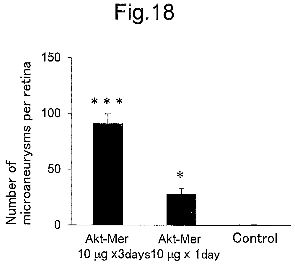
D00016

S00001
XML
uspto.report is an independent third-party trademark research tool that is not affiliated, endorsed, or sponsored by the United States Patent and Trademark Office (USPTO) or any other governmental organization. The information provided by uspto.report is based on publicly available data at the time of writing and is intended for informational purposes only.
While we strive to provide accurate and up-to-date information, we do not guarantee the accuracy, completeness, reliability, or suitability of the information displayed on this site. The use of this site is at your own risk. Any reliance you place on such information is therefore strictly at your own risk.
All official trademark data, including owner information, should be verified by visiting the official USPTO website at www.uspto.gov. This site is not intended to replace professional legal advice and should not be used as a substitute for consulting with a legal professional who is knowledgeable about trademark law.