Prefusion RSV F proteins and their use
Kwong , et al. December 8, 2
U.S. patent number 10,858,400 [Application Number 15/633,578] was granted by the patent office on 2020-12-08 for prefusion rsv f proteins and their use. This patent grant is currently assigned to The United States of America, as represented by the Secretary, Department of Health and Human Services, The United States of America, as represented by the Secretary, Department of Health and Human Services. The grantee listed for this patent is The United States of America, as represented by the Secretary, Department of Health and Human Services, The United States of America, as represented by the Secretary, Department of Health and Human Services, The United States of America, as represented by the Secretary, Department of Health and Human Services. Invention is credited to Man Chen, Barney Graham, Michael Gordon Joyce, Peter Kwong, Jason McLellan, Yongping Yang, Baoshan Zhang, Tongqing Zhou.


View All Diagrams
| United States Patent | 10,858,400 |
| Kwong , et al. | December 8, 2020 |
Prefusion RSV F proteins and their use
Abstract
Disclosed are immunogens including a recombinant RSV F protein stabilized in a prefusion conformation. Also disclosed are nucleic acids encoding the immunogens and methods of producing the immunogens. Methods for generating an immune response in a subject are also disclosed. In some embodiments, the method is a method for treating or preventing a RSV infection in a subject by administering a therapeutically effective amount of the immunogen to the subject.
| Inventors: | Kwong; Peter (Washington, DC), Graham; Barney (Rockville, MD), McLellan; Jason (Austin, TX), Chen; Man (Bethesda, MD), Zhang; Baoshan (Bethesda, MD), Zhou; Tongqing (Boyds, MD), Joyce; Michael Gordon (Washington, DC), Yang; Yongping (Potomac, DC) | ||||||||||
|---|---|---|---|---|---|---|---|---|---|---|---|
| Applicant: |
|
||||||||||
| Assignee: | The United States of America, as
represented by the Secretary, Department of Health and Human
Services (Bethesda, MD) |
||||||||||
| Family ID: | 51527989 | ||||||||||
| Appl. No.: | 15/633,578 | ||||||||||
| Filed: | June 26, 2017 |
Prior Publication Data
| Document Identifier | Publication Date | |
|---|---|---|
| US 20170298101 A1 | Oct 19, 2017 | |
Related U.S. Patent Documents
| Application Number | Filing Date | Patent Number | Issue Date | ||
|---|---|---|---|---|---|
| 14207372 | Mar 12, 2014 | 9738689 | |||
| 61798389 | Mar 15, 2013 | ||||
| 61780910 | Mar 13, 2013 | ||||
| Current U.S. Class: | 1/1 |
| Current CPC Class: | C07K 14/005 (20130101); C07K 2319/70 (20130101); C07K 2319/21 (20130101); C07K 2319/50 (20130101); C07K 2319/03 (20130101); C12N 2760/18534 (20130101); C07K 2319/40 (20130101); C12N 2760/18522 (20130101) |
| Current International Class: | C07K 14/005 (20060101) |
References Cited [Referenced By]
U.S. Patent Documents
| 2010/0068217 | March 2010 | Kwong et al. |
| 2010/0239593 | September 2010 | Spits et al. |
| 2012/0070446 | March 2012 | Beaumont et al. |
| 2012/0093847 | April 2012 | Baudoux et al. |
| 2012/0164176 | June 2012 | Swanson et al. |
| 2012/0315270 | December 2012 | McLellan et al. |
| 2014/0072575 | March 2014 | Spits et al. |
| 2014/0248314 | September 2014 | Swanson et al. |
| 2014/0271699 | September 2014 | Kwong et al. |
| 2016/0046675 | February 2016 | Kwong et al. |
| 102210860 | Oct 2011 | CN | |||
| WO 1998/02457 | Jan 1998 | WO | |||
| WO 2005/111621 | Nov 2005 | WO | |||
| WO 2006/091455 | Aug 2006 | WO | |||
| WO 2009/079796 | Jul 2009 | WO | |||
| WO 2010/149743 | Dec 2010 | WO | |||
| WO 2010/149745 | Dec 2010 | WO | |||
| WO 2011/008974 | Jan 2011 | WO | |||
| WO 2011/043643 | Apr 2011 | WO | |||
| WO 2012/158613 | Nov 2012 | WO | |||
| WO 2013/017713 | Feb 2013 | WO | |||
| WO 2014/024026 | Feb 2014 | WO | |||
| WO 2014/079842 | May 2014 | WO | |||
| WO 2014/139476 | Sep 2014 | WO | |||
| WO 2014/174018 | Oct 2014 | WO | |||
Other References
|
McLellan et al. Science vol. 340 (Year: 2013). cited by examiner . McClellan et al Science vol. 342, p. 592, 2013, (Year: 2013). cited by examiner . Murphy, et al. "An update on approaches to the development of respiratory syncytial virus (RSV) and parainfluenza virus type 3 (PIV3) vaccines." Virus Research 32, No. 1 (1994): 13-36. cited by applicant . Weisshaar, et al. "Blocking respiratory syncytial virus entry: a story with twists." DNA and Cell Biology 34, No. 8 (2015): 505-510. cited by applicant . Arbiza, et al. "Characterization of two antigenic sites recognized by neutralizing monoclonal antibodies directed against the fusion glycoprotein of human respiratory syncytial virus." The Journal of general virology, 73 (1992): 2225-2234. cited by applicant . Beeler, et al. "Neutralization epitopes of the F glycoprotein of respiratory syncytial virus: effect of mutation upon fusion function." Journal of virology, 63.7 (1989): 2941-2950. cited by applicant . Bem et al. "Animal models of human respiratory syncytial virus disease." American Journal of Physiology-Lung Cellular and Molecular Physiology, 301.2 (2011): L148-L156. cited by applicant . Bian, et al., "Influenza virus vaccine expressing fusion and attachment protein epitopes of respiratory syncytial virus induces protective antibodies in BALB/c mice," Antiviral Research, 104: 110-117, 2014 (published online, Feb. 6, 2014). cited by applicant . Blanco, et al., "A recombinant anchorless respiratory syncytial virus (RSV) fusion (F) protein/monophosphoryl lipid a (MPL) vaccine protects against RSV-induced replication and lung pathology," Vaccine, 32(13): 1495-1500, 2014 (published online, Nov. 16, 2013). cited by applicant . Chaiwatpongsakorn, et al. "Soluble respiratory syncytial virus fusion protein in the fully cleaved, pretriggered state is trsaiggered by exposure to low-molarity buffer," Journal of virology 85.8 (2011): 3968-3977. cited by applicant . Chambers, et al., "Sequence analysis of the gene encoding the fusion glycoprotein of pneumonia virus of mice suggests possible conserved secondary structure elements in paramyxovirus fusion glycoproteins." The Journal of general virology 73 (1992): 1717-1724. cited by applicant . Cherukuri, et al., "An adjuvanted respiratory syncytial virus fusion protein induces protection in aged BALB/c mice," Immun Ageing, 9(1): 21, 2012. cited by applicant . Collarini, et al., "Potent high-affinity antibodies for treatment and prophylaxis of respiratory syncytial virus derived from B cells of infected patients." The Journal of Immunology, 183.10 (2009): 6338-6345. cited by applicant . Collins, et al., "Respiratory syncytial virus: virology, reverse genetics, and pathogenesis of disease," Current topics in microbiology and immunology, 372: 3-38, Dec. 21, 2013. cited by applicant . Colman, et al. "The structural biology of type I viral membrane fusion." Nature Reviews Molecular Cell Biology, 4.4 (2003): 309-319. cited by applicant . Connor, et al. "Comparison of human respiratory syncytial virus A2 and 8/60 fusion glycoprotein gene sequences and mapping of sub-group specific antibody epitopes." Journal of medical virology 63.2 (2001): 168-177. cited by applicant . Correia, et al., "Proof of principle for epitope-focused vaccine design," Nature, 507 (7491): 201-206, 2014, (published online, Feb. 5, 2014). cited by applicant . Corvalsier, et al. "Cross-reactive and group-specific immune responses to a neutralizing epitope of the human respiratory syncytial virus fusion protein." Archives of virology, 142.6 (1997): 1073-1086. cited by applicant . Crim, et al. "Identification of linear heparin-binding peptides derived from human respiratory syncytial virus fusion glycoprotein that inhibit infectivity." Journal of virology, 81.1 (2007): 261-271. cited by applicant . Cseke, et al. "Integrin .alpha.v.beta.1 promotes infection by human metapneumovirus." Proceedings of the National Academy of Sciences, 106.5 (2009): 1566-1571. cited by applicant . Dombkowski. "Disulfide by Design.TM.: a computational method for the rational design of disulfide bonds in proteins." Bioinformatics, 19.14 (2003): 1852-1853. cited by applicant . Ekiert, et al. "Cross-neutralization of influenza a viruses mediated by a single antibody loop." Nature, 489.7417 (2012): 526-532. cited by applicant . Eyles, et al., "Nonreplicating vaccines can protect african green monkeys from the memphis 37 strain of respiratory syncytial virus," Journal of Infectious Diseases, 208 (2): 319-329, 2013 (published online, Apr. 17, 2013). cited by applicant . Feldman, et al. "The fusion glycoprotein of human respiratory syncytial virus facilitates virus attachment and infectivity via an interaction with cellular heparan sulfate." Journal of virology, 74.14 (2000): 6442-6447. cited by applicant . Frank, et al. "Stabilization of short collagen-like triple helices by protein engineering." Journal of molecular biology, 308.5 (2001): 1081-1089. cited by applicant . Gilman, et al., "Characterization of a Prefusion-Specific Antibody That Recognizes a Quaternary, Cleavage-Dependent Epitope on the RSV Fusion Glycoprotein," PLoS Pathog, 11(7): e1005035, 2015. cited by applicant . Graham, et al., "Challenges and opportunities for respiratory syncytial virus vaccines," Current topics in microbiology and immunology, 372: 391-404, Dec. 21, 2013. cited by applicant . Graham, et al., "Novel antigens for RSV vaccines," Current Opinion in Immunology, 35: 30-38, 2015. cited by applicant . Graham. "Biological challenges and technological opportunities for respiratory syncytial virus vaccine development." Immunological reviews, 239.1 (2011): 149-166. cited by applicant . Hall, et al. "The burden of respiratory syncytial virus infection in young children." New England Journal of Medicine, 360.6 (2009): 588-598. cited by applicant . Harbury, et al. "A switch between two-, three-, and four-stranded coiled coils in GCN4 leucine zipper mutants." Science 262.5138 (1993): 1401-1407. cited by applicant . Hoppe, et al. "A parallel three stranded .alpha.-helical bundle at the nucleation site of collagen triple-helix formation." FEBS letters 344.2 (1994): 191-195. cited by applicant . IMpact-RSV Study Group. "Palivizumab, a humanized respiratory syncytial virus monoclonal antibody, reduces hospitalization from respiratory syncytial virus infection in high-risk infants." Pediatrics, 102.3 (1998): 531-537. cited by applicant . International Preliminary Report on Patentability for App. No. PCT/CN2014/073505, mailed by the State Intellectual Property Office of the P.R. China as ISA dated Jun. 16, 2014 (43 pages, with English translation). cited by applicant . International Search Report for App. No. PCT/CN2014/073505, mailed by the State Intellectual Property Office of the P.R. China as ISA dated Jun. 16, 2014 (4 pages, with English translation). cited by applicant . International Search Report for App. No. PCT/US2014/026714, mailed by the Australian Patent Office as ISA dated Jul. 29, 2014 (10 pages). cited by applicant . Izard, et al. "Principles of quasi-equivalence and Euclidean geometry govern the assembly of cubic and dodecahedral cores of pyruvate dehydrogenase complexes." Proceedings of the National Academy of Sciences, 96.4 (1999): 1240-1245. cited by applicant . Johnson; et al. "Development of a humanized monoclonal antibody (MEDI-493) with potent in vitro and in vivo activity against respiratory syncytial virus." Journal of Infectious Diseases, 176.5 (1997): 1215-1224. cited by applicant . Johnson, et al., "Genetic vaccine for respiratory syncytial virus provides protection without disease potentiation," Molecular Therapy, 22 (1): 196-205, 2014 (published online, Jun. 10, 2013). cited by applicant . Jones, et al., "Sendai virus-based RSV vaccine protects African green monkeys from RSV infection," Vaccine, 30 (5): 959-968, 2012. cited by applicant . Joyce et al., "Iterative structure-based improvement of a fusion-glycoprotein vaccine against RSV," Nat Struct Mol Biol. 23:811-820, 2016, including supplementary information. cited by applicant . Kanekiyo, et al. "Self-assembling influenza nanoparticle vaccines elicit broadly neutralizing H1N1 antibodies." Nature 499, No. 7456: 102-106 (2013). cited by applicant . Kim, et al., "Development of an adenovirus-based respiratory syncytial virus vaccine: preclinical evaluation of efficacy, immunogenicity, and enhanced disease in a cotton rat model," Journal of Virology, 88 (9): 5100-5108, 2014 (published online, Feb. 26, 2014). cited by applicant . Kwakkenbos, et al. "Generation of stable monoclonal antibody-producing B cell receptor-positive human memory B cells by genetic programming." Nature medicine 16.1 (2010): 123-128. cited by applicant . Lee, et al. "Structure of the Ebola virus glycoprotein bound to an antibody from a human survivor." Nature, 454.7201 (2008): 177-182. cited by applicant . Liang, et al., "Chimeric bovine/human parainfluenza virus type 3 expressing respiratory syncytial virus (RSV) F glycoprotein: effect of insert position on expression, replication, immunogenicity, stability, and protection against RSV infection," Journal of Virology, 88 (8): 4237-4250, 2014 (published online, Jan. 29, 2014). cited by applicant . Liang, et al., "Enhanced Neutralizing Antibody Response Induced by Respiratory Syncytial Virus Pre-fusion F Protein Expressed by a Vaccine Candidate," Journal of Virology, JVI-01373-15, 2015 (published online, Jul. 8, 2015). cited by applicant . Lopez, et al. "Antigenic structure of human respiratory syncytial vices fusion glycoprotein." Journal of virology, 72.8 (1998): 6922-6928. cited by applicant . Lopez, et al. "Location of a highly conserved neutralizing epitope in the F glycoprotein of human respiratory syncytial virus." Journal of virology 64.2 (1990): 927-930. cited by applicant . Lozano, et al. "Global and regional mortality from 235 causes of death for 20 age groups in 1990 and 2010: a systematic analysis for the Global Burden of Disease Study 2010." The Lancet, 380.9859 (2013): 2095-2128. cited by applicant . Ludwig, et al., "Electron cryomicroscopy reveals different F1+ F2 protein states in intact parainfluenza virions," Journal of Virology, 82(7): 3775-3781, 2008. cited by applicant . Magro, et al. "Neutralization of human respiratory syncytial virus infectivity by antibodies and low-molecular-weight compounds targeted against the fusion glycoprotein." Journal of virology, 84.16 (2010): 7970-7982. cited by applicant . Magro, et al. "Neutralizing antibodies against the preactive form of respiratory syncytial virus fusion protein offer unique possibilities for clinical intervention." Proceedings of the National Academy of Sciences 109.8 (2012): 3089-3094. cited by applicant . Martin, et al. "Sequence elements of the fusion peptide of human respiratory syncytial virus fusion protein required for activity." Journal of general virology, 87.6 (2006): 1649-1658. cited by applicant . McAlinden, et al. ".alpha.-Helical coiled-coil oligomerization domains are almost ubiquitous in the collagen superfamily." Journal of Biological Chemistry, 278.43 (2003): 42200-42207. cited by applicant . McGinnes Cullen, et al., "Murine Immune Responses to Virus-like Particle Associated Pre-and Post-Fusion Forms of the Respiratory Syncytial Virus F Protein," Journal of Virology, 89(13):6835-6847, 2015. cited by applicant . McLellan et al., "Structure and function of respiratory syncytial virus surface glycoproteins," Current topics in microbiology and immunology, 372: 83-104, Dec. 21, 2013. cited by applicant . McLellan, "Neutralizing epitopes on the respiratory syncytial virus fusion glycoprotein," Current Opinion in Virology, 11: 70-75, 2015. cited by applicant . McLellan, et al. "Design and characterization of epitope-scaffold immunogens that present the motavizumab epitope from respiratory syncytial virus." Journal of molecular biology 409.5 (2011): 853-866. cited by applicant . McLellan, et al. "Structural basis of respiratory syncytial virus neutralization by motavizumab." Nature structural & molecular biology 17.2 (2010): 248-250. cited by applicant . McLellan, et al. "Structure of a major antigenic site on the respiratory syncytial virus fusion glycoprotein in complex with neutralizing antibody 101F."Journal of virology 84.23 (2010): 12236-12244. cited by applicant . McLellan, et al. "Structure of HIV-1 gp120 V1/V2 domain with broadly neutralizing antibody PG9." Nature, 480.7377 (2011): 336-343. cited by applicant . McLellan, et al. "Structure of respiratory syncytial virus fusion glycoprotein in the postfusion conformation reveals preservation of neutralizing epitopes." Journal of virology 85.15 (2011): 7788-7796. cited by applicant . McLellan, et al. "Structure of RSV fusion glycoprotein trimer bound to a prefusion-specific neutralizing antibody," Science vol. 340 (2013): pp. 1113-1117. cited by applicant . McLellan et al., "Structure-based design of a fusion glycoprotein vaccine for respiratory syncytial virus," Science 342:592-598, 2013, including supplementary information. cited by applicant . Miroshnikov, et al. "Engineering trimeric fibrous proteins based on bacteriophage T4 adhesins." Protein engineering, 11.4 (1998): 329-332. cited by applicant . Nair, et al. "Global burden of acute lower respiratory infections due to respiratory syncytial virus in young children: a systematic review and meta-analysis." The Lancet, 375.9725 (2010): 1545-1555. cited by applicant . Nelson, et al., "Genetic stability of RSV-F expression and the restricted growth phenotype of a live attenuated PIV3 vectored RSV vaccine candidate (MEDI-534) following restrictive growth in human lung cells," Vaccine, 31 (36): 3756-3762, 2013 (published online, Apr. 24, 2013). cited by applicant . Pancera, et al. "N332-Directed broadly neutralizing antibodies use diverse modes of HIV-1 recognition: inferences from heavy-light chain complementation of function." PloS one, 8.2 (2013): e55701. cited by applicant . Petersen, et al. "Amino acid neighbours and detailed conformational analysis of cysteines in proteins." Protein engineering, 12.7 (1999): 535-548. cited by applicant . Shay, et al. "Bronchiolitis-associated hospitalizations among US children, 1980-1996." JAMA, 282.15 (1999): 1440-1446. cited by applicant . Stewart-Jones, et al. "A Cysteine Zipper Stabilizes a Pre-Fusion F Glycoprotein Vaccine for Respiratory Syncytial Virus," PloS One, 10(6): e0128779, 2015. cited by applicant . Sutter, et al. "Structural basis of enzyme encapsulation into a bacterial nanocompartment." Nature structural & molecular biology, 15.9 (2008): 939-947. cited by applicant . Swanson, et al. "Structural basis for immunization with postfusion respiratory syncytial virus fusion F glycoprotein (RSV F) to elicit high neutralizing antibody titers." Proceedings of the National Academy of Sciences 108.23 (2011): 9619-9624. cited by applicant . Swanson, et al., "A monomeric uncleaved respiratory syncytial virus f antigen retains prefusion-specific neutralizing epitopes," Journal of Virology, 88(20): 11802-11810, 2014 (published online, Jul. 30, 2014). cited by applicant . Urich, et al. "X-ray structure of a self-compartmentalizing sulfur cycle metalloenzyme." Science, 311.5763 (2006): 996-1000. cited by applicant . Walsh, et al. "Monoclonal antibodies to respiratory syncytial virus proteins: identification of the fusion protein." Journal of Virology, 47.1 (1983): 171-177. cited by applicant . Welch, et al. "Structure of the cleavage-activated prefusion form of the parainfluenza virus 5 fusion protein." Proceedings of the National Academy of Sciences, 109.41 (2012): 16672-16677. cited by applicant . Wen, et al. "Structure of the human metapneumovirus fusion protein with neutralizing antibody identifies a pneumovirus antigenic site." Nature structural & molecular biology, 19.4 (2012): 461-463. cited by applicant . Written Opinion for App. No. PCT/US2014/026714, mailed by the Australian Patent Office as ISA dated Jul. 29, 2014 (14 pages). cited by applicant . Written Opinion of the ISA for App. No. PCT/CN2014/073505, mailed by the State Intellectual Property Office of the P.R. China as ISA dated Jun. 16, 2014 (8 pages, with English translation). cited by applicant . Wu, et al. "Development of motavizumab, an ultra-potent antibody for the prevention of respiratory syncytial virus infection in the upper and lower respiratory tract." Journal of molecular biology, 368.3 (2007): 652-665. cited by applicant . Yin, et al. "Structure of the parainfluenza virus 5 F protein in s metastable, prefusion conformation." Nature, 439.7072 (2006): 38-44. cited by applicant . Zhang, et al, "Self-assembly in the ferritin nano-cage protein superfamily." International journal of molecular sciences 12.8 (2011): 5406-5421. cited by applicant . Zhang, et al. "X-ray structure analysis and crystallographic refinement of lumazine synthase from the hyperthermophile Aquifex aeolicus at 1.6 a resolution: determinants of thermostability revealed from structural comparisons." Journal of molecular biology, 306.5 (2001): 1099-1114. cited by applicant . Ban et al., "Efficient generation of transgene-free human induced pluripotent stem cells (iPSCs) by temperature-sensitive Sendai virus vectors." Proceedings of the National Academy of Sciences vol. 108, No. 34 (2011): 14234-14239. cited by applicant . He et al., "Ferritin family proteins and their use in bionanotechnology." New Biotechnology vol. 32, No. 6 (2015): 651-657. cited by applicant . Wyatt et al., "The HIV-1 envelope glycoproteins: fusogens, antigens, and immunogens." Science vol. 280, No. 5371 (1998): 1884-1888. cited by applicant. |
Primary Examiner: Foley; Shanon A.
Assistant Examiner: Hill; Myron G
Attorney, Agent or Firm: Klarquist Sparkman, LLP
Parent Case Text
RELATED APPLICATIONS
This application is a continuation of U.S. application Ser. No. 14/207,372, filed Mar. 12, 2014, which in turn claims the benefit of U.S. Provisional Application No. 61/780,910, filed Mar. 13, 2013, and U.S. Provisional Application No. 61/798,389, filed Mar. 15, 2013; each of these applications is specifically incorporated by reference herein in its entirety.
Claims
We claim:
1. An isolated immunogen, comprising: a recombinant RSV F protein comprising S 190F and V207L amino acid substitutions compared to a native RSV F protein that stabilize the recombinant RSV F protein in a prefusion conformation; wherein the prefusion conformation comprises an antigenic site O comprising RSV F residues 62-69 and 196-209 that specifically binds to a D25 antibody or an AM22 antibody after incubation at 20.degree. C. in phosphate buffered saline at physiological pH for at least 24 hours in the absence of the D25 antibody or the AM22 antibody.
2. The immunogen of claim 1, wherein the RSV F protein further comprises one or more additional amino acid substitutions.
3. The immunogen of claim 1, wherein the RSV F protein is a human subtype A or subtype B, or bovine RSV F protein comprising the amino acid substitutions.
4. The immunogen of claim 1, wherein the recombinant RSV F protein comprises a F.sub.2 polypeptide and a F.sub.1 polypeptide comprising or consisting of RSV F positions 26-109 and 137-513, respectively.
5. The immunogen of claim 1, wherein the recombinant RSV F protein comprises a F2 polypeptide and a F.sub.1 polypeptide, wherein a C-terminal residue of the F.sub.2 polypeptide is linked to an N-terminal residue of the F.sub.1 polypeptide by a heterologous peptide linker.
6. The immunogen of claim 1, wherein the recombinant RSV F protein comprises an F.sub.1 polypeptide comprising the amino acid sequence set forth as residues 137-513 of SEQ ID NO: 191.
7. The immunogen of claim 1, wherein the recombinant RSV F protein is soluble and comprises an F.sub.1 ectodomain comprising a C-terminal residue linked to a trimerization domain.
8. The immunogen of claim 7, wherein the trimerization domain is a Foldon domain.
9. The immunogen of claim 8, wherein the recombinant RSV F protein linked to the trimerization domain comprises the amino acid sequences set forth as positions 26-109 and 137-544 of SEQ ID NO: 191.
10. The immunogen of claim 1, wherein the C-terminus of recombinant RSV F protein is linked to a ferritin domain, an encapsulin domain, a Sulfur Oxygenase Reductase (SOR) domain, a lumazine synthase domain, or a pyruvate dehydrogenase domain.
11. The immunogen of claim 1, wherein the recombinant RSV F protein comprises a F.sub.1 ectodomain comprising a C-terminal residue linked to a transmembrane domain.
12. A virus-like particle comprising the immunogen of claim 1.
13. A protein nanoparticle comprising the immunogen of claim 1.
14. The protein nanoparticle of claim 13, wherein the protein nanoparticle is a ferritin nanoparticle, an encapsulin nanoparticle, a Sulfur Oxygenase Reductase (SOR) nanoparticle, a lumazine synthase nanoparticle, or a pyruvate dehydrogenase nanoparticle.
15. A nucleic acid molecule encoding the immunogen of claim 1.
16. A vector comprising the nucleic acid molecule of claim 15.
17. An isolated host cell comprising the vector of claim 16.
18. An immunogenic composition comprising an effective amount of the immunogen of claim 1, and a pharmaceutically acceptable carrier.
19. A method for generating an immune response to RSV F protein in a subject, comprising administering an effective amount of the immunogen of claim 1 to the subject to generate the immune response.
Description
FIELD
This disclosure relates to polypeptides, compositions, and methods of their use, for elicitation and detection of an immune response to respiratory syncytial virus (RSV).
BACKGROUND
Respiratory syncytial virus (RSV) is an enveloped non-segmented negative-strand RNA virus in the family Paramyxoviridae, genus Pneumovirus. It is the most common cause of bronchiolitis and pneumonia among children in their first year of life. RSV also causes repeated infections including severe lower respiratory tract disease, which may occur at any age, especially among the elderly or those with compromised cardiac, pulmonary, or immune systems. Passive immunization currently is used to prevent severe illness caused by RSV infection, especially in infants with prematurity, bronchopulmonary dysplasia, or congenital heart disease. Current treatment includes administration of a RSV-neutralizing antibody, Palivizumab (SYNAGIS.RTM.; MedImmune, Inc.), which binds a 24-amino acid, linear, conformational epitope on the RSV Fusion (F) protein.
In nature, the RSV F protein is initially expressed as a single polypeptide precursor, designated F.sub.0. F.sub.0 trimerizes in the endoplasmic reticulum and is processed by a cellular furin-like protease at two conserved sites, generating, F.sub.1, F.sub.2 and Pep27 polypeptides. The Pep27 polypeptide is excised and does not form part of the mature F protein. The F.sub.2 polypeptide originates from the N-terminal portion of the F.sub.0 precursor and links to the F.sub.1 polypeptide via two disulfide bonds. The F.sub.1 polypeptide originates from the C-terminal portion of the F.sub.0 precursor and anchors the mature F protein in the membrane via a transmembrane domain, which is linked to an .about.24 amino acid cytoplasmic tail. Three protomers of the F.sub.2-F.sub.1 heterodimer assemble to form a mature F protein, which adopts a metastable prefusion conformation that is triggered to undergo a conformational change that fuses the viral and target-cell membranes. Due to its obligatory role in RSV entry, the RSV F protein is the target of neutralizing antibodies and the subject of vaccine development; however, like other RSV antigens, prior efforts to develop an RSV F protein-based vaccine have proven unsuccessful.
Prior to the work disclosed herein, a homogeneous preparation of soluble prefusion RSV F protein was unavailable, precluding determination of the prefusion F structure and identification of novel F-specific antigenic sites.
SUMMARY
As described herein, the three-dimensional structure of RSV F protein in its pre-fusion conformation was elucidated. The disclosure reveals for the first time the prefusion conformation of RSV F, which includes a unique antigenic site ("antigenic site O") at its membrane distal apex. Using the three-dimensional structure of prefusion F as a guide, stabilized forms of prefusion F ("PreF" antigens) were engineered and constructed and used to generate RSV neutralizing immune responses many fold greater than that achieved with prior RSV F protein-based immunogens.
Disclosed herein are isolated recombinant RSV F proteins that are stabilized in a prefusion conformation, as well as nucleic acid molecules encoding the recombinant RSV F proteins, which are useful, for example, to induce an immune response to RSV in a subject. In several embodiments, the recombinant RSV F protein can be stabilized in a prefusion conformation that can specifically bind to a prefusion specific antibody, such as a D25 or an AM22 antibody. In some embodiments, the recombinant RSV F protein comprises an antigenic site O comprising residues 62-69 and 196-209 of SEQ ID NO: 370, that specifically binds the D25 antibody or the AM22 antibody, or both. The PreF antigens can be used, for example, as both potential vaccines for RSV and as diagnostic molecules. In some embodiments, the recombinant RSV F proteins can be used to detect and quantify target antibodies in a polyclonal serum response.
Elucidation of the PreF antigens was accomplished by achieving, for the first time, the crystallization and three-dimensional structure determination of the RSV F protein in its prefusion conformation. RSV F protein specific antibodies were identified that neutralize RSV, but that do not bind to a RSV F protein construct stabilized in the postfusion conformation, and the structure of RSV F protein recognized by these antibodies was determined. A prefusion-specific antigenic site was revealed by the structure (antigenic site O), which provides atomic-level details that were used to develop the recombinant RSV F proteins.
In several embodiments, the RSV F protein can be a single chain RSV F protein.
In some embodiments, the recombinant RSV F protein can include an F.sub.1 polypeptide and an F.sub.2 polypeptide, wherein the F.sub.1 polypeptide, the F.sub.2 polypeptide, or both, include at least one modification (such as an amino acid substitution) that stabilizes the recombinant RSV F protein in the prefusion conformation. In some embodiments, the modification can include an amino acid substitution that introduces a non-natural disulfide bond, or the substitution can be a cavity-filling amino acid substitution.
In one non-limiting example, the recombinant RSV F protein can include S155C and S290C substitutions.
In some embodiments, the recombinant RSV F protein can be linked to a trimerization domain, such as a Foldon domain, which can further stabilize the recombinant RSV F protein in the prefusion conformation. In additional embodiments, the recombinant RSV F protein can be included on a protein nanoparticle, such as a ferritin nanoparticle.
Additional embodiments include epitope-scaffold proteins including a RSV F protein prefusion specific epitope (such as antigenic site O), wherein the epitope scaffold protein is specifically bound by a RSV F prefusion-specific monoclonal antibody, such as a D25 or AM22 antibody.
Methods of generating an immune response in a subject are disclosed, as are methods of treating, inhibiting or preventing a RSV infection in a subject. In such methods a subject, such as a human or bovine subject, can be administered an effective amount of a disclosed PreF antigen and/or a nucleic acid molecule encoding a disclosed PreF antigen. In some embodiments, the methods include administration of an immunogenic composition including an adjuvant selected to elicit a Th1 biased immune response in a subject.
Methods for detecting or isolating an RSV binding antibody in a subject infected with RSV are also disclosed.
The foregoing and other objects, features, and advantages of the embodiments will become more apparent from the following detailed description, which proceeds with reference to the accompanying figures.
BRIEF DESCRIPTION OF THE FIGURES
The patent or application file contains at least one drawing executed in color. Copies of this patent or patent application publication with color drawing(s) will be provided by the Office upon request and payment of the necessary fee.
FIGS. 1A-1C are a set of graphs and an diagram illustrating RSV neutralization, F glycoprotein recognition, and the crystal structure of human antibody D25 in complex with the prefusion RSV F trimer. The prefusion conformation of RSV F is metastable, and when expressed in a soluble form readily adopts the postfusion state; a number of potent antibodies, including D25, bind to a newly revealed antigenic site at the top of the prefusion F glycoprotein. (A) RSV neutralization by antibodies including palivizumab, the FDA-approved prophylactic antibody to prevent severe RSV disease. (B) Enzyme linked immunosorbant assay (ELISA) measuring antibody binding to postfusion F glycoprotein. (C) D25-RSV F trimer structure in ribbon and molecular surface representations. One protomer of the F glycoprotein trimer is shown as ribbons and colored as a rainbow from blue to red, N-terminus of F.sub.2 to C-terminus of F.sub.1, respectively. Molecular surfaces are shown for the other two F protomers, colored pink and green. The D25 Fab bound to the F protomer shown in ribbons is also displayed in ribbon representation, with heavy chain colored red and light chain colored grey. The other D25 Fabs are colored the same, but shown in surface representation.
FIGS. 2A and 2B are a set of diagrams and a sequence aligned with RSV secondary structure illustrating the structural rearrangement of RSV F. To mediate virus-cell entry, the RSV F glycoprotein transitions from a metastable prefusion conformation to a stable postfusion conformation. (A) Prefusion and postfusion structures. Outer images display prefusion (left) and postfusion (right) trimeric structures, colored the same as in FIG. 1C. A complex glycan, shown as sticks, is modeled at each of the three N-linked glycosylation sites found in the mature protein. Inner images display a single RSV F protomer in ribbon representation, colored as a rainbow from blue to red, N-terminus of F.sub.2 to C-terminus of F.sub.1, respectively. (B) RSV F sequence and secondary structure. Sites of N-linked glycosylation are highlighted by black triangles, antigenic sites are labeled in red, and downward arrows indicate the position of furin cleavage sites. Secondary structures are shown below the sequence (SEQ ID NO: 370), with cylinders representing .alpha.-helices and arrows representing .beta.-strands. Disordered or missing residues are indicated by an "X"; residues that move over 5 .ANG. between prefusion and postfusion conformations shown with grey shadow.
FIGS. 3A-3C show a set of diagrams and a sequence alignment illustrating the RSV F interface with D25. Antibody D25 binds a quaternary epitope spanning two protomers at the apex of the prefusion F trimer. (A) Close-up of the interface between D25 and RSV F. Side chains of F residues interacting with D25 are labeled and shown as sticks. Oxygen atoms are colored red and nitrogen atoms are colored blue. Hydrogen bonds are depicted as dotted lines. The two images are related by a 90.degree. rotation about the vertical axis. (B) Position and conformation of the D25 epitope on the prefusion and postfusion F molecules. RSV F residues at the D25 interface are colored red; polarity of .alpha.4 and .alpha.5.sub.post indicated with arrows, with fragment N- and C-termini indicated. (C) Sequence conservation of F residues in regions recognized by D25. Amino acids in human RSV subtype B (hRSV/B) or in bovine RSV (bRSV) that differ from hRSV/A are colored red. Ectodomain is defined as F residues 26-109 and 137-524. Residues 63-74 and 200-213 of SEQ ID NO: 1 (hRSV/A), SEQ ID NO: 129 (hRSV/B), and SEQ ID NO: 178 (bRSV) are shown.
FIGS. 4A-4D are series of graphs and digital images concerning antigenic site O. Highly effective RSV-neutralizing antibodies target a site at the membrane-distal apex of the prefusion F trimer. (A) The ability of antibodies to block D25 binding to RSV-infected cells was measured as a function of antibody concentration. (B) Analysis of RSV F/Fab complexes by negative stain electron microscopy: (Left) Reprojection of a 12 .ANG. slice through the crystal structure of RSV F+D25 Fab filtered to 10 .ANG. resolution and sliced to include the F-trimer cavity. (Middle) Aligned average of 263 particles of RSV F+D25 Fab. (Right) Aligned average of 550 particles of RSV F+AM22 Fab. Scale bar in middle panel is 50 .ANG.. (C) Fusion inhibition and (D) attachment inhibition activity for antibodies targeting antigenic site O and F-specific antibodies targeting other antigenic sites. For the attachment-inhibition assay, heparin was used as a positive control.
FIG. 5 shows a schematic diagram illustrating the methods used to express complexes of RSV F and D25. Plasmids expressing RSV F(+) Fd (green circle), the D25 light chain (grey circle), and the D25 heavy chain (with or without a stop codon in the hinge region, red circle) were simultaneously transfected into HEK293 cells in suspension. Alternatively, the RSV F(+) Fd plasmid could be transfected, with purified D25 Fab or IgG added to the cells 3 hours post-transfection. The best yields were obtained by simultaneously expressing F and D25 Fab (.about.1.0 mg of purified complex per liter of cells).
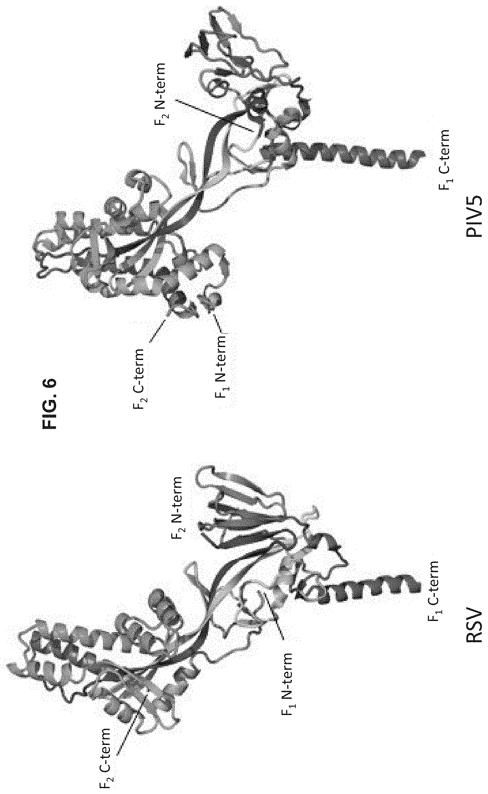
FIG. 6 shows a set of ribbon diagrams illustrating the comparison of D25-bound RSV F to prefusion PIV5 F. Ribbon representation of D25-bound RSV F (+) Fd (left) and PIV5 F-GCNt (right) colored as a rainbow from blue to red, F.sub.2 N-terminus to F.sub.1 C-terminus, respectively. There is excellent agreement of secondary structure elements between the two proteins, despite having only .about.12% sequence identity. One of the most striking differences is the location of the fusion peptide (N-terminus of F.sub.1 subunit), also shown in FIG. 7. The PIV5 F structure was described as consisting of three domains: I, II and III (Yin et al., Nature, 439, 38 (2006)). Domain III termed the membrane distal lobe, whereas domains I and II encompass the central barrel and membrane proximal lobe. The cleaved PIV5 structure shown here was generated from PDB ID: 4GIP (Welch et al., Proc. Natl. Acad. Sci., U.S.A. 109, 16672 (2012)).
FIG. 7 shows a series of diagrams illustrating Type I prefusion viral glycoproteins. Prefusion structures of RSV F, PIV5 F (PDB ID: 4GIP (Welch et al., Proc. Natl. Acad. Sci., U.S.A. 109, 16672 (2012)), influenza HA (PDB ID: 2HMG; Wilson et al., Nature, 289, 366 (1981)) and Ebola GP (PDB ID: 3CSY; Lee et al., Nature, 454, 177 (2008)) are shown as molecular surfaces, with each protomer colored differently. On the bottom row, a red sphere is shown for the C-terminal residue of F.sub.2 (RSV and PIV5) or HA.sub.1 (Flu), and a blue sphere is show for the N-terminal residue of the fusion peptide. The RSV and PIV5 are both paramyxoviruses and their F proteins share .about.12% sequence identity. Although Ebola GP is a type I fusion protein, it lacks a free N-terminal fusion peptide on GP2, and instead contains an internal fusion loop that is commonly seen in type II and type III fusion proteins. Thus, the Ebola GP was omitted from the fusion peptide comparison.
FIG. 8 is a set of graphs concerning RSV neutralization by IgG and Fab. D25, AM22 and Motavizumab neutralize RSV equally well as IgG or Fab. Note that the x-axis for the Motavizumab plot is different than the others.
FIGS. 9A and 9B are a series of diagrams and graphs illustrating properties of antigenic sites on the RSV F glycoprotein. Only antibodies directed to antigenic site O bind specifically to the prefusion conformation and have exceptional neutralization potency. (A) For site O, an image of a single D25 Fab binding to the prefusion RSV F trimer is shown, along with neutralization curves for AM22 and D25. For site I, arrows point to Pro389, a known escape mutation (Lopez et al., J. Virol., 72, 6922 (1998)). A neutralization curve is shown for antibody 131-2a. Like antibody 2F (Magro et al., J. Virol., 84, 7970 (2010)), antibody 131-2a only neutralizes .about.50% of the virus. (B) For antigenic sites II and IV, models of Motavizumab (site II) and 101F (site IV) binding to the prefusion and postfusion (McLellan et al., J. Virol., 85, 7788 (2011)) F structures were made using the coordinates of antibody-peptide structures (McLellan et al., J. Virol., 84, 12236 (2010); McLellan et al., Nat. Struct. Mol. Biol., 17, 248 (2010)).
FIG. 10 shows an image of a polyacrylamide gel illustrating expression of the recombinant RSV F protein construct with S155C and S290C amino acid substitutions and a Foldon domain linked to the C-terminus of F.sub.1, and a set of diagrams illustrating that the disulfide bond between S155C and S290C can only form in the prefusion conformation of RSV F protein.
FIG. 11 is a set of graphs showing results from ELISA and gel filtration assays using the recombinant RSV F protein construct with S155C and S290C amino acid substitutions and a Foldon domain linked to the C-terminus of F.sub.1. The ELISA data indicate that the S155C/S290C construct is specifically bound by RSV F prefusion specific antibodies. The gel filtration profiles show that the S155C/S290C construct exists solely as a trimer, whereas aggregates and rosettes form in solution with a control RSV F construct lacking the S155C/S290C substitutions.
FIG. 12 shows negative-stain electron microscopy images of recombinant RSV F protein construct with S155C and S290C amino acid substitutions and a Foldon domain linked to the C-terminus of F1. The images below the large panel are 2D averages of individual particles. The results indicate that the S155C/S290C construct is stabilized in the prefusion conformation.
FIGS. 13-14 show a set of graphs illustrating the neutralizing antibody response of mice administered native RSV (RSV), formalin inactivated RSV (FI-RSV), the recombinant RSV F protein construct with S155C and S290C amino acid substitutions and a Foldon domain linked to the C-terminus of F.sub.1 (prefusion F), or a RSV F protein construct stabilized in the postfusion conformation (postfusion RSV). The antibody response at 5 weeks (FIG. 13) and 7 weeks (FIG. 14) post-initial immunization is shown.
FIG. 15 shows digital images of the crystals of a soluble recombinant RSV F protein stabilized in a prefusion conformation by S155C and S290C substitutions. Left, standard light images; Right, ultraviolet images, indicative of proteins. The formation of crystals from aqueous buffered solutions demonstrates that this protein is substantially homogeneous in solution.
SEQUENCE LISTING
The nucleic and amino acid sequences listed in the accompanying sequence listing are shown using standard letter abbreviations for nucleotide bases, and three letter code for amino acids, as defined in 37 C.F.R. 1.822. Only one strand of each nucleic acid sequence is shown, but the complementary strand is understood as included by any reference to the displayed strand. The Sequence Listing is submitted as an ASCII text file in the form of the file named "Sequence.txt" (.about.1.7 MB), which was created on Apr. 27, 2017, and is incorporated by reference herein. In the accompanying Sequence Listing:
SEQ ID NOs: 1-128 are the amino acid sequences of native RSV F proteins from RSV type A.
SEQ ID NOs: 129-177 are the amino acid sequences of native RSV F proteins from RSV type B.
SEQ ID NOs: 178-184 are the amino acid sequences of native RSV F proteins from bovine RSV.
SEQ ID NOs: 185-350 are the amino acid sequences of recombinant RSV F proteins.
SEQ ID NO: 351 is the amino acid sequence of a T4 fibritin Foldon domain.
SEQ ID NO: 352 and 355-365 are amino acid sequences of peptide linkers.
SEQ ID NO: 353 is the amino acid sequence of a Helicobacter pylori ferritin protein (GENBANK.RTM. Accession No. EJB64322.1, incorporated by reference herein as present in the database on Feb. 28, 2013).
SEQ ID NO: 354 is the amino acid sequence of an encapsulin protein (GENBANK.RTM. Accession No. YP_001738186.1, incorporated by reference herein as present in the database on Feb. 28, 2013).
SEQ ID NOs: 366 and 367 are the V.sub.H and V.sub.L amino acid sequences of the AM22 mAb, respectively.
SEQ ID NO: 368 and 369 are the V.sub.H and V.sub.L amino acid sequences of the D25 mAb, respectively.
SEQ ID NO: 370 is a recombinant RSV F.sub.0 protein variant amino acid sequence of the prototypical A2 strain (GENBANK accession No. P03420, incorporated by reference herein as present in the database on Feb. 28, 2012), including P102A, I379V, and M447V substitutions compared to the P03420 sequence.
STRUCTURAL COORDINATES
The atomic coordinates of the crystal structure of RSV F protein bound by D25 Fab are recited in the 3D Protein Crystals file which is submitted as an ASCII text file named "4239-90594-18 Table 1.txt" (.about.1 MB), which was created on Mar. 13, 2013, and is incorporated by reference herein, and is referred to herein as "Table 1".
DETAILED DESCRIPTION
The RSV F glycoprotein it is a type I fusion protein that facilitates fusion of viral and cellular membranes (Walsh and Hruska, J. Virol., 47, 171 (1983)). After initial synthesis, RSV F adopts a metastable prefusion conformation that stores folding energy, which is released during a structural rearrangement to a highly stable postfusion conformation after contact with host cell membranes. Three antigenic sites (I, II, and IV) on RSV F protein have been found to elicit neutralizing activity (Arbiza et al., J. Gen. Virol., 73, 2225 (1992); Lopez et al., J. Virol., 72, 6922 (1998); Lopez et al., J. Virol., 64, 927 (1990)), and all exist on the postfusion form of RSV F protein as determined by structural and biophysical studies (McLellan et al., J. Virol., 85, 7788 (2011); Swanson et al., Proc. Natl. Acad. Sci. U.S.A., 108, 9619 (2011)). Absorption of human sera with postfusion RSV F, however, fails to remove the majority of F-specific neutralizing activity, suggesting that the prefusion form of RSV F harbors novel neutralizing antigenic sites (Magro et al., Proc. Natl. Acad. Sci. U.S.A., 109, 3089 (2012)).
Prior to the work disclosed herein, a homogeneous preparation of soluble prefusion RSV F protein was unavailable, precluding determination of the prefusion F structure and identification of novel F-specific antigenic sites. As described herein, RSV F protein specific antibodies were identified that neutralize RSV, but do not specifically bind to postfusion RSV F, and the three-dimensional structure of prefusion F, recognized by these antibodies, was obtained. The results provided herein reveal for the first time the prefusion conformation of RSV F and the mechanism of neutralization for a category of remarkably potent RSV prefusion F neutralizing antibodies. Using the three-dimensional structure of prefusion F as a guide, stabilized forms of prefusion F ("PreF" antigens) were constructed and used to generate RSV neutralizing immune responses many fold greater than that achieved with prior RSV F protein-based immunogens.
I. TERMS
Unless otherwise noted, technical terms are used according to conventional usage. Definitions of common terms in molecular biology can be found in Benjamin Lewin, Genes VII, published by Oxford University Press, 1999; Kendrew et al. (eds.), The Encyclopedia of Molecular Biology, published by Blackwell Science Ltd., 1994; and Robert A. Meyers (ed.), Molecular Biology and Biotechnology: a Comprehensive Desk Reference, published by VCH Publishers, Inc., 1995; and other similar references.
As used herein, the singular forms "a," "an," and "the," refer to both the singular as well as plural, unless the context clearly indicates otherwise. For example, the term "an antigen" includes single or plural antigens and can be considered equivalent to the phrase "at least one antigen." As used herein, the term "comprises" means "includes." Thus, "comprising an antigen" means "including an antigen" without excluding other elements. It is further to be understood that any and all base sizes or amino acid sizes, and all molecular weight or molecular mass values, given for nucleic acids or polypeptides are approximate, and are provided for descriptive purposes, unless otherwise indicated. Although many methods and materials similar or equivalent to those described herein can be used, particular suitable methods and materials are described below. In case of conflict, the present specification, including explanations of terms, will control. In addition, the materials, methods, and examples are illustrative only and not intended to be limiting. To facilitate review of the various embodiments, the following explanations of terms are provided:
Adjuvant: A vehicle used to enhance antigenicity. Adjuvants include a suspension of minerals (alum, aluminum hydroxide, or phosphate) on which antigen is adsorbed; or water-in-oil emulsion, for example, in which antigen solution is emulsified in mineral oil (Freund incomplete adjuvant), sometimes with the inclusion of killed mycobacteria (Freund's complete adjuvant) to further enhance antigenicity (inhibits degradation of antigen and/or causes influx of macrophages) Immunostimulatory oligonucleotides (such as those including a CpG motif) can also be used as adjuvants. Adjuvants include biological molecules (a "biological adjuvant"), such as costimulatory molecules. Exemplary adjuvants include IL-2, RANTES, GM-CSF, TNF-.alpha., IFN-.gamma., G-CSF, LFA-3, CD72, B7-1, B7-2, OX-40L, 4-1BBL and toll-like receptor (TLR) agonists, such as TLR-9 agonists. The person of ordinary skill in the art is familiar with adjuvants (see, e.g., Singh (ed.) Vaccine Adjuvants and Delivery Systems. Wiley-Interscience, 2007). Adjuvants can be used in combination with the disclosed PreF antigens.
Administration: The introduction of a composition into a subject by a chosen route. Administration can be local or systemic. For example, if the chosen route is intravenous, the composition (such as a composition including a disclosed immunogen) is administered by introducing the composition into a vein of the subject.
Agent: Any substance or any combination of substances that is useful for achieving an end or result; for example, a substance or combination of substances useful for inhibiting RSV infection in a subject. Agents include proteins, nucleic acid molecules, compounds, small molecules, organic compounds, inorganic compounds, or other molecules of interest, such as viruses, such as recombinant viruses. An agent can include a therapeutic agent (such as an anti-RSV agent), a diagnostic agent or a pharmaceutical agent. In some embodiments, the agent is a polypeptide agent (such as an immunogenic RSV polypeptide), or an anti-viral agent. The skilled artisan will understand that particular agents may be useful to achieve more than one result.
AM22: A neutralizing monoclonal antibody that specifically binds to the prefusion conformation of the RSV F protein, but not the post fusion conformation of RSV F protein. AM22 protein and nucleic acid sequences are known, for example, the heavy and light chain amino acid sequences of the AM22 antibody are set forth in U.S. Pat. App. Pub. No. 2012/0070446, which is incorporated herein in its entirety). As described in Example 1, AM22 specifically binds to an epitope including positions found on the RSV F protein in its prefusion conformation, but not the post fusion conformation. This epitope is included within RSV F positions 62-69 and 196-209, and located at the membrane distal apex of the RSV F protein in the prefusion conformation (see, e.g., FIGS. 2B and 9A). Prior to this disclosure it was not known that AM22 was specific for the prefusion conformation. In several embodiments, antibody AM22 specifically binds to the PreF antigens disclosed herein.
Amino acid substitutions: The replacement of one amino acid in an antigen with a different amino acid. In some examples, an amino acid in an antigen is substituted with an amino acid from a homologous protein.
Animal: A living multi-cellular vertebrate or invertebrate organism, a category that includes, for example, mammals. The term mammal includes both human and non-human mammals. Similarly, the term "subject" includes both human and veterinary subjects, such as non-human primates. Thus, administration to a subject can include administration to a human subject. Particular examples of veterinary subjects include domesticated animals (such as cats and dogs), livestock (for example, cattle, horses, pigs, sheep, and goats), laboratory animals (for example, mice, rabbits, rats, gerbils, guinea pigs, and non-human primates).
Antibody: A polypeptide that in nature is substantially encoded by an immunoglobulin gene or immunoglobulin genes, or fragments thereof, which specifically binds and recognizes an analyte (such as an antigen or immunogen) such as a RSV F protein or antigenic fragment thereof. Immunoglobulin genes include the kappa, lambda, alpha, gamma, delta, epsilon and mu constant region genes, as well as the myriad immunoglobulin variable region genes.
Antibodies exist, for example as intact immunoglobulins and as a number of well characterized fragments produced by digestion with various peptidases. For instance, Fabs, Fvs, and single-chain Fvs (SCFvs) that bind to RSV F protein, would be RSV F protein-specific binding agents. This includes intact immunoglobulins and the variants and portions of them well known in the art, such as Fab' fragments, F(ab)'.sub.2 fragments, single chain Fv proteins ("scFv"), and disulfide stabilized Fv proteins ("dsFv"). A scFv protein is a fusion protein in which a light chain variable region of an immunoglobulin and a heavy chain variable region of an immunoglobulin are bound by a linker, while in dsFvs, the chains have been mutated to introduce a disulfide bond to stabilize the association of the chains. The term also includes genetically engineered forms such as chimeric antibodies (such as humanized murine antibodies), heteroconjugate antibodies (such as bispecific antibodies). See also, Pierce Catalog and Handbook, 1994-1995 (Pierce Chemical Co., Rockford, Ill.); Kuby, J., Immunology, 3.sup.rd Ed., W.H. Freeman & Co., New York, 1997.
Antibody fragments are defined as follows: (1) Fab, the fragment which contains a monovalent antigen-binding fragment of an antibody molecule produced by digestion of whole antibody with the enzyme papain to yield an intact light chain and a portion of one heavy chain; (2) Fab', the fragment of an antibody molecule obtained by treating whole antibody with pepsin, followed by reduction, to yield an intact light chain and a portion of the heavy chain; two Fab' fragments are obtained per antibody molecule; (3) (Fab')2, the fragment of the antibody obtained by treating whole antibody with the enzyme pepsin without subsequent reduction; (4) F(ab')2, a dimer of two Fab' fragments held together by two disulfide bonds; (5) Fv, a genetically engineered fragment containing the variable region of the light chain and the variable region of the heavy chain expressed as two chains; and (6) single chain antibody ("SCA"), a genetically engineered molecule containing the variable region of the light chain, the variable region of the heavy chain, linked by a suitable polypeptide linker as a genetically fused single chain molecule. The term "antibody," as used herein, also includes antibody fragments either produced by the modification of whole antibodies or those synthesized de novo using recombinant DNA methodologies.
Typically, a naturally occurring immunoglobulin has heavy (H) chains and light (L) chains interconnected by disulfide bonds. There are two types of light chain, lambda (.lamda.) and kappa (.kappa.). There are five main heavy chain classes (or isotypes) which determine the functional activity of an antibody molecule: IgM, IgD, IgG, IgA and IgE. The disclosed antibodies can be class switched.
Each heavy and light chain contains a constant region and a variable region, (the regions are also known as "domains"). In several embodiments, the heavy and the light chain variable domains combine to specifically bind the antigen. In additional embodiments, only the heavy chain variable domain is required.
For example, naturally occurring camelid antibodies consisting of a heavy chain only are functional and stable in the absence of light chain (see, e.g., Hamers-Casterman et al., Nature, 363:446-448, 1993; Sheriff et al., Nat. Struct. Biol., 3:733-736, 1996). Light and heavy chain variable domains contain a "framework" region interrupted by three hypervariable regions, also called "complementarity-determining regions" or "CDRs" (see, e.g., Kabat et al., Sequences of Proteins of Immunological Interest, U.S. Department of Health and Human Services, 1991). The sequences of the framework regions of different light or heavy chains are relatively conserved within a species. The framework region of an antibody, that is the combined framework regions of the constituent light and heavy chains, serves to position and align the CDRs in three-dimensional space.
The CDRs are primarily responsible for binding to an epitope of an antigen. The amino acid sequence boundaries of a given CDR can be readily determined using any of a number of well-known schemes, including those described by Kabat et al. ("Sequences of Proteins of Immunological Interest," 5th Ed. Public Health Service, National Institutes of Health, Bethesda, Md., 1991; "Kabat" numbering scheme), Al-Lazikani et al., (JMB 273,927-948, 1997; "Chothia" numbering scheme), and Lefranc, et al. ("IMGT unique numbering for immunoglobulin and T cell receptor variable domains and Ig superfamily V-like domains," Dev. Comp. Immunol., 27:55-77, 2003; "IMGT" numbering scheme).
The CDRs of each chain are typically referred to as CDR1, CDR2, and CDR3 (from the N-terminus to C-terminus), and are also typically identified by the chain in which the particular CDR is located. Thus, a V.sub.H CDR3 is located in the variable domain of the heavy chain of the antibody in which it is found, whereas a V.sub.L CDR1 is the CDR1 from the variable domain of the light chain of the antibody in which it is found. Light chain CDRs are sometimes referred to as CDR L1, CDR L2, and CDR L3. Heavy chain CDRs are sometimes referred to as CDR H1, CDR H2, and CDR H3.
Antigen: A compound, composition, or substance that can stimulate the production of antibodies or a T cell response in an animal, including compositions that are injected or absorbed into an animal. An antigen reacts with the products of specific humoral or cellular immunity, including those induced by heterologous antigens, such as the disclosed recombinant RSV F proteins. "Epitope" or "antigenic determinant" refers to the region of an antigen to which B and/or T cells respond. In one embodiment, T cells respond to the epitope, when the epitope is presented in conjunction with an MHC molecule. Epitopes can be formed both from contiguous amino acids or noncontiguous amino acids juxtaposed by tertiary folding of a protein. Epitopes formed from contiguous amino acids are typically retained on exposure to denaturing solvents whereas epitopes formed by tertiary folding are typically lost on treatment with denaturing solvents. An epitope typically includes at least 3, and more usually, at least 5, about 9, or about 8-10 amino acids in a unique spatial conformation. Methods of determining spatial conformation of epitopes include, for example, x-ray crystallography and nuclear magnetic resonance.
Examples of antigens include, but are not limited to, polypeptides, peptides, lipids, polysaccharides, combinations thereof (such as glycopeptides) and nucleic acids containing antigenic determinants, such as those recognized by an immune cell. In some examples, antigens include peptides derived from a pathogen of interest, such as RSV. In specific examples, an antigen is derived from RSV, such as an antigen including a RSV F protein in a prefusion conformation.
A "target epitope" is a specific epitope on an antigen that specifically binds an antibody of interest, such as a monoclonal antibody. In some examples, a target epitope includes the amino acid residues that contact the antibody of interest, such that the target epitope can be selected by the amino acid residues determined to be in contact with the antibody of interest.
Anti-RSV agent: An agent that specifically inhibits RSV from replicating or infecting cells. Non-limiting examples of anti-RSV agents include the monoclonal antibody palivizumab (SYNAGIS.RTM.; Medimmune, Inc.) and the small molecule anti-viral drug ribavirin (manufactured by many sources, e.g., Warrick Pharmaceuticals, Inc.).
Atomic Coordinates or Structure coordinates: Mathematical coordinates derived from mathematical equations related to the patterns obtained on diffraction of a monochromatic beam of X-rays by the atoms (scattering centers) such as an antigen, or an antigen in complex with an antibody. In some examples that antigen can be RSV F protein (for example stabilized in a prefusion conformation by binding to a prefusion-specific antibody, or by introduction of stabilizing modifications) in a crystal. The diffraction data are used to calculate an electron density map of the repeating unit of the crystal. The electron density maps are used to establish the positions of the individual atoms within the unit cell of the crystal. In one example, the term "structure coordinates" refers to Cartesian coordinates derived from mathematical equations related to the patterns obtained on diffraction of a monochromatic beam of X-rays, such as by the atoms of a RSV F protein in crystal form.
Those of ordinary skill in the art understand that a set of structure coordinates determined by X-ray crystallography is not without standard error. For the purpose of this disclosure, any set of structure coordinates that have a root mean square deviation of protein backbone atoms (N, C.alpha., C and O) of less than about 1.0 Angstroms when superimposed, such as about 0.75, or about 0.5, or about 0.25 Angstroms, using backbone atoms, shall (in the absence of an explicit statement to the contrary) be considered identical.
Cavity-filling amino acid substitution: An amino acid substitution that fills a cavity within the protein core of the RSV F protein, for example a cavity present in a protomer of the RSV F protein, or a cavity between protomers of the RSV F protein. Cavities are essentially voids within a folded protein where amino acids or amino acid side chains are not present. In several embodiments, a cavity filling amino acid substitution is introduced to fill a cavity in the RSV F protein core present in the RSV F protein prefusion conformation that collapse (e.g., have reduced volume) after transition to the postfusion conformation.
Contacting: Placement in direct physical association; includes both in solid and liquid form. Contacting includes contact between one molecule and another molecule, for example the amino acid on the surface of one polypeptide, such as an antigen, that contact another polypeptide, such as an antibody. Contacting also includes administration, such as administration of a disclosed antigen to a subject by a chosen route.
Control: A reference standard. In some embodiments, the control is a negative control sample obtained from a healthy patient. In other embodiments, the control is a positive control sample obtained from a patient diagnosed with RSV infection. In still other embodiments, the control is a historical control or standard reference value or range of values (such as a previously tested control sample, such as a group of RSV patients with known prognosis or outcome, or group of samples that represent baseline or normal values).
A difference between a test sample and a control can be an increase or conversely a decrease. The difference can be a qualitative difference or a quantitative difference, for example a statistically significant difference. In some examples, a difference is an increase or decrease, relative to a control, of at least about 5%, such as at least about 10%, at least about 20%, at least about 30%, at least about 40%, at least about 50%, at least about 60%, at least about 70%, at least about 80%, at least about 90%, at least about 100%, at least about 150%, at least about 200%, at least about 250%, at least about 300%, at least about 350%, at least about 400%, at least about 500%, or greater than 500%.
D25: A neutralizing monoclonal antibody that specifically binds to the prefusion conformation of the RSV F protein, but not the post fusion conformation of RSV F protein. D25 protein and nucleic acid sequences are known, for example, the heavy and light chain amino acid sequences of the D25 antibody are set forth in U.S. Pat. App. Pub. No. 2010/0239593, which is incorporated herein in its entirety; see also, Kwakkenbos et al., Nat. Med., 16:123-128, 2009). As described in Example 1, D25 specifically binds to a quaternary epitope found on the RSV F protein in its prefusion conformation, but not the post fusion conformation. This epitope is included within RSV F positions 62-69 and 196-209, and located at the membrane distal apex of the RSV F protein in the prefusion conformation (see, e.g., FIGS. 2B and 9A). Prior to this disclosure it was not known that D25 was specific for the prefusion conformation of RSV F protein). In several embodiments, antibody D25 specifically binds to the PreF antigens disclosed herein.
Degenerate variant and conservative variant: A polynucleotide encoding a polypeptide or an antibody that includes a sequence that is degenerate as a result of the genetic code. For example, a polynucleotide encoding a disclosed antigen or an antibody that specifically binds a disclosed antigen includes a sequence that is degenerate as a result of the genetic code. There are 20 natural amino acids, most of which are specified by more than one codon. Therefore, all degenerate nucleotide sequences are included as long as the amino acid sequence of the antigen or antibody that binds the antigen encoded by the nucleotide sequence is unchanged. Because of the degeneracy of the genetic code, a large number of functionally identical nucleic acids encode any given polypeptide. For instance, the codons CGU, CGC, CGA, CGG, AGA, and AGG all encode the amino acid arginine. Thus, at every position where an arginine is specified within a protein encoding sequence, the codon can be altered to any of the corresponding codons described without altering the encoded protein. Such nucleic acid variations are "silent variations," which are one species of conservative variations. Each nucleic acid sequence herein that encodes a polypeptide also describes every possible silent variation. One of skill will recognize that each codon in a nucleic acid (except AUG, which is ordinarily the only codon for methionine) can be modified to yield a functionally identical molecule by standard techniques. Accordingly, each "silent variation" of a nucleic acid which encodes a polypeptide is implicit in each described sequence.
One of ordinary skill will recognize that individual substitutions, deletions or additions which alter, add or delete a single amino acid or a small percentage of amino acids (for instance less than 5%, in some embodiments less than 1%) in an encoded sequence are conservative variations where the alterations result in the substitution of an amino acid with a chemically similar amino acid.
Conservative amino acid substitutions providing functionally similar amino acids are well known in the art. The following six groups each contain amino acids that are conservative substitutions for one another:
1) Alanine (A), Serine (S), Threonine (T);
2) Aspartic acid (D), Glutamic acid (E);
3) Asparagine (N), Glutamine (Q);
4) Arginine (R), Lysine (K);
5) Isoleucine (I), Leucine (L), Methionine (M), Valine (V); and
6) Phenylalanine (F), Tyrosine (Y), Tryptophan (W).
Not all residue positions within a protein will tolerate an otherwise "conservative" substitution. For instance, if an amino acid residue is essential for a function of the protein, even an otherwise conservative substitution may disrupt that activity, for example the specific binding of an antibody to a target epitope may be disrupted by a conservative mutation in the target epitope.
Epitope: An antigenic determinant. These are particular chemical groups or peptide sequences on a molecule that are antigenic, such that they elicit a specific immune response, for example, an epitope is the region of an antigen to which B and/or T cells respond. An antibody binds a particular antigenic epitope, such as an epitope of a RSV F protein, for example, a D25 or AM22 epitope present on the prefusion conformation of the RSV F protein.
Epitopes can be formed both from contiguous amino acids or noncontiguous amino acids juxtaposed by tertiary folding of a protein. Epitopes formed from contiguous amino acids are typically retained on exposure to denaturing solvents whereas epitopes formed by tertiary folding are typically lost on treatment with denaturing solvents. An epitope typically includes at least 3, and more usually, at least 5, about 9, or about 8-10 amino acids in a unique spatial conformation. Methods of determining spatial conformation of epitopes include, for example, x-ray crystallography and nuclear magnetic resonance. Epitopes can also include post-translation modification of amino acids, such as N-linked glycosylation.
Effective amount: An amount of agent, such as a PreF antigen or nucleic acid encoding a PreF antigen or other agent that is sufficient to generate a desired response, such as an immune response to RSV F protein, or a reduction or elimination of a sign or symptom of a condition or disease, such as RSV infection. For instance, this can be the amount necessary to inhibit viral replication or to measurably alter outward symptoms of the viral infection. In general, this amount will be sufficient to measurably inhibit virus (for example, RSV) replication or infectivity. When administered to a subject, a dosage will generally be used that will achieve target tissue concentrations (for example, in respiratory tissue) that has been shown to achieve in vitro inhibition of viral replication. In some examples, an "effective amount" is one that treats (including prophylaxis) one or more symptoms and/or underlying causes of any of a disorder or disease, for example to treat RSV infection. In one example, an effective amount is a therapeutically effective amount. In one example, an effective amount is an amount that prevents one or more signs or symptoms of a particular disease or condition from developing, such as one or more signs or symptoms associated with RSV infection.
Expression: Translation of a nucleic acid into a protein. Proteins may be expressed and remain intracellular, become a component of the cell surface membrane, or be secreted into the extracellular matrix or medium.
Expression Control Sequences: Nucleic acid sequences that regulate the expression of a heterologous nucleic acid sequence to which it is operatively linked. Expression control sequences are operatively linked to a nucleic acid sequence when the expression control sequences control and regulate the transcription and, as appropriate, translation of the nucleic acid sequence. Thus expression control sequences can include appropriate promoters, enhancers, transcription terminators, a start codon (ATG) in front of a protein-encoding gene, splicing signal for introns, maintenance of the correct reading frame of that gene to permit proper translation of mRNA, and stop codons. The term "control sequences" is intended to include, at a minimum, components whose presence can influence expression, and can also include additional components whose presence is advantageous, for example, leader sequences and fusion partner sequences. Expression control sequences can include a promoter.
A promoter is a minimal sequence sufficient to direct transcription. Also included are those promoter elements which are sufficient to render promoter-dependent gene expression controllable for cell-type specific, tissue-specific, or inducible by external signals or agents; such elements may be located in the 5' or 3' regions of the gene. Both constitutive and inducible promoters are included (see for example, Bitter et al., Methods in Enzymology 153:516-544, 1987). For example, when cloning in bacterial systems, inducible promoters such as pL of bacteriophage lambda, plac, ptrp, ptac (ptrp-lac hybrid promoter) and the like may be used. In one embodiment, when cloning in mammalian cell systems, promoters derived from the genome of mammalian cells (such as metallothionein promoter) or from mammalian viruses (such as the retrovirus long terminal repeat; the adenovirus late promoter; the vaccinia virus 7.5K promoter) can be used. Promoters produced by recombinant DNA or synthetic techniques may also be used to provide for transcription of the nucleic acid sequences.
A polynucleotide can be inserted into an expression vector that contains a promoter sequence, which facilitates the efficient transcription of the inserted genetic sequence of the host. The expression vector typically contains an origin of replication, a promoter, as well as specific nucleic acid sequences that allow phenotypic selection of the transformed cells.
Ferritin: A protein that stores iron and releases it in a controlled fashion. The protein is produced by almost all living organisms. Ferritin assembles into a globular protein complex that in some cases consists of 24 protein subunits. In some examples, ferritin is used to form a particle presenting antigens on its surface, for example an RSV antigen, such as the disclosed RSV F protein antigens stabilized in a prefusion conformation.
Foldon domain: An amino acid sequence that naturally forms a trimeric structure. In some examples, a Foldon domain can be included in the amino acid sequence of a disclosed RSV F protein antigen stabilized in a prefusion conformation so that the antigen will form a trimer. In one example, a Foldon domain is the T4 Foldon domain set forth as SEQ ID NO: 351.
Glycoprotein (gp): A protein that contains oligosaccharide chains (glycans) covalently attached to polypeptide side-chains. The carbohydrate is attached to the protein in a cotranslational or posttranslational modification. This process is known as glycosylation. In proteins that have segments extending extracellularly, the extracellular segments are often glycosylated. Glycoproteins are often important integral membrane proteins, where they play a role in cell-cell interactions. In some examples a glycoprotein is an RSV glycoprotein, such as a RSV F protein antigen stabilized in a prefusion conformation or an immunogenic fragment thereof.
Glycosylation site: An amino acid sequence on the surface of a polypeptide, such as a protein, which accommodates the attachment of a glycan. An N-linked glycosylation site is triplet sequence of NX(S/T) in which N is asparagine, X is any residues except proline, and (S/T) is a serine or threonine residue. A glycan is a polysaccharide or oligosaccharide. Glycan may also be used to refer to the carbohydrate portion of a glycoconjugate, such as a glycoprotein, glycolipid, or a proteoglycan.
Homologous proteins: Proteins from two or more species that have a similar structure and function in the two or more species. For example a RSV F protein from one species of RSV such as RSV A is a homologous protein to a RSV F protein from a related species such as bovine RSV F protein. Homologous proteins share similar protein folding characteristics and can be considered structural homologs.
Homologous proteins typically share a high degree of sequence conservation, such as at least 80%, at least 90%, at least 91%, at least 92%, at least 93%, at least 94%, or at least 95%, at least 96%, at least 97%, at least 98%, or at least 99% sequence conservation, and a high degree of sequence identity, such as at least 80%, at least 90%, at least 91%, at least 92%, at least 93%, at least 94%, or at least 95%, at least 96%, at least 97%, at least 98%, or at least 99% sequence identity.
Host cells: Cells in which a vector can be propagated and its DNA expressed. The cell may be prokaryotic or eukaryotic. The term also includes any progeny of the subject host cell. It is understood that all progeny may not be identical to the parental cell since there may be mutations that occur during replication. However, such progeny are included when the term "host cell" is used.
Immunogen: A protein or a portion thereof that is capable of inducing an immune response in a mammal, such as a mammal infected or at risk of infection with a pathogen. Administration of an immunogen can lead to protective immunity and/or proactive immunity against a pathogen of interest. In some examples, an immunogen includes a disclosed PreF antigen.
Immune response: A response of a cell of the immune system, such as a B cell, T cell, or monocyte, to a stimulus. In one embodiment, the response is specific for a particular antigen (an "antigen-specific response"). In one embodiment, an immune response is a T cell response, such as a CD4+ response or a CD8+ response. In another embodiment, the response is a B cell response, and results in the production of specific antibodies.
A "Th1" biased immune response is characterized by the presence of CD4.sup.+ T helper cells that produce IL-2 and IFN-.gamma., and thus, by the secretion or presence of IL-2 and IFN-.gamma.. In contrast, a "Th2" biased immune response is characterized by a preponderance of CD4.sup.+ helper cells that produce IL-4, IL-5, and IL-13.
Immunogenic composition: A composition comprising an antigen that induces an immune response, such as a measurable CTL response against virus expressing the antigen, or a measurable B cell response (such as production of antibodies) against the antigen. As such, an immunogenic composition includes one or more antigens (for example, polypeptide antigens) or antigenic epitopes. An immunogenic composition can also include one or more additional components capable of eliciting or enhancing an immune response, such as an excipient, carrier, and/or adjuvant. In certain instances, immunogenic compositions are administered to elicit an immune response that protects the subject against symptoms or conditions induced by a pathogen. In some cases, symptoms or disease caused by a pathogen is prevented (or reduced or ameliorated) by inhibiting replication of the pathogen (e.g., RSV) following exposure of the subject to the pathogen. In one example, an "immunogenic composition" includes a recombinant RSV F protein stabilized in a prefusion conformation, that induces a measurable CTL response against virus expressing RSV F protein, or induces a measurable B cell response (such as production of antibodies) against a RSV F protein. It further refers to isolated nucleic acids encoding an antigen, such as a nucleic acid that can be used to express the antigen (and thus be used to elicit an immune response against this polypeptide).
For in vitro use, an immunogenic composition may include an antigen or nucleic acid encoding an antigen. For in vivo use, the immunogenic composition will typically include the protein, immunogenic peptide or nucleic acid in pharmaceutically acceptable carriers, and/or other agents. Any particular peptide, such as a disclosed RSV F protein stabilized in a prefusion conformation or a nucleic acid encoding a disclosed RSV F protein stabilized in a prefusion conformation, can be readily tested for its ability to induce a CTL or B cell response by art-recognized assays. Immunogenic compositions can include adjuvants, which are well known to one of skill in the art.
Immunologically reactive conditions: Includes reference to conditions which allow an antibody raised against a particular epitope to bind to that epitope to a detectably greater degree than, and/or to the substantial exclusion of, binding to substantially all other epitopes Immunologically reactive conditions are dependent upon the format of the antibody binding reaction and typically are those utilized in immunoassay protocols or those conditions encountered in vivo. The immunologically reactive conditions employed in the methods are "physiological conditions" which include reference to conditions (such as temperature, osmolarity, pH) that are typical inside a living mammal or a mammalian cell. While it is recognized that some organs are subject to extreme conditions, the intra-organismal and intracellular environment is normally about pH 7 (such as from pH 6.0 to pH 8.0, more typically pH 6.5 to 7.5), contains water as the predominant solvent, and exists at a temperature above 0.degree. C. and below 50.degree. C. Osmolarity is within the range that is supportive of cell viability and proliferation.
Immunological probe: A molecule that can be used for selection of antibodies from sera which are directed against a specific epitope or antigen, including from human patient sera. In some examples, the disclosed RSV F proteins stabilized in a prefusion conformation can be used as immunological probes in both positive and negative selection of antibodies specific for RSV F protein in a prefusion conformation.
Immunogenic surface: A surface of a molecule, for example RSV F protein, capable of eliciting an immune response. An immunogenic surface includes the defining features of that surface, for example the three-dimensional shape and the surface charge. In some examples, an immunogenic surface is defined by the amino acids on the surface of a protein or peptide that are in contact with an antibody, such as a neutralizing antibody, when the protein and the antibody are bound together. A target epitope includes an immunogenic surface. Immunogenic surface is synonymous with antigenic surface.
Inhibiting or treating a disease: Inhibiting the full development of a disease or condition, for example, in a subject who is at risk for a disease such as RSV infection. "Treatment" refers to a therapeutic intervention that ameliorates a sign or symptom of a disease or pathological condition after it has begun to develop. The term "ameliorating," with reference to a disease or pathological condition, refers to any observable beneficial effect of the treatment. The beneficial effect can be evidenced, for example, by a delayed onset of clinical symptoms of the disease in a susceptible subject, a reduction in severity of some or all clinical symptoms of the disease, a slower progression of the disease, an improvement in the overall health or well-being of the subject, or by other parameters well known in the art that are specific to the particular disease. A "prophylactic" treatment is a treatment administered to a subject who does not exhibit signs of a disease or exhibits only early signs for the purpose of decreasing the risk of developing pathology.
The term "reduces" is a relative term, such that an agent reduces a response or condition if the response or condition is quantitatively diminished following administration of the agent, or if it is diminished following administration of the agent, as compared to a reference agent. Similarly, the term "prevents" does not necessarily mean that an agent completely eliminates the response or condition, so long as at least one characteristic of the response or condition is eliminated. Thus, an immunogenic composition that reduces or prevents an infection or a response, such as a pathological response, e.g., vaccine enhanced viral disease, can, but does not necessarily completely eliminate such an infection or response, so long as the infection or response is measurably diminished, for example, by at least about 50%, such as by at least about 70%, or about 80%, or even by about 90% of (that is to 10% or less than) the infection or response in the absence of the agent, or in comparison to a reference agent.
Isolated: An "isolated" biological component (such as a protein, for example a disclosed PreF antigen or nucleic acid encoding such an antigen) has been substantially separated or purified away from other biological components in which the component naturally occurs, such as other chromosomal and extrachromosomal DNA, RNA, and proteins. Proteins, peptides and nucleic acids that have been "isolated" include proteins purified by standard purification methods. The term also embraces proteins or peptides prepared by recombinant expression in a host cell as well as chemically synthesized proteins, peptides and nucleic acid molecules. Isolated does not require absolute purity, and can include protein, peptide, or nucleic acid molecules that are at least 50% isolated, such as at least 75%, 80%, 90%, 95%, 98%, 99%, or even 99.9% isolated. The PreF antigens disclosed herein (for example, an isolated recombinant RSV F protein stabilized in a prefusion conformation) isolated from RSV F proteins in a post-fusion conformation, for example, are at least 80% isolated, at least 90%, 95%, 98%, 99%, or even 99.9% isolated from RSV F proteins in a postfusion conformation. In several embodiments, the PreF antigen is substantially separated from RSV F proteins that do not include antigen site O and/or are not specifically bound by a prefusion specific monoclonal antibody (such as D25 or AM22), for example, the PreF antigen may be at least 80% isolated, at least 90%, 95%, 98%, 99%, or even 99.9% isolated from RSV F proteins that do not include antigen site O and/or are not specifically bound by a prefusion specific monoclonal antibody, such as D25 or AM22.
K.sub.d: The dissociation constant for a given interaction, such as a polypeptide-ligand interaction or an antibody-antigen interaction. For example, for the bimolecular interaction of an antibody (such as D25) and an antigen (such as RSV F protein), it is the concentration of the individual components of the bimolecular interaction divided by the concentration of the complex. Methods of determining the Kd of an antibody:antigen interaction are familiar to the person of ordinary skill in the art.
Label: A detectable compound or composition that is conjugated directly or indirectly to another molecule to facilitate detection of that molecule. Specific, non-limiting examples of labels include fluorescent tags, enzymatic linkages, and radioactive isotopes. In some examples, a disclosed PreF antigen is labeled with a detectable label. In some examples, label is attached to a disclosed antigen or nucleic acid encoding such an antigen.
Native antigen or native sequence: An antigen or sequence that has not been modified by selective mutation, for example, selective mutation to focus the antigenicity of the antigen to a target epitope. Native antigen or native sequence are also referred to as wild-type antigen or wild-type sequence.
Nucleic acid: A polymer composed of nucleotide units (ribonucleotides, deoxyribonucleotides, related naturally occurring structural variants, and synthetic non-naturally occurring analogs thereof) linked via phosphodiester bonds, related naturally occurring structural variants, and synthetic non-naturally occurring analogs thereof. Thus, the term includes nucleotide polymers in which the nucleotides and the linkages between them include non-naturally occurring synthetic analogs, such as, for example and without limitation, phosphorothioates, phosphoramidates, methyl phosphonates, chiral-methyl phosphonates, 2-O-methyl ribonucleotides, peptide-nucleic acids (PNAs), and the like. Such polynucleotides can be synthesized, for example, using an automated DNA synthesizer. The term "oligonucleotide" typically refers to short polynucleotides, generally no greater than about 50 nucleotides. It will be understood that when a nucleotide sequence is represented by a DNA sequence (i.e., A, T, G, C), this also includes an RNA sequence (i.e., A, U, G, C) in which "U" replaces "T." "Nucleotide" includes, but is not limited to, a monomer that includes a base linked to a sugar, such as a pyrimidine, purine or synthetic analogs thereof, or a base linked to an amino acid, as in a peptide nucleic acid (PNA). A nucleotide is one monomer in a polynucleotide. A nucleotide sequence refers to the sequence of bases in a polynucleotide.
Conventional notation is used herein to describe nucleotide sequences: the left-hand end of a single-stranded nucleotide sequence is the 5'-end; the left-hand direction of a double-stranded nucleotide sequence is referred to as the 5'-direction. The direction of 5' to 3' addition of nucleotides to nascent RNA transcripts is referred to as the transcription direction. The DNA strand having the same sequence as an mRNA is referred to as the "coding strand;" sequences on the DNA strand having the same sequence as an mRNA transcribed from that DNA and which are located 5' to the 5'-end of the RNA transcript are referred to as "upstream sequences;" sequences on the DNA strand having the same sequence as the RNA and which are 3' to the 3' end of the coding RNA transcript are referred to as "downstream sequences."
"cDNA" refers to a DNA that is complementary or identical to an mRNA, in either single stranded or double stranded form.
"Encoding" refers to the inherent property of specific sequences of nucleotides in a polynucleotide, such as a gene, a cDNA, or an mRNA, to serve as templates for synthesis of other polymers and macromolecules in biological processes having either a defined sequence of nucleotides (for example, rRNA, tRNA and mRNA) or a defined sequence of amino acids and the biological properties resulting therefrom. Thus, a gene encodes a protein if transcription and translation of mRNA produced by that gene produces the protein in a cell or other biological system. Both the coding strand, the nucleotide sequence of which is identical to the mRNA sequence and is usually provided in sequence listings, and non-coding strand, used as the template for transcription, of a gene or cDNA can be referred to as encoding the protein or other product of that gene or cDNA. Unless otherwise specified, a "nucleotide sequence encoding an amino acid sequence" includes all nucleotide sequences that are degenerate versions of each other and that encode the same amino acid sequence. Nucleotide sequences that encode proteins and RNA may include introns. In some examples, a nucleic acid encodes a disclosed PreF antigen.
Operably linked: A first nucleic acid sequence is operably linked with a second nucleic acid sequence when the first nucleic acid sequence is placed in a functional relationship with the second nucleic acid sequence. For instance, a promoter is operably linked to a coding sequence if the promoter affects the transcription or expression of the coding sequence. Generally, operably linked DNA sequences are contiguous and, where necessary to join two protein-coding regions, in the same reading frame.
Polypeptide: Any chain of amino acids, regardless of length or post-translational modification (such as glycosylation or phosphorylation). "Polypeptide" applies to amino acid polymers including naturally occurring amino acid polymers and non-naturally occurring amino acid polymer as well as in which one or more amino acid residue is a non-natural amino acid, for example an artificial chemical mimetic of a corresponding naturally occurring amino acid. A "residue" refers to an amino acid or amino acid mimetic incorporated in a polypeptide by an amide bond or amide bond mimetic. A polypeptide has an amino terminal (N-terminal) end and a carboxy terminal (C-terminal) end. "Polypeptide" is used interchangeably with peptide or protein, and is used interchangeably herein to refer to a polymer of amino acid residues.
In many instances, a polypeptide folds into a specific three-dimensional structure, and can include surface-exposed amino acid residues and non-surface-exposed amino acid residues. In some instances a protein can include multiple polypeptides that fold together into a functional unit. For example, the RSV F protein is composed of F.sub.1/F.sub.2 heterodimers that trimerize in to a multimeric protein. "Surface-exposed amino acid residues" are those amino acids that have some degree of exposure on the surface of the protein, for example such that they can contact the solvent when the protein is in solution. In contrast, non-surface-exposed amino acids are those amino acid residues that are not exposed on the surface of the protein, such that they do not contact solution when the protein is in solution. In some examples, the non-surface-exposed amino acid residues are part of the protein core.
A "protein core" is the interior of a folded protein, which is substantially free of solvent exposure, such as solvent in the form of water molecules in solution. Typically, the protein core is predominately composed of hydrophobic or apolar amino acids. In some examples, a protein core may contain charged amino acids, for example aspartic acid, glutamic acid, arginine, and/or lysine. The inclusion of uncompensated charged amino acids (a compensated charged amino can be in the form of a salt bridge) in the protein core can lead to a destabilized protein. That is, a protein with a lower T.sub.m then a similar protein without an uncompensated charged amino acid in the protein core. In other examples, a protein core may have a cavity within the protein core. Cavities are essentially voids within a folded protein where amino acids or amino acid side chains are not present. Such cavities can also destabilize a protein relative to a similar protein without a cavity. Thus, when creating a stabilized form of a protein, it may be advantageous to substitute amino acid residues within the core in order to fill cavities present in the wild-type protein.
Amino acids in a peptide, polypeptide or protein generally are chemically bound together via amide linkages (CONH). Additionally, amino acids may be bound together by other chemical bonds. For example, linkages for amino acids or amino acid analogs can include CH.sub.2NH--, --CH.sub.2S--, --CH.sub.2--CH.sub.2--CH.dbd.CH-- (cis and trans), --COCH.sub.2--CH(OH)CH.sub.2--, and --CHH.sub.2SO-- (These and others can be found in Spatola, in Chemistry and Biochemistry of Amino Acids, Peptides, and Proteins, B. Weinstein, eds., Marcel Dekker, New York, p. 267 (1983); Spatola, A. F., Vega Data (March 1983), Vol. 1, Issue 3, Peptide Backbone Modifications (general review); Morley, Trends Pharm Sci pp. 463-468, 1980; Hudson, et al., Int J Pept Prot Res 14:177-185, 1979; Spatola et al. Life Sci 38:1243-1249, 1986; Harm J. Chem. Soc Perkin Trans. 1307-314, 1982; Almquist et al. J. Med. Chem. 23:1392-1398, 1980; Jennings-White et al. Tetrahedron Lett 23:2533, 1982; Holladay et al. Tetrahedron. Lett 24:4401-4404, 1983; and Hruby Life Sci 31:189-199, 1982.
Peptide modifications: Peptides, such as the disclosed RSV F proteins stabilized in a prefusion conformation can be modified by a variety of chemical techniques to produce derivatives having essentially the same activity and conformation as the unmodified peptides, and optionally having other desirable properties. For example, carboxylic acid groups of the protein, whether carboxyl-terminal or side chain, may be provided in the form of a salt of a pharmaceutically-acceptable cation or esterified to form a C.sub.1-C.sub.16 ester, or converted to an amide of formula NR.sub.1R.sub.2 wherein R.sub.1 and R.sub.2 are each independently H or C.sub.1-C.sub.16 alkyl, or combined to form a heterocyclic ring, such as a 5- or 6-membered ring Amino groups of the peptide, whether amino-terminal or side chain, may be in the form of a pharmaceutically-acceptable acid addition salt, such as the HCl, HBr, acetic, benzoic, toluene sulfonic, maleic, tartaric and other organic salts, or may be modified to C.sub.1-C.sub.16 alkyl or dialkyl amino or further converted to an amide.
Hydroxyl groups of the peptide side chains can be converted to C.sub.1-C.sub.16 alkoxy or to a C.sub.1-C.sub.16 ester using well-recognized techniques. Phenyl and phenolic rings of the peptide side chains can be substituted with one or more halogen atoms, such as F, Cl, Br or I, or with C.sub.1-C.sub.16 alkyl, C.sub.1-C.sub.16 alkoxy, carboxylic acids and esters thereof, or amides of such carboxylic acids. Methylene groups of the peptide side chains can be extended to homologous C.sub.2-C.sub.4 alkylenes. Thiols can be protected with any one of a number of well-recognized protecting groups, such as acetamide groups.
Pharmaceutically acceptable carriers: The pharmaceutically acceptable carriers useful in this disclosure are conventional. Remington's Pharmaceutical Sciences, by E. W. Martin, Mack Publishing Co., Easton, Pa., 19th Edition (1995), describes compositions and formulations suitable for pharmaceutical delivery of the proteins and other compositions herein disclosed.
In general, the nature of the carrier will depend on the particular mode of administration being employed. For instance, parenteral formulations usually comprise injectable fluids that include pharmaceutically and physiologically acceptable fluids such as water, physiological saline, balanced salt solutions, aqueous dextrose, glycerol or the like as a vehicle. For solid compositions, powder, pill, tablet, or capsule forms, conventional non-toxic solid carriers can include, for example, pharmaceutical grades of mannitol, lactose, starch, or magnesium stearate. In addition to biologically-neutral carriers, pharmaceutical compositions to be administered can contain minor amounts of non-toxic auxiliary substances, such as wetting or emulsifying agents, preservatives, and pH buffering agents and the like, for example sodium acetate or sorbitan monolaurate.
Prime-boost vaccination: An immunotherapy including administration of a first immunogenic composition (the primer vaccine) followed by administration of a second immunogenic composition (the booster vaccine) to a subject to induce an immune response. The primer vaccine and the booster vaccine include a vector (such as a viral vector or DNA vector) expressing the antigen to which the immune response is directed. The booster vaccine is administered to the subject after the primer vaccine; the skilled artisan will understand a suitable time interval between administration of the primer vaccine and the booster vaccine, and examples of such timeframes are disclosed herein. In some embodiments, the primer vaccine, the booster vaccine, or both primer vaccine and the booster vaccine additionally include an adjuvant.
Protein nanoparticle: A multi-subunit, protein-based polyhedron shaped structure. The subunits are each composed of proteins or polypeptides (for example a glycosylated polypeptide), and, optionally of single or multiple features of the following: nucleic acids, prosthetic groups, organic and inorganic compounds. Non-limiting examples of protein nanoparticles include ferritin nanoparticles (see, e.g., Zhang, Y. Int. J. Mol. Sci., 12:5406-5421, 2011, incorporated by reference herein), encapsulin nanoparticles (see, e.g., Sutter et al., Nature Struct. and Mol. Biol., 15:939-947, 2008, incorporated by reference herein), Sulfur Oxygenase Reductase (SOR) nanoparticles (see, e.g., Urich et al., Science, 311:996-1000, 2006, incorporated by reference herein), lumazine synthase nanoparticles (see, e.g., Zhang et al., J. Mol. Biol., 306: 1099-1114, 2001) or pyruvate dehydrogenase nanoparticles (see, e.g., Izard et al., PNAS 96: 1240-1245, 1999, incorporated by reference herein). Ferritin, encapsulin, SOR, lumazine synthase, and pyruvate dehydrogenase are monomeric proteins that self-assemble into a globular protein complexes that in some cases consists of 24, 60, 24, 60, and 60 protein subunits, respectively. In some examples, ferritin, encapsulin, SOR, lumazine synthase, or pyruvate dehydrogenase monomers are linked to a disclosed antigen (for example, a recombinant RSV F protein stabilized in a prefusion conformation) and self-assembled into a protein nanoparticle presenting the disclosed antigens on its surface, which can be administered to a subject to stimulate an immune response to the antigen.
Recombinant: A recombinant nucleic acid is one that has a sequence that is not naturally occurring or has a sequence that is made by an artificial combination of two otherwise separated segments of sequence. This artificial combination can be accomplished by chemical synthesis or, more commonly, by the artificial manipulation of isolated segments of nucleic acids, for example, by genetic engineering techniques. A recombinant protein is a protein encoded by a heterologous (for example, recombinant) nucleic acid that has been introduced into a host cell, such as a bacterial or eukaryotic cell. The nucleic acid can be introduced, for example, on an expression vector having signals capable of expressing the protein encoded by the introduced nucleic acid or the nucleic acid can be integrated into the host cell chromosome.
Repacking amino acid substitution: An amino acid substitution that increases the interactions of neighboring residues in a protein, for example, by enhancing hydrophobic interactions or hydrogen-bond formation, or by reducing unfavorable or repulsive interactions of neighboring residues, for example, by eliminating clusters of similarly charged residues. In several embodiments, a repacking amino acid substitution is introduced to increase the interactions of neighboring residues in the RSV F protein prefusion conformation, that are not in close proximity in the RSV F postfusion conformation. Typically, introduction of a repacking amino acid substitution will increase the T.sub.m of the prefusion conformation of the RSV F protein, and lower the T.sub.m of the postfusion conformation of the RSV F protein.
Respiratory Syncytial Virus (RSV): An enveloped non-segmented negative-sense single-stranded RNA virus of the family Paramyxoviridae. It is the most common cause of bronchiolitis and pneumonia among children in their first year of life and infects nearly all children by 3 years of age. RSV also causes repeated infections including severe lower respiratory tract disease, which may occur at any age, especially among the elderly or those with compromised cardiac, pulmonary, or immune systems. In the US, RSV bronchiolitis is the leading cause of hospitalization in infants and a major cause of asthma and wheezing throughout childhood (Shay et al., JAMA, 282, 1440 (1999); Hall et al., N. Engl. J. Med., 360, 588 (2009)). Globally, RSV is responsible for 66,000-199,000 deaths each year for children younger than five years of age (Nair et al., Lancet, 375, 1545 (2010)), and accounts for 6.7% of deaths among infants one month to one year old--more than any other single pathogen except malaria (Lozano et al., Lancet, 380, 2095 (2013)).
The RSV genome is .about.15,000 nucleotides in length and includes 10 genes encoding 11 proteins, including the glycoproteins SH, G and F. The F protein mediates fusion, allowing entry of the virus into the cell cytoplasm and also promoting the formation of syncytia. Two subtypes of human RSV strains have been described, the A and B subtypes, based on differences in the antigenicity of the G glycoprotein. RSV strains for other species are also known, including bovine RSV. Exemplary RSV strain sequences are known to the person of ordinary skill in the art. Further, several models of human RSV infection are available, including model organisms infected with hRSV, as well as model organisms infected with species specific RSV, such as use of bRSV infection in cattle (see, e.g., Bern et al., Am J, Physiol. Lung Cell Mol. Physiol., 301: L148-L156, 2011).
Several methods of diagnosing RSV infection are known, including use of Direct Fluorescent Antibody detection (DFA), Chromatographic rapid antigen detection, and detection of viral RNA using RT PCR. Quantification of viral load can be determined, for example, by Plaque Assay, antigen capture enzyme immunoassay (EIA), ELISA and HA, and quantification of antibody levels by HAI and Neutralization assay. Current RSV treatment is passive administration of the monoclonal antibody palivizumab (SYNAGIS.RTM.), which recognizes the RSV F protein (Johnson et al., J. Infect. Dis., 176, 1215 (1997); Beeler and van Wyke Coelingh, J. Virol., 63, 2941 (1989)) and reduces incidence of severe disease (The IMpact-RSV Study Group, Pediatrics, 102, 531 (1998)). (Also see, e.g., Nam and Kun (Eds.). Respiratory Syncytial Virus: Prevention, Diagnosis and Treatment. Nova Biomedical Nova Science Publisher, 2011; and Cane (Ed.) Respiratory Syncytial Virus. Elsevier Science, 2007.)
RSV Fusion (F) protein: An RSV envelope glycoprotein that facilitates fusion of viral and cellular membranes. In nature, the RSV F protein is initially synthesized as a single polypeptide precursor approximately 574 amino acids in length, designated F.sub.0. F.sub.0 includes an N-terminal signal peptide that directs localization to the endoplasmic reticulum, where the signal peptide (approximately the first 25 residues of F.sub.0) is proteolytically cleaved. The remaining F.sub.0 residues oligomerize to form a trimer which is again proteolytically processed by a cellular protease at two conserved furin consensus cleavage sequences (approximately F.sub.0 positions 109 and 136; for example, RARR.sub.109 (SEQ ID NO: 124, residues 106-109) and RKRR.sub.136 (SEQ ID NO: 124, residues 133-136) releasing a 27 amino acid glycopeptide and generating two disulfide-linked fragments, F.sub.1 and F.sub.2. The smaller of these fragments, F.sub.2, originates from the N-terminal portion of the F.sub.0 precursor and includes approximately residues 26-109 of F.sub.0. The larger of these fragments, F.sub.1, includes the C-terminal portion of the F.sub.0 precursor (approximately residues 137-574) including an extracellular/lumenal region (.about.residues 137-524), a transmembrane domain (.about.residues 525-550), and a cytoplasmic domain (.about.residues 551-574) at the C-terminus.
Three F.sub.2-F.sub.1 protomers oligomerize to form a mature F protein, which adopts a metastable "prefusion" conformation that is triggered to undergo a conformational change (to a "postfusion" conformation) upon contact with a target cell membrane. This conformational change exposes a hydrophobic sequence, known as the fusion peptide, which is located at the N-terminus of the F.sub.1 polypeptide, and which associates with the host cell membrane and promotes fusion of the membrane of the virus, or an infected cell, with the target cell membrane.
A number of neutralizing antibodies that specifically bind to antigenic sites on RSV F protein have been identified. These include monoclonal antibodies 131-2a and 2F, which bind to antigenic site I (centered around residue P389); monoclonal antibodies palivizumab and motavizumab, which bind to antigenic site II (centered around residues 254-277); and monoclonal antibodies 101F and mAb19, which bind to antigenic site IV (centered around residues 429-437).
RSV F.sub.0 polypeptide (F.sub.0): The precursor of the RSV F protein, including the amino acids of a N-terminal signal peptide, a F.sub.2 polypeptide, a pep27 polypeptide, and a F.sub.1 polypeptide including the F.sub.1 extracellular domain, transmembrane domain and cytosolic tail. The native F.sub.0 polypeptide is proteolytically processed at a signal sequence cleavage site, and two furin cleavage sites (approximately F.sub.0 positions 109 and 136; for example, RARR.sub.109 (SEQ ID NO: 124, residues 106-109) and RKRR.sub.136 (SEQ ID NO: 124, residues 133-136), resulting in the F.sub.1 and F.sub.2 fragments. Examples of F.sub.0 polypeptides from many different RSV subgroups are known, including from the A, B and bovine subgroups, examples of which are set forth herein as SEQ ID NOs: 1-128, 129-177, and 178-184, respectively.
RSV F.sub.1 polypeptide (F.sub.1): A peptide chain of the RSV F protein. As used herein, "F.sub.1 polypeptide" refers to both native F.sub.1 polypeptides and F.sub.1 polypeptides including modifications (e g, amino acid substitutions) from the native sequence, for example, modifications designed to stabilize a recombinant F protein (including the modified F.sub.1 polypeptide) in a RSV F protein prefusion conformation. Native F.sub.1 includes approximately residues 137-574 of the RSV F.sub.0 precursor, and includes (from N- to C-terminus) an extracellular/lumenal region (.about.residues 137-524), a transmembrane domain (.about.residues 525-550), and a cytoplasmic domain (.about.residues 551-574). Several embodiments include an F.sub.1 polypeptide modified from a native F.sub.1 sequence, for example an F.sub.1 polypeptide that lacks the transmembrane and cytosolic domain, or includes one or more amino acid substitutions that stabilize a recombinant F protein (containing the F.sub.1 polypeptide) in a prefusion conformation. In one example, a disclosed RSV F protein includes a F.sub.1 polypeptide with deletion of the transmembrane and cytosolic domains, cysteine substitutions at positions 155 and 290, and which includes a C-terminal linkage to a trimerization domain. Many examples of native F.sub.1 sequences are known which are provided herein as approximately positions 137-524 of SEQ ID NOs: 1-184.
RSV F.sub.2 polypeptide (F.sub.2): A polypeptide chain of the RSV F protein. As used herein, "F.sub.2 polypeptide" refers to both native F.sub.2 polypeptides and F.sub.2 polypeptides including modifications (e g, amino acid substitutions) from the native sequence, for example, modifications designed to stabilize a recombinant F protein (including the modified F.sub.2 polypeptide) in a RSV F protein prefusion conformation. Native F.sub.2 includes approximately residues 26-109 of the RSV F.sub.0 precursor. In native RSV F protein, the F.sub.2 polypeptide is linked to the F.sub.1 polypeptide by two disulfide bonds. Many examples of native F.sub.2 sequences are known which are provided herein as approximately positions 26-109 of SEQ ID NOs: 1-184.
RSV pep27 polypeptide (pep27): A 27 amino acid polypeptide that is excised from the F.sub.0 precursor during maturation of the RSV F protein. pep27 is flanked by two furin cleavage sites that are cleaved by a cellular protease during F protein maturation to generate the F.sub.1 and F.sub.2 polypeptide. Examples of native pep27 sequences are known which are provided herein as positions 110-136 of SEQ ID NOs: 1-184.
RSV F protein prefusion conformation: A structural conformation adopted by the RSV F protein prior to triggering of the fusogenic event that leads to transition of RSV F to the postfusion conformation. The three-dimensional structure of an exemplary RSV F protein in a prefusion conformation is disclosed herein (see Example 1) and the structural coordinates of the exemplary RSV F protein in a prefusion conformation bound by the prefusion specific antibody D25 are provided in Table 1. In the prefusion state, the RSV F protein includes an antigenic site at the membrane distal apex ("antigenic site O," see Example 1), that includes the epitopes of the D25 and AM22 antibodies. As used herein, a recombinant RSV F protein stabilized in a prefusion conformation can be specifically bound by an antibody that is specific for the prefusion conformation of the RSV F protein, such as an antibody that specifically binds to an epitope within antigenic site O, for example, the D25 or AM22 antibody.
RSV F protein postfusion conformation: A structural conformation adopted by the RSV F protein that is not the prefusion conformation. The post fusion conformation of RSV F protein has been described at the atomic level (see, e.g., McLellan et al., J. Virol., 85, 7788, 2011; Swanson et al., Proc. Natl. Acad. Sci. U.S.A., 108, 9619, 2011; and structural coordinates deposited PDB Accession No. 3RRR; each of which is incorporated by reference herein). In the postfusion conformation, the RSV F protein does not include antigenic site O, and therefore does not include the D25 epitope and is not specifically bound by D25 or AM22. The RSV postfusion conformation occurs, for example, following fusion of the F protein with the cell membrane.
Resurfaced antigen or resurfaced immunogen: A polypeptide immunogen derived from a wild-type antigen in which amino acid residues outside or exterior to a target epitope are mutated in a systematic way to focus the immunogenicity of the antigen to the selected target epitope. In some examples a resurfaced antigen is referred to as an antigenically-cloaked immunogen or antigenically-cloaked antigen.
Root mean square deviation (RMSD): The square root of the arithmetic mean of the squares of the deviations from the mean. In several embodiments, RMSD is used as a way of expressing deviation or variation from the structural coordinates of a reference three dimensional structure. This number is typically calculated after optimal superposition of two structures, as the square root of the mean square distances between equivalent C.sub.a atoms. In some embodiments, the reference three-dimensional structure includes the structural coordinates of the RSV F protein bound to monoclonal antibody D25, set forth herein in Table 1.
Sequence identity/similarity: The identity/similarity between two or more nucleic acid sequences, or two or more amino acid sequences, is expressed in terms of the identity or similarity between the sequences. Sequence identity can be measured in terms of percentage identity; the higher the percentage, the more identical the sequences are. Homologs or orthologs of nucleic acid or amino acid sequences possess a relatively high degree of sequence identity/similarity when aligned using standard methods.
Methods of alignment of sequences for comparison are well known in the art. Various programs and alignment algorithms are described in: Smith & Waterman, Adv. Appl. Math. 2:482, 1981; Needleman & Wunsch, J. Mol. Biol. 48:443, 1970; Pearson & Lipman, Proc. Natl. Acad. Sci. USA 85:2444, 1988; Higgins & Sharp, Gene, 73:237-44, 1988; Higgins & Sharp, CABIOS 5:151-3, 1989; Corpet et al., Nuc. Acids Res. 16:10881-90, 1988; Huang et al. Computer Appls. in the Biosciences 8, 155-65, 1992; and Pearson et al., Meth. Mol. Bio. 24:307-31, 1994. Altschul et al., J. Mol. Biol. 215:403-10, 1990, presents a detailed consideration of sequence alignment methods and homology calculations.
Once aligned, the number of matches is determined by counting the number of positions where an identical nucleotide or amino acid residue is present in both sequences. The percent sequence identity is determined by dividing the number of matches either by the length of the sequence set forth in the identified sequence, or by an articulated length (such as 100 consecutive nucleotides or amino acid residues from a sequence set forth in an identified sequence), followed by multiplying the resulting value by 100. For example, a peptide sequence that has 1166 matches when aligned with a test sequence having 1554 amino acids is 75.0 percent identical to the test sequence (1166/1554*100=75.0). The percent sequence identity value is rounded to the nearest tenth. For example, 75.11, 75.12, 75.13, and 75.14 are rounded down to 75.1, while 75.15, 75.16, 75.17, 75.18, and 75.19 are rounded up to 75.2. The length value will always be an integer.
For sequence comparison of nucleic acid sequences and amino acids sequences, typically one sequence acts as a reference sequence, to which test sequences are compared. When using a sequence comparison algorithm, test and reference sequences are entered into a computer, subsequence coordinates are designated, if necessary, and sequence algorithm program parameters are designated. Default program parameters are used. Methods of alignment of sequences for comparison are well known in the art. Optimal alignment of sequences for comparison can be conducted, for example, by the local homology algorithm of Smith & Waterman, Adv. Appl. Math. 2:482, 1981, by the homology alignment algorithm of Needleman & Wunsch, J. Mol. Biol. 48:443, 1970, by the search for similarity method of Pearson & Lipman, Proc. Nat'l. Acad. Sci. USA 85:2444, 1988, by computerized implementations of these algorithms (GAP, BESTFIT, FASTA, and TFASTA in the Wisconsin Genetics Software Package, Genetics Computer Group, 575 Science Dr., Madison, Wis.), or by manual alignment and visual inspection (see for example, Current Protocols in Molecular Biology (Ausubel et al., eds 1995 supplement)). The NCBI Basic Local Alignment Search Tool (BLAST) (Altschul et al., J. Mol. Biol. 215:403-10, 1990) is available from several sources, including the National Center for Biological Information (NCBI, National Library of Medicine, Building 38A, Room 8N805, Bethesda, Md. 20894) and on the Internet, for use in connection with the sequence analysis programs blastp, blastn, blastx, tblastn, and tblastx. Blastn is used to compare nucleic acid sequences, while blastp is used to compare amino acid sequences. Additional information can be found at the NCBI web site.
Another example of algorithms that are suitable for determining percent sequence identity and sequence similarity are the BLAST and the BLAST 2.0 algorithm, which are described in Altschul et al., J. Mol. Biol. 215:403-410, 1990 and Altschul et al., Nucleic Acids Res. 25:3389-3402, 1977. Software for performing BLAST analyses is publicly available through the National Center for Biotechnology Information (World Wide Web address ncbi.nlm.nih.gov). The BLASTN program (for nucleotide sequences) uses as defaults a word length (W) of 11, alignments (B) of 50, expectation (E) of 10, M=5, N=-4, and a comparison of both strands. The BLASTP program (for amino acid sequences) uses as defaults a word length (W) of 3, and expectation (E) of 10, and the BLOSUM62 scoring matrix (see Henikoff & Henikoff, Proc. Natl. Acad. Sci. USA 89:10915, 1989).
Another indication of sequence similarity between two nucleic acids is the ability to hybridize. The more similar are the sequences of the two nucleic acids, the more stringent the conditions at which they will hybridize. The stringency of hybridization conditions are sequence-dependent and are different under different environmental parameters. Thus, hybridization conditions resulting in particular degrees of stringency will vary depending upon the nature of the hybridization method of choice and the composition and length of the hybridizing nucleic acid sequences. Generally, the temperature of hybridization and the ionic strength (especially the Na.sup.++ and/or Mg.sup.++ concentration) of the hybridization buffer will determine the stringency of hybridization, though wash times also influence stringency. Generally, stringent conditions are selected to be about 5.degree. C. to 20.degree. C. lower than the thermal melting point (T.sub.m) for the specific sequence at a defined ionic strength and pH. The T.sub.m is the temperature (under defined ionic strength and pH) at which 50% of the target sequence hybridizes to a perfectly matched probe. Conditions for nucleic acid hybridization and calculation of stringencies can be found, for example, in Sambrook et al., Molecular Cloning: A Laboratory Manual, Cold Spring Harbor Laboratory Press, Cold Spring Harbor, N.Y., 2001; Tijssen, Hybridization With Nucleic Acid Probes, Part I: Theory and Nucleic Acid Preparation, Laboratory Techniques in Biochemistry and Molecular Biology, Elsevier Science Ltd., NY, N.Y., 1993; and Ausubel et al. Short Protocols in Molecular Biology, 4.sup.th ed., John Wiley & Sons, Inc., 1999.
"Stringent conditions" encompass conditions under which hybridization will only occur if there is less than 25% mismatch between the hybridization molecule and the target sequence. "Stringent conditions" may be broken down into particular levels of stringency for more precise definition. Thus, as used herein, "moderate stringency" conditions are those under which molecules with more than 25% sequence mismatch will not hybridize; conditions of "medium stringency" are those under which molecules with more than 15% mismatch will not hybridize, and conditions of "high stringency" are those under which sequences with more than 10% mismatch will not hybridize. Conditions of "very high stringency" are those under which sequences with more than 6% mismatch will not hybridize. In contrast nucleic acids that hybridize under "low stringency conditions include those with much less sequence identity, or with sequence identity over only short subsequences of the nucleic acid.
Signal Peptide: A short amino acid sequence (e.g., approximately 18-25 amino acids in length) that directs newly synthesized secretory or membrane proteins to and through membranes (for example, the endoplasmic reticulum membrane). Signal peptides are typically located at the N-terminus of a polypeptide and are removed by signal peptidases after the polypeptide has crossed the membrane. Signal peptide sequences typically contain three common structural features: an N-terminal polar basic region (n-region), a hydrophobic core, and a hydrophilic c-region). Exemplary signal peptide sequences are set forth as residues 1-25 of SEQ ID NOs: 1-182 (RSV F protein signal peptides from A, B, and bovine RSV).
Specifically bind: When referring to the formation of an antibody:antigen protein complex, refers to a binding reaction which determines the presence of a target protein, peptide, or polysaccharide (for example a glycoprotein), in the presence of a heterogeneous population of proteins and other biologics. Thus, under designated conditions, an antibody binds preferentially to a particular target protein, peptide or polysaccharide (such as an antigen present on the surface of a pathogen, for example RSV F) and does not bind in a significant amount to other proteins or polysaccharides present in the sample or subject. An antibody that specifically binds to the prefusion conformation of RSV F protein (e.g., and antibody that specifically binds to antigenic site O) does not specifically bind to the postfusion conformation of RSV F protein. Specific binding can be determined by methods known in the art. With reference to an antibody:antigen or Fab:antigen complex, specific binding of the antigen and antibody has a K.sub.d (or apparent K.sub.d) of less than about 10.sup.-6, such as less than about 10.sup.-7 Molar, 10.sup.-8 Molar, 10.sup.-9, or even less than about 10.sup.-10 Molar.
Soluble protein: A protein capable of dissolving in aqueous liquid at room temperature and remaining dissolved. The solubility of a protein may change depending on the concentration of the protein in the water-based liquid, the buffering condition of the liquid, the concentration of other solutes in the liquid, for example salt and protein concentrations, and the heat of the liquid. In several embodiments, a soluble protein is one that dissolves to a concentration of at least 0.5 mg/ml in phosphate buffered saline (pH 7.4) at room temperature and remains dissolved for at least 48 hours.
Therapeutic agent: A chemical compound, small molecule, or other composition, such as nucleic acid molecule, capable of inducing a desired therapeutic or prophylactic effect when properly administered to a subject.
Therapeutically effective amount or Effective amount: The amount of agent, such as a disclosed antigen or immunogenic composition containing a disclosed antigen, that is sufficient to prevent, treat (including prophylaxis), reduce and/or ameliorate the symptoms and/or underlying causes of any of a disorder or disease, for example to prevent, inhibit, and/or treat RSV infection. In some embodiments, an "effective amount" is sufficient to reduce or eliminate a symptom of a disease, such as RSV infection. For instance, this can be the amount necessary to inhibit viral replication or to measurably alter outward symptoms of the viral infection. In general, this amount will be sufficient to measurably inhibit virus (for example, RSV) replication or infectivity. When administered to a subject, a dosage will generally be used that will achieve target tissue concentrations that has been shown to achieve in vitro inhibition of viral replication. It is understood that to obtain a protective immune response against a pathogen can require multiple administrations of the immunogenic composition. Thus, a therapeutically effective amount encompasses a fractional dose that contributes in combination with previous or subsequent administrations to attaining a protective immune response.
Transmembrane domain: An amino acid sequence that inserts into a lipid bilayer, such as the lipid bilayer of a cell or virus or virus-like particle. A transmembrane domain can be used to anchor an antigen to a membrane. In some examples a transmembrane domain is a RSV F protein transmembrane domain Exemplary RSV F transmembrane domains are familiar to the person of ordinary skill in the art, and provided herein. For example, the amino acid sequences of exemplary RSV F transmembrane domains are provided as positions 525-550 of SEQ ID NOs: 1-183.
Transformed: A transformed cell is a cell into which has been introduced a nucleic acid molecule by molecular biology techniques. As used herein, the term transformation encompasses all techniques by which a nucleic acid molecule might be introduced into such a cell, including transfection with viral vectors, transformation with plasmid vectors, and introduction of DNA by electroporation, lipofection, and particle gun acceleration.
Vaccine: A pharmaceutical composition that elicits a prophylactic or therapeutic immune response in a subject. In some cases, the immune response is a protective immune response. Typically, a vaccine elicits an antigen-specific immune response to an antigen of a pathogen, for example a viral pathogen, or to a cellular constituent correlated with a pathological condition. A vaccine may include a polynucleotide (such as a nucleic acid encoding a disclosed antigen), a peptide or polypeptide (such as a disclosed antigen), a virus, a cell or one or more cellular constituents.
Vector: A nucleic acid molecule as introduced into a host cell, thereby producing a transformed host cell. Recombinant DNA vectors are vectors having recombinant DNA. A vector can include nucleic acid sequences that permit it to replicate in a host cell, such as an origin of replication. A vector can also include one or more selectable marker genes and other genetic elements known in the art. Viral vectors are recombinant DNA vectors having at least some nucleic acid sequences derived from one or more viruses.
A replication deficient viral vector that requires complementation of one or more regions of the viral genome required for replication, as a result of, for example a deficiency in at least one replication-essential gene function. For example, such that the viral vector does not replicate in typical host cells, especially those in a human patient that could be infected by the viral vector in the course of a therapeutic method. Examples of replication-deficient viral vectors and systems for their use are known in the art and include; for example replication-deficient LCMV vectors (see, e.g., U.S. Pat. Pub. No. 2010/0297172, incorporated by reference herein in its entirety) and replication deficient adenoviral vectors (see, e.g., PCT App. Pub. No. WO2000/00628, incorporated by reference herein).
Virus: A virus consists essentially of a core of nucleic acid surrounded by a protein coat, and has the ability to replicate only inside a living cell. "Viral replication" is the production of additional virus by the occurrence of at least one viral life cycle. A virus may subvert the host cells' normal functions, causing the cell to behave in a manner determined by the virus. For example, a viral infection may result in a cell producing a cytokine, or responding to a cytokine, when the uninfected cell does not normally do so. In some examples, a virus is a pathogen.
Virus-like particle (VLP): A non-replicating, viral shell, derived from any of several viruses. VLPs are generally composed of one or more viral proteins, such as, but not limited to, those proteins referred to as capsid, coat, shell, surface and/or envelope proteins, or particle-forming polypeptides derived from these proteins. VLPs can form spontaneously upon recombinant expression of the protein in an appropriate expression system. Methods for producing particular VLPs are known in the art. The presence of VLPs following recombinant expression of viral proteins can be detected using conventional techniques known in the art, such as by electron microscopy, biophysical characterization, and the like. Further, VLPs can be isolated by known techniques, e.g., density gradient centrifugation and identified by characteristic density banding. See, for example, Baker et al. (1991) Biophys. J. 60:1445-1456; and Hagensee et al. (1994) J. Virol. 68:4503-4505; Vincente, J Invertebr Pathol., 2011; Schneider-Ohrum and Ross, Curr. Top. Microbiol. Immunol., 354: 53073, 2012).
II. DESCRIPTION OF SEVERAL EMBODIMENTS
It is disclosed herein that the RSV F protein undergoes a dramatic structural rearrangement between its pre- and postfusion conformations (see Example 1, below). As shown in FIG. 2B, the N-terminal region of the F.sub.1 polypeptide in the prefusion conformation (corresponding in part to the membrane distal lobe shown in FIG. 2A) includes the indicated .alpha.2, .alpha.3, .beta.3, .beta.4, and .alpha.4 helical and beta sheet structures, whereas the corresponding region of the N-terminus of the F.sub.1 polypeptide in the postfusion structure includes an extended .alpha.5 helical structure. Further, the C-terminal region of the F.sub.1 polypeptide in the prefusion conformation (corresponding in part to the membrane proximal lobe shown in FIG. 2A) includes the indicated .beta.22, .alpha.9, and .beta.23 beta sheet and helical structures, whereas the corresponding C-terminal region of the of the F.sub.1 polypeptide in the postfusion conformation structure includes an extended .alpha.10 helical structure. Thus, the membrane distal and membrane proximal lobes of the RSV F protein in its prefusion conformation include several distinct structural elements that are absent from the corresponding regions of the RSV F protein in its postfusion conformation Amino acid positions (and sequences) corresponding to these regions are highlighted in grey in FIG. 2, including positions 137-216, and 461-513 of the F.sub.1 polypeptide.
RSV F protein antigens are provided that are stabilized or "locked" in a prefusion conformation, termed "PreF antigens." Using structure-guided design, positions of the RSV F.sub.1 and F.sub.2 polypeptides are targeted for modification (e.g., amino acid substitution) to hinder or prevent transition of the RSV F protein from a pre- to postfusion conformation. Such antigens have utility, for example, as immunogens to induce a neutralizing antibody response to RSV F protein.
A. Native RSV F Proteins
Native RSV F proteins from different RSV groups, as well as nucleic acid sequences encoding such proteins and methods, are known. For example, the sequence of several group A (Seq_1-128), B (Seq_129-177) and bovine (Seq_178-184) precursor RSV F.sub.0 proteins provided as SEQ ID NOs: 1-184. The GenInfo Identifier (gi) and corresponding accession number for each of these sequences, as well as the corresponding RSV group are provided in Table 3, below:
TABLE-US-00001 TABLE 3 Exemplary Group A, B and bovine RSV F protein sequences SEQ Accession 1 gi|113472470|gb|ABI35685.1 2 gi|46405966|gb|AAS93651.1 3 gi|346682949|gb|AEO45830.1 4 gi|392301680|gb|AFM55244.1 5 gi|392301896|gb|AFM55442.1 6 gi|392301692|gb|AFM55255.1 7 gi|392301728|gb|AFM55288.1 8 gi|392976459|gb|AFM95385.1 9 gi|392976475|gb|AFM95400.1 10 gi|21689583|gb|AAM68157.1 11 gi|21689587|gb|AAM68160.1 12 gi|346682981|gb|AEO45859.1 13 gi|352962949|gb|AEQ63444.1 14 gi|353441614|gb|AEQ98752.1 15 gi|392301740|gb|AFM55299.1 16 gi|346682971|gb|AEO45850.1 17 gi|346682992|gb|AEO45869.1 18 gi|346683003|gb|AEO45879.1 19 gi|346683036|gb|AEO45909.1 20 gi|21689579|gb|AAM68154.1 21 gi|326578296|gb|ADZ95777.1 22 gi|330470871|gb|AEC32087.1 23 gi|346683058|gb|AEO45929.1 24 gi|392301644|gb|AFM55211.1 25 gi|392301656|gb|AFM55222.1 26 gi|392301776|gb|AFM55332.1 27 gi|46405962|gb|AAS93649.1 28 gi|326578298|gb|ADZ95778.1 29 gi|392301872|gb|AFM55420.1 30 gi|346682960|gb|AEO45840.1 31 gi|346683080|gb|AEO45949.1 32 gi|227299|prf||1701388A/1-574 33 gi|352962996|gb|AEQ63487.1 34 gi|352963032|gb|AEQ63520.1 35 gi|46405970|gb|AAS93653.1 36 gi|392976437|gb|AFM95365.1 37 gi|392976449|gb|AFM95376.1 38 gi|352962805|gb|AEQ63312.1 39 gi|346340362|gb|AEO23051.1 40 gi|352962829|gb|AEQ63334.1 41 gi|352962865|gb|AEQ63367.1 42 gi|392302028|gb|AFM55563.1 43 gi|392302016|gb|AFM55552.1 44 gi|417346971|gb|AFX60137.1 45 gi|417347051|gb|AFX60173.1 46 gi|392301812|gb|AFM55365.1 47 gi|29290039|gb|AAO72323.1 48 gi|29290041|gb|AAO72324.1 49 gi|262479010|gb|ACY68435.1 50 gi|330470867|gb|AEC32085.1 51 gi|392301704|gb|AFM55266.1 52 gi|392301716|gb|AFM55277.1 53 gi|392301800|gb|AFM55354.1 54 gi|345548062|gb|AEO12131.1 55 gi|346340367|gb|AEO23052.1 56 gi|352962889|gb|AEQ63389.1 57 gi|353441606|gb|AEQ98748.1 58 gi|353441604|gb|AEQ98747.1 59 gi|353441608|gb|AEQ98749.1 60 gi|353441616|gb|AEQ98753.1 61 gi|353441620|gb|AEQ98755.1 62 gi|353441624|gb|AEQ98757.1 63 gi|409905594|gb|AFV46409.1 64 gi|409905610|gb|AFV46417.1 65 gi|417346953|gb|AFX60128.1 66 gi|417347079|gb|AFX60187.1 67 gi|417346955|gb|AFX60129.1 68 gi|417346967|gb|AFX60135.1 69 gi|417346979|gb|AFX60141.1 70 gi|417346993|gb|AFX60148.1 71 gi|417346999|gb|AFX60151.1 72 gi|417347043|gb|AFX60169.1 73 gi|417347105|gb|AFX60200.1 74 gi|417347107|gb|AFX60201.1 75 gi|392301788|gb|AFM55343.1 76 gi|409905578|gb|AFV46401.1 77 gi|409905596|gb|AFV46410.1 78 gi|353441622|gb|AEQ98756.1 79 gi|409905582|gb|AFV46403.1 80 gi|417347109|gb|AFX60202.1 81 gi|409905602|gb|AFV46413.1 82 gi|409905604|gb|AFV46414.1 83 gi|417347121|gb|AFX60208.1 84 gi|409905614|gb|AFV46419.1 85 gi|409905616|gb|AFV46420.1 86 gi|417346973|gb|AFX60138.1 87 gi|417346997|gb|AFX60150.1 88 gi|417347021|gb|AFX60162.1 89 gi|417347085|gb|AFX60190.1 90 gi|425706126|gb|AFX95851.1 91 gi|392301836|gb|AFM55387.1 92 gi|392301992|gb|AFM55530.1 93 gi|346683047|gb|AEO45919.1 94 gi|46405974|gb|AAS93655.1 95 gi|46405976|gb|AAS93656.1 96 gi|346683069|gb|AEO45939.1 97 gi|1353201|sp|P11209.2 98 gi|1912295|gb|AAC57027.1 99 gi|9629375|ref|NP_044596.1 100 gi|21263086|gb|AAM44851.1 101 gi|417346951|gb|AFX60127.1 102 gi|417347009|gb|AFX60156.1 103 gi|29290043|gb|AAO72325.1 104 gi|138252|sp|P12568.1 105 gi|226438|prf||1512372A 106 gi|37674744|gb|AAQ97026.1 107 gi|37674754|gb|AAQ97031.1 108 gi|37674746|gb|AAQ97027.1 109 gi|37674748|gb|AAQ97028.1 110 gi|37674750|gb|AAQ97029.1 111 gi|37674752|gb|AAQ97030.1 112 gi|146738079|gb|ABQ42594.1 113 gi|403379|emb|CAA81295.1 114 gi|226838116|gb|ACO83302.1 115 gi|326578304|gb|ADZ95781.1 116 gi|326578306|gb|ADZ95782.1 117 gi|326578308|gb|ADZ95783.1 118 gi|326578310|gb|ADZ95784.1 119 gi|326578312|gb|ADZ95785.1 120 gi|60549171|gb|AAX23994.1 121 gi|226838109|gb|ACO83297.1 122 gi|352962877|gb|AEQ63378.1 123 gi|346683014|gb|AEO45889.1 124 gi|138251|sp|P03420.1| 125 gi|1695263|gb|AAC55970.1 126 gi|61211|emb|CAA26143.1 127 gi|226838114|gb|ACO83301.1 128 gi|352963080|gb|AEQ63564.1 129 gi|109689536|dbj|BAE96918.1 130 gi|380235900|gb|AFD34266.1 131 gi|401712638|gb|AFP99059.1 132 gi|401712648|gb|AFP99064.1 133 gi|380235886|gb|AFD34259.1 134 gi|326578302|gb|ADZ95780.1 135 gi|326578294|gb|ADZ95776.1 136 gi|326578300|gb|ADZ95779.1 137 gi|380235892|gb|AFD34262.1 138 gi|46405984|gb|AAS93660.1 139 gi|46405986|gb|AAS93661.1 140 gi|46405990|gb|AAS93663.1 141 gi|46405992|gb|AAS93664.1 142 gi|345121421|gb|AEN74946.1 143 gi|417347137|gb|AFX60215.1 144 gi|380235888|gb|AFD34260.1 145 gi|346340378|gb|AEO23054.1 146 gi|384872848|gb|AFI25262.1 147 gi|380235890|gb|AFD34261.1 148 gi|46405978|gb|AAS93657.1 149 gi|46405982|gb|AAS93659.1 150 gi|352963104|gb|AEQ63586.1 151 gi|352963128|gb|AEQ63608.1 152 gi|352963164|gb|AEQ63641.1 153 gi|46405996|gb|AAS93666.1 154 gi|417347131|gb|AFX60212.1 155 gi|417347135|gb|AFX60214.1 156 gi|417347145|gb|AFX60219.1 157 gi|380235898|gb|AFD34265.1 158 gi|352963116|gb|AEQ63597.1 159 gi|401712640|gb|AFP99060.1 160 gi|352963152|gb|AEQ63630.1 161 gi|401712642|gb|AFP99061.1 162 gi|417347133|gb|AFX60213.1 163 gi|417347147|gb|AFX60220.1 164 gi|417347151|gb|AFX60222.1 165 gi|417347169|gb|AFX60231.1 166 gi|417347171|gb|AFX60232.1 167 gi|417347175|gb|AFX60234.1 168 gi|46405988|gb|AAS93662.1 169 gi|138250|sp|P13843.1 170 gi|2582041|gb|AAB82446.1 171 gi|9629206|ref|NP_056863.1 172 gi|38230490|gb|AAR14266.1 173 gi|326578292|gb|ADZ95775.1 174 gi|345121416|gb|AEN74944.1 175 gi|345121418|gb|AEN74945.1 176 gi|46405994|gb|AAS93665.1 177 gi|380235896|gb|AFD34264.1 178 gi|138247|sp|P22167.1 179 gi|3451386|emb|CAA76980.1 180 gi|17939990|gb|AAL49399.1 181 gi|9631275|ref|NP_048055.1 182 gi|94384139|emb|CAI96787.1 183 gi|425678|gb|AAB28458.1 184 gi|17940002|gb|AAL49410.1
The RSV F protein exhibits remarkable sequence conservation across RSV subtypes (see Table 3, below, which shows average pairwise sequence identity across subtypes and F protein segments). For example, RSV subtypes A and B share 90% sequence identity, and RSV subtypes A and B each share 81% sequence identify with bRSV F protein, across the F.sub.0 precursor molecule. Within RSV subtypes the F.sub.0 sequence identity is even greater; for example within each of RSV A, B, and bovine subtypes, the RSV F.sub.0 precursor protein has .about.98% sequence identity. Nearly all identified RSV F.sub.0 precursor proteins are approximately 574 amino acids in length, with minor differences in length typically due to the length of the C-terminal cytoplasmic tail. Sequence identity across RSV F proteins is illustrated in Table 4, below:
TABLE-US-00002 TABLE 4 RSV F protein sequence identity Sub-type hRSV A hRSV B bRSV hRSV A hRSV B bRSV hRSV A hRSV B bRSV F.sub.0 (positions 1-574) F.sub.1 (positions 137-513) F.sub.2 (positions 26-109) hRSV A 98% -- -- 99% -- -- 98% -- -- hRSV B 90% 99% -- 95% >99% -- 93% 99% -- bRSV 81% 81% 98% 91% 92% 99% 77% 77% 98%
In view of the conservation of RSV F sequences, the person of ordinary skill in the art can easily compare amino acid positions between different native RSV F sequences, to identify corresponding RSV F amino acid positions between different RSV strains and subtypes. For example, across nearly all identified native RSV F.sub.0 precursor proteins, the furin cleavage sites fall in the same amino acid positions. Thus, the conservation of RSV F protein sequences across strains and subtypes allows use of a reference RSV F sequence for comparison of amino acids at particular positions in the RSV F protein. For the purposes of this disclosure (unless context indicates otherwise), RSV F protein amino acid positions are given with reference to the reference F.sub.0 protein precursor polypeptide set forth as SEQ ID NO: 124 (corresponding to GENBANK.RTM. Acc. No. P03420, incorporated by reference herein as present in GENBANK.RTM. on Feb. 28, 2013).
B. PreF Antigens
Isolated antigens are disclosed herein that include a recombinant RSV F protein stabilized in a prefusion conformation ("PreF antigens"). The PreF antigens contain a recombinant RSV F protein that has been modified from a native form to increase immunogenicity. For example, the disclosed recombinant RSV F proteins have been modified from the native RSV sequence to be stabilized in a prefusion conformation. The person of ordinary skill in the art will appreciate that the disclosed PreF antigens are useful to induce immunogenic responses in vertebrate animals (such as mammals, for example, humans and cattle) to RSV (for example RSV A, RSV B, or bovine RSV). Thus, in several embodiments, the disclosed antigens are immunogens.
The D25 antibody recognizes a quaternary epitope including multiple protomers of the RSV F protein. This epitope is contained within a antigenic site ("Antigenic site O") located on the membrane-distal apex of the RSV F glycoprotein (see, e.g., FIG. 1C), when it is in a prefusion conformation. While the secondary structural elements of the this epitope remains mostly unchanged between pre- and post-fusion F conformations, their relative orientation changes substantially, with the .alpha.4-helix pivoting .about.180.degree. relative to strand .beta.2 in pre- and post-fusion conformations (see, e.g., FIG. 3B). The conformational changes in the structure of the RSV F protein between the pre- and post-fusion conformations determines the presence of the D25 epitope on the RSV F protein. Accordingly, in several embodiments, a PreF antigen including a recombinant RSV F protein stabilized in a prefusion conformation can be identified by determining the specific binding of the D25 monoclonal antibody to the antigen. The person of ordinary skill in the art will appreciate that other antibodies that specifically bind to antigenic site O of the RSV F protein (such as the AM22 antibody), can also be used to identify a PreF antigen including a RSV F protein stabilized in a prefusion conformation.
Thus, the PreF antigens disclosed herein are specifically bound by an antibody that is specific for the RSV F prefusion conformation but not the post fusion conformation. In several embodiments, the PreF antigen is specifically bound by the D25 and/or AM22 antibody, which (as disclosed herein) are antibodies specific for the pre- but not post-fusion conformation of the RSV F protein. In several examples, the prefusion specific antibody (such as D25 or AM22) specifically binds to the PreF antigen with a dissociation constant of less than about 10.sup.-6 Molar, such as less than about 10.sup.-7 Molar, 10.sup.-8 Molar, or less than 10.sup.-9 Molar. Specific binding can be determined by methods known in the art. The determination of specific binding may readily be made by using or adapting routine procedures, such as ELISA, immunocompetition, surface plasmon resonance, or other immunosorbant assays (described in many standard texts, including Harlow and Lane, Using Antibodies: A Laboratory Manual, CSHL, New York, 1999).
In further embodiments, the PreF antigen is not specifically bound by an antibody specific for the postfusion conformation of the RSV F protein. For example, an antibody specific for the six helix bundle found only in the postfusion conformation of RSV F protein (e.g., as described in Magro et al., Proc. Nat'l. Acad. Sci. U.S.A., 109:3089-3094, 2012). In several examples, the dissociation constant for the RSV F postfusion specific antibody binding to the PreF antigen is greater than 10.sup.-5 Molar, such as at least 10.sup.-5 Molar, 10.sup.-4 Molar, or 10.sup.-3.
In several embodiments, any of the PreF antigens includes a RSV F protein prefusion epitope (such as a D25 or AM22 epitope) in a RSV F protein prefusion specific antibody-bound conformation (such as a D25 or AM22 bound conformation). For example, in several embodiments, any of the PreF antigens includes an epitope in a D25 or AM22 epitope-bound conformation (e.g., the conformation defined by the structural coordinates provided in Table 1) when the PreF antigen is not bound by D25 or AM22, that is, the PreF antigen is stabilized in the D25- or AM22-bound conformation. Methods of determining if a disclosed PreF antigen includes a RSV F protein prefusion epitope (such as a D25 or AM22 epitope) in a RSV F protein prefusion specific monoclonal antibody-bound conformation (such as a D25 or AM22 bound conformation) are known to the person of ordinary skill in the art and further disclosed herein (see, for example, McLellan et al., Nature, 480:336-343, 2011; and U.S. Patent Application Publication No. 2010/0068217, each of which is incorporated by reference herein in its entirety). For example, the disclosed three-dimensional structure of the D25 Fab fragment in complex with the RSV F protein can be compared with three-dimensional structure of any of the disclosed PreF antigens.
The person of ordinary skill in the art will appreciate that a disclosed PreF antigen can include an epitope in a prefusion specific monoclonal antibody-bound conformation even though the structural coordinates of antigen are not strictly identical to those of the prefusion F protein as disclosed herein. For example, in several embodiments, any of the disclosed PreF antigens include a RSV F prefusion-specific epitope (such as a D25 or AM22 epitope) that in the absence of the RSV F prefusion specific monoclonal antibody can be structurally superimposed onto the corresponding epitope in complex with the RSV F prefusion specific monoclonal antibody with a root mean square deviation (RMSD) of their coordinates of less than 1.0, 0.75, 0.5, 0.45, 0.4, 0.35, 0.3 or 0.25 .ANG./residue, wherein the RMSD is measured over the polypeptide backbone atoms N, C.alpha., C, O, for at least three consecutive amino acids.
In several embodiments, the PreF antigen is soluble in aqueous solution. For example, in some embodiments, the PreF antigen is soluble in a solution that lacks detergent. in some embodiments, the PreF antigen dissolves to a concentration of at least 0.5 mg/ml (such as at least 1.0 mg/ml, 1.5 mg/ml, 2.0 mg/ml, 3.0 mg/ml, 4.0 mg/ml or at least 5.0 mg/ml) in phosphate buffered saline (pH 7.4) at room temperature (e.g., 20-22 degrees Celsius) and remains dissolved for at least for at least 12 hours (such as at least 24 hours, at least 48 hours, at least one week, at least two weeks, or more time). In one embodiment, the phosphate buffered saline includes NaCl (137 mM), KCl (2.7 mM), Na.sub.2HPO.sub.4 (10 mM), KH.sub.2PO.sub.4 (1.8 mM) at pH 7.4. In some embodiments, the phosphate buffered saline further includes CaCl.sub.2 (1 mM) and MgCl.sub.2 (0.5 mM). The person of skill in the art is familiar with methods of determining if a protein remains in solution over time. For example, the concentration of the protein dissolved in an aqueous solution can be tested over time using standard methods.
In several embodiments, any of the disclosed PreF antigens can be used to induce an immune response to RSV in a subject. In several such embodiments, induction of the immune response includes production of neutralizing antibodies to RSV. Methods to assay for neutralization activity are known to the person of ordinary skill in the art and further described herein, and include, but are not limited to, plaque reduction neutralization (PRNT) assays, microneutralization assays (see e.g., Anderson et al., J. Clin. Microbiol., 22: 1050-1052, 1985), or flow cytometry based assays (see, e.g., Chen et al., J. Immunol. Methods., 362:180-184, 2010). Additional neutralization assays are described herein, and familiar to the person of ordinary skill in the art.
In some embodiments, the PreF antigen includes a recombinant RSV F protein that, when dissolved in an aqueous solution, forms a population of recombinant RSV F proteins stabilized in a prefusion conformation. The aqueous solution can be, for example, phosphate buffered saline at physiological pH, such as pH 7.4. In some embodiments, the population is a homogeneous population including one or more recombinant RSV F proteins that are, for example, all stabilized in a prefusion conformation. In some embodiments, in the homogeneous population at least about 90% of the recombinant RSV F proteins (such as at least about 91%, 92%, 93%, 94%, 95%, 96%, 97%, 98%, 99% or 99.9% of the RSV F proteins) are stabilized in the prefusion conformation. In some embodiments, in the homogeneous population, at least about 90% of the recombinant RSV F proteins (such as at least about 91%, 92%, 93%, 94%, 95%, 96%, 97%, 98%, 99% or 99.9% of the RSV F proteins) are specifically bound by a prefusion specific antibody (e.g., D25 or AM22 antibody), and/or include a RSV F prefusion specific conformation (such as antigenic site O). It will be understood that a homogeneous population of RSV F proteins in a particular conformation can include variations (such as protein modification variations, e.g., glycosylation state), that do not alter the conformational state of the RSV F protein. In several embodiments, the population of recombinant RSV F protein remains homogeneous over time. For example, the PreF antigen can include a recombinant RSV F protein that, when dissolved in aqueous solution, forms a population of recombinant RSV F proteins that is stabilized in a prefusion conformation for at least 12 hours, such as at least 24 hours, at least 48 hours, at least one week, at least two weeks, or more.
In several embodiments, the isolated PreF antigens are substantially separated from RSV F proteins in a post-fusion conformation. Thus, the PreF antigen can be, for example, at least 80% isolated, at least 90%, 95%, 98%, 99%, or even 99.9% separated from RSV F proteins in a postfusion conformation. In several embodiments, the PreF antigens are also separated from RSV F proteins that do not include antigen site O and/or are not specifically bound by a prefusion specific monoclonal antibody (such as D25 or AM22). For example, the PreF antigen can be at least 80% isolated, at least 90%, 95%, 98%, 99%, or even 99.9% separated from RSV F proteins that do not include antigen site O and/or are not specifically bound by a prefusion specific monoclonal antibody (such as D25 or AM22).
In some embodiments, the PreF antigens are provided as a homogenous population that does not include detectable RSV F protein in a post-fusion conformation. RSV F protein is detectable by negative stain electron microscope and/or specific binding by a postfusion antibody.
1. Recombinant RSV F Proteins Stabilized in a Prefusion Conformation
The PreF antigens disclosed herein can include a recombinant RSV F protein stabilized in a prefusion conformation and include an F.sub.1 polypeptide and a F.sub.2 polypeptide. The F.sub.1 polypeptide, F.sub.2 polypeptide, or both, can include at least one modification (e.g., an amino acid substitution) that stabilizes the recombinant RSV F protein in its prefusion conformation. Stabilization of the recombinant RSV F protein in the prefusion conformation preserves at least one prefusion-specific epitope (i.e., an epitope present in the pre- (but not post-) fusion conformation of the RSV F protein) that specifically binds to a RSV F prefusion-specific monoclonal antibody (i.e., an antibody that specifically binds to the RSV F protein in a prefusion conformation, but not a post fusion conformation). Thus, the disclosed PreF antigens are specifically bound by a prefusion specific antibody (e.g., D25 or AM22 antibody), and/or includes a RSV F prefusion specific conformation (such as antigenic site O).
In some examples, the PreF antigen includes a recombinant RSV F protein including a F.sub.1 and/or F.sub.2 polypeptide from a RSV A virus, for example, for example, a F.sub.1 and/or F.sub.2 polypeptide from a RSV F.sub.0 protein provided as one of SEQ ID NOs: 1-128. In some examples, the PreF antigen includes a recombinant RSV F protein including a F.sub.1 and/or F.sub.2 polypeptide from a RSV B virus, for example, for example, a F.sub.1 and/or F.sub.2 polypeptide from a RSV F.sub.0 protein provided as one of SEQ ID NOs: 129-177. In some examples, the PreF antigen includes a recombinant RSV F protein including a F.sub.1 and/or F.sub.2 polypeptide from a RSV bovine virus, for example, for example, a F.sub.1 and/or F.sub.2 polypeptide from a RSV F.sub.0 protein provided as one of SEQ ID NOs: 178-184. The person of ordinary skill in the art will appreciate that F.sub.1 and/or F.sub.2 polypeptides from other RSV subtypes can also be used. The person of ordinary skill in the art will appreciate that the recombinant RSV F protein can include modifications of the native RSV sequences, such as amino acid substitutions, deletions or insertions, glycosylation and/or covalent linkage to unrelated proteins, as long as the PreF antigen retains the recombinant RSV F protein stabilized in a prefusion conformation. RSV F proteins from the different RSV groups, as well as nucleic acid sequences encoding such proteins and methods for the manipulation and insertion of such nucleic acid sequences into vectors, are disclosed herein and known in the art (see, e.g., Tan et al., PLOS one, 7: e51439, 2011; Sambrook et al., Molecular Cloning, a Laboratory Manual, 2d edition, Cold Spring Harbor Press, Cold Spring Harbor, N.Y. (1989); Ausubel et al., Current Protocols in Molecular Biology, Greene Publishing Associates and John Wiley & Sons, New York, N.Y. (1994)).
In some examples, the PreF antigen includes a recombinant RSV F protein including a F.sub.1 and/or F.sub.2 polypeptide including a polypeptide sequences having at least 75% (for example at least 85%, 90%, 95%, 96%, 97%, 98% or 99%) sequence identity with a RSV F.sub.1 and/or F.sub.2 polypeptide from a RSV A virus, for example, a F.sub.1 and/or F.sub.2 polypeptide from a RSV F.sub.0 protein provided as one of SEQ ID NOs: 1-128. In further examples, the PreF antigen includes a recombinant RSV F protein including a F.sub.1 and/or F.sub.2 polypeptide including a polypeptide sequences having at least 75% (for example at least 85%, 90%, 95%, 96%, 97%, 98% or 99%) sequence identity with a RSV F.sub.1 and/or F.sub.2 polypeptide from a RSV B virus, for example, a F.sub.1 and/or F.sub.2 polypeptide from a RSV F.sub.0 protein provided as one of SEQ ID NOs: 129-177. In further examples, the PreF antigen includes a recombinant RSV F protein including a F.sub.1 and/or F.sub.2 polypeptide including a polypeptide sequences having at least 75% (for example at least 85%, 90%, 95%, 96%, 97%, 98% or 99%) sequence identity with a RSV F.sub.1 and/or F.sub.2 polypeptide from a RSV bovine virus, for example, a F.sub.1 and/or F.sub.2 polypeptide from a RSV F.sub.0 protein provided as one of SEQ ID NOs: 178-184.
In several embodiments, the PreF antigen includes a recombinant RSV F protein including a F.sub.1 polypeptide including or consisting of at least 300 consecutive amino acids (such as at least 310, 320, 330, 340, 350, 360, 370, 380, 390, 400, 410, 420, or 430 consecutive amino acids) from a native F.sub.1 polypeptide sequence, such as positions 137-513 of one of SEQ ID NOs: 1-184, including any polypeptide sequences having at least 75% (for example at least 80%, 81%, 82%, 83%, 84%, 85%, 86%, 87%, 88%, 89%, 90%, 91%, 92%, 93%, 94%, 95%, 96%, 97%, 98% or 99%) sequence identity to a native F.sub.1 polypeptide sequence, such as positions 137-513 of any one of SEQ ID NOs: 1-184. For example, in some embodiments, the PreF antigen includes a recombinant F protein includes a F.sub.1 polypeptide including or consisting of positions 137-513, 137-481, 137-491, or position 137 to the C-terminus, or positions 137- to the transmembrane domain, of any one of SEQ ID NOs: 1-184, including any polypeptide sequences having at least 75% (for example at least 85%, 90%, 95%, 96%, 97%, 98% or 99%) sequence identity to a native F.sub.1 polypeptide sequence, such as positions 137-513, or position 137 to the C-terminus, or positions 137- to the transmembrane domain, any one of SEQ ID NOs: 1-184. The person of ordinary skill in the art will appreciate that the PreF antigen including the recombinant RSV F protein can include a F1 polypeptide with N- or C-terminal truncations (for example, deletion of 1, 2, 3, 4, 5, 6, 7, 8, 9, 10, 15, 20, 25, 30, 35, 40, 45, or 50 or more amino acids) compared to extracellular region of a native F.sub.1 polypeptide (for example, positions 137-524), as long as the PreF antigen is specifically bound by a prefusion specific antibody (e.g., D25 or AM22 antibody), and/or includes a RSV F prefusion specific conformation (such as antigenic site O).
In some embodiments, the PreF antigen includes a F.sub.1 polypeptide including a maximum length, for example no more than 300, 310, 320, 330, 340, 350, 360, 370, 380, 390, 400, 410, 420, 430, or no more than 440 amino acids in length. The F.sub.1 polypeptide may include, consist or consist essentially of the disclosed sequences. The disclosed contiguous F.sub.1 polypeptide sequences may also be joined at either end to other unrelated sequences (for examiner, non-RSV F.sub.1 protein sequences, non-RSV F protein sequences, non-RSV, non-viral envelope, or non-viral protein sequences)
In several embodiments, the PreF antigen includes a recombinant RSV F protein including a F.sub.2 polypeptide including or consisting of at least 60 consecutive amino acids (such as at least 60, 61, 62, 63, 64, 65, 66, 67, 68, 69, 70, 71, 72, 73, 74, 75, 76, 77, 78, 79, 80, 81, 82, 83, 84, 85, 86, 87, 88, 89, 90, 91, 92, 93, 94, 95, 96, 97, 98, 99, 100, 101, 102, 103, 104, 105, 106, 107, 108 or 109 consecutive amino acids) from a native F.sub.2 polypeptide sequence, such as positions 26-109 of any one of SEQ ID NOs: 1-184, including a polypeptide sequences having at least 75% (for example at least 85%, 90%, 95%, 96%, 97%, 98% or 99%) sequence identity to a native F.sub.1 polypeptide sequence, such as positions 26-109 any one of SEQ ID NOs: 1-184. For example, in some embodiments, the PreF antigen includes a recombinant F protein including a F.sub.2 polypeptide including or consisting of 70-109 consecutive amino acids (such as 60-100, 75-95, 80-90, 75-85, 80-95, 81-89, 82-88, 83-87, 83-84, or 84-85 consecutive amino acids) from a native F.sub.2 polypeptide sequence, such as positions 26-109 any one of SEQ ID NOs: 1-184, including any polypeptide sequences having at least 75% (for example at least 85%, 90%, 95%, 96%, 97%, 98% or 99%) sequence identity to a native F.sub.2 polypeptide sequence, such as positions 137-513 any one of SEQ ID NOs: 1-184.
In some embodiments, the PreF antigen includes a F.sub.2 polypeptide that is also of a maximum length, for example no more than 60, 61, 62, 63, 64, 65, 66, 67, 68, 69, 70, 71, 72, 73, 74, 75, 76, 77, 78, 79, 80, 81, 82, 83, 84, 85, 86, 87, 88, 89, 90, 91, 92, 93, 94, 95, 96, 97, 98, 99, or 100 amino acids in length. The F.sub.2 polypeptide may include, consist or consist essentially of the disclosed sequences. The disclosed contiguous F.sub.2 polypeptide sequences may also be joined at either end to other unrelated sequences (for examiner, non-RSV F.sub.2 protein sequences, non-RSV F protein sequences, non-RSV, non-viral envelope, or non-viral protein sequences).
In some embodiments, the PreF antigen includes a recombinant RSV F protein including a F.sub.2 polypeptide including or consisting of at least 60 consecutive amino acids (such as at least 60, 61, 62, 63, 64, 65, 66, 67, 68, 69, 70, 71, 72, 73, 74, 75, 76, 77, 78, 79, 80, 81, 82, 83, 84, 85, 86, 87, 88, 89, 90, 91, 92, 93, 94, 95, 96, 97, 98, 99, 100, 101, 102, 103, 104, 105, 106, 107, 108 or 109 consecutive amino acids) from a native F.sub.2 polypeptide sequence, such as positions 26-109 of any one of SEQ ID NOs: 1-184, including polypeptide sequences having at least 75% (for example at least 85%, 90%, 95%, 96%, 97%, 98% or 99%) sequence identity to a native F.sub.2 polypeptide sequence, such as amino acids 26-109 any one of SEQ ID NOs: 1-184, and further includes a F.sub.1 polypeptide including or consisting of at least 300 consecutive amino acids (such as at least 310, 320, 330, 340, 350, 360, 370, 380, 390, 400, 410, 420, or 430 consecutive amino acids) from a native F.sub.1 polypeptide sequence, such as positions 137-513 of one of SEQ ID NOs: 1-184, including any polypeptide sequences having at least 75% (for example at least 80%, 81%, 82%, 83%, 84%, 85%, 86%, 87%, 88%, 89%, 90%, 91%, 92%, 93%, 94%, 95%, 96%, 97%, 98% or 99%) sequence identity to a native F.sub.1 polypeptide sequence, such as positions 137-513 of any one of SEQ ID NOs: 1-184.
In one non-limiting example, the PreF antigen includes a recombinant RSV F protein including a F.sub.2 polypeptide and a F.sub.1 polypeptide including positions 26-109 and 137-513, respectively, of any one of SEQ ID NOs: 1-184, including polypeptide sequences having at least 75% (for example at least 85%, 90%, 95%, 96%, 97%, 98% or 99%) sequence identity to a positions 26-109 and 137-513, respectively, of any one of SEQ ID NOs: 1-184.
As noted above, the RSV F protein is initially synthesized as a F.sub.0 precursor protein and is cleaved at multiple sites (including two conserved furin cleavage sites) during maturation. Thus, the native RSV F protein lacks the N-terminal signal peptide and the pep27 peptide (or a portion thereof) of the F.sub.0 precursor protein. In several embodiments, the disclosed recombinant RSV F proteins stabilized in the prefusion conformation do not include the signal peptide (or a portion thereof) and/or do not include the pep27 peptide (or a portion thereof). The person of ordinary skill in the art will appreciate that recombinant RSV F proteins lacking the RSV F signal peptide and/or pep27 peptide can be generated by expressing the recombinant F.sub.0 polypeptide in cells where the signal peptide and the pep27 peptide will be excised from the F.sub.0 precursor by cellular proteases.
Several embodiments include a PreF antigen including a multimer of any of the disclosed recombinant RSV F proteins, for example, a multimer including 2, 3, 4, 5, 6, 7, 8, 9, or 10 or more of the disclosed recombinant RSV F proteins. In several examples, any of the disclosed recombinant RSV F proteins can be linked (e.g., via a peptide linker) to another of the recombinant RSV F proteins to form the multimer.
It is understood in the art that some variations can be made in the amino acid sequence of a protein without affecting the activity of the protein. Such variations include insertion of amino acid residues, deletions of amino acid residues, and substitutions of amino acid residues. These variations in sequence can be naturally occurring variations or they can be engineered through the use of genetic engineering technique known to those skilled in the art. Examples of such techniques are found in Sambrook J, Fritsch E F, Maniatis T et al., in Molecular Cloning--A Laboratory Manual, 2nd Edition, Cold Spring Harbor Laboratory Press, 1989, pp. 9.31-9.57), or in Current Protocols in Molecular Biology, John Wiley & Sons, N.Y. (1989), 6.3.1-6.3.6, both of which are incorporated herein by reference in their entirety. Thus, in some embodiments, the PreF antigen includes a F.sub.1 polypeptide, a F.sub.2 polypeptide, or both a F.sub.1 and F.sub.2 polypeptide, that include one or more amino acid substitutions compared to the corresponding native RSV sequence. For example, in some embodiments, the F.sub.1 polypeptide, F.sub.2 polypeptide, or both the F.sub.1 polypeptide and the F.sub.2 polypeptide, include up to 20 (such as up to 1, 2, 3, 4, 5, 6, 7, 8, 9, 10, 11, 12, 13, 14, 15, 16, 17, 18, or 19) amino acid substitutions compared to a native F.sub.1 polypeptide sequence, such as a native RSV sequence set forth as any one of SEQ ID NOs: 1-184, wherein the PreF antigen is specifically bound by a RSV F prefusion specific antibody (e.g., D25 or AM22 antibody), and/or includes a RSV F prefusion specific conformation (such as antigenic site O). For example, in some embodiments, the PreF antigen includes a recombinant RSV F protein in a prefusion conformation that is modified to increase expression of the protein for protein productions purposes, e.g., by elimination of one or more nuclear localization signals present on the RSV F protein. Manipulation of the nucleotide sequence encoding the F.sub.1 or F.sub.2 polypeptide sequence (such as a nucleotide sequence encoding the F.sub.0 polypeptide including the F.sub.1 and F.sub.2 polypeptides) using standard procedures, including in one specific, non-limiting, embodiment, site-directed mutagenesis or in another specific, non-limiting, embodiment, PCR, can be used to produce such variants. Alternatively, the F.sub.1 and F.sub.2 polypeptides can be synthesized using standard methods. The simplest modifications involve the substitution of one or more amino acids for amino acids having similar biochemical properties. These so-called conservative substitutions are likely to have minimal impact on the activity of the resultant protein.
a. Prefusion Stabilizing Modifications
As disclosed herein, the RSV F protein undergoes a structural rearrangement between its pre- and post-fusion conformations. As shown in FIG. 2B, the N-terminal region of the F.sub.1 polypeptide in the prefusion conformation (corresponding in part to the membrane distal lobe shown in FIG. 2A) includes the indicated .alpha.2, .alpha.3, .beta.3, .beta.4, and .alpha.4 helical and beta sheet structures, whereas the corresponding region of the N-terminus of the F.sub.1 polypeptide in the postfusion structure includes an extended .alpha.5 helical structure--the .alpha.2, .alpha.3, .beta.3, .beta.4, and .alpha.4 helical and beta sheet structures are absent. Further, the C-terminal region of the F.sub.1 polypeptide in the prefusion conformation (corresponding in part to the membrane proximal lobe shown in FIG. 2A) includes the indicated .beta.22, .alpha.9, and .beta.23 beta sheet and helical structures, whereas the corresponding C-terminal region of the F.sub.1 polypeptide in the postfusion conformation structure includes an extended .alpha.10 helical structure and extended coil--the .beta.22, .alpha.9, and .beta.23 beta sheet and helical structures are absent. Thus, the membrane distal and membrane proximal lobes of the RSV F protein in its prefusion conformation include several distinct structural elements that are absent from the corresponding regions of the RSV F protein in its postfusion conformation.
Guided by the structural features identified in the pre- and post-fusion conformations of the RSV F protein, several modes of stabilizing the RSV F protein in a prefusion conformation are available, including amino acid substitutions that introduce one or more disulfide bonds, fill cavities within the RSV F protein, alter the packing of residues in the RSV F protein, introduce N-linked glycosylation sites, and combinations thereof.
The stabilizing modifications provided herein are targeted modifications that stabilize the recombinant RSV F protein in the prefusion conformation. Thus, in several embodiments, the RSV F protein is not stabilized by non-specific cross-linking, such as glutaraldehyde crosslinking, for example glutaraldehyde crosslinking of membrane bound RSV F trimers.
In some non-limiting embodiments, the PreF antigen includes a recombinant RSV F protein stabilized in a prefusion conformation by introduction of a disulfide bond, wherein the recombinant RSV F protein includes S155C and S290C; G151C and I288C; A153C and K461C; A149C and Y458C; G143C and S404S substitutions; or Y33C and V469C amino acid substitutions. Non-limiting examples of precursor proteins of such recombinant RSV F proteins (including a Foldon domain linked to the C-terminus of the F1 polypeptide) are set forth herein as SEQ ID NO: 185, SEQ ID NO: 189, SEQ ID NO: 205, SEQ ID NO: 207, SEQ ID NO: 209, and SEQ ID NO: 211.
i. Disulfide Bonds
In several embodiments, the PreF antigen includes a recombinant RSV F protein stabilized in a prefusion conformation by at least one disulfide bond including a pair of cross-linked cysteine residues. For example, in some embodiments, any of the disclosed recombinant RSV F protein can be stabilized in a prefusion conformation by any one of 1, 2, 3, 4, 5, 6, 7, 8, 9, or 10 disulfide bonds including a pair of cross-linked cysteine residues. In one specific non-limiting example, the recombinant RSV F protein is stabilized in a prefusion conformation by a single pair of cross-linked cysteine residues. In another non-limiting example, any of the disclosed recombinant RSV F protein is stabilized in a prefusion conformation by two pairs of crosslinked cysteine residues.
The cysteine residues that form the disulfide bond can be introduced into native RSV F protein sequence by one or more amino acid substitutions. For example, in some embodiments, a single amino acid substitution introduces a cysteine that forms a disulfide bond with a cysteine residue present in the native RSV F protein sequence. In additional embodiments, two cysteine residues are introduced into a native RSV sequence to form the disulfide bond. The location of the cysteine (or cysteines) of a disulfide bond to stabilize the RSV F protein in a prefusion conformation can readily be determined by the person of ordinary skill in the art using the disclosed structure of RSV F protein in its prefusion conformation, and the previously identified structure of RSV F protein in its post fusion conformation.
For example, the amino acid positions of the cysteines are typically within a sufficiently close distance for formation of a disulfide bond in the prefusion conformation of the RSV F protein. Methods of using three-dimensional structure data to determine if two residues are within a sufficiently close distance to one another for disulfide bond formation are known (see, e.g., Peterson et al., Protein engineering, 12:535-548, 1999 and Dombkowski, Bioinformatics, 19:1852-1853, 3002 (disclosing DISULFIDE BY DESIGN.TM.), each of which is incorporated by reference herein). For example, residues can be selected manually, based on the three dimensional structure of RSV F protein in a prefusion conformation provided herein, or a software, such as DISULFIDEBYDESIGN.TM., can be used. Without being bound by theory, ideal distances for formation of a disulfide bond are generally considered to be about .about.5.6 .ANG. for C.alpha.-C.alpha. distance, .about.2.02 .ANG. for S.gamma.-S.gamma. distance, and 3.5-4.25 .ANG. for C.beta.-C.beta. distance. The person of ordinary skill in the art will appreciate that variations from these distances are included when selecting residues in a three dimensional structure that can be substituted for cysteines for introduction of a disulfide bond. For example, in some embodiments the selected residues have a C.alpha.-C.alpha. distance of less than 7.0 .ANG. and/or a C.beta.-C.beta. distance of less than 4.7 .ANG.. In some embodiments the selected residues have a C.alpha.-C.alpha. distance of from 2.0-8.0 .ANG. and/or a C.beta.-C.beta. distance of from 2.0-5.5 .ANG.. In several embodiments, the amino acid positions of the cysteines are within a sufficiently close distance for formation of a disulfide bond in the prefusion, but not post-fusion, conformation of the RSV F protein.
The person of ordinary skill in the art can readily determine the relative position of a particular amino acid between the pre- and post-fusion conformations of the RSV F protein, for example by comparing the prefusion structures defined herein by the structural coordinates provided in Table 1, with the previously identified postfusion structure described in McLellan et al., J. Virol., 85, 7788, 2011, with structural coordinates deposited as PDB Accession No. 3RRR). Methods of determining relative position of a particular amino acid between the two protein structures (e.g., between the three dimensional structures pre- and post-fusion RSV F protein) are known. For example the person of ordinary skill in the art can use known superimposition methods to compare the two structures (e.g., methods using the LSQKAB program (Kabsch W. Acta. Cryst. A 32 922-923 (1976)). In one example, the pre- and postfusion structures can be superimposed by using LSQKAB to align F protein positions 26-60, 77-97, 220-322, and 332-459 defined by the structural coordinates provided in Table 1, with the F protein positions 26-60, 77-97, 220-322, and 332-459 defined by the structural coordinates deposited as PDB Accession No. 3RRR, and comparing the distance between the C.alpha. atom for each residue in the pre- and post-fusion structures to identify the deviation of particular residues between the two structures.
In several embodiments, the PreF antigen includes a recombinant RSV F protein stabilized in a prefusion conformation by a disulfide bond between a cysteine introduced into an amino acid position that changes conformation, and a cysteine introduced into an amino acid position that does not change conformation, between the pre- and post-fusion structures, respectively. For example, in some embodiments, the PreF antigen includes a recombinant RSV F protein including amino acid substitutions introducing a pair of cysteines, wherein the first cysteine is in an amino acid position of the RSV F protein that has a root mean square deviation of at least 5 (such as at least 6, at least 7, at least 8, at least 9 or at least 10) angstroms between the three-dimensional structure of the RSV F protein pre- and post-fusion conformations, and the second cysteine is in an amino acid position of the RSV F protein that has a root mean square deviation of less than 4 (such as less than 3, 2, or 1) angstroms between the three-dimensional structure of the RSV F protein pre- and post-fusion conformations, wherein the PreF antigen is specifically bound by a prefusion specific antibody (e.g., D25 or AM22 antibody), and/or includes a RSV F prefusion specific conformation (such as antigenic site O).
Based on a comparison of the pre- and post-fusion RSV F structures, there are at least two regions that undergo large conformational changes, located at the N- and C-termini of the F.sub.1 subunit (residues 137-216 and 461-513, respectively). For example, as illustrated in FIG. 2B, the positions 137-216 and 461-513 of the F.sub.1 polypeptide undergo structural rearrangement between the Pre- and Post-F protein conformations, whereas positions 217-460 of the F.sub.1 polypeptide remain relatively unchanged. Thus, in some embodiments, the PreF antigen includes a recombinant RSV F protein stabilized in a prefusion conformation by a disulfide bond between a first cysteine in one of positions 137-216 or 461-513 of the F.sub.1 polypeptide, and a second cysteine in one of positions 217-460 of the F.sub.1 polypeptide. In further embodiments, the PreF antigen includes a recombinant RSV F protein stabilized in a prefusion conformation by a disulfide bond between a first cysteine in one of positions 137-216 or 461-513 of the F.sub.1 polypeptide, and a second cysteine in a position of the F.sub.2 polypeptide, such as one of positions 26-109 (for example, one of positions 26-61 or 77-97) of the F.sub.2 polypeptide.
In additional embodiments, the PreF antigen includes a recombinant RSV F protein stabilized in a prefusion conformation by a disulfide bond between cysteines that are introduced into amino acid positions that change conformation between the pre- and post-fusion structures, respectively. For example, in some embodiments, the PreF antigen includes a recombinant RSV F protein including amino acid substitutions introducing a pair of cysteines, wherein the first cysteine and the second cysteine is in an amino acid position of the RSV F protein that has a root mean square deviation of at least 5 (such as at least 6, at least 7, at least 8, at least 9 or at least 10) angstroms between the three-dimensional structure of the RSV F protein pre- and post-fusion conformations, wherein the PreF antigen includes specific binding activity to an RSV F prefusion specific antibody (e.g., D25 or AM22 antibody), and/or includes a RSV F prefusion specific epitope (e.g., a D25 or AM22 epitope). In some such embodiments, the PreF antigen includes a recombinant RSV F protein stabilized in a prefusion conformation by a disulfide bond between a the first cysteine and the second cysteine in positions 137-216 of the F.sub.1 polypeptide. In additional embodiments, the PreF antigen includes a recombinant RSV F protein stabilized in a prefusion conformation by a disulfide bond between a the first cysteine and the second cysteine in positions 461-513 of the F.sub.1 polypeptide. In further embodiments, the PreF antigen includes a recombinant RSV F protein stabilized in a prefusion conformation by a disulfide bond between a the first cysteine and the second cysteine in positions 137-216 and 461-513, respectively, of the F.sub.1 polypeptide.
The person of ordinary skill in the art can readily determine the location of a particular amino acid in the pre- and post-fusion conformations of the RSV F protein (and any difference in a position between the two conformations) using the structural coordinates of the three-dimensional structure the RSV F protein in the pre fusion conformation are set forth in Table 1, and the structural coordinates of the three-dimensional structure of the RSV F protein in the postfusion conformation are set forth in Protein Databank Accession No. 3RRR. For example, such comparison methods are described in Example 1, below. Table 5 provides examples of cysteine pairs and amino acid substitutions that can be used to stabilize a RSV F protein in a prefusion conformation.
TABLE-US-00003 TABLE 5 Exemplary Cysteine Pairs for Disulfide Bond Stabilization F protein Residue Pair(s) for Cysteine Substitutions corresponding SEQ ID Substitution to SEQ ID NO: 124 NO F.sub.1 substitutions - Intra-Protomer Disulfide Bond 1 155 and 290 S155C and S290C 185 2 151 and 288 G151C and I288C 189 3 137 and 337 F137C and T337C 213 4 397 and 487 T397C and E487C 247 5 138 and 353 L138C and P353C 257 6 341 and 352 W341C and F352C 267 7 403 and 420 S403C and T420C 268 8 319 and 413 S319C and I413C 269 9 401 and 417 D401C and Y417C 270 10 381 and 388 L381C and N388C 271 11 320 and 415 P320C and S415C 272 12 319 and 415 S319C and S415C 273 13 331 and 401 N331C and D401C 274 14 320 and 335 P320C and T335C 275 15 406 and 413 V406C and I413C 277 16 381 and 391 L381C and Y391C 278 17 357 and 371 T357C and N371C 279 18 403 and 417 S403C and Y417C 280 19 321 and 334 L321C and L334C 281 20 338 and 394 D338C and K394C 282 21 288 and 300 I288C and V300C 284 F.sub.2 and F.sub.1 Substitutions - Intra-Protomer Disulfide Bond 22 60 and 194 E60C and D194C 190 23 33 and 469 Y33C and V469C 211 24 54 and 154 T54C and V154C 212 25 59 and 192 I59C and V192C 246 26 46 and 311 S46C and T311C 276 27 48 and 308 L48C and V308C 283 28 30 and 410 E30C and L410C 285 F.sub.1 substitutions - Inter-Protomer Disulfide Bond 29 400 and 489 T400C and D489C 201 30 144 and 406 V144C and V406C 202 31 153 and 461 A153C and K461C 205 32 149 and 458 A149C and Y458C 207 33 143 and 404 G143C and S404S 209 34 346 and 454 S346C and N454C 244 35 399 and 494 K399C and Q494C 245 36 146 and 407 S146C and I407C 264 37 374 and 454 T374C and N454C 265 38 369 and 455 T369C and T455C 266 39 402 and 141 V402C and L141C 302 F.sub.2 and F.sub.1 Substitutions - Inter-Protomer Disulfide Bond 40 74 and 218 A74C and E218C 243 Amino acid insertions to Orient the Disulfide bond 41 145 and 460 (Inter), AA insertion between S145C and 460C; AA insertion between 338 positions 146 and 147 positions 146/147 42 183 and 423 (Inter), AAA insertion between N183C and K423C; AAA insertion 339 positions 182 and 183 between positions 182/183 43 330 and 430 (Inter); CAA insertion between A329C and S430C; and a CAA insertion 340 positions 329 and 330 between positions 329 and 330 Combinations 44 155 and 290 (Intra); and 402 and 141 (Inter) S155C and S290C; and V402C and 303 L141C 45 155 and 290(Intra); and 74 and 218 S155C and S290C; and A74C and E218C 263 46 155 and 290 (Intra); and 146 and 460 (Inter); G S155C and S290C; and S146C and 258 insertion between position 460 and 461 N460C; G insertion between position 460 and 461 47 155 and 290 (Intra); and 345 and 454(Inter); C S155C and S290C; and N345C and 259 insertion between positions 453 and 454 N454G; C insertion between positions 259 453 and 454 48 155 and 290 (Intra); and 374 and 454(Inter); C S155C and S290C; and T374C and 260 insertion between positions 453 and 454 N454G; C insertion between positions 453 and 454 49 155 and 290 (Intra); and 239 and 279(Inter); C S155C and S290C; and S238G and 261 insertion between positions 238 and 239 Q279C; C insertion between positions 238 and 239 50 155 and 290 (Intra); and 493 paired with C S155C and S290C; and S493C paired 262 insertion between positions 329 and 330 with a C insertion between positions 329 and 330 51 183 and 428 (Inter), G insertion between N183C and N428C; G insertion between 296 positions 182 and 183 positions 182 and 183 52 183 and 428 (Inter), C insertion between N183C and N427G; C insertion between 297 positions 427 and 428 positions 427 and 428 53 155 and 290 (Intra); and 183 and 428(Inter); G S155C and S290C; and N183C and 298 insertion between positions 182 and 183 N428C; G insertion between positions 182 and 183 54 155 and 290 (Intra); and 183 and 428(Inter); C S155C and S290C; and N183C and 299 insertion between positions 427 and 428 N427G; C insertion between positions 427 and 428
In some embodiments, the PreF antigen includes a recombinant RSV F protein including one or more (such as 2, 3, 4, 5, 6, 7, 8, 9 or 10) disulfide bonds, including disulfide bond between cysteine residues located at the RSV F positions listed in one or more of rows 1, 2, 3, 4, 5, 6, 7, 8, 9, 10, 11, 12, 13, 14, 15, 16, 17, 18, 19, 20, 21, 22, 23, 24, 25, 26, 27, 28, 29, 30, 31, 32, 33, 34, 35, 36, 37, 38, 39, 40, 41, 42, 43, 44, 45, 46, 47, 48, 49, 50, or 51 of column 2 of Table 5, wherein the PreF antigen is specifically bound by a prefusion specific antibody (e.g., D25 or AM22 antibody), and/or includes a RSV F prefusion specific conformation (such as antigenic site O).
In further embodiments, the PreF antigen includes a recombinant RSV F protein including one or more (such as 2, 3, 4, 5, 6, 7, 8, 9 or 10) disulfide bonds, including disulfide bonds between cysteine residues that are introduced by the cysteine amino acid substitutions listed in one or more of rows 1, 2, 3, 4, 5, 6, 7, 8, 9, 10, 11, 12, 13, 14, 15, 16, 17, 18, 19, 20, 21, 22, 23, 24, 25, 26, 27, 28, 29, 30, 31, 32, 33, 34, 35, 36, 37, 38, 39, 40, 41, 42, 43, 44, 45, 46, 47, 48, 49, 50, or 51 of column 3 of Table 5, wherein the PreF antigen is specifically bound by a prefusion specific antibody (e.g., D25 or AM22 antibody), and/or includes a RSV F prefusion specific conformation (such as antigenic site O).
The SEQ ID NOs listed in column 4 of Table 5 set forth amino acid sequences including the indicated substitutions, as well as, a signal peptide, F.sub.2 polypeptide (positions 26-109), a pep27 polypeptide (positions 27-136), a F.sub.1 polypeptide (positions 137-513), a trimerization domain (a Foldon domain) and a thrombin cleavage site (LVPRGS, residues 547-552) and purification tags (his-tag (HHHHHH, residues 553-558 of SEQ ID NO: 185) and Strep Tag II (SAWSHPQFEK, residues 559-563 of SEQ ID NO: 185)).
Thus, in additional embodiments, the PreF antigen includes a recombinant RSV F protein including a F.sub.1 polypeptide and a F.sub.2 polypeptide as set forth in any one of the SEQ ID NOs listed in column 4 of Table 5, such as a SEQ ID NO listed in one of rows 1, 2, 3, 4, 5, 6, 7, 8, 9, 10, 11, 12, 13, 14, 15, 16, 17, 18, 19, 20, 21, 22, 23, 24, 25, 26, 27, 28, 29, 30, 31, 32, 33, 34, 35, 36, 37, 38, 39, 40, 41, 42, 43, 44, 45, 46, 47, 48, 49, 50, or 51 of column 4 of Table 5, wherein the PreF antigen is specifically bound by a prefusion specific antibody (e.g., D25 or AM22 antibody), and/or includes a RSV F prefusion specific conformation (such as antigenic site O). In further embodiments, the PreF antigen includes a RSV F protein including a F.sub.1 polypeptide and a F.sub.2 polypeptide, wherein the F.sub.2 and the F.sub.1 polypeptide include the amino acid sequence set forth as positions 26-109 and 137-513, respectively, of any one of the SEQ ID NOs listed in column 4 of Table 5, such as a SEQ ID NO listed in one of rows 1, 2, 3, 4, 5, 6, 7, 8, 9, 10, 11, 12, 13, 14, 15, 16, 17, 18, 19, 20, 21, 22, 23, 24, 25, 26, 27, 28, 29, 30, 31, 32, 33, 34, 35, 36, 37, 38, 39, 40, 41, 42, 43, 44, 45, 46, 47, 48, 49, 50, or 51 of column 4 of Table 5, wherein the PreF antigen is specifically bound by a prefusion specific antibody (e.g., D25 or AM22 antibody), and/or includes a RSV F prefusion specific conformation (such as antigenic site O).
In further embodiments, the PreF antigen includes a recombinant RSV F protein including one or more (such as 2, 3, 4, 5, 6, 7, 8, 9 or 10) intra-protomer disulfide bonds, including a disulfide bond between cysteine residues located at the RSV F positions of the F.sub.1 polypeptide listed in of one or more of rows 1, 2, 3, 4, 5, 6, 7, 8, 9, 10, 11, 12, 13, 14, 15, 16, 17, 18, 19, 20, or 21 of column 2 Table 5. In further embodiments, the PreF antigen includes a recombinant RSV F protein including one or more (such as 2, 3, 4, 5, 6, 7, 8, 9 or 10) intra-protomer disulfide bonds, including disulfide bonds between cysteine residues that are introduced by the F.sub.1 polypeptide amino acid substitutions listed in of one or more of rows 1, 2, 3, 4, 5, 6, 7, 8, 9, 10, 11, 12, 13, 14, 15, 16, 17, 18, 19, 20, or 21 of column 3 of Table 5. In any of these embodiments, the PreF antigen is specifically bound by a prefusion specific antibody (e.g., D25 or AM22 antibody), and/or includes a RSV F prefusion specific conformation (such as antigenic site O).
In further embodiments, the PreF antigen includes a recombinant RSV F protein including one or more (such as 2, 3, 4, 5, 6, or 7) intra-protomer disulfide bonds, including a disulfide bond between cysteine residues located at the RSV F positions of the F.sub.2 and F.sub.1 polypeptides listed in of one or more of rows 22, 23, 24, 25, 26, 27, or 28 of column 2 of Table 5. In further embodiments, the PreF antigen includes a recombinant RSV F protein including one or more (such as 2, 3, 4, 5, 6, or 7) intra-protomer disulfide bonds, including disulfide bond between cysteine residues that are introduced by the F.sub.2 and F.sub.1 polypeptide amino acid substitutions listed in of one or more of rows 22, 23, 24, 25, 26, 27, or 28 of column 3 of Table 5. In any of these embodiments, the PreF antigen is specifically bound by a prefusion specific antibody (e.g., D25 or AM22 antibody), and/or includes a RSV F prefusion specific conformation (such as antigenic site O).
In further embodiments, the PreF antigen includes a recombinant RSV F protein including one or more (such as 2, 3, 4, 5, 6, 7, 8, 9 or 10) inter-protomer disulfide bonds, including a disulfide bond between cysteine residues located at the RSV F positions of the F.sub.1 polypeptide listed in one or more of rows 29, 30, 31, 32, 33, 34, 35, 36, 37, 38, or 39 of column 2 of Table 5. In further embodiments, the PreF antigen includes a recombinant RSV F protein including one or more (such as 2, 3, 4, 5, 6, 7, 8, 9 or 10) inter-protomer disulfide bonds, including disulfide bond between cysteine residues that are introduced by the F.sub.1 polypeptide amino acid substitutions listed in of one or more of rows 29, 30, 31, 32, 33, 34, 35, 36, 37, 38, or 39 of column 3 of Table 5. In any of these embodiments, the PreF antigen is specifically bound by a prefusion specific antibody (e.g., D25 or AM22 antibody), and/or includes a RSV F prefusion specific conformation (such as antigenic site O).
In further embodiments, the PreF antigen includes a recombinant RSV F protein including an inter-protomer disulfide bond between cysteine residues located at the RSV F positions of the F.sub.2 and F.sub.1 polypeptides listed in column 2 of row 40 of Table 5. In further embodiments, the PreF antigen includes a recombinant RSV F protein including an inter-protomer disulfide bond between cysteine residues that are introduced by the amino acid substitutions in the F.sub.2 and F.sub.1 polypeptide listed in column 3 of row 40 of Table 5. In any of these embodiments, the PreF antigen is specifically bound by a prefusion specific antibody (e.g., D25 or AM22 antibody), and/or includes a RSV F prefusion specific conformation (such as antigenic site O).
In some embodiments, amino acids can be inserted (or deleted) from the F protein sequence to adjust the alignment of residues in the F protein structure, such that particular residue pairs are within a sufficiently close distance to form an intra- or inter-protomer disulfide bond in the prefusion, but not postfusion, conformation. In several such embodiments, the PreF antigen includes a recombinant RSV F protein including a disulfide bond between cysteine residues located at the RSV F positions of the F.sub.1 polypeptide, as well as the amino acid insertion, listed in one or more of rows 41, 42, or, 43 of column 2 of Table 5. In further embodiments, the PreF antigen includes a recombinant RSV F protein including a disulfide bond between cysteine residues that are introduced by the F.sub.1 polypeptide amino acid substitutions, as well as the amino acid insertion, listed in of one or more of rows 41, 42, or, 43 of column 3 of Table 5.
In one example, the PreF antigen includes a recombinant RSV F protein stabilized in a prefusion conformation that includes a disulfide bond between cysteines at F.sub.1 positions 155 and 290, such as a recombinant F1 polypeptide protein with S155C and S290C substitutions.
In some embodiments, the PreF antigen includes a recombinant RSV F protein including a combination of two or more of the disulfide bonds between cysteine residues listed above, wherein the PreF antigen is specifically bound by a prefusion specific antibody (e.g., D25 or AM22 antibody), and/or includes a RSV F prefusion specific conformation (such as antigenic site O). It is understood that some combinations will not result in a RSV F protein stabilized in a prefusion conformation; such combinations can be identified by methods disclosed herein, for example by confirming that the antigen containing such a polypeptide is specifically bound by a prefusion specific antibody (e.g., D25 or AM22 antibody), and/or includes a RSV F prefusion specific conformation (such as antigenic site O)
ii. Cavity Filling Amino Acid Substitutions
Comparison of the structure of the prefusion conformation of the RSV F protein (e.g., in complex with D25 Fab as disclosed herein) to the structure of the postfusion RSV F protein (disclosed, e.g., in as disclosed in McLellan et al., J. Virol., 85, 7788, 2011) identifies several internal cavities or pockets in the prefusion conformation that must collapse for F to transition to the postfusion conformation. These cavities include those listed in Table 6, below.
Accordingly, in several embodiments, the PreF antigen includes a recombinant RSV F protein stabilized in a prefusion conformation by one or more amino acid substitutions that introduce an amino acid that reduces the volume of an internal cavity that collapses in the postfusion conformation of RSV F protein. For example, cavities are filled by substituting amino acids with large side chains for those with small side chains. The cavities can be intra-protomer cavities, or inter-protomer cavities. One example of a RSV F cavity filling amino acid substitution to stabilize the RSV protein in its prefusion conformation a RSV F protein with S190F and V207L substitutions.
The person of ordinary skill in the art can use methods provided herein to compare the structures of the pre- and post-fusion conformations of the RSV F protein to identify suitable cavities, and amino acid substitutions for filling the identified cavities. Exemplary cavities and amino acid substitutions for reducing the volume of these cavities are provided in Table 6, below.
TABLE-US-00004 TABLE 6 Exemplarity cavity-filling amino acid substitution Row Cavity/Cavities A.A. Substitutions SEQ ID NO: 1 Ser190 190F and 207L 191 2 Val207 207L and 220L 193 3 Ser190 and Val296 296F and 190F 196 4 Ala153 and Val207 220L and 153W 197 5 Val207 203W 248 6 Ser190 and Val207 83W and 260W 192 7 Val296 58W and 298L 195 8 Val90 87F and 90L 194
The indicated cavities are referred to by a small residue abutting the cavity that can be mutated to a larger residue to fill the cavity. It will be understood that other residues (besides the one the cavity is named after) could also be mutated to fill the same cavity.
Thus, in some embodiments, the PreF antigen includes a recombinant RSV F protein including one or more amino acid substitutions that reduce the volume of one or more of the cavities listed in column 2 of Table 6, wherein the PreF antigen is specifically bound by a prefusion specific antibody (e.g., D25 or AM22 antibody), and/or includes a RSV F prefusion specific conformation (such as antigenic site O). In additional embodiments, the PreF antigen includes a recombinant RSV F protein including one or more of the amino acid substitutions listed in of row 1, 2, 3, 4, 5, 6, 7, or 8 of column 3 of Table 6, wherein the PreF antigen is specifically bound by a prefusion specific antibody (e.g., D25 or AM22 antibody), and/or includes a RSV F prefusion specific conformation (such as antigenic site O).
The SEQ ID NOs listed in Table 6 set forth amino acid sequences including the indicated substitutions, as well as, a signal peptide, F.sub.2 polypeptide (positions 26-109), a pep27 polypeptide (positions 27-136), a F.sub.1 polypeptide (positions 137-513), a trimerization domain (a Foldon domain) and a thrombin cleavage site (LVPRGS, residues 547-552) and purification tags (his-tag (HHHHHH, residues 553-558 of SEQ ID NO: 185) and Strep Tag II (SAWSHPQFEK, residues 559-563 of SEQ ID NO: 185)). Thus, in additional embodiments, the PreF antigen includes a recombinant RSV F protein including a F.sub.1 polypeptide and a F.sub.2 polypeptide as set forth in any one of the SEQ ID NOs listed in of row 1, 2, 3, 4, 5, 6, 7 or 8 of column 4 of Table 6, wherein the PreF antigen is specifically bound by a prefusion specific antibody (e.g., D25 or AM22 antibody), and/or includes a RSV F prefusion specific conformation (such as antigenic site O). In further embodiments, the PreF antigen includes a recombinant RSV F protein including a F.sub.1 polypeptide and a F.sub.2 polypeptide as set forth as positions 26-109 and 137-513, respectively, as set forth in any one of the SEQ ID NOs listed in of row 1, 2, 3, 4, 5, 6, 7, or 8 of column 4 of Table 6, wherein the PreF antigen is specifically bound by a prefusion specific antibody (e.g., D25 or AM22 antibody), and/or includes a RSV F prefusion specific conformation (such as antigenic site O).
iii. Repacking Substitutions and N-Linked Glycosylation Substitutions
Additional embodiments concerning repacking amino acid substitutions and N-linked glycosylation amino acid substitutions are disclosed in sections (II.B.1.a.iii) and (II.B.1.a.iv) on pages 53-58 and Tables 7 and 8 of priority U.S. Provisional application No. 61/798,389, filed Mar. 15, 2013, which is specifically incorporated by reference herein in its entirety. Tables 7 and 8 are shown below:
TABLE-US-00005 TABLE 7 Repacking Amino Acid Substitutions SEQ Row Substitutions ID NO 1 I64L, I79V, Y86W, L193V, L195F, Y198F, I199F, L203F, V207L, I214L 227 2 I64L, I79L, Y86W, L193V, L195F, Y198F, I199F, L203F, I214L 228 3 I64W, I79V, Y86W, L193V, L195F, Y198F, I199F, L203F, V207L, I214L 229 4 I79V, Y86F, L193V, L195F, Y198F, I199F, L203F, V207L, I214L 230 5 I64V, I79V, Y86W, L193V, L195F, Y198F, I199Y, L203F, V207L, I214L 231 6 I64F, I79V, Y86W, L193V, L195F, Y198F, I199F, L203F, V207L, I214L 232 7 I64L, I79V, Y86W, L193V, L195F, I199F, L203F, V207L, 1214L 233 8 V56I, T58I, V1641, L1711, V179L, L181F, V1871, I291V, V296I, A298I 234 9 V56I, T58I, V1641, V179L, T189F, I291V, V296I, A298I 235 10 V56L, T58I, L158W, V164L, I167V, L1711, V179L, L181F, V1871, I291V, V296L 236 11 V56L, T58I, L158Y, V164L, I167V, V1871, T189F, I291V, V296L 237 12 V56I, T58W, V1641, I167F, L1711, V179L, L181V, V1871, I291V, V296I 238 13 V56I, T58I, I64L, I79V, Y86W, V1641, V179L, T189F, L193V, L195F, Y198F, I199F, 239 L203F, V207L, I214L, I291V, V296I, A298I 14 V56I, T58I, I79V, Y86F, V1641, V179L, T189F, L193V, L195F, Y198F, I199F, L203F, 240 V207L, I214L, I291V, V296I, A298I 15 V56I, T58W, I64L, I79V, Y86W, V1641, I167F, L1711, V179L, L181V, V1871, L193V, 241 L195F, Y198F, I199F, L203F, V207L, I214L, I291V, V296I 16 V56I, T58W, I79V, Y86F, V1641, I167F, L1711, V179L, L181V, V1871, L193V, L195F, 242 Y198F, I199F, L203F, V207L, I214L, I291V, V296I 17 D486N, E487Q, D489N, and S491A 249 18 D486H, E487Q, and D489H 250 19 T400V, D486L, E487L, and D489L 251 20 T400V, D486I, E487L, and D489I, 252 21 T400V, S485I, D486L, E487L, D489L, Q494L, and K498L 253 23 T400V, S485I, D486I, E487L, D489I, Q494L, and K498L 254 24 K399I, T400V, S485I, D486L, E487L, D489L, Q494L, E497L, and K498L 255 25 K399I, T400V, S485I, D486I, E487L, D489I, Q494L, E497L, and K498L 256 26 L375W, Y391F, and K394M 286 27 L375W, Y391F, and K394W 287 28 L375W, Y391F, K394M, D486N, E487Q, D489N, and S491A 288 29 L375W, Y391F, K394M, D486H, E487Q, and D489H 289 30 L375W, Y391F, K394W, D486N, E487Q, D489N, and S491A 290 31 L375W, Y391F, K394W, D486H, E487Q, and D489H 291 32 L375W, Y391F, K394M, T400V, D486L, E487L, D489L, Q494L, and K498M 292 33 L375W, Y391F, K394M, T400V, D486I, E487L, D489I, Q494L, and K498M 293 34 L375W, Y391F, K394W, T400V, D486L, E487L, D489L, Q494L, and K498M 294 35 L375W, Y391F, K394W, T400V, D486I, E487L, D489I, Q494L, and K498M 295 36 F137W and R339M 326 37 F137W and F140W 327 38 F137W, F140W, and F488W 328 39 D486N, E487Q, D489N, S491A, and F488W 329 40 D486H, E487Q, D489H, and F488W 330 41 T400V, D486L, E487L, D489L, and F488W 331 42 T400V, D486I, E487L, D489I, and F488W 332 43 D486N, E487Q, D489N, S491A, F137W, and F140W 333 44 D486H, E487Q, D489H, F137W, and F140W 334 45 T400V, D486L, E487L, D489L, F137W, and F140W 335 46 L375W, Y391F, K394M, F137W, and F140W or 336 47 L375W, Y391F, K394M, F137W, F140W, and R339M 337
TABLE-US-00006 TABLE 8 Exemplary N-linked glycosylation N-linked glycosylation Exemplary Exemplary Row site position substitutions SEQ ID NO 1 506 I506N and K508T 198 2 175 A1775 199 3 178 V178N 200 4 276 V278T 203 5 476 Y478T 204 6 185 V185N and V187T 214 7 160 L160N and G162S 215 8 503 L503N and a F505S 216 9 157 V157N 217
b. Additional Modifications
In several embodiments, the PreF antigen includes a membrane anchored form of the recombinant RSV F protein (e.g., with a transmembrane domain). In other embodiments, the PreF antigen includes a soluble form of the recombinant RSV F protein (e.g., without a transmembrane domain or other membrane anchor). It will be understood that there are several different approaches for generating a soluble or membrane anchored recombinant RSV F protein, including those discussed below. Examples include introduction of a trimerization domain, introduction of cysteine pairs that can form a disulfide bond that stabilizes the C-terminal region of F.sub.1, and introduction of a transmembrane domain (e.g., for applications including a membrane-anchored PreF antigen).
Further, as disclosed herein, the structure of the RSV F protein in complex with D25 Fab (i.e., in a prefusion conformation) compared to the structure of the postfusion RSV F protein (disclosed, e.g., in McLellan et al., J. Virol., 85, 7788, 2011, with coordinates deposited as PDB Accession No. 3RRR) show structural rearrangements between pre- and post-fusion conformations in both the membrane-proximal and membrane-distal lobes. Several embodiments include a modification targeted for stabilization of the membrane proximal lobe of the RSV F protein prefusion conformation. It will be understood that these modifications are not strictly necessary to stabilize a recombinant RSV F protein in a prefusion conformation, but that, in some instances, they are combined with other prefusion stabilizing modifications, such as those described above.
i. Trimerization Domain
In several embodiments, the PreF antigen includes a recombinant RSV F protein including an F1 polypeptide with a trimerization domain linked to its C-terminus. In several embodiments, the trimerization domain promotes trimerization of the three F1/F.sub.2 protomers in the recombinant RSV F protein. Several exogenous multimerization domains promote stable trimers of soluble recombinant proteins: the GCN4 leucine zipper (Harbury et al. 1993 Science 262:1401-1407), the trimerization motif from the lung surfactant protein (Hoppe et al. 1994 FEBS Lett 344:191-195), collagen (McAlinden et al. 2003 J Biol Chem 278:42200-42207), and the phage T4 fibritin Foldon (Miroshnikov et al. 1998 Protein Eng 11:329-414), any of which can be linked to the F1 polypeptide in the PreF antigen to promote trimerization of the recombinant F protein, as long as the PreF antigen is specifically bound by a prefusion specific antibody (e.g., D25 or AM22 antibody), and/or includes a RSV F prefusion specific conformation (such as antigenic site O).
In some examples, the PreF antigen includes a recombinant RSV F protein including an F1 polypeptide with a Foldon domain linked to its C-terminus. In specific examples, the Foldon domain is a T4 fibritin Foldon domain such as the amino acid sequence GYIPEAPRDGQAYVRKDGEWVLLSTF (SEQ ID NO: 351), which adopts a .beta.-propeller conformation, and can fold and trimerize in an autonomous way (Tao et al. 1997 Structure 5:789-798). In some specific examples, a PreF antigen including a recombinant RSV F protein including a T4 fibritin Foldon domain, includes a F.sub.2 polypeptide and an F.sub.1 polypeptide linked to a Foldon domain as set forth in one of SEQ ID NOs: 185, or 189-303. Typically, the heterologous multimerization motif is positioned C-terminal to the F.sub.1 domain. Optionally, the multimerization domain is connected to the F.sub.1 polypeptide via a linker, such as an amino acid linker, such as the sequence GG. The linker can also be a longer linker (for example, including the sequence GG, such as the amino acid sequence: GGSGGSGGS; SEQ ID NO: 352). Numerous conformationally neutral linkers are known in the art that can be used in this context without disrupting the conformation of the PreF antigen.
In some embodiments, the PreF antigen includes a recombinant RSV F protein including any of the trimerization domain modifications listed above combined with any of the modifications listed in section II.B.1. For example, in some embodiments, the PreF antigen includes a recombinant RSV F protein including any of the trimerization domain modifications listed above in combination with one or more of the disulfide bond modification listed in one of rows 1, 2, 3, 4, 5, 6, 7, 8, 9, 10, 11, 12, 13, 14, 15, 16, 17, 18, 19, 20, 21, 22, 23, 24, 25, 26, 27, 28, 29, 30, 31, 32, 33, 34, 35, 36, 37, 38, 39, 40, 41, 42, 43, 44, 45, 46, 47, 48, 49, 50, or 51 of Table 5, and/or one or more of the cavity filling modifications listed in one of rows 1, 2, 3, 4, 5, 6, 7, or 8 of Table 6, and/or one or more of the repacking modifications listed in one of rows 1, 2, 3, 4, 5, 6, 7, 8, 9, 10, 11, 12, 13, 14, 15, 16, 17, 18, 19, 20, 21, 22, 23, 24, 25, 26, 27, 28, 29, 30, 31, 32, 33, 34, 35, 36, 37, 38, 39, 40, 41, 42, 43, 44, 45, 46, or 47 of Table 7, and/or one or more of the glycosylation modifications listed in one or rows 1, 2, 3, 4, 5, 6, 7, 8, or 9 of Table 8, wherein the PreF antigen is specifically bound by a prefusion specific antibody (e.g., D25 or AM22 antibody), and/or includes a RSV F prefusion specific conformation (such as antigenic site O).
For example, in some embodiments, the PreF antigen includes a recombinant RSV F protein stabilized in a RSV F protein prefusion conformation, and includes one or more disulfide bonds and a Foldon domain, wherein the F.sub.2 polypeptide and the F.sub.1 polypeptide linked to the Foldon domain include the amino acid sequence set forth as positions 26-109 and 137-544, respectively, of any one of SEQ ID NO: 185, SEQ ID NO: 189, SEQ ID NO: 201, SEQ ID NO: 202, SEQ ID NO: 205, SEQ ID NO: 207, SEQ ID NO: 209, SEQ ID NO: 213, SEQ ID NO: 244, SEQ ID NO: 245, SEQ ID NO: 247, SEQ ID NO: 257, SEQ ID NO: 264, SEQ ID NO: 265, SEQ ID NO: 266, SEQ ID NO: 267, SEQ ID NO: 268, SEQ ID NO: 269, SEQ ID NO: 270, SEQ ID NO: 271, SEQ ID NO: 272, SEQ ID NO: 273, SEQ ID NO: 274, SEQ ID NO: 275, SEQ ID NO: 277, SEQ ID NO: 278, SEQ ID NO: 279, SEQ ID NO: 280, SEQ ID NO: 281, SEQ ID NO: 282, SEQ ID NO: 284, SEQ ID NO: 302, SEQ ID NO: 303, SEQ ID NO: 190, SEQ ID NO: 211, SEQ ID NO: 212, SEQ ID NO: 243, SEQ ID NO: 246, SEQ ID NO: 276, SEQ ID NO: 283, SEQ ID NO: 285, or SEQ ID NO: 263; or positions 26-109 and 137-545, respectively, of any one of SEQ ID NO: 258, SEQ ID NO: 259, SEQ ID NO: 260, SEQ ID NO: 261, SEQ ID NO: 262, SEQ ID NO: 296, SEQ ID NO: 297, SEQ ID NO: 298, or SEQ ID NO: 299, wherein the PreF antigen is specifically bound by a prefusion specific antibody (e.g., D25 or AM22 antibody), and/or includes a RSV F prefusion specific conformation (such as antigenic site O).
In additional embodiments, the PreF antigen includes a recombinant RSV F protein stabilized in a RSV F protein prefusion conformation, and includes one or more cavity-filling amino acid substitution and a Foldon domain, wherein the F.sub.2 polypeptide and the F.sub.1 polypeptide linked to the Foldon domain include the amino acid sequence set forth as positions 26-109 and 137-544, respectively, of any one of SEQ ID NO: 191, SEQ ID NO: 193, SEQ ID NO: 196, SEQ ID NO: 197, SEQ ID NO: 248, SEQ ID NO: 192, SEQ ID NO: 195, or SEQ ID NO: 194; wherein the PreF antigen is specifically bound by a prefusion specific antibody (e.g., D25 or AM22 antibody), and/or includes a RSV F prefusion specific conformation (such as antigenic site O).
In additional embodiments, the PreF antigen includes a recombinant RSV F protein stabilized in a RSV F protein prefusion conformation, and includes one or more repacking amino acid substitutions and a Foldon domain, wherein the F.sub.2 polypeptide and the F.sub.1 polypeptide linked to the Foldon domain include the amino acid sequence set forth as positions 26-109 and 137-544, respectively, of any one of SEQ ID NO: 249, SEQ ID NO: 250, SEQ ID NO: 251, SEQ ID NO: 252, SEQ ID NO: 253, SEQ ID NO: 254, SEQ ID NO: 255, SEQ ID NO: 256, SEQ ID NO: 288, SEQ ID NO: 289, SEQ ID NO: 290, SEQ ID NO: 291, SEQ ID NO: 292, SEQ ID NO: 293, SEQ ID NO: 294, SEQ ID NO: 295, SEQ ID NO: 296, SEQ ID NO: 297, SEQ ID NO: 326, SEQ ID NO: 327, SEQ ID NO: 328, SEQ ID NO: 329, SEQ ID NO: 330, SEQ ID NO: 331, SEQ ID NO: 332, SEQ ID NO: 333, SEQ ID NO: 334, SEQ ID NO: 335, SEQ ID NO: 336, or SEQ ID NO: 337; wherein the PreF antigen is specifically bound by a prefusion specific antibody (e.g., D25 or AM22 antibody), and/or includes a RSV F prefusion specific conformation (such as antigenic site O).
In further embodiments, the PreF antigen includes a recombinant RSV F protein stabilized in a RSV F protein prefusion conformation, and includes one or more N-linked glycosylation sites and a Foldon domain, wherein the F.sub.2 polypeptide and the F.sub.1 polypeptide linked to the Foldon domain include the amino acid sequence set forth as positions 26-109 and 137-544, respectively, of any one of SEQ ID NOs selected from the group consisting of SEQ ID NO: 198, SEQ ID NO: 199, SEQ ID NO: 200, SEQ ID NO: 203, SEQ ID NO: 204, SEQ ID NO: 214, SEQ ID NO: 215, SEQ ID NO: 216, or SEQ ID NO: 217; wherein the PreF antigen is specifically bound by a prefusion specific antibody (e.g., D25 or AM22 antibody), and/or includes a RSV F prefusion specific conformation (such as antigenic site O).
ii. Disulfide Bonds
In some embodiments, the PreF antigen includes a recombinant RSV F protein including a F1 polypeptide including one or more disulfide bonds that are used to stabilize the membrane proximal lobe of the recombinant RSV F protein. The cysteine residues that form the disulfide bond can be introduced into the recombinant RSV F protein by one or more amino acid substitutions. For example, in some embodiments, a single amino acid substitution introduces a cysteine that forms a disulfide bond with a cysteine residue present in the native RSV F protein sequence. In additional embodiments, two cysteine residues are introduced into a native RSV sequence to form the disulfide bond. The location of the cysteine (or cysteines) of a disulfide bond to stabilize the membrane proximal lobe of the RSV F protein in a prefusion conformation can readily be determined by the person of ordinary skill in the art using methods described herein and familiar to the skilled artisan.
In some embodiments, the PreF antigen includes a recombinant RSV F protein including a disulfide bond between cysteine residues located at RSV F positions 486 and 487, or between cysteine residues located at RSV F positions 512 and 513, wherein the PreF antigen is specifically bound by a prefusion specific antibody (e.g., D25 or AM22 antibody), and/or includes a RSV F prefusion specific conformation (such as antigenic site O). In some such embodiments, the F.sub.1 polypeptide includes D486C and E487C substitutions, or L512C and L513C substitutions, respectively.
In some embodiments, amino acids can be inserted (or deleted) from the F protein sequence to adjust the alignment of residues in the F protein structure, such that particular residue pairs are within a sufficiently close distance to form an disulfide bond. In some such embodiments, the PreF antigen includes a recombinant RSV F protein including a disulfide bond between cysteine residues located at 486 and 487; with a proline insertion between positions 486 and 487, wherein the PreF antigen is specifically bound by a prefusion specific antibody (e.g., D25 or AM22 antibody), and/or includes a RSV F prefusion specific conformation (such as antigenic site O). In some such embodiments, the F.sub.1 polypeptide includes D486C and E487C substitutions, and a proline insertion between positions 486 and 487.
In additional embodiments, the PreF antigen includes a recombinant RSV F protein including a disulfide bond between a cysteine residue located at position 493 and a cysteine residue inserted between positions 329 and 330, wherein the PreF antigen is specifically bound by a prefusion specific antibody (e.g., D25 or AM22 antibody), and/or includes a RSV F prefusion specific conformation (such as antigenic site O). In some such embodiments, the F.sub.1 polypeptide includes S493C substitution, and a cysteine residue inserted between positions 329 and 330.
In additional embodiments, the PreF antigen includes a recombinant RSV F protein including a disulfide bond between a cysteine residue located at position 493 and a cysteine residue inserted between positions 329 and 330, and further includes a glycine insertion between residues 492 and 493, wherein the PreF antigen is specifically bound by a prefusion specific antibody (e.g., D25 or AM22 antibody), and/or includes a RSV F prefusion specific conformation (such as antigenic site O). In some such embodiments, the F.sub.1 polypeptide includes S493C substitution, a cysteine residue inserted between positions 329 and 330, and a glycine insertion between residues 492 and 493
In some embodiments, the PreF antigen includes a recombinant RSV F protein including any of the above disulfide bond modifications for stabilizing the membrane proximal lobe of the RSV F protein, combined with any of the stabilization modifications listed in section II.B.1. In some embodiments, the PreF antigen includes a recombinant RSV F protein including any of the disulfide bond modifications for stabilizing the membrane proximal lobe of the RSV F protein listed above in combination with one or more of the disulfide bond modifications listed in one of rows 1, 2, 3, 4, 5, 6, 7, 8, 9, 10, 11, 12, 13, 14, 15, 16, 17, 18, 19, 20, 21, 22, 23, 24, 25, 26, 27, 28, 29, 30, 31, 32, 33, 34, 35, 36, 37, 38, 39, 40, 41, 42, 43, 44, 45, 46, 47, 48, 49, 50, or 51 of Table 5, and/or one or more of the cavity filling modifications listed in one of rows 1, 2, 3, 4, 5, 6, 7, or 8 of Table 6, and/or one or more of the repacking modifications listed in one of rows 1, 2, 3, 4, 5, 6, 7, 8, 9, 10, 11, 12, 13, 14, 15, 16, 17, 18, 19, 20, 21, 22, 23, 24, 25, 26, 27, 28, 29, 30, 31, 32, 33, 34, 35, 36, 37, 38, 39, 40, 41, 42, 43, 44, 45, 46, or 47 of Table 7, and/or one or more of the glycosylation modifications listed in one or rows 1, 2, 3, 4, 5, 6, 7, 8, or 9 of Table 8, wherein the PreF antigen is specifically bound by a prefusion specific antibody (e.g., D25 or AM22 antibody), and/or includes a RSV F prefusion specific conformation (such as antigenic site O).
iii. Transmembrane Domains
In some embodiments, the recombinant RSV F protein includes a transmembrane domain linked to the F.sub.1 polypeptide, for example, for an application including a membrane anchored PreF antigen. For example, the presence of the transmembrane sequences is useful for expression as a transmembrane protein for membrane vesicle preparation. The transmembrane domain can be linked to a F.sub.1 protein containing any of the stabilizing mutations provided herein, for example, those described above, such as a F.sub.1 protein with a S155C/S290C cysteine substitution. Additionally, the transmembrane domain can be further linked to a RSV F.sub.1 cytosolic tail. Examples include a signal peptide, F.sub.2 polypeptide (positions 26-109), pep27 polypeptide (positions 27-136), F.sub.1 polypeptide (positions 137-513), a RSV transmembrane domain are provided as SEQ ID NO: 323 (without a cytosolic domain) and SEQ ID NO: 324 (with a cytosolic domain)
In some embodiments, the PreF antigen includes a recombinant RSV F protein including an F.sub.1 polypeptide linked to a transmembrane domain, combined with any of the stabilization modifications listed in section II.B.1. For example, in some embodiments, the PreF antigen includes a recombinant RSV F protein including an F.sub.1 polypeptide linked to a transmembrane domain, and further includes one or more of the disulfide bond modification listed in one of rows 1, 2, 3, 4, 5, 6, 7, 8, 9, 10, 11, 12, 13, 14, 15, 16, 17, 18, 19, 20, 21, 22, 23, 24, 25, 26, 27, 28, 29, 30, 31, 32, 33, 34, 35, 36, 37, 38, 39, 40, 41, 42, 43, 44, 45, 46, 47, 48, 49, 50, or 51 of Table 5, and/or one or more of the cavity filling modifications listed in one of rows 1, 2, 3, 4, 5, 6, 7, or 8 of Table 6, and/or one or more of the repacking modifications listed in one of rows 1, 2, 3, 4, 5, 6, 7, 8, 9, 10, 11, 12, 13, 14, 15, 16, 17, 18, 19, 20, 21, 22, 23, 24, 25, 26, 27, 28, 29, 30, 31, 32, 33, 34, 35, 36, 37, 38, 39, 40, 41, 42, 43, 44, 45, 46, or 47 of Table 7, and/or one or more of the glycosylation modifications listed in one or rows 1, 2, 3, 4, 5, 6, 7, 8, or 9 of Table 8, wherein the PreF antigen is specifically bound by a prefusion specific antibody (e.g., D25 or AM22 antibody), and/or includes a RSV F prefusion specific conformation (such as antigenic site O).
iv. Antigenic Sites
In some embodiments, the PreF antigen includes a recombinant RSV F protein that is stabilized in a prefusion conformation and includes further modification to eliminate a known antigenic site other than antigenic site O. For example, the recombinant RSV F protein can include a modification that disrupts antigenic site I, II or IV. Such modifications can be identified, for example, by binding of antibodies specific for these sites.
2. Epitope-Scaffold Proteins
In several embodiments, the PreF antigen includes an epitope-scaffold protein including a RSV F protein prefusion specific epitope in a prefusion specific conformation. In some examples, the epitope scaffold protein includes any of the recombinant RSV F proteins stabilized in a prefusion conformation as disclosed herein. The prefusion specific epitope can be placed anywhere in the scaffold protein (for example, on the N-terminus, C-terminus, or an internal loop), as long as the PreF antigen including the epitope scaffold protein is specifically bound by a prefusion specific antibody (e.g., D25 or AM22 antibody), and/or includes a RSV F prefusion specific conformation (such as antigenic site O).
Methods for identifying and selecting scaffolds are disclosed herein and known to the person of ordinary skill in the art. For example, methods for superposition, grafting and de novo design of epitope-scaffolds are disclosed in U.S. Patent Application Publication No. 2010/0068217, incorporated by reference herein in its entirety.
"Superposition" epitope-scaffolds are based on scaffold proteins having an exposed segment with similar conformation as the target epitope--the backbone atoms in this "superposition-region" can be structurally superposed onto the target epitope with minimal root mean square deviation (RMSD) of their coordinates. Suitable scaffolds are identified by computationally searching through a library of protein crystal structures; epitope-scaffolds are designed by putting the epitope residues in the superposition region and making additional mutations on the surrounding surface of the scaffold to prevent clash or other interactions with the antibody.
"Grafting" epitope-scaffolds utilize scaffold proteins that can accommodate replacement of an exposed segment with the crystallized conformation of the target epitope. For each suitable scaffold identified by computationally searching through all protein crystal structures, an exposed segment is replaced by the target epitope and the surrounding sidechains are redesigned (mutated) to accommodate and stabilize the inserted epitope. Finally, as with superposition epitope-scaffolds, mutations are made on the surface of the scaffold and outside the epitope, to prevent clash or other interactions with the antibody. Grafting scaffolds require that the replaced segment and inserted epitope have similar translation and rotation transformations between their N- and C-termini, and that the surrounding peptide backbone does not clash with the inserted epitope. One difference between grafting and superposition is that grafting attempts to mimic the epitope conformation exactly, whereas superposition allows for small structural deviations.
"De novo" epitope-scaffolds are computationally designed from scratch to optimally present the crystallized conformation of the epitope. This method is based on computational design of a novel fold (Kuhlman, B. et al. 2003 Science 302:1364-1368). The de novo allows design of immunogens that are both minimal in size, so they do not present unwanted epitopes, and also highly stable against thermal or chemical denaturation.
The scaffold can be a heterologous scaffold. In several embodiments, the native scaffold protein (without epitope insertion) is not a viral envelope protein. In additional embodiments, the scaffold protein is not a RSV protein. In still further embodiments, the scaffold protein is not a viral protein.
In additional embodiments, the epitope-scaffold protein includes the amino acid sequence set forth as any one of SEQ ID NOs: 341-343, or a polypeptide with at least 80% sequence identity (such as at least 85%, at least 90%, at least 95%, at least 96%, at least 97%, at least 98% or at least 99% sequence identity) to any one of SEQ ID NOs: 341-343, and wherein the epitope-scaffold protein is specifically bound by a prefusion specific antibody (e.g., D25 or AM22 antibody), and/or includes a RSV F prefusion specific conformation (such as antigenic site O). In additional embodiments, the RSV F protein is any one of SEQ ID NOs: 341-343, wherein the amino acid sequence of the RSV F protein has up to 20 amino acid substitutions, and wherein the epitope scaffold protein is specifically bound by a prefusion specific antibody (e.g., D25 or AM22 antibody), and/or includes a RSV F prefusion specific conformation (such as antigenic site O), in the absence of binding by the corresponding prefusion specific antibody (e.g., D25 or AM22 antibody). Alternatively, the polypeptide can have none, or up to 1, 2, 3, 4, 5, 6, 7, 8, 9, 10, 11, 12, 13, 14, 15, 16, 17, 18 or 19 amino acid substitutions.
The recombinant RSV F protein stabilized in a prefusion conformation can be placed anywhere in the scaffold, as long as the resulting epitope-scaffold protein is specifically bound by a prefusion specific antibody (e.g., D25 or AM22 antibody), and/or includes a RSV F prefusion specific conformation (such as antigenic site O), in the absence of binding by the corresponding prefusion specific antibody (e.g., D25 or AM22 antibody). Methods for determining if a particular epitope-scaffold protein is specifically bound by a prefusion specific antibody (e.g., D25 or AM22 antibody) are disclosed herein and known to the person of ordinary skill in the art (see, for example, International Application Pub. Nos. WO 2006/091455 and WO 2005/111621). In addition, the formation of an antibody-antigen complex can be assayed using a number of well-defined diagnostic assays including conventional immunoassay formats to detect and/or quantitate antigen-specific antibodies. Such assays include, for example, enzyme immunoassays, e.g., ELISA, cell-based assays, flow cytometry, radioimmunoassays, and immunohistochemical staining. Numerous competitive and non-competitive protein binding assays are known in the art and many are commercially available. Methods for determining if a particular epitope-scaffold protein includes a RSV F prefusion specific conformation (such as antigenic site O), in the absence of binding by the corresponding prefusion specific antibody (e.g., D25 or AM22 antibody) are also described herein and further known to the person of ordinary skill in the art.
3. Virus-Like Particles\
In some embodiments, a virus-like particle (VLP) is provided that includes a disclosed recombinant RSV F protein stabilized in a prefusion conformation. VLPs lack the viral components that are required for virus replication and thus represent a highly attenuated form of a virus. The VLP can display a polypeptide (e.g., a recombinant RSV F protein stabilized in a prefusion conformation) that is capable of eliciting an immune response to RSV when administered to a subject. Virus like particles and methods of their production are known and familiar to the person of ordinary skill in the art, and viral proteins from several viruses are known to form VLPs, including human papillomavirus, HIV (Kang et al., Biol. Chem. 380: 353-64 (1999)), Semliki-Forest virus (Notka et al., Biol. Chem. 380: 341-52 (1999)), human polyomavirus (Goldmann et al., J. Virol. 73: 4465-9 (1999)), rotavirus (Jiang et al., Vaccine 17: 1005-13 (1999)), parvovirus (Casal, Biotechnology and Applied Biochemistry, Vol 29, Part 2, pp 141-150 (1999)), canine parvovirus (Hurtado et al., J. Virol. 70: 5422-9 (1996)), hepatitis E virus (Li et al., J. Virol. 71: 7207-13 (1997)), and Newcastle disease virus. For example, a chimeric VLP containing a RSV antigen and can be a Newcastle disease virus-based VLP. Newcastle disease based VLPs have previously been shown to elicit a neutralizing immune response to RSV in mice. The formation of such VLPs can be detected by any suitable technique. Examples of suitable techniques known in the art for detection of VLPs in a medium include, e.g., electron microscopy techniques, dynamic light scattering (DLS), selective chromatographic separation (e.g., ion exchange, hydrophobic interaction, and/or size exclusion chromatographic separation of the VLPs) and density gradient centrifugation.
4. Protein Nanoparticles
In some embodiments a protein nanoparticle is provided that includes one or more of any of the disclosed recombinant RSV F protein stabilized in a prefusion conformation, wherein the protein nanoparticle is specifically bound by a prefusion specific antibody (e.g., D25 or AM22 antibody), and/or includes a RSV F prefusion specific conformation (such as antigenic site O). Non-limiting example of nanoparticles include ferritin nanoparticles, an encapsulin nanoparticles and Sulfur Oxygenase Reductase (SOR) nanoparticles, which are comprised of an assembly of monomeric subunits including ferritin proteins, encapsulin proteins and SOR proteins, respectively. To construct protein nanoparticles including the disclosed recombinant RSV F protein stabilized in a prefusion conformation, the antigen is linked to a subunit of the protein nanoparticle (such as a ferritin protein, an encapsulin protein or a SOR protein). The fusion protein self-assembles into a nanoparticle under appropriate conditions.
In some embodiments, any of the disclosed recombinant RSV F proteins stabilized in a prefusion conformation are linked to a ferritin polypeptide or hybrid of different ferritin polypeptides to construct a ferritin protein nanoparticle, wherein the ferritin nanoparticle is specifically bound by a prefusion specific antibody (e.g., D25 or AM22 antibody), and/or includes a RSV F prefusion specific conformation (such as antigenic site O). Ferritin is a globular protein that is found in all animals, bacteria, and plants, and which acts primarily to control the rate and location of polynuclear Fe(III).sub.2O.sub.3 formation through the transportation of hydrated iron ions and protons to and from a mineralized core. The globular form of ferritin is made up of monomeric subunits, which are polypeptides having a molecule weight of approximately 17-20 kDa. An example of the sequence of one such monomeric subunit is represented by SEQ ID NO: 353. Each monomeric subunit has the topology of a helix bundle which includes a four antiparallel helix motif, with a fifth shorter helix (the c-terminal helix) lying roughly perpendicular to the long axis of the 4 helix bundle. According to convention, the helices are labeled `A, B, C, D & E` from the N-terminus respectively. The N-terminal sequence lies adjacent to the capsid three-fold axis and extends to the surface, while the E helices pack together at the four-fold axis with the C-terminus extending into the capsid core. The consequence of this packing creates two pores on the capsid surface. It is expected that one or both of these pores represent the point by which the hydrated iron diffuses into and out of the capsid. Following production, these monomeric subunit proteins self-assemble into the globular ferritin protein. Thus, the globular form of ferritin comprises 24 monomeric, subunit proteins, and has a capsid-like structure having 432 symmetry. Methods of constructing ferritin nanoparticles are known to the person of ordinary skill in the art and are further described herein (see, e.g., Zhang, Int. J. Mol. Sci., 12:5406-5421, 2011, which is incorporated herein by reference in its entirety).
In specific examples, the ferritin polypeptide is E. coli ferritin, Helicobacter pylori ferritin, human light chain ferritin, bullfrog ferritin or a hybrid thereof, such as E. coli-human hybrid ferritin, E. coli-bullfrog hybrid ferritin, or human-bullfrog hybrid ferritin. Exemplary amino acid sequences of ferritin polypeptides and nucleic acid sequences encoding ferritin polypeptides for use in the disclosed RSV F protein antigens stabilized in a prefusion conformation can be found in GENBANK.RTM., for example at accession numbers ZP_03085328, ZP_06990637, EJB64322.1, AAA35832, NP_000137 AAA49532, AAA49525, AAA49524 and AAA49523, which are specifically incorporated by reference herein in their entirety as available Feb. 28, 2013. In one embodiment, any of the disclosed recombinant RSV F proteins stabilized in a prefusion conformation is linked to a ferritin protein including an amino acid sequence at least 80% (such as at least 85%, at least 90%, at least 95%, or at least 97%) identical to amino acid sequence set forth as SEQ ID NO: 353. A specific example of the disclosed recombinant RSV F proteins stabilized in a prefusion conformation linked to a ferritin protein include the amino acid sequence set forth as SEQ ID NO: 350.
In additional embodiments, any of the disclosed RSV F protein antigens stabilized in a prefusion conformation are linked to an encapsulin polypeptide to construct an encapsulin nanoparticle, wherein the encapsulin nanoparticle is specifically bound by a prefusion specific antibody (e.g., D25 or AM22 antibody), and/or includes a RSV F prefusion specific conformation (such as antigenic site O). Encapsulin proteins are a conserved family of bacterial proteins also known as linocin-like proteins that form large protein assemblies that function as a minimal compartment to package enzymes. The encapsulin assembly is made up of monomeric subunits, which are polypeptides having a molecule weight of approximately 30 kDa. An example of the sequence of one such monomeric subunit is provided as SEQ ID NO: 354. Following production, the monomeric subunits self-assemble into the globular encapsulin assembly including 60 monomeric subunits. Methods of constructing encapsulin nanoparticles are known to the person of ordinary skill in the art, and further described herein (see, for example, Sutter et al., Nature Struct. and Mol. Biol., 15:939-947, 2008, which is incorporated by reference herein in its entirety). In specific examples, the encapsulin polypeptide is bacterial encapsulin, such as E. coli or Thermotoga maritime encapsulin. An exemplary encapsulin sequence for use with the disclosed RSV F protein antigens stabilized in a prefusion conformation is set forth as SEQ ID NO: 354.
In additional embodiments, any of the disclosed recombinant RSV F proteins stabilized in a prefusion conformation are linked to a Sulfer Oxygenase Reductase (SOR) polypeptide to construct a SOR nanoparticle, wherein the SOR nanoparticle is specifically bound by a prefusion specific antibody (e.g., D25 or AM22 antibody), and/or includes a RSV F prefusion specific conformation (such as antigenic site O). SOR proteins are microbial proteins (for example from the thermoacidophilic archaeon Acidianus ambivalens that form 24 subunit protein assemblies. Methods of constructing SOR nanoparticles are known to the person of ordinary skill in the art (see, e.g., Urich et al., Science, 311:996-1000, 2006, which is incorporated by reference herein in its entirety). Specific examples of the disclosed recombinant RSV F proteins stabilized in a prefusion conformation linked to a SOR protein include the amino acid sequences set forth as SEQ ID NO: 344 and SEQ ID NO: 345.
In additional embodiments, any of the disclosed recombinant RSV F proteins stabilized in a prefusion conformation are linked to a Lumazine synthase polypeptide to construct a Lumazine synthase nanoparticle, wherein the Lumazine synthase nanoparticle is specifically bound by a prefusion specific antibody (e.g., D25 or AM22 antibody), and/or includes a RSV F prefusion specific conformation (such as antigenic site O). Specific examples of the disclosed recombinant RSV F proteins stabilized in a prefusion conformation linked to a Lumazine synthase protein include the amino acid sequences set forth as SEQ ID NOs: 346-348.
In additional embodiments, any of the disclosed recombinant RSV F proteins stabilized in a prefusion conformation are linked to a pyruvate dehydrogenase polypeptide to construct a pyruvate dehydrogenase nanoparticle, wherein the pyruvate dehydrogenase nanoparticle is specifically bound by a prefusion specific antibody (e.g., D25 or AM22 antibody), and/or includes a RSV F prefusion specific conformation (such as antigenic site O). A specific example of the disclosed recombinant RSV F proteins stabilized in a prefusion conformation linked to a pyruvate dehydrogenase protein include the amino acid sequence set forth as SEQ ID NO: 349.
In some examples, any of the disclosed recombinant RSV F proteins stabilized in a prefusion conformation is linked to the N- or C-terminus of a ferritin, encapsulin, SOR, lumazine synthase or pyruvate dehydrogenase protein, for example with a linker, such as a Ser-Gly linker. When the constructs have been made in HEK 293 Freestyle cells, the fusion proteins are secreted from the cells and self-assembled into particles. The particles can be purified using known techniques, for example by a few different chromatography procedures, e.g. Mono Q (anion exchange) followed by size exclusion (SUPEROSE.RTM. 6) chromatography.
Several embodiments include a monomeric subunit of a ferritin, encapsulin, SOR, lumazine synthase or pyruvate dehydrogenase protein, or any portion thereof which is capable of directing self-assembly of monomeric subunits into the globular form of the protein Amino acid sequences from monomeric subunits of any known ferritin, encapsulin, SOR, lumazine synthase or pyruvate dehydrogenase protein can be used to produce fusion proteins with the disclosed recombinant RSV F proteins stabilized in a prefusion conformation, so long as the monomeric subunit is capable of self-assembling into a nanoparticle displaying the recombinant RSV F proteins stabilized in a prefusion conformation on its surface.
The fusion proteins need not comprise the full-length sequence of a monomeric subunit polypeptide of a ferritin, encapsulin, SOR, lumazine synthase or pyruvate dehydrogenase protein. Portions, or regions, of the monomeric subunit polypeptide can be utilized so long as the portion comprises amino acid sequences that direct self-assembly of monomeric subunits into the globular form of the protein.
In some embodiments, it may be useful to engineer mutations into the amino acid sequence of the monomeric ferritin, encapsulin, SOR, lumazine synthase or pyruvate dehydrogenase subunits. For example, it may be useful to alter sites such as enzyme recognition sites or glycosylation sites in order to give the fusion protein beneficial properties (e.g., half-life).
It will be understood by those skilled in the art that fusion of any of the disclosed recombinant RSV F proteins stabilized in a prefusion conformation to the ferritin, encapsulin, SOR, lumazine synthase or pyruvate dehydrogenase protein should be done such that the disclosed recombinant RSV F proteins stabilized in a prefusion conformation portion of the fusion protein does not interfere with self-assembly of the monomeric ferritin, encapsulin, SOR, lumazine synthase or pyruvate dehydrogenase subunits into the globular protein, and that the ferritin, encapsulin, SOR, lumazine synthase or pyruvate dehydrogenase protein portion of the fusion protein does not interfere with the ability of the disclosed recombinant RSV F protein antigen stabilized in a prefusion conformation to elicit an immune response to RSV. In some embodiments, the ferritin, encapsulin, SOR, lumazine synthase or pyruvate dehydrogenase protein and disclosed recombinant RSV F protein stabilized in a prefusion conformation can be joined together directly without affecting the activity of either portion. In other embodiments, the ferritin, encapsulin, SOR, lumazine synthase or pyruvate dehydrogenase protein and the recombinant RSV F protein stabilized in a prefusion conformation are joined using a linker (also referred to as a spacer) sequence. The linker sequence is designed to position the ferritin, encapsulin, SOR, lumazine synthase or pyruvate dehydrogenase portion of the fusion protein and the disclosed recombinant RSV F protein stabilized in a prefusion conformation portion of the fusion protein, with regard to one another, such that the fusion protein maintains the ability to assemble into nanoparticles, and also elicit an immune response to RSV. In several embodiments, the linker sequences comprise amino acids. Preferable amino acids to use are those having small side chains and/or those which are not charged. Such amino acids are less likely to interfere with proper folding and activity of the fusion protein. Accordingly, preferred amino acids to use in linker sequences, either alone or in combination are serine, glycine and alanine. One example of such a linker sequence is SGG. Amino acids can be added or subtracted as needed. Those skilled in the art are capable of determining appropriate linker sequences for construction of protein nanoparticles.
In certain embodiments, the protein nanoparticles have a molecular weight of from 100 to 5000 kDa, such as approximately 500 to 4600 kDa. In some embodiments, a Ferritin nanoparticle has an approximate molecular weight of 650 kDa, an Encapsulin nanoparticle has an approximate molecular weight of 2100 kDa, a SOR nanoparticle has an approximate molecular weight of 1000 kDa, a lumazine synthase particle has an approximate molecular weight of 4000 kDa, and a pyruvate dehydrogenase particle has an approximate molecular weight of 4600 kDa, when the protein nanoparticle include a recombinant RSV F protein stabilized in a prefusion conformation.
The disclosed recombinant RSV F proteins stabilized in a prefusion conformation linked to ferritin, encapsulin, SOR, lumazine synthase or pyruvate dehydrogenase proteins can self-assemble into multi-subunit protein nanoparticles, termed ferritin nanoparticles, encapsulin nanoparticles, SOR nanoparticles, lumazine synthase nanoparticles, and pyruvate dehydrogenase nanoparticles, respectively. The nanoparticles include the disclosed recombinant RSV F proteins stabilized in a prefusion conformation have substantially the same structural characteristics as the native ferritin, encapsulin, SOR, lumazine synthase or pyruvate dehydrogenase nanoparticles that do not include the disclosed recombinant RSV F proteins stabilized in a prefusion conformation. That is, they contain 24, 60, 24, 60, or 60 subunits (respectively) and have similar corresponding symmetry. In the case of nanoparticles constructed of monomer subunits including a disclosed recombinant RSV F protein stabilized in a prefusion conformation, such nanoparticles are specifically bound by a prefusion specific antibody (e.g., D25 or AM22 antibody), and/or includes a RSV F prefusion specific conformation (such as antigenic site O).
C. Polynucleotides Encoding Antigens
Polynucleotides encoding the disclosed PreF antigens (e.g., a recombinant RSV F protein stabilized in a prefusion conformation, or epitope-scaffold protein, or virus-like particle or protein nanoparticle containing such proteins) are also provided. These polynucleotides include DNA, cDNA and RNA sequences which encode the antigen.
In some embodiments, the nucleic acid molecule encodes a precursor F.sub.0 polypeptide that, when expressed in an appropriate cell, is processed into a disclosed PreF antigen. In some embodiments, the nucleic acid molecule encodes a precursor F.sub.0 polypeptide that, when expressed in an appropriate cell, is processed into a disclosed PreF antigen, wherein the precursor F.sub.0 polypeptide includes, from N- to C-terminus, a signal peptide, a F.sub.2 polypeptide, a Pep27 polypeptide, and a F.sub.1 polypeptide. In some embodiments, the Pep27 polypeptide includes the amino acid sequence set forth as positions 110-136 of any one SEQ ID NOs: 1-184, wherein the amino acid positions correspond to the amino acid sequence of a reference F.sub.0 polypeptide set forth as SEQ ID NO: 124. In some embodiments, the signal peptide includes the amino acid sequence set forth as positions 1-25 of any one SEQ ID NOs: 1-184, wherein the amino acid positions correspond to the amino acid sequence of a reference F.sub.0 polypeptide set forth as SEQ ID NO: 124.
In some embodiments, the nucleic acid molecule encodes a precursor F.sub.0 polypeptide that, when expressed in an appropriate cell, is processed into a disclosed PreF antigen, wherein the precursor F.sub.0 polypeptide includes the amino acid sequence set forth as any one of SEQ ID NOs: 185, or 189-303.
Methods for the manipulation and insertion of the nucleic acids of this disclosure into vectors are well known in the art (see for example, Sambrook et al., Molecular Cloning, a Laboratory Manual, 2d edition, Cold Spring Harbor Press, Cold Spring Harbor, N.Y., 1989, and Ausubel et al., Current Protocols in Molecular Biology, Greene Publishing Associates and John Wiley & Sons, New York, N.Y., 1994).
A nucleic acid encoding PreF antigens (e.g., a recombinant RSV F protein stabilized in a prefusion conformation, or epitope-scaffold protein, or virus-like particle or protein nanoparticle containing such proteins) can be cloned or amplified by in vitro methods, such as the polymerase chain reaction (PCR), the ligase chain reaction (LCR), the transcription-based amplification system (TAS), the self-sustained sequence replication system (3SR) and the Q.beta. replicase amplification system (QB). For example, a polynucleotide encoding the protein can be isolated by polymerase chain reaction of cDNA using primers based on the DNA sequence of the molecule. A wide variety of cloning and in vitro amplification methodologies are well known to persons skilled in the art. PCR methods are described in, for example, U.S. Pat. No. 4,683,195; Mullis et al., Cold Spring Harbor Symp. Quant. Biol. 51:263, 1987; and Erlich, ed., PCR Technology, (Stockton Press, N Y, 1989). Polynucleotides also can be isolated by screening genomic or cDNA libraries with probes selected from the sequences of the desired polynucleotide under stringent hybridization conditions.
The polynucleotides encoding PreF antigens (e.g., a recombinant RSV F protein stabilized in a prefusion conformation, or epitope-scaffold protein, or virus-like particle or protein nanoparticle containing such proteins) include a recombinant DNA which is incorporated into a vector into an autonomously replicating plasmid or virus or into the genomic DNA of a prokaryote or eukaryote, or which exists as a separate molecule (such as a cDNA) independent of other sequences. The nucleotides can be ribonucleotides, deoxyribonucleotides, or modified forms of either nucleotide. The term includes single and double forms of DNA.
DNA sequences encoding PreF antigens (e.g., a recombinant RSV F protein stabilized in a prefusion conformation, or epitope-scaffold protein, or virus-like particle or protein nanoparticle containing such proteins) can be expressed in vitro by DNA transfer into a suitable host cell. The cell may be prokaryotic or eukaryotic. The term also includes any progeny of the subject host cell. It is understood that all progeny may not be identical to the parental cell since there may be mutations that occur during replication. Methods of stable transfer, meaning that the foreign DNA is continuously maintained in the host, are known in the art.
Polynucleotide sequences encoding PreF antigens (e.g., a recombinant RSV F protein stabilized in a prefusion conformation, or epitope-scaffold protein, or virus-like particle or protein nanoparticle containing such proteins) can be operatively linked to expression control sequences. An expression control sequence operatively linked to a coding sequence is ligated such that expression of the coding sequence is achieved under conditions compatible with the expression control sequences. The expression control sequences include, but are not limited to, appropriate promoters, enhancers, transcription terminators, a start codon (i.e., ATG) in front of a protein-encoding gene, splicing signal for introns, maintenance of the correct reading frame of that gene to permit proper translation of mRNA, and stop codons.
Hosts can include microbial, yeast, insect and mammalian organisms. Methods of expressing DNA sequences having eukaryotic or viral sequences in prokaryotes are well known in the art. Non-limiting examples of suitable host cells include bacteria, archea, insect, fungi (for example, yeast), plant, and animal cells (for example, mammalian cells, such as human). Exemplary cells of use include Escherichia coli, Bacillus subtilis, Saccharomyces cerevisiae, Salmonella typhimurium, SF9 cells, C129 cells, 293 cells, Neurospora, and immortalized mammalian myeloid and lymphoid cell lines. Techniques for the propagation of mammalian cells in culture are well-known (see, Jakoby and Pastan (eds), 1979, Cell Culture. Methods in Enzymology, volume 58, Academic Press, Inc., Harcourt Brace Jovanovich, N.Y.). Examples of commonly used mammalian host cell lines are VERO and HeLa cells, CHO cells, and WI38, BHK, and COS cell lines, although cell lines may be used, such as cells designed to provide higher expression, desirable glycosylation patterns, or other features. In some embodiments, the host cells include HEK293 cells or derivatives thereof, such as GnTI.sup.-/- cells (ATCC.RTM. No. CRL-3022).
Transformation of a host cell with recombinant DNA can be carried out by conventional techniques as are well known to those skilled in the art. Where the host is prokaryotic, such as, but not limited to, E. coli, competent cells which are capable of DNA uptake can be prepared from cells harvested after exponential growth phase and subsequently treated by the CaCl.sub.2 method using procedures well known in the art. Alternatively, MgCl.sub.2 or RbCl can be used. Transformation can also be performed after forming a protoplast of the host cell if desired, or by electroporation.
When the host is a eukaryote, such methods of transfection of DNA as calcium phosphate coprecipitates, conventional mechanical procedures such as microinjection, electroporation, insertion of a plasmid encased in liposomes, or viral vectors can be used. Eukaryotic cells can also be co-transformed with polynucleotide sequences encoding a disclosed antigen, and a second foreign DNA molecule encoding a selectable phenotype, such as the herpes simplex thymidine kinase gene. Another method is to use a eukaryotic viral vector, such as simian virus 40 (SV40) or bovine papilloma virus, to transiently infect or transform eukaryotic cells and express the protein (see for example, Eukaryotic Viral Vectors, Cold Spring Harbor Laboratory, Gluzman ed., 1982).
D. Viral Vectors
The nucleic acid molecules encoding a recombinant RSV F protein stabilized in a prefusion conformation can be included in a viral vector, for example for expression of the antigen in a host cell, or for immunization of a subject as disclosed herein. In some embodiments, the viral vectors are administered to a subject as part of a prime-boost vaccination. In several embodiments, the viral vectors are included in a vaccine, such as a primer vaccine or a booster vaccine for use in a prime-boost vaccination.
In several examples, the viral vector encoding the recombinant RSV F protein stabilized in a prefusion conformation can be replication-competent. For example, the viral vector can have a mutation (e.g., insertion of nucleic acid encoding the PreF antigen) in the viral genome that does not inhibit viral replication in host cells. The viral vector also can be conditionally replication-competent. In other examples, the viral vector is replication-deficient in host cells.
In several embodiments, the recombinant RSV F protein stabilized in a prefusion conformation is expressed by a viral vector that can be delivered via the respiratory tract. For example, a paramyxovirus (PIV) vector, such as bovine parainfluenza virus (BPIV) vector (e.g., a BPIV-1, BPIV-2, or BPV-3 vector) or human PIV vector, a metapneumovirus (MPV) vector, a Sendia virus vector, or a measles virus vector, is used to express a disclosed antigen. A BPIV3 viral vector expressing the RSV F and the hPIV F proteins (MEDI-534) is currently in clinical trials as a RSV vaccine. Examples of paramyxovirus (PIV) vector for expressing antigens are known to the person of skill in the art (see, e.g., U.S. Pat. App. Pubs. 2012/0045471, 2011/0212488, 2010/0297730, 2010/0278813, 2010/0167270, 2010/0119547, 2009/0263883, 2009/0017517, 2009/0004722, 2008/0096263, 2006/0216700, 2005/0147623, 2005/0142148, 2005/0019891, 2004/0208895, 2004/0005545, 2003/0232061, 2003/0095987, and 2003/0072773; each of which is incorporated by reference herein in its entirety). In another example, a Newcastle disease viral vector is used to express a disclosed antigen (see, e.g., McGinnes et al., J. Virol., 85: 366-377, 2011, describing RSV F and G proteins expressed on Newcastle disease like particles, incorporated by reference in its entirety). In another example, a Sendai virus vector is used to express a disclosed antigen (see, e.g., Jones et al., Vaccine, 30:959-968, 2012, incorporated by reference herein in its entirety, which discloses use of a Sendai virus-based RSV vaccine to induce an immune response in primates).
Additional viral vectors are also available for expression of the disclosed antigens, including polyoma, i.e., SV40 (Madzak et al., 1992, J. Gen. Virol., 73:15331536), adenovirus (Berkner, 1992, Cur. Top. Microbiol. Immunol., 158:39-6; Berliner et al., 1988, Bio Techniques, 6:616-629; Gorziglia et al., 1992, J. Virol., 66:4407-4412; Quantin et al., 1992, Proc. Natl. Acad. Sci. USA, 89:2581-2584; Rosenfeld et al., 1992, Cell, 68:143-155; Wilkinson et al., 1992, Nucl. Acids Res., 20:2233-2239; Stratford-Perricaudet et al., 1990, Hum. Gene Ther., 1:241-256), vaccinia virus (Mackett et al., 1992, Biotechnology, 24:495-499), adeno-associated virus (Muzyczka, 1992, Curr. Top. Microbiol. Immunol., 158:91-123; On et al., 1990, Gene, 89:279-282), herpes viruses including HSV and EBV and CMV (Margolskee, 1992, Curr. Top. Microbiol. Immunol., 158:67-90; Johnson et al., 1992, J. Virol., 66:29522965; Fink et al., 1992, Hum. Gene Ther. 3:11-19; Breakfield et al., 1987, Mol. Neurobiol., 1:337-371; Fresse et al., 1990, Biochem. Pharmacol., 40:2189-2199), Sindbis viruses (H. Herweijer et al., 1995, Human Gene Therapy 6:1161-1167; U.S. Pat. Nos. 5,091,309 and 5,2217,879), alphaviruses (S. Schlesinger, 1993, Trends Biotechnol. 11:18-22; I. Frolov et al., 1996, Proc. Natl. Acad. Sci. USA 93:11371-11377) and retroviruses of avian (Brandyopadhyay et al., 1984, Mol. Cell Biol., 4:749-754; Petropouplos et al., 1992, J. Virol., 66:3391-3397), murine (Miller, 1992, Curr. Top. Microbiol. Immunol., 158:1-24; Miller et al., 1985, Mol. Cell Biol., 5:431-437; Sorge et al., 1984, Mol. Cell Biol., 4:1730-1737; Mann et al., 1985, J. Virol., 54:401-407), and human origin (Page et al., 1990, J. Virol., 64:5370-5276; Buchschalcher et al., 1992, J. Virol., 66:2731-2739). Baculovirus (Autographa californica multinuclear polyhedrosis virus; AcMNPV) vectors are also known in the art, and may be obtained from commercial sources (such as PharMingen, San Diego, Calif.; Protein Sciences Corp., Meriden, Conn.; Stratagene, La Jolla, Calif.). Additional viral vectors are familiar to the person of ordinary skill in the art.
In several embodiments, the methods and compositions disclosed herein include an adenoviral vector that expresses a recombinant RSV F protein stabilized in a prefusion conformation. Adenovirus from various origins, subtypes, or mixture of subtypes can be used as the source of the viral genome for the adenoviral vector. Non-human adenovirus (e.g., simian, chimpanzee, gorilla, avian, canine, ovine, or bovine adenoviruses) can be used to generate the adenoviral vector. For example, a simian adenovirus can be used as the source of the viral genome of the adenoviral vector. A simian adenovirus can be of serotype 1, 3, 7, 11, 16, 18, 19, 20, 27, 33, 38, 39, 48, 49, 50, or any other simian adenoviral serotype. A simian adenovirus can be referred to by using any suitable abbreviation known in the art, such as, for example, SV, SAdV, SAV or sAV. In some examples, a simian adenoviral vector is a simian adenoviral vector of serotype 3, 7, 11, 16, 18, 19, 20, 27, 33, 38, or 39. In one example, a chimpanzee serotype C Ad3 vector is used (see, e.g., Peruzzi et al., Vaccine, 27:1293-1300, 2009). Human adenovirus can be used as the source of the viral genome for the adenoviral vector. Human adenovirus can be of various subgroups or serotypes. For instance, an adenovirus can be of subgroup A (e.g., serotypes 12, 18, and 31), subgroup B (e.g., serotypes 3, 7, 11, 14, 16, 21, 34, 35, and 50), subgroup C (e.g., serotypes 1, 2, 5, and 6), subgroup D (e.g., serotypes 8, 9, 10, 13, 15, 17, 19, 20, 22, 23, 24, 25, 26, 27, 28, 29, 30, 32, 33, 36-39, and 42-48), subgroup E (e.g., serotype 4), subgroup F (e.g., serotypes 40 and 41), an unclassified serogroup (e.g., serotypes 49 and 51), or any other adenoviral serotype. The person of ordinary skill in the art is familiar with replication competent and deficient adenoviral vectors (including singly and multiply replication deficient adenoviral vectors). Examples of replication-deficient adenoviral vectors, including multiply replication-deficient adenoviral vectors, are disclosed in U.S. Pat. Nos. 5,837,511; 5,851,806; 5,994,106; 6,127,175; 6,482,616; and 7,195,896, and International Patent Application Nos. WO 94/28152, WO 95/02697, WO 95/16772, WO 95/34671, WO 96/22378, WO 97/12986, WO 97/21826, and WO 03/022311.
E. Compositions
The disclosed PreF antigens, viral vectors, and nucleic acid molecules can be included in a pharmaceutical composition, including therapeutic and prophylactic formulations, and can be combined together with one or more adjuvants and, optionally, other therapeutic ingredients, such as antiviral drugs. In several embodiments, compositions including one or more of the disclosed PreF antigens, viral vectors, or nucleic acid molecules are immunogenic compositions.
Such pharmaceutical compositions can be administered to subjects by a variety of administration modes known to the person of ordinary skill in the art, for example, nasal, pulmonary, intramuscular, subcutaneous, intravenous, intraperitoneal, or parenteral routes.
To formulate the compositions, the disclosed PreF antigens, viral vectors, or nucleic acid molecules can be combined with various pharmaceutically acceptable additives, as well as a base or vehicle for dispersion of the conjugate. Desired additives include, but are not limited to, pH control agents, such as arginine, sodium hydroxide, glycine, hydrochloric acid, citric acid, and the like. In addition, local anesthetics (for example, benzyl alcohol), isotonizing agents (for example, sodium chloride, mannitol, sorbitol), adsorption inhibitors (for example, TWEEN.RTM. 80), solubility enhancing agents (for example, cyclodextrins and derivatives thereof), stabilizers (for example, serum albumin), and reducing agents (for example, glutathione) can be included. Adjuvants, such as aluminum hydroxide (ALHYDROGEL.RTM., available from Brenntag Biosector, Copenhagen, Denmark and AMPHOGEL.RTM., Wyeth Laboratories, Madison, N.J.), Freund's adjuvant, MPL.TM. (3-O-deacylated monophosphoryl lipid A; Corixa, Hamilton, Ind.), IL-12 (Genetics Institute, Cambridge, Mass.) TLR agonists (such as TLR-9 agonists), among many other suitable adjuvants well known in the art, can be included in the compositions.
When the composition is a liquid, the tonicity of the formulation, as measured with reference to the tonicity of 0.9% (w/v) physiological saline solution taken as unity, is typically adjusted to a value at which no substantial, irreversible tissue damage will be induced at the site of administration. Generally, the tonicity of the solution is adjusted to a value of about 0.3 to about 3.0, such as about 0.5 to about 2.0, or about 0.8 to about 1.7.
The disclosed PreF antigens, viral vectors, or nucleic acid molecules can be dispersed in a base or vehicle, which can include a hydrophilic compound having a capacity to disperse the antigens, and any desired additives. The base can be selected from a wide range of suitable compounds, including but not limited to, copolymers of polycarboxylic acids or salts thereof, carboxylic anhydrides (for example, maleic anhydride) with other monomers (for example, methyl (meth)acrylate, acrylic acid and the like), hydrophilic vinyl polymers, such as polyvinyl acetate, polyvinyl alcohol, polyvinylpyrrolidone, cellulose derivatives, such as hydroxymethylcellulose, hydroxypropylcellulose and the like, and natural polymers, such as chitosan, collagen, sodium alginate, gelatin, hyaluronic acid, and nontoxic metal salts thereof. Often, a biodegradable polymer is selected as a base or vehicle, for example, polylactic acid, poly(lactic acid-glycolic acid) copolymer, polyhydroxybutyric acid, poly(hydroxybutyric acid-glycolic acid) copolymer and mixtures thereof. Alternatively or additionally, synthetic fatty acid esters such as polyglycerin fatty acid esters, sucrose fatty acid esters and the like can be employed as vehicles. Hydrophilic polymers and other vehicles can be used alone or in combination, and enhanced structural integrity can be imparted to the vehicle by partial crystallization, ionic bonding, cross-linking and the like. The vehicle can be provided in a variety of forms, including fluid or viscous solutions, gels, pastes, powders, microspheres and films, for examples for direct application to a mucosal surface.
The disclosed PreF antigens, viral vectors, or nucleic acid molecules can be combined with the base or vehicle according to a variety of methods, and release of the antigens can be by diffusion, disintegration of the vehicle, or associated formation of water channels. In some circumstances, the disclosed antigens, or a nucleic acid or a viral vector encoding, expressing or including the antigen, is dispersed in microcapsules (microspheres) or nanocapsules (nanospheres) prepared from a suitable polymer, for example, isobutyl 2-cyanoacrylate (see, for example, Michael et al., J. Pharmacy Pharmacol. 43:1-5, 1991), and dispersed in a biocompatible dispersing medium, which yields sustained delivery and biological activity over a protracted time.
The pharmaceutical compositions can contain as pharmaceutically acceptable vehicles substances as required to approximate physiological conditions, such as pH adjusting and buffering agents, tonicity adjusting agents, wetting agents and the like, for example, sodium acetate, sodium lactate, sodium chloride, potassium chloride, calcium chloride, sorbitan monolaurate, and triethanolamine oleate. For solid compositions, conventional nontoxic pharmaceutically acceptable vehicles can be used which include, for example, pharmaceutical grades of mannitol, lactose, starch, magnesium stearate, sodium saccharin, talcum, cellulose, glucose, sucrose, magnesium carbonate, and the like.
Pharmaceutical compositions for administering the disclosed PreF antigens, viral vectors, or nucleic acid molecules can also be formulated as a solution, microemulsion, or other ordered structure suitable for high concentration of active ingredients. The vehicle can be a solvent or dispersion medium containing, for example, water, ethanol, polyol (for example, glycerol, propylene glycol, liquid polyethylene glycol, and the like), and suitable mixtures thereof. Proper fluidity for solutions can be maintained, for example, by the use of a coating such as lecithin, by the maintenance of a desired particle size in the case of dispersible formulations, and by the use of surfactants. In many cases, it will be desirable to include isotonic agents, for example, sugars, polyalcohols, such as mannitol and sorbitol, or sodium chloride in the composition. Prolonged absorption of the disclosed antigens can be brought about by including in the composition an agent which delays absorption, for example, monostearate salts and gelatin.
In certain embodiments, the disclosed PreF antigens, viral vectors, or nucleic acid molecules can be administered in a time-release formulation, for example in a composition that includes a slow release polymer. These compositions can be prepared with vehicles that will protect against rapid release, for example a controlled release vehicle such as a polymer, microencapsulated delivery system or bioadhesive gel. Prolonged delivery in various compositions of the disclosure can be brought about by including in the composition agents that delay absorption, for example, aluminum monostearate hydrogels and gelatin. When controlled release formulations are desired, controlled release binders suitable for use in accordance with the disclosure include any biocompatible controlled release material which is inert to the active agent and which is capable of incorporating the disclosed antigen and/or other biologically active agent. Numerous such materials are known in the art. Useful controlled-release binders are materials that are metabolized slowly under physiological conditions following their delivery (for example, at a mucosal surface, or in the presence of bodily fluids). Appropriate binders include, but are not limited to, biocompatible polymers and copolymers well known in the art for use in sustained release formulations. Such biocompatible compounds are non-toxic and inert to surrounding tissues, and do not trigger significant adverse side effects, such as nasal irritation, immune response, inflammation, or the like. They are metabolized into metabolic products that are also biocompatible and easily eliminated from the body. Numerous systems for controlled delivery of therapeutic proteins are known (e.g., U.S. Pat. Nos. 5,055,303; 5,188,837; 4,235,871; 4,501,728; 4,837,028; 4,957,735; and 5,019,369; 5,055,303; 5,514,670; 5,413,797; 5,268,164; 5,004,697; 4,902,505; 5,506,206; 5,271,961; 5,254,342; and 5,534,496).
Exemplary polymeric materials for use include, but are not limited to, polymeric matrices derived from copolymeric and homopolymeric polyesters having hydrolyzable ester linkages. A number of these are known in the art to be biodegradable and to lead to degradation products having no or low toxicity. Exemplary polymers include polyglycolic acids and polylactic acids, poly(DL-lactic acid-co-glycolic acid), poly(D-lactic acid-co-glycolic acid), and poly(L-lactic acid-co-glycolic acid). Other useful biodegradable or bioerodable polymers include, but are not limited to, such polymers as poly(epsilon-caprolactone), poly(epsilon-aprolactone-CO-lactic acid), poly(epsilon.-aprolactone-CO-glycolic acid), poly(beta-hydroxy butyric acid), poly(alkyl-2-cyanoacrilate), hydrogels, such as poly(hydroxyethyl methacrylate), polyamides, poly(amino acids) (for example, L-leucine, glutamic acid, L-aspartic acid and the like), poly(ester urea), poly(2-hydroxyethyl DL-aspartamide), polyacetal polymers, polyorthoesters, polycarbonate, polymaleamides, polysaccharides, and copolymers thereof. Many methods for preparing such formulations are well known to those skilled in the art (see, for example, Sustained and Controlled Release Drug Delivery Systems, J. R. Robinson, ed., Marcel Dekker, Inc., New York, 1978). Other useful formulations include controlled-release microcapsules (U.S. Pat. Nos. 4,652,441 and 4,917,893), lactic acid-glycolic acid copolymers useful in making microcapsules and other formulations (U.S. Pat. Nos. 4,677,191 and 4,728,721) and sustained-release compositions for water-soluble peptides (U.S. Pat. No. 4,675,189).
Pharmaceutical compositions typically are sterile and stable under conditions of manufacture, storage and use. Sterile solutions can be prepared by incorporating the conjugate in the required amount in an appropriate solvent with one or a combination of ingredients enumerated herein, as required, followed by filtered sterilization. Generally, dispersions are prepared by incorporating the disclosed antigen and/or other biologically active agent into a sterile vehicle that contains a basic dispersion medium and the required other ingredients from those enumerated herein. In the case of sterile powders, methods of preparation include vacuum drying and freeze-drying which yields a powder of the disclosed antigen plus any additional desired ingredient from a previously sterile-filtered solution thereof. The prevention of the action of microorganisms can be accomplished by various antibacterial and antifungal agents, for example, parabens, chlorobutanol, phenol, sorbic acid, thimerosal, and the like.
Actual methods for preparing administrable compositions will be known or apparent to those skilled in the art and are described in more detail in such publications as Remingtons Pharmaceutical Sciences, 19.sup.th Ed., Mack Publishing Company, Easton, Pa., 1995.
In several embodiments, the compositions include an adjuvant. The person of ordinary skill in the art is familiar with adjuvants, for example, those that can be included in an immunogenic composition. In several embodiments, the adjuvant is selected to elicit a Th1 biased immune response in a subject administered an immunogenic composition containing the adjuvant and a disclosed antigens, or a nucleic acid or a viral vector encoding, expressing or including the antigen.
One suitable adjuvant is a non-toxic bacterial lipopolysaccharide derivative. An example of a suitable non-toxic derivative of lipid A, is monophosphoryl lipid A or more particularly 3-Deacylated monophoshoryl lipid A (3D-MPL). See, for example, U.S. Pat. Nos. 4,436,727; 4,877,611; 4,866,034 and 4,912,094. 3D-MPL primarily promotes CD4+ T cell responses with an IFN-y (Th1) phenotype. 3D-MPL can be produced according to the methods disclosed in GB2220211 A. Chemically it is a mixture of 3-deacylated monophosphoryl lipid A with 3, 4, 5 or 6 acylated chains. In the compositions, small particle 3D-MPL can be used. Small particle 3D-MPL has a particle size such that it can be sterile-filtered through a 0.22 .mu.m filter. Such preparations are described in WO94/21292.
In other embodiments, the lipopolysaccharide can be a .beta.(1-6) glucosamine disaccharide, as described in U.S. Pat. No. 6,005,099 and EP Patent No. 0 729 473 B1. One of skill in the art would be readily able to produce various lipopolysaccharides, such as 3D-MPL, based on the teachings of these references. In addition to the aforementioned immunostimulants (that are similar in structure to that of LPS or MPL or 3D-MPL), acylated monosaccharide and disaccharide derivatives that are a sub-portion to the above structure of MPL are also suitable adjuvants.
In several embodiments, a Toll-like receptor (TLR) agonist is used as an adjuvant. For example a disclosed PreF antigen can be combined with a TLR agonist in an immunogenic composition used for elicitation of a neutralizing immune response to RSV. For example, the TLR agonist can be a TLR-4 agonist such as a synthetic derivative of lipid A (see, e.g., WO 95/14026, and WO 01/46127) an alkyl Glucosaminide phosphate (AGP; see, e.g., WO 98/50399 or U.S. Pat. Nos. 6,303,347; 6,764,840). Other suitable TLR-4 ligands, capable of causing a signaling response through TLR-4 are, for example, lipopolysaccharide from gram-negative bacteria and its derivatives, or fragments thereof, in particular a non-toxic derivative of LPS (such as 3D-MPL). Other suitable TLR agonists are: heat shock protein (HSP) 10, 60, 65, 70, 75 or 90; surfactant Protein A, hyaluronan oligosaccharides, heparan sulphate fragments, fibronectin fragments, fibrinogen peptides and .beta.-defensin-2, and muramyl dipeptide (MDP). In one embodiment the TLR agonist is HSP 60, 70 or 90. Other suitable TLR-4 ligands are as described in WO 2003/011223 and in WO 2003/099195.
Additional TLR agonists (such as an agent that is capable of causing a signaling response through a TLR signaling pathway) are also useful as adjuvants, such as agonists for TLR2, TLR3, TLR7, TLR8 and/or TLR9. Accordingly, in one embodiment, the composition further includes an adjuvant which is selected from the group consisting of: a TLR-1 agonist, a TLR-2 agonist, TLR-3 agonist, a TLR-4 agonist, TLR-5 agonist, a TLR-6 agonist, TLR-7 agonist, a TLR-8 agonist, TLR-9 agonist, or a combination thereof.
In one embodiment, a TLR agonist is used that is capable of causing a signaling response through TLR-1, for example one or more of from: Tri-acylated lipopeptides (LPs); phenol-soluble modulin; Mycobacterium tuberculosis LP; S-(2,3-bis(palmitoyloxy)-(2-RS)-propyl)-N-palmitoyl-(R)-Cys-(S)-Ser-(S)-L- -ys(4)-OH, trihydrochloride (Pam3Cys) LP which mimics the acetylated amino terminus of a bacterial lipoprotein and OspA LP from Borrelia burgdorferi. In another embodiment, a TLR agonist is used that is capable of causing a signaling response through TLR-2, such as one or more of a lipoprotein, a peptidoglycan, a bacterial lipopeptide from M tuberculosis, B burgdorferi or T pallidum; peptidoglycans from species including Staphylococcus aureus; lipoteichoic acids, mannuronic acids, Neisseria porins, bacterial fimbriae, Yersina virulence factors, CMV virions, measles haemagglutinin, and zymosan from yeast. In some embodiments, a TLR agonist is used that is capable of causing a signaling response through TLR-3, such as one or more of double stranded RNA (dsRNA), or polyinosinic-polycytidylic acid (Poly IC), a molecular nucleic acid pattern associated with viral infection. In further embodiments, a TLR agonist is used that is capable of causing a signaling response through TLR-5, such as bacterial flagellin. In additional embodiments, a TLR agonist is used that is capable of causing a signaling response through TLR-6, such as one or more of mycobacterial lipoprotein, di-acylated LP, and phenol-soluble modulin. Additional TLR6 agonists are described in WO 2003/043572. In an embodiment, a TLR agonist is used that is capable of causing a signaling response through TLR-7, such as one or more of a single stranded RNA (ssRNA), loxoribine, a guanosine analogue at positions N7 and C8, or an imidazoquinoline compound, or derivative thereof. In one embodiment, the TLR agonist is imiquimod. Further TLR7 agonists are described in WO 2002/085905. In some embodiments, a TLR agonist is used that is capable of causing a signaling response through TLR-8. Suitably, the TLR agonist capable of causing a signaling response through TLR-8 is a single stranded RNA (ssRNA), an imidazoquinoline molecule with anti-viral activity, for example resiquimod (R848); resiquimod is also capable of recognition by TLR-7. Other TLR-8 agonists which can be used include those described in WO 2004/071459.
In further embodiments, an adjuvant includes a TLR agonist capable of inducing a signaling response through TLR-9. For example, the adjuvant can include HSP90, bacterial or viral DNA, and/or DNA containing unmethylated CpG nucleotides (e.g., a CpG oligonucleotide). For example, CpG-containing oligonucleotides induce a predominantly Th1 response. Such oligonucleotides are well known and are described, for example, in WO 95/26204, WO 96/02555, WO 99/33488 and U.S. Pat. Nos. 5,278,302, 5,666,153, and. 6,008,200 and 5,856,462. Accordingly, oligonucleotides for use as adjuvants in the disclosed compositions include CpG containing oligonucleotides, for example, containing two or more dinucleotide CpG motifs. Also included are oligonucleotides with mixed internucleotide linkages.
Other adjuvants that can be used in immunogenic compositions with the antigens, or a nucleic acid or a viral vector encoding, expressing or including an antigen, e.g., on their own or in combination with 3D-MPL, or another adjuvant described herein, are saponins, such as QS21. In some examples, saponins are used as an adjuvant, e.g., for systemic administration of a PreF antigen. Use of saponins (e.g., use of Quil A, derived from the bark of the South American tree Quillaja Saponaria Molina) as adjuvants is familiar to the person of ordinary skill in the art (see, e.g., U.S. Pat. No. 5,057,540 and EP 0 362 279 B1. EP 0 109 942 B1; WO 96/11711; WO 96/33739). The haemolytic saponins QS21 and QS17 (HPLC purified fractions of Quil A) have been described as potent systemic adjuvants, and the method of their production is disclosed in U.S. Pat. No. 5,057,540 and EP 0 362 279 B1.
The adjuvant can also include mineral salts such as an aluminum or calcium salts, in particular aluminum hydroxide, aluminum phosphate and calcium phosphate.
Another class of suitable Th1 biasing adjuvants for use in compositions includes outer membrane proteins (OMP)-based immunostimulatory compositions. OMP-based immunostimulatory compositions are particularly suitable as mucosal adjuvants, e.g., for intranasal administration. OMP-based immunostimulatory compositions are a genus of preparations of (OMPs, including some porins) from Gram-negative bacteria, e.g., Neisseria species, which are useful as a carrier or in compositions for immunogens, such as bacterial or viral antigens (see, e.g., U.S. Pat. Nos. 5,726,292; 4,707,543). Further, proteosomes have the capability to auto-assemble into vesicle or vesicle-like OMP clusters of about 20 nm to about 800 nm, and to noncovalently incorporate, coordinate, associate (e.g., electrostatically or hydrophobically), or otherwise cooperate with protein antigens (Ags), particularly antigens that have a hydrophobic moiety. Proteosomes can be prepared, for example, as described in the art (see, e.g., U.S. Pat. Nos. 5,726,292 or 5,985,284; 2003/0044425.).
Proteosomes are composed primarily of chemically extracted outer membrane proteins (OMPs) from Neisseria meningitidis (mostly porins A and B as well as class 4 OMP), maintained in solution by detergent (Lowell G H. Proteosomes for Improved Nasal, Oral, or Injectable Vaccines. In: Levine M M, Woodrow G C, Kaper J B, Cobon G S, eds, New Generation Vaccines. New York: Marcel Dekker, Inc. 1997; 193-206). Proteosomes can be formulated with a variety of antigens such as purified or recombinant proteins derived from viral sources, including the PreF polypeptides disclosed herein. The gradual removal of detergent allows the formation of particulate hydrophobic complexes of approximately 100-200 nm in diameter (Lowell G H. Proteosomes for Improved Nasal, Oral, or Injectable Vaccines. In: Levine M M, Woodrow G C, Kaper J B, Cobon G S, eds, New Generation Vaccines. New York: Marcel Dekker, Inc. 1997; 193-206).
Combinations of different adjuvants can also be used in compositions with the disclosed PreF antigens, viral vectors, or nucleic acid molecules in the composition. For example, as already noted, QS21 can be formulated together with 3D-MPL. The ratio of QS21:3D-MPL will typically be in the order of 1:10 to 10:1; such as 1:5 to 5:1, and often substantially 1:1. Typically, the ratio is in the range of 2.5:1 to 1:1 3D-MPL:QS21 (such as AS01 (GlaxoSmithKline). Another combination adjuvant formulation includes 3D-MPL and an aluminum salt, such as aluminum hydroxide (such as AS04 (GlaxoSmithKline). When formulated in combination, this combination can enhance an antigen-specific Th1 immune response.
In some instances, the adjuvant formulation a mineral salt, such as a calcium or aluminum (alum) salt, for example calcium phosphate, aluminum phosphate or aluminum hydroxide. In some embodiments, the adjuvant includes an oil and water emulsion, e.g., an oil-in-water emulsion (such as MF59 (Novartis) or AS03 (GlaxoSmithKline). One example of an oil-in-water emulsion comprises a metabolizable oil, such as squalene, a tocol such as a tocopherol, e.g., alpha-tocopherol, and a surfactant, such as sorbitan trioleate (Span 85) or polyoxyethylene sorbitan monooleate (Tween 80), in an aqueous carrier.
The pharmaceutical composition typically contains a therapeutically effective amount of a disclosed PreF antigen, viral vector, or nucleic acid molecule and can be prepared by conventional techniques. Preparation of immunogenic compositions, including those for administration to human subjects, is generally described in Pharmaceutical Biotechnology, Vol. 61 Vaccine Design--the subunit and adjuvant approach, edited by Powell and Newman, Plenum Press, 1995. New Trends and Developments in Vaccines, edited by Voller et al., University Park Press, Baltimore, Md., U.S.A. 1978. Encapsulation within liposomes is described, for example, by Fullerton, U.S. Pat. No. 4,235,877. Conjugation of proteins to macromolecules is disclosed, for example, by Likhite, U.S. Pat. No. 4,372,945 and by Armor et al., U.S. Pat. No. 4,474,757. Typically, the amount of antigen in each dose of the immunogenic composition is selected as an amount which induces an immune response without significant, adverse side effects.
The amount of the disclosed PreF antigen, viral vector, or nucleic acid molecule can vary depending upon the specific antigen employed, the route and protocol of administration, and the target population, for example. Typically, each human dose will comprise 1-1000 .mu.g of protein, such as from about 1 .mu.g to about 100 .mu.g, for example, from about 1 .mu.g to about 50 .mu.g, such as about 1 .mu.g, about 2 .mu.g, about 5 .mu.g, about 10 .mu.g, about 15 .mu.g, about 20 .mu.g, about 25 .mu.g, about 30 .mu.g, about 40 .mu.g, or about 50 .mu.g. The amount utilized in an immunogenic composition is selected based on the subject population (e.g., infant or elderly). An optimal amount for a particular composition can be ascertained by standard studies involving observation of antibody titers and other responses in subjects. It is understood that a therapeutically effective amount of an antigen in a immunogenic composition can include an amount that is ineffective at eliciting an immune response by administration of a single dose, but that is effective upon administration of multiple dosages, for example in a prime-boost administration protocol.
In several examples, pharmaceutical compositions for eliciting an immune response against RSV in humans include a therapeutically effective amount of a disclosed PreF antigens, viral vectors, or nucleic acid molecules for administration to infants (e.g., infants between birth and 1 year, such as between 0 and 6 months, at the age of initial dose) or elderly patients subject (such as a subject greater than 65 years of age). It will be appreciated that the choice of adjuvant can be different in these different applications, and the optimal adjuvant and concentration for each situation can be determined empirically by those of skill in the art.
In certain embodiments, the pharmaceutical compositions are vaccines that reduce or prevent infection with RSV. In some embodiments, the immunogenic compositions are vaccines that reduce or prevent a pathological response following infection with RSV. Optionally, the pharmaceutical compositions containing the disclosed PreF antigen, viral vector, or nucleic acid molecule are formulated with at least one additional antigen of a pathogenic organism other than RSV. For example, the pathogenic organism can be a pathogen of the respiratory tract (such as a virus or bacterium that causes a respiratory infection). In certain cases, the pharmaceutical composition contains an antigen derived from a pathogenic virus other than RSV, such as a virus that causes an infection of the respiratory tract, such as influenza or parainfluenza. In other embodiments, the additional antigens are selected to facilitate administration or reduce the number of inoculations required to protect a subject against a plurality of infectious organisms. For example, the antigen can be derived from any one or more of influenza, hepatitis B, diphtheria, tetanus, pertussis, Hemophilus influenza, poliovirus, Streptococcus or Pneumococcus, among others.
F. Methods of Treatment
In several embodiments, the disclosed PreF antigens, or a nucleic acid or a viral vector encoding, expressing or including a PreF antigen are used to induce an immune response to RSV in a subject. Thus, in several embodiments, a therapeutically effective amount of an immunogenic composition including one or more of the disclosed PreF antigens, or a nucleic acid or a viral vector encoding, expressing or including the antigen, can be administered to a subject in order to generate an immune response to RSV.
In accordance with the disclosure herein, a prophylactically or therapeutically effective amount of a immunogenic composition including a PreF antigen, or a nucleic acid or a viral vector encoding, expressing or including the antigen, is administered to a subject in need of such treatment for a time and under conditions sufficient to prevent, inhibit, and/or ameliorate a RSV infection in a subject. The immunogenic composition is administered in an amount sufficient to elicit an immune response against an RSV antigen, such as RSV F protein, in the subject.
In some embodiments, a subject is selected for treatment that has, or is at risk for developing, an RSV infection, for example, because of exposure or the possibility of exposure to RSV. Following administration of a therapeutically effective amount of the disclosed therapeutic compositions, the subject can be monitored for RSV infection, symptoms associated with RSV infection, or both. Because nearly all humans are infected with RSV by the age of 3, the entire birth cohort is included as a relevant population for immunization. This could be done, for example, by beginning an immunization regimen anytime from birth to 6 months of age, from 6 months of age to 5 years of age, in pregnant women (or women of child-bearing age) to protect their infants by passive transfer of antibody, family members of newborn infants or those still in utero, and subjects greater than 50 years of age.
Subjects at greatest risk of RSV infection with severe symptoms (e.g. requiring hospitalization) include children with prematurity, bronchopulmonary dysplasia, and congenital heart disease are most susceptible to severe disease. Atopy or a family history of atopy has also been associated with severe disease in infancy. During childhood and adulthood, disease is milder but can be associated with lower airway disease and is commonly complicated by sinusitis. Disease severity increases in the institutionalized elderly (e.g., humans over 65 years old). Severe disease also occurs in persons with severe combined immunodeficiency disease or following bone marrow or lung transplantation. (See, e.g., Shay et al., JAMA, 282:1440-6, 1999; Hall et al., N Engl J Med. 2009; 360:588-598; Glezen et al., Am J Dis Child., 1986; 140:543-546; and Graham, Immunol. Rev., 239:149-166, 2011, each of which is incorporated by reference herein). Thus, these subjects can be selected for administration of the disclosed PreF antigens, or a nucleic acid or a viral vector encoding, expressing or including a PreF antigen.
Typical subjects intended for treatment with the compositions and methods of the present disclosure include humans, as well as non-human primates and other animals, such as cattle. To identify subjects for prophylaxis or treatment according to the methods of the disclosure, screening methods employed to determine risk factors associated with a targeted or suspected disease or condition, or to determine the status of an existing disease or condition in a subject. These screening methods include, for example, conventional work-ups to determine environmental, familial, occupational, and other such risk factors that may be associated with the targeted or suspected disease or condition, as well as diagnostic methods, such as various ELISA and other immunoassay methods, which are available and well known in the art to detect and/or characterize RSV infection. These and other routine methods allow the clinician to select patients in need of therapy using the methods and pharmaceutical compositions of the disclosure. An immunogenic composition can be administered as an independent prophylaxis or treatment program, or as a follow-up, adjunct or coordinate treatment regimen to other treatments.
The immunogenic composition can be used in coordinate vaccination protocols or combinatorial formulations. In certain embodiments, combinatorial immunogenic compositions and coordinate immunization protocols employ separate immunogens or formulations, each directed toward eliciting an immune response to an RSV antigen, such as an immune response to RSV F protein. Separate immunogenic compositions that elicit the immune response to the RSV antigen can be combined in a polyvalent immunogenic composition administered to a subject in a single immunization step, or they can be administered separately (in monovalent immunogenic compositions) in a coordinate immunization protocol.
The administration of the immunogenic compositions can be for either prophylactic or therapeutic purpose. When provided prophylactically, the immunogenic composition is provided in advance of any symptom, for example in advance of infection. The prophylactic administration of the immunogenic compositions serves to prevent or ameliorate any subsequent infection. When provided therapeutically, the immunogenic compositions are provided at or after the onset of a symptom of disease or infection, for example after development of a symptom of RSV infection, or after diagnosis of RSV infection. The immunogenic composition can thus be provided prior to the anticipated exposure to RSV so as to attenuate the anticipated severity, duration or extent of an infection and/or associated disease symptoms, after exposure or suspected exposure to the virus, or after the actual initiation of an infection.
Administration induces a sufficient immune response to treat or prevent the pathogenic infection, for example, to inhibit the infection and/or reduce the signs and/or symptoms of the infection. Amounts effective for this use will depend upon the severity of the disease, the general state of the subject's health, and the robustness of the subject's immune system. A therapeutically effective amount of the disclosed immunogenic compositions is that which provides either subjective relief of a symptom(s) or an objectively identifiable improvement as noted by the clinician or other qualified observer.
For prophylactic and therapeutic purposes, the immunogenic composition can be administered to the subject in a single bolus delivery, via continuous delivery (for example, continuous transdermal, mucosal or intravenous delivery) over an extended time period, or in a repeated administration protocol (for example, by an hourly, daily or weekly, repeated administration protocol). The therapeutically effective dosage of the immunogenic composition can be provided as repeated doses within a prolonged prophylaxis or treatment regimen that will yield clinically significant results to alleviate one or more symptoms or detectable conditions associated with a targeted disease or condition as set forth herein. Determination of effective dosages in this context is typically based on animal model studies followed up by human clinical trials and is guided by administration protocols that significantly reduce the occurrence or severity of targeted disease symptoms or conditions in the subject. Suitable models in this regard include, for example, murine, rat, porcine, feline, ferret, non-human primate, and other accepted animal model subjects known in the art. Alternatively, effective dosages can be determined using in vitro models (for example, immunologic and histopathologic assays). Using such models, only ordinary calculations and adjustments are required to determine an appropriate concentration and dose to administer a therapeutically effective amount of the immunogenic composition (for example, amounts that are effective to elicit a desired immune response or alleviate one or more symptoms of a targeted disease). In alternative embodiments, an effective amount or effective dose of the immunogenic composition may simply inhibit or enhance one or more selected biological activities correlated with a disease or condition, as set forth herein, for either therapeutic or diagnostic purposes.
In one embodiment, a suitable immunization regimen includes at least three separate inoculations with one or more immunogenic compositions, with a second inoculation being administered more than about two, about three to eight, or about four, weeks following the first inoculation. Generally, the third inoculation is administered several months after the second inoculation, and in specific embodiments, more than about five months after the first inoculation, more than about six months to about two years after the first inoculation, or about eight months to about one year after the first inoculation. Periodic inoculations beyond the third are also desirable to enhance the subject's "immune memory." The adequacy of the vaccination parameters chosen, e.g., formulation, dose, regimen and the like, can be determined by taking aliquots of serum from the subject and assaying antibody titers during the course of the immunization program. If such monitoring indicates that vaccination is sub-optimal, the subject can be boosted with an additional dose of immunogenic composition, and the vaccination parameters can be modified in a fashion expected to potentiate the immune response. It is contemplated that there can be several boosts, and that each boost can include the same or a different PreF antigen.
For prime-boost protocols, the prime can be administered as a single dose or multiple doses, for example two doses, three doses, four doses, five doses, six doses or more can be administered to a subject over days, weeks or months. The boost can be administered as a single dose or multiple doses, for example two to six doses, or more can be administered to a subject over a day, a week or months. Multiple boosts can also be given, such one to five, or more. Different dosages can be used in a series of sequential inoculations. For example a relatively large dose in a primary inoculation and then a boost with relatively smaller doses. The immune response against the selected antigenic surface can be generated by one or more inoculations of a subject with an immunogenic composition disclosed herein.
The actual dosage of the immunogenic composition will vary according to factors such as the disease indication and particular status of the subject (for example, the subject's age, size, fitness, extent of symptoms, susceptibility factors, and the like), time and route of administration, other drugs or treatments being administered concurrently, as well as the specific pharmacology of the immunogenic composition for eliciting the desired activity or biological response in the subject. Dosage regimens can be adjusted to provide an optimum prophylactic or therapeutic response. As described above in the forgoing listing of terms, an effective amount is also one in which any toxic or detrimental side effects of the disclosed antigen and/or other biologically active agent is outweighed in clinical terms by therapeutically beneficial effects.
A non-limiting range for a therapeutically effective amount of the disclosed PreF antigens within the methods and immunogenic compositions of the disclosure is about 0.0001 mg/kg body weight to about 10 mg/kg body weight, such as about 0.01 mg/kg, about 0.02 mg/kg, about 0.03 mg/kg, about 0.04 mg/kg, about 0.05 mg/kg, about 0.06 mg/kg, about 0.07 mg/kg, about 0.08 mg/kg, about 0.09 mg/kg, about 0.1 mg/kg, about 0.2 mg/kg, about 0.3 mg/kg, about 0.4 mg/kg, about 0.5 mg/kg, about 0.6 mg/kg, about 0.7 mg/kg, about 0.8 mg/kg, about 0.9 mg/kg, about 1 mg/kg, about 1.5 mg/kg, about 2 mg/kg, about 2.5 mg/kg, about 3 mg/kg, about 4 mg/kg, about 5 mg/kg, or about 10 mg/kg, for example 0.01 mg/kg to about 1 mg/kg body weight, about 0.05 mg/kg to about 5 mg/kg body weight, about 0.2 mg/kg to about 2 mg/kg body weight, or about 1.0 mg/kg to about 10 mg/kg body weight.
In some embodiments, the dosage a set amount of a disclosed PreF antigen, or a nucleic acid or a viral vector encoding, expressing or including a PreF antigen includes for children, adults, elderly, etc., such as from about 1-300 .mu.g, for example, a dosage of about 1, 2, 3, 4, 5, 6, 7, 8, 9, 10, 15, 20, 30, 40, 50, 60, 70, 80, 90, 100, 150, 200, 250, or about 300 .mu.g of the PreF antigens, or a nucleic acid or a viral vector encoding, expressing or including a PreF antigen. The dosage and number of doses will depend on the setting, for example, in an adult or anyone primed by prior RSV infection or immunization, a single dose may be a sufficient booster. In naive infants, in some examples, at least two doses would be given, for example, at least three doses. In some embodiments, an annual boost is given to elderly subjects (e.g., humans over age 60) once per year, for example, along with an annual influenza vaccination. Methods for preparing administrable compositions will be known or apparent to those skilled in the art and are described in more detail in such publications as Remingtons Pharmaceutical Sciences, 19.sup.th Ed., Mack Publishing Company, Easton, Pa., 1995.
Dosage can be varied by the attending clinician to maintain a desired concentration at a target site (for example, systemic circulation). Higher or lower concentrations can be selected based on the mode of delivery, for example, trans-epidermal, rectal, oral, pulmonary, or intranasal delivery versus intravenous or subcutaneous delivery. Dosage can also be adjusted based on the release rate of the administered formulation, for example, of an intrapulmonary spray versus powder, sustained release oral versus injected particulate or transdermal delivery formulations, and so forth. To achieve the same serum concentration level, for example, slow-release particles with a release rate of 5 nanomolar (under standard conditions) would be administered at about twice the dosage of particles with a release rate of 10 nanomolar.
Upon administration of an immunogenic composition of this disclosure, the immune system of the subject typically responds to the immunogenic composition by producing antibodies specific for the prefusion conformation of the RSV F protein. Such a response signifies that an effective dose of the immunogenic composition was delivered.
In several embodiments, it may be advantageous to administer the immunogenic compositions disclosed herein with other agents such as proteins, peptides, antibodies, and other antiviral agents, such as anti-RSV agents. Non-limiting examples of anti-RSV agents include the monoclonal antibody palivizumab (SYNAGIS.RTM.; Medimmune, Inc.) and the small molecule anti-viral drug ribavirin (manufactured by many sources, e.g., Warrick Pharmaceuticals, Inc.). In certain embodiments, immunogenic compositions are administered concurrently with other anti-RSV agents. In certain embodiments, the immunogenic compositions are administered sequentially with other anti-RSV therapeutic agents, such as before or after the other agent. One of ordinary skill in the art would know that sequential administration can mean immediately following or after an appropriate period of time, such as hours, days, weeks, months, or even years later.
In additional embodiments, a therapeutically effective amount of a pharmaceutical composition including a nucleic acid encoding a disclosed PreF antigen is administered to a subject in order to generate an immune response. In one specific, non-limiting example, a therapeutically effective amount of a nucleic acid encoding a disclosed antigen is administered to a subject to treat or prevent or inhibit RSV infection.
One approach to administration of nucleic acids is direct immunization with plasmid DNA, such as with a mammalian expression plasmid. As described above, the nucleotide sequence encoding a disclosed antigen can be placed under the control of a promoter to increase expression of the molecule. Another approach would use RNA (such as Nonviral delivery of self-amplifying RNA vaccines, see e.g., Geall et al., Proc Natl Acad Sci USA, 109:14604-9, 2012.
Immunization by nucleic acid constructs is well known in the art and taught, for example, in U.S. Pat. No. 5,643,578 (which describes methods of immunizing vertebrates by introducing DNA encoding a desired antigen to elicit a cell-mediated or a humoral response), and U.S. Pat. Nos. 5,593,972 and 5,817,637 (which describe operably linking a nucleic acid sequence encoding an antigen to regulatory sequences enabling expression). U.S. Pat. No. 5,880,103 describes several methods of delivery of nucleic acids encoding immunogenic peptides or other antigens to an organism. The methods include liposomal delivery of the nucleic acids (or of the synthetic peptides themselves), and immune-stimulating constructs, or ISCOMS.TM., negatively charged cage-like structures of 30-40 nm in size formed spontaneously on mixing cholesterol and Quil A.TM. (saponin). Protective immunity has been generated in a variety of experimental models of infection, including toxoplasmosis and Epstein-Barr virus-induced tumors, using ISCOMS' as the delivery vehicle for antigens (Mowat and Donachie, Immunol. Today 12:383, 1991). Doses of antigen as low as 1 .mu.g encapsulated in ISCOMS.TM. have been found to produce Class I mediated CTL responses (Takahashi et al., Nature 344:873, 1990).
In another approach to using nucleic acids for immunization, a disclosed antigen can also be expressed by attenuated viral hosts or vectors or bacterial vectors. Recombinant vaccinia virus, adenovirus, adeno-associated virus (AAV), herpes virus, retrovirus, cytomegalovirus or other viral vectors can be used to express the peptide or protein, thereby eliciting a CTL response. For example, vaccinia vectors and methods useful in immunization protocols are described in U.S. Pat. No. 4,722,848. BCG (Bacillus Calmette Guerin) provides another vector for expression of the peptides (see Stover, Nature 351:456-460, 1991).
In one embodiment, a nucleic acid encoding a disclosed PreF antigen is introduced directly into cells. For example, the nucleic acid can be loaded onto gold microspheres by standard methods and introduced into the skin by a device such as Bio-Rad's HELIOS.TM. Gene Gun. The nucleic acids can be "naked," consisting of plasmids under control of a strong promoter. Typically, the DNA is injected into muscle, although it can also be injected directly into other sites, including tissues in proximity to metastases. Dosages for injection are usually around 0.5 .mu.g/kg to about 50 mg/kg, and typically are about 0.005 mg/kg to about 5 mg/kg (see, e.g., U.S. Pat. No. 5,589,466).
In addition to the therapeutic methods provided above, any of the disclosed PreF antigens can be utilized to produce antigen specific immunodiagnostic reagents, for example, for serosurveillance. Immunodiagnostic reagents can be designed from any of the antigens described herein. For example, in the case of the disclosed antigens, the presence of serum antibodies to RSV is monitored using the isolated antigens disclosed herein, such as to detect an RSV infection and/or the presence of antibodies that specifically bind to the prefusion conformation of RSV F protein.
Generally, the method includes contacting a sample from a subject, such as, but not limited to a blood, serum, plasma, urine or sputum sample from the subject with one or more of the RSV F protein antigen stabilized in a prefusion conformation disclosed herein and detecting binding of antibodies in the sample to the disclosed immunogens. The binding can be detected by any means known to one of skill in the art, including the use of labeled secondary antibodies that specifically bind the antibodies from the sample. Labels include radiolabels, enzymatic labels, and fluorescent labels.
In addition, the detection of the prefusion RSV F binding antibody also allows the response of the subject to immunization with the disclosed antigen to be monitored. In still other embodiments, the titer of the prefusion RSV F antibody binding antibodies is determined. The binding can be detected by any means known to one of skill in the art, including the use of labeled secondary antibodies that specifically bind the antibodies from the sample. Labels include radiolabels, enzymatic labels, and fluorescent labels. In other embodiments, a disclosed immunogen is used to isolate antibodies present in a subject or biological sample obtained from a subject.
G. Kits
Kits are also provided. For example, kits for treating or preventing an RSV infection in a subject, or for detecting the presence of RSV F protein prefusion specific antibodies in the sera of a subject. The kits will typically include one or more of the PreF antigens, or a nucleic acid or a viral vector encoding, expressing or including the antigen.
The kit can include a container and a label or package insert on or associated with the container. Suitable containers include, for example, bottles, vials, syringes, etc. The containers may be formed from a variety of materials such as glass or plastic. The container typically holds a composition including one or more of the disclosed PreF antigens, or a nucleic acid or a viral vector encoding, expressing or including the antigen, which is effective for treating or preventing RSV infection. In several embodiments the container may have a sterile access port (for example the container may be an intravenous solution bag or a vial having a stopper pierceable by a hypodermic injection needle). The label or package insert indicates that the composition is used for treating the particular condition.
The label or package insert typically will further include instructions for use of a PreF antigen, or a nucleic acid or a viral vector encoding, expressing or including the antigen, for example, in a method of treating or preventing a RSV infection. The package insert typically includes instructions customarily included in commercial packages of therapeutic products that contain information about the indications, usage, dosage, administration, contraindications and/or warnings concerning the use of such therapeutic products. The instructional materials may be written, in an electronic form (such as a computer diskette or compact disk) or may be visual (such as video files). The kits may also include additional components to facilitate the particular application for which the kit is designed. The kits may additionally include buffers and other reagents routinely used for the practice of a particular method. Such kits and appropriate contents are well known to those of skill in the art.
H. Certain Embodiments
Additional embodiments are disclosed in section H on pages 87-114 of priority U.S. Provisional application No. 61/798,389, filed Mar. 15, 2013, which is specifically incorporated by reference herein in its entirety.
III. EXAMPLES
The following examples are provided to illustrate particular features of certain embodiments, but the scope of the claims should not be limited to those features exemplified.
Example 1
Structure of Respiratory Syncytial Virus Prefusion F Trimer Bound to a Human Antibody
The prefusion conformation of the respiratory syncytial virus (RSV) fusion (F) glycoprotein is the target of most RSV-neutralizing antibodies in human sera, but its metastability has hindered characterization. To overcome this obstacle, antibodies that do not bind the postfusion conformation of F and are >10-fold more potent than the prophylactic antibody palivizumab (Synagis.RTM.), were identified. The co-crystal structure for one of these antibodies, D25, in complex with the F glycoprotein reveals that D25 locks F in its prefusion state. Comparisons of prefusion and postfusion F conformations define the rearrangements required to mediate RSV entry. The D25-F glycoprotein structure reveals a new site-of-vulnerability, antigenic site O, at the top of the F glycoprotein that is prefusion-specific and quaternary in character. The prefusion RSV F trimer structure, along with definition of antigenic site O, should enable the design of improved vaccine antigens and guide new approaches for passive prevention of RSV-induced disease.
Respiratory syncytial virus (RSV) is ubiquitous, infecting nearly all children by 3 years of age (Glezen et al., Am. J. Dis. Child., 140, 543 (1986)). In the US, RSV bronchiolitis is the leading cause of hospitalization in infants and a major cause of asthma and wheezing throughout childhood (Shay et al., JAMA, 282, 1440 (1999); Hall et al., N. Engl. J. Med., 360, 588 (2009)). Globally, RSV is responsible for 66,000-199,000 deaths each year for children younger than five years of age (Nair et al., Lancet, 375, 1545 (2010)), and accounts for 7% of deaths among infants 1 month to 1 year old--more than any other single pathogen except malaria (Lozano et al., Lancet, 380, 2095 (2013)). The only available intervention is passive administration of the licensed monoclonal antibody palivizumab (Synagis.RTM.), which recognizes the RSV fusion (F) glycoprotein (Johnson et al., J. Infect. Dis., 176, 1215 (1997); Beeler and van Wyke Coelingh, J. Virol., 63, 2941 (1989)) and reduces incidence of severe disease (The IMpact-RSV Study Group, Pediatrics, 102, 531 (1998)). Clinical evidence that RSV F-specific antibodies can protect against disease has prompted a search for better antibodies (Collarini et al., J. Immunol., 183, 6338 (2009); Wu et al., J. Mol. Biol., 368, 652 (2007); Kwakkenbos et al., Nat. Med., 16, 123 (2010)) and a concerted effort to develop an effective vaccine (Graham, Immunol. Rev., 239, 149 (2011)).
The RSV F glycoprotein facilitates fusion of viral and cellular membranes (Walsh and Hruska, J. Virol., 47, 171 (1983)); it is a type I fusion protein, with a metastable prefusion conformation that stores folding energy, released during a structural rearrangement to a highly stable postfusion conformation. Three antigenic sites (I, II, and IV) have been found to elicit neutralizing activity (Arbiza et al., J. Gen. Virol., 73, 2225 (1992); Lopez et al., J. Virol., 72, 6922 (1998); Lopez et al., J. Virol., 64, 927 (1990)), and all exist on the postfusion form of F as determined by structural and biophysical studies (McLellan et al., J. Virol., 85, 7788 (2011); Swanson et al., Proc. Natl. Acad. Sci. U.S.A., 108, 9619 (2011)). Absorption of human sera with postfusion F, however, fails to remove the majority of F-specific neutralizing activity, suggesting that the prefusion form may harbor novel neutralizing antigenic sites (Magro et al., Proc. Natl. Acad. Sci. U.S.A., 109, 3089 (2012)). Despite extensive effort, a homogeneous preparation of soluble prefusion RSV F has not been obtained. Thus, determination of the prefusion F structure and identification of novel F-specific antigenic sites have become converging priorities for development of new prophylactic and therapeutic antibodies and vaccines. In line with these objectives, F-specific antibodies that could neutralize RSV, but not bind to postfusion F were identified, and structure of RSV F recognized by these antibodies was defined. The results reveal the prefusion conformation of RSV F, the mechanism of neutralization for a category of remarkably potent antibodies, and atomic-level details for a prefusion-specific antigenic site that should serve as a target of improved antibody-based therapies and provide a basis for the development of effective vaccine antigens.
Two human antibodies--D25 and AM22--were determined to be .about.50-fold more potent than palivizumab (FIG. 1A) for neutralizing RSV F, and which also do not bind to a soluble form of RSV F stabilized in the postfusion conformation (McLellan et al., J. Virol., 85, 7788 (2011)) (FIG. 1B). D25 and AM22 were previously disclosed (Kwakkenbos et al., Nat. Med., 16, 123 (2010); U.S. Pat. Pub. 2010/0239593; U.S. Pat. Pub. 2012/0070446). The lack of D25 and AM22 binding to the postfusion form of RSV F suggested these antibodies might recognize the metastable prefusion conformation.
Structural efforts were focused on the human antibodies, AM22 and D25. A 96-well microtiter plate expression format (Pancera et al., PLoS One. 2013; 8(2):e55701, 2013, incorporated by reference herein) was used to screen binding of these antibodies to a panel of RSV F glycoprotein variants that were captured from cell supernatants on Ni.sup.2+-NTA ELISA plates. Antibody binding to an F glycoprotein construct (RSV F(+) Fd), comprising RSV F residues 1-513 fused to a C-terminal fibritin trimerization domain was tested (Frank et al., J. Mol. Biol., 308, 1081 (2001)). However, complexes were not formed by mixing purified RSV F(+) Fd with purified D25 or AM22 antibody. It was determined that purification of the soluble F glycoprotein triggered the metastable prefusion state (Chaiwatpongsakorn et al., J. Virol., 85, 3968 (2011)); to overcome this instability, cells expressing RSV F(+) Fd were incubated with antigen-binding fragments (Fabs) or immunoglobulins (the latter with an HRV3C protease-cleavage site in the hinge region (McLellan et al., Nature 480, 336, (2011)) in order to trap F in the prefusion state. Alternatively, cells expressing RSV F(+) Fd were cotransfected with separate DNA-expression cassettes encoding antibody-heavy and -light chains (FIG. 5). Optimal expression of a D25-F glycoprotein complex was obtained from cotransfection of DNA encoding D25 Fab with DNA encoding RSV F(+) Fd; reasonable complex yields were also observed from the addition of soluble Fab.
Crystallizations were screened for Fab D25 and AM22, alone or in complex with RSV F(+) Fd. X-ray diffraction data to 1.6 .ANG. resolution were obtained on hexagonal crystals of Fab D25 by itself, and the structure was solved by molecular replacement and refined to R.sub.cryst/R.sub.free of 24.5/25.7% (Table 9). Data to 3.6 .ANG. resolution were obtained on cubic crystals of Fab D25 in complex with RSV F (+) Fd, and this structure was solved by molecular replacement using the unbound D25 structure and portions of the previously determined postfusion RSV F structure (McLellan et al., J. Virol., 85, 7788 (2011); Swanson et al., Proc. Natl. Acad. Sci. U.S.A., 108, 9619 (2011)) as search models, along with clues from a gold derivative. The structure of the complex was refined to R.sub.cryst/R.sub.free of 21.3/26.7% (FIG. 1C) (Table 9).
A complex of one D25 Fab bound to one molecule of the RSV F glycoprotein was present in the asymmetric unit of the cubic lattice. Three-fold lattice symmetry positioned two other D25-RSV F complexes to generate an extensive RSV F trimeric interface of 2,098 .ANG..sup.2. Continuous electron density was observed for residues 26 to 513, except for residues 98-136 that included the 27 amino-acid fragment removed by proteolytic cleavage of the F.sub.0 precursor to form the F.sub.2 and F.sub.1 subunits (corresponding to N- and C-terminal fragments, respectively) of the mature F glycoprotein. Three sites of N-linked glycosylation were detected in the electron density at asparagine residues 27, 70 and 500 (FIG. 2A).
Overall, the D25-bound RSV F structure consists of two lobes packed at either end of a 7-stranded antiparallel open-ended barrel, two strands of which (.beta.2 and .beta.7) extend between the two lobes, hydrogen-bonding for over 70 .ANG. and forming integral portions of both lobes and of the central barrel. The membrane-proximal lobe, which contains the F.sub.2 N-terminus and F.sub.1 C-terminus, consists of a triple layered .beta.-sandwich and three helices (.alpha.8, .alpha.9 and .alpha.10). Helix .alpha.10 forms part of a helix that appeared to extend into the viral membrane and to which the fibrin trimerization domain was appended. The membrane-distal lobe, approximately 90 .ANG. from the viral membrane, consists of seven helices, packed around a three-stranded antiparallel sheet and a .beta.-hairpin (.beta.3+.beta.4). Extensive inter-protomer contacts appeared to stabilize the trimeric structure, particularly the hydrophobic N-terminus of the F.sub.1 subunit (also known as the fusion peptide), which was cradled by the triple .beta.-sandwich from the membrane-proximal lobe of a neighboring protomer. The fusion peptide, contained within the otherwise hollow cavity of the trimer, is connected to the surface-exposed .alpha.2 and .alpha.3 helices through a cylindrical opening between the protomers that is roughly 10 .ANG. in diameter; this opening may be used as an exit path for the fusion peptide during triggering.
The structure of the D25-bound F glycoprotein resembled the prefusion structure of the related parainfluenza virus 5 (PIV5) F glycoprotein (Welch et al., Proc. Natl. Acad. Sci. U.S.A., 109, 16672 (2012); Yin et al., Nature, 439, 38 (2006)) (FIGS. 6 and 7). The D25-bound form of RSV F thus appeared to be in the prefusion conformation (FIG. 2). To define the structural rearrangements between pre- and post-fusion F, D25-bound form of RSV F was compared with its postfusion conformation, which was recently determined (McLellan et al., J. Virol., 85, 7788 (2011); Swanson et al., Proc. Natl. Acad. Sci. U.S.A., 108, 9619 (2011).
Pre- and post-fusion conformations of RSV F revealed dramatic changes in overall shape, from a relatively compact oval-shaped structure with a height of 110 .ANG. to an extended cone approximately 50% longer (170 .ANG.) (FIG. 2A). Despite this remarkable change in conformation, the majority of the F glycoprotein secondary and tertiary structure was preserved in both pre- and post-fusion states, with 215 residues showing less than 2 .ANG. C.alpha. deviation between the two structures (FIGS. 2A,B). Two regions of striking conformational change occur. In the membrane-distal lobe, the fusion peptide and five secondary structure elements (.alpha.2, .alpha.3, .beta.3, .beta.4, and .alpha.4) join with the .alpha.5-helix to form a single extended postfusion helix (.alpha.5.sub.post) of over 100 .ANG. in length, which is capped at its N-terminus by the fusion peptide (to aid in clarity, secondary structure elements of the postfusion structure are labeled with "post" subscript). In the membrane-proximal lobe, the sole parallel strand (.beta.22) of the triple .beta.-sandwich--which in the prefusion structure hydrogen bonds to .beta.1--unravels, allowing the prefusion .alpha.10-helix to join with the .alpha.5.sub.post-helix. Together, the .alpha.5.sub.post and .alpha.10.sub.post helices juxtapose F.sub.1 N- and C-termini to form the coiled-coil structure characteristic of type I fusion proteins in their postfusion conformation (Colman and Lawrence, Nat. Rev. Mol. Cell Biol., 4, 309 (2003)). Overall, portions of the .alpha.10 helix move more than 170 .ANG. between pre- and post-fusion conformations.
In comparison to the previously reported protease-cleaved, prefusion type I structures of influenza hemagglutinin (Wilson et al., Nature, 289, 366 (1981)), Ebola GP (Lee et al., Nature, 454, 177 (2008)) and PIV5 F(Welch et al., Proc. Natl. Acad. Sci. U.S.A., 109, 16672 (2012)), the location of the RSV fusion peptide is most similar to that of hemagglutinin (FIG. 7), which is surprising given that PIV5 and RSV are both paramyxoviruses. The RSV F fusion peptide is buried in the center of the hollow trimer cavity, and is located more than 40 .ANG. away from the last visible F.sub.2 residue. This suggests that a substantial structural rearrangement of the fusion peptide occurs after the F.sub.0 precursor is cleaved by the furin-like host protease to produce F.sub.1/F.sub.2. In addition, dramatic structural rearrangements occur between pre- and post-fusion conformations in both the membrane-proximal and membrane-distal lobes, providing insight into the difficulty of stabilizing the prefusion conformation of RSV F. Unlike PIV5 F and human metapneumovirus F, which can be stabilized in the prefusion state solely by appending a GCN4-trimerization motif to the C-terminus (Yin et al., Nature, 439, 38 (2006); Wen et al., Nat. Struct. Mol. Biol., 19, 461 (2012)), the prefusion RSV F conformation requires stabilization of both the membrane-proximal lobe (accomplished by appending a fibritin trimerization domain (Frank et al., J. Mol. Biol., 308, 1081 (2001)) and the membrane-distal lobe (which occurs through binding of the D25 antibody).
The D25 antibody recognizes the membrane-distal apex of the RSV F glycoprotein (FIG. 1C). It binds to a quaternary epitope, with the D25-heavy chain interacting with one protomer (involving 638 .ANG..sup.2 of buried interactive-surface area on RSV) and the D25-light chain binding to both the same protomer (373 .ANG..sup.2) and a neighboring protomer (112 .ANG..sup.2) (FIG. 3A). RSV F contacts are made by 5 of the 6 complementarity-determining loops of D25, with the heavy chain 3' CDR (CDR H3) interacting with the .alpha.4-helix (F.sub.1 residues 196-210) and forming intermolecular hydrogen bonds with F.sub.2 residues 63, 65, 66 and 68 in the loop between strand .beta.2 and helix .alpha.1. While the secondary structural elements of the D25 epitope remain mostly unchanged, their relative orientation changes substantially, with .alpha.4-helix pivoting .about.180.degree. relative to strand 132 in pre- and post-fusion conformations (FIG. 3B). This structural rearrangement explains the failure of D25 to bind postfusion F molecules and suggests D25 inhibits membrane fusion by stabilizing the prefusion conformation of the trimeric F glycoprotein complex. Although F proteins from human RSV A and B subtypes are highly related in sequence (447/472 or 94.7% of the amino acids comprising the mature F.sub.2/F.sub.1 ectodomain are identical between known subtypes), six naturally observed positions of RSV-sequence variation (residues 67 and 74 in F.sub.2, and residues 200, 201, 209, and 213 in F.sub.1) are located in the region bound by D25 (FIG. 3C). Similarly, of the 56 amino acids in bovine RSV F that are not identical to the mature ectodomain of human RSV F subtype A, 13 are found in this same region (FIG. 3C). Thus, the D25 epitope, at the apex of the prefusion RSV F structure, may be under immune pressure and serve as a determinant of subtype-specific immunity (Chambers et al., J. Gen. Virol., 73, 1717 (1992)). For example, based on sequence analysis, a loop region in F glycoproteins was hypothesized to exist within the Paramyxoviridae family that might be under immune pressure (Chambers et al., J. Gen. Virol., 73, 1717 (1992)). It has been demonstrated that binding of RSV sub-group specific monoclonal antibodies can be affected by site-directed mutations between F1 residues 200 and 216 (Connor et al., J. Med. Virol., 63, 168 (2001)), and that a peptide comprising F1 residues 205-225 could elicit neutralizing activity in rabbits, although a specific epitope was not defined (Corvaisier et al., Arch. Virol., 142, 1073 (1997)).
To understand the relationship of the D25 epitope relative to epitopes recognized by other RSV-neutralizing antibodies, competition for D25 binding to RSV-infected cells was tested (FIG. 4A). Notably, AM22 competed with D25 for RSV F binding, suggesting that they recognized the same antigenic site. To further define the site recognized by these antibodies, negative stain EM on Fab-RSV F complexes was performed. EM images of Fab D25-RSV F complexes resembled the crystal structure of Fab D25-RSV F, and also EM images of Fab AM22-RSV F (FIG. 4B). Together, these results suggested antibodies D25 and AM22 recognize the same or a highly related antigenic site, which was named "antigenic site 0".
To characterize antibodies that recognize antigenic site O, their functional properties were examined. In addition to their extraordinary potency and prefusion-specificity (FIG. 1A), all three antibodies strongly inhibited fusion when added post-attachment (FIG. 4C), and all three were unable to block cell-surface attachment (FIG. 4D), suggesting that the RSV F receptor binds to a region on F not blocked by these three antibodies. The receptor-binding domain on the related human metapneumovirus F protein is an RGD motif (Cseke et al., Proc. Natl. Acad. Sci. U.S.A., 106, 1566 (2009)) that corresponds to RSV F residues 361-363, which reside at the tip of a loop of the central barrel, on the side of the prefusion RSV F trimer not blocked by D25-binding. Although these antibodies do not prevent attachment, the regions of both F.sub.2 and F.sub.1 comprising antigenic site O are known to contribute to heparin binding (Feldman et al., J. Virol., 74, 6442 (2000); Crim et al., J. Virol., 81, 261 (2007)), and it is possible that this region may contribute to non-specific attachment to heparin sulfate moieties on glycosaminoglycans in concert with the G glycoprotein and other regions of F. Lastly, AM22 and D25 antibodies neutralized similarly in both Fab and immunoglobulin contexts (FIG. 8), indicating that avidity did not play a dominant role as it does for some influenza-virus antibodies (Ekiert et al., Nature, 489, 526 (2012)). Overall, the shared binding-specificity and neutralization phenotypes of D25 and AM22 and suggest that these properties may be characteristic of antibodies that recognize antigenic site O. By contrast, none of the antibodies that recognize other antigenic sites on RSV F associated with neutralizing activity (sites I, II, and IV) share similar properties of neutralizing potency and prefusion F specificity (FIGS. 9A-9B).
Despite antigenic site O being partially shielded from immune recognition by multiple mechanisms including conformational masking (it is only present in the metastable prefusion state), quaternary assembly (the site is shared by RSV protomers), antigenic variation (it is one of the most variable portions of RSV F), and glycan shielding (the N-linked glycan attached to Asn70 is at the top of the prefusion F trimer), all three prefusion-specific antibodies appear to target a similar epitope. The location of antigenic site O at the apex of the prefusion F trimer should be readily accessible even on the crowded virion surface, which may explain the observation that most neutralizing activity in human sera induced by natural RSV infection is directed against the prefusion form of RSV F (Magro et al., Proc. Natl. Acad. Sci. U.S.A., 109, 3089 (2012), although other prefusion-specific antigenic sites cannot be ruled out. The high potency of antibodies against antigenic site O suggests they could be developed for passive prophylaxis of RSV-induced disease in neonates. Also, vaccine-based prefusion specific antibody elicitation may be assisted by stabilization of the prefusion form of RSV F, perhaps facilitated by linking mobile and immobile portions of the F structure through structure-based design of RSV F variants with disulfide bonds. It is noted that prefusion-stabilized F contains all of the previously characterized neutralizing epitopes as well as antigenic site O. Definition of the D25-RSV F structure thus provides the basis for multiple new approaches to prevent RSV-induced disease.
Materials and Methods
Viruses and cells. Viral stocks were prepared and maintained as previously described (Graham et al., J. Med. Virol., 26, 153 (1988)) RSV-expressing Green Fluorescent Protein (GFP) RSV-GFP was constructed as previously reported (Hallak et al., Virology. 271, 264 (2000)). The titer of the RSV-GFP stocks used for flow cytometry-based neutralization and fusion assays was 2.5.times.10.sup.7 pfu/ml. The titer of the RSV A2 stock used for attachment assay was 1.02.times.10.sup.8 pfu/ml. HEp-2 cells were maintained in Eagle's minimal essential medium containing 10% fetal bovine serum (10% EMEM) and were supplemented with glutamine, penicillin and streptomycin.
Creation of Antibody Expression Plasmids.
DNA encoding antibody heavy and light variable regions were codon-optimized for human expression and synthesized. AM22 and D25 heavy and light variable regions were subcloned into pVRC8400 expression plasmids containing in-frame human constant domains (IgG1 for heavy chain and kappa for light chain). Variants of the AM22 and D25 heavy chain expression plasmids were made by inserting either an HRV3C protease site (GLEVLFQGP; SEQ ID NO: 355) or a stop codon into the hinge region.
Expression and Purification of Antibodies and Fab Fragments.
Antibodies were expressed by transient co-transfection of heavy and light chain plasmids into HEK293F cells in suspension at 37.degree. C. for 4-5 days. The cell supernatants were passed over Protein A agarose, and bound antibodies were washed with PBS and eluted with IgG elution buffer into 1/10th volume of 1 M Tris-HCl pH 8.0. AM22 and D25 Fabs were created by digesting the IgG with Lys-C. The digestion was inhibited by the addition of Complete protease inhibitor cocktail tablets, and the Fab and Fc mixtures was passed back over Protein A agarose to remove Fc fragments. The Fab that flowed through the column was further purified by size exclusion chromatography.
RSV Neutralization Assays.
Antibody-mediated neutralization was measured by a flow cytometry neutralization assay (Chen et al., J. Immunol. Methods, 362, 180 (2010). Briefly, HEp-2 cells were infected with RSV-GFP and infection was monitored as a function of GFP expression at 18 hours post-infection by flow cytometry. Data were analyzed by curve fitting and non-linear regression (GraphPad Prism, GraphPad Software Inc., San Diego Calif.).
Postfusion RSV F-Binding Assay.
Purified, soluble RSV F protein in the postfusion conformation was prepared as described in (McLellan et al., J. Virol., 85, 7788 (2011). A kinetic ELISA was used to test binding of monoclonal antibodies to postfusion RSV F as described previously (McLellan et al., J. Mol. Biol., 409, 853 (2011). Briefly, 96-well Ni.sup.2+-NTA-coated plates (ThermoFisher Scientific) were coated with 100 .mu.l postfusion RSV F (1 .mu.g/ml) for one hour at room temperature. 100 .mu.l of diluted antibody was added to each well and incubated for one hour at room temperature. Bound antibodies were detected by incubating the plates with 100 .mu.l HRP-conjugated goat anti-mouse IgG antibody (Jackson ImmunoResearch Laboratories, West Grove, Pa.) or HRP-conjugated anti-human IgG (Santa Cruz Biolotechnology, Inc, Santa Cruz, Calif.) for 1 hour at room temperature. Then, 100 .mu.l of Super AquaBlue ELISA substrate (eBioscience, San Diego Calif.) was added to each well and plates were read immediately using a Dynex Technologies microplate reader at 405 nm (Chantilly, Va.). Between steps, plates were washed with PBS-T.
Crystallization and X-Ray Data Collection of Unbound D25 Fab.
Crystallization conditions were screened using a Cartesian Honeybee crystallization robot, and initial crystals were grown by the vapor diffusion method in sitting drops at 20.degree. C. by mixing 0.2 .mu.l of D25 Fab with 0.2 .mu.l of reservoir solution (22% (w/v) PEG 4000, 0.1 M sodium acetate pH 4.6). Crystals were manually reproduced in hanging drops by combining protein and reservoir solution at a 2:1 ratio. Crystals were flash frozen in liquid nitrogen in 27.5% (w/v) PEG 4000, 0.1 M sodium acetate pH 4.5, and 15% (v/v) 2R,3R-butanediol. X-ray diffraction data to 1.6 .ANG. were collected at a wavelength of 1.00 .ANG. at the SER-CAT beamline ID-22 (Advanced Photon Source, Argonne National Laboratory).
Structure determination and refinement of unbound D25 Fab. X-ray diffraction data were integrated and scaled with the HKL2000 suite (Otwinowski and Minor, in Methods Enzymol. (Academic Press, vol. 276, pp. 307-326, 1997)), and a molecular replacement solution using Ig domains from PDB ID: 3GBM (Ekiert et al., Science, 324, 246 (2009)) and 3IDX (Chen et al., Science, 326, 1123 (2009)) as search models was obtained using PHASER (McCoy et al., J. Appl. Crystallogr., 40, 658 (2007)). Manual model building was carried out using COOT (Emsley et al., Acta Crystallogr D Biol Crystallogr, 66, 486 (2010)), and refinement of individual sites, TLS parameters, and individual B-factors was performed in PHENIX (Adams et al., Acta Crystallogr D Biol Crystallogr, 66, 213 (2010)). The electron density for the D25 variable domains was excellent, but the electron density for the constant domains was poor, possibly a result of flexibility in the elbow angle. Final data collection and refinement statistics are presented in Table 8.
Expression and Purification of RSV F(+) Fd in Complex with D25 Fab.
The RSV F (+) Fd protein construct was derived from the A2 strain (accession P03420) with three naturally occurring substitutions (P102A, I379V, and M447V) to enhance expression. A mammalian codon-optimized gene encoding RSV F residues 1-513 with a C-terminal T4 fibritin trimerization motif (Frank et al., J. Mol. Biol., 308, 1081 (2001)), thrombin site, 6.times.His-tag, and StreptagII was synthesized and subcloned into a mammalian expression vector derived from pLEXm (Aricescu et al., Acta Crystallogr D Biol Crystallogr, 62, 1243 (2006)). Plasmids expressing RSV F(+) Fd, the D25 light chain, and the D25 heavy chain (with or without a stop codon in the hinge region) were simultaneously transfected into HEK293 GnTI.sup.-/- cells (Reeves et al., Proc. Natl. Acad. Sci. U.S.A., 99, 13419 (2002)) in suspension. Alternatively, just the RSV F(+) Fd plasmid could be transfected, with purified D25 Fab added to the GnTI.sup.-/- cells 3 hours post-transfection. After 4-5 days, the cell supernatant was harvested, centrifuged, filtered and concentrated. The complex was initially purified via Ni.sup.2+-NTA resin (Qiagen, Valencia, Calif.) using an elution buffer consisting of 20 mM Tris-HCl pH 7.5, 200 mM NaCl, and 250 mM imidazole pH 8.0. The complex was then concentrated and further purified over StrepTactin resin as per the manufacturer's instructions (Novagen, Darmstadt, Germany). After an overnight incubation with thrombin protease (Novagen) to remove the His and Strep tags, an excess of D25 Fab was added to the complex, which was then purified on a Superose6 gel filtration column (GE Healthcare) with a running buffer of 2 mM Tris-HCl pH 7.5, 350 mM NaCl, and 0.02% NaN.sub.3. The eluted complex was diluted with an equal volume of water and concentrated to .about.5 mg/ml. Similar procedures were used to express and purify AM22 Fab complexes.
Crystallization and X-Ray Data Collection of RSV F(+) Fd in Complex with D25 Fab.
Initial crystals were grown by the vapor diffusion method in sitting drops at 20.degree. C. by mixing 0.1 .mu.l of RSV F(+) Fd bound to D25 Fab with 0.1 .mu.l of reservoir solution (40% (w/v) PEG 400, 5% (w/v) PEG 3350, and 0.1 M sodium acetate pH 5.5) (Majeed et al., Structure, 11, 1061 (2003)). Crystals were manually reproduced in hanging drops, and the crystal that diffracted to 3.6 .ANG. was grown using a reservoir solution containing 30% (w/v) PEG 400, 3.75% (w/v) PEG 3350, 0.1 M HEPES pH 7.5, and 1% (v/v) 1,2-butanediol. The crystal was directly transferred from the drop into the cryostream, and X-ray diffraction data were collected remotely at a wavelength of 1.00 .ANG. at the SER-CAT beamline ID-22.
Structure Determination and Refinement of RSV F(+)Fd in Complex with D25 Fab.
X-ray diffraction data were integrated and scaled with the HKL2000 suite (Otwinowski and Minor, in Methods Enzymol. (Academic Press, vol. 276, pp. 307-326, 1997)), and a molecular replacement solution was obtained by PHASER (McCoy et al., J. Appl. Crystallogr., 40, 658 (2007)) using the unbound D25 Fab structure and residues 29-42, 49-60, 78-98, 219-306, 313-322, 333-343, and 376-459 from the postfusion RSV F structure (PDB ID: 3RRR, McLellan et al., J. Virol., 85, 7788 (2011)) as search models. Six sites from a NaAuCl.sub.4 derivative mapped to known reactive side chains (F residues Met97/His159, Met264/Met274, His317, and Met396; D25 heavy chain residues Met19/His82 and His 59). Manual model building was carried out using COOT (Emsley et al., Acta Crystallogr D Biol Crystallogr, 66, 486 (2010)), with secondary structure elements being built first. Refinement of individual sites, TLS parameters, and individual B-factors was performed in PHENIX (Adams et al., Acta Crystallogr D Biol Crystallogr, 66, 213 (2010)), using the unbound D25 Fab structure, and portions of the postfusion RSV F structure as reference models during the refinement. All RSV F residues in the mature protein were built except for those residues in F.sub.2 C-terminal to Met97. Final data collection and refinement statistics are presented in Table 9.
RSV F Competition Binding Assay.
Competition binding of antibodies was performed on RSV infected HEp-2 cells. HEp-2 cells were infected with 3 MOI (multiplicity of infection) of RSV for 18-20 hours. After infection, cells were separated using cell dissociation solution (Cellstripper, Mediatech Inc., Herndon, Va.), and washed with PBS. Cells were seeded at 5.times.10.sup.4/well in 96-well U-bottom plates in PBS. Monoclonal antibodies AM22, D25, and 101F were diluted starting at a concentration of 100 .mu.g/ml, and added to HEp-2 cells. After 30 minutes 100 ul of Alexa 488 conjugated D25 was added at a concentration of 1 .mu.g/ml and incubated at 4.degree. C. for one hour. Cells were washed once with PBS, and then fixed with 0.5% paraformaldehyde. The binding of D25-Alexa 488 on cells was measured by flow cytomery (LSR II instrument, Becton Dickinson, San Jose, Calif.). Data were analyzed by using FlowJo software, version 8.5 (Tree Star, San Carlos, Calif.).
Negative Staining Electron Microscopy Analysis.
Samples were adsorbed to freshly glow-discharged carbon-coated grids, rinsed shortly with water, and stained with freshly made 0.75% uranyl formate. Images were recorded on an FEI T20 microscope with an Eagle CCD camera. Image analysis and 2D averaging was performed with Bsoft (Heymann and Belnap, J. Struct. Biol., 157, 3 (2007) and EMAN (Ludtke et al., J. Struct. Biol., 128, 82 (1999)).
RSV Virus-to-Cell Fusion Inhibition Assay.
The ability of antibodies to inhibit RSV virus-to-cell fusion was measured as described previously (McLellan et al., J. Virol., 84, 12236 (2010)). Briefly, HEp-2 cells were seeded in 96-well plates, cultured for 24 hours at 37.degree. C., and then chilled at 4.degree. C. for one hour prior to assay. RSV-GFP was added to pre-chilled cells at 4.degree. C., and then cells were washed in cold PBS to remove unbound virus. Serially-diluted antibodies were added to chilled cells and incubated for 1 hour at 4.degree. C., before transferring to 37.degree. C. for 18 hours. After incubation, cells were trypsinized, fixed in 0.5% paraformaldehyde, and analyzed by flow cytometry to determine the frequency of GFP-expressing cells.
RSV Attachment Inhibition Assay.
The ability of antibodies to inhibit RSV attachment to cells was measured as described previously (McLellan et al., J. Virol., 84, 12236 (2010)). Briefly, HEp-2 cells were dispersed into media, washed with cold PBS, seeded in 96-well v-bottom plates, and chilled for 1 hour at 4.degree. C. before use. Antibodies and heparin, a known RSV attachment inhibitor, were distributed in serial dilutions, then mixed with RSV A2 strain virus for one hour at 37.degree. C. Medium from chilled cells was removed after centrifugation and virus or mixtures of virus and reagents were added to chilled cells and incubated for 1 hour at 4.degree. C. After incubation, cells were washed in cold PBS to remove unbound virus, and fixed with 0.5% paraformaldehyde. Viruses bound on cells were detected with FITC-conjugated goat anti-RSV antibody. Cells were washed in cold PBS and evaluated by flow cytometry. Median fluorescence intensities of bound virus were analyzed with FlowJo software, version 8.5 (Tree Star, San Carlos, Calif.).
TABLE-US-00007 TABLE 9 Crystallographic data collection and refinement statistics. D25 Fab D25 Fab + RSV F Data collection Space group P6.sub.122 P2.sub.13 Cell constants a, b, c (.ANG.) 108.7, 108.7, 139.9 152.3, 152.3, 152.3 .alpha., .beta., .gamma. (.degree.) 90.0, 90.0, 120.0 90.0, 90.0, 90.0 Wavelength (.ANG.) 1.00 1.00 Resolution (.ANG.) 50.0-1.6 (1.63-1.60) 50.0-3.6 (3.73-3.60) R.sub.merge 11.2 (68.0) 12.7 (81.4) I/.sigma.I 27.3 (2.1) 16.4 (2.0) Completeness (%) 98.3 (86.1) 99.6 (99.3) Redundancy 11.0 (5.3) 6.5 (5.2) Refinement Resolution (.ANG.) 35.4-1.6 (1.62-1.60) 42.2-3.6 (3.88-3.60) Unique reflections 63,360 (2,241) 13,877 (2,742) R.sub.work/R.sub.free (%) 24.1/25.5 21.3/26.7 No. atoms Protein 3,305 6,778 Ligand/ion 0 0 Water 270 0 B-factors (.ANG..sup.2) Protein 53.0 128.1 Ligand/ion -- -- Water 44.1 -- R.m.s. deviations Bond lengths (.ANG.) 0.007 0.003 Bond angles (.degree.) 1.20 0.91 Ramachandran Favored (%) 96.5 92.0 Allowed (%) 3.0 7.3 Outliers (%) 0.5 0.7
Example 2
Stabilization of RSV F Proteins
This example illustrates design of exemplary RSV F proteins stabilized in a prefusion conformation. The crystal structure of the RSV F protein in complex with D25 Fab (i.e., in a prefusion conformation) compared to the structure of the postfusion RSV F protein (disclosed, e.g., in McLellan et al., J. Virol., 85, 7788, 2011, with coordinates deposited as PDB Accession No. 3RRR) shows dramatic structural rearrangements between pre- and post-fusion conformations in both the membrane-proximal and membrane-distal lobes, providing guidance for the stabilization of the prefusion conformation of RSV F. Based on a comparison of the pre- and post-fusion RSV F structures, there are two regions that undergo large conformational changes, located at the N- and C-termini of the F.sub.1 subunit. For example, as illustrated in FIG. 2, the positions 137-216 and 461-513 of the F.sub.1 polypeptide undergo structural rearrangement between the Pre- and Post-F protein conformations, whereas positions 271-460 of the F.sub.1 polypeptide remain relatively unchanged. This example illustrates several strategies of stabilizing the RSV F protein in its prefusion conformation.
To stabilize the N-terminal region of F.sub.1, which is a component of antigenic site O and is involved in binding to antibody D25, various strategies have been designed, including introduction of intra-protomer disulfide bonds, inter-protomer disulfide bonds, cavity filling amino acid substitutions, repacking substitutions, introduction of N-linked glycosylation sites, and combinations thereof.
Intra-Protomer Disulfide Bonds.
Introduction of two cysteine residues that are within a sufficiently close distance to form an intra-protomer disulfide bond in the prefusion, but not postfusion, conformation can lock the F protein in the prefusion conformation. An intra-molecular disulfide bond can be formed within a single F.sub.2/F.sub.1 protomer within the trimer, and thus would not cross-link the three protomers together. Specifically, a disulfide bond formed between a region that changes conformation and a region that does not change conformation in the pre- and post-fusion structures should lock the protein in the prefusion conformation. One example is that of the S155C/S290C mutant, where Ser155 is located in a region that changes conformation, whereas Ser290 is in a region that does not change conformation. Additionally, formation of a disulfide bond between two regions that both change conformation, such as two residues located within F.sub.1 positions 137-216, or two residues located within F.sub.1 positions 461-513, or one residue within F.sub.1 positions 137-216 and the second within F.sub.1 positions 461-513, may also be sufficient to lock the protein in the prefusion conformation.
Using the methods described above, several pairs of residues of the RSV F protein were determined to be in close enough proximity in the prefusion conformation, but not the postfusion conformation, to form an intra-protomer disulfide bond if cysteines were introduces at the corresponding residue pair positions. These residue pairs, as well as the corresponding amino acid substitutions needed to introduce cysteine residues at these positions, are indicated in Table 10, below. Table 10 also lists a SEQ ID NO containing the indicated substitutions, and corresponding to a precursor F.sub.0 construct including a signal peptide, F.sub.2 polypeptide (positions 26-109), pep27 polypeptide (positions 27-136), F.sub.1 polypeptide (positions 137-513), a trimerization domain (a Foldon domain) and a thrombin cleavage site (LVPRGS, residues 547-552) and purification tags (his-tag (HHHHHH, residues 553-558 of SEQ ID NO: 185) and Strep Tag II (SAWSHPQFEK, residues 559-563 of SEQ ID NO: 185)).
TABLE-US-00008 TABLE 10 Exemplary Cross-Linked Cysteine Pairs for Intra-Protomer Disulfide Bond Stabilization F protein Residue Pair(s) for A.A. substitutions corresponding Cysteine Substitution to SEQ ID NO: 1 SEQ ID NO F.sub.1 Substitutions 155 and 290 S155C and S290C 185 151 and 288 G151C and I288C 189 137 and 337 F137C and T337C 213 397 and 487 T397C and E487C 247 138 and 353 L138C and P353C 257 341 and 352 W341C and F352C 267 403 and 420 S403C and T420C 268 319 and 413 S319C and I413C 269 401 and 417 D401C and Y417C 270 381 and 388 L381C and N388C 271 320 and 415 P320C and S415C 272 319 and 415 S319C and S415C 273 331 and 401 N331C and D401C 274 320 and 335 P320C and T335C 275 406 and 413 V406C and I413C 277 381 and 391 L381C and Y391C 278 357 and 371 T357C and N371C 279 403 and 417 S403C and Y417C 280 321 and 334 L321C and L334C 281 338 and 394 D338C and K394C 282 288 and 300 I288C and V300C 284 F.sub.2 and F.sub.1 Substitutions 60 and 194 E60C and D194C 190 33 and 469 Y33C and V469C 211 54 and 154 T54C and V154C 212 59 and 192 I59C and V192C 246 46 and 311 S46C and T311C 276 48 and 308 L48C and V308C 283 30 and 410 E30C and L410C 285
Intermolecular Disulfide Bonds.
Introduction of two cysteine residues that are within a sufficiently close distance to form an inter-protomer disulfide bond in the prefusion, but not postfusion, conformation can lock the F protein in the prefusion conformation. An inter-protomer disulfide bond would be formed between adjacent protomers within the trimer, and thus would cross-link the three protomers together. Specifically, a disulfide bond formed between a region that changes conformation and a region that does not change conformation in the pre- and post-fusion structures should lock the protein in the prefusion conformation. One example is that of the A153C/K461C mutant, where Ala153 is located in a region that changes conformation, whereas Lys461 is in a region that does not change conformation. Additionally, formation of a disulfide bond between two regions that both change conformation, such as two residues located within F.sub.1 positions 137-216, or two residues located within F.sub.1 positions 461-513, or one residue within F.sub.1 positions 137-216 and the second within F.sub.1 positions 461-513, may also be sufficient to lock the protein in the prefusion conformation.
Using the methods described above, several pairs of residues of the RSV F protein were determined to be in close enough proximity in the prefusion conformation, but not the post-fusion conformation, to form an inter-protomer disulfide bond if cysteines were introduced at the corresponding residue pair positions. These residue pairs, as well as the corresponding amino acid substitutions needed to introduce cysteine residues at these positions, are indicated in Table 11, below. Table 11 also lists a SEQ ID NO containing the indicated substitutions, and corresponding to a precursor F.sub.0 construct also including a signal peptide, F.sub.2 polypeptide (positions 26-109), pep27 polypeptide (positions 27-136), F.sub.1 polypeptide (positions 137-513), a trimerization domain (a Foldon domain) and a thrombin cleavage site (LVPRGS, residues 547-552) and purification tags (his-tag (HHHHHH, residues 553-558 of SEQ ID NO: 185) and Strep Tag II (SAWSHPQFEK, residues 559-563 of SEQ ID NO: 185)).
TABLE-US-00009 TABLE 11 Exemplary Cross-Linked Cysteine Pairs for Inter-Protomer Disulfide Bond Stabilization A.A. substitutions F protein corresponding Residue pair(s) to SEQ ID NO: 1 SEQ ID NO F.sub.1 Substitutions 400 and 489 T400C and D489C 201 144 and 406 V144C and V406C 202 153 and 461 A153C and K461C 205 149 and 458 A149C and Y458C 207 143 and 404 G143C and S404S 209 346 and 454 S346C and N454C 244 399 and 494 K399C and Q494C 245 146 and 407 S146C and I407C 264 374 and 454 T374C and N454C 265 369 and 455 T369C and T455C 266 402 and 141 V402C and L141C 302 F.sub.2 and F.sub.1 Substitutions 74 and 218 A74C and E218C 243
Additionally, multiple stabilizing mutations described herein can be combined to generate a PreF antigen containing more than one stabilizing mutation. Examples of such constructs containing a first and second residue pair that form an intra- or an inter-protomer disulfide bond are provided in Table 12, below. Table 12 also lists a SEQ ID NO containing the indicated substitutions, and corresponding to a precursor F0 construct also including a signal peptide, F.sub.2 polypeptide (positions 26-109), pep27 polypeptide (positions 27-136), F.sub.1 polypeptide (positions 137-513), a trimerization domain (a Foldon domain) and a thrombin cleavage site (LVPRGS, residues 547-552) and purification tags (his-tag (HHHHHH, residues 553-558 of SEQ ID NO: 185) and Strep Tag II (SAWSHPQFEK, residues 559-563 of SEQ ID NO: 185)).
TABLE-US-00010 TABLE 12 Exemplary Cross-Linked Cysteine Pairs for Combinations of Intra- and Inter-Protomer Disulfide Bond Stabilization. F protein Residue pair(s) Substitutions SEQ ID NO 155 and 290 (Intra); S155C and S290C; 303 and 402 and 141 (Inter) and V402C and L141C 155 and 290(Intra); S155C and S290C; 263 and 74 and 218 and A74C and E218C
Further, amino acids can be inserted (or deleted) from the F protein sequence to adjust the alignment of residues in the F protein structure, such that particular residue pairs are within a sufficiently close distance to form an intra- or inter-protomer disulfide bond in the prefusion, but not postfusion, conformation, which, as discussed above, will stabilize the F protein in the prefusion conformation. Examples of such modification are provided in Table 13, below. Table 13 also lists a SEQ ID NO containing the indicated substitutions, and corresponding to a precursor F.sub.0 construct also including a signal peptide, F.sub.2 polypeptide (positions 26-109), pep27 polypeptide (positions 27-136), F.sub.1 polypeptide (positions 137-513), a trimerization domain (a Foldon domain) and a thrombin cleavage site (LVPRGS, residues 547-552) and purification tags (his-tag (HHHHHH, residues 553-558 of SEQ ID NO: 185) and Strep Tag II (SAWSHPQFEK, residues 559-563 of SEQ ID NO: 185)).
TABLE-US-00011 TABLE 13 Using amino acid insertions to orient F proteins to accept inter-intra-protomer disulfide bonds, or combinations thereof. SEQ ID F protein Residue pair(s) Substitutions NO 155 and 290 (Intra); and 146 and 460 (Inter); G S155C and S290C; and S146C and N460C; G 258 insertion between position 460/461 insertion between position 460/461 155 and 290 (Intra); and 345 and 454(Inter); C S155C and S290C; and N345C and N454G; C 259 insertion between positions 453/454 insertion between positions 453/454 155 and 290 (Intra); and 374 and 454(Inter); C S155C and S290C; and T374C and N454G; C 260 insertion between positions 453/454 insertion between positions 453/454 155 and 290 (Intra); and 239 and 279(Inter); C S155C and S290C; and S238G and Q279C; C 261 insertion between positions 238/239 insertion between positions 238/239 155 and 290 (Intra); and 493 paired with C S155C and S290C; and S493C paired with a C 262 insertion between positions 329/330 insertion between positions 329/330 183 and 428 (Inter), G insertion between N183C and N428C; G insertion between 296 positions 182/183 positions 182/183 183 and 428 (Inter), C insertion between N183C and N427G; C insertion between 297 positions 427/428 positions 427/428 155 and 290 (Intra); and 183 and 428(Inter); G S155C and S290C; and N183C and N428C; G 298 insertion between positions 182/183 insertion between positions 182/183 155 and 290 (Intra); and 183 and 428(Inter); C S155C and S290C; and N183C and N427G; C 299 insertion between positions 427/428 insertion between positions 427/428 145 and 460 (Inter), AA insertion between S145C and 460C; AA insertion between 338 positions 146/147 positions 146/147 183 and 423 (Inter), AAA insertion between N183C and K423C; AAA insertion between 339 positions 182/183 positions 182/183 330 and 430 (Inter); CAA insertion between A329C and S430C; and a CAA insertion 340 positions 329/330 between positions 329/330
Cavity-Filling Substitutions.
Comparison of the crystal structure of the RSV F protein in complex with D25 Fab (i.e., in a prefusion conformation) compared to the structure of the postfusion RSV F protein (disclosed, e.g., in McLellan et al., J. Virol., 85, 7788, 2011; structural coordinates of the RSV F protein in its postfusion conformation are deposited in the Protein Data Bank (PDB) as PDB Accession No. 3RRR) identifies several internal cavities or pockets in the prefusion conformation that must collapse for F to transition to the postfusion conformation. These cavities are listed in Table 14, below. Accordingly, filling these internal cavities stabilizes F in the prefusion state, by preventing transition to the postfusion conformation. Cavities are filled by substituting amino acids with large side chains for those with small sidechains. The cavities can be intra-protomer cavities, or inter-protomer cavities. One example of a RSV F cavity-filling modification to stabilize the RSV protein in its prefusion conformation is the S190F/V207L mutant.
Using this strategy, several cavity filling modifications were identified to stabilize the RSV F protein in its prefusion conformation. These modifications, are indicated in Table 14, below. Table 14 also lists a SEQ ID NO containing the indicated substitutions, and corresponding to a precursor F.sub.0 construct including a signal peptide, F.sub.2 polypeptide (positions 26-109), pep27 polypeptide (positions 27-136), F.sub.1 polypeptide (positions 137-513), a trimerization domain (a Foldon domain) and a thrombin cleavage site (LVPRGS, residues 547-552) and purification tags (his-tag (HHHHHH, residues 553-558 of SEQ ID NO: 185) and Strep Tag II (SAWSHPQFEK, residues 559-563 of SEQ ID NO: 185)).
TABLE-US-00012 TABLE 14 Exemplarity cavity-filling amino acid substitution Cavity A.A. Substitutions SEQ ID NO: Ser190 190F and 207L 191 Val207 207L and 220L 193 Ser190 and Val296 296F and 190F 196 Ala153 and Val207 220L and 153W 197 Val207 203W 248 Ser190 and Val207 83W and 260W 192 Val296 58W and 298L 195 Val90 87F and 90L 194
The indicated cavities are referred to by a small residue abutting the cavity that can be mutated to a larger residue to fill the cavity. It will be understood that other residues (besides the one the cavity is named after) could also be mutated to fill the same cavity.
Repacking Substitutions.
Additionally, the prefusion conformation of the RSV F protein may be stabilized by increasing the interactions of neighboring residues, such as by enhancing hydrophobic interactions or hydrogen-bond formation. Further, the prefusion conformation of the RSV F protein may be stabilized by reducing unfavorable or repulsive interactions of neighboring residues that lead to metastability of the prefusion conformation. This can be accomplished by eliminating clusters of similarly charged residues. Examples of such modifications are indicated in Table 15, below. Table 15 also lists a SEQ ID NO containing the indicated substitutions, and corresponding to a precursor F.sub.0 construct including a signal peptide, F.sub.2 polypeptide (positions 26-109), pep27 polypeptide (positions 27-136), F.sub.1 polypeptide (positions 137-513), a trimerization domain (a Foldon domain) and a thrombin cleavage site (LVPRGS, residues 547-552) and purification tags (his-tag (HHHHHH, residues 553-558 of SEQ ID NO: 185) and Strep Tag II (SAWSHPQFEK, residues 559-563 of SEQ ID NO: 185)).
TABLE-US-00013 TABLE 15 Repacking Amino Acid Substitutions Substitutions SEQ ID NO I64L, I79V, Y86W, L193V, L195F, Y198F, I199F, L203F, V207L, I214L 227 I64L, I79L, Y86W, L193V, L195F, Y198F, I199F, L203F, I214L 228 I64W, I79V, Y86W, L193V, L195F, Y198F, I199F, L203F, V207L, I214L 229 I79V, Y86F, L193V, L195F, Y198F, I199F, L203F, V207L, I214L 230 I64V, I79V, Y86W, L193V, L195F, Y198F, I199Y, L203F, V207L, I214L 231 I64F, I79V, Y86W, L193V, L195F, Y198F, I199F, L203F, V207L, I214L 232 I64L, I79V, Y86W, L193V, L195F, I199F, L203F, V207L, I214L 233 V56I, T58I, V164I, L171I, V179L, L181F, V187I, I291V, V296I, A298I 234 V56I, T58I, V164I, V179L, T189F, I291V, V296I, A298I 235 V56L, T58I, L158W, V164L, I167V, L171I, V179L, L181F, V187I, I291V, 236 V296L V56L, T58I, L158Y, V164L, I167V, V187I, T189F, I291V, V296L 237 V56I, T58W, V164I, I167F, L171I, V179L, L181V, V187I, I291V, V296I 238 V56I, T58I, I64L, I79V, Y86W, V164I, V179L, T189F, L193V, L195F, Y198F, 239 I199F, L203F, V207L, I214L, I291V, V296I, A298I V56I, T58I, I79V, Y86F, V164I, V179L, T189F, L193V, L195F, Y198F, I199F, 240 L203F, V207L, I214L, I291V, V296I, A298I V56I, T58W, I64L, I79V, Y86W, V164I, I167F, L171I, V179L, L181V, V187I, 241 L193V, L195F, Y198F, I199F, L203F, V207L, I214L, I291V, V296I V56I, T58W, I79V, Y86F, V164I, I167F, L171I, V179L, L181V, V187I, L193V, 242 L195F, Y198F, I199F, L203F, V207L, I214L, I291V, V296I D486N, E487Q, D489N, and S491A 249 D486H, E487Q, and D489H 250 T400V, D486L, E487L, and D489L 251 T400V, D486I, E487L, and D489I, 252 T400V, S485I, D486L, E487L, D489L, Q494L, and K498L 253 T400V, S485I, D486I, E487L, D489I, Q494L, and K498L 254 K399I, T400V, S485I, D486L, E487L, D489L, Q494L, E497L, and K498L 255 K399I, T400V, S485I, D486I, E487L, D489I, Q494L, E497L, and K498L 256 L375W, Y391F, and K394M 286 L375W, Y391F, and K394W 287 L375W, Y391F, K394M, D486N, E487Q, D489N, and S491A 288 L375W, Y391F, K394M, D486H, E487Q, and D489H 289 L375W, Y391F, K394W, D486N, E487Q, D489N, and S491A 290 L375W, Y391F, K394W, D486H, E487Q, and D489H 291 L375W, Y391F, K394M, T400V, D486L, E487L, D489L, Q494L, and K498M 292 L375W, Y391F, K394M, T400V, D486I, E487L, D489I, Q494L, and K498M 293 L375W, Y391F, K394W, T400V, D486L, E487L, D489L, Q494L, and K498M 294 L375W, Y391F, K394W, T400V, D486I, E487L, D489I, Q494L, and K498M 295 F137W and R339M 326 F137W and F140W 327 F137W, F140W, and F488W 328 D486N, E487Q, D489N, S491A, and F488W 329 D486H, E487Q, D489H, and F488W 330 T400V, D486L, E487L, D489L, and F488W 331 T400V, D486I, E487L, D489I, and F488W 332 D486N, E487Q, D489N, S491A, F137W, and F140W 333 D486H, E487Q, D489H, F137W, and F140W 334 T400V, D486L, E487L, D489L, F137W, and F140W 335 L375W, Y391F, K394M, F137W, and F140W or 336 L375W, Y391F, K394M, F137W, F140W, and R339M 337
Glycosylation Mutations.
Additionally, introduction of N-linked glycosylation sites that would be solvent-accessible in the prefusion RSV F conformation but solvent-inaccessible in the postfusion RSV F conformation may stabilize RSV F in the prefusion state by preventing adoption of the postfusion state. To create an N-linked glycosylation site, the sequence Asn-X-Ser/Thr (where X is any amino acid except Pro) may be introduced. This can be accomplished by substitution of a Ser/Thr amino acid two residues C-terminal to a native Asn residue, or by substitution of an Asn amino acid two residues N-terminal to a native Ser/Thr residue, or by substitution of both an Asn and Ser/Thr residue separated by one non-proline amino acid.
Using this strategy, several locations for N-linked glycosylation sites that would be solvent-accessible in the prefusion RSV F conformation but solvent-inaccessible in the postfusion RSV F conformation were identified. These modifications are indicated in Table 16, below. Table 16 also lists the SEQ ID NO containing the indicated substitutions, and corresponding to a precursor F.sub.0 construct including a signal peptide, F.sub.2 polypeptide (positions 26-109), pep27 polypeptide (positions 27-136), F.sub.1 polypeptide (positions 137-513), a trimerization domain (a Foldon domain) and a thrombin cleavage site (LVPRGS, residues 547-552) and purification tags (his-tag (HHHHHH, residues 553-558 of SEQ ID NO: 185) and Strep Tag II (SAWSHPQFEK, residues 559-563 of SEQ ID NO: 185)).
TABLE-US-00014 TABLE 16 Exemplary N-linked glycosylation Exemplary N-linked glycosylation position Exemplary substitutions SEQ ID NO 506 I506N and K508T 198 175 A177S 199 178 V178N 200 276 V278T 203 476 Y478T 204 185 V185N and V187T 214 160 L160N and G162S 215 503 L503N and a F505S 216 157 V157N 217
Example 3
Stabilizing the Membrane Proximal Lobe of PreF Antigens
As discussed above, the crystal structure of the RSV F protein in complex with D25 Fab (i.e., in a prefusion conformation) compared to the structure of the postfusion RSV F protein ((disclosed, e.g., in McLellan et al., J. Virol., 85, 7788, 2011, with coordinates deposited as PDB Accession No. 3RRR)) shows dramatic structural rearrangements between pre- and post-fusion conformations in the membrane-distal lobe. Based on a comparison of the pre- and post-fusion RSV F structures, there are two regions that undergo large conformational changes, located at the N- and C-termini of the F.sub.1 subunit. For example, as illustrated in FIG. 2, the positions 137-216 and 461-513 of the F.sub.1 polypeptide undergo structural rearrangement between the Pre- and Post-F protein conformations, whereas positions 271-460 of the F.sub.1 polypeptide remain relatively unchanged. This example illustrates several strategies of stabilizing the C-terminal region of F.sub.1, which includes the membrane proximal lobe of the RSV F protein. Various strategies have been identified, including introduction of a trimerization domain (as discussed above), introduction of cysteine pairs that can form a disulfide bond that stabilizes the C-terminal region of F1, and introduction of a transmembrane domain (e.g., for applications including a membrane-bound PreF antigen).
Disulfide Bonds.
One strategy for stabilizing the membrane proximal lobe of the F protein is to introduce one or more cysteine substitutions that introduce a disulfide bond that that stabilizes the C-terminal portion of F.sub.1 (for example, for an application including a soluble PreF antigen). Such a strategy can be combined with any of the stabilization modifications provided herein, for example, those described in Example 2, such as a F.sub.1 protein with a S155C/S290C cysteine substitution. One strategy includes introduction of two cysteine residues that are within a sufficiently close distance to form an inter-protomer disulfide bond that links the C-terminal region of the F.sub.1 protein in the prefusion conformation. An inter-protomer disulfide bond would be formed between adjacent protomers within the trimer, and thus would cross-link the three protomers together. Using the methods described above, several pairs of residues of the RSV F protein were determined to be in close enough proximity in the prefusion conformation, to form an inter-protomer disulfide bond if cysteines were introduces at the corresponding residue pair positions.
Examples of cysteine substitutions that can be introduced to generate a disulfide bond that stabilizes the membrane proximal lobe include cysteine substitutions at residue pairs: 486 and 487; 486 and 487, with a P insertion between positions 486/487; 512 and 513; 493, with a C insertion between 329/330; 493 with a C insertion between 329/330, and G insertion between 492/493. Further, the length of the F.sub.1 polypeptide can be varied, depending on the position of the of the C-terminal cysteine pair. For example, the F.sub.1 polypeptide can include positions 137-481, which eliminate the .alpha.10 helix from the F.sub.1 polypeptide.
Examples of constructs containing modifications including cysteines at these residue pairs, as well as additional description are listed in Table 17, below. Table 17 also lists a SEQ ID NO containing the indicated substitutions, and corresponding to a precursor F.sub.0 construct also including a signal peptide, F.sub.2 polypeptide (positions 26-109), pep27 polypeptide (positions 27-136), F.sub.1 polypeptide (with varying positions).
TABLE-US-00015 TABLE 17 Disulfide bonds to stabilize the membrane proximal lobe of F protein. F.sub.1 SEQ ID Substitutions/insertion Description positions NO D486C/E487C; The D486C and E487C mutations allows inter-protomer 137-481 304 S155C/S290C disulfide bond formation, the S155C/S290C mutations stabilize the prefusion format, no Foldon or alpha-10 helix. S155C/S290C; The D486C and E487C mutant should allow inter-protomer 137-481 305 D486C/E487C; P disulfide bond formation while the S155C/S290C mutations insertion between stabilize the prefusion format, no Foldon or alpha-10 helix. positions 486/487 N183C/N428C; The D486C and E487C mutant allows inter-protomer disulfide 137-481 306 D486C/E487C; G bond formation; the 183C and 428C mutations stabilize insertion between 182/183 prefusion format. No Foldon or alpha-10 sequence. N183C/K427G; C The D486C and E487C mutant allows inter-protomer disulfide 137-481 307 insertion between bond formation; the 183C and 428C mutations stabilize the 247/428; D486C/E487C; prefusion format. no Foldon sequence or alpha-10 sequence. P insertion between positions 486/487 V402C/L141C; The 141C and 402C stabilize the prefusion form by locking 1-513 308 L512C/L513C down the fusion peptide. While the 512C and 513C create an inter-protomer disulfide bond. no Foldon domain. S155C/S290C; The 141C and 402C stabilize the prefusion form by locking 1-513 309 V402C/L141C down the fusion peptide in conjunction with the L512C/L513C S155C/S290C. While the 512C and 513C create an inter- protomer disulfide bond. no Foldon sequence S155C/S290C; Removal of the "Foldon" and the facilitation of intermolecular 137-491 310 S493C; C insertion disulfide bond stabilization while the S155C/S290C mutations between 329/330 stabilize the prefusion format S155C/S290C; Removal of the "Foldon" and the facilitation of intermolecular 137-491 311 S493C; C insertion disulfide bond stabilization while the S155C/S290C mutations between 329/330; stabilize the prefusion format G insertion between 492/493
Transmembrane Domains.
Another strategy for stabilizing the membrane proximal lobe of the F protein is to include a transmembrane domain on the F.sub.1 protein, for example, for an application including a membrane anchored PreF antigen. For example, the presence of the transmembrane sequences is useful for expression as a transmembrane protein for membrane vesicle preparation. The transmembrane domain can be linked to a F.sub.1 protein containing any of the stabilizing mutations provided herein, for example, those described in Example 2, such as a F.sub.1 protein with a S155C/S290C cysteine substitution. Additionally, the transmembrane domain can be further linked to a RSV F.sub.1 cytosolic tail. Examples of precursor F.sub.0 constructs including a signal peptide, F.sub.2 polypeptide (positions 26-109), pep27 polypeptide (positions 27-136), F.sub.1 polypeptide (positions 137-513), a RSV transmembrane domain are provided as SEQ ID NOs: 323 (without a cytosolic domain) and 324 (with a cytosolic domain).
Example 4
Single Chain PreF Antigens
This example illustrates recombinant RSV F proteins that lack the native furin cleavage sites, such that the F protein protomer is formed as a single polypeptide chain, instead of a F.sub.2/F.sub.1 heterodimer.
Table 18 lists several single chain PreF antigens that include deletion of F positions 98-149, which removes the two furin cleavage sites, the pep27 polypeptide, and the fusion peptide. The remaining portions of the F.sub.1 and F.sub.2 polypeptides are joined by a linker. Additionally, several strategies can be employed to stabilize the single chain constructs in a prefusion conformation, including use of the strategies described in examples 2 and 3, above. Table 18 also lists a SEQ ID NO containing the indicated substitutions, and corresponding to a precursor F.sub.0 construct also including a signal peptide, F.sub.2 polypeptide (positions 26-109), pep27 polypeptide (positions 27-136), F.sub.1 polypeptide (with varying positions).
TABLE-US-00016 TABLE 18 Single chain PreF antigens SEQ C-term ID Substitutions Discussion F2/F.sub.1 Linker Stabilization NO S155C/S290C (A) The rationale for this construct is to create GSGNVGLGG Foldon 313 L373R a single chain RSV fusion molecule, remove (SEQ ID NO: .DELTA.98-149 the nucleus localization signal, and the fusion 356) peptide while the S155C/S290C mutations stabilize the prefusion format S155C/S290C Same as (A) GSGNWGLGG Foldon 314 L373R (SEQ ID NO: .DELTA.98-149 357) S155C/S290C Same as (A) GSGNIGLGG Foldon 315 L373R (SEQ ID NO: .DELTA.98-149 358) S155C/S290C Same as (A) GSGGNGIGLG Foldon 316 L373R G (SEQ ID NO: .DELTA.98-149 359) S155C/S290C Same as (A) GSGGSGGSGG Foldon 317 L373R (SEQ ID NO: .DELTA.98-149 360) S155C/S290C Same as (A) GSGNVLGG Foldon 318 L373R (SEQ ID NO: .DELTA.98-149 361) S155C/S290C (B) The rationale for this construct is to create GSGNVGLGG D486C/E487C; P 319 L373R a single chain RSV fusion molecule, remove (SEQ ID NO: insertion .DELTA.98-149 the nucleus localization signal, and the fusion 362) between peptide and also the alpha 10 helix and positions Foldon, while the S155C/S290C mutations 486/487 stabilize the prefusion format S155C/S290C/ Same as (B) GSGNVGLGG L512C/L513C 320 L373R (SEQ ID NO: .DELTA.98-149 363) S155C/S290C Same as (A) GSGNIGLGG TM 322 L373R (SEQ ID NO: .DELTA.98-149 364) S155C/S290C The rationale is to create a transmembrane GSGNIGLGG TM + 325 L373R single chain RSV molecule requiring the (SEQ ID NO: Cytoplasmic .DELTA.98-149 cytoplasmic tail to allow generation of a 365) domain virus-like particle which may be a viable immunogen while the S155C/S290C mutations stabilize the prefusion format
Example 5
RSV F Protein Stabilized with a Disulfide Bond and a Trimerization Domain
This example illustrates production of a RSV F protein stabilized with a disulfide bond and a trimerization domain.
As illustrated in FIG. 10, the serine residues at positions 155 and 290 (indicated by arrows and red highlighting in the ribbon diagrams) are adjacent to each other in the prefusion conformation of RSV F protein, but not in the post fusion conformation of the RSV F protein. Further, the side chains of these residues are oriented towards one another. However, the side chains of the residues adjacent to serine 155, valine 154 and lysine 156, are oriented away from the side chain of serine 290. In view of these findings, a recombinant RSV F protein was constructed with S155C and S290C substitutions. It was expected that the cysteine residues in this 155/290 construct would form a disulfide bond that would lock the recombinant RSV F protein in the prefusion conformation, but that incorporation of cysteines at positions 154 or 156 (instead of position 155) would fail to produce a stabilizing disulfide bond.
A nucleic acid molecule encoding a native RSV F.sub.0 polypeptide was mutated using standard molecular biology techniques to encode the RSV F protein called RSVF(+)FdTHS S155C, S290C, and
TABLE-US-00017 SEQ ID NO: 185: (SEQ ID NO: 185) MELLILKANAITTILTAVTFCFASGQNITEEFYQSTCSAVSKGYLSALR TGWYTSVITIELSNIKENKCNGTDAKVKLIKQELDKYKNAVTELQLLMQ STPATNNRARRELPRFMNYTLNNAKKTNVTLSKKRKRRFLGFLLGVGSA IASGVAVCKVLHLEGEVNKIKSALLSTNKAVVSLSNGVSVLTSKVLDLK NYIDKQLLPIVNKQSCSISNIETVIEFQQKNNRLLEITREFSVNAGVTT PVSTYMLTNSELLSLINDMPITNDQKKLMSNNVQIVRQQSYSIMCIIKE EVLAYVVQLPLYGVIDTPCWKLHTSPLCTTNTKEGSNICLTRTDRGWYC DNAGSVSFFPQAETCKVQSNRVFCDTMNSLTLPSEVNLCNVDIFNPKYD CKIMTSKTDVSSSVITSLGAIVSCYGKTKCTASNKNRGIIKTFSNGCDY VSNKGVDTVSVGNTLYYVNKQEGKSLYVKGEPIINFYDPLVFPSDEFDA SISQVNEKINQSLAFIRKSDELLSAIGGYIPEAPRDWAYVRKDGEWVLL STFLGGLVPRGSHHHHHHSAWSHPQFEK.
RSVF(+)FdTHS S155C, S290C includes a signal peptide (residues 1-25), F.sub.2 polypeptide (residues 26-109), Pep27 polypeptide (residues (27-136), F.sub.1 polypeptide (residues 137-513), Foldon domain (residues 514-544), and a thrombin cleavage site (LVPRGS, residues 547-552) and purification tags (his-tag (HHHHHH, residues 553-558 of SEQ ID NO: 185) and Strep Tag II (SAWSHPQFEK, residues 559-563 of SEQ ID NO: 185)). Control constructs were also generated with V154C or K156C substitutions instead of the S155C substitution. When expressed in cells, RSVF(+)FdTHS S155C, S290C was processed and expressed as a stable and soluble RSV F protein; however, the control constructs with 154/290 or 156/290 substitutions failed to express (likely because they failed to fold in a soluble conformation) (see FIG. 10).
The RSVF(+)FdTHS S155C, S290C construct was purified and tested for antibody binding to the prefusion specific antibodies AM22 and D25, as well as 131-2a antibody (which binds antigenic site I, present on pre- and post-fusion RSV F conformations), motavizumab and palivizumab (which bind antigenic site II, present on pre- and post-fusion RSV F conformations), and 101F antibody (which binds antigenic site IV, present on pre- and post-fusion RSV F conformations). As shown in FIG. 11 (left graph), all of these antibodies specifically bound to the purified RSVF(+)FdTHS S155C, S290C construct, indicating that RSVF(+)FdTHS S155C, S290C maintains a prefusion conformation. The results further indicate that this construct maintains antigenic sites I, II and IV, common to the pre- and post-fusion RSV F conformations.
To demonstrate that purified RSVF(+)FdTHS S155C, S290C is in a trimeric conformation, this construct was passed over a size-exclusion chromatography column. As shown in FIG. 11 (right graphs) a preparation of purified RSVF(+)FdTHS S155C, S290C eluted in a single peak corresponding to the molecular weight of the trimeric F protein. In contrast, a preparation of a control construct lacking the S155C and S290C substitutions, which is not expected to be stabilized in the prefusion conformation, eluted in multiple peaks, indicating the presence of rosettes of triggered F protein and aggregates, indicating that this control construct is not stable in a homogeneous prefusion conformation.
To further confirm that the RSVF(+)FdTHS S155C, S290C construct is stabilized in a prefusion conformation, electron microscopy studies were performed (FIG. 12) and demonstrate that RSVF(+)FdTHS S155C, S290C form homogeneous population of structures with a shape similar to that of the prefusion conformation of RSV F, and significantly different from that of the postfusion F protein (right image, from Martin et al., J. Gen. Virol., 2006).
Crystallography studies were performed to demonstrate that purified RSVF(+)FdTHS S155C, S290C is homogeneous in solution. Formation of crystals in aqueous solution is a stringent test for the homogeneity of a protein in solution. FIG. 15 shows pictures of the crystals formed by purified RSVF(+)FdTHS S155C, S290C in aqueous buffer containing 0.2 M lithium sulfate, 1.64 M Na/K tartrate and 0.1 M CHES, at pH 9.5. The formation of RSVF(+)FdTHS S155C, S290C crystals in aqueous buffer demonstrates that this protein is substantially homogeneous in solution.
Example 6
Induction of a Neutralizing Immune Response Using a PreF Antigen
This example illustrates use of a PreF antigen to elicit a RSV neutralizing immune response in a subject.
Eight week old pathogen-free CB6F1/J mice (Jackson Labs) were divided into 5 groups of 10 each, and immunized with the following regimens:
1) live RSV A2 (RSV) at 5.times.10.sup.6 pfu intranasally;
2) formalin-inactivated alum-precipitated RSV(FI-RSV) intramuscularly (IM);
3) stabilized prefusion RSV F (RSVF(+)FdTHS S155C, S290C; prefusion F) 20 .mu.g in polyI:C 50 .mu.g IM;
4) postfusion RSV F trimer ((postfusion RSV) 20 .mu.g in polyI:C 50 .mu.g IM; and
5) recomb. adenovirus serotype 5 vector expressing wild-type RSV F (rAd5-F) 10.sup.9 particle units (PU) IM.
Group 1 (live RSV) was infected once at time 0, and all other groups were immunized at 0 and 3 weeks. Serum was obtained at week 5, two weeks after the 2.sup.nd IM injection or five weeks post RSV infection. Neutralizing activity was determined by the following method: Sera were distributed as four-fold dilutions from 1:10 to 1:40960, mixed with an equal volume of recombinant mKate-RSV expressing prototypic F genes from either strain A2 (subtype A) or 18537 (subtype B) and the Katushka fluorescent protein, and incubated at 37.degree. C. for one hour. Next, 50 .mu.l of each serum dilution/virus mixture was added to HEp-2 cells that had been seeded at a density of 1.5.times.10.sup.4 in 30 .mu.l MEM (minimal essential medium) in each well of 384-well black optical bottom plates, and incubated for 20-22 hours before spectrophotometric analysis at Ex 588 nm and Em 635 nm (SpectraMax Paradigm, Molecular Devices, Sunnyvale, Calif. 94089). The IC50 for each sample was calculated by curve fitting and non-linear regression using GraphPad Prism (GraphPad Software Inc., San Diego Calif.). P values were determined by Student's T-test. The above method for measuring RSV neutralization was performed substantially as described previously (see, e.g., Chen et al. J. Immunol. Methods., 362:180-184, 2010, incorporated by reference herein), except that the readout was by a fluorescent plate-reader instead of flow cytometry.
Using this assay, generally antibody responses above .about.100 EC50 would be considered to be protective. As shown in FIGS. 13 and 14, mice administered an RSV F protein stabilized in a prefusion conformation (RSV F (RSVF(+)FdTHS S155C, S290C) produced a neutralizing immune response to RSV A .about.15-fold greater than that produced by mice administered a RSV F protein in a postfusion conformation, and a response to RSV B .about.5-fold greater than that produced by mice administered a RSV F protein in a postfusion conformation. FIG. 13 shows the results after 5 weeks post-initial immunization, and FIG. 14 shows results after 7 weeks post immunization. The mean elicited IC50 values are also shown in FIGS. 13 and 14. The difference in neutralization between RSV A and B subgroups is not surprising as the RSVF(+)FdTHS S155C, S290C construct is derived from a F protein from an RSV A subgroup. It is expected that immunization with a corresponding construct derived from a RSV B strain would generate neutralizing sera more specific for RSV B. The results show that immunization with a RSV F protein stabilized in a prefusion conformation produces a protective immune response to RSV.
Example 7
Treatment of Subjects with the Disclosed Vaccines
This example describes methods that can be used to treat a subject that has or is at risk of having an infection from RSV by administration of one or more of the disclosed PreF antigens. In particular examples, the method includes screening a subject having, thought to have, or at risk of having (for example due to impaired immunity, physiological status, or exposure to RSV) an RSV infection. Subjects of an unknown infection status can be examined to determine if they have an infection, for example using serological tests, physical examination, enzyme-linked immunosorbent assay (ELISA), radiological screening or other diagnostic technique known to those of ordinary skill in the art. In some examples, a subject is selected that has an RSV infection or is at risk of acquiring an RSV infection. Subjects found to (or known to) have an RSV infection and thereby treatable by administration of the disclosed PreF antigens are selected to receive the PreF antigens. Subjects may also be selected who are at risk of developing an influenza infection for example, the elderly, the immunocompromised and the very young, such as infants.
Subjects selected for treatment can be administered a therapeutic amount of disclosed PreF antigens. An immunogenic composition including the PreF antigen can be administered at doses of 1 .mu.g/kg body weight to about 1 mg/kg body weight per dose, such as 1 .mu.g/kg body weight-100 .mu.g/kg body weight per dose, 100 .mu.g/kg body weight-500 .mu.g/kg body weight per dose, or 500 .mu.g/kg body weight-1000 .mu.g/kg body weight per dose or even greater. However, the particular dose can be determined by a skilled clinician. The immunogenic composition can be administered in several doses, for example continuously, daily, weekly, or monthly.
The mode of administration can be any used in the art, such as nasal administration. The amount of agent administered to the subject can be determined by a clinician, and may depend on the particular subject treated. Specific exemplary amounts are provided herein (but the disclosure is not limited to such doses).
It will be apparent that the precise details of the methods or compositions described may be varied or modified without departing from the spirit of the described embodiments. We claim all such modifications and variations that fall within the scope and spirit of the claims below.
SEQUENCE LISTINGS
0 SQTB SEQUENCE LISTING The patent contains a lengthy "Sequence Listing" section. A copy of the "Sequence Listing" is available in electronic form from the USPTO web site (https://seqdata.uspto.gov/?pageRequest=docDetail&DocID=US10858400B2- ). An electronic copy of the "Sequence Listing" will also be available from the USPTO upon request and payment of the fee set forth in 37 CFR 1.19(b)(3).
* * * * *
References
D00001
D00002
D00003
D00004
D00005
D00006
D00007
D00008
D00009
D00010
D00011
D00012
D00013

D00014

D00015

D00016

D00017

D00018
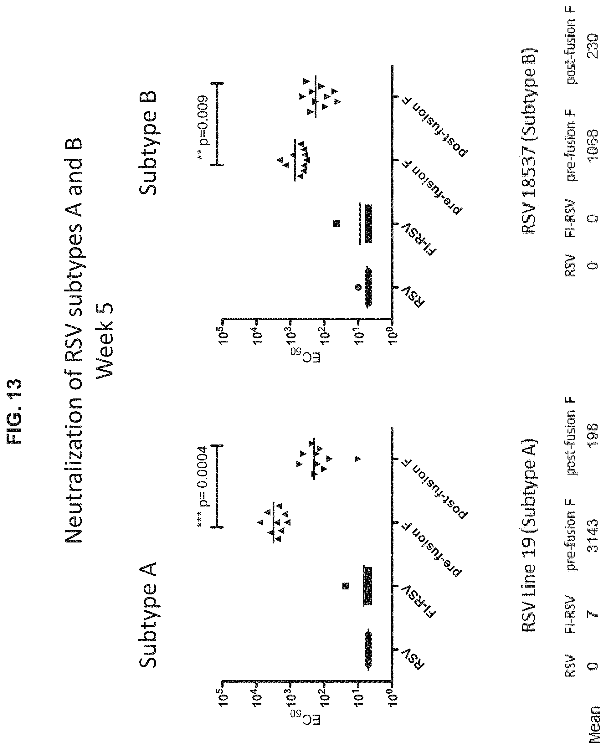
D00019

D00020

S00001
XML
uspto.report is an independent third-party trademark research tool that is not affiliated, endorsed, or sponsored by the United States Patent and Trademark Office (USPTO) or any other governmental organization. The information provided by uspto.report is based on publicly available data at the time of writing and is intended for informational purposes only.
While we strive to provide accurate and up-to-date information, we do not guarantee the accuracy, completeness, reliability, or suitability of the information displayed on this site. The use of this site is at your own risk. Any reliance you place on such information is therefore strictly at your own risk.
All official trademark data, including owner information, should be verified by visiting the official USPTO website at www.uspto.gov. This site is not intended to replace professional legal advice and should not be used as a substitute for consulting with a legal professional who is knowledgeable about trademark law.