IsPETase variants
Kim December 1, 2
U.S. patent number 10,851,355 [Application Number 16/443,966] was granted by the patent office on 2020-12-01 for ispetase variants. This patent grant is currently assigned to KYUNGPOOK NATIONAL UNIVERSITY INDUSTRY-ACADEMIC COOPERATION FOUNDATION. The grantee listed for this patent is KYUNGPOOK NATIONAL UNIVERSITY INDUSTRY-ACADEMIC COOPERATION FOUNDATION. Invention is credited to Kyung Jin Kim.


View All Diagrams
| United States Patent | 10,851,355 |
| Kim | December 1, 2020 |
IsPETase variants
Abstract
Disclosed are a method for preparing crystals of IsPETase protein, a method for screening an IsPETase protein activity regulator and IsPETase variants using a conformation of the protein crystal, a method for screening, IsPETase variants with increased PETase activity, and a method for decomposing PET using the variants. According to exemplary embodiments of the present invention, it is possible to determine a method for effectively preparing a crystal of the IsPETase protein and to obtain the resulting crystal thereof. Further, according to exemplary embodiments of the present invention, it is possible to identify a tertiary structure of the IsPETase from the crystal thereof and to prepare the variant with an increased PETase activity based on this structure. The IsPETase variant may be used effectively in the PET decomposition field.
| Inventors: | Kim; Kyung Jin (Daegu, KR) | ||||||||||
|---|---|---|---|---|---|---|---|---|---|---|---|
| Applicant: |
|
||||||||||
| Assignee: | KYUNGPOOK NATIONAL UNIVERSITY
INDUSTRY-ACADEMIC COOPERATION FOUNDATION (N/A) |
||||||||||
| Family ID: | 1000005214074 | ||||||||||
| Appl. No.: | 16/443,966 | ||||||||||
| Filed: | June 18, 2019 |
Prior Publication Data
| Document Identifier | Publication Date | |
|---|---|---|
| US 20200048621 A1 | Feb 13, 2020 | |
Foreign Application Priority Data
| Aug 8, 2018 [KR] | 10-2018-0092540 | |||
| Feb 14, 2019 [KR] | 10-2019-0017256 | |||
| Current U.S. Class: | 1/1 |
| Current CPC Class: | C12N 9/18 (20130101); C12N 1/20 (20130101); C12N 15/52 (20130101) |
| Current International Class: | C12N 9/18 (20060101); C12N 1/20 (20060101); C12N 15/52 (20060101) |
| 2015025861 | Feb 2015 | WO | |||
Other References
|
Bornscheuer et al. Curr Protoc Protein Sci. Nov. 2011;Chapter 26:Unit26.7. (Year: 2011). cited by examiner . Li et al. Biotechnol Bioeng. Jul. 2014;111(7):1273-87. Epub May 6, 2014. (Year: 2014). cited by examiner . Liu et al. Chembiochem. Jul. 16, 2018;19(14):1471-1475. Epub May 28, 2018. (Year: 2018). cited by examiner . Accession A0A0K8P6T7. Nov. 11, 2015 (Year: 2015). cited by examiner . Chica et al. Curr Opin Biotechnol. Aug. 2005;16(4):378-84. (Year: 2005). cited by examiner . Singh et al. Curr Protein Pept Sci. 2017, 18, 1-11 (Year: 2017). cited by examiner . Seongjoon Joo et al. "Structural Insight Into Molecular Mechanism of Poly(Ethylene Terephthalate) Degradation" Nature Communications., 9:382 (2018). cited by applicant . Shosuke Yoshida et al. "A Bacterium That Degrades and Assimilates Poly(Ethylene Terephthalate)" Research Reports., vol. 351 ISSUE6278. cited by applicant . RCSB Protein data bank--5YNS. cited by applicant . Notice of Allowance from Korean Patent Office in Application No. KR 10-2019-0017256 dated Jan. 21, 2020. 5 pages. cited by applicant. |
Primary Examiner: Fronda; Christian L
Attorney, Agent or Firm: Dilworth IP, LLC
Claims
I claim:
1. An IsPETase variant consisting of the amino acid sequence represented by SEQ ID NO: 1, wherein the IsPETase variant includes the amino acid substitution at position 280 of SEQ ID NO: 1 with alanine.
2. The IsPETase variant of claim 1, wherein the IsPETase variant further includes any one of the following amino acid substitutions: (a) substitution of the amino acid at position 121 of SEQ ID NO: 1 with aspartic acid, (b) substitution of the amino acid at position 186 of SEQ ID NO: 1 with histidine, phenylalanine, isoleucine, leucine, or valine, and (c) substitution of the amino acid at position 121 of SEQ ID NO: 1 with aspartic acid; and substitution of the amino acid at position 186 of SEQ ID NO: 1 with histidine, phenylalanine, isoleucine, leucine, and valine.
3. The IsPETase variant of claim 1, wherein the IsPETase variant further includes the amino acid substitution at position 121 of SEQ ID NO: 1 with aspartic acid and the amino acid substitution at position 186 of SEQ ID NO: 1 with histidine.
4. The IsPETase variant of claim 1, wherein the IsPETase variant further includes any one of the following amino acid substitutions: (a) substitution of the amino acid at position 121 of SEQ ID NO: 1 with glutamic acid, (b) substitution of the amino acid at position 186 of SEQ ID NO: 1 with histidine, phenylalanine, isoleucine, leucine, or valine, and (c) substitution of the amino acid at position 121 of SEQ ID NO: 1 with glutamic acid; and substitution of the amino acid at position 186 of SEQ ID NO: 1 with histidine, phenylalanine, isoleucine, leucine, or valine.
5. The IsPETase variant of claim 1, wherein the IsPETase variant further includes the amino acid substitution at position 121 of SEQ ID NO: 1 with glutamic acid and the amino acid substitution at position 186 of SEQ ID NO: 1 with histidine.
6. The IsPETase variant of claim 1, wherein the IsPETase variant has a higher PETase activity than a PETase activity of an IsPETase wild-type.
7. A method for decomposing poly(ethylene terephthalate)(PET)comprising treating PET with the IsPETase variant of claim 1.
Description
CROSS-REFERENCE TO RELATED APPLICATIONS
This application claims priority to and the benefit of Korean Patent Application No. 10-2018-0092540 filed in the Korean Intellectual Property Office on Aug. 8, 2018 and 10-2019-0017256 filed in the Korean Intellectual Property Office on Feb. 14, 2019, the entire contents of which are incorporated herein by reference.
TECHNICAL FIELD
The present invention relates to a method for preparing crystals of IsPETase protein, a method for screening an IsPETase protein activity regulator using a conformation of the protein crystal, a method for screening IsPETase variants, IsPETase variants with increased PETase activity, and a method for decomposing PET using the variants.
BACKGROUND ART
Plastics are essential materials in life because of their desirable properties such as lightness, durability, low cost, easy formability in various forms and non-degradability. However, due to the non-degradability considered to be a major advantage in using plastics, waste plastics have been accumulated in landfills and oceans and are considered as a major cause of environmental problems. The production of plastics is steadily increasing, thereby producing around 320 million tons worldwide in 2015. Since most plastics are robust against being biodegradable and it takes a long time for the plastics to decompose, an accumulated amount of plastic wastes is expected to reach 33 billion tons by 2050. Thus, much effort is being made to reduce plastic wastes. Various chemical decomposition methods such as glycolysis, methanolysis, hydrolysis, aminolysis and ammonolysis have been developed to remove plastic wastes and recycle plastic-based materials. However, these methods generally require high temperature conditions and often produce additional environmental pollutants. Therefore, when biocatalytic decomposition is applied in an environmentally friendly manner, this may be an alternative method. Microorganisms may be colonized on the material surface and decompose the plastic materials through enzymatic hydrolysis of ester bonds. The biodegradability of plastics depends on their chemical and physical properties.
Poly(ethylene terephthalate) (PET) is a widely used polyester and is resistant to biodegradation. According to a report by the US National Park Service, it takes about 450 years to decompose a PET bottle. PET is polymerized via the ester bond between terephthalate (TPA) and ethylene glycol (EG). Various bacterial hydrolases, such as cutinase, lipase, carboxylesterase, and esterase emerge to decompose PET, although decomposition degrees thereof are different. Among the PET-degrading enzymes identified so far, Thermobifida fusca DSM43793-derived TfH and TfH BTA-2, Thermobifida fusca KW3-derived TfCut1 and TfCut2, plant compost metagenome-derived LC cutinase, Saccharomonospora viridis AHK190-derived cutinase, Thermomyces insolens-derived HiC, and Candida Antarctica-derived lipase B emerge to have a relatively high decomposition ability. However, the decomposition activity thereof for industrial applications is still too low.
Several strategies have been adopted to increase the enzymatic activity. Using site-directed mutagenesis of the active site, the cutinase exhibits higher hydrolysis activity. Further, introduction of Ca.sup.2+ or Mg.sup.2+ ions into esterase or addition of disulfide bonds to esterase improves the thermal stability of the enzyme, which leads to improvement of PET decomposition property. Recently, dual enzyme system consisting of T. fusca KW3-derived TfCut2 and LC cutinase; or a dual enzyme system consisting of C. antarctica-derived lipase and Humicola insolens-derived cutinase has been found to exhibit a synergistic effect. However, despite these attempts, the PET decomposition activities thereof are still low.
Recently, Ideonella sakaiensis has been isolated as a new bacterial species that can use PET as a carbon source. I. sakaiensis PETase (IsPETase) can decompose PET at a suitable temperature (30.degree. C.) and has a relatively higher PET decomposition activity compared to other PET decomposition enzymes such as cutinase and lipase (Yoshida, S. et al. Science 351, 1196-1199, 2016). Further, because the IsPETase exhibits a higher specificity to PET, the excellent ability of IsPETase to perform the PET decomposition has attracted much attention. However, detailed enzyme mechanism thereof is not clarified, and further research thereof is difficult.
Based on this background, the present inventors have tried to reveal a crystal structure and structural characteristics of IsPETase. Thus, we have identified a method for the efficient production of protein crystals of IsPETase, and the crystals were obtained. Further, we have identified a tertiary structure of IsPETase from the crystal. Based on this structure, we have confirmed variants thereof with increased PETase activity. In this manner, we have completed the present invention.
SUMMARY OF THE INVENTION
The present invention has been made in an effort to provide an IsPETase variant having one or more amino acid substitutions in an amino acid sequence represented by SEQ ID NO: 1 in which the IsPETase variant includes the amino acid substitution of R280A.
Another purpose of the present invention is to provide polynucleotides encoding the IsPETase variant.
Another purpose of the present invention is to provide a recombinant vector including a polynucleotide encoding the IsPETase variant.
Another purpose of the present invention is to provide microorganisms transformed to include the IsPETase variant and the protein.
Another purpose of the present invention is to provide a method for decomposing poly(ethylene terephthalate) (PET) involving treating the IsPETase variant.
Another purpose of the present invention is to provide a method for preparing crystals of IsPETase protein, the method including mixing i) a reservoir solution containing polyethylene glycol (PEG) 10000, bis-tris and ammonium acetate, and ii) a protein solution containing IsPETase protein to form a mixed solution; and crystallizing the IsPETase protein via a vapor diffusion method of the mixed solution to form an IsPETase protein crystal.
Another purpose of the present invention is to provide a method for screening an IsPETase protein activity regulator, the method including (a) preparing or selecting IsPETase protein activity-regulating candidate peptides or IsPETase protein-binding candidate compounds using a conformation of the IsPETase protein; and (b) determining whether the candidate peptides or compounds selected or prepared in the step (a) regulate the activity of the IsPETase protein.
Another purpose of the present invention is to provide a method for screening IsPETase variants, the method including determining a substrate-binding site from a conformation of an IsPETase protein.
An exemplary embodiment of the present invention provides an IsPETase variant having one or more amino acid substitutions in an amino acid sequence represented by SEQ ID NO: 1 in which the IsPETase variant includes the amino acid substitution of R280A.
As used herein, a term "IsPETase" is a type of esterase that hydrolyzes ester-bonds using Ideonella sakaiensis-derived PET-degrading enzyme among PETases as an enzyme that decomposes the PET. The IsPETase decomposes the PET into monomers such as bis(2-hydroxyethyl) terephthalate (BHET), mono(2-hydroxyethyl) terephthalate (MHET), and terephthalic acid (TPA) (See FIG. 3A-3D).
As used herein, a term "IsPETase variant" includes peptides with one or more differences in an amino acid sequence compared to the IsPETase wild-type; that is, peptides resulting from modification of the IsPETase wild-type sequence.
Specifically, the IsPETase variant may be prepared by changing one or more amino acids in the IsPETase wild-type via at least one of a substitution, addition, deletion, modification, and combinations thereof.
As used herein, the IsPETase variant may have increased activity and thermal stability over the IsPETase wild-type. Specifically, the activity may be an activity measured based on BHET as a substrate or an activity measured based on a PET film as a substrate. However, the present invention is not limited thereto. The thermal stability may be measured by measuring a melting temperature (Tm) of IsPETase or may be measured based on the PET film as a substrate at high temperature (over 40.degree. C.). The present invention is not limited thereto.
Specifically, the IsPETase variant may be, but is not limited to, an IsPETase variant that includes the amino acid substitution of R280A in the amino acid sequence represented by SEQ ID NO: 1. More specifically, the IsPETase variant may be an IsPETase variant that additionally includes one or more amino acid substitutions selected from the group consisting of amino acid substitutions of S121D and D186H in addition to the amino acid substitution of R280A. Alternatively, the IsPETase variant may be an IsPETase variant that additionally includes at least one amino acid substitution selected from the group consisting of amino acid substitutions of S121E and D186H in addition to the amino acid substitution of R280A. The present invention is not limited thereto.
In some embodiments, the IsPETase variant may further includes a amino acid substitution at position 121-th from N-terminal of SEQ ID NO: 1 with aspartic acid or a amino acid substitution at position 186-th from N-terminal of SEQ ID NO: 1, wherein the substitution of the 186th amino acid is replaced by any one of histidine, phenylalanine, isoleucine, leucine, and valine.
In some embodiments, the IsPETase variant may further incldues a amino acid substitution at position 121-th from N-terminal of SEQ ID NO: 1 with aspartic acid and a amino acid substitution at position 186-th from N-terminal of SEQ ID NO: 1, Wherein the substitution of the 186th amino acid is replaced by any one of histidine, phenylalanine, isoleucine, leucine, and valine.
In some embodiments, the IsPETase variant may further incldues a amino acid substitution at position 121-th from N-terminal of SEQ ID NO: 1 with glutamic acid or a amino acid substitution at position 186-th from N-terminal of SEQ ID NO: 1, Wherein the substitution of the 186th amino acid is replaced by any one of histidine, phenylalanine, isoleucine, leucine, and valine.
In some embodiments, the IsPETase variant may further incldues a amino acid substitution at position 121-th from N-terminal of SEQ ID NO: 1 with glutamic acid and a amino acid substitution at position 186-th from N-terminal of SEQ ID NO: 1, Wherein the substitution of the 186th amino acid is replaced by any one of histidine, phenylalanine, isoleucine, leucine, and valine.
More specifically, the IsPETase variant may be composed of an amino acid sequence represented by any one of SEQ ID NOS: 2 to 4. As used herein, the IsPETase variant may include a polypeptide having at least 80%, 90%, 95%, 96%, 97%, 98%, or 99% or more homology with residues 34 to 290 of the amino acid sequence represented by any one of SEQ ID NOS: 2 to 4. The present invention is not limited thereto. Further, when the amino acid sequence has this homology and exhibits an effect comparable to the above protein, it is obvious that supplementary proteins of the amino acid sequence with deletion, modification, substitution or addition of some amino acid sequences are also included within the scope of the present application.
The term "homology" refers to a degree of similarity of two given protein sequences, a degree to which they match a given amino acid sequence or base sequence. This degree may be expressed as a percentage. As used herein, a homologous sequence of the variant having the same or similar activity as a given amino acid sequence or base sequence may be indicated as "% homology". The sequences homology between one moiety to another may be determined by known techniques to the art. For example, a standard software (for example, BLAST 2.0) that computes parameters such as score, identity and similarity may be used to determine the homology. Alternatively, sequences may be compared using Southern hybridization experiments under defined stringent conditions to determine the homology. A suitable hybridization condition as defined may be within a range in the art and may be determined by a method well known to those skilled in the art (e.g., J. Sambrook et al., Molecular Cloning, A Laboratory Manual, 2.sup.nd Edition, Cold Spring Harbor Laboratory press, Cold Spring Harbor, N.Y., 1989; F. M. Ausubel et al., Current Protocols in Molecular Biology, John Wiley & Sons, Inc., New York).
Another exemplary embodiment provides IsPETase variants including at least one amino acid substitution selected from a group consisting of amino acid substitutions of S160A, D206A, H237A, Y87A, M161A, W185A, I208A, W159A, S238A, N241A, S121D, S121E, D186H, D186F, D186I, D186L, D186V and R280A. More specifically, the IsPETase variant including the amino acid substitution of R280A, the IsPETase variant including amino acid substitutions of S121D and D186H, the IsPETase variant including amino acid substitutions of S121E and D186H, the IsPETase variant including amino acid substitutions of R280A, S121D and D186H, and the IsPETase variant including amino acid substitutions of R280A, S121E and D186H were prepared (Examples and 5). The present invention is not limited thereto. Further, the results of measuring the activity of the variants are as follows: the PETase activities of the IsPETase variant including the amino acid substitution of R280A, the IsPETase variant including amino acid substitutions of S121D and D186H, the IsPETase variant including amino acid substitutions of S121E and D186H, the IsPETase variant including amino acid substitutions of R280A, S121D and D186H, and the IsPETase variant including amino acid substitutions of R280A, S121E and D186H may be increased relative to the PETase activity of the IsPETase wild-type and lasts for a long time at a high temperature (40.degree. C.) (FIGS. 5A-5C, 6, 10A-10B and 12A-12B).
Yet another exemplary embodiment provides a polynucleotide encoding the IsPETase variant, or a vector containing the polynucleotide.
The description of the "IsPETase" and the "IsPETase variant" is as described above.
As used herein, a term "polynucleotide" is a polymer of nucleotides in which nucleotide monomers are linked to each other in a long chain by a covalent bond and is a DNA or RNA strand longer than a certain length. More specifically, the polynucleotide refers to a polynucleotide fragment encoding the variant polypeptide.
A base sequence encoding the same amino acid sequence and variants thereof due to genetic code degeneracy may be included within the scope of the present application. A polynucleotide encoding the same amino acid sequence and variants thereof due to genetic code degeneracy may be included within the scope of the present application.
The polynucleotide sequence according to the present invention may be easily modified via substitution, deletion, insertion or combination of one or more bases. Thus, polynucleotides having at least 80%, 90%, 95%, 96%, 97%, 98%, or 99% or more homology with the base sequence represented by SEQ ID NO: 2, 3 or 4 should be interpreted as being included in the scope of the present application.
As used herein, a term "vector" refers to a DNA product containing a base sequence of a polynucleotide encoding the target protein operatively linked to a suitable regulation sequence so that the target protein can be expressed in the appropriate host. The regulation sequence may include a promoter that may initiate transcription, any operator sequence to regulate such transcription, a sequence that encodes a suitable mRNA ribosome-binding site, and a sequence that regulates the termination of transcription and translation. The vector may be transformed into a suitable host cell and then be replicated or functioned regardless of the host genome. The vector may be integrated within the host genome itself.
The vector used herein is not particularly limited as long as it is replicable in the host cell. Any vector known in the art may be used. Examples of commonly used vectors may include plasmids, cosmids, viruses and bacteriophages in their natural or recombinant state. For example, the phage vector or cosmid vector may include pWE15, M13, MBL3, MBL4, IXII, ASHII, APII, t10, t11, Charon4A, and Charon21A. The plasmid vector may include a pBR based vector or the like. Specifically, pDZ, pACYC177, pACYC184, pCL, pECCG117, pUC19, pBR322, pMW118, pCC1BAC vectors and the like may be used.
The term "operatively-linked" means that the gene sequence is functionally linked to a promoter sequence that initiates and mediates transcription of a polynucleotide encoding the target protein of the present invention. The operatively linkage may be made using known gene recombinant techniques in the art. A site-specific DNA cleavage and ligation may be performed using restriction enzyme and ligation enzymes in the art. The present invention is not limited thereto.
In one example, a polynucleotide encoding a target protein in a chromosome may be replaced with a modified polynucleotide via a vector for insertion chromosome into a cell. The insertion of the polynucleotide into the chromosome may be made by any method known in the art, for example, homologous recombination. The present invention is not limited thereto.
Still another exemplary embodiment provides a microorganism transformed with a recombinant vector containing a polynucleotide encoding the IsPETase variant. Specifically, introduction may be accomplished by transformation, but the present invention is not limited thereto.
In this connection, the description of the "IsPETase", "IsPETase variant", "polynucleotide" and "vector" is as described above.
As used herein, a term "transformation" refers to introducing a vector containing a polynucleotide encoding a target protein into a host cell so that the protein encoded by the polynucleotide can be expressed in the host cell. As long as the transformed polynucleotides can be expressed in the host cells, the transformed polynucleotides may be included in the chromosome of the host cell or may be located outside the chromosome. Further, as long as the polynucleotide may be introduced into the host cells and expressed, the polynucleotide may have any form. For example, the polynucleotide may be introduced into a host cell in the form of an expression cassette as a gene structure that contains all the elements needed for self-expression thereof. The expression cassette may include a promoter, a transcription termination signal, a ribosome-binding site, and a translation termination signal operatively linked to the polynucleotide. The expression cassette may be in a form of a self-replicating expression vector. Further, the polynucleotide may be introduced into the host cell in its own form and may be operatively linked to the sequence required for the expression thereof in the host cell. The present invention is not limited thereto.
The host cell used in transformation according to the present invention may be a host cell well known in the art. Specifically, the host cell may include a bacterial, fungal, yeast, plant or animal (e.g., mammal or insect) cell. More specifically, the host cell may be selected from the group consisting of Escherichia coli(E. coli) or Corynebacterium glutamicum strain. The present invention is not limited thereto.
Still yet another exemplary embodiment provides a method for decomposing poly(ethylene terephthalate) (PET) involving treating the IsPETase variant.
In this connection, the description of the "IsPETase" and the "IsPETase variant" is as described above.
In the method for decomposing PET, a composition including the IsPETase variant may be used. Alternatively, a device including the composition may be used. The present invention is not limited thereto. As long as the method for decomposing PET using the IsPETase variant is feasible, any approach may be included within the scope of the present application.
Still yet another exemplary embodiment provides a method for preparing crystals of IsPETase protein, the method including mixing i) a reservoir solution containing polyethylene glycol (PEG) 10000, bis-tris and ammonium acetate, and ii) a protein solution containing IsPETase protein to form a mixed solution; and crystallizing the IsPETase protein via a vapor diffusion method of the mixed solution to form an IsPETase protein crystal.
In this connection, the description of the "IsPETase" is as described above.
There are many different methods for analyzing a protein's crystal structure, and two main methods are NMR spectroscopy and X-ray crystallography. NMR Spectroscopy is based on the principle of predicting a distance between particular atoms in a molecule by analyzing signal changes due to chemical factor which can be detected in the NMR spectrum of a molecule. Data of the chemical shift obtained by the NMR test is analyzed to obtain a set of the distances between labeled atoms in one protein, and a model or a set of models satisfying information about all distances determined by the experiment is produced. Thus, there is a disadvantage of requiring collection and analysis of a large amount of data. Meanwhile, X-ray crystallography is based on the principle of obtaining the result by analyzing x-ray scattered by electron cloud surrounding an atom of the crystal in an x-ray generator. X-ray diffraction patterns from protein crystals are regular because the individual protein molecules are arranged in a regular lattice. Based on this principle, X-ray crystallography is a method of analyzing a protein structure by producing an electron density map of the protein using x-ray scattered and reflected from the protein crystals. However, there is a disadvantage of requiring protein crystallization, because of requiring pure protein samples. According to the present invention, the present inventors have identified a method capable of optimally producing IsPETase protein crystals. We obtained IsPETase protein crystals and performed X-ray analysis thereof to identify the conformation of the IsPETase protein.
As used herein, the phrase "allowing crystallization" or "having crystallinity" means that, in order to prepare a protein in a form suitable for X-ray analysis of its conformation, mutations are introduced into the protein molecule so as to form solid particles having a uniform shape and size from a uniform liquid or to further stabilize the crystal of the protein. The conformation of a protein is very important for the understanding of in vivo actions of the protein. That is, since an understanding of the arrangement and three-dimensional structure of atoms constituting a protein as a polymer makes it possible to analyze the conformation of the IsPETase protein and thus to produce the IsPETase variants with high PETase activity, it is important to identify the conformation of the protein.
The IsPETase protein crystal of the present invention has an amino acid sequence as shown in SEQ ID NO: 1. Specifically, the IsPETase protein crystal belongs to a space group P2.sub.12.sub.12.sub.1 and has unit cell parameters a=43.48 .ANG., b=50.40 .ANG., and c=129.49 .ANG.. The present invention is not limited thereto.
As used herein, the term "space group" refers to a symmetry of a unit cell of a crystal, and a combination of symmetry elements forms a group.
As used herein, the term "unit cell parameter" is also called a lattice constant. The unit cell is the smallest repeatable unit which constitutes the space group and is interpreted in the easiest manner. The unit cell may be defined using microcrystalline axes. The unit cell parameters may be lengths (a, b, c) for these three axes.
The crystallization of the IsPETase protein may be carried out by a variety of known crystallization methods, preferably, by a vapor diffusion method. The present invention is not limited thereto.
The vapor diffusion method may be a sitting drop vapor diffusion method or a hanging-drop vapor diffusion method, and more preferably, the hanging-drop vapor diffusion method, but is not limited thereto.
In the vapor diffusion method, the protein solution is equilibrated with a large aqueous reservoir solution containing a precipitant at a suitable concentration to prepare the crystal of the protein. Usually, a purified protein solution is mixed with an equal amount of a reservoir solution to bring the concentration of the precipitant to about a half thereof that is required for the crystallization. Then, the mixture solution is hung under a cover slip sealing a top of the reservoir or is attached to a top of the container. Then, the sealed container is left for 1 day to 1 year, usually for 2 to 6 weeks, so that the crystal of the protein grows larger. In the vapor diffusion method, when a micro-drop of mother liquor and a much larger reservoir solution exist separately in a closed space, transport of either water or other volatile agents occurs between them, leading to a supersaturated state in the solution condition of protein, and in such a thermodynamically metastable state, proteins are precipitated depending on the change of the precipitant. While the protein precipitation slowly occurs, stable crystals are formed and the precipitant functions to lower the solubility of the concentrated protein solution, and proteins congregate to form crystals in order to reduce an adsorption layer around protein molecules.
The reservoir solution contains a mixture of the precipitant, buffer, salt, and surfactant at different concentrations. Droplets are usually formed by mixing the protein solution with the reservoir solution of various conditions at a ratio of 1:1, and the droplets thus formed are placed on a microbridge, and sealed. At this time, there is a difference in the concentration between the proteins in the droplets and the reservoir solution in the initial stage, and thus the proteins do not exist as crystals. They are equilibrated while sealed, and crystals are formed under the specific conditions by the above described principle. As used herein, the term "hanging-drop vapor diffusion method" is a protein crystallization method, which provides crystals having a size sufficient for protein structural analysis. In the hanging-drop vapor diffusion method, a reagent containing a sample and a pure liquid reagent are placed on the top of the reservoir under vapor equilibration. To achieve equilibrium of the sample having a lower reagent contents than the reservoir, water contained in the sample eventually ends up in the reservoir. Water contained in the sample is removed until the concentration is approximately the same as that in the liquid reagent, and finally, protein crystals reaching the equilibration can be obtained.
In the vapor diffusion method, the type and proper concentration of the salt, the buffer and the surfactant as well as the precipitant in the reservoir solution, pH of the solution, and the experimental temperature vary depending on the type of protein, and in some cases, they become very important factors in crystal formation of proteins.
Thus, the present invention provides a method for preparing crystals of IsPETase protein optimized for crystal formation of the IsPETase protein.
The method for preparing crystals of IsPETase protein may include crystallizing of the IsPETase protein using a reservoir solution containing polyethylene glycol (PEG) 10000, bis-tris and ammonium acetate via a vapor diffusion method.
The polyethylene glycol 10000 may be contained in a reservoir solution as a precipitant and may have an unmodified or modified form. The present invention is not limited thereto. Specifically, the polyethylene glycol 10000 may be contained at a concentration of 10 to 20% (v/v), more specifically, at a concentration of 17%, but the present invention is not limited thereto.
The bis-tris may be contained in a reservoir solution as a buffer and may have an unmodified or modified form. The present invention is not limited thereto. Specifically, the bis-tris may be contained at a concentration of 0.01 to 1 M, more specifically at a concentration of 0.1 M. The present invention is not limited thereto. Further, the pH of the buffer may be 4.0 to 6.0, more specifically 5.0. The present invention is not limited thereto.
The ammonium acetate may be contained in a reservoir solution as a salt and may have an unmodified form or a modified form. The present invention is not limited thereto. Specifically, the ammonium acetate may be contained at a concentration of 0.01 to 1 M, more specifically at a concentration of 0.1 M, but the present invention is not limited thereto.
The protein solution containing the IsPETase protein may be added and mixed to the reservoir solution having the same composition as defined above. Then, the mixture solution may be equilibrated to prepare the crystal of the IsPETase protein.
In this connection, the protein solution containing the IsPETase protein may contain purified IsPETase protein. Specifically, the purified IsPETase protein may be contained in the protein solution at a concentration of 20 to 40 mg/ml, more specifically at a concentration of 28 mg/ml. The present invention is not limited thereto.
One embodiment of the present invention may include purifying the IsPETase protein, mixing the protein solution containing the purified IsPETase protein at a concentration of 28 mg/ml with 17% (v/v) polyethylene glycol 10000, 0.1 M bis-tris having pH 5.0 and 0.1 M ammonium acetate, and then performing the hanging-drop vapor diffusion method, thereby to prepare the crystal of the IsPETase protein (See Example 2-1).
Still yet another exemplary embodiment provides a method for screening an IsPETase protein activity regulator, the method including (a) producing or screening IsPETase protein activity-regulating candidate peptides or IsPETase protein-binding candidate compounds using a conformation of the IsPETase protein; and (b) determining whether the candidate peptides or compounds selected or prepared in the step (a) regulate the activity of the IsPETase protein.
In this connection, the description of the "IsPETase" is as described above.
Information on various protein sites including substrate-binding sites may be determined based on the conformation of the IsPETase protein as identified in accordance with the present invention.
The step (a) of the screening method may include identifying the conformation of the IsPETase protein, and production or screening of the IsPETase protein activity-regulating candidate peptides or the IsPETase protein-binding candidate compounds using the conformation.
In one embodiment of the present invention, the IsPETase protein crystals are obtained, and, after the X-ray analysis of the crystals, the structure and substrate-binding site of the IsPETase protein were identified.
Specifically, in one embodiment of the present invention, it was confirmed that the IsPETase crystal structure belongs to the .alpha./.beta. hydrolase superfamily, that a central twisted .beta.-sheet is composed of nine mixed .beta.-strands (.beta.1 to .beta.9) and is surrounded by seven .alpha.-helix strands (.alpha.1 to .alpha.7), and that the IsPETase contains conserved serine hydrolase Gly-x1-Ser-x2-Gly motif (Gly158-Trp159-Ser160-Met161-Gly162) located in the active site (See Example 2-3, and FIGS. 1A & 1B). Further, it was confirmed that the IsPETase does not form a hydrogen-bond between Pro181 residues in in a 6-th strand (.beta.6-sheet) in the .beta.-sheet and adjacent amino acids and does not form a hydrogen-bond between Asp186 residues and adjacent .alpha.-helix Ser121 residues (See Example 5-1, FIG. 7A-7C and FIG. 8A-8D).
It was confirmed that the substrate-binding site forms a long and shallow L-shaped gap on a flat surface with a value of about 25 .ANG. and 29 .ANG., and that the substrate-binding site is composed of two subsites (See Example 2-4, and FIG. 3A-3D).
Therefore, we may prepare or select substances or binding-substances that regulate the activity of the IsPETase protein based on the active site and structural information, or sequence information, etc. of the IsPETase protein as defined above.
The step (b) of the screening method includes a step of determining whether the IsPETase protein regulation candidate peptide or compound as prepared or selected in the step (a) regulates the activity of the IsPETase protein. When the candidate substance inhibits the activity of the IsPETase protein, the substance may be considered as an IsPETase protein inhibitor. When the candidate substance increases the activity of the IsPETase protein, this substance may be considered as an IsPETase protein activity enhancer.
Still yet another exemplary embodiment provides a method for screening an IsPETase variant, the method including determining a substrate-binding site from the conformation of the IsPETase protein.
In this connection, the description of the "IsPETase" and "IsPETase variant" is as described above.
According to exemplary embodiments of the present invention, it is possible to determine a method for effectively producing the crystal of the IsPETase protein and to obtain the resulting crystal thereof. Further, according to exemplary embodiments of the present invention, it is possible to identify the tertiary structure of the IsPETase from the crystal thereof and to prepare the variant with an increased PETase activity based on this structure. The IsPETase variant may be used effectively in the PET decomposition field.
BRIEF DESCRIPTION OF THE DRAWINGS
FIG. 1A shows an alignment of the amino acid sequence of the PET-decomposing enzyme. Amino acid sequences of two types I, two types IIa, and two types IIb, that is, six different PET-decomposing enzymes are compared. Secondary structural elements are indicated based on the IsPETase structure and are indicated using arrows (.beta.-sheet) and helix (.alpha.-helix). Gly-x1-Ser-x2-Gly motif and extended loop are highlighted using boxes. Residues related to enzyme catalysis and configuration of a sub-site I and a sub-site II are shown in triangles, respectively. Disulfide-bonds found in all six enzymes are indicated by an line and `disulfide-bond 1`. Additional disulfide-bonds found only in the IsPETase are indicated by the line and `disulfide-bond 2`. Is, Ad, Pp, Oa, Tf, and Sv respectively refer to Ideonella sakaiensis, Acidovorax delafieldii, Pseudomonas pseudoalcaligenes, Oleispira Antarctica, Thermobifida fusca, and Saccharomonospora viridis based PET-decomposing enzymes. FIG. 1B shows the IsPETase structure. The monomer structure is represented via a ribbon diagram. The three residues of Ser160, Asp206 and His237 forming the catalyst triplet are denoted as sticks and the two disulfide-bonds are denoted as sticks. The 2-HE(MHET).sub.4 molecule simulated at the active site is represented by an stick. The drawing on the right results from a 90 degrees rotation of the drawing on the left in a horizontal direction.
FIG. 2A shows the electron density map of the IsPETase, which is produced through WinCoot. The 2F.sub.0-F.sub.c map outlined at 1.5.sigma. is indicated using a mesh. The no crystal symmetry suggests the monomer structure of the IsPETase. FIG. 2B shows the IsPETase structure at a high resolution of 1.0 .ANG..
FIG. 3A shows the docking model of the reaction intermediate of 2-HE(MHET).sub.4 and the catalyst triplet in the IsPETase. Three residues of Ser160, Asp206 and His237 forming the catalyst triplet are represented by sticks. The distance of interaction between the oxygen anion hole and the catalyst triplet is also shown. FIG. 3B shows the substrate-binding site of the IsPETase. The IsPETase structure is represented using an electrostatic potential surface model. The 2-HE(MHET).sub.4 docking model is represented by an stick. The cleavage site is highlighted with a box. The sub-sites I, IIa, IIb, and IIc of the substrate-binding site are represented by dotted circles. Arg280 residue located at the end of the sub-site IIc is shown. FIG. 3C shows a side view of the substrate binding mode of the IsPETase in FIG. 3B. FIG. 3D shows a side view of the substrate binding mode of the IsPETase in FIG. 3B. FIG. 3E shows the residue associated with the IsPETase active site. The IsPETase is represented by a cartoon diagram. The residue associated with the bond of 2-HE(MHET).sub.4 is represented by the line model. The ester-bonds that are cleaved by the enzyme are indicated by star marks. The 2-HE(MHET).sub.4 docking model is represented by an stick. The hydrogen-bond between the residue and the substrate is indicated by a line.
FIG. 4 shows chemical structures of PET-related molecules.
FIG. 5A shows hydrolysis activity of the IsPETase and its variants when using BHET as a substrate. The PETase activities of the IsPETase and its variants are measured with a concentration of 200 mg/L of BHET and enzyme concentration of 50 nM. The amount of MHET as produced is monitored by HPLC analysis. The PETase activity of the IsPETase variant is compared with that of the IsPETase wild-type. FIG. 5B shows PETase activity of the IsPETase protein when using a PET film as a substrate. The decomposition activity of the PET film by the IsPETase protein is measured with an enzyme concentration of 200 nM. The amounts of thus produced MHET and TPA are monitored by HPLC analysis. The PETase activity of the IsPETase variant is compared with that of the IsPETase wild-type. The IsPETase.sup.R280A variant, which exhibits the increased activity, is highlighted as a star. FIG. 5C shows the electrostatic potential surface of the IsPETase.sup.R280A structure. 2-HE(MHET).sub.4 molecules are labelled. Arg280 residue in IsPETas.sup.W/T and Arg280Ala(R280A) residue in IsPETase.sup.R280A are indicated by dotted circles. The error bars represent the standard deviation values obtained in duplicate experiments.
FIG. 6 shows the PETase activity of the IsPETase.sup.S121E/D186H/R280A variant. The hydrolysis activities of the IsPETase and its variants are measured when using the PET film as a substrate. The PETase activity of the IsPETase.sup.S121E/D186H/R280A variant is measured at an enzyme concentration of 200 nM. The amount of MHET as produced is monitored by HPLC analysis. The PETase activities at 24 hours and 72 hours of IsPETase.sup.S121E/D186H/R280A variants are compared with those of IsPETase.sup.W/T at 30.degree. C. and 40.degree. C.
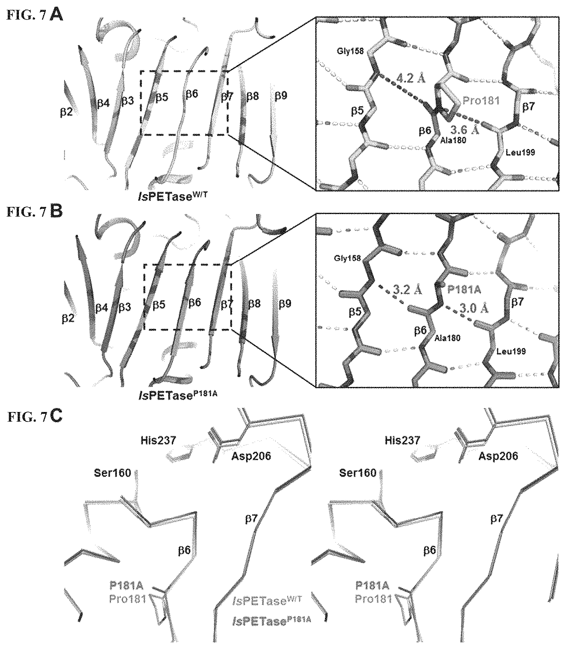
FIG. 7A shows that a central twisted .beta.-sheet of IsPETase.sup.W/T is composed of nine mixed .beta.-strands (.beta.1 to .beta.9). The hydrogen-bonds formed by nitrogen and oxygen respectively are indicated by dotted lines. FIG. 7B shows that the central twisted .beta.-sheet of IsPETase.sup.P181A variant is composed of nine mixed .beta.-strands (.beta.1 to .beta.9). The hydrogen-bonds formed by nitrogen and oxygen respectively are indicated by dotted lines. In FIG. 7C, a structure of the IsPETase.sup.W/T or the IsPETase.sup.P181A are indicated by a line.
FIG. 8A shows the .beta.6-.beta.7 connection loop (Asp186-Phe191) and adjacent .alpha.-helix Ser121 residue of the IsPETase.sup.W/T. FIG. 8B shows the .beta.6-.beta.7 connection loop (His156-Trp161) of Thermobifida fusca DSM43793-derived TfCut2 and adjacent .alpha.-helix Asp156 residue thereof. The hydrogen-bonds formed by nitrogen and oxygen respectively are indicated by dotted lines. FIG. 8C shows D186H of the .beta.6-.beta.7 connection loop (Asp186-Phe191) of the IsPETase.sup.S121D/D186H variant and adjacent .alpha.-helix S121D residue thereof. FIG. 8D shows D186H of the .beta.6-.beta.7 connection loop (Asp186-Phe191) of the IsPETase.sup.S121E/D186H variant and adjacent .alpha.-helix S121E and Asn172 residues thereof. The hydrogen-bonds formed by nitrogen and oxygen are indicated by dotted lines.
FIG. 9A shows the melting temperature of the IsPETase using a protein thermal shift dye. FIG. 9B shows the melting temperature of the IsPETase variant using a protein thermal shift dye.
FIG. 10A shows the hydrolytic activity of IsPETase when using the PET film as a substrate. The PETase activity of the IsPETase is measured at an enzyme concentration of 200 nM. The amount of MHET as produced is monitored by HPLC analysis. FIG. 10B shows the hydrolytic activity of the IsPETase variant when using the PET film as a substrate. The PETase activity of the IsPETase variant is measured at an enzyme concentration of 200 nM. The amount of MHET as produced is monitored by HPLC analysis. PETase activites at 24 and 72 hours of the IsPETase variant are compared with those of the IsPETase.sup.W/T at 30.degree. C. and 40.degree. C.
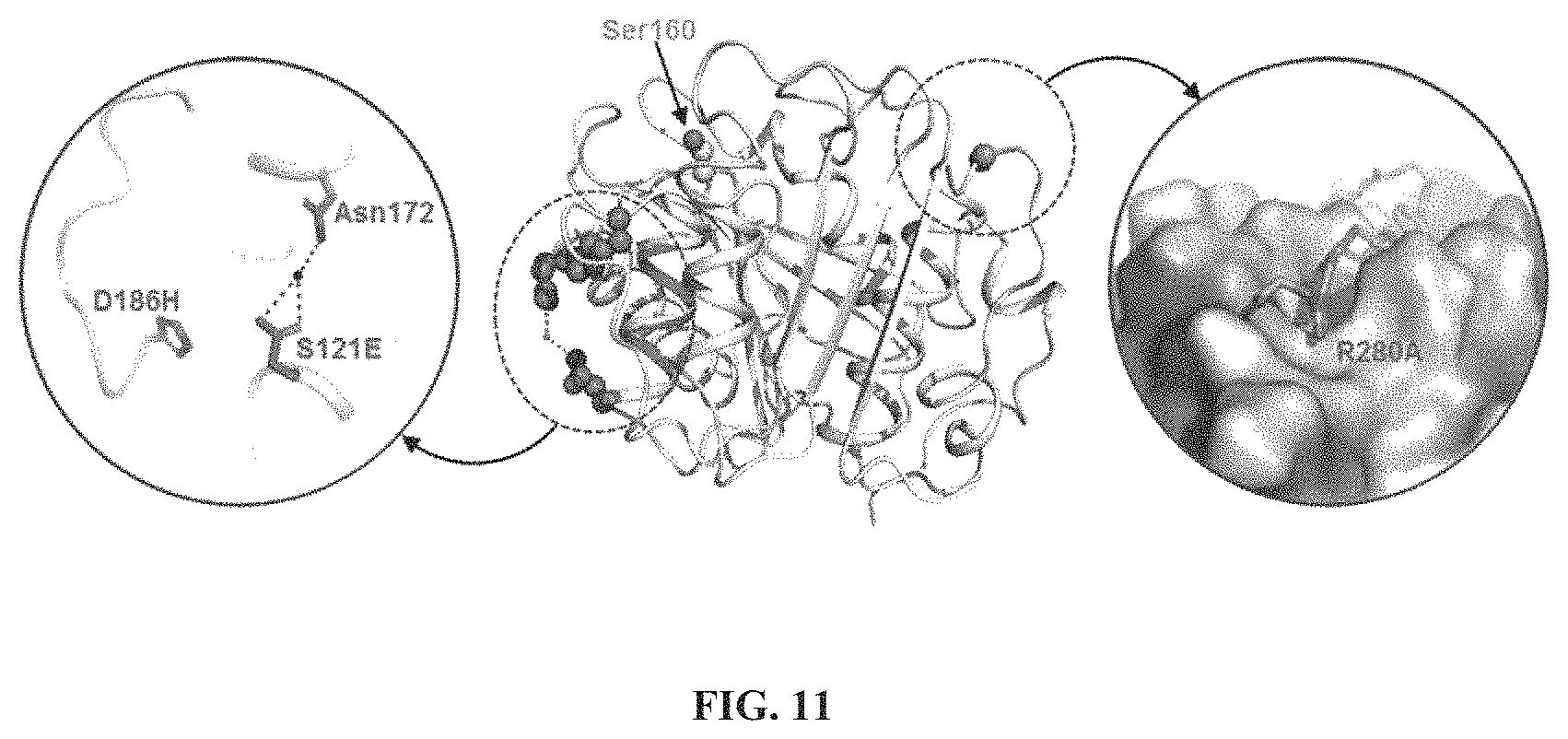
FIG. 11 shows the surface of the crystal structure of the IsPETase.sup.S121E/D186H/R280A variant, in which D186H, S121E, and Asn172 residues of the .beta.6-.beta.7 connection loop, and the R280A residue of the sub-site IIc are shown. The hydrogen-bond is indicated by dotted lines.
FIG. 12A shows the PETase activity of IsPETase.sup.S121E/D186H/R280A variant over time compared with that of IsPETase.sup.W/T at 30.degree. and 40.degree. C. The hydrolysis activities of the IsPETase and IsPETase.sup.S121E/D186H/R280A variant are measured when using the PET film as a substrate. The PETase activities of IsPETase and IsPETase.sup.S121E/D186H/R280A variant are measured at an enzyme concentration of 200 nM. The amount of MHET as produced is monitored by HPLC analysis. In FIG. 12B, the PETase activity of IsPETase.sup.S121E/D186H/R280A variant as heat-inactivated at 50.degree. C. for 1 to 60 minutes is compared with the PETase activity of the IsPETase.sup.W/T. The hydrolysis activities of the IsPETase and IsPETase.sup.S121E/D186H/R280A variant are measured when using the PET film as a substrate. The PETase activities of IsPETase and IsPETase.sup.S121E/D186H/R280A variant are measured at an enzyme concentration of 200 nM. The amount of MHET as produced is monitored by HPLC analysis.
DETAILED DESCRIPTION
Hereinafter, the present invention will be described in more detail by the following Examples. However, the Examples are intended to illustrate the present invention only, and the scope of the present invention is not limited thereto.
Example 1. Production of IsPETase Protein
For IsPETase expression in Escherichia coli cells, the IsPETase gene was amplified by polymerase chain reaction (PCR) using a codon-optimized synthetic gene as a template. The nucleotide sequence corresponding to the signal peptide was removed from the synthetic DNA. Then, The PCR product was subcloned into vector pET 15b. Using the thus produced expression vector pET 15a: IsPETase, E. coli strain Resettagami-B was transformed. The E. coli strain Resettagami-B was grown in 1 L of lysogeny broth medium containing ampicillin at 37.degree. C. Protein expression was induced via addition of 1 mM isopropyl .beta.-D-1-thiogalactopyranoside, and then, the culture medium was further cultured at 18.degree. C. for 16 hours. The medium centrifuged at 4000.times.g for 10 minutes at 20.degree. C. The cell pellet was resuspended in a buffer A (50 mM Tris-HCl, pH 8.0) and then was crushed with ultrasound treatment. Resulting cell debris was removed by centrifugation at 13500.times.g for 25 minutes. The supernatant was applied to a Ni-NTA agarose column (Qiagen). After washing with a buffer A containing 30 mM imidazole, the binding protein was eluted with 300 mM imidazole in the buffer A. Finally, traces of contaminated substances were removed by size-exclusion chromatography using a Superdex 200 prep-grade column (320 ml, GE Healthcare) equilibrated with the buffer A. All purification steps were performed at 4.degree. C. Protein purity was determined by sodium dodecyl sulfate polyacrylamide gel electrophoresis. The purified protein was concentrated to 28 mg/ml in 50 mM Na.sub.2HPO.sub.4--HCl (pH 7.0) and 100 mM NaCl.
Example 2. IsPETase Protein Crystal
Example 2-1. Production of IsPETase Protein Crystal
Crystallization of the purified IsPETase protein was performed using a crystal screening kit: Index and PEG/Ion (Hampton Research) and Wizard I and II (Rigaku) based on a hanging-drop vapor diffusion method at 20.degree. C. Each experiment was conducted using 1.0 .mu.l of a protein solution and 1.0 .mu.l of a reservoir solution and then equilibrated with 50 .mu.l of a reservoir solution.
The highest quality IsPETase crystals were found when using 0.1M ammonium acetate, 0.1M bis-tris (pH 5.0) and 17% polyethylene glycol 10000. The protein crystal was transferred to a cryoprotectant solution containing 0.1M ammonium acetate, 0.1M bis-tris (pH 5.0), 20% polyethylene glycol 10,000 and 30% (v/v) glycerol. Then, the protein crystal was extracted using a loop larger than the crystal, and was immersed in the liquid nitrogen, and was rapidly frozen therein.
Example 2-2. X-Ray Diffraction Analysis of IsPETase Protein Crystal
Data were collected at 100K via Beamline 6D from the Pohang Accelerator Laboratory (Pohang, Korea) for the analysis of the IsPETase protein crystal. The data were indexed and integrated and scaled using the HKL2000 software suite. Thus, it was confirmed that the IsPETase crystal belongs to a space group P2.sub.12.sub.12.sub.1, and that the IsPETase crystal has unit cell parameters a=43.48 .ANG., b=50.40 .ANG., and c=129.49 .ANG.. For one molecule of IsPETase on an asymmetric unit basis, the Metthews modulus was 2.64 .ANG..sup.3/Da, which corresponds to a solvent content of 53.38%.
Example 2-3. Determination of Structure of IsPETase Protein Crystal
In order to characterize the structure of the IsPETase protein crystal, Thermobifida alba (TaCut, PDB code 3VIS, 50% sequence identity)-derived cutinase structure was employed as a search model. Then, we have identified the structure of the IsPETase via the molecular substitution method using the CCP4 version of MOLREP. Then, a model building was performed using the WinCoot program. Purification was carried out using REFMAC5. The statistical data are shown in Table 1. The refined model of the IsPETase is stored in Protein Data Bank as PDB code 5XJH.
TABLE-US-00001 TABLE 1 IsPETase IsPETase.sup.R280A PDB code 5XJH 5YNS Data collection Wavelength (.ANG.) 0.97934 0.97934 Unit cell (a, b, c; .gamma.) (.ANG.; .degree.) 43.48, 50.40, 43.61, 50.59, 129.49; 90.0 129.58; 90.0 Space group P2.sub.12.sub.12.sub.1 P2.sub.12.sub.12.sub.1 Solvent content (%) 53.38 52.66 Protein chains in AU 1 1 Resolution range (.ANG.) 50.00-1.55 50.00-1.36 Highest resolution shell (.ANG.) 1.58-1.55 1.38-1.36 Unique reflections 42939 62434 Redundancy 6.5 (5.9) 9.3 (5.7) Completeness (%) 99.9 (99.9) 98.7 (96.9) R.sub.merge (%) 6.8 (29.8) 6.7 (30.1) Average I/.sigma. (I) 31.4 (5.2) 40.8 (4.6) B from Wilson plot (.ANG..sup.2) 14.2 10.8 Refinement R (%) 15.9 16.6 R.sub.free (%) 19.2 19.2 Mean B value (.ANG..sup.2)* 16.1 15.0 RMS deviation bond lengths (.ANG.) 0.026 0.025 RMS deviation bond angles (.degree.) 2.286 2.249 Number of amino acid residues 272 264 Number of water molecules 150 294 Ramachandran plot Most favored regions (%) 97.3 97.6 Additional allowed Regions (%) 2.7 2.4 *Mean B value is for both protein atoms and the solvent molecules.
The signal peptide sequence (Met1-Ala33) was removed for production of the protein core domain. Recombinant IsPETase protein has additional amino acid residues at both the N and C terminals (Met13-Met33 and Leu291-Gln312) due to the use of the pET 15b vector.
As a result, the IsPETase crystal structure was determined at 1.5 .ANG. resolution (FIGS. 1A and 1B and FIGS. 2A and 2B). The structure includes the Ser31-Gln292 residue, which may be seen in the electron density map. The refined structure is consistent with X-ray crystallographic statistics in terms of a binding angle, a bind length, and other geometric parameters (See Table 1). The asymmetric unit of the space group P2.sub.12.sub.12.sub.1 includes one molecule of IsPETase. This indicates that the IsPETase exists as a monomer. The IsPETase crystal structure belongs to the .alpha./.beta. hydrolase superfamily. The central twisted .beta.-sheet is composed of nine mixed .beta.-strands (.beta.1 to .beta.9) and is surrounded by seven .alpha.-helix (.alpha.1 to .alpha.7) (See FIG. 1B). As observed in proteins belonging to other .alpha./.beta. hydrolase superfamilies such as lipase and estrase, the IsPETase contains a conserved serine hydrolase Gly-x1-Ser-x2-Gly motif (Gly158-Trp159-Ser160-Met161-Gly162) located in the active site (See FIG. 1A).
Example 2-4. Active Site of IsPETase Protein Crystal
The IsPETase decomposes a PET into monomers such as bis(2-hydroxyethyl) terephthalate (BHET), mono(2-hydroxyethyl) terephthalate (MHET) and terephthalic acid (TPA) (See FIG. 3A-3D). Further, the IsPETase hydrolyzes BHET widely used in PET studies as a monomer that is similar to the core structure of the PET and is commercially available. BHET is decomposed into MHET by the IsPETase without further decomposition reaction. Covalent bond based docking calculation was executed using 2-hydroxyethyl-(monohydroxyethyl terephthalate).sub.4, 2-HE(MHET).sub.4 and 4-MHET molecules mimicking the PET to estimate a binding manner of the enzyme to the substrate (See FIG. 3A-3D).
Tetrahedral intermediate molecular docking from 2-HE (MHET).sub.4 to an IsPETase structure was performed through a hybrid approach of soft and covalent bond based dockings using AutoDock 4.2 and AutoDock Vina. The ligand molecules of IsPETase were prepared with WinCoot and ProDrg. The nonpolar H atoms were merged into both the ligand and target using AutoDock Tools prior to the docking. Soft residues (Tyr87, Trp159, Ser160, Met161, Trp185, Ile208, His237, Ser238, and Asn241) were selected for the production of the pdbqt file of both hard and soft receptors. The bonds to the side-chains of the residues were allowed for rotation. A grid box is centered at x: -3.249, y: 25.239 and z: -29.093. The box size has axis lengths 90.7, 74.7, and 122.7 .ANG., respectively. Prior to the covalent bond based docking, non-covalent bond based docking calculations were performed using AutoDock Vina. Nine output poses occurred with a binding free energy as calculated from the scoring function thereof. We chose the best docking model with the lowest binding energy (-7.1 kcal/mol). A conformation of the model was used as an evaluation criterion for following calculations. Further, an induced conformation of the soft residue in the best model was applied to receptors for the covalent bond based docking. A total of 200 docking poses were evaluated based on the appropriate distance of the oxygen anion hole. The best pose with the binding energy -10.27 kcal/mol (from a semi-empirical free energy force field of AutoDock) was similar to the non-covalent bond based docking result. Finally, the docking pose was minimized using the OPLS3 force field in the Schrodinger suite.
It has been shown that at the active site of the IsPETase protein, three residues Ser160, His237, and Asp206 form a catalytic triplet, and a residue Ser160 functions as a covalent bond nucleophile with respect to the carbonyl carbon atom of a cleavable ester bond, such as a carboxyl esterase (See FIG. 3A). A tetrahedral intermediate oxygen anion was stabilized by an oxygen anion hole composed of nitrogen atoms of Tyr87 and Met160 having respectively distances of 2.90 and 2.83 .ANG.(See FIG. 3A). The substrate-binding site was simulated to form a long and shallow L-shaped gap in a flat surface with values of approximately 25 and 29 .ANG. (See FIG. 3B-3D). A surface of the substrate-binding gap was mainly hydrophobic and a length of the gap was .about.40 .ANG. (See FIG. 3B). Based on the cleavable ester bond of the 2-HE(MHET).sub.4, the substrate-binding site may be divided into two sub-sites, that is, a sub-site I and a sub-site II, onto which one and three MHET moieties are combined respectively (See FIGS. 3B & 3E). For binding of a first MHET moiety in the sub-site I, a benzene ring is located at a valley between two aromatic residues Tyr87 and Trp185 (See FIGS. 3B & 3E). A .pi.-.pi. interaction between the benzene ring of the first MHET moiety and the Trp185 with a distance of .about.3.6 .ANG. appears to be a major factor for ligand stabilization (See FIG. 3E). Further, Met161 and Ile208 are predicted to aid binding of the first MHET moiety by providing hydrophobic surfaces at a bottom and sides of the sub-site I respectively (See FIG. 3E). The sub-site II tends to form a longer and shallow gap than that defined in the sub-site I. The sub-site II accommodates three MHET moieties (second, third and fourth MHET moieties of the 2-HE(MHET).sub.4) (See FIG. 3B-3D). Based on a MHET binding, the sub-site II is further subdivided into three sub-sites IIa, IIb, and IIc (See FIGS. 3B & 3E). The sub-site II is composed of residues including Thr88, Ala89, Trp159, Ile232, Asn233, Ser236, Ser238, Asn241, Asn244, Ser245, Asn246, and Arg280. An interaction between the sub-site II and the 2 MHET moieties appears to be mediated primarily via a hydrophobic interaction. However, a carbonyl oxygen atom of the fourth MHET moiety forms a polar interaction with the Ser236 main chain and Asn246 side chain within the sub-site IIc (See FIGS. 3B & 3E). The residue Arg280 is located at an end portion of the sub-site IIc, and the residue Arg280 is positively charged and has a slightly protruding structure that interferes with extension of the substrate-binding site (See FIGS. 3B, 3C, & 3E).
Example 3. IsPETase Variant
Example 3-1. Production of IsPETase Variant Protein
Site-directed mutagenesis experiments were conducted to determine residues associated with enzyme catalysis and substrate binding. The site-directed mutagenesis experiments were performed using a Quick Change kit (Stratagene). Expression and purification of the IsPETase variant were performed under the same condition as the expression and purification of the IsPETase protein.
Example 3-2. Production of IsPETase Variant Protein Crystal
Crystallization of the IsPETase variant Protein was performed by a method similar to a method of the crystallization of IsPETase protein in the above Example 2-1.
Example 3-3. X-Ray Diffraction Analysis of IsPETase Variant Protein Crystal
Data were collected at 100K via Beamline 7A from the Pohang Accelerator Laboratory (Pohang, Korea). As a result, IsPETase.sup.R280A crystal belongs to the space group P2.sub.12.sub.12.sub.1, and unit cell parameters were similar to those in the IsPETase.sup.W/T crystal.
Example 3-4. Determination of Structure of IsPETase Variant Protein Crystal
A structure of IsPETase.sup.R280A was identified via a molecular substitution method using an IsPETase.sup.W/T structure as a search model. Modeling and purification were performed in the same manner as in the IsPETase.sup.W/T in the Example 2-3. The statistical data are shown in Table 1. The refined model of IsPETase.sup.R280A is stored in the Protein Data Bank as PDB code 5YNs.
Example 4. Activity Analysis
Example 4-1. In Vitro Analysis Using Bis-Hydroxyethyl Terephthalate
For comparison of activities, bis-hydroxyethyl terephthalate (BHET) was used as a substrate for enzyme analysis. A BHET stock solution was prepared by dissolving (2.5 g/l) BHET in dimethyl sulfoxide. The analysis protocol was based on a previously reported article (Yosida, S. et al. Science 351, 1196-1199, 2016). Enzyme assays were performed using 200 mg/l BHET in a buffer solution (80 mM Na.sub.2HPO.sub.4--HCl, 40 mM NaCl) at pH 7.0. The enzyme reaction was initiated by addition of 50 nM enzyme thereto and lasted for 30 minutes at 30.degree. C. Then, after heating at 85.degree. C. for 15 minutes, the reaction was terminated. The reaction mixture was centrifuged at 13,200 r.p.m. for 10 minutes. Finally, the supernatant was subjected to liquid chromatography (LC) analysis.
In vitro assay samples were analyzed by HPLC (1100 Series HPLC, Agilent) equipped with MS LC/MSD VL, Agilent). An Eclipse Plus-C18 column (5 .mu.m, 4.6.times.150 mm, Agilent) was used. All analyzes were performed at a room temperature (25.degree. C.). For a mobile phase, a buffer A (0.1% formic acid in distilled water) and a buffer B (acetonitrile) were used at a flow rate of 0.8 ml/min. The mobile phase was gradually changed from a 95% buffer A to a 30% buffer A in 20 minutes (all % means volume %). Chemical substances (BHET, MHET and TPA) were detected at 260 nm.
Catalytic residues Ser160, Asp206 and His237 were substituted with Ala and then hydrolysis activity was measured using BHET as a substrate. All three variants IsPETase.sup.S160A, IsPETase.sup.D206A and IsPETase.sup.H232A show nearly complete loss of the activity (See FIG. 5A). This indicates that these three residues are involved in catalysis.
Subsequently, Tyr87, Trp185, Met161, and Ile208 residues, which constitute the sub-site I, were substituted with Ala. IsPETase.sup.Y87A and IsPETase.sup.W188A variants exhibited 5% BHET hydrolysis activity compared to the IsPETase.sup.W/T (See FIG. 5A). This result implies that blocking the .pi.-.pi. interaction between these residues and the benzene ring of the first MHET moiety reduces the stability of the first MHET moiety. IsPETase.sup.M181A and IsPETase.sup.I208A variants show 52% and 46% activities, respectively, compared to IsPETase.sup.W/T (See FIG. 5A). This means that this residue is not as important as the Tyr87 and Trp185 residues but contributes to the constitution of the sub-site I.
The residues Trp159, Ser238, and Asn241 that constitute the sub-site II were substituted with Ala. IsPETase.sup.W159A and IsPETase.sup.M241A showed 8% and 18% BHET hydrolysis activities respectively compared to IsPETase.sup.W/T (See FIG. 5A). This means that this residue is important in the constitution of the sub-site II. However, IsPETase.sup.S238A showed an almost similar activity of BHET hydrolysis compared to IsPETase.sup.W/T (See FIG. 5A). This result suggests that substitution of the Ser238 with Ala does not affect the BHET hydrolysis activity of the enzyme.
Example 4-2. In Vitro Analysis Using PET Film
The analysis was carried out with minor modifications (as described below) based on the previously reported article (Yosida, S. et al. Science 351, 1196-1199, 2016). In order to analyze the decomposition rate of the PET by the PETase, a commercially available PET film (UBIGEO, Korea) was used as a substrate for enzyme analysis. The PET film was prepared in a circular shape having a diameter of 6 mm. The PET film was wetted with 300 .mu.l of pH 9.0 glycine-NaOH buffer and 200 nM the enzyme. The reaction mixture was incubated at 30.degree. C. for 18 and 36 hours/24 and 72 hours. After heating at 85.degree. C. for 15 minutes, the reaction was terminated. Thereafter, the sample was centrifuged at 13,200 r.p.m. The supernatant was analyzed via LC. After the enzymatic reaction, the film was washed with 1% SDS and 20% ethanol in distilled water.
The PETase activity of the variant was measured when using a PET film as a substrate. The variant of the catalyst triplet with Ala showed an almost loss of the enzyme activity. The variant involved in the configuration of the substrate-binding site showed decreased PETase activity compared to the IsPETase.sup.W/T (See FIG. 5B). The substitution of Arg280 with Ala was preformed and then the BHET hydrolysis and PETase activity were measured. The IsPETase.sup.R280A showed an almost similar activity to that of IsPETase.sup.W/T (See FIG. 5A). When a PET film is used as a substrate, the IsPETase.sup.R280A was found to increase the PETase activity by 22.4% at 18 h and by 32.4% at 36 h (See FIG. 5B) compared to the IsPETase.sup.W/T (high specificity of IsPETase to PET). In other words, substitution of Arg280 with a small hydrophobic residue enables a more stable binding of the longer substrate, thus resulting in an increase in the PETase activity. More specifically, to investigate whether the substitution of Arg280 with Ala actually changed the conformation of the substrate-binding site (the sub-site IIc) to enable the binding of the longer substrate, the structure of IsPETase.sup.R280A was checked at 1.36 .ANG. resolution. As expected, the structure of IsPETase.sup.R280A exhibited an extended sub-site IIc by providing a hydrophobic and non-protruding gap, compared to the IsPETase.sup.W/T (See FIG. 5C). Substitution of Arg280 at a terminal from a catalyst site with a distance of 22.8 .ANG. with Ala could also be known to increase the enzyme activity. This result could not be obtained without reliable docking calculations to check the inherent binding properties of the IsPETase to the substrate.
The PETase activity was measured by substitution of Ser121 with Glu, Asp186 with His, and Arg280 with Ala. When using the PET film as a substrate, IsPETase.sup.S121E/D186H/R280A has the PETase activity increased by 1.25 times at 30.degree. C. and 2.23 times at 40.degree. C. at 24 hours and by 2.03 times at 30.degree. C. and 3.39 times at 40.degree. C. at 72 hours compared with the IsPETase.sup.W/T (See FIG. 6). The hydrogen bond resulting from the substitution increased the stability of the enzyme and consequently increased the PETase activity. The increased stability of the enzyme may be confirmed via the increase in the melting point (Tm) of the enzyme. From this fact, we could know that the S121E and D186H substitutions in the IsPETase variant individually increased the PETase activity or increased the PETase activity synergistically in combination with other substitutions.
Example 5. Activity Analysis of IsPETase Protein about Thermal Stability
Example 5-1. Thermal Stability Site of IsPETase Protein Crystal
The IsPETase has the superior PET decomposition activity compared to other PET-decomposing enzymes. However, due to its low thermal stability, the IsPETase has the disadvantage that it may be used to decompose the PET only at a moderate temperature (30.degree. C.). Thus, the thermal stability of the IsPETase is predicted to be a critical factor in the efficient decomposition of the PET using the PETase. Thus, structural properties that affect the thermal stability of the IsPETase protein crystal were determined (See FIG. 7A-7C).
As a first structural feature, the IsPETase crystal has an unusual conformation of a .beta.6-sheet. In other words, the Pro181 residue located at the center of the .beta.6-sheet in the IsPETase crystal does not form a hydrogen bond with adjacent amino acids, thus interfering with the formation of continuous .beta.-sheet. In the structure of the IsPETase crystal, a distance between nitrogen of the Pro181 residue and oxygen of the Leu199 residue is 3.6 .ANG., and a distance between nitrogen of Gly158 residue and oxygen of Ala180 residue is 4.2 .ANG.. This suggests that the Pro181 residue interferes with the secondary structure formation of the IsPETase protein (See FIG. 7A).
As a second structural feature, the .beta.6-.beta.7 connecting loop (Asp186-Phe191) in the IsPETase crystal has a higher b-factor value (22.2) than an average b-factor value (16.1) in the entire protein and has a very flexible structure (See FIG. 8A). On the other hand, in Thermobifida fusca DSM43793-derived cutinase2(TfCut2), which has a high thermal stability, the .beta.6-.beta.7 connecting loop (His156-Trp161) has a b-factor value similar to the average b-factor of the entire protein and has a very flexible structure (See FIG. 8B). In this connection, the His156 residue seems to contribute to the structural stability of the connection loop by the His156 residue forming a hydrogen bond with an Asp156 residue in a .alpha.2-helix. Therefore, the substitution of Asp186 and Ser121 residues of the IsPETase crystal with histidine and aspartic acid, respectively, as in the TfCut2 may allow the hydrogen bonds to be formed in the .beta.6-.beta.7 connection loop, resulting in the improved structural stability.
Example 5-2. Production of IsPETase Variant Protein
Site-directed mutagenesis experiments were performed to determine the amino acid residues of the IsPETase associated with thermal stability. The site-directed mutagenesis experiments were performed under the same conditions as in the Example 3-1. Thus, IsPETase.sup.8121D, IsPETase.sup.S121E, IsPETase.sup.D186H, IsPETase.sup.D186F, IsPETase.sup.D186I, IsPETase.sup.D186L, IsPETase.sup.D186V, IsPETase.sup.S121D/D186H, IsPETase.sup.S121E/D186H, IsPETase.sup.P181A/S121E/D186H, IsPETase.sup.P181A/S121E/D186H, IsPETase.sup.S121D/D186H/R280A, and IsPETase.sup.S121E/D186H/R280A were produced. Further, the crystallization of the IsPETase variant protein was performed under the same conditions as in the Example 3-2.
Structures of IsPETase.sup.S121D/D186H, IsPETase.sup.S121E/D186H and IsPETase.sup.S121E/D186H/R280A were identified via a molecular substitution method using the IsPETase.sup.W/T structure as a search model. Modeling and purification were performed under the same conditions as in the IsPETase.sup.W/T in the Example 2-3. Statistical data are shown in Table 2 below. Refined models of the IsPETase.sup.S121D/D186H, IsPETase.sup.S121E/D186H and IsPETase.sup.S121E/D186H/R280A are stored in Protein Data Bank as PDB codes 6IJ3, 6IJ4, 6IJ6, respectively.
TABLE-US-00002 TABLE 2 IsPETase.sup.S121D/D186H IsPETase.sup.S121E/D186H IsPETase.sup.S121E/D186- H/R280A PDB code 6IJ3 6IJ4 6IJ6 Data collection Wavelength (.ANG.) 0.97934 0.97934 0.97934 Unit cell (a, b, c; .gamma.) (.ANG.; .degree.) 115.43, 50.63, 41.49; 116.20, 50.86, 41.49; 114.91, 51.14, 51.16; 90.0, 93.1, 90.0 90.0, 92.7, 90.0 90.0, 109.7, 90.0 Space group C121 C121 C121 Solvent content (%) 47.20 47.80 54.84 Protein chains in AU 1 1 1 Resolution range (.ANG.) 50.00-1.40 50.00-1.85 50.00-1.95 Highest resolution shell (.ANG.) 1.42-1.40 1.88-1.85 1.98-1.95 Unique reflections 46596 19977 19893 Redundancy 3.2 (2.9) 3.3 (2.9) 3.5 (3.1) Completeness (%) 99.0 (98.5) 98.3 (96.2) 97.4 (94.2) R.sub.merge (%) 12.2 (38.1) 10.8 (34.9) 9.7 (19.5) Average I/.sigma. (I) 25.99 (3.64) 25.56 (3.53) 33.04 (9.90) Refinement R (%) 19.4 20.7 15.1 R.sub.free (%) 23.3 24.7 19.3 Mean B value (.ANG..sup.2)* 16.4 28.6 19.6 B from Wilson plot (.ANG..sup.2) 12.3 24.1 19.2 RMS deviation bond lengths (.ANG.) 0.013 0.013 0.013 RMS deviation bond angles (.degree.) 1.889 1.592 1.638 Number of amino acid residues 261 262 261 Number of water molecules 225 102 188 *Mean B value is for both protein atoms and the solvent molecules.
Example 5-3. Analysis of Thermal Stability of IsPETase Variant Protein
To compare the thermal stabilities of the IsPETase variants, the structures of the IsPETase variants were analyzed and the melting temperatures (Tm) thereof were measured.
The melting temperatures were measured based on melting curves with a StepOnePlus Real-Time PCR (Thermo Fisher Scientific) using a protein thermal shift dye (Applied Biosystems). Specifically, 5 ug of the IsPETase was mixed with 20 .mu.l of a protein thermal shift dye, and then, signal changes reflecting protein denaturation were monitored in a temperature increasing region of 25 to 99.degree. C. Based on the melting curves, the melting temperatures of the IsPETase and its variants were determined (FIG. 9A-9B).
The structures and melting temperatures of the IsPETase variants were compared with those of the IsPETase.sup.W/T. Referring to FIG. 9A, the melting temperatures of the IsPETase.sup.S121D and IsPETase.sup.S121E were similar to that of the IsPETase.sup.W/T.
In one example, Asp186 residues were substituted with hydrophobic residues His, Phe, Ile, Leu, and Val. Thus, the melting temperatures of IsPETase.sup.D186H, IsPETase.sup.D186F, IsPETase.sup.D186I, IsPETase.sup.D186L and IsPETase.sup.D186V were similar to or higher than the melting temperature of the IsPETase.sup.W/T. In particular, it was confirmed that the IsPETase.sup.D186H and IsPETase.sup.D186V have the higher melting temperature.
In the structure of IsPETase.sup.S121D/D186H, a b-factor value of the .beta.6-.beta.7 connection loop (Asp186-Phe191) was 18.5, and the melting temperature thereof was 54.85.degree. C. Thus, the structure of IsPETase.sup.S121D/D186H is more stable than that of the IsPETase.sup.W/T. However, the IsPETase.sup.S121D/D186H did not form a hydrogen bond due to a larger distance (3.9 .ANG.) between Asp121 residue and His186 residue (See FIG. 8C).
Substituting of the Ser121 residue with Glu having a longer amino acid length than Asp was performed. Then, the structure of the resulting IsPETase.sup.S121E/D186H was analyzed. The IsPETase.sup.S121E/D186H still did not form the hydrogen bonds between the Glu121 and His186 residues. However, the hydrogen bonds were formed between the Glu121 residues and adjacent Asn172 (See FIG. 8D). Referring to FIG. 9B, the melting temperature of IsPETase.sup.S121E/D186H was 56.02.degree. C., which was higher than those of the IsPETase.sup.W/T and IsPETase.sup.S121D/D186H. Thus, the IsPETase.sup.S121E/D186H could be found to have the higher thermal stability than the IsPETase.sup.S121D/D186H via forming the hydrogen bonds in the connection loop.
To determine the thermal stability of the PETase with a normal conformation and stable connection loop in the .beta.-sheet, P181A was further introduced into the IsPETase.sup.S121D/D186H and IsPETase.sup.S121E/D186H, which, in turn, were assayed in terms of the PETase activities. The melting temperatures of the IsPETase.sup.P181A/S121D/D186H and IsPETase.sup.P181A/S121E/D186H were 52.69.degree. C. and 53.56.degree. C., respectively, and were higher than that of the IsPETase.sup.W/T but were lower than those of the IsPETase.sup.S121D/D186H and IsPETase.sup.S121E/D186H, which did not incorporate the P181A. Thus, it could be seen that the Pro181 residue interferes with the increase in the enzyme activity and thermal stability of the IsPETase.
Referring to the fact that the IsPETase.sup.R280A was confirmed to have an excellent enzyme activity in the Example 4, we introduced the R280A into the IsPETase.sup.S121D/D186H, and IsPETase.sup.S121E/D186H. Then, the PETase activities thereof were measured. The melting temperatures of the IsPETase.sup.S121D/D186H/R280A, and IsPETase.sup.S121E/D186H/R280A were higher than the melting temperatures of the IsPETase.sup.S121D/D186H and IsPETase.sup.S121E/D186H free of the R280A.
Example 5-4. Activity Analysis of IsPETase Variant Protein
To compare the activities, the decomposition rate of PET by the PETases was analyzed under the same conditions as the Example 4-2.
The PETase activity of the IsPETase variant based on the increasing thermal stability was measured using the PET film as a substrate.
As shown in FIG. 10B, IsPETase.sup.S121D and IsPETase.sup.S121E have higher enzyme activities than that of the IsPETase.sup.W/T at 30.degree. C. However, the IsPETase.sup.S121D and IsPETase.sup.S121E have similar enzyme activities to that of the IsPETase.sup.W/T at 40.degree. C. These variants appear to have lost the enzyme activity at 40.degree. C.
In one example, IsPETase.sup.D186H, IsPETase.sup.D186F, IsPETase.sup.D861I, IsPETase.sup.D186L, and IsPETase.sup.D186V have similar enzyme activities to or lower enzyme activities than that of the IsPETase.sup.W/T at 30.degree. C. To the contrary, the IsPETase.sup.D186H and IsPETase.sup.D186F have higher enzyme activities than that of the IsPETase.sup.W/T at 40.degree. C. Especially, the IsPETase.sup.D186H maintained the enzyme activity for a long time at the high temperature.
As shown in FIG. 10B, IsPETase.sup.S121D/D186H and IsPETase.sup.S121E/D186H have higher enzyme activities at 24 hours and 72 hours than that of the IsPETase.sup.W/T. Further, IsPETase.sup.S121D/D186H and IsPETase.sup.S121E/D186H have higher enzyme activities than that of the IsPETase.sup.W/T at each of 30.degree. C. and 40.degree. C. In particular, the enzyme activity of the IsPETase.sup.S121E/D186H was greatly increased compared to IsPETase.sup.S121D/D186H.
The IsPETase.sup.P181A/S121D/D186H and IsPETase.sup.P181A/S121E/D186H were similar to the IsPETase.sup.W/T in terms of the enzyme activity due to the P181A residue. The IsPETase.sup.S121D/D186H/R280A and IsPETase.sup.S121E/D186H/R280A showed higher enzyme activities than those of the IsPETase.sup.S121D/D186H and IsPETase.sup.S121E/D186H. This activity result may correspond to the melting temperature result. In particular, the enzyme activity of the IsPETase.sup.S121E/D186H/R280A at 40.degree. C. was at least two times higher than those of the IsPETase.sup.S121D/D186H and IsPETase.sup.S121E/D186H. In the structure of the IsPETase.sup.S121E/D186H/R280A, Glu121 residue and Asn172 residue form together the water-mediated hydrogen bond as in the IsPETase.sup.S121E/D186H. Further, the sub-site IIc of the substrate-binding site was extended (FIG. 11).
As a result, the .beta.6-.beta.7 connection loop and the sub-site IIc are located in opposite sides respectively and do not affect each other. Thus, the IsPETase.sup.S121E/D186H/R280A exhibited the structural stability due to the .beta.6-.beta.7 connection loop and the extended sub-site IIc. The IsPETase.sup.S121E/D186H/R280A may have a synergetic effect of increasing both the thermal stability and enzyme activity.
Example 5-5. Durability Analysis of IsPETase Variant Protein
To compare the persistence of the enzyme activity, PET decomposition rates were analyzed for 10 days under the same conditions as in the Example 4-2.
The enzyme activity of the IsPETase.sup.S121E/D186H/R280A over time was compared with that of the IsPETase.sup.W/T. The activity of the IsPETase.sup.W/T was maintained by a 10-th day without any significant change. However, the enzyme activity thereof at 40.degree. C. was lower than that at 30.degree. C. On the other hand, the activity of the IsPETase.sup.S121E/D186H/R280A rapidly increased on a third day and continued for 10 days. The enzyme activity thereof at 40.degree. C. was higher than that at 30.degree. C. (See FIG. 12A).
In order to compare the thermal stabilities against the high temperature, a further heat inactivation test was performed. The IsPETase.sup.W/T and IsPETase.sup.S121E/D186H/R280A were heat-inactivated at 50.degree. C. for 1 to 60 minutes. Then, the decomposition rates of the PET by the two PETases under the same conditions as in the Example 4-2 were measured.
The IsPETase.sup.W/T had a melting temperature of 48.81.degree. C. When the IsPETase.sup.W/T was inactivated for 10 mins at 50.degree. C., the IsPETase.sup.W/T did not exhibit the enzyme activity. On the other hand, the IsPETase.sup.S121E/D186H/R280A showed a melting temperature of 57.67.degree. C. When the IsPETase.sup.S121E/D186H/R280A was inactivated for 20 mins, the enzyme activity thereof was maintained at 50% of the enzyme activity of IsPETase.sup.S121E/D186H/R280A which is not subjected to the inactivation. When the IsPETase.sup.S121E/D186H/R280A was inactivated for 60 mins, the enzyme activity thereof was maintained at 10% of the enzyme activity of IsPETase.sup.S121E/D186H/R280A which is not subjected to the inactivation (See FIG. 12B). As a result, the thermal stability of the PETase against the high temperature was a very important factor for the efficient decomposition of the PET. In this connection, the high thermal stability of the IsPETase.sup.S121E/D186H/R280A could increase the PET decomposition rate.
One of ordinary skill in the art to which the present invention belongs may understand that the present invention may be embodied in other specific forms from the foregoing descriptions without departing from the spirit or essential characteristics thereof. In this regard, the Examples as described above are to be understood in all respects as illustrative and not restrictive. The scope of the present invention is to be interpreted as including the meaning and scope of the following claims rather than the detailed descriptions. All changes or modifications derived from the equivalents thereof are included in the scope of the present invention.
SEQUENCE LISTINGS
1
41290PRTArtificial SequencePETase of Ideonella sakaiensis 1Met Asn Phe Pro Arg Ala Ser Arg Leu Met Gln Ala Ala Val Leu Gly1 5 10 15Gly Leu Met Ala Val Ser Ala Ala Ala Thr Ala Gln Thr Asn Pro Tyr 20 25 30Ala Arg Gly Pro Asn Pro Thr Ala Ala Ser Leu Glu Ala Ser Ala Gly 35 40 45Pro Phe Thr Val Arg Ser Phe Thr Val Ser Arg Pro Ser Gly Tyr Gly 50 55 60Ala Gly Thr Val Tyr Tyr Pro Thr Asn Ala Gly Gly Thr Val Gly Ala65 70 75 80Ile Ala Ile Val Pro Gly Tyr Thr Ala Arg Gln Ser Ser Ile Lys Trp 85 90 95Trp Gly Pro Arg Leu Ala Ser His Gly Phe Val Val Ile Thr Ile Asp 100 105 110Thr Asn Ser Thr Leu Asp Gln Pro Ser Ser Arg Ser Ser Gln Gln Met 115 120 125Ala Ala Leu Arg Gln Val Ala Ser Leu Asn Gly Thr Ser Ser Ser Pro 130 135 140Ile Tyr Gly Lys Val Asp Thr Ala Arg Met Gly Val Met Gly Trp Ser145 150 155 160Met Gly Gly Gly Gly Ser Leu Ile Ser Ala Ala Asn Asn Pro Ser Leu 165 170 175Lys Ala Ala Ala Pro Gln Ala Pro Trp Asp Ser Ser Thr Asn Phe Ser 180 185 190Ser Val Thr Val Pro Thr Leu Ile Phe Ala Cys Glu Asn Asp Ser Ile 195 200 205Ala Pro Val Asn Ser Ser Ala Leu Pro Ile Tyr Asp Ser Met Ser Arg 210 215 220Asn Ala Lys Gln Phe Leu Glu Ile Asn Gly Gly Ser His Ser Cys Ala225 230 235 240Asn Ser Gly Asn Ser Asn Gln Ala Leu Ile Gly Lys Lys Gly Val Ala 245 250 255Trp Met Lys Arg Phe Met Asp Asn Asp Thr Arg Tyr Ser Thr Phe Ala 260 265 270Cys Glu Asn Pro Asn Ser Thr Arg Val Ser Asp Phe Arg Thr Ala Asn 275 280 285Cys Ser 2902290PRTArtificial Sequencemodified PETase of Ideonella sakaiensis_IsPETaseR280A 2Met Asn Phe Pro Arg Ala Ser Arg Leu Met Gln Ala Ala Val Leu Gly1 5 10 15Gly Leu Met Ala Val Ser Ala Ala Ala Thr Ala Gln Thr Asn Pro Tyr 20 25 30Ala Arg Gly Pro Asn Pro Thr Ala Ala Ser Leu Glu Ala Ser Ala Gly 35 40 45Pro Phe Thr Val Arg Ser Phe Thr Val Ser Arg Pro Ser Gly Tyr Gly 50 55 60Ala Gly Thr Val Tyr Tyr Pro Thr Asn Ala Gly Gly Thr Val Gly Ala65 70 75 80Ile Ala Ile Val Pro Gly Tyr Thr Ala Arg Gln Ser Ser Ile Lys Trp 85 90 95Trp Gly Pro Arg Leu Ala Ser His Gly Phe Val Val Ile Thr Ile Asp 100 105 110Thr Asn Ser Thr Leu Asp Gln Pro Ser Ser Arg Ser Ser Gln Gln Met 115 120 125Ala Ala Leu Arg Gln Val Ala Ser Leu Asn Gly Thr Ser Ser Ser Pro 130 135 140Ile Tyr Gly Lys Val Asp Thr Ala Arg Met Gly Val Met Gly Trp Ser145 150 155 160Met Gly Gly Gly Gly Ser Leu Ile Ser Ala Ala Asn Asn Pro Ser Leu 165 170 175Lys Ala Ala Ala Pro Gln Ala Pro Trp Asp Ser Ser Thr Asn Phe Ser 180 185 190Ser Val Thr Val Pro Thr Leu Ile Phe Ala Cys Glu Asn Asp Ser Ile 195 200 205Ala Pro Val Asn Ser Ser Ala Leu Pro Ile Tyr Asp Ser Met Ser Arg 210 215 220Asn Ala Lys Gln Phe Leu Glu Ile Asn Gly Gly Ser His Ser Cys Ala225 230 235 240Asn Ser Gly Asn Ser Asn Gln Ala Leu Ile Gly Lys Lys Gly Val Ala 245 250 255Trp Met Lys Arg Phe Met Asp Asn Asp Thr Arg Tyr Ser Thr Phe Ala 260 265 270Cys Glu Asn Pro Asn Ser Thr Ala Val Ser Asp Phe Arg Thr Ala Asn 275 280 285Cys Ser 2903290PRTArtificial Sequencemodified PETase of Ideonella sakaiensis_IsPETaseS121D/D186H/R280A 3Met Asn Phe Pro Arg Ala Ser Arg Leu Met Gln Ala Ala Val Leu Gly1 5 10 15Gly Leu Met Ala Val Ser Ala Ala Ala Thr Ala Gln Thr Asn Pro Tyr 20 25 30Ala Arg Gly Pro Asn Pro Thr Ala Ala Ser Leu Glu Ala Ser Ala Gly 35 40 45Pro Phe Thr Val Arg Ser Phe Thr Val Ser Arg Pro Ser Gly Tyr Gly 50 55 60Ala Gly Thr Val Tyr Tyr Pro Thr Asn Ala Gly Gly Thr Val Gly Ala65 70 75 80Ile Ala Ile Val Pro Gly Tyr Thr Ala Arg Gln Ser Ser Ile Lys Trp 85 90 95Trp Gly Pro Arg Leu Ala Ser His Gly Phe Val Val Ile Thr Ile Asp 100 105 110Thr Asn Ser Thr Leu Asp Gln Pro Asp Ser Arg Ser Ser Gln Gln Met 115 120 125Ala Ala Leu Arg Gln Val Ala Ser Leu Asn Gly Thr Ser Ser Ser Pro 130 135 140Ile Tyr Gly Lys Val Asp Thr Ala Arg Met Gly Val Met Gly Trp Ser145 150 155 160Met Gly Gly Gly Gly Ser Leu Ile Ser Ala Ala Asn Asn Pro Ser Leu 165 170 175Lys Ala Ala Ala Pro Gln Ala Pro Trp His Ser Ser Thr Asn Phe Ser 180 185 190Ser Val Thr Val Pro Thr Leu Ile Phe Ala Cys Glu Asn Asp Ser Ile 195 200 205Ala Pro Val Asn Ser Ser Ala Leu Pro Ile Tyr Asp Ser Met Ser Arg 210 215 220Asn Ala Lys Gln Phe Leu Glu Ile Asn Gly Gly Ser His Ser Cys Ala225 230 235 240Asn Ser Gly Asn Ser Asn Gln Ala Leu Ile Gly Lys Lys Gly Val Ala 245 250 255Trp Met Lys Arg Phe Met Asp Asn Asp Thr Arg Tyr Ser Thr Phe Ala 260 265 270Cys Glu Asn Pro Asn Ser Thr Ala Val Ser Asp Phe Arg Thr Ala Asn 275 280 285Cys Ser 2904290PRTArtificial Sequencemodified PETase of Ideonella sakaiensis_IsPETaseS121E/D186H/R280A 4Met Asn Phe Pro Arg Ala Ser Arg Leu Met Gln Ala Ala Val Leu Gly1 5 10 15Gly Leu Met Ala Val Ser Ala Ala Ala Thr Ala Gln Thr Asn Pro Tyr 20 25 30Ala Arg Gly Pro Asn Pro Thr Ala Ala Ser Leu Glu Ala Ser Ala Gly 35 40 45Pro Phe Thr Val Arg Ser Phe Thr Val Ser Arg Pro Ser Gly Tyr Gly 50 55 60Ala Gly Thr Val Tyr Tyr Pro Thr Asn Ala Gly Gly Thr Val Gly Ala65 70 75 80Ile Ala Ile Val Pro Gly Tyr Thr Ala Arg Gln Ser Ser Ile Lys Trp 85 90 95Trp Gly Pro Arg Leu Ala Ser His Gly Phe Val Val Ile Thr Ile Asp 100 105 110Thr Asn Ser Thr Leu Asp Gln Pro Glu Ser Arg Ser Ser Gln Gln Met 115 120 125Ala Ala Leu Arg Gln Val Ala Ser Leu Asn Gly Thr Ser Ser Ser Pro 130 135 140Ile Tyr Gly Lys Val Asp Thr Ala Arg Met Gly Val Met Gly Trp Ser145 150 155 160Met Gly Gly Gly Gly Ser Leu Ile Ser Ala Ala Asn Asn Pro Ser Leu 165 170 175Lys Ala Ala Ala Pro Gln Ala Pro Trp His Ser Ser Thr Asn Phe Ser 180 185 190Ser Val Thr Val Pro Thr Leu Ile Phe Ala Cys Glu Asn Asp Ser Ile 195 200 205Ala Pro Val Asn Ser Ser Ala Leu Pro Ile Tyr Asp Ser Met Ser Arg 210 215 220Asn Ala Lys Gln Phe Leu Glu Ile Asn Gly Gly Ser His Ser Cys Ala225 230 235 240Asn Ser Gly Asn Ser Asn Gln Ala Leu Ile Gly Lys Lys Gly Val Ala 245 250 255Trp Met Lys Arg Phe Met Asp Asn Asp Thr Arg Tyr Ser Thr Phe Ala 260 265 270Cys Glu Asn Pro Asn Ser Thr Ala Val Ser Asp Phe Arg Thr Ala Asn 275 280 285Cys Ser 290
* * * * *
D00001
D00002
D00003
D00004
D00005
D00006
D00007
D00008
D00009
D00010
D00011
D00012
S00001
XML
uspto.report is an independent third-party trademark research tool that is not affiliated, endorsed, or sponsored by the United States Patent and Trademark Office (USPTO) or any other governmental organization. The information provided by uspto.report is based on publicly available data at the time of writing and is intended for informational purposes only.
While we strive to provide accurate and up-to-date information, we do not guarantee the accuracy, completeness, reliability, or suitability of the information displayed on this site. The use of this site is at your own risk. Any reliance you place on such information is therefore strictly at your own risk.
All official trademark data, including owner information, should be verified by visiting the official USPTO website at www.uspto.gov. This site is not intended to replace professional legal advice and should not be used as a substitute for consulting with a legal professional who is knowledgeable about trademark law.