Use of double-stranded DNA in exosomes: a novel biomarker in cancer detection
Lyden , et al. November 24, 2
U.S. patent number 10,844,436 [Application Number 15/300,639] was granted by the patent office on 2020-11-24 for use of double-stranded dna in exosomes: a novel biomarker in cancer detection. This patent grant is currently assigned to CORNELL UNIVERSITY, SLOAN-KETTERING INSTITUTE FOR CANCER RESEARCH. The grantee listed for this patent is CORNELL UNIVERSITY, SLOAN-KETTERING INSTITUTE FOR CANCER RESEARCH. Invention is credited to Annette Becker, Jacqueline Bromberg, David C. Lyden, Hector Peinado Selgas, Basant Kumar Thakur, Haiying Zhang.


| United States Patent | 10,844,436 |
| Lyden , et al. | November 24, 2020 |
Use of double-stranded DNA in exosomes: a novel biomarker in cancer detection
Abstract
The present invention is directed to methods of prognosing, treating, or managing treatment of cancer in a subject. These methods involve selecting a subject having cancer, obtaining, from the selected subject, a sample containing exosomes, recovering the exosomes from the sample, and isolating the double-stranded DNA from within the exosomes. The isolated double-stranded DNA is then used to detect the presence or absence of one or more genetic mutations associated with cancer, quantify the amount of isolated double-stranded DNA from the recovered exosomes in the sample, detect the methylation status of the isolated double-stranded DNA, or quantify the amount isolated double-stranded DNA able to enter a recipient cell. The prognosing, treating, or managing treatment is carried out based on this information.
| Inventors: | Lyden; David C. (New York, NY), Selgas; Hector Peinado (New York, NY), Zhang; Haiying (New York, NY), Thakur; Basant Kumar (New York, NY), Becker; Annette (New York, NY), Bromberg; Jacqueline (West Nyack, NY) | ||||||||||
|---|---|---|---|---|---|---|---|---|---|---|---|
| Applicant: |
|
||||||||||
| Assignee: | CORNELL UNIVERSITY (Ithaca,
NY) SLOAN-KETTERING INSTITUTE FOR CANCER RESEARCH (New York, NY) |
||||||||||
| Family ID: | 1000005201388 | ||||||||||
| Appl. No.: | 15/300,639 | ||||||||||
| Filed: | April 1, 2015 | ||||||||||
| PCT Filed: | April 01, 2015 | ||||||||||
| PCT No.: | PCT/US2015/023832 | ||||||||||
| 371(c)(1),(2),(4) Date: | September 29, 2016 | ||||||||||
| PCT Pub. No.: | WO2015/153732 | ||||||||||
| PCT Pub. Date: | October 08, 2015 |
Prior Publication Data
| Document Identifier | Publication Date | |
|---|---|---|
| US 20170175200 A1 | Jun 22, 2017 | |
Related U.S. Patent Documents
| Application Number | Filing Date | Patent Number | Issue Date | ||
|---|---|---|---|---|---|
| 61973635 | Apr 1, 2014 | ||||
| Current U.S. Class: | 1/1 |
| Current CPC Class: | C12Q 1/6806 (20130101); C12Q 1/6886 (20130101); C12Q 2600/136 (20130101); C12Q 2600/106 (20130101); C12Q 2600/178 (20130101); C12Q 2600/158 (20130101); C12Q 2600/112 (20130101) |
| Current International Class: | C12Q 1/6886 (20180101); C12Q 1/6806 (20180101) |
References Cited [Referenced By]
U.S. Patent Documents
| 5849701 | December 1998 | Roberts et al. |
| 7147852 | December 2006 | Gilbertson |
| 7511056 | March 2009 | Dieferibacher et al. |
| 8158589 | April 2012 | Dotor Herrerias et al. |
| 8569462 | October 2013 | Bedinger et al. |
| 8691944 | April 2014 | Clark et al. |
| 9816998 | November 2017 | Lyden et al. |
| 9921223 | March 2018 | Kalluri |
| 2010/0184046 | July 2010 | Klass et al. |
| 2010/0196426 | August 2010 | Skog et al. |
| 2011/0118298 | May 2011 | Fritz |
| 2011/0160210 | June 2011 | Fleenor et al. |
| 2012/0208706 | August 2012 | Downing et al. |
| 2013/0005599 | January 2013 | Klass |
| 2013/0029339 | January 2013 | Skog et al. |
| 2013/0177498 | July 2013 | Goldenberg et al. |
| 2013/0287801 | October 2013 | Castronovo et al. |
| 2014/0038901 | February 2014 | Lyden et al. |
| 2014/0045915 | February 2014 | Skog et al. |
| 2014/0162888 | June 2014 | Kuslich et al. |
| 2014/0227179 | August 2014 | Liu et al. |
| 2015/0218651 | August 2015 | Lyden et al. |
| 2017/0175200 | June 2017 | Lyden et al. |
| 2018/0045728 | February 2018 | Kalluri |
| 2018/0231558 | August 2018 | Lyden et al. |
| 2019/0049435 | February 2019 | Lyden et al. |
| 2005/091805 | Jun 2005 | WO | |||
| 2009/100029 | Aug 2009 | WO | |||
| 2010/056337 | May 2010 | WO | |||
| 2010/141955 | Dec 2010 | WO | |||
| 2012/031008 | Mar 2012 | WO | |||
| 2012/135844 | Oct 2012 | WO | |||
| 2013/028788 | Feb 2013 | WO | |||
| 2013/134786 | Sep 2013 | WO | |||
| 2014/028862 | Feb 2014 | WO | |||
| 2014/037332 | Mar 2014 | WO | |||
| 2014/055775 | Apr 2014 | WO | |||
| 2014/062978 | Apr 2014 | WO | |||
Other References
|
International Search Report and Written Opinion for corresponding Application No. PCT/US2015/023832 (dated Jul. 21, 2015). cited by applicant . Kahlert et al., "Identification of Double-Stranded Genomic DNA Spanning Chromosomes with Mutated KRAS and p53 DNA in the Serum Exosomes of Patients with Pancreatic Cancer," J. Biol. Chem. 289(7):3869-3875 (2014). cited by applicant . Thakur et al., "Double-Stranded DNA in Exosomes: A Novel Biomarker in Cancer Detection," Cell Res. 24 (6):766-769 (2014). cited by applicant . Extended European Search Report and European search opinion for European Application No. 15774168.7 dated Oct. 27, 2017. cited by applicant . Partial European Search Report for corresponding European Application No. 15774168.7 dated Jan. 1, 2019. cited by applicant . Guescini et al., "Astrocytes and Glioblastoma Cells Release Exosomes Carrying mtDNA," J Neural Transm 117 (1):1-4 (2010). cited by applicant . Balaj et al., "Tumour Microvesicles Contain Retrotransposon Elements and Amplified Oncogene Sequences," Nat Commun 2:180 (2011). cited by applicant . Zhang et al., "Stimulated Human Mast Cells Secrete Mitochondrial Components That Have Autocrine and Paracrine Inflammatory Actions," PLOS ONE 7(12):1-9 (2012). cited by applicant . Zimmer et al., "The S100 Protein Family: History, Function, and Expression," Brain Research Bulletin 37(4):417-429 (1995). cited by applicant . Schmid et al., "EGFR/KRAS/BRAF Mutations in Primary Lung Adenocarcinomas and corresponding Locoregional Lymph Node Metastase," Clin Cancer Res. 15:4554 (2009). cited by applicant . Adamczyk et al., "Characterization of Soluble and Exosomal Forms of the EGFR Released from Pancreatic Cancer Cells," Life Sciences 89:304 (2011). cited by applicant . Batagov et al., "Exosomes Secreted by Human Cells Transport Largely mRNA Fragments that are Enriched in the 3'-Untranslated Regions," Biology Direct 8(12):1-8 (2013). cited by applicant . Fesler et al., "Circulating microRNA Testing for the Early Diagnosis and Follow-up of Colorectal Cancer Patients," Mol. Diagn. Ther. 18(3):303-308 (2014). cited by applicant . Mathivanan et al., "Exosomes: Extracellular Organelles Important in Intercellular Communication," J. Proteomics 73:1907-1920 (2010). cited by applicant . Zhang et al., "A Niche Role for Cancer Exosomes in Metastasis," Nat. Cell Biol. 17(6):709-711 (2015). cited by applicant . Seton-Rogers, "Metastasis: An Influential Delivery," Nat. Rev. Cancer 15(7):386 (2015). cited by applicant . Ferrarelli, "Exosomes Prep the Metastatic Site," Sci. Signal. 8(380):ec150 (2015). cited by applicant . Vignieri and Smith, "Cancer Biology: Tumor Cells Educate the Metastatic Niche," Science Magazine 348 (6240):1220 (Jun. 12, 2015). cited by applicant . Ray, "Pancreatic Cancer Exosomes Prime the Liver for Metastasis," Nat. Rev. Gastroenterol. Hepatol. 12(7):371 (2015). cited by applicant . Costa-Silva et al., "Pancreatic Cancer Exosomes Initiate Pre-Metastatic Niche Formation in the Liver," Nat. Cell Biol. 17:816-826 (2015). cited by applicant . Hagemann et al., "Macrophages Induce Invasiveness of Epithelial Cancer Cells via NF-kappaB and JNK," J. Immunol. 175:1197-1205 (2005). cited by applicant . Desgrosellier et al., "Integrins in Cancer: Biological Implications and Therapeutic Opportunities," Nat Rev Cancer 10(1):9-22 (2010). cited by applicant . Enns et al., "Alphavbeta5-integrins Mediate Early Steps of Metastasis Formation," Eur J Cancer 41(7)1065-1072 (2005). cited by applicant . Nair et al., "HYD1-induced Increase in Reactive Oxygen Species Leads to Autophagy and Necrotic Cell Death in Multiple Myeloma Cells," Mol Cancer Ther. 8(8)2441-2451 (2009). cited by applicant . Mullamitha et al, "Phase I Evaluation of a Fully Human Anti-Alphav Integrin Monoclonal Antibody (CNTO 95) in Patients With Advanced Solid Tumors," Clin. Cancer Res. 13(7):2128-2135 (2007). cited by applicant. |
Primary Examiner: Myers; Carla J
Attorney, Agent or Firm: Troutman Pepper Hamilton Sanders LLP (Rochester)
Government Interests
This invention was made with government support under grant number RO1-CA169416 awarded by the National Institutes of Health and grant numbers W81XWH-13-1-0427 and W81XWH-12-BCRP-IDEA awarded by the United States Department of Defense. The government has certain rights in the invention.
Parent Case Text
This application is a national stage application under 35 U.S.C. .sctn. 371 of PCT Application No. PCT/US2015/023832, filed Apr. 1, 2015, which claims the priority benefit of U.S. Provisional Patent Application Ser. No. 61/973,635, filed Apr. 1, 2014, which is hereby incorporated by reference in its entirety.
Claims
What is claimed is:
1. A method of prognosing cancer in a subject, said method comprising: selecting a subject having cancer; obtaining, from the selected subject, a sample containing exosomes; recovering the exosomes from the sample; removing DNA from outside of the recovered exosomes; isolating double-stranded DNA, a majority of which has a size of less than 2500 bp, from within said exosomes after said removing; contacting the isolated double-stranded DNA with one or more reagents suitable to: (1) detect the presence or absence of one or more genetic mutations in the isolated double-stranded DNA that are associated with cancer, (2) quantify the amount of isolated double-stranded DNA from the recovered exosomes in the sample, or (3) detect the methylation status of the isolated double-stranded DNA; determining a difference between (1) the presence or absence of one or more genetic mutations in the isolated double-stranded DNA that are associated with cancer and a reference sample, (2) the quantified amount of isolated double-stranded DNA from the recovered exosomes in the sample and a reference sample, or (3) the methylation status of the isolated double-stranded DNA and a reference sample; and prognosing the cancer based on said determining, wherein a difference in the presence or absence of one or more genetic mutations in the isolated double-stranded DNA, the quantified amount of isolated double-stranded DNA from the recovered exosomes, or the methylation status of the isolated double-stranded DNA relative to the reference sample prognoses the cancer.
2. The method of claim 1, wherein said sample is blood.
3. The method of claim 1 , wherein said contacting is carried out by detecting the presence or absence of one or more genetic mutations in the isolated double- stranded DNA that are associated with cancer.
4. The method of claim 1, wherein said contacting is suitable for quantifying the amount of isolated double-stranded DNA from the recovered exosomes in the sample.
5. The method of claim 4, wherein said quantifying is carried out by comparing the amount of isolated double-stranded DNA to that in a prior sample obtained from the selected subject and subjected to said recovering, said isolating, and said contacting.
6. The method of claim 4, wherein said quantifying is carried out by comparing the amount of isolated double-stranded DNA to a standard.
7. The method of claim 1, wherein said contacting is suitable for detecting the methylation status of the isolated double-stranded DNA.
8. The method of claim 1, wherein said prognosing is carried out to predict sites of metastasis, to determine the stage of the cancer, or to identify the location of a primary tumor in the subject.
9. The method of claim 1 further comprising: selecting a suitable cancer therapeutic based on said prognosing and administering the selected cancer therapeutic to said selected subject.
10. The method of claim 1, wherein the cancer is selected from the group consisting of melanoma, breast cancer, lung cancer, and leukemia.
11. The method of claim 1, wherein said prognosing is carried out to predict the metastatic potential of the cancer.
12. The method of claim 1, wherein said one or more reagents suitable for detecting the presence or absence of one or more mutations in the sample are suitable for carrying out allele-specific polymerase chain reaction (PCR) or genomic sequencing.
13. The method of claim 3, wherein the selected subject has melanoma and the presence or absence of a mutation in BRAF is detected.
14. The method of claim 3, wherein the presence or absence of one or more mutations in EGFR is detected.
15. The method of claim 14, wherein the one or more mutations in EGFR is selected from the group consisting of an exon 19 deletion, L858R, T790M, and any combination thereof.
16. The method of claim 3, wherein said one or more genetic mutations are mutations in genes selected from the group consisting of BRA, EGFR, APC, NOTCH1, HRAS, KRAS, NRAS, MET, p53, PTEN, HER2, FLT3, BRCA1, BRCA2, PIK3CA, KIT, RET, AKT, ABL, CDK4, MYC, RAF, PDGFR, BCR-ABL, NPM1, CEBPalpha, and SRC.
17. The method of claim 8, wherein said prognosing is carried out to predict sites of metastasis.
18. The method of claim 8, wherein said prognosing is carried out to determine the stage of the cancer.
19. The method of claim 8, wherein said prognosing is carried out to identify the location of a primary tumor in the subject.
20. The method of claim 1, wherein the isolated double-stranded has a size range of 100 bp to 2500 bp.
21. A method comprising: selecting a subject having cancer; obtaining, from the selected subject, a sample containing exosomes; recovering the exosomes from the sample; removing DNA from outside of the recovered exosomes; isolating double-stranded DNA, a majority of which has a size of less than 2500 bp, from within said exosomes after said removing; and contacting the isolated double-stranded DNA with one or more reagents suitable to: (1) detect the presence or absence of one or more genetic mutations in the isolated double-stranded DNA that are associated with cancer, (2) quantify the amount of isolated double-stranded DNA from the recovered exosomes in the sample, or (3) detect the methylation status of the isolated double-stranded DNA.
22. The method of claim 21 further comprising: determining a difference between (1) the presence or absence of one or more genetic mutations in the isolated double-stranded DNA that are associated with cancer and a reference sample, (2) the quantified amount of isolated double-stranded DNA from the recovered exosomes in the sample and a reference sample, or (3) the methylation status of the isolated double-stranded DNA and a reference sample after said contacting.
23. The method of claim 21, wherein said sample is blood.
24. The method of claim 21, wherein said one or more reagents suitable for detecting the presence or absence of one or more mutations in the sample are suitable for carrying out allele-specific polymerase chain reaction (PCR) or genomic sequencing.
Description
FIELD OF THE INVENTION
The present invention relates to the use of double-stranded DNA in exosomes as a novel biomarker in cancer detection.
BACKGROUND OF THE INVENTION
Various cancer types have been described to release exosomes; small membrane vesicles generated either through budding off the plasma membrane or through the release by the fusion of multivesicular bodies with the plasma membrane (Peinado et al., "The Secreted Factors Responsible for Pre-metastatic Niche Formation: Old Sayings and New Thoughts," Seminars in Cancer Biology 21:139-146 (2011); Raposo et al., "Extracellular Vesicles: Exosomes, Microvesicles, and Friends," The Journal of Cell Biology 200:373-383 (2013); Skog et al., "Glioblastoma Microvesicles Transport RNA and Proteins That Promote Tumour Growth and Provide Diagnostic Biomarkers," Nature Cell Biology 10:1470-1476 (2008); van Niel et al., "Exosomes: A Common Pathway for a Specialized Function," Journal of Biochemistry 140:13-21 (2006)). Depending on the cell types they originate from, exosomes bear a specific protein and lipid composition (Choi et al., "Proteomics, Transcriptomics and Lipidomics of Exosomes and Ectosomes," Proteomics 13:1554-1571 (2013); Raposo et al., "Extracellular Vesicles: Exosomes, Microvesicles, and Friends," The Journal of Cell Biology 200:373-383 (2013); Stoorvogel et al., "The Biogenesis and Functions of Exosomes," Traffic 3:321-330 (2002)) and carry a select set of functional mRNAs, including micro RNAs (Valadi et al., "Exosome-mediated Transfer of mRNAs and microRNAs is a Novel Mechanism of Genetic Exchange Between Cells," Nature Cell Biology 9:654-659 (2007)). Moreover, retrotransposon RNA transcripts such as LINE-1 and Alu elements were transferred to normal cells via exosomes (Balaj et al., "Tumour Microvesicles Contain Retrotransposon Elements and Amplified Oncogene Sequences," Nature Communications 2:180 (2011)). Importantly, single-stranded DNA (ssDNA) harboring mutations reflecting the genetic status of the tumor cell as well as oncogene amplification (i.e. c-myc) has been detected in microvesicles (Balaj et al., "Tumour Microvesicles Contain Retrotransposon Elements and Amplified Oncogene Sequences," Nature Communications 2:180 (2011)). Cardiomyocyte microvesicles have been recently shown to secrete DNA and RNA promoting genetic changes in their microenvironment (Waldenstrom et al., "Cardiomyocyte Microvesicles Contain DNA/RNA and Convey Biological Messages to Target Cells," PloS One 7:e34653 (2012)). Interestingly, mitochondrial DNA has been also found in Astrocytes and Glioblastoma-derived microvesicles (Guescini et al., "Astrocytes and Glioblastoma Cells Release Exosomes Carrying mtDNA," Journal of Neural Transmission 117:1-4 (2010)).
During the process of pre-metastatic niche formation, bone marrow-derived cells (BMDCs) have been shown to constitute a crucial element in establishing a suitable microenvironment for the primary tumor and generation of metastasis (Kaplan et al., "VEGFR1-positive Haematopoietic Bone Marrow Progenitors Initiate the Pre-metastatic Niche," Nature 438:820-827 (2005); Kaplan et al., "Bone Marrow Cells in the `Pre-metastatic Niche`: Within Bone and Beyond," Cancer Metastasis Reviews 25:521-529 (2006); Psaila et al., "The Metastatic Niche: Adapting the Foreign Soil," Nature Reviews Cancer 9:285-293 (2009); Sethi et al., "Unravelling the Complexity of Metastasis--Molecular Understanding and Targeted Therapies," Nature Reviews Cancer 11:735-748 (2011)). Tumor-derived exosomes were recently identified as new factors reinforcing metastatic niche formation by permanently educating BMDCs toward increased metastatic and vasculogenic phenotypes (Peinado et al., "Melanoma Exosomes Educate Bone Marrow Progenitor Cells Toward a Pro-metastatic Phenotype Through MET," Nature Medicine 18:883-891 (2012)). The underlying cause of BMDC reprogramming was MET oncoprotein upregulation in BMDCs due to the influence and transference of MET positive secreted exosomes derived from highly metastatic melanoma models (Peinado et al., "Melanoma Exosomes Educate Bone Marrow Progenitor Cells Toward a Pro-metastatic Phenotype Through MET," Nature Medicine 18:883-891 (2012)). Furthermore, a melanoma specific exosome proteomic signature comprising TYRP2, VLA-4, HSP70 as well as the MET oncoprotein has been identified (Peinado et al., "Melanoma Exosomes Educate Bone Marrow Progenitor Cells Toward a Pro-metastatic Phenotype Through MET," Nature Medicine 18:883-891 (2012)). Because oncoproteins could be transferred to recipient cells, it was sought to determine whether tumor-derived DNA packaged in the exosome could also be transferred to normal stromal cells.
The present invention is directed to overcoming these and other deficiencies in the art.
SUMMARY OF THE INVENTION
A first aspect of the present invention is directed to a method for prognosing cancer in a subject. This method involves selecting a subject having cancer, obtaining a sample containing exosomes from the selected subject, recovering the exosomes from the sample, isolating double-stranded DNA from within the exosomes, and contacting the isolated double-stranded DNA with one or more reagents suitable to (1) detect presence or absence of one or more genetic mutations in the isolated double-stranded DNA that are associated with cancer, (2) quantify the amount of isolated double-stranded DNA from the recovered exosomes in the sample, (3) detect the methylation status of the isolated double-stranded DNA, or (4) quantify the amount of isolated double-stranded DNA able to enter a recipient cell. The cancer is prognosed based on the contacting.
Another aspect of the present invention is directed to a method of treating a subject having cancer. This method involves selecting a subject having cancer, obtaining a sample containing exosomes from the selected subject, recovering the exosomes from the sample, isolating double-stranded DNA from within the exosomes, and detecting (1) the presence or absence of one or more genetic mutations in the isolated double-stranded DNA that are associated with cancer, (2) the amount of isolated double-stranded DNA from the recovered exosomes in the sample, (3) the methylation status of the isolated double-stranded DNA, or (4) the amount of isolated double-stranded DNA able to enter a recipient cell. A suitable cancer therapeutic is selected based on the detecting and is administered to the subject under conditions effective to treat the cancer.
Another aspect of the present invention is directed to a method of managing treatment of a subject having cancer. This method involves selecting a subject undergoing treatment for cancer, obtaining a sample containing exosomes from the selected subject, recovering the exosomes from the sample, isolating double-stranded DNA from within the exosomes, and detecting (1) the presence or absence of one or more genetic mutations in the isolated double-stranded DNA that are associated with cancer, (2) the amount of isolated double-stranded DNA from the recovered exosomes in the sample, (3) the methylation status of the isolated double-stranded DNA, or (4) the amount of isolated double-stranded DNA able to enter a recipient cell. Treatment is modified, as necessary, based on the detecting.
The present invention is based on the inventors' discovery that circulating tumor exosomes contain double-stranded DNA that phenocopies the mutational status of primary tumors and metastatic tumors. Accordingly, tumor derived exosomal double-stranded DNA can serve as a non-invasive, diagnostic and prognostic tool by facilitating the rapid genotyping of cancers to enable early detection and optimized treatment of disease. Importantly, diagnoses and prognoses are rendered feasible using this technique in cases where a biopsy is difficult to obtain (due to inaccessibility) or when a patient has multiple sites of disease. Moreover, this tool allows for frequent monitoring of the dynamics of tumor progression and molecular changes during treatment. In addition to prognostic and diagnostic utility, the molecular information gathered from exosomal double-stranded DNA analysis can be used to guide and develop personalized therapeutic regimes. Finally, because exosomes are secreted from tumors constitutively, and isolation of exosomes requires no special equipment, exosome double-stranded DNA-based testing can be readily employed in all standard laboratories.
In addition, the assessment of circulating cell-free (cf) DNA, bearing melanoma-specific mutations, has been proposed as a potentially useful prognostic marker (Sanmamed et al., "Quantitative Cell-free Circulating BRAFV600E Mutation Analysis by Use of Droplet Digital PCR in the Follow-up of Patients With Melanoma Being Treated With BRAF Inhibitors," Clin. Chem. 61(1):297-304 (2015); Schwarzenbach et al., "Clinical Relevance of Circulating Cell-free MicroRNAs in Cancer," Nat. Rev. Clin. Oncol. 11(3):145-156 (2014); Schwarzenbach et al., "Cell-free Nucleic Acids as Biomarkers in Cancer Patients," Nat. Rev. Cancer 11(6):426-437 (2011), which are hereby incorporated by reference in their entirety). Exosomal dsDNA represents the entire genomic DNA and represents an oncogenic profile corresponding to the mutational status of the primary tumor (Thakur et al., "Double-stranded DNA in Exosomes: a Novel Biomarker in Cancer Detection," Cell Res. 24(6):766-769 (2014), which is hereby incorporated by reference in its entirety) and is likely more stable compared to cfDNA due to its protection from nucleases in the serum by the exosomal membrane. Therefore, the present invention has potential to provide an improved measure of the mutational status of a primary tumor in metastatic capacity to predict cancer progression and recurrence. By investigating two novel parameters 1) level of exoDNA and 2) genetic alteration within exoDNA, the present invention advances existing prognostic tools in cancer and consequently improves stratification of cancer patients in terms of disease stage and risk of recurrence.
BRIEF DESCRIPTION OF THE DRAWINGS
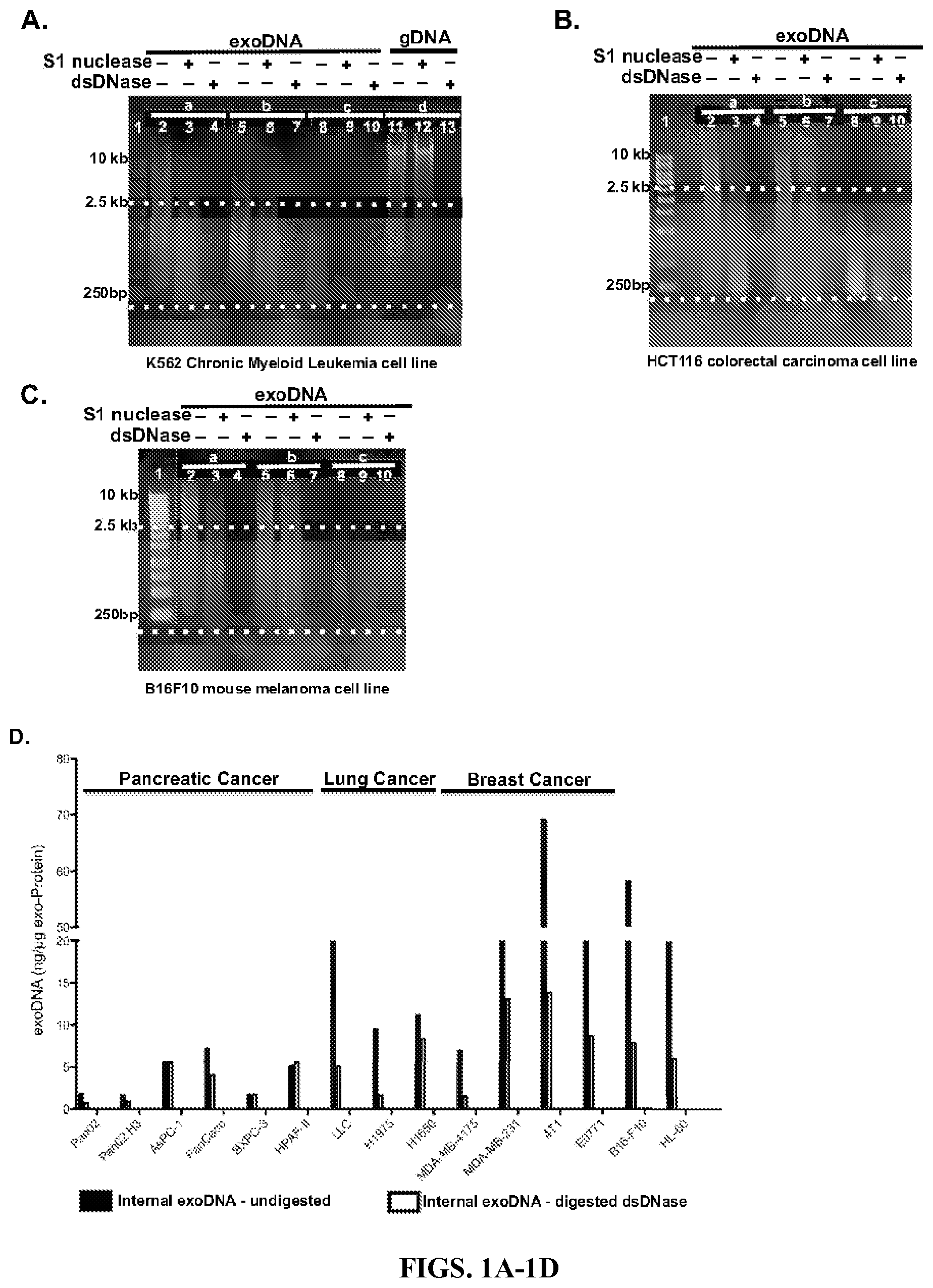
FIGS. 1A-1D show double-stranded DNA is associated with exosomes derived from different types of cancer cells. Exosomes were isolated from K-562 (FIG. 1A), HCT116 (FIG. 1B), and B16F10 (FIG. 1C) cell lines. Equal amounts of DNA extracted from untreated exosomes (Set a), exosomes treated with ssDNA specific S1 Nuclease (Set b) and exosomes treated with dsDNA specific Shrimp DNase (Set c) were digested with either S1 nuclease (lanes 3, 6 and 9) or dsDNase (lane 4, 7 and 10). Total genomic DNA from K-562 (FIG. 1A, Set d) was digested with S1 nuclease (Lane 12) or dsDNase (Lane 13). The results are representative of 2-3 experiments performed independently. As shown in FIG. 1D, internal exoDNA was extracted from exosomes secreted by different types of cancer cell lines including pancreatic cancer (Pan02, Pan02 H3, AsPC-1, PanCaco, BXPC-3 and HPAF-II), lung cancer (LLC, H1975 and H1650), breast cancer (MDA-MB-4175, MDA-MB-231 and 4T1 and E0771), melanoma (B16-F10) and leukemia (HL-60) (FIG. 1D). Abundance of dsDNA inside the exosomes, before and after digestion with dsDNase, was expressed as "nanogram of DNA per microgram of exoProtein".
FIGS. 2A-2B shows the specificity of S1 nuclease and shrimp DNase. In FIG. 2A, control digestion of ssDNA oligonucleotide and Lamda dsDNA was performed to verify the specificity of the enzymes. In FIG. 2B, total genomic DNA (gDNA) and exosomal DNA (exoDNA) isolated from K-562, HCT116 and B16F10 were quantified using QuantiFluor.RTM. dsDNA System, after the DNA samples were treated with either S1 nuclease or dsDNase. Control ssDNA oligonucleotide and dsDNA (Lamda DNA) were treated in parallel with either S1 nuclease or dsDNase.
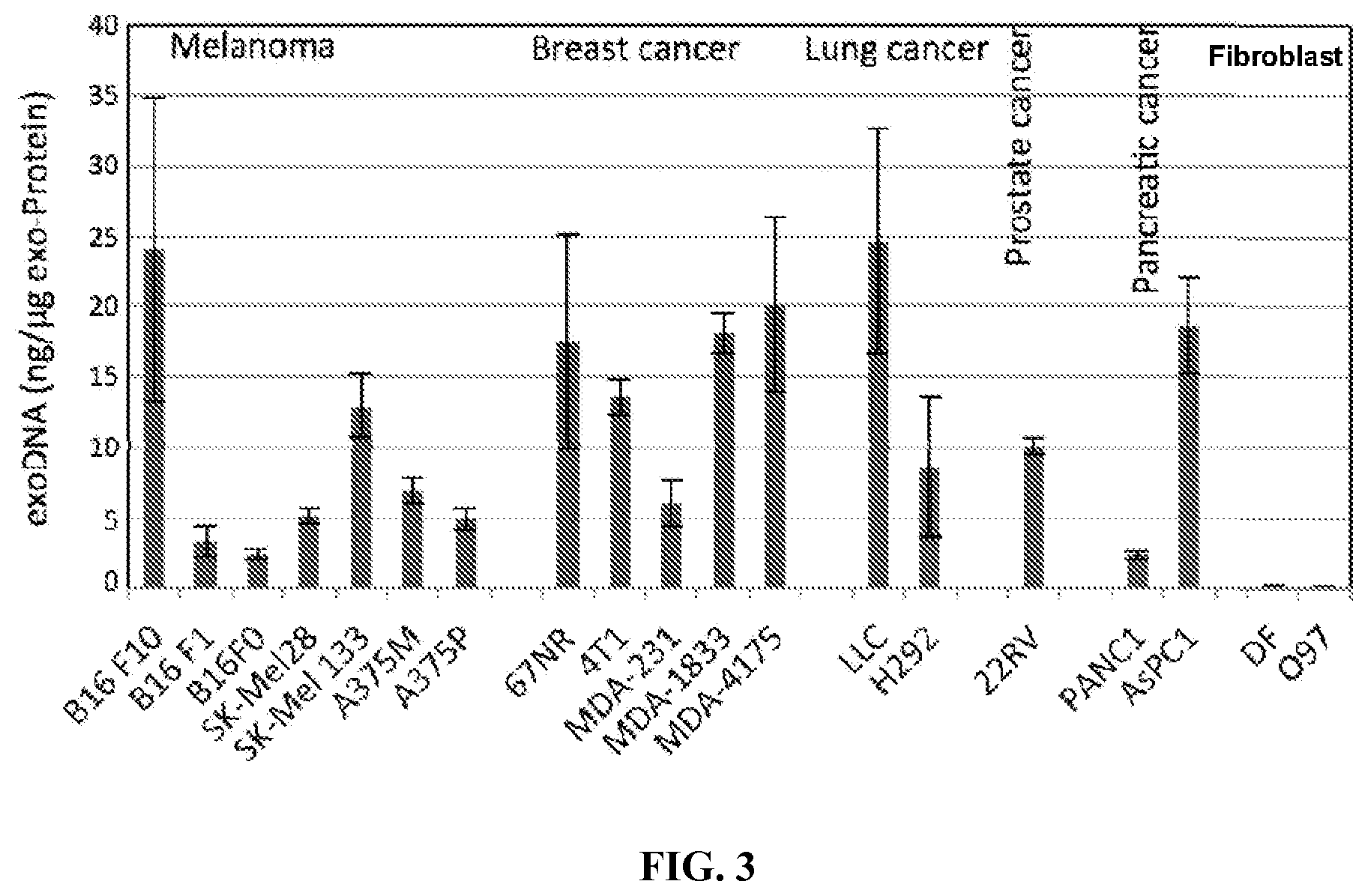
FIG. 3 shows DNA is present in tumor-cell derived exosomes. DNA was extracted from exosomes secreted by different types of cancer cell lines including melanoma (B16-F10, B16-F1, B16-FO, SK-Mel28, SK-MEL133, A375M and A375P), breast cancer (67NR, 4T1, MDA-MB-231, MDA-MB-1833 and MDA-MB-4175), lung cancer (LLC and H292), prostate cancer (22RV1), and pancreatic cancer (PANC1 and AsPC1). DNA abundance was expressed as "nanogram of DNA per microgram of exoProtein". DNA abundance was also evaluated for exosomes derived from two healthy human primary stromal fibroblast cell lines, DF and 097. Experiments were performed in duplication and results are shown as mean.+-.standard errors (n=2).
FIG. 4 shows detection of DNA in exosomes by immunogold electron microscopy. B16F10 exosome pellets were subjected to immunogold electron microscopy analysis using an anti-DNA antibody. The small grey areas represent exosomes (*) and the solid black dots represent the DNA (arrow).
FIGS. 5A-5B show exoDNA represents genomic DNA and phenocopies mutational status of parental tumor cells. FIG. 5A shows a circular view of the readings of fragments along each chromosome in the whole genome sequencing analysis of exoDNA isolated from murine melanoma B16-F10 cell-derived exosomes. FIG. 5B shows a comparative Genomic Hybridization array analysis of B16F10 exoDNA vs. genomic DNA.
FIG. 6 shows exoDNA methylation levels are comparable to those of gDNA. Dot blotted DNA was probed with an anti-5' methyl-Cytosine antibody to determine the cytosine methylation level of exoDNA vs. gDNA. Probing of the same blot with anti-DNA antibody serves as a loading control.
FIGS. 7A-7B show exoDNA can be horizontally transferred to different cell types in vitro and in vivo. FIG. 7A shows BrdU-labeled exoDNA can be detected by immunofluorescence using anti-BrdU antibodies in NIH3T3 fibroblasts and lineage-negative bone marrow cells (Lin-BM). Cells were treated with BrdU-labeled B16F10 exosomes for 24 hours in vitro. FIG. 7B shows the transfer of BrdU-labeled exoDNA to blood cells and whole bone marrow (WBM) is shown by BrdU flow analysis 24 hours post tail vein injection of BrdU-labeled B16F10 exosomes.
FIG. 8 shows evaluation of the sensitivity and specificity of AS-PCR assay for BRAF(V600E) mutation detection. Genomic DNA containing no BRAF(V600E) mutation or 0.1% of this mutation were used as template for AS-PCR to assess the sensitivity and specificity of the assay. Different amounts of template DNA (as low as 2.5 ng) were examined.
FIGS. 9A-9B show detection of BRAF V600E mutation. FIG. 9A shows detection of BRAF V600E mutation in exoDNA isolated from melanoma cells harboring this mutation. AS-PCR was employed to detect the BRAF V600E mutation in the extracted exoDNA, with gDNA isolated from Sk-Mel-28 and Sk-Mel-103 cells as positive and negative controls for V600E mutation (WT (V) and mutant (E) alleles). Primers that distinguish the WT (V) and mutant (E) alleles of BRAF V600E mutation were used to amplify the target in the samples. Asterisk indicates the size of expected PCR products. The following cell lines were examined in this study for BRAF(V600E) mutation: wild-type: SK-Mel103, SK-Mel146 and SK-Mel 147; BRAF(V600E): SK-Mel 28, SK-Mel 133, SK-Mel 192, and SK-Mel 267. FIG. 9B shows detection of BRAF (V600E) mutation in circulating exoDNA isolated from melanoma-bearing NOD/SCID mice subcutaneously implanted with the human melanoma cell line, Sk-Mel-28) using AS-PCR assay as described in FIG. 9A.
FIG. 10 shows detection of EGFR mutations in exoDNA isolated from lung cancer cells. gDNA and exoDNA were extracted from human non-small cell lung cancer (NSCLC) cell lines harboring WT (H292), exon 19 deletion (H1650 and PC9), and T790M mutation (H1975) of EGFR. AS-PCR was employed to detect WT and mutant alleles. For deletion of exon 19 mutation, "I" indicates internal control; "W", wild type; and "del", deletion of exon 19. For T790M mutation, "T" indicates wild type allele, and "M" indicates the mutant allele. The arrow marks the expected size of PCR products.
FIGS. 11A-11D show double-stranded exoDNA entering the nucleus of recipient cells. FIGS. 11A and 11C shows confocal imaging of mouse bone marrow cells (FIG. 11A) and RAW 264.7 cells (FIG. 11C) pre-treated with EdU-labeled B16-F10 exosomes. FIGS. 11B and 11D show confocal imaging of mouse bone marrow cells (FIG. 11B) and RAW 264.7 cells (FIG. 11D) pre-treated with unlabeled exosomes. The left panels show DAPI staining (in grey) of nuclei, the middle panel shows the visualization of exosomal EdU-labeled DNA (in white) within the nucleus of the recipient cells, and the right panel shows overlay of the DAPI staining (in grey) and EdU-labeled DNA staining (in white). In FIGS. 11A-11D, Z-stack images of detected fluorescence from individual cells were taken to verify intra-nuclear localization of fluorescence signals.
DETAILED DESCRIPTION OF THE INVENTION
A first aspect of the present invention is directed to a method for prognosing cancer in a subject. This method involves selecting a subject having cancer, obtaining a sample containing exosomes from the selected subject, recovering the exosomes from the sample, isolating double-stranded DNA from within the exosomes, and contacting the isolated double-stranded DNA with one or more reagents suitable to (1) detect presence or absence of one or more genetic mutations in the isolated double-stranded DNA that are associated with cancer, (2) quantify the amount of isolated double-stranded DNA from the recovered exosomes in the sample, (3) detect the methylation status of the isolated double-stranded DNA, or (4) quantify the amount of isolated double-stranded DNA able to enter a recipient cell. The cancer is prognosed based on the contacting.
Cancer prognosis as described herein includes determining the probable progression and course of the cancerous condition, and determining the chances of recovery and survival of a subject with the cancer, e.g., a favorable prognosis indicates an increased probability of recovery and/or survival for the cancer patient, while an unfavorable prognosis indicates a decreased probability of recovery and/or survival for the cancer patient. A subject's prognosis can be determined or modified by the availability of a suitable treatment (i.e., a treatment that will increase the probability of recovery and survival of the subject with cancer). For example, if the subject has a cancer, such as melanoma that is positive for one or more BRAF mutations as described herein, the subject has a favorable prognosis, because he/she is a candidate for treatment with BRAF inhibitor therapy. Likewise, if the subject has lung cancer or other cancer that is positive for one or more EGFR mutations as described herein, the subject has a favorable prognosis, because he/she is a candidate for treatment with an EGFR inhibitor therapy. Accordingly, another aspect of the present invention includes selecting a suitable cancer therapeutic based on the determined prognosis and administering the selected therapeutic to the subject.
Prognosis also encompasses the metastatic potential of a cancer. For example, a favorable prognosis based on the presence or absence of a genetic phenotype can indicate that the cancer is a type of cancer having low metastatic potential, and the patient has an increased probability of long term recovery and/or survival. Alternatively, an unfavorable prognosis, based on the presence or absence of a genetic phenotype can indicate that the cancer is a type of cancer having a high metastatic potential, and the patient has a decreased probability of long term recovery and/or survival.
In accordance with this aspect of the present invention, and as described herein, exosomes derived from tumors having high metastatic potential contain much higher levels of double-stranded DNA (dsDNA) within the exosome than exosomes derived from tumors having a low or no metastatic potential. Therefore, in one embodiment of the present invention, a reference or standard exosomal sample is an exosomal sample derived from tumor cells known to have low metastatic potential such as B16F1 melanoma cells, H1975 and H1650 lung cancer cells, or U87 glioblastoma cells. A higher concentration of DNA in the exosomal sample from the subject as compared to the concentration of dsDNA in exosomes derived from cells of low metastatic potential indicates the subject has a cancer with a high metastatic potential. If the exosomal sample from the subject has the same or lower concentration of dsDNA as compared to the concentration of dsDNA in exosomes derived from cells of low metastatic potential, then the subject has a cancer with a low metastatic potential. Alternatively, a reference or standard exosomal sample can be derived from tumor cells having a high metastatic potential, such as B16F10 melanoma cells or Lewis lung carcinoma cells. If the exosomal sample from the subject has the same or higher concentration of dsDNA as compared to exosomes derived from tumor cells of high metastatic potential, then the subject has a cancer with high metastatic potential. If the exosomal sample from the subject has a lower concentration of dsDNA as compared to exosomes derived from tumor cells of high metastatic potential, then the subject has a cancer with low metastatic potential.
Prognosis further encompasses prediction of sites of metastasis, determination of the stage of the cancer, or identifying the location of a primary tumor in a subject.
A change in the mutational status of gene associated with cancer (e.g., BRAF and/or EGFR) indicates that a change in the cancer phenotype has occurred with disease progression. For example, detecting the presence of a BRAF and/or EGFR mutation in an exosomal dsDNA sample from a subject whereas no BRAF and/or EGFR mutation was detected in an earlier exosomal dsDNA sample obtained from the same subject, can be indicative of a particular site of metastasis or progression to a more advanced stage of the cancer. Therefore, periodic monitoring of exosomal dsDNA mutational status provides a means for detecting primary tumor progression, metastasis, and facilitating optimal targeted or personalized treatment of the cancerous condition.
The detection of certain exosomal dsDNA mutations in a metastatic cancer sample can also identify the location of a primary tumor. For example, the detection of one or more BRAF mutations in a metastatic tumor or cancer cell-derived exosomal sample indicates that the primary tumor or cancer was melanoma or a form of brain cancer, e.g. glioblastoma. The detection of one or more EGFR mutations in a metastatic tumor or cancer cell derived exosomal dsDNA sample indicates that the primary tumor originated in the lung, or alternatively the primary cancer was head and neck cancer, ovarian cancer, cervical cancer, bladder cancer, or esophageal cancer.
Another aspect of the present invention is directed to a method of treating a subject having cancer. This method involves selecting a subject having cancer, obtaining a sample containing exosomes from the selected subject, recovering the exosomes from the sample, isolating double-stranded DNA from within the exosomes, and detecting (1) the presence or absence of one or more genetic mutations in the isolated double-stranded DNA that are associated with cancer, (2) the amount of isolated double-stranded DNA from the recovered exosomes in the sample, (3) the methylation status of the isolated double-stranded DNA, or (4) the amount of isolated double-stranded DNA able to enter a recipient cell. A suitable cancer therapeutic is selected based on the detecting and is administered to the selected subject under conditions effective to treat the cancer.
Another aspect of the present invention is directed to a method of managing treatment of a subject having cancer. This method involves selecting a subject undergoing treatment for cancer, obtaining a sample containing exosomes from the selected subject, recovering the exosomes from the sample, isolating double-stranded DNA from within the exosomes, and detecting (1) the presence or absence of one or more genetic mutations in the isolated double-stranded DNA that are associated with cancer, (2) the amount of isolated double-stranded DNA from the recovered exosomes in the sample, (3) the methylation status of the isolated double-stranded DNA, or (4) the amount of isolated double-stranded DNA able to enter a recipient cell. Treatment is modified, as necessary, based on the detecting.
In accordance with all aspects of the present invention, a "subject" or "patient" encompasses any animal, but preferably a mammal, e.g., human, non-human primate, a dog, a cat, a horse, a cow, or a rodent. More preferably, the subject or patient is a human. In some embodiments of the present invention, the subject has cancer, for example and without limitation, melanoma, breast cancer, lung cancer, or leukemia. In some embodiments, the cancer is a primary tumor, while in other embodiments, the cancer is a secondary or metastatic tumor.
"Exosomes" are microvesicles released from a variety of different cells, including cancer cells (i.e., "cancer-derived exosomes"). These small vesicles (50-100 nm in diameter) derive from large multivesicular endosomes and are secreted into the extracellular milieu. The precise mechanisms of exosome release/shedding remain unclear; however, this release is an energy-requiring phenomenon, modulated by extracellular signals. They appear to form by invagination and budding from the limiting membrane of late endosomes, resulting in vesicles that contain cytosol and that expose the extracellular domain of membrane-bound cellular proteins on their surface. Using electron microscopy, studies have shown fusion profiles of multivesicular endosomes with the plasma membrane, leading to the secretion of the internal vesicles into the extracellular environment. The rate of exosome release is significantly increased in most neoplastic cells and occurs continuously. Increased release of exosomes and their accumulation appear to be important in the malignant transformation process.
In accordance with the methods of the present invention, exosomes can be isolated or obtained from most biological fluids including, without limitation, blood, serum, plasma, ascites, cyst fluid, pleural fluid, peritoneal fluid, cerebral spinal fluid, tears, urine, saliva, sputum, nipple aspirates, lymph fluid, fluid of the respiratory, intestinal, and genitourinary trances, breast milk, intra-organ system fluid, or combinations thereof.
An enriched population of exosomes can be obtained from a biological sample using methods known in the art. For example, exosomes may be concentrated or isolated from a biological sample using size exclusion chromatography, density gradient centrifugation, differential centrifugation (Raposo et al. "B lymphocytes Secrete Antigen-presenting Vesicles," J Exp Med 183(3): 1161-72 (1996), which is hereby incorporated by reference in its entirety), anion exchange and/or gel permeation chromatography (for example, as described in U.S. Pat. No. 6,899,863 to Dhellin et al., and U.S. Pat. No. 6,812,023 to Lamparski et al., which are hereby incorporated by reference in their entirety), sucrose density gradients or organelle electrophoresis (for example, as described in U.S. Pat. No. 7,198,923), magnetic activated cell sorting (MACS) (Taylor et al., "MicroRNA Signatures of Tumor-derived Exosomes as Diagnostic Biomarkers of Ovarian Cancer," Gynecol Oncol 110(1): 13-21 (2008), which is hereby incorporated by reference in its entirety), nanomembrane ultrafiltration (Cheruvanky et al., "Rapid Isolation of Urinary Exosomal Biomarkers using a Nanomembrane Ultrafiltration Concentrator," Am J Physiol Renal Physiol 292(5): F1657-61 (2007), which is hereby incorporated by reference in its entirety), immunoabsorbent capture, affinity purification, microfluidic separation, or combinations thereof.
Exosomes isolated from a bodily fluid (i.e., peripheral blood, cerebrospinal fluid, urine) can be enriched for those originating from a specific cell type, for example, lung, pancreas, stomach, intestine, bladder, kidney, ovary, testis, skin, colorectal, breast, prostate, brain, esophagus, liver, placenta, and fetal cells. Because the exosomes often carry surface molecules such as antigens from their donor cells, surface molecules may be used to identify, isolate and/or enrich for exosomes from a specific donor cell type. In this way, exosomes originating from distinct cell populations can be analyzed for their nucleic acid content. For example, tumor (malignant and non-malignant) exosomes carry tumor-associated surface antigens and these exosomes can be isolated and/or enriched via these specific tumor-associated surface antigens. In one example, the tumor-associated surface antigen is epithelial-celladhesion-molecule (EpCAM), which is specific to exosomes from carcinomas of lung, colorectal, breast, prostate, head and neck, and hepatic origin, but not of hematological cell origin (Balzar et al. "The Biology of the 17-1A Antigen (Ep-CAM)," J Mol Med 77(10): 699-712 (1999); Went et al. "Frequent EpCam Protein Expression in Human Carcinomas," Hum Pathol 35(1): 122-8 (2004), which are hereby incorporated by reference in their entirety). In another example, the surface antigen is CD24, which is a glycoprotein specific to urine microvesicles (Keller et al. "CD24 is a Marker of Exosomes Secreted into Urine and Amniotic Fluid," Kidney Int 72(9): 1095-102 (2007), which is hereby incorporated by reference in its entirety). In yet another example, the surface antigen is CD70, carcinoembryonic antigen (CEA), EGFR, EGFRvIII and other variants, Fas ligand, TRAIL, tranferrin receptor, p38.5, p97 and HSP72. Alternatively, tumor specific exosomes may be characterized by the lack of surface markers, such as the lack of CD80 and CD86 expression.
The isolation of exosomes from specific cell types can be accomplished, for example, by using antibodies, aptamers, aptamer analogs, or molecularly imprinted polymers specific for a desired surface antigen. In one embodiment, the surface antigen is specific for a cancer type. In another embodiment, the surface antigen is specific for a cell type which is not necessarily cancerous. One example of a method of exosome separation based on cell surface antigen is provided in U.S. Pat. No. 7,198,923, which is hereby incorporated by reference in its entirety. As described in, e.g., U.S. Pat. No. 5,840,867 to Toole and U.S. Pat. No. 5,582,981 to Toole, which are hereby incorporated by reference in their entirety, aptamers and their analogs specifically bind surface molecules and can be used as a separation tool for retrieving cell type-specific exosomes. Molecularly imprinted polymers also specifically recognize surface molecules as described in, e.g., U.S. Pat. Nos. 6,525,154, 7,332,553 and 7,384,589, which are hereby incorporated by reference in their entirety, and are a tool for retrieving and isolating cell type-specific exosomes.
The exosomal fraction from a bodily fluid of a subject can be pre-treated with DNase to eliminate or substantially eliminate any DNA located on the surface or outside of the exosomes. Without DNAse pre-treatment, short DNA fragments on the outside of the exosomes may remain and co-isolate with nucleic acids extracted from inside the exosomes. Thus, elimination of all or substantially all DNA associated with the outside or surface of the exosomes by pre-treatment of with DNase, has the ability to enrich for internal exosomal dsDNA. To distinguish DNA strandedness within exosomes, Shrimp DNase specifically digests double-stranded DNA and S1 nuclease specifically digests single-stranded DNA.
In accordance with this and all other aspects of the present invention, the double-stranded DNA may be isolated by extracting the DNA from the exosomes prior to or for analysis.
The extracted dsDNA can be analyzed directly without an amplification step. Direct analysis may be performed with different methods including, but not limited to, nanostring technology. NanoString technology enables identification and quantification of individual target molecules in a biological sample by attaching a color coded fluorescent reporter to each target molecule. This approach is similar to the concept of measuring inventory by scanning barcodes. Reporters can be made with hundreds or even thousands of different codes allowing for highly multiplexed analysis. The technology is described in a publication by Geiss et al. "Direct Multiplexed Measurement of Gene Expression with Color-Coded Probe Pairs," Nat Biotechnol 26(3): 317-25 (2008), which is hereby incorporated by reference in its entirety.
In another embodiment, it may be beneficial or otherwise desirable to amplify the nucleic acid of the exosome prior to analyzing it. Methods of nucleic acid amplification are commonly used and generally known in the art. If desired, the amplification can be performed such that it is quantitative. Quantitative amplification will allow quantitative determination of relative amounts of the various exosomal nucleic acids.
Nucleic acid amplification methods include, without limitation, polymerase chain reaction (PCR) (U.S. Pat. No. 5,219,727, which is hereby incorporated by reference in its entirety) and its variants such as in situ polymerase chain reaction (U.S. Pat. No. 5,538,871, which is hereby incorporated by reference in its entirety), quantitative polymerase chain reaction (U.S. Pat. No. 5,219,727, which is hereby incorporated by reference in its entirety), nested polymerase chain reaction (U.S. Pat. No. 5,556,773, which is hereby incorporated by reference in its entirety), self sustained sequence replication and its variants (Guatelli et al. "Isothermal, In vitro Amplification of Nucleic Acids by a Multienzyme Reaction Modeled after Retroviral Replication," Proc Natl Acad Sci USA 87(5): 1874-8 (1990), which is hereby incorporated by reference in its entirety), transcriptional amplification system and its variants (Kwoh et al. "Transcription-based Amplification System and Detection of Amplified Human Immunodeficiency Virus type 1 with a Bead-Based Sandwich Hybridization Format," Proc Natl Acad Sci USA 86(4): 1173-7 (1989), which is hereby incorporated by reference in its entirety), Qb Replicase and its variants (Miele et al. "Autocatalytic Replication of a Recombinant RNA." J Mol Biol 171(3): 281-95 (1983), which is hereby incorporated by reference in its entirety), cold-PCR (Li et al. "Replacing PCR with COLD-PCR Enriches Variant DNA Sequences and Redefines the Sensitivity of Genetic Testing." Nat Med 14(5): 579-84 (2008), which is hereby incorporated by reference in its entirety) or any other nucleic acid amplification and detection methods known to those of skill in the art. Especially useful are those detection schemes designed for the detection of nucleic acid molecules if such molecules are present in very low numbers.
In one embodiment, the isolated double-stranded DNA is contacted with one or more reagents suitable to detect the presence or absence of one or more genetic mutations in the isolated double-stranded DNA that are associated with cancer. Exemplary genetic mutations associated with cancer include, but are not limited to, BRAF, EGFR, APC, NOTCH1, HRAS, KRAS, NRAS, MET, p.53, PTEN, HER2, FLT3, BRCA1, BRCA2, PIK3CA, KIT, RET, AKT, ABL, CDK4, MYC, RAF, PDGFR, BCR-ABL, NPM1, CEBPalpha, and SRC.
The one or more mutations in the one or more identified genes can be detected using a hybridization assay. In a hybridization assay, the presence or absence of a gene mutation is determined based on the hybridization of one or more allele-specific oligonucleotide probes to one or more nucleic acid molecules in the exosomal dsDNA sample from the subject. The oligonucleotide probe or probes comprise a nucleotide sequence that is complementary to at least the region of the gene that contains the mutation of interest. The oligonucleotide probes are designed to be complementary to the wildtype, non-mutant nucleotide sequence and/or the mutant nucleotide sequence of the one or more genes to effectuate the detection of the presence or the absence of the mutation in the sample from the subject upon contacting the sample with the oligonucleotide probes. A variety of hybridization assays that are known in the art are suitable for use in the methods of the present invention. These methods include, without limitation, direct hybridization assays, such as northern blot or Southern blot (see e.g., Ausabel et al., Current Protocols in Molecular Biology, John Wiley & Sons, NY (1991), which is hereby incorporated by reference in its entirety). Alternatively, direct hybridization can be carried out using an array based method where a series of oligonucleotide probes designed to be complementary to a particular non-mutant or mutant gene region are affixed to a solid support (glass, silicon, nylon membranes). A labeled exosomal DNA or cDNA sample from the subject is contacted with the array containing the oligonucleotide probes, and hybridization of nucleic acid molecules from the sample to their complementary oligonucleotide probes on the array surface is detected. Examples of direct hybridization array platforms include, without limitation, the Affymetrix GeneChip or SNP arrays and Illumina's Bead Array. Alternatively, the sample is bound to a solid support (often DNA or PCR amplified DNA) and labeled with oligonucleotides in solution (either allele specific or short so as to allow sequencing by hybridization).
Other common genotyping methods include, but are not limited to, restriction fragment length polymorphism assays; amplification based assays such as molecular beacon assays, nucleic acid arrays, high resolution melting curve analysis (Reed and Wittwer, "Sensitivity and Specificity of Single-Nucleotide Polymorphism Scanning by High Resolution Melting Analysis," Clinical Chem 50(10): 1748-54 (2004), which is hereby incorporated by reference in its entirety); allele-specific PCR (Gaudet et al., "Allele-Specific PCR in SNP Genotyping," Methods Mol Biol 578: 415-24 (2009), which is hereby incorporated by reference in its entirety); primer extension assays, such as allele-specific primer extension (e.g., Illumina.RTM. Infinium.RTM. assay), arrayed primer extension (see Krjutskov et al., "Development of a Single Tube 640-plex Genotyping Method for Detection of Nucleic Acid Variations on Microarrays," Nucleic Acids Res. 36(12) e75 (2008), which is hereby incorporated by reference in its entirety), homogeneous primer extension assays, primer extension with detection by mass spectrometry (e.g., Sequenom.RTM. iPLEX SNP genotyping assay) (see Zheng et al., "Cumulative Association of Five Genetic Variants with Prostate Cancer," N. Eng. J. Med. 358(9):910-919 (2008), which is hereby incorporated by reference in its entirety), multiplex primer extension sorted on genetic arrays; flap endonuclease assays (e.g., the Invader.RTM. assay) (see Olivier M., "The Invader Assay for SNP Genotyping," Mutat. Res. 573 (1-2) 103-10 (2005), which is hereby incorporated by reference in its entirety); 5' nuclease assays, such as the TaqMan.RTM. assay (see U.S. Pat. No. 5,210,015 to Gelfand et al. and U.S. Pat. No. 5,538,848 to Livak et al., which are hereby incorporated by reference in their entirety); and oligonucleotide ligation assays, such as ligation with rolling circle amplification, homogeneous ligation, OLA (see U.S. Pat. No. 4,988,617 to Landgren et al., which is hereby incorporated by reference in its entirety), multiplex ligation reactions followed by PCR, wherein zipcodes are incorporated into ligation reaction probes, and amplified PCR products are determined by electrophoretic or universal zipcode array readout (see U.S. Pat. Nos. 7,429,453 and 7,312,039 to Barany et al., which are hereby incorporated by reference in their entirety). Such methods may be used in combination with detection mechanisms such as, for example, luminescence or chemiluminescence detection, fluorescence detection, time-resolved fluorescence detection, fluorescence resonance energy transfer, fluorescence polarization, mass spectrometry, and electrical detection. In general, the methods for analyzing genetic aberrations are reported in numerous publications, not limited to those cited herein, and are available to those skilled in the art. The appropriate method of analysis will depend upon the specific goals of the analysis, the condition/history of the patient, and the specific cancer(s), diseases or other medical conditions to be detected, monitored or treated.
Alternatively, the presence or absence of one or more mutations identified supra can be detected by direct sequencing of the genes, or preferably particular gene regions comprising the one or more identified mutations, from the patient sample. Direct sequencing assays typically involve isolating DNA sample from the subject using any suitable method known in the art, and cloning the region of interest to be sequenced into a suitable vector for amplification by growth in a host cell (e.g. bacteria) or direct amplification by PCR or other amplification assay. Following amplification, the DNA can be sequenced using any suitable method. A preferable sequencing method involves high-throughput next generation sequencing (NGS) to identify genetic variation. Various NGS sequencing chemistries are available and suitable for use in carrying out the claimed invention, including pyrosequencing (Roche.RTM. 454), sequencing by reversible dye terminators (Illumina.RTM. HiSeq, Genome Analyzer and MiSeq systems), sequencing by sequential ligation of oligonucleotide probes (Life Technologies.RTM. SOLiD), and hydrogen ion semiconductor sequencing (Life Technologies.RTM., Ion Torrent.TM.) Alternatively, classic sequencing methods, such as the Sanger chain termination method or Maxam-Gilbert sequencing, which are well known to those of skill in the art, can be used to carry out the methods of the present invention.
In one embodiment of the present invention, the selected subject has melanoma, and the presence or absence of a mutation in BRAF is detected in an exosomal dsDNA sample from the subject. BRAF is a serine/threonine protein kinase that is encoded on chromosome 7q34. The amino acid sequence and nucleotide sequence of human BRAF are provided below as SEQ ID NO: 1 and SEQ ID NO: 2, respectively.
TABLE-US-00001 Human BRAF SEQ ID NO: 1 Met Ala Ala Leu Ser Gly Gly Gly Gly Gly Gly Ala Glu Pro Gly Gln 1 5 10 15 Ala Leu Phe Asn Gly Asp Met Glu Pro Glu Ala Gly Ala Gly Ala Gly 20 25 30 Ala Ala Ala Ser Ser Ala Ala Asp Pro Ala Ile Pro Glu Glu Val Trp 35 40 45 Asn Ile Lys Gln Met Ile Lys Leu Thr Gln Glu His Ile Glu Ala Leu 50 55 60 Leu Asp Lys Phe Gly Gly Glu His Asn Pro Pro Ser Ile Tyr Leu Glu 65 70 75 80 Ala Tyr Glu Glu Tyr Thr Ser Lys Leu Asp Ala Leu Gln Gln Arg Glu 85 90 95 Gln Gln Leu Leu Glu Ser Leu Gly Asn Gly Thr Asp Phe Ser Val Ser 100 105 110 Ser Ser Ala Ser Met Asp Thr Val Thr Ser Ser Ser Ser Ser Ser Leu 115 120 125 Ser Val Leu Pro Ser Ser Leu Ser Val Phe Gln Asn Pro Thr Asp Val 130 135 140 Ala Arg Ser Asn Pro Lys Ser Pro Gln Lys Pro Ile Val Arg Val Phe 145 150 155 160 Leu Pro Asn Lys Gln Arg Thr Val Val Pro Ala Arg Cys Gly Val Thr 165 170 175 Val Arg Asp Ser Leu Lys Lys Ala Leu Met Met Arg Gly Leu Ile Pro 180 185 190 Glu Cys Cys Ala Val Tyr Arg Ile Gln Asp Gly Glu Lys Lys Pro Ile 195 200 205 Gly Trp Asp Thr Asp Ile Ser Trp Leu Thr Gly Glu Glu Leu His Val 210 215 220 Glu Val Leu Glu Asn Val Pro Leu Thr Thr His Asn Phe Val Arg Lys 225 230 235 240 Thr Phe Phe Thr Leu Ala Phe Cys Asp Phe Cys Arg Lys Leu Leu Phe 245 250 255 Gln Gly Phe Arg Cys Gln Thr Cys Gly Tyr Lys Phe His Gln Arg Cys 260 265 270 Ser Thr Glu Val Pro Leu Met Cys Val Asn Tyr Asp Gln Leu Asp Leu 275 280 285 Leu Phe Val Ser Lys Phe Phe Glu His His Pro Ile Pro Gln Glu Glu 290 295 300 Ala Ser Leu Ala Glu Thr Ala Leu Thr Ser Gly Ser Ser Pro Ser Ala 305 310 315 320 Pro Ala Ser Asp Ser Ile Gly Pro Gln Ile Leu Thr Ser Pro Ser Pro 325 330 335 Ser Lys Ser Ile Pro Ile Pro Gln Pro Phe Arg Pro Ala Asp Glu Asp 340 345 350 His Arg Asn Gln Phe Gly Gln Arg Asp Arg Ser Ser Ser Ala Pro Asn 355 360 365 Val His Ile Asn Thr Ile Glu Pro Val Asn Ile Asp Asp Leu Ile Arg 370 375 380 Asp Gln Gly Phe Arg Gly Asp Gly Gly Ser Thr Thr Gly Leu Ser Ala 385 390 395 400 Thr Pro Pro Ala Ser Leu Pro Gly Ser Leu Thr Asn Val Lys Ala Leu 405 410 415 Gln Lys Ser Pro Gly Pro Gln Arg Glu Arg Lys Ser Ser Ser Ser Ser 420 425 430 Glu Asp Arg Asn Arg Met Lys Thr Leu Gly Arg Arg Asp Ser Ser Asp 435 440 445 Asp Trp Glu Ile Pro Asp Gly Gln Ile Thr Val Gly Gln Arg Ile Gly 450 455 460 Ser Gly Ser Phe Gly Thr Val Tyr Lys Gly Lys Trp His Gly Asp Val 465 470 475 480 Ala Val Lys Met Leu Asn Val Thr Ala Pro Thr Pro Gln Gln Leu Gln 485 490 495 Ala Phe Lys Asn Glu Val Gly Val Leu Arg Lys Thr Arg His Val Asn 500 505 510 Ile Leu Leu Phe Met Gly Tyr Ser Thr Lys Pro Gln Leu Ala Ile Val 515 520 525 Thr Gln Trp Cys Glu Gly Ser Ser Leu Tyr His His Leu His Ile Ile 530 535 540 Glu Thr Lys Phe Glu Met Ile Lys Leu Ile Asp Ile Ala Arg Gln Thr 545 550 555 560 Ala Gln Gly Met Asp Tyr Leu His Ala Lys Ser Ile Ile His Arg Asp 565 570 575 Leu Lys Ser Asn Asn Ile Phe Leu His Glu Asp Leu Thr Val Lys Ile 580 585 590 Gly Asp Phe Gly Leu Ala Thr Val Lys Ser Arg Trp Ser Gly Ser His 595 600 605 Gln Phe Glu Gln Leu Ser Gly Ser Ile Leu Trp Met Ala Pro Glu Val 610 615 620 Ile Arg Met Gln Asp Lys Asn Pro Tyr Ser Phe Gln Ser Asp Val Tyr 625 630 635 640 Ala Phe Gly Ile Val Leu Tyr Glu Leu Met Thr Gly Gln Leu Pro Tyr 645 650 655 Ser Asn Ile Asn Asn Arg Asp Gln Ile Ile Phe Met Val Gly Arg Gly 660 665 670 Tyr Leu Ser Pro Asp Leu Ser Lys Val Arg Ser Asn Cys Pro Lys Ala 675 680 685 Met Lys Arg Leu Met Ala Glu Cys Leu Lys Lys Lys Arg Asp Glu Arg 690 695 700 Pro Leu Phe Pro Gln Ile Leu Ala Ser Ile Glu Leu Leu Ala Arg Ser 705 710 715 720 Leu Pro Lys Ile His Arg Ser Ala Ser Glu Pro Ser Leu Asn Arg Ala 725 730 735 Gly Phe Gln Thr Glu Asp Phe Ser Leu Tyr Ala Cys Ala Ser Pro Lys 740 745 750 Thr Pro Ile Gln Ala Gly Gly Tyr Gly Ala Phe Pro Val His 755 760 765 Human BRAF SEQ ID NO: 2 cgcctccctt ccccctcccc gcccgacagc ggccgctcgg gccccggctc tcggttataa 60 gatggcggcg ctgagcggtg gcggtggtgg cggcgcggag ccgggccagg ctctgttcaa 120 cggggacatg gagcccgagg ccggcgccgg cgccggcgcc gcggcctctt cggctgcgga 180 ccctgccatt ccggaggagg tgtggaatat caaacaaatg attaagttga cacaggaaca 240 tatagaggcc ctattggaca aatttggtgg ggagcataat ccaccatcaa tatatctgga 300 ggcctatgaa gaatacacca gcaagctaga tgcactccaa caaagagaac aacagttatt 360 ggaatctctg gggaacggaa ctgatttttc tgtttctagc tctgcatcaa tggataccgt 420 tacatcttct tcctcttcta gcctttcagt gctaccttca tctctttcag tttttcaaaa 480 tcccacagat gtggcacgga gcaaccccaa gtcaccacaa aaacctatcg ttagagtctt 540 cctgcccaac aaacagagga cagtggtacc tgcaaggtgt ggagttacag tccgagacag 600 tctaaagaaa gcactgatga tgagaggtct aatcccagag tgctgtgctg tttacagaat 660 tcaggatgga gagaagaaac caattggttg ggacactgat atttcctggc ttactggaga 720 agaattgcat gtggaagtgt tggagaatgt tccacttaca acacacaact ttgtacgaaa 780 aacgtttttc accttagcat tttgtgactt ttgtcgaaag ctgcttttcc agggtttccg 840 ctgtcaaaca tgtggttata aatttcacca gcgttgtagt acagaagttc cactgatgtg 900 tgttaattat gaccaacttg atttgctgtt tgtctccaag ttctttgaac accacccaat 960 accacaggaa gaggcgtcct tagcagagac tgccctaaca tctggatcat ccccttccgc 1020 acccgcctcg gactctattg ggccccaaat tctcaccagt ccgtctcctt caaaatccat 1080 tccaattcca cagcccttcc gaccagcaga tgaagatcat cgaaatcaat ttgggcaacg 1140 agaccgatcc tcatcagctc ccaatgtgca tataaacaca atagaacctg tcaatattga 1200 tgacttgatt agagaccaag gatttcgtgg tgatggagga tcaaccacag gtttgtctgc 1260 taccccccct gcctcattac ctggctcact aactaacgtg aaagccttac agaaatctcc 1320 aggacctcag cgagaaagga agtcatcttc atcctcagaa gacaggaatc gaatgaaaac 1380 acttggtaga cgggactcga gtgatgattg ggagattcct gatgggcaga ttacagtggg 1440 acaaagaatt ggatctggat catttggaac agtctacaag ggaaagtggc atggtgatgt 1500 ggcagtgaaa atgttgaatg tgacagcacc tacacctcag cagttacaag ccttcaaaaa 1560 tgaagtagga gtactcagga aaacacgaca tgtgaatatc ctactcttca tgggctattc 1620 cacaaagcca caactggcta ttgttaccca gtggtgtgag ggctccagct tgtatcacca 1680 tctccatatc attgagacca aatttgagat gatcaaactt atagatattg cacgacagac 1740 tgcacagggc atggattact tacacgccaa gtcaatcatc cacagagacc tcaagagtaa 1800 taatatattt cttcatgaag acctcacagt aaaaataggt gattttggtc tagctacagt 1860 gaaatctcga tggagtgggt cccatcagtt tgaacagttg tctggatcca ttttgtggat 1920 ggcaccagaa gtcatcagaa tgcaagataa aaatccatac agctttcagt cagatgtata 1980 tgcatttgga attgttctgt atgaattgat gactggacag ttaccttatt caaacatcaa 2040 caacagggac cagataattt ttatggtggg acgaggatac ctgtctccag atctcagtaa 2100 ggtacggagt aactgtccaa aagccatgaa gagattaatg gcagagtgcc tcaaaaagaa 2160 aagagatgag agaccactct ttccccaaat tctcgcctct attgagctgc tggcccgctc 2220 attgccaaaa attcaccgca gtgcatcaga accctccttg aatcgggctg gtttccaaac 2280 agaggatttt agtctatatg cttgtgcttc tccaaaaaca cccatccagg cagggggata 2340 tggtgcgttt cctgtccact gaaacaaatg agtgagagag ttcaggagag tagcaacaaa 2400 aggaaaataa atgaacatat gtttgcttat atgttaaatt gaataaaata ctctcttttt 2460 ttttaaggtg aaccaaagaa cacttgtgtg gttaaagact agatataatt tttccccaaa 2520 ctaaaattta tacttaacat tggattttta acatccaagg gttaaaatac atagacattg 2580 ctaaaaattg gcagagcctc ttctagaggc tttactttct gttccgggtt tgtatcattc 2640 acttggttat tttaagtagt aaacttcagt ttctcatgca acttttgttg ccagctatca 2700 catgtccact agggactcca gaagaagacc ctacctatgc ctgtgtttgc aggtgagaag 2760 ttggcagtcg gttagcctgg gttagataag gcaaactgaa cagatctaat ttaggaagtc 2820 agtagaattt aataattcta ttattattct taataatttt tctataacta tttcttttta 2880 taacaatttg gaaaatgtgg atgtctttta tttccttgaa gcaataaact aagtttcttt 2940 ttataaaaa 2949
BRAF activates the MAP kinase/ERK-signaling pathway, and mutations in BRAF are associated with approximately 50% of pediatric and adult malignant melanomas (Daniotti et al., "Cutaneous Melanoma in Childhood and Adolescence Shows Frequent Loss of INK4A and Gain of KIT," J. Invest. Dermatol. 129 (7): 1759-68 (2009), which is hereby incorporated by reference in its entirety). In addition, BRAF point mutations have been reported to occur in several low- and high-grade tumor types in pediatric and adult patients, including approximately 50-60% of gangliogliomas (MacConaill et al., "Profiling Critical Cancer Gene Mutations in Clinical Tumor Samples," PloSOne 4(11):e7887 (2009), and Dougherty et al. "Activating Mutations in BRAF Characterize a Spectrum of Pediatric Low-Grade Gliomas," Neuro Oncol 12 (7): 621-630 (2010), which are hereby incorporated by reference in their entirety), approximately 2-12% of pilocytic astrocytomas (Forshew et al., "Activation of the ERK/MAPK Pathway: A Signature Genetic Defect in Posterior Fossa Pilocytic Astrocytomas," J Pathol. 218:172-181 (2009); Pfister et al., "BRAF Gene Duplication Constitutes a Mechanism of MAPK Pathway Activation in Low-Grade Astrocytomas," J Clin Invest. 118:1739-1749 (2008); MacConaill et al., "Profiling Critical Cancer Gene Mutations in Clinical Tumor Samples," PloSOne 4(11):e7887 (2009); Qaddoumi et al., "Paediatric Low-Grade Gliomas and the Need for New Options for Therapy," Cancer Biol Ther. 8:1-7 (2009); Jacob et al., "Duplication of 7q34 is Specific to Juvenile Pilocytic Astrocytomas and a Hallmark of Cerebellar and Optic Pathway Tumors," Brit J Cancer; 101:722-733 (2009); and Dias-Santagata et al., "BRAF V600E Mutations Are Common in Pleomorphic Xanthoastrocytoma: Diagnostic and Therapeutic Implications," PLoS ONE 6(3): e17948 (2011), which are hereby incorporated by reference in their entirety), and in as many as 30% of high-grade astrocytomas. Glioma accounts for 90% of malignant central nervous system (CNS) tumors in adults and 50% in the pediatric population (Central Brain Tumor Registry of the United States, 2010).
Over 90% of BRAF mutations in melanoma are at amino acid residue 600 (SEQ ID NO: 1), and over 90% of these involve a single nucleotide mutation that causes a valine glutamic acid change (BRAF V600E: nucleotide 1799 T>A of SEQ ID NO: 2; codon GTG>GAG) (Ascierto et al., "The Role of BRAF V600 Mutation in Melanoma," J. Translational Med. 10:85 (2012), which is hereby incorporated by reference in its entirety). Other mutations at this same valine residue of BRAF include a lysine substitution (BRAFV600K), an arginine substitution (BRAFV600R), and an aspartic acid substitution (BRAFV600D). The detection of any one of these BRAF V600 mutations, or other known BRAF mutations (i.e., insertions, deletions, duplications, etc.) in an exosomal DNA sample from a subject has diagnostic/prognostic and therapeutic implications in accordance with the methods of the present invention.
The BRAF V600 mutations cause constitutive activation of BRAF, which leads to activation of the downstream MEK/ERK pathway, evasion of senescence and apoptosis, uncheck replicative potential, angiogenesis, tissue invasion, metastasis, as well as evasion of immune response (Maurer et al., "Raf Kinases in Cancer-Roles and Therapeutic Opportunities," Oncogene 30: 3477-3488 (2011), which is hereby incorporated by reference in its entirety). Melanoma patients and patients having brain cancer identified as having a BRAF V600 mutation or other BRAF activating mutations are candidates for treatment with a BRAF inhibitor, such as vemurafenib (PLX/RG7204/RO5185426) (Sosman et al., "Survival in BRAF V600-Mutant Advanced Melanoma Treated with Vemurafenib," N Engl J Med 366:707-14 (2012) and Chapman et al., "Improved Survival with Vemurafenib in Melanoma with BRAF V600E Mutation," N Engl J Med 364''2507-2516 (2011), which are hereby incorporated by reference in their entirety), dabrafenib (Tafinlar; GSK2118436) (Gibney et al., "Clinical Development of Dabrafenib in BRAF mutant Melanoma and Other Malignancies" Expert Opin Drug Metab Toxicol 9(7):893-9 (2013), which is hereby incorporated by reference in its entirety), RAF265 (Su et al., "RAF265 Inhibits the Growth of Advanced Human Melanoma Tumors," Clin Cancer Res 18(8): 2184-98 (2012), which is hereby incorporated by reference in its entirety), and LGX818 (Stuart et al., "Preclinical Profile of LGX818: A Potent and Selective RAF Kinase Inhibitor," Cancer Res 72(8) Suppl 1 (2012), which is hereby incorporated by reference in its entirety).
In another embodiment of the present invention, the presence or absence of one or more mutations in the epidermal growth factor receptor (EGFR) is detected. EGFR is a transmembrane glycoprotein with an extracellular ligand-binding domain and an intracellular domain possessing intrinsic tyrosine kinase activity. Upon receptor dimerization following ligand binding, the tyrosine kinase domain is activated and recruited for phosphorylation of intracellular targets that drive normal cell growth and differentiation. The amino acid sequence and nucleotide sequence of human EGFR are provided below as SEQ ID NO: 3 and SEQ ID NO: 4, respectively.
TABLE-US-00002 Human EGFR SEQ ID NO: 3 Arg Pro Ser Gly Thr Ala Gly Ala Ala Leu Leu Ala Leu Leu Ala Ala 1 5 10 15 Leu Cys Pro Ala Ser Arg Ala Leu Glu Glu Lys Lys Val Cys Gln Gly 20 25 30 Thr Ser Asn Lys Leu Thr Gln Leu Gly Thr Phe Glu Asp His Phe Leu 35 40 45 Ser Leu Gln Arg Met Phe Asn Asn Cys Glu Val Val Leu Gly Asn Leu 50 55 60 Glu Ile Thr Tyr Val Gln Arg Asn Tyr Asp Leu Ser Phe Leu Lys Thr 65 70 75 80 Ile Gln Glu Val Ala Gly Tyr Val Leu Ile Ala Leu Asn Thr Val Glu 85 90 95 Arg Ile Pro Leu Glu Asn Leu Gln Ile Ile Arg Gly Asn Met Tyr Tyr 100 105 110 Glu Asn Ser Tyr Ala Leu Ala Val Leu Ser Asn Tyr Asp Ala Asn Lys 115 120 125 Thr Gly Leu Lys Glu Leu Pro Met Arg Asn Leu Gln Glu Ile Leu His 130 135 140 Gly Ala Val Arg Phe Ser Asn Asn Pro Ala Leu Cys Asn Val Glu Ser 145 150 155 160 Ile Gln Trp Arg Asp Ile Val Ser Ser Asp Phe Leu Ser Asn Met Ser 165 170 175 Met Asp Phe Gln Asn His Leu Gly Ser Cys Gln Lys Cys Asp Pro Ser 180 185 190 Cys Pro Asn Gly Ser Cys Trp Gly Ala Gly Glu Glu Asn Cys Gln Lys 195 200 205 Leu Thr Lys Ile Ile Cys Ala Gln Gln Cys Ser Gly Arg Cys Arg Gly 210 215 220 Lys Ser Pro Ser Asp Cys Cys His Asn Gln Cys Ala Ala Gly Cys Thr 225 230 235 240 Gly Pro Arg Glu Ser Asp Cys Leu Val Cys Arg Lys Phe Arg Asp Glu 245 250 255 Ala Thr Cys Lys Asp Thr Cys Pro Pro Leu Met Leu Tyr Asn Pro Thr 260 265 270 Thr Tyr Gln Met Asp Val Asn Pro Glu Gly Lys Tyr Ser Phe Gly Ala 275 280 285 Thr Cys Val Lys Lys Cys Pro Arg Asn Tyr Val Val Thr Asp His Gly 290 295 300 Ser Cys Val Arg Ala Cys Gly Ala Asp Ser Tyr Glu Met Glu Glu Asp 305 310 315 320 Gly Val Arg Lys Cys Lys Lys Cys Glu Gly Pro Cys Arg Lys Val Cys 325 330 335 Asn Gly Ile Gly Ile Gly Glu Phe Lys Asp Ser Leu Ser Ile Asn Ala 340 345 350 Thr Asn Ile Lys His Phe Lys Asn Cys Thr Ser Ile Ser Gly Asp Leu 355 360 365 His Ile Leu Pro Val Ala Phe Arg Gly Asp Ser Phe Thr His Thr Pro 370 375 380 Pro Leu Asp Pro Gln Glu Leu Asp Ile Leu Lys Thr Val Lys Glu Ile 385 390 395 400 Thr Gly Phe Leu Leu Ile Gln Ala Trp Pro Glu Asn Arg Thr Asp Leu 405 410 415 His Ala Phe Glu Asn Leu Glu Ile Ile Arg Gly Arg Thr Lys Gln His 420 425 430 Gly Gln Phe Ser Leu Ala Val Val Ser Leu Asn Ile Thr Ser Leu Gly 435 440 445 Leu Arg Ser Leu Lys Glu Ile Ser Asp Gly Asp Val Ile Ile Ser Gly 450 455 460 Asn Lys Asn Leu Cys Tyr Ala Asn Thr Ile Asn Trp Lys Lys Leu Phe 465 470 475 480 Gly Thr Ser Gly Gln Lys Thr Lys Ile Ile Ser Asn Arg Gly Glu Asn 485 490 495 Ser Cys Lys Ala Thr Gly Gln Val Cys His Ala Leu Cys Ser Pro Glu 500 505 510 Gly Cys Trp Gly Pro Glu Pro Arg Asp Cys Val Ser Cys Arg Asn Val 515 520 525 Ser Arg Gly Arg Glu Cys Val Asp Lys Cys Asn Leu Leu Glu Gly Glu 530 535 540 Pro Arg Glu Phe Val Glu Asn Ser Glu Cys Ile Gln Cys His Pro Glu 545 550 555 560 Cys Leu Pro Gln Ala Met Asn Ile Thr Cys Thr Gly Arg Gly Pro Asp 565 570 575 Asn Cys Ile Gln Cys Ala His Tyr Ile Asp Gly Pro His Cys Val Lys 580 585 590 Thr Cys Pro Ala Gly Val Met Gly Glu Asn Asn Thr Leu Val Trp Lys 595 600 605 Tyr Ala Asp Ala Gly His Val Cys His Leu Cys His Pro Asn Cys Thr 610 615 620 Tyr Gly Cys Thr Gly Pro Gly Leu Glu Gly Cys Pro Thr Asn Gly Pro 625 630 635 640 Lys Ile Pro Ser Ile Ala Thr Gly Met Val Gly Ala Leu Leu Leu Leu 645 650 655 Leu Val Val Ala Leu Gly Ile Gly Leu Phe Met Arg Arg Arg His Ile 660 665 670 Val Arg Lys Arg Thr Leu Arg Arg Leu Leu Gln Glu Arg Glu Leu Val 675 680 685 Glu Pro Leu Thr Pro Ser Gly Glu Ala Pro Asn Gln Ala Leu Leu Arg 690 695 700 Ile Leu Lys Glu Thr Glu Phe Lys Lys Ile Lys Val Leu Gly Ser Gly 705 710 715 720 Ala Phe Gly Thr Val Tyr Lys Gly Leu Trp Ile Pro Glu Gly Glu Lys 725 730 735 Val Lys Ile Pro Val Ala Ile Lys Glu Leu Arg Glu Ala Thr Ser Pro 740 745 750 Lys Ala Asn Lys Glu Ile Leu Asp Glu Ala Tyr Val Met Ala Ser Val 755 760 765 Asp Asn Pro His Val Cys Arg Leu Leu Gly Ile Cys Leu Thr Ser Thr 770 775 780 Val Gln Leu Ile Thr Gln Leu Met Pro Phe Gly Cys Leu Leu Asp Tyr 785 790 795 800 Val Arg Glu His Lys Asp Asn Ile Gly Ser Gln Tyr Leu Leu Asn Trp 805 810 815 Cys Val Gln Ile Ala Lys Gly Met Asn Tyr Leu Glu Asp Arg Arg Leu 820 825 830 Val His Arg Asp Leu Ala Ala Arg Asn Val Leu Val Lys Thr Pro Gln 835 840 845 His Val Lys Ile Thr Asp Phe Gly Leu Ala Lys Leu Leu Gly Ala Glu 850 855 860 Glu Lys Glu Tyr His Ala Glu Gly Gly Lys Val Pro Ile Lys Trp Met 865 870 875 880 Ala Leu Glu Ser Ile Leu His Arg Ile Tyr Thr His Gln Ser Asp Val 885 890 895 Trp Ser Tyr Gly Val Thr Val Trp Glu Leu Met Thr Phe Gly Ser Lys 900 905 910 Pro Tyr Asp Gly Ile Pro Ala Ser Glu Ile Ser Ser Ile Leu Glu Lys 915 920 925 Gly Glu Arg Leu Pro Gln Pro Pro Ile Cys Thr Ile Asp Val Tyr Met 930 935 940 Ile Met Val Lys Cys Trp Met Ile Asp Ala Asp Ser Arg Pro Lys Phe 945 950 955 960 Arg Glu Leu Ile Ile Glu Phe Ser Lys Met Ala Arg Asp Pro Gln Arg 965 970 975 Tyr Leu Val Ile Gln Gly Asp Glu Arg Met His Leu Pro Ser Pro Thr 980 985 990 Asp Ser Asn Phe Tyr Arg Ala Leu Met Asp Glu Glu Asp Met Asp Asp 995 1000 1005 Val Val Asp Ala Asp Glu Tyr Leu Ile Pro Gln Gln Gly Phe Phe Ser 1010 1015 1020 Ser Pro Ser Thr Ser Arg Thr Pro Leu Leu Ser Ser Leu Ser Ala Thr 1025 1030 1035 1040 Ser Asn Asn Ser Thr Val Ala Cys Ile Asp Arg Asn Gly Leu Gln Ser 1045 1050 1055 Cys Pro Ile Lys Glu Asp Ser Phe Leu Gln Arg Tyr Ser Ser Asp Pro 1060 1065 1070 Thr Gly Ala Leu Thr Glu Asp Ser Ile Asp Asp Thr Phe Leu Pro Val 1075 1080 1085 Pro Glu Tyr Ile Asn Gln Ser Val Pro Lys Arg Pro Ala Gly Ser Val 1090 1095 1100 Gln Asn Pro Val Tyr His Asn Gln Pro Leu Asn Pro Ala Pro Ser Arg 1105 1110 1115 1120 Asp Pro His Tyr Gln Asp Pro His Ser Thr Ala Val Gly Asn Pro Glu 1125 1130 1135 Tyr Leu Asn Thr Val Gln Pro Thr Cys Val Asn Ser Thr Phe Asp Ser 1140 1145 1150 Pro Ala His Trp Ala Gln Lys Gly Ser His Gln Ile Ser Leu Asp Asn 1155 1160 1165 Pro Asp Tyr Gln Gln Asp Phe Phe Pro Lys Glu Ala Lys Pro Asn Gly 1170 1175 1180 Ile Phe Lys Gly Ser Thr Ala Glu Asn Ala Glu Tyr Leu Arg Val Ala 1185 1190 1195 1200 Pro Gln Ser Ser Glu Phe Ile Gly Ala 1205 Human EGFR SEQ ID NO: 4 ccccggcgca gcgcggccgc agcagcctcc gccccccgca cggtgtgagc gcccgacgcg 60 gccgaggcgg ccggagtccc gagctagccc cggcggccgc cgccgcccag accggacgac 120 aggccacctc gtcggcgtcc gcccgagtcc ccgcctcgcc gccaacgcca caaccaccgc 180 gcacggcccc ctgactccgt ccagtattga tcgggagagc cggagcgagc tcttcgggga 240 gcagcgatgc gaccctccgg gacggccggg gcagcgctcc tggcgctgct ggctgcgctc 300 tgcccggcga gtcgggctct ggaggaaaag aaagtttgcc aaggcacgag taacaagctc 360 acgcagttgg gcacttttga agatcatttt ctcagcctcc agaggatgtt caataactgt 420 gaggtggtcc ttgggaattt ggaaattacc tatgtgcaga ggaattatga tctttccttc 480 ttaaagacca tccaggaggt ggctggttat gtcctcattg ccctcaacac agtggagcga 540
attcctttgg aaaacctgca gatcatcaga ggaaatatgt actacgaaaa ttcctatgcc 600 ttagcagtct tatctaacta tgatgcaaat aaaaccggac tgaaggagct gcccatgaga 660 aatttacagg aaatcctgca tggcgccgtg cggttcagca acaaccctgc cctgtgcaac 720 gtggagagca tccagtggcg ggacatagtc agcagtgact ttctcagcaa catgtcgatg 780 gacttccaga accacctggg cagctgccaa aagtgtgatc caagctgtcc caatgggagc 840 tgctggggtg caggagagga gaactgccag aaactgacca aaatcatctg tgcccagcag 900 tgctccgggc gctgccgtgg caagtccccc agtgactgct gccacaacca gtgtgctgca 960 ggctgcacag gcccccggga gagcgactgc ctggtctgcc gcaaattccg agacgaagcc 1020 acgtgcaagg acacctgccc cccactcatg ctctacaacc ccaccacgta ccagatggat 1080 gtgaaccccg agggcaaata cagctttggt gccacctgcg tgaagaagtg tccccgtaat 1140 tatgtggtga cagatcacgg ctcgtgcgtc cgagcctgtg gggccgacag ctatgagatg 1200 gaggaagacg gcgtccgcaa gtgtaagaag tgcgaagggc cttgccgcaa agtgtgtaac 1260 ggaataggta ttggtgaatt taaagactca ctctccataa atgctacgaa tattaaacac 1320 ttcaaaaact gcacctccat cagtggcgat ctccacatcc tgccggtggc atttaggggt 1380 gactccttca cacatactcc tcctctggat ccacaggaac tggatattct gaaaaccgta 1440 aaggaaatca cagggttttt gctgattcag gcttggcctg aaaacaggac ggacctccat 1500 gcctttgaga acctagaaat catacgcggc aggaccaagc aacatggtca gttttctctt 1560 gcagtcgtca gcctgaacat aacatccttg ggattacgct ccctcaagga gataagtgat 1620 ggagatgtga taatttcagg aaacaaaaat ttgtgctatg caaatacaat aaactggaaa 1680 aaactgtttg ggacctccgg tcagaaaacc aaaattataa gcaacagagg tgaaaacagc 1740 tgcaaggcca caggccaggt ctgccatgcc ttgtgctccc ccgagggctg ctggggcccg 1800 gagcccaggg actgcgtctc ttgccggaat gtcagccgag gcagggaatg cgtggacaag 1860 tgcaaccttc tggagggtga gccaagggag tttgtggaga actctgagtg catacagtgc 1920 cacccagagt gcctgcctca ggccatgaac atcacctgca caggacgggg accagacaac 1980 tgtatccagt gtgcccacta cattgacggc ccccactgcg tcaagacctg cccggcagga 2040 gtcatgggag aaaacaacac cctggtctgg aagtacgcag acgccggcca tgtgtgccac 2100 ctgtgccatc caaactgcac ctacggatgc actgggccag gtcttgaagg ctgtccaacg 2160 aatgggccta agatcccgtc catcgccact gggatggtgg gggccctcct cttgctgctg 2220 gtggtggccc tggggatcgg cctcttcatg cgaaggcgcc acatcgttcg gaagcgcacg 2280 ctgcggaggc tgctgcagga gagggagctt gtggagcctc ttacacccag tggagaagct 2340 cccaaccaag ctctcttgag gatcttgaag gaaactgaat tcaaaaagat caaagtgctg 2400 ggctccggtg cgttcggcac ggtgtataag ggactctgga tcccagaagg tgagaaagtt 2460 aaaattcccg tcgctatcaa ggaattaaga gaagcaacat ctccgaaagc caacaaggaa 2520 atcctcgatg aagcctacgt gatggccagc gtggacaacc cccacgtgtg ccgcctgctg 2580 ggcatctgcc tcacctccac cgtgcagctc atcacgcagc tcatgccctt cggctgcctc 2640 ctggactatg tccgggaaca caaagacaat attggctccc agtacctgct caactggtgt 2700 gtgcagatcg caaagggcat gaactacttg gaggaccgtc gcttggtgca ccgcgacctg 2760 gcagccagga acgtactggt gaaaacaccg cagcatgtca agatcacaga ttttgggctg 2820 gccaaactgc tgggtgcgga agagaaagaa taccatgcag aaggaggcaa agtgcctatc 2880 aagtggatgg cattggaatc aattttacac agaatctata cccaccagag tgatgtctgg 2940 agctacgggg tgaccgtttg ggagttgatg acctttggat ccaagccata tgacggaatc 3000 cctgccagcg agatctcctc catcctggag aaaggagaac gcctccctca gccacccata 3060 tgtaccatcg atgtctacat gatcatggtc aagtgctgga tgatagacgc agatagtcgc 3120 ccaaagttcc gtgagttgat catcgaattc tccaaaatgg cccgagaccc ccagcgctac 3180 cttgtcattc agggggatga aagaatgcat ttgccaagtc ctacagactc caacttctac 3240 cgtgccctga tggatgaaga agacatggac gacgtggtgg atgccgacga gtacctcatc 3300 ccacagcagg gcttcttcag cagcccctcc acgtcacgga ctcccctcct gagctctctg 3360 agtgcaacca gcaacaattc caccgtggct tgcattgata gaaatgggct gcaaagctgt 3420 cccatcaagg aagacagctt cttgcagcga tacagctcag accccacagg cgccttgact 3480 gaggacagca tagacgacac cttcctccca gtgcctgaat acataaacca gtccgttccc 3540 aaaaggcccg ctggctctgt gcagaatcct gtctatcaca atcagcctct gaaccccgcg 3600 cccagcagag acccacacta ccaggacccc cacagcactg cagtgggcaa ccccgagtat 3660 ctcaacactg tccagcccac ctgtgtcaac agcacattcg acagccctgc ccactgggcc 3720 cagaaaggca gccaccaaat tagcctggac aaccctgact accagcagga cttctttccc 3780 aaggaagcca agccaaatgg catctttaag ggctccacag ctgaaaatgc agaataccta 3840 agggtcgcgc cacaaagcag tgaatttatt ggagcatgac cacggaggat agtatgagcc 3900 ctaaaaatcc agactctttc gatacccagg accaagccac agcaggtcct ccatcccaac 3960 agccatgccc gcattagctc ttagacccac agactggttt tgcaacgttt acaccgacta 4020 gccaggaagt acttccacct cgggcacatt ttgggaagtt gcattccttt gtcttcaaac 4080 tgtgaagcat ttacagaaac gcatccagca agaatattgt ccctttgagc agaaatttat 4140 ctttcaaaga ggtatatttg aaaaaaaaaa aaagtatatg tgaggatttt tattgattgg 4200 ggatcttgga gtttttcatt gtcgctattg atttttactt caatgggctc ttccaacaag 4260 gaagaagctt gctggtagca cttgctaccc tgagttcatc caggcccaac tgtgagcaag 4320 gagcacaagc cacaagtctt ccagaggatg cttgattcca gtggttctgc ttcaaggctt 4380 ccactgcaaa acactaaaga tccaagaagg ccttcatggc cccagcaggc cggatcggta 4440 ctgtatcaag tcatggcagg tacagtagga taagccactc tgtcccttcc tgggcaaaga 4500 agaaacggag gggatggaat tcttccttag acttactttt gtaaaaatgt ccccacggta 4560 cttactcccc actgatggac cagtggtttc cagtcatgag cgttagactg acttgtttgt 4620 cttccattcc attgttttga aactcagtat gctgcccctg tcttgctgtc atgaaatcag 4680 caagagagga tgacacatca aataataact cggattccag cccacattgg attcatcagc 4740 atttggacca atagcccaca gctgagaatg tggaatacct aaggatagca ccgcttttgt 4800 tctcgcaaaa acgtatctcc taatttgagg ctcagatgaa atgcatcagg tcctttgggg 4860 catagatcag aagactacaa aaatgaagct gctctgaaat ctcctttagc catcacccca 4920 accccccaaa attagtttgt gttacttatg gaagatagtt ttctcctttt acttcacttc 4980 aaaagctttt tactcaaaga gtatatgttc cctccaggtc agctgccccc aaaccccctc 5040 cttacgcttt gtcacacaaa aagtgtctct gccttgagtc atctattcaa gcacttacag 5100 ctctggccac aacagggcat tttacaggtg cgaatgacag tagcattatg agtagtgtgg 5160 aattcaggta gtaaatatga aactagggtt tgaaattgat aatgctttca caacatttgc 5220 agatgtttta gaaggaaaaa agttccttcc taaaataatt tctctacaat tggaagattg 5280 gaagattcag ctagttagga gcccaccttt tttcctaatc tgtgtgtgcc ctgtaacctg 5340 actggttaac agcagtcctt tgtaaacagt gttttaaact ctcctagtca atatccaccc 5400 catccaattt atcaaggaag aaatggttca gaaaatattt tcagcctaca gttatgttca 5460 gtcacacaca catacaaaat gttccttttg cttttaaagt aatttttgac tcccagatca 5520 gtcagagccc ctacagcatt gttaagaaag tatttgattt ttgtctcaat gaaaataaaa 5580 ctatattcat ttccactcta aaaaaaaaaa aaaaaa 5616
Several EGFR mutations leading to constitutive activation have been associated with neoplastic growth and cancer progression in a variety of cancers, including lung cancer (in particular non-small cell lung carcinoma), head and neck cancer, ovarian cancer, cervical cancer, bladder cancer, and esophageal cancer (Nicholson et al., "EGFR and Cancer Prognosis," Eur J Cancer 37(4):9-15 (2001), which is hereby incorporated by reference in its entirety). Therefore, subjects suitable for EGFR mutational detection in accordance with methods of the present invention include subjects having any one of the aforementioned cancers.
A gain of function mutation suitable for detection in exosomal dsDNA samples in accordance with the present invention, includes, without limitation, the L858R mutation which results in leucine to arginine amino acid substitution at amino acid position 858 of human EGFR (SEQ ID NO: 3). This mutation occurs within the kinase domain (exon 21) and arises from a T>G nucleotide mutation at position 2573 of the EGFR gene sequence (SEQ ID NO: 4) (NCBI dbSNP reference SNP rs121434568; Mitsudomi et al., "Epidermal Growth Factor Receptor in Relation to Tumor Development: EGFR Gene and Cancer," FEBS J 277(2): 301-8 (2010), which are hereby incorporated by reference in their entirety).
Another gain of function mutation in EGFR suitable for detection in accordance with the present invention is the T790M mutation which results in a threonine to methionine mutation at amino acid position 790 in EGFR (SEQ ID NO: 3). This mutation occurs within the kinase domain (exon 20) and arises from a C>T mutation at nucleotide 2369 of the EGFR gene (SEQ ID NO: 4) (NCBI dbSNP reference SNP rs121434569; Tam et al., "Distinct Epidermal Growth Factor Receptor and KRAS Mutation Patterns in Non-Small Cell Lung Cancer Patients with Different Tobacco Exposure and Clinicopathologic Features," Clin Cancer Res 12:1647 (2006), which are hereby incorporated by reference in their entirety).
Another gain of function mutation in EGFR suitable for detection in accordance with the present invention is an in-frame deletion in exon 19. For example, deletions in amino acid residues 746-750, 746-751, 746-752, 747-751, 747-749, and 752-759 (SEQ ID NO: 3) have all been associated with lung cancer (see e.g., Mitsudomi et al., "Epidermal Growth Factor Receptor in Relation to Tumor Development: EGFR Gene and Cancer," FEBS J 277(2): 301-8 (2010), which is hereby incorporated by reference in its entirety). Detection of any one of these exon 19 deletions in exosomal dsDNA from a subject has prognostic/diagnostic and therapeutic implications in accordance with the present invention.
Subjects identified as having any of the above described EGFR mutations, or any other known EGFR mutations (i.e., insertions, deletions, duplications, etc), particularly gain-of-function mutations, are candidates for treatment using EGFR inhibitory agents which induce apoptosis and reduce proliferation of tumor growth (Ciardiello et al., "A Novel Approach in the Treatment of Cancer: Targeting the Epidermal Growth Factor Receptor," Clin Cancer Res 7:2958-2970 (2001); Ritter et al., "The Epidermal Growth Factor Receptor-Tyrosine Kinase: A Promising Therapeutic Target in Solid Tumors," Semin Oncol 30:3-11 (2003), which are hereby incorporated by reference in their entirety). Suitable EGFR inhibitors include, without limitation, small-molecule inhibitors of EGFR such as Gefitnib, Erlotinib (Tarceva), Afatinib (Gilotrif), Lapatinib (Tyverb) and monoclonal antibody inhibitors such as Panitumumab (Vectibix) and Cetuximab (Erbitux). Other EGFR inhibitors that are known in the art are also suitable for use in accordance with the methods of the present invention.
In another embodiment, the methods of the present invention involve contacting the isolated double-stranded DNA with one or more reagents suitable to quantify the amount of isolated double-stranded DNA from the recovered exosomes in the sample.
DNA can be quantified using any number of procedures, which are well-known in the art, the particular extraction procedure chosen based on the particular biological sample. For example, methods for extracting nucleic acids from urinary exosomes are described in Miranda et al. "Nucleic Acids within Urinary Exosomes/Microvesicles are Potential Biomarkers for Renal Disease," Kidney Int. 78:191-9 (2010) and in WO/2011/009104 to Russo, which are hereby incorporated by reference in their entirety. In some instances, with some techniques, it may also be possible to analyze the nucleic acid without extraction from the exosome.
In one embodiment, quantifying the amount of isolated double-stranded DNA is carried out by comparing the amount of isolated double-stranded DNA in a sample to that in a prior sample obtained from the selected subject.
The time between obtaining a first exosomal sample and a second, or any additional subsequent exosomal samples from a subject can be any desired period of time, for example, weeks, months, years, as determined is suitable by a physician and based on the characteristics of the primary tumor (tumor type, stage, location, etc.). In one embodiment of this aspect of the present invention, the first sample is obtained before treatment and the second sample is obtained after treatment. Alternatively, both samples can be obtained after one or more treatments; the second sample obtained at some point in time later than the first sample.
In another embodiment, quantifying the amount of isolated double-stranded DNA is carried out by comparing the amount of isolated double-stranded DNA to a standard. Exemplary standard exosomal samples are described supra. For example, the quantity of exosomal dsDNA obtained from B16F1 melanoma cells, H1975 and H1650 lung cancer cells, or U87 glioblastoma cells can serve as a standard sample that is indicative of the quantity of exosomal dsDNA associated with a low metastatic potential. Alternatively, the quantity of exosomal dsDNA obtained from B16F10 melanoma cells or Lewis lung carcinoma cells can serve as a standard sample that is indicative of the quantity of exosomal dsDNA associated with a high metastatic potential.
In a further embodiment, the methods of the present invention involve contacting the isolated double-stranded DNA with one or more reagents suitable to detect the methylation status of the DNA. DNA methylation involves the chemical addition of a methyl group to the 5' carbon position on the cytosine pyrimidine ring. Most DNA methylation occurs within CpG islands which are commonly found in the promoter region of a gene. Thus, this form of post modification of DNA acts as communicative signal for activation or inactivation of certain gene expression throughout various cell types. Methods to analyze DNA methylation status are well known in the art and include, but are not limited to, Me-DIP, HPLC, microarrays, and mass spectrometry. Another common method for DNA methylation analysis involves bisulfite treatment, in which unmethylated cytosines are converted to uracil while methylated cytosines remain unchanged, followed by downstream amplification and sequencing. The exosomal dsDNA methylation level or pattern can act as a surrogate for primary tumor cell status.
In yet another embodiment, the methods of the present invention involve contacting the isolated double-stranded DNA with one or more reagents suitable to quantify the amount of isolated double-stranded DNA able to enter a recipient cell.
DNA can be labeled using methods well known in the art including, but not limited to, use of various enzymes, prosthetic groups, fluorescent materials, luminescent materials, bioluminescent materials, and radioactive materials. The detectable substance may be coupled or conjugated either directly to the nucleic acid or indirectly, through an intermediate (such as, for example, a linker known in the art) using techniques known in the art. Examples of suitable enzymes include, but are not limited to, horseradish peroxidase, alkaline phosphatase, betagalactosidase, or acetylcholinesterase. Examples of suitable prosthetic group complexes include, but are not limited to, streptavidin/biotin and avidin/biotin. Examples of suitable fluorescent materials include, but are not limited to, umbelliferone, fluorescein, fluorescein isothiocyanate, Cascade Blue, rhodamine, dichlorotriazinylamine fluorescein, dansyl chloride, Texas Red, Oregon Green, cyanine (e.g., CY2, CY3, and CY5), allophycocyanine or phycoerythrin. An example of a luminescent material includes luminol. Examples of bioluminescent materials include, but are not limited to, luciferase, luciferin, green fluorescent protein (GFP), enhanced GFP (Yang et al., 1996), and aequorin. Once the exosomal dsDNA is labeled (e.g., by either BrdU or EdU), it is contacted with a preparation of suitable recipient cells, and the amount of dsDNA that enters the recipient cells is imaged and quantified using well known microscopy techniques, such as atomic force microscopy, electron microscopy, and advanced confocal microscopy. For electron microscopy, immunogold-labeling of dsDNA may be employed. Suitable recipient cells include, but are not limited to, fibroblasts, bone marrow cells, epithelial cells, and macrophages. For prognostic purposes, a high quantity of exosomal dsDNA able to enter a recipient cell is indicative of a poorer prognosis and higher metastatic potential. A low quantity of exosomal dsDNA able to enter a recipient cell is indicative of a good prognosis and lower metastatic potential.
EXAMPLES
The examples below are intended to exemplify the practice of the present invention but are by no means intended to limit the scope thereof.
Materials and Methods for Examples 1-5
Cell Lines and Cell Culture.
B16-F10, B16-F1, B16-F0, 67NR, 4T1, MDA-MB-231, MDA-MB-1833, MDA-MB-4175, LLC, HCT-116 (Horizon Discovery), PANC1 and AsPc1 cells were cultured in DMEM, and human melanoma cells (SK-Mel-, A375M and A375P), as well as Pan02, Pan02-H3, PanCaco, BXPC-3, HPAF-II, EO771, H292, H1975, H1650, K-562 (DSMZ), 22RV and HL-60 cells were cultured in RPMI supplemented with penicillin (100 U ml.sup.-1) and streptomycin (100 pg ml.sup.-1) and 10% exosome-depleted FBS. Cell lines were obtained from American Type Culture Collection if not otherwise mentioned. Human melanoma cell lines were obtained from Memorial Sloan-Kettering Cancer Center (MSKCC).
Animal Models and Plasma Collection.
C57BL/6 and NOD/SCID mice were obtained from Jackson Laboratory and maintained at the Weill Cornell Medical College (WCMC) animal facility. All procedures were performed according to protocols approved by the Institutional Animal Care and Use Committee of WCMC and MSKCC.
To analyze the circulating exoDNA from melanoma-bearing mice, NOD/SCID mice were subcutaneously implanted with 2.times.10.sup.6 human melanoma SK-Mel28 cells mixed with an equal volume of matrigel (BD Biosciences). Mice were sacrificed when the tumor reached maximum size allowed by the IACUC protocol and peripheral blood was obtained by retro-orbital bleeding directly into anti-coagulant tubes (EDTA). Plasma was separated from blood cells by sequential centrifugation at 500.times.g for 10 min followed by 3000.times.g for 10 min, and subjected to exosome isolation as described below.
Exosome Preparation and exoDNA Extraction.
Exosomes were prepared using differential ultracentrifugation methods essentially as described before (Peinado et al., "Melanoma Exosomes Educate Bone Marrow Progenitor Cells Toward a Pro-metastatic Phenotype Through MET," Nature Medicine 18:883-891 (2012), which is hereby incorporated by reference in its entirety), and resuspended in PBS for subsequent analysis. For mouse plasma samples, the plasma was filtered through a 1.2 .mu.m membrane to remove debris and large particles, then subjected to ultracentrifugation to pellet and wash exosomes. DNA was extracted from exosomes using the QIAamp DNA mini kit (QIAGEN) following the manufacturer's protocol and eluted with 50 l of 10 mM Tris pH8.0. DNA quality and quantity were analyzed using Nanodrop and Agilent Bioanalyzer chips.
DNase Digestion Analysis of exoDNA.
Approximately 40 micrograms of purified exosomes resuspended in PBS were treated either with S1 nuclease (Fermentas) or dsDNA-specific Shrimp DNase (Fermentas and Affymetrix). Equal amounts of exosomes were used as untreated controls. The digestion was performed at 30 degrees Celsius for 30 minutes for S1 nuclease and Shrimp dsDNase (Affymetrix). While using Shrimp dsDNase (Fermentas) the digestion was performed at 37 degrees Celsius for 30 min. Reaction mixtures were prepared according to manufacturers recommendations. After digestion the enzymes were heat inactivated at 70 degrees Celsius for five minutes in presence of EDTA according to manufacturer's instructions. Exosomal DNA was extracted using QIAamp DNA mini kit (Qiagen) and the eluted DNA was distributed equally and further subjected to S1 nuclease or Shrimp dsDNase treatment. All digestions were set up in 20 microliters reaction and after digestion 15 microliters of each sample resuspended in 1.times.DNA loading dye, (Fermentas) along with 1 kb DNA ladder (Fermentas) were loaded on a 1.5% agarose gel (Ultrapure agarose from Invitrogen) run at 150 V for 45 mins. The agarose gel was stained with SyBrGreen gold (1:5000 dilution in 1.times.TAE) for 45 minutes and imaged with Spectroline UV transilluminator (Kodak).
For detection of dsDNA using QuantiFluor.RTM. dsDNA System (Promega), 5 microliters of the digested or undigested mixes was mixed with QuantiFluor.RTM. dsDNA specific fluorescent dye and quantification of DNA was performed using manufactures protocol. The fluorescent intensity was measured using Spectramax M5 from Molecular devices.
Whole Genome Sequencing, CGH Array and Bioinformatics Analysis.
1 .mu.g of each exoDNA and gDNA sample was subjected to Illumina TruSeq library preparation and High Throughput DNA sequencing following manufacture's instruction. Short reads were aligned to the reference mouse genome (mm9) using the BWA computer programs with default parameters. Clonal reads were collapsed using custom scripts. Aligned read densities across the entire genome were then calculated using 100 Kb bins and represented using Circos plots.
For the CGH assay, exoDNA and gDNA samples were labeled using the Genomic DNA Enzymatic Labeling Kit (Agilent Technologies) following manufacture's instruction, and two color hybridization was performed using SurePrint G3 and HD CGH microarrays purchased from Agilent Technologies following standard procedures. The arrays were then analyzed and copy number visualized using the Agilent Genomic Workbench software analysis tools.
Dot Blot.
DNA samples were denatured with 0.4N NaOH at room temperature for 30 min, then placed on ice immediately and neutralized with an equal volume of pre-cooled 0.95M Tris (pH6.8) buffer. A four-fold serial dilution of exoDNA starting at 200 ng was dot blotted on Nylon membrane and crosslinked in a Stratalinker. The membranes were blocked with TBST buffer containing 1% milk and then probed with anti-5'-methylcytidine (Eurogentec) and anti-DNA (American Research Products, Inc) antibodies and developed with SuperSignal West Femto Chemiluminescent reagent (Thermo Scientific).
Transmission Electron Microscopy.
Exosome samples were fixed in 2% paraformaldehye and 0.2% glutaraldehyde in 0.1 M phosphate buffered saline and centrifuged to form a 1 mm thick visible pellet on the wall of a microcentrifuge tube. The pelleted exosomes were rinsed without resuspension in 0.5% sodium borohydride to block aldehyde groups and then dehydrated in a graded series of ethanol before being infiltrated in 100% LR White resin for 18 hours at 4.degree. C. All the processing was done in the same microcentrifuge tube and solutions were changed so as not to disturb the exosome pellet. The resin was polymerized at 60.degree. C. overnight and the microcentrifuge tube was cut away so that the exosomes could be thin sectioned. 100 nm thick sections were collected on nickel grids. Post embedding immunogold labeling was done for DNA labeling using the mouse monoclonal antibody AC-30-10 (EMD Millipore, Billerica, Mass. 01821 USA) and 10 colloidal gold conjugated to goat anti mouse IgM secondary antibodies were used to reveal the presence of DNA (BB International, Ted Pella Redding Calif. 96049 USA). Positive control sections consisted of sections of LR white embedded human bone marrow; negative control sections were incubated in secondary antibody without being exposed to primary antibody. Following immunogold labeling, sections were counterstained with 1% uranyl acetate and then examined in a Hitachi H7000 electron microscope at 75 kV accelerating voltage. Images were collected on Kodak 4489 film and after development were scanned at 2400 DPI and the images were processed for contrast using Adobe Photoshop.
Mutational Analysis of BRAF and EGFR.
To detect mutations in BRAF and EGFR genes, AS-PCR assays were adopted and modified from literature (Dahse et al., "Two Allele-specific PCR Assays for Screening Epidermal Growth Factor Receptor Gene Hotspot Mutations in Lung Adenocarcinoma," Molecular Medicine Reports 1:45-50 (2008); Jarry et al., "Real-time Allele-specific Amplification for Sensitive Detection of the BRAF Mutation V600E," Molecular and Cellular Probes 18:349-352 (2004); Uhara et al., "Simple Polymerase Chain Reaction for the Detection of Mutations and Deletions in the Epidermal Growth Factor Receptor Gene: Applications of This Method for the Diagnosis of Non-small-cell Lung Cancer," Clinica Chimica Acta; International Journal of Clinical Chemistry 401:6872 (2009), which are hereby incorporated by reference in their entirety). In brief, for both BRAF V600E and EGFR T790M mutations, standard PCR reactions containing 1.5 mM MgCl2 and primer pairs either for wild-type or mutant alleles at 0.5 M for BRAF and 2.5 M for EGFR were used, respectively. The PCR programs are the following: 95.degree. C., 5 min; 40 cycles of (95.degree. C., 5 sec, 66.degree. C. (BRAF)/56.degree. C. (EGFR), 5 sec and 72.degree. C., 5 sec); 72.degree. C., 5 min. For detection of Exon 19 deletion in EGFR, PCR reactions containing 1.5 mM MgCl2 and each of the four primers at 0.25 M were conducted. The PCR program is as follows: 95.degree. C., 5 min; 40 cycles of (95.degree. C., 30 sec, 58.degree. C., 30 sec and 72.degree. C., 30 sec); 72.degree. C., 5 min. A higher cycle number (80) was used when assessing the sensitivity of the assay and when circulating exoDNA was analyzed. End point PCR products were analyzed by agarose gel (2%) electrophoresis.
Brdu-Labeling of exoDNA and Transferring Assays.
B16-F10 cells were incubated with 10 mM BrdU for 24 h and washed with PBS. Fresh DMEM media supplemented with exosome-depleted fetal bovine serum was added to the cells and cells were cultured for another 24 h. The supernatant was then harvested for exosome preparation. For in vitro exoDNA transfer assays, NIH3T3 cells or freshly isolated lineage-negative bone marrow cells were treated with BrdU-labeled vs. non-labeled B16-F10 exosomes at 10 g/ml for 24 h, then cells were fixed directly or cytospun on coverslips followed by fixation with 4% paraformaldehyde and immunofluorescence staining with anti-BrdU antibody (Invitrogen). For in vivo studies, 10 g of BrdU-labeled or non-labeled B16-F10 exosomes in a total volume of 150 .mu.l PBS were administrated into C57BL/6 mice via tail vein injection, and 24 h later the mice were sacrificed and blood and whole bone marrow samples were processed as described by Peinado et al., "Melanoma Exosomes Educate Bone Marrow Progenitor Cells Toward a Pro-metastatic Phenotype Through MET," Nature Medicine 18:883-891 (2012), which is hereby incorporated by reference in its entirety, and analyzed with APC-BrdU flow kit (BD Pharmingen) following manufacturer's instructions. As positive controls, freshly isolated whole bone marrow cells were either treated directly with 10 mM BrdU for 30 min or with 10 g of BrdU-labeled or non-labeled B16-F10 exosomes for 16 h and then analyzed with APC-BrdU flow kit.
Example 1--Double-Stranded DNA (dsDNA) is Present in Exosomes Derived from Various Cancer Cell Types
Previous studies have demonstrated that proteins and genetic material such as mRNAs and miRNAs can be selectively packaged into exosomes in a cell type-dependent manner (Valadi et al., "Exosome-mediated Transfer of mRNAs and microRNAs is a Novel Mechanism of Genetic Exchange Between Cells," Nature Cell Biology 9:654-659 (2007), which is hereby incorporated by reference in its entirety). This is the first report providing evidence for the presence of dsDNA inside exosomes derived from multiple cancer cell lines: human K-562 chronic myeloid leukemia cells (FIG. 1A), human HCT116 colorectal carcinoma cells (FIG. 1B), and murine B16-F10 (highly metastatic) melanoma cells (FIG. 1C). To carefully characterize exosomal DNA within exosomes, extracellular and non-specific DNA present on the exosomal surface was eradicated using ssDNA-specific S1 Nuclease or dsDNA specific Shrimp DNase. Further to determine the nature of DNA present inside exosomes, the combination of S1 nuclease and dsDNase enzymes was used again to selectively digest single-stranded versus double-stranded DNA. It was observed that dsDNA-specific Shrimp DNase completely degraded the DNA associated with exosomes derived from all three cell sources included in the study. Moreover, it was observed that the size of the major population of dsDNA inside the exosomes ranged between 100 bp to 2500 bp whereas the population of dsDNA outside the exosomes constituted DNA species greater than 2500 bp in size. As a control, it was shown that Shrimp dsDNase completely degraded the genomic DNA (FIG. 1A, Lane 13).
To validate the specificity of the S1 nuclease and shrimp dsDNase, gDNA, purified ssDNA oligonucleotides and lamda dsDNA were included as controls in the study (FIG. 2A).
Next, to quantify exosomal dsDNA, the QuantiFluor.RTM. dsDNA System (Promega) was used which employs a dsDNA-specific fluorescent DNA-binding dye and enables sensitive quantitation of small amounts of double-stranded DNA (dsDNA) in solution. In contrast to undigested or S1 nuclease-treated exosomes, a strong reduction in the binding of dsDNA-specific fluorescent DNA-binding dye in the samples was obtained where exoDNA was treated with shrimp dsDNase (FIG. 2B). This result further confirms the finding that the majority of DNA associated with exosomes is indeed double-stranded in nature.
To determine whether the association of DNA with exosomes is a common feature of cancer cells, the analysis of exoDNA was extended to a broader range of cancer types. As shown in FIG. 3, exoDNA was detected at various levels in a panel of cancer cell lines including melanoma, breast cancer, lung cancer, prostate cancer, and pancreatic cancer. Furthermore, whether DNA is present in exosomes derived from normal stromal cells, such as fibroblasts (FIG. 3), was investigated. Although exoDNA could be isolated from exosomes derived from human dermal fibroblasts and human mammary tissue-derived fibroblasts, there was .about.20-fold less DNA in these exosomes compared to exosomes isolated from tumor cells (FIG. 3). Therefore, the abundance of exoDNA varies depending on cell type, with tumor-derived exosomes containing considerably more DNA than exosomes isolated from normal cells. Next, to understand the nature of DNA inside the exosomes, some of the cancer cell lines used in FIG. 3 were selected. After eradication of the external exoDNA by using method mentioned earlier in FIG. 1A-C, it is demonstrated that in almost all cancer cell lines the majority of DNA inside the exosomes is double-stranded in nature (FIG. 1D).
To further validate the presence of DNA in exosomes, and to determine its distribution in the population of exosomes, immunogold electron microscopy of exosomes derived from murine B16-F10 melanoma was performed using an anti-DNA antibody (FIG. 4). Interestingly, this study revealed that only a subset of exosomes indeed contained DNA (roughly .about.10% of B16-F10 exosomes).
Example 2--Exosomal DNA Represents the Entire Genomic DNA of Parental Tumor Cells
To determine if the genetic abnormalities driving tumorigenesis in cancer cells are represented and can be detected in exoDNA, both high throughput whole genome sequencing (FIG. 5A) and comparative genomic hybridization (CGH) analysis (FIG. 5B) were first carried out to examine, in an unbiased manner, the genome coverage of exoDNA. These analyses revealed that total genomic DNA (but not mitochondrial DNA) was represented in exoDNA. No bias for gene-coding versus intergenic regions and sense versus antisense strands of gene-coding regions was observed in the exoDNA. In addition, no specific fragments were highly enriched or depleted in the exoDNA pool compared to the genomic DNA.
In intact cells, 5'-cytosine methylation is a major modification of nuclear DNA involved in various biological processes, such as transcription and DNA repair. Therefore, the overall level of 5'-cytosine methylation of exoDNA was examined. It was found that, much like nuclear genomic DNA, exoDNA is also methylated, and to a level similar to that of cellular DNA (FIG. 6).
Example 3--Horizontal Transfer of Exosomal DNA from Donor Cells to Recipient Cells
It is now widely accepted that exosomes can mediate the horizontal transfer of functional molecules, such as oncogenic proteins, membrane-bound tyrosine kinase receptors and mRNAs of angiogenic factors, into target cells, resulting in epigenetic reprogramming of these recipient cells, and therefore, initiating profound biological responses (Peinado et al., "Melanoma Exosomes Educate Bone Marrow Progenitor Cells Toward a Pro-metastatic Phenotype Through MET," Nature Medicine 18:883-891 (2012); Zhang et al., "Exosomes and Immune Surveillance of Neoplastic Lesions: a Review," Biotech. Histochem. 87:161-168 (2012), which are hereby incorporated by reference in their entirety). However, there have been no reports investigating the transfer of exoDNA from donor to recipient cells. Therefore, whether exoDNA can be horizontally transferred into other cells was determined. First, exosomes were isolated from BrdU-labeled B16F10 cells, and it was confirmed that the exoDNA was indeed BrdU+. This approach allowed tracking of exoDNA in subsequent assays. The uptake of exoDNA by fibroblast (NIH3T3) or lineage negative bone marrow cells by immunofluorescence microscopy was then examined 24 hours post treatment of these cells with BrdU-labeled exosomes. BrdU-labeled exoDNA was clearly detected in the treated cells (FIG. 7A), and it was observed that, at this time point post exosome-treatment, the majority of exoDNA is localized in the cytoplasm of treated cells. Furthermore, using a similar approach followed by flow cytometric detection of BrdU, it was demonstrated that BrdU+ exoDNA from B16-F10 melanoma exosomes injected intravenously in mice was transferred to blood and bone marrow cells (FIG. 7B). Therefore, exoDNA is uptaken by various recipient cell types both in vitro and in vivo.
Example 4--Exosomal DNA Phenocopies the Mutational Status of the Parental Tumor Cells
The finding that exoDNA represents genomic DNA, and that it can be easily detected in purified exosomes prompted the examination of whether exoDNA could be utilized as a surrogate for tumor tissues or cells to detect tumor-specific genetic mutations. To this end, DNA isolated from exosomes derived from various cancer cell lines was tested, including melanoma and lung cancer for driver mutations known to be present in those cell lines. Since the BRAF (V600E) mutation is present in 50% of malignant melanomas (Daniotti et al., "Cutaneous Melanoma in Childhood and Adolescence Shows Frequent Loss of INK4A and Gain of KIT," The Journal of Investigative Dermatology 129:1759-1768 (2009); Davies et al., "Mutations of the BRAF Gene in Human Cancer," Nature 417:949-954 (2002); Gorden et al., "Analysis of BRAF and N-RAS Mutations in Metastatic Melanoma Tissues," Cancer Research 63:3955-3957 (2003), which are hereby incorporated by reference in their entirety), allele-specific polymerase chain reaction (AS-PCR) analysis was performed (adopted and modified from Jarry et al., "Real-time Allele-specific Amplification for Sensitive Detection of the BRAF Mutation V600E," Molecular and Cellular Probes 18:349-352 (2004), which is hereby incorporated by reference in its entirety) to evaluate the mutational status of BRAF in exoDNA isolated from several human primary melanoma cell lines which harbor either wild type (WT; SK-Mel146 and SK-Mel 147) or mutated BRAF (SK-Mel 28, SK-Mel 133, SK-Mel 192, and SK-Mel 267). First, proof of principle experiments were performed to verify the sensitivity and specificity of the AS-PCR assay (FIG. 8). Using primers that distinguished between wild type ("V") and mutant alleles ("E") of BRAF, the mutant allele could be detected in exoDNA of all cell lines containing the mutation, whereas only the wild-type allele was detected in those cell lines with non-mutated BRAF (FIG. 9A). These findings demonstrated that exoDNA reflects the mutational BRAF status of the parental cell lines.
A second example of a well-described tumor-associated mutation is the epidermal growth factor receptor (EGFR), which is mutated in several types of cancers, including non-small cell lung cancer (NSCLC) (Lynch et al., "Activating Mutations in the Epidermal Growth Factor Receptor Underlying Responsiveness of Non-small-cell Lung Cancer to Gefitinib," The New England Journal of Medicine 350:2129-2139 (2004); Paez et al., "EGFR Mutations in Lung Cancer: Correlation with Clinical Response to Gefitinib Therapy," Science 304:1497-1500 (2004); Pao et al., "EGF Receptor Gene Mutations are Common in Lung Cancers from "Never Smokers" and are Associated with Sensitivity of Tumors to Gefitinib and Erlotinib," Proceedings of the National Academy of Sciences of the United States of America 101:13306-13311 (2004), which are hereby incorporated by reference in their entirety). Gain-of-function mutations within the kinase domain of EGFR, such as the L858R point mutation, a deletion in 19 exon (19Del), and the T790M gate-keeper mutation, are crucial for selecting those patients who will benefit from targeted therapy using tyrosine kinase inhibitors. AS-PCR was again employed to assess exosomal DNA from several NSCLC cell lines, including the H292 cell line (wildtype), the H1975 cell line (harboring the L858R and T790M point mutations), and the H1650 and PC9 cell lines (harboring the exon 19 deletion). EGFR mutations were detected in 100% of exoDNA isolated from cultured NSCLC cell lines having these known EGFR mutations as shown in FIG. 10.
Example 5--Mutational Analysis of Exosomal DNA in Cell Line and Clinical Samples of Non-Small Cell Lung Cancer
Numerous studies have demonstrated that tumor cells secrete exosomes into the peripheral circulation and that these exosomes, which can be obtained non-invasively using a simple blood test, represent a reservoir of biomarkers. To assess the feasibility of detecting tumor-associated genetic mutations in circulating exoDNA a preclinical animal model of melanoma was employed. Specifically, human melanoma cells (Sk-Mel 28) harboring the BRAF(V600E) mutation were subcutaneously implanted in the flanks of NOD/SCID mice. Plasma was harvested when the tumor reached the size limit allowed by the standard animal protocol. Circulating exosomes were isolated using ultracentrifugation procedure, and dsDNA from within the exosomes was extracted and assayed for the BRAF(V600E) mutation. As demonstrated in FIG. 9B, the V600E mutation was present in the circulating exoDNA isolated from melanoma-bearing mice, suggesting that this assay could be translated to the clinic.
Example 6--Exosomal DNA Enters the Nucleus of Recipient Cells
To investigate exosome and exo-DNA transfer upon education of mouse bone marrow and RAW 264.7 target cells, exosomes from B16-F10 mouse melanoma cell line were collected in the presence or absence of EdU (10 .mu.g/ml; Invitrogen; Click-it EdU Imaging Kit) in the cell culture medium for 72 hrs. By using a purification protocol including ultracentrifugation of exosomes in a sucrose cushion layer, homogenous populations of exosomes were obtained. Mouse bone marrow and RAW 264.7 cells were seeded in 24-well plates (bone marrow: 0.5.times.10.sup.6/well; RAW: 10,000/well in 500 ml growth media overnight). The following day, media was replaced with fresh media containing exosome depleted FBS and cells were incubated with 20 .mu.g/ml of either unlabeled or EdU labeled exosomes. After 48 hours of incubation, cells were fixed (3.7% paraformaldehyde) and permeabilized (0.5% Triton X-100). For EdU detection, the fixed and permeabilized cells were processed as instructed by the manufacturer's protocol (Invitrogen; Click-it EdU Alexa Fluor 488 Imaging Kit; Cat # C10337) and afterwards DAPI stained and mounted using ProLong Gold antifade reagent with DAPI (life technologies). The cells were later on imaged by confocal microscopy. As shown in FIGS. 11A-11D, the exosomal DNA is able to enter the cytoplasm and nucleus of both the mouse bone marrow cells and the RAW 264.7 cells.
Discussion of Examples 1-6
Exosomes are important mediators of communication between tumor cells and the cells in their surrounding microenvironment, both locally and distally. Thus, the composition of exosomes derived from cancer cells can influence the adhesion, fusion and transfer to recipient cells. Here, it is demonstrated that dsDNA predominates as the primary nucleic acid structure present in exosomes derived from cancer cells. Moreover, it was found that the entire genome was represented in exosomes and that the mutations present in the parental tumor cells can be readily identified in exosomes. These findings have significant translational implications for diagnostic and therapeutic monitoring of patients with cancer. Currently, there is a high level of interest in the potential of circulating nucleic acids and circulating tumor cells (CTCs) in patient serum to serve as markers for detection and monitoring of cancer (Waldenstrom et al., "Cardiomyocyte Microvesicles Contain DNA/RNA and Convey Biological Messages to Target Cells," PloS One 7:e34653 (2012), which is hereby incorporated by reference in its entirety). However, progress in this area has been hindered by limitations in the sensitivity of these assays, as CTCs are extremely rare, and free, circulating DNA can easily be degraded. However, the dsDNA inside the exosomes is protected from extracellular nucleases and thus can represent a more reliable and stable source of tumor DNA that can be assayed for mutations. The inherent stability of dsDNA in exosomes may also be the basis for a functional role of exoDNA in intercellular genetic communication. Understanding the mechanism of dsDNA transfer and integration in recipient cells will lead to a better understanding of the role of tumor exosomes in cancer and metastasis.
Interestingly, the EM study provides the first direct evidence that only a subset of exosomes contain DNA. This finding raises the question of whether DNA packaging into exosomes is randomly restricted to a subset of particles due to size and distribution limitations, or whether there are specific biogenesis mechanisms that allow DNA packaging specifically in this particular subset of exosomes. It is possible that heterogeneous populations of exosome particles are present in the cancer microenvironment and the content of exosomes indirectly reflects the status of the cancer cells. Since exosomes are cellular in origin, the biomolecular composition of exosomes may further reflect cellular compartment of its origin.
Noteworthy, exoDNA is present in exosomes derived from most tumors, but not all tumor types, such as pancreatic cancer. Therefore, education of recipient cells via exosomes may depend on the genetic makeup of the uptaken exosomes. For example, the education of BMDC in the metastatic environment can involve both epigenetic and genetic processes. While the presence of ssDNA and non-coding RNAs explains the process of epigenetic education, presence of dsDNA may be associated with more permanent genetic changes. For example, the ability of dsDNA to undergo homologous recombination may account for the exosome-mediated transfer of genetic lesions to cells in the distant metastatic organs during cancer progression.
In this study, a thorough characterization of the nature, size and distribution pattern of DNA associated with exosomes was performed by using ssDNA and dsDNA specific enzymes on the intact exosomes. Interestingly, the exoDNA associated with the exterior of the exosome is larger in size in comparison to the exoDNA actually packaged into the exosome, which ranges between 100 bp to 2500 bp. Therefore, investigations in external and internal exoDNA are ongoing and may possibly have distinct functional roles in cancer progression.
While CTCs are extremely rare and their isolation requires specialized procedures, exosomes are constitutively secreted by all tumor cells, abundant in the plasma of metastatic cancer patients (Peinado et al., "Melanoma Exosomes Educate Bone Marrow Progenitor Cells Toward a Pro-metastatic Phenotype Through MET," Nature Medicine 18:883-891 (2012), which is hereby incorporated by reference in its entirety), and their isolation requires no special equipment. Therefore, an exoDNA-based test may be feasible in standard laboratories. Since aberrant DNA methylation patterns have been associated with certain types of cancer and its progression, it is reasonable to expect that exoDNA can be used as a surrogate for tumor cells to examine the relevant cancer-associated epigenetic alterations. It is also possible that the mutational status of a primary tumor can vary from that of the metastatic sites, and that mutations detected in exosomal DNA isolated from the plasma of cancer patients may not necessarily represent the mutational status of the primary tumor. This would suggest that targeted therapy to specific mutations may have a role in select patients whose primary tumors which lack a specific mutation, which may instead, be revealed in plasma-derived exosomes of cancer patients. In conclusion, it has been demonstrated herein that double-stranded exoDNA has unique features and value for the development of diagnostic/prognostic tools.
Although preferred embodiments have been depicted and described in detail herein, it will be apparent to those skilled in the relevant art that various modifications, additions, substitutions, and the like can be made without departing from the spirit of the invention and these are therefore considered to be within the scope of the invention as defined in the claims which follow.
SEQUENCE LISTINGS
1
41766PRTHomo sapiens 1Met Ala Ala Leu Ser Gly Gly Gly Gly Gly Gly Ala Glu Pro Gly Gln1 5 10 15Ala Leu Phe Asn Gly Asp Met Glu Pro Glu Ala Gly Ala Gly Ala Gly 20 25 30Ala Ala Ala Ser Ser Ala Ala Asp Pro Ala Ile Pro Glu Glu Val Trp 35 40 45Asn Ile Lys Gln Met Ile Lys Leu Thr Gln Glu His Ile Glu Ala Leu 50 55 60Leu Asp Lys Phe Gly Gly Glu His Asn Pro Pro Ser Ile Tyr Leu Glu65 70 75 80Ala Tyr Glu Glu Tyr Thr Ser Lys Leu Asp Ala Leu Gln Gln Arg Glu 85 90 95Gln Gln Leu Leu Glu Ser Leu Gly Asn Gly Thr Asp Phe Ser Val Ser 100 105 110Ser Ser Ala Ser Met Asp Thr Val Thr Ser Ser Ser Ser Ser Ser Leu 115 120 125Ser Val Leu Pro Ser Ser Leu Ser Val Phe Gln Asn Pro Thr Asp Val 130 135 140Ala Arg Ser Asn Pro Lys Ser Pro Gln Lys Pro Ile Val Arg Val Phe145 150 155 160Leu Pro Asn Lys Gln Arg Thr Val Val Pro Ala Arg Cys Gly Val Thr 165 170 175Val Arg Asp Ser Leu Lys Lys Ala Leu Met Met Arg Gly Leu Ile Pro 180 185 190Glu Cys Cys Ala Val Tyr Arg Ile Gln Asp Gly Glu Lys Lys Pro Ile 195 200 205Gly Trp Asp Thr Asp Ile Ser Trp Leu Thr Gly Glu Glu Leu His Val 210 215 220Glu Val Leu Glu Asn Val Pro Leu Thr Thr His Asn Phe Val Arg Lys225 230 235 240Thr Phe Phe Thr Leu Ala Phe Cys Asp Phe Cys Arg Lys Leu Leu Phe 245 250 255Gln Gly Phe Arg Cys Gln Thr Cys Gly Tyr Lys Phe His Gln Arg Cys 260 265 270Ser Thr Glu Val Pro Leu Met Cys Val Asn Tyr Asp Gln Leu Asp Leu 275 280 285Leu Phe Val Ser Lys Phe Phe Glu His His Pro Ile Pro Gln Glu Glu 290 295 300Ala Ser Leu Ala Glu Thr Ala Leu Thr Ser Gly Ser Ser Pro Ser Ala305 310 315 320Pro Ala Ser Asp Ser Ile Gly Pro Gln Ile Leu Thr Ser Pro Ser Pro 325 330 335Ser Lys Ser Ile Pro Ile Pro Gln Pro Phe Arg Pro Ala Asp Glu Asp 340 345 350His Arg Asn Gln Phe Gly Gln Arg Asp Arg Ser Ser Ser Ala Pro Asn 355 360 365Val His Ile Asn Thr Ile Glu Pro Val Asn Ile Asp Asp Leu Ile Arg 370 375 380Asp Gln Gly Phe Arg Gly Asp Gly Gly Ser Thr Thr Gly Leu Ser Ala385 390 395 400Thr Pro Pro Ala Ser Leu Pro Gly Ser Leu Thr Asn Val Lys Ala Leu 405 410 415Gln Lys Ser Pro Gly Pro Gln Arg Glu Arg Lys Ser Ser Ser Ser Ser 420 425 430Glu Asp Arg Asn Arg Met Lys Thr Leu Gly Arg Arg Asp Ser Ser Asp 435 440 445Asp Trp Glu Ile Pro Asp Gly Gln Ile Thr Val Gly Gln Arg Ile Gly 450 455 460Ser Gly Ser Phe Gly Thr Val Tyr Lys Gly Lys Trp His Gly Asp Val465 470 475 480Ala Val Lys Met Leu Asn Val Thr Ala Pro Thr Pro Gln Gln Leu Gln 485 490 495Ala Phe Lys Asn Glu Val Gly Val Leu Arg Lys Thr Arg His Val Asn 500 505 510Ile Leu Leu Phe Met Gly Tyr Ser Thr Lys Pro Gln Leu Ala Ile Val 515 520 525Thr Gln Trp Cys Glu Gly Ser Ser Leu Tyr His His Leu His Ile Ile 530 535 540Glu Thr Lys Phe Glu Met Ile Lys Leu Ile Asp Ile Ala Arg Gln Thr545 550 555 560Ala Gln Gly Met Asp Tyr Leu His Ala Lys Ser Ile Ile His Arg Asp 565 570 575Leu Lys Ser Asn Asn Ile Phe Leu His Glu Asp Leu Thr Val Lys Ile 580 585 590Gly Asp Phe Gly Leu Ala Thr Val Lys Ser Arg Trp Ser Gly Ser His 595 600 605Gln Phe Glu Gln Leu Ser Gly Ser Ile Leu Trp Met Ala Pro Glu Val 610 615 620Ile Arg Met Gln Asp Lys Asn Pro Tyr Ser Phe Gln Ser Asp Val Tyr625 630 635 640Ala Phe Gly Ile Val Leu Tyr Glu Leu Met Thr Gly Gln Leu Pro Tyr 645 650 655Ser Asn Ile Asn Asn Arg Asp Gln Ile Ile Phe Met Val Gly Arg Gly 660 665 670Tyr Leu Ser Pro Asp Leu Ser Lys Val Arg Ser Asn Cys Pro Lys Ala 675 680 685Met Lys Arg Leu Met Ala Glu Cys Leu Lys Lys Lys Arg Asp Glu Arg 690 695 700Pro Leu Phe Pro Gln Ile Leu Ala Ser Ile Glu Leu Leu Ala Arg Ser705 710 715 720Leu Pro Lys Ile His Arg Ser Ala Ser Glu Pro Ser Leu Asn Arg Ala 725 730 735Gly Phe Gln Thr Glu Asp Phe Ser Leu Tyr Ala Cys Ala Ser Pro Lys 740 745 750Thr Pro Ile Gln Ala Gly Gly Tyr Gly Ala Phe Pro Val His 755 760 76522949DNAHomo sapiens 2cgcctccctt ccccctcccc gcccgacagc ggccgctcgg gccccggctc tcggttataa 60gatggcggcg ctgagcggtg gcggtggtgg cggcgcggag ccgggccagg ctctgttcaa 120cggggacatg gagcccgagg ccggcgccgg cgccggcgcc gcggcctctt cggctgcgga 180ccctgccatt ccggaggagg tgtggaatat caaacaaatg attaagttga cacaggaaca 240tatagaggcc ctattggaca aatttggtgg ggagcataat ccaccatcaa tatatctgga 300ggcctatgaa gaatacacca gcaagctaga tgcactccaa caaagagaac aacagttatt 360ggaatctctg gggaacggaa ctgatttttc tgtttctagc tctgcatcaa tggataccgt 420tacatcttct tcctcttcta gcctttcagt gctaccttca tctctttcag tttttcaaaa 480tcccacagat gtggcacgga gcaaccccaa gtcaccacaa aaacctatcg ttagagtctt 540cctgcccaac aaacagagga cagtggtacc tgcaaggtgt ggagttacag tccgagacag 600tctaaagaaa gcactgatga tgagaggtct aatcccagag tgctgtgctg tttacagaat 660tcaggatgga gagaagaaac caattggttg ggacactgat atttcctggc ttactggaga 720agaattgcat gtggaagtgt tggagaatgt tccacttaca acacacaact ttgtacgaaa 780aacgtttttc accttagcat tttgtgactt ttgtcgaaag ctgcttttcc agggtttccg 840ctgtcaaaca tgtggttata aatttcacca gcgttgtagt acagaagttc cactgatgtg 900tgttaattat gaccaacttg atttgctgtt tgtctccaag ttctttgaac accacccaat 960accacaggaa gaggcgtcct tagcagagac tgccctaaca tctggatcat ccccttccgc 1020acccgcctcg gactctattg ggccccaaat tctcaccagt ccgtctcctt caaaatccat 1080tccaattcca cagcccttcc gaccagcaga tgaagatcat cgaaatcaat ttgggcaacg 1140agaccgatcc tcatcagctc ccaatgtgca tataaacaca atagaacctg tcaatattga 1200tgacttgatt agagaccaag gatttcgtgg tgatggagga tcaaccacag gtttgtctgc 1260taccccccct gcctcattac ctggctcact aactaacgtg aaagccttac agaaatctcc 1320aggacctcag cgagaaagga agtcatcttc atcctcagaa gacaggaatc gaatgaaaac 1380acttggtaga cgggactcga gtgatgattg ggagattcct gatgggcaga ttacagtggg 1440acaaagaatt ggatctggat catttggaac agtctacaag ggaaagtggc atggtgatgt 1500ggcagtgaaa atgttgaatg tgacagcacc tacacctcag cagttacaag ccttcaaaaa 1560tgaagtagga gtactcagga aaacacgaca tgtgaatatc ctactcttca tgggctattc 1620cacaaagcca caactggcta ttgttaccca gtggtgtgag ggctccagct tgtatcacca 1680tctccatatc attgagacca aatttgagat gatcaaactt atagatattg cacgacagac 1740tgcacagggc atggattact tacacgccaa gtcaatcatc cacagagacc tcaagagtaa 1800taatatattt cttcatgaag acctcacagt aaaaataggt gattttggtc tagctacagt 1860gaaatctcga tggagtgggt cccatcagtt tgaacagttg tctggatcca ttttgtggat 1920ggcaccagaa gtcatcagaa tgcaagataa aaatccatac agctttcagt cagatgtata 1980tgcatttgga attgttctgt atgaattgat gactggacag ttaccttatt caaacatcaa 2040caacagggac cagataattt ttatggtggg acgaggatac ctgtctccag atctcagtaa 2100ggtacggagt aactgtccaa aagccatgaa gagattaatg gcagagtgcc tcaaaaagaa 2160aagagatgag agaccactct ttccccaaat tctcgcctct attgagctgc tggcccgctc 2220attgccaaaa attcaccgca gtgcatcaga accctccttg aatcgggctg gtttccaaac 2280agaggatttt agtctatatg cttgtgcttc tccaaaaaca cccatccagg cagggggata 2340tggtgcgttt cctgtccact gaaacaaatg agtgagagag ttcaggagag tagcaacaaa 2400aggaaaataa atgaacatat gtttgcttat atgttaaatt gaataaaata ctctcttttt 2460ttttaaggtg aaccaaagaa cacttgtgtg gttaaagact agatataatt tttccccaaa 2520ctaaaattta tacttaacat tggattttta acatccaagg gttaaaatac atagacattg 2580ctaaaaattg gcagagcctc ttctagaggc tttactttct gttccgggtt tgtatcattc 2640acttggttat tttaagtagt aaacttcagt ttctcatgca acttttgttg ccagctatca 2700catgtccact agggactcca gaagaagacc ctacctatgc ctgtgtttgc aggtgagaag 2760ttggcagtcg gttagcctgg gttagataag gcaaactgaa cagatctaat ttaggaagtc 2820agtagaattt aataattcta ttattattct taataatttt tctataacta tttcttttta 2880taacaatttg gaaaatgtgg atgtctttta tttccttgaa gcaataaact aagtttcttt 2940ttataaaaa 294931209PRTHomo sapiens 3Arg Pro Ser Gly Thr Ala Gly Ala Ala Leu Leu Ala Leu Leu Ala Ala1 5 10 15Leu Cys Pro Ala Ser Arg Ala Leu Glu Glu Lys Lys Val Cys Gln Gly 20 25 30Thr Ser Asn Lys Leu Thr Gln Leu Gly Thr Phe Glu Asp His Phe Leu 35 40 45Ser Leu Gln Arg Met Phe Asn Asn Cys Glu Val Val Leu Gly Asn Leu 50 55 60Glu Ile Thr Tyr Val Gln Arg Asn Tyr Asp Leu Ser Phe Leu Lys Thr65 70 75 80Ile Gln Glu Val Ala Gly Tyr Val Leu Ile Ala Leu Asn Thr Val Glu 85 90 95Arg Ile Pro Leu Glu Asn Leu Gln Ile Ile Arg Gly Asn Met Tyr Tyr 100 105 110Glu Asn Ser Tyr Ala Leu Ala Val Leu Ser Asn Tyr Asp Ala Asn Lys 115 120 125Thr Gly Leu Lys Glu Leu Pro Met Arg Asn Leu Gln Glu Ile Leu His 130 135 140Gly Ala Val Arg Phe Ser Asn Asn Pro Ala Leu Cys Asn Val Glu Ser145 150 155 160Ile Gln Trp Arg Asp Ile Val Ser Ser Asp Phe Leu Ser Asn Met Ser 165 170 175Met Asp Phe Gln Asn His Leu Gly Ser Cys Gln Lys Cys Asp Pro Ser 180 185 190Cys Pro Asn Gly Ser Cys Trp Gly Ala Gly Glu Glu Asn Cys Gln Lys 195 200 205Leu Thr Lys Ile Ile Cys Ala Gln Gln Cys Ser Gly Arg Cys Arg Gly 210 215 220Lys Ser Pro Ser Asp Cys Cys His Asn Gln Cys Ala Ala Gly Cys Thr225 230 235 240Gly Pro Arg Glu Ser Asp Cys Leu Val Cys Arg Lys Phe Arg Asp Glu 245 250 255Ala Thr Cys Lys Asp Thr Cys Pro Pro Leu Met Leu Tyr Asn Pro Thr 260 265 270Thr Tyr Gln Met Asp Val Asn Pro Glu Gly Lys Tyr Ser Phe Gly Ala 275 280 285Thr Cys Val Lys Lys Cys Pro Arg Asn Tyr Val Val Thr Asp His Gly 290 295 300Ser Cys Val Arg Ala Cys Gly Ala Asp Ser Tyr Glu Met Glu Glu Asp305 310 315 320Gly Val Arg Lys Cys Lys Lys Cys Glu Gly Pro Cys Arg Lys Val Cys 325 330 335Asn Gly Ile Gly Ile Gly Glu Phe Lys Asp Ser Leu Ser Ile Asn Ala 340 345 350Thr Asn Ile Lys His Phe Lys Asn Cys Thr Ser Ile Ser Gly Asp Leu 355 360 365His Ile Leu Pro Val Ala Phe Arg Gly Asp Ser Phe Thr His Thr Pro 370 375 380Pro Leu Asp Pro Gln Glu Leu Asp Ile Leu Lys Thr Val Lys Glu Ile385 390 395 400Thr Gly Phe Leu Leu Ile Gln Ala Trp Pro Glu Asn Arg Thr Asp Leu 405 410 415His Ala Phe Glu Asn Leu Glu Ile Ile Arg Gly Arg Thr Lys Gln His 420 425 430Gly Gln Phe Ser Leu Ala Val Val Ser Leu Asn Ile Thr Ser Leu Gly 435 440 445Leu Arg Ser Leu Lys Glu Ile Ser Asp Gly Asp Val Ile Ile Ser Gly 450 455 460Asn Lys Asn Leu Cys Tyr Ala Asn Thr Ile Asn Trp Lys Lys Leu Phe465 470 475 480Gly Thr Ser Gly Gln Lys Thr Lys Ile Ile Ser Asn Arg Gly Glu Asn 485 490 495Ser Cys Lys Ala Thr Gly Gln Val Cys His Ala Leu Cys Ser Pro Glu 500 505 510Gly Cys Trp Gly Pro Glu Pro Arg Asp Cys Val Ser Cys Arg Asn Val 515 520 525Ser Arg Gly Arg Glu Cys Val Asp Lys Cys Asn Leu Leu Glu Gly Glu 530 535 540Pro Arg Glu Phe Val Glu Asn Ser Glu Cys Ile Gln Cys His Pro Glu545 550 555 560Cys Leu Pro Gln Ala Met Asn Ile Thr Cys Thr Gly Arg Gly Pro Asp 565 570 575Asn Cys Ile Gln Cys Ala His Tyr Ile Asp Gly Pro His Cys Val Lys 580 585 590Thr Cys Pro Ala Gly Val Met Gly Glu Asn Asn Thr Leu Val Trp Lys 595 600 605Tyr Ala Asp Ala Gly His Val Cys His Leu Cys His Pro Asn Cys Thr 610 615 620Tyr Gly Cys Thr Gly Pro Gly Leu Glu Gly Cys Pro Thr Asn Gly Pro625 630 635 640Lys Ile Pro Ser Ile Ala Thr Gly Met Val Gly Ala Leu Leu Leu Leu 645 650 655Leu Val Val Ala Leu Gly Ile Gly Leu Phe Met Arg Arg Arg His Ile 660 665 670Val Arg Lys Arg Thr Leu Arg Arg Leu Leu Gln Glu Arg Glu Leu Val 675 680 685Glu Pro Leu Thr Pro Ser Gly Glu Ala Pro Asn Gln Ala Leu Leu Arg 690 695 700Ile Leu Lys Glu Thr Glu Phe Lys Lys Ile Lys Val Leu Gly Ser Gly705 710 715 720Ala Phe Gly Thr Val Tyr Lys Gly Leu Trp Ile Pro Glu Gly Glu Lys 725 730 735Val Lys Ile Pro Val Ala Ile Lys Glu Leu Arg Glu Ala Thr Ser Pro 740 745 750Lys Ala Asn Lys Glu Ile Leu Asp Glu Ala Tyr Val Met Ala Ser Val 755 760 765Asp Asn Pro His Val Cys Arg Leu Leu Gly Ile Cys Leu Thr Ser Thr 770 775 780Val Gln Leu Ile Thr Gln Leu Met Pro Phe Gly Cys Leu Leu Asp Tyr785 790 795 800Val Arg Glu His Lys Asp Asn Ile Gly Ser Gln Tyr Leu Leu Asn Trp 805 810 815Cys Val Gln Ile Ala Lys Gly Met Asn Tyr Leu Glu Asp Arg Arg Leu 820 825 830Val His Arg Asp Leu Ala Ala Arg Asn Val Leu Val Lys Thr Pro Gln 835 840 845His Val Lys Ile Thr Asp Phe Gly Leu Ala Lys Leu Leu Gly Ala Glu 850 855 860Glu Lys Glu Tyr His Ala Glu Gly Gly Lys Val Pro Ile Lys Trp Met865 870 875 880Ala Leu Glu Ser Ile Leu His Arg Ile Tyr Thr His Gln Ser Asp Val 885 890 895Trp Ser Tyr Gly Val Thr Val Trp Glu Leu Met Thr Phe Gly Ser Lys 900 905 910Pro Tyr Asp Gly Ile Pro Ala Ser Glu Ile Ser Ser Ile Leu Glu Lys 915 920 925Gly Glu Arg Leu Pro Gln Pro Pro Ile Cys Thr Ile Asp Val Tyr Met 930 935 940Ile Met Val Lys Cys Trp Met Ile Asp Ala Asp Ser Arg Pro Lys Phe945 950 955 960Arg Glu Leu Ile Ile Glu Phe Ser Lys Met Ala Arg Asp Pro Gln Arg 965 970 975Tyr Leu Val Ile Gln Gly Asp Glu Arg Met His Leu Pro Ser Pro Thr 980 985 990Asp Ser Asn Phe Tyr Arg Ala Leu Met Asp Glu Glu Asp Met Asp Asp 995 1000 1005Val Val Asp Ala Asp Glu Tyr Leu Ile Pro Gln Gln Gly Phe Phe 1010 1015 1020Ser Ser Pro Ser Thr Ser Arg Thr Pro Leu Leu Ser Ser Leu Ser 1025 1030 1035Ala Thr Ser Asn Asn Ser Thr Val Ala Cys Ile Asp Arg Asn Gly 1040 1045 1050Leu Gln Ser Cys Pro Ile Lys Glu Asp Ser Phe Leu Gln Arg Tyr 1055 1060 1065Ser Ser Asp Pro Thr Gly Ala Leu Thr Glu Asp Ser Ile Asp Asp 1070 1075 1080Thr Phe Leu Pro Val Pro Glu Tyr Ile Asn Gln Ser Val Pro Lys 1085 1090 1095Arg Pro Ala Gly Ser Val Gln Asn Pro Val Tyr His Asn Gln Pro 1100 1105 1110Leu Asn Pro Ala Pro Ser Arg Asp Pro His Tyr Gln Asp Pro His 1115 1120 1125Ser Thr Ala Val Gly Asn Pro Glu Tyr Leu Asn Thr Val Gln Pro 1130 1135 1140Thr Cys Val Asn Ser Thr Phe Asp Ser Pro Ala His Trp Ala Gln 1145 1150 1155Lys Gly Ser His Gln Ile Ser Leu Asp Asn Pro Asp Tyr Gln Gln 1160 1165 1170Asp Phe Phe Pro Lys Glu Ala Lys Pro Asn Gly Ile Phe Lys Gly 1175 1180 1185Ser Thr Ala Glu Asn Ala Glu Tyr Leu Arg Val Ala Pro Gln Ser 1190 1195
1200Ser Glu Phe Ile Gly Ala 120545616DNAHomo sapiens 4ccccggcgca gcgcggccgc agcagcctcc gccccccgca cggtgtgagc gcccgacgcg 60gccgaggcgg ccggagtccc gagctagccc cggcggccgc cgccgcccag accggacgac 120aggccacctc gtcggcgtcc gcccgagtcc ccgcctcgcc gccaacgcca caaccaccgc 180gcacggcccc ctgactccgt ccagtattga tcgggagagc cggagcgagc tcttcgggga 240gcagcgatgc gaccctccgg gacggccggg gcagcgctcc tggcgctgct ggctgcgctc 300tgcccggcga gtcgggctct ggaggaaaag aaagtttgcc aaggcacgag taacaagctc 360acgcagttgg gcacttttga agatcatttt ctcagcctcc agaggatgtt caataactgt 420gaggtggtcc ttgggaattt ggaaattacc tatgtgcaga ggaattatga tctttccttc 480ttaaagacca tccaggaggt ggctggttat gtcctcattg ccctcaacac agtggagcga 540attcctttgg aaaacctgca gatcatcaga ggaaatatgt actacgaaaa ttcctatgcc 600ttagcagtct tatctaacta tgatgcaaat aaaaccggac tgaaggagct gcccatgaga 660aatttacagg aaatcctgca tggcgccgtg cggttcagca acaaccctgc cctgtgcaac 720gtggagagca tccagtggcg ggacatagtc agcagtgact ttctcagcaa catgtcgatg 780gacttccaga accacctggg cagctgccaa aagtgtgatc caagctgtcc caatgggagc 840tgctggggtg caggagagga gaactgccag aaactgacca aaatcatctg tgcccagcag 900tgctccgggc gctgccgtgg caagtccccc agtgactgct gccacaacca gtgtgctgca 960ggctgcacag gcccccggga gagcgactgc ctggtctgcc gcaaattccg agacgaagcc 1020acgtgcaagg acacctgccc cccactcatg ctctacaacc ccaccacgta ccagatggat 1080gtgaaccccg agggcaaata cagctttggt gccacctgcg tgaagaagtg tccccgtaat 1140tatgtggtga cagatcacgg ctcgtgcgtc cgagcctgtg gggccgacag ctatgagatg 1200gaggaagacg gcgtccgcaa gtgtaagaag tgcgaagggc cttgccgcaa agtgtgtaac 1260ggaataggta ttggtgaatt taaagactca ctctccataa atgctacgaa tattaaacac 1320ttcaaaaact gcacctccat cagtggcgat ctccacatcc tgccggtggc atttaggggt 1380gactccttca cacatactcc tcctctggat ccacaggaac tggatattct gaaaaccgta 1440aaggaaatca cagggttttt gctgattcag gcttggcctg aaaacaggac ggacctccat 1500gcctttgaga acctagaaat catacgcggc aggaccaagc aacatggtca gttttctctt 1560gcagtcgtca gcctgaacat aacatccttg ggattacgct ccctcaagga gataagtgat 1620ggagatgtga taatttcagg aaacaaaaat ttgtgctatg caaatacaat aaactggaaa 1680aaactgtttg ggacctccgg tcagaaaacc aaaattataa gcaacagagg tgaaaacagc 1740tgcaaggcca caggccaggt ctgccatgcc ttgtgctccc ccgagggctg ctggggcccg 1800gagcccaggg actgcgtctc ttgccggaat gtcagccgag gcagggaatg cgtggacaag 1860tgcaaccttc tggagggtga gccaagggag tttgtggaga actctgagtg catacagtgc 1920cacccagagt gcctgcctca ggccatgaac atcacctgca caggacgggg accagacaac 1980tgtatccagt gtgcccacta cattgacggc ccccactgcg tcaagacctg cccggcagga 2040gtcatgggag aaaacaacac cctggtctgg aagtacgcag acgccggcca tgtgtgccac 2100ctgtgccatc caaactgcac ctacggatgc actgggccag gtcttgaagg ctgtccaacg 2160aatgggccta agatcccgtc catcgccact gggatggtgg gggccctcct cttgctgctg 2220gtggtggccc tggggatcgg cctcttcatg cgaaggcgcc acatcgttcg gaagcgcacg 2280ctgcggaggc tgctgcagga gagggagctt gtggagcctc ttacacccag tggagaagct 2340cccaaccaag ctctcttgag gatcttgaag gaaactgaat tcaaaaagat caaagtgctg 2400ggctccggtg cgttcggcac ggtgtataag ggactctgga tcccagaagg tgagaaagtt 2460aaaattcccg tcgctatcaa ggaattaaga gaagcaacat ctccgaaagc caacaaggaa 2520atcctcgatg aagcctacgt gatggccagc gtggacaacc cccacgtgtg ccgcctgctg 2580ggcatctgcc tcacctccac cgtgcagctc atcacgcagc tcatgccctt cggctgcctc 2640ctggactatg tccgggaaca caaagacaat attggctccc agtacctgct caactggtgt 2700gtgcagatcg caaagggcat gaactacttg gaggaccgtc gcttggtgca ccgcgacctg 2760gcagccagga acgtactggt gaaaacaccg cagcatgtca agatcacaga ttttgggctg 2820gccaaactgc tgggtgcgga agagaaagaa taccatgcag aaggaggcaa agtgcctatc 2880aagtggatgg cattggaatc aattttacac agaatctata cccaccagag tgatgtctgg 2940agctacgggg tgaccgtttg ggagttgatg acctttggat ccaagccata tgacggaatc 3000cctgccagcg agatctcctc catcctggag aaaggagaac gcctccctca gccacccata 3060tgtaccatcg atgtctacat gatcatggtc aagtgctgga tgatagacgc agatagtcgc 3120ccaaagttcc gtgagttgat catcgaattc tccaaaatgg cccgagaccc ccagcgctac 3180cttgtcattc agggggatga aagaatgcat ttgccaagtc ctacagactc caacttctac 3240cgtgccctga tggatgaaga agacatggac gacgtggtgg atgccgacga gtacctcatc 3300ccacagcagg gcttcttcag cagcccctcc acgtcacgga ctcccctcct gagctctctg 3360agtgcaacca gcaacaattc caccgtggct tgcattgata gaaatgggct gcaaagctgt 3420cccatcaagg aagacagctt cttgcagcga tacagctcag accccacagg cgccttgact 3480gaggacagca tagacgacac cttcctccca gtgcctgaat acataaacca gtccgttccc 3540aaaaggcccg ctggctctgt gcagaatcct gtctatcaca atcagcctct gaaccccgcg 3600cccagcagag acccacacta ccaggacccc cacagcactg cagtgggcaa ccccgagtat 3660ctcaacactg tccagcccac ctgtgtcaac agcacattcg acagccctgc ccactgggcc 3720cagaaaggca gccaccaaat tagcctggac aaccctgact accagcagga cttctttccc 3780aaggaagcca agccaaatgg catctttaag ggctccacag ctgaaaatgc agaataccta 3840agggtcgcgc cacaaagcag tgaatttatt ggagcatgac cacggaggat agtatgagcc 3900ctaaaaatcc agactctttc gatacccagg accaagccac agcaggtcct ccatcccaac 3960agccatgccc gcattagctc ttagacccac agactggttt tgcaacgttt acaccgacta 4020gccaggaagt acttccacct cgggcacatt ttgggaagtt gcattccttt gtcttcaaac 4080tgtgaagcat ttacagaaac gcatccagca agaatattgt ccctttgagc agaaatttat 4140ctttcaaaga ggtatatttg aaaaaaaaaa aaagtatatg tgaggatttt tattgattgg 4200ggatcttgga gtttttcatt gtcgctattg atttttactt caatgggctc ttccaacaag 4260gaagaagctt gctggtagca cttgctaccc tgagttcatc caggcccaac tgtgagcaag 4320gagcacaagc cacaagtctt ccagaggatg cttgattcca gtggttctgc ttcaaggctt 4380ccactgcaaa acactaaaga tccaagaagg ccttcatggc cccagcaggc cggatcggta 4440ctgtatcaag tcatggcagg tacagtagga taagccactc tgtcccttcc tgggcaaaga 4500agaaacggag gggatggaat tcttccttag acttactttt gtaaaaatgt ccccacggta 4560cttactcccc actgatggac cagtggtttc cagtcatgag cgttagactg acttgtttgt 4620cttccattcc attgttttga aactcagtat gctgcccctg tcttgctgtc atgaaatcag 4680caagagagga tgacacatca aataataact cggattccag cccacattgg attcatcagc 4740atttggacca atagcccaca gctgagaatg tggaatacct aaggatagca ccgcttttgt 4800tctcgcaaaa acgtatctcc taatttgagg ctcagatgaa atgcatcagg tcctttgggg 4860catagatcag aagactacaa aaatgaagct gctctgaaat ctcctttagc catcacccca 4920accccccaaa attagtttgt gttacttatg gaagatagtt ttctcctttt acttcacttc 4980aaaagctttt tactcaaaga gtatatgttc cctccaggtc agctgccccc aaaccccctc 5040cttacgcttt gtcacacaaa aagtgtctct gccttgagtc atctattcaa gcacttacag 5100ctctggccac aacagggcat tttacaggtg cgaatgacag tagcattatg agtagtgtgg 5160aattcaggta gtaaatatga aactagggtt tgaaattgat aatgctttca caacatttgc 5220agatgtttta gaaggaaaaa agttccttcc taaaataatt tctctacaat tggaagattg 5280gaagattcag ctagttagga gcccaccttt tttcctaatc tgtgtgtgcc ctgtaacctg 5340actggttaac agcagtcctt tgtaaacagt gttttaaact ctcctagtca atatccaccc 5400catccaattt atcaaggaag aaatggttca gaaaatattt tcagcctaca gttatgttca 5460gtcacacaca catacaaaat gttccttttg cttttaaagt aatttttgac tcccagatca 5520gtcagagccc ctacagcatt gttaagaaag tatttgattt ttgtctcaat gaaaataaaa 5580ctatattcat ttccactcta aaaaaaaaaa aaaaaa 5616
* * * * *
D00001
D00002
D00003
D00004
D00005
D00006
D00007
D00008
D00009
D00010
D00011
S00001
XML
uspto.report is an independent third-party trademark research tool that is not affiliated, endorsed, or sponsored by the United States Patent and Trademark Office (USPTO) or any other governmental organization. The information provided by uspto.report is based on publicly available data at the time of writing and is intended for informational purposes only.
While we strive to provide accurate and up-to-date information, we do not guarantee the accuracy, completeness, reliability, or suitability of the information displayed on this site. The use of this site is at your own risk. Any reliance you place on such information is therefore strictly at your own risk.
All official trademark data, including owner information, should be verified by visiting the official USPTO website at www.uspto.gov. This site is not intended to replace professional legal advice and should not be used as a substitute for consulting with a legal professional who is knowledgeable about trademark law.