Bone-targeting antibodies
Qiu , et al. November 24, 2
U.S. patent number 10,844,115 [Application Number 15/875,125] was granted by the patent office on 2020-11-24 for bone-targeting antibodies. This patent grant is currently assigned to Genzyme Corporation. The grantee listed for this patent is Genzyme Corporation. Invention is credited to Sunghae Park, Huawei Qiu, James Stefano.


View All Diagrams
| United States Patent | 10,844,115 |
| Qiu , et al. | November 24, 2020 |
Bone-targeting antibodies
Abstract
Provided are recombinant and chemically-conjugated antibodies and fragments thereof modified with one or more poly-aspartate (poly-D) peptides (e.g., a D10 sequence) to improve localization of the antibodies or fragments to bone. Methods of making and using of these antibodies and fragments also are disclosed.
| Inventors: | Qiu; Huawei (Westborough, MA), Park; Sunghae (Waban, MA), Stefano; James (Hopkinton, MA) | ||||||||||
|---|---|---|---|---|---|---|---|---|---|---|---|
| Applicant: |
|
||||||||||
| Assignee: | Genzyme Corporation (Cambridge,
MA) |
||||||||||
| Family ID: | 1000005201088 | ||||||||||
| Appl. No.: | 15/875,125 | ||||||||||
| Filed: | January 19, 2018 |
Prior Publication Data
| Document Identifier | Publication Date | |
|---|---|---|
| US 20180208650 A1 | Jul 26, 2018 | |
Related U.S. Patent Documents
| Application Number | Filing Date | Patent Number | Issue Date | ||
|---|---|---|---|---|---|
| 62448763 | Jan 20, 2017 | ||||
| Current U.S. Class: | 1/1 |
| Current CPC Class: | C07K 16/22 (20130101); A61P 13/12 (20180101); A61K 47/68 (20170801); A61P 19/10 (20180101); C07K 7/06 (20130101); A61P 19/08 (20180101); A61K 39/3955 (20130101); A61P 35/04 (20180101); C07K 2317/76 (20130101); A61K 2039/505 (20130101); C07K 2317/94 (20130101); C07K 2317/92 (20130101); C07K 2319/00 (20130101) |
| Current International Class: | A61K 39/395 (20060101); C07K 7/06 (20060101); A61P 13/12 (20060101); A61P 19/08 (20060101); C07K 16/22 (20060101); A61P 35/04 (20060101); A61K 47/68 (20170101); A61P 19/10 (20060101); A61K 39/00 (20060101) |
References Cited [Referenced By]
U.S. Patent Documents
| 4671958 | June 1987 | Rodwell et al. |
| 4867973 | September 1989 | Goers et al. |
| 5571714 | November 1996 | Dasch et al. |
| 5824655 | October 1998 | Border |
| 6419928 | July 2002 | Dasch et al. |
| 6455495 | September 2002 | Orgel et al. |
| 7527791 | May 2009 | Adams et al. |
| 7723486 | May 2010 | Ledbetter et al. |
| 7763712 | July 2010 | Crine et al. |
| 8114845 | February 2012 | Langermann et al. |
| 8383780 | February 2013 | Ledbetter et al. |
| 8591901 | November 2013 | Ledbetter et al. |
| 8609089 | December 2013 | Langermann et al. |
| 9090685 | July 2015 | Ledbetter et al. |
| 9205148 | December 2015 | Langermann et al. |
| 9481726 | November 2016 | Ledbetter et al. |
| 2003/0224501 | December 2003 | Young et al. |
| 2005/0276802 | December 2005 | Adams et al. |
| 2006/0263355 | November 2006 | Quan |
| 2012/0020967 | January 2012 | Barbas, III |
| 2012/0114648 | May 2012 | Langermann et al. |
| 2012/0114649 | May 2012 | Langermann et al. |
| 2013/0039911 | February 2013 | Atul et al. |
| 2013/0323244 | December 2013 | Crine |
| 2014/0286933 | January 2014 | Schnieders et al. |
| 2014/0099254 | April 2014 | Chang et al. |
| 2015/0203579 | July 2015 | Papadopoulos et al. |
| 2015/0322530 | November 2015 | Orsulic et al. |
| 2016/0304607 | October 2016 | Sadineni et al. |
| 2017/0066821 | March 2017 | Ledbetter et al. |
| 1074563 | Feb 2001 | EP | |||
| 2350129 | Jun 2015 | EP | |||
| 2927240 | Oct 2015 | EP | |||
| WO 94/13804 | Jun 1994 | WO | |||
| WO 97/13844 | Apr 1997 | WO | |||
| WO 97/29131 | Aug 1997 | WO | |||
| WO 2004/098637 | Nov 2000 | WO | |||
| WO 2005/097832 | Oct 2001 | WO | |||
| WO 2004/060920 | Jul 2004 | WO | |||
| WO 2005/010049 | Feb 2005 | WO | |||
| WO 2006/086469 | Aug 2006 | WO | |||
| WO 2010/124276 | Oct 2010 | WO | |||
| WO 2011/109789 | Sep 2011 | WO | |||
| WO 2012/145493 | Oct 2012 | WO | |||
| WO 2012/167143 | Dec 2012 | WO | |||
| WO 2014/153435 | Sep 2014 | WO | |||
| WO-2014153435 | Sep 2014 | WO | |||
| 2014/182676 | Nov 2014 | WO | |||
| WO 2015/035606 | Mar 2015 | WO | |||
| WO 2015/112800 | Jul 2015 | WO | |||
| WO 2015/112900 | Jul 2015 | WO | |||
| WO 2015/140150 | Sep 2015 | WO | |||
| WO 2016/057933 | Apr 2016 | WO | |||
| WO 2016/092419 | Jun 2016 | WO | |||
| WO 2016/123573 | Aug 2016 | WO | |||
| WO 2017/011773 | Jan 2017 | WO | |||
| WO 2017/037634 | Mar 2017 | WO | |||
| WO 2018/027329 | Feb 2018 | WO | |||
| WO-2018027329 | Feb 2018 | WO | |||
Other References
|
Silva et al., J. Biol. Chem., 2015, vol. 290(9):5462-5469. cited by examiner . Aalberse et al., "IgG4 breaking the rules," Immunology 105: 9-19 (2002). cited by applicant . Akhurst et al., "Targeting the TGF.beta. signalling pathway in disease," Nat Rev Drug Discov. 11(10):790-811 (2012). cited by applicant . Angal el al., "A single amino acid substitution abolishes the heterogeneity of chimeric mouse/human (IgG4) antibody," Mol Immunol 30(1):105-8 (1993). cited by applicant . Arteaga et al., "Anti-transforming growth factor (TGF)-.beta. antibodies inhibit breast cancer cell tumorigenicity and increase mouse spleen natural killer cell activity. Implications for a possible role of tumor cell/host TGF-beta interactions in human breast cancer progression," J Clin Invest 92:2569-2576 (1993). cited by applicant . Becht et al., "Cancer immune contexture and immunotherapy," Curr Opin Immunol 39:7-13 (2016). cited by applicant . Becht et al., "Estimating the population abundance of tissue-infiltrating immune and stromal cell populations using gene expression," Genome Biol 17:218 (2016). cited by applicant . Bedinger et al., "Development and characterization of human monoclonal antibodies that neutralize multiple TGF.beta. isoforms," MAbs. 8(2):389-404 (2016). cited by applicant . Behrens et al., "Methods for site-specific drug conjugation to antibodies," mAbs 6(1):46-53 (2014). cited by applicant . Bloom et al., "Intrachain disulfide bond in the core hinge region of human IgG4," Protein Science 6:407-415 (1997). cited by applicant . Bonewald, "Regulation and regulatory activities of transforming growth factor .beta.," Crit Rev Eukaryot Gene Expr. 9(1):33-44 (1999). cited by applicant . Border et al., "Fibrosis linked to TGF-.beta.in yet another disease," J Clin Invest 96:655-656 (1995). cited by applicant . Border et al., "Suppression of experimental glomerulonephritis by antiserum against transforming growth factor .beta.1," Nature 346:371-374 (1990). cited by applicant . Border et al., "Targetting TGF-.beta. for treatment of disease," Nat. Med. 1(10):1000-1001 (1995). cited by applicant . Border et al., "TGF-.beta.," Scientific American--Science & Medicine 68-77 (Jan./Feb. 1995). cited by applicant . Border et al., "Transforming growth factor .beta. in tissue fibrosis," New Eng. J Med. 331(19):1286-1292 (1994). cited by applicant . Chen et al., "TGF-.beta. and BMP signaling in osteoblast differentiation and bone formation," Int. J. Biol. Sci. 8(2): 272-88 (2012). cited by applicant . Chen et al., "Fusion protein linkers: property, design and functionality," Adv Drug Deliv Rev 65(10):1357-1369 (2013). cited by applicant . Connolly et al., "Complexities of TGF-.beta. Targeted Cancer Therapy," Int J Biol Sci 8(7):964-78 (2012). cited by applicant . Cuende et al., "Monoclonal antibodies against GARP/TGF-.beta.1 complexes inhibit the immunosuppressive activity of human regulatory T cells in vivo," Sci Transl Med. 7:284ra56 (2015). cited by applicant . Dalal et al., "Immunocytochemical localization of secreted transforming growth factor-.beta.1 to the advancing edges of primary tumors and to lymph node metastases of human mammary carcinoma," American Journal of Pathology 143:381-389 (1993). cited by applicant . Danielpour et al., "Immunodetection and quantitation of the two forms of transforming growth factor-beta (TGF-.beta.1 and TGF-.beta.2) secreted by cells in culture," J Cell. Physiol. 138:79-86 (1989). cited by applicant . Danielpour et al., "Sandwich enzyme-linked immunosorbent assays (SELISAs) quantitate and distinguish two forms of transforming growth factor-beta (TGF-.beta.1 and TGF-.beta.2) in complex biological fluids," Growth Factors 2:61-71 (1989). cited by applicant . Dasch et al., "Monoclonal antibodies recognizing transforming growth factor-.beta.. Bioactivity neutralization and transforming growth factor .beta.2 affinity purification," The Journal of Immunology 142(5):1536-1541 (1989). cited by applicant . Davies et al., "Human IgG4: a structural perspective," Immunological Reviews 268:139-159 (2015). cited by applicant . Durocher et al., "High-level and high-throughput recombinant protein production by transient transfection of suspension-growing human 293-EBNA1 cells," Nucl. Acids Res. 30(2):E9 (2002). cited by applicant . Feyler et al., "Tumour cell generation of inducible regulatory T-cells in multiple myeloma is contact-dependent and antigen-presenting cell-independent," PLoS One 7(5):e35981, 11 pages (2012). cited by applicant . Flanders et al., "Antibodies to peptide determinants in transforming growth factor .beta. and their applications," Biochemistry 27:739-746 (1988). cited by applicant . Flavell et al., "The polarization of immune cells in the tumour environment by TGFbeta," Nature Reviews Immunology 10:554-567 (2010). cited by applicant . Fridman et al., "The immune contexture in human tumours: impact on clinical outcome," Nat Rev Cancer 12(4):298-306 (2012). cited by applicant . Friedman et al., "High levels of transforming growth factor .beta.1 correlate with disease progression in human colon cancer," Cancer Epidemiology, Biomarkers & Prevention 4: 549-554 (1995). cited by applicant . Galon et al., "The continuum of cancer immunosurveillance: prognostic, predictive, and mechanistic signatures," Immunity 39(1):11-26 (2013). cited by applicant . Girt et al., "Effect of antibody to transforming growth factor .beta. on bleomycin induced accumulation of lung collagen in mice," Thorax 48:959-966 (1993). cited by applicant . Griffith et al., "Three-dimensional structure of recombinant human osteogenic protein 1: structural paradigm for the transforming growth factor beta superfamily," Proc. Natl. Acad. Sci. USA 93:878-883 (1996). cited by applicant . Hirashima et al., "Transforming growth factor-.beta.1 produced by ovarian cancer cell line HRA stimulates attachment and invasion through an up-regulation of plasminogen activator inhibitor Type-1 in human peritoneal mesothelial cells," Journal of Biological Chemistry 278: 26793-26802 (2003). cited by applicant . Hocevar et al., "TGF-.beta. induces fibronectin synthesis through a c-Jun N-terminal kinase-dependent, Smad4-independent pathway," The EMBO Journal 18(5):1345-1356 (1999). cited by applicant . Hoefer et al., "Anti-(transforming growth factor .beta.) antibodies with predefined specificity inhibit metastasis of highly tumorigenic human xenotransplants in nulnu mice," Cancer Immunol. Immunother 41:302-308 (1995). cited by applicant . Ignotz et al., "Transforming growth factor-.beta. stimulates the expression of fibronectin and collagen and their incorporation into the extracellular matrix," J Biol. Chem. 261(9):4337-4345 (1986). cited by applicant . Ikeda et al., "The roles of IFN gamma in protection against tumor development and cancer immunoediting," Cytokine Growth Factor Rev 13:95-109 (2002). cited by applicant . Jackson, "Modulation of the activity of transforming growth factor beta," Expert Opinion on Therapeutic Patents 8(11):1479-1486 (1998). cited by applicant . Kadam et al., "A canonical transforming growth factor .beta.-dependent signaling pathway is present in peripheral blood cells of cancer patients with skeletal metastasis," Journal of Molecular Biomarkers & Diagnosis 4:153 (2013). cited by applicant . Khanna et al., "Transforming growth factor (TGF)-.beta. mimics and anti-TGF-.beta. antibody abrogates the in vivo effects of cyclosporine," Transplantation 67(6):882-889 (1999). cited by applicant . Kim et al., "Multi-cellular natural killer (NK) cell clusters enhance NK cell activation through localizing IL-2 within the cluster," Scientific Reports 7:40623 (2017). cited by applicant . Kjellman et al., "Expression of TGF-.beta. isoforms, TGF-.beta. receptors, and SMAD molecules at different stages of human glioma," Int. J. Cancer (Pred. Oncol.) 89: 251-258 (2000). cited by applicant . Labrijn et al., "Therapeutic IgG4 antibodies engage in Fab-arm exchange with endogenous human IgG4 in vivo," Nature Biotechnology 27: 767-773 (2009). cited by applicant . Larkin et al, "Combined nivolumab and ipilimumab or monotherapy in untreated melanoma," N Engl J Med 373:23-34 (2015). cited by applicant . Leask et al., "TGF-.beta. signaling and the fibrotic response," FASEB J 18:816-827 (2004). cited by applicant . Lee et al., "Transforming growth factor .beta. induces vascular endothelial growth factor elaboration from pleural mesothelial cells in vivo and in vitro," Am J Respir Crit Care Med 165:88-94 (2002). cited by applicant . Lei et al., "Autocrine TGF.beta. supports growth and survival of human breast cancer MDA-MB-231 cells," Oncogene 21:7514-7523 (2002). cited by applicant . Lewis et al., "Tumour-derived TGF-.beta.1 modulates myofibroblast differentiation and promotes HGF/SF-dependent invasion of squamous carcinoma cells," British Journal of Cancer 90:822-832 (2004). cited by applicant . Lin et al., "Regulation of fibronectin by thyroid hormone receptors," J Mol Endocrinol 33:445-458 (2004). cited by applicant . Ling et al., "Therapeutic role of TGF-.beta.-neutralizing antibody in mouse cyclosporin A nephropathy: Morphologic improvement associated with functional preservation," J Am. Soc. Nephrol. 14:377-388 (2003). cited by applicant . Liu et al., "Neutralizing TGF-.beta.1 antibody infusion in neonatal rat delays in vivo glomerular capillary formation," Kidney Int 56:1334-1348 (1999). cited by applicant . Liu et al., "Role of TGF-.beta. in a mouse model of high turnover renal osteodystrophy," J. Bone Miner Res. 29(5):1141-57 (2014). cited by applicant . Lo et al., "Evaluation of fluorescence-based thermal shift assays for hit identification in drug discovery," Anal. Biochem. 332(1):153-9 (2004). cited by applicant . Logan et al., "Effects of transforming growth factor .beta.1 on scar production in the injured central nervous system of the rat," Eur. J Neurosci. 6:355-363 (1994). cited by applicant . Lucas et al., "The autocrine production of transforming growth factor-.beta.1 during lymphocyte activation--A study with a monoclonal antibody-based ELISA," The Journal of Immunology 145(5):1415-1422 (1990). cited by applicant . Lyons et al., "Transforming growth factors and the regulation of cell proliferation," Eur J Biochem 187:467-473 (1990). cited by applicant . Ma et al., "Progress of immunotherapy for hepatocellular carcinoma," Immuno-Gastroenterology 2(3):167-172 (2013). cited by applicant . Massague, "TGF.beta. in cancer," Cell 134(2): 215-230 (2008). cited by applicant . Mittl et al., "The crystal structure of TGF-.beta.3 and cmparison to TGF-.beta.2: Implications for receptor binding," Protein Science 5:1261-1271 (1996). cited by applicant . Miyajima et al., "Antibody to transforming growth factor-.beta. ameliorates tubular apoptosis in unilateral ureteral obstruction," Kidney Int. 58:2301-2313 (2000). cited by applicant . Mookerjee et al., "Immunosuppression in hamsters with progressive visceral leishmaniasis is associated with an impairment of protein kinase C activity in their lymphocytes that can be partially reversed by okadaic acid or anti-transforming growth factor beta antibody," Infection and Immunity 71:2439-2446 (2003). cited by applicant . Morris et al., "Phase I study of GC1008 (Fresolimumab): A human anti-transforming growth factor-beta (TGF.beta.) monoclonal antibody in patients with advanced malignant melanoma or renal cell carcinoma," PLoS One 9:e90353 (2014). cited by applicant . Newman et al., "Modification of the Fc region of a primatized IgG antibody to human CD4 retains its ability to modulate cd4 receptors but does not deplete CD4+ T cells in chimpanzees," Clinical Immunology 98:164-174 (2001). cited by applicant . Peters et al., "Targeting TGF-.beta. overexpression in renal disease: maximizing the antifibrotic action of angiotensin II blockade," Kidney Int 54:1570-1580 (1998). cited by applicant . Pintavorn et al., "TGF-.beta. and the endothelium during immune injury," Kidney Int 51:1401-1412 (1997). cited by applicant . Redman et al., "Advances in immunotherapy for melanoma," BMC Med 14:20-30 (2016). cited by applicant . Rispens et al., "Mechanism of Immunoglobulin G4 Fab-arm Exchange," J. Am. Chem. Soc. 133:10302-10311 (2011). cited by applicant . Salas-Solano et al., "Optimization and validation of a quantitative capillary electrophoresis sodium dodecyl sulfate method for quality control and stability monitoring of monoclonal antibodies," Anal Chem 78:6583-94 (2006). cited by applicant . Schneider et al., "Monocyte chemoattractant protein-1 mediates collagen deposition in experimental glomerulonephritis by transforming growth factor-.beta.," Kidney Int. 56:135-144 (1999). cited by applicant . Schuurman et al., "The inter-heavy chain disulfide bonds of IgG4 are in equilibrium with intra-chain disulfide bonds," Mol Immunol. 38(1):1-8 (2001). cited by applicant . Schuurman, "IgG4 Fab-arm exchange," 28 pages, Copenhagen Oct. 27, 2010. cited by applicant . Shah et al., "Neutralising antibody to TGF-beta 1,2 reduces cutaneous scarring in adult rodents," J Cell. Sci. 107:1137-1157 (1994). cited by applicant . Shenkar et al., "Anti-transforming growth factor-.beta. monoclonal antibodies prevent lung injury in hemorrhaged mice," Am. J Respir. Cell. Mol. Biol. 11:351-357 (1994). cited by applicant . Silva et al., "The S228P mutation prevents in vivo and in vitro IgG4 Fab-arm exchange as demonstrated using a combination of novel quantitative immunoassays and physiological matrix preparation," J Biol Chem. 290:5462-9 (2015). cited by applicant . Sinha et al., "Transforming growth factor-.beta.1 signaling contributes to development of smooth muscle cells from embryonic stem cells," Am J Physiol Cell Physiol 287:C1560-C1568 (2004). cited by applicant . Tahara et al., "Synthetic peptide-generated monoclonal antibodies to transforming growth factor-.beta.1," Hybridoma 12(4):441-453 (1993). cited by applicant . Tauriello et al., "TGF.beta. drives immune evasion in genetically reconstituted colon cancer metastasis," Nature 554(7693):538-543 (2018). cited by applicant . Tempest et al., "Human antibodies specific for human TGF-.beta. derived from phage display libraries," Immunotechnology 2:306 (1996). cited by applicant . Thompson et al., "A fully human antibody neutralising biologically active human TGFbeta2 for use in therapy," Journal of Immunological Methods 227:17-29 (1999). cited by applicant . Trotta et al., "TGF-beta utilizes SMAD3 to inhibit CD16-mediated IFN-gamma production and antibody-dependent cellular cytotoxicity in human NK cells," Journal of immunology 181:3784-3792 (2008). cited by applicant . Vanpouille-Box et al., "TGF.beta. is a master regulator of radiation therapy-induced antitumor immunity," Cancer Res 75(11):2232-2242 (2015). cited by applicant . Wang et al., "Transforming growth factor-.beta.1 stimulates vascular endothelial growth factor 164 via mitogen-activated protein kinase kinase 3-p38.alpha. and p38.delta. mitogen-activated protein kinase-dependent pathway in murine mesangial cells," J Biol Chem 279:33213-33219 (2004). cited by applicant . Weeks et al., "Inducible expression of transforming growth factor .beta. 1 in papillomas causes rapid metastasis," Cancer Research 61:7435-7443 (2001). cited by applicant . Yang et al., "Comprehensive analysis of the therapeutic IgG4 antibody pembrolizumab: hinge modification blocks half molecule exchange in vitro and in vivo," Journal of Pharmaceutical Sciences 104:4002-4014 (2015). cited by applicant . Yingling et al., "Development of TGF-beta signaling inhibitors for cancer therapy," Nature Review/Drug Discovery 3:1011-1022 (2004). cited by applicant . U.S. Appl. No. 62/373,597, filed Aug. 11, 2016. cited by applicant. |
Primary Examiner: Xie; Xiaozhen
Attorney, Agent or Firm: Steptoe & Johnson LLP Li; Z. Ying Alvarez; Mauricio
Parent Case Text
CROSS REFERENCE TO RELATED APPLICATION
This application claims priority from U.S. Provisional Application 62/448,763, filed on Jan. 20, 2017, the disclosure of which is incorporated by reference herein in its entirety.
Claims
What is claimed is:
1. An IgG.sub.4 antibody that binds human TGF.beta.1, TGF.beta.2, and TGF.beta.3, wherein the heavy chain of the antibody comprises the amino acid sequence of SEQ ID NO: 14, and the light chain comprises the amino acid sequence of SEQ ID NO: 15.
2. An IgG.sub.4 antibody that binds human TGF.beta.1, TGF.beta.2, and TGF.beta.3, wherein the heavy chain of the antibody comprises the amino acid sequence of SEQ ID NO: 17, and the light chain comprises the amino acid sequence of SEQ ID NO: 15.
3. A pharmaceutical composition comprising an antibody of claim 1 and a pharmaceutically acceptable excipient.
4. A pharmaceutical composition comprising an antibody of claim 2 and a pharmaceutically acceptable excipient.
5. A method of producing a bone-targeting pharmaceutical composition, comprising: providing an antibody of claim 1, and admixing the antibody with a pharmaceutically acceptable carrier.
6. A method of producing a bone-targeting pharmaceutical composition, comprising: providing an antibody of claim 2, and admixing the antibody with a pharmaceutically acceptable carrier.
Description
SEQUENCE LISTING
The instant application contains a Sequence Listing which has been filed electronically in ASCII format and is hereby incorporated by reference in its entirety. Said ASCII copy, created on Jan. 16, 2018, is named 022548_US012_SL.txt and is 74,233 bytes in size.
FIELD OF THE INVENTION
The invention relates to antibodies modified with bone-targeting peptides and methods of their use for treating pathophysiological bone degeneration.
BACKGROUND OF THE INVENTION
Proper bone development and maintenance are important factors for normal health. In the average human, bone development occurs until the age of about 20 years old, where bone density is typically at its maximum. Thereafter, bone density can diminish without proper diet and physical exertion. Normal bone maintenance, however, requires homeostatic bone turnover, where old bone is removed and replaced with new bone.
Yet, there are numerous diseases and conditions that can affect bone development and maintenance. For example, bone development is affected in diseases such as osteogenesis imperfecta, where bone strength is compromised, which leads to children with fragile bones that can easily break. Moreover, lack of homeostatic bone turnover can occur in otherwise healthy individuals as they age, leading to osteoporosis, where bone density is compromised over time, and ultimately to fragile bones and bone fractures.
Still further, there are certain diseases wherein bone health is affected collaterally to the primary disease and involved in other comorbid sequelae, such as in chronic kidney disease (CKD). CKD is a progressive disease in which kidney function declines over time, often leading to cardiovascular diseases linked to poor bone health and altered bone turnover rates. It has been shown that treatments that improve bone health concomitantly alleviate the associated cardiovascular diseases. Such reports suggest that normal bone turnover rates could be influential on, if not causative of, other diseases. Therefore, improved methodologies for regulating bone development and/or maintenance could have a widespread direct or indirect effect on improving the health of individuals suffering from numerous disparate diseases and conditions.
TGF.beta. is a member of the transforming growth factor-beta (TGF.beta.) superfamily and is important in bone formation during mammalian development (see Chen et al., Int. J. Biol. Sci. 8(2): 272-88 (2012)). TGF.beta. appears to be equally important for homeostatic bone maintenance. Interestingly, TGF.beta. has been shown to be expressed at higher levels in individuals with CKD, suggesting that it is a viable target for therapeutic intervention. Systemic treatment of a jck mouse model of CKD with anti-TGF.beta. antibodies demonstrated a reduction in high bone turnover rates (Liu et al., J. Bone Miner Res. 29(5): 1141-57 (2014)). However, this study did not investigate the degree to which localization of the anti-TGF.beta. antibodies in bone may improve treatment efficacy. Given that TGF.beta. is involved in a multitude of cellular processes including DNA damage response, allergic immune responses, and wound epithelialization, just to name a few, a more targeted approach for controlling TGF.beta. activity is desirable to minimize potential undesired side-effects. Therefore, a more precise approach for regulating TGF.beta. activity is needed to provide improved treatments for regulating bone development and/or maintenance.
SUMMARY OF THE INVENTION
Provided herein are antibodies, such as anti-TGF.beta. antibodies, that are effectively targeted to bone. In a first aspect, the present disclosure provides an antibody, or an antigen-binding fragment thereof, comprising a heavy chain, a light chain, and one or more poly-aspartate (poly-D) peptides. In one particular embodiment, the antibody or antigen-binding fragment comprises a heavy chain, a light chain, and one or more poly-aspartate (poly-D) peptides connected to the heavy chain and/or the C-terminus of the light chain.
In one embodiment, the antibody or antigen-binding fragment thereof exhibits at least a 2-fold increase in localization to bone compared to an antibody with the same heavy chain and light chain but lacking the one or more poly-D peptides.
In one embodiment, the one or more poly-D peptides are connected to the antibody or antigen-binding fragment thereof by chemical conjugation. In another embodiment, the one or more poly-D peptides are connected at the hinge region of the heavy chain. In a further embodiment, the one or more poly-D peptides are connected to the N-terminus or C-terminus of the light chain. In a still further embodiment, the one or more poly-D peptides are connected to the antibody or antigen-binding fragment thereof by one or more spacers/linkers (e.g., polyethylene glycol (PEG) spacers and peptide linkers).
In one embodiment, one or more poly-D peptides are integral with an amino acid sequence of the heavy chain and/or one or more poly-D peptides are integral with an amino acid sequence of the light chain. A poly-D peptide that is "integral" with an amino acid sequence is included in the same polypeptide chain. For example the integral poly-D peptide can be translated from the same RNA chain as the heavy or light chain sequence, which may be encoded from a recombinant DNA plasmid. In one embodiment, one or more poly-D peptides are integral with the N-terminus and/or one or more poly-D peptides are integral with the C-terminus of the heavy chain. Two or more poly-D peptides can be linked in tandem, separated by zero, one or more other amino acid residues (i.e., non-aspartate amino acids) or a peptide linker to the N-terminus or the C-terminus of the heavy chain. In a further embodiment, one or more poly-D peptides are integral with the N-terminus and/or one or more poly-D peptides are integral with the C-terminus of the light chain. For example, two or more poly-D peptides can be linked in tandem being separated by zero, one or more other amino acid residues (i.e., non-aspartate amino acids) or a peptide linker to the N-terminus or the C-terminus of the light chain. In one embodiment, a poly-D peptide is integral with the C-terminus of the heavy chain. In another embodiment, a poly-D peptide is integral with the C-terminus of the heavy chain and a poly-D peptide is integral with the N-terminus of the heavy chain.
In one embodiment, the light chain does not comprise a poly-D peptide. In another embodiment, the heavy chain does not comprise a poly-D peptide.
In one embodiment, the one or more poly-D peptides each independently comprise 2-30 aspartic acid residues. For example, a poly-D peptide can include 2, or 3, or 4, or 5, or 6, or 7, or 8, or 9, or 10, or 11, or 12, or 13, or 14, or 15, or 16, or 17, or 18, or 19, or 20, or 21, or 22, or 23, or 24, or 25, or 26, or 27, or 28, or 29, or 30 aspartic acid residues. In another embodiment, the one or more poly-D peptides each independently comprise 6, 7, 8, 9, 10 or 11 aspartic acid residues. In another embodiment, the one or more poly-D peptides each comprise 10 aspartic acid residues; such peptides are called "D10" (SEQ ID NO: 1) herein. In some embodiments, the antibody or fragment may include 1, 2, 3, 4, 5, 6, 7, 8, 9, 10, 11, 12, or more than 12 poly-D peptides.
In another embodiment, the antibody is any of isotypes IgG.sub.1, IgG.sub.2, IgG.sub.3, IgG.sub.4, IgA.sub.1, IgA.sub.2, IgM, IgE, or IgD. In another embodiment, the antibody is an IgG.sub.1 or IgG.sub.4 isotype. In another embodiment, the antibody or antigen-binding fragment thereof specifically binds one or more of TGF.beta.1, TGF.beta.2, and TGF.beta.3, such as one or more of human TGF.beta.1, TGF.beta.2, and TGF.beta.3.
In one embodiment, an antibody fragment is contemplated having one or more poly-aspartate (poly-D) peptides. It is envisioned that the antibody fragment would exhibit at least a 2-fold increase in localization to bone compared to the same antibody fragment but lacking the one or more poly-D peptides. The antibody fragment can, for example, be any or a combination of the following: Fab, F(ab').sub.2, monospecific Fab.sub.2, bispecific Fab.sub.2, trispecific Fab.sub.3, monovalent IgG, scFv, bispecific diabody, trispecific triabody, scFv-sc, a minibody, IgNAR, V-NAR, hclgG, or VhH. In one embodiment, the antibody fragment binds one or more of TGF.beta.1, TGF.beta.2, and TGF.beta.3, such as one or more of human TGF.beta.1, TGF.beta.2, and TGF.beta.3. The antibody or antibody fragment herein may be fully human, humanized, or chimeric.
In a second aspect, the present disclosure provides a method of producing an antibody or an antigen-binding fragment thereof targeted to bone that includes the steps of providing an antibody heavy chain, providing an antibody light chain, providing one or more poly-D peptides attached to the heavy chain and/or one or more poly-D peptides attached to the light chain, and combining the heavy chain and the light chain to produce an antibody or antigen-binding fragment thereof targeted to bone.
In one embodiment, the one or more poly-D peptides attached to the heavy chain and/or the one or more poly-D peptides attached to the light chain are attached by chemical conjugation. In another embodiment, the one or more poly-D peptides attached to the heavy chain and/or the one or more poly-D peptides attached to the light chain are attached by recombination.
In a third aspect, the present disclosure provides an anti-TGF.beta. antibody targeted to bone that includes a heavy chain comprising an amino acid sequence set forth in any of SEQ ID NOS: 2, 3, 4, and 5 (with or without the heavy chain C-terminal lysine), and a light chain comprising an amino acid sequence set forth in any of SEQ ID NOS: 6, 7, 8, 11, and 12, with the proviso that the heavy chain amino acid sequence is not SEQ ID NO: 2 (with or without the heavy chain C-terminal lysine) when the light chain amino acid sequence is SEQ ID NO: 6.
In a fourth aspect, the present disclosure provides an anti-TGF.beta. antibody targeted to bone that includes a heavy chain comprising an amino acid sequence set forth in any of SEQ ID NOS: 13, 14, 16, and 17 (with or without the heavy chain C-terminal lysine), and a light chain comprising an amino acid sequence set forth in any of SEQ ID NOS: 15, 18, 19, 20, 21, and 22, with the proviso that the heavy chain amino acid sequence is not SEQ ID NO: 13 (with or without the heavy chain C-terminal lysine) when the light chain amino acid sequence is SEQ ID NO: 15.
In a fifth aspect, the present disclosure provides a human IgG.sub.4 antibody that includes a heavy chain comprising the amino acid sequence of SEQ ID NO: 14 (with or without the heavy chain C-terminal lysine) and a light chain comprising the amino acid sequence of SEQ ID NO: 15 (e.g., mAb2 F6). The antibody specifically binds one or more of TGF.beta.1, TGF.beta.2, and TGF.beta.3. In one embodiment, the antibody specifically binds TGF.beta.1.
In a sixth aspect, the present disclosure provides a human IgG.sub.4 antibody that includes a heavy chain comprising the amino acid sequence of SEQ ID NO: 17 (with or without the heavy chain C-terminal lysine), and a light chain comprising the amino acid sequence of SEQ ID NO: 15 (e.g., mAb2 F16). The antibody specifically binds one or more of TGF.beta.1, TGF.beta.2, and TGF.beta.3. In one embodiment, the antibody specifically binds TGF.beta.1.
In a seventh aspect, the present disclosure provides a human IgG.sub.4 antibody that includes a heavy chain comprising the amino acid sequence of SEQ ID NO: 16 (with or without the heavy chain C-terminal lysine), and a light chain comprising the amino acid sequence of SEQ ID NO: 15 (e.g., mAb2 F11). The antibody specifically binds one or more of TGF.beta.1, TGF.beta.2, and TGF.beta.3. In one embodiment, the antibody specifically binds TGF.beta.1.
In an eighth aspect, the present disclosure provides a human IgG.sub.4 antibody that includes a heavy chain comprising the amino acid sequence of SEQ ID NO: 17 (with or without the heavy chain C-terminal lysine), and a light chain comprising the amino acid sequence of SEQ ID NO: 18 (e.g., mAb2 F17). The antibody specifically binds one or more of TGF.beta.1, TGF.beta.2, and TGF.beta.3. In one embodiment, the antibody specifically binds TGF.beta.1.
In a ninth aspect, the present disclosure provides a human IgG.sub.4 antibody that includes a heavy chain comprising the amino acid sequence of SEQ ID NO: 16 (with or without the heavy chain C-terminal lysine), and a light chain comprising the amino acid sequence of SEQ ID NO: 18 (e.g., mAb2 F12). The antibody specifically binds one or more of TGF.beta.1, TGF.beta.2, and TGF.beta.3. In one embodiment, the antibody specifically binds TGF.beta.1.
In a tenth aspect, the present disclosure provides a human IgG.sub.4 antibody that includes a heavy chain comprising the amino acid sequence of SEQ ID NO: 14 (with or without the heavy chain C-terminal lysine), and a light chain comprising the amino acid sequence of SEQ ID NO: 18 (e.g., mAb2 F7). The antibody specifically binds one or more of TGF.beta.1, TGF.beta.2, and TGF.beta.3. In one embodiment, the antibody specifically binds TGF.beta.1.
In an eleventh aspect, the present disclosure provides a human IgG.sub.4 antibody that includes a heavy chain comprising the amino acid sequence of SEQ ID NO: 13 (with or without the heavy chain C-terminal lysine), and a light chain comprising the amino acid sequence of SEQ ID NO: 18 (e.g., mAb2 F2). The antibody specifically binds one or more of TGF.beta.1, TGF.beta.2, and TGF.beta.3. In one embodiment, the antibody specifically binds TGF.beta.1.
In a twelfth aspect, the present disclosure provides an anti-TGF.beta. antibody targeted to bone including a heavy chain comprising an amino acid sequence encoded by a nucleic acid sequence in set forth in any of SEQ ID NOS: 23, 24, 25, and 26 (with or without the codon for the heavy chain C-terminal lysine) and a light chain comprising an amino acid sequence encoded by a nucleic acid sequence in set forth in any of SEQ ID NOS: 27, 28, 29, 30, 31, and 32, with the proviso that the heavy chain amino acid sequence is not encoded by the nucleic acid sequence set forth in SEQ ID NO: 23 (with or without the codon for the heavy chain C-terminal lysine) when the light chain amino acid sequence is encoded by the nucleic acid sequence in set forth in SEQ ID NO: 27.
In a thirteenth aspect, the present disclosure provides a human IgG.sub.4 antibody including a heavy chain comprising an amino acid sequence encoded by the nucleic acid sequence set forth in SEQ ID NO: 25 (with or without the codon for the heavy chain C-terminal lysine) and a light chain comprising an amino acid sequence encoded by the nucleic acid sequence set forth in SEQ ID NO: 27. The antibody specifically binds one or more of TGF.beta.1, TGF.beta.2, and TGF.beta.3.
In a fourteenth aspect, the present disclosure provides a human IgG.sub.4 antibody including a heavy chain comprising an amino acid sequence encoded by the nucleic acid sequence set forth in SEQ ID NO: 26 (with or without the codon for the heavy chain C-terminal lysine) and a light chain comprising an amino acid sequence encoded by the nucleic acid sequence set forth in SEQ ID NO: 27. The antibody specifically binds one or more of TGF.beta.1, TGF.beta.2, and TGF.beta.3.
In a fifteenth aspect, the present disclosure provides a method for treating an individual for bone loss including administering to the individual an effective amount of an anti-TGF.beta. antibody or an antigen-binding fragment thereof targeted to bone and detecting at least one of a reduction in TGF.beta. levels, a reduction in TGF.beta. activity, a reduction in bone loss, a reduction in rate of bone loss, an increase in bone density, an increase in bone strength, and a reduction in IL-11 levels.
In one embodiment, the individual is a human. In another embodiment, the anti-TGF.beta. antibody or antibody fragment specifically binds one or more of TGF.beta.1, TGF.beta.2, and TGF.beta.3. In a further embodiment, the anti-TGF.beta. antibody includes a heavy chain, a light chain, and one or more poly-aspartate (poly-D) peptides. The antibody exhibits at least a 2-fold increase in localization to bone compared to an antibody with the same heavy chain and light chain but lacking the one or more poly-D peptides. In one embodiment, the antibody is any of isotypes IgG.sub.1, IgG.sub.2, IgG.sub.3, IgG.sub.4, IgA.sub.1, IgA.sub.2, IgM, IgE, and IgD. In another embodiment, the antibody is an IgG.sub.1 or IgG.sub.4 isotype. In one embodiment, the individual has chronic kidney disease and/or a bone disease, including metastasis of cancer to bone. The bone disease can be osteogenesis imperfecta or osteoporosis. In one embodiment, the effective amount of the anti-TGF.beta. antibody or antibody fragment targeted to bone is administered subcutaneously, intravenously, or intramuscularly.
In a sixteenth aspect, the present disclosure provides a pharmaceutical composition comprising an antibody or antigen-binding fragment of the present invention and a pharmaceutically acceptable carrier. For example, the antibody may include a heavy chain comprising an amino acid sequence set forth in any of SEQ ID NOS: 2, 3, 4, and 5 (with or without the heavy chain C-terminal lysine) and a light chain comprising an amino acid sequence set forth in any of SEQ ID NOS: 6, 7, 8, 11, and 12, with the proviso that the heavy chain amino acid sequence is not SEQ ID NO: 2 (with or without the heavy chain C-terminal lysine) when the light chain amino acid sequence is SEQ ID NO: 6. In another embodiment, the antibody may include a heavy chain comprising an amino acid sequence set forth in any of SEQ ID NOS: 13, 14, 16, and 17 (with or without the heavy chain C-terminal lysine); and a light chain comprising an amino acid sequence set forth in any of SEQ ID NOS: 15, 18, 19, 29, 21, and 22, with the proviso that the heavy chain amino acid sequence is not SEQ ID NO: 13 (with or without the heavy chain C-terminal lysine) when the light chain amino acid sequence is SEQ ID NO: 15.
In an seventeenth aspect, the present disclosure provides an isolated nucleic acid molecule including a nucleic acid sequence encoding the heavy chain, the light chain, or both, of an anti-TGF.beta. antibody targeted to bone, wherein the heavy chain of the anti-TGF.beta. antibody comprises an amino acid sequence set forth in any of SEQ ID NOS: 13, 14, 16, and 17 (with or without the heavy chain C-terminal lysine) and the light chain of the anti-TGF.beta. antibody comprises an amino acid sequence set forth in any of SEQ ID NOS: 15, 18, 19, 20, 21, and 22, with the proviso that the heavy chain amino acid sequence is not SEQ ID NO: 13 (with or without the heavy chain C-terminal lysine) when the light chain amino acid sequence is SEQ ID NO: 15.
In an eighteenth aspect, the present disclosure provides an expression vector including a nucleic acid sequence encoding the heavy chain, the light chain, or both, of an anti-TGF.beta. antibody targeted to bone, wherein the heavy chain of the anti-TGF.beta. antibody comprises an amino acid sequence set forth in any of SEQ ID NOS: 13, 14, 16, and 17 (with or without the heavy chain C-terminal lysine); and the light chain of the anti-TGF.beta. antibody comprises an amino acid sequence set forth in any of SEQ ID NOS: 15, 18, 19, 20, 21, and 22, with the proviso that the heavy chain amino acid sequence is not SEQ ID NO: 13 (with or without the heavy chain C-terminal lysine) when the light chain amino acid sequence is SEQ ID NO: 15.
In a nineteenth aspect, the present disclosure provides a host cell comprising one or more expression vectors including nucleic acid sequences encoding an anti-TGF.beta. antibody targeted to bone, wherein the heavy chain of the anti-TGF.beta. antibody comprises a heavy chain comprising an amino acid sequence set forth in any of SEQ ID NOS: 13, 14, 16, and 17 (with or without the heavy chain C-terminal lysine); and a light chain comprising an amino acid sequence set forth in any of SEQ ID NOS: 15, 18, 19, 29, 21, and 22, with the proviso that the heavy chain amino acid sequence is not SEQ ID NO: 13 (with or without the heavy chain C-terminal lysine) when the light chain amino acid sequence is SEQ ID NO: 15. In one embodiment, the host cell is a mammalian cell or a prokaryotic cell. In another embodiment, the host cell is a Chinese Hamster Ovary (CHO) cell or an Escherichia coli (E. coli) cell.
In a twentieth aspect, the present disclosure provides a method of producing an anti-TGF.beta. antibody or an antigen-binding fragment thereof targeting bone. The method includes growing a host cell under conditions permitting production of the antibody or antigen-binding fragment thereof. The host cell comprises (i) a nucleic acid sequence encoding a heavy chain comprising an amino acid sequence set forth in any of SEQ ID NOS: 13, 14, 16, and 17 (with or without the heavy chain C-terminal lysine); and (ii) a nucleic acid sequence encoding a light chain comprising an amino acid sequence set forth in any of SEQ ID NOS: 15, 18, 19, 29, 21, and 22, with the proviso that the heavy chain amino acid sequence is not SEQ ID NO: 13 (with or without the heavy chain C-terminal lysine) when the light chain amino acid sequence is SEQ ID NO: 15. In one embodiment, the method further includes formulating the antibody or antigen-binding fragment thereof as a pharmaceutical composition comprising an acceptable carrier.
In a twenty-first aspect, the present disclosure provides a pharmaceutical composition comprising an anti-TGF.beta. antibody targeted to bone. The anti-TGF.beta. antibody targeted to bone includes a heavy chain comprising an amino acid sequence set forth in any of SEQ ID NOS: 2, 3, 4, and 5 (with or without the heavy chain C-terminal lysine) and a light chain comprising an amino acid sequence set forth in any of SEQ ID NOS: 6, 7, 8, 11, and 12, with the proviso that the heavy chain amino acid sequence is not SEQ ID NO: 2 (with or without the heavy chain C-terminal lysine) when the light chain amino acid sequence is SEQ ID NO: 6, or the anti-TGF.beta. antibody targeted to bone includes a heavy chain comprising an amino acid sequence set forth in any of SEQ ID NOS: 13, 14, 16, and 17 (with or without the heavy chain C-terminal lysine); and a light chain comprising an amino acid sequence set forth in any of SEQ ID NOS: 15, 18, 19, 29, 21, and 22, with the proviso that the heavy chain amino acid sequence is not SEQ ID NO: 13 (with or without the heavy chain C-terminal lysine) when the light chain amino acid sequence is SEQ ID NO: 15. In one embodiment, the pharmaceutical composition is formulated as a liquid drug product. In another embodiment, the pharmaceutical composition is formulated as a lyophilized drug product.
In a twenty-second aspect, the present disclosure provides an anti-TGF.beta. antibody targeted to bone. The heavy chain of the antibody comprises: a heavy chain complementarity-determining region 1 (HCDR1) comprising the amino acid sequence of SEQ ID NO: 33, an HCDR2 comprising the amino acid sequence of SEQ ID NO:34, and an HCDR3 comprising the amino acid sequence of SEQ ID NO: 35. The light chain of the antibody comprises: a light chain complementarity-determining region 1 (LCDR1) comprising the amino acid sequence of SEQ ID NO: 36, an LCDR2 comprising the amino acid sequence of SEQ ID NO: 37, and an LCDR3 comprising the amino acid sequence of SEQ ID NO: 38. And, the antibody further comprises a D10 polypeptide at one of more of the N terminus of the heavy chain, the C terminus of the heavy chain, the N terminus of the light chain, and the C terminus of the light chain.
In a twenty-third aspect, the present disclosure provides an anti-TGF.beta. antibody targeted to bone, wherein the heavy chain of the antibody comprises the heavy chain complementarity-determining regions (CDR) 1-3 in SEQ ID NO: 39 and the light chain CDR1-3 in SEQ ID NO: 40, wherein the antibody further comprises a D10 polypeptide at one of more of the N terminus of the heavy chain, the C terminus of the heavy chain, the N terminus of the light chain, and the C terminus of the light chain. In some embodiments, the antibody comprises a heavy chain variable domain (V.sub.H or HCVD) comprising the amino acid sequence of SEQ ID NO: 39 and a light chain variable domain (V.sub.L or LCVD) comprising the amino acid sequence of SEQ ID NO: 40.
In a twenty-fourth aspect, the present disclosure provides a polynucleotide sequence encoding: an anti-TGF.beta. antibody targeted to bone, wherein the heavy chain of the antibody comprises an HCDR1 comprising the amino acid sequence of SEQ ID NO: 33, an HCDR2 comprising the amino acid sequence of SEQ ID NO: 34, and an HCDR3 comprising the amino acid sequence of SEQ ID NO: 35, the light chain of the antibody comprises an LCDR1 comprising the amino acid sequence of SEQ ID NO: 36, an LCDR2 comprising the amino acid sequence of SEQ ID NO: 37, and an LCDR3 comprising the amino acid sequence of SEQ ID NO: 38, and the antibody further comprises a D10 polypeptide at one of more of the N terminus of the heavy chain, the C terminus of the heavy chain, the N terminus of the light chain, and the C terminus of the light chain; or an anti-TGF.beta. antibody targeted to bone, wherein the heavy chain of the antibody comprises the heavy chain complementarity-determining regions (CDR) 1-3 in SEQ ID NO: 39 and the light chain CDR1-3 in SEQ ID NO: 40, wherein the antibody further comprises a D10 polypeptide at one of more of the N terminus of the heavy chain, the C terminus of the heavy chain, the N terminus of the light chain, and the C terminus of the light chain.
In a twenty-fifth aspect, the present disclosure provides a bone-targeting antibody, such as a bone-targeting anti-TGF.beta. antibody or antigen-binding fragment, of the present invention for use in a treatment method described herein.
In a twenty-sixth aspect, the disclosure provides the use of a bone-targeting antibody, such as a bone targeting anti-TGF.beta. antibody or antigen-binding fragment, of the present invention, for the manufacture of a medicament for a treatment method described herein.
Particular embodiments contemplated herein are further described below. The above-described and other features and advantages of the present invention will be more fully understood from the following detailed description of the invention taken together with the accompanying claims. It is noted that the scope of the claims is defined by the recitations therein and not by the specific discussion of features and advantages set forth in the present description.
BRIEF DESCRIPTION OF DRAWINGS
FIG. 1 depicts a D10 peptide chemically conjugated to an anti-TGF.beta. (.alpha.-TGF.beta.) antibody.
FIG. 2 depicts the process of chemically conjugating a peptide bearing a linker (peptide linker) and a maleimide functional group to reduced hinge region disulfides on a murine IgG1 antibody.
FIG. 3A depicts a reducing SDS-PAGE gel of chemical conjugates of a D10 peptide-linker with an anti-TGF.beta. murine IgG1, mAb1. The upper band(s) represents the heavy chain and the lower band the light chain.
FIG. 3B depicts peptide to antibody ratio (PAR) values vs. the peptide-maleimide:mAb ratio in the chemical conjugation reaction of Example 1, which shows a linear increase in the PAR with increasing number of peptides up to 8 mol:mol PAR.
FIGS. 4A-4G depict size-exclusion chromatography of 1:1 molar mixtures of TGF.beta.1 and chemical conjugates of a D10 peptide with either trastuzumab (Herceptin.RTM.) or the anti-TGF.beta. antibody mAb1. FIG. 4A depicts the SEC profile of the chemical conjugate of D10 peptide with mAb1. FIG. 4B depicts the SEC profile of a 1:1 molar mixture of the mAb1-D10 chemical conjugate with TGF.beta.1. FIG. 4C depicts the SEC profile of unmodified mAb1. FIG. 4D depicts the SEC profile of a 1:1 molar mixture of mAb1 with TGF.beta.1. FIG. 4E depicts the SEC profile of Herceptin.RTM.. FIG. 4F depicts the SEC profile of a chemical conjugate of D10 peptide with Herceptin.RTM.. FIG. 4G depicts the SEC profile of the chemical conjugate of D10 with Herceptin.RTM. mixed in a 1:1 ratio with TGF.beta.1.
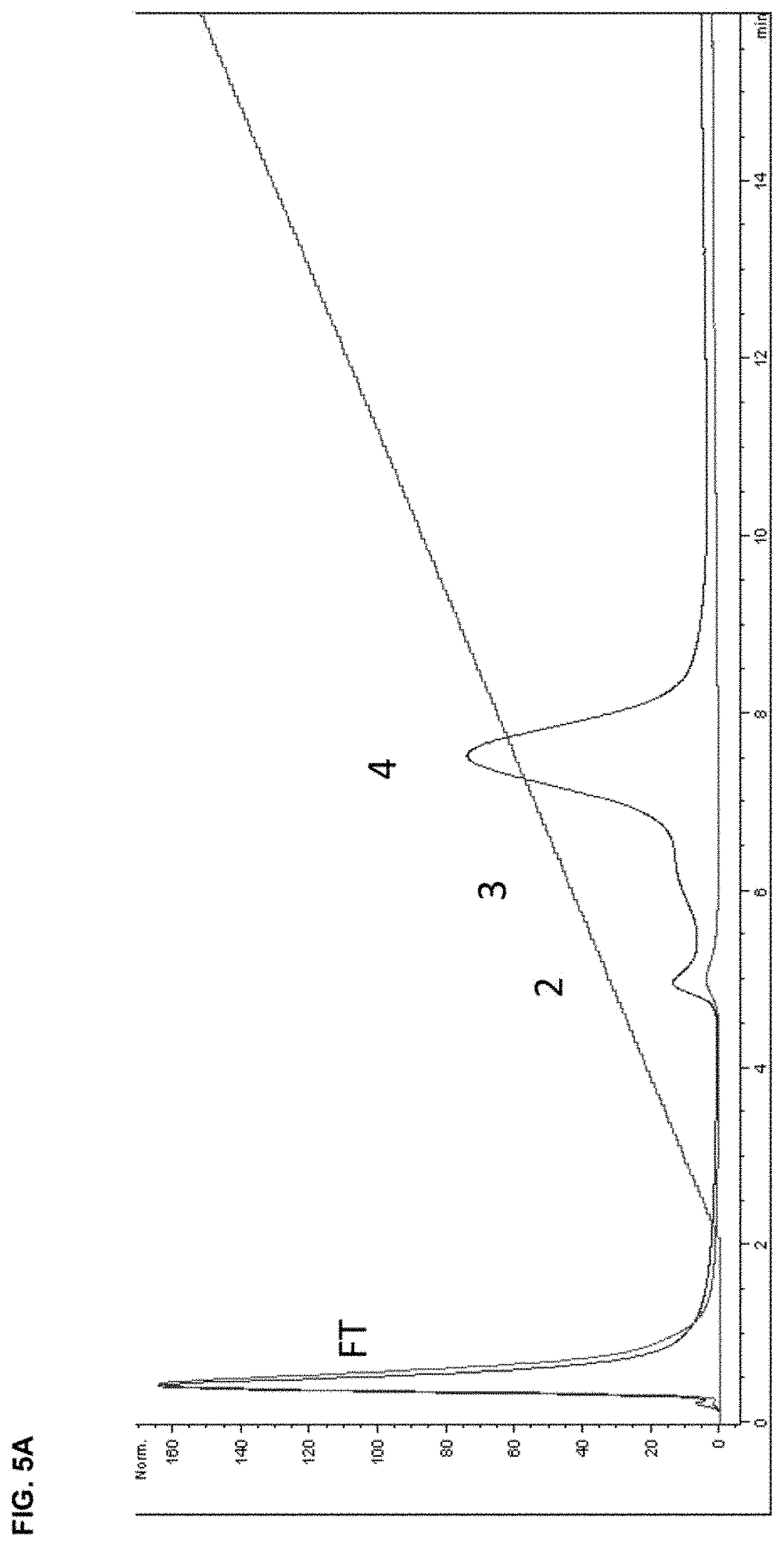
FIG. 5A depicts the A.sub.280 trace from hydroxyapatite chromatography of mAb1 and a D10-mAb1 conjugate (x-axis=minutes, y-axis=absorbance (normalized)).
FIG. 5B depicts SDS-PAGE gel of fractions from the flowthrough (FT) and peak 4.
FIG. 6A depicts hydroxyapatite chromatography of chemical conjugates with increasing numbers of peptides. The absorbance at 280 nm of the eluate for each conjugate is shown (x-axis=minutes, y-axis=absorbance (normalized)).
FIG. 6B depicts the fraction of analyte bound (upper curve, circles, scale left) and the retention time (lower curve, triangles, scale right) as a function of the number of peptides conjugated as determined by SDS-PAGE.
FIG. 7 depicts an in vitro TGF.beta. neutralization assay performed with A549 cells with a control conjugate (PAR=0) and conjugates with an average of 4 or 9 peptides compared to unmodified mAb1.
FIG. 8A depicts the time-dependent biodistribution of fluorophore-labeled mAb1 and a chemical conjugate at 1 mg/kg (1 mpk) containing approximately 4.5 peptides. The times at which the animals were imaged are indicated in each panel. Per photograph, the left mouse received mAb1, and right mouse received the chemical conjugate. Image intensities have been adjusted to reveal differences in distribution.
FIG. 8B depicts the ratio of fluorescence found in the region of interest corresponding to the distal femur and the region of interest corresponding to the heart in the images shown in FIG. 8A. Circles correspond to mAb1 antibody and squares correspond to the D10 peptide conjugated with mAb1 (D10 mAb1).
FIG. 9A diagrammatically depicts possible locations of D10 peptides on an IgG subtype antibody for creating a series of mAb1 fusion variant antibodies to be obtained by attachment of D10 peptides to the heavy and/or light chain termini using recombinant methods. The sites of addition of D10 peptides are indicated by the circles and the use of peptide linker sequences is indicated by wavy lines (the longer wavy line represents a longer linker than the shorter wavy line).
FIG. 9B depicts a series of fusion variant antibodies (fusion variants) derived by placement of the peptides, as shown in FIG. 9A. Recombinant fusion variants with various combinations of the position of attachment and peptide numbers were generated. The smaller numbers below each diagram depict the identity of each recombinant fusion variant as referred to herein for the sake of clarity. As referred to herein, fusion variants are designated either with "fusion" or "F" followed by the intended variant number. For example, "Fusion 1" and "F1" both refer to an antibody having the configuration of the antibody "1" without a D10 peptide. The longer wavy line represents a longer linker than the shorter wavy line.
FIG. 10 depicts SDS-PAGE of the indicated purified recombinant mAb1 fusion variants under reducing (upper gel) or non-reducing (lower gel) conditions.
FIG. 11 depicts thermostability of recombinant mAb1 fusion variants as determined by differential scanning fluorimetry (DSF). The transition to a partially-denatured form at each temperature is detected by an increase in dye fluorescence. The slope of fluorescence increase with temperature (-d(RFU)/dT) was calculated and is displayed versus the temperature of the sample. The rate of the denaturation is maximal at the minima of the curves which represent the midpoint of the thermal transitions (Tm). For reference, the structures of each of the recombinant mAb1 fusion variants are shown diagrammatically.
FIGS. 12A and 12B depict the neutralization of TGF.beta. in eliciting the production of IL-11 by A549 cells in vitro by eight recombinant mAb1 fusion variants shown diagrammatically in FIG. 9B.
FIG. 13 depicts the affinity of recombinant mAb1 fusion variants and mAb1 chemical conjugates to hydroxyapatite as assessed by column chromatography on a column of ceramic hydroxyapatite.
FIG. 14 depicts the biodistribution of selected fluorophore-labeled recombinant mAb1 fusion variants and mAb1 chemical conjugates in CD-1 mice obtained by live imaging at various times post-administration.
FIG. 15 depicts the amount of fluorescent dye-labeled antibody, recombinant mAb1 fusion variant and mAb1 chemical conjugate localized to the vertebral column after administration to CD-1 mice. Fluorescence was measured by an IVIS instrument over a 3 week period. The logarithm of the maximum fluorescence within the ROI is shown.
FIG. 16 depicts fluorescence images of resected spine and femurs of mice administered recombinant mAb1 fusion variants mAb1 F6, mAb1 F16, and mAb1 F17 and mAb1 chemical conjugate ("Conj") in the study described in Example 13 and FIG. 15 after 10 and 21 days.
FIGS. 17A and 17B depict the fluorescence levels of mAb2 F1 and mAb2 F6 in 10 .mu.L serum, and resected lumbar portion of spine, distal (trabecular) femur, kidney and heart after 24 and 96 hr as described in Example 15.
FIG. 18A shows that bone targeting via mAb1-D10 (mAb1 F6) profoundly influences serum PK following single dose administration. mAb1 F6 exhibits 13 to 14 fold lower serum exposure (AUC), faster serum clearance, and shorter serum half-life (t.sub.1/2) than mAb1 as measured by ELISA. Data are expressed as mean.+-.SD: Statistical significance (*p.ltoreq.0.05 mAb1 F6 compared to mAb1) was observed as measured by analysis of variance (AVOVA), Dunnet's Multiple Comparison Test. mAb1 is murinized inhibitory anti-TGF.beta. monoclonal antibody and mAb1 F6 is a recombinant murinized inhibitory anti-TGF.beta. monoclonal antibody with an aspartate polypeptide D10 attached to the C-terminus of the heavy chain of mAb1. AUC for Imaging/Bone was normalized to 1.0. Doses were 5 mg/kg for each mAb1 F6 and mAb1.
FIG. 18B shows that mAb1 F6 exhibits a 22 fold higher exposure (AUC) in the bone as measured by Optical Imaging compared to mAb1. Data expressed as mean.+-.SD: Statistical significance (*p.ltoreq.0.05 mAb1 F6 compared to mAb1) was observed as measured by AVOVA, Dunnet's Multiple Comparison Test. Doses were 1 mg/kg for each mAb1 F6 and mAb1.
FIG. 19 illustrates multiple dose peak-trough PK profiles. Bone targeting (via mAb1 F6) profoundly influences multiple dose peak-trough serum PK. mAb1 F6 exhibits lower serum concentrations than mAb1 at both 24 and 48 hr post-dose and following the first dose and dose 23. Fold-differences were lower for mAb1 F6 by 3 to 4.5 fold at 24 hr and 6 to 9 fold at 48 hr. Accumulation also appeared less with mAb1 F6 than with mAb1. Data expressed as mean.+-.SD: Statistical significance (*p.ltoreq.0.05 mAb1 F6 compared to mAb1) was observed as measured by unpaired t-test. Serum concentrations were measured via mass spectrometry.
FIG. 20 shows that bone targeting (mAb1 F6) and mAb1 increase BV/TV (%) in a dose responsive fashion in G610C (OI) mice. Significant changes on BV/TV (%) compared to control antibody 13C4 (mouse IgG1 antibody) treated G610C mice were observed at doses of 1 and 5 mg/kg, for both treatments. G610C mice treated with 13C4 (13C4) exhibited significant decreases in BV/TV compared to WT background strain (WT 13C4). Data expressed as mean.+-.SD: Statistical significance (*p.ltoreq.0.05 mAb1 F6 compared to mAb1; .sup.#p.ltoreq.0.05 13C4 compared to WT 13C4) was observed as measured by one way ANOVA. BV/TV (%) measured via .mu.CT imaging.
FIG. 21 shows that bone targeting (mAb1 F6) and mAb1 increase maximum force to failure in a dose responsive fashion in G610C (OI) mice. Significant changes on maximum force to failure compared to 13C4-treated G610C mice were observed at 1 and 5 mg/kg for mAb1 F6 and 5 mg/kg, only, for mAb1. G610C mice treated with an antibody control (13C4) exhibited significant decreases in maximum force to failure compared to WT background strain. Data expressed as mean.+-.SD: Statistical significance (*p.ltoreq.0.05 mAb1 F6 compared to mAb1; .sup.#p.ltoreq.0.05 13C4 compared to WT 13C4) was observed as measured by one-way ANOVA. Maximum force to failure was measured via biomechanical compression test.
FIG. 22 shows the effects of mAb1 and mAb1 F6 on BV/TV in G610C mice. The antibodies were dosed at various frequencies (3.times. weekly, 1.times. weekly, 1.times. every 2 weeks, or 1.times. every 4 weeks) at 5 mg/kg for 12 weeks. Antibody 13C4 was used as control. Statistical significance (*p.ltoreq.0.05 mAb1 F6 compared to mAb1; .sup.#p.ltoreq.0.05 13C4 compared to WT 13C4) was observed as measured by one way ANOVA. BV/TV was measured via .mu.CT imaging.
FIG. 23 shows the effects of mAb1 and mAb1 F6 on maximum force to failure in G610C mice. The antibodies were dosed at various frequencies (3.times. weekly, 1.times. weekly, 1.times. every 2 weeks, or 1.times. every 4 weeks) at 5 mg/kg for 12 weeks. Antibody 13C4 was used as control. Statistical significance (*p.ltoreq.0.05 mAb1 or mAb1 F6 compared to 13C4) was observed as measured by one-way ANOVA. Maximum force to failure was measured via biomechanical compression test.
FIG. 24 shows the effects of mAb1 and mAb1 F6 on BV/TV (%) and the antibodies' average serum levels in G610C mice. The antibodies were dosed 1.times. every 2 weeks or 1.times. weekly at 5 mg/kg for 12 weeks. Antibody 13C4 was used as control. Statistical significance (*p.ltoreq.0.05 mAb1 or mAb1 F6 compared to 13C4; .sup.#p.ltoreq.0.05 13C4 compared to WT 13C4) was observed as measured by one-way ANOVA. BV/TV (%) was measured via .mu.CT imaging.
FIG. 25 shows the effects of mAb1 and mAb1 F16 on BV/TV (%) in G610C mice. The antibodies were dosed 3.times. weekly at 5 mg/kg for 8 weeks. Antibody 13C4 was used as control. Statistical significance (*p.ltoreq.0.05 mAb1 or mAb1 F16 compared to 13C4; .sup.#p.ltoreq.0.05 13C4 compared to WT 13C4) was observed as measured by one-way ANOVA. BV/TV (%) was measured via .mu.CT imaging.
FIG. 26 shows the effects of mAb1 and mAb1 F11 on BV/TV (%) in wild type mice. The antibodies were dosed 3.times. weekly at 5 mg/kg for 9 weeks. Statistical significance (*p.ltoreq.0.05 mAb1 or mAb1 F11 compared to 13C4) was observed as measured by one-way ANOVA. BV/TV (%) was measured via .mu.CT imaging.
FIG. 27 shows the total radiant efficiency in the lumbar of wild type mice after receiving a single intraperitoneal dose of vehicle or fluorescently labeled mAb1 or mAb1 F6. Statistical significance (*p.ltoreq.0.05 mAb1 F6 compared to mAb1) was observed as measured by one-way ANOVA.
FIG. 28 shows the total radiant efficiency in the heart of wild type mice after receiving a single intraperitoneal dose of vehicle or fluorescently labeled mAb1 or mAb1 F6. Statistical significance (*p.ltoreq.0.05 mAb1 compared to mAb1 F6) was observed as measured by one-way ANOVA.
FIG. 29 shows the total radiant efficiency in the liver of wild type mice after receiving a single intraperitoneal dose of vehicle or fluorescently labeled mAb1 or mAb1 F6. Statistical significance (*p.ltoreq.0.05 mAb1 compared to mAb1 F6) was observed as measured by one-way ANOVA.
FIG. 30 shows the total radiant efficiency in the intestine of wild type mice after receiving a single intraperitoneal dose of vehicle or fluorescently labeled mAb1 or mAb1 F6.
FIG. 31 shows the total radiant efficiency in the indicated tissues of wild type mice at 24 hrs or 96 hrs after receiving a single intraperitoneal dose of fluorescently labeled mAb2 or mAb2 D10. Statistical significance (*p.ltoreq.0.05 mAb1 compared to mAb1 F6) was observed as measured by t test.
FIG. 32 shows the lumbar/serum total radiant efficiency ratios in wild type mice at 24 hrs or 96 hrs after receiving a single intraperitoneal dose of fluorescently labeled mAb2 or mAb2 D10.
FIG. 33 shows the femur/serum total radiant efficiency ratios in wild type mice at 24 hrs or 96 hrs after receiving a single intraperitoneal dose of fluorescently labeled mAb2 or mAb2 D10. Statistical significance (*p.ltoreq.0.05 mAb2 D10 compared to mAb2) was observed as measured by t test.
DETAILED DESCRIPTION OF THE INVENTION
The present invention provides antibodies and antigen-binding fragments thereof that are connected to one or more bone-targeting poly-D peptides such that the antibodies and fragments preferentially home to the bones in a patient in need thereof. The bone-targeting feature of such an antibody or fragment allows the antibody and fragment to target bone tissues specifically and reduces the patient's systemic exposure to the antibody or fragment, thereby enhancing the efficacy of the drug while minimizing undesired adverse side effects.
As used herein, the term "poly-D peptide" refers to a peptide sequence having a plurality of aspartic acid or aspartate or "D" amino acids, such as about 2, 3, 4, 5, 6, 7, 8, 9, 10, 20, 30, or more aspartic acid amino acids (residues). In one embodiment, a poly-D peptide can include about 2 to about 30, or about 3 to about 15, or about 4 to about 12, or about 5 to about 10, or about 6 to about 8, or about 7 to about 9, or about 8 to about 10, or about 9 to about 11, or about 12 to about 14 aspartic acid residues. In one embodiment, poly-D peptides include only aspartate residues. In another embodiment, poly-D peptides may include one or more other amino acids or similar compounds. As used herein, the term "D10" refers to a contiguous sequence of ten aspartic acid amino acids, as seen in SEQ ID NO: 1. In some embodiments, an antibody or antibody fragment of the invention may include 1, 2, 3, 4, 5, 6, 7, 8, 9, 10, 11, 12, or more than 12 poly-D peptides.
The poly-D peptide can be connected to an antibody or antigen-binding fragment of interest via recombinant technology or chemical conjugation. As used herein, the term "fusion variant" or "variant" refers to an assembled antibody construct (see FIG. 9B) that includes at least one of a heavy chain or a light chain or antibody fragment or subpart that incorporates or is otherwise associated with a poly-D peptide, such as a D10 sequence. For example, a poly-D peptide can be connected to an antibody chain in a fusion variant by recombinant technology (e.g., where a poly-D peptide sequence is integral with the amino acid sequence of the heavy chain, light chain, or antibody fragment or subpart), chemical conjugation, or both.
As used herein, the term "chemical conjugate" refers to an assembled antibody that includes at least one of a heavy chain or a light chain or antibody fragment or subpart to which one or more poly-D peptides are connected by chemical reaction with, for example, the cysteine residues present in the amino acid sequence of the heavy chain, light chain, antibody fragment, or subpart. Exemplary cysteine residues that can be used for conjugation are those in the heavy chain hinge region. Cysteine residues or other residues appropriate for conjugation can also be introduced to the antibody chain by mutagenesis. A spacer/linker such as a peptide linker or a chemical moiety (e.g., a maleimide function group and a polyethylene glycol (PEG)) may be used between the poly-D peptide and the antibody component in the conjugation. Methods for chemical conjugation of desired moieties to antibodies are well known in the art. See, e.g., Behrens and Liu, mAbs 6:1, 46-53 (2014).
As used herein, the term "integral" refers to the integration of a poly-D peptide with an antibody chain via recombinant technology such that the poly-D peptide is transcribed from the same RNA transcript as the antibody chain and resides in the same polypeptide sequence as the antibody chain. In such cases, the poly-D peptide can be connected to the antibody chain, with or without any peptide linker or amino acid spacer, at the antibody chain's either or both termini, or integrated internally to the antibody chain, without affecting the antibody chain's proper folding, the antibody molecule's assembly, or the antibody's binding to its antigen.
Exemplary formats of the bone-targeting antibodies of the present invention are shown in FIG. 9B (formats F2-F20). The bone-targeting peptide (represented by circles) can be attached or fused to (e.g., integral with) either or both termini of the heavy chain and/or light chain of the antibody. In some embodiments, the bone-targeting peptide is not attached to the light chain through the light chain's N-terminus. The attachment or fusion can be a direct connection (i.e., without a spacer or linker), or through a spacer or linker (represented by the wavy lines; e.g., a peptide linker). Specific examples of these formats are shown in Tables 1 and 7 below.
Any suitable spacer or linker can be used herein to attach the bone-targeting peptide by, e.g., recombinant technology or chemical conjugation, to an antibody of interest. For example, a peptide linker having one, two, three, or more repeats of the G4S peptide (SEQ ID NO: 9) may be used. Other suitable peptide linkers can also be used. See, e.g., Chen et al., Adv Drug Deliv Rev 65(10):1357-1369 (2013).
Exemplary Bone-Targeting Antibodies and Antigen-Binding Fragments Thereof
The present invention discloses antibodies and antigen-binding fragments having one or more poly-D (poly-aspartate or poly-Asp) peptides (e.g., a D10 sequence) attached thereto. These modified antibodies and fragments have improved localization to bone. In one particular embodiment, these antibodies are anti-TGF.beta. antibodies, as described herein. While not wishing to be bound by theory, it is believed that effectively targeting anti-TGF.beta. antibodies to bone with one or more poly-D peptides may provide a new therapy for individuals with diseases characterized by pathophysiological bone degeneration associated with TGF.beta..
However, while numerous embodiments and examples herein are expressed in the context of using .alpha.-TGF.beta. antibodies and D10 sequences, it is contemplated that other antibodies or proteins suitable for treating an abnormal bone condition or a bone disease can be modified with bone-targeting moieties as described herein. For example, therapeutic antibodies for treating bone loss, stimulating bone growth, or targeting abnormal cells (e.g., cancer cells) in bone can be linked to one or more bone-targeting peptides as described herein. The therapeutic antibodies may bind to proteins or peptides involved in bone formation or maintenance. Further, other bone localization or targeting peptides may be used.
As used herein, the terms ".alpha.-TGF.beta. antibody" and "anti-TGF.beta. antibody" can be used interchangeably and refer to an antibody, or an antigen-binding fragment thereof, that is specific for TGF.beta.1, TGF.beta.2, and/or TGF.beta.3. For example, at least one antigen-binding site (or paratope) of an .alpha.-TGF.beta. antibody, or an antigen-binding fragment thereof, binds to an epitope found on human TGF.beta.1, TGF.beta.2, and/or TGF.beta.3.
In one embodiment, a contemplated .alpha.-TGF.beta. antibody-D10 construct may be created by chemical conjugation. For example, chemical conjugation may be performed by methods known in the art such as those disclosed in U.S. Pat. Nos. 7,763,712, 4,671,958, and 4,867,973, each of which is incorporated by reference. In another example, a peptide or other linker can be used to attach a D10 peptide to an antibody (see FIGS. 1 and 2). In a further embodiment, reduction of thiol groups at the hinge region (e.g., hinge region cysteine residues) of the antibody allows chemical conjugation of poly-D peptides using a PEG spacer. Similarly, other cysteine residues of contemplated antibodies and antibody fragments, either native to the antibodies and fragments or introduced by mutagenesis, can be chemically conjugated with poly-D peptides. One such contemplated assembly scheme of an .alpha.-TGF.beta. antibody chemically conjugated with a D10 peptide is illustrated in FIG. 2.
In another embodiment, a contemplated .alpha.-TGF.beta. antibody-D10 construct may be created by recombinant expression, where the D10 sequence is added to the amino acid sequence of the heavy chain and/or light chain of the .alpha.-TGF.beta. antibody. For example, the nucleic acid sequences encoding the amino acid sequences of the heavy and/or light chains can be modified to encode a D10 sequence that would be expressed either at the N-terminus, the C-terminus, or both N-terminus and C-terminus of the heavy and/or light chains of the .alpha.-TGF.beta. antibody. Similarly, one or more D10 sequences could be added to an amino acid sequence of an antibody heavy chain at or near the hinge region and/or within the amino acid sequence of an antibody light chain. Each nucleic acid sequence for the D10 harboring-heavy and/or light chain may be incorporated into an expression vector and subsequently transfected into a host cell capable of expressing and translating the nucleic acid sequence into the corresponding amino acid sequence. Moreover, the host cell is capable of assembling the expressed amino acid sequences into the functional protein by combining each of the heavy chain and light chain with its complementary sequence to form an .alpha.-TGF.beta. antibody-D10 construct. Examples of contemplated recombinant .alpha.-TGF.beta. antibody-D10 fusion variants are illustrated in FIGS. 9A and 9B.
While a poly-D peptide is discussed herein, other similar peptides may also be used to enable targeting of an antibody, another protein, or a peptide to bone. For example, aspartic acid repeat sequences may have more or fewer residues than a D10 sequence, such as about 2, or about 4, or about 6, or about 8, or about 12, or about 14, or about 16, or, about 18, or about 20, or about 30, or 6, 7, 8, 9, 10 or 11 residues, and the like. Further, other natural amino acids with similar chemical properties, such as glutamate, or non-natural amino acids and/or other chemically equivalent compounds may be substituted for or used in combination with aspartic acid, as well.
In one embodiment, it is contemplated that an antibody with one or more poly-D peptides attached thereto will exhibit at least about a 2-fold, or about a 3-fold, or about at 5-fold, or about a 10-fold, or about a 20-fold increase in localization to bone compared to the same antibody without the one or more poly-D peptides.
Moreover, while an .alpha.-TGF.beta. antibody is described herein, any antibody that binds other proteins involved in bone formation or bone maintenance may be similarly modified to target the antibody to bone, as desired. Antibodies or antigen-binding fragments thereof contemplated herein may be from any species or represent hybrid antibodies combining heavy chains and light chains from different species, and may be specific for any desired epitope. In addition, antibodies that may be used herein are not limited by isotype, and may be any of IgG.sub.1, IgG.sub.2, IgG.sub.3, IgG.sub.4, IgA.sub.1, IgA.sub.2, IgM, IgE, or IgD. Antibody fragments may also be used. For example, D10 sequences or other bone-targeting compounds may be attached to Fab and/or Fc fragments or any other antibody fragment to achieve a desired result as described herein. Further, D10 sequences can be attached to scFv fragments and other similar fusion proteins. In another embodiment, D10 sequences can be attached to antibodies having a S228P core-hinge mutation (numbered according to the EU numbering system; or alternatively S241P according to the Kabat system; see Kabat et al., Sequences of Proteins of Immunological Interest, 4.sup.th ed., United States Government Printing Office, 165-492 (1987); and Silva et al. Jour. Biol. Chem. 290:5462-5469 (2015)).
In a further embodiment, antibodies and/or other proteins contemplated herein may be conjugated with additional molecules. For example, antibodies or other proteins contemplated herein may be conjugated with chemical labels that allow tracking of the antibodies/proteins when injected or otherwise introduced into a subject. For example, radiolabels, fluorescent compounds, and the like may be attached to the antibodies/proteins to aid their tracking in vivo. Further, antibodies and/or other proteins contemplated herein may also be conjugated with additional compounds having a therapeutic effect, such as small molecules, pharmaceuticals, antineoplastic agents, growth hormones, vitamins, etc., such that the antibodies and/or other proteins may serve as a vehicle for one or more of such compounds.
In some embodiments, the bone-targeting anti-TGF.beta. antibody comprises a heavy chain comprising an amino acid sequence set forth in any of SEQ ID NOS: 2, 3, 4, and 5, and a light chain comprising an amino acid sequence set forth in any of SEQ ID NOS: 6, 7, 8, 11, and 12, with the proviso that the heavy chain amino acid sequence is not SEQ ID NO: 2 when the light chain amino acid sequence is SEQ ID NO: 6. Exemplary antibodies are mAb1 F3, mAb1 F4, mAb1 F5, mAb1 F6, mAb1 F8, mAb1 F9, mAb1 F10, mAb1 F11, mAb1 F13, mAb1 F14, mAb1 F15, mAb1 F16, mAb1 F18, mAb1 F19, and mAb1 F20 (Table 1).
In other embodiments, the bone-targeting anti-TGF.beta. antibody comprises a heavy chain comprising an amino acid sequence set forth in any of SEQ ID NOS: 13, 14, 16, and 17, and a light chain comprising an amino acid sequence set forth in any of SEQ ID NOS: 15, 18, 19, 20, 21, and 22, with the proviso that the heavy chain amino acid sequence is not SEQ ID NO: 13 when the light chain amino acid sequence is SEQ ID NO: 15. Exemplary antibodies are mAb2 F3, mAb2 F4, mAb2 F5, mAb2 F6, mAb2 F8, mAb2 F9, mAb2 F10, mAb2 F11, mAb2 F13, mAb2 F14, mAb2 F15, mAb2 F16, mAb2 F18, mAb2 F19, and mAb2 F20 (Table 7).
In some embodiments, the antibodies of the present invention, such as the anti-TGF.beta. antibodies, do not have the C-terminal lysine in the heavy chain. The C-terminal lysine may be removed during manufacture or by recombinant technology (i.e., the coding sequence of the heavy chain does not include a codon for the C-terminal terminal lysine). Thus contemplated within the invention also are antibodies comprising the heavy chain amino acid sequence of SEQ ID NO: 2 or 13 without the C-terminal lysine. A poly-D peptide may be attached to the C-terminus of a heavy chain with or without the C-terminal lysine.
Treatment Methods
In one particular embodiment, a method of treating an individual such as a human patient for bone loss associated with TGF.beta. includes administering an effective amount of an anti-TGF.beta. antibody targeted to bone to the individual. The method can further include a step of measuring or detecting a reduction in TGF.beta. levels or activity, a reduction in bone loss or the rate of bone loss, an increase in bone density, and/or an increase in bone strength.
An "effective amount," as used herein, refers to an amount of a therapeutic agent, such as an .alpha.-TGF.beta. antibody or antibody fragment, that when administered to an individual in need thereof improves an individual's health, such as, for example, by reducing TGF.beta. levels or activity associated with bone, reducing bone loss or the rate of bone loss, increasing bone density, and/or increasing bone strength.
As used herein, the term "individual" refers to an animal. Examples of individuals include humans, domesticated animals, household pets, and other animals without limitation. Further examples of individuals include animals having a bone disease associated with TGF.beta..
In another embodiment, pharmaceutical antibody formulations or compositions including aqueous liquid drug product formulations and lyophilized drug product formulations containing one or more bone-targeting anti-TGF.beta. antibodies such as chemical conjugates or recombinant fusion variants are contemplated. Pharmaceutical compositions including bone-targeting anti-TGF.beta. antibody and/or antibody fragments can be formulated as described in U.S. Patent Application Publication No. US 2014/0286933 A9, which is incorporated herein by reference, or otherwise as is known in the art.
In one particular embodiment, a method for treating bone disease includes administering an effective amount of an anti-TGF.beta. antibody targeted to bone to an individual with a bone disease, such as a bone diseases associated with chronic kidney disease, cancer metastasis to bone, or abnormal metabolic conditions. In another particular embodiment, a method for treating osteogenesis imperfecta includes administering an effective amount of an anti-TGF.beta. antibody targeted to bone to an individual with osteogenesis imperfecta. In a further particular embodiment, a method for treating osteoporosis includes administering an effective amount of an anti-TGF.beta. antibody targeted to bone to an individual with osteoporosis.
In some embodiments, the patients are treated with a combination of a bone-targeting antibody or antibody fragment of the present invention and another therapeutic agent, such as a therapeutic agent for a bone loss condition (e.g., bisphosphonates). The antibody or antibody fragment and the other therapeutic agent can be administered to the patient simultaneously or sequentially.
Methods of Making Antibodies
The antibodies or fragments of the present invention can be made by methods well established in the art. DNA sequences encoding the heavy and light chains of the antibodies can be inserted into expression vectors such that the genes are operatively linked to necessary expression control sequences such as transcriptional and translational control sequences. Expression vectors include plasmids, retroviruses, adenoviruses, adeno-associated viruses (AAV), plant viruses such as cauliflower mosaic virus, tobacco mosaic virus, cosmids, YACs, EBV derived episomes, and the like. The antibody light chain coding sequence and the antibody heavy chain coding sequence can be inserted into separate vectors, and may be operatively linked to the same or different expression control sequences (e.g., promoters). In one embodiment, both coding sequences are inserted into the same expression vector and may be operatively linked to the same expression control sequences (e.g., a common promoter), to separate identical expression control sequences (e.g., promoters), or to different expression control sequences (e.g., promoters). The antibody coding sequences may be inserted into the expression vector by standard methods (e.g., ligation of complementary restriction sites on the antibody gene fragment and vector, or blunt end ligation if no restriction sites are present).
In addition to the antibody chain genes, the recombinant expression vectors may carry regulatory sequences that control the expression of the antibody chain genes in a host cell. Examples of regulatory sequences for mammalian host cell expression include viral elements that direct high levels of protein expression in mammalian cells, such as promoters and/or enhancers derived from retroviral LTRs, cytomegalovirus (CMV) (such as the CMV promoter/enhancer), Simian Virus 40 (SV40) (such as the SV40 promoter/enhancer), adenovirus, (e.g., the adenovirus major late promoter (AdMLP)), polyoma and strong mammalian promoters such as native immunoglobulin and actin promoters.
In addition to the antibody chain genes and regulatory sequences, the recombinant expression vectors of the invention may carry additional sequences, such as sequences that regulate replication of the vector in host cells (e.g., origins of replication) and selectable marker genes. For example, the selectable marker gene confers resistance to drugs, such as G418, hygromycin or methotrexate, on a host cell into which the vector has been introduced. Selectable marker genes may include the dihydrofolate reductase (DHFR) gene (for use in dhfr-host cells with methotrexate selection/amplification), the neo gene (for G418 selection), and the glutamate synthetase gene.
The expression vectors encoding the antibodies of the present invention are introduced to host cells for expression. The host cells are cultured under conditions suitable for expression of the antibody, which is then harvested and isolated. Host cells include mammalian, plant, bacterial or yeast host cell. Mammalian cell lines available as hosts for expression are well known in the art and include many immortalized cell lines available from the American Type Culture Collection (ATCC). These include, inter alia, Chinese hamster ovary (CHO) cells, NSO cells, SP2 cells, HEK-293T cells, 293 Freestyle cells (Invitrogen), NIH-3T3 cells, HeLa cells, baby hamster kidney (BHK) cells, African green monkey kidney cells (COS), human hepatocellular carcinoma cells (e.g., Hep G2), A549 cells, and a number of other cell lines. Cell lines may be selected based on their expression levels. Other cell lines that may be used are insect cell lines, such as Sf9 or Sf21 cells.
Further, expression of antibodies can be enhanced using a number of known techniques. For example, the glutamine synthetase gene expression system (the GS system) is a common approach for enhancing expression under certain conditions.
Tissue culture media for the host cells may include, or be free of, animal-derived components (ADC), such as bovine serum albumin. In some embodiments, ADC-free culture media is preferred for human safety. Tissue culture can be performed using the fed-batch method, a continuous perfusion method, or any other method appropriate for the host cells and the desired yield.
Pharmaceutical Compositions
The antibody of the invention can be formulated for suitable storage stability. For example, the antibody can be lyophilized or stored or reconstituted for use using pharmaceutically acceptable excipients. For a combination therapy, the two or more therapeutic agents such as antibodies can be co-formulated, e.g., mixed and provided in a single composition.
The term "excipient" or "carrier" is used herein to describe any ingredient other than the compound(s) of the invention. The choice of excipient(s) will to a large extent depend on factors such as the particular mode of administration, the effect of the excipient on solubility and stability, and the nature of the dosage form. "Pharmaceutically acceptable excipient" includes any and all solvents, dispersion media, coatings, antibacterial and antifungal agents, isotonic and absorption delaying agents, and the like that are physiologically compatible. Some examples of pharmaceutically acceptable excipients are water, saline, phosphate buffered saline, dextrose, glycerol, ethanol and the like, as well as combinations thereof. In some cases, isotonic agents, for example, sugars, polyalcohols such as mannitol, sorbitol, or sodium chloride will be included in the composition. Additional examples of pharmaceutically acceptable substances are wetting agents or minor amounts of auxiliary substances such as wetting or emulsifying agents, preservatives or buffers, which enhance the shelf life or effectiveness of the antibody.
A pharmaceutical composition of the invention may be prepared, packaged, or sold in bulk, as a single unit dose, or as a plurality of single unit doses. As used herein, a "unit dose" is a discrete amount of the pharmaceutical composition comprising a predetermined amount of the active ingredient. The amount of the active ingredient is generally equal to the dosage of the active ingredient which would be administered to a subject or a convenient fraction of such a dosage such as, for example, one-half or one-third of such a dosage.
The pharmaceutical compositions of the invention are typically suitable for parenteral administration. As used herein, "parenteral administration" of a pharmaceutical composition includes any route of administration characterized by physical breaching of a tissue of a subject and administration of the pharmaceutical composition through the breach in the tissue, thus generally resulting in the direct administration into the blood stream, into muscle, or into an internal organ. Parenteral administration thus includes, but is not limited to, administration of a pharmaceutical composition by injection of the composition, by application of the composition through a surgical incision, by application of the composition through a tissue-penetrating non-surgical wound, and the like. In particular, parenteral administration is contemplated to include, but is not limited to, subcutaneous, intraperitoneal, intramuscular, intrasternal, intravenous, intraarterial, intrathecal, intraventricular, intraurethral, intracranial, intratumoral, and intrasynovial injection or infusions; and kidney dialytic infusion techniques. Regional perfusion is also contemplated. Preferred embodiments may include the intravenous and subcutaneous routes.
Formulations of a pharmaceutical composition suitable for parenteral administration typically comprise the active ingredient combined with a pharmaceutically acceptable carrier, such as sterile water or sterile isotonic saline. Such formulations may be prepared, packaged, or sold in a form suitable for bolus administration or for continuous administration. Injectable formulations may be prepared, packaged, or sold in unit dosage form, such as in ampoules or in multi-dose containers containing a preservative. Formulations for parenteral administration include, but are not limited to, suspensions, solutions, emulsions in oily or aqueous vehicles, pastes, and the like. Such formulations may further comprise one or more additional ingredients including, but not limited to, suspending, stabilizing, or dispersing agents. In one embodiment of a formulation for parenteral administration, the active ingredient is provided in dry (i.e., powder or granular) form for reconstitution with a suitable vehicle (e.g., sterile pyrogen free water) prior to parenteral administration of the reconstituted composition. Parenteral formulations also include aqueous solutions which may contain excipients such as salts, carbohydrates and buffering agents (e.g., a pH of from 3 to 9), but, for some applications, they may be more suitably formulated as a sterile non-aqueous solution or as a dried form to be used in conjunction with a suitable vehicle such as sterile, pyrogen-free water. Exemplary parenteral administration forms include solutions or suspensions in sterile aqueous solutions, for example, aqueous propylene glycol or dextrose solutions. Such dosage forms can be suitably buffered, if desired. Other parentally-administrable formulations which are useful include those which comprise the active ingredient in microcrystalline form, or in a liposomal preparation. Formulations for parenteral administration may be formulated to be immediate and/or modified release. Modified release formulations include delayed-, sustained-, pulsed-, controlled-, targeted and programmed release.
In some embodiments, the antibody or antigen-binding fragment of the present invention may be administered at 40, 20, or 15 mg/kg or less (such as 14, 13, 12, 11, 10, 9, 8, 7, 6, 5, 4, 3, 2, or 1 mg/kg). In some further embodiments, the doses may be 0.01, 0.02, 0.03, 0.04, 0.05, 0.1, 0.2, 0.3, 0.4, or 0.5 mg/kg. The dosing frequency may be, for example, daily, every two, three, four, or five days, weekly, biweekly, or triweekly, monthly, bimonthly, every three months, every six months, or every twelve months, or as needed. The antibody may be administered by intravenous (e.g., intravenous infusion over 0.5-8 hours), subcutaneously, intramuscularly, or any other route of administration that is appropriate for the condition and the drug formulation.
EXEMPLARY EMBODIMENTS
Further particular embodiments of the present invention are described as follows.
1. An antibody, or an antigen-binding fragment thereof, comprising a heavy chain, a light chain, and one or more poly-aspartate (poly-D) peptides connected to (i) the heavy chain, (ii) the C-terminus of the light chain, or (iii) both (i) and (ii).
2. The antibody or antigen-binding fragment of embodiment 1, wherein the one or more poly-D peptides are connected to the antibody or antigen-binding fragment by chemical conjugation.
3. The antibody or antigen-binding fragment of embodiment 2, wherein the one or more poly-D peptides are conjugated to the heavy chain at the hinge region.
4. The antibody or antigen-binding fragment of embodiment 2 or 3, wherein the one or more poly-D peptides are conjugated to the antibody or antigen-binding fragment by a polyethylene glycol (PEG) spacer.
5. The antibody or antigen-binding fragment of embodiment 1, comprising a poly-D peptide integral with an amino acid sequence of the heavy chain or the light chain.
6. The antibody or antigen-binding fragment of embodiment 5, comprising a poly-D peptide integral with the N-terminus of the heavy chain.
7. The antibody or antigen-binding fragment of embodiment 5, comprising a poly-D peptide integral with the C-terminus of the heavy chain.
8. The antibody or antigen-binding fragment of embodiment 5, comprising a first poly-D peptide integral with the N-terminus of the heavy chain and a second poly-D peptide integral with the C-terminus of the heavy chain.
9. The antibody or antigen-binding fragment of any one of embodiments 5-8, comprising a poly-D peptide integral with the C-terminus of the light chain.
10. The antibody or antigen-binding fragment of any one of embodiments 5-9, wherein the poly-D peptide(s) are fused to the heavy or light chain via a peptide linker.
11. The antibody or antigen-binding fragment of embodiment 10, wherein the peptide linker comprises 1-3 repeats of the amino acid sequence GGGGS (SEQ ID NO: 9).
12. The antibody or antigen-binding fragment of any one of the preceding embodiments, wherein the one or more poly-D peptides each independently comprise 2-30 aspartic acid residues.
13. The antibody or antigen-binding fragment of embodiment 12, wherein the one or more poly-D peptides each comprise 10 aspartic acid residues (SEQ ID NO: 1).
14. The antibody or antigen-binding fragment of any one of the preceding embodiments, wherein the antibody is an IgG.
15. The antibody or antigen-binding fragment of embodiment 15, wherein the antibody is an IgG.sub.1 or IgG.sub.4.
16. The antibody or antigen-binding fragment of any one of the preceding embodiments, wherein the antibody or antigen-binding fragment specifically binds to one or more of TGF.beta.1, TGF.beta.2, and TGF.beta.3.
17. The antibody or antigen-binding fragment of embodiment 16, wherein the antibody comprises the heavy chain complementarity-determining regions (CDR) 1-3 in SEQ ID NO: 13 and the light chain CDR1-3 in SEQ ID NO: 15.
18. The antibody or antigen-binding fragment of embodiment 17, wherein the antibody comprises a heavy chain variable domain (V.sub.H) amino acid sequence corresponding to residues 1-120 of SED ID NO: 13 and a light chain variable domain (V.sub.L) amino acid sequence corresponding to residues 1-108 of SEQ ID NO:15.
19. The antibody or antigen-binding fragment of embodiment 17 or 18, wherein the antibody comprises a human IgG.sub.4 constant region having a proline at position 228 (EU numbering).
20. The antibody or antigen-binding fragment of embodiment 19, wherein the heavy chain of the antibody comprises the amino acid sequence of SEQ ID NO: 13 with or without the heavy chain C-terminal lysine, and the light chain of the antibody comprises the amino acid sequence of SEQ ID NO: 15.
21. The antibody of embodiment 17, wherein the heavy chain comprises the amino acid sequence of SEQ ID NO: 13 with or without the heavy chain C-terminal lysine, SEQ ID NO: 14 with or without the lysine immediately preceding the C-terminal D10 sequence, SEQ ID NO: 16 with or without the heavy chain C-terminal lysine, or SEQ ID NO: 17 with or without the lysine immediately preceding the C-terminal D10 sequence, and the light chain comprises the amino acid sequence of SEQ ID NO: 15, 19, 21, or 22.
22. The antibody or antigen-binding fragment of embodiment 16, wherein the antibody is mouse antibody 1D11 having the heavy and light chain amino acid sequences of SEQ ID NO: 2 with or without the C-terminal lysine and SEQ ID NO: 6, respectively.
23. An IgG.sub.4 antibody that binds human TGF.beta.1, TGF.beta.2, and TGF.beta.3, wherein the heavy chain of the antibody comprises the amino acid sequence of SEQ ID NO: 14 (with or without the lysine immediately preceding the C-terminal D10 sequence), and the light chain comprises the amino acid sequence of SEQ ID NO: 15.
24. An IgG.sub.4 antibody that binds human TGF.beta.1, TGF.beta.2, and TGF.beta.3, wherein the heavy chain of the antibody comprises the amino acid sequence of SEQ ID NO: 17 (with or without the lysine immediately preceding the C-terminal D10 sequence), and the light chain comprises the amino acid sequence of SEQ ID NO: 15.
25. The antibody or antigen-binding fragment of any one of the preceding embodiments, wherein the antibody or antigen-binding fragment exhibits at least a 2-fold increase in localization to bone compared to an antibody with the same heavy chain and light chain but lacking the poly-D peptide(s).
26. A pharmaceutical composition comprising an antibody or antigen-binding fragment of any one of the preceding embodiment s and a pharmaceutically acceptable excipient.
27. A method for treating an individual with a bone condition that benefits from inhibition of TGF.beta., comprising administering to the individual an effective amount of an anti-TGF.beta. antibody or antigen-binding fragment of any one of embodiments 16-25.
28. The method of embodiment 27, further comprising detecting at least one of (1) a reduction in TGF.beta. levels, (2) a reduction in TGF.beta. activity, (3) a reduction in bone loss, (4) a reduction in rate of bone loss, (5) an increase in bone density, (6) an increase in bone strength, and (7) a reduction in IL-11 levels.
29. An antibody or antigen-binding fragment of any one of embodiments 16-25 for use in treating an individual with a bone condition that benefits from inhibition of TGF.beta..
30. Use of an antibody or antigen-binding fragment of any one of embodiment 16-25 for the manufacture of a medicament for treating an individual with a bone condition that benefits from inhibition of TGF.beta..
31. The method of embodiment 27, the antibody or antigen-binding fragment for use of embodiment 29, or the use of embodiment 30, wherein the individual is a human.
32. The method, antibody or antigen-binding fragment for use, or use of embodiment 31, wherein the human has osteogenesis imperfecta.
33. The method, antibody or antigen-binding fragment for use, or use of embodiment 31, wherein the human has bone loss or osteoporosis.
34. The method, antibody or antigen-binding fragment for use, or use of embodiment 31, wherein the human has chronic kidney disease.
35. The method, antibody or antigen-binding fragment for use, or use of embodiment 31, wherein the human is a cancer patient with bone metastasis.
36. An isolated nucleic acid molecule, comprising a nucleotide sequence encoding the heavy chain, the light chain, or both, of the antibody or antigen-binding fragment of any one of embodiments 1-25.
37. An expression vector comprising the isolated nucleic acid molecule of embodiment 36.
38. A host cell comprising the expression vector of embodiment 37.
39. The host cell of embodiment 38, wherein the host cell is a mammalian cell.
40. A method of producing an antibody or antigen-binding fragment of any one of embodiments 1-25, the method comprising: providing a host cell comprising first and second nucleotide sequences encoding the heavy chain and light chain, respectively, of the antibody or antigen-binding fragment, growing the host cell under conditions permitting production of the antibody or antigen-binding fragment, and recovering the antibody or antigen-binding fragment.
41. The method of embodiment 40, wherein the first nucleotide sequence comprises SEQ ID NO: 23, 24, 25, or 26 (with or without the codon for the heavy chain C-terminal lysine), and the second nucleotide sequence comprises SEQ ID NO: 27, 29, 31, or 32.
42. A method of producing a bone-targeting antibody or antigen-binding fragment, comprising: providing an antibody or an antigen-binding fragment thereof and one or more poly-D peptides, and attaching the poly-D peptides to the antibody through a covalent bond by chemical conjugation.
43. The method of any one of embodiments 40-42, further comprising formulating the antibody or antigen-binding fragment as a pharmaceutical composition comprising a pharmaceutically acceptable carrier.
44. A method of producing a bone-targeting pharmaceutical composition, comprising: providing an antibody or antigen-binding fragment of any one of embodiments 1-25, and admixing the antibody or antigen-binding fragment with a pharmaceutically acceptable carrier.
The invention will be further described in the following examples, which do not limit the scope of the invention described in the claims.
EXAMPLES
The Examples that follow are illustrative of specific embodiments of the invention, and various uses thereof. They are set forth for explanatory purposes only and are not to be taken as limiting of the invention.
TGF.beta. Antibody
A first anti-TGF.beta. antibody, referred to herein as mAb1, is a mouse IgG1 monoclonal antibody specific for human TGF-.beta.1, TGF-.beta.2, and TGF-.beta.3 ("pan-specific") and is available from R&D Systems (Clone#1D11, Minneapolis, Minn.). The mAb1 antibody served as a template in the examples.
A second anti-TGF.beta. antibody, referred to herein as mAb2, used in the examples is a human anti-TGF.beta. IgG4 antibody with a hinge mutation S228P (EU numbering). The mAb2 antibody is similar to antibodies disclosed in U.S. Pat. No. 9,090,685, which is incorporated by reference. Antibody mAb2 has an estimated molecular weight of 144 KD when un-glycosylated. Its heavy and light chain amino acid sequences are SEQ ID NOS: 13 and 15, respectively. These two sequences are shown below. Variable domains are italicized, and are designated herein as heavy chain variable domain (HCVD, SEQ ID NO: 39) and light chain variable domain (LCVD, SEQ ID NO: 40). CDRs are shown in boxes and are designated heavy chain complementarity-determining region 1 (HCDR1, SEQ ID NO: 33); HCDR2 (SEQ ID NO: 34); and HCDR3 (SEQ ID NO: 35), and light chain complementarity-determining region 1 (LCDR1, SEQ ID NO: 36); LCDR2 (SEQ ID NO: 37); and LCDR3 (SEQ ID NO: 38). The glycosylation site in the constant domain of the heavy chain is in boldface (N297).
TABLE-US-00001 (SEQ ID NO: 13) ##STR00001## ##STR00002## ASTKGPSVFP LAPCSRSTSE STAALGCLVK DYFPEPVTVS WNSGALTSGV HTFPAVLQSS GLYSLSSVVT VPSSSLGTKT YTCNVDHKPS NTKVDKRVES KYGPPCPPCP APEFLGGPSV FLFPPKPKDT LMISRTPEVT CVVVDVSQED PEVQFNWYVD GVEVHNAKTK PREEQFNSTY RVVSVLTVLH QDWLNGKEYK CKVSNKGLPS SIEKTISKAK GQPREPQVYT LPPSQEEMTK NQVSLTCLVK GFYPSDIAVE WESNGQPENN YKTTPPVLDS DGSFFLYSRL TVDKSRWQEG NVFSCSVMHE ALHNHYTQKS LSLSLGK (SEQ ID NO: 15) ##STR00003## ##STR00004## PSDEQLKSGT ASVVCLLNNF YPREAKVQWK VDNALQSGNS QESVTEQDSK DSTYSLSSTL TLSKADYEKH KVYACEVTHQ GLSSPVTKSF NRGEC
As described herein, other antibodies, antibody fragments, proteins, or peptides that bind proteins involved in bone formation or bone maintenance may be used.
Example 1: Preparation of Chemical Conjugates of D10 Peptide with mAb1 .alpha.-TGF.beta. Antibody
D10 peptide chemical conjugates of the .alpha.-TGF.beta. antibody mAb1 were prepared in the manner depicted in FIG. 2. mAb1 (2.0 mg) was exchanged into degassed borate buffer (25 mM sodium chloride, 1 mM DTPA, 20 mM sodium borate pH 8.0) by three rounds of ultrafiltration over Amicon.RTM. Ultra 50 kDa MWCO centrifugal filters (EMD Millipore). The hinge region disulfides were then reduced with 12 mol:mol dithiothreitol (DTT) per mAb for 2 hr at 37.degree. C. The product was desalted over a 4 mL Amicon.RTM. Ultra filter with degassed borate buffer. Aliquots (1 nmol, 150 .mu.g) were reacted at 25.degree. C. with increasing amounts (1-15 mol:mol) of a D10-maleimide peptide (Ac-D10-C2-PEG12-C6-maleimide, where PEG12 consists of a defined length PEG containing 12 ethylene oxide groups). After 1.5 hr, remaining unreacted thiol groups were blocked by the addition of 12 equivalents of N-ethylmaleimide followed by incubation for 1.5 hr. The products were desalted by ultrafiltration. FIG. 3A depicts SDS-PAGE of 0.5 .mu.g of each product on a 4-12% NuPAGE gel stained with SimplyBlue.TM. (Thermo Scientific) and imaged by an Odyssey.RTM. near IR scanner (LiCor). The lane fluorescence profiles were integrated using AlphaView software (ProteinSimple Corp.) and the peptide to antibody ratio (PAR) determined. The small shift in mobility in each of the heavy or light chain was assumed to represent the addition of a single D10 peptide which is consistent with the maximum of 5 identifiable bands for the heavy chain and 1 for the light chain and matches the number of hinge region cysteines for each. The average number of peptides on the heavy and light chain was separately calculated from the sum of the product of the relative abundance of each minor band times its assigned peptide number. PAR was calculated from the sum of those numbers from the heavy and light chains times two since each chain is represented twice in the whole IgG. The PAR value vs. the peptide-maleimide:mAb ratio in the chemical conjugation reaction is depicted in FIG. 3B which shows a linear increase in the PAR with increasing number of conjugated peptides up to 8 mol:mol.
Example 2: Binding of mAb1-010 Chemical Conjugates to TGF-.beta.1
In this example, a set of chemical conjugates of varying PAR was prepared in the same fashion as described in Example 1 except that the ratio of DTT was varied from 8-10 mol:mol and the maleimide-peptide:mAb was either 3 or 15 mol:mol. As a control, a human IgG1 (Herceptin.RTM.) chemical conjugate was prepared by reduction of its hinge disulfides by 3 mol:mol tris(2-carboxyethylphosphine) (TCEP) for 2 hr 37.degree. C. under argon followed by reaction with 15 mol:mol maleimide-peptide overnight at 25.degree. C. and purified by desalting using ultrafiltration.
To assess their ability to bind TGF-.beta.1, the chemical conjugates were mixed with TGF-.beta.1 in a 1:1 molar ratio followed by size exclusion chromatography on Superdex 200 (G.E. Healthcare) in phosphate buffered saline (PBS) pH 7.2. The mAb1-D10 chemical conjugate alone yielded a somewhat heterogeneous peak eluting earlier than unmodified mAb1 as shown in FIG. 4A, which is likely due to charge effects produced by the conjugated peptides. Addition of TGF-.beta.1 to the conjugate produced an earlier-eluting peak than either mAb1 or the mAb1-D10 chemical conjugate, indicating formation of a higher molecular weight complex (FIG. 4B). Similarly, addition of TGF-.beta.1 to unmodified mAb1 antibody produced a shift to an earlier retention time (FIGS. 4C, 4D). In contrast, although chemical conjugation of D10 to Herceptin.RTM. caused a shift in the retention time of the antibody alone as with the mAb1 conjugate (FIGS. 4E, 4F), adding TGF-.beta.1 (1 mol:mol) to the Herceptin.RTM.-D10 conjugate failed to produce any change in its elution time or apparent MW (FIGS. 4F, 4G) indicating binding to the conjugate did not occur as a consequence of the conjugation.
Example 3: Peptide-Dependent Binding of a mAb1-D10 Chemical Conjugate to Hydroxyapatite
In this example, a chemical conjugate of D10 with mAb1, such as shown in FIG. 2, was produced by reduction of the hinge region disulfides with 12 eq DTT in 25 mM NaCl, 1 mM DTPA, 20 mM sodium borate pH 8 for 2 hr at 37.degree. C. followed by reaction with 2 mol:mol 2,2'-dipyridyl disulfide (Sigma) to convert a portion of the free thiols back to disulfides. This was followed by reaction with D10-maleimide peptide described in Example 1. The final product was purified by ultrafiltration. A portion (25 .mu.g) was exchanged into 5 mM sodium phosphate pH 7.4 over a spin column and chromatographed over a 100 .mu.L column of ceramic hydroxyapatite (CHT) Type II (BioRad) and eluted with a gradient of 5-500 mM sodium phosphate pH 7.4 at a flow rate of 0.5 mL/min. Unmodified mAb1 was used as a control. The A.sub.280 column profile is depicted in FIG. 5A. mAb1 showed only trace binding to the column whereas about half of the conjugate bound and eluted around 0.2 M phosphate. Fractions were concentrated and analyzed by SDS-PAGE (FIG. 5B). The unbound (flowthrough or "FT") fraction showed mostly unmodified mAb whereas the major peak 4 contained conjugate with an estimated PAR of 6 by SDS-PAGE.
Example 4: Effect of a Range of Peptide Loading on Binding to Hydroxyapatite
In this example, a series of D10 peptide chemical conjugates with varying numbers of peptides described in Example 1 was chromatographed on a CHT type II column as described in Example 3. The A.sub.280 profiles and a plot of the fraction of conjugate bound and the peak retention times are shown in FIG. 6A. As seen in FIG. 6B, the amount of conjugate bound to the column increased up to PAR 3.8, leveled off, and began to decrease with a greater number of peptides. In contrast, the retention time of the conjugate (indicating the strength of the interaction) with the resin increased with increasing number of peptides up to 9.
Example 5: Effect of D10 Peptide Chemical Conjugation to mAb1 on its Potency in Neutralizing TGF-.beta.1 In Vitro
In this example, a set of three mAb1-D10 conjugates, such as shown in FIG. 2, was prepared as described in Example 1. A control conjugate was prepared by omitting the D10-maleimide peptide during conjugation. By SDS-PAGE analysis, such as depicted in FIG. 3, the peptide loading (PAR) was determined to be 0, 5 or 9 for these three conjugates. The ability of these conjugates to inactivate TGF-.beta.1 was then determined by coincubation with TGF-.beta.1 for 1 hr. Serial dilutions in growth medium were then applied to human A549 cells expressing the human TGF-.beta.1 receptor followed by overnight incubation. The cellular response to active TGF-.beta.1 was then determined by release of IL-11 into the growth medium, which was detected by an ELISA assay specific for IL-11. The IL-11 response for the three conjugates and a mAb1 control is depicted in FIG. 7. In all cases, the half-maximal inhibition of IL-11 release (EC.sub.50) occurred around 0.1 nM antibody. Both conjugates (PAR=5 and PAR=9) and the mock conjugate lacking peptide (PAR=0) showed slightly better inhibition than mAb1. The number of conjugated peptides had no effect on the EC.sub.50.
Example 6: Effect of Conjugated D10 Peptide on Biodistribution of mAb1 Administered to Mice
A D10-mAb1 chemical conjugate was prepared in a similar fashion as described in Example 1, such as shown in FIG. 2. mAb1 (19.6 mg) was exchanged into degassed borate buffer, reacted with 10.6 eq (1.06 .mu.mol) DTT for 1.5 h at 37.degree. C. followed by reaction without buffer exchange with 6 eq D10 peptide (Ac-D10-C2-PEG12-C6-maleimide) under argon for 1.5 hr at 25.degree. C. Unconjugated free thiols were blocked by addition of 12 eq N-ethylmaleimide followed by incubation for 30 min at 25.degree. C. The final product was purified by ultrafiltration. The peptide-to-antibody ratio was determined using SDS-PAGE (PAR.about.4.8). The light chain showed trace amounts of a band corresponding to the addition of a single peptide to a light chain cysteine residue.
The conjugate and mAb1 antibody control were separately labeled with AlexaFluor.RTM. 750 (Thermo Fisher) using conditions as described by the manufacturer. The fluorescent test articles were administered to SKH-1 hairless mice which were imaged with an IVIS instrument (Perkin Elmer) immediately following administration (0.3-1 hr), and after 4 hr, and 1, 2, 3, 4, 7, 8, and 9 days. The images of mice injected with 1 mg/kg labeled mAb1 and conjugate are depicted in FIG. 8A. The fluorescence of labeled mAb1 was uniformly distributed throughout the body, and this pattern was maintained but decreased in intensity over 9 days. In contrast, after 1 day the mAb1-D10 conjugate was concentrated at or near the dorsal midline, limbs and tail, consistent with localization to bone but was at nearly undetectable levels elsewhere in the body. Consistent with the images of FIG. 8A, ratios of distal femur fluorescence compared to heart fluorescence measured in the images of FIG. 8A were significantly elevated (P<0.05) for the mAb1-D10 conjugate compared to control by 24 hr and remained significantly elevated through 168 hr (see FIG. 8B). This distribution was maintained over 9 days with only a minor change in intensity.
Example 7: Preparation of Recombinant Murine Anti-TGF.beta.-D10 Antibody Fusion Variants
A set of recombinant anti-TGF.beta.-D10 peptide fusion variants as depicted in FIGS. 9A and 9B was prepared as described below.
Heavy and Light Chain Expression Vector Constructs
A panel of plasmids for expressing mAb1 heavy and light chains with D10 peptides at various positions was generated. In addition, two plasmids were also generated for wild type mAb1 heavy and light chain without D10 peptides. All codon-optimized sequences were generated synthetically (GeneArt), flanked with appropriate restriction enzyme sites designed in-frame for the cloning purpose. Three gene fragments, encoding mouse IgG1 constant regions with or without C-terminal D10 peptides and mAb1 wild type complete light chain, were cloned into an empty episomal mammalian expression vector pFF, an analog of the pTT vector described by Durocher et al., (2002, Nucl. Acids Res. 30(2): E9) to create "mIgG1_CH123_pFF", "mIgG1_CH123D10_pFF," and "mAb1_VLCL_pFF" using ApaLI/HindIII restriction enzymes and subsequent ligation. Gene fragments encoding variable regions with or without N-terminal D10 peptide were cloned into the vectors using ApaLI/EagI for heavy chains and ApaLI/MfeI for the light chain. Gene fragments encoding the constant region of the mouse Ig kappa light chain with C-terminal D10 and different lengths of G4S (SEQ ID NO: 9) spacer were cloned into "mAb1_VLCL_pFF," using MfeI/HindIII to replace the wild type constant region. Expected correct DNA sequence of each construct was confirmed by DNA sequencing (ACGT, Inc.).
Fusion Variant Assembly and Expression
The fusion variants were produced by selecting one of each of the heavy and light chain vectors for cotransfection in order to yield the fusion variants and a no-peptide control as described in Table 1. "PAR" (peptide-to-antibody ratio) reflects the total number of peptides expected to be appended to the final expressed antibody on both its heavy and light chains. Fusion variant ID "mAb1 F1" represents a "wildtype" ("wt") construct that is substantially identical to the mAb1 sequence without modification. "HC" indicates the heavy chain of mAb1, "LC" indicates the light chain of mAb1, "D10" indicates the D10 peptide (SEQ ID NO: 1), and "G45" indicates a spacer sequence consisting of gly-gly-gly-gly-ser (SEQ ID NO: 9) incorporated in some constructs.
TABLE-US-00002 TABLE 1 Recombinant fusion variants of mAb1 with D10 Variant Heavy Chain SEQ ID Light Chain SEQ ID ID Construct NO: Construct NO: PAR mAb1 HC 2 LC 6 0 F1 mAb1 HC 2 D10-LC 7 2 F2 mAb1 HC 2 LC-D10 8 2 F3 mAb1 HC 2 LC-(G4S)-D10 11 2 F4 mAb1 HC 2 LC-(G4S).sub.2-D10 12 2 F5 mAb1 HC-D10 3 LC 6 2 F6 mAb1 HC-D10 3 D10-LC 7 4 F7 mAb1 HC-D10 3 LC-D10 8 4 F8 mAb1 HC-D10 3 LC-(G4S)-D10 11 4 F9 mAb1 HC-D10 3 LC-(G4S).sub.2-D10 12 4 F10 mAb1 D10-HC 4 LC 6 2 F11 mAb1 D10-HC 4 D10-LC 7 4 F12 mAb1 D10-HC 4 LC-D10 8 4 F13 mAb1 D10-HC 4 LC-(G4S)-D10 11 4 F14 mAb1 D10-HC 4 LC-(G4S).sub.2-D10 12 4 F15 mAb1 D10-HC-D10 5 LC 6 4 F16 mAb1 D10-HC-D10 5 D10-LC 7 6 F17 mAb1 D10-HC-D10 5 LC-D10 8 6 F18 mAb1 D10-HC-D10 5 LC-(G4S)-D10 11 6 F19 mAb1 D10-HC-D10 5 LC-(G4S).sub.2-D10 12 6 F20
To assess the ability of the desired recombinant fusion variants to be expressed, sixteen variants from Table 1 were evaluated by cotransfection into Expi293F.TM. cells (Life Technologies) using conditions as described by the manufacturer. After 4 days, expression was determined by SDS-PAGE of conditioned medium. All of the variants were expressed at levels estimated to be between 10-30 .mu.g/mL. Slightly higher levels were observed after 5 and 7 days but expression levels were dependent on the variant. WT (mAb1 F1), HC-D10:LC-D10 (mAb1 F8), and HC-D10: LC-G4S-D10 (mAb1 F9) showed particularly high expression, while D10-HC:D10-LC (mAb1 F12) expressed poorly.
Larger-Scale Expression
All twenty recombinant mAb1-D10 fusion variants were expressed in Expi293-F cells at 30 mL scale. Conditioned media (CM) were harvested on day 6 and expression levels assessed by non-reducing SDS-PAGE. Somewhat higher levels of expression (30-150 .mu.g/ml) were observed compared to the initial assessment, but the relative expression levels were consistent with the smaller scale transfections.
Example 8: Recombinant mAb1-010 Fusion Variant Characterization
Expression Level and TGF-.beta.1 Binding
Quantitation of the expression level and the capability of recombinant mAb1-D10 fusion variants to bind TGF-.beta.1 were assessed using an Octet.RTM. QK384. Octet.RTM. biosensors for murine IgG were dipped into conditioned medium diluted 1:10 in sample diluent (PBS, pH 7.4 containing 0.01% BSA and 0.02% Tween 20) or 2-fold serial dilutions of a purified mAb1 standard over the range of 1.25 to 100 .mu.g/mL. Binding data were collected for 2 min while shaking the samples at 500 rpm. Titers were calculated using a 4-parameter fit of the initial binding rates and comparison to those obtained from the standards. The biosensors were then washed with diluent for 1 min to remove any medium and reestablish a baseline and then dipped into wells containing 40 nM TGF-.beta.1 for 3 min at 1000 rpm to follow binding. This was followed by a dissociation step by transfer of the sensors to diluent for 3 min at 1000 rpm. As seen in Table 2, the concentrations for the fusion variants in the media ranged from 11 to 178 .mu.g/mL ("Octet Conc.") and generally matched trends observed by SDS-PAGE ("Expression Level"). In addition, all of the fusion variants were capable of binding TGF-.beta.1, suggesting the presence of D10 peptide at any of the positions indicated did not seriously affect antigen binding.
Mouse FcRn Binding
Mouse FcRn binding by the recombinant mAb1-D10 fusion variants was assessed using an Octet QK384. All steps were performed at 1000 rpm. Conditioned medium diluted to achieve an antibody concentration of 10 .mu.g/mL was contacted by anti-murine IgG Fv biosensors for 5 minutes. The biosensors were then dipped into PBSP pH 6.0 (50 mM sodium phosphate pH 6.0, 150 mM sodium chloride, 0.005% surfactant P20) to establish a baseline. The sensors were then transferred to wells containing soluble mouse FcRn diluted to 1 .mu.M in PBSP pH 6.0 for 3 min followed by 3 min dissociation in wells containing PBSP pH 6.0. As indicated in Table 2, all variants bound mouse FcRn.
Protein G Binding
Binding by the recombinant mAb1-D10 fusion variants to protein G was assessed using an Octet QK384. Protein G biosensors were dipped into wells containing conditioned medium diluted to 10 .mu.g/mL antibody in sample diluent and signal followed for 3 min at 1000 rpm. As indicated in Table 2, all of the variants bound protein G.
TABLE-US-00003 TABLE 2 Characterization of mAb1-D10 fusion variants Octet .RTM. Mouse Protein Variant Heavy Light Conc. Expression TGF-.beta.1 FcRn G ID Chain Chain PAR (.mu.g/mL) Level Binding Binding Binding mAb1 wt-HC wt-LC 0 85 + + + + + + + F1 mAb1 wt-HC D10-LC 2 53 + + + + + + F2 mAb1 wt-HC LC-D10 2 50 + + + + + F3 mAb1 wt-HC LC-(G4S)-D10 2 52 + + + + + F4 mAb1 wt-HC LC-(G4S).sub.2-D10 2 34 + + + + F5 mAb1 HC-D10 wt-LC 2 178 + + + + + + + + F6 mAb1 HC-D10 D10-LC 4 46 + + + + + F7 mAb1 HC-D10 LC-D10 4 117 + + + + + + F8 mAb1 HC-D10 LC-(G4S)-D10 4 156 + + + + + + + F9 mAb1 HC-D10 LC-(G4S).sub.2-D10 4 100 + + + + + + F10 mAb1 D10-HC wt-LC 2 36 + + + + + F11 mAb1 D10-HC D10-LC 4 11 + + + + + F12 mAb1 D10-HC LC-D10 4 36 + + + + F13 mAb1 D10-HC LC-(G4S)-D10 4 34 + + + + F14 mAb1 D10-HC LC-(G4S).sub.2-D10 4 27 + + + + F15 mAb1 D10-HC-D10 wt-LC 4 36 + + + + + F16 mAb1 D10-HC-D10 D10-LC 6 23 + + + + + F17 mAb1 D10-HC-D10 LC-D10 6 31 + + + + F18 mAb1 D10-HC-D10 LC-(G4S)-D10 6 40 + + + + F19 mAb1 D10-HC-D10 LC-(G4S).sub.2-D10 6 28 + + + + F20
SDS-PAGE
Purified WT construct and recombinant fusion variants mAb1 F2, mAb1 F6, mAb1 F7, mAb1 F11, mAb1 F12, mAb1 F16, and mAb1 F17 were analyzed on 4-20% Tris-Glycine SDS-PAGE gels (Novex, Life Sciences) under reducing and non-reducing conditions and stained with Coomassie Blue. A visible light image collected by a ProteinSimple.RTM. imager is depicted in FIG. 10. A small reduction in the mobility of the heavy and/or light chain was observed which matched the expected presence and number of D10 peptides on each. Impurities and covalent aggregates were not detectable.
Thermal Stability by Differential Scanning Fluorimetry (DSF)
Thermal stabilities of several recombinant mAb1-D10 fusion variants representing all possible combinations of D10 peptides at the termini on the heavy and light chains were determined by differential scanning fluorimetry using SYPRO.RTM. Orange (Thermo Scientific) as a reporter dye. It is generally accepted that the stability of proteins at higher temperature is predictive of their stability under typical storage conditions and thus can be used to assess their suitability for manufacture and use as therapeutics. Thermal stability can be assessed with dyes which exhibit an increase in fluorescence upon binding hydrophobic regions exposed by unfolding of protein structure on real-time PCR instruments (Lo et al., 2004, Anal. Biochem. 332(1): 153-9). The fluorescence of samples (10 .mu.L at 0.5 mg/ml protein) containing a 1:1000 dilution of SYPRO.RTM. Orange was followed while raising the temperature on a CFX96 Real-time PCR Detection System. Data were analyzed using CFX Manager 3.0 (Bio-Rad Laboratories). FIG. 11 depicts the rate of change of the fluorescence with temperature for several variants. The rate of change is typically used rather than absolute fluorescence in order to separate out the binding of dye to portions of the protein structure which are unaffected by structural transitions. A negative displacement reflects an increase in the rate of fluorescence change and a minimum represents the midpoint of the transition to an unfolded state ("Tm"). Several of the variants tested (mAb1 F6, mAb1 F11, mAb1 F16) showed Tm profiles indistinguishable from the unmodified antibody control ("WT"=mAb1 F1). Two others (mAb1 F2 and mAb1 F7) showed a significant decrease in the Tm for the major transition. Two variants (mAb1 F12 and mAb1 F17) showed the lowest Tm values. Notably, all four of the recombinant mAb1-D10 fusion variants containing a D10 peptide at the N-terminus of the light chain showed a significant decrease in Tm suggesting placement of the peptide at this position on the mAb1 antibody destabilizes its structure. Conversely, placement of the D10 peptide on either terminus of the heavy chain did not correlate with any change in Tm. The Tm values of the predominant transitions for several fusion variants observed as depicted in FIG. 11 are tabulated in Table 3.
TABLE-US-00004 TABLE 3 Differential scanning fluorimetry of expressed recombinant fusion variants Variant ID.sup.A (5'-HC-3'): (5'-LC-3') PAR Tm mAb1 F1.sup.B WT 0 72.5 mAb1 F2 WT-HC:D10-LC 2 64.0 mAb1 F6 HC-D10:WT-LC 2 72.3 mAb1 F7 HC-D10:D10-LC 4 64.0 mAb1 F11 D10-HC: WT-LC 2 72.0 mAb1 F12 D10-HC: D10-LC 4 62.7 mAb1 F16 D10-HC-D10: WT-LC 4 72.0 mAb1 F17 D10-HC-D10: D10-LC 6 62.5 .sup.Asee Table 2; .sup.BmAb1 F1 is a recombinant version of mAb1.
Example 9: Potency of Recombinant mAb1-010 Fusion Variants in Neutralizing TGF-.beta.1 In Vitro
The potency of recombinant mAb1-D10 fusion variants in neutralizing TGF-.beta.1 activity was determined by inhibition of the secretion of IL-11 by A549 tumor cells in vitro in response to TGF-.beta.1 added to the medium. The procedure was performed as described in Example 3. Several representative fusion variants from Example 7 were selected on the basis of their affinity for protein G (as an indicator of their ease of purification). The TGF-.beta.1 inhibition profiles for eight variants (mAb1 F1, mAb1 F2, mAb1 F6, mAb1 F7, mAb1 F11, mAb1 F12, mAb1 F16, and mAb1 F17) are depicted in FIGS. 12A and 12B. All of the fusion proteins showed a similar EC.sub.50 as the mAb1 control.
Example 10: Binding of TGF-.beta.1 by Recombinant Fusion Variants
Recombinant mAb1-D10 fusion variants of Example 7 were also quantitatively assessed for TGF-.beta.1 binding using surface plasmon resonance (SPR) as detected by Biacore.RTM.. Samples containing purified variants were passed over a sensor chip with amine-coupled TGF-.beta.1 to determine the equilibrium constant, K.sub.D. A target immobilization level of less than 100 response units (RU) of TGF-.beta.1 was chosen to minimize the potential for avidity effects arising from binding adjacent TGF-.beta.1 molecules on the chip. The level of immobilization was determined by the change in response units after NHS/EDC activation of the chip surface, but before quenching with ethanolamine. The fusion variants, WT fusion variant F1, and mAb1 control were diluted to 30, 10, 3, 1 and 0.37 nM in HBS-EP buffer and passed over the TGF-.beta.1 chip at 30 .mu.L/min for 3 min, followed by 5 min dissociation in the same buffer. Between runs, the chip surface was regenerated to remove any bound antibody by passing two 30 sec injections of 40 mM HCl over the chip at a flow rate of 75 .mu.L/min. As shown in Table 4, the equilibrium constants (K.sub.D) for all of the fusion variants and the WT construct (F1) were within 2.4-fold of the mAb1 control. Unexpectedly, the maximum signal (RU) elicited by the fusion variants decreased with increasing number of peptides, with variant F17 (D10-HC-D10/D10-LC, PAR=6) showing the lowest signal. This is in contrast to the A549 cell potency assay as described in Example 9 which showed all the tested fusion variants to be comparable to or more potent than either mAb1 or the WT construct in neutralizing TGF-.beta.1. A possible explanation of this decline may reflect electrostatic repulsion between the chip matrix (carboxymethyl dextran) and the negatively charged D10 peptides.
TABLE-US-00005 TABLE 4 Equilibrium constants for mAb1-D10 fusion variants binding to TGF-.beta.1 by Biacore analysis Kinetic Fit TGF.beta.1 Variant Conc. K.sub.a K.sub.d K.sub.D ID.sup.A Sample PAR Range (.times.10.sup.5 M.sup.-1 s.sup.-1) (.times.10.sup.-3 s.sup.-1) (nM) mAb1 unmodified mAb 0 90-3 nM 1.67 1.00 6.00 mAb1 F1 WT-HC: WT-LC 0 30-3 nM 3.38 0.85 2.51 mAb1 F2 WT-HC: D10-LC 2 90-3 nM 1.66 1.00 6.01 mAb1 F6 HC-D10: WT-LC 2 90-3 nM 1.82 0.84 4.58 mAb1 F7 HC-D10: D10-LC 4 90-3 nM 1.72 0.94 5.43 mAb1 F11 D10-HC: WT-LC 2 90-3 nM 1.64 0.92 5.62 mAb1 F12 D10-HC: D10-LC 4 90-3 nM 1.71 0.93 5.41 mAb1 F16 D10-HC-D10: WT-LC 4 90-3 nM 1.59 1.10 6.88 mAb1 F17 D10-HC-D10: D10-LC 6 90-3 nM 1.91 0.79 4.15 .sup.Asee Table 2
Example 11: Binding of Recombinant mAb1-010 Fusion Variants to Hydroxyapatite
Recombinant mAb1-D10 fusion variants from Example 7 were also tested for binding ceramic hydroxyapatite (CHT) columns as described in Example 3 to assess their potential for binding to the mineral structure of bone. Recombinant mAb1-D10 fusion variants (25 .mu.g each in 5 mM Na phosphate pH 7.4) were applied to a 100 .mu.L column of hydroxyapatite (CHT type II, BioRad) and eluted using a gradient from 0.005 to 0.5 M Na.sup.+ phosphate pH 7.4. Protein in the mobile phase eluting from the column was followed by A.sub.280. A standard (usually a mAb1-D10 chemical conjugate) was run at the beginning and end of each set of runs to insure consistency in column performance. FIG. 13 depicts the A.sub.280 profiles for each run. The retention time (RT) and fraction bound are shown in Table 5 below. All but one of the recombinant fusion variants were bound nearly quantitatively but showed differences in affinity as reflected in their retention times. The rank order of retention time did not correlate well with the PAR suggesting a significant effect of peptide location on binding. In particular, the strongest binding (highest RT) was observed with variant F7 (PAR4) while variant F17 (PARE) showed slightly weaker binding. One variant (F6, PAR2) showed weaker binding than most of the PAR4 variants but significantly better than other PAR2 variants (F2 and F11). One variant (F2) with the peptide solely at the N-terminus of the light chains showed only weak binding as reflected in its RT and fraction bound (23%). The mAb1-D10 chemical conjugate ("CC"; PAR-4) yielded two peaks, with 84% eluting at an earlier RT than all of the PAR4 fusion variants.
TABLE-US-00006 TABLE 5 Hydroxyapatite binding by mAb1-D10 recombinant fusion variants and chemical conjugates RT Variant ID.sup.A Description PAR (min) mAb1 Unmodified mAb 0 Nd* F7 HC-D10/D10-LC 4 7.58 F16 D10-HC-D10/wt LC 4 7.53 F17 D10-HC-D10/D10-LC 6 7.44 F6 HC-D10/wt-LC 2 7.07 CC.sup.B NA.sup.C 4.8 6.57 (84%.sup..dagger.) F12 D10-HC/D10-LC 4 6.27 F11 D10-HC/wt LC 2 5.39 F2 wt HC/D10-LC 2 5.17 (23%).sup. *none detected, .sup..dagger.fraction bound (if <100%); .sup.Asee Table 2; .sup.BmAb1-D10 chemical conjugate (conjugated on hinge region cysteines and light chain C-terminus cysteines, see FIG. 3A); .sup.Cnot applicable.
Example 12: Biodistribution of Recombinant Fusion Variants in Mice
The biodistributions of a selected subset of the recombinant mAb1-D10 fusion variants (F6, F16, and F17) and the mAb1-D10 chemical conjugate produced as described in Example 6 were determined following tail vein injection into CD-1 mice. The recombinant variants were selected on the basis of several factors including expression and purification yield, TGF-.beta.1 binding affinity, cell based potency, and binding to hydroxyapatite. These variants included examples containing two, four, or six D10 peptides to assess whether targeting recapitulates binding to hydroxyapatite or if it is a function of peptide number. The proteins were expressed in HEK293 cells and purified over protein A. Three recombinant mAb1 fusion variants were characterized in detail in vitro and in vivo. In these, D10 peptides were recombinantly added either solely to the C-terminus of the heavy chain (mAb1 F6), both the N- and C-termini of the heavy chain (mAb1 F16), or to the N- and C-termini of the heavy chain and C-terminus of the light chains (mAb1 F17). The mAb1 F6, F16 and F17 recombinant variants have peptide to antibody ratios (PAR) of 2, 4, and 6, respectively. The mAb1-D10 peptide chemical conjugate (.about.4.8 PAR), which showed targeting to bone in the study in Example 6, was chosen as a positive control.
The recombinant fusion variants and chemical conjugate were labeled by reaction with Dylight.RTM. 800-4.times.PEG NHS ester (Thermo Scientific) in 50 mM sodium borate pH 8.65 and a dye:protein molar ratio of approximately 5:1. The degree of labeling (DOL) was maintained within 20% (.about.1.2 mol:mol) by adjustment of the dye:protein ratio. The labelled proteins were then administered at 1 mg/kg to CD-1 mice by tail vein injection. Anesthetized animals were subsequently imaged on an IVIS small animal near-infrared imager (Perkin Elmer) at 24, 48, 168, and 504 hours (3 weeks) following administration. Femurs and spine were recovered from an animal from each group at 240 and 504 hr to verify delivery to bone.
As shown in the dorsal view images in FIG. 14, all of the mAb1-D10 fusion variants and chemical conjugate were concentrated at the dorsal midline near the vertebral column and remained there for 3 weeks (504 hours) although significant differences were observed between the signal intensity for the test articles. A region of interest (ROI) was used for quantitation which included the portion of the spinal column between the shoulders and pelvis, as shown in FIG. 14. The maximum radiant efficiency within the ROI normalized for the DOL was calculated and plotted as shown in FIG. 15. Recombinant fusion variant F6, which has a D10 peptide on each of the heavy chain C-termini, showed the highest level at the spinal column of all the constructs over the entire course of the study (3 weeks). After 48 hours, the rank order of signal intensity within the ROI was variant F6>variant F16>variant F17.apprxeq.chemical conjugate, and this order was maintained throughout the course of study (FIG. 15). The area under the curve (AUC) for the maximum radiant efficiencies within the ROI over time less the background autofluorescence (from vehicle-only control animals) calculated using Phoenix.RTM. WinNonlin.RTM. software (Pharsight) is shown in Table 6. There was an 8 to 22 fold increase in the bone exposure (AUC) compared to the mAb1 control and modest differences (up to 2.5-fold) between the D10-containing constructs. Variant F6 showed the highest increase compared to mAb1 (21.8-fold, p<0.05). The tissue half-life as calculated using WinNonlin.RTM. similarly showed a >10-fold increase in the half-life in bone for variant F6 compared to mAb1 (p<0.05).
Spines and femurs from representative animals in each cohort were isolated after 240 and 504 hr, separated from surrounding tissue and imaged (FIG. 16). The relative fluorescence intensities of these samples were consistent with the dorsal image signals observed in live animals indicating that they reflect the presence of the recombinant fusion variants and chemical conjugate on bone.
TABLE-US-00007 TABLE 6 Vertebral exposures from fluorescence AUC data Variant ID AUC.sub.inf (norm).sup.A Tissue t.sub.1/2 (d).sup.A mAb1 (1.0) 2.3 .+-. 0.1 F6 21.8 .+-. 9.1* 25.4 .+-. 9.9* F16 10.0 .+-. 1.6 14.2 .+-. 2.4 F17 9.5 .+-. 3.1 19.0 .+-. 3.7 CC.sup.B 7.7 .+-. 1.0 17.9 .+-. 3.4 .sup.AMean .+-. SEM, adjusted for DOL and normalized to mAb1; .sup.BmAb1-D10 chemical conjugate *P < 0.05 compared to mAb1
Example 13: Preparation of a Recombinant Human Anti-TGF.beta.-D10 Antibody Fusion Variant
An expression vector for preparing mAb2 (a human anti-TGF.beta. IgG4 antibody with a hinge mutation S228P) bearing a C-terminal D10 sequence on the heavy chain (i.e., mAb2 HC-D10/mAb2 wt LC (SEQ ID NO: 14/SEQ ID NO: 15), which has corresponding configuration as in variant F6 (see FIG. 9B), and hereafter referred to as mAb2 F6; see Table 7) was generated in the same fashion as described in Example 7. Expi293F cells were transfected with miniprep DNA and after 6 days, conditioned medium (60 mL) was harvested, and the product protein purified over HiTrap protein A (G.E. Healthcare).
TABLE-US-00008 TABLE 7 Recombinant fusion variants of mAb2 with D10 Heavy Chain SEQ ID Light Chain SEQ ID Variant ID Construct NO: Construct NO: mAb2 F1 HC 13 LC 15 mAb2 F2 HC 13 D10-LC 18 mAb2 F3 HC 13 LC-D10 19 mAb2 F4 HC 13 LC-(G4S)-D10 21 mAb2 F5 HC 13 LC-(G4S).sub.2-D10 22 mAb2 F6 HC-D10 14 LC 15 mAb2 F7 HC-D10 14 D10-LC 18 mAb2 F8 HC-D10 14 LC-D10 19 mAb2 F9 HC-D10 14 LC-(G4S)-D10 21 mAb2 F10 HC-D10 14 LC-(G4S).sub.2-D10 22 mAb2 F11 D10-HC 16 LC 15 mAb2 F12 D10-HC 16 D10-LC 18 mAb2 F13 D10-HC 16 LC-D10 19 mAb2 F14 D10-HC 16 LC-(G4S)-D10 21 mAb2 F15 D10-HC 16 LC-(G4S).sub.2-D10 22 mAb2 F16 D10-HC-D10 17 LC 15 mAb2 F17 D10-HC-D10 17 D10-LC 18 mAb2 F18 D10-HC-D10 17 LC-D10 19 mAb2 F19 D10-HC-D10 17 LC-(G4S)-D10 21 mAb2 F20 D10-HC-D10 17 LC-(G4S).sub.2-D10 22
Example 14: Biodistribution of Human Anti-TGF.beta.-D10 Antibody Fusion Protein in Mice
The recombinant mAb2 variant F6 (mAb2 F6) and mAb2 control antibodies were labeled with AlexaFluor.RTM. 647 (Thermo Scientific) and administered intraperitoneally to C57BL/6 mice at a dose of 1 mg/kg. After 24 and 96 hours, some mice were sacrificed and spines and femurs resected and imaged on an IVIS instrument. A sample of serum (10 .mu.L) obtained at sacrifice was imaged in parallel. The average total radiant efficiency for the distal femur (trabecular) ROI and lumbar spine is shown in FIGS. 17A and 17B. The relative intensities are tabulated in Table 8. These results show a large increase in the amount of antibody in the lumbar spine and femur with significantly less in serum after 96 hours as a result of recombinant addition of D10 to mAb2.
TABLE-US-00009 TABLE 8 Relative bone exposure in mice for human anti-TGF.beta.-D10 fusion proteins in C57BL/6 Bone/serum Signal Ratio* Bone 24 h 96 h mAb2 F1 Lumbar (1.00) 1.2 D. Femur 0.82 0.70 mAb2 F6 Lumbar 12.6 189 D. Femur 19.0 147 *normalized to mAb2 lumbar at 24 h
Example 15: Single Dose Serum and Bone Pharmacokinetics of Murine Anti-TGF.beta.-D10 Antibody Fusion Protein in Mice
In this example, the pharmacokinetics of murine anti-TGF.beta.-D10 antibody fusion proteins was measured in mice.
A single dose of mAb1 or recombinant mAb1 F6 (see Table 2) was administered intraperitoneally to G610C mice (an osteogenesis imperfecta animal model; n=12 per time point) and blood samples were collected at 4 hr or 2, 7, 15, 22, and 43 days post-dose. An ELISA optimized for detecting and quantifying serum concentrations of relevant antibodies was utilized.
For bone imaging, a single dose of fluorophore-labeled mAb1, recombinant mAb1 F6 or various other D10 alternatives was administered intravenously to nude CD-1 mice (n=3 per time point) and in vivo optical imaging performed at 4 hr or 1, 2, 4, 7, 10, and 21 days post-dose. Fluorescent images of mouse spinal column were generated which allowed for relative test article comparisons between mAb1 and mAb1 F6 in the bone (not shown).
Pharmacokinetic profiles in the serum and bone, respectively, can be seen in FIGS. 18A and 18B and resulting pharmacokinetic parameters in Table 9 below.
The results demonstrated fundamental contrasts in pharmacokinetics between mAb1 and mAb1 F6 in the serum and bone following a single dose. mAb1 F6 exhibited 13-fold less AUC (exposure) in the serum and 22-fold higher exposure in the bone compared to mAb1. Additionally, mAb1 F6 exhibited a 14-fold shorter t.sub.1/2 in serum than mAb1 and commensurately 13-fold faster clearance. And lastly for bone, mAb1 F6 exhibited an 11-fold longer t.sub.1/2 than mAb1 and commensurately 17-fold slower clearance. These attributes may be advantageous for a human form of mAb1-D10 in the clinical realm where peripheral (serum) inhibition of TGF.beta. may not be desired from a safety standpoint, while higher exposure in the bone may enhance efficacy.
TABLE-US-00010 TABLE 9 Single Dose PK Parameters Test Method/ t.sub.1/2 Article Analyte AUC (day) Clearance mAb1 ELISA/ 740 .+-. 91.6 12.6 .+-. 2.51 0.14 .+-. 0.034 mAb1 F6 Serum 56.3 .+-. 13.1* 0.91 .+-. 0.21* 1.78 .+-. 0.27* mAb1 Imaging/ (1.0).sup.# 2.3 .+-. 0.1 1.82 .+-. 0.08 mAb1 F6 Bone 21.8 .+-. 9.1* 25.4 .+-. 9.9* 0.11 .+-. 0.03* *p < 0.05 compared to mAb1; .sup.#AUC normalized to 1.0 for mAb1
Example 16: Multiple Dose Peak Trough Serum Pharmacokinetics of Murine Anti-TGF.beta.-D10 Antibody Fusion Protein in Mice
In this example, a multiple dose peak-trough pharmacokinetic study was performed in an animal model of osteogenesis imperfecta.
mAb1 and mAb1 F6 (see Table 2) were dosed intraperitoneally at a concentration of 0.3 mg/kg and 1 mg/kg, 3.times. weekly for 8 weeks (24 total doses) to G610C mice (n=10) and blood samples were collected at 24 and 48 hr post dose following dose 1 and 23 (beginning and end of study). Results are shown only for the 1 mg/kg dose (see FIG. 19). A Mass Spec assay, optimized for detecting and quantifying serum concentrations of relevant antibodies, was utilized.
Results are quantified in Table 10 below. The results demonstrated fundamental contrasts in pharmacokinetics between mAb1 and mAb1 F6 in the serum following both dose 1 and 23. Significantly lower serum concentrations were observed for mAb1 F6 compared to mAb1 at both 24 and 48 hr post dose on both dose 1 and 23. Additionally, the slope between 24 and 48 hr post dose for mAb1 F6 was steeper compared to mAb1 at both dose 1 and 23, suggesting that mAb1 F6 is leaving the serum (systemic circulation) at a faster rate than mAb1, likely due to its high affinity for bone (hydroxyapatite). Lastly, both mAb1 F6 and mAb1 appear to be accumulating in the serum from dose 1 to 23, but mAb1 F6 appears to accumulate at a decreased concentration compared to mAb1 (mAb1 F6: 2.5 to 3.5 fold accumulation and mAb1: 4 to 5.5 fold accumulation from dose 1 to 23). These attributes may be advantageous for a human form of mAb1 F6 in the clinical realm where peripheral (serum) inhibition of TGF.beta. may not be desired from a safety standpoint.
TABLE-US-00011 TABLE 10 mAb1 dosing study results Avg. Serum Dose Groups (.mu.g/mL) (1 mg/kg) 24 hr 48 hr mAb1 F6 Dose 1 3.24 .+-. 1.25* 1.60 .+-. 0.68* mAb1 Dose 1 9.60 .+-. 1.72 9.03 .+-. 1.51 mAb1 F6 Dose 23 8.52 .+-. 2.61* 4.86 .+-. 1.85* mAb1 Dose 23 40.07 .+-. 5.43 44.95 .+-. 7.58 *p < 0.05 compared to mAb1
Example 17: Multiple Dose Efficacy Study in Lumbar Bone with mAb1 and mAb1 F6
In this example, a multiple dose efficacy study was performed in an animal model of osteogenesis imperfecta to determine effectiveness of bone targeted (mAb1 F6) versus untargeted mAb1 on bone density and strength.
mAb1 and mAb1 F6 were dosed intraperitoneally 3.times. weekly at 0.3, 1, and 5 mg/kg for 8 weeks to G610C mice. Following the final dose, mice were necropsied and the 6th lumbar bone was imaged via .mu.CT to determine bone volume over total volume (BV/TV) and subjected to biomechanical testing to ascertain maximum force to failure (bone strength).
Results are shown in FIGS. 20 and 21. Significant changes of BV/TV (%) compared to 13C4 treated G610C mice were observed at 1 and 5 mg/kg for both treatments. Significant changes of maximum force to failure compared to 13C4 treated G610C mice were observed at 1 and 5 mg/kg for mAb1 F6 and 1 mg/kg only for mAb1. G610C mice treated with an antibody control (13C4) exhibited significant decreases in both BV/TV and maximum force to failure compared to WT background strain. These results demonstrate that both treatments induce similar dose related changes in BV/TV and maximum force to failure in the G610C mouse at this regimen of 3.times. weekly dosing. Trends did exist for enhanced efficacy for mAb1 F6 on bone strength, as half of the cohort of mice at 5 mg/kg exhibited substantially higher maximum force to failure values (40 Newtons or higher) than mice treated with mAb1 at 1 mg/kg or 5 mg/kg.
Example 18: Dosing Frequency Study in Lumbar Bone with mAb1 and mAb1 F6
In this example, a dosing frequency study was performed in an animal model of osteogenesis imperfecta to determine the appropriate frequency of dosing for mAb1 F6 to achieve its optimal effectiveness of bone targeted antibodies.
mAb1 and mAb1 F6 were dosed intraperitoneally at various frequencies (3.times. weekly, 1.times. weekly, 1.times. every 2 weeks, or 1.times. every 4 weeks) at 5 mg/kg for 12 weeks to G610C mice. Pharmacokinetic (PK) serum samples were taken at the beginning and end of study to ascertain Peak and Trough values for both mAb1 and mAb1 F6. Following the final dose, mice were necropsied and the 6th lumbar bone was imaged via .mu.CT to determine bone volume over total volume (BV/TV) and subjected to biomechanical testing to ascertain maximum force to failure (bone strength).
Results are shown in FIGS. 22, 23, and 24. Significant changes of BV/TV (%) compared to 13C4-treated G610C mice were observed for mAb1 at 3.times. weekly, 1.times. weekly, 1.times. every 2 weeks, and 1.times. every 4 weeks. Significant changes of BV/TV (%) compared to 13C4-treated G610C mice were observed for mAb1 F6 at 3.times. weekly and 1.times. weekly. mAb1 treatment exhibited significantly higher BV/TV compared to mAb1 F6 treatment at 1.times. every 2 weeks and 1.times. every 4 weeks dosing frequency. Significant changes of maximum force to failure compared to 13C4-treated G610C mice were observed for mAb1 at 3.times. weekly, 1.times. weekly and 1.times. every 2 weeks. Significant changes of maximum force to failure compared to 13C4-treated G610C mice were observed for mAb1 F6 at 3.times. weekly and 1.times. weekly. G610C mice treated with a control antibody (13C4) exhibited significant decreases in BV/TV and a trend of lower maximum force to failure, compared to WT background strain.
These results demonstrate that both mAb1 and mAb1 F6 can induce similar maximum effects in BV/TV and maximum force to failure in the G610C mice. mAb1 appears to have an advantage in durability of efficacy compared to mAb1 F6, maintaining significant efficacy when dosed once every 4 weeks for BV/TV and once every 2 weeks for maximum force to failure. However, PK serum sample averages at equivalently efficacious dosing regimens (mAb1, 1.times. every 2 weeks and mAb1 F6, 1.times. weekly) resulted in approximately 38 .mu.g/mL and 8 .mu.g/mL for mAb1 and mAb1 F6, respectively. This suggests that serum exposure may be less with mAb 1 F6, which may offer safety advantages to 01 patients.
Example 19: Dosing Frequency Study in Lumbar Bone with mAb1 F16
In this example, a dosing frequency study was performed in an animal model of osteogenesis imperfecta to determine the appropriate dosing frequency for mAb1 F16 to achieve its optimal impact on bone density.
mAb1 and mAb1 F16 were dosed intraperitoneally 3.times. weekly at 5 mg/kg for 8 weeks in G610C mice. Following the final dose, mice were necropsied and the 6th lumbar bone was imaged via .mu.CT to determine bone volume over total volume (BV/TV).
Results are shown in FIG. 25. Significant changes of BV/TV (%) compared to 13C4-treated G610C mice were observed at 5 mg/kg for both mAb1 and mAb F16. G610C mice treated with the control antibody (13C4) exhibited significant decreases in BV/TV compared to WT background strain. These results demonstrate that both mAb1 and mAb F16 induce similar dose-related changes in BV/TV in the G610C mice under this dosing regimen.
Example 20: Dosing Frequency Study in Bone with mAb1 F11
In this example, a dosing frequency study was performed in wild type mice to determine the appropriate frequency of dosing for mAb1 F11 to achieve its optimal impact on bone density.
mAb1 and mAb1 F11 were dosed intraperitoneally 3.times. weekly at 5 mg/kg for 11 weeks in wild type mice. Several in vivo .mu.CT time points were taken during the in life portion of the study. Data is shown only at 9 weeks post dose for bone volume over total volume (BV/TV %).
Results are shown in FIG. 26. Significant changes of BV/TV (%) compared to 13C4-treated wild type mice were observed at 5 mg/kg for both treatments. These results demonstrate that both mAb1 and mAb1 F11 induce similar dose related changes in BV/TV in wild type mice at this dosing regimen.
Example 21: Biodistribution of Mouse and Human Bone Targeting Anti-TGF.beta. Antibodies mAb1 F6 and mAb2 F6 in Mice
In this example, a study was conducted to compare the biodistribution of fluorescently labeled mAb1, mAb1 F6, mAb2, and mAb2 D10 (D10 conjugated to the heavy chain C-terminus of mAb2; mAb2 F6) in wild type mice. A single intraperitoneal dose of each test article and vehicle was administered to the mice, which were euthanized at various time points for tissue collection. Among other tissues harvested (data not shown), lumbar vertebrae, heart, liver, and intestines were collected at 1, 4, 10, 20, 43, and 98 days post dosing with mAb1 and mAb1 F6. Tissues were also sampled following dosing with mAb2 and mAb2 D10 at 24 and 96 hrs.
Results are shown in FIGS. 27-33. FIGS. 27-30 show total radiant efficiencies (TREs) in tissues from mice dosed with mAb1, mAb1 F6, or vehicle at 1-98 days post dose. Lumbar vertebrae exhibits robust persistent presence of mAb1 F6 compared to mAb1, with significantly higher total radiant efficiency (TRE) at every time point. In the heart and liver, mAb1 F6 exhibits much lower TRE relative to mAb1. Lastly, no significant differences were noted between vehicle and either test article in the intestines.
These results demonstrate that mAb1 F6 is characterized by high bone affinity that conversely leads to lower exposure in other tissues (e.g., heart and liver) The results also indicate the safety advantage of targeting the site of TGF-.beta. inhibition in the bone while limiting systemic TGF-.beta. inhibition and reducing adverse side effects. The lack of any TRE relative to vehicle in the intestines demonstrates that the fluorophore maintained its labeling on the respective antibodies. Previous data have shown if the flourophore did not maintain its label on the antibodies, it would be detected in the intestines.
FIGS. 31-33 show TREs in tissues from mice dosed with either mAb2 or mAb2 D10. FIG. 31 demonstrates the high bone affinity of mAb2 D10 compared to mAb2. Significantly, higher TRE is observed in the femur for mAb2 D10 compared to mAb2. The same overall trend was observed in the lumbar. The lumbar/serum and femur/serum ratios in mAb2 D10 dosed mice as compared to those in mAb2 dosed mice also strongly support the bone targeting capability of mAb2 D10 (FIGS. 32 and 33).
Having described the invention in detail and by reference to specific embodiments thereof, it will be apparent that modifications and variations are possible without departing from the scope of the invention defined in the appended claims. More specifically, although some aspects of the present invention are identified herein as particularly advantageous, it is contemplated that the present invention is not necessarily limited to these particular aspects of the invention. In some embodiments, values disclosed herein may alternatively vary in amount by .+-.10, 20, or 30% from values disclosed and remain within the scope of the contemplated invention.
Unless otherwise defined herein, scientific and technical terms used in connection with the present invention shall have the meanings that are commonly understood by those of ordinary skill in the art. Exemplary methods and materials are described below, although methods and materials similar or equivalent to those described herein can also be used in the practice or testing of the present invention. All publications and other references mentioned herein are incorporated by reference in their entirety. In case of conflict, the present specification, including definitions, will control. Although a number of documents are cited herein, this citation does not constitute an admission that any of these documents forms part of the common general knowledge in the art. Further, unless otherwise required by context, singular terms shall include pluralities and plural terms shall include the singular. Generally, nomenclature used in connection with, and techniques of, cell and tissue culture, molecular biology, immunology, microbiology, genetics, analytical chemistry, synthetic organic chemistry, medicinal and pharmaceutical chemistry, and protein and nucleic acid chemistry and hybridization described herein are those well-known and commonly used in the art. Enzymatic reactions and purification techniques are performed according to manufacturer's specifications, as commonly accomplished in the art or as described herein. Throughout this specification and embodiments, the words "have" and "comprise," or variations such as "has," "having," "comprises," or "comprising," will be understood to imply the inclusion of a stated integer or group of integers but not the exclusion of any other integer or group of integers. Further, the singular forms "a," "an," and "the" include plural referents unless the context clearly dictates otherwise. For example, reference to "an antibody" means one or more antibodies.
For the purposes of describing and defining the present invention it is noted that the term "substantially" is utilized herein to represent the inherent degree of uncertainty that can be attributed to any quantitative comparison, value, measurement, or other representation. The term "substantially" is also utilized herein to represent the degree by which a quantitative representation can vary from a stated reference without resulting in a change in the basic function of the subject matter at issue.
As used herein, the term "about" refers to .+-.10% of a given quantity, however whenever the quantity in question refers to an indivisible object, such as an amino acid or other object that would lose its identity is subdivided, then "about" refers to .+-.1 of the indivisible object. For example, about 2% water refers to 1.8% to 2.2% water, whereas about 6 amino acid residues refers to 5-7 amino acid residues.
As used herein, the terms "or" and "and/or" are utilized to describe multiple components in combination or exclusive of one another. For example, "x, y, and/or z" can refer to "x" alone, "y" alone, "z" alone, "x, y, and z," "(x and y) or z," "x or (y and z)," or "x or y or z."
Sequences referred to in the specification are provided in Table 11 below as well as in the Sequence Listing.
TABLE-US-00012 TABLE 11 Sequences Amino acid sequence for D10 (SEQ ID NO: 1) DDDDDDDDDD Amino acid sequence for HVQLQQSGPELVRPGASVKLSCKASGYIFITYWMNWVKQR mAb1 Heavy Chain PGQGLEWIGQIFPASGSTNYNEMFEGKATLTVDTSSSTAYM (WT HC) QLSSLTSEDSAVYYCARGDGNYALDAMDYWGQGTSVTVSS (SEQ ID NO: 2) AKTTPPSVYPLAPGSAAQTNSMVTLGCLVKGYFPEPVTVTW NSGSLSSGVHTFPAVLQSDLYTLSSSVTVPSSTWPSQTVTC NVAHPASSTKVDKKIVPRDCGCKPCICTVPEVSSVFIFPPKP KDVLTITLTPKVTCVVVDISKDDPEVQFSWFVDDVEVHTAQT KPREEQFNSTFRSVSELPIMHQDWLNGKEFKCRVNSAAFPA PIEKTISKTKGRPKAPQVYTIPPPKEQMAKDKVSLTCMITDFF PEDITVEWQWNGQPAENYKNTQPIMDTDGSYFVYSKLNVQ KSNWEAGNTFTCSVLHEGLHNHHTEKSLSHSPGK Amino acid sequence for HVQLQQSGPELVRPGASVKLSCKASGYIFITYWMNWVKQR mAb1 Heavy Chain with PGQGLEWIGQIFPASGSTNYNEMFEGKATLTVDTSSSTAYM C-terminal D10 QLSSLTSEDSAVYYCARGDGNYALDAMDYWGQGTSVTVSS (HC-D10) AKTTPPSVYPLAPGSAAQTNSMVTLGCLVKGYFPEPVTVTW (SEQ ID NO: 3) NSGSLSSGVHTFPAVLQSDLYTLSSSVTVPSSTWPSQTVTC NVAHPASSTKVDKKIVPRDCGCKPCICTVPEVSSVFIFPPKP KDVLTITLTPKVTCVVVDISKDDPEVQFSWFVDDVEVHTAQT KPREEQFNSTFRSVSELPIMHQDWLNGKEFKCRVNSAAFPA PIEKTISKTKGRPKAPQVYTIPPPKEQMAKDKVSLTCMITDFF PEDITVEWQWNGQPAENYKNTQPIMDTDGSYFVYSKLNVQ KSNWEAGNTFTCSVLHEGLHNHHTEKSLSHSPGKDDDDDD DDDD Amino acid sequence for DDDDDDDDDDHVQLQQSGPELVRPGASVKLSCKASGYIFIT mAb1 Heavy Chain with YVVMNWVKQRPGQGLEWIGQIFPASGSTNYNEMFEGKATLT N-terminal D10 VDTSSSTAYMQLSSLTSEDSAVYYCARGDGNYALDAMDYW (D10-HC) GQGTSVTVSSAKTTPPSVYPLAPGSAAQTNSMVTLGCLVKG (SEQ ID NO: 4) YFPEPVTVTWNSGSLSSGVHTFPAVLQSDLYTLSSSVTVPS STWPSQTVTCNVAHPASSTKVDKKIVPRDCGCKPCICTVPE VSSVFIFPPKPKDVLTITLTPKVTCVVVDISKDDPEVQFSWFV DDVEVHTAQTKPREEQFNSTFRSVSELPIMHQDWLNGKEF KCRVNSAAFPAPIEKTISKTKGRPKAPQVYTIPPPKEQMAKD KVSLTCMITDFFPEDITVEWQWNGQPAENYKNTQPIMDTDG SYFVYSKLNVQKSNWEAGNTFTCSVLHEGLHNHHTEKSLS HSPGK Amino acid sequence for DDDDDDDDDDHVQLQQSGPELVRPGASVKLSCKASGYIFIT mAb1 Heavy Chain with YWMNWVKQRPGQGLEWIGQIFPASGSTNYNEMFEGKATLT N- and C-termini D10 VDTSSSTAYMQLSSLTSEDSAVYYCARGDGNYALDAMDYW (D10-HC-D10) GQGTSVTVSSAKTTPPSVYPLAPGSAAQTNSMVTLGCLVKG (SEQ ID NO: 5) YFPEPVTVTWNSGSLSSGVHTFPAVLQSDLYTLSSSVTVPS STWPSQTVTCNVAHPASSTKVDKKIVPRDCGCKPCICTVPE VSSVFIFPPKPKDVLTITLTPKVTCVVVDISKDDPEVQFSWFV DDVEVHTAQTKPREEQFNSTFRSVSELPIMHQDWLNGKEF KCRVNSAAFPAPIEKTISKTKGRPKAPQVYTIPPPKEQMAKD KVSLTCMITDFFPEDITVEWQWNGQPAENYKNTQPIMDTDG SYFVYSKLNVQKSNWEAGNTFTCSVLHEGLHNHHTEKSLS HSPGKDDDDDDDDDD Amino acid sequence for NIVLTQSPASLAVSLGQRATISCRASESVDSYGNSFMHWYQ mAb1 Light Chain QKSGQPPKLLIYLASNLESGVPARFSGSGSRTDFTLTIDPVE (WT LC) ADDAATYYCQQNNEDPLTFGAGTKLELKRADAAPTVSIFPP (SEQ ID NO: 6) SSEQLTSGGASVVCFLNNFYPKDINVKWKIDGSERQNGVLN SWTDQDSKDSTYSMSSTLTLTKDEYERHNSYTCEATHKTST SPIVKSFNRNEC Amino acid sequence for DDDDDDDDDDNIVLTQSPASLAVSLGQRATISCRASESVDS mAb1 Light Chain with N- YGNSFMHWYQQKSGQPPKLLIYLASNLESGVPARFSGSGS terminal D10 RTDFTLTIDPVEADDAATYYCQQNNEDPLTFGAGTKLELKRA (D10-LC) DAAPTVSIFPPSSEQLTSGGASVVCFLNNFYPKDINVKWKID (SEQ ID NO: 7) GSERQNGVLNSWTDQDSKDSTYSMSSTLTLTKDEYERHNS YTCEATHKTSTSPIVKSFNRNEC Amino acid sequence for NIVLTQSPASLAVSLGQRATISCRASESVDSYGNSFMHWYQ mAb1 Light Chain with C- QKSGQPPKLLIYLASNLESGVPARFSGSGSRTDFTLTIDPVE terminal D10 ADDAATYYCQQNNEDPLTFGAGTKLELKRADAAPTVSIFPP (LC-D10) SSEQLTSGGASVVCFLNNFYPKDINVKWKIDGSERQNGVLN (SEQ ID NO: 8) SWTDQDSKDSTYSMSSTLTLTKDEYERHNSYTCEATHKTST SPIVKSFNRNECDDDDDDDDDD Amino acid sequence for: GGGGS 1X peptide linker (G4S) (SEQ ID NO: 9) Amino acid sequence for: GGGGSGGGGS 2X peptide linker ((G4S)x2) (SEQ ID NO: 10) Amino acid sequence for NIVLTQSPASLAVSLGQRATISCRASESVDSYGNSFMHWYQ mAb1 Light Chain with QKSGQPPKLLIYLASNLESGVPARFSGSGSRTDFTLTIDPVE 1X Linker and C-terminal ADDAATYYCQQNNEDPLTFGAGTKLELKRADAAPTVSIFPP D10 SSEQLTSGGASVVCFLNNFYPKDINVKWKIDGSERQNGVLN (LC-G4S-D10) SWTDQDSKDSTYSMSSTLTLTKDEYERHNSYTCEATHKTST (SEQ ID NO: 11) SPIVKSFNRNECGGGGSDDDDDDDDDD Amino acid sequence for NIVLTQSPASLAVSLGQRATISCRASESVDSYGNSFMHWYQ mAb1 Light Chain with QKSGQPPKLLIYLASNLESGVPARFSGSGSRTDFTLTIDPVE 2X Linker and C-terminal ADDAATYYCQQNNEDPLTFGAGTKLELKRADAAPTVSIFPP D10 SSEQLTSGGASVVCFLNNFYPKDINVKWKIDGSERQNGVLN (LC-(G4S)2-D10) SWTDQDSKDSTYSMSSTLTLTKDEYERHNSYTCEATHKTST (SEQ ID NO: 12) SPIVKSFNRNECGGGGSGGGGSDDDDDDDDDD Amino acid sequence for QVQLVQSGAEVKKPGSSVKVSCKASGYTFSSNVISWVRQA mAb2 Heavy Chain WT PGQGLEWMGGVIPIVDIANYAQRFKGRVTITADESTSTTYME (WT HC) LSSLRSEDTAVYYCASTLGLVLDAMDYWGQGTLVTVSSAST (SEQ ID NO: 13) KGPSVFPLAPCSRSTSESTAALGCLVKDYFPEPVTVSWNSG ALTSGVHTFPAVLQSSGLYSLSSVVTVPSSSLGTKTYTCNVD HKPSNTKVDKRVESKYGPPCPPCPAPEFLGGPSVFLFPPKP KDTLMISRTPEVTCVVVDVSQEDPEVQFNWYVDGVEVHNA KTKPREEQFNSTYRVVSVLTVLHQDWLNGKEYKCKVSNKG LPSSIEKTISKAKGQPREPQVYTLPPSQEEMTKNQVSLTCLV KGFYPSDIAVEWESNGQPENNYKTTPPVLDSDGSFFLYSRL TVDKSRWQEGNVFSCSVMHEALHNHYTQKSLSLSLGK Amino acid sequence for QVQLVQSGAEVKKPGSSVKVSCKASGYTFSSNVISWVRQA mAb2 Heavy Chain with PGQGLEWMGGVIPIVDIANYAQRFKGRVTITADESTSTTYME C-terminal D10 (HC-D10) LSSLRSEDTAVYYCASTLGLVLDAMDYWGQGTLVTVSSAST (SEQ ID NO: 14) KGPSVFPLAPCSRSTSESTAALGCLVKDYFPEPVTVSWNSG ALTSGVHTFPAVLQSSGLYSLSSVVTVPSSSLGTKTYTCNVD HKPSNTKVDKRVESKYGPPCPPCPAPEFLGGPSVFLFPPKP KDTLMISRTPEVTCVVVDVSQEDPEVQFNWYVDGVEVHNA KTKPREEQFNSTYRVVSVLTVLHQDWLNGKEYKCKVSNKG LPSSIEKTISKAKGQPREPQVYTLPPSQEEMTKNQVSLTCLV KGFYPSDIAVEWESNGQPENNYKTTPPVLDSDGSFFLYSRL TVDKSRWQEGNVFSCSVMHEALHNHYTQKSLSLSLGKDDD DDDDDDD Amino acid sequence for ETVLTQSPGTLSLSPGERATLSCRASQSLGSSYLAWYQQKP mAb2 Light Chain WT GQAPRLLIYGASSRAPGIPDRFSGSGSGTDFTLTISRLEPED (WT LC) FAVYYCQQYADSPITFGQGTRLEIKRTVAAPSVFIFPPSDEQ (SEQ ID NO: 15) LKSGTASVVCLLNNFYPREAKVQWKVDNALQSGNSQESVT EQDSKDSTYSLSSTLTLSKADYEKHKVYACEVTHQGLSSPV TKSFNRGEC Amino acid sequence for DDDDDDDDDDQVQLVQSGAEVKKPGSSVKVSCKASGYTFS mAb2 Heavy Chain with SNVISWVRQAPGQGLEWMGGVIPIVDIANYAQRFKGRVTITA N-terminal D10 (D10-HC) DESTSTTYMELSSLRSEDTAVYYCASTLGLVLDAMDYWGQ (SEQ ID NO: 16) GTLVTVSSASTKGPSVFPLAPCSRSTSESTAALGCLVKDYFP EPVTVSWNSGALTSGVHTFPAVLQSSGLYSLSSVVTVPSSS LGTKTYTCNVDHKPSNTKVDKRVESKYGPPCPPCPAPEFLG GPSVFLFPPKPKDTLMISRTPEVTCVVVDVSQEDPEVQFNW YVDGVEVHNAKTKPREEQFNSTYRVVSVLTVLHQDWLNGK EYKCKVSNKGLPSSIEKTISKAKGQPREPQVYTLPPSQEEMT KNQVSLTCLVKGFYPSDIAVEWESNGQPENNYKTTPPVLDS DGSFFLYSRLTVDKSRWQEGNVFSCSVMHEALHNHYTQKS LSLSLGK Amino acid sequence for DDDDDDDDDDQVQLVQSGAEVKKPGSSVKVSCKASGYTFS mAb2 Heavy Chain with SNVISWVRQAPGQGLEWMGGVIPIVDIANYAQRFKGRVTITA N-terminal D10 and C- DESTSTTYMELSSLRSEDTAVYYCASTLGLVLDAMDYWGQ terminal D10 (D10-HC- GTLVTVSSASTKGPSVFPLAPCSRSTSESTAALGCLVKDYFP D10) EPVTVSWNSGALTSGVHTFPAVLQSSGLYSLSSVVTVPSSS (SEQ ID NO: 17) LGTKTYTCNVDHKPSNTKVDKRVESKYGPPCPPCPAPEFLG GPSVFLFPPKPKDTLMISRTPEVTCVVVDVSQEDPEVQFNW YVDGVEVHNAKTKPREEQFNSTYRVVSVLTVLHQDWLNGK EYKCKVSNKGLPSSIEKTISKAKGQPREPQVYTLPPSQEEMT KNQVSLTCLVKGFYPSDIAVEWESNGQPENNYKTTPPVLDS DGSFFLYSRLTVDKSRWQEGNVFSCSVMHEALHNHYTQKS LSLSLGKDDDDDDDDDD Amino acid sequence for DDDDDDDDDDETVLTQSPGTLSLSPGERATLSCRASQSLG mAb2 Light Chain with N- SSYLAWYQQKPGQAPRLLIYGASSRAPGIPDRFSGSGSGTD terminal D10 (D10-LC) FTLTISRLEPEDFAVYYCQQYADSPITFGQGTRLEIKRTVAAP (SEQ ID NO: 18) SVFIFPPSDEQLKSGTASVVCLLNNFYPREAKVQWKVDNAL QSGNSQESVTEQDSKDSTYSLSSTLTLSKADYEKHKVYACE VTHQGLSSPVTKSFNRGEC Amino acid sequence for ETVLTQSPGTLSLSPGERATLSCRASQSLGSSYLAWYQQKP mAb2 Light Chain with C- GQAPRLLIYGASSRAPGIPDRFSGSGSGTDFTLTISRLEPED terminal D10 (LC-D10) FAVYYCQQYADSPITFGQGTRLEIKRTVAAPSVFIFPPSDEQ (SEQ ID NO: 19) LKSGTASVVCLLNNFYPREAKVQWKVDNALQSGNSQESVT EQDSKDSTYSLSSTLTLSKADYEKHKVYACEVTHQGLSSPV TKSFNRGECDDDDDDDDDD Amino acid sequence for DDDDDDDDDDETVLTQSPGTLSLSPGERATLSCRASQSLG mAb2 Light Chain with N- SSYLAWYQQKPGQAPRLLIYGASSRAPGIPDRFSGSGSGTD terminal D10 and C- FTLTISRLEPEDFAVYYCQQYADSPITFGQGTRLEIKRTVAAP terminal D10 (D10-LC- SVFIFPPSDEQLKSGTASVVCLLNNFYPREAKVQWKVDNAL D10) QSGNSQESVTEQDSKDSTYSLSSTLTLSKADYEKHKVYACE (SEQ ID NO: 20) VTHQGLSSPVTKSFNRGECDDDDDDDDDD Amino acid sequence for ETVLTQSPGTLSLSPGERATLSCRASQSLGSSYLAWYQQKP mAb2 Light Chain with GQAPRLLIYGASSRAPGIPDRFSGSGSGTDFTLTISRLEPED 1X Linker and C-terminal FAVYYCQQYADSPITFGQGTRLEIKRTVAAPSVFIFPPSDEQ D10 LKSGTASVVCLLNNFYPREAKVQWKVDNALQSGNSQESVT (LC-G4S- D10) EQDSKDSTYSLSSTLTLSKADYEKHKVYACEVTHQGLSSPV (SEQ ID NO: 21) TKSFNRGECGGGGSDDDDDDDDDD Amino acid sequence for ETVLTQSPGTLSLSPGERATLSCRASQSLGSSYLAWYQQKP mAb2 Light Chain with GQAPRLLIYGASSRAPGIPDRFSGSGSGTDFTLTISRLEPED 2X Linker and C-terminal FAVYYCQQYADSPITFGQGTRLEIKRTVAAPSVFIFPPSDEQ D10 LKSGTASVVCLLNNFYPREAKVQWKVDNALQSGNSQESVT (LC-(G4S)2- D10) EQDSKDSTYSLSSTLTLSKADYEKHKVYACEVTHQGLSSPV (SEQ ID NO: 22) TKSFNRGECGGGGSGGGGSDDDDDDDDDD Nucleic acid sequence CAGGTGCAGCTGGTGCAGTCTGGGGCTGAGGTGAAGAA for mAb2 Heavy Chain GCCTGGGTCCTCGGTGAAGGTCTCCTGCAAGGCTTCTGG WT (WT HC) ATACACCTTCAGTAGCAATGTTATCAGCTGGGTGCGCCA (SEQ ID NO: 23) GGCCCCTGGACAAGGGCTCGAGTGGATGGGGGGGGTCA TCCCTATTGTTGATATTGCGAACTACGCACAGAGATTCAA GGGCAGAGTCACGATTACCGCGGACGAATCCACTAGTAC AACTTACATGGAGTTGAGCAGCCTGAGGTCTGAGGACAC GGCCGTGTATTACTGTGCGAGCACACTTGGTCTCGTCCT GGATGCTATGGACTACTGGGGTCAGGGTACGTTAGTGAC GGTCTCGAGTGCTTCCACCAAGGGCCCATCCGTCTTCCC CCTGGCGCCCTGCTCCAGGAGCACCTCCGAGAGCACAG CCGCCCTGGGCTGCCTGGTCAAGGACTACTTCCCCGAAC CGGTGACGGTGTCGTGGAACTCAGGCGCCCTGACCAGC GGCGTGCACACCTTCCCGGCTGTCCTACAGTCCTCAGGA CTCTACTCCCTCAGCAGCGTGGTGACCGTGCCCTCCAGC AGCTTGGGCACGAAGACCTACACCTGCAACGTAGATCAC AAGCCCAGCAACACCAAGGTCGACAAGAGAGTTGAGTCC AAATATGGTCCCCCATGCCCACCATGCCCAGCACCTGAG TTCCTGGGGGGACCATCAGTCTTCCTGTTCCCCCCAAAA CCCAAGGACACTCTCATGATCTCCCGGACCCCTGAGGTC ACGTGCGTGGTGGTGGACGTGAGCCAGGAAGACCCCGA GGTCCAGTTCAACTGGTACGTGGATGGCGTGGAGGTGCA TAATGCCAAGACAAAGCCGCGGGAGGAGCAGTTCAACAG CACGTACCGTGTGGTCAGCGTCCTCACCGTCCTGCACCA GGACTGGCTGAACGGCAAGGAGTACAAGTGCAAGGTCTC CAACAAAGGCCTCCCGTCCTCCATCGAGAAAACCATCTC CAAAGCCAAAGGGCAGCCCCGAGAGCCACAGGTGTACA CCCTGCCCCCATCCCAGGAGGAGATGACCAAGAACCAG GTCAGCCTGACCTGCCTGGTCAAAGGCTTCTACCCCAGC GACATCGCCGTGGAGTGGGAGAGCAATGGGCAGCCGGA GAACAACTACAAGACCACGCCTCCCGTGCTGGACTCCGA CGGCTCCTTCTTCCTCTACAGCAGGCTAACCGTGGACAA GAGCAGGTGGCAGGAGGGGAATGTCTTCTCATGCTCCGT GATGCATGAGGCTCTGCACAACCACTACACACAGAAGAG CCTCTCCCTGTCTCTGGGGAAATGA Nucleic acid sequence GACGACGATGATGACGATGACGACGACGATCAGGTGCAG for mAb2 Heavy Chain CTGGTGCAGTCTGGGGCTGAGGTGAAGAAGCCTGGGTC with N-terminal D10 CTCGGTGAAGGTCTCCTGCAAGGCTTCTGGATACACCTT (D10-HC) CAGTAGCAATGTTATCAGCTGGGTGCGCCAGGCCCCTGG (SEQ ID NO: 24) ACAAGGGCTCGAGTGGATGGGGGGGGTCATCCCTATTGT TGATATTGCGAACTACGCACAGAGATTCAAGGGCAGAGT CACGATTACCGCGGACGAATCCACTAGTACAACTTACATG GAGTTGAGCAGCCTGAGGTCTGAGGACACGGCCGTGTAT TACTGTGCGAGCACACTTGGTCTCGTCCTGGATGCTATG GACTACTGGGGTCAGGGTACGTTAGTGACGGTCTCGAGT GCTTCCACCAAGGGCCCATCCGTCTTCCCCCTGGCGCCC TGCTCCAGGAGCACCTCCGAGAGCACAGCCGCCCTGGG CTGCCTGGTCAAGGACTACTTCCCCGAACCGGTGACGGT GTCGTGGAACTCAGGCGCCCTGACCAGCGGCGTGCACA CCTTCCCGGCTGTCCTACAGTCCTCAGGACTCTACTCCCT CAGCAGCGTGGTGACCGTGCCCTCCAGCAGCTTGGGCA CGAAGACCTACACCTGCAACGTAGATCACAAGCCCAGCA ACACCAAGGTCGACAAGAGAGTTGAGTCCAAATATGGTC CCCCATGCCCACCATGCCCAGCACCTGAGTTCCTGGGGG GACCATCAGTCTTCCTGTTCCCCCCAAAACCCAAGGACA
CTCTCATGATCTCCCGGACCCCTGAGGTCACGTGCGTGG TGGTGGACGTGAGCCAGGAAGACCCCGAGGTCCAGTTC AACTGGTACGTGGATGGCGTGGAGGTGCATAATGCCAAG ACAAAGCCGCGGGAGGAGCAGTTCAACAGCACGTACCGT GTGGTCAGCGTCCTCACCGTCCTGCACCAGGACTGGCTG AACGGCAAGGAGTACAAGTGCAAGGTCTCCAACAAAGGC CTCCCGTCCTCCATCGAGAAAACCATCTCCAAAGCCAAA GGGCAGCCCCGAGAGCCACAGGTGTACACCCTGCCCCC ATCCCAGGAGGAGATGACCAAGAACCAGGTCAGCCTGAC CTGCCTGGTCAAAGGCTTCTACCCCAGCGACATCGCCGT GGAGTGGGAGAGCAATGGGCAGCCGGAGAACAACTACA AGACCACGCCTCCCGTGCTGGACTCCGACGGCTCCTTCT TCCTCTACAGCAGGCTAACCGTGGACAAGAGCAGGTGGC AGGAGGGGAATGTCTTCTCATGCTCCGTGATGCATGAGG CTCTGCACAACCACTACACACAGAAGAGCCTCTCCCTGT CTCTGGGGAAATGA Nucleic acid sequence CAGGTGCAGCTGGTGCAGTCTGGGGCTGAGGTGAAGAA for mAb2 Heavy Chain GCCTGGGTCCTCGGTGAAGGTCTCCTGCAAGGCTTCTGG with C-terminal D10 (HC- ATACACCTTCAGTAGCAATGTTATCAGCTGGGTGCGCCA D10) GGCCCCTGGACAAGGGCTCGAGTGGATGGGGGGGGTCA (SEQ ID NO: 25) TCCCTATTGTTGATATTGCGAACTACGCACAGAGATTCAA GGGCAGAGTCACGATTACCGCGGACGAATCCACTAGTAC AACTTACATGGAGTTGAGCAGCCTGAGGTCTGAGGACAC GGCCGTGTATTACTGTGCGAGCACACTTGGTCTCGTCCT GGATGCTATGGACTACTGGGGTCAGGGTACGTTAGTGAC GGTCTCGAGTGCTTCCACCAAGGGCCCATCCGTCTTCCC CCTGGCGCCCTGCTCCAGGAGCACCTCCGAGAGCACAG CCGCCCTGGGCTGCCTGGTCAAGGACTACTTCCCCGAAC CGGTGACGGTGTCGTGGAACTCAGGCGCCCTGACCAGC GGCGTGCACACCTTCCCGGCTGTCCTACAGTCCTCAGGA CTCTACTCCCTCAGCAGCGTGGTGACCGTGCCCTCCAGC AGCTTGGGCACGAAGACCTACACCTGCAACGTAGATCAC AAGCCCAGCAACACCAAGGTCGACAAGAGAGTTGAGTCC AAATATGGTCCCCCATGCCCACCATGCCCAGCACCTGAG TTCCTGGGGGGACCATCAGTCTTCCTGTTCCCCCCAAAA CCCAAGGACACTCTCATGATCTCCCGGACCCCTGAGGTC ACGTGCGTGGTGGTGGACGTGAGCCAGGAAGACCCCGA GGTCCAGTTCAACTGGTACGTGGATGGCGTGGAGGTGCA TAATGCCAAGACAAAGCCGCGGGAGGAGCAGTTCAACAG CACGTACCGTGTGGTCAGCGTCCTCACCGTCCTGCACCA GGACTGGCTGAACGGCAAGGAGTACAAGTGCAAGGTCTC CAACAAAGGCCTCCCGTCCTCCATCGAGAAAACCATCTC CAAAGCCAAAGGGCAGCCCCGAGAGCCACAGGTGTACA CCCTGCCCCCATCCCAGGAGGAGATGACCAAGAACCAG GTCAGCCTGACCTGCCTGGTCAAAGGCTTCTACCCCAGC GACATCGCCGTGGAGTGGGAGAGCAATGGGCAGCCGGA GAACAACTACAAGACCACGCCTCCCGTGCTGGACTCCGA CGGCTCCTTCTTCCTCTACAGCAGGCTAACCGTGGACAA GAGCAGGTGGCAGGAGGGGAATGTCTTCTCATGCTCCGT GATGCATGAGGCTCTGCACAACCACTACACACAGAAGAG CCTCTCCCTGTCTCTGGGGAAAGACGACGATGATGACGA TGACGACGACGATTGA Nucleic acid sequence GACGACGATGATGACGATGACGACGACGATCAGGTGCAG for mAb2 Heavy Chain CTGGTGCAGTCTGGGGCTGAGGTGAAGAAGCCTGGGTC with N-terminal D10 and CTCGGTGAAGGTCTCCTGCAAGGCTTCTGGATACACCTT C-term ma (D10-HC- D10) CAGTAGCAATGTTATCAGCTGGGTGCGCCAGGCCCCTGG (SEQ ID NO: 26) ACAAGGGCTCGAGTGGATGGGGGGGGTCATCCCTATTGT TGATATTGCGAACTACGCACAGAGATTCAAGGGCAGAGT CACGATTACCGCGGACGAATCCACTAGTACAACTTACATG GAGTTGAGCAGCCTGAGGTCTGAGGACACGGCCGTGTAT TACTGTGCGAGCACACTTGGTCTCGTCCTGGATGCTATG GACTACTGGGGTCAGGGTACGTTAGTGACGGTCTCGAGT GCTTCCACCAAGGGCCCATCCGTCTTCCCCCTGGCGCCC TGCTCCAGGAGCACCTCCGAGAGCACAGCCGCCCTGGG CTGCCTGGTCAAGGACTACTTCCCCGAACCGGTGACGGT GTCGTGGAACTCAGGCGCCCTGACCAGCGGCGTGCACA CCTTCCCGGCTGTCCTACAGTCCTCAGGACTCTACTCCCT CAGCAGCGTGGTGACCGTGCCCTCCAGCAGCTTGGGCA CGAAGACCTACACCTGCAACGTAGATCACAAGCCCAGCA ACACCAAGGTCGACAAGAGAGTTGAGTCCAAATATGGTC CCCCATGCCCACCATGCCCAGCACCTGAGTTCCTGGGGG GACCATCAGTCTTCCTGTTCCCCCCAAAACCCAAGGACA CTCTCATGATCTCCCGGACCCCTGAGGTCACGTGCGTGG TGGTGGACGTGAGCCAGGAAGACCCCGAGGTCCAGTTC AACTGGTACGTGGATGGCGTGGAGGTGCATAATGCCAAG ACAAAGCCGCGGGAGGAGCAGTTCAACAGCACGTACCGT GTGGTCAGCGTCCTCACCGTCCTGCACCAGGACTGGCTG AACGGCAAGGAGTACAAGTGCAAGGTCTCCAACAAAGGC CTCCCGTCCTCCATCGAGAAAACCATCTCCAAAGCCAAA GGGCAGCCCCGAGAGCCACAGGTGTACACCCTGCCCCC ATCCCAGGAGGAGATGACCAAGAACCAGGTCAGCCTGAC CTGCCTGGTCAAAGGCTTCTACCCCAGCGACATCGCCGT GGAGTGGGAGAGCAATGGGCAGCCGGAGAACAACTACA AGACCACGCCTCCCGTGCTGGACTCCGACGGCTCCTTCT TCCTCTACAGCAGGCTAACCGTGGACAAGAGCAGGTGGC AGGAGGGGAATGTCTTCTCATGCTCCGTGATGCATGAGG CTCTGCACAACCACTACACACAGAAGAGCCTCTCCCTGT CTCTGGGGAAAGACGACGATGATGACGATGACGACGACG ATTGA Nucleic acid sequence GAAACGGTACTCACGCAGTCTCCAGGTACCCTGTCTTTGT for mAb2 Light Chain WT CTCCAGGGGAAAGAGCCACCCTCTCCTGCAGGGCCAGT (WT LC) CAGAGTCTTGGCAGCAGCTACTTAGCCTGGTATCAGCAG (SEQ ID NO: 27) AAACCTGGTCAGGCTCCCAGGCTCCTCATCTATGGTGCA TCCAGCAGGGCACCTGGCATCCCAGACAGGTTCAGTGGC AGTGGGTCTGGTACCGACTTCACTCTCACCATCAGCCGA CTGGAGCCTGAAGATTTTGCAGTTTATTACTGTCAGCAGT ATGCTGACTCACCGATCACCTTCGGCCAAGGGACACGAC TGGAGATTAAACGTACGGTGGCTGCACCATCTGTCTTCAT CTTCCCGCCATCTGATGAGCAGTTGAAATCTGGAACTGC CTCTGTTGTGTGCCTGCTGAATAACTTCTATCCCAGAGAG GCCAAAGTACAGTGGAAGGTGGATAACGCCCTCCAATCG GGTAACTCCCAGGAGAGTGTCACAGAGCAGGACAGCAAG GACAGCACCTACAGCCTCAGCAGCACCCTGACGCTGAGC AAAGCAGACTACGAGAAACACAAAGTCTACGCCTGCGAA GTCACCCATCAGGGCCTGAGCTCGCCCGTCACAAAGAGC TTCAACAGGGGAGAGTGTTAG Nucleic acid sequence GACGACGATGATGACGATGACGACGACGATGAAACGGTA for mAb2 Light Chain CTCACGCAGTCTCCAGGTACCCTGTCTTTGTCTCCAGGG with N-terminal D10 GAAAGAGCCACCCTCTCCTGCAGGGCCAGTCAGAGTCTT (D10-LC) GGCAGCAGCTACTTAGCCTGGTATCAGCAGAAACCTGGT (SEQ ID NO: 28) CAGGCTCCCAGGCTCCTCATCTATGGTGCATCCAGCAGG GCACCTGGCATCCCAGACAGGTTCAGTGGCAGTGGGTCT GGTACCGACTTCACTCTCACCATCAGCCGACTGGAGCCT GAAGATTTTGCAGTTTATTACTGTCAGCAGTATGCTGACT CACCGATCACCTTCGGCCAAGGGACACGACTGGAGATTA AACGTACGGTGGCTGCACCATCTGTCTTCATCTTCCCGC CATCTGATGAGCAGTTGAAATCTGGAACTGCCTCTGTTGT GTGCCTGCTGAATAACTTCTATCCCAGAGAGGCCAAAGTA CAGTGGAAGGTGGATAACGCCCTCCAATCGGGTAACTCC CAGGAGAGTGTCACAGAGCAGGACAGCAAGGACAGCAC CTACAGCCTCAGCAGCACCCTGACGCTGAGCAAAGCAGA CTACGAGAAACACAAAGTCTACGCCTGCGAAGTCACCCA TCAGGGCCTGAGCTCGCCCGTCACAAAGAGCTTCAACAG GGGAGAGTGTTAG Nucleic acid sequence GAAACGGTACTCACGCAGTCTCCAGGTACCCTGTCTTTGT for mAb2 Light Chain CTCCAGGGGAAAGAGCCACCCTCTCCTGCAGGGCCAGT with C-terminal D10 (LC- CAGAGTCTTGGCAGCAGCTACTTAGCCTGGTATCAGCAG D10) AAACCTGGTCAGGCTCCCAGGCTCCTCATCTATGGTGCA (SEQ ID NO: 29) TCCAGCAGGGCACCTGGCATCCCAGACAGGTTCAGTGGC AGTGGGTCTGGTACCGACTTCACTCTCACCATCAGCCGA CTGGAGCCTGAAGATTTTGCAGTTTATTACTGTCAGCAGT ATGCTGACTCACCGATCACCTTCGGCCAAGGGACACGAC TGGAGATTAAACGTACGGTGGCTGCACCATCTGTCTTCAT CTTCCCGCCATCTGATGAGCAGTTGAAATCTGGAACTGC CTCTGTTGTGTGCCTGCTGAATAACTTCTATCCCAGAGAG GCCAAAGTACAGTGGAAGGTGGATAACGCCCTCCAATCG GGTAACTCCCAGGAGAGTGTCACAGAGCAGGACAGCAAG GACAGCACCTACAGCCTCAGCAGCACCCTGACGCTGAGC AAAGCAGACTACGAGAAACACAAAGTCTACGCCTGCGAA GTCACCCATCAGGGCCTGAGCTCGCCCGTCACAAAGAGC TTCAACAGGGGAGAGTGTGACGACGATGATGACGATGAC GACGACGATTAG Nucleic acid sequence GACGACGATGATGACGATGACGACGACGATGAAACGGTA for mAb2 Light Chain CTCACGCAGTCTCCAGGTACCCTGTCTTTGTCTCCAGGG with N-terminal D10 and GAAAGAGCCACCCTCTCCTGCAGGGCCAGTCAGAGTCTT C-terminal D10 (D10-LC- GGCAGCAGCTACTTAGCCTGGTATCAGCAGAAACCTGGT D10) CAGGCTCCCAGGCTCCTCATCTATGGTGCATCCAGCAGG (SEQ ID NO: 30) GCACCTGGCATCCCAGACAGGTTCAGTGGCAGTGGGTCT GGTACCGACTTCACTCTCACCATCAGCCGACTGGAGCCT GAAGATTTTGCAGTTTATTACTGTCAGCAGTATGCTGACT CACCGATCACCTTCGGCCAAGGGACACGACTGGAGATTA AACGTACGGTGGCTGCACCATCTGTCTTCATCTTCCCGC CATCTGATGAGCAGTTGAAATCTGGAACTGCCTCTGTTGT GTGCCTGCTGAATAACTTCTATCCCAGAGAGGCCAAAGTA CAGTGGAAGGTGGATAACGCCCTCCAATCGGGTAACTCC CAGGAGAGTGTCACAGAGCAGGACAGCAAGGACAGCAC CTACAGCCTCAGCAGCACCCTGACGCTGAGCAAAGCAGA CTACGAGAAACACAAAGTCTACGCCTGCGAAGTCACCCA TCAGGGCCTGAGCTCGCCCGTCACAAAGAGCTTCAACAG GGGAGAGTGTGACGACGATGATGACGATGACGACGACG ATTAG Nucleic acid sequence GAAACGGTACTCACGCAGTCTCCAGGTACCCTGTCTTTGT for mAb2 Light Chain CTCCAGGGGAAAGAGCCACCCTCTCCTGCAGGGCCAGT with 1X Linker and C- CAGAGTCTTGGCAGCAGCTACTTAGCCTGGTATCAGCAG terminal D10 AAACCTGGTCAGGCTCCCAGGCTCCTCATCTATGGTGCA (LC-G4S-D10) TCCAGCAGGGCACCTGGCATCCCAGACAGGTTCAGTGGC (SEQ ID NO: 31) AGTGGGTCTGGTACCGACTTCACTCTCACCATCAGCCGA CTGGAGCCTGAAGATTTTGCAGTTTATTACTGTCAGCAGT ATGCTGACTCACCGATCACCTTCGGCCAAGGGACACGAC TGGAGATTAAACGTACGGTGGCTGCACCATCTGTCTTCAT CTTCCCGCCATCTGATGAGCAGTTGAAATCTGGAACTGC CTCTGTTGTGTGCCTGCTGAATAACTTCTATCCCAGAGAG GCCAAAGTACAGTGGAAGGTGGATAACGCCCTCCAATCG GGTAACTCCCAGGAGAGTGTCACAGAGCAGGACAGCAAG GACAGCACCTACAGCCTCAGCAGCACCCTGACGCTGAGC AAAGCAGACTACGAGAAACACAAAGTCTACGCCTGCGAA GTCACCCATCAGGGCCTGAGCTCGCCCGTCACAAAGAGC TTCAACAGGGGAGAGTGTGGCGGAGGCGGCAGCGACGA CGATGATGACGATGACGACGACGATTAG Nucleic acid sequence GAAACGGTACTCACGCAGTCTCCAGGTACCCTGTCTTTGT for mAb2 Light Chain CTCCAGGGGAAAGAGCCACCCTCTCCTGCAGGGCCAGT with 2X Linker and C- CAGAGTCTTGGCAGCAGCTACTTAGCCTGGTATCAGCAG terminal D10 AAACCTGGTCAGGCTCCCAGGCTCCTCATCTATGGTGCA (LC-(G4S)2-D10) TCCAGCAGGGCACCTGGCATCCCAGACAGGTTCAGTGGC (SEQ ID NO: 32) AGTGGGTCTGGTACCGACTTCACTCTCACCATCAGCCGA CTGGAGCCTGAAGATTTTGCAGTTTATTACTGTCAGCAGT ATGCTGACTCACCGATCACCTTCGGCCAAGGGACACGAC TGGAGATTAAACGTACGGTGGCTGCACCATCTGTCTTCAT CTTCCCGCCATCTGATGAGCAGTTGAAATCTGGAACTGC CTCTGTTGTGTGCCTGCTGAATAACTTCTATCCCAGAGAG GCCAAAGTACAGTGGAAGGTGGATAACGCCCTCCAATCG GGTAACTCCCAGGAGAGTGTCACAGAGCAGGACAGCAAG GACAGCACCTACAGCCTCAGCAGCACCCTGACGCTGAGC AAAGCAGACTACGAGAAACACAAAGTCTACGCCTGCGAA GTCACCCATCAGGGCCTGAGCTCGCCCGTCACAAAGAGC TTCAACAGGGGAGAGTGTGGCGGAGGCGGCAGCGGCGG AGGCGGCAGCGACGACGATGATGACGATGACGACGACG ATTAG mAb2 HCDR1 SNVIS (SEQ ID NO: 33) mAb2 HCDR2 GVIPIVDIANYAQRFKG (SEQ ID NO: 34) mAb2 HCDR3 TLGLVLDAMDY (SEQ ID NO: 35) mAb2 LCDR1 RASQSLGSSYLA (SEQ ID NO: 36) mAb2 LCDR2 GASSRAP (SEQ ID NO: 37) mAb2 LCDR3 QQYADSPIT (SEQ ID NO: 38) mAb2 HCVD QVQLVQSGAEVKKPGSSVKVSCKASGYTFSSNVISWVRQA (SEQ ID NO: 39) PGQGLEWMGGVIPIVDIANYAQRFKGRVTITADESTSTTYME LSSLRSEDTAVYYCASTLGLVLDAMDYWGQGTLVTVSS mAb2 LCVD ETVLTQSPGTLSLSPGERATLSCRASQSLGSSYLAWYQQKP (SEQ ID NO: 40) GQAPRLLIYGASSRAPGIPDRFSGSGSGTDFTLTISRLEPED FAVYYCQQYADSPITFGQGTRLEIK
SEQUENCE LISTINGS
1
40110PRTArtificial SequenceDescription of Artificial Sequence Synthetic peptide 1Asp Asp Asp Asp Asp Asp Asp Asp Asp Asp1 5 102445PRTArtificial SequenceDescription of Artificial Sequence Synthetic polypeptide 2His Val Gln Leu Gln Gln Ser Gly Pro Glu Leu Val Arg Pro Gly Ala1 5 10 15Ser Val Lys Leu Ser Cys Lys Ala Ser Gly Tyr Ile Phe Ile Thr Tyr 20 25 30Trp Met Asn Trp Val Lys Gln Arg Pro Gly Gln Gly Leu Glu Trp Ile 35 40 45Gly Gln Ile Phe Pro Ala Ser Gly Ser Thr Asn Tyr Asn Glu Met Phe 50 55 60Glu Gly Lys Ala Thr Leu Thr Val Asp Thr Ser Ser Ser Thr Ala Tyr65 70 75 80Met Gln Leu Ser Ser Leu Thr Ser Glu Asp Ser Ala Val Tyr Tyr Cys 85 90 95Ala Arg Gly Asp Gly Asn Tyr Ala Leu Asp Ala Met Asp Tyr Trp Gly 100 105 110Gln Gly Thr Ser Val Thr Val Ser Ser Ala Lys Thr Thr Pro Pro Ser 115 120 125Val Tyr Pro Leu Ala Pro Gly Ser Ala Ala Gln Thr Asn Ser Met Val 130 135 140Thr Leu Gly Cys Leu Val Lys Gly Tyr Phe Pro Glu Pro Val Thr Val145 150 155 160Thr Trp Asn Ser Gly Ser Leu Ser Ser Gly Val His Thr Phe Pro Ala 165 170 175Val Leu Gln Ser Asp Leu Tyr Thr Leu Ser Ser Ser Val Thr Val Pro 180 185 190Ser Ser Thr Trp Pro Ser Gln Thr Val Thr Cys Asn Val Ala His Pro 195 200 205Ala Ser Ser Thr Lys Val Asp Lys Lys Ile Val Pro Arg Asp Cys Gly 210 215 220Cys Lys Pro Cys Ile Cys Thr Val Pro Glu Val Ser Ser Val Phe Ile225 230 235 240Phe Pro Pro Lys Pro Lys Asp Val Leu Thr Ile Thr Leu Thr Pro Lys 245 250 255Val Thr Cys Val Val Val Asp Ile Ser Lys Asp Asp Pro Glu Val Gln 260 265 270Phe Ser Trp Phe Val Asp Asp Val Glu Val His Thr Ala Gln Thr Lys 275 280 285Pro Arg Glu Glu Gln Phe Asn Ser Thr Phe Arg Ser Val Ser Glu Leu 290 295 300Pro Ile Met His Gln Asp Trp Leu Asn Gly Lys Glu Phe Lys Cys Arg305 310 315 320Val Asn Ser Ala Ala Phe Pro Ala Pro Ile Glu Lys Thr Ile Ser Lys 325 330 335Thr Lys Gly Arg Pro Lys Ala Pro Gln Val Tyr Thr Ile Pro Pro Pro 340 345 350Lys Glu Gln Met Ala Lys Asp Lys Val Ser Leu Thr Cys Met Ile Thr 355 360 365Asp Phe Phe Pro Glu Asp Ile Thr Val Glu Trp Gln Trp Asn Gly Gln 370 375 380Pro Ala Glu Asn Tyr Lys Asn Thr Gln Pro Ile Met Asp Thr Asp Gly385 390 395 400Ser Tyr Phe Val Tyr Ser Lys Leu Asn Val Gln Lys Ser Asn Trp Glu 405 410 415Ala Gly Asn Thr Phe Thr Cys Ser Val Leu His Glu Gly Leu His Asn 420 425 430His His Thr Glu Lys Ser Leu Ser His Ser Pro Gly Lys 435 440 4453455PRTArtificial SequenceDescription of Artificial Sequence Synthetic polypeptide 3His Val Gln Leu Gln Gln Ser Gly Pro Glu Leu Val Arg Pro Gly Ala1 5 10 15Ser Val Lys Leu Ser Cys Lys Ala Ser Gly Tyr Ile Phe Ile Thr Tyr 20 25 30Trp Met Asn Trp Val Lys Gln Arg Pro Gly Gln Gly Leu Glu Trp Ile 35 40 45Gly Gln Ile Phe Pro Ala Ser Gly Ser Thr Asn Tyr Asn Glu Met Phe 50 55 60Glu Gly Lys Ala Thr Leu Thr Val Asp Thr Ser Ser Ser Thr Ala Tyr65 70 75 80Met Gln Leu Ser Ser Leu Thr Ser Glu Asp Ser Ala Val Tyr Tyr Cys 85 90 95Ala Arg Gly Asp Gly Asn Tyr Ala Leu Asp Ala Met Asp Tyr Trp Gly 100 105 110Gln Gly Thr Ser Val Thr Val Ser Ser Ala Lys Thr Thr Pro Pro Ser 115 120 125Val Tyr Pro Leu Ala Pro Gly Ser Ala Ala Gln Thr Asn Ser Met Val 130 135 140Thr Leu Gly Cys Leu Val Lys Gly Tyr Phe Pro Glu Pro Val Thr Val145 150 155 160Thr Trp Asn Ser Gly Ser Leu Ser Ser Gly Val His Thr Phe Pro Ala 165 170 175Val Leu Gln Ser Asp Leu Tyr Thr Leu Ser Ser Ser Val Thr Val Pro 180 185 190Ser Ser Thr Trp Pro Ser Gln Thr Val Thr Cys Asn Val Ala His Pro 195 200 205Ala Ser Ser Thr Lys Val Asp Lys Lys Ile Val Pro Arg Asp Cys Gly 210 215 220Cys Lys Pro Cys Ile Cys Thr Val Pro Glu Val Ser Ser Val Phe Ile225 230 235 240Phe Pro Pro Lys Pro Lys Asp Val Leu Thr Ile Thr Leu Thr Pro Lys 245 250 255Val Thr Cys Val Val Val Asp Ile Ser Lys Asp Asp Pro Glu Val Gln 260 265 270Phe Ser Trp Phe Val Asp Asp Val Glu Val His Thr Ala Gln Thr Lys 275 280 285Pro Arg Glu Glu Gln Phe Asn Ser Thr Phe Arg Ser Val Ser Glu Leu 290 295 300Pro Ile Met His Gln Asp Trp Leu Asn Gly Lys Glu Phe Lys Cys Arg305 310 315 320Val Asn Ser Ala Ala Phe Pro Ala Pro Ile Glu Lys Thr Ile Ser Lys 325 330 335Thr Lys Gly Arg Pro Lys Ala Pro Gln Val Tyr Thr Ile Pro Pro Pro 340 345 350Lys Glu Gln Met Ala Lys Asp Lys Val Ser Leu Thr Cys Met Ile Thr 355 360 365Asp Phe Phe Pro Glu Asp Ile Thr Val Glu Trp Gln Trp Asn Gly Gln 370 375 380Pro Ala Glu Asn Tyr Lys Asn Thr Gln Pro Ile Met Asp Thr Asp Gly385 390 395 400Ser Tyr Phe Val Tyr Ser Lys Leu Asn Val Gln Lys Ser Asn Trp Glu 405 410 415Ala Gly Asn Thr Phe Thr Cys Ser Val Leu His Glu Gly Leu His Asn 420 425 430His His Thr Glu Lys Ser Leu Ser His Ser Pro Gly Lys Asp Asp Asp 435 440 445Asp Asp Asp Asp Asp Asp Asp 450 4554455PRTArtificial SequenceDescription of Artificial Sequence Synthetic polypeptide 4Asp Asp Asp Asp Asp Asp Asp Asp Asp Asp His Val Gln Leu Gln Gln1 5 10 15Ser Gly Pro Glu Leu Val Arg Pro Gly Ala Ser Val Lys Leu Ser Cys 20 25 30Lys Ala Ser Gly Tyr Ile Phe Ile Thr Tyr Trp Met Asn Trp Val Lys 35 40 45Gln Arg Pro Gly Gln Gly Leu Glu Trp Ile Gly Gln Ile Phe Pro Ala 50 55 60Ser Gly Ser Thr Asn Tyr Asn Glu Met Phe Glu Gly Lys Ala Thr Leu65 70 75 80Thr Val Asp Thr Ser Ser Ser Thr Ala Tyr Met Gln Leu Ser Ser Leu 85 90 95Thr Ser Glu Asp Ser Ala Val Tyr Tyr Cys Ala Arg Gly Asp Gly Asn 100 105 110Tyr Ala Leu Asp Ala Met Asp Tyr Trp Gly Gln Gly Thr Ser Val Thr 115 120 125Val Ser Ser Ala Lys Thr Thr Pro Pro Ser Val Tyr Pro Leu Ala Pro 130 135 140Gly Ser Ala Ala Gln Thr Asn Ser Met Val Thr Leu Gly Cys Leu Val145 150 155 160Lys Gly Tyr Phe Pro Glu Pro Val Thr Val Thr Trp Asn Ser Gly Ser 165 170 175Leu Ser Ser Gly Val His Thr Phe Pro Ala Val Leu Gln Ser Asp Leu 180 185 190Tyr Thr Leu Ser Ser Ser Val Thr Val Pro Ser Ser Thr Trp Pro Ser 195 200 205Gln Thr Val Thr Cys Asn Val Ala His Pro Ala Ser Ser Thr Lys Val 210 215 220Asp Lys Lys Ile Val Pro Arg Asp Cys Gly Cys Lys Pro Cys Ile Cys225 230 235 240Thr Val Pro Glu Val Ser Ser Val Phe Ile Phe Pro Pro Lys Pro Lys 245 250 255Asp Val Leu Thr Ile Thr Leu Thr Pro Lys Val Thr Cys Val Val Val 260 265 270Asp Ile Ser Lys Asp Asp Pro Glu Val Gln Phe Ser Trp Phe Val Asp 275 280 285Asp Val Glu Val His Thr Ala Gln Thr Lys Pro Arg Glu Glu Gln Phe 290 295 300Asn Ser Thr Phe Arg Ser Val Ser Glu Leu Pro Ile Met His Gln Asp305 310 315 320Trp Leu Asn Gly Lys Glu Phe Lys Cys Arg Val Asn Ser Ala Ala Phe 325 330 335Pro Ala Pro Ile Glu Lys Thr Ile Ser Lys Thr Lys Gly Arg Pro Lys 340 345 350Ala Pro Gln Val Tyr Thr Ile Pro Pro Pro Lys Glu Gln Met Ala Lys 355 360 365Asp Lys Val Ser Leu Thr Cys Met Ile Thr Asp Phe Phe Pro Glu Asp 370 375 380Ile Thr Val Glu Trp Gln Trp Asn Gly Gln Pro Ala Glu Asn Tyr Lys385 390 395 400Asn Thr Gln Pro Ile Met Asp Thr Asp Gly Ser Tyr Phe Val Tyr Ser 405 410 415Lys Leu Asn Val Gln Lys Ser Asn Trp Glu Ala Gly Asn Thr Phe Thr 420 425 430Cys Ser Val Leu His Glu Gly Leu His Asn His His Thr Glu Lys Ser 435 440 445Leu Ser His Ser Pro Gly Lys 450 4555465PRTArtificial SequenceDescription of Artificial Sequence Synthetic polypeptide 5Asp Asp Asp Asp Asp Asp Asp Asp Asp Asp His Val Gln Leu Gln Gln1 5 10 15Ser Gly Pro Glu Leu Val Arg Pro Gly Ala Ser Val Lys Leu Ser Cys 20 25 30Lys Ala Ser Gly Tyr Ile Phe Ile Thr Tyr Trp Met Asn Trp Val Lys 35 40 45Gln Arg Pro Gly Gln Gly Leu Glu Trp Ile Gly Gln Ile Phe Pro Ala 50 55 60Ser Gly Ser Thr Asn Tyr Asn Glu Met Phe Glu Gly Lys Ala Thr Leu65 70 75 80Thr Val Asp Thr Ser Ser Ser Thr Ala Tyr Met Gln Leu Ser Ser Leu 85 90 95Thr Ser Glu Asp Ser Ala Val Tyr Tyr Cys Ala Arg Gly Asp Gly Asn 100 105 110Tyr Ala Leu Asp Ala Met Asp Tyr Trp Gly Gln Gly Thr Ser Val Thr 115 120 125Val Ser Ser Ala Lys Thr Thr Pro Pro Ser Val Tyr Pro Leu Ala Pro 130 135 140Gly Ser Ala Ala Gln Thr Asn Ser Met Val Thr Leu Gly Cys Leu Val145 150 155 160Lys Gly Tyr Phe Pro Glu Pro Val Thr Val Thr Trp Asn Ser Gly Ser 165 170 175Leu Ser Ser Gly Val His Thr Phe Pro Ala Val Leu Gln Ser Asp Leu 180 185 190Tyr Thr Leu Ser Ser Ser Val Thr Val Pro Ser Ser Thr Trp Pro Ser 195 200 205Gln Thr Val Thr Cys Asn Val Ala His Pro Ala Ser Ser Thr Lys Val 210 215 220Asp Lys Lys Ile Val Pro Arg Asp Cys Gly Cys Lys Pro Cys Ile Cys225 230 235 240Thr Val Pro Glu Val Ser Ser Val Phe Ile Phe Pro Pro Lys Pro Lys 245 250 255Asp Val Leu Thr Ile Thr Leu Thr Pro Lys Val Thr Cys Val Val Val 260 265 270Asp Ile Ser Lys Asp Asp Pro Glu Val Gln Phe Ser Trp Phe Val Asp 275 280 285Asp Val Glu Val His Thr Ala Gln Thr Lys Pro Arg Glu Glu Gln Phe 290 295 300Asn Ser Thr Phe Arg Ser Val Ser Glu Leu Pro Ile Met His Gln Asp305 310 315 320Trp Leu Asn Gly Lys Glu Phe Lys Cys Arg Val Asn Ser Ala Ala Phe 325 330 335Pro Ala Pro Ile Glu Lys Thr Ile Ser Lys Thr Lys Gly Arg Pro Lys 340 345 350Ala Pro Gln Val Tyr Thr Ile Pro Pro Pro Lys Glu Gln Met Ala Lys 355 360 365Asp Lys Val Ser Leu Thr Cys Met Ile Thr Asp Phe Phe Pro Glu Asp 370 375 380Ile Thr Val Glu Trp Gln Trp Asn Gly Gln Pro Ala Glu Asn Tyr Lys385 390 395 400Asn Thr Gln Pro Ile Met Asp Thr Asp Gly Ser Tyr Phe Val Tyr Ser 405 410 415Lys Leu Asn Val Gln Lys Ser Asn Trp Glu Ala Gly Asn Thr Phe Thr 420 425 430Cys Ser Val Leu His Glu Gly Leu His Asn His His Thr Glu Lys Ser 435 440 445Leu Ser His Ser Pro Gly Lys Asp Asp Asp Asp Asp Asp Asp Asp Asp 450 455 460Asp4656218PRTArtificial SequenceDescription of Artificial Sequence Synthetic polypeptide 6Asn Ile Val Leu Thr Gln Ser Pro Ala Ser Leu Ala Val Ser Leu Gly1 5 10 15Gln Arg Ala Thr Ile Ser Cys Arg Ala Ser Glu Ser Val Asp Ser Tyr 20 25 30Gly Asn Ser Phe Met His Trp Tyr Gln Gln Lys Ser Gly Gln Pro Pro 35 40 45Lys Leu Leu Ile Tyr Leu Ala Ser Asn Leu Glu Ser Gly Val Pro Ala 50 55 60Arg Phe Ser Gly Ser Gly Ser Arg Thr Asp Phe Thr Leu Thr Ile Asp65 70 75 80Pro Val Glu Ala Asp Asp Ala Ala Thr Tyr Tyr Cys Gln Gln Asn Asn 85 90 95Glu Asp Pro Leu Thr Phe Gly Ala Gly Thr Lys Leu Glu Leu Lys Arg 100 105 110Ala Asp Ala Ala Pro Thr Val Ser Ile Phe Pro Pro Ser Ser Glu Gln 115 120 125Leu Thr Ser Gly Gly Ala Ser Val Val Cys Phe Leu Asn Asn Phe Tyr 130 135 140Pro Lys Asp Ile Asn Val Lys Trp Lys Ile Asp Gly Ser Glu Arg Gln145 150 155 160Asn Gly Val Leu Asn Ser Trp Thr Asp Gln Asp Ser Lys Asp Ser Thr 165 170 175Tyr Ser Met Ser Ser Thr Leu Thr Leu Thr Lys Asp Glu Tyr Glu Arg 180 185 190His Asn Ser Tyr Thr Cys Glu Ala Thr His Lys Thr Ser Thr Ser Pro 195 200 205Ile Val Lys Ser Phe Asn Arg Asn Glu Cys 210 2157228PRTArtificial SequenceDescription of Artificial Sequence Synthetic polypeptide 7Asp Asp Asp Asp Asp Asp Asp Asp Asp Asp Asn Ile Val Leu Thr Gln1 5 10 15Ser Pro Ala Ser Leu Ala Val Ser Leu Gly Gln Arg Ala Thr Ile Ser 20 25 30Cys Arg Ala Ser Glu Ser Val Asp Ser Tyr Gly Asn Ser Phe Met His 35 40 45Trp Tyr Gln Gln Lys Ser Gly Gln Pro Pro Lys Leu Leu Ile Tyr Leu 50 55 60Ala Ser Asn Leu Glu Ser Gly Val Pro Ala Arg Phe Ser Gly Ser Gly65 70 75 80Ser Arg Thr Asp Phe Thr Leu Thr Ile Asp Pro Val Glu Ala Asp Asp 85 90 95Ala Ala Thr Tyr Tyr Cys Gln Gln Asn Asn Glu Asp Pro Leu Thr Phe 100 105 110Gly Ala Gly Thr Lys Leu Glu Leu Lys Arg Ala Asp Ala Ala Pro Thr 115 120 125Val Ser Ile Phe Pro Pro Ser Ser Glu Gln Leu Thr Ser Gly Gly Ala 130 135 140Ser Val Val Cys Phe Leu Asn Asn Phe Tyr Pro Lys Asp Ile Asn Val145 150 155 160Lys Trp Lys Ile Asp Gly Ser Glu Arg Gln Asn Gly Val Leu Asn Ser 165 170 175Trp Thr Asp Gln Asp Ser Lys Asp Ser Thr Tyr Ser Met Ser Ser Thr 180 185 190Leu Thr Leu Thr Lys Asp Glu Tyr Glu Arg His Asn Ser Tyr Thr Cys 195 200 205Glu Ala Thr His Lys Thr Ser Thr Ser Pro Ile Val Lys Ser Phe Asn 210 215 220Arg Asn Glu Cys2258228PRTArtificial SequenceDescription of Artificial Sequence Synthetic polypeptide 8Asn Ile Val Leu Thr Gln Ser Pro Ala Ser Leu Ala Val Ser Leu Gly1 5 10 15Gln Arg Ala Thr Ile Ser Cys Arg Ala Ser Glu Ser Val Asp Ser Tyr 20 25 30Gly Asn Ser Phe Met His Trp Tyr Gln Gln Lys Ser Gly Gln Pro Pro 35 40 45Lys Leu Leu Ile Tyr Leu Ala Ser Asn Leu Glu Ser Gly Val Pro Ala 50 55 60Arg Phe Ser Gly Ser Gly Ser Arg Thr Asp Phe Thr Leu Thr Ile Asp65 70 75 80Pro Val Glu Ala Asp Asp Ala Ala Thr Tyr Tyr Cys Gln Gln Asn Asn 85 90 95Glu Asp Pro Leu Thr Phe Gly Ala Gly Thr Lys Leu Glu Leu Lys Arg
100 105 110Ala Asp Ala Ala Pro Thr Val Ser Ile Phe Pro Pro Ser Ser Glu Gln 115 120 125Leu Thr Ser Gly Gly Ala Ser Val Val Cys Phe Leu Asn Asn Phe Tyr 130 135 140Pro Lys Asp Ile Asn Val Lys Trp Lys Ile Asp Gly Ser Glu Arg Gln145 150 155 160Asn Gly Val Leu Asn Ser Trp Thr Asp Gln Asp Ser Lys Asp Ser Thr 165 170 175Tyr Ser Met Ser Ser Thr Leu Thr Leu Thr Lys Asp Glu Tyr Glu Arg 180 185 190His Asn Ser Tyr Thr Cys Glu Ala Thr His Lys Thr Ser Thr Ser Pro 195 200 205Ile Val Lys Ser Phe Asn Arg Asn Glu Cys Asp Asp Asp Asp Asp Asp 210 215 220Asp Asp Asp Asp22595PRTArtificial SequenceDescription of Artificial Sequence Synthetic peptide 9Gly Gly Gly Gly Ser1 51010PRTArtificial SequenceDescription of Artificial Sequence Synthetic peptide 10Gly Gly Gly Gly Ser Gly Gly Gly Gly Ser1 5 1011233PRTArtificial SequenceDescription of Artificial Sequence Synthetic polypeptide 11Asn Ile Val Leu Thr Gln Ser Pro Ala Ser Leu Ala Val Ser Leu Gly1 5 10 15Gln Arg Ala Thr Ile Ser Cys Arg Ala Ser Glu Ser Val Asp Ser Tyr 20 25 30Gly Asn Ser Phe Met His Trp Tyr Gln Gln Lys Ser Gly Gln Pro Pro 35 40 45Lys Leu Leu Ile Tyr Leu Ala Ser Asn Leu Glu Ser Gly Val Pro Ala 50 55 60Arg Phe Ser Gly Ser Gly Ser Arg Thr Asp Phe Thr Leu Thr Ile Asp65 70 75 80Pro Val Glu Ala Asp Asp Ala Ala Thr Tyr Tyr Cys Gln Gln Asn Asn 85 90 95Glu Asp Pro Leu Thr Phe Gly Ala Gly Thr Lys Leu Glu Leu Lys Arg 100 105 110Ala Asp Ala Ala Pro Thr Val Ser Ile Phe Pro Pro Ser Ser Glu Gln 115 120 125Leu Thr Ser Gly Gly Ala Ser Val Val Cys Phe Leu Asn Asn Phe Tyr 130 135 140Pro Lys Asp Ile Asn Val Lys Trp Lys Ile Asp Gly Ser Glu Arg Gln145 150 155 160Asn Gly Val Leu Asn Ser Trp Thr Asp Gln Asp Ser Lys Asp Ser Thr 165 170 175Tyr Ser Met Ser Ser Thr Leu Thr Leu Thr Lys Asp Glu Tyr Glu Arg 180 185 190His Asn Ser Tyr Thr Cys Glu Ala Thr His Lys Thr Ser Thr Ser Pro 195 200 205Ile Val Lys Ser Phe Asn Arg Asn Glu Cys Gly Gly Gly Gly Ser Asp 210 215 220Asp Asp Asp Asp Asp Asp Asp Asp Asp225 23012238PRTArtificial SequenceDescription of Artificial Sequence Synthetic polypeptide 12Asn Ile Val Leu Thr Gln Ser Pro Ala Ser Leu Ala Val Ser Leu Gly1 5 10 15Gln Arg Ala Thr Ile Ser Cys Arg Ala Ser Glu Ser Val Asp Ser Tyr 20 25 30Gly Asn Ser Phe Met His Trp Tyr Gln Gln Lys Ser Gly Gln Pro Pro 35 40 45Lys Leu Leu Ile Tyr Leu Ala Ser Asn Leu Glu Ser Gly Val Pro Ala 50 55 60Arg Phe Ser Gly Ser Gly Ser Arg Thr Asp Phe Thr Leu Thr Ile Asp65 70 75 80Pro Val Glu Ala Asp Asp Ala Ala Thr Tyr Tyr Cys Gln Gln Asn Asn 85 90 95Glu Asp Pro Leu Thr Phe Gly Ala Gly Thr Lys Leu Glu Leu Lys Arg 100 105 110Ala Asp Ala Ala Pro Thr Val Ser Ile Phe Pro Pro Ser Ser Glu Gln 115 120 125Leu Thr Ser Gly Gly Ala Ser Val Val Cys Phe Leu Asn Asn Phe Tyr 130 135 140Pro Lys Asp Ile Asn Val Lys Trp Lys Ile Asp Gly Ser Glu Arg Gln145 150 155 160Asn Gly Val Leu Asn Ser Trp Thr Asp Gln Asp Ser Lys Asp Ser Thr 165 170 175Tyr Ser Met Ser Ser Thr Leu Thr Leu Thr Lys Asp Glu Tyr Glu Arg 180 185 190His Asn Ser Tyr Thr Cys Glu Ala Thr His Lys Thr Ser Thr Ser Pro 195 200 205Ile Val Lys Ser Phe Asn Arg Asn Glu Cys Gly Gly Gly Gly Ser Gly 210 215 220Gly Gly Gly Ser Asp Asp Asp Asp Asp Asp Asp Asp Asp Asp225 230 23513447PRTArtificial SequenceDescription of Artificial Sequence Synthetic polypeptide 13Gln Val Gln Leu Val Gln Ser Gly Ala Glu Val Lys Lys Pro Gly Ser1 5 10 15Ser Val Lys Val Ser Cys Lys Ala Ser Gly Tyr Thr Phe Ser Ser Asn 20 25 30Val Ile Ser Trp Val Arg Gln Ala Pro Gly Gln Gly Leu Glu Trp Met 35 40 45Gly Gly Val Ile Pro Ile Val Asp Ile Ala Asn Tyr Ala Gln Arg Phe 50 55 60Lys Gly Arg Val Thr Ile Thr Ala Asp Glu Ser Thr Ser Thr Thr Tyr65 70 75 80Met Glu Leu Ser Ser Leu Arg Ser Glu Asp Thr Ala Val Tyr Tyr Cys 85 90 95Ala Ser Thr Leu Gly Leu Val Leu Asp Ala Met Asp Tyr Trp Gly Gln 100 105 110Gly Thr Leu Val Thr Val Ser Ser Ala Ser Thr Lys Gly Pro Ser Val 115 120 125Phe Pro Leu Ala Pro Cys Ser Arg Ser Thr Ser Glu Ser Thr Ala Ala 130 135 140Leu Gly Cys Leu Val Lys Asp Tyr Phe Pro Glu Pro Val Thr Val Ser145 150 155 160Trp Asn Ser Gly Ala Leu Thr Ser Gly Val His Thr Phe Pro Ala Val 165 170 175Leu Gln Ser Ser Gly Leu Tyr Ser Leu Ser Ser Val Val Thr Val Pro 180 185 190Ser Ser Ser Leu Gly Thr Lys Thr Tyr Thr Cys Asn Val Asp His Lys 195 200 205Pro Ser Asn Thr Lys Val Asp Lys Arg Val Glu Ser Lys Tyr Gly Pro 210 215 220Pro Cys Pro Pro Cys Pro Ala Pro Glu Phe Leu Gly Gly Pro Ser Val225 230 235 240Phe Leu Phe Pro Pro Lys Pro Lys Asp Thr Leu Met Ile Ser Arg Thr 245 250 255Pro Glu Val Thr Cys Val Val Val Asp Val Ser Gln Glu Asp Pro Glu 260 265 270Val Gln Phe Asn Trp Tyr Val Asp Gly Val Glu Val His Asn Ala Lys 275 280 285Thr Lys Pro Arg Glu Glu Gln Phe Asn Ser Thr Tyr Arg Val Val Ser 290 295 300Val Leu Thr Val Leu His Gln Asp Trp Leu Asn Gly Lys Glu Tyr Lys305 310 315 320Cys Lys Val Ser Asn Lys Gly Leu Pro Ser Ser Ile Glu Lys Thr Ile 325 330 335Ser Lys Ala Lys Gly Gln Pro Arg Glu Pro Gln Val Tyr Thr Leu Pro 340 345 350Pro Ser Gln Glu Glu Met Thr Lys Asn Gln Val Ser Leu Thr Cys Leu 355 360 365Val Lys Gly Phe Tyr Pro Ser Asp Ile Ala Val Glu Trp Glu Ser Asn 370 375 380Gly Gln Pro Glu Asn Asn Tyr Lys Thr Thr Pro Pro Val Leu Asp Ser385 390 395 400Asp Gly Ser Phe Phe Leu Tyr Ser Arg Leu Thr Val Asp Lys Ser Arg 405 410 415Trp Gln Glu Gly Asn Val Phe Ser Cys Ser Val Met His Glu Ala Leu 420 425 430His Asn His Tyr Thr Gln Lys Ser Leu Ser Leu Ser Leu Gly Lys 435 440 44514457PRTArtificial SequenceDescription of Artificial Sequence Synthetic polypeptide 14Gln Val Gln Leu Val Gln Ser Gly Ala Glu Val Lys Lys Pro Gly Ser1 5 10 15Ser Val Lys Val Ser Cys Lys Ala Ser Gly Tyr Thr Phe Ser Ser Asn 20 25 30Val Ile Ser Trp Val Arg Gln Ala Pro Gly Gln Gly Leu Glu Trp Met 35 40 45Gly Gly Val Ile Pro Ile Val Asp Ile Ala Asn Tyr Ala Gln Arg Phe 50 55 60Lys Gly Arg Val Thr Ile Thr Ala Asp Glu Ser Thr Ser Thr Thr Tyr65 70 75 80Met Glu Leu Ser Ser Leu Arg Ser Glu Asp Thr Ala Val Tyr Tyr Cys 85 90 95Ala Ser Thr Leu Gly Leu Val Leu Asp Ala Met Asp Tyr Trp Gly Gln 100 105 110Gly Thr Leu Val Thr Val Ser Ser Ala Ser Thr Lys Gly Pro Ser Val 115 120 125Phe Pro Leu Ala Pro Cys Ser Arg Ser Thr Ser Glu Ser Thr Ala Ala 130 135 140Leu Gly Cys Leu Val Lys Asp Tyr Phe Pro Glu Pro Val Thr Val Ser145 150 155 160Trp Asn Ser Gly Ala Leu Thr Ser Gly Val His Thr Phe Pro Ala Val 165 170 175Leu Gln Ser Ser Gly Leu Tyr Ser Leu Ser Ser Val Val Thr Val Pro 180 185 190Ser Ser Ser Leu Gly Thr Lys Thr Tyr Thr Cys Asn Val Asp His Lys 195 200 205Pro Ser Asn Thr Lys Val Asp Lys Arg Val Glu Ser Lys Tyr Gly Pro 210 215 220Pro Cys Pro Pro Cys Pro Ala Pro Glu Phe Leu Gly Gly Pro Ser Val225 230 235 240Phe Leu Phe Pro Pro Lys Pro Lys Asp Thr Leu Met Ile Ser Arg Thr 245 250 255Pro Glu Val Thr Cys Val Val Val Asp Val Ser Gln Glu Asp Pro Glu 260 265 270Val Gln Phe Asn Trp Tyr Val Asp Gly Val Glu Val His Asn Ala Lys 275 280 285Thr Lys Pro Arg Glu Glu Gln Phe Asn Ser Thr Tyr Arg Val Val Ser 290 295 300Val Leu Thr Val Leu His Gln Asp Trp Leu Asn Gly Lys Glu Tyr Lys305 310 315 320Cys Lys Val Ser Asn Lys Gly Leu Pro Ser Ser Ile Glu Lys Thr Ile 325 330 335Ser Lys Ala Lys Gly Gln Pro Arg Glu Pro Gln Val Tyr Thr Leu Pro 340 345 350Pro Ser Gln Glu Glu Met Thr Lys Asn Gln Val Ser Leu Thr Cys Leu 355 360 365Val Lys Gly Phe Tyr Pro Ser Asp Ile Ala Val Glu Trp Glu Ser Asn 370 375 380Gly Gln Pro Glu Asn Asn Tyr Lys Thr Thr Pro Pro Val Leu Asp Ser385 390 395 400Asp Gly Ser Phe Phe Leu Tyr Ser Arg Leu Thr Val Asp Lys Ser Arg 405 410 415Trp Gln Glu Gly Asn Val Phe Ser Cys Ser Val Met His Glu Ala Leu 420 425 430His Asn His Tyr Thr Gln Lys Ser Leu Ser Leu Ser Leu Gly Lys Asp 435 440 445Asp Asp Asp Asp Asp Asp Asp Asp Asp 450 45515215PRTArtificial SequenceDescription of Artificial Sequence Synthetic polypeptide 15Glu Thr Val Leu Thr Gln Ser Pro Gly Thr Leu Ser Leu Ser Pro Gly1 5 10 15Glu Arg Ala Thr Leu Ser Cys Arg Ala Ser Gln Ser Leu Gly Ser Ser 20 25 30Tyr Leu Ala Trp Tyr Gln Gln Lys Pro Gly Gln Ala Pro Arg Leu Leu 35 40 45Ile Tyr Gly Ala Ser Ser Arg Ala Pro Gly Ile Pro Asp Arg Phe Ser 50 55 60Gly Ser Gly Ser Gly Thr Asp Phe Thr Leu Thr Ile Ser Arg Leu Glu65 70 75 80Pro Glu Asp Phe Ala Val Tyr Tyr Cys Gln Gln Tyr Ala Asp Ser Pro 85 90 95Ile Thr Phe Gly Gln Gly Thr Arg Leu Glu Ile Lys Arg Thr Val Ala 100 105 110Ala Pro Ser Val Phe Ile Phe Pro Pro Ser Asp Glu Gln Leu Lys Ser 115 120 125Gly Thr Ala Ser Val Val Cys Leu Leu Asn Asn Phe Tyr Pro Arg Glu 130 135 140Ala Lys Val Gln Trp Lys Val Asp Asn Ala Leu Gln Ser Gly Asn Ser145 150 155 160Gln Glu Ser Val Thr Glu Gln Asp Ser Lys Asp Ser Thr Tyr Ser Leu 165 170 175Ser Ser Thr Leu Thr Leu Ser Lys Ala Asp Tyr Glu Lys His Lys Val 180 185 190Tyr Ala Cys Glu Val Thr His Gln Gly Leu Ser Ser Pro Val Thr Lys 195 200 205Ser Phe Asn Arg Gly Glu Cys 210 21516457PRTArtificial SequenceDescription of Artificial Sequence Synthetic polypeptide 16Asp Asp Asp Asp Asp Asp Asp Asp Asp Asp Gln Val Gln Leu Val Gln1 5 10 15Ser Gly Ala Glu Val Lys Lys Pro Gly Ser Ser Val Lys Val Ser Cys 20 25 30Lys Ala Ser Gly Tyr Thr Phe Ser Ser Asn Val Ile Ser Trp Val Arg 35 40 45Gln Ala Pro Gly Gln Gly Leu Glu Trp Met Gly Gly Val Ile Pro Ile 50 55 60Val Asp Ile Ala Asn Tyr Ala Gln Arg Phe Lys Gly Arg Val Thr Ile65 70 75 80Thr Ala Asp Glu Ser Thr Ser Thr Thr Tyr Met Glu Leu Ser Ser Leu 85 90 95Arg Ser Glu Asp Thr Ala Val Tyr Tyr Cys Ala Ser Thr Leu Gly Leu 100 105 110Val Leu Asp Ala Met Asp Tyr Trp Gly Gln Gly Thr Leu Val Thr Val 115 120 125Ser Ser Ala Ser Thr Lys Gly Pro Ser Val Phe Pro Leu Ala Pro Cys 130 135 140Ser Arg Ser Thr Ser Glu Ser Thr Ala Ala Leu Gly Cys Leu Val Lys145 150 155 160Asp Tyr Phe Pro Glu Pro Val Thr Val Ser Trp Asn Ser Gly Ala Leu 165 170 175Thr Ser Gly Val His Thr Phe Pro Ala Val Leu Gln Ser Ser Gly Leu 180 185 190Tyr Ser Leu Ser Ser Val Val Thr Val Pro Ser Ser Ser Leu Gly Thr 195 200 205Lys Thr Tyr Thr Cys Asn Val Asp His Lys Pro Ser Asn Thr Lys Val 210 215 220Asp Lys Arg Val Glu Ser Lys Tyr Gly Pro Pro Cys Pro Pro Cys Pro225 230 235 240Ala Pro Glu Phe Leu Gly Gly Pro Ser Val Phe Leu Phe Pro Pro Lys 245 250 255Pro Lys Asp Thr Leu Met Ile Ser Arg Thr Pro Glu Val Thr Cys Val 260 265 270Val Val Asp Val Ser Gln Glu Asp Pro Glu Val Gln Phe Asn Trp Tyr 275 280 285Val Asp Gly Val Glu Val His Asn Ala Lys Thr Lys Pro Arg Glu Glu 290 295 300Gln Phe Asn Ser Thr Tyr Arg Val Val Ser Val Leu Thr Val Leu His305 310 315 320Gln Asp Trp Leu Asn Gly Lys Glu Tyr Lys Cys Lys Val Ser Asn Lys 325 330 335Gly Leu Pro Ser Ser Ile Glu Lys Thr Ile Ser Lys Ala Lys Gly Gln 340 345 350Pro Arg Glu Pro Gln Val Tyr Thr Leu Pro Pro Ser Gln Glu Glu Met 355 360 365Thr Lys Asn Gln Val Ser Leu Thr Cys Leu Val Lys Gly Phe Tyr Pro 370 375 380Ser Asp Ile Ala Val Glu Trp Glu Ser Asn Gly Gln Pro Glu Asn Asn385 390 395 400Tyr Lys Thr Thr Pro Pro Val Leu Asp Ser Asp Gly Ser Phe Phe Leu 405 410 415Tyr Ser Arg Leu Thr Val Asp Lys Ser Arg Trp Gln Glu Gly Asn Val 420 425 430Phe Ser Cys Ser Val Met His Glu Ala Leu His Asn His Tyr Thr Gln 435 440 445Lys Ser Leu Ser Leu Ser Leu Gly Lys 450 45517467PRTArtificial SequenceDescription of Artificial Sequence Synthetic polypeptide 17Asp Asp Asp Asp Asp Asp Asp Asp Asp Asp Gln Val Gln Leu Val Gln1 5 10 15Ser Gly Ala Glu Val Lys Lys Pro Gly Ser Ser Val Lys Val Ser Cys 20 25 30Lys Ala Ser Gly Tyr Thr Phe Ser Ser Asn Val Ile Ser Trp Val Arg 35 40 45Gln Ala Pro Gly Gln Gly Leu Glu Trp Met Gly Gly Val Ile Pro Ile 50 55 60Val Asp Ile Ala Asn Tyr Ala Gln Arg Phe Lys Gly Arg Val Thr Ile65 70 75 80Thr Ala Asp Glu Ser Thr Ser Thr Thr Tyr Met Glu Leu Ser Ser Leu 85 90 95Arg Ser Glu Asp Thr Ala Val Tyr Tyr Cys Ala Ser Thr Leu Gly Leu 100 105 110Val Leu Asp Ala Met Asp Tyr Trp Gly Gln Gly Thr Leu Val Thr Val 115 120 125Ser Ser Ala Ser Thr Lys Gly Pro Ser Val Phe Pro Leu Ala Pro Cys 130 135 140Ser Arg Ser Thr Ser Glu Ser Thr Ala Ala Leu Gly Cys Leu Val Lys145 150 155 160Asp Tyr Phe Pro Glu Pro Val Thr Val Ser Trp Asn Ser Gly Ala Leu 165 170 175Thr Ser Gly Val His Thr Phe Pro Ala Val Leu Gln Ser Ser Gly Leu 180
185 190Tyr Ser Leu Ser Ser Val Val Thr Val Pro Ser Ser Ser Leu Gly Thr 195 200 205Lys Thr Tyr Thr Cys Asn Val Asp His Lys Pro Ser Asn Thr Lys Val 210 215 220Asp Lys Arg Val Glu Ser Lys Tyr Gly Pro Pro Cys Pro Pro Cys Pro225 230 235 240Ala Pro Glu Phe Leu Gly Gly Pro Ser Val Phe Leu Phe Pro Pro Lys 245 250 255Pro Lys Asp Thr Leu Met Ile Ser Arg Thr Pro Glu Val Thr Cys Val 260 265 270Val Val Asp Val Ser Gln Glu Asp Pro Glu Val Gln Phe Asn Trp Tyr 275 280 285Val Asp Gly Val Glu Val His Asn Ala Lys Thr Lys Pro Arg Glu Glu 290 295 300Gln Phe Asn Ser Thr Tyr Arg Val Val Ser Val Leu Thr Val Leu His305 310 315 320Gln Asp Trp Leu Asn Gly Lys Glu Tyr Lys Cys Lys Val Ser Asn Lys 325 330 335Gly Leu Pro Ser Ser Ile Glu Lys Thr Ile Ser Lys Ala Lys Gly Gln 340 345 350Pro Arg Glu Pro Gln Val Tyr Thr Leu Pro Pro Ser Gln Glu Glu Met 355 360 365Thr Lys Asn Gln Val Ser Leu Thr Cys Leu Val Lys Gly Phe Tyr Pro 370 375 380Ser Asp Ile Ala Val Glu Trp Glu Ser Asn Gly Gln Pro Glu Asn Asn385 390 395 400Tyr Lys Thr Thr Pro Pro Val Leu Asp Ser Asp Gly Ser Phe Phe Leu 405 410 415Tyr Ser Arg Leu Thr Val Asp Lys Ser Arg Trp Gln Glu Gly Asn Val 420 425 430Phe Ser Cys Ser Val Met His Glu Ala Leu His Asn His Tyr Thr Gln 435 440 445Lys Ser Leu Ser Leu Ser Leu Gly Lys Asp Asp Asp Asp Asp Asp Asp 450 455 460Asp Asp Asp46518225PRTArtificial SequenceDescription of Artificial Sequence Synthetic polypeptide 18Asp Asp Asp Asp Asp Asp Asp Asp Asp Asp Glu Thr Val Leu Thr Gln1 5 10 15Ser Pro Gly Thr Leu Ser Leu Ser Pro Gly Glu Arg Ala Thr Leu Ser 20 25 30Cys Arg Ala Ser Gln Ser Leu Gly Ser Ser Tyr Leu Ala Trp Tyr Gln 35 40 45Gln Lys Pro Gly Gln Ala Pro Arg Leu Leu Ile Tyr Gly Ala Ser Ser 50 55 60Arg Ala Pro Gly Ile Pro Asp Arg Phe Ser Gly Ser Gly Ser Gly Thr65 70 75 80Asp Phe Thr Leu Thr Ile Ser Arg Leu Glu Pro Glu Asp Phe Ala Val 85 90 95Tyr Tyr Cys Gln Gln Tyr Ala Asp Ser Pro Ile Thr Phe Gly Gln Gly 100 105 110Thr Arg Leu Glu Ile Lys Arg Thr Val Ala Ala Pro Ser Val Phe Ile 115 120 125Phe Pro Pro Ser Asp Glu Gln Leu Lys Ser Gly Thr Ala Ser Val Val 130 135 140Cys Leu Leu Asn Asn Phe Tyr Pro Arg Glu Ala Lys Val Gln Trp Lys145 150 155 160Val Asp Asn Ala Leu Gln Ser Gly Asn Ser Gln Glu Ser Val Thr Glu 165 170 175Gln Asp Ser Lys Asp Ser Thr Tyr Ser Leu Ser Ser Thr Leu Thr Leu 180 185 190Ser Lys Ala Asp Tyr Glu Lys His Lys Val Tyr Ala Cys Glu Val Thr 195 200 205His Gln Gly Leu Ser Ser Pro Val Thr Lys Ser Phe Asn Arg Gly Glu 210 215 220Cys22519225PRTArtificial SequenceDescription of Artificial Sequence Synthetic polypeptide 19Glu Thr Val Leu Thr Gln Ser Pro Gly Thr Leu Ser Leu Ser Pro Gly1 5 10 15Glu Arg Ala Thr Leu Ser Cys Arg Ala Ser Gln Ser Leu Gly Ser Ser 20 25 30Tyr Leu Ala Trp Tyr Gln Gln Lys Pro Gly Gln Ala Pro Arg Leu Leu 35 40 45Ile Tyr Gly Ala Ser Ser Arg Ala Pro Gly Ile Pro Asp Arg Phe Ser 50 55 60Gly Ser Gly Ser Gly Thr Asp Phe Thr Leu Thr Ile Ser Arg Leu Glu65 70 75 80Pro Glu Asp Phe Ala Val Tyr Tyr Cys Gln Gln Tyr Ala Asp Ser Pro 85 90 95Ile Thr Phe Gly Gln Gly Thr Arg Leu Glu Ile Lys Arg Thr Val Ala 100 105 110Ala Pro Ser Val Phe Ile Phe Pro Pro Ser Asp Glu Gln Leu Lys Ser 115 120 125Gly Thr Ala Ser Val Val Cys Leu Leu Asn Asn Phe Tyr Pro Arg Glu 130 135 140Ala Lys Val Gln Trp Lys Val Asp Asn Ala Leu Gln Ser Gly Asn Ser145 150 155 160Gln Glu Ser Val Thr Glu Gln Asp Ser Lys Asp Ser Thr Tyr Ser Leu 165 170 175Ser Ser Thr Leu Thr Leu Ser Lys Ala Asp Tyr Glu Lys His Lys Val 180 185 190Tyr Ala Cys Glu Val Thr His Gln Gly Leu Ser Ser Pro Val Thr Lys 195 200 205Ser Phe Asn Arg Gly Glu Cys Asp Asp Asp Asp Asp Asp Asp Asp Asp 210 215 220Asp22520235PRTArtificial SequenceDescription of Artificial Sequence Synthetic polypeptide 20Asp Asp Asp Asp Asp Asp Asp Asp Asp Asp Glu Thr Val Leu Thr Gln1 5 10 15Ser Pro Gly Thr Leu Ser Leu Ser Pro Gly Glu Arg Ala Thr Leu Ser 20 25 30Cys Arg Ala Ser Gln Ser Leu Gly Ser Ser Tyr Leu Ala Trp Tyr Gln 35 40 45Gln Lys Pro Gly Gln Ala Pro Arg Leu Leu Ile Tyr Gly Ala Ser Ser 50 55 60Arg Ala Pro Gly Ile Pro Asp Arg Phe Ser Gly Ser Gly Ser Gly Thr65 70 75 80Asp Phe Thr Leu Thr Ile Ser Arg Leu Glu Pro Glu Asp Phe Ala Val 85 90 95Tyr Tyr Cys Gln Gln Tyr Ala Asp Ser Pro Ile Thr Phe Gly Gln Gly 100 105 110Thr Arg Leu Glu Ile Lys Arg Thr Val Ala Ala Pro Ser Val Phe Ile 115 120 125Phe Pro Pro Ser Asp Glu Gln Leu Lys Ser Gly Thr Ala Ser Val Val 130 135 140Cys Leu Leu Asn Asn Phe Tyr Pro Arg Glu Ala Lys Val Gln Trp Lys145 150 155 160Val Asp Asn Ala Leu Gln Ser Gly Asn Ser Gln Glu Ser Val Thr Glu 165 170 175Gln Asp Ser Lys Asp Ser Thr Tyr Ser Leu Ser Ser Thr Leu Thr Leu 180 185 190Ser Lys Ala Asp Tyr Glu Lys His Lys Val Tyr Ala Cys Glu Val Thr 195 200 205His Gln Gly Leu Ser Ser Pro Val Thr Lys Ser Phe Asn Arg Gly Glu 210 215 220Cys Asp Asp Asp Asp Asp Asp Asp Asp Asp Asp225 230 23521230PRTArtificial SequenceDescription of Artificial Sequence Synthetic polypeptide 21Glu Thr Val Leu Thr Gln Ser Pro Gly Thr Leu Ser Leu Ser Pro Gly1 5 10 15Glu Arg Ala Thr Leu Ser Cys Arg Ala Ser Gln Ser Leu Gly Ser Ser 20 25 30Tyr Leu Ala Trp Tyr Gln Gln Lys Pro Gly Gln Ala Pro Arg Leu Leu 35 40 45Ile Tyr Gly Ala Ser Ser Arg Ala Pro Gly Ile Pro Asp Arg Phe Ser 50 55 60Gly Ser Gly Ser Gly Thr Asp Phe Thr Leu Thr Ile Ser Arg Leu Glu65 70 75 80Pro Glu Asp Phe Ala Val Tyr Tyr Cys Gln Gln Tyr Ala Asp Ser Pro 85 90 95Ile Thr Phe Gly Gln Gly Thr Arg Leu Glu Ile Lys Arg Thr Val Ala 100 105 110Ala Pro Ser Val Phe Ile Phe Pro Pro Ser Asp Glu Gln Leu Lys Ser 115 120 125Gly Thr Ala Ser Val Val Cys Leu Leu Asn Asn Phe Tyr Pro Arg Glu 130 135 140Ala Lys Val Gln Trp Lys Val Asp Asn Ala Leu Gln Ser Gly Asn Ser145 150 155 160Gln Glu Ser Val Thr Glu Gln Asp Ser Lys Asp Ser Thr Tyr Ser Leu 165 170 175Ser Ser Thr Leu Thr Leu Ser Lys Ala Asp Tyr Glu Lys His Lys Val 180 185 190Tyr Ala Cys Glu Val Thr His Gln Gly Leu Ser Ser Pro Val Thr Lys 195 200 205Ser Phe Asn Arg Gly Glu Cys Gly Gly Gly Gly Ser Asp Asp Asp Asp 210 215 220Asp Asp Asp Asp Asp Asp225 23022235PRTArtificial SequenceDescription of Artificial Sequence Synthetic polypeptide 22Glu Thr Val Leu Thr Gln Ser Pro Gly Thr Leu Ser Leu Ser Pro Gly1 5 10 15Glu Arg Ala Thr Leu Ser Cys Arg Ala Ser Gln Ser Leu Gly Ser Ser 20 25 30Tyr Leu Ala Trp Tyr Gln Gln Lys Pro Gly Gln Ala Pro Arg Leu Leu 35 40 45Ile Tyr Gly Ala Ser Ser Arg Ala Pro Gly Ile Pro Asp Arg Phe Ser 50 55 60Gly Ser Gly Ser Gly Thr Asp Phe Thr Leu Thr Ile Ser Arg Leu Glu65 70 75 80Pro Glu Asp Phe Ala Val Tyr Tyr Cys Gln Gln Tyr Ala Asp Ser Pro 85 90 95Ile Thr Phe Gly Gln Gly Thr Arg Leu Glu Ile Lys Arg Thr Val Ala 100 105 110Ala Pro Ser Val Phe Ile Phe Pro Pro Ser Asp Glu Gln Leu Lys Ser 115 120 125Gly Thr Ala Ser Val Val Cys Leu Leu Asn Asn Phe Tyr Pro Arg Glu 130 135 140Ala Lys Val Gln Trp Lys Val Asp Asn Ala Leu Gln Ser Gly Asn Ser145 150 155 160Gln Glu Ser Val Thr Glu Gln Asp Ser Lys Asp Ser Thr Tyr Ser Leu 165 170 175Ser Ser Thr Leu Thr Leu Ser Lys Ala Asp Tyr Glu Lys His Lys Val 180 185 190Tyr Ala Cys Glu Val Thr His Gln Gly Leu Ser Ser Pro Val Thr Lys 195 200 205Ser Phe Asn Arg Gly Glu Cys Gly Gly Gly Gly Ser Gly Gly Gly Gly 210 215 220Ser Asp Asp Asp Asp Asp Asp Asp Asp Asp Asp225 230 235231344DNAArtificial SequenceDescription of Artificial Sequence Synthetic polynucleotide 23caggtgcagc tggtgcagtc tggggctgag gtgaagaagc ctgggtcctc ggtgaaggtc 60tcctgcaagg cttctggata caccttcagt agcaatgtta tcagctgggt gcgccaggcc 120cctggacaag ggctcgagtg gatggggggg gtcatcccta ttgttgatat tgcgaactac 180gcacagagat tcaagggcag agtcacgatt accgcggacg aatccactag tacaacttac 240atggagttga gcagcctgag gtctgaggac acggccgtgt attactgtgc gagcacactt 300ggtctcgtcc tggatgctat ggactactgg ggtcagggta cgttagtgac ggtctcgagt 360gcttccacca agggcccatc cgtcttcccc ctggcgccct gctccaggag cacctccgag 420agcacagccg ccctgggctg cctggtcaag gactacttcc ccgaaccggt gacggtgtcg 480tggaactcag gcgccctgac cagcggcgtg cacaccttcc cggctgtcct acagtcctca 540ggactctact ccctcagcag cgtggtgacc gtgccctcca gcagcttggg cacgaagacc 600tacacctgca acgtagatca caagcccagc aacaccaagg tcgacaagag agttgagtcc 660aaatatggtc ccccatgccc accatgccca gcacctgagt tcctgggggg accatcagtc 720ttcctgttcc ccccaaaacc caaggacact ctcatgatct cccggacccc tgaggtcacg 780tgcgtggtgg tggacgtgag ccaggaagac cccgaggtcc agttcaactg gtacgtggat 840ggcgtggagg tgcataatgc caagacaaag ccgcgggagg agcagttcaa cagcacgtac 900cgtgtggtca gcgtcctcac cgtcctgcac caggactggc tgaacggcaa ggagtacaag 960tgcaaggtct ccaacaaagg cctcccgtcc tccatcgaga aaaccatctc caaagccaaa 1020gggcagcccc gagagccaca ggtgtacacc ctgcccccat cccaggagga gatgaccaag 1080aaccaggtca gcctgacctg cctggtcaaa ggcttctacc ccagcgacat cgccgtggag 1140tgggagagca atgggcagcc ggagaacaac tacaagacca cgcctcccgt gctggactcc 1200gacggctcct tcttcctcta cagcaggcta accgtggaca agagcaggtg gcaggagggg 1260aatgtcttct catgctccgt gatgcatgag gctctgcaca accactacac acagaagagc 1320ctctccctgt ctctggggaa atga 1344241374DNAArtificial SequenceDescription of Artificial Sequence Synthetic polynucleotide 24gacgacgatg atgacgatga cgacgacgat caggtgcagc tggtgcagtc tggggctgag 60gtgaagaagc ctgggtcctc ggtgaaggtc tcctgcaagg cttctggata caccttcagt 120agcaatgtta tcagctgggt gcgccaggcc cctggacaag ggctcgagtg gatggggggg 180gtcatcccta ttgttgatat tgcgaactac gcacagagat tcaagggcag agtcacgatt 240accgcggacg aatccactag tacaacttac atggagttga gcagcctgag gtctgaggac 300acggccgtgt attactgtgc gagcacactt ggtctcgtcc tggatgctat ggactactgg 360ggtcagggta cgttagtgac ggtctcgagt gcttccacca agggcccatc cgtcttcccc 420ctggcgccct gctccaggag cacctccgag agcacagccg ccctgggctg cctggtcaag 480gactacttcc ccgaaccggt gacggtgtcg tggaactcag gcgccctgac cagcggcgtg 540cacaccttcc cggctgtcct acagtcctca ggactctact ccctcagcag cgtggtgacc 600gtgccctcca gcagcttggg cacgaagacc tacacctgca acgtagatca caagcccagc 660aacaccaagg tcgacaagag agttgagtcc aaatatggtc ccccatgccc accatgccca 720gcacctgagt tcctgggggg accatcagtc ttcctgttcc ccccaaaacc caaggacact 780ctcatgatct cccggacccc tgaggtcacg tgcgtggtgg tggacgtgag ccaggaagac 840cccgaggtcc agttcaactg gtacgtggat ggcgtggagg tgcataatgc caagacaaag 900ccgcgggagg agcagttcaa cagcacgtac cgtgtggtca gcgtcctcac cgtcctgcac 960caggactggc tgaacggcaa ggagtacaag tgcaaggtct ccaacaaagg cctcccgtcc 1020tccatcgaga aaaccatctc caaagccaaa gggcagcccc gagagccaca ggtgtacacc 1080ctgcccccat cccaggagga gatgaccaag aaccaggtca gcctgacctg cctggtcaaa 1140ggcttctacc ccagcgacat cgccgtggag tgggagagca atgggcagcc ggagaacaac 1200tacaagacca cgcctcccgt gctggactcc gacggctcct tcttcctcta cagcaggcta 1260accgtggaca agagcaggtg gcaggagggg aatgtcttct catgctccgt gatgcatgag 1320gctctgcaca accactacac acagaagagc ctctccctgt ctctggggaa atga 1374251374DNAArtificial SequenceDescription of Artificial Sequence Synthetic polynucleotide 25caggtgcagc tggtgcagtc tggggctgag gtgaagaagc ctgggtcctc ggtgaaggtc 60tcctgcaagg cttctggata caccttcagt agcaatgtta tcagctgggt gcgccaggcc 120cctggacaag ggctcgagtg gatggggggg gtcatcccta ttgttgatat tgcgaactac 180gcacagagat tcaagggcag agtcacgatt accgcggacg aatccactag tacaacttac 240atggagttga gcagcctgag gtctgaggac acggccgtgt attactgtgc gagcacactt 300ggtctcgtcc tggatgctat ggactactgg ggtcagggta cgttagtgac ggtctcgagt 360gcttccacca agggcccatc cgtcttcccc ctggcgccct gctccaggag cacctccgag 420agcacagccg ccctgggctg cctggtcaag gactacttcc ccgaaccggt gacggtgtcg 480tggaactcag gcgccctgac cagcggcgtg cacaccttcc cggctgtcct acagtcctca 540ggactctact ccctcagcag cgtggtgacc gtgccctcca gcagcttggg cacgaagacc 600tacacctgca acgtagatca caagcccagc aacaccaagg tcgacaagag agttgagtcc 660aaatatggtc ccccatgccc accatgccca gcacctgagt tcctgggggg accatcagtc 720ttcctgttcc ccccaaaacc caaggacact ctcatgatct cccggacccc tgaggtcacg 780tgcgtggtgg tggacgtgag ccaggaagac cccgaggtcc agttcaactg gtacgtggat 840ggcgtggagg tgcataatgc caagacaaag ccgcgggagg agcagttcaa cagcacgtac 900cgtgtggtca gcgtcctcac cgtcctgcac caggactggc tgaacggcaa ggagtacaag 960tgcaaggtct ccaacaaagg cctcccgtcc tccatcgaga aaaccatctc caaagccaaa 1020gggcagcccc gagagccaca ggtgtacacc ctgcccccat cccaggagga gatgaccaag 1080aaccaggtca gcctgacctg cctggtcaaa ggcttctacc ccagcgacat cgccgtggag 1140tgggagagca atgggcagcc ggagaacaac tacaagacca cgcctcccgt gctggactcc 1200gacggctcct tcttcctcta cagcaggcta accgtggaca agagcaggtg gcaggagggg 1260aatgtcttct catgctccgt gatgcatgag gctctgcaca accactacac acagaagagc 1320ctctccctgt ctctggggaa agacgacgat gatgacgatg acgacgacga ttga 1374261404DNAArtificial SequenceDescription of Artificial Sequence Synthetic polynucleotide 26gacgacgatg atgacgatga cgacgacgat caggtgcagc tggtgcagtc tggggctgag 60gtgaagaagc ctgggtcctc ggtgaaggtc tcctgcaagg cttctggata caccttcagt 120agcaatgtta tcagctgggt gcgccaggcc cctggacaag ggctcgagtg gatggggggg 180gtcatcccta ttgttgatat tgcgaactac gcacagagat tcaagggcag agtcacgatt 240accgcggacg aatccactag tacaacttac atggagttga gcagcctgag gtctgaggac 300acggccgtgt attactgtgc gagcacactt ggtctcgtcc tggatgctat ggactactgg 360ggtcagggta cgttagtgac ggtctcgagt gcttccacca agggcccatc cgtcttcccc 420ctggcgccct gctccaggag cacctccgag agcacagccg ccctgggctg cctggtcaag 480gactacttcc ccgaaccggt gacggtgtcg tggaactcag gcgccctgac cagcggcgtg 540cacaccttcc cggctgtcct acagtcctca ggactctact ccctcagcag cgtggtgacc 600gtgccctcca gcagcttggg cacgaagacc tacacctgca acgtagatca caagcccagc 660aacaccaagg tcgacaagag agttgagtcc aaatatggtc ccccatgccc accatgccca 720gcacctgagt tcctgggggg accatcagtc ttcctgttcc ccccaaaacc caaggacact 780ctcatgatct cccggacccc tgaggtcacg tgcgtggtgg tggacgtgag ccaggaagac 840cccgaggtcc agttcaactg gtacgtggat ggcgtggagg tgcataatgc caagacaaag 900ccgcgggagg agcagttcaa cagcacgtac cgtgtggtca gcgtcctcac cgtcctgcac 960caggactggc tgaacggcaa ggagtacaag tgcaaggtct ccaacaaagg cctcccgtcc 1020tccatcgaga aaaccatctc caaagccaaa gggcagcccc gagagccaca ggtgtacacc 1080ctgcccccat cccaggagga gatgaccaag aaccaggtca gcctgacctg cctggtcaaa 1140ggcttctacc ccagcgacat cgccgtggag tgggagagca atgggcagcc ggagaacaac 1200tacaagacca cgcctcccgt gctggactcc gacggctcct tcttcctcta cagcaggcta 1260accgtggaca agagcaggtg gcaggagggg aatgtcttct catgctccgt gatgcatgag 1320gctctgcaca accactacac acagaagagc ctctccctgt ctctggggaa agacgacgat 1380gatgacgatg acgacgacga ttga 140427648DNAArtificial SequenceDescription of Artificial Sequence Synthetic polynucleotide
27gaaacggtac tcacgcagtc tccaggtacc ctgtctttgt ctccagggga aagagccacc 60ctctcctgca gggccagtca gagtcttggc agcagctact tagcctggta tcagcagaaa 120cctggtcagg ctcccaggct cctcatctat ggtgcatcca gcagggcacc tggcatccca 180gacaggttca gtggcagtgg gtctggtacc gacttcactc tcaccatcag ccgactggag 240cctgaagatt ttgcagttta ttactgtcag cagtatgctg actcaccgat caccttcggc 300caagggacac gactggagat taaacgtacg gtggctgcac catctgtctt catcttcccg 360ccatctgatg agcagttgaa atctggaact gcctctgttg tgtgcctgct gaataacttc 420tatcccagag aggccaaagt acagtggaag gtggataacg ccctccaatc gggtaactcc 480caggagagtg tcacagagca ggacagcaag gacagcacct acagcctcag cagcaccctg 540acgctgagca aagcagacta cgagaaacac aaagtctacg cctgcgaagt cacccatcag 600ggcctgagct cgcccgtcac aaagagcttc aacaggggag agtgttag 64828678DNAArtificial SequenceDescription of Artificial Sequence Synthetic polynucleotide 28gacgacgatg atgacgatga cgacgacgat gaaacggtac tcacgcagtc tccaggtacc 60ctgtctttgt ctccagggga aagagccacc ctctcctgca gggccagtca gagtcttggc 120agcagctact tagcctggta tcagcagaaa cctggtcagg ctcccaggct cctcatctat 180ggtgcatcca gcagggcacc tggcatccca gacaggttca gtggcagtgg gtctggtacc 240gacttcactc tcaccatcag ccgactggag cctgaagatt ttgcagttta ttactgtcag 300cagtatgctg actcaccgat caccttcggc caagggacac gactggagat taaacgtacg 360gtggctgcac catctgtctt catcttcccg ccatctgatg agcagttgaa atctggaact 420gcctctgttg tgtgcctgct gaataacttc tatcccagag aggccaaagt acagtggaag 480gtggataacg ccctccaatc gggtaactcc caggagagtg tcacagagca ggacagcaag 540gacagcacct acagcctcag cagcaccctg acgctgagca aagcagacta cgagaaacac 600aaagtctacg cctgcgaagt cacccatcag ggcctgagct cgcccgtcac aaagagcttc 660aacaggggag agtgttag 67829678DNAArtificial SequenceDescription of Artificial Sequence Synthetic polynucleotide 29gaaacggtac tcacgcagtc tccaggtacc ctgtctttgt ctccagggga aagagccacc 60ctctcctgca gggccagtca gagtcttggc agcagctact tagcctggta tcagcagaaa 120cctggtcagg ctcccaggct cctcatctat ggtgcatcca gcagggcacc tggcatccca 180gacaggttca gtggcagtgg gtctggtacc gacttcactc tcaccatcag ccgactggag 240cctgaagatt ttgcagttta ttactgtcag cagtatgctg actcaccgat caccttcggc 300caagggacac gactggagat taaacgtacg gtggctgcac catctgtctt catcttcccg 360ccatctgatg agcagttgaa atctggaact gcctctgttg tgtgcctgct gaataacttc 420tatcccagag aggccaaagt acagtggaag gtggataacg ccctccaatc gggtaactcc 480caggagagtg tcacagagca ggacagcaag gacagcacct acagcctcag cagcaccctg 540acgctgagca aagcagacta cgagaaacac aaagtctacg cctgcgaagt cacccatcag 600ggcctgagct cgcccgtcac aaagagcttc aacaggggag agtgtgacga cgatgatgac 660gatgacgacg acgattag 67830708DNAArtificial SequenceDescription of Artificial Sequence Synthetic polynucleotide 30gacgacgatg atgacgatga cgacgacgat gaaacggtac tcacgcagtc tccaggtacc 60ctgtctttgt ctccagggga aagagccacc ctctcctgca gggccagtca gagtcttggc 120agcagctact tagcctggta tcagcagaaa cctggtcagg ctcccaggct cctcatctat 180ggtgcatcca gcagggcacc tggcatccca gacaggttca gtggcagtgg gtctggtacc 240gacttcactc tcaccatcag ccgactggag cctgaagatt ttgcagttta ttactgtcag 300cagtatgctg actcaccgat caccttcggc caagggacac gactggagat taaacgtacg 360gtggctgcac catctgtctt catcttcccg ccatctgatg agcagttgaa atctggaact 420gcctctgttg tgtgcctgct gaataacttc tatcccagag aggccaaagt acagtggaag 480gtggataacg ccctccaatc gggtaactcc caggagagtg tcacagagca ggacagcaag 540gacagcacct acagcctcag cagcaccctg acgctgagca aagcagacta cgagaaacac 600aaagtctacg cctgcgaagt cacccatcag ggcctgagct cgcccgtcac aaagagcttc 660aacaggggag agtgtgacga cgatgatgac gatgacgacg acgattag 70831693DNAArtificial SequenceDescription of Artificial Sequence Synthetic polynucleotide 31gaaacggtac tcacgcagtc tccaggtacc ctgtctttgt ctccagggga aagagccacc 60ctctcctgca gggccagtca gagtcttggc agcagctact tagcctggta tcagcagaaa 120cctggtcagg ctcccaggct cctcatctat ggtgcatcca gcagggcacc tggcatccca 180gacaggttca gtggcagtgg gtctggtacc gacttcactc tcaccatcag ccgactggag 240cctgaagatt ttgcagttta ttactgtcag cagtatgctg actcaccgat caccttcggc 300caagggacac gactggagat taaacgtacg gtggctgcac catctgtctt catcttcccg 360ccatctgatg agcagttgaa atctggaact gcctctgttg tgtgcctgct gaataacttc 420tatcccagag aggccaaagt acagtggaag gtggataacg ccctccaatc gggtaactcc 480caggagagtg tcacagagca ggacagcaag gacagcacct acagcctcag cagcaccctg 540acgctgagca aagcagacta cgagaaacac aaagtctacg cctgcgaagt cacccatcag 600ggcctgagct cgcccgtcac aaagagcttc aacaggggag agtgtggcgg aggcggcagc 660gacgacgatg atgacgatga cgacgacgat tag 69332708DNAArtificial SequenceDescription of Artificial Sequence Synthetic polynucleotide 32gaaacggtac tcacgcagtc tccaggtacc ctgtctttgt ctccagggga aagagccacc 60ctctcctgca gggccagtca gagtcttggc agcagctact tagcctggta tcagcagaaa 120cctggtcagg ctcccaggct cctcatctat ggtgcatcca gcagggcacc tggcatccca 180gacaggttca gtggcagtgg gtctggtacc gacttcactc tcaccatcag ccgactggag 240cctgaagatt ttgcagttta ttactgtcag cagtatgctg actcaccgat caccttcggc 300caagggacac gactggagat taaacgtacg gtggctgcac catctgtctt catcttcccg 360ccatctgatg agcagttgaa atctggaact gcctctgttg tgtgcctgct gaataacttc 420tatcccagag aggccaaagt acagtggaag gtggataacg ccctccaatc gggtaactcc 480caggagagtg tcacagagca ggacagcaag gacagcacct acagcctcag cagcaccctg 540acgctgagca aagcagacta cgagaaacac aaagtctacg cctgcgaagt cacccatcag 600ggcctgagct cgcccgtcac aaagagcttc aacaggggag agtgtggcgg aggcggcagc 660ggcggaggcg gcagcgacga cgatgatgac gatgacgacg acgattag 708335PRTArtificial SequenceDescription of Artificial Sequence Synthetic peptide 33Ser Asn Val Ile Ser1 53417PRTArtificial SequenceDescription of Artificial Sequence Synthetic peptide 34Gly Val Ile Pro Ile Val Asp Ile Ala Asn Tyr Ala Gln Arg Phe Lys1 5 10 15Gly3511PRTArtificial SequenceDescription of Artificial Sequence Synthetic peptide 35Thr Leu Gly Leu Val Leu Asp Ala Met Asp Tyr1 5 103612PRTArtificial SequenceDescription of Artificial Sequence Synthetic peptide 36Arg Ala Ser Gln Ser Leu Gly Ser Ser Tyr Leu Ala1 5 10377PRTArtificial SequenceDescription of Artificial Sequence Synthetic peptide 37Gly Ala Ser Ser Arg Ala Pro1 5389PRTArtificial SequenceDescription of Artificial Sequence Synthetic peptide 38Gln Gln Tyr Ala Asp Ser Pro Ile Thr1 539120PRTArtificial SequenceDescription of Artificial Sequence Synthetic polypeptide 39Gln Val Gln Leu Val Gln Ser Gly Ala Glu Val Lys Lys Pro Gly Ser1 5 10 15Ser Val Lys Val Ser Cys Lys Ala Ser Gly Tyr Thr Phe Ser Ser Asn 20 25 30Val Ile Ser Trp Val Arg Gln Ala Pro Gly Gln Gly Leu Glu Trp Met 35 40 45Gly Gly Val Ile Pro Ile Val Asp Ile Ala Asn Tyr Ala Gln Arg Phe 50 55 60Lys Gly Arg Val Thr Ile Thr Ala Asp Glu Ser Thr Ser Thr Thr Tyr65 70 75 80Met Glu Leu Ser Ser Leu Arg Ser Glu Asp Thr Ala Val Tyr Tyr Cys 85 90 95Ala Ser Thr Leu Gly Leu Val Leu Asp Ala Met Asp Tyr Trp Gly Gln 100 105 110Gly Thr Leu Val Thr Val Ser Ser 115 12040108PRTArtificial SequenceDescription of Artificial Sequence Synthetic polypeptide 40Glu Thr Val Leu Thr Gln Ser Pro Gly Thr Leu Ser Leu Ser Pro Gly1 5 10 15Glu Arg Ala Thr Leu Ser Cys Arg Ala Ser Gln Ser Leu Gly Ser Ser 20 25 30Tyr Leu Ala Trp Tyr Gln Gln Lys Pro Gly Gln Ala Pro Arg Leu Leu 35 40 45Ile Tyr Gly Ala Ser Ser Arg Ala Pro Gly Ile Pro Asp Arg Phe Ser 50 55 60Gly Ser Gly Ser Gly Thr Asp Phe Thr Leu Thr Ile Ser Arg Leu Glu65 70 75 80Pro Glu Asp Phe Ala Val Tyr Tyr Cys Gln Gln Tyr Ala Asp Ser Pro 85 90 95Ile Thr Phe Gly Gln Gly Thr Arg Leu Glu Ile Lys 100 105
* * * * *
C00001
C00002
C00003
C00004
D00001
D00002
D00003
D00004
D00005
D00006
D00007
D00008
D00009
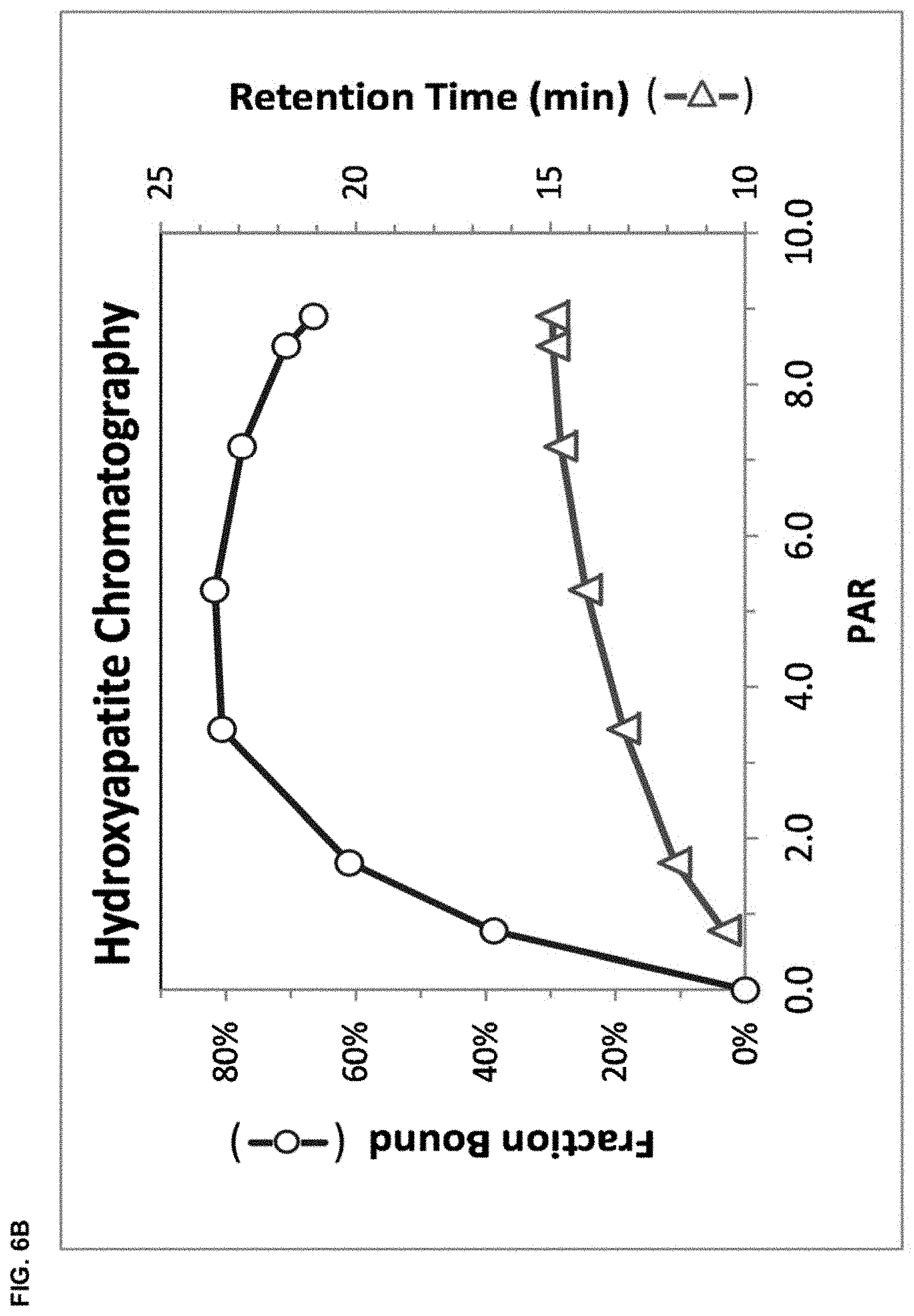
D00010
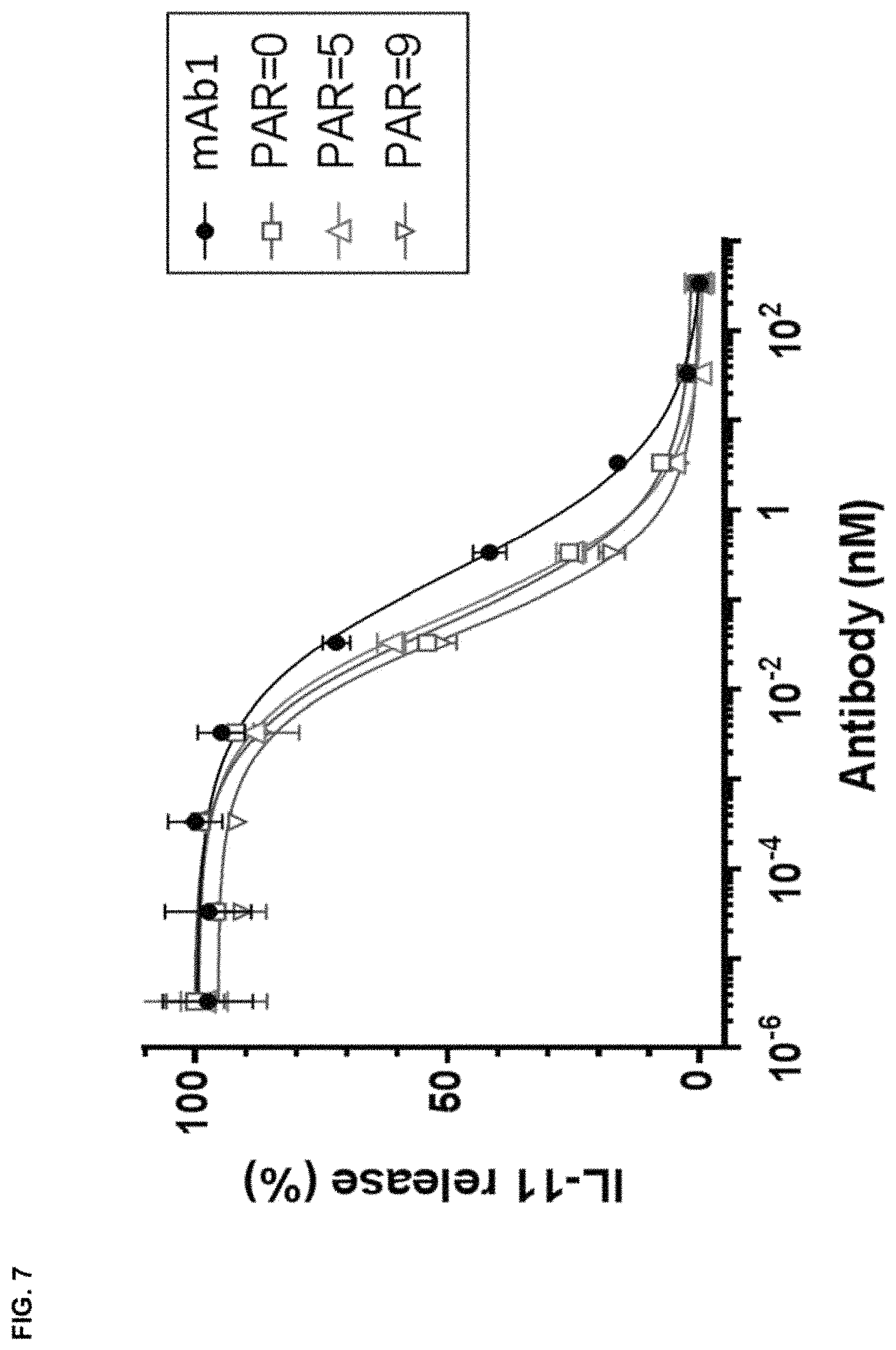
D00011

D00012

D00013

D00014
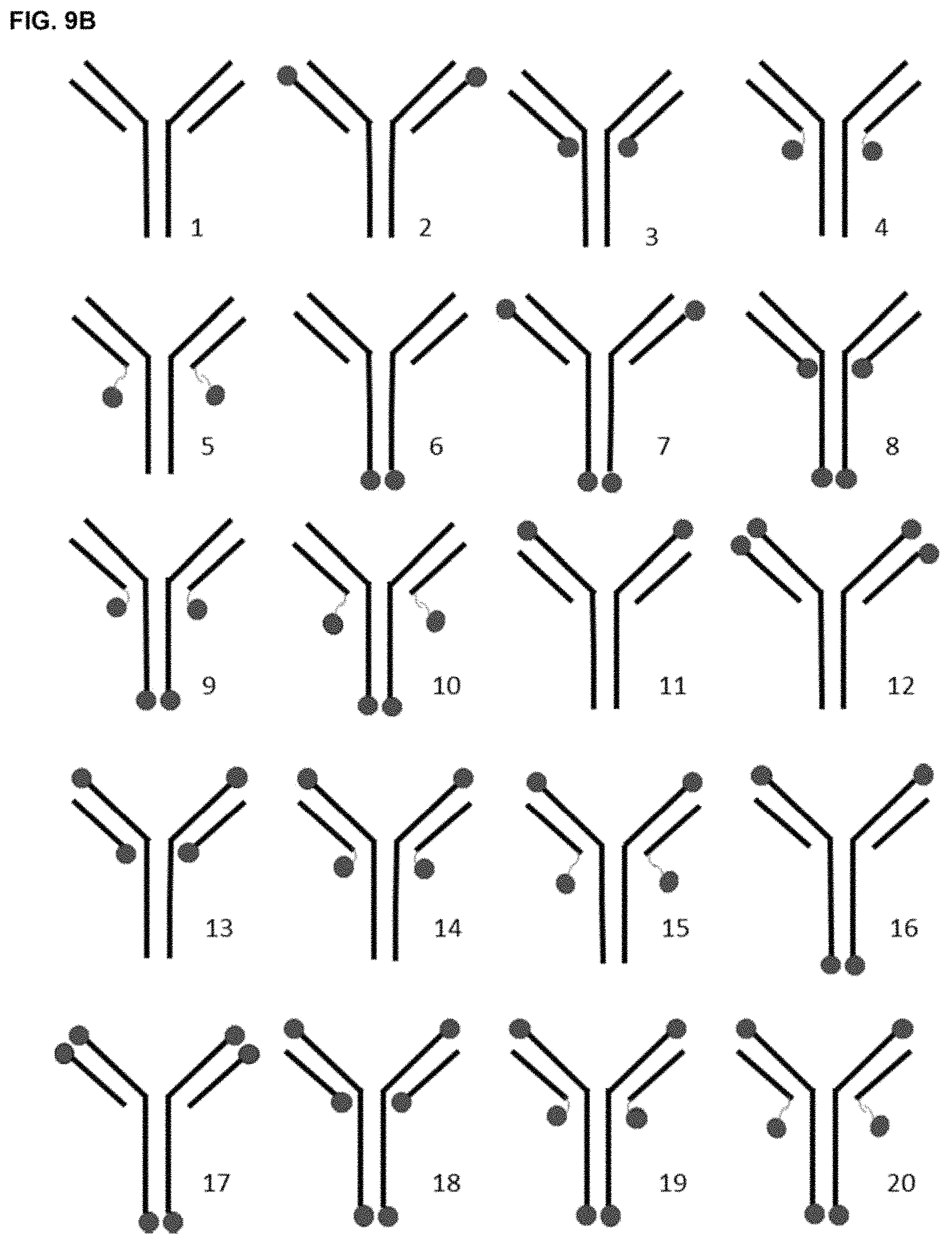
D00015

D00016

D00017

D00018

D00019
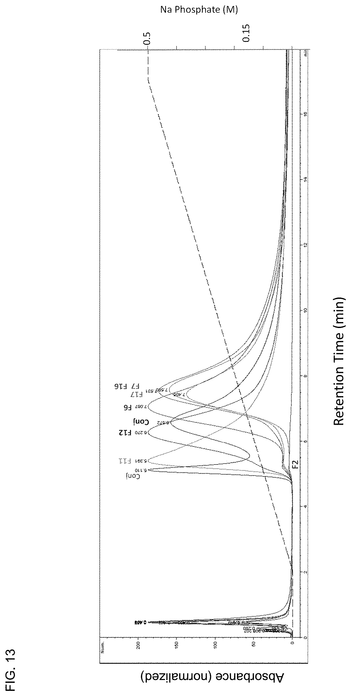
D00020
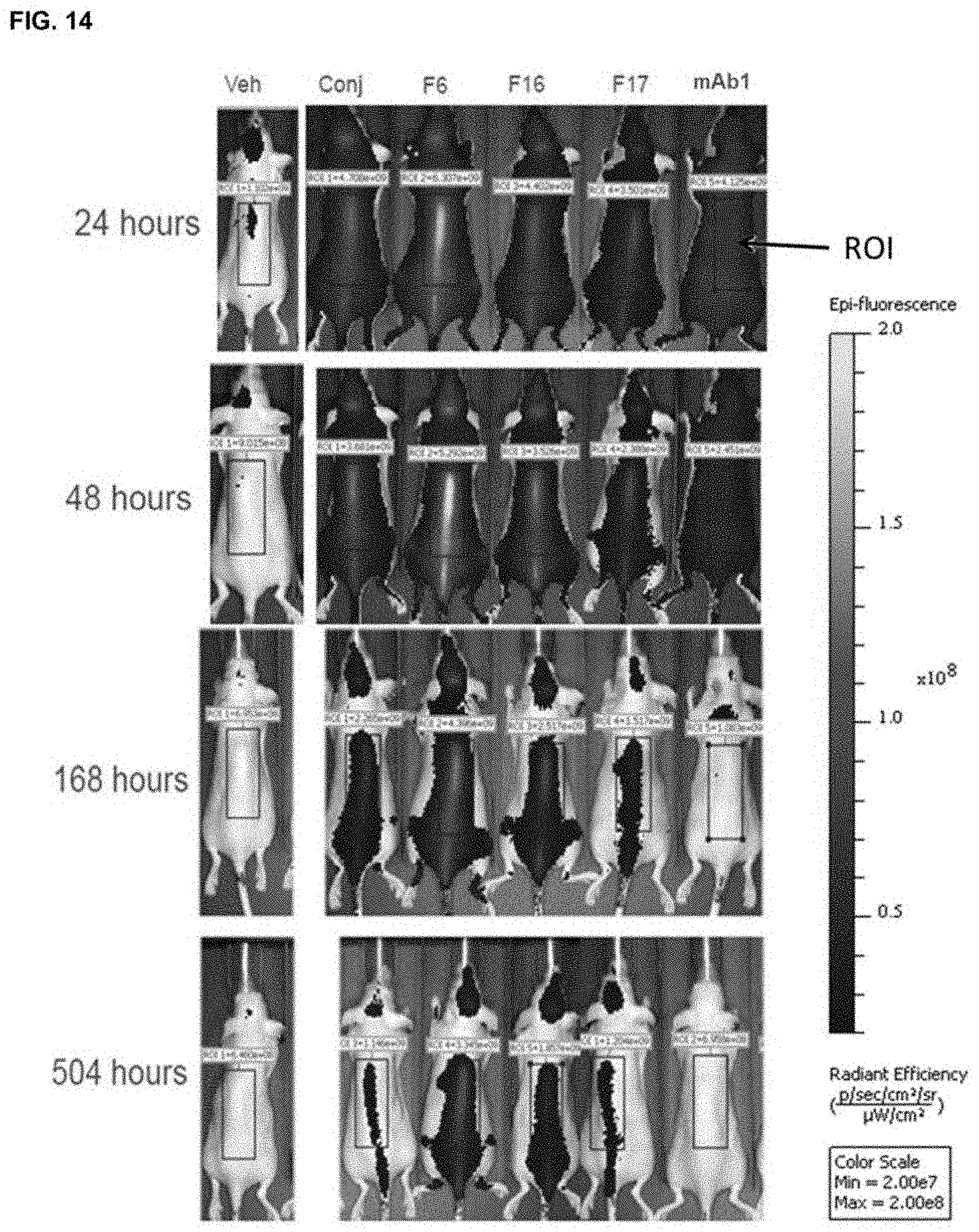
D00021
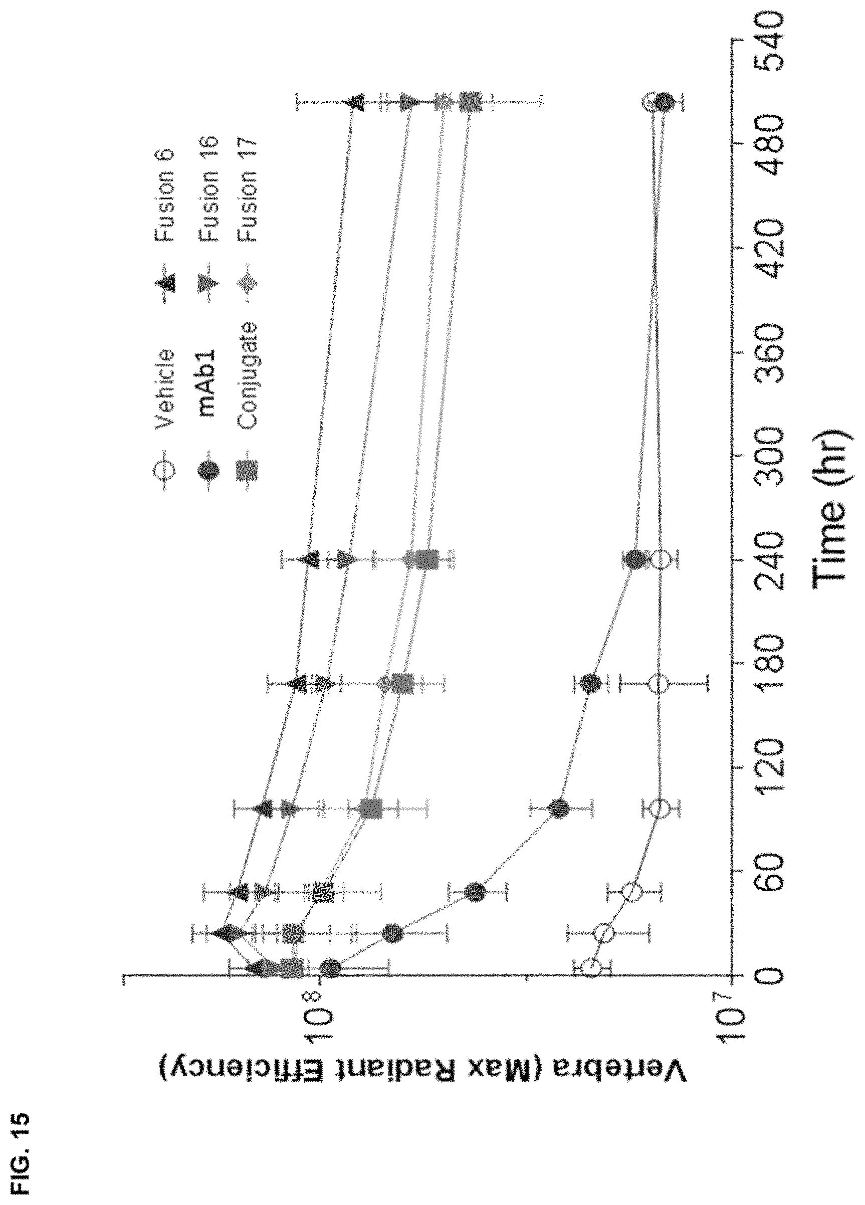
D00022

D00023

D00024

D00025

D00026

D00027

D00028
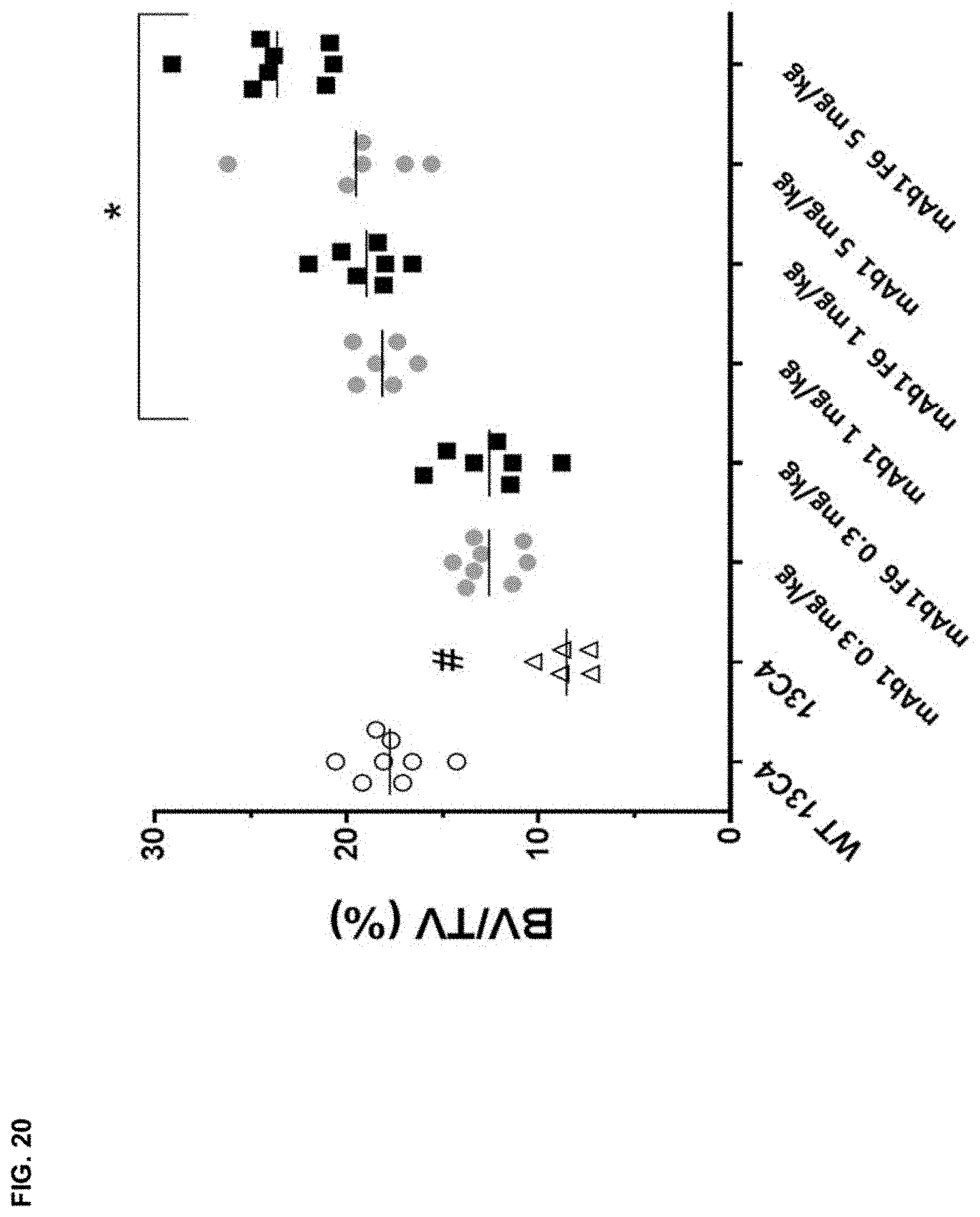
D00029

D00030

D00031

D00032

D00033

D00034
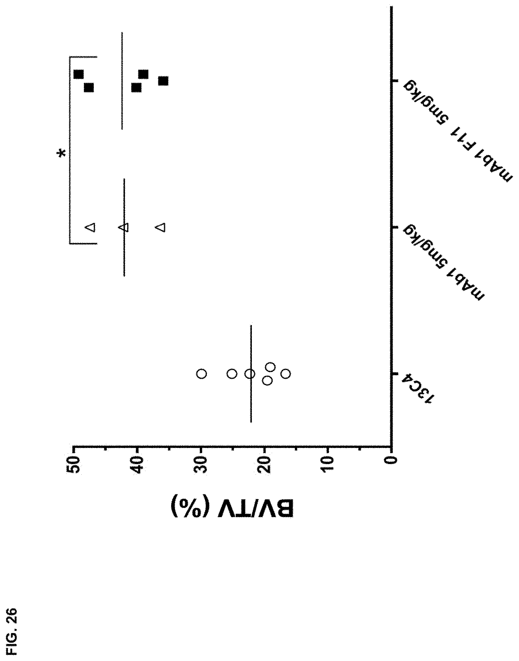
D00035

D00036

D00037
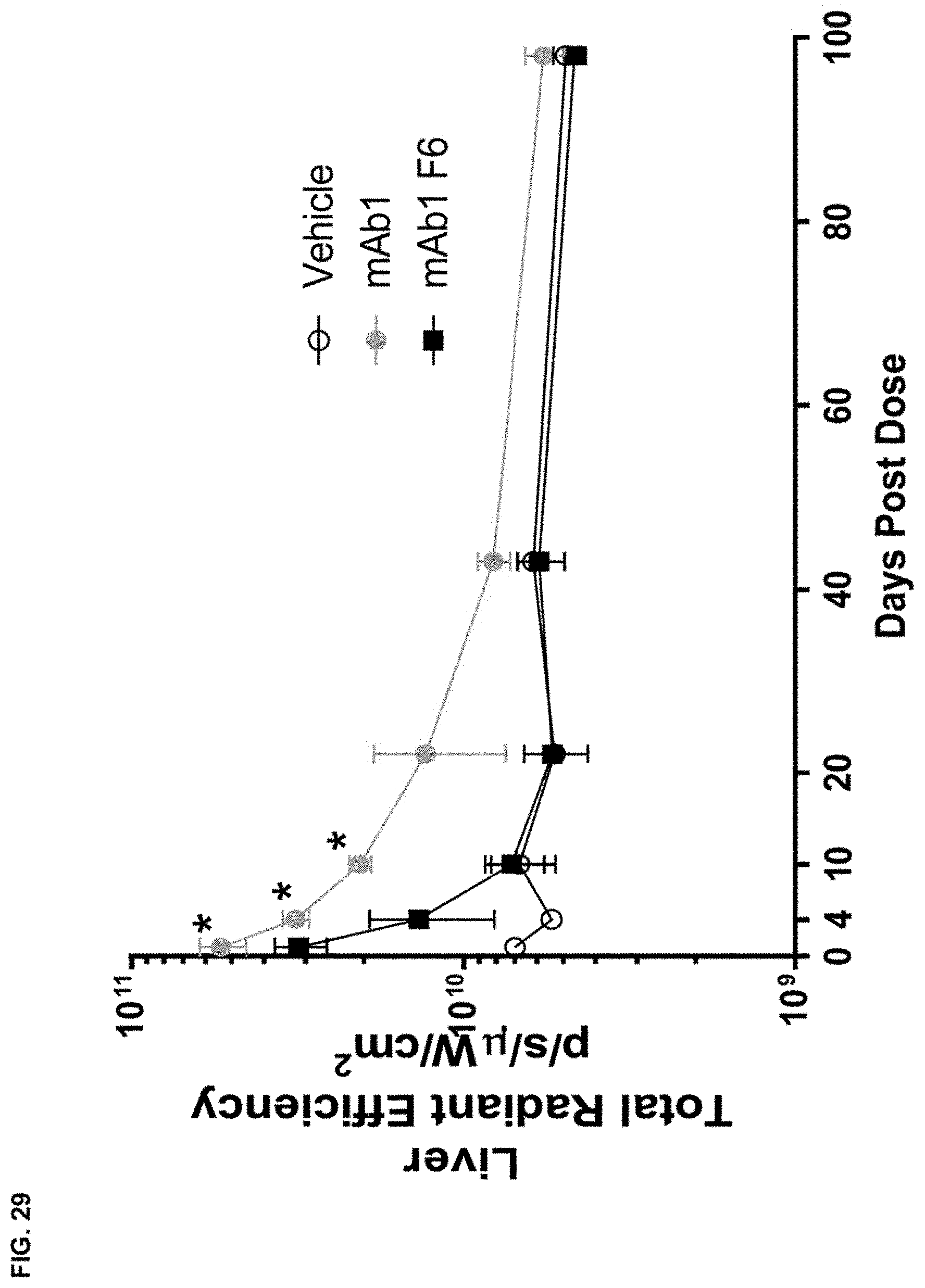
D00038
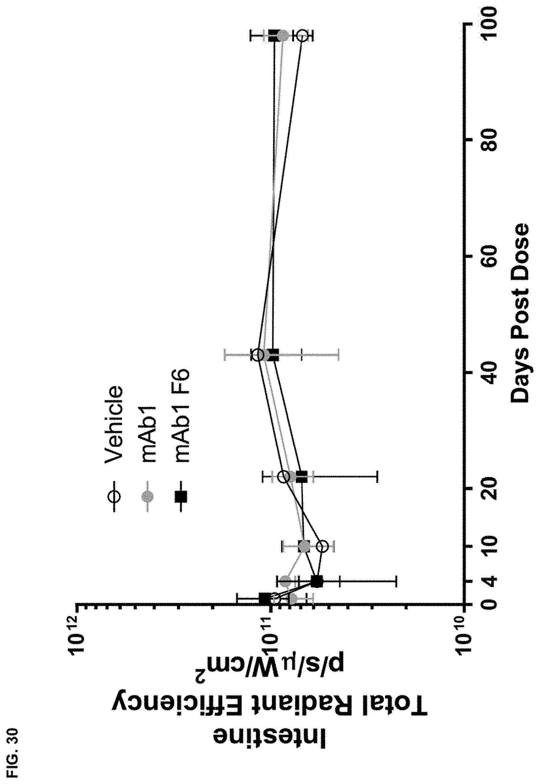
D00039
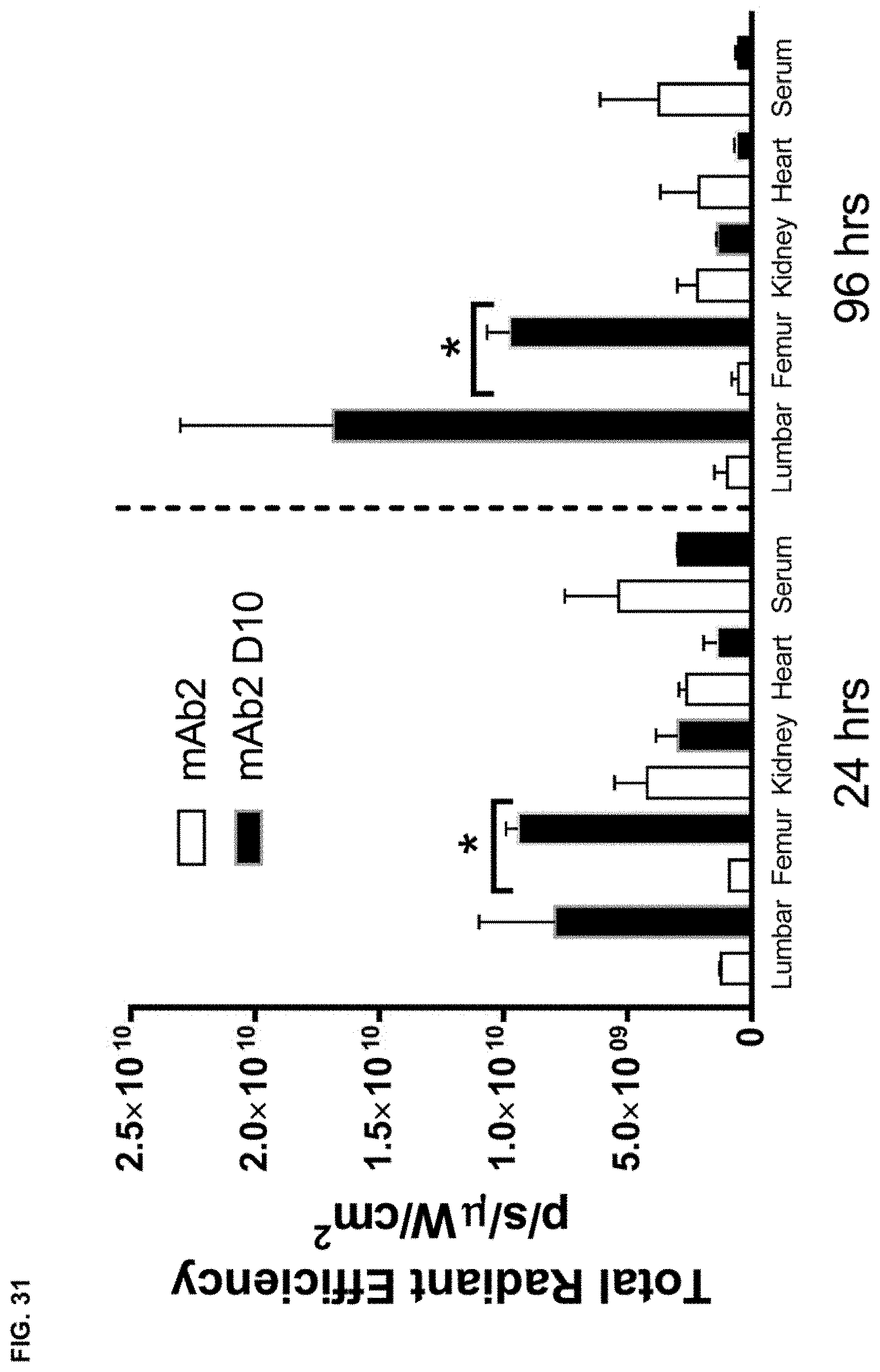
D00040

D00041

S00001
XML
uspto.report is an independent third-party trademark research tool that is not affiliated, endorsed, or sponsored by the United States Patent and Trademark Office (USPTO) or any other governmental organization. The information provided by uspto.report is based on publicly available data at the time of writing and is intended for informational purposes only.
While we strive to provide accurate and up-to-date information, we do not guarantee the accuracy, completeness, reliability, or suitability of the information displayed on this site. The use of this site is at your own risk. Any reliance you place on such information is therefore strictly at your own risk.
All official trademark data, including owner information, should be verified by visiting the official USPTO website at www.uspto.gov. This site is not intended to replace professional legal advice and should not be used as a substitute for consulting with a legal professional who is knowledgeable about trademark law.