Mass spectrometric determination of derivatized methylmalonic acid
Goldman , et al. November 17, 2
U.S. patent number 10,837,972 [Application Number 13/156,307] was granted by the patent office on 2020-11-17 for mass spectrometric determination of derivatized methylmalonic acid. This patent grant is currently assigned to Quest Diagnostics Investments Incorporated. The grantee listed for this patent is Scott M. Goldman, Thomas Lynn, Julie A. Neidich, Denise Salazar. Invention is credited to Scott M. Goldman, Thomas Lynn, Julie A. Neidich, Denise Salazar.


View All Diagrams
| United States Patent | 10,837,972 |
| Goldman , et al. | November 17, 2020 |
Mass spectrometric determination of derivatized methylmalonic acid
Abstract
The invention relates to the detection of methylmalonic acid (MMA). In a particular aspect, the invention relates to methods for detecting derivatized methylmalonic acid (MMA) by mass spectrometry.
| Inventors: | Goldman; Scott M. (Laguna Niguel, CA), Neidich; Julie A. (Ladera Ranch, CA), Salazar; Denise (Altadena, CA), Lynn; Thomas (Temecula, CA) | ||||||||||
|---|---|---|---|---|---|---|---|---|---|---|---|
| Applicant: |
|
||||||||||
| Assignee: | Quest Diagnostics Investments
Incorporated (Wilmington, DE) |
||||||||||
| Family ID: | 1000005185667 | ||||||||||
| Appl. No.: | 13/156,307 | ||||||||||
| Filed: | June 8, 2011 |
Prior Publication Data
| Document Identifier | Publication Date | |
|---|---|---|
| US 20110306144 A1 | Dec 15, 2011 | |
Related U.S. Patent Documents
| Application Number | Filing Date | Patent Number | Issue Date | ||
|---|---|---|---|---|---|
| 61353172 | Jun 9, 2010 | ||||
| Current U.S. Class: | 1/1 |
| Current CPC Class: | G01N 33/82 (20130101); Y10T 436/201666 (20150115); G01N 2560/00 (20130101); G01N 30/72 (20130101); H01J 49/00 (20130101) |
| Current International Class: | G01N 33/82 (20060101); H01J 49/00 (20060101); G01N 30/72 (20060101) |
| Field of Search: | ;436/129,173 |
References Cited [Referenced By]
U.S. Patent Documents
| 4940658 | July 1990 | Allen et al. |
| 5457055 | October 1995 | Allen et al. |
| 5559038 | September 1996 | Kolhouse et al. |
| 5772874 | June 1998 | Quinn et al. |
| 5795469 | August 1998 | Quinn et al. |
| 5800979 | September 1998 | Kolhouse et al. |
| 5919368 | July 1999 | Quinn et al. |
| 5968367 | October 1999 | Quinn et al. |
| 6107623 | August 2000 | Bateman et al. |
| 6124137 | September 2000 | Hutchens et al. |
| 6204500 | March 2001 | Whitehouse et al. |
| 6268144 | July 2001 | Koster |
| 7993931 | August 2011 | Chen |
| 2002/0019056 | February 2002 | Shushan et al. |
| 2005/0165560 | July 2005 | Kushnir et al. |
| 2008052299 | May 2008 | WO | |||
Other References
|
Park, Y. J. et al, Journal of Agricultural and Food Chemistry 1999, 47, 2322-2326. cited by examiner . Ohie, T. et al, Journal of Chromatography B 2000, 746, 63-73. cited by examiner . Fox, A. et al, Journal of Microbiological Methods 1995, 22, 11-26. cited by examiner . Rifai, N. et al, Annals of Clinical Biochemistry 1998, 35, 633-636. cited by examiner . De Jong, A. P. J. M. et al, Biomedical Mass Spectromtery 1980, 7, 359-364. cited by examiner . Stabler, S. P. et al, Journal of Biological Chemistry 1988, 263, 5581-5588. cited by examiner . Cappiello, A. et al, Annali di Chimica 2002, 92, 623-636. cited by examiner . Cappiello, A. et al, Journal of the American Society for Mass spectromtery 2002, 13, 265-273. cited by examiner . Peters, H. et al, JOurnal of Biological Chemistry 2003, 278, 52909-52913. cited by examiner . Blom, H. J. et al, Clinical Chemistry and Laboratory Medicine 2007, 45, 645-650. cited by examiner . Ia Marca, G. et al, Clinical Chemistry 2007, 53, 1364-1369. cited by examiner . Armbruster, D. A. et al, Clinical Chemistry 1994, 40, 1233-1238. cited by examiner . Edlund, A. et al, Plant Physiology 1995, 108, 1043-1047. cited by examiner . "Analytical Detection Limit Guidance & Laboratory Guide for Determining Method Detection Limits" PUBL-TS-056-96 published Apr. 1996 by the Wisconsin Department of Natural Resources Laboratory Certification Program, 33 pages. cited by examiner . Volmer, D. A. et al, Analytical Chemistry 1997, 69, 4143-4155. cited by examiner . Norris, A. J. et al, Biochemistry 2001, 40, 3774-3779. cited by examiner . Windelberg, A. et al, Clinical Chemistry 2005, 51, 2103-2109. cited by examiner . Armbruster, D. A. et al, Clinical Biochemist Reviews 2008, 29, Supplement (i), S49-S52. cited by examiner . Johnson, J. V. et al, Analytical Chemistry 1985, 57, pp. 758A-760A, 762A, 764A,766A and 768A. cited by examiner . Christensson, B. et al, Arthritis and Rheumatism 1989, 32, 1268-1272. cited by examiner . Zinn, A. B. et al, Pediatric Research 1982, 16, 740-745. cited by examiner . Gaskell, S. J. et al, Biomedical Mass Spectrometry 1985, 12, 139-141. cited by examiner . Rasmussen, K., Clinical Chemistry 1989, 35, 260-264. cited by examiner . McGhie, T. K. et al, Biomedical and Environmental Mass Spectrometry 1990, 19, 267-272. cited by examiner . Johnson, J. V. et al, Analytical Chemistry 1990, 62, 2162-2172. cited by examiner . Fox, A. et al, Applied and Environmental Microbiology 1993, 59, 4254-4260. cited by examiner . Moritz, T. et al, Analytical Chemistry 1995, 67, 1711-1716. cited by examiner . Kushnir, M. M. et al, Journal of Chromatography B 2000, 741, 231-241. cited by examiner . Busch, M. et al, Journal of Chromatography B 2002, 775, 215-223. cited by examiner . Ardrey, R. E., "Liquid Chromatography--Mass Spectrometry: An Introduction" Chapters 3 and 5, pp. 33-74 and 129-234, 2003, John Wiley & Sons, Ltd. cited by examiner . Hasegawa, Y. et al, Journal of Chromatography B 2005, 823, 13-17. cited by examiner . Bader, M. et al, "The use of gas chromatography-mass spectrometry" in the MAK-Collection Part IV: Biomonitoring Methods, vol. 10, 3-51, Wiley-VCH Verlag GmbH & Co. KGaA, Weinheim. cited by examiner . Kawana, S. et al, Clinica Chimica Acta 2008, 392, 34-40. cited by examiner . Schummer, C. et al, Talanta, 2009, 77, 1473-1482. cited by examiner . Shigematsu, Y. et al, Journal of Inherited Metabolic Disease 2010, 33 (Supplement 2), S283-S288. cited by examiner . Fredriksson, S.-A. et al, Journal of Mass Spectrometry 1995, 30, 1133-1143. cited by examiner . Rashe, M. S. et al, Clinical Chemistry 1997, 43 1129-1141. cited by examiner . Kelly, C., Journal of Chromatography A 2000, 872, 309-314. cited by examiner . Driskell, W. J. et al, Journal of Analytical Toxicology 2002, 26, 6-11. cited by examiner . Marcos, J. et al, Journal of Mass Spectrometry 2002, 37, 1059-1073. cited by examiner . Pitt, J. J. et al, Clinical Chemistry 2002, 48, 1970-1980. cited by examiner . Kushnir, M. M. et al, Clinical Biochemistry 2005, 38, 319-327. cited by examiner . Kushnir, M. M. et al, Clinical Chemistry 2006, 52, 1559-1567. cited by examiner . Walorczyk, S., Journal of Chromatography A 2007, 1165, 200-212. cited by examiner . Kharbouche, H. et al, Journal of Chromatography B 2009, 877, 2337-2343. cited by examiner . Regueiro, J. et al, Journal of Chromatography A 2009, 1216, 4693-4702. cited by examiner . Walorczyk, S. et al, Journal of Chromatography A 2009, 1216, 6522-6531. cited by examiner . Fasching, C. et al, in "Clinical Applications of Mass Spectrometry, Methods in Molecular Biology" Garg, U. et al, editors, Humana Press, 2010, Capter 36, 371-378. cited by examiner . Courant, F. et al, Journal of Clinical Endocrinology & Metabolism 2010,95, 82-92. cited by examiner . Qu, L.-J. et al, Food Chemistry 2010, 122, 327-332. cited by examiner . Cervera, M. I. et al, Analytical and Bioanalytical Chemistry 2010, 397, 2873-2891. cited by examiner . Verma, K. K. et al, Chromatographia 1997, 44, 372-380. cited by examiner . Tsikas, D., Journal of Chromatography B 1998, 717, 201-245. cited by examiner . Hernando, M. D. et al, Journal of Chromatography A 2004, 1047, 129-135. cited by examiner . Bates-Dubrow, L. et al, ASMS Conference 2010, 2 pages. cited by examiner . Schmedes et al. Analysis of Methylmalonic Acid in Plasma by Liquid Chromatography-Tandem Mass Spectrometry. Clinical Chemistry. vol. 52 (4): pp. 754-757, 2006. cited by applicant . Straczek et al. Quantification of methylmalonic acid in serum measured by capillary gas chromatography-mass spectrometry as tert-butyldimethylsilyl derivatives. Journal of Chromatography. vol. 620 (1): pp. 1-7, 1993. cited by applicant . Appelblad, P., et al., "Mass Spectrometric Detection of Homocysteine, Methylmalonic Acid and Succinic Acid using HILIC on a Zwitterionic Stationary Phase," LC-GC Europe; Mar. 2005 Supplement, vol. 18, p. 47. cited by applicant . Bartolucci et al., "Liquid chromatography tandem mass spectrometric quantitation of sulfamethazine and its metabolites: direct analysis of swine urine by triple quadrupole and by ion trap mass spectrometry," Rapid Communication in Mass Spectrometry, 14:967-973, 2000. cited by applicant . Carvalho, V., et al., "Determination of serum methylmalonic acid by alkylative extraction and liquid chromatography coupled to tandem mass spectrometry," Anal. Biochem., 381:67-73 (2008). cited by applicant . Chen, J., et al., "Quantitation of methylmalonic acid in serum or plasma using isotope dilution-selected ion gas chromatography-mass spectrometry," Clinical Applications of Mass Spectrometry, Methods in Molecular Biology, Chapter 35:365. cited by applicant . Fasching, C., et al., "Clinical Applications of Mass Spectrometry," Methods in Molecular Biology, Chapter 36:371. cited by applicant . Hagen, T., "A GC/MS/MS screening method for multiple organic acidemias from urine specimens," Clinica Chimica Atca, 283:77-88 (1999). cited by applicant . Kushnir, M., et al., "Analysis of dicarboxylic acids by tandem mass spectrometry. High-throughput quantitative measurement of methylmalonic acid in serum, plasma, and urine," Clin. Chem., 47:1993-2002 (2001). cited by applicant . Lakso, H., et al., "Quantification of methylmalonic acid in human plasma with hydrophilic interaction liquid chromatography separation and mass spectrometric detection," Clin. Chem., 54:2028-2035 (2008). cited by applicant . Luo, X., et al., "Methylmalonic acid in amniotic fluid and maternal urine as a marker for neural tube defects," J. Hauzhong University of Sci. and Tech., 24:166-169 (2004). cited by applicant . Magera, M., "Methylmalonic acid measured in plasma and urine by stable-isotope dilution and electrospray tandem mass spectrometry," Clin. Chem., 46:1804-1810 (2000). cited by applicant . Marcell, P., et al., "Quantitation of methylmalonic acid and other dicarboxylic acids in normal serum and urine using capillary gas chromatography-mass spectrometry," Analytical Chem., 150:58-66 (1985). cited by applicant . McCann, M., "Methylmalonic acid quantification by stable isotope dilution gas chromatography-mass spectrometry from filter paper urine samples," Clin. Chem., 42:910-914 (1996). cited by applicant . McGhie, "Analysis of serum methylmalonic acid for the determination of cobalt deficiency in cattle," J. Chromatog., 556:215-22 (1991). cited by applicant . Merchant et al., "Recent advancements in surface-enhanced laser desorption/ionization-time of flight-mass spectrometry," Electrophoresis, 21:1164-1167, (2000). cited by applicant . Polson et al., "Optimization of protein precipitation based upon effectiveness of protein removal and ionization effect in liquid chromatography-tandem mass spectrometry," J. Chromatogr B., (2003), 785:263-275. cited by applicant . Purevsuren, J., et al., "Urinary organic metabolite screening of children with influenza-associated encephalopathy for inborn errors of metabolism using GC/MS," Brain and Develop., 30:520-26 (2008). cited by applicant . Robb, et al., "Atmospheric Pressure Photoionization: An Ionization Method for Liquid Chromatography-Mass Spectrometry," Anal. Chem., (2000) 72:3653-3659. cited by applicant . Song, Y., et al., "Selective screening for inborn errors of metabolism and secondary methylmalonic aciduria in pregnancy at high risk district of neural tube defects: a human metabolome study by GC-MS in China," Clin. Biochem., 41:616-20 (2008). cited by applicant . Specker, B., et al, "Urinary methylmalonic acid excretion in infants fed formula or human milk," Am. J. Clin. Nutr., 51:209-11 (1990). cited by applicant . Stabler, S., et al., "Assay of methylmalonic acid in the serum of patients with cobalamin deficiency using capillary gas chromatography-mass spectrometry," J. Clin. Invest., 77:1606-12 (1986). cited by applicant . Thurman, et al., "Chromatographic-Mass Spectrometric Food Analysis for Trace Determination of Pesticide Residues," Chapter 8 (Amadeo R. Fernandez-Alba, ed., Elsevier, 2005), p. 387. cited by applicant . Tuchman, M., "Screening urine of 3-week-old newborns: transient methylmalonic and hydroxyphenyllactic aciduria," Biochem. Med. and Metabolic Biology, 48:64-68 (1992). cited by applicant . Ueland, P., "Quantitative profiling of folate and one-carbon metabolism in large-scale epidemiological studies by mass spectrometry," Clin. Chem. Lab. Med., 45:1737-45 (2007). cited by applicant . Vandenberghe, Y., et al., "Effect of folate supplementation on clinical chemistry and hematologic changes related to bidisomide administration in the rat," Drug and Chemical Toxicology, 18:235-270 (1995). cited by applicant . Wasant, P., "Neonatal Screening in the 21st Century," Southeast Asian Journal of Tropical Medicine and Public Health, 30:160-5 (1998). cited by applicant . Windelberg, A., "Automated assay for the determination of methylmalonic acid, total homocysteine, and related amino acids in human serum or plasma by means of methylchloroformate derivatization and gas chromatography-mass spectrometry," Clin. Chem., 2005, 51:2103-2109. cited by applicant . Wright et al., "Proteinchip.RTM. surface enhanced laser desorption/ionization (SELDI) mass spectrometry: a novel protein biochip technology for detection of prostate cancer biomarkers in complex protein mixtures," Prostate Cancer and Prostatic Diseases, (1999), 2: 264-76. cited by applicant . Yazdanpanah, M., "An improved assay for plasma methylmalonic acid using chemical ionization gas chromatography mass spectrometry," Clin Biochem., 36:617-620 (2003). cited by applicant . Young, P., "Sensitive gas chromatographic--high resolution mass spectrometric method for the determination of methylmalonic acid in bovine plasma," Analyst, 120:2199-2201 (1995). cited by applicant . Written Opinion for Application No. PCT/US2011/039652, dated Sep. 28, 2011, 6 Pages. cited by applicant . International Search Report for Application No. PCT/US2011/039652, dated Sep. 28, 2011, 2 Pages. cited by applicant . Child-Specific Exposure Factors Handbook, Chapter 5, pp. 5i-4-42, Sep. 2008. cited by applicant . National Center for Biotechnology Information. PubChem Compound Database; CID=441038, https://pubchem.ncbi.nlm.nih.gov/compound/441038 (accessed Oct. 23, 2015. cited by applicant . Non-Final Office Action dated Jan. 22, 2016 for U.S. Appl. No. 14/063,956, filed Oct. 25, 2013. cited by applicant . Aguera A., et al. "One-Year Routine Application of a New Method Based on Liquid Chromatography-Tandem Mass Spectrometry to the Analysis of 16 Multiclass Pesticides in Vegetable Samples" J Chromotogr, 1045, pp. 125-135, (2004). cited by applicant . Fiehn O., and Weckwerth W. (2006) Mass Spectrometry: Quantitation. In: Encyclopedic Reference of Genomics and Proteomics in Molecular Medicine. Springer, Berlin, Heidelberg, pp. 1030-1034. cited by applicant . Jessome L., et al. "Ion Supression: A Major Concern in Mass Spectrometry" LCGC North America, vol. 24, No. 5, pp. 498-510, (May 2006). cited by applicant. |
Primary Examiner: Soderquist; Arlen
Attorney, Agent or Firm: Quest Diagnostics, Inc.
Parent Case Text
CROSS-REFERENCE TO RELATED PATENT APPLICATIONS
This application claims priority to U.S. Provisional Application Ser. No. 61/353,172, filed Jun. 9, 2010, which is incorporated herein by reference in its entirety including all figures and tables.
Claims
That which is claimed is:
1. A method for determining the amount of methylmalonic acid (MMA) in a sample by tandem mass spectrometry, the method comprising: (a) obtaining a sample containing MMA; (b) subjecting said sample to a derivatizing agent under conditions suitable to generate tert-butyldimethylsilyl derivatized MMA (TBDMS-MMA); (c) subjecting said TBDMS-MMA from the sample to chromatography; (d) ionizing TBDMS-MMA by electron impact (EI) ionization following chromatography to generate a precursor ion and fragmenting the precursor ion using collision energy of 3V or 10V to form one or more fragment ions; and (e) determining the amount of said one or more fragment ions by tandem mass spectrometry; wherein the amount of said one or more fragment ions determined in step (e) is related to the amount of MMA originally present in the sample, wherein the method is capable of detecting about 9 nMol/L of MMA.
2. The method of claim 1, wherein said chromatography is gas chromatography.
3. The method of claim 2, wherein said tandem mass spectrometry is a quadropole tandem mass spectrometry.
4. The method of claim 1, wherein said MMA in said sample is purified by solid phase extraction prior to being subjected to said derivatizing agent.
5. The method of claim 1, wherein said sample comprises a biological sample.
6. The method of claim 1, wherein said sample comprises a biological fluid.
7. The method of claim 1, wherein said sample is from a human.
8. The method of claim 1, wherein said sample comprises plasma or serum.
9. The method of claim 1, wherein said one or more fragment ions comprise an ion with m/z of 189.0.+-.0.5.
10. The method of claim 1, wherein said one or more fragment ions comprise an ion with m/z of 147.0.+-.0.5.
11. A method for determining the amount of methylmalonic acid (MMA) in a sample by tandem mass spectrometry, the method comprising: (a) obtaining a sample containing MMA; (b) subjecting said sample to a derivatizing agent under conditions suitable to generate tert-butyldimethylsilyl derivatized MMA (TBDMS-MMA); (c) subjecting TBDMS-MMA from the sample to gas chromatography (GC); (d) ionizing TBDMS-MMA by electron impact (EI) ionization following chromatography to generate a precursor ion and fragmenting the precursor ion using collision energy of 3V or 10V to form one or more fragment ions; and (e) determining the amount of said one or more fragment ions by tandem mass spectrometry; wherein the amount of said one or more fragment ions determined in step (e) is related to the amount of MMA originally present in the sample, wherein the method is capable of detecting about 9 nMol/L of MMA.
12. The method of claim 11, wherein said tandem mass spectrometry is a quadropole tandem mass spectrometry.
13. The method of claim 11, wherein said one or more fragment ions comprise an ion with a mass to charge ratio (m/z) of 189.0.+-.0.5.
14. The method of claim 11, wherein said one or more fragment ions comprise an ion with a mass to charge ratio (m/z) of 147.0.+-.0.5.
15. The method of claim 11, wherein said MMA in said sample is purified by solid phase extraction prior to being subjected to said derivatizing agent.
16. The method of claim 11, wherein said sample comprises a biological sample.
17. The method of claim 11, wherein said sample comprises a biological fluid.
18. The method of claim 11, wherein said sample is from a human.
19. The method of claim 11, wherein said sample comprises plasma or serum.
20. A method for determining the amount of methylmalonic acid (MMA) in a sample by tandem mass spectrometry, the method comprising: (a) obtaining a sample containing tert-butyldimethylsilyl derivatized MMA (TBDMS-MMA), wherein said TBDMS-MMA is produced by derivatizing MMA originally present in the sample; (b) subjecting said TBDMS-MMA from the sample to chromatography; (c) ionizing TBDMS-MMA by electron impact (EI) ionization following chromatography to generate a precursor ion and fragmenting the precursor ion using collision energy of 3V or 10V to form one or more fragment ions; and (d) determining the amount of said one or more fragment ions by tandem mass spectrometry; wherein the amount of said one or more fragment ions determined in step (d) is related to the amount of MMA originally present in the sample, wherein the method is capable of detecting about 9 nMol/L of MMA.
21. The method of claim 20, wherein said chromatography is gas chromatography.
22. The method of claim 21, wherein said tandem mass spectrometry is a quadropole tandem mass spectrometry.
23. The method of claim 20, wherein said sample comprises a biological sample.
24. The method of claim 20, wherein said sample comprises a biological fluid.
25. The method of claim 20, wherein said sample is from a human.
26. The method of claim 20, wherein said sample comprises plasma or serum.
27. The method of claim 20, wherein said one or more fragment ions comprise an ion with m/z of 189.0.+-.0.5.
28. The method of claim 20, wherein said one or more fragment ions comprise an ion with m/z of 147.0.+-.0.5.
29. A method for determining the amount of tert-butyldimethylsilyl derivatized MMA (TBDMS-MMA) in a sample by tandem mass spectrometry, the method comprising: (a) subjecting TBDMS-MMA in the sample to chromatography; (b) ionizing TBDMS-MMA by electron impact (EI) ionization following chromatography to generate a precursor ion and fragmenting the precursor ion using collision energy of 3V or 10V to form one or more fragment ions; and (c) determining the amount of said one or more fragment ions by tandem mass spectrometry; wherein the amount of said one or more fragment ions determined in step (c) is related to the amount of MMA originally present in the sample, wherein the method is capable of detecting about 9 nMol/L of MMA.
30. The method of claim 29, wherein said chromatography is gas chromatography.
31. The method of claim 30, wherein said tandem mass spectrometry is a quadropole tandem mass spectrometry.
32. The method of claim 29, wherein said sample comprises a biological sample.
33. The method of claim 29, wherein said sample comprises a biological fluid.
34. The method of claim 29, wherein said sample is from a human.
35. The method of claim 29, wherein said sample comprises plasma or serum.
36. The method of claim 29, wherein said one or more fragment ions comprise an ion with m/z of 189.0.+-.0.5.
37. The method of claim 29, wherein said one or more fragment ions comprise an ion with m/z of 147.0.+-.0.5.
38. A method for diagnosing vitamin B12 deficiency in a patient, the method comprising: (a) obtaining a sample from a patient containing MMA; (b) subjecting said sample to a derivatizing agent under conditions suitable to generate tert-butyldimethylsilyl derivatized MMA (TBDMS-MMA); (c) subjecting said TBDMS-MMA from the sample to chromatography; (d) ionizing TBDMS-MMA by electron impact (EI) ionization following chromatography to generate a precursor ion and fragmenting the precursor ion using collision energy of 3V or 10V to form one or more fragment ions; (e) determining the amount of said one or more fragment ions by tandem mass spectrometry; (f) determining the amount of MMA originally in the sample from the amount of said one or more fragment ions determined in step (e); and (g) determining the patient is deficient in vitamin B n from the amount of MMA determined in step (f) by comparison to a diagnostic MMA level, wherein if the amount of MMA determined in step (f) is greater than the diagnostic MMA level, the patient is deficient in vitamin B12, wherein the method is capable of detecting about 9 nMol/L of MMA.
39. The method of claim 38, wherein said chromatography is gas chromatography.
40. The method of claim 39, wherein said tandem mass spectrometry is a quadropole tandem mass spectrometry.
41. The method of claim 38, wherein said MMA in said sample is purified by solid phase extraction prior to being subjected to said derivatizing agent.
42. The method of claim 38, wherein said sample comprises a biological fluid.
43. The method of claim 38, wherein said sample comprises plasma or serum.
44. The method of claim 38, wherein said one or more fragment ions comprise an ion with m/z of 189.0.+-.0.5.
45. The method of claim 38, wherein said one or more fragment ions comprise an ion with m/z of 147.0.+-.0.5.
46. The method of claim 38, wherein said diagnostic MMA level is within the range of 200 nMol/L to about 350 nMol/L.
47. The method of claim 38, wherein said diagnostic MMA level is about 318.+-.20 nMol/L.
Description
FIELD OF THE INVENTION
The invention relates to the quantitative measurement of methylmalonic acid. In a particular aspect, the invention relates to methods for quantitative measurement of methylmalonic acid by gas chromatography-tandem mass spectrometry.
BACKGROUND OF THE INVENTION
Methylmalonic acid (MMA) is a C-methylated dicarboxylic acid derivative of malonate. The coenzyme A-linked form of MMA is a metabolic intermediate in the conversion of propionyl-CoA to succinyl-CoA. Vitamin B.sub.12 is a vital co-factor in this conversion. If insufficient vitamin B.sub.12 is available, the conversion does not proceed to completion. Build-up of incomplete conversion products (including L-methylmalonyl-CoA) ultimately leads to increased MMA production. Thus, elevated levels of MMA are observed when there is a vitamin B.sub.12 deficiency.
Several methods for MMA determination in biological samples have been reported. For example, gas chromatography-single mass spectrometry techniques utilizing derivatization of MMA prior to analysis have been reported by Allen et al. (U.S. Pat. Nos. 5,438,017 and 5,457,055; detection of t-butyldimethylsilyl ester derivatives); Lou et al. (J. Huazhong University of Science and Technology (2004) 24:166-9; detection of N, o-Bis(trimethylsilyl) trifluoroacetamide derivatives); Stabler et al. (J. Clin. Invest. (1986) 77:1606-12; detection of t-butyldimethylsilyl ester derivatives); Marcell, et al. (Analytical Chem. (1985) 150:58-66; detection of t-butyldimethylsilyl ester derivatives); McCann, et al. (Clin. Chem. (1996) 42:910-14; detection of t-butyldimethylsilyl ester derivatives); Windelberg, et al. (Clin. Chem. (2005) 51:2103-09; detection of methylchloroformate derivatives); Specker, et al. (Am J Clin Nutr. (1990) 51:209-11; detection of dicyclohexyl ester derivatives); Yazdanpanah, et al. (Clin Biochem. (2003) 36:617-20; detection of N, o-Bis(trimethylsilyl) trifluoroacetamide derivatives); Purevsuren, et al. (Brain and Develop. (2008) 30:520-26; detection of bis(trimethylsilyl)trifluorooacetamide and trimethylchlorosilane derivatives); Chen et al. (Clinical Applications of Mass Spectrometry, Methods in Molecular Biology, Chapter 35:365; detection of trimethylchlorosilane and butyl ester derivatives); and Young, et al. (Analyst (1995) 120:2199-2201; reporting butyl ester derivatives). Liquid chromatography-tandem mass spectrometry techniques utilizing derivatization of MMA prior to analysis have been reported by Shushan et al. (U.S. Pat. No. 6,692,971; detection of n-butyl ester derivatives); Kushnir et al. (U.S. Publication No. 2004/0165560; detection of n-butyl ester derivatives); and Kushnir et al. (Clin. Chem. (2001) 47:1993-2000; detection of dibutyl ether derivatives). Gas chromatography-tandem mass spectrometric techniques have been reported by Hagen, et al. (Clinica Chimica Atca (1999) 283:77-88; detection of trimethylsilyl derivatives); and Ueland et al. (Clin Chem Lab Med (2007) 445:1737-45; detection of methylchloroformate derivatives).
SUMMARY OF THE INVENTION
One aspect of the present invention provides methods for determining the amount of methylmalonic acid (MMA) in a sample by tandem mass spectrometry. In some embodiments, the methods include: (a) obtaining a sample containing MMA; (b) subjecting the MMA containing sample to a derivatizing agent under conditions suitable to generate tert-butyldimethylsilyl derivatized MMA (TBDMS-MMA); (c) subjecting TBDMS-MMA from the sample to chromatography; (d) ionizing TBDMS-MMA following chromatography to generate two or more ions detectable by mass spectrometry, these ions including a precursor ion with a mass to charge ratio of 289.0.+-.0.5 and one or more fragment ions selected from the group consisting of fragment ions with mass to charge ratios of 189.0.+-.0.5, 147.0.+-.0.5, and 73.0.+-.0.5; and (e) determining the amount of one or more fragment ions by tandem mass spectrometry. In these methods, the amount of ions determined in step (e) is related to the amount of MMA originally present in the sample. In some embodiments, chromatography is gas chromatography (GC). In some embodiments, the derivatizing agent comprises methyl-(t-butyldimethylsilyl)trifluoroacetamide (MTBSFTA). In some embodiments, MMA originally present in the sample is purified by solid phase extraction prior to being subjected to the derivatizing agent. In some embodiments, the sample is from a human patient.
In some embodiments, the methods include: (a) obtaining a sample containing MMA; (b) subjecting said sample to a derivatizing agent under conditions suitable to generate tert-butyldimethylsilyl derivatized MMA (TBDMS-MMA); (c) subjecting TBDMS-MMA from the sample to gas chromatography (GC); (d) ionizing TBDMS-MMA following chromatography to generate two or more ions detectable by mass spectrometry, the ions including a precursor ion and one or more fragment ions; and (e) determining the amount of one or more of the fragment ions by tandem mass spectrometry. In these embodiments, the amount of ions determined in step (e) is related to the amount of MMA originally present in the sample. In some embodiments, the precursor ion has a mass to charge ratio of 289.0.+-.0.5. In some embodiments, one or more fragment ions are selected from the group consisting of fragment ions with mass to charge ratios of 189.0.+-.0.5, 147.0.+-.0.5, and 73.0.+-.0.5. In some embodiments, the derivatizing agent comprises methyl-(t-butyldimethylsilyl)trifluoroacetamide (MTBSFTA). In some related embodiments, MMA in the sample is purified by solid phase extraction prior to being subjected to the derivatizing agent.
In some embodiments, the methods include: (a) obtaining a sample containing tert-butyldimethylsilyl derivatized MMA (TBDMS-MMA), wherein TBDMS-MMA is produced by derivatizing MMA originally present in the sample; (b) subjecting TBDMS-MMA from the sample to chromatography; (c) ionizing TBDMS-MMA following chromatography to generate two or more ions detectable by mass spectrometry, the ions including a precursor ion with a mass to charge ratio of 289.0.+-.0.5 and one or more fragment ions selected from the group consisting of fragment ions with mass to charge ratios of 189.0.+-.0.5, 147.0.+-.0.5, and 73.0.+-.0.5; and (d) determining the amount of one or more of the fragment ions by tandem mass spectrometry. In these embodiments, the amount of ions determined in step (d) is related to the amount of MMA originally present in the sample. In some embodiments, chromatography is gas chromatography. In some embodiments, the methods further include obtaining a sample containing MMA and subjecting the MMA containing sample to a derivatizing agent under conditions suitable to generate tert-butyldimethylsilyl derivatized MMA (TBDMS-MMA). In some related embodiments, the derivatizing agent comprises methyl-(t-butyldimethylsilyl)trifluoroacetamide (MTBSFTA). In some related embodiments, the sample is purified by solid phase extraction prior to being subjected to said derivatizing agent.
In a second aspect, methods are presented for determining the amount of tert-butyldimethylsilyl derivatized MMA (TBDMS-MMA) in a sample by tandem mass spectrometry. These methods include: (a) subjecting TBDMS-MMA in the sample to chromatography; (b) ionizing TBDMS-MMA following chromatography to generate two or more ions detectable by mass spectrometry, these ions including a precursor ion with a mass to charge ratio of 289.0.+-.0.5 and one or more fragment ions selected from the group consisting of fragment ions with mass to charge ratios of 189.0.+-.0.5, 147.0.+-.0.5, and 73.0.+-.0.5; and (c) determining the amount of one or more of the fragment ions by tandem mass spectrometry. In these methods, the amount of ions determined in step (c) is related to the amount of MMA originally present in the sample. In some embodiments, chromatography is gas chromatography.
In a third aspect, methods are presented for diagnosing vitamin B.sub.12 deficiency in a patient. These methods include: (a) obtaining a sample containing MMA from a patient; (b) subjecting MMA from the sample to a derivatizing agent under conditions suitable to generate tert-butyldimethylsilyl derivatized MMA (TBDMS-MMA); (c) subjecting the TBDMS-MMA from step (b) to chromatography; (d) ionizing TBDMS-MMA following chromatography to generate two or more ions detectable by mass spectrometry, these ions including a precursor ion with a mass to charge ratio of 289.0.+-.0.5 and one or more fragment ions selected from the group consisting of fragment ions with mass to charge ratios of 189.0.+-.0.5, 147.0.+-.0.5, and 73.0.+-.0.5; (e) determining the amount of one or more of the fragment ions by tandem mass spectrometry; (f) determining the amount of MMA originally in the sample from the amount of ions determined in step (e); and (g) diagnosing a vitamin B.sub.12 deficiency in the patient from the amount of MMA determined in step (f) by comparison to a diagnostic MMA level, wherein if the amount of MMA determined in step (f) is greater than the diagnostic MMA level, the patient is deficient in vitamin B.sub.12. In some embodiments, chromatography is gas chromatography. In some embodiments, the derivatizing agent comprises methyl-(t-butyldimethylsilyl)trifluoroacetamide (MTBSFTA). In some embodiments, MMA in the sample is purified by solid phase extraction prior to being subjected to said derivatizing agent.
In some embodiments, the MMA may be measured in a patient sample for the diagnosis of vitamin B.sub.12 deficiency, or to monitor compliance with and/or efficacy of treatment of vitamin B.sub.12 deficiency in a patient. In these embodiments, a patient MMA level in excess of a diagnostic MMA level within the range of about 200 nMol/L to about 350 nMol/L may be indicative of a vitamin B.sub.12 deficiency, or failure to comply or lack of efficacy of treatment for a vitamin B.sub.12 deficiency. In some embodiments, the diagnostic MMA level may be within the range of about 200 nMol/L to about 250 nMol/L, the range of about 250 nMol/L to about 300 nMol/L, or the range of about 300 nMol/L to about 350 nMol/L. In some embodiments, the diagnostic MMA level may be about 223.+-.20 nMol/L, 250.+-.20 nMol/L, 275.+-.20 nMol/L, 300.+-.20 nMol/L, 318.+-.20 nMol/L, or 350.+-.20 nMol/L.
In some embodiments, TBDMS-MMA is ionized by electron impact ionization.
In some embodiments, the sample is a biological sample, preferably a body fluid sample; for example, plasma or serum, such as from a human patient.
In the methods described herein, mass spectrometry is tandem mass spectrometry. Tandem mass spectrometry may be conducted by any method known in the art, including for example, multiple reaction monitoring, precursor ion scanning, or product ion scanning.
As used herein, "derivatizing" means reacting two molecules to form a new molecule. Thus, a derivatizing agent is an agent that may be reacted with another substance to derivatize the substance. For example, methyl-(t-butyldimethylsilyl)trifluoroacetamide (MTBSFTA) is a derivatizing reagent that may be reacted with MMA to form tert-butyldimethylsilyl derivatized MMA (TBDMS-MMA).
As used here, the names of derivatized forms of MMA include an indication as to the nature of derivatization. For example, tert-butyldimethylsilyl derivatized MMA is indicated as TBDMS-MMA (or TBDMS-derivatized MMA).
In certain embodiments, mass spectrometry is performed in positive ion mode. Alternatively, mass spectrometry is performed in negative ion mode. Various ionization sources, including for example electron impact (EI) ionization, atmospheric pressure chemical ionization (APCI), laser diode thermal desorption (LDTD), or electrospray ionization (ESI), may be used in embodiments of the present invention. In some embodiments, TBDMS-MMA is ionized by EI in positive ion mode.
One or more separately detectable internal standards may be provided in the sample prior to treatment of the sample with a derivatizing reagent. In these embodiments, the one or more internal standards may undergo derivatization along with the endogenous MMA, in which case ions of the derivatized internal standards are detected by mass spectrometry. Alternatively, the one or more separately detectable internal standards may be provided in the sample after treatment with a derivatizing reagent. The presence or amount of ions generated from the analyte of interest may be related to the presence or amount of analyte of interest in the sample by comparison to the amount of ions generated from the internal standard(s). In some embodiments, the internal standards may be isotopically labeled versions of MMA, such as MMA-.sup.2H.sub.3.
Ions detectable in a mass spectrometer may be generated for any internal standard selected for use. Exemplary spectra generated for TBDMS-MMA-.sup.2H.sub.3 are discussed in Example 3, and shown in FIGS. 4A-G.
As used herein, an "isotopic label" produces a mass shift in the labeled molecule relative to the unlabeled molecule when analyzed by mass spectrometric techniques. Examples of suitable labels include deuterium (.sup.2H), .sup.13C, and .sup.15N. For example, MMA-.sup.2H.sub.3 has a mass of about 3 mass units higher than MMA. The isotopic label can be incorporated at one or more positions in the molecule and one or more kinds of isotopic labels can be used on the same isotopically labeled molecule.
In other embodiments, the amount of TBDMS-MMA ion or ions may be determined by comparison to one or more external reference standards. Exemplary external reference standards include blank plasma or serum spiked with TBDMS-MMA or an isotopically labeled version thereof. External standards typically will undergo the same treatment and analysis as any other sample to be analyzed.
As used herein, unless otherwise stated, the singular forms "a," "an," and "the" include plural reference. Thus, for example, a reference to "a protein" includes a plurality of protein molecules.
As used herein, the term "purification" or "purifying" does not refer to removing all materials from the sample other than the analyte(s) of interest. Instead, purification refers to a procedure that enriches the amount of one or more analytes of interest relative to other components in the sample that may interfere with detection of the analyte of interest. Purification of the sample by various means may allow relative reduction of one or more interfering substances, e.g., one or more substances that may or may not interfere with the detection of selected parent or daughter ions by mass spectrometry. Relative reduction as this term is used does not require that any substance, present with the analyte of interest in the material to be purified, is entirely removed by purification.
As used herein, the term "solid phase extraction" or "SPE" refers to a process in which a chemical mixture is separated into components as a result of the affinity of components dissolved or suspended in a solution (i.e., mobile phase) for a solid through or around which the solution is passed (i.e., solid phase). In some instances, as the mobile phase passes through or around the solid phase, undesired components of the mobile phase may be retained by the solid phase resulting in a purification of the analyte in the mobile phase. In other instances, the analyte may be retained by the solid phase, allowing undesired components of the mobile phase to pass through or around the solid phase. In these instances, a second mobile phase is then used to elute the retained analyte off of the solid phase for further processing or analysis. SPE, including TFLC, may operate via a unitary or mixed mode mechanism. Mixed mode mechanisms utilize ion exchange and hydrophobic retention in the same column; for example, the solid phase of a mixed-mode SPE column may exhibit strong anion exchange and hydrophobic retention; or may exhibit strong cation exchange and hydrophobic retention.
As used herein, the term "chromatography" refers to a process in which a chemical mixture carried by a liquid or gas is separated into components as a result of differential distribution of the chemical entities as they flow around or through a stationary liquid or solid phase.
As used herein, the term "liquid chromatography" or "LC" means a process of selective retardation of one or more components of a fluid solution as the fluid uniformly percolates through a column of a finely divided substance, or through capillary passageways. The retardation results from the distribution of the components of the mixture between one or more stationary phases and the bulk fluid, (i.e., mobile phase), as this fluid moves relative to the stationary phase(s). Examples of "liquid chromatography" include reverse phase liquid chromatography (RPLC), high performance liquid chromatography (HPLC), and turbulent flow liquid chromatography (TFLC) (sometimes known as high turbulence liquid chromatography (HTLC) or high throughput liquid chromatography).
As used herein, the term "high performance liquid chromatography" or "HPLC" (sometimes known as "high pressure liquid chromatography") refers to liquid chromatography in which the degree of separation is increased by forcing the mobile phase under pressure through a stationary phase, typically a densely packed column.
As used herein, the term "turbulent flow liquid chromatography" or "TFLC" (sometimes known as high turbulence liquid chromatography or high throughput liquid chromatography) refers to a form of chromatography that utilizes turbulent flow of the material being assayed through the column packing as the basis for performing the separation. TFLC has been applied in the preparation of samples containing two unnamed drugs prior to analysis by mass spectrometry. See, e.g., Zimmer et al., J Chromatogr A 854: 23-35 (1999); see also, U.S. Pat. Nos. 5,968,367, 5,919,368, 5,795,469, and 5,772,874, which further explain TFLC. Persons of ordinary skill in the art understand "turbulent flow". When fluid flows slowly and smoothly, the flow is called "laminar flow". For example, fluid moving through an HPLC column at low flow rates is laminar. In laminar flow the motion of the particles of fluid is orderly with particles moving generally in straight lines. At faster velocities, the inertia of the water overcomes fluid frictional forces and turbulent flow results. Fluid not in contact with the irregular boundary "outruns" that which is slowed by friction or deflected by an uneven surface. When a fluid is flowing turbulently, it flows in eddies and whirls (or vortices), with more "drag" than when the flow is laminar. Many references are available for assisting in determining when fluid flow is laminar or turbulent (e.g., Turbulent Flow Analysis: Measurement and Prediction, P. S. Bernard & J. M. Wallace, John Wiley & Sons, Inc., (2000); An Introduction to Turbulent Flow, Jean Mathieu & Julian Scott, Cambridge University Press (2001)).
As used herein, the term "gas chromatography" or "GC" refers to chromatography in which the sample mixture is vaporized and injected into a stream of carrier gas (such as nitrogen or helium) moving through a column containing a stationary phase composed of a liquid or a particulate solid and is separated into its component compounds according to the affinity of the compounds for the stationary phase.
As used herein, the term "large particle column" or "extraction column" refers to a chromatography column containing an average particle diameter greater than about 50 .mu.m.
As used herein, the term "analytical column" refers to a chromatography column having sufficient chromatographic plates to effect a separation of materials in a sample that elute from the column sufficient to allow a determination of the presence or amount of an analyte. In a preferred embodiment the analytical column contains particles of about 5 .mu.m in diameter. Such columns are often distinguished from "extraction columns", which have the general purpose of separating or extracting retained material from non-retained materials in order to obtain a purified sample for further analysis.
As used herein, the terms "on-line" and "inline", for example as used in "on-line automated fashion" or "on-line extraction" refers to a procedure performed without the need for operator intervention. In contrast, the term "off-line" as used herein refers to a procedure requiring manual intervention of an operator. Thus, if samples are subjected to precipitation, and the supernatants are then manually loaded into an autosampler, the precipitation and loading steps are off-line from the subsequent steps. In various embodiments of the methods, one or more steps may be performed in an on-line automated fashion.
As used herein, the term "mass spectrometry" or "MS" refers to an analytical technique to identify compounds by their mass. MS refers to methods of filtering, detecting, and measuring ions based on their mass-to-charge ratio, or "m/z". MS technology generally includes (1) ionizing the compounds to form charged compounds; and (2) detecting the molecular weight of the charged compounds and calculating a mass-to-charge ratio. The compounds may be ionized and detected by any suitable means. A "mass spectrometer" generally includes an ionizer and an ion detector. In general, one or more molecules of interest are ionized, and the ions are subsequently introduced into a mass spectrometric instrument where, due to a combination of magnetic and electric fields, the ions follow a path in space that is dependent upon mass ("m") and charge ("z"). See, e.g., U.S. Pat. No. 6,204,500, entitled "Mass Spectrometry From Surfaces;" U.S. Pat. No. 6,107,623, entitled "Methods and Apparatus for Tandem Mass Spectrometry;" U.S. Pat. No. 6,268,144, entitled "DNA Diagnostics Based On Mass Spectrometry;"U.S. Pat. No. 6,124,137, entitled "Surface-Enhanced Photolabile Attachment And Release For Desorption And Detection Of Analytes;" Wright et al., Prostate Cancer and Prostatic Diseases 1999, 2: 264-76; and Merchant and Weinberger, Electrophoresis 2000, 21: 1164-67.
As used herein, the term "operating in negative ion mode" refers to those mass spectrometry methods where negative ions are generated and detected. The term "operating in positive ion mode" as used herein, refers to those mass spectrometry methods where positive ions are generated and detected.
As used herein, the term "ionization" or "ionizing" refers to the process of generating an analyte ion having a net electrical charge equal to one or more electron units. Negative ions are those having a net negative charge of one or more electron units, while positive ions are those having a net positive charge of one or more electron units.
As used herein, the term "electron impact" ionization or "EI" ionization refers to methods in which an analyte of interest in a gaseous or vapor phase interacts with a flow of electrons. Impact of the electrons with the analyte produces analyte ions, which may then be subjected to a mass spectrometry technique.
As used herein, the term "chemical ionization" or "CI" refers to methods in which a reagent gas (e.g. ammonia) is subjected to electron impact, and analyte ions are formed by the interaction of reagent gas ions and analyte molecules.
As used herein, the term "fast atom bombardment" or "FAB" refers to methods in which a beam of high energy atoms (often Xe or Ar) impacts a non-volatile sample, desorbing and ionizing molecules contained in the sample. Test samples are dissolved in a viscous liquid matrix such as glycerol, thioglycerol, m-nitrobenzyl alcohol, 18-crown-6 crown ether, 2-nitrophenyloctyl ether, sulfolane, diethanolamine, and triethanolamine. The choice of an appropriate matrix for a compound or sample is an empirical process.
As used herein, the term "matrix-assisted laser desorption ionization" or "MALDI" refers to methods in which a non-volatile sample is exposed to laser irradiation, which desorbs and ionizes analytes in the sample by various ionization pathways, including photo-ionization, protonation, deprotonation, and cluster decay. For MALDI, the sample is mixed with an energy-absorbing matrix, which facilitates desorption of analyte molecules.
As used herein, the term "surface enhanced laser desorption ionization" or "SELDI" refers to another method in which a non-volatile sample is exposed to laser irradiation, which desorbs and ionizes analytes in the sample by various ionization pathways, including photo-ionization, protonation, deprotonation, and cluster decay. For SELDI, the sample is typically bound to a surface that preferentially retains one or more analytes of interest. As in MALDI, this process may also employ an energy-absorbing material to facilitate ionization.
As used herein, the term "electrospray ionization" or "ESI," refers to methods in which a solution is passed along a short length of capillary tube, to the end of which is applied a high positive or negative electric potential. Solution reaching the end of the tube is vaporized (nebulized) into a jet or spray of very small droplets of solution in solvent vapor. This mist of droplets flows through an evaporation chamber, which is heated slightly to prevent condensation and to evaporate solvent. As the droplets get smaller the electrical surface charge density increases until such time that the natural repulsion between like charges causes ions as well as neutral molecules to be released.
As used herein, the term "atmospheric pressure chemical ionization" or "APCI," refers to mass spectrometry methods that are similar to ESI; however, APCI produces ions by ion-molecule reactions that occur within a plasma at atmospheric pressure. The plasma is maintained by an electric discharge between the spray capillary and a counter electrode. Then ions are typically extracted into the mass analyzer by use of a set of differentially pumped skimmer stages. A counterflow of dry and preheated N.sub.2 gas may be used to improve removal of solvent. The gas-phase ionization in APCI can be more effective than ESI for analyzing less-polar species.
The term "atmospheric pressure photoionization" or "APPI" as used herein refers to the form of mass spectrometry where the mechanism for the photoionization of molecule M is photon absorption and electron ejection to form the molecular ion M-. Because the photon energy typically is just above the ionization potential, the molecular ion is less susceptible to dissociation. In many cases it may be possible to analyze samples without the need for chromatography, thus saving significant time and expense. In the presence of water vapor or protic solvents, the molecular ion can extract H to form MH+. This tends to occur if M has a high proton affinity. This does not affect quantitation accuracy because the sum of M+ and MH+ is constant. Drug compounds in protic solvents are usually observed as MH+, whereas nonpolar compounds such as naphthalene or testosterone usually form M+. See, e.g., Robb et al., Anal. Chem. 2000, 72(15): 3653-3659.
As used herein, the term "inductively coupled plasma" or "ICP" refers to methods in which a sample interacts with a partially ionized gas at a sufficiently high temperature such that most elements are atomized and ionized.
As used herein, the term "field desorption" refers to methods in which a non-volatile test sample is placed on an ionization surface, and an intense electric field is used to generate analyte ions.
As used herein, the term "desorption" refers to the removal of an analyte from a surface and/or the entry of an analyte into a gaseous phase. Laser diode thermal desorption (LDTD) is a technique wherein a sample containing the analyte is thermally desorbed into the gas phase by a laser pulse. The laser hits the back of a specially made 96-well plate with a metal base. The laser pulse heats the base and the heat causes the sample to transfer into the gas phase. The gas phase sample may then be drawn into an ionization source, where the gas phase sample is ionized in preparation for analysis in the mass spectrometer. When using LDTD, ionization of the gas phase sample may be accomplished by any suitable technique known in the art, such as by ionization with a corona discharge (for example by APCI).
As used herein, the term "selective ion monitoring" is a detection mode for a mass spectrometric instrument in which only ions within a relatively narrow mass range, typically about one mass unit, are detected.
As used herein, "multiple reaction mode," sometimes known as "selected reaction monitoring," is a detection mode for a mass spectrometric instrument in which a precursor ion and one or more fragment ions are selectively detected.
As used herein, an "amount" of an analyte in a body fluid sample refers generally to an absolute value reflecting the mass of the analyte detectable in volume of sample. However, an amount also contemplates a relative amount in comparison to another analyte amount. For example, an amount of an analyte in a sample can be an amount which is greater than a control or normal level of the analyte normally present in the sample.
The term "about" as used herein in reference to quantitative measurements not including the measurement of the mass of an ion, refers to the indicated value plus or minus 10%. Mass spectrometry instruments can vary slightly in determining the mass of a given analyte. The term "about" in the context of the mass of an ion or the mass/charge ratio of an ion refers to +/-0.50 atomic mass unit.
The summary of the invention described above is non-limiting and other features and advantages of the invention will be apparent from the following detailed description of the invention, and from the claims.
BRIEF DESCRIPTION OF THE DRAWINGS
FIGS. 1A-1B show exemplary chromatograms for TBDMS-MMA and TBDMS-MMA-.sup.2H.sub.3 (internal standard), respectively. Details are discussed in Example 3.
FIG. 2 shows an exemplary calibration curve for MMA determined by methods described in Example 3.
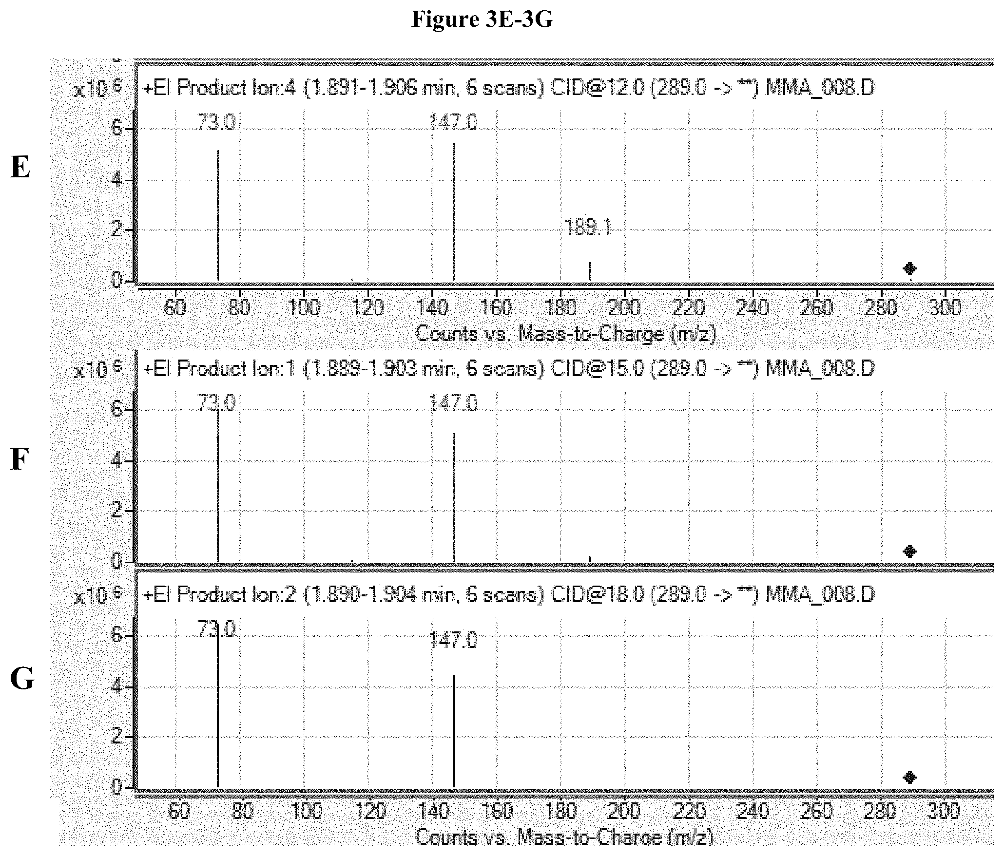
FIGS. 3A-3G show exemplary product ion spectra (covering the m/z range of about 50 to 310) for fragmentation of the TBDMS-MMA precursor ion with m/z of about 289.0 at various collision energies. Details are described in Example 3.
FIGS. 4A-4G show exemplary product ion spectra (covering the m/z range of about 60 to 300) for fragmentation of the TBDMS-MMA-.sup.2H.sub.3 precursor ion with m/z of about 292.0 at various collision energies. Details are described in Example 3.
FIG. 5 shows exemplary data collected for TBDMS-MMA and TBDMS-MMA-.sup.2H.sub.3 for the m/z transitions at 289.fwdarw.189 (at 3 V) and 292.fwdarw.189 (at 3 V), respectively. Details are discussed in Example 3.
FIG. 6 shows exemplary data collected for TBDMS-MMA and TBDMS-MMA-.sup.2H.sub.3 for the m/z transitions at 289.fwdarw.147 (at 10 V) and 292.fwdarw.147 (at 10 V), respectively. Details are discussed in Example 3.
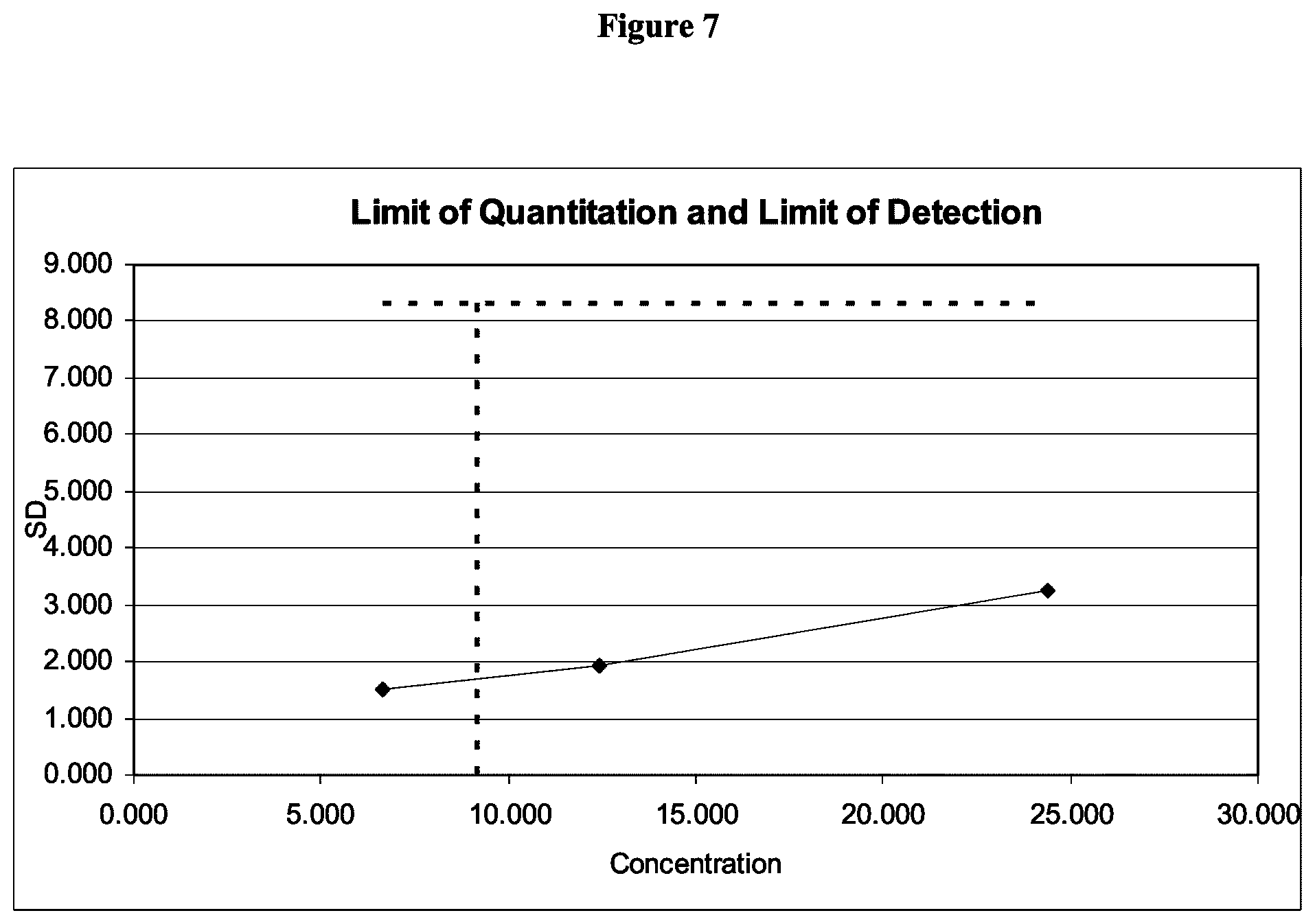
FIG. 7 shows a plot of coefficient of variation (CV) versus concentration for quantitation of TBDMS-MMA in serum measured by monitoring the m/z transition at 289.fwdarw.189 (at 3 V). Details are discussed in Example 5.
FIG. 8 shows a plot demonstrating the linearity of response for quantitation of TBDMS-MMA in serum measured by monitoring the m/z transition at 289.fwdarw.189 (at 3 V). Details are discussed in Example 6.
FIGS. 9A and 9B show plots for method comparison between GC-MS/MS and LC-MS/MS. FIG. 9A shows a plot of values obtained from the same sample by each method, while FIG. 9B shows the differences between obtained values as a function of MMA concentration. Details are discussed in Example 8.
DETAILED DESCRIPTION OF THE INVENTION
Methods are described for determining the amount of MMA in a sample. More specifically, tandem mass spectrometric methods are described for detecting and quantifying tert-butyldimethylsilyl derivatized MMA (TBDMS-MMA) in a sample. Thus, the amount of MMA in a sample may be determined by derivatizing MMA in a sample to form TBDMS-MMA, measuring the amount of TBDMS-MMA in the sample by tandem mass spectrometry, and relating the amount of TBDMS-MMA measured to the amount of MMA in the sample. In some embodiments, TBDMS-MMA derivatives may be prepared by derivatization of MMA with methyl-(t-butyldimethylsilyl) trifluoroacetamide (MTBSFTA).
The methods may use a chromatography technique, such as gas chromatography (GC), to perform a purification of derivatized MMA, combined with methods of mass spectrometry (MS), thereby providing a high-throughput assay system for detecting and quantifying MMA in a sample. Preferred embodiments are particularly well suited for application in large clinical laboratories for automated MMA quantitation.
Suitable test samples for use in methods of the present invention include any test sample that may contain the analyte of interest. In some preferred embodiments, a sample is a biological sample; that is, a sample obtained from any biological source, such as an animal, a cell culture, an organ culture, etc. In certain preferred embodiments, samples are obtained from a mammalian animal, such as a dog, cat, horse, etc. Particularly preferred mammalian animals are primates, most preferably male or female humans. Preferred samples comprise bodily fluids such as blood, plasma, serum, saliva, cerebrospinal fluid, or tissue samples; preferably plasma (including EDTA and heparin plasma) and serum; most preferably serum. Such samples may be obtained, for example, from a patient; that is, a living person, male or female, presenting oneself in a clinical setting for diagnosis, prognosis, or treatment of a disease or condition.
The present invention also contemplates kits for a MMA quantitation assay. A kit for a MMA quantitation assay may include a kit comprising the compositions provided herein. For example, a kit may include packaging material and measured amounts of derivatizing reagent for generation of TBDMS-MMA derivatives (such as MTBSFTA) and an isotopically labeled internal standard, in amounts sufficient for at least one assay. Typically, the kits will also include instructions recorded in a tangible form (e.g., contained on paper or an electronic medium) for using the packaged reagents for use in a MMA quantitation assay.
Calibration and QC pools for use in embodiments of the present invention are preferably prepared using a matrix similar to the intended sample matrix.
As described above, elevated MMA levels may be caused in a patient by a vitamin B.sub.12 deficiency. Therefore, some embodiments of the present invention may be used to diagnose vitamin B.sub.12 deficiency in a patient, or monitor compliance with and/or efficacy of treatment of vitamin B.sub.12 deficiency in a patient. For example, a patient MMA level in excess of a diagnostic MMA level within the range of about 200 nMol/L to about 350 nMol/L may be indicative of a vitamin B.sub.12 deficiency, or failure to comply or lack of efficacy of treatment for a vitamin B.sub.12 deficiency. Generally, diagnostic MMA levels increase with age of patient, but significant overlap between patient age groups may be observed. For example, the reference interval for MMA levels in a healthy patient population between the ages of 18 to 65 years of age may be between about 67.+-.20 nMol/L and about 223.+-.20 nMol/L, whereas the reference interval for MMA levels in a healthy patient population between the ages of 18 to 76 years of age may be between about 87.+-.20 nMol/L and about 318.+-.20 nMol/L group being tested. Thus, in some embodiments, the diagnostic MMA level may be within the range of about 200 nMol/L to about 250 nMol/L, the range of about 250 nMol/L to about 300 nMol/L, or the range of about 300 nMol/L to about 350 nMol/L. In some embodiments, the diagnostic MMA level may be about 223.+-.20 nMol/L, 250.+-.20 nMol/L, 275.+-.20 nMol/L, 300.+-.20 nMol/L, 318.+-.20 nMol/L, or 350.+-.20 nMol/L.
Sample Preparation for Mass Spectrometric Analysis
In preparation for mass spectrometric analysis, MMA may be enriched relative to one or more other components in the sample (e.g. protein) by various methods known in the art, including for example, liquid chromatography, filtration, centrifugation, thin layer chromatography (TLC), electrophoresis including capillary electrophoresis, affinity separations including immunoaffinity separations, extraction methods including ethyl acetate or methanol extraction, and the use of chaotropic agents or any combination of the above or the like.
Protein precipitation is one method of preparing a test sample, especially a biological test sample, such as serum or plasma. Protein purification methods are well known in the art, for example, Polson et al., Journal of Chromatography B 2003, 785:263-275, describes protein precipitation techniques suitable for use in methods of the present invention. Protein precipitation may be used to remove most of the protein from the sample leaving MMA in the supernatant. The samples may be centrifuged to separate the liquid supernatant from the precipitated proteins; alternatively the samples may be filtered to remove precipitated proteins. The purified MMA may then be derivatized with reagent capable of generating TBDMS-MMA, preferably MTBSFTA or an isotopically labeled variant thereof.
Another method of sample purification that may be used prior to mass spectrometry is liquid chromatography (LC). Certain methods of liquid chromatography, including HPLC, rely on relatively slow, laminar flow technology. Traditional HPLC analysis relies on column packing in which laminar flow of the sample through the column is the basis for separation of the analyte of interest from the sample. The skilled artisan will understand that separation in such columns is a diffusional process and may select LC, including HPLC, instruments and columns that are suitable for use with derivatized vitamin D. The chromatographic column typically includes a medium (i.e., a packing material) to facilitate separation of chemical moieties (i.e., fractionation). The medium may include minute particles, or may include a monolithic material with porous channels. A surface of the medium typically includes a bonded surface that interacts with the various chemical moieties to facilitate separation of the chemical moieties. One suitable bonded surface is a hydrophobic bonded surface such as an alkyl bonded, cyano bonded surface, or highly pure silica surface. Alkyl bonded surfaces may include C-4, C-8, C-12, or C-18 bonded alkyl groups. The chromatographic column includes an inlet port for receiving a sample and an outlet port for discharging an effluent that includes the fractionated sample. The sample may be supplied to the inlet port directly, or from an extraction column, such as an on-line SPE cartridge or a TFLC extraction column.
Certain methods of liquid chromatography, including HPLC, rely on relatively slow, laminar flow technology. Traditional HPLC analysis relies on column packing in which laminar flow of the sample through the column is the basis for separation of the analyte of interest from the sample. The skilled artisan will understand that separation in such columns is a diffusional process and may select LC, including HPLC, instruments and columns that are suitable for use with derivatized MMA. The chromatographic column typically includes a medium (i.e., a packing material) to facilitate separation of chemical moieties (i.e., fractionation). The medium may include minute particles, or may include a monolithic material with porous channels. A surface of the medium typically includes a bonded surface that interacts with the various chemical moieties to facilitate separation of the chemical moieties. One suitable bonded surface is a hydrophobic bonded surface such as an alkyl bonded, cyano bonded surface, or highly pure silica surface. Alkyl bonded surfaces may include C-4, C-8, C-12, or C-18 bonded alkyl groups. The chromatographic column includes an inlet port for receiving a sample and an outlet port for discharging an effluent that includes the fractionated sample. The sample may be supplied to the inlet port directly, or from an extraction column, such as an on-line SPE cartridge or a TFLC extraction column.
In one embodiment, the sample may be applied to the LC column at the inlet port, eluted with a solvent or solvent mixture, and discharged at the outlet port. Different solvent modes may be selected for eluting the analyte(s) of interest. For example, liquid chromatography may be performed using a gradient mode, an isocratic mode, or a polytyptic (i.e. mixed) mode. During chromatography, the separation of materials is effected by variables such as choice of eluent (also known as a "mobile phase"), elution mode, gradient conditions, temperature, etc.
In certain embodiments, an analyte may be purified by applying a sample to a column under conditions where the analyte of interest is reversibly retained by the column packing material, while one or more other materials are not retained (i.e., solid phase extraction). In these embodiments, a first mobile phase condition can be employed where the analyte of interest is retained by the column, and a second mobile phase condition can subsequently be employed to remove retained material from the column, once the non-retained materials are washed through. Alternatively, an analyte may be purified by applying a sample to a column under mobile phase conditions where the analyte of interest elutes at a differential rate in comparison to one or more other materials. Such procedures may enrich the amount of one or more analytes of interest relative to one or more other components of the sample.
By careful selection of valves and connector plumbing, two or more chromatography columns may be connected as needed such that material is passed from one to the next without the need for any manual steps. In preferred embodiments, the selection of valves and plumbing is controlled by a computer pre-programmed to perform the necessary steps. Most preferably, the chromatography system is also connected in such an on-line fashion to the detector system, e.g., an MS system. Thus, an operator may place a tray of samples in an autosampler, and the remaining operations are performed under computer control, resulting in purification and analysis of all samples selected.
Another method of sample purification that may be used prior to mass spectrometry is gas chromatography (GC) (also sometimes known as vapor-phase chromatography or liquid partition chromatography). Gas chromatography may be employed in methods of analyzing compounds that can be vaporized without decomposition. In gas chromatography, the mobile phase is a carrier gas, usually an inert gas such as helium or an unreactive gas such as nitrogen. The stationary phase is a microscopic layer of liquid or polymer on an inert solid support. The stationary phase is contained inside a column. The gaseous compounds being analyzed interact with the walls of the column, which may be coated with various stationary phases. Varying degrees of interaction of each gaseous compound with the stationary phase cause each compound to elute at a different times. Thus, for a given stationary phase and given column conditions, each compound will have a characteristic retention time. In some embodiments, a GC column comprising cyanopropyl methyl siloxane, such as an Agilent DB-23 cis/trans FAME GC column, is used to purify TBDMS-MMA. In some embodiments, the carrier gas is helium. In some embodiments, the column is heated to a temperature of about 185.degree. C. and held for about 0.4 minutes, then the temperature is ramped to about 300.degree. C.; for example at a rate of about 85.degree. C./minute. Once the temperature reaches about 300.degree. C., the temperature is then held constant, for example for about 0.6 minutes.
In some embodiments, an extraction column may be used for purification of MMA prior to mass spectrometry. In such embodiments, MMA is purified by applying a first mobile phase (containing the analyte) to an extraction column which captures the analyte, and then eluting the captured analyte with a second mobile phase. The eluent may be collected and chromatographed on a second extraction column or on an analytical column (such as a GC column) prior to ionization. In some embodiments, extraction of MMA via extraction column is done before MMA is derivatized; alternatively, MMA may be derivatized prior to extraction.
Detection and Quantitation by Mass Spectrometry
In various embodiments, derivatized MMA may be ionized by any method known to the skilled artisan. Mass spectrometry is performed using a mass spectrometer, which includes an ion source for ionizing the fractionated sample and creating charged molecules for further analysis. For example ionization of the sample may be performed by electron impact (EI) ionization, chemical ionization, electrospray ionization (ESI), photon ionization, atmospheric pressure chemical ionization (APCI), photoionization, atmospheric pressure photoionization (APPI), Laser diode thermal desorption (LDTD), fast atom bombardment (FAB), liquid secondary ionization (LSI), matrix assisted laser desorption ionization (MALDI), field ionization, field desorption, thermospray/plasmaspray ionization, surface enhanced laser desorption ionization (SELDI), inductively coupled plasma (ICP) and particle beam ionization. The skilled artisan will understand that the choice of ionization method may be determined based on the analyte to be measured, type of sample, the type of detector, the choice of positive versus negative mode, etc.
Derivatized MMA may be ionized in positive or negative mode. In preferred embodiments, derivatized MMA is ionized by electron impact (EI) ionization in positive mode.
In mass spectrometry techniques generally, after the sample has been ionized, the positively or negatively charged ions thereby created may be analyzed to determine a mass-to-charge ratio. Suitable analyzers for determining mass-to-charge ratios include quadrupole analyzers, ion traps analyzers, and time-of-flight analyzers. Exemplary ion trap methods are described in Bartolucci, et al., Rapid Commun. Mass Spectrom. 2000, 14:967-73.
The ions may be detected using several detection modes. For example, selected ions may be detected, i.e. using a selective ion monitoring mode (SIM), or alternatively, mass transitions resulting from collision induced dissociation or neutral loss may be monitored, e.g., multiple reaction monitoring (MRM) or selected reaction monitoring (SRM). Preferably, the mass-to-charge ratio is determined using a quadrupole analyzer. For example, in a "quadrupole" or "quadrupole ion trap" instrument, ions in an oscillating radio frequency field experience a force proportional to the DC potential applied between electrodes, the amplitude of the RF signal, and the mass/charge ratio. The voltage and amplitude may be selected so that only ions having a particular mass/charge ratio travel the length of the quadrupole, while all other ions are deflected. Thus, quadrupole instruments may act as both a "mass filter" and as a "mass detector" for the ions injected into the instrument.
One may enhance the resolution of the MS technique by employing "tandem mass spectrometry," or "MS/MS". In this technique, a precursor ion (also called a parent ion) generated from a molecule of interest can be filtered in an MS instrument, and the precursor ion subsequently fragmented to yield one or more fragment ions (also called daughter ions or product ions) that are then analyzed in a second MS procedure. By careful selection of precursor ions, only ions produced by certain analytes are passed to the fragmentation chamber, where collisions with atoms of an inert gas produce the fragment ions. Because both the precursor and fragment ions are produced in a reproducible fashion under a given set of ionization/fragmentation conditions, the MS/MS technique may provide an extremely powerful analytical tool. For example, the combination of filtration/fragmentation may be used to eliminate interfering substances, and may be particularly useful in complex samples, such as biological samples.
Alternate modes of operating a tandem mass spectrometric instrument include product ion scanning and precursor ion scanning For a description of these modes of operation, see, e.g., E. Michael Thurman, et al., Chromatographic-Mass Spectrometric Food Analysis for Trace Determination of Pesticide Residues, Chapter 8 (Amadeo R. Fernandez-Alba, ed., Elsevier 2005) (387).
The results of an analyte assay may be related to the amount of the analyte in the original sample by numerous methods known in the art. For example, given that sampling and analysis parameters are carefully controlled, the relative abundance of a given ion may be compared to a table that converts that relative abundance to an absolute amount of the original molecule. Alternatively, external standards may be run with the samples, and a standard curve constructed based on ions generated from those standards. Using such a standard curve, the relative abundance of a given ion may be converted into an absolute amount of the original molecule. In certain embodiments, an internal standard is used to generate a standard curve for calculating the quantity of MMA. Methods of generating and using such standard curves are well known in the art and one of ordinary skill is capable of selecting an appropriate internal standard. For example, in some embodiments an isotopically labeled MMA (e.g., MMA-.sup.2H.sub.3) may be used as an internal standard. Numerous other methods for relating the amount of an ion to the amount of the original molecule will be well known to those of ordinary skill in the art.
One or more steps of the methods may be performed using automated machines. In certain embodiments, one or more purification steps are performed on-line, and more preferably all of the purification and mass spectrometry steps may be performed in an on-line fashion.
In certain embodiments, such as MS/MS, where precursor ions are isolated for further fragmentation, collision activated dissociation (CAD) is often used to generate fragment ions for further detection. In CAD, precursor ions gain energy through collisions with an inert gas, and subsequently fragment by a process referred to as "unimolecular decomposition." Sufficient energy must be deposited in the precursor ion so that certain bonds within the ion can be broken due to increased vibrational energy.
In some embodiments, MMA in a sample is detected and/or quantified using MS/MS as follows. MMA is first extracted from a sample with a solid phase extraction column. Then, MMA in the purified sample is derivatized with a reagent capable of generating TBDMS-MMA, preferably MTBSFTA. The purified samples (now comprising TBDMS-MMA) are then subjected to gas chromatography; the flow of analyte from the chromatographic column enters an EI ionization source and TBDMS-MMA ions are generated. The TBDMS-MMA ions, e.g. precursor ions, pass through the orifice of the instrument and enter the first quadrupole. Quadrupoles 1 and 3 (Q1 and Q3) are mass filters, allowing selection of ions (i.e., selection of "precursor" and "fragment" ions in Q1 and Q3, respectively) based on their mass to charge ratio (m/z). Quadrupole 2 (Q2) is the collision cell, where ions are fragmented. The first quadrupole of the mass spectrometer (Q1) selects for TBDMS-MMA precursor ions, which are allowed to pass into the collision chamber (Q2), while unwanted ions with any other mass/charge ratio collide with the sides of the quadrupole and are eliminated. Precursor ions entering Q2 collide with a collision gas and fragment. The fragment ions generated are passed into quadrupole 3 (Q3), where certain TBDMS-MMA fragment ions are selected while other ions are eliminated.
As ions collide with the detector they produce a pulse of electrons that are converted to a digital signal. The acquired data is relayed to a computer, which plots counts of the ions collected versus time. The resulting mass chromatograms are similar to chromatograms generated in traditional GC-MS methods. The areas under the peaks corresponding to particular ions, or the amplitude of such peaks, may be measured and correlated to the amount of the analyte of interest. In certain embodiments, the area under the curves, or amplitude of the peaks, for fragment ion(s) and/or precursor ions are measured to determine the amount of MMA. As described above, the relative abundance of a given ion may be converted into an absolute amount of the original analyte using calibration standard curves based on peaks of one or more ions of an internal molecular standard.
EXAMPLES
Example 1: Solid Phase Extraction and TBDMS Derivatization
The following extraction techniques were conducted on serum samples.
MMA was extracted from serum samples by solid phase extraction (SPE) on a strong anion exchange (SAX) packed SPE column. Water and 100% methanol were used as wash and elution solvents, respectively. Alternatively, MMA was extracted from serum samples by SPE on a SAX packed SPE column where water and 3 M acetic acid/methanol were used as wash and elution solvents, respectively.
MMA was then derivatized by adding 150 .mu.l of MTBSTFA with 1% tert-butyl dimethylchlorosilane (TBDMCS) in hexane or pyridine to the collected extracted samples. The resulting mixtures were heated to about 70.degree. C. for 15-20 minutes and chilled at about 4.degree. C. for about 10 minutes. After cooling, the mixtures were transferred to GC/MS vials for analysis.
Alternatively, MMA from the sample was derivatized by first evaporating the eluted samples to dryness, adding 500 .mu.l of 20% triethylamine in methanol to the dried samples, drying the samples again, adding 100 .mu.l MTBSTFA with 1% tert-butyl dimethylchlorosilane (TBDMCS) in hexane or pyridine to the dried samples, and vortexing for 2-3 minutes. The resulting mixtures were heated to about 60.degree. C. for about 30 minutes and then chilled. After cooling, the mixtures were then subjected to GC-MS/MS analysis.
Example 2: Purification of TBDMS-MMA Derivatives with Gas Chromatography
Purification of TBDMS-MMA derivatives with gas chromatography was conducted with an Agilent 7890A Fast GC equipped with a cyanopropyl methyl siloxane analytical column, such as an Agilent DB-23 cis/trans FAME GC column (DB-23: J&W 122-2361, Agilent Technologies, Wilmington, Del. USA). The GC column was heated to a temperature of about 185.degree. C. and held for about 0.4 minutes. The GC column was then heated further to a temperature of about 300.degree. C. at a rate of about 85.degree. C./minute. Once the temperature reached about 300.degree. C., the temperature was held at about 300.degree. C. for about 0.6 minutes. Helium was used as the carrier gas at approximately 3 ml/min. Under these conditions, retention time for TBDMS-MMA was about 1.76-1.78 minutes. Succinic acid, a potentially interfering species with a similar mass to MMA, was clearly separated from MMA under this procedure with a retention time of approximately 2.12 minutes.
Alternatively, purification of TBDMS-MMA derivatives with gas chromatography was conducted with an Agilent 7890 GC equipped with a cyanopropyl methyl siloxane analytical column, such as an Agilent DB-23 cis/trans FAME GC column (DB-23: J&W 122-2361, Agilent Technologies, Wilmington, Del. USA). The GC column was heated to a temperature of about 300.degree. C. Helium was used as the carrier gas at approximately 4 ml/min. Under these conditions, retention time for TBDMS-MMA was about 1.30 minutes.
Example 3: Detection and Quantitation of TBDMS-MMA Derivatives by MS/MS
MS/MS was performed on the above generated samples using a Agilent 7001 GC/MS Triple Quadrupole MS system (Agilent Technologies, Wilmington, Del. USA). TBDMS-MMA exiting the analytical column flowed to the electron impaction ionization interface of the MS/MS analyzer and ionized.
Ions passed to the first quadrupole (Q1), which selected TBDMS-MMA precursor ions with a mass-to-charge ratio of 289.0.+-.0.5 m/z. Ions entering quadrupole 2 (Q2) collided with argon gas to generate ion fragments, which were passed to quadrupole 3 (Q3) for further selection. Simultaneously, the same process using isotope dilution mass spectrometry was carried out with internal standard, TBDMS-MMA-.sup.2H.sub.3. Mass transitions observed at collision energies of 3V and 10 V (positive polarity) are shown in Table 1.
TABLE-US-00001 TABLE 2 Mass Transitions for TBDMS-MMA and TBDMS-MMA- .sup.2H.sub.3 (internal standard) (Positive Polarity) Product Ions Product Ions Precursor Ion (m/z) (m/z) Analyte (m/z) (CE = 3 V) (CE = 10 V) TBDMS-MMA 289.0 .+-. 0.5 189.0 .+-. 0.5 147.0 .+-. 0.5 147.0 .+-. 0.5 73.0 .+-. 0.5 73.0 .+-. 0.5 TBDMS-MMA-.sup.2H.sub.3 292.0 .+-. 0.5 189.0 .+-. 0.5 147.0 .+-. 0.5 147.0 .+-. 0.5 73.0 .+-. 0.5 73.0 .+-. 0.5
Exemplary chromatograms for TBDMS-MMA and TBDMS-MMA-.sup.2H.sub.3 (internal standard), are shown in FIGS. 1A and 1B, respectively.
An exemplary calibration curve for the determination of TBDMS-MMA in serum as measured by monitoring the transition from 289.0.+-.0.5.fwdarw.147.0.+-.0.5 at a collision energy of about 10 V is shown in FIG. 2.
Example 4: Exemplary Spectra from MS/MS Analysis of TBDMS-MMA
Tandem mass spectrometric analyses of TBDMS-MMA and TBDMS-MMA-.sup.2H.sub.3 were conducted by selecting precursor ions with m/z of about 289.0 and 292.0, respectively. The precursor ions were fragmented at collision energies of 3, 5, 8, 10, 12, 15, and 18 V. Exemplary product ion scans generated from these analyses are presented in FIGS. 3A-3G and 4A-4G for TBDMS-MMA and TBDMS-MMA-.sup.2H.sub.3, respectively.
Exemplary MRM transitions for the quantitation of TBDMS-MMA include fragmenting a precursor ion with a m/z of about 289.0.+-.0.5 to one or more product ions selected from the group of ions with a m/z of about 189.0.+-.0.5, 147.0.+-.0.5, and 73.0.+-.0.5. As seen in FIGS. 3A-3G and 4A-4G, selection of fragments to monitor for quantitation may depend on the collision energy selected. At a higher collision energy, such as 10 V, fragment ions may include ions with m/z of about 147.0.+-.0.5 and 73.0.+-.0.5, while at lower collision energies, such as about 3 V, fragment ions may also include ions with m/z of about 189.0.+-.0.5. Similarly, MRM transitions for the quantitation of TBDMS-MMA-.sup.2H.sub.3 include fragmenting a precursor ion with a m/z of about 292.0 to one or more product ions selected from the group of ions with a m/z of about 189.0.+-.0.5, 147.0.+-.0.5, and 73.0.+-.0.5. Again, at a higher collision energy, such as 10 V, fragment ions may include ions with m/z of about 147.0.+-.0.5 and 73.0.+-.0.5, while at lower collision energies, such as about 3 V, fragment ions may also include ions with m/z of about 189.0.+-.0.5.
Data was collected for quantitation of TBDMS-MMA and TBDMS-MMA-.sup.2H.sub.3 in serum by monitoring MRM transitions generated by collision energies of 3 V and 10 V. Exemplary data from the 3 V collision energy (TBDMS-MMA: 289.0.fwdarw.189.0; and TBDMS-MMA-.sup.2H.sub.3: 292.0.fwdarw.189.0) are shown in FIG. 5, while exemplary data from the 10 V collision energy (TBDMS-MMA: 289.0.fwdarw.147.0; and TBDMS-MMA-.sup.2H.sub.3: 292.0.fwdarw.147.0) are shown in FIG. 6.
Example 5: Within Run and Total Precision Studies
For within run and total precision studies, Low, Mid, and High concentration control pools were prepared with target values of 150, 1000, and 5000 nMol/L MMA by spiking charcoal stripped serum with MMA in water. MMA was quantitated in these serum samples as described above, by monitoring the MRM transition from a MRM precursor ion with m/z of about 289.0.+-.0.5 to a fragment ion with m/z of about 189.0.+-.0.5 (collision energy about 3 V).
For the within run precision studies, aliquots of the Low, Mid, and High control pools were analyzed 10 times each in a single sample run. The coefficients of variation (CV) from each concentration were used to determine if reproducibility within run was acceptable. Statistical analysis of the results determined that the CV for the Low, Mid, and High pools were about 2.0%, 2.7%, and 2.2%, respectively. In addition, standard deviations (SD) for each of the pools were calculated to be about 3 nMol/L, 28 nMol/L, and 111 nMol/L, respectively. Data from these experiments are presented in Table 3.
TABLE-US-00002 TABLE 3 Within Run Precision Control Pool Low Mid High (150 nMol/L) (1000 nMol/L) (5000 nMol/L) Replicate Measured MMA Concentration (nMol/L) 1 153 1054 4960 2 153 1033 5181 3 156 1028 4984 4 157 997 4952 5 151 1070 4961 6 154 1037 5191 7 149 1011 4855 8 159 1028 5021 9 155 1090 4965 10 154 1021 5130 Mean 154 1037 5020 Std Dev 3 28 111 CV 2.0% 2.7% 2.2%
For the total precision studies, aliquots of the Low, Mid, and High control pools were analyzed on 19 separate days. On 17 of those days, the control pools were run in duplicate, while on two of the days, the controls were run in singleton. A total of 36 data points for each control pool were generated and statistically analyzed. The CV for the Low, Mid, and High pools were calculated to be about 10.9%, 9.3%, and 8.0%, respectively. In addition, SD for each of the pools were calculated to be about 19 nMol/L, 104 nMol/L, and 420 nMol/L, respectively. Data from these experiments are presented in Table 4.
TABLE-US-00003 TABLE 4 Total Precision Control Pool Low Mid High (150 nMol/L) (1000 nMol/L) (5000 nMol/L) Replicate Measured MMA Concentration (nMol/L) A 153 1054 4960 B 154 1021 5130 A 150 1026 4731 B 148 966 4765 A 156 1056 5294 B 147 977 5061 A 149 1074 5279 B 181 1173 5341 A 186 1184 5684 B 177 1020 5059 A 169 1139 5158 B 181 1141 4634 A 180 1182 5164 B 157 1021 4616 A 178 1179 5382 B 156 1021 4743 A 164 1132 5280 B 151 1125 5423 A 184 1209 5805 B 185 1155 5224 A 191 1097 5461 B 180 1182 4924 A 168 987 5127 B 193 1197 4912 A 171 964 4672 A 163 1093 5044 B 174 1464 5903 A 200 1200 5269 B 175 -- -- A 180 1088 5603 B 147 1170 5660 A 175 1125 4600 B 141 1143 5573 A 223 1261 6400 B 197 1273 5526 A 203 1252 5876 B 163 1236 5677 Count (n) 37 36 36 Mean 172 1127 5249 Std Dev 19 104 420 CV 10.9% 9.3% 8.0%
Example 6: Analytical Sensitivity: Limit of Blank (LOB), Limit of Detection CLOD), and Lower Limit of Quantitation (LLOQ)
The Limit of Blank (LOB) was determined by analyzing a zero calibrator (i.e., a blank of stripped serum) 20 times in a single run. The LOB was calculated to be about 8 nMol/L.
The Limit of Detection (LOD) is the point at which a value is beyond the uncertainty associated with its measurement and is defined as four standard deviations from the zero concentration. To determine the LOD, a blank of stripped serum was analyzed 20 times in a single sample run, and the mean and SD were determined. The resulting LOD was calculated to be about 9 nMol/L.
The Lower Limit of Quantitation (LLOQ) is the point where measurements become quantitatively meaningful. Analyte response at the LLOQ is identifiable, discrete and reproducible with a precision (i.e., coefficient of variation (CV)) of less than 20% and an accuracy of 80% to 120%. The LLOQ was determined by assaying five different stripped serum samples spiked with MMA at about 6 nMol/L, 13 nMol/L, 25 nMol/L, 50 nMol/L, and 100 nMol/L in 5 replicates over 5 days, according to the method described above in Example 4. CV's were calculated for each level. The LLOQ was calculated from the data to be 9 nMol/L. Data from analysis of the lower three concentrations and a blank are presented in Table 5. The graphical representations of CV versus concentration is shown in FIG. 7.
TABLE-US-00004 TABLE 5 Lower Limit of Quantitation Experiments MMA Concentration (nMol/L) Run # Result # 6 nMol/L 13 nMol/L 25 nMol/L 0 nMol/L 1 1 8.000 9.000 25.000 6.760 2 8.000 10.000 21.000 8.562 3 8.000 12.000 19.000 7.110 4 8.000 14.000 25.000 6.492 5 4.000 13.000 26.000 6.232 2 1 8.000 11.000 25.000 7.243 2 7.000 13.000 20.000 6.208 3 5.000 13.000 24.000 6.684 4 6.000 13.000 22.000 6.794 5 7.000 12.000 23.000 7.661 3 1 4.000 10.000 25.000 6.339 2 5.000 10.000 24.000 6.901 3 5.000 12.000 20.000 6.329 4 4.000 10.000 19.000 6.967 5 5.000 11.000 26.000 7.082 4 1 7.000 16.000 28.000 6.811 2 6.000 12.000 29.000 6.994 3 7.000 13.000 25.000 7.578 4 6.000 12.000 20.000 7.127 5 7.000 12.000 26.000 7.003 5 1 8.000 15.000 31.000 -- 2 8.000 15.000 29.000 -- 3 8.000 16.000 26.000 -- 4 9.000 12.000 26.000 -- 5 8.000 15.000 26.000 -- Count (n) 25 25 25 20 Mean 6.640 12.440 24.400 6.944 SD 1.524 1.938 3.240 0.555 CV 23.1% 15.6% 12.9% 8.3%
Example 7: Reportable Range and Linearity
A sample with high concentration of MMA (8868 nMol/L) was mixed with a sample with a low concentration of MMA (99 nMol/L) in different proportions to achieve five samples with concentrations across the range. The five samples were prepared with the following proportions 100% Low (expected concentration of 99 nMol/L), 75% Low/25% High (expected concentration of 2291 nMol/L), 50% Low/50% High (expected concentration of 4484 nMol/L), 25% Low/75% High (expected concentration of 6676 nMol/L), and 100% High (expected concentration of 8868 nMol/L). Analysis of samples at these five concentrations as described in Example 4 demonstrated linear response across a reportable range of up to at least about 8870 nMol/L. A graphical representation demonstrating the linearity of response is shown in FIG. 8.
Example 8: Recovery Studies
Six patient serum pools (STD 1-6) were spiked with a known amount of MMA. Samples 1-3 (500 .mu.L) were spiked with 50 .mu.L of 3333 nMol/L MMA standard in stripped serum, and samples 4-6 (500 .mu.L) were spiked with 50 .mu.L of 10,000 nMol/L MMA standard in stripped serum. Non-spiked samples were prepared for comparison by adding 50 .mu.L of blank stripped serum to 500 .mu.L of each of Samples 1-6. For each serum pool, 2 baseline (blank serum added) and 4 spiked replicates were tested as described in Example 4. The amount recovered was calculated as the difference between the mean of the spiked and baseline samples. Percent recovery was calculated to be the ratio of the amount recovered (observed) divided by the amount added. The six pools yielded an average accuracy of about 104.1%. All assays were within the acceptable accuracy range of 85-115%. The results of the spiked specimen recovery studies are presented in Table 6.
TABLE-US-00005 TABLE 6 Spiked Specimen Recovery Studies STD 1 STD 2 STD 3 STD 4 STD 5 STD 6 Measured Concentration (nMol/L) Blank 399.77 420.96 431.60 1070.50 1113.17 1478.64 Result 1 421.41 433.23 463.68 1111.00 1169.87 1689.27 Result 2 434.17 440.31 449.28 1142.52 1160.13 1533.30 Result 3 420.86 445.55 461.15 1158.69 1193.30 1576.17 Result 4 414.25 422.68 429.29 1107.55 1132.14 1555.99 Spiked Mean 418.09 432.55 447.00 1118.05 1153.72 1566.67 % Recovery 104.58 102.75 103.57 104.44 103.64 105.95
Example 9: Method Comparison Studies
The performance of the GC-MS/MS assay described in Example 4 was compared against quantitation of MMA by LC-MS/MS. Forty patient serum samples were analyzed in four individual runs over four days by each method. Difference and % difference were calculated for each patient sample. The results of these studies are shown plotted in FIGS. 9A (GC-MS/MS vs LC-MS/MS) and 9B (difference vs amount detected).
Example 10: Hemolysis, Lipemia, and Icteria Studies
The effect hemolysis, lipemia, and icteria have on the assay was also investigated.
Hemolysis.
The effect of hemolysis was evaluated by preparing five patient serum pools as follows. A hemolysate was prepared by washing 2 mL of heparinized whole blood three times with phosphate buffered saline (PBS). After the final wash, the cells were frozen at about -70.degree. C. for about 30 minutes. The hemolyzed cell mixture was removed from the freezer and mixed by mechanical vortex. For each of the five patient serum pools, 200 .mu.L of hemolysate was added to 2 mL of serum. A baseline (non-hemolytic) sample was prepared for each pool by the addition of 200 .mu.L of PBS to 2 mL of serum. For each pool, baseline and hemolytic samples were analyzed in quadruplicate and the differences between mean and hemolyzed samples observed. Results of analysis of all pools were acceptable (i.e., between 85% and 115% of baseline). Results of these studies are presented in Table 7.
Lipemia.
The effect of lipemia was evaluated by preparing five patient serum pools each with 200 .mu.L of Intralipid added to 2 mL of serum. A baseline (non-lipemic) sample was prepared for each pool by the addition of 200 .mu.L of PBS to 2 mL of serum. For each pool, baseline and lipemic samples were analyzed in quadruplicate and the differences between mean and lipemic samples observed. Results of analysis of all pools were acceptable (i.e., between 85% and 115% of baseline). Results of these studies are presented in Table 7.
Icteria.
The effect of icteria was evaluated by preparing five patient serum pools each with 200 .mu.L of Verichem High Bilirubin Standard Level F (30 mg/dL total and direct bilirubin) added to 2 mL of serum. A baseline (non-icteric) sample was prepared for each pool by the addition of 200 .mu.L of PBS to 2 mL of serum. For each pool, baseline and icteric samples were analyzed in quadruplicate and the differences between mean and icteric samples observed. Results of analysis of four of the five pools were unacceptable (i.e., above 115% of baseline). Results of these studies are presented in Table 7.
TABLE-US-00006 TABLE 7 Hemolysis, Lipemia, and Bilirubin Interference Studies Pool 1 Pool 2 Pool 3 Pool 4 Pool 5 Average MMA Concentration (nMol/L) Baseline 210 187 170 199 150 Hemolysis 209 193 174 204 157 Icterus 203 188 174 198 152 Lipemia 223 235 219 238 195 % Recovery Hemolysis 100% 103% 102% 103% 105% Icterus 97% 97% 100% 97% 97% Lipemia 110% 125% 126% 120% 128%
Example 11: MMA Reference Intervals
Serum samples from 30 healthy male and 30 healthy female donors (total n=60) between the ages of 18 to 65 years were assayed for MMA as described above, and results statistically analyzed to determine reference ranges for healthy individuals. Healthy reference ranges were determined to be within about 67 nMol/L to about 223 nMol/L. No sex difference was observed. Raw data from these studies is presented in Table 8.
TABLE-US-00007 TABLE 8 Reference Interval Studies MMA Sample (nMol/L) Below RI Above RI 1 137.00 2 166.00 3 67.00 4 100.00 5 153.00 6 80.00 7 136.00 8 71.00 9 61.00 YES 10 111.00 11 96.00 12 129.00 13 101.00 14 161.00 15 95.00 16 197.00 17 169.00 18 203.00 19 121.00 20 113.00 21 125.00 22 126.00 23 277.00 YES 24 122.00 25 124.00 26 160.00 27 84.00 28 144.00 29 120.00 30 104.00 31 96.00 32 98.00 33 99.00 34 109.00 35 163.00 36 106.00 37 156.00 38 111.00 39 120.00 40 143.00 41 134.00 42 159.00 43 76.00 44 101.00 45 230.00 YES RI Lower Limit 67 RI Upper Limit 223 Number of donors 45 Number above RI 2 Percent above RI 4.4% Number below RI 1 Percent below RI 2.2% Percent outside RI 6.7%
The contents of the articles, patents, and patent applications, and all other documents and electronically available information mentioned or cited herein, are hereby incorporated by reference in their entirety to the same extent as if each individual publication was specifically and individually indicated to be incorporated by reference. Applicants reserve the right to physically incorporate into this application any and all materials and information from any such articles, patents, patent applications, or other physical and electronic documents.
The methods illustratively described herein may suitably be practiced in the absence of any element or elements, limitation or limitations, not specifically disclosed herein. Thus, for example, the terms "comprising", "including," containing", etc. shall be read expansively and without limitation. Additionally, the terms and expressions employed herein have been used as terms of description and not of limitation, and there is no intention in the use of such terms and expressions of excluding any equivalents of the features shown and described or portions thereof. It is recognized that various modifications are possible within the scope of the invention claimed. Thus, it should be understood that although the present invention has been specifically disclosed by preferred embodiments and optional features, modification and variation of the invention embodied therein herein disclosed may be resorted to by those skilled in the art, and that such modifications and variations are considered to be within the scope of this invention.
The invention has been described broadly and generically herein. Each of the narrower species and subgeneric groupings falling within the generic disclosure also form part of the methods. This includes the generic description of the methods with a proviso or negative limitation removing any subject matter from the genus, regardless of whether or not the excised material is specifically recited herein.
Other embodiments are within the following claims. In addition, where features or aspects of the methods are described in terms of Markush groups, those skilled in the art will recognize that the invention is also thereby described in terms of any individual member or subgroup of members of the Markush group.
* * * * *
References
D00000
D00001
D00002
D00003
D00004
D00005
D00006
D00007
D00008
D00009
D00010
D00011
XML
uspto.report is an independent third-party trademark research tool that is not affiliated, endorsed, or sponsored by the United States Patent and Trademark Office (USPTO) or any other governmental organization. The information provided by uspto.report is based on publicly available data at the time of writing and is intended for informational purposes only.
While we strive to provide accurate and up-to-date information, we do not guarantee the accuracy, completeness, reliability, or suitability of the information displayed on this site. The use of this site is at your own risk. Any reliance you place on such information is therefore strictly at your own risk.
All official trademark data, including owner information, should be verified by visiting the official USPTO website at www.uspto.gov. This site is not intended to replace professional legal advice and should not be used as a substitute for consulting with a legal professional who is knowledgeable about trademark law.