Methods for using long ssDNA polynucleotides as primers (superprimers) in PCR assays
Mautner November 3, 2
U.S. patent number 10,822,647 [Application Number 15/283,851] was granted by the patent office on 2020-11-03 for methods for using long ssdna polynucleotides as primers (superprimers) in pcr assays. This patent grant is currently assigned to BIODYNAMICS S.R.L.. The grantee listed for this patent is Biodynamics S.R.L.. Invention is credited to Martin Eduardo Mautner.


View All Diagrams
| United States Patent | 10,822,647 |
| Mautner | November 3, 2020 |
Methods for using long ssDNA polynucleotides as primers (superprimers) in PCR assays
Abstract
Method to perform a PCR assay that comprises the following steps: a. Obtaining a nucleic acid sample; b. Hybridizing that nucleic acid sample to one or more pair of primers where at least one primer consists of a single stranded DNA polynucleotide having a length of 60 or more nucleotides; c Subjecting said nucleic acid sample to a PCR, wherein the reaction mixture medium contains at least one of said primers; and d. Detecting the length of the amplified products. The amplified nucleic acid may contain any sequence or multiple sequences of STRs (short tandem repeats), genes or any coding region having a defined location on a genome. The preferred nucleic acid samples to be amplified are degraded or fragmented and contain one or more genetic markers.
| Inventors: | Mautner; Martin Eduardo (Buenos Aires, AR) | ||||||||||
|---|---|---|---|---|---|---|---|---|---|---|---|
| Applicant: |
|
||||||||||
| Assignee: | BIODYNAMICS S.R.L. (Buenos
Aires, AR) |
||||||||||
| Family ID: | 1000005156029 | ||||||||||
| Appl. No.: | 15/283,851 | ||||||||||
| Filed: | October 3, 2016 |
Prior Publication Data
| Document Identifier | Publication Date | |
|---|---|---|
| US 20180016621 A1 | Jan 18, 2018 | |
Related U.S. Patent Documents
| Application Number | Filing Date | Patent Number | Issue Date | ||
|---|---|---|---|---|---|
| 62361184 | Jul 12, 2016 | ||||
| Current U.S. Class: | 1/1 |
| Current CPC Class: | C12Q 1/6858 (20130101); C12Q 1/686 (20130101); C12Q 1/6858 (20130101); C12Q 2525/204 (20130101); C12Q 2531/113 (20130101); C12Q 2537/143 (20130101); C12Q 1/686 (20130101); C12Q 2525/204 (20130101); C12Q 2537/143 (20130101) |
| Current International Class: | C12P 19/34 (20060101); C12Q 1/686 (20180101); C12Q 1/6858 (20180101) |
| Field of Search: | ;435/91.2 |
References Cited [Referenced By]
U.S. Patent Documents
| 10422002 | September 2019 | Stephens et al. |
| 2013/0115605 | May 2013 | Merk |
| 2015/0232929 | August 2015 | Stephens |
Other References
|
Protocol online., Long PCR primer forum, http://www.protocol-online.org/biology-forums-2/posts/6949.html, pp. 1-4, Mar. 2009. cited by examiner . OpenWetWare., Designing primers, https://openwetware.org/wiki/Designing_primers, pp. 1-4, Nov. 25, 2015. cited by examiner . Butler et al., Methods in Molecular Biology, vol. 297, Forensic DNA typing Protocols, Humana Press. Inc, Totowa, NJ, pp. 53-65, (Year: 2005). cited by examiner . Wiegand et al., Int. J. Legal Med. 114: 287-287, (Year: 2001). cited by examiner . Butler, "Table 10.1", Advanced Topics in Forensic DNA Typing: Methodology, Elsevier, 2012. cited by applicant . Butler, "Attempts with DNA Repair Following DNA Damage", Advanced Topics in Forensic DNA Typing: Methodology, Elsevier, chapter 10, p. 304, 2012. cited by applicant . Declaration submitted Apr. 1, 2020, including Exhibits A-J, in connection with U.S. Appl. No. 15/283,851. cited by applicant. |
Primary Examiner: Wilder; Cynthia B
Attorney, Agent or Firm: Norris McLaughlin, P.A.
Parent Case Text
This application claims benefit of U.S. Provisional Application No. 62/361,184, filed Jul. 12, 2016, the disclosure of which patent application is incorporated herein by reference.
Claims
I claim:
1. A method for simultaneously detecting in a nucleic acid sample a plurality of genetic markers by performing a polymerase chain reaction comprising: a) providing a sample comprising a nucleic acid; b) hybridizing the nucleic acid to a plurality of pairs of primers, thereby forming a hybridized nucleic acid; c) subjecting the hybridized nucleic acid to a polymerase chain reaction, thereby generating a plurality of amplified products; and d) detecting the plurality of amplified products to determine a plurality of lengths of the plurality of amplified products or a plurality of sizes of the plurality of amplified products, wherein: each pair of primers of the plurality of pairs of primers amplifies a distinct DNA region; and at least one of the primers of the plurality of pairs of primers is a single-stranded DNA polynucleotide having a length of at least 60 nucleotides.
2. The method according to claim 1, wherein the nucleic acid comprises a genomic DNA, a mitochondrial DNA, a plasmid DNA, a viral DNA, a viral RNA, a circulating cell-free DNA, a synthetic DNA, or a messenger RNA.
3. The method according to claim 1, wherein the nucleic acid is a genomic DNA.
4. The method according to claim 1, wherein the plurality of genetic markers comprises a plurality of genes, a plurality of identification markers, or a plurality of inherited disease markers.
5. The method according to claim 1, wherein the plurality of genetic markers comprises a plurality of single tandem repeat identification markers and one single tandem repeat identification marker of the plurality of single tandem repeat identification markers comprises CSF1 PO, DYS391, or Penta E.
6. The method according to claim 1, wherein the single-stranded DNA polynucleotide has a length between 60 and 300 nucleotides.
7. The method according to claim 1, wherein the sequence of the single-stranded DNA polynucleotide is fully complementary to a target sequence.
8. The method according to claim 1, wherein the single-stranded DNA polynucleotide has a non-homologous sequences at the 5' end and a 3'-priming region complementary to a target sequence.
9. The method according to claim 1, wherein the single-stranded DNA polynucleotide is labelled with a fluorescent dye, a luminescent moiety, an enzyme, a hapten, or an antigen.
10. The method according to claim 1, wherein the polymerase chain reaction comprises a duplex polymerase chain reaction, a triplex polymerase chain reaction, a multiplex polymerase chain reaction, an end-point polymerase chain reaction, a real-time polymerase chain reaction, a hot start polymerase chain reaction, or a non-hot start polymerase chain reaction.
11. The method according to claim 1, wherein the nucleic acid comprises a degraded nucleic acid or a fragmented nucleic acid.
12. The method according to claim 1, wherein: the plurality of pairs of primers are capable of simultaneously amplifying the plurality of genetic markers; the plurality of genetic markers comprises a first genetic marker and a second genetic marker; and the first genetic marker and the second genetic marker are different genetic markers.
13. The method according to claim 4, wherein the plurality of genetic markers is a plurality of single tandem repeat identification markers and the plurality of single tandem repeat identification markers comprises a combination of CSF1PO, DYS391, and Penta E.
Description
FIELD OF THE INVENTION
The invention relates to nucleic acid amplification reactions and assays involving the use of long polynucleotide ssDNA as primers for monoplex and multiplex polymerase chain reactions (PCR) and, more particularly refers to methods to obtain a suitable length of a PCR product without the need to amplify longer off-target sequences, thereby allowing the amplification of short, fragmented and degraded nucleic acids.
BACKGROUND OF THE INVENTION
Detecting and identifying variations in DNA sequences among individuals and species has provided insights into evolutionary relationships, inherited disorders, acquired disorders and others aspects of molecular genetics and medicine.
In many instances these variations may involve different lengths of DNA, from several nucleotides down to just a single one. In particular, the detection of the variations in the number of short tandem repeats (STRs) is a challenging task aimed to provide new developments in the field of Molecular Biology, especially in the field of Genetic Identity. That is described in several publications: Litt and Luty (1989) Am J. Hum Genet 3(4):599-605; Tautz, D (1989) NAR17:6463-6471, Weber and May (1989) Am J Hum Genet 44:388-396; Edwards, A., et al. (1991) Am. J. Hum. Genet. 49: 746-756, Hammond, H. A., et al. (1994)Am. J. Hum. Genet. 55:175-189, Fregeau, C. J.; and Fourney, R. M. (1993) BioTechniques 15(1): 100-119; Schumm, J. W. et al. (1994) in The Fourth International Symposium on Human Identification 1993, pp. 177-187, Edwards et al. (1991) Nucleic Acids Res. 19:4791; Chen et al. (1993) Genomics 15(3): 621-5, Harada et al. (1994) Am. J. Hum. Genet. 55: 175-189, Comings et al. (1995), Genomics 29(2):390-6, Utah Marker Development Group (1995), Am. J. Genet. 57:619-628, Jurka and Pethiyagoda (1995) J. Mol. Evol. 40:120-126), U.S. Pat. Nos. 5,582,979, 5,364,759, 6,479,235 B1 and German Patent DE 38 34 636 C2.
The analysis of STRs variations is traditionally performed by PCR amplifying selected genetic markers from a DNA sample and then running the amplified products in a polyacrylamide gel electrophoresis (PAGE) or in a capillary electrophoresis (CE) column to determine the different length of the present alleles. The oligonucleotide primers used in the PCR amplification are usually fluorescently labelled with different dyes in order to ease the detection of the amplicons in CE columns. The reaction is usually run in a multiplex format involving the simultaneous amplification and detection of several DNA targets.
A key aspect of the methods based upon DNA length is that the products must always have a different size in order to prevent any overlapping during their detection. When using different fluorescent dyes the same concern applies to products labelled with the same dye. Since there is a limited number of fluorescent dyes that can be readily used and detected, the PCR primers in multiplex methods are designed to obtain several products of different non-overlapping sizes for each fluorescent dye. However, this requires amplifying additional, longer off-target DNA regions in order to obtain the desired length, thereby compromising the amplification of short, fragmented and degraded DNA templates, as commonly found in DNA samples.
Several commercial and non-commercial reagents and kits are currently available for STRs applications in forensics, histocompatibility, kinship studies and DNA databases. A description of the primers and amplicons used in some of these and other systems is described in the NIST Short Tandem Repeat DNA Internet Database created by John M. Butler and Dennis J. Reeder (NIST Short Tandem Repeat DNA Internet Database created by John M. Butler and Dennis J. Reeder http://www.cstl.nist.gov/strbase/index.htm) while some other proprietary sequences are not disclosed. Nowadays most techniques used by primary manufacturers (Promega Corp., Madison, Wis. and Thermo Fisher Scientific Inc., Waltham. Mass.), rely on amplifying several longer off-target sequences in order to obtain an orderly distribution of the amplicons, usually within a range from 100 to 400 base pairs.
The degradation and fragmentation of DNA samples affect the integrity and sensitivity of biomolecular assays. The patent document US 20160168644A1 teaches methods for the quantitative analysis of such nucleic acid fragmentation/degradation and its "amplificability" by determining the ratio between the amount of long and short PCR amplification products from a given sample.
The use of mini-STRs for reducing the extension of the DNA amplification, and thereby improving the PCR yield in degraded DNA samples, has been described by Whitaker J P et al. (Whitaker J P, Clayton T M, Urquhart A J, et al. (BioTechniques (1995); 18:670-677)) and by Butler J M et al. (Butler J M, Shen Y, McCord B R. (J Forensic Sci (2003); 48). In this method the mini-STRs primers target regions adjacent to the repeats to reduce the extent of intact DNA necessary to amplify the desired repeated sequence. However, this always renders short DNA amplicons of approximately 100 bp thereby limiting the number of possible mini-STRs that can be used in multiplex assays due to the overlapping effects in electrophoresis gels or CE chromatograms.
U.S. Pat. No. 6,743,905B2 teaches methods for synthetizing and using mobility-modifying polymers linked to a sequence-specific nucleobase polymer. By incorporating organic chains into a primer the mobility-modifying polymers can provide larger DNA amplicons without the need to actually amplify the DNA equivalent to its length, thereby reducing the extent of the amplification. Since the mobility-modifying polymer is not amplifiable, to detect the increased amplicon size the polymer is usually linked to a detection molecule, e.g. a fluorescent dye, in order to detect the DNA strand where the mobility modifier is incorporated. This limits the length of this polymer to only short sizes to avoid the overlapping of the mobility-modified polymer primer with the labelled PCR products. Mobility modifiers are currently used for some genetic markers, e.g. CSF1PO (NIST Short Tandem Repeat DNA Internet Database created by John M. Butler and Dennis J. Reeder (http://www.cstl.nist.gov/strbase/index.htm), in the commercial kit Identifiler.TM.. Thermo Fisher Scientific Inc.
Traditionally DNA oligonucleotide primers in the range of 18 to 25 nucleotides are used in PCR. In some applications longer primers can also be used for incorporating tags, adapters or other desired sequences to the resulting amplification product. In these cases usually a flanking, non-complementary sequence of approximately 10 to 20 nucleotides is added to the complementary sequence, resulting in primers of up to approximately 45 nucleotides long. For instance, patent document US 20130115605A1 describes a method to produce long DNA stretches by PCR using adapters shorter than 70 nucleotides and long extension primers of 70 nucleotides or more for the construction of genes. Trichas G. et al have used long reverse primers of 106 nucleotides in plasmid construction by amplifying a target sequence for bicistronic expression in transgenic mice. (Trichas G., Begbie J., Srnivas S., BCM Biology, (2008) 6:40). Also, long ssDNA polynucleotides--commercially named Ultramers.TM. by the DNA manufacturer IDT (Integrated DNA Technologies, Coralville, Iowa)--have been used in PCR, mostly as copy number standards in quantitative PCR (Viijoen C. D., Thompson G. G., Sreenivasan S., Gene. (2013) March 1; 516(1):143-5), gene construction (Gibson D., Nucleic Acids Res, (2009) 37(20):6984-6990) and mutagenesis (Sabel, J., Mutagenesis Application Guide, (2011), Integrated DNA Technologies).
It would therefore be convenient to have a method to obtain a suitable length of a PCR product without the need to amplify longer off-target sequences, focusing on amplifying only the specific, informative target sequences.
SUMMARY OF THE INVENTION
It is an object of the present invention to provide a use of long, single stranded DNA (ssDNA) polynucleotides as primers, instead of the usual shorter DNA oligonucleotides, yielding larger amplicons without the need to actually amplify extra DNA regions. In reducing the required template length between a given pair of primers, the performance of a PCR assay is greatly increased, particularly in degraded DNA samples, as previously shown by the use of mini-STRs.
It is another object of the present invention to provide a method to perform a polymerase chain reaction (PCR) comprising the following steps: (a) Providing a nucleic acid sample; (b) Hybridizing said nucleic acid sample to one or more pair of primers where at least one primer consists of a single stranded DNA polynucleotide having a length of 60 or more nucleotides; (c) Subjecting said nucleic acid sample to a PCR, wherein the reaction mixture medium contains at least one of said primers; and (d) Detecting the length of the amplified products.
It is still another object of the invention to provide a method to perform a polymerase chain reaction (PCR) wherein the target sequence can be amplified from any nucleic acid suitable for the PCR technique, for instance genomic DNA, mitochondrial DNA, plasmid DNA, viral DNA or RNA, synthetic DNA and messenger RNA. The preferred samples to amplify are degraded or fragmented nucleic acids encoding genetic marker sequences. The method allows to obtain amplicons of a suitable size for their detection in the presence of a degraded or fragmented target sequence. The fragmentation may have been caused by any physical or chemical effect on these target sequences.
It is even another object of the invention to provide a method to perform a polymerase chain reaction (PCR) wherein at least one primer has a length between 60 and 200 nucleotides. It may be obvious for any person skilled in the art that primers longer than 200 nucleotides may also be used in the described method, these primers being accounted for in the method of invention. The long ssDNA primers may or may not be fully complementary to the target sequence, for instance while having a 3' priming, complementary sequence to the target, they may have heterologous, non-complementary sequences at the 5'-end region. One or some of the primers present in the reaction may be labelled by several means, for instance by attaching fluorescent dyes, luminescent moieties, antigens, haptens or any other signaling tags.
It is still another object of the invention to provide a method to perform a polymerase chain reaction (PCR) wherein the method may involve the hybridization of several pair of primers that amplify several different target sequences of nucleic acids at the same time, where each target sequence is a genetic marker; and where the primers and the target sequences are in the same reaction mixture. Therefore, the method can be used in different PCR formats with a different number of pair of primers as in monoplex, duplexes, triplexes and multiplexes assays.
It is another object of the invention to provide a method to detect a genetic marker in a degraded nucleic acid sample employing the polymerase chain reaction (PCR), comprising the following steps: (a) Providing a nucleic acid sample that contains one or more genetic markers; (b) Hybridizing said nucleic acid sample to one or more pair of primers where at least one primer consists of a single stranded DNA polynucleotide having a length of 60 or more nucleotides; (c) Subjecting said nucleic acid sample to a PCR, wherein the reaction mixture medium contains at least one of said primers; and (d) Detecting the length of the amplified products.
It is a further object of the invention to provide a PCR kit containing one or more pair of primers, where at least one of the primers has a length of 60 nucleotides or more.
It is also an object of the invention to provide a PCR kit including a DNA polymerase enzyme or enzymes, dNTPs (deoxynucleotide triphosphate), buffers, salts, control template, size standards and all other components required to amplify and detect PCR products.
BRIEF DESCRIPTION OF THE DRAWINGS
The subject matter regarded as the invention is particularly pointed out and distinctly claimed in the concluding portion of the specification. The invention, however, together with objects, features and advantages thereof, may be best understood by reference to the following drawings in which:
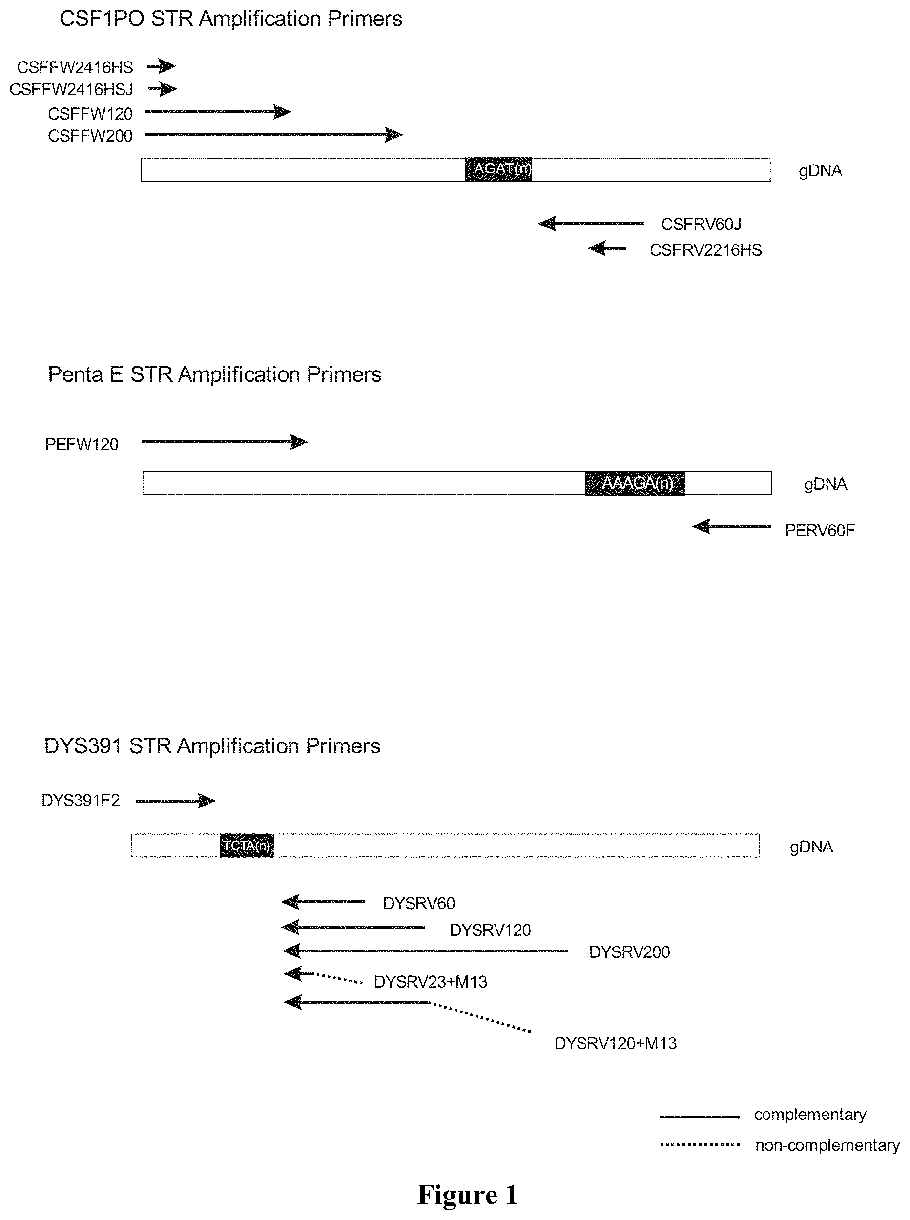
FIG. 1 shows the location of the different primers on the DNA regions to be amplified. The sequence for each described primer is included in the listing of sequences.
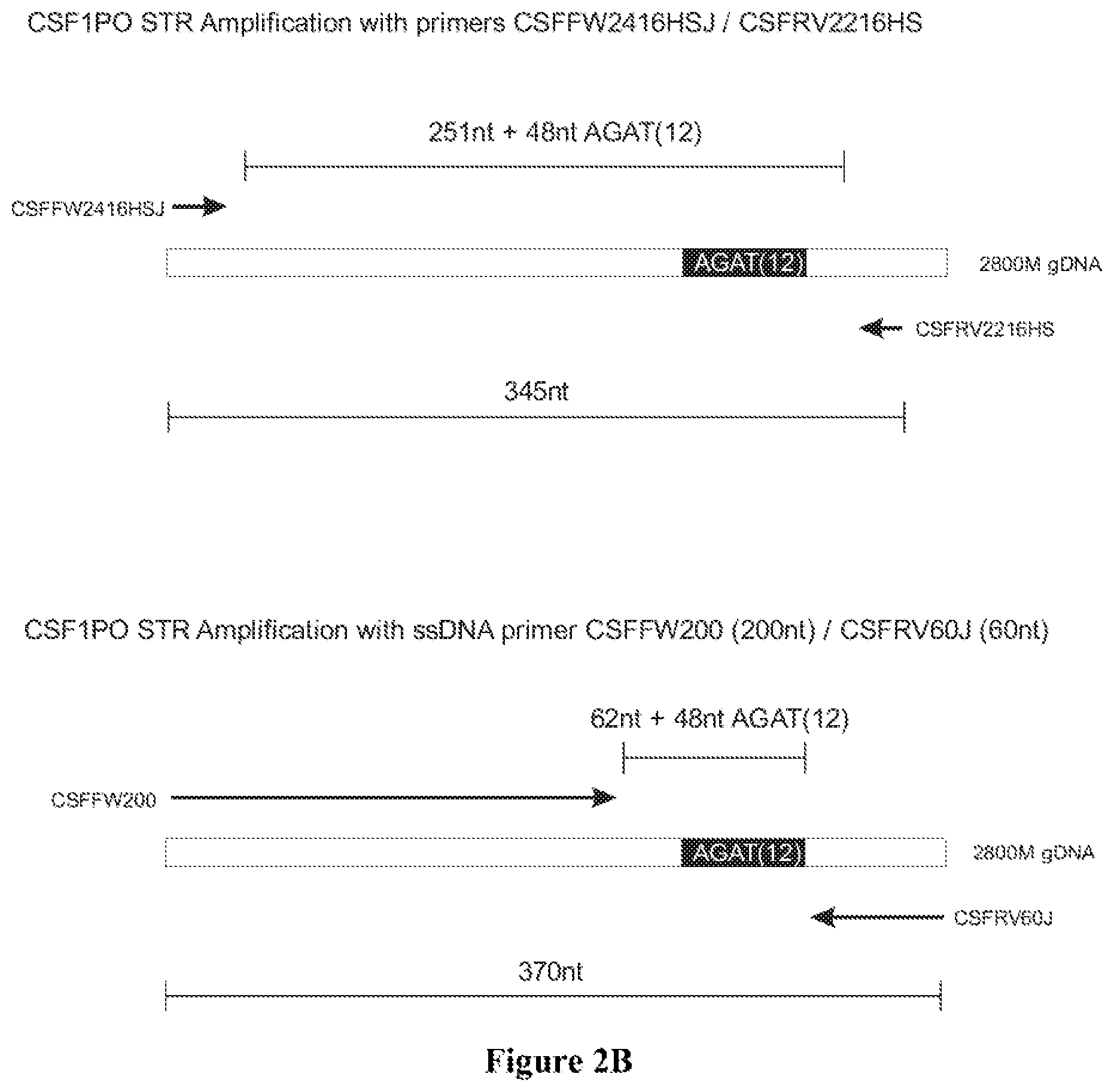
FIG. 2A shows the amplification results on 2800M DNA for locus CSF1PO in an agarose gel electrophoresis: Lane 1: 100 bp Ladder (Promega); Lane 2: 345 bp band with PowerPlex 16HS primers CSFFW2416HSJ/CSFRV2216HS; Lane 3: NTC with PowerPlex 16HS primers CSFFW2416HSJ/CSFRV2216HS; Lane 4: 370 bp band with primers CSFFW2416HS/CSFRV60J; Lane 5: NTC with primers CSFFW2416HS/CSFRV60J; Lane 6: 370 bp band with primers CSFFW120/CSFRV60J; Lane 7: NTC with primers CSFFW120/CSFRV60J; Lane 8: 370 bp band with primers CSFFW200/CSFRV60J: Lane 9: NTC with primers CSFFW200/CSFRV60J and Lane 10: 100 bp Ladder (Promega). FIG. 2B shows the diagram of CSF1PO amplification with primers CSFFW2416HSJ/CSFRV2216HS and CSFFW200 (200 nt)/CSFRV60J (60 nt).
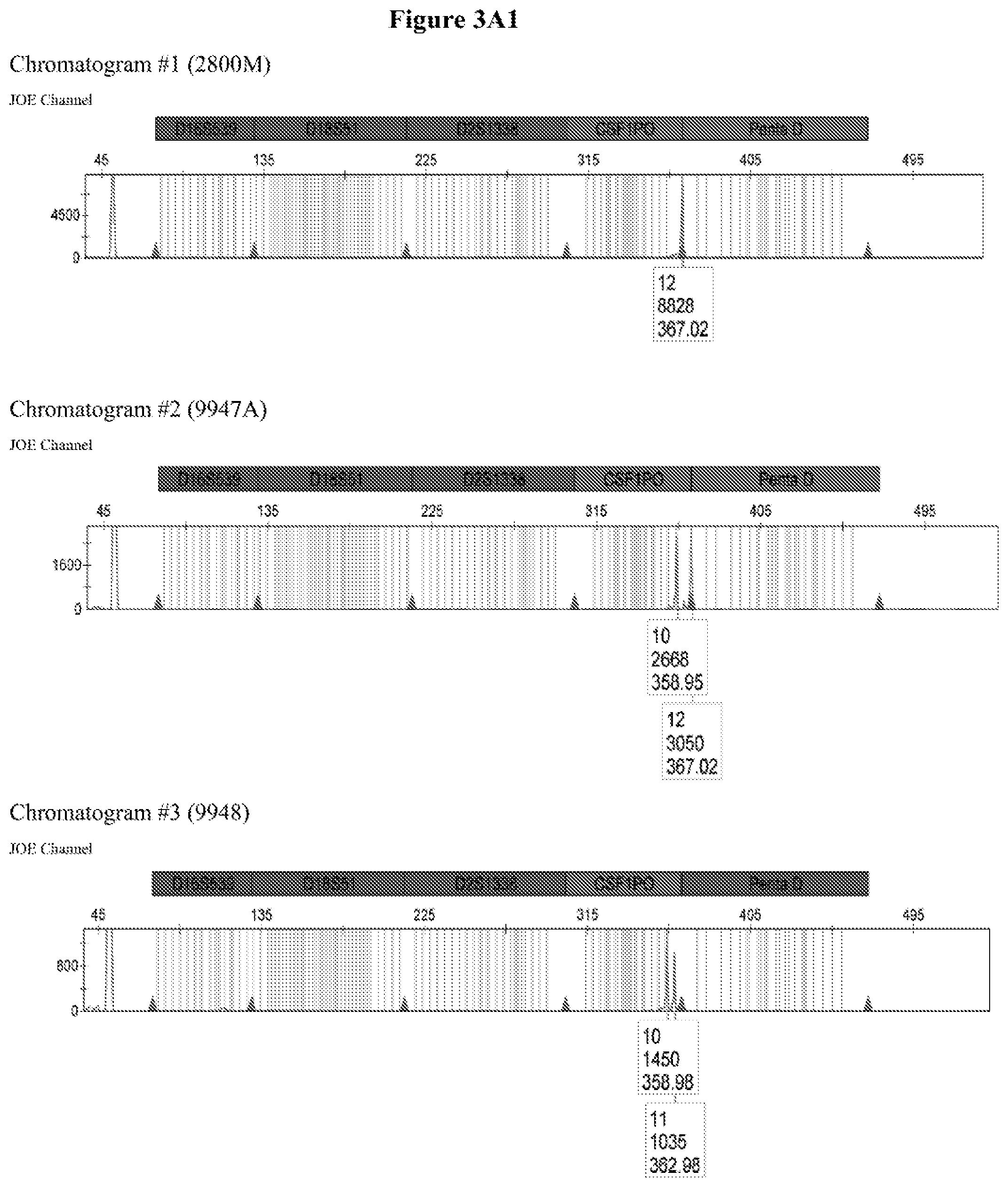
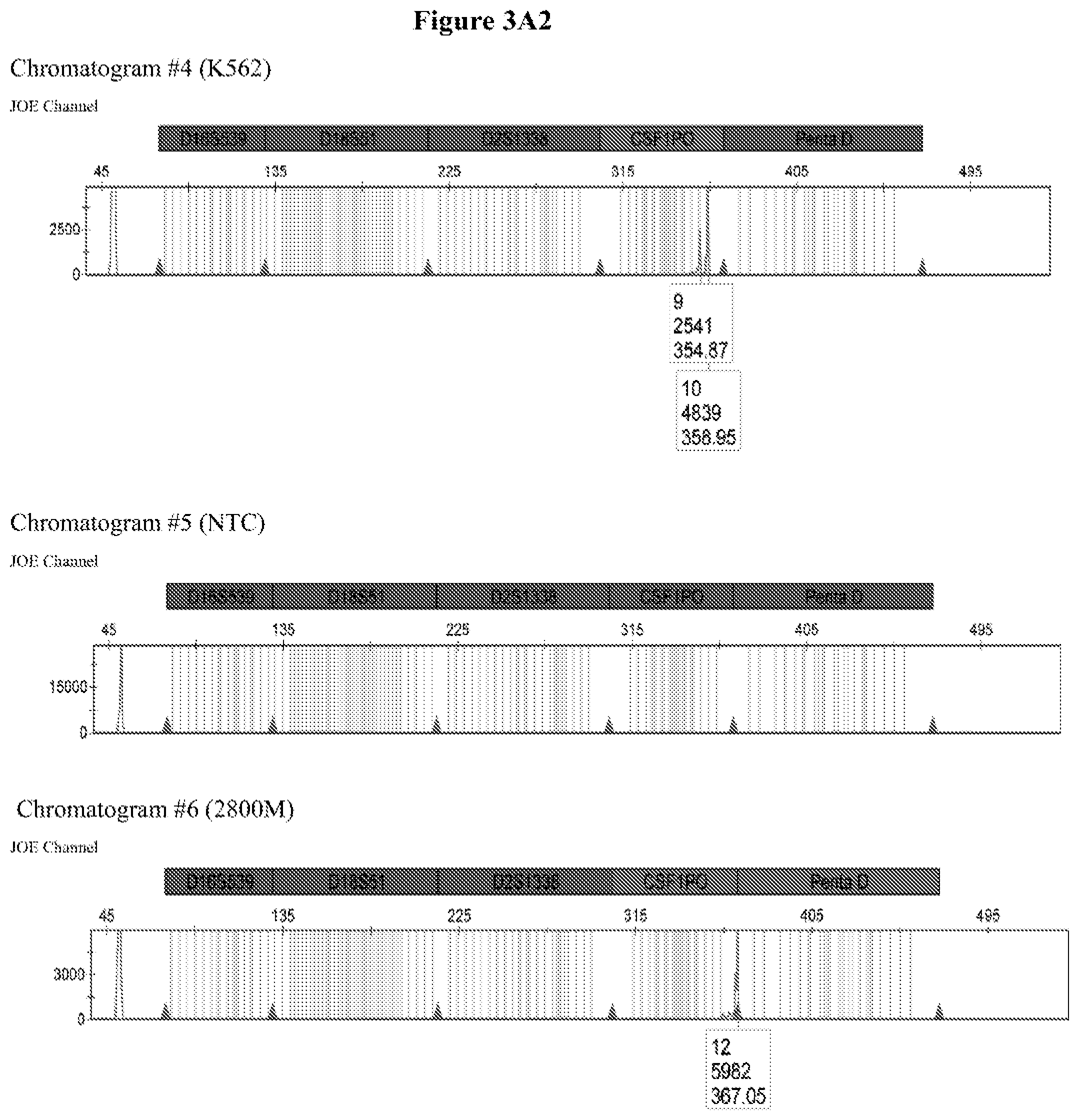
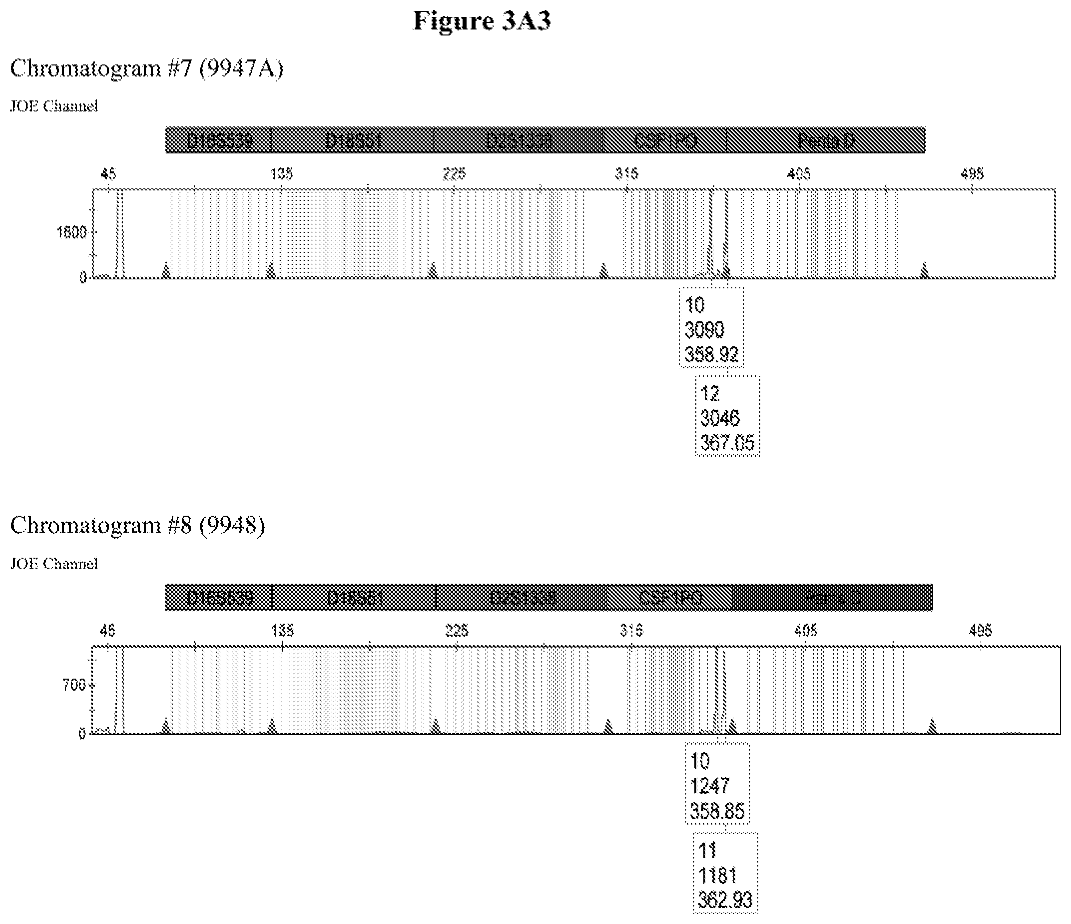
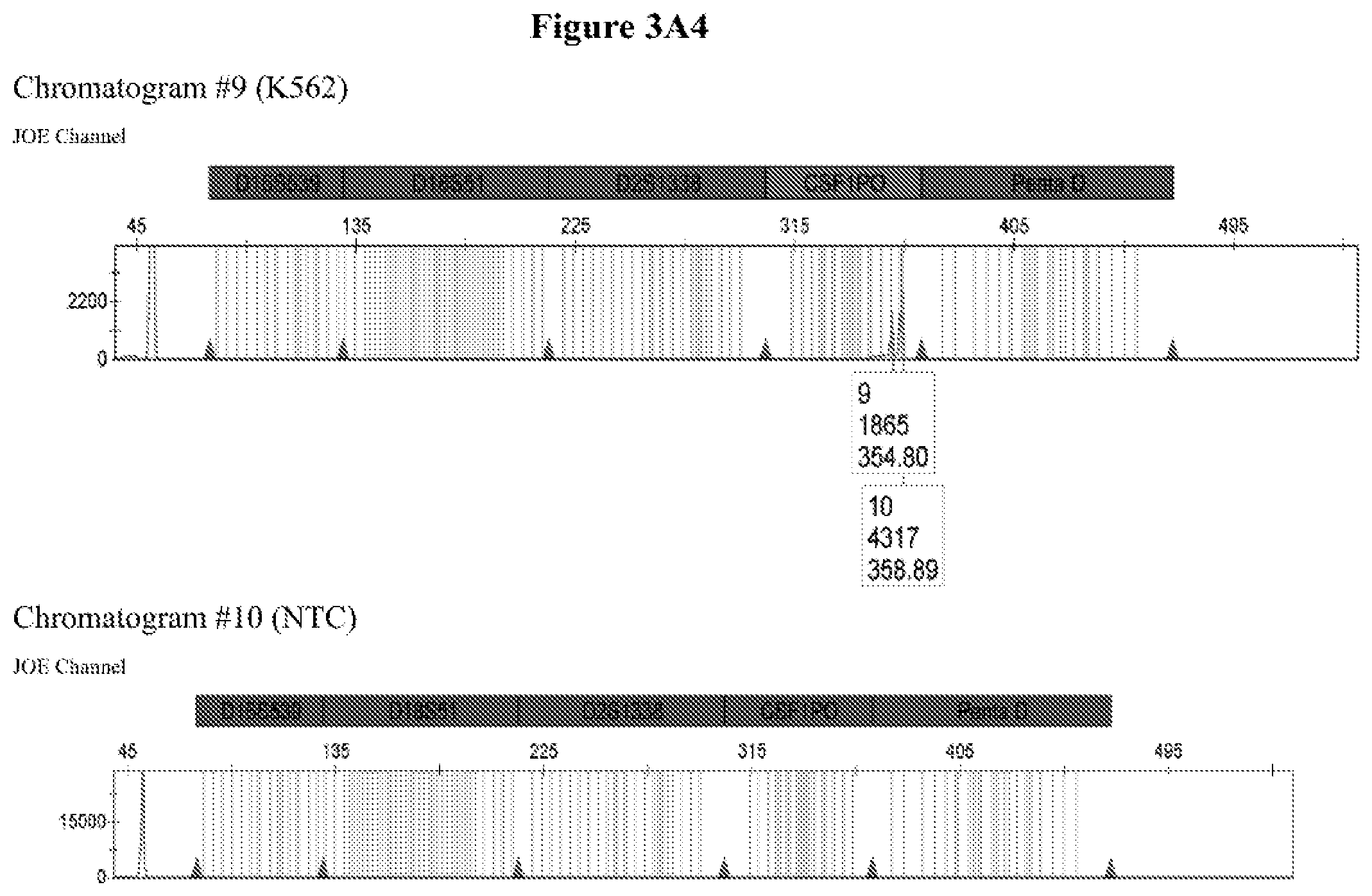
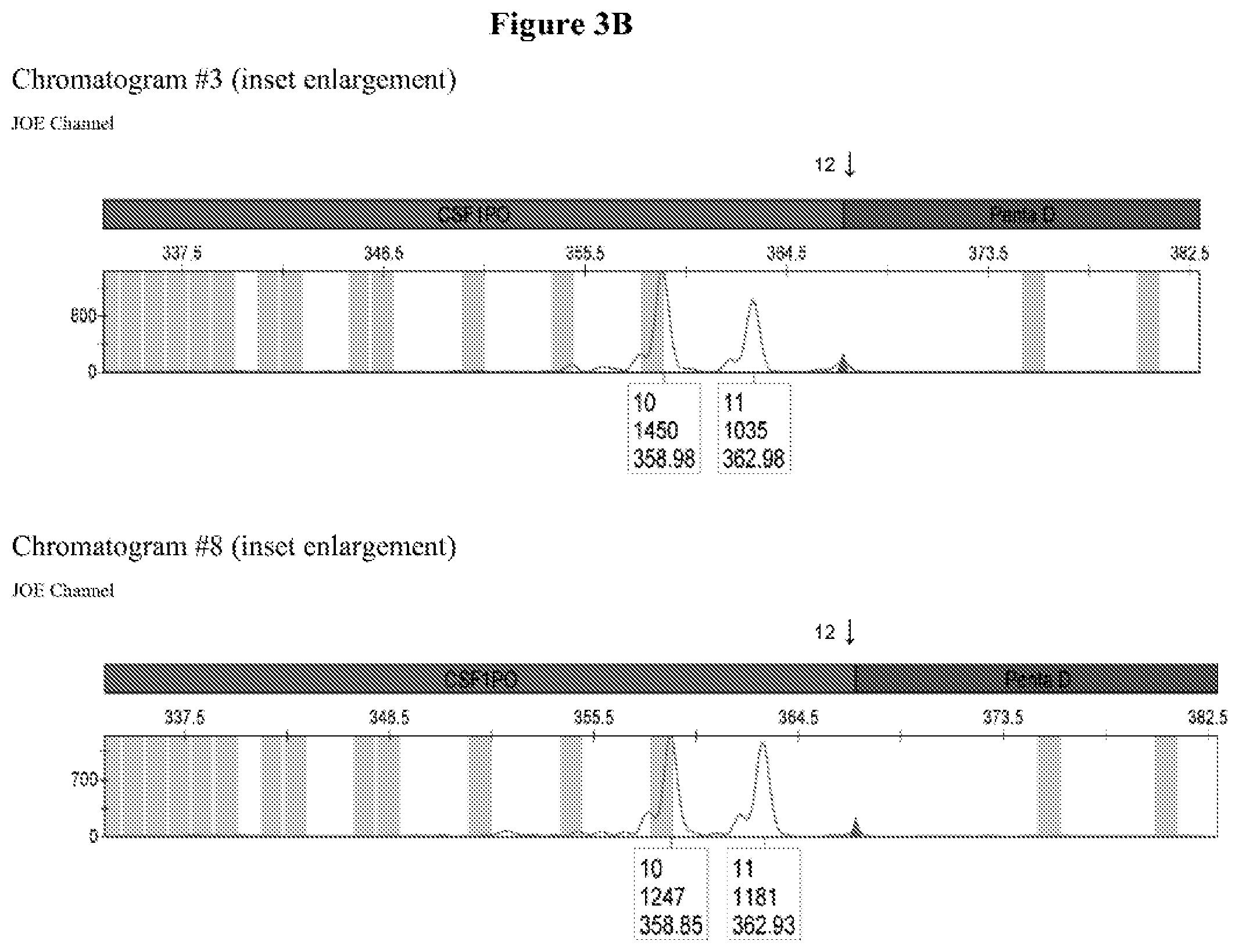
FIG. 3A shows the amplification results for locus CSF PO with different DNA templates in a CE chromatogram. Chromatogram #1: 2800M DNA with primers CSFFW120/CSFRV60J, Chromatogram #2: 9947A DNA with primers CSFFW120/CSFRV60J, Chromatogram #43: 9948 DNA with primers CSFFW120/CSFRV60J, Chromatogram #4: K562 DNA with primers CSFFW120/CSFRV60J, Chromatogram #5: NTC control with primers CSFFW200/CSFRV60J, Chromatogram #6: 2800M DNA with primers CSFFW200/CSFRV60J, Chromatogram #7: 9947 DNA with primers CSFFW200/CSFRV60J, Chromatogram #8: 9948 DNA with primers CSFFW200/CSFRV60J, Chromatogram #9: K562 DNA with primers CSFFW200/CSFRV60J and Chromatogram #10: NTC control with primers CSFFW200/CSFRV60J. FIG. 3B: Detail of CSF1PO triplet profile from Chromatograms #3 and #8.
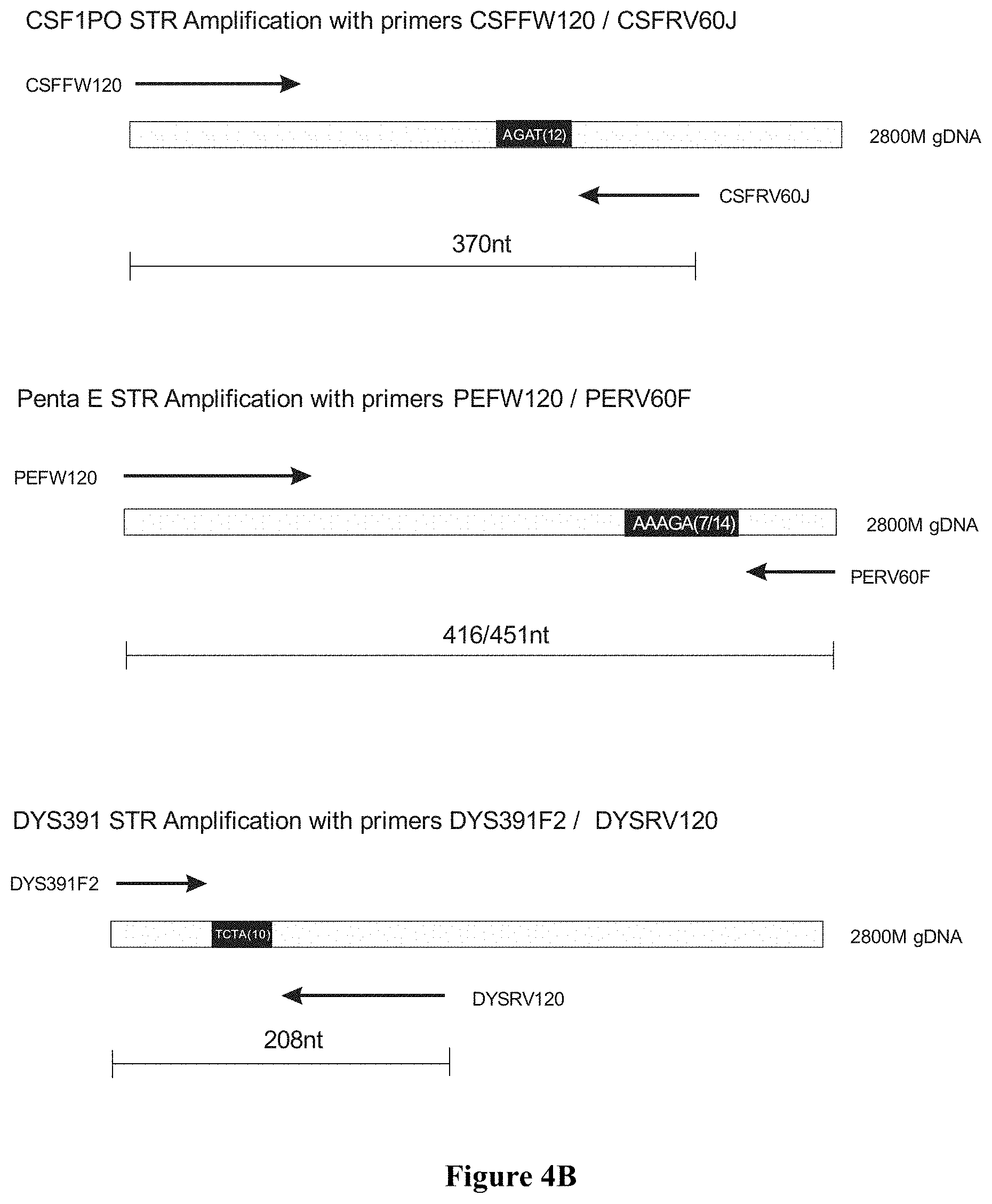
FIG. 4A: Agarose gel electrophoresis for monoplex, duplex and triplex amplifications of markers CSF1PO. Penta E and DYS391 on 2800M DNA, wherein Lane 1 100 bp Ladder (Promega), Lane 2: CSF1PO monoplex with primers CSFFW120/CSFRV60J, Lane 3: Penta E monoplex with primers PEFW120/PERV60F, Lane 4: DYS391 monoplex with primers DYS391F2/DYSRV120, Lane 5: CSF1PO/Penta E duplex with primers CSFFW120/CSFRV60J+PEFW120/PERV60F, Lane 6: CSF1PO/DSY391 duplex with primers CSFFW120/CSFRV60J+DYS391F2/DYSRV120, Lane 7: Penta E/DSY391 duplex with primers PEFW120/PERV60F+DYS391F2/DYSRV120, and Lane 8: CSF1PO/Penta E/DYS391 triplex with primers CSFFW120/CSFRV60J+PEFW120/PERV60F+DYS391F2/DYSRV120; FIG. 4B: Primer design for amplification of CSF1PO, Penta E and DYS391.
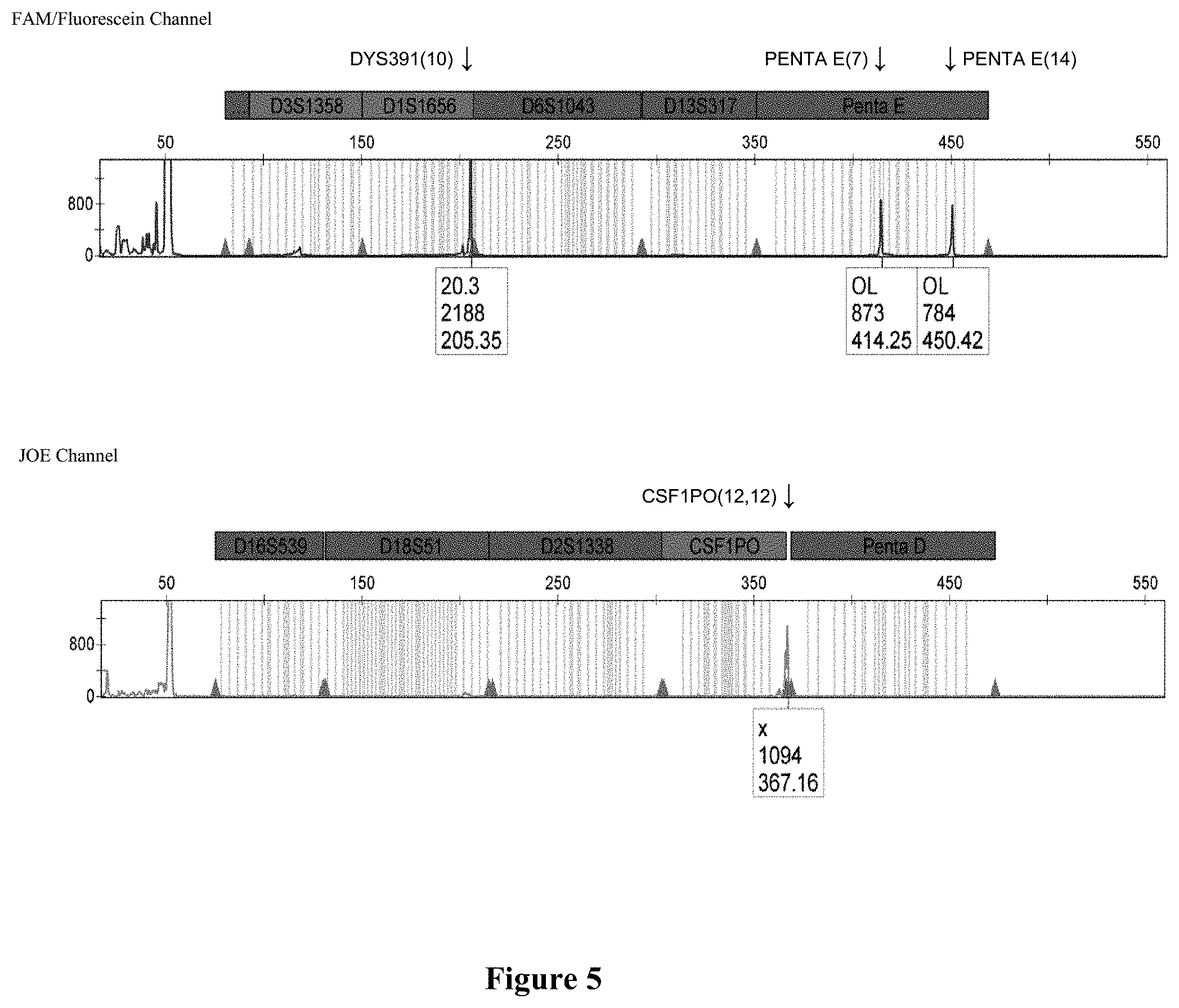
FIG. 5 shows a capillary electrophoresis chromatogram of the triplex PCR of CSF1PO (JOE channel). Penta E (FAM channel) and DYS391 (FAM channel) on 2800M DNA with primers CSFFW120/CSFRV60.1. PEFW120/PERV60F and DYS391F2/DYSRV120.
FIG. 6 shows the amplification results of locus DYS391 on 2800M DNA in an agarose gel electrophoresis: Lane 1: 100 bp Ladder (Promega), Lane 2: 148 bp band with primers DYS391F2/DYSRV60, Lane 3: NTC with primers DYS391F2/DYSRV60, Lane 4: 208 bp band with primers DYS391F2/DYSRV120, Lane 5: NTC with primers DYS391F2/DYSRV120, Lane 6: 288 bp band with primers DYS391F2/DYSRV200, Lane 7: NTC with primers DYS391F2/DYSRV200, Lane 8: 148 bp band with primers DYS391F2/DYSRV23+M13, Lane 9: NTC with primers DYS391F2/DYSRV23+M13, Lane 10: 288 bp band with primers DYS391F2/DYSRV120+M13, Lane 11: NTC with primers DYS391F2/DYSRV120+M13 and Lane 12: 100 bp Ladder (Promega).
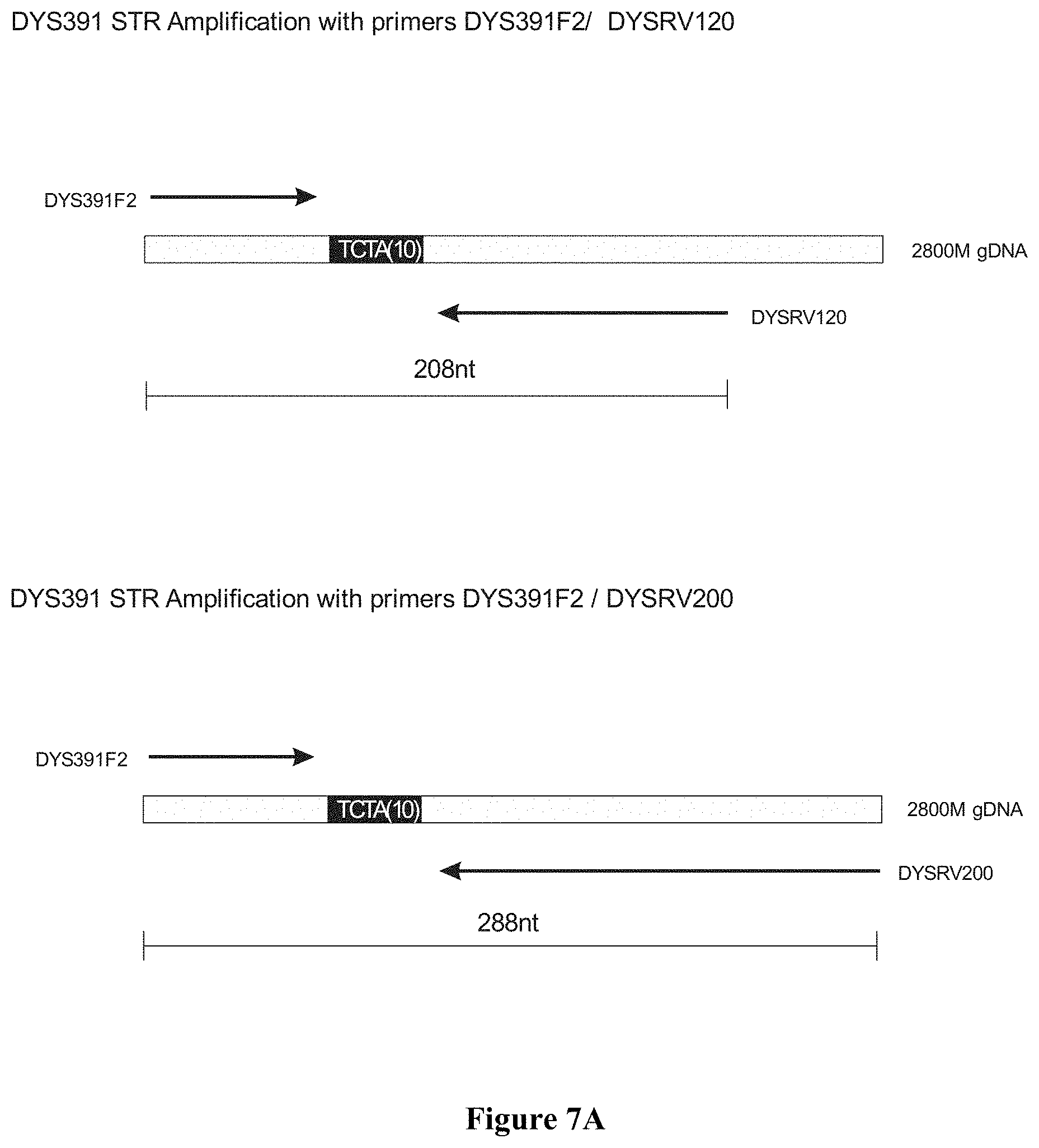
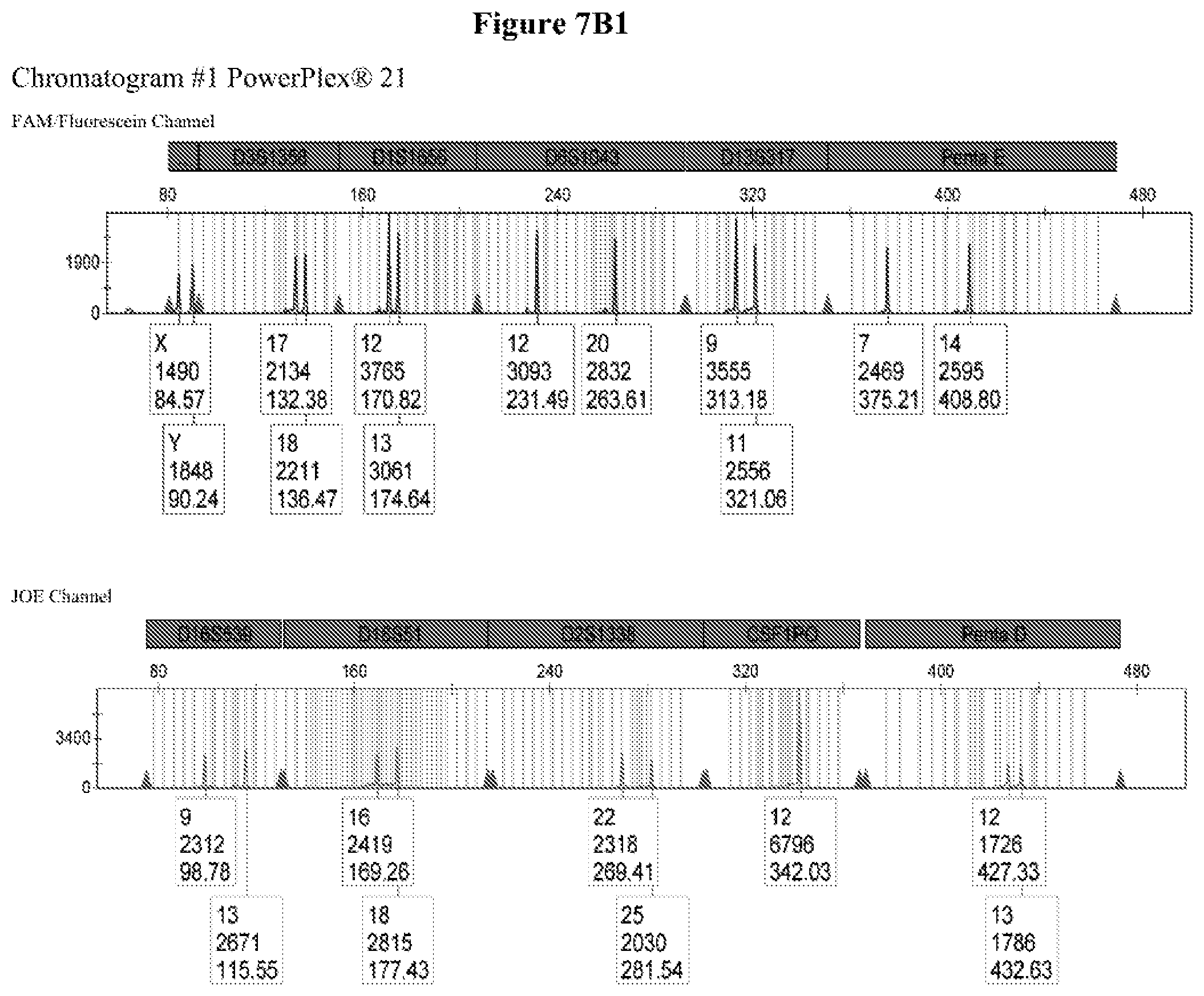
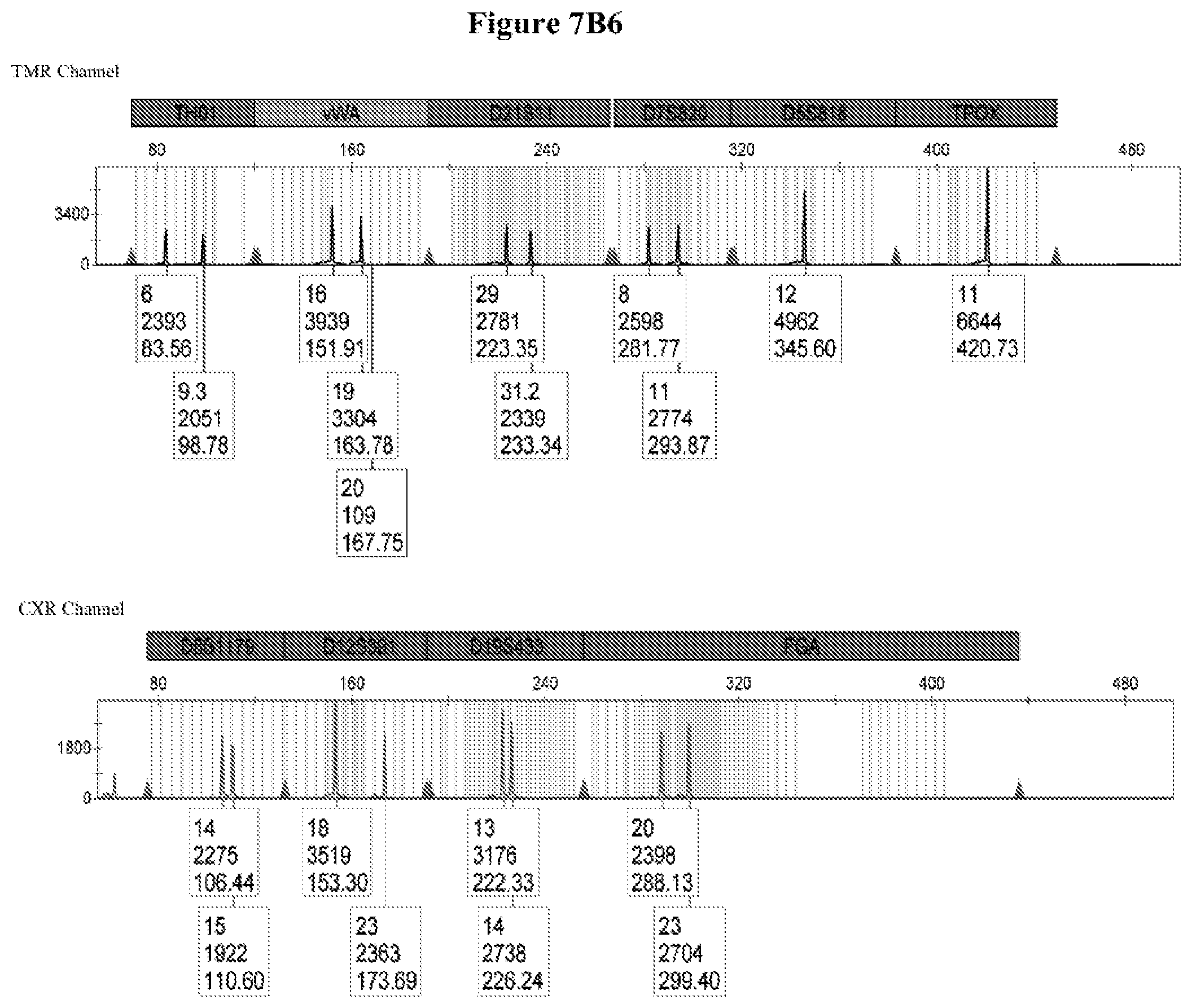
FIG. 7: FIG. 7A shows the primer design for marker DYS391 addition to PowerPlex.RTM.21 and FIG. 7B shows the capillary electrophoresis chromatogram for DYS391 addition to PowerPlex.RTM.21, wherein Chromatogram #1: 2800M DNA with no extra primers, Chromatogram #2: 2800M DNA with primers DYS391F2/DYSRV120 (FAM channel) and Chromatogram #3: 2800M DNA with primers DYS391F2/DYSRV200 (FAM channel).
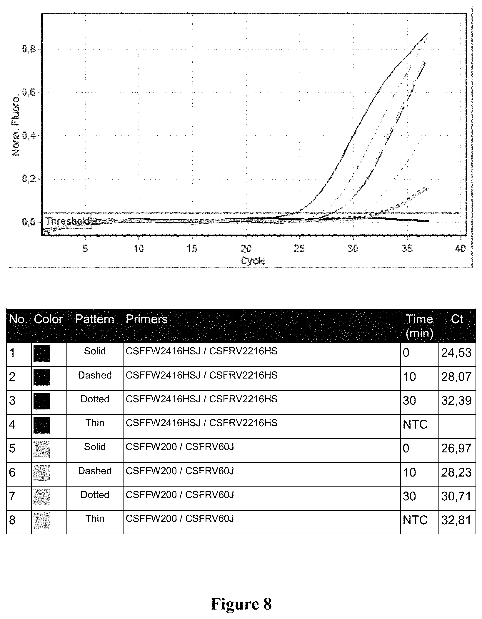
FIG. 8 shows the qPCR profile for locus CSF1PO on 2800M DNA degraded by different times at 95.degree. C. Sample 1 (black, solid line): 0 minutes with primers CSFFW2416HSJ/CSFRV2216HS, Sample 2 (black, dashed line): 10 minutes with primers CSFFW2416HSJ/CSFRV2216HS. Sample 3 (black, dotted line): 30 minutes with primers CSFFW2416HSJ/CSFRV2216HS, Sample 4 (black, thin line): NTC with primers CSFFW2416HSJ/CSFRV2216HS, Sample 5 (gray, solid line): 0 minutes with primers CSFFW200/CSFRV60J, Sample 6 (gray, dashed line): 10 minutes with primers CSFFW200/CSFRV60J, Sample 7 (gray dotted line): 30 minutes with primers CSFFW200/CSFRV60J and Sample 8 (gray, thin line): NTC with primers CSFFW200/CSFRV60J.
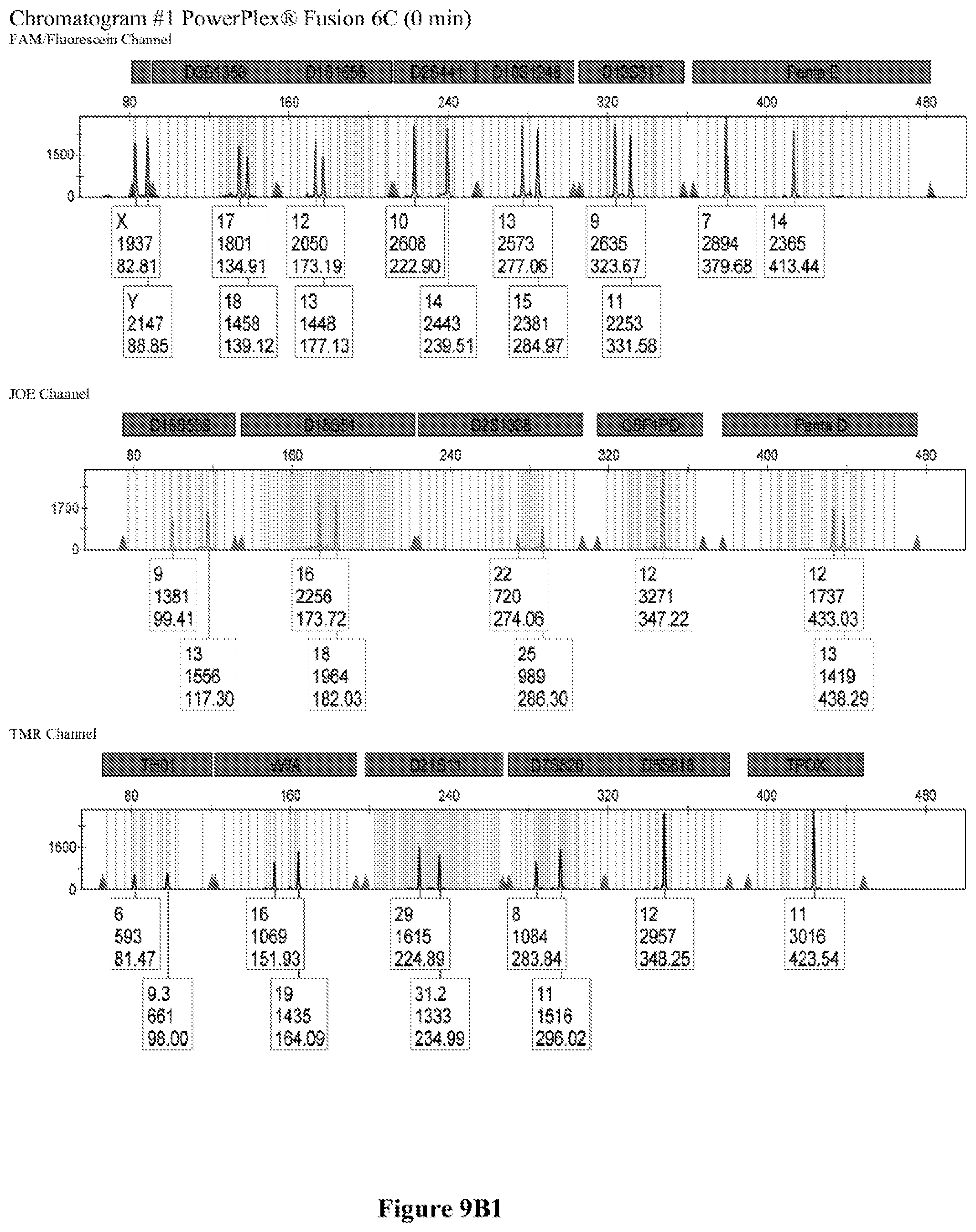
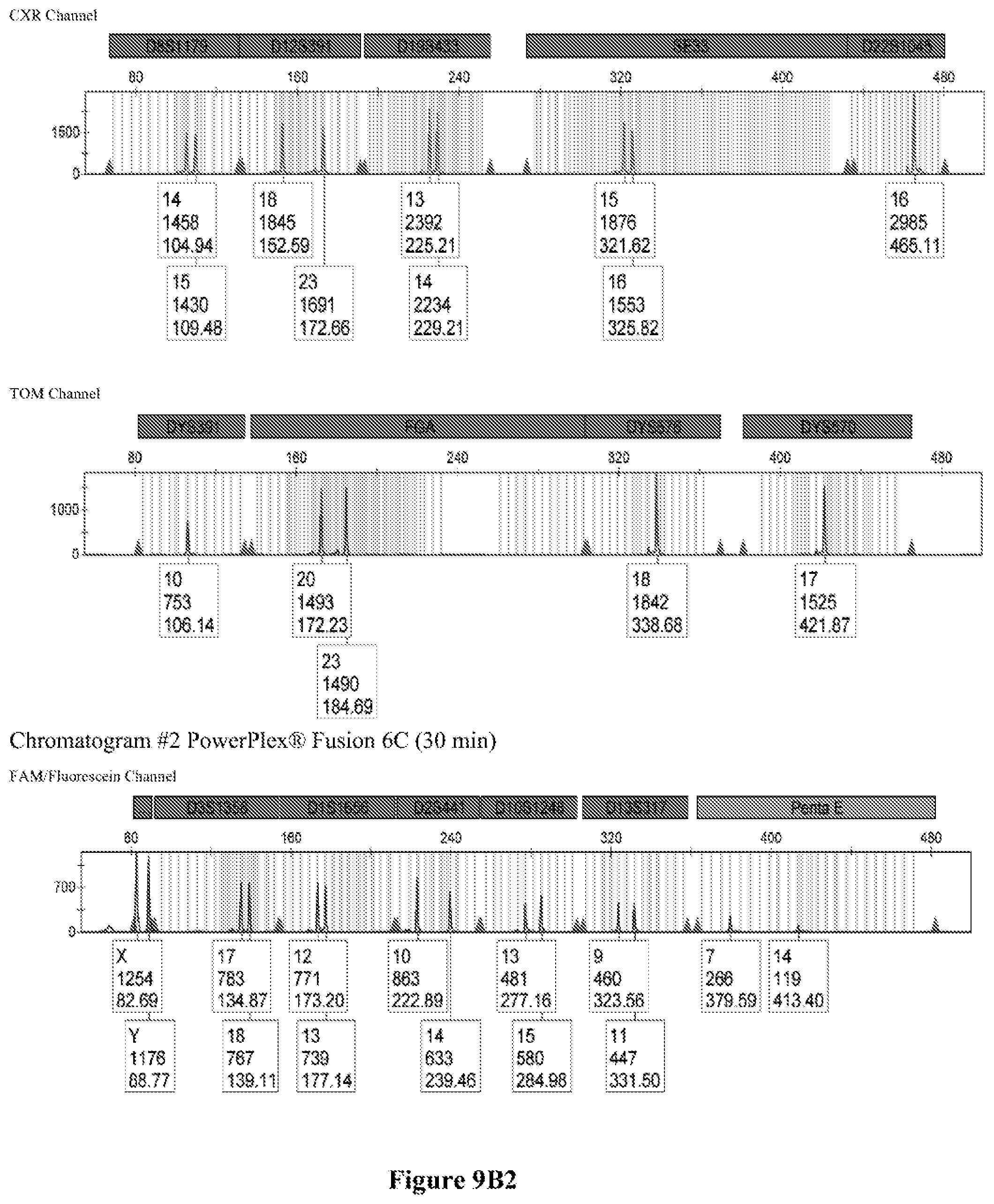
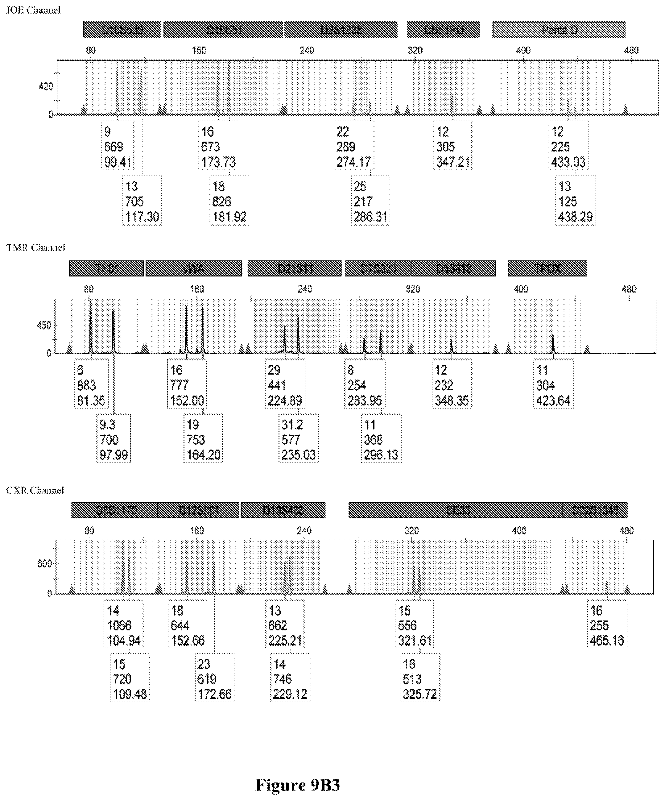
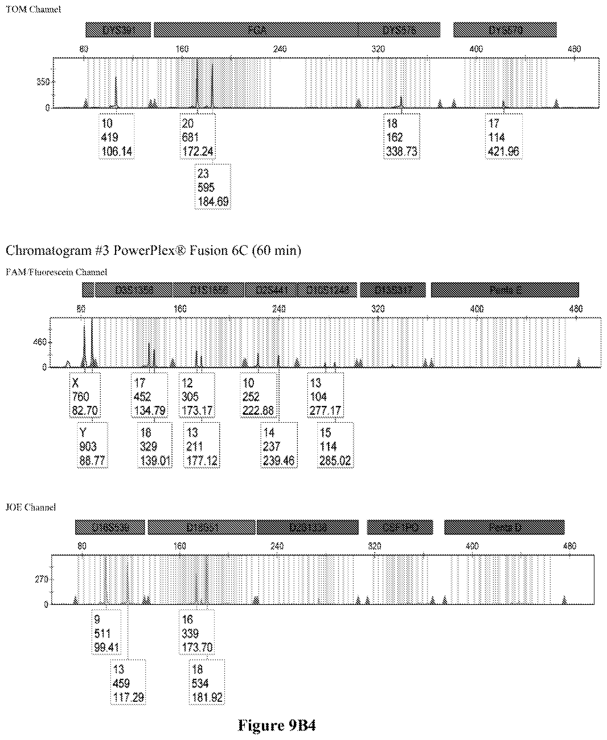
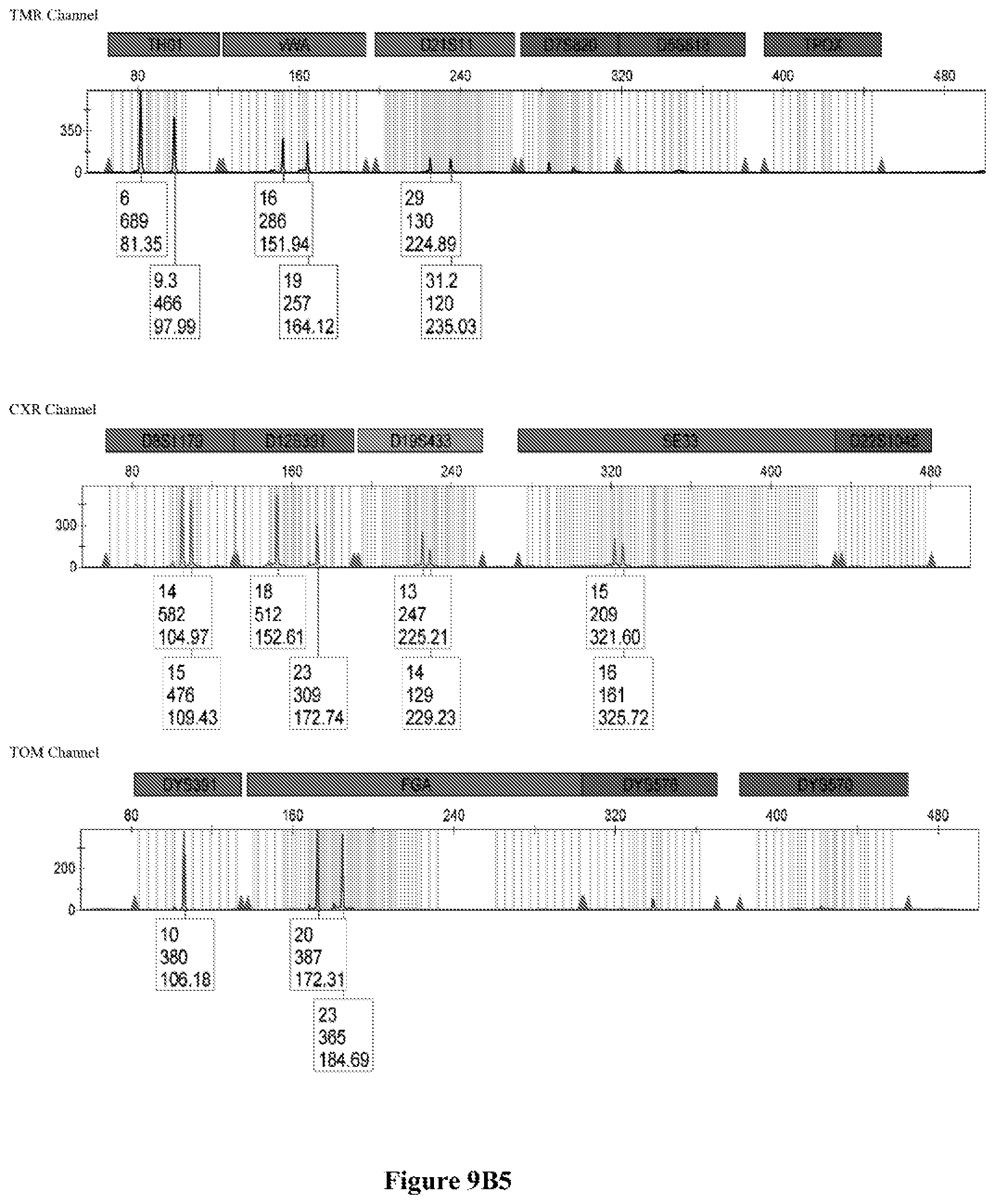
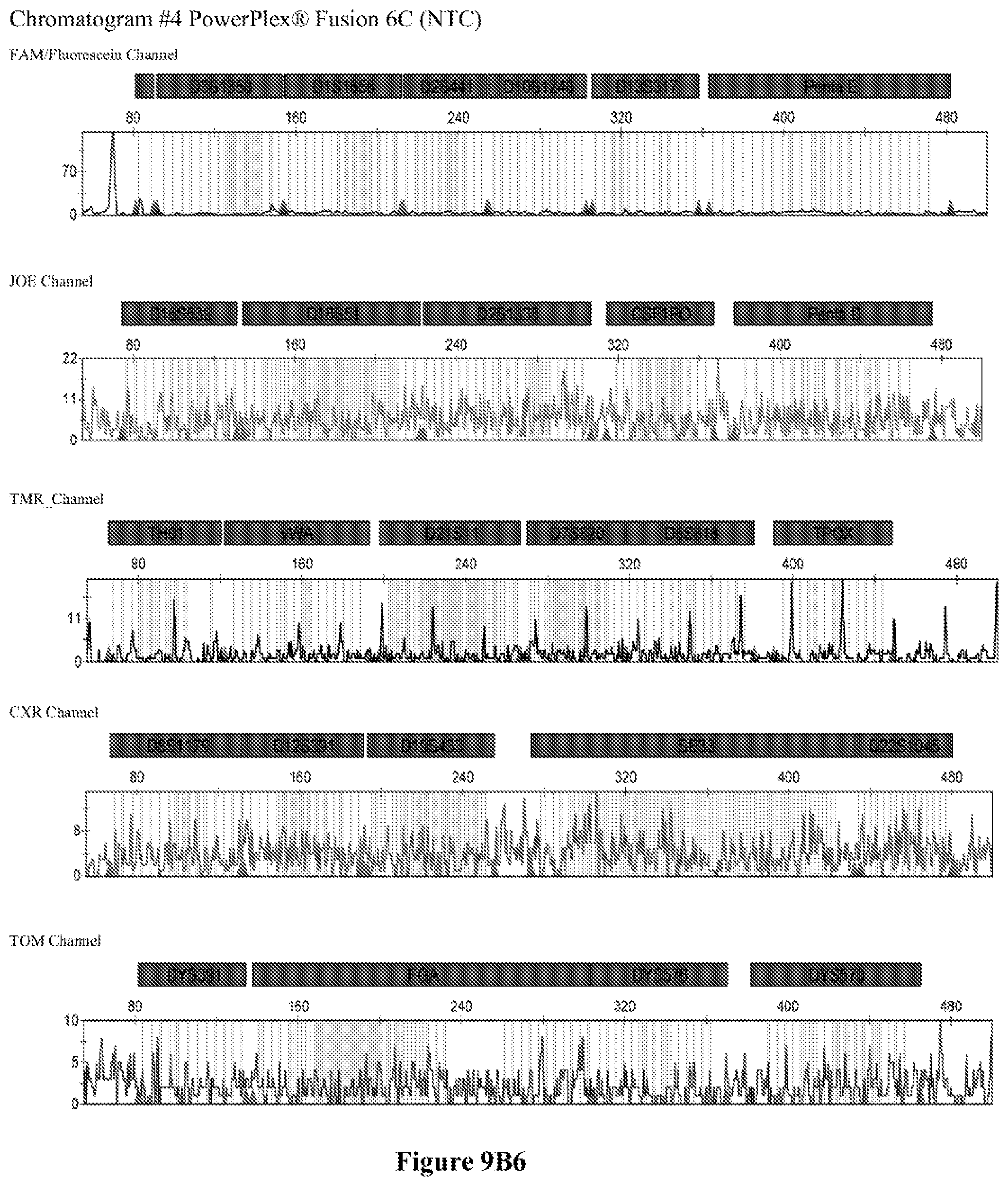
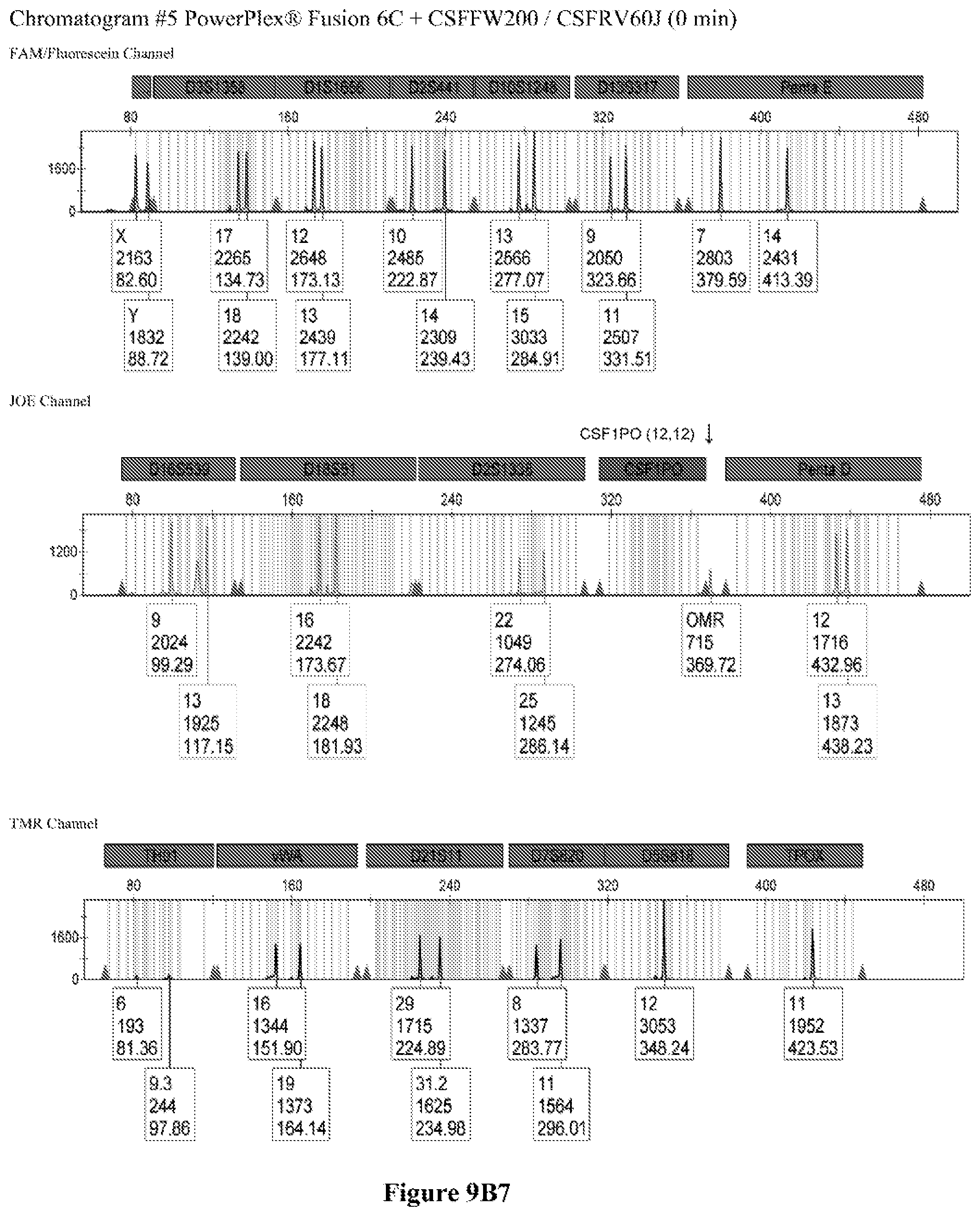
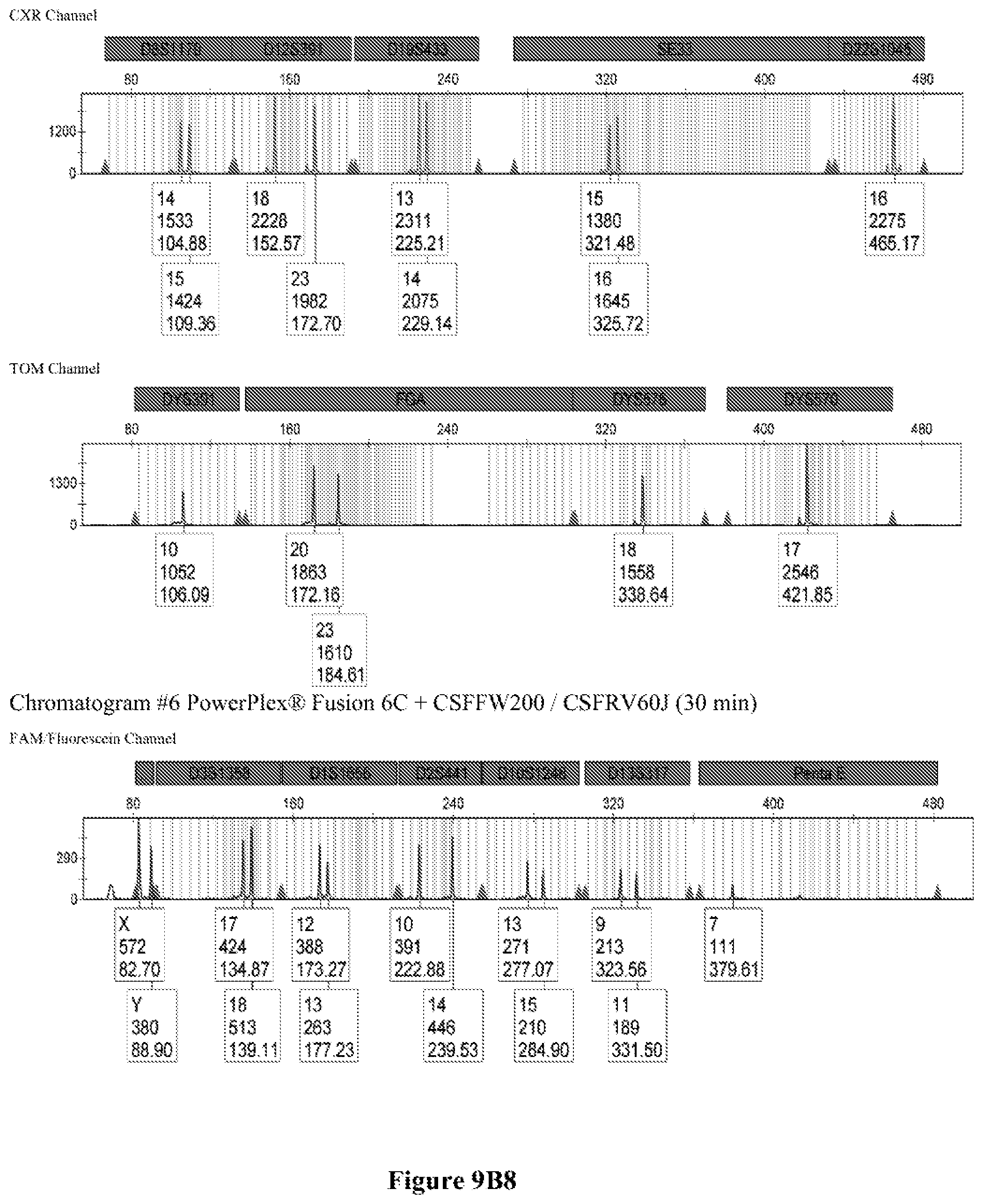
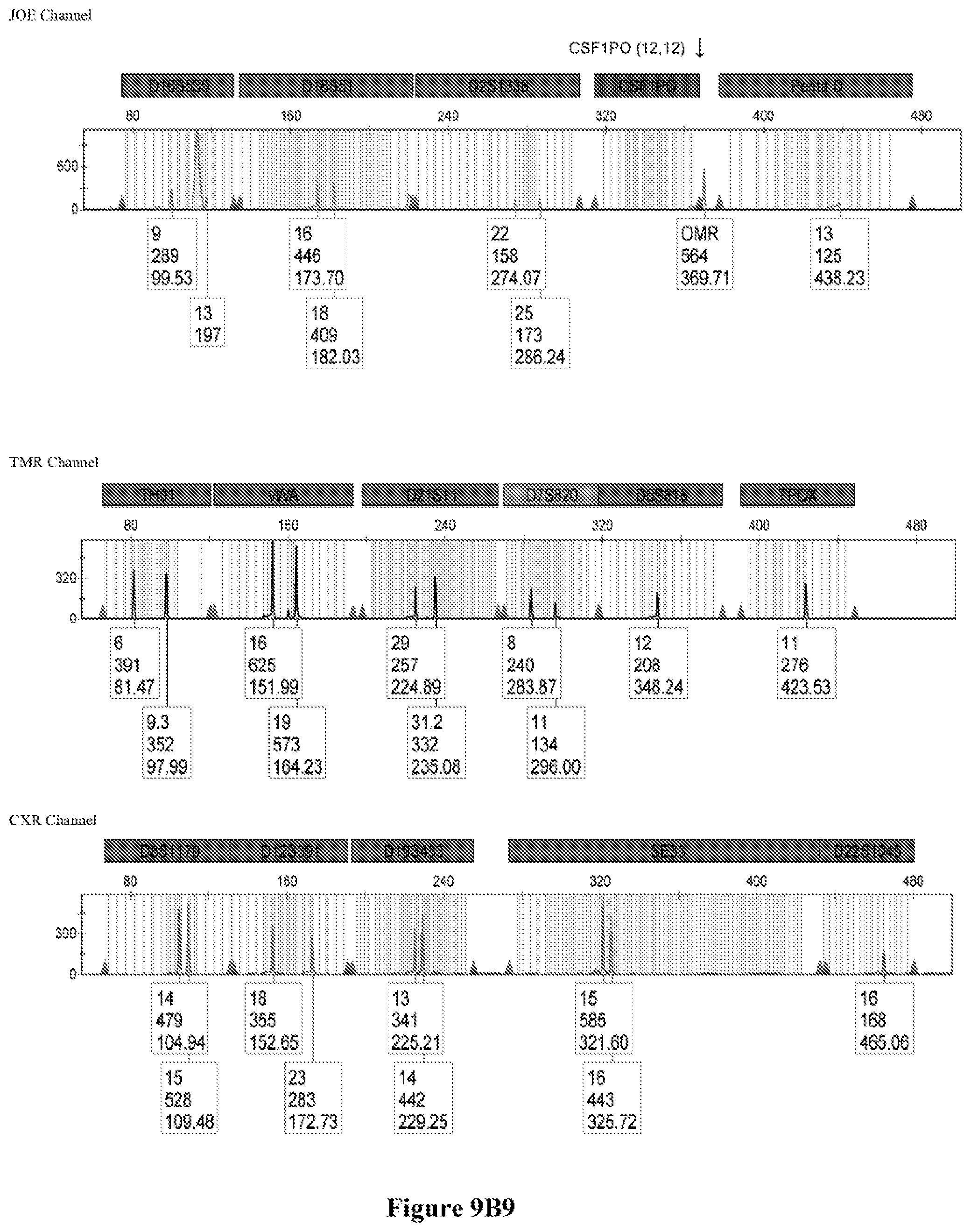
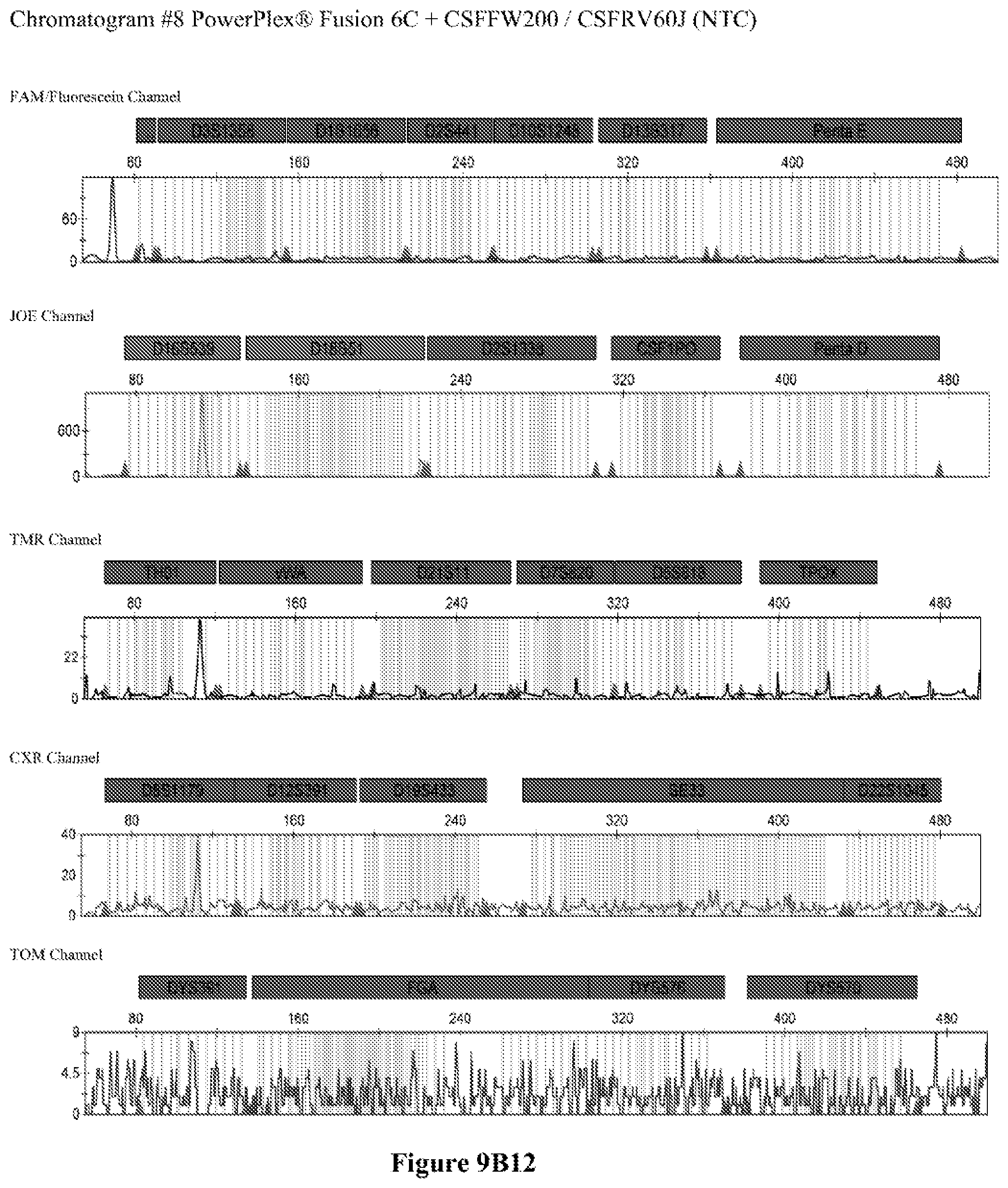
FIG. 9A shows the qPCR profiles of 2800M DNA heated at 95.degree. C. for 0, 10, 30, 45 and 60 minutes using the primer pair CSFFW200/CSFRV60J. Sample 1 (black, solid line): 0, Sample 2 (black, dashed line): 10 minutes, Sample 3 (black, dotted line): 30 minutes. Sample 4 (gray, solid line): 45, Sample 5 (gray, dashed line): 60 minutes, Sample 6 (gray, dotted line): NTC (no template control). DNA samples heated for 0, 30 and 60 minutes was used--along an NTC (no template control)--to demonstrate the advantage of using superprimers in a current CODIS (Combined DNA Index System, FBI) commercial kit. To that aim 0.8 .mu.M (final concentration) of each primer of the CSFFW200/CSFRV60J pair was added to the PowerPlex.RTM. Fusion 6C reaction mix, which already contains 54 primers that amplify 27 loci. The CE chromatogram (FIG. 9B) shows that the peak signals decrease or disappear as the DNA is more degraded, especially for the longer amplicons, while the peak corresponding to the CSFFW200/CSFRV60J pair is still visible for the most degraded DNA.
DETAILED DESCRIPTION OF THE INVENTION
A genetic marker is understood as a gene or DNA sequence with a known location on a genome that can be used to identify a region associated to a given trait or phenotype, to inherited diseases and to the identification of individuals or species. The term locus is sometimes used to express the same concept.
A target sequence refers to a segment of nucleic acid of interest that is selectively amplified using a sequence-specific primer.
A primer is an oligonucleotide or polynucleotide capable of hybridizing to a complimentary segment of nucleic acid to allow the initiation of the replication by a DNA polymerase enzyme.
For the purpose of the present description, the term superprimer refers to a single stranded DNA (ssDNA) polynucleotide primer longer than 60 nucleotides, more preferably between 60 and 300 nucleotides.
The use of long, single stranded DNA (ssDNA) polynucleotides as primers (superprimers), instead of the usual shorter DNA oligonucleotides, yields larger amplicons without the need to actually amplify extra DNA regions. In reducing the required template length between a pair of primers, the performance of the PCR assays is greatly increased, particularly in degraded DNA samples.
ssDNA polynucleotides ranging from fifty to hundreds of nucleotides, far longer than the smaller oligonucleotides containing few nucleotides--as normally used as primers in PCR--can optimally be employed as surrogate primers without any particular concerns on their annealing temperatures or the need of special PCR conditions. While we preferred a hot start PCR option, standard PCR may also be utilized. The use of long ssDNA primers allows to obtain large DNA amplicons without the need to actually amplify a large portion of DNA template.
The goal of the present invention is to increase the sensitivity of PCR assays by reducing the required length of available intact DNA template needed for amplification while at the same time obtaining larger DNA amplicons suitable for size discrimination in analysis by CE and other related methods. For that aim, the addition of long ssDNA polynucleotides as primers or superprimers allows the polymerization of specific, small DNA segments while at the same time yielding product sizes suitable for their convenient detection.
The inventive methods are based upon the decreasing efficiency of the extension by polymerase enzymes in the presence of degraded DNA as the PCR pair of primers are farther apart. The method of the invention is useful for detecting genotypes in degraded or fragmented DNA samples as is typical in casework and aged samples.
The method demonstrated that long ssDNA primers can be readily used for PCR assays in monoplex or multiplex formats. While we preferred hot start PCR, standard PCR may also be used. Actually no special PCR conditions or primer design were needed towards that goal other than the use of highly purified, long ssDNA (superprimer) as primer reagent. Moreover, we also showed that these primers can be applied in the amplification of degraded DNA templates with the same beneficial effect of the small mini-STRs, while at the same time producing larger DNA amplicons of flexible length that are more suitable for size discrimination assays.
Primers totally complementary to the template sequence or complementary only at their 3' priming region were both successfully amplified. The use of non-homologous sequences may be important for providing flexibility in designing multiplex reactions with several primers. Furthermore, primers bearing long 5', non-homologous sequences might be considered for performing mutagenesis and also for annealing, molecular binding and labeling strategies.
The invention method is suitable for PCR assays and for detecting variations that involve length polymorphisms. It may be applied to the current methods in use that rely on different amplification lengths to distinguish variations in nucleic acid sequences. It may also be useful in the design of multiplex kits for the identification of individuals and species and for medical diagnostics.
The invention method increases the sensitivity of PCR assays by reducing the required length of available intact DNA template needed for amplification while at the same time obtaining larger DNA amplicons of flexible length suitable for size discrimination in analysis by CE and other related methods. For that aim, the addition of long ssDNA fragments as primers (superpritners) allows the enzymatic polymerization of specific, small DNA target regions while at the same time yielding product sizes suitable for their convenient detection.
The present invention increases the sensitivity of PCR assays by reducing the required length for intact DNA template while at the same time obtaining larger DNA amplicons suitable for size discrimination.
The method of the invention is useful to obtain a suitable length of a PCR product without the need to amplify off-target sequences towards that aim, thereby increasing the sensitivity of the assay for short, fragmented or degraded DNA samples by reducing the number of nucleotides being amplified. Additionally, more versatility is provided in the design of PCR assays by allowing the use of primers with large non-homologous sequences.
The design of PCR primers was based on the information provided by the NIST Short Tandem Repeat DNA Internet Database created by John M. Butler and Dennis J. Reeder (NIST Short Tandem Repeat DNA Internet Database created by John M. Butler and Dennis J. Reeder http://www.cstl.nist.gov/strbase/index.htm) and the published genome sequence from the GenBank (GenBank http://www.ncbi.nlm.nih.gov/genbank/).
All DNA primers were synthesized by Integrated DNA Technologies Inc. (IDT). Primers labelled with the fluorescent dyes 6-FAM (fluorescein) and JOE (6-carboxy-4',5'-dichloro-2',7'-dimethoxyfluorescein, NHS ester) were purified by HPLC. Primers >100 nucleotides were PAGE purified. All other primers were desalted.
FIG. 1 shows the location of the different primers on the amplified DNA regions used in the examples.
The primer sequences are:
TABLE-US-00001 CSFRV2216HS: SEQ ID No 1 CSFFW2416HS: SEQ ID No 2 CSFFW2416HSJ:. SEQ ID No 3 (It is identical to SEQ ID No 2, but has a fluorescent JOE dye at the 5' end). CSFFW120: SEQ ID No 4 CSFFW200: SEQ ID No 5 CSFRV60J:. SEQ ID No 6 (It may have a fluorescent JOE dye at the 5' end). PEFW120: SEQ ID No 7 PERV60F:. SEQ ID No 8 (It may have a fluorescent 6-FAM dye at the 5' end). DYS391F2: SEQ ID No 9 (It may have a fluorescent 6-FAM dye at the 5' end). DYSRV60: SEQ ID No 10 DYS391RV120:. SEQ ID No 11 DYS391RV200:. SEQ ID No 12 DYSRV120 + M13:. SEQ ID No 13 DYSRV23 + M13:. SEQ ID No 14
Long ssDNA primers produced neat PCR products in a similar fashion to shorter oligonucleotide primers, without any artifacts or spurious bands. FIG. 2A shows the PCR amplification of a 370 bp region of the genotyping locus CSF1PO (5q33.1; human c-fms proto-oncogene for CSF-1 receptor gene, 6th intron GenBank Accession X14720) using ssDNA fragments of 120 nucleotides (CSFFW120) or 200 nucleotides (CSFFW200) as forward primers and a 60-nucleotide primer (CSFRV60J) as reverse primer. A third forward primer of 24 nucleotides (CSFFW2416HS) with the same sequence as in the commercial kit PowerPlex.RTM.16HS (Promega) was also used.
Since the three forward primers share the same 5' sequence, they all yield the same 370 bp product. A control amplification of 345 bp with PowerPlex.RTM.16HS primers CSFFW2416HSJ/CSFRV2216HS was also performed. Standard DNA 2800M (Promega) was used as template in all reactions.
Due to the extended length of the long ssDNA primers, only 62 nucleotides of non-repeat sequences span between the 3' ends of the primer pair CSFFW200/CSFRV60J. This contrasts with the 251-nucleotide distance in PowerPlex.RTM.16HS, even though the latter amplicon is 25 bases shorter (FIG. 2B).
The quality of the DNA profiles generated by long ssDNA primers was verified for different genotypes. The CSF1PO region of DNA templates 2800M (genotype 12, 12), 9947A (genotype 10, 12), 9948 (genotype 10, 11, 12) and K562 (genotype 9, 10) was amplified with forward primers CSFFW120 (120 nt) or CSFFW200 (200 nt) and reverse primer CSFRV60J (60 nt, JOE-labelled) and then analyzed by capillary electrophoresis (FIG. 3A).
All profiles agreed with the ones described in the literature for these cell types (James, R. New Control DNA for PowerPlex.RTM. Systems. Promega Corporation Web site. http://worldwide.promega.com/resources/profiles-in-dna/2011/new-control-d- na-for-powerplex-systems/ Updated 2011; National Institute of Standards & Technology Certificate of Analysis Standard Reference Material.RTM. 2391b PCR-Based DNA Profiling Standard and America Type Culture Collection: ATCC STR Data for Human Cell Lines www.atcc.org/STR%20Database.aspx). Moreover, they showed the characteristic heterozygous unbalance present in cell lines K562 (dominant 10) and 9948 (dominant 10). There is also a small peak in cell line 9948 (FIG. 3B) that may represent the allele 12 triplet of CSF1PO which has a relative intensity less than 10% of the dominant allele 10 (America Type Culture Collection: ATCC STR Data for Human Cell Lines www.atcc.org/STR %20Database.aspx and Forensic DNA Typing, Biology, Technology and Genetics of STR Markers, 2.sup.nd edition (2005), John, M. Butler, Elsevier Academic Press).
Other commonly used STR loci as DYS391 and Penta E can be successfully amplified and combined in multiplex formats using long ssDNA primers (superprimers). FIG. 4A shows the amplification products for locus CSF1PO, Penta E and DYS391 on 2800M DNA using the following sets of primers pairs: CSFFW120/CSFRV60J, PEFW120/PERV60F and DY391F2/DSYRV120 (FIG. 4B). These primer pairs were in turn successfully combined to produce all possible duplex combinations and a triplex. FIG. 5 shows the CE profile of the triplex assay with the expected peak profiles for DYS391 and Penta E (FAM channel) and CSF1PO (JOE channel).
Long ssDNA primers (superprimers) with partial, non-complementary sequences may also be successfully utilized. FIG. 6 shows the amplification of the DYS391 SIR region with forward primer DYS391F2 and reverse primers of 60 nucleotides (DYSRV60), 120 nucleotides (DYSRV120) or 200 nucleotides (DYSRV200). These three reverse primers share the same 3'-sequence and are fully complementary to the DYS391 region. In order to verify that non-complementary sequences may also be successfully utilized, we included reverse primers DYSRV23.+-.M13 (60 nt) and DYSRV120+M13 (200 nt), both sharing the same homologous 3' priming sequence with the other reverse primers, but having a non-complementary M13 tag at their 5'-end region.
Primers fully complementary to the template as well as primers complementary only in the 3' priming region were both successfully amplified in the SIR DYS391 region yielding products of the expected size. The use of primers with non-complementary sequences in the 5' region--which does not require annealing to the template for priming--might be important to provide flexibility in designing multiplex reactions with several primers by allowing the use of any sequence of choice. Primers with long 5', non-homologous sequences might be useful for mutagenesis, gene building and other applications.
As shown, superprimers not fully complementary to the target sequence in their 5' ends can be readily used, as long as their 3'-end sequences are homologous to the priming region.
Long ssDNA primers (superprimers) are compatible with more complex PCR multiplex assays. In order to know if the long ssDNA fragments (superprimers) might somehow interfere in assays involving several primers, we added primer pairs DYS391F2/DYSRV120 and DYS391F2/DYSRV200 into a commercial kit already containing 42 primers (PowerPlex.RTM. 21, Promega, with 42 primers for amplifying 21 Joel (FIG. 7A). FIG. 7B shows that the 2800M DNA profile for all 21 markers is not altered by the addition of the long primers, while a peak corresponding to DYS391 is identified in the FAM channel at the expected size for both pairs. Superprimers added to an existing kit provide the expected results without interfering in the multiplex detection of several loci. The method of the invention may be utilized in multiplex PCR assays.
Long ssDNA primers (superprimers) may be useful for amplifying degraded DNA. To demonstrate the advantage of using long primers on fragmented or degraded DNA samples, genomic DNA was subjected to a controlled degradation by heating at 95.degree. C. at increasing periods of time. The resulting degraded DNA was qPCR amplified using either standard PowerPlex.RTM.16HS primers or the longer ssDNA pair of primers (superprimers) CSFFW200 (200 nt)/CSFRV60J (60 nt) (FIG. 2B). FIG. 8 shows that for more degraded DNA the Ct in the reaction with long ssDNA primers becomes lower than in the one with the standard PowerPlex.RTM.16HS primers, illustrating that they are more suitable for amplifying degraded DNA.
Long ssDNA primers are helpful to detect genetic markers on complex multiplex assays involving dozens of primers when the DNA sample is degraded. For instance, superprimers can be used in the current kits containing the CODIS (Combined DNA Index System) markers of the FBI (Federal Bureau of Investigation) database in order to amplify loci that otherwise might remain undetected, especially those ones that have large amplicon sizes. FIG. 9A shows the qPCR profiles of 2800M DNA heated at 95.degree. C. for increasing periods of time and then amplified using primers CSFFW200 (200 nt) and JOE-labelled CSFRV60J (60 nt). In spite that the DNA gets more degraded by longer heating incubations, the primer pair CSFFW200/CSFRV60J yields a PCR product in all time points. Degraded DNA was used--along an NTC (no template control)--to demonstrate the advantage of using superprimers in a current CODIS (Combined DNA Index System, FBI) commercial kit. To that aim the CSFFW200/CSFRV60J pair was added to the PowerPlex.RTM. Fusion 6C reaction mix, which already contains 54 primers that amplify 27 loci. The peak signals decrease as the DNA is more degraded, especially for the longer amplicons (FIG. 9B). However, degraded samples amplified with long ssDNA primers CSFFW200/CSFRV60J still show useful genotyping peaks for the CSF1PO marker. This demonstrates the advantage of the superprimers when performing complex CODIS multiplex assays in the presence of degraded DNA samples.
As the DNA is degraded the longer primers are more efficient in amplifying the resulting fragmented DNA templates due to the shorter amplification span between the 3' end of the forward and reverse primers. This is the same effect as seen in the mini-STRs (Advanced Topics in Forensic DNA Typing: Methodology, 1.sup.st edition (2011), John, M. Butler, Elsevier Academic Press), but with the benefit of yielding PCR products larger and of more flexible lengths that are suitable for multiplex assays.
While we have utilized long primers of up to 200 nucleotides, it is clear that even longer primers may be successfully used in the described method. This may be inferred by the negligibly difference in product yield between the primers of 120 and 200 nucleotides, as shown in the amplification of locus DYS391 (FIG. 7B). The peak area for DYSRV200 (200 nt) in the CE chromatogram is only 20% smaller than the one for DYSRV120 (120 nt). It may be safely extrapolated that primers of 300 nucleotides or longer may be used for PCR without a significant detriment in the final yield.
The present invention is best illustrated by the following examples, which by no means can be interpreted as a limitation to its scope. On the contrary, it must be clearly understood that other versions, modifications and applications could be suggested to anyone skilled in the art after reading the present description, without departing from the spirit of the present invention and the reach of the included claims.
Example 1
End-Point PCR Using Long ssDNA Primers
A PCR amplification of a 370 bp region of the genotyping locus CSF1PO was performed using ssDNA fragments of 120 nucleotides (CSFFW120) or 200 nucleotides (CSFFW200) as forward primers and a 60-nucleotide primer (CSFRV60J) as reverse primer. A third forward primer of 24 nucleotides (CSFFW2416HS) with the same sequence as in the commercial kit PowerPlex.RTM.16HS (Promega) was also used. A control amplification of 345 bp with PowerPlex.RTM.16HS primers CSFFW2416HSJ/CSFRV2216HS was also included. Standard DNA 2800M (Promega) was the template in all reactions.
End-point PCR amplifications were run in a MultiGene.TM. Gradient thermal cycler (Labnet) in a 20 .mu.L reaction volume containing 1.times. Colorless GoTag.RTM. reaction buffer including 1.5 mM MgCl2 (Promega), 200 uM each deoxy-NTP, (Promega), 1 U GoTag.RTM. Hot Start DNA Polymerase (Promega), 500 nM of each primer (IDT) and 500 pg of genomic DNA standard 2800M (Promega). Cycling conditions encompassed an initial denaturation step of 2 min at 94.degree. C., followed by 35 cycles of 10 seconds at 94.degree. C., 60 seconds at 59.degree. C. and 45 seconds at 72.degree. C. and a final incubation of 10 minutes at 60.degree. C. PCR products were electrophoresed through a 2% agarose gel in 1.times.TBE Buffer at 100 Volts constant voltage and visualized with ethidium bromide. 3 .mu.L of sample or DNA size standard (100 bp Ladder, Promega) were loaded per well.
The electrophoresis gel shows that long ssDNA primers produced neat PCR products in similar fashion to shorter oligonucleotide primers, without any artifacts or spurious bands (FIG. 2A).
Example 2
Capillary Electrophoresis (CE) Profiles of Different DNA Genotypes Using Long ssDNA Primers
The CSF1PO region of DNA templates 2800M (genotype 12, 12), 9947A (genotype 10, 12), 9948 (genotype 10, 11) and K562 (genotype 9, 10) was amplified with forward primers CSFFW120 (120 nt) or CSFFW200 (200 nt) and reverse primer CSFRV60J (60 nt, JOE-labelled) and then analyzed by capillary electrophoresis (FIG. 3A).
PCR amplifications were run in a MultiGene.TM. Gradient thermal cycler (Labnet) in a 20 .mu.L reaction volume containing 1.times. Colorless GoTaq.RTM. reaction buffer including 1.5 mM MgCl2 (Promega). 200 uM each deoxy-NTP, (Promega), 1 U GoTaq.RTM. Hot Start DNA Polymerase (Promega), 500 nM of each primer (IDT) and 500 pg of genomic DNA template. Cycling conditions encompassed an initial denaturation step of 2 min at 94.degree. C., followed by 35 cycles of 10 seconds at 94.degree. C., 60 seconds at 59.degree. C. and 45 seconds at 72.degree. C. and a final incubation of 10 minutes at 60.degree. C. Sample dilutions of the PCR products were run in an ABI 3500 sequencer (Applied Biosystems) with a 50 cm-capillary column and POP7 resin. The injection time was 5 seconds. The sample volume was 0.5 .mu.L or 14, formamide 9.0 .mu.l, and CC5 Internal lane Standard (ILS) (Promega) 0.5 .mu.L. The software was GeneMapper.RTM. ID-X 1.2.
All profiles agreed with the ones described in the literature for these cell types. Moreover, they showed the characteristic heterozygous unbalance present in cell lines K562 (dominant 10 in 9, 10) and 9948 (dominant 10 in 10, 11). There is also a small peak in cell line 9948 (FIG. 3B) that may represent the allele 12 triplet of CSF1PO which has a relative intensity less than 10% of the dominant allele 10.
Example 3
Amplification of STR Loci in a Multiplex Format.
FIG. 4A shows the amplification products for locus CSF1PO, Penta E and DYS391 on 2800M DNA using the following sets of primers pairs: CSFFW120/CSFRV60J, PEFW120/PERV60F and DY391F2/DSYRV120 (FIG. 4B). These primer pairs were in turn successfully combined to produce all possible duplex combinations and a triplex.
End-point PCR amplifications were run in a MultiGene.TM. Gradient thermal cycler (Labnet) in a 20 .mu.L reaction volume containing 1.times. Colorless GoTaq.RTM. reaction buffer including 1.5 mM MgCl2 (Promega), 200 uM each deoxy-NTP, (Promega), 1 U GoTaq.RTM. Hot Start DNA Polymerase (Promega), 500 nM of each primer (IDT) and 500 pg of genomic DNA standard 2800M (Promega). Cycling conditions encompassed an initial denaturation step of 2 min at 94.degree. C., followed by 35 cycles of 10 seconds at 94.degree. C. 60 seconds at 59.degree. C. and 45 seconds at 72.degree. C. and a final incubation of 10 minutes at 60.degree. C. PCR products were electrophoresed through a 2% agarose gel in 1.times.TBE Buffer at 100 Volts constant voltage and visualized with ethidium bromide. 3 .mu.L of sample or DNA size standard (100 bp Ladder, Promega) were loaded per well.
FIG. 5 shows the CE profile of the triplex assay with the expected peak profiles for DYS391 and Penta E (FAM channel) and CSF1PO (JOE channel). Sample dilutions of the PCR products were run in an ABI 3500 sequencer (Applied Biosystems) with a 50 cm-capillary column and POP7 resin. The injection time was 5 seconds. The sample volume was 0.5 .mu.L or 1 .mu.L, formamide 9.0 .mu.l, and CC5 Internal lane Standard (ILS) (Promega) 0.5 .mu.L. The software was GeneMapper.RTM. ID-X 1.2.
Commonly used STR loci as DYS391, Penta E and CSF1PO were successfully amplified using long ssDNA primers and combined in several multiplex formats.
Example 4
PCR Amplification Using Long ssDNA Primers with Partial, Non-Complementary Sequences.
FIG. 6 shows the amplification of the DYS391 STR region with forward primer DYS391F2 and reverse primers of 60 nucleotides (DYSRV60), 120 nucleotides (DYSRV120) or 200 nucleotides (DYSRV200). These three reverse primers share the same 3'-sequence and are fully complementary to the DYS391 region. In order to verify that non-complementary sequences may also be successfully utilized, we also included reverse primers DYSRV23+M13 (60 nt) and DYSRV120+M13 (200 nt) sharing the same, homologous 3' priming sequence with the other reverse primers, but having a non-complementary M13 tag at their 5'-end region.
End-point PCR amplifications were run in a MultiGene.TM. Gradient thermal cycler (Labnet) in a 20 .mu.L reaction volume containing, 1.times. Colorless GoTaq.RTM. reaction buffer including 1.5 mM MgCl2 (Promega), 200 uM each deoxy-NTP, (Promega), 1 U GoTaq.RTM. Hot Start DNA Polymerase (Promega), 500 nM of each primer (IDT) and 500 pg of genomic DNA standard 2800M (Promega). Cycling conditions encompassed an initial denaturation step of 2 min at 94.degree. C., followed by 35 cycles of 10 seconds at 94.degree. C. 60 seconds at 59.degree. C. and 45 seconds at 72.degree. C. and a final incubation of 10 minutes at 60.degree. C. PCR products were electrophoresed through a 2% agarose gel in 1.times.TBE Buffer at 100 Volts constant voltage and visualized with ethidium bromide. 3 .mu.L of sample or DNA size standard (100 bp Ladder, Promega) were loaded per well.
Long ssDNA primers that are complementary to the target only in the 3' priming region were successfully amplified in the STR DYS391 region yielding products of the expected size.
Example 5
Addition of Long ssDNA Primers in Complex Multiplex PCR Assays
In order to know if the long ssDNA fragments might somehow interfere in assays involving several primers, we added primer pairs DYS391F2/DYSRV120 (0.8 .mu.M final concentration each) and DYS391F2/DYSRV200 (0.8 .mu.M final concentration each) (FIG. 7A) into a commercial kit containing already 42 primers (PowerPlex.RTM. 21, Promega). PowerPlex.RTM. 21 (Promega) PCR reactions were done according to the manufacturer's instructions in an ABI 9700 thermal cycler (Applied Biosystems Inc.), except that the reaction final volume was 12.5 .mu.L. Sample dilutions of the PCR products were run in an ABI 3500 sequencer (Applied Biosystems) with a 50 cm-capillary column and POP7 resin. The injection time was 5 seconds. The sample volume was 0.5 .mu.L or 1 .mu.L, formamide 9.0 .mu.l, and CC5 Internal lane Standard (ILS) (Promega) 0.5 .mu.L. The software was GeneMapper.RTM. ID-X 1.2.
FIG. 7B shows that the 2800M DNA profile for the 21 markers is not altered at all by the addition of the long primers, while a peak corresponding to DYS391 is identified in the FAM channel at the expected size for both pairs of primers. The secondary peak in the amplification of DYSRV200 (200 nt) may be due to an incomplete n-1 PAGE post-synthesis purification of the primer. Long ssDNA primers are then fully compatible with more complex multiplex PCR assays.
Example 6
PCR Amplification of Degraded DNA with Long ssDNA Primers.
To demonstrate the advantage of using long primers on fragmented or degraded DNA samples, genomic DNA was subjected to a controlled degradation. 4 ng of 2800M DNA was incubated for 0, 10, and 30 minutes at 95.degree. C. in 40 .mu.L of nuclease-free water. Samples were immediately chilled out after each time-point. A qPCR amplification of the region of CSF1PO locus using primer pair CSFFW2416HSJ/CSFRV2216HS was performed on 500 pg of DNA for each time-point in order to assess the degree of degradation by comparing the different Ct values. The resulting degraded DNA was qPCR amplified using either the CSF1PO primers CSFFW2416HSJ/CSFRV2216HS from PowerPlex.RTM.16HS or the longer ssDNA pair of primers CSFFW200 (200 nt) and JOE-labelled CSFRV60J (60 nt) (FIG. 2B).
Quantitative PCR was performed in a Rotor-Gene RG-6000 cycler (Qiagen) in a 20 .mu.L reaction volume containing 1.times.qPCR GoTaq.RTM. qPCR Master Mix (Promega), 500 nM of each primer (IDT) and 500 pg of heat-treated 2800M genomic DNA (Promega). Cycling conditions included an initial denaturation step of 2 min at 94.degree. C., followed by 38 cycles of 10 seconds at 94.degree. C., 60 seconds at 59.degree. C. and 45 seconds at 72.degree. C. and a final incubation of 10 minutes at 60.degree. C. Fluorescent readings were detected at 470 nm excitation/510 nm emission wavelengths.
FIG. 8 shows the qPCR profiles for both pair of primers with DNA treated for different periods of time. The shorter PowerPlex.RTM.16HS primers gave an initial lower Ct (cycle threshold) value with intact DNA (time 0). However, the DNA template heat-treated for only 10 minutes already shows a similar Ct for both pair of primers. For more degraded DNA the Ct in the reaction with ssDNA primers becomes lower than in the one with PowerPlex.RTM.16HS primers.
The qPCR profiles show that long ssDNA primers are advantageous for amplifying degraded DNA.
Example 7
Amplification of Degraded DNA Using Long ssDNA Primers in Complex Multiplex PCR Assays Containing CODIS Markers
To show the advantage of using long ssDNA primers on fragmented or degraded DNA samples in complex multiplex PCR assays, genomic DNA was subjected to a controlled degradation for different period of times and then amplified with the commercial kit PowerPlex.RTM. Fusion 6C, which already contains 54 primers, in the presence or absence of the long primer pair CSFFW200/CSFRV60J. 4 ng of 2800M DNA was incubated for 0, 10, 30, 45 and 60 minutes at 95.degree. C. in 40 .mu.L of nuclease-free water. Samples were immediately chilled out after each time-point. A qPCR amplification of the region of CSF1PO locus using primer pair CSFFW200/CSFRV60J was performed on 500 pg of treated DNA for each time-point in order to assess the degree of degradation by comparing the different Ct values (FIG. 9A). Quantitative PCR was performed in a Rotor-Gene RG-6000 cycler (Qiagen) in a 20 .mu.L reaction volume containing 1.times.qPCR GoTaq.RTM. qPCR Master Mix (Promega), 500 nM of each primer (IDT) and 500 pg of heat-treated 2800M genomic DNA (Promega). Cycling conditions included an initial denaturation step of 2 min at 94.degree. C., followed by 35 cycles of 10 seconds at 94.degree. C., 60 seconds at 59.degree. C. and 45 seconds at 72.degree. C. and a final incubation of 10 minutes at 60.degree. C. Fluorescent readings were detected at 470 nm excitation/510 nm emission wavelengths.
Time points 0, 30 and 60 minutes--and an NTC (no template contorl)--were amplified using the commercial Powerflex.RTM. Fusion 6C in the presence or absence of the long primer pair CSFFW200/CSFRV60J (0.8 .mu.M final concentration each). PowerPlex.RTM. Fusion 6C PCR reactions were done according to the manufacturer's instructions in an ABI 9700 thermal cycler (Applied Biosystems Inc.), except that the reaction final volume was 12.5 .mu.L. Sample dilutions of the PCR products were run in an ABI 3500 sequencer (Applied Biosystems Inc.) with a 50 cm-capillary column and POP7 resin. The injection time was 5 seconds. The sample volume was 0.5 .mu.L or 14, formamide 9.0 .mu.l, and WEN Internal lane Standard (ILS) (Promega) 0.5 .mu.L. The software was GeneMapper.RTM. ID-X 1.2.
This example demonstrates the advantage of using superprimers when performing complex multiplex assays containing CODIS markers in the presence of degraded DNA samples.
While certain features of the invention have been illustrated and described herein, many modifications, substitutions, changes, and equivalents will now occur to those of ordinary skill in the art. It is, therefore, to be understood that the appended claims are intended to cover all such modifications and changes as fall within the true spirit of the invention.
SEQUENCE LISTINGS
1
14122DNAArtificial SequenceCSFRV2216HS 1atttcctgtg tcagaccctg tt 22224DNAArtificial SequenceCSFFW2416HS 2ccggaggtaa aggtgtctta aagt 24324DNAArtificial SequenceCSFFW2416HSJ 3ccggaggtaa aggtgtctta aagt 244120DNAArtificial SequenceCSFFW120 4ccggaggtaa aggtgtctta aagtgagaaa gaataactgc atcttaacct attgggaggt 60cattgtaaag aggagagtga tggggtcaga ttgtacagag gaggcacttc gtggtggtca 1205200DNAArtificial SequenceCSFFW200 5ccggaggtaa aggtgtctta aagtgagaaa gaataactgc atcttaacct attgggaggt 60cattgtaaag aggagagtga tggggtcaga ttgtacagag gaggcacttc gtggtggtca 120ggagcacaca ctccagggca gtgttccaac ctgagtctgc caaggactag caggttgcta 180accaccctgt gtctcagttt 200660DNAArtificial SequenceCSFRV60J 6atctcctggt gcacacttgg acagcatttc ctgtgtcaga ccctgttcta agtacttcct 607120DNAArtificial SequencePEFW120 7tttaccaaca tgaaagggta ccaataacaa gaaaattgtg gacaggtgcg gtgattcacg 60cctgcaatcc tagcactttg ggaggccgat gcaggtgtat tacctgagct caggagatca 120860DNAArtificial SequencePERV60F 8ttgatacatg gaaagaattc tcttatttgg gttattaatt gagaaaactc cttacaattt 60926DNAArtificial SequenceDYS391F2 9ctattcattc aatcatacac ccatat 261060DNAArtificial SequenceDYSRV60 10tcctgggaat aaaatctccc tggttgcaag caattgccat agagggatag gtaggcaggc 6011120DNAArtificial SequenceDYSRV120 11agtaacattc atcccatttg tgacaaccaa aaaaattgtt gtcagacata gccaaatatc 60tcctgggaat aaaatctccc tggttgcaag caattgccat agagggatag gtaggcaggc 12012200DNAArtificial SequenceDYSRV200 12tggaggattc tttgtggtgg gtctgtcttg tgcactgtag ggtgttgagc agcatctctg 60ggctccaccc accagatgcc agtaacattc atcccatttg tgacaaccaa aaaaattgtt 120gtcagacata gccaaatatc tcctgggaat aaaatctccc tggttgcaag caattgccat 180agagggatag gtaggcaggc 20013200DNAArtificial SequenceDYSRV120+M13 13aacgctacta ctattagtag aattgatgcc accttttcag ctcgcgcccc aaatgaaaat 60atagctaaac aggttattga agtaacattc atcccatttg tgacaaccaa aaaaattgtt 120gtcagacata gccaaatatc tcctgggaat aaaatctccc tggttgcaag caattgccat 180agagggatag gtaggcaggc 2001460DNAArtificial SequenceDYSRV23+M13 14aacgctacta ctattagtag aattgatgcc accttttcat agagggatag gtaggcaggc 60
* * * * *
References
-
protocol-online.org/biology-forums-2/posts/6949.html
-
openwetware.org/wiki/Designing_primers
-
cstl.nist.gov/strbase/index.htm
-
ncbi.nlm.nih.gov/genbank
-
worldwide.promega.com/resources/profiles-in-dna/2011/new-control-dna-for-powerplex-systems/Updated2011
-
atcc.org/STR%20Database.aspx
-
D00001
D00002
D00003
D00004
D00005
D00006
D00007
D00008
D00009
D00010
D00011
D00012
D00013
D00014
D00015

D00016

D00017

D00018

D00019
D00020
D00021

D00022
D00023
D00024
D00025
D00026
D00027
D00028
D00029
D00030
D00031

D00032

D00033
S00001
XML
uspto.report is an independent third-party trademark research tool that is not affiliated, endorsed, or sponsored by the United States Patent and Trademark Office (USPTO) or any other governmental organization. The information provided by uspto.report is based on publicly available data at the time of writing and is intended for informational purposes only.
While we strive to provide accurate and up-to-date information, we do not guarantee the accuracy, completeness, reliability, or suitability of the information displayed on this site. The use of this site is at your own risk. Any reliance you place on such information is therefore strictly at your own risk.
All official trademark data, including owner information, should be verified by visiting the official USPTO website at www.uspto.gov. This site is not intended to replace professional legal advice and should not be used as a substitute for consulting with a legal professional who is knowledgeable about trademark law.