Identification of elastic lamina to guide interventional therapy
Simpson , et al. October 20, 2
U.S. patent number 10,806,484 [Application Number 16/194,183] was granted by the patent office on 2020-10-20 for identification of elastic lamina to guide interventional therapy. This patent grant is currently assigned to Avinger, Inc.. The grantee listed for this patent is Avinger, Inc.. Invention is credited to Xuanmin He, Richard R. Newhauser, Ryan Radjabi, John B. Simpson.


View All Diagrams
| United States Patent | 10,806,484 |
| Simpson , et al. | October 20, 2020 |
Identification of elastic lamina to guide interventional therapy
Abstract
Described herein is a system and method for identifying elastic lamina during interventional procedures, such as atherectomy. Such identification can be used to avoid trauma to the external elastic lamina during the procedure.
| Inventors: | Simpson; John B. (Woodside, CA), He; Xuanmin (Sunnyvale, CA), Radjabi; Ryan (Campbell, CA), Newhauser; Richard R. (Redwood City, CA) | ||||||||||
|---|---|---|---|---|---|---|---|---|---|---|---|
| Applicant: |
|
||||||||||
| Assignee: | Avinger, Inc. (Redwood City,
CA) |
||||||||||
| Family ID: | 1000005124148 | ||||||||||
| Appl. No.: | 16/194,183 | ||||||||||
| Filed: | November 16, 2018 |
Prior Publication Data
| Document Identifier | Publication Date | |
|---|---|---|
| US 20190159796 A1 | May 30, 2019 | |
Related U.S. Patent Documents
| Application Number | Filing Date | Patent Number | Issue Date | ||
|---|---|---|---|---|---|
| 14899893 | 10130386 | ||||
| PCT/US2014/045799 | Jul 8, 2014 | ||||
| 61843866 | Jul 8, 2013 | ||||
| Current U.S. Class: | 1/1 |
| Current CPC Class: | A61B 90/361 (20160201); A61B 17/320783 (20130101); A61B 17/3207 (20130101); A61B 2090/3735 (20160201); A61B 2017/00119 (20130101); A61B 2017/22071 (20130101); A61B 2034/107 (20160201); A61B 2017/320791 (20130101); A61B 2090/08021 (20160201) |
| Current International Class: | A61B 17/3207 (20060101); A61B 90/00 (20160101); A61B 17/00 (20060101); A61B 17/22 (20060101); A61B 34/10 (20160101) |
References Cited [Referenced By]
U.S. Patent Documents
| 3367727 | February 1968 | Ward et al. |
| 3908637 | September 1975 | Doroshow |
| 4178935 | December 1979 | Gekhman et al. |
| 4487206 | December 1984 | Aagard |
| 4527553 | July 1985 | Upsher |
| 4552554 | November 1985 | Gould et al. |
| 4611600 | September 1986 | Cohen |
| 4621353 | November 1986 | Hazel et al. |
| 4639091 | January 1987 | Huignard et al. |
| 4654024 | March 1987 | Crittenden et al. |
| 4681106 | July 1987 | Kensey et al. |
| 4686982 | August 1987 | Nash |
| 4691708 | September 1987 | Kane |
| 4771774 | September 1988 | Simpson et al. |
| 4841977 | June 1989 | Griffith et al. |
| 4857046 | August 1989 | Stevens et al. |
| 4920961 | May 1990 | Grossi et al. |
| 4926858 | May 1990 | Gifford, III et al. |
| 5000185 | March 1991 | Yock |
| 5018529 | May 1991 | Tenerz et al. |
| 5041082 | August 1991 | Shiber |
| 5047040 | September 1991 | Simpson et al. |
| 5085662 | February 1992 | Willard |
| 5099850 | March 1992 | Matsui et al. |
| 5178153 | January 1993 | Einzig |
| 5182291 | January 1993 | Gubin et al. |
| 5190050 | March 1993 | Nitzsche |
| 5192291 | March 1993 | Pannek, Jr. |
| 5312415 | May 1994 | Palermo |
| 5312425 | May 1994 | Evans et al. |
| 5321501 | June 1994 | Swanson et al. |
| 5333142 | July 1994 | Scheps |
| 5358472 | October 1994 | Vance et al. |
| 5366464 | November 1994 | Belknap |
| 5383460 | January 1995 | Jang et al. |
| 5383467 | January 1995 | Auer et al. |
| 5425273 | June 1995 | Chevalier |
| 5429136 | July 1995 | Milo et al. |
| 5431673 | July 1995 | Summers et al. |
| 5437284 | August 1995 | Trimble |
| 5459570 | October 1995 | Swanson et al. |
| 5460168 | October 1995 | Masubuchi et al. |
| 5465147 | November 1995 | Swanson |
| 5507795 | April 1996 | Chiang et al. |
| 5517998 | May 1996 | Madison |
| 5556405 | September 1996 | Lary |
| 5607394 | March 1997 | Andersen et al. |
| 5620426 | April 1997 | Braithwaite |
| 5632754 | May 1997 | Farley et al. |
| 5632755 | May 1997 | Nordgren et al. |
| 5674232 | October 1997 | Halliburton |
| 5681336 | October 1997 | Clement et al. |
| 5690634 | November 1997 | Muller et al. |
| 5722403 | March 1998 | McGee et al. |
| 5728148 | March 1998 | Bostrom et al. |
| 5795295 | August 1998 | Hellmuth et al. |
| 5807339 | September 1998 | Bostrom et al. |
| 5830145 | November 1998 | Tenhoff |
| 5836957 | November 1998 | Schulz et al. |
| 5843050 | December 1998 | Jones et al. |
| 5843103 | December 1998 | Wulfman |
| 5851212 | December 1998 | Zirps et al. |
| 5868778 | February 1999 | Gershony et al. |
| 5872879 | February 1999 | Hamm |
| 5904651 | May 1999 | Swanson et al. |
| 5907425 | May 1999 | Dickensheets et al. |
| 5935075 | August 1999 | Casscells et al. |
| 5938602 | August 1999 | Lloyd |
| 5938671 | August 1999 | Katoh et al. |
| 5951482 | September 1999 | Winston et al. |
| 5951581 | September 1999 | Saadat et al. |
| 5951583 | September 1999 | Jensen et al. |
| 5956355 | September 1999 | Swanson et al. |
| 5957952 | September 1999 | Gershony et al. |
| 5987995 | November 1999 | Sawatari et al. |
| 5997558 | December 1999 | Nash |
| 6001112 | December 1999 | Taylor |
| 6007530 | December 1999 | Dornhofer et al. |
| 6010449 | January 2000 | Selmon et al. |
| 6013072 | January 2000 | Winston et al. |
| 6017359 | January 2000 | Gershony et al. |
| 6027514 | February 2000 | Stine et al. |
| 6032673 | March 2000 | Savage et al. |
| 6048349 | April 2000 | Winston et al. |
| 6080170 | June 2000 | Nash et al. |
| 6106515 | August 2000 | Winston et al. |
| 6110164 | August 2000 | Vidlund |
| 6120515 | September 2000 | Rogers et al. |
| 6120516 | September 2000 | Selmon et al. |
| 6134002 | October 2000 | Stimson et al. |
| 6134003 | October 2000 | Tearney et al. |
| 6152938 | November 2000 | Curry |
| 6152951 | November 2000 | Hashimoto et al. |
| 6160826 | December 2000 | Swanson et al. |
| 6175669 | January 2001 | Colston et al. |
| 6176871 | January 2001 | Pathak et al. |
| 6183432 | February 2001 | Milo |
| 6193676 | February 2001 | Winston et al. |
| 6206898 | March 2001 | Honeycutt et al. |
| 6228076 | May 2001 | Winston et al. |
| 6241744 | June 2001 | Imran et al. |
| 6283957 | September 2001 | Hashimoto et al. |
| 6285903 | September 2001 | Rosenthal et al. |
| 6290668 | September 2001 | Gregory et al. |
| 6294775 | September 2001 | Seibel et al. |
| 6299622 | October 2001 | Snow et al. |
| 6307985 | October 2001 | Murakami et al. |
| 6375615 | April 2002 | Flaherty et al. |
| 6402719 | June 2002 | Ponzi et al. |
| 6416527 | July 2002 | Berg et al. |
| 6445939 | September 2002 | Swanson et al. |
| 6445944 | September 2002 | Ostrovsky |
| 6447525 | September 2002 | Follmer et al. |
| 6451036 | September 2002 | Heitzmann et al. |
| 6454717 | September 2002 | Pantages et al. |
| 6454779 | September 2002 | Taylor |
| 6482216 | November 2002 | Hiblar et al. |
| 6482217 | November 2002 | Pintor et al. |
| 6485413 | November 2002 | Boppart et al. |
| 6497649 | December 2002 | Parker et al. |
| 6501551 | December 2002 | Tearney et al. |
| 6503261 | January 2003 | Bruneau et al. |
| 6511458 | January 2003 | Milo et al. |
| 6517528 | February 2003 | Pantages et al. |
| 6542665 | April 2003 | Reed et al. |
| 6544230 | April 2003 | Flaherty et al. |
| 6546272 | April 2003 | MacKinnon et al. |
| 6551302 | April 2003 | Rosinko et al. |
| 6563105 | May 2003 | Seibel et al. |
| 6564087 | May 2003 | Pitris et al. |
| 6565588 | May 2003 | Clement et al. |
| 6572563 | June 2003 | Ouchi et al. |
| 6572643 | June 2003 | Gharibadeh |
| 6575995 | June 2003 | Huter et al. |
| 6579298 | June 2003 | Bruneau et al. |
| 6615071 | September 2003 | Casscells, III et al. |
| 6629953 | October 2003 | Boyd |
| 6638233 | October 2003 | Corvi et al. |
| 6645217 | November 2003 | MacKinnon et al. |
| 6657727 | December 2003 | Izatt et al. |
| 6666874 | December 2003 | Heitzmann et al. |
| 6687010 | February 2004 | Horii |
| 6728571 | April 2004 | Barbato |
| D489973 | May 2004 | Root et al. |
| 6730063 | May 2004 | Delaney et al. |
| 6758854 | July 2004 | Butler et al. |
| 6760112 | July 2004 | Reed et al. |
| 6800085 | October 2004 | Selmon et al. |
| 6818001 | November 2004 | Wulfman et al. |
| 6824550 | November 2004 | Noriega et al. |
| 6830577 | December 2004 | Nash et al. |
| 6845190 | January 2005 | Smithwick et al. |
| 6852109 | February 2005 | Winston et al. |
| 6853457 | February 2005 | Bjarklev et al. |
| 6856712 | February 2005 | Fauver et al. |
| 6867753 | March 2005 | Chinthammit et al. |
| 6879851 | April 2005 | McNamara et al. |
| 6947787 | September 2005 | Webler |
| 6961123 | November 2005 | Wang et al. |
| 6970732 | November 2005 | Winston et al. |
| 6975898 | December 2005 | Seibel |
| 7068878 | June 2006 | Crossman-Bosworth et al. |
| 7074231 | July 2006 | Jang |
| 7126693 | October 2006 | Everett et al. |
| 7172610 | February 2007 | Heitzmann et al. |
| 7242480 | July 2007 | Alphonse |
| 7261687 | August 2007 | Yang |
| 7288087 | October 2007 | Winston et al. |
| 7291146 | November 2007 | Steinke et al. |
| 7297131 | November 2007 | Nita |
| 7311723 | December 2007 | Seibel et al. |
| 7344546 | March 2008 | Wulfman et al. |
| 7366376 | April 2008 | Shishkov et al. |
| 7382949 | June 2008 | Bouma et al. |
| 7426036 | September 2008 | Feldchtein et al. |
| 7428001 | September 2008 | Schowengerdt et al. |
| 7428053 | September 2008 | Feldchtein et al. |
| 7455649 | November 2008 | Root et al. |
| 7474407 | January 2009 | Gutin |
| 7485127 | February 2009 | Nistal |
| 7488340 | February 2009 | Kauphusman et al. |
| 7530948 | May 2009 | Seibel et al. |
| 7530976 | May 2009 | MacMahon et al. |
| 7538859 | May 2009 | Tearney et al. |
| 7538886 | May 2009 | Feldchtein |
| 7539362 | May 2009 | Teramura |
| 7542145 | June 2009 | Toida et al. |
| 7544162 | June 2009 | Ohkubo |
| 7545504 | June 2009 | Buckland et al. |
| 7555333 | June 2009 | Wang et al. |
| 7577471 | August 2009 | Camus et al. |
| 7583872 | September 2009 | Seibel et al. |
| 7616986 | November 2009 | Seibel et al. |
| 7637885 | December 2009 | Maschke |
| 7674253 | March 2010 | Fisher et al. |
| 7682319 | March 2010 | Martin et al. |
| 7706863 | April 2010 | Imanishi et al. |
| 7728985 | June 2010 | Feldchtein et al. |
| 7729745 | June 2010 | Maschke |
| 7734332 | June 2010 | Sher |
| 7738945 | June 2010 | Fauver et al. |
| 7753852 | July 2010 | Maschke |
| 7771425 | August 2010 | Dycus et al. |
| 7785286 | August 2010 | Magnin et al. |
| 7813609 | October 2010 | Petersen et al. |
| 7821643 | October 2010 | Amazeen et al. |
| 7824089 | November 2010 | Charles |
| 7840283 | November 2010 | Bush et al. |
| 7944568 | May 2011 | Teramura et al. |
| 7952718 | May 2011 | Li et al. |
| 7972299 | July 2011 | Carter et al. |
| 8059274 | November 2011 | Splinter |
| 8062316 | November 2011 | Patel et al. |
| 8068921 | November 2011 | Prakash et al. |
| 8313493 | November 2012 | Fischer |
| 8361097 | January 2013 | Patel et al. |
| 8548571 | October 2013 | He et al. |
| 8548603 | October 2013 | Swoyer et al. |
| 8632557 | January 2014 | Thatcher et al. |
| 8644913 | February 2014 | Simpson et al. |
| 8647335 | February 2014 | Markus |
| 8696695 | April 2014 | Patel et al. |
| 8911459 | December 2014 | Simpson et al. |
| 9119662 | September 2015 | Moberg |
| 9125562 | September 2015 | Spencer et al. |
| 9333007 | May 2016 | Escudero et al. |
| 9345398 | May 2016 | Tachibana et al. |
| 9345406 | May 2016 | Spencer et al. |
| 9345510 | May 2016 | Patel et al. |
| 9345511 | May 2016 | Smith et al. |
| 9351757 | May 2016 | Kusleika |
| 9498247 | November 2016 | Patel et al. |
| 9498600 | November 2016 | Rosenthal et al. |
| 9557156 | January 2017 | Kankaria |
| 9572492 | February 2017 | Simpson et al. |
| 9592075 | March 2017 | Simpson et al. |
| 9642646 | May 2017 | Patel et al. |
| 9788790 | October 2017 | Black et al. |
| 9854979 | January 2018 | Smith et al. |
| 9918734 | March 2018 | Patel et al. |
| 9949754 | April 2018 | Newhauser et al. |
| 10052125 | August 2018 | Rosenthal et al. |
| 10130386 | November 2018 | Simpson et al. |
| 2001/0005788 | June 2001 | McGuckin, Jr. |
| 2001/0020126 | September 2001 | Swanson et al. |
| 2002/0019644 | February 2002 | Hastings et al. |
| 2002/0072706 | June 2002 | Hiblar et al. |
| 2002/0082585 | June 2002 | Carroll et al. |
| 2002/0082626 | June 2002 | Donohoe et al. |
| 2002/0111548 | August 2002 | Swanson et al. |
| 2002/0115931 | August 2002 | Strauss et al. |
| 2002/0147459 | October 2002 | Bashiri et al. |
| 2002/0158547 | October 2002 | Wood |
| 2003/0002038 | January 2003 | Mawatari |
| 2003/0028100 | February 2003 | Tearney et al. |
| 2003/0032880 | February 2003 | Moore |
| 2003/0045835 | March 2003 | Anderson et al. |
| 2003/0095248 | May 2003 | Frot |
| 2003/0097044 | May 2003 | Rovegno |
| 2003/0120150 | June 2003 | Govari |
| 2003/0120295 | June 2003 | Simpson et al. |
| 2003/0125756 | July 2003 | Shturman et al. |
| 2003/0125757 | July 2003 | Patel et al. |
| 2003/0125758 | July 2003 | Simpson et al. |
| 2003/0139751 | July 2003 | Evans et al. |
| 2003/0181855 | September 2003 | Simpson et al. |
| 2004/0002650 | January 2004 | Mandrusov et al. |
| 2004/0039371 | February 2004 | Tockman et al. |
| 2004/0057667 | March 2004 | Yamada et al. |
| 2004/0059257 | March 2004 | Gaber |
| 2004/0082850 | April 2004 | Bonner et al. |
| 2004/0092915 | May 2004 | Levatter |
| 2004/0093001 | May 2004 | Hamada |
| 2004/0147934 | July 2004 | Kiester |
| 2004/0167553 | August 2004 | Simpson et al. |
| 2004/0167554 | August 2004 | Simpson et al. |
| 2004/0181249 | September 2004 | Torrance et al. |
| 2004/0186368 | September 2004 | Ramzipoor et al. |
| 2004/0202418 | October 2004 | Ghiron et al. |
| 2004/0220519 | November 2004 | Wulfman et al. |
| 2004/0230212 | November 2004 | Wulfman |
| 2004/0230213 | November 2004 | Wulfman et al. |
| 2004/0236312 | November 2004 | Nistal et al. |
| 2004/0243162 | December 2004 | Wulfman et al. |
| 2004/0254599 | December 2004 | Lipoma et al. |
| 2004/0260236 | December 2004 | Manning et al. |
| 2005/0020925 | January 2005 | Kleen et al. |
| 2005/0043614 | February 2005 | Huizenga et al. |
| 2005/0054947 | March 2005 | Goldenberg |
| 2005/0075660 | April 2005 | Chu et al. |
| 2005/0085708 | April 2005 | Fauver et al. |
| 2005/0085721 | April 2005 | Fauver et al. |
| 2005/0105097 | May 2005 | Fang-Yen et al. |
| 2005/0141843 | June 2005 | Warden et al. |
| 2005/0154407 | July 2005 | Simpson |
| 2005/0159712 | July 2005 | Andersen |
| 2005/0159731 | July 2005 | Lee |
| 2005/0171478 | August 2005 | Selmon et al. |
| 2005/0177068 | August 2005 | Simpson |
| 2005/0182295 | August 2005 | Soper et al. |
| 2005/0187571 | August 2005 | Maschke |
| 2005/0192496 | September 2005 | Maschke |
| 2005/0201662 | September 2005 | Petersen et al. |
| 2005/0203553 | September 2005 | Maschke |
| 2005/0222519 | October 2005 | Simpson |
| 2005/0222663 | October 2005 | Simpson et al. |
| 2005/0251116 | November 2005 | Steinke et al. |
| 2006/0011820 | January 2006 | Chow-Shing et al. |
| 2006/0032508 | February 2006 | Simpson |
| 2006/0046235 | March 2006 | Alexander |
| 2006/0049587 | March 2006 | Cornwell |
| 2006/0064009 | March 2006 | Webler et al. |
| 2006/0084911 | April 2006 | Belef et al. |
| 2006/0109478 | May 2006 | Tearney et al. |
| 2006/0135870 | June 2006 | Webler |
| 2006/0173475 | August 2006 | Lafontaine et al. |
| 2006/0229646 | October 2006 | Sparks |
| 2006/0229659 | October 2006 | Gifford et al. |
| 2006/0235262 | October 2006 | Arnal et al. |
| 2006/0235366 | October 2006 | Simpson |
| 2006/0236019 | October 2006 | Soito et al. |
| 2006/0239982 | October 2006 | Simpson |
| 2006/0241503 | October 2006 | Schmitt et al. |
| 2006/0244973 | November 2006 | Yun et al. |
| 2006/0252993 | November 2006 | Freed et al. |
| 2006/0264741 | November 2006 | Prince |
| 2006/0264743 | November 2006 | Kleen et al. |
| 2006/0264907 | November 2006 | Eskridge et al. |
| 2007/0010840 | January 2007 | Rosenthal et al. |
| 2007/0015969 | January 2007 | Feldman et al. |
| 2007/0015979 | January 2007 | Redel |
| 2007/0035855 | February 2007 | Dickensheets |
| 2007/0038061 | February 2007 | Huennekens et al. |
| 2007/0038125 | February 2007 | Kleen et al. |
| 2007/0038173 | February 2007 | Simpson |
| 2007/0078469 | April 2007 | Soito et al. |
| 2007/0078500 | April 2007 | Ryan et al. |
| 2007/0081166 | April 2007 | Brown et al. |
| 2007/0088230 | April 2007 | Terashi et al. |
| 2007/0106155 | May 2007 | Goodnow et al. |
| 2007/0135712 | June 2007 | Maschke |
| 2007/0167710 | July 2007 | Unal et al. |
| 2007/0196926 | August 2007 | Soito et al. |
| 2007/0219484 | September 2007 | Straub |
| 2007/0250080 | October 2007 | Jones et al. |
| 2007/0255252 | November 2007 | Mehta |
| 2007/0270647 | November 2007 | Nahen et al. |
| 2007/0276419 | November 2007 | Rosenthal |
| 2007/0288036 | December 2007 | Seshadri |
| 2007/0299309 | December 2007 | Seibel et al. |
| 2008/0004643 | January 2008 | To et al. |
| 2008/0004644 | January 2008 | To et al. |
| 2008/0004645 | January 2008 | To et al. |
| 2008/0004646 | January 2008 | To et al. |
| 2008/0015491 | January 2008 | Bei et al. |
| 2008/0027334 | January 2008 | Langston |
| 2008/0033396 | February 2008 | Danek et al. |
| 2008/0045986 | February 2008 | To et al. |
| 2008/0049234 | February 2008 | Seitz |
| 2008/0058629 | March 2008 | Seibel et al. |
| 2008/0065124 | March 2008 | Olson |
| 2008/0065125 | March 2008 | Olson |
| 2008/0065205 | March 2008 | Nguyen et al. |
| 2008/0095421 | April 2008 | Sun et al. |
| 2008/0103439 | May 2008 | Torrance et al. |
| 2008/0103446 | May 2008 | Torrance et al. |
| 2008/0103516 | May 2008 | Wulfman et al. |
| 2008/0132929 | June 2008 | O'Sullivan et al. |
| 2008/0139897 | June 2008 | Ainsworth et al. |
| 2008/0146942 | June 2008 | Dala-Krishna |
| 2008/0147000 | June 2008 | Seibel et al. |
| 2008/0154293 | June 2008 | Taylor et al. |
| 2008/0177138 | July 2008 | Courtney et al. |
| 2008/0186501 | August 2008 | Xie |
| 2008/0221388 | September 2008 | Seibel et al. |
| 2008/0228033 | September 2008 | Tumlinson et al. |
| 2008/0243030 | October 2008 | Seibel et al. |
| 2008/0243031 | October 2008 | Seibel et al. |
| 2008/0262312 | October 2008 | Carroll et al. |
| 2008/0275485 | November 2008 | Bonnette et al. |
| 2009/0018565 | January 2009 | To et al. |
| 2009/0018566 | January 2009 | Escudero et al. |
| 2009/0018567 | January 2009 | Escudero et al. |
| 2009/0024084 | January 2009 | Khosla et al. |
| 2009/0024085 | January 2009 | To et al. |
| 2009/0024191 | January 2009 | Seibel et al. |
| 2009/0028407 | January 2009 | Seibel et al. |
| 2009/0028507 | January 2009 | Jones et al. |
| 2009/0043191 | February 2009 | Castella et al. |
| 2009/0073444 | March 2009 | Wang |
| 2009/0093764 | April 2009 | Pfeffer et al. |
| 2009/0099641 | April 2009 | Wu et al. |
| 2009/0125019 | May 2009 | Douglass et al. |
| 2009/0135280 | May 2009 | Johnston et al. |
| 2009/0137893 | May 2009 | Seibel et al. |
| 2009/0152664 | June 2009 | Tian et al. |
| 2009/0185135 | July 2009 | Volk |
| 2009/0196554 | August 2009 | Irisawa |
| 2009/0198125 | August 2009 | Nakabayashi et al. |
| 2009/0208143 | August 2009 | Yoon et al. |
| 2009/0216180 | August 2009 | Lee et al. |
| 2009/0221904 | September 2009 | Shealy et al. |
| 2009/0221920 | September 2009 | Boppart et al. |
| 2009/0235396 | September 2009 | Wang et al. |
| 2009/0244485 | October 2009 | Walsh et al. |
| 2009/0244547 | October 2009 | Ozawa |
| 2009/0264826 | October 2009 | Thompson |
| 2009/0284749 | November 2009 | Johnson et al. |
| 2009/0292199 | November 2009 | Bielewicz et al. |
| 2009/0306520 | December 2009 | Schmitt et al. |
| 2009/0316116 | December 2009 | Melville et al. |
| 2009/0318862 | December 2009 | Ali et al. |
| 2010/0021926 | January 2010 | Noordin |
| 2010/0049225 | February 2010 | To et al. |
| 2010/0080016 | April 2010 | Fukui et al. |
| 2010/0125253 | May 2010 | Olson et al. |
| 2010/0130996 | May 2010 | Doud et al. |
| 2010/0217245 | August 2010 | Prescott |
| 2010/0241147 | September 2010 | Maschke |
| 2010/0253949 | October 2010 | Adler et al. |
| 2010/0292539 | November 2010 | Lankenau et al. |
| 2010/0292721 | November 2010 | Moberg |
| 2010/0312263 | December 2010 | Moberg et al. |
| 2010/0317973 | December 2010 | Nita |
| 2010/0324472 | December 2010 | Wulfman |
| 2011/0023617 | February 2011 | Yu et al. |
| 2011/0028977 | February 2011 | Rauscher et al. |
| 2011/0040238 | February 2011 | Wulfman et al. |
| 2011/0058250 | March 2011 | Liu et al. |
| 2011/0060186 | March 2011 | Tilson et al. |
| 2011/0071401 | March 2011 | Hastings et al. |
| 2011/0092955 | April 2011 | Purdy et al. |
| 2011/0106004 | May 2011 | Eubanks et al. |
| 2011/0118660 | May 2011 | Torrance et al. |
| 2011/0130777 | June 2011 | Zhang et al. |
| 2011/0144673 | June 2011 | Zhang et al. |
| 2011/0201924 | August 2011 | Tearney et al. |
| 2011/0208222 | August 2011 | Ljahnicky et al. |
| 2011/0257478 | October 2011 | Kleiner et al. |
| 2011/0264125 | October 2011 | Wilson et al. |
| 2011/0270187 | November 2011 | Nelson |
| 2011/0295148 | December 2011 | Destoumieux et al. |
| 2011/0301625 | December 2011 | Mauch et al. |
| 2011/0319905 | December 2011 | Palme et al. |
| 2012/0002928 | January 2012 | Irisawa |
| 2012/0004506 | January 2012 | Tearney et al. |
| 2012/0123352 | May 2012 | Fruland et al. |
| 2012/0238869 | September 2012 | Schmitt et al. |
| 2012/0259337 | October 2012 | del Rio et al. |
| 2012/0289971 | November 2012 | Segermark et al. |
| 2013/0035692 | February 2013 | Sorensen et al. |
| 2013/0096589 | April 2013 | Spencer et al. |
| 2013/0211221 | August 2013 | Sunnarborg et al. |
| 2013/0223798 | August 2013 | Jenner et al. |
| 2013/0223801 | August 2013 | Bhagavatula et al. |
| 2013/0255069 | October 2013 | Higashi et al. |
| 2013/0266259 | October 2013 | Bhagavatula et al. |
| 2013/0296695 | November 2013 | Spencer et al. |
| 2013/0317519 | November 2013 | Romo et al. |
| 2013/0325003 | December 2013 | Kapur et al. |
| 2014/0005534 | January 2014 | He et al. |
| 2014/0128893 | May 2014 | Guggenheimer et al. |
| 2014/0187949 | July 2014 | Zhao et al. |
| 2014/0222047 | August 2014 | Vreeman |
| 2014/0275996 | September 2014 | Stigall |
| 2014/0291985 | October 2014 | Cabrera et al. |
| 2014/0343410 | November 2014 | Graf et al. |
| 2014/0371718 | December 2014 | Alvarez et al. |
| 2015/0025310 | January 2015 | Everingham et al. |
| 2015/0036146 | February 2015 | Staloff |
| 2015/0146211 | May 2015 | Bhagavatula et al. |
| 2015/0164530 | June 2015 | Carver et al. |
| 2015/0208922 | July 2015 | Simpson et al. |
| 2015/0320975 | November 2015 | Simpson et al. |
| 2016/0008025 | January 2016 | Gupta et al. |
| 2016/0038030 | February 2016 | Smith et al. |
| 2016/0144155 | May 2016 | Simpson et al. |
| 2016/0262791 | September 2016 | Patel et al. |
| 2016/0262839 | September 2016 | Spencer et al. |
| 2016/0338582 | November 2016 | Tachibana et al. |
| 2017/0065295 | March 2017 | Patel et al. |
| 2017/0238803 | August 2017 | Kankaria |
| 2017/0238808 | August 2017 | Simpson et al. |
| 2017/0273711 | September 2017 | Simpson et al. |
| 2018/0042520 | February 2018 | Patel et al. |
| 2018/0049700 | February 2018 | Black et al. |
| 2018/0207417 | July 2018 | Zung et al. |
| 2018/0256039 | September 2018 | Smith et al. |
| 2018/0256187 | September 2018 | Patel et al. |
| 1875242 | Dec 2006 | CN | |||
| 1947652 | Apr 2007 | CN | |||
| 101601581 | Dec 2009 | CN | |||
| 103027727 | Apr 2013 | CN | |||
| 202006018883.5 | Feb 2007 | DE | |||
| 0347098 | Dec 1989 | EP | |||
| 0808638 | Nov 1997 | EP | |||
| 0845692 | Nov 2005 | EP | |||
| 1859732 | Nov 2007 | EP | |||
| 2090245 | Aug 2009 | EP | |||
| 2353526 | Sep 2013 | EP | |||
| 2942028 | Nov 2015 | EP | |||
| S62-275425 | Nov 1987 | JP | |||
| 03502060 | Feb 1990 | JP | |||
| 05103763 | Apr 1993 | JP | |||
| 06027343 | Feb 1994 | JP | |||
| 07308393 | Nov 1995 | JP | |||
| 2002214127 | Jul 2002 | JP | |||
| 2004509695 | Apr 2004 | JP | |||
| 2004516073 | Jun 2004 | JP | |||
| 2005114473 | Apr 2005 | JP | |||
| 2005230550 | Sep 2005 | JP | |||
| 2005249704 | Sep 2005 | JP | |||
| 2005533533 | Nov 2005 | JP | |||
| 2008175698 | Jul 2006 | JP | |||
| 2006288775 | Oct 2006 | JP | |||
| 2006313158 | Nov 2006 | JP | |||
| 2006526790 | Nov 2006 | JP | |||
| 2006326157 | Dec 2006 | JP | |||
| 200783053 | Apr 2007 | JP | |||
| 200783057 | Apr 2007 | JP | |||
| 2007225349 | Sep 2007 | JP | |||
| 2007533361 | Nov 2007 | JP | |||
| 2008023627 | Feb 2008 | JP | |||
| 2008128708 | Jun 2008 | JP | |||
| 2008145376 | Jun 2008 | JP | |||
| 2008183208 | Aug 2008 | JP | |||
| 2008253492 | Oct 2008 | JP | |||
| 200914751 | Jan 2009 | JP | |||
| 2009509690 | Mar 2009 | JP | |||
| 200978150 | Apr 2009 | JP | |||
| 2009066252 | Apr 2009 | JP | |||
| 2009201969 | Sep 2009 | JP | |||
| 2010042182 | Feb 2010 | JP | |||
| 2010518900 | Jun 2010 | JP | |||
| 2011521747 | Jul 2011 | JP | |||
| 2012143558 | Aug 2012 | JP | |||
| 2012229976 | Nov 2012 | JP | |||
| 2012533353 | Dec 2012 | JP | |||
| 2013/524930 | Jun 2013 | JP | |||
| 2016508758 | Mar 2016 | JP | |||
| 2007/0047221 | May 2007 | KR | |||
| 2185859 | Jul 2002 | RU | |||
| 2218191 | Dec 2003 | RU | |||
| WO91/17698 | Nov 1991 | WO | |||
| WO99/23958 | May 1999 | WO | |||
| WO00/54659 | Sep 2000 | WO | |||
| WO01/15609 | Mar 2001 | WO | |||
| WO01/76680 | Oct 2001 | WO | |||
| WO2006/133030 | Dec 2006 | WO | |||
| WO2008/005888 | Jan 2008 | WO | |||
| WO2008/029506 | Mar 2008 | WO | |||
| WO2008/042987 | Apr 2008 | WO | |||
| WO2008/051951 | May 2008 | WO | |||
| WO2008/065600 | Jun 2008 | WO | |||
| WO2008/086613 | Jul 2008 | WO | |||
| WO2008/087613 | Jul 2008 | WO | |||
| WO2009/005779 | Jan 2009 | WO | |||
| WO2009/006335 | Jan 2009 | WO | |||
| WO2009/009799 | Jan 2009 | WO | |||
| WO2009/009802 | Jan 2009 | WO | |||
| WO2009/023635 | Feb 2009 | WO | |||
| WO2009/024344 | Feb 2009 | WO | |||
| WO2009/094341 | Jul 2009 | WO | |||
| WO2009/140617 | Nov 2009 | WO | |||
| WO2009/148317 | Dec 2009 | WO | |||
| WO2010/039464 | Apr 2010 | WO | |||
| WO2010/056771 | May 2010 | WO | |||
| WO2011/044387 | Apr 2011 | WO | |||
| WO2011/062087 | May 2011 | WO | |||
| WO2012/057940 | May 2012 | WO | |||
| WO2012/061935 | May 2012 | WO | |||
| WO2012/123737 | Sep 2012 | WO | |||
| WO2012/166332 | Dec 2012 | WO | |||
| WO2013/033490 | Mar 2013 | WO | |||
| WO2013/056262 | Apr 2013 | WO | |||
| WO2014/077870 | May 2014 | WO | |||
| WO2014/093148 | Jun 2014 | WO | |||
| WO2015/074018 | May 2015 | WO | |||
| WO2015/120146 | Aug 2015 | WO | |||
Other References
|
Stamper et al.; Plaque characterization with optical coherence tomography. Journal of the American College of Cardiology. 47(8); pp. 69-79; Apr. 18, 2006. cited by applicant . Patel et al.; U.S. Appl. No. 16/681,807 entitled "Atherectomy catheters and occlusion crossing devices," filed Nov. 12, 2019. cited by applicant . Bayer Material Science: ; Snap-Fit Joints for Plastics; 26 pages; retrieved from the Internet: ( https://web.archive.org/web/20121119232733if_/http://fab.cba.mit/edu:80/c- lasses/S62.12/people/vemelle.noel/Plastic_Snap_fit_design.pdf) on Sep. 26, 2018. cited by applicant . Fernandez et al., U.S. Appl. No. 16/305,136 entitled "Catheter device with detachable distal end," filed Nov. 28, 2018. cited by applicant . Patel et al., U.S. Appl. No. 16/310,470 entitled "Atherectomy catheter with shapeable distal tip," filed Dec. 17, 2019. cited by applicant . Aziz et al.; Chronic total occlusions--a stiff challege requiring a major breakthrough: is there light at the end of the tunnel?; Heart; vol. 91; suppl. III; pp. 42-48; Jun. 2005. cited by applicant . Cioppa et al.; Safety and efficacy of femoro-popliteal chronic total occlusions recanalisation using the ocelot, oct-guided intraluminal crossing system. Single center experience; (Presentation abstract); ICCAD; 10th International Congress on Coronary Artery Disease; Florence, Italy; Published in Cardiology; 126, suppl. 2; pp. 493; Oct. 13-16, 2013. cited by applicant . Emkey et al.; Analysis and evaluation of graded-index fiber-lenses; Journal of Lightwave Technology; vol. LT-5; No. 9; pp. 1156-1164; Sep. 1987. cited by applicant . Golomb et al.; Contemporary reviews in cardiovascular medicine: peripheral arterial disease morbidity and mortality implications; Circulation; 114(7); pp. 688-699; Aug. 15, 2006. cited by applicant . Gonzalo et al.; Optical coherence tomography patterns of stent restenosis; Am. Heart J.; 158(2); pp. 284-293; Aug. 2009. cited by applicant . Han et al.; In situ Frog Retina Imaging Using Common-Path OCT with a Gold-Coated Bare Fiber Probe; CFM6; San Jose, California; CLEO, May 4, 2008; 2 pages. cited by applicant . Krishnan et al.; Histopathologic evidence of adventitial cuts predicts retenosis after directional atherectomy of lower extremity peripheral arterial disease: results from a randomized, open label, investigator-initiated trial comparing intravascular ultrasound-guided atherectomy to angiography guided atherectomy in peripheral vascular interventions for TASC's A,B lesions (utopia) pilot study; Journal of the American College of Cardiology; 59(13); p. E2083; Mar. 27, 2012. cited by applicant . Linares et al.; Arbitrary single-mode coupling by tapered and nontapered grin fiber lenses; Applied Optics; vol. 29; No. 28; pp. 4003-4007; Oct. 1, 1990. cited by applicant . Muller et al.; Time-gated infrared fourier-domain optical coherence tomography; CFM5; San Jose, California; CLEO May 4, 2008; 2 pages. cited by applicant . Rogers et al.; The right to bear legs--an amendment to healthcare: how preventing amputations can save billions for the US health-care system; Journal of American Podiatric Medical Association; 98(2); pp. 166-168; Mar./Apr. 2008. cited by applicant . Sharma et al.; Optical coherence tomography based on an all-fiber autocorrelator using probe-end reflection as reference; CWJ13; San Francisco, California; CLEO May 16, 2004; 4 pages. cited by applicant . Shinkle et al.; Evaluation of stent placement and outcomes with optical coherence tomography; Interv. Cardiol.; 2(4); pp. 535-543; (manuscript version, 12 pages); Aug. 2010. cited by applicant . Smith et al.; Re-entry devices in the treatment of peripheral chronic occlusion; Texas Heart Institute J.; 38(4); pp. 392-397; Aug. 2011. cited by applicant . Suparno et al.; Light scattering with single-mode fiber collimators; Applied Optics; vol. 33; No. 30; pp. 7200-7205; Oct. 20, 1994. cited by applicant . Tanaka et al.; Challenges on the frontier of intracoronary imaging: atherosclerotic plaque macrophage measurement by optical coherence tomography; Journal of Biomedical Optics; 15(1); pp. (011104-1)-(011104-8); Jan.-Feb. 2010. cited by applicant . Taylor et al.; An all inclusive and transparent view of a vascular program's direct impact on its health system; J. Vasc. Surg.; 55(1); pp. 281-285; Jan. 2012. cited by applicant . Tentolouris et al; Mortality in diabetic and nondiabetic patients after amputations performed from 1990 to 1995: a 5 year follow-up study; Diabetes Care; 27(7); pp. 1598-1604; Jul. 2004. cited by applicant . Wang et al.; Common-path endoscopic Fourier domain OCT with a reference Michelson interferometer; Proceedings of the SPIE; vol. 7566; pp. 75660L-75660L-7; Jan. 2010. cited by applicant . Newhauser et al.; U.S. Appl. No. 15/954,407 entitled "Occlusion-crossing devices," filed Apr. 16, 2018. cited by applicant . Rosenthal et al.; U.S. Appl. No. 16/105,743 entitled "Atherectomy catheter with laterally-displaceable tip," filed Aug. 20, 2018. cited by applicant . Choma et al.; Sensitivity advantage of swept source and fourier domain optical coherence tomography; Optics Express; 11(18); pp. 2183-2189; Sep. 8, 2003. cited by applicant . De Boer et al.; Improved signal-to-noise ratio in spectral-domain compared with time-domain optical coherence tomography; Optics Letters; 28(21); pp. 2067-2069; Nov. 2003. cited by applicant . Leitgeb et al.; Performance of fourier domain vs time domain optical coherence tomography; Optics Express; 11(8); pp. 889-894; Apr. 21, 2003. cited by applicant . Rollins et al.; Optimal interferometer designs for optical coherence tomography; Optics Letters; 24(21); pp. 1484-1486; Nov. 1999. cited by applicant . Tachibana et al.; U.S. Appl. No. 16/372,112 entitled "Atherectomy catheter drive assemblies," filed Apr. 1, 2019. cited by applicant . Radjabi et al.; U.S. Appl. No. 16/347,840 entitled "Methods, systems and apparatuses for displaying real-time catheter position," filed May 7, 2019. cited by applicant . Schmitt et al.; A new rotational thrombectomy catheter: System design and first clinical esperiences; Cardiovascular and Interventional Radiology; Sprinver-Verlag; 22(6); pp. 504-509; Nov. 1, 1999. cited by applicant . Patel et al.; U.S. Appl. No. 16/801,047 entitled "Micro-molded anamorphic reflector lens for image guided therapeutic/diagnostic catheters," filed Feb. 25, 2020. cited by applicant . Patel et al.; U.S. Appl. No. 16/490,903 entitled "Atherctomy catheter," filed Jul. 2, 2019. cited by applicant . Black et al; U.S. Appl. No. 16/506,851 entitled "Optical coherence tomography for biological imaging," filed Jul. 9, 2019. cited by applicant . Patel et al.; U.S. Appl. No. 16/516,093 entitled "High speed chronic total occlusion crossing devices," filed Jul. 18, 2019. cited by applicant . Sharma et al.; Common-path optical coherence tomography with side-viewing bare fiber probe for endoscopic optical coherence tomography; vol. 78; 113102; 5 pages; Nov. 6, 2007. cited by applicant. |
Primary Examiner: Miles; Wade
Attorney, Agent or Firm: Shay Glenn LLP
Parent Case Text
CROSS REFERENCE TO RELATED APPLICATIONS
This application is a continuation of U.S. patent application Ser. No. 14/899,893, filed Dec. 18, 2015, titled "IDENTIFICATION OF ELASTIC LAMINA TO GUIDE INTERVENTIONAL THERAPY," now U.S. Pat. No. 10,130,386, which is a 371 of International Patent Application No. PCT/US2014/045799, filed Jul. 8, 2014, titled "IDENTIFICATION OF ELASTIC LAMINA TO GUIDE INTERVENTIONAL THERAPY," now International Publication No. WO 2015/006353, which claims priority to U.S. Provisional Patent Application No. 61/843,866, titled "IDENTIFICATION OF ELASTIC LAMINA TO GUIDE INTERVENTIONAL THERAPY," filed on Jul. 8, 2013, each of which is herein incorporated by reference in its entirety.
Claims
The invention claimed is:
1. A method of performing atherectomy, comprising: inserting an atherectomy device into a vessel; gathering optical coherence tomography (OCT) images using an imaging sensor on the device, wherein the OCT images provide a toroidal view of the vessel; identifying an external elastic lamina in the OCT images by identifying an outer-most bright line in the toroidal view; and cutting tissue in the vessel, wherein cutting tissue in the vessel comprises adjusting a depth of cut based upon the identification, and wherein adjusting a depth of cut comprises moving a cutter from an active mode to a passive mode.
2. The method of claim 1, further comprising highlighting the external elastic lamina in the OCT images after the identifying step.
3. The method of claim 1, wherein the device further includes a balloon configured to inflate or deflate to expose or cover the cutter, and wherein moving the cutter from an active mode to a passive mode comprises at least partially deflating the balloon.
4. The method of claim 1, wherein the adjusting step is performed automatically.
5. The method of claim 1, wherein cutting tissue in the vessel based upon the identification comprises reorienting a distal tip of the device based upon the identification.
6. The method of claim 1, wherein reorienting the distal tip comprises using a marker in the OCT images to reorient the tip.
7. The method of claim 1, wherein cutting tissue in the vessel comprises not cutting through the external elastic lamina.
8. The method of claim 1, wherein the identification is performed automatically.
9. The method of claim 1, further comprising determining a distance between the cutter and the external elastic lamina.
10. The method of claim 9, further comprising activating an alarm if the distance is below a threshold value.
11. The method of claim 9, further comprising stopping the cutting if the distance is below a threshold value.
Description
INCORPORATION BY REFERENCE
All publications and patent applications mentioned in this specification are herein incorporated by reference to the same extent as if each individual publication or patent application was specifically and individually indicated to be incorporated by reference.
BACKGROUND
Referring to FIG. 1, a normal healthy artery wall includes layers of tissue, such as the innermost intima 111, the media 107, the adventitia 103, and the periadventitia 101. The intima 111, media 107, and adventitia 103 are separated by two elastic membranes. The inner membrane is the internal elastic lamina (IEL) 109, which separates the intima 111 from the media 107, and the outer membrane is the external elastic lamina (EEL) 105, which separates the media 107 from the adventitia 103.
Coronary artery disease (CAD) and Peripheral artery disease (PAD) are both caused by the progressive narrowing of the blood vessels most often caused by atherosclerosis, the collection of plaque or a fatty substance along the inner lining or intima of the artery wall. Over time, this substance hardens and thickens, which can cause an occlusion in the artery, completely or partially restricting flow through the artery. Blood circulation to the arms, legs, stomach and kidneys brain and heart may be reduced, increasing the risk for stroke and heart disease.
Peripheral artery disease (PAD) and coronary artery disease (CAD) affect millions of people in the United States alone. PAD and CAD are silent, dangerous diseases that can have catastrophic consequences when left untreated. CAD is the leading cause of death in the United States while PAD is the leading cause of amputation in patients over 50 and is responsible for approximately 160,000 amputations in the United States each year.
Interventional treatments for CAD and PAD may include endarterectomy and/or atherectomy. Endarterectomy is surgical removal of plaque from the blocked artery to restore or improve blood flow. Endovascular therapies such as atherectomy are typically minimally invasive techniques that open or widen arteries that have become narrowed or blocked. Other treatments may include angioplasty to open the artery. For example, a balloon angioplasty typically involves insertion of a catheter into a leg or arm artery and positioning the catheter such that the balloon resides within the blockage. The balloon, connected to the catheter, is expanded to open the artery. Surgeons may then place a wire mesh tube, called a stent, at the area of blockage to keep the artery open.
During interventional treatments, trauma often occurs to the IEL 109, media 107, EEL 105, and adventitia 103. Trauma to the EEL 105 and/or adventitia 103 can initiate a severe inflammatory response, which can accelerate scarring and cause potential closure of the vessel. Disruption of the EEL 105 can also signal complimentary and inflammatory factors that accelerate and further promote restenosis. Accordingly, an interventional treatment that avoids trauma to EEL 105, and thus to the adventitia 103, is desired.
SUMMARY OF THE DISCLOSURE
Described herein is a system and method for identifying elastic lamina during interventional procedures and treatments. Such identification can be used to avoid trauma to the external elastic lamina during such procedures and treatments.
In general, in one embodiment, a method of performing atherectomy includes: (1) inserting an atherectomy device into a vessel; (2) gathering optical coherence tomography (OCT) images using an imaging sensor on the device; (3) identifying an external elastic lamina in the OCT images; and (4) cutting tissue in the vessel based upon the identification.
This and other embodiments can include one or more of the following features. The OCT images can be a toroidal view of the vessel. Identifying an external elastic lamina can include identifying an outer-most bright line in the toroidal view. Cutting tissue in the vessel based upon the identification can include adjusting a depth of cut based upon the identification. Cutting tissue in the vessel based upon the identification can include reorienting a distal tip of the device based upon the identification. Cutting tissue in the vessel can include cutting right up to the external elastic lamina, but not through the external elastic lamina. The identification can be performed automatically. Adjusting the depth of cut can include moving the cutter from an active mode to a passive mode. Moving the cutter from an active mode to a passive mode can include at least partially deflating a balloon on the device. The adjusting step can be performed automatically. The reorienting step can include using a marker in the OCT images to reorient the tip. The method can further include determining a distance between the cutter and the external elastic lamina. The method can further include activating an alarm if the distance is below a threshold value. The method can further include stopping the cutting if the distance is below a threshold value. The method can further including highlighting the external lamina in the OCT images after the identifying step.
In general, in one embodiment, an atherectomy system includes a catheter having an OCT imaging sensor attached thereto configured to gather OCT images and a controller. The controller is configured to automatically identify an external elastic lamina in the OCT images.
This and other embodiments can include one or more of the following features. The system can further include a display connected to the controller, and the display can be configured to display the OCT images as a toroidal view of the vessel. The controller can be further configured to highlight the external elastic lamina in the OCT images on the display after identification. The controller can be further configured to adjust a depth of cut based upon the identification. Adjusting a depth of cut can include moving the cutter from an active mode to a passive mode. Moving the cutter from an active mode to a passive mode can include at least partially deflating a balloon on the device. The controller can be further configured to reorient a distal tip of the device based upon the identification. The controller can be further configured to determine a distance between the cutter and the external elastic lamina. The controller can be configured to activate an alarm if the distance is below a threshold value. The controller can be configured to prevent cutting if the distance is below a threshold value.
BRIEF DESCRIPTION OF THE DRAWINGS
The novel features of the invention are set forth with particularity in the claims that follow. A better understanding of the features and advantages of the present invention will be obtained by reference to the following detailed description that sets forth illustrative embodiments, in which the principles of the invention are utilized, and the accompanying drawings of which:
FIG. 1 is a histological view of a healthy vessel.
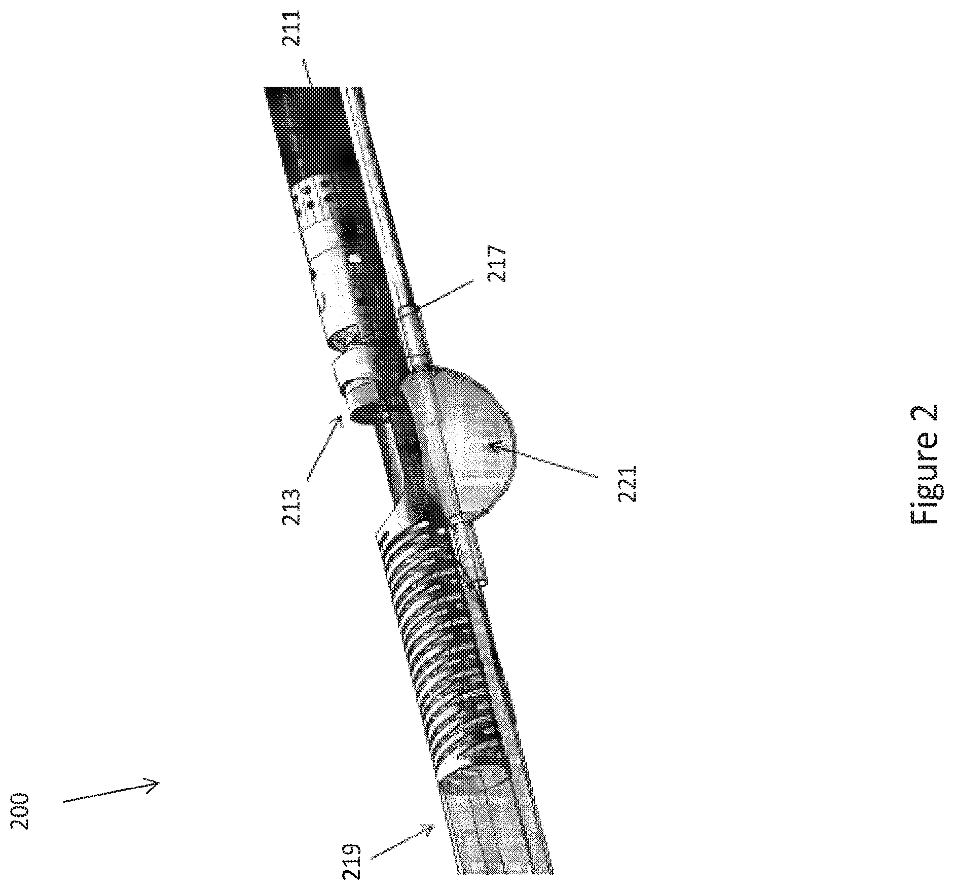
FIG. 2 is a view of an exemplary catheter with on-board imaging.
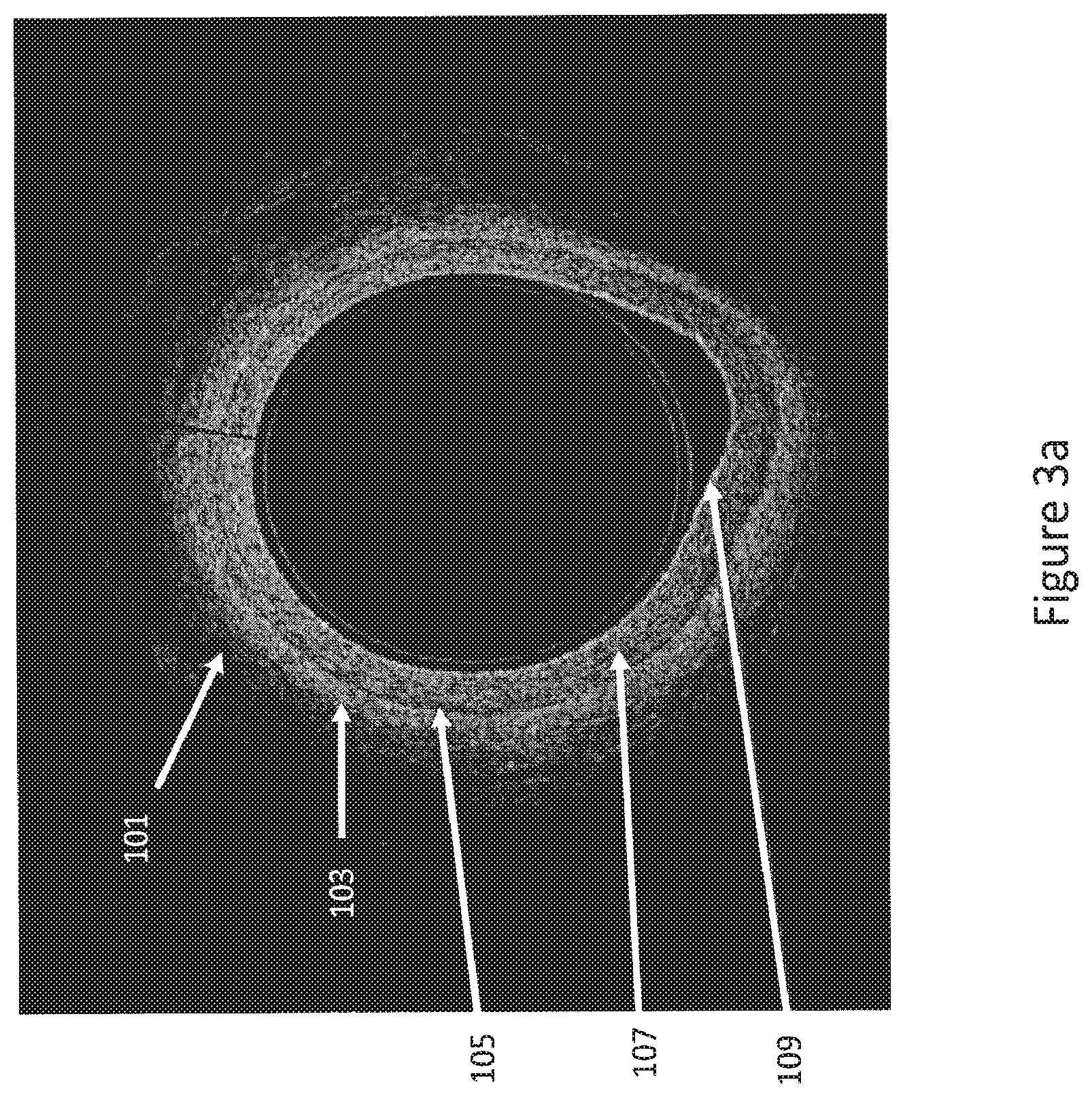
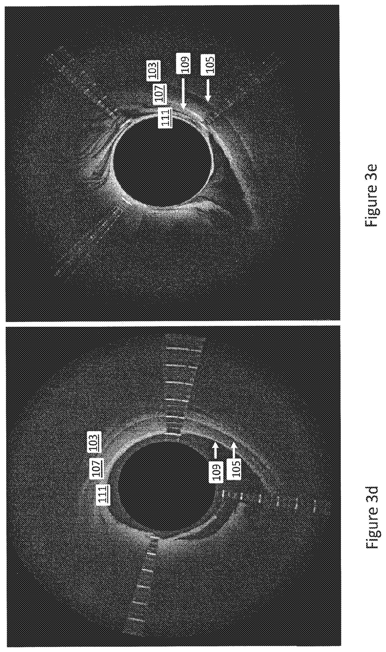
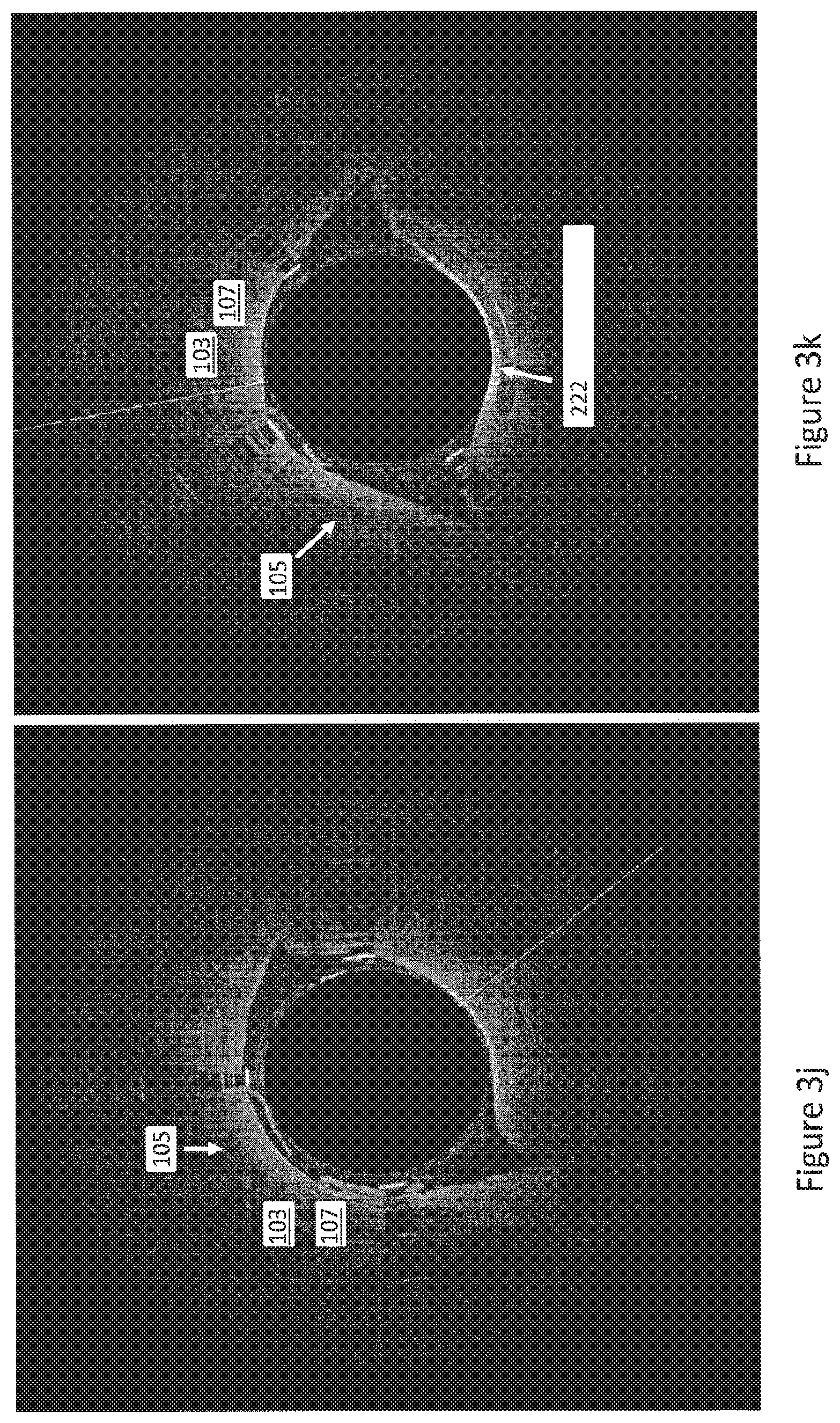
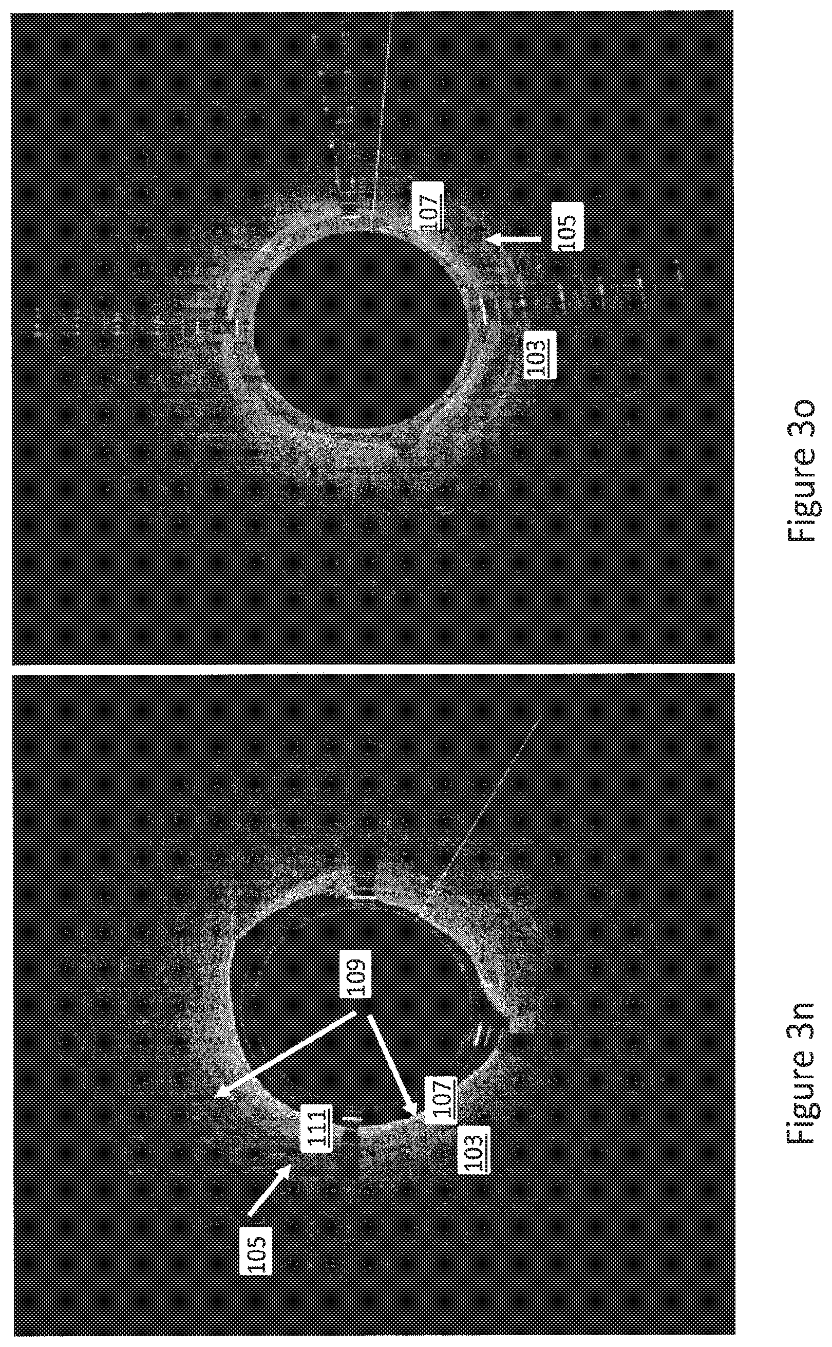
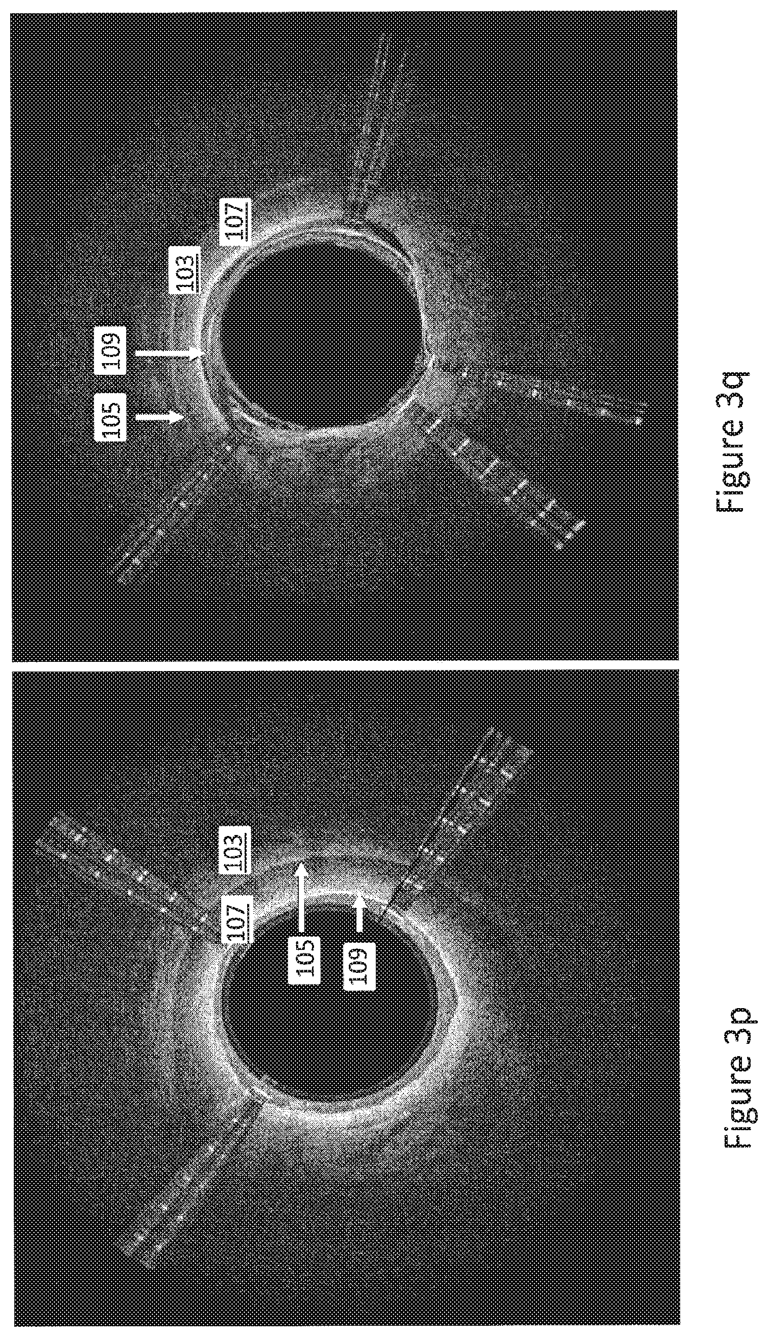
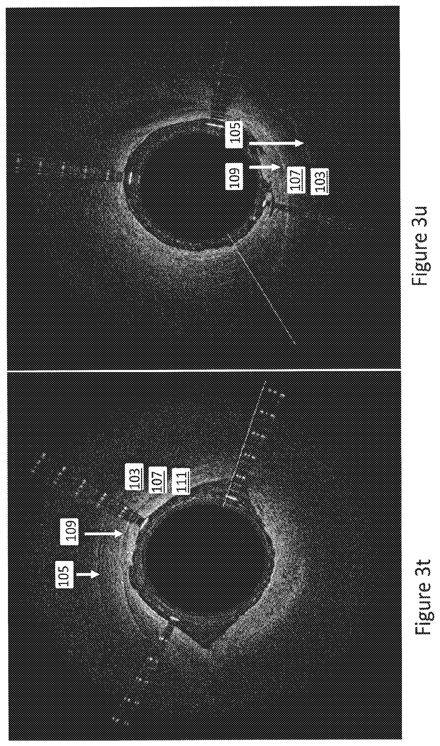
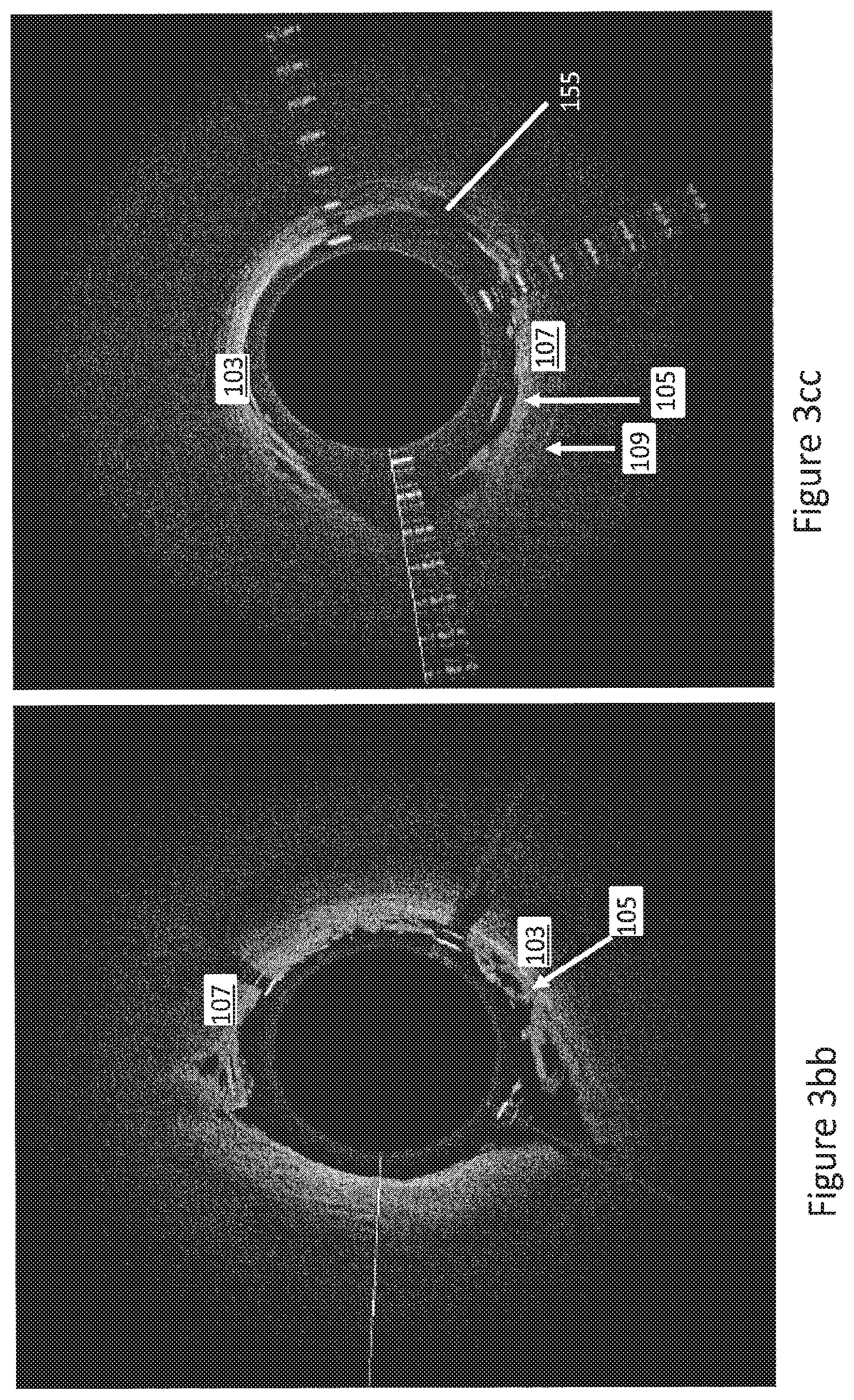
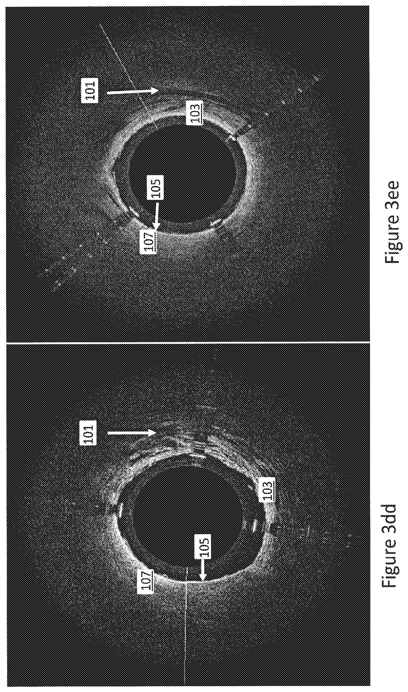
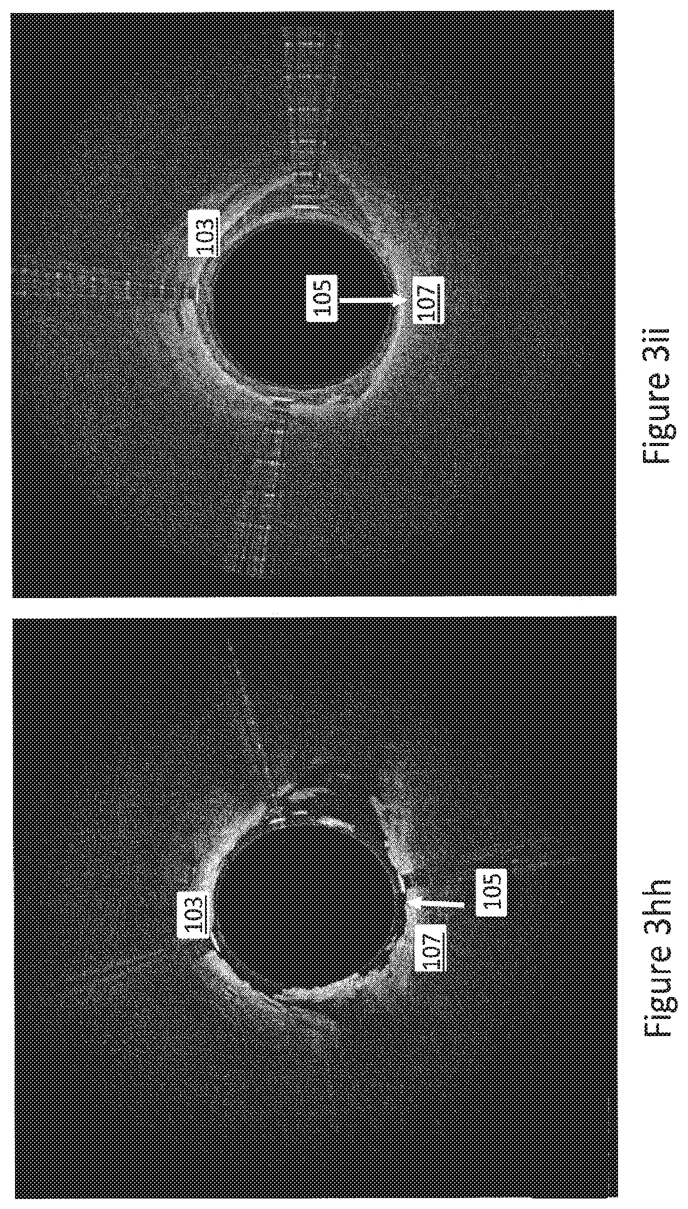
FIGS. 3a-3ii are exemplary OCT images wherein the internal elastic lamina and/or the external elastic lamina can be identified.
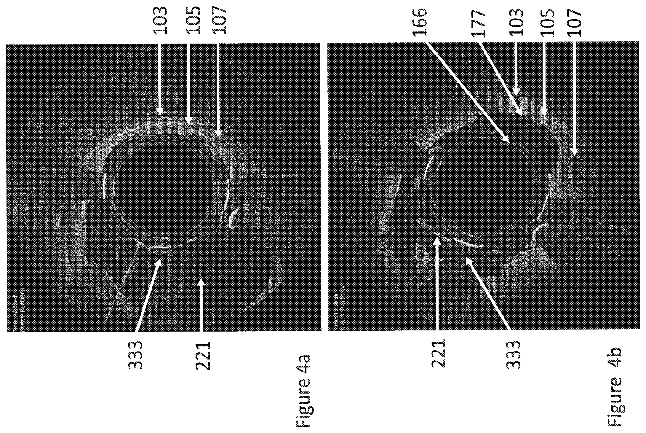
FIGS. 4a and 4b are OCT images taken with an imaging atherectomy device having an inflatable balloon to urge the cutter against the wall. FIG. 4a shows an OCT image during cutting into the media, close to the EEL and adventitia with the balloon fully inflated. FIG. 4B shown an OCT image where the balloon has been deflated to reduce the cutting depth and avoid the EEL and adventitia.
FIGS. 5a and 5b are OCT images taken with a directional atherectomy device. FIG. 5a shows the direction of cut directly towards the artery wall structure with media and EEL. FIG. 5b shows adjustment of the direction away from the artery wall and towards plaque.
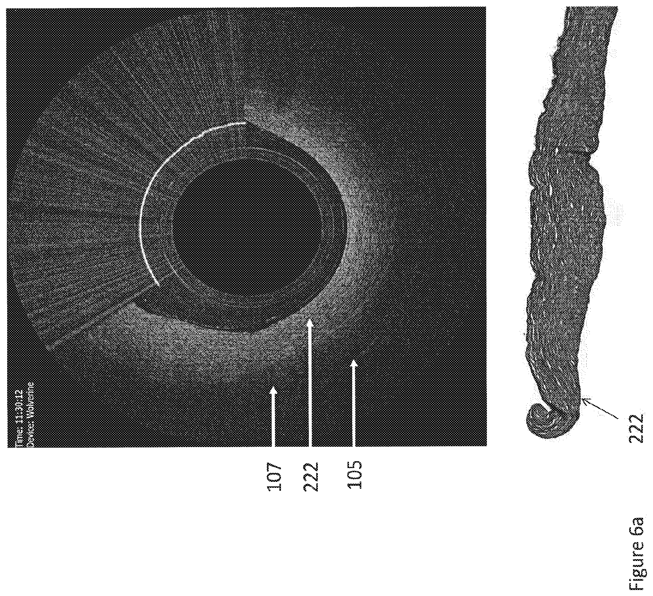
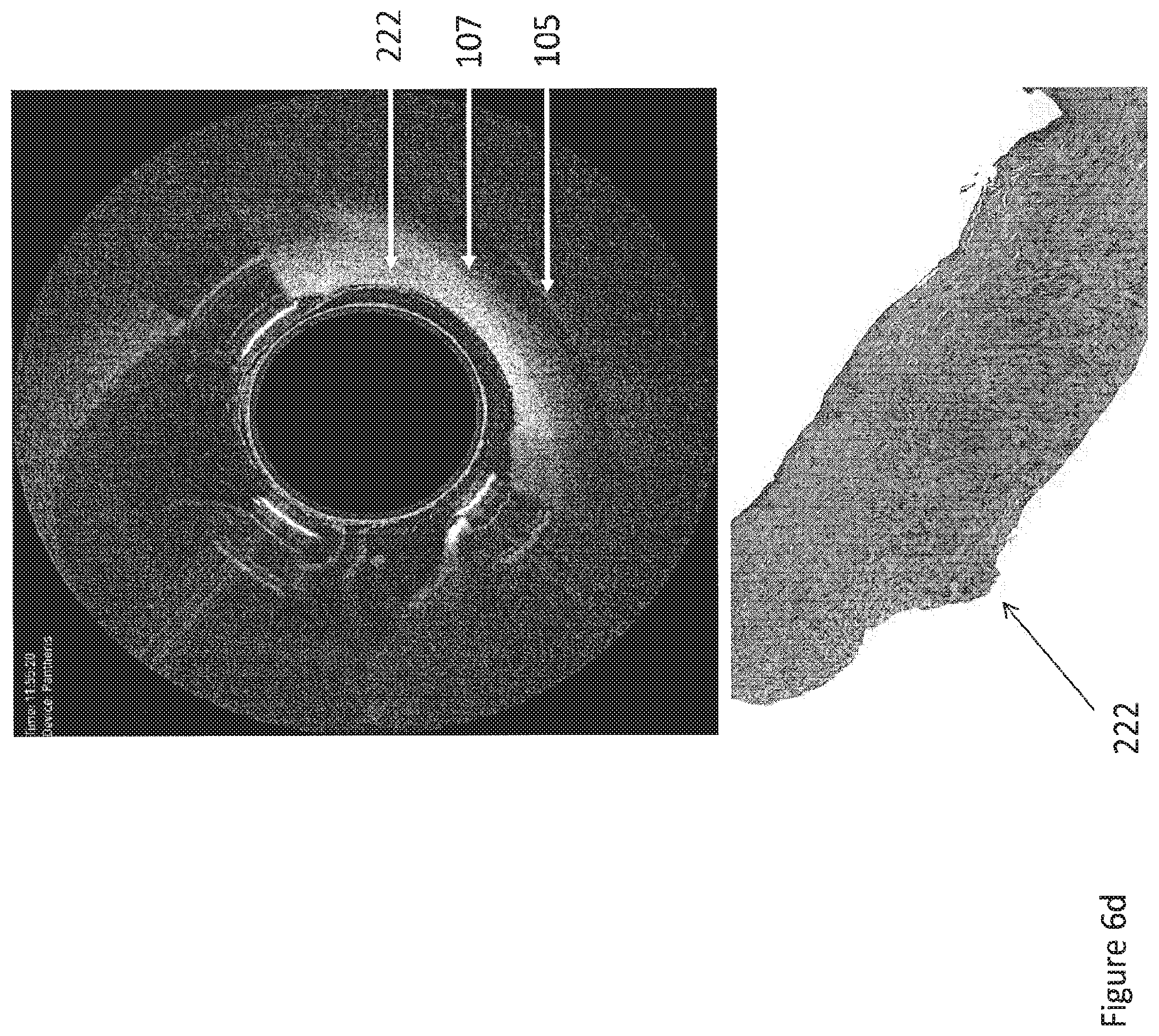
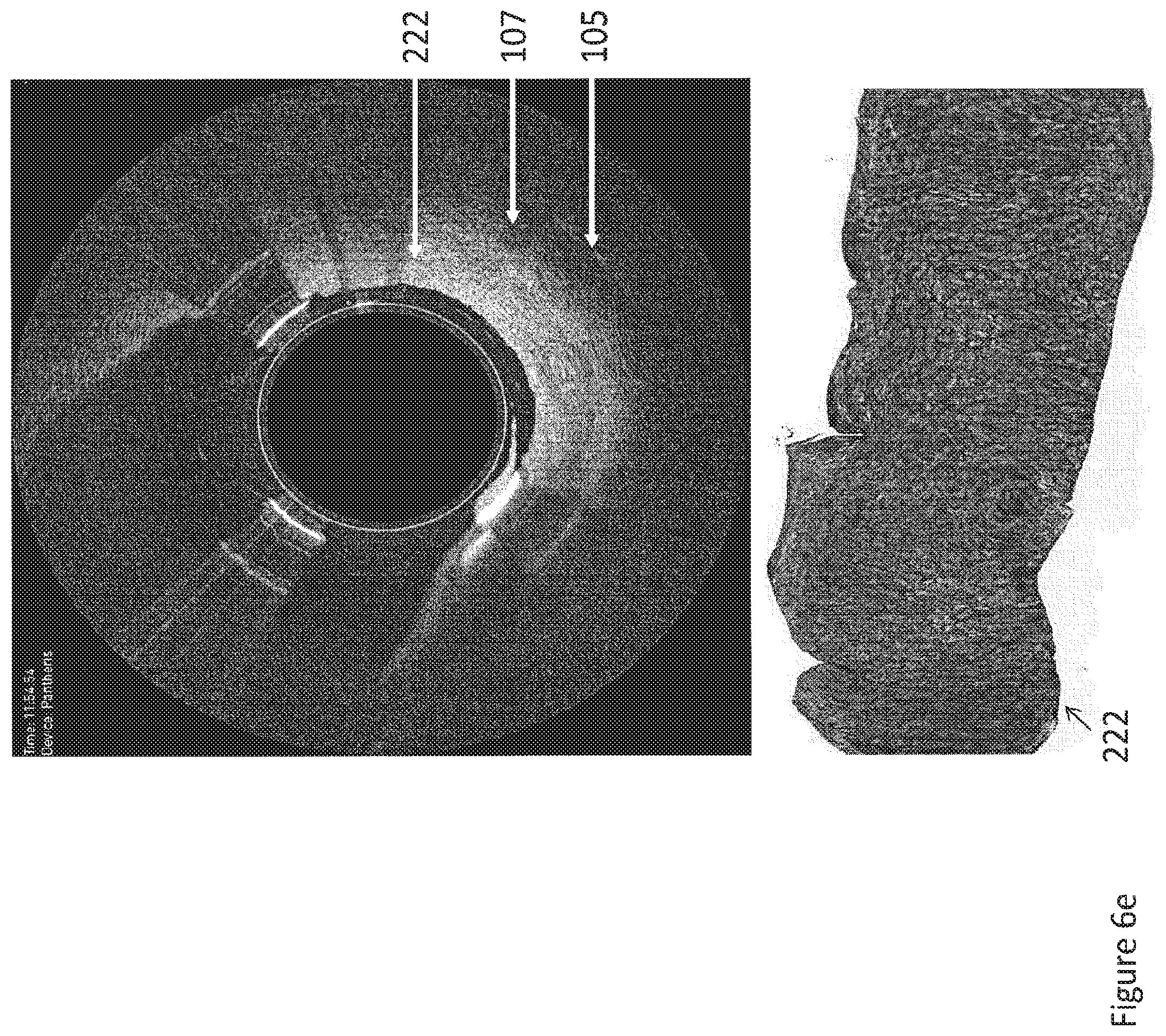
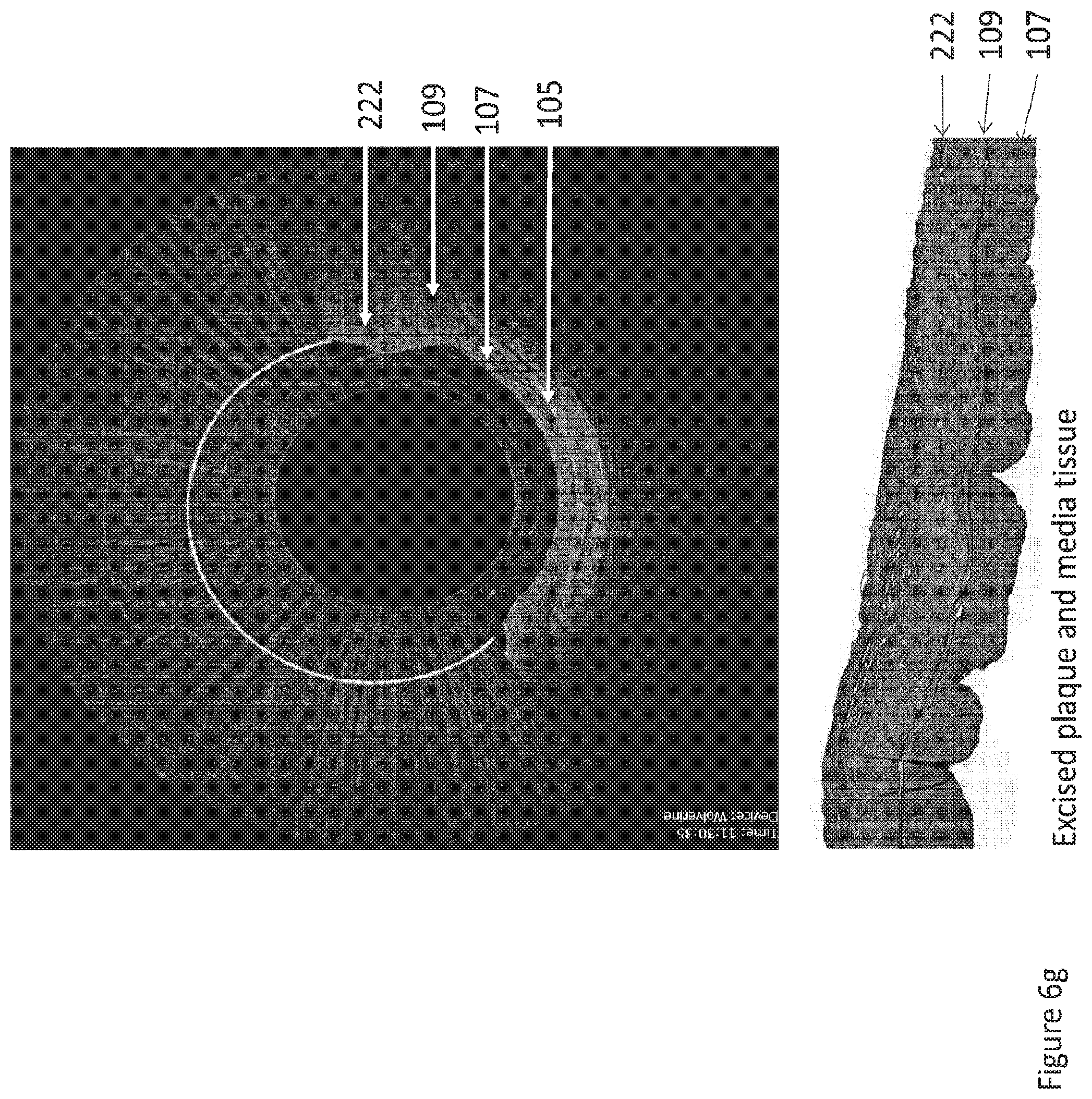
FIGS. 6a-6h show exemplary tissue excised with the identification methods described herein and the OCT images taken during cutting procedures.
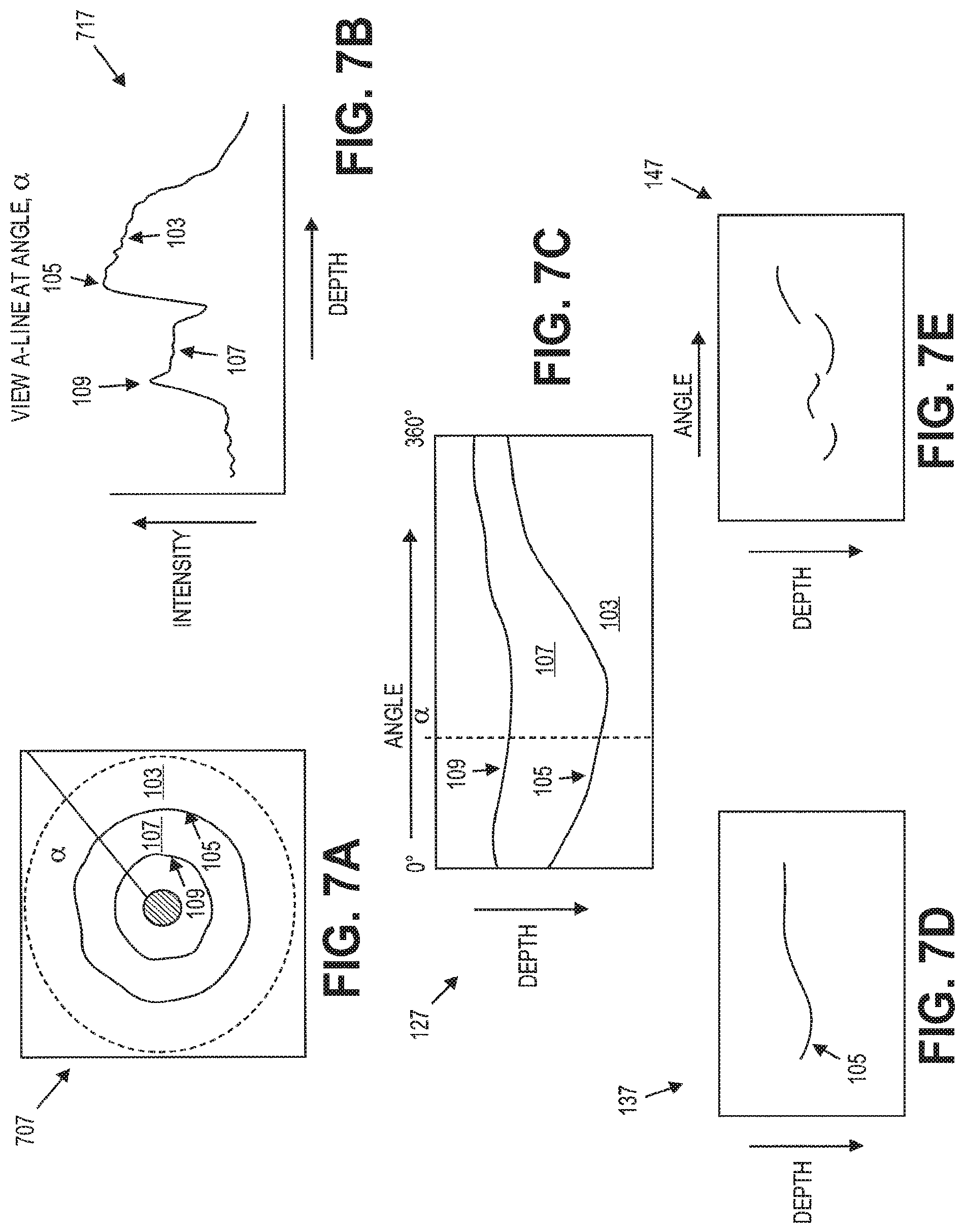
FIGS. 7a-7e show a method of automatic detection of the EEL with a controller.
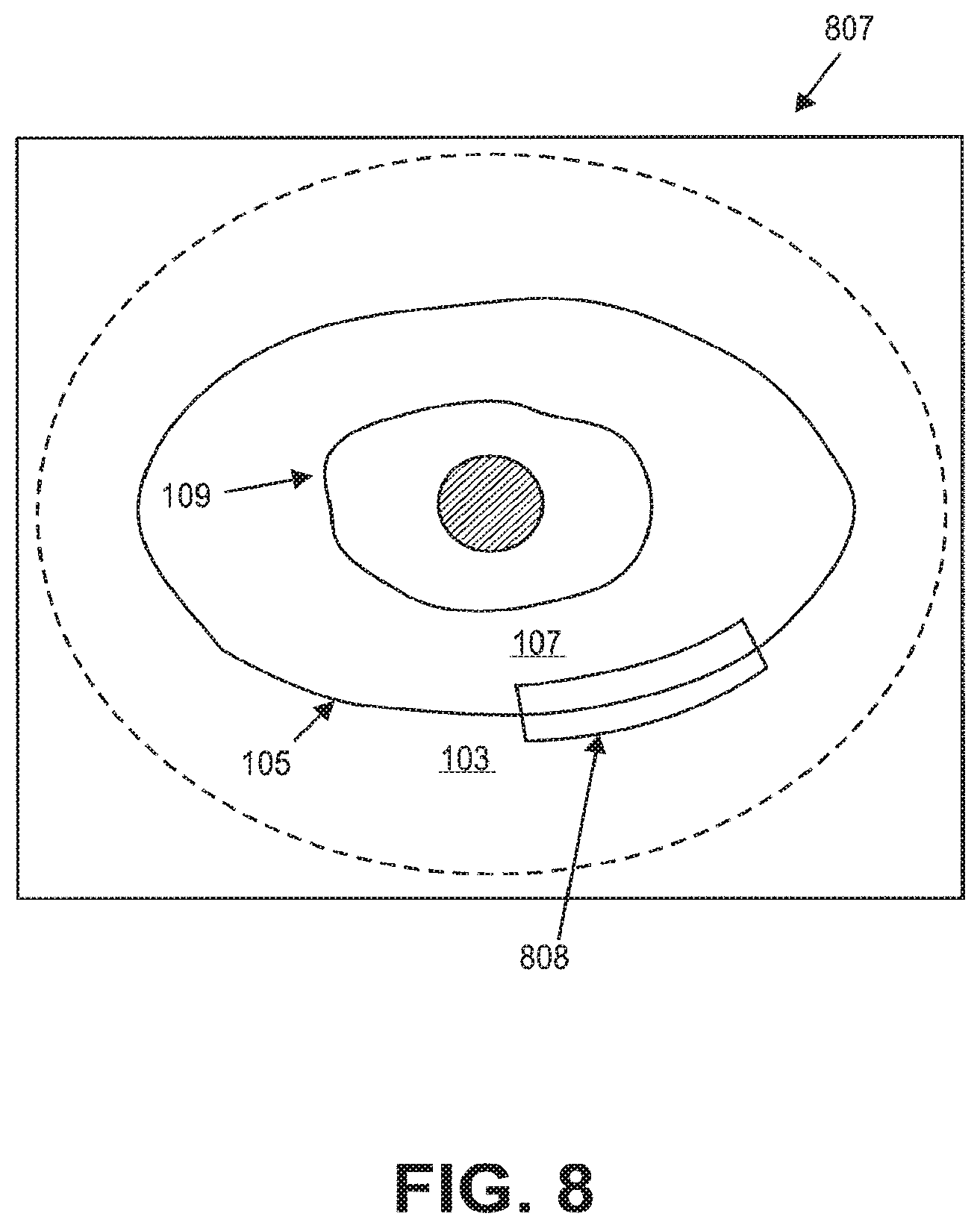
FIG. 8 shows an OCT image with a highlighted band on the EEL.
DETAILED DESCRIPTION
Described herein is a system and method for identifying elastic lamina during interventional treatments using a catheter having on-board imaging.
Referring to FIG. 2, an interventional catheter 200, such as an atherectomy catheter or an occlusion-crossing catheter, can include an elongate body 211 with a cutter 213 extending therefrom. An imaging sensor 217 can be configured to gather optical coherence tomography (OCT) images. A distal nosecone 219 can be configured to collect tissue cut by the cutter 213. In some embodiments, the device 200 can be a directional atherectomy device, and a balloon 221 can be used to expose the cutter 213 and/or urge the cutter 213 against the vessel wall for cutting. The amount of inflation of the balloon 212 can be varied to modify a depth of cut made by the cutter 213 into the vessel wall. Exemplary atherectomy devices are described further in U.S. patent application Ser. No. 12/829,277, filed Jul. 1, 2010, titled "ATHERECTOMY CATHETER WITH LATERALLY-DISPLACEABLE TIP," now U.S. Patent Application Publication No. 2011/0004107, U.S. patent application Ser. No. 13/175,232, filed Jul. 1, 2011, titled "ATHERECTOMY CATHETERS WITH LONGITUDINALLY DISPLACEABLE DRIVE SHAFTS," now U.S. Patent Application Publication No. 2012/0046679, U.S. patent application Ser. No. 13/654,357, filed Oct. 17, 2012, titled "ATHERECTOMY CATHETERS AND NON-CONTACT ACTUATION MECHANISM FOR CATHETERS," now U.S. Patent Application Publication No. 2013/0096589, International Patent Application No. PCT/US2013/031901, filed Mar. 15, 2013, titled "ATHERECTOMY CATHETERS WITH IMAGING," now published as WO 2013/172970, and International Patent Application No. PCT/US2013/032494, filed Mar. 15, 2013, titled "BALLOON ATHERECTOMY CATHETERS WITH IMAGING," now published as WO 2014/039099, the entireties of which are incorporated by reference herein.
Referring to FIGS. 3a-3ii, OCT images obtained with such an on-board imaging catheter can show the walls of the vessel, such as in a toroidal view of the walls. The resulting images can, for example, show plaque or thrombosis as well as layers of the vessel, including the backscattering or signal-rich intima 111, the media 107 that frequently has low backscattering or is signal-poor, the heterogeneous and frequently high backscattering adventitia 103, and/or the periadventitial tissue 101 characterized by large clear structures. The OCT image can further advantageously clearly show the IEL 109 as a distinct line between the media 107 and the intima 111 and the EEL 105 as a distinct line between the media 107 and the adventitia 103. For example, the IEL 109 and EEL 105 can be displayed as thin bright structures (i.e., be highly backscattering). In many cases, the IEL 109 and EEL 105 appear as continuous lines following along (substantially parallel with) the internal and external perimeter of the media 107.
The OCT images collected with the catheter can thus be used to clearly identify the EEL 105. Moreover, OCT advantageously has a higher resolution than other types of imaging, such as ultrasound, thereby allowing for the clear identification of the EEL 105. Further, upon identification the EEL 105 in the images, the interventional therapy (e.g., atherectomy) can be tailored so as to avoid or limit interaction with the EEL 105 and adventitia 103, thereby avoiding the inflammatory response that occurs if the EEL 105 or adventitia 103 are injured.
Referring to FIGS. 4a and 4b, in one embodiment, the depth of cut with the atherectomy catheter can be adjusted based upon the identification of the EEL 105, such as by deflating a balloon or otherwise adjusting the cutting depth, in order to avoid excising the EEL 105 or the adventitia 103. For example, FIG. 4a shows an OCT image where the balloon 221 is fully inflated. The resulting cut (the cutter position is indicated by the dark circle in the center and the direction is opposite to the middle marker 333) is close to the media 107 and EEL 105. In contrast, in FIG. 4b, the balloon 221 has been deflated in order to pull the cutter away and leave a space 166 between the cutter and the EEL 105 (note that FIG. 4b shows the cutting mark 177, close to the EEL 105, created by the cut of FIG. 4a).
Referring to FIGS. 5a-5b, in one embodiment, the direction of the cut can be modified to avoid the EEL 105, i.e., the distal tip of the catheter 200 can be oriented away from the EEL 105. In some embodiments, markers on the image can be used to help orient the direction of the catheter to ensure that the EEL is not cut (i.e., the middle marker 333 can show a position directly opposite to the direction of the cut). The cutting direction can thus be changed from being oriented directly at the healthy artery wall (and thus normal to the EEL) and instead towards the plaque 333.
As a result of the direction and/or depth modifications, the tissue can be cut right up to, but not through, the EEL 105. FIGS. 6a-6h show the OCT image and resulting tissue cut using the identification methods described herein. As shown, only plaque tissue 222 (and the IEL 109 and media 107 in FIG. 6g) is cut, but not the EEL 105.
Moreover, if the OCT images show that trauma has occurred (i.e., if the images shown a break 155 in the continuity of the bright lines as shown in FIG. 3cc), the treatment plan can be adjusted so as to avoid further trauma, such as by reorienting the catheter or adjusting the depth of cut. Furthermore, if trauma to the EEL 105 is identified, the treatment plan can be adjusted to mitigate the inflammatory cascade by implanting drug coated stents, drug eluting balloons, or oral medication (e.g., anti-inflammatory, Plavix, etc.).
In some embodiments, the identification of the EEL 105 can be performed manually by a physician or technician viewing the imagines.
In other embodiments, the identification of the EEL 105 can be performed automatically with a controller. For example, referring to FIGS. 7a-7e, an A-line graph 717 of intensity vs. depth can be taken at an angle .alpha. within an OCT image 707. The peak intensity in the graph will correspond to the EEL 105 while the next highest peak will correspond to the IEL 109. The controller can then move to a new angle and search for the contour. For example, if the if the sector is unfolded into a B-scan 127, the EEL 105 can be identified at the set angle .alpha. and then the process repeated to find a continuous edge. As shown in FIG. 7d, if the line is continuous, it signifies the EEL 105. If it is discontinuous, then either the features is not the EEL or the EEL has been broken.
In some embodiments, the EEL can be automatically labeled or highlighted in the display of the OCT images. Referring to FIG. 8, the EEL 105 could be labeled in the OCT image 807, for example, with a transparent colored band 808. The band 808 can, for example, be brighter for greater confidence in EEL detection. The band 808 can further include different colors or widths, can be overlaid with different small shapes (such as dots or triangles). Further, the band 808 can be drawn outside of the OCT image 807 to identify where the feature is without interrupting the viewing (e.g., as an arc outside the OCT region).
Further, in some embodiments, a controller can use the identification of the EEL 105 to automatically assist with the interventional procedure. That is, in some embodiments, both the EEL 105 can be detected as well as the distance between the EEL and the cutter edge. The controller can thus calculate a distance between the EEL and the cutter and take a set action if that distance goes below a threshold value. For example, the controller can set off an alarm (e.g., audible noise, flash of light, graphic symbol). In other embodiments, the controller can shut down the cutter activation if the distance is below the threshold (and/or if the EEL 105 is going to be or has been broken or damaged as shown in FIG. 7e). In still other embodiments, the amount of urge on the catheter can be automatically reduced (such as the amount of balloon inflation). To automatically reduce the amount of urge, the inflation/deflation of the balloon, and thus the movement of the cutter between an active (or open) position to a passive (or closed) position can be automated with servos (or motors, or actuators) that are controlled by the controller. The controller can thus use the identification of the EEL 105 to partially or completely deflate the balloon, thereby reducing the cut depth and/or moving the cutter from an active to a passive position.
By identifying the EEL in images taken during interventional therapy, injury or trauma to those structures can advantageously be avoided. For example, referring to FIGS. 6a-4h, tissue cut during an atherectomy procedure using such techniques can advantageously include substantially only the diseased thrombosis or plaque (little to no media 107, adventitia 103, or EEL 105). In contrast, in atherectomy procedures where such methods are not used, excised tissue can include EEL 105, media 107, and/or adventitia 103, in addition to the diseased tissue, suggesting that an enhanced inflammatory response was instigated during the atherectomy procedure.
Additional details pertinent to the present invention, including materials and manufacturing techniques, may be employed as within the level of those with skill in the relevant art. The same may hold true with respect to method-based aspects of the invention in terms of additional acts commonly or logically employed. Also, it is contemplated that any optional feature of the inventive variations described may be set forth and claimed independently, or in combination with any one or more of the features described herein. Likewise, reference to a singular item, includes the possibility that there are a plurality of the same items present. More specifically, as used herein and in the appended claims, the singular forms "a," "and," "said," and "the" include plural referents unless the context clearly dictates otherwise. It is further noted that the claims may be drafted to exclude any optional element. As such, this statement is intended to serve as antecedent basis for use of such exclusive terminology as "solely," "only" and the like in connection with the recitation of claim elements, or use of a "negative" limitation. Unless defined otherwise herein, all technical and scientific terms used herein have the same meaning as commonly understood by one of ordinary skill in the art to which this invention belongs. The breadth of the present invention is not to be limited by the subject specification, but rather only by the plain meaning of the claim terms employed.
* * * * *
References
D00000
D00001
D00002
D00003
D00004
D00005
D00006
D00007
D00008
D00009
D00010
D00011
D00012

D00013
D00014

D00015

D00016

D00017
D00018
D00019

D00020
D00021
D00022

D00023
D00024

D00025

D00026
D00027
D00028

D00029
D00030

D00031
D00032
XML
uspto.report is an independent third-party trademark research tool that is not affiliated, endorsed, or sponsored by the United States Patent and Trademark Office (USPTO) or any other governmental organization. The information provided by uspto.report is based on publicly available data at the time of writing and is intended for informational purposes only.
While we strive to provide accurate and up-to-date information, we do not guarantee the accuracy, completeness, reliability, or suitability of the information displayed on this site. The use of this site is at your own risk. Any reliance you place on such information is therefore strictly at your own risk.
All official trademark data, including owner information, should be verified by visiting the official USPTO website at www.uspto.gov. This site is not intended to replace professional legal advice and should not be used as a substitute for consulting with a legal professional who is knowledgeable about trademark law.