Simulated dissectible tissue
Felsinger , et al. October 6, 2
U.S. patent number 10,796,606 [Application Number 14/875,067] was granted by the patent office on 2020-10-06 for simulated dissectible tissue. This patent grant is currently assigned to Applied Medical Resources Corporation. The grantee listed for this patent is Applied Medical Resources Corporation. Invention is credited to Katie Black, Tracy Breslin, Natasha Felsinger.


View All Diagrams
| United States Patent | 10,796,606 |
| Felsinger , et al. | October 6, 2020 |
Simulated dissectible tissue
Abstract
A simulated dissectible tissue for surgical training is provided. The simulated tissue comprises a silicone gel layer encapsulated within a silicone shell. A simulated anatomical structure is embedded together with the silicone gel layer within the sealed shell. The silicone shell as well as the silicone gel layer may include a deadening agent. Further processing of the silicone gel layer may include adding alcohol and, optionally, heating the mixture. The simulated dissectible tissue may be formed into a specific tissue or organ model for practicing surgical skills. The user practices incising through the outer layer and separating the shell layer along a dissection plane defined by the silicone gel layer to gain visibility of the embedded simulated anatomical structures. The silicone gel layer simulates dissectible tissue and has glossy and elastic properties that provide a realistic dissectible tissue layer for emulating skeletonization of the simulated anatomical structures contained therein.
| Inventors: | Felsinger; Natasha (Rancho Santa Margarita, CA), Black; Katie (Rancho Santa Margarita, CA), Breslin; Tracy (Rancho Santa Margarita, CA) | ||||||||||
|---|---|---|---|---|---|---|---|---|---|---|---|
| Applicant: |
|
||||||||||
| Assignee: | Applied Medical Resources
Corporation (Rancho Santa Margarita, CA) |
||||||||||
| Family ID: | 1000005098386 | ||||||||||
| Appl. No.: | 14/875,067 | ||||||||||
| Filed: | October 5, 2015 |
Prior Publication Data
| Document Identifier | Publication Date | |
|---|---|---|
| US 20160027344 A1 | Jan 28, 2016 | |
Related U.S. Patent Documents
| Application Number | Filing Date | Patent Number | Issue Date | ||
|---|---|---|---|---|---|
| PCT/US2015/022774 | Mar 26, 2015 | ||||
| 61970436 | Mar 26, 2014 | ||||
| Current U.S. Class: | 1/1 |
| Current CPC Class: | B29B 7/002 (20130101); B29C 39/10 (20130101); G09B 23/30 (20130101); G09B 23/34 (20130101); G09B 23/28 (20130101); B29C 39/38 (20130101); B29L 2031/7546 (20130101); B29K 2995/0081 (20130101); B29K 2083/00 (20130101); B29K 2995/0046 (20130101); B29L 2031/7532 (20130101) |
| Current International Class: | G09B 23/30 (20060101); G09B 23/34 (20060101); G09B 23/28 (20060101); B29B 7/00 (20060101); B29C 39/10 (20060101); B29C 39/38 (20060101) |
References Cited [Referenced By]
U.S. Patent Documents
| 184573 | November 1876 | Becker |
| 2127774 | August 1938 | Jacobs |
| 2284888 | June 1942 | Arneil, Jr. |
| 2324702 | July 1943 | Hoffman et al. |
| 2345489 | March 1944 | Lord |
| 2495568 | January 1950 | Coel |
| 3766666 | October 1973 | Stroop |
| 3775865 | December 1973 | Rowan |
| 3789518 | February 1974 | Chase |
| 3921311 | November 1975 | Beasley et al. |
| 3991490 | November 1976 | Markman |
| 4001951 | January 1977 | Fasse |
| 4001952 | January 1977 | Kleppinger |
| 4321047 | March 1982 | Landis |
| 4323350 | April 1982 | Bowden, Jr. |
| 4332569 | June 1982 | Burbank |
| 4371345 | February 1983 | Palmer et al. |
| 4386917 | June 1983 | Forrest |
| 4459113 | July 1984 | Boscaro Gatti et al. |
| 4481001 | November 1984 | Graham et al. |
| 4596528 | June 1986 | Lewis et al. |
| 4726772 | February 1988 | Amplatz |
| 4737109 | April 1988 | Abramson |
| 4789340 | December 1988 | Zikria |
| 4832978 | May 1989 | Lesser |
| 4867686 | September 1989 | Goldstein |
| 4907973 | March 1990 | Hon |
| 4938696 | July 1990 | Foster et al. |
| 4940412 | July 1990 | Blumenthal |
| 5061187 | October 1991 | Jerath |
| 5083962 | January 1992 | Pracas |
| 5104328 | April 1992 | Lounsbury |
| 5149270 | September 1992 | McKeown |
| 5180308 | January 1993 | Garito et al. |
| 5230630 | July 1993 | Burgett |
| 5273435 | December 1993 | Jacobson |
| 5295694 | March 1994 | Levin |
| 5310348 | May 1994 | Miller |
| 5318448 | June 1994 | Garito et al. |
| 5320537 | June 1994 | Watson |
| 5358408 | October 1994 | Medina |
| 5368487 | November 1994 | Medina |
| 5380207 | January 1995 | Siepser |
| 5403191 | April 1995 | Tuason |
| 5425644 | June 1995 | Szinicz |
| 5425731 | June 1995 | Daniel et al. |
| 5472345 | December 1995 | Eggert |
| 5518406 | May 1996 | Waters |
| 5518407 | May 1996 | Greenfield et al. |
| 5520633 | May 1996 | Costin |
| 5541304 | July 1996 | Thompson |
| 5620326 | April 1997 | Younker |
| 5720742 | February 1998 | Zacharias |
| 5722836 | March 1998 | Younker |
| 5727948 | March 1998 | Jordan |
| 5743730 | April 1998 | Clester et al. |
| 5762458 | June 1998 | Wang et al. |
| 5769640 | June 1998 | Jacobus et al. |
| 5775916 | July 1998 | Cooper et al. |
| 5785531 | July 1998 | Leung |
| 5800178 | September 1998 | Gillio |
| 5803746 | September 1998 | Barrie et al. |
| 5807378 | September 1998 | Jensen et al. |
| 5810880 | September 1998 | Jensen et al. |
| 5814038 | September 1998 | Jensen et al. |
| 5850033 | December 1998 | Mirzeabasov et al. |
| 5855583 | January 1999 | Wang et al. |
| 5873732 | February 1999 | Hasson |
| 5873863 | February 1999 | Komlosi |
| 5908302 | June 1999 | Goldfarb |
| 5947743 | September 1999 | Hasson |
| 5951301 | September 1999 | Younker |
| 6080181 | June 2000 | Jensen et al. |
| 6083008 | July 2000 | Yamada et al. |
| 6113395 | September 2000 | Hon |
| 6234804 | May 2001 | Yong |
| 6271278 | August 2001 | Park et al. |
| 6336812 | January 2002 | Cooper et al. |
| 6398557 | June 2002 | Hoballah |
| 6413264 | July 2002 | Jensen et al. |
| 6474993 | November 2002 | Grund et al. |
| 6485308 | November 2002 | Goldstein |
| 6488507 | December 2002 | Stoloff et al. |
| 6497902 | December 2002 | Ma |
| 6511325 | January 2003 | Lalka et al. |
| 6517354 | February 2003 | Levy |
| 6568941 | May 2003 | Goldstein |
| 6589057 | July 2003 | Keenan et al. |
| 6620174 | September 2003 | Jensen et al. |
| 6654000 | November 2003 | Rosenberg |
| 6659776 | December 2003 | Aumann et al. |
| 6773263 | August 2004 | Nicholls et al. |
| 6780016 | August 2004 | Toly |
| 6817973 | November 2004 | Merril et al. |
| 6820025 | November 2004 | Bachmann et al. |
| 6854976 | February 2005 | Suhr |
| 6857878 | February 2005 | Chosack et al. |
| 6863536 | March 2005 | Fisher et al. |
| 6866514 | March 2005 | Von Roeschlaub et al. |
| 6887082 | May 2005 | Shun |
| 6929481 | August 2005 | Alexander et al. |
| 6939138 | September 2005 | Chosack et al. |
| 6950025 | September 2005 | Nguyen |
| 6960617 | November 2005 | Omidian et al. |
| 6997719 | February 2006 | Wellman et al. |
| 7008232 | March 2006 | Brassel |
| 7018327 | March 2006 | Conti |
| 7025064 | April 2006 | Wang et al. |
| 7056123 | June 2006 | Gregorio et al. |
| 7080984 | July 2006 | Cohen |
| 7118582 | October 2006 | Wang et al. |
| 7255565 | August 2007 | Keegan |
| 7269532 | September 2007 | David et al. |
| 7272766 | September 2007 | Sakezles |
| 7300450 | November 2007 | Vleugels et al. |
| 7364582 | April 2008 | Lee |
| 7404716 | July 2008 | Gregorio et al. |
| 7419376 | September 2008 | Sarvazyan et al. |
| 7427199 | September 2008 | Sakezles |
| 7431189 | October 2008 | Shelton, IV et al. |
| 7441684 | October 2008 | Shelton, IV et al. |
| 7465168 | December 2008 | Allen et al. |
| 7467075 | December 2008 | Humphries et al. |
| 7544062 | June 2009 | Hauschild et al. |
| 7549866 | June 2009 | Cohen et al. |
| 7553159 | June 2009 | Arnal et al. |
| 7575434 | August 2009 | Palakodeti |
| 7594815 | September 2009 | Toly |
| 7621749 | November 2009 | Munday |
| 7646901 | January 2010 | Murphy et al. |
| 7648367 | January 2010 | Makower et al. |
| 7648513 | January 2010 | Green et al. |
| 7651332 | January 2010 | Dupuis et al. |
| 7677897 | March 2010 | Sakezles |
| 7775916 | August 2010 | Mahoney |
| 7780451 | August 2010 | Willobee et al. |
| 7802990 | September 2010 | Korndorffer et al. |
| 7803151 | September 2010 | Whitman |
| 7806696 | October 2010 | Alexander et al. |
| 7819799 | October 2010 | Merril et al. |
| 7833018 | November 2010 | Alexander et al. |
| 7837473 | November 2010 | Koh |
| 7850454 | December 2010 | Toly |
| 7850456 | December 2010 | Chosack et al. |
| 7854612 | December 2010 | Frassica et al. |
| 7857626 | December 2010 | Toly |
| 7866983 | January 2011 | Hemphill et al. |
| 7931470 | April 2011 | Alexander et al. |
| 7931471 | April 2011 | Senagore et al. |
| 7988992 | August 2011 | Omidian et al. |
| 7993140 | August 2011 | Sakezles |
| 7997903 | August 2011 | Hasson et al. |
| 8007281 | August 2011 | Toly |
| 8007282 | August 2011 | Gregorio et al. |
| 8016818 | September 2011 | Ellis et al. |
| 8017107 | September 2011 | Thomas et al. |
| 8021162 | September 2011 | Sui |
| 8048088 | November 2011 | Green et al. |
| 8083691 | December 2011 | Goldenberg et al. |
| 8116847 | February 2012 | Gattani et al. |
| 8137110 | March 2012 | Sakezles |
| 8157145 | April 2012 | Shelton, IV et al. |
| 8197464 | June 2012 | Krever et al. |
| 8205779 | June 2012 | Ma et al. |
| 8221129 | July 2012 | Parry et al. |
| 8297982 | October 2012 | Park et al. |
| 8308817 | November 2012 | Egilsson et al. |
| 8323028 | December 2012 | Matanhelia |
| 8323029 | December 2012 | Toly |
| 8328560 | December 2012 | Niblock et al. |
| 8342851 | January 2013 | Speeg et al. |
| 8403674 | March 2013 | Feygin et al. |
| 8403675 | March 2013 | Stoianovici et al. |
| 8403676 | March 2013 | Frassica et al. |
| 8408920 | April 2013 | Speller |
| 8425234 | April 2013 | Sakezles |
| 8439687 | May 2013 | Morriss et al. |
| 8442621 | May 2013 | Gorek et al. |
| 8454368 | June 2013 | Ault et al. |
| 8459094 | June 2013 | Yanni |
| 8459520 | June 2013 | Giordano et al. |
| 8460002 | June 2013 | Wang et al. |
| 8465771 | June 2013 | Wan et al. |
| 8469715 | June 2013 | Ambrozio |
| 8469716 | June 2013 | Fedotov et al. |
| 8480407 | July 2013 | Campbell |
| 8480408 | July 2013 | Ishii et al. |
| 8491309 | July 2013 | Parry et al. |
| 8500753 | August 2013 | Green et al. |
| 8512044 | August 2013 | Sakezles |
| 8517243 | August 2013 | Giordano et al. |
| 8521252 | August 2013 | Diez |
| 8535062 | September 2013 | Nguyen |
| 8544711 | October 2013 | Ma et al. |
| 8556635 | October 2013 | Toly |
| 8608483 | December 2013 | Trotta et al. |
| 8613621 | December 2013 | Henderickson et al. |
| 8636520 | January 2014 | Iwasaki et al. |
| D699297 | February 2014 | Bahsoun et al. |
| 8641423 | February 2014 | Gumkowski |
| 8647125 | February 2014 | Johns et al. |
| 8678831 | March 2014 | Trotta et al. |
| 8679279 | March 2014 | Thompson et al. |
| 8696363 | April 2014 | Gray et al. |
| 8708213 | April 2014 | Shelton, IV et al. |
| 8708707 | April 2014 | Hendrickson et al. |
| 8764449 | July 2014 | Rios et al. |
| 8764452 | July 2014 | Pravong et al. |
| 8800839 | August 2014 | Beetel |
| 8801437 | August 2014 | Mousques |
| 8801438 | August 2014 | Sakezles |
| 8807414 | August 2014 | Ross et al. |
| 8808004 | August 2014 | Misawa et al. |
| 8808311 | August 2014 | Heinrich et al. |
| 8814573 | August 2014 | Nguyen |
| 8827988 | September 2014 | Belson et al. |
| 8840628 | September 2014 | Green et al. |
| 8870576 | October 2014 | Millon et al. |
| 8888498 | November 2014 | Bisaillon et al. |
| 8893946 | November 2014 | Boudreaux et al. |
| 8911238 | December 2014 | Forsythe |
| 8915742 | December 2014 | Hendrickson et al. |
| 8945095 | February 2015 | Blumenkranz et al. |
| 8961190 | February 2015 | Hart et al. |
| 8966954 | March 2015 | Ni et al. |
| 8968003 | March 2015 | Hendrickson et al. |
| 9008989 | April 2015 | Wilson et al. |
| 9017080 | April 2015 | Placik |
| 9026247 | May 2015 | White |
| 9050201 | June 2015 | Egilsson et al. |
| 9056126 | June 2015 | Hersel et al. |
| 9070306 | June 2015 | Rappel et al. |
| 9087458 | July 2015 | Shim et al. |
| 9096744 | August 2015 | Wan et al. |
| 9117377 | August 2015 | Shim et al. |
| 9119572 | September 2015 | Gorek et al. |
| 9123261 | September 2015 | Lowe |
| 9129054 | September 2015 | Nawana et al. |
| 9196176 | November 2015 | Hager et al. |
| 9226799 | January 2016 | Lightcap et al. |
| 9257055 | February 2016 | Endo et al. |
| 9265587 | February 2016 | Vancamberg et al. |
| 9295468 | March 2016 | Heinrich et al. |
| 9351714 | May 2016 | Ross et al. |
| 9336694 | June 2016 | Shim et al. |
| 9358682 | June 2016 | Ruiz Morales |
| 9364224 | June 2016 | Nicholas et al. |
| 9364279 | June 2016 | Houser et al. |
| 9370361 | June 2016 | Viola et al. |
| 9373270 | June 2016 | Miyazaki |
| 9387276 | July 2016 | Sun et al. |
| 9427496 | August 2016 | Sun et al. |
| 9439649 | September 2016 | Shelton, IV et al. |
| 9439733 | September 2016 | Ha et al. |
| 9449532 | September 2016 | Black et al. |
| 9468438 | October 2016 | Baber et al. |
| 2001/0019818 | September 2001 | Yong |
| 2002/0168619 | November 2002 | Provenza |
| 2003/0031993 | February 2003 | Pugh |
| 2003/0091967 | May 2003 | Chosack et al. |
| 2003/0176770 | September 2003 | Merril et al. |
| 2004/0005423 | January 2004 | Dalton et al. |
| 2004/0126746 | July 2004 | Toly |
| 2004/0248072 | December 2004 | Gray et al. |
| 2005/0008997 | January 2005 | Herman |
| 2005/0026125 | February 2005 | Toly |
| 2005/0084833 | April 2005 | Lacey et al. |
| 2005/0131390 | June 2005 | Heinrich et al. |
| 2005/0142525 | June 2005 | Cotin et al. |
| 2005/0192595 | September 2005 | Green et al. |
| 2005/0196739 | September 2005 | Moriyama |
| 2005/0196740 | September 2005 | Moriyama |
| 2005/0214727 | September 2005 | Stoianovici et al. |
| 2006/0046235 | March 2006 | Alexander et al. |
| 2006/0252019 | November 2006 | Burkitt et al. |
| 2006/0275741 | December 2006 | Chewning et al. |
| 2007/0074584 | April 2007 | Talarico et al. |
| 2007/0077544 | April 2007 | Lemperle et al. |
| 2007/0078484 | April 2007 | Talarico et al. |
| 2007/0148626 | June 2007 | Ikeda |
| 2007/0166682 | July 2007 | Yarin et al. |
| 2007/0197895 | August 2007 | Nycz et al. |
| 2007/0225734 | September 2007 | Bell et al. |
| 2007/0275359 | November 2007 | Rotnes et al. |
| 2008/0032272 | February 2008 | Palakodeti |
| 2008/0032273 | February 2008 | Macnamara et al. |
| 2008/0052034 | February 2008 | David et al. |
| 2008/0064017 | March 2008 | Grundmeyer, III |
| 2008/0076101 | March 2008 | Hyde et al. |
| 2008/0097501 | April 2008 | Blier |
| 2008/0108869 | May 2008 | Sanders et al. |
| 2008/0187895 | August 2008 | Sakezles |
| 2008/0188948 | August 2008 | Flatt |
| 2008/0299529 | December 2008 | Schaller |
| 2008/0317818 | December 2008 | Griffith et al. |
| 2009/0068627 | March 2009 | Toly |
| 2009/0142739 | June 2009 | Wang et al. |
| 2009/0142741 | June 2009 | Ault et al. |
| 2009/0143642 | June 2009 | Takahashi et al. |
| 2009/0176196 | July 2009 | Niblock et al. |
| 2009/0187079 | July 2009 | Albrecht et al. |
| 2009/0246747 | October 2009 | Buckman, Jr. |
| 2009/0298034 | December 2009 | Parry et al. |
| 2009/0314550 | December 2009 | Layton |
| 2010/0047752 | February 2010 | Chan et al. |
| 2010/0094312 | April 2010 | Ruiz Morales et al. |
| 2010/0099067 | April 2010 | Agro |
| 2010/0167248 | July 2010 | Ryan |
| 2010/0167249 | July 2010 | Ryan |
| 2010/0167250 | July 2010 | Ryan et al. |
| 2010/0167253 | July 2010 | Ryan et al. |
| 2010/0167254 | July 2010 | Nguyen |
| 2010/0196867 | August 2010 | Geerligs et al. |
| 2010/0204713 | August 2010 | Ruiz Morales |
| 2010/0209899 | August 2010 | Park |
| 2010/0248200 | September 2010 | Ladak |
| 2010/0258611 | October 2010 | Smith et al. |
| 2010/0273136 | October 2010 | Kandasami et al. |
| 2010/0279263 | November 2010 | Duryea |
| 2010/0285094 | November 2010 | Gupta |
| 2010/0324541 | December 2010 | Whitman |
| 2011/0020779 | January 2011 | Hannaford et al. |
| 2011/0046637 | February 2011 | Patel et al. |
| 2011/0046659 | February 2011 | Ramstein et al. |
| 2011/0087238 | April 2011 | Wang et al. |
| 2011/0091855 | April 2011 | Miyazaki |
| 2011/0137337 | June 2011 | van den Dool et al. |
| 2011/0200976 | August 2011 | Hou et al. |
| 2011/0207104 | August 2011 | Trotta |
| 2011/0218550 | September 2011 | Ma |
| 2011/0244436 | October 2011 | Campo |
| 2011/0269109 | November 2011 | Miyazaki |
| 2011/0281251 | November 2011 | Mousques |
| 2011/0301620 | December 2011 | Di Betta et al. |
| 2012/0015337 | January 2012 | Hendrickson |
| 2012/0015339 | January 2012 | Hendrickson et al. |
| 2012/0016362 | January 2012 | Heinrich et al. |
| 2012/0028231 | February 2012 | Misawa et al. |
| 2012/0045743 | February 2012 | Okano et al. |
| 2012/0065632 | March 2012 | Shadduck |
| 2012/0082970 | April 2012 | Pravong et al. |
| 2012/0100217 | April 2012 | Green et al. |
| 2012/0115117 | May 2012 | Marshall |
| 2012/0115118 | May 2012 | Marshall |
| 2012/0116391 | May 2012 | Houser et al. |
| 2012/0148994 | June 2012 | Hori et al. |
| 2012/0164616 | June 2012 | Endo et al. |
| 2012/0165866 | June 2012 | Kaiser et al. |
| 2012/0172873 | July 2012 | Artale et al. |
| 2012/0179072 | July 2012 | Kegreiss |
| 2012/0202180 | August 2012 | Stock et al. |
| 2012/0264096 | October 2012 | Taylor et al. |
| 2012/0264097 | October 2012 | Newcott et al. |
| 2012/0282583 | November 2012 | Thaler et al. |
| 2012/0282584 | November 2012 | Millon et al. |
| 2012/0283707 | November 2012 | Giordano et al. |
| 2012/0288839 | November 2012 | Crabtree |
| 2012/0308977 | December 2012 | Tortola |
| 2013/0087597 | April 2013 | Shelton, IV et al. |
| 2013/0101973 | April 2013 | Hoke et al. |
| 2013/0105552 | May 2013 | Weir et al. |
| 2013/0116668 | May 2013 | Shelton, IV et al. |
| 2013/0157240 | June 2013 | Hart et al. |
| 2013/0171288 | July 2013 | Harders |
| 2013/0177890 | July 2013 | Sakezles |
| 2013/0192741 | August 2013 | Trotta et al. |
| 2013/0218166 | August 2013 | Elmore |
| 2013/0224709 | August 2013 | Riojas et al. |
| 2013/0245681 | September 2013 | Straehnz et al. |
| 2013/0253480 | September 2013 | Kimball et al. |
| 2013/0267876 | October 2013 | Leckenby et al. |
| 2013/0282038 | October 2013 | Dannaher et al. |
| 2013/0288216 | October 2013 | Parry, Jr. et al. |
| 2013/0302771 | November 2013 | Alderete |
| 2013/0324991 | December 2013 | Clem et al. |
| 2013/0324999 | December 2013 | Price et al. |
| 2014/0011172 | January 2014 | Lowe |
| 2014/0017651 | January 2014 | Sugimoto et al. |
| 2014/0030682 | January 2014 | Thilenius |
| 2014/0038151 | February 2014 | Hart |
| 2014/0051049 | February 2014 | Jarc et al. |
| 2014/0072941 | March 2014 | Hendrickson et al. |
| 2014/0087345 | March 2014 | Breslin et al. |
| 2014/0087346 | March 2014 | Breslin et al. |
| 2014/0087347 | March 2014 | Tracy |
| 2014/0087348 | March 2014 | Tracy et al. |
| 2014/0088413 | March 2014 | Von Bucsh et al. |
| 2014/0093852 | April 2014 | Poulsen et al. |
| 2014/0093854 | April 2014 | Poulsen et al. |
| 2014/0099858 | April 2014 | Hernandez |
| 2014/0106328 | April 2014 | Loor |
| 2014/0107471 | April 2014 | Haider et al. |
| 2014/0156002 | June 2014 | Thompson et al. |
| 2014/0162016 | June 2014 | Matsui et al. |
| 2014/0170623 | June 2014 | Jarstad et al. |
| 2014/0186809 | July 2014 | Hendrickson et al. |
| 2014/0187855 | July 2014 | Nagale et al. |
| 2014/0200561 | July 2014 | Ingmanson et al. |
| 2014/0212861 | July 2014 | Romano |
| 2014/0220527 | August 2014 | Li et al. |
| 2014/0220530 | August 2014 | Merkle et al. |
| 2014/0220532 | August 2014 | Ghez et al. |
| 2014/0242564 | August 2014 | Pravong et al. |
| 2014/0246479 | September 2014 | Baber et al. |
| 2014/0248596 | September 2014 | Hart et al. |
| 2014/0263538 | September 2014 | Leimbach et al. |
| 2014/0272878 | September 2014 | Shim et al. |
| 2014/0272879 | September 2014 | Shim et al. |
| 2014/0275795 | September 2014 | Little et al. |
| 2014/0275981 | September 2014 | Selover et al. |
| 2014/0277017 | September 2014 | Leimbach et al. |
| 2014/0303643 | October 2014 | Ha et al. |
| 2014/0303646 | October 2014 | Morgan et al. |
| 2014/0303660 | October 2014 | Boyden et al. |
| 2014/0308643 | October 2014 | Trotta et al. |
| 2014/0342334 | November 2014 | Black et al. |
| 2014/0349266 | November 2014 | Choi |
| 2014/0350530 | November 2014 | Ross et al. |
| 2014/0357977 | December 2014 | Zhou |
| 2014/0370477 | December 2014 | Black et al. |
| 2014/0371761 | December 2014 | Juanpera |
| 2014/0378995 | December 2014 | Kumar et al. |
| 2015/0031008 | January 2015 | Black et al. |
| 2015/0037773 | February 2015 | Quirarte Catano |
| 2015/0038613 | February 2015 | Sun et al. |
| 2015/0076207 | March 2015 | Boudreaux et al. |
| 2015/0086955 | March 2015 | Poniatowski et al. |
| 2015/0132732 | May 2015 | Hart et al. |
| 2015/0132733 | May 2015 | Garvik et al. |
| 2015/0135832 | May 2015 | Blumenkranz et al. |
| 2015/0148660 | May 2015 | Weiss et al. |
| 2015/0164598 | June 2015 | Blumenkranz et al. |
| 2015/0187229 | July 2015 | Wachli et al. |
| 2015/0194075 | July 2015 | Rappel et al. |
| 2015/0202299 | July 2015 | Burdick et al. |
| 2015/0209035 | July 2015 | Zemlock |
| 2015/0209059 | July 2015 | Trees et al. |
| 2015/0209573 | July 2015 | Hibner et al. |
| 2015/0228206 | August 2015 | Shim et al. |
| 2015/0262511 | September 2015 | Lin et al. |
| 2015/0265431 | September 2015 | Egilsson et al. |
| 2015/0272571 | October 2015 | Leimbach et al. |
| 2015/0272574 | October 2015 | Leimbach et al. |
| 2015/0272580 | October 2015 | Leimbach et al. |
| 2015/0272581 | October 2015 | Leimbach et al. |
| 2015/0272583 | October 2015 | Leimbach et al. |
| 2015/0272604 | October 2015 | Chowaniec et al. |
| 2015/0332609 | November 2015 | Alexander |
| 2015/0358426 | December 2015 | Kimball et al. |
| 2015/0371560 | December 2015 | Lowe |
| 2015/0374378 | December 2015 | Giordano et al. |
| 2015/0374449 | December 2015 | Chowaniec et al. |
| 2016/0000437 | January 2016 | Giordano et al. |
| 2016/0022374 | January 2016 | Haider et al. |
| 2016/0030240 | February 2016 | Gonenc et al. |
| 2016/0031091 | February 2016 | Popovic et al. |
| 2016/0058534 | March 2016 | Derwin et al. |
| 2016/0066909 | March 2016 | Baber et al. |
| 2016/0070436 | March 2016 | Thomas et al. |
| 2016/0073928 | March 2016 | Soper et al. |
| 2016/0074103 | March 2016 | Sartor |
| 2016/0098933 | April 2016 | Reiley et al. |
| 2016/0104394 | April 2016 | Miyazaki |
| 2016/0117956 | April 2016 | Larsson et al. |
| 2016/0125762 | May 2016 | Becker et al. |
| 2016/0133158 | May 2016 | Sui et al. |
| 2016/0140876 | May 2016 | Jabbour et al. |
| 2016/0194378 | July 2016 | Cass et al. |
| 2016/0199059 | July 2016 | Shelton, IV et al. |
| 2016/0220150 | August 2016 | Sharonov |
| 2016/0220314 | August 2016 | Huelman et al. |
| 2016/0225288 | August 2016 | East et al. |
| 2016/0232819 | August 2016 | Hofstetter et al. |
| 2016/0235494 | August 2016 | Shelton, IV et al. |
| 2016/0256187 | September 2016 | Shelton, IV et al. |
| 2016/0256229 | September 2016 | Morgan et al. |
| 2016/0262736 | September 2016 | Ross et al. |
| 2016/0262745 | September 2016 | Morgan et al. |
| 2016/0293055 | October 2016 | Hofstetter |
| 2016/0296144 | October 2016 | Gaddam et al. |
| 2421706 | Feb 2001 | CN | |||
| 2751372 | Jan 2006 | CN | |||
| 2909427 | Jun 2007 | CN | |||
| 101313842 | Dec 2008 | CN | |||
| 101528780 | Sep 2009 | CN | |||
| 201364679 | Dec 2009 | CN | |||
| 201955979 | Aug 2011 | CN | |||
| 102458496 | May 2012 | CN | |||
| 202443680 | Sep 2012 | CN | |||
| 202563792 | Nov 2012 | CN | |||
| 202601055 | Dec 2012 | CN | |||
| 202694651 | Jan 2013 | CN | |||
| 103050040 | Apr 2013 | CN | |||
| 203013103 | Jun 2013 | CN | |||
| 203038549 | Jul 2013 | CN | |||
| 203338651 | Dec 2013 | CN | |||
| 203397593 | Jan 2014 | CN | |||
| 203562128 | Apr 2014 | CN | |||
| 102596275 | Jun 2014 | CN | |||
| 103845757 | Jun 2014 | CN | |||
| 103886797 | Jun 2014 | CN | |||
| 103396562 | Jul 2015 | CN | |||
| 105194740 | Dec 2015 | CN | |||
| 105504166 | Apr 2016 | CN | |||
| 9102218 | May 1991 | DE | |||
| 41 05 892 | Aug 1992 | DE | |||
| 44 14 832 | Nov 1995 | DE | |||
| 197 16 341 | Oct 1998 | DE | |||
| 1 024 173 | Aug 2000 | EP | |||
| 2 218 570 | Aug 2010 | EP | |||
| 2 691 826 | Dec 1993 | FR | |||
| 2 917 876 | Dec 2008 | FR | |||
| 2488994 | Sep 2012 | GB | |||
| 10 211160 | Aug 1998 | JP | |||
| 2001005378 | Jan 2001 | JP | |||
| 2009236963 | Oct 2009 | JP | |||
| 3162161 | Aug 2010 | JP | |||
| 2013127496 | Jun 2013 | JP | |||
| 101231565 | Feb 2013 | KR | |||
| PA 02004422 | Nov 2003 | MX | |||
| 106230 | Sep 2013 | PT | |||
| WO 1994/06109 | Mar 1994 | WO | |||
| WO 1996/042076 | Dec 1996 | WO | |||
| WO 1998/58358 | Dec 1998 | WO | |||
| WO 1999/01074 | Jan 1999 | WO | |||
| WO 2000/36577 | Jun 2000 | WO | |||
| WO 2002/38039 | May 2002 | WO | |||
| WO 2002/038039 | May 2002 | WO | |||
| WO 2004/032095 | Apr 2004 | WO | |||
| WO 2004/082486 | Sep 2004 | WO | |||
| WO 2005/071639 | Aug 2005 | WO | |||
| WO 2006/083963 | Aug 2006 | WO | |||
| WO 2007/068360 | Jun 2007 | WO | |||
| WO 2008/021720 | Feb 2008 | WO | |||
| WO 2008/103383 | Aug 2008 | WO | |||
| WO 2009/000939 | Dec 2008 | WO | |||
| WO 2009/089614 | Jul 2009 | WO | |||
| WO 2010/094730 | Aug 2010 | WO | |||
| WO 2011/035410 | Mar 2011 | WO | |||
| WO 2011/046606 | Apr 2011 | WO | |||
| WO 2011/127379 | Oct 2011 | WO | |||
| WO 2011/151304 | Dec 2011 | WO | |||
| WO 2012/149606 | Nov 2012 | WO | |||
| WO 2012/168287 | Dec 2012 | WO | |||
| WO 2012/175993 | Dec 2012 | WO | |||
| WO 2013/048978 | Apr 2013 | WO | |||
| WO 2013/103956 | Jul 2013 | WO | |||
| WO 2014/022815 | Feb 2014 | WO | |||
| WO 2014/093669 | Jun 2014 | WO | |||
| WO 2014/197793 | Dec 2014 | WO | |||
| WO 2015/148817 | Oct 2015 | WO | |||
| WO 2016/138528 | Sep 2016 | WO | |||
| WO 2016/183412 | Nov 2016 | WO | |||
| WO 2016/198238 | Dec 2016 | WO | |||
| WO 2016/201085 | Dec 2016 | WO | |||
| WO 2017/031214 | Feb 2017 | WO | |||
| WO 2017/042301 | Mar 2017 | WO | |||
Other References
|
PlatSil.RTM. Gel-OO & Gel-10, Polytek, Technical Bulletin, Apr. 12, 2010 (Year: 2010). cited by examiner . The International Bureau of WIPO, International Preliminary Report on Patentability for International Application No. PCT/US2016/036664, entitled "Hysterectomy Model," dated Dec. 21, 2017, 10 pgs. cited by applicant . European Patent Office, Examination Report for European Application No. 14733949.3 titled "Gallbladder Model," dated Dec. 21, 2016, 6 pgs. cited by applicant . European Patent Office, The International Search Report and Written Opinion of the International Searching Authority for International Application No. PCT/US2016/062669 titled "Simulated Dissectible Tissue," dated Apr. 5, 2017, 19 pgs. cited by applicant . European Patent Office, The International Search Report and Written Opinion of the International Searching Authority for International Application No. PCT/US2017/020389 titled "Simulated Tissue Cartridge", dated May 24, 2017, 13 pgs. cited by applicant . The International Bureau of WIPO, International Preliminary Report on Patentability and Written Opinion for International Application No. PCT/US2015/059668, entitled "Simulated Tissue Models and Methods," dated May 26, 2017, 16 pgs. cited by applicant . The International Bureau of WIPO, International Preliminary Report on Patentability for International Application No. PCT/US2016/018697, entitled "Simulated Tissue Structures and Methods," dated Aug. 31, 2017, 14 pgs. cited by applicant . Society of Laparoendoscopic Surgeons, "Future Technology Session: The Edge of Innovation in Surgery, Space, and Business," http://www.laparoscopytoday.com/endourology/page/2/ , Figure 1B: http://laparoscopy.blogs.com/laparoscopy today/images/6-1/6-1VlaovicPicB.jpg , Sep. 5-8, 2007, 10 pgs. cited by applicant . European Patent Office, International Search Report for International Application No. PCT/US2011/053859 A3, dated Apr. 5, 2012, entitled "Portable Laparoscopic Trainer," 8 pgs. cited by applicant . European Patent Office, The International Search Report and Written Opinion for International Application No. PCT/US2012/60997, entitled "Simulated Tissue Structure for Surgical Training," dated Mar. 7, 2013, 8 pgs. cited by applicant . European Patent Office, The International Search Report and Written Opinion for International Application No. PCT/US2012/070971, entitled "Advanced Surgical Simulation," dated Mar. 18, 2013, 10 pgs. cited by applicant . Human Patient Simulator, Medical Education Technologies, Inc., http://www.meti.com (1999) all, printed Apr. 12, 2013, 24 pgs. cited by applicant . The International Bureau of WIPO, International Preliminary Report on Patentability and Written Opinion for International Application No. PCT/US2011/053859, entitled "Portable Laparoscopic Trainer," dated Apr. 2, 2013, 9 pgs. cited by applicant . European Patent Office, The International Search Report and Written Opinion for International Application No. PCT/US2013/062363, entitled "Surgical Training Model for Laparoscopic Procedures," dated Jan. 22, 2014, 11 pgs. cited by applicant . European Patent Office, The International Search Report and Written Opinion for International Application No. PCT/US2013/061949, entitled "Surgical Training Model for Laparoscopic Procedures," dated Feb. 17, 2014, 7 pgs. cited by applicant . Anonymous: Realsim Systems--LTS2000, Sep. 4, 2005, pp. 1-2, XP055096193, Retrieved from the Internet: URL:https://web.archive.org/web/2005090403;3030/http://www.realsimsystems- .com/exersizes.htm (retrieved on Jan. 14, 2014). cited by applicant . European Patent Office, The International Search Report and Written Opinion for International Application No. PCT/US2013/062269, entitled "Surgical Training Model for Transluminal Procedures," dated Feb. 17, 2014, 8 pgs. cited by applicant . European Patent Office, The International Search Report and Written Opinion for International Application No. PCT/US2013/061557, entitled "Surgical Training Model for Laparoscopic Procedures," dated Feb. 10, 2014, 9 pgs. cited by applicant . European Patent Office, The International Search Report and Written Opinion for International Application No. PCT/US2013/061728, entitled "Surgical Training Model for Laparoscopic Procedures," dated Oct. 18, 2013, 9 pgs. cited by applicant . Limps and Things, EP Guildford MATTU Hernia Trainer, http://limbsandthings.com/us/products/tep-guildford-mattu-hernia-trainer/- , printed May 29, 2014, 11 pgs. cited by applicant . Simulab, Hernia Model, http://www.simulab.com/product/surgery/open/hernia model, printed printed May 29, 2014, 4 pgs. cited by applicant . McGill Laparoscopic Inguinal Hernia Simulator, Novel Low-Cost Simulator for Laparoscopic Inguinal Hernia Repair, Feb. 8, 2011, 1 pg. cited by applicant . University of Wisconsin-Madison Biomedical Engineering, Inguinal Hernia Model, http://bmedesign.engr.wisc.edu/projects/s10/hernia model/, printed May 29, 2014, 62 pgs. cited by applicant . The International Bureau of WIPO, International Preliminary Report on Patentability for International Application No. PCT/US2012/070971, entitled "Advanced Surgical Simulation," dated Jun. 24, 2014, 7 pgs. cited by applicant . European Patent Office, The International Search Report and Written Opinion of the International Searching Authority for International Application No. PCT/US2014/038195, entitled "Hernia Model", dated Oct. 15, 2014, 20 pgs. cited by applicant . European Patent Office, The International Search Report and Written Opinion of the International Searching Authority for International Application No. PCT/US2014/048027, entitled "First Entry Model", dated Oct. 17, 2014, 10 pgs. cited by applicant . The International Bureau of WIPO, International Preliminary Report on Patentability for International Application No. PCT/US2012/060997, entitled "Simulated Tissue Structure for Surgical Training" dated Apr. 22, 2014, 6 pgs. cited by applicant . European Patent Office, The International Search Report and Written Opinion for International Application No. PCT/US2014/019840, entitled "Advanced Surgical Simulation Constructions and Methods," dated Jul. 4, 2014, 8 pgs. cited by applicant . Kurashima, et al, "A tool for training and evaluation of Laparoscopic inguinal hernia repair; the Global Operative Assessment of Laparoscopic Skills--Groin Hernia" American Journal of Surgery, Paul Hoeber, New York, NY, US vol. 201, No. 1, Jan. 1, 2011, pp. 54-61 XP027558745. cited by applicant . European Patent Office, The International Search Report and Written Opinion for International Application No. PCT/US2014/042998, entitled "Gallbladder Model," dated Jan. 7, 2015, 20 pgs. cited by applicant . The International Bureau of WIPO, International Preliminary Report on Patentability, for PCT application No. PCT/US2013/053497, entitled Simulated Stapling and Energy Based Ligation for Surgical Training, dated Feb. 12, 2015, 6 pgs. cited by applicant . The International Bureau of WIPO, International Preliminary Report on Patentability for International Application No. PCT/US2013/062363, entitled "Surgical Training Model for Laparoscopic Procedures," dated Apr. 9, 2015, 9 pgs. cited by applicant . The International Bureau of WIPO, International Preliminary Report on Patentability for International Application No. PCT/US2013/062269, entitled "Surgical Training Model for Laparoscopic Procedures," dated Apr. 9, 2015, 6 pgs. cited by applicant . The International Bureau of WIPO, International Preliminary Report on Patentability for International Application No. PCT/US2013/061557, entitled "Surgical Training Model for Laparoscopic Procedures," dated Apr. 9, 2015, 6 pgs. cited by applicant . The International Bureau of WIPO, International Preliminary Report on Patentability for International Application No. PCT/US2013/061728, entitled "Surgical Training Model for Laparoscopic Procedures," dated Apr. 9, 2015, 7 pgs. cited by applicant . The International Bureau of WIPO, International Preliminary Report on Patentability for International Application No. PCT/US2013/061949, entitled "Surgical Training Model for Laparoscopic Procedures," dated Apr. 9, 2015, 6 pgs. cited by applicant . The International Bureau of WIPO, International Preliminary Report on Patentability for International Application No. PCT/US2014/019840, entitled "Simulated Tissue Structure for Surgical Training," dated Sep. 11, 2015, 8 pgs. cited by applicant . European Patent Office, The International Search Report and Written Opinion for International Application No. PCT/US2015/020574, entitled "Advanced First Entry Model for Surgical Simulation," dated Jun. 1, 2015, 12 pgs. cited by applicant . The International Bureau of WIPO, International Preliminary Report on Patentability for International Application No. PCT/US2014/038195, entitled "Hernia Model," dated Nov. 26, 2015, 16 pgs. cited by applicant . The International Bureau of WIPO, International Preliminary Report on Patentability for International Application No. PCT/US2014/042998, entitled "Gallbladder Model," dated Dec. 30, 2015, 15 pgs. cited by applicant . European Patent Office, International Search Report and Written Opinion for International Application No. PCT/US2013/053497, titled "Simulated Stapling and Energy Based Ligation for Surgical Training," dated Nov. 5, 2013, 8 pgs. cited by applicant . The International Bureau of WIPO, International Preliminary Report on Patentability for International Application No. PCT/US2014/048027, entitled "First Entry Model," dated Feb. 4, 2016, 8 pgs. cited by applicant . European Patent Office, International Search Report and Written Opinion for International Application No. PCT/US2015/059668, entitled "Simulated Tissue Models and Methods," dated Apr. 26, 2016, 20 pgs. cited by applicant . Australian Patent Office, Patent Examination Report No. 1 for Australian Application No. 2012358851, titled "Advanced Surgical Simulation," dated May 26, 2016, 3 pgs. cited by applicant . Miyazaki Enterprises, "Miya Model Pelvic Surgery Training Model and Video," www.miyazakienterprises, printed Jul. 1, 2016, 1 pg. cited by applicant . European Patent Office, International Search Report and Written Opinion for International Application No. PCT/US2016/032292, entitled "Synthetic Tissue Structures for Electrosurgical Training and Simulation," dated Jul. 14, 2016, 11 pgs. cited by applicant . European Patent Office, International Search Report and Written Opinion for International Application No. PCT/US2016/018697, entitled "Simulated Tissue Structures and Methods," dated Jul. 14, 2016, 21 pgs. cited by applicant . European Patent Office, International Search Report and Written Opinion for International Application No. PCT/US2016/034591, entitled "Surgical Training Model for Laparoscopic Procedures," dated Aug. 8, 2016, 18 pgs. cited by applicant . European Patent Office, The International Search Report and Written Opinion of the International Searching Authority for International Application No. PCT/US2016/036664, entitled "Hysterectomy Model", dated Aug. 19, 2016, 15 pgs. cited by applicant . The International Bureau of WIPO, International Preliminary Report on Patentability for International Application No. PCT/US2015/020574, entitled "Advanced First Entry Model for Surgical Simulation," dated Sep. 22, 2016, 9 pgs. cited by applicant . European Patent Office, The International Search Report and Written Opinion of the International Searching Authority for International Application No. PCT/US2016/0043277 titled "Appendectomy Model", dated Oct. 4, 2016, 12 pgs. cited by applicant . The International Bureau of WIPO, International Preliminary Report on Patentability for International Application No. PCT/US2015/022774, titled "Simulated Dissectible Tissue," dated Oct. 6, 2016, 9 pgs. cited by applicant . European Patent Office, The International Search Report and Written Opinion of the International Searching Authority for International Application No. PCT/US2016/041852 titled "Simulated Dissectible Tissue", dated Oct. 13, 2016, 12 pgs. cited by applicant . 3D-MED Corporation, "Validated Training Course for Laparoscopic Skills," https://www.3-dmed.com/sites/default/files/product-additional/product-spe- c/Validated%20Training%20Course%20for%20Laparoscopic%20Skills.docx 3.pdf , printed Aug. 23, 2016, pp. 1-6. cited by applicant . 3D-MED Corporation, "Loops and Wire #1," https://www.3-dmed.com/product/loops-and-wire-1 , printed Aug. 23, 2016, 4 pgs. cited by applicant . Barrier, et al., "A Novel and Inexpensive Vaginal Hysterectomy Simulatory," Simulation in Healthcare: The Journal of the Society for Simulation in Healthcare, vol. 7, No. 6, Dec. 1, 2012, pp. 374-379. cited by applicant . European Patent Office, The International Search Report and Written Opinion for International Application No. PCT/US2018/018895, entitled "Synthetic Tissue Structures for Electrosurgical Training and Simulation," dated May 17, 2018, 12 pgs. cited by applicant . The International Bureau of WIPO, International Preliminary Report on Patentability for International Application No. PCT/US2016/062669, entitled "Simulated Dissectible Tissue," dated May 31, 2018, 11 pgs. cited by applicant . The International Bureau of WIPO, International Preliminary Report on Patentability for International Application No. PCT/US2016/0032292, entitled "Synthetic Tissue Structures for Electrosurgical Training and Simulation," dated Nov. 23, 2017, 2017, 8 pgs. cited by applicant . The International Bureau of WIPO, International Preliminary Report on Patentability for International Application No. PCT/US2016/034591, entitled "Surgical Training Model for Laparoscopic Procedures," dated Dec. 7, 2017, 2017, 14 pgs. cited by applicant . European Patent Office, Invitation to Pay Additional Fees for International Application No. PCT/US2016/062669, titled "Simulated Dissectible Tissue", mailed Feb. 10, 2017, 8 pgs. cited by applicant . European Patent Office, The International Search Report and Written Opinion of the International Searching Authority for International Application No. PCT/US2016/055148 titled "Hysterectomy Model", dated Feb. 28, 2017, 12 pgs. cited by applicant . Lamouche, Guy, et al., "Review of tissue simulating phantoms with controllable optical, mechanical and structural properties for use in optical coherence tomography," Biomedical Optics Express, vol. 3, No. 6, Jun. 1, 2012 (18 pgs.). cited by applicant . European Patent Office, The International Search Report and Written Opinion for International Application No. PCT/US2015/022774, dated Jun. 11, 2015, entitled "Simulated Dissectible Tissue," 13 pages. cited by applicant . Anonymous: Silicone Rubber--from Wikipedia, the free encyclopedia, Feb. 21, 2014, pp. 1-6. cited by applicant . The International Bureau of WIPO, International Preliminary Report on Patentability for International Application No. PCT/US2016/041852, entitled "Simulated Dissectible Tissue," dated Jan. 25, 2018, 12 pgs. cited by applicant . European Patent Office, Extended European Search Report for European Patent Application No. EP 17202365.7, titled "Gallbladder Model", dated Jan. 31, 2018, 8 pgs. cited by applicant . The International Bureau of WIPO, International Preliminary Report on Patentability for International Application No. PCT/US2016/043277, entitled "Appendectomy Model," dated Feb. 1, 2018, 9 pgs. cited by applicant . European Patent Office, The International Search Report and Written Opinion for International Application No. PCT/US2018/018036, entitled "Laparoscopic Training System," dated Jun. 8, 2018, 13 pgs. cited by applicant . The International Bureau of WIPO, International Preliminary Report on Patentability for International Application No. PCT/US2016/055148, entitled "Hysterectomy Model," dated Apr. 12, 2018, 12 pgs. cited by applicant . European Patent Office, Extended European Search Report for European Patent Application No. EP 18177751.7, titled "Portable Laparoscopic Trainer," dated Jul. 13, 2018, 8 pgs. cited by applicant . European Patent Office, The International Search Report and Written Opinion for International Application No. PCT/US2018/034705, entitled "Laparoscopic Training System," dated Aug. 20, 2018, 14 pgs. cited by applicant . The International Bureau of WIPO, International Preliminary Report on Patentability for International Application No. PCT/US2017/020389, entitled "Simulated Tissue Cartridge," dated Sep. 13, 2018, 8 pgs. cited by applicant . European Patent Office, Extended European Search Report for European Patent Application No. EP 18184147.9, titled "First Entry Model," dated Nov. 7, 2018, 7 pgs. cited by applicant . European Patent Office, The International Search Report and Written Opinion for International Application No. PCT/US2017/039113, entitled "Simulated Abdominal Wall," dated Aug. 7, 2017, 13 pgs. cited by applicant . The International Bureau of WIPO, International Preliminary Report on Patentability for International Application No. PCT/US2017/039113, entitled "Simulated Abdominal Wall," dated Jan. 10, 2019, 8 pgs. cited by applicant . European Patent Office, Extended European Search Report for European Patent Application No. EP 18210006.5, titled "Surgical Training Model for Laparoscopic Procedures," dated Jan. 21, 2019, 7 pgs. cited by applicant . European Patent Office, Extended European Search Report for European Patent Application No. EP 18207214.0, titled "Synthetic Tissue Structures for Electrosurgical Training and Simulation," dated Mar. 28, 2019, 6 pgs. cited by applicant . European Patent Office, Extended European Search Report for European Patent Application No. EP 18216002.8, titled "Surgical Training Model for Laparoscopic Procedures," dated Feb. 4, 2019, 6 pgs. cited by applicant . European Patent Office, Extended European Search Report for European Patent Application No. EP 18216005.1, titled "Surgical Training Model for Laparoscopic Procedures," dated Feb. 4, 2019, 7 pgs. cited by applicant . European Patent Office, Extended European Search Report for European Patent Application No. EP 19159065.2, titled "Simulated Tissue Structures and Methods," dated May 29, 2019, 8 pgs. cited by applicant . The International Bureau of WIPO, International Preliminary Report on Patentability for International Application No. PCT/US2018/018036, entitled "Laparoscopic Training System," dated Aug. 29, 2019, 8 pgs. cited by applicant . The International Bureau of WIPO, International Preliminary Report on Patentability for International Application No. PCT/US2018/018895, entitled "Synthetic Tissue Structures for Electrosurgical Training and Simulation," dated Sep. 6, 2019, 7 pgs. cited by applicant. |
Primary Examiner: Wollschlager; Jeffrey M
Assistant Examiner: Melendez; Armand
Attorney, Agent or Firm: Bozorgui; Shirin Ikehara; Patrick
Parent Case Text
CROSS-REFERENCE TO RELATED APPLICATIONS
This application is a continuation of International Application No. PCT/US2015/022774 entitled "Simulated dissectible tissue" filed on Mar. 26, 2015 and incorporated herein by reference in its entirety, which claims priority to and benefit of U.S. Provisional Patent Application Ser. No. 61/970,436 entitled "Right colon model" filed on Mar. 26, 2014 which is incorporated herein by reference in its entirety.
Claims
We claim:
1. A method of manufacturing a simulated dissectible tissue for surgical training comprising the steps of: providing a first sheet of silicone and a second sheet of silicone; curing simultaneously the first sheet of silicone and the second sheet of silicone; forming a first gel-coated silicone sheet and a second gel-coated silicone sheet; placing at least one simulated anatomical structure onto the first gel-coated silicone sheet; curing simultaneously the first gel-coated silicone sheet with the at least one simulated anatomical structure and the second gel-coated silicone sheet; preparing an uncured silicone gel for pouring over the first gel-coated silicone sheet to form an uncured silicone gel layer, the pouring step being carried out after performing the step of curing the first gel-coated silicone sheet with the at least one simulated anatomical structure; and placing the second gel-coated silicone sheet over the uncured silicone gel layer atop the first gel-coated silicone sheet to form a planar multilayer structure, wherein the step of preparing the uncured silicone gel comprises the steps of: selecting the uncured silicone gel from the group consisting of a silicone gel, a mixture of silicone gel and alcohol in a ratio of approximately 1:1 by volume, a mixture of silicone gel and deadener in a ratio of approximately 1:1 by volume and a mixture of silicone gel and deadener and alcohol in a ratio of approximately 1:1:1 by volume; and heating the uncured silicone gel at a temperature of at least 60 degrees Celsius for 25 minutes to induce porosity.
2. The method of claim 1 further comprising the step of allowing the uncured silicone gel layer disposed between the first gel-coated silicone sheet and the second gel-coated silicone sheet to cure.
3. The method of claim 1 wherein the step of forming a first gel-coated silicone sheet and a second gel-coated silicone sheet comprises a step of spreading a layer of uncured silicone gel on an untextured side of each of the first sheet of silicone and the second sheet of silicone.
4. The method of claim 1 wherein the uncured silicone gel layer covers completely the at least one simulated anatomical structure, wherein perimeters of the first gel-coated silicone sheet and the second gel-coated silicone sheet are surrounding the uncured silicone gel layer.
5. The method of claim 1 wherein the step of placing the second gel-coated silicone sheet to form a planar multilayer structure further comprises the step of applying pressure to substantially remove air bubbles and air pockets located within the planar multilayer structure.
6. The method of claim 5 further comprising the step of adhering perimeters of the first gel-coated silicone sheet and the second gel-coated silicone sheet together to encapsulate and seal the uncured silicone gel layer.
7. The method of claim 1 wherein the at least one simulated anatomical structure comprises one or more of vessels, vasculature, pathology, tumor, organ, partial organ, cartilage and duct, wherein the at least one simulated anatomical structure is embedded within the uncured silicone gel layer and is made from silicone or Kraton polymers.
8. The method of claim 1 wherein the planar multilayer structure simulating a mesentery layer, wherein the first and second sheets of silicone representing peritoneum layers and the uncured silicone gel layer disposed between the first and second gel-coated silicone sheets representing a connective tissue surrounding the at least one simulated anatomical structure.
9. A method of selecting a material that closely emulates a desired set of properties in human tissues for manufacturing a multilayered simulated dissectible tissue, the method comprising the steps of: choosing an anatomy represented by the multilayered simulated dissectible tissue, wherein the multi-layered simulated dissectible tissue comprises at least one outer layer composed of silicone and an inner layer composed of silicone gel; identifying the properties associated with the anatomy, wherein the properties associated with the anatomy comprises at least one of elasticity, toughness, tear resistance, porosity, tackiness, color or texture; selecting a material based, at least in part, on the identified properties for the outer layer; wherein the material for the outer layer is selected from the group consisting of a silicone and a mixture of silicone and deadener; selecting a material based, at least in part, on the identified properties for the inner layer; wherein the material for the inner layer is selected from the group consisting of a silicone gel, a mixture of silicone gel and deadener, a mixture of silicone gel and alcohol, and a mixture of silicone gel and deadener and alcohol; and providing the at least one outer layer and the inner layer according to their respective selected material such that the resulting multilayered simulated dissectible tissue has the desired physical properties of the natural human anatomy, wherein the step of providing the at least one outer layer and the inner layer comprises the steps of: providing a first silicone outer layer; providing a mold having a central cavity; placing the first silicone outer layer onto the mold so that a central area of the first silicone outer layer is placed within the central cavity and an outer perimeter of the first silicone outer layer is aligned with outer edges of the mold; heating the inner layer to induce porosity; applying the inner layer onto the first silicone outer layer; providing a second silicone outer layer; placing the second silicone outer layer over the inner layer atop the first silicone outer layer to form the multilayered simulated dissectible tissue.
10. The method of claim 9 wherein the inner layer comprises the mixture of silicone gel and alcohol in a ratio of approximately 1:1 by volume.
11. The method of claim 10 wherein the step of heating the inner layer comprises heating the mixture of silicone gel and alcohol by placing the mixture in an oven having a temperature of at least 60 degrees Celsius for approximately 25 minutes.
12. The method of claim 9 wherein the inner layer comprises the mixture of silicone gel and deadener in a ratio of approximately 1:1 by volume.
13. The method of claim 9 wherein the inner layer comprises the mixture of silicone gel and deadener and alcohol in a ratio of approximately 1:1:1 by volume.
14. The method of claim 13 wherein the step of heating the inner layer comprises heating the mixture of silicone gel and deadener and alcohol by placing the mixture in an oven having a temperature of at least 60 degrees Celsius for approximately 25 minutes.
15. The method of claim 9 further comprises the steps of: identifying landmarks and structures within the chosen anatomy; selecting a material based, at least in part, on the identified landmarks and structures, wherein the material is a silicone polymer; providing at least one simulated anatomical structure; and encapsulating the simulated anatomical structure within the inner layer, wherein the simulated anatomical structure comprises one or more of vessels, vasculature, pathology, tumor, organ, partial organ, cartilage and duct.
16. The method of claim 9 wherein the silicone of the outer layer comprises a durometer of 00-10 or 10 A Shore.
17. The method of claim 9 wherein the outer layer comprises the mixture of silicone and deadener with a ratio of silicone to deadener being approximately 1:1 or 2:1 parts by weight or by volume, wherein the silicone of the mixture of silicone and deadener comprises a durometer of 00-10 or 10 A Shore.
18. The method of claim 9 wherein the outer layer is configured to encompass the inner layer.
19. The method of claim 9 further comprising the step of adhering the first silicone outer layer to the second silicone outer layer in a location surrounding the inner layer.
20. The method of claim 9 further comprising the step of allowing the multilayered simulated dissectible tissue to cure.
Description
FIELD OF THE INVENTION
This invention relates to surgical training tools, and in particular, to simulated tissue structures and models for teaching and practicing surgical procedures.
BACKGROUND
A laparoscopic colectomy involves the resection of the bowel at various locations. Depending on the location, the colectomy is called a right hemicolectomy, a left hemicolectomy, a sigmoid colectomy, or a total colectomy. A right hemicolectomy is the removal of the entirety of the ascending colon through a portion of transverse colon, and is the most common among the colectomy procedures. A critical step of a right hemicolectomy procedure is the ability to identify key anatomical landmarks and vasculature in order to transect the appropriate vessels and adhesions to enable the mobilization of the colon. A surgeon's first step of the procedure is to identify and transect the ileocolic vessels. The ileocolic vessels are taken down with the help of the patient being in the Trendelenburg body position with the right side upwardly positioned. This body position aides in moving away the omentum and small bowel. The ileocolic vessels are typically located adjacent to the duodenum and are encased within a mesentery layer that is made up of two peritoneum layers. During this step, the surgeon uses the duodenum as a structural landmark in locating the ileocolic vessels. Upon transection of the ileocolic vessels, there can be either a medial to lateral or lateral to medial dissection of the mesentery layer. This dissection is done through blunt dissection using laparoscopic tools or energy compatible devices that can cut and seal smaller vasculature and lymph nodes encased within the mesentery layer. For medial to lateral dissection movement is made anterior to the duodenum and Gerota's fascia to the root of the mesentery attached to the cecum and ileum. If a surgeon moves lateral to medial, dissection is performed at the ileocecal junction and moves medially, again ensuring to stay anterior to the duodenum and Gerota's fascia. Once the cecum and ileum are mobilized, the surgeon will move up the White Line of Toldt in order to reach the hepatic flexure of the colon. The White Line of Toldt is an avascular plane that is connected to the abdominal side wall through lateral adhesions. A surgeon typically takes down these adhesions and the White Line of Toldt using laparoscopic scissors or other laparoscopic devices compatible with energy. Upon taking down the White Line of Toldt, adhesions along the hepatic flexure are removed in order to allow the extracorporeal mobilization and transection of the bowel. Upon transection of bowel the surgeon performs an extracorporeal anastomosis, which reconnects the remaining bowel.
Since there are several procedural steps for a right hemicolectomy, it is important that surgeons have a way to learn and practice this surgical procedure. The model needs to be anatomically correct and include the key landmarks as well as vasculature involved with right hemicolectomy procedures. The model should be compatible with any variation of the procedural steps. As an example, either medial to lateral or lateral to medial dissection should be able to be performed on the model. Moreover, the model needs to simulate the tactile feedback that a surgeon observes during the procedure. As an example, when dissection through the mesentery layer is performed, the difference in the feeling going through the layers to get to large vessels should be apparent. Vessels should be able to be grasped, cut and clipped. Although there are several procedural steps, the majority of this procedure involves mobilizing the bowel through various dissection techniques; therefore, developing an accurate dissection model is crucial to the simulation. The organs in the model should be simulated to be able to be moved and maneuvered as they would be in the body. Additionally, the organs on the model should be attached to the model so that they can be moved in the correct direction as positioning of the model is placed in Trendelenberg or reverse Trendelenberg body positioning. There is a need for an anatomical model that addresses these issues.
Furthermore, surgical residents as well as practicing surgeons undergo extensive training prior to being qualified to practice surgery on human patients. The training teaches a variety of aspects of surgery, which can include training to develop a specific skill, to practice a specific surgical procedure, or to practice using certain surgical instruments. There is a need for synthetic simulated models that will facilitate the training for surgeons. Specifically, there is a need for a simulated tissue that closely resembles the response of human tissue that is being dissected. The ability to perform dissection between planes or dissection to skeletonize vasculature from surrounding anatomy is a skill that is found within surgical procedures. Particularly, if a laparoscopic procedure is performed, maneuvering of instruments to perform dissection is a skill that can be acquired, which will allow for an atraumatic procedure with minimal injury. The present invention sets forth such a simulated tissue.
SUMMARY OF THE INVENTION
According to one aspect of the invention, a simulated dissectible tissue for surgical training is provided. The simulated dissectible tissue includes a first layer made of silicone and having an inner surface and an outer surface defining a thickness therebetween. The simulated dissectible tissue includes a second layer made of silicone and having an inner surface and an outer surface defining a thickness therebetween. The simulated dissectible tissue includes a third layer comprising silicone gel located between the first layer and the second layer. The silicone gel is sealed by the first and second layers. The first and second layers are incisable and the third layer elastically adheres the first and layers together such that the first and second layers are separable with a blunt instrument.
According to another aspect of the invention, a simulated dissectible tissue for surgical training is provided. The simulated dissectible tissue includes an outer shell made of silicone and configured to form an interior cavity. A filling is located and sealed inside the interior cavity. The encapsulated filling comprises a silicone gel and the outer shell is separable in the location of the filling to emulate surgical skeletonization.
According to another aspect of the invention, a method for manufacturing a simulated dissectible tissue for surgical training is provided. The method includes providing a first layer of silicone, curing the first layer, providing a mold having a central cavity, placing the first layer of silicone onto the mold such that the first layer covers central cavity, preparing a silicone gel, applying the uncured silicone gel onto the first layer, providing a second layer of silicone, placing the second layer over the silicone gel and the first layer, curing the silicone gel and curing the second layer.
According to another aspect of the invention, a method of manufacturing a simulated dissectible tissue comprising one or more outer layer encapsulating an inner layer is provided. The method includes the steps of selecting a material for the outer layer. The step of selecting a material for the outer layer includes selecting one of a silicone and a mixture of silicone and deadener. The method including the step of selecting a material for the inner layer. The step of selecting a material for the inner layer includes selecting one of a silicone gel and a mixture of silicone gel and deadener.
BRIEF DESCRIPTION OF THE DRAWINGS
FIG. 1 is a top perspective view of a laparoscopic trainer.
FIG. 2 is a top view of a right colon model according to the present invention.
FIG. 3 is a top view of a right colon model with an omentum layer pulled back according to the present invention.
FIG. 4 is a top view of a large bowel of a right colon model according to the present invention.
FIG. 5 is a top view of an aorta of a right colon model according to the present invention.
FIG. 6 is a top view of a simulated tissue structure such as a mesentery layer according to the present invention.
FIGS. 7A-7E are schematic drawings illustrating the steps of a manufacturing process for a simulated tissue structure such as a mesentery layer according to the present invention.
FIG. 8A is a list of composition variations for the outside layers of a simulated tissue structure such as a mesentery layer according to the present invention.
FIG. 8B is a list of composition variations for the middle or inner layer of a simulated tissue structure such as a mesentery layer according to the present invention.
FIGS. 9A-9B is a flow-chart of composition variations for a simulated tissue structure such as a mesentery layer according to the present invention.
FIG. 10A is a top view of a first layer of a simulated dissectible tissue according to the present invention.
FIG. 10B is a top view of a mold and template for manufacturing a simulated dissectible tissue according to the present invention.
FIG. 10C is a top view of a first layer of a simulated dissectible tissue on top of a mold and template according to the present invention.
FIG. 10D is a top view of simulated vasculature and simulated tumors on top of a first layer, a template and mold according to the present invention.
FIG. 10E is a top view of a gel second layer on top of the simulated vasculature, simulated tumors, first layer, template and mold according to the present invention.
FIG. 10F is a top view of a third layer on top of a gel second layer, simulated vasculature, simulated tumors, first layer, template and mold according to the present invention.
FIG. 10G is a top perspective view of a model of simulated dissectible tissue mounted on pegs of a simulated tissue platform according to the present invention.
FIG. 10H is a top view of a model of simulated dissectible tissue mounted on pegs of a simulated tissue platform according to the present invention.
FIG. 11 is a top perspective view of a simulated dissectible tissue with an incision in an outer layer exposing an inner gel layer according to the present invention.
DETAILED DESCRIPTION OF THE INVENTION
Organ tray models of one or more simulated organs and tissues are ideal for training and practicing laparoscopic procedures and techniques when placed inside a simulated laparoscopic trainer like the SIMSEI laparoscopic training system manufactured by Applied Medical Resources Corporation in California. A laparoscopic trainer 10 is shown in FIG. 1. The laparoscopic trainer 10 is described in co-pending U.S. patent application Ser. No. 13/248,449 entitled "Portable laparoscopic trainer" and filed on Sep. 29, 2011 by Pravong et al. to Applied Medical Resources Corporation and published as U.S. Patent Publication No. 2012/0082970, hereby incorporated by reference in its entirety herein. The laparoscopic trainer 10 includes a top cover 12 connected to a base 14 by a pair of legs 16 spacing the top cover 12 from the base 14. The laparoscopic trainer 10 is configured to mimic the torso of a patient such as the abdominal region. The top cover 12 is representative of the anterior surface of the patient and the space between the top cover 12 and the base 14 is representative of an interior of the patient or body cavity 18 where organs reside. The laparoscopic trainer 10 is a useful tool for teaching, practicing and demonstrating various surgical procedures and their related instruments in simulation of a patient. Surgical instruments are inserted into the cavity 18 through pre-established apertures 20 in the top cover 12. These pre-established apertures 20 may include seals that simulate trocars or may include simulated tissue that simulates the patient's skin and abdominal wall portions. Various tools and techniques may be used to penetrate the top cover 12 to perform mock procedures on model organs placed between the top cover 12 and the base 14 such as the right colon model of the present invention. When placed inside the cavity 18 of the trainer 10, the organ model is generally obscured from the perspective of the user who can then practice performing surgical techniques laparoscopically by viewing the surgical site indirectly via a video feed displayed on a video monitor 22.
A video display monitor 22 that is hinged to the top cover 12 is shown in an open orientation in FIG. 1. The video monitor 22 is connectable to a variety of visual systems for delivering an image to the monitor 22. For example, a laparoscope inserted through one of the pre-established apertures 20 or a webcam located in the cavity and used to observe the simulated procedure can be connected to the video monitor 22 and/or a mobile computing device to provide an image to the user. In another variation, the top cover 12 does not include a video display 22 but includes means for supporting a laptop computer, a mobile digital device or tablet and connecting it by wire or wirelessly to the trainer 10.
When assembled, the top cover 12 is positioned directly above the base 14 with legs 16 located substantially at the periphery and interconnected between the top cover 12 and base 14. The top cover 12 and base 14 are substantially the same shape and size and have substantially the same peripheral outline. Although the trainer 10 has no sidewalls, the legs 16 partially obscure the internal cavity from view from an otherwise open-sided trainer 10. The laparoscopic trainer 10 includes a top cover 12 that angulates with respect to the base 14. The legs 16 are configured to permit the angle of the top cover 12 with respect to the base 14 to be adjusted. FIG. 1 illustrates the trainer 10 adjusted to an angulation of approximately 30-45 degrees with respect to the base 14. The angulation of the trainer 10 advantageously simulates a patient in a Trendelenburg or reverse Trendelenburg position. In the Trendelenburg position the body is tilted such that it is laid flat on the back with the feet higher than the head or vice versa. The Trendelenburg position allows better access to the pelvic organs as gravity pulls the intestines away from the pelvis to thereby prevent encroachment of the intestines upon the pelvic operating field to provide more working space inside the abdominal cavity in which the surgeon can more easily manipulate organs. The selected angulation of the top cover 12 is locked by tightening thumbscrews provided on the legs 16. The angulation of the top cover 12 of the trainer 10 with respect to the base 14 or of the top cover 12 with respect to a horizontal surface such as a table top is particularly advantageous with respect to training and practicing a right hemicolectomy with the colon model of the present invention inserted into the cavity 18 of the trainer 10.
Turning now to FIG. 2, there is shown a right colon model 26 of the present invention that is particularly suitable for training and practicing a right hemicolectomy procedure among other procedures in a laparoscopic environment such as a laparoscopic trainer 10 described above with respect to FIG. 1. The simulated organs are typically made of silicone or thermoplastic elastomer (TPE) and placed in a tray 28. The tray 28 is configured to contain the model organs disposed within the tray 28. The tray 28 includes a base and at least one sidewall typically formed around the perimeter of the base. Additional sidewalls are formed inside the perimeter to define anatomy-specific locations and configured to contain simulated organ structures and tissues. These additional sidewalls provide lateral support in response to forces applied by the practitioner while manipulated the simulated organs with instruments inserted through the top cover 12 of the trainer 10 with the model 26 disposed within the cavity 18. FIG. 2 illustrates a model liver 30 made of silicone located along the top of the tray 28 and a simulated omentum layer 32 overlaying other organs and including representative vasculature 34.
Turning to FIG. 3, the omentum layer 32 is shown pulled back to uncover the underlying simulated organs that include at least a portion of a large bowel 36 (shown isolated in FIG. 4) that can be attached to an appendix 42 and sigmoid colon, at least a portion of a small bowel 38, a liver 30 containing a gallbladder assembly, a stomach, a duodenum, kidneys, ureters, an aorta 40 (shown isolated in FIG. 5), vessels representing arteries and veins 44, and connective tissue layers including peritoneum, Gerota's fascia, and a mesentery layer 46 (shown isolated in FIG. 6). The organs are assembled to represent the correct anatomical positioning and location present in the human body for surgical training using a variety of laparoscopic instruments. The right colon model 26 which can also be called the right bowel model 26 is assembled using silicone simulated organs with modification to emphasize key landmarks and features for a right hemicolectomy surgical training.
A base tray 28 is provided. The base tray 28 is made of yellow or red foam and sized and configured to be insertable into the cavity 18 of the trainer 10. Alternatively, the base tray 28 may include a liner that is made of yellow or red foam that fits directly into the base tray 28 which together with the liner is insertable into the laparoscopic trainer 10. An additional foam portion can be added to the left side of the foam base in order to simulate the right abdominal side wall. To allow simulation of various body positions during the simulated surgical procedure, alternative model bases are provided. For example, the right colon model base 28 or liner can be made from a vacuum formed plastic to have an inclined angle at one end of the model 26. The angle may simulate reverse Trendelenberg positioning of the patient during the surgical procedure. Moreover, the model 26 can be built on a vacuum-formed plastic base to have a curved shape that is modeled to mimic a pelvis shape that extends proximally to form the curved shape of the abdominal side walls.
A sheet made of silicone is adhered on top of the model's base 28 to aid in the attachment and assembly of the simulated organs. A list of the simulated organs which are made of silicone and their colors can be found in Table 1 below. The large bowel 36, aorta 40 and mesentery 46 can remain the substantially the size shown in FIGS. 2 and 3, or they can be shortened or shrunk in order to better fit the base of the laparoscopic trainer 10. These anatomical structures are adhered to the top of the foam base tray 28 in a manner that closely represents their accurate relative anatomical positioning.
TABLE-US-00001 TABLE 1 Organs & Their Colors Organ Color Large Bowel Pink Small Bowel Pink Appendix Pink Cecum Pink Stomach Flesh tone or pink Kidney Dark Red Liver Dark Red Gallbladder Green to Brown Aorta Dark Red Duodenum Flesh tone Arteries Dark Red Veins Blue Ureters Clear Omentum Yellow Mesentery Yellow Adhesion Pink Peritoneum Yellow or white
The mesentery layer 46 encapsulates arteries and veins 44 and is configured to be grasped and dissected using laparoscopic dissectors. Dissection between tissue layers has characteristics that cannot be simulated by silicone alone. Therefore, in order solve this issue, several variations of a simulated dissectible tissue suitable for simulating real anatomical structures such as the mesentery 46 have been developed. The simulated dissectible tissue suitable for simulating the mesentery 46 is composed of three layers stacked on top of each other. The three layers include a top layer 48, a bottom layer 50 and the middle layer 54. The top layer 48 and the bottom layer 50 may represent peritoneum layers and the middle layer 54, which comprises gel, may represent the connective tissue surrounding the blood vessels 44 made of silicone that can be dissected.
With reference now to FIGS. 7A-7E, the construction of the simulated dissectible tissue according to the present invention which can find exemplary use as mesentery layer 46 will now be described. It is noted that, the simulated dissectible tissue 47 of the present invention is not limited to use as a mesentery layer 46 but can form at least a part of any simulated tissue construction. Constructing the simulated dissectible tissue 47 involves an initial step of creating two separate thin sheets of silicone, one for the top layer 48 and one for the bottom layer 50 and letting them fully cure as shown in FIG. 7A. When the sheets are fully cured, a thin layer of silicone gel 58a, 58b is spread, using a spatula or a similar tool, on the untextured side of the each of the silicone sheets 48, 50, respectively, as shown in FIG. 7B. Simulated vasculature 44, comprising silicone vessels, is laid over one of either of the uncured gel layers 58a, 58b. FIG. 7C illustrates the simulated vasculature 44 being placed on the uncured gel layer 58a on the top layer 48 sheet. The silicone sheets 48, 50 with the gel layers 58a, 58b are then allowed to fully cure to adhere the simulated vasculature 44 to the top layer 48. When the gel-lined layers 48, 50 are cured, a third or middle layer 54 comprising fresh silicone gel is prepared and poured over one of the layers 48, 50. In one variation, the fresh silicone gel is poured over the silicone sheet 48 that has the silicone vessels 44 laid out. The gel is spread to completely cover the simulated vessels of the vasculature 44. The second layer 50 sheet is then laid over the middle layer 54 atop the first layer 48 while the silicone is still uncured gel and air pockets are pushed out to the edges to create a sandwich-like construction. The result of this process is the three-layered simulated dissectible tissue 47 that can be used to simulate a mesentery assembly 46 such as depicted in FIG. 6 that is particularly suitable and compatible with laparoscopic dissection and skeletonization of the encapsulated vasculature 44 located between the layers. Having multiple layers provides for an accurate, realistic feel and function for the simulated dissectible tissue structure. Furthermore, the simulated dissectible tissue 47 advantageously creates various tissue planes through which the practitioner may practice dissection skills. Not only does the model 26 provide the ability to dissect the layers but also allows the practitioner to properly identify the tissue planes or layers 44, 48, 50, 54, 58a, 58b which is an important skill to learn for each individual procedure. In one variation, the simulated dissectible tissue 47 is constructed without the vasculature layer 44 and can also be used to practice dissection.
Through the process of fabricating the simulated dissectible tissue 47, several additives were introduced which resulted in various desirable characteristics and iterations of the simulated dissectible tissue 47. A list of the various compositions for the outside first and second layers 48, 50 and the inner or middle layer 54 of the dissectible mesentery layer 46 is shown in Table 2 and summarized in the flow chart of FIGS. 8A and 8B. A flow chart for determining the best dissectible sheet based on desirable characteristics of FIGS. 8A-8B is shown in FIGS. 9A-9B. In FIGS. 9A-9B, the mesentery layer is used as an exemplary application for the simulated dissectible tissue according to the present invention and the chart in FIGS. 9A-9B is not limited to use for making a simulated mesentery layer alone but may include use in any simulated tissue structure. Also, the mention of vessels is in FIGS. 9A-9B is not limited to simulated vessels but may include any embedded simulated anatomical structure or tissue including but not limited to tumors, pathologies, organs, ducts, cartilage and the like. The silicone outside layers 48, 50 are made with the conventional two-part 1:1 ratio room temperature vulcanizing (RTV) silicone or with the RTV silicone and a deadening agent additive at a 1:1 ratio or with the RTV silicone and a deadening agent additive at a 2:1 ratio. The RTV includes but is not limited to platinum cured room temperature vulcanization silicone (PCRTVS). The silicone deadener is within the silicone fluid chemical family, including but not limited to silicone oil, and is a platinum cured silicone additive. One example of a deadener is called SLACKER made by Smooth-On, Inc. in Macungie, Pa. The deadening agent additive makes silicone softer and more realistically similar to the feel of skin or human tissue. Silicone deadener is a silicone additive that can soften and alter the resulting "feel" as well as the rebound properties of the cured silicone. This additive is within the chemical family of silicone fluids containing silicone oils. Silicone fluids and silicone oils have a range of uses that are dependent on the viscosity and chemical structure of the fluid. A silicone deadening agent is a type of silicone oil that can be mixed with platinum-cured room temperature vulcanization silicone.
The conventional silicone used to mold the organs range from a 00-10 Shore to a 10 A Shore durometer. Thus, the addition of the deadener would result in different properties when added to silicones with different durometers. Addition of a deadening agent to a softer durometer silicone results in a gel-like composition when fully cured. However, addition of a deadening agent to higher durometer silicones results in desirable features of a softer feeling silicone that more readily approaches its fracture point of deformation when fully cured. Thus, the combination of the silicone and deadening agent can provide the tactile features of the outer layers 48, 50 such as the peritoneum layers that make up the mesentery 46.
The variations of the middle layer include: (1) gel with the deadener agent, (2) gel with alcohol, (3) gel with alcohol as well as the addition of heat, or (4) gel with the deadener agent and alcohol with heat. Isopropyl alcohol is used. The addition of each additive to the encapsulated gel layer 54 decreases the amount of pressure and force used to dissect through the layer making it easier for dissection. The gel is a platinum cured silicone rubber gel that can be used as the middle dissectible layer 54 in the mesentery assembly 46. In another variation to make the middle layer 54 easier to dissect, alcohol is added to thin the gel, thus making it easier to penetrate. Further degradation of the gel layer 54 can further enhance the ease with which the middle layer 54 can be dissected. The alcohol and gel mixture is heated to approximately 70 degrees Celsius to speed up the cure time as well as to create a porous middle layer 54 that results from the evaporation of the alcohol. The porous middle layer 54 composed of gel, alcohol and heat reduces the tack intrinsic to the gel and makes it easier to penetrate and dissect through the mesentery layers 48, 50, 54. In another variation, deadener is added to the silicone gel which results in a formulation that has a lower elastic property yet has an increased amount of tack when fully cured. In order to alleviate the issues involving the tack of the cured gel mixture, alcohol is added in equal ratios to the silicone gel and deadener mixture. The resulting property, when fully cured, has a reduced amount of tack in comparison to the sole gel mixture and the gel, deadener mixture, yet also exhibits the desirable dissectible tactile feedback when using laparoscopic dissectors. Once again this mixture can be introduced to heat to create a porous middle layer 54 with features listed above for another variation of the dissectible layer. The variations of the middle dissectible layer 54 composed of gel and the variety of additives advantageously provide the tactile feedback of moving through tissue to dissect free vessels encased between two outer layers 48, 50 within the mesentery layer 46. Furthermore, the formulated gel variations presented herein give a realistic wet-looking appearance to the layer 54 providing a shine that is particularly advantageous in laparoscopic procedures where the cavity is enclosed and illuminated with a laparoscope.
TABLE-US-00002 TABLE 2 Layers & Materials Layer Material Additive Outside Silicone or Deadener TPE Middle Gel Deadener Alcohol Alcohol & Heat Deadener & Alcohol Deadener & Alcohol & Heat
The vasculature 44 present in the right colon model 26 is made of silicone or KRATON polymer tubes from Kraton Polymers in Houston, Tex. The vessels are encased within the simulated mesentery layer 46 that was previously described. The vasculature 44 is anatomically arranged and is adhered to peritoneum layers 48, 50 by the gel middle layer 54. Moreover, while the dissectible tissue with encased vasculature 44 was described with respect to a right hemicolectomy model, the method of manufacturing can be applied to any tissue simulation model through similar means or as a standalone model for use with the simulated tissue platform.
Another component of the right colon tray is the omentum 32. The omentum 32 is adhered over the large bowel 36 and drapes over the top of the model 26. Several variations of the omentum 32 have been developed. The first is a textured silicone cast omentum 32 that can easily drape over the top of the model 26. However, in order to simulate the heaviness and the feel of the omentum 32, it can also be cast using soft silicone foam. The omentum 32 made of foam is colored yellow and appears to take more space within the abdominal cavity, yet is still able to drape over the top of the model 26. Vasculature 34 is present on both variations of the omentum 32 in order to simulate its appearance as it is seen within the body.
With reference back to FIG. 3, the presence of adhesions 64 on the model that connect the ascending colon 60 to the abdominal wall 62 is an important feature for right hemicolectomy procedural training. In order to address the abdominal wall 62, several iterations have arisen. The first iteration of the abdominal wall 62 is made by attaching a long thin piece of foam that is about two inches in height to the side of the base 28 which can also be made of foam. The abdominal side wall 62 can also be built into the base 28 through the curvature of the formed bases and sidewalls described previously. Lastly, the abdominal side wall 62 can be made of a curved rigid hard cast material that extends to the length of the foam base 28 and is adhered to the foam base 28. The lateral adhesions 64 can be attached to the any of the described abdominal walls 62. The lateral adhesions 64 are made by using two textured silicone sheets that adhere to the top of any of the described abdominal walls 62. Between the two sheets adhered to the abdominal wall 62, is the White Line of Toldt. There are several models for the White Line of Toldt. The first iteration simulates the White Line of Toldt with rope fibers. Strands of white cotton rope are used in the model 26 to resemble the appearance of the vasculature plane of the White Line of Toldt. The White Line of Toldt can also be simulated by creating a white stripe from silicone in order to represent the anatomical landmark. The White Line of Toldt is adhered between the two layers of silicone sheet and then the layers are adhered to each other along the edges and then adhered onto the ascending colon 60. The resulting structure would be lateral adhesions that connect the bowel 36 to the abdominal side wall 62.
In another aspect of the invention, the simulated dissectible tissue 47 is composed of at least two different layers. The first layer is composed of a silicone layer and the second layer is composed of a silicone gel. The simulated dissectible tissue 47 can be used to create synthetic tissue and organ models that have a close anatomical resemblance and can be used as simulation training models used for training dissection and other surgical procedures. The simulated dissectible tissue 47 according to the present invention is an assembly that is composed of at least one outer silicone layer and a gel layer encapsulated by the one or more outer silicone layer resulting in a structure that closely resemble dissection observed by surgeons. The one or more outer silicone layers of the simulated dissectible tissue is made from a two-part RTV 10 A durometer silicone mixed with a silicone deadener at 33% of the total weight, leading to a 2:1 ratio of the total silicone used to deadener. The deadener is a silicone oil that softens the properties of the curing silicone it is added too. Consequently, a 10 A durometer silicone to which a deadener is added will cure to be less than a 10 A durometer silicone. The amount of deadener added is proportional to the changes in properties of durometer to which it is added to. Silicone pigment is added to the silicone and deadener mixture, creating a viscous mixture with a pigment that corresponds to the anatomy that the silicone will be cast to represent. The silicone mixture is cast onto a sheet of foam optionally containing texture or onto a sheet containing texture made of a plaster material. The cast silicone mixture is allowed to cure in room temperature for approximately 45 minutes if foam is used or within an oven at approximately 70.degree. C. for approximately 25 minutes if foam is not sued. The sheet size can have varying length and width depending on the size of the plane or surface that is being dissected.
Once cured, the silicone sheet is placed onto a mold containing a rectangular cavity, which is smaller than the size of the silicone sheet. The silicone sheet is placed on the mold so that the central area of the sheet is placed within the cavity and the outer perimeter of the sheet is lays flat on the surface of the mold. Having this set up configuration will facilitate the gel encapsulation process with minimal leaking of the gel. Within the central cavity, silicone vasculature and pathologies such as tumors are adhered using silicone adhesive on the section of the sheet that is within the cavity. The arrangement of the vasculature and pathology are similar to anatomical tissue where dissection is typically performed. When the silicone adhesive has cured and the vasculature and pathologies are intact, the middle gel layer is created. The invention is not limited to embedding vasculature but may include other anatomical landmarks and structures including but not limited to vasculature, tumors, pathologies, organs and tissue structures and the material from which these are fabricated include but are not limited to any polymer material, silicone, KRATON and the like.
In one variation, the encapsulated gel present in the simulated dissectible tissue is composed of a silicone gel, a deadener, and isopropyl alcohol. To create the gel, the two part silicone gel is added to a mixing cup at equal parts of weight and volume. The deadener is added at an equal volume amount of the total silicone added. Isopropyl alcohol is added at an equal volume amount as the deadener. The mixture is mixed until a homogenous solution is created. Silicone pigment can be added as necessary to create the pigment that closely resembles human tissue that is being dissected. Once the solution is thoroughly mixed, it is cast on top of the outer silicone sheet that is placed within the cavity of the mold to create a gel layer. The gel is contained and not allowed to pass the top of cavity as that will lead to leaking of the gel and would be detrimental to the overall tissue model. Silicone gel is a silicone elastomer. It is a platinum cured silicone rubber that is extremely soft. The durometer of silicone gel falls below the Shore 00 hardness scale causing the gel-like properties of softness, tackiness and low tear resistance. An example of the gel used for the dissectible tissue is ECOFLEX gel which is manufactured by Smooth-On and has a hardness of 000-35.
At this point of manufacture, there are two distinct methods of completing a simulated dissectible tissue model. For example, the simulated dissectible tissue can be consumed as a component within an organ tray that is focused on training a surgical procedure as described with respect to FIGS. 1-6. In such a case, when the simulated dissectible tissue is consumed as a component within a tray, a second silicone sheet with the same size and composition of silicone, deadener, and pigment as the first silicone sheet is used to encapsulate the gel layer. The second silicone sheet is placed over the first sheet that contains the gel. The layers are pressed so that any air pockets are pushed to the sides of the sheets and are released to the atmosphere. Silicone adhesive is used to line the perimeter of the cavity in between the two silicone sheets in order to create a seal in between the two silicone layers and prevent leaking of the gel. The gel is allowed to cure at room temperature in between the two silicone sheets. Once cured, the simulated dissectible tissue can be removed from the casting mold. The perimeters of the silicone sheets can be used to adhere the simulated dissectible tissue to various silicone organs within an organ tray. The simulated dissectible tissue 47 can be created specifically for a simulated organ training tray that trains for a right hemicolectomy procedure as shown in FIGS. 2-6.
In another example, the simulated dissectible tissue 46 can be utilized on a smaller platform in order to solely train on the skill of dissection. In this case, once the gel layer is cast into the cavity, it is cured in an oven at approximately 60.degree. C. for approximately 35 minutes. When the gel is cured, a 10 A durometer silicone mixture is prepared with the same pigment as the outer silicone sheet of the simulated dissectible tissue. To form the second silicone sheet layer, the silicone mixture is cast over the gel and outer silicone sheet layer and then cured for approximately 30 minutes in an oven at approximately 60.degree. C. The resulting simulated dissectible tissue model is a standalone model that can be used to practice the skill of dissection. This iteration of the simulated dissectible tissue is a one sided model, where only one of the outer layer is a softer silicone that has a similar properties of human tissue. The outer layer constructed out of the 10 A durometer silicone serves are taut support for the model when placed in suture platform such as the kind described in U.S. patent application entitled "Surgical training model for laparoscopic procedures" bearing Ser. No. 14/037,005 and filed on Sep. 25, 2012 which is incorporated herein by reference in its entirety.
The deadening agent additive that is added while fabricating the outer silicone layer causes the cured silicone to be softer and more realistic to the feel of skin or human tissue. The addition of the deadener results in different properties when added to silicones with different durometers. Addition of a deadening agent to a softer durometer silicone results in a gel like composition when fully cured. However, addition of a deadening agent to higher durometer silicones results in desirable features of a softer feeling silicone that more readily approaches its fracture point of deformation when fully cured. Thus, the combination of the silicone and the deadening agent can provide the tactile features of the human tissue such as outer peritoneum layers that make up the mesentery.
The middle gel layer includes the gel with the deadener as well as the addition of alcohol and heat. The addition of each additive to the encapsulated gel layer decreases the amount of pressure and force used to dissect through, making it easier for dissection. The gel is a platinum cured silicone rubber gel that can be used as the middle dissectible layer in the simulated dissectible tissue. Alcohol is added to thin the gel, making the middle layer easier to dissect and easier to penetrate through. Moreover, degradation of the gel layer can further enhance the dissectible properties of the middle layer. The alcohol and gel mixture is heated to speed up the cure time as well as to create a porous middle layer through the evaporation of the alcohol. The porous middle layer composed of gel and alcohol reduces the tack intrinsic to the gel and makes it easier to penetrate and dissect through the encapsulated gel layer. In another variation, deadener is added to the silicone gel which results in a formulation that has a lower elastic property yet an increased amount of tack when fully cured. In order to alleviate the issues involving the tack of the cured gel mixture, alcohol is added in equal ratios to the silicone gel and deadener mixture. The resulting property, when fully cured, has a reduced amount of tack in comparison to the sole gel mixture and the gel, deadener mixture, yet also exhibits the desirable dissectible tactile feedback when using laparoscopic dissectors. Once again this mixture can be introduced to heat to create a porous middle layer with features listed above for another variation of the dissectible layer. The construction of the middle dissectible layer composed of gel and the variety of additives can give the tactile feedback of moving through tissue to dissect free vessels encased within the mesentery layer or other tissue structure or organ. Furthermore, the use of the shine of the gel gives a realistic wet looking appearance as in real tissue and is especially useful when viewed on a video monitor in the training of laparoscopic skills.
Variations in fabricating the outer silicone layer include changing the durometer of the silicone. RTV platinum cured silicones that are useful for creating simulated organ models include a 00-10 durometer and a 10 A durometer and the silicone outer layer can be fabricated using either silicone. Additionally, a deadener can be added to the silicone to soften the cured form of the silicone. The change in softness and elasticity of the silicone is directly proportional to the amount of deadener added. FIG. 8a shows a flow chart of outer silicone layers that form the simulated dissectible tissue.
The middle gel layer consists of a base silicone gel with additives including deadener, alcohol, and heat application to cure. Eliminating each of additives separately will give variations at each step and result in properties for each configuration. FIG. 8B shows the variation of each gel layer composition with additives at a specific ratio and the properties they exhibit. Throughout this specification for all embodiments, the ratios for the outer layer can be by volume or by weight since the densities of the silicone and deadener are almost equivalent. The ratio for the gel layer to deadener to alcohol is by volume.
Variation in the assembly of a simulated dissectible mesentery layer can include the use of the silicone gel to adhere vasculature. The construction of this assembly involves the initial step of creating two separate thin sheets of silicone containing deadener and letting them fully cure as shown in FIG. 7A. When the sheets are fully cured, a thin layer 58a, 58b of silicone gel is spread, using a spatula or a similar tool, on the untextured side of the each of the silicone sheets 48, 50 as shown in FIG. 7B. Vasculature 44 made of silicone vessels is laid over the uncured gel layer 58a on one of the sheets as shown in FIG. 7C. The silicone sheets 48, 50 with the gel layers 58a, 58b are then allowed to fully cure. When the gel lining layers are cured, a fresh silicone gel 54 is prepared and poured over the silicone sheet 48 that has the silicone vessels 44 laid out. The gel 54 is spread to completely cover the vessels 44 as shown in FIG. 7D. The second silicone sheet 50 is then laid over the uncured gel and air pockets are pushed out of to the edges. The result of this process is a multi-layered mesentery that can be compatible with laparoscopic dissection as shown in FIG. 7E.
The simulated dissectible tissue of the present invention has mechanical properties of low tear resistance, elasticity, toughness, color, and texture of typical tissue that is dissected. Laparoscopic tools such as Maryland dissectors or laparoscopic scissors can be used within this tissue in order to dissect or cut through the tissue, respectively. The dissectible tissue use of gel creates a unique shine to the material allowing it have a realistic wet appearance. Since the gel used to construct this dissectible tissue is silicone based, it can be bonded to various other silicone models or organs that are already manufactured such as silicone vessels. Moreover, the tackiness of the gel enables the vessels made of other thermoplastic elastomers such as KRATON polymer to be adhered on to the outer silicone layers with the silicone gel.
The simulated dissectible tissue of the present invention is capable of being dissected and has several advantageous characteristics, which closely resemble human tissue. The simulated tissue emulates the mechanical properties of human tissue such as elasticity, toughness, color, and texture. Also, the tear resistance or tear strength of the simulated tissue is low and advantageously allows propagation of tissue separation. Low tear resistance of the simulated tissue facilitates blunt dissection using laparoscopic Maryland dissectors or laparoscopic scissors with minimal force. The simulated tissue also provides for the inclusion of anatomical landmarks or anatomical structures of typical anatomy that requires dissection. These anatomical landmarks or structures include but are not limited to peritoneum sheets that surround organs, vasculature embedded in between mesentery layers, or pathologies such as tumors that need to be resected. The anatomical landmarks or structures are able to be grasped using atraumatic laparoscopic graspers or Maryland dissectors or cut using laparoscopic scissors. Additionally, the simulated dissectible tissue of the present invention allows for manipulation and maneuvering of anatomical structures upon completion of dissection. The movement of the structures closely resembles the movement of anatomical structures of human tissue when dissection is completed. Additionally, the simulated dissectible tissue is capable of being manufactured consistently. The simulated tissue is moldable to take the shape of the human organs or membranes. The simulated dissectible tissue also is bondable with a variety of silicones and thermoplastics. Any and all of the silicone layers in the present invention may be translucent or transparent such that the underlying embedded pathologies, tumors, vasculature and the like may be slightly visible through one or more of the layers.
EXAMPLE
With reference to FIGS. 9A-9H, an example of manufacturing a simulated tissue model having a composition of a simulated dissectible tissue will now described. A 10 A durometer silicone comprising two parts, parts A and B, is provided. Approximately 5 grams of part A of the 10 A durometer silicone is mixed with approximately 5 grams of part B of the 10 A durometer silicone. Approximately 5 grams of a silicone deadener is added. Yellow silicone pigment is added. The silicone, deadener and pigment are thoroughly mixed. The mixture is cast onto a textured mold and allowed to cure to form a first layer sheet 66 as shown in FIG. 9A. A rectangular mold 68 having a central cavity 70 of a depth of approximately 0.125 inches is provided as shown in FIG. 9B. The depth of the central cavity 70 can be modified depending on the dissectible thickness desired. A vasculature template 72 is placed into the rectangular mold inside the central cavity as shown in FIG. 9B. The template 72 depicts lines 74 where silicone vessels should be placed for correct anatomy. The template 72 also includes depictions of anatomical locations 76 of where certain pathologies should be placed. Although a vasculature template 72 is disclosed the invention is not so limited and a template 72 of any anatomical feature may be employed directing where certain structures, pathologies, organs, tumors and other tissue and anatomical landmarks should be placed. The first layer sheet 66 is then placed over the mold 68 such that it is aligned with the outer edges of the mold 68 as shown in FIG. 9C. The first layer sheet 66 is transparent such that the template 72 is visible through the first layer sheet 66. Next, as shown in FIG. 9D, simulated vasculature 78 and simulated tumors 80 are adhered to the first silicone sheet layer 66 using silicone adhesive and in the locations shown on the template 72 that was placed underneath. The simulated vasculature 78 is placed over the lines 74 of the template 72. The lines 74 or other shapes on the template 72 may be further color-coded such that the correspondingly colored vessels and/organs are placed in the correct anatomical locations. Approximately 3.3 grams of part A silicone gel is mixed with approximately 3.3 grams of part B silicone gel. Approximately 6.67 milliliters of silicone deadener and 5.27 grams of isopropyl alcohol are added to the silicone gel and mixed together. Volumetrically, these amounts would be approximately 3.3 milliliters of part A of silicone gel, mixed with approximately 3.3 milliliters of part B silicone gel, 6.67 milliliters of silicone deadener and 6.67 milliliters of isopropyl alcohol. Yellow and white silicone pigment is added and mixed. The mixture is cast into the central cavity to surround the simulated vasculature 78 but not leak over the cavity to create the middle gel layer 82 as shown in FIG. 9E. All of the components are cured in an oven at approximately 60 degrees Celsius for approximately 35 minutes. The middle gel layer 82 is also transparent when cured such that the simulated vessels 78 and simulated tumor 80 are visible through the middle gel layer 82. Approximately 35 grams of part A and 35 grams of part B of a 10 A durometer silicone are mixed. The silicone is cast over the cured gel and the side surfaces of the model to create the second outer layer 84. The model is then allowed to cure in an oven at approximately 60 degrees Celsius for approximately 25 minutes. The second outer layer 84 is also transparent such that in combination of the gel layer 82 and the outer layer 84, the embedded landmarks such as the simulated tumor 80 and simulated vessels 78 are visible through the layers 66, 82, 84. Any excess perimeter of the silicone sheet sandwich is trimmed such that the dimensions of the model are approximately 4 inches by 5 inches with the middle gel layer 82 being encapsulated by the two encompassing outer layers 66, 84. The perimeter of the model is made of the two outer layers 66, 84 adhered against each other with no middle gel layer 82 therebetween in order to prevent leakage of the gel layer 84 out of the model. The outer layers 66, 84 serve to seal in and encompass the gel middle layer 84. With reference to FIGS. 9G and 9H, four holes 86 in the corners of the rectangular model are punched and the model is placed over the upstanding pegs 88 of a simulated tissue platform 90 and suspended in trampoline-like fashion so that the dissection may be practiced. The simulated tissue model of FIGS. 9A-9H is that of the vasculature near the gallbladder and may be considered a partial gallbladder model and may or may not include a simulated gallblader. In use, the model is suspended on the platform 90 or made part of a larger organ model or organ tray and placed in a surgical simulator and/or trainer for the practitioner to train surgical procedures on the model. The model may also be used outside of a simulator and/or trainer. With additional reference to FIG. 11, the practitioner will incise into the second outer layer 84 and enter the middle gel layer 82. The practitioner will spread the second outer layer 84 apart from the first outer layer 66 to access the embedded structures, simulated vessels 78 and simulated tumors 80. In doing so, the practitioner will have to separated or dissect the middle gel layer 82. The middle gel layer 82 is soft and is glossy and elastic. When the second outer layer 84 is lifted, the soft elastic gel of the middle layer 82 advantageously resembles a fibrous membrane as it stretches when the second outer layer 84 is moved apart from the first outer layer 66. As the middle gel layer 82 stretches, it advantageously opens into deep pockets with strands 92 of gel remaining interconnected between the two layers which the surgeon will practice to cut through these strands of gel. Blunt or sharp dissection of the soft silicone gel will continue to open up the space creating a dissection plane through the middle gel layer 82 and between the first outer layer 66 and the second outer layer 84. The anterior layer can also be incised and divided to further gain visibility of the structures such as the simulated vasculature 78 embedded in the middle gel layer 82, emulating skeletonization.
It is understood that various modifications may be made to the embodiments of the simulated dissectible tissue disclosed herein. Therefore, the above description should not be construed as limiting, but merely as exemplifications of preferred embodiments. Those skilled in the art will envision other modifications within the scope and spirit of the present disclosure.
* * * * *
References
-
laparoscopytoday.com/endourology/page/2
-
laparoscopy.blogs.com/laparoscopytoday/images/6-1/6-1VlaovicPicB.jpg
-
meti.com
-
realsimsystems.com/exersizes.htm
-
limbsandthings.com/us/products/tep-guildford-mattu-hernia-trainer
-
simulab.com/product/surgery/open/herniamodel
-
bmedesign.engr.wisc.edu/projects/s10/herniamodel
-
miyazakienterprises
-
3-dmed.com/sites/default/files/product-additional/product-spec/Validated%20Training%20Course%20for%20Laparoscopic%20Skills.docx3.pdf
-
D00000
D00001
D00002
D00003
D00004
D00005
D00006
D00007
D00008
D00009
D00010
D00011
D00012

D00013
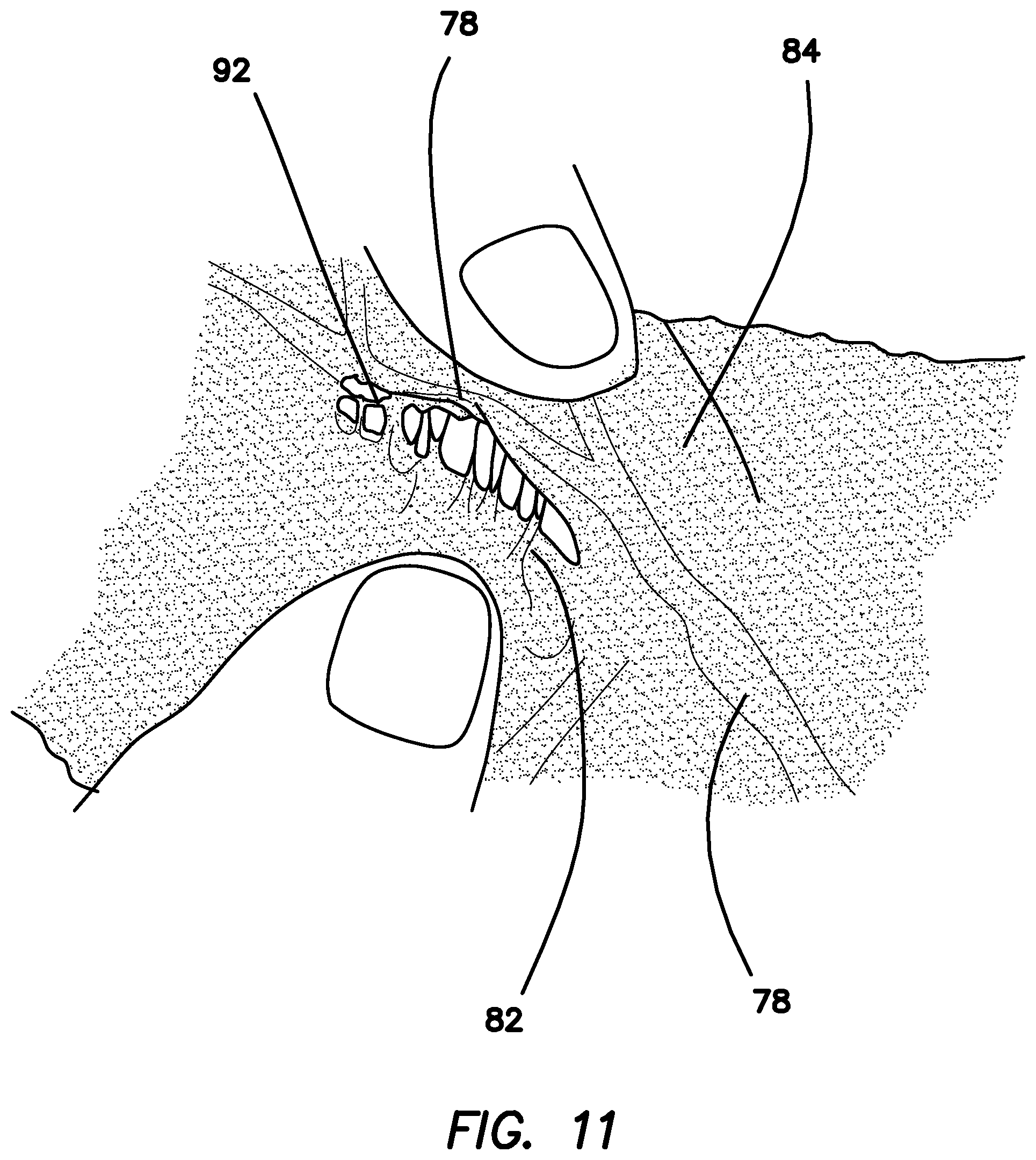
XML
uspto.report is an independent third-party trademark research tool that is not affiliated, endorsed, or sponsored by the United States Patent and Trademark Office (USPTO) or any other governmental organization. The information provided by uspto.report is based on publicly available data at the time of writing and is intended for informational purposes only.
While we strive to provide accurate and up-to-date information, we do not guarantee the accuracy, completeness, reliability, or suitability of the information displayed on this site. The use of this site is at your own risk. Any reliance you place on such information is therefore strictly at your own risk.
All official trademark data, including owner information, should be verified by visiting the official USPTO website at www.uspto.gov. This site is not intended to replace professional legal advice and should not be used as a substitute for consulting with a legal professional who is knowledgeable about trademark law.