Therapeutic peptides
Wucherpfennig , et al. October 6, 2
U.S. patent number 10,793,633 [Application Number 15/211,758] was granted by the patent office on 2020-10-06 for therapeutic peptides. This patent grant is currently assigned to Dana-Farber Cancer Institute, Inc.. The grantee listed for this patent is Dana-Farber Cancer Institute, Inc.. Invention is credited to Glenn Dranoff, Bettina Franz, Christopher Harvey, F. Stephen Hodi, Kenneth F. May, Jr., Kai W. Wucherpfennig.


View All Diagrams
| United States Patent | 10,793,633 |
| Wucherpfennig , et al. | October 6, 2020 |
Therapeutic peptides
Abstract
The present disclosure provides, in part, compositions comprising peptides that immunospecifically bind to a defined binding partner, such as MHC class I polypeptide-related sequence A (MICA), or an epitope thereon. In some embodiments, the peptides comprise one or more complementarity determining regions relating to the complementarity regions shown in Table 1. The disclosure also provides methods of treating cancer in a subject using the compositions disclosed herein, and methods of isolating human antibodies from cancer patients following immunotherapy.
| Inventors: | Wucherpfennig; Kai W. (Brookline, MA), Franz; Bettina (Orem, UT), May, Jr.; Kenneth F. (Bozeman, MT), Dranoff; Glenn (Sudbury, MA), Hodi; F. Stephen (Framingham, MA), Harvey; Christopher (Boston, MA) | ||||||||||
|---|---|---|---|---|---|---|---|---|---|---|---|
| Applicant: |
|
||||||||||
| Assignee: | Dana-Farber Cancer Institute,
Inc. (Boston, MA) |
||||||||||
| Family ID: | 1000005095800 | ||||||||||
| Appl. No.: | 15/211,758 | ||||||||||
| Filed: | July 15, 2016 |
Prior Publication Data
| Document Identifier | Publication Date | |
|---|---|---|
| US 20170008962 A1 | Jan 12, 2017 | |
Related U.S. Patent Documents
| Application Number | Filing Date | Patent Number | Issue Date | ||
|---|---|---|---|---|---|
| 14025573 | Aug 2, 2016 | 9402905 | |||
| PCT/US2012/057839 | Sep 28, 2012 | ||||
| 61541921 | Sep 30, 2011 | ||||
| Current U.S. Class: | 1/1 |
| Current CPC Class: | C07K 16/2833 (20130101); C07K 16/22 (20130101); A61K 39/3955 (20130101); C07K 16/06 (20130101); C07K 16/1282 (20130101); A61K 45/06 (20130101); A61K 31/365 (20130101); C07K 2317/565 (20130101); C07K 2317/76 (20130101); A61K 31/16 (20130101); A61K 31/325 (20130101); C07K 2317/21 (20130101); C07K 2317/92 (20130101); C07K 2317/73 (20130101); C07K 2317/10 (20130101); A61K 39/39558 (20130101) |
| Current International Class: | A61K 39/00 (20060101); C07K 16/06 (20060101); A61K 39/395 (20060101); C07K 16/28 (20060101); C07K 16/12 (20060101); C07K 16/22 (20060101); A61K 45/06 (20060101); A61K 31/365 (20060101); A61K 31/325 (20060101); A61K 31/16 (20060101) |
References Cited [Referenced By]
U.S. Patent Documents
| 4816567 | March 1989 | Cabilly et al. |
| 5122464 | June 1992 | Wilson et al. |
| 5225539 | July 1993 | Winter |
| 5530101 | June 1996 | Queen et al. |
| 5545403 | August 1996 | Page |
| 5545405 | August 1996 | Page |
| 5545806 | August 1996 | Lonberg et al. |
| 5545807 | August 1996 | Surani et al. |
| 5569825 | October 1996 | Lonberg et al. |
| 5585089 | December 1996 | Queen et al. |
| 5591639 | January 1997 | Bebbington |
| 5624821 | April 1997 | Winter et al. |
| 5625126 | April 1997 | Lonberg et al. |
| 5633425 | May 1997 | Lonberg et al. |
| 5648260 | July 1997 | Winter et al. |
| 5658759 | August 1997 | Bebbington |
| 5661016 | August 1997 | Lonberg et al. |
| 5677425 | October 1997 | Bodmer et al. |
| 5693762 | December 1997 | Queen et al. |
| 5714350 | February 1998 | Co et al. |
| 5766886 | June 1998 | Studnicka et al. |
| 5869046 | February 1999 | Presta et al. |
| 5939598 | August 1999 | Kucherlapati et al. |
| 5981216 | November 1999 | Kenten et al. |
| 5998144 | December 1999 | Reff et al. |
| 6075181 | June 2000 | Kucherlapati et al. |
| 6091001 | June 2000 | Jakobovits et al. |
| 6114598 | September 2000 | Kucherlapati et al. |
| 6121022 | September 2000 | Presta et al. |
| 6165745 | December 2000 | Ward et al. |
| 6180370 | January 2001 | Queen et al. |
| 6194551 | February 2001 | Idusogie et al. |
| 6277375 | August 2001 | Ward |
| 6344203 | February 2002 | Sandrin et al. |
| 6350861 | February 2002 | Co et al. |
| 6737056 | May 2004 | Presta |
| 7317091 | January 2008 | Lazar et al. |
| 7771718 | August 2010 | Spies et al. |
| 7959916 | June 2011 | Spies et al. |
| 8101720 | January 2012 | Lazar et al. |
| 8182809 | May 2012 | Wu |
| 9402905 | August 2016 | Wucherpfennig |
| 10106611 | October 2018 | Wucherpfennig et al. |
| 10279021 | May 2019 | Dranoff et al. |
| 2003/0022450 | January 2003 | Pan et al. |
| 2003/0099647 | May 2003 | Deshpande et al. |
| 2003/0118592 | June 2003 | Ledbetter et al. |
| 2003/0133939 | June 2003 | Ledbetter et al. |
| 2003/0153043 | August 2003 | Carr et al. |
| 2003/0165835 | September 2003 | Spies et al. |
| 2004/0067503 | April 2004 | Tan et al. |
| 2004/0115198 | June 2004 | Spies et al. |
| 2005/0053608 | March 2005 | Weber et al. |
| 2005/0059087 | March 2005 | Weber et al. |
| 2005/0158307 | July 2005 | Spies et al. |
| 2005/0233391 | October 2005 | Spies et al. |
| 2006/0024297 | February 2006 | Wood et al. |
| 2006/0228349 | October 2006 | Acton et al. |
| 2006/0246071 | November 2006 | Green et al. |
| 2007/0248607 | October 2007 | Spies et al. |
| 2008/0095803 | April 2008 | Mekalanos |
| 2008/0148432 | June 2008 | Abad |
| 2009/0022644 | January 2009 | Sweredjuk |
| 2009/0175821 | July 2009 | Bridon et al. |
| 2009/0226447 | September 2009 | Boone et al. |
| 2009/0252729 | October 2009 | Farrington et al. |
| 2010/0111973 | May 2010 | Dranoff et al. |
| 2010/0189711 | July 2010 | Dranoff et al. |
| 2010/0261269 | October 2010 | June et al. |
| 2011/0060120 | March 2011 | Obeid |
| 2011/0311561 | December 2011 | Martin, Jr. et al. |
| 2012/0100182 | April 2012 | Mooney |
| 2012/0315287 | December 2012 | Wu |
| 2013/0202707 | August 2013 | Ali et al. |
| 2014/0004112 | January 2014 | Wucherpfennig et al. |
| 2014/0027630 | January 2014 | Musselman |
| 2014/0037630 | February 2014 | Dranoff et al. |
| 2015/0071862 | March 2015 | Sabatino et al. |
| 2016/0027305 | January 2016 | Inabu et al. |
| 2016/0046716 | February 2016 | Wucherpfennig et al. |
| 2017/0000867 | January 2017 | Dranoff et al. |
| 2017/0022275 | January 2017 | Wucherpfennig et al. |
| 2017/0198054 | July 2017 | Harvey et al. |
| 102911270 | Feb 2013 | CN | |||
| 0 154 316 | Sep 1985 | EP | |||
| 0 401 384 | Dec 1990 | EP | |||
| 1 176 195 | Jan 2002 | EP | |||
| 2336180 | Jun 2011 | EP | |||
| 2008-543774 | Dec 2008 | JP | |||
| WO 88/07054 | Mar 1988 | WO | |||
| WO 88/08089 | Oct 1988 | WO | |||
| WO 91/10741 | Jul 1991 | WO | |||
| WO 94/02602 | Feb 1994 | WO | |||
| WO 94/29351 | Dec 1994 | WO | |||
| WO 96/32478 | Oct 1996 | WO | |||
| WO 96/33735 | Oct 1996 | WO | |||
| WO 97/34631 | Sep 1997 | WO | |||
| 98/19167 | May 1998 | WO | |||
| WO 98/24893 | Jun 1998 | WO | |||
| WO 99/051642 | Oct 1999 | WO | |||
| WO 99/54342 | Oct 1999 | WO | |||
| WO 00/42072 | Jul 2000 | WO | |||
| WO 01/58957 | Aug 2001 | WO | |||
| WO 02/06919 | Jan 2002 | WO | |||
| 02/068615 | Sep 2002 | WO | |||
| WO 03/035835 | May 2003 | WO | |||
| WO 03/041600 | May 2003 | WO | |||
| WO 2003/074679 | Sep 2003 | WO | |||
| 03/089616 | Oct 2003 | WO | |||
| WO 2004/016750 | Feb 2004 | WO | |||
| WO 2004/029207 | Apr 2004 | WO | |||
| WO 2004/035752 | Apr 2004 | WO | |||
| WO 2004/063351 | Jul 2004 | WO | |||
| WO 2004/074455 | Sep 2004 | WO | |||
| WO 2004/099249 | Nov 2004 | WO | |||
| WO 2005/040217 | May 2005 | WO | |||
| WO 2005/070963 | Aug 2005 | WO | |||
| WO 2005/092925 | Oct 2005 | WO | |||
| WO 2006/020114 | Feb 2006 | WO | |||
| WO 2006/068953 | Jun 2006 | WO | |||
| WO 2006/133396 | Dec 2006 | WO | |||
| 2007/055926 | May 2007 | WO | |||
| WO 2007/055926 | May 2007 | WO | |||
| 2008/036981 | Mar 2008 | WO | |||
| WO 2010/069532 | Jun 2010 | WO | |||
| WO 2011/014469 | Feb 2011 | WO | |||
| WO 2011/063336 | May 2011 | WO | |||
| WO 2013/049517 | Apr 2013 | WO | |||
| 2013/117647 | Aug 2013 | WO | |||
| WO 2014/144791 | Sep 2014 | WO | |||
Other References
|
MacCallum et al (J. Mol. Biol. (1996) 262, 732-745) (Year: 1996). cited by examiner . Vajdos et al (JMB, 2002 320, 415-428) (Year: 2002). cited by examiner . Wu et al (J. Mol. Biol. 1999, 294, 151-162) (Year: 1999). cited by examiner . Edwards et al (JMB, 2003, 334: 103-118) (Year: 2003). cited by examiner . Lloyd et al (Protein Engineering, Eng. Design & Selection, 2009, 22(3): 159-168) (Year: 2009). cited by examiner . Goel et al (J. Immunol., 2004, 173: 7358-7367) (Year: 2004). cited by examiner . Khan and Salunke (J. Immunol, 2014, 192: 5398-5405) teach (Year: 2014). cited by examiner . Poosarla et al (Biotechn. Bioeng., 2017, 114(6): 1331-1342) (Year: 2017). cited by examiner . Torres and Casadevall (Trend. Immunol., 2008, 29(2): 91-97) (Year: 2008). cited by examiner . Spear et al (Cancer Immunity, 2013, 13: 1-14) (Year: 2013). cited by examiner . Zwirner et al (Immunologia, 2006, 25: 25-38) (Year: 2006). cited by examiner . Justus et al (J. Translational Medicine, 2017, 15: 204, pp. 1-14) (Year: 2017). cited by examiner . Mei et al., "Expression of NKG2D ligands in multidrug-resistant nasopharyngeal carcinoma cell line CNE2/DDP and their effects on cytotoxicity of natural killer cells," Nan Fang Yi Ke Da Xue Xue Bao.,27 (6):887-889 (2007). cited by applicant . Nausch et al., "NKG2D ligands in tumor immunity," Oncogene, 27: 5944-5958 (2008). cited by applicant . Nelson et al., "Cancer cells engineered to secrete granulocyte-macrophage colony-stimulating factor using ex vivo gene transfer as vaccines for the treatment of genitourinary malignancies," Cancer Chemother. Pharmacol., 46 (Suppl.): S67-72 (2000). cited by applicant . Pantazes, R.J. et al., "OptCDR: a general computational method for the design of antibody complementaritydetermining regions for targeting epitope binding," Protein Engineering, Design & Selection, vol. 23 (11 ), pp. 849-858 (2010). cited by applicant . Pende et al.,"Major histocompatibility complex class I-related chain A and UL16-binding protein expression on tumor cell lines of different histotypes: analysis of tumor susceptibility to NKG2D-dependent natural killer cell cytotoxicity," Cancer Res., 62 (21): 6178-6186 (2002). cited by applicant . Pettersen et al., "CD47 signals T cell death," J. Immunol., 162 (12): 7031-7040 (1999). cited by applicant . Phumyen, A. et al., "Improved Binding Activity of Antibodies Against Major Histocompatibility Complex Class I Chain-Related Gene A by Phage Display Technology for Cancer-Targeted Therapy," Journal of Biomedicine and Biotechnology, vol. 2012(597647) 8 pages, (2012). cited by applicant . Ponsel, Dirk et al., "High Affinity, Developability and Functional Size: The Holy Grail of Combinatorial Antibody Library Generation," Molecules, vol. 16:, pp. 3675-3700 (2011). cited by applicant . Rader, C. et al., "A Phage Display Approach for Rapid Antibody Humanization: Designed Combinatorial V Gene Libraries," PNAS, USA, vol. 95, pp. 8910-8915 (1998). cited by applicant . Riemer , A. et al. "Matching of trastuzumab (Herceptin) epitope mimics onto the surface of Her-2/neu--a new method of epitope definition", Mol. Immunol, vol. 42:, pp. 1121-1124 (2005). cited by applicant . Rudikoff , S. et al., "Single amino acid substitution altering antigen-binding specificity," Proc Natl Acad Sci USA, vol. 79, pp. 1979-1983 (1982 ). cited by applicant . Saijo, N., "What are the reasons for negative phase III trials of molecular-target-based drugs?",; Cancer Sci., vol. 95(10), pp. 772-776 (2004). cited by applicant . Salih, Helmut R. et al., "Cutting Edge: Down-Regulation of MICA on Human Tumors by Proteolytic Shedding," The Journal of Immunology, vol. 169, pp. 4098-4102 (2002). cited by applicant . Salih, Helmut R. et al., "Functional expression and release of ligands for the activating immunoreceptor NKG2D in leukemia," Blood, vol. 102(4), pp. 1389-1396 (2003). cited by applicant . Schoenfeld, J. et al., "Active Immunotherapy Induces Antibody Responses That Target Tumor Angiogenesis," Microenvironment and Immunology, Cancer Research, vol. 70(24), pp. 10150-10160 (2010). cited by applicant . Sircar, A. et al., "Rosetta Antibody: antibody variable region homology modeling server," Nucleic Acids Research, vol. 37, pp. W474-W479 (2009). cited by applicant . Steinle, A. et al., "Diversification, expression, and gammadelta T cell recognition of evolutionarily distant members of the MIC family of major histocompatibility complex class I-related molecules," Proc. Natl. Acad. Sci. USA, vol. 95, pp. 12510-12515 (1998). cited by applicant . Suarez-Alvarez, B. et al., "Identification of epitopes and immunodominant regions on the MICA protein defined by alloantibodies from kidney transplant patients," Transplantation, Williams and Wilkins, GB, vol. 88 (3) Suppl, pp. S68-S77( Aug. 15, 2009). cited by applicant . Tang, B. et al., "Evaluation of human major histocompatibility complex class I chain-related A as a potential target for tumor imaging," Cancer Letters, New York, NY, US, vol. 263 (1), pp. 99-106, Jan. 30, 2008. cited by applicant . Thom, George et al., "Probing a protein-protein interaction by in vitro evolution," PNAS, vol. 103(20):7619-7624 (2006). cited by applicant . Vajdos, FF et al., "Comprehensive functional maps of the antigen-binding site of an anti-ErbB2 antibody obtained with shotgun scanning mutagenesis," J Mol Biol. vol. 320(2) pp. 415-428 (2002). cited by applicant . Vitetta, Ellen S. et al., "Monoclonal Antibodies as Agonists: An Expanded Role for Their Use in Cancer Therapy," Cancer Research, vol. 54:5301-5309 (1994). cited by applicant . Weber, "Review: anti-CTLA-4 antibody ipilimumab: case studies of clinical response and immune-related adverse events," Oncologist, 12(7): 864-872 (2007). cited by applicant . Whiteside, T. et al., "Antigen-Processing Machinery in Human Dendritic Cells; Up-regulation by Maturation and Down-Regulation by Tumor Cells," J. Immunol., vol. 173, pp. 1526-1534 (2004). cited by applicant . Wongsena et al "Production and characterization of monoclonal antibodies against major histocompatibility complex class 1 chain-related gene A," Tissue Antigens, 72(5):431-440 (2008). cited by applicant . Wu et al., "Prevalent expression of the immunostimulatory MHC class I chain-related molecule is counteracted by shedding in prostate cancer," J. Clin. Invest.,114 (4): 560-568 (2004). cited by applicant . Zou, Yizhou et al., "MICA is a Target for Complement-Dependent Cytotoxicity With Mouse Monoclonal Antibodies and Human Alloantibodies," Human Immunology, vol. 63, pp. 30-39 (2002). cited by applicant . Zwirner, N. et al., "Immunobiology of the human MHC class I chain-related gene A (MICA): from 13 transplantation immunology to tumor immune escape," Immunologia, vol. 25(1), pp. 25-38, (Jan.-Mar. 2006). cited by applicant . Ali, O. A. et al., "Infection-mimicking materials to program dendritic cells in situ," Nature Materials, 8:151-158 (2009). cited by applicant . Ali et al. "In Situ Regulation of DC Subsets and T Cells Mediates Tumor Regression in Mice," Science Translation Medicine, 1, 8ra19, 12 pages (2009). cited by applicant . Almagro et al., "Humanization of antibodies," Frontiers in Bioscience, 13:1619-33 (2008). cited by applicant . Altschul et al. "Basic Local Alignment Search Tool," J. Mol. Biol., 215:403-10 (1990). cited by applicant . Altschul et al., "Gapped BLAST and PSI-BLAST: a new generation of protein database search programs," Nucleic Acids Res. 25(17):3389-3402 (1997). cited by applicant . Amanna et al., "Duration of Humoral Immunity to Common Viral and Vaccine Antigens," N. Engl. J. Med., 357:1903-1915, (2007). cited by applicant . Andrade et al., "Adsorption of complex proteins at interfaces," Pure and Appl. Chem., 64(11):1777-1781 (1992). cited by applicant . Balazs et al., "Antibody-based protection against HIV infection by vectored immunoprophylaxis," Nature, 481(7379):81-4 (2011). cited by applicant . Balmana et al. "BRCA in breast cancer: ESMO Clinical Recommendations," Annals of Oncology 20(supplement 4):iv19-20 (2009). cited by applicant . Banchereau et al. "Long-Term Human B Cell Lines Dependent on Interleukin-4 and Antibody to CD40," Science, 251:70 (1991). cited by applicant . Bendsten et al. "Improved Prediction of Signal Peptides: SignaIP 3.0," J Mol Biol 340(4):783-95 (2004). cited by applicant . Benjamin et al. "The Antigenic Structure of Proteins: A Reappraisal," Ann Rev Immunol, 2:67-101 (1984). cited by applicant . Bird et al. "Single-Chain Antigen-Binding Proteins," Science 242:423-426 (1988). cited by applicant . Buisman et al., "Long-term presence of memory B-cells specific for different vaccine components," Vaccine, 28:179-186 (2010). cited by applicant . Boerner et al, "Production of antigen-specific human monoclonal antibodies from in vitro-primed human splenocytes," J Immunol, 147(1):86-95 (1991). cited by applicant . Bordo et al. "Suggestions for "Safe" Residue Substitutions in Site-Directed Mutagenesis," J. Mol. Biol., 217:721-729 (1991). cited by applicant . Caine et al., "Recombinant Human Phenylethanolamine N-Methyltransferase: Overproduction in Escherichia coli, Purification, and Characterization," Protein Expression and Purification, 8(2):159-166 (1996). cited by applicant . Canfield "The Binding Affinity of Human IgG for its High Affinity Fc Receptor Is Determined by Multiple Amino Acids in the CH2 Domain and Is Modulated by the Hinge Region," J. Exp. Med. 173:1483 (1991). cited by applicant . Cao et al., "An optimized assay for the enumeration of antigen-specific memory B cells in different compartments of the human body," Journal of Immunological Methods, 358:56-65 (2010). cited by applicant . Champe et al. "Monoclonal Antibodies That Block the Activity of Leukocyte Function-associated Antigen 1 Recognize Three Discrete Epitopes in the Inserted Domain of CD11a," The Journal of Biological Chemistry, 270:1388-1394 (1995). cited by applicant . Cheung et al., "Epitope-Specific Antibody Response to the Surface Antigen of Duck Hepatitis B Virus in Infected Ducks," Virology, 176:546 (1990). cited by applicant . Choi et al., "Evolutionary conservation in multiple faces of protein interaction," Proteins: Structure, Function, and Bioinformatics, 77(1):14-25 (2009). cited by applicant . Chothia et al. "Canonical Structures for the Hypervariable Regions," J Mol Biol, 196:901-917 (1987). cited by applicant . Clackson et al, "Making antibody fragments using phage display libraries," Nature, 352 624-628 (1991). cited by applicant . Cole et al, "The EBV-Hybridoma Technique and its Application to Human Lung Cancer," Monoclonal Antibodies and Cancer Therapy, pp. 77-96 (1985). cited by applicant . Corada et al., Monoclonal antibodies directed to different regions of vascular endothelial cadherin extracellular domain affect adhesion and clustering of the protein and modulate endothelial permeability, Blood,97:1679-84 (2001). cited by applicant . Corti et al., "Analysis of Memory B Cell Responses and Isolation of Novel Monoclonal Antibodies with Neutralizing Breadth from HIV-1-Infected Individuals," PLoS One, 5:e8805 (2010). cited by applicant . Cox et al., "Glycan optimization of a human monoclonal antibody in the aquatic plant Lemna minor," Nat. Biotechnol, 24(12):1591-7 (2006). cited by applicant . Cox, J. P. L. et al. "A Directory of Human Germ-line VH Segments Reveals a Strong Bias in their Usage," Eur. J Immunol., 24:827-836 (1994). cited by applicant . Crotty et al., "Cutting Edge: Long-Term B Cell Memory in Humans after Smallpox Vaccination," J. Immunol., 171:4969-4973 (2003). cited by applicant . Cunningham et al. "High-Resolution Epitope Mapping of hGH-Receptor Interactions by Alanine-Scanning Mutagenesis," Science, 244:1081-1085 (1989). cited by applicant . Dall'Acqua et al. "Increasing the Affinity of a Human IgG1 for the Neonatal Fc Receptor: Biological Consequences", Journal of Immunology, 169:5171-5180 (2002). cited by applicant . Dall'Acqua et al., "Properties of Human IgG1s Engineered for Enhanced Binding to the Neonatal Fc Receptor (FcRn)", Journal of Biological Chemistry 281:23514-23524 (2006). cited by applicant . De Genst et al., "Antibody repertoire development in camelids ", Dev Comp Immunol; 30:187-98 (2006). cited by applicant . De Ridder, G. et al. "Cell-Surface GRP78 and its Antibodies: Pathologic and Therapeutic Roles in Cancer", 2010. Retrieved from the Internet: URL:http://dukespace.lib.duke.edu/dspace/bitstream/handle/10161 /3805/deRidder_duke_0066D_ 10579.pdf?sequence=1. cited by applicant . Duncan et al., "Localization of the binding site for the human high-affinity Fe receptoron IgG," Nature, 332:563 (1988). cited by applicant . Emini et al. "Antigenic Conservation and Divergence between the Viral-Specific Proteins of Poliovirus Type 1 and Various Picornaviruses", Virology 140 13-20 (1985). cited by applicant . Fang et al. "Stable antibody expression at therapeutic levels using the 2A peptide", Nature Biotechnology 23, 584-590 ((2005). cited by applicant . Fecteau et al., "Peripheral blood CD27+ IgG+ B cells rapidly proliferate and differentiate into immunoglobulin-secreting cells after exposure to low CD154 interaction", Immunology, 128:e353-e365 30 (2009). cited by applicant . Fields et al., Chapter 3 "Synthetic Peptides: A User's Guide", pp. 77-183 (1992). cited by applicant . Fishwild et al, "High-avidity human IgGk monoclonal antibodies from a novel strain of minilocus transgenic mice", Nature Biotechnology 14, 845-51 (1996). cited by applicant . Franz et al. "Ex vivo characterization and isolation of rare memory B cells with antigen tetramers", Blood, 118(2):348-357 (2011). cited by applicant . Getzoff et al. "The Chemistry and Mechanism of Antibody Binding to Protein Antigens", Advances in Immunology 43:1-98, (1988). cited by applicant . Gillies S.D. et al., "Improving the Efficacy of Antibody-Interleukin 2 Fusion Proteins by Reducing Their Interaction with Fc Receptors", Cancer Res. 59:2159-66 (1999). cited by applicant . Gong et al., "A protein domain interaction interface database: InterPare", BMC: Bioinformatics, 6:1471-2105 (2007). cited by applicant . Gonzalez, G. et al., "A novel cancer vaccine composed of human-recombinant epidermal growth factor linked to a carrier protein: Report of a pilot clinical trial," Annals of Oncology, 9:431-435 (1998). cited by applicant . Groh, V. et al., "Recognition of Stress-Induced MHC Molecules by Intestinal Epithelial gammadelta T cells," Science 279:1737-1740 (1988). cited by applicant . Guo et al., "Protein tolerance to random amino acid change", Proc. Natl. Acad. Sci., USA, 101(25):9205-9210 (2004). cited by applicant . Henn et al., "Modulation of Single-Cell IgG Secretion Frequency and Rates in Human Memory B Cells by CpG DNA, CD40L, IL-21, and Cell Division", J. Immunol., 183:31777-3187 (2009). cited by applicant . Hinton et al. "An Engineered Human IgG1 Antibody with Longer Serum Half-Life", Journal of Immunology 176:346-356 (2006). cited by applicant . Hinton et al., "Engineered Human IgG Antibodies with Longer Serum Half-lives in Primates", J. Biol. Chem. 279(8): 6213-6216 (2004). cited by applicant . Hofer et al., Adaptation of humoral memory, Immunological Reviews, 211:295-302 (2006). cited by applicant . Hofmann et al. "A database of membrane spanning proteins segments", Biol Chem Hoppe-Seyler 374,166 (1993). cited by applicant . Hoogenboom et al. "Human Antibodies from Synthetic Repertoires of Germline VH Gene Segments Rearranged in Vitro", J Mol Biol, 227 381 (1991). cited by applicant . Hopp et al. "A computer program for predicting protein antigenic determinants", Molecular Immunology 20 483-489 (1983). cited by applicant . Hopp et al. "Prediction of protein antigenic determinants from amino acid sequences", Proc Natl Acad Sci USA 78 3824-3828 (1981). cited by applicant . Hopp, Methods for identifying antigenic determinants and other interaction sites, Immunol Methods 88 1-18 (1986). cited by applicant . Huergo-Zapico L. et al. "Expression of ERp5 and GRP78 on the membrane of chronic lymphocytic leukemia cells: association with soluble MICA shedding", Cancer Immunology, Immunotherapy, 61(8):1201-1210 (2012). cited by applicant . Huggins et al., "CpG DNAactivation and plasma-cell differentiation of CD27_ naive human B cells", Blood, 109:1611-1619 (2007). cited by applicant . Huston et al. "Protein engineering of antibody binding sites: Recovery of specific activity in an antidigoxin single-chain Fv analogue produced in Escherichia coli", Proc. Natl. Acad. Sci. USA 85:5879-5883 (1988). cited by applicant . Jaeger et al., "Improved predictions of secondary structures for RNA", Proc. Natl. Acad. Sci. USA 86:7706-10 (1989). cited by applicant . Jaeger et al., "Predicting Optimal and Suboptimal Secondary Structure for RNA", Methods Enzymol. 183:281-306 (1989). cited by applicant . Jameson, et al. The antigenic index: a novel algorithm for predicting antigenic determinants, Comput Appl Biosci 4(1) 181-186 (1988). cited by applicant . Jespers et al., "Guiding the Selection of Human Antibodies from Phage Display Repertoires to a Single Epitope of an Antigen", Biotechnology 12:899, (1994). cited by applicant . Jiang, et al., "TLR9 stimulation drives naive B cells to proliferate and to attain enhanced antigen presenting function", Eur. J. Immunol., 37:2205-2213 (2007). cited by applicant . Johnson et al. A Structural Basis for Sequence Comparisons: An Evaluation of Scoring Methodologies, J. Mol. Biol. 233:716-738 (1993). cited by applicant . Jones, P. et al. "Replacing the complementarity-determining regions in a human antibody with those from a mouse", Nature 321:522-525 (1986). cited by applicant . Jourdan et al., "An in vitro model of differentiation of memory B cells into plasmablasts and plasma cells including detailed phenotypic and molecular characterization", Blood, 114:5173-5181 (2009). cited by applicant . Kalos M, et al., "T Cells with Chimeric Antigen Receptors Have Potent Antitumor Effects and Can Establish Memory in Patients with Advanced Leukemia", Sci Transl Med. Aug. 2010;3 (95), (2011). cited by applicant . Kaneko et al., "Anti-Inflammatory Activity of Immunoglobulin G Resulting from Fc Sialylation", Science 313:670-673 (2006). cited by applicant . Kettleborough et al, "Humanization of a mouse monoclonal antibody by CDR-grafting: the importance of framework residues on loop conformation," Protein Eng 4(7) 773 83 (1991). cited by applicant . Kim et al., "Targeting Heat Shock Proteins on Cancer Cells: Selection, Characterization, and Cell-Penetrating Properties of a Peptidic GRP78 Ligand," Biochemistry 45:9434-9444 (2006). cited by applicant . Kirkland et al., "Analysis of the Fine Specificity and Cross-Reactivity of Monoclonal Anti-Lipid A Antibodies", J Immunol. 137:3614 (1986). cited by applicant . Klinman "CpG DNA as a vaccine adjuvant", Expert Review Vaccines 2(2):305-15 (2003). cited by applicant . Kostelny et al., "Formation of a Bispecific Antibody by the Use of Leucine Zippers", Journal of Immunol. 148, 1547-1553 (1992). cited by applicant . Kratz et al. "Native display of complete foreign protein domains on the surface of hepatitis B virus capsids", Proc Natl Acad Sci USA. 96(5):1915-1920 (1999). cited by applicant . Krogh et al. "Predicting transmembrane protein topology with a hidden Markov model Application to complete genomes", Journal of Molecular Biology, 305(3) 567-580, (2001). cited by applicant . Kunkel et al., "Plasma-Cell Homing", Nat. Rev. Immunol., 3:822-829 (2003). cited by applicant . Kyte et al. "A Simple Method for Displaying the Hydropathic Character of a Protein", J Mol Biol 157 105-132 (1982). cited by applicant . Lanzavecchia et al., "Human B cell memory", Curr. Opin. Immunol. 21:298-304 (2009). cited by applicant . Li et al., "Optimization of humanized IgGs in glycoengineered Pichia pastoris", Nature Biotechnology 24(2):210-215 (2006). cited by applicant . Liu et al. "Perturbation of NK cell peripheral homeostasis accelerates prostate carcinoma metastasis", The Journal of Clinical Investigation 123(10):4410-4422 (2013). cited by applicant . Liu R. et al. "Monoclonal Antibody against Cell Surface GRP78 as a Novel Agent in Suppressing PI3K/AKT Signaling, Tumor Growth, and Metastasis", Clinical Cancer Research, 19(24):6802-6811 (2013). cited by applicant . Lonberg "Human antibodies from transgenic animals", Nature Biotechnology 23(9): 1117-1125, (2005). cited by applicant . Lonberg et al, "Antigen-specific human antibodies from mice comprising four distinct genetic modifications", Nature 368 856-859 (1994). cited by applicant . Lonberg et al, "Human Antibodies from Transgenic Mice", International Reviews of Immunology, 13 65-93 (1995). cited by applicant . Makabe et al., "Thermodynamic Consequences of Mutations in Vernier Zone Residues of a Humanized Anti-human Epidermal Growth Factor Receptor Murine Antibody, 528", Journal of Biological Chemistry, 283:1156-1166 (2008). cited by applicant . Marks et al, "By-Passing Immunization: Building High Affinity Human Antibodies by Chain Shuffling", Bio/Technology 10, 779-783 (1992). cited by applicant . Marks et al, "Human Antibodies from V-gene Libraries Displayed on Phage", J Mol Biol, 222 581-597 (1991). cited by applicant . McCafferty et al, "Phage antibodies: filamentous phage displaying antibody variable domains", Nature, 348 552-554 (1990). cited by applicant . Meyers E. et al. "Optimal alignments in linear space", CABIOS, 4:11-17 (1989). cited by applicant . Milone et al. "Chimeric Receptors Containing CD137 Signal Transduction Domains Mediate Enhanced Survival of T Cells and Increased Antileukemic Efficacy In Vivo", Mol. Ther. 17:1453 (2009). cited by applicant . Moldenhauer et al., "Identity of HML-1 Antigen on Intestinal Intraepithelial T Cells and of B-ly7 Antigen on Hairy Cell Leukaemia", Scand. J Immunol. 32:77 (1990). cited by applicant . Morel et al., "Monoclonal Antibodies to Bovine Serum Albumin: Affinity and Specificity Determinations", Mol. Immunol. 25(1):7 (1988). cited by applicant . Morrison et al, "Chimeric human antibody molecules: Mouse antigen-binding domains with human constant region domains", Proc Natl Acad Sci USA, 81 6851-6855 (1984). cited by applicant . Morrison et al., "Genetically Engineered Antibody Molecules," Advances in Immunology, 44:65-92 (1988). cited by applicant . Morrison, "Success in Specification", Nature 368, 812-13 (1994). cited by applicant . Nassal, M. et al., "Development of hepatitus B virus capsids into a whole-chain protein antigen display platform: New particulate Lyme disease vaccines," International Journal of Medical Microbiology, 298:135-142 (2008). cited by applicant . Nechansky et al., "Compensation of endogenous IgG mediated inhibition of antibody-dependent cellular cytotoxicity by glyco-engineering of therapeutic antibodies", Molecular Immunology 44(7): 1815-1817 (2007). cited by applicant . Needleman et al. "A General Method Applicable to the Search for Similarities in the Amino Acid Sequence of Two Proteins", J. Mol. Biol. 48:443-453 (1970). cited by applicant . Neuberger, "Generating high-avidity human Mabs in mice", Nature Biotechnology 14,826 (1996). cited by applicant . Odendahl et al., "Generation of migratory antigen-specific plasma blasts and mobilization of resident plasma cells in a secondary immune response", Blood, 105:1614-1621 (2005). cited by applicant . Padlan, "A possible procedure for reducing the immunogenicity of antibody variable domains while preserving their ligand-binding properties", Molecular Immunology, 28 489-498 (1991). cited by applicant . Padlan, "Anatomy of the Antibody Molecule", Molecular Immunology 31(3) 169-217 (1994). cited by applicant . Padlan, "X-Ray Crystallography of Antibodies", Advances in Protein Chemistry 49:57-133 (1996). cited by applicant . Park et al., "Prediction of protein-protein interaction types using association rule based classification", BMC: Bioinformatics, 10:36 (2009); doi: 10.1186/1471-2105-10-36, 15 pages. cited by applicant . Pashine et al. "Targeting the innate immune response with improved vaccine adjuvants", Nature Med. 11(4):S63-S68 (2005). cited by applicant . Pearson et al. "Improved tools for biological sequence comparison", Proc. Natl. Acad. Sci. USA 85:2444 (1988). cited by applicant . Pearson, "Rapid and Sensitive Sequence Comparison with FASTP and FASTA", Meth. Enzymology, 183:63-98 (1990). cited by applicant . Pluckthun, "Mono- and Bivalent Antibody Fragments Produced in Escherichia coli: Engineering, Folding and Antigen Binding", Immunol. Reviews 130:151-188 (1992). cited by applicant . Qi, J. et al., "Immobilized MICA Could Expand Human V .gamma.1 .gamma..delta. T cells In Vitro that Displayed Major Histocompatibility Comlex Class I Chain-Related A-Dependent Cytotoxicity to Human Epithelial Carcinomas," Scandinavian Journal of Immunology, 58:211-220 (2003). cited by applicant . Queen, C. et al. "A humanized antibody that binds to the interleukin 2 receptor", Proc. Natl. Acad. Sci. USA 86:10029-10033 (1989). cited by applicant . Riechmann, L. et al. "Reshaping human antibodies for therapy", Nature 332:323-327 (1998). cited by applicant . Sarmay et al. "Mapping and Comparison of the Interaction Sites on the Fc Region of IgG Responsible for Triggering Antibody Dependent Cellular Cytotoxicity (ADCC) through Different Types of Human Fc.gamma. Receptor", Molec. Immunol. 29 (5): 633-9 (1992). cited by applicant . Scallon et al., "Higher levels of sialylated Fc glycans in immunoglobulin G molecules can adversely impact functionality", Mol Immunol. 44(7): 1524-34 (2007). cited by applicant . Scatchard, "The attractions of proteins for small molecules and ions", Ann NY Acad Sci 51 660-672, (1949). cited by applicant . Scheid et al., "Broad diversity of neutralizing antibodies isolated from memory B cells in HIV-infected individuals", Nature, 458:636-640 (2009). cited by applicant . Shields, R.L. et al. "High Resolution Mapping of the Binding Site on Human IgG1 for FcgRI, FcgRII, FcgRIII, and FcRn and Design of IgG1 Variants with Improved Binding to the FcgR", J Biol. Chem. 276:6591-6604 (2001). cited by applicant . Shields, R.L. et al. "Lack of Fucose on Human IgG1 N-Linked Oligosaccharide Improves Binding to Human FcyRIII and Antibody-dependent Cellular Toxicity", J Biol. Chem. 277:26733-26740 (2002). cited by applicant . Skerra et al., "Bacterial expression of immunoglobulin fragments", Curr. Opinion in Immunol., 5:256-262 (1993). cited by applicant . Smith et al. "Comparison of Biosequences", Advances in Applied Mathematics, 2:482 (1981). cited by applicant . Songsivilai et al. "Bispecific antibody: a tool for diagnosis and treatment of disease", Clin. Exp. Immunol. 79:315-321 (1990). cited by applicant . Spear et al., "NKG2D ligands as therapeutic targets," Cancer Immunity, 13:1-14 (2013). cited by applicant . Stahli et al.,"Distinction of Epitopes by Monoclonal Antibodies", Methods in Enzymology 92:242-253 (1983). cited by applicant . Strohl, "Optimization of Fc-mediated effector functions of monoclonal Antibodies", Current Opinion in Biotechnology 20:685-691 (2009). cited by applicant . Tao et al. "Role of Carbohydrate in the Structure and Effector Functions Mediated by the Human IgG Constant Region", J. Immunol. 143:2595-2601 (1989). cited by applicant . Tao et al., "Structural Features of Human Immunoglobulin G that Determine Isotype-specitic Differences in Complement Activation", J. Exp. Med. 178:661 (1993). cited by applicant . Taylor, The Classification of Amino Acid Conservation, J. Theor. Biol. 119:205-218 (1986). cited by applicant . Tomlinson, I. M., et al. "The Repertoire of Human Germline VH Sequences Reveals about Fifty Groups of VH Segments with Different Hypervariable Loops" J. Mol. Biol. 227:776-798 (1992). cited by applicant . Umana et al. "Engineered glycoforms of an antineuroblastoma IgG1 with optimized antibodydependent cellular cytotoxic activity", Nat. Biotech. 17:176-180 (1999). cited by applicant . Verhoeyen et al, "Reshaping Human Antibodies: Grafting an Antilysozyme Activity", Science 239 1534 1536 (1988). cited by applicant . Vetter, C. S. et al., "Expression of Stress-induced MHC Class I Related Chain Molecules on Human Melanoma," The Journal of Investigative Dermatology, 118(4):600-605 (2002). cited by applicant . Wang, M. et al. "Role of the Unfolded Protein Response Regulator GRP78/BiP in Development, Cancer, and Neurological Disorders", Antioxidants and Redox Signaling 11(9): 2307-2316 (2009). cited by applicant . Wang et al., "Human immunoglobulin variable region gene analysis by single cell RT-PCR", J. Immunol. Methods, 244:217-225 (2000). cited by applicant . Ward et al., "Binding activities of a repertoire of single immunoglobulin variable domains secreted from Escherichia coli", Nature 341:544-546 (1989). cited by applicant . Wu et al., "Rational Design of Envelope Identifies Broadly Neutralizing Human Monoclonal Antibodies to HIV-1", Science, 329:856-861 (2010). cited by applicant . Yeung et al., "Engineering Human IgG1 Affinity to Human Neonatal Fc Receptor: Impact of Affinity Improvement on Pharmacokinetics in Primates", J Immunol, 182:7663-7671 (2010). cited by applicant . Yoshida et al., "Memory B and memory plasma Cells", Immunol. Rev., 237:117-139 (2010). cited by applicant . Yu et al. "Minimal Lipidation Stabilizes Protein-Like Molecular Architecture", J Am. Chem. Soc., 120(39):9979-9987 (1998). cited by applicant . Zapata et al , "Engineering linear F(ab')2 fragments for efficient production in Escherichia coli and enhanced antiproliferative activity", Protein Eng 8(10) 1057-1062 (1995). cited by applicant . Zuker, "On Finding All Suboptimal Foldings of an RNA Molecule", Science 244:48-52 (1989). cited by applicant . Araya, Carlos L. et al., "Deep mutational scanning: assessing protein function on a massive scale," Trends Biotechnol., vol. 29(9), pp. 435-442 (2011 ). cited by applicant . Barbas, S. et al., "Recognition of DNA by Synthetic Antibodies," J. Am. Chem. Soc., vol. 116 (5), pp. 2161-2162 (1994). cited by applicant . Beiboer, S. et al., "Guided Selection of a Pan Carcinoma Specific Antibody Reveals Similar Binding Characteristics yet Structural Divergence Between the Original Murine Antibody and its Human Equivalient," JMB, vol. 296, pp. 833-849 (2000). cited by applicant . Bergers et al., "Extrinsic regulators of epithelial tumor progression: metalloproteinases," Curro Opin. in Genetics and Development, vol. 10, pp. 120-127 (2000). cited by applicant . Bodey, B. et al., "Failure of Cancer Vaccines: The Significant Limitations of this Approach to Immunotherapy," Anticancer Research, vol. 20, pp. 2665-2676 (2000). cited by applicant . Brown, M. et al., "Tolerance of single, but not multiple, amino acid replacements in antibody VH CDR 2: a means of minimizing B cell wastage from somatic hypermutation?," J Immunol., vol. 156(9), pp. 3285-3291 (1996). cited by applicant . Casset, F. et al., "A peptide mimetic of an anti-CD4 monoclonal antibody by rational design," BBRC, vol. 307, pp. 198-205(2003). cited by applicant . Chen, C. et al., "Generation and Analysis of Random Point Mutations in an Antibody CDR2 Sequence: Many Mutated Antibodies Lose Their Ability to Bind Antigen," J. Exp. Med., vol. 176, pp. 855-866 (1992). cited by applicant . Chen, Y. et al, "Selection and analysis of an optimized anti-VEGF antibody: crystal structure of an affinity-matured Fab in complex with antigen," J. Mol. Biol., vol. 293, pp. 865-881 (1999). cited by applicant . Chin et al., "Immune intervention with monoclonal antibodies targeting CD152 (CTLA-4) for autoimmune and malignant diseases," Chang Gung Med J., 31 (1): 1-15 (2008). cited by applicant . Chothia, C. et al., "Conformations of Immunoglobulin hypervariable regions," Nature, vol. 342, pp. 877-883 (1989). cited by applicant . Chothia, C. et al., "Structural Repertoire of the Human VH Segments," J. Mol. Biol., vol. 227, pp. 799-817 (1992). cited by applicant . De Pascalis, R. et al, "Grafting of "abbreviated" complementarity-determining regions containing specificity-determining residues essential for ligand contact to engineer a less immunogenic humanized monoclonal antibody," The Journal of Immunology, vol. 169, pp. 3076-3084 (2002). cited by applicant . Dennis, C. "Off by a whisker", Nature, vol. 442, pp. 739-741 (2006). cited by applicant . Ditzel, H. et al., "Determinants of Polyreactivity in a Large Panel of Recombinant Human Antibodies from HIV-I Infection," The Journal of Immunology, vol. 157, pp. 739-749, (1996). cited by applicant . Doubrovina et al.,"Evasion from NK cell immunity by MHC class I chain-related molecules expressing colon adenocarcinoma," J. Immunol. 171 (12): 6891-6899 (2003). cited by applicant . Duquesnoy, Structurally based epitope analysis of major histocompatibility complex class I-related chain A (MICA) antibody specificity patterns, Human Immunology, vol. 69:826-832 (2008). cited by applicant . Extended European Search Report, EPI2835118.6, dated May 4, 2015, 9 pages. cited by applicant . Fonseca, C. et al., "Protein disulfide isomerases are antibody targets during immune-mediated tumor destruction", Blood, 1vol. 13, pp. 1681-1688 (2009). cited by applicant . Germain, C. et al., "Mhc Class I-Related Chain A Conjugated to Antitumor Antibodies Can Sensitize Tumor Cells to Specific Lysis by Natural Killer Cells," Clinical Cancer Research, The American Association for Cancer Research, US, vol. 11 (20), pp. 7516-7522, Oct. 15, 2005. cited by applicant . Girlanda, S. et al., "MICA Expressed by Multiple Myeloma and Monoclonal Gammopathy of Undetermined Significance Plasma Cells Costimulates Pamidronate-activated Gammadelta Lymphocytes," Cancer Research, vol. 65(16), pp. 7502-7508 (2005). cited by applicant . Groh, V. et al., "Broad tumor-associated expression and recognition by tumor-derived gammadelta cells of MICA and MICB," Proc. Natl. Acad. Sci. USA, vol. 96:6879-6884 (1999). cited by applicant . Groh, V. et al., "Cell stress-related human major histocompatibility complex class I gene expressed in gastrointestinal epithelium," Proc. Natl. Acad. Sci. USA, vol. 93:12445-12450 (1996). cited by applicant . Groh, V. et al., Efficient cross-priming of tumor anigen-specific T cells by dendritic cells sensistized with diverse anti-MICA opsonized tumor cells. Proc. Nat'l Acad. Sci., May 2005. vol. 102, No. 18 pp. 6461-6466. cited by applicant . Groh, V. et al., "Recognition of Stress-Induced MHC Molecules by Intestinal Epithelial gammadelta T Cells," Science, vol. 279:1737-1740 (1998). cited by applicant . Groh, V. et al., "Tumour-derived soluble MIC ligands impair expression of NKG2D and T-cell activation," Nature, vol. 419:734-738 (2002). cited by applicant . Gura, T. "Systems for Identifying New Drugs are Often Faulty", Science, 278: 1041-1042 (1997). cited by applicant . Hara et al., "Interleukin-2 potentiation of cetuximab antitumor activity for epidermal growth factor receptor-overexpressing gastric cancer xenografts through antibody-dependent cellular cytotoxicity," Cancer Sci., 99 (7): 471-478 (2008). cited by applicant . Holm, P. et al. "Functional mapping and single chain construction of the anti-cytokeratin 8 monoclonal antibody TS1," Mol. Immunol., vol. 44, pp. 1075-1084 (2007). cited by applicant . Hue, S. et al., "Potential Role of NKG2D/MHC Class I-Related Chain a Interaction in Intrathymic Maturation of Single-Positive CD8 T Cells," The Journal of Immunology, vol. 171, pp. 1909-1917 (2003). cited by applicant . International Preliminary Report on Patentability, PCT/US2014/029348, dated Sep. 15, 2015, 7 pages. cited by applicant . International Preliminary Report on Patentability, PCT/US2014/068862, dated Jun. 7, 2016, 7 pages. cited by applicant . International Search Report and Written Opinion, PCT/US2014/029348, dated Oct. 16, 2014, 11 pages. cited by applicant . Jiang, B. et al., "A Novel Peptide Isolated from a Phage Display Peptide Library with Trastuzumab Can Mimic Antigen Epitope of HER-2", J. Biol. Chem., vol. 280 (6), pp. 4656-4662 (2005). cited by applicant . Jinushi, M. et al. "MHC class I chain-related protein A antibodies and shedding are associated with the progression of multiple myeloma", PNAS, vol. 105, pp. 1285-1290 (2008). cited by applicant . Jinushi, M. et al., "Enhancing the clinical activity of granulocyte-macrophage colony stimulating factor-secreting tumor cell vaccines", Immunological Reviews, vol. 222, pp. 287-298 (2008). cited by applicant . Jinushi, M. et al., "Impairment of natural killer cell and dendritic cell functions by the soluble form of MHC class I-related chain A in advanced human hepatocellular carcinomas", J. of Hepatology, vol. 43, pp. 1013-1020 (2005). cited by applicant . Jinushi, M. et al., "Therapy-induced antibodies to Wic class I chain-related protein A antagonize immune suppression and stimulate antitumor cytotoxicity.," Proc. Nat'l Acad. Sci., vol. 103(24) pp. 9190-9195 (Jun. 2006 ). cited by applicant . Jordan, Peter A. et al., "A role for the thiol isomerase protein ERP5 in platelet function," Blood, vol. 105 (4), pp. 1500-1507 (2005). cited by applicant . Kaiser, Brett et al., "Disulphide-isomerase-enabled shedding of tumour-associated NKG2D ligands", Nature, vol. 447, pp. 482-487 (2007). cited by applicant . Kelland, L.R. "Of mice and men: values and liabilities of the athymic nude mouse model in anticancer drug development", Eur. J. Cancer, vol. 40 (6), pp. 827-836 (2004). cited by applicant . Klimka, A., et al., "Human anti-CD30 recombinant antibodies by guided phage antibody selection using cell panning," British Journal of Cancer, vol. 83(2), pp. 252-260 (2000). cited by applicant . Kuo et al.,"Anti-caveolin-1 antibodies as anti-prostate cancer therapeutics," Hybridoma, 31 (2): 77-86 (2012). cited by applicant . Leblond et al.,"The amphipathic alpha-helical repeats of apolipoprotein A-I are responsible for binding of high density lipoproteins to HepG2 cells," J. Biol. Chem., 266 (10): 6058-6067 (1991). cited by applicant . Liu et al.,"The Cutting edge: The membrane type matrix metalloproteinase MMP14 mediates constitutive shedding of MHC class I chain-related molecule A independent of A disintegrin and metalloproteinases," J. Immunol. 184 (7): 3346-3350 (2010). cited by applicant . MacCallum, R. et al., "Antibody-antigen interactions: contact analysis and binding site topography," J. Mol. Biol, vol. 262,pp. 732-745 (1996). cited by applicant . Marten et al., "Soluble MIC is elevated in the serum of patients with pancreatic carcinoma diminishing gammadelta T cell cytotoxicity," Int. J. Cancer. 119 (10): 2359-2365 (2006). cited by applicant . Martin, D., et al., "Symposium on Cancer Immunology and Immunotherapy," Roche/Nature Medicine, Sep. 11-13, 2011 Roche, Nutley, New Jersey, USA, 91 pages (2011). cited by applicant . Masahisa K. et al "WIC class 1 chain-related protein A antibodies and shedding are associated with the progression of multiple myeloma" PNAS, vol. 15(4):1285-1290 (2008). cited by applicant . May, K. et al., "Isolation of human anti-MICA antibody from cancer patients responding to immunotherapies," Journal of Clinical Oncology, vol. 30(15), 2012 ASCO Annual Meeting (Abstract No. 3502, 2 pages (2012). cited by applicant . U.S. Appl. No. 14/025,573, Dec. 17, 2015. cited by applicant . U.S. Appl. No. 14/025,573, Jun. 27, 2014. cited by applicant . U.S. Appl. No. 14/025,573, Jan. 31, 2014. cited by applicant . U.S. Appl. No. 14/025,573, Oct. 31, 2013. cited by applicant . U.S. Appl. No. 14/442,222, Jul. 25, 2014. cited by applicant . U.S. Appl. No. 14/442,222, May 29, 2013. cited by applicant . U.S. Appl. No. 14/442,222, Jun. 28, 2012. cited by applicant . U.S. Appl. No. 14/442,222, Oct. 6, 2011. cited by applicant . U.S. Appl. No. 14/021,111, Sep. 7, 2016. cited by applicant . U.S. Appl. No. 14/021,111, Feb. 11, 2016. cited by applicant . U.S. Appl. No. 14/021,111, Sep. 29, 2015. cited by applicant . U.S. Appl. No. 12/442,222, filed Dec. 23, 2009, Dranoff et al. cited by applicant . U.S. Appl. No. 14/021,111, filed Sep. 9, 2013, Dranoff et al. cited by applicant . U.S. Appl. No. 14/025,573, filed Sep. 12, 2013, Wucherpfennig et al. cited by applicant . U.S. Appl. No. 14/776,968, filed Feb. 18, 2016, Wucherpfennig et al. cited by applicant . De Andrade, L. F. et al., Antibody-mediated inhibition of MICA and MICB shedding promotes NK cell-driven tumor immunity, Science 359: 1537-1542 (2018). cited by applicant . Wang, X. et al., "An six-amino acid motif in the .alpha.3 domain of MICA is the cancer therapeutic target to inhibit shedding," Biochemical and Biophysical Research Communications, 387:476-481 (2009). cited by applicant. |
Primary Examiner: Ewoldt; G. R.
Assistant Examiner: DiBrino; Marianne
Attorney, Agent or Firm: Baker Donelson
Government Interests
GOVERNMENT SUPPORT
This invention was made with Government support under Grant No. PO1 AI045757, awarded by the National Institutes of Health. The Government has certain rights in the invention.
Parent Case Text
CLAIM OF PRIORITY
This application is a continuation of U.S. patent application Ser. No. 14/025,573, filed Sep. 12, 2013, now U.S. Pat. No. 9,402,905, issued Aug. 2, 2016, which is a continuation of International Application No. PCT/US201.2/057839, filed Sep. 28, 2012, which claims the benefit of US Provisional Patent Application Ser. No. 61/541,921, filed on Sep. 30, 2011, the entire contents of which are hereby incorporated by reference.
Claims
What is claimed is:
1. A method of treating cancer in a subject, the method comprising administering a pharmaceutical composition comprising an antibody or antigen binding fragment thereof that binds to MHC class I polypeptide-related sequence A (MICA), wherein the antibody or antigen binding fragment thereof comprises a heavy chain variable region (V.sub.H) and a light chain variable region (V.sub.L) and wherein, (a) the V.sub.H complementarity determining region 1 (CDR1) consists of the amino acid sequence of SEQ ID NO: 153, the VH CDR2 consists of the amino acid sequence of SEQ ID NO: 156, the VH CDR3 consists of the amino acid sequence set forth in SEQ ID NO: 158; the VL CDR1 consists of the amino acid sequence of SEQ ID NO:160, the VL CDR2 consists of the amino acid sequence of SEQ ID NO: 162, and the VL CDR3 consists of the amino acid sequence of SEQ ID NO: 164; or (b) the VH CDR 1 consists of the amino acid sequence of SEQ ID NO:208, the VH CDR2 consists of the amino acid sequence of SEQ ID NO:210, the VH CDR3 consists of the amino acid sequence set forth in SEQ ID NO: 212, the VL CDR1 consists of the amino acid sequence of SEQ ID NO:215, the VL CDR2 consists of the amino acid sequence of SEQ ID NO:217, and the VL CDR3 consists of the amino acid sequence of SEQ ID NO:219.
2. The method of claim 1, wherein the serum of the subject has elevated levels of the soluble MICA (sMICA) prior to administration of the pharmaceutical composition.
3. The method of claim 1, wherein the cancer is melanoma, lung cancer, breast cancer, kidney cancer, ovarian cancer, prostate cancer, pancreatic cancer, gastric cancer, colon carcinoma, lymphoma or leukemia.
Description
SEQUENCE LISTING
The instant application contains a Sequence Listing which has been submitted in ASCII format via EFS-Web and is hereby incorporated by reference in its entirety. Said ASCII copy, created on Jul. 15, 2016 is named 53293WO1.txt and is 90,535 bytes in size.
TECHNICAL FIELD
This invention relates to therapeutic compositions (e.g., peptides) related to human subjects.
BACKGROUND
Human subjects exposed to a condition or disease offer a source of antibodies with therapeutic potential and general methods for obtaining such antibodies are known in the art. However, methods for specifically obtaining antibodies with therapeutic potential are generally limited by the low frequency, slow proliferation rate, and low antibody secretion levels of B cells that express such antibodies. For example, memory B cells with defined specificity typically account for only one cell per million peripheral blood mononuclear cells or approximately one milliliter of blood (Lanzavecchia et al., Curr. Opin. Immunol., 21:298-304 (2009): Yoshida et al., Immunol. Rev., 237:117-139 (2010)). The frequency of antibodies with therapeutic potential is likely to be even lower in cancer patients, necessitating the development of novel approaches that enable isolation of such cells with high sensitivity and efficiency.
Conventional methods generally rely on conversion of memory B cells into antibody secreting cells by in vitro culture and/or use of immunized animal models (e.g., mice) (Crotty et al., J. Immunol., 171:4969-4973 (2003): Fecteau et al., Immunology, 128:e353-e365 (2009): Buisman et al., Vaccine, 28:179-186 (2009): Corti et al., PLoS One, 5:e8805 (2010)). For example, following in vitro culture for up to one week, antibodies can be measured in culture supernatants and frequencies of antibody secreting cells assessing using enzyme-linked immunosorbent spot (ELISPOT) assay. Limitations of such methods are reported (Henn et al., J. Immunol., 183:31777-3187 (2009): Cao et al., J. Immunol., Methods, 358:56-65 (2010)). For instances, in vitro culture of memory B cells alters the memory B cell phenotype to resemble plasma cells with distinct functional properties (Jiang et al., Eur. J. Immunol., 37:2205-2213 (2007): Huggins et al., Blood, 109:1611-1619 (2007): Jourdan et al., Blood, 114:5173-5181 (2009)). Limitations for fluorescent antigen-based methods are also reported (Hofer et al., Immunol. Rev., 211:295-302 (2006): Odendahl et al., Blood, 105:1614-1621 (2005); Kunkel et al., Nat. Rev. Immunol., 3:822-829 (2003): Scheid et al., Nature, 458:636-640 (2009): Wu et al., Science, 329:856-861 (2010)).
Improved methods for specifically obtaining or targeting antibodies with therapeutic potential are required.
MICA is a ligand for NKG2D, a C-type lectin-like, type II transmembrane receptor expressed on most human NK cells, .gamma..delta. T cells, and CD8+ T cells. Upon ligation, NKG2D signals through the adaptor protein DAP10 to evoke perforin dependent cytolysis and to provide co-stimulation. In humans, the NKG2D ligands include MHC class I chain-related protein A (MICA), the closely related MICB, UL-16 binding proteins (ULBP) 1-4, and RAE-1G. While NKG2D ligands are not usually found on healthy tissues, various forms of cellular stress, including DNA damage, may upregulate ligand expression, resulting in their frequent detection in multiple solid and hematologic malignancies, including melanoma. NKG2D activation through ligand positive transformed cells contributes to extrinsic tumor suppression, since NKG2D deficient and wild type mice treated with anti-NKG2D blocking antibodies manifest enhanced tumor susceptibility. Immune escape may be achieved in patients, however, by the shedding of NKG2D ligands from tumor cells, which triggers internalization of surface NKG2D and impaired function of cytotoxic lymphocytes. Soluble NKG2D ligands may also stimulate the expansion of regulatory NKG2D+CD4+Foxp3-T cells that may antagonize anti-tumor cytotoxicity through Fas ligand, IL-10, and TGF-.beta.. MICA is a NKG2D ligand shed from tumor cells, i.e., released from the cell surface into the surrounding medium, and sera from cancer patients typically contain elevated levels of the soluble form (sMICA). MICA shedding is accomplished in part through interactions with the protein disulfide isomerase ERp5, which forms a disulfide bond with a critical cysteine that results in unfolding of the .alpha.3 domain, rendering it susceptible to protcolysis by ADAM-10/17 and MMP14.
Angiogenesis is the process of forming new capillaries from preexisting blood vessels and has been implicated as a critical part of tumor growth and dissemination. Tumors stimulate angiogenesis to meet increasing oxygen and nutrient requirements that exceed those that can be met by diffusion alone. Consequently, tumors recruit, remodel and expand existing vascular to meet their metabolic demand. The dependence of growing tumors on new blood vessel formation has made angiogenesis an appealing target for anti-cancer therapies. Many cytokines have been are believed to play a role in the regulation of angiogenesis, including vascular endothelial growth factor (VEGF) family members and the angiopoietins. The angiopioetins were discovered as ligands for the Ties, a family of tyrosine kinases that is selectively expressed in the vascular endothelium. There are four know angiopoietins: angiopoietin-1 ("Ang-1") through angiopoietin-4 ("Ang-4"). Studies have suggested that angiopoietins (e.g., Ang-1 and Ang-2) may be involved and tumor angiogenesis. With this information, angiopoietins have been identified as potential targets of immune-based cancer therapy.
There is a need to identify new agents that specifically recognize and bind targets of immune-based cancer therapy, such as MICA and angiopoietins. Such agents would be useful for diagnostic screening and therapeutic intervention in disease states that are associated with tumor development.
SUMMARY
The present disclosure provides compositions and methods related to antibodies with therapeutic potential.
In some embodiments, the disclosure provides compositions comprising peptides that immunospecifically bind to MHC class I polypeptide-related sequence A (MICA), or an epitope thereon. In some aspects, peptides of the compositions include complementarity determining region (CDR) 3 of the V.sub.H of antibody ID 1, 6, 7, 8 or 9 shown in Table 1 having 5 or fewer conservative amino acid substitutions, and CDR3 of the V.sub.L of antibody ID 1, 6, 7, 8 or 9 shown in Table 1 having 5 or fewer conservative amino acid substitutions. In some aspects, such peptides include complementarity determining region (CDR) 3 of the V.sub.H of antibody ID 1, 6, 7, 8 or 9 shown in Table 1, and CDR3 of the V.sub.L of antibody ID 1, 6, 7, 8 or 9 shown in Table 1. In some aspects, peptides further include CDR2 of the V.sub.H of antibody ID 1, 6, 7, 8 or 9 shown in Table 1 having 5 or fewer conservative amino acid substitutions, or CDR2 of the V.sub.L of antibody ID 1, 6, 7, 8 or 9 shown in Table 1 having 5 or fewer conservative amino acid substitutions, or both. In some aspects, such peptides include complementarity determining region CDR2 of the V.sub.H of antibody ID 1, 6, 7, 8 or 9 shown in Table 1, or CDR2 of the V.sub.L of antibody ID 1, 6, 7, 8 or 9 shown in Table 1, or both. In some aspects, peptides further include CDR1 of the V.sub.H of antibody ID 1, 6, 7, 8 or 9 shown in Table 1 having 5 or fewer conservative amino acid substitutions, or CDR1 of the V.sub.L of antibody ID 1, 6, 7, 8 or 9 shown in Table 1 having 5 or fewer conservative amino acid substitutions, or both. In some aspects, such peptides include complementarity determining region CDR1 of the V.sub.H of antibody ID 1, 6, 7, 8 or 9 shown in Table 1, or CDR1 of the V.sub.L of antibody ID 1, 6, 7, 8 or 9 shown in Table 1, or both.
In some aspects, peptides are antibody or antibody fragment that include: a V.sub.H chain with identity to SEQ ID NO:2, wherein regions corresponding to CDR1, CDR2, and CDR3 comprise CDR1, CDR2, and CDR3 of the V.sub.H of antibody ID 1 shown in table 1 having 5 or fewer conservative amino acid substitutions, and regions within SEQ ID NO:2 corresponding to FR1, FR2, FR3, FR4, comprise amino acid sequences with at least 80%, 85%, 90%, 95%, 96%, 97%, 98, 99%, or 100% identity to FR1, FR2, FR3, FR4 of the V.sub.H of antibody ID 1 shown in table 1; and a V.sub.L chain with identity to SEQ ID NO: 11, wherein regions corresponding to CDR1, CDR2, and CDR3 comprise CDR1, CDR2, and CDR3 of the V.sub.L of antibody ID 1 shown in table 1 having 5 or fewer conservative amino acid substitutions, and regions within SEQ ID NO: 11 corresponding to FR1, FR2, FR3, FR4, comprise amino acid sequences with at least 80%, 85%, 90%, 95%, 96%, 97%, 98, 99%, or 100% identity to FR1, FR2, FR3, FR4 of the V.sub.L of antibody ID 1 shown in table 1. In some aspects, peptides include an antibody or antibody fragment comprising a V.sub.H chain comprising SEQ ID NO:2 and a V.sub.L chain comprising SEQ ID NO: 11. In some aspects, in addition the peptides, compositions further include one or more (e.g., 1 2, 3, 4, 5, 6, 7, 8, 9, 10, or less than 20) anti-cancer therapeutics. In some aspects, compositions are formulated as pharmaceutical compositions (e.g., for administration to a subject).
In some aspects, peptides are antibody or antibody fragment that include: a V.sub.H chain with identity to SEQ ID NO:149, wherein regions corresponding to CDR1, CDR2, and CDR3 comprise CDR1, CDR2, and CDR3 of the V.sub.H of antibody ID 6 shown in table 1 having 5 or fewer conservative amino acid substitutions within the CDR1, CDR2, and CDR3 regions, and regions within SEQ ID NO:149 corresponding to FR1, FR2, FR3, FR4, comprise amino acid sequences with at least 80%, 85%, 90%, 95%, 96%, 97%, 98, 99%, or 100% identity to FR1, FR2, FR3, FR4 of the V.sub.H of antibody ID 6 shown in table 1; and a V.sub.L chain with identity to SEQ ID NO: 151, wherein regions corresponding to CDR1, CDR2, and CDR3 comprise CDR1, CDR2, and CDR3 of the V.sub.L of antibody ID 6 shown in table 1 having 5 or fewer conservative amino acid substitutions within the CDR1, CDR2, and CDR3 regions, and regions within SEQ ID NO: 151 corresponding to FR1, FR2, FR3, FR4, comprise amino acid sequences with at least 80%, 85%, 90%, 95%, 96%, 97%, 98, 99%, or 100% identity to FR1, FR2, FR3, FR4 of the V.sub.L of antibody ID 6 shown in table 1. In some aspects, peptides include an antibody or antibody fragment comprising a V.sub.H chain comprising SEQ ID NO: 149 and a V.sub.L chain comprising SEQ ID NO:151. In some aspects, in addition the peptides, compositions further include one or more (e.g., 1 2, 3, 4, 5, 6, 7, 8, 9, 10, or less than 20) anti-cancer therapeutics. In some aspects, compositions are formulated as pharmaceutical compositions (e.g., for administration to a subject).
In some aspects, peptides are antibody or antibody fragment that include: a V.sub.H chain with identity to SEQ ID NO: 168, wherein regions corresponding to CDR1, CDR2, and CDR3 comprise CDR1, CDR2, and CDR3 of the V.sub.H of antibody ID 7 shown in table 1 having 5 or fewer conservative amino acid substitutions within the CDR1, CDR2, and CDR3 regions, and regions within SEQ ID NO: 168 corresponding to FR1, FR2, FR3, FR4, comprise amino acid sequences with at least 80%, 85%, 90%, 95%, 96%, 97%, 98, 99%, or 100% identity to FR1, FR2, FR3, FR4 of the V.sub.H of antibody ID 7 shown in table 1; and a V.sub.L chain with identity to SEQ ID NO:170, wherein regions corresponding to CDR1, CDR2, and CDR3 comprise CDR1, CDR2, and CDR3 of the V.sub.L of antibody ID 7 shown in table 1 having 5 or fewer conservative amino acid substitutions within the CDR1, CDR2, and CDR3 regions, and regions within SEQ ID NO: 170 corresponding to FR1, FR2, FR3, FR4, comprise amino acid sequences with at least 80%, 85%, 90%, 95%, 96%, 97%, 98, 99%, or 100% identity to FR1, FR2, FR3, FR4 of the V.sub.L of antibody ID 7 shown in table 1. In some aspects, peptides include an antibody or antibody fragment comprising a V.sub.H chain comprising SEQ ID NO:168 and a V.sub.L chain comprising SEQ ID NO: 170. In some aspects, in addition the peptides, compositions further include one or more (e.g., 1 2, 3, 4, 5, 6, 7, 8, 9, 10, or less than 20) anti-cancer therapeutics. In some aspects, compositions are formulated as pharmaceutical compositions (e.g., for administration to a subject).
In some aspects, peptides are antibody or antibody fragment that include: a V.sub.H chain with identity to SEQ ID NO:186, wherein regions corresponding to CDR1, CDR2, and CDR3 comprise CDR1, CDR2, and CDR3 of the V.sub.H of antibody ID 8 shown in table 1 having 5 or fewer conservative amino acid substitutions within the CDR1, CDR2, and CDR3 regions, and regions within SEQ ID NO: 186 corresponding to FR1, FR2, FR3, FR4, comprise amino acid sequences with at least 80%, 85%, 90%, 95%, 96%, 97%, 98, 99%, or 100% identity to FR1, FR2, FR3, FR4 of the V.sub.H of antibody ID 8 shown in table 1; and a V.sub.L chain with identity to SEQ ID NO: 188, wherein regions corresponding to CDR1, CDR2, and CDR3 comprise CDR1, CDR2, and CDR3 of the V.sub.L of antibody ID 8 shown in table 1 having 5 or fewer conservative amino acid substitutions within the CDR1, CDR2, and CDR3 regions, and regions within SEQ ID NO:188 corresponding to FR1, FR2, FR3, FR4, comprise amino acid sequences with at least 80%, 85%, 90%, 95%, 96%, 97%, 98, 99%, or 100% identity to FR1, FR2, FR3, FR4 of the V.sub.L of antibody ID 8 shown in table 1. In some aspects, peptides include an antibody or antibody fragment comprising a V.sub.H chain comprising SEQ ID NO: 186 and a V.sub.L chain comprising SEQ ID NO:188. In some aspects, in addition the peptides, compositions further include one or more (e.g., 1 2, 3, 4, 5, 6, 7, 8, 9, 10, or less than 20) anti-cancer therapeutics. In some aspects, compositions are formulated as pharmaceutical compositions (e.g., for administration to a subject).
In some aspects, peptides are antibody or antibody fragment that include: a V.sub.H chain with identity to SEQ ID NO:204, wherein regions corresponding to CDR1, CDR2, and CDR3 comprise CDR1, CDR2, and CDR3 of the V.sub.H of antibody ID 9 shown in table 1 having 5 or fewer conservative amino acid substitutions within the CDR1, CDR2, and CDR3 regions, and regions within SEQ ID NO:204 corresponding to FR1, FR2, FR3, FR4, comprise amino acid sequences with at least 80%, 85%, 90%, 95%, 96%, 97%, 98, 99%, or 100% identity to FR1, FR2, FR3, FR4 of the V.sub.L of antibody ID 9 shown in table 1; and a V.sub.L chain with identity to SEQ ID NO:206, wherein regions corresponding to CDR1, CDR2, and CDR3 comprise CDR1, CDR2, and CDR3 of the V.sub.L of antibody ID 9 shown in table 1 having 5 or fewer conservative amino acid substitutions within the CDR1, CDR2, and CDR3 regions, and regions within SEQ ID NO:206 corresponding to FR1, FR2, FR3, FR4, comprise amino acid sequences with at least 80%, 85%, 90%, 95%, 96%, 97%, 98, 99%, or 100% identity to FR1, FR2, FR3, FR4 of the V.sub.L of antibody ID 9 shown in table 1. In some aspects, peptides include an antibody or antibody fragment comprising a V.sub.H chain comprising SEQ ID NO:204 and a V.sub.L chain comprising SEQ ID NO:206. In some aspects, in addition the peptides, compositions further include one or more (e.g., 1 2, 3, 4, 5, 6, 7, 8, 9, 10, or less than 20) anti-cancer therapeutics. In some aspects, compositions are formulated as pharmaceutical compositions (e.g., for administration to a subject).
In some embodiments, the disclosure provides compositions that include one or more peptides that bind to angiopoietin or an epitope thereon. In some aspects, peptides of the compositions include complementarity determining region (CDR) 3 of the V.sub.H of antibody ID 2, 3, 4, 5 or 10 shown in Table 1 having 5 or fewer conservative amino acid substitutions, and CDR3 of the V.sub.L of antibody ID 2, 3, 4 5 or 10 shown in Table 1 having 5 or fewer conservative amino acid substitutions within the CDR1, CDR2, and CDR3 regions. In some aspects, peptides can include complementarity determining region (CDR) 3 of the V.sub.H of antibody ID 2, 3, 4 or 5 or 10 shown in Table 1, and CDR3 of the V.sub.L of antibody ID 2, 3, 4 or 5 or 10 shown in Table 1. In some aspects, peptides can further include CDR2 of the V.sub.H of antibody ID 2, 3, 4 or 5 or 10 shown in Table 1 having 5 or fewer conservative amino acid substitutions, or CDR2 of the V.sub.L of antibody ID 2, 3, 4 or 5 or 10 shown in Table 1 having 5 or fewer conservative amino acid substitutions, or both. In some aspects, such peptides can include complementarity determining region CDR2 of the V.sub.H of antibody ID 2, 3, 4 or 5 or 10 shown in Table 1, or CDR2 of the V.sub.L of antibody ID 2, 3, 4 or 5 or 10 shown in Table 1, or both. In some aspects, peptides can further include CDR1 of the V.sub.H of antibody ID 2, 3, 4 or 5 or 10 shown in Table 1 having 5 or fewer conservative amino acid substitutions, or CDR1 of the V.sub.L of antibody ID 2, 3, 4, or 5 shown in Table 1 having 5 or fewer conservative amino acid substitutions, or both. In some aspects, such peptides can include complementarity determining region CDR1 of the V.sub.H of antibody ID 2, 3, 4 or 5 or 10 shown in Table 1, or CDR1 of the V.sub.L of antibody ID 2, 3, 4 or 5 or 10 shown in Table 1, or both.
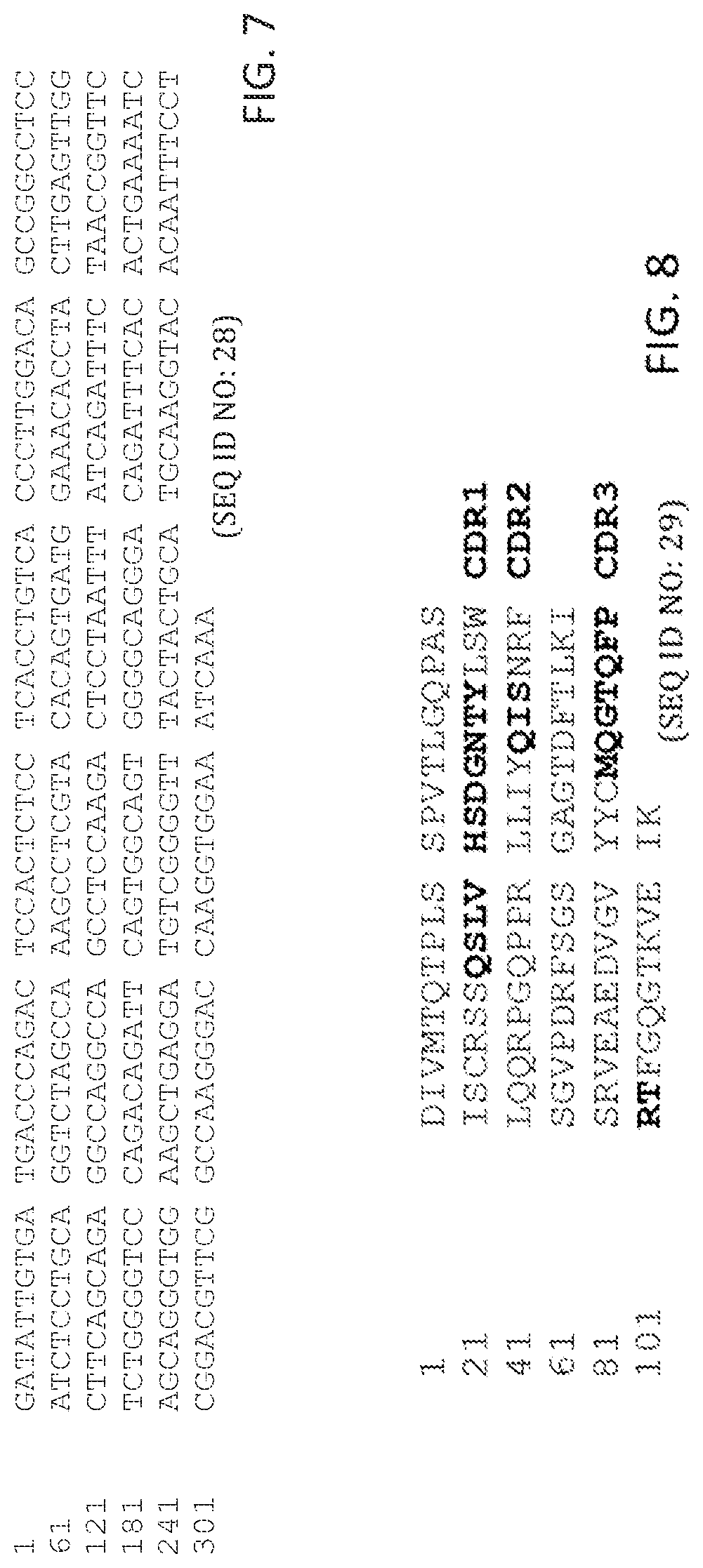
In some aspects, peptides include an antibody or antibody fragment comprising: a V.sub.H chain with identity to SEQ ID NO:20, wherein regions corresponding to CDR1, CDR2, and CDR3 comprise CDR1, CDR2, and CDR3 of the V.sub.H of antibody ID 2 shown in table 1 having 5 or fewer conservative amino acid substitutions within the CDR1, CDR2, and CDR3 regions, and regions within SEQ ID NO:20 corresponding to FR1, FR2, FR3, FR4, comprise amino acid sequences with at least 80%, 85%, 90%, 95%, 96%, 97%, 98, 99%, or 100% identity to FR1, FR2, FR3, FR4 of the V.sub.H of antibody ID 2 shown in table 1; and a V.sub.L chain with identity to SEQ ID NO:29, wherein regions corresponding to CDR1, CDR2, and CDR3 comprise CDR1, CDR2, and CDR3 of the V.sub.L of antibody ID 2 shown in table 1 having 5 or fewer conservative amino acid substitutions within the CDR1, CDR2, and CDR3 regions, and regions within SEQ ID NO:29 corresponding to FR1, FR2, FR3, FR4, comprise amino acid sequences with at least 80%, 85%, 90%, 95%, 96%, 97%, 98, 99%, or 100% identity to FR1, FR2, FR3, FR4 of the V.sub.L of antibody ID 2 shown in table 1. In some aspects, the peptides include an antibody or antibody fragment comprising a V.sub.H chain comprising SEQ ID NO:20 and a V.sub.L chain comprising SEQ ID NO:29.
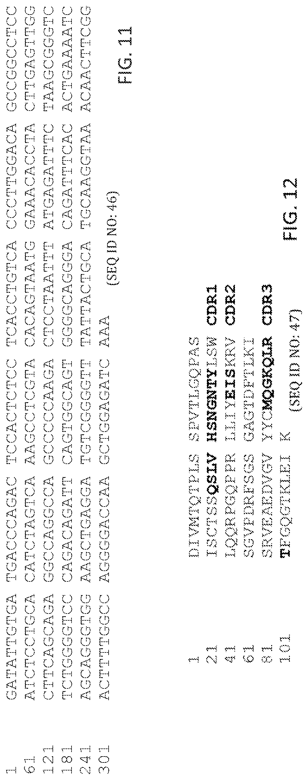
In some aspects, the peptides an antibody or antibody fragment comprising: a V.sub.H chain with identity to SEQ ID NO:38, wherein regions corresponding to CDR1, CDR2, and CDR3 comprise CDR1, CDR2, and CDR3 of the V.sub.H of antibody ID 3 shown in table 1 having 5 or fewer conservative amino acid substitutions within the CDR1, CDR2, and CDR3 regions, and regions within SEQ ID NO:38 corresponding to FR1, FR2, FR3, FR4, comprise amino acid sequences with at least 80%, 85%, 90%, 95%, 96%, 97%, 98, 99%, or 100% identity to FR1, FR2, FR3, FR4 of the V.sub.H of antibody ID 3 shown in table 1; and a V.sub.L chain with identity to SEQ ID NO:47, wherein regions corresponding to CDR1, CDR2, and CDR3 comprise CDR1, CDR2, and CDR3 of the V.sub.L of antibody ID 3 shown in table 1 having 5 or fewer conservative amino acid substitutions within the CDR1, CDR2, and CDR3 regions, and regions within SEQ ID NO:47 corresponding to FR1, FR2, FR3, FR4, comprise amino acid sequences with at least 80%, 85%, 90%, 95%, 96%, 97%, 98, 99%, or 100% identity to FR1, FR2, FR3, FR4 of the V.sub.L of antibody ID 3 shown in table 1. In some aspects, peptides include an antibody or antibody fragment comprising a V.sub.H chain comprising SEQ ID NO:38 and a V.sub.L chain comprising SEQ ID NO:47.
In some aspects, peptides include an antibody or antibody fragment comprising: a V.sub.H chain with identity to SEQ ID NO:56, wherein regions corresponding to CDR1, CDR2, and CDR3 comprise CDR1, CDR2, and CDR3 of the V.sub.H of antibody ID 4 shown in table 1 having 5 or fewer conservative amino acid substitutions within the CDR1, CDR2, and CDR3 regions, and regions within SEQ ID NO:56 corresponding to FR1, FR2, FR3, FR4, comprise amino acid sequences with at least 80%, 85%, 90%, 95%, 96%, 97%, 98, 99%, or 100% identity to FR1, FR2, FR3, FR4 of the V.sub.H of antibody ID 4 shown in table 1; and a V.sub.L chain with identity to SEQ ID NO:65, wherein regions corresponding to CDR1, CDR2, and CDR3 comprise CDR1, CDR2, and CDR3 of the V.sub.L of antibody ID 4 shown in table 1 having 5 or fewer conservative amino acid substitutions within the CDR1, CDR2, and CDR3 regions, and regions within SEQ ID NO:65 corresponding to FR1, FR2, FR3, FR4, comprise amino acid sequences with at least 80%, 85%, 90%, 95%, 96%, 97%, 98, 99%, or 100% identity to FR1, FR2, FR3, FR4 of the V.sub.L of antibody ID 4 shown in table 1. In some aspects, peptides include an antibody or antibody fragment comprising a V.sub.H chain comprising SEQ ID NO:56 and a V.sub.L chain comprising SEQ ID NO:65.
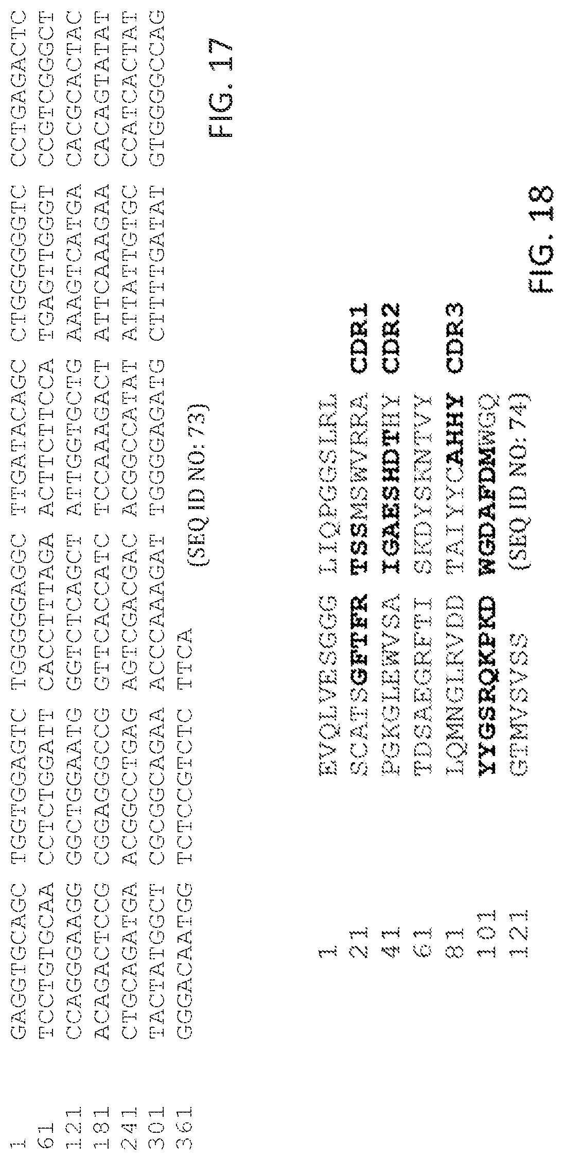
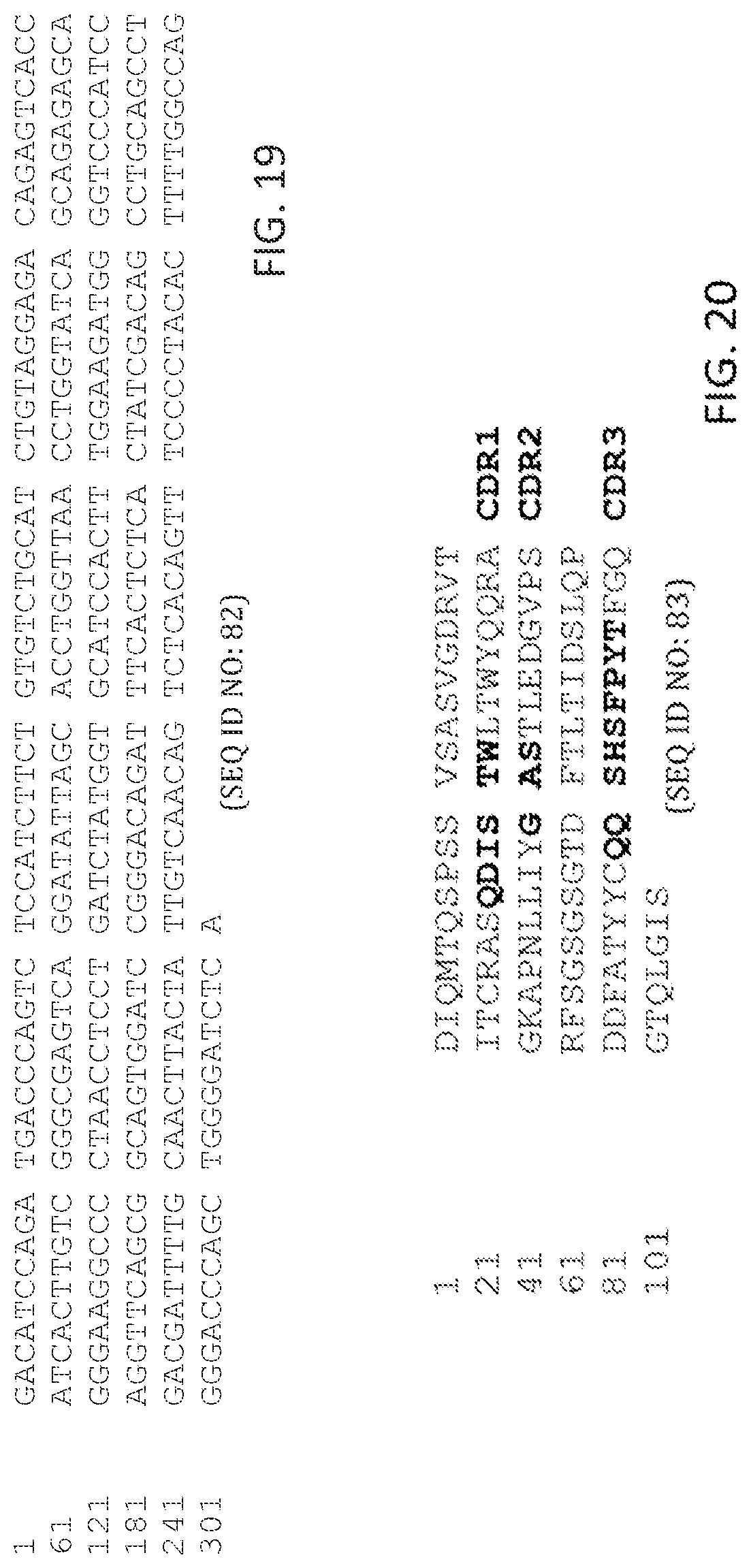
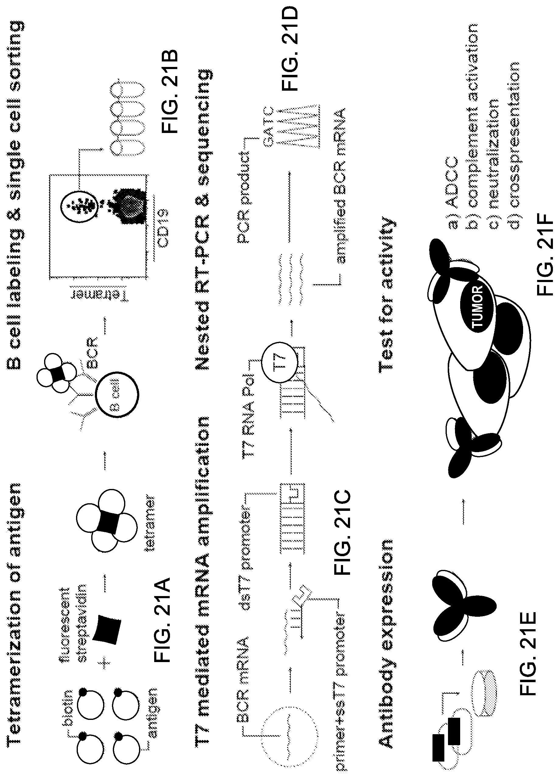
In some aspects, peptides include an antibody or antibody fragment comprising: a V.sub.H chain with identity to SEQ ID NO:74, wherein regions corresponding to CDR1, CDR2, and CDR3 comprise CDR1, CDR2, and CDR3 of the V.sub.H of antibody ID 5 shown in table 1 having 5 or fewer conservative amino acid substitutions within the CDR1, CDR2, and CDR3 regions, and regions within SEQ ID NO:74 corresponding to FR1, FR2, FR3, FR4, comprise amino acid sequences with at least 80%, 85%, 90%, 95%, 96%, 97%, 98, 99%, or 100% identity to FR1, FR2, FR3, FR4 of the V.sub.H of antibody ID 5 shown in table 1; and a V.sub.L chain with identity to SEQ ID NO:83, wherein regions corresponding to CDR1, CDR2, and CDR3 comprise CDR1, CDR2, and CDR3 of the V.sub.L of antibody ID 5 shown in table 1 having 5 or fewer conservative amino acid substitutions within the CDR1, CDR2, and CDR3 regions, and regions within SEQ ID NO:83 corresponding to FR1, FR2, FR3, FR4, comprise amino acid sequences with at least 80%, 85%, 90%, 95%, 96%, 97%, 98, 99%, or 100% identity to FR1, FR2, FR3, FR4 of the V.sub.L of antibody ID 5 shown in table 1. In some aspects, the peptides include an antibody or antibody fragment comprising a V.sub.H chain comprising SEQ ID NO:74 and a V.sub.L chain comprising SEQ ID NO:83. In some aspects, the peptides immunospecifically bind to at least angiopoietin-2. In some aspects, the compositions further include one or more anti-cancer therapeutics. In some aspects, the compositions are formulated as a pharmaceutical composition.
In some aspects, peptides include an antibody or antibody fragment comprising: a V.sub.H chain with identity to SEQ ID NO:222, wherein regions corresponding to CDR1, CDR2, and CDR3 comprise CDR1, CDR2, and CDR3 of the V.sub.H of antibody ID 10 shown in table 1 having 5 or fewer conservative amino acid substitutions within the CDR1, CDR2, and CDR3 regions, and regions within SEQ ID NO:222 corresponding to FR1, FR2, FR3, FR4, comprise amino acid sequences with at least 80%, 85%, 90%, 95%, 96%, 97%, 98, 99%, or 100% identity to FR1, FR2, FR3, FR4 of the V.sub.H of antibody ID 10 shown in table 1; and a V.sub.L chain with identity to SEQ ID NO:224, wherein regions corresponding to CDR1, CDR2, and CDR3 comprise CDR1, CDR2, and CDR3 of the V.sub.L of antibody ID 10 shown in table 1 having 5 or fewer conservative amino acid substitutions within the CDR1, CDR2, and CDR3 regions, and regions within SEQ ID NO:224 corresponding to FR1, FR2, FR3, FR4, comprise amino acid sequences with at least 80%, 85%, 90%, 95%, 96%, 97%, 98, 99%, or 100% identity to FR1, FR2, FR3, FR4 of the V.sub.L of antibody ID 10 shown in table 1. In some aspects, the peptides include an antibody or antibody fragment comprising a V.sub.H chain comprising SEQ ID NO:222 and a V.sub.L chain comprising SEQ ID NO:224. In some aspects, the peptides immunospecifically bind to at least angiopoietin-2. In some aspects, the compositions further include one or more anti-cancer therapeutics. In some aspects, the compositions are formulated as a pharmaceutical composition.
In some embodiments, the disclosure includes methods of treating cancer in a subject. In some aspects, methods include administering to a subject a composition of any one of claims 1-27.
The present disclosure also provides methods of isolating human antibodies from cancer patients following immunotherapy.
In some embodiments, the disclosure includes method of obtaining immune cells directed against a self antigen from a subject, the method comprising identifying a subject exhibiting a positive immune response towards the self antigen, providing a multimeric form of the self antigen, contacting the multimeric form of the self antigen with a sample from the subject exhibiting a positive immune response towards the self antigen, and obtaining immune cells bound to the multimeric form of the self antigen.
In some embodiments, the disclosure includes method of obtaining immune cells from a cancer patient directed against a self antigen, the method comprising identifying a subject exhibiting a positive immune response towards the self antigen; providing a multimeric form of the self antigen; contacting the multimeric form of the self antigen with a sample from the subject exhibiting a positive immune response towards the self antigen; and obtaining immune cells bound to the multimeric form of the self antigen.
Unless otherwise defined, all technical and scientific terms used herein have the same meaning as commonly understood by one of ordinary skill in the art to which this invention belongs. Methods and materials are described herein for use in the present invention; other, suitable methods and materials known in the art can also be used. The materials, methods, and examples are illustrative only and not intended to be limiting. All publications, patent applications, patents, sequences, database entries, and other references mentioned herein are incorporated by reference in their entirety. In case of conflict, the present specification, including definitions, will control.
Other features and advantages of the invention will be apparent from the following detailed description and figures, and from the claims.
DESCRIPTION OF DRAWINGS
FIG. 1|Nucleic acid sequence of the variable heavy (V.sub.H) chain of antibody ID 1 (anti-MHC class I polypeptide-related sequence A (MICA) antibody) (SEQ ID NO: 1).
FIG. 2|Amino acid sequence of V.sub.H chain of antibody ID 1 (anti-MICA antibody) (SEQ ID NO:2).
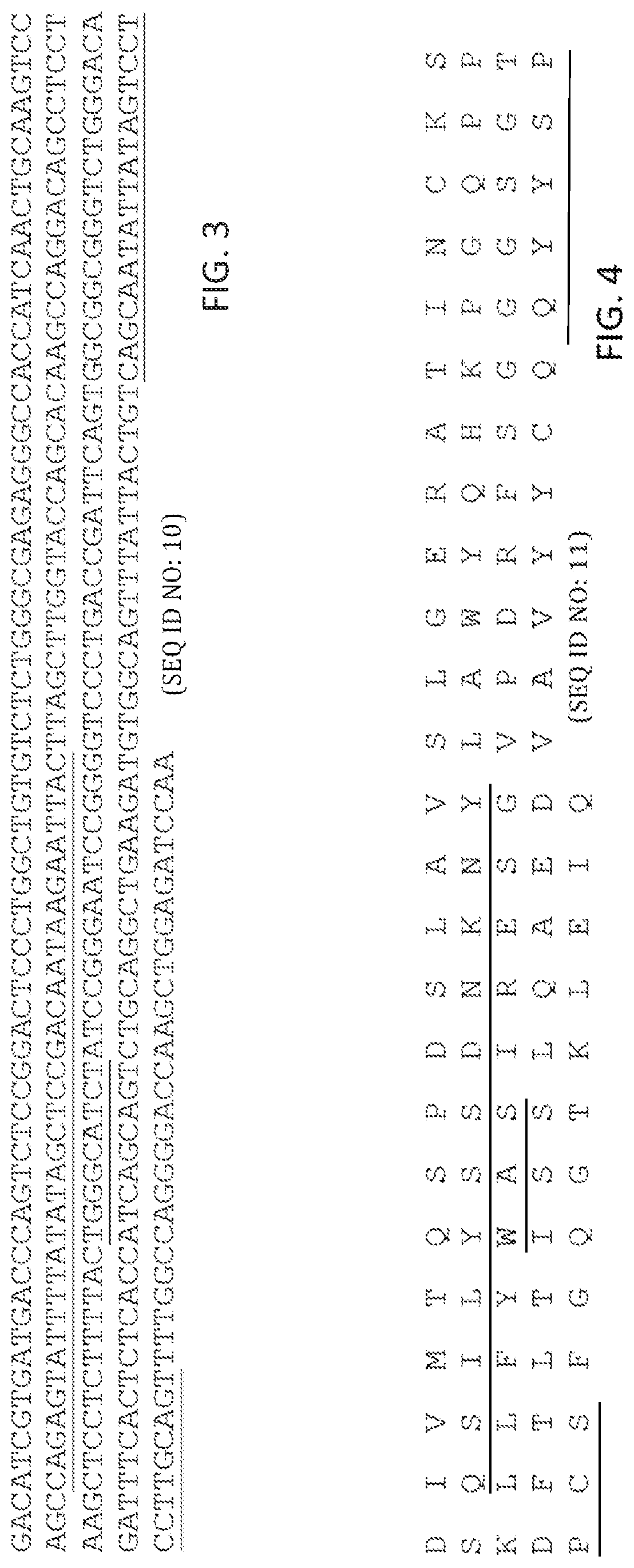
FIG. 3|Nucleic acid sequence of the variable light (V.sub.L) chain of antibody ID 1 (anti-MICA antibody) (SEQ ID NO: 10).
FIG. 4|Amino acid sequence of V.sub.L chain of antibody ID 1 (anti-MICA antibody) (SEQ ID NO:11).
FIG. 5|Nucleic acid sequence of the V.sub.H chain of antibody ID 2 (anti-angiopoietin-2 antibody) (SEQ ID NO: 19).
FIG. 6|Amino acid sequence of V.sub.H chain of antibody ID 2 (anti-angiopoietin-2 antibody) (SEQ ID NO:20).
FIG. 7|Nucleic acid sequence of the V.sub.L chain of antibody ID 2 (anti-angiopoietin-2 antibody) (SEQ ID NO:28).
FIG. 8|Amino acid sequence of V.sub.L chain of antibody ID 2 (anti-angiopoietin-2 antibody) (SEQ ID NO:29).
FIG. 9|Nucleic acid sequence of the V.sub.H chain of antibody ID 3 (anti-angiopoietin-2 antibody) (SEQ ID NO:37).
FIG. 10|Amino acid sequence of V.sub.H chain of antibody ID 3 (anti-angiopoietin-2 antibody) (SEQ ID NO:38).
FIG. 11|Nucleic acid sequence of the V.sub.L chain of antibody ID 3 (anti-angiopoietin-2 antibody) (SEQ ID NO:46).
FIG. 12|Amino acid sequence of V.sub.L chain of antibody ID 3 (anti-angiopoietin-2 antibody) (SEQ ID NO:47).
FIG. 13|Nucleic acid sequence of the V.sub.H chain of antibody ID 4 (anti-angiopoietin-2 antibody) (SEQ ID NO:55).
FIG. 14|Amino acid sequence of V.sub.H chain of antibody ID 4 (anti-angiopoietin-2 antibody) (SEQ ID NO:56).
FIG. 15|Nucleic acid sequence of the V.sub.L chain of antibody ID 4 (anti-angiopoietin-2 antibody) (SEQ ID NO:64).
FIG. 16|Amino acid sequence of V.sub.L chain of antibody ID 4 (anti-angiopoietin-2 antibody) (SEQ ID NO:65).
FIG. 17|Nucleic acid sequence of the V.sub.H chain of antibody ID 5 (anti-angiopoietin-2 antibody) (SEQ ID NO:73).
FIG. 18|Amino acid sequence of V.sub.H chain of antibody ID 5 (anti-angiopoietin-2 antibody) (SEQ ID NO:74).
FIG. 19|Nucleic acid sequence of the V.sub.L chain of antibody ID 5 (anti-angiopoietin-2 antibody) (SEQ ID NO:82).
FIG. 20|Amino acid sequence of V.sub.L chain of antibody ID 5 (anti-angiopoietin-2 antibody) (SEQ ID NO:83).
FIG. 21A-21F|Illustrates exemplary methods for making antibodies from B-cells. (A) Antigen is expressed with a BirA tag for site-specific biotinylation and tetramerization with fluorescently-labeled streptavidin. (B) B cells are stained with tetramer and a panel of monoclonal antibodies. Tetramer.sup.+, class-switched memory B cells are single-cell sorted into PCR strips. (C) mRNA amplification is performed with T7 RNA polymerase. (D) Sequencing of PCR products is carried out using 300-400 bp PCR products. (E) Overlap PCR is used for construction of full-length IgG1 heavy chain and kappa/lambda light sequences which are cloned into separate vectors. Vectors are transiently transfected into CHO-S cells for expression of fully human recombinant antibodies. (F) Antibodies are tested for antigen binding and assessed for potential therapeutic properties.
FIGS. 22A-22B|Graphs showing comparison of monomeric and tetrameric antigen for identification of memory B cells. (A) Mono-biotinylated TTCF or CD80 antigens were directly labeled with Alexa-488 fluorophore; tetramers were generated with unlabeled streptavidin. Enriched B cells from each donor were split into three fractions and stained with control CD80 tetramer, TTCF monomer, or TTCF tetramer at the same total antigen concentration of 0.125 .mu.g/mL. FACS plots depict CD19.sup.+ CD27.sup.+ IgM.sup.- class-switched memory B cells; numbers adjacent to the gate represent the percentage of the parental gate. (B) Frequencies of tetramer.sup.+ memory B cells detected in three different donors. Numbers are calculated as tetramer.sup.+ cells per 1.times.10.sup.6 CD19.sup.+ memory B cells.
FIGS. 23A-23B|Line graphs showing high affinity binding of TTCF by antibodies generated from plasmablasts and memory B cells. Saturation binding experiments were carried out to determine the affinities of recombinant antibodies. TTCF antigen was labeled with europium, which emits a strong fluorescent signal at 615 nm upon incubation with a chelating reagent. Antibodies were immobilized in a 96-well plate and incubated with TTCF-europium (100 nM to 4 pM) for two hours at 37.degree. C. Fluorescent counts at 615 nm were recorded and K.sub.D calculated using non-linear regression analysis. Control antibody (clone 8.18.C5) that was also produced in CHO-S cells was included in all experiments. (A) Recombinant TTCF Abs 1 and 2 were generated from TTCF tetramer.sup.+ plasmablasts (donor 1). (B) TTCF antibodies 3, 4, and 5 originated from TTCF tetramer.sup.+ memory B cells of three different donors.
FIG. 24|Bar chart showing binding of anti-MICA antibodies to MICA-coated luminex beads.
FIGS. 25A-250|Line graphs showing binding of anti-MICA antibodies to MICA-coated beads.
FIGS. 26A-26D|Bar graphs showing binding of four human angiopoietin 2 specific antibodies as well as a control antibody to three human angiopoietins (angiopoietin-1, 2 and 4) and ang-like-3. Recombinant angiopoietins were immobilized in an ELISA plate and binding of human recombinant antibodies was detected with europium-labeled streptavidin.
FIGS. 27A-27C|Show graphs and a gel relating to isolation of angiopoietin-specific antibodies from a lung cancer patient. (A) Angiopoietin-2 reactivity of lung cancer patient (L19) serum (diluted 1:1000) determined by ELISA. (B) FACS plot showing PBMC sample (timepoint--10/98) gated on CD19.sup.+, CD27.sup.+ IgM-B cells with CD19 on the X-axis and fluorescently-tagged angiopoietin-2 on the Y-axis. (C) Heavy, light chain, and hinge region PCR products from 10 angiopoietin-2 reactive memory B-cells isolated from patient L19. The 500 base pair marker is indicated on the left.
FIG. 28|Nucleic acid sequence of the variable heavy (V.sub.H) chain of antibody ID 6 (anti-MHC class I polypeptide-related sequence A (MICA) antibody) (SEQ ID NO: 148).
FIG. 29|Amino acid sequence of V.sub.H chain of antibody 6 (anti-MICA antibody) (SEQ ID NO:149).
FIG. 30|Nucleic acid sequence of the variable light (V.sub.L) chain of antibody ID 6 (anti-MICA antibody) (SEQ ID NO: 150).
FIG. 31|Amino acid sequence of V.sub.L chain of antibody ID 6 (anti-MICA antibody) (SEQ ID NO: 151).
FIG. 32|Nucleic acid sequence of the variable heavy (V.sub.H) chain of antibody ID 7 (anti-MHC class I polypeptide-related sequence A (MICA) antibody) (SEQ ID NO:167).
FIG. 33|Amino acid sequence of V.sub.H chain of antibody ID 7 (anti-MICA antibody) (SEQ ID NO: 168).
FIG. 34|Nucleic acid sequence of the variable light (V.sub.L) chain of antibody ID 7 (anti-MICA antibody) (SEQ ID NO:169).
FIG. 35|Amino acid sequence of V.sub.L chain of antibody ID 7 (anti-MICA antibody) (SEQ ID NO: 170).
FIG. 36|Nucleic acid sequence of the variable heavy (VH) chain of antibody ID 8 (anti-MHC class I polypeptide-related sequence A (MICA) antibody) (SEQ ID NO:185).
FIG. 37|Amino acid sequence of VH chain of antibody ID 8 (anti-MICA antibody) (SEQ ID NO: 186).
FIG. 38|Nucleic acid sequence of the variable light (VL) chain of antibody ID 8 (anti-MICA antibody) (SEQ ID NO:187).
FIG. 39|Amino acid sequence of VL chain of antibody ID 8 (anti-MICA antibody) (SEQ ID NO: 188).
FIG. 40|Nucleic acid sequence of the variable heavy (V.sub.H) chain of antibody ID 9 (anti-MHC class I polypeptide-related sequence A (MICA) antibody) (SEQ ID NO:203).
FIG. 41|Amino acid sequence of V.sub.H chain of antibody ID 9 (anti-MICA antibody) (SEQ ID NO:204).
FIG. 42|Nucleic acid sequence of the variable light (V.sub.L) chain of antibody ID 9 (anti-MICA antibody) (SEQ ID NO:205).
FIG. 43|Amino acid sequence of V.sub.L chain of antibody ID 9 (anti-MICA antibody) (SEQ ID NO: 206).
FIG. 44|Nucleic acid sequence of the V.sub.H chain of antibody ID 10 (anti-angiopoietin-2 antibody) (SEQ ID NO:221).
FIG. 45|Amino acid sequence of V.sub.H chain of antibody ID 10 (anti-angiopoietin-2 antibody) (SEQ ID NO:222).
FIG. 46|Nucleic acid sequence of the V.sub.L chain of antibody ID 10 (anti-angiopoietin-2 antibody) (SEQ ID NO:223).
FIG. 47|Amino acid sequence of V.sub.L chain of antibody ID 10 (anti-angiopoietin-2 antibody) (SEQ ID NO:224).
FIGS. 48A-G|Line graphs showing assessment of MICA allele-specific binding by recombinant anti-MICA antibodies.
FIG. 49|Line graph showing labeling of autologous tumor cells by anti-MICA antibody CM24002 Ab2.
FIG. 50|A series of FACS plot showing regulation of NKG2D by serum MICA. Human NK cells were incubated with control serum from patient CM24002 and a 1:10 dilution for 48 hours. Indicated antibodies were added at the start of the incubation at a concentration of 10 .mu.g/ml. NKG2D expression was assessed on CD56+ NK cells by flow cytometry.
FIG. 51|A series of FACS plot showing regulation of NKG2D by recombinant MICA. Human NK cells were incubated with recombinant MICA at a concentration of 2 ng/ml for 48 hours. Indicated antibodies were added at the start of the incubation at a concentration of 10 .mu.g/ml. After 48 hours, NKG2D expression was assessed on CD56+ NK cells by flow cytometry.
FIG. 52|Line graph demonstrating enhancement of cell-mediated toxicity by anti-MICA antibody CM24002 Ab2. Human NK cells were incubated with recombinant MICA (2 ng/ml) for 48 hours in the presence of indicated antibodies at 10 .mu.g/ml. The ability of NK cells (effectors) to kill K562 target cells was assessed by measuring LDH release following 4 hour incubation at the indicated ratios.
FIG. 53|Bar graph demonstration cell-mediated toxicity by anti-MICA antibodies CM24002 Ab2 and CM33322 Ab29. Human NK cells were incubated with recombinant MICA (2 ng/ml) for 48 hours in the presence of indicated antibodies at 10 .mu.g/ml. The ability of NK cells (effectors) to kill K562 target cells was assessed by measuring LDH release following 4 hour incubation. NKG2D blocking antibody or Fc blocking antibody was added during the 4 hr incubation of effector and target cells to assess the contribution of Fc receptor and NKG2D to cell-mediated toxicity.
FIG. 54|A series of line graphs showing binding of MICA alpha 3 domain by recombinant anti-MICA antibodies. Recombinant MICA alpha 3 domains were biotinylated and captured on the surface of streptavidin-coated beads. Indicated antibodies were incubated at 10 .mu.g/ml with the beads coated with the individual recombinant protein for 1 hr. Beads were subsequently washed and incubated with FITC-conjugated anti-human IgG secondary antibody. FITC fluorescence was quantified by flow cytometry.
FIG. 55|Line graphs demonstrating labeling of tumor cells by anti-MICA antibodies CM24002 Ab2 and CM33322 Ab29. Fluorescence was determined by flow cytometry.
FIG. 56|Bar graph demonstrating MICA allelic specificity of anti-MICA antibodies CM33322 Ab29 as determined by Luminex assay.
FIG. 57|Bar graphs showing binding of anti-angiopoietin 2 specific antibody anti-Ang6 Ab2 as well as a control antibody to three human angiopoietins (angiopoietin-1, 2 and 4) and ang-like-3. Recombinant angiopoietins were immobilized in an ELISA plate and binding of human recombinant antibodies was detected with europium-labeled streptavidin.
DETAILED DESCRIPTION
The present disclosure is based, in part, on the observation that antibodies directed against therapeutic targets important in a disease can be obtained from human subjects exposed to the disease by labeling of B cells with a tetrameric form of the antigen of interest. As described in the background section above, prior methods are limited at least in that they are inefficient at identifying appropriate B cells in human subjects and/or because they induce any captured B cells to undergo phenotypic changes, thus reducing their value. In contrast, methods are described herein that allow capture of rare memory B cells directed against specific disease-related antigens. As described below, the methods require tetramerization of the disease-related antigen, which process, as demonstrated in the Examples below, enhances the identification of appropriate memory B cells. Specifically, methods herein permit more efficient capture of appropriate memory B cells for increased periods of time following initial exposure of a subject to the antigen. Methods herein also include antibodies (and peptides generated from the sequences of such antibodies) generated using genetic material obtained from memory B cells captured using the methods disclosed herein.
Described herein are human antibodies against MHC class I polypeptide-related sequence A (MICA) and human antibodies targeted against angiopoietin-2. Both types of human antibodies were identified from patients who had received a cell-based cancer vaccine (GM-CSF transduced autologous tumor cells) by methods that entail the use of tetrameric antigens.
In some instances, the disclosure provides methods for specifically obtaining or targeting antibodies with therapeutic potential from select human subjects and therapeutic compositions resulting therefrom. These methods can include: obtaining or targeting immune cells in a human subject, wherein immune cells include but are not limited to, for example, B cells and/or memory B cells, isolating or purifying genetic material (e.g., DNA and/or mRNA) from the obtained or targeted immune cells, and using the isolated or purified genetic material to produce therapeutic compositions, e.g., therapeutic compositions disclosed herein. Further description of the methods is provided under the section entitled "Methods," below.
In some instances, the disclosure provides therapeutic compositions (e.g., including therapeutic peptides, including antibodies, antibody fragments, antibody derivatives, and/or antibody conjugates) related to antibodies present in subjects that have or had a condition or disease and that exhibited a positive immune response towards the condition or disease.
Therapeutic Compositions
In some instances, therapeutic compositions herein can interact with (e.g., bind, bind specifically and/or bind immunospecifically) binding partners (e.g., an immunogen(s), antigen(s), and/or epitope(s)) related to a disease or condition, wherein interaction between the therapeutic composition and the binding partners results in a positive immune response towards the condition or disease (e.g., a decrease in the level of disease or symptoms thereof in a subject).
In some instances, therapeutic compositions can include peptides that include (e.g., comprise, consist essentially of, or consist of) at least one (e.g., one, two, three, four, five, and/or six) complementarity determining region (CDR) of the variable heavy chain (V.sub.H) and/or variable light chain (V.sub.L) of antibody ID 1, 2, 3, 4, or 5, 6, 7, 8, 9 or 10, shown in Table 1.
In some instances, therapeutic compositions can include peptides that include (e.g., comprise, consist essentially of, or consist of) at least one (e.g., one, two, three, four, five, and/or six) complementarity determining region (CDR) of the variable heavy chain (V.sub.H) and/or variable light chain (V.sub.L) of antibody ID 1, 2, 3, 4, 5, 6, 7, 8, 9 or 10, shown in Table 1, and that interact with (e.g., bind, bind specifically and/or bind immunospecifically) to MHC class I polypeptide-related sequence A (MICA (e.g., UniGene Hs.130838)) (e.g., soluble MICA (sMICA)) and/or angiopoietin-2 (e.g., UniGene Hs.583870), including epitopes thereof.
In some instances, therapeutic compositions can include peptides that include at least one CDR of the V.sub.H and/or V.sub.L of antibody ID 1, 6, 7, 8 and/or 9 shown in Table 1, wherein the peptide binds (e.g., binds specifically and/or binds immunospecifically) to MICA (e.g., human MICA (e.g., soluble MICA (sMICA))). In some instances, peptides can include at least two CDRs, wherein the at least two CDRs are CDRs shown in Table 1 for different antibodies. In other words, CDRs (and FRs and/or AA sequences) shown in Table 1 for antibodies IDs 1, 6, 7, 8 and 9 are interchangeable and can be combined to generate peptides, so long as the peptides bind (e.g., bind specifically and/or bind immunospecifically) to MICA (e.g., human MICA (e.g., soluble MICA (sMICA))). In some instances, such peptides include CDR3 of the V.sub.H and/or V.sub.L of antibody ID 1, 6, 7, 8 and/or 9 shown in Table 1. In some instances, such peptides include CDR3 of the V.sub.H and V.sub.L of antibody ID 1, 6, 7, 8 and/or 9 and CDR1 and/or CDR2 of the V.sub.H and/or V.sub.L of antibody ID 1, 6, 7, 8 and/or 9 shown in Table 1. In some instances, such peptides include CDR1 CDR2, and CDR3 of the V.sub.H and/or V.sub.L of antibody ID 1, 6, 7, 8 and/or 9. In some instances, such peptides include CDR1, CDR2, and CDR3 of the V.sub.H and/or V.sub.L of antibody ID 1, 6, 7, 8 and/or 9 and at least one of FR1 FR2 FR3, and/or FR4 of the V.sub.H and/or V.sub.L of antibody ID 1, 6, 7, 8 and/or 9, shown in Table 1. In some instances, such peptides include one of SEQ ID NO:2, 149, 168, 186 or 204 and/or one of SEQ ID NO:11, 151, 170, 188, or 206. In each instance, the peptide can bind (e.g., bind specifically and/or bind immunospecifically) to MICA (e.g., human MICA (e.g., soluble MICA (sMICA))). In some instances, the affinity of binding between the peptides and MICA can be between about 0.1 nM to 1 .mu.M, for example, about 10 nM.
In some instances, therapeutic compositions can include peptides that include at least one CDR of the V.sub.H and/or V.sub.L of antibody ID 6 shown in Table 1, wherein the peptide binds (e.g., binds specifically and/or binds immunospecifically) to MICA (e.g., human MICA (e.g., soluble MICA (sMICA))). In some instances, such peptides include CDR3 of the V.sub.H and/or V.sub.L of antibody ID 6 shown in Table 1. In some instances, such peptides include CDR3 of the V.sub.H and V.sub.1, of antibody ID 6 and CDR1 and/or CDR2 of the V.sub.H and/or V.sub.L of antibody ID 6 shown in Table 1. In some instances, such peptides include CDR1 CDR2, and CDR3 of the V.sub.H and/or V.sub.L of antibody ID 6. In some instances, such peptides include CDR1, CDR2, and CDR3 of the V.sub.H and/or V.sub.L of antibody ID 6 and at least one of FR1 FR2 FR3, and/or FR4 of the V.sub.H and/or V.sub.1, of antibody ID 6, shown in Table 1. In some instances, such peptides include SEQ ID NO: 149 and/or SEQ ID NO: 151. In each instance, the peptide can bind (e.g., bind specifically and/or bind immunospecifically) to MICA (e.g., human MICA (e.g., soluble MICA (sMICA))). In some instances, the affinity of binding between the peptides and MICA can be between about 0.1 nM to 1 .mu.M, for example, about 10 nM.
In some instances, therapeutic compositions can include peptides that include at least one CDR of the V.sub.H and/or V.sub.L of antibody ID 7 shown in Table 1, wherein the peptide binds (e.g., binds specifically and/or binds immunospecifically) to MICA (e.g., human MICA (e.g., soluble MICA (sMICA))). In some instances, such peptides include CDR3 of the V.sub.H and/or V.sub.L of antibody ID 7 shown in Table 1. In some instances, such peptides include CDR3 of the V.sub.H and V.sub.L of antibody ID 7 and CDR1 and/or CDR2 of the V.sub.H and/or V.sub.L of antibody ID 7 shown in Table 1. In some instances, such peptides include CDR1 CDR2, and CDR3 of the V.sub.H and/or V.sub.L of antibody ID 7. In some instances, such peptides include CDR1, CDR2, and CDR3 of the V.sub.H and/or V.sub.L of antibody ID 7 and at least one of FR1 FR2 FR3, and/or FR4 of the V.sub.H and/or V.sub.L of antibody ID 7, shown in Table 1. In some instances, such peptides include SEQ ID NO: 168 and/or SEQ ID NO: 170. In each instance, the peptide can bind (e.g., bind specifically and/or bind immunospecifically) to MICA (e.g., human MICA (e.g., soluble MICA (sMICA))). In some instances, the affinity of binding between the peptides and MICA can be between about 0.1 nM to 1 M, for example, about 10 nM.
In some instances, therapeutic compositions can include peptides that include at least one CDR of the V.sub.H and/or V.sub.1, of antibody ID 8 shown in Table 1, wherein the peptide binds (e.g., binds specifically and/or binds immunospecifically) to MICA (e.g., human MICA (e.g., soluble MICA (sMICA))). In some instances, such peptides include CDR3 of the V.sub.H and/or V.sub.L of antibody ID 8 shown in Table 1. In some instances, such peptides include CDR3 of the V.sub.H and V.sub.L of antibody ID 8 and CDR1 and/or CDR2 of the V.sub.H and/or V.sub.1, of antibody ID 8 shown in Table 1. In some instances, such peptides include CDR1 CDR2, and CDR3 of the V.sub.H and/or V.sub.L of antibody ID 8. In some instances, such peptides include CDR1, CDR2, and CDR3 of the V.sub.H and/or V.sub.L of antibody ID 8 and at least one of FR1 FR2 FR3, and/or FR4 of the V.sub.H and/or V.sub.L of antibody ID 8, shown in Table 1. In some instances, such peptides include SEQ ID NO: 186 and/or SEQ ID NO: 188. In each instance, the peptide can bind (e.g., bind specifically and/or bind immunospecifically) to MICA (e.g., human MICA (e.g., soluble MICA (sMICA))). In some instances, the affinity of binding between the peptides and MICA can be between about 0.1 nM to 1 .mu.M, for example, about 10 nM.
In some instances, therapeutic compositions can include peptides that include at least one CDR of the V.sub.H and/or V.sub.L of antibody ID 9 shown in Table 1, wherein the peptide binds (e.g., binds specifically and/or binds immunospecifically) to MICA (e.g., human MICA (e.g., soluble MICA (sMICA))). In some instances, such peptides include CDR3 of the V.sub.H and/or V.sub.L of antibody ID 9 shown in Table 1. In some instances, such peptides include CDR3 of the V.sub.H and V.sub.L of antibody ID 9 and CDR1 and/or CDR2 of the V.sub.H and/or V.sub.L of antibody ID 9 shown in Table 1. In some instances, such peptides include CDR1 CDR2, and CDR3 of the V.sub.H and/or V.sub.L of antibody ID 9. In some instances, such peptides include CDR1, CDR2, and CDR3 of the V.sub.H and/or V.sub.L of antibody ID 9 and at least one of FR1 FR2 FR3, and/or FR4 of the V.sub.H and/or V.sub.L of antibody ID 9, shown in Table 1. In some instances, such peptides include SEQ ID NO:204 and/or SEQ ID NO:206. In each instance, the peptide can bind (e.g., bind specifically and/or bind immunospecifically) to MICA (e.g., human MICA (e.g., soluble MICA (sMICA))). In some instances, the affinity of binding between the peptides and MICA can be between about 0.1 nM to 1 M, for example, about 10 nM.
In some instances, therapeutic compositions can include peptides that include at least one CDR of the V.sub.H and/or V.sub.L of antibody ID 2, 3, 4, 5, and/or 10 shown in Table 1, wherein the peptide binds (e.g., binds specifically and/or binds immunospecifically) to angiopoietin-2 (e.g., human angiopoietin-2). In some instances, peptides can include at least two CDRs, wherein the at least two CDRs are CDRs shown in Table 1 for different antibodies. In other words, CDRs (and FRs and/or AA sequences) shown in Table 1 for antibodies IDs 2, 3 4, 5, and 10 are interchangeable and can be combined to generate peptides, so long as the peptides bind (e.g., bind specifically and/or bind immunospecifically) to angiopoietin-2 (e.g., human angiopoietin-2). In some instances, such peptides include CDR3 of the V.sub.H and/or V.sub.L of antibody ID 2, 3, 4, 5, and/or 10 shown in Table 1. In some instances, such peptides include CDR3 of the V.sub.H and V.sub.L of antibody ID 2, 3, 4, 5, and/or 10 and CDR1 and/or CDR2 of the V.sub.H and/or V.sub.L of antibody ID 2, 3, 4, 5, and/or 10 shown in Table 1. In some instances, such peptides include CDR1, CDR2, and CDR3 of the V.sub.H and/or V.sub.L of antibody ID 2, 3, 4, 5, and/or 10. In some instances, such peptides include CDR1 CDR2, and CDR3 of the V.sub.H and/or V.sub.L of antibody ID 2, 3, 4, 5, and/or 10 and at least one of FR1 FR2 FR3, and/or FR4 of the V.sub.H and/or V.sub.L of antibody ID 2, 3, 4, 5, and/or 10 5, shown in Table 1. In some instances, such peptides include one of SEQ ID NO:20, 38, 56, 74, or 222 and/or one of SEQ ID NO:29, 47, 65, 83 or 224. In some instances, peptides include one of SEQ ID NO:20, 38, 56, 74, or 222 and one of SEQ ID NO:29, 47, 65, 83 or 224. In each instance, the peptide can bind (e.g., bind specifically and/or bind immunospecifically) to angiopoietin-2 (e.g., human angiopoietin-2 (e.g, UniGene Hs.583870)).
In some instances, therapeutic compositions can include peptides that include at least one CDR of the V.sub.H and/or V.sub.L of antibody ID 2 shown in Table 1, wherein the peptide binds (e.g., binds specifically and/or binds immunospecifically) to angiopoietin-2 (e.g., human angiopoietin-2). In some instances, such peptides include CDR3 of the V.sub.H and/or V.sub.L of antibody ID 2 shown in Table 1. In some instances, such peptides include CDR3 of the V.sub.H and V.sub.L of antibody ID 2 and CDR1 and/or CDR2 of the V.sub.H and/or V.sub.L of antibody ID 2 shown in Table 1. In some instances, such peptides include CDR1, CDR2, and CDR3 of the V.sub.H and/or V.sub.L of antibody ID 2. In some instances, such peptides include CDR1 CDR2, and CDR3 of the V.sub.H and/or V.sub.L of antibody ID 2 and at least one of FR1 FR2 FR3, and/or FR4 of the V.sub.H and/or V.sub.L of antibody ID 2, shown in Table 1. In some instances, such peptides include SEQ ID NO:20 and/or SEQ ID NO:29. In each instance, the peptide can bind (e.g., bind specifically and/or bind immunospecifically) to angiopoietin-2 (e.g., human angiopoietin-2). In some instances, the affinity of binding between the peptides and angiopoietin-2 can be between about 0.1 nM to 1 .mu.M, for example, about 10 nM.
In some instances, therapeutic compositions can include peptides that include at least one CDR of the V.sub.H and/or V.sub.L of antibody ID 3 shown in Table 1, wherein the peptide binds (e.g., binds specifically and/or binds immunospecifically) to angiopoietin-2 (e.g., human angiopoietin-2). In some instances, such peptides include CDR3 of the V.sub.H and/or V.sub.L of antibody ID 3 shown in Table 1. In some instances, such peptides include CDR3 of the V.sub.H and V.sub.L of antibody ID 3 and CDR1 and/or CDR2 of the V.sub.H and/or V.sub.L of antibody ID 3 shown in Table 1. In some instances, such peptides include CDR1, CDR2, and CDR3 of the V.sub.H1 and/or V.sub.L of antibody ID 3. In some instances, such peptides include CDR1 CDR2, and CDR3 of the V.sub.H and/or V.sub.L of antibody ID 3 and at least one of FR1 FR2 FR3, and/or FR4 of the V.sub.H and/or V.sub.L of antibody ID 3, shown in Table 1. In some instances, such peptides include SEQ ID NO:38 and/or SEQ ID NO:47. In each instance, the peptide can bind (e.g., bind specifically and/or bind immunospecifically) to angiopoietin-2 (e.g., human angiopoietin-2). In some instances, the affinity of binding between the peptides and angiopoietin-2 can be between about 0.1 nM to 1 .mu.M, for example, about 10 nM.
In some instances, therapeutic compositions can include peptides that include at least one CDR of the V.sub.H and/or V.sub.L of antibody ID 4 shown in Table 1, wherein the peptide binds (e.g., binds specifically and/or binds immunospecifically) to angiopoietin-2 (e.g., human angiopoietin-2). In some instances, such peptides include CDR3 of the V.sub.H and/or V.sub.L of antibody ID 4 shown in Table 1. In some instances, such peptides include CDR3 of the V.sub.H and V.sub.L of antibody ID 4 and CDR1 and/or CDR2 of the V.sub.H and/or V.sub.L of antibody ID 4 shown in Table 1. In some instances, such peptides include CDR1, CDR2, and CDR3 of the V.sub.H and/or V.sub.L of antibody ID 4. In some instances, such peptides include CDR1 CDR2, and CDR3 of the V.sub.H and/or V.sub.L of antibody ID 4 and at least one of FR1 FR2 FR3, and/or FR4 of the V.sub.H and/or V.sub.L of antibody ID 4, shown in Table 1. In some instances, such peptides include SEQ ID NO:56 and/or SEQ ID NO:65. In each instance, the peptide can bind (e.g., bind specifically and/or bind immunospecifically) to angiopoietin-2 (e.g., human angiopoietin-2). In some instances, the affinity of binding between the peptide and angiopoietin-2 can be between X-Y, for example, X-Y, X-Y. In some instances, the affinity of binding between the peptides and angiopoietin-2 can be between about 0.1 nM to 1 .mu.M, for example, about 10 nM.
In some instances, therapeutic compositions can include peptides that include at least one CDR of the V.sub.H and/or V.sub.L of antibody ID 5 shown in Table 1, wherein the peptide binds (e.g., binds specifically and/or binds immunospecifically) to angiopoietin-2 (e.g., human angiopoietin-2). In some instances, such peptides include CDR3 of the V.sub.H and/or V.sub.L of antibody ID 5 shown in Table 1. In some instances, such peptides include CDR3 of the V.sub.H and V.sub.L of antibody ID 5 and CDR1 and/or CDR2 of the V.sub.H and/or V.sub.L of antibody ID 5 shown in Table 1. In some instances, such peptides include CDR1, CDR2, and CDR3 of the V.sub.H and/or V.sub.L of antibody ID 5. In some instances, such peptides include CDR1 CDR2, and CDR3 of the V.sub.H and/or V.sub.L of antibody ID 5 and at least one of FR1 FR2 FR3, and/or FR4 of the V.sub.H and/or V.sub.L of antibody ID 5, shown in Table 1. In some instances, such peptides include SEQ ID NO:74 and/or SEQ ID NO:83. In each instance, the peptide can bind (e.g., bind specifically and/or bind immunospecifically) to angiopoietin-2 (e.g., human angiopoietin-2). In some instances, the affinity of binding between the peptides and angiopoietin-2 can be between about 0.1 nM to 1 .mu.M, for example, about 10 nM.
In some instances, therapeutic compositions can include peptides that include at least one CDR of the V.sub.H and/or V.sub.L of antibody ID 10 shown in Table 1, wherein the peptide binds (e.g., binds specifically and/or binds immunospecifically) to angiopoietin-2 (e.g., human angiopoietin-2). In some instances, such peptides include CDR3 of the V.sub.H and/or V.sub.L of antibody ID 10 shown in Table 1. In some instances, such peptides include CDR3 of the V.sub.H and V.sub.L of antibody ID 10 and CDR1 and/or CDR2 of the V.sub.H and/or V.sub.L of antibody ID 10 shown in Table 1. In some instances, such peptides include CDR1, CDR2, and CDR3 of the V.sub.H and/or V.sub.L of antibody ID 10. In some instances, such peptides include CDR1 CDR2, and CDR3 of the V.sub.H and/or V.sub.L of antibody ID 10 and at least one of FR1 FR2 FR3, and/or FR4 of the V.sub.H and/or V.sub.L of antibody ID 10, shown in Table 1. In some instances, such peptides include SEQ ID NO:222 and/or SEQ ID NO:224. In each instance, the peptide can bind (e.g., bind specifically and/or bind immunospecifically) to angiopoietin-2 (e.g., human angiopoietin-2). In some instances, the affinity of binding between the peptides and angiopoietin-2 can be between about 0.1 nM to 1 .mu.M, for example, about 10 nM.
In some instances, peptides that bind to angiopoietin-2 can also bind to angiopoietin-1 (e.g., Unigene Hs.369675) and/or angiopoietin-4 (e.g., Unigene Hs.278973). For example, in some instances, peptides that bind to angiopoietin-2 can also bind specifically and/or immunospecifically relative to other antigens (other than angiopoietin-1) to angiopoictin-1. In some instances, peptides that bind to angiopoietin-2 can also bind specifically and/or immunospecifically relative to other antigens (other than angiopoietin-4) to angiopoietin-4.
In some instances, therapeutic compositions can include peptides that include: SEQ ID NO: 2 and/or SEQ ID NO: 11; SEQ ID NO: 149 and/or SEQ ID NO:151; SEQ ID NO: 168 and/or SEQ ID NO:170; SEQ ID NO: 186 and/or SEQ ID NO:188; SEQ ID NO: 204 and/or SEQ ID NO:206; SEQ ID NO:20 and/or SEQ ID NO:29: SEQ ID NO:38 and/or SEQ ID NO:47; SEQ ID NO:56 and/or SEQ ID NO:65; SEQ ID NO:74 and/or SEQ ID NO:83; and SEQ ID NO: 222 and/or SEQ ID NO:224.
TABLE-US-00001 TABLE 1 V.sub.H | Nuc. ID Target V.sub.L FR1* CDR1** FR2* CDR2** FR3* CDR3** FR4* A.A..sup.# Acid- .sup.## 1 Human V.sub.H QVQLQQW GGSFTDH WSWIRQA INHSGVT NYNPSLK AKTGLYY WGQGTLV S- EQ ID SEQ ID MICA GAGLLKP Y PGKGLEW (SEQ ID SRLTISV DDVWGTF TVSS NO: 2 NO: 1 SETLALT (SEQ ID IGE NO: 6) DTSKSQF RPRGGFD (SEQ ID (see (see CAVS NO: 4) (SEQ ID SLRLTSV S NO: 9) FIG. 2) FIG. 1) (SEQ ID NO: 5) TAADTAL (SEQ ID NO: 3) YYC NO: 8) (SEQ ID NO: 7) V.sub.L DIVMTQS QSILYSS LAWYQHK WAS IRESGVP QQYYSPP FGQGTKL SEQ ID SEQ ID PDSLAVS DNKNY PGQPPKL (SEQ. ID DRESGGG CS EIQ NO: 11 NO: 10 LGERATI (SEQ ID LFY NO: 15) SGTDFTL (SEQ ID (SEQ ID (see (see NCKSS NO: 13) (SEQ ID TISSLQA NO: 17) NO: 18) FIG. 4) FIG. 3) (SEQ ID NO: 14) EDVAVYY NO: 12) C (SEQ ID NO: 16) 6 Human V.sub.H QVQLQES GGSISRS WSVVVRQ IHHIGRS SYNPSLK CAKNGYY GQGTIVI S- EQ ID SEQ ID MICA GPGLVEP NW PPGEGLE (SEQ ID SRVTMSV AMDVW VSS NO: 149 NO: 148 SGTLSLT (SEQ ID WIGE NO: 156) DKSQNQF (SEQ ID (SEQ ID (see (see CTVS NO: 153) (SEQ ID SLRLTSV NO: 158) NO: 155) FIG. 28) FIG. 29) (SEQ ID NO: 154) TAADTAV NO: 152) YY (SEQ ID NO: 157) V.sub.L EIVLTQS QSVSSDF LAWYQQK ATS FRATGIS CQHYRSS AQGTKL SEQ ID SEQ ID PGTLSLS (SEQ ID PGQAPRL (SEQ ID DRIFSGS PPWYTF DMRRTV NO: 151 NO: 150 PGERATL NO: 160) LIY NO: 162) GSGTDFS (SEQ ID AAPSV (see (see SCRAS (SEQ ID LTINRLE NO: 164) (SEQ ID FIG. 31) FIG. 30) (SEQ ID NO: 161) PEDFAVY NO: 165) NO: 159) Y (SEQ ID NO: 163) 7 Human V.sub.H QVQLQES GASITNG WSWVRQ IYLNGNI NSNPSLK CAKNAAY GQGALVT SE- Q ID SEQ ID MICA GPGLVKP AW PPGKGLE (SEQ ID SRVIISVD NLEFW VSS NO: 168 NO: 167 SGTLSLT (SEQ ID WIGE NO: 174) KSKNHFS (SEQ. ID (SEQ ID (see (see CAVS NO: 172) (SEQ ID LTLNSVT NO: 176) NO: 177) FIG. 33) FIG. 32) (SEQ ID NO: 173) AADTAV NO: 171) YY (SEQ ID NO: 166) V.sub.L EIVLTQS QTVSSPY VAWYQQ GAS TRATGIP CQQYDRS GQGTKLE SEQ ID SEQ ID PGTLSLS (SEQ ID KRGQAP (SEQ ID DRFSGSG YYYTF IK NO: 170 NO: 169 PGERATL NO: 179) RLLIY NO: 181) SGTDFTL (SEQ ID (SEQ ID (see (see SCRAS (SEQ ID TISRLEP NO: 183) NO: 184) FIG. 35) FIG. 34) (SEQ ID NO: 180) EDFAVYY NO: 178) (SEQ ID NO: 182) 8 Human V.sub.H QVQLQES DASMSDY WSWIRQA MYSTGSP YYKPSLK CASGQHI GQGTLVT S- EQ ID SEQ ID MICA GPGLVKP H AGKGLEW (SEQ ID GRVTMSI GGWVPPD VSS NO: 186 NO: 185 SENLSLT (SEQ ID IGR NO: 192) DTSKNQF FW (SEQ ID (see (see CTVS NO: 190) (SEQ ID SLKLASV (SEQ ID NO: 195) FIG. 37) FIG. 36) (SEQ ID NO: 191) TAADTAI NO: 194) NO: 189) YY (SEQ ID NO: 193) V.sub.L DIVMTQT EGLVYSD LSWFHQR KIS NRFSGVP CMQATHF GQGTKVE SEQ ID SEQ ID PLSSPVT GDTY PGQPPRL (SEQ ID DRIFSGS PWIT VKR NO: 188 NO: 187 LGQPASI (SEQ ID LIY NO: 199) GAGTDFT (SEQ ID (SEQ ID (see (see SCRSS NO: 197) (SEQ ID LKISRVE NO: 201) NO: 202) FIG. 39) FIG. 38) (SEQ ID NO: 198) AEDVGVY NO: 196) Y (SEQ ID NO: 200) 9 Human V.sub.H EVQLLES GETTSSY LTWIRQA ISGSGNN YYADSVK CLGVGQ GHGIPVI SE- Q ID SEQ ID MICA GGGLVQP G PGKGLEW T GRFTISR (SEQ ID VSS NO: 204 NO: 203 GGSLRLS (SEQ ID VSS (SEQ ID DKVKKTL NO: 212) (SEQ ID (see (see CAAS NO: 208) (SEQ ID NO: 210) YLQMDSL NO: 213) FIG. 41) FIG. 40) (SEQ ID NO: 209) TVGDTAV NO: 207) YY (SEQ ID NO: 211) V.sub.L DIVMTQT QSLVHRD LSWFLQR RIS NRFSGVP CMQATQI GQGTKLE SEQ ID SEQ ID PLSSPVT GNTY PGQAPRL (SEQ ID DRFSGSG PNTF IK NO: 206 NO: 205 LGQPASI (SEQ ID LIY NO: 217) AGTDFTL (SEQ ID (SEQ ID (see (see SCRSS NO: 215) (SEQ ID KISRVEA NO: 219) NO: 220) FIG. 43) FIG. 42) (SEQ ID NO: 216) EDVGVYY NO: 214) (SEQ ID NO: 218) 2 Angio- V.sub.H EVQLVES GFTFSSY MSWVRQA IYWSGGS YYADSVK ARGDYYG WGQGTLV - SEQ ID SEQ ID poietin- GGGLVQP A PGKGLEW T GRFTISR SGAHFDY TVSS NO: 20 NO: 19 2 GGSLRLS (SEQ ID VSG (SEQ ID DISKNTL (SEQ ID (SEQ ID (see (see CAAS NO: 22) (SEQ ID NO: 24) YLQMNSL NO: 26) NO: 27) FIG. 6) FIG. 5) (SEQ ID NO: 23) RADDTAV NO: 21) YYC (SEQ ID NO: 25) V.sub.L DIVMTQT QSLVHSD LSVVLQQ QIS NRFSGVP MQGTQFP FGQGTKV SEQ ID SEQ ID PLSSPVT GNTY RPGQPPR (SEQ ID DRFSGSG RT EIK NO: 29 NO: 28 LGQPASI (SEQ ID LLIY NO: 33) AGTDFTL (SEQ ID (SEQ ID (see (see SCRSS NO: 31) (SEQ ID KISRVEA NO: 35) NO: 36) FIG. 8) FIG. 7) (SEQ ID NO: 32) EDVGVYY NO: 30) C (SEQ ID NO: 34) 3 Angio- V.sub.H EVQLVES GETESNN MHWVRQA IRSDGNF RYADSM ARDYPYS WGQGTLV S- EQ ID SEQ ID poietin- GGGLVQP W PGKGLEW T KGRFTI IDY TVSS NO: 38 NO: 37 2 GGSLRLS (SEQ ID ISE (SEQ ID SRDNAK (SEQ ID (SEQ ID (see (see GAAS NO: 40) (SEQ ID NO: 42) STLYLQ NO: 44) NO: 45) FIG. 10) FIG. 9) (SEQ ID NO: 41) MNSLRV NO: 39) EDTGLY YC (SEQ ID NO: 43) V.sub.L DIVMTQT QSLVHSN LSWLQQR EIS KRVSGVP MQGKQL FGQGTKL SEQ ID SEQ ID PLSSPVT GNTY PGQPPRL (SEQ ID DRESGSG RT EIK NO: 47 NO: 46 LGQPASI (SEQ ID LIY NO: 51) AGTDFTL (SEQ ID (SEQ ID (see (see SCTSS NO: 49) (SEQ ID KISRVEA NO: 53) NO: 54) FIG. 12) FIG. 11) (SEQ ID NO: 50) EDVGVYY NO: 48) C (SEQ ID NO: 52) 4 Angio- V.sub.H EVQLVES GFILSNF MSWVRQA NFGGREN YYADSVK ARGDYHG WGQGILV - SEQ ID SEQ ID poietin- GGGLVQP A PGKGLDW T GRFTISR SGAHFDY TVSS NO: 56 NO: 55 2 GGSVRLS (SEQ ID VSG (SEQ ID DSSKSTL (SEQ ID (SEQ ID (see (see GAAS NO: 58) (SEQ ID NO: 60) YLQMNNL NO: 62) NO: 63) FIG. 14) FIG. 13) (SEQ ID NO: 59) RAEDTAV NO: 57) YYC (SEQ ID NO: 61) V.sub.L DIVMTQS QSLLHSD LSWLHQR QIS NRFSGVP MQGTEFP FGQGTKV SEQ ID SEQ ID PLSSPVI GNTY PGQPPRL (SEQ ID DRFSGSG RT EIK NO: 65 NO: 64 LGQPASI (SEQ ID LIY NO: 69) TGTDFTL (SEQ ID (SEQ ID (see (see SCRSS NO: 67) (SEQ ID KISRVEA NO: 71) NO: 72) FIG. 16) FIG. 15) (SEQ ID NO: 68) EDAGIYY NO: 66) C (SEQ ID NO: 70) 5 Angio- V.sub.H EVQLVES GFTFRTS MSWVRRA IGAESHD HYTDSAE AHHYYYG WGQGTMV - SEQ ID SEQ ID poietin- GGGLIQP S PGKGLEW T GRFTISK SRQKPKD SVSS NO: 74 NO: 73 2 GGSLRLS (SEQ ID VSA (SEQ ID DYSKNTV WGDAFDM (SEQ ID (see (see CATS NO: 76) (SEQ ID NO: 78) YLQMNGL (SEQ ID NO: 81) FIG. 18) FIG. 17) (SEQ ID NO: 77) RVDDTAI NO: 80) NO: 75) YYC (SEQ ID NO: 79) V.sub.L DIQMTQS QDISTW LTWYQQR GAS TLEDGVP QQSHSFP FGQGTQL SEQ ID SEQ ID PSSVSAS (SEQ ID AGKAPNL (SEQ ID SRFSGSG YT GIS NO: 83 NO: 82 VGDRVTI NO: 85) LIY NO: 87) SGTDFTL (SEQ ID (SEQ ID (see (see TCRAS (SEQ ID TIDSLQP NO: 89) NO: 90) FIG. 20) FIG. 19) (SEQ ID NO: 86) DDFATYY NO: 84) C (SEQ ID NO: 88) 10 Angio- V.sub.H EVQLVES GFLISSY MSWVRQA IYSDGST YYVDSVK CATRHLN GQGTLVT - SEQ ID SEQ ID poietin- GGGLIQP F PGKGPEW (SEQ ID GRFTIST YDGDHW VSSASTK NO: 222 NO: 221 2 GGSLRLS (SEQ ID VSV NO: 228) DNSKNTL (SEQ ID (SEQ ID (see (see CAAS NO: 226) (SEQ ID YLQMNSL NO: 230) NO: 175) FIG. 45) FIG. 44) (SEQ ID NO: 227) RAEDTAR NO: 225) YY (SEQ ID NO: 229) V.sub.L DVVMTQS QSLVHSD LNWFHQR KVS KRDSGVP CMQGTHW GQGTKVE SEQ ID SEQ ID PLSLPVT GNTY PGQSPRR (SEQ ID DRFSGSG PTF IKRTVAA NO: 224 NO: 223 LGQPASI (SEQ ID LIY NO: 234) SGSDFTL (SEQ ID (SEQ ID (see (see SCRSS NO: 232) (SEQ ID KISRVEA NO: 236) NO: 237) FIG. 47) FIG. 46) (SEQ ID NO: 233) EDVGIYY NO: 231) (SEQ ID NO: 235) *Sequences include sequences or variants with (e.g., with at least) 80%, 85%, 90%, 95%, 96%, 97%, 98, 99%, and/or 100% sequence identity to the sequences shown. **Sequences can include one, two, three, four, five, less than five, or less than ten conservative amino acid modifications. .sup.#Sequences include sequences or variants with (e.g., with at least) 80%, 85%, 90%, 95%, 96%, 97%, 98, 99%, and/or 100% sequence identity to the sequences shown, e.g., within regions corresponding to FR1, FR2, FR3, and/or FR4, and/or one, two, three, four, five, less than 5, or less than ten conservative amino acid modifications within regions corresponding to CDRs 1, 2, and/or 3. .sup.##Sequences include sequences or variants with (e.g., with at least) 80%, 85%, 90%, 95%, 96%, 97%, 98, 99%, and/or 100% sequence identity to the sequences shown, wherein the sequences encode the corresponding AA. A.A..sup.# shows the V.sub.H or V.sub.L amino acid sequence. Nuc. Acid.sup.## shows the V.sub.H or V.sub.L nucleic acid sequence. While CDR and FR regions are shown above, such regions can also be defined according to Kabat (Sequences of Proteins of Immunological Interest(National Institutes of Health, Bethesda, Md., 1987 and 1991)). Amino acid numbering of antibodies or antigen binding fragments is also according to that of Kabat.
In some instances, therapeutic compositions can include peptides, including for example, antibodies, including full length and/or intact antibodies, or antibody fragments. An "antibody" is an immunoglobulin molecule capable of specific binding to a target, such as a carbohydrate, polynucleotide, lipid, polypeptide, etc., through at least one antigen recognition site, located in the variable region of the immunoglobulin molecule. As used herein, the term "antibody" encompasses not only intact polyclonal or monoclonal antibodies, but also any antigen binding fragment (i.e., "antigen-binding portion") or single chain thereof, fusion proteins comprising an antibody, and any other modified configuration of the immunoglobulin molecule that comprises an antigen recognition site including. An antibody includes an antibody of any class, such as IgG, IgA, or IgM (or sub-class thereof), and the antibody need not be of any particular class. Depending on the antibody amino acid sequence of the constant region of its heavy chains, immunoglobulins can be assigned to different classes. There are five major classes of immunoglobulins: IgA, IgD, IgE, IgG, and IgM, and several of these may be further divided into subclasses (isotypes), e.g., IgG1, IgG2, IgG3, IgG4, IgA1 and IgA2. The heavy-chain constant regions that correspond to the different classes of immunoglobulins are called alpha, delta, epsilon, gamma, and mu, respectively. The subunit structures and three-dimensional configurations of different classes of immunoglobulins are well known. Exemplary antibodies and antibody fragments include, but are not limited to, monoclonal antibodies (including full-length monoclonal antibodies), polyclonal antibodies, multispecific antibodies formed from at least two different epitope binding fragments (e.g., bispecific antibodies), camelised antibodies, chimeric antibodies, single-chain Fvs (scFv), single-chain antibodies, single domain antibodies, domain antibodies, Fab fragments, F(ab')2 fragments, antibody fragments that exhibit the desired biological activity (e.g. the antigen binding portion), disulfide-linked Fvs (dsFv), and anti-idiotypic (anti-Id) antibodies (including, e.g., anti-Id antibodies to antibodies of the invention), intrabodies, and epitope-binding fragments of any of the above. Antibodies or antibody fragments can be human or humanized.
Fragments of antibodies are suitable for use in the methods provided so long as they retain the desired affinity and specificity of the full-length antibody. Thus, a fragment of an anti-MICA antibody or the anti-Angiopoietin antibody will retain an ability to bind to MICA or angiopoietin, respectively, in the Fv portion and the ability to bind the Fc receptor on dendritic cells in the FC portion. Such fragments are characterized by properties similar to the corresponding full-length anti-MICA antibody or the anti-Angiopoietin antibody, that is, the fragments will specifically bind a human MICA antigen or the angiopoietin antigen, respectively, expressed on the surface of a human cell or the corresponding sMICA antigen that has been shed into the media.
An Fv fragment is an antibody fragment which contains a complete antigen recognition and binding site. This region consists of a dimer of one heavy and one light chain variable domain in tight association, which can be covalent in nature, for example in scFv. It is in this configuration that the three CDRs of each variable domain interact to define an antigen binding site on the surface of the VH-VL dimer. Collectively, the six CDRs or a subset thereof confer antigen binding specificity to the antibody. However, even a single variable domain (or half of an Fv comprising only three CDRs specific for an antigen) can have the ability to recognize and bind antigen, although usually at a lower affinity than the entire binding site.
Single-chain Fv or (scFv) antibody fragments comprise the V.sub.H and V.sub.L domains of antibody, wherein these domains are present in a single polypeptide chain. Generally the Fv polypeptide further comprises a polypeptide linker between the V.sub.H and V.sub.L domains, which enables the scFv to form the desired structure for antigen binding.
The Fab fragment contains a variable and constant domain of the light chain and a variable domain and the first constant domain (CH1) of the heavy chain. F(ab')2 antibody fragments comprise a pair of Fab fragments which are generally covalently linked near their carboxy termini by hinge cysteines between them. Other chemical couplings of antibody fragments are also known in the art.
Diabodies are small antibody fragments with two antigen-binding sites, which fragments comprise a V.sub.H connected to a V.sub.L in the same polypeptide chain (V.sub.H and V.sub.L). By using a linker that is too short to allow pairing between the two domains on the same chain, the domains are forced to pair with the complementary domains of another chain and create two antigen-binding sites.
Linear antibodies comprise a pair of tandem Fd segments (V.sub.H--CH1-V.sub.H--CH1) which, together with complementary light chain polypeptides, form a pair of antigen binding regions. Linear antibodies can be bispecific or monospecific.
Antibodies and antibody fragments of the present disclosure can be modified in the Fc region to provide desired effector functions or serum half-life. In some instances, the Fc region can be conjugated to PEG or albumin to increase the serum half-life, or some other conjugation that results in the desired effect. Alternatively, where it is desirable to eliminate or reduce effector function, so as to minimize side effects or therapeutic complications, certain other Fc regions may be used.
Human and humanized antibodies include antibodies having variable and constant regions derived from (or having the same amino acid sequence as those derived from) human germline immunoglobulin sequences. Human antibodies may include amino acid residues not encoded by human germline immunoglobulin sequences (e.g., mutations introduced by random or site-specific mutagenesis in vitro or by somatic mutation in vivo), for example in the CDRs and in particular CDR3.
A "CDR" of a variable domain are amino acid residues within the hypervariable region that are identified in accordance with the definitions of the Kabat, Chothia, the cumulation of both Kabat and Chothia, AbM, contact, and/or conformational definitions or any method of CDR determination well known in the art. Antibody CDRs may be identified as the hypervariable regions originally defined by Kabat et al. See, e.g., Kabat et al., 1992, Sequences of Proteins of Immunological Interest, 5th ed., Public Health Service, NIH, Washington D.C. The positions of the CDRs may also be identified as the structural loop structures originally described by Chothia and others. See, e.g., Chothia et al., 1989, Nature 342:877-883. Other approaches to CDR identification include the "AbM definition," which is a compromise between Kabat and Chothia and is derived using Oxford Molecular's AbM antibody modeling software (now Accelrys.RTM.), or the "contact definition" of CDRs based on observed antigen contacts, set forth in MacCallum et al., 1996, J. Mol. Biol., 262:732-745. In another approach, referred to herein as the "conformational definition" of CDRs, the positions of the CDRs may be identified as the residues that make enthalpic contributions to antigen binding. See, e.g., Makabe et al., 2008, Journal of Biological Chemistry, 283:1156-1166. Still other CDR boundary definitions may not strictly follow one of the above approaches, but will nonetheless overlap with at least a portion of the Kabat CDRs, although they may be shortened or lengthened in light of prediction or experimental findings that particular residues or groups of residues or even entire CDRs do not significantly impact antigen binding. As used herein, a CDR may refer to CDRs defined by any approach known in the art, including combinations of approaches. The methods used herein may utilize CDRs defined according to any of these approaches. For any given embodiment containing more than one CDR, the CDRs may be defined in accordance with any of Kabat, Chothia, extended, AbM, contact, and/or conformational definitions.
In some instances, amino acid sequences of the peptides disclosed herein can be modified and varied to create peptide variants (e.g., peptides with a defined sequence homology to the peptides disclosed herein), for example, so long as the antigen binding property of the peptide variant is maintained or improved relative to the unmodified peptide (antigen binding properties of any modified peptide can be assessed using the in vitro and/or in vivo assays described herein and/or techniques known in the art).
While peptide variants are generally observed and discussed at the amino acid level, the actual modifications are typically introduced or performed at the nucleic acid level. For example, variants with 80%, 85%, 90%, 95%, 96%, 97%, 98, or 99% amino acid sequence identity to the peptides shown in Table 1 can be generated by modifying the nucleic acids encoding SEQ ID NOs:1, 10, 19, 28, 37, 46, 55, 64, 73, and/or 82 or portions/fragments thereof, using techniques (e.g., cloning techniques) known in the art and/or that are disclosed herein.
Amino acid sequence modifications typically fall into one or more of three classes: substitutional, insertional, or deletional modifications. Insertions include amino and/or terminal fusions as well as intra-sequence insertions of single or multiple amino acid residues. Insertions ordinarily will be smaller insertions than those of amino or carboxyl terminal fusions, for example, on the order of one to four residues. Deletions are characterized by the removal of one or more amino acid residues from the protein sequence. Typically, no more than about from 2 to 6 residues are deleted at any one site within the protein molecule. Amino acid substitutions are typically of single residues, but can occur at a number of different locations at once; insertions usually will be on the order of about from 1 to 10 amino acid residues; and deletions will range about from 1 to 30 residues. Deletions or insertions can be made in adjacent pairs, i.e., a deletion of 2 residues or insertion of 2 residues. Substitutions, deletions, insertions or any combination thereof may be combined to arrive at a final construct. The mutations must not place the sequence out of reading frame and preferably will not create complementary regions that could produce secondary mRNA structure. Substitutional modifications are those in which at least one residue has been removed and a different residue inserted in its place. In some instances, substitutions can be conservative amino acid substitutions. In some instances, peptides herein can include one or more conservative amino acid substitutions relative to a peptide shown in Table 1. For example, variants can include 1, 2, 3, 4, 5, 6, 7, 8, 9, 10, 11, 12, 13, 14, 15, 16, 17, 18, 19, 20, 20-30, 30-40, or 40-50 conservative amino acid substitutions relative to a peptide shown in Table 1. Alternatively, variants can include 50 or fewer, 40 or fewer, 30 or fewer, 20 or fewer, 10 or fewer, 9 or fewer, 8 or fewer, 7 or fewer, 6 or fewer, 5 or fewer, 4 or fewer, 3 or fewer, or 2 or fewer conservative amino acid substitutions relative to a peptide shown in Table 1. Such substitutions generally are made in accordance with the following Table 2 and are referred to as conservative substitutions. Methods for predicting tolerance to protein modification are known in the art (see, e.g., Guo et al., Proc. Natl. Acad. Sci., USA, 101(25):9205-9210 (2004)).
TABLE-US-00002 TABLE 2 Conservative Amino Acid Substitutions Amino Acid Substitutions (others are known in the art) Ala Ser, Gly, Cys Arg Lys, Gln, His Asn Gln, His, Glu, Asp Asp Glu, Asn, Gln Cys Ser, Met, Thr Gln Asn, Lys, Glu, Asp, Arg Glu Asp, Asn, Gln Gly Pro, Ala, Ser His Asn, Gln, Lys Ile Leu, Val, Met, Ala Leu Ile, Val, Met, Ala Lys Arg, Gln, His Met Leu, Ile, Val, Ala, Phe Phe Met, Leu, Tyr, Trp, His Ser Thr, Cys, Ala Thr Ser, Val, Ala Trp Tyr, Phe Tyr Trp, Phe, His Val Ile, Leu, Met, Ala, Thr
In some instances, substitutions are not conservative. For example, an amino acid in a peptide shown in Table 1 can be replaced with an amino acid that can alter some property or aspect of the peptide. In some instances, non-conservative amino acid substitutions can be made, e.g., to change the structure of a peptide, to change the binding properties of a peptide (e.g., to increase or decrease the affinity of binding of the peptide to an antigen and/or to alter increase or decrease the binding specificity of the peptide to the antigen).
In some instances, peptides and/or peptide variants can include or can be fragments of the peptides shown in Table 1. Such fragments can include, for example, 1, 2, 3, 4, 5, 6, 7, 8, 9, 10, 11, 12, 13, 14, 15, 16, 17, 18, 19, 20, 21, 22, 23, 24, 25, 26, 27, 28, 29, 30, 31, 32, 33, 34, 35, 36 37, 38, 39, 40, 41, 42, 43, 44, 45, 46, 47, 48, 49, 50, 50-100, 101-150, fewer amino acids than the CDRs, FRs, and/or AAs shown in Table 1, e.g., so long as the fragments retain at least at portion of the binding properties of the full-length peptide (e.g., at least 50%, 60%, 70%, 80%, 90%, or 100% of the binding properties of the full-length peptide). Truncations can be made at the amino-terminus, the carboxy-terminus, and/or within the peptides herein.
In some instances, the interacting face of a peptide variant can be the same (e.g., substantially the same) as an unmodified peptide, e.g., to alter (e.g., increase or decrease), preserve, or maintain the binding properties of the peptide variant relative to the unmodified peptide. Methods for identifying the interacting face of a peptide are known in the art (Gong et al., BMC: Bioinformatics, 6:1471-2105 (2007); Andrade and Wei et al., Pure and Appl. Chem., 64(11):1777-1781 (1992); Choi et al., Proteins: Structure, Function, and Bioinformatics, 77(1):14-25 (2009); Park et al., BMC: and Bioinformatics, 10: 1471-2105 (2009).
Those of skill in the art readily understand how to determine the identity of two polypeptides (e.g., an unmodified peptide and a peptide variant). For example, identity can be calculated after aligning the two sequences so that the identity is at its highest level. Another way of calculating identity can be performed by published algorithms.
Optimal alignment of sequences for comparison may be conducted by the local identity algorithm of Smith and Waterman, Adv. Appl. Math, 2:482 (1981), by the identity alignment algorithm of Needleman and Wunsch, J. Mol. Biol. 48:443 (1970), by the search for similarity method of Pearson and Lipman, Proc. Natl. Acad. Sci. USA 85:2444 (1988), by computerized implementations of these algorithms (GAP, BESTFIT, FASTA, and TFASTA in the Wisconsin Genetics Software Package, Genetics Computer Group, 575 Science Dr., Madison, Wis.), or by inspection.
The same types of identity can be obtained for nucleic acids by, for example, the algorithms disclosed in Zuker, Science 244:48-52 (1989); Jaeger et al., Proc. Natl. Acad. Sci. USA 86:7706-10 (1989); Jaeger et al., Methods Enzymol. 183:281-306 (1989), which are herein incorporated by reference for at least material related to nucleic acid alignment. It is understood that any of the methods typically can be used and that in certain instances the results of these various methods may differ, but the skilled artisan understands if identity is found with at least one of these methods, the sequences would be said to have the stated identity and to be disclosed herein.
In some instances, as described in more detail under the methods section below, therapeutic compositions disclosed herein can be produced using genetic material (e.g., DNA and/or mRNA) isolated and/or purified from immune cells (e.g., B cells, including memory B cells) obtained using the methods disclosed herein. Once such genetic material has been obtained, methods for using it to produce the therapeutic compositions disclosed herein are known in the art and/or are summarized below.
In some instances, peptides can include a detectable label. As used herein, a "label" refers to a moiety that has at least one element, isotope, or functional group incorporated into the moiety which enables detection of the peptide to which the label is attached. Labels can be directly attached (ie, via a bond) or can be attached by a linker (e.g., such as, for example, a cyclic or acyclic, branched or unbranched, substituted or unsubstituted alkylene; cyclic or acyclic, branched or unbranched, substituted or unsubstituted alkenylene; cyclic or acyclic, branched or unbranched, substituted or unsubstituted alkynylene; cyclic or acyclic, branched or unbranched, substituted or unsubstituted heteroalkylene; cyclic or acyclic, branched or unbranched, substituted or unsubstituted heteroalkenylene; cyclic or acyclic, branched or unbranched, substituted or unsubstituted heteroalkynylene; substituted or unsubstituted arylene; substituted or unsubstituted heteroarylene; or substituted or unsubstituted acylene, or any combination thereof, which can make up a linker). Labels can be attached to a peptide at any position that does not interfere with the biological activity or characteristic of the inventive polypeptide that is being detected.
Labels can include: labels that contain isotopic moieties, which may be radioactive or heavy isotopes, including, but not limited to, .sup.2H, .sup.3H, .sup.13C, .sup.14C, .sup.15N, .sup.31P, .sup.32P, .sup.35S, .sup.67Ga, .sup.99mTc (Tc-99m), .sup.111In, .sup.123I, .sup.125I, .sup.169Yb, and .sup.186Re; labels that include immune or immunoreactive moieties, which may be antibodies or antigens, which may be bound to enzymes (e.g., such as horseradish peroxidase); labels that are colored, luminescent, phosphorescent, or include fluorescent moieties (e.g., such as the fluorescent label FITC); labels that have one or more photoaffinity moieties; labels that have ligand moieties with one or more known binding partners (such as biotin-streptavidin, FK506-FKBP, etc.).
In some instances, labels can include one or more photoaffinity moieties for the direct elucidation of intermolecular interactions in biological systems. A variety of known photophores can be employed, most relying on photoconversion of diazo compounds, azides, or diazirines to nitrenes or carbenes (see, e.g., Bayley, H., Photogenerated Reagents in Biochemistry and Molecular Biology (1983), Elsevier, Amsterdam, the entire contents of which are incorporated herein by reference). In certain embodiments of the invention, the photoaffinity labels employed are o-, m- and p-azidobenzoyls, substituted with one or more halogen moieties, including, but not limited to 4-azido-2,3,5,6-tetrafluorobenzoic acid.
Labels can also be or can serve as imaging agents. Exemplary imaging agents include, but are not limited to, those used in positron emissions tomography (PET), computer assisted tomography (CAT), single photon emission computerized tomography, x-ray, fluoroscopy, and magnetic resonance imaging (MRI); anti-emetics; and contrast agents. Exemplary diagnostic agents include but are not limited to, fluorescent moieties, luminescent moieties, magnetic moieties; gadolinium chelates (e.g., gadolinium chelates with DTPA, DTPA-BMA, DOTA and HP-DO3A), iron chelates, magnesium chelates, manganese chelates, copper chelates, chromium chelates, iodine-based materials useful for CAT and x-ray imaging, and radionuclides. Suitable radionuclides include, but are not limited to, .sup.123I, .sup.125I, .sup.130I, .sup.131I, .sup.133I, .sup.135I, .sup.47Sc, .sup.72As, .sup.72Se, .sup.90Y, .sup.88Y, .sup.97Ru, .sup.100Pd, .sup.101mRh, .sup.119Sb, .sup.128Ba, .sup.197Hg, .sup.211At, .sup.212Bi, .sup.212Pb, .sup.109Pd, .sup.111In, .sup.67Ga, .sup.68Ga, .sup.67Cu, .sup.75Br, .sup.77Br, .sup.99mTc, .sup.14C, .sup.13N, .sup.150, .sup.32P, .sup.33P, and .sup.18F.
Fluorescent and luminescent moieties include, but are not limited to, a variety of different organic or inorganic small molecules commonly referred to as "dyes," "labels," or "indicators." Examples include, but are not limited to, fluorescein, rhodamine, acridine dyes, Alexa dyes, cyanine dyes, etc. Fluorescent and luminescent moieties may include a variety of naturally occurring proteins and derivatives thereof, e.g., genetically engineered variants. For example, fluorescent proteins include green fluorescent protein (GFP), enhanced GFP, red, blue, yellow, cyan, and sapphire fluorescent proteins, reef coral fluorescent protein, etc. Luminescent proteins include luciferase, aequorin and derivatives thereof. Numerous fluorescent and luminescent dyes and proteins are known in the art (see, e.g., U.S. Patent Publication 2004/0067503; Valeur, B., "Molecular Fluorescence: Principles and Applications," John Wiley and Sons, 2002; and Handbook of Fluorescent Probes and Research Products, Molecular Probes, 9th edition, 2002).
The term "purified" as used herein, refers to other molecules, e.g. polypeptide, nucleic acid molecule that have been identified and separated and/or recovered from a component of its natural environment. Thus, in one embodiment the antibodies of the invention are purified antibodies wherein they have been separated from one or more components of their natural environment.
The term "epitope" as used herein refers to a protein determinant capable of binding to an antibody. Epitopes usually consist of chemically active surface groupings of molecules such as amino acids or sugar side chains and usually have specific three dimensional structural characteristics, as well as specific charge characteristics. Conformational and non-conformational epitopes are distinguished in that the binding to the former but not the latter is lost in the presence of denaturing solvents.
In some instances, the disclosure provides nucleotide sequences corresponding to (e.g., encoding) the disclosed peptides (e.g., disclosed in Table 1). These sequences include all degenerate sequences related to the disclosed peptides, i.e., all nucleic acids having a sequence that encodes one particular peptide and variants and derivatives thereof. Thus, while each particular nucleic acid sequence may not be written out herein, it is understood that each and every sequence is in fact disclosed and described herein through the disclosed polypeptide sequences.
In some instances, nucleic acids of the disclosed can include expression vectors. Examples of suitable vectors include, but are not limited to, plasmids, artificial chromosomes, such as BACs, YACs, or PACs, and viral vectors.
The provided vectors also can include, for example, origins of replication and/or markers. A marker gene can confer a selectable phenotype, e.g., antibiotic resistance, on a cell. The marker product is used to determine if the vector has been delivered to the cell and once delivered is being expressed. Examples of selectable markers for mammalian cells are dihydrofolate reductase (DHFR), thymidine kinase, neomycin, neomycin analog G418, hygromycin, puromycin, and blasticidin. When such selectable markers are successfully transferred into a mammalian host cell, the transformed mammalian host cell can survive if placed under selective pressure. Examples of other markers include, for example, the E. coli lacZ gene, green fluorescent protein (GFP), and luciferase. In addition, an expression vector can include a tag sequence designed to facilitate manipulation or detection (e.g., purification or localization) of the expressed polypeptide. Tag sequences, such as GFP, glutathione S-transferase (GST), polyhistidine, c-myc, hemagglutinin, or FLAG.TM. tag (Kodak; New Haven, Conn.) sequences typically are expressed as a fusion with the encoded polypeptide. Such tags can be inserted anywhere within the polypeptide including at either the carboxyl or amino terminus.
In some instances, the disclosure includes cells comprising the nucleic acids (e.g., vectors) and/or peptides disclosed herein. Cells can include, for example, eukaryotic and/or prokaryotic cells. In general, cells that can be used herein are commercially available from, for example, the American Type Culture Collection (ATCC), P.O. Box 1549, Manassas, Va. 20108. See also F. Ausubel et al., Current Protocols in Molecular Biology, John Wiley & Sons, New York, N.Y., (1998). Transformation and transfection methods useful in the generation of the cells disclosed herein are described, e.g., in F. Ausubel et al., Current Protocols in Molecular Biology, John Wiley & Sons, New York, N.Y., (1998).
Pharmaceutical Formulations
In some instances, therapeutic compositions disclosed herein can include other compounds, drugs, and/or agents used for the treatment of cancer. Such compounds, drugs, and/or agents can include, for example, chemotherapy drugs, small molecule drugs or antibodies that stimulate the immune response to a given cancer. In some instances, therapeutic compositions can include, for example, one or more peptides disclosed herein and one or more of an anti-CTLA-4 antibody or peptide, an anti-PD-1 antibody or peptide, and/or an anti-PDL-1 antibody or peptide. For example, in some instances, therapeutic compositions disclosed herein can be combined with one or more (e.g., one, two, three, four, five, or less than ten) compounds.
In some instances, therapeutic compositions disclosed herein can include other compounds including histone deacetylase inhibitors ("HDAC") inhibitors. Examples of HDAC inhibitors include, for example, hydroxamic acid, Vorinostat (Zolinza); suberoylanilide hydroxamic acid (SAHA)(Merck), Trichostatin A (TSA), LAQ824 (Novartis), Panobinostat (LBH589) (Novartis), Belinostat (PXD101)(XCuraGen), ITF2357 Italfarmaco SpA (Cinisello), Cyclic tetrapeptide; Depsipeptide (romidepsin, FK228) (Gloucester Pharmaceuticals), Benzamide; Entinostat (SNDX-275/MS-275)(Syndax Pharmaceuticals), MGCD0103 (Celgene), Short-chain aliphatic acids, Valproic acid, Phenyl butyrate, AN-9, pivanex (Titan Pharmaceutical), CHR-3996 (Chroma Therapeutics), and CHR-2845 (Chroma Therapeutics).
In some instances, therapeutic compositions disclosed herein can include other compounds including proteasome inhibitors, including, for example, Bortezomib, (Millennium Pharmaceuticals), NPI-0052 (Nereus Pharmaceuticals), Carfilzomib (PR-171)(Onyx Pharmaceuticals), CEP 18770, and MLN9708
In some instances, the therapeutic compositions disclosed herein can include alkylating agents such as mephalan and topoisomerase inhibitors such as Adriamycin (doxorubicin) have been shown to increase MICA expression, which could enhance efficacy of an anti-MICA monoclonal antibody.
In some instances, therapeutic compositions disclosed herein can be formulated for use as or in pharmaceutical compositions. Such compositions can be formulated or adapted for administration to a subject via any route, e.g., any route approved by the Food and Drug Administration (FDA). Exemplary methods are described in the FDA's CDER Data Standards Manual, version number 004 (which is available at fda.give/cder/dsm/DRG/drg00301.htm).
In some instances, pharmaceutical compositions can include an effective amount of one or more peptides. The terms "effective amount" and "effective to treat," as used herein, refer to an amount or a concentration of one or more peptides for a period of time (including acute or chronic administration and periodic or continuous administration) that is effective within the context of its administration for causing an intended effect or physiological outcome.
In some instances, pharmaceutical compositions can include one or more peptides and any pharmaceutically acceptable carrier, adjuvant and/or vehicle. In some instances, pharmaceuticals can further include one or more additional therapeutic agents in amounts effective for achieving a modulation of disease or disease symptoms.
The term "pharmaceutically acceptable carrier or adjuvant" refers to a carrier or adjuvant that may be administered to a patient, together with a peptide of this invention, and which does not destroy the pharmacological activity thereof and is nontoxic when administered in doses sufficient to deliver a therapeutic amount of the compound.
Pharmaceutically acceptable carriers, adjuvants and vehicles that may be used in the pharmaceutical compositions of this invention include, but are not limited to, ion exchangers, alumina, aluminum stearate, lecithin, self-emulsifying drug delivery systems (SEDDS) such as d-I-tocopherol polyethyleneglycol 1000 succinate, surfactants used in pharmaceutical dosage forms such as Tweens or other similar polymeric delivery matrices, serum proteins, such as human serum albumin, buffer substances such as phosphates, glycine, sorbic acid, potassium sorbate, partial glyceride mixtures of saturated vegetable fatty acids, water, salts or electrolytes, such as protamine sulfate, disodium hydrogen phosphate, potassium hydrogen phosphate, sodium chloride, zinc salts, colloidal silica, magnesium trisilicate, polyvinyl pyrrolidone, cellulose-based substances, polyethylene glycol, sodium carboxymethylcellulose, polyacrylates, waxes, polyethylene-polyoxypropylene-block polymers, polyethylene glycol and wool fat. Cyclodextrins such as I-, .differential.-, and K-cyclodextrin, may also be advantageously used to enhance delivery of compounds of the formulae described herein.
The pharmaceutical compositions of this invention may contain any conventional non-toxic pharmaceutically-acceptable carriers, adjuvants or vehicles. In some cases, the pH of the formulation may be adjusted with pharmaceutically acceptable acids, bases or buffers to enhance the stability of the formulated compound or its delivery form. The term parenteral as used herein includes subcutaneous, intracutaneous, intravenous, intramuscular, intra-articular, intraarterial, intrasynovial, intrasternal, intrathecal, intralesional and intracranial injection or infusion techniques.
Pharmaceutical compositions can be in the form of a solution or powder for inhalation and/or nasal administration. Such compositions may be formulated according to techniques known in the art using suitable dispersing or wetting agents (such as, for example, Tween 80) and suspending agents. The sterile injectable preparation may also be a sterile injectable solution or suspension in a non-toxic parenterally acceptable diluent or solvent, for example, as a solution in 1,3-butanediol. Among the acceptable vehicles and solvents that may be employed are mannitol, water, Ringer's solution and isotonic sodium chloride solution. In addition, sterile, fixed oils are conventionally employed as a solvent or suspending medium. For this purpose, any bland fixed oil may be employed including synthetic mono- or diglycerides. Fatty acids, such as oleic acid and its glyceride derivatives are useful in the preparation of injectables, as are natural pharmaceutically-acceptable oils, such as olive oil or castor oil, especially in their polyoxyethylated versions. These oil solutions or suspensions may also contain a long-chain alcohol diluent or dispersant, or carboxymethyl cellulose or similar dispersing agents which are commonly used in the formulation of pharmaceutically acceptable dosage forms such as emulsions and or suspensions. Other commonly used surfactants such as Tweens or Spans and/or other similar emulsifying agents or bioavailability enhancers which are commonly used in the manufacture of pharmaceutically acceptable solid, liquid, or other dosage forms may also be used for the purposes of formulation.
Pharmaceutical compositions can be orally administered in any orally acceptable dosage form including, but not limited to, capsules, tablets, emulsions and aqueous suspensions, dispersions and solutions. In the case of tablets for oral use, carriers which are commonly used include lactose and corn starch. Lubricating agents, such as magnesium stearate, are also typically added. For oral administration in a capsule form, useful diluents include lactose and dried corn starch. When aqueous suspensions and/or emulsions are administered orally, the active ingredient may be suspended or dissolved in an oily phase is combined with emulsifying and/or suspending agents. If desired, certain sweetening and/or flavoring and/or coloring agents may be added.
Alternatively or in addition, pharmaceutical compositions can be administered by nasal aerosol or inhalation. Such compositions are prepared according to techniques well-known in the art of pharmaceutical formulation and may be prepared as solutions in saline, employing benzyl alcohol or other suitable preservatives, absorption promoters to enhance bioavailability, fluorocarbons, and/or other solubilizing or dispersing agents known in the art.
In some embodiments, the present disclosure provides methods for using any one or more of the peptides or pharmaceutical compositions (indicated below as `X`) disclosed herein in the following methods: Substance X for use as a medicament in the treatment of one or more diseases or conditions disclosed herein (e.g., cancer, referred to in the following examples as `Y`).
Use of substance X for the manufacture of a medicament for the treatment of Y; and substance X for use in the treatment of Y.
In some instances, therapeutic compositions disclosed herein can be formulated for sale in the US, import into the US, and/or export from the US.
Methods
In some instances, methods can include selection of a human subject who has or had a condition or disease and who exhibits or exhibited a positive immune response towards the condition or disease. In some instances, suitable subjects include, for example, subjects who have or had a condition or disease but that resolved the disease or an aspect thereof, present reduced symptoms of disease (e.g., relative to other subjects (e.g., the majority of subjects) with the same condition or disease), and/or that survive for extended periods of time with the condition or disease (e.g., relative to other subjects (e.g., the majority of subjects) with the same condition or disease), e.g., in an asymptomatic state (e.g., relative to other subjects (e.g., the majority of subjects) with the same condition or disease). In some instances, subjects can be selected if they have been vaccinated (e.g., previously vaccinated and/or vaccinated and re-vaccinated (e.g., received a booster vaccine)) against a condition or disease.
The term "subject," as used herein, refers to any animal. In some instances, the subject is a mammal. In some instances, the term "subject", as used herein, refers to a human (e.g., a man, a woman, or a child). Samples for use in the methods can include serum samples, e.g., obtained from the selected subject.
In some instances, subject selection can include obtaining a sample from a subject (e.g., a candidate subject) and testing the sample for an indication that the subject is suitable for selection. In some instances, the subject can be confirmed or identified, e.g. by a health care professional, as having had or having a condition or disease. In some instances, exhibition of a positive immune response towards a condition or disease can be made from patient records, family history, and/or detecting an indication of a positive immune response. In some instances multiple parties can be included in subject selection. For example, a first party can obtain a sample from a candidate subject and a second party can test the sample. In some instances, subjects can be selected and/or referred by a medical practitioner (e.g., a general practitioner). In some instances, subject selection can include obtaining a sample from a selected subject and storing the sample and/or using the in the methods disclosed herein. Samples can include, for example, cells or populations of cells.
In some instances, obtaining or targeting immune cells can include one or more and/or combinations of, for example: obtaining or providing a tetrameric immunogen that can bind (e.g., bind specifically) to a target immune cell; contacting the tetrameric immunogen with a sample; detecting the tetrameric immunogen; determining whether the tetrameric immunogen is bound to a target immune cell; and, if the tetrameric immunogen is bound to a target immune cell, then obtaining the target immune cell.
Tetrameric immunogens can include immunogens related to a condition or disease and/or that bind (e.g., bind specifically) to a target immune cell, e.g., wherein the target immune cell is related to a selected condition or disease. Immunogens and target immune cells related to a condition or disease include, for example, immunogens or immune cells present in subjects with a certain condition or disease, but not subjects without the condition or disease; and/or immunogens or immune cells present at altered levels (e.g., increased) in subjects with a certain condition or disease relative to subjects without the condition or disease. In some instances, immunogens or immune cells can be cancer specific. Immunogens can be soluble. Tetrameric immunogen can include tetrameric (including, e.g., tetramerized monomeric, dimeric, and/or trimeric antigen immunogen (e.g., antigen and/or epitope). In some instances, a tetrameric immunogen has increased binding to a cell relative to the level of binding between a non-tetrameric form of the immunogen to the cell under similar conditions. In some instances, a tetrameric antigen includes a detectable moiety, e.g., a streptavidin moiety. Tetramerization methods are known in the art and are disclosed herein.
Detecting tetrameric immunogen and/or determining whether tetrameric immunogen is bound to a target cell can be performed using methods known in the art and/or disclosed herein. For example, methods can include flow cytometry. Optimization methods for flow cytometry, including sorting and gating methods, are known in the art and/or are disclosed herein. In some instances, methods can include analysis of the level of binding, binding affinity, and/or binding specificity between a tetrameric immunogen bound to a target immune cell. For example, a target immune cell can be obtained if (e.g., only if) a pre-determined level of binding between a tetrameric immunogen and a target immune cell is determined. Pre-determined levels of binding can be specific levels and/or can be relative levels. Obtaining target immune cells can include obtaining, providing, identifying, selecting, purifying, and/or isolating the target immune cells. Such methods can include, for example, cell sorting methods, cell enrichment, and/or background reduction.
In some instances, obtaining immune cells directed against a self antigen can include one or more and/or combinations of, for example, identifying a subject exhibiting a positive immune response towards the self antigen; obtaining or providing a multimeric form of the self antigen; contacting the multimeric form of the self antigen with a sample from the subject exhibiting a positive immune response towards the self antigen; obtaining immune cells bound to the multimeric form of the self antigen.
In some instances, methods can include obtaining immune cells directed against a self antigen from a cancer patient, can include one or more and/or combinations of, for example, identifying a subject exhibiting a positive immune response towards the self antigen; providing a multimeric form of the self antigen; contacting the multimeric form of the self antigen with a sample from the subject exhibiting a positive immune response towards the self antigen; and obtaining immune cells bound to the multimeric form of the self antigen.
Multimeric forms of a self antigen can include self antigens related to a condition or disease and/or that bind (e.g., bind specifically) to a target immune cell, e.g., wherein the target immune cell is related to a selected condition or disease. Self antigens and target immune cells related to a condition or disease include, for example, antigens or immune cells present in subjects with a certain condition or disease, but not subjects without the condition or disease; and/or immunogens or immune cells present at altered levels (e.g., increased) in subjects with a certain condition or disease relative to subjects without the condition or disease. In some instances, the condition or disease can be a cancer. In some embodiments, the cancer is melanoma, lung, breast, kidney, ovarian, prostate, pancreatic, gastric, and colon carcinoma, lymphoma or leukemia. In some instances, the self antigens or immune cells can be cancer specific. The self antigens can be soluble. Multimeric form of the self antigen can include a tetrameric form (including, e.g., tetramerized monomeric, dimeric, and/or trimeric antigen) of the self-antigen (e.g., antigen and/or epitope). In some instances, a multimeric form of the self antigen includes a detectable moiety, e.g., a streptavidin moiety. Multimerization methods are known in the art and are disclosed herein.
Methods for isolating or purifying genetic material (e.g., DNA and/or mRNA) from the obtained target immune cell are known in the art and are exemplified herein. Once such genetic material has been obtained, methods for using it to produce the therapeutic compositions disclosed herein are known in the art and/or are summarized below. As discussed above, genetic material can be varied, using techniques known in the art to create peptide variants disclosed herein.
Generating peptides from nucleic acids (e.g., cDNA) contained within or obtained from the target cell can include, for example, analysis, e.g., sequencing of heavy and light chain variable domains from target immune cells (e.g., single or isolated identified target immune cells). In some instances, methods can include generating fully human antibodies, or fragments thereof (e.g., as disclosed above), and humanization of non-human antibodies. DNA can be readily isolated and/or sequenced from the obtained immune cells using conventional procedures (e.g., by using oligonucleotide probes that are capable of binding specifically to genes encoding the heavy and light chains of murine antibodies).
Once isolated, DNA can be placed into expression vectors, which are then transfected into host cells such as Escherichia coli cells, simian COS cells, Chinese Hamster Ovary (CHO) cells, or myeloma cells that do not otherwise produce antibody protein, to obtain the synthesis of monoclonal antibodies in the recombinant host cells. Review articles on recombinant expression in bacteria of DNA encoding the antibody include Skerra et al., Curr. Opinion in Immunol., 5:256-262 (1993) and Pluckthun, Immunol. Revs., 130:151-188 (1992).
Recombinant expression of an antibody or variant thereof generally requires construction of an expression vector containing a polynucleotide that encodes the antibody. The invention, thus, provides replicable vectors comprising a nucleotide sequence encoding an antibody molecule, a heavy or light chain of an antibody, a heavy or light chain variable domain of an antibody or a portion thereof, or a heavy or light chain CDR, operably linked to a promoter. Such vectors may include the nucleotide sequence encoding the constant region of the antibody molecule (see, e.g., U.S. Pat. Nos. 5,981,216; 5,591,639; 5,658,759 and 5,122,464) and the variable domain of the antibody may be cloned into such a vector for expression of the entire heavy, the entire light chain, or both the entire heavy and light chains.
Once the expression vector is transferred to a host cell by conventional techniques, the transfected cells are then cultured by conventional techniques to produce an antibody. Thus, the invention includes host cells containing a polynucleotide encoding an antibody of the invention or fragments thereof, or a heavy or light chain thereof, or portion thereof, or a single-chain antibody of the invention, operably linked to a heterologous promoter. In certain embodiments for the expression of double-chained antibodies, vectors encoding both the heavy and light chains may be co-expressed in the host cell for expression of the entire immunoglobulin molecule, as detailed below.
Mammalian cell lines available as hosts for expression of recombinant antibodies are well known in the art and include many immortalized cell lines available from the American Type Culture Collection (ATCC), including but not limited to Chinese hamster ovary (CHO) cells, HeLa cells, baby hamster kidney (BHK) cells, monkey kidney cells (COS), human hepatocellular carcinoma cells (e.g., Hep G2), human epithelial kidney 293 cells, and a number of other cell lines. Different host cells have characteristic and specific mechanisms for the post-translational processing and modification of proteins and gene products. Appropriate cell lines or host systems can be chosen to ensure the correct modification and processing of the antibody or portion thereof expressed. To this end, eukaryotic host cells which possess the cellular machinery for proper processing of the primary transcript, glycosylation, and phosphorylation of the gene product may be used. Such mammalian host cells include but are not limited to CHO, VERY, BHK, Hela, COS, MDCK, 293, 3T3, W138, BT483, Hs578T, HTB2, BT2O and T47D, NSO (a murine myeloma cell line that does not endogenously produce any functional immunoglobulin chains), SP20, CRL7O3O and HsS78Bst cells. In one embodiment, human cell lines developed by immortalizing human lymphocytes can be used to recombinantly produce monoclonal antibodies. In one embodiment, the human cell line PER.C6. (Crucell, Netherlands) can be used to recombinantly produce monoclonal antibodies.
In some instances, peptides disclosed herein can be generated synthetically. Synthetic chemistry transformations and protecting group methodologies (protection and deprotection) useful in synthesizing peptides described herein are known in the art and include, for example, those such as described in R. Larock, Comprehensive Organic Transformations, VCH Publishers (1989); T. W. Greene and P. G. M. Wuts, Protective Groups in Organic Synthesis, 3d. Ed., John Wiley and Sons (1999); L. Fieser and M. Fieser, Fieser and Fieser's Reagents for Organic Synthesis, John Wiley and Sons (1994); and L. Paquette, ed., Encyclopedia of Reagents for Organic Synthesis, John Wiley and Sons (1995), and subsequent editions thereof.
Peptides can also be made by chemical synthesis methods, which are well known to the ordinarily skilled artisan. See, for example, Fields et al., Chapter 3 in Synthetic Peptides: A User's Guide, ed. Grant, W. H. Freeman & Co., New York, N.Y., 1992, p. 77. Hence, peptides can be synthesized using the automated Merrifield techniques of solid phase synthesis with the .alpha.-NH.sub.2 protected by either t-Boc or Fmoc chemistry using side chain protected amino acids on, for example, an Applied Biosystems Peptide Synthesizer Model 430A or 431.
One manner of making of the peptides described herein is using solid phase peptide synthesis (SPPS). The C-terminal amino acid is attached to a cross-linked polystyrene resin via an acid labile bond with a linker molecule. This resin is insoluble in the solvents used for synthesis, making it relatively simple and fast to wash away excess reagents and by-products. The N-terminus is protected with the Fmoc group, which is stable in acid, but removable by base. Any side chain functional groups are protected with base stable, acid labile groups.
Longer peptides could be made by conjoining individual synthetic peptides using native chemical ligation. Alternatively, the longer synthetic peptides can be synthesized by well-known recombinant DNA techniques. Such techniques are provided in well-known standard manuals with detailed protocols. To construct a gene encoding a peptide of this invention, the amino acid sequence is reverse translated to obtain a nucleic acid sequence encoding the amino acid sequence, preferably with codons that are optimum for the organism in which the gene is to be expressed. Next, a synthetic gene is made, typically by synthesizing oligonucleotides which encode the peptide and any regulatory elements, if necessary. The synthetic gene is inserted in a suitable cloning vector and transfected into a host cell. The peptide is then expressed under suitable conditions appropriate for the selected expression system and host. The peptide is purified and characterized by standard methods.
The peptides can be made in a high-throughput, combinatorial fashion, e.g., using a high-throughput multiple channel combinatorial synthesizer available from Advanced Chemtech.
Peptide bonds can be replaced, e.g., to increase physiological stability of the peptide, by: a retro-inverso bonds (C(O)--NH); a reduced amide bond (NH--CH.sub.2); a thiomethylene bond (S--CH.sub.2 or CH.sub.2--S); an oxomethylene bond (O--CH.sub.2 or CH.sub.2--O); an ethylene bond (CH.sub.2--CH.sub.2); a thioamide bond (C(S)--NH); a trans-olefin bond (CH.dbd.CH); a fluoro substituted trans-olefin bond (CF.dbd.CH); a ketomethylene bond (C(O)--CHR) or CHR--C(O) wherein R is H or CH.sub.3; and a fluoro-ketomethylene bond (C(O)--CFR or CFR--C(O) wherein R is H or F or CH.sub.3.
Peptides can be further modified by: acetylation, amidation, biotinylation, cinnamoylation, farnesylation, fluoresceination, formylation, myristoylation, palmitoylation, phosphorylation (Ser, Tyr or Thr), stearoylation, succinylation and sulfurylation. As indicated above, peptides can be conjugated to, for example, polyethylene glycol (PEG): alkyl groups (e.g., C1-C20 straight or branched alkyl groups); fatty acid radicals; and combinations thereof.
In some instances, peptides can be purified by any method known in the art for purification of an immunoglobulin molecule, for example, by chromatography (e.g., ion exchange, affinity, particularly by affinity for the specific antigens Protein A or Protein G, and sizing column chromatography), centrifugation, differential solubility, or by any other standard technique for the purification of proteins. Further, the antibodies of the present invention or fragments thereof may be fused to heterologous polypeptide sequences (referred to herein as "tags") described above or otherwise known in the art to facilitate purification.
An exemplary, non-limiting, overview of the methods is shown in FIG. 21. Ordering is not implied.
Methods of Use
In some instances, the disclosure provides methods of treatment that include administering to a subject a composition disclosed herein.
Provided herein are methods for treating and/or preventing cancer or symptoms of cancer in a subject comprising administering to the subject a therapeutically effective amount of a composition comprising a peptide that immunospecifically binds to MHC class I polypeptide-related sequence A (MICA), wherein the peptide comprises complementarity determining region (CDR) 3 of the V.sub.H of antibody ID 1, 6, 7, 8 or 9 shown in Table 1 having 5 or fewer conservative amino acid substitutions, and CDR3 of the V.sub.L of antibody ID 1, 6, 7, 8 or 9 shown in Table 1 having 5 or fewer conservative amino acid substitutions. In some embodiments the cancer is a cancer associated with overexpression of MICA. In some embodiments, the cancer is melanoma, lung, breast, kidney, ovarian, prostate, pancreatic, gastric, and colon carcinoma, lymphoma or leukemia. In some embodiments, the cancer is melanoma. In some embodiments, the cancer is a plasma cell malignancy, for example, multiple myeloma (MM) or pre-malignant condition of plasma cells. In some embodiments the subject has been diagnosed as having a cancer or as being predisposed to cancer.
In some instances, the disclosure provides methods for treating and/or preventing cancer or symptoms of cancer in a subject comprising administering to the subject a therapeutically effective amount of a composition comprising an isolated antibody which specifically binds to MHC class I polypeptide-related sequence A (MICA), wherein the antibody comprises a heavy chain variable region (VH) comprising the VH CDR1, VH CDR2, and VH CDR3 as shown in the VH sequence of SEQ ID NO: 11, 149, 168, 186, or 204 and a light chain variable region (VL) sequence of SEQ ID No: 4, 151, 170, 189, or 206.
Also provided herein are methods for treating and/or preventing cancer or symptoms of cancer in a subject comprising administering to the subject a therapeutically effective amount of a peptide that immunospecifically binds to angiopoietin, wherein the peptide comprises complementarity determining region (CDR) 3 of the VH of antibody ID 2, 3, 4 or 5 or 10 shown in Table 1 having 5 or fewer conservative amino acid substitutions, and CDR3 of the VL of antibody ID 2, 3, 4 or 5 shown in Table 1 having 5 or fewer conservative amino acid substitutions. In some embodiments the cancer is a cancer associated with overexpression of MICA. In some embodiments, the cancer is melanoma, lung, breast, kidney, ovarian, prostate, pancreatic, gastric, and colon carcinoma, lymphoma or leukemia. In some embodiments, the cancer is melanoma. In some embodiments, the cancer is a plasma cell malignancy, for example, multiple myeloma (MM) or pre-malignant condition of plasma cells. In some embodiments the subject has been diagnosed as having a cancer or as being predisposed to cancer.
In some instances, the disclosure provides methods for treating and/or preventing cancer or symptoms of cancer in a subject comprising administering to the subject a therapeutically effective amount of a composition comprising an isolated antibody which specifically binds to angiopoietin (e.g., angiopoietin-2), wherein the antibody comprises a heavy chain variable region (VH) comprising the VH CDR1, VH CDR2, and VH CDR3 as shown in the VH sequence of SEQ ID NO: 20, 38, 56, 74, 222 and a light chain variable region (VL) sequence of SEQ ID No: 29, 47, 65, 83, or 224.
Symptoms of cancer are well-known to those of skill in the art and include, without limitation, unusual mole features, a change in the appearance of a mole, including asymmetry, border, color and/or diameter, a newly pigmented skin area, an abnormal mole, darkened area under nail, breast lumps, nipple changes, breast cysts, breast pain, death, weight loss, weakness, excessive fatigue, difficulty eating, loss of appetite, chronic cough, worsening breathlessness, coughing up blood, blood in the urine, blood in stool, nausea, vomiting, liver metastases, lung metastases, bone metastases, abdominal fullness, bloating, fluid in peritoneal cavity, vaginal bleeding, constipation, abdominal distension, perforation of colon, acute peritonitis (infection, fever, pain), pain, vomiting blood, heavy sweating, fever, high blood pressure, anemia, diarrhea, jaundice, dizziness, chills, muscle spasms, colon metastases, lung metastases, bladder metastases, liver metastases, bone metastases, kidney metastases, and pancreatic metastases, difficulty swallowing, and the like.
The methods disclosed herein can be applied to a wide range of species, e.g., humans, non-human primates (e.g., monkeys), horses, cattle, pigs, sheep, deer, elk, goats, dogs, cats, mustelids, rabbits, guinea pigs, hamsters, rats, and mice.
The terms "treat" or "treating," as used herein, refers to partially or completely alleviating, inhibiting, ameliorating, and/or relieving the disease or condition from which the subject is suffering. In some instances, treatment can result in the continued absence of the disease or condition from which the subject is suffering.
In general, methods include selecting a subject at risk for or with a condition or disease. In some instances, the subject's condition or disease can be treated with a pharmaceutical composition disclosed herein. For example, in some instances, methods include selecting a subject with cancer, e.g., wherein the subject's cancer can be treated by targeting one or both of MICA and/or angiopoetin-2.
In some instances, treatments methods can include a single administration, multiple administrations, and repeating administration as required for the prophylaxis or treatment of the disease or condition from which the subject is suffering. In some instances treatment methods can include assessing a level of disease in the subject prior to treatment, during treatment, and/or after treatment. In some instances, treatment can continue until a decrease in the level of disease in the subject is detected.
The terms "administer," "administering," or "administration," as used herein refers to implanting, absorbing, ingesting, injecting, or inhaling, the inventive peptide, regardless of form. In some instances, one or more of the peptides disclosed herein can be administered to a subject topically (e.g., nasally) and/or orally. For example, the methods herein include administration of an effective amount of compound or compound composition to achieve the desired or stated effect. Specific dosage and treatment regimens for any particular patient will depend upon a variety of factors, including the activity of the specific compound employed, the age, body weight, general health status, sex, diet, time of administration, rate of excretion, drug combination, the severity and course of the disease, condition or symptoms, the patient's disposition to the disease, condition or symptoms, and the judgment of the treating physician.
Following administration, the subject can be evaluated to detect, assess, or determine their level of disease. In some instances, treatment can continue until a change (e.g., reduction) in the level of disease in the subject is detected.
Upon improvement of a patient's condition (e.g., a change (e.g., decrease) in the level of disease in the subject), a maintenance dose of a compound, composition or combination of this invention may be administered, if necessary. Subsequently, the dosage or frequency of administration, or both, may be reduced, as a function of the symptoms, to a level at which the improved condition is retained. Patients may, however, require intermittent treatment on a long-term basis upon any recurrence of disease symptoms.
In some instances, the disclosure provides methods for detecting immune cells e.g., B cells and/or memory B cells, from a human subject. Such methods can be used, for example, to monitor the levels of immune cells e.g., B cells and/or memory B cells, in a human subject, e.g., following an event. Exemplary events can include, but are not limited to, detection of diseases, infection; administration of a therapeutic composition disclosed herein, administration of a therapeutic agent or treatment regimen, administration of a vaccine, induction of an immune response. Such methods can be used clinically and/or for research.
EXAMPLES
The invention is further described in the following examples, which do not limit the scope of the invention described in the claims.
Methods are described herein that allow sensitive, specific, and reliable detection of rare memory B cells, with defined antigen specificity, from limited quantities of peripheral blood. Methods allowed visualization and isolation of memory B cells months to years after antigen had been cleared.
Proof of principle for the methods disclosed herein was established using tetramers of tetanus toxin C-fragment (TTCF), as reported in detail in Franz et al. (Blood, 118(2):348-357 (2011)), which reference is hereby incorporated by reference in its entirety.
TTCF (i.e., the 52 kDa, non-toxic, C-terminal fragment of TTCF) was selected as a model antigen because the majority of individuals have been vaccinated with tetanus toxoid and persistent IgG antibody titers are induced by the vaccine (Amanna et al., N. Engl. J. Med., 357:1903-1915, 2007). Accordingly, use of TTCF afforded a large pool of subjects in which the methods disclosed herein could be verified. One of skill in the art will appreciate, however, that the present methods can be adapted to include any disease-related antigen using routine skill. As demonstrated in the examples below, such adaption has been shown through the acquisition of antibodies directed against MICA and angiopoietin-2, which are cancer-related antigens.
Example 1
Antigen Expression and Tetramer Formation
As described in further detail below, TTCF was expressed in Escherichia coli and a BirA site was attached to the N-terminus for site-specific mono-biotinylation by BirA enzyme. A flexible linker was placed between the protein and the biotinylation site to prevent steric hindrance of antibody binding. TTCF was purified by anion-exchange chromatography, biotinylated with BirA, and separated from free biotin and BirA by gel filtration chromatography. TTCF tetramers were generated by incubating fluorescently tagged streptavidin with biotinylated TTCF antigen at a molar ratio of 1:4. These tetramers were then used along with a panel of mAbs for the identification of tetanus toxoid specific memory B cells.
TTCF was cloned in pET-15b (Novagen). Protein expression was induced in BL21(DE3) Escherichia coli with 1 mM isopropyl .beta.-D-1-thiogalactopyranoside (IPTG) for 4 hours at 28.degree. C. Cells were washed, lysed, and resulting supernatant was collected. TTCF was purified using a HIS-Select affinity column (Sigma). The His-tag was removed proteolytically. Murine CD80 membrane proximal domain was produced using similar methods. Proteins were mono-biotinylated. For certain experiments, Alexa-488 dye molecules (Molecular probes) were linked to primary amines on biotinylated TTCF or CD80.
Antigen tetramers were prepared by incubating biotinyated antigen with premium grade PE labeled streptavidin (Molecular Probes) for at least 20 minutes on ice at a molar ratio of 4:1. Prior to use, tetramer preparations were centrifuged to remove aggregates. In some experiments, tetramers were formed with Alexa-fluor-488 tagged antigens and non-fluorescent streptavidin at a 4:1 ratio.
Example 2
Identification Methods
Methods were performed as described in Franz et al., Blood, 118(2):348-357 (2011).
Cells were sorted on a BD FACS Aria II cell sorter. Cells were single-cell sorted. Samples were first gated on CD19.sup.+ cells that were negative for a panel of exclusion markers (CD3, CD14, CD16, 7AAD) then gated on plasmablasts, identified by high levels of CD27 and an immediate level of CD19 expression, and finally on tetramer.sup.+ CD19.sup.+ cells.
Due to the low frequency of memory B cells, it was necessary to carefully reduce background as much as possible. B cells were first enriched by negative selection (cocktail of antibodies to CD2, CD3, CD14, CD16, CD56 and glycophorin A) to remove most cells that could non-specifically bind the tetramer. Enriched cells were split evenly and stained with TTCF or a control tetramer followed by labeling with CD19, CD27 and IgM to specifically select class-switched memory B cells. The gating strategy considered expression of CD19, lack of labeling with a panel of exclusion markers (CD3, CD14, CD16, 7AAD), expression of the memory marker CD27 and lack of IgM expression as evidence of class switching. Tetramer staining was plotted versus CD27 staining for visualization of memory B cells with the antigen specificity of interest. Tetramer-positive B cells were directly sorted into PCR strips containing 3 .mu.l mRNA extraction buffer.
Tubes were kept cold during sorting and sorted cells were frozen and stored at -80.degree. C. CD19+ CD27+ IgM-B cells were used as positive controls.
A previously reported nest PCR protocol was used to amplify heavy and light chain variable segments (Wang et al., J. Immunol. Methods., 244:217-225, 2000). mRNA amplification was carried out under conditions suitable to minimize contamination. Primers used included:
TABLE-US-00003 (SEQ ID NO: 91) TAATACGACTCACTATAGGTTCGGGGAAGTAGTCCTTGACCAGG; (SEQ ID NO: 92) TAATACGACTCACTATAGGGATAGAAGTTATTCAGCAGGCACAC; (SEQ ID NO: 93) TAATACGACTCACTATAGGCGTCAGGCTCAGRTAGCTGCTGGCCGC.
Nested RT-PCR was performed as described in Franz et al., Blood, 118(2):348-357 (2011).
Negative controls were included to monitor and guard against contamination. From a total of 35 single cells labeled with the TTCF tetramer, 32 heavy and 30 light chain segments were amplified and directly sequence from gel-purified PCR products, corresponding to an overall PCR efficiency of 89%. Sequence analysis revealed that TTCF tetramer.sup.+ cells employed a variety of different V.sub.HD-J.sub.H gene segments, without dominance of one particular gene segment. Sequences observed supported that clones represented cells diversified by somatic hypermutation.
Antibody production and purification included cloning heavy and light variable domain DNA into separate pcDNA3.3 expression vectors containing the bovine prolactin signal peptide sequence as well as full length IgG1 heavy or kappa light chain constant domains. Antibodies were expressed in CHO-S media (Invitrogen) supplemented with 8 mM Glutamax (Gibco) in 100 ml sinner flasks at 37.degree. C. with 8% CO.sub.2. One day prior to transfection, cells were split to 6.times.10.sup.5 cells/ml. On the day of transfection, cells were adjusted, were necessary, to 1.times.10.sup.6 cells/ml. 25 .mu.g of heavy and light chain plasmid DNA were co-transfected using MAX transfection reagent (Invitrogen) and transfected cells were cultured for 6-8 days. Protein was obtained using Protein G sepharose beads and antibody was eluted using 100 mM glycine pH2.5 and separated from beads using Spin-X centrifuge tubes. Purified antibody was exchanged into phosphate buffered saline (PBS) using Micro Bio-Spin columns (BioRad). Protein concentration was assessed by absorbance at 280 nm.
For saturation binding assay, non-biotinylated, MonoQ purified TTCF was labeled with europium and free europium was removed. 96-well flat bottom plates were coated overnight with 20 ng of antibody per well in 100 mM NaHCO.sub.3 buffer at pH 9.6. Blocking was performed with assay buffer supplemented with bovine serum albumin (BSA) and bovine gamma globulins. TTCF-europium was diluted in assay buffer (100 nM to 4 pM) and 200 .mu.l was added per well in triplicate. Plates were incubated for 2 hours at 37.degree. C. and washed three times with 200 .mu.l wash buffer (50 mM Tris pH 8, 150 mM NaCl, 20 .mu.M EDTA, 0.05% Tween). 100 .mu.l enhancement solution was added to each well and fluorescence counts measured using a Victor.sup.3 plate reader at 615 nm.
Heavy and light chain variable domain sequences were analyzed using IMGTN-Quest and JIONSOLVER software. Flow cytometry data were evaluated using FlowJo analysis software. Statistical analyses were carried out using GraphPad Prism 5 software using unpaired t-test. To determine antibody K.sub.D values, saturation binding data were fitted using GraphPad Prism 5 software using non-linear regression analysis.
Example 3
Multimerization Enhances Identification of Memory B Cells
Tetrameric and monomeric TTCF were compared. TTCF was fluorescently labeled with Alexa-488 and then used in monomeric form or was converted to a tetramer using unlabeled streptavidin (see above). Enriched B cells were then incubated with tetrameric or monomeric TTCF-Alexa-488 at the same concentration. Control protein (CD80 membrane proximal domain) was labeled in the same way and also used as a tetramer.
As shown in FIGS. 22A and 22B, TTCF labeled some memory B cells, but frequencies identified with tetramer were substantially larger (1.6-7.3 fold) using cells from three donors. In one of the three donors TTCF specific memory B cells could be detected with the tetramer but not with the monomer.
These results demonstrate that antigen tetramers enable sensitive detection of memory B cells based on the antigen specificity of their BCR, despite such cells being very rare in peripheral blood. Class-switched memory B cells specific for TTCF were brightly labeled by the appropriate tetrameric TTCF antigen, while background labeling with control tetramer was consistently low.
Example 4
Method/Antibody Validation
Fully human antibodies were generated by joining constant regions of IgG heavy and kappa chains to isolated variable segments via overlap PCR. Antibodies were expressed in a transient, serum free mammalian expression system using CHO-S cells for a period of 6-8 days. Antibodies were purified using protein G and gel filtration chromatography.
As shown in FIG. 23, antibodies isolated from TTCF-specific plasmablasts showed high binding affinities to TTCF antigen, with a K.sub.D of 2.2 nM (TTCF Ab 1) and 323 pM (TTCF Ab 2)(FIG. 23B. Antibodies isolated from memory B cells also exhibited high binding affinities, with K.sub.D of 382 pM, 228 pM, and 1.4 nM, for other antibodies (TTCF Abs 3, 4, and 5)(FIG. 23B).
These data support the specificity of the methods disclosed herein. Moreover, the specificity of the methods herein was demonstrated by the construction of five anti-TTCF antibodies from three different donors, all of which bound to TTCF with high affinities.
Data herein also demonstrate that antigen tetramers enable sensitive detection of memory B cells long after clearance of the antigen from the host.
Example 5
Obtaining Anti-MICA Antibodies
Antibodies that immunospecifically bind to MICA were developed using the methods herein.
Briefly, MICA antigen (UniGene Hs.130838) was expressed with a C-terminal BirA tag (GLNDIFEAQKIEWHE (SEQ ID NO: 238)), which enables mono-biotinylation of the antigen. Antigen was tetramerized with streptavidin (SA) labeled with R-Phycoerythrin (PE) at a molar ration of 4 MICA: 1 SA. Peripheral blood mononuclear cells were obtained from advanced stage melanoma patients who had been vaccinated with autologous tumor cells transduced with a GM-CSF expression vector (GVAX) (PNAS 103: 9190, 2006), and subsequently treated with the anti-CTLA-4 monoclonal antibody ipilimumab (YERVOY.TM. (available from Bristol Myers Squib)) Peripheral blood mononuclear cells were quickly thawed, washed and resuspended at 5.times.10.sup.6 in phosphate buffered saline (pH 7.2) supplemented with 2% fetal calf serum and stained with approximately 0.1 ug/ml tetramer for 30 minutes on ice. Antibodies were added to identify class-switched, memory B-cells (CD19.sup.+, CD27.sup.+, and IgM.sup.-). A panel of exclusion antibodies labeling T-cells, natural killer-cells, marcrophages, and dead cells were included to reduce background tetramer staining (CD3, CD14, CD16, 7-AAD). Single B-cells that bound to the MICA tetramer were sorted into 8-tube-PCR strips using the BD FACS Aria II. The B-cell receptor (BCR) mRNA was amplified using a commercial kit from Epicentre Biotechnologies (catalog number: MBCL90310) using gene specific primers shown below:
TABLE-US-00004 mRNA Amplification IgG-T7: (SEQ ID NO: 94) AATACGACTCACTATAGGTTCGGGGAAGTAGTCCTTGACCAGG Kappa-T7: (SEQ ID NO: 95) TAATACGACTCACTATAGGGATAGAAGTTATTCAGCAGGCACAC Lambda-T7: (SEQ ID NO: 96) TAATACGACTCACTATAGGCGTCAGGCTCAGRTAGCTGCTGGCCGC PCR One VHL-1: (SEQ ID NO: 97) TCACCATGGACTG(C/G)ACCTGGA VHL-2: (SEQ ID NO: 98) CCATGGACACACTTTG(C/T)TCCAC VHL-3: (SEQ ID NO: 99) TCACCATGGAGTTTGGGCTGAGC VHL-4: (SEQ ID NO: 100) AGAACATGAAACA(C/T)CTGTGGTTCTT VHL-5: (SEQ ID NO: 101) ATGGGGTCAACCGCCATCCT VHL-6: (SEQ ID NO: 102) ACAATGTCTGTCTCCTTCCTCAT VkL-1: (SEQ ID NO: 103) GCTCAGCTCCTGGGGCTCCTG VkL-2: (SEQ ID NO: 104) CTGGGGCTGCTAATGCTCTGG VkL-3: (SEQ ID NO: 105) TTCCTCCTGCTACTCTGGCTC VkL-4: (SEQ ID NO: 106) CAGACCCAGGTCTTCATTTCT VlL-1: (SEQ ID NO: 107) CCTCTCCTCCTCACCCTCCT VlL-2: (SEQ ID NO: 108) CTCCTCACTCAGGGCACA VlL-3: (SEQ ID NO: 109) ATGGCCTGGA(T/C)C(C/G)CTCTCC CgII: (SEQ ID NO: 110) GCCAGGGGGAAGAC(C/G)GATG CkII: (SEQ ID NO: 111) TTTCAACTGCTCATCAGATGGCGG ClII: (SEQ ID NO: 112) AGCTCCTCAGAGGAGGG(C/T)GG PCR Two VH-1: (SEQ ID NO: 113) CAGGT(G/C)CAGCTGGT(G/A)CAGTC VH-2: (SEQ ID NO: 114) CAG(A/G)TCACCTTGAAGGAGTC VH-3: (SEQ ID NO: 115) (G/C)AGGTGCAGCTGGTGGAGTC VH-4: (SEQ ID NO: 116) CAGGTGCAGCTGCAGGAGTC VH-5: (SEQ ID NO: 117) GA(G/A)GTGCAGCTGGTGCAGTC VH-6: (SEQ ID NO: 118) CAGGTACAGCTGCAGCAGTC Vk-1: (SEQ ID NO: 119) CG(A/C)CATCC(A/G)G(A/T)TGACCCAGT Vk-2: (SEQ ID NO: 120) CGAT(A/G)TTGTGATGAC(C/T)CAG Vk-3: (SEQ ID NO: 121) CGAAAT(T/A)GTG(T/A)TGAC(G/A)CAGTCT Vk-4: (SEQ ID NO: 122) CGACATCGTGATGACCCAGT Vl-1: (SEQ ID NO: 123) CCAGTCTGTGCTGACTCAGC Vl-2: (SEQ ID NO: 124) CCAGTCTGCCCTGACTCAGC Vl-3: (SEQ ID NO: 125) CTCCTATGAGCTGAC(T/A)CAGC CgIII: (SEQ ID NO: 126) GAC(C/G)GATGGGCCCTTGGTGGA CkIII: (SEQ ID NO: 127) AAGATGAAGACAGATGGTGC ClIII: (SEQ ID NO: 128) GGGAACAGAGTGACCG
The primers and PCR cycling conditions used in PCR one and PCR two are adapted from Wang and Stollar et al. (journal of immunological methods 2000).
An alternate heavy chain variable region forward primer set was developed to cover heavy chain variable region sequences potentially not adequately covered by the above primer set. The following alternate primers were generated:
TABLE-US-00005 PCR One VHL1-58: (SEQ ID NO: 129) TCACTATGGACTGGATTTGGA VHL2-5: (SEQ ID NO: 130) CCATGGACA(C/T)ACTTTG(C/T)TCCAC VHL3-7: (SEQ ID NO: 131) GTAGGAGACATGCAAATAGGGCC VHL3-11: (SEQ ID NO: 132) AACAAAGCTATGACATATAGATC VHL3-13.1: (SEQ ID NO: 133) ATGGAGTTGGGGCTGAGCTGGGTT VHL3-13.2: (SEQ ID NO: 134) AGTTGTTAAATGTTTATCGCAGA VHL3-23: (SEQ ID NO: 135) AGGTAATTCATGGAGAAATAGAA VHL4-39: (SEQ ID NO: 136) AGAACATGAAGCA(C/T)CTGTGGTTCTT VHL4-61: (SEQ ID NO: 137) ATGGACTGGACCTGGAGCATC VHL-9: (SEQ ID NO: 138) CCTCTGCTGATGAAAACCAGCCC PCR Two VH1-3/18: (SEQ ID NO: 139) CAGGT(C/T)CAGCT(T/G)GTGCAGTC VH1-45/58: (SEQ ID NO: 140) CA(A/G)ATGCAGCTGGTGCAGTC VH2-5: (SEQ ID NO: 141) CAG(A/G)TCACCTTGA(A/G)GGAGTCTGGT VH3-9/23/43: (SEQ ID NO: 142) GA(A/G)GTGCAGCTG(T/G)TGGAGTC VH3-16: (SEQ ID NO: 143) GAGGTACAACTGGTGGAGTC VH3-47: (SEQ ID NO: 144) GAGGATCAGCTGGTGGAGTC V4-34: (SEQ ID NO: 145) CAGGTGCAGCTACAGCAGTG V4-30-2/39: (SEQ ID NO: 146) CAGCTGCAGCTGCAGGAGTC VH7-4-1: (SEQ ID NO: 147) CAGGTGCAGCTGGTGCAATC
Briefly, 2 ul cDNA generated via mRNA amplification was used as a template for first-round PCR, with the following cycling conditions: 3 cycles of preamplification (94.degree. C./45 seconds, 45.degree. C./45 seconds, 72.degree. C./105 seconds); 30 cycles of amplification (94.degree. C./45 seconds, 50.degree. C./45 seconds, 72.degree. C./105 seconds); 10 minutes of final extension at 72.degree. C.
3 ul of first-round PCR product served as a template for the second round of nested PCR. The same cycling conditions were used for the first round of PCR, but the 3 cycles of preamplification were omitted. Both PCR steps were performed by the use of cloned Pfu polymerase AD (Agilent Technologies). PCR products were separated on 1% agarose gels and products of 300-400 nucleotides in size isolated with the use of Zymoclean DNA gel recovery kit (Zymo Research). Sequencing was performed by the use of forward and reverse primers used for the second-round nested PCR. A two-step nested PCR amplifies the BCR variable domains of heavy and light chains (see above). Peripheral blood mononuclear cells were obtained from advanced stage melanoma patients who had been vaccinated with autologous tumor cells transduced with a GM-CSF expression vector (GVAX) (PNAS 103: 9190, 2006). The antibodies were expressed as full-length IgG1 antibodies in a transient CHO-S expression system.
Validation of anti-MICA antibody binding to MICA was performed using two independent bead-based assays. The first assay used a commercially available solution-based bead assay kit designed for detection of anti-MICA antibodies reactive to a variety of MICA alleles (One Lambda, catalog number LSMICA001). Varying concentrations of the MICA antibody were incubated with beads, then washed, and incubated with an anti-human IgG antibody conjugated with phycoerythrin. Following a second wash step, beads were analyzed on a Luminex machine. A negative control consisted of incubation of beads with anti-human IgG antibody conjugated with phycoerythrin alone (no anti-MICA antibody). A positive control consisted of incubation of beads with a commercially available anti-MICA/MICB monoclonal antibody (clone 6D4) directly conjugated to phycoerythrin (BioLegend catalog #320906). The second assay was developed internally using polystyrene beads conjugated with streptavidin. Beads were coated with monobiotinylated MICA protein, and incubated with varying concentrations of anti-MICA antibody, anti-TTCF antibody (isotype negative control), or BioLegend anti-MICA/MICB antibody directly conjugated to phycoerythrin (positive control). Beads incubated with anti-MICA antibody or anti-TTCF antibody were washed and then incubated with anti-human IgG antibody conjugated with Alexa488. To determine background binding to the beads, the same incubation was performed using streptavidin-conjugated beads not coated with MICA protein for comparison. Beads were analyzed for binding to antibodies on a FACS Caliber flow cytometer.
As shown in FIGS. 24 and 25, anti-MICA antibodies (MICA-Ab12 and MICA-Ab20) bind with high affinity to MICA. MICA-Ab20 corresponds to the anti-MICA antibody ID-1 described in Table 1.
Example 6
Anti-MICA Antibodies
Additional anti-MICA antibodies with clinically relevant biological properties were developed using the methods herein. MICA-specific antibodies reactive to common alleles were identified in patients who had received a cellular cancer vaccine (GM-CSF transduced cancer cells, referred to as GVAX) and an antibody that blocks the inhibitory CTLA-4 receptor on T cells ipilimumab (YERVOY.TM. (available from Bristol Myers Squib)). MICA tetramers were then used to isolate B cells from peripheral blood mononuclear cells of patients with the highest serum MICA reactivity. Heavy and light chain sequences were determined from these B cells by single cell PCR, as outlined in the in Example 5. This effort led to the identification of antibodies that recognize alleles common in the North American population.
CM24002 Ab2 (anti-MICA antibody ID-6 described in Table 1) is an antibody isolated from a patient with acute myeloid leukemia (AML) who demonstrated a significant clinical response to the GVAX+Ipilimumab combination therapy and whose plasma reacted strongly with MICA. The CM24002 Ab2 light chain (FIGS. 30 and 31) and heavy chain (FIGS. 28 and 29) nucleotide and amino acid sequences are shown, with CDR1, CDR2 and CDR3 sequences underlined. An additional antibody with strong binding was obtained from the same patient and is labeled as CM24002 Ab4 (anti-MICA antibody ID-7 described in Table 1) The CM24002 Ab4 light chain (FIGS. 34 and 35) and heavy chain (FIGS. 23 and 32) nucleotide and amino acid sequences are shown, with CDR1, CDR2 and CDR3 sequences underlined.
CM33322 Ab11 (anti-MICA antibody ID-8 described in Table 1) and CM33322 Ab29 (anti-MICA antibody ID-9 described in Table 1) are antibodies isolated from a patient with metastatic melanoma who is a long-term responder (>15 years) to the GVAX+Ipilimumab combination therapy. The CM33322 Ab11 light chain ((FIGS. 38 and 39) and heavy chain (FIGS. 36 and 37) nucleotide and amino acid sequences are shown, with CDR1, CDR2 and CDR3 sequences underlined. The CM33322 Ab29 light chain ((FIGS. 42 and 43) and heavy chain (FIGS. 40 and 41) nucleotide and amino acid sequences are shown, with CDR1, CDR2 and CDR3 sequences underlined. Due to the long-term clinical response of this patient, these antibodies are of particular interest.
After initial identification, cloning, and expression of the antibodies of interest, the specificity of these antibodies for different MICA alleles was determined with a cytometric bead assay. Briefly, soluble, recombinant MICA alleles 002, 008, 009 and MICB with a single BirA biotinylation site were expressed, purified, and captured on streptavidin beads. Indicated anti-MICA antibodies were then incubated with the beads coated with recombinant MICA at different concentrations for one hour, then washed, and incubated with a FITC-labeled anti-human IgG secondary antibody. Following a second wash step, quantification of bead-bound FITC fluorescence was completed by flow cytometry. MICA alleles 002, 008, 009 as well as the related MICB protein were chosen based on their prevalence in the North American population (FIG. 48). MICA alleles 002, 008, 009 as well as the related MICB protein were also chosen based on their generally high prevalence worldwide. Importantly, CM24002 Ab2 and CM33322 Ab29 bound strongly to all MICA alleles as well as to MICB. The other two antibodies bound to a subset of alleles: CM24002 Ab4 bound highly to MICA*009 and MICB, and CM33322 Ab11 bound highly to MICA*002, MICA*008, and MICB. (FIGS. 48A-F) Specificity was documented by use of a negative human control antibody generated with the same technology (specific for tetanus toxoid C-terminal fragment, TTCF) and a positive control antibody to MICA (a commercial murine antibody from BioLegend directed against MICA). These studies identified CM24002 Ab2 and CM33322 Ab29 as potential candidates for clinical application.
Example 7
Binding of Anti-MICA Antibody to Autologous Tumor Cells
The ability of isolated anti-MICA antibody CM24002 Ab2 to bind to autologous tumor cells was examined by flow cytometry (FIG. 49). Bone marrow obtained from patient CM24002 and tested binding to tumor cells by CM24002 Ab2. Tumor cells were then identified from the bone marrow sample as CD33+CD34+ cells. The tumor cells were then stained with 10 .mu.g/ml with anti-MICA antibody CM24002 Ab2, positive control commercial MICA antibody (BioLegend) or a negative control antibody (TTCF specific). As shown in FIG. 49, CM24002 Ab2 strongly bound to these cells. CM24002 Ab2 did not display binding to non-tumor cells (CD16+ and CD3+ cells) and only background binding to CD14+ cells, demonstrating anti-tumor specificity (data not shown).
Example 8
Anti-MICA Antibody Inhibition of NKG2D Receptor on NK Cells
The ability of isolated anti-MICA antibody CM24002 Ab2 to prevent soluble MICA-mediated down-regulation of is cognate receptor, NKG2D was examined. Serum from patient CM24002 was used at a 1:10 dilution and incubated with human NK cells for a period of 48 hours. CM24002 Ab2 (concentration of 10 .mu.g/ml), positive control commercial MICA antibody (BioLegend) or a negative control antibody (TTCF specific) were added to these cultures. NKG2D expression was assessed by flow cytometry at 48 hr (FIG. 50). Serum from patient CM24002 strongly down-regulated expression of NKG2D (thus disabling the function of this receptor). CM24002 Ab2 and the positive control MICA antibody partially restored NKG2D surface expression by NK cells. To demonstrate specificity, we repeated the above experiment by incubating cells with recombinant MICA at 2 ng/ml instead of patient serum (FIG. 51). CM24002 Ab2 completely prevented MICA-mediated down-regulation of NKG2D expression, while the negative control antibody (specific for TTCF) had no effect (FIG. 51). These data demonstrate that human MICA antibodies can prevent inhibition of the critical NKG2D receptor on human NK cells.
Example 9
Anti-MICA Antibody Cell-Mediated Cytotoxicity
To determine if CM24002 Ab2 enables cell-mediated cytotoxicity, human NK cells (effector cells) were incubated for 48 hours with recombinant MICA (2 ng/ml) in the presence of CM24002 Ab2, a negative control antibody (TTCF specific) or a positive control antibody (BioLegend), all at 10 .mu.g/ml. After 48 hours, cells were washed and incubated with K562 tumor cells at 20:1, 10:1, and 5:1 effector:target ratios for 4 hours. Specific lysis of target cells by NK cells was determined by release of a cytosolic protein (LDH) from K562 tumor cells. In the absence of MICA antibodies, there was no killing of K562 tumor cells by NK cells. However, CM24002 Ab2 greatly enhanced NK cell mediated lysis of K562 tumor cells and was more effective than the positive control murine MICA antibody at all effector:target ratios (FIG. 52). It was further demonstrated that killing of K562 tumor cells was indeed mediated by the NKG2D pathway (rather than Fc receptors). The above experiment was repeated, with the addition two experimental groups: a blocking antibody for NKG2D and human Fe block. In addition, CM33322 Ab29 was also tested. The data show that addition of CM24002 Ab2 and CM33322 Ab29 enabled NK cell mediated cytotoxicity. Killing of K562 cells did not occur when a blocking NKG2D antibody was added, while the Fc blocking reagent had little effect (FIG. 53). These data show that CM24002 Ab2 and CM33322 Ab29 restore the anti-tumor function of the NKG2D pathway.
Example 10
Binding of Anti-MICA Antibody to Alpha 3 MICA Domain
The NKG2D receptor binds to the top alpha 1 and alpha 2 domains of MICA, and antibodies that bind to the same site may compete with the NKG2D receptor and thereby block killing of tumor cells by NK cells. Antibodies that bind to the alpha 3 domain are of particular interest because they cannot block NKG2D receptor binding. At the same time, such antibodies can interfere with proteolytic cleavage of MICA from the tumor cell surface. The ability of anti-MICA antibodies to the MICA alpha 3 domain was assessed using the previously described cytometric bead assay. The biotinylated recombinant protein was captured on streptavidin beads. Beads were then incubated with antibodies CM24002 Ab2, CM24002 Ab4, CM33322 Ab11, CM33322 AB29, a negative control antibody (TTCF specific) or a positive control antibody (BioLegend), at 10 .mu.g/ml followed by a FITC-labeled anti-human IgG secondary antibody and quantification of bead-bound FITC fluorescence by flow cytometry (FIG. 54). As shown in FIG. 54, CM33322 Ab29 bound to the MICA alpha 3 domain and is therefore of great interest for therapeutic applications.
Example 11
Binding of Anti-MICA Antibody to Tumor Cells
The potential of CM24002 Ab2 and CM33322 Ab29 to be used to target a broad range of cancers was assessed. A panel of multiple myeloma (RPMI 8226 and Xg-1), ovarian cancer (OVCAR3), acute myeloid leukemia (U937), melanoma (K028), lung cancer (1792 and 827), and breast cancer (MCF7) cells were tested for labeling by CM24002 Ab2 and CM33322 Ab29. The tumor cells were resuspended at a concentration of 1.times.10.sup.6 cells/ml in PBS with 1% BSA and stained with the CM24002 Ab2 and CM33322 Ab29, as well as positive and negative controls (murine MICA antibody and TTCF-specific antibody, respectively)(directly conjugated) at a concentration of 10 g/ml for 1 hour at 4.degree. C. Labeling was assessed by flow cytometry (FIG. 55). CM24002 Ab2 and CM33322 Ab29 both bound every tumor cell type tested, with labeling being greater than the commercial positive control for the majority of tested cell lines.
Example 12
MICA Allele Specificity of Anti-MICA Antibody
The allelic specificity of CM33322 Ab29 was assessed using a commercially available Luminex assay. The commercial test kit contains recombinant MICA alleles (MICA*001, *002, *007, *012, *017, *018, *027, *004, *009, and*015) directly conjugated to Luminex beads, each with intrinsic fluorescent properties enabling binding to be assessed in a single sample. Luminex beads coated with the indicated MICA alleles were incubated with CM33322 Ab29, BioLegend positive control, and the negative control (TTCF), at 10 .mu.g/ml for 1 hr, with subsequent incubation with PE-conjugated anti-human IgG secondary antibody. Fluorescence was determined following incubation for 60 minutes with the indicated antibodies and subsequent incubation with anti-human PE-conjugated secondary antibody using a Luminex 200 instrument (FIG. 56). CM33322 Ab29 was able to bind to all alleles present in the commercial assay, indicating that it may be used in patients regardless of MICA genotype.
These data demonstrate the high biological activity of CM24002 Ab2 and CM33322 Ab29 and their ability to restore NK cell mediated lysis of tumor cells. These data demonstrate that cancer patients who responded to immunotherapies produced MICA antibodies that restored the anti-tumor activity of NK cells. Together, these results highlight the therapeutic potential of anti-MICA antibodies to overcome immune suppression and promote tumor destruction in cancer patients.
Example 13
Obtaining Anti-Angiopoietin-2 Antibodies
Antibodies that bind to angiopoietin-2 were developed using the methods herein. Briefly, biotinylated angiopoietin-2 (UniGene Hs.583870) was purchased from R&D Systems. Peripheral blood mononuclear cells were quickly thawed, washed and resuspended at 5.times.10.sup.6 in phosphate buffered saline (pH 7.2) supplemented with 2% fetal calf serum and stained with approximately 0.5 ug/ml angiopoietin-2 for 30 minutes on ice. Cells were washed twice with 4 ml PBS/2% FCS. Then antibodies were added to identify class-switched, memory B-cells (CD19+, CD27+, and IgM-) as well as SA-PE to label B-cells with biotinylated angiopoietin on the surface. A panel of exclusion antibodies labeling T-cells, natural killer-cells, marcrophages, and dead cells were included to reduce background tetramer staining (CD3, CD14, CD16, 7-AAD). Single B-cells that bound to angiopoietin-2 were sorted into 8-tube-PCR strips using the BD FACS Aria II. The B-cell receptor (BCR) mRNA was amplified using a commercial kit from Epicentre Biotechnologies (catalog number: MBCL90310) using gene specific primers (see above). A two-step nested PCR amplifies the BCR variable domains of heavy and light chains (see above). Peripheral blood mononuclear cells were obtained from a patient with malignant non-small cell lung carcimonnoma who had been vaccinated with autologous tumor cells transduced with a GM-CSF expression vector (GVAX) (Cancer Res. 70: 10150, 2010). The antibodies were expressed as full-length IgG1 antibodies in a transient CHO-S expression system.
Validation of anti-angiopoietin-2 antibodies binding to angiopoietin-2 was performed using ELISA assays. Briefly, angiopoietin-2 was coated overnight at 4 .mu.g/ml in 100 mM sodium bicarbonate buffer pH 9.6 in 96-well flat bottom plates (PerkinElmer) at 4.degree. C. Plates were blocked with assay buffer containing bovine serum albumin and bovine gamma globulins (PerkinElmer) at room temperature for three hours. Antibodies were diluted in assay buffer at 20 ug/ml-0.16 ug/ml and incubated for 1 hour at 4.degree. C. Plates were washed three times with 200 .mu.l wash buffer (50 mM Tris pH8, 150 mM NaCl, 20 mM EDTA, 0.05% Tween). 100 .mu.l enhancement solution (PerkinElmer) was added to each well and fluorescence counts measured using a Victor3 plate reader (PerkinElmer) at a wavelength of 615 nm. Human angiopoietin-1 and -4 was also tested for binding and showed similar reactivity.
Relevant data is shown in FIGS. 27A-27C, that provide graphs and a gel relating to isolation of angiopoietin-specific antibodies from a lung cancer patient. (A) Angiopoietin-2 reactivity of lung cancer patient (L19) serum (diluted 1:1000) determined by ELISA. Dates of serum collection are shown on the X-axis. The control protein bovine serum albumin (BSA) was included as a negative control. (B) FACS plot showing PBMC sample (timepoint--10/98) gated on CD19+, CD27+ IgM-B cells with CD19 on the X-axis and fluorescently-tagged angiopoietin-2 on the Y-axis. The gate indicates approximately where the sorting cut-off was made. Ten B-cells were sorted from this sample. (C) Heavy, light chain, and hinge region PCR products from 10 angiopoietin-2 reactive memory B-cells isolated from patient L19. Heavy (top) and light (bottom) chain PCR products after two rounds of nested PCR of approximately 350 base pairs.
Example 14
Binding of Anti-Angiopoietin-2 Antibodies Against Human Recombinant Angiopoietin Family Members
96 well plates were coated overnight with 4 .mu.g/mL recombinant angiopoietin-1, -2, and -4 (R&D Systems) in sodium bicarbonate buffer at pH9.6. Plates were subsequently blocked for 3 hours at room temperature with assay buffer (Perkin Elmer) containing bovine serum albumin (BSA) and bovine gamma-globulins. Antibodies ID 2, 3, 4, and 5 (see Table 1), diluted between 20 .mu.g/mL-0.16 .mu.g/mL, were incubated on plates for 1 hour at 4.degree. C. with rotation. Plates were subsequently washed before being incubated with anti-human IgG-Europium antibody (Perkin Elmer). Fluorescent counts at 615 nm were obtained via plate reader. A negative control antibody (clone 8.18.C5) was used to determine specificity. Data was determined in duplicate.
As shown in FIGS. 26A-26C, antibodies ID 2, 3, 4, and 5 (see Table 1) bind with high specificity to angiopoietin-1-2, and -4. Antibodies do not bind to Ang-like-3, a structurally-related protein (see FIG. 26D).
An additional anti-angiopoietin antibody, designated anti-Ang2 Ab6 (anti-MICA antibody ID-10 described in Table 1) with clinically relevant biological properties were developed using the methods herein. Binding of anti-Ang2 Ab6 to human recombinant angiopoietin family members was analyzed as described above. Briefly, ELISA plates were coated with 4 .mu.g/ml of angiopoietins Ang-1, Ang-2, Ang4, and Ang-like-3 binding, and detection by anti-Ang2 Ab6 was tested at 20 .mu.g/ml. 4 .mu.g/ml, 0.8 .mu.g/ml, and 0.16 .mu.g/ml. Europium conjugated anti-human IgG secondary was used, with europium counts measured after 45 minutes. As shown in FIG. 57, anti-Ang2 Ab6 binds to all angiopoietins in a dose dependent manner.
OTHER EMBODIMENTS
It is to be understood that while the invention has been described in conjunction with the detailed description thereof, the foregoing description is intended to illustrate and not limit the scope of the invention, which is defined by the scope of the appended claims. Other aspects, advantages, and modifications are within the scope of the following claims.
SEQUENCE LISTINGS
1
2381384DNAArtificial SequenceDescription of Artificial Sequence Synthetic polynucleotide 1caggtgcagc tacagcagtg gggcgcagga ctgttgaagc cttcggagac cctggccctc 60acctgcgctg tctctggtgg gtccttcact gatcattact ggagttggat ccgtcaggcc 120ccagggaagg ggctggagtg gattggagaa atcaatcata gtggagtcac caactacaac 180ccgtccctca agagtcgact caccatatca gtagacacgt ccaagagcca gttctccctg 240aggctgacct ctgtgaccgc cgcggacacg gctctgtact actgtgcgaa aactggcctg 300tattatgatg acgtttgggg gacttttcgt ccacggggcg ggttcgactc ctggggccag 360ggaaccctgg tcaccgtctc ctca 3842128PRTArtificial SequenceDescription of Artificial Sequence Synthetic polypeptide 2Gln Val Gln Leu Gln Gln Trp Gly Ala Gly Leu Leu Lys Pro Ser Glu1 5 10 15Thr Leu Ala Leu Thr Cys Ala Val Ser Gly Gly Ser Phe Thr Asp His 20 25 30Tyr Trp Ser Trp Ile Arg Gln Ala Pro Gly Lys Gly Leu Glu Trp Ile 35 40 45Gly Glu Ile Asn His Ser Gly Val Thr Asn Tyr Asn Pro Ser Leu Lys 50 55 60Ser Arg Leu Thr Ile Ser Val Asp Thr Ser Lys Ser Gln Phe Ser Leu65 70 75 80Arg Leu Thr Ser Val Thr Ala Ala Asp Thr Ala Leu Tyr Tyr Cys Ala 85 90 95Lys Thr Gly Leu Tyr Tyr Asp Asp Val Trp Gly Thr Phe Arg Pro Arg 100 105 110Gly Gly Phe Asp Ser Trp Gly Gln Gly Thr Leu Val Thr Val Ser Ser 115 120 125325PRTArtificial SequenceDescription of Artificial Sequence Synthetic peptide 3Gln Val Gln Leu Gln Gln Trp Gly Ala Gly Leu Leu Lys Pro Ser Glu1 5 10 15Thr Leu Ala Leu Thr Cys Ala Val Ser 20 2548PRTArtificial SequenceDescription of Artificial Sequence Synthetic peptide 4Gly Gly Ser Phe Thr Asp His Tyr1 5517PRTArtificial SequenceDescription of Artificial Sequence Synthetic peptide 5Trp Ser Trp Ile Arg Gln Ala Pro Gly Lys Gly Leu Glu Trp Ile Gly1 5 10 15Glu67PRTArtificial SequenceDescription of Artificial Sequence Synthetic peptide 6Ile Asn His Ser Gly Val Thr1 5738PRTArtificial SequenceDescription of Artificial Sequence Synthetic polypeptide 7Asn Tyr Asn Pro Ser Leu Lys Ser Arg Leu Thr Ile Ser Val Asp Thr1 5 10 15Ser Lys Ser Gln Phe Ser Leu Arg Leu Thr Ser Val Thr Ala Ala Asp 20 25 30Thr Ala Leu Tyr Tyr Cys 35822PRTArtificial SequenceDescription of Artificial Sequence Synthetic peptide 8Ala Lys Thr Gly Leu Tyr Tyr Asp Asp Val Trp Gly Thr Phe Arg Pro1 5 10 15Arg Gly Gly Phe Asp Ser 20911PRTArtificial SequenceDescription of Artificial Sequence Synthetic peptide 9Trp Gly Gln Gly Thr Leu Val Thr Val Ser Ser1 5 1010339DNAArtificial SequenceDescription of Artificial Sequence Synthetic polynucleotide 10gacatcgtga tgacccagtc tccggactcc ctggctgtgt ctctgggcga gagggccacc 60atcaactgca agtccagcca gagtatttta tatagctccg acaataagaa ttacttagct 120tggtaccagc acaagccagg acagcctcct aagctcctct tttactgggc atctatccgg 180gaatccgggg tccctgaccg attcagtggc ggcgggtctg ggacagattt cactctcacc 240atcagcagtc tgcaggctga agatgtggca gtttattact gtcagcaata ttatagtcct 300ccttgcagtt ttggccaggg gaccaagctg gagatccaa 33911113PRTArtificial SequenceDescription of Artificial Sequence Synthetic polypeptide 11Asp Ile Val Met Thr Gln Ser Pro Asp Ser Leu Ala Val Ser Leu Gly1 5 10 15Glu Arg Ala Thr Ile Asn Cys Lys Ser Ser Gln Ser Ile Leu Tyr Ser 20 25 30Ser Asp Asn Lys Asn Tyr Leu Ala Trp Tyr Gln His Lys Pro Gly Gln 35 40 45Pro Pro Lys Leu Leu Phe Tyr Trp Ala Ser Ile Arg Glu Ser Gly Val 50 55 60Pro Asp Arg Phe Ser Gly Gly Gly Ser Gly Thr Asp Phe Thr Leu Thr65 70 75 80Ile Ser Ser Leu Gln Ala Glu Asp Val Ala Val Tyr Tyr Cys Gln Gln 85 90 95Tyr Tyr Ser Pro Pro Cys Ser Phe Gly Gln Gly Thr Lys Leu Glu Ile 100 105 110Gln1226PRTArtificial SequenceDescription of Artificial Sequence Synthetic peptide 12Asp Ile Val Met Thr Gln Ser Pro Asp Ser Leu Ala Val Ser Leu Gly1 5 10 15Glu Arg Ala Thr Ile Asn Cys Lys Ser Ser 20 251312PRTArtificial SequenceDescription of Artificial Sequence Synthetic peptide 13Gln Ser Ile Leu Tyr Ser Ser Asp Asn Lys Asn Tyr1 5 101417PRTArtificial SequenceDescription of Artificial Sequence Synthetic peptide 14Leu Ala Trp Tyr Gln His Lys Pro Gly Gln Pro Pro Lys Leu Leu Phe1 5 10 15Tyr153PRTArtificial SequenceDescription of Artificial Sequence Synthetic peptide 15Trp Ala Ser11636PRTArtificial SequenceDescription of Artificial Sequence Synthetic polypeptide 16Ile Arg Glu Ser Gly Val Pro Asp Arg Phe Ser Gly Gly Gly Ser Gly1 5 10 15Thr Asp Phe Thr Leu Thr Ile Ser Ser Leu Gln Ala Glu Asp Val Ala 20 25 30Val Tyr Tyr Cys 35179PRTArtificial SequenceDescription of Artificial Sequence Synthetic peptide 17Gln Gln Tyr Tyr Ser Pro Pro Cys Ser1 51810PRTArtificial SequenceDescription of Artificial Sequence Synthetic peptide 18Phe Gly Gln Gly Thr Lys Leu Glu Ile Gln1 5 1019363DNAArtificial SequenceDescription of Artificial Sequence Synthetic polynucleotide 19gaggtgcagc tggtggagtc tgggggaggc ttggtacagc ctggggggtc cctgagactc 60tcctgtgcag cctctggatt cacctttagt agttatgcca tgagctgggt ccgccaggct 120ccagggaagg ggctggagtg ggtctcaggt atttattgga gtggtggtag cacatactac 180gcagactccg tgaagggccg gttcaccatc tccagagaca tatccaagaa cacgctgtat 240ctgcaaatga acagtctgag agccgacgac acggccgtgt attactgtgc gagaggcgat 300tactatggtt cgggggctca ctttgactac tggggccagg gaaccctggt caccgtctcc 360tca 36320121PRTArtificial SequenceDescription of Artificial Sequence Synthetic polypeptide 20Glu Val Gln Leu Val Glu Ser Gly Gly Gly Leu Val Gln Pro Gly Gly1 5 10 15Ser Leu Arg Leu Ser Cys Ala Ala Ser Gly Phe Thr Phe Ser Ser Tyr 20 25 30Ala Met Ser Trp Val Arg Gln Ala Pro Gly Lys Gly Leu Glu Trp Val 35 40 45Ser Gly Ile Tyr Trp Ser Gly Gly Ser Thr Tyr Tyr Ala Asp Ser Val 50 55 60Lys Gly Arg Phe Thr Ile Ser Arg Asp Ile Ser Lys Asn Thr Leu Tyr65 70 75 80Leu Gln Met Asn Ser Leu Arg Ala Asp Asp Thr Ala Val Tyr Tyr Cys 85 90 95Ala Arg Gly Asp Tyr Tyr Gly Ser Gly Ala His Phe Asp Tyr Trp Gly 100 105 110Gln Gly Thr Leu Val Thr Val Ser Ser 115 1202125PRTArtificial SequenceDescription of Artificial Sequence Synthetic peptide 21Glu Val Gln Leu Val Glu Ser Gly Gly Gly Leu Val Gln Pro Gly Gly1 5 10 15Ser Leu Arg Leu Ser Cys Ala Ala Ser 20 25228PRTArtificial SequenceDescription of Artificial Sequence Synthetic peptide 22Gly Phe Thr Phe Ser Ser Tyr Ala1 52317PRTArtificial SequenceDescription of Artificial Sequence Synthetic peptide 23Met Ser Trp Val Arg Gln Ala Pro Gly Lys Gly Leu Glu Trp Val Ser1 5 10 15Gly248PRTArtificial SequenceDescription of Artificial Sequence Synthetic peptide 24Ile Tyr Trp Ser Gly Gly Ser Thr1 52538PRTArtificial SequenceDescription of Artificial Sequence Synthetic polypeptide 25Tyr Tyr Ala Asp Ser Val Lys Gly Arg Phe Thr Ile Ser Arg Asp Ile1 5 10 15Ser Lys Asn Thr Leu Tyr Leu Gln Met Asn Ser Leu Arg Ala Asp Asp 20 25 30Thr Ala Val Tyr Tyr Cys 352614PRTArtificial SequenceDescription of Artificial Sequence Synthetic peptide 26Ala Arg Gly Asp Tyr Tyr Gly Ser Gly Ala His Phe Asp Tyr1 5 102711PRTArtificial SequenceDescription of Artificial Sequence Synthetic peptide 27Trp Gly Gln Gly Thr Leu Val Thr Val Ser Ser1 5 1028336DNAArtificial SequenceDescription of Artificial Sequence Synthetic polynucleotide 28gatattgtga tgacccagac tccactctcc tcacctgtca cccttggaca gccggcctcc 60atctcctgca ggtctagcca aagcctcgta cacagtgatg gaaacaccta cttgagttgg 120cttcagcaga ggccaggcca gcctccaaga ctcctaattt atcagatttc taaccggttc 180tctggggtcc cagacagatt cagtggcagt ggggcaggga cagatttcac actgaaaatc 240agcagggtgg aagctgagga tgtcggggtt tactactgca tgcaaggtac acaatttcct 300cggacgttcg gccaagggac caaggtggaa atcaaa 33629112PRTArtificial SequenceDescription of Artificial Sequence Synthetic polypeptide 29Asp Ile Val Met Thr Gln Thr Pro Leu Ser Ser Pro Val Thr Leu Gly1 5 10 15Gln Pro Ala Ser Ile Ser Cys Arg Ser Ser Gln Ser Leu Val His Ser 20 25 30Asp Gly Asn Thr Tyr Leu Ser Trp Leu Gln Gln Arg Pro Gly Gln Pro 35 40 45Pro Arg Leu Leu Ile Tyr Gln Ile Ser Asn Arg Phe Ser Gly Val Pro 50 55 60Asp Arg Phe Ser Gly Ser Gly Ala Gly Thr Asp Phe Thr Leu Lys Ile65 70 75 80Ser Arg Val Glu Ala Glu Asp Val Gly Val Tyr Tyr Cys Met Gln Gly 85 90 95Thr Gln Phe Pro Arg Thr Phe Gly Gln Gly Thr Lys Val Glu Ile Lys 100 105 1103026PRTArtificial SequenceDescription of Artificial Sequence Synthetic peptide 30Asp Ile Val Met Thr Gln Thr Pro Leu Ser Ser Pro Val Thr Leu Gly1 5 10 15Gln Pro Ala Ser Ile Ser Cys Arg Ser Ser 20 253111PRTArtificial SequenceDescription of Artificial Sequence Synthetic peptide 31Gln Ser Leu Val His Ser Asp Gly Asn Thr Tyr1 5 103217PRTArtificial SequenceDescription of Artificial Sequence Synthetic peptide 32Leu Ser Trp Leu Gln Gln Arg Pro Gly Gln Pro Pro Arg Leu Leu Ile1 5 10 15Tyr333PRTArtificial SequenceDescription of Artificial Sequence Synthetic peptide 33Gln Ile Ser13436PRTArtificial SequenceDescription of Artificial Sequence Synthetic polypeptide 34Asn Arg Phe Ser Gly Val Pro Asp Arg Phe Ser Gly Ser Gly Ala Gly1 5 10 15Thr Asp Phe Thr Leu Lys Ile Ser Arg Val Glu Ala Glu Asp Val Gly 20 25 30Val Tyr Tyr Cys 35359PRTArtificial SequenceDescription of Artificial Sequence Synthetic peptide 35Met Gln Gly Thr Gln Phe Pro Arg Thr1 53610PRTArtificial SequenceDescription of Artificial Sequence Synthetic peptide 36Phe Gly Gln Gly Thr Lys Val Glu Ile Lys1 5 1037351DNAArtificial SequenceDescription of Artificial Sequence Synthetic polynucleotide 37gaggtgcagc tggtggagtc cgggggaggc ttagttcagc ctgggggatc cctgagactc 60tcctgtgcag cctcagggtt cacctttagt aataactgga tgcactgggt ccgccaggct 120ccagggaagg ggctggagtg gatctcagag attagaagtg atgggaattt cacaaggtac 180gcggactcca tgaagggccg attcaccatc tccagagaca acgccaagag cacactgtat 240ttgcaaatga acagtctgag agtcgaggac acgggtctgt attactgtgc aagagactac 300ccctatagca ttgactactg gggccaggga accctggtca ccgtctcctc a 35138117PRTArtificial SequenceDescription of Artificial Sequence Synthetic polypeptide 38Glu Val Gln Leu Val Glu Ser Gly Gly Gly Leu Val Gln Pro Gly Gly1 5 10 15Ser Leu Arg Leu Ser Cys Ala Ala Ser Gly Phe Thr Phe Ser Asn Asn 20 25 30Trp Met His Trp Val Arg Gln Ala Pro Gly Lys Gly Leu Glu Trp Ile 35 40 45Ser Glu Ile Arg Ser Asp Gly Asn Phe Thr Arg Tyr Ala Asp Ser Met 50 55 60Lys Gly Arg Phe Thr Ile Ser Arg Asp Asn Ala Lys Ser Thr Leu Tyr65 70 75 80Leu Gln Met Asn Ser Leu Arg Val Glu Asp Thr Gly Leu Tyr Tyr Cys 85 90 95Ala Arg Asp Tyr Pro Tyr Ser Ile Asp Tyr Trp Gly Gln Gly Thr Leu 100 105 110Val Thr Val Ser Ser 1153925PRTArtificial SequenceDescription of Artificial Sequence Synthetic peptide 39Glu Val Gln Leu Val Glu Ser Gly Gly Gly Leu Val Gln Pro Gly Gly1 5 10 15Ser Leu Arg Leu Ser Cys Ala Ala Ser 20 25408PRTArtificial SequenceDescription of Artificial Sequence Synthetic peptide 40Gly Phe Thr Phe Ser Asn Asn Trp1 54117PRTArtificial SequenceDescription of Artificial Sequence Synthetic peptide 41Met His Trp Val Arg Gln Ala Pro Gly Lys Gly Leu Glu Trp Ile Ser1 5 10 15Glu428PRTArtificial SequenceDescription of Artificial Sequence Synthetic peptide 42Ile Arg Ser Asp Gly Asn Phe Thr1 54338PRTArtificial SequenceDescription of Artificial Sequence Synthetic polypeptide 43Arg Tyr Ala Asp Ser Met Lys Gly Arg Phe Thr Ile Ser Arg Asp Asn1 5 10 15Ala Lys Ser Thr Leu Tyr Leu Gln Met Asn Ser Leu Arg Val Glu Asp 20 25 30Thr Gly Leu Tyr Tyr Cys 354410PRTArtificial SequenceDescription of Artificial Sequence Synthetic peptide 44Ala Arg Asp Tyr Pro Tyr Ser Ile Asp Tyr1 5 104511PRTArtificial SequenceDescription of Artificial Sequence Synthetic peptide 45Trp Gly Gln Gly Thr Leu Val Thr Val Ser Ser1 5 1046333DNAArtificial SequenceDescription of Artificial Sequence Synthetic polynucleotide 46gatattgtga tgacccagac tccactctcc tcacctgtca cccttggaca gccggcctcc 60atctcctgca catctagtca aagcctcgta cacagtaatg gaaacaccta cttgagttgg 120cttcagcaga ggccaggcca gcccccaaga ctcctaattt atgagatttc taagcgggtc 180tctggggtcc cagacagatt cagtggcagt ggggcaggga cagatttcac actgaaaatc 240agcagggtgg aagctgagga tgtcggggtt tattactgca tgcaaggtaa acaacttcgg 300acttttggcc aggggaccaa gctggagatc aaa 33347111PRTArtificial SequenceDescription of Artificial Sequence Synthetic polypeptide 47Asp Ile Val Met Thr Gln Thr Pro Leu Ser Ser Pro Val Thr Leu Gly1 5 10 15Gln Pro Ala Ser Ile Ser Cys Thr Ser Ser Gln Ser Leu Val His Ser 20 25 30Asn Gly Asn Thr Tyr Leu Ser Trp Leu Gln Gln Arg Pro Gly Gln Pro 35 40 45Pro Arg Leu Leu Ile Tyr Glu Ile Ser Lys Arg Val Ser Gly Val Pro 50 55 60Asp Arg Phe Ser Gly Ser Gly Ala Gly Thr Asp Phe Thr Leu Lys Ile65 70 75 80Ser Arg Val Glu Ala Glu Asp Val Gly Val Tyr Tyr Cys Met Gln Gly 85 90 95Lys Gln Leu Arg Thr Phe Gly Gln Gly Thr Lys Leu Glu Ile Lys 100 105 1104826PRTArtificial SequenceDescription of Artificial Sequence Synthetic peptide 48Asp Ile Val Met Thr Gln Thr Pro Leu Ser Ser Pro Val Thr Leu Gly1 5 10 15Gln Pro Ala Ser Ile Ser Cys Thr Ser Ser 20 254911PRTArtificial SequenceDescription of Artificial Sequence Synthetic peptide 49Gln Ser Leu Val His Ser Asn Gly Asn Thr Tyr1 5 105017PRTArtificial SequenceDescription of Artificial Sequence Synthetic peptide 50Leu Ser Trp Leu Gln Gln Arg Pro Gly Gln Pro Pro Arg Leu Leu Ile1 5 10 15Tyr513PRTArtificial SequenceDescription of Artificial Sequence Synthetic peptide 51Glu Ile Ser15236PRTArtificial SequenceDescription of Artificial Sequence Synthetic polypeptide 52Lys Arg Val Ser Gly Val Pro Asp Arg Phe Ser Gly Ser Gly Ala Gly1 5 10 15Thr Asp Phe Thr Leu Lys Ile Ser Arg Val Glu Ala Glu Asp Val Gly 20 25 30Val Tyr Tyr Cys 35538PRTArtificial SequenceDescription of Artificial Sequence Synthetic peptide 53Met Gln Gly Lys Gln Leu Arg Thr1 55410PRTArtificial SequenceDescription of Artificial Sequence Synthetic peptide 54Phe Gly Gln Gly Thr Lys Leu Glu Ile Lys1 5 1055363DNAArtificial SequenceDescription of Artificial Sequence Synthetic polynucleotide 55gaggtgcagc tggtggagtc tgggggaggc ttggtacagc ctgggggctc cgtgagactg 60tcttgtgcgg cctcaggctt cattcttagc aactttgcca
tgagttgggt ccgccaggct 120ccagggaagg ggctggactg ggtctcaggt aattttggtg gtcgtgaaaa tacatattac 180gcagactccg tgaagggccg gttcaccatc tccagagaca gttccaagag cacactgtat 240ctgcaaatga acaatttgag agccgaggac acggccgtat attactgtgc gcgaggcgat 300taccatggtt cgggggctca ctttgactac tggggccagg gaatactggt caccgtctcc 360tca 36356121PRTArtificial SequenceDescription of Artificial Sequence Synthetic polypeptide 56Glu Val Gln Leu Val Glu Ser Gly Gly Gly Leu Val Gln Pro Gly Gly1 5 10 15Ser Val Arg Leu Ser Cys Ala Ala Ser Gly Phe Ile Leu Ser Asn Phe 20 25 30Ala Met Ser Trp Val Arg Gln Ala Pro Gly Lys Gly Leu Asp Trp Val 35 40 45Ser Gly Asn Phe Gly Gly Arg Glu Asn Thr Tyr Tyr Ala Asp Ser Val 50 55 60Lys Gly Arg Phe Thr Ile Ser Arg Asp Ser Ser Lys Ser Thr Leu Tyr65 70 75 80Leu Gln Met Asn Asn Leu Arg Ala Glu Asp Thr Ala Val Tyr Tyr Cys 85 90 95Ala Arg Gly Asp Tyr His Gly Ser Gly Ala His Phe Asp Tyr Trp Gly 100 105 110Gln Gly Ile Leu Val Thr Val Ser Ser 115 1205725PRTArtificial SequenceDescription of Artificial Sequence Synthetic peptide 57Glu Val Gln Leu Val Glu Ser Gly Gly Gly Leu Val Gln Pro Gly Gly1 5 10 15Ser Val Arg Leu Ser Cys Ala Ala Ser 20 25588PRTArtificial SequenceDescription of Artificial Sequence Synthetic peptide 58Gly Phe Ile Leu Ser Asn Phe Ala1 55917PRTArtificial SequenceDescription of Artificial Sequence Synthetic peptide 59Met Ser Trp Val Arg Gln Ala Pro Gly Lys Gly Leu Asp Trp Val Ser1 5 10 15Gly608PRTArtificial SequenceDescription of Artificial Sequence Synthetic peptide 60Asn Phe Gly Gly Arg Glu Asn Thr1 56138PRTArtificial SequenceDescription of Artificial Sequence Synthetic polypeptide 61Tyr Tyr Ala Asp Ser Val Lys Gly Arg Phe Thr Ile Ser Arg Asp Ser1 5 10 15Ser Lys Ser Thr Leu Tyr Leu Gln Met Asn Asn Leu Arg Ala Glu Asp 20 25 30Thr Ala Val Tyr Tyr Cys 356214PRTArtificial SequenceDescription of Artificial Sequence Synthetic peptide 62Ala Arg Gly Asp Tyr His Gly Ser Gly Ala His Phe Asp Tyr1 5 106311PRTArtificial SequenceDescription of Artificial Sequence Synthetic peptide 63Trp Gly Gln Gly Ile Leu Val Thr Val Ser Ser1 5 1064336DNAArtificial SequenceDescription of Artificial Sequence Synthetic polynucleotide 64gatattgtga tgacccagag tccactctcc tcacctgtca tccttggaca gccggcctcc 60atctcctgca ggtctagtca aagcctccta cacagtgatg gaaacaccta cttgagttgg 120cttcaccaga ggccaggcca gcctcctaga ctcctaattt atcagatttc taaccggttc 180tctggggtcc cagacagatt cagtggcagt gggacaggga cagatttcac actgaaaatc 240agcagggtgg aagctgagga tgccgggatt tattactgca tgcaaggtac agaatttcct 300cggacgttcg gccaagggac caaggtggaa atcaaa 33665112PRTArtificial SequenceDescription of Artificial Sequence Synthetic polypeptide 65Asp Ile Val Met Thr Gln Ser Pro Leu Ser Ser Pro Val Ile Leu Gly1 5 10 15Gln Pro Ala Ser Ile Ser Cys Arg Ser Ser Gln Ser Leu Leu His Ser 20 25 30Asp Gly Asn Thr Tyr Leu Ser Trp Leu His Gln Arg Pro Gly Gln Pro 35 40 45Pro Arg Leu Leu Ile Tyr Gln Ile Ser Asn Arg Phe Ser Gly Val Pro 50 55 60Asp Arg Phe Ser Gly Ser Gly Thr Gly Thr Asp Phe Thr Leu Lys Ile65 70 75 80Ser Arg Val Glu Ala Glu Asp Ala Gly Ile Tyr Tyr Cys Met Gln Gly 85 90 95Thr Glu Phe Pro Arg Thr Phe Gly Gln Gly Thr Lys Val Glu Ile Lys 100 105 1106626PRTArtificial SequenceDescription of Artificial Sequence Synthetic peptide 66Asp Ile Val Met Thr Gln Ser Pro Leu Ser Ser Pro Val Ile Leu Gly1 5 10 15Gln Pro Ala Ser Ile Ser Cys Arg Ser Ser 20 256711PRTArtificial SequenceDescription of Artificial Sequence Synthetic peptide 67Gln Ser Leu Leu His Ser Asp Gly Asn Thr Tyr1 5 106817PRTArtificial SequenceDescription of Artificial Sequence Synthetic peptide 68Leu Ser Trp Leu His Gln Arg Pro Gly Gln Pro Pro Arg Leu Leu Ile1 5 10 15Tyr693PRTArtificial SequenceDescription of Artificial Sequence Synthetic peptide 69Gln Ile Ser17036PRTArtificial SequenceDescription of Artificial Sequence Synthetic polypeptide 70Asn Arg Phe Ser Gly Val Pro Asp Arg Phe Ser Gly Ser Gly Thr Gly1 5 10 15Thr Asp Phe Thr Leu Lys Ile Ser Arg Val Glu Ala Glu Asp Ala Gly 20 25 30Ile Tyr Tyr Cys 35719PRTArtificial SequenceDescription of Artificial Sequence Synthetic peptide 71Met Gln Gly Thr Glu Phe Pro Arg Thr1 57210PRTArtificial SequenceDescription of Artificial Sequence Synthetic peptide 72Phe Gly Gln Gly Thr Lys Val Glu Ile Lys1 5 1073384DNAArtificial SequenceDescription of Artificial Sequence Synthetic polynucleotide 73gaggtgcagc tggtggagtc tgggggaggc ttgatacagc ctggggggtc cctgagactc 60tcctgtgcaa cctctggatt cacctttaga acttcttcca tgagttgggt ccgtcgggct 120ccagggaagg ggctggaatg ggtctcagct attggtgctg aaagtcatga cacgcactac 180acagactccg cggagggccg gttcaccatc tccaaagact attcaaagaa cacagtatat 240ctgcagatga acggcctgag agtcgacgac acggccatat attattgtgc ccatcactat 300tactatggct cgcggcagaa acccaaagat tggggagatg cttttgatat gtggggccag 360gggacaatgg tctccgtctc ttca 38474128PRTArtificial SequenceDescription of Artificial Sequence Synthetic polypeptide 74Glu Val Gln Leu Val Glu Ser Gly Gly Gly Leu Ile Gln Pro Gly Gly1 5 10 15Ser Leu Arg Leu Ser Cys Ala Thr Ser Gly Phe Thr Phe Arg Thr Ser 20 25 30Ser Met Ser Trp Val Arg Arg Ala Pro Gly Lys Gly Leu Glu Trp Val 35 40 45Ser Ala Ile Gly Ala Glu Ser His Asp Thr His Tyr Thr Asp Ser Ala 50 55 60Glu Gly Arg Phe Thr Ile Ser Lys Asp Tyr Ser Lys Asn Thr Val Tyr65 70 75 80Leu Gln Met Asn Gly Leu Arg Val Asp Asp Thr Ala Ile Tyr Tyr Cys 85 90 95Ala His His Tyr Tyr Tyr Gly Ser Arg Gln Lys Pro Lys Asp Trp Gly 100 105 110Asp Ala Phe Asp Met Trp Gly Gln Gly Thr Met Val Ser Val Ser Ser 115 120 1257525PRTArtificial SequenceDescription of Artificial Sequence Synthetic peptide 75Glu Val Gln Leu Val Glu Ser Gly Gly Gly Leu Ile Gln Pro Gly Gly1 5 10 15Ser Leu Arg Leu Ser Cys Ala Thr Ser 20 25768PRTArtificial SequenceDescription of Artificial Sequence Synthetic peptide 76Gly Phe Thr Phe Arg Thr Ser Ser1 57717PRTArtificial SequenceDescription of Artificial Sequence Synthetic peptide 77Met Ser Trp Val Arg Arg Ala Pro Gly Lys Gly Leu Glu Trp Val Ser1 5 10 15Ala788PRTArtificial SequenceDescription of Artificial Sequence Synthetic peptide 78Ile Gly Ala Glu Ser His Asp Thr1 57938PRTArtificial SequenceDescription of Artificial Sequence Synthetic polypeptide 79His Tyr Thr Asp Ser Ala Glu Gly Arg Phe Thr Ile Ser Lys Asp Tyr1 5 10 15Ser Lys Asn Thr Val Tyr Leu Gln Met Asn Gly Leu Arg Val Asp Asp 20 25 30Thr Ala Ile Tyr Tyr Cys 358021PRTArtificial SequenceDescription of Artificial Sequence Synthetic peptide 80Ala His His Tyr Tyr Tyr Gly Ser Arg Gln Lys Pro Lys Asp Trp Gly1 5 10 15Asp Ala Phe Asp Met 208111PRTArtificial SequenceDescription of Artificial Sequence Synthetic peptide 81Trp Gly Gln Gly Thr Met Val Ser Val Ser Ser1 5 1082321DNAArtificial SequenceDescription of Artificial Sequence Synthetic polynucleotide 82gacatccaga tgacccagtc tccatcttct gtgtctgcat ctgtaggaga cagagtcacc 60atcacttgtc gggcgagtca ggatattagc acctggttaa cctggtatca gcagagagca 120gggaaggccc ctaacctcct gatctatggt gcatccactt tggaagatgg ggtcccatcc 180aggttcagcg gcagtggatc cgggacagat ttcactctca ctatcgacag cctgcagcct 240gacgattttg caacttacta ttgtcaacag tctcacagtt tcccctacac ttttggccag 300gggacccagc tggggatctc a 32183107PRTArtificial SequenceDescription of Artificial Sequence Synthetic polypeptide 83Asp Ile Gln Met Thr Gln Ser Pro Ser Ser Val Ser Ala Ser Val Gly1 5 10 15Asp Arg Val Thr Ile Thr Cys Arg Ala Ser Gln Asp Ile Ser Thr Trp 20 25 30Leu Thr Trp Tyr Gln Gln Arg Ala Gly Lys Ala Pro Asn Leu Leu Ile 35 40 45Tyr Gly Ala Ser Thr Leu Glu Asp Gly Val Pro Ser Arg Phe Ser Gly 50 55 60Ser Gly Ser Gly Thr Asp Phe Thr Leu Thr Ile Asp Ser Leu Gln Pro65 70 75 80Asp Asp Phe Ala Thr Tyr Tyr Cys Gln Gln Ser His Ser Phe Pro Tyr 85 90 95Thr Phe Gly Gln Gly Thr Gln Leu Gly Ile Ser 100 1058426PRTArtificial SequenceDescription of Artificial Sequence Synthetic peptide 84Asp Ile Gln Met Thr Gln Ser Pro Ser Ser Val Ser Ala Ser Val Gly1 5 10 15Asp Arg Val Thr Ile Thr Cys Arg Ala Ser 20 25856PRTArtificial SequenceDescription of Artificial Sequence Synthetic peptide 85Gln Asp Ile Ser Thr Trp1 58617PRTArtificial SequenceDescription of Artificial Sequence Synthetic peptide 86Leu Thr Trp Tyr Gln Gln Arg Ala Gly Lys Ala Pro Asn Leu Leu Ile1 5 10 15Tyr873PRTArtificial SequenceDescription of Artificial Sequence Synthetic peptide 87Gly Ala Ser18836PRTArtificial SequenceDescription of Artificial Sequence Synthetic polypeptide 88Thr Leu Glu Asp Gly Val Pro Ser Arg Phe Ser Gly Ser Gly Ser Gly1 5 10 15Thr Asp Phe Thr Leu Thr Ile Asp Ser Leu Gln Pro Asp Asp Phe Ala 20 25 30Thr Tyr Tyr Cys 35899PRTArtificial SequenceDescription of Artificial Sequence Synthetic peptide 89Gln Gln Ser His Ser Phe Pro Tyr Thr1 59010PRTArtificial SequenceDescription of Artificial Sequence Synthetic peptide 90Phe Gly Gln Gly Thr Gln Leu Gly Ile Ser1 5 109144DNAArtificial SequenceDescription of Artificial Sequence Synthetic primer 91taatacgact cactataggt tcggggaagt agtccttgac cagg 449244DNAArtificial SequenceDescription of Artificial Sequence Synthetic primer 92taatacgact cactataggg atagaagtta ttcagcaggc acac 449346DNAArtificial SequenceDescription of Artificial Sequence Synthetic primer 93taatacgact cactataggc gtcaggctca grtagctgct ggccgc 469443DNAArtificial SequenceDescription of Artificial Sequence Synthetic primer 94aatacgactc actataggtt cggggaagta gtccttgacc agg 439544DNAArtificial SequenceDescription of Artificial Sequence Synthetic primer 95taatacgact cactataggg atagaagtta ttcagcaggc acac 449646DNAArtificial SequenceDescription of Artificial Sequence Synthetic primer 96taatacgact cactataggc gtcaggctca grtagctgct ggccgc 469721DNAArtificial SequenceDescription of Artificial Sequence Synthetic primer 97tcaccatgga ctgsacctgg a 219822DNAArtificial SequenceDescription of Artificial Sequence Synthetic primer 98ccatggacac actttgytcc ac 229923DNAArtificial SequenceDescription of Artificial Sequence Synthetic primer 99tcaccatgga gtttgggctg agc 2310025DNAArtificial SequenceDescription of Artificial Sequence Synthetic primer 100agaacatgaa acayctgtgg ttctt 2510120DNAArtificial SequenceDescription of Artificial Sequence Synthetic primer 101atggggtcaa ccgccatcct 2010223DNAArtificial SequenceDescription of Artificial Sequence Synthetic primer 102acaatgtctg tctccttcct cat 2310321DNAArtificial SequenceDescription of Artificial Sequence Synthetic primer 103gctcagctcc tggggctcct g 2110421DNAArtificial SequenceDescription of Artificial Sequence Synthetic primer 104ctggggctgc taatgctctg g 2110521DNAArtificial SequenceDescription of Artificial Sequence Synthetic primer 105ttcctcctgc tactctggct c 2110621DNAArtificial SequenceDescription of Artificial Sequence Synthetic primer 106cagacccagg tcttcatttc t 2110720DNAArtificial SequenceDescription of Artificial Sequence Synthetic primer 107cctctcctcc tcaccctcct 2010818DNAArtificial SequenceDescription of Artificial Sequence Synthetic primer 108ctcctcactc agggcaca 1810919DNAArtificial SequenceDescription of Artificial Sequence Synthetic primer 109atggcctgga ycsctctcc 1911019DNAArtificial SequenceDescription of Artificial Sequence Synthetic primer 110gccaggggga agacsgatg 1911124DNAArtificial SequenceDescription of Artificial Sequence Synthetic primer 111tttcaactgc tcatcagatg gcgg 2411220DNAArtificial SequenceDescription of Artificial Sequence Synthetic primer 112agctcctcag aggagggygg 2011320DNAArtificial SequenceDescription of Artificial Sequence Synthetic primer 113caggtscagc tggtrcagtc 2011420DNAArtificial SequenceDescription of Artificial Sequence Synthetic primer 114cagrtcacct tgaaggagtc 2011520DNAArtificial SequenceDescription of Artificial Sequence Synthetic primer 115saggtgcagc tggtggagtc 2011620DNAArtificial SequenceDescription of Artificial Sequence Synthetic primer 116caggtgcagc tgcaggagtc 2011720DNAArtificial SequenceDescription of Artificial Sequence Synthetic primer 117gargtgcagc tggtgcagtc 2011820DNAArtificial SequenceDescription of Artificial Sequence Synthetic primer 118caggtacagc tgcagcagtc 2011920DNAArtificial SequenceDescription of Artificial Sequence Synthetic primer 119cgmcatccrg wtgacccagt 2012019DNAArtificial SequenceDescription of Artificial Sequence Synthetic primer 120cgatrttgtg atgacycag 1912122DNAArtificial SequenceDescription of Artificial Sequence Synthetic primer 121cgaaatwgtg wtgacrcagt ct 2212220DNAArtificial SequenceDescription of Artificial Sequence Synthetic primer 122cgacatcgtg atgacccagt 2012320DNAArtificial SequenceDescription of Artificial Sequence Synthetic primer 123ccagtctgtg ctgactcagc 2012420DNAArtificial SequenceDescription of Artificial Sequence Synthetic primer 124ccagtctgcc ctgactcagc 2012520DNAArtificial SequenceDescription of Artificial Sequence Synthetic primer 125ctcctatgag ctgacwcagc 2012621DNAArtificial SequenceDescription of Artificial Sequence Synthetic primer 126gacsgatggg cccttggtgg a 2112720DNAArtificial SequenceDescription of Artificial Sequence Synthetic primer 127aagatgaaga cagatggtgc 2012816DNAArtificial SequenceDescription of Artificial Sequence Synthetic primer 128gggaacagag tgaccg 1612921DNAArtificial SequenceDescription of Artificial Sequence Synthetic primer 129tcactatgga ctggatttgg a
2113022DNAArtificial SequenceDescription of Artificial Sequence Synthetic primer 130ccatggacay actttgytcc ac 2213123DNAArtificial SequenceDescription of Artificial Sequence Synthetic primer 131gtaggagaca tgcaaatagg gcc 2313223DNAArtificial SequenceDescription of Artificial Sequence Synthetic primer 132aacaaagcta tgacatatag atc 2313324DNAArtificial SequenceDescription of Artificial Sequence Synthetic primer 133atggagttgg ggctgagctg ggtt 2413423DNAArtificial SequenceDescription of Artificial Sequence Synthetic primer 134agttgttaaa tgtttatcgc aga 2313523DNAArtificial SequenceDescription of Artificial Sequence Synthetic primer 135aggtaattca tggagaaata gaa 2313625DNAArtificial SequenceDescription of Artificial Sequence Synthetic primer 136agaacatgaa gcayctgtgg ttctt 2513721DNAArtificial SequenceDescription of Artificial Sequence Synthetic primer 137atggactgga cctggagcat c 2113823DNAArtificial SequenceDescription of Artificial Sequence Synthetic primer 138cctctgctga tgaaaaccag ccc 2313920DNAArtificial SequenceDescription of Artificial Sequence Synthetic primer 139caggtycagc tkgtgcagtc 2014020DNAArtificial SequenceDescription of Artificial Sequence Synthetic primer 140caratgcagc tggtgcagtc 2014124DNAArtificial SequenceDescription of Artificial Sequence Synthetic primer 141cagrtcacct tgarggagtc tggt 2414220DNAArtificial SequenceDescription of Artificial Sequence Synthetic primer 142gargtgcagc tgktggagtc 2014320DNAArtificial SequenceDescription of Artificial Sequence Synthetic primer 143gaggtacaac tggtggagtc 2014420DNAArtificial SequenceDescription of Artificial Sequence Synthetic primer 144gaggatcagc tggtggagtc 2014520DNAArtificial SequenceDescription of Artificial Sequence Synthetic primer 145caggtgcagc tacagcagtg 2014620DNAArtificial SequenceDescription of Artificial Sequence Synthetic primer 146cagctgcagc tgcaggagtc 2014720DNAArtificial SequenceDescription of Artificial Sequence Synthetic primer 147caggtgcagc tggtgcaatc 20148351DNAArtificial SequenceDescription of Artificial Sequence Synthetic polynucleotide 148caggtgcagc tgcaggagtc gggcccagga ctggtggagc cttcggggac cctgtccctc 60acctgcactg tgtctggtgg ctccatcagc aggagtaact ggtggagttg ggtccgccag 120cccccagggg aggggctgga atggattgga gaaatccatc acattgggag gtccagctac 180aatccgtccc tcaagagtcg agtcaccatg tctgtagaca agtcccagaa ccagttctcc 240ctgaggctga cctctgtgac cgccgcggac acggccgtgt attactgtgc gaaaaatggc 300tactacgcta tggacgtctg gggccaaggg accacggtca ccgtctcctc g 351149117PRTArtificial SequenceDescription of Artificial Sequence Synthetic polypeptide 149Gln Val Gln Leu Gln Glu Ser Gly Pro Gly Leu Val Glu Pro Ser Gly1 5 10 15Thr Leu Ser Leu Thr Cys Thr Val Ser Gly Gly Ser Ile Ser Arg Ser 20 25 30Asn Trp Trp Ser Trp Val Arg Gln Pro Pro Gly Glu Gly Leu Glu Trp 35 40 45Ile Gly Glu Ile His His Ile Gly Arg Ser Ser Tyr Asn Pro Ser Leu 50 55 60Lys Ser Arg Val Thr Met Ser Val Asp Lys Ser Gln Asn Gln Phe Ser65 70 75 80Leu Arg Leu Thr Ser Val Thr Ala Ala Asp Thr Ala Val Tyr Tyr Cys 85 90 95Ala Lys Asn Gly Tyr Tyr Ala Met Asp Val Trp Gly Gln Gly Thr Thr 100 105 110Val Thr Val Ser Ser 115150354DNAArtificial SequenceDescription of Artificial Sequence Synthetic polynucleotide 150gaaattgtgt tgacgcagtc tccaggcacc ctgtctttgt ctccagggga aagagccacc 60ctctcctgca gggccagtca gagtgttagc agcgacttcc tagcctggta ccagcagaaa 120cctggccagg ctcccaggct cctcatctac gctacatcct tcagggccac tggcatctca 180gacaggttca gtggcagtgg gtctgggaca gacttctctc tcaccatcaa cagactggaa 240cctgaagatt ttgcagtgta ttactgtcag cactatcgta gttcacctcc gtggtacact 300tttgcccagg ggaccaagct ggacatgaga cgtacggtgg ctgcaccatc tgtc 354151118PRTArtificial SequenceDescription of Artificial Sequence Synthetic polypeptide 151Glu Ile Val Leu Thr Gln Ser Pro Gly Thr Leu Ser Leu Ser Pro Gly1 5 10 15Glu Arg Ala Thr Leu Ser Cys Arg Ala Ser Gln Ser Val Ser Ser Asp 20 25 30Phe Leu Ala Trp Tyr Gln Gln Lys Pro Gly Gln Ala Pro Arg Leu Leu 35 40 45Ile Tyr Ala Thr Ser Phe Arg Ala Thr Gly Ile Ser Asp Arg Phe Ser 50 55 60Gly Ser Gly Ser Gly Thr Asp Phe Ser Leu Thr Ile Asn Arg Leu Glu65 70 75 80Pro Glu Asp Phe Ala Val Tyr Tyr Cys Gln His Tyr Arg Ser Ser Pro 85 90 95Pro Trp Tyr Thr Phe Ala Gln Gly Thr Lys Leu Asp Met Arg Arg Thr 100 105 110Val Ala Ala Pro Ser Val 11515225PRTArtificial SequenceDescription of Artificial Sequence Synthetic peptide 152Gln Val Gln Leu Gln Glu Ser Gly Pro Gly Leu Val Glu Pro Ser Gly1 5 10 15Thr Leu Ser Leu Thr Cys Thr Val Ser 20 251539PRTArtificial SequenceDescription of Artificial Sequence Synthetic peptide 153Gly Gly Ser Ile Ser Arg Ser Asn Trp1 515417PRTArtificial SequenceDescription of Artificial Sequence Synthetic peptide 154Trp Ser Trp Val Arg Gln Pro Pro Gly Glu Gly Leu Glu Trp Ile Gly1 5 10 15Glu15510PRTArtificial SequenceDescription of Artificial Sequence Synthetic peptide 155Gly Gln Gly Thr Thr Val Thr Val Ser Ser1 5 101567PRTArtificial SequenceDescription of Artificial Sequence Synthetic peptide 156Ile His His Ile Gly Arg Ser1 515737PRTArtificial SequenceDescription of Artificial Sequence Synthetic polypeptide 157Ser Tyr Asn Pro Ser Leu Lys Ser Arg Val Thr Met Ser Val Asp Lys1 5 10 15Ser Gln Asn Gln Phe Ser Leu Arg Leu Thr Ser Val Thr Ala Ala Asp 20 25 30Thr Ala Val Tyr Tyr 3515812PRTArtificial SequenceDescription of Artificial Sequence Synthetic peptide 158Cys Ala Lys Asn Gly Tyr Tyr Ala Met Asp Val Trp1 5 1015926PRTArtificial SequenceDescription of Artificial Sequence Synthetic peptide 159Glu Ile Val Leu Thr Gln Ser Pro Gly Thr Leu Ser Leu Ser Pro Gly1 5 10 15Glu Arg Ala Thr Leu Ser Cys Arg Ala Ser 20 251607PRTArtificial SequenceDescription of Artificial Sequence Synthetic peptide 160Gln Ser Val Ser Ser Asp Phe1 516117PRTArtificial SequenceDescription of Artificial Sequence Synthetic peptide 161Leu Ala Trp Tyr Gln Gln Lys Pro Gly Gln Ala Pro Arg Leu Leu Ile1 5 10 15Tyr1623PRTArtificial SequenceDescription of Artificial Sequence Synthetic peptide 162Ala Thr Ser116335PRTArtificial SequenceDescription of Artificial Sequence Synthetic polypeptide 163Phe Arg Ala Thr Gly Ile Ser Asp Arg Phe Ser Gly Ser Gly Ser Gly1 5 10 15Thr Asp Phe Ser Leu Thr Ile Asn Arg Leu Glu Pro Glu Asp Phe Ala 20 25 30Val Tyr Tyr 3516413PRTArtificial SequenceDescription of Artificial Sequence Synthetic peptide 164Cys Gln His Tyr Arg Ser Ser Pro Pro Trp Tyr Thr Phe1 5 1016517PRTArtificial SequenceDescription of Artificial Sequence Synthetic peptide 165Ala Gln Gly Thr Lys Leu Asp Met Arg Arg Thr Val Ala Ala Pro Ser1 5 10 15Val16637PRTArtificial SequenceDescription of Artificial Sequence Synthetic polypeptide 166Asn Ser Asn Pro Ser Leu Lys Ser Arg Val Ile Ile Ser Val Asp Lys1 5 10 15Ser Lys Asn His Phe Ser Leu Thr Leu Asn Ser Val Thr Ala Ala Asp 20 25 30Thr Ala Val Tyr Tyr 35167351DNAArtificial SequenceDescription of Artificial Sequence Synthetic polynucleotide 167caggtgcagc tgcaggagtc gggcccagga ctggtgaagc cttcggggac cctgtccctc 60acctgcgctg tctctggtgc ctccattacc aatggtgcct ggtggagttg ggtccgccag 120cccccaggga aggggctgga gtggattgga gaaatctatc ttaatgggaa caccaactcc 180aacccgtccc tgaagagtcg agtcatcata tcagtggaca agtccaagaa ccacttctcg 240ctgaccctga actctgtgac cgccgcggac acggccgtgt attactgtgc gaagaacgct 300gcctacaacc ttgagttctg gggccaggga gccctggtca ccgtctcctc a 351168117PRTArtificial SequenceDescription of Artificial Sequence Synthetic polypeptide 168Gln Val Gln Leu Gln Glu Ser Gly Pro Gly Leu Val Lys Pro Ser Gly1 5 10 15Thr Leu Ser Leu Thr Cys Ala Val Ser Gly Ala Ser Ile Thr Asn Gly 20 25 30Ala Trp Trp Ser Trp Val Arg Gln Pro Pro Gly Lys Gly Leu Glu Trp 35 40 45Ile Gly Glu Ile Tyr Leu Asn Gly Asn Thr Asn Ser Asn Pro Ser Leu 50 55 60Lys Ser Arg Val Ile Ile Ser Val Asp Lys Ser Lys Asn His Phe Ser65 70 75 80Leu Thr Leu Asn Ser Val Thr Ala Ala Asp Thr Ala Val Tyr Tyr Cys 85 90 95Ala Lys Asn Ala Ala Tyr Asn Leu Glu Phe Trp Gly Gln Gly Ala Leu 100 105 110Val Thr Val Ser Ser 115169327DNAArtificial SequenceDescription of Artificial Sequence Synthetic polynucleotide 169gaaattgtgt tgacgcagtc tccaggcacc ctgtctttgt ctccagggga aagagccacc 60ctctcctgca gggccagtca gactgttagc agcccctacg tagcctggta ccagcagaaa 120cgtggccagg ctcccaggct cctcatctat ggtgcatcca ccagggccac cggcatccca 180gacaggttca gtggcagtgg gtctgggaca gacttcactc tcaccatcag cagactggag 240cctgaagatt ttgcagtgta ttactgtcag cagtatgata gatcatacta ttacactttt 300ggccagggga ccaagctgga gatcaaa 327170109PRTArtificial SequenceDescription of Artificial Sequence Synthetic polypeptide 170Glu Ile Val Leu Thr Gln Ser Pro Gly Thr Leu Ser Leu Ser Pro Gly1 5 10 15Glu Arg Ala Thr Leu Ser Cys Arg Ala Ser Gln Thr Val Ser Ser Pro 20 25 30Tyr Val Ala Trp Tyr Gln Gln Lys Arg Gly Gln Ala Pro Arg Leu Leu 35 40 45Ile Tyr Gly Ala Ser Thr Arg Ala Thr Gly Ile Pro Asp Arg Phe Ser 50 55 60Gly Ser Gly Ser Gly Thr Asp Phe Thr Leu Thr Ile Ser Arg Leu Glu65 70 75 80Pro Glu Asp Phe Ala Val Tyr Tyr Cys Gln Gln Tyr Asp Arg Ser Tyr 85 90 95Tyr Tyr Thr Phe Gly Gln Gly Thr Lys Leu Glu Ile Lys 100 10517125PRTArtificial SequenceDescription of Artificial Sequence Synthetic peptide 171Gln Val Gln Leu Gln Glu Ser Gly Pro Gly Leu Val Lys Pro Ser Gly1 5 10 15Thr Leu Ser Leu Thr Cys Ala Val Ser 20 251729PRTArtificial SequenceDescription of Artificial Sequence Synthetic peptide 172Gly Ala Ser Ile Thr Asn Gly Ala Trp1 517317PRTArtificial SequenceDescription of Artificial Sequence Synthetic peptide 173Trp Ser Trp Val Arg Gln Pro Pro Gly Lys Gly Leu Glu Trp Ile Gly1 5 10 15Glu1747PRTArtificial SequenceDescription of Artificial Sequence Synthetic peptide 174Ile Tyr Leu Asn Gly Asn Thr1 517514PRTArtificial SequenceDescription of Artificial Sequence Synthetic peptide 175Gly Gln Gly Thr Leu Val Thr Val Ser Ser Ala Ser Thr Lys1 5 1017612PRTArtificial SequenceDescription of Artificial Sequence Synthetic peptide 176Cys Ala Lys Asn Ala Ala Tyr Asn Leu Glu Phe Trp1 5 1017710PRTArtificial SequenceDescription of Artificial Sequence Synthetic peptide 177Gly Gln Gly Ala Leu Val Thr Val Ser Ser1 5 1017826PRTArtificial SequenceDescription of Artificial Sequence Synthetic peptide 178Glu Ile Val Leu Thr Gln Ser Pro Gly Thr Leu Ser Leu Ser Pro Gly1 5 10 15Glu Arg Ala Thr Leu Ser Cys Arg Ala Ser 20 251797PRTArtificial SequenceDescription of Artificial Sequence Synthetic peptide 179Gln Thr Val Ser Ser Pro Tyr1 518017PRTArtificial SequenceDescription of Artificial Sequence Synthetic peptide 180Val Ala Trp Tyr Gln Gln Lys Arg Gly Gln Ala Pro Arg Leu Leu Ile1 5 10 15Tyr1813PRTArtificial SequenceDescription of Artificial Sequence Synthetic peptide 181Gly Ala Ser118235PRTArtificial SequenceDescription of Artificial Sequence Synthetic polypeptide 182Thr Arg Ala Thr Gly Ile Pro Asp Arg Phe Ser Gly Ser Gly Ser Gly1 5 10 15Thr Asp Phe Thr Leu Thr Ile Ser Arg Leu Glu Pro Glu Asp Phe Ala 20 25 30Val Tyr Tyr 3518312PRTArtificial SequenceDescription of Artificial Sequence Synthetic peptide 183Cys Gln Gln Tyr Asp Arg Ser Tyr Tyr Tyr Thr Phe1 5 101849PRTArtificial SequenceDescription of Artificial Sequence Synthetic peptide 184Gly Gln Gly Thr Lys Leu Glu Ile Lys1 5185360DNAArtificial SequenceDescription of Artificial Sequence Synthetic polynucleotide 185caggtgcagc tgcaggagtc gggcccagga ctggtgaagc cttcggagaa cctgtcgctc 60acctgcactg tctctgatgc ctccatgagt gattatcact ggagctggat ccggcaggcc 120gccgggaagg gactggagtg gattgggcgt atgtacagca ctgggagtcc ctactacaaa 180ccctccctca aaggtcgggt caccatgtca atagacacgt ccaagaacca gttctccctg 240aagctggcct ctgtgaccgc cgcagacacg gccatctatt attgtgcgag cggacaacat 300attggtggct gggtcccccc tgacttctgg ggccagggaa ccctggtcac cgtctcctca 360186120PRTArtificial SequenceDescription of Artificial Sequence Synthetic polypeptide 186Gln Val Gln Leu Gln Glu Ser Gly Pro Gly Leu Val Lys Pro Ser Glu1 5 10 15Asn Leu Ser Leu Thr Cys Thr Val Ser Asp Ala Ser Met Ser Asp Tyr 20 25 30His Trp Ser Trp Ile Arg Gln Ala Ala Gly Lys Gly Leu Glu Trp Ile 35 40 45Gly Arg Met Tyr Ser Thr Gly Ser Pro Tyr Tyr Lys Pro Ser Leu Lys 50 55 60Gly Arg Val Thr Met Ser Ile Asp Thr Ser Lys Asn Gln Phe Ser Leu65 70 75 80Lys Leu Ala Ser Val Thr Ala Ala Asp Thr Ala Ile Tyr Tyr Cys Ala 85 90 95Ser Gly Gln His Ile Gly Gly Trp Val Pro Pro Asp Phe Trp Gly Gln 100 105 110Gly Thr Leu Val Thr Val Ser Ser 115 120187339DNAArtificial SequenceDescription of Artificial Sequence Synthetic polynucleotide 187gatattgtga tgacccagac tccactctcc tcacctgtca cccttggaca gccggcctcc 60atctcctgca ggtctagtga aggcctcgta tatagtgatg gagacaccta cttgagttgg 120tttcaccaga ggccaggcca gcctccaaga ctcctgattt ataaaatttc taaccggttc 180tctggggtcc ccgacagatt cagtggcagt ggggcaggca cagatttcac actgaaaatc 240agcagggtgg aggctgagga tgtcggggtt tattactgca tgcaagctac acattttccg 300tggacgttcg gccaggggac caaagtggaa gtcaaacgt 339188113PRTArtificial SequenceDescription of Artificial Sequence Synthetic polypeptide 188Asp Ile Val Met Thr Gln Thr Pro Leu Ser Ser Pro Val Thr Leu Gly1 5 10 15Gln Pro Ala Ser Ile Ser Cys Arg Ser Ser Glu Gly Leu Val Tyr Ser 20 25 30Asp Gly Asp Thr Tyr Leu Ser Trp Phe His Gln Arg Pro Gly Gln Pro 35 40 45Pro Arg Leu Leu Ile Tyr Lys Ile Ser Asn Arg Phe Ser Gly Val Pro 50 55 60Asp Arg Phe Ser Gly Ser Gly Ala Gly Thr Asp Phe Thr Leu Lys Ile65 70 75 80Ser Arg Val Glu Ala Glu Asp Val Gly Val Tyr Tyr Cys Met Gln Ala 85 90 95Thr His Phe Pro Trp Thr Phe Gly Gln Gly Thr Lys Val Glu Val Lys 100 105 110Arg18925PRTArtificial SequenceDescription of Artificial Sequence Synthetic peptide 189Gln Val Gln Leu Gln Glu Ser Gly Pro Gly Leu Val Lys Pro Ser Glu1 5 10
15Asn Leu Ser Leu Thr Cys Thr Val Ser 20 251908PRTArtificial SequenceDescription of Artificial Sequence Synthetic peptide 190Asp Ala Ser Met Ser Asp Tyr His1 519117PRTArtificial SequenceDescription of Artificial Sequence Synthetic peptide 191Trp Ser Trp Ile Arg Gln Ala Ala Gly Lys Gly Leu Glu Trp Ile Gly1 5 10 15Arg1927PRTArtificial SequenceDescription of Artificial Sequence Synthetic peptide 192Met Tyr Ser Thr Gly Ser Pro1 519337PRTArtificial SequenceDescription of Artificial Sequence Synthetic polypeptide 193Tyr Tyr Lys Pro Ser Leu Lys Gly Arg Val Thr Met Ser Ile Asp Thr1 5 10 15Ser Lys Asn Gln Phe Ser Leu Lys Leu Ala Ser Val Thr Ala Ala Asp 20 25 30Thr Ala Ile Tyr Tyr 3519416PRTArtificial SequenceDescription of Artificial Sequence Synthetic peptide 194Cys Ala Ser Gly Gln His Ile Gly Gly Trp Val Pro Pro Asp Phe Trp1 5 10 1519510PRTArtificial SequenceDescription of Artificial Sequence Synthetic peptide 195Gly Gln Gly Thr Leu Val Thr Val Ser Ser1 5 1019626PRTArtificial SequenceDescription of Artificial Sequence Synthetic peptide 196Asp Ile Val Met Thr Gln Thr Pro Leu Ser Ser Pro Val Thr Leu Gly1 5 10 15Gln Pro Ala Ser Ile Ser Cys Arg Ser Ser 20 2519711PRTArtificial SequenceDescription of Artificial Sequence Synthetic peptide 197Glu Gly Leu Val Tyr Ser Asp Gly Asp Thr Tyr1 5 1019817PRTArtificial SequenceDescription of Artificial Sequence Synthetic peptide 198Leu Ser Trp Phe His Gln Arg Pro Gly Gln Pro Pro Arg Leu Leu Ile1 5 10 15Tyr1993PRTArtificial SequenceDescription of Artificial Sequence Synthetic peptide 199Lys Ile Ser120035PRTArtificial SequenceDescription of Artificial Sequence Synthetic polypeptide 200Asn Arg Phe Ser Gly Val Pro Asp Arg Phe Ser Gly Ser Gly Ala Gly1 5 10 15Thr Asp Phe Thr Leu Lys Ile Ser Arg Val Glu Ala Glu Asp Val Gly 20 25 30Val Tyr Tyr 3520111PRTArtificial SequenceDescription of Artificial Sequence Synthetic peptide 201Cys Met Gln Ala Thr His Phe Pro Trp Thr Phe1 5 1020210PRTArtificial SequenceDescription of Artificial Sequence Synthetic peptide 202Gly Gln Gly Thr Lys Val Glu Val Lys Arg1 5 10203333DNAArtificial SequenceDescription of Artificial Sequence Synthetic polynucleotide 203gaggtgcagc tgttggagtc tgggggaggc ttggtacagc ctggggggtc cctgagactc 60tcctgtgcag cctctggatt cacctttagt tcatatggct tgacctggat acgccaggct 120ccggggaagg gcctggagtg ggtctcaagt atcagtggca gtggcaataa cacatactac 180gcagactctg tgaagggccg gttcaccatc tccagagaca aagtcaagaa gacactatat 240ctacaaatgg acagcctgac agtcggagac acggccgtct attactgctt aggagtcggt 300cagggccacg gaattccggt catcgtctcc tca 333204111PRTArtificial SequenceDescription of Artificial Sequence Synthetic polypeptide 204Glu Val Gln Leu Leu Glu Ser Gly Gly Gly Leu Val Gln Pro Gly Gly1 5 10 15Ser Leu Arg Leu Ser Cys Ala Ala Ser Gly Phe Thr Phe Ser Ser Tyr 20 25 30Gly Leu Thr Trp Ile Arg Gln Ala Pro Gly Lys Gly Leu Glu Trp Val 35 40 45Ser Ser Ile Ser Gly Ser Gly Asn Asn Thr Tyr Tyr Ala Asp Ser Val 50 55 60Lys Gly Arg Phe Thr Ile Ser Arg Asp Lys Val Lys Lys Thr Leu Tyr65 70 75 80Leu Gln Met Asp Ser Leu Thr Val Gly Asp Thr Ala Val Tyr Tyr Cys 85 90 95Leu Gly Val Gly Gln Gly His Gly Ile Pro Val Ile Val Ser Ser 100 105 110205336DNAArtificial SequenceDescription of Artificial Sequence Synthetic polynucleotide 205gatattgtga tgacccagac tccactctcc tcacctgtca cccttggaca gccggcctcc 60atctcctgca ggtctagtca gagcctcgta caccgtgatg gaaacaccta cttgagttgg 120tttctgcaga ggccaggcca ggctccaaga ctcctaattt atcggatttc taaccggttc 180tctggggtcc cagacagatt cagtggcagt ggggcaggga cggatttcac actgaaaatc 240agcagggtgg aagctgagga tgtcggcgtt tactactgca tgcaagctac acaaatcccc 300aacacttttg gccaggggac caagctggag atcaag 336206112PRTArtificial SequenceDescription of Artificial Sequence Synthetic polypeptide 206Asp Ile Val Met Thr Gln Thr Pro Leu Ser Ser Pro Val Thr Leu Gly1 5 10 15Gln Pro Ala Ser Ile Ser Cys Arg Ser Ser Gln Ser Leu Val His Arg 20 25 30Asp Gly Asn Thr Tyr Leu Ser Trp Phe Leu Gln Arg Pro Gly Gln Ala 35 40 45Pro Arg Leu Leu Ile Tyr Arg Ile Ser Asn Arg Phe Ser Gly Val Pro 50 55 60Asp Arg Phe Ser Gly Ser Gly Ala Gly Thr Asp Phe Thr Leu Lys Ile65 70 75 80Ser Arg Val Glu Ala Glu Asp Val Gly Val Tyr Tyr Cys Met Gln Ala 85 90 95Thr Gln Ile Pro Asn Thr Phe Gly Gln Gly Thr Lys Leu Glu Ile Lys 100 105 11020725PRTArtificial SequenceDescription of Artificial Sequence Synthetic peptide 207Glu Val Gln Leu Leu Glu Ser Gly Gly Gly Leu Val Gln Pro Gly Gly1 5 10 15Ser Leu Arg Leu Ser Cys Ala Ala Ser 20 252088PRTArtificial SequenceDescription of Artificial Sequence Synthetic peptide 208Gly Phe Thr Phe Ser Ser Tyr Gly1 520917PRTArtificial SequenceDescription of Artificial Sequence Synthetic peptide 209Leu Thr Trp Ile Arg Gln Ala Pro Gly Lys Gly Leu Glu Trp Val Ser1 5 10 15Ser2108PRTArtificial SequenceDescription of Artificial Sequence Synthetic peptide 210Ile Ser Gly Ser Gly Asn Asn Thr1 521137PRTArtificial SequenceDescription of Artificial Sequence Synthetic polypeptide 211Tyr Tyr Ala Asp Ser Val Lys Gly Arg Phe Thr Ile Ser Arg Asp Lys1 5 10 15Val Lys Lys Thr Leu Tyr Leu Gln Met Asp Ser Leu Thr Val Gly Asp 20 25 30Thr Ala Val Tyr Tyr 352126PRTArtificial SequenceDescription of Artificial Sequence Synthetic peptide 212Cys Leu Gly Val Gly Gln1 521310PRTArtificial SequenceDescription of Artificial Sequence Synthetic peptide 213Gly His Gly Ile Pro Val Ile Val Ser Ser1 5 1021426PRTArtificial SequenceDescription of Artificial Sequence Synthetic peptide 214Asp Ile Val Met Thr Gln Thr Pro Leu Ser Ser Pro Val Thr Leu Gly1 5 10 15Gln Pro Ala Ser Ile Ser Cys Arg Ser Ser 20 2521511PRTArtificial SequenceDescription of Artificial Sequence Synthetic peptide 215Gln Ser Leu Val His Arg Asp Gly Asn Thr Tyr1 5 1021617PRTArtificial SequenceDescription of Artificial Sequence Synthetic peptide 216Leu Ser Trp Phe Leu Gln Arg Pro Gly Gln Ala Pro Arg Leu Leu Ile1 5 10 15Tyr2173PRTArtificial SequenceDescription of Artificial Sequence Synthetic peptide 217Arg Ile Ser121835PRTArtificial SequenceDescription of Artificial Sequence Synthetic polypeptide 218Asn Arg Phe Ser Gly Val Pro Asp Arg Phe Ser Gly Ser Gly Ala Gly1 5 10 15Thr Asp Phe Thr Leu Lys Ile Ser Arg Val Glu Ala Glu Asp Val Gly 20 25 30Val Tyr Tyr 3521911PRTArtificial SequenceDescription of Artificial Sequence Synthetic peptide 219Cys Met Gln Ala Thr Gln Ile Pro Asn Thr Phe1 5 102209PRTArtificial SequenceDescription of Artificial Sequence Synthetic peptide 220Gly Gln Gly Thr Lys Leu Glu Ile Lys1 5221363DNAArtificial SequenceDescription of Artificial Sequence Synthetic polynucleotide 221gaggtgcagc tggtggagtc tggaggaggc ttaatccagc cgggggggtc cctaagactc 60tcctgtgcag cctcgggctt cctcatcagt agttatttca tgagctgggt ccgccaggct 120ccagggaagg ggccggagtg ggtctcagtt atttatagcg atggtagtac atattacgta 180gactccgtga agggccgatt caccatctcc acagacaatt ccaagaacac actatatctt 240cagatgaaca gcctgagagc cgaggacacg gcccgatatt actgtgcgac acggcatttg 300aattatgacg gtgaccactg gggccaggga accctggtca ccgtctcctc agcctccacc 360aag 363222121PRTArtificial SequenceDescription of Artificial Sequence Synthetic polypeptide 222Glu Val Gln Leu Val Glu Ser Gly Gly Gly Leu Ile Gln Pro Gly Gly1 5 10 15Ser Leu Arg Leu Ser Cys Ala Ala Ser Gly Phe Leu Ile Ser Ser Tyr 20 25 30Phe Met Ser Trp Val Arg Gln Ala Pro Gly Lys Gly Pro Glu Trp Val 35 40 45Ser Val Ile Tyr Ser Asp Gly Ser Thr Tyr Tyr Val Asp Ser Val Lys 50 55 60Gly Arg Phe Thr Ile Ser Thr Asp Asn Ser Lys Asn Thr Leu Tyr Leu65 70 75 80Gln Met Asn Ser Leu Arg Ala Glu Asp Thr Ala Arg Tyr Tyr Cys Ala 85 90 95Thr Arg His Leu Asn Tyr Asp Gly Asp His Trp Gly Gln Gly Thr Leu 100 105 110Val Thr Val Ser Ser Ala Ser Thr Lys 115 120223348DNAArtificial SequenceDescription of Artificial Sequence Synthetic polynucleotide 223gatgttgtga tgactcagtc tccactctcc ctgcccgtca cccttggaca gccggcctcc 60atctcctgca ggtctagtca aagcctcgta cacagtgacg gaaacaccta cttgaattgg 120tttcaccaga ggccaggcca atctccaagg cgcctaattt ataaggtttc taagcgggac 180tctggggtcc cagacagatt cagcggcagt gggtcaggta gtgatttcac actgaaaatc 240agcagggtgg aggctgagga tgttggaatt tattactgca tgcaaggtac acattggccg 300acgttcggcc aagggaccaa ggtggaaatc aaacgaactg tggctgca 348224116PRTArtificial SequenceDescription of Artificial Sequence Synthetic polypeptide 224Asp Val Val Met Thr Gln Ser Pro Leu Ser Leu Pro Val Thr Leu Gly1 5 10 15Gln Pro Ala Ser Ile Ser Cys Arg Ser Ser Gln Ser Leu Val His Ser 20 25 30Asp Gly Asn Thr Tyr Leu Asn Trp Phe His Gln Arg Pro Gly Gln Ser 35 40 45Pro Arg Arg Leu Ile Tyr Lys Val Ser Lys Arg Asp Ser Gly Val Pro 50 55 60Asp Arg Phe Ser Gly Ser Gly Ser Gly Ser Asp Phe Thr Leu Lys Ile65 70 75 80Ser Arg Val Glu Ala Glu Asp Val Gly Ile Tyr Tyr Cys Met Gln Gly 85 90 95Thr His Trp Pro Thr Phe Gly Gln Gly Thr Lys Val Glu Ile Lys Arg 100 105 110Thr Val Ala Ala 11522525PRTArtificial SequenceDescription of Artificial Sequence Synthetic peptide 225Glu Val Gln Leu Val Glu Ser Gly Gly Gly Leu Ile Gln Pro Gly Gly1 5 10 15Ser Leu Arg Leu Ser Cys Ala Ala Ser 20 252268PRTArtificial SequenceDescription of Artificial Sequence Synthetic peptide 226Gly Phe Leu Ile Ser Ser Tyr Phe1 522717PRTArtificial SequenceDescription of Artificial Sequence Synthetic peptide 227Met Ser Trp Val Arg Gln Ala Pro Gly Lys Gly Pro Glu Trp Val Ser1 5 10 15Val2287PRTArtificial SequenceDescription of Artificial Sequence Synthetic peptide 228Ile Tyr Ser Asp Gly Ser Thr1 522937PRTArtificial SequenceDescription of Artificial Sequence Synthetic polypeptide 229Tyr Tyr Val Asp Ser Val Lys Gly Arg Phe Thr Ile Ser Thr Asp Asn1 5 10 15Ser Lys Asn Thr Leu Tyr Leu Gln Met Asn Ser Leu Arg Ala Glu Asp 20 25 30Thr Ala Arg Tyr Tyr 3523013PRTArtificial SequenceDescription of Artificial Sequence Synthetic peptide 230Cys Ala Thr Arg His Leu Asn Tyr Asp Gly Asp His Trp1 5 1023126PRTArtificial SequenceDescription of Artificial Sequence Synthetic peptide 231Asp Val Val Met Thr Gln Ser Pro Leu Ser Leu Pro Val Thr Leu Gly1 5 10 15Gln Pro Ala Ser Ile Ser Cys Arg Ser Ser 20 2523211PRTArtificial SequenceDescription of Artificial Sequence Synthetic peptide 232Gln Ser Leu Val His Ser Asp Gly Asn Thr Tyr1 5 1023317PRTArtificial SequenceDescription of Artificial Sequence Synthetic peptide 233Leu Asn Trp Phe His Gln Arg Pro Gly Gln Ser Pro Arg Arg Leu Ile1 5 10 15Tyr2343PRTArtificial SequenceDescription of Artificial Sequence Synthetic peptide 234Lys Val Ser123535PRTArtificial SequenceDescription of Artificial Sequence Synthetic polypeptide 235Lys Arg Asp Ser Gly Val Pro Asp Arg Phe Ser Gly Ser Gly Ser Gly1 5 10 15Ser Asp Phe Thr Leu Lys Ile Ser Arg Val Glu Ala Glu Asp Val Gly 20 25 30Ile Tyr Tyr 3523610PRTArtificial SequenceDescription of Artificial Sequence Synthetic peptide 236Cys Met Gln Gly Thr His Trp Pro Thr Phe1 5 1023714PRTArtificial SequenceDescription of Artificial Sequence Synthetic peptide 237Gly Gln Gly Thr Lys Val Glu Ile Lys Arg Thr Val Ala Ala1 5 1023815PRTArtificial SequenceDescription of Artificial Sequence Synthetic peptide 238Gly Leu Asn Asp Ile Phe Glu Ala Gln Lys Ile Glu Trp His Glu1 5 10 15
* * * * *
References
D00001
D00002
D00003
D00004
D00005
D00006
D00007
D00008
D00009
D00010
D00011
D00012
D00013

D00014

D00015
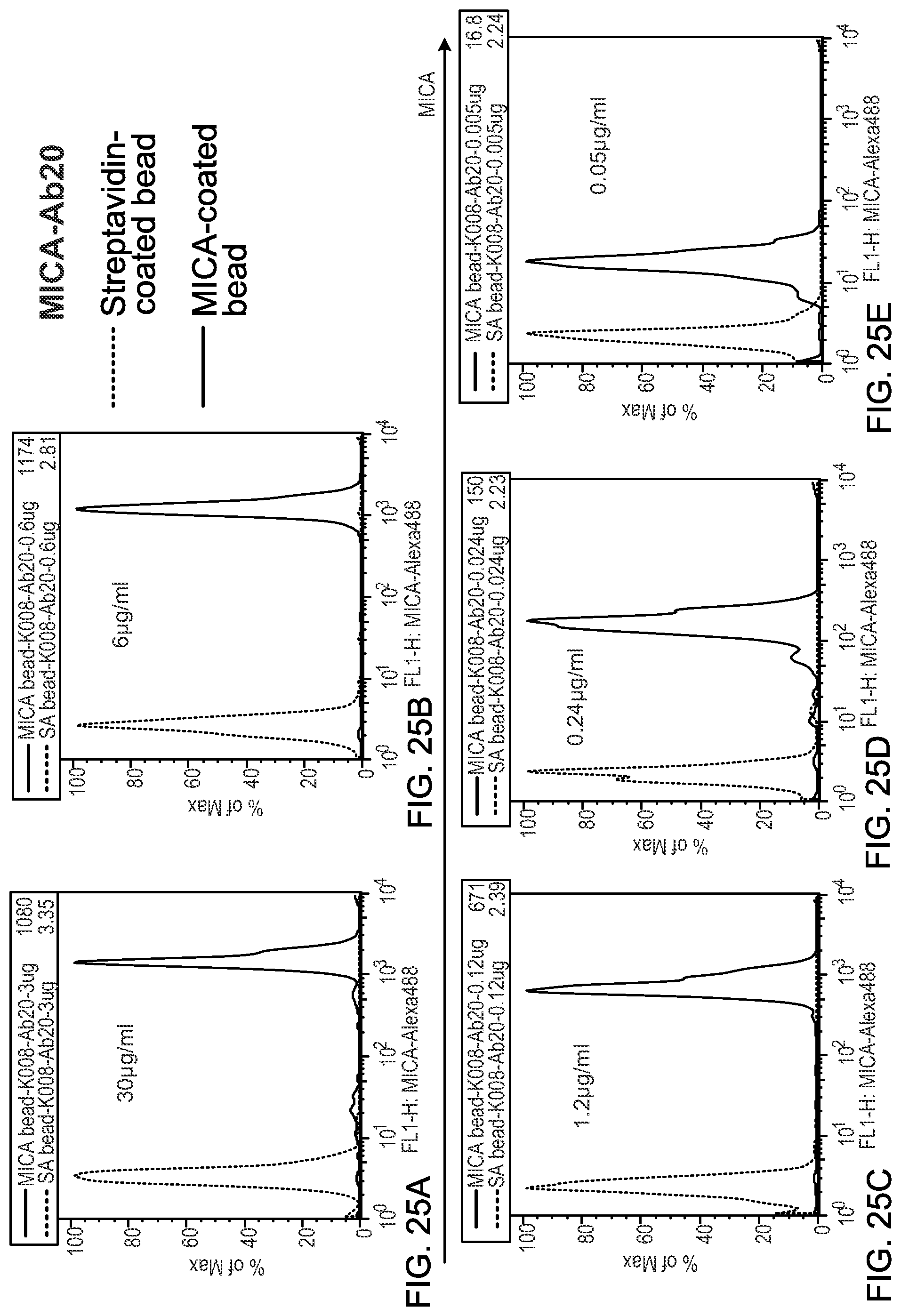
D00016
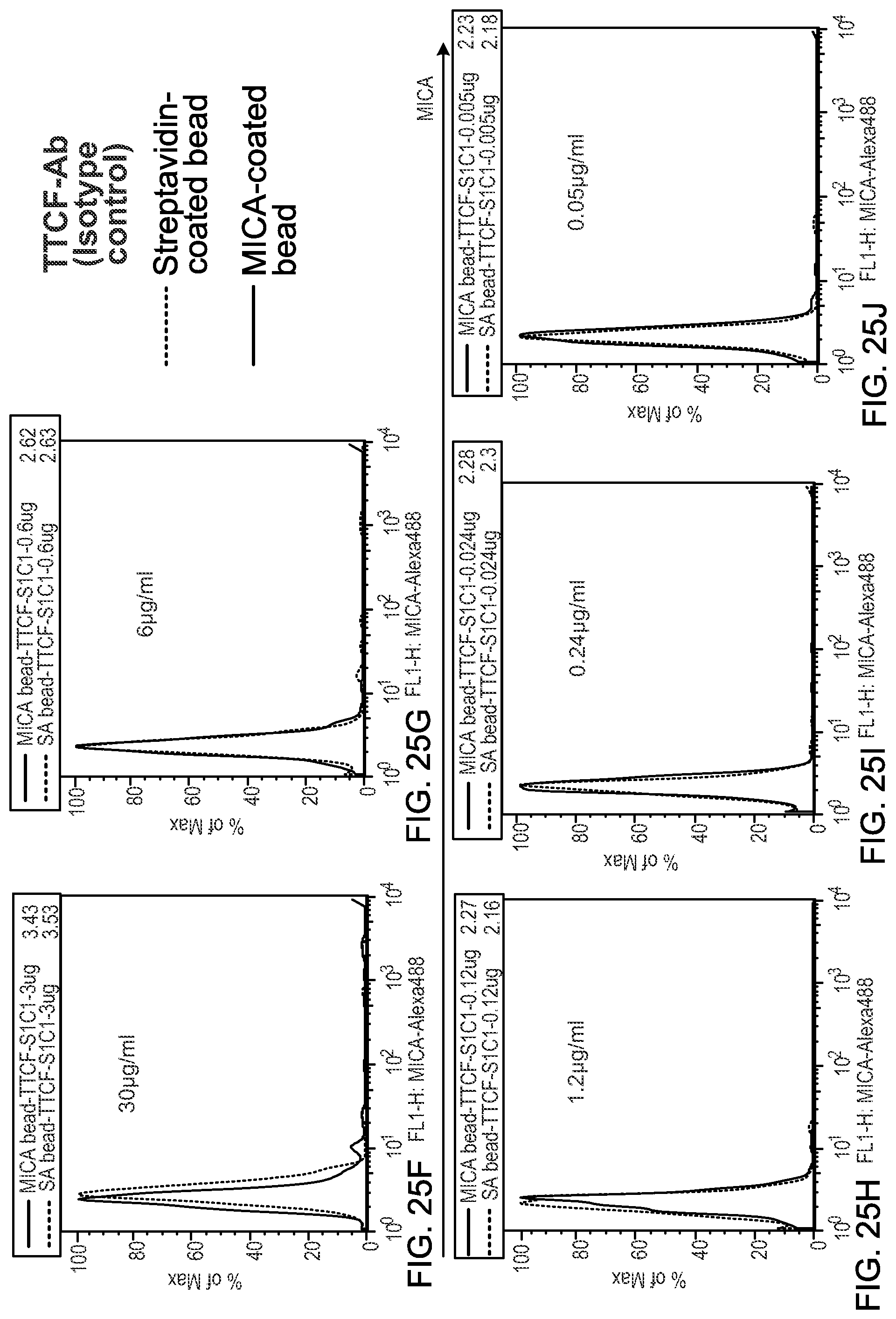
D00017

D00018

D00019

D00020
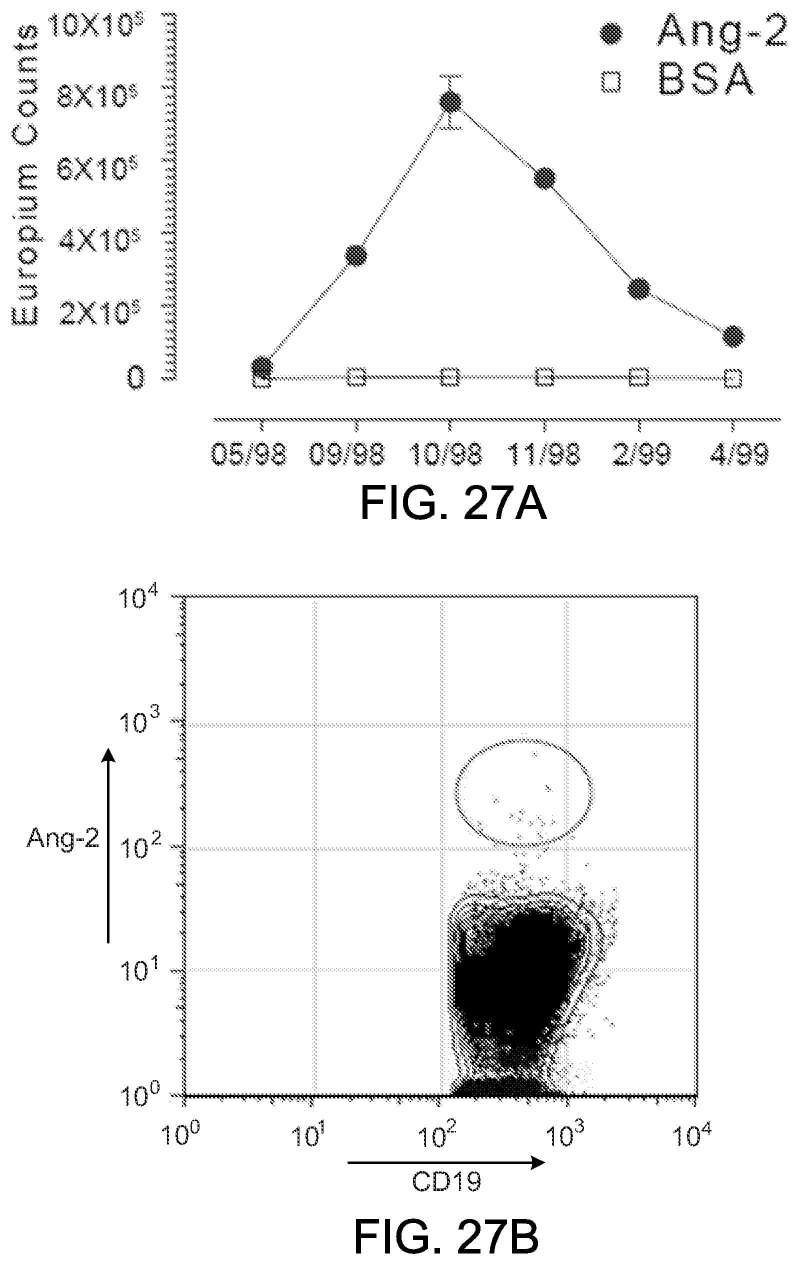
D00021

D00022

D00023

D00024

D00025

D00026

D00027

D00028

D00029

D00030

D00031
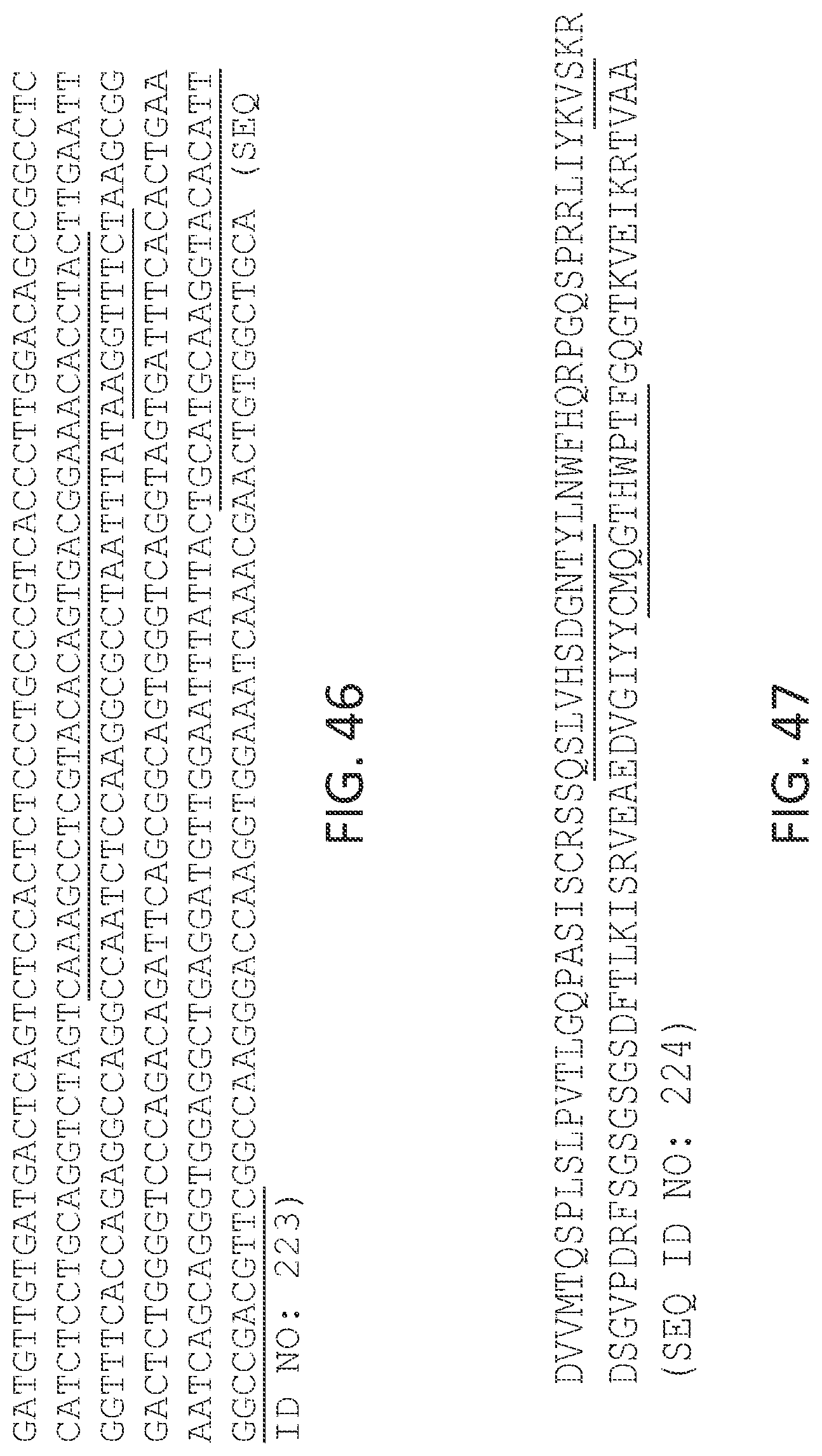
D00032

D00033

D00034

D00035
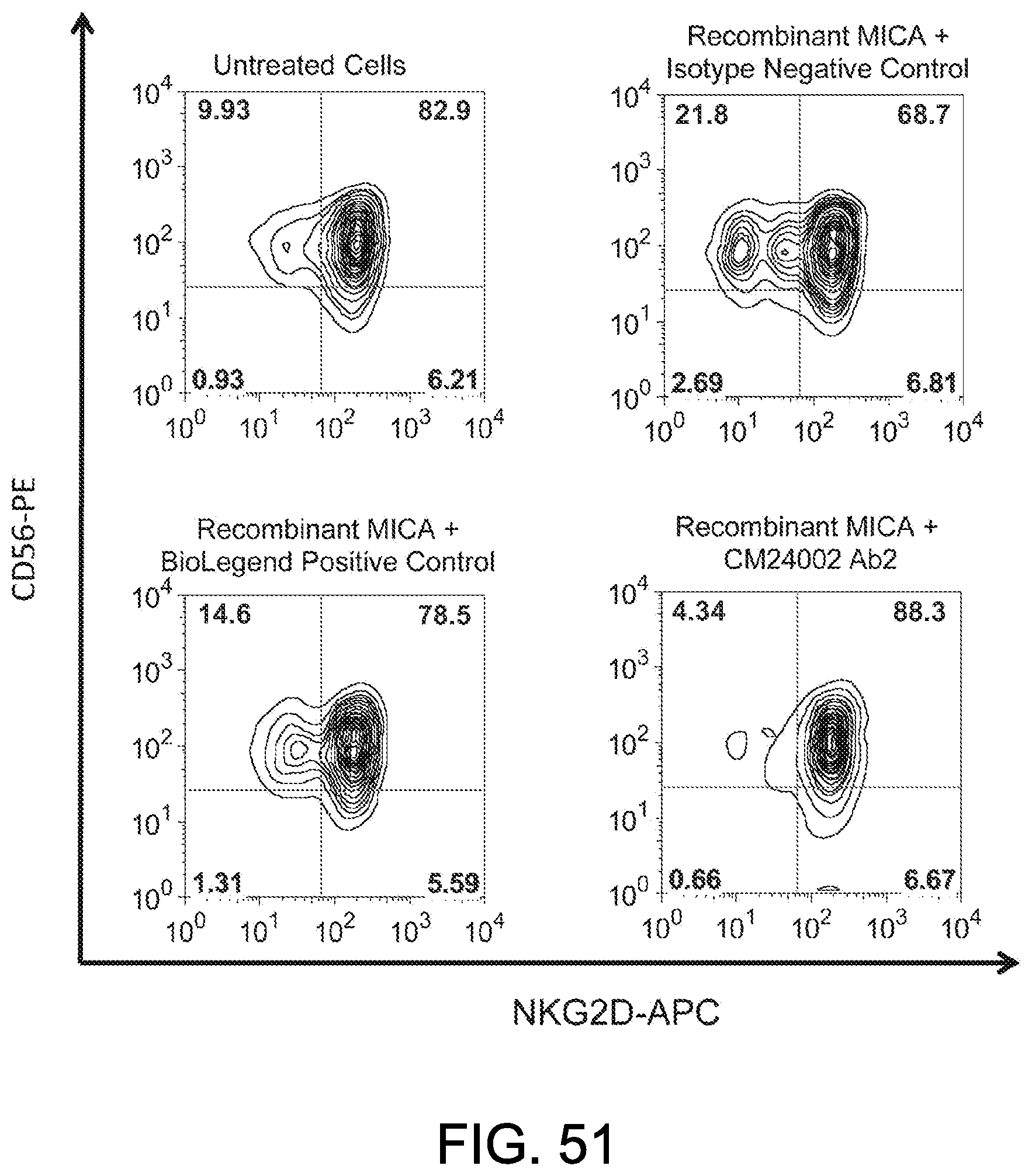
D00036

D00037

D00038

D00039
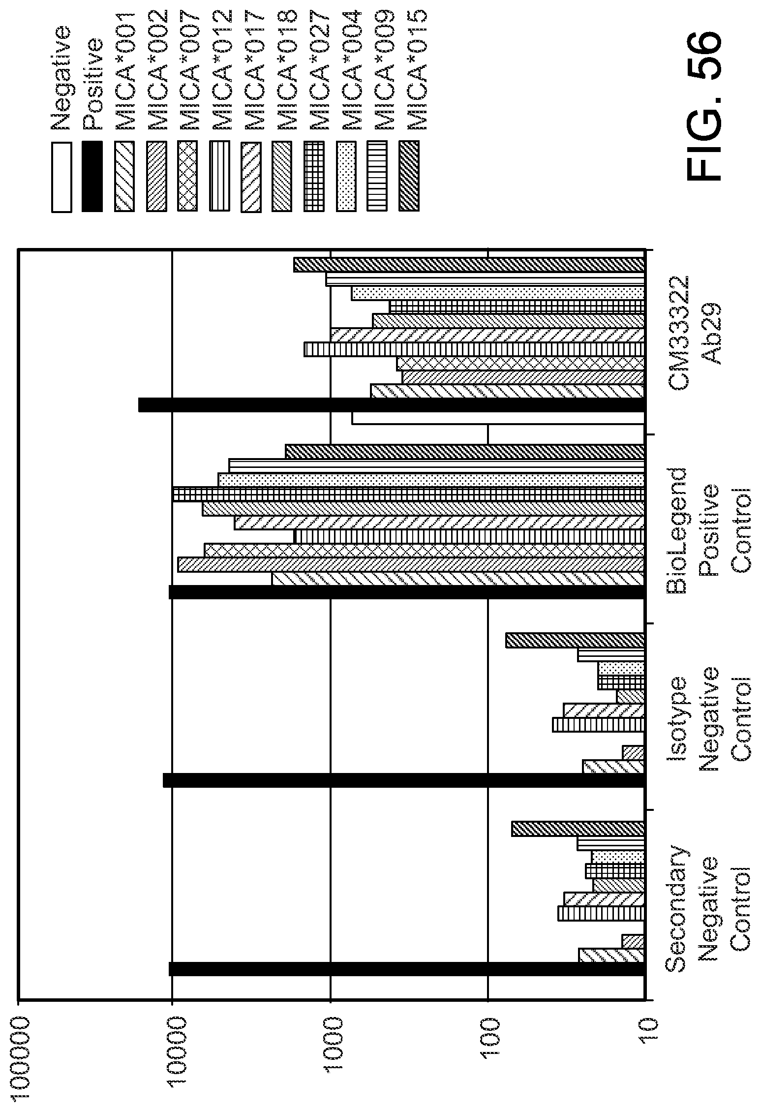
D00040

S00001
XML
uspto.report is an independent third-party trademark research tool that is not affiliated, endorsed, or sponsored by the United States Patent and Trademark Office (USPTO) or any other governmental organization. The information provided by uspto.report is based on publicly available data at the time of writing and is intended for informational purposes only.
While we strive to provide accurate and up-to-date information, we do not guarantee the accuracy, completeness, reliability, or suitability of the information displayed on this site. The use of this site is at your own risk. Any reliance you place on such information is therefore strictly at your own risk.
All official trademark data, including owner information, should be verified by visiting the official USPTO website at www.uspto.gov. This site is not intended to replace professional legal advice and should not be used as a substitute for consulting with a legal professional who is knowledgeable about trademark law.