GARP-TGF-.beta. 1 IGG antibodies
van der Woning , et al. October 6, 2
U.S. patent number 10,793,627 [Application Number 16/698,833] was granted by the patent office on 2020-10-06 for garp-tgf-.beta. 1 igg antibodies. This patent grant is currently assigned to argenx BVBA, Universite Catholique de Louvain. The grantee listed for this patent is argenx BVBA, Universite Catholique de Louvain. Invention is credited to Filip Borgions, Pierre Coulie, Gitte De Boeck, Torsten Dreier, Stephanie Lienart, Sophie Lucas, Lore Marien, Sebastian van der Woning.


View All Diagrams
| United States Patent | 10,793,627 |
| van der Woning , et al. | October 6, 2020 |
GARP-TGF-.beta. 1 IGG antibodies
Abstract
The present invention relates to antibodies and antigen binding fragments thereof, which bind to a complex of GARP and TGF-.beta.1, particularly a complex of human GARP and human TGF-.beta.1. These antibodies and antigen binding fragments exhibit a combination of advantageous properties including high affinity antigen binding and the ability to inhibit the release of active TGF-.beta. from regulatory T cells. The antibodies and antigen binding fragments of the present invention are relatively resistant to deamidation, isomerization and oxidation, such that they display improved stability.
| Inventors: | van der Woning; Sebastian (Bachte-Maria-Leerne, BE), Borgions; Filip (Herk-de-Stad, BE), Dreier; Torsten (Sint-Martens-Latem, BE), Marien; Lore (Ghent, BE), De Boeck; Gitte (Malderon, BE), Lienart; Stephanie (Sawston, GB), Lucas; Sophie (Duisburg, BE), Coulie; Pierre (Kraainem, BE) | ||||||||||
|---|---|---|---|---|---|---|---|---|---|---|---|
| Applicant: |
|
||||||||||
| Assignee: | argenx BVBA (Zwijnaarde,
BE) Universite Catholique de Louvain (Louvain-la-Neuve, BE) |
||||||||||
| Family ID: | 1000005095794 | ||||||||||
| Appl. No.: | 16/698,833 | ||||||||||
| Filed: | November 27, 2019 |
Prior Publication Data
| Document Identifier | Publication Date | |
|---|---|---|
| US 20200095311 A1 | Mar 26, 2020 | |
Related U.S. Patent Documents
| Application Number | Filing Date | Patent Number | Issue Date | ||
|---|---|---|---|---|---|
| 16409679 | May 10, 2019 | ||||
| 15977449 | Nov 19, 2019 | 10479829 | |||
Foreign Application Priority Data
| May 11, 2017 [GB] | 1707561.5 | |||
| Current U.S. Class: | 1/1 |
| Current CPC Class: | C07K 16/22 (20130101); C07K 16/28 (20130101); C07K 2317/92 (20130101); C07K 2317/22 (20130101); C07K 2317/526 (20130101); C07K 2317/76 (20130101); A61K 2039/505 (20130101); C07K 2317/565 (20130101); C07K 2317/32 (20130101); C07K 2317/33 (20130101); C07K 2317/56 (20130101); C07K 2317/732 (20130101); C07K 2317/55 (20130101); C07K 2317/94 (20130101) |
| Current International Class: | C07K 16/22 (20060101); A61K 39/00 (20060101); C07K 16/28 (20060101) |
References Cited [Referenced By]
U.S. Patent Documents
| 4816567 | March 1989 | Cabilly |
| 5591828 | January 1997 | Bosslet |
| 5892019 | April 1999 | Schlom |
| 6162963 | December 2000 | Kucherlapati |
| 9399676 | July 2016 | Schurpf |
| 9573995 | February 2017 | Schurpf |
| 2008/0279834 | November 2008 | Garaczi |
| 2011/0300119 | December 2011 | Tran |
| 2016/0251438 | September 2016 | Lucas et al. |
| 2999819 | Mar 2017 | CA | |||
| 0404097 | Dec 1990 | EP | |||
| 0799836 | Oct 1997 | EP | |||
| 2832747 | Feb 2015 | EP | |||
| 1993011161 | Jun 1993 | WO | |||
| 2005102387 | Nov 2005 | WO | |||
| 2006085938 | Aug 2006 | WO | |||
| 2007113301 | Oct 2007 | WO | |||
| 2009073163 | Jun 2009 | WO | |||
| 2010001251 | Jan 2010 | WO | |||
| 2014033252 | Mar 2014 | WO | |||
| 2014182676 | Nov 2014 | WO | |||
| 2015015003 | Feb 2015 | WO | |||
| 2016125017 | Aug 2016 | WO | |||
| 2018013939 | Jan 2018 | WO | |||
Other References
|
Akhurst et al., 2012 "Targeting the TGF.beta. signalling pathway in disease," Nat Rev Drug Discovery 11(10):790-811. cited by applicant . Boon et al., 2006 "Human T cell responses against melanoma," Annu Rev Immunol 24:175-208. cited by applicant . Chan et al., 2010 "Therapeutic antibodies for autoimmunity and inflammation," Nat Rev Immunol 10(5):301-316. cited by applicant . Colombo et al., 2007 "Regulatory-T-cell inhibition versus depletion: the right choice in cancer immunotherapy," Nat Rev Cancer 7(11):880-887. cited by applicant . Cuende, 2015 "Monoclonal antibodies against GARP1TGF-b1 complexes inhibit the immunosuppressive activity of human regulatory T cells in vivo," Human Immunology 7(284):284ra56, pp. 1-12. cited by applicant . De Vries et al., 2010 "Frequency of circulating Tregs with demethylated FOXP3 intron 1 in melanoma patients receiving tumor vaccines and potentially Treg-depleting agents," Clin Cancer Res 17:841-848. cited by applicant . Edwards et al., 2013 "Regulation of the expression of GARP/latent TGF-.beta.1 complexes on mouse T cells and their role in regulatory T cell and Th17 differentiation," J Immunol 190(11):5506-5515. cited by applicant . Gauthy et al., 2013 "GARP is regulated by miRNAs and controls latent TGF-.beta.1 production by human regulatory T cells," PloS One 8(9):e76186, pp. 1-13. cited by applicant . Genbank Database [Online] (2003) "Immunoglobulin heavy chain variable region, partial [Mus musculus]," Accession No. AA019657.1. National Center for Biotechnology Information. Accessible on the Internet at URL: http://www.ncbi.nlm.nih.gov/protein/27752388. cited by applicant . Genbank Database [Online] (Apr. 30, 2011) "Mus musculus clone YC15 anti-KSHV gH immunoglobulin light chain variable region mRNA, partial cds," Accession No. JF330319.1. National Center for Biotechnology Information. Accessible on the Internet at URL: http://www.ncbi.nlm.nih.gov/nuccore/JF330319.1. cited by applicant . Genbank Database [Online] (Oct. 17, 2015) "Homo sapiens transforming growth factor, beta 1 (TGFB1), mRNA," Accession No. NM_000660. National Center for Biotechnology Information. Accessible on the Internet at URL: http://www.ncbi.nlm.nih.gov/nuccore/NM_000660. cited by applicant . Genbank Database [Online] (Mar. 15, 2015) "Homo sapiens leucine rich repeat containing 32 (LRRC32), transcript variant 2, mRNA," Accession No. NM_001128922. National Center for Biotechnology Information. Accessible on the Internet at URL: http://www.ncbi.nlm.nih.gov/nuccore/NM_001128922. cited by applicant . Genbank Database [Online] (Nov. 7, 2015) "Homo sapiens transforming growth factor beta 2 (TGFB2), transcript variant 1, mRNA," Accession No. NM_001135599. National Center for Biotechnology Information. Accessible on the Internet at URL: http://www.ncbi.nlm.nih.gov/nuccore/NM_00113559. cited by applicant . Genbank Database [Online] (Nov. 7, 2015) "Homo sapiens transforming growth factor beta 2 (TGFB2), transcript variant 2, mRNA," Accession No. NM_003238. National Center for Biotechnology Information. Accessible on the Internet at URL: http://www.ncbi.nlm.nih.gov/nuccore/NM_003238. cited by applicant . Genbank Database [Online] (Mar. 12, 2015) "Predicted: Homo sapiens transforming growth factor, beta 3 (TGFB3), transcript variant X1, mRNA," Accession No. XM_005268028. National Center for Biotechnology Information. Accessible on the Internet at URL: http://www.ncbi.nlm.nih.gov/nuccore/XM_005268028. cited by applicant . Genbank Database [Online] (Jan. 25, 2016) "Predicted: Macaca fascicularis leucine rich repeat containing 32 LRRC32), transcript variant X1, mRNA," Accession No. XM_005579140. National Center for Biotechnology Information. Accessible on the Internet at URL: http://www.ncbi.nlm.nih.gov/nuccore/XM_005579140. cited by applicant . Genbank Database [Online] (Jan. 25, 2016) "Predicted: Macaca fascicularis transforming growth factor beta 1 (TGFB1), transcript variant X1, mRNA," Accession No. XM_005589338. National Center for Biotechnology Information. Accessible on the Internet at URL: http://www.ncbi.nlm.nih.gov/nuccore/XM_005589338. cited by applicant . Graham et al. (1977) "Characteristics of a human cell line transformed by DNA from human adenovirus type 5," J Gen Viral 36:59-72. cited by applicant . Hannon et al., 2013 "Infusion of clinical-grade enriched regulatory T cells delays experimental xenogeneic graft-versus-host disease," Transfusion 54(2):353-363. cited by applicant . Jakobovitz et al., 1993 "Germ-line transmission and expression of a human-derived yeast artificial chromosome," Nat 362:255-258. cited by applicant . Lefranc et al., 1999 "IMGT, the international ImMunoGeneTics database," Nucl Acids Res 27:209-212. cited by applicant . Lemaire et al., 2011 "Induction of autoantibodies against mouse soluble proteins after immunization with living cells presenting the autoantigen at the cell surface in fusion with a human type 2 transmembrane protein," J Immunol Methods 367(1-2):56-62. cited by applicant . Lonning et al., 2011 "Antibody targeting of TGF-13 in cancer patients," Curr Pharm Biotechnol 12:2176-2189. cited by applicant . Roux et al., 1998 "Comparisons of the ability of human IgG3 hinge mutants, IgM, IgE, and IgA2, to form small immune complexes: a role for flexibility and geometry," J Immunol 161:4083-4090. cited by applicant . Shopes, 1992 "A genetically engineered human IgG mutant with enhanced cytolytic activity," J Immunol 148:2918-2922. cited by applicant . Stockis et al., 2009 "Comparison of stable human Treg and Th clones by transcriptional profiling," Eur J Immunol 39:869-882. cited by applicant . Stockis et al., 2009 "Membrane protein GARP is a receptor for latent TGF-beta on the surface of activated human Treg," Eur J Immunol 39(12):3315-3332. cited by applicant . Tran et al., 2009 "GARP (LRRC32) is essential for the surface expression of latent TGF-beta on platelets and activated FOXP3+ regulatory T cells," Proc Natl Acd Sci USA. 106(32):13445-13450. cited by applicant . Wang et al., 2012 "GARP regulates the bioavailability and activation of TGF13," Mol Biol Cell 1129-1139. cited by applicant . Williams et al., 1996 "Sequence and evolution of the human germline V lambda repertoire," J Mol Biol 264:220-232. cited by applicant . Yamane-Ohnuki and Satoh, 2009 "Production of therapeutic antibodies with controlled fucosylation," mAbs 1 (3):230-236. cited by applicant . Zhou et al., 2013 "GARP-TGF-.beta. complexes negatively regulate regulatory T cell development and maintenance of peripheral CD4+ T cells in vivo," J Immunol 190(10):5057-5064. cited by applicant . International Search Report with Written Opinion corresponding to International Patent Application No. PCT/EP2014/066650, dated Oct. 30, 2014. cited by applicant . Liewen et al., 2005 "Characterization of the human GARP (Golgi associated retrograde protein) complex," Exp Cell Res 306(1):24-34. cited by applicant . Miller et al., 2014 "CD4+CD25+ T regulatory cells activated during feline immunodeficiency virus infection convert T helper cells into functional suppressors through a membrane-bound TGF.beta. / GARP-mediated mechanism," Viral. J. 11(1):7. pp. 1-14. cited by applicant . International Search Report with Written Opinion corresponding to International Patent Application No. PCT/IB2016/000182, dated May 10, 2016, 15 pages. cited by applicant . Engelman and Settleman, 2008 "Acquired resistance to tyrosine kinase inhibitors during cancer therapy," Current Opinion in Genetics & Development 18(1):73-79. cited by applicant . Kovacs et al., 2015 "Cardiac Safety of TGF-.beta. Receptor I Kinase Inhibitor LY2157299 Monohydrate in Cancer Patients in a First-in Human Dose Study," Cardiovasc Toxicol 15(4):309-323. cited by applicant . Lacouture et al., 2015 "Cutaneous keratoacanthomas/squamous cell carcinomas associated with neutralization of transforming growth factor beta by the monoclonal antibody fresolimumab (GC1008)," Cancer Immunol Immunother 34(4):437-446. cited by applicant . Li et al., 2006 "Transforming growth factor-beta regulation of immune responses," Annu Rev Immunol 24:99-146. cited by applicant . Lucas et al., 2012 "Demethylation of the FOXP3 gene in human melanoma cells precludes the use of this epigenetic mark for quantification of Tregs in unseparated melanoma samples," Int J Cancer 130(8):1960-1966. cited by applicant . Shull et al., 1992 "Targeted disruption of the mouse transforming growth factor-beta 1 gene results in multifocal inflammatory disease," Nature 359(6397):693-699. cited by applicant . Silence et al., 2014 "ARGX-110, a highly potent antibody targeting CD70, eliminates tumors via both enhanced ADCC and immune checkpoint blockade," MAbs 6(2):523-532. cited by applicant . Tan et al., 2013 "Cellular re- and de-programming by microenvironmental memory: why short TGF-betal pulses can have long effects," Fibrogenesis Tissue Repair 6(1):1-12. cited by applicant . Zheng and Rudensky, 2007 "Foxp3 in control of the regulatory T cell lineage," Nat Immunol 8(5):457-462. cited by applicant . Zheng et al., 2007 "Genome-wide analysis of Foxp3 target genes in developing and mature regulatory T cells," Nature 445(7130):936-940. cited by applicant . Third Party Observation corresponding to European Patent Application No. 14748185.7, dated Jan. 13, 2017 (8 pages). cited by applicant . U.S. Appl. No. 61/819,840, corresponding to European Publication No. WO 2014/182676, dated May 6, 2013 (183 pages). cited by applicant . Non-Final Rejection corresponding to U.S. Appl. No. 15/013,706, dated Nov. 1, 2017 (14 pages). cited by applicant . Shevach, 2017 "Garp as a therapeutic target for modulation of T regulatory cell function," Expert Opin Ther Targets, 21(2):191-200. cited by applicant . Caron et al., 1992 "Engineered humanized dimeric forms of IgG are more effective antibodies," J Exp Med 176(4): 1191-1195. cited by applicant . Chothia and Lesk, 1987 "Canonical structures for the hypervariable regions of immunoglobulins," J Mol Biol 196 (4):901-917. cited by applicant . Genbank Database [Online] (Jan. 28, 2019) "transforming growth factor beta activator LRRC32 precursor [Homo sapiens]," Accession No. NP_001122394.1. National Center for Biotechnology Information. Accessible on the Internet at URL: https://www.ncbi.nlm.nih.gov/protein/np_001122394. cited by applicant . Hinton et al., 2006 "An Engineered Human IgG1 Antibody with Longer Serum Half-Life," J Immunol 176(1):346-356. cited by applicant . Hollinger and Hudson, 2005 "Engineered antibody fragments and the rise of single domains," Nat Biotechnol 23(9)1126-1136. cited by applicant . MacCallum et al., 1996 "Antibody-antigen interactions: contact analysis and binding site topography," J Mol Biol 262(5):732-745. cited by applicant . Mather, 1980 "Establishment and Characterization of Two Distinct Mouse Testicular Epithelial Cell Lines," Biol Reprod 23: 243-252. cited by applicant . Mather et al., 1982 "Culture of testicular cells in hormone-supplemented serum-free medium," Ann N Y Acad Sci 383:44-68. cited by applicant . Morea et al., 2000 "Antibody Modeling: Implications for Engineering and Design," Methods 20:267-279. cited by applicant . Natsume et al., 2009 "Improving effector functions of antibodies for cancer treatment: Enhancing ADCC and CDC," Drug Des Devel Ther 3:7-16. cited by applicant . Presta, 2008 "Molecular engineering and design of therapeutic antibodies," Curr Opin Immunol 20:460-470. cited by applicant . Rudikoff et al., 1982 "Single amino acid substitution altering antigen-binding specificity," Proc Natl Acd Sci USA 79(6):1979-1983. cited by applicant . Stemmer et al., 1995 "Single-step assembly of a gene and entire plasmid from large numbers of oligodeoxyribonucleotides," Gene 164(1):49-53. cited by applicant . Tatusova and Madden, 1999 "Blast 2 sequences--a new tool for comparing protein and nucleotide sequences", FEMS Microbiol Lett 174(2):247-250. cited by applicant . Tramontano et al., 1990 "Framework residue 71 is a major determinant of the position and conformation of the second hypervariable region in the VH domains of immunoglobulins," J Mol Biol 215(1):175-182. cited by applicant . Urlaub and Chasin, 1980 "Isolation of Chinese hamster cell mutants deficient in dihydrofolate reductase activity," Proc Natl Acad Sci U S A (7):4216-4220. cited by applicant . Vaccaro et al., 2005 "Engineering the Fc region of immunoglobulin G to modulate in vivo antibody levels," Nat Biotechnol 23(10):1283-1288. cited by applicant . Yeung et al., 2009 "Engineering Human IgG1 Affinity to Human Neonatal Fc Receptor: Impact of Affinity Improvement on Pharmacokinetics in Primates," J Immunol 182(12):7663-7671. cited by applicant . Zalevsky et al., 2010 "Enhanced antibody half-life improves in vivo activity," Nat Biotechnol 28(2):157-159. cited by applicant . Anonymous (2016) "Dissociation constant." Wikipedia, the free encyclopedia, Jun. 27, 2016 (Jun. 27, 2016) 6 pages. cited by applicant . Drescher 2011 "Characterization of biological interactions with Biacore." Internet Citation, pp. 1-26. cited by applicant . Pauthner et al., 2015 "Antibody engineering & therapeutics, the annual meeting of the antibody society Dec. 7-10, 2015. San Diego, CA. USA." Mabs, vol. 8, No. 3, pp. 617-652. cited by applicant . International Search Report and Written Opinion of PCT/EP2018/062251 dated Jul. 6, 2018, 14 pages. cited by applicant . Wang et al., 2007 "Antibody Structure, Instability, and Formulation," Journal of Pharmaceutical Sciences 96:1-26. cited by applicant . Klimka et al., 2000 "Human anti-CD30 recombinant antibodies by guided phage antibody selection using cell panning," Br J Cancer 83(2):252-260. cited by applicant . Miyara et al., 2009 "Functional delineation and differentiation dynamics of human CD4+ T cells expressing the FoxP3 transcription factor," Immunity 30(6):899-911. cited by applicant . Padlan et al., 1989 "Structure of an antibody-antigen complex: crystal structure of the HyHEL-10 Fab-lysozyme complex," Proc Natl Acad Sci USA 86(15): 5938-5959. cited by applicant . Plato 1 specification, http://www.enzolifesciences.com/ALX-804-867/lrrc32-monoclonal-antibody-pl- ato-1/, Accessed Feb. 1, 2019 (Year: 2019) 2 pages. cited by applicant . Altschul et al., 1990 "Basic local alignment search tool," J Mol Biol 215:403-10. cited by applicant . Armour et al.,1999 "Recombinant human IgG molecules lacking Fcgamma receptor I binding and monocyte triggering activities," Eur J Immunol (1999) 29:2613-24. cited by applicant . Basilico et al., 2014 "Four individually druggable MET hotspots mediate HGF-driven tumor progression," J Clin Invest 124:3172-86. cited by applicant . Cai et al., 2011 "An immunotoxin targeting the gH glycoprotein of KSHV for selective killing of cells in the cytic phase of infection," Antiviral Research 90(3):143-10. cited by applicant . Carillo et al., 1988 "The Multiple Sequence Alignment Problem in Biology," SIAM J Applied Math 48:1073-82. cited by applicant . Carter et al., 1992 "Humanization of an anti-p185HER2 antibody for human cancer therapy," Proc Natl Acad Sci USA 89:4285-89. cited by applicant . Chomczynski et al., "Single-step method of RNA isolation by acid guanidinium thiocyanate-phenolthloroform extraction," Anal Biochem (1987) 162:156-59. cited by applicant . Clackson et al., 1991 "Making antibody fragments using phage display libraries," Nat 352:624-28. cited by applicant . Daley et al., 2005 "Application of monoclonal antibodies in functional and comparative investigations of heavy-chain immunoglobulins in new world camelids," Clin Diagn Lab Immunol 3:380-86. cited by applicant . De Haard et al., 1999 "A large non-immunized human Fab fragment phage library that permits rapid isolation and kinetic analysis of high affinity antibodies," J Biol Chem 274(26):18218-230. cited by applicant . De Haard et al., 2005 "Llama antibodies against a lactococcal protein located at the tip of the phage tail prevent phage infection," J Bacterial 187:4531-41. cited by applicant . Delgado et al., 1996 "Enhanced tumour specificity of an anti-carcinoembrionic antigen Fab' fragment by poly (ethylene glycol) (PEG) modification," Br J Cancer 73(2):175-82. cited by applicant . Devereux et al., 2013 "Regulation of the expression of GARP/latent TGF-131 complexes on mouse T cells and their role in regulatory T cell and Th17 differentiation," J Immunol 190(11):5506-15. cited by applicant . Holliger et al., "`Diabodies`: small bivalent and bispecific antibody fragments," Proc Natl Acad Sci USA (1993) 80:6444-48. cited by applicant . Jones et al., 1986 "Replacing the complementarity-determining regions in a human antibody with those from a mouse," Nat 321:522-25. cited by applicant . Kabat et al., 1977 "Unusual distributions of amino acids in complementarity-determining (hypervariable) segments of heavy and light chains of immunoglobulins and their possible roles in specificity of antibody-combining sites," J Biol Them 252:6609-16. cited by applicant . Kohler et al., 1975 "Continuous cultures of fused cells secreting antibody of predefined specificity," Nat 256:495-97. cited by applicant . Kretschmer et al., 2002 "Strong antigenic selection shaping the immunoglobulin heavy chain repertoire of B-la1 lymphocytes in lambda 2(315) transgenic mice," Eur J Immunol 32:2317-27. cited by applicant . Leong et al., 2001 "Adapting pharmacokinetic properties of a humanized anti-interleukin-8 antibody for therapeutic applications using site-specific pegylation," Cytokines 16(3):106-19. cited by applicant . Marks et al., 1991 "By-passing immunization. Human antibodies from V-gene libraries displayed on phage," J Mol Biol 222:581-97. cited by applicant . Martin et al., 1996 "Structural families in loops of homologous proteins: automatic classification, modelling and application to antibodies, " J Mol Biol 263:800-15. cited by applicant . Merchant et al., 1998 "An efficient route to human bispecific IgG," Nat Biotechnol 167(7):677-81. cited by applicant . Morrison et al., 1984 "Chimeric human antibody molecules: mouse antigen-binding domains with human constant region domains," Proc Natl Acad Sci USA 81(21):6851-55. cited by applicant . Ono et al.,1999 "The humanized anti-HM1.24 antibody effectively kills multiple myeloma cells by human effector cellnediated cytotoxicity," Mal. Immunol 36:387-95. cited by applicant . Pluckthun et al., 1994 "Antibodies from Escherichia coli," In; The Pharmacology of Monoclonal Antibodies. Rosenberg at al.: Eds. Springer-Verlag. vol. 113 pp. 269-315. cited by applicant . Presta et al., 1992 "Antibody engineering," Curr Opin Struct Biol 3:394-98. cited by applicant . Presta et al., 1993 "Humanization of an antibody directed against IgE," J Immunol 151(5):2623-32. cited by applicant . Qu et al., 1999 "Humanization of Immu31, an alpha-fetoprotein-specific antibody," Clin Cancer Res 5:3095s-3100s. cited by applicant . Ridgway et al., 1996 "`Knobs-into-holes` engineering of antibody CH3 domains for heavy chain heterodimerization," Protein Eng 9(7):617-21. cited by applicant . Riechmann et al., 1988 "Reshaping human antibodies for therapy," Nat 332(6162):323-27. cited by applicant . Scatchard et al., 1949 "The attractions of proteins for small molecules and ions," Ann NY Acad Sci 51:660-72. cited by applicant . Schultz et al., 2012 "Humanized mice for immune system investigation: progress promise and challenges," Nat Rev Immunol 12:786-98. cited by applicant . Sims et al., 1993 "A humanized CD18 antibody can block function without cell destruction," J Immunol 151 (4):2296-2308. cited by applicant . Stevenson et al., 1989 "A chimeric antibody with dual Fc regions (bisFabFc) prepared by manipulations at the IgG hinge," Anti-Cancer Drug Design 3:219-30. cited by applicant . Tomlinson et al., 1995 "The structural repertoire of the human V kappa domain," EMBO J 14:4628-38. cited by applicant . Tramontano et al., 1989 "Structural determinants of the conformations of medium-sized loops in proteins," Proteins 6:382-94. cited by applicant . Verhoeyen et al., "Reshaping human antibodies: grafting an antilysozyme activity," Science 239(A847):1534-36. cited by applicant . Lienart et al., 2018 "Structural basis of latent TGF-.beta.1 presentation and activation by GARP on human regulatory T cells," Science 362:952-56. cited by applicant. |
Primary Examiner: Stoica; Elly-Gerald
Attorney, Agent or Firm: Dechert LLP
Parent Case Text
RELATED APPLICATIONS
This application is a continuation of U.S. application Ser. No. 16/409,679, filed May 10, 2019, which is a continuation of U.S. application Ser. No. 15/977,449, filed May 11, 2018, which claims benefit of priority to Great Britain Provisional Application No. 1707561.5, filed on May 11, 2017, the entire contents of each of which are incorporated herein in their entireties by reference thereto.
Claims
The invention claimed is:
1. An antibody that binds to a complex of human glycoprotein A repetitions predominant (hGARP) and TGF-.beta.1, which is an IgG antibody consisting of heavy chains each consisting of the amino acid sequence of SEQ ID NO:16 and light chains each consisting of the amino acid sequence of SEQ ID NO:17.
Description
SEQUENCE LISTING
The instant application contains a Sequence Listing which has been submitted electronically in ASCII format and is hereby incorporated by reference in its entirety. Said ASCII copy, created on Nov. 27, 2019. Is named 381493-466C2(171186)_SL.txt and is 43,819 bytes in size.
FIELD OF THE INVENTION
The present invention relates to antibodies and antigen binding fragments thereof, which bind to a complex of GARP and TGF-.beta.1, particularly a complex of human GARP and human TGF-.beta.1. These antibodies and antigen binding fragments exhibit a combination of advantageous properties including high affinity antigen binding and the ability to inhibit the release of active TGF-.beta. from regulatory T cells. The antibodies and antigen binding fragments of the present invention are improved as compared with prior art antibodies binding to the complex of GARP and TGF-.beta.1. In particular, the antibodies and antigen binding fragments of the present invention are relatively resistant to deamidation, isomerization and oxidation, such that they display improved stability as compared with GARP-TGF-.beta.1 antibodies described in the prior art.
BACKGROUND TO THE INVENTION
Regulatory T cells (otherwise known as "Tregs" or Foxp3.sup.+ T regulatory cells) are an important component of the immune system. In particular, Tregs play a critical role in immune homeostasis by suppressing various aspects of the immune response. As a consequence of their role in coordinating the immune response, dysregulated Treg activity can lead to the development of various diseases and conditions. In particular, insufficient Treg function can result in autoimmune pathology, whereas excessive Treg activity has been linked to the inhibition of anti-tumour responses in cancer patients.
The protein GARP (Glycoprotein A Repetitions Predominant) has been identified as a highly expressed marker on the surface of Tregs, particularly activated Tregs. GARP is an 80 kDa transmembrane protein with an extracellular region comprising 20 leucine-rich repeats. It is also known as LRRC32. GARP serves as the receptor for TGF-.beta., particularly the latent form of TGF-.beta., and is required for the expression of latent TGF-.beta. on Treg cells (E M Shevach. Expert Opin Ther Targets (2016) 21(2), 191-200).
TGF-.beta. is a cytokine known to play a role in multiple processes including cell proliferation and differentiation, tissue morphogenesis, inflammation and apoptosis. It has also been identified as an important growth factor implicated in cancer development, and rather unusually, has been identified as a cytokine with tumour promoting and tumour suppressive properties.
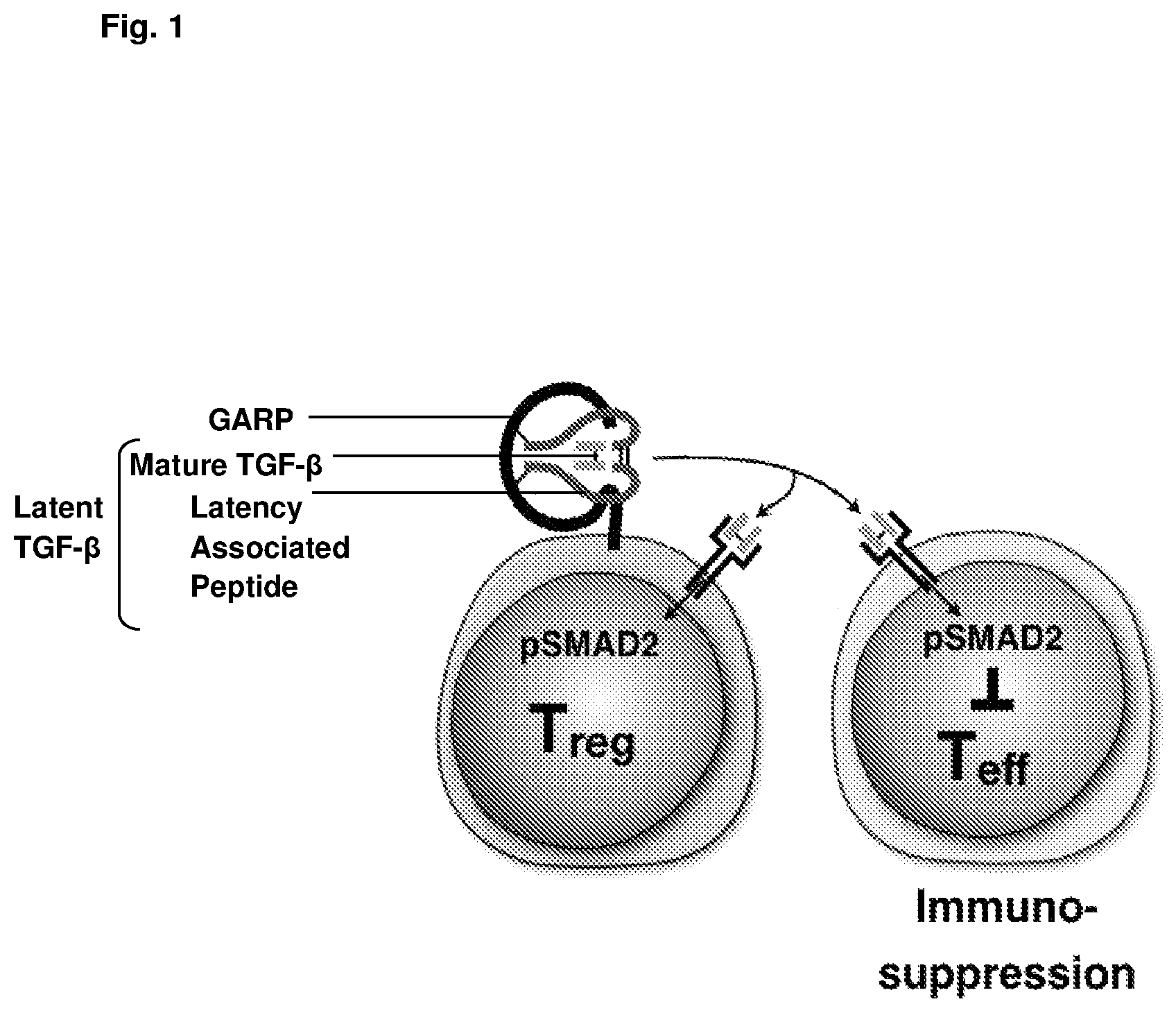
The production and activation of TGF-.beta. is a multi-step process, which is regulated at different levels. TGF-.beta. is synthesised as a pro-TGF-.beta. dimeric precursor, each polypeptide chain consisting of a latency-associated peptide (LAP) and a mature TGF-.beta. region. Pro-TGF-.beta. undergoes cleavage by the enzyme furin to form "latent TGF-.beta.," an inactive form in which the LAP remains non-covalently associated with the mature TGF-.beta. region of each polypeptide chain (see FIG. 1). Membrane-localised GARP serves to transport and anchor latent TGF-.beta. to the cell surface of Tregs, and it is from this membrane-bound GARP-latent TGF-.beta. complex that the active form of TGF-.beta. is released. A variety of mechanisms have been proposed to explain how active TGF-.beta. is released from the GARP-latent TGF-.beta. complex on the surface of Tregs. However, integrins, particularly .alpha.v.beta.6 and .alpha.v.beta.8, are now thought to play an important role in driving the shear forces needed for release of the mature TGF-.beta. dimer.
Once released, the active TGF-.beta. dimer can act as an autocrine or paracrine mediator of downstream signalling pathways. In the context of the immune system, TGF-.beta. release from Treg cells is thought to influence the activity of various T effector cells and also Tregs themselves (see FIG. 1). Since Tregs play an important role in suppressing immunity, it is thought that TGF-.beta. released from Tregs and acting in an autocrine fashion may be involved in mediating Treg suppression. In particular, Treg-derived TGF-.beta.1 is thought to play a significant role in Treg-mediated suppression of tumour immunity.
Given the role of Treg-derived TGF-.beta. in suppressing the immune response in the tumour microenvironment, there has been interest in targeting this pathway as an alternative approach to cancer immunotherapy. For example, therapeutic agents capable of dampening this pathway may serve as useful tools to improve the efficacy of cancer vaccines or other cancer immunotherapy strategies designed to harness the power of the body's immune system to treat cancer.
Cuende et al. (Sci Transl Med. 2015 Apr. 22; 7(284):284ra56) describes the production and characterisation of two monoclonal antibodies (MHG-8 and LHG10), which bind to the GARP-TGF-.beta. complex on Tregs and inhibit TGF-.beta. production. These two antibodies are also described and characterised in International patent applications WO2015/015003 and WO2016/125017. These antibodies were shown to be capable of inhibiting the immunosuppressive activity of human Treg in a xenogeneic graft-versus-host disease mouse model. This work serves to validate the GARP-TGF-.beta. complex as a therapeutic target of interest for the purposes of modulating Treg function and consequently treating diseases such as cancer and autoimmune disease where the level of Treg activity plays an important role. There remains a need however, for improved GARP-TGF-.beta. antibodies capable of inhibiting TGF-.beta. release and thereby modifying Treg activity. The present invention addresses this problem as described herein.
SUMMARY OF INVENTION
The present invention improves upon the state of the art by providing new antibodies and antigen binding fragments thereof, which bind to the human GARP-TGF-.beta.1 complex. The antibodies and antigen binding fragments of the present invention are derived from the GARP-TGF-.beta.1 antibody "LHG-10", described in International patent applications WO2015/015003 and WO2016/125017. The heavy chain and light chain variable domain sequences of LHG-10 are shown in SEQ ID NOs: 1 and 2, respectively, and the light chain variable domain of a chain-shuffled variant, LHG-10.6 (also described in WO2015/015003 and WO2016/125017), is shown in SEQ ID NO: 3. The antibodies of the present invention differ particularly with respect to certain CDR sequences as compared with LHG-10 and LHG-10.6, specifically with respect to the CDR2 and CDR3 sequences of the heavy chain variable domain. The LHG-10 and LHG-10.6 GARP-TGF-.beta. antibodies possess the heavy chain CDR2 sequence: RIDPEDGGTKYAQKFQG (SEQ ID NO: 5); and the heavy chain CDR3 sequence: NEWETVVVGDLMYEYEY (SEQ ID NO: 6), whereas the antibodies of the present invention comprise the heavy chain CDR2 sequence: RIDPEDAGTKYAQKFQG (SEQ ID NO: 12); and the heavy chain CDR3 sequence: YEWETVVVGDLMYEYEY (SEQ ID NO: 13).
The differences in the heavy chain CDR2 and CDR3 sequences reported herein result in antibodies that are improved as compared with the prior art antibodies by virtue of their improved stability. More specifically, the antibodies of the present invention are relatively resistant to deamidation, isomerization and oxidation, such that they exhibit enhanced stability. Surprisingly, these specific substitutions in the heavy chain CDR2 and CDR3 regions that lead to improved stability do not significantly decrease the binding affinity of the antibodies for the GARP-TGF-.beta.1 complex. The improved stability combined with high affinity target binding renders the antibodies of the present invention particularly suitable for clinical development as therapeutic agents, for example as cancer therapeutic agents.
In a first aspect, the present invention provides an antibody or antigen binding fragment thereof, which binds to a complex of human GARP-TGF-.beta.1, wherein the antibody or antigen binding fragment thereof comprises a heavy chain variable domain (VH) wherein:
the VH CDR3 comprises the amino acid sequence YEWETVVVGDLMYEYEY (SEQ ID NO: 13),
the VH CDR2 comprises the amino acid sequence RIDPEDAGTKYAQKFQG (SEQ ID NO: 12), and
the VH CDR1 comprises the amino acid sequence SYYID (SEQ ID NO: 4).
In certain embodiments, the present invention provides an antibody or antigen binding fragment thereof, which binds to a complex of human GARP-TGF-.beta.1, wherein the antibody or antigen binding fragment thereof comprises a heavy chain variable domain (VH) wherein:
the VH CDR3 consists of the amino acid sequence YEWETVVVGDLMYEYEY (SEQ ID NO: 13),
the VH CDR2 consists of the amino acid sequence RIDPEDAGTKYAQKFQG (SEQ ID NO: 12), and
the VH CDR1 consists of the amino acid sequence SYYID (SEQ ID NO: 4).
The antibody or antigen binding fragment may additionally comprise a light chain variable domain (VL) wherein:
the VL CDR3 comprises the amino acid sequence QQYASVPVT (SEQ ID NO: 11),
the VL CDR2 comprises the amino acid sequence GASRLKT (SEQ ID NO: 10), and
the VL CDR1 comprises the amino acid sequence QASQSISSYLA (SEQ ID NO: 9).
In certain embodiments, the antibody or antigen binding fragment may additionally comprise a light chain variable domain (VL) wherein:
the VL CDR3 consists of the amino acid sequence QQYASVPVT (SEQ ID NO: 11),
the VL CDR2 consists of the amino acid sequence GASRLKT (SEQ ID NO: 10), and
the VL CDR1 consists of the amino acid sequence QASQSISSYLA (SEQ ID NO: 9).
In certain embodiments, the antibody or antigen binding fragment thereof comprises a heavy chain variable domain (VH), wherein: the VH CDR3 comprises the amino acid sequence YEWETVVVGDLMYEYEY (SEQ ID NO: 13), the VH CDR2 comprises the amino acid sequence RIDPEDAGTKYAQKFQG (SEQ ID NO: 12), and the VH CDR1 comprises the amino acid sequence SYYID (SEQ ID NO: 4); and a light chain variable domain (VL), wherein: the VL CDR3 comprises the amino acid sequence QQYASVPVT (SEQ ID NO: 11), the VL CDR2 comprises the amino acid sequence GASRLKT (SEQ ID NO: 10), and the VL CDR1 comprises the amino acid sequence QASQSISSYLA (SEQ ID NO: 9).
In certain embodiments, the antibody or antigen binding fragment thereof comprises a heavy chain variable domain (VH), wherein: the VH CDR3 consists of the amino acid sequence YEWETVVVGDLMYEYEY (SEQ ID NO: 13), the VH CDR2 consists of the amino acid sequence RIDPEDAGTKYAQKFQG (SEQ ID NO: 12), and the VH CDR1 consists of the amino acid sequence SYYID (SEQ ID NO: 4); and a light chain variable domain (VL), wherein: the VL CDR3 consists of the amino acid sequence QQYASVPVT (SEQ ID NO: 11), the VL CDR2 consists of the amino acid sequence GASRLKT (SEQ ID NO: 10), and the VL CDR1 consists of the amino acid sequence QASQSISSYLA (SEQ ID NO: 9).
In certain embodiments, the antibodies or antigen binding fragments include at least one heavy chain variable domain (VH) and/or at least one light chain variable domain (VL) that is a humanised, germlined or affinity variant of a camelid-derived VH or VL domain.
In certain embodiments, provided herein are antibodies or antigen binding fragments thereof, which bind to the complex of human GARP and human TGF-.beta.1, wherein the antibodies or antigen binding fragments comprise a heavy chain variable domain selected from the following: (i) a VH comprising or consisting of the amino acid sequence of SEQ ID NO: 14; or (ii) a VH comprising or consisting of an amino acid sequence having at least 90%, at least 95%, at least 97%, at least 98%, or at least 99% identity to SEQ ID NO:14.
Alternatively or in addition, the antibodies or antigen binding fragments may comprise a light chain variable domain (VL) selected from the following: (i) a VL comprising or consisting of the amino acid sequence of SEQ ID NO: 15; or (ii) a VL comprising or consisting of an amino acid sequence having at least 90%, at least 95%, at least 97%, at least 98%, or at least 99% identity to SEQ ID NO: 15.
For embodiments wherein the domains of the antibodies or antigen binding fragments are defined by a particular percentage sequence identity to a reference sequence, the VH and/or VL domains may retain identical CDR sequences to those present in the reference sequence such that the variation is present only within the framework regions.
In a particular embodiment, provided herein are antibodies or antigen binding fragments thereof, wherein the heavy chain variable domain (VH) comprises or consists of the amino acid sequence of SEQ ID NO: 14 and the light chain variable domain (VL) comprises or consists of the amino acid sequence of SEQ ID NO: 15.
In certain embodiments, the antibodies of the invention include the CH1 domain, hinge region, CH2 domain and CH3 domain of a human antibody, in particular human IgG1, IgG2, IgG3 or IgG4. In certain embodiments, the antibody includes the CH3 region of a human IgG4 and includes the substitution S228P in the CH3 domain.
The antibodies which bind the GARP-TGF-.beta.1 complex may comprise at least one full-length immunoglobulin heavy chain and/or at least one full-length lambda or kappa light chain. In certain embodiments, the antibodies comprise a heavy chain comprising the amino acid sequence of SEQ ID NO: 16 and a light chain comprising the amino acid sequence of SEQ ID NO: 17. In certain embodiments, provided herein are monoclonal antibodies comprising a heavy chain with at least 90%, at least 95%, at least 97%, at least 98%, or at least 99% sequence identity to the amino acid sequence shown as SEQ ID NO: 16. In certain embodiments, provided herein are monoclonal antibodies comprising a light chain with at least 90%, at least 95%, at least 97%, at least 98%, or at least 99% sequence identity to the amino acid sequence shown as SEQ ID NO: 17. In certain embodiments, provided herein are monoclonal antibodies comprising a heavy chain with at least 90%, at least 95%, at least 97%, at least 98%, or at least 99% sequence identity to the amino acid sequence shown as SEQ ID NO: 16, and a light chain with at least 90%, at least 95%, at least 97%, at least 98%, or at least 99% sequence identity to the amino acid sequence shown as SEQ ID NO: 17.
For embodiments wherein the heavy and/or light chains of the antibodies are defined by a particular percentage sequence identity to a reference sequence, the heavy chain and/or light chain may retain identical CDR sequences to those present in the reference sequence such that the variation is present only outside the CDR regions.
Unless otherwise stated in the present application, % sequence identity between two amino acid sequences may be determined by comparing these two sequences aligned in an optimum manner and in which the amino acid sequence to be compared can comprise additions or deletions with respect to the reference sequence for an optimum alignment between these two sequences. The percentage of identity is calculated by determining the number of identical positions for which the amino acid residue is identical between the two sequences, by dividing this number of identical positions by the total number of positions in the comparison window and by multiplying the result obtained by 100 in order to obtain the percentage of identity between these two sequences. For example, it is possible to use the BLAST program, "BLAST 2 sequences" (Tatusova et al, "Blast 2 sequences--a new tool for comparing protein and nucleotide sequences", FEMS Microbiol Lett. 174:247-250), the parameters used being those given by default (in particular for the parameters "open gap penalty": 5, and "extension gap penalty": 2; the matrix chosen being, for example, the matrix "BLOSUM 62" proposed by the program), the percentage of identity between the two sequences to be compared being calculated directly by the program.
The GARP-TGF-.beta.1 antibodies or antigen binding fragments thereof provided herein may each exhibit one or more of the following properties/features: the antibody or antigen binding fragment may cross-react with the GARP-TGF-.beta. complex of Cynomolgus origin; the antibody or antigen binding fragment may bind to human GARP-TGF-.beta.1 with high affinity; the antibody or antigen binding fragment may include a VH domain and VL domain that when tested as a Fab fragment exhibit an off-rate (K.sub.off) for the complex of human GARP and TGF-.beta.1 of less than 5.times.10.sup.-4 s.sup.-1; the antibody or antigen binding fragment may include a VH domain and VL domain that when tested as a Fab fragment exhibit an off-rate (K.sub.off) for the complex of human GARP and TGF-.beta.1 in the range 1.times.10.sup.-6 s.sup.-1 to 5.times.10.sup.-4 s.sup.-1; the antibody or antigen binding fragment may include a VH domain and VL domain that when tested as a mAb exhibit a K.sub.D of less than 1.7.times.10.sup.-9 M; the antibody or antigen binding fragment may block release of active TGF-.beta.1 from regulatory T cells.
In further aspects, the invention also provides polynucleotide molecules which encode the above-listed antibodies and antigen binding fragments, in addition to expression vectors comprising the polynucleotides, host cells containing the vectors, and methods of recombinant expression/production of the antibodies described herein.
In a still further aspect, the invention provides a pharmaceutical composition comprising any one of the GARP-TGF-.beta.1 antibodies or antigen binding fragments thereof described herein, and a pharmaceutically acceptable carrier or excipient.
A still further aspect of the invention concerns methods of medical treatment using the above-listed GARP-TGF-.beta.1 antibodies or antigen binding fragments thereof, particularly in the prophylaxis and/or treatment of TGF-.beta.-related disorders. In certain embodiments, the invention relates to methods of treatment using the GARP-TGF-.beta.1 antibodies or antigen binding fragments thereof, wherein the disease or condition to be treated is selected from the group consisting of inflammatory diseases, chronic infection, cancer, fibrosis, cardiovascular disease, cerebrovascular disease and neurodegenerative disease. In certain embodiments, the GARP-TGF-.beta.1 antibodies or antigen binding fragments thereof are administered in combination with another treatment as part of a combination therapy. For example, the GARP-TGF-.beta.1 antibodies or antigen binding fragments thereof may be administered in combination with an immunotherapeutic agent, optionally an immunostimulatory antibody or a tumour vaccine.
These and other embodiments of the invention will be better appreciated and understood when considered in conjunction with the following description and the accompanying drawings. It should be understood, however, that the following description, while indicating various embodiments of the invention and numerous specific details thereof, is given by way of illustration and not of limitation. Many substitutions, modifications, additions and/or rearrangements may be made within the scope of the invention without departing from the spirit thereof, and the invention includes all such substitutions, modifications, additions and/or rearrangements.
BRIEF DESCRIPTION OF THE DRAWINGS
FIG. 1 is a schematic showing the binding of latent TGF-.beta. to GARP on the surface of regulatory T cells. TGF-.beta. is produced as a precursor, "pro-TGF-.beta." and undergoes cleavage to produce "latent-TGF-.beta.", a form in which the mature TGF-.beta. dimer remains non-covalently associated with the latency associated peptide (LAP) region of each polypeptide. It is this latent form that binds to GARP on the surface of Treg cells. Integrins .alpha.v.beta.6 and .alpha.v.beta.8 are thought to be responsible for mediating the release of mature or "active TGF-.beta." from the cell surface. This active form can act in a paracrine fashion to bring about effects in a variety of target cells, or can act as an autocrine mediator by binding to the TGF-.beta. receptor on Treg cells.
FIG. 2 shows target binding activity as measured by surface plasmon resonance (SPR) for antibodies 39B6 IgG1.sup.N297Q and 39B6 IgG4.sup.S228P over a 56-day period for samples stored at -20.degree. C., 5.degree. C. and 37.degree. C. in PBS or PBSTween (PBSTw). The reference sample (-20.degree. C.) was set as 100% binding activity at each time point.
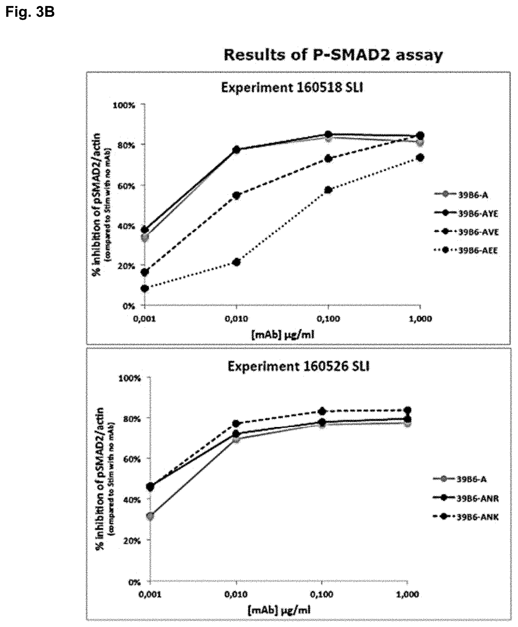
FIGS. 3A and 3B show the results of testing the 39B6-A antibody variants in an assay designed to monitor SMAD2 phosphorylation downstream of TGF-.beta. receptor activation. SMAD2 phosphorylation serves as a marker of activation of the TGF-.beta. signalling pathway, following TGF-.beta. binding to its receptor. If SMAD2 phosphorylation is reduced, TGF-.beta. activity is inhibited. FIG. 3A: Western blots showing decreases in SMAD2 phosphorylation in the presence of different concentrations of the GARP-TGF-.beta. antibodies 39B6-A, 39B6-AVE, 39B6-AEE, 39B6-AYE, 39B6-ANR and 39B6-ANK. FIG. 3B: Graphical representation of the data in (A) showing the percentage inhibition of SMAD2 phosphorylation at different antibody concentrations.
FIG. 4 shows the results of testing the 39B6-A antibody variants in an assay designed to measure TGF-.beta. activity via a luciferase reporter gene conjugated to a SMAD promoter. Graphs show percentage inhibition of luminescence signal in the presence of different concentrations of the GARP-TGF-.beta. antibodies LHG-10, 39B6-A, 39B6-AVE, 39B6-AEE, 39B6-AYE, 39B6-ANR and 39B6-ANK.
FIG. 5 shows the percentage aggregate formation over a 56-day period with antibodies 39B6-AVE, 39B6-AYE, 39B6-ANK and 39B6-ANR stored at 5.degree. C. and 37.degree. C. Aggregate formation was monitored by size exclusion chromatography (SE-HPLC).
FIG. 6 shows the percentage fragment formation over a 56-day period with antibodies 39B6-AVE, 39B6-AYE, 39B6-ANK and 39B6-ANR stored at 37.degree. C. Fragment formation was monitored by size exclusion chromatography (SE-HPLC).
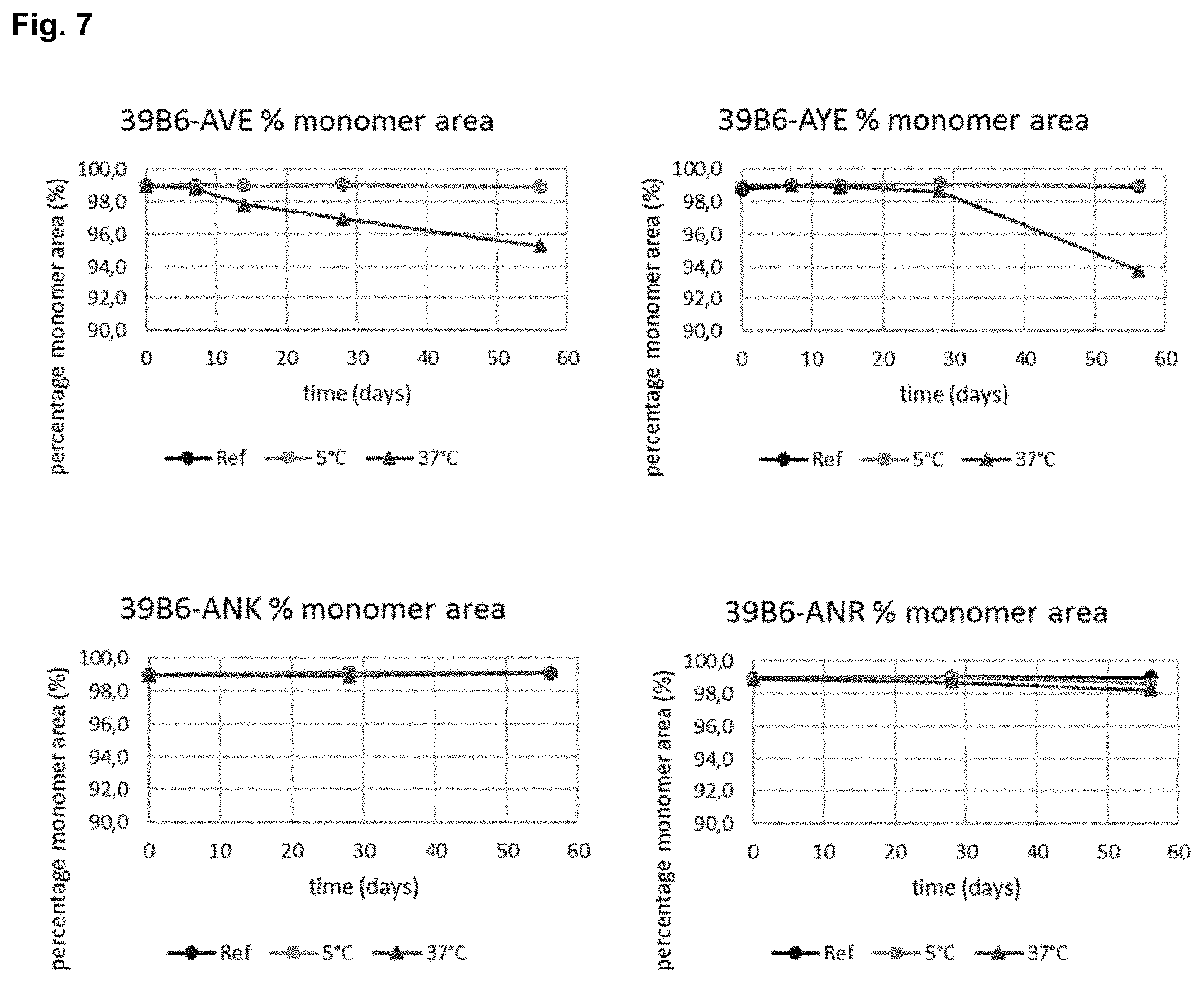
FIG. 7 shows the percentage monomer area over a 56-day period with antibodies 39B6-AVE, 39B6-AYE, 39B6-ANK and 39B6-ANR stored at 5.degree. C. and 37.degree. C. Monomer area was monitored by size exclusion chromatography (SE-HPLC).
FIGS. 8A-8D show the results of SDS-PAGE analysis of antibody samples stored for 56 days at a reference temperature (-20.degree. C.), at 5.degree. C. and at 37.degree. C. FIG. 8A: 39B6-AVE. FIG. 8B: 39B6-AYE. FIG. 8C: 39B6-ANK. FIG. 8D: 39B6-ANR. Markers appear at the centre of each gel. To the left of the markers, the 3 samples are (i) Ref; (ii) 5.degree. C.; and (iii) 37.degree. C. samples tested under non-reducing conditions and to the right of the markers, the 3 samples are (i) Ref; (ii) 5.degree. C.; and (iii) 37.degree. C. samples tested under reducing conditions.
FIG. 9 shows target binding activity as measured by SPR for antibodies 39B6-AVE, 39B6-AYE, 39B6-ANK and 39B6-ANR over a 56-day period for samples stored at -20.degree. C., 5.degree. C. and 37.degree. C. The reference sample (-20.degree. C.) was set as 100% binding activity at each time point.
FIG. 10 shows the protein concentration (mg/ml) for samples of antibodies 39B6-AVE, 39B6-AYE, 39B6-ANK and 39B6-ANR over a 56-day period for samples stored at -20.degree. C. (ref), 5.degree. C. and 37.degree. C.
FIG. 11 shows the results of SDS-PAGE analysis of antibody samples after 10 freeze-thaw cycles. Markers appear at the centre of the gel. To the left of the markers, the 4 samples are the samples analysed under non-reducing conditions: (i) Ref for 39B6-AVE; (ii) Freeze-thaw sample for 39B6-AVE; (iii) Ref for 39B6-AYE; and (iv) Freeze-thaw sample for 39B6-AYE. To the right of the markers, the 4 samples are the samples analysed under reducing conditions: (i) Ref for 39B6-AVE; (ii) Freeze-thaw sample for 39B6-AVE; (iii) Ref for 39B6-AYE; and (iv) Freeze-thaw sample for 39B6-AYE.
FIG. 12 shows target binding activity as measured by SPR following 10 freeze-thaw cycles for antibodies 39B6-AVE and 39B6-AYE. The reference sample (-20.degree. C.) was set as 100% binding activity at each time point.
FIG. 13 shows the protein concentration (mg/ml) for samples of antibodies 39B6-AVE and 39B6-AYE following 10 freeze-thaw cycles.
FIG. 14 shows target binding activity as measured by SPR following thermal stability testing at temperatures ranging from 54.6.degree. C. through 71.4.degree. C. for antibodies 39B6-AVE, 39B6-AYE, 39B6-ANK and 39B6-ANR. The reference sample was set as 100% binding activity.
FIG. 15 shows the results of SDS-PAGE analysis of antibody samples after 96 hours of rotation. Markers appear at the centre of the gel. To the left of the markers, the 4 samples are the samples analysed under non-reducing conditions: (i) Ref for 39B6-AVE; (ii) Rotated sample for 39B6-AVE; (iii) Ref for 39B6-AYE; and (iv) Rotated sample for 39B6-AYE. To the right of the markers, the 4 samples are the samples analysed under reducing conditions: (i) Ref for 39B6-AVE; (ii) Rotated sample for 39B6-AVE; (iii) Ref for 39B6-AYE; and (iv) Rotated sample for 39B6-AYE.
FIG. 16 shows target binding activity as measured by SPR following rotational stability testing for mAbs 39B6-AVE and 39B6-AYE. The reference sample was set as 100% binding activity.
FIG. 17 shows the protein concentration (mg/ml) for samples of mAbs 39B6-AVE and 39B6-AYE following rotational stability testing.
FIG. 18 shows the relative amount of deamidation and isomerization of position N95 in the antibodies 39B6-ANE, 39B6-ANR and 39B6-ANK over a 56-day period. Antibodies 39B6-AVE and 39B6-AYE are not included because these antibodies have had residue "N95" removed from CDR3. Also shown is the relative binding activity for mAbs 39B6-ANE, 39B6-AVE, 39B6-AYE, 39B6-ANK and 39B6-ANR over the 56 d time course, for samples stored at 37.degree. C.
FIG. 19 shows the requirement for mature TGF-.beta. in the binding of 39B6-AYE (ARGX-115) to the GARP-TGF-.beta. complex. ELISA plates were coated with either GARP or the anti-GARP Ab ARGX-115. For ELISA plates coated with GARP, a complex with either full-length latent TGF-.beta. (including both the LAP and mature TGF-.beta. regions) or a complex with recombinant LAP was allowed to form by the addition of the relevant recombinant protein. For ELISA plates coated with ARGX-115, GARP was added and then either full-length latent TGF-.beta. or LAP was added. ARGX-115 was only able to bind GARP in the presence of full-length TGF-.beta.. Binding of ARGX-115 to the GARP-LAP complex did not occur. In contrast, an anti-LAP antibody was able to bind to the GARP-LAP complex. This demonstrates the requirement for mature TGF-.beta. for binding of ARGX-115 to the GARP-TGF-.beta. complex.
FIG. 20 shows the ability of antibodies to neutralize TGF-.beta. activation by the GARP-TGF-.beta. complex with various mutant forms of TGF-.beta.. Neutralizing activity of ARGX-115 was abrogated by mutation of R58 in LAP and K338 in mature TGF-.beta..
FIG. 21 shows a gradient PCR device program for the thermal stability study.
DETAILED DESCRIPTION
A. Definitions
"GARP"--GARP (Glycoprotein A Repetitions Predominant) is a member of the leucine-rich repeat family of proteins. It is also called Leucine Rich Repeat Containing 32 (LRRC32). GARP is an 80 kDa transmembrane protein with an extracellular region composed primarily of 20 leucine-rich repeats. The complete amino acid sequence of the human GARP protein transcript variant 2 (GenBank Accession No. NP_001122394) is:
TABLE-US-00001 (SEQ ID NO: 33) MRPQILLLLALLTLGLAAQHQDKVPCKMVDKKVSCQVLGLLQVPSVLPPD TETLDLSGNQLRSILASPLGFYTALRHLDLSTNEISFLQPGAFQALTHLE HLSLAHNRLAMATALSAGGLGPLPRVTSLDLSGNSLYSGLLERLLGEAPS LHTLSLAENSLTRLTRHTFRDMPALEQLDLHSNVLMDIEDGAFEGLPRLT HLNLSRNSLTCISDFSLQQLRVLDLSCNSIEAFQTASQPQAEFQLTWLDL RENKLLHFPDLAALPRLIYLNLSNNLIRLPTGPPQDSKGIHAPSEGWSAL PLSAPSGNASGRPLSQLLNLDLSYNEIELIPDSFLEHLTSLCFLNLSRNC LRTFEARRLGSLPCLMLLDLSHNALETLELGARALGSLRTLLLQGNALRD LPPYTFANLASLQRLNLQGNRVSPCGGPDEPGPSGCVAFSGITSLRSLSL VDNEIELLRAGAFLHTPLTELDLSSNPGLEVATGALGGLEASLEVLALQG NGLMVLQVDLPCFICLKRLNLAENRLSHLPAVVTQAVSLEVLDLRNNSFS LLPGSAMGGLETSLRRLYLQGNPLSCCGNGWLAAQLHQGRVDVDATQDLI CRFSSQEEVSLSHVRPEDCEKGGLKNINLIIILTFILVSAILLTTLAACC CVRRQKFNQQYKA.
"TGF-.beta."--TGF-.beta. is a cytokine belonging to a superfamily of growth factors. There are three distinct isoforms of TGF-.beta. (TGF-.beta.1, TGF-.beta.2 and TGF-.beta.3) encoded by three distinct genes, but the overall structures of the TGF-.beta. isoforms are highly similar, with homologies in the order of 70-80%. The term TGF-.beta., as used herein, is typically used to encompass all three different isoforms of the TGF-.beta. cytokine, unless the context indicates otherwise.
All three TGF-.beta. isoforms are encoded as large protein precursors; TGF-.beta.1 (GenBank Accession No: NM_000660) contains 390 amino acids, and TGF-.beta.2 (GenBank Accession Nos: NM_001135599 and NM_003238) and TGF-.beta.3 (GenBank Accession No: XM_005268028) each contain 412 amino acids. They each have an N-terminal signal peptide of 20-30 amino acids that is required for secretion from a cell, a pro-region (named latency associated peptide or LAP), and a 112-114 amino acid C-terminal region that becomes the mature TGF-.beta. molecule following its release from the pro-region by proteolytic cleavage. After proteolytic cleavage, LAP and mature TGF-.beta. remain non-covalently associated and form the "latent TGF-.beta." molecule. In this latent form, mature TGF-.beta. is prevented from binding to the TGF-.beta. receptor by LAP. To exert a signal, mature TGF-.beta. must be released from LAP. Mature TGF-.beta. that is not associated to LAP is called active TGF-.beta., as it can bind to the TGF-.beta. receptor and transduce a signal.
TABLE-US-00002 Full length TGF-.beta.1 has the following amino acid sequence: (SEQ ID NO: 34) MPPSGLRLLPLLLPLLWLLVLTPGRPAAGLSTCKTIDMELVKRKRIEAIR GQILSKLRLASPPSQGEVPPGPLPEAVLALYNSTRDRVAGESAEPEPEPE ADYYAKEVTRVLMVETHNEIYDKFKQSTHSIYMFFNTSELREAVPEPVLL SRAELRLLRLKLKVEQHVELYQKYSNNSWRYLSNRLLAPSDSPEWLSFDV TGVVRQWLSRGGEIEGFRLSAHCSCDSRDNTLQVDINGFTTGRRGDLATI HGMNRPFLLLMATPLERAQHLQSSRHRRALDTNYCFSSTEKNCCVRQLYI DFRKDLGWKVVIHEPKGYHANFCLGPCPYIWSLDTQYSKVLALYNQHNPG ASAAPCCVPQALEPLPIVYYVGRKPKVEQLSNMIVRSCKCS. LAP has the following amino acid sequence: (SEQ ID NO: 35) LSTCKTIDMELVKRKRIEAIRGQILSKLRLASPPSQGEVPPGPLPEAVLA LYNSTRDRVAGESAEPEPEPEADYYAKEVTRVLMVETHNEIYDKFKQSTH SIYMFFNTSELREAVPEPVLLSRAELRLLRLKLKVEQHVELYQKYSNNSW RYLSNRLLAPSDSPEWLSFDVTGVVRQWLSRGGEIEGFRLSAHCSCDSRD NTLQVDINGFTTGRRGDLATIHGMNRPFLLLMATPLERAQHLQSSRHRR. Mature TGF-.beta.1 has the following amino acid sequence: (SEQ ID NO: 36) ALDTNYCFSSTEKNCCVRQLYIDFRKDLGWKVVIHEPKGYHANFCLGPCP YIWSLDTQYSKVLALYNQHNPGASAAPCCVPQALEPLPIVYYVGRKPKVE QLSNMIVRSCKCS.
"GARP-TGF-.beta. complex"--As used herein, the GARP-TGF-.beta. complex means the native complex that forms when latent TGF-.beta. binds to GARP, particularly GARP located on the surface of Treg cells. Although not specified throughout, the "GARP-TGF-.beta. complex," or simply "GARP-TGF-.beta.," as used herein, is intended to mean the complex between GARP and latent TGF-.beta.. The binding of GARP to TGF-.beta., more specifically latent TGF-.beta., has been characterised at the molecular level, for example as reported in Wang et al. Mol Biol Cell. 2012 March; 23(6):1129-39. GARP forms a disulphide linkage to the Cys4 of latent TGF-.beta. and also associates with latent TGF-.beta. through non-covalent interactions. There are 15 Cys residues in the extracellular domain of GARP, and GARP uses Cys-192 and Cys-331 to form disulphide linkages to the two Cys4 residues of latent TGF-.beta.. It follows that one GARP protein associates with one latent TGF-.beta. dimer.
"Antibody" or "Immunoglobulin"--As used herein, the term "immunoglobulin" includes a polypeptide having a combination of two heavy and two light chains whether or not it possesses any relevant specific immunoreactivity. "Antibodies" refer to such assemblies which have significant specific immunoreactive activity to an antigen of interest (e.g. the complex of GARP and TGF-.beta.). The term "GARP-TGF-.beta. antibodies" is used herein to refer to antibodies which exhibit immunological specificity for the complex of GARP and TGF-.beta.1, particularly the human GARP-TGF-.beta.1 complex and in some cases species homologues thereof. Antibodies and immunoglobulins comprise light and heavy chains, with or without an interchain covalent linkage between them. Basic immunoglobulin structures in vertebrate systems are relatively well understood.
The generic term "immunoglobulin" comprises five distinct classes of antibody (IgG, IgM, IgA, IgD or IgE) that can be distinguished biochemically. All five classes of antibodies are within the scope of the present invention. The following discussion will generally be directed to the IgG class of immunoglobulin molecules. With regard to IgG, immunoglobulins typically comprise two identical light polypeptide chains of molecular weight approximately 23,000 Daltons, and two identical heavy chains of molecular weight 53,000-70,000. The four chains are joined by disulfide bonds in a "Y" configuration wherein the light chains bracket the heavy chains starting at the mouth of the "Y" and continuing through the variable region.
The light chains of an antibody are classified as either kappa (.kappa.) or lambda (.lamda.). Each heavy chain class may be bound with either a kappa or lambda light chain. In general, the light and heavy chains are covalently bonded to each other, and the "tail" portions of the two heavy chains are bonded to each other by covalent disulfide linkages or non-covalent linkages when the immunoglobulins are generated either by hybridomas, B cells or genetically engineered host cells. In the heavy chain, the amino acid sequences run from an N-terminus at the forked ends of the Y configuration to the C-terminus at the bottom of each chain. Those skilled in the art will appreciate that heavy chains are classified as gamma, mu, alpha, delta, or epsilon, (.gamma., .mu., .alpha., .delta., .epsilon.) with some subclasses among them (e.g., .gamma.1-.gamma.4). It is the nature of this chain that determines the "class" of the antibody as IgG, IgM, IgA, IgD or IgE, respectively. The immunoglobulin subclasses (isotypes) e.g., IgG1, IgG2, IgG3, IgG4, IgA1, etc. are well characterized and are known to confer functional specialization. Modified versions of each of these classes and isotypes are readily discernible to the skilled artisan in view of the instant disclosure and, accordingly, are within the scope of the instant invention.
As indicated above, the variable region of an antibody allows the antibody to selectively recognize and specifically bind epitopes on antigens. That is, the VL domain and VH domain of an antibody combine to form the variable region that defines a three dimensional antigen binding site. This quaternary antibody structure forms the antigen binding site present at the end of each arm of the Y. More specifically, the antigen binding site is defined by three complementary determining regions (CDRs) on each of the VH and VL chains.
"Binding Site"--As used herein, the term "binding site" comprises a region of a polypeptide which is responsible for selectively binding to a target antigen of interest. Binding domains comprise at least one binding site. Exemplary binding domains include an antibody variable domain. The antibody molecules of the invention may comprise a single binding site or multiple (e.g., two, three or four) binding sites.
"Variable region" or "variable domain"--The terms "variable region" and "variable domain" are used herein interchangeably and are intended to have equivalent meaning. The term "variable" refers to the fact that certain portions of the variable domains VH and VL differ extensively in sequence among antibodies and are used in the binding and specificity of each particular antibody for its target antigen. However, the variability is not evenly distributed throughout the variable domains of antibodies. It is concentrated in three segments called "hypervariable loops" in each of the VL domain and the VH domain which form part of the antigen binding site. The first, second and third hypervariable loops of the VLambda light chain domain are referred to herein as L1(.lamda.), L2(.lamda.) and L3(.lamda.) and may be defined as comprising residues 24-33 (L1(.lamda.), consisting of 9, 10 or 11 amino acid residues), 49-53 (L2(.lamda.), consisting of 3 residues) and 90-96 (L3(.lamda.), consisting of 5 residues) in the VL domain (Morea et al., Methods 20:267-279 (2000)). The first, second and third hypervariable loops of the VKappa light chain domain are referred to herein as L1(.kappa.), L2(.kappa.) and L3(.kappa.) and may be defined as comprising residues 25-33 (L1(.kappa.), consisting of 6, 7, 8, 11, 12 or 13 residues), 49-53 (L2(.kappa.), consisting of 3 residues) and 90-97 (L3(.lamda.), consisting of 6 residues) in the VL domain (Morea et al., Methods 20:267-279 (2000)). The first, second and third hypervariable loops of the VH domain are referred to herein as H1, H2 and H3 and may be defined as comprising residues 25-33 (H1, consisting of 7, 8 or 9 residues), 52-56 (H2, consisting of 3 or 4 residues) and 91-105 (H3, highly variable in length) in the VH domain (Morea et al., Methods 20:267-279 (2000)).
Unless otherwise indicated, the terms L1, L2 and L3 respectively refer to the first, second and third hypervariable loops of a VL domain, and encompass hypervariable loops obtained from both Vkappa and Vlambda isotypes. The terms H1, H2 and H3 respectively refer to the first, second and third hypervariable loops of the VH domain, and encompass hypervariable loops obtained from any of the known heavy chain isotypes, including .gamma., .epsilon., .delta., .alpha. or .mu..
The hypervariable loops L1, L2, L3, H1, H2 and H3 may each comprise part of a "complementarity determining region" or "CDR", as defined below. The terms "hypervariable loop" and "complementarity determining region" are not strictly synonymous, since the hypervariable loops (HVs) are defined on the basis of structure, whereas complementarity determining regions (CDRs) are defined based on sequence variability (Kabat et al., Sequences of Proteins of Immunological Interest, 5th Ed. Public Health Service, National Institutes of Health, Bethesda, Md., 1983) and the limits of the HVs and the CDRs may be different in some VH and VL domains.
The CDRs of the VL and VH domains can typically be defined as comprising the following amino acids: residues 24-34 (LCDR1), 50-56 (LCDR2) and 89-97 (LCDR3) in the light chain variable domain, and residues 31-35 or 31-35b (HCDR1), 50-65 (HCDR2) and 95-102 (HCDR3) in the heavy chain variable domain; (Kabat et al., Sequences of Proteins of Immunological Interest, 5th Ed. Public Health Service, National Institutes of Health, Bethesda, Md. (1991)). Thus, the HVs may be comprised within the corresponding CDRs and references herein to the "hypervariable loops" of VH and VL domains should be interpreted as also encompassing the corresponding CDRs, and vice versa, unless otherwise indicated.
The more highly conserved portions of variable domains are called the framework region (FR), as defined below. The variable domains of native heavy and light chains each comprise four FRs (FR1, FR2, FR3 and FR4, respectively), largely adopting a .beta.-sheet configuration, connected by the three hypervariable loops. The hypervariable loops in each chain are held together in close proximity by the FRs and, with the hypervariable loops from the other chain, contribute to the formation of the antigen-binding site of antibodies. Structural analysis of antibodies revealed the relationship between the sequence and the shape of the binding site formed by the complementarity determining regions (Chothia et al., J. Mol. Biol. 227: 799-817 (1992)); Tramontano et al., J. Mol. Biol, 215:175-182 (1990)). Despite their high sequence variability, five of the six loops adopt just a small repertoire of main-chain conformations, called "canonical structures". These conformations are first of all determined by the length of the loops and secondly by the presence of key residues at certain positions in the loops and in the framework regions that determine the conformation through their packing, hydrogen bonding or the ability to assume unusual main-chain conformations.
"CDR"--As used herein, the term "CDR" or "complementarity determining region" means the non-contiguous antigen combining sites found within the variable region of both heavy and light chain polypeptides. These particular regions have been described by Kabat et al., J. Biol. Chem. 252, 6609-6616 (1977) and Kabat et al., Sequences of protein of immunological interest. (1991), and by Chothia et al., J. Mol. Biol. 196:901-917 (1987) and by MacCallum et al., J. Mol. Biol. 262:732-745 (1996) where the definitions include overlapping or subsets of amino acid residues when compared against each other. The amino acid residues which encompass the CDRs as defined by each of the above cited references are set forth for comparison. Preferably, the term "CDR" is a CDR as defined by Kabat based on sequence comparisons.
TABLE-US-00003 TABLE 1 CDR definitions CDR Definitions Kabat.sup.1 Chothia.sup.2 MacCallum.sup.3 V.sub.H CDR1 31-35 26-32 30-35 V.sub.H CDR2 50-65 53-55 47-58 V.sub.H CDR3 95-102 96-101 93-101 V.sub.L CDR1 24-34 26-32 30-36 V.sub.L CDR2 50-56 50-52 46-55 V.sub.L CDR3 89-97 91-96 89-96 .sup.1Residue numbering follows the nomenclature of Kabat et al., supra .sup.2Residue numbering follows the nomenclature of Chothia et al., supra .sup.3Residue numbering follows the nomenclature of MacCallum et al., supra
"Framework region"--The term "framework region" or "FR region" as used herein, includes the amino acid residues that are part of the variable region, but are not part of the CDRs (e.g., using the Kabat definition of CDRs). Therefore, a variable region framework is between about 100-120 amino acids in length but includes only those amino acids outside of the CDRs. For the specific example of a heavy chain variable domain and for the CDRs as defined by Kabat et al., framework region 1 corresponds to the domain of the variable region encompassing amino acids 1-30; framework region 2 corresponds to the domain of the variable region encompassing amino acids 36-49; framework region 3 corresponds to the domain of the variable region encompassing amino acids 66-94, and framework region 4 corresponds to the domain of the variable region from amino acids 103 to the end of the variable region. The framework regions for the light chain are similarly separated by each of the light chain variable region CDRs. Similarly, using the definition of CDRs by Chothia et al. or McCallum et al. the framework region boundaries are separated by the respective CDR termini as described above. In preferred embodiments the CDRs are as defined by Kabat.
In naturally occurring antibodies, the six CDRs present on each monomeric antibody are short, non-contiguous sequences of amino acids that are specifically positioned to form the antigen binding site as the antibody assumes its three dimensional configuration in an aqueous environment. The remainder of the heavy and light variable domains show less inter-molecular variability in amino acid sequence and are termed the framework regions. The framework regions largely adopt a .beta.-sheet conformation and the CDRs form loops which connect, and in some cases form part of, the .beta.-sheet structure. Thus, these framework regions act to form a scaffold that provides for positioning the six CDRs in correct orientation by inter-chain, non-covalent interactions. The antigen binding site formed by the positioned CDRs defines a surface complementary to the epitope on the immunoreactive antigen. This complementary surface promotes the non-covalent binding of the antibody to the immunoreactive antigen epitope. The position of CDRs can be readily identified by one of ordinary skill in the art.
"Constant region"--As used herein, the term "constant region" refers to the portion of the antibody molecule outside of the variable domains or variable regions. Immunoglobulin light chains have a single domain "constant region", typically referred to as the "CL or CL1 domain". This domain lies C terminal to the VL domain. Immunoglobulin heavy chains differ in their constant region depending on the class of immunoglobulin (.gamma., .mu., .alpha., .delta., .epsilon.). Heavy chains .gamma., a and .delta. have a constant region consisting of three immunoglobulin domains (referred to as CH1, CH2 and CH3) with a flexible hinge region separating the CH1 and CH2 domains. Heavy chains p and c have a constant region consisting of four domains (CH1-CH4). The constant domains of the heavy chain are positioned C terminal to the VH domain.
The numbering of the amino acids in the heavy and light immunoglobulin chains run from the N-terminus at the forked ends of the Y configuration to the C-terminus at the bottom of each chain. Different numbering schemes are used to define the constant domains of the immunoglobulin heavy and light chains. In accordance with the EU numbering scheme, the heavy chain constant domains of an IgG molecule are identified as follows: CH1--amino acid residues 118-215; CH2--amino acid residues 231-340; CH3--amino acid residues 341-446. In accordance with the Kabat numbering scheme, the heavy chain constant domains of an IgG molecule are identified as follows: CH1--amino acid residues 114-223; CH2--amino acid residues 244-360; CH3--amino acid residues 361-477. The "hinge region" includes the portion of a heavy chain molecule that joins the CH1 domain to the CH2 domain. This hinge region comprises approximately 25 residues and is flexible, thus allowing the two N-terminal antigen binding regions to move independently. Hinge regions can be subdivided into three distinct domains: upper, middle, and lower hinge domains (Roux K. H. et al. J. Immunol. 161:4083-90 1998). Antibodies of the invention comprising a "fully human" hinge region may contain one of the hinge region sequences shown in Table 2 below.
TABLE-US-00004 TABLE 2 Human hinge sequences IgG Upper hinge Middle hinge Lower hinge IgG1 EPKSCDKTHT CPPCP APELLGGP (SEQ ID NO: 37) (SEQ ID NO: 38) (SEQ ID NO: 39) IgG3 ELKTPLGDTTHT CPRCP (EPKSCDTPPPCPRCP).sub.3 APELLGGP (SEQ ID NO: 40) (SEQ ID NO: 41) (SEQ ID NO: 42) IgG4 ESKYGPP CPSCP APEFLGGP (SEQ ID NO: 43) (SEQ ID NO: 44) (SEQ ID NO: 45) IgG2 ERK CCVECPPPCP APPVAGP (SEQ ID NO: 46) (SEQ ID NO: 47) (SEQ ID NO: 48)
"Fragment"--The term "fragment", as used in the context of antibodies of the invention, refers to a part or portion of an antibody or antibody chain comprising fewer amino acid residues than an intact or complete antibody or antibody chain. The term "antigen-binding fragment" refers to a polypeptide fragment of an immunoglobulin or antibody that binds antigen or competes with intact antibody (i.e., with the intact antibody from which they were derived) for antigen binding (i.e., specific binding to the GARP-TGF-.beta. complex). As used herein, the term "fragment" of an antibody molecule includes antigen-binding fragments of antibodies, for example, an antibody light chain variable domain (VL), an antibody heavy chain variable domain (VH), a single chain antibody (scFv), a F(ab')2 fragment, a Fab fragment, an Fd fragment, an Fv fragment, a one-armed (monovalent) antibody, diabodies, triabodies, tetrabodies or any antigen-binding molecule formed by combination, assembly or conjugation of such antigen binding fragments. The term "antigen binding fragment" as used herein is further intended to encompass antibody fragments selected from the group consisting of unibodies, domain antibodies and nanobodies. Fragments can be obtained, e.g., via chemical or enzymatic treatment of an intact or complete antibody or antibody chain or by recombinant means.
"Conservative amino acid substitution"--A "conservative amino acid substitution" is one in which the amino acid residue is replaced with an amino acid residue having a similar side chain. Families of amino acid residues having similar side chains have been defined in the art, including basic side chains (e.g., lysine, arginine, histidine), acidic side chains (e.g., aspartic acid, glutamic acid), uncharged polar side chains (e.g., glycine, asparagine, glutamine, serine, threonine, tyrosine, cysteine), nonpolar side chains (e.g., alanine, valine, leucine, isoleucine, proline, phenylalanine, methionine, tryptophan), beta-branched side chains (e.g., threonine, valine, isoleucine) and aromatic side chains (e.g., tyrosine, phenylalanine, tryptophan, histidine). Thus, a nonessential amino acid residue in an immunoglobulin polypeptide may be replaced with another amino acid residue from the same side chain family. In another embodiment, a string of amino acids can be replaced with a structurally similar string that differs in order and/or composition of side chain family members.
"Chimeric"--A "chimeric" protein comprises a first amino acid sequence linked to a second amino acid sequence with which it is not naturally linked in nature. The amino acid sequences may normally exist in separate proteins that are brought together in the fusion polypeptide or they may normally exist in the same protein but are placed in a new arrangement in the fusion polypeptide. A chimeric protein may be created, for example, by chemical synthesis, or by creating and translating a polynucleotide in which the peptide regions are encoded in the desired relationship. Exemplary chimeric antibodies of the invention include fusion proteins comprising camelid-derived VH and VL domains, or humanised variants thereof, fused to the constant domains of a human antibody, e.g. human IgG1, IgG2, IgG3 or IgG4.
"Valency"--As used herein the term "valency" refers to the number of potential target binding sites in a polypeptide. Each target binding site specifically binds one target molecule or specific site on a target molecule. When a polypeptide comprises more than one target binding site, each target binding site may specifically bind the same or different molecules (e.g., may bind to different ligands or different antigens, or different epitopes on the same antigen).
"Specificity"--The term "specificity" refers to the ability to bind (e.g., immunoreact with) a given target, e.g., a complex of GARP-TGF-.beta.1. A polypeptide may be monospecific and contain one or more binding sites which specifically bind a target or a polypeptide may be multispecific and contain two or more binding sites which specifically bind the same or different targets.
"Synthetic"--As used herein the term "synthetic" with respect to polypeptides includes polypeptides which comprise an amino acid sequence that is not naturally occurring. For example, non-naturally occurring polypeptides which are modified forms of naturally occurring polypeptides (e.g., comprising a mutation such as an addition, substitution or deletion) or which comprise a first amino acid sequence (which may or may not be naturally occurring) that is linked in a linear sequence of amino acids to a second amino acid sequence (which may or may not be naturally occurring) to which it is not naturally linked in nature.
"Engineered"--As used herein the term "engineered" includes manipulation of nucleic acid or polypeptide molecules by synthetic means (e.g. by recombinant techniques, in vitro peptide synthesis, by enzymatic or chemical coupling of peptides or some combination of these techniques). Preferably, the antibodies of the invention are engineered, including for example, humanized and/or chimeric antibodies, and antibodies which have been engineered to improve one or more properties, such as antigen binding, stability/half-life or effector function.
"Humanising substitutions"--As used herein, the term "humanising substitutions" refers to amino acid substitutions in which the amino acid residue present at a particular position in the VH or VL domain of an antibody (for example a camelid-derived GARP-TGF-.beta.1 antibody) is replaced with an amino acid residue which occurs at an equivalent position in a reference human VH or VL domain. The reference human VH or VL domain may be a VH or VL domain encoded by the human germline. Humanising substitutions may be made in the framework regions and/or the CDRs of the antibodies, defined herein.
"Humanised variants"--As used herein the term "humanised variant" refers to a variant antibody which contains one or more "humanising substitutions" compared to a reference antibody, wherein a portion of the reference antibody (e.g. the VH domain and/or the VL domain or parts thereof containing at least one CDR) has an amino acid derived from a non-human species, and the "humanising substitutions" occur within the amino acid sequence derived from a non-human species.
"Germlined variants"--The term "germlined variant" is used herein to refer specifically to "humanised variants" in which the "humanising substitutions" result in replacement of one or more amino acid residues present at a particular position (s) in the VH or VL domain of an antibody (for example a camelid-derived GARP-TGF-.beta.1 antibody) with an amino acid residue which occurs at an equivalent position in a reference human VH or VL domain encoded by the human germline. It is typical that for any given "germlined variant", the replacement amino acid residues substituted into the germlined variant are taken exclusively, or predominantly, from a single human germline-encoded VH or VL domain. The terms "humanised variant" and "germlined variant" are often used interchangeably herein. Introduction of one or more "humanising substitutions" into a camelid-derived (e.g. llama derived) VH or VL domain results in production of a "humanised variant" of the camelid (llama)-derived VH or VL domain. If the amino acid residues substituted in are derived predominantly or exclusively from a single human germline-encoded VH or VL domain sequence, then the result may be a "human germlined variant" of the camelid (llama)-derived VH or VL domain.
"Affinity variants"--As used herein, the term "affinity variant" refers to a variant antibody which exhibits one or more changes in amino acid sequence compared to a reference antibody, wherein the affinity variant exhibits an altered affinity for the target antigen in comparison to the reference antibody. For example, affinity variants will exhibit a changed affinity for GARP-TGF-.beta., as compared to the reference GARP-TGF-.beta. antibody. Preferably the affinity variant will exhibit improved affinity for the target antigen, as compared to the reference antibody. Affinity variants typically exhibit one or more changes in amino acid sequence in the CDRs, as compared to the reference antibody. Such substitutions may result in replacement of the original amino acid present at a given position in the CDRs with a different amino acid residue, which may be a naturally occurring amino acid residue or a non-naturally occurring amino acid residue. The amino acid substitutions may be conservative or non-conservative.
B. GARP-TGF-.beta.1 Antibodies
The present invention relates to antibodies and antigen binding fragments thereof, which specifically bind to the complex of GARP and TGF-.beta.1, particularly the complex of human GARP and human TGF-.beta.1. The antibodies and antigen binding fragments of the present invention may be defined with respect to structural and functional characteristics as described herein.
Importantly, the GARP-TGF-.beta.1 antibodies of the invention are improved as compared with the GARP-TGF-.beta.1 antibodies described previously, for the reason that they display improved stability. In particular, the stability of the GARP-TGF-.beta.1 antibodies described herein is improved as compared with antibodies having the heavy chain and light chain CDR sequences of the GARP-TGF-.beta.1 reference antibodies LHG-10 and LHG-10.6, described in WO2015/015003 and WO2016/125017. This improvement in stability is achieved without a significant decrease in the binding affinity of the antibodies for the GARP-TGF-.beta.1 complex, as compared with the reference LHG10 and LHG10.6 antibodies.
The antibodies of the present invention differ from the LHG-10 and LHG-10.6 GARP-TGF-.beta.1 reference antibodies described previously particularly with respect to the sequences of the heavy chain CDR2 and CDR3 sequences. More specifically, the LHG-10 and LHG-10.6 GARP-TGF-.beta.1 antibodies possess the heavy chain CDR2 sequence: RIDPEDGGTKYAQKFQG (SEQ ID NO: 5) and the heavy chain CDR3 sequence: NEWETVVVGDLMYEYEY (SEQ ID NO: 6), whereas the antibodies of the present invention comprise the heavy chain CDR2 sequence: RIDPEDAGTKYAQKFQG (SEQ ID NO: 12) and the heavy chain CDR3 sequence: YEWETVVVGDLMYEYEY (SEQ ID NO:13). As described and exemplified herein, the G55A and N95Y amino acid substitutions in the heavy chain CDR2 and CDR3 sequences, respectively, were found to improve antibody stability by reducing deamidation, isomerization and oxidation, whilst achieving a binding affinity for the GARP-TGF-.beta.1 complex approximately equivalent to that of the reference antibodies.
In a first aspect, the present invention provides antibodies or antigen binding fragments thereof, which bind to a complex of GARP and TGF-.beta.1, and comprise a heavy chain variable domain (VH) wherein:
the VH CDR3 comprises or consists of the amino acid sequence YEWETVVVGDLMYEYEY (SEQ ID NO: 13),
the VH CDR2 comprises or consists of the amino acid sequence RIDPEDAGTKYAQKFQG (SEQ ID NO: 12), and
the VH CDR1 comprises or consists of the amino acid sequence SYYID (SEQ ID NO: 4).
In certain embodiments, the antibodies or antigen binding fragments thereof additionally comprise a light chain variable domain (VL), wherein:
the VL CDR3 comprises or consists of the amino acid sequence QQYASVPVT (SEQ ID NO: 11),
the VL CDR2 comprises or consists of the amino acid sequence GASRLKT (SEQ ID NO: 10), and
the VL CDR1 comprises or consists of the amino acid sequence QASQSISSYLA (SEQ ID NO: 9).
In certain embodiments, provided herein are antibodies or antigen binding fragments thereof, which specifically bind the GARP-TGF-.beta.1 complex, wherein the antibodies or antigen binding fragments thereof comprise at least one heavy chain variable domain (VH) and at least one light chain variable domain (VL), wherein:
the VH CDR3 comprises or consists of the amino acid sequence YEWETVVVGDLMYEYEY (SEQ ID NO: 13),
the VH CDR2 comprises or consists of the amino acid sequence RIDPEDAGTKYAQKFQG (SEQ ID NO: 12),
the VH CDR1 comprises or consists of the amino acid sequence SYYID (SEQ ID NO: 4),
the VL CDR3 comprises or consists of the amino acid sequence QQYASVPVT (SEQ ID NO: 11),
the VL CDR2 comprises or consists of the amino acid sequence GASRLKT (SEQ ID NO: 10), and
the VL CDR1 comprises or consists of the amino acid sequence QASQSISSYLA (SEQ ID NO: 9).
In certain embodiments, provided herein are antibodies or antigen binding fragments thereof, which specifically bind the GARP-TGF-.beta.1 complex, wherein the antibodies or antigen binding fragments thereof comprise at least one heavy chain variable domain (VH) and at least one light chain variable domain (VL), wherein:
the VH CDR3 consists of the amino acid sequence YEWETVVVGDLMYEYEY (SEQ ID NO: 13),
the VH CDR2 consists of the amino acid sequence RIDPEDAGTKYAQKFQG (SEQ ID NO: 12),
the VH CDR1 consists of the amino acid sequence SYYID (SEQ ID NO: 4),
the VL CDR3 consists of the amino acid sequence QQYASVPVT (SEQ ID NO: 11),
the VL CDR2 consists of the amino acid sequence GASRLKT (SEQ ID NO: 10), and
the VL CDR1 consists of the amino acid sequence QASQSISSYLA (SEQ ID NO: 9).
In certain embodiments, the antibodies and antigen binding fragments are recombinant. In certain embodiments, the antibodies and antigen binding fragments are monoclonal.
The term "antibody" herein is used in the broadest sense and encompasses, but is not limited to, monoclonal antibodies (including full length monoclonal antibodies), polyclonal antibodies, and multispecific antibodies (e.g., bispecific antibodies), so long as they exhibit the appropriate immunological specificity for the GARP-TGF-.beta.1 complex. The term "monoclonal antibody" as used herein refers to an antibody obtained from a population of substantially homogeneous antibodies, i.e., the individual antibodies comprising the population are identical except for possible naturally occurring mutations that may be present in minor amounts. Monoclonal antibodies are highly specific, being directed against a single antigenic site. Furthermore, in contrast to conventional (polyclonal) antibody preparations which typically include different antibodies directed against different determinants (epitopes) on the antigen, each monoclonal antibody is directed against a single determinant or epitope on the antigen.
The present invention also encompasses "antigen binding fragments" of antibodies, and such fragments are defined elsewhere herein. Antibody fragments typically comprise a portion of a full length antibody, generally the antigen binding or variable domain thereof. Examples of antibody fragments include Fab, Fab', F(ab')2, bi-specific Fab's, and Fv fragments, linear antibodies, single-chain antibody molecules, a single chain variable fragment (scFv) and multispecific antibodies formed from antibody fragments (see Holliger and Hudson, Nature Biotechnol. 23:1126-36 (2005)).
The antibodies and antigen binding fragments of the present invention may exhibit high human homology. The level of homology with human sequence may be assessed across the length of the heavy chain variable domain (VH) and/or across the length of the light chain variable domain (VL). In the context of the present invention, an antibody comprising a heavy chain variable domain (VH) and a light chain variable domain (VL) may be considered as having high human homology if the VH domains and the VL domains, taken together, exhibit at least 90%, at least 92%, at least 94%, or at least 96% amino acid sequence identity to the closest matching human germline VH and VL sequences. In one embodiment the VH domain of the antibody with high human homology may exhibit an amino acid sequence identity or sequence homology of at least 90%, at least 92%, at least 94%, or at least 96% with one or more human VH domains across the framework regions FR1, FR2, FR3 and FR4. In one embodiment the VH domain of the antibody with high human homology may contain one or more (e.g. 1 to 10) amino acid sequence mis-matches across the framework regions FR1, FR2, FR3 and FR4, in comparison to the closest matched human VH sequence.
In another embodiment the VL domain of the antibody with high human homology may exhibit a sequence identity or sequence homology of at least 90%, at least 92%, at least 94%, or at least 96% with one or more human VL domains across the framework regions FR1, FR2, FR3 and FR4. In one embodiment the VL domain of the antibody with high human homology may contain one or more (e.g. 1 to 10) amino acid sequence mis-matches across the framework regions FR1, FR2, FR3 and FR4, in comparison to the closest matched human VL sequence.
Antibodies and antigen binding fragments of the present having high human homology may include antibodies comprising VH and VL domains of native non-human antibodies which exhibit sufficiently high % sequence identity to human germline sequences. In certain embodiments, the antibodies and antigen binding fragments of the invention are humanised or germlined variants of non-human antibodies, for example antibodies comprising VH and VL domains of camelid conventional antibodies engineered so as to be humanised, or germlined variants of the original antibodies.
The antibodies or antigen binding fragments thereof may comprise a heavy chain variable domain (VH) comprising or consisting of the amino acid sequence of SEQ ID NO: 14 and optionally a light chain variable domain (VL) comprising or consisting of the amino acid sequence of SEQ ID NO: 15.
In certain embodiments, the antibodies or antigen binding fragments thereof may comprise a heavy chain variable domain (VH) comprising the amino acid sequence of SEQ ID NO: 14. In certain embodiments, the antibodies or antigen binding fragments thereof may comprise a light chain variable domain (VL) comprising the amino acid sequence of SEQ ID NO: 15.
In certain embodiments, the antibodies or antigen binding fragments thereof may comprise a heavy chain variable domain (VH) consisting of the amino acid sequence of SEQ ID NO: 14. In certain embodiments, the antibodies or antigen binding fragments thereof may comprise a light chain variable domain (VL) consisting of the amino acid sequence of SEQ ID NO: 15.
In certain embodiments, the antibodies or antigen binding fragments thereof may comprise a heavy chain variable domain (VH) comprising the amino acid sequence of SEQ ID NO: 14 and a light chain variable domain (VL) comprising the amino acid sequence of SEQ ID NO: 15.
In certain embodiments, the antibodies or antigen binding fragments thereof may comprise a heavy chain variable domain (VH) consisting of the amino acid sequence of SEQ ID NO: 14 and a light chain variable domain (VL) consisting of the amino acid sequence of SEQ ID NO: 15.
In certain embodiments, provided herein are monoclonal antibodies or antigen binding fragments thereof, comprising a heavy chain variable domain and a light chain variable domain, the heavy chain variable domain comprising a VH sequence with at least 90%, at least 95%, at least 97%, at least 98%, or at least 99% sequence identity to the amino acid sequence shown as SEQ ID NO: 14 and/or the light chain variable domain comprising a VL with at least 90%, at least 95%, at least 97%, at least 98%, or at least 99% sequence identity to the amino acid sequence shown as SEQ ID NO: 15.
For embodiments wherein the domains of the antibodies or antigen binding fragments are defined by a particular percentage sequence identity to a reference sequence, the VH and/or VL domains may retain identical CDR sequences to those present in the reference sequence such that the variation is present only within the framework regions. In certain embodiments, the antibodies or antigen binding fragments comprising heavy chain variable domains and/or light chain variable domains defined as having a particular percentage identity to SEQ ID NOs: 14 and 15, respectively, will have the following CDR sequences:
a VH CDR3 comprising or consisting of the amino acid sequence YEWETVVVGDLMYEYEY (SEQ ID NO: 13),
a VH CDR2 comprising or consisting of the amino acid sequence RIDPEDAGTKYAQKFQG (SEQ ID NO: 12),
a VH CDR1 comprising or consisting of the amino acid sequence SYYID (SEQ ID NO: 4),
a VL CDR3 comprising or consisting of the amino acid sequence QQYASVPVT (SEQ ID NO: 11),
a VL CDR2 comprising or consisting of the amino acid sequence GASRLKT (SEQ ID NO: 10), and
a VL CDR1 comprising or consisting of the amino acid sequence QASQSISSYLA (SEQ ID NO: 9).
In non-limiting embodiments, the antibodies of the present invention may comprise CH1 domains and/or CL domains (from the heavy chain and light chain, respectively), the amino acid sequence of which is fully or substantially human. Where the antibody or antigen binding fragment of the invention is an antibody intended for human therapeutic use, it is typical for the entire constant region of the antibody, or at least a part thereof, to have fully or substantially human amino acid sequence. Therefore, one or more or any combination of the CH1 domain, hinge region, CH2 domain, CH3 domain and CL domain (and CH4 domain if present) may be fully or substantially human with respect to its amino acid sequence.
Advantageously, the CH1 domain, hinge region, CH2 domain, CH3 domain and CL domain (and CH4 domain if present) may all have fully or substantially human amino acid sequence. In the context of the constant region of a humanised or chimeric antibody, or an antibody fragment, the term "substantially human" refers to an amino acid sequence identity of at least 90%, or at least 92%, or at least 95%, or at least 97%, or at least 99% with a human constant region. The term "human amino acid sequence" in this context refers to an amino acid sequence which is encoded by a human immunoglobulin gene, which includes germline, rearranged and somatically mutated genes. The invention also contemplates polypeptides comprising constant domains of "human" sequence which have been altered, by one or more amino acid additions, deletions or substitutions with respect to the human sequence, excepting those embodiments where the presence of a "fully human" hinge region is expressly required.
The presence of a "fully human" hinge region in the GARP-TGF-.beta.1 antibodies of the invention may be beneficial both to minimise immunogenicity and to optimise stability of the antibody.
As discussed elsewhere herein, it is contemplated that one or more amino acid substitutions, insertions or deletions may be made within the constant region of the heavy and/or the light chain, particularly within the Fc region. Amino acid substitutions may result in replacement of the substituted amino acid with a different naturally occurring amino acid, or with a non-natural or modified amino acid. Other structural modifications are also permitted, such as for example changes in glycosylation pattern (e.g. by addition or deletion of N- or O-linked glycosylation sites).
The GARP-TGF-.beta.1 antibodies may be modified within the Fc region to increase binding affinity for the neonatal receptor FcRn. The increased binding affinity may be measurable at acidic pH (for example from about approximately pH 5.5 to approximately pH 6.0). The increased binding affinity may also be measurable at neutral pH (for example from approximately pH 6.9 to approximately pH 7.4). By "increased binding affinity" is meant increased binding affinity to FcRn relative to the unmodified Fc region. Typically the unmodified Fc region will possess the wild-type amino acid sequence of human IgG1, IgG2, IgG3 or IgG4. In such embodiments, the increased FcRn binding affinity of the antibody molecule having the modified Fc region will be measured relative to the binding affinity of wild-type IgG1, IgG2, IgG3 or IgG4 for FcRn.
In certain embodiments, one or more amino acid residues within the Fc region may be substituted with a different amino acid so as to increase binding to FcRn. Several Fc substitutions have been reported that increase FcRn binding and thereby improve antibody pharmacokinetics. Such substitutions are reported in, for example, Zalevsky et al. (2010) Nat. Biotechnol. 28(2):157-9; Hinton et al. (2006) J Immunol. 176:346-356; Yeung et al. (2009) J Immunol. 182:7663-7671; Presta LG. (2008) Curr. Op. Immunol. 20:460-470; and Vaccaro et al. (2005) Nat. Biotechnol. 23(10):1283-88, the contents of which are incorporated herein in their entirety.
In certain embodiments, the GARP-TGF-.beta.1 antibodies comprise a modified human IgG Fc domain comprising or consisting of the amino acid substitutions H433K and N434F, wherein the Fc domain numbering is in accordance with EU numbering. In a further embodiment, the GARP-TGF-.beta.1 antibodies described herein comprise a modified human IgG Fc domain comprising or consisting of the amino acid substitutions M252Y, S254T, T256E, H433K and N434F, wherein the Fc domain numbering is in accordance with EU numbering.
In certain embodiments, the GARP-TGF-.beta.1 antibodies comprise a modified human IgG Fc domain consisting of up to 2, up to 3, up to 4, up to 5, up to 6, up to 7, up to 8, up to 9, up to 10, up to 12, up to 15, up to 20 substitutions relative to the corresponding wild-type IgG sequence.
Depending on the intended use of the antibody, it may be desirable to modify the antibody of the invention with respect to its binding properties to Fc receptors, for example to modulate effector function. For example cysteine residue(s) may be introduced in the Fc region, thereby allowing interchain disulfide bond formation in this region. The homodimeric antibody thus generated may have improved internalization capability and/or increased complement-mediated cell killing and antibody-dependent cellular cytotoxicity (ADCC). See Caron et al., J. Exp. Med. 176:1191-1195 (1992) and Shopes, B. J. Immunol. 148:2918-2922 (1992). The invention also contemplates immunoconjugates comprising an antibody as described herein conjugated to a cytotoxic agent such as a chemotherapeutic agent, toxin (e.g., an enzymatically active toxin of bacterial, fungal, plant or animal origin, or fragments thereof), or a radioactive isotope (i.e., a radioconjugate). Fc regions may also be engineered for half-life extension, as described by Chan and Carter, Nature Reviews: Immunology, Vol. 10, pp 301-316, 2010, incorporated herein by reference.
In yet another embodiment, the Fc region is modified to increase the ability of the antibody to mediate antibody dependent cellular cytotoxicity (ADCC) and/or to increase the affinity of the antibody for an Fc.gamma. receptor by modifying one or more amino acids.
In particular embodiments, the Fc region may be engineered such that there is no effector function. A GARP-TGF-.beta.1 antibody having no Fc effector function may be particularly useful as a receptor blocking agent. In certain embodiments, the antibodies of the invention may have an Fc region derived from naturally-occurring IgG isotypes having reduced effector function, for example IgG4. Fc regions derived from IgG4 may be further modified to increase therapeutic utility, for example by the introduction of modifications that minimise the exchange of arms between IgG4 molecules in vivo. Fc regions derived from IgG4 may be modified to include the S228P substitution.
In still another embodiment, the glycosylation of an antibody is modified. For example, an aglycosylated antibody can be made (i.e., the antibody lacks glycosylation). Glycosylation can be altered to, for example, increase the affinity of the antibody for the target antigen. Such carbohydrate modifications can be accomplished by, for example, altering one or more sites of glycosylation within the antibody sequence. For example, one or more amino acid substitutions can be made that result in elimination of one or more variable region framework glycosylation sites to thereby eliminate glycosylation at that site. Such aglycosylation may increase the affinity of the antibody for antigen.
Also envisaged are variant GARP-TGF-.beta.1 antibodies having an altered type of glycosylation, such as a hypofucosylated antibody having reduced amounts of fucosyl residues or a fully or partially de-fucosylated antibody (as described by Natsume et al., Drug Design Development and Therapy, Vol. 3, pp 7-16, 2009) or an antibody having increased bisecting GlcNac structures. Such altered glycosylation patterns have been demonstrated to increase the ADCC activity of antibodies, producing typically 10-fold enhancement of ADCC relative to an equivalent antibody comprising a "native" human Fc domain. Such carbohydrate modifications can be accomplished by, for example, expressing the antibody in a host cell with altered glycosylation enzymatic machinery (as described by Yamane-Ohnuki and Satoh, mAbs 1:3, 230-236, 2009). Examples of non-fucosylated antibodies with enhanced ADCC function are those produced using the Potelligent.RTM. technology of BioWa Inc.
Antibodies intended for human therapeutic use will typically be of the IgG, IgM, IgA, IgD, or IgE type, often of the IgG type, in which case they can belong to any of the four sub-classes IgG1, IgG2a and b, IgG3 or IgG4. Within each of these sub-classes it is permitted to make one or more amino acid substitutions, insertions or deletions within the Fc portion, or to make other structural modifications, for example to enhance or reduce Fc-dependent functionalities.
In certain embodiments, the antibodies which specifically bind GARP-TGF-.beta.1 comprise at least one full-length immunoglobulin heavy chain and/or at least one full-length lambda or kappa light chain, wherein the heavy chain comprises or consists of the amino acid sequence of SEQ ID NO:16 and the light chain comprises or consists of the amino acid sequence of SEQ ID NO:17.
In certain embodiments, the antibodies which specifically bind GARP-TGF-.beta.1 comprise at least one full-length immunoglobulin heavy chain and/or at least one full-length lambda or kappa light chain, wherein the heavy chain comprises the amino acid sequence of SEQ ID NO:16 and the light chain comprises the amino acid sequence of SEQ ID NO:17.
In certain embodiments, the antibodies which specifically bind GARP-TGF-.beta.1 comprise at least one full-length immunoglobulin heavy chain and/or at least one full-length lambda or kappa light chain, wherein the heavy chain consists of the amino acid sequence of SEQ ID NO:16 and the light chain consists of the amino acid sequence of SEQ ID NO:17.
In certain embodiments, provided herein are monoclonal antibodies comprising a heavy chain with at least 90%, at least 95%, at least 97%, at least 98%, or at least 99% sequence identity to the amino acid sequence shown as SEQ ID NO:16, and/or a light chain with at least 90%, at least 95%, at least 97%, at least 98%, or at least 99% sequence identity to the amino acid sequence shown as SEQ ID NO:17.
In certain embodiments, provided herein are monoclonal antibodies comprising a heavy chain with at least 90%, at least 95%, at least 97%, at least 98%, or at least 99% sequence identity to the amino acid sequence shown as SEQ ID NO:16. In certain embodiments, provided herein are monoclonal antibodies comprising a light chain with at least 90%, at least 95%, at least 97%, at least 98%, or at least 99% sequence identity to the amino acid sequence shown as SEQ ID NO:17. In certain embodiments, provided herein are monoclonal antibodies comprising a heavy chain with at least 90%, at least 95%, at least 97%, at least 98%, or at least 99% sequence identity to the amino acid sequence shown as SEQ ID NO:16, and a light chain with at least 90%, at least 95%, at least 97%, at least 98%, or at least 99% sequence identity to the amino acid sequence shown as SEQ ID NO:17.
For embodiments wherein the chains of the antibodies are defined by a particular percentage sequence identity to a reference sequence, the heavy chain and/or light chain may retain identical CDR sequences to those present in the reference sequence such that the variation is present only outside the CDR regions. In particular, the antibodies or antigen binding fragments comprising heavy chains and/or light chains defined as having a particular percentage identity to SEQ ID NOs: 16 and 17, respectively, may have the following CDR sequences:
a VH CDR3 comprising or consisting of the amino acid sequence YEWETVVVGDLMYEYEY (SEQ ID NO: 13),
a VH CDR2 comprising or consisting of the amino acid sequence RIDPEDAGTKYAQKFQG (SEQ ID NO: 12),
a VH CDR1 comprising or consisting of the amino acid sequence SYYID (SEQ ID NO: 4),
a VL CDR3 comprising or consisting of the amino acid sequence QQYASVPVT (SEQ ID NO: 11), and
a VL CDR2 sequence comprising or consisting of SEQ ID NO: 10 [GASRLKT] and
a VL CDR1 comprising or consisting of the amino acid sequence QASQSISSYLA (SEQ ID NO: 9).
Binding to GARP-TGF-.beta.1
The antibodies and antigen binding fragments of the present invention bind to a complex of GARP and TGF-.beta.1, particularly a complex of human GARP and human TGF-.beta.1. As explained elsewhere herein, GARP is a transmembrane protein expressed on the surface of regulatory T cells and acts as the receptor for the latent form of TGF-.beta.. FIG. 1 includes a schematic representation of the complex that forms between GARP and latent TGF-.beta. at the cell surface of regulatory T cells.
The GARP-TGF-.beta.1 complex to which the antibodies and antigen binding fragments of the present invention bind is the native GARP-TGF-.beta.1 complex that forms at the cell surface between GARP and TGF-.beta.1.
The antibodies and antigen binding fragments of the present invention are characterised in that they bind to the complex of GARP-TGF-.beta.1 but do not bind to GARP in the absence of TGF-.beta.1 or latent TGF-.beta.. The antibodies and antigen binding fragments bind to GARP only in the presence of TGF-.beta.1. In particular, the antibodies and antigen binding fragments bind to GARP only in the presence of latent TGF-.beta.1.
Since the target antigen for the antibodies and antigen binding fragments thereof of the present invention is a complex comprising two separate proteins, the epitope to which the antibodies and antigen binding fragments bind is a conformational, as opposed to a linear, epitope. The conformational epitope comprises at least one residue from GARP and at least one residue from latent TGF-.beta.1. In preferred embodiments, the conformational epitope comprises at least one residue from GARP, at least one residue from the latency associated peptide (LAP) of latent TGF-.beta.1 and at least one residue from mature TGF-.beta.1.
The antibodies and antigen binding fragments of the present invention may bind to an epitope of a complex formed by human GARP and human TGF-.beta.1 wherein the epitope comprises at least one residue from GARP selected from Y137, S138, G139, T162 and R163 (with reference to SEQ ID NO: 33), and at least one residue from TGF-.beta.1. In preferred embodiments, the epitope comprises at least residues Y137, S138, G139, T162 and R163 of GARP (with reference to SEQ ID NO:33).
The epitope may comprise at least one residue from the TGF-.beta.1 polypeptide (SEQ ID NO: 34), optionally wherein the at least one residue is K338. The epitope may comprise at least one residue from the latency associated peptide (LAP) of TGF-.beta.1 and at least one residue from mature TGF-.beta.1. The epitope may comprise R58 of LAP and K338 of mature TGF-.beta.1 (with reference to SEQ ID NO: 34).
The antibodies and antigen binding fragments of the present invention bind to a complex of GARP and TGF-.beta.1. The antibodies and antigen binding fragments of the present invention may additionally bind to a complex of human GARP and human TGF-.beta.2 and/or a complex of human GARP and human TGF-.beta.3.
In certain embodiments, antibodies and antigen binding fragments of the invention bind to the complex of GARP and TGF-.beta.1 with high affinity. As used herein, the term "affinity" or "binding affinity" should be understood based on the usual meaning in the art in the context of antibody binding, and reflects the strength and/or stability of binding between an antigen and a binding site on an antibody or antigen binding fragment thereof.
The binding affinity of an antibody or antigen binding fragment thereof for its respective antigen can be determined experimentally using techniques known in the art. For example, SPR instruments such as Biacore.TM. measure affinity based on the immobilization of a target protein or antigen on a biosensor chip while the antibody or antibody fragment is passed over the immobilized target under specific flow conditions. These experiments yield k.sub.on and k.sub.off measurements, which can be translated into K.sub.D values, wherein K.sub.D is the equilibrium constant for the dissociation of an antigen with an antibody or fragment thereof. The smaller the K.sub.D value, the stronger the binding interaction between an antibody and its target antigen.
As noted above, the affinity of an antibody may be determined by SPR, for example using the protocol described elsewhere herein. The affinity of the antibody or antigen binding fragment for the GARP-TGF-.beta.1 complex, as measured by SPR, may be determined using recombinantly expressed GARP-TGF-.beta.1 complex, as described for example, in Example 2.
The GARP-TGF-.beta.1 antibodies or antigen binding fragments thereof of the invention may exhibit an off-rate (k.sub.off) for the GARP-TGF-.beta.1 complex of less than 7.times.10.sup.-4 s.sup.-1, less than 5.times.10.sup.-4 s.sup.-1 less than 3.times.10.sup.-4 s.sup.-1, less than 1.5.times.10.sup.-4 s.sup.-1 when tested as a Fab. The GARP-TGF-.beta. antibodies or antigen binding fragments thereof of the invention may exhibit an off-rate (k.sub.off) for the complex of GARP-TGF-.beta.1 in the range from 1.times.10.sup.-6 s.sup.-1 to 5.times.10.sup.-4 s.sup.-1, preferably in the range from 1.times.10.sup.-6 s.sup.-1 to 3.times.10.sup.-4 s.sup.-1, more preferably in the range from 1.times.10.sup.-5 s.sup.-1 to 1.5.times.10.sup.-4 s.sup.-1.
The GARP-TGF-.beta.1 antibodies of the invention may exhibit a K.sub.D value less than 5.times.10.sup.-9 M, less than 2.times.10.sup.-9 M. In preferred embodiments, the GARP-TGF-.beta.1 antibodies of the invention exhibit a K.sub.D value less than 1.7.times.10.sup.-9 M.
In certain embodiments, the antibodies or antigen binding fragments described herein that bind to the complex of GARP and TGF-.beta.1 may cross-react with one or more species homologs of GARP and TGF-.beta., for example GARP and TGF-.beta. homologs of non-human primate origin.
In certain embodiments, the antibodies or antigen binding fragments of the present invention do not cross-react with the complex of GARP and TGF-.beta. of murine origin. Alternatively or in addition, the antibodies or antigen binding fragments may bind to the GARP-TGF-.beta. complex of non-human primate origin, particularly the GARP-TGF-.beta. complex of cynomolgus origin. The cross-reactivity with other species homologs can be particularly advantageous in the development and testing of therapeutic antibodies. For example, pre-clinical toxicology testing of therapeutic antibodies is frequently carried out in primate species including but not limited to cynomolgus monkeys. Cross-reactivity with these species homologs can therefore be particularly advantageous for the development of antibodies as clinical candidates.
Improved Stability
The GARP-TGF-.beta.1 antibodies of the invention are improved as compared with GARP-TGF-.beta.1 antibodies described previously, for the reason that they display improved stability. In particular, the stability of the GARP-TGF-.beta.1 antibodies is improved as compared with antibodies having the heavy chain and light chain CDR sequences of the GARP-TGF-.beta.1 reference antibody LHG10.6, described in WO2015/015003 and WO2016/125017.
The GARP-TGF-.beta. antibody LHG10.6 possesses the following combination of heavy chain variable domain and light chain variable domain CDR sequences:
TABLE-US-00005 heavy chain CDR3 consisting of (SEQ ID NO: 6) NEWETVVVGDLMYEYEY, heavy chain CDR2 consisting of (SEQ ID NO: 5) RIDPEDGGTKYAQKFQG, heavy chain CDR1 consisting of (SEQ ID NO: 4) SYYID, light chain CDR3 consisting of (SEQ ID NO: 11) QQYASVPVT, light chain CDR2 consisting of (SEQ ID NO: 10) GASRLKT, and light chain CDR1 consisting of (SEQ ID NO: 9) QASQSISSYLA.
As reported elsewhere herein, it has been found that GARP-TGF-.beta.1 antibodies having the heavy chain and light chain CDR sequences of LHG10.6 lack stability. In particular, germlined monoclonal antibody variants of LHG10.6 (referred to elsewhere herein as mAb 39B6 IgG4 and 39B6 IgG1) were found to exhibit a trend towards lower target binding activity when stored at 37.degree. C. in both PBS and PBS/Tween (see for example, FIG. 2). This instability was attributed at least in part to the isomerization and deamidation at position N95 of HCDR3, i.e. the first residue of heavy chain CDR3.
The GARP-TGF-.beta.1 antibodies and antigen binding fragments of the present invention differ with respect to their CDR sequences, particularly their heavy chain CDR sequences, such that their stability is improved. In particular, the heavy chain CDR2 (HCDR2 or VH CDR2) sequence is modified to include a G55A substitution, such that the HCDR2 sequence of the antibodies of the invention comprises the HCDR2 represented by RIDPEDAGTKYAQKFQG (SEQ ID NO: 12). In addition, the heavy chain CDR3 (HCDR3 or VH CDR3) sequence is modified to include a N95Y substitution, such that the HCDR3 sequence of the antibodies of the invention comprises the HCDR3 represented by YEWETVVVGDLMYEYEY (SEQ ID NO: 13). The antibodies of the present invention do not undergo deamidation or isomerization. In addition, it has surprisingly been found that the GARP-TGF-.beta.1 antibodies of the present invention are relatively resistant to oxidation. This resistance to deamidation, isomerization and oxidation correlates with improved stability of the antibodies of the invention, particularly improved stability as measured at a temperature of 37.degree. C.
Furthermore, the antibodies and antigen binding fragments of the invention are surprisingly advantageous because the modifications to the heavy chain CDR2 and CDR3 sequences, as compared with the reference antibody LHG10.6, do not significantly decrease target binding activity. As exemplified herein, the GARP-TGF-.beta.1 antibodies of the present invention are relatively stable at 37.degree. C. and do not exhibit a significant reduction in binding affinity for the GARP-TGF-.beta.1 complex as compared with the reference antibody LHG10.6 or a germlined variant thereof (39B6). As described elsewhere, in preferred embodiments, the GARP-TGF-.beta.1 antibodies of the invention exhibit a K.sub.D value less than 1.7.times.10.sup.-9 M.
As reported herein, not all substitutions at position N95 of HCDR3 that remove the Asn (N) residue are capable of improving antibody stability whilst also retaining binding affinity for the GARP-TGF-.beta.1 complex. A germlined monoclonal antibody variant having an N95V substitution (referred to herein as 39B6-AVE) was found to be resistant to deamidation and isomerization, but underwent significant oxidation upon storage for 28 days at 37.degree. C. The binding activity of this N95V antibody variant was also found to decrease significantly over a 56-day period of storage at 37.degree. C. The inventors also found that bulky substitutions at adjacent position 96 in the heavy chain (i.e. the second residue of HCDR3) were incapable of improving stability and retaining high affinity antigen binding activity. As exemplified herein, the inventors tested two germlined monoclonal antibody variants including the substitutions E96K and E96R in the HCDR3 domain. The E96K variant (referred to herein as 39B6-ANK) did not undergo oxidation, but was subject to deamidation and isomerization over a 28-day period at 37.degree. C. and underwent a significant decrease in binding activity over a 56-day period. The E96R variant (referred to herein as 39B6-ANR) underwent significant oxidation and deamidation over a 28-day period at 37.degree. C. and underwent a decrease in binding activity over a 56-day period.
In preferred embodiments of the invention, the antibodies which specifically bind the GARP-TGF-.beta.1 complex and exhibit improved stability comprise at least one heavy chain variable domain (VH) and at least one light chain variable domain (VL), wherein
the VH CDR3 comprises or consists of the amino acid sequence YEWETVVVGDLMYEYEY (SEQ ID NO: 13),
the VH CDR2 comprises or consists of the amino sequence RIDPEDAGTKYAQKFQG (SEQ ID NO: 12),
the VH CDR1 comprises or consists of the amino acid sequence SYYID (SEQ ID NO: 4),
the VL CDR3 comprises or consists of the amino acid sequence QQYASVPVT (SEQ ID NO: 11),
the VL CDR2 comprises or consists of the amino sequence GASRLKT (SEQ ID NO: 10), and
the VL CDR1 comprises or consists of the amino acid sequence QASQSISSYLA (SEQ ID NO: 9).
In certain preferred embodiments of the invention, the antibodies which specifically bind the GARP-TGF-.beta.1 complex and exhibit improved stability comprise at least one heavy chain variable domain (VH) and at least one light chain variable domain (VL), wherein
the VH CDR3 comprises the amino acid sequence YEWETVVVGDLMYEYEY (SEQ ID NO: 13),
the VH CDR2 comprises the amino sequence RIDPEDAGTKYAQKFQG (SEQ ID NO: 12),
the VH CDR1 comprises the amino acid sequence SYYID (SEQ ID NO: 4),
the VL CDR3 comprises the amino acid sequence QQYASVPVT (SEQ ID NO: 11),
the VL CDR2 comprises the amino sequence GASRLKT (SEQ ID NO: 10), and
the VL CDR1 comprises the amino acid sequence QASQSISSYLA (SEQ ID NO: 9).
In certain preferred embodiments of the invention, the antibodies which specifically bind the GARP-TGF-.beta.1 complex and exhibit improved stability comprise at least one heavy chain variable domain (VH) and at least one light chain variable domain (VL), wherein
the VH CDR3 consists of the amino acid sequence YEWETVVVGDLMYEYEY (SEQ ID NO: 13),
the VH CDR2 consists of the amino sequence RIDPEDAGTKYAQKFQG (SEQ ID NO: 12),
the VH CDR1 consists of the amino acid sequence SYYID (SEQ ID NO: 4),
the VL CDR3 consists of the amino acid sequence QQYASVPVT (SEQ ID NO: 11),
the VL CDR2 consists of the amino sequence GASRLKT (SEQ ID NO: 10), and
the VL CDR1 consists of the amino acid sequence QASQSISSYLA (SEQ ID NO: 9).
These antibodies preferably exhibit a K.sub.D value less than 1.7.times.10.sup.-9 M.
In particularly preferred embodiments of the invention, the antibodies which specifically bind the GARP-TGF-.beta.1 complex and exhibit improved stability comprise a heavy chain variable domain (VH) comprising or consisting of the amino acid sequence of SEQ ID NO: 14 and optionally a light chain variable domain (VL) comprising or consisting of the amino acid sequence of SEQ ID NO: 15.
In particularly preferred embodiments of the invention, the antibodies which specifically bind the GARP-TGF-.beta.1 complex and exhibit improved stability comprise a heavy chain variable domain (VH) comprising the amino acid sequence of SEQ ID NO: 14 and a light chain variable domain (VL) comprising the amino acid sequence of SEQ ID NO: 15.
In particularly preferred embodiments of the invention, the antibodies which specifically bind the GARP-TGF-.beta.1 complex and exhibit improved stability comprise a heavy chain variable domain (VH) consisting of the amino acid sequence of SEQ ID NO: 14 and a light chain variable domain (VL) consisting of the amino acid sequence of SEQ ID NO: 15.
In further preferred embodiments of the invention, the antibodies which specifically bind the GARP-TGF-.beta.1 complex and exhibit improved stability comprise a heavy chain comprising or consisting of the amino acid sequence of SEQ ID NO: 16 and optionally a light chain comprising or consisting of the amino acid sequence of SEQ ID NO: 17.
In further preferred embodiments of the invention, the antibodies which specifically bind the GARP-TGF-.beta.1 complex and exhibit improved stability comprise a heavy chain comprising the amino acid sequence of SEQ ID NO: 16 and a light chain comprising the amino acid sequence of SEQ ID NO: 17.
In further preferred embodiments of the invention, the antibodies which specifically bind the GARP-TGF-.beta.1 complex and exhibit improved stability comprise a heavy chain consisting of the amino acid sequence of SEQ ID NO: 16 and a light chain consisting of the amino acid sequence of SEQ ID NO: 17.
Polynucleotides Encoding GARP-TGF-.beta. Antibodies
The invention also provides polynucleotide molecules comprising one or more nucleotide sequences encoding the GARP-TGF-.beta.1 antibodies or antigen binding fragments of the invention, expression vectors containing said nucleotide sequences of the invention operably linked to regulatory sequences which permit expression of the antibodies or fragments thereof in a host cell or cell-free expression system, and host cells or cell-free expression systems containing said expression vectors.
In certain embodiments, the heavy chain variable domain and/or the light chain variable domain of the GARP-TGF-.beta.1 antibodies or antigen-binding fragments according to the present invention are encoded by first and second polynucleotide sequences, wherein the first and second polynucleotide sequences comprise the amino acid sequences of SEQ ID NOs: 18 and 19, respectively. In certain embodiments, the polynucleotides encoding the GARP-TGF-.beta.1 antibodies of the invention may comprise variant sequences which encode functional VH or VL domains of a GARP-TGF-.beta.1 antibody. The variant sequences encoding VH domains may exhibit at least 80%, 85%, 90%, 95%, 97% or 99% sequence identity when optimally aligned to SEQ ID NO: 18, and the variant sequences encoding VL domains may exhibit at least 80%, 85%, 90%, 95%, 97% or 99% sequence identity when optimally aligned to SEQ ID NO: 19.
In certain embodiments, the heavy chain and/or the light chain of the GARP-TGF-.beta.1 antibodies or antigen-binding fragments according to the present invention are encoded by first and second polynucleotide sequences, wherein the first and second polynucleotide sequences comprise the amino acid sequences of SEQ ID NOs: 20 and 21, respectively. In certain embodiments, the polynucleotides encoding the GARP-TGF-.beta.1 antibodies of the invention may comprise variant sequences which encode heavy chains or light chains of a GARP-TGF-.beta.1 antibody. The variant sequences encoding heavy chains may exhibit at least 80%, 85%, 90%, 95%, 97% or 99% sequence identity when optimally aligned to SEQ ID NO: 20, and the variant sequences encoding light chains may exhibit at least 80%, 85%, 90%, 95%, 97% or 99% sequence identity when optimally aligned to SEQ ID NO: 21.
In this context, % sequence identity between two polynucleotide sequences may be determined by comparing these two sequences aligned in an optimum manner and in which the polynucleotide sequence to be compared can comprise additions or deletions with respect to the reference sequence for an optimum alignment between these two sequences. The percentage of identity is calculated by determining the number of identical positions for which the nucleotide residue is identical between the two sequences, by dividing this number of identical positions by the total number of positions in the comparison window and by multiplying the result obtained by 100 in order to obtain the percentage of identity between these two sequences. For example, it is possible to use the BLAST program, "BLAST 2 sequences" (Tatusova et al, "Blast 2 sequences--a new tool for comparing protein and nucleotide sequences", FEMS Microbiol Lett. 174:247-250) available at ncbi.nlm.nih.gov/gorf/bl2, the parameters used being those given by default (in particular for the parameters "open gap penalty": 5, and "extension gap penalty": 2; the matrix chosen being, for example, the matrix "BLOSUM 62" proposed by the program), the percentage of identity between the two sequences to be compared being calculated directly by the program.
Polynucleotide molecules encoding the antibodies of the invention include, for example, recombinant DNA molecules. The terms "nucleic acid", "polynucleotide" or "polynucleotide molecule" as used interchangeably herein refer to any DNA or RNA molecule, either single- or double-stranded and, if single-stranded, the molecule of its complementary sequence. In discussing nucleic acid molecules, a sequence or structure of a particular nucleic acid molecule may be described herein according to the normal convention of providing the sequence in the 5' to 3' direction. In some embodiments of the invention, nucleic acids or polynucleotides are "isolated." This term, when applied to a nucleic acid molecule, refers to a nucleic acid molecule that is separated from sequences with which it is immediately contiguous in the naturally occurring genome of the organism in which it originated. For example, an "isolated nucleic acid" may comprise a DNA molecule inserted into a vector, such as a plasmid or virus vector, or integrated into the genomic DNA of a prokaryotic or eukaryotic cell or non-human host organism. When applied to RNA, the term "isolated polynucleotide" refers primarily to an RNA molecule encoded by an isolated DNA molecule as defined above. Alternatively, the term may refer to an RNA molecule that has been purified/separated from other nucleic acids with which it would be associated in its natural state (i.e., in cells or tissues). An isolated polynucleotide (either DNA or RNA) may further represent a molecule produced directly by biological or synthetic means and separated from other components present during its production.
For recombinant production of an antibody according to the invention, a recombinant polynucleotide encoding it may be prepared (using standard molecular biology techniques) and inserted into a replicable vector for expression in a chosen host cell or a cell-free expression system. Suitable host cells may be prokaryote, yeast, or higher eukaryote cells, specifically mammalian cells. Examples of useful mammalian host cell lines are monkey kidney CV1 line transformed by SV40 (COS-7, ATCC CRL 1651); human embryonic kidney line (293 or 293 cells subcloned for growth in suspension culture, Graham et al., J. Gen. Virol. 36:59 (1977)); baby hamster kidney cells (BHK, ATCC CCL 10); Chinese hamster ovary cells/-DHFR (CHO, Urlaub et al., Proc. Natl. Acad. Sci. USA 77:4216 (1980)); mouse sertoli cells (TM4, Mather, Biol. Reprod. 23:243-251 (1980)); mouse myeloma cells SP2/0-AG14 (ATCC CRL 1581; ATCC CRL 8287) or NSO (HPA culture collections no. 85110503); monkey kidney cells (CV1 ATCC CCL 70); African green monkey kidney cells (VERO-76, ATCC CRL-1587); human cervical carcinoma cells (HELA, ATCC CCL 2); canine kidney cells (MDCK, ATCC CCL 34); buffalo rat liver cells (BRL 3A, ATCC CRL 1442); human lung cells (W138, ATCC CCL 75); human liver cells (Hep G2, HB 8065); mouse mammary tumor (MMT 060562, ATCC CCL51); TRI cells (Mather et al., Annals N.Y. Acad. Sci. 383:44-68 (1982)); MRC 5 cells; FS4 cells; and a human hepatoma line (Hep G2), as well as DSM's PERC-6 cell line. Expression vectors suitable for use in each of these host cells are also generally known in the art.
It should be noted that the term "host cell" generally refers to a cultured cell line. Whole human beings into which an expression vector encoding an antibody or antigen binding fragment according to the invention has been introduced are explicitly excluded from the definition of a "host cell".
Antibody Production
In a further aspect, the invention also provides a method of producing antibodies of the invention which comprises culturing a host cell (or cell-free expression system) containing one or more polynucleotides (e.g., an expression vector) encoding the antibody under conditions which permit expression of the antibody, and recovering the expressed antibody. This recombinant expression process can be used for large scale production of antibodies, including GARP-TGF-.beta.1 antibodies according to the invention, including monoclonal antibodies intended for human therapeutic use. Suitable vectors, cell lines and production processes for large scale manufacture of recombinant antibodies suitable for in vivo therapeutic use are generally available in the art and will be well known to the skilled person.
Pharmaceutical Compositions
The scope of the invention includes pharmaceutical compositions containing one or a combination of GARP-TGF-.beta.1 antibodies of the invention, or antigen-binding fragments thereof, formulated with one or more pharmaceutically acceptable carriers or excipients. Such compositions may include one or a combination of (e.g., two or more different) GARP-TGF-.beta.1 antibodies. Techniques for formulating monoclonal antibodies for human therapeutic use are well known in the art and are reviewed, for example, in Wang et al., Journal of Pharmaceutical Sciences, Vol. 96, pp 1-26, 2007, the contents of which are incorporated herein in their entirety.
In certain embodiments, the pharmaceutical compositions are formulated for administration to a subject via any suitable route of administration including but not limited to intravenous, intramuscular, intradermal, intraperitoneal, subcutaneous, epidural, nasal, oral, rectal, topical, inhalational, buccal (e.g., sublingual), and transdermal administration.
Pharmaceutically acceptable excipients that may be used in these compositions include, but are not limited to, ion exchangers, alumina, aluminum stearate, lecithin, serum proteins, such as human serum albumin, buffer substances such as phosphates, glycine, sorbic acid, potassium sorbate, partial glyceride mixtures of saturated vegetable fatty acids, water, salts or electrolytes, such as protamine sulfate, disodium hydrogen phosphate, potassium hydrogen phosphate, sodium chloride, zinc salts, colloidal silica, magnesium trisilicate, polyvinyl pyrrolidone, cellulose-based substances (for example sodium carboxymethylcellulose), polyethylene glycol, polyacrylates, waxes, polyethylene-polyoxypropylene-block polymers, polyethylene glycol and wool fat.
Therapeutic Utility of GARP-TGF-.beta.1 Antibodies
The antibodies and antigen binding fragments of the present invention may be used in methods of treatment, wherein a subject in need thereof is administered a therapeutically effective amount of a GARP-TGF-.beta.1 antibody or antigen binding fragment thereof. In certain embodiments, the antibodies and antigen binding fragments of the present invention may be used in methods of treatment, wherein a human subject in need thereof is administered a therapeutically effective amount of a GARP-TGF-.beta.1 antibody or antigen binding fragment thereof. Provided herein are antibodies or antigen binding fragments thereof, which bind to the complex of human GARP and TGF-.beta.1, for use as medicaments.
The antibodies and antigen binding fragments of the invention bind to the GARP-TGF-.beta. complex on regulatory T cells and can block active TGF-.beta. production or release. Therefore, in a further aspect of the invention, provided herein are methods for treating subjects having or suspected of having TGF-.beta.-related disorders. Such methods involve administering to a subject in need thereof a therapeutically effective amount of a GARP-TGF-.beta.1 antibody of the invention.
Exemplary TGF-.beta.-related disorders include, but are not limited to, inflammatory diseases, chronic infection, cancer, fibrosis, cardiovascular diseases, cerebrovascular disease (e.g. ischemic stroke), and neurodegenerative diseases. In certain embodiments, a TGF-.beta.-related disorder is chronic infection. In certain embodiments, a TGF-.beta.-related disorder is cancer.
For use in administration to a subject, the antibodies and antigen binding fragments of the invention may be formulated as pharmaceutical compositions. The compositions may be administered orally, parenterally, by inhalation spray, topically, rectally, nasally, buccally, vaginally or via an implanted reservoir. The term "administration" as used herein includes without limitation subcutaneous, intravenous, intraperitoneal, intramuscular, intra-articular, intra-synovial, intrasternal, intrathecal, intrahepatic, intralesional, intratumoral, and intracranial injection or infusion techniques.
Sterile injectable forms of the compositions may be aqueous or an oleaginous suspension. These suspensions may be formulated according to techniques known in the art using suitable dispersing or wetting agents and suspending agents. The sterile injectable preparation may also be a sterile injectable solution or suspension in a non-toxic parenterally acceptable diluent or solvent. Among the acceptable vehicles and solvents that may be employed are water, Ringer's solution and isotonic sodium chloride solution. In addition, sterile, fixed oils are conventionally employed as a solvent or suspending medium. For this purpose, any bland fixed oil may be employed including synthetic mono- or diglycerides. Fatty acids, such as oleic acid and its glyceride derivatives are useful in the preparation of injectables, as are natural pharmaceutically acceptable oils, such as olive oil or castor oil, especially in their polyoxyethylated versions. These oil solutions or suspensions may also contain a long-chain alcohol diluent or dispersant, such as carboxymethyl cellulose or similar dispersing agents that are commonly used in the formulation of pharmaceutically acceptable dosage forms including emulsions and suspensions. Other commonly used surfactants, such as Tweens, Spans and other emulsifying agents or bioavailability enhancers which are commonly used in the manufacture of pharmaceutically acceptable solid, liquid, or other dosage forms may also be used for the purposes of formulation.
Schedules and dosages for administration of the antibody or antigen binding fragment thereof in the pharmaceutical compositions can be determined in accordance with known methods for these types of products. For example, an antibody present in a pharmaceutical composition of this invention can be supplied at a concentration of 10 mg/mL in either 100 mg (10 mL) or 500 mg (50 mL) single-use vials. The product is formulated for intravenous (IV) administration in 9.0 mg/mL sodium chloride, 7.35 mg/mL sodium citrate dihydrate, 0.7 in g/mL polysorbate 80, and Sterile Water for Injection. The pH is adjusted to 6.5.
For clinical use, the antibody or antigen binding fragment can be administered to a subject in one or more doses. For parenteral routes of administration, a single dose of the antibody or antigen binding fragment can be, for example, about 0.01 to about 100 mg/kg body weight. In an embodiment, a single dose of the antibody or antigen binding fragment can be, for example, about 0.1 to about 50 mg/kg body weight. In an embodiment, a single dose of the antibody or antigen binding fragment can be, for example, about 1 to about 20 mg/kg body weight. For repeated dosing, individual doses can be administered by the same or different routes of administration. Also for repeated dosing, individual doses can be the same or different. For example, a first or loading dose may be more than a subsequent dose. Also for repeated dosing, individual doses can be administered on a fixed schedule or on an adjustable or variable schedule based, for example, on a subject's clinical condition or clinical response. Also for repeated dosing, individual doses typically can be administered once daily, once every other day, once every 3, 4, 5, 6, or 7 days, once weekly, once biweekly, once every three or four weeks, or once every other month. Other schedules are also contemplated by the invention.
It will be appreciated that these doses and schedules are exemplary and that an optimal schedule and regimen can be determined by taking into account such factors as the particular antibody or antigen binding fragment to be administered, the disease or disorder to be treated, the size, age and condition of the subject to be treated, the route of administration, other therapies being administered to the subject, and the affinity and tolerability of the particular antibody. Such factors and dosing considerations can be determined in one or more clinical trials.
The present invention also provides methods for boosting the immune system in a subject in need thereof, comprising administering to a subject a therapeutically effective amount of an antibody or antigen binding fragment of the invention. The present invention also provides methods for inhibiting the immune suppressive function of human Tregs in a subject in need thereof, comprising administering to the subject a therapeutically effective amount of an antibody or antigen binding fragment of the invention.
The present invention also provides methods for the treatment of cancer using GARP-TGF-.beta.1 antibodies and antigen binding fragments of the invention. Such methods may involve reducing immunosuppression in the tumor environment in a subject in need thereof.
For embodiments wherein the GARP-TGF-.beta.1 antibodies or antigen binding fragments thereof are for use in methods of treating cancer, the antibodies or antigen binding fragments thereof may be administered in combination with one or more additional treatments for cancer, for example one or more immunotherapeutic agent(s).
For embodiments wherein the GARP-TGF-.beta.1 antibodies are administered in combination with an immunotherapeutic agent, said immunotherapeutic agent may be a tumor vaccine. Alternatively, the immunotherapeutic agent may be an immunostimulatory antibody. Without wishing to be bound by theory, the antibodies of the invention will likely improve the efficacy of the immunotherapeutic agent by preventing or alleviating any immunosuppression. In certain embodiments, the combination with an immunotherapeutic agent may exhibit a synergistic effect.
Various cancers can be treated in accordance with the methods described herein including but not limited to adrenocortical carcinoma, anal cancer, bladder cancer, brain tumor, glioma, breast carcinoma, carcinoid tumor, cervical cancer, colon carcinoma, endometrial cancer, esophageal cancer, extrahepatic bile duct cancer, Ewing's tumor, extracranial germ cell tumor, eye cancer, gall bladder cancer, gastric cancer, germ cell tumor, gestational trophoblastic tumor, head and neck cancer, hypopharyngeal cancer, islet cell carcinoma, kidney cancer, laryngeal cancer, leukemia, lip and oral cavity cancer, liver cancer, lung cancer, lymphoma, melanoma, mesothelioma, Merkel cell carcinoma, metastatic squamous head and neck cancer, myeloma, neoplasm, nasopharyngeal cancer, neuroblastoma, oral cancer, oropharyngeal cancer, osteosarcoma, ovarian cancer, pancreatic cancer, sinus and nasal cancer, parathyroid cancer, penile cancer, pheochromocytoma, pituitary cancer, plasma cell neoplasm, prostate cancer, rhabdomyosarcoma, rectal cancer, renal cell carcinoma, salivary gland cancer, skin cancer, Kaposi's sarcoma, T-cell lymphoma, soft tissue sarcoma, stomach cancer, testicular cancer, thymoma, thyroid cancer, urethral cancer, uterine cancer, vaginal cancer, vulvar cancer, or Wilms' tumor.
Suitable tumor antigens for use as tumor vaccines known in the art include for example: (a) cancer-testis antigens such as NY-ESO-1, SSX2, SCP1 as well as RAGE, BAGE, GAGE and MAGE family polypeptides, for example, GAGE-1, GAGE-2, MAGE-1, MAGE-2, MAGE-3, MAGE-4, MAGE-5, MAGE-6, and MAGE-12 (which can be used, for example, to address melanoma, lung, head and neck, NSCLC, breast, gastrointestinal, and bladder tumors), (b) mutated antigens, for example, p53 (associated with various solid tumors, e.g., colorectal, lung, head and neck cancer), p21/Ras (associated with, e.g., melanoma, pancreatic cancer and colorectal cancer), CD 4 (associated with, e.g., melanoma), MUM 1 (associated with, e.g., melanoma), caspase-8 (associated with, e.g., head and neck cancer), CIA 0205 (associated with, e.g., bladder cancer), HLA-A2-R1701, beta catenin (associated with, e.g., melanoma), TCR (associated with, e.g., T-cell non-Hodgkins lymphoma), BCR-abl (associated with, e.g., chronic myelogenous leukemia), triosephosphate isomerase, IA 0205, CDC-27, and LDLR-FUT, (c) over-expressed antigens, for example, Galectin 4 (associated with, e.g., colorectal cancer), Galectin 9 (associated with, e.g., Hodgkin's disease), proteinase 3 (associated with, e.g., chronic myelogenous leukemia), WT 1 (associated with, e.g., various leukemias), carbonic anhydrase (associated with, e.g., renal cancer), aldolase A (associated with, e.g., lung cancer), PRAME (associated with, e.g., melanoma), HER-2/neu (associated with, e.g., breast, colon, lung and ovarian cancer), alpha-fetoprotein (associated with, e.g., hepatoma), SA (associated with, e.g., colorectal cancer), gastrin (associated with, e.g., pancreatic and gastric cancer), telomerase catalytic protein, MUC-1 (associated with, e.g., breast and ovarian cancer), G-250 (associated with, e.g., renal cell carcinoma), and carcinoembryonic antigen (associated with, e.g., breast cancer, lung cancer, and cancers of the gastrointestinal tract such as colorectal cancer), (d) shared antigens, for example, melanoma-melanocyte differentiation antigens such as MART-I/Melan A, gp100, MC1R, melanocyte-stimulating hormone receptor, tyrosinase, tyrosinase related protein-1/TRPI and tyrosinase related protein-2/TRP2 (associated with, e.g., melanoma), (e) prostate associated antigens such as PAP, PSA, PSMA, PSH-P1, PSM-P1, PSM-P2, associated with e.g., prostate cancer, (f) immunoglobulin idiotypes (associated with myeloma and B cell lymphomas, for example), and (g) other tumor antigens, such as polypeptide- and saccharide-containing antigens including (i) glycoproteins such as sialyl Tn and sialyl Le<x> (associated with, e.g., breast and colorectal cancer) as well as various mucins; glycoproteins may be coupled to a carrier protein (e.g., MUC-1 may be coupled to LH); (ii) lipopolypeptides (e.g., MUC-1 linked to a lipid moiety); (iii) polysaccharides (e.g., Globo H synthetic hexasaccharide), which may be coupled to a carrier proteins (e.g., to KLH), (iv) gangliosides such as GM2, GM12, GD2, GD3 (associated with, e.g., brain, lung cancer, melanoma), which also may be coupled to carrier proteins (e.g., KLH). Other tumor antigens include pi 5, Hom/MeI-40, H-Ras, E2A-PRL, H4-RET, IGH-IGK, MYL-RAR, Epstein Barr virus antigens, EBNA, human papillomavirus (HPV) antigens, including E6 and E7, hepatitis B and C virus antigens, human T-cell lymphotropic virus antigens, TSP-180, pI85erbB2, pI80erbB-3, c-met, mn-23H I, TAG-72-4, CA 19-9, CA 72-4, CAM 17.1, NuMa, K-ras, p 16, TAGE, PSCA, CT7, 43-9F, 5T4, 791 Tgp72, beta-HCG, BCA225, BTAA, CA 125, CA 15-3 (CA 27.29\BCAA), CA 195, CA 242, CA-50, CAM43, CD68\KP1, CO-029, FGF-5, Ga733 (EpCAM), HTgp-175, M344, MA-50, MG7-Ag, MOV 18, NB/70K, NY-CO-1, RCAS1, SDCCAG16, TA-90 (Mac-2 binding protein/cyclophilin C-associated protein), TAAL6, TAG72, TLP, TPS, and the like. Suitable immunostimulatory antibodies include, but are not limited to: anti-CTLA-4, anti-PD1, anti-PDL1 and anti-KIR antibodies.
In certain embodiments, the methods for treating cancer described herein, comprise administering to the subject the antibody or antigen binding fragment of the invention prior to, concurrent to and/or after the administration of another anti-cancer agent or cancer treatment, such as chemotherapy treatment.
The present invention also includes methods for preventing infectious diseases by administering antibodies or antigen binding fragments of the invention so as to improve the efficacy of vaccination strategies. For example, the methods of the invention may include the prevention of infectious diseases such as HIV, malaria, or Ebola by combined administration of antibodies or antigen binding fragments as described herein together with vaccines particular to these diseases.
Kits
In a further aspect, the present invention provides a kit comprising at least one GARP-TGF-.beta.1 antibody or antigen binding fragment of the invention.
The term "kit" is intended to mean any manufacture (e.g., a package or a container) comprising at least one reagent, e.g., an antibody or antigen binding fragment of the invention, for specifically binding the GARP-TGF-.beta.1 complex. The kit may be promoted, distributed, or sold as a unit for performing the methods of the present invention. Furthermore, any or all of the kit reagents may be provided within containers that protect them from the external environment, such as in sealed containers. The kits may also contain a package insert describing the kit and methods for its use.
EXAMPLES
The invention will be further understood with reference to the following non-limiting examples.
Example 1: Germlining Antibody LHG-10.6
The production and characterisation of antibody "LHG-10" is described in International patent applications WO2015/015003 and WO2016/125017, the contents of which are incorporated herein in their entirety. Antibody LHG-10 was raised by immunising llamas with HEK293E cells overexpressing the human GARP-TGF-.beta.1 complex, and was identified by selecting and screening for GARP-TGF-.beta.1 Fabs. A light chain shuffling approach (as described for example, in International patent application WO2014/033252) was used to improve the affinity of the monoclonal antibody LHG-10, and a variant "LHG-10.6" (also referred to herein as 17H5) was identified as having improved binding characteristics. The VH and VL sequences of LHG-10 and the V.sub.K shuffled variant LHG-10.6 are shown in Table 3.
TABLE-US-00006 TABLE 3 SEQ ID NO: VH EVQLVQPGAELRNSGASVKVSCKASGYRFTSYYIDWVRQAPGQGLEW 1 domain of MGRIDPEDGGTKYAQKFQGRVTFTADTSTSTAYVELSSLRSEDTAVYYC LHG-10 ARNEWETVVVGDLMYEYEYWGQGTQVTVSS and LHG10.6 VL DIQMTQSPSSLSASLGDRVTITCQASQSISSYLAWYQQKPGQAPKLLIYG 2 domain of ASRLQTGVPSRFSGSGSGTSFTLTISGLEAEDAGTYYCQQYDSLPVTFG LHG-10 QGTKVELK VL DIQMTQSPSSLSASLGDRVTITCQASQSISSYLAWYQQKPGQAPNILIYG 3 domain of ASRLKTGVPSRFSGSGSGTSFTLTISGLEAEDAGTYYCQQYASVPVTFG LHG10.6 QGTKVELK
The 17H5 antibody was found to have 88.5% identity with the closest human germline sequence (X92343|IGHV1-46*01) across the VH domain and 86.2% identity with the closest human germline sequence (X59315|IGKV1-39*01) across the VL domain. This antibody was subjected to germlining according to a 3-step method: 1--Assembly of a gene library by using overlapping oligonucleotides to synthetically generate the variable light (VL) and variable heavy (VH) chain encoding genes via PCR. 2--Cloning of this gene library into a vector (pCB13) containing the human constant heavy (CH1) and constant light kappa (C.sub.K) chain encoding genes (library construction). 3--Selection of the functional Fabs using phage display and affinity selection. 1. Library Construction
The VH of 17H5 was compared with the closest human germline sequence and framework residues deviating from the human germline were identified. These framework residues were allowed to mutate to the residue present in the human V-region, whilst also maintaining in the library the original residue. The 12 VH residues that were allowed to mutate to their human counterparts are shown in the table below. In addition to these germlining mutations, an isomerization site within CDR2 (D54;D55) and an oxidation site in CDR3 (M100f) were allowed to mutate. These sites are also shown in the table below.
TABLE-US-00007 TABLE 4 Targeted residues in the 17H5 VH domain Position Camelid aa Mutated aa Probability Clones 1 E Q 0.5 2 7 P S 0.5 2 11 L V 0.5 2 12 R K 0.5 2 13 N K 0.5 2 14 S P 0.5 2 28 R T 0.5 2 54 (HCDR2) D E 0.5 2 55 (HCDR2) G A 0.5 2 69 F M 0.5 2 71 A R 0.5 2 78 A V 0.5 2 80 V M 0.5 2 100f (HCDR3) M L/T 0.33 3 108 Q L 0.5 2
For the Vk of 17H5, a comparison between the closest human germline sequence and the framework sequences led to the identification of 11 residues to be targeted. These sites are shown in the table below.
TABLE-US-00008 TABLE 5 Targeted residues in the 17H5 VL domain Position Camelid aa Mutated aa Probability Clones 11 L V 0.5 2 42 Q K 0.5 2 45 N K 0.5 2 46 I L 0.5 2 70 S D 0.5 2 77 G S 0.5 2 79 E Q 0.5 2 80 A P 0.5 2 83 A F 0.5 2 84 G A 0.5 2 106 L I 0.5 2
The theoretical sizes of the 17H5 germlined library are shown in Table 6.
TABLE-US-00009 TABLE 6 17H5 size of the VH library 5 .times. 10.sup.4 size of the VL library 2 .times. 10.sup.3 size of the final library 1 .times. 10.sup.8
2. Construction of the Gene Library
The germlined libraries were created by gene assembly. Synthetic genes for VH and VL (two sets) based on this design were generated by PCR based assembly (Stemmer et al., Gene (1995) 164: 49-530). Overlapping oligonucleotides with specific mutations on certain positions were assembled by PCR. The nucleotides were degenerated to encode the human and the llama amino acid. This was done to prevent complete loss of binding in case the llama residues were critical for stability, folding or high affinity binding.
The synthetic genes of the 17H5 V.sub.K and VH libraries were first cloned in pCB13-CK1, a phagemid vector with the human Ck and CH1 domains, respectively. After construction of these VkCk and VHCH1 sub-libraries, the final libraries were made by ligation of the heavy chain insert into the light chain library. A colony PCR was carried out to determine the insert percentage and clones were sent for sequencing to ensure that all desired mutations were present in the library. The characteristics of the different libraries are described in the table below.
TABLE-US-00010 TABLE 7 Characteristics of the 17H5 germlined libraries 17H5 size of the VH library 1E5 size of the VL library 1E3 size of the final library 1E8 % insert 85%
The germlining libraries were found to be of good quality and could be used for selections.
3. Selection of Fabs with High Human Identity
Phage display was used to select for Fabs with high affinity for the human GARP-TGF-.beta.1 complex, as described previously in WO2015/015003, the contents of which are incorporated herein in their entirety. In short, a microtiter plate was coated O/N at 4.degree. C. with the GARP-TGF-.beta.1 complex and the day after, Fab expressing phages were incubated for 2 hours in wells coated with different concentrations of complex. Plates were washed and specific phage were eluted with trypsin and used for infection of TG1 cells. Details of the selection conditions are listed in Table 8.
TABLE-US-00011 TABLE 8 Details of the selection rounds for the germlined library of 17H5 Phage huGARP-TGF-.beta.1 Washing Washing Round input (.mu.l) [ng/well] time antigen 1 10 1000-100-10 -- -- 2 1 1000-100-10 -- -- 3 1 1000-100-10 2 hours 20 .times. huGT 4 1 1000-100-10 24 hours 20 .times. huGT 5 1 1000-100-10 72 hours 20 .times. huGT 6 1 1000-100-10 1 week 100 .times. huGT
After the first round of selection, enrichment was only observed over a coating of an irrelevant protein at 10 .mu.g/ml. In round 2, the enrichment was 10-fold higher, while the amount of input phage was 10-fold lower. Starting from round 3, the stringency was increased by increasing the duration of the washing and by adding an excess of target in solution. A large excess of soluble target is solution was added to prevent rebinding of the dissociated phage. Even after the 6th round, with a 1-week off-rate washing and a 100-fold excess of soluble target, enrichment was still observed.
4. Screening of Fabs with High Human Identity
Masterplates were picked from selection round 2, 3, 4, 5 and 6 (48 clones for each round). Periplasmic extracts were prepared and off-rates were determined by SPR. All clones were DNA-sequenced and amino acid sequences were deduced thereof. Clones with an identity to human germline Ig V-region above 92% are summarized in Table 9. The off-rates of these clones were measured as periplasmic extracts on SPR, and compared to the off-rate of Fab 17H5.
TABLE-US-00012 TABLE 9 Binding affinity of clones with more than 92% total human identity Risk Isomerisation oxidation Off-rate % Identity % Identity % Overall site D54; G55 M100f Fab (10.sup.-4 s.sup.-1) VH VK Identity (HCDR2) (HCDR3) 17H5 2.01 88.5 86.2 87.4 wt (D54; G55) wt (M100f) *39A12 6.06 95.4 96.2 95.8 E54; G55 T100f *39A5 1.9 94.2 96.2 95.2 wt wt 39H6 4.4 93.1 96.2 94.7 wt wt 40H3 2.47 94.2 95.0 94.6 wt wt 39A10 1.77 94.2 95.0 94.6 wt wt *39B6 1.05 95.4 93.7 94.6 wt wt 39E6 8.61 96.5 92.5 94.5 wt wt 40G4 2.07 93.1 95.0 94.1 wt wt 38C3 2.32 95.4 92.5 94.0 wt wt 39G3 6.62 95.4 92.5 94.0 E54; G55 T100f 39G9 9.47 95.4 92.5 94.0 E54; G55 T100f 38F3 2.03 91.9 95.0 93.5 D54; A55 wt *38F2 4.08 94.2 92.5 93.4 D54; A55 T100f *39B10 2.36 95.4 91.2 93.3 E54; G55 wt 39C6 7.89 95.4 91.2 93.3 E54; G55 T100f 39G6 7.79 94.2 91.2 92.7 wt L100f 38C5 1.69 90.8 93.7 92.3 E54; G55 wt 38B2 2.86 93.1 91.2 92.2 wt T100f *5 Fabs that were reformatted as full IgG1
The 5 clones showing an overall human identity of >93% and an off-rate comparable to that of the parental 17H5 Fab were recloned as full human IgG1 antibodies. The CDR, VH and VL sequences of these antibodies are shown in Tables 10, 11 and 12.
TABLE-US-00013 TABLE 10 VH CDR sequences for GARP-TGF-.beta.1 Abs SEQ ID SEQ ID SEQ ID CDR1 NO: CDR2 NO: CDR3 NO: 39B6 SYYID 4 RIDPEDGGTKYAQKFQG 5 NEWETVVVGDLMYEYEY 6 39A12 SYYID 4 RIDPEEGGTKYAQKFQG 7 NEWETVVVGDLTYEYEY 32 39A5 SYYID 4 RIDPEDGGTKYAQKFQG 5 NEWETVVVGDLMYEYEY 6 38F2 SYYID 4 RIDPEDAGTKYAQKFQG 8 NEWETVVVGDLTYEYEY 32 39B10 SYYID 4 RIDPEEGGTKYAQKFQG 5 NEWETVVVGDLMYEYEY 6
TABLE-US-00014 TABLE 11 VL CDR sequences for GARP-TGF-.beta.1 Abs SEQ ID SEQ ID SEQ ID CDR1 NO: CDR2 NO: CDR3 NO: 39B6 QASQSISSYLA 9 GASRLKT 10 QQYASVPVT 11 39A12 QASQSISSYLA 9 GASRLKT 10 QQYASVPVT 11 39A5 QASQSISSYLA 9 GASRLKT 10 QQYASVPVT 11 38F2 QASQSISSYLA 9 GASRLKT 10 QQYASVPVT 11 39B10 QASQSISSYLA 9 GASRLKT 10 QQYASVPVT 11
TABLE-US-00015 TABLE 12 Variable domain sequences for GARP-TGF-.beta.1 Abs SEQ ID SEQ ID Clone VH NO: VL NO: 39B6 QVQLVQPGAEVRKPGASVKVSCKASGYRFTSYYI 22 DIQMTQSPSSLSASVGDRVTITCQASQSISS- YL 23 DWVRQAPGQGLEWMGRIDPEDGGTKYAQKFQG AWYQQKPGQAPKILIYGASRLKTGVPSRFSGS RVTMTADTSTSTVYVELSSLRSEDTAVYYCARNE GSGTSFTLTISSLEPEDAATYYCQQYASVPVTF WETVVVGDLMYEYEYWGQGTLVTVSS GQGTKVEIK 39A12 EVQLVQPGAEVKKPGASVKVSCKASGYRFTSYYID 24 DIQMTQSPSSLSASVGDRVTITCQASQSI- SSYL 25 WVRQAPGQGLEWMGRIDPEEGGTKYAQKFQGRV AWYQQKPGQAPKILIYGASRLKTGVPSRFSGS TFTADTSTSTVYVELSSLRSEDTAVYYCARNEWET GSGTDFTLTISSLQAEDFATYYCQQYASVPVT VVVGDLTYEYEYWGQGTLVTVSS FGQGTKVEIK 39A5 QVQLVQPGAELRNPGASVKVSCKASGYRFTSYYI 26 DIQMTQSPSSLSASLGDRVTITCQASQSISS- YL 27 DWVRQAPGQGLEWMGRIDPEDGGTKYAQKFQG AWYQQKPGQAPKLLIYGASRLKTGVPSRFSG RVTFTRDTSTSTVYMELSSLRSEDTAVYYCARNE SGSGTDFTLTISSLQPEDAATYYCQQYASVPV WETVVVGDLMYEYEYWGQGTLVTVSS TFGQGTKVEIK 38F2 QVQLVQPGAELKKPGASVKVSCKASGYRFTSYYID 28 DIQMTQSPSSLSASVGDRVTITCQASQSIS- SYL 29 WVRQAPGQGLEWMGRIDPEDAGTKYAQKFQGRV AWYQQKPGKAPNLLIYGASRLKTEVPSRFSGS TFTADTSTSTVYVELSSLRSEDTAVYYCARNEWET GSGTDFTLISGLEPEDAGTYYCQQYASVPVTF VVVGDLTYEYEYWGQGTLVTVSS GQGTKVEIK 39B10 EVQLVQSGAELKKPGASVKVSCKASGYRFTSYYID 30 DIQMTQSPSSLSASLGDRVTITCQASQSI- SSYL 31 WVRQAPGQGLEWMGRIDPEEGGTKYAQKFQGRV AWYQQKPGQAPNILIYGASRLKTGVPSRFSGS TMTADTSTSTAYMELSSLRSEDTAVYYCARNEWE GSGTDFTLTISGLEAEDFATYYCQQYASVPVT TVVVGDLMYEYEYWGQGTLVTVSS FGQGTKVEIK
5. In Vitro Characterisation of mAbs with High Human Framework Residue Identity
The five germlined clones reformatted to human IgG1 were produced by transient transfection in HEK 293E cells. All the antibodies were tested for binding to the human GARP-TGF-.beta.1 complex by SPR and showed binding affinity similar to the non-germlined parental clone (K.sub.D 2-5 times lower than 17H5). Changing residue M100f in the CDR3 to a threonine decreased the K.sub.D=5-fold. Binding properties and characteristics of the germlined 17H5 Abs are listed in Table 13.
TABLE-US-00016 TABLE 13 Binding properties and characteristics of the germlined 17H5 Abs risk % % % fold isomerisation oxidation Identity Identity Identity mAb K.sub.D (M) difference site D54; G55 M100f VH VK overall 17H5 1.00E-10 1.0 wt (DG) wt (M) 88.5 86.2 87.3 39B6 1.67E-10 1.7 wt (DG) wt (M) 95.4 93.7 94.5 39B10 2.62E-10 2.6 EG wt (M) 95.4 91.2 93.3 39A5 2.98E-10 3.0 wt (DG) wt (M) 94.2 96.2 95.2 38F2 4.80E-10 4.8 DA T 94.2 92.5 93.3 39A12_T 5.05E-10 5.1 EG T 95.4 96.2 95.8
6. Stability of Germlined mAb 3986
Clone 39B6 was selected as the lead germlined clone based on its binding characteristics. This antibody was produced in two effector function deficient formats, hIgG 1.sup.N297Q and hIgG4.sup.S228P, and stability tests were carried out. Prior to the stability testing, the affinity of the effector deficient 39B6 antibodies for GARP-TGF-.beta.1 was tested by BiacoreT200 using a CM5 chip coated with GARP-TGF-.beta.1 complex at 750RU (Table 14) or coated with the antibodies at 1000RU (Table 15).
TABLE-US-00017 TABLE 14 mAb Format K.sub.D (M) Fold-difference LHG10.6 IgG1-N297Q 8.19E-10 1 39B6 IgG1-N297Q 8.63E-11 0.1 39B6 IgG1-N297Q 3.13E-10 0.4
TABLE-US-00018 TABLE 15 mAb Format K.sub.D (M) Fold-difference LHG10.6 IgG1-N297Q 5.34E-10 1 39B6 IgG1-N297Q 1.23E-09 2.3 39B6 IgG1-N297Q 1.02E-09 1.9
The thermo-tolerance of the 39B6 antibodies was tested using the following set-up. mAb sample preparation: 1 ml of the mAbs (39B6-IgG1 and 39B6-IgG4), fresh prepared stock solution (5 mg/ml), were put in 2 ml glass screw cap glass vials and stored at 5.degree. C. and 37.degree. C. Vials were checked immediately for absence of particles. PBS negative control: aliquots of 1 ml filtered Dulbecco's PBS were prepared in 2 ml glass vials as the PBST negative control. Vials were checked immediately for absence of particles. PBSTw negative control: aliquots of 1 ml filtered Dulbecco's PBS containing 0.02% Tween80 (Sigma) were prepared in 2 ml glass vials as the PBSTw negative control. Vials were checked immediately for absence of particles. High-aggregation control: 1 ml aliquots of an in-house antibody were prepared with abundant visible aggregation in 2 ml glass vials. Reference sample: for each antibody, 60 .mu.l mAb aliquots were prepared in 500 .mu.l sterile, PCR tubes, labeled and stored at -20.degree. C.
The stability of the antibodies stored at different temperatures and under different conditions (PBS versus PBSTw) was monitored over a 56-day time course. The effect of storage on target binding activity as measured by SPR (Biacore.TM.) is shown in FIG. 2. As can be seen from the results presented, the 39B6 antibody (in both effector deficient formats) exhibited a trend towards lower target binding activity in both PBS and PBS/Tween when stored at 37.degree. C. In contrast, the samples stored at 5.degree. C. did not display a significant loss in target binding activity over the time-course.
Example 2: Development of GARP-TGF-.beta.1 Antibodies with Improved Stability
2.1 Production of 3986 Variants
The decrease in target binding activity noted above for antibody 39B6 was found to be linked to deamidation occurring at positions 95-96 of VH CDR3. As such, further work was carried out to improve the stability of 39B6.
First, this clone was subjected to modification of the VH chain in the CDR2 region to introduce a G55A substitution (39B6-A). The VH and VL regions of 39B6-A were recloned into the human IgG4s.sup.228P backbone i.e. the effector function deficient human antibody format.
In an attempt to improve the stability of antibody 39B6-A IgG4.sup.S228P, five variants were generated having mutations at positions 95 or 96 of CDR3 of the VH domain. These variants are shown below. All variants were produced in the effector function deficient format IgG4.sup.S228P.
TABLE-US-00019 TABLE 16 39B6-A Variants Variant Position 95 Position 96 39B6-AVE V E* 39B6-AYE Y E* 39B6-ANK N* K 39B6-ANR N* R 39B6-AEE E E* *residue already present in 39B6-A
The antibodies were filtered and concentrated to 5 mg/ml and stored in Dulbecco's PBS with 0.02% Tween80 (Sigma). The Tween80 was needed to stabilize the antibody formulations.
The affinity of all antibodies (including 39B6-A) for GARP-TGF-.beta.1 was tested by BiacoreT200 using a CM5 chip coated with GARP-TGF-.beta.1 complex at 150RU (Table 17) or coated with the antibodies at 200RU (Table 17)
TABLE-US-00020 TABLE 17 mAb K.sub.D (M) Fold-difference 39B6-A 9.91E-10 1.0 39B6-AVE 8.94E-10 0.9 39B6-AYE 7.63E-10 0.8 39B6-AEE 5.79E-09 5.8 39B6-ANK 8.80E-10 0.9 39B6-ANR 8.84E-10 0.9 LHG10.6 5.17E-10 0.5
TABLE-US-00021 TABLE 18 mAb K.sub.D (M) Fold-difference 39B6-A 1.33E-09 1.0 39B6-AVE 1.29E-09 1.0 39B6-AYE 1.62E-09 1.2 39B6-AEE 1.02E-07 76.7 39B6-ANK 2.02E-09 1.5 39B6-ANR 1.94E-09 1.5
The potency to block the release of active TGF-.beta. by human Tregs was analyzed with all variants by determining the level of SMAD2 phosphorylation of CD3/CD28 stimulated human Tregs in the presence of the mAbs. As shown in FIG. 3, antibodies 39B6-AYE, 39B6-ANK and 39B6-ANR had a similar potency in the SMAD2 phosphorylation assay compared to the original 39B6-A. The variants 39B6-AEE and 39B6-AVE showed a clear loss in potency.
Similar results were obtained using the GAGA-luc assay where 293T-hGARP cells were transiently transfected with a reporter plasmid in which Firefly luciferase is under the control of a SMAD responding promotor (GAGA-luc). Antibodies 39B6-AYE, 39B6-ANK and 39B6-ANR had a similar potency in the luciferase assay compared to the original 39B6-A. The variants 39B6-AEE and 39B6-AVE showed a clear loss in potency (see FIG. 4).
2.2 Stability Studies
With the exception of 39B6-AEE, the variants were tested for stability using different approaches as described below.
For each of the different stability studies, the antibodies were tested using one or more of the following techniques: Visual inspection Size Exclusion-HPLC SDS-PAGE (reducing and non-reducing) Target binding affinity on SPR Protein concentration
The protocols for these techniques are described below.
Protocol for Visual Inspection
Samples were blinded and scored for the presence of visible particles by visual inspection of the vials by three analysts. Samples were allowed to reach room temperature for 30 minutes before inspection. The following scoring system was used for the assessment:
A: sample is clear, no particles visible
B: very few particles
C: moderate presence of particles
D: abundant particles observed
f: fibers
Protocol for Size-Exclusion Chromatography (SE-HPLC)
System, Column and Sample Preparation
The column used throughout the study was an Xbridge Protein BEH SEC 200A (3.5 .mu.m, 7.8*30 mm, Waters) which is routinely stored in 20% EtOH. An Xbridge Protein BEH 200A pre-guard column was coupled to the analytical column (3.5 .mu.m, 7.8*3 mm; Waters). Before each utilization, the column was equilibrated with Dulbecco's PBS for 5CV at least, and before being removed from the system, it was cleaned with 5CV of LC-grade water. All solvents were filtered and degassed before utilization.
The chromatographic system used was an Agilent 1260 Infinity, equipped with a quaternary pump, automatic injector, on-line degasser and a DAD detector. The column was not kept in a thermostated compartment and samples were analyzed at room temperature. The detector was set to wavelengths 280 and 214 nm simultaneously (reference wavelength at 360 nm with a cut-off of 100 nm). Aggregation monitoring was followed on channel 214 nm. Data acquisition was done with the Chemstation software (Agilent).
Sample analysis was done by transferring 25 .mu.l of all original samples directly from the 5 mg/ml concentration under aseptic conditions. They were spun down at 2000 rcf for 1 min and 20 .mu.l was carefully transferred into screw-cap, amber, glass vials. The injection volume for each condition was 5 .mu.l (25 .mu.g), at a flow rate of 0.7 ml PBS/min for 25 min. Each injection was accompanied with a needle wash with LC-grade water and seal washing took place at the end of each sample.
Every time point sequence was initiated with 2.times.PBS injections (blank; 5 .mu.l injection volume), followed by 2 .mu.l BEH 200 Standard Protein mixture injection (Waters) and a known-aggregation mAb sample. Every twelve samples analyzed, a blank injection was done. Each analytical sequence was terminated as initiated but in the reverse order (known-aggregation control mAb, BEH standard protein mixture and 2.times. blank).
Method Used for Determination of % Aggregation and % Monomer Area
The following protocol was used: Integrate all chromatograms with the method ARGX-115 (basic technical parameters: tangent skim standard mode, slope sensitivity 2.0, height reject 1.7, area reject 1.0, peak width: 0.02) Verify that all peaks are integrated in a way that coincides with the previous analyses obtained Export the integration results in a PDF and Excel file Calculate the % total aggregation as the summary of areas of all peaks eluting before the monomer peak divided by the total area of the injection and multiply by 100 Also calculate the % monomer area by dividing the monomer area by the total area and multiply by 100. This % monomer area reflects if there are insoluble aggregates present in the sample Keep detailed records to monitor the performance, efficiency and resolution of the analytical column (i.e. peak symmetry, monomer area, total area, reproducibility of retention times etc.) Protocol for SDS-PAGE Sample Preparation
Sample analysis was done by transferring 25 .mu.l of all original samples at 5 mg/ml in 100 .mu.l PBS/0.02% Tween80 (abbreviated as PBSTw throughout this study) under aseptic conditions in order to generate an intermediate concentration at 1 mg/ml, for SDS-PAGE (reducing and non-reducing), binding affinity on SPR and protein concentration assessment. 4-20% tris glycine, mini-protean, stain-free, pre-cast gels were used for SDS-PAGE analysis of the samples (Biorad). A batch of 4.times. concentrated loading dye, with or without reducing agent DTT, was prepared and aliquoted to -20.degree. C. Routinely, 5 .mu.l was taken from the intermediate dilution at 1 mg/ml and then, 5 .mu.l of 4.times. concentrated loading dye (+/-DDT) was added together with 10 .mu.l of mQ. The final quantity for each condition was 5 .mu.g. Samples were boiled for 10 min at 95.degree. C. and then loaded (20 .mu.l) to the pre-cast gels. Electrophoresis took place for 35 min in a tris-glycine buffer system under a constant voltage of 200 V. Blue staining (Gentaur) followed for 1 h. All gels were de-stained with mQ water for at least 1 h.
Determination of % Full Length Ab
The intensity of the different bands was determined on the Odyssey v3.0 Li-Cor system by scanning the protein gels to one-channel detection (700 nm) under the settings: focus offset 0.5 mm and "high" quality. The brightness and contrast for each analysis were set to 50% with a linear manual parameter of 5.
The method was as follows: Scan the gel and verify that all bands on gel are surrounded by tight rectangles Choose "export" in the settings in order to obtain the raw intensity of all bands in an Excel format Calculate the % raw intensity of each band present in a lane as the raw intensity of the band divided by the total raw intensity of the lane and multiply by 100 For the non-reducing conditions, the % full length Ab is defined as the % raw intensity of the band which corresponds to the .about.100 kD band For the reducing conditions, the % full length Ab is defined as the summary of the raw intensity of the bands which correspond to the .about.25-35 kD and .about.55 kD bands Protocol for Target Binding Activity Measurement in Biacore 3000 Sample Preparation
From the intermediate concentration of all samples at 1 mg/ml described above, an extra 1/250 dilution was done for SPR analysis to yield a final concentration of 4 .mu.g/ml. This 1/250 dilution took place for all conditions in two steps: a) 5 .mu.l (1 mg/ml)+120 .mu.l HBS-EP (SPR buffer) and b) an extra 10.times. dilution (15 .mu.l+135 .mu.l HBS-EP). For each antibody and each time point, a separate standard curve was prepared, starting from a -80.degree. C. frozen sample. This was analyzed together with the stability samples to determine the slope of each curve after general fitting of the standards. These slopes were used to calculate the percent activity of the stability samples by setting the reference sample at 100%.
Determination of % Activity on Biacore
To determine the target binding activity in Biacore, a CM5 chip was coated with .about.4000 RU human GARP-TGF-.beta. complex. Flow was set to 30 .mu.l/min with a "kinject" injection mode and two regeneration injections (1 mM NaCl, 2.5 mM glycine pH 1.5) with a 5 min interval.
The method was carried out as follows: Open the curves in the BIAEVAL program and select value `2-1` (1: blank, 2 hGARP-TGF-.beta.1) Select one curve at the time for plot overlay Delete the regeneration part of the curve Select the baseline just before the injection and transform the Y-axis for `zero at median of selection` Select `General Fit`, starting from 120 sec after injecting and ending at 155 sec Plot the standards in Excel and determine the slope of the curve for each antibody These values are converted to a percent activity by setting the reference (sample stored at -20.degree. C.) at 100% Protocol for Protein Concentration
For the determination of the protein content of each condition, the NanoDrop system was used by measuring the absorbance at 280 nm of each sample (2 .mu.l). The system was blanked with PBS and the protein determination was done by measuring in triplicates the absorbance at 280 nm of each sample (intermediate dilution at 1 mg/ml). All values obtained at 280 nm were divided by the factor 1.51. A PBS blank measurement was done after each different condition. All data were reported as average value of the three measurements for each condition.
0.2.1 Temperature Stability Study
To monitor the stability of the antibodies under different temperature storage conditions, the following set-up was followed: mAb sample preparation: 1 ml of the mAbs (39B6-AVE, 39B6-AYE, 39B6-ANK and 39B6-ANR), fresh prepared stock solution (5 mg/ml), were put in 2 ml glass screw cap glass vials and stored at 5.degree. C. and 37.degree. C. Vials were checked immediately for absence of particles. PBSTw negative control: aliquots of 1 ml filtered Dulbecco's PBS containing 0.02% Tween80 (Sigma) were prepared in 2 ml glass vials as the PBSTw negative control. Vials were checked immediately for absence of particles. High-aggregation control: 1 ml aliquots of an in-house antibody were prepared with abundant visible aggregation in 2 ml glass vials. Reference sample: for each antibody, 60 .mu.l mAb aliquots were prepared in 500 .mu.l sterile, PCR tubes, labeled and stored at -20.degree. C.
At different time points, samples of the mAbs were taken from the storage conditions and tested for: Visual inspection SE-HPLC SDS-PAGE (reducing and non-reducing) target binding affinity on SPR protein concentration Visual Inspection
The results for visual inspection are shown in Table 19 below. All mAbs were in PBSTw, which should give a stabilizing effect.
TABLE-US-00022 TABLE 19 Results of visual inspection of mAb samples of 39B6-AVE, 39B6-AYE. 39B6-ANK and 39B6-ANR over a 56-day period under storage conditions of 5.degree. C. and 37.degree. C. Sample 0 d 1 d 7 d 14 d 28 d 56 d Storing conditions: 5.degree. C. 39B6-AVE A-B-B A-B-B Af-B-B Af-B-B Af-B-A B-Bf-Af 39B6-AYE A-A-A A-A-A A-A-B Af-A-B Af-Af-B Af-Bf-A 39B6-ANK A-A-A A-A-B Af-B-B Af-A-B B-A-C B-A-B 39B6-ANR B-A-A B-B-B B-Bf-Bf C-Bf-Bf C-Bf-Cf C-Bf-C High C-C-C C-C-C C-D-D D-D-D D-D-D D-D-D aggregation control PBSTw Af-A-A Af-A-A A-B-B Af-A-B Af-A-A Af-A-A Storing conditions: 37.degree. C. 39B6-AVE A-A-A A-B-B Af-Bf-B Af-Bf-B Af-Bf-B Af-Bf-Af 39B6-AYE A-A-A A-B-B Af-A-B Af-B-B B-B-B B-Af-Bf 39B6-ANK A-A-A A-A-B A-B-B B-A-B C-B-B D-Bf-Bf 39B6-ANR B-A-A B-A-B Af-Af-Bf Af-Af-Bf B-Af-Bf C-Af-Bf High C-C-C C-C-C C-D-C D-D-C D-D-D D-D-D aggregation control PBSTw Af-B-B Af-Bf-B Af-Af-Bf Af-Bf-B Af-Bf-Bf Af-A-Af
For the 5.degree. C. condition, the average scores for the mAbs after 56 days were as follows:
39B6-AYE: `sample is clear, no particles visible`
39B6-AVE and 39B6-ANK: `very few particles`
39B6-ANR: `moderate presence of particles`.
The PBSTw buffer is scored `sample is clear, no particles visible` at 5.degree. C. so this indicates that the observed particles are protein-related.
For the 37.degree. C. storage condition, the average scores for the mAbs after 56 days were as follows:
39B6-AYE, 39B6-ANK and 39B6-ANR: `very few particles`
39B6-AVE: `sample is clear, no particles visible`.
The PBSTw buffer is scored `sample is clear, no particles visible` at 37.degree. C. so this indicates that the observed particles are protein-related.
Analysis by SE-HPLC
Protein aggregation and fragmentation were measured by SE-HPLC.
The protein aggregation results for the mAbs 39B6-AVE, 39B6-AYE, 39B6-ANK and 39B6-ANR, as measured by SE-HPLC, are summarized in Table 20 below and shown in FIG. 5.
TABLE-US-00023 TABLE 20 % Aggregate formation monitored by SE-HPLC for mAbs 39B6-AVE, 39B6-AYE, 39B6-ANK and 39B6-ANR t (days) Ref 5.degree. C. 37.degree. C. 39B6-AVE - Percent aggregation (%) upon SE-HPLC analysis 0 1.0 1.0 1.0 7 1.0 1.1 1.2 14 1.0 1.0 2.2 28 0.9 0.9 3.0 56 1.1 1.1 4.4 39B6-AYE - Percent aggregation (%) upon SE-HPLC analysis 0 1.2 1.0 1.0 7 1.0 1.0 1.0 14 1.0 1.0 1.1 28 0.9 0.9 1.4 56 1.1 1.0 0.5 39B6-ANK - Percent aggregation (%) upon SE-HPLC analysis 0 1.0 1.0 1.0 28 0.9 0.9 1.1 56 0.9 0.9 0.7 39B6-ANR - Percent aggregation (%) upon SE-HPLC analysis 0 1.0 1.1 1.1 28 1.0 1.0 1.3 56 1.0 1.4 1.5
When stored at 5.degree. C., no change in % aggregate levels was observed for mAbs 39B6-AYE, 39B6-AVE and 39B6-ANK over the 56 d time period. Observed % aggregate levels were low--between 0.9% and 1.1%. A minor increase in aggregate levels was observed for mAb 39B6-ANR from 1.1% to 1.4%.
At 37.degree. C., no change in % aggregate levels was observed for mAbs 39B6-AYE and 39B6-ANK. For 39B6-ANR a minor increase from 1.1% at 0d to 1.5% at 56d was observed. For 39B6-AVE an increase in % aggregate was observed from 1.0% at 0d to 4.4% at 56d.
The presence of fragment peaks for the mAbs 39B6-AVE, 39B6-AYE, 39B6-ANK and 39B6-ANR, as measured by SE-HPLC, is shown in Table 21 and FIG. 6. As fragmentation peaks were only observed at 37.degree. C. after 56 days, only these results are presented.
TABLE-US-00024 TABLE 21 % Fragment formation monitored by size-exclusion chromatography for mAbs 39B6-AVE, 39B6-AYE, 39B6-ANK and 39B6-ANR at 56 days t (days) Ref 5.degree. C. 37.degree. C. 39B6-AVE - Percent fragmentation (%) upon SE-HPLC analysis 56 0 0 0.3 39B6-AYE - Percent fragmentation (%) upon SE-HPLC analysis 56 0 0 5.7 39B6-ANK - Percent fragmentation (%) upon SE-HPLC analysis 56 0 0 0.2 39B6-ANR - Percent fragmentation (%) upon SE-HPLC analysis 56 0 0 0.3
For mAbs 39B6-AVE, 39B6-ANK and 39B6-ANR the percentage of fragment peaks is between 0.2% and 0.3% after 56 days whilst for mAb 39B6-AYE the percentage of fragment peaks is 5.7%.
The results of the % monomer peak for all mAbs are summarized in Table 22.
TABLE-US-00025 TABLE 22 Monomer area (%) monitored by size-exclusion chromatography for mAbs 39B6-AVE, 39B6-AYE, 39B6-ANK and 39B6-ANR t (days) Ref 5.degree. C. 37.degree. C. 39B6-AVE - Percent monomer area (%) upon SE-HPLC analysis 0 99.0 99.0 99.0 7 99.0 98.9 98.8 14 99.0 99.0 97.8 28 99.1 99.1 97.0 56 98.9 98.9 95.3 39B6-AYE - Percent monomer area (%) upon SE-HPLC analysis 0 98.8 99.0 99.0 7 99.0 99.0 99.0 14 99.0 99.0 98.9 28 99.1 99.1 98.6 56 98.9 99.0 93.8 39B6-ANK - Percent monomer area (%) upon SE-HPLC analysis 0 99.0 99.0 99.0 28 99.1 99.0 98.9 56 99.1 99.1 99.1 39B6-ANR - Percent monomer area (%) upon SE-HPLC analysis 0 99.0 98.9 98.9 28 99.0 99.0 98.7 56 99.0 98.6 98.2
SDS-PAGE
The SDS-PAGE results for analysis of all antibodies are shown in Table 23 and FIG. 8.
TABLE-US-00026 TABLE 23 Total percentage of full length Ab/heavy chain estimated by SDS-PAGE analysis and Odyssey scanning for mAbs 39B6-AVE, 39B6-AYE, 39B6-ANK and 39B6-ANR Non-reducing conditions Reducing conditions t (days) Ref 5.degree. C. 37.degree. C. Ref 5.degree. C. 37.degree. C. % full-length 39B6-AVE 0 77.2 74.4 96.7 96.6 7 76.0 78.0 79.8 97.9 98.0 97.8 14 80.3 81.9 79.8 98.4 97.5 97.1 28 82.8 81.9 74.5 96.2 96.1 85.6 56 79.2 80.7 70.7 95.1 94.8 78.6 % full-length 39B6-AYE 0 74.2 74.6 96.4 97.2 7 76.5 79.6 77.8 98.4 97.8 98.0 14 77.1 77.9 76.9 97.1 96.5 90.8 28 75.6 77.3 73.6 97.0 98.3 90.3 56 73.4 73.4 59.8 96.4 94.7 88.0 % full-length 39B6-ANK 0 74.7 71.3 96.7 96.6 28 70.8 70.7 74.2 97.0 96.9 89.5 56 79.3 78.5 73.6 96.8 96.7 88.1 % full-length 39B6-ANR 0 76.2 76.5 97.1 96.9 28 78.1 78.7 73.5 96.9 96.1 84.9 56 74.1 75.7 70.6 95.5 95.6 79.0
No trend towards degradation was observed for any of the mAbs for the 5.degree. C. temperature condition for both the reducing and non-reducing conditions.
At 37.degree. C., all mAbs showed a similar rate of degradation for the non-reducing condition except for mAb 39B6-AYE. This mAb shows a similar degradation rate up to 28 days but demonstrates a faster degradation rate between day 28 and day 56 compared to the other mAbs. For the reducing condition, all mAbs show a similar rate of degradation.
Target Binding Affinity on Biacore
Samples were tested for their target binding activity in Biacore by measuring the slope at each time point. The reference sample was set as 100% of the activity. The results for all mAbs are summarized in Table 24 and FIG. 9.
TABLE-US-00027 TABLE 24 Percentage activity on Biacore for mAbs 39B6-AVE, 39B6-AYE, 39B6-ANK and 39B6-ANR t (days) Ref 5.degree. C. 37.degree. C. 39B6-AVE - Activity on Biacore (%) 0 100.0% 99.5% 99.5% 7 100.0% 111.7% 113.9% 14 100.0% 93.8% 90.2% 28 100.0% 99.0% 68.0% 56 100.0% 96.9% 31.3% 39B6-AYE - Activity on Biacore (%) 0 100.0% 93.5% 93.5% 7 100.0% 103.1% 101.5% 14 100.0% 97.5% 97.5% 28 100.0% 96.7% 96.1% 56 100.0% 103.5% 104.2% 39B6-ANK - Activity on Biacore (%) 0 100.0% 130.1% 130.1% 28 100.0% 101.1% 77.8% 56 100.0% 100.6% 62.4% 39B6-ANR - Activity on Biacore (%) 0 100.0% 100.9% 100.9% 28 100.0% 129.1% 85.4% 56 100.0% 85.4% 46.3%
For the mAb samples stored at 37.degree. C., only antibody 39B6-AYE retained target binding activity over the full 56-day period. All of the other antibodies displayed a significant decrease in target binding activity over the 56-day period when stored at 37.degree. C.
Protein Concentration
The protein concentration of all samples was measured for each condition on NanoDrop. In Table 25 and FIG. 10, the measured protein concentration for all mAbs is shown.
TABLE-US-00028 TABLE 25 Protein concentration (mg/ml) for mAbs 39B6- AVE, 39B6-AYE, 39B6-ANK and 39B6-ANR t (days) Ref 5.degree. C. 37.degree. C. 39B6-AVE - Protein concentration (mg/ml) 0 1.1 1.0 1.0 7 1.0 1.0 1.1 14 1.0 1.0 1.1 28 1.0 1.0 1.0 56 1.0 1.0 1.1 39B6-AYE - Protein concentration (mg/ml) 0 1.0 1.0 1.0 7 1.0 1.0 1.0 14 1.0 1.0 1.0 28 1.0 1.0 1.0 56 1.0 1.0 1.0 39B6-ANK - Protein concentration (mg/ml) 0 1.1 1.1 1.1 28 1.0 1.0 1.1 56 1.1 1.1 1.1 39B6-ANR - Protein concentration (mg/ml) 0 1.0 1.0 1.0 28 1.0 1.0 1.0 56 1.0 1.0 1.0
Summary and Conclusion for the Temperature Stability Study
The temperature stability study revealed some significant differences between the four mAbs tested: an aggregation of 4.4% was observed on SE-HPLC for mAb 39B6-AVE after 56 days at 37.degree. C. and a fragmentation of 5.7% for mAb 39B6-AYE. However, this fragmentation for mAb 39B6-AYE did not affect the target binding activity at 37.degree. C. on Biacore; after 56 days this was still as good as the reference sample. Meanwhile, lower target binding activity for mAbs 39B6-AVE, 39B6-ANK and 39B6-ANR was clearly seen at 37.degree. C. after 56 days.
2.2.2 Freeze-Thaw Stability Study
To monitor the stability of the antibodies under freeze-thaw conditions, the set-up was as follows. A 1 ml aliquot of the mAbs (at 5 mg/ml) was frozen for at least 6 hours at -20.degree. C. and thawed for 1 hour at RT. This cycle was repeated 9.times. (10 freeze-thaw cycles in total). Samples were analyzed by visual inspection, SE-HPLC, SDS-PAGE, target binding activity on Biacore and protein concentration. Reference samples stored at -20.degree. C. were used for all analyses in parallel. As the mAbs 39B6-ANR and 39B6-ANK have a possible deamidation site, they were only subjected to analysis by visual inspection.
Visual Inspection
The results for visual inspection in the freeze-thaw stability study are shown in Table 26. All mAbs were in PBSTw, which should give a stabilizing effect.
TABLE-US-00029 TABLE 26 Visual Inspection freeze-thaw stability for mAbs 39B6-AVE, 39B6-AYE, 39B6-ANK and 39B6-ANR Sample 10x FT 39B6-AVE B-B-B 39B6-AYE B-Af-B 39B6-ANK A-Bf-Bf 39B6-ANR A-A-B High aggregation control C-D-D PBSTw B-Af-Bf
The average scores for the mAbs were as follows: 39B6-AVE, 39B6-AYE and 39B-ANK: `very few particles`; and 39B6-ANR: `sample is clear, no particles visible`.
The PBSTw buffer was scored by two people as `very few particles` and therefore, the particles seen in the 39B6-AVE, 39B6-AYE and 39B-ANK samples may not be protein-related. It can be concluded that all mAbs remain unchanged after 10 freeze-thaw cycles.
Analysis by SE-HPLC
Protein aggregation and fragmentation were measured by SE-HPLC.
The protein aggregation results for the freeze-thaw stability study for the mAbs 39B6-AVE and 39B6-AYE are summarized in Table 27.
TABLE-US-00030 TABLE 27 % Aggregate formation monitored by size-exclusion chromatography in freeze-thaw stability for mAbs 39B6-AVE and 39B6-AYE 39B6-AVE 39B6-AYE Sample Ref 10x FT Ref 10x FT 1.0 1.0 1.0 0.9
No change in percentage aggregate levels was observed for either mAb following 10 freeze-thaw cycles as compared to the reference samples.
The areas of the monomeric peak for all different injections were also examined. The monomer area results for the mAbs 39B6-AVE and 39B6-AYE are summarized in Table 28.
TABLE-US-00031 TABLE 28 % Monomer area monitored by size-exclusion chromatography in freeze-thaw stability for mAbs 39B6-AVE and 39B6-AYE. 39B6- AVE 39B6-AYE Sample Ref 10x FT Ref 10x FT 99.0 99.0 99.0 99.1
No change in % monomer area was observed for both mAbs following 10 freeze-thaw cycles compared to the reference samples.
Analysis by SDS-PAGE
Freeze-thaw samples were analyzed for their integrity by SDS-PAGE under non-reducing and reducing conditions. The results are shown in Table 29 and FIG. 11 for mAbs 39B6-AVE and 39B6-AYE.
TABLE-US-00032 TABLE 29 Total percentage of full length Ab/heavy chain estimated by SDS-PAGE analysis and Odyssey scanning in freeze-thaw stability study for mAbs 39B6-AVE and 39B6-AYE Non-reducing conditions Reducing conditions Sample Ref 10x FT Ref 10x FT % full-length 39B6-AVE 78.1 79.3 95.9 96.1 % full-length 39B6-AYE 78.8 78.7 96.3 97.0
No changes were observed for both mAbs after 10 freeze-thaw cycles.
Analysis of Target Binding by Biacore
Samples were tested for their target binding activity in Biacore by measuring the slope after 10 freeze-thaw cycles. The reference sample was set as 100% of the activity. The results for both mAbs 39B6-AVE and 39B6-AYE are shown in FIG. 12.
The results demonstrate that after 10 freeze-thaw cycles no change in target binding activity is observed.
Analysis of Protein Concentration
The protein concentration of the freeze-thaw samples was measured on NanoDrop. In Table 30, the measured protein concentration for mAbs 39B6-AVE and 39B6-AYE after 10 freeze-thaw cycles is shown. Also, FIG. 13 shows these results for both mAbs.
TABLE-US-00033 TABLE 30 Protein concentration (mg/ml) in freeze-thaw stability for mAbs 39B6-AVE and 39B6-AYE 39B6-AVE 39B6-AYE Sample Ref 10x FT Ref 10x FT 1.0 1.1 1.0 1.0
Conclusion
The freeze-thaw stability study did not reveal any significant differences for the tested mAbs 39B6-AVE and 39B6-AYE.
2.2.3 Thermal Stability Study
To analyse the melting curves, mAbs were heated according to the scheme below. Following completion of the thermal cycle in the PCR device, samples were tested for affinity on Biacore.
The protocol used was as follows: 1) Aliquots of 1 ml for each mAb were stored in glass vials at -20.degree. C. in the beginning of the study 2) After one week of storage, they were defrosted once 3) The mAbs were diluted at 1 mg/ml as usual and then further diluted at 100 .mu.g/ml (10.times. dilution: 200 .mu.l+1800 .mu.l PBSTw) 4) The diluted mAbs were aliquoted in a PCR plate (50 .mu.l/well) 5) Keep sufficient sample and also sample at 5.degree. C. as reference to be analyzed in parallel 6) 1 h in PCR device exposed at the temperatures given below 7) 2 h in PCR device at 25.degree. C. 8) At 4.degree. C. in PCR device 9) Prepare the samples and the references for analysis at 4 .mu.g/ml (25.times. dilution: 168 .mu.l Biacore buffer+7 .mu.l sample)
Run the program as shown in FIG. 21 in a gradient PCR device.
Protocol for Biacore Analysis:
The same CM5 chip, coated with .about.4000 RU human GARP-TGF-.beta. complex was used Open the curves in the BIAEvaluation program and select value `2-1` (1: blank, 2 hGARP-TGF-.beta.) Select one curve at the time for plot overlay Delete the regeneration part of the curve Select the baseline just before the injection and transform the Y-axis for `zero at median of selection` Select `General Fit`, starting from 120 sec after injecting and ending at 155 sec For calculation of the IC50: The slope for 5.degree. C. references is levelled at 100%. The percentage (Y-axis) of the other temperatures (X-axis) can be calculated
Thermo-tolerance of the four mAbs was measured and the results are summarized in FIG. 14. The melting temperature was calculated as the temperature at which 50% of the antibody is still functional. The reference samples which were kept at 5.degree. C. were set as 100% of the activity. The melting temperatures are shown in Table 31.
TABLE-US-00034 TABLE 31 Melting temperatures in thermal stability study mAb Melting temperature 39B6-AVE 67.0.degree. C. 39B6-AYE 66.5.degree. C. 39B6-ANK 69.1.degree. C. 39B6-ANR 68.6.degree. C.
The mAbs 39B6-ANK and 39B6-ANR displayed the highest melting temperatures: 69.13.degree. C. and 68.55.degree. C., respectively. The mAbs 39B6-AVE and 39B6-AYE also gave good melting temperatures, 66.99.degree. C. and 66.53.degree. C., respectively. All melting temperatures for all mAbs can be considered high.
Conclusion of the Thermal Stability Study
The four mAbs demonstrated good thermo-tolerance. The melting temperatures were comparable to the original 39B6-A mAb in the previous stability study, 66.8.degree. C.
2.2.4 Rotational Stability Study
For the rotational stability study, the set-up was as follows. Aliquots of 1 ml for each mAb were stored in glass vials at -20.degree. C. at the beginning of the study. After one week of storage the aliquots were defrosted once and rotated head over head at 15 rpm at room temperature.
Samples were scored for presence of particles at the indicated time points: Hours: 0, 3, 6, 24, 30, 48, 54, 72 and 96
Samples were also analyzed after 96 hours by SE-HPLC, SDS-PAGE, target binding activity on Biacore and protein concentration. Reference samples stored at -20.degree. C. were used for all analyses in parallel. As the mAbs 39B6-ANR and 39B6-ANK still have a possible deamidation site, they were only assessed by visual inspection.
Visual Inspection
The results for visual inspection in the rotational stability study are shown in Table 32. All mAbs are in PBSTw, which should give a stabilizing effect.
TABLE-US-00035 TABLE 32 Visual Inspection rotational stability study for mAbs 39B6-AVE, 39B6-AYE, 39B6-ANK and 39B6-ANR Sample 0 h 3 h 6 h 24 h 30 h 48 h 54 h 72 h 96 h 39B6-AVE Af-B-Bf Af-B-Bf Af-B-Bf Af-Cf-Cf Af-Bf- Af-B- B-B- B-B- B-B- Cf Bf Bf Bf Bf 39B6-AYE Af-Bf-Bf Af-Bf-Bf Af-Bf-Bf Af-Bf-Bf Af-Bf- Af-Bf- Af-Bf- B-Bf- B-- Bf- Bf Bf Bf Bf Bf 39B6-ANK Af-Bf-C Af-Bf-Bf Af-Bf-Bf Af-Bf-Bf Af-Bf- Af-Bf- B-Bf- B-Bf- B-Bf- - Bf Bf Cf Cf Cf 39B6-ANR Af-Bf-Bf Af-Bf-Bf Af-Bf-Bf Af-Bf-Bf Af-Bf- Af-Bf- Af-Bf- Af-Bf- A- f-Bf- Bf Bf Bf Bf Bf High C-B-C C-Df-D C-Df-D C-Df-Df C-Df- C-Df- C-Df- D-Df- D-Df- aggregation Df Df Df Df Df control PBSTw A-A-A A-A-A A-A-A A-A-A A-A-A A-A-A A-A-A A-A-A A-A-A
All mAbs were found to have an average score of `very few particles` so they remain relatively unaffected after 96 hours of rotation. The PBSTw buffer did not contain any particles in any experimental condition tested. This indicates that the observed `very few particles` are protein-related in the antibody samples.
Analysis by SE-HPLC
Protein aggregation and fragmentation were measured by SE-HPLC. The protein aggregation results for the rotational stability study for the mAbs 39B6-AVE and 39B6-AYE are shown in Table 33.
TABLE-US-00036 TABLE 33 % Aggregate formation monitored by size-exclusion chromatography in rotational stability study for mAbs 39B6-AVE and 39B6-AYE. 39B6-AVE 39B6-AYE Sample Ref 96 h Ref 96 h 1.1 1.0 1.0 1.0
No change in % aggregate levels were observed for the mAbs following rotational stress as compared to the reference samples.
The areas of the monomeric peak for all different injections were also examined. The monomer area results for the mAbs 39B6-AVE and 39B6-AYE are shown in Table 34.
TABLE-US-00037 TABLE 34 % Monomer area monitored by size-exclusion chromatography in rotational stability study for mAbs 39B6-AVE and 39B6-AYE 39B6-AVE 39B6-AYE Sample Ref 96 h Ref 96 h 98.9 99.0 99.0 99.0
No change in % monomer area was observed for either mAb after 96 hours of rotation as compared to the reference samples.
For both antibodies, no change in SE-HPLC profile was observed after 96 hours of rotation as compared to the reference samples.
Analysis by SDS-PAGE
Rotation samples were analyzed for their integrity by SDS-PAGE under non-reducing and reducing conditions. The results are summarized in Table 35 for mAbs 39B6-AVE and 39B6-AYE.
TABLE-US-00038 TABLE 35 Total percentage of full length Ab/heavy chain estimated by SDS-PAGE analysis and Odyssey scanning in rotational stability study for mAbs 39B6-AVE and 39B6-AYE Non-reducing conditions Reducing conditions Sample Ref 96 hours Ref 96 hours % full-length 39B6-AVE 81.3 76.6 91.0 90.7 % full-length 39B6-AYE 76.2 79.0 91.5 92.6
The SDS-PAGE gel for both mAbs after 96 hours of rotation can be seen in FIG. 15. No changes were observed for either antibody after 96 hours of rotation.
Analysis for Target Binding in Biacore
Samples were tested for their target binding activity in Biacore by measuring the slope after 96 hours of rotation. The reference sample was set as 100% of the activity. The results for both mAbs 39B6-AVE and 39B6-AYE are shown in FIG. 16.
Analysis for Protein Concentration
The protein concentration of the rotational samples was measured on NanoDrop. In Table 36, the measured protein concentration for mAbs 39B6-AVE and 39B6-AYE after 96 hours of rotation is shown. Also, FIG. 17 shows these results for both mAbs.
TABLE-US-00039 TABLE 36 Protein concentration (mg/ml) in rotational stability for mAbs 39B6-AVE and 39B6-AYE 39B6-AVE 39B6-AYE Sample Ref 96 h Ref 96 h 1.0 1.1 1.0 1.1
No protein loss was observed for both mAbs after 96 hours of rotation.
Conclusion of the Rotational Stability Study
The rotational stability study did not reveal any difference between the tested mAbs 39B6-AVE and 39B6-AYE.
2.2.5 Primary Sequence Analysis by Peptide Mapping
Samples from the temperature, freeze-thaw and rotational stability study were analyzed using Tryptic peptide mapping RP-HPLC-UV-MS methodology to identify modifications (deamidation, isomerization and oxidation) at the protein (peptide) level.
Deamidation in the CDR3 of the heavy chain at the positions 95-96 (amino acids NE) was engineered out in mAbs 39B6-AVE and 39B6-AYE by mutation of position N95. This deamidation site is still present in mAbs 39B6-ANK and 39B6-ANR and significant deamidation/isomerization was detected after 28 days at both 5.degree. C. and 37.degree. C. in the temperature stability study and also in the freeze-thaw and rotational stability study. It was concluded that deamidation could not be prevented by the introduction of bulky positively charged residues downstream of N95.
At position 100f of the CDR3 of the heavy chain there is a methionine that is essential for high binding affinity. Temperature, freeze-thaw and rotational stability studies have demonstrated that oxidation of M100f does not occur in mAbs 39B6-AYE and 39B6-ANK, while oxidation is still observed for mAbs 39B6-AVE and 39B6-ANR. This demonstrates that the amino acids at positions 95 and 96 influence the sensitivity towards oxidation of the downstream methionine. Table 37 shows an overview of the levels of oxidation and deamidation of peptides covering the heavy chain CDR3, which were generated by tryptic digestion.
TABLE-US-00040 TABLE 37 Extent of oxidation and deamidation of peptides within the heavy chain CDR3 Oxidation (%) Deamidation (%) 5.degree. C. 37.degree. C. Freeze 5.degree. C. day day Rotational thaw day 37.degree. C. Rotational Freeze 28 28 (96 h) (10x) 28 day 28 (96 h) thaw (10x) 39B6-AVE 1.08 10.51 1.58 3.47 0 0 0 0 39B6-AYE Below the limit of quantitation (LOQ) 0 0 0 0 39B6- Below the limit of quantitation (LOQ) 4.72 11.15 3.5 4.75 ANK 39B6- 9.20 20.98 19.37 12.47 12.85 34.38 12.79 12.77 ANR *Below the limit of quantitation (LOQ) = intensity of oxidized form is close to background
The relative amount of deamidation and isomerization of N95, and the relative binding activity of the variants stored at 37.degree. C. are depicted in FIG. 18. Also the original 39B6-A is displayed in the figure (labelled 39B6-ANE). Because N95 has been mutated in mAbs 39B6-AVE and 39B6-AYE, deamidation and isomerization levels are zero and therefore not included. The correlation between deamidation of N95 and lower target binding activity observed for mAbs 39B6-ANK and 39B6-ANR suggests that N95 deamidation negatively affects the binding of the mAb to its target.
Conclusion
The GARP-TGF-.beta.1 antibody variant 39B6-AYE is a particularly good GARP-TGF-.beta.1 antibody to take forward for clinical development because it: Retains high affinity binding to its target, the GARP-TGF-.beta.1 complex; Displays good potency in the SMAD2 phosphorylation assay; Does not undergo deamidation or isomerization in CDR3 Does not undergo oxidation in CDR3 Displays high human homology (95%) Displays improved stability as compared with 39B6-A, as measured by different stability assays.
The CDR and variable domain sequences for 39B6-AYE are shown in Tables 38 and 39 below. The full-length heavy chain and light chain sequences are shown in Table 40. The polynucleotide sequences encoding the VH and VL domains and the full-length heavy and light chains are shown in Table 41.
TABLE-US-00041 TABLE 38 VH and VL CDR sequences for 39B6-AYE 39B6-AYE SEQ ID NO: VH CDR1 SYYID 4 CDR2 RIDPEDAGTKYAQKFQG 12 CDR3 YEWETVVVGDLMYEYEY 13 VL CDR1 QASQSISSYLA 9 CDR2 GASRLKT 10 CDR3 QQYASVPVT 11
TABLE-US-00042 TABLE 39 VH and VL domain sequences for 39B6-AYE 39B6-AYE SEQ ID NO: VH QVQLVQPGAEVRKPGASVKVSCKASGYRFTSYYIDWV 14 RQAPGQGLEWMGRIDPEDAGTKYAQKFQGRVTMTAD TSTSTVYVELSSLRSEDTAVYYCARYEWETVVVGDLM YEYEYWGQGTLVTVSS VL DIQMTQSPSSLSASVGDRVTITCQASQSISSYLAWYQQ 15 KPGQAPKILIYGASRLKTGVPSRFSGSGSGTSFTLTISS LEPEDAATYYCQQYASVPVTFGQGTKVEIK
TABLE-US-00043 TABLE 40 Heavy chain and light chain sequences for 39B6-AYE 39B6-AYE SEQ ID NO: Heavy chain QVQLVQPGAEVRKPGASVKVSCKASGYRFTSYYIDWV 16 RQAPGQGLEWMGRIDPEDAGTKYAQKFQGRVTMTAD TSTSTVYVELSSLRSEDTAVYYCARYEWETVVVGDLM YEYEYWGQGTLVTVSSASTKGPSVFPLAPCSRSTSES TAALGCLVKDYFPEPVTVSWNSGALTSGVHTFPAVLQ SSGLYSLSSVVTVPSSSLGTKTYTCNVDHKPSNTKVDK RVESKYGPPCPPCPAPEFLGGPSVFLFPPKPKDTLMIS RTPEVTCVVVDVSQEDPEVQFNWYVDGVEVHNAKTK PREEQFNSTYRVVSVLTVLHQDWLNGKEYKCKVSNKG LPSSIEKTISKAKGQPREPQVYTLPPSQEEMTKNQVSL TCLVKGFYPSDIAVEWESNGQPENNYKTTPPVLDSDG SFFLYSRLTVDKSRWQEGNVFSCSVMHEALHNHYTQK SLSLSLG Light chain DIQMTQSPSSLSASVGDRVTITCQASQSISSYLAWYQQ 17 KPGQAPKILIYGASRLKTGVPSRFSGSGSGTSFTLTISS LEPEDAATYYCQQYASVPVTFGQGTKVEIKRTVAAPSV FIFPPSDEQLKSGTASVVCLLNNFYPREAKVQWKVDNA LQSGNSQESVTEQDSKDSTYSLSSTLTLSKADYEKHKV YACEVTHQGLSSPVTKSFNRGEC
TABLE-US-00044 TABLE 41 Polynucleotide sequences encoding 39B6-AYE SEQ ID NO: VH CAAGTCCAACTTGTCCAACCGGGGGCGGAAGTGC 18 GGAAGCCGGGGGCGAGCGTGAAAGTCTCGTGCAA GGCATCGGGATACCGATTCACATCATATTACATCGA CTGGGTCAGGCAAGCGCCGGGGCAAGGGCTGGAA TGGATGGGGCGGATCGACCCGGAGGATGCCGGGA CGAAATATGCGCAAAAATTCCAAGGGCGCGTCACG ATGACGGCCGACACATCGACGAGCACGGTATACGT GGAGCTGAGCTCGCTGAGGAGCGAGGACACCGCG GTATACTACTGCGCGCGATACGAATGGGAGACCGT CGTCGTCGGGGACCTGATGTACGAATACGAATACT GGGGGCAAGGGACGCTTGTCACGGTCTCGAGC VL GACATCCAGATGACTCAGAGCCCTTCCAGCCTGAG 19 CGCCTCTGTGGGAGATAGAGTCACCATCACATGCC AGGCTAGTCAGTCAATTTCTAGTTACCTGGCATGGT ATCAGCAGAAGCCTGGCCAGGCACCTAAAATCCTG ATCTACGGAGCCAGTAGGCTGAAGACAGGGGTGC CATCTCGGTTCTCCGGCAGCGGATCTGGGACATCC TTTACTCTGACCATCTCATCCCTGGAGCCAGAAGA CGCCGCTACATACTATTGTCAGCAGTATGCTTCCGT GCCCGTCACATTCGGTCAGGGCACTAAGGTCGAGA TCAAG Heavy chain CAAGTCCAACTTGTCCAACCGGGGGCGGAAGTGC 20 GGAAGCCGGGGGCGAGCGTGAAAGTCTCGTGCAA GGCATCGGGATACCGATTCACATCATATTACATCGA CTGGGTCAGGCAAGCGCCGGGGCAAGGGCTGGAA TGGATGGGGCGGATCGACCCGGAGGATGCCGGGA CGAAATATGCGCAAAAATTCCAAGGGCGCGTCACG ATGACGGCCGACACATCGACGAGCACGGTATACGT GGAGCTGAGCTCGCTGAGGAGCGAGGACACCGCG GTATACTACTGCGCGCGATACGAATGGGAGACCGT CGTCGTCGGGGACCTGATGTACGAATACGAATACT GGGGGCAAGGGACGCTTGTCACGGTCTCGAGCGC TAGCACCAAGGGCCCCTCCGTGTTCCCCCTGGCC CCTTGCTCCCGGTCCACCTCCGAGTCTACCGCCGC TCTGGGCTGCCTGGTGAAAGACTACTTCCCCGAGC CTGTGACCGTGAGCTGGAACTCTGGCGCCCTGAC CTCCGGCGTGCACACCTTCCCTGCCGTGCTGCAAT CCTCCGGCCTGTACTCCCTGTCCTCCGTGGTGACA GTGCCCTCCTCCAGCCTGGGCACCAAGACCTACAC CTGTAACGTGGACCACAAGCCCTCCAACACCAAGG TGGACAAGCGGGTGGAATCTAAATACGGCCCTCCC TGCCCCCCCTGCCCTGCCCCTGAATTTCTGGGCGG ACCTTCCGTGTTTCTGTTCCCCCCAAAGCCCAAGG ACACCCTGATGATCTCCCGGACCCCCGAAGTGACC TGCGTGGTGGTGGACGTGTCCCAGGAAGATCCAG AGGTGCAGTTCAACTGGTATGTTGACGGCGTGGAA GTGCACAACGCCAAGACCAAGCCCAGAGAGGAAC AGTTCAACTCCACCTACCGGGTGGTGTCCGTGCTG ACCGTGCTGCACCAGGACTGGCTGAACGGCAAAG AGTACAAGTGCAAGGTGTCCAACAAGGGCCTGCCC TCCAGCATCGAAAAGACCATCTCCAAGGCCAAGGG CCAGCCCCGCGAGCCCCAGGTGTACACCCTGCCC CCTAGCCAGGAAGAGATGACCAAGAACCAGGTGTC CCTGACCTGTCTGGTGAAAGGCTTCTACCCCTCCG ACATTGCCGTGGAATGGGAGTCCAACGGCCAGCC CGAGAACAACTACAAGACCACCCCCCCTGTGCTGG ACTCCGACGGCTCCTTCTTCCTGTACTCTCGGCTG ACAGTGGATAAGTCCCGGTGGCAGGAAGGCAACG TGTTCTCCTGCAGCGTGATGCACGAGGCCCTGCAC AACCACTATACCCAGAAGTCCCTGTCCCTGAGCCT GGGC Light chain GACATCCAGATGACTCAGAGCCCTTCCAGCCTGAG 21 CGCCTCTGTGGGAGATAGAGTCACCATCACATGCC AGGCTAGTCAGTCAATTTCTAGTTACCTGGCATGGT ATCAGCAGAAGCCTGGCCAGGCACCTAAAATCCTG ATCTACGGAGCCAGTAGGCTGAAGACAGGGGTGC CATCTCGGTTCTCCGGCAGCGGATCTGGGACATCC TTTACTCTGACCATCTCATCCCTGGAGCCAGAAGA CGCCGCTACATACTATTGTCAGCAGTATGCTTCCGT GCCCGTCACATTCGGTCAGGGCACTAAGGTCGAGA TCAAGCGTACGGTCGCGGCGCCTTCTGTGTTCATT TTCCCCCCATCTGATGAACAGCTGAAATCTGGCAC TGCTTCTGTGGTCTGTCTGCTGAACAACTTCTACCC TAGAGAGGCCAAAGTCCAGTGGAAAGTGGACAATG CTCTGCAGAGTGGGAATTCCCAGGAATCTGTCACT GAGCAGGACTCTAAGGATAGCACATACTCCCTGTC CTCTACTCTGACACTGAGCAAGGCTGATTACGAGA AACACAAAGTGTACGCCTGTGAAGTCACACATCAG GGGCTGTCTAGTCCTGTGACCAAATCCTTCAATAG GGGAGAGTGC
Example 3. Batch Testing of 3986-AYE (ARGX-115)
The pilot drug substance batch of ARGX-115 was tested for stability over a three-month period. Test samples of the pilot drug substance batch were stored at the intended storage condition of -70.degree. C., at the accelerated storage condition of +5.degree. C. and the stressed storage condition of +25.degree. C. The pilot drug substance was presented in the formulation: 10 mM Histidine/Histidine Hydrochloride, 200 mM Sucrose, 40 mM Arginine, 0.03% (w/v) polysorbate 80 at pH 6.0 and a protein concentration of 20.0.+-.2.0 mg/ml.
ARGX-115 pilot drug substance batch was confirmed to be stable for three months when stored at the intended storage condition of -70.degree. C. The SPR binding activity was also determined as a measure of the stability. The binding activity was expressed as a percentage of the binding activity of the reference sample that was kept at -70.degree. C.
The results are shown in Table 42 below.
TABLE-US-00045 TABLE 42 Method Samples Results ARGX115 Biacore T3M + 5.degree. C. 101% T3M - 70.degree. C. 106% T3M + 25.degree. C. 107%
These results confirm that ARGX-115 is stable over a prolonged storage period.
Example 4. Characterisation of ARGX-115 Binding to the GARP-TGF-.beta. Complex
4.1 Mature TGF-.beta. is Essential for ARGX-115 Binding
In nature, the GARP-TGF-.beta. complex (GARP in complex with latent TGF-.beta.) is formed in the endoplasmic reticulum with covalent cysteine interactions (disulphide bridges) between GARP and latent TGF.beta.. This complex is then displayed on the cell surface. In vitro, the GARP-TGF-.beta. complex can be formed from recombinant human GARP and recombinant human latent TGF-.beta. (C33S)-3xstrep-tag. The C33S mutant form of latent TGF-.beta. does not form the covalent interactions with GARP (or any of the Latent TGF-.beta. Binding Proteins (LTBPs)) like are present in the native complex.
To demonstrate ARGX-115 binding to the in vitro-formed complex of recombinant GARP and recombinant latent TGF.beta. (C33S), recombinant GARP was coated to an ELISA plate (1 .mu.g/mL human GARP 0/N at 4.degree. C.), blocked with blocking agent (casein-PBS), and latent TGF-.beta. (5 .mu.g/mL 1 h RT) was captured by the coated GARP. ARGX-115 and an isotype control antibody (1 .mu.g/mL 1 h at RT) were allowed to bind to the complex, and were detected with a HRP-conjugated anti-human IgG. As shown in FIG. 19, ARGX-115 bound to the GARP-latent TGF-.beta. (C33S) complex, whereas the isotype control did not.
The assay was also found to work the other way around. The ELISA plate was coated with ARGX-115, or an isotype control antibody (1 .mu.g/mL ON at 4.degree. C.) and blocked with blocking agent (casein-PBS). Recombinant GARP (5 .mu.g/mL) was captured by the coated ARGX-115 antibody, the plate was washed, and recombinant latent TGF-.beta. (C33S)-3xstrep-tag (5 .mu.g/mL) was captured and detected with streptavidin-HRP. HRP activity was detected only in the presence of ARGX-115 and not in wells of the plate coated with the isotype control.
To test the binding of ARGX-115 to a complex of GARP and the latency associated peptide (LAP) of TGF-.beta., a complex between recombinant GARP and recombinant LAP was formed. An ELISA plate was coated with recombinant GARP (1 .mu.g/mL 0/N 4.degree. C.), blocked with blocking agent (casein-PBS), and LAP (5 .mu.g/mL) was captured on the coated recombinant GARP. LAP binding was detected with anti-LAP-HRP. The binding of the anti-LAP-HRP demonstrates that the GARP-LAP complex does form in vitro. ARGX-115, however, did not show any binding to the GARP-LAP complex. Moreover, when the ELISA plate was coated with ARGX-115 (1 .mu.g/mL ON 4.degree. C.), recombinant GARP (5 .mu.g/mL) was added followed by LAP (5 .mu.g/mL), no binding of anti-LAP-HRP was measured. These results confirm that mature TGF-.beta. is required for ARGX-115 binding to the GARP-TGF-.beta. complex.
4.2 Impact of Mutations in hTGF-.beta. in Complex with GARP on the Neutralizing Activity of ARGX-115
293T cells stably expressing integrin .alpha.v.beta.6 (293Tcl.ITGB6) were transiently transfected with a mix of 3 plasmids (plasmid mix): (i) CAGA-luc reporter plasmid; (ii) human GARP (pEF-BOS-puro-hGARP); and (iii) either WT human TGF-.beta. or mutant TGF-.beta. (pDisplay). Integrin .alpha.v.beta.6 is one of the two TGF-.beta.-activation integrins. 293Tcl.ITGB6 cells were detached and harvested from semi-confluent 75 cm.sup.2 flasks, counted and diluted to 1E+06 cells/mL, and distributed 1 mL per eppendorf tube. 250 .mu.l plasmid mix was added to each tube for transfection. Directly after transfection, the transfected cells were distributed in a 96-well optical plate, at 50 .mu.l (4E+04 cells) per well, containing different test mAbs (ARGX-115, LHG10.6, 1D11 and MHG-8 at 100 .mu.l/mL). 1D11 is a mAb against the active from of TGF-.beta. isoform-1, -2 and -3. MHG-8 and LHG10.6 are described in WO2015/015003 and WO2016/125017. After incubation for 24 h at 37.degree. C., the luciferase activity was measured. The value obtained with the transfection of mutant TGF-.beta. was expressed as a percentage of the value obtained for WT-TGF-.beta.. The results are shown in FIG. 20. As can be seen from the figure, the neutralizing activity of ARGX-115 measured against the TGF-.beta. mutant including the R58A substitution and the TGF-.beta. mutant including the K338E substitution was significantly reduced. This indicates that these two residues in the GARP-TGF-.beta. complex are particularly important for the neutralizing activity of ARGX-115.
SEQUENCE LISTINGS
1
481126PRTArtificial sequenceSynthetic polypeptide 1Glu Val Gln Leu Val Gln Pro Gly Ala Glu Leu Arg Asn Ser Gly Ala1 5 10 15Ser Val Lys Val Ser Cys Lys Ala Ser Gly Tyr Arg Phe Thr Ser Tyr 20 25 30Tyr Ile Asp Trp Val Arg Gln Ala Pro Gly Gln Gly Leu Glu Trp Met 35 40 45Gly Arg Ile Asp Pro Glu Asp Gly Gly Thr Lys Tyr Ala Gln Lys Phe 50 55 60Gln Gly Arg Val Thr Phe Thr Ala Asp Thr Ser Thr Ser Thr Ala Tyr65 70 75 80Val Glu Leu Ser Ser Leu Arg Ser Glu Asp Thr Ala Val Tyr Tyr Cys 85 90 95Ala Arg Asn Glu Trp Glu Thr Val Val Val Gly Asp Leu Met Tyr Glu 100 105 110Tyr Glu Tyr Trp Gly Gln Gly Thr Gln Val Thr Val Ser Ser 115 120 1252107PRTArtificial sequenceSynthetic polypeptide 2Asp Ile Gln Met Thr Gln Ser Pro Ser Ser Leu Ser Ala Ser Leu Gly1 5 10 15Asp Arg Val Thr Ile Thr Cys Gln Ala Ser Gln Ser Ile Ser Ser Tyr 20 25 30Leu Ala Trp Tyr Gln Gln Lys Pro Gly Gln Ala Pro Lys Leu Leu Ile 35 40 45Tyr Gly Ala Ser Arg Leu Gln Thr Gly Val Pro Ser Arg Phe Ser Gly 50 55 60Ser Gly Ser Gly Thr Ser Phe Thr Leu Thr Ile Ser Gly Leu Glu Ala65 70 75 80Glu Asp Ala Gly Thr Tyr Tyr Cys Gln Gln Tyr Asp Ser Leu Pro Val 85 90 95Thr Phe Gly Gln Gly Thr Lys Val Glu Leu Lys 100 1053107PRTArtificial sequenceSynthetic polypeptide 3Asp Ile Gln Met Thr Gln Ser Pro Ser Ser Leu Ser Ala Ser Leu Gly1 5 10 15Asp Arg Val Thr Ile Thr Cys Gln Ala Ser Gln Ser Ile Ser Ser Tyr 20 25 30Leu Ala Trp Tyr Gln Gln Lys Pro Gly Gln Ala Pro Asn Ile Leu Ile 35 40 45Tyr Gly Ala Ser Arg Leu Lys Thr Gly Val Pro Ser Arg Phe Ser Gly 50 55 60Ser Gly Ser Gly Thr Ser Phe Thr Leu Thr Ile Ser Gly Leu Glu Ala65 70 75 80Glu Asp Ala Gly Thr Tyr Tyr Cys Gln Gln Tyr Ala Ser Val Pro Val 85 90 95Thr Phe Gly Gln Gly Thr Lys Val Glu Leu Lys 100 10545PRTArtificial sequenceSynthetic peptide 4Ser Tyr Tyr Ile Asp1 5517PRTArtificial sequenceSynthetic peptide 5Arg Ile Asp Pro Glu Asp Gly Gly Thr Lys Tyr Ala Gln Lys Phe Gln1 5 10 15Gly617PRTArtificial sequenceSynthetic peptide 6Asn Glu Trp Glu Thr Val Val Val Gly Asp Leu Met Tyr Glu Tyr Glu1 5 10 15Tyr717PRTArtificial sequenceSynthetic peptide 7Arg Ile Asp Pro Glu Glu Gly Gly Thr Lys Tyr Ala Gln Lys Phe Gln1 5 10 15Gly817PRTArtificial sequenceSynthetic peptide 8Arg Ile Asp Pro Glu Asp Ala Gly Thr Lys Tyr Ala Gln Lys Phe Gln1 5 10 15Gly911PRTArtificial sequenceSynthetic peptide 9Gln Ala Ser Gln Ser Ile Ser Ser Tyr Leu Ala1 5 10107PRTArtificial sequenceSynthetic peptide 10Gly Ala Ser Arg Leu Lys Thr1 5119PRTArtificial sequenceSynthetic peptide 11Gln Gln Tyr Ala Ser Val Pro Val Thr1 51217PRTArtificial sequenceSynthetic peptide 12Arg Ile Asp Pro Glu Asp Ala Gly Thr Lys Tyr Ala Gln Lys Phe Gln1 5 10 15Gly1317PRTArtificial sequenceSynthetic peptide 13Tyr Glu Trp Glu Thr Val Val Val Gly Asp Leu Met Tyr Glu Tyr Glu1 5 10 15Tyr14126PRTArtificial sequenceSynthetic polypeptide 14Gln Val Gln Leu Val Gln Pro Gly Ala Glu Val Arg Lys Pro Gly Ala1 5 10 15Ser Val Lys Val Ser Cys Lys Ala Ser Gly Tyr Arg Phe Thr Ser Tyr 20 25 30Tyr Ile Asp Trp Val Arg Gln Ala Pro Gly Gln Gly Leu Glu Trp Met 35 40 45Gly Arg Ile Asp Pro Glu Asp Ala Gly Thr Lys Tyr Ala Gln Lys Phe 50 55 60Gln Gly Arg Val Thr Met Thr Ala Asp Thr Ser Thr Ser Thr Val Tyr65 70 75 80Val Glu Leu Ser Ser Leu Arg Ser Glu Asp Thr Ala Val Tyr Tyr Cys 85 90 95Ala Arg Tyr Glu Trp Glu Thr Val Val Val Gly Asp Leu Met Tyr Glu 100 105 110Tyr Glu Tyr Trp Gly Gln Gly Thr Leu Val Thr Val Ser Ser 115 120 12515107PRTArtificial sequenceSynthetic polypeptide 15Asp Ile Gln Met Thr Gln Ser Pro Ser Ser Leu Ser Ala Ser Val Gly1 5 10 15Asp Arg Val Thr Ile Thr Cys Gln Ala Ser Gln Ser Ile Ser Ser Tyr 20 25 30Leu Ala Trp Tyr Gln Gln Lys Pro Gly Gln Ala Pro Lys Ile Leu Ile 35 40 45Tyr Gly Ala Ser Arg Leu Lys Thr Gly Val Pro Ser Arg Phe Ser Gly 50 55 60Ser Gly Ser Gly Thr Ser Phe Thr Leu Thr Ile Ser Ser Leu Glu Pro65 70 75 80Glu Asp Ala Ala Thr Tyr Tyr Cys Gln Gln Tyr Ala Ser Val Pro Val 85 90 95Thr Phe Gly Gln Gly Thr Lys Val Glu Ile Lys 100 10516452PRTArtificial sequenceSynthetic polypeptide 16Gln Val Gln Leu Val Gln Pro Gly Ala Glu Val Arg Lys Pro Gly Ala1 5 10 15Ser Val Lys Val Ser Cys Lys Ala Ser Gly Tyr Arg Phe Thr Ser Tyr 20 25 30Tyr Ile Asp Trp Val Arg Gln Ala Pro Gly Gln Gly Leu Glu Trp Met 35 40 45Gly Arg Ile Asp Pro Glu Asp Ala Gly Thr Lys Tyr Ala Gln Lys Phe 50 55 60Gln Gly Arg Val Thr Met Thr Ala Asp Thr Ser Thr Ser Thr Val Tyr65 70 75 80Val Glu Leu Ser Ser Leu Arg Ser Glu Asp Thr Ala Val Tyr Tyr Cys 85 90 95Ala Arg Tyr Glu Trp Glu Thr Val Val Val Gly Asp Leu Met Tyr Glu 100 105 110Tyr Glu Tyr Trp Gly Gln Gly Thr Leu Val Thr Val Ser Ser Ala Ser 115 120 125Thr Lys Gly Pro Ser Val Phe Pro Leu Ala Pro Cys Ser Arg Ser Thr 130 135 140Ser Glu Ser Thr Ala Ala Leu Gly Cys Leu Val Lys Asp Tyr Phe Pro145 150 155 160Glu Pro Val Thr Val Ser Trp Asn Ser Gly Ala Leu Thr Ser Gly Val 165 170 175His Thr Phe Pro Ala Val Leu Gln Ser Ser Gly Leu Tyr Ser Leu Ser 180 185 190Ser Val Val Thr Val Pro Ser Ser Ser Leu Gly Thr Lys Thr Tyr Thr 195 200 205Cys Asn Val Asp His Lys Pro Ser Asn Thr Lys Val Asp Lys Arg Val 210 215 220Glu Ser Lys Tyr Gly Pro Pro Cys Pro Pro Cys Pro Ala Pro Glu Phe225 230 235 240Leu Gly Gly Pro Ser Val Phe Leu Phe Pro Pro Lys Pro Lys Asp Thr 245 250 255Leu Met Ile Ser Arg Thr Pro Glu Val Thr Cys Val Val Val Asp Val 260 265 270Ser Gln Glu Asp Pro Glu Val Gln Phe Asn Trp Tyr Val Asp Gly Val 275 280 285Glu Val His Asn Ala Lys Thr Lys Pro Arg Glu Glu Gln Phe Asn Ser 290 295 300Thr Tyr Arg Val Val Ser Val Leu Thr Val Leu His Gln Asp Trp Leu305 310 315 320Asn Gly Lys Glu Tyr Lys Cys Lys Val Ser Asn Lys Gly Leu Pro Ser 325 330 335Ser Ile Glu Lys Thr Ile Ser Lys Ala Lys Gly Gln Pro Arg Glu Pro 340 345 350Gln Val Tyr Thr Leu Pro Pro Ser Gln Glu Glu Met Thr Lys Asn Gln 355 360 365Val Ser Leu Thr Cys Leu Val Lys Gly Phe Tyr Pro Ser Asp Ile Ala 370 375 380Val Glu Trp Glu Ser Asn Gly Gln Pro Glu Asn Asn Tyr Lys Thr Thr385 390 395 400Pro Pro Val Leu Asp Ser Asp Gly Ser Phe Phe Leu Tyr Ser Arg Leu 405 410 415Thr Val Asp Lys Ser Arg Trp Gln Glu Gly Asn Val Phe Ser Cys Ser 420 425 430Val Met His Glu Ala Leu His Asn His Tyr Thr Gln Lys Ser Leu Ser 435 440 445Leu Ser Leu Gly 45017214PRTArtificial sequenceSynthetic polypeptide 17Asp Ile Gln Met Thr Gln Ser Pro Ser Ser Leu Ser Ala Ser Val Gly1 5 10 15Asp Arg Val Thr Ile Thr Cys Gln Ala Ser Gln Ser Ile Ser Ser Tyr 20 25 30Leu Ala Trp Tyr Gln Gln Lys Pro Gly Gln Ala Pro Lys Ile Leu Ile 35 40 45Tyr Gly Ala Ser Arg Leu Lys Thr Gly Val Pro Ser Arg Phe Ser Gly 50 55 60Ser Gly Ser Gly Thr Ser Phe Thr Leu Thr Ile Ser Ser Leu Glu Pro65 70 75 80Glu Asp Ala Ala Thr Tyr Tyr Cys Gln Gln Tyr Ala Ser Val Pro Val 85 90 95Thr Phe Gly Gln Gly Thr Lys Val Glu Ile Lys Arg Thr Val Ala Ala 100 105 110Pro Ser Val Phe Ile Phe Pro Pro Ser Asp Glu Gln Leu Lys Ser Gly 115 120 125Thr Ala Ser Val Val Cys Leu Leu Asn Asn Phe Tyr Pro Arg Glu Ala 130 135 140Lys Val Gln Trp Lys Val Asp Asn Ala Leu Gln Ser Gly Asn Ser Gln145 150 155 160Glu Ser Val Thr Glu Gln Asp Ser Lys Asp Ser Thr Tyr Ser Leu Ser 165 170 175Ser Thr Leu Thr Leu Ser Lys Ala Asp Tyr Glu Lys His Lys Val Tyr 180 185 190Ala Cys Glu Val Thr His Gln Gly Leu Ser Ser Pro Val Thr Lys Ser 195 200 205Phe Asn Arg Gly Glu Cys 21018378DNAArtificial sequenceSynthetic polynucleotide 18caagtccaac ttgtccaacc gggggcggaa gtgcggaagc cgggggcgag cgtgaaagtc 60tcgtgcaagg catcgggata ccgattcaca tcatattaca tcgactgggt caggcaagcg 120ccggggcaag ggctggaatg gatggggcgg atcgacccgg aggatgccgg gacgaaatat 180gcgcaaaaat tccaagggcg cgtcacgatg acggccgaca catcgacgag cacggtatac 240gtggagctga gctcgctgag gagcgaggac accgcggtat actactgcgc gcgatacgaa 300tgggagaccg tcgtcgtcgg ggacctgatg tacgaatacg aatactgggg gcaagggacg 360cttgtcacgg tctcgagc 37819321DNAArtificial sequenceSynthetic polynucleotide 19gacatccaga tgactcagag cccttccagc ctgagcgcct ctgtgggaga tagagtcacc 60atcacatgcc aggctagtca gtcaatttct agttacctgg catggtatca gcagaagcct 120ggccaggcac ctaaaatcct gatctacgga gccagtaggc tgaagacagg ggtgccatct 180cggttctccg gcagcggatc tgggacatcc tttactctga ccatctcatc cctggagcca 240gaagacgccg ctacatacta ttgtcagcag tatgcttccg tgcccgtcac attcggtcag 300ggcactaagg tcgagatcaa g 321201356DNAArtificial sequenceSynthetic polynucleotide 20caagtccaac ttgtccaacc gggggcggaa gtgcggaagc cgggggcgag cgtgaaagtc 60tcgtgcaagg catcgggata ccgattcaca tcatattaca tcgactgggt caggcaagcg 120ccggggcaag ggctggaatg gatggggcgg atcgacccgg aggatgccgg gacgaaatat 180gcgcaaaaat tccaagggcg cgtcacgatg acggccgaca catcgacgag cacggtatac 240gtggagctga gctcgctgag gagcgaggac accgcggtat actactgcgc gcgatacgaa 300tgggagaccg tcgtcgtcgg ggacctgatg tacgaatacg aatactgggg gcaagggacg 360cttgtcacgg tctcgagcgc tagcaccaag ggcccctccg tgttccccct ggccccttgc 420tcccggtcca cctccgagtc taccgccgct ctgggctgcc tggtgaaaga ctacttcccc 480gagcctgtga ccgtgagctg gaactctggc gccctgacct ccggcgtgca caccttccct 540gccgtgctgc aatcctccgg cctgtactcc ctgtcctccg tggtgacagt gccctcctcc 600agcctgggca ccaagaccta cacctgtaac gtggaccaca agccctccaa caccaaggtg 660gacaagcggg tggaatctaa atacggccct ccctgccccc cctgccctgc ccctgaattt 720ctgggcggac cttccgtgtt tctgttcccc ccaaagccca aggacaccct gatgatctcc 780cggacccccg aagtgacctg cgtggtggtg gacgtgtccc aggaagatcc agaggtgcag 840ttcaactggt atgttgacgg cgtggaagtg cacaacgcca agaccaagcc cagagaggaa 900cagttcaact ccacctaccg ggtggtgtcc gtgctgaccg tgctgcacca ggactggctg 960aacggcaaag agtacaagtg caaggtgtcc aacaagggcc tgccctccag catcgaaaag 1020accatctcca aggccaaggg ccagccccgc gagccccagg tgtacaccct gccccctagc 1080caggaagaga tgaccaagaa ccaggtgtcc ctgacctgtc tggtgaaagg cttctacccc 1140tccgacattg ccgtggaatg ggagtccaac ggccagcccg agaacaacta caagaccacc 1200ccccctgtgc tggactccga cggctccttc ttcctgtact ctcggctgac agtggataag 1260tcccggtggc aggaaggcaa cgtgttctcc tgcagcgtga tgcacgaggc cctgcacaac 1320cactataccc agaagtccct gtccctgagc ctgggc 135621642DNAArtificial sequenceSynthetic polynucleotide 21gacatccaga tgactcagag cccttccagc ctgagcgcct ctgtgggaga tagagtcacc 60atcacatgcc aggctagtca gtcaatttct agttacctgg catggtatca gcagaagcct 120ggccaggcac ctaaaatcct gatctacgga gccagtaggc tgaagacagg ggtgccatct 180cggttctccg gcagcggatc tgggacatcc tttactctga ccatctcatc cctggagcca 240gaagacgccg ctacatacta ttgtcagcag tatgcttccg tgcccgtcac attcggtcag 300ggcactaagg tcgagatcaa gcgtacggtc gcggcgcctt ctgtgttcat tttcccccca 360tctgatgaac agctgaaatc tggcactgct tctgtggtct gtctgctgaa caacttctac 420cctagagagg ccaaagtcca gtggaaagtg gacaatgctc tgcagagtgg gaattcccag 480gaatctgtca ctgagcagga ctctaaggat agcacatact ccctgtcctc tactctgaca 540ctgagcaagg ctgattacga gaaacacaaa gtgtacgcct gtgaagtcac acatcagggg 600ctgtctagtc ctgtgaccaa atccttcaat aggggagagt gc 64222126PRTArtificial sequenceSynthetic polypeptide 22Gln Val Gln Leu Val Gln Pro Gly Ala Glu Val Arg Lys Pro Gly Ala1 5 10 15Ser Val Lys Val Ser Cys Lys Ala Ser Gly Tyr Arg Phe Thr Ser Tyr 20 25 30Tyr Ile Asp Trp Val Arg Gln Ala Pro Gly Gln Gly Leu Glu Trp Met 35 40 45Gly Arg Ile Asp Pro Glu Asp Gly Gly Thr Lys Tyr Ala Gln Lys Phe 50 55 60Gln Gly Arg Val Thr Met Thr Ala Asp Thr Ser Thr Ser Thr Val Tyr65 70 75 80Val Glu Leu Ser Ser Leu Arg Ser Glu Asp Thr Ala Val Tyr Tyr Cys 85 90 95Ala Arg Asn Glu Trp Glu Thr Val Val Val Gly Asp Leu Met Tyr Glu 100 105 110Tyr Glu Tyr Trp Gly Gln Gly Thr Leu Val Thr Val Ser Ser 115 120 12523107PRTArtificial sequenceSynthetic polypeptide 23Asp Ile Gln Met Thr Gln Ser Pro Ser Ser Leu Ser Ala Ser Val Gly1 5 10 15Asp Arg Val Thr Ile Thr Cys Gln Ala Ser Gln Ser Ile Ser Ser Tyr 20 25 30Leu Ala Trp Tyr Gln Gln Lys Pro Gly Gln Ala Pro Lys Ile Leu Ile 35 40 45Tyr Gly Ala Ser Arg Leu Lys Thr Gly Val Pro Ser Arg Phe Ser Gly 50 55 60Ser Gly Ser Gly Thr Ser Phe Thr Leu Thr Ile Ser Ser Leu Glu Pro65 70 75 80Glu Asp Ala Ala Thr Tyr Tyr Cys Gln Gln Tyr Ala Ser Val Pro Val 85 90 95Thr Phe Gly Gln Gly Thr Lys Val Glu Ile Lys 100 10524126PRTArtificial sequenceSynthetic polypeptide 24Glu Val Gln Leu Val Gln Pro Gly Ala Glu Val Lys Lys Pro Gly Ala1 5 10 15Ser Val Lys Val Ser Cys Lys Ala Ser Gly Tyr Arg Phe Thr Ser Tyr 20 25 30Tyr Ile Asp Trp Val Arg Gln Ala Pro Gly Gln Gly Leu Glu Trp Met 35 40 45Gly Arg Ile Asp Pro Glu Glu Gly Gly Thr Lys Tyr Ala Gln Lys Phe 50 55 60Gln Gly Arg Val Thr Phe Thr Ala Asp Thr Ser Thr Ser Thr Val Tyr65 70 75 80Val Glu Leu Ser Ser Leu Arg Ser Glu Asp Thr Ala Val Tyr Tyr Cys 85 90 95Ala Arg Asn Glu Trp Glu Thr Val Val Val Gly Asp Leu Thr Tyr Glu 100 105 110Tyr Glu Tyr Trp Gly Gln Gly Thr Leu Val Thr Val Ser Ser 115 120 12525107PRTArtificial sequenceSynthetic polypeptide 25Asp Ile Gln Met Thr Gln Ser Pro Ser Ser Leu Ser Ala Ser Val Gly1 5 10 15Asp Arg Val Thr Ile Thr Cys Gln Ala Ser Gln Ser Ile Ser Ser Tyr 20 25 30Leu Ala Trp Tyr Gln Gln Lys Pro Gly Gln Ala Pro Lys Ile Leu Ile 35 40 45Tyr Gly Ala Ser Arg Leu Lys Thr Gly Val Pro Ser Arg Phe Ser Gly 50 55 60Ser Gly Ser Gly Thr Asp Phe Thr Leu Thr Ile Ser Ser Leu Gln Ala65 70 75 80Glu Asp Phe Ala Thr Tyr Tyr Cys Gln Gln Tyr Ala Ser Val Pro Val 85 90 95Thr Phe Gly Gln Gly Thr Lys Val Glu Ile Lys 100 10526123PRTArtificial sequenceSynthetic polypeptide 26Gln Val Gln Leu Val
Gln Pro Gly Ala Glu Leu Arg Asn Pro Gly Ala1 5 10 15Ser Val Lys Val Ser Cys Lys Ala Ser Gly Tyr Arg Phe Thr Ser Tyr 20 25 30Tyr Ile Asp Trp Val Arg Gln Ala Pro Gly Gln Gly Leu Glu Trp Met 35 40 45Gly Arg Ile Asp Pro Glu Asp Gly Gly Thr Lys Tyr Ala Gln Lys Phe 50 55 60Gln Gly Arg Val Thr Phe Thr Arg Asp Thr Ser Thr Ser Thr Val Tyr65 70 75 80Met Glu Leu Ser Ser Leu Arg Ser Glu Asp Thr Ala Val Tyr Tyr Cys 85 90 95Ala Arg Asn Glu Trp Glu Thr Val Val Val Gly Asp Leu Met Tyr Glu 100 105 110Tyr Glu Tyr Trp Gly Gln Gly Thr Leu Val Thr 115 12027107PRTArtificial sequenceSynthetic polypeptide 27Asp Ile Gln Met Thr Gln Ser Pro Ser Ser Leu Ser Ala Ser Leu Gly1 5 10 15Asp Arg Val Thr Ile Thr Cys Gln Ala Ser Gln Ser Ile Ser Ser Tyr 20 25 30Leu Ala Trp Tyr Gln Gln Lys Pro Gly Gln Ala Pro Lys Leu Leu Ile 35 40 45Tyr Gly Ala Ser Arg Leu Lys Thr Gly Val Pro Ser Arg Phe Ser Gly 50 55 60Ser Gly Ser Gly Thr Asp Phe Thr Leu Thr Ile Ser Ser Leu Gln Pro65 70 75 80Glu Asp Ala Ala Thr Tyr Tyr Cys Gln Gln Tyr Ala Ser Val Pro Val 85 90 95Thr Phe Gly Gln Gly Thr Lys Val Glu Ile Lys 100 10528126PRTArtificial sequenceSynthetic polypeptide 28Gln Val Gln Leu Val Gln Pro Gly Ala Glu Leu Lys Lys Pro Gly Ala1 5 10 15Ser Val Lys Val Ser Cys Lys Ala Ser Gly Tyr Arg Phe Thr Ser Tyr 20 25 30Tyr Ile Asp Trp Val Arg Gln Ala Pro Gly Gln Gly Leu Glu Trp Met 35 40 45Gly Arg Ile Asp Pro Glu Asp Ala Gly Thr Lys Tyr Ala Gln Lys Phe 50 55 60Gln Gly Arg Val Thr Phe Thr Ala Asp Thr Ser Thr Ser Thr Val Tyr65 70 75 80Val Glu Leu Ser Ser Leu Arg Ser Glu Asp Thr Ala Val Tyr Tyr Cys 85 90 95Ala Arg Asn Glu Trp Glu Thr Val Val Val Gly Asp Leu Thr Tyr Glu 100 105 110Tyr Glu Tyr Trp Gly Gln Gly Thr Leu Val Thr Val Ser Ser 115 120 12529106PRTArtificial sequenceSynthetic polypeptide 29Asp Ile Gln Met Thr Gln Ser Pro Ser Ser Leu Ser Ala Ser Val Gly1 5 10 15Asp Arg Val Thr Ile Thr Cys Gln Ala Ser Gln Ser Ile Ser Ser Tyr 20 25 30Leu Ala Trp Tyr Gln Gln Lys Pro Gly Lys Ala Pro Asn Leu Leu Ile 35 40 45Tyr Gly Ala Ser Arg Leu Lys Thr Glu Val Pro Ser Arg Phe Ser Gly 50 55 60Ser Gly Ser Gly Thr Asp Phe Thr Leu Ile Ser Gly Leu Glu Pro Glu65 70 75 80Asp Ala Gly Thr Tyr Tyr Cys Gln Gln Tyr Ala Ser Val Pro Val Thr 85 90 95Phe Gly Gln Gly Thr Lys Val Glu Ile Lys 100 10530126PRTArtificial sequenceSynthetic polypeptide 30Glu Val Gln Leu Val Gln Ser Gly Ala Glu Leu Lys Lys Pro Gly Ala1 5 10 15Ser Val Lys Val Ser Cys Lys Ala Ser Gly Tyr Arg Phe Thr Ser Tyr 20 25 30Tyr Ile Asp Trp Val Arg Gln Ala Pro Gly Gln Gly Leu Glu Trp Met 35 40 45Gly Arg Ile Asp Pro Glu Glu Gly Gly Thr Lys Tyr Ala Gln Lys Phe 50 55 60Gln Gly Arg Val Thr Met Thr Ala Asp Thr Ser Thr Ser Thr Ala Tyr65 70 75 80Met Glu Leu Ser Ser Leu Arg Ser Glu Asp Thr Ala Val Tyr Tyr Cys 85 90 95Ala Arg Asn Glu Trp Glu Thr Val Val Val Gly Asp Leu Met Tyr Glu 100 105 110Tyr Glu Tyr Trp Gly Gln Gly Thr Leu Val Thr Val Ser Ser 115 120 12531107PRTArtificial sequenceSynthetic polypeptide 31Asp Ile Gln Met Thr Gln Ser Pro Ser Ser Leu Ser Ala Ser Leu Gly1 5 10 15Asp Arg Val Thr Ile Thr Cys Gln Ala Ser Gln Ser Ile Ser Ser Tyr 20 25 30Leu Ala Trp Tyr Gln Gln Lys Pro Gly Gln Ala Pro Asn Ile Leu Ile 35 40 45Tyr Gly Ala Ser Arg Leu Lys Thr Gly Val Pro Ser Arg Phe Ser Gly 50 55 60Ser Gly Ser Gly Thr Asp Phe Thr Leu Thr Ile Ser Gly Leu Glu Ala65 70 75 80Glu Asp Phe Ala Thr Tyr Tyr Cys Gln Gln Tyr Ala Ser Val Pro Val 85 90 95Thr Phe Gly Gln Gly Thr Lys Val Glu Ile Lys 100 1053217PRTArtificial sequenceSynthetic polypeptide 32Asn Glu Trp Glu Thr Val Val Val Gly Asp Leu Thr Tyr Glu Tyr Glu1 5 10 15Tyr33662PRTHomo sapiens 33Met Arg Pro Gln Ile Leu Leu Leu Leu Ala Leu Leu Thr Leu Gly Leu1 5 10 15Ala Ala Gln His Gln Asp Lys Val Pro Cys Lys Met Val Asp Lys Lys 20 25 30Val Ser Cys Gln Val Leu Gly Leu Leu Gln Val Pro Ser Val Leu Pro 35 40 45Pro Asp Thr Glu Thr Leu Asp Leu Ser Gly Asn Gln Leu Arg Ser Ile 50 55 60Leu Ala Ser Pro Leu Gly Phe Tyr Thr Ala Leu Arg His Leu Asp Leu65 70 75 80Ser Thr Asn Glu Ile Ser Phe Leu Gln Pro Gly Ala Phe Gln Ala Leu 85 90 95Thr His Leu Glu His Leu Ser Leu Ala His Asn Arg Leu Ala Met Ala 100 105 110Thr Ala Leu Ser Ala Gly Gly Leu Gly Pro Leu Pro Arg Val Thr Ser 115 120 125Leu Asp Leu Ser Gly Asn Ser Leu Tyr Ser Gly Leu Leu Glu Arg Leu 130 135 140Leu Gly Glu Ala Pro Ser Leu His Thr Leu Ser Leu Ala Glu Asn Ser145 150 155 160Leu Thr Arg Leu Thr Arg His Thr Phe Arg Asp Met Pro Ala Leu Glu 165 170 175Gln Leu Asp Leu His Ser Asn Val Leu Met Asp Ile Glu Asp Gly Ala 180 185 190Phe Glu Gly Leu Pro Arg Leu Thr His Leu Asn Leu Ser Arg Asn Ser 195 200 205Leu Thr Cys Ile Ser Asp Phe Ser Leu Gln Gln Leu Arg Val Leu Asp 210 215 220Leu Ser Cys Asn Ser Ile Glu Ala Phe Gln Thr Ala Ser Gln Pro Gln225 230 235 240Ala Glu Phe Gln Leu Thr Trp Leu Asp Leu Arg Glu Asn Lys Leu Leu 245 250 255His Phe Pro Asp Leu Ala Ala Leu Pro Arg Leu Ile Tyr Leu Asn Leu 260 265 270Ser Asn Asn Leu Ile Arg Leu Pro Thr Gly Pro Pro Gln Asp Ser Lys 275 280 285Gly Ile His Ala Pro Ser Glu Gly Trp Ser Ala Leu Pro Leu Ser Ala 290 295 300Pro Ser Gly Asn Ala Ser Gly Arg Pro Leu Ser Gln Leu Leu Asn Leu305 310 315 320Asp Leu Ser Tyr Asn Glu Ile Glu Leu Ile Pro Asp Ser Phe Leu Glu 325 330 335His Leu Thr Ser Leu Cys Phe Leu Asn Leu Ser Arg Asn Cys Leu Arg 340 345 350Thr Phe Glu Ala Arg Arg Leu Gly Ser Leu Pro Cys Leu Met Leu Leu 355 360 365Asp Leu Ser His Asn Ala Leu Glu Thr Leu Glu Leu Gly Ala Arg Ala 370 375 380Leu Gly Ser Leu Arg Thr Leu Leu Leu Gln Gly Asn Ala Leu Arg Asp385 390 395 400Leu Pro Pro Tyr Thr Phe Ala Asn Leu Ala Ser Leu Gln Arg Leu Asn 405 410 415Leu Gln Gly Asn Arg Val Ser Pro Cys Gly Gly Pro Asp Glu Pro Gly 420 425 430Pro Ser Gly Cys Val Ala Phe Ser Gly Ile Thr Ser Leu Arg Ser Leu 435 440 445Ser Leu Val Asp Asn Glu Ile Glu Leu Leu Arg Ala Gly Ala Phe Leu 450 455 460His Thr Pro Leu Thr Glu Leu Asp Leu Ser Ser Asn Pro Gly Leu Glu465 470 475 480Val Ala Thr Gly Ala Leu Gly Gly Leu Glu Ala Ser Leu Glu Val Leu 485 490 495Ala Leu Gln Gly Asn Gly Leu Met Val Leu Gln Val Asp Leu Pro Cys 500 505 510Phe Ile Cys Leu Lys Arg Leu Asn Leu Ala Glu Asn Arg Leu Ser His 515 520 525Leu Pro Ala Trp Thr Gln Ala Val Ser Leu Glu Val Leu Asp Leu Arg 530 535 540Asn Asn Ser Phe Ser Leu Leu Pro Gly Ser Ala Met Gly Gly Leu Glu545 550 555 560Thr Ser Leu Arg Arg Leu Tyr Leu Gln Gly Asn Pro Leu Ser Cys Cys 565 570 575Gly Asn Gly Trp Leu Ala Ala Gln Leu His Gln Gly Arg Val Asp Val 580 585 590Asp Ala Thr Gln Asp Leu Ile Cys Arg Phe Ser Ser Gln Glu Glu Val 595 600 605Ser Leu Ser His Val Arg Pro Glu Asp Cys Glu Lys Gly Gly Leu Lys 610 615 620Asn Ile Asn Leu Ile Ile Ile Leu Thr Phe Ile Leu Val Ser Ala Ile625 630 635 640Leu Leu Thr Thr Leu Ala Ala Cys Cys Cys Val Arg Arg Gln Lys Phe 645 650 655Asn Gln Gln Tyr Lys Ala 66034390PRTHomo sapiens 34Met Pro Pro Ser Gly Leu Arg Leu Leu Pro Leu Leu Leu Pro Leu Leu1 5 10 15Trp Leu Leu Val Leu Thr Pro Gly Arg Pro Ala Ala Gly Leu Ser Thr 20 25 30Cys Lys Thr Ile Asp Met Glu Leu Val Lys Arg Lys Arg Ile Glu Ala 35 40 45Ile Arg Gly Gln Ile Leu Ser Lys Leu Arg Leu Ala Ser Pro Pro Ser 50 55 60Gln Gly Glu Val Pro Pro Gly Pro Leu Pro Glu Ala Val Leu Ala Leu65 70 75 80Tyr Asn Ser Thr Arg Asp Arg Val Ala Gly Glu Ser Ala Glu Pro Glu 85 90 95Pro Glu Pro Glu Ala Asp Tyr Tyr Ala Lys Glu Val Thr Arg Val Leu 100 105 110Met Val Glu Thr His Asn Glu Ile Tyr Asp Lys Phe Lys Gln Ser Thr 115 120 125His Ser Ile Tyr Met Phe Phe Asn Thr Ser Glu Leu Arg Glu Ala Val 130 135 140Pro Glu Pro Val Leu Leu Ser Arg Ala Glu Leu Arg Leu Leu Arg Leu145 150 155 160Lys Leu Lys Val Glu Gln His Val Glu Leu Tyr Gln Lys Tyr Ser Asn 165 170 175Asn Ser Trp Arg Tyr Leu Ser Asn Arg Leu Leu Ala Pro Ser Asp Ser 180 185 190Pro Glu Trp Leu Ser Phe Asp Val Thr Gly Val Val Arg Gln Trp Leu 195 200 205Ser Arg Gly Gly Glu Ile Glu Gly Phe Arg Leu Ser Ala His Cys Ser 210 215 220Cys Asp Ser Arg Asp Asn Thr Leu Gln Val Asp Ile Asn Gly Phe Thr225 230 235 240Thr Gly Arg Arg Gly Asp Leu Ala Thr Ile His Gly Met Asn Arg Pro 245 250 255Phe Leu Leu Leu Met Ala Thr Pro Leu Glu Arg Ala Gln His Leu Gln 260 265 270Ser Ser Arg His Arg Arg Ala Leu Asp Thr Asn Tyr Cys Phe Ser Ser 275 280 285Thr Glu Lys Asn Cys Cys Val Arg Gln Leu Tyr Ile Asp Phe Arg Lys 290 295 300Asp Leu Gly Trp Lys Trp Ile His Glu Pro Lys Gly Tyr His Ala Asn305 310 315 320Phe Cys Leu Gly Pro Cys Pro Tyr Ile Trp Ser Leu Asp Thr Gln Tyr 325 330 335Ser Lys Val Leu Ala Leu Tyr Asn Gln His Asn Pro Gly Ala Ser Ala 340 345 350Ala Pro Cys Cys Val Pro Gln Ala Leu Glu Pro Leu Pro Ile Val Tyr 355 360 365Tyr Val Gly Arg Lys Pro Lys Val Glu Gln Leu Ser Asn Met Ile Val 370 375 380Arg Ser Cys Lys Cys Ser385 39035249PRTHomo sapiens 35Leu Ser Thr Cys Lys Thr Ile Asp Met Glu Leu Val Lys Arg Lys Arg1 5 10 15Ile Glu Ala Ile Arg Gly Gln Ile Leu Ser Lys Leu Arg Leu Ala Ser 20 25 30Pro Pro Ser Gln Gly Glu Val Pro Pro Gly Pro Leu Pro Glu Ala Val 35 40 45Leu Ala Leu Tyr Asn Ser Thr Arg Asp Arg Val Ala Gly Glu Ser Ala 50 55 60Glu Pro Glu Pro Glu Pro Glu Ala Asp Tyr Tyr Ala Lys Glu Val Thr65 70 75 80Arg Val Leu Met Val Glu Thr His Asn Glu Ile Tyr Asp Lys Phe Lys 85 90 95Gln Ser Thr His Ser Ile Tyr Met Phe Phe Asn Thr Ser Glu Leu Arg 100 105 110Glu Ala Val Pro Glu Pro Val Leu Leu Ser Arg Ala Glu Leu Arg Leu 115 120 125Leu Arg Leu Lys Leu Lys Val Glu Gln His Val Glu Leu Tyr Gln Lys 130 135 140Tyr Ser Asn Asn Ser Trp Arg Tyr Leu Ser Asn Arg Leu Leu Ala Pro145 150 155 160Ser Asp Ser Pro Glu Trp Leu Ser Phe Asp Val Thr Gly Val Val Arg 165 170 175Gln Trp Leu Ser Arg Gly Gly Glu Ile Glu Gly Phe Arg Leu Ser Ala 180 185 190His Cys Ser Cys Asp Ser Arg Asp Asn Thr Leu Gln Val Asp Ile Asn 195 200 205Gly Phe Thr Thr Gly Arg Arg Gly Asp Leu Ala Thr Ile His Gly Met 210 215 220Asn Arg Pro Phe Leu Leu Leu Met Ala Thr Pro Leu Glu Arg Ala Gln225 230 235 240His Leu Gln Ser Ser Arg His Arg Arg 24536112PRTHomo sapiens 36Ala Leu Asp Thr Asn Tyr Cys Phe Ser Ser Thr Glu Lys Asn Cys Cys1 5 10 15Val Arg Gln Leu Tyr Ile Asp Phe Arg Lys Asp Leu Gly Trp Lys Trp 20 25 30Ile His Glu Pro Lys Gly Tyr His Ala Asn Phe Cys Leu Gly Pro Cys 35 40 45Pro Tyr Ile Trp Ser Leu Asp Thr Gln Tyr Ser Lys Val Leu Ala Leu 50 55 60Tyr Asn Gln His Asn Pro Gly Ala Ser Ala Ala Pro Cys Cys Val Pro65 70 75 80Gln Ala Leu Glu Pro Leu Pro Ile Val Tyr Tyr Val Gly Arg Lys Pro 85 90 95Lys Val Glu Gln Leu Ser Asn Met Ile Val Arg Ser Cys Lys Cys Ser 100 105 1103710PRTHomo sapiens 37Glu Pro Lys Ser Cys Asp Lys Thr His Thr1 5 10385PRTHomo sapiens 38Cys Pro Pro Cys Pro1 5398PRTHomo sapiens 39Ala Pro Glu Leu Leu Gly Gly Pro1 54012PRTHomo sapiens 40Glu Leu Lys Thr Pro Leu Gly Asp Thr Thr His Thr1 5 104150PRTHomo sapiens 41Cys Pro Arg Cys Pro Glu Pro Lys Ser Cys Asp Thr Pro Pro Pro Cys1 5 10 15Pro Arg Cys Pro Glu Pro Lys Ser Cys Asp Thr Pro Pro Pro Cys Pro 20 25 30Arg Cys Pro Glu Pro Lys Ser Cys Asp Thr Pro Pro Pro Cys Pro Arg 35 40 45Cys Pro 50428PRTHomo sapiens 42Ala Pro Glu Leu Leu Gly Gly Pro1 5437PRTHomo sapiens 43Glu Ser Lys Tyr Gly Pro Pro1 5445PRTHomo sapiens 44Cys Pro Ser Cys Pro1 5458PRTHomo sapiens 45Ala Pro Glu Phe Leu Gly Gly Pro1 5463PRTHomo sapiens 46Glu Arg Lys14710PRTHomo sapiens 47Cys Cys Val Glu Cys Pro Pro Pro Cys Pro1 5 10487PRTHomo sapiens 48Ala Pro Pro Val Ala Gly Pro1 5
* * * * *
References
-
ncbi.nlm.nih.gov/protein/27752388
-
-
-
-
-
-
-
-
-
-
enzolifesciences.com/ALX-804-867/lrrc32-monoclonal-antibody-plato-1
D00001
D00002
D00003
D00004
D00005
D00006
D00007
D00008
D00009
D00010
D00011
D00012
D00013
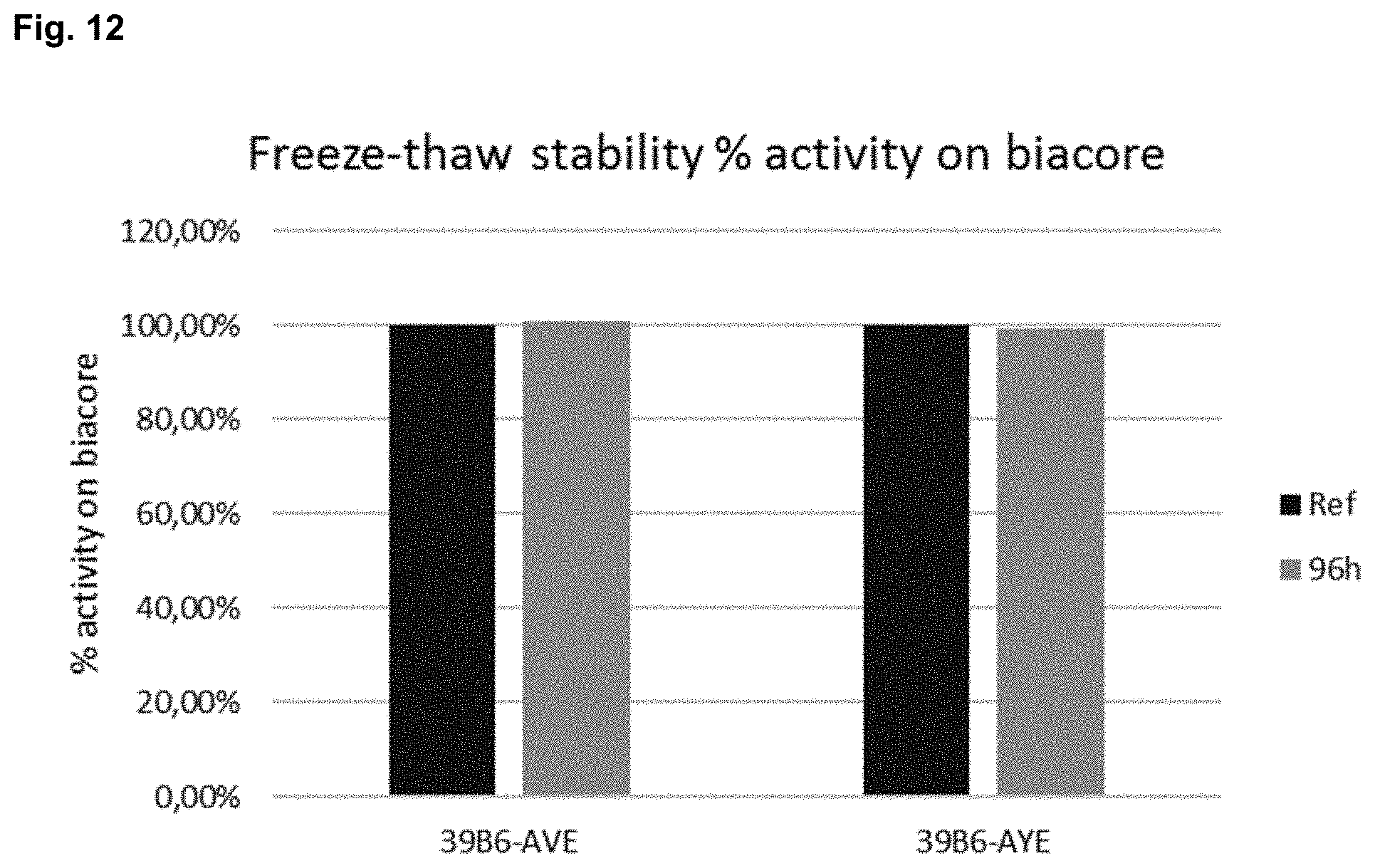
D00014

D00015

D00016

D00017

D00018

D00019

D00020

D00021

D00022

S00001
XML
uspto.report is an independent third-party trademark research tool that is not affiliated, endorsed, or sponsored by the United States Patent and Trademark Office (USPTO) or any other governmental organization. The information provided by uspto.report is based on publicly available data at the time of writing and is intended for informational purposes only.
While we strive to provide accurate and up-to-date information, we do not guarantee the accuracy, completeness, reliability, or suitability of the information displayed on this site. The use of this site is at your own risk. Any reliance you place on such information is therefore strictly at your own risk.
All official trademark data, including owner information, should be verified by visiting the official USPTO website at www.uspto.gov. This site is not intended to replace professional legal advice and should not be used as a substitute for consulting with a legal professional who is knowledgeable about trademark law.