System and method for identification and characterization of transglutaminase species
Albert , et al. September 29, 2
U.S. patent number 10,788,495 [Application Number 15/974,385] was granted by the patent office on 2020-09-29 for system and method for identification and characterization of transglutaminase species. This patent grant is currently assigned to ROCHE SEQUENCING SOLUTIONS, INC.. The grantee listed for this patent is Roche Sequencing Solutions, Inc.. Invention is credited to Thomas Albert, Frank Bergmann, Victor Lyamichev, Jigar Patel, Michael Schraeml, Wojtek Steffen, Thomas Streidl.


View All Diagrams
| United States Patent | 10,788,495 |
| Albert , et al. | September 29, 2020 |
System and method for identification and characterization of transglutaminase species
Abstract
In one aspect, the present disclosure provides a system and method for the identification and characterization of a transglutaminase. Further, the present disclosure provides transglutaminase enzymes for forming isopeptide bonds, methods of forming isopeptide bonds in the presence of transglutaminases, and substrate tags for use with transglutaminases.
| Inventors: | Albert; Thomas (Danville, CA), Bergmann; Frank (Iffeldorf, DE), Lyamichev; Victor (Madison, WI), Patel; Jigar (Verona, WI), Schraeml; Michael (Penzberg, DE), Steffen; Wojtek (Penzberg, DE), Streidl; Thomas (Seeshaupt, DE) | ||||||||||
|---|---|---|---|---|---|---|---|---|---|---|---|
| Applicant: |
|
||||||||||
| Assignee: | ROCHE SEQUENCING SOLUTIONS,
INC. (Pleasanton, CA) |
||||||||||
| Family ID: | 1000005082593 | ||||||||||
| Appl. No.: | 15/974,385 | ||||||||||
| Filed: | May 8, 2018 |
Prior Publication Data
| Document Identifier | Publication Date | |
|---|---|---|
| US 20180252714 A1 | Sep 6, 2018 | |
Related U.S. Patent Documents
| Application Number | Filing Date | Patent Number | Issue Date | ||
|---|---|---|---|---|---|
| 14973472 | Dec 17, 2015 | 10006915 | |||
| 62260162 | Nov 25, 2015 | ||||
| 62094495 | Dec 19, 2014 | ||||
| Current U.S. Class: | 1/1 |
| Current CPC Class: | G01N 33/573 (20130101); C12N 9/1044 (20130101); C07K 7/06 (20130101); C07K 1/1075 (20130101); G01N 2333/91085 (20130101); C12Y 203/01013 (20130101) |
| Current International Class: | G01N 33/573 (20060101); C07K 1/107 (20060101); C07K 7/06 (20060101); C12N 9/10 (20060101) |
References Cited [Referenced By]
U.S. Patent Documents
| 2009/0117640 | May 2009 | Norskov-Lauritsen et al. |
| 2009/0318349 | December 2009 | Hu et al. |
| 2012/0190561 | July 2012 | Wildt et al. |
| 2402563 | Jul 2001 | CA | |||
| 2902841 | Oct 2014 | CA | |||
| 2007/020290 | Feb 2007 | WO | |||
| 2008/020074 | Feb 2008 | WO | |||
| 2012059882 | May 2012 | WO | |||
| 2013163162 | Oct 2013 | WO | |||
| 2013130953 | Oct 2014 | WO | |||
Other References
|
Bowie et al (Science, 1990, 247; 1305-1310). cited by applicant . Burgess et al (J. Cell Biol. 111:2129-2138, 1990). cited by applicant . Lazar et al (Mol. Cell. Biol, 8:1247-1252, 1988). cited by applicant . Shikhman A R et al: "Cytokeratin Peptide SFGSGFGGGY Mimics N-Acetyl-Beta-D-Glucosamine in Reaction With Antibodies and Lectins, and Induced In Vivo Anti-Carbohydrate Antibody Response", The Journal of Immunology, The American Association of Immunologists, US, vol. 153, Jan. 1, 1994 (Jan. 1, 1994), pp. 5593-5606, XP000941613, ISSN: 0022-1767. cited by applicant . Karen Chao Butterfield et al: "Identification and Sequence Composition Characterization of Chondroitin Sulfate-Binding Peptides through Peptide Array Screening", Biochemistry, vol. 49, No. 7, Feb. 23, 2010 (Feb. 23, 2010), pp. 1549-1555, XP055067897, ISSN: 0006-2960, DOI: 10.1021/bi9021044. cited by applicant . Yuriy Rebets et al: "Complete genome sequence of producer of the glycopeptide antibiotic Aculeximycin Kutzneria albida DSM 43870T, a representative of minor genus of Pseudonocardiaceae", BMC Genomics, Biomed Central LTD, London, UK, vol. 15, No. 1, Oct. 10, 2014 (Oct. 10, 2014), p. 885, XP021200656, ISSN: 1471-2164, DOI: 10.1186/1471-2164-15-885. cited by applicant . Hemung, B.-0. et al., Reactivity of Fish and Microbial Transglutaminases on Glutaminyl Sites of Peptides Derived from Threadfin Bream Myosin, J. Agric. Food Chern., (2008), pp. 7510-7516, vol. 56 No. 16. cited by applicant . Kuramoto, K. et al., Phage-displayed peptide library screening for preferred human substrate peptide sequences for transglutaminase 7, Archives of Biochemistry and Biophysics, (2013), pp. 138-143, vol. 537 Issue 1. cited by applicant . Sugimura, Y. et al., Identification of preferred substrate sequences of microbial transglutaminase from Streptomyces mobaraensis using a phage-displayed peptide library, Archives of Biochemistry and Biophysics, (2008), pp. 379-383, vol. 477 Issue 2. cited by applicant . International Search Report and Written Opinion dated Mar. 14, 2016 in corresponding PCT Application No. PCT/US2015/066491 filed Dec. 17, 2015, pp. 1-16. cited by applicant . Strop, P., Versatility of Microbial Transglutaminase, Bioconjugate Chem., 2014, 25 (5), pp. 855-862, DOI: 10.1021/bc500099v, Publication Date (Web): Apr. 2, 2014. cited by applicant . Oteng-Pabi, S. K. and Keillor, J. W., Continuous enzyme-coupled assay for microbial transglutaminase activity, Analytical Biochemistry 2013, 441 (2), pp. 169-171. cited by applicant . Steffen, et al., "Discovery of a microbial transglutaminase enabling highly site-specific labeling of proteins," J. Biol. Chem. 292(38):15622-15635 (2017). cited by applicant . Butterfield, et al., "Identification and Sequence Composition Characterization of Chondroitin Sulfate-Binding Peptides through Peptide Array Screening," Biochemistry 49:1549-1555 (2010). cited by applicant . Rebets, et al., "Complete genome sequence of producer of glycopeptide antibiotic Aculeximycin Kutzneria albida DSM 43870T, a representative of minor genus of Pseudonocardiaceae," BMC Genomics 15:885 (2014). cited by applicant . UniProt, Sequence of Heme A synthase (ctaA) gene from Bacilus velezensis, UniProtKB-A7Z4B0 (CTAA_BACVZ) (last modified Oct. 23, 2007). cited by applicant . UniProt, Sequence of 8-amino-7-oxononanoate synthase (bioF) gene from Cellvibrio japonicus, UniProtKB-B3PI88 (BIOF_CELJU) (last modified Jul. 28, 2009). cited by applicant . UniProt, Sequence of 4-hydroxyproline 2-epimerase (proR) gene from Pseudomonas putida, UniProtKB-Q88NF3 (4HYPE_PSEPK) (last modified Jun. 1, 2003). cited by applicant . UniProt, Sequence of Autophagy-related protein 23 (ATG23) gene from Ashbya gossypii, UniProtKB-Q756Z7 (ATG23_ASHGO) (last modified Jul. 5, 2004). cited by applicant. |
Primary Examiner: Gangle; Brian
Attorney, Agent or Firm: Lee; Eric Grant Agnew; Daniel E.
Parent Case Text
CROSS-REFERENCE TO RELATED APPLICATIONS
This application is based on, claims the benefit of, and incorporates herein by reference, U.S. Provisional Patent Application Ser. No. 62/094,495 filed on Dec. 19, 2014 and entitled, "Identification of Transglutaminase Substrates and Uses Therefor," and U.S. Provisional Patent Application Ser. No. 62/260,162 filed on Nov. 25, 2015 and entitled, "System and Method for Identification and Characterization of Transglutaminase Species."
Claims
What is claimed is:
1. A structure, a substrate tag attached to the structure, and a microbial transglutaminase bound to the substrate tag, wherein the substrate tag comprises an acyl-donor tag having at least 80% sequence identity to a peptide sequence consisting of RYRQR (SEQ ID NO:14), wherein the acyl-donor tag is a substrate for the microbial transglutaminase, wherein the structure is selected from a recombinant protein, a detectable label, and a chemically synthesized structure, and wherein the microbial transglutaminase has at least 80% sequence identity to the Kutzneria albida microbial transglutaminase (SEQ ID NO:6).
2. The structure of claim 1, wherein the detectable label is selected from a biotin moiety, a fluorescent dye, a ruthenium label, a radiolabel, and a chemiluminescent label.
3. The structure of claim 1, wherein the acyl-donor tag has the peptide sequence APRYRQRAA (SEQ ID NO:24), and wherein the microbial transglutaminase is further bound to an amine-donor tag.
4. The structure of claim 1, wherein the recombinant microbial transglutaminase is the Kutzneria albida microbial transglutaminase (SEQ ID NO:6).
5. The structure of claim 1, wherein the chemically synthesized structure comprises at least one of a protein, a peptide, an oligonucleotide, a carboxybenzyl group, and a polyethylene glycol (PEG) polymer.
6. The structure of claim 1, wherein the microbial transglutaminase is further bound to an amine-donor tag.
7. The structure of claim 6, wherein the amine-donor tag has a peptide sequence consisting of RYESK (SEQ ID NO:2).
8. The structure of claim 7, wherein the amine-donor tag is attached to a second structure, wherein the second structure is selected from a second recombinant protein, a second detectable label, and a second chemically synthesized structure.
9. The structure of claim 8, wherein the second detectable label is selected from a biotin moiety, a fluorescent dye, a ruthenium label, a radiolabel, and a chemiluminescent label.
10. The structure of claim 8, wherein the second chemically synthesized structure comprises at least one of a protein, a peptide, an oligonucleotide, a carboxybenzyl group, and a polyethylene glycol (PEG) polymer.
Description
STATEMENT REGARDING FEDERALLY SPONSORED RESEARCH
Not applicable.
BACKGROUND OF THE INVENTION
The disclosure relates, in general, to the identification of transglutaminases and substrates therefore, and more particularly to the discovery and characterization of a microbial transglutaminase from Kutzneria albida.
Elucidating the details of enzyme activity and specificity is important for understanding the physiological function of enzymes and for biotechnological applications of the reactions catalyzed by enzymes. For example, transglutaminases belong to a large family of related enzymes, including microbial and mammalian transglutaminases. Transglutaminases catalyze cross-linking between two polypeptide or peptide chains by forming an isopeptide bond between a gamma-carboxamide group of a glutamine residue and an epsilon-amino group of a lysine residue. Elucidating the details of transglutaminase activity and specificity is important for biotechnological applications of the cross-linking reaction catalyzed by transglutaminases, for example, for modification of proteins for labeling, tagging, multi-protein complex formation, and the like.
To date, microbial transglutaminase is the most studied transglutaminase enzyme because of its small size, robust performance, stability, and the calcium independence of its activity. Several studies have shown that a broad variety of long alkylamines can substitute for the lysine substrate of transglutaminases and the simple dipeptide glutamine-glycine can serve as the glutamine substrate. These discoveries of lysine and glutamine substrates of transglutaminases have helped to develop a variety of tests for transglutaminase activity and practical assays for modification of proteins using transglutaminases. However, several challenges may still arise in the identification and characterization of known and novel transglutaminases. One challenge is the specificity of a particular transglutaminase for isopeptide bond formation may be too broad or too narrow for a particular application. Another challenge is transglutaminases having the same or similar substrate specificity may not be useful for orthogonal labeling strategies, or the like. Yet another challenge is the identification of substrates for uncharacterized or poorly characterized transglutaminases. Still other challenges may arise depending on factors associated with a given transglutaminase, such as the origin, specificity, activity, stability, the like, and combinations thereof.
SUMMARY OF THE INVENTION
The present invention overcomes the aforementioned drawbacks by providing a system and method for the identification and characterization of transglutaminases, as well as substrates and uses therefor.
In accordance with one aspect of the present disclosure, a substrate tag for a microbial transglutaminase includes one of an acyl-donor tag having at least 80% sequence identity to the peptide sequence YRYRQ (SEQ ID NO:1), and an amine donor tag having at least 80% sequence identity to the peptide sequence RYESK (SEQ ID NO:2).
In one aspect, the microbial transglutaminase has at least 80% sequence identity to the Kutzneria albida microbial transglutaminase (SEQ ID NO:6).
In another aspect, the substrate tag further includes a detectable label.
In another aspect, the detectable label is selected from a biotin moiety, a fluorescent dye, a ruthenium label, a radiolabel, and a chemiluminescent label.
In another aspect, the acyl-donor tag having the peptide sequence APRYRQRAA (SEQ ID NO:24).
In accordance with another aspect of the present disclosure, a method of forming an isopeptide bond in the presence of a microbial transglutaminase includes exposing a microbial transglutaminase to a first substrate and a second substrate, the first substrate including an acyl-donor tag having at least 80% sequence identity to the peptide sequence YRYRQ (SEQ ID NO:1), and the second substrate including an amine-donor tag having at least 80% sequence identity to the peptide sequence RYESK (SEQ ID NO:2), and cross-linking the first substrate and the second substrate, thereby forming an isopeptide bond between the acyl donor tag and the amino donor tag.
In one aspect, the microbial transglutaminase has at least 80% sequence identity to the Kutzneria albida microbial transglutaminase (SEQ ID NO:6).
In another aspect, the step of cross-linking the first substrate and the second substrate forms an isopeptide bond between a gamma-carboxamide group of the acyl-donor tag and an epsilon-amino group of the amino-donor tag.
In another aspect, at least one of the first substrate and the second substrate includes a detectable label.
In another aspect, the detectable label is selected from a biotin moiety, a fluorescent dye, a ruthenium label, a radiolabel, and a chemiluminescent label.
In another aspect, the acyl-donor tag having the peptide sequence APRYRQRAA (SEQ ID NO:24).
In another aspect, cross-linking of the first substrate to the second substrate is achieved with a yield of at least about 70%.
In another aspect, the yield is achieved within about 30 minutes.
In accordance with another aspect of the present disclosure, a kit for forming an isopeptide bond in the presence of a microbial transglutaminase, includes a purified microbial transglutaminase having at least 80% sequence identity to the Kutzneria albida microbial transglutaminase (SEQ ID NO:6).
In one aspect, the kit further includes one of a first substrate including an acyl-donor tag having at least 80% sequence identity to the peptide sequence YRYRQ (SEQ ID NO:1), and a second substrate including an amine-donor tag having at least 80% sequence identity to the peptide sequence RYESK (SEQ ID NO:2).
In another aspect, at least one of the first substrate and the second substrate includes a detectable label.
In another aspect, the detectable label is selected from a biotin moiety, a fluorescent dye, a ruthenium label, a radiolabel, and a chemiluminescent label.
In another aspect, the acyl-donor tag having the peptide sequence APRYRQRAA (SEQ ID NO:24).
In another aspect, the kit further includes the other one of the first substrate and the second substrate.
In accordance with another aspect of the present disclosure, an enzyme for forming an isopeptide bond includes a purified microbial transglutaminase having at least 80% sequence identity to the Kutzneria albida microbial transglutaminase (SEQ ID NO:6).
In one aspect, the isolated microbial transglutaminase is expressed and isolated in the presence of ammonium.
In another aspect, the ammonium is present at a concentration of at least about 10 .mu.M.
In accordance with another aspect of the present disclosure, an acyl-donor substrate for a transglutaminase includes an amino acid sequence having the formula Xaa.sub.1-Xaa.sub.2-Xaa.sub.3-Xaa.sub.4-Xaa.sub.5, where Xaa is any amino acid, where at least one of Xaa.sub.3, Xaa.sub.4, and Xaa.sub.5 is glutamine, where one of Xaa.sub.4 and Xaa.sub.5 is arginine, where the amino acid sequence includes at least one arginine sequentially adjacent to a glutamine, and where the total number of amino acids in the amino acid sequence selected from arginine, glutamine, phenylalanine, tryptophan, and tyrosine is at least four.
In one aspect, Xaa.sub.5 and at least one of Xaa.sub.1, Xaa.sub.2, and Xaa.sub.3 is arginine.
In accordance with another aspect of the present disclosure, an amine-donor substrate for a transglutaminase includes an amino acid sequence having the formula Xaa.sub.1-Xaa.sub.2-Xaa.sub.3-Xaa.sub.4-Xaa.sub.5, where Xaa is any amino acid, where the amino acid sequence includes at least one lysine, where one of Xaa.sub.1 and Xaa.sub.2 is selected from tyrosine and arginine, and where the total number of amino acids in the amino acid sequence selected from arginine, serine, tyrosine, and lysine is at least three.
In one aspect, one of Xaa.sub.4 and Xaa.sub.5 is lysine.
In another aspect, the amino acid sequence includes no more than two of the amino acid lysine.
The foregoing and other aspects and advantages of the invention will appear from the following description. In the description, reference is made to the accompanying drawings which form a part hereof, and in which there is shown by way of illustration a preferred embodiment of the invention. Such embodiment does not necessarily represent the full scope of the invention, however, and reference is made therefore to the claims and herein for interpreting the scope of the invention.
BRIEF DESCRIPTION OF THE FIGURES
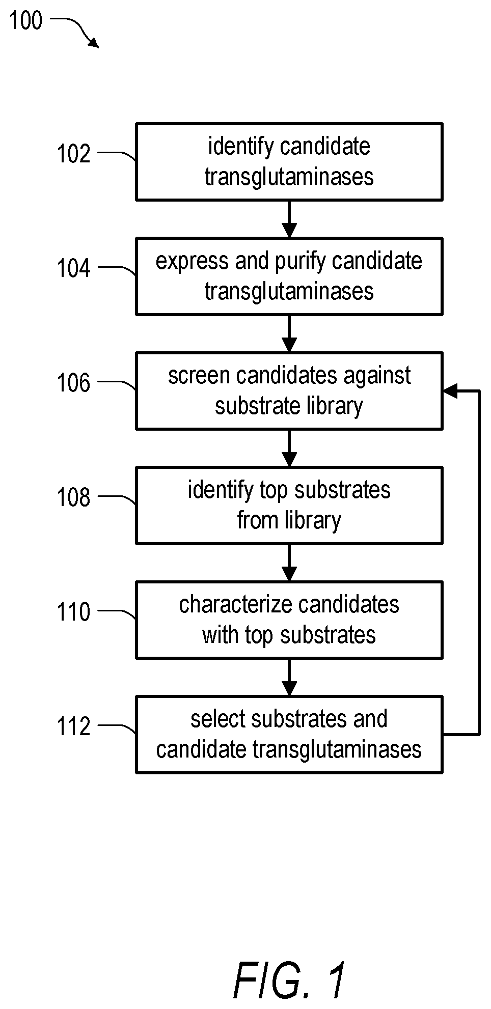
FIG. 1 is a schematic diagram illustrating an embodiment of a method for identification and characterization of transglutaminase species according to the present disclosure.
FIG. 2A is a Clustal Omega version 1.2.1 (Sievers et al., 2011. Molecular Systems Biology 7:539) multiple sequence alignment of Kutzneria albida KALB_7456 hypothetical protein (SEQ ID NO:6) (upper row) and Streptomyces mobaraensis microbial transglutaminase (SEQ ID NO:5) (lower row). Identical amino acid residues are marked by asterisks (*), similar residues by colons (:). Conserved residues of the S. mobaraensis microbial transglutaminase catalytic triad (Cys, Asp, His) are highlighted in grey.
FIG. 2B is an amino acid sequence of the hypothetical transglutaminase KALB_7456 from K. albida (SEQ ID NO:6) including a general cleavage site prediction as determined by ProP 1.0 (Duckert et al., 2004. Protein Engineering, Design and Selection 17: 107-112), with both predicted signal peptide sequence (`s`) and propeptide cleavage site (`P`) indicated.
FIG. 2C is a bar graph showing propeptide cleavage potential as a function of the amino acid sequence position for the hypothetical transglutaminase KALB_7456 from K. albida as determined by ProP 1.0. The dashed line indicates the propeptide cleavage potential threshold, and the dotted line indicates the amino acid position predicted for signal peptide.
FIG. 3A is an optical image of an SDS-PAGE gel showing an expression profile for K. albida transglutaminase (KalbTG) fusion proteins. Amino-terminal fusion partners are grouped in adjacent lanes as indicated by lane pairs numbered 1-7 (1: 8X-His tag; 2: dsbA signal peptide; 3: ompT signal peptide; 4: E. coli SlyD (EcSlyD); 5: 2xEcSlyD; 6: FkpA; 7: maltose binding protein). The lane labeled `L` was loaded with a standard molecular weight ladder (values shown in kDa). Individual lanes labeled as `P` and `S` denote insoluble (pellet) and soluble (supernatant) fractions of E. coli cell lysate, respectively. The coomassie blue stained protein band representing His-KalbTG is marked by an asterisk (*) in lane pair 1.
FIG. 3B is a schematic representation of the 2xSlyD-fusion protein expression and purification strategy. N-terminal fusion with two moieties of sensitive-to-lysis D (SlyD) protein confers solubility and is cleavable by factor Xa. The enzyme is further matured by cleavage of a propeptide sequence with trypsin.
FIG. 3C is an image of an SDS-PAGE gel showing the modular purification of KalbTG. The lanes labeled `L` were loaded with a standard molecular weight ladder (values shown in kDa). Lane 1: Fraction containing 2xSlyD-KalbTG from first Ni.sup.+-IMAC gradient elution (0-250 mM Imidazole). Lane 2: KalbTG proenzyme purified by Ni.sup.+-IMAC gradient elution, on-column factor Xa digest, and size exclusion chromatography. Lane 3: Fraction from second Ni.sup.+-IMAC gradient elution after consecutive on-column digests with factor Xa and trypsin (0-250 mM imidazole). Lane 4: Concentrate of lane 3, filtered by a 50,000 molecular weight cut-off (MWCO) membrane.
FIG. 4A is a log-log scatter plot showing the correlation between fluorescence signal data generated by KalbTG for replicate features on a 5-mer peptide array in the presence of biotinylated amine-donor substrate. Each data point represents a pair of replicate peptides from a library of 1.4 million unique peptides synthesized in duplicate. The 22 data points displaying the highest fluorescence signals are tagged by their respective 5-mer peptide sequence: YRYRQ (SEQ ID NO:1), RYRQR (SEQ ID NO:14), RYSQR (SEQ ID NO:15), FRQRQ (SEQ ID NO:16), RQRQR (SEQ ID NO:17), FRQRG (SEQ ID NO:18), QRQRQ (SEQ ID NO:19), YKYRQ (SEQ ID NO:20), QYRQR (SEQ ID NO:21), YRQSR (SEQ ID NO:32), LRYRQ (SEQ ID NO:33), YRQRA (SEQ ID NO:34), VRYRQ (SEQ ID NO:35), QRQTR (SEQ ID NO:36), YRQTR (SEQ ID NO:37), PRYRQ (SEQ ID NO:38), RFSQR (SEQ ID NO:39), WQRQR (SEQ ID NO:40), VRQRQ (SEQ ID NO:41), RYTQR (SEQ ID NO:42), AYRQR (SEQ ID NO:43), and YQRQR (SEQ ID NO:44).
FIG. 4B is a log-log scatter plot showing the correlation between fluorescence signal data generated by KalbTG for replicate features on a 5-mer peptide array in the presence of biotinylated glutamine-donor substrate (Z-APRYRQRAAGGG-PEG-biotin). Each data point represents a pair of replicate peptides from a library of 1.4 million unique peptides synthesized in duplicate. The 17 data points displaying the highest fluorescence signals are tagged by their respective 5-mer peptide sequence: RYESK (SEQ ID NO:2), RYSKY (SEQ ID NO:25), NYRFK (SEQ ID NO:45), YQKWK (SEQ ID NO:46), YKYKY (SEQ ID NO:47), RWKFK (SEQ ID NO:48), RFYSK (SEQ ID NO:49), YKYAK (SEQ ID NO:50), YRYAK (SEQ ID NO:51), RYSYK (SEQ ID NO:52), YKSFK (SEQ ID NO:53), YKSWK (SEQ ID NO:54), KYRYK (SEQ ID NO:55), YKYNK (SEQ ID NO:56), PYKYK (SEQ ID NO:57), FYKYK (SEQ ID NO:58), and FYESK (SEQ ID NO:59).
FIG. 5A is a log-log scatter plot showing the correlation between fluorescence signal data generated by S. mobaraensis MTG for replicate features on a 5-mer peptide array in the presence of biotinylated amine-donor substrate. Each data point represents a pair of replicate peptides from a library of 1.4 million unique peptides synthesized in duplicate. The 22 data points corresponding to the highest fluorescence signals generated by KalbTG from FIG. 4A are tagged by their respective 5-mer peptide sequence: YRYRQ (SEQ ID NO:1), RYRQR (SEQ ID NO:14), RYSQR (SEQ ID NO:15), FRQRQ (SEQ ID NO:16), RQRQR (SEQ ID NO:17), FRQRG (SEQ ID NO:18), QRQRQ (SEQ ID NO:19), YKYRQ (SEQ ID NO:20), QYRQR (SEQ ID NO:21), YRQSR (SEQ ID NO:32), LRYRQ (SEQ ID NO:33), YRQRA (SEQ ID NO:34), VRYRQ (SEQ ID NO:35), QRQTR (SEQ ID NO:36), YRQTR (SEQ ID NO:37), PRYRQ (SEQ ID NO:38), RFSQR (SEQ ID NO:39), WQRQR (SEQ ID NO:40), VRQRQ (SEQ ID NO:41), RYTQR (SEQ ID NO:42), AYRQR (SEQ ID NO:43), and YQRQR (SEQ ID NO:44).
FIG. 5B is the plot of fluorescence signal data generated by S. mobaraensis MTG of FIG. 5A with the tagged data points from FIG. 4A omitted. The 16 data points corresponding to highest fluorescence signals generated by MTG are tagged by their respective 5-mer peptide sequence: DYALQ (SEQ ID NO:22), EWVAQ (SEQ ID NO:60), EWALQ (SEQ ID NO:61), DYFLQ (SEQ ID NO:62), EYWLQ (SEQ ID NO:63), DWALQ (SEQ ID NO:64), DWYLQ (SEQ ID NO:65), DYWLQ (SEQ ID NO:66), EYVAQ (SEQ ID NO:67), DYVAQ (SEQ ID NO:68), DWVAQ (SEQ ID NO:69), EYVLQ (SEQ ID NO:70), EWIAQ (SEQ ID NO:71), WYALQ (SEQ ID NO:72), EYALQ (SEQ ID NO:73), and EYFLQ (SEQ ID NO:74).
FIG. 5C is the plot of fluorescence signal data generated by KalbTG of FIG. 4A with the tagged data points from FIG. 4A omitted. The 16 data points corresponding to highest fluorescence signals generated by S. mobaraensis MTG from FIG. 5B are tagged by their respective 5-mer peptide sequence: DYALQ (SEQ ID NO:22), EWVAQ (SEQ ID NO:60), EWALQ (SEQ ID NO:61), DYFLQ (SEQ ID NO:62), EYWLQ (SEQ ID NO:63), DWALQ (SEQ ID NO:64), DWYLQ (SEQ ID NO:65), DYWLQ (SEQ ID NO:66), EYVAQ (SEQ ID NO:67), DYVAQ (SEQ ID NO:68), DWVAQ (SEQ ID NO:69), EYVLQ (SEQ ID NO:70), EWIAQ (SEQ ID NO:71), WYALQ (SEQ ID NO:72), EYALQ (SEQ ID NO:73), and EYFLQ (SEQ ID NO:74).
FIG. 5D is a plot of S. mobaraensis MTG and KalbTG activity obtained by measuring rates of NADH oxidation at 340 nm and 37.degree. C. for varying concentrations of glutamine-donor substrates Z-GGGDYALQGGGG (SEQ ID NO:76) (0 to 1 mM) and Z-GGGYRYRQGGGG (SEQ ID NO:75) (0 to 1 mM) in the presence of amine-donor substrate cadaverine (1 mM) in a GLDH-coupled assay; YRYRQ (SEQ ID NO:1), DYALQ (SEQ ID NO:22).
FIG. 6A is a series of images of SDS-PAGE gels showing both an experimental time-course (Left: bright field; Right: Cy3 fluorescence) and control data (Left: bright field; Right: Cy3 fluorescence) for Cy3 labeling of a Q-tagged Thermus thermophilus SlyD moiety. Successful mono-labeling is observed by the shift in electrophoretic mobility corresponding to the 6 kDa molecular weight of the label and by the fluorescent signal of the labeled species. Data is shown for a 60 minute time-course performed with 10-fold label excess, and for an 18 hour incubation with 50-fold label excess. Lanes labeled `L` were loaded with a standard molecular weight ladder (values shown in kDa). Lanes labeled `-` and `+` denote control reactions with SlyD containing the S. mobaraensis MTG Q-tag (DYALQ (SEQ ID NO: 22)) and KalbTG (-) or S. mobaraensis MTG (+).
FIG. 6B is a series of images of an SDS-PAGE gel showing a pH profile of KalbTG labeling efficacy for pH values between 6.2 and 9.0 (Left: bright field; Right: Cy3 fluorescence). The highest labeling yield after the 15 minute reaction time was observed at pH 7.4. The lane labeled `L` was loaded with a standard molecular weight ladder (values shown in kDa).
FIG. 6C is a series of images of SDS-PAGE gels showing dual site-specific functionalization of the construct YRYRQ-PEG27-(factor Xa cleavage site)-PEG27-PEG27-DYALQ with Cy3 and Cy5 fluorescent labels. Each group (labeled 1, 2, and 3) includes three images of the same gel lane arranged in the following order from left to a right: i) bright field; ii) Cy3 fluorescence; and iii) Cy5 fluorescence. Group 1: A mixture of peptide construct and 10-fold excess of Cy3 label. Group 2: A mixture of peptide construct and 10-fold excess of Cy3 label following incubation with KalbTG enzyme for 30 min. Group 3: S. mobaraensis MTG enzyme and Cy5 label added to the composition of Group 2, and incubated for 15 min with no intermediate blocking or purification steps; dually labeled construct was achieved with nearly quantitative yield.
FIG. 7A is a three-dimensional alignment of the active enzyme structures of KalbTG (dark grey) and S. mobaraensis MTG (light grey) that reveals high conservation in the core and active site region and high variability in the peripheral loop regions.
FIG. 7B is a three-dimensional surface overlay of the active enzyme structures of KalbTG (dark grey) and S. mobaraensis MTG (light grey) illustrating that the binding pocket of both S. mobaraensis MTG and the more compact KalbTG may be similarly occupied by a propeptide (ribbon structure).
FIG. 7C is a three-dimensional ribbon structure illustrating contributors to the formation of the KalbTG active cleft including two strongly charged, hydrophilic loops that are believed to either mediate substrate recruitment, act as a substrate mimic, or a combination thereof. The two hydrophilic loops are labeled with their corresponding sequences (i.e., NHEEPR (SEQ ID NO:3) and YRYRAR (SEQ ID NO:4)).
DETAILED DESCRIPTION
As discussed above, in various situations it may be useful to elucidate details of enzyme activity and specificity to provide both a basic understanding of those enzymes, as well as for the development of biotechnological applications including those enzymes. For example, conventional chemical strategies for the modification of therapeutic and diagnostic proteins often lack site-specificity, linkage stability, stoichiometric control, or a combination thereof, giving rise to heterogeneous conjugates which may cause interference (e.g., with immunoreactivity or stability of a therapeutic agent). In one aspect, it is anticipated that the industrial development of therapeutic and diagnostic reagents of the coming years will see a massive increase in sophisticated formats requiring stable and truly site-specific conjugation. Therefore, there is a need for new enzymatic methods that offer an attractive and cost-effective alternative to established chemical strategies.
Microbial transglutaminase (MTG) is a protein-glutamine .gamma.-glutamyltransferase (EC 2.3.2.13) that was first described by researchers of Ajinomoto Co., Inc. in 1989, and is one of the most widely used groups of enzymes for the cross-linking of proteins and peptides in many food and biotechnological applications. MTG was first discovered in and later extracted from the organism Streptomyces mobaraensis. MTG catalyzes the formation of a stable isopeptide bond between an acyl-group (e.g., a glutamine side chain; acyl-donor) and an alkyl-amine (e.g., a lysine side chain; amine-donor). In the absence of reactive amine groups, the enzymatic reaction with water leads to deamination of glutamine side chains. The bacterial enzyme works without the addition of cofactors such as Ca.sup.2+ or GTP and in a broad range of pH, buffer, and temperature conditions.
In contrast to sortase A, whose natural substrate specificity is very stringent, the known MTG (e.g., from S. mobaraensis) are generally promiscuous enzymes with regard to substrate molecules and the specificities of transglutaminase variants remain largely unknown. While significant scientific efforts have been made to establish MTG as the enzyme of choice in the development of therapeutic antibody-drug conjugates, the large-scale production of such MTG-mediated immunoconjugates is hampered by the low specificity of the enzyme.
Known MTG species are mainly representatives of the families Streptomyces or Bacillus. These MTG species exhibit very similar primary amino acid structures and substrate specificities. All known active MTG species exhibit molecular weights of at least about 38 kDa. Being a cross-linking enzyme in nature, known MTG generally display broad substrate specificity for amine-donor substrates and a relatively low specificity for acyl-donor substrates. Approaches for the high-throughput screening of improved MTG substrates have previously been limited to phage panning or mRNA display. While recently pioneered array-based high-throughput screening approaches have successfully identified substrates of the S. mobaraensis MTG (U.S. Provisional Pat. App. Ser. No. 62/094,495 to Albert et al. filed on 19 Dec. 2014), only the substrate specificities of this and homolog enzymes are known, thereby precluding any bio-orthogonal conjugation approaches (e.g., simultaneous labeling of a biomolecule using two or more different label-substrates and two or more transglutaminase species). Accordingly, there is a need for high-throughput approaches for the identification and characterization of new transglutaminase species. Moreover, there is a need for improved transglutaminases with greater activity, specificity, or a combination thereof. Further, there is a need for acyl-donor tags (e.g., glutamine- or Q-tags) and amine-donor tags (e.g., lysine- or K-tags) that are specific and unique substrates for a transglutaminase of interest.
These and other challenges may be overcome with a system and method for the identification and characterization of transglutaminase species. To this end, the present disclosure provides for characterization of the structure and biochemistry of known and unknown candidate transglutaminase species. The disclosure further provides for characterization of the recombinant production of candidate transglutaminases, high-throughput screening of potential transglutaminase substrates via high-density peptide array, and semi-orthogonal conjugation of biomolecules using the newly characterized transglutaminase species. In yet another aspect, the present disclosure provides for the acyl-donor tags (e.g., glutamine- or Q-tags) and amine-donor tags (e.g., lysine- or K-tags) that are specific and unique substrates for a transglutaminase of interest. Here, the term `tag` refers to a sequence including one or more amino acids or other like molecules that can be grafted, fused, conjugated, or otherwise attached to another structure, such as a protein, peptide, small molecule, detectable label (e.g., a fluorescent dye), oligonucleotide, non-amino or nucleic acid polymer (e.g., polyethylene glycol), or the like. In one aspect, the nature of the attachment should enable the access to the tag by an enzyme for which the tag is a substrate.
In one embodiment of the present disclosure, a previously unknown transglutaminase species from the organism Kutzneria albida was identified, recombinantly expressed, purified, and characterized using a high-throughput array-based screening approach. The K. albida transglutaminase was determined to exhibit a high selectivity and substrate specificity for its array-determined substrate sequences, but reacts only poorly or not at all with substrate sequences of the S. mobaraensis enzyme. Accordingly, the K. albida transglutaminase can be said to be bio-orthogonal to the S. mobaraensis enzyme. In another aspect, the K. albida transglutaminase exhibited a surprisingly lower molecular weight (about 30 kDa) than all previously described MTG species (e.g., S. mobaraensis MTG is about 38 kDa), signifying an advantage for production and enzymatic labeling purposes. Further, the K. albida transglutaminase had a distinctly different primary amino acid structure compared to all currently known proteins. Overall, these properties make the K. albida transglutaminase highly attractive for a broad range of applications, including, but not limited to, the versatile, cost-effective, and site-specific conjugation of biomolecules with various label molecules. Additional or alternative applications where the K. albida transglutaminase can be effective include the production of therapeutic antibody-drug conjugates or chemiluminescent antibodies for in-vitro diagnostic uses.
In one aspect, a number of the challenges posed by recombinant production of transglutaminases may be overcome through implementation of embodiments of the present disclosure. With reference to FIG. 1, a method 100 of identifying and characterizing a transglutaminase includes a step 102 of identifying candidate transglutaminases. The step 102 can include a search for homologs of known or suspected transglutaminase species to identify candidate transglutaminases for further study. In one illustrative embodiment, the transglutaminase can be a microbial transglutaminase (e.g., a Streptoverticillium sp. transglutaminase, Kutzneria sp. transglutaminase, Streptomyces sp., or the like) or a mammalian transglutaminase. In embodiments where the enzyme is a mammalian transglutaminase, the mammalian transglutaminase can be, for example, selected from the group consisting of Human Factor XIII A transglutaminase, Human Factor XIII B transglutaminase, a Factor XIII transglutaminase, a keratinocyte transglutaminase, a tissue-type transglutaminase, an epidermal transglutaminase, a prostate transglutaminase, a neuronal transglutaminase, a human transglutaminase 5, and a human transglutaminase 7.
A search for homologs of known or suspected candidate transglutaminases can include the use of one or more search tools or databases. One suitable tool includes the Protein Basic Local Alignment Search Tool (Protein BLAST) from the National Center for Biotechnology Information (NCBI). The Protein BLAST tool can be supplied with the sequence of known or suspected transglutaminase species, the sequences of which can be obtained from various databases. One example database is the Universal Protein catalog (UniProt). However, other databases may be used in addition, or as an alternative to UniProt. In one aspect, when using Protein BLAST, a threshold Expect-value (E-value) may be selected for narrowing the results of the search. In one example, it may be useful to select an E-value of less than about 10.sup.-8. In another example, it may be useful to select an E-value of less than about 10.sup.-10. In yet another example, it may be useful to select an E-value of less than about 10.sup.-12.
The step 102 can further include performance of a sequence alignment of a transglutaminase sequence with an alignment tool. One example alignment tool includes the Clustal Omega 1.2.1 tool (Sievers et al. 2011, Molecular Systems Biology 7: 539). An alignment tool can provide a percent identity matrix value, identify potential conservation of catalytically active residues (if known), or a combination thereof. Other tools that can be used in the step 102 include the ProP 1.0 Server from the Technical University of Denmark (Duckert et al. 2004, Protein Engineering, Design & Selection 17(1): 107-112) to predict propeptide and signal sequences of a transglutaminase. In one aspect, a candidate transglutaminase may have a similarity of at least about 20%, at least about 25%, at least about 30%, at least about 35%, or more with respect to a known transglutaminase. Further, a candidate transglutaminase may be characterized by conservation of at least one or more active site residues with respect to a known or suspected transglutaminase, indicating that the enzymatic structure and function may be preserved.
Information gleaned in the step 102 through the use of one or more of the aforementioned tools can be used to select candidate transglutaminases from predicted or known transglutaminase sequences for expression and purification in a step 104. In one aspect, the step 104 can include rapidly screening expression conditions for a candidate transglutaminase species. One suitable method for screening includes insertion of the genetic insert for the candidate transglutaminase into one or more expression vectors designed for soluble cytosolic or periplasmic expression in a host organism using a fragment exchange system (Geertsma, et al. 2011. Biochemistry 50(15): 3272-3278). Other methods for screening may also be employed.
The step 104 can further include an initial screening to identify evidence of expression of full-length protein with the anticipated electrophoretic mobility. In the case that poor or no expression of the full-length, active candidate transglutaminase protein is observed, a candidate transglutaminase sequence can be fused with a chaperone (e.g., SlyD) to improve the likelihood of expression of functional enzyme. Expression of a candidate transglutaminase can be further optimized by screening different incubation times, incubation temperatures, inducer concentrations, induction times, media types, media volumes, the like, and combinations thereof.
In general, it will be appreciated that the many viable purification and expression strategies can be employed in the step 104 of the method 100. In one embodiment, a candidate transglutaminase sequence can be incorporated into a modular expression construct. Example expression constructs for use in the step 104 can include chaperone modules, protease cleavage site modules, purification tag modules, detection modules, the like, and combinations thereof. For example, an expression construct may include one or more SlyD chaperone modules arranged to yield a transglutaminase-chaperone fusion protein, one or more protease cleavage sites modules flanking the transglutaminase sequence for separation of the various modules following expression, and one or more 8X-histidine tags or other purification modules for recovery of one or more segments of the expressed protein. For expression constructs including a protease cleavage site module, the expressed protein can be treated with one or more proteases to yield the activated protein. For example, the expressed protein can be treated with a factor Xa protease, trypsin protease, thrombin protease, or another like protease to cleave any chaperone proteins, propeptide sequences, purification tags, or the like from the expressed candidate transglutaminase.
For the production of a selected transglutaminase protein, the gene sequence encoding the candidate transglutaminase--including some, none or all predicted signal and propeptide sequences--can be codon optimized and chemically synthesized for expression in a particular host organism (e.g., E. coli). Expression can be performed according to standard molecular biology protocols as described, for example, in Green, et al., 2012, Molecular Cloning: A Laboratory Manual, Cold Spring Harbor Laboratory Press.
In a next step 106 of the method 100, candidate transglutaminases expressed and purified in the step 104 are screened against a substrate library to identify potential substrates including acyl-donor sequences, amine-donor sequences, or both. In one aspect, the substrate library can include a plurality of peptide features synthesized in an array by maskless array synthesis (U.S. Pat. Pub. No 2015/0185216 to Albert et al. filed on 19 Dec. 2014). The peptide features can be prepared from natural amino acids, non-natural amino acids, other molecular building blocks, the like, and combinations thereof. Further, the peptides can be in a linear, cyclic or constrained (macrocycle) form.
As used herein, the terms "peptide," "oligopeptide" or "peptide binder" refer to organic compounds composed of amino acids, which may be arranged in either a linear chain (joined together by peptide bonds between the carboxyl and amino groups of adjacent amino acid residues), in a cyclic form or in a constrained (e.g., "macrocycle" form). The terms "peptide" or "oligopeptide" also refer to shorter polypeptides, i.e., organic compounds composed of less than 50 amino acid residues. A macrocycle (or constrained peptide), as used herein, is used in its customary meaning for describing a cyclic small molecule such as a peptide of about 500 Daltons to about 2,000 Daltons.
The term "natural amino acid" refers to one of the 20 amino acids typically found in proteins and used for protein biosynthesis as well as other amino acids which can be incorporated into proteins during translation (including pyrrolysine and selenocysteine). The 20 natural amino acids include histidine, alanine, valine, glycine, leucine, isoleucine, aspartic acid, glutamic acid, serine, glutamine, asparagine, threonine, arginine, proline, phenylalanine, tyrosine, tryptophan, cysteine, methionine, and lysine.
The term "non-natural amino acid" refers to an organic compound that is not among those encoded by the standard genetic code, or incorporated into proteins during translation. Therefore, non-natural amino acids include amino acids or analogs of amino acids, but are not limited to, the D-isostereomers of amino acids, the beta-amino-analogs of amino acids, homocitrulline, homoarginine, hydroxyproline, homoproline, ornithine, 4-amino-phenylalanine, cyclohexylalanine, .alpha.-aminoisobutyric acid, N-methyl-alanine, N-methyl-glycine, norleucine, N-methyl-glutamic acid, tert-butylglycine, .alpha.-aminobutyric acid, tert-butylalanine, 2-aminoisobutyric acid, .alpha.-aminoisobutyric acid, 2-aminoindane-2-carboxylic acid, selenomethionine, dehydroalanine, lanthionine, .gamma.-amino butyric acid, and derivatives thereof wherein the amine nitrogen has been mono- or di-alkylated.
With continued reference to FIG. 1, a step 108 of the method 100 includes identifying from the substrate library top amine-donor substrate sequences, acyl-donor substrate sequences, or both. In one aspect, activity of the candidate transglutaminases on the library substrates can be measured using one or more direct or coupled assays. In general, a direct assay includes measurement of the reactants (e.g., substrates, co-factors, etc.) and products (e.g., isopeptide bonds, deamidated substrates, etc.) of the enzymatic reaction. For example, spectrophotometry can be used to track the course of the enzymatic reaction by measuring a change in light absorbance associated with a reactant or product over time. In the case that the enzymatic reaction is not conducive to the use of one or more direct assay measurements, or in addition or as an alternative to a direct assay, a coupled assay can be employed. In the case of a coupled assay, the product of the enzymatic reaction of interest can be used as the substrate of another, more readily measured secondary reaction. Examples of coupled assays include measurement of redox reactions involving cofactors such as NADP(H) and NAD(H) that are involved as products or reactants in the enzymatic reaction of interest. In the case of the reaction catalyzed by a transglutaminase, a glutamate dehydrogenase (GLDH) dependent oxidation coupled assay may be implemented. One example of a GLDH assay includes .beta.-casein as a cross-linking substrate and detection of deamidation by GLDH oxidation of NADPH. Notably, it may be useful to select an assay that is compatible with a high-throughput screening format in order to investigate a large number of substrates (e.g., greater than 1 million) in parallel.
Using one of the aforementioned direct or indirect assays, the step 108 can include the use of peptide substrate arrays to identify specific sequences or motifs recognized by the candidate transglutaminases. For example, the transamidation reaction between a millions of unique peptides and a biotinylated amine donor can be quantified on one or more arrays in parallel and the sequences of the peptides with the highest signal output (i.e., the top substrates) can be determined. Thereafter, in a next step 110 of the method 100, the top substrates can be resynthesized and tested for transglutaminase activity in a standalone (on array or in solution) assay. Accordingly, the step 110 includes characterization of the candidate transglutaminases in the presence of the top substrates identified in the step 108.
Characterization of candidate transglutaminases can include determination of the parameters such as specificity, selectivity, affinity, activity, and the like. Moreover, two or more candidate transglutaminases can be characterized for orthogonality. Here, the ability of the candidate transglutaminases to act on the same substrates can be identified. In the case that two different transglutaminases are unable to act on the same substrate or substrates, the two different transglutaminases can be said to be orthogonal. However, in the case that the two different transglutaminases are able to act on one or more of the same substrates, but with different degrees of activity, the two different transglutaminases can be said to be semi-orthogonal. In one aspect, a peptide substrate array can deliver a readout for all viable 5-mer peptides sequences at once. Therefore, data collected from the substrate libraries can be used to identify differences in substrate specificity for each candidate transglutaminase for the identification of orthogonal, semi-orthogonal, and non-orthogonal transglutaminases.
The step 110 of the method 100 can further include characterization of the ability of the candidate transglutaminases to perform site-specific labeling on protein substrates. In one aspect assay, the top substrate sequences identified in the step 108 can be further analyzed in the step 110 both on array and in solution. Experiments can also be performed to quantify cross-reactivity of candidate transglutaminases with various substrates. In one aspect, a protein scaffold may be useful for labeling approaches as epitope (i.e., substrate sequence) containing loops can be grafted onto the scaffold for presentation to binders or enzymes. One approach including the use of scaffolds is described in PCT App. Pub. No. WO 2012/150321 to Andres et al. filed on 4 May 2012. Example scaffolds can advantageously include one or more FK506-binding protein (FKBP) domains as a site for grafting epitope-containing loops.
Labeling of the scaffold protein with candidate transglutaminases can be achieved under a variety of conditions. Factors that can be varied for labeling experiments include the ratio of substrate to transglutaminase enzyme, the ratio of one substrate to another substrate, the labelling time, pH, or the like. Notably, a substrate refers to any peptide, protein, or other structure including one or more amine-donor or acyl-donor substrates sequences. Example substrates include scaffold proteins having an acyl-donor or amine-donor substrate sequence grafted thereon, a detectable label conjugated to or otherwise associated with an acyl-donor or amine-donor substrate sequence, an acyl-donor or amine-donor substrate sequence in isolation, the like, and combinations thereof. Labeling yield can be measured over time using standard techniques, such as sodium dodecyl sulfate polyacrylamide gel electrophoresis (SDS-PAGE) in combination with optical (e.g., bright field, fluorescence) detection. For example, a first substrate can include one or more detectable labels. Cross-linking of the first substrate to a second substrate (e.g., the protein scaffold) can be analyzed by identifying a molecular weight shift on an SDS-PAGE gel followed by detection of the label within the gel.
Labels for use with embodiments of the present disclosure include any suitable label that can be combined with a transglutaminase substrate. Examples of suitable labels include fluorescent labels, chemiluminescent labels, radiolabels, chemical labels (e.g., incorporating "click" chemistry), haptens, a toxin, the like, and combinations thereof. More generally, a suitable label is compatible with at least one substrate of the transglutaminase (e.g., an acyl-donor substrate or amine-donor substrate) in that the label does not eliminate the ability of the transglutaminase to act on the labeled substrate. Further, a suitable label can produce a signal that is detectable relative to an unlabeled transglutaminase substrate. Specific examples of detectable labels for use in any appropriate embodiment herein can comprise fluorescein, rhodamine, TEXAS RED, phycoerythrin, OREGON GREEN (e.g., OREGON GREEN 488, OREGON GREEN 514, and the like) ALEXA FLUOR 488, ALEXA FLUOR 647 (Molecular Probes, Eugene, Oreg.), Cy3, Cy5, Cy7, biotin, ruthenium, DYLIGHT fluorescent agents, including but not limited to DYLIGHT 680, CW 800, trans-cyclooctene, tetrazine, methyltetrazine, and the like. Examples of haptens include biotin, digoxigenin, dinitrophenyl, and the like. Examples of toxins include amatoxins (e.g., amanitin), maitansinoids, and the like.
In some embodiments, the step 110 can further include identification and characterization of the three-dimensional (3D) crystal structure of the transglutaminase to provide further insight into the nature of the transglutaminase. The crystal structure of the transglutaminase can be performed in the presence of absence of one or more substrates, cofactors, or the like. Analysis of the crystal structure can provide insight into possible locations for site-specific mutagenesis for improving the properties of the transglutaminase. Crystallization of a candidate transglutaminase with a particular substrate sequence can further reveal interactions between the transglutaminase and substrate sequence to inform modifications to either or both of the transglutaminase and substrate sequence to tailor the properties of the transglutaminase to a particular application. Moreover, analysis of the crystal structure can serves as an independent confirmation of the reliability of array-based substrate discovery (see Example 5).
In a step 112 the method 100, substrate sequences identified in the step 108 and characterized in the step 110 are selected for use in downstream application of a selected candidate transglutaminase. In general, a particular acyl-donor or amine-donor substrate sequence may be unique to a given transglutaminase. Accordingly, for a given application, it may be useful to first select a transglutaminase and then select one or more substrate sequences. For applications where specificity and selectivity are important (e.g., orthogonal labeling with two or more transglutaminases), the step 112 can include selecting substrate sequences that are specifically and selectively labeled by the selected transglutaminase. However, other applications may benefit from selecting substrate sequences that can be acted on by more than one transglutaminase. Substrates can also be selected to achieve a particular degree of transglutaminase activity when it is useful to achieve faster or slower reaction times. To further tailor the selected substrate sequences, after the step 112 the method 100 can return to the step 106 for additional rounds of screening. In this case, the selected substrates can be extended, matured, or the like in subsequent rounds of screening on the peptide array. Example methods for extension and maturation of peptide sequences are described in U.S. Pat. Pub. No 2015/0185216 to Albert et al. filed on 19 Dec. 2014.
In yet other embodiments, it may be useful to provide site-specific labeling with promiscuous transglutaminase activity. A promiscuous transglutaminase may be useful if the substrate is not recombinantly produced, if the labeling site and label ratio can be controlled or are not of critical importance for the application on hand, or the like. One example of labeling with a promiscuous transglutaminase is the conjugation of payloads to deglycosylated or glycosylated IgG. However, transglutaminases having non-specific activity may be limited to a narrow range of possible applications. Accordingly, in other situations, it may be useful to provide a transglutaminase having specific activity for only a particular substrate or group of like substrates.
In summary, the method 100 can be used to identify and characterize one or more candidate transglutaminases along with one or more corresponding substrates. Hypothetical or known transglutaminases can be expressed and screened on substrate libraries to identify preliminary substrate sequences that elicit the desired transglutaminase activity. Top substrates can then be selected and optionally refined in an iterative manner, thereby resulting in a transglutaminase-substrate combination that can be implemented for a variety of applications.
EXAMPLES
Example 1
Identification of the K. albida Microbial Transglutaminase
Establishing a viable and robust, enzymatic, industrial-scale method for site-specific conjugation approaches like antibody-drug conjugates makes high demands on the coupling enzyme. Among other factors, it may be useful for such approaches to have a high reaction rate, conjugation efficacy, and substrate specificity. Further, it may be useful for such approaches to be economical in production, include an enzyme having a low molecular weight, an enzyme that is independent of cofactors, the like, and combinations thereof. For discovery of new microbial transglutaminases, a search for homologs of this enzyme that may fulfill all the mentioned criteria was performed using the amino acid sequence of S. mobaraensis protein-glutamine gamma-glutamyltransferase as a query. This yielded the hypothetical gene product KALB_7456 from bacteria K. albida DSM 43870, a spore-forming gram-positive bacterium which was sequenced in 2014 (Rebets et al., 2014. BMC genomics 15: 885).
The web interface of NCBI Protein BLAST tool was used to search for sequences similar to the MTG of Streptomyces mobaraensis. The amino acid sequence of S. mobaraensis protein-glutamine gamma-glutamyltransferase (UniProt accession number P81453) was entered as a query. The amino acid sequence of S. mobaraensis protein-glutamine gamma-glutamyltransferase is as follows:
TABLE-US-00001 (SEQ ID NO: 5) MRIRRRALVFATMSAVLCTAGFMPSAGEAAADNGAGEETKSYAETYRL TADDVANINALNESAPAASSAGPSFRAPDSDDRVTPPAEPLDRMPDPY RPSYGRAETVVNNYIRKWQQVYSHRDGRKQQMTEEQREWLSYGCVGVT WVNSGQYPTNRLAFASFDEDRFKNELKNGRPRSGETRAEFEGRVAKES FDEEKGFQRAREVASVMNRALENAHDESAYLDNLKKELANGNDALRNE DARSPFYSALRNTPSFKERNGGNHDPSRMKAVIYSKHFWSGQDRSSSA DKRKYGDPDAFRPAPGTGLVDMSRDRNIPRSPTSPGEGFVNFDYGWFG AQTEADADKTVWTHGNHYHAPNGSLGAMHVYESKFRNWSEGYSDFDRG AYVITFIPKSWNTAPDKVKQGWP
Manual screening of the results for E-values of less than 10.sup.-10 and polypeptide sequences shorter than that of S. mobaraensis MTG yielded hypothetical gene product KALB_7456 (SEQ ID NO:6) from bacterial strain Kutzneria albida DSM 43870 (GenBank accession number AHI00814.1; UniProt accession number W5WHY8). The amino acid sequence of gene product KALB_7456 is as follows:
TABLE-US-00002 (SEQ ID NO: 6) MHKWFLRAAVVAAVGFGLPTLIATTAQAAAVAAPTPRAPLAPPLAEDR SYRTWRVEDYVEAWERYHGREMTEDERENLARGCIGVTVVNLNREDLS NPPLNLSFGSLRTAEAVQAALNKIVDTHPSPAQYEAAVAKDPILKRLK NVVKALPSWIDSAKLKASIFSKRFYSWQNPDWSEERAHTTYRPDRETD QVDMSTYRYRARPGYVNFDYGWFDQDTNTWWHANHEEPRMVVYQSTLR HYSRPLQDFDEQVFTVAFAKKD
Sequence alignment of the S. mobaraensis and the K. albida sequences with Clustal Omega 1.2.1 yielded a value of 32% in the percent identity matrix and identified conservation of the catalytically active residues of S. mobaraensis MTG (C140, R331, and H350 based on P81453 numbering (SEQ ID NO:5)). The ProP 1.0 Server from the Technical University of Denmark was used to predict the propeptide and signal sequences of the hypothetical K. albida microbial transglutaminase. The only predicted propeptide cleavage site was VAAPTPR/AP (residues 31 to 39 of SEQ ID NO:6) with a score (0.513) above the threshold, where the slash mark in between the amino acids R and A indicates the predicted cleavage site.
Comparing the primary structures of the S. mobaraensis (SEQ ID NO:5) and K. albida (SEQ ID NO:6) gene products showed 30% similarity with a distinct conservation of active site residues (FIG. 2A), indicating that the enzymatic structure and function may be preserved. The full K. albida gene product is significantly smaller than that of S. mobaraensis MTG, with the K. albida gene product amounting to a calculated molecular weight of 30.1 kDa. As S. mobaraensis MTG is produced as an inactive proenzyme and processed by extracellular proteases to yield the 38 kDa active form, a similar activation mechanism was predicted for the hypothetical K. albida MTG and the ProP 1.0 server was used to analyze the probability for signal and propeptide sequences in the N-terminal region of the protein (FIGS. 2B and 2C). The sequence VAAPTPR/AP (residues 31 to 39 of SEQ ID NO:6) was the only predicted propeptide cleavage site, where cleavage occurs between the amino acids arginine and proline as indicated by the forward slash. The sequence VAAPTPR/AP (residues 31 to 39 of SEQ ID NO:6) corresponds with the dispase site SAGPSFR/AP (residues 68 to 76 of SEQ ID NO:5) in S. mobaraensis MTG but putatively has no dispase reactivity as phenylalanine is a required residue in the enzymes recognition motif. Additionally, a signal peptide was predicted by the ProP 1.0 server with a high-probability cleavage site GLPTLIA/TT (residues 17 to 25 of SEQ ID NO:6). However, the predicted signal peptide cleavage site bears no sequence resemblance to the significantly longer S. mobaraensis MTG pre-sequence or other known signal peptides. Based on the predicted signal peptide and propeptide cleavage site, the molecular weights of the mature K. albida transglutaminase, the pro-enzyme, and the pre-pro-enzyme were calculated to be 26.4 kDa, 27.7 kDa, and 30.1 kDa, respectively.
Example 2
Parallel Construct Evaluation for Recombinant Production of KalbTG
To rapidly screen expression conditions for the hypothetical K. albida transglutaminase (KalbTG), we inserted the synthetic genetic insert into multiple expression vectors designed for the soluble cytosolic or periplasmic expression in E. coli using a fragment exchange system (Geertsma, et al. 2011. Biochemistry 50(15): 3272-3278).
Initial screening at the 5 ml scale provided clear evidence that proteins with the anticipated electrophoretic mobility of full length KalbTG fusions were expressed, and that a fusion with tandem SlyD chaperones (Scholz, et al. 2005. Journal of Molecular Biology 345(5): 1229-1241) yielded the highest amount of soluble protein among all the constructs tested (FIG. 3A). The expression of this construct was further optimized by screening different incubation times and temperatures, Isopropyl .beta.-D-1-thiogalactopyranoside (IPTG) inducer concentrations and induction times, media types and volumes. With reference to FIGS. 3B and 3C, the modular nature of the chosen fusion construct afforded the SlyD fusion protein, the pro-enzyme, and the activated enzyme by a combination of sequential purification and proteolytic cleavage steps. The expression construct 200 included, starting from N-terminus, two sequential SlyD chaperones 202, a factor Xa protease cleavage site (C.sub.1), the KalbTG pro-peptide 204, a trypsin protease cleavage site (C.sub.2) the KalbTG enzyme 206, and an 8X-histidine tag 208. The SlyD chaperones 202 are cleaved from the expression construct 200 with a factor Xa protease 210. Further, the pro-peptide 204 is cleaved from the KalbTG enzyme 206 with a trypsin protease 212 resulting in an activated form of the KalbTG construct 200. The purified and activated enzyme remained stable at 4.degree. C. and over multiple freeze-thaw cycles. The melting point of the purified and activated enzyme was determined to be 48.9.degree. C. using differential scanning calorimetry (DSC). It will be appreciated that the method described herein represents one of many viable purification strategies. Further, the described parallel cloning approach enables reevaluation of different constructs and lab-scale production processes in an efficient and economical manner.
For the production of KalbTG, the gene sequences encoding for hypothetical K. albida microbial transglutaminase, including both the predicted signal and propeptide (KaibTGpp), including only the predicted propeptide (kalbTGt3), excluding the predicted signal and propeptide (kalbTGt1), or excluding the predicted signal and inserting an additional factor Xa cleavage site after the propeptide (kalbTGt2), were codon optimized for E. coli expression (Roche Sequence Analysis Web interface) chemically synthesized (GeneArt, ThermoFisher, Regensburg) and cloned via fragment exchange (Fx) cloning (Geertsma, et al. 2011, Biochemistry 50(15): 3272-3278) into a vector conferring two N-terminal moieties of Sensitive-to-lysis D chaperones (SlyD, UniProt entry P0A9K9, truncated after Asp165, (Scholz et al. 2005, Journal Of Molecular Biology 345(5): 1229-1241) followed by protease factor Xa cleavage site and conferring a C-terminal 8X-His tag. The amino acid sequence of SlyD truncated after Asp165 is as follows:
TABLE-US-00003 (SEQ ID NO: 7) MKVAKDLVVSLAYQVRTEDGVLVDESPVSAPLDYLHGHGSLISGLETA LEGHEVGDKFDVAVGANDAYGQYDENLVQRVPKDVFMGVDELQVGMRF LAETDQGPVPVEITAVEDDHVVVDGNHMLAGQNLKFNVEVVAIREATE EELAHGHVHGAHDHHHDHDHD
The vector is based on the pQE-80 series by Qiagen, comprising IPTG-inducible protein expression by T5 promotor and conferring resistance to ampicillin. The expression construct used for all experiments described in this work was termed EcSlyD2-Xa-KalbTGt3-8xHis. Additionally, as an initial expression screen, fragment exchange cloning was performed in vectors conferring N-terminal fusions to 8X-His tag, dsbA, and ompT signal peptides, single SlyD or FkpA chaperone moieties and maltose binding protein (MBP). Plasmid preparation and transformation of chemically competent E. coli B121 Tuner cells with the expression plasmid was performed according to standard molecular biology protocols (Green, et al., 2012. "Molecular Cloning: A Laboratory Manual", Cold Spring Harbor Laboratory Press).
To prepare active KalbTG enzyme, between 0.4 liters and 1 liter Terrific Broth (TB) medium was inoculated with overnight culture of EcSlyD2-Xa-KalbTGt3-8xHis-tag in E. coli B121 Tuner in a ratio of 1:50. The amino acid sequence of the EcSlyD2-Xa-KalbTGt3-8xHis-tag expression construct was as follows:
TABLE-US-00004 (SEQ ID NO: 8) MKVAKDLVVSLAYQVRTEDGVLVDESPVSAPLDYLHGHGSLISGLETA LEGHEVGDKFDVAVGANDAYGQYDENLVQRVPKDVFMGVDELQVGMRF LAETDQGPVPVEITAVEDDHVVVDGNHMLAGQNLKFNVEVVAIREATE EELAHGHVHGAHDHHHDHDHDGGGSGGGSGGGSGGGSGGGSGGGMKVA KDLVVSLAYQVRTEDGVLVDESPVSAPLDYLHGHGSLISGLETALEGH EVGDKFDVAVGANDAYGQYDENLVQRVPKDVFMGVDELQVGMRFLAET DQGPVPVEITAVEDDHVVVDGNHMLAGQNLKFNVEVVAIREATEEELA HGHVHGAHDHHHDHDHDGGGSGGGSGGGSGGGSGGGSGGGIEGRMGGG STTAQAAAVAAPTPRAPLAPPLAEDRSYRTWRVEDYVEAWERYHGREM TEDERENLARGCIGVTVVNLNREDLSNPPLNLSFGSLRTAEAVQAALN KIVDTHPSPAQYEAAVAKDPILKRLKNVVKALPSWIDSAKLKASIFSK RFYSWQNPDWSEERAHTTYRPDRETDQVDMSTYRYRARPGYVNFDYGW FDQDTNTWWHANHEEPRMVVYQSTLRHYSRPLQDFDEQVFTVAFAKKD GGGSGHHHHHHHH
Cells were incubated at 37.degree. C., 180 rpm in baffled shaker flasks and protein expression was induced with 1 mM IPTG after cell density had reached an OD.sub.600nm of 0.8-1.2. Cells were harvested by centrifugation (7878.times.g for 30 at 4.degree. C.). Supernatants were discarded and cell pellets were stored at -80.degree. C. or immediately processed for nickel immobilized metal affinity chromatography (Ni.sup.+-IMAC).
For the subsequent Ni.sup.+-IMAC purification of EcSlyD2-Xa-KalbTGt3-8xHis, cell pellets were resuspended in 30-50 ml phosphate-buffered saline (PBS) in the presence of lysozyme and DNAse I. Cells were disrupted by high pressure homogenization at 2 kbar. To remove cellular debris, the suspension was centrifuged (17,210.times.g for 30 min at 4.degree. C.).
Supernatants derived from cell disruption were filtered through a 0.45 .mu.m polyethersulfone (PES) membrane and loaded onto a 5 ml His Trap column, washed with at least 5 column volumes PBS, and His-tagged protein eluted with a linear gradient from 0 to 250 mM imidazole in PBS (30 ml, 5 ml min.sup.-1). The 3 ml fractions containing protein as identified by Abs.sub.280nm were collected, diluted in PBS and concentrated via AmiconUltra concentrators (10 000 MWCO; 5000.times.g for 15-30 min). The protein concentration of the fractions was determined by Bradford Assay (BioRad, according to the manufacturer's instructions). Between 5-10 .mu.g protein per sample was analyzed by SDS-PAGE (ThermoFisher Novex, according to the manufacturer's instructions). Purified protein was aliquoted into 200 .mu.l volumes, frozen through a short incubation in liquid nitrogen and stored at -80.degree. C.
To cleave the SlyD chaperones and propeptide from EcSlyD2-Xa-KalbTGt3-8xHis-tag, the protein was immobilized on a 5 ml His Trap column and on-column digest was performed with factor Xa followed with trypsin. One microgram factor Xa per 50 .mu.g total protein was applied and incubated on column for 1.5 hours. Protease and cleaved SlyD was washed off the column with PBS.
The amino acid sequence of the EcSlyD2-Xa-KalbTGt3-8xHis-tag expression construct after factor Xa digest was as follows:
TABLE-US-00005 (SEQ ID NO: 9) MGGGSTTAQAAAVAAPTPRAPLAPPLAEDRSYRTWRVEDYVEAWERYH GREMTEDERENLARGCIGVTVVNLNREDLSNPPLNLSFGSLRTAEAVQ AALNKIVDTHPSPAQYEAAVAKDPILKRLKNVVKALPSWIDSAKLKAS IFSKRFYSWQNPDWSEERAHTTYRPDRETDQVDMSTYRYRARPGYVNF DYGWFDQDTNTWWHANHEEPRMVVYQSTLRHYSRPLQDFDEQVFTVAF AKKDGGGSGHHHHHHHH
Following Trypsin digest, the amino acid sequence of the KalbTG enzyme (KalbTGt3) was as follows:
TABLE-US-00006 (SEQ ID NO: 10) APLAPPLAEDRSYRTWRVEDYVEAWERYHGREMTEDERENLARGCIGV TVVNLNREDLSNPPLNLSFGSLRTAEAVQAALNKIVDTHPSPAQYEAA VAKDPILKRLKNVVKALPSWIDSAKLKASIFSKRFYSWQNPDWSEERA HTTYRPDRETDQVDMSTYRYRARPGYVNFDYGWFDQDTNTWWHANHEE PRMVVYQSTLRHYSRPLQDFDEQVFTVAFAKKDGGGSGHHHHHHHH
For crystal structure analysis of KalbTG, His-tagged protein was eluted after factor Xa digest with a 0-250 mM linear imidazole gradient and a polishing step was performed using size exclusion chromatography (GE SUPERDEX 200 pg 16/60, PBS). Alternatively, to receive active and pure enzyme preparation, activation was performed by adding 200 .mu.g ml.sup.-1 trypsin onto the His Trap column and incubating for 15-30 min. Protease and cleaved propeptide were washed off the column with PBS and digested KalbTG was collected in the same manner as described above. To eliminate high molecular-weight impurities from active KalbTG, the enzyme preparation was filtered through AmiconUltra concentrators (50,000 MWCO). The filtrate was tested for activity via GLDH-coupled assay and purity analyzed by SDS-PAGE as shown in FIG. 3C. The remaining filtrate was divided into 200 .mu.l aliquots, frozen in liquid nitrogen and stored at -80.degree. C.
In another embodiment of an approach for the preparation of active KalbTG enzyme, E. coli BL21 Tuner harboring the plasmid pQE-EcSlyD2-Xa-KalbTGt1-8H (ColE1 origin; IPTG inducible T5 promoter) was inoculated into 10 L standard E. coli fermentation medium similar to Terrific Broth (yeast extract, K.sub.2HPO.sub.4, NH.sub.4Cl, glycerin, antifoam, MgSO.sub.4 7H.sub.2O, H.sub.3PO.sub.4, NaOH) and containing an additional 1 g of NH.sub.4Cl per liter. The sequence of the EcSlyD2-Xa-KalbTGt1-8xHis construct was as follows:
TABLE-US-00007 (SEQ ID NO: 11) MKVAKDLVVSLAYQVRTEDGVLVDESPVSAPLDYLHGHGSLISGLETA LEGHEVGDKFDVAVGANDAYGQYDENLVQRVPKDVFMGVDELQVGMRF LAETDQGPVPVEITAVEDDHVVVDGNHMLAGQNLKFNVEVVAIREATE EELAHGHVHGAHDHHHDHDHDGGGSGGGSGGGSGGGSGGGSGGGMKVA KDLVVSLAYQVRTEDGVLVDESPVSAPLDYLHGHGSLISGLETALEGH EVGDKFDVAVGANDAYGQYDENLVQRVPKDVFMGVDELQVGMRFLAET DQGPVPVEITAVEDDHVVVDGNHMLAGQNLKFNVEVVAIREATEEELA HGHVHGAHDHHHDHDHDGGGSGGGSGGGSGGGSGGGSGGGIEGRMLAP PLAEDRSYRTWRVEDYVEAWERYHGREMTEDERENLARGCIGVTVVNL NREDLSNPPLNLSFGSLRTAEAVQAALNKIVDTHPSPAQYEAAVAKDP ILKRLKNVVKALPSWIDSAKLKASIFSKRFYSWQNPDWSEERAHTTYR PDRETDQVDMSTYRYRARPGYVNFDYGWFDQDTNTWWHANHEEPRMVV YQSTLRHYSRPLQDFDEQVFTVAFAKKDGHHHHHHHH
The EcSlyD2-Xa-KalbTGt1-8xHis construct had a modular composition similar (but not identical) to the construct illustrated in FIG. 3B, with one notable difference being that the EcSlyD2-Xa-KalbTGt1-8xHis construct omitted the KalbTG pro-peptide 204 and the trypsin protease cleavage site (C.sub.2).
Fermentation was carried out at 35.degree. C. for 26 h, until an OD.sub.600 of 44 was reached. Cells were harvested and resuspended in buffer containing 50 mM Tris-HCl pH 8.0, 1 mM EDTA, 1 mM DTT and 10 mM (NH.sub.4).sub.2SO.sub.4. Cells were disrupted by a high-pressure homogenizer at 800 bar. The resulting cellular extract was pre-treated with 1-3% Polymin-G20 and then loaded on a Q-SEPHAROSE XL column (strong anion exchange matrix; GE Healthcare Life Sciences) at a protein concentration of approximately 30 mg/ml. Bound protein was washed with 20 mM Tris-HCl pH 8.0, 1 mM EDTA, 1 mM DTT, 10 mM (NH.sub.4).sub.2SO.sub.4 and 150 mM NaCl and then eluted with a 30 column volumes gradient from 150-500 mM NaCl. The eluate was dialyzed (10 kDa molecular weight cutoff) against 20 mM Tris-HCl pH 8.0, 0.1 mM EDTA, 0.1 mM DTT, 10 mM (NH.sub.4).sub.2SO.sub.4, 500 mM NaCl, concentrated and loaded onto a Ni-NTA column. Bound, His-tagged protein was washed with 20 mM Tris-HCl pH 8.0, 0.1 mM EDTA, 0.1 mM DTT, 10 mM (NH.sub.4).sub.2SO.sub.4, 500 mM NaCl, 25 mM imidazole and eluted with a 20 column volume gradient from 25-200 mM imidazole. Purified protein was dialyzed (10 kDa molecular weight cutoff) against 20 mM Tris-HCl, 1 mM EDTA, 1 mM DTT and 10 mM (NH.sub.4).sub.2SO.sub.4, pH 8.0, concentrated to 1.77 mg/ml, analyzed by SDS-PAGE and GLDH activity assay and frozen in 10 mg aliquots at -80.degree. C. Prior to use, (NH.sub.4).sub.2SO.sub.4 was removed by dialysis with a 10 kDa molecular weight cutoff filter. Factor Xa digest was performed as described herein to remove the 2xSlyD portion from the KalbTG construct, thereby yielding the KalbTG enzyme (KalbTGt1) having the following sequence:
TABLE-US-00008 (SEQ ID NO: 12) MLAPPLAEDRSYRTWRVEDYVEAWERYHGREMTEDERENLARGCIGVT VVNLNREDLSNPPLNLSFGSLRTAEAVQAALNKIVDTHPSPAQYEAAV AKDPILKRLKNVVKALPSWIDSAKLKASIFSKRFYSWQNPDWSEERAH TTYRPDRETDQVDMSTYRYRARPGYVNFDYGWFDQDTNTWWHANHEEP RMVVYQSTLRHYSRPLQDFDEQVFTVAFAKKDGHHHHHHHH
One aspect of the approach taken to express the KalbTG enzyme derived from the EcSlyD2-Xa-KalbTGt1-8xHis construct includes the addition of a source of ammonium ion (i.e., (NH.sub.4).sub.2SO.sub.4 or NH.sub.4Cl), which is a natural inhibitor of the KalbTG enzyme, to both the fermentation broth and the purification buffers. The use of ammonium (or ammonia) enables production of the KalbTG fusion protein without the need for expression or downstream cleavage of the pro-peptide sequence. The auto-catalytic activity of the KalbTG enzyme is reversibly inhibited by the presence of the ammonium ion (from the ammonium chloride in solution) until the final dialysis used to remove to the ammonium ion prior to application of the KalbTG enzyme. Surprisingly, the purification process including the use of ammonium chloride lead to an increase in KalbTG enzymatic activity of up to about 9-fold as measured by GLDH assay, thereby making the KalbTG enzyme highly competitive as compared to current commercially available MTG (See Example 3, Table 4). Accordingly, it may be useful to express, purify, or otherwise isolate a transglutaminase in the presence of ammonium chloride, another ammonium salt, or another source of ammonia.
In some embodiments, it may be useful to select a source of ammonium where the counter-ion has a neutral effect in the overall process. For example, in experiments where the ammonium counter-ion was sulfate, a negative impact on expression was observed as compared with the use of chloride as the counter-ion. However, the use of sulfate as the counter-ion may have little to no negative effect with respect to purification steps. Accordingly, it may be useful to first determine the effects of the counter-ion on expression and purification of a given transglutaminase. Moreover, the concentration of ammonium ion present during either expression or purification of a given transglutaminase may be varied. In one embodiment, the ammonium may be present at a concentration of at least about 10 .mu.M. In another embodiment, the ammonium may be present at a concentration of at least about 100 .mu.M. In yet another embodiment, the ammonium may be present at a concentration of at least about 1 mM. In still another embodiment, the ammonium may be present at a concentration of at least about 10 mM.
Example 3
Use of Peptide Arrays for Transglutaminase Substrate Discovery
To identify potential acyl-donor and amine-donor substrates of the K. albida transglutaminase, high-throughput screening on 5-mer peptide arrays was performed (FIGS. 4A and 4B). An activity of at least 1.65 U/mg for both the SlyD-fused and the mature KalbTG enzymes was confirmed by commercial assay (Zedira MTG-ANiTA-KIT; compared to 4.3 U mg.sup.-1 by the MTG supplied with the kit and a 0.07 U/mg blank value with BSA). The assay used .beta.-casein as a cross-linking substrate and detected deamidation by a glutamate dehydrogenase dependent oxidation of NADPH. Specific recognition motifs were identified by assaying KalbTG with peptide arrays prepared by maskless array synthesis (U.S. Pat. Pub. No 2015/0185216 to Albert et al. filed on 19 Dec. 2014). The efficiency of the transamidation reaction between 1.4 million unique 5-mer peptides and a biotinylated amine donor was quantified on two arrays in parallel and the sequences of the peptides with the highest turnovers were determined (FIG. 4A). The nine best peptides were resynthesized and tested for KalbTG activity in a standalone GLDH-coupled assay. The results of the GLDH-coupled assay alongside the corresponding array data are shown in Table 1. Note that the array signal for the sequence MLAQG (SEQ ID NO:13) is marked not applicable (n. a.) as this sequence was not included on the array. Similarly, measurement of a background signal was only applicable to experiments performed on the array.
TABLE-US-00009 TABLE 1 Array signal Reaction rate in solution Sequence SED ID NO (log.sub.2) (pmol/s) YRYRQ (SEQ ID NO: 1) 8.99 3.52 .+-. 0.08 RYRQR (SEQ ID NO: 14) 8.96 3.60 .+-. 0.12 RYSQR (SEQ ID NO: 15) 8.93 3.22 .+-. 0.10 FRQRQ (SEQ ID NO: 16) 8.93 3.07 .+-. 0.17 RQRQR (SEQ ID NO: 17) 8.84 2.06 .+-. 0.08 FRQRG (SEQ ID NO: 18) 8.82 2.11 .+-. 0.13 QRQRQ (SEQ ID NO: 19) 8.77 2.98 .+-. 0.01 YKYRQ (SEQ ID NO: 20) 8.70 4.00 .+-. 0.18 QYRQR (SEQ ID NO: 21) 8.70 1.92 .+-. 0.07 DYALQ (SEQ ID NO: 22) 7.22 -0.05 .+-. 0.09 MLAQG (SEQ ID NO: 13) n.a. -0.11 .+-. 0.10 Background 7.27 n.a.
KalbTG activity was obtained by measuring incorporation of a biotinylated amine-donor on the peptide array and rates of NADH oxidation at 340 nm and 37.degree. C. in the presence of amine-donor substrate (500 .mu.M) in the GLDH-coupled assay using 100 .mu.M each of nine of the best-performing array-selected glutamine-containing substrates and of two S. mobaraensis MTG glutamine-containing substrates. Strong correlation between the top array-selected substrates and their performance in the GLDH-coupled assay was observed, whereas KalbTG exhibited no activity with preferred S. mobaraensis MTG substrates DYALQ (SEQ ID NO: 22) and MLAQG (SEQ ID NO: 13). Testing of the top sequences in Table 1 confirmed YRYRQ (SEQ ID NO: 1) and RYRQR (SEQ ID NO 14) as the top performing 5-mer substrates, with turnover rates of 3.52.+-.0.08 pmol NADH s.sup.-1 and 3.60.+-.0.12 pmol NADH s.sup.-1, respectively. Lysine-containing substrate YKYRQ (SEQ ID NO:20) exhibited the highest rates in the GLDH assay (4.00.+-.0.18 pmol s.sup.-1), which, without being limited by theory, may be an artifact caused by lysine cross-reactivity, and was thus omitted in further analysis. Surprisingly, no activity could be detected with the well-known S. mobaraensis MTG recognition motif MLAQGS (SEQ ID NO:23), represented by the 5-mer sequence MLAQG (SEQ ID NO: 13), or the S. mobaraensis MTG substrate DYALQ (SEQ ID NO:22).
A second round of maturation on the peptide array yielded APRYRQRAA (SEQ ID NO:24) as the top performing 9-mer substrate, which was then resynthesized as biotinylated peptide to act as acyl donor for the discovery of optimized lysine recognition motifs back on the 5-mer peptide array (FIG. 4B). Again, six of the best lysine-containing peptides were resynthesized and tested in the KalbTG in-solution activity assay using a peptide containing the optimized glutamine recognition sequence YRYRQ (SEQ ID NO:1) as an acyl donor (Table 2). It will be appreciated that calculation of an array signal for cadaverine was not applicable due to omission of cadaverine from the array.
TABLE-US-00010 TABLE 2 Array Signal Reaction Rate in Solution Sequence SEQ ID NO (log.sub.2) (pmol s.sup.-1) RYSKY (SEQ ID NO: 25) 12.75 3.89 .+-. 0.04 RYESK (SEQ ID NO: 2) 12.65 4.47 .+-. 0.16 AYRTK (SEQ ID NO: 26) 12.57 3.65 .+-. 0.17 RYRSK (SEQ ID NO: 27) 12.45 3.26 .+-. 0.10 RYGKS (SEQ ID NO: 28) 12.38 2.66 .+-. 0.11 YKGRG (SEQ ID NO: 29) 12.25 3.01 .+-. 0.09 Cadaverine n.a. n.a. 3.51 .+-. 0.12 ARSKL (SEQ ID NO: 30) 12.0 3.87 .+-. 0.31
With reference to Table 2, KalbTG activity was obtained by measuring incorporation of a biotinylated glutamine-donor and rates of NADH oxidation at 340 nm and 37.degree. C. in the presence of glutamine-donor substrate (200 .mu.M) in the GLDH-coupled assay using 100 .mu.M each of i) six of the top-performing array-selected lysine substrates, ii) cadaverine, and iii) preferred MTG lysine substrate ARSKL (SEQ ID NO:30). The highest turnover (4.47.+-.0.16 pmol NADH s.sup.-1) in the GLDH assay was observed with the sequence RYESK (SEQ ID NO:2). This is a small but significant increase over cadaverine (3.51.+-.0.12 pmol s.sup.-1) or ARSKL (SEQ ID NO:30) (3.87.+-.0.31 pmol s.sup.-1), a peptide previously established as a preferred MTG lysine donor motif on the peptide array. Additional details on the S. mobaraensis MTG substrate peptide ARSKL (SEQ ID NO:30) can be found in U.S. Provisional Patent Application Ser. No. 62/094,495 to Albert et al. filed on Dec. 19, 2014.
For construction of the peptide array, a library of 1,360,732 unique 5-mer peptides was designed by using all combinations of 18 natural amino acids excluding cysteine and methionine, as well as any dimer or a longer repeat of the same amino acid, and any peptide containing a dipeptide selected from HR, RH, HK, KH, RK, KR, HP, and PQ sequences. The library was synthesized in duplicate on the same array by using maskless light-directed peptide array synthesis. Each 5-mer peptide was flanked on both the N-terminus and C-terminus by 3 amino acid-long linkers synthesized by using mixture of glycine and serine having a 3:1 ratio.
To test KalbTG specificity for glutamine-containing substrate, N-(biotinyl)cadaverine was used as a substitute for a lysine substrate to biotinylate glutamine-peptides on a peptide array. KalbTG labeling reaction was performed in 1200 .mu.L 100 mM Tris-HCl pH 8, 1 mM DTT, 50 .mu.M N-(biotinyl)cadaverine, 0.2 ng .mu.l.sup.-1 KalbTG in SecureSeal.TM. chamber (Grace Bio-Labs) at 37.degree. C. for 45 minutes. After incubation the chamber was removed and the array was washed in 20 mM Tris-HCl, pH7.8, 0.2 M NaCl, 1% SDS for 1 minute followed by a 1 minute wash in 20 mM Tris-HCl. Biotin linked to the array was stained with 0.3 .mu.g ml.sup.-1 Cy5-streptavidin in 10 mM Tris-HCl, pH7.4, 1% alkali-soluble casein, 0.05% TWEEN-20 at room temperature for 1 hour. Cy5 fluorescence intensity was measured with a fluorescence scanner at a resolution of 2 .mu.m and a wavelength of 635 nm.
In order to test KalbTG specificity for lysine substrates, chemically synthesized Z-[APRYRQRAAGGG (SEQ ID NO:31)]-PEG-Biotin peptide--which includes the sequence APRYRQRAAGGG (SEQ ID NO:31)--was used as a glutamine-containing substrate to biotinylate lysine-containing peptides. Array biotinylation was done as described above with 0.1 ng .mu.l.sup.-1 KalbTG, 0.8 .mu.M peptide at 37.degree. C. for 15 minutes. Note that the a "Z-" in front of a peptide or other like construct is used herein to represent a carboxybenzyl group unless stated otherwise.
S. mobaraensis MTG reactions on peptide array were performed in 100 mM Tris-HCl, pH 8, 1 mM DTT with 10 .mu.M N-(Biotinyl)cadaverine and 0.1 ng .mu.l.sup.-1 S. mobaraensis MTG at 37.degree. C. for 15 minutes. As shown in FIG. 4A, the top 22 glutamine-donor substrates identified on the 5-mer peptide array with KalbTG were (in no particular order) FRQRG (SEQ ID NO:18), YRYRQ (SEQ ID NO:1), QRQRQ (SEQ ID NO:19), FRQRQ (SEQ ID NO:16), RYRQR (SEQ ID NO:14), RQRQR (SEQ ID NO:17), YRQSR (SEQ ID NO:32), YKYRQ (SEQ ID NO:20), LRYRQ (SEQ ID NO:33), YRQRA (SEQ ID NO:34), VRYRQ (SEQ ID NO:35), QRQTR (SEQ ID NO:36), YRQTR (SEQ ID NO:37), PRYRQ (SEQ ID NO:38), RFSQR (SEQ ID NO:39), WQRQR (SEQ ID NO:40), QYRQR (SEQ ID NO:21), VRQRQ (SEQ ID NO:41), RYTQR (SEQ ID NO:42), AYRQR (SEQ ID NO:43), YQRQR (SEQ ID NO:44), and RYSQR (SEQ ID NO:15). As shown in FIG. 4B, the top 17 lysine-donor substrates identified on the 5-mer peptide array with KalbTG were (in no particular order) NYRFK (SEQ ID NO:45), YQKWK (SEQ ID NO:46), YKYKY (SEQ ID NO:47), RWKFK (SEQ ID NO:48), RFYSK (SEQ ID NO:49), YKYAK (SEQ ID NO:50), YRYAK (SEQ ID NO:51), RYSYK (SEQ ID NO:52), YKSFK (SEQ ID NO:53), YKSWK (SEQ ID NO:54), KYRYK (SEQ ID NO:55), YKYNK (SEQ ID NO:56), RYSKY (SEQ ID NO:25), RYESK (SEQ ID NO:2), PYKYK (SEQ ID NO:57), FYKYK (SEQ ID NO:58), and FYESK (SEQ ID NO:59). The 16 glutamine-donor substrates identified with MTG and shown in FIGS. 5B and 5C were (in no particular order) EWVAQ (SEQ ID NO:60), EWALQ (SEQ ID NO:61), DYFLQ (SEQ ID NO:62), DYALQ (SEQ ID NO:22), EYWLQ (SEQ ID NO:63), DWALQ (SEQ ID NO:64), DWYLQ (SEQ ID NO:65), DYWLQ (SEQ ID NO:66), EYVAQ (SEQ ID NO:67), DYVAQ (SEQ ID NO:68), DWVAQ (SEQ ID NO:69), EYVLQ (SEQ ID NO:70), EWIAQ (SEQ ID NO:71), WYALQ (SEQ ID NO:72), EYALQ (SEQ ID NO:73), and EYFLQ (SEQ ID NO:74).
Example 4
Specificity of KalbTG for Maturated Glutamine Substrates and Application to Semi-Orthogonal Conjugation
Since the peptide array can deliver readout about all viable 5-mer peptides at once, a single dataset each suffices to evaluate how enzymes differ in substrate specificity. The top KalbTG glutamine substrates (FIG. 4A) were found in the mid-field of the signal distribution on the array performed with MTG (FIG. 5A). By comparison, the top-performing S. mobaraensis MTG glutamine-containing substrates (FIG. 5B) exhibited relatively lower signal on the KalbTG array (FIG. 5C). To confirm that the two transglutaminase enzymes have orthogonal glutamine-containing substrate preferences and to quantify the amount of cross-reactivity, the kinetics of both enzymes were determined in the presence of varying concentrations of substrate peptides Z-[GGGYRYRQGGGG (SEQ ID NO:75)] and Z-[GGGDYALQGGGG (SEQ ID NO:76)] (FIG. 5D). Notably, the Z-conjugated substrates included the peptide sequences GGGYRYRQGGGG (SEQ ID NO:75) and GGGDYALQGGGG (SEQ ID NO:76). The S. mobaraensis MTG exhibited similar K.sub.M values in the 0.6-0.9 mM range for both substrates whereas turnover k.sub.cat was significantly higher with Z-[GGGDYALQGGGG (SEQ ID NO:76)] substrate including the preferred DYALQ (SEQ ID NO:22) sequence (1.39 s.sup.-1 versus 0.93 s.sup.-1 with YRYRQ (SEQ ID NO:1)), resulting in catalytic efficiencies (k.sub.cat K.sub.M.sup.-1) of 1.64.times.10.sup.3 [M.sup.-1 s.sup.-1] and 1.44.times.10.sup.3 [M.sup.-1 s.sup.-1] respectively (Table 3). Compared to the engineered S. mobaraensis MTG enzyme, KalbTG appears to have a lower substrate binding efficiency (K.sub.M of 2 mM) but higher turnover (k.sub.cat of 1.92 s.sup.-1), leading to k.sub.cat K.sub.M.sup.-1 of 0.89.times.10.sup.3 [M.sup.-1 s.sup.-1]. KalbTG appeared to be completely unreactive towards S. mobaraensis MTG substrate Z-[GGGDYALQGGGG (SEQ ID NO:76)], thus kinetic parameters could not be determined as indicated by `n. d.` in Table 3.
TABLE-US-00011 TABLE 3 Value Enzyme Property MTG MTG KalbTG KalbTG Substrate DYALQ YRYRQ DYALQ YRYRQ (SEQ ID NO: 22) (SEQ ID NO: 1) (SEQ ID NO: 22) (SEQ ID NO: 1) V.sub.max [pmol/s] 36.66 .+-. 3.33 21.50 .+-. 1.00 n.d. 73.18 .+-. 7.27 K.sub.M [.mu.M] 846.96 .+-. 137.75 643.63 .+-. 58.38 n.d. 2151.09 .+-. 290.94 k.sub.cat [s.sup.-1] 1.39 0.93 n.d. 1.92 k.sub.cat/K.sub.M [M.sup.-1s.sup.-1] 1.64 .times. 10.sup.3 .sup. 1.44 .times. 10.sup.3 n.d. .sup. 0.89 .times. 10.sup.3
Next, the array and in-solution data were applied to perform site-specific labeling on protein substrates. The molecular chaperone SlyD is a useful scaffold for labeling approaches as epitope-containing loops can be grafted onto the FKBP domain for presentation to binders or enzymes (PCT App. Pub. No. WO 2012/150321 A1 to Andres et al.). A chimeric protein consisting of the Thermus thermophilus FKBP domain and the KalbTG recognition sequence RYRQR (SEQ ID NO:14) was produced. Labeling with a ten-fold excess of KalbTG K-tag-Cy3 and a substrate to enzyme ratio of 72:1 afforded approximately 70% yield of a labeled protein species after 15 minutes (FIG. 6A). This yield remained constant over a time-course of 60 minutes. Incubation with a 50-fold label excess only slightly increased the yield of the labeled species. The molecular weight shift from 13 kDa to 19 kDa was observed on the SDS-PAGE gel, corresponding exactly to the incorporation of a single 6 kDa label molecule. An identically constructed FKBP domain, containing the S. mobaraensis MTG sequence DYALQ (SEQ ID NO:22) instead of RYRQR (SEQ ID NO:14), showed no incorporation of label when incubated with KalbTG (FIG. 6A), signifying that the reaction is limited to the site of the KalbTG recognition motif and that none of the 5 other glutamines intrinsic to the FKBP domain are recognized. We furthermore assayed the pH dependency of the labeling reaction at pH 6.2, 6.8, 7.4, 8.0, 8.5, and 9 (FIG. 6B). The highest labeling efficiency after 15 minutes was found at pH 7.4, with activity trailing off at pH 8.5 and above. These findings correspond well with the published pH preferences of S. mobaraensis MTG.
Turning to FIG. 6C, the high sequence specificity of KalbTG was used to conjugate a 6 kDa Cy3 label to the YRYRQ (SEQ ID NO:1) site of a 7 kDa substrate peptide comprising both the KalbTG and S. mobaraensis MTG glutamine-containing motifs. The reaction was run for 30 minutes to saturate the YRYRQ (SEQ ID NO:1) site. Analysis by SDS-PAGE confirmed that the label was integrated at a single site. The substrate peptide was subsequently incubated for 15 minutes with S. mobaraensis MTG and a 6 kDa Cy5 label. This resulted in the formation of a site-specifically dual-labeled conjugate, with all single-labeled species having visibly been converted to the dual-labeled species. These results confirm that KalbTG and S. mobaraensis MTG constitute a semi-orthogonal labeling system with unparalleled ease of use, yield, and efficiency. Accordingly, KalbTG may be useful for the industrial-scale synthesis of complex protein conjugates of interest in therapeutic or diagnostic applications.
For the GLDH coupled assay, to determine whether the KalbTG peptides selected in the array assay were also preferred substrates in a solution reaction and to quantify cross-reactivity of KalbTG and S. mobaraensis MTG with various substrates, a continuous glutamate dehydrogenase (GLDH)-coupled assay for S. mobaraensis MTG activity (see Oteng-Pabi, et al., 2013. Analytical biochemistry 441(2): 169-173) was applied.
For glutamine substrate evaluation, the assay was performed in a transparent 96-well microtiter plate in the presence of 500 .mu.M .alpha.-ketoglutarate, 500 .mu.M or 1 mM cadaverine as Amine donor substituting for a lysine-containing peptide, 2 U ml.sup.-1 of glutamate dehydrogenase (GLDH), 500 .mu.M NADH and glutamine-containing substrate peptide (Z-[GGGQRWRQGGGG (SEQ ID NO:77)], Z-[GGGWRYRQGGGG (SEQ ID NO:78)], Z-[GGGYRYRQGGGG (SEQ ID NO:75)], Z-[GGGRYRQRGGGG (SEQ ID NO:79)], Z-[GGGRYSQRGGGG (SEQ ID NO:80)], Z-[GGGFRQRQGGGG (SEQ ID NO:81)], Z-[GGGRQRQRGGGG (SEQ ID NO:82)], Z-[GGGFRQRGGGGG (SEQ ID NO:83)], Z-[GGGQRQRQGGGG (SEQ ID NO:84)], Z-[GGGYKYRQGGGG (SEQ ID NO:85)], Z-[GGGQYRQRGGGG (SEQ ID NO:86)], Z-[GGGDYALQGGGG (SEQ ID NO:76)] or Z-[GGGMLAQGSGGG (SEQ ID NO:87)]) concentrations ranging between 0 and 1 mM in 200 mM MOPS, 1 mM EDTA pH 7.2 (total volume per well 200 .mu.l). Notably, the Z-conjugated peptides included the sequences GGGQRWRQGGGG (SEQ ID NO:77), GGGWRYRQGGGG (SEQ ID NO:78), GGGYRYRQGGGG (SEQ ID NO:75), GGGRYRQRGGGG (SEQ ID NO:79), GGGRYSQRGGGG (SEQ ID NO:80), GGGFRQRQGGGG (SEQ ID NO:81), GGGRQRQRGGGG (SEQ ID NO:82), GGGFRQRGGGGG (SEQ ID NO:83), GGGQRQRQGGGG (SEQ ID NO:84), GGGYKYRQGGGG (SEQ ID NO:85), GGGQYRQRGGGG (SEQ ID NO:86), GGGDYALQGGGG (SEQ ID NO:76), and GGGMLAQGSGGG (SEQ ID NO:87).
For amine substrate evaluation, assay conditions were the same as for glutamine substrate evaluation with the exception that 100 .mu.M each of amine substrate (Z-[GGGRYSKYGGGG (SEQ ID NO:88)], Z-[GGGAYRTKGGGG (SEQ ID NO:89)], Z-[GGGRYRSKGGGG (SEQ ID NO:90)], Z-[GGGYKGRGGGGG (SEQ ID NO:91)], Z-[GGGRYGKSGGGG (SEQ ID NO:92)], Z-[GGGRYESKGGGG (SEQ ID NO:93)], Z-[GGGPGRYKGGGG (SEQ ID NO:94)], Z-[GGGARSKLGGGG (SEQ ID NO:95)] or cadaverine) and 200 .mu.M glutamine donor (Z-[GGGYRYRQGGGG (SEQ ID NO:75)] or Z-[GGGDYALQGGGG (SEQ ID NO:76)]) were used. Notably, the amine substrates included the sequences GGGRYSKYGGGG (SEQ ID NO:88), GGGAYRTKGGGG (SEQ ID NO:89), GGGRYRSKGGGG (SEQ ID NO:90), GGGYKGRGGGGG (SEQ ID NO:91), GGGRYGKSGGGG (SEQ ID NO:92), GGGRYESKGGGG (SEQ ID NO:93), GGGPGRYKGGGG (SEQ ID NO:94), and GGGARSKLGGGG (SEQ ID NO:95).
Reactions were started by the addition of 5 .mu.g ml.sup.-1 of S. mobaraensis MTG or KalbTG and the oxidation of NADH was continuously recorded at 340 nm for 60 minutes using a Biotek Synergy H4 microplate reader, temperature controlled at 37.degree. C., with short shaking intervals before each measurement. After a short lag phase where the GLDH was saturated by transglutaminase-mediated release of ammonia, linear rates of absorbance versus time corresponding to transglutaminase turnover were observed and subjected to Michaelis-Menten kinetic analysis. Rates of absorbance in millioptical density units per minute (mOD min.sup.-1) were converted into molar rates of NADH turnover (pmol s.sup.-1) using the formula of Equation 1 (previously determined by an NADH standard curve): Turnover rate=|Absorbance rate|*1.111 (1)
For the labeling assays, the chaperone SlyD from Thermus thermophilus (Universal Protein Resource (UniProt) Number Q5SLE7) was used as a labeling scaffold for KalbTG. The SlyD sequence is:
TABLE-US-00012 (SEQ ID NO: 96) MKVGQDKVVTIRYTLQVEGEVLDQGELSYLHGHRNLIPGLEEALEGRE EGEAFQAHVPAEKAYGPHDPEGVQVVPLSAFPEDAEVVPGAQFYAQDM EGNPMPLTVVAVEGEEVTVDFNHPLAGKDLDFQVEVVKVREATPEELL HGHAH
A KalbTG glutamine donor sequence (Q-tag) was recombinantly grafted onto the FKBP domain of SlyD, yielding the following polypeptide sequence:
TABLE-US-00013 (SEQ ID NO: 97) MKVGQDKVVTIRYTLQVEGEVLDQGELSYLHGHRNLIPGLEEALEGRE EGEAFQAHVPAEKAYGAGSGGGGRYRQRGGGGGSSGKDLDFQVEVVKV REATPEELLHGHAHHHHHHHH
The 8X-histidine-tagged protein was produced in E. coli B121 Tuner and purified by standard Ni SEPHAROSE-based immobilized metal ion affinity and size exclusion chromatography (HISTRAP, SUPERDEX 200; GE Healthcare).
Labeled peptides were chemically synthesized to have (in order from N-terminus to C-terminus) a "Z-" group (i.e., a carboxybenzyl group), a transglutaminase lysine donor sequence (K-tag), 8-amino-3,6-dioxaoctanoic acid (O2Oc), peptide, and a Cy3 or Cy5 fluorescent dye. The primary chemical structures of the labeled peptides were:
TABLE-US-00014 KalbTG K-tag-Cy3: Z-[RYESKG (SEQ ID NO: 98)]-O2Oc- [EUEUEUEUEUEUEUEUEUEUEUEUEUEUEUEUEUEUEUEU (SEQ ID NO: 99)]-C(sCy3-MH)-OH (5863.9 g/mol), and MTG K-tag-Cy5: Z-[RSKLG (SEQ ID NO: 100)]-O2Oc- [EUEUEUEUEUEUEUEUEUEUEUEUEUEUEUEUEUEUEUEU (SEQ ID NO: 99)]-C(Cy5-MH)-OH (5723.9 g/mol),
where E is glutamate, U is .beta.-alanine, and C(sCy3-MH) and C(Cy5-MH) stand for a C-terminal cysteine modified post-synthetically by sulfo-Cy3 maleimide or Cy5 maleimide, respectively.
For the orthogonal labeling experiment, a molecule containing both KalbTG and MTG Q-tags was chemically synthesized to have the primary chemical structure: Z-[GGGYRYRQGGG (SEQ ID NO:101)]-PEG27-[GIEGRG (SEQ ID NO:102)]-PEG27-PEG27-[GGGDYALQGG (SEQ ID NO:103)]--OH (6620.6 g/mol).
All peptides were synthesized via standard Fluorenylmethyloxycarbonyl (FMOC)-based solid phase peptide synthesis in a 0.25 mmol scale using commercially available building blocks. After solid phase synthesis, peptides were cleaved with a solution of 95% TFA, 2.5% triisopropylsilane, and 2.5% water. Peptides were then precipitated with diisopropylether and purified via reverse phase C18-based high performance liquid chromatography (RP18-HPLC) using a water/TFA acetonitrile gradient. Dye labeling was achieved by reaction of the peptides with sulfo-Cy3 maleimide (Lumiprobe) and Cy5 maleimide (GE Healthcare), respectively. Purification of dye labeled peptides was achieved by RP18-HPLC using a water/TFA acetonitrile gradient. Identity of peptides was confirmed by liquid chromatography-mass spectrometry (LC-MS) (Thermo Scientific RSLC-MSQplus system) applying a Kinetex C18 2.6 .mu.m, 50.times.3 mm column (Phenomenex).
If not noted otherwise, labeling reactions were performed for 15 minutes at 37.degree. C. in the presence of 72 .mu.M substrate protein, 720 .mu.M label peptide and 1 .mu.M transglutaminase in 200 mM MOPS pH 7.2 and 1 mM EDTA. For the pH-dependent labeling profile, experiments were performed in 200 mM MOPS for each of pH 6.2, 6.8, and 7.4, or in 200 mM Tris for each of pH 8.0, 8.5, and 9.0. For the orthogonal labeling experiment, 1.5 .mu.M EcSlyD2-Xa-KalbTGt3-8xHis was added to a volume of 20 .mu.l containing 100 .mu.M substrate peptide and 1 mM KalbTG K-tag-Cy3. After incubation for 30 minutes at 37.degree. C., 1 mM MTG K-tag-Cy5, and 1.5 .mu.M S. mobaraensis MTG were added and incubated for an additional 15 minutes at 37.degree. C. The reaction was stopped by the addition of 50 mM TCA. Samples were taken between incubation steps and analyzed by SDS-PAGE, in-gel fluorescence (BioRad ChemiDoc gel documentation system, Cy3 and Cy5 LED and filter sets). In a further orthogonal labeling experiment, 4.65 .mu.M EcSlyD2-Xa-KalbTGt3-8xHis was added to a volume of 50 .mu.l containing 302 .mu.M substrate peptide and 3.02 mM KalbTG K-tag-Cy3 (i.e., Cy3 labeled lysine substrate for KalbTG). After incubation for 30 minutes at 37.degree. C., mixture was diluted by addition of 400 .mu.l buffer, and subsequently concentrated to 50 .mu.l (10 kDa molecular weight cutoff spin filter). Next, 3.02 mM MTG K-tag-Cy5 (i.e., Cy5 labeled lysine substrate for S. mobaraensis MTG) and 4.65 .mu.M S. mobaraensis MTG were added and incubated for an additional 15 minutes at 37.degree. C. The reaction was stopped by the addition of 50 .mu.M (NH.sub.4).sub.2SO.sub.4. Then, 2 .mu.g of factor Xa was added and the mixture incubated for 2 hours at room temperature. The reaction was stopped by the addition of 50 mM TCA. The mixture was filtered (0.2 .mu.m spin filter) and analyzed by LC-MS with UV detection at 214 nm and 305 nm.
A further experiment was performed to determine the activity of KalbTG enzymes prepared from two different constructs, and stored at two different temperatures. The KalbTGt3 (SEQ ID NO:10) and KalbTGt1 (SEQ ID NO: 12) enzymes tested were obtained respectively from the EcSlyD2-Xa-KalbTGt3-8xHis-tag construct (SEQ ID NO:8) and the EcSlyD2-Xa-KalbTGt1-8xHis-tag construct (SEQ ID NO:11) described in Example 3. The activity of the two KalbTG enzymes was tested after storage at either 4.degree. C. or -20.degree. C. and compared against the activity of commercially available S. mobaraensis MTG according to a published protocol (Oteng-Pabi, et al., 2013. Analytical biochemistry 441(2): 169-173). In particular, assays were performed at 37.degree. C. in 200 .mu.l total volume. The assay mixture included 200 mM MOPS, 1 mM EDTA, and 1 mM DTT at pH 7.4. Further, .alpha.-ketoglutarate and NADH were added at a concentration of 500 .mu.M, along with 2 U/ml GLDH, and between 0.1 .mu.g and 1.0 .mu.g of one of the transglutaminase enzymes. The glutamine substrate was 200 .mu.M Z-[GGGYRYRQGGGG (SEQ ID NO:75)]--OH, and the amine donor was 10 mM cadaverine, where Z represents a carboxybenzyl group. All data were collected in triplicate (average of 3 wells in a microtiter plate) and baseline activity (buffer without enzyme) subtracted. The resulting data is shown in Table 4.
TABLE-US-00015 TABLE 4 Storage Activity Enzyme (.degree. C.) (.mu.mol NH3/min/mg Enzyme) S. mobaraensis MTG -20 1.1348 .+-. 0.0059 KalbTGt3 (SEQ ID NO: 10) 4 0.1501 .+-. 0.0011 KalbTGt3 (SEQ ID NO: 10) -20 0.1528 .+-. 0.0400 KalbTGt1 (SEQ ID NO: 12) 4 1.4099 .+-. 0.1806 KalbTGt1 (SEQ ID NO: 12) -20 0.8630 .+-. 0.0259
Example 5
Identification and Characterization of the 3D Crystal Structure of KalbTG
As shown in FIGS. 7A-7C, alignment of the full structure of the MTG from K. albida with the MTG from S. mobaraensis provided insight into the nature of the KalbTG. In one aspect, the first nineteen N-terminal amino acids and the C-terminal artificial GGGSG-8X-His tag are disordered and were therefore not resolved in the structure. The overall structure of KalbTG resembles the S. mobaraensis MTG structure as described previously (Kashiwagi, et al. 2002. J Bio Chem 277(46): 44252-44260.), forming a disc-like shape of the .alpha.+.beta. folding class, with two multi-loop protrusions forming the active site cleft. However, the structures differ in two .alpha.-helices (.alpha..sub.4 and .alpha.5 in Kashiwagi's numbering) which are not present in the KalbTG structure, and two small .beta.-strands (.beta..sub.2 and .beta..sub.4) which comprise less hydrophobic residues in the KalbTG (SF instead of AF, and QV instead of LV, respectively), bringing the total elements in KalbTG to only nine .alpha.-helices and six .beta.-strands. The catalytic triad (Cys64, Asp255, His274) is structurally conserved (Cys82, Asp211, His224 numbering from the beginning of the KalbTG open reading frame). However the thiol side chain of KalbTG Cys82 is embedded 2.6 .ANG. deeper in the active cleft than its S. mobaraensis MTG counterpart. The crystal structure of the S. mobaraensis MTG zymogen (Yang, et al. 2011. J Bio Chem 286(9): 7301-7307) shows the active cleft being tightly occupied by the L-shaped propeptide. The binding pocket of KalbTG can be aligned with the propeptide of S. mobaraensis MTG without steric hindrance, indicating that a similar zymogenic mechanism may be present in the KalbTG (FIG. 7B). Amazingly, one of the loops forming the active cleft near Cys82 presents the amino acid sequence YRYRAR (SEQ ID NO:4), which is, but for the glutamine side chain, identical to the preferred KalbTG substrate discovered on the peptide array (i.e., the top two 5-mer peptides were YRYRQ (SEQ ID NO:1) and RYRQR (SEQ ID NO:14); FIG. 7C). Accordingly, analysis of the crystal structure served as an independent confirmation of the reliability of the peptide arrays with respect to the identification of substrate sequences.
For crystallization and structural characterization of KalbTG, KalbTG in PBS was crystallized at 22.degree. C. using the sitting drop (200 nL) vapor diffusion method by 1:1 mixing of 8 mg/mL protein with un-buffered reservoir consisting of 0.2 M ammonium tartrate, 20% PEG 3350. Crystals were cryo-protected in reservoir solution containing 20% ethylene glycol before flash-cooling in liquid nitrogen. Data were collected at 100 K at SLS beamline PX-II using a Pilatus 6M detector and integrated and scaled in space group P3 with XDS (PMID 20124692). The 1=3n reflections have I/.sigma. of >9, rendering the presence of a screw axis unlikely. Self-Patterson and twinning analyses did not reveal suspicious data pathologies. The cell volume is consistent with two or three KalbTG molecules in the asymmetric unit with Matthews parameters of 3.5 .ANG..sup.3/Da and 2.3 .ANG..sup.3/Da, respectively. Data collection statistics are summarized in Table 5.
The structure of KalbTG (226 residues) was determined by molecular replacement using the S. mobaraensis transglutaminase (354 residues, RCSB Protein Data Band (PDB) ID No. 3iu0) as the search model. First attempts using the complete S. mobaraensis TG were generally unsuccessful as, without being limited by theory, the enzymes are of very different sizes. The two transglutaminases share 28.2% sequence identity and 38.9% sequence similarity over the entire length of KalbTG. A variant of S. mobaraensis TG devoid of loop regions and trimmed to the hydrophobic core resulted in a potential solution with the Phaser crystallographic software (McCoy et al., 2007. J Appl Crystallogr. August 1; 40(Pt 4): 658-674) when searching for two molecules in the asymmetric unit in space group P3 with a log-likelihood gain (LLG) of 213. Trigonal space groups P3.sub.1 and P3.sub.2 did not yield solutions, consistent with the high intensities of the I=3n reflections. The model was refined with the BUSTER crystallographic software (Blanc et al., 2004. Acta Cryst. D60, 2210-2221) to an R.sub.free of 46%. Some secondary structure elements were visible in the electron density maps and included in the model, which was then submitted to ten cycles of automatic model building and refinement in CBUCCANEER and REFMACS (Winn et al., 2011. Acta Cryst. D67, 235-242). The resulting model contained all protein residues and had an R.sub.free of 30%. The structure was completed in COOT (Emsley et al., 2010. Acta Cryst. D66, 486-501) and refined with PHENIX (Adams et al., 2010. Acta Cryst. D66, 213-221). Model refinement statistics are collected in Table 5.
TABLE-US-00016 TABLE 5 Data Statistics Wavelength (.ANG.) 1.0 Resolution range (.ANG.) 38.69-1.98 (2.051-1.98) Space group P 3 Unit cell (.ANG., .degree.) A = 106.9 c = 56.1 Total reflections 244642 (23235) Unique reflections 49846 (4993) Multiplicity 4.9 (4.7) Completeness (%) 1.00 (1.00) Mean I/.sigma. (I) 7.41 (1.10) Wilson B-factor (.ANG..sup.2) 29.12 R-merge 0.1793 (1.484) R-meas 0.2013 (1.679) CC1/2 0.993 (0.327) CC* 0.998 (0.702) <I.sup.2>/<I>.sup.2 2.0 <|E.sup.2 - 1|> 0.731 Model Refinement Reflections used in refinement 49846 (4984) Reflections used for R-free 2419 (318) R-work 0.1827 (0.3131) R-free 0.2296 (0.3498) CC(work) 0.967 (0.612) CC(free) 0.941 (0.575) Number of non-hydrogen atoms 4262 Macromolecules 3765 Ligands 75 Protein residues 450 RMS(bonds) (.ANG.) 0.007 RMS(angles) (.degree.) 1.08 Ramachandran favoured (%) 99 Ramachandran allowed (%) 1.3 Ramachandran outliers (%) 0 Rotamer outliers (%) 0.5 Clashscore 2.94 Average B-factor (.ANG..sup.2) 33.35 Macromolecules 32.42 Ligands 51.75 Solvent 38.41
With reference to Table 5, statistics for the highest-resolution shell are shown in parentheses.
Dynamic scanning calorimetry (DSC) measurements were performed in the temperature range 20.degree. C. to 90.degree. C. on a VP-Capillary DSC instrument (MicroCal/GE Healthcare) and a scanning rate of 90.degree. C. h.sup.-1, using PBS as reference.
Example 6
Analysis of Identified KalbTG Peptide Substrates Characteristics
With reference to Tables 6 and 7, analysis of the KalbTG peptide substrates identified herein revealed a set of characteristics shared by those substrates.
TABLE-US-00017 TABLE 6 amino acid count amino acid R + position F + Q + F + SEQ ID NO: 1 2 3 4 5 R Q R + Q W + Y W + Y (SEQ ID NO: 1) Y R Y R Q 2 1 3 2 5 (SEQ ID NO: 18) F R Q R G 2 1 3 1 4 (SEQ ID NO: 14) R Y R Q R 3 1 4 1 5 (SEQ ID NO: 15) R Y S Q R 2 1 3 1 4 (SEQ ID NO: 16) F R Q R Q 2 2 4 1 5 (SEQ ID NO: 17) R Q R Q R 3 2 5 0 5 (SEQ ID NO: 19) Q R Q R Q 2 3 5 0 5 (SEQ ID NO: 20) Y K Y R Q 1 1 2 2 4 (SEQ ID NO: 21) Q Y R Q R 2 2 4 1 5 (SEQ ID NO: 32) Y R Q S R 2 1 3 1 4 (SEQ ID NO: 33) L R Y R Q 2 1 3 1 4 (SEQ ID NO: 34) Y R Q R A 2 1 3 1 4 (SEQ ID NO: 35) V R Y R Q 2 1 3 1 4 (SEQ ID NO: 36) Q R Q T R 2 2 4 0 4 (SEQ ID NO: 37) Y R Q T R 2 1 3 1 4 (SEQ ID NO: 38) P R Y R Q 2 1 3 1 4 (SEQ ID NO: 39) R F S Q R 2 1 3 1 4 (SEQ ID NO: 40) W Q R Q R 2 2 4 1 5 (SEQ ID NO: 41) V R Q R Q 2 2 4 0 4 (SEQ ID NO: 42) R Y T Q R 2 1 3 1 4 (SEQ ID NO: 43) A Y R Q R 2 1 3 1 4 (SEQ ID NO: 44) Y Q R Q R 2 2 4 1 5
TABLE-US-00018 TABLE 7 amino acid amino acid count position K + Y + SEQ ID NO: 1 2 3 4 5 K Y R S R + S (SEQ ID NO: 2) R Y E S K 1 1 1 1 4 (SEQ ID NO: 25) R Y S K Y 1 2 1 1 5 (SEQ ID NO: 26) A Y R T K 1 1 1 0 3 (SEQ ID NO: 27) R Y R S K 1 1 2 1 5 (SEQ ID NO: 28) R Y G K S 1 1 1 1 4 (SEQ ID NO: 29) Y K G R G 1 1 1 0 3 (SEQ ID NO: 30) A R S K L 1 0 1 1 3 (SEQ ID NO: 45) N Y R F K 1 1 1 0 3 (SEQ ID NO: 46) Y Q K W K 2 1 0 0 3 (SEQ ID NO: 47) Y K Y K Y 2 3 0 0 5 (SEQ ID NO: 48) R W K F K 2 0 1 0 3 (SEQ ID NO: 49) R F Y S K 1 1 1 1 4 (SEQ ID NO: 50) Y K Y A K 2 2 0 0 4 (SEQ ID NO: 51) Y R Y A K 1 2 1 0 4 (SEQ ID NO: 52) R Y S Y K 1 2 1 1 5 (SEQ ID NO: 53) Y K S F K 2 1 0 1 4 (SEQ ID NO: 54) Y K S W K 2 1 0 1 4 (SEQ ID NO: 55) K Y R Y K 2 2 1 0 5 (SEQ ID NO: 56) Y K Y N K 2 2 0 0 4 (SEQ ID NO: 57) P Y K Y K 2 2 0 0 4 (SEQ ID NO: 58) F Y K Y K 2 2 0 0 4 (SEQ ID NO: 59) F Y E S K 1 1 0 1 3
Turning first to Table 6, twenty-two 5-mer peptide sequences identified as acyl-donor substrates for KalbTG are listed using the one-letter amino acid code along with information including amino acid position (numbered from N-terminus to C-terminus) and amino acid counts for both individual amino acids (R and Q) and groups of amino acids (R+Q, F+W+Y, and R+Q+F+W+Y). In one aspect, for the KalbTG, the data revealed that an acyl-donor substrate including a 5-mer amino acid sequence having the formula Xaa.sub.1-Xaa.sub.2-Xaa.sub.3-Xaa.sub.4-Xaa.sub.5, where Xaa is any amino acid, generally complied with several design rules. First, each 5-mer sequence included at least one glutamine (Q). More particularly, at least one of the third, fourth, and fifth positions of the 5-mer sequence (i.e., Xaa.sub.3, Xaa.sub.4, and Xaa.sub.5) was a glutamine. Notably, sequences having two or more adjacent glutamines were generally not observed. However, several sequences were observed that included a glutamine at each of the third and fifth positions. In another aspect, the observation was made that each 5-mer sequence included at least one arginine (R). More particularly, at least one of the fourth and fifth positions of the 5-mer sequence (i.e., Xaa.sub.4 and Xaa.sub.5) was an arginine. For example, for each of the twenty-two sequences analyzed, an arginine was found in either (but not both) of the fourth or fifth positions. Further, the observation was made that when the fifth position was an arginine, at least one additional arginine was located in one of the first, second, and third positions of the 5-mer sequence (i.e., Xaa.sub.1, Xaa.sub.2, and Xaa.sub.3).
In yet another aspect, the 5-mer sequences each included at least one arginine sequentially adjacent to a glutamine. For example, the 5-mer sequence FRQRG (SEQ ID NO: 18) includes a glutamine at the third position that is flanked by arginines located at both of the second and fourth positions, while the 5-mer sequence YRYRQ (SEQ ID NO: 1) includes an arginine at the fourth position followed sequentially by a glutamine in the fifth position. In addition to the presence of at least one arginine and at least one glutamine in the each of the 5-mer sequences, an amino acid having an aromatic side chain (i.e., phenylalanine, tryptophan, and tyrosine) was found to be present in many of the 5-mer sequences. In particular, the observation was made that the total number of positions occupied by an arginine, glutamine, phenylalanine, tryptophan, or tyrosine in each of the 5-mer sequences was at least four (see last column in Table 6 amino acid count for R+Q+F+W+Y). For example, the 5-mer sequence FRQRG (SEQ ID NO: 18) includes a glutamine, two arginines, and a phenylalanine for a total of four positions occupied by an amino acid selected from arginine, glutamine, phenylalanine, tryptophan, and tyrosine. In another example the 5-mer sequence YRYRQ (SEQ ID NO: 1) includes two arginines, two phenylalanines, and a glutamine for a total of five positions occupied by an amino acid selected from arginine, glutamine, phenylalanine, tryptophan, and tyrosine. Notably, 5-mer sequences lacking an aromatic amino acid include at least four total amino acids selected from arginine and glutamine (e.g., QRQRQ (SEQ ID NO: 19), QRQTR (SEQ ID NO: 36)).
With reference to Table 7, twenty-two 5-mer peptide sequences identified as amine-donor substrates for KalbTG are listed using the one-letter amino acid code along with information including amino acid position (numbered from N-terminus to C-terminus) and amino acid counts for both individual amino acids (K Y, R, S) and a group of amino acids (K+Y+R+S). Similar to the data obtained for the acyl-donor sequences in Table 6, for KalbTG, an amine-donor substrate including a 5-mer amino acid sequence having the formula Xaa.sub.1-Xaa.sub.2-Xaa.sub.3-Xaa.sub.4-Xaa.sub.5, where Xaa is any amino acid, generally complied with several design rules. First, each 5-mer sequence included at least one lysine (K). More particularly, at least one of the fourth and fifth positions of each 5-mer sequence was a lysine with the single exception being the 5-mer sequence YKGRG (SEQ ID NO: 29). Notably, sequences having two or more adjacent lysines were generally not observed. While several 5-mer sequences were observed that included two total lysines, 5-mer sequences having more than two lysines were not found amongst the analyzed 5-mer sequences. In another aspect, the observation was made that most 5-mer sequences included at least one tyrosine (Y) with the only two exceptions of the twenty-two sequences in Table 7 being RWKFK (SEQ ID NO: 48) and ARSKL (SEQ ID NO: 30).
After the amino acids lysine and tyrosine, the next most frequently occurring amino acids that appeared in the 5-mer sequences in Table 7 were arginine and serine, with at least one arginine or serine present in many of the 5-mer sequences. In particular, the observation was made that the total number of positions occupied by a lysine, tyrosine, arginine, or serine in each of the 5-mer sequences was at least three (see last column in Table 7-amino acid count for K+Y+R+S). For example, the 5-mer sequence NYRFK (SEQ ID NO: 45) includes a tyrosine, an arginine, and a lysine for a total of three positions occupied by an amino acid selected from lysine, tyrosine, arginine, and serine. In another example the 5-mer sequence RYRSK (SEQ ID NO: 27) includes two arginines, a tyrosine, a serine, and a lysine for a total of five positions occupied by an amino acid selected from lysine, tyrosine, arginine, and serine.
Notably, all 5-mer sequences included at least two total amino acids selected from lysine, tyrosine, and either arginine or serine. For example, ARSKL (SEQ ID NO:30) includes one lysine, one arginine, one serine, and no tyrosine. Accordingly, the total number of amino acids selected from lysine, tyrosine, and arginine is two, and the total number of amino acids selected from lysine, tyrosine, and serine is also two. Of course, the total number of positions occupied by a lysine, tyrosine, arginine, or serine in the 5-mer sequences ARSKL (SEQ ID NO:30) is at least three, as discussed above. In a related aspect, 5-mer sequences having both a lysine and at least one of the amino acids tyrosine and arginine include at least two total amino acids selected from lysine, tyrosine, and arginine (e.g., ARSKL (SEQ ID NO:30), FYESK (SEQ ID NO:59)). Moreover, each of the 5-mer sequences included at least one tyrosine or arginine at one of positions one and two (i.e., Xaa.sub.1 and Xaa.sub.2).
The schematic flow charts shown in the Figures are generally set forth as logical flow chart diagrams. As such, the depicted order and labeled steps are indicative of one embodiment of the presented method. Other steps and methods may be conceived that are equivalent in function, logic, or effect to one or more steps, or portions thereof, of the illustrated method. Additionally, the format and symbols employed in the Figures are provided to explain the logical steps of the method and are understood not to limit the scope of the method. Although various arrow types and line types may be employed, they are understood not to limit the scope of the corresponding method. Indeed, some arrows or other connectors may be used to indicate only the logical flow of the method. For instance, an arrow may indicate a waiting or monitoring period of unspecified duration between enumerated steps of the depicted method. Additionally, the order in which a particular method occurs may or may not strictly adhere to the order of the corresponding steps shown.
The present invention is presented in several varying embodiments in the following description with reference to the Figures, in which like numbers represent the same or similar elements. Reference throughout this specification to "one embodiment," "an embodiment," or similar language means that a particular feature, structure, or characteristic described in connection with the embodiment is included in at least one embodiment of the present invention. Thus, appearances of the phrases "in one embodiment," "in an embodiment," and similar language throughout this specification may, but do not necessarily, all refer to the same embodiment.
The described features, structures, or characteristics of the invention may be combined in any suitable manner in one or more embodiments. In the following description, numerous specific details are recited to provide a thorough understanding of embodiments of the system. One skilled in the relevant art will recognize, however, that the system and method may both be practiced without one or more of the specific details, or with other methods, components, materials, and so forth. In other instances, well-known structures, materials, or operations are not shown or described in detail to avoid obscuring aspects of the invention. Accordingly, the foregoing description is meant to be exemplary, and does not limit the scope of present inventive concepts.
Each reference identified in the present application is herein incorporated by reference in its entirety.
SEQUENCE LISTINGS
1
9715PRTArtificial Sequencesynthetic peptide sequence 1Tyr Arg Tyr Arg Gln1 525PRTArtificial Sequencesynthetic peptide sequence 2Arg Tyr Glu Ser Lys1 536PRTArtificial Sequencesynthetic peptide sequence 3Asn His Glu Glu Pro Arg1 546PRTArtificial Sequencesynthetic peptide sequence 4Tyr Arg Tyr Arg Ala Arg1 55407PRTStreptomyces mobaraensisprotein-glutamine gamma-glutamyltransferase(1)..(407) 5Met Arg Ile Arg Arg Arg Ala Leu Val Phe Ala Thr Met Ser Ala Val1 5 10 15Leu Cys Thr Ala Gly Phe Met Pro Ser Ala Gly Glu Ala Ala Ala Asp 20 25 30Asn Gly Ala Gly Glu Glu Thr Lys Ser Tyr Ala Glu Thr Tyr Arg Leu 35 40 45Thr Ala Asp Asp Val Ala Asn Ile Asn Ala Leu Asn Glu Ser Ala Pro 50 55 60Ala Ala Ser Ser Ala Gly Pro Ser Phe Arg Ala Pro Asp Ser Asp Asp65 70 75 80Arg Val Thr Pro Pro Ala Glu Pro Leu Asp Arg Met Pro Asp Pro Tyr 85 90 95Arg Pro Ser Tyr Gly Arg Ala Glu Thr Val Val Asn Asn Tyr Ile Arg 100 105 110Lys Trp Gln Gln Val Tyr Ser His Arg Asp Gly Arg Lys Gln Gln Met 115 120 125Thr Glu Glu Gln Arg Glu Trp Leu Ser Tyr Gly Cys Val Gly Val Thr 130 135 140Trp Val Asn Ser Gly Gln Tyr Pro Thr Asn Arg Leu Ala Phe Ala Ser145 150 155 160Phe Asp Glu Asp Arg Phe Lys Asn Glu Leu Lys Asn Gly Arg Pro Arg 165 170 175Ser Gly Glu Thr Arg Ala Glu Phe Glu Gly Arg Val Ala Lys Glu Ser 180 185 190Phe Asp Glu Glu Lys Gly Phe Gln Arg Ala Arg Glu Val Ala Ser Val 195 200 205Met Asn Arg Ala Leu Glu Asn Ala His Asp Glu Ser Ala Tyr Leu Asp 210 215 220Asn Leu Lys Lys Glu Leu Ala Asn Gly Asn Asp Ala Leu Arg Asn Glu225 230 235 240Asp Ala Arg Ser Pro Phe Tyr Ser Ala Leu Arg Asn Thr Pro Ser Phe 245 250 255Lys Glu Arg Asn Gly Gly Asn His Asp Pro Ser Arg Met Lys Ala Val 260 265 270Ile Tyr Ser Lys His Phe Trp Ser Gly Gln Asp Arg Ser Ser Ser Ala 275 280 285Asp Lys Arg Lys Tyr Gly Asp Pro Asp Ala Phe Arg Pro Ala Pro Gly 290 295 300Thr Gly Leu Val Asp Met Ser Arg Asp Arg Asn Ile Pro Arg Ser Pro305 310 315 320Thr Ser Pro Gly Glu Gly Phe Val Asn Phe Asp Tyr Gly Trp Phe Gly 325 330 335Ala Gln Thr Glu Ala Asp Ala Asp Lys Thr Val Trp Thr His Gly Asn 340 345 350His Tyr His Ala Pro Asn Gly Ser Leu Gly Ala Met His Val Tyr Glu 355 360 365Ser Lys Phe Arg Asn Trp Ser Glu Gly Tyr Ser Asp Phe Asp Arg Gly 370 375 380Ala Tyr Val Ile Thr Phe Ile Pro Lys Ser Trp Asn Thr Ala Pro Asp385 390 395 400Lys Val Lys Gln Gly Trp Pro 4056262PRTKutzneria albidaHypothetical_transglutaminase(1)..(262) 6Met His Lys Trp Phe Leu Arg Ala Ala Val Val Ala Ala Val Gly Phe1 5 10 15Gly Leu Pro Thr Leu Ile Ala Thr Thr Ala Gln Ala Ala Ala Val Ala 20 25 30Ala Pro Thr Pro Arg Ala Pro Leu Ala Pro Pro Leu Ala Glu Asp Arg 35 40 45Ser Tyr Arg Thr Trp Arg Val Glu Asp Tyr Val Glu Ala Trp Glu Arg 50 55 60Tyr His Gly Arg Glu Met Thr Glu Asp Glu Arg Glu Asn Leu Ala Arg65 70 75 80Gly Cys Ile Gly Val Thr Val Val Asn Leu Asn Arg Glu Asp Leu Ser 85 90 95Asn Pro Pro Leu Asn Leu Ser Phe Gly Ser Leu Arg Thr Ala Glu Ala 100 105 110Val Gln Ala Ala Leu Asn Lys Ile Val Asp Thr His Pro Ser Pro Ala 115 120 125Gln Tyr Glu Ala Ala Val Ala Lys Asp Pro Ile Leu Lys Arg Leu Lys 130 135 140Asn Val Val Lys Ala Leu Pro Ser Trp Ile Asp Ser Ala Lys Leu Lys145 150 155 160Ala Ser Ile Phe Ser Lys Arg Phe Tyr Ser Trp Gln Asn Pro Asp Trp 165 170 175Ser Glu Glu Arg Ala His Thr Thr Tyr Arg Pro Asp Arg Glu Thr Asp 180 185 190Gln Val Asp Met Ser Thr Tyr Arg Tyr Arg Ala Arg Pro Gly Tyr Val 195 200 205Asn Phe Asp Tyr Gly Trp Phe Asp Gln Asp Thr Asn Thr Trp Trp His 210 215 220Ala Asn His Glu Glu Pro Arg Met Val Val Tyr Gln Ser Thr Leu Arg225 230 235 240His Tyr Ser Arg Pro Leu Gln Asp Phe Asp Glu Gln Val Phe Thr Val 245 250 255Ala Phe Ala Lys Lys Asp 2607165PRTEscherichia coliSlyD_fragment(1)..(165) 7Met Lys Val Ala Lys Asp Leu Val Val Ser Leu Ala Tyr Gln Val Arg1 5 10 15Thr Glu Asp Gly Val Leu Val Asp Glu Ser Pro Val Ser Ala Pro Leu 20 25 30Asp Tyr Leu His Gly His Gly Ser Leu Ile Ser Gly Leu Glu Thr Ala 35 40 45Leu Glu Gly His Glu Val Gly Asp Lys Phe Asp Val Ala Val Gly Ala 50 55 60Asn Asp Ala Tyr Gly Gln Tyr Asp Glu Asn Leu Val Gln Arg Val Pro65 70 75 80Lys Asp Val Phe Met Gly Val Asp Glu Leu Gln Val Gly Met Arg Phe 85 90 95Leu Ala Glu Thr Asp Gln Gly Pro Val Pro Val Glu Ile Thr Ala Val 100 105 110Glu Asp Asp His Val Val Val Asp Gly Asn His Met Leu Ala Gly Gln 115 120 125Asn Leu Lys Phe Asn Val Glu Val Val Ala Ile Arg Glu Ala Thr Glu 130 135 140Glu Glu Leu Ala His Gly His Val His Gly Ala His Asp His His His145 150 155 160Asp His Asp His Asp 1658637PRTArtificial Sequencesynthetic peptide sequence - EcSlyD2-Xa- KalbTGt3-8xHis-tagSlyD(1)..(165)SlyD(189)..(353)factor_Xa_cleavage_site(3- 77)..(380)K-albida_MTG(386)..(624)8xHis-tag(630)..(637) 8Met Lys Val Ala Lys Asp Leu Val Val Ser Leu Ala Tyr Gln Val Arg1 5 10 15Thr Glu Asp Gly Val Leu Val Asp Glu Ser Pro Val Ser Ala Pro Leu 20 25 30Asp Tyr Leu His Gly His Gly Ser Leu Ile Ser Gly Leu Glu Thr Ala 35 40 45Leu Glu Gly His Glu Val Gly Asp Lys Phe Asp Val Ala Val Gly Ala 50 55 60Asn Asp Ala Tyr Gly Gln Tyr Asp Glu Asn Leu Val Gln Arg Val Pro65 70 75 80Lys Asp Val Phe Met Gly Val Asp Glu Leu Gln Val Gly Met Arg Phe 85 90 95Leu Ala Glu Thr Asp Gln Gly Pro Val Pro Val Glu Ile Thr Ala Val 100 105 110Glu Asp Asp His Val Val Val Asp Gly Asn His Met Leu Ala Gly Gln 115 120 125Asn Leu Lys Phe Asn Val Glu Val Val Ala Ile Arg Glu Ala Thr Glu 130 135 140Glu Glu Leu Ala His Gly His Val His Gly Ala His Asp His His His145 150 155 160Asp His Asp His Asp Gly Gly Gly Ser Gly Gly Gly Ser Gly Gly Gly 165 170 175Ser Gly Gly Gly Ser Gly Gly Gly Ser Gly Gly Gly Met Lys Val Ala 180 185 190Lys Asp Leu Val Val Ser Leu Ala Tyr Gln Val Arg Thr Glu Asp Gly 195 200 205Val Leu Val Asp Glu Ser Pro Val Ser Ala Pro Leu Asp Tyr Leu His 210 215 220Gly His Gly Ser Leu Ile Ser Gly Leu Glu Thr Ala Leu Glu Gly His225 230 235 240Glu Val Gly Asp Lys Phe Asp Val Ala Val Gly Ala Asn Asp Ala Tyr 245 250 255Gly Gln Tyr Asp Glu Asn Leu Val Gln Arg Val Pro Lys Asp Val Phe 260 265 270Met Gly Val Asp Glu Leu Gln Val Gly Met Arg Phe Leu Ala Glu Thr 275 280 285Asp Gln Gly Pro Val Pro Val Glu Ile Thr Ala Val Glu Asp Asp His 290 295 300Val Val Val Asp Gly Asn His Met Leu Ala Gly Gln Asn Leu Lys Phe305 310 315 320Asn Val Glu Val Val Ala Ile Arg Glu Ala Thr Glu Glu Glu Leu Ala 325 330 335His Gly His Val His Gly Ala His Asp His His His Asp His Asp His 340 345 350Asp Gly Gly Gly Ser Gly Gly Gly Ser Gly Gly Gly Ser Gly Gly Gly 355 360 365Ser Gly Gly Gly Ser Gly Gly Gly Ile Glu Gly Arg Met Gly Gly Gly 370 375 380Ser Thr Thr Ala Gln Ala Ala Ala Val Ala Ala Pro Thr Pro Arg Ala385 390 395 400Pro Leu Ala Pro Pro Leu Ala Glu Asp Arg Ser Tyr Arg Thr Trp Arg 405 410 415Val Glu Asp Tyr Val Glu Ala Trp Glu Arg Tyr His Gly Arg Glu Met 420 425 430Thr Glu Asp Glu Arg Glu Asn Leu Ala Arg Gly Cys Ile Gly Val Thr 435 440 445Val Val Asn Leu Asn Arg Glu Asp Leu Ser Asn Pro Pro Leu Asn Leu 450 455 460Ser Phe Gly Ser Leu Arg Thr Ala Glu Ala Val Gln Ala Ala Leu Asn465 470 475 480Lys Ile Val Asp Thr His Pro Ser Pro Ala Gln Tyr Glu Ala Ala Val 485 490 495Ala Lys Asp Pro Ile Leu Lys Arg Leu Lys Asn Val Val Lys Ala Leu 500 505 510Pro Ser Trp Ile Asp Ser Ala Lys Leu Lys Ala Ser Ile Phe Ser Lys 515 520 525Arg Phe Tyr Ser Trp Gln Asn Pro Asp Trp Ser Glu Glu Arg Ala His 530 535 540Thr Thr Tyr Arg Pro Asp Arg Glu Thr Asp Gln Val Asp Met Ser Thr545 550 555 560Tyr Arg Tyr Arg Ala Arg Pro Gly Tyr Val Asn Phe Asp Tyr Gly Trp 565 570 575Phe Asp Gln Asp Thr Asn Thr Trp Trp His Ala Asn His Glu Glu Pro 580 585 590Arg Met Val Val Tyr Gln Ser Thr Leu Arg His Tyr Ser Arg Pro Leu 595 600 605Gln Asp Phe Asp Glu Gln Val Phe Thr Val Ala Phe Ala Lys Lys Asp 610 615 620Gly Gly Gly Ser Gly His His His His His His His His625 630 6359257PRTArtificial Sequencesynthetic peptide sequence - KalbTGt3-8xHis-tagK-albida_MTG(6)..(244)8xHis-tag(250)..(257) 9Met Gly Gly Gly Ser Thr Thr Ala Gln Ala Ala Ala Val Ala Ala Pro1 5 10 15Thr Pro Arg Ala Pro Leu Ala Pro Pro Leu Ala Glu Asp Arg Ser Tyr 20 25 30Arg Thr Trp Arg Val Glu Asp Tyr Val Glu Ala Trp Glu Arg Tyr His 35 40 45Gly Arg Glu Met Thr Glu Asp Glu Arg Glu Asn Leu Ala Arg Gly Cys 50 55 60Ile Gly Val Thr Val Val Asn Leu Asn Arg Glu Asp Leu Ser Asn Pro65 70 75 80Pro Leu Asn Leu Ser Phe Gly Ser Leu Arg Thr Ala Glu Ala Val Gln 85 90 95Ala Ala Leu Asn Lys Ile Val Asp Thr His Pro Ser Pro Ala Gln Tyr 100 105 110Glu Ala Ala Val Ala Lys Asp Pro Ile Leu Lys Arg Leu Lys Asn Val 115 120 125Val Lys Ala Leu Pro Ser Trp Ile Asp Ser Ala Lys Leu Lys Ala Ser 130 135 140Ile Phe Ser Lys Arg Phe Tyr Ser Trp Gln Asn Pro Asp Trp Ser Glu145 150 155 160Glu Arg Ala His Thr Thr Tyr Arg Pro Asp Arg Glu Thr Asp Gln Val 165 170 175Asp Met Ser Thr Tyr Arg Tyr Arg Ala Arg Pro Gly Tyr Val Asn Phe 180 185 190Asp Tyr Gly Trp Phe Asp Gln Asp Thr Asn Thr Trp Trp His Ala Asn 195 200 205His Glu Glu Pro Arg Met Val Val Tyr Gln Ser Thr Leu Arg His Tyr 210 215 220Ser Arg Pro Leu Gln Asp Phe Asp Glu Gln Val Phe Thr Val Ala Phe225 230 235 240Ala Lys Lys Asp Gly Gly Gly Ser Gly His His His His His His His 245 250 255His10238PRTArtificial Sequencesynthetic peptide sequence - KalbTGt3-8xHis-tag (post-trypsin digest)K-albida_MTG(1)..(225)8xHis-tag(231)..(238) 10Ala Pro Leu Ala Pro Pro Leu Ala Glu Asp Arg Ser Tyr Arg Thr Trp1 5 10 15Arg Val Glu Asp Tyr Val Glu Ala Trp Glu Arg Tyr His Gly Arg Glu 20 25 30Met Thr Glu Asp Glu Arg Glu Asn Leu Ala Arg Gly Cys Ile Gly Val 35 40 45Thr Val Val Asn Leu Asn Arg Glu Asp Leu Ser Asn Pro Pro Leu Asn 50 55 60Leu Ser Phe Gly Ser Leu Arg Thr Ala Glu Ala Val Gln Ala Ala Leu65 70 75 80Asn Lys Ile Val Asp Thr His Pro Ser Pro Ala Gln Tyr Glu Ala Ala 85 90 95Val Ala Lys Asp Pro Ile Leu Lys Arg Leu Lys Asn Val Val Lys Ala 100 105 110Leu Pro Ser Trp Ile Asp Ser Ala Lys Leu Lys Ala Ser Ile Phe Ser 115 120 125Lys Arg Phe Tyr Ser Trp Gln Asn Pro Asp Trp Ser Glu Glu Arg Ala 130 135 140His Thr Thr Tyr Arg Pro Asp Arg Glu Thr Asp Gln Val Asp Met Ser145 150 155 160Thr Tyr Arg Tyr Arg Ala Arg Pro Gly Tyr Val Asn Phe Asp Tyr Gly 165 170 175Trp Phe Asp Gln Asp Thr Asn Thr Trp Trp His Ala Asn His Glu Glu 180 185 190Pro Arg Met Val Val Tyr Gln Ser Thr Leu Arg His Tyr Ser Arg Pro 195 200 205Leu Gln Asp Phe Asp Glu Gln Val Phe Thr Val Ala Phe Ala Lys Lys 210 215 220Asp Gly Gly Gly Ser Gly His His His His His His His His225 230 23511613PRTArtificial Sequencesynthetic peptide sequence - EcSlyD2-Xa- KalbTGt1-8xHisSlyD(1)..(165)SlyD(189)..(353)Factor_Xa_cleavage_site(377).- .(380)K-albida_MTG(382)..(604)8xHis-tag(606)..(613) 11Met Lys Val Ala Lys Asp Leu Val Val Ser Leu Ala Tyr Gln Val Arg1 5 10 15Thr Glu Asp Gly Val Leu Val Asp Glu Ser Pro Val Ser Ala Pro Leu 20 25 30Asp Tyr Leu His Gly His Gly Ser Leu Ile Ser Gly Leu Glu Thr Ala 35 40 45Leu Glu Gly His Glu Val Gly Asp Lys Phe Asp Val Ala Val Gly Ala 50 55 60Asn Asp Ala Tyr Gly Gln Tyr Asp Glu Asn Leu Val Gln Arg Val Pro65 70 75 80Lys Asp Val Phe Met Gly Val Asp Glu Leu Gln Val Gly Met Arg Phe 85 90 95Leu Ala Glu Thr Asp Gln Gly Pro Val Pro Val Glu Ile Thr Ala Val 100 105 110Glu Asp Asp His Val Val Val Asp Gly Asn His Met Leu Ala Gly Gln 115 120 125Asn Leu Lys Phe Asn Val Glu Val Val Ala Ile Arg Glu Ala Thr Glu 130 135 140Glu Glu Leu Ala His Gly His Val His Gly Ala His Asp His His His145 150 155 160Asp His Asp His Asp Gly Gly Gly Ser Gly Gly Gly Ser Gly Gly Gly 165 170 175Ser Gly Gly Gly Ser Gly Gly Gly Ser Gly Gly Gly Met Lys Val Ala 180 185 190Lys Asp Leu Val Val Ser Leu Ala Tyr Gln Val Arg Thr Glu Asp Gly 195 200 205Val Leu Val Asp Glu Ser Pro Val Ser Ala Pro Leu Asp Tyr Leu His 210 215 220Gly His Gly Ser Leu Ile Ser Gly Leu Glu Thr Ala Leu Glu Gly His225 230 235 240Glu Val Gly Asp Lys Phe Asp Val Ala Val Gly Ala Asn Asp Ala Tyr 245 250 255Gly Gln Tyr Asp Glu Asn Leu Val Gln Arg Val Pro Lys Asp Val Phe 260 265 270Met Gly Val Asp Glu Leu Gln Val Gly Met Arg Phe Leu Ala Glu Thr 275 280 285Asp Gln Gly Pro Val Pro Val Glu Ile Thr Ala Val Glu Asp Asp His 290 295 300Val Val Val Asp Gly Asn His Met Leu Ala Gly Gln Asn Leu Lys Phe305 310 315 320Asn Val Glu Val Val Ala Ile Arg Glu Ala Thr Glu Glu Glu Leu Ala
325 330 335His Gly His Val His Gly Ala His Asp His His His Asp His Asp His 340 345 350Asp Gly Gly Gly Ser Gly Gly Gly Ser Gly Gly Gly Ser Gly Gly Gly 355 360 365Ser Gly Gly Gly Ser Gly Gly Gly Ile Glu Gly Arg Met Leu Ala Pro 370 375 380Pro Leu Ala Glu Asp Arg Ser Tyr Arg Thr Trp Arg Val Glu Asp Tyr385 390 395 400Val Glu Ala Trp Glu Arg Tyr His Gly Arg Glu Met Thr Glu Asp Glu 405 410 415Arg Glu Asn Leu Ala Arg Gly Cys Ile Gly Val Thr Val Val Asn Leu 420 425 430Asn Arg Glu Asp Leu Ser Asn Pro Pro Leu Asn Leu Ser Phe Gly Ser 435 440 445Leu Arg Thr Ala Glu Ala Val Gln Ala Ala Leu Asn Lys Ile Val Asp 450 455 460Thr His Pro Ser Pro Ala Gln Tyr Glu Ala Ala Val Ala Lys Asp Pro465 470 475 480Ile Leu Lys Arg Leu Lys Asn Val Val Lys Ala Leu Pro Ser Trp Ile 485 490 495Asp Ser Ala Lys Leu Lys Ala Ser Ile Phe Ser Lys Arg Phe Tyr Ser 500 505 510Trp Gln Asn Pro Asp Trp Ser Glu Glu Arg Ala His Thr Thr Tyr Arg 515 520 525Pro Asp Arg Glu Thr Asp Gln Val Asp Met Ser Thr Tyr Arg Tyr Arg 530 535 540Ala Arg Pro Gly Tyr Val Asn Phe Asp Tyr Gly Trp Phe Asp Gln Asp545 550 555 560Thr Asn Thr Trp Trp His Ala Asn His Glu Glu Pro Arg Met Val Val 565 570 575Tyr Gln Ser Thr Leu Arg His Tyr Ser Arg Pro Leu Gln Asp Phe Asp 580 585 590Glu Gln Val Phe Thr Val Ala Phe Ala Lys Lys Asp Gly His His His 595 600 605His His His His His 61012233PRTArtificial Sequencesynthetic peptide sequence - KalbTGt1-8xHisK-albida_MTG(2)..(224)8xHis-tag(226)..(233) 12Met Leu Ala Pro Pro Leu Ala Glu Asp Arg Ser Tyr Arg Thr Trp Arg1 5 10 15Val Glu Asp Tyr Val Glu Ala Trp Glu Arg Tyr His Gly Arg Glu Met 20 25 30Thr Glu Asp Glu Arg Glu Asn Leu Ala Arg Gly Cys Ile Gly Val Thr 35 40 45Val Val Asn Leu Asn Arg Glu Asp Leu Ser Asn Pro Pro Leu Asn Leu 50 55 60Ser Phe Gly Ser Leu Arg Thr Ala Glu Ala Val Gln Ala Ala Leu Asn65 70 75 80Lys Ile Val Asp Thr His Pro Ser Pro Ala Gln Tyr Glu Ala Ala Val 85 90 95Ala Lys Asp Pro Ile Leu Lys Arg Leu Lys Asn Val Val Lys Ala Leu 100 105 110Pro Ser Trp Ile Asp Ser Ala Lys Leu Lys Ala Ser Ile Phe Ser Lys 115 120 125Arg Phe Tyr Ser Trp Gln Asn Pro Asp Trp Ser Glu Glu Arg Ala His 130 135 140Thr Thr Tyr Arg Pro Asp Arg Glu Thr Asp Gln Val Asp Met Ser Thr145 150 155 160Tyr Arg Tyr Arg Ala Arg Pro Gly Tyr Val Asn Phe Asp Tyr Gly Trp 165 170 175Phe Asp Gln Asp Thr Asn Thr Trp Trp His Ala Asn His Glu Glu Pro 180 185 190Arg Met Val Val Tyr Gln Ser Thr Leu Arg His Tyr Ser Arg Pro Leu 195 200 205Gln Asp Phe Asp Glu Gln Val Phe Thr Val Ala Phe Ala Lys Lys Asp 210 215 220Gly His His His His His His His His225 230135PRTArtificial Sequencesynthetic peptide sequence 13Met Leu Ala Gln Gly1 5145PRTArtificial Sequencesynthetic peptide sequence 14Arg Tyr Arg Gln Arg1 5155PRTArtificial Sequencesynthetic peptide sequence 15Arg Tyr Ser Gln Arg1 5165PRTArtificial Sequencesynthetic peptide sequence 16Phe Arg Gln Arg Gln1 5175PRTArtificial Sequencesynthetic peptide sequence 17Arg Gln Arg Gln Arg1 5185PRTArtificial Sequencesynthetic peptide sequence 18Phe Arg Gln Arg Gly1 5195PRTArtificial Sequencesynthetic peptide sequence 19Gln Arg Gln Arg Gln1 5205PRTArtificial Sequencesynthetic peptide sequence 20Tyr Lys Tyr Arg Gln1 5215PRTArtificial Sequencesynthetic peptide sequence 21Gln Tyr Arg Gln Arg1 5225PRTArtificial Sequencesynthetic peptide sequence 22Asp Tyr Ala Leu Gln1 5236PRTArtificial Sequencesynthetic peptide sequence 23Met Leu Ala Gln Gly Ser1 5249PRTArtificial Sequencesynthetic peptide sequence 24Ala Pro Arg Tyr Arg Gln Arg Ala Ala1 5255PRTArtificial Sequencesynthetic peptide sequence 25Arg Tyr Ser Lys Tyr1 5265PRTArtificial Sequencesynthetic peptide sequence 26Ala Tyr Arg Thr Lys1 5275PRTArtificial Sequencesynthetic peptide sequence 27Arg Tyr Arg Ser Lys1 5285PRTArtificial Sequencesynthetic peptide sequence 28Arg Tyr Gly Lys Ser1 5295PRTArtificial Sequencesynthetic peptide sequence 29Tyr Lys Gly Arg Gly1 5305PRTArtificial Sequencesynthetic peptide sequence 30Ala Arg Ser Lys Leu1 53112PRTArtificial Sequencesynthetic peptide sequence 31Ala Pro Arg Tyr Arg Gln Arg Ala Ala Gly Gly Gly1 5 10325PRTArtificial Sequencesynthetic peptide sequence 32Tyr Arg Gln Ser Arg1 5335PRTArtificial Sequencesynthetic peptide sequence 33Leu Arg Tyr Arg Gln1 5345PRTArtificial Sequencesynthetic peptide sequence 34Tyr Arg Gln Arg Ala1 5355PRTArtificial Sequencesynthetic peptide sequence 35Val Arg Tyr Arg Gln1 5365PRTArtificial Sequencesynthetic peptide sequence 36Gln Arg Gln Thr Arg1 5375PRTArtificial Sequencesynthetic peptide sequence 37Tyr Arg Gln Thr Arg1 5385PRTArtificial Sequencesynthetic peptide sequence 38Pro Arg Tyr Arg Gln1 5395PRTArtificial Sequencesynthetic peptide sequence 39Arg Phe Ser Gln Arg1 5405PRTArtificial Sequencesynthetic peptide sequence 40Trp Gln Arg Gln Arg1 5415PRTArtificial Sequencesynthetic peptide sequence 41Val Arg Gln Arg Gln1 5425PRTArtificial Sequencesynthetic peptide sequence 42Arg Tyr Thr Gln Arg1 5435PRTArtificial Sequencesynthetic peptide sequence 43Ala Tyr Arg Gln Arg1 5445PRTArtificial Sequencesynthetic peptide sequence 44Tyr Gln Arg Gln Arg1 5455PRTArtificial Sequencesynthetic peptide sequence 45Asn Tyr Arg Phe Lys1 5465PRTArtificial Sequencesynthetic peptide sequence 46Tyr Gln Lys Trp Lys1 5475PRTArtificial Sequencesynthetic peptide sequence 47Tyr Lys Tyr Lys Tyr1 5485PRTArtificial Sequencesynthetic peptide sequence 48Arg Trp Lys Phe Lys1 5495PRTArtificial Sequencesynthetic peptide sequence 49Arg Phe Tyr Ser Lys1 5505PRTArtificial Sequencesynthetic peptide sequence 50Tyr Lys Tyr Ala Lys1 5515PRTArtificial Sequencesynthetic peptide sequence 51Tyr Arg Tyr Ala Lys1 5525PRTArtificial Sequencesynthetic peptide sequence 52Arg Tyr Ser Tyr Lys1 5535PRTArtificial Sequencesynthetic peptide sequence 53Tyr Lys Ser Phe Lys1 5545PRTArtificial Sequencesynthetic peptide sequence 54Tyr Lys Ser Trp Lys1 5555PRTArtificial Sequencesynthetic peptide sequence 55Lys Tyr Arg Tyr Lys1 5565PRTArtificial Sequencesynthetic peptide sequence 56Tyr Lys Tyr Asn Lys1 5575PRTArtificial Sequencesynthetic peptide sequence 57Pro Tyr Lys Tyr Lys1 5585PRTArtificial Sequencesynthetic peptide sequence 58Phe Tyr Lys Tyr Lys1 5595PRTArtificial Sequencesynthetic peptide sequence 59Phe Tyr Glu Ser Lys1 5605PRTArtificial Sequencesynthetic peptide sequence 60Glu Trp Val Ala Gln1 5615PRTArtificial Sequencesynthetic peptide sequence 61Glu Trp Ala Leu Gln1 5625PRTArtificial SequenceArtificial Peptide Sequence 62Asp Tyr Phe Leu Gln1 5635PRTArtificial SequenceArtificial Peptide Sequence 63Glu Tyr Trp Leu Gln1 5645PRTArtificial SequenceArtificial Peptide Sequence 64Asp Trp Ala Leu Gln1 5655PRTArtificial Sequencesynthetic peptide sequence 65Asp Trp Tyr Leu Gln1 5665PRTArtificial Sequencesynthetic peptide sequence 66Asp Tyr Trp Leu Gln1 5675PRTArtificial Sequencesynthetic peptide sequence 67Glu Tyr Val Ala Gln1 5685PRTArtificial Sequencesynthetic peptide sequence 68Asp Tyr Val Ala Gln1 5695PRTArtificial Sequencesynthetic peptide sequence 69Asp Trp Val Ala Gln1 5705PRTArtificial Sequencesynthetic peptide sequence 70Glu Tyr Val Leu Gln1 5715PRTArtificial Sequencesynthetic peptide sequence 71Glu Trp Ile Ala Gln1 5725PRTArtificial Sequencesynthetic peptide sequence 72Trp Tyr Ala Leu Gln1 5735PRTArtificial Sequencesynthetic peptide sequence 73Glu Tyr Ala Leu Gln1 5745PRTArtificial Sequencesynthetic peptide sequence 74Glu Tyr Phe Leu Gln1 57512PRTArtificial Sequencesynthetic peptide sequence 75Gly Gly Gly Tyr Arg Tyr Arg Gln Gly Gly Gly Gly1 5 107612PRTArtificial Sequencesynthetic peptide sequence 76Gly Gly Gly Asp Tyr Ala Leu Gln Gly Gly Gly Gly1 5 107712PRTArtificial Sequencesynthetic peptide sequence 77Gly Gly Gly Gln Arg Trp Arg Gln Gly Gly Gly Gly1 5 107812PRTArtificial Sequencesynthetic peptide sequence 78Gly Gly Gly Trp Arg Tyr Arg Gln Gly Gly Gly Gly1 5 107912PRTArtificial Sequencesynthetic peptide sequence 79Gly Gly Gly Arg Tyr Arg Gln Arg Gly Gly Gly Gly1 5 108012PRTArtificial Sequencesynthetic peptide sequence 80Gly Gly Gly Arg Tyr Ser Gln Arg Gly Gly Gly Gly1 5 108112PRTArtificial Sequencesynthetic peptide sequence 81Gly Gly Gly Phe Arg Gln Arg Gln Gly Gly Gly Gly1 5 108212PRTArtificial Sequencesynthetic peptide sequence 82Gly Gly Gly Arg Gln Arg Gln Arg Gly Gly Gly Gly1 5 108312PRTArtificial Sequencesynthetic peptide sequence 83Gly Gly Gly Phe Arg Gln Arg Gly Gly Gly Gly Gly1 5 108412PRTArtificial Sequencesynthetic peptide sequence 84Gly Gly Gly Gln Arg Gln Arg Gln Gly Gly Gly Gly1 5 108512PRTArtificial Sequencesynthetic peptide sequence 85Gly Gly Gly Tyr Lys Tyr Arg Gln Gly Gly Gly Gly1 5 108612PRTArtificial Sequencesynthetic peptide sequence 86Gly Gly Gly Gln Tyr Arg Gln Arg Gly Gly Gly Gly1 5 108712PRTArtificial Sequencesynthetic peptide sequence 87Gly Gly Gly Met Leu Ala Gln Gly Ser Gly Gly Gly1 5 108812PRTArtificial Sequencesynthetic peptide sequence 88Gly Gly Gly Arg Tyr Ser Lys Tyr Gly Gly Gly Gly1 5 108912PRTArtificial Sequencesynthetic peptide sequence 89Gly Gly Gly Ala Tyr Arg Thr Lys Gly Gly Gly Gly1 5 109012PRTArtificial Sequencesynthetic peptide sequence 90Gly Gly Gly Arg Tyr Arg Ser Lys Gly Gly Gly Gly1 5 109112PRTArtificial Sequencesynthetic peptide sequence 91Gly Gly Gly Tyr Lys Gly Arg Gly Gly Gly Gly Gly1 5 109212PRTArtificial Sequencesynthetic peptide sequence 92Gly Gly Gly Arg Tyr Gly Lys Ser Gly Gly Gly Gly1 5 109312PRTArtificial Sequencesynthetic peptide sequence 93Gly Gly Gly Arg Tyr Glu Ser Lys Gly Gly Gly Gly1 5 109412PRTArtificial Sequencesynthetic peptide sequence 94Gly Gly Gly Pro Gly Arg Tyr Lys Gly Gly Gly Gly1 5 109512PRTArtificial Sequencesynthetic peptide sequence 95Gly Gly Gly Ala Arg Ser Lys Leu Gly Gly Gly Gly1 5 1096149PRTThermus thermophilusSlyD(1)..(149) 96Met Lys Val Gly Gln Asp Lys Val Val Thr Ile Arg Tyr Thr Leu Gln1 5 10 15Val Glu Gly Glu Val Leu Asp Gln Gly Glu Leu Ser Tyr Leu His Gly 20 25 30His Arg Asn Leu Ile Pro Gly Leu Glu Glu Ala Leu Glu Gly Arg Glu 35 40 45Glu Gly Glu Ala Phe Gln Ala His Val Pro Ala Glu Lys Ala Tyr Gly 50 55 60Pro His Asp Pro Glu Gly Val Gln Val Val Pro Leu Ser Ala Phe Pro65 70 75 80Glu Asp Ala Glu Val Val Pro Gly Ala Gln Phe Tyr Ala Gln Asp Met 85 90 95Glu Gly Asn Pro Met Pro Leu Thr Val Val Ala Val Glu Gly Glu Glu 100 105 110Val Thr Val Asp Phe Asn His Pro Leu Ala Gly Lys Asp Leu Asp Phe 115 120 125Gln Val Glu Val Val Lys Val Arg Glu Ala Thr Pro Glu Glu Leu Leu 130 135 140His Gly His Ala His14597117PRTArtificial Sequencesynthetic peptide sequence - KalbTG acyl-donor sequence grafted onto the FKBP domain of SlyDFKBP_domain(1)..(64)acyl-donor_tag(72)..(76)8xHis_tag(110)..(117) 97Met Lys Val Gly Gln Asp Lys Val Val Thr Ile Arg Tyr Thr Leu Gln1 5 10 15Val Glu Gly Glu Val Leu Asp Gln Gly Glu Leu Ser Tyr Leu His Gly 20 25 30His Arg Asn Leu Ile Pro Gly Leu Glu Glu Ala Leu Glu Gly Arg Glu 35 40 45Glu Gly Glu Ala Phe Gln Ala His Val Pro Ala Glu Lys Ala Tyr Gly 50 55 60Ala Gly Ser Gly Gly Gly Gly Arg Tyr Arg Gln Arg Gly Gly Gly Gly65 70 75 80Gly Ser Ser Gly Lys Asp Leu Asp Phe Gln Val Glu Val Val Lys Val 85 90 95Arg Glu Ala Thr Pro Glu Glu Leu Leu His Gly His Ala His His His 100 105 110His His His His His 115
* * * * *
D00001
D00002
D00003
D00004
D00005
D00006
D00007
D00008
D00009
D00010
D00011
D00012
D00013

D00014

D00015

S00001
XML
uspto.report is an independent third-party trademark research tool that is not affiliated, endorsed, or sponsored by the United States Patent and Trademark Office (USPTO) or any other governmental organization. The information provided by uspto.report is based on publicly available data at the time of writing and is intended for informational purposes only.
While we strive to provide accurate and up-to-date information, we do not guarantee the accuracy, completeness, reliability, or suitability of the information displayed on this site. The use of this site is at your own risk. Any reliance you place on such information is therefore strictly at your own risk.
All official trademark data, including owner information, should be verified by visiting the official USPTO website at www.uspto.gov. This site is not intended to replace professional legal advice and should not be used as a substitute for consulting with a legal professional who is knowledgeable about trademark law.