Compositions and methods relating to a mutant clostridium difficile toxin
Lotvin , et al. September 29, 2
U.S. patent number 10,787,652 [Application Number 14/436,875] was granted by the patent office on 2020-09-29 for compositions and methods relating to a mutant clostridium difficile toxin. This patent grant is currently assigned to Pfizer Inc.. The grantee listed for this patent is Pfizer Inc.. Invention is credited to Annaliesa Sybil Anderson, Robert G. K. Donald, Michael James Flint, Kathrin Ute Jansen, Narender Kumar Kalyan, Jason Arnold Lotvin, Justin Keith Moran, Mark Edward Ruppen, Maninder K. Sidhu, Weiqiang Sun.


View All Diagrams
| United States Patent | 10,787,652 |
| Lotvin , et al. | September 29, 2020 |
Compositions and methods relating to a mutant clostridium difficile toxin
Abstract
In one aspect, the invention relates to an immunogenic composition that includes a mutant Clostridium difficile toxin A and/or a mutant Clostridium difficile toxin B. The mutant toxin may include a glucosyltransferase domain having at least one mutation and a cysteine protease domain having at least one mutation, relative to the corresponding wild-type C. difficile toxin. The mutant toxins may include at least one amino acid that is chemically crosslinked. In another aspect, the invention relates to methods and compositions for use in culturing Clostridium difficile and in producing C. difficile toxins.
| Inventors: | Lotvin; Jason Arnold (Thiells, NY), Anderson; Annaliesa Sybil (Upper Saddle River, NJ), Donald; Robert G. K. (South Orange, NJ), Flint; Michael James (Decatur, GA), Kalyan; Narender Kumar (Ridgewood, NJ), Jansen; Kathrin Ute (New York, NY), Sidhu; Maninder K. (New City, NY), Moran; Justin Keith (West Nyack, NY), Ruppen; Mark Edward (Garnerville, NY), Sun; Weiqiang (Morristown, NJ) | ||||||||||
|---|---|---|---|---|---|---|---|---|---|---|---|
| Applicant: |
|
||||||||||
| Assignee: | Pfizer Inc. (New York,
NY) |
||||||||||
| Family ID: | 1000005081869 | ||||||||||
| Appl. No.: | 14/436,875 | ||||||||||
| Filed: | October 7, 2013 | ||||||||||
| PCT Filed: | October 07, 2013 | ||||||||||
| PCT No.: | PCT/IB2013/059183 | ||||||||||
| 371(c)(1),(2),(4) Date: | April 17, 2015 | ||||||||||
| PCT Pub. No.: | WO2014/060898 | ||||||||||
| PCT Pub. Date: | April 24, 2014 |
Prior Publication Data
| Document Identifier | Publication Date | |
|---|---|---|
| US 20150291940 A1 | Oct 15, 2015 | |
Related U.S. Patent Documents
| Application Number | Filing Date | Patent Number | Issue Date | ||
|---|---|---|---|---|---|
| 61716605 | Oct 21, 2012 | ||||
| Current U.S. Class: | 1/1 |
| Current CPC Class: | C07K 14/33 (20130101); C12N 9/1051 (20130101); C12N 1/20 (20130101) |
| Current International Class: | C12N 9/10 (20060101); C07K 14/33 (20060101); C12N 1/20 (20060101) |
References Cited [Referenced By]
U.S. Patent Documents
| 4689299 | August 1987 | Insel |
| 4713240 | December 1987 | Wilkins et al. |
| 5231003 | July 1993 | Coughlin et al. |
| 5358868 | October 1994 | Klein et al. |
| 5412077 | May 1995 | Siber et al. |
| 5530103 | June 1996 | Livey et al. |
| 5578308 | November 1996 | Capiau et al. |
| 5582827 | December 1996 | Siber et al. |
| 5585089 | December 1996 | Queen et al. |
| 5599539 | February 1997 | Carroll et al. |
| 5601823 | February 1997 | Williams et al. |
| 5610023 | March 1997 | Deutsch |
| 5762934 | June 1998 | Williams et al. |
| 5773000 | June 1998 | Bostwick et al. |
| 5814477 | September 1998 | Williams et al. |
| 5919463 | July 1999 | Thomas et al. |
| 5919665 | July 1999 | Williams |
| 6083512 | July 2000 | Roberts |
| 6214341 | April 2001 | Thomas et al. |
| 6290960 | September 2001 | Kink et al. |
| 6299881 | October 2001 | Lees et al. |
| 6635260 | October 2003 | Gerding |
| 6667035 | December 2003 | von Eichel-Streiber |
| 6680168 | January 2004 | Thomas et al. |
| 6733760 | May 2004 | Wilkins et al. |
| 6939548 | September 2005 | Wilkins et al. |
| 6969520 | November 2005 | Thomas et al. |
| 7037503 | May 2006 | Collier et al. |
| 7151159 | December 2006 | von Eichel-Streiber |
| 7226597 | June 2007 | Ballard et al. |
| 7625559 | December 2009 | Ambrosino et al. |
| 7750204 | July 2010 | Kodama et al. |
| 8420352 | April 2013 | Oyler et al. |
| 8444996 | May 2013 | Schneerson |
| 8481692 | July 2013 | Sidhu |
| 8557548 | October 2013 | Anderson |
| 8765399 | July 2014 | Riska |
| 8900597 | December 2014 | Anderson |
| 9096653 | August 2015 | Schneerson |
| 9102921 | August 2015 | Oyler et al. |
| 9115347 | August 2015 | Fang |
| 9187536 | November 2015 | Anderson |
| RE46376 | April 2017 | Anderson |
| 9694063 | July 2017 | Scarselli et al. |
| RE46518 | August 2017 | Anderson |
| 9745354 | August 2017 | Ruppen |
| 10046040 | August 2018 | Galen |
| 10047404 | August 2018 | Bergeron et al. |
| 10093722 | October 2018 | Castado |
| 10117933 | November 2018 | Berry |
| 10130694 | November 2018 | Boutriau |
| 10160797 | December 2018 | Anderson |
| 10357557 | July 2019 | Ellingsworth |
| 10369206 | August 2019 | Shone |
| 10377816 | August 2019 | Castado |
| 10597428 | March 2020 | Jansen |
| 2003/0044414 | March 2003 | Thoma et al. |
| 2004/0028705 | February 2004 | Ballard et al. |
| 2004/0029129 | February 2004 | Wang et al. |
| 2004/0137601 | July 2004 | Von Eichel-Streiber et al. |
| 2004/0141986 | July 2004 | Parizek et al. |
| 2005/0020506 | January 2005 | Drapeau et al. |
| 2005/0106157 | May 2005 | Deckers et al. |
| 2005/0202042 | September 2005 | Wilkins |
| 2006/0029608 | February 2006 | Thomas et al. |
| 2007/0231336 | October 2007 | Thomas et al. |
| 2008/0248542 | October 2008 | Demain |
| 2009/0087478 | April 2009 | Hansen et al. |
| 2009/0208948 | August 2009 | Paquette et al. |
| 2010/0013762 | January 2010 | Zontrop et al. |
| 2010/0167320 | July 2010 | Beernink et al. |
| 2010/0278907 | November 2010 | Bieberich |
| 2011/0053244 | March 2011 | Oyler et al. |
| 2011/0124109 | May 2011 | Minton et al. |
| 2011/0195086 | August 2011 | Caulfield et al. |
| 2011/0256606 | October 2011 | Fang |
| 2011/0287474 | November 2011 | Riska |
| 2012/0100616 | April 2012 | Cartman et al. |
| 2012/0178643 | July 2012 | Ault-Riche et al. |
| 2012/0258126 | October 2012 | Scholler et al. |
| 2012/0269841 | October 2012 | Sidhu |
| 2012/0276132 | November 2012 | Feng et al. |
| 2012/0282293 | November 2012 | Galen |
| 2013/0004561 | January 2013 | Shone et al. |
| 2013/0005690 | January 2013 | Savidge et al. |
| 2013/0058962 | March 2013 | Shoemaker et al. |
| 2013/0244307 | September 2013 | Anderson |
| 2013/0330371 | December 2013 | Anderson |
| 2015/0044250 | February 2015 | Heinrichs et al. |
| 2015/0056238 | February 2015 | Ellingsworth et al. |
| 2015/0125927 | May 2015 | Ruppen |
| 2015/0132333 | May 2015 | Scarselli et al. |
| 2015/0291940 | October 2015 | Lotvin |
| 2015/0307563 | October 2015 | Anderson |
| 2015/0328209 | November 2015 | Bosse |
| 2016/0045586 | February 2016 | Hauser |
| 2016/0053221 | February 2016 | Fang |
| 2016/0250283 | September 2016 | Ghose-Paul |
| 2017/0165375 | June 2017 | Ashley et al. |
| 2017/0313749 | November 2017 | Jansen |
| 2018/0099039 | April 2018 | Emini et al. |
| 2019/0071714 | March 2019 | Li et al. |
| 2019/0112584 | April 2019 | Lotvin |
| 2019/0202873 | July 2019 | Jansen |
| 2019/0290747 | September 2019 | Ellingsworth |
| 2020/0095290 | March 2020 | Jansen |
| 58216123 | Dec 1983 | JP | |||
| 94/13264 | Jun 1994 | WO | |||
| 96/07430 | Mar 1996 | WO | |||
| 96/12802 | May 1996 | WO | |||
| 97/02835 | Jan 1997 | WO | |||
| 97/02836 | Jan 1997 | WO | |||
| 97/09886 | Mar 1997 | WO | |||
| 98/40100 | Sep 1998 | WO | |||
| 98/59053 | Dec 1998 | WO | |||
| 99/20304 | Apr 1999 | WO | |||
| 00/61761 | Oct 2000 | WO | |||
| 00/61762 | Oct 2000 | WO | |||
| 00/62800 | Oct 2000 | WO | |||
| 01/77319 | Oct 2001 | WO | |||
| 03/000719 | Jan 2003 | WO | |||
| 2005/069913 | Aug 2005 | WO | |||
| WO 2005/070458 | Aug 2005 | WO | |||
| 2006/121422 | Nov 2006 | WO | |||
| 2006121422 | Nov 2006 | WO | |||
| 2006/130925 | Dec 2006 | WO | |||
| 2007/148091 | Dec 2007 | WO | |||
| 2008/024769 | Feb 2008 | WO | |||
| 2008/152075 | Dec 2008 | WO | |||
| 2009/035707 | Mar 2009 | WO | |||
| 2009/139919 | Nov 2009 | WO | |||
| 2009/156852 | Dec 2009 | WO | |||
| 2010/017383 | Feb 2010 | WO | |||
| 2010/036826 | Apr 2010 | WO | |||
| 2010/063693 | Jun 2010 | WO | |||
| 2010/067262 | Jun 2010 | WO | |||
| 2010/094970 | Aug 2010 | WO | |||
| 2011/068953 | Jun 2011 | WO | |||
| WO 2012/028471 | Sep 2011 | WO | |||
| WO 2011/126811 | Oct 2011 | WO | |||
| 2012/028741 | Mar 2012 | WO | |||
| 2012/046061 | Apr 2012 | WO | |||
| 2012/143902 | Oct 2012 | WO | |||
| 2012/163810 | Dec 2012 | WO | |||
| 2013/084071 | Jun 2013 | WO | |||
| WO 2013/084071 | Jun 2013 | WO | |||
| 2013/112867 | Aug 2013 | WO | |||
| WO 2014/045226 | Mar 2014 | WO | |||
| 2014060898 | Apr 2014 | WO | |||
| WO 2014/060898 | Apr 2014 | WO | |||
| WO 2014/144567 | Sep 2014 | WO | |||
| 2017085602 | May 2017 | WO | |||
Other References
|
Vidunas et al, J. Pharmaceutical Sciences, 2016, 105:2032-2041. cited by examiner . Donald et al, Microbiology, 2013, 159:1254-1266. cited by examiner . Abdiche et al, "Determining kinetics and affinities of protein interactions using a parallel real-time label-free biosensor, the Octet", Analytical Biochemistry, 377(2):209-217 (2008). cited by applicant . Aboudola et al, "Clostridium difficile Vaccine and Serum Immunoglobulin G Antibody Response to Toxin A", Infection and Immunity, 71(3):1608-1610 (2003). cited by applicant . Ackermann et al, "Cloning and Expression of Clostridium difficile Toxin A Gene (tcdA) by PCR Amplification and the Use of an Expression Vector", Abstracts of the Interscience Conference on Antimicrobial Agents & Chemotherapy, 43rd ICAAC, Session 80(B), Abstract # B805 (2003). cited by applicant . Aktories, "Self-Cutting To Kill: New Insights into the Processing of Clostridium difficile Toxins", ACS Chemical Biology, 2(4):228-230 (2007). cited by applicant . Albesa-Jove et al, "Four Distinct Structural Domains in Clostridium difficile Toxin B Visualized Using SAXS", J. Mol. Biol., 396(5):1260-1270 (2010). cited by applicant . Allo et al, "Prevention of Clindamycin-Induced Colitis in Hamsters by Clostridium sordellii Antitoxin", Gastroenterology 76(2):351-355 (1979). cited by applicant . Ananthakrishnan, "Clostridium difficile infection: epidemiology, risk factors and management", Nat. Rev. Gastroenterol. Hepatol, 8:17-26 (2011). cited by applicant . Anderson et al, "The Use of Esters of N-Hydroxysuccinimide in Peptide Synthesis", Journal of the American Chemical Society 86(9):1839-1842 (1964). cited by applicant . Antunes et al, "Molecular Methods to Study Transcriptional Regulation of Clostridium difficile Toxin Genes", Methods in Molecular Biology, 646:93-115 (2010). cited by applicant . Aoki et al, "Mode of Action of Botulinum Neurotoxins: Current Vaccination Strategies and Molecular Immune Recognition", Critical Reviews in Immunology, 30(2):167-187 (2010). cited by applicant . Aslam et al, "Treatment of Clostridium difficile-associated disease: old therapies and new strategies", The Lancet Infectious Diseases, 5(9):549-557 (2005). cited by applicant . Aunins et al, "Vaccine Production", The Biomedical Engineering Handbook: Second Edition, Ed. Joseph D. Bronzino, CRC Press LLC, 2000. cited by applicant . Babcock et al, "Human Monoclonal Antibodies Directed against Toxins A and B Prevent Clostridium difficile-Induced Mortality in Hamsters", Infection and Immunity, 74(11):6339-6347 (2006). cited by applicant . Banno et al, "Biochemical Characterization and Biologic Actions of Two Toxins (D-1 and D-2) from Clostridium difficile", Reviews of Infectious Diseases, 6(Supp. 1):S11-S20 (1984). cited by applicant . Barroso et al, "Mutagenesis of the Clostridium difficile toxin B gene and effect on cytotoxic activity", Microbial Pathogenesis, 16(4):297-303 (1994). cited by applicant . Bartlett, "Narrative Review: The New Epidemic of Clostridium difficile-Associated Enteric Disease", Annals of Internal Medicine, 145(10):758-764 (2006). cited by applicant . Bartlett, "Clostridium difficile: progress and challenges", Ann. N. Y. Acad. Sci., 1213:62-69 (2010). cited by applicant . Basle, E., et al., "Protein Chemical Modification on Endogenous Amino Acids", Chemistry & Biology Review, 17:213-227 (2010). cited by applicant . Belyi et al, "Construction of a fusion protein carrying antigenic determinants of enteric clostridial toxins", FEMS Microbiology Letters, 225(2):325-329 (2003). cited by applicant . Bird, R.E., et al., "Single-Chain Antigen-Binding Proteins", Science, 242:423-242 (1988). cited by applicant . Bisseret et al, "Clostridium Difficile Toxin B: Characterization and Sequence of Three Peptides", Journal of Chromatography, 490(1):91-100 (1989). cited by applicant . Bobak, "The Molecular Pathogenesis of Clostridium difficile-associated Disease", Current Infectious Disease Reports, 10(2):111-115 (2008). cited by applicant . Bobo et al, "Sporulation and Toxin Production in Clostridium difficile", Abstracts of the Annual Meeting of the American Society for Microbiology, B-67, p. 35 (1986). cited by applicant . Bokori-Brown et al, Molecular basis of toxicity of Clostridium perfringens epsilon toxin, The FEBS Journal 278 (3):4589-4601 (2011). cited by applicant . Braun et al, "Definition of the single integration site of the pathogenecity locus in Clostridium difficile", Gene, 181 (1-2):29-38 (1996). cited by applicant . Brewer, "Vegetable Bacteriological Media as Substitutes for Meat Infusion Media", Journal of Bacteriology 46 (4):395-396 (1943). cited by applicant . Brown et al, "Construction and Characterization of Genetically Inactivated Pertussis Toxin", Symposium on Pertussis: Evaluation and Research on Acellular Pertussis Vaccines, Shizouka, Japan 1990, Develop. Biol. Standard., 73:63-73 (1991). cited by applicant . Burger et al, "Expression of recombinant Clostridium difficile toxin A using the Bacillus megaterium system", Biochemical and Biophysical Research Communications, 307(3):584-588 (2003). cited by applicant . Burns et al, "The diverse sporulation characteristics of Clostridium difficile clinical isolates are not associated with type", Anaerobe, 16(6):618-622 (2010). cited by applicant . Busch, "A Common Motif of Eukaryotic Glycosyltransferases is Essential for the Enzyme Activity of Large Clostridial Cytotoxins", The Journal of Biological Chemistry, 273(31):19566-19572 (1998). cited by applicant . Busch, "Involvement of a Conserved Tryptophan Residue in the UDP-Glucose Binding of Large Clostridial Cytotoxin Glycosyltransferases", The Journal of Biological Chemistry, 275(18):13228-13234 (2000). cited by applicant . Castell, "Powdered Grass as an Enrichment Medium for Acid-Forming Anaerobes", Journal of Bacteriology 43 (4):463-471 (1942). cited by applicant . Castell, "Further Studies on the Use of Plant Materials as Enrichment Media For Butyric-Acid-Forming Anaerobes", Journal of Bacteriology 43(4):473-479 (1942). cited by applicant . CDC, "FAQs about Clostridium Difficile", http://www.cdc.gov/HAI/pdfs/cdiff/Cdiff_tagged.pdf, accessed on Sep. 26, 2013. cited by applicant . Chabala et al, "Carbodiimide modification reduces the conductance and increases the tetrodotoxin sensitivity in batrachotoxin-modified sodium channels", Pflugers Arch. European Journal of Physiology 421(2-3):262-269 (1992). cited by applicant . Chang et al, "Clindamycin-Induced Enterocolitis in Hamsters as a Model of Pseudomembranous Colitis in Patients", Infection and Immunity 20(2):526-529 (1978). cited by applicant . Chothia, C., et al., "Canonical Structures for the Hypervariable Regions of Immunoglobulins", J. Mol. Biol., 196:901-917 (1987). cited by applicant . Christodoulides et al, "Acellular pertussis vaccine prepared by a simple extraction and toxoiding procedure", Vaccine 5(3):199-207 (1987). cited by applicant . Christodoulides et al, "Optimal conditions for the toxoiding of pertussis toxin with 1-ethyl-3(3-dimethylaminopropyl) carbodiimide HC1", FEMS Microbiology Immunology 47:425-436 (1989). cited by applicant . Cohen et al, "Analysis of the Pathogenicity Locus in Clostridium difficile Strains", The Journal of Infectious Diseases, 181(2):659-663 (2000). cited by applicant . Corthier et al, "Protection against Experimental Pseudomembranous Colitis in Gnotobiotic Mice by Use of Monoclonal Antibodies against Clostridium difficile Toxin A", Infection and Immunity 59(3):1192-1195 (1991). cited by applicant . Coyle et al, "Reactivity of Clostridium difficile Toxinotypes with the illumigene.RTM. C. difficile Molecular Assay" poster presented at the 110th General Meeting of the American Society for Microbiology, Poster Board No. 219, http://www.meridianbioscience.com/Content/Assets/Files/2.1%20%20C.%20diff- icile%20Products/illumigene%20C.%20difficile/illumigene/%20technology%20pa- ge/ASM_Abstract_2.pdf--Date accessed Apr. 4, 2011. cited by applicant . Database Geneseq [Online], Feb. 21, 2008, "Clostridium difficile toxin B protein", XP002718659, retrieved from EBI accession No. GSP:AOG16927. cited by applicant . Database Geneseq [Online], Apr. 26, 2012, "Clostridium difficile strain 630 trdA protein SEQ:6.", XP002718658, retrieved from EBI accession No. GSP:AZU07697. cited by applicant . Daubener et al, "Clostridium difficile Toxins A and B Inhibit Human Immune Response In Vitro", Infection and Immunity, 56(5)1107-1112 (1988). cited by applicant . Davis et al, "Antisera specificities to 1-ethyl-3-(3-dimethylaminopropyl) carbodiimide adducts of proteins", Immunology 53(3):435-41 (1984). cited by applicant . Demarest et al, "Neutralization of Clostridium difficile toxin A using antibody combinations", mAbs, 2(2):190-198 (2010). cited by applicant . Deneve et al, "New trends in Clostridium difficile virulence and pathogenesis", International Journal of Antimicrobial Agents, 33(S1):S24-S28 (2009). cited by applicant . Doern et al, "Laboratory Diagnosis of Clostridium difficile-Associated Gastrointestinal Disease: Comparison of a Monoclonal Antibody Enzyme Immunoassay for Toxins A and B with a Monoclonal Antibody Enzyme Immunoassay for Toxin A Only and Two Cytotoxicity Assays", Journal of Clinical Microbiology, 30(8):2042-2046 (1992). cited by applicant . Donald et al, "A novel approach to generate a recombinant toxoid vaccine against Clostridium difficile", Microbiology 159(Pt. 7):1254-1266 (2013). cited by applicant . Phelps et al, "Construction and Expression of the Complete Clostridium difficile Toxin A Gene in Escherichia coli", Infection and Immunity, 59(1):150-153 (1991). cited by applicant . Pizza et al, "Mutants of Pertussis Toxin Suitable for Vaccine Development", Science 246(4929):497-500 (1989). cited by applicant . Price et al, "Cloning of the Carbohydrate-binding Portion of the Toxin A Gene of Clostridium difficile", Current Microbiology, 16(1):55-60 (1987). cited by applicant . Prigge, "The Development of Diphtheria Vaccines", Bull. Wld Hlth Org., 13(3):473-478 (1955). cited by applicant . Prochazkova et al, "Structural and Molecular Mechanism for Autoprocessing of MARTX Toxin of Vibrio cholerae at Multiple Sites", The Journal of Biological Chemistry 284(39):26557-26568 (2009). cited by applicant . Pruitt et al, "Structure-Function Analysis of Inositol Hexakisphosphate-induced Autoprocessing in Clostridium difficile Toxin A", The Journal of Biological Chemistry, 284(33):21934-21940 (2009). cited by applicant . Pruitt et al, "Structural organization of the functional domains of Clostridium difficile toxins A and B", PNAS, 107 (30):13467-13472 (2010). cited by applicant . Puri et al, "Rational Design of Inhibitors and Activity-Based Probes Targeting Clostridium difficile Virulence Factor TcdB", Chemistry & Biology, 17(11):1201-1211 (2010). cited by applicant . Qa'Dan et al, "pH-Induced Conformational Changes in Clostridium difficile Toxin B", Infection and Immunity 68 (5):2470-2474 (2000). cited by applicant . Rappuoli, "Toxin inactivation and antigen stabilization: two different uses of formaldehyde", Vaccine, 12(7):579-581 (1994). cited by applicant . Reineke et al, "Autocatalytic cleavage of Clostridium difficile toxin B", Nature, 446(7134):415-419 (2007). cited by applicant . Reinert et al, "Structural Basis for the Function of Clostridium difficile Toxin B", J. Mol. Biol., 351(5):973-981 (2005). cited by applicant . Rihn et al, "A New Purification Procedure for Clostridium Difficile Enterotoxin", Biochemical and Biophysical Research Communications, 124(3):690-695 (1984). cited by applicant . Robbins et al, "The Diphtheria and Pertussis Components of Diphtheria-Tetanus Toxoids-Pertussis Vaccine Should be Genetically Inactivated Mutant Toxins", The Journal of Infectious Diseases 191(1):81-88 (2005). cited by applicant . Robbins et al, "The rise in pertussis cases urges replacement of chemically-inactivated with genetically-inactivated toxoid for DTP", Vaccine, 25(15):2811-2816 (2007). cited by applicant . Roberts et al, "Modification of surface histidine residues abolishes the cytotoxic activity of Clostridium difficile toxin A", Toxicon, 39(2-3):325-333 (2001). cited by applicant . Robinson et al, "Tetanus Toxin: The Effect of Chemical Modifications on Toxicity, Immunogenicity, and Conformation", The Journal of Biological Chemistry 250(18):7435-7442 (1975). cited by applicant . Rolfe et al, "Purification and Characterization of Clostridium difficile Toxin", Infection and Immunity, 25(1):191-201 (1979). cited by applicant . Rothman et al, "Differential Cytotoxic Effects of Toxins A and B Isolated from Clostridium difficile", Infection and Immunity, 46(2):324-331 (1984). cited by applicant . Rupnik et al, "Characterization of the cleavage site and function of resulting cleavage fragments after limited proteolysis of Clostridium difficile toxin B (TcdB) by host cells", Microbiology, 151:199-208 (2005). cited by applicant . Rupnik, "Heterogeneity of large clostridial toxins: importance of Clostridium difficile toxinotype", FEMS Microbiol Rev, 32(3):541-555 (2008). cited by applicant . Rupnik, "Clostridium difficile infection: new developments in epidemiology and pathogenesis", Nature, 7(7):526-536 (2009). cited by applicant . Saif et al, "The distribution of Clostridium difficile in the environment of South Wales", Journal of Medical Microbiology 45(2):133-137 (1996). cited by applicant . Sakurai et al, "Carboxyl groups in Clostridium perfringens epsilon toxin", Microbial Pathogenesis, 3(6):469-474 (1987). cited by applicant . Salcedo et al, "Intravenous immunoglobulin therapy for severe Clostridium difficile colitis", Gut, 41(3):366-370 (1997). cited by applicant . Salnikova et al, "Physical Characterization of Clostridium difficile Toxins and Toxoids: Effect of the Formaldehyde Crosslinking on Thermal Stability", Journal of Pharmaceutical Sciences 97(9):3735-3752 (2008). cited by applicant . Sambol et al, "Toxin Gene Analysis of a Variant Strain of Clostridium difficile That Causes Human Clinical Disease", Infection and Immunity, 68(10):5480-5487 (2000). cited by applicant . Sambol et al, "Infection of Hamsters with Epidemiologically Important Strains of Clostridium difficile", The Journal of Infectious Diseases, 183(12):1760-1766 (2001). cited by applicant . Sauerborn et al, "The C-terminal ligand-binding domain of Clostridium difficile toxin A (TcdA) abrogates TcdA-specific binding to cells and prevents mouse lethality", FEMS Microbiology Letters, 155(1):45-54 (1997). cited by applicant . Schmidt et al, "Clostridium difficile Toxin as a Confounding Factor in Enterovirus Isolation", Journal of Clinical Microbiology, 12(6):796-798 (1980). cited by applicant . Sebaihia et al, "The multidrug-resistant human pathogen Clostridium difficile has a highly mobile, mosaic genome", Nature Genetics, 38(7):779-786 (2006). cited by applicant . Sheehan et al, "The Use of Water-Soluble and Basic Carbodiimides in Peptide Synthesis", The Journal of Organic Chemistry 21(4):439-441 (1956). cited by applicant . Shen et al, "Defining an allosteric circuit in the cysteine protease domain of Clostridium difficile toxins", Nature Structural & Molecular Biology, 18(3):364-372 (2011). cited by applicant . Smith, "Botulism and vaccines for its prevention", Vaccine, 27(Suppl 4):D33-D39 (2009). cited by applicant . Song et al, "Molecular analysis of the promoter region of the Clostridium difficile toxin B gene that is functional in Escherichia coli", J. Med. Microbiol., 47(4):309-316 (1998). cited by applicant . Sougioultzis et al, "Bacterial infections: small intestine and colon", Current Opinion in Gastroenterology, 19 (1):23-30 (2003). cited by applicant . Sougioultzis et al, "Clostridium difficile Toxoid Vaccine in Recurrent C. difficile-Associated Diarrhea", Gastroenterology, 128(3):764-770 (2005). cited by applicant . Spyres et al, "Deletion Analysis of the Clostridium difficile Toxin B Glucosylation Domain", Abstracts of the 101st General Meeting of the American Society for Microbiology, Session No. 156/B. Abstract B-238 (2001). cited by applicant . Spyres et al, "Mutational Analysis of the Enzymatic Domain of Clostridium difficile Toxin B Reveals Novel Inhibitors of the Wild-Type Toxin", Infection and Immunity, 71(6):3294-3301 (2003). cited by applicant . Stabler et al, "Comparative genome and phenotypic analysis of Clostridium difficile 027 strains provides insight into the evolution of a hypervirulent bacterium", Genome Biology, 10(9)Article R102:R102-R102.15 (2009). cited by applicant . Staros et al, "Enhancement by N-Hydroxysulfosuccinimide of Water-Soluble Carbodiimide-Mediated Coupling Reactions", Analytical Biochemistry 156(1):220-222 (1986). cited by applicant . Sun et al, "Essential role of the glucosyltransferase activity in Clostridium difficile toxin-induced secretion of TnF-.alpha. by macrophages", Microbial Pathogenesis, 46(6):298-305 (2009). cited by applicant . Tachovsky et al, "Toxin Production and Plasmid DNA in Clostridium-difficile", Abstracts of the Annual Meeting of the American Society for Microbiology, B134 (1984). cited by applicant . Tang et al, "One-Step Cloning and Expression of Clostridium difficile Toxin B Gene (tcdB)", Abstracts of the Interscience Conference on Antimicrobial Agents & Chemotherapy, 41st ICAAC Abstracts, Session 98(B), Abstract #B-970 (2001). cited by applicant . Tang et al, "Identification of alternative products and optimization of 2-nitro-5-thiocyanatobenzoic acid cyanylation and cleavage at cysteine residues", Analytical Biochemistry 334:48-61 (2004). cited by applicant . Tang-Feldman et al, "One-step cloning and expression of Clostridium difficile toxin B gene (tcdB)", Molecular and Cellular Probes, 16(3):179-183 (2002). cited by applicant . Taylor et al, "Comparison of Two Toxins Produced by Clostridium difficile", Infection and Immunity, 34(3):1036-1043 (1981). cited by applicant . Teichert et al, "Application of Mutated Clostridium difficile Toxin A for Determination of Glucosyltransferase-Dependent Effects", Infection and Immunity, 74(10):6006-6010 (2006). cited by applicant . Thaysen-Anderson et al, "Investigation of the detoxification mechanism of formaldehyde-treated tetanus toxin", Vaccine 25(12):2213-2227 (2007). cited by applicant . Thermo Scientific Pierce Crosslinking Technical Handbook, .COPYRGT. 2009 Thermo Fisher Scientific Inc., www.piercenet.com/Files/1601673_Crosslink_HB_Intl.pdf, Date accessed Mar. 23, 2011. cited by applicant . Tian et al, "A novel fusion protein containing the receptor binding domains of C. difficile toxin A and toxin B elicits protective immunity against lethal toxin and spore challenge in preclinical efficacy models", Vaccine 30:4249-4258 (2012). cited by applicant . Timkovich, "Detection of the Stable Addition of Carbodiimide to Proteins", Analytical Biochemistry 79(1-2):135-143 (1977). cited by applicant . Toma et al, "Serotyping of Clostridium difficile", Journal of Clinical Microbiology, 26(3):426-428 (1988). cited by applicant . Torres et al, "Evaluation of Formalin-Inactivated Clostridium difficile Vaccines Administered by Parenteral and Mucosal Routes of Immunization in Hamsters", Infection and Immunity, 63(12):4619-4627 (1995). cited by applicant . Torres et al, "Antigenicity of amino-acid sequences from Clostridium difficile toxin B", J. Med. Microbiol., 44 (6):464-474 (1996). cited by applicant . Torres et al, "Clostridium difficile Vaccine: Influence of Different Adjuvants and Routes of Immunization on Protective Immunity in Hamsters", Vaccine Research, 5(3):149-162 (1996). cited by applicant . Underwood, et al, "Characterization of the Sporulation Initiation Pathway of Clostridium difficile and Its Role in Toxin Production", Journal of Bacteriology, 191(23):7296-7305 (2009). cited by applicant . Viswanathan et al, "Clostridium difficile infection--An Overview of the disease and its pathogenesis, epidemiology and interventions", Gut Microbes, 1(4):234-242 (2010). cited by applicant . Von Eichel-Streiber et al, "Cloning and Characterization of Overlapping DNA Fragments of the Toxin A Gene of Clostridium difficile", Journal of General Microbiology, 135(1):55-64 (1989). cited by applicant . Von Eichel-Streiber et al, "Cloning of Clostridium difficile toxin B gene and demonstration of high N-terminal homology between toxin A and B", Med Microbiol Imnnunol, 179(5):271-279 (1990). cited by applicant . Von Eichel-Streiber et al, "A nonsense mutation abrogates production of a functional enterotoxin A in Clostridium difficile toxinotype VIII strains of serogroups F and X", FEMS Microbiology Letters, 178(1):163-168 (1999). cited by applicant . Voth et al, "Clostridium difficile Toxins: Mechanism of Action and Role in Disease", Clinical Microbiology Reviews, 18 (2):247-263 (2005). cited by applicant . Ward et al, "Immunogenicity of a Salmonella typhimurium aroA aroD Vaccine Expressing a Nontoxic Domain of Clostridium difficile Toxin A", Infection and Immunity, 67(5):2145-2152 (1999). cited by applicant . Ward et al, "Binding activities of a repertoire of single immunoglobulin variable domains secreted from Escherichia coli", Nature, 341:544-546 (1989). cited by applicant . Ward et al, "Local and Systemic Neutralizing Antibody Responses Induced by Intranasal Immunization with the Nontoxic Binding Domain of Toxin A from Clostridium difficile", Infection and Immunity, 67(10):5124-5132 (1999). cited by applicant . Warny et al, "Human Antibody Response to Clostridium difficile Toxin A in Relation to Clinical Course of Infection", Infection and Immunity, 62(2):384-389 (1994). cited by applicant . Warny et al, "Gamma Globulin Administration in Relapsing Clostridium Difficile-Induced Pseudomembranous Colitis with a Defective Antibody Response to Toxin A", Acta Clinica Belgica 50:36-39 (1995). cited by applicant . Wilchek et al, "Limitations of N-Hydroxysuccinimide Esters in Affinity Chromatography and Protein Immobilization", Biochemistry 26(8):2155-2161 (1987). cited by applicant . Wilkins et al, "Clostridium difficile Testing: after 20 Years, Still Challenging", Journal of Clinical Microbiology, 41 (2):531-534 (2003). cited by applicant . Williamson et al, "Mass Spectrometric Analysis of Multiple Pertussis Toxins and Toxoids", Journal of Biomedicine and Biotechnology vol. 2010, Article ID 942365, 9 pages (2010). cited by applicant . Willis et al, "Confirmation that the Latex-Reactive Protein of Clostridium difficile Is a Glutamate Dehydogenase", Journal of Clinical Microbiology, 30(5):1363-1364 (1992). cited by applicant . Wolfhagen et al, "Toxins A and B of Clostridium difficile", FEMS Microbiology Reviews, 13(1):59-64 (1994). cited by applicant . Woody et al, "Modification of Carboxyl Groups in Botulinum Neurotoxin Types A and E", Toxicon 27(10):1143-1150 (1989). cited by applicant . Wren et al, "Molecular cloning and expression of Clostridium difficile toxin A in Escherichia coli K12", FEBS Letters, 225(1-2):82-86 (1987). cited by applicant . Yang et al, "Expression of recombinant Clostridium difficile toxin A and B in Bacillus megaterium", BMC Microbiology, 8:192 (2008). cited by applicant . Zeitlin et al, "Preventing infectious disease with passive immunization", Microbes and Infection 2(6):701-708 (2000). cited by applicant . Karberg et al, "Group II introns as controllable gene targeting vectors for genetic manipulation of bacteria", Nature Biotechnology, 19(2):1162-1167 (2001). cited by applicant . Karlsson et al, "Supression of Toxin Production in C. difficile by Amino Acids", Abstracts of the 99th General Meeting of the American Society for Microbiology, Session No. 55/Abstract L-4 (1999). cited by applicant . Kato et al, "Deletions in the repeating sequences of the toxin A gene of toxin A-negative, toxin B-positive Clostridium difficile strains", FEMS Microbiology Letters, 175(2):197-203 (1999). cited by applicant . Kayser et al, "Disruption of Bacterial Genes Using Retargeted Group II Introns", Sigma-Aldrich Poster, http://www.sigmaaldrich.com/etc/medialib/docs/Sigma/General_Information/2- /groupllintronskarberg.pdf, Date accessed Sep. 12, 2010. cited by applicant . Kelly, "Anti-Clostridium difficile Bovine Immunoglobulin Concentrate Inhibits Cytotoxicity and Enterotoxicity of C. difficile Toxins", Antimicrobial Agents and Chemotherapy 40(2):373-379 (1996). cited by applicant . Kelly, "Immune response to Clostridium difficile infection", European Journal of Gastroenterology & Hepatology, 8 (11):1048-1053 (1996). cited by applicant . Kelly et al, "The host immune response to Clostridium difficile", Journal of Medical Microbiology, 60:1070-1079 (2011). cited by applicant . Ketley et al, "Sporogenesis and toxin A production by Clostridium difficile", J. Med. Microbiol., 22(1):33-38 (1986). cited by applicant . Kim et al, "Immunization of Adult Hamsters against Clostridium difficile-Associated Ileocecitis and Transfer of Protection to Infant Hamsters", Infection and Immunity, 55(12):2984-2992 (1987). cited by applicant . Kink et al, "Antibodies to Recombinant Clostridium difficle Toxins A and B Are an Effective Treatment and Prevent Relapse of C. difficile-Associated Disease in a Hamster Model of Infection", Infection and Immunity, 66(5):2018-2025 (1998). cited by applicant . Klipstein et al, "Development of a vaccine of cross-linked heat-stable and heat-labile enterotoxins that protects against Escherichia coli producing either enterotoxin", Infection and Immunity 37(2):550-557 (1982). cited by applicant . Kohler, G., et al., "Continuous cultures of fused cells secreting antibody of predefined specificity", Nature, 256:495-497 (1975). cited by applicant . Kotloff et al, "Safety and Immunogenicity of Increasing Doses of a Clostridium difficile Toxoid Vaccine Administered to Healthy Adults", Infection and Immunity, 69(2): 988-995 (2001). cited by applicant . Kreimeyer et al, "Autoproteolytic cleavage mediates cytotoxicity of Clostridium difficile toxin A", Naunyn-Schmiedeberg's Archives of Pharmacology, 383(3):253-262 (2011). cited by applicant . Kuehne et al, "The role of toxin A and toxin B in Clostridium difficile infection", Nature, 467(7316):711-714 (2010). cited by applicant . Kunkel et al, "Contact-site cross-linking agents", Molecular and Cellular Biochemistry 34(1):3-13 (1981). cited by applicant . Kyne et al, "Prospects for a Vaccine for Clostridium difficile", BioDrugs, 10(3):173-181 (1998). cited by applicant . Kyne et al, "Association between antibody response to toxin A and protection against recurrent Clostridium difficile diarrhoea", The Lancet, 357(9251):189-193 (2001). cited by applicant . Lancaster et al, "An assessment of thermal stability of Clostridium difficile toxoid formulations", Human Vaccines, 7 (2):202-210 (2011). cited by applicant . Lanis et al, "Variations in TcdB Activity and the Hypervirulence of Emerging Strains of Clostridium difficile", Plos Pathogens, 6(8):e1001061 (pp. 1-11) (2010). cited by applicant . Letourneur et al, "Molecular cloning, overexpression in Escherichia coli, and purification of 6x his-tagged C-terminal domain of Clostridium difficile toxins A and B", Protein Expression & Purification, 31(2):276-285 (2003). cited by applicant . Leung et al, "Treatment with intravenously administered gamma globulin of chronic relapsing colitis induced by Clostridium difficile toxin", The Journal of Pediatrics 118:633-637 (1991). cited by applicant . Libby et al, "Production of Antitoxins to Two Toxins of Clostridium difficile and Immunological Comparison of the Toxins by Cross-Neutralization Studies", Infection and Immunity, 35(1):374-376 (1982). cited by applicant . Libby et al, "Effects of the Two Toxins of Clostridium difficile in Antibiotic-Associated Cecitis in Hamsters," Infection and Immunity, 36(2):822-829 (1982). cited by applicant . Lonnroth et al, "Toxin A of Clostridium Difficile: Production, Purification and Effect in Mouse Intestine", Acta Pathologica, Microbiologica, et Immunologica Scandinavica--Section B, Microbiology, 91(6):395-400 (1983). cited by applicant . Lopez-Alonso et al, "Carbodiimide EDC Induces Cross-Links That Stabilize Rnase A C-Dimer against Dissociation: EDC Adducts Can Affect Protein Net Charge, Conformation, and Activity", Bioconjugate Chem. 20(8):1459-1473 (2009). cited by applicant . Lowy et al, "Treatment with Monoclonal Antibodies against Clostridium difficile Toxins", The New England Journal of Medicine, 362(3):197-205 (2010). cited by applicant . Lyerly et al, "Biological Activities of Toxins A and B of Clostridium difficile", Infection and Immunity, 35(3):1147-1150 (1982). cited by applicant . Lyerly et al, "Vaccination against Lethal Clostridium difficile Enterocolitis with a Nontoxic Recombinant Peptide of Toxin A", Current Microbiology, 21(1):29-32 (1990). cited by applicant . Lyerly et al, "Passive Immunization of Hamsters against Disease Caused by Clostridium difficile by Use of Bovine Immunoglobulin G Concentrate", Infection and Immunity 59(6):2215-2218 (1991). cited by applicant . Lyras et al, "Toxin B is essential for virulence of Clostridium difficile", Nature, 458(7242):1176-1179 (2009). cited by applicant . Malorni et al, "Enhancement of Cell-Mediated Cytotoxicity by Clostridium Difficile Toxin A: An In Vitro Study", Toxicon, 29(4/5):417-428 (1991). cited by applicant . Mann et al, "Analysis of protein phosphorylation using mass spectrometry: deciphering the phosphoproteome", Trends in Biotechnology 20(6):261-268 (2002). cited by applicant . McMaster-Baxter and Musher, "Clostridium difficile: Recent Epidemiologic Findings and Advances in Therapy", Pharmacotherapy, 27(7):1029-1039 (2007). cited by applicant . Metz et al, "Physicochemical and immunochemical techniques predict the quality of diptheria toxoid vaccines", Vaccine, 22(2):156-167 (2003). cited by applicant . Metz et al, "Identification of Formaldehyde-induced Modifications in Proteins", The Journal of Biological Chemistry, 279(8):6235-6243 (2004). cited by applicant . Metz et al, "Identification of Formaldehyde-Induced Modifications in Proteins: Reactions with Insulin", Bioconjugate Chem. 17(3):815-822 (2006). cited by applicant . Metz et al, "Quality-control issues and approaches in vaccine development", Expert Rev. Vaccines 8(2):227-238 (2009). cited by applicant . Michaels et al, "Polyvinyl alcohol and polyethylene glycol as protectants against fluid-mechanical injury of freely-suspended animal cells (CRL 8018)", Journal of Biotechnology 19(2-3):241-258 (1991). cited by applicant . Mitty et al, "Clostridium difficile Diarrhea: Pathogenesis, Epidemiology, and Treatment", The Gastroenterologist 2:61-69 (1994). cited by applicant . Moncrief et al, "Genetic Characterization of Toxin A-Negative, Toxin B-Positive Clostridium difficile Isolates by PCR", Journal of Clinical Microbiology, 38(8):3072-3075 (2000). cited by applicant . Muldrow et al, "Molecular cloning of Clostridium difficile toxin A gene fragment in lambda gtll", FEBS Letters, 213 (2):249-253 (1987). cited by applicant . Mulligan et al, "Elevated Levels of Serum Immunoglobulins in Asymptomatic Carriers of Clostridium difficile", Clinical Infectious Diseases 16(Suppl 4):S239-S244 (1993). cited by applicant . Nakajima et al, "Mechanism of Amide Formation by Carbodiimide for Bioconjugation in Aqueous Media", Bioconjugate Chem. 6(1):123-130 (1995). cited by applicant . Nencioni et al, "Characterization of Genetically Inactivated Pertussis Toxin Mutants: Candidates for a New Vaccine against Whooping Cough", Infection and Immunity, 58(5):1308-1315 (1990). cited by applicant . Nottrott et al, "Clostridium difficile toxin A-induced apoptosis is p53-independent but depends on glucosylation of Rho GTPases", Apoptosis, 12(8):1443-1453 (2007). cited by applicant . Paliwal et al, "Comparison of the Conformation, Hydrophobicity, and Model Membrane Interactions of Diphtheria Toxin to Those of Formaldehyde-Treated Toxin (Diphtheria Toxoid): Formaldehyde Stabilization of the Native Conformation Inhibits Changes That Allow Membrane Insertion", Biochemistry 35(7):2374-2379 (1996). cited by applicant . Pasut et al, "New active poly(ethylene glycol) derivative for amino coupling", Reactive & Functional Polymers 67 (6):529-539 (2007). cited by applicant . Pavliakova et al, "Clostridium difficile Recombinant Toxin A Repeating Units as a Carrier Protein for Conjugate Vaccines: Studies of Pneumococcal Type 14, Escherichia coli K1, and Shigella flexneri Type 2a Polysaccharides in Mice", Infection and Immunity, 68(4):2161-2166 (2000). cited by applicant . PCT International Search Report and Written Opinion for PCT/IB2013/059183 dated Apr. 24, 2014. cited by applicant . Liang, A.C., et al., "Fast-dissolving intraoral drug delivery systems", Expert Opin. Ther. Patents, 11(6):981-986 (2001). cited by applicant . Donta et al, "Effects of Clostridium difficile Toxin on Tissue-Cultured Cells", The Journal of Infectious Diseases, 141 (2):218-222 (1980). cited by applicant . Donta et al, "Differential Effects of Clostridium difficile Toxins on Tissue-Cultured Cells", Journal of Clinical Microbiology, 15(6):1157-1158 (1982). cited by applicant . Donta et al, "Recombinant Polypeptide of C. difficile Toxin B that Inhibits Toxin Activity", Abstracts of the 96th General Meeting of the American Society for Microbiology, B-22, p. 158 (1996). cited by applicant . Dupuy et al, "Regulated transcription of Clostridium difficile toxin genes", Molecular Microbiology, 27(1):107-120 (1998). cited by applicant . Ebright et al, "Evaluation of Eight Cephalosporins in Hamster Colitis Model", Antimicrobial Agents and Chemotherapy, 19(6):980-986 (1981). cited by applicant . Egerer et al, "Auto-catalytic Cleavage of Clostridium difficile Toxins A and B Depends on Cysteine Protease Activity", The Journal of Biological Chemistry, 282(35):25314-25321 (2007). cited by applicant . Egerer et al, "Autocatalytic Processing of Clostridium difficile Toxin B", The Journal of Biological Chemistry, 284 (6):3389-3395 (2009). cited by applicant . El-Faham et al, "Peptide Coupling Reagents, More than a Letter Soup", Chemical Reviews 111(11):6557-6602 (2011). cited by applicant . Fang et al, "Production of Clostridium difficile toxin in a medium totally free of both animal and dairy proteins or digests", Proc Natl Acad Sci USA, 106(32):13225-13229 (2009). cited by applicant . Faust et al, "The Enzymatic Domain of Clostridium difficile Toxin A is Located within its N-Terminal Region", Biochemical and Biophysical Research Communications, 251(1):100-105 (1998). cited by applicant . Fekety et al, "Diagnosis and Treatment of Clostridium difficile Colitis", Journal of the American Medical Association 269(1):71-75 (1993). cited by applicant . Fernie et al, "Active and Passive Immunization to Protect Against Antibiotic Associated Caecitis in Hamsters", International Symposium on Enteric Infections in Man and Animals: Standardization of Immunological Procedures, Dublin, Ireland, 1982, Develop. biol. Standard, 53:325-332 (1983). cited by applicant . Fluit et al, "Nontoxigenic Strains of Clostridium difficile Lack the Genes for Both Toxin A and Toxin B", Journal of Clinical Microbiology, 29(11):2666-2667 (1991). cited by applicant . Gardiner et al, "A DNA vaccine targeting the receptor-binding domain of Clostridium difficile toxin A", Vaccine, 27 (27):3598-3604 (2009). cited by applicant . Genisyuerek et al, "Structural determinants for membrane insertion, pore formation and translocation of Clostridium difficile toxin B", Molecular Microbiology 79(6):1643-1654 (2011). cited by applicant . Genth et al, "New Method To Generate Enzymatically Deficient Clostridium difficile Toxin B as an Antigen for Immunization", Infection and Immunity, 68(3)1094-1101 (2000). cited by applicant . Genth et al, "Clostridium difficile toxins: More than mere inhibitors of Rho proteins", The International Journal of Biochemistry & Cell Biology, 40(4):592-597 (2008). cited by applicant . Gerding et al, "Treatment of Clostridium difficile Infection", Clinical Infectious Diseases, 46(Supp1):S32-S42 (2008). cited by applicant . Gerding et al, "Advances in pathogenesis, diagnosis and management of CDI", Nat. Rev. Gastroenterol. Hepatol., 8 (2):67-68 (2011). cited by applicant . Gerding, "Clostridium dithcile Infection Prevention: Biotherapeutics, Immunologics, and Vaccines", Discovery Medicine 13(68):75-83 (2012). cited by applicant . Gerhard et al, "Comparison of wild type with recombinant Clostridium difficile toxin A", Microbial Pathogenesis, 38 (2-3):77-83 (2005). cited by applicant . Gersch et al, "Disarming Clostridium difficile", Chemistry & Biology, 17(11):1165-1166 (2010). cited by applicant . Ghose et al, "Transcutaneous Immunization with Clostridium difficile Toxoid A Induces Systemic and Mucosal Immune Responses and Toxin A--Neutralizing Antibodies in Mice", Infection and Immunity, 75(6):2826-2832 (2007). cited by applicant . Giannasca et al, "Serum Antitoxin Antibodies Mediate Systemic and Mucosal Protection from Clostridium difficile Disease in Hamsters", Infection and Immunity 67(2):527-538 (1999). cited by applicant . Giannasca et al, "Active and passive immunization against Clostridium difficile diarrhea and colitis", Vaccine, 22 (7):848-856 (2004). cited by applicant . Giesemann et al, "Processing of Clostridium difficile Toxins", Journal of Medical Microbiology, 57:690-696 (2008). cited by applicant . Gouliouris et al, "Prevention and treatment of Clostridium difficile infection", Clinical Medicine, 11(1):75-79 (2011). cited by applicant . Grabarek et al, "Zero-Length Crosslinking Procedure with the Use of Active Esters", Analytical Biochemistry 185 (1):131-135 (1990). cited by applicant . Greenberg et al, "Phase I dose finding studies of an adjuvanted Clostridium difficile toxoid vaccine", Vaccine 30 (13):2245-2249 (2012). cited by applicant . Greenhill, "The importance of toxin A is re-established in Clostridium difficile infection", Nature Reviews: Gastroenterology & Hepatology, 7(12):654 (2010). cited by applicant . Greenspan et al, "Defining epitopes: It's not as easy as it seems", Nature Biotechnology, 17(10):936-937 (1999). cited by applicant . Gupta et al, "Adjuvants for human vaccines--current status, problems and future prospects", Vaccine 13 (14):1263-1276 (1995). cited by applicant . Gurwith et al, "Morphologic and functional effects of Clostridium difficile enterotoxin in tissue culture", Canadian Journal of Microbiology, 28(1):100-105 (1982). cited by applicant . Guttenberg et al, "Inositol Hexakisphosphate-Dependent Processing of Clostridium Sordellii Lethal Toxin and Clostridium Novyi .alpha.-Toxin", Journal of Biological Chemistry, 286(17):14779-14786 (2011). cited by applicant . Haslam et al, "Growth of Clostridium difficile and production of toxins A and B in complex and defined media", J Med Microbiol, 21(4):293-297 (1986). cited by applicant . Heap et al, "The ClosTron: A universal gene knock-out system for the genus Clostridium", Journal of Microbiological Methods, 70(3):452-464 (2007). cited by applicant . Heap et al, "A modular system for Clostridium shuttle plasmids", Journal of Microbiological Methods, 78(1):79-85 (2009). cited by applicant . Heap et al, "The ClosTron: Mutagenesis in Clostridium refined and streamlined", Journal of Microbiological Methods 80(1):49-55 (2010). cited by applicant . Hoare et al, "A Method for the Quantitative Modification and Estimation of Carboxylic Acid Groups in Proteins", The Journal of Biological Chemistry 242(10)2447-2453 (1967). cited by applicant . Hofmann et al, "Localization of the Glucosyltransferase Activity of Clostridium difficile Toxin B to the N-terminal Part of the Holotoxin", The Journal of Biological Chemistry, 272(17):11074-11078 (1997). cited by applicant . Holden et al, "Effects of Helminthosporium maydis Race T Toxin on Electron Transport in Susceptible Corn Mitochondria and Prevention of Toxin Actions by Dicyclohexylcarbodiimide", Plant Physiol. 91(4):1296-1302 (1989). cited by applicant . Hundsberger et al, "Transcription analysis of the genes tcdA-E of the pathogenicity locus of Clostridium difficile", Eur. J. Biochem., 244(3):735-742 (1997). cited by applicant . Hussack et al, "Neutralization of Clostridium difficile Toxin A with Single-Domain Antibodies Targeting the Cell-Receptor Binding Domain", Journal of Biological Chemistry, 286(11):8961-8976 (2011). cited by applicant . Huston, J.S., et al., "Protein engineering of antibody binding sites: Recovery of specific activity in an anti-digoxin single-chain Fv analogue produced in Escherichia coli", Proc. Natl. Acad. Sci., 85:5879-5883 (1988). cited by applicant . Jank et al, "Change of the Donor Substrate Specificity of Clostridium difficile Toxin B by Site-directed Mutagenesis", The Journal of Biological Chemistry, 280(45):37833-37838 (2005). cited by applicant . Jank et al, "Clostridium difficile Glucosyltransferase Toxin B-essential Amino Acids for Substrate Binding", The Journal of Biological Chemistry, 282(48):35222-35231 (2007). cited by applicant . Jank et al, "Structure and mode of action of clostridial glucosylating toxins: the ABCD model", Trends in Microbiology, 16(5):222-229 (2008). cited by applicant . Johnson, "Systemic and Mucosal Antibody Responses to Toxin A in Patients Infected with Clostridium difficile", The Journal of Infectious Diseases 166:1287-1294 (1992). cited by applicant . Johnson, "Antibody Responses to Clostridial Infection in Humans", Clinical Infectious Diseases, 25(Suppl 2):S173-S177 (1997). cited by applicant . Jones et al, "An improved method for development of toxoid vaccines and antitoxins", Journal of Immunological Methods, 337(1):42-48 (2008). cited by applicant . Jansen, et al, "A novel approach to a C. difficile toxoid vaccine: immunogenicity andpreclinical efficacy" Abstract O2. In: Presented at the 4th international Clostrid-ium difficile symposium (2012). cited by applicant . Sheldon et al, "A phase 1, placebo-controlled, randomized study of the safety, tolerability, and immunogenicity of a Clostridium difficile vaccine administered with or without aluminum hydroxide in healthy adults", Vaccine 34 (18):2082-2091 (2016). cited by applicant . Devi et al., Antibodies to poly[(2--8)-alpha-N-acetylneuraminic acid] and poly[(2--9)-alpha-N-acetylneuraminic acid] are elicited by immunization of mice with Escherichia coli K92 conjugates: potential vaccines for groups B and C meningococci and E. coli K1. Proc Natl Acad Sci U S A. Aug. 15, 1991;88(16):7175-9. cited by applicant . Matsuoka et al., Safety and immunogenicity of Clostridium difficile toxoid vaccine in Japanese adults, Jpn. J. Chemother. 65 (2): 183-191, Mar. 2017. [Japanese]. cited by applicant . Matsuoka et al., Safety and immunogenicity of Clostridium difficile toxoid vaccine in Japanese adults, Jpn. J. Chemother. 65 (2): 183-191, Mar. 2017. [English translation]. cited by applicant . Demarest, SJ, et al., "Structural Characterization of the Cell Wall Binding Domains of Clostridium difficile Toxins A and B; Evidence that Ca2+ Plays a Role in Toxin A Cell Surface Association", J. Mol. Biol., 346:1197-1206 (2005). cited by applicant . Fitzgerald, Clostridium difficile Toxin A antibody, #70-CR66; URL: http://www.fitzgerald-fii.com/clostridium-difficile-toxin-a-antibody-70-c- r66.html. cited by applicant . Meridian Life Science, Rabbit antibody to C. difficile Toxin B, #B01246R; Inc. URL:https://meridianlifescience.com/products/results_2.aspx?searchbo- x=B01246R&page=1&group=0 Jul. 2, 2014. cited by applicant . Fischer, "Amine Coupling Through EDC/NHS: A Practicle Approach", Surface Plasmon Resonance, Chapter 3, 627:55-73 (2010). cited by applicant . Sunenshine, et al, "Clostridium Difficile-Associated Disease: New Challenges from an Established Pathogen", Cleveland Clinic Journal of Medicine, 73(2):187-197 (2006). cited by applicant . Vidunas, et al, "Production and Characterization of Chemically Inactivated Genetically Engineered Clostridium Difficile Toxoids", Journal of Pharmaceutical Science, 105:2032-2041 (2016). cited by applicant. |
Primary Examiner: Minnifield; Nita M.
Attorney, Agent or Firm: Chau; Anna C.
Parent Case Text
CROSS REFERENCE TO RELATED APPLICATIONS
The present application claims the benefit of U.S. Provisional Patent Application 61/716,605, filed on Oct. 21, 2012, which is incorporated by reference in its entirety.
Claims
The invention claimed is:
1. A method of producing a polypeptide comprising culturing a Clostridium difficile cell in a medium under suitable conditions for about 24 hours to produce a polypeptide, and isolating the polypeptide from the medium; wherein the medium comprises soy hydrolysate, yeast extract, and a carbon source selected from the group consisting of glucose, mannitol, fructose, and mannose; wherein the medium comprises a Clostridium difficile cell, wherein the cell lacks an endogenous polynucleotide encoding a toxin, wherein the cell comprises a Clostridium sporogenes feredoxin promoter, wherein the cell density of the culture achieves an optimal density OD.sub.600 of at least about 10 after about 24 hours of culturing the cell.
2. The method according to claim 1, wherein the cell is derived from a Clostridium difficile cell selected from the group consisting of Clostridium difficile 1351, Clostridium difficile 3232, Clostridium difficile 7322, Clostridium difficile 5036, Clostridium difficile 4811, and Clostridium difficile VPI 11186.
3. The method according to claim 2, wherein the cell is a Clostridium difficile VPI 11186 cell.
4. The method according to claim 1, wherein a sporulation gene of the Clostridium difficile cell is inactivated.
5. The method according to claim 1, wherein the cell comprises a polynucleotide which encodes a polypeptide comprising the amino acid sequence set forth in any of SEQ ID NOs: 1, 2, 4, 6, 7, 8, 83, 84, 85, and 86.
6. The method according to claim 5, wherein the polypeptide comprises the amino acid sequence set forth in SEQ ID NO: 84.
7. The method according to claim 5, wherein the polypeptide comprises the amino acid sequence set forth in SEQ ID NO: 86.
8. The method according to claim 5, wherein the polypeptide comprises the amino acid sequence set forth in SEQ ID NO: 1.
9. The method according to claim 5, wherein the polypeptide comprises the amino acid sequence set forth in SEQ ID NO: 2.
10. The method according to claim 5, wherein the polypeptide comprises the amino acid sequence set forth in SEQ ID NO: 4.
11. The method according to claim 5, wherein the polypeptide comprises the amino acid sequence set forth in SEQ ID NO: 6.
12. The method according to claim 5, wherein the polypeptide comprises the amino acid sequence set forth in SEQ ID NO: 7.
13. The method according to claim 5, wherein the polypeptide comprises the amino acid sequence set forth in SEQ ID NO: 8.
14. The method according to claim 5, wherein the polypeptide comprises the amino acid sequence set forth in SEQ ID NO: 83.
15. The method according to claim 5, wherein the polypeptide comprises the amino acid sequence set forth in SEQ ID NO: 85.
16. The method according to claim 1, wherein the medium comprises at least about 10 g/L to about 70 g/L of carbon source.
17. The method according to claim 1, wherein the pH of the medium is about 7.0.
Description
FIELD
The present invention is directed to compositions and methods relating to mutant Clostridium difficile toxins.
BACKGROUND
Clostridium difficile (C. difficile) is a Gram-positive anaerobic bacterium that is associated with gastrointestinal disease in humans. Colonization of C. difficile usually occurs in the colon if the natural gut flora is diminished by treatment with antibiotics. An infection can lead to antibiotic-associated diarrhea and sometimes pseudomembranous colitis through the secretion of the glucosylating toxins, toxin A and toxin B (308 and 270 kDa, respectively), which are the primary virulence factors of C. difficile.
Toxin A and toxin B are encoded within the 19 kb pathogenicity locus (PaLoc) by the genes tcdA and tcdB, respectively. Nonpathogenic strains of C. difficile have this locus replaced by an alternative 115 base pair sequence.
Both toxin A and toxin B are potent cytotoxins. These proteins are homologous glucosyltransferases that inactivate small GTPases of the Rho/Rac/Ras family. The resulting disruption in signaling causes a loss of cell-cell junctions, dysregulation of the actin cytoskeleton, and/or apoptosis, resulting in the profound secretory diarrhea that is associated with Clostridium difficile infections (CDI).
In the last decade, the numbers and severity of C. difficile outbreaks in hospitals, nursing homes, and other long-term care facilities increased dramatically. Key factors in this escalation include emergence of hypervirulent pathogenic strains, increased use of antibiotics, improved detection methods, and increased exposure to airborne spores in health care facilities.
Metronidazole and vancomycin represent the currently accepted standard of care for the antibiotic treatment of C. difficile associated disease (CDAD). However, about 20% of patients receiving such treatment experience a recurrence of infection after a first episode of CDI, and up to about 50% of those patients suffer from additional recurrences. Treatment of recurrences represents a very significant challenge, and the majority of recurrences usually occur within one month of the preceding episode.
Accordingly, there is a need for immunogenic and/or therapeutic compositions and methods thereof directed to C. difficile.
SUMMARY OF THE INVENTION
These and other objectives are provided by the invention herein.
In one aspect, the invention relates to a mutant C. difficile toxin A. The mutant toxin A includes a mutation at residues positions 285, 287, 700, 972, and 978 as compared to a wild-type toxin A. In one embodiment, the mutant toxin A includes SEQ ID NO: 183. In one embodiment, the mutant toxin A is less cytotoxic than a corresponding wild-type toxin A. In one embodiment, the mutant toxin A includes at least one amino acid residue that is chemically modified. In one aspect, the invention relates to an isolated polypeptide that includes SEQ ID NO: 183.
In another aspect, the invention relates to a mutant C. difficile toxin B. The mutant toxin B includes a mutation at residues 286, 288, 698, 970, and 976 as compared to a wild-type toxin B. In one embodiment, the mutant toxin B includes SEQ ID NO: 184. In one embodiment, the mutant toxin B is less cytotoxic than a corresponding wild-type toxin B. In one embodiment, the mutant toxin A includes at least one amino acid residue that is chemically modified. In one aspect, the invention relates to an isolated polypeptide that includes SEQ ID NO: 184.
The invention further relates to compositions and methods for use in culturing C. difficile and producing C. difficile toxins. In one aspect, the invention relates to a culture medium including a vegetable hydrolysate and a C. difficile cell. In a preferred embodiment, the hydrolysate is soy hydrolysate. More preferably, the soy hydrolysate is soy hydrolysate SE50MK.
In another aspect, the invention relates to a culture medium including a nitrogen source and a C. difficile cell. In one embodiment, the nitrogen source is a yeast extract. Preferably, the yeast extract is HY YEST 412 (Kerry Biosciences).
In a further aspect, the invention relates to a culture medium including a vegetable hydrolysate, yeast extract, and a C. difficile cell. In one embodiment, the medium does not contain a carbon source.
In a preferred embodiment, the medium further includes a carbon source. The inventors discovered that fermentation of C. difficile in a culture medium including at least one carbon source provided high OD.sub.600 values and high toxin production yields, as compared to fermentation without a carbon source. In one embodiment, the carbon source is glucose, mannitol, fructose, and/or mannose.
In one embodiment, the C. difficile cell is not genetically modified. In another embodiment, the C. difficile cell is a recombinant C. difficile cell. In one embodiment, the C. difficile cell is lacks an endogenous polynucleotide encoding a toxin. In another embodiment, the cell includes a constitutive promoter. In a preferred embodiment, the promoter is a Clostridium sporogenes feredoxin (fdx) promoter. In a further embodiment, the cell does not include a native, regulated chromosomal promoter.
In another aspect, the invention relates to a method of culturing C. difficile. The method includes culturing a C. difficile cell in a medium. In one embodiment, the medium includes soy hydrolysate and/or yeast extract. In a preferred embodiment, the medium further includes a carbon source. Preferably, the carbon source is glucose.
In one embodiment, the culturing step is carried out under anaerobic conditions.
In one embodiment, the C. difficile is grown as a seed culture. In one embodiment, the seed culture is started by inoculation from a stock culture that was grown in the medium.
In one embodiment, the C. difficile is grown as a fermentation culture. In one embodiment, the fermentation culture was inoculated from a seed culture that was grown in the medium. In an alternative aspect, the invention relates to a method of culturing C. difficile. The method includes culturing a C. difficile cell in a monoclonal antibody medium.
In one aspect, the invention relates to a method of producing a C. difficile toxin. The method includes culturing a C. difficile cell in a medium. The method further includes isolating a C. difficile toxin from said medium.
BRIEF DESCRIPTION OF DRAWINGS
FIG. 1 A-H: Sequence alignment of wild-type C. difficile toxin A from strains 630 (SEQ ID NO: 1), VPI10463 (SEQ ID NO: 19), R20291 (SEQ ID NO: 15), CD196 (SEQ ID NO: 17), and mutant toxin A having SEQ ID NO: 4, using CLUSTALW alignment, default parameters.
FIG. 2 A-F: Sequence alignment of wild-type C. difficile toxin B from strains 630 (SEQ ID NO: 2), VPI10463 (SEQ ID NO: 25), R20291 (SEQ ID NO: 21), CD196 (SEQ ID NO: 23), and mutant toxin B having SEQ ID NO: 6, using CLUSTALW alignment, default parameters.
FIG. 3: Graph showing identification of wild-type toxin-negative C. difficile strains. Culture media of 13 C. difficile strains were tested by ELISA for toxin A. As illustrated, seven strains expressed toxin A and 6 strains did not (strains 1351, 3232, 7322, 5036, 4811 and VPI 11186).
FIGS. 4 A and B: SDS-PAGE results illustrating that triple mutant A (SEQ ID NO: 4), double mutant B (SEQ ID NO: 5), and triple mutant B (SEQ ID NO: 6) do not glucosylate Rac1 or RhoA GTPases in an in vitro glucosylation assays with UDP-.sup.14C-glucose; whereas 10 .mu.g to 1 ng of wild type toxin B does glucosylate Rac1.
FIG. 5: Western blot indicating abrogation of cysteine protease activity in mutant toxins A and B (SEQ ID NOs: 4 and 6, respectively), as compared to observation of cleaved fragments of wild-type toxins A and B (SEQ ID NOs: 1 and 2, respectively). See Example 13.
FIG. 6: Graphs showing that triple mutant toxins A and B (SEQ ID NOs: 4 and 6, respectively) exhibit residual cytotoxicity when tested at high concentrations (e.g., about 100 .mu.g/ml) by in vitro cytotoxicity assay in IMR-90 cells.
FIG. 7: Graph showing that EC.sub.50 values are similar for the triple mutant toxin B (SEQ ID NO: 6) and hepta mutant toxin B (SEQ ID NO: 8).
FIG. 8: Graph representing results from in vitro cytotoxicity tests in which the ATP levels (RLUs) are plotted against increasing concentrations of the triple mutant TcdA (SEQ ID NO: 4)(top panel) and triple mutant TcdB (SEQ ID NO: 6)(bottom panel). Residual cytotoxicity of mutant toxin A and B can be completely abrogated with neutralizing antibodies specific for mutant toxin A (top panel-pAb A and mAbs A3-25+A60-22) and mutant toxin B (bottom panel-pAb B).
FIG. 9: Images of IMR-90 cell morphology at 72 hours post treatment. Panel A shows mock treated control cells. Panel B shows cell morphology following treatment with formalin inactivated mutant TcdB (SEQ ID NO: 6). Panel C shows cell morphology following treatment with EDC inactivated mutant TcdB (SEQ ID NO: 6). Panel D shows cell morphology following treatment with wild-type toxin B (SEQ ID NO: 2). Panel E shows cell morphology following treatment with triple mutant TcdB (SEQ ID NO: 6). Similar results were observed for TcdA treatments.
FIG. 10: Graph showing neutralizing antibody titers as described in Example 25 (study muCdiff2010-06).
FIG. 11A-B: Graph showing neutralizing antibody titers as described in Example 26 (study muCdiff2010-07).
FIG. 12: Graph showing neutralizing antibody responses against toxins A and B in hamsters after four immunizations as described in Example 27 (study ham C. difficile 2010-02)
FIG. 13A-B: Graph showing neutralizing antibody responses in hamsters after vaccination with chemically inactivated genetic mutant toxins and List Biological toxoids, as described in Example 27 (study ham C. difficile 2010-02).
FIG. 14: Survival curves for three immunized groups of hamsters as compared to the non-immunized controls, described in Example 28 (study ham C. difficile 2010-02, continued).
FIG. 15: Graph showing relative neutralizing antibody response against different formulations of C. difficile mutant toxins in hamsters (study ham C. difficile 2010-03), as described in Example 29.
FIG. 16A-B: Graphs showing strong relative neutralizing antibody response against chemically inactivated genetic mutant toxins A and B (SEQ ID NOs: 4 and 6, respectively) in cynomolgus macaques, as described in Example 30.
FIG. 17: Amino acid sequences of variable regions of light (VL) and heavy (HL) chains of A3-25 mAb IgE. Signal peptide--highlighted; CDRs--italicized and underlined; Constant region--bolded and underlined (complete sequence not shown).
FIG. 18: Graph showing titration of individual toxin A monoclonal antibodies in the toxin neutralization assay using ATP levels (quantified by relative light units-- RLU) as an indicator of cell viability. In comparison to the toxin (4.times.EC.sub.50) control, mAbs A80-29, A65-33, A60-22 and A3-25 had increasing neutralizing effects on toxin A with concentration but not to the level of the positive rabbit anti-toxin A control. mAbs A50-10, A56-33, and A58-46 did not neutralize toxin A. The cell only control was 1-1.5.times.10.sup.6 RLUs.
FIG. 19: Mapping of 8 epitope groups of toxin B mAbs by BiaCore FIG. 20A-C: Synergistic neutralizing activities of combinations of toxin A mAbs: Adding different dilutions of neutralizing antibodies A60-22 (FIG. 20A), A65-33 (FIG. 20B), and A80-29 (FIG. 20C)to increasing concentrations of A3-25 mAb synergistically increased the neutralization of toxin A regardless of the dilution. The RLUs of the toxin A only (4.times.EC.sub.50) control is illustrated (<0.3.times.10.sup.6) and cell only controls were 2-2.5.times.10.sup.6 RLUs as depicted in graphs shown in FIG. 20B and FIG. 20C.
FIG. 21: Synergistic neutralizing activities of toxin B mAbs: Neutralization of toxin B by mAbs 8-26, B60-2 and B59-3 is illustrated in FIG. 21A. Neutralization of toxin B is synergistically increased after combining B8-26 with dilutions of B59-3 (FIG. 21B)
FIG. 22: Western blot showing that Rac1 GTPase expression is reduced in genetic mutant toxin B (SEQ ID NO: 6) extracts from 24 to 96 hours, but not in wild-type toxin B (SEQ ID NO: 2) treated extracts. The blot also shows that Rac1 is glucosylated in toxin B-treated extracts, but not in genetic mutant toxin B treated extracts.
FIG. 23A-K: Graph representing results from in vitro cytotoxicity tests in which the ATP levels (RLUs) are plotted against increasing concentrations of C. difficile culture media and the hamster serum pool (.box-solid.); crude toxin (culture harvest) from the respective strain and the hamster serum pool (.circle-solid.); purified toxin (commercial toxin obtained from List Biologicals) and the hamster serum pool (.tangle-solidup.); crude toxin (), control; and purified toxin (.diamond-solid.), control. The toxins from the respective strains were added to the cells at 4.times.EC.sub.50 values. FIG. 23 shows that an immunogenic composition including mutant TcdA (SEQ ID NO: 4) and mutant TcdB (SEQ ID NO: 6), wherein the mutant toxins were inactivated with EDC, according to, for example, Example 29, Table 25, described herein, induced neutralizing antibodies that exhibited neutralizing activity against toxins from at least the following 16 different CDC strains of C. difficile, in comparison to the respective toxin only control: 2007886 (FIG. 23A); 2006017 (FIG. 23B); 2007070 (FIG. 23C); 2007302 (FIG. 23D); 2007838 (FIG. 23E); 2007886 (FIG. 23F); 2009292 (FIG. 23G); 2004013 (FIG. 23H); 2009141 (FIG. 23I); 2005022 (FIG. 23J); 2006376 (FIG. 23K).
FIG. 24A-C: Illustration of an exemplary EDC/NHS inactivation of mutant C. difficile toxins, resulting in at least three possible types of modifications: crosslinks, glycine adducts, and beta-alanine adducts. Panel A illustrates crosslinking. Carboxylic residues of triple mutant toxins are activated by the addition of EDC and NHS. The activated esters react with primary amines to form stable amide bonds, resulting in intra- and intermolecular crosslinks. Panel B illustrates formation of glycine adducts. After inactivation, residual activated esters are quenched by the addition of glycine to form stable amide bonds. Panel C illustrates formation of beta-alanine adducts. Three moles of NHS can react with one mole of EDC to form activated beta-alanine. This then reacts with primary amines to form stable amide bonds.
FIG. 25: Illustration of an exemplary EDC/NHS inactivation of mutant C. difficile toxins, resulting in at least one of the following types of modifications: (A) crosslinks, (B) glycine adducts, and (C) beta-alanine adducts.
FIG. 26: Graph representing results from an in vitro cytotoxicity assay in which the ATP levels (RLUs) (72 hr ATP) are plotted against increasing concentrations of the wild-type TcdB, commercially obtained from List Biologicals (.quadrature.), triple mutant TcdB (SEQ ID NO: 86)(.circle-solid.), and penta mutant TcdB (SEQ ID NO: 184) (.box-solid.). IMR-90 cells (*) were used as control.
FIG. 27: Graph showing competitive inhibition of triple mutant toxin B (SEQ ID NO: 86)-mediated cytotoxicity by penta mutant toxin B (SEQ ID NO: 184) on IMR-90 cells, (72 hr ATP assay) -.circle-solid.- represents penta mutant toxin B (SEQ ID NO: 184)("PM-B"); -.DELTA.- represents triple mutant (TM) at 200 ng/mL.
FIG. 28: Graph showing final OD and triple mutant toxin B titer (mg/I) following a perfusion fermentation (CDF-5126) -.circle-solid.- represents OD.sub.600 nm, -.DELTA.- represents perfusion flow rate (Fermentor volumes/2.0 h); -.box-solid.- represents glucose (g/L); -.circle-solid.- represents toxoid B (triple mutant, SEQ ID NO: 86)
FIG. 29: Graph showing final OD and triple mutant toxin B titer (mg/I) results from another perfusion culture (CDF-5127) -.circle-solid.- represents OD.sub.600 nm, -.tangle-solidup.- represents perfusion flow rate (Fermentor volumes/2.0 h); -.box-solid.- represents glucose (g/L); -.circle-solid.- represents toxoid B (triple mutant, SEQ ID NO: 86).
BRIEF DESCRIPTION OF SEQUENCES
SEQ ID NO: 1 sets forth the amino acid sequence for wild-type C. difficile 630 toxin A (TcdA).
SEQ ID NO: 2 sets forth the amino acid sequence for wild-type C. difficile 630 toxin B (TcdB).
SEQ ID NO: 3 sets forth the amino acid sequence for a mutant TcdA having a mutation at positions 285 and 287, as compared to SEQ ID NO: 1.
SEQ ID NO: 4 sets forth the amino acid sequence for a mutant TcdA having a mutation at positions 285, 287, and 700, as compared to SEQ ID NO: 1.
SEQ ID NO: 5 sets forth the amino acid sequence for a mutant TcdB having a mutation at positions 286 and 288, as compared to SEQ ID NO: 2.
SEQ ID NO: 6 sets forth the amino acid sequence for a mutant TcdB having a mutation at positions 286, 288, and 698, as compared to SEQ ID NO: 2.
SEQ ID NO: 7 sets forth the amino acid sequence for a mutant TcdA having a mutation at positions 269, 272, 285, 287, 460, 462, and 700, as compared to SEQ ID NO: 1
SEQ ID NO: 8 sets forth the amino acid sequence for a mutant TcdB having a mutation at positions 270, 273, 286, 288, 461, 463, and 698, as compared to SEQ ID NO: 2 SEQ ID NO: 9 sets forth a DNA sequence encoding a wild-type C. difficile 630 toxin A (TcdA).
SEQ ID NO: 10 sets forth a DNA sequence encoding a wild-type C. difficile 630 toxin B (TcdB).
SEQ ID NO: 11 sets forth a DNA sequence encoding SEQ ID NO: 3
SEQ ID NO: 12 sets forth a DNA sequence encoding SEQ ID NO: 4
SEQ ID NO: 13 sets forth a DNA sequence encoding SEQ ID NO: 5
SEQ ID NO: 14 sets forth a DNA sequence encoding SEQ ID NO: 6
SEQ ID NO: 15 sets forth the amino acid sequence for wild-type C. difficile R20291 TcdA.
SEQ ID NO: 16 sets forth a DNA sequence encoding SEQ ID NO: 15.
SEQ ID NO: 17 sets forth the amino acid sequence for wild-type C. difficile CD196 TcdA.
SEQ ID NO: 18 sets forth a DNA sequence encoding SEQ ID NO: 17.
SEQ ID NO: 19 sets forth the amino acid sequence for wild-type C. difficile VPI10463 TcdA.
SEQ ID NO: 20 sets forth a DNA sequence encoding SEQ ID NO: 19.
SEQ ID NO: 21 sets forth the amino acid sequence for wild-type C. difficile R20291 TcdB.
SEQ ID NO: 22 sets forth a DNA sequence encoding SEQ ID NO: 21.
SEQ ID NO: 23 sets forth the amino acid sequence for wild-type C. difficile CD196
TcdB.
SEQ ID NO: 24 sets forth a DNA sequence encoding SEQ ID NO: 23.
SEQ ID NO: 25 sets forth the amino acid sequence for wild-type C. difficile VPI10463 TcdB.
SEQ ID NO: 26 sets forth a DNA sequence encoding SEQ ID NO: 25.
SEQ ID NO: 27 sets forth a DNA sequence of a pathogenicity locus of wild-type C. difficile VPI10463.
SEQ ID NO: 28 sets forth the amino acid sequence for residues 101 to 293 of SEQ ID NO: 1.
SEQ ID NO: 29 sets forth the amino acid sequence for residues 1 to 542 of SEQ ID NO: 1.
SEQ ID NO: 30 sets forth the amino acid sequence for residues 101 to 293 of SEQ ID NO: 2.
SEQ ID NO: 31 sets forth the amino acid sequence for residues 1 to 543 of SEQ ID NO: 2.
SEQ ID NO: 32 sets forth the amino acid sequence for residues 543 to 809 of SEQ ID NO: 1.
SEQ ID NO: 33 sets forth the amino acid sequence for residues 544 to 767 of SEQ ID NO: 2.
SEQ ID NO: 34 sets forth the amino acid sequence for a mutant TcdA, wherein residues 101, 269, 272, 285, 287, 460, 462, 541, 542, 543, 589, 655, and 700 may be any amino acid.
SEQ ID NO: 35 sets forth the amino acid sequence for a mutant TcdB, wherein 102, 270, 273, 286, 288, 384, 461, 463, 520, 543, 544, 587, 600, 653, 698, and 751 may be any amino acid.
SEQ ID NO: 36 sets forth the amino acid sequence for the variable light chain of a neutralizing antibody of C. difficile TcdA (A3-25 mAb).
SEQ ID NO: 37 sets forth the amino acid sequence for the variable heavy chain of a neutralizing antibody of C. difficile TcdA (A3-25 mAb).
SEQ ID NO: 38 sets forth the amino acid sequence for CDR1 of the variable light chain of neutralizing antibody of C. difficile TcdA (A3-25 mAb).
SEQ ID NO: 39 sets forth the amino acid sequence for CDR2 of the variable light chain of neutralizing antibody of C. difficile TcdA (A3-25 mAb).
SEQ ID NO: 40 sets forth the amino acid sequence for CDR3 of the variable light chain of neutralizing antibody of C. difficile TcdA (A3-25 mAb).
SEQ ID NO: 41 sets forth the amino acid sequence for CDR1 of the variable heavy chain of neutralizing antibody of C. difficile TcdA (A3-25 mAb).
SEQ ID NO: 42 sets forth the amino acid sequence for CDR2 of the variable heavy chain of neutralizing antibody of C. difficile TcdA (A3-25 mAb).
SEQ ID NO: 43 sets forth the amino acid sequence for CDR3 of the variable heavy chain of neutralizing antibody of C. difficile TcdA (A3-25 mAb).
SEQ ID NO: 44 sets forth a DNA sequence encoding SEQ ID NO: 3.
SEQ ID NO: 45 sets forth a DNA sequence encoding SEQ ID NO: 4.
SEQ ID NO: 46 sets forth a DNA sequence encoding SEQ ID NO: 5.
SEQ ID NO: 47 sets forth a DNA sequence encoding SEQ ID NO: 6.
SEQ ID NO: 48 sets forth the nucleotide sequence of immunostimulatory oligonucleotide ODN CpG 24555.
SEQ ID NO: 49 sets forth the amino acid sequence for the variable heavy chain of a C. difficile TcdB neutralizing antibody (B8-26 mAb).
SEQ ID NO: 50 sets forth the amino acid sequence for the signal peptide of the variable heavy chain of a C. difficile TcdB neutralizing antibody (B8-26 mAb).
SEQ ID NO: 51 sets forth the amino acid sequence for CDR1 of the variable heavy chain of a C. difficile TcdB neutralizing antibody (B8-26 mAb).
SEQ ID NO: 52 sets forth the amino acid sequence for CDR2 of the variable heavy chain of a C. difficile TcdB neutralizing antibody (B8-26 mAb).
SEQ ID NO: 53 sets forth the amino acid sequence for CDR3 of the variable heavy chain of a C. difficile TcdB neutralizing antibody (B8-26 mAb).
SEQ ID NO: 54 sets forth the amino acid sequence for the constant region of the variable heavy chain of a C. difficile TcdB neutralizing antibody (B8-26 mAb).
SEQ ID NO: 55 sets forth the amino acid sequence for the variable light chain of a C. difficile TcdB neutralizing antibody (B8-26 mAb).
SEQ ID NO: 56 sets forth the amino acid sequence for the signal peptide of the variable light chain of a C. difficile TcdB neutralizing antibody (B8-26 mAb).
SEQ ID NO: 57 sets forth the amino acid sequence for CDR1 of the variable light chain of a C. difficile TcdB neutralizing antibody (B8-26 mAb).
SEQ ID NO: 58 sets forth the amino acid sequence for CDR2 of the variable light chain of a C. difficile TcdB neutralizing antibody (B8-26 mAb).
SEQ ID NO: 59 sets forth the amino acid sequence for CDR3 of the variable light chain of a C. difficile TcdB neutralizing antibody (B8-26 mAb).
SEQ ID NO: 60 sets forth the amino acid sequence for the variable heavy chain of a C. difficile TcdB neutralizing antibody (B59-3 mAb).
SEQ ID NO: 61 sets forth the amino acid sequence for the signal peptide of the variable heavy chain of a C. difficile TcdB neutralizing antibody (B59-3 mAb).
SEQ ID NO: 62 sets forth the amino acid sequence for CDR1 of the variable heavy chain of a C. difficile TcdB neutralizing antibody (B59-3 mAb).
SEQ ID NO: 63 sets forth the amino acid sequence for CDR2 of the variable heavy chain of a C. difficile TcdB neutralizing antibody (B59-3 mAb).
SEQ ID NO: 64 sets forth the amino acid sequence for CDR3 of the variable heavy chain of a C. difficile TcdB neutralizing antibody (B59-3 mAb).
SEQ ID NO: 65 sets forth the amino acid sequence for the constant region of the variable heavy chain of a C. difficile TcdB neutralizing antibody (B59-3 mAb).
SEQ ID NO: 66 sets forth the amino acid sequence for the variable light chain of a C. difficile TcdB neutralizing antibody (B59-3 mAb).
SEQ ID NO: 67 sets forth the amino acid sequence for the signal peptide of the variable light chain of a C. difficile TcdB neutralizing antibody (B59-3 mAb).
SEQ ID NO: 68 sets forth the amino acid sequence for CDR1 of the variable light chain of a C. difficile TcdB neutralizing antibody (B59-3 mAb).
SEQ ID NO: 69 sets forth the amino acid sequence for CDR2 of the variable light chain of a C. difficile TcdB neutralizing antibody (B59-3 mAb).
SEQ ID NO: 70 sets forth the amino acid sequence for CDR3 of the variable light chain of a C. difficile TcdB neutralizing antibody (B59-3 mAb).
SEQ ID NO: 71 sets forth the amino acid sequence for the variable heavy chain of a C. difficile TcdB neutralizing antibody (B9-30 mAb).
SEQ ID NO: 72 sets forth the amino acid sequence for the signal peptide of the variable heavy chain of a C. difficile TcdB neutralizing antibody (B9-30 mAb).
SEQ ID NO: 73 sets forth the amino acid sequence for CDR1 of the variable heavy chain of a C. difficile TcdB neutralizing antibody (B9-30 mAb).
SEQ ID NO: 74 sets forth the amino acid sequence for CDR2 of the variable heavy chain of a C. difficile TcdB neutralizing antibody (B9-30 mAb).
SEQ ID NO: 75 sets forth the amino acid sequence for CDR3 of the variable heavy chain of a C. difficile TcdB neutralizing antibody (B9-30 mAb).
SEQ ID NO: 76 sets forth the amino acid sequence for the constant region of the variable heavy chain of a C. difficile TcdB neutralizing antibody (B9-30 mAb). SEQ ID NO: 77 sets forth the amino acid sequence for the variable light chain of a C. difficile TcdB neutralizing antibody (B9-30 mAb). SEQ ID NO: 78 sets forth the amino acid sequence for the signal peptide of the variable light chain of a C. difficile TcdB neutralizing antibody (B9-30 mAb). SEQ ID NO: 79 sets forth the amino acid sequence for CDR1 of the variable light chain of a C. difficile TcdB neutralizing antibody (B9-30 mAb). SEQ ID NO: 80 sets forth the amino acid sequence for CDR2 of the variable light chain of a C. difficile TcdB neutralizing antibody (B9-30 mAb). SEQ ID NO: 81 sets forth the amino acid sequence for CDR3 of the variable light chain of a C. difficile TcdB neutralizing antibody (B9-30 mAb). SEQ ID NO: 82 sets forth the amino acid sequence for a mutant TcdB, wherein a residue at positions 102, 270, 273, 286, 288, 384, 461, 463, 520, 543, 544, 587, 600, 653, 698, and 751 may be any amino acid. SEQ ID NO: 83 sets forth the amino acid sequence for a mutant TcdA having a mutation at positions 269, 272, 285, 287, 460, 462, and 700, as compared to SEQ ID NO: 1, wherein the methionine at position 1 is absent. SEQ ID NO: 84 sets forth the amino acid sequence for a mutant C. difficile toxin A having a mutation at positions 285, 287, and 700, as compared to SEQ ID NO: 1, wherein the methionine at position 1 is absent. SEQ ID NO: 85 sets forth the amino acid sequence for a mutant C. difficile toxin B having a mutation at positions 270, 273, 286, 288, 461, 463, and 698, as compared to SEQ ID NO: 2, wherein the methionine at position 1 is absent. SEQ ID NO: 86 sets forth the amino acid sequence for a mutant C. difficile toxin B having a mutation at positions 286, 288, and 698, as compared to SEQ ID NO: 2, wherein the methionine at position 1 is absent. SEQ ID NO: 87 sets forth the amino acid sequence for wild-type C. difficile 2004013 TcdA. SEQ ID NO: 88 sets forth the amino acid sequence for wild-type C. difficile 2004111 TcdA. SEQ ID NO: 89 sets forth the amino acid sequence for wild-type C. difficile 2004118 TcdA. SEQ ID NO: 90 sets forth the amino acid sequence for wild-type C. difficile 2004205 TcdA. SEQ ID NO: 91 sets forth the amino acid sequence for wild-type C. difficile 2004206 TcdA. SEQ ID NO: 92 sets forth the amino acid sequence for wild-type C. difficile 2005022 TcdA. SEQ ID NO: 93 sets forth the amino acid sequence for wild-type C. difficile 2005088 TcdA. SEQ ID NO: 94 sets forth the amino acid sequence for wild-type C. difficile 2005283 TcdA. SEQ ID NO: 95 sets forth the amino acid sequence for wild-type C. difficile 2005325 TcdA. SEQ ID NO: 96 sets forth the amino acid sequence for wild-type C. difficile 2005359 TcdA. SEQ ID NO: 97 sets forth the amino acid sequence for wild-type C. difficile 2006017 TcdA. SEQ ID NO: 98 sets forth the amino acid sequence for wild-type C. difficile 2007070 TcdA. SEQ ID NO: 99 sets forth the amino acid sequence for wild-type C. difficile 2007217 TcdA. SEQ ID NO: 100 sets forth the amino acid sequence for wild-type C. difficile 2007302 TcdA. SEQ ID NO: 101 sets forth the amino acid sequence for wild-type C. difficile 2007816 TcdA. SEQ ID NO: 102 sets forth the amino acid sequence for wild-type C. difficile 2007838 TcdA. SEQ ID NO: 103 sets forth the amino acid sequence for wild-type C. difficile 2007858 TcdA. SEQ ID NO: 104 sets forth the amino acid sequence for wild-type C. difficile 2007886 TcdA. SEQ ID NO: 105 sets forth the amino acid sequence for wild-type C. difficile 2008222 TcdA. SEQ ID NO: 106 sets forth the amino acid sequence for wild-type C. difficile 2009078 TcdA. SEQ ID NO: 107 sets forth the amino acid sequence for wild-type C. difficile 2009087 TcdA. SEQ ID NO: 108 sets forth the amino acid sequence for wild-type C. difficile 2009141 TcdA. SEQ ID NO: 109 sets forth the amino acid sequence for wild-type C. difficile 2009292 TcdA. SEQ ID NO: 110 sets forth the amino acid sequence for wild-type C. difficile 2004013 TcdB. SEQ ID NO: 111 sets forth the amino acid sequence for wild-type C. difficile 2004111 TcdB. SEQ ID NO: 112 sets forth the amino acid sequence for wild-type C. difficile 2004118 TcdB. SEQ ID NO: 113 sets forth the amino acid sequence for wild-type C. difficile 2004205 TcdB. SEQ ID NO: 114 sets forth the amino acid sequence for wild-type C. difficile 2004206 TcdB. SEQ ID NO: 115 sets forth the amino acid sequence for wild-type C. difficile 2005022 TcdB. SEQ ID NO: 116 sets forth the amino acid sequence for wild-type C. difficile 2005088 TcdB. SEQ ID NO: 117 sets forth the amino acid sequence for wild-type C. difficile 2005283 TcdB. SEQ ID NO: 118 sets forth the amino acid sequence for wild-type C. difficile 2005325 TcdB. SEQ ID NO: 119 sets forth the amino acid sequence for wild-type C. difficile 2005359 TcdB. SEQ ID NO: 120 sets forth the amino acid sequence for wild-type C. difficile 2006017 TcdB. SEQ ID NO: 121 sets forth the amino acid sequence for wild-type C. difficile 2006376 TcdB. SEQ ID NO: 122 sets forth the amino acid sequence for wild-type C. difficile 2007070 TcdB. SEQ ID NO: 123 sets forth the amino acid sequence for wild-type C. difficile 2007217 TcdB. SEQ ID NO: 124 sets forth the amino acid sequence for wild-type C. difficile 2007302 TcdB. SEQ ID NO: 125 sets forth the amino acid sequence for wild-type C. difficile 2007816 TcdB. SEQ ID NO: 126 sets forth the amino acid sequence for wild-type C. difficile 2007838 TcdB. SEQ ID NO: 127 sets forth the amino acid sequence for wild-type C. difficile 2007858 TcdB. SEQ ID NO: 128 sets forth the amino acid sequence for wild-type C. difficile 2007886 TcdB. SEQ ID NO: 129 sets forth the amino acid sequence for wild-type C. difficile 2008222 TcdB. SEQ ID NO: 130 sets forth the amino acid sequence for wild-type C. difficile 2009078 TcdB. SEQ ID NO: 131 sets forth the amino acid sequence for wild-type C. difficile 2009087 TcdB. SEQ ID NO: 132 sets forth the amino acid sequence for wild-type C. difficile 2009141 TcdB. SEQ ID NO: 133 sets forth the amino acid sequence for wild-type C. difficile 2009292 TcdB. SEQ ID NO: 134 sets forth the amino acid sequence for wild-type C. difficile 014 TcdA. SEQ ID NO: 135 sets forth the amino acid sequence for wild-type C. difficile 015 TcdA. SEQ ID NO: 136 sets forth the amino acid sequence for wild-type C. difficile 020 TcdA. SEQ ID NO: 137 sets forth the amino acid sequence for wild-type C. difficile 023 TcdA. SEQ ID NO: 138 sets forth the amino acid sequence for wild-type C. difficile 027 TcdA. SEQ ID NO: 139 sets forth the amino acid sequence for wild-type C. difficile 029 TcdA. SEQ ID NO: 140 sets forth the amino acid sequence for wild-type C. difficile 046 TcdA. SEQ ID NO: 141 sets forth the amino acid sequence for wild-type C. difficile 014 TcdB. SEQ ID NO: 142 sets forth the amino acid sequence for wild-type C. difficile 015 TcdB. SEQ ID NO: 143 sets forth the amino acid sequence for wild-type C. difficile 020 TcdB. SEQ ID NO: 144 sets forth the amino acid sequence for wild-type C. difficile 023 TcdB. SEQ ID NO: 145 sets forth the amino acid sequence for wild-type C. difficile 027 TcdB. SEQ ID NO: 146 sets forth the amino acid sequence for wild-type C. difficile 029 TcdB. SEQ ID NO: 147 sets forth the amino acid sequence for wild-type C. difficile 046 TcdB. SEQ ID NO: 148 sets forth the amino acid sequence for wild-type C. difficile 001 TcdA. SEQ ID NO: 149 sets forth the amino acid sequence for wild-type C. difficile 002 TcdA. SEQ ID NO: 150 sets forth the amino acid sequence for wild-type C. difficile 003 TcdA. SEQ ID NO: 151 sets forth the amino acid sequence for wild-type C. difficile 004 TcdA. SEQ ID NO: 152 sets forth the amino acid sequence for wild-type C. difficile 070 TcdA. SEQ ID NO: 153 sets forth the amino acid sequence for wild-type C. difficile 075 TcdA. SEQ ID NO: 154 sets forth the amino acid sequence for wild-type C. difficile 077 TcdA. SEQ ID NO: 155 sets forth the amino acid sequence for wild-type C. difficile 081 TcdA. SEQ ID NO: 156 sets forth the amino acid sequence for wild-type C. difficile 117 TcdA. SEQ ID NO: 157 sets forth the amino acid sequence for wild-type C. difficile 131 TcdA. SEQ ID NO: 158 sets forth the amino acid sequence for wild-type C. difficile 001 TcdB. SEQ ID NO: 159 sets forth the amino acid sequence for wild-type C. difficile 002 TcdB. SEQ ID NO: 160 sets forth the amino acid sequence for wild-type C. difficile 003 TcdB. SEQ ID NO: 161 sets forth the amino acid sequence for wild-type C. difficile 004 TcdB. SEQ ID NO: 162 sets forth the amino acid sequence for wild-type C. difficile 070 TcdB. SEQ ID NO: 163 sets forth the amino acid sequence for wild-type C. difficile 075 TcdB. SEQ ID NO: 164 sets forth the amino acid sequence for wild-type C. difficile 077 TcdB. SEQ ID NO: 165 sets forth the amino acid sequence for wild-type C. difficile 081 TcdB. SEQ ID NO: 166 sets forth the amino acid sequence for wild-type C. difficile 117 TcdB. SEQ ID NO: 167 sets forth the amino acid sequence for wild-type C. difficile 131 TcdB. SEQ ID NO: 168 sets forth the amino acid sequence for wild-type C. difficile 053 TcdA. SEQ ID NO: 169 sets forth the amino acid sequence for wild-type C. difficile 078 TcdA. SEQ ID NO: 170 sets forth the amino acid sequence for wild-type C. difficile 087 TcdA. SEQ ID NO: 171 sets forth the amino acid sequence for wild-type C. difficile 095 TcdA. SEQ ID NO: 172 sets forth the amino acid sequence for wild-type C. difficile 126 TcdA. SEQ ID NO: 173 sets forth the amino acid sequence for wild-type C. difficile 053 TcdB. SEQ ID NO: 174 sets forth the amino acid sequence for wild-type C. difficile 078 TcdB. SEQ ID NO: 175 sets forth the amino acid sequence for wild-type C. difficile 087 TcdB. SEQ ID NO: 176 sets forth the amino acid sequence for wild-type C. difficile 095 TcdB. SEQ ID NO: 177 sets forth the amino acid sequence for wild-type C. difficile 126 TcdB. SEQ ID NO: 178 sets forth the amino acid sequence for wild-type C. difficile 059 TcdA. SEQ ID NO: 179 sets forth the amino acid sequence for wild-type C. difficile 059 TcdB. SEQ ID NO: 180 sets forth the amino acid sequence for wild-type C. difficile 106 TcdA. SEQ ID NO: 181 sets forth the amino acid sequence for wild-type C. difficile 106 TcdB. SEQ ID NO: 182 sets forth the amino acid sequence for wild-type C. difficile 017 TcdB. SEQ ID NO: 183 sets forth the amino acid sequence for a mutant TcdA having a mutation at positions 285, 287, 700, 972, and 978 as compared to SEQ ID NO: 1. SEQ ID NO: 184 sets forth the amino acid sequence for a mutant TcdB having a mutation at positions 286, 288, 698, 970, and 976 as compared to SEQ ID NO: 2. SEQ ID NO: 185 through SEQ ID NO: 195 each set forth the amino acid sequence for an exemplary mutant toxin. SEQ ID NO: 196 through SEQ ID NO: 212 each set forth the amino acid sequence for an exemplary mutant toxin A. SEQ ID NO: 213 through SEQ ID NO: 222 each set forth the amino acid sequence for an exemplary mutant toxin B. SEQ ID NO: 223 through SEQ ID NO: 236 each set forth the amino acid sequence for an exemplary mutant toxin A. SEQ ID NO: 237 through SEQ ID NO: 243 each set forth the amino acid sequence for an exemplary mutant toxin B. SEQ ID NO: 244 through SEQ ID NO: 245 each set forth the amino acid sequence for an exemplary mutant toxin A. SEQ ID NO: 246 through SEQ ID NO: 249 each set forth the amino acid sequence for an exemplary mutant toxin B. SEQ ID NO: 250 through SEQ ID NO: 253 each set forth the amino acid sequence for an exemplary mutant toxin A. SEQ ID NO: 254 sets forth the amino acid sequence for an exemplary mutant toxin. SEQ ID NO: 255 through SEQ ID NO: 263 each set forth the amino acid sequence for an exemplary mutant toxin A. SEQ ID NO: 264 through SEQ ID NO: 269 each set forth the amino acid sequence for an exemplary mutant toxin B. SEQ ID NO: 270 through SEQ ID NO: 275 each set forth the amino acid sequence for an exemplary mutant toxin. SEQ ID NO: 276 through SEQ ID NO: 323 each set forth the amino acid sequence for an exemplary mutant toxin A. SEQ ID NO: 324 through SEQ ID NO: 373 each set forth the amino acid sequence for an exemplary mutant toxin B. SEQ ID NO: 374 through SEQ ID NO: 421 each set forth the amino acid sequence for an exemplary mutant toxin A. SEQ ID NO: 422 through SEQ ID NO: 471 each set forth the amino acid sequence for an exemplary mutant toxin B. SEQ ID NO: 472 through SEQ ID NO: 519 each set forth the amino acid sequence for an exemplary mutant toxin A. SEQ ID NO: 568 through SEQ ID NO: 615 each set forth the amino acid sequence for an exemplary mutant toxin B. SEQ ID NO: 520 through SEQ ID NO: 567 each set forth the amino acid sequence for an exemplary mutant toxin A. SEQ ID NO: 616 through SEQ ID NO: 663 each set forth the amino acid sequence for an exemplary mutant toxin B. SEQ ID NO: 664 through SEQ ID NO: 711 each set forth the amino acid sequence for an exemplary mutant toxin A. SEQ ID NO: 712 through SEQ ID NO: 761 each set forth the amino acid sequence for an exemplary mutant toxin B.
DETAILED DESCRIPTION
The inventors surprisingly discovered, among other things, a mutant C. difficile toxin A and toxin B, and methods thereof. The mutants are characterized, in part, by being immunogenic and exhibiting reduced cytotoxicity compared to a wild-type form of the respective toxin. The present invention also relates to immunogenic portions thereof, biological equivalents thereof, and isolated polynucleotides that include nucleic acid sequences encoding any of the foregoing.
The immunogenic compositions described herein unexpectedly demonstrated the ability to elicit novel neutralizing antibodies against C. difficile toxins and they may have the ability to confer active and/or passive protection against a C. difficile challenge. The novel antibodies are directed against various epitopes of toxin A and toxin B. The inventors further discovered that a combination of at least two of the neutralizing monoclonal antibodies can exhibit an unexpectedly synergistic effect in respective in vitro neutralization of toxin A and toxin B.
The inventive compositions described herein may be used to treat, prevent, decrease the risk of, decrease occurrences of, decrease severity of, and/or delay the outset of a C. difficile infection, C. difficile associated disease (CDAD), syndrome, condition, symptom, and/or complication thereof in a mammal, as compared to a mammal to which the composition was not administered.
Moreover, the inventors discovered a recombinant asporogenic C. difficile cell that can stably express the mutant C. difficile toxin A and toxin B, and novel methods for producing the same.
Immunogenic Compositions
In one aspect, the invention relates to an immunogenic composition that includes a mutant C. difficile toxin. The mutant C. difficile toxin includes an amino acid sequence having at least one mutation in a glucosyltransferase domain and at least one mutation in a cysteine protease domain, relative to the corresponding wild-type C. difficile toxin.
The term "wild-type," as used herein, refers to the form found in nature. For example, a wild-type polypeptide or polynucleotide sequence is a sequence present in an organism that can be isolated from a source in nature and which has not been intentionally modified by human manipulation. The present invention also relates to isolated polynucleotides that include nucleic acid sequences encoding any of the foregoing. In addition, the present invention relates to use of any of the foregoing compositions to treat, prevent, decrease the risk of, decrease severity of, decrease occurrences of, and/or delay the outset of a C. difficile infection, C. difficile associated disease, syndrome, condition, symptom, and/or complication thereof in a mammal, as compared to a mammal to which the composition is not administered, as well as methods for preparing said compositions.
As used herein, an "immunogenic composition" or "immunogen" refers to a composition that elicits an immune response in a mammal to which the composition is administered.
An "immune response" refers to the development of a beneficial humoral (antibody mediated) and/or a cellular (mediated by antigen-specific T cells or their secretion products) response directed against a C. difficile toxin in a recipient patient. The immune response may be humoral, cellular, or both.
The immune response can be an active response induced by administration of an immunogenic composition, an immunogen. Alternatively, the immune response can be a passive response induced by administration of antibody or primed T-cells.
The presence of a humoral (antibody-mediated) immune response can be determined, for example, by cell-based assays known in the art, such as a neutralizing antibody assay, ELISA, etc.
A cellular immune response is typically elicited by the presentation of polypeptide epitopes in association with Class I or Class II MHC molecules to activate antigen-specific CD4+ T helper cells and/or CD8+ cytotoxic T cells. The response may also involve activation of monocytes, macrophages, NK cells, basophils, dendritic cells, astrocytes, microglia cells, eosinophils or other components of innate immunity. The presence of a cell-mediated immunological response can be determined by proliferation assays (CD4+ T cells) or CTL (cytotoxic T lymphocyte) assays known in the art.
In one embodiment, an immunogenic composition is a vaccine composition. As used herein, a "vaccine composition" is a composition that elicits an immune response in a mammal to which the composition is administered. The vaccine composition may protect the immunized mammal against subsequent challenge by an immunizing agent or an immunologically cross-reactive agent. Protection can be complete or partial with regard to reduction in symptoms or infection as compared to a non-vaccinated mammal under the same conditions.
The immunogenic compositions described herein are cross-reactive, which refers to having a characteristic of being able to elicit an effective immune response (e.g., humoral immune response) against a toxin produced by another C. difficile strain that is different from the strain from which the composition is derived. For example, the immunogenic compositions (e.g., derived from C. difficile 630) described herein may elicit cross-reactive antibodies that can bind to toxins produced by multiple strains of C. difficile (e.g., toxins produced by C. difficile R20291 and VPI10463). See, for example, Example 37. Cross-reactivity is indicative of the cross-protection potential of the bacterial immunogen, and vice versa.
The term "cross-protective" as used herein refers to the ability of the immune response induced by an immunogenic composition to prevent or attenuate infection by a different bacterial strain or species of the same genus. For example, an immunogenic composition (e.g., derived from C. difficile 630) described herein may induce an effective immune response in a mammal to attenuate a C. difficile infection and/or to attenuate a C. difficile disease caused by a strain other than 630 (e.g., C. difficile R20291) in the mammal.
Exemplary mammals in which the immunogenic composition or immunogen elicits an immune response include any mammals, such as, for example, mice, hamsters, primates, and humans. In a preferred embodiment, the immunogenic composition or immunogen elicits an immune response in a human to which the composition is administered.
As described above, toxin A (TcdA) and toxin B (TcdB) are homologous glucosyltransferases that inactivate small GTPases of the Rho/Rac/Ras family. The action of TcdA and TcdB on mammalian target cells depends on a multistep mechanism of receptor-mediated endocytosis, membrane translocation, autoproteolytic processing, and monoglucosylation of GTPases. Many of these functional activities have been ascribed to discrete regions within the primary sequence of the toxins, and the toxins have been imaged to show that these molecules are similar in structure.
The wild-type gene for TcdA has about 8130 nucleotides that encode a protein having a deduced molecular weight of about 308-kDa, having about 2710 amino acids. As used herein, a wild-type C. difficile TcdA includes a C. difficile TcdA from any wild-type C. difficile strain. A wild-type C. difficile TcdA may include a wild-type C. difficile TcdA amino acid sequence having at least about 90%, 91%, 92%, 93%, 94%, 95%, 96%, 97%, preferably about 98%, more preferably about 99% or most preferably about 100% identity to SEQ ID NO: 1 (full length) when optimally aligned, such as by the programs GAP or BESTFIT using default gap weights.
In a preferred embodiment, the wild-type C. difficile TcdA includes an amino acid sequence set forth in SEQ ID NO: 1, which describes the wild-type amino acid sequence for TcdA from C. difficile strain 630 (also disclosed in GenBank accession number YP_001087137.1 and/or CAJ67494.1). C. difficile strain 630 is known in the art as being a PCR-ribotype 012 strain. SEQ ID NO: 9 describes the wild-type gene for TcdA from C. difficile strain 630, which is also disclosed in GenBank accession number NC_009089.1.
Another example of a wild-type C. difficile TcdA includes an amino acid sequence set forth in SEQ ID NO: 15, which describes the wild-type amino acid sequence for TcdA from C. difficile strain R20291 (also disclosed in GenBank accession number YP_003217088.1). C. difficile strain R20291 is known in the art as being a hypervirulent strain and a PCR-ribotype 027 strain. The amino acid sequence for TcdA from C. difficile strain R20291 has about 98% identity to SEQ ID NO:1. SEQ ID NO: 16 describes the wild-type gene for TcdA from C. difficile strain R20291, which is also disclosed in GenBank accession number NC_013316.1.
An additional example of a wild-type C. difficile TcdA includes an amino acid sequence set forth in SEQ ID NO: 17, which describes the wild-type amino acid sequence for TcdA from C. difficile strain CD196 (also disclosed in GenBank accession number CBA61156.1). CD196 is a strain from a recent Canadian outbreak, and it is known in the art as a PCR-ribotype 027 strain. The amino acid sequence for TcdA from C. difficile strain CD196 has about 98% identity to SEQ ID NO: 1, and has about 100% identity to TcdA from C. difficile strain R20291. SEQ ID NO: 18 describes the wild-type gene for TcdA from C. difficile strain CD196, which is also disclosed in GenBank accession number FN538970.1.
Further examples of an amino acid sequence for a wild-type C. difficile TcdA include SEQ ID NO: 19, which describes the wild-type amino acid sequence for TcdA from C. difficile strain VPI10463 (also disclosed in GenBank accession number CAA63564.1). The amino acid sequence for TcdA from C. difficile strain VPI10463 has about 100% (99.8%) identity to SEQ ID NO: 1. SEQ ID NO: 20 describes the wild-type gene for TcdA from C. difficile strain VPI10463, which is also disclosed in GenBank accession number X92982.1.
Additional examples of a wild-type C. difficile TcdA include TcdA from wild-type C. difficile strains obtainable from the Centers for Disease Control and Prevention (CDC, Atlanta, Ga.). The inventors discovered that the amino acid sequence of TcdA from wild-type C. difficile strains obtainable from the CDC include at least about 99.3% to 100% identity, when optimally aligned, to amino acid residues 1 to 821 of SEQ ID NO: 1 (TcdA from C. difficile 630). See Table 1.
The inventors also discovered that the amino acid sequence of TcdA from wild-type C. difficile strains may include at least about 90%, 91%, 92%, 93%, 94%, 95%, 96%, 97%, 98%, 99%, to about 100% identity, when optimally aligned (e.g., when full, length sequences are optimally aligned) to SEQ ID NO: 1.
Table 1: wild-type C. difficile strains obtained from CDC and the percent identity of amino acid residues 1-821 of TcdA from the respective wild-type C. difficile strain to amino acid residues 1-821 of SEQ ID NO: 1, when optimally aligned.
TABLE-US-00001 TABLE 1 Wild-type C. difficile Strains from CDC C. difficile Approximate % Amino Acid Identity Strain ID to Residues 1-821 of SEQ ID NO: 1 2004111 100 2004118 99.6 2004205 100 2004206 100 2005325 99.3 2005359 99.6 2006017 100 2007070 100 2007302 100 2007816 99.3 2007838 99.6 2007886 99.6 2008222 100 2009078 100 2009087 100 2009141 100 2009292 99.6
Accordingly, in one embodiment, the wild-type C. difficile TcdA amino acid sequence includes a sequence of at least about 500, 600, 700, or 800 contiguous residues, which has at least about 90%, 91%, 92%, 93%, 94%, 95%, 96%, 97%, preferably about 98%, more preferably about 99%, or most preferably about 100% identity to a sequence of equal length between residues 1 to 900 of SEQ ID NO: 1 when optimally aligned, such as by the programs GAP or BESTFIT using default gap weights. Examples include strains described above (e.g., R20291, CD196, etc) and those listed in Table 1.
In another embodiment, the wild-type C. difficile TcdA amino acid sequence includes a sequence having at least about 90%, 91%, 92%, 93%, 94%, 95%, 96%, preferably about 97%, preferably about 98%, more preferably about 99% or most preferably about 100% identity to any sequence selected from SEQ ID NOs: 87-109 when optimally aligned. See Table 1-a.
TABLE-US-00002 TABLE 1-a Wild-type C. difficile Strains C. difficile Strain ID Toxin A, SEQ ID NO: 2004013 SEQ ID NO: 87 2004111 SEQ ID NO: 88 2004118 SEQ ID NO: 89 2004205 SEQ ID NO: 90 2004206 SEQ ID NO: 91 2005022 SEQ ID NO: 92 2005088 SEQ ID NO: 93 2005283 SEQ ID NO: 94 2005325 SEQ ID NO: 95 2005359 SEQ ID NO: 96 2006017 SEQ ID NO: 97 2006376 N/A 2007070 SEQ ID NO: 98 2007217 SEQ ID NO: 99 2007302 SEQ ID NO: 100 2007816 SEQ ID NO: 101 2007838 SEQ ID NO: 102 2007858 SEQ ID NO: 103 2007886 SEQ ID NO: 104 2008222 SEQ ID NO: 105 2009078 SEQ ID NO: 106 2009087 SEQ ID NO: 107 2009141 SEQ ID NO: 108 2009292 SEQ ID NO: 109 001 SEQ ID NO: 148 002 SEQ ID NO: 149 003 SEQ ID NO: 150 012 (004) SEQ ID NO: 151 014 SEQ ID NO: 134 015 SEQ ID NO: 135 017 020 SEQ ID NO: 136 023 SEQ ID NO: 137 027 SEQ ID NO: 138 029 SEQ ID NO: 139 046 SEQ ID NO: 140 053 SEQ ID NO: 168 059 SEQ ID NO: 178 070 SEQ ID NO: 152 075 SEQ ID NO: 153 077 SEQ ID NO: 154 078 SEQ ID NO: 169 081 SEQ ID NO: 155 087 SEQ ID NO: 170 095 SEQ ID NO: 171 106 SEQ ID NO: 180 117 SEQ ID NO: 156 126 SEQ ID NO: 172 131 SEQ ID NO: 157 SE844 SEQ ID NO: 196 12087 SEQ ID NO: 197 K14 SEQ ID NO: 198 BI6 SEQ ID NO: 199 BI17 SEQ ID NO: 200 CH6230 SEQ ID NO: 201 SE881 SEQ ID NO: 202
The wild-type gene for TcdB has about 7098 nucleotides that encode a protein with a deduced molecular weight of about 270 kDa, having about 2366 amino acids. As used herein, a wild-type C. difficile TcdB includes a C. difficile TcdB from any wild-type C. difficile strain. A wild-type C. difficile TcdB may include a wild-type amino acid sequence having at least about 90%, 91%, 92%, 93%, 94%, 95%, 96%, 97%, preferably about 98%, more preferably about 99% or most preferably about 100% identity to SEQ ID NO: 2 when optimally aligned, such as by the programs GAP or BESTFIT using default gap weights. In a preferred embodiment, the wild-type C. difficile TcdB includes an amino acid sequence set forth in SEQ ID NO: 2, which describes the wild-type amino acid sequence for TcdB from C. difficile strain 630 (also disclosed in GenBank accession number YP_001087135.1 and/or CAJ67492). SEQ ID NO: 10 describes the wild-type gene for TcdB from C. difficile strain 630, which is also disclosed in GenBank accession number NC_009089.1.
Another example of a wild-type C. difficile TcdB includes an amino acid sequence set forth in SEQ ID NO: 21, which describes the wild-type amino acid sequence for TcdB from C. difficile strain R20291 (also disclosed in GenBank accession number YP_003217086.1 and/or CBE02479.1). The amino acid sequence for TcdB from C. difficile strain R20291 has about 92% identity to SEQ ID NO: 2. SEQ ID NO: 22 describes the wild-type gene for TcdB from C. difficile strain R20291, which is also disclosed in GenBank accession number NC_013316.1.
An additional example of a wild-type C. difficile TcdB includes an amino acid sequence set forth in SEQ ID NO: 23, which describes the wild-type amino acid sequence for TcdB from C. difficile strain CD196 (also disclosed in GenBank accession number YP_003213639.1 and/or CBA61153.1). SEQ ID NO: 24 describes the wild-type gene for TcdB from C. difficile strain CD196, which is also disclosed in GenBank accession number NC_013315.1. The amino acid sequence for TcdB from C. difficile strain CD196 has about 92% identity to SEQ ID NO: 2.
Further examples of an amino acid sequence for a wild-type C. difficile TcdB include SEQ ID NO: 25, which describes the wild-type amino acid sequence for TcdB from C. difficile strain VPI10463 (also disclosed in GenBank accession number P18177 and/or CAA37298). The amino acid sequence for TcdB from C. difficile strain VPI10463 has 100% identity to SEQ ID NO: 2. SEQ ID NO: 26 describes the wild-type gene for TcdB from C. difficile strain VPI10463, which is also disclosed in GenBank accession number X53138.1.
Additional examples of a wild-type C. difficile TcdB include TcdB from wild-type C. difficile strains obtainable from the Centers for Disease Control and Prevention (CDC, Atlanta, Ga.). The inventors discovered that the amino acid sequence of TcdB from wild-type C. difficile strains obtainable from the CDC include at least about 96% to 100% identity, when optimally aligned, to amino acid residue 1 to 821 of SEQ ID NO: 2 (TcdB from C. difficile 630). See Table 2.
Table 2: wild-type C. difficile strains obtained from CDC and the % identity of amino acid residues 1-821 of TcdB from the respective wild-type C. difficile strain to amino acid residues 1-821 of SEQ ID NO: 2, when optimally aligned.
TABLE-US-00003 TABLE 2 Wild-type C. difficile Strains from CDC C. difficile Approximate % Amino Acid Identity Strain ID to Residues 1-821 of SEQ ID NO: 2 2004013 96.0 2004111 100 2004118 96.0 2004206 100 2005022 100 2005325 96.7 2007302 100 2007816 96.7 2008222 100 2009078 100 2009087 100 2009141 100
Accordingly, in one embodiment, a wild-type C. difficile TcdB amino acid sequence includes a sequence of at least about 500, 600, 700, or 800 contiguous residues, which has at least about 90%, 91%, 92%, 93%, 94%, 95%, 96%, preferably about 97%, preferably about 98%, more preferably about 99% or most preferably about 100% identity to a sequence of equal length between residues 1 to 900 of SEQ ID NO: 2 when optimally aligned, such as by the programs GAP or BESTFIT using default gap weights. Examples include strains described above (e.g., R20291, CD196, etc) and those listed in Table 2.
In another embodiment, the wild-type C. difficile TcdB amino acid sequence includes a sequence having at least about 90%, 91%, 92%, 93%, 94%, 95%, 96%, preferably about 97%, preferably about 98%, more preferably about 99% or most preferably about 100% identity to any sequence selected from SEQ ID NOs: 110-133 when optimally aligned. See Table 2-a.
TABLE-US-00004 TABLE 2-a Wild-type C. difficile Strains C. difficile Strain ID Toxin B, SEQ ID NO: 2004013 SEQ ID NO: 110 2004111 SEQ ID NO: 111 2004118 SEQ ID NO: 112 2004205 SEQ ID NO: 113 2004206 SEQ ID NO: 114 2005022 SEQ ID NO: 115 2005088 SEQ ID NO: 116 2005283 SEQ ID NO: 117 2005325 SEQ ID NO: 118 2005359 SEQ ID NO: 119 2006017 SEQ ID NO: 120 2006376 SEQ ID NO: 121 2007070 SEQ ID NO: 122 2007217 SEQ ID NO: 123 2007302 SEQ ID NO: 124 2007816 SEQ ID NO: 125 2007838 SEQ ID NO: 126 2007858 SEQ ID NO: 127 2007886 SEQ ID NO: 128 2008222 SEQ ID NO: 129 2009078 SEQ ID NO: 130 2009087 SEQ ID NO: 131 2009141 SEQ ID NO: 132 2009292 SEQ ID NO: 133 001 SEQ ID NO: 158 002 SEQ ID NO: 159 003 SEQ ID NO: 160 012 (004) SEQ ID NO: 161 014 SEQ ID NO: 141 015 SEQ ID NO: 142 017 SEQ ID NO: 182 020 SEQ ID NO: 143 023 SEQ ID NO: 144 027 SEQ ID NO: 145 029 SEQ ID NO: 146 046 SEQ ID NO: 147 053 SEQ ID NO: 173 059 SEQ ID NO: 179 070 SEQ ID NO: 162 075 SEQ ID NO: 163 077 SEQ ID NO: 164 078 SEQ ID NO: 174 081 SEQ ID NO: 165 087 SEQ ID NO: 175 095 SEQ ID NO: 176 106 SEQ ID NO: 181 117 SEQ ID NO: 166 126 SEQ ID NO: 177 131 SEQ ID NO: 167
The genes for toxins A and B (tcdA and tcdB) are part of a 19.6-kb genetic locus (the pathogenicity locus, PaLoc) that includes 3 additional small open-reading frames (ORFs), tcdD, tcdE, and tcdC, and may be considered useful for virulence. The PaLoc is known to be stable and conserved in toxigenic strains. It is present at the same chromosomal integration site in all toxigenic strains that have been analyzed to date. In nontoxigenic strains, the pathogenicity locus (PaLoc) is not present. Accordingly, a characteristic of the wild-type C. difficile strains described herein is the presence of a pathogenicity locus. Another preferred characteristic of the wild-type C. difficile strains described herein is the production of both TcdA and TcdB.
In one embodiment, the wild-type C. difficile strain is a strain having a pathogenicity locus that is at least about 90%, 91%, 92%, 93%, 94%, 95%, 96%, 97%, preferably about 98%, more preferably about 99% or most preferably about 100% identical to that of C. difficile 630 or VPI10463. The total pathogenicity locus sequence of C. difficile VPI10463, is registered at the EMBL database with the sequence accession number X92982, also shown in SEQ ID NO: 26. Strains in which the PaLoc is identical to that of the reference strain VPI10463 are referred to as toxinotype 0. Strains of toxinotypes IX, XII-XV, and XVIII-XXIV produce both TcdA and TcdB despite variations in their toxin genes.
At the N-terminus of the toxins, the glucosyltransferase domain is located. The glucosyltransferase activity of the toxins is associated with the cytotoxic function of the toxins. Without being bound by mechanism or theory, the glucosyltransferase activity in both toxins is believed to catalyze the monoglucosylation of small GTP-binding proteins in the Rho/Rac/Ras superfamily. After glucosylation of these GTP binding proteins, cellular physiology is modified dramatically, resulting in a loss of structural integrity and disruption of essential signaling pathways of the host cells infected by the toxins. The Asp-Xaa-Asp (DXD) motif, which is involved with manganese, uridine diphosphate (UDP), and glucose binding, is a typical characteristic for the glucosyltransferase domain. Without being bound by mechanism or theory, it is believed that residues critical for catalytic activity, such as the DXD motif, do not vary between a TcdB from a known "historical" strain, such as 630, and a TcdB from a hypervirulent strain, such as R20291. The DXD motif is located at residues 285 to 287 of a wild-type C. difficile TcdA, according to the numbering of SEQ ID NO: 1, and at residues 286 to 288 of a wild-type C. difficile TcdB, according to the numbering of SEQ ID NO: 2.
Global alignment algorithms (e.g., sequence analysis programs) are known in the art and may be used to optimally align two or more amino acid toxin sequences to determine if the toxin includes a particular signature motif (e.g., DXD in the glucosyltransferase domain, DHC in the cysteine protease domain described below, etc.). The optimally aligned sequence(s) are compared to a respective reference sequence (e.g., SEQ ID NO:1 for TcdA or SEQ ID NO: 2 for TcdB) to determine the existence of the signature motif. "Optimal alignment" refers to an alignment giving the highest percent identity score. Such alignment can be performed using known sequence analysis programs. In one embodiment, a CLUSTAL alignment (such as CLUSTALW) under default parameters is used to identify suitable wild-type toxins by comparing the query sequence against the reference sequence. The relative numbering of the conserved amino acid residues is based on the residue numbering of the reference amino acid sequence to account for small insertions or deletions (for example, five amino acids of less) within the aligned sequence.
As used herein, the term "according to the numbering of" refers to the numbering of the residues of a reference sequence when the given amino acid or polynucleotide sequence is compared to the reference sequence. In other words, the number or residue position of a given polymer is designated with respect to the reference sequence rather than by the actual numerical position of the residue within the given amino acid or polynucleotide sequence.
For example, a given amino acid sequence, such as that of a hypervirulent wild-type C. difficile strain, can be aligned to a reference sequence (e.g., such as that of a historical wild-type C. difficile strain, e.g., 630) by introducing gaps, if necessary, to optimize residue matches between the two sequences. In these cases, although the gaps are present, the numbering of the residue in the given amino acid or polynucleotide sequence is made with respect to the reference sequence to which it has been aligned. As used herein, a "reference sequence" refers to a defined sequence used as a basis for a sequence comparison.
Unless stated otherwise, all references herein to amino acid positions of a TcdA refer to the numbering of SEQ ID NO: 1. Unless stated otherwise, all references herein to amino acid positions of a TcdB refer to the numbering of SEQ ID NO: 2.
The glucosyltransferase domain of TcdA, as used herein, may begin at exemplary residue 1, 101, or 102, and may end at exemplary residue 542, 516, or 293 of a wild-type C. difficile TcdA, e.g., SEQ ID NO: 1. Any minimum residue position may be combined with a maximum residue position between residues 1 and 542 of TcdA to define a sequence for the glucosyltransferase domain as long as the DXD motif region is included. For example, in one embodiment, the glucosyltransferase domain of TcdA includes SEQ ID NO: 27, which is identical to residues 101-293 of SEQ ID NO: 1, and it includes the DXD motif region. In another embodiment, the glucosyltransferase domain of TcdA includes SEQ ID NO: 28, which is identical to residues 1-542 of SEQ ID NO: 1.
The glucosyltransferase domain of TcdB, as used herein, may begin at exemplary residue 1, 101, or 102, and may end at exemplary residue 543, 516, or 293 of a wild-type C. difficile TcdB, e.g., SEQ ID NO: 2. Any minimum residue position may be combined with a maximum residue position between residues 1 and 543 of TcdB to define a sequence for the glucosyltransferase domain as long as the DXD motif region is included. For example, in one embodiment, the glucosyltransferase domain of TcdB includes SEQ ID NO: 29, which is identical to residues 101-293 of SEQ ID NO: 2, and it includes the DXD motif region. In another embodiment, the glucosyltransferase domain of TcdB includes SEQ ID NO: 30, which is identical to residues 1-543 of SEQ ID NO: 2.
Without being bound to theory or mechanism, it is believed that the N-terminus of TcdA and/or TcdB is cleaved by an autoproteolytic process for the glucosyltransferase domain to be translocated and released into the host cell cytosol, where it can interact with Rac/Ras/Rho GTPases. Wild-type C. difficile TcdA has been shown to be cleaved between L542 and S543. Wild-type C. difficile TcdB has been shown to be cleaved between L543 and G544.
The cysteine protease domain is associated with the autocatalytic proteolytic activity of the toxin. The cysteine protease domain is located downstream of the glucosyltransferase domain and may be characterized by the catalytic triad aspartate, histidine, and cysteine (DHC), e.g., D589, H655, and C700 of a wild-type TcdA, and D587, H653, and 0698 of a wild-type TcdB. Without being bound by mechanism or theory, it is believed that the catalytic triad is conserved between a toxin from a "historical" strain, such as 630, and a TcdB from a hypervirulent strain, such as R20291.
The cysteine protease domain of TcdA, as used herein, may begin at exemplary residue 543, and may end at exemplary residue 809 769, 768, or 767 of a wild-type TcdA, e.g., SEQ ID NO: 1. Any minimum residue position may be combined with a maximum residue position between 543 and 809 of a wild-type TcdA to define a sequence for the cysteine protease domain as long as the catalytic triad DHC motif region is included. For example, in one embodiment, the cysteine protease domain of TcdA includes SEQ ID NO: 32, which has the DHC motif region located at residues 47, 113, and 158 of SEQ ID NO: 32, which respectively correspond to D589, H655, and C700 of a wild-type TcdA according to the numbering of SEQ ID NO: 1. SEQ ID NO: 32 is identical to residues 543 to 809 of SEQ ID NO: 1, TcdA.
The cysteine protease domain of TcdB, as used herein, may begin at exemplary residue 544, and may end at exemplary residue 801, 767, 755, or 700 of a wild-type TcdB, e.g., SEQ ID NO: 2. Any minimum residue position may be combined with a maximum residue position between 544 and 801 of a wild-type TcdB to define a sequence for the cysteine protease domain as long as the catalytic triad DHC motif region is included. For example, in one embodiment, the cysteine protease domain of TcdB includes SEQ ID NO: 33, which includes the DHC motif region located at residues 44, 110, and 115 of SEQ ID NO: 33, which respectively correspond to D587, H653, and C698 of a wild-type TcdB according to the numbering of SEQ ID NO: 2. SEQ ID NO: 33 is identical to residues 544 to 767 of SEQ ID NO: 2, TcdB. In another embodiment, the cysteine protease domain of TcdB includes residues 544-801 of SEQ ID NO: 2, TcdB.
Mutant Toxin
In the present invention, the immunogenic composition includes a mutant C. difficile toxin. The term "mutant," as used herein, refers to a molecule that exhibits a structure or sequence that differs from the corresponding wild-type structure or sequence, e.g., by having crosslinks as compared to the corresponding wild-type structure and/or by having at least one mutation, as compared to the corresponding wild-type sequence when optimally aligned, such as by the programs GAP or BESTFIT using default gap weights. The term "mutant" as used herein further includes a molecule that exhibits a functional property (e.g., abrogated glucosyltransferase and/or abrogated cysteine protease activity) that differs from the corresponding wild-type molecule.
A C. difficile toxin from any of the wild-type strains described above may be used as a source from which a mutant C. difficile toxin is produced. Preferably, C. difficile 630 is the source from which a mutant C. difficile toxin is produced.
The mutation may involve a substitution, deletion, truncation or modification of the wild type amino acid residue normally located at that position. Preferably, the mutation is a non-conservative amino acid substitution. The present invention also contemplates isolated polynucleotides that include nucleic acid sequences encoding any of the mutant toxins described herein.
A "non-conservative" amino acid substitution, as used herein, refers to an exchange of an amino acid from one class for an amino acid from another class, according to the following Table 3:
TABLE-US-00005 TABLE 3 Amino Acid Classes Class Amino acid Nonpolar: Ala (A), Val (V), Leu (L), Ile (I), Pro (P), Met (M), Phe (F), Trp (W) Uncharged polar: Gly (G), Ser (S), Thr (T), Cys (C), Tyr (Y), Asn (N), Gln (Q) Acidic: Asp (D), Glu (E) Basic: Lys (K), Arg (R), His (H)
Examples of a non-conservative amino acid substitution include a substitution wherein an aspartic acid residue (Asp, D) is replaced by an alanine residue (Ala, A). Other examples include replacing an aspartic acid residue (Asp, D) with an asparagine residue (Asn, N); replacing an arginine (Arg, R), glutamic acid (Glu, E), lysine (Lys, K), and/or histidine (His, H) residue with an alanine residue (Ala, A).
A conservative substitution refers to an exchange between amino acids from the same class, for example, according to Table 3.
The mutant toxins of the invention may be prepared by techniques known in the art for preparing mutations, such as, for example, site-directed mutagenesis, mutagenesis using a mutagen (e.g., UV light), etc. Preferably, site-directed mutagenesis is used. Alternatively, a nucleic acid molecule having an objective sequence may be directly synthesized. Such chemical synthesis methods are known in the art.
In the present invention, the mutant C. difficile toxin includes at least one mutation in a glucosyltransferase domain, relative to the corresponding wild-type C. difficile toxin. In one embodiment, the glucosyltransferase domain includes at least two mutations. Preferably, the mutation decreases or abrogates glucosyltransferase enzyme activity of the toxin, as compared to the glucosyltransferase enzyme activity of the corresponding wild-type C. difficile toxin.
Exemplary amino acid residues in a glucosyltransferase domain of TcdA that may undergo a mutation include at least one of the following, or any combination thereof: W101, D269, R272, D285, D287, E460, R462, S541, and L542, as compared to a wild-type C. difficile TcdA, according to the numbering of SEQ ID NO: 1. Further exemplary amino acid residues that may undergo a mutation include E514, S517, and W519, as compared to a wild-type C. difficile TcdA, according to the numbering of SEQ ID NO: 1.
Exemplary mutations in a glucosyltransferase domain of TcdA include at least one of the following, or any combination thereof: W101A, D269A, R272A, D285A, D287A, E460A, R462A, S541A, and L542G, as compared to a wild-type C. difficile TcdA. In a preferred embodiment, the glucosyltransferase domain of TcdA includes a L542G mutation, as compared to a wild-type C. difficile TcdA. In another preferred embodiment, the glucosyltransferase domain of TcdA includes a D285A and a D287A mutation, as compared to a wild-type C. difficile TcdA.
Exemplary amino acid residues in a glucosyltransferase domain of TcdB that may undergo a mutation include at least one of the following, or any combination thereof: W102, D270, R273, D286, D288, N384, D461, K463, W520, and L543, as compared to a wild-type C. difficile toxin B, according to the numbering of SEQ ID NO: 2. Further exemplary amino acid residues that may undergo a mutation include E515, S518, and W520, as compared to a wild-type C. difficile toxin B, according to the numbering of SEQ ID NO: 2.
Exemplary mutations in a glucosyltransferase domain of TcdB include at least one of the following, or any combination thereof: W102A, D270A, D270N, R273A, D286A, D288A, N384A, D461A, D461 R, K463A, K463E, W520A, and L543A, as compared to a wild-type C. difficile TcdB. In a preferred embodiment, the glucosyltransferase domain of TcdB includes a L543A, as compared to a wild-type C. difficile TcdB. In another preferred embodiment, the glucosyltransferase domain of TcdB includes a D286A and a D288A mutation, as compared to a wild-type C. difficile TcdB.
Any of the mutations described herein above may be combined with a mutation in a cysteine protease domain. In the present invention, the mutant C. difficile toxin includes at least one mutation in a cysteine protease domain, relative to the corresponding wild-type C. difficile toxin. Preferably, the mutation decreases or abrogates cysteine protease activity of the toxin, as compared to the cysteine protease activity of the corresponding wild-type C. difficile toxin.
Exemplary amino acid residues in a cysteine protease domain of TcdA that may undergo a mutation include at least one of the following, or any combination thereof: S543, D589, H655, and C700, as compared to a wild-type C. difficile TcdA, according to the numbering of SEQ ID NO: 1. Exemplary mutations in a glucosyltransferase domain of TcdA include at least one of the following, or any combination thereof: S543A, D589A, D589N, H655A, C700A, as compared to a wild-type C. difficile TcdA. In a preferred embodiment, the cysteine protease domain of TcdA includes a C700A mutation, as compared to a wild-type C. difficile TcdA.
Exemplary amino acid residues in a cysteine protease domain of TcdB that may undergo a mutation include at least one of the following, or any combination thereof: G544, D587, H653, and C698, as compared to a wild-type C. difficile TcdB, according to the numbering of SEQ ID NO: 2. Exemplary mutations in a glucosyltransferase domain of TcdB include at least one of the following, or any combination thereof: G544A, D587A, D587N, H653A, C698A, as compared to a wild-type C. difficile TcdB. In a preferred embodiment, the cysteine protease domain of TcdB includes a C698A mutation, as compared to a wild-type C. difficile TcdB. Additional amino acid residues in a cysteine protease domain of TcdB that may undergo a mutation include: K600 and/or R751, as compared to a wild-type TcdB. Exemplary mutations include K600E and/or R751E.
Accordingly, the inventive mutant C. difficile toxin includes a glucosyltransferase domain having a mutation and a cysteine protease domain having a mutation, relative to the corresponding wild-type C. difficile toxin. In one embodiment, the mutant toxin includes at least one mutation in the glucosyltransferase domain and at least one mutation in the cysteine protease domain. In a preferred embodiment, a mutant toxin A includes at least a D285, D287, and C700 mutation. In a preferred embodiment, a mutant toxin B includes at least a D286, D288, and C698 mutation. The mutant toxins may include any further mutation, individually or in combination, described herein.
An exemplary mutant C. difficile TcdA includes a glucosyltransferase domain including SEQ ID NO: 29 having an amino acid substitution at positions 285 and 287, and a cysteine protease domain comprising SEQ ID NO: 32 having an amino acid substitution at position 158, relative to the corresponding wild-type C. difficile toxin A. For example, such a mutant C. difficile TcdA includes the amino acid sequence set forth in SEQ ID NO: 4, wherein the initial methionine is optionally not present. In another embodiment, the mutant mutant C. difficile toxin A includes the amino acid sequence set forth in SEQ ID NO: 84.
Further examples of a mutant C. difficile toxin A include the amino acid sequence set forth in SEQ ID NO: 7, which has a D269A, R272A, D285A, D287A, E460A, R462A, and C700A mutation, as compared to SEQ ID NO: 1, wherein the initial methionine is optionally not present. In another embodiment, the mutant mutant C. difficile toxin A includes the amino acid sequence set forth in SEQ ID NO: 83.
Another exemplary mutant TcdA includes SEQ ID NO: 34, wherein the residue at positions 101, 269, 272, 285, 287, 460, 462, 541, 542, 543, 589, 655, and 700 may be any amino acid.
In some embodiments, the mutant C. difficile toxin exhibits decreased or abrogated autoproteolytic processing as compared to the corresponding wild-type C. difficile toxin. For example, a mutant C. difficile TcdA may include a mutation at one of the following residues, or any combination thereof: S541, L542 and/or S543, as compared to the corresponding wild-type C. difficile TcdA. Preferably, the mutant C. difficile TcdA includes at least one of the following mutations, or any combination thereof: S541A, L542G, and S543A, as compared to the corresponding wild-type C. difficile TcdA.
Another exemplary mutant C. difficile TcdA includes a S541A, L542, S543 and C700 mutation, as compared to the corresponding wild-type C. difficile TcdA.
An exemplary mutant C. difficile toxin B includes a glucosyltransferase domain comprising SEQ ID NO: 31 having an amino acid substitution at positions 286 and 288, and a cysteine protease domain comprising SEQ ID NO: 33 having an amino acid substitution at position 155, relative to the corresponding wild-type C. difficile toxin B. For example, such a mutant C. difficile TcdB includes the amino acid sequence set forth in SEQ ID NO: 6, wherein the initial methionine is optionally not present. In another embodiment, the mutant mutant C. difficile toxin A includes the amino acid sequence set forth in SEQ ID NO: 86.
Further examples of a mutant C. difficile TcdB include the amino acid sequence set forth in SEQ ID NO: 8, which has a D270A, R273A, D286A, D288A, D461A, K463A, and C698A mutation, as compared to SEQ ID NO: 2. SEQ ID NO: 8 wherein the initial methionine is optionally not present. In another embodiment, the mutant mutant C. difficile toxin A includes the amino acid sequence set forth in SEQ ID NO: 85.
Another exemplary mutant TcdB includes SEQ ID NO: 35, wherein the residue at positions 101, 269, 272, 285, 287, 460, 462, 541, 542, 543, 589, 655, and 700 may be any amino acid.
As another example, a mutant C. difficile TcdB may include a mutation at positions 543 and/or 544, as compared to the corresponding wild-type C. difficile TcdB. Preferably, the mutant C. difficile TcdB includes a L543 and/or G544 mutation, as compared to the corresponding wild-type C. difficile TcdB. More preferably, the mutant C. difficile TcdB includes a L543G and/or G544A mutation, as compared to the corresponding wild-type C. difficile TcdB.
Another exemplary mutant C. difficile TcdB includes a L543G, G544A and 0698 mutation, as compared to the corresponding wild-type C. difficile TcdB.
In one aspect, the invention relates to an isolated polypeptide having a mutation at any position from amino acid residue 1 to 1500 according to the numbering of SEQ ID NO: 2, to define an exemplary mutant C. difficile toxin B. For example, in one embodiment, the isolated polypeptide includes a mutation between amino acids residues 830 and 990 of SEQ ID NO: 2. Exemplary positions for mutations include positions 970 and 976 according to the numbering of SEQ ID NO: 2. Preferably, the mutation between residues 830 and 990 is a substitution. In one embodiment, the mutation is a non-conservative substitution wherein an Asp (D) and/or a Glu (E) amino acid residue is replaced by an amino acid residue that is not neutralized upon acidification, such as, for example, lysine (K), arginine (R), and histidine (H). Exemplary mutations include: E970K, E970R, E970H, E976K, E976R, E976H of SEQ ID NO: 2, to define a mutant C. difficile toxin B.
In one embodiment, the isolated polypeptide includes the following substitutions D286A/D288A/C698A/E970K/E976K (SEQ ID NO: 184). E970 and E976 are conserved in toxin B from all C. difficile strains observed (see Table 2-a, SEQ ID NOs: 110-133), except in ribotype 078 and ribotype 126 strains (see Table 38 and Table 40). In toxin B from ribotype 078 and ribotype 126 strains, there is a glycine-970 (G970) instead of glutamate-970. Accordingly, in one embodiment, the isolated polypeptide includes a mutation at G970 and E976, such as, for example, G970K and E976K. The mutant toxins described above and herein may exhibit reduced cytotoxicity as compared to the corresponding wild-type toxin. See Examples 8 and 15).
In another aspect, the invention relates to an isolated polypeptide having a mutation at any position from amino acid residue 1 to 1500 according to the numbering of SEQ ID NO: 1, to define an exemplary mutant C. difficile toxin A. For example, in one embodiment, the isolated polypeptide includes a mutation between amino acids residues 832 and 992 of SEQ ID NO: 1. Exemplary positions for mutations include positions 972 and 978 according to the numbering of SEQ ID NO: 1. Preferably, the mutation between residues 832 and 992 is a substitution. In one embodiment, the mutation is a non-conservative substitution wherein an Asp (D) and/or a Glu (E) amino acid residue is replaced by an amino acid residue that is not neutralized upon acidification, such as, for example, lysine (K), arginine (R), and histidine (H). Exemplary mutations include: D972K, D972R, D972H, D978K, D978R, D978H of SEQ ID NO: 1, to define a mutant C. difficile toxin A.
In one embodiment, the isolated polypeptide includes the following substitutions D285A/D287A/C700A/D972K/D978K (SEQ ID NO: 183). D972 and D978 residues are conserved in toxin A from all C. difficile strains assessed (see Table 1-a, SEQ ID NOs: 87-109). The mutant toxins described above and herein may exhibit reduced cytotoxicity as compared to the corresponding wild-type toxin.
The following describes additional exemplary mutant toxins. In one embodiment, the mutant toxin TcdA includes (i) SEQ ID NO: 185 (ii) a polypeptide of SEQ ID NO:185 having at least 90%, 92%, 93%, 95%, 98%, 99% or 100% identity to SEQ ID NO:185; or (iii) a fragment of at least 250, 280 or 300 amino acids of SEQ ID NO:185. In another embodiment, the mutant toxin TcdB includes (iv) SEQ ID NO:186; (v) a polypeptide of SEQ ID NO:186 having at least 80%, 85%, 90%, 92%, 93%, 95%, 98%, 99% or 100% identity to SEQ ID NO:186; or (vi) a fragment of at least 250, 280 or 300 amino acids of SEQ ID NO:186.
In one embodiment, the mutant toxin TcdA consists of less than 600, 675, 650, 625, 600, 575, 550, 525, 500, 475, 450, 425, 400, 375, 350, 325, 300, 275, 250, or 225 amino acids. In one embodiment, the mutant toxin consists of less than 800, 775, 750, 725, 700, 675, 650, 625, 600, 575, 550, or 525 amino acids. In one embodiment, the mutant toxin includes at least 200, 225, 250, 270, 280, 300 or 310 amino acids of SEQ ID NO:185 or at least 200, 225, 250, 270, 280, 300 or 310 amino acids of a polypeptide having at least 80%, 85%, 90%, 92%, 95%, 98%, 99%, or 100% identity to SEQ ID NO:185. In one embodiment the mutant toxin includes at least 400, 425, 450, 475, 500, 525, 550, 575, 600 or 610 amino acids of SEQ ID NO: 186 or at least 400, 425, 450, 475, 500, 525, 550, 575, 600 or 610 amino acids of a polypeptide having at least 80%, 85%, 90%, 92%, 95%, 98%, 99%, or 100% identity to SEQ ID NO:186.
In one embodiment, the mutant toxin includes a fusion protein that includes A) (i) SEQ ID NO:185 (ii) a polypeptide of SEQ ID NO:185 having at least 90%, 92%, 93%, 95%, 98%, 99% or 100% identity to SEQ ID NO:185; or (iii) a fragment of at least 250, 280 or 300 amino acids of SEQ ID NO:185 fused to B) (iv) SEQ ID NO:186; (v) a polypeptide of SEQ ID NO:186 having at least 80%, 85%, 90%, 92%, 93%, 95%, 98%, 99% or 100% identity to SEQ ID NO:186; or (vi) a fragment of at least 250, 280 or 300 amino acids of SEQ ID NO:186. In a further embodiment the N-terminus of SEQ ID NO: 185 is adjacent to the C-terminus of SEQ ID NO: 186. In a further embodiment the N-terminus of SEQ ID NO: 185 is adjacent to the N-terminus of SEQ ID NO: 186. In a further embodiment the C-terminus of SEQ ID NO:185 is adjacent to the C-terminus of SEQ ID NO:186. Further examples of a mutant toxin include a polypeptide having any one of the following amino acid sequences SEQ ID NO: 187, SEQ ID NO: 188, SEQ ID NO: 189, SEQ ID NO: 190, SEQ ID NO: 191, SEQ ID NO: 192, SEQ ID NO: 193, SEQ ID NO: 194, and SEQ ID NO: 195.
In another embodiment, the mutant toxin includes a fusion and/or hybrid polypeptide that includes any combination of amino acid sequences selected from any of the following: SEQ ID NO: 223, SEQ ID NO: 224, SEQ ID NO: 225, SEQ ID NO: 226, SEQ ID NO: 227, SEQ ID NO: 228, SEQ ID NO: 229, SEQ ID NO: 230, SEQ ID NO: 231, SEQ ID NO: 232, SEQ ID NO: 233, SEQ ID NO: 234, SEQ ID NO: 235, SEQ ID NO: 236. SEQ ID NO: 237, SEQ ID NO: 238, SEQ ID NO: 239, SEQ ID NO: 240, SEQ ID NO: 241, SEQ ID NO: 242, and SEQ ID NO: 243. For example, in one embodiment, the mutant toxin includes an amino acid sequence as set forth in any one of the following: SEQ ID NO: 254, SEQ ID NO: 270, SEQ ID NO: 271, SEQ ID NO: 272, SEQ ID NO: 273, SEQ ID NO: 274, or SEQ ID NO: 275.
Another example of a mutant toxin includes a fragment of a wild-type toxin. A "fragment" mutant toxin TcdA as used herein refers to a peptide sequence that has less consecutive amino acids total than the corresponding wild-type C. difficile toxin TcdA sequence, e.g., a sequence that includes less than 2710 consecutive amino acids total. The fragment mutant toxin TcdA may further include a mutation of an amino acid residue as described herein. A "fragment" mutant toxin TcdB as used herein refers to a peptide sequence that has less consecutive amino acids total than the corresponding wild-type C. difficile toxin TcdB sequence, e.g., a sequence that includes less than 2366 consecutive amino acids total. The fragment mutant toxin TcdB may further include a mutation of an amino acid residue as described herein. Such exemplary mutant toxin sequences include SEQ ID NO: 28, SEQ ID NO: 29, SEQ ID NO: 30, SEQ ID NO: 31, SEQ ID NO: 32, SEQ ID NO: 33. In one embodiment, the mutant toxin TcdA includes at least 200, 300, 400, 500, 600, 700, 800, 900, 1000, 1100, 1200, 1300, 1400, 1500, 1600, 1700, 1800, 1900, or 2000 consecutive amino acids of a wild-type mutant toxin A, as described herein, e.g., SEQ ID NO: 1. In another embodiment, a mutant toxin includes a fragment of a genetically mutated toxin A described herein. In one embodiment, the mutant toxin TcdB includes at least 200, 300, 400, 500, 600, 700, 800, 900, 1000, 1100, 1200, 1300, 1400, 1500, 1600, 1700, 1800, 1900, or 2000 consecutive amino acids of a wild-type mutant toxin B, as described herein, e.g., SEQ ID NO: 2. In another embodiment, a mutant toxin includes a fragment of a genetically mutated toxin B described herein. In one embodiment, the mutant toxin includes at most 3000, 2710, 2500, 2400, 2366, 2000, 1900, 1800, 1700, 1600, 1500, 1400, 1300, 1200, 1100, 1000, 900, 800, 700, 600, 500, 400, or 300 consecutive amino acids. Any minimum value may be combined with any maximum value to define a suitable range. In another embodiment, the mutant toxin is fused to at least one other peptide or at least one other mutant toxin a manner that results in the production of a hybrid molecule.
Additional exemplary fragment mutant toxin TcdA includes a polypeptide having any one of the amino acid sequences of SEQ ID NO: 374 through SEQ ID NO: 421. Further exemplary fragment mutant toxin TcdA includes a polypeptide having any one of the amino acid sequences of SEQ ID NO: 472 through SEQ ID NO: 519. Additional exemplary mutant toxin TcdB includes a polypeptide having any one of the amino acid sequences of SEQ ID NO: 422 through SEQ ID NO: 471. Further exemplary fragment mutant toxin TcdB includes a polypeptide having any one of the amino acid sequences of SEQ ID NO: 568 through SEQ ID NO: 615.
The following describes further exemplary mutant toxins. In one embodiment, the mutant toxin includes aTcdA which includes or consists of at least 3, at least 4, or at least 5 mutations at amino acid residues selected from the group consisting of: W101, D287, E514, D285, S517, W519, and C700, e.g., according to the numbering of SEQ ID NO: 1. In additional embodiments; the TcdA mutants include or consist of at least 3, at least 4, or at least 5 mutations selected from the group consisting of: W101A, D287A, E514Q, D285A, S517A, W519A, and C700A substitutions and a W101 deletion.
Another exemplary mutant toxin TcdA includes the amino acid substitutions W101, D287A, and E514Q, e.g., according to the numbering of SEQ ID NO: 1. A further embodiment provides a TcdA protein including or consisting of the amino acid substitutions W101A, D287A, E514Q, and W519A. Another specific embodiment of the invention is a TcdA protein including or consisting of the amino acid substitutions W101A, D287A, E514Q, W519A, and C700A.
In another embodiment, the mutant toxin TcdA includes the mutations W101A, D287A, E514Q and D285A, e.g., according to the numbering of SEQ ID NO: 1. In another embodiment, the mutant toxin TcdA includes the mutations W101A, D287A, E514Q and S517A.
In another embodiment, a further mutation may be added to the mutant TcdA, e.g. a C700A mutation. In additional embodiments, up to 2, 3, 4, 5, 6, 7, 8, 9, 10, 11, 12, 13, 14, 15, 16, 17, 18, 19, or 20 further mutations may be added to any of the mutant toxin TcdA embodiments described herein.
Further examples of a mutant toxin TcdA includes the amino acid sequence as set forth in SEQ ID NO: 203, SEQ ID NO: 204, SEQ ID NO: 205, SEQ ID NO: 206, SEQ ID NO: 207, SEQ ID NO: 208, SEQ ID NO: 209, or SEQ ID NO: 211. In another embodiment, the mutant toxin TcdA includes the amino acid sequence SEQ ID NO: 210. In yet another embodiment, the mutant toxin TcdA includes a mutation at positions W101, D287, E514, W519 and C700, wherein W101 is replaced with any amino acid except tryptophan, D287 is replaced with any amino acid but aspartic acid, E514 is replaced with any amino acid but glutamic acid, W519 is replaced with any amino acid but tryptophan and C700 is replaced with any amino acid but cysteine, as set forth in SEQ ID NO: 212.
Additional examples of a mutant toxin TcdA include a polypeptide having an amino acid sequence that is at least 85%, at least 90%, at least 95%, at least 97%, at least 98%, or at (east 99% identical to the original reference sequence (e.g. SEQ ID NO: 203, SEQ ID NO: 204, SEQ ID NO: 205, SEQ ID NO: 206, SEQ ID NO: 207, SEQ ID NO: 208, SEQ ID NO: 209, SEQ ID NO: 210, SEQ ID NO: 211 or SEQ ID NO: 212 or SEQ ID NO: 82, SEQ ID NO: 83, SEQ ID NO: 84, SEQ ID NO: 85 or SEQ ID NO: 86).
In a further embodiment, mutant toxin TcdA includes mutations at at most than 12 amino acid residues, at most than 11 amino acid residues, at most than 10 amino acid residues, at most than 9 amino acid residues, at most than 8 amino acid residues, at most than 7 amino acid residues, at most than 6 amino acid residues, at most than 5 amino acid residues, at most than 4 amino acid residues, at most than 3 amino acid residues, at most than 2 amino acid residues, or 1 amino acid residue, for example, relative to SEQ ID NO: 203, SEQ ID NO: 204, SEQ ID NO: 205, SEQ ID NO: 206, SEQ ID NO: 207, SEQ ID NO: 208, SEQ ID NO: 209, SEQ ID NO: 210, SEQ ID NO: 211 and SEQ ID NO: 212 or SEQ ID NO: 82, SEQ ID NO: 83, SEQ ID NO: 84, SEQ ID NO: 85 or SEQ ID NO: 86.
The following describes further exemplary mutant toxins. In one embodiment, the mutant toxin is a mutant TcdB that that includes at least 3, at least 4, or at least 5 mutations at amino acid residues selected from the group consisting of: W102, D288, E515, D286, S518, W520, and C698, according to the numbering of SEQ ID NO: 2. In another embodiment, the mutant toxin TcdB includes at least 3, at least 4, or at least 5 mutations selected from the group consisting of: W102A, D288A, E515Q, D286A, S518A, W520A, and C698A substitutions and a W102 deletion. In one embodiment, the mutant toxin TcdB includes the amino acid sequence as set forth in SEQ ID NO: 213, SEQ ID NO: 214, SEQ ID NO: 215, SEQ ID NO: 216, SEQ ID NO: 217, SEQ ID NO: 218, SEQ ID NO: 219, SEQ ID NO: 220, or SEQ ID NO: 221. In yet another embodiment, the mutant toxin TcdB includes a mutation at positions W102, D288, E515, W520 and C698, wherein W102 is replaced with any amino acid except tryptophan, D288 is replaced with any amino acid but aspartic acid, E515 is replaced with any amino acid but glutamic acid, W520 is replaced with any amino acid but tryptophan and C698 is replaced with any amino acid but cysteine, as set forth in SEQ ID NO: 222.
Additional examples of a mutant toxin TcdB include a polypeptide having an amino acid sequence that is at least 85%, at least 90%, at least 95%, at least 97%, at least 98%, or at (east 99% identical to the original reference sequence SEQ ID NO: 213, SEQ ID NO: 214, SEQ ID NO: 215, SEQ ID NO: 216, SEQ ID NO: 217, SEQ ID NO: 218, SEQ ID NO: 219, SEQ ID NO: 220, SEQ ID NO: 221, or SEQ ID NO: 222.
In a further embodiment, mutant toxin TcdB includes mutations at at most than 12 amino acid residues, at most than 11 amino acid residues, at most than 10 amino acid residues, at most than 9 amino acid residues, at most than 8 amino acid residues, at most than 7 amino acid residues, at most than 6 amino acid residues, at most than 5 amino acid residues, at most than 4 amino acid residues, at most than 3 amino acid residues, at most than 2 amino acid residues, or 1 amino acid residue, for example, relative to SEQ ID NO: 213, SEQ ID NO: 214, SEQ ID NO: 215, SEQ ID NO: 216, SEQ ID NO: 217, SEQ ID NO: 218, SEQ ID NO: 219, SEQ ID NO: 220, SEQ ID NO: 221, and/or SEQ ID NO: 222.
Additional exemplary mutant toxin TcdA includes a polypeptide having any one of the amino acid sequences of SEQ ID NO: 276 through SEQ ID NO: 323. Additional exemplary mutant toxin TcdB includes a polypeptide having any one of the amino acid sequences of SEQ ID NO: 324 through SEQ ID NO: 373.
The following describes further exemplary mutant toxins. In one embodiment, the mutant toxin TcdA includes an amino acid sequence: (a) having 50% or more identity (e.g. 60%, 65%, 70%, 75%, 80%, 85%, 90%, 91%, 92%, 93%, 94%, 95%, 96%, 97%, 98%, 98.5%, 99%, 99.5%, 99.8%, 99.9%, or 100%) to SEQ ID NO: 224 or SEQ ID NO: 245; and/or b) that is a fragment of at least "n" consecutive amino acids of SEQ ID NO: 224 or SEQ ID NO: 245, wherein "n" is 7 or more (e.g. 8, 10, 12, 14, 16, 18, 20, 25, 30, 35, 40, 45, 50, 60, 70, 80, 90, 100, 150, 250, 300, 400, 500, 540, or more). or of a polypeptide having 80% or more identity to SEQ ID NO: 224 or SEQ ID NO: 245 and that comprises an epitope of SEQ ID NO: 224 and/or SEQ ID NO: 245.
In one embodiment, the mutant toxin TcdA includes the amino acid sequence set forth in SEQ ID NO: 223, SEQ ID NO: 224, SEQ ID NO: 225, SEQ ID NO: 226, SEQ ID NO: 227, SEQ ID NO: 228, SEQ ID NO: 229, SEQ ID NO: 230, SEQ ID NO: 231, SEQ ID NO: 232, SEQ ID NO: 233, SEQ ID NO: 234, SEQ ID NO: 235, or SEQ ID NO: 236. Additional exemplary embodiments of a mutant toxin TcdA includes the amino acid sequence set forth in any of the following: SEQ ID NO: 244, SEQ ID NO: 245, SEQ ID NO: 250, SEQ ID NO: 251, SEQ ID NO: 252, SEQ ID NO: 253, SEQ ID NO: 254, SEQ ID NO: 255, SEQ ID NO: 256, SEQ ID NO: 257, SEQ ID NO: 258, SEQ ID NO: 259, SEQ ID NO: 260, SEQ ID NO: 261, SEQ ID NO: 262, and/or SEQ ID NO: 263.
In one embodiment, the mutant toxin TcdB includes an amino acid sequence (a) having 80% or more identity to SEQ ID NO: 238 or SEQ ID NO: 247; and/or b) that is a fragment of at least 7 consecutive amino acids of SEQ ID NO: 238 or SEQ ID NO: 247, or of a polypeptide having 80% or more identity to SEQ ID NO: 238 or SEQ ID NO: 247 and that comprises an epitope of SEQ ID NO: 238 or SEQ ID NO: 247.
In one embodiment, the mutant toxin TcdB includes an amino acid sequence: (a) having 50% or more identity (e.g. 60%, 65%, 70%, 75%, 80%, 85%, 90%, 91%, 92%, 93%, 94%, 95%, 96%, 97%, 98%, 98.5%, 99%, 99.5%, 99.8%, 99.9%, or more) to SEQ ID NO: 238 or SEQ ID NO: 247; and/or (b) that is a fragment of at least "n" consecutive amino acids of SEQ ID NO: 238 or SEQ ID NO: 247, or of a polypeptide having 50% or more identity to SEQ ID NO: 238 or SEQ ID NO: 247, wherein "n" is 7 or more (e.g. 8, 10, 12, 14, 16, 18, 20, 25, 30, 35, 40, 45, 50, 60, 70, 80, 90, 100, 150, 250, 300, 400, 500, 540, or more).
In one embodiment, the mutant toxin TcdB includes the amino acid sequence set forth in SEQ ID NO: 237, SEQ ID NO: 238, SEQ ID NO: 239, SEQ ID NO: 240, SEQ ID NO: 241, SEQ ID NO: 242, or SEQ ID NO: 243. Additional exemplary embodiments of a mutant toxin TcdB includes the amino acid sequence set forth in any of the following: SEQ ID NO: 246, SEQ ID NO: 247, SEQ ID NO: 248, SEQ ID NO: 249, SEQ ID NO: 264, SEQ ID NO: 265, SEQ ID NO: 266, SEQ ID NO: 267, SEQ ID NO: 268, and/or SEQ ID NO: 269.
Additional exemplary mutant toxin TcdA includes a polypeptide having any one of the amino acid sequences of SEQ ID NO: 520 through SEQ ID NO: 567. Additional exemplary mutant toxin TcdB includes a polypeptide having any one of the amino acid sequences of SEQ ID NO: 616 through SEQ ID NO: 663.
The following describes yet additional exemplary mutant toxins. In one embodiment, the mutant toxin TcdA includes a polypeptide having any one of the amino acid sequences of SEQ ID NO: 664 through SEQ ID NO: 711. Additional exemplary mutant toxin TcdB includes a polypeptide having any one of the amino acid sequences of SEQ ID NO: 712 through SEQ ID NO: 761.
The polypeptides of the invention may include an initial methionine residue, in some cases as a result of a host cell-mediated process. Depending on, for example, the host cell used in a recombinant production procedure and/or the fermentation or growth conditions of the host cell, it is known in the art that the N-terminal methionine encoded by the translation initiation codon may be removed from a polypeptide after translation in cells or the N-terminal methionine may remain present in the isolated polypeptide.
Accordingly, in one aspect, the invention relates to an isolated polypeptide including the amino acid sequence set forth in SEQ ID NO: 4, wherein the initial methionine (at position 1) is optionally not present. In one embodiment, the initial methionine of SEQ ID NO: 4 is absent. In one aspect, the invention relates to an isolated polypeptide including the amino acid sequence set forth in SEQ ID NO: 84, which is identical to SEQ ID NO: 4, but for an absence of the initial methionine.
In another aspect, the isolated polypeptide includes the amino acid sequence set forth in SEQ ID NO: 6, wherein the initial methionine (at position 1) is optionally not present. In one embodiment, the initial methionine of SEQ ID NO: 6 is absent. In one aspect, the invention relates to an isolated polypeptide including the amino acid sequence set forth in SEQ ID NO: 86, which is identical to SEQ ID NO: 6, but for an absence of the initial methionine.
In a further aspect, the isolated polypeptide includes the amino acid sequence set forth in SEQ ID NO: 7, wherein the initial methionine (at position 1) is optionally not present. In one embodiment, the invention relates to an isolated polypeptide including the amino acid sequence set forth in SEQ ID NO: 83, which is identical to SEQ ID NO: 7, but for an absence of the initial methionine. In yet another aspect, the isolated polypeptide includes the amino acid sequence set forth in SEQ ID NO: 8, wherein the initial methionine (at position 1) is optionally not present. In one embodiment, the isolated polypeptide includes the amino acid sequence set forth in SEQ ID NO: 85, which is identical to SEQ ID NO: 8, but for an absence of the initial methionine.
In one aspect, the invention relates to an immunogenic composition including SEQ ID NO: 4, wherein the initial methionine (at position 1) is optionally not present. In another aspect, the invention relates to an immunogenic composition including SEQ ID NO: 6, wherein the initial methionine (at position 1) is optionally not present. In a further aspect, the invention relates to an immunogenic composition including SEQ ID NO: 7, wherein the initial methionine (at position 1) is optionally not present. In yet another aspect, the invention relates to an immunogenic composition including SEQ ID NO: 8, wherein the initial methionine (at position 1) is optionally not present.
In another aspect, the invention relates to an immunogenic composition including SEQ ID NO: 83. In one aspect, the invention relates to an immunogenic composition including SEQ ID NO: 84. In one aspect, the invention relates to an immunogenic composition including SEQ ID NO: 85. In another aspect, the invention relates to an immunogenic composition including SEQ ID NO: 86.
Cytotoxicity
In addition to generating an immune response in a mammal, the immunogenic compositions described herein also have reduced cytotoxicity compared to the corresponding wild-type C. difficile toxin. Preferably, the immunogenic compositions are safe and have minimal (e.g., about a 6-8 log.sub.10 reduction) to no cytotoxicity, relative to the cytotoxicity of a respective wild-type toxin, for administration in mammals.
As used herein, the term cytotoxicity is a term understood in the art and refers to apoptotic cell death and/or a state in which one or more usual biochemical or biological functions of a cell are aberrantly compromised, as compared to an identical cell under identical conditions but in the absence of the cytotoxic agent. Toxicity can be quantitated, for example, in cells or in mammals as the amount of an agent needed to induce 50% cell death (i.e., EC.sub.50 or ED.sub.50, respectively) or by other methods known in the art.
Assays for indicating cytotoxicity are known in the art, such as cell rounding assays (see, for example, Kuehne et al. Nature. 2010 Oct. 7; 467(7316):711-3). The action of TcdA and TcdB causes cells to round (e.g., lose morphology) and die, and such a phenomenon is visible by light microscopy. See, for example, FIG. 9.
Additional exemplary cytotoxicity assays known in the art include glucosylation assays relating to phosphorimaging of Ras labeled with [.sup.14C]glucose assays (as described in Busch et al., J Biol Chem. 1998 Jul. 31; 273(31):19566-72), and preferably the in vitro cytotoxicity assay described in the Examples below wherein EC.sub.50 may refer to a concentration of an immunogenic composition that exhibits at least about 50% of cytopathogenic effect (CPE) in a cell, preferably a human diploid fibroblast cell (e.g., IMR90 cell (ATCC CCL-186.TM.), as compared to an identical cell under identical conditions in the absence of the toxin. The in vitro cytotoxicity assay may also be used to assess the concentration of a composition that inhibits at least about 50% of a wild-type C. difficile toxin-induced cytopathogenic effect (CPE) in a cell, preferably a human diploid fibroblast cell (e.g., IMR90 cell (ATCC CCL-186.TM.), as compared to an identical cell under identical conditions in the absence of the toxin. Additional exemplary cytotoxicity assays include those described in Doern et al., J Clin Microbiol. 1992 August; 30(8):2042-6. Cytotoxicity can also be determined by measuring ATP levels in cells treated with toxin. For example, a luciferase based substrate such as CELLTITERGLO.RTM. (Promega) may be used, which emits luminescence measured as a relative light unit (RLU). In such an assay, cell viability may be directly proportional to the amount of ATP in the cells or the RLU values.
In one embodiment, the cytotoxicity of the immunogenic composition is reduced by at least about 1000, 2000, 3000, 4000, 5000-, 6000-, 7000-, 8000-, 9000-, 10000-, 11000-, 12000-, 13000-fold, 14000-fold, 15000-fold, or more, as compared to the corresponding wild-type C. difficile toxin. See, for example, Table 31.
In another embodiment, the cytotoxicity of the immunogenic composition is reduced by at least about 2-login, more preferably by about 3-login, and most preferably by about 4-login or more, relative to the corresponding wild-type toxin under identical conditions. For example, a mutant C. difficile TcdB may have an EC.sub.50 value of about 10.sup.-9 g/ml as measured in a standard cytopathic effect assay (CPE), as compared to an exemplary wild-type C. difficile TcdB which may have an EC.sub.50 value of at least about 10.sup.-12 g/ml. See, for example, Tables 11A and 11B, 12A and 12B in the Examples section below.
In yet another embodiment, the cytotoxicity of the mutant C. difficile toxin has an EC.sub.50 of at least about 50 .mu.g/ml, 100 .mu.g/ml, 200 .mu.g/ml, 300 .mu.g/ml, 400 .mu.g/ml, 500 .mu.g/ml, 600 .mu.g/ml, 700 .mu.g/ml, 800 .mu.g/ml, 900 .mu.g/ml, 1000 .mu.g/ml or greater, as measured by, for example, an in vitro cytotoxicity assay, such as one described herein. Accordingly, in a preferred embodiment, the immunogenic compositions and mutant toxins are biologically safe for administration to mammals.
Without being bound by mechanism or theory, a TcdA having a D285 and D287 mutation, as compared to a wild-type TcdA, and a TcdB having a D286 and a D288 mutation, as compared to a wild-type TcdB, were expected to be defective in glycosyltransferase activity and therefore defective in inducing a cytopathic effect. In addition, a toxin having a mutation in the DHC motif was expected to be defective in autocatalytic processing, and therefore be without any cytotoxic effects.
However, the inventors surprisingly discovered, among other things, that exemplary mutant TcdA having SEQ ID NO: 4 and exemplary mutant TcdB having SEQ ID NO: 6 unexpectedly exhibited cytotoxicity (albeit significantly reduced from wild-type C. difficile 630 toxins) despite exhibiting dysfunctional glucosyltransferase activity and dysfunctional cysteine protease activity. Without being bound by mechanism or theory, the mutant toxins are believed to effect cytotoxicity through a novel mechanism. Nevertheless, the exemplary mutant TcdA having SEQ ID NO: 4 and exemplary mutant TcdB having SEQ ID NO: 6 were surprisingly immunogenic. See Examples below.
Crosslinking
Although chemical crosslinking of a wild-type toxin has a potential to fail in inactivating the toxin, the inventors further discovered that chemically crosslinking at least one amino acid of a mutant toxin further reduced cytotoxicity of the mutant toxin, relative to an identical mutant toxin lacking chemical crosslinks, and relative to the corresponding wild-type toxin. Preferably, the mutant toxin is purified before contact with the chemical crosslinking agent.
Moreover, despite a potential of chemical crosslinking agents to alter useful epitopes, the inventors surprisingly discovered that a genetically modified mutant C. difficile toxin having at least one amino acid chemically crosslinked resulted in immunogenic compositions that elicited multiple neutralizing antibodies or binding fragments thereof. Accordingly, epitopes associated with neutralizing antibody molecules were unexpectedly retained following chemical crosslinking.
Crosslinking (also referred to as "chemical inactivation" or "inactivation" herein) is a process of chemically joining two or more molecules by a covalent bond. The terms "crosslinking reagents," "crosslinking agents," and "crosslinkers" refer to molecules that are capable of reacting with and/or chemically attaching to specific functional groups (primary amines, sulhydryls, carboxyls, carbonyls, etc) on peptides, polypeptides, and/or proteins. In one embodiment, the molecule may contain two or more reactive ends that are capable of reacting with and/or chemically attaching to specific functional groups (primary amines, sulhydryls, carboxyls, carbonyls, etc) on peptides, polypeptides, and/or proteins. Preferably, the chemical crosslinking agent is water-soluble. In another preferred embodiment, the chemical crosslinking agent is a heterobifunctional crosslinker. In another embodiment, the chemical crosslinking agent is not a bifunctional crosslinker. Chemical crosslinking agents are known in the art.
In a preferred embodiment, the crosslinking agent is a zero-length crosslinking agent. A "zero-length" crosslinker refers to a crosslinking agent that will mediate or produce a direct crosslink between functional groups of two molecules. For example, in the crosslinking of two polypeptides, a zero-length crosslinker will result in the formation of a bridge, or a crosslink between a carboxyl group from an amino acid side chain of one polypeptide, and an amino group of another polypeptide, without integrating extrinsic matter. Zero-length crosslinking agents can catalyze, for example, the formation of ester linkages between hydroxyl and carboxyl moieties, and/or the formation of amide bonds between carboxyl and primary amino moieties.
Exemplary suitable chemical crosslinking agents include formaldehyde; formalin; acetaldehyde; propionaldehyde; water-soluble carbodiimides (RN.dbd.C.dbd.NR'), which include 1-Ethyl-3-(3-Dimethylaminopropyl)-Carbodiimide (EDC), 1-Ethyl-3-(3-Dimethylaminopropyl)-Carbodiimide Hydrochloride, 1-Cyclohexyl-3-(2-morpholinyl-(4-ethyl)carbodiimide metho-p-toluenesulfonate (CMC), N,N'-dicyclohexylcarbodiimide (DCC), and N,N'-diisopropylcarbodiimide (DIC), and derivatives thereof; and N-hydroxysuccinimide (NHS); phenylglyoxal; and/or UDP-dialdehyde.
Preferably, the crosslinking agent is EDC. When a mutant C. difficile toxin polypeptide is chemically modified by EDC (e.g., by contacting the polypeptide with EDC), in one embodiment, the polypeptide includes (a) at least one crosslink between a side chain of an aspartic acid residue of the polypeptide and a side chain of a lysine residue of the polypeptide. In one embodiment, the polypeptide includes (b) at least one crosslink between a side chain of a glutamic acid residue of the polypeptide and a side chain of a lysine residue of the polypeptide. In one embodiment, the polypeptide includes (c) at least one crosslink between the carboxyl group at the C-terminus of the polypeptide and the amino group of the N-terminus of the polypeptide. In one embodiment, the polypeptide includes (d) at least one crosslink between the carboxyl group at the C-terminus of the polypeptide and a side chain of a lysine residue of the polypeptide. In one embodiment, the polypeptide includes (e) at least one crosslink between a side chain of an aspartic acid residue of the polypeptide and a side chain of a lysine residue of a second isolated polypeptide. In one embodiment, the polypeptide includes (f) at least one crosslink between a side chain of a glutamic acid residue of the polypeptide and a side chain of a lysine residue of a second isolated polypeptide. In one embodiment, the polypeptide includes (g) at least one crosslink between the carboxyl group at the C-terminus of the polypeptide and the amino group of the N-terminus of a second isolated polypeptide. In one embodiment, the polypeptide includes (h) at least one crosslink between the carboxyl group at the C-terminus of the polypeptide and a side chain of a lysine residue of a second isolated polypeptide. See, for example, FIG. 24 and FIG. 25.
The "second isolated polypeptide" refers to any isolated polypeptide that is present during the reaction with EDC. In one embodiment, the second isolated polypeptide is a mutant C. difficile toxin polypeptide having an identical sequence as the first isolated polypeptide. In another embodiment, the second isolated polypeptide is a mutant C. difficile toxin polypeptide having a different sequence from the first isolated polypeptide.
In one embodiment, the polypeptide includes at least two modifications selected from the (a)-(d) modifications. In an exemplary embodiment, the polypeptide includes (a) at least one crosslink between a side chain of an aspartic acid residue of the polypeptide and a side chain of a lysine residue of the polypeptide and (b) at least one crosslink between a side chain of a glutamic acid residue of the polypeptide and a side chain of a lysine residue of the polypeptide. In a further embodiment, the polypeptide includes at least three modifications selected from the (a)-(d) modifications. In yet a further embodiment, the polypeptide includes the (a), (b), (c), and (d) modifications.
When more than one mutant polypeptide is present during chemical modification by EDC, in one embodiment, the resulting composition includes at least one of any of the (a)-(h) modifications. In one embodiment, the composition includes at least two modifications selected from the (a)-(h) modifications. In a further embodiment, the composition includes at least three modifications selected from the (a)-(h) modifications. In yet a further embodiment, the composition includes at least four modifications selected from the (a)-(h) modifications. In another embodiment, the composition includes at least one of each of the (a)-(h) modifications.
In an exemplary embodiment, the resulting composition includes (a) at least one crosslink between a side chain of an aspartic acid residue of the polypeptide and a side chain of a lysine residue of the polypeptide; and (b) at least one crosslink between a side chain of a glutamic acid residue of the polypeptide and a side chain of a lysine residue of the polypeptide. In one embodiment, the composition further includes (c) at least one crosslink between the carboxyl group at the C-terminus of the polypeptide and the amino group of the N-terminus of the polypeptide; and (d) at least one crosslink between the carboxyl group at the C-terminus of the polypeptide and a side chain of a lysine residue of the polypeptide.
In another exemplary embodiment, the resulting composition includes (e) at least one crosslink between a side chain of an aspartic acid residue of the polypeptide and a side chain of a lysine residue of a second isolated polypeptide; (f) at least one crosslink between a side chain of a glutamic acid residue of the polypeptide and a side chain of a lysine residue of a second isolated polypeptide; (g) at least one crosslink between the carboxyl group at the C-terminus of the polypeptide and the amino group of the N-terminus of a second isolated polypeptide; and (h) at least one crosslink between the carboxyl group at the C-terminus of the polypeptide and a side chain of a lysine residue of a second isolated polypeptide.
In a further exemplary embodiment, the resulting composition includes (a) at least one crosslink between a side chain of an aspartic acid residue of the polypeptide and a side chain of a lysine residue of the polypeptide; (b) at least one crosslink between a side chain of a glutamic acid residue of the polypeptide and a side chain of a lysine residue of the polypeptide; (e) at least one crosslink between a side chain of an aspartic acid residue of the polypeptide and a side chain of a lysine residue of a second isolated polypeptide; and (f) at least one crosslink between a side chain of a glutamic acid residue of the polypeptide and a side chain of a lysine residue of a second isolated polypeptide.
In a preferred embodiment, the chemical crosslinking agent includes formaldehyde, more preferably, an agent including formaldehyde in the absence of lysine. Glycine or other appropriate compound with a primary amine can be used as the quencher in crosslinking reactions. Accordingly, in another preferred embodiment, the chemical agent includes formaldehyde and use of glycine.
In yet another preferred embodiment, the chemical crosslinking agent includes EDC and NHS. As is known in the art, NHS may be included in EDC coupling protocols. However, the inventors surprisingly discovered that NHS may facilitate in further decreasing cytotoxicity of the mutant C. difficile toxin, as compared to the corresponding wild-type toxin, as compared to a genetically mutated toxin, and as compared to a genetically mutated toxin that has been chemically crosslinked by EDC. See, for example, Example 22. Accordingly, without being bound by mechanism or theory, a mutant toxin polypeptide having a beta-alanine moiety linked to a side chain of at least one lysine residue of the polypeptide (e.g., resulting from a reaction of the mutant toxin polypeptide, EDC, and NHS) may facilitate in further decreasing cytotoxicity of the mutant toxin, as compared to, for example, a C. difficile toxin (wild-type or mutant) wherein a beta-alanine moiety is absent.
Use of EDC and/or NHS may also include use of glycine or other appropriate compound with a primary amine as the quencher. Any compound having a primary amine may be used as a quencher, such as, for example glycine methyl ester and alanine. In a preferred embodiment, the quencher compound is a non-polymeric hydrophilic primary amine. Examples of a non-polymeric hydrophilic primary amine include, for example, amino sugars, amino alcohols, and amino polyols. Specific examples of a non-polymeric hydrophilic primary amine include glycine, ethanolamine, glucamine, amine functionalized polyethylene glycol, and amine functionalized ethylene glycol oligomers.
In one aspect, the invention relates to a mutant C. difficile toxin polypeptide having at least one amino acid side chain chemically modified by EDC and a non-polymeric hydrophilic primary amine, preferably glycine. The resulting glycine adducts (e.g., from a reaction of triple mutant toxins treated with EDC, NHS, and quenched with glycine) may facilitate in decreasing cytotoxicity of the mutant toxin as compared to the corresponding wild-type toxin.
In one embodiment, when a mutant C. difficile toxin polypeptide is chemically modified by EDC and glycine, the polypeptide includes at least one modification when the polypeptide is modified by EDC (e.g., at least one of any of the (a)-(h) modifications described above), and at least one of the following exemplary modifications: (i) a glycine moiety linked to the carboxyl group at the C-terminus of the polypeptide; (j) a glycine moiety linked to a side chain of at least one aspartic acid residue of the polypeptide; and (k) a glycine moiety linked to a side chain of at least one glutamic acid residue of the polypeptide. See, for example, FIG. 24 and FIG. 25.
In one embodiment, at least one amino acid of the mutant C. difficile TcdA is chemically crosslinked and/or at least one amino acid of the mutant C. difficile TcdB is chemically crosslinked. Any of the mutant toxins described herein may be chemically crosslinked. In another embodiment, at least one amino acid of SEQ ID NO: 4, SEQ ID NO: 6, SEQ ID NO: 7, and/or SEQ ID NO: 8 is chemically crosslinked. In one embodiment, at least one amino acid residue of a polypeptide having the amino acid sequence of any of SEQ ID NOs: 1 through SEQ ID NO: 761 is crosslinked. In another embodiment, at least one amino acid residue of a polypeptide having the amino acid sequence of any of SEQ ID NOs: 183 through SEQ ID NO: 761 includes a modification as described above, e.g., any of the (a)-(k) modifications, such as (a) at least one crosslink between a side chain of an aspartic acid residue of the polypeptide and a side chain of a lysine residue of the polypeptide.
For example, the at least one amino acid may be chemically crosslinked by an agent that includes a carbodiimide, such as EDC. Carbodiimides may form a covalent bond between free carboxyl (e.g., from the side chains of aspartic acid and/or glutamic acid) and amino groups (e.g., in the side chain of lysine residues) to form stable amide bonds.
As another example, the at least one amino acid may be chemically crosslinked by an agent that includes NHS. NHS ester-activated crosslinkers may react with primary amines (e.g., at the N-terminus of each polypeptide chain and/or in the side chain of lysine residues) to yield an amide bond.
In another embodiment, the at least one amino acid may be chemically crosslinked by an agent that includes EDC and NHS. For example, in one embodiment, the invention relates to an isolated polypeptide having the amino acid sequence set forth in SEQ ID NO: 4, wherein the methionine residue at position 1 is optionally not present, wherein the polypeptide includes at least one amino acid side chain chemically modified by EDC and NHS. In another embodiment, the invention relates to an isolated polypeptide having the amino acid sequence set forth in SEQ ID NO: 6, wherein the methionine residue at position 1 is optionally not present, wherein the polypeptide includes at least one amino acid side chain chemically modified by EDC and NHS. In yet another embodiment, the invention relates to an isolated polypeptide having the amino acid sequence set forth in SEQ ID NO: 84, SEQ ID NO: 86, SEQ ID NO: 83, SEQ ID NO: 85, SEQ ID NO: 7, or SEQ ID NO: 8. The polypeptide is modified by contacting the polypeptide with EDC and NHS. See, for example, FIG. 24 and FIG. 25.
When a mutant C. difficile toxin polypeptide is chemically modified by (e.g., by contacting) EDC and NHS, in one embodiment, the polypeptide includes at least one modification when the polypeptide is modified by EDC (e.g., at least one of any of the (a)-(h) modifications described above), and (I) a beta-alanine moiety linked to a side chain of at least one lysine residue of the polypeptide.
In another aspect, the invention relates to a mutant C. difficile toxin polypeptide wherein the polypeptide includes at least one amino acid side chain chemically modified by EDC, NHS, and a non-polymeric hydrophilic primary amine, preferably glycine. In one embodiment, the polypeptide includes at least one modification when the polypeptide is modified by EDC (e.g., at least one of any of the (a)-(h) modifications described above), at least one modification when the polypeptide is modified by glycine (e.g., at least one of any of the (i)-(k) modifications described above), and (I) a beta-alanine moiety linked to a side chain of at least one lysine residue of the polypeptide. See, for example, FIG. 24 and FIG. 25.
In one aspect, the invention relates to a mutant C. difficile toxin polypeptide, wherein a side chain of at least one lysine residue of the polypeptide is linked to a beta-alanine moiety. In one embodiment, a side chain of a second lysine residue of the polypeptide is linked to a side chain of an aspartic acid residue and/or to a side chain of a glutamic acid residue. The "second" lysine residue of the polypeptide includes a lysine residue of the polypeptide that is not linked to a beta-alanine moiety. The side chain of an aspartic acid and/or the side chain of a glutamic acid to which the second lysine residue is linked may be that of the polypeptide to form an intra-molecular crosslink, or that of a second polypeptide to form an inter-molecular crosslink. In another embodiment, a side chain of at least one aspartic acid residue and/or a side chain of at least one glutamic acid residue of the polypeptide is linked to a glycine moiety. The aspartic acid residue and/or the glutamic acid residue that is linked to a glycine moiety is not also linked to a lysine residue.
In another aspect, the invention relates to a mutant C. difficile toxin wherein at least one amino acid side chain of a wild-type C. difficile toxin is chemically modified. In one embodiment, at least one amino acid side chain of a wild-type C. difficile toxin A and/or at least one amino acid side chain of a wild-type C. difficile toxin B is chemically modified by EDC. For example, in one embodiment, TcdA (SEQ ID NO: 1) and/or Tcdb (SEQ ID NO: 2) is chemically modified by EDC. In another embodiment, the wild-type toxin is chemically modified by EDC and NHS. In one embodiment, the mutant toxin includes a chemically modified wild-type toxin A, wherein the wild-type toxin A is any one described in table 1-a. In another embodiment, the mutant toxin includes a chemically modified wild-type toxin B, wherein the wild-type toxin B is any one described intable 2-a.
As yet another example of a chemically crosslinked mutant C. difficile toxin polypeptide, the at least one amino acid may be chemically crosslinked by an agent that includes formaldehyde. Formaldehyde may react with the amino group of an N-terminal amino acid residue and the side-chains of arginine, cysteine, histidine, and lysine. Formaldehyde and glycine may form a Schiff-base adduct, which may attach to primary N-terminal amino groups, arginine, and tyrosine residues, and to a lesser degree asparagine, glutamine, histidine, and tryptophan residues.
A chemical crosslinking agent is said to reduce cytotoxicity of a toxin if the treated toxin has less toxicity (e.g., about 100%, 99%, 95%, 90%, 80%, 75%, 60%, 50%, 25%, or 10% less toxicity) than untreated toxin under identical conditions, as measured, for example, by an in vitro cytotoxicity assay, or by animal toxicity.
Preferably, the chemical crosslinking agent reduces cytotoxicity of the mutant C. difficile toxin by at least about a 2-log.sub.10 reduction, more preferably about a 3-log.sub.10 reduction, and most preferably about a 4-log.sub.10 or more, relative to the mutant toxin under identical conditions but in the absence of the chemical crosslinking agent. As compared to the wild-type toxin, the chemical crosslinking agent preferably reduces cytotoxicity of the mutant toxin by at least about a 5-log.sub.10 reduction, about a 6-log.sub.10 reduction, about a 7-log.sub.10 reduction, about an 8-log.sub.10 reduction, or more.
In another preferred embodiment, the chemically inactivated mutant C. difficile toxin exhibits EC.sub.50 value of greater than or at least about 50 .mu.g/ml, 100 .mu.g/ml, 200 .mu.g/ml, 300 .mu.g/ml, 400 .mu.g/ml, 500 .mu.g/ml, 600 .mu.g/ml, 700 .mu.g/ml, 800 .mu.g/ml, 900 .mu.g/ml, 1000 .mu.g/ml or greater, as measured by, for example, an in vitro cytotoxicity assay, such as one described herein.
Reaction conditions for contacting the mutant toxin with the chemical crosslinking agent are within the scope of expertise of one skilled in the art, and the conditions may vary depending on the agent used. However, the inventors surprisingly discovered optimal reaction conditions for contacting a mutant C. difficile toxin polypeptide with a chemical crosslinking agent, while retaining functional epitopes and decreasing cytotoxicity of the mutant toxin, as compared to the corresponding wild-type toxin.
Preferably, the reaction conditions are selected for contacting a mutant toxin with the crosslinking agent, wherein the mutant toxin has a minimum concentration of about 0.5, 0.75, 1.0, 1.25, 1.5, 1.75, 2.0 mg/ml to a maximum of about 3.0, 2.5, 2.0, 1.5, or 1.25 mg/ml. Any minimum value may be combined with any maximum value to define a range of suitable concentrations of a mutant toxin for the reaction. Most preferably, the mutant toxin has a concentration of about 1.0-1.25 mg/ml for the reaction.
In one embodiment, the agent used in the reaction has a minimum concentration of about 1 mM, 2 mM, 3 mM, 4 mM, 5 mM, 10 mM, 15 mM, 20 mM, 30 mM, 40 mM, or 50 mM, and a maximum concentration of about 100 mM, 90 mM, 80 mM, 70 mM, 60 mM, or 50 mM. Any minimum value may be combined with any maximum value to define a range of suitable concentrations of the chemical agent for the reaction.
In a preferred embodiment wherein the agent includes formaldehyde, the concentration used is preferably any concentration between about 2 mM to 80 mM, most preferably about 40 mM. In another preferred embodiment wherein the agent includes EDC, the concentration used is preferably any concentration between about 1.3 mM to about 13 mM, more preferably about 2 mM to 3 mM, most preferably about 2.6 mM. In one embodiment, the concentration of EDC is at most 5 g/L, 4 g/L, 3 g/L, 2.5 g/L, 2 g/L, 1.5 g/L, 1.0 g/L, 0.5 g/L based on the total reaction volume, preferably at most 1 g/L, more preferably at most 0.5 g/L.
Exemplary reaction times in which the mutant toxin is contacted with the chemical crosslinking agent include a minimum of about 0.5, 1, 2, 3, 4, 5, 6, 12, 24, 36, 48, or 60 hours, and a maximum of about 14 days, 12 days, 10 days, 7 days, 5 days, 3 days, 2 days, 1 day, or 12 hours, 11, 10, 9, 8, 7, 6, 5, 4, 3, 2, or 1 hour. Any minimum value may be combined with any maximum value to define a range of suitable reaction times.
In a preferred embodiment, the step of contacting the mutant toxin with the chemical crosslinking agent occurs for a period of time that is sufficient to reduce cytotoxicity of the mutant C. difficile toxin to an EC.sub.50 value of at least about 1000 .mu.g/ml in a suitable human cell, e.g., IMR-90 cells, in a standard in vitro cytotoxicity assay, as compared to an identical mutant toxin in the absence of the crosslinking agent. More preferably, the reaction step is carried out for a time that is at least twice as long, and most preferably at least three times as long or more, as the period of time sufficient to reduce the cytotoxicity of the mutant toxin to an EC.sub.50 value of at least about 1000 .mu.g/ml in a suitable human cell. In one embodiment, the reaction time does not exceed about 168 hours (or 7 days).
For example, in one embodiment wherein the agent includes formaldehyde, the mutant toxin is preferably contacted with the agent for about 12 hours, which was shown to be an exemplary period of time that was sufficient to reduce cytotoxicity of the mutant C. difficile toxin to an EC.sub.50 value of at least about 1000 .mu.g/ml in a suitable human cell, e.g., IMR-90 cells, in a standard in vitro cytotoxicity assay, as compared to an identical mutant toxin in the absence of the crosslinking agent. In a more preferred embodiment, the reaction is carried out for about 48 hours, which is at least about three times as long as a sufficient period of time for the reaction. In such an embodiment, the reaction time is preferably not greater than about 72 hours.
In another embodiment wherein the agent includes EDC, the mutant toxin is preferably contacted with the agent for about 0.5 hours, more preferably at least about 1 hour, or most preferably about 2 hours. In one embodiment, the mutant toxin is contacted with EDC for at most about 5 hours, preferably at most about 3 hours, more preferably at most about 2 hours. In such an embodiment, the reaction time is preferably not greater than about 6 hours.
Exemplary pH at which the mutant toxin is contacted with the chemical crosslinking agent include a minimum of about pH 5.5, 6.0, 6.5, 7.0, or 7.5, and a maximum of about pH 8.5, 8.0, 7.5, 7.0, or 6.5. Any minimum value may be combined with any maximum value to define a range of suitable pH. Preferably, the reaction occurs at pH 6.5 to 7.5, preferably at pH 7.0.
Exemplary temperatures at which the mutant toxin is contacted with the chemical crosslinking agent include a minimum of about 2.degree. C., 4.degree. C., 10.degree. C., 20.degree. C., 25.degree. C., or 37.degree. C., and a maximum temperature of about 40.degree. C., 37.degree. C., 30.degree. C., 27.degree. C., 25.degree. C., or 20.degree. C. Any minimum value may be combined with any maximum value to define a range of suitable reaction temperature. Preferably, the reaction occurs at about 20.degree. C. to 30.degree. C., most preferably at about 25.degree. C.
The immunogenic compositions described above may include one mutant C. difficile toxin (A or B). Accordingly, the immunogenic compositions can occupy separate vials (e.g., a separate vial for a composition including mutant C. difficile toxin A and a separate vial for a composition including mutant C. difficile toxin B) in the preparation or kit. The immunogenic compositions may be intended for simultaneous, sequential, or separate use.
In another embodiment, the immunogenic compositions described above may include both mutant C. difficile toxins (A and B). Any combination of mutant C. difficile toxin A and mutant C. difficile toxin B described may be combined for an immunogenic composition. Accordingly, the immunogenic compositions can be combined in a single vial (e.g., a single vial containing both a composition including mutant C. difficile TcdA and a composition including mutant C. difficile TcdB). Preferably, the immunogenic compositions include a mutant C. difficile TcdA and a mutant C. difficile TcdB. For example, in one embodiment, the immunogenic composition includes SEQ ID NO: 4 and SEQ ID NO: 6, wherein at least one amino acid of each of SEQ ID NO: 4 and SEQ ID NO: 6 is chemically crosslinked. In another embodiment, the immunogenic composition includes a mutant C. difficile toxin A, which includes SEQ ID NO: 4 or SEQ ID NO: 7, and a mutant C. difficile toxin B, which comprises SEQ ID NO: 6 or SEQ ID NO: 8, wherein at least one amino acid of each of the mutant C. difficile toxins is chemically crosslinked.
In another embodiment, the immunogenic composition includes any sequence selected from SEQ ID NO: 4, SEQ ID NO: 84, and SEQ ID NO: 83, and any sequence selected from SEQ ID NO: 6, SEQ ID NO: 86, and SEQ ID NO: 85. In another embodiment, the immunogenic composition includes SEQ ID NO: 84 and an immunogenic composition including SEQ ID NO: 86. In another embodiment, the immunogenic composition includes SEQ ID NO: 83 and an immunogenic composition including SEQ ID NO: 85. In another embodiment, the immunogenic composition includes SEQ ID NO: 84, SEQ ID NO: 83, SEQ ID NO: 86, and SEQ ID NO: 85.
In another embodiment, the immunogenic composition includes a polypeptide having any one sequence selected from SEQ ID NO: 1 to SEQ ID NO: 761, and a second polypeptide having any one sequence selected from SEQ ID NO: 1 to SEQ ID NO: 761.
It is understood that any of the inventive compositions, for example, immunogenic compositions including a mutant toxin A and/or mutant toxin B, can be combined in different ratios or amounts for therapeutic effect. For example, the mutant C. difficile TcdA and mutant C. difficile TcdB can be present in a immunogenic composition at a ratio in the range of 0.1:10 to 10:0.1, A:B. In another embodiment, for example, the mutant C. difficile TcdB and mutant C. difficile TcdA can be present in a immunogenic composition at a ratio in the range of 0.1:10 to 10:0.1, B:A. In one preferred embodiment, the ratio is such that the composition includes a greater total amount of a mutant TcdB than a total amount of mutant TcdA.
In one aspect, an immunogenic composition is capable of binding to a neutralizing antibody or binding fragment thereof. Preferably, the neutralizing antibody or binding fragment thereof is one described herein below. In one exemplary embodiment, an immunogenic composition is capable of binding to an anti-toxin A antibody or binding fragment thereof, wherein the anti-toxin A antibody or binding fragment thereof includes a variable light chain having the amino acid sequence of SEQ ID NO: 36 and a variable heavy chain having the amino acid sequence of SEQ ID NO: 37. For example, the immunogenic composition may include a mutant C. difficile TcdA, SEQ ID NO: 4, or SEQ ID NO: 7. As another example, the immunogenic composition may include SEQ ID NO: 84 or SEQ ID NO: 83.
In another exemplary embodiment, an immunogenic composition is capable of binding to an anti-toxin B antibody or binding fragment thereof, wherein the anti-toxin B antibody or binding fragment thereof includes a variable light chain of B8-26 and a variable heavy chain of B8-26. For example, the immunogenic composition may include a mutant C. difficile TcdB, SEQ ID NO: 6, or SEQ ID NO: 8. As another example, the immunogenic composition may include SEQ ID NO: 86 or SEQ ID NO: 85.
Recombinant Cell
In another aspect, the invention relates to a recombinant cell or progeny thereof. In one embodiment, the cell or progeny thereof includes a polynucleotide encoding a mutant C. difficile TcdA and/or a mutant C. difficile TcdB.
In another embodiment, the recombinant cell or progeny thereof includes a nucleic acid sequence that encodes a polypeptide having at least about 90%, 91%, 92%, 93%, 94%, 95%, 96%, 97%, preferably about 98%, more preferably about 99% or most preferably about 100% identity to any of SEQ ID NO: 4, SEQ ID NO: 6, SEQ ID NO: 7, or SEQ ID NO: 8, when optimally aligned, such as by the programs GAP or BESTFIT using default gap weights. In one embodiment, the recombinant cell or progeny thereof includes a nucleic acid sequence that encodes a polypeptide having at least about 90%, 91%, 92%, 93%, 94%, 95%, 96%, 97%, preferably about 98%, more preferably about 99% or most preferably about 100% identity to any of SEQ ID NOs: 1 through SEQ ID NO: 761.
In another embodiment, the recombinant cell or progeny thereof includes a nucleic acid sequence that encodes a polypeptide having at least about 90%, 91%, 92%, 93%, 94%, 95%, 96%, 97%, preferably about 98%, more preferably about 99% or most preferably about 100% identity to any of SEQ ID NO: 84, SEQ ID NO: 86, SEQ ID NO: 83, or SEQ ID NO: 85, when optimally aligned, such as by the programs GAP or BESTFIT using default gap weights.
In an additional embodiment, the recombinant cell or progeny thereof includes nucleic acid sequence having at least about 90%, 91%, 92%, 93%, 94%, 95%, 96%, 97%, preferably about 98%, more preferably about 99% or most preferably about 100% identity to any of SEQ ID NO: 11, SEQ ID NO: 12, SEQ ID NO: 13, SEQ ID NO: 14, SEQ ID NO: 44, SEQ ID NO: 45, SEQ ID NO: 46, or SEQ ID NO: 47, when optimally aligned, such as by the programs GAP or BESTFIT using default gap weights. The recombinant cell may be derived from any cell useful in the recombinant production of a polypeptide of the present invention, e.g., a prokaryote or a eukaryote. Preferably, the recombinant cell is derived from any cell that is suitable for expressing heterologous nucleic acid sequences greater than about 5000, 6000, preferably about 7000, and more preferably about 8000 nucleotides or more. The prokaryotic host cell may be any gram-negative or gram-positive bacterium. In exemplary embodiments, the prokaryotic host cell lacks an endogenous polynucleotide encoding a toxin and/or spore.
Gram-negative bacteria include, but are not limited to, Campylobacter, E. coli, Flavobacterium, Fusobacterium, Helicobacter, Ilyobacter, Neisseria, Pseudomonas, Salmonella, and Ureaplasma. For example, the recombinant cell may be derived from a Pseudomonas fluorescens cell, as described in US Patent application publication 2010013762, paragraphs [0201]-[0230], which is incorporated herein by reference.
Gram-positive bacteria include, but are not limited to, Bacillus, Clostridium, Enterococcus, Geobacillus, Lactobacillus, Lactococcus, Oceanobacillus, Staphylococcus, Streptococcus, and Streptomyces. Preferably, the cell is derived from a C. difficile cell.
The inventors identified strains of wild-type C. difficile that lack an endogenous polynucleotide encoding a C. difficile toxin. The strains lacking endogenous toxin A and B genes include the following strains, which are available through the American Type Culture Collection (ATCC) (Manassas, Va.): C. difficile 1351 (ATCC 43593.TM.) C. difficile 3232 (ATCC BAA-1801.TM.), C. difficile 7322 (ATCC 43601.TM.), C. difficile 5036 (ATCC 43603.TM.), C. difficile 4811 (ATCC 43602.TM.), and C. difficile VPI 11186 (ATCC 700057 .TM.)
Accordingly, in one embodiment, the recombinant C. difficile cell is derived from a strain described herein. Preferably, the recombinant C. difficile cell or progeny thereof is derived from the group consisting of C. difficile 1351, C. difficile 5036, and C. difficile VPI 11186. More preferably, the recombinant C. difficile cell or progeny thereof is derived from a C. difficile VPI 11186 cell.
In a preferred embodiment, the sporulation gene of the recombinant C. difficile cell or progeny thereof is inactivated. Spores may be infective, highly resistant, and facilitate the persistence of C. difficile in aerobic environments outside of the host. Spores may also contribute to survival of C. difficile inside the host during antimicrobial therapy. Accordingly, a C. difficile cell lacking a sporulation gene is useful to produce a safe immunogenic composition for administration to mammals. In addition, use of such cells facilitates safety during manufacturing, e.g., safety to protect the facility, future products, and staff.
Examples of sporulation genes for targeted inactivation include, inter alia, spo0A, spol1E, .sigma..sup.E, .sigma..sup.G, and .sigma..sup.K. Preferably, the spo0A gene is inactivated.
Methods of inactivating a C. difficile sporulation gene are known in the art. For example, a sporulation gene may be inactivated by targeted insertion of a selectable marker, such as, an antibiotic resistance marker. See, for example, Heap et al., J Microbiol Methods. 2010 January; 80(1):49-55, Heap et al., J. Microbiol. Methods, 2007 September; 70(3):452-464; and Underwood et al., J Bacteriol. 2009 December; 191(23):7296-305. See also, for example, Minton et al., WO2007/148091, entitled, "DNA Molecules and Methods," incorporated herein by reference in its entirety from pages 33-66, or the corresponding US publication US 20110124109 A1, paragraphs [00137]-[0227].
Compositions Relating to Culturing a C. difficile Cell
The invention further relates to compositions and methods for use in culturing C. difficile and producing C. difficile toxins. In one aspect, the invention relates to a culture medium comprising a nitrogen source and a C. difficile cell.
Suitable culture medium nitrogen sources include: HY-SOY (Quest), AMI-SOY (Quest), NZ-SOY (Quest), NZ-SOY BL4 (Quest), NZ-SOY BL7 (Quest), SHEFTONE D (Sheffield), SE50M (DMV), SE50 (DMV), SE %) MK (DMV), SOY PEPTONE (Gibco), BACTO-SOYTON (Difco), NUTRISOY 2207 (ADM), BAKES NUTRISOY (ADM) NUTRISOY FLOUR, Soybean meal, BACTO-YEAST EXTRACT (Difco) YEAST EXTRACT (Gibco), HY-YEST 412 yeast extract (Quest), HY-YEST 441 yeast extract (Quest), HY-YEST 444 yeast extract (Quest), HY-YEST 455 yeast extract (Quest) BACTO-MALT EXTRACT (Difco), Corn Steep, and PROFLO (Traders).
In one aspect, the invention relates to a culture medium including a vegetable hydrolysate and a C. difficile cell. Any vegetable hydrolysate may be suitable. Examples of suitable vegetable hydrolysates are cottonseed hydrolysate, pea hydrolysate, and soy hydrolysate.
In a preferred embodiment, the vegetable hydrolysate is soy hydrolysate. Preferably, the soy hydrolysate is SE50MK (Friesland-Campaigna). Other examples of soy products that may be used in the invention, and their sources, include: Tekniscience: Soy Peptone A1, Soy Peptone A2, Soy Peptone A3, Plant Peptone E1, Plant Peptone ET1, and Wheat Peptone E1; Quest: HY-Soy, HY-Soy T, AMI-Soy, NZ-Soy, NZ-Soy BLA, and NZ-Soy BL7; DMV: SE50M, SE70M, SE50MK, SE50MK-NK (Friesland-Campaigna), WGE80BT, WGE80M, CNE50M, and SE70BT, Marcor: Soy Peptone Type AB, Soy Peptone Type AC, Soy Peptone Type SL, Soy Peptone Type II, and Soy Peptone Type F; Oxoid: Vegetable Peptone and Vegetable Peptone No. 1; Gibco: Soy Peptone; and Difco: Bacsoytone.
Concentrations of the vegetable hydrolysate in the culture medium can range, for example, between a minimal value of 5, 10, 20, 30, 40, or 50 g/L to a maximal value of 200, 150, 100, or 75 g/L. Any minimum value can be combined with any maximum value to define a suitable range. In a preferred embodiment, the concentration of vegetable hydrolysate in the culture medium is between 10-50 g/L, most preferably about 30 g/L. The concentration of vegetable hydrolysate in the culture medium described herein may be based on the total volume of the culture medium.
In another aspect, the invention relates to a culture medium including a yeast extract (e.g., as a nitrogen source) and a Clostridium difficile cell. Most preferably, the yeast extract is HY YEST 412 (Kerry Biosciences).
Concentrations of the yeast extract in the culture medium can range, for example, between a minimal value of 5, 10, 20, 30, 40, or 50 g/L to a maximal value of 200, 150, 100, or 75 g/L. Any minimum value can be combined with any maximum value to define a suitable range. In a preferred embodiment, the concentration of yeast extract in the culture medium is between 10-50 g/L, most preferably about 20 g/L. The concentration of yeast extract in the culture medium described herein may be based on the total volume of the culture medium.
The inventors discovered that growth of C. difficile can be supported in a culture medium including a vegetable hydrolysate without yeast extract, and in a culture medium that includes yeast extract without a vegetable hydrolysate. The culture of cells and/or production of toxin resulting from a culture medium that includes both a vegetable hydrosylate and yeast extract, however, has been observed to be higher than the yields resulting from a culture medium including a vegetable hydrolysate in the absence of yeast extract, and higher than the yields resulting from a culture medium including yeast extract in the absence of a vegetable hydrolysate.
Accordingly, the inventors discovered that a combination of a vegetable hydrolysate and yeast extract helps to support maximal growth of C. difficile and/or production of toxin. In one aspect, the invention relates to a culture medium including a vegetable hydrolysate, yeast extract, and a C. difficile cell. The vegetable hydrolysate can be any suitable vegetable hydrolysate known in the art as described above. Preferably, the hydrolysate is soy hydrolysate. More preferably, the soy hydrolysate is SE50MK (Friesland-Campaigna). In a preferred embodiment, the yeast extract is HY YEST 412.
In one embodiment, the medium does not include a carbon source. The inventors observed that culture medium including soy hydrolysate/yeast extract in the absence of a carbon source achieves OD.sub.600 values of 2-3 and toxin production yields of 10-15 mg/L.
However, a culture medium that includes a carbon source was surprisingly observed to increase the culture of C. difficile cells and production of toxin, as compared to a medium in the absence of a carbon source. In addition, the inventors surprisingly discovered that production of the mutant toxin was not markedly inhibited by culturing the C. difficile cell under anaerobic conditions including a carbon source in the presence of a medium including soy hydrolysate. Accordingly, in a preferred embodiment, the medium further includes a carbon source. More preferably, in one embodiment, the culture medium includes a carbon source, wherein the medium further includes a recombinant C. difficile cell. Even more preferably, the recombinant C. difficile cell includes a constitutive promoter. In a preferred embodiment, the promoter is a Clostridium sporogenes feredoxin (fdx) promoter. In another preferred embodiment, the recombinant C. difficile cell does not include a regulated chromosomal promoter that may be negatively regulated by, for example, glucose repression. In a most preferred embodiment, the C. difficile cell is a recombinant cell or progeny thereof as described above. Any carbon source may be used in the culture medium. Suitable carbon sources include glucose, dextrose, mannitol, fructose, and/or mannose. Preferably, the carbon source in the culture medium is glucose. In a preferred embodiment, the culture medium does not include arabinose, xylose, sucrose, lactose, maltose, glycerol, rhamnose, and/or galactose.
Concentrations of a carbon source (e.g., glucose, mannitol, fructose, and/or mannose) in the culture medium can range, for example, between a minimal value of 1, 5, 10, 15, 20, 30, 40, 50, or 60 g/L to a maximal value of 150, 100, 90, 80, 75, 70, 50, 40, or 30 g/L. Any minimum value can be combined with any maximum value to define a suitable range. In a preferred embodiment, the concentration of a carbon source in the culture medium is between 5-70 g/L. In one embodiment, the concentration of a carbon source in the culture medium is about 10 g/L. In another embodiment, the concentration of a carbon source in the culture medium is greater than 10 g/L. In another embodiment, the concentration of a carbon source (e.g., glucose) in the culture medium is 45-75 g/L, preferably 55-65 g/L more preferably 55-65 g/L. In another embodiment, the concentration of a carbon source in the culture medium is about 60 g/L. The concentration of carbon source in the culture medium described herein may be based on the total volume of the culture medium.
In one embodiment, the culture medium further includes a chloramphenicol derivative. Exemplary chloramphenicol derivatives include any one of thiamphenicol, florfenicol, chloramphenicol succinate, and fluoramphenicol. Preferably, the culture medium includes thiamphenicol. Without being bound by mechanism or theory, the chloramphenicol derivative is believed to help in preventing plasmid loss during fermentation and in increasing production of cells and/or toxin, as compared to a culture medium in the absence of a chloramphenicol derivative.
Concentrations of a chloramphenicol derivative in the culture medium can range, for example, between a minimal value of 5, 10, 15, 20, or 30 mg/L to a maximal value of 100, 75, 50, or 40 mg/L. In another embodiment, the concentration of chloramphenicol derivative in the culture medium can range, for example between a minimal value of 0.5 g/L, 1 g/L, 1.5 g/L, 2 g/L, 2.5 g/L, 3 g/L, 3.5 g/L, 4 g/L, 4.5 g/L, or 5 g/L to a maximal value of 10 g/L, 9.5 g/L, 9 g/L, 8.5 g/L, 8 g/L, 7.5 g/L, 7 g/L, 6.5 g/L, 6 g/L, 5.5 g/L, 5 g/L. Any minimum value can be combined with any maximum value to define a suitable range. In a preferred embodiment, the concentration of a chloramphenicol derivative in the culture medium is between 5-20 mg/L, most preferably about 15 mg/L. In a another preferred embodiment, the concentration of a chloramphenicol derivative in the culture medium is between 1 g/L-10 g/L, preferably between 1 g/L-5 g/L, most preferably about 3 g/L. The concentration of chloramphenicol derivative in the culture medium described herein may be based on the total volume of the culture medium.
In one embodiment, the culture medium further includes a cell protectant, such as polyethylene glycol, a polyvinyl alcohol or a pluronic polyol. In one embodiment, the culture medium includes polyethylene glycol, such as polyethylene glycol 2000 (PPG 2000).
Concentrations of a cell protectant, such as polyethylene glycol, in the culture medium can range, for example, between a minimal value of 0.01, 0.05, 0.10, 0.15, 0.20, 0.25, 0.30, 0.35, 0.40, 0.45, 0.50, 0.55, 0.60, 0.65, 0.70, 0.75, 0.80, 0.85, 0.90, 0.95, or 1 ml/L to a maximal value of 2, 1, 0.90, 0.80, 0.70, 0.60, 0.50, 0.40, 0.30, 0.20 ml/L. Any minimum value can be combined with any maximum value to define a suitable range. In a preferred embodiment, the concentration of a the cell protectant, such as polyethylene glycol in the culture medium is between 0.01 ml/L to 0.0.50, most preferably about 0.25 ml/L. The concentration of cell protectant in the culture medium described herein may be based on the total volume of the culture medium.
In a preferred embodiment, the culture medium includes a vegetable hydrolysate, yeast extract, and a carbon source, wherein the vegetable hydrolysate is soy hydrolysate, wherein the yeast extract is HY YEST 412 (Kerry Biosciences), and wherein the carbon source is glucose, mannitol, fructose, and/or mannose. In a most preferred embodiment, the culture medium includes soy hydrolysate SE50MK, HY YEST 412 yeast extract, glucose, and thiamphenicol, at pH 7.
In one preferred embodiment, the culture medium includes 30 g/L soy hydrolysate SE50MK, 20 g/L HY YEST 412 yeast extract, 10 g/L glucose, and 15 mg/L thiamphenicol, at pH 7. In one preferred embodiment, the culture medium includes about 30 g/L soy hydrolysate SE50MK, about 20 g/L HY YEST 412 yeast extract, about 10 g/L glucose, and about 15 mg/L thiamphenicol, at pH 7. The culture medium may further include about about 0.25 ml/L PPG 2000. In another embodiment, the culture medium may further include dextrose.
In another preferred embodiment, the culture medium includes 30 g/L soy hydrolysate SE50MK, 20 g/L HY YEST 412 yeast extract, 60 g/L glucose, and 15 mg/L thiamphenicol, at pH 7. In another preferred embodiment, the culture medium includes about 30 g/L soy hydrolysate SE50MK, about 20 g/L HY YEST 412 yeast extract, about 60 g/L glucose, and about 15 mg/L thiamphenicol, at pH 7. The culture medium may further include about about 0.25 ml/L PPG 2000. In another embodiment, the culture medium may further include dextrose.
In one embodiment, the culture medium further includes polypropylene glycol 2000 (PPG 2000). The culture medium may include about 0.05 mL PPG 2000 to about 1 ml PPG 2000/L medium, preferably the culture medium includes about 0.25 ml/L PPG 2000/L medium.
The culture medium described herein may be suitable for culturing any C. difficile cell. In one embodiment, the cell is not genetically modified. In another embodiment, the cell is genetically modified, such as a recombinant cell or progreny thereof as described above. In one embodiment, the C. difficile cell is lacks an endogenous polynucleotide encoding a toxin. In a preferred embodiment, the C. difficile cell is derived from VPI 11186.
In another embodiment, the cell includes a constitutive promoter. In a preferred embodiment, the promoter is a Clostridium sporogenes feredoxin (fdx) promoter. In another preferred embodiment, the cell does not include a regulated chromosomal promoter that may be negatively regulated by, for example, glucose repression. In a most preferred embodiment, the C. difficile cell is a recombinant cell or progeny thereof as described above.
The inventors also discovered that a monoclonal antibody medium supports growth of C. difficile. Accordingly, in one aspect, the invention relates to a culture medium that includes a monoclonal antibody medium. In one embodiment, the medium is SFM4MAb.TM. medium (Thermo Scientific). The medium was shown to give OD.sub.600 values of about 10 and toxin production yield was about 40 mg/L.
In one embodiment, the pH of the culture medium can range, for example, between a minimal value of 6.0, 6.1, 6.2, 6.3, 6.4, 6.5, 6.6, 6.7, 6.8, 6.9, or 7.0 to a maximal value of 8.0, 7.9, 7.8, 7.7, 7.6, 7.5, 7.4, 7.3, 7.2, or 7.1. Any minimum value can be combined with any maximum value to define a suitable range. In a preferred embodiment, the pH of the culture medium is between 6.0 to 8.0, more preferably, between 6.5 to 7.5. Most preferably, the pH is 7.0.
Suitable pH titrants are known in the art, including, for example, NH.sub.4OH, Na.sub.2CO.sub.3, and NaOH. Preferably, the pH titrant is NaOH.
The culture medium may include phosphate-containing ingredients such as Na.sub.2HPO.sub.4, KH.sub.2PO.sub.4. However, in a preferred embodiment, the culture medium does not include a phosphate-containing ingredient.
Methods Relating to Culturing a C. difficile Cell
In another aspect, the invention relates to a method of culturing C. difficile. The method includes culturing a C. difficile cell in a medium as described above.
In one embodiment, growth of C. difficile according to the methods of the invention proceeds in at least two phases: seed growth and fermentation. A seed culture is first grown by inoculation from a stock culture, e.g., a working cell bank. The seed is used either to inoculate a second seed culture or to inoculate a relatively large fermentation culture. As is understood in the art, the number of seed cultures used may depend, for example, on the size and volume of the fermentation step.
Accordingly, in one aspect, the invention relates to a method of culturing C. difficile. The method includes culturing a C. difficile cell in a first culture medium under conditions that facilitate growth of the cell; inoculating a second culture medium with all or a portion of said first medium after said first culturing; culturing said inoculated second medium under conditions that facilitate growth of the cell. The method may further include isolating a C. difficile toxin from said second medium. In one embodiment, the C. difficile is grown in a first culture medium referred to as a seed culture. In one embodiment, the seed culture includes a culture medium as described above and an inoculation from a stock culture that was grown in the medium.
The seed growth phase (or phases) are generally carried out to scale-up the quantity of the microorganism from a stored culture, so that it can be used as an inoculant for the fermentation phase. The seed growth phase can also be carried out to allow relatively dormant microbes in stored cultures to become rejuvenated and to grow into actively growing cultures.
The volume and quantity of viable cells used to inoculate the fermentation culture can be controlled more accurately if taken from an actively growing culture (e.g., a seed culture), rather than if taken from a stored culture.
In addition, more than one (e.g., two or three) seed growth phases can be used to scale-up the quantity of C. difficile for inoculation of the fermentation medium. Alternatively, growth of C. difficile in the fermentation phase can proceed directly from the stored culture by direct inoculation, if desired.
The first culture medium includes a culture medium as described above. For example, in one embodiment, the first culture medium includes a vegetable hydrolysate and a C. difficile cell. Preferably, the vegetable hydrolysate is soy hydrolysate, most preferably soy hydrolysate SE50MK. In another embodiment, the first culture medium includes a yeast extract and a C. difficile cell. Preferably, the yeast extract is HY YEST 412. In a further embodiment, the first culture medium includes a vegetable hydrolysate and a yeast extract. In yet a further embodiment, the first culture medium further includes a carbon source. Preferably, the carbon source is glucose. In one embodiment, the culture medium includes a recombinant C. difficile cell when a carbon source is included, wherein the recombinant cell includes a constitutive promoter. In another embodiment, the first culture medium further includes a chloramphenicol derivative. Preferably, the culture medium includes thiamphenicol.
Concentrations of the vegetable hydrolysate in the first culture medium (e.g., seed culture medium) can range, for example, between a minimal value of 5, 10, 20, 30, 40, or 50 g/L to a maximal value of 200, 150, 100, or 75 g/L. Any minimum value can be combined with any maximum value to define a suitable range. In a preferred embodiment, the concentration of vegetable hydrolysate in the culture medium is between 10-50 g/L, most preferably about 30 g/L. The concentration of vegetable hydrolysate in the culture medium described herein may be based on the total volume of the culture medium.
Concentrations of the yeast extract in the first culture medium (e.g., seed culture medium) can range, for example, between a minimal value of 5, 10, 20, 30, 40, or 50 g/L to a maximal value of 200, 150, 100, or 75 g/L. Any minimum value can be combined with any maximum value to define a suitable range. In a preferred embodiment, the concentration of yeast extract in the culture medium is between 10-50 g/L, most preferably about 20 g/L. The concentration of yeast extract in the culture medium described herein may be based on the total volume of the culture medium.
Concentrations of a carbon source in the first culture medium (e.g., seed culture medium) can range, for example, between a minimal value of 1, 5, 10, 15, or 20 g/L to a maximal value of 100, 75, 50, 40, or 30 g/L. Any minimum value can be combined with any maximum value to define a suitable range. In a preferred embodiment, the concentration of a carbon source in the culture medium is between 5-20 g/L, most preferably about 10 g/L.
In one embodiment, the culture medium further includes a chloramphenicol derivative selected from the group consisting of thiamphenicol, florfenicol, chloramphenicol succinate, and fluoramphenicol. Preferably, the culture medium includes thiamphenicol. Concentrations of a chloramphenicol derivative in the first culture medium (e.g., seed culture medium) can range, for example, between a minimal value of 5, 10, 15, 20, or 30 mg/L to a maximal value of 100, 75, 50, or 40 mg/L. Any minimum value can be combined with any maximum value to define a suitable range. In a preferred embodiment, the concentration of a chloramphenicol derivative in the culture medium is between 5-20 mg/L, most preferably about 15 mg/L.
In one embodiment, the pH of the first culture medium can range, for example, between a minimal value of 6.0, 6.1, 6.2, 6.3, 6.4, 6.5, 6.6, 6.7, 6.8, 6.9, or 7.0 to a maximal value of 8.0, 7.9, 7.8, 7.7, 7.6, 7.5, 7.4, 7.3, 7.2, or 7.1. Any minimum value can be combined with any maximum value to define a suitable range. In a preferred embodiment, the pH of the first culture medium is between 6.0 to 8.0, more preferably, between 6.5 to 7.5. Most preferably, the pH is 7.0.
In a preferred embodiment, the first culture medium includes soy hydrolysate, yeast extract HY YEST 412, glucose, thiamphenicol, at pH 7. More preferably, the culture medium includes 30 g/L soy hydrolysate SE50MK, 20 g/L yeast extract HY YEST 412, 10 g/L glucose, 15 mg/L thiamphenicol, at pH 7.
To start the fermentation phase, a portion or all of a seed culture containing C. difficile may be used to inoculate fermentation culture medium. An appropriate concentration of seed culture to use to inoculate fermentation media can be determined by those of skill in this art and can range, for example, from 0.1-10%. As specific examples, concentrations of 0.5, 1, 5, 5.5, 6, 6.25, 6.5, 7, 8, 9, 10% can be used.
Fermentation may be used to produce the maximum amount of cells in a large-scale anaerobic environment. In one embodiment, the C. difficile is grown as a fermentation culture. In one embodiment, the fermentation culture was inoculated from a seed culture that was grown in the medium.
The second culture medium includes a culture medium as described above. For example, in one embodiment, the second culture medium includes a vegetable hydrolysate and a C. difficile cell. Preferably, the vegetable hydrolysate is soy hydrolysate, most preferably soy hydrolysate SE50MK. In another embodiment, the second culture medium includes a yeast extract and a C. difficile cell. Preferably, the yeast extract is HY YEST 412. In a further embodiment, the second culture medium includes a vegetable hydrolysate and a yeast extract. In yet a further embodiment, the second culture medium further includes a carbon source. Preferably, the carbon source is glucose. In one embodiment, the culture medium includes a recombinant C. difficile cell when a carbon source is included, wherein the recombinant cell includes a constitutive promoter. In another embodiment, the second culture medium further includes a chloramphenicol derivative. Preferably, the culture medium includes thiamphenicol.
Concentrations of the vegetable hydrolysate in the second culture medium can range, for example, between a minimal value of 5, 10, 20, 30, 40, or 50 g/L to a maximal value of 200, 150, 100, or 75 g/L. Any minimum value can be combined with any maximum value to define a suitable range. In a preferred embodiment, the concentration of vegetable hydrolysate in the culture medium is between 10-50 g/L, most preferably about 30 g/L. The concentration of vegetable hydrolysate in the culture medium described herein may be based on the total volume of the culture medium.
Concentrations of the yeast extract in the second culture medium can range, for example, between a minimal value of 5, 10, 20, 30, 40, or 50 g/L to a maximal value of 200, 150, 100, or 75 g/L. Any minimum value can be combined with any maximum value to define a suitable range. In a preferred embodiment, the concentration of yeast extract in the culture medium is between 10-50 g/L, most preferably about 20 g/L.
Concentrations of a carbon source in the second culture medium can range, for example, between a minimal value of 10, 20, 30, 40, 50, or 60 g/L to a maximal value of 150, 100, 90, 80, or 70 g/L. Any minimum value can be combined with any maximum value to define a suitable range. In a preferred embodiment, the concentration of a carbon source in the culture medium is between 50-70 g/L, most preferably about 60 g/L. In one embodiment, the concentration of a carbon source in the second culture medium is greater than the concentration of the carbon source in the first culture medium. The concentration of carbon source in the culture medium described herein may be based on the total volume of the culture medium.
In one embodiment, the culture medium further includes a chloramphenicol derivative selected from the group consisting of thiamphenicol, florfenicol, chloramphenicol succinate, and fluoramphenicol. Preferably, the culture medium includes thiamphenicol. Concentrations of a chloramphenicol derivative in the second culture medium can range, for example, between a minimal value of 5, 10, 15, 20, or 30 mg/L to a maximal value of 100, 75, 50, or 40 mg/L. In another embodiment, the concentration of chloramphenicol derivative in the culture medium can range, for example between a minimal value of 0.5 g/L, 1 g/L, 1.5 g/L, 2 g/L, 2.5 g/L, 3 g/L, 3.5 g/L, 4 g/L, 4.5 g/L, or 5 g/L to a maximal value of 10 g/L, 9.5 g/L, 9 g/L, 8.5 g/L, 8 g/L, 7.5 g/L, 7 g/L, 6.5 g/L, 6 g/L, 5.5 g/L, 5 g/L. Any minimum value can be combined with any maximum value to define a suitable range. In a preferred embodiment, the concentration of a chloramphenicol derivative in the culture medium is between 5-20 mg/L, most preferably about 15 mg/L. In a another preferred embodiment, the concentration of a chloramphenicol derivative in the culture medium is between 1 g/L-10 g/L, preferably between 1 g/L-5 g/L, most preferably about 3 g/L. The concentration of chloramphenicol derivative in the culture medium described herein may be based on the total volume of the culture medium.
In one embodiment, the culture medium further includes a cell protectant, such as polyethylene glycol, a polyvinyl alcohol or a pluronic polyol. In one embodiment, the culture medium includes polyethylene glycol, such as polyethylene glycol 2000 (PPG 2000).
Concentrations of a cell protectant, such as polyethylene glycol, in the culture medium can range, for example, between a minimal value of 0.01, 0.05, 0.10, 0.15, 0.20, 0.25, 0.30, 0.35, 0.40, 0.45, 0.50, 0.55, 0.60, 0.65, 0.70, 0.75, 0.80, 0.85, 0.90, 0.95, or 1 ml/L to a maximal value of 2, 1, 0.90, 0.80, 0.70, 0.60, 0.50, 0.40, 0.30, 0.20 ml/L. Any minimum value can be combined with any maximum value to define a suitable range. In a preferred embodiment, the concentration of a the cell protectant, such as polyethylene glycol in the culture medium is between 0.01 ml/L to 0.0.50, most preferably about 0.25 ml/L. The concentration of cell protectant in the culture medium described herein may be based on the total volume of the culture medium.
In one embodiment, the pH of the second culture medium can range, for example, between a minimal value of 6.0, 6.1, 6.2, 6.3, 6.4, 6.5, 6.6, 6.7, 6.8, 6.9, or 7.0 to a maximal value of 8.0, 7.9, 7.8, 7.7, 7.6, 7.5, 7.4, 7.3, 7.2, or 7.1. Any minimum value can be combined with any maximum value to define a suitable range. In a preferred embodiment, the pH of the second culture medium is between 6.0 to 8.0, more preferably, between 6.5 to 7.5. Most preferably, the pH is 7.0.
In a preferred embodiment, the second culture medium includes soy hydrolysate, yeast extract HY YEST 412, glucose, thiamphenicol, at pH 7. More preferably, the culture medium includes 30 g/L soy hydrolysate, 20 g/L yeast extract HY YEST 412, 60 g/L glucose, 15 mg/L thiamphenicol, at pH 7.
In one embodiment, the culturing is carried out under anaerobic conditions. In one embodiment, the culturing steps of the methods of the invention (both seed and fermentation) are carried out under anaerobic conditions, although aerobic conditions for either of these phases may be used as well.
Approaches to anaerobic culture of bacteria, such as C. difficile, are known in the art and can employ, for example, nitrogen gas or a mixture of nitrogen and hydrogen gases. The gas may be bubbled through the medium (e.g., sparging) during fermentation or passed through the area above the liquid in a culture chamber (e.g., the chamber headspace).
Culturing of the C. difficile cell can be carried out in an anaerobic chamber at approximately 30.+-.1.degree. C., 31.+-.1.degree. C., 32.+-.1.degree. C., 33.+-.1.degree. C., 34.+-.1.degree. C., 35.+-.1.degree. C., 36.+-.1.degree. C., 37.+-.1.degree. C., 38.+-.1.degree. C., or 39.+-.1.degree. C., preferably about 37.+-.1.degree. C. The culturing can be carried out for times ranging, for example, between a minimal value of 1, 2, 3, 4, 7, 9, 10, 11, 12, 13, 14, 15, 16, 17, 18, 19, 20, 21, 22, 23, or 24 hours to a maximal value of 9 days, 8 days, 7 days, 6, days, 5 days, 4 days, 3 days, 48 hours, or 36 hours, or 24 hours. Any minimum value can be combined with any maximum value to define a suitable range. In a preferred embodiment, the culturing of the cell occurs for 11 to 48 hours, most preferably about 24 hours. In another embodiment, the culturing of the cell occurs for 5 to 25 hours, 10 to 20 hours, preferably about 15 hours. Growth can be monitored by measuring the optical density (O.D.) of the medium.
In one embodiment, the method further includes isolating C. difficile toxins from said medium. C. difficile toxins can be isolated and/or purified from cultures using known protein purification methods. The purified toxins can then, for example, be inactivated by chemically inactivation treatment.
In an alternative aspect, the invention relates to a method of culturing C. difficile. The method includes culturing a C. difficile cell in a monoclonal antibody medium.
In another aspect, the invention relates to a method for production of a C. difficile toxin. The method includes culturing a C. difficile bacterium in a medium, as described above, under conditions to permit production of the toxin. The method further includes isolating the C. difficile toxin from the medium.
In one embodiment, the method includes culturing a C. difficile cell in a first medium under conditions that facilitate growth of C. difficile; inoculating a second medium with all or a portion of said first medium after said culturing; culturing said inoculated second medium under conditions that facilitate growth of C. difficile and toxin production; and isolating a C. difficile toxin from said second medium. In one embodiment, culturing of the first or second media including C. difficile is carried out under anaerobic conditions.
In one embodiment, the C. difficile cell is cultured in a continuous culture system. In another embodiment, the invention relates to a method of culturing C. difficile in a perfusion culture. Surprisingly, the inventors discovered that a recombinant C. difficile cell expressing a mutant toxin may be successfully cultured in an anaerobic continous culture and in an anaerobic perfusion culture. An advantage of a continuous system and/or a perfusion system is that fresh media may be added continuously. In addition, toxin byproducts from production may be removed during production while maintaining cell viability in the system.
The continuous culture system may include providing fresh medium to the cells while simultaneously removing spent medium and cells from the bioreactor. The continuous culture may include a perfusion culture, in which the liquid outflow contains culture medium that is substantially free of cells or includes a substantially lower cell concentration than that in the bioreactor. In a perfusion culture, cells can be retained by, for example, filtration, ultrasonic filtration, centrifugation, or sedimentation.
In one embodiment, the spent media is removed and filtered, which prevents cells from being removed from the bioreactor. The filter may be a cross-flow filter and/or a tangential flow filter. In one embodiment, said filtration system comprises a hollow fiber filter. In another embodiment, the cells are prevented from being removed from the bioreactor by a centrifugation step. In another embodiment, the cells are prevented from being removed from the bioreactor by an ultrasonic filtration step. In another embodiment, the cells arc prevented from being removed from the bioreactor via a sedimentation system. In another embodiment, said filtration system comprises a flat-sheet cassette.
In yet another embodiment, the perfusion system comprises a hollow fiber filter that will retain cells, but not the desired product. The cells are recycled back into the bioreactor and the spent media containing the desired product is passed through a desired molecular weight cut-off filter. The filter will retain the desired product. Waste products not retained by the filter can be disposed or recycled.
Method of Producing a Mutant C. difficile Toxin
In one aspect, the invention relates to a method of producing a mutant C. difficile toxin. In one embodiment, the method includes culturing any recombinant cell or progeny thereof described above, under suitable conditions to express a polypeptide. The method further includes a step of isolating the toxin from the medium.
In another embodiment, the method includes culturing a recombinant cell or progeny thereof under suitable conditions to express a polynucleotide encoding a mutant C. difficile toxin, wherein the cell includes the polynucleotide encoding the mutant C. difficile toxin, and wherein the mutant includes a glucosyltransferase domain having at least one mutation and a cysteine protease domain having at least one mutation, relative to the corresponding wild-type Clostridium difficile toxin. In one embodiment, the cell lacks an endogenous polynucleotide encoding a toxin.
In a further embodiment, the method includes culturing a recombinant C. difficile cell or progeny thereof under suitable conditions to express a polynucleotide encoding a mutant C. difficile toxin, wherein the cell includes the polynucleotide encoding the mutant C. difficile toxin and the cell lacks an endogenous polynucleotide encoding a C. difficile toxin.
In another aspect, the invention relates to a method of producing a mutant C. difficile toxin. The method includes the steps of: (a) contacting a C. difficile cell with a recombinant Escherichia coli cell, wherein the C. difficile cell lacks an endogenous polynucleotide encoding a C. difficile toxin and the E. coli cell includes a polynucleotide that encodes a mutant C. difficile toxin; (b) culturing the C. difficile cell and the E. coli cell under suitable conditions for transfer of the polynucleotide from the E. coli cell to the C. difficile cell; (c) selecting the C. difficile cell comprising the polynucleotide encoding the mutant C. difficile toxin; (d) culturing the C. difficile cell of step (c) under suitable conditions to express the polynucleotide; and (e) isolating the mutant C. difficile toxin.
In the inventive method, the recombinant E. coli cell includes a heterologous polynucleotide that encodes the mutant C. difficile toxin, described herein. The polynucleotide may be DNA or RNA. In one exemplary embodiment, the polynucleotide that encodes the mutant C. difficile toxin is codon-optimized for E. coli codon usage. Methods for codon-optimizing a polynucleotide are known in the art.
In one embodiment, the polynucleotide includes a nucleic acid sequence that is at least about 60%, 65%, 70%, 75%, 80%, 85%, 86%, 87%, 88%, 89%, 90%, 91%, 92%, 93%, 94%, 95%, 96%, 97%, 98%, 99%, or 100% identical to a polynucleotide encoding a mutant C. difficile TcdA, as described above. In one embodiment, the polynucleotide includes a nucleic acid sequence that is at least about 60%, 65%, 70%, 75%, 80%, 85%, 86%, 87%, 88%, 89%, 90%, 91%, 92%, 93%, 94%, 95%, 96%, 97%, 98%, 99%, or 100% identical to a polynucleotide encoding a polypeptide having any one sequence selected from SEQ ID NO: 1 to SEQ ID NO: 761. An exemplary polynucleotide encoding a mutant C. difficile toxin A includes SEQ ID NO: 11, SEQ ID NO: 12, SEQ ID NO: 44, and SEQ ID NO: 45.
In another embodiment, the polynucleotide includes a nucleic acid sequence that is at least about 60%, 65%, 70%, 75%, 80%, 85%, 86%, 87%, 88%, 89%, 90%, 91%, 92%, 93%, 94%, 95%, 96%, 97%, 98%, 99%, or 100% identical to a polynucleotide encoding a mutant C. difficile TcdB, as described above. An exemplary polynucleotide encoding a mutant C. difficile toxin B includes SEQ ID NO: 13, SEQ ID NO: 14, SEQ ID NO: 46, and SEQ ID NO: 47. In another embodiment, the polynucleotide encodes SEQ ID NO: 83, SEQ ID NO: 84, SEQ ID NO: 85, or SEQ ID NO: 86.
In one embodiment, the E. coli cell that includes the heterologous polynucleotide is an E. coli cell that stably hosts the heterologous polynucleotide, which encodes the mutant C. difficile toxin. Exemplary E. coli cells include a cell selected from the group consisting of MAX Efficiency.RTM. Stbl2.TM. E. coli Competent Cells (Invitrogen, Carlsbad, Calif.), One Shot.RTM. Stbl3.TM. Chemically Competent E. coli (Invitrogen, Carlsbad, Calif.), ElectroMAX.TM. Stbl4.TM. E. coli Competent Cells (Invitrogen, and E. coli CA434. In a preferred embodiment, the E. coli cloning host cell is not DH5a. More preferably, the E. coli cloning host cell is a MAX Efficiency.RTM. Stbl2.TM. E. coli Competent Cell.
The inventive method further includes a step of culturing the C. difficile cell and the E. coli cell under suitable conditions for transfer of the polynucleotide from the E. coli cell to the C. difficile cell, resulting in a recombinant C. difficile cell. In a preferred embodiment, the culture conditions are suitable for transfer of the polynucleotide from the E. coli cell (the donor cell) into the C. difficile cell (the recipient cell), and resulting in a genetically stable inheritance.
Most preferably, the culture conditions are suitable for bacterial conjugation, which are known in the art. "Conjugation" refers to a particular process of transferring a polynucleotide in which a unidirectional transfer of a polynucleotide (e.g., from a bacterial plasmid) occurs from one bacterium cell (i.e., the "donor") to another (i.e., the "recipient"). The conjugation process involves donor cell-to-recipient cell contact. Preferably, the donor E. coli cell is an E. coli CA434 cell.
Exemplary suitable (conjugation) conditions for transferring of the polynucleotide from the E. coli cell to the C. difficile cell include growing liquid cultures of C. difficile in brain heart infusion broth (BHI; Oxoid) or Schaedlers anaerobic broth (SAB; Oxoid). In another embodiment, solid C. difficile cultures may be grown on fresh blood agar (FBA) or BHI agar. Preferably, the C. difficile is grown at 37.degree. C. in an anaerobic environment (e.g., 80% N2, 10% CO.sub.2, and 10% H.sub.2 [vol/vol]). In one embodiment, the suitable condition includes growing the E. coli aerobically in Luria-Bertani (LB) broth or on LB agar at 37.degree. C. For conjugative transfer to C. difficile, an exemplary suitable condition includes growing E. coli anaerobically on FBA. Antibiotics may be included in the liquid and solid media as is known in the art. Examples of such antibiotics include cycloserine (250 .mu.g/ml), cefoxitin (8 .mu.g/ml), chloramphenicol (12.5 .mu.g/ml), thiamphenicol (15 .mu.g/ml), and erythromycin (5 .mu.g/ml).
The inventive method additionally includes a step of selecting the resulting recombinant C. difficile cell that includes the polynucleotide encoding the mutant C. difficile toxin. In an exemplary embodiment, the recombinant C. difficile cell is a recipient of the polynucleotide encoding the mutant C. difficile toxin from the recombinant E. coli cell via conjugation.
The inventive method includes a step of culturing the recombinant cell or progeny thereof under suitable conditions to express the polynucleotide encoding the mutant C. difficile toxin, resulting in production of a mutant C. difficile toxin. Suitable conditions for a recombinant cell to express the polynucleotide include culture conditions suitable for growing a C. difficile cell, which are known in the art. For example, suitable conditions may include culturing the C. difficile transformants in brain heart infusion broth (BHI; Oxoid) or Schaedlers anaerobic broth (SAB; Oxoid). In another embodiment, solid C. difficile cultures may be grown on FBA or BHI agar. Preferably, the C. difficile is grown at 37.degree. C. in an anaerobic environment (e.g., 80% N.sub.2, 10% CO.sub.2, and 10% H.sub.2 [vol/vol]).
In one embodiment, the inventive method includes a step of isolating the resulting mutant C. difficile toxin. Methods of isolating a protein from C. difficile are known in the art.
In another embodiment, the method includes a step of purifying the resulting mutant C. difficile toxin. Methods of purifying a polypeptide, such as chromatography, are known in the art.
In an exemplary embodiment, the method further includes a step of contacting the isolated mutant Clostridium difficile toxin with a chemical crosslinking agent described above. Preferably, the agent includes formaldehyde, ethyl-3-(3-dimethylaminopropyl) carbodiimide, or a combination of EDC and NHS. Exemplary reaction conditions are described above and in the Examples section below.
In another aspect, the invention relates to an immunogenic composition including a mutant C. difficile toxin described herein, produced by any method, preferably by any of the methods described above.
Antibodies
Surprisingly, the inventive immunogenic compositions described above elicited novel antibodies in vivo, suggesting that the immunogenic compositions include a preserved native structure (e.g., a preserved antigenic epitope) of the respective wild-type C. difficile toxin and that the immunogenic compositions include an epitope. The antibodies produced against a toxin from one strain of C. difficile may be capable of binding to a corresponding toxin produced by another strain of C. difficile. That is, the antibodies and binding fragments thereof may by "cross-reactive," which refers to the ability to react with similar antigenic sites on toxins produced from multiple C. difficile strains. Cross-reactivity also includes the ability of an antibody to react with or bind an antigen that did not stimulate its production, i.e., the reaction between an antigen and an antibody that was generated against a different but similar antigen.
In one aspect, the inventors surprisingly discovered monoclonal antibodies having a neutralizing effect on C. difficile toxins, and methods of producing the same. The inventive antibodies can neutralize C. difficile toxin cytotoxicity in vitro, inhibit binding of C. difficile toxin to mammalian cells, and/or can neutralize C. difficile toxin enterotoxicity in vivo. The present invention also relates to isolated polynucleotides that include nucleic acid sequences encoding any of the foregoing. In addition, the present invention relates to use of any of the foregoing compositions to treat, prevent, decrease the risk of, decrease severity of, decrease occurrences of, and/or delay the outset of a C. difficile infection, C. difficile associated disease, syndrome, condition, symptom, and/or complication thereof in a mammal, as compared to a mammal to which the composition is not administered, as well as methods for preparing said compositions.
The inventors further discovered that a combination of at least two of the neutralizing monoclonal antibodies can exhibit an unexpectedly synergistic effect in respective neutralization of TcdA or TcdB. Anti-toxin antibodies or binding fragments thereof can be useful in the inhibition of a C. difficile infection.
An "antibody" is a protein including at least one or two heavy (H) chain variable regions (abbreviated herein as VH), and at least one or two light (L) chain variable regions (abbreviated herein as VL). The VH and VL regions can be further subdivided into regions of hypervariability, termed "complementarity determining regions" ("CDR"), interspersed with regions that are more conserved, termed "framework regions" (FR). The extent of the framework region and CDRs has been precisely defined (see, Kabat, E. A., et al. Sequences of Proteins of Immunological Interest, Fifth Edition, U.S. Department of Health and Human Services, NIH Publication No. 91-3242, 1991, and Chothia, C. et al., J. Mol. Biol. 196:901-917, 1987). The term "antibody" includes intact immunoglobulins of types IgA, IgG, IgE, IgD, IgM (as well as subtypes thereof), wherein the light chains of the immunoglobulin may be of types kappa or lambda.
The antibody molecules can be full-length (e.g., an IgG1 or IgG4 antibody). The antibodies can be of the various isotypes, including: IgG (e.g., IgG1, IgG2, IgG3, IgG4), IgM, IgA1, IgA2, IgD, or IgE. In one preferred embodiment, the antibody is an IgG isotype, e.g., IgG1. In another preferred embodiment, the antibody is an IgE antibody.
In another embodiment, the antibody molecule includes an "antigen-binding fragment" or "binding fragment," as used herein, which refers to a portion of an antibody that specifically binds to a toxin of C. difficile (e.g., toxin A). The binding fragment is, for example, a molecule in which one or more immunoglobulin chains is not full length, but which specifically binds to a toxin.
Examples of binding portions encompassed within the term "binding fragment" of an antibody include (i) a Fab fragment, a monovalent fragment consisting of the VL, VH, CL and CH1 domains; (ii) a F(ab').sub.2 fragment, a bivalent fragment comprising two Fab fragments linked by a disulfide bridge at the hinge region; (iii) a Fd fragment consisting of the VH and CH1 domains; (iv) a Fv fragment consisting of the VL and VH domains of a single arm of an antibody, (v) a dAb fragment (Ward et al., Nature 341:544-546, 1989), which consists of a VH domain; and (vi) an isolated complementarity determining region (CDR) having sufficient framework to specifically bind, e.g., an antigen binding portion of a variable region.
A binding fragment of a light chain variable region and a binding fragment of a heavy chain variable region, e.g., the two domains of the Fv fragment, VL and VH, can be joined, using recombinant methods, by a synthetic linker that enables them to be made as a single protein chain in which the VL and VH regions pair to form monovalent molecules (known as single chain Fv (scFv); see e.g., Bird et al. (1988) Science 242:423-426; and Huston et al. (1988) Proc. Natl. Acad. Sci. USA 85:5879-5883). Such single chain antibodies are also encompassed within the term "binding fragment" of an antibody. These antibody portions are obtained using techniques known in the art, and the portions are screened for utility in the same manner as are intact antibodies.
As used herein, an antibody that "specifically binds" to or is "specific" for a particular polypeptide or an epitope on a particular polypeptide is an antibody that binds to that particular polypeptide or epitope on a particular polypeptide without substantially binding to any other polypeptide or polypeptide epitope. For example, when referring to a biomolecule (e.g., protein, nucleic acid, antibody, etc.) that "specifically binds" to a target, the biomolecule binds to its target molecule and does not bind in a significant amount to other molecules in a heterogeneous population of molecules that include the target, as measured under designated conditions (e.g. immunoassay conditions in the case of an antibody). The binding reaction between the antibody and its target is determinative of the presence of the target in the heterogeneous population of molecules. For example, "specific binding" or "specifically binds" refers to the ability of an antibody or binding fragment thereof to bind to a wild-type and/or mutant toxin of C. difficile with an affinity that is at least two-fold greater than its affinity for a non-specific antigen.
In an exemplary embodiment, the antibody is a chimeric antibody. A chimeric antibody can be produced by recombinant DNA techniques known in the art. For example, a gene encoding the Fc constant region of a murine (or other species) monoclonal antibody molecule can be digested with restriction enzymes to remove the region encoding the murine Fc, and the equivalent portion of a gene encoding a human Fc constant region is substituted. A chimeric antibody can also be created by recombinant DNA techniques where DNA encoding murine variable regions can be ligated to DNA encoding the human constant regions.
In another exemplary embodiment, the antibody or binding fragment thereof is humanized by methods known in the art. For example, once murine antibodies are obtained, a CDR of the antibody may be replaced with at least a portion of a human CDR. Humanized antibodies can also be generated by replacing sequences of the murine Fv variable region that are not directly involved in antigen binding with equivalent sequences from human Fv variable regions. General methods for generating humanized antibodies are known in the art.
For example, monoclonal antibodies directed toward C. difficile TcdA or C. difficile TcdB can also be produced by standard techniques, such as a hybridoma technique (see, e.g., Kohler and Milstein, 1975, Nature, 256: 495-497). Briefly, an immortal cell line is fused to a lymphocyte from a mammal immunized with C. difficile TcdA, C. difficile TcdB, or a mutant C. difficile toxin described herein, and the culture supernatants of the resulting hybridoma cells are screened to identify a hybridoma producing a monoclonal antibody that binds to C. difficile TcdA or C. difficile TcdB. Typically, the immortal cell line is derived from the same mammalian species as the lymphocytes. Hybridoma cells producing a monoclonal antibody of the invention are detected by screening the hybridoma culture supernatants for antibodies that bind C. difficile TcdA or C. difficile TcdB using an assay, such as ELISA. Human hybridomas can be prepared in a similar way.
As an alternative to producing antibodies by immunization and selection, antibodies of the invention may also be identified by screening a recombinant combinatorial immunoglobulin library with a C. difficile TcdA, C. difficile TcdB, or a mutant C. difficile toxin described herein. The recombinant antibody library may be an scFv library or an Fab library, for example. Moreover, the inventive antibodies described herein may be used in competitive binding studies to identify additional anti-TcdA or anti-TcdB antibodies and binding fragments thereof. For example, additional anti-TcdA or anti-TcdB antibodies and binding fragments thereof may be identified by screening a human antibody library and identifying molecules within the library that competes with the inventive antibodies described herein in a competitive binding assay.
In addition, antibodies encompassed by the present invention include recombinant antibodies that may be generated by using phage display methods known in the art. In phage display methods, phage can be used to display antigen binding domains expressed from a repertoire or antibody library (e.g., human or murine). Phage expressing an antigen binding domain that binds to an immunogen described herein (e.g., a mutant C. difficile toxin) can be selected or identified with antigen, e.g., using labeled antigen.
Also within the scope of the invention are antibodies and binding fragments thereof in which specific amino acids have been substituted, deleted, or added. In particular, preferred antibodies have amino acid substitutions in the framework region, such as to improve binding to the antigen. For example, a selected, small number of acceptor framework residues of the immunoglobulin chain can be replaced by the corresponding donor amino acids. Preferred locations of the substitutions include amino acid residues adjacent to the CDR, or which are capable of interacting with a CDR. Criteria for selecting amino acids from the donor are described in U.S. Pat. No. 5,585,089 (e.g., columns 12-16). The acceptor framework can be a mature human antibody framework sequence or a consensus sequence.
As used herein, a "neutralizing antibody or binding fragment thereof" refers to a respective antibody or binding fragment thereof that binds to a pathogen (e.g., a C. difficile TcdA or TcdB) and reduces the infectivity and/or an activity of the pathogen (e.g., reduces cytotoxicity) in a mammal and/or in cell culture, as compared to the pathogen under identical conditions in the absence of the neutralizing antibody or binding fragment thereof. In one embodiment, the neutralizing antibody or binding fragment thereof is capable of neutralizing at least about 70%, 75%, 80%, 85%, 90%, 95%, 99%, or more of a biological activity of the pathogen, as compared to the biological activity of the pathogen under identical conditions in the absence of the neutralizing antibody or binding fragment thereof.
As used herein, the term "anti-toxin antibody or binding fragment thereof" refers to an antibody or binding fragment thereof that binds to the respective C. difficile toxin (e.g., a C. difficile toxin A or toxin B). For example, an anti-toxin A antibody or binding fragment thereof refers to an antibody or binding fragment thereof that binds to TcdA.
The antibodies or binding fragments thereof described herein may be raised in any mammal, wild-type and/or transgenic, including, for example, mice, humans, rabbits, and goats.
When an immunogenic composition described above is one that has been previously administered to a population, such as for vaccination, the antibody response generated in the subjects can be used to neutralize toxins from the same strain and from a strain that did not stimulate production of the antibody. See, for example, Example 37, which shows studies relating to cross-reactivity, generated by the immunogenic composition, between the 630 strain and toxins from various wild-type C. difficile strains.
In one aspect, the invention relates to an antibody or binding fragment thereof specific to C. difficile TcdA. Monoclonal antibodies that specifically bind to TcdA include A65-33; A60-22; A80-29 and/or, preferably, A3-25.
In one aspect, the invention relates to an antibody or binding fragment thereof specific to a TcdA from any wild type C. difficile strain, such as those described above, e.g., to SEQ ID NO: 1. In another aspect, the invention relates to an antibody or binding fragment thereof specific to an immunogenic composition described above. For example, in one embodiment, the antibody or binding fragment thereof is specific to an immunogenic composition that includes SEQ ID NO: 4 or SEQ ID NO: 7. In another embodiment, the antibody or binding fragment thereof is specific to an immunogenic composition that includes SEQ ID NO: 4 or SEQ ID NO: 7, wherein at least one amino acid of SEQ ID NO: 4 or SEQ ID NO: 7 is crosslinked by formaldehyde, EDC, NHS, or a combination of EDC and NHS. In another embodiment, the antibody or binding fragment thereof is specific to an immunogenic composition that includes SEQ ID NO: 84 or SEQ ID NO: 83.
Antibodies or binding fragments thereof having a variable heavy chain and variable light chain regions that are at least about 90%, 91%, 92%, 93%, 94%, 95%, 96%, 97%, preferably about 98%, more preferably about 99% or most preferably about 100% identity to the variable heavy and light chain regions of A65-33; A60-22; A80-29 and/or, preferably, A3-25 can also bind to TcdA.
In one embodiment, the antibody or antigen binding fragment thereof includes a variable heavy chain region including an amino acid sequence at least about 60%, 65%, 70%, 75%, 80%, 85%, 86%, 87%, 88%, 89%, 90%, 91%, 92%, 93%, 94%, 95%, 96%, 97%, 98%, 99%, or 100% identical to a variable heavy chain region amino acid sequence of A3-25 as set forth in SEQ ID NO: 37.
In another embodiment, the antibody or antigen binding fragment thereof includes a variable light chain region including an amino acid sequence at least about 60%, 65%, 70%, 75%, 80%, 85%, 86%, 87%, 88%, 89%, 90%, 91%, 92%, 93%, 94%, 95%, 96%, 97%, 98%, 99%, or 100% identical to a variable light chain region amino acid sequence of A3-25 as set forth in SEQ ID NO: 36.
In yet a further aspect, the antibody or antigen binding fragment thereof includes a variable heavy chain region including an amino acid sequence at least about 60%, 65%, 70%, 75%, 80%, 85%, 86%, 87%, 88%, 89%, 90%, 91%, 92%, 93%, 94%, 95%, 96%, 97%, 98%, 99%, or 100% identical to a variable heavy chain region amino acid sequence set forth in SEQ ID NO: 37, and a variable light chain region including an amino acid sequence at least about 60%, 65%, 70%, 75%, 80%, 85%, 86%, 87%, 88%, 89%, 90%, 91%, 92%, 93%, 94%, 95%, 96%, 97%, 98%, 99%, or 100% identical to a variable light chain region amino acid sequence set forth in SEQ ID NO: 36.
In another embodiment, antibodies or binding fragments thereof having complementarity determining regions (CDRs) of variable heavy chains and/or variable light chains of A65-33; A60-22; A80-29 and/or, preferably, A3-25 can also bind to TcdA.
The CDRs of the variable heavy chain region of A3-25 are shown in table 4, below.
TABLE-US-00006 TABLE 4 Variable Heavy Chain CDR Amino Acid Sequences Clone Chain CDR Amino Acid Sequence SEQ ID NO: A3-25 Heavy CDR1 GFTFTNYWMN 41 CDR2 EIRLKSHNYATHFAESVKG 42 CDR3 DYYGNPAFVY 43
The CDRs of the variable light chain region of A3-25 are shown in table 5, below.
TABLE-US-00007 TABLE 5 Variable Light Chain CDR Amino Acid Sequences Clone Chain CDR Amino Acid Sequence SEQ ID NO: A3-25 Light CDR1 RSSQSLIHSNGNTYLH 38 CDR2 KVSNRFS 39 CDR3 SQTTYFPYT 40
In one embodiment, the antibody or binding fragment thereof includes amino acid sequences of the heavy chain complementarity determining regions (CDRs) as shown in SEQ ID NOs: 41 (CDR H1), 42 (CDR H2) and 43 (CDR H3), and/or the amino acid sequences of the light chain CDRs as shown in SEQ ID NOs: 38 (CDR L1), 39 (CDR L2) and 40 (CDR L3).
In one exemplary embodiment, the antibody or binding fragment thereof specific to C. difficile toxin A specifically binds to an epitope within the N-terminal region of TcdA e.g., an epitope between amino acids 1-1256 of a TcdA, according to the numbering of SEQ ID NO: 1.
In a preferred embodiment, the antibody or binding fragment thereof specific to C. difficile toxin A specifically binds to an epitope within the C-terminal region of toxin A, e.g., an epitope between amino acids 1832 to 2710 of a TcdA, according to the numbering of SEQ ID NO: 1. Examples include A3-25; A65-33; A60-22; A80-29.
In yet another embodiment, the antibody or binding fragment thereof specific to C. difficile toxin A specifically binds to an epitope within the "translocation" region of C. difficile toxin A, e.g., an epitope that preferably includes residues 956-1128 of a TcdA, according to the numbering of SEQ ID NO: 1, such as an epitope between amino acids 659-1832 of a TcdA, according to the numbering of SEQ ID NO: 1.
In another aspect, the invention relates to an antibody or binding fragment thereof specific to C. difficile TcdB. For example, the antibody or binding fragment thereof may be specific to a TcdB from any wild type C. difficile strain, such as those described above, e.g., to SEQ ID NO: 2. In another aspect, the invention relates to an antibody or binding fragment thereof specific to an immunogenic composition described above. For example, in one embodiment, the antibody or binding fragment thereof is specific to an immunogenic composition that includes SEQ ID NO: 6 or SEQ ID NO: 8.
In another embodiment, the antibody or binding fragment thereof is specific to an immunogenic composition that includes SEQ ID NO: 6 or SEQ ID NO: 8, wherein at least one amino acid of SEQ ID NO: 6 or SEQ ID NO: 8 is crosslinked by formaldehyde, EDC, NHS, or a combination of EDC and NHS. In another embodiment, the antibody or binding fragment thereof is specific to an immunogenic composition that includes SEQ ID NO: 86 or SEQ ID NO: 85.
Monoclonal antibodies that specifically bind to TcdB include antibodies produced by the B2-31; B5-40, B70-2; B6-30; B9-30; B59-3; B60-2; B56-6; and/or, preferably, B8-26 clones described herein.
Antibodies or binding fragments thereof that can also bind to TcdB include those having a variable heavy chain and variable light chain regions that are at least about 90%, 91%, 92%, 93%, 94%, 95%, 96%, 97%, preferably about 98%, more preferably about 99% or most preferably about 100% identity to the variable heavy and light chain regions of B2-31; B5-40, B70-2; B6-30; B9-30; B59-3; B60-2; B56-6, preferably B8-26, B59-3, and/or B9-30.
In one embodiment, the antibody or antigen binding fragment thereof includes a variable heavy chain region including an amino acid sequence at least about 60%, 65%, 70%, 75%, 80%, 85%, 86%, 87%, 88%, 89%, 90%, 91%, 92%, 93%, 94%, 95%, 96%, 97%, 98%, 99%, or 100% identical to a variable heavy chain region amino acid sequence of A3-25 as set forth in SEQ ID NO: 49.
In one embodiment, the antibody or antigen binding fragment thereof includes a variable heavy chain region including an amino acid sequence at least about 60%, 65%, 70%, 75%, 80%, 85%, 86%, 87%, 88%, 89%, 90%, 91%, 92%, 93%, 94%, 95%, 96%, 97%, 98%, 99%, or 100% identical to a variable heavy chain region amino acid sequence of A3-25 as set forth in SEQ ID NO: 60.
In one embodiment, the antibody or antigen binding fragment thereof includes a variable heavy chain region including an amino acid sequence at least about 60%, 65%, 70%, 75%, 80%, 85%, 86%, 87%, 88%, 89%, 90%, 91%, 92%, 93%, 94%, 95%, 96%, 97%, 98%, 99%, or 100% identical to a variable heavy chain region amino acid sequence of A3-25 as set forth in SEQ ID NO: 71.
In another embodiment, the antibody or antigen binding fragment thereof includes a variable light chain region including an amino acid sequence at least about 60%, 65%, 70%, 75%, 80%, 85%, 86%, 87%, 88%, 89%, 90%, 91%, 92%, 93%, 94%, 95%, 96%, 97%, 98%, 99%, or 100% identical to a variable light chain region amino acid sequence of A3-25 as set forth in SEQ ID NO: 55.
In another embodiment, the antibody or antigen binding fragment thereof includes a variable light chain region including an amino acid sequence at least about 60%, 65%, 70%, 75%, 80%, 85%, 86%, 87%, 88%, 89%, 90%, 91%, 92%, 93%, 94%, 95%, 96%, 97%, 98%, 99%, or 100% identical to a variable light chain region amino acid sequence of A3-25 as set forth in SEQ ID NO: 66.
In another embodiment, the antibody or antigen binding fragment thereof includes a variable light chain region including an amino acid sequence at least about 60%, 65%, 70%, 75%, 80%, 85%, 86%, 87%, 88%, 89%, 90%, 91%, 92%, 93%, 94%, 95%, 96%, 97%, 98%, 99%, or 100% identical to a variable light chain region amino acid sequence of A3-25 as set forth in SEQ ID NO: 77.
The amino acid sequence for the variable heavy chain of a neutralizing antibody of C. difficile TcdB (B8-26 mAb) is set forth in SEQ ID NO: 49. See Table 6A.
TABLE-US-00008 TABLE 6A Variable Heavy Chain Amino Acid Sequences SEQ ID Clone Region Amino Acid Sequence NO: B8-26 Signal MGWSCIILFLVATATGVHS 50 peptide Variable QVQLQQPGAELVKPGA 49 heavy PVKLSCKAS GYSFTSYWMN chain WVKQRPGRGLEWIG RIDPSNSEIYYNQKF KDKATLTVDKSSSTAYIQLSSL TSEDSAVYYCAS GHYGSIFAY WGQGTTLTVSS CDR1 GYSFTSYWMN 51 CDR2 RIDPSNSEIYYNQKF 52 CDR3 GHYGSIFAY 53 Constant AKTTPPSVYPLAPGNSK 54 region (IgG1)
The amino acid sequence for the variable light chain of a neutralizing antibody of C. difficile TcdB (B8-26 mAb) is set forth in SEQ ID NO: 55. See Table 6B.
TABLE-US-00009 TABLE 6B Variable Light (.kappa.) Chain Amino Acid Sequences Clone Region Amino Acid Sequence SEQ ID NO: B8-26 Signal MRFQVQVLGLLLLWISGAQCD 56 peptide Variable VQITQSPSYLAASPGETITINC 55 light RASKSISKYLA WYQEKPGKTNK chain LLLY SGSTLQS GIPS RFSGSRSGTDFTLIISSLEPED SAMYYC QQHNEYPLT FGAGTKLELKRADAAPTVSIFPP SSEEFQ CDR1 RASKSISKYLA 57 CDR2 SGSTLQS 58 CDR3 QQHNEYPLT 59
In one embodiment, the antibody or binding fragment thereof includes amino acid sequences of the heavy chain CDRs as shown in SEQ ID NOs: 51 (CDR H1), 52 (CDR H2) and 53 (CDR H3), and/or the amino acid sequences of the light chain CDRs as shown in SEQ ID NOs: 57 (CDR L1), 58 (CDR L2) and 59 (CDR L3).
The amino acid sequence for the variable heavy chain of a neutralizing antibody of C. difficile TcdB (B59-3 mAb) is set forth in SEQ ID NO: 60. See Table 7A.
TABLE-US-00010 TABLE 7A Variable Heavy Chain Amino Acid Sequences Clone Region Amino Acid Sequence SEQ ID NO: B59-3 Signal MGWSYIILFLVATATDVHS 61 peptide Variable QVQLQQPGAELVKPGASVKLS 60 heavy CKAS GYTFTSYWMH chain WVKQRPGQGLEWIG VINPSNGRSTYSEKF KTTATVTVDKSSSTAYMQL SILTSEDSAVYYCAR AYYSTSYYAMDY WGQGTSVTVSS CDR1 GYTFTSYWMH 62 CDR2 VINPSNGRSTYSEKF 63 CDR3 AYYSTSYYAMDY 64 Constant AKTTPPSVYPLAPGNSK 65 region (IgG1)
The amino acid sequence for the variable light chain of a neutralizing antibody of C. difficile TcdB (B59-3 mAb) is set forth in SEQ ID NO: 66. See Table 7B.
TABLE-US-00011 TABLE 7B Variable Light (.kappa.) Chain Amino Acid Sequences Clone Region Amino Acid Sequence SEQ ID NO: B59-3 Signal MKLPVRLLVLMFWIPASSSD 67 peptide Variable VLMTQSPLSLPVSLGDQASIS 66 light C RSSQNIVHSNGNTYLE chain WYLQKPGQSPKLLIY KVSNRFS GVPDRFSGSGSGTYFTLKISRVE AEDLGVYYCFQGSHFPFTFGTGT KLEIKRADAAPTVSIFPPSSEEFQ CDR1 RSSQNIVHSNGNTYLE 68 CDR2 KVSNRFS 69 CDR3 FQGSHFPFT 70
In one embodiment, the antibody or binding fragment thereof includes amino acid sequences of the heavy chain CDRs as shown in SEQ ID NOs: 62 (CDR H1), 63 (CDR H2) and 64 (CDR H3), and/or the amino acid sequences of the light chain CDRs as shown in SEQ ID NOs: 68 (CDR L1), 69 (CDR L2) and 70 (CDR L3).
The amino acid sequence for the variable heavy chain of a neutralizing antibody of C. difficile TcdB (B9-30 mAb) is set forth in SEQ ID NO: 71. See Table 8A.
TABLE-US-00012 TABLE 8A Variable Heavy Chain Amino Acid Sequences Clone Region Amino Acid Sequence SEQ ID NO: B9-30 Signal MGWSCIILFLVATATGVHS 72 peptide Variable QVQLQQPGAEVVKPGAPVKLS 71 heavy CKAS GYPFTNYWMN chain WVKQRPGRGLEWIG RIDPSNSEIYYNQKFKDKATLTV DKSSSTAYIQLSSLTSEDSAVYY CAS GHYGSIFAY WGQGTTLTVSS CDR1 GYPFTNYWMN 73 CDR2 RIDPSNSEIYYNQKF 74 CDR3 GHYGSIFAY 75 Constant AKTTPPSVYPLAPGNSK 76 region (IgG1)
The amino acid sequence for the variable light chain of a neutralizing antibody of C. difficile TcdB (B9-30 mAb) is set forth in SEQ ID NO: 77. See Table 8B.
TABLE-US-00013 TABLE 8B Variable Light (.kappa.) Chain Amino Acid Sequences Clone Region Amino Acid Sequence SEQ ID NO: B9-30 Signal MRFQVQVLGLLLLWISGAQCD 78 peptide Variable VQITQSPSYLAASPGETITINC 77 light RASKSISKYLA WYQEKPGKTN chain KLLIYSGSTLQS GIPSRFSGS RSGTDFTLIISSLEPEDSAMYYC QQHNEYPLTFGAGT KLELKRADAAPTVSIFPPSSEEFQ CDR1 RASKSISKYLA 79 CDR2 SGSTLQS 80 CDR3 QQHNEYPLT 81
In one embodiment, the antibody or binding fragment thereof includes amino acid sequences of the heavy chain CDRs as shown in SEQ ID NOs: 73 (CDR H1), 74 (CDR H2) and 75 (CDR H3), and/or the amino acid sequences of the light chain CDRs as shown in SEQ ID NOs: 79 (CDR L1), 80 (CDR L2) and 81 (CDR L3).
In one aspect, the invention relates to an antibody or binding fragment thereof specific to a wild type C. difficile TcdB from any C. difficile strain, such as those described above, e.g., to SEQ ID NO: 2. In another aspect, the invention relates to an antibody or binding fragment thereof specific to an immunogenic composition described above. For example, in one embodiment, the antibody or binding fragment thereof is specific to an immunogenic composition that includes SEQ ID NO: 6 or SEQ ID NO: 8. In another embodiment, the antibody or binding fragment thereof is specific to an immunogenic composition that includes SEQ ID NO: 6 or SEQ ID NO: 8, wherein at least one amino acid of SEQ ID NO: 6 or SEQ ID NO: 8 is crosslinked by formaldehyde, EDC, NHS, or a combination of EDC and NHS.
Antibodies or binding fragments thereof having a variable heavy chain and variable light chain regions that are at least about 90%, 91%, 92%, 93%, 94%, 95%, 96%, 97%, preferably about 98%, more preferably about 99% or most preferably about 100% identity to the variable heavy and light chain regions of B2-31, B5-40, B70-2; B6-30; B9-30; B59-3; B60-2; B56-6; and/or, preferably, B8-26 can also bind to TcdB.
In one embodiment, the antibody or antigen binding fragment thereof includes a variable heavy chain region including an amino acid sequence at least about 60%, 65%, 70%, 75%, 80%, 85%, 86%, 87%, 88%, 89%, 90%, 91%, 92%, 93%, 94%, 95%, 96%, 97%, 98%, 99%, or 100% identical to a variable heavy chain region amino acid sequence of B8-26 (SEQ ID NO: 49).
In another embodiment, the antibody or antigen binding fragment thereof includes a variable light chain region including an amino acid sequence at least about 60%, 65%, 70%, 75%, 80%, 85%, 86%, 87%, 88%, 89%, 90%, 91%, 92%, 93%, 94%, 95%, 96%, 97%, 98%, 99%, or 100% identical to a variable light chain region amino acid sequence of B8-26 (SEQ ID NO: 55).
In yet a further aspect, the antibody or antigen binding fragment thereof includes a variable heavy chain region including an amino acid sequence at least about 60%, 65%, 70%, 75%, 80%, 85%, 86%, 87%, 88%, 89%, 90%, 91%, 92%, 93%, 94%, 95%, 96%, 97%, 98%, 99%, or 100% identical to a variable heavy chain region amino acid sequence of B8-26 (SEQ ID NO: 49), and a variable light chain region including an amino acid sequence at least about 60%, 65%, 70%, 75%, 80%, 85%, 86%, 87%, 88%, 89%, 90%, 91%, 92%, 93%, 94%, 95%, 96%, 97%, 98%, 99%, or 100% identical to a variable light chain region amino acid sequence of B8-26 (SEQ ID NO: 55).
In another embodiment, antibodies or binding fragments thereof having CDRs of variable heavy chains and/or variable light chains of B2-31; B5-40, B70-2; B6-30; B9-30; B59-3; B60-2; B56-6; and/or, preferably, B8-26 can also bind to TcdB.
In one embodiment, the antibody or binding fragment thereof includes amino acid sequences of the heavy chain complementarity determining regions (CDRs) of B8-26, and/or the amino acid sequences of the light chain CDRs of B8-26.
In a preferred embodiment, the antibody or binding fragment thereof specific to C. difficile toxin B specifically binds to an epitope within the N-terminal region of toxin B, e.g., an epitope between amino acids 1-1256 of a TcdB, according to the numbering of SEQ ID NO: 2. Examples include B2-31; B5-40; B8-26; B70-2; B6-30; and B9-30.
In an exemplary embodiment, the antibody or binding fragment thereof specific to C. difficile toxin B specifically binds to an epitope within the C-terminal region of toxin B, e.g., an epitope between amino acids 1832 to 2710 of a TcdB, according to the numbering of SEQ ID NO: 2.
In yet another embodiment, the antibody or binding fragment thereof specific to C. difficile toxin B specifically binds to an epitope within the "translocation" region of C. difficile toxin B, e.g., an epitope that preferably includes residues 956-1128 of a TcdB, according to the numbering of SEQ ID NO: 2, such as an epitope between amino acids 659-1832 of a TcdB. Examples include B59-3; B60-2; and B56-6.
Combinations of Antibodies
The anti-toxin antibody or binding fragment thereof can be administered in combination with other anti-C. difficile toxin antibodies (e.g., other monoclonal antibodies, polyclonal gamma-globulin) or a binding fragment thereof. Combinations that can be used include an anti-toxin A antibody or binding fragment thereof and an anti-toxin B antibody or binding fragment thereof.
In another embodiment, a combination includes an anti-toxin A antibody or binding fragment thereof and another anti-toxin A antibody or binding fragment thereof. Preferably, the combination includes a neutralizing anti-toxin A monoclonal antibody or binding fragment thereof and another neutralizing anti-toxin A monoclonal antibody or binding fragment thereof. Surprisingly, the inventors discovered that such a combination resulted in a synergistic effect in neutralization of toxin A cytotoxicity. For example, the combination includes a combination of at least two of the following neutralizing anti-toxin A monoclonal antibodies: A3-25; A65-33; A60-22; and A80-29. More preferably, the combination includes A3-25 antibody and at least one of the following neutralizing anti-toxin A monoclonal antibodies: A65-33; A60-22; and A80-29. Most preferably, the combination includes all four antibodies: A3-25; A65-33; A60-22; and A80-29.
In a further embodiment, a combination includes an anti-toxin B antibody or binding fragment thereof and another anti-toxin B antibody or binding fragment thereof. Preferably, the combination includes a neutralizing anti-toxin B monoclonal antibody or binding fragment thereof and another neutralizing anti-toxin B monoclonal antibody or binding fragment thereof. Surprisingly, the inventors discovered that such a combination resulted in a synergistic effect in neutralization of toxin B cytotoxicity. More preferably, the combination includes a combination of at least two of the following neutralizing anti-toxin B monoclonal antibodies: B8-26; B9-30 and B59-3. Most preferably, the combination includes all three antibodies: B8-26; B9-30 and B59-3.
In yet another embodiment, a combination includes an anti-toxin B antibody or binding fragment thereof and another anti-toxin B antibody or binding fragment thereof. As stated previously, the inventors discovered that a combination of at least two of the neutralizing monoclonal antibodies can exhibit an unexpectedly synergistic effect in respective neutralization of toxin A and toxin B.
In another embodiment, the agents of the invention can be formulated as a mixture, or chemically or genetically linked using art recognized techniques thereby resulting in covalently linked antibodies (or covalently linked antibody fragments), having both anti-toxin A and anti-toxin B binding properties. The combined formulation may be guided by a determination of one or more parameters such as the affinity, avidity, or biological efficacy of the agent alone or in combination with another agent.
Such combination therapies are preferably additive and/or synergistic in their therapeutic activity, e.g., in the inhibition, prevention (e.g., of relapse), and/or treatment of C. difficile-related diseases or disorders. Administering such combination therapies can decrease the dosage of the therapeutic agent (e.g., antibody or antibody fragment mixture, or cross-linked or genetically fused bispecific antibody or antibody fragment) needed to achieve the desired effect.
It is understood that any of the inventive compositions, for example, an anti-toxin A and/or anti-toxin B antibody or binding fragment thereof, can be combined in different ratios or amounts for therapeutic effect. For example, the anti-toxin A and anti-toxin B antibody or respective binding fragment thereof can be present in a composition at a ratio in the range of 0.1:10 to 10:0.1, A:B. In another embodiment, the anti-toxin A and anti-toxin B antibody or respective binding fragment thereof can be present in a composition at a ratio in the range of 0.1:10 to 10:0.1, B:A.
In another aspect, the invention relates to a method of producing a neutralizing antibody against a C. difficile TcdA. The method includes administering an immunogenic composition as described above to a mammal, and recovering the antibody from the mammal. In a preferred embodiment, the immunogenic composition includes a mutant C. difficile TcdA having SEQ ID NO: 4, wherein at least one amino acid of the mutant C. difficile TcdA is chemically crosslinked, preferably by formaldehyde or 1-ethyl-3-(3-dimethylaminopropyl) carbodiimide. Exemplary neutralizing antibodies against TcdA that may be produced include A65-33; A60-22; A80-29 and/or A3-25.
In yet another aspect, the invention relates to a method of producing a neutralizing antibody against a C. difficile TcdB. The method includes administering an immunogenic composition as described above to a mammal, and recovering the antibody from the mammal. In a preferred embodiment, the immunogenic composition includes a mutant C. difficile TcdB having SEQ ID NO: 6, wherein at least one amino acid of the mutant C. difficile TcdB is chemically crosslinked, preferably by formaldehyde or 1-ethyl-3-(3-dimethylaminopropyl) carbodiimide. Exemplary neutralizing antibodies against TcdB that may be produced include B2-31; B5-40, B70-2; B6-30; B9-30; B59-3; B60-2; B56-6; and/or B8-26.
Formulations
Compositions of the present invention (such as, e.g., compositions including a mutant C. difficile toxin, immunogenic compositions, antibodies and/or antibody binding fragments thereof described herein) may be in a variety of forms. These include, for example, semi-solid and solid dosage forms, suppositories, liquid forms, such as liquid solutions (e.g., injectable and infusible solutions), dispersions or suspensions, liposomes, and/or dried form, such as, for example, lyophilized powder form, freeze-dried form, spray-dried form, and/or foam-dried form. For suppositories, binders and carriers include, for example, polyalkylene glycols or triglycerides; such suppositories can be formed from mixtures containing the inventive compositions. In an exemplary embodiment, the composition is in a form that is suitable for solution in, or suspension in, liquid vehicles prior to injection. In another exemplary embodiment, the composition is emulsified or encapsulated in liposomes or microparticles, such as polylactide, polyglycolide, or copolymer.
In a preferred embodiment, the composition is lyophilized and extemporaneously reconstituted prior to use.
In one aspect, the present invention relates to pharmaceutical compositions that include any of the compositions described herein (such as, e.g., compositions including a mutant C. difficile toxin, immunogenic compositions, antibodies and/or antibody binding fragments thereof described herein), formulated together with a pharmaceutically acceptable carrier. "Pharmaceutically acceptable carriers" include any solvents, dispersion media, stabilizers, diluents, and/or buffers that are physiologically suitable.
Exemplary stabilizers include carbohydrates, such as sorbitol, mannitol, starch, dextran, sucrose, trehalose, lactose, and/or glucose; inert proteins, such as albumin and/or casein; and/or other large, slowly metabolized macromolecules, such as polysaccharides such as chitosan, polylactic acids, polyglycolic acids and copolymers (such as latex functionalized SEPHAROSE.TM. agarose, agarose, cellulose, etc/), amino acids, polymeric amino acids, amino acid copolymers, and lipid aggregates (such as oil droplets or liposomes). Additionally, these carriers may function as immunostimulating agents (i.e., adjuvants).
Preferably, the composition includes trehalose. Preferred amounts of trehalose (% by weight) include from a minimum of about 1%, 2%, 3%, or 4% to a maximum of about 10%, 9%, 8%, 7%, 6%, or 5%. Any minimum value can be combined with any maximum value to define a suitable range. In one embodiment, the composition includes about 3%-6% trehalose, most preferably, 4.5% trehalose, for example, per 0.5 mL dose.
Examples of suitable diluents include distilled water, saline, physiological phosphate-buffered saline, glycerol, alcohol (such as ethanol), Ringer's solutions, dextrose solution, Hanks' balanced salt solutions, and/or a lyophilization excipient. Exemplary buffers include phosphate (such as potassium phosphate, sodium phosphate); acetate (such as sodium acetate); succinate (such as sodium succinate); glycine; histidine; carbonate, Tris (tris(hydroxymethyl)aminomethane), and/or bicarbonate (such as ammonium bicarbonate) buffers. Preferably, the composition includes tris buffer. Preferred amounts of tris buffer include from a minimum of about 1 mM, 5 mM, 6 mM, 7 mM, 8 mM, 9 mM, 10 mM to a maximum of about 100 mM, 50 mM, 20 mM, 19 mM, 18 mM, 17 mM, 16 mM, 15 mM, 14 mM, 13 mM, 12 mM, or 11 mM.
Any minimum value can be combined with any maximum value to define a suitable range. In one embodiment, the composition includes about 8 mM to 12 mM tris buffer, most preferably, 10 mM tris buffer, for example, per 0.5 mL dose.
In another preferred embodiment, the composition includes histidine buffer. Preferred amounts of histidine buffer include from a minimum of about 1 mM, 5 mM, 6 mM, 7 mM, 8 mM, 9 mM, 10 mM to a maximum of about 100 mM, 50 mM, 20 mM, 19 mM, 18 mM, 17 mM, 16 mM, 15 mM, 14 mM, 13 mM, 12 mM, or 11 mM. Any minimum value can be combined with any maximum value to define a suitable range. In one embodiment, the composition includes about 8 mM to 12 mM histidine buffer, most preferably, 10 mM histidine buffer, for example, per 0.5 mL dose.
In yet another preferred embodiment, the composition includes phosphate buffer. Preferred amounts of phosphate buffer include from a minimum of about 1 mM, 5 mM, 6 mM, 7 mM, 8 mM, 9 mM, 10 mM to a maximum of about 100 mM, 50 mM, 20 mM, 19 mM, 18 mM, 17 mM, 16 mM, 15 mM, 14 mM, 13 mM, 12 mM, or 11 mM. Any minimum value can be combined with any maximum value to define a suitable range. In one embodiment, the composition includes about 8 mM to 12 mM phosphate buffer, most preferably, 10 mM phosphate buffer, for example, per 0.5 mL dose.
The pH of the buffer will generally be chosen to stabilize the active material of choice, and can be ascertainable by those in the art by known methods. Preferably, the pH of the buffer will be in the range of physiological pH. Thus, preferred pH ranges are from about 3 to about 8; more preferably, from about 6.0 to about 8.0; yet more preferably, from about 6.5 to about 7.5; and most preferably, at about 7.0 to about 7.2.
In some embodiments, the pharmaceutical compositions may include a surfactant. Any surfactant is suitable, whether it is amphoteric, non-ionic, cationic or anionic. Exemplary surfactants include the polyoxyethylene sorbitan esters surfactants (e.g., TWEEN 0), such as polysorbate 20 and/or polysorbate 80; polyoxyethylene fatty ethers derived from lauryl, cetyl, stearyl and oleyl alcohols (known as Brij surfactants), such as triethyleneglycol monolauryl ether (Brij 30); Triton X 100, or t-octylphenoxypolyethoxyethanol; and sorbitan esters (commonly known as the SPANs), such as sorbitan trioleate (Span 85) and sorbitan monolaurate, and combinations thereof. Preferred surfactants include polysorbate 80 (polyoxyethylene sorbitan monooleate).
Preferred amounts of polysorbate 80 (% by weight) include from a minimum of about 0.001%, 0.005%, or 0.01%, to a maximum of about 0.010%, 0.015%, 0.025%, or 1.0%. Any minimum value can be combined with any maximum value to define a suitable range. In one embodiment, the composition includes about 0.005%-0.015% polysorbate 80, most preferably, 0.01% polysorbate 80.
In an exemplary embodiment, the immunogenic composition includes trehalose and phosphate 80. In another exemplary embodiment, the immunogenic composition includes tris buffer and polysorbate 80. In another exemplary embodiment, the immunogenic composition includes histidine buffer and polysorbate 80. In yet another exemplary embodiment, the immunogenic composition includes phosphate buffer and polysorbate 80.
In one exemplary embodiment, the immunogenic composition includes trehalose, tris buffer and polysorbate 80. In another exemplary embodiment, the immunogenic composition includes trehalose, histidine buffer and polysorbate 80. In yet another exemplary embodiment, the immunogenic composition includes trehalose, phosphate buffer and polysorbate 80.
The compositions described herein may further include components of petroleum, animal, vegetable, or synthetic origin, for example, peanut oil, soybean oil, and/or mineral oil. Examples include glycols such as propylene glycol or polyethylene glycol.
In some embodiments, the pharmaceutical composition further includes formaldehyde. For example, in a preferred embodiment, a pharmaceutical composition that further includes formaldehyde has an immunogenic composition, wherein the mutant C. difficile toxin of the immunogenic composition has been contacted with a chemical crosslinking agent that includes formaldehyde. The amount of formaldehyde present in the pharmaceutical composition may vary from a minimum of about 0.001%, 0.002%, 0.003%, 0.004%, 0.005%, 0.006%, 0.007%, 0.008%, 0.009%, 0.010%, 0.013%, or 0.015%, to a maximum of about 0.020%, 0.019%, 0.018%, 0.017% 0.016%, 0.015%, 0.014%, 0.013%, 0.012% 0.011% or 0.010%. Any minimum value can be combined with any maximum value to define a suitable range. In one embodiment, the pharmaceutical composition includes about 0.010% formaldehyde.
In some alternative embodiments, the pharmaceutical compositions described herein do not include formaldehyde. For example, in a preferred embodiment, a pharmaceutical composition that does not include formaldehyde has an immunogenic composition, wherein at least one amino acid of the mutant C. difficile toxin is chemically crosslinked by an agent that includes EDC. More preferably, in such an embodiment, the mutant C. difficile toxin has not been contacted with a chemical crosslinking agent that includes formaldehyde. As another exemplary embodiment, a pharmaceutical composition that is in a lyophilized form does not include formaldehyde.
In another embodiment, the compositions described herein may include an adjuvant, as described below. Preferred adjuvants augment the intrinsic immune response to an immunogen without causing conformational changes in the immunogen that may affect the qualitative form of the immune response.
Exemplary adjuvants include 3 De-O-acylated monophosphoryl lipid A (MPL.TM.) (see GB 2220211 (GSK)); an aluminum hydroxide gel such as Alhydrogel.TM. (Brenntag Biosector, Denmark); aluminum salts (such as aluminum hydroxide, aluminum phosphate, aluminum sulfate), which may be used with or without an immunostimulating agent such as MPL or 3-DMP, QS-21, polymeric or monomeric amino acids such as polyglutamic acid or polylysine.
Yet another exemplary adjuvant is an immunostimulatory oligonucleotide such as a CpG oligonucleotide (see, e.g., WO 1998/040100, WO2010/067262), or a saponin and an immunostimulatory oligonucleotide, such as a CpG oligonucleotide (see, e.g., WO 00/062800). In a preferred embodiment, the adjuvant is a CpG oligonucleotide, most preferably a CpG oligodeoxynucleotides (CpG ODN). Preferred CpG ODN are of the B Class that preferentially activate B cells. In aspects of the invention, the CpG ODN has the nucleic acid sequence 5' T*C*G*T*C*G*T*T*T*T*T*C*G*G*T*G*C*T*T*T*T 3' (SEQ ID NO: 48) wherein * indicates a phosphorothioate linkage. The CpG ODN of this sequence is known as CpG 24555, which is described in WO2010/067262. In a preferred embodiment, CpG 24555 is used together with an aluminium hydroxide salt such as Alhydrogel.
A further class of exemplary adjuvants include saponin adjuvants, such as Stimulon.TM. (QS-21, which is a triterpene glycoside or saponin, Aquila, Framingham, Mass.) or particles generated therefrom such as ISCOMs (immune stimulating complexes) and ISCOMATRIX.RTM. adjuvant. Accordingly, the compositions of the present invention may be delivered in the form of ISCOMs, ISCOMS containing CTB, liposomes or encapsulated in compounds such as acrylates or poly(DL-lactide-co-glycoside) to form microspheres of a size suited to adsorption. Typically, the term "ISCOM" refers to immunogenic complexes formed between glycosides, such as triterpenoid saponins (particularly Quil A), and antigens which contain a hydrophobic region. In a preferred embodiment, the adjuvant is an ISCOMATRIX adjuvant.
Other exemplary adjuvants include RC-529, GM-CSF and Complete Freund's Adjuvant (CFA) and Incomplete Freund's Adjuvant (IFA).
Yet another class of exemplary adjuvants is glycolipid analogues including N-glycosylamides, N-glycosylureas and N-glycosylcarbamates, each of which is substituted in the sugar residue by an amino acid.
Optionally, the pharmaceutical composition includes two or more different adjuvants. Preferred combinations of adjuvants include any combination of adjuvants including, for example, at least two of the following adjuvants: alum, MPL, QS-21, ISCOMATRIX, CpG, and Alhydrogel. An exemplary combination of adjuvants includes a combination of CpG and Alhydrogel.
Alternatively, in one embodiment, the composition is administered to the mammal in the absence of an adjuvant.
Compositions described herein can be administered by any route of administration, such as, for example, parenteral, topical, intravenous, mucosal, oral, subcutaneous, intraarterial, intracranial, intrathecal, intraperitoneal, intranasal, intramuscular, intradermal, infusion, rectal, and/or transdermal routes for prophylactic and/or therapeutic applications. In a preferred embodiment, the route of administration of the composition is parenteral, more preferably, intramuscular administration. Typical intramuscular administration is performed in the arm or leg muscles.
Compositions described herein can be administered in combination with therapies that are at least partly effective in prevention and/or treatment of C. difficile infection. For example, a composition of the invention may be administered before, concurrently with, or after biotherapy; probiotic therapy; stool implants; immunotherapy (such as intravenous immunoglobulin); and/or an accepted standard of care for the antibiotic treatment of C. difficile associated disease (CDAD), such as metronidazole and/or vancomycin.
A composition of the present invention relating to toxin A and toxin B may be administered to the mammal in any combination. For example, an immunogenic composition including a mutant C. difficile TcdA may be administered to the mammal before, concurrently with, or after administration of an immunogenic composition including a mutant C. difficile TcdB. Conversely, an immunogenic composition including a mutant C. difficile TcdB may be administered to the mammal before, concurrently with, or after administration of an immunogenic composition including a mutant C. difficile TcdA.
In another embodiment, a composition including an anti-toxin A antibody or binding fragment thereof may be administered to the mammal before, concurrently with, or after administration of a composition including an anti-toxin B antibody or binding fragment thereof. Conversely, a composition including an anti-toxin B antibody or binding fragment thereof may be administered to the mammal before, concurrently with, or after administration of a composition including an anti-toxin A antibody or binding fragment thereof.
In a further embodiment, a composition of the present invention may be administered to the mammal before, concurrently with, or after administration of a pharmaceutically acceptable carrier. For example, an adjuvant may be administered before, concurrently with, or after administration of a composition including a mutant C. difficile toxin. Accordingly, a composition of the present invention and a pharmaceutically acceptable carrier can be packaged in the same vial or they can be packaged in separate vials and mixed before use. The compositions can be formulated for single dose administration and/or multiple dose administration.
Methods of Protecting and/or Treating C. difficile Infection in a Mammal
In one aspect, the invention relates to a method of inducing an immune response to a C. difficile toxin in a mammal. The method includes administering an effective amount of a composition described herein to the mammal. For example, the method may include administering an amount effective to generate an immune response to the respective C. difficile toxin in the mammal.
In an exemplary embodiment, the invention relates to a method of inducing an immune response to a C. difficile TcdA in a mammal. The method includes administering an effective amount of an immunogenic composition that includes a mutant C. difficile TcdA to the mammal. In another exemplary embodiment, the invention relates to a method of inducing an immune response to a C. difficile TcdB in a mammal. The method includes administering an effective amount of an immunogenic composition that includes a mutant C. difficile TcdB to the mammal.
In a further embodiment, the method includes administering an effective amount of an immunogenic composition that includes a mutant C. difficile TcdA and an effective amount of an immunogenic composition that includes a mutant C. difficile TcdB to the mammal. In additional aspects, the compositions described herein may be used to treat, prevent, decrease risk of, decrease severity of, decrease occurrences of, and/or delay outset of a C. difficile infection, C. difficile associated disease, syndrome, condition, symptom, and/or complication thereof in a mammal, as compared to a mammal to which the composition is not administered. The method includes administering an effective amount of the composition to the mammal.
Three clinical syndromes caused by C. difficile infection are recognized, based on the severity of the infection. The most severe form is pseudomembranous colitis (PMC), which is characterized by profuse diarrhea, abdominal pain, systemic signs of illness, and a distinctive endoscopic appearance of the colon.
Antibiotic-associated colitis (AAC) is also characterized by profuse diarrhea, abdominal pain and tenderness, systemic signs (e.g., fever), and leukocytosis. Intestinal injury in AAC is less severe than in PMC, the characteristic endoscopic appearance of the colon in PMC is absent, and mortality is low.
Finally, antibiotic-associated diarrhea (AAD, which is also known as C. difficile associated diarrhea (CDAD) is a relatively mild syndrome, and is characterized by mild to moderate diarrhea, lacking both large intestinal inflammation (as characterized by, e.g., abdominal pain and tenderness) and systemic signs of infection (e.g., fever).
These three distinct syndromes typically occur in an increasing order of frequency. That is, PMC typically occurs less frequently than AAC, and AAD is typically the most frequent clinical presentation of C. difficile disease.
A frequent complication of C. difficile infection is recurrent or relapsing disease, which occurs in up to 20% of all subjects who recover from C. difficile disease. Relapse may be characterized clinically as AAD, AAC, or PMC. Patients who relapse once are more likely to relapse again.
As used herein, conditions of a C. difficile infection include, for example, mild, mild-to-moderate, moderate, and severe C. difficile infection. A condition of C. difficile infection may vary depending on presence and/or severity of symptoms of the infection.
Symptoms of a C. difficile infection may include physiological, biochemical, histologic and/or behavioral symptoms such as, for example, diarrhea; colitis; colitis with cramps, fever, fecal leukocytes, and inflammation on colonic biopsy; pseudomembranous colitis; hypoalbuminemia; anasarca; leukocytosis; sepsis; abdominal pain; asymptomatic carriage; and/or complications and intermediate pathological phenotypes present during development of the infection, and combinations thereof, etc. Accordingly, for example, administration of an effective amount of the compositions described herein may, for example treat, prevent, decrease risk of, decrease severity of, decrease occurrences of, and/or delay outset of diarrhea; abdominal pain, cramps, fever, inflammation on colonic biopsy, hypoalbuminemia, anasarca, leukocytosis, sepsis, and/or asymptomatic carriage, etc., as compared to a mammal to which the composition was not administered.
Risk factors of a C. difficile infection may include, for example, present or immediate future use of an antimicrobial (any antimicrobial agent with an antibacterial spectrum and/or activity against anaerobic bacteria are encompassed, including, for example, antibiotics that cause disruption of normal colonic microbiota, e.g., clindamycin, cephalosporins, metronidazole, vancomycin, fluoroquinolones (including levofloxacin, moxifloxacin, gatifloxacin, and ciprofloxacin), linezolid, etc.); present or immediate future withdrawal of prescribed metronidazole or vancomycin; present or immediate future admission to a healthcare facility (such as a hospital, chronic care facility, etc.) and healthcare workers; present or immediate future treatment with proton-pump inhibitors, H2 antagonists, and/or methotrexate, or a combination thereof; present or risk of gastrointestinal diseases, such as inflammatory bowel disease; past, present, or immediate future gastrointestinal surgery or gastrointestinal procedure on the mammal; past or present recurrence of a C. difficile infection and/or a CDAD, e.g., patients who have had a C. difficile infection and/or a CDAD once or more than once; and humans aged at least about 65 and above.
In the methods described herein, the mammal may be any mammal, such as, for example, mice, hamsters, primates, and humans. In a preferred embodiment, the mammal is a human. According to the present invention, the human may include individuals who have exhibited a C. difficile infection, C. difficile associated disease, syndrome, condition, symptom, and/or complication thereof; individuals who are presently exhibiting a C. difficile infection, C. difficile associated disease, syndrome, condition, symptom, and/or complication thereof; and individuals who are at risk of a C. difficile infection, C. difficile associated disease, syndrome, condition, symptom, and/or complication thereof.
Examples of individuals who have shown symptoms of C. difficile infection include individuals who have shown or are showing symptoms described above; individuals who have had or are having a C. difficile infection and/or a C. difficile associated disease (CDAD), and individuals who have a recurrence of a C. difficile infection and/or CDAD.
Examples of patients who are at risk of a C. difficile infection include individuals at risk of or are presently undergoing planned antimicrobial use; individuals at risk of or are presently undergoing withdrawal of prescribed metronidazole or vancomycin; individuals who are at risk of or are presently undergoing a planned admission to a healthcare facility (such as a hospital, chronic care facility, etc.) and healthcare workers; and/or individuals at risk of or are presently undergoing a planned treatment with proton-pump inhibitors, H2 antagonists, and/or methotrexate, or a combination thereof; individuals who have had or are undergoing gastrointestinal diseases, such as inflammatory bowel disease; individuals who have had or are undergoing gastrointestinal surgery or gastrointestinal procedures; and individuals who have had or are having a recurrence of a C. difficile infection and/or a CDAD, e.g., patients who have had a C. difficile infection and/or a CDAD once or more than once; individuals who are about 65 years old or older. Such at-risk patients may or may not be presently showing symptoms of a C. difficile infection.
In asymptomatic patients, prophylaxis and/or treatment can begin at any age (e.g., at about 10, 20, or 30 years old). In one embodiment, however, it is not necessary to begin treatment until a patient reaches at least about 45, 55, 65, 75, or 85 years old. For example, the compositions described herein may be administered to an asymptomatic human who is aged 50-85 years.
In one embodiment, the method of preventing, decreasing risk of, decreasing severity of, decreasing occurrences of, and/or delaying outset of a C. difficile infection, C. difficile associated disease, syndrome, condition, symptom, and/or complication thereof in a mammal includes administering an effective amount of a composition described herein to a mammal in need thereof, a mammal at risk of, and/or a mammal susceptible to a C. difficile infection. An effective amount includes, for example, an amount sufficient to prevent, decrease risk of, decrease severity of, decrease occurrences of, and/or delay outset of a C. difficile infection, C. difficile associated disease, syndrome, condition, symptom, and/or complication thereof in a mammal, as compared to a mammal to which the composition is not administered. Administration of an effective amount of the compositions described herein may, for example, prevent, decrease risk of, decrease severity of, decrease occurrences of, and/or delay outset of diarrhea; abdominal pain, cramps, fever, inflammation on colonic biopsy, hypoalbuminemia, anasarca, leukocytosis, sepsis, and/or asymptomatic carriage, etc., as compared to a mammal to which the composition was not administered. In a preferred embodiment, the method includes administering an effective amount of an immunogenic composition described herein to the mammal in need thereof, the mammal at risk of, and/or the mammal susceptible to a C. difficile infection.
In an additional embodiment, the method of treating, decreasing severity of, and/or delaying outset of a C. difficile infection, C. difficile associated disease, syndrome, condition, symptom, and/or complication thereof in a mammal includes administering an effective amount of a composition described herein to a mammal suspected of, or presently suffering from a C. difficile infection. An effective amount includes, for example, an amount sufficient to treat, decrease severity of, and/or delay the outset of a C. difficile infection, C. difficile associated disease, syndrome, condition, symptom, and/or complication thereof in a mammal, as compared to a mammal to which the composition is not administered.
Administration of an effective amount of the composition may improve at least one sign or symptom of C. difficile infection in the subject, such as those described below. Administration of an effective amount of the compositions described herein may, for example, decrease severity of and/or decrease occurrences of diarrhea; decrease severity of and/or decrease occurrences of abdominal pain, cramps, fever, inflammation on colonic biopsy, hypoalbuminemia, anasarca, leukocytosis, sepsis, and/or asymptomatic carriage, etc., as compared to a mammal to which the composition was not administered. Optionally, the presence of symptoms, signs, and/or risk factors of an infection is determined before beginning treatment. In a preferred embodiment, the method includes administering an effective amount of an antibody and/or binding fragment thereof described herein to the mammal suspected of, or presently suffering from, a C. difficile infection.
Accordingly, an effective amount of the composition refers to an amount sufficient to achieve a desired effect (e.g., prophylactic and/or therapeutic effect) in the methods of the present invention. For example, the amount of an immunogen for administration may vary from a minimum of about 1 .mu.g, 5 .mu.g, 25 .mu.g, 50 .mu.g, 75 .mu.g, 100 .mu.g, 200 .mu.g, 500 .mu.g, or 1 mg to a maximum of about 2 mg, 1 mg, 500 .mu.g, 200 .mu.g per injection. Any minimum value can be combined with any maximum value to define a suitable range. Typically about 10, 20, 50 or 100 .mu.g per immunogen is used for each human injection.
The amount of a composition of the invention administered to the subject may depend on the type and severity of the infection and/or on the characteristics of the individual, such as general health, age, sex, body weight and tolerance to drugs. It may also depend on the degree, severity, and type of disease. An effective amount may also vary depending upon factors, such as route of administration, target site, physiological state of the patient, age of the patient, whether the patient is human or an animal, other therapies administered, and whether treatment is prophylactic or therapeutic. The skilled artisan will be able to determine appropriate amounts depending on these and other factors.
An effective amount may include one effective dose or multiple effective doses (such as, for example, 2, 3, 4 doses, or more) for use in the methods herein. Effective dosages may need to be titrated to optimize safety and efficacy.
A combination of amount and frequency of dose adequate to accomplish prophylactic and/or therapeutic uses is defined as a prophylatically- or therapeutically-effective regimen. In a prophylactic and/or therapeutic regimen, the composition is typically administered in more than one dosage until a sufficient immune response has been achieved. Typically, the immune response is monitored and repeated dosages are given if the immune response starts to wane.
The compositions may be administered in multiple dosages over a period of time. Treatment can be monitored by assaying antibody, or activated T-cell or B-cell responses to the therapeutic agent (e.g., the immunogenic composition including a mutant C. difficile toxin) over time. If the response falls, a booster dosage is indicated.
EXAMPLES
Example 1: Identification of Toxin-Negative C. difficile Strains
To identify C. difficile strains lacking toxin (A and B) genes and toxin expression, 13 C. difficile strains were tested. Culture media of 13 C. difficile strains were tested by ELISA for toxin A. Seven strains expressed toxin A: C. difficile 14797-2, C. difficile 630, C. difficile BDMS, C. difficile W1194, C. difficile 870, C. difficile 1253, and C. difficile 2149. See FIG. 3.
Six strains did not express toxin A and lacked the entire pathogenicity locus: C. difficile 1351 (ATCC 43593.TM.), C. difficile 3232 (ATCC BAA-1801 .TM.), C. difficile 7322 (ATCC 43601.TM.), C. difficile 5036 (ATCC 43603.TM.), C. difficile 4811 (4 ATCC 3602.TM.), and C. difficile VPI 11186 (ATCC 700057.TM.). VPI 11186 was selected based upon its effectiveness to take up plasmid DNA by conjugation.
The same 13 strains were tested in a multiplex PCR assay using primers outside of the pathogenicity locus (PaLoc; Braun et al., Gene. 1996 Nov. 28; 181(1-2):29-38). The PCR results demonstrated the DNA from the 6 toxin A negative strains by ELISA did not amplify any genes from the PaLoc (tcdA-tcdE). The PaLoc flanking sequences (cdd3 and cdu2) were present (data not shown).
Example 2: Inactivation of Sporulation Pathway in C. difficile VPI 11186
Knocking-out the spore-forming function of the C. difficile production strain facilitates large scale fermentation in a safe manufacturing environment. The ClosTron system was used to create an asporogenic C. difficile strain. See Heap et al., J Microbiol Methods. 2009 July; 78(1):79-85. The ClosTron system allows targeted gene inactivation with a group II intron for site directed insertional inactivation of a spo0A1 clostridial gene. The toxin-minus production strain VPI11186 was subjected to sporulation inactivation by the ClosTron technology. Erythromycin resistant mutants were selected and the presence of the insertional cassette was confirmed by PCR (not shown). The inability of two independent clones to form spores was confirmed.
Example 3: Genetic Modification of Toxin A and B Genes to Inactivate Cytotoxicity Function
Full-length mutant toxins A and B open reading frames (ORFs) based on strain 630.DELTA.genome sequences were designed for custom synthesis at Blue Heron Biotech. See, for example, SEQ ID NOs: 9-14. The active site for the glucosyltransferase activity responsible for cellular toxicity was altered by two allelic substitutions: D285A/D287A (see SEQ ID NO: 3) for toxin A, and D286A/D288A (see SEQ ID NO: 5) for toxin B. Two nucleotides were mutated in each aspartate (D) codon to create the codon for alanine (A). See, for example, SEQ ID NOs: 9-14. In addition, a pair of vectors expressing mutant toxins lacking cysteine residues was constructed following custom synthesis at Blue Heron Biotech. Seven cysteine residues from mutant toxin A and 9 cysteine residues from mutant toxin B were replaced with alanine. The substitutions include catalytic cysteines of the A and B toxin autocatalytic protease. Also, silent mutations were introduced where necessary to eliminate restriction enzyme sites used for vector construction.
Example 4: pMTL84121fdx Expression Vector
The plasmid shuttle vector used for C. difficile mutant toxin antigen expression was selected from the pMTL8000-series modular system developed by the Minton lab (see Heap et al., J Microbiol Methods. 2009 July; 78(1):79-85). The chosen vector pMTL84121fdx contains the C. difficile plasmid pCD6 Gram+ replicon, the catP (chloramphenicol/thiamphenicol) selectable marker, the p15a Gram-replicon and tra function, and the C. sporogenes feredoxin promoter (fdx) and distal multiple cloning site (MCS). Empirical data suggested that the low-copy number p15a replicon conferred greater stability in E. coli than the ColE1 alternative. The fdx promoter was selected as it yielded higher expression than other promoters tested in experiments with CAT reporter constructs (e.g. tcdA, tcdB; or heterologous tetR or xylR) (data not shown).
Example 5: Cloning the Modified Toxin ORFs into pMTL84121fdx
Full-length mutant toxin A and B open reading frames (ORFs) based on strain 630.DELTA.genome sequences were subcloned using pMTL84121fdx vector multiple cloning NdeI and BglII sites using standard molecular biology techniques. To facilitate cloning, the ORFs were flanked by a proximal NdeI site containing the start codon and a BglII site just downstream of the stop codon.
Example 6: Site Directed Mutagenesis of TcdA to Create a Triple Mutant
The catalytic cysteine residue of the autocatalytic protease domain was substituted (i.e., C700A for TcdA and C698A for TcdB) in SEQ ID NOs: 3 and 5, i.e., in each of the "double mutants." For mutagenesis of mutant toxin A, a 2.48 kb NdeI-HindIII fragment from the TcdA D285A/D287A expression plasmid was subcloned into pUC19 (cut with same) and site-directed mutagenesis was performed on this template. Once the new alleles were confirmed by DNA sequence analysis, the modified NdeI-HindIII fragments were reintroduced into the expression vector pMTL84121 fdx to create the "triple mutants," i.e., SEQ ID NO: 4 and SEQ ID NO: 6.
Example 7: Site Directed Mutagenesis of TcdB to Create a Triple Mutant
For mutagenesis of mutant toxin B, a 3.29 kb NdeI-EcoNI fragment from the mutant toxin B plasmid was modified and reintroduced. As the EcoNI site is not present in available cloning vectors a slightly larger 3.5 kb NdeI-EcoRV fragment was subcloned into pUC19 (prepared with NdeI-SmaI). After mutagenesis, the modified internal 3.3 kb NdeI-EcoNI fragment was excised and used to replace the corresponding mutant toxin B expression vector pMTL84121fdx fragment. As the cloning efficiency of this directional strategy was found to be quite low, an alternative strategy for introducing the C698A allele involving replacement of a 1.5 kb DraIII was attempted in parallel. Both strategies independently yielded the desired recombinants.
Example 8: Creating Additional Mutant Toxin Variants by Site-Directed Mutagenesis
At least twelve different C. difficile mutant toxin variants were constructed. Allelic substitutions were introduced into N-terminal mutant toxin gene fragments by site directed mutagenesis (Quickchange.RTM. kit). Recombinant toxins were also engineered as reference controls to evaluate the capacity of this plasmid-based system to generate protein quantitatively equivalent in biological activity to native toxins purified from wild-type C. difficile strains. In this case, allelic substitutions were introduced to revert the original glucosyltransferase substitutions. In addition, a pair of cysteineless mutant toxin vectors was constructed following custom synthesis at Blue Heron Biotech.
The twelve toxin variants include (1) a mutant C. difficile toxin A having a D285A/D287A mutation (SEQ ID NO: 3); (2) a mutant C. difficile toxin B having a D286A/D288A mutation (SEQ ID NO: 5); (3) a mutant C. difficile toxin A having a D285A/D287A C700A mutation (SEQ ID NO: 4); (4) a mutant C. difficile toxin B having a D286A/D288A C698A mutation (SEQ ID NO: 6); (5) a recombinant toxin A having SEQ ID NO: 1; (6) a recombinant toxin B having SEQ ID NO: 2; (7) a mutant C. difficile toxin A having a C700A mutation; (8) a mutant C. difficile toxin B having a C698A mutation; (9) a mutant C. difficile toxin A having a C700A C597S, 01169S, C1407S, 01623S, 02023S, and C2236S mutation; (10) a mutant C. difficile toxin B having a C698A C395S, C595S, C824S, 0870S, 01167S, 01625S, 01687S, and C2232S mutation; (11) a mutant C. difficile toxin A having a D285A, D287A, C700A, D269A, R272A, E460A, and R462A mutation (SEQ ID NO: 7); and (12) a mutant C. difficile toxin B having a D270A, R273A, D286A, D288A, D461A, K463A, and C698A mutation (SEQ ID NO: 8)
Penta mutant toxins were also constructed by site directed mutagenesis and by using the same materials and methods as described above, e.g., in Examples 1-7. The penta mutant for toxin B included the following substitutions D286A/D288A 0698A/E970K/E976K (SEQ ID NO: 184). The penta mutant toxin B was expressed in VPI 11186 spo0A-negative cells as described above. A western blot using mAb # B8-26 (SEQ ID NO: 49), which is specific to an N-terminal epitope of toxin B, was done to confirm expression of the penta mutant toxin B. In the second, third, and fourth lanes from the left, 50 ng, 30 ng, and 10 ng of purified triple mutant B (SEQ ID NO: 86), respectively, were used as a reference protein. In the fifth and sixth lanes from the left, a 1:100 dilution and a 1:1000 dilution, respectively, of the penta mutant toxin B cell lysate concentrate was assessed. The estimated amount of protein in the penta mutant toxin B concentration is about 1000 ug/mL. As shown on the blot, the penta mutant toxin B (concentrated) exhibits a protein band of expected size at about 270 kD.
Example 9: Stability of Transformants
Rearranged plasmids were obtained with the commonly-used DH5.quadrature. E. coli lab strain. In contrast, transformations using the Invitrogen Stbl2.TM. E. coli host yielded slow-growing full-length mutant toxin recombinants after three days of growth at 30.degree. C. on LB chloramphenicol (25 .mu.g/ml) plates. Lower cloning efficiencies were obtained with related Stbl3.TM. and Stbl4.TM. E. coli strains, although these lines were found to be stable for plasmid maintenance. Transformants were subsequently propagated in agar or in liquid culture under chloramphenicol selection at 30.degree. C. The use of LB (Miller's) media was also found to improve the recovery and growth of transformants compared with animal-free tryptone-soy based media.
Example 10: Transformation of C. difficile with pMTL84121 Fdx Encoding Wild-Type or Genetic Mutant Toxin Genes
Transformation of C. difficile by E. coli conjugal transfer was done essentially as described in Heap et al., Journal of Microbiological Methods, 2009. 78(1): p. 79-85. E. coli host CA434 was transformed with pMTL84121 fdx encoding wild type or variant mutant toxin genes. E. coli host CA434 is the intermediate to mobilize expression plasmids into the C. difficile production strain VPI 11186 spo0A1. CA434 is a derivative of E. coli HB101. This strain harbors the Tra+ Mob+R702 conjugative plasmid which confers resistance to Km, Tc, Su, Sm/Spe, and Hg (due to Tn1831). Chemically competent or electrocompetent CA434 cells were prepared and expression vector transformants were selected on Miller's LB CAM plates at 30.degree. C. Slow growing colonies appearing after 3 days were picked and amplified in 3 mL LB chloramphenicol cultures until mid-log phase (.about.24 h, 225 rpm, orbital shaker at 30.degree. C.). E. coli cultures were harvested by low speed (5,000 g) centrifugation to avoid breaking pili, and cell pellets were resuspended gently with a wide-bore transfer pipette in 1 mL PBS. Cells were concentrated by low speed centrifugation. Most of the PBS was removed by inversion and the drained pellets were transferred into the anaerobic chamber and resuspended with 0.2 mL of C. difficile culture, spotted onto BHIS agar plates and left to grow for 8 h or overnight. In the case of mutant toxin A transformants, better results were achieved with overnight conjugation. Cell patches were scraped into 0.5 mL PBS and 0.1 mL was plated on BHIS selection media supplemented with 15 .mu.g/mL thiamphenicol (more potent analog of chloramphenicol) and D-cycloserine/cefoxitin to kill E. coli donor cells. Transformants appearing 16-24 h later were purified by re-streaking onto a new BHIS (plus supplements) plate and subsequent cultures were tested for expression of recombinant toxins or mutant toxins. Glycerol permanents and seed stocks were prepared from clones showing good expression. Plasmid minipreps were also prepared from 2 mL cultures using a modified Qiagen kit procedure in which cells were pretreated with lysozyme (not essential). The C. difficile miniprep DNA was used as a template for PCR sequencing to verify clone integrity. Alternatively, plasmid maxiprep DNA was prepared from E. coli Stbl2.TM. transformants and sequenced.
Example 11: C. difficile Expression Analysis of the Toxin A and B Triple Mutant (SEQ ID NOs: 4 and 6, Respectively) and Hepta B Mutant (SEQ ID NO: 8)
Transformants were grown either in 2 mL cultures (for routine analysis) or in vent-capped flasks (for time course experiments). Samples (2 mL) were centrifuged briefly (10,000 rpm, 30s) to concentrate the cells: supernatants were decanted and concentrated 10.times. (Amicon-ultra 30 k); pellets were drained and frozen at -80.degree. C. Cell pellets were thawed on ice, resuspended in 1 mL lysis buffer (Tris-HCl pH7.5; 1 mM EDTA, 15% glycerol) and sonicated (1.times.20s burst with microtip). The lysate was centrifuged at 4.degree. C. and supernatant was concentrated 5-fold. Samples of supernatant and lysate were combined with sample buffer and heat treated (10 min, 80.degree. C.) before loading onto duplicate 3-8% Tris-acetate SDS-PAGE gels (Invitrogen). One gel was stained with Coomassie, the second was electroblotted for western analysis. Toxin A-specific and Toxin B-specific rabbit antisera (Fitgerald; Biodesign) were used to detect mutant toxin A and B variants. Expression of the hepta mutant toxin B (SEQ ID NO: 8) was also confirmed by western blot hybridization.
Example 12: Abrogation of Glucosyltransferase Activity of the Mutant Toxins
Genetic Double Mutant (DM) Toxins A and B (SEQ ID NOs: 3 and 5, Respectively) and triple mutant (TM) toxins A and B (SEQ ID NOs: 4 and 6, respectively) did not transfer .sup.14C-glucose to 10 .mu.g of RhoA, Rac1 and Cdc42 GTPases in in vitro glucosylation assays in the presence of UDP-.sup.14C-glucose [30 .mu.M], 50 mM HEPES, pH 7.2, 100 mM KCl, 4 mM MgCl.sub.2, 2 mM MnCl.sub.2, 1 mM DTT, and 0.1 .mu.g/.mu.L BSA. However, wild-type A and B toxin controls (having SEQ ID NOs: 1 and 2, respectively) transferred .sup.14C-glucose to GTPases efficiently at a low dose of 10 and 1 ng each (and lower-data not shown) (FIGS. 4A and 4B), even in the presence of 100 .mu.g of mutant toxin (FIG. 4B) indicating at least 100,000-fold reduction compared to respective wild-type toxins. Similar results were detected for Cdc42 GTPase (data not shown).
Specifically, in FIG. 4B, wild-type toxin A and toxin B (1 ng) or triple mutant toxin A and triple mutant toxin B (100 .mu.g) were incubated with RhoA GTPase in the presence of UDP-.sup.14C-glucose for 2 hr at 30.degree. C. As illustrated, 1 ng of wild-type TcdA and TcdB transferred .sup.14C-glucose to RhoA but 100 .mu.g of triple mutant toxin A and triple mutant toxin B did not. When 1 ng of wild-type TcdA or TcdB was spiked into the reaction with respective 100 .mu.g of triple mutant toxin A or triple mutant toxin B, glucosylation of RhoA was detected, indicating the lack of glucosylation inhibitors. The sensitivity of detection for the glucosylation activity was established to be 1 ng of wild-type toxin in a background of 100 .mu.g mutant toxin (ratio of 1:100,000). The results show that the mutations in the active site of the glucosyltransferase in the triple mutant toxin A and triple mutant toxin B reduced any measurable (less than 100,000-fold lower activity compared to the activity of the respective wild-type toxins) glucosyltransferase activity. A similar assay was also developed to quantify glucosyltransferase activity by TCA precipitation of glucosylated GTPases.
Example 13: Abrogation of Auto-Catalytic Cysteine Protease Activity
The function of auto-catalytic cleavage was abrogated in the triple genetic mutants A and B (TM) (SEQ ID NOs: 4 and 6, respectively) when the cysteine protease cleavage site was mutated. As illustrated in FIG. 5, the wild type (wt) toxins A and B (SEQ ID NOs: 3 and 5, respectively) are cleaved in the presence of inositol-6-phosphate. The double mutant toxins A and B (SEQ ID NOs: 3 and 5, respectively) are also cleaved in the presence of inositol-6-phosphate (data not shown), similar to that for wild-type. Toxin A (SEQ ID NO: 3) is cleaved from 308 kDa into 2 fragments of 245 and 60 kDa. Toxin B (SEQ ID NO: 5) is cleaved from 270 kDa into two fragments of 207 and 63 kDa. The triple genetic mutants A and B (TM) (SEQ ID NOs: 4 and 6, respectively) remain unaffected at 308 and 270 kDa respectively, even in the presence of inositol-6-phosphate. See FIG. 5. Therefore, the cysteine protease activity was inactivated by genetic modification.
More specifically, in FIG. 5, one .mu.g of triple mutant A and triple mutant B were incubated for 90 minutes at room temperature (21.+-.5.degree. C.) in parallel with wild-type TcdA and TcdB from List Biologicals. The cleavage reaction was performed in 20 .mu.L volume in Tris-HCl, pH 7.5, 2 mM DTT in the presence or absence of inositol-6-phosphate (10 mM for TcdA and 0.1 mM for Tcd B). The entire reaction volume was then loaded on a 3-8% SDS/PAGE; the protein bands were visualized by silver staining. As illustrated, wt Tcd A and TcdB were cleaved into two protein bands of 245 kD and 60 kD and 207 kD and 63 kD, respectively, in the presence of inositol-6-phosphate. The triple mutant toxin A and triple mutant toxin B were not cleaved, thus confirming the C700A mutation in triple mutant toxin A and C698A mutation in triple mutant toxin B blocked cleavage.
Example 14: Residual Cytotoxicity of Triple Mutant Toxins A and B (SEQ ID NOs: 4 and 6, Respectively)
The genetic mutant toxins were evaluated for their cytotoxicity by an in vitro cytotoxicity assay in IMR90 cells, a human diploid lung fibroblast cell line. These cells are sensitive to both toxin A and B. As an alternative preferred embodiment, Vero normal kidney cells from Cercopithecus aethiops may be used in the cytotoxicity assay since they were observed to have reasonable sensititivities to toxin A and B. Preferably, HT-29 human colorectal adenocarcinoma cells are not used in the cytotoxicity assay because they have shown significantly decreased sensititivities to the toxins, as compared to the Vero and IMR90 cell lines. See, for example, Table 9 below.
TABLE-US-00014 TABLE 9 Cell Line Sensitivities to Toxins A and B* Toxin EC.sub.50 (pg/ml) Cell line 50 .mu.g/ml Cells/well 48 hours 72 hours Vero A 10000 1816 244 (ATCC CCL-81 .TM.) B 10000 62 29 IMR90 A 10000 1329 1152 (ATCC CCL-186TM) B 10000 14 13 HT-29 A 10000 >1E6 >1E6 (ATCC HTB-38 .TM.) B 10000 11089 53313 *In vitro cytotoxicity assay was performed by measuring cellular ATP using luciferase-based substrate, CellTiter-Glo .RTM. (Promega, Madison, WI)
Serially diluted genetic mutant toxin or wt toxin samples were added to the cell monolayers grown in 96-well tissue culture plates. After incubation at 37.degree. C. for 72 h, the plates were evaluated for metabolically active cells by measuring cellular ATP levels by addition of luciferase based CellTiterGlo.RTM. reagent (Promega, Madison, Wis.) generating luminescence expressed as relative luminescence units (RLUs). High RLUs show that the cells are viable, low RLUs show that the cells are not metabolically active and are dying. The level of cytotoxicity, expressed as EC.sub.50, is defined as the amount of wt toxin or genetic mutant toxin that elicits a 50% reduction in RLUs compared to levels in cell culture controls (details of this assay are provided below). As shown in FIG. 6, Table 11A, and Table 12A, the EC.sub.50 values of TcdA and TcdB were about 0.92 ng/mL and 0.009 ng/mL, respectively. The EC.sub.50 values of triple mutant toxin A and triple mutant toxin B were about 8600 ng/mL and 74 ng/mL, respectively. Despite an approximate 10,000-fold reduction in cytotoxicity relative to wt toxins, both genetic mutant toxins still demonstrated low residual levels of cytotoxicity. This residual cytotoxicity could be blocked by neutralizing antitoxin monoclonal antibodies indicating that it was specific to the triple mutant toxins but not likely related to the known enzymatic activities of the wt toxins (glucosylation or autoproteolysis).
Both wt toxins exhibit potent in vitro cytotoxicity, with small amounts of the toxins being sufficient to cause various effects on mammalian cells such as cell rounding (cytopathic effect or CPE) and lack of metabolic activity (as measured by ATP levels). Consequently, two in vitro assays (a CPE or cell rounding assay and an ATP assay) have been developed to verify that no residual cytotoxicity in the mutant toxin drug substances remains. The results are expressed as EC.sub.50, which is the amount of toxin or mutant toxin that causes 1) 50% of the cells to develop CPE or 2) 50% reduction in ATP levels as measured in relative light units.
In the CPE assay, a sample of drug substance is serially diluted and incubated with IMR90 cells, which are observed for a potential cytopathic effect. The CPE assay is scored on a scale of 0 (normal cells) to 4 (.about.100% cell rounding) and a score of 2 (.about.50% cell rounding) is defined as EC.sub.50 value of the test sample. This method is used for testing of mutant toxin drug substance at the concentration of 1000 .mu.g/mL, which is the maximal tolerable concentration that can be tested in this assay without matrix interference. Consequently, no detectable cytotoxicity is reported as EC.sub.50>1000 .mu.g/ml.
The ATP assay is based on measurement of the amount of luminescence signal generated from ATP, which is proportional to the number of metabolically active cells. The maximal tolerable concentration that can be tested in this assay without assay interference is about 200 .mu.g/mL. Therefore no detectable cytotoxity in this assay is reported as EC.sub.50>200 .mu.g/mL.
Different concentrations of mutant toxin A and B were added to cells in parallel with toxin controls. The endpoints of the assay were cell viability determined by cellular ATP levels using the CellTiter-Glo.RTM. (Promega). The degree of luminescence is proportional to ATP levels or viable cell number.
The in vitro cytotoxicity (EC.sub.50) of wild type (wt) toxin A was 920 .mu.g/mL and 9 .mu.g/mL for toxin B. The in vitro cytotoxicity (EC.sub.50) of mutant toxin A (SEQ ID NO: 4) was 8600 ng/mL and 74 ng/mL for mutant toxin B (SEQ ID NO: 6). Although these values represent reductions of 9348 and 8222-fold, respectively, residual cytotoxicity was detected in both mutant toxins.
In other words, the cytotoxicity of triple mutant toxins A and B (SEQ ID NOs: 4 and 6, respectively) was significantly reduced in the in vitro cytotoxicity assay in IMR-90 cells relative to the cytotoxicity of wt toxins A and B (SEQ ID NOs: 1 and 2, respectively). As illustrated in FIG. 6, although both triple mutant toxins exhibited significant reduction in cytotoxicity (10.sup.4 fold) relative to the wt toxin, residual cytotoxicity was observed at higher concentrations of both triple mutant toxins.
Furthermore, the residual cytotoxicity of each triple mutant toxin could be completely neutralized (e.g., at least a 6-8 log.sub.10 reduction in toxicity, relative to the wild-type toxin toxicity) by the toxin specific antibodies. See Example 16, below.
Cell culture assays are more sensitive for detection of cytotoxicity than in vivo animal models. When delivered by either i.p. or i.v routes in the mouse lethal challenge model, the wt TcdA has an LD.sub.50 of .about.50 ng per mouse while the wt TcdB is more potent with an LD.sub.50 of .about.5 ng per mouse. In contrast, the cell culture based in vitro assays described above have EC.sub.50 values of 100 .mu.g per well for wt TcdA and 2 .mu.g per well for wt TcdB.
Example 15: Residual Cytotoxicity of the Genetic Hepta Mutant Toxin B (SEQ ID NO: 8), and Cytotoxicity of Penta Mutant Toxin B (SEQ ID NO: 184)
As illustrated in FIG. 7, the EC.sub.50 values are similar for the triple mutant toxin B (SEQ ID NO: 6) (20.78 ng/mL) and hepta mutant toxin B (SEQ ID NO: 8) (35.9 ng/mL) mutants indicating that the four additional mutations to further modify the glucosyltransferase active site and GTPase substrate binding site did not further reduce the cytotoxicity of the genetic mutant toxins. The EC.sub.50 values were also similar for the double mutant toxin B (SEQ ID NO: 5) as they are for the triple and hepta mutant toxins (data not shown). This observation suggests the mechanism for cytotoxicity of the mutant toxins is surprisingly independent of the glucosyltransferase and substrate recognition mechanism.
The penta mutant toxin B (SEQ ID NO: 184) was evaluated for its cytotoxicity by an in vitro ATP cytotoxicity assay in IMR90 cells, as described above. See, e.g., Example 14. As shown in FIG. 26, the cytotoxicity of the penta mutant toxin B was greatly reduced, as compared to the wild-type toxin B (e.g., SEQ ID NO: 2), which was obtained commercially from List Biologicals), and as compared to the triple mutant toxin B (SEQ ID NO: 86). The "cells only" control relates to IMR-90 cells. See Table 10 below, showing the respective EC.sub.50 values.
TABLE-US-00015 TABLE 10 Fold-reduction Sample EC.sub.50 in EC.sub.50 Penta Mutant B (lysate concentrate) 11.5 .mu.g/mL 1,642,857 Triple Mutant B (Purified) 18.9 ng/mL 2700 List Toxin B lot127902-115 7.0 pg/mL 1
Moreover, the penta mutant toxin B ("PM-B")(SEQ ID NO: 184) was assessed for competitive inhibition of cytotoxicity that is mediated by the triple mutant toxin B (SEQ ID NO: 86) on IMR-90 cells. See FIG. 27. To prepare the samples, the triple mutant toxin B (TM B)(SEQ ID NO: 86) was kept at a constant concentration of 200 ng/mL (10.times.EC.sub.50) in all wells. A 2-fold serially diluted penta mutant toxin B starting at 5 .mu.g/mL was added to the wells containing the triple mutant toxin B. The samples were then transferred to a 96-well plate containing IMR-90 cells, then incubated for 72 hours. An ATP assay was completed, as described above, e.g., in Example 14. As can be shown in FIG. 27, the penta mutant toxin B competitively inhibited cytotoxicity of the triple mutant toxin B.
Example 16: Residual Cytotoxicity of Triple Mutant Toxins A and B (SEQ ID NOs: 4 and 6, Respectively)
To further evaluate the nature of the residual cytotoxicity, the mutant toxins (SEQ ID NOs: 4 and 6) were mixed and incubated with their respective neutralizing antibodies before and the mixture was added to IMR90 cell monolayer.
The results (FIG. 8) showed that the residual cytotoxicity of mutant toxin A and B (SEQ ID NOs: 4 and 6, respectively) can be completely abrogated with neutralizing antibodies specific for mutant toxin A (top panel, FIG. 8) and mutant toxin B (bottom panel, FIG. 8). Increasing concentrations of mutant toxin A (top panel) and B (bottom panel) were incubated with rabbit anti-toxin polyclonal (pAb, 1:10 dilution) or murine monoclonal antibodies (1:50 dilution from a stock containing 3.0 mg IgG/mL) before adding to IMR90 cells. After 72-hr treatment incubation with IMR90 cells at 37.degree. C., CellTiter-Glo.RTM. substrate was added and the relative light units (RLU) were measured in a spectrophotometer with the luminescence program to measure ATP levels. The lower the ATP level, the higher the toxicity. Controls included TcdA and TcdB added at 4 times their corresponding EC.sub.50 values.
Published reports suggest that mutations in the glucosyltransferase or autocatalytic protease domain of the toxins result in complete inactivation of the toxicity. However, our data do not agree with these published reports and this could be attributed to increased concentrations of the highly purified mutant toxins tested in our studies as opposed to crude culture lysates in published reports; increased time points at which cell rounding of mutant toxin-treated cells was observed (e.g., 24 hours, 48 hours, 72 hours, or 96 hours) as opposed to observations made in less than 12 hours; use of cell lines that exhibit significantly higher sensitivities to toxins in present cytotoxicity assays in contrast to HT-29 human colorectal adenocarcinoma cells in cytotoxicity assays disclosed in published reports; and/or to an unknown activity or process, other than glycosylation, that could be driving the residual toxicity of the mutant toxins.
Example 17: Novel Mechanism of Cytotoxicity of Genetic Mutant Toxins
To investigate the mechanism of residual cytotoxicity of the genetic mutant toxins, IMR-90 cells were treated with wt toxin B (SEQ ID NO: 2) or genetic mutant toxin B (SEQ ID NO: 6), and glucosylation of Rac1 GTPase was studied with time of treatment. Samples were collected from 24 to 96 hours and cell extracts were prepared. Glucosylated Rac1 is distinguished from non-glucosylated Rac1 by western blots with two antibodies to Rac1. One antibody recognizes both forms of Rac1 (23A8) and the other (102) only recognizes non-glucosylated Rac1. As illustrated in FIG. 22, for toxin B, the total Rac1 levels stayed unchanged over time with majority of the Rac1 being glucosylated. Treatment with the genetic mutant toxin B (SEQ ID NO: 6), on the other hand, resulted in significant reduction of total Rac1, however, the Rac1 was non-glucosylated at all time points. This shows that Rac1 level was negatively affected by the treatment with the genetic mutant toxin, but not by wt toxin. As illustrated in FIG. 22, the level of actin was similar in toxin and genetic mutant toxin B treated cells and similar to mock treated cells at indicated time points. This showed that the genetic mutant toxins exerted cytotoxicity by a mechanism which is different than the wild-type toxin-driven glucosylation pathway.
Example 18: Chemical Treatment of Genetic Mutant Toxins
Although the genetically modified mutant toxins showed a 4-log reduction in cytotoxic activity is preferred, further reduction (2 to 4 logs) in cytotoxic activity was considered. Two chemical inactivation strategies have been evaluated.
The first method uses formaldehyde and glycine to inactivate the mutant toxins. Formaldehyde inactivation occurs by forming a Schiff base (imine) between formaldehyde and primary amines on the protein. The Schiff bases can then react with a number of amino acid residues (Arg, His, Trp, Tyr, Gln, Asn) to form either intra- or intermolecular crosslinks. This crosslinking fixates the structure of the protein rendering it inactive. In addition, formaldehyde can react with glycine to from a Schiff base. The glycyl Schiff base can then react with the amino acid residues to form intermolecular protein-glycine crosslinks. Formaldehyde reduced the cytotoxic activity of the genetic mutant toxins to below detectable limits (reduction in cytotoxicity>8 log.sub.10 for triple mutant B (SEQ ID NO: 6) and >6 log.sub.10 for triple mutant A (SEQ ID NO: 4). However, reversion was observed over time when the formaldehyde-inactivated (FI) triple mutant toxins were incubated at 25.degree. C. The cytotoxic reversion can be prevented by addition of a low amount of formaldehyde (0.01-0.02%) into the FI-triple mutant toxins storage solution. See Example 23.
Another method uses 1-ethyl-3-(3-dimethylaminopropyl) carbodiimide (EDC)/N-hydroxysuccinimide (NHS) treatment to generate inactivated mutant toxins. In this method, EDC/NHS reacts with carboxylic groups on the protein to form activated esters. The activated esters can then react with primary amines on the protein to form stable amide bonds. As with the formaldehyde reaction, this reaction results in intra- and intermolecular crosslinks. The amide bond formed by treatment with EDC/NHS is more stable and non-reversible than the labile imine bond formed by formalin inactivation. In addition to crosslinks formed by the reaction of activated esters with primary amines on the polypeptide, both glycine and beta-alanine adducts can be formed. Without being bound by mechanism or theory, glycine adducts are produced when glycine is added to quench unreacted activated esters. The amine of glycine reacts with the activated ester on the polypeptide to form stable amide bonds. Without being bound by mechanism or theory, beta-alanine adducts are formed by the reaction of activated beta-alanine with primary amines on the polypeptide. This reaction results in stable amide bonds. Activated beta-alanine is produced by the reaction of three moles of NHS with one mole of EDC.
To achieve the 2-4 logs reduction of cytotoxic activity relative to the genetically modified mutant toxins (6-8 logs, relative to native toxins), the chemically inactivated mutant toxins should have EC.sub.50 values of 1000 .mu.g/mL. In addition to reduction in cytotoxic activity, it would be advantageous to retain key epitopes as determined by dot-blot analysis. To date, a number of reaction conditions have been identified that meet both the reduction cytotoxicity and epitope recognition criteria. Several batches of inactivated mutant toxins have been prepared for animal studies and analytical data from a few representative batches is shown in Tables 11A and 11B, Tables 12A and 12B.
TABLE-US-00016 TABLE 11A Chemically Inactivated Mutant Toxin A is Safe and Antigenic CPE Reduction in EC.sub.50 toxicity Reactivities to Sample # Toxin Sample ID Treatment .mu.g/mL Log Scale mAbs 1 Mutant TcdA (SEQ ID NO: 4) Formalin >1000 6.4 Medium/high L44905-160A 2 Mutant TcdA (SEQ ID NO: 4) EDC >1000 6.4 High L44166-166 3 Mutant TcdA (SEQ ID NO: 4) Formalin >1000 6.4 Low L44905-170A CONTROLS 4 TcdA wt (from List Bio) none 390 pg/mL 1.sup. High 5 TcdB wt (from List Bio) none 3.90 pg/mL Not applicable None 6 rMutant TcdA TM Genetic none 12.5 .mu.g/mL 4.5 High L36901-79 (SEQ ID NO: 4) 7 Toxoid A List Bio Formalin Not Done -- Low
TABLE-US-00017 TABLE 11B Chemically inactivated Mutant Toxin A is Safe and Antigenic Reactivity with mAb (dot blot, nondenaturing conditions) Mid- N- Domain C-terminal (neut) Sample terminal mAb # A80- A3- A60- A65- # Toxin Sample ID Treatment mAb #6 102 29 25 22 33 1 Mutant TcdA Formalin ++ ++ ++++ ++ ++++ ++++ (SEQ ID NO: 4) L44905-160A 2 Mutant TcdA EDC ++++ ++++ ++++ ++++ ++++ ++++ (SEQ ID NO: 4) L44166-166 3 Mutant TcdA Formalin + + ++ ++ ++ + (SEQ ID NO: 4) L44905-170A CONTROLS 4 TcdA wt (from none ++++ +++ ++++ ++++ ++++ ++++ List Bio) 5 TcdB wt (from none -- -- -- -- -- -- List Bio) 6 rMutant TcdA none ++++ ++++ ++++ ++++ ++++ ++++ TM Genetic L36901-79 (SEQ ID NO: 4) 7 Toxoid A Formalin -- -- + -- ++ + List Bio
List=List Biologicals; CPE=cytopathic effect assay; EC.sub.50=the lowest concentration where 50% of the cells show cytotoxicity; mAbs=monoclonal antibodies; neut=neutralizing; ND=not done; TM=active site and cleavage mutant ("triple mutant")
TABLE-US-00018 TABLE 12A Chemically Inactivated Mutant Toxin B is Safe and Antigenic CPE Reduction in EC.sub.50 toxicity Reactivities to Sample # Toxin Sample ID Treatment .mu.g/mL Log Scale mAbs 1 Mutant TcdB L44905-182 Formalin >1000 8.4 Medium/high (SEQ ID NO: 6) 2 Mutant TcdB L34346-38A EDC >1000 8.4 High (SEQ ID NO: 6) 3 Mutant TcdB L44905-170B Formalin >1000 8.4 Low (SEQ ID NO: 6) CONTROLS 4 Tcda wt (from List Bio) none 390 pg/mL Not applicable None 5 TcdB wt (from List Bio) none 3.90 pg/mL 1.sup. High 6 rMutant toxin B TM Genetic none 69 ng/mL 4.2 High (SEQ ID NO: 6) L34346-022 7 Toxoid A List Formalin Not done -- Medium
TABLE-US-00019 TABLE 12B Chemically Inactivated Mutant Toxin B is Safe and Antigenic Reactivity with mAb (dot blot, nondenaturing conditions) N-terminal Mid-/C-terminal Sample aa 1-543 aa 544-2366 # Toxin Sample ID Treatment B8-26 B9-30 B56-6 B59-3 1 Mutant TcdB (SEQ ID NO: 6) Formalin +++ +++ ++ ++ L44905-160A 2 Mutant TcdB (SEQ ID NO: 6) EDC ++++ ++++ ++++ ++++ L44166-166 3 Mutant TcdB (SEQ ID NO: 6) Formalin ++ + +/- -- L44905-170A CONTROLS 4 TcdA wt (from List Bio) none -- -- -- -- 5 TcdB wt (from List Bio) none ++++ +++ ++++ ++++ 6 rMutant TcdB TM Genetic none ++++ ++++ ++++ ++++ L34346-022 (SEQ ID NO: 6) 7 Toxoid B Formalin +++ +++ +++ +++ List
List=List Biologicals; CPE=cytopathic effect assay; EC.sub.50=the concentration where 50% of the cells show cytotoxicity; mAbs=monoclonal antibodies; neut=neutralizing; ND=not done; TM=active site and cleavage mutant ("triple mutant")
Example 19: Fermentation and Purification
Fermentations were initiated from a frozen source of a recombinant Clostridium difficile including an fdx promoter described above. The frozen stocks were a cell suspension made to an OD.sub.600=2.0 and 20% glycerol. A starter culture was inoculated with 0.2 mL of the culture stock into 500 mL SHYG10 medium (30 g/L soy hydrolysate SE50MK, 20 g/L yeast extract HY YEST 412, 10 g/L glucose, 15 mg/L thiamphenicol, pH7). The medium was contained in a 500 mL vented bottle. The inoculation was performed in a conventional biosafety cabinet, the bottle was flushed with nitrogen, and the bottle was then incubated static (vents closed) for .about.16-18 hours at 37.degree. C. in a conventional incubator.
A ten liter bioreactor containing 8 L of SHYG60 medium (30 g/L soy hydrolysate SE50MK, 20 g/L yeast extract HY YEST 412, 60 g/L glucose, 15 mg/L thiamphenicol, pH7) was used for the fermentation phase. The 500 mL contents of the inoculum bottle were inoculated to the fermentor which was operated at 37.degree. C., 400 rpm (1.47 m/s) with 0.1 vvm nitrogen sparge. The pH was controlled at 7.0 by auto-addition of 5N NaOH. Fermentation was typically run for .about.24 hours to reach peak potency. Growth during the course of the fermentation was monitored by OD.sub.600 readings. Samples taken for mutant toxin quantitation were spun at .about.5000.times.g and the resulting pellets were frozen at -70.degree. C. The cell pellet was then defrosted and resuspended in a buffer consisting of 20 mM Tris, 3 mM NaCl, 0.5 mM EDTA, pH 6.5 and sonicated at an amplitude of 40 for 20 seconds. The resulting cell lysate was spun at 5,000.times.g for 10 min. The supernatant was combined with loading buffer and reducing agent and run on a 3.8% Tris-acetate PAGE gel at 150 volts for 50-55 min versus authentic toxin standards. The gel was stained overnight, then destained and quantitated on a scanning densitometer. Typically, OD.sub.600 values of .about.10-12 were observed with toxin values of 80-120 mg/L.
The following table 13 is an example of fermentation data for mutant toxin A.
TABLE-US-00020 TABLE 13 Elapsed fermentation Toxin yield time (hrs) OD.sub.600 (mg/L) 1 0.32 3 1.16 5 3.55 33 7 5.56 53 9 7.11 63 11 8.64 81 24 11.14 123
An example of fermentation data for mutant toxin B is presented in the table below.
TABLE-US-00021 TABLE 14 Elapsed fermentation Toxin yield time (hrs) OD.sub.600 (mg/L) 1 0.73 3 1.18 5 4.54 30 7 5.93 40 9 7.05 48 11 8.44 62 24 10.03 94
Modifications of the composition and methods for culturing the recombinant C. difficile cell and/or production of the mutant toxins have been tested and are within the scope of the invention. For example, although SE50MK (and variously SE50MK-NK, both sourced from Friesland-Campaigna) was the preferred choice for nitrogen source, a variety of other soy hydrolysates from other manufacturers were identified that worked well in the process. A culture medium including soy hydrolysate in the absence of yeast extract provided 30-40% of the expected yield.
Yeast extracts from alternative manufacturers were shown to support equivalent yields. A culture medium including yeast extract in the absence of soy hydrolysate provided 60-70% of the expected yield.
Use of a culture medium based on soy hydrolysate/yeast extract, in the absence of a carbon source, yieleded about 2-3 OD.sub.600 and about 10-15 mg/L toxin. Although glucose was a preferred carbon source for culturing, equivalent results were obtained with mannitol. A screen of carbon sources in bottles, indicated that C. difficile may also utilize fructose and mannose which would also be expected to support high OD/yield, as compared to a culture medium in the absence of a carbon source. The following carbon sources did not appear to support optimal growth: arabinose, xylose, sucrose, lactose, maltose, glycerol, rhamnose and galactose.
In addition, extending the fermentation time to 48 hours (requiring addition of more glucose) did not appear to substantially improve yield.
Further, fermentation at pH 6.5 and 7.5 gave yields in the expected range of 80-120 mg/L. Fermentation at more extreme pH (pH 6.0 or 8.0) still gave the expected OD.sub.600 values, but reduced yields (40-60 mg/L) of toxin.
Fermentation using a culture medium in the absence of thiamphenicol resulted in a loss of plasmid, e.g., about 10-20% for a plasmid encoding mutant toxin B, and about 30-40% for a plasmid encoding mutant toxin A. Accordingly, fermentation using a culture medium in the absence of a chloramphenicol derivative was feasible.
Alternative modes of operating fermentation were also tested and are within the scope of the invention. For example, fermentation may be run at 400 rpm or less and nitrogen overlay may be used, both techniques of which were tried and used successfully.
Moreover, a monoclonal antibody medium SFM4MAb was tested and was shown to give about 10 OD.sub.600 of cells and about 40 mg/L of mutant toxin.
Lastly, addition of a phosphate-containing ingredient to the fermentation appeared to reduce the production of toxin, as compared to culture medium in the absence of the phosphate-containing ingredient.
At the end of fermentation, the fermenter is cooled. The cell slurry is recovered by continuous centrifugation and re-suspended in the appropriate buffer. Lysis of the cell suspension is achieved by high-pressure homogenization. For mutant toxin A, the homogenate is flocculated and the flocculated solution undergoes continuous centrifugation. This solution is filtered and then transferred for downstream processing. For mutant toxin B, the homogenate is clarified by continuous centrifugation, and then transferred for downstream processing.
Mutant toxin A (SEQ ID NO: 4) is purified using two chromatographic steps followed by a final buffer exchange. The clarified lysate is loaded onto a hydrophobic interaction chromatography (HIC) column and the bound mutant toxin is eluted using a sodium citrate gradient. The product pool from the HIC column is then loaded on a cation exchange (CEX) column and the bound mutant toxin A is eluted using a sodium chloride gradient. The CEX pool containing purified mutant toxin A is exchanged into the final buffer by diafiltration. The purified mutant toxin A is exchanged into the final drug substance intermediate buffer by diafiltration. After diafiltration, the retentate is filtered through a 0.2 micron filter prior to chemically inactivation to a final drug substance. The protein concentration is targeted to 1-3 mg/mL.
Mutant toxin B (SEQ ID NO: 6) is purified using two chromatographic steps followed by a final buffer exchange. The clarified lysate is loaded onto an anion exchange (AEX) column, and the bound mutant toxin is eluted using a sodium chloride gradient. Sodium citrate is added to the product pool from the AEX column and loaded on a hydrophobic interaction chromatography (HIC) column. The bound mutant toxin is eluted using a sodium citrate gradient. The HIC pool containing purified mutant toxin polypeptide (SEQ ID NO: 6) is exchanged into the final buffer by diafiltration. The purified mutant toxin B is exchanged into the final drug substance intermediate buffer by diafiltration. After diafiltration, the retentate is filtered through a 0.2 micron filter prior to chemically inactivation to a final drug substance. The protein concentration is targeted to 1-3 mg/mL.
Example 20: Formaldehyde/Glycine Inactivation
After purification, the genetic mutant toxins A and B (SEQ ID NOs: 4 and 6, respectively) are inactivated for 48 hours at 25.degree. C. using 40 mM (1.2 mg/ml) of formaldehyde. The inactivation is carried out at pH 7.0.+-.0.5 in 10 mM phosphate, 150 mM sodium chloride buffer containing 40 mM (3 mg/ml) glycine. The inactivation period is set to exceed three times the period needed for reduction in the EC.sub.50 in IMR90 cells to greater than 1000 ug/mL. After 48 hours, the biological activity is reduced 7 to 8 logo relative to the native toxin. Following the 48 hour incubation, the inactivated mutant toxin is exchanged into the final drug substance buffer by diafiltration. For example, using a 100 kD regenerated cellulose acetate ultrafiltration cassette, the inactivated toxin is concentrated to 1-2 mg/mL and buffer-exchanged.
Example 21: N-(3-Dimethylaminopropyl)-N'-ethylcarbodiimide (EDC)/N-hydroxysuccinimide (NHS) Inactivation
After purification, the genetic mutant toxins (SEQ ID NO: 4 and SEQ ID NO: 6) are inactivated for 2 hours at 25.degree. C. using 0.5 mg EDC and 0.5 mg NHS per mg of purified genetic mutant toxin A and B (approximately 2.6 mM and 4.4 mM respectively).
The reaction is quenched by the addition of glycine to a final concentration of 100 mM and the reactions incubate for an additional 2 hours at 25.degree. C. The inactivation is carried out at pH 7.0.+-.0.5 in 10 mM phosphate, 150 mM sodium chloride buffer. The inactivation period is set to exceed three times the period needed for reduction in the EC.sub.50 in IMR90 cells to greater than 1000 ug/mL. After 2 hours, the biological activity is reduced 7 to 8 log.sub.10 relative to the native toxin. Following the 4 hour incubation, the inactivated mutant toxin is exchanged into the final drug substance buffer by diafiltration. For example, using a 100 kD regenerated cellulose acetate ultrafiltration cassette, the inactivated toxin is concentrated to 1-2 mg/mL and buffer-exchanged.
Unless otherwise stated, the following terms as used in the Examples section refer to a composition produced according to the present description in Example 21: "EDC/NHS-treated triple mutant toxin"; "EDC-inactivated mutant toxin"; "mutant toxin [A/B] drug substance"; "EI-mutant toxin"; "EDC/NHS-triple mutant toxin." For example, the following terms are synonymous: "EDC/NHS-treated triple mutant toxin A"; "EDC-inactivated mutant toxin A"; "mutant toxin A drug substance"; "EI-mutant toxin A"; "EDC/NHS-triple mutant toxin A." As another example, the following terms are synonymous: "EDC/NHS-treated triple mutant toxin B"; "EDC-inactivated mutant toxin B"; "mutant toxin B drug substance"; "EI-mutant toxin B"; "EDC/NHS-triple mutant toxin B."
The mutant toxin A drug substance and the mutant toxin B drug substance are each manufactured using a batch process, which includes (1) fermentation of a the toxin negative C. difficile strain (VPI 11186) containing a plasmid encoding the respective genetic triple mutant toxin polypeptide (in a medium including soy hydrolysate, yeast extract HY YEST.TM. 412 (Sheffield Bioscience), glucose, and thiamphenicol), (2) purification of the genetic mutant toxin (the "drug substance intermediate") from the cell-free lysate using ion exchange and hydrophobic interaction chromatographic procedures to at least greater than 95% purity, (3) chemical inactivation by treatment with EDC/NHS followed by quenching/capping with glycine, and (4) exchange into the final buffer matrix.
Example 22: Studies Supporting Conditions of Inactivation and Formulation
To optimize the chemical inactivation of the genetic mutant toxins, a statistical design of experiment (DOE) was performed. Factors examined in the DOE included temperature, formaldehyde/glycine concentration, EDC/NHS concentration and time (Tables 15 and 16). To monitor loss of biological activity, EC.sub.50 values in IMR90 cells were determined. In addition, cell morphology of IMR-90 cells various timepoints post-treatment were also observed. See FIG. 9, showing morphology at 72 hours post treatment. To determine the effect on protein structure, epitope recognition was monitored using dot-blot analysis using a panel of monoclonal antibodies raised against different domains of the toxin.
TABLE-US-00022 TABLE 15 Parameters Tested Formaldehyde/Glycine DOE Parameters Range tested Time (days) 1 to 14 Temperature (.degree. C.) 4 to 37 Toxin concentration (mg/ml) 1 to 1.25 Formaldehyde concentration (mM) 2 to 80 Glycine concentration (mg/ml) 0 to 80
TABLE-US-00023 TABLE 16 Parameters Tested EDC/NHS DOE Parameters Range tested Time (hours) 1 to 4 Temperature (.degree. C.) 25 to 35 Toxin concentration (mg/ml) 1 to 1.25 EDC (mg/mg toxin) 0.25 to 2.5 NHS (mg/mg toxin) 0 to 2.5
In the formaldehyde/glycine inactivation of C. difficile mutant toxins, final reaction conditions were chosen such that the desired level of reduction in cytotoxic activity (7 to 8 log.sub.10) was achieved while maximizing epitope recognition. See Example 20 above.
In the EDC/NHS inactivation of C. difficile mutant toxins, final reaction conditions were chosen such that the desired level of reduction in cytotoxic activity (7 to 8 log.sub.10) was achieved while maximizing epitope recognition. See Example 21 above.
In an alternative embodiment, the EDC-NHS reaction was quenched by addition of alanine, which sufficiently quenched the reaction. Use of alanine may result in a modification on the mutant toxin protein that is similar to the modification when the reaction is quenched by glycine. For example, quenching by adding alanine may result in an alanine moiety on a side chain of a glutamic acid and/or aspartic acid residue of the mutant toxin. In another alternative embodiment, the EDC-NHS reaction was quenched by addition of glycine methyl ester, which sufficiently quenched the reaction.
Production of chemically inactive triple mutant C. difficile toxin A and toxin B under optimized conditions resulted in a further reduction of residual cytotoxicity to an undetectable level (>1000 .mu.g/mL--the highest concentration tested via the CPE assay), while retaining antigenicity as measured by their reactivity to the toxin-specific neutralizing antibodies. The results shown in Table 17 demonstrate a stepwise reduction in cytotoxicity from wt toxin through to EDC/NHS-treated triple mutant toxins. Immunofluorescence labelling confirmed that triple mutant toxins (SEQ ID NO: 4 and 6) and mutant toxin drug substances exhibited comparable binding to the IMR-90 cells suggesting that the cytotoxicity loss was not due to reduced binding to the cells (data not shown). Compared to mutant toxin A drug substance, the mutant toxin B drug substance achieved higher fold-reduction in cytotoxicity, which is consistent with the observed .about.600-fold higher potency of TcdB compared to TcdA.
TABLE-US-00024 TABLE 17 Cytotoxicity Summary Fold reduction in Toxin Sample EC.sub.50 cytotoxicity A TcdA (SEQ ID NO: 1) 1.6 ng/mL 1 Triple mutant 12.5 .mu.g/mL 7800 toxin A (SEQ ID NO: 4) Mutant toxin A >1000 .mu.g/mL >625,000 Drug Substance B TcdB (SEQ ID NO: 2) 2.5 pg/mL 1 Triple mutant 45 ng/mL 18,000 toxin B (SEQ ID NO: 6) Mutant toxin B >1000 .mu.g/mL >400,000,000 Drug Substance
Cytotoxicity assay results for mutant toxin B modified by EDC alone, or by EDC and sulfo-NHS were also assessed. See Table 18.
TABLE-US-00025 TABLE 18 Cytotoxicity EC.sub.50, mg Sample mL.sup.-1 (CPE) Comment TcdB TM (SEQ ID NO: 6), 0.03 unmodified TM TcdB-EDC 1, no NHS <0.97 Reacted with EDC alone TM TcdB-EDC 2, no NHS <0.97 Duplicate preparation TM TcdB-EDC 3, sulfo- 125 Reacted with EDC and NHS (0.5x) sulfo-NHS TM TcdB-EDC 4, sulfo- 125 Duplicate preparation NHS (0.5x) TM TcdB-EDC 3, sulfo- 250 Reacted with EDC and NHS (1.0x) sulfo-NHS TM TcdB-EDC 4, sulfo- 750 Reacted with EDC and NHS (2.0x) sulfo-NHS
Conditions: Triple mutant toxin B ("TM TcdB")(SEQ ID NO: 6) was modified in the weight ratios mutant toxin B:EDC:sulfo-NHS=1:0.5:0.94. This ratio is the molar equivalent (corrected for higher MW of sulfo-NHS) to the standard EDC/NHS reaction as described in Example 21. To determine the affect of sulfo-NHS, the sulfo-NHS ratio was varied from 0.5.times. to 2.times. the standard ratio. Duplicate reactions were performed in 1.times.PBS pH 7.0 at 25.degree. C., and were initiated by addition of EDC solution. After 2 hours, reactions were quenched by the addition of 1 M glycine pH 7.0 (0.1 M final concentration) and incubated for a further 2 hours. Quenched reactions were desalted and mutant toxin B drug substance ("TM TcdB-EDC") was concentrated using Vivaspin 20 devices, and sterile filtered into sterile vials and submitted for assessment in a cytotoxicity assay.
At the same molar ratio, sulfo-NHS reduced the EC.sub.50 to about 250 ug/mL as compared to >1000 ug/mL for NHS. Even at twice the molar ratio, sulfo-NHS does not appear not as effective as NHS in decreasing cytotoxicity. See Table 19.
TABLE-US-00026 TABLE 19 reference NHS control Sulfo-NHS Digest (TcdB digest (TcdB Sample Modification EDC 004) EDC 001) Digest glycine adduct (+57 da) 49 29 35 beta-alanine (+71 da) 24 19 0 crosslinks (-18 da) 7 4 3 dehydroalanine (-34 da) 6 5 4 Unmodified 273 195 217
To determine the number and type of modifications, peptide mapping was performed on both EDC/NHS and EDC/sulfo-NHS inactivated triple mutant toxin B samples. Similar amounts of glycine adducts, crosslinks and dehydroalanine modifications were observed in both samples. However in the sulfo-NHS sample, no beta-alanine was observed.
Wild-type toxin B (SEQ ID NO: 2) was inactivated using the standard protocol (see Example 21); toxin B:EDC:NHS 1:0.5:0.5, 25.degree. C. for 2 hours in 1.times.PBS pH 7.0, then quench with 1 M glycine (0.1 M final concentration) and incubate for an additional 2 hours. The sample was desalted, concentrated and submitted for cytotoxicity assay. The EC.sub.50 for this samples was <244 ng/mL.
Example 23: Reversion Studies
To determine if reversion occurs with either the formaldehyde/glycine or EDC/NHS inactivated C. difficile mutant toxins, samples of inactivated mutant toxins (1 mg/mL) were incubated at 25.degree. C. for five-six weeks. Aliquots were removed each week and the EC.sub.50 values in IMR90 cells were determined. One formaldehyde/glycine inactivated sample contained no formaldehyde and one sample contained 0.01% formaldehyde. The EC.sub.50 was measured by the CPE assay.
TABLE-US-00027 TABLE 20 Results from Inactivated TcdA Reversion Study EC.sub.50 (IMR90 cell assay) Formalin-inactivated Time of Incubation No 0.01% (Days) formaldehyde formaldehyde EDC/NHS 0 1000 ug/ml.sup. 1000 ug/ml 1000 ug/ml 7 740 ug/mL ND 1000 ug/ml 14 493 ug/mL 1000 ug/ml 1000 ug/ml 21 395 ug/mL ND 1000 ug/ml 28 395 ug/mL 1000 ug/ml 1000 ug/ml 35 326 ug/M ND ND
At 25.degree. C. in the absence of residual formaldehyde, partial reversion is observed (Table 20). After five weeks, the cytotoxic activity increased approximately 3-fold. Although the cytotoxic activity increased, after five weeks there was still a 7 logo reduction relative to the native toxin. Reversion was completely prevented by inclusion of formalin at a concentration of 0.010%. No reversion was observed in the EDC/NHS inactivated sample. Throughout the 6-week incubation, EC.sub.50 values remained at the starting level of >1000 .mu.g/mL for all four lots of both EDC/NHS-treated triple mutant toxin A (SEQ ID NO: 4) and EDC/NHS-treated triple mutant toxin B (SEQ ID NO: 6). In contrast, the EC.sub.50 values of FI-treated triple mutant toxin A (SEQ ID NO: 4) and FI-treated triple mutant toxin B (SEQ ID NO: 6) were not stable and declined to unacceptably low EC.sub.50 values, indicating an increase in cytotoxicity or reversion of inactivation. See Table 20.
In addition to stably reducing the cytotoxicity to an undetectable level (>1000 .mu.g/mL, as measured by the CPE assay), mutant toxins inactivated using EDC/NHS retained important epitopes that are targets of toxin-neutralizing mAbs. See Table 21. FI mutant toxins showed a loss of the same antigenic determinants.
TABLE-US-00028 TABLE 21 EDC/NHS Inactivation Reduced Cytotoxicity of Genetic Mutant Toxins and Maintained Important Antigenic Determinants Reduction in cytotoxicity Max binding (Rmax).sup.b relative to wt Neut mAb.sup.d Sample EC.sub.50 toxin (log.sub.10).sup.a 1.sup.c 2 3 Triple mutant A .sup. 12.5 .mu.g/mL 4.5 100 100 100 (SEQ ID NO: 4) FI- Triple >1000 .mu.g/mL >6.4 55 59 53 mutant A EDC/NHS- Triple >1000 .mu.g/mL >6.4 90 94 103 mutant A Triple mutant B 69 ng/mL 4.3 100 100 100 (SEQ ID NO: 6) FI-Triple >1000 .mu.g/mL 8.4 67 67 36 mutant B EDC/NHS-Triple >1000 .mu.g/mL 8.4 87 78 73 mutant B .sup.acytotoxicity was measured using the CPE assay on IMR90 cells .sup.bvalues determined by Biacore .TM. analysis using multiple neutralizing mAbs directed at various non-overlapping toxin epitopes .sup.cvalues are averages of two experiments .sup.dFor the first three rows, the neut mAb "1," "2," "3" refer to mAbs A60-22, A80-29, and A65-33 for toxin A, respectively. For the bottom three rows, the neut mAb "1," "2," "3" refer to mAbs B8-26, B59-3, and B-56-15 for toxin B, respectively.
Example 24: Preclinical Immunogenicity Studies
Key preclinical objectives include testing compositions including C. difficile mutant toxins A and B in small animals and nonhuman primates (NHP). Mice and hamsters were immunized to determine, among other things, if the C. difficile compositions are capable of eliciting neutralizing antibodies against the mutant toxin A and B. The antigens were tested for induction of serum neutralization antibody responses following a series of immunizations in mice, hamsters, and cynomolgus macaques. The genetic and/or chemically-inactivated mutant toxins were formulated in either neutral buffer, aluminum phosphate buffer, or buffer containing ISCOMATRIX as an adjuvant in some embodiments. Neutralizing antibody responses were generally tested about two to four weeks after each boost or the final dose.
The toxin neutralization assay demonstrates the ability of an antiserum to neutralize the cytotoxic effect mediated by C. difficile TcdA or TcdB and is therefore able to measure the functional activity of antibodies that are present in a sample. A toxin neutralization assay was performed on a human lung fibroblast cell line, IMR-90, which is sensitive to both TcdA and TcdB. Briefly, a 96-well microtiter plate was seeded with IMR-90 cells serving as the target of toxin-mediated cytotoxicity. Each test serum sample was analyzed separately for the ability to neutralize TcdA and TcdB. Appropriate serial dilutions of test antisera were mixed with a fixed concentrations of TcdA or TcdB and incubated at 37.degree. C. for 90 minutes in a humidified incubator (37.degree. C./5% CO.sub.2) to allow for neutralization of the toxins to occur. For quality control, all plates included a Reference standard and controls which includes antitoxin antibodies of known titer. After 90 minutes, the toxin-antisera mixture was added to the IMR-90 cell monolayer and the plates were incubated for an additional 72 hours. Subsequently, CellTiter-Glo.RTM. substrate was added to the assay plate to determine the Adenosine Triphosphate (ATP) levels present in metabolically active cells and was measured as Relative Luminescence Units (RLU). A large ATP level indicates high cell viability, and levels are directly proportional to the amount of neutralization of the toxin by the antibody present in the sample. For preclinical data, the RLU data was plotted against the dilution value of the test antisera sample to generate a Four-Parameter Logistic (4-PL) regression response fit curve. The neutralization titers were expressed as the sample dilution value which exhibited 50% reduction in cytotoxicity.
Example 25: Mouse Immunogenicity Study: mu C. difficile 2010-06
The purpose of this study was to assess the immunogenicity of two forms of mutant C. difficile toxin B (SEQ ID NO: 6), each chemically-inactivated by different methods. In this study, the untreated mutant toxin B (SEQ ID NO: 6) (genetically inactivated but not chemically inactivated) was used as a control, with and without adjuvant.
Groups of 10 mice were immunized intramuscularly with 10 .mu.g of an immunogen according to Table 22.
TABLE-US-00029 TABLE 22 Testing chemically inactivated mutant toxin B (SEQ ID NO: 6) in mice Group Immunogen Dose No. Route Schedule 1 Formalin-Inactivated 10 .mu.g 10 IM Prime wk 0, Mutant toxin B.sup.a Boost wks 4, 8 in AlPO.sub.4 .sup.c 2 Inactivated Mutant 10 .mu.g 10 IM Prime wk 0, toxin B form 2.sup.b Boost wks 4, 8 in AlPO.sub.4 .sup.c 3 Genetic-Inactivated 10 .mu.g 10 IM Prime wk 0, Mutant toxin B Boost wks 4, 8, unadjuvanted 4 Genetic-Inactivated 10 .mu.g 10 IM Prime wk 0, Mutant toxin B Boost wks 4, 8, in AlPO.sub.4 .sup.c .sup.achemical inactivation = Formalin/glycine treated 10.degree. C. for 7 days .sup.bchemical inactivation = EDC/NHS treated, 30.degree. C. for 2 hours .sup.c aluminum ion concentration = 0.5 mg/mL
Results: There were no adverse events in the mice following each administration of the vaccine candidates. As illustrated in FIG. 10, mice in each group developed significant robust anti-toxin B neutralizing antibodies after the third dose with the respective mutant toxins.
Based on the week 12 titers, it appears that in mice the EDC-inactivated mutant toxin B (Group 2) and the formalin-inactivated mutant toxins (Group 1) generated potent neutralizing responses.
In the absence of chemical inactivation, the genetic mutant toxin B (SEQ ID NO: 6) generated neutralizing responses after two doses (Groups 3-4, week 8), which were boosted after the third dose (Groups 3-4, week 12).
Example 26: Mouse Immunogenicity Study: mu C. difficile 2010-07
The purpose of this study was to assess immunogenicity of chemically inactivated C. difficile mutant toxins A and B (SEQ ID NOs: 4 and 6, respectively), either alone or in combination. The immunogens for all groups were formulated with aluminum phosphate as an adjuvant.
Groups of 5 mice were immunized intramuscularly with 10 .mu.g of an immunogen according to Table 23.
TABLE-US-00030 TABLE 23 Testing Chemically Inactivated Genetic A and B mutant toxins (SEQ ID NOs: 4 and 6, respectively) in Mice Group Immunogen Dose No. Group Schedule 1 Formalin-Inactivated.sup.a 10 .mu.g 5 IM Prime wk 0, Mutant toxin B Boost wks 4, 8, (SEQ ID NO: 6) 12 in AlPO.sub.4.sup.c 2 EDC-Inactivated.sup.b 10 .mu.g 5 IM Prime wk 0, Mutant toxin B Boost wks 4, 8, (SEQ ID NO: 6) 12 in AlPO.sub.4.sup.c 3 Formalin-Inactivated 10 .mu.g 5 IM Prime wk 0, Mutant toxin A Boost wks 4, 8, (SEQ ID NO: 4) 12 form 1 in AlPO.sub.4.sup.c 4 EDC-Inactivated 10 .mu.g 5 IM Prime wk 0, Mutant toxin A Boost wks 4, 8, (SEQ ID NO: 4) 12 in AlPO.sub.4.sup.c 5 Formalin-Inactivated 10 .mu.g 5 IM Prime wk 0, Mutant toxins A + B each Boost wks 4, 8, in AlPO.sub.4.sup.c 12 .sup.aFormalin-treatment = formalin/glycine treated for 2 days at 25.degree. C.; mutant toxin was not cytotoxic and retained binding to all mutant toxin-specific monoclonal antibodies tested .sup.bEDC-treatment = EDC/NHS treated for 4 hrs at 30.degree. C.; mutant toxin was not cytotoxic and retained binding to all mutant toxin-specific monoclonal antibodies tested .sup.caluminum ion concentration = 0.5 mg/mL
Results: There were no adverse events in the mice following each administration of the vaccine candidates. As illustrated in FIG. 11, after two doses of chemically inactivated genetic mutant toxins, the anti-toxin A neutralizing antibodies (Groups 3-5) were boosted to titers between 3 and 4 log.sub.10 while the anti-toxin B neutralizing antibodies (Groups 1-2, 5) remained low to undetectable, which is consistent with the data from the mouse study described above (FIG. 10). Anti-toxin B neutralizing antibodies boosted to 2-3 log.sub.10 in groups 1, 2, and 5 following the third dose (week 12 titers) and reached their peak two weeks following the fourth dose (week 14 titers). The anti-toxin A neutralizing antibody titers in groups 3-5 increased slightly following the third (week 12 titers) and fourth immunizations (week 14 titers).
Example 27: Hamster immunogenicity study: ham C. difficile 2010-02
The purpose of this study was to assess immunogenicity and protective potential of C. difficile triple mutant and chemically inactivated mutant toxins A and B in the Syrian golden hamster model. The Syrian golden hamster model represents the best available challenge model for simulating human CDAD. The same batches of mutant toxins A and B used in mouse study mu C. difficile 2010-07 were used in this study. As a control, one group was given mutant toxins without aluminum-containing adjuvant.
Groups of 5 Syrian golden hamsters were immunized intramuscularly with 10 .mu.g of an immunogen according to Table 24.
TABLE-US-00031 TABLE 24 Testing Chemically Inactivated Mutant Toxins A and B (SEQ ID NOs: 4 and 6, respectively) in Hamsters (hamC. difficile2010-02) Group Immunogen Dose No. Route Schedule 1 Formalin-Inactivated.sup.a 10 .mu.g 5 IM Prime wk 0, Mutant toxins A + B each Boost wks 4, 8, (SEQ ID NOs: 4 and 6) 12 in AlPO.sub.4.sup.c 2 Formalin-Inactivated 10 .mu.g 5 IM Prime wk 0, Mutant toxins A + B each Boost wks 4, 8, (SEQ ID NOs: 4 and 6) 12 in PBS (no adjuvant) 3 EDC-Inactivated.sup.b 10 .mu.g 5 IM Prime wk 0, Mutant toxins A + B each Boost wks 4, 8, (SEQ ID NOs: 4 and 6) 12 in AlPO.sub.4.sup.c 4 List Biological toxoid 10 .mu.g 5 IM Prime wk 0, in AlPO.sub.4.sup.c each Boost wks 4, 8, 12 .sup.aFormalin-treatment = formalin/glycine treated for 2 days at 25.degree. C.; Mutant toxin was not cytotoxic and retained binding to all mutant toxin-specific monoclonal antibodies tested .sup.bEDC-treatment = EDC/NHS treated for 4 hrs at 30.degree. C.; Mutant toxin was not cytotoxic and retained binding to all mutant toxin-specific monoclonal antibodies tested .sup.caluminum ion concentration = 0.5 mg/mL 1. Animals: 15 Syrian golden hamsters, female, 6-8 weeks old/100-130 g each. 2. Vaccination: IM, 0.05 ml each, according to above schedule. Toxoids will be provided by Process Development and will be formulated in AlPO4 diluent by the Formulations Group. Group 2 will serve as a non-adjuvanted control group. 3. Bleed: All hamsters will be bled at weeks 0, 4, 8, and 12, just prior to each immunization. 4. Serum sample analysis: Neutralization assay
Results: There were no adverse events observed following immunization with the mutant toxins. As illustrated in FIG. 12, after a single dose of mutant toxins, the anti-toxin A neutralizing responses were between 2-3 log.sub.10 for the formalin-inactivated mutant toxins (Groups 1-2) and between 3-4 log.sub.10 for the EDC-inactivated mutant toxins (Group 3). After the second dose, anti-toxin A antibodies boosted in all three groups. Anti-toxin A antibodies in all three groups did not appear to increase after the third dose. A similar result was observed after the fourth immunization, where an increase in titer was observed in the formalin-inactivated group that did not contain the aluminum adjuvant (Group 2).
The anti-toxin B neutralizing responses were undetectable in the formalin-inactivated mutant toxins groups (Groups 1-2) and were just over 2 log.sub.10 for the EDC-inactivated mutant toxins (Group 3) after a single dose. After the second dose, anti-toxin B neutralizing antibody titers in the two formalin-inactivated groups (Groups 1-2) increased to 3-4 log.sub.10 while those in the EDC-inactivated group (Group 3) increased to 4-5 log.sub.10. For all three groups, increases in anti-toxin B neutralizing antibody titers were observed after the third and/or fourth doses, with all groups reaching a peak titer at week 16 (after the last dose). See FIG. 12.
In FIG. 13, the level of neutralizing antibody responses against chemically inactivated genetic mutant toxins (FIG. 12) was compared to those elicited by List Biological Laboratories, Inc. (Campbell, Calif.) (also referred herein as "List Bio" or "List Biologicals") toxoids (i.e., toxoids purchased from List Biological Laboratories were prepared by formalin inactivation of wild type toxins; control reagent used to establish the hamster challenge model).
As used herein, "FI" in figures and tables refers to formalin/glycine treatment of the toxins, 2 days at 25.degree. C., unless otherwise stated. As used herein, "EI" in figures and tables refers to EDC/NHS treatment for 4 hours at 30.degree. C., unless otherwise stated. In FIG. 13, 5 hamster animals were treated with the respective mutant toxin composition, whereas 11 hamster animals were treated with the toxoid purchased from List Biological.
The data in FIG. 13 shows that, in hamsters administered according to Table 24, the respective neutralizing antibody titers against toxin A (FIG. 13A) and toxin B (FIG. 13B) induced by the immunogenic composition including EDC inactivated mutant toxins after two doses is higher than the respective neutralizing antibody titers elicited by the List Biologicals toxoids.
Example 28: Hamster Immunogenicity Study: C. difficile Ham2010-02 (Continued)
To assess protective efficacy of the mutant toxins, immunized hamsters, along with one control group of non-immunized animals, were first given an oral dose of clindamycin antibiotic (30 mg/kg) to disrupt normal intestinal flora. After five days, the hamsters were challenged with an oral dose of wild type C. difficile spores (630 strain, 100 cfu per animal). Animals were monitored daily for eleven days post-challenge for signs of CDAD, which in hamsters is known as wet tail. Using a system of clinical scoring a number of different parameters, animals determined to have severe CDAD were euthanized. The parameters included activity following stimulation, dehydration, excrement, temperature, and weight, etc., which are known in the art.
At day 11, the study was terminated and all surviving animals were euthanized. FIG. 14 shows the survival curves for each of the three immunized groups (Groups 1-3, according to Table 24) as compared to the non-immunized controls. As can be seen, the non-immunized animals all developed severe CDAD and required euthanasia between days 1-3 post challenge (0% survival). Both groups administered with formalin-inactivated mutant toxin had 60% survival curves, with animals not requiring euthanasia until day 3 (Group 1) or day 4 (Group 2). The group administered with EDC-inactivated mutant toxin had an 80% survival curve, with 1 (out of 5) animal requiring euthanasia on day 7. Accordingly, the hamsters were protected from lethal challenge with C. difficile spores.
Example 29: Hamster Immunogenicity Study: Ham C. difficile 2010-03: Immunogenicity of Genetic and Chemically-Inactivated C. difficile Mutant Toxins
The purpose of this study was to assess immunogenicity of non-adjuvanted C. difficile triple mutant and chemically inactivated mutant toxins A and B (SEQ ID NOs: 4 and 6, respectively) in the Syrian golden hamster model. The same batches of mutant toxins A and B (SEQ ID NOs: 4 and 6, respectively) used in mouse study mu C. difficile 2010-07 were used in this study. As a control, one group (Group 1) was given a phosphate-buffered saline as placebo.
Groups of five or ten Syrian golden hamsters were immunized with an immunogen according to Table 25. Animals were given three doses. In addition, animals were dosed every two weeks.
TABLE-US-00032 TABLE 25 Experimental Design of Hamster Immunization and Challenge Group Immunogen Dose No. Route Schedule 1 Placebo (PBS buffer) NA 5 NA 2 Mutant toxin A + B 10 .mu.g 10 IM Prime wk 0, (SEQ ID NOs: 4 and each Boost wks 2, 4 6, respectively); Formalin-inactivated 3 Mutant toxin A + B 10 .mu.g 10 IM Prime wk 0, (SEQ ID NOs: 4 and each Boost wks 2, 4 6, respectively); EDC- Inactivated 4 Mutant toxin A + B 10 .mu.g 10 IM Prime wk 0, (SEQ ID NOs: 4 and each Boost wks 2, 4 6, respectively); genetic
Results: See FIG. 15. No anti-toxin A or B antibodies were observed in the placebo control group. After one dose, anti-toxin A neutralizing antibodies were observed between 2-3 log.sub.10 for the formalin-inactivated (Group 2) and genetic mutant toxin (Group 4) groups and between 3-4 log.sub.10 for the EDC-inactivated group (Group 3). Anti-toxin A neutralizing antibodies increased in each of these groups (2-4) after the second immunization with the relevant mutant toxins (compare titers at week 2 to week 3 in FIG. 15). After the third dose of mutant toxins (given at week 4), anti-toxin A neutralizing antibody titers in Groups 2-4 increased compared to their week 4 titers.
Anti-toxin B neutralizing antibodies were detectable after the second dose, wherein the formalin-inactivated (Group 2) and EDC-inactivated (Group 3) anti-toxin B neutralizing antibodies increased to between 3-4 log.sub.10 and to between 2-3 log.sub.10 for the genetic triple mutant (Group 4). Following the third immunization (week 4), the anti-toxin B neutralizing antibody titers boosted to between 3-4 log.sub.10 for the formalin-inactivated mutant toxins (Group 2) and genetic mutant toxins (Group 4) and between 4-5 log.sub.10 for the EDC-inactivated mutant toxins (Group 3).
For both anti-toxin A and anti-toxin B neutralizing antibodies, peak titers were observed at week 6 (post-dose 3) for all vaccinated groups (Groups 2-4).
Assessment of Immunogenic Compositions Adjuvanted with Alhydrogel/CpG or ISCOMATRIX
Hamsters immunized with an immunogenic composition including a chemically inactivated mutant toxin formulated with Alhydrogel, ISCOMATRIX, or Alhydrogel/CpG24555 (Alh/CpG) developed robust neutralizing antitoxin antisera. It was observed that peak antitoxin A and antitoxin B responses were 2-3-fold higher and statistically significant in groups immunized with mutant toxins formulated in Alh/CpG or ISCOMATRIX when compared to vaccine formulated with Alhydrogel alone. See Table 26 showing 50% neutralization titers. Hamsters (n=10/group) were immunized IM at 0, 2, and 4 weeks with 10 .mu.g each mutant toxin A drug substance and mutant toxin B drug substance formulated with 100 .mu.g of Alhydrogel, or 200 .mu.g of CpG 24555+100 .mu.g of Alhydrogel, or 10 U of ISCOMATRIX. Sera were collected at each time point and analyzed in the toxin neutralization assay for functional antitoxin activity. Geometric mean titers are provided in Table 26. Asterisks (*) indicate statistical significance (p<0.05) when compared to titers in the Alhydrogel group.
TABLE-US-00033 TABLE 26 Immunogenicity of Adjuvanted Mutant Toxin Drug Substances in Hamsters 50% Neutralization Titer Week 0 Week 1 Week 2 Week 3 Week 4 Week 6 Antitoxin A Alhydrogel 10 26 88 7425 6128 15965 Titer: Alh/CpG 10 103 *688 *34572 *23028 *62203 Titer: ISCOMATRIX 10 27 *246 *12375 8566 *36244 Titer: Antitoxin B Alhydrogel 10 15 10 218 1964 7703 Titer: Alh/CpG 10 10 18 *5550 *5212 *59232 Titer: ISCOMATRIX 10 12 12 *7412 *15311 *92927 Titer:
Protective efficacy of the immunogenic composition including mutant toxin drug substances formulated with these adjuvants was tested. Hamsters were immunized and were given oral clindamycin (30 mg/kg) on week 5 and challenged according to the method described above. One group of unimmunized hamsters (n=5) was included as a control. Increased efficacy was observed in hamsters immunized with mutant toxin drug substances adjuvanted with either Alh/CpG or ISCOMATRIX (100% survival) as compared to Alhydrogel alone (70% survival). Accordingly, the hamsters were protected from lethal challenge with C. difficile spores.
Example 30: Clostridium difficile Vaccination in Cynomolgus Macaques
The purpose of this study was to test the immunogenicity of low and high doses of EDC-Inactivated and Formalin-Inactivated C. difficile mutant toxins in cynomolgus macaques. All mutant toxins were formulated in ISCOMATRIX.RTM. as an adjuvant except for one group, which served as the unadjuvanted control (Group 5).
TABLE-US-00034 TABLE 27 Immunization of Cynomolgus Macaques Group Immunogen Number Dose Route Schedule 1 FI-Mutant toxins 5 10 .mu.g IM Prime wk 0, A + B each Boost wks 2, 4 (ISCOMATRIX) 2 FI-Mutant toxins 5 100 .mu.g IM Prime wk 0, A + B each Boost wks 2, 4 (ISCOMATRIX) 3 EI-Mutant toxins 5 10 .mu.g IM Prime wk 0, A + B each Boost wks 2, 4 (ISCOMATRIX) 4 EI-Mutant toxins 5 100 .mu.g IM Prime wk 0, A + B each Boost wks 2, 4 (ISCOMATRIX) 5 EI-Mutant toxins 5 100 .mu.g IM Prime wk 0, A + B each Boost wks 2, 4 (no adjuvant) Animals: 25 cynomolgus macaques The asterisk, "*", in FIG. 16 refers to having only 4 cynos in the group for week 12, one cyno in the group was terminally bled week at 8 Vaccination: IM, 0.5 mL per dose, at weeks 0, 2, and 4. Mutant toxin compositions were prepared as described above. The mutant toxin compositions were formulated in ISCOMATRIX, except Group 5 was formulated in buffer without adjuvant. Bleed: Weeks -2, 0, 2, 3, 4, 6, 8, and 12. Euthanasia and terminal bleeds on animals with highest C. difficile titers at week 8. Serum sample analysis: Protein ELISA and Neutralization assays
Results: FIG. 16 shows the anti-toxin neutralizing antibody responses in these animals at weeks 0, 2, 3, 4, 6, 8, and 12. Anti-toxin A titers were between 2-3 log.sub.10 for all five groups after a single dose (week 2 titers). These titers boosted after each subsequent dose for each group. In these animals, there was no drop in titer between weeks 3 and 4. For all groups, the peak titers were between 4-5 log.sub.10. At all time points, the group without ISCOMATRIX adjuvant (Group 5) had the lowest titers, indicating the utility of ISCOMATRIX at boosting the immune responses. The no-adjuvant control group (Group 5) reached peak titers at week 12, as did the group immunized with the high dose of EDC-inactivated mutant toxins (Group 4); all other groups reached peak titers at week 6, two weeks after the last dose. The titers in all groups boosted after the second dose (week 3 time point). As with the anti-toxin A responses, the anti-toxin B responses did not decrease from week 3 to week 4. After the third dose (week 6 time point), the anti-toxin B neutralizing antibody titers in all groups were between 3-4 log.sub.10, except in the low dose formalin-inactivated group (Group 1) and the high dose EDC-inactivated group (Group 4), both of which had titers just >4 log.sub.10. The peak titers were observed at week 12 for all groups except the low dose EDC-inactivated group (Group 3), which had peak titers at week 8. All groups had peak titers >4 log.sub.10.
Example 31: Monoclonal Antibodies Production
Although toxins A and B share a lot of structural homology, the neutralizing activities of the antibodies were found to be toxin-specific. In this invention, several antibodies were identified that are specific to individual toxin, and directed to various epitopes and functional domains, and have high affinity and potent neutralizing activity toward native toxins. Antibodies were isolated from mice that were immunized with either a commercially available formalin inactivated (FI)-mutant toxin or recombinant holo-mutant toxin (SEQ ID NOs: 4 and 6) rendered non-toxic by introducing specific mutations in its catalytic site for producing toxin A and B mAb, respectively. Epitope mapping of the antibodies showed that the vast majority of the mAb against toxin A (49 out of 52) were directed to the non-catalytic C terminal domain of the toxin.
Monoclonals against toxin B were targeted to three domains of the protein. Out of a total of 17 toxin B specific mAb, 6 were specific to N-terminus (e.g., amino acids 1-543 of a wild-type C. difficile TcdB, such as 630), 6 to C-terminus (e.g., amino acids 1834-2366 of a wild-type C. difficile TcdB, such as 630) and 5 to mid-translocation domain (e.g., amino acids 799-1833 of a wild-type C. difficile TcdB, such as 630). The approach of using mutant C. difficile toxins (e.g., SEQ ID NO: 4 and 6) as immunizing antigens thus offers a key advantage of presenting most, if not all, antigenic epitopes as compared to the formalin inactivation process that tend to adversely affect the antigenic structure of the mutant toxin.
Example 32: Characterization of Toxin A mAb, A3-25, which Includes a Variable Light Chain Having the Amino Acid Sequence of SEQ ID NO: 36 and a Variable Heavy Chain Having the Amino Acid Sequence of SEQ ID NO: 37
The mAb A3-25 was of particular interest since this antibody defied all attempts to define its immunoglobulin (Ig) isotyping using the commonly available isotyping kits for IgG, IgM and IgA. Further analysis by western blot using Ig H-chain specific antisera showed that the A3-25 is of IgE isotype, a rare event in mAb production. This was further confirmed by the nucleotide sequencing of mRNA isolated from A3-25 hybridoma cells. The amino acid sequences deduced from the nucleotide sequences of the variable regions of H- and L-chain of A3-25 are shown in FIG. 17.
In order to further evaluate the A3-25 mAb in animal model for C. difficile infection and disease, its Ig isotype was changed to murine IgG1 by molecular grafting of the variable region of c H chain onto the murine y heavy chain according to the published methods.
Example 33: Neutralizing Ability and Epitope Mapping of Toxin Specific Antibodies
Further, in an effort to identify functional/neutralizing antibodies, all monoclonals were evaluated for the ability to neutralize wild type toxins in a standard cytopathic effect (CPE) assay or in a more stringent and quantitative assay based on measurement of ATP as cell viability indicator.
Out of a total of 52 toxin A specific antibodies, four mAb (A3-25, A65-33, A60-22 and A80-29 (Table 28 and FIG. 18) exhibited varied levels of neutralizing activity. BiaCore competitive binding assay and hemagglutination inhibition (HI) assays were performed to map the antibody epitopes. Results indicated that these antibodies may be targeted to different epitopes of the toxin A protein (Table 28). To further identify the location of binding sites on the protein, the antibodies were individually evaluated in western blot or dot blot assays using toxin fragments of known sequences. All 4 neutralizing mAb were found to be directed to the C-terminus region of the toxin.
From a total of 17 toxin B specific antibodies, 9 were found to be neutralizing. Of the nine neutralizing mAb, six of them were directed to the N-terminus and the other three to the translocation domain of the B toxin (Table 29). Based on the Biacore competitive binding assay, the nine neutralizing monoclonal antibodies may be grouped into four epitope groups as shown in FIG. 19.
TABLE-US-00035 TABLE 28 Characteristics of Selected Toxin A mAb Epitope Neutral- Hemagglu- Group izing tination Binding (Biacore) mAb # activity Inhibition Specificity Ig Isotype 1 A3-25 + - C-teriminal IgE, .kappa. 2 A65-33 + - C-teriminal IgG2a, .kappa. 3 A80-29 + + C-teriminal IgG1, .kappa. ND A60-22 + + C-teriminal IgG1, .kappa. 4 A64-6 - - In progress IgG1, .kappa. A50-10 - - C-teriminal IgG1, .kappa. A56-33 - - In progress IgG1, .kappa. ND A1 - - N-terminal IgG1, .kappa.
TABLE-US-00036 TABLE 29 Characteristics of Selected Toxin B mAb Epitope Group Neutralizing Binding (Biacore) mAb # activity Specificity Ig isotype 1 B2-31 + N-terminal IgG1, .kappa. B5-40 IgG1, .kappa. B8-26 IgG1, .kappa. B70-2 IgG1, .kappa. 2 B6-30 + N-terminal IgG1, .kappa. B9-30 IgG1, .kappa. 3 B59-3 + Translocation IgG1, .kappa. B60-2 domain IgG1, .kappa. 4 B56-6 + Translocation IgG1, .kappa. B58-4 - domain IgG1, .kappa. 5 B12-34 - C-terminal IgG1, .kappa. B14-23 IgG1, .kappa. B80-3 IgG1, .kappa. 6 B66-29 - C-terminal IgG1, .kappa. 7 B84-3 - C-terminal IgG1, .kappa.
Example 34: Identification of Novel Toxin a Antibodies Combinations with Significantly Enhanced Neutralizing Activity
The four toxin A mAb (A3-25, A65-33, A60-22 and A80-29) showed incomplete or partial neutralization of toxin A when tested individually in the ATP based neutralization assay. The mAb A3-25 was the most potent antibody and the other three were less neutralizing with A80-29 barely above background (FIG. 18). However, when A3-25 was combined with either one of the other three mAbs, a synergistic effect in neutralization was observed in all three combinations which was far greater than the sum total of neutralization of individual antibodies as shown in FIG. 20A-C. In addition, all three combinations exhibited complete neutralization capability normally observed with anti-toxin A polyclonal antibodies.
Example 35: Identification of Novel Toxin B Antibodies Combinations Showing Significantly Enhanced Neutralizing Activity
We also observed synergistic neutralization with the Toxin B mAbs from the different epitope groups identified by BiaCore analysis. Toxin B mAb B8-26, the most dominant mAb of group 1, was combined with multiple mAbs from group 3. The combinations were evaluated in a toxin B specific neutralization assay and the results are shown in FIG. 21 and Table 30.
TABLE-US-00037 TABLE 30 Neutralization of Toxin B with mAbs Neut titer mAb CPE ATP B8-26 alone 20,480 5,000 B59-3 alone 320 120 B60-2 alone 320 80 B8-26 + B59-3 655,360 ~60,000 B8-26 + B60-2 327,680 nd nd, not done
The synergistic neutralizing effect was observed when B8-26 was combined with an epitope group 3 mAb (FIG. 21 B), but not any other mAb (data not shown).
Example 36: In Vitro Screening by mAb for Safe and Efficacious Mutant Toxin Compositions
Genetic mutant toxins A and B of C. difficile (e.g., SEQ ID NO: 4 and 6) generated via genetic engineering showed residual cytotoxicity using an in vitro cytotoxicity assay. Although we have achieved a .about.4 log reduction in cytotoxicity for each mutant toxin C. difficile toxin (Table 31), further chemical inactivation of the mutant toxins, such as with formalin treatment was preferred. However, chemical inactivation treatments may be harsh and may adversely affect key antigenic epitopes of these toxins or mutant toxins.
TABLE-US-00038 TABLE 31 A Comparison of In Vitro Cytotoxicity of WT Toxin, Triple Mutant Toxin, and Formalin-Inactivated (FI, from List Biological) WT toxins (List Biological, commercial) Fold Reduction in Tcd Source/treatment EC.sub.50 ng/mL Cytotoxicity TcdA Toxin A WT 0.92 1 (SEQ ID NO: 1) Mutant toxin A Triple mutant 8600 9348 (SEQ ID NO: 4) Toxoid A (FI) Formalin treated, >20,000 >21,739 commercial TcdB Toxin B WT 0.009 1 (SEQ ID NO: 2) Mutant toxin B Triple mutant 74 8222 (SEQ ID NO: 6) Toxoid B (FI) Formalin treated, 4300 477,778 commercial
For bioprocess optimization, a statistical design of experiment (DOE) was performed for the chemical inactivation of triple mutant Tcd A and B (1 mg/mL) using formalin and EDC/NHS treatment. To optimize formalin inactivation of triple mutant TcdA, we varied concentrations of formalin/glycine (20-40 mM), pH (6.5-7.5), and temperature (25-40.degree. C.). For triple mutant TcdB, we varied the formalin/glycine concentration from 2 to 80 mM and the temperature and pH were 25.degree. C. and 7.0 respectively. The incubation time for all formalin treatments was 24 hours. For the formalin inactivation, "40/40" in Tables 32 and 34 represent the concentration of formalin and glycine used in the reaction. For EDC/NHS treatment, we varied the concentrations of EDC/NHS from 0.25 to 2.5 mg/mg of triple mutant TcdA and from 0.125 to 2.5 mg/mg of triple mutant TcdB and incubated for four hours at 25.degree. C. At the end of the reactions, all samples were desalted in 10 mM phosphate, pH 7.0. After purification, the treated Tcds were analyzed for residual cytotoxicity and mAb recognition of epitopes by dot-blot analysis. The goal was to identify treatment conditions that reduce cytotoxicity to the desired level (EC.sub.50>1000 .mu.g/mL) without negatively impacting epitopes recognized by a panel of neutralizing mAbs (++++ or +++). The treatment conditions (marked with a check mark " " in Tables 32-35) yielded potentially safe and efficacious immunogenic compositions that retained reactivity to at least four neutralizing mAbs while exhibiting 6-8 log.sub.10 reduction in cytotoxicity, relative to the respective wild-type toxin cytotoxicity. Select results are illustrated in Tables 32 to 35. Additional data from varying treatment conditions on the triple mutant toxins and the data from in vitro cytotoxicity and toxin neutralization assays are shown in Table 36 and Table 37. See also, for example, Examples 20 and 21 above, which provide further details regarding preferred crosslinking treatment conditions of the mutant toxins.
TABLE-US-00039 TABLE 32 Cytotoxicity and Neutralizing mAb Reactivity of Formalin-inactivated Triple Mutant TcdA (SEQ ID NO: 4) Reactivity with mAb (dot blot, non-denaturing conditions) Chemical inactivation C-terminal (neut) reaction conditions on CPE N-terminal Translocation A80- A3- A60- A65- Triple Mutant TcdA .mu.g/mL Mab#6 Domain Mab# 102 29 25 22 33 25.degree. C., pH 6.5, 20/20 mM 250 ++++ ++++ ++++ ++++ ++++ ++++ 25.degree. C., pH 6.5, 40/40 mM >1000 ++++ ++++ ++++ ++++ ++++ ++++ 25.degree. C., pH 7.5, 40/40 mM >1000 ++++ ++++ ++++ ++++ ++++ ++++ 40.degree. C., pH 6.5, 40/40 mM >1000 ++ +++ ++++ ++++ ++++ ++++ 40.degree. C., pH 7.5, 40/40 mM >1000 ++ ++ ++++ ++++ ++++ +++ None, Triple mutant toxin A 18.5-25 ++++ ++++ ++++ ++++ ++++ ++++ FI Toxoid A (List ND - - ++ ++ +++ + Biological)
TABLE-US-00040 TABLE 33 Cytotoxicity and Neutralizing mAb Reactivity of EDC-inactivated Triple Mutant TcdA (SEQ ID NO: 4) Reactivity with mAb (dot blot, non-denaturing conditions) Chemical inactivation C-terminal (neut) reaction conditions on CPE N-terminal Translocation A80- A3- A60- A65- Triple Mutant TcdA .mu.g/mL Mab#6 Domain Mab# 102 29 25 22 33 25.degree. C., 0.25 mg/mg, 4 hr >1000 ++++ ++++ ++++ ++++ ++++ ++++ 25.degree. C., 0.5 mg/mg, 4 hr >1000 ++++ ++++ ++++ ++++ ++++ ++++ 25.degree. C., 1.25 mg/mg, 4 hr >1000 +++ ++++ ++++ +++ ++++ ++++ 25.degree. C., 2.5 mg/mg, 4 hr >1000 +++ ++++ ++++ +++ ++++ +++ None, Triple mutant TcdA 18.5-25 ++++ ++++ ++++ ++++ ++++ ++++ FI Toxoid A (List ND - - ++ ++ +++ + Biological)
TABLE-US-00041 TABLE 34 Cytotoxicity and Neutralizing mAb Reactivity of Formalin- inactivated Triple Mutant TcdB (SEQ ID NO: 6) mAb # mAb # Chemical inactivation (N-terminal (mid-/C-terminal reaction conditions on CPE aa 1-543) aa 544-2366) Triple Mutant TcdB (.mu.g/mL) B8-26 B9-30 B56-6 B59-3 25.degree. C., pH 7.0, >1000 ++++ ++++ ++++ +++ 80/80 mM, 24 hr 25.degree. C., pH 7.0, >1000 ++++ ++++ ++++ ++++ 40/40 mM, 24 hr 25.degree. C., pH 7.0, 15.6 ++++ ++++ ++++ ++++ 10/10 mM, 24 hr 25.degree. C., pH 7.0, <0.98 ++++ ++++ ++++ ++++ 2/2 mM, 24 hr None, Triple mutant 0.058 ++++ ++++ ++++ ++++ TcdB FI Toxoid B (List ND +++ +++ +++ ++ Biological)
TABLE-US-00042 TABLE 35 Cytotoxicity and Neutralizing mAb Reactivity of EDC-inactivated Triple Mutant TcdB (SEQ ID NO: 6) mAb # mAb # (N-terminal (mid-/C-terminal Chemical inactivation aa 1-543) aa 544-2366) reaction conditions on CPE 8-26 9-30 56-6 59-3 Triple Mutant TcdB (.mu.g/mL) B8-26 B9-30 B56-6 B59-3 25.degree. C., 0.125 3.9 ++++ ++++ ++++ ++++ mg/mg, 4 hr 25.degree. C., 0.25 250 ++++ ++++ ++++ ++++ mg/mg, 4 hr 25.degree. C., 0.5 >1000 ++++ ++++ ++++ ++++ mg/mL, 4 hr 25.degree. C., 1.25 >1000 ++++ +++ +++ +++ mg/mg, 4 hr 25.degree. C., 2.5 >1000 ++++ +++ +++ +++ mg/mg, 4 hr None, Triple mutant 0.058 ++++ ++++ ++++ ++++ TcdB FI Toxoid B (List ND +++ +++ +++ ++ Biological)
TABLE-US-00043 TABLE 36 Reactivity with mAb (dot blot, non-denaturing Cyto Assay conditions) Mutant toxin A (EC50) N- Translocation (SEQ ID NO: CPE; CPE, terminal Domain Mab# C-terminal (neut) Sample # 4) Sample ID 24 h .mu.g/mL 72 h .mu.g/mL Mab#6 102 80-29 3-25 60-22 65-33 1 L44166-157A >1000 >1000 ++++ ++++ ++++ +++ ++++ ++++ 2 L44166-157B >1000 >1000 +++ ++++ ++++ +++ ++++ ++++ 3 L44166-157C >1000 >1000 +++ +++ ++++ +++ ++++ ++++ 4 L44166-157D >1000 >1000 +++ +++ ++++ +++ ++++ ++++ 5 L44905-160A >1000 >1000 ++ ++ ++++ ++ ++++ ++++ 6 L44166-166 >1000 >1000 ++++ ++++ ++++ ++++ ++++ ++++ 7 L44905-170A ND >1000 + + ++ ++ ++ + 8 L44897-61 >1000 ND +++ ++ ++++ ++++ ++++ ++++ 9 L44897-63 >1000 ND ++++ +++ ++++ +++ ++++ ++++ 10 L44897-72 250 ND ++++ ++++ ++++ ++++ ++++ ++++ Tube#1 11 L44897-72 >1000 ND ++++ ++++ ++++ ++++ ++++ ++++ Tube#2 12 L44897-72 >1000 ND +++ +++ ++++ ++++ ++++ ++++ Tube#3 13 L44897-72 >1000 ND +++ ++++ ++++ ++++ ++++ ++++ Tube#4 14 L44897-72 >1000 ND +++ ++++ ++++ ++++ ++++ ++++ Tube#5 15 L44897-75 >1000 ND +++ ++++ ++++ ++++ ++++ ++++ Tube#6 16 L44897-75 >1000 ND ++++ ++++ ++++ ++++ ++++ ++++ Tube#7 17 L44897-75 >1000 ND ++++ ++++ ++++ ++++ ++++ ++++ Tube#8 18 L44897-75 >1000 ND ++ +++ ++++ ++++ ++++ ++++ Tube#9 19 L44897-75 >1000 ND ++++ ++++ ++++ ++++ ++++ ++++ Tube#10 20 L44897-75 >1000 ND ++ ++ ++++ ++++ ++++ +++ Tube#11 21 L44897-101 23.4 <7.8 ++++ ++++ ++++ ++++ ++++ ++++ (pre- modification) TxA control 22 L44897-101, 2 hr 187.5 155.9 +++ ++++ ++++ ++++ ++++ ++++ 23 L44897-101, 4 hr 375 380.3 +++ ++++ ++++ ++++ ++++ ++++ 24 L44897-101, 6 hr 500 429.6 +++ ++++ ++++ ++++ ++++ ++++ 25 L44897 102, >1000 >1000 ++ ++++ ++++ ++++ ++++ ++++ 24 hr 26 L44897-103, >1000 >1000 + +++ +++ ++++ ++++ +++ 51 hr 27 L44897-104, >1000 >1000 - +++ +++ +++ +++ +++ 74 hr 28 L44897-105, >1000 >1000 - ++ ++ +++ +++ ++ 120 hr 29 L44980-004 >1000 >1000 ++++ ++++ ++++ ++++ ++++ ++++ 30 Reaction #1 750 ug/mL ND ND ++ ++ +++ +++ ++ Week 0, 25C 31 Reaction #1 375 ug/mL ND ND +++ +++ +++ +++ +++ Week 1, 25C 32 Reaction #1 375 ug/mL ND ND +++ +++ +++ +++ +++ Week 2, 25C 33 Reaction #1 375 ug/mL ND ND +++ +++ +++ +++ +++ Week 3, 25C 34 Reaction #1 250 ug/mL ND ND +++ +++ +++ +++ +++ Week 4, 25C 35 Reaction #1 93.8 ug/mL ND ND ++++ ++++ ++++ ++++ ++++ Week 3, 37C 36 Reaction #2 375 ug/mL ND ND +++ +++ +++ +++ +++ Week 0, 25C 37 Reaction #2 375 ug/mL ND ND ++++ ++++ ++++ ++++ ++++ Week 1, 25C 38 Reaction #2 750 ug/mL ND ND ++ ++ ++ +++ ++ Week 2, 25C 39 Reaction #2 250 ug/mL ND ND +++ +++ +++ ++++ +++ Week 3, 25C 40 Reaction #2 250 ug/mL ND ND +++ +++ +++ ++++ +++ Week 4, 25C 41 Reaction #2 187.5 ug/mL ND ND +++ +++ ++++ ++++ +++ Week 3, 37C 42 TxA Control 18.8 ug/mL ND ND ++++ ++++ ++++ ++++ ++++ Week 3, 25C 43 TxA Control 25 ug/mL ND ND ++++ ++++ ++++ ++++ ++++ Week 3, 37C 44 L44897-116-6 >2000 ug/mL ND ND ++ ++ ++ +++ ++ 29.5 hrs 45 L44897-116-7 >2000 ug/mL ND ND ++ ++ ++ +++ ++ 57.5 hrs 46 L44897-116-8 >2000 ug/mL ND ND + + + +++ + 79.5 hrs 47 L44897-116-9 >2000 ug/mL ND ND ++ ++ ++ +++ ++ 123.5 hrs 48 L44897-139 >1000 ND ++ ++++ ++++ ++++ ++++ ++++ 49 L44166-204 >1000 ND ++++ ++++ ++++ ++++ ++++ ++++
Chemical Crosslinking Reaction Conditions for the Samples of Triple Mutant Toxin A (SEQ ID NO: 4) Referenced in Table 36
Samples 1-4 were modified with EDC/NHS. Conditions: 30.degree. C., 20 mM MES/150 mM NaCl pH 6.5. Reactions were initiated by addition of EDC. After 2 hours reaction, samples A, B, and C had 1 M glycine added to 50 mM glycine final concentration. Sample D had no glycine added. The reactions were set up with different weight ratios of Mutant toxin A (SEQ ID NO: 4):EDC:NHS as indicated below.
1 L44166-157A 1:0.25:0.25 w:w:w
2 L44166-157B 1:1.25:1.25
3 L44166-157C 1:2.5:2.5
4 L44166-157D 1:2.5:2.5
Sample 5 L44905-160A 80 mM HCHO, 80 mM glycine, 80 mM NaPO4 pH 7, 1 mg/mL Mutant toxin A (SEQ ID NO: 4) Protein, 48 hrs reaction at 25.degree. C.
Sample 6 L44166-166 EDC/NHS modification of Mutant toxin A (SEQ ID NO: 4) at 25.degree. C. in 20 mM MES/150 mM NaCl pH 6.5. Mutant toxin A (SEQ ID NO: 4):EDC:NHS=1:0.5:0.5. Reaction initiated by addition of EDC. After 2 hours reaction, 1M glycine added to 0.1 M glycine final concentration and further 2 hour incubation. After this time, reaction buffer exchanged into 1.times.PBS on Sephadex G25.
Sample 7 L44905-170A 80 mM HCHO, 80 mM glycine, 80 mM NaPO.sub.4 pH 7, 1 mg/mL Mutant toxin A (SEQ ID NO: 4) Protein, 48 hrs reaction at 35 C. This formalin reaction was directed at producing excessive crosslinking so that antigen binding would be severely diminished.
Sample 8 L44897-61 32 mM HCHO/80 mM glycine, 72 hrs reaction at 25.degree. C.
Sample 9 L44897-63 80 mM HCHO/80 mM glycine, 72 hrs reaction at 25.degree. C.
The following reactions all had 24 hrs reaction time.
Sample 10 L44897-72 Tube #1 25.degree. C., 80 mM NaPi pH 6.5, 20 mM HCHO/20 mM glycine
Sample 11 L44897-72 Tube #2 25.degree. C., 80 mM NaPi pH 6.5, 40 mM HCHO/40 mM glycine
Sample 12 L44897-72 Tube #3 32.5.degree. C., 80 mM NaPi pH 7.0, 30 mM HCHO/30 mM glycine
Sample 13 L44897-72 Tube #4 32.5.degree. C., 80 mM NaPi pH 7.0, 30 mM HCHO/30 mM glycine Sample 14 L44897-72 Tube #5 32.5.degree. C., 80 mM NaPi pH 7.0, 30 mM HCHO/30 mM glycine
Sample 15 L44897-75 Tube #6 25.degree. C., 80 mM NaPi pH 7.5, 20 mM HCHO/20 mM glycine
Sample 16 L44897-75 Tube #7 25.degree. C., 80 mM NaPi pH 7.5, 40 mM HCHO/40 mM glycine
Sample 17 L44897-75 Tube #8 40.degree. C., 80 mM NaPi pH 6.5, 20 mM HCHO/20 mM glycine
Sample 18 L44897-75 Tube #9 40.degree. C., 80 mM NaPi pH 6.5, 40 mM HCHO/40 mM glycine
Sample 19 L44897-75 Tube #10 40.degree. C., 80 mM NaPi pH 7.5, 20 mM HCHO/20 mM glycine
Sample 20 L44897-75 Tube #11 40.degree. C., 80 mM NaPi pH 7.5, 40 mM HCHO/40 mM glycine
The following 8 samples were reacted at 25.degree. C. for the indicated times in 80 mM NaPi pH 7.0 containing 78 mM HCHO and 76 mM glycine
Sample 21 L44897-101 (pre-modification) T.times.A control time zero control sample, not modified or exposed to HCHO/glycine
Sample 22 L44897-101, 2 hr
Sample 23 L44897-101, 4 hr
Sample 24 L44897-101, 6 hr
Sample 25 L44897 102, 24 hr
Sample 26 L44897-103, 51 hr
Sample 27 L44897-104, 74 hr
Sample 28 L44897-105, 120 hr
Sample 29 (L44980-004) was EDC/NHS modified Mutant toxin A (SEQ ID NO: 4) (triple mutant toxin A (SEQ ID NO: 4)-EDC). Reaction conditions are: 25.degree. C., buffer was 20 mM MES/150 mM NaCl pH 6.6. Triple mutant toxin A (SEQ ID NO: 4):EDC:NHS=1:0.5:0.5 w:w:w. Reaction initiated by addition of EDC. After 2 hours reaction, glycine added to 0.1 M final concentration and reacted further 2 hours at 25 C. Reaction terminated by desalting on Sephadex G25.
The following 12 samples and 2 controls were reversion experiments where samples were incubated at 25.degree. C. and 37.degree. C.
Reaction 1=25.degree. C., 80 mM NaPi pH 7.0, 40 mM HCHO only (no glycine), 24 hour reaction.
Reaction 2=25.degree. C., 80 mM NaPi pH 7.0, 40 mM HCHO/40 mM glycine, 24 hour reaction
TABLE-US-00044 Sample Reaction 30 Reaction #1 Week 0, 25.degree. C. 31 Reaction #1 Week 1, 25.degree. C. 32 Reaction #1 Week 2, 25.degree. C. 33 Reaction #1 Week 3, 25.degree. C. 34 Reaction #1 Week 4, 25.degree. C. 35 Reaction #1 Week 3, 37.degree. C. 36 Reaction #2 Week 0, 25.degree. C. 37 Reaction #2 Week 1, 25.degree. C. 38 Reaction #2 Week 2, 25.degree. C. 39 Reaction #2 Week 3, 25.degree. C. 40 Reaction #2 Week 4, 25.degree. C. 41 Reaction #2 Week 3, 37.degree. C. 42 TxA Control Week 3, 25.degree. C. 43 TxA Control Week 3, 37.degree. C.
The next 4 samples were generated by reaction for the indicated times at 25.degree. C. in 80 mM NaPi pH 7.0, 40 mM HCHO/40 mM glycine
44 L44897-116-6 29.5 hrs
45 L44897-116-7 57.5 hrs
46 L44897-116-8 79.5 hrs
47 L44897-116-9 123.5 hrs
Sample 48 L44897-139 48 hrs reaction at 25.degree. C., 80 mM NaPi pH 7.0, 40 mM HCHO/40 mM glycine.
Sample 49 L44166-204 EDC/NHS modification of Mutant toxin A (SEQ ID NO: 4). 25 C, buffer 1.times.PBS pH7.0. Mutant toxin A (SEQ ID NO: 4):EDC:NHS=1:0.5:0.5 w:w:w. 2 hours reaction with EDC/NHS, then 1 M glycine added to 0.1 M final concentration and further 2 hours reaction. Buffer exchanged on Sephadex G25 into 20 mM L-histidine/100 mM NaCl pH 6.5.
TABLE-US-00045 TABLE 37 Reactivity with neut mAb (dot blot, non-denaturing conditions) mAb # (N- mAb # (mid-/C- Strong terminal aa 1-543) terminal aa 544-2366) reactivities Mutant toxin B Cyto Assay (EC50) 8-26 56-6 to Sample ID CPE; 24 h ATP, 72 h 9-30 59-3 all 4 mAbs L44905-86-01 <0.1 .mu.g/mL <0.1 .mu.g/mL ++++ ++++ ++++ ++++ Triple mutant toxin B (SEQ ID NO: 6), Untreated Control L44905-86-02 .gtoreq.100 .mu.g/mL 2.2 .mu.g/mL ++++ ++++ ++++ ++++ Triple mutant toxin B (SEQ ID NO: 6), Rxn1, 10.degree. C., day1 L44905-86-03 >100 .mu.g/mL >100 .mu.g/mL +++ ++++ ++ +++ * Triple mutant toxin B (SEQ ID NO: 6), Rxn1, 25.degree. C., day1 L44905-86-04 >100 .mu.g/mL 5.2 .mu.g/mL ++++ ++++ ++++ ++++ Triple mutant toxin B (SEQ ID NO: 6), Rxn2, 10.degree. C., day1 L44905-86-05 >100 .mu.g/mL >100 .mu.g/mL ++++ ++++ ++ +++ * Triple mutant toxin B (SEQ ID NO: 6), Rxn2, 25.degree. C., day1 L44905-86-06 >100 .mu.g/mL >100 .mu.g/mL ++++ - ++++ +++ Triple mutant toxin B (SEQ ID NO: 6), Rxn3, 10.degree. C., day1 L44905-86-07 >100 .mu.g/mL >100 .mu.g/mL ++++ - ++++ +++ Triple mutant toxin B (SEQ ID NO: 6), Rxn3, 25.degree. C., day1 L44905-86-08 >100 .mu.g/mL >100 .mu.g/mL ++++ ++++ ++++ +++ Triple mutant toxin B (SEQ ID NO: 6), Rxn1, 10.degree. C., day5 L44905-86-09 >100 .mu.g/mL >100 .mu.g/mL ++ ++ - + Triple mutant toxin B (SEQ ID NO: 6), Rxn1, 25.degree. C., day5 L44905-86-10 >100 .mu.g/mL >100 .mu.g/mL ++++ ++++ ++++ ++++ Triple mutant toxin B (SEQ ID NO: 6), Rxn2, 10.degree. C., day5 L44905-86-11 >100 .mu.g/mL >100 .mu.g/mL ++ ++++ - + Triple mutant toxin B (SEQ ID NO: 6), Rxn2, 25.degree. C., day5 L44905-86-12 >100 .mu.g/mL >100 .mu.g/mL ++++ - ++++ +++ Triple mutant toxin B (SEQ ID NO: 6), Rxn3, 10.degree. C., day5 L44905-86-13 >100 .mu.g/mL >100 .mu.g/mL ++++ - ++++ ++++ Triple mutant toxin B (SEQ ID NO: 6), Rxn3, 25.degree. C., day5 L44905-86-14 >100 .mu.g/mL >100 .mu.g/mL ++++ ++++ ++++ ++++ Triple mutant toxin B (SEQ ID NO: 6), Rxn1, 10.degree. C., day7 L44905-86-15 >100 .mu.g/mL >100 .mu.g/mL +++ ++++ - + Triple mutant toxin B (SEQ ID NO: 6), Rxn1, 25.degree. C., day7 L44905-86-16 >100 .mu.g/mL >100 .mu.g/mL +++ ++++ +++ ++++ Triple mutant toxin B (SEQ ID NO: 6), Rxn2, 10.degree. C., day7 L44905-86-17 >100 .mu.g/mL >100 .mu.g/mL ++ ++ - + Triple mutant toxin B (SEQ ID NO: 6), Rxn2, 25.degree. C., day7 L44905-86-18 >100 .mu.g/mL >100 .mu.g/mL ++++ - ++++ ++++ Triple mutant toxin B (SEQ ID NO: 6), Rxn3, 10.degree. C., day7 L44905-86-19 >100 .mu.g/mL >100 .mu.g/mL +++ - ++ ++ Triple mutant toxin B (SEQ ID NO: 6), Rxn3, 25.degree. C., day7 L34346-30A >100 .mu.g/mL >100 .mu.g/mL ++++ ++++ ++++ ++++ L34346-30B >100 .mu.g/mL >100 .mu.g/mL +++ ++++ ++++ ++++ Commercial, FI ND ND ++++ ++++ ++++ ++++ Toxoid B (List Biologicals) Commercial, 22.5 pg/mL 7.8 pg/mL +++ ++ +++ +++ Control Toxin B wt (List Biologicals) Control, 78 ng/mL 72 ng/ml +++ ++ ++++ +++ recombinant triple mutant toxin B (SEQ ID NO: 6)
Chemical Crosslinking Reaction Conditions for the Samples of Mutant Toxin B Referenced in Table 37
Triple mutant toxin B (SEQ ID NO: 6) was chemically crosslinked and tested according to the following reaction conditions. The L44905-86 samples were tested in an experiment involving three formalin reaction variations and two incubation temperatures. Each day, 6 samples were taken for a total of 18 samples. The first sample in the list is the untreated control (which makes 19 samples total). The untreated control included an untreated triple mutant toxin B polypeptide (SEQ ID NO: 6).
Reaction1 ("Rxn1")=80 mM HCHO, 80 mM glycine, 80 mM NaPO4 pH 7, 1 mg/mL Triple mutant toxin B (SEQ ID NO: 6) Protein
Reaction2 ("Rxn2")=80 mM HCHO, No glycine, 80 mM NaPO4 pH 7, 1 mg/mL Triple mutant toxin B (SEQ ID NO: 6) Protein
Reaction3 ("Rxn3")=80 mM HCHO, No glycine, 80 mM NaPO4 pH 7, 1 mg/mL Triple mutant toxin B (SEQ ID NO: 6) Protein+Cyanoborohydride capping. Cyanoborohydride Capping involved 80 mM CNBrH.sub.4 added to desalted final reaction and incubated 24 hr at 36.degree. C.
For Sample L34346-30A 0.5 g EDC and NHS per gram of triple mutant toxin B (SEQ ID NO: 6), 4 hours at 30.degree. C., in 20 mM MES, 150 mM NaCl, pH 6.5.
For Sample L34346-30B 0.5 g EDC and NHS per gram of triple mutant toxin B (SEQ ID NO: 6), 2 hours at 30.degree. C. followed by addition of glycine (final concentration of g/L) and incubated another 2 hours at 30.degree. C., in 20 mM MES, 150 mM NaCl, pH 6.5. The only difference between the two reactions for L34346-30A and L34346-30B is the addition of glycine to reaction L34346-30B.
Example 37: Antibodies Induced by Immunogenic Compositions are Capable of Neutralizing Toxins from Various C. difficile Strains
To assess whether antibodies induced by the immunogenic compositions including the mutant toxin drug substances can neutralize a broad spectrum of diverse toxin sequences, strains representing diverse ribotypes and toxinotypes were sequenced to identify the extent of genetic diversity among the various strains compared to the mutant toxin drug substances. Culture supernatants containing secreted toxins from the various strains were then tested in an in vitro neutralization assay using sera from immunized hamsters to determine the coverage of the immunogenic composition and to determine the ability of the immunogenic composition to protect against diverse toxins from circulating clinical strains.
Both HT-29 cells (colon carcinoma cell line) and IMR-90 cells were used to test the neutralization of toxins expressed from CDC strains. HT-29 cells are more sensitive to TcdA; the EC.sub.50 of the purified TcdA in these cells is 100 .mu.g/mL as compared to 3.3 ng/mL for TcdB. On the other hand IMR-90 cells are more sensitive to TcdB, the EC.sub.50 of the purified TcdB in these cells ranges between 9-30 .mu.g/mL as compared to 0.92-1.5 ng/mL for TcdA. The assay specificity for both TcdA and TcdB in these cell lines was confirmed by using both polyclonal and monoclonal toxin-specific antibodies. For assay normalization, culture filtrates of the 24 CDC isolates were tested at a concentration four times their respective EC.sub.50 value. Three of the strains had toxin levels that were too low for testing in the neutralization assay.
Twenty-four strains representing diverse ribotypes/toxinotypes covering greater than 95% of the circulating strains of C. difficile in the USA and Canada were obtained from the CDC. Among these isolates were strains representing ribotypes 027, 001 and 078, three epidemic strains of CDAD in the United States, Canada and UK. Strains 2004013 and 2004118 represented ribotype 027; strain 2004111 represented ribotype 001 and strains 2005088, 2005325 and 2007816 represented ribotype 078. To identify the extent of genetic diversity between the disease-causing clinical isolates and the 630 strain, the toxin genes (tcdA and tcdB) from these clinical strains were fully sequenced. See Table 38. The amino acid sequences of the toxins were aligned using ClustalW in the Megalign.TM. program (DNASTAR.RTM. Lasergene.RTM.) and analyzed for sequence identity. For tcdA, genomic alignment analysis showed that all of the clinical isolates and strain 630 shared overall about 98-100% amino acid sequence identity. The C-terminal portion of the tcdA gene was slightly more divergent. The same analysis was performed for the tcdB gene which exhibited greater sequence divergence. Notably strains 2007838/NAP7/126 and 2007858/NAP1/unk5 displayed the most divergent patterns from the 630 strain in the N terminal (79-100%) and the C terminal domains (88-100%; data not shown).
A hamster serum pool (HS) was collected from the Syrian golden hamsters that were immunized with an immunogen including mutant TcdA (SEQ ID NO: 4) and mutant TcdB (SEQ ID NO: 6), wherein the mutant toxins were inactivated with EDC, according to, for example, Example 29, Table 25, described above, and formulated with aluminum phosphate. The results in Table 38 show that at least toxin B from the respective culture supernatants were neutralized, in an in vitro neutralization assay, by sera from the immunized hamsters.
TABLE-US-00046 TABLE 38 Description of C. difficile strains from CDC and Ability of Immune Hamster Sera to Neutralize Various Toxins Neutralized by Hamster Strain PFGE Type Ribotype Sera 2005088 NAP7 78 yes 2007816 NAP7- 78 yes related 2005325 NAP7 78 yes 2004013 NAP1 27 yes 2007886 NAP1 yes 2008222 NAP4 77 yes 2004206 NAP4 154 yes 2005283 NAP5 Unk3 Not tested.sup.b 2009141 NAP2 yes 2007838 NAP7 126 yes 2004111 NAP2 1 yes 2007070 NAP10 70 yes 2006017 NAP12 15 yes 2009078 NAP11 106 Not tested.sup.b 2007217 NAP8 126 yes 2006376 NAP9 17 yes 2007302 NAP11 Unk2 yes 2004118 NAP1 27 yes 2005022 NAP3 53 yes 2009292 NAP1 yes 2004205 NAP6 2 yes 2007858 NAP1 Unk5 yes 2009087 NAP11 106 Not tested.sup.b 2005359 NAP1- yes related .sup.bToxin levels were too low to perform the neutralization assay.
FIG. 23 depicts the results of the neutralization assay using toxin preparations from various C. difficile strains on IMR-90 cells. The data show TcdB neutralizing antibodies in the hamster antisera were capable of neutralizing toxins from all 21 isolates tested, including hypervirulent strains and a TcdA-negative, TcdB-positive strain. At least 16 different strains of C. difficile were obtained from the CDC (Atlanta, Ga.)(previously described) and were cultured in C. difficile culture media under suitable conditions as known in the art and as described above. Culture supernatants containing the secreted toxins were analyzed to determine their cytotoxicity (EC.sub.50) on IMR-90 monolayers and subsequently tested in a standard in vitro neutralization assay at 4 times the EC.sub.50 using various dilutions of sera from hamsters immunized with mutant toxin A drug substance and mutant toxin B drug substance, formulated with aluminium phosphate. Crude toxin obtained from culture supernatants of each strain and purified toxin (commercial toxin obtained from List Biologicals)(not purified from respective supernatants) were tested for cytotoxicity to IMR-90 cells using the in vitro cytotoxicity assay described above.
In FIGS. 23A-K, the graphs show results from in vitro cytotoxicity tests (previously described) in which the ATP levels (RLUs) are plotted against increasing concentrations of: C. difficile culture media and the hamster serum pool (.box-solid.); crude toxin and the hamster serum pool (.circle-solid.); purified toxin and the hamster serum pool (.tangle-solidup.); crude toxin (), control; and purified toxin (.diamond-solid.), control. The toxins from the respective strains were added to the cells at 4.times.EC.sub.50 values.
As shown in FIGS. 23A-K, the hamsters that received the described immunogen surprisingly developed neutralizing antibodies that exhibited neutralizing activity against toxins from at least the following 16 different CDC strains of C. difficile, in comparison to the respective toxin only control: 2007886 (FIG. 23A); 2006017 (FIG. 23B); 2007070 (FIG. 23C); 2007302 (FIG. 23D); 2007838 (FIG. 23E); 2007886 (FIG. 23F); 2009292 (FIG. 23G); 2004013 (FIG. 23H); 2009141 (FIG. 23I); 2005022 (FIG. 23J); 2006376 (FIG. 23K). See also Table 38 for additional C. difficile strains from which toxins were tested and were neutralized by the immunogenic composition including a mutant toxin A drug substance and mutant toxin B drug substance, formulated in aluminum phosphate.
In another study, culture supernatants containing secreted toxins from the various C. difficile strains (obtained from the CDC and from Leeds Hospital, UK) were tested in the in vitro neutralization assay using sera from hamsters that were administered with mutant toxin A drug substance and mutant toxin B drug substance, formulated with Alhydrogel. See Table 39 for the experimental design. The results are shown in Table 40 and Table 41.
TABLE-US-00047 TABLE 39 Experimental design Assay In assay using HT-29 cells: Rabbit anti-serum (Anti-Toxin A Control polyclonal Fitzgerald Industries, #70-CR65) and Reference Toxin A (wild-type toxin A from List Biologicals) In assay using IMR-90 cells: Rabbit anti-serum (Anti-Toxin B polyclonal Meridian Life Science, #B01246R) and Reference Toxin B (wild-type toxin B from List Biologicals) Sample In assay using HT-29 cells: HS serum + Reference Toxin A Controls In assay using IMR-90 cells: HS serum + Reference Toxin B HS serum + 630 wt toxin HS serum + Culture media of IMR-90 or HT-29 cell line HS serum + culture supernatant of VPI11186 Test HS + respective C. difficile culture supernatant Sample Source of Animals administered with mutant toxin A drug substance and Hamster mutant toxin B drug substance formulated with Alhydrogel antiserum (HS)
TABLE-US-00048 TABLE 40 Immunogenic Composition-induced Antibodies Neutralized Toxin A and Toxin B from Various Wild-type C. difficile Strains from the CDC, including Hypervirulent strains Neutralized by Neutralized by Cdiff PFGE Other Typing HS (IMR-90, HS (HT-29, Strain Type Ribotype Toxinotype Method Toxin B) Toxin A) 2004111 NAP2 1 0 Respective toxin Yes Yes 2009141 NAP2 0 sequence has 100% Yes Yes 2006017 NAP12 15 0 Homology to toxin Yes Yes 2007302 NAP11 Unk2 0 from Strain 630 Yes Yes 2009087 NAP11 106 0 Yes Yes 2005022 NAP3 53 0 Yes Yes 2005283 NAP5 Unk3 0 Yes Yes 2009078 NAP5 53 0 Yes Yes 2004206 NAP4 154 0 Yes Yes 2008222 NAP4 77 0 Yes Yes 2004205 NAP6 2 0 Yes Yes 2007070 NAP10 70 0 Yes Yes 2006376 NAP9 17 VIII txnA-/txnB+ Yes N/A 2007816 NAP7- 78 V Increasing Yes Yes related prevalence in US 2007838 NAP7 126 and Europe Yes Yes 2005088 NAP7 78 Yes Yes 2005325 NAP7 78 Yes Yes 2007217 NAP8 126 Yes Yes 2004013 NAP1 27 III Hypervirulent Yes Yes 2004118 NAP1 27 NAP1/027/III Yes Yes 2009292 NAP1 Yes Yes 2005359 NAP1- Yes Yes related 2007858 NAP1 Unk5 IX/XXIII Other Yes Yes 2007886 NAP1 IX/XXIII Yes Yes
TABLE-US-00049 TABLE 41 Immunogenic Composition-induced Antibodies Neutralized Toxin A and Toxin B from Various Wild-type C. difficile Strains from Europe, including Hypervirulent strains Neutralized Neutralized by HS by HS C diff PFGE Other Typing Toxin (IMR-90, (HT-29, Strain Type Method type Toxin B) Toxin A) 001 NAP2 Toxinotype 0 0 Yes Yes 002 NAP6 Strains Yes Yes 012 NAPCR1 Yes Yes (004) 014 UK Yes Yes 015 NAP12 Yes Yes 020 NAP4 Yes Yes 029 UK Yes Yes 046 UK Yes Yes 053 NAP5 Yes Yes 059 UK Yes Yes 077 UK Yes Yes 078 UK Yes Yes 081 UK Yes Yes 087 UK Yes Yes 095 UK Yes Yes 106 UK Yes Yes 117 UK Yes Yes 017 NAP9 txnA-/txnB+ VIII Yes NA 027 NAP1 Hypervirulent III Yes Yes 075 UK Yes Yes 003 NAP10 Other I Yes Yes 023 UK IV Yes Yes 070 UK XIII Yes Yes 126 UK UK Yes Yes 131 UK UK In Progress Yes Wild-type C. difficile strains obtained from Leeds Hospital, UK. "UK" = unknown status NA, not applicable; strain does not make toxin A; was not tested in Toxin A neutralization assay
Example 38: Peptide Mapping of EDC/NHS Triple Mutant Toxins
To characterize the EDC/NHS inactivated triple mutant toxins, peptide mapping experiments were performed on four lots of EDC/NHS-treated triple mutant toxin A (SEQ ID NO: 4) and four lots of EDC/NHS-treated triple mutant B (SEQ ID NO: 6). After digesting the mutant toxins with trypsin, the resulting peptide fragments were separated using reverse-phase HPLC. Mass spectral analysis was used to identify modifications that occur as a result of the inactivation process. For both mutant toxin A drug substance and mutant toxin B drug substance, greater than 95% of the theoretical tryptic peptides were identified. Crosslinks and glycine adducts (glycine was used as the capping agent) were identified. In both mutant toxin A drug substance and mutant toxin B drug substance, beta-alanine adducts were also observed. Without being bound by mechanism or theory, the beta-alanine adducts appear to result from the reaction of three moles of NHS with one mole of EDC which forms NHS activated beta-alanine. This molecule can then react with lysine groups to form beta-alanine adducts (+70 Da). In the EDC/NHS-treated triple mutant toxin B samples, low levels (0.07 moles/mole protein) of dehydroalanine (.about.34 Da) were also observed. Dehydroalanine is a result of de-sulfonation of a cysteine residue. The same type and degree of modification was observed in all four batches of each mutant toxin, indicating that the process produces a consistent product. Peptide mapping (at greater than 95% sequence coverage) confirms that modifications are present. A summary of the modifications are shown in Table 39. See also FIGS. 24-25. In addition, the size and charge heterogeneity of the triple mutant toxin A drug substance and of the triple mutant toxin B drug substance increased, as compared to the size and charge heterogeneity of the respective triple mutant toxin A and triple mutant toxin B in the absence of chemical inactivation. As a result, the size-exclusion chromatography (SEC) and anion-exchange chromatography (AEX) profiles had relatively broad peaks (data not shown).
TABLE-US-00050 TABLE 42 Summary of Modifications Observed in Mutant Toxin Drug Substances Moles # of modified/ Modified Total # of Degree of mole Modification Residues Residues Modification protein Mutant toxin A drug substance Crosslink 2 313 Asp/Glu 16-40% 0.6 Glycine moiety 8 313 Asp/Glu 10-53% 2.2 Beta Alanine 19 233 Lys 10-60% 4.7 moiety Mutant toxin B drug substance Crosslink 3 390 Asp/Glu 11-63% 0.8 Glycine moiety 23 390 Asp/Glu 10-31% 3.9 Beta Alanine 10 156 Lys 12-42% 2.6 moiety dehydroalanine 2 8 Cys 1.0-3.5% .07
The degree of modification is calculated by dividing the HPLC area of modified peptide by the HPLC area of the native+modified peptide.
Example 39: Drug Product Production
The C. difficile immunogenic composition (drug product) contains two active pharmaceutical ingredients (mutant toxin A drug substance and mutant toxin B drug substance):.quadrature.. An exemplary drug product is a lyophilized formulation containing 10 mM Tris buffer pH 7.4, 4.5% (w/w) trehalose dihydrate, and 0.01% (w/v) polysorbate 80, including each of a mutant toxin A drug substance and a mutant toxin B drug substance. See Table 40. The immunogenic composition is prepared for injection by resuspending the lyophilized vaccine either with diluent or with diluent containing Alhydrogel. The placebo will include a sterile normal saline solution for injection (0.9% sodium chloride).
TABLE-US-00051 TABLE 43 Component Selected Formulation dosage form Lyophilized Antigen dose per 0.5 mL 25, 50, 100 .mu.g of each EDC/NHS- treated triple mutant toxin A (SEQ ID NO: 4) and EDC/NHS-treated triple mutant toxin B (SEQ ID NO: 6) pH 7.4 .+-. 0.5 Buffer 10 mM Tris Stabilizer/Bulking agent 4.5% Trehalose dihydrate (3-6%) Surfactant 0.01% Polysorbate 80 (0.005-0.015%) Container closures 2 mL 13 mm Type 1 flint glass Vial, Blowback, West - Flurotec
Buffer Preparation
Water for injection (WFI) is added to a compounding vessel. While mixing, the excipients are added and dissolved until into solution. The pH is measured. If required, the pH is adjusted to 7.4.+-.0.1 with HCl. The solution is diluted to the final weight with WFI then filtered using a 0.22 .mu.m Millipore Express SHC XL150 filter. A pre-filtration bioburden reduction sample is taken prior to filtration. The filtered buffer is sampled for osmolality and pH.
Formulation Preparation
The thawed mutant toxin Drug Substances are pooled in the formulation vessel based on the precalculated amounts in the following order of operation: 50% of the target dilution buffer volume to achieve 0.6 mg/mL is added to the vessel first, followed by addition of mutant toxin A drug substance and mixed for 5 minutes at 100 rpm. Mutant toxin B drug substance is then added to the vessel and the solution is further diluted to 0.6 mg/mL dilution point and then mixed for another 5 minutes at 100 rpm. A sample is removed and tested for total mutant toxin concentration. The solution is diluted to 100 percent volume based on the in-process mutant toxin concentration value then mixed for 15 minutes at 100 rpm. The formulated drug product is sampled for pH and bioburden pre-filtration. The formulated drug product is then filtered using a Millipore Express SHC XL150 for overnight storage, or brought to the filling line for sterile filtration.
The formulated bulk is brought to the filling area, sampled for bioburden, and then sterile filtered with two in-series Millipore Express SHC XL150 filters. The formulated bulk is filled into depyrogenated glass vials at a target fill volume of 0.73 mL. The filled vials are partially stoppered and then loaded into the freeze dryer. The lyophilization cycle is executed as shown in Table 41. At the completion of cycle, the lyophilization chamber is back-filled with nitrogen to 0.8 atm and then the stoppers are fully seated. The chamber is unloaded and the vials are capped using flip-off seals.
TABLE-US-00052 TABLE 44 C. difficile Drug Product Lyophilization Cycle Set Points Temperature Ramp Soak Step (.degree. C.) (minutes) (minutes) Pressure Loading 5.degree. C. N/A 60 -- Freezing 1 -50.degree. C. 183 60 -- Annealing -10.degree. C. 133 180 Freezing 2 -45.degree. C. 117 90 Vacuum Initiation -45.degree. C. -- 60 50 Primary Drying -30.degree. C. 75 3420 50 Secondary Drying 30.degree. C. 300 600 50 Storage 5.degree. C. 50 -- 50
Drug product stability data is summarized in Table 45. The data suggest that the drug product is physically and chemically stable during storage at 2-8.degree. C. for at least 3 months or at least 1 month at 25.degree. or 40.degree. C. Under both storage conditions, the level of impurities detected by size exclusion chromatography (SEC) did not change, nor were there changes in in vitro antigenicity through the latest timepoints tested.
TABLE-US-00053 TABLE 45 Stability of Lyophilized Drug Product.sup.a Drug Product Formulation 200 .mu.g/mL mutant toxin A drug substance, 200 .mu.g/mL mutant toxin B drug substance, 4.5% Trehalose dihydrate, 0.01% Polysorbate 80, 10 mM Tris buffer pH 7.4 1 1 3 Test t = 0 Month@25.degree. C. Month@40.degree. C. months @2-8.degree. C. Appearance before White cake essentially White cake essentially White cake essentially White cake essentially Reconstitution. free from visible free from visible free from visible free from visible foreign particulate foreign particulate foreign particulate foreign particulate matter matter matter matter Appearance after Clear colorless Clear colorless Clear colorless Clear colorless Reconstitution. solution solution solution solution pH 7.5 7.6 7.6 7.5 Strength by AEX mutant toxin A drug Mutant toxin A drug Mutant toxin A drug Mutant toxin A drug (.mu.g/mL) substance 212 substance 193 substance 191 substance 193 mutant toxin B drug mutant toxin B drug Mutant toxin B drug mutant toxin B drug substance 235 substance 223 substance 222 substance 230 Impurity by SEC <2.5% 2.8% 2.8% 2.9% Characterization HMMS: 29.6% HMMS: 30.2% HMMS: 30.2% HMMS: 28.5% by SEC Monomer: 68.0% Monomer: 67.1% Monomer: 67.1% Monomer: 68.7% Moisture 0.5 NA NA NA .sup.aLyophilized DP is reconstituted with 60 mM NaCl diluent for these tests.
Example 40: Vaccine Diluents
For saline, 60 mM NaCl is used as a diluent for the lyophilized drug product without any adjuvant to ensure an isotonic solution upon reconstitution.
Alhydrogel:
Alhydrogel "85" 2% (Brenntag) is a commercially available Good Manufacturing Practice (GMP) grade product composed of octahedral crystalline sheets of aluminum hydroxide. An exemplary Alhydrogel diluent formulation is shown in Table 46. The exemplary formulation may be used in combination with the drug product described above.
TABLE-US-00054 TABLE 46 Formulation Rationale for Alhydrogel Diluent Component Selected Formulation dosage form Liquid Suspension Adjuvant dose per 0.5 mL 0.5 mg Al pH 6.5 .+-. 0.5 Buffer 10 mM His Salt 60 mM NaCl Container closures 2 mL 13 mm Type 1 Flint Glass Vial, Blowback, West - Flurotec
Studies with the Alhydrogel adjuvant show 100% binding of mutant toxin A drug substance and mutant toxin B drug substance to 1 mg Al/mL Alhydrogel from pH 6.0 to 7.5. Maximum binding of both drug substances was seen at the highest protein concentration tested (300 .mu.g/mL each).
The binding of the proteins to Alhydrogel was also tested with the lyophilized drug product formulation containing 200 .mu.g/mL of each drug substance and Alhydrogel ranging from 0.25 to 1.5 mg/ml. The drug product was reconstituted with diluents containing the varying concentrations of Alhydrogel and the percent of each mutant toxin bound was measured. All tested concentrations of Alhydrogel demonstrated 100% binding of the antigens.
The binding kinetics of the proteins to Alhydrogel at the target dose of mutant toxin A drug substance and mutant toxin B drug substance (200 .mu.g/mL each) were also assessed. The results show that 100% of the mutant toxin drug substances were bound to Alhydrogel throughout the 24-hour RT time course.
CpG 24555 and Alhydrogel:
CpG 24555 is a synthetic 21-mer oligodeoxynucleotide (ODN) having a sequence 5-TCG TCG TTTTTC GGT GCT TTT-3 (SEQ ID NO: 48). An exemplary formulation for a combination of CpG 24555 and Alhydrogel diluents is shown in Table 47. The exemplary formulation may be used in combination with the drug product described above.
TABLE-US-00055 TABLE 47 Formulation Rationale for CpG/Alhydrogel Diluent Component Selected Formulation dosage form Liquid Suspension Adjuvant dose per 0.5 mL 0.5 mg Al and 1 mg cpG pH 6.5 .+-. 0.5 Buffer 10 mM His Salt 60 mM NaCl Container closures 2 mL 13 mm Type 1 Flint Glass Vial, Blowback, West - Flurotec
ISCOMATRIX.RTM.:
The ISCOMATRIX.RTM. adjuvant is a saponin-based adjuvant known in the art. An exemplary formulation for the ISCOMATRIX.RTM. adjuvant formulation is shown in Table 48. The exemplary formulation may be used in combination with the drug product described above.
TABLE-US-00056 TABLE 48 Formulation Rationale for ISCOMATRIX .RTM. Diluent Component Selected Formulation dosage form Liquid Suspension Adjuvant dose per 0.5 mL 45units pH 6.2 .+-. 0.5 Buffer 10 mM phosphate Salt 60 mM NaCl Container closures 2 mL 13 mm Type 1 Flint Glass Vial, Blowback, West - Flurotec
Example 41: Immunogenicity of Mutant Toxin Drug Substance Compositions Adjuvanted with Alhydrogel in NHP Model and Preclinical Proof of Concept
The immunogenicity of mutant toxin A drug substance and mutant toxin B drug substance compositions adjuvanted with Alhydrogel in NHPs was assessed, specifically cynomolgus macaques. NHPs immunized at two-week intervals (weeks 0, 2, 4) with 10 .mu.g of each mutant toxin A drug substance and mutant toxin B drug substance compositions (formulated with Alhydrogel) per dose, developed robust neutralizing antitoxin responses. See Table 49. Both antitoxin A and antitoxin B neutralizing responses reached a protective range after the third immunization and remained within or above the protective range at least through week 33 (last timepoint studied).
Cynomolgus macaques (n=8) were immunized IM at 0, 2 and 4 weeks with 10 .mu.g each of mutant toxin A drug substance and mutant toxin B drug substance formulated in 250 .mu.g of Alhydrogel. Sera was collected at each time point and analyzed in the toxin neutralization assay for functional antitoxin activity. GMTs are provided in Table 49. The protective titer range provided in the table depicts the neutralizing antibody titer range which correlates to significant reduction in recurrence of C. difficile infection in the Merck monoclonal antibody therapy trial.
TABLE-US-00057 TABLE 49 Immunogenicity of Mutant Toxin A Drug Substance and Mutant Toxin B Drug Substance (Formulated in 250 .mu.g Alhydrogel) in Cynomolgus Monkeys (50% Neutralization Titer) Week: Wk 0 Wk 1 Wk 2 Wk 3 Wk 4 Wk 5 Wk 6 Wk 8 Wk 12 Wk 25 Wk 29 Wk 33 Antitoxin A (Merck/Medarex protective range: 666-6,667 for antitoxin A) Titer: 15 19 129 382 336 2469 3069 2171 1599 1520 1545 2178 Antitoxin B (Merck/Medarex protective range: 222-2,222 for antitoxin B) Titer: 10 10 10 10 20 311 410 446 676 1631 2970 3510
Correlation of Human Protective Antibody Titers from Merck mAb Therapy Trial to Titers Induced by Pfizer's Vaccine Candidate in NHPs
The Phase 2 efficacy study with Merck/Medarex mAbs (Lowy et al., N Engl J Med. 2010 Jan. 21; 362(3):197-205) seemed to demonstrate a correlation between the level of neutralizing antitoxin mAbs in the serum and the prevention of recurrence of CDAD. After administration of the toxin-specific mAbs to humans, serum antibody levels in human recipients in the range of 10 to 100 .mu.g/mL appear to protect against recurrences (70% reduction in the recurrence of CDAD).
Immunogenic compositions including the mutant toxin drug substances were tested to gauge whether the immunogenic compositions are capable of inducing a potentially efficacious neutralizing antibody responses in humans by comparing published data from the Merck/Medarex Phase 2 study to the levels of antibody induced by the immunogenic compositions in the NHP model. This was accomplished by utilizing previously published characteristics of the Merck/Medarex mAbs to convert the range of these mAbs in the serum obtained from subjects that displayed no sign of recurrences (10-100 .mu.g/mL) into 50% neutralization titers and comparing these titers ("protective titer range") to the titers observed in the preclinical models described herein. As shown in Table 49, the immunogenic compositions including the mutant toxin A drug substance and mutant toxin B drug substance adjuvanted with Alhydrogel generated immune responses in NHPs that reached the "protective range" after the third dose and have remained within or above this range through week 33. The level of toxin-neutralizing antibodies induced in NHPs by the inventive C. difficile immunogenic composition is comparable to the serum antibody levels in the Merck/Medarex trial subjects who appeared to be protected from recurrences of CDAD.
Example 42: Immunogenicity of Mutant Toxin Drug Substance Compositions Adjuvanted with ISCOMATRIX or Alhydrogel/CpG 24555 (Alh/CpG) in NHP Model
In NHPs, both ISCOMATRIX and Alh/CpG statistically significantly enhanced antitoxin A and B neutralization titers when compared to vaccine administered with Alhydrogel alone (Table 50). Antitoxin responses above background were elicited at earlier time points by vaccine administered with either Alh/CpG or ISCOMATRIX (week 2-4) as compared to Alhydrogel alone (week 4-6), which may have an important effect on protection from recurrence of CDAD in humans. Compared to Alhydrogel, the immunogenic composition adjuvanted with Alh/CpG or with ISCOMATRIX generated antitoxin neutralization titers that reached the protective range (see also Example 41) more swiftly and that have remained within or above this range through week 33.
As shown in Table 50, Cynomolgus macaques were immunized IM at weeks 0, 2, and 4 with 10 .mu.g each of mutant toxin A drug substance and mutant toxin B drug substance formulated in 250 .mu.g of Alhydrogel (n=8), or 500 .mu.g of CpG+250 .mu.g of Alhydrogel (n=10), or 45 U of ISCOMATRIX (n=10). Sera were collected at each time point and analyzed in the toxin neutralization assay described above for functional antitoxin activity. GMTs are listed in the tables. Asterisks (*) indicate statistical significance (p<0.05) when compared to titers in the Alhydrogel group. The protective titer range represents the neutralizing antibody titer range which correlates to significant reduction in recurrence of C. difficile infection according to the Merck/Medarex mAb therapy trial.
TABLE-US-00058 TABLE 50 Immunogenicity of Adjuvanted Mutant Toxin Drug Substances in NHPs (50% Neutralization Titer) Week: Wk 0 Wk 2 Wk 4 Wk 6 Wk 12 Wk 25 Wk 33 Antitoxin A (Merck/Medarex protective range: 666-6,667 for antitoxin A) Alhydrogel 15 129 336 3069 1599 1520 2178 Titer: Alhydrogel + CpG 17 *1004 *2162 *15989 *7179 *5049 *7023 Titer: ISCOMATRIX 25 *1283 *3835 *19511 *12904 *6992 *7971 Titer: Antitoxin B (Merck/Medarex protective range: 222-2,222 for antitoxin B) Alhydrogel 10 10 20 410 676 1631 3510 Titer: Alhydrogel + CpG 10 13 *136 *2163 *5076 *9057 *27971 Titer: ISCOMATRIX 10 10 *269 *5325 *9161 *19479 *25119 Titer:
The dose of mutant toxin A drug substance and mutant toxin B drug substance administered, in the presence of ISCOMATRIX or Alh/CpG adjuvants, on neutralizing antitoxin antibody titers generated in NHPs was also evaluated. In one study, NHPs were administered a low (10 .mu.g) or a high (100 .mu.g) dose of each mutant toxin drug substance formulated in ISCOMATRIX. Responses were compared at each time point after immunization. As shown in Table 51, antitoxin neutralization titers were robust in both treatment groups. The antitoxin A titers were nearly equivalent at most time points between the low dose and high dose groups, while there was a trend for the antitoxin B titers to be higher in the high dose group.
TABLE-US-00059 TABLE 51 Neutralizing Antitoxin Titers in NHPs Following Immunization with Either 10 .mu.g or 100 .mu.g of Each of Mutant Toxin Drug Substance and Mutant Toxin Drug Substance Administered with ISCOMATRIX (50% Neutralization Titer) Week Wk 0 Wk 2 Wk 3 Wk 4 Wk 6 Wk 8 Wk 12 Antitoxin A (Merck/Medarex protective range: 666-6,667 for antitoxin A) 10 .mu.g 11 585 3522 4519 19280 10225 12084 Titer: 100 .mu.g 11 400 1212 2512 9944 10283 18337 Titer: Antitoxin B (Merck/Medarex protective range: 222-2,222 for antitoxin B) 10 .mu.g 10 10 112 266 3710 2666 7060 Titer: 100 .mu.g 10 10 303 469 6016 4743 20683 Titer:
As shown in Table 51, Cynomolgus macaques (n=5) were immunized IM at weeks 0, 2, and 4 with 10 .mu.g or 100 .mu.g each of mutant toxin A drug substance and mutant toxin B drug substance formulated with 45 U of ISCOMATRIX. Sera were collected at each time point and analyzed in the toxin neutralization assay for functional antitoxin activity. GMTs are listed in the table. The protective titer range represents the neutralizing antibody titer range which correlates to significant reduction in recurrence of C. difficile infection in the Merck/Medarex mAb therapy trial.
In an effort to enhance the kinetics of antitoxin B responses, NHPs were immunized with a constant dose of mutant toxin A drug substance (10 .mu.g) that was mixed with an increasing dose of mutant toxin B drug substance (10, 50, or 100 .mu.g) in the presence of ISCOMATRIX or Alh/CpG adjuvants. Regardless of adjuvant, there was a trend for groups that received higher doses of mutant toxin B drug substance (either 50 or 100 .mu.g) to induce higher antitoxin B neutralizing responses in comparison to the 10 .mu.g dose of mutant toxin B drug (Table 53, marked by * to indicate statistically significant increases). This trend was observed at most time points after the final immunization. However, in some cases, antitoxin A neutralizing responses showed a statistically significant decrease (marked by A in Table 52) when the amount of mutant toxin B was increased.
As shown in Table 52 and Table 53, NHPs (10 per group) were immunized IM at weeks 0, 2, and 4 with different ratios of mutant toxin A drug substance and mutant toxin B drug substance (10 .mu.g of mutant toxin A drug substance plus either 10, 50, or 100 .mu.g of mutant toxin B drug substance; designated 10A:10B, 10A:50B and 10A:100B, respectively, in Table 52 and Table 53), formulated with ISCOMATRIX (45 U per dose) or with Alh/CpG/(250 .mu.g/500 .mu.g per dose). Table 52 shows Antitoxin A titers. Table 53 shows Antitoxin B titers. GMTs are listed in the tables. The protective titer range represents the neutralizing antibody titer range which correlates to significant reduction in recurrence of C. difficile infection in the Merck mAb therapy trial. The symbol {circumflex over ( )}, represents statistically significant decrease in neutralizing titers (p<0.05) compared to the 10A: 10B group. The asterisk symbol, *, represents statistically significant increase in neutralizing titers (p<0.05) compared to the 10A: 10B group.
TABLE-US-00060 TABLE 52 Neutralizing Antitoxin Titers in NHPs Following Immunization with 10 .mu.g Mutant Toxin A Drug Substance Combined with 10, 50, or 100 .mu.g Mutant Toxin B Drug Substance using ISCOMATRIX or Alh/CpG as Adjuvants (50% Neutralization Titer) Week: Wk 0 Wk 2 Wk 4 Wk 6 Wk 12 Wk 25 Wk 33 Antitoxin A (Merck/Medarex protective range: 666-6,667 for antitoxin A) ISCOMATRIX 10A:10B 25 1283 3835 19511 12904 6992 7971 Titer: 10A:50B 29 906 2917 16126 {circumflex over ( )}7756 {circumflex over ( )}4208 5965 Titer: 10A:100B 20 982 2310 {circumflex over ( )}5034 {circumflex over ( )}5469 {circumflex over ( )}4007 3780 Titer: Antitoxin A (Merck/Medarex protective range: 666-6,667 for antitoxin A) Alh/CpG 10A:10B 17 1004 2162 15989 7179 5049 7023 Titer: 10A:50B 20 460 1728 16600 6693 6173 8074 Titer: 10A:100B 27 {circumflex over ( )}415 1595 13601 6465 5039 6153 Titer:
TABLE-US-00061 TABLE 53 Neutralizing Antitoxin Titers in NHPs Following Immunization with 10 .mu.g Mutant Toxin A Drug Substance Combined with 10, 50, or 100 .mu.g Mutant Toxin B Drug Substance using ISCOMATRIX or Alh/CpG as Adjuvants (50% Neutralization Titer) Week: Wk 0 Wk 2 Wk 4 Wk 6 Wk 12 Wk 25 Wk 33 Antitoxin B (Merck/Medarex protective range: 222-2,222 for antitoxin B) ISCO- 10 10 269 5325 9161 19479 25119 MATRIX Titer: Titer: 13 *20 *604 4861 10801 20186 *57565 Titer: 10 *23 *862 *10658 10639 *33725 *56073 Antitoxin B (Merck/Medarex protective range: 222-2,222 for antitoxin B) Alh/CpG 10 13 136 2163 5076 9057 27971 Titer: Titer: 10 15 *450 *5542 *9843 15112 50316 Titer: 11 17 *775 *13533 *11708 *17487 26600
Example 43: Five-Week Repeat-Dose IM Toxicity Study with an Immunogenic Composition in Cynomolgus Monkeys, with a 4-Week Recovery Period
The 5-week IM repeat-dose toxicity study with PF-06425095 (an immunogenic composition including triple mutant toxin A drug substance and triple mutant toxin B drug substance in a combination with adjuvants aluminum hydroxide and CpG 24555) in Cynomolgus monkeys was conducted to assess the potential toxicity and immunogenicity of C. difficile triple mutant toxin A drug substance and triple mutant toxin B drug substance in a combination with the adjuvants aluminum hydroxide and CpG 24555 (PF-06425095). PF-06425095 at 0.2 or 0.4 mg/dose triple mutant toxin A drug substance and triple mutant toxin B drug substance (low- and high-dose immunogenic composition groups, respectively), 0.5 mg aluminum as aluminum hydroxide, and 1 mg CpG 24555 and the adjuvant combination alone (aluminum hydroxide+CpG 24555; PF-06376915) were administered IM to cynomolgus monkeys (6/sex/group) as a prime dose followed by 3 booster doses (Days 1, 8, 22, and 36). A separate group of animals (6/sex) received 0.9% isotonic saline at an approximate pH of 7.0. The immunogenic composition vehicle was composed of 10 mM Tris buffer at pH 7.4, 4.5% trehalose dihydrate, and 0.1% polysorbate 80. The adjuvant control vehicle was composed of 10 mM histidine buffer with 60 nM NaCl at pH 6.5. The total dose volume was 0.5 mL per injection. All doses were administered into the left and/or right quardriceps muscle. Selected animals underwent a 4-week dose-free observation period to assess for reversibility of any effects observed during the dosing phase of the study.
There were no adverse findings in this study. PF-06425095 was well-tolerated and produced only local inflammatory reaction without evidence of systemic toxicity. During the dosing phase, dose-dependent increases from pretest in fibrinogen (23.1% to 2.3.times.) on Days 4 and 38 and C-reactive protein on Days 4 (2.1.times. to 27.5.times.) and 38 (2.3.times. to 101.5.times.), and globulin (11.1% to 24.1%) on Day 36 and/or 38, were seen in immunogenic composition-treated groups and were consistent with the expected inflammatory response to administration of an adjuvanted immunogenic composition.
The increases in fibrinogen and C-reactive protein noted on Day 4 had partially recovered by Day 8 with increases in fibrinogen (25.6% to 65.5%) and C-reactive protein (4.5.times. and 5.6.times.) in the high-dose immunogenic composition group only. Increases in interleukin (IL)-6 were observed in the low- and high-dose immunogenic composition groups on Day 1, Hour 3 (8.3.times. to 127.2.times. individual values Day 1, Hour 0, dose responsive) and Day 36, Hour 3 (9.4.times. to 39.5.times. individual values Day 36, Hour 0). There were no changes observed in the other cytokines (IL-10, IL-12, Interferon-Inducible Protein (IP-10), and Tumor Necrosis Factor .alpha. (TNF-.alpha.). Increases in these acute phase proteins and cytokine were part of the expected normal physiologic response to the administration of foreign antigen. There were no PF 06425095-related or adjuvant-related alterations in these clinical pathology parameters in the recovery phase (cytokines were not evaluated during the recovery phase). In addition, there were localized changes at the injection sites, which were of similar incidence and severity in the adjuvant control group and the low- and high-dose immunogenic composition groups; hence, they were not directly related to PF-06425095. During the dosing phase, the changes included minimal to moderate chronic-active inflammation that was characterized by separation of muscle fibers by infiltrates of macrophages, which often contained basophilic granular material (interpreted as aluminum-containing adjuvant), lymphocytes, plasma cells, neutrophils, eosinophils, necrotic debris, and edema. The basophilic granular material was also present extracellularly within these foci of chronic-active inflammation. At the end of the recovery phase, there was minimal to moderate chronic inflammation and mononuclear cell infiltrate, and minimal fibrosis. These injection site findings represent a local inflammatory response to the adjuvant. Other microscopic changes included minimal to moderate increased lymphoid cellularity in the iliac (draining) lymph node and minimal increased cellularity in germinal centers in the spleen that were noted during the dosing phase in the adjuvant control group and the low- and high-dose immunogenic composition groups. At the end of the recovery phase, these microscopic findings were of lower severity. These effects represent an immunologic response to antigenic stimulation, and were a pharmacologic response to the adjuvant or PF-06425095. There was no test article-related increase in anti-DNA antibodies.
Based on absence of adverse findings, the no observed adverse effect level (NOAEL) in this study is the high-dose immunogenic composition group (0.4 mg of triple mutant toxin A drug substance and triple mutant toxin B drug substance/dose as PF-06425095) administered as two 0.5 mL injections for four doses.
Example 44: Efficacy of Seropositive NHP Sera Passively Transferred to Hamsters
Groups of 5 Syrian golden hamsters were administered an oral dose of clindamycin antibiotic (30 mg/kg) to disrupt normal intestinal flora. After five days, the hamsters were challenged with an oral dose of wild type C. difficile spores (630 strain, 100 cfu per animal), and administered intraperitoneally (IP) with NHP sera according to Table 51. Without being bound by mechanism or theory, disease symptoms following challenge with the spores typically manifest beginning about 30-48 hours post-challenge.
The NHP sera that were administered to the hamsters were pooled from NHP serum samples exhibiting the highest titer (anti-toxin A sera and anti-toxin B sera) following three immunizations with mutant toxin A drug substance and mutant toxin B drug substance (10:10, 10:50, and 10:100 A:B ratios), formulated with ISCOMATRIX (see Example 42, Table 52, and Table 53). The NHP sera were collected from timepoints at weeks 5, 6, and 8 (immunizations occurred at weeks 0, 2, and 4), as described in Examine 42. Results are shown in Tables 55-57 below. The symbol "+" indicates a Geometric mean (GM) in 0 that does not include animal #3, non-responder. "*TB" represents terminal bleed, the day the animal was euthanized, which is not the same for all animals.
TABLE-US-00062 TABLE 54 Experimental design Administered No. Group composition animals Route Schedule 1 Seropositive NHP sera 5 IP Challenge Day 0 "1 (unconcentrated) Dose day 0 dose" Bleed days 0, 1, 2, TB on day 11 2 Seropositive NHP sera 5 IP Challenge Day 0 "2 (unconcentrated) Dose days 0, 1 dose" Bleed days 0, 1, 2, TB on day 11
TABLE-US-00063 TABLE 55 Anti-toxin A Neutralization Titers in Hamster Sera Following 1 or 2 IP doses of NHP Sera (50% Neutralization Titer in RLU) Day Hamster 1 Hamster 2 Hamster 3 Hamster 4 Hamster 5 GM SE 1 D0 50 50 50 50 50 50 0 dose D1 2877 4008 2617 4917 1872 3081 538 D2 1983 3009 2750 2902 1117 2214 357 TB* 3239 537 155 977 972 762 538 (d4) (d9) (d11) (d9) (d2) 2 D0 50 50 50 50 50 50 0 dose D1 1154 2819 50 429 1174 606 475 .sup. (1131).sup.+ D2 4119 4674 1899 545 2113 862 TB* 1236 1267 1493 50 1877 738 306 (d9) (d8) (d4) (d11) (d9) Input NHP sera = 41976
TABLE-US-00064 TABLE 56 Anti-toxin B Neutralization Titers in Hamster Sera Following 1 or 2 IP doses of NHP Sera (50% Neutralization Titer in RLU) Day Hamster 1 Hamster 2 Hamster 3 Hamster 4 Hamster 5 GM SE 1 D0 50 50 50 50 50 50 0 dose D1 1846 4254 1347 5178 406 1859 904 D2 992 1795 2585 2459 1145 1669 327 TB* 1744 50 50 265 544 229 317 (d4) (d9) (d11) (d9) (d2) 2 D0 50 50 50 50 50 50 0 dose D1 1189 2229 50 550 3920 778 687 .sup. (1546).sup.+ D2 2288 2706 1452 287 1268 477 TB* 301 694 682 50 1334 394 217 (d9) (d8) (d4) (d11) (d9) Input NHP sera = 23633
TABLE-US-00065 TABLE 57 Percentage of hamsters protected from severe CDAD following 1 or 2 IP doses of NHP sera Days post-infection 0 2 4 6 8 10 11 1 dose NHP Sera 100% 80% 60% 60% 60% 20% 20% 2 dose NHP Sera 100% 100% 80% 80% 60% 20% 20% Placebo 100% 75% 50% 25% 0% n/a n/a
In another study, Syrian golden hamsters were administered an oral dose of clindamycin antibiotic (30 mg/kg) to disrupt normal intestinal flora. After five days, the hamsters were challenged with an oral dose of wild type C. difficile spores (630 strain, 100 cfu per animal), and administered intraperitoneally (IP) NHP sera according to Table 58. Without being bound by mechanism or theory, disease symptoms following challenge with the spores typically manifest beginning about 30-48 hours post-challenge.
The NHP sera that were administered to the hamsters were pooled from samples collected from NHPs following three immunizations with mutant toxin A drug substance and mutant toxin B drug substance (10:10, 10:50, and 10:100 A:B ratios), formulated with Alhydrogel and CpG 24555 (see Example 42, Table 52, and Table 53). The NHP sera were collected from timepoints at weeks 5, 6, 8, and 12 as described in Examine 42 (NHPs were immunized on weeks 0, 2, and 4). Results are shown in Tables 59-62 below. Sera from the hamsters were further investigated to determine inhibitory concentration (IC.sub.50) value, which were determined using the toxin neutralization assay described above. The level of toxin-neutralizing antibodies induced in hamsters by the inventive C. difficile immunogenic composition is comparable to the serum antibody levels in the Merck/Medarex trial subjects who appeared to be protected from recurrences of CDAD.
TABLE-US-00066 TABLE 58 Experimental Design Administered Group Composition No. Route Schedule 1 Seropositive NHP 5 IP Challenge D 0 sera Dose D 0, 1, 3, 5, 7 2 Seropositive NHP 5 IP no challenge sera Dose D 0, 1, 3, 5, 7, 3 Seropositive NHP 10 IP Challenge D 0 sera Dose D 0, 1, 3, 5, 7 4 Placebo 5 IM Challenge D 0
TABLE-US-00067 TABLE 59 Anti-toxin A Neutralization Titers.sup.a in Hamster Sera Following 1 or 2 IP doses of NHP Sera (50% Neutralization Titer in RLU) Challenged Not Challenged Day (Groups 1 and 3) (Group 2) p Value 0 11 12 0.5933 1 380 720 0.034* 3 666 1220 0.0256* 5 864 1367 0.0391* 7 564 1688 0.0411* 11 263 1281 0.001* Input NHP sera pool = 9680 .sup.atiters expressed as geometric means for each group (n = 15 at day 0 for "challenged" group, n = 5 for "not challenged" group) Merck/Medarex protective range: 666-6,667 for antitoxin A The asterisk "*" indicates a significant difference.
TABLE-US-00068 TABLE 60 Anti-toxin B Neutralization Titers.sup.a in Hamster Sera Following 1 or 2 IP doses of NHP Sera (50% Neutralization Titer in RLU) Challenged Not Challenged Day (Groups 1 and 3) (Group 2) p Value 0 10 10 0.3343 1 465 828 0.0579 3 765 1400 0.0273* 5 941 1734 0.0226* 7 611 1877 0.0498* 11 194 1436 0.0047* Input NHP sera pool = 19631 .sup.atiters expressed as geometric means for each group (n = 15 at day 0 for "challenged" group, n = 5 for "not challenged" group) Merck/Medarex protective range: 222-2,222 for antitoxin B The asterisk "*" indicates a significant difference.
TABLE-US-00069 TABLE 61 Percentage of hamsters protected from severe CDAD following IP dose of NHP sera Days post-infection 0 2 4 6 8 10 11 Groups 1 and 3 100% 73% 53% 53% 47% 33% 33% Placebo (Group 2) 100% 50% 0%
TABLE-US-00070 TABLE 62 IC.sub.50 values from Toxin-specific 50% Neutralization Titers IC.sub.50 of Anti Toxin A IC.sub.50 of Anti Toxin B Day of Post Dose Day of Post Dose Animal ID 0 1 3 5 7 11 Animal ID 0 1 3 5 7 11 Challenged 1-1 10 50 338 died 1-1 10 50 254 died D4 D4 1-2 10 614 579 777 605 192 1-2 10 720 659 896 475 157 1-3 10 710 1035 845 548 Died 1-3 10 867 1017 988 694 D10 1-4 10 850 588 942 1116 296 1-4 10 1158 555 1158 1806 250 1-5 10 780 895* 1-5 10 910 687* 3-1 10 647 Died 3-1 10 598 Died D2 D2 3-2 10 331 Died 3-2 10 290 Died D2 D2 3-3 10 660 1273 849 692 640 3-3 10 717 1623 870 791 574 3-4 10 536 493 1102 1314 Died 3-4 10 618 598 977 1478 Died D9 D9 3-5 10 817 807 774 1077 187 3-5 10 772 1260 850 913 243 3-6 10 117 649 803 50 186 3-6 10 1038 773 883 50 50 3-7 10 50 Died 3-7 10 50 Died D2 D2 3-8 10 149 659 650* 3-8 10 121 1010 517* 3-9 30 797 1170* 3-9 10 1008 1720* 3-10 10 792 Died 3-10 10 835 Died D2 D2 GeoMean 11 380 666 864 564 263 GeoMean 10 465 765 941 611 194 Not Std Error 1 78 86 41 163 88 Std Error 0 94 125 38 224 88 Challenged 2-1 10 697 1634 1597 2219 1709 2-1 10 890 1777 1910 3229 1355 2-2 10 779 1207 1322 1755 1327 2-2 10 939 1378 1564 1897 1379 2-3 10 581 669 722 1401 1118 2-3 10 828 837 865 1484 1404 2-4 26 856 1540 1875 1830 1826 2-4 10 748 1780 2939 1880 2650 2-5 10 715 1331 1668 1374 744 2-5 10 752 1475 2064 1364 880 GeoMean 12 720 1220 1367 1688 1281 GeoMean 10 828 1400 1734 1877 1436 Std Error 3 46 169 199 156 197 Std Error 0 38 173 338 332 296 *= deceased on that day
Example 45: Characterization of Mutant Toxin Drug Substances
The primary structure of triple mutant toxin A is shown in SEQ ID NO: 4. The NH.sub.2-terminal Met residue at position 1 of SEQ ID NO: 4 is originated from the initiation codon of SEQ ID NO: 12 and is absent in isolated protein (e.g., see SEQ ID NO: 84). Accordingly, in Example 12 to Example 45, "SEQ ID NO: 4" refers to SEQ ID NO: 4 wherein the initial methionine (at position 1) is absent. Both purified triple mutant toxin A (SEQ ID NO: 4) (Drug Substance Intermediate--Lot L44993-132) and EDC/NHS treated triple mutant toxin A (SEQ ID NO: 4)("mutant toxin A Drug Substance"--Lot L44898-012) displayed a single NH.sub.2-terminal sequence starting at SLISKEELIKLAYSI (positions 2-16 of SEQ ID NO: 4).
The primary structure of triple mutant toxin B is shown in SEQ ID NO: 6. The NH.sub.2-terminal Met residue at position 1 of SEQ ID NO: 6 is originating from the initiation codon and is absent in isolated protein (e.g., see SEQ ID NO: 86). Accordingly, in Example 12 to Example 45, "SEQ ID NO: 6" refers to SEQ ID NO: 6 wherein the initial methionine (at position 1) is absent. Both purified triple mutant toxin B (SEQ ID NO: 6) (Drug Substance Intermediate--Lot 010) and EDC/NHS treated triple mutant toxin B (SEQ ID NO: 6)("mutant toxin B Drug Substance"--Lot L44906-153) displayed a single NH.sub.2-terminal sequence starting at SLVNRKQLEKMANVR (positions 2-16 of SEQ ID NO: 6).
Circular dichroism (CD) spectroscopy was used to assess secondary and tertiary structure of triple mutant A (SEQ ID NO: 4) and mutant toxin A drug substance. CD spectroscopy was also used to assess secondary and tertiary structure of the triple mutant toxin B (SEQ ID NO: 6) and the mutant toxin B drug substance. CD spectroscopy was also used to assess potential effects of pH on structure. The effect of EDC treatment on triple mutant toxin A was analyzed by comparing CD data obtained for mutant toxin A drug substance to the data obtained for triple mutant toxin A. The effects of EDC treatment on triple mutant toxin B (SEQ ID NO: 6) were analyzed by comparing CD data obtained for mutant toxin B drug substance to the data obtained for triple mutant toxin B.
Mutant toxin A drug substance far-UV CD data were obtained at various pH. Spectra recorded at pH 5.0-7.0 are indicative of high proportion of .alpha.-helices in the secondary structure, suggesting that polypeptide backbone of the protein adopts well-defined conformation dominated by .alpha.-helices.
Near-UV CD spectra of mutant toxin A drug substance were also obtained. Strong negative ellipticity between 260 and 300 nm is an indication that aromatic side chains are in the unique rigid environment, i.e. mutant toxin A drug substance possesses tertiary structure. In fact, characteristic features arising from individual types of aromatic side chains can be distinguished within the spectrum: shoulder at .about.290 nm and largest negative peak at .about.283 nm are due to absorbance of the polarized light by ordered tryptophan side chains, negative peak at 276 nm is from the tyrosine side chains, and minor shoulders at 262 and 268 nm are indicative of the phenylalanine residues participating in tertiary contacts. Far- and near-UV results provide evidence that mutant toxin A drug substance retains compactly folded structure at physiological pH. Nearly identical far- and near-UV CD spectra observed at pH 5.0-7.0 indicate that no detectable structural changes are taking place within this pH range. CD data could not be collected at pH 3.0 and 4.0, since the protein was insoluble at these pH points. In comparing far- and near-UV CD spectra of mutant toxin A drug substance with those of the triple mutant toxin A, spectra of both proteins are essentially identical under all of the experimental conditions studied, indicating that EDC treatment had no detectable effects on secondary and tertiary structure of the triple mutant toxin A. This finding is in agreement with the gel-filtration and analytical ultracentrifugation results, which show no detectable changes in Stokes radii and sedimentation/frictional coefficients, respectively.
Mutant toxin A drug substance (as well as triple mutant toxin A) contains 25 tryptophan residues that are spread throughout the primary sequence and can serve as convenient intrinsic fluorescence probes. Fluorescence emission spectra of mutant toxin A drug substance between 300 and 400 nm as a function of temperature were obtained. At 6.8.degree. C. mutant toxin A drug substance shows characteristic tryptophan fluorescence emission spectrum upon excitation at 280 nm. Fluorescence emission maximum is observed at -335 nm, indicating that tryptophan residues are in non-polar environment, typical of protein interiors rather than of polar aqueous environments. The fluorescence emission spectra results, together with the results of the CD experiments presented in this report, confirm that mutant toxin A drug substance retains compact folded structure.
Fluorescence of the extrinsic probe 8-anilino-1-naphtalene sulfonic acid (ANS) was used to characterize possible conformational changes in mutant toxin A drug substance and triple mutant toxin A upon changes in pH. As can be seen from the results, there is essentially no increase in ANS fluorescence intensity when either mutant toxin A drug substance or triple mutant toxin A are titrated with the probe at pH 7.0, suggesting that no hydrophobic surfaces are exposed on the proteins under these conditions. Shifting pH to 2.6 leads to a dramatic increase in ANS fluorescence quantum yield upon increase in probe's concentration, until fluorescence quantum yield reaches apparent saturation. This increase in ANS fluorescence quantum yield indicates that at low pH (2.6), both mutant toxin A drug substance and triple mutant toxin A undergo pH-induced conformational change that exposes hydrophobic surfaces. Such conformational changes indicate that EDC-induced modification and inactivation of triple mutant toxin A did not restrict conformational plasticity of mutant toxin A drug substance (DS).
Effect of EDC treatment on hydrodynamic properties of triple mutant toxin A was evaluated using size-exclusion chromatography on a G4000 SWXL column. Mutant toxin A drug substance and triple mutant toxin A were injected onto the G4000 SWXL column equilibrated at pH 7.0, 6.0, and 5.0. The data indicate that no differences in the Stoke's radius of mutant toxin A drug substance and triple mutant toxin A can be detected using size exclusion chromatography. Therefore, EDC treatment has not dramatically affected hydrodynamic properties and, correspondingly, overall molecular shape of the triple mutant toxin A.
Further analysis of triple mutant toxin A and mutant toxin A drug substance was performed using multi-angle laser light scattering (MALLS) technique. Treatment of triple mutant toxin A with EDC resulted in generation of heterogeneous mixture composed of various multimeric and monomeric species. Such heterogeneity reflects introduction of a large number of EDC-induced inter- and intra-molecular covalent bonds between carboxyls and primary amines of the protein.
Obtained data provide physical and chemical characteristics of triple mutant toxin A and mutant toxin A drug substance (triple mutant toxin A treated with EDC) and describe the key features of their primary, secondary, and tertiary structure. Generated data demonstrate that treatment of triple mutant toxin A with EDC resulted in covalent modification of its polypeptide chain but did not affect secondary and tertiary structures of the protein. Treatment with EDC leads to intra- and intermolecular cross-linking. The biochemical and biophysical parameters obtained for mutant toxin A drug substance (as well as triple mutant toxin A) are presented in Table 63.
TABLE-US-00071 TABLE 63 Major Biochemical and Biophysical Parameters Obtained for Triple Mutant Toxin A (SEQ ID NO: 4) and Mutant Toxin A Drug Substance Triple Mutant toxin A Mutant Toxin A Drug Parameter (SEQ ID NO: 4) Substance Number of amino acid 2709 2709 residues N-terminal sequence SLISKEELIKLAYSI SLISKEELIKLAYSI (positions 2-16 (positions 2-16 of SEQ ID NO: 4) of SEQ ID NO: 4) Mol mass (from AA 308 kDa 308 kDa sequence) Mol mass (from SEC- 299 kDa 300 kDa and MALLS) 718-1139 kDa Extinction coefficient 1.292 or 1.292 or at 280 nm 1.275 (mg/ml).sup.-1cm.sup.-1 1.275 275 (mg/ml).sup.-1cm.sup.-1 Theoretical pl 5.57 ND Partial specific mol 0.735 cm.sup.3/g 0.735 cm.sup.3/g volume at 20.degree. C. Anhydrous volume/monomer 3.8 .times. 10.sup.-19cm.sup.3 3.8 .times. 10.sup.-19cm.sup.3 Sedimentation 9.2 S 9.2 S coefficient/monomer Frictional coefficient 1.69 1.69 ratio (f/f.sub.0) Stokes radius/monomer 78.4 .+-. 1.1 77.9 Fluorescence max (.lamda.ex = 334-335 nm 334-335 nm 280 nm) Near-UV CD spectrum 284 nm and 278 nm 284 nm and 278 nm minima Mean res ellipticity at -138 .+-. 7 & -130 .+-. 7 -138 .+-. 8 & 284 & 278 nm -131 .+-. 10 Mean res ellipticity -8989 .+-. 277 -7950 .+-. 230 at 222 nm DSC unfolding transitions 47.3.degree. C. and 47.9.degree. C. and maxima (PBS, pH 7.4) 53.6.degree. C. 54.1.degree. C.
Mutant toxin B drug substance far-UV CD data were obtained at various pH. Spectra recorded at pH 5.0-7.0 are indicative of high proportion of .alpha.-helices in the secondary structure, suggesting that polypeptide backbone of the protein adopts well-defined conformation dominated by .alpha.-helices.
Near-UV CD spectra of mutant toxin B drug substance were also obtained. Strong negative ellipticity between 260 and 300 nm is an indication that aromatic side chains are in the unique rigid environment, i.e. mutant toxin B drug substance possesses tertiary structure. In fact, characteristic features arising from individual types of aromatic side chains can be distinguished within the spectrum: shoulder at .about.290 nm and largest negative peak at .about.283 nm are due to absorbance of the polarized light by ordered tryptophan side chains, negative peak at 276 nm is from the tyrosine side chains, and minor shoulders at 262 and 268 nm are indicative of the phenylalanine residues participating in tertiary contacts. Far- and near-UV CD spectra provide evidence that mutant toxin B drug substance retains compactly folded structure at physiological pH. Very similar far- and near-UV CD spectra observed at pH 5.0-7.0 indicate that no detectable secondary or tertiary structural changes are taking place within this pH range. CD data could not be collected at pH 3.0 and 4.0, since the protein was insoluble at these pH points.
In comparing far- and near-UV CD spectra of mutant toxin B drug substance with those of the triple mutant toxin B, spectra of both proteins are very similar between pH 5.0 and 7.0, indicating that EDC treatment had no detectable effects on secondary and tertiary structure of the protein.
Triple mutant toxin B contains 16 tryptophan residues that are spread throughout the primary sequence and can serve as convenient intrinsic fluorescence probes. Fluorescence emission spectra of mutant toxin B drug substance between 300 and 400 nm as a function of temperature were obtained. At 7.degree. C. mutant toxin B drug substance shows characteristic tryptophan fluorescence emission spectrum upon excitation at 280 nm. Fluorescence emission maximum is observed at .about.335 nm, indicating that tryptophan residues are in non-polar environment, typical of protein interiors rather than of polar aqueous environments. This result, together with the results of the CD experiments (see above), confirm that mutant toxin B drug substance retains compact folded structure.
Fluorescence of the extrinsic probe 8-anilino-1-naphtalene sulfonic acid (ANS) was used to characterize possible conformational changes in mutant toxin B drug substance and triple mutant toxin B upon changes in pH. As can be seen from the results, there is essentially no increase in ANS fluorescence intensity when either mutant toxin B drug substance or triple mutant toxin B are titrated with the probe at pH 7.0, suggesting that no hydrophobic surfaces are exposed on the proteins under these conditions. Shifting pH to 2.6 leads to a dramatic increase in ANS fluorescence quantum yield upon increase in probe's concentration in the presence of mutant toxin B drug substance, until fluorescence quantum yield reaches apparent saturation. This increase in ANS fluorescence quantum yield indicates that at low pH (2.6), mutant toxin B drug substance undergoes pH-induced conformational change that exposes hydrophobic surfaces. Such conformational changes indicate that EDC-induced modification and inactivation of triple mutant toxin B did not restrict conformational plasticity of mutant toxin B drug substance (DS).
Effect of EDC treatment on hydrodynamic properties of triple mutant toxin B was evaluated using size-exclusion chromatography on a G4000 SWXL column. mutant toxin B drug substance and triple mutant toxin B were injected onto the G4000 SWXL column equilibrated at pH 7.0, 6.0, 5.0. The data indicate that no differences in the Stoke's radius of mutant toxin B drug substance and triple mutant toxin B can be detected using size-exclusion chromatography, therefore EDC treatment has not dramatically affected hydrodynamic properties and, correspondingly, overall molecular shape of the protein.
Further analysis of triple mutant toxin B and mutant toxin B drug substance was performed using multi-angle laser light scattering (MALLS) technique. Treatment of triple mutant toxin B with EDC resulted in generation of more heterogeneous mixture that is composed of various multimeric and monomeric species. Such heterogeneity reflects introduction of a large number of EDC-induced inter- and intra-molecular covalent bonds between carboxyls and primary amines of the protein.
Obtained data provide physical and chemical characteristics of triple mutant toxin B and mutant toxin B drug substance (triple mutant toxin B treated with EDC) and describe the key features of their primary, secondary, and tertiary structure. Generated data demonstrate that treatment of triple mutant toxin B with EDC resulted in covalent modification of its polypeptide chain but did not affect secondary and tertiary structures of the protein. Treatment with EDC leads to intra- and intermolecular cross-linking. The major biochemical and biophysical parameters obtained for mutant toxin B drug substance (as well as triple mutant toxin B) are presented in Table 64.
TABLE-US-00072 TABLE 64 Major Biochemical and Biophysical Parameters Obtained for Triple Mutant Toxin B (SEQ ID NO: 6) and Mutant Toxin B Drug Substance Triple mutant toxin B Mutant Toxin B Drug Parameter (SEQ ID NO: 6) Substance Number of amino acid 2365 2365 residues N-terminal sequence SLVNRKQLEKMANVR SLVNRKQLEKMANVR (positions 2-16 of (positions 2-16 of SEQ ID NO: 6) SEQ ID NO: 6) Mol mass (from AA 269.5 kDa 269.5 kDa sequence) Mol mass (from SEC- 255 kDa and ~1,754 kDa 264, 268, 706, MALLS) and 2,211 kDa Extinction coefficient 1.067 (mg/ml).sup.-1cm.sup.-1 1.067 (mg/ml).sup.-1cm.sup.-1 at 280 nm Theoretical pl 4.29 ND Partial specific mol 0.734 cm.sup.3/g 0.734 cm.sup.3/g volume at 20.degree. C. Anhydrous 3.3 .times. 10.sup.-19cm.sup.3 3.3 .times. 10.sup.-19cm.sup.3 volume/monomer Sedimentation 9.1 .+-. 0.2 S 9.4 S coefficient/monomer Frictional coefficient 1.58 .+-. 0.03 1.53 ratio (f/f.sub.0) Stokes radius /monomer 76.2 76.2 Fluorescence max (.lamda.ex = 335 nm 335 nm 280 nm) Near-UV CD negative 290, 283, 276, 290, 283, 276, bands 268, 262 nm 268, 262 nm Far-UV CD negative 208 and 222 nm 208 and 222 nm bands DSC unfolding transition 48.8 .+-. 0.0.degree. C. and 48.2 .+-. 0.3.degree. C. and midpoints 52.0 .+-. 0.1.degree. C. 54.3 .+-. 0.2.degree. C. T.sub.m1 and T.sub.m2 (PBS, pH 7.0)
Example 46: Perfusion Fermentation for Toxoid B (Triple Mutant)
Toxoid B (triple mutant) seed cultures: inoculated each 1 L bottle containing 400 mL of medium with 1 ml seed, incubate at 37.degree. C., stationary overnight (.about.15 hrs). The final OD.sub.600 should be 3.0-4.0. Working vol: 3 L (2.7 L medium+300 mL inoculum). Each fermenter had 1 Rushton impeller and a tube sparger. Initial conditions: Temperature: 37.degree. C., N2 flow: .about.0.5 vvm, sparged. Controllers: pH controlled at 7.0 with 5N NaOH. Foam controlled by automatic addition of PPG-2000, with 0.25 mL/L added to the fermenter medium before sterilization.
Perfusion culture of C. difficile was performed using a stack of 2 SARTOCON Slice Cassettes, 0.2 .mu.m pore size of HYDROSART filter material, 0.1 square meter surface area per cassette. The 3 L was added to the fermentors in the 10 L glass fermentors. The perfusion started when the OD reaches the target .about.4 for 2 hour intervals of increasing speed as 0.75 L/hr, 1.5 L/hr, 2.25 L/hr, 3 L/hr for example 1, FIG. 28. The perfusion will be started with fermentation medium when the OD reaches the target .about.4 for 2 hour intervals of increasing speed as 0.75 L/hr, 1.5 L/hr, 3 L/hr, and 6 L/hr for example 2, FIG. 29. At the start signal for perfusion, the recirculation pump was started up at the desired speed at 1.3 L/min crossflow.
Example 1, FIG. 28: Final OD 50 and toxoid B titer 243 mg/L were obtained.
Example 2, FIG. 29: Final OD 59 and toxoid B titer 306 mg/L were obtained.
The invention also provides the following embodiments as defined in the clauses below:
Clause 1. An isolated polypeptide comprising SEQ ID NO: 183.
Clause 2. An isolated polypeptide comprising SEQ ID NO: 184.
Clause 3. An immunogenic composition comprising an isolated polypeptide comprising SEQ ID NO: 183.
Clause 4. An immunogenic composition comprising an isolated polypeptide comprising SEQ ID NO: 184.
Clause 5. A culture medium comprising soy hydrolysate, yeast extract, and glucose, and wherein the medium comprises a Clostridium difficile bacterium derived from VPI 11186, wherein the bacterium lacks an endogenous polynucleotide encoding a toxin, wherein the bacterium comprises a polynucleotide encoding a mutant C. difficile toxin, wherein the bacterium further comprises a Clostridium sporogenes feredoxin (fdx) promoter. Clause 6. A method of culturing Clostridium difficile comprising culturing C. difficile in a medium, wherein the medium comprises soy hydrolysate, yeast extract, and glucose, and wherein the medium comprises a Clostridium difficile bacterium derived from VPI 11186, wherein the bacterium lacks an endogenous polynucleotide encoding a toxin, wherein the bacterium comprises a polynucleotide encoding a mutant C. difficile toxin, wherein the bacterium further comprises a Clostridium sporogenes feredoxin (fdx) promoter. Clause 7. A method of producing a Clostridium difficile toxin comprising culturing C. difficile in a medium under suitable conditions to produce a toxin, and isolating the toxin from the medium; wherein the medium comprises soy hydrolysate, yeast extract, and glucose, and wherein the medium comprises a Clostridium difficile bacterium derived from VPI 11186, wherein the bacterium lacks an endogenous polynucleotide encoding a toxin, wherein the bacterium comprises a polynucleotide encoding a mutant C. difficile toxin, wherein the bacterium further comprises a Clostridium sporogenes feredoxin (fdx) promoter.
SEQUENCE LISTINGS
0 SQTB SEQUENCE LISTING The patent contains a lengthy "Sequence Listing" section. A copy of the "Sequence Listing" is available in electronic form from the USPTO web site (https://seqdata.uspto.gov/?pageRequest=docDetail&DocID=US10787652B2- ). An electronic copy of the "Sequence Listing" will also be available from the USPTO upon request and payment of the fee set forth in 37 CFR 1.19(b)(3).
* * * * *
References
-
cdc.gov/HAI/pdfs/cdiff/Cdiff_tagged.pdf
-
meridianbioscience.com/Content/Assets/Files/2.1%20%20C.%20difficile%20Products/illumigene%20C.%20difficile/illumigene/%20technology%20page/ASM_Abstract_2.pdf--Date
-
piercenet.com/Files/1601673_Crosslink_HB_Intl.pdf
-
sigmaaldrich.com/etc/medialib/docs/Sigma/General_Information/2/groupllintronskarberg.pdf
-
fitzgerald-fii.com/clostridium-difficile-toxin-a-antibody-70-cr66.html
-
meridianlifescience.com/products/results_2.aspx?searchbox=B01246R&page=1&group=0Jul
-
seqdata.uspto.gov/?pageRequest=docDetail&DocID=US10787652B2
D00001
D00002
D00003
D00004
D00005
D00006
D00007
D00008
D00009
D00010
D00011
D00012
D00013

D00014

D00015

D00016

D00017

D00018

D00019
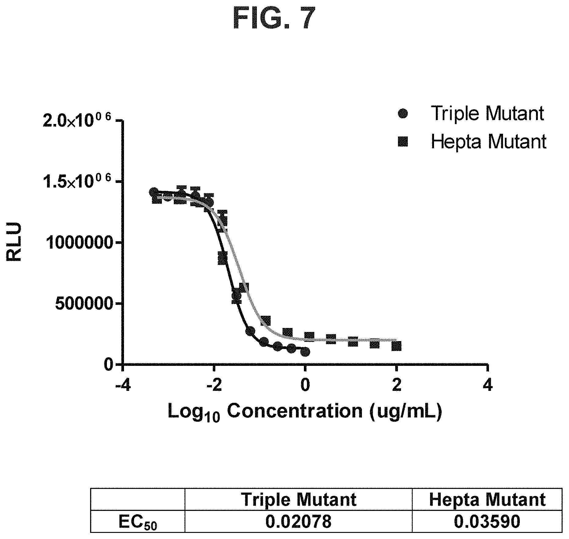
D00020

D00021

D00022

D00023

D00024

D00025

D00026

D00027

D00028

D00029

D00030

D00031

D00032

D00033

D00034

D00035

D00036

D00037

D00038

D00039

D00040

D00041

D00042

D00043

D00044

D00045

D00046

D00047

D00048
D00049

D00050

S00001
XML
uspto.report is an independent third-party trademark research tool that is not affiliated, endorsed, or sponsored by the United States Patent and Trademark Office (USPTO) or any other governmental organization. The information provided by uspto.report is based on publicly available data at the time of writing and is intended for informational purposes only.
While we strive to provide accurate and up-to-date information, we do not guarantee the accuracy, completeness, reliability, or suitability of the information displayed on this site. The use of this site is at your own risk. Any reliance you place on such information is therefore strictly at your own risk.
All official trademark data, including owner information, should be verified by visiting the official USPTO website at www.uspto.gov. This site is not intended to replace professional legal advice and should not be used as a substitute for consulting with a legal professional who is knowledgeable about trademark law.