Methods, kits, and compositions for stem cell self-renewal
Perry , et al. September 29, 2
U.S. patent number 10,787,643 [Application Number 14/634,540] was granted by the patent office on 2020-09-29 for methods, kits, and compositions for stem cell self-renewal. This patent grant is currently assigned to STOWERS INSTITUTE FOR MEDICAL RESEARCH. The grantee listed for this patent is Stowers Institute for Medical Research. Invention is credited to Justin C. Grindley, Linheng Li, John M. Perry.


View All Diagrams
| United States Patent | 10,787,643 |
| Perry , et al. | September 29, 2020 |
Methods, kits, and compositions for stem cell self-renewal
Abstract
The present invention relates to methods and kits for expanding a stem cell population. More particularly, the invention relates, inter alia, to methods, kits, and compositions for expanding a stem cell population, particularly a hematopoietic stem cell population.
| Inventors: | Perry; John M. (Olathe, KS), Li; Linheng (Leawood, KS), Grindley; Justin C. (Kansas City, MO) | ||||||||||
|---|---|---|---|---|---|---|---|---|---|---|---|
| Applicant: |
|
||||||||||
| Assignee: | STOWERS INSTITUTE FOR MEDICAL
RESEARCH (Kansas City, MO) |
||||||||||
| Family ID: | 1000005081860 | ||||||||||
| Appl. No.: | 14/634,540 | ||||||||||
| Filed: | February 27, 2015 |
Prior Publication Data
| Document Identifier | Publication Date | |
|---|---|---|
| US 20150344845 A1 | Dec 3, 2015 | |
Related U.S. Patent Documents
| Application Number | Filing Date | Patent Number | Issue Date | ||
|---|---|---|---|---|---|
| 12589551 | Oct 23, 2009 | ||||
| PCT/US2008/005230 | Apr 23, 2008 | ||||
| 60926065 | Apr 23, 2007 | ||||
| 61066693 | Feb 22, 2008 | ||||
| Current U.S. Class: | 1/1 |
| Current CPC Class: | A61K 35/28 (20130101); C12N 5/0647 (20130101); C12N 2502/11 (20130101); C12N 2501/415 (20130101); C12N 2501/70 (20130101); C12N 2501/40 (20130101) |
| Current International Class: | C12N 5/00 (20060101); C12N 5/02 (20060101); C12N 5/0789 (20100101); A61K 35/28 (20150101); C12N 5/18 (20060101) |
References Cited [Referenced By]
U.S. Patent Documents
| 5851984 | December 1998 | Matthews |
| 5958769 | September 1999 | Roberts et al. |
| 6159462 | December 2000 | Matthews |
| 7169605 | January 2007 | Peled et al. |
| 7850960 | December 2010 | Moon |
| 7955852 | June 2011 | Peled et al. |
| 8080417 | December 2011 | Peled et al. |
| 8263403 | September 2012 | Perry |
| 8476071 | July 2013 | Perry |
| 9896659 | February 2018 | Perry |
| 2005/0054097 | March 2005 | Peled |
| 2005/0158857 | July 2005 | Yilmaz et al. |
| 2005/0276793 | December 2005 | Milhem |
| 2006/0147435 | July 2006 | Moon |
| 2006/0275823 | December 2006 | Kodadek |
| 2007/0065447 | March 2007 | Tryggvason et al. |
| 2009/0093026 | April 2009 | Dowdy et al. |
| 2009/0285786 | November 2009 | Zon |
| 2010/0196337 | August 2010 | Perry et al. |
| 2011/0053266 | March 2011 | Perry et al. |
| 2012/0309088 | December 2012 | Perry et al. |
| 2013/0252338 | September 2013 | Perry et al. |
| 2010524499 | Jul 2010 | JP | |||
| WO 90/10448 | Sep 1990 | WO | |||
| WO 91/04753 | Apr 1991 | WO | |||
| 04/016731 | Feb 2004 | WO | |||
| WO 2004/016731 | Feb 2004 | WO | |||
| WO 2004/048536 | Jun 2004 | WO | |||
| WO 2006/027545 | Mar 2006 | WO | |||
| 2008133904 | Nov 2008 | WO | |||
Other References
|
Hofmeister et al., Jan. 2007; Bone Marrow Transplantation 39:11-23. cited by examiner . Bug et al 2005, Cancer Res. 65:2537-2541. cited by examiner . Sato et al 2004, Nature Medicine 10:55-63. cited by examiner . Gallichio et al., 1981, Exp. Hematol. 9:804-810. cited by examiner . Trowbridge et al 2005, Nat. Med. 12:89-98. cited by examiner . Bailin et al 1998, Brit. J. Hematol 100:219-221. cited by examiner . Robinson et al., 2005, Cytotherapy 7:243-250. cited by examiner . Antochuk et al 2002, Cell 109:39-45. cited by examiner . Zhang 2005, Blood 105:4314-4320. cited by examiner . Feitz et al 1999, Bone Marrow Transplantation 23:1109-1115. cited by examiner . Sato et al Maintenance of pluripotency in human and mouse embryonic stem cells through activation of Wnt signaling by a pharmacological GSK-3-specific inhibitor Published online Dec. 21, 2003. Nature Medicine vol. 10 | No. 1 | Jan. 2004 55-63. cited by examiner . Fietz, T., et al., Culturing human umbilical cord blood: a comparison of mononuclear vs CD34+ selected cells. Bone Marrow Transplant. 23: 1109-1115, (1999). cited by applicant . Hofmeister, CC, et al., Jan. 2007, Bone Marrow Transplant. Jan. 2007; 39(1):11-23. cited by applicant . Gallicchio, V., 1981, Influcence of Lithium on Proliferation of Hematopoietic Stem Cells, Exp. Hematol, 9:804-810. cited by applicant . Trowbridge, J. J., et al., Glycogen synthase kinase-3 is an in vivo regulator of hematopoietic stem cell repopulation, Nat Med. 12:89-98. cited by applicant . Ballin, A., et al., Increased number of peripheral blood CD34+ cells in lithium-treated patients, Brit. J. Haematology, vol., Issue 1, pp. 219-221 (Jan. 1998). cited by applicant . The National Formulary, American Pharmaceutical Association, Washington, DC. cited by applicant . Remington, The Science and practice of pharmacy, 21 Ed., Lippincott Williams and Wilkins, Philadelphia, PA. cited by applicant . Pietrancosta, N., et al., Are p53 inhibitors potentially useful therapeutics?, Drug Dev. Res. 65:43-49 (2005). cited by applicant . Smith, LH and Clayton, ML, (1970) Distribution of injected 59Fe in mice, Haemopoietic Stem Cells, Ciba Foundation Symposium 13, Exp Hematol 20: 82-86. cited by applicant . Wu-Pong, Biopharm, Nov. (Nov. 1, 1994), pp. 20-33. cited by applicant . Peled, T., et al., Chelatable cellular copper modulates differentiation and self-renewal of cord blood-derived hematopoietic progenitor cells, Exp Hematol. Oct. 2005;33(10):1092-100. cited by applicant . Robinson, S., et al., Ex vivo expansion of umbilical cord blood, Cytotherapy 7, 243-250 (2005). cited by applicant . Akashi et al., "A Clonogenic Common Myeloid Progenitor That Gives Rise to All Myeloid Lineages," Nature, 404, pp. 193-197 (2000). cited by applicant . Antonchuk et al., "HOXB4-Induced Expansion of Adult Hematopoietic Stem Cells Ex Vivo," Cell, 109, pp. 39-45 (2002). cited by applicant . Arai et al., "Tie2/Angiopoietin-1 Signaling Regulates Hematopoietic Stem Cell Quiescence in the Bone Marrow Niche," Cell, 118, pp. 149-161 (2004). cited by applicant . Burgering et al., "Decisions on Life and Death: FOXO Forkhead Transcription Factors Are in Command When PKB/Akt is Off Duty," Journal of Leukocyte Biology, 73, pp. 689-701 (2003). cited by applicant . Calvi et al., "Osteoblastic Cells Regulate the Haematopoietic Stem Cell Niche," Nature, 425, pp. 841-846 (2003). cited by applicant . Caplen et al., "Specific Inhibition of Gene Expression by Small Double-Stranded RNAs in Invertebrate and Vertebrate Systems," PNAS, 98(17), pp. 9742-9747 (2001). cited by applicant . Cardone et al., "Regulation of Cell Death Protease Caspase-9 by Phosphorylation," Science, 282(5392), pp. 1318-1321 (1998). cited by applicant . Casset et al., "A Peptide Mimetic of an Anti-CD4 Monoclonal Antibody by Rational Design," Biochem. Biophys. Res. Commun., 307(1), pp. 198-205 (2003). cited by applicant . Christensen et al., "Flk-2 is a Marker in Hematopoietic Stem Cell Differentiation: A Simple Method to Isolate Long-Term Stem Cells," PNAS, 98(25), pp. 14541-14546 (2001). cited by applicant . Cobas et al., "Beta-Catenin is Dispensable for Hematopoiesis and Lymphopoiesis," J. Exp. Med., 199(2), pp. 221-229 (2004). cited by applicant . Cortez-Retamozo et al., "Efficient Cancer Therapy With a Nanobody-Based Conjugate," Cancer Research, 64, pp. 2853-2857 (2004). cited by applicant . Cross et al., "Inhibition of Glycogen Synthase Kinase-3 by Insulin Mediated by Protein Kinase B," Nature, 378, pp. 785-789 (1995). cited by applicant . Cui et al., "Muscarinic Acetylcholine Receptors Mediate Oligodendrocyte Progenitor Survival Through Src-like Tyrosine Kinases and PI3K/Akt Pathways," Neurochem. Int., 48(5), pp. 383-393 (2006). cited by applicant . Cully et al., "Beyond PTEN Mutations: the P13K Pathway as an Integrator of Multiple Inputs During Tumorigenesis," Nature Review Cancer, 6, pp. 184-192 (2006). cited by applicant . Datta et al., "Akt Phosphorylation of BAD Couples Survival Signals to the Cell-Intrinsic Death Machinery," Cell, 91, pp. 231-241 (1997). cited by applicant . Datta et al., "Cellular Survival: A Play in Three Akts," Genes & Development, 13, 2905-2927 (1999). cited by applicant . Donepudi et al., "Structure and Zymogen Activation of Caspases," Biophys. Chem., vols. 101-102, pp. 145-153 (2002). cited by applicant . Elbashir et al., "Duplexes of 21-Nucleotide RNAs Mediate RNA Interference in Cultured Mammalian Cells," Nature, 411, pp. 494-498 (2001). cited by applicant . El-Deiry et al., "WAF1, a Potential Mediator of P53 Tumor Suppression," Cell, 75(4), pp. 817-825 (1993). cited by applicant . Fassina, "Complementary Peptides as Antibody Mimetics for Protein Purification and Assay," Immunomethods, 5(2), pp. 121-129 (1994). cited by applicant . Foster, "Regulation of mTOR by Phosphatidic Acid?," Cancer Res., 67(1), pp. 1-4 (2007). cited by applicant . Fujita et al., "Akt-Dependent Phosphorylation of p27.sup.Kip1 Promotes Binding to 14-3-3 and Cytoplasmic Localization," J. Biol. Chem., 277(32), pp. 28706-28713 (2002). cited by applicant . Gilley et al., "FOXO Transcription Factors Directly Activate Bim Gene Expression and Promote Apoptosis in Sympathetic Neurons," J. Cell Biol., 162(4), pp. 613-622 (2003). cited by applicant . Goessling et al., "Genetic Interaction of PGE2 and Wnt Signaling Regulates Developmental Specification of Stem Cells and Regeneration," Cell, 136, pp. 1136-1147 (2009). cited by applicant . Gothert et al., In Vivo Fate-Tracing Studies Using the Scl Stem Cell Enhancer: Embryonic Hematopoietic Stem Cells Significantly Contribute to Adult Hematopoiesis, Blood Journal, 105(7), pp. 2724-2732 (2005). cited by applicant . Gottlieb et al., "Cross-talk Between Akt, p53 and Mdm2: Possible Implications for the Regulation of Apoptosis," Oncogene, 21(8), pp. 1299-1303 (2002). cited by applicant . Gray et al., "Trichofolliculoma," Arch. Dermatol., 86(5), pp. 619-625 (1962). cited by applicant . Guo et al., "Multi-Genetic Events Collaboratively Contribute to Pten-null Leukaemia Stem-Cell Formation," Nature, 453, pp. 529-533 (2008). cited by applicant . Hagen et al., "Expression and Characterization of GSK-3 Mutants and Their Effect on -Catenin Phosphorylation in Intact Cells," J. Biol. Chem., 277(26), pp. 23330-23335 (2002). cited by applicant . Harada et al., "Intestinal Polyposis in Mice With a Dominant Stable Mutation of the -Catenin Gene," EMBO Journal, 18(21), pp. 5931-5942 (1999). cited by applicant . Harborth et al., "Identification of Essential Genes in Cultured Mammalian Cells Using Small Interfering RNAs," Journal of Cell Science, 114(24), pp. 4557-4565 (2001). cited by applicant . Haupt et al., "Mdm2 Promotes the Rapid Degradation of P53," Nature, 387, pp. 296-299 (1997). cited by applicant . He et al., "PTEN-Deficient Intestinal Stem Cells Initiate Intestinal Polyposis," Nature Genetics, 39(2), pp. 189-198 (2007). cited by applicant . Heissig et al., "Recruitment of Stem and Progenitor Cells From the Bone Marrow Niche Requires MMP-9 Mediated Release of Kit-Ligand," Cell, 109, pp. 625-637 (2002). cited by applicant . Hsu et al., "Interference of BAD (Bcl-xL/Bcl-2-Associated Death Promoter)-Induced Apoptosis in Mammalian Cells by 14-3-3 Isoforms and P11," Molecular Endocrinology, 11(12), pp. 1858-1867 (1997). cited by applicant . Hui et al., "The Neuroprotection of Insulin on Ischemic Brain Injury in Rat Hippocampus Through Negative Regulation of JNK Signaling Pathway to PI3K/Akt Activation," Brain Res., 1052(1), pp. 1-9 (2005). cited by applicant . Katoh et al., "Cross-Talk of WNT and FGF Signaling Pathways at GSK3 to Regulate -Catenin and SNAIL Signaling Cascades," Cancer Biol. Ther., 5(9), pp. 1059-1064 (2006). cited by applicant . Kenney et al., "Hedgehog and PI-3 Kinase Signaling Converge on Nmyc1 to Promote Cell Cycle Progression in Cerebellar Neuronal Precursors," Development, 131(1), pp. 217-228 (2004). cited by applicant . Kiel et al., "SLAM Family Receptors Distinguish Hematopoietic Stem and Progenitor Cells and Reveal Endothelial Niches for Stem Cells," Cell, 121, pp. 1109-1121 (2005). cited by applicant . Kim et al., "GSK3 at the Edge: Regulation of Developmental Specification and Cell Polarization," Current Drug Targets, 7(11), pp. 1411-1419 (2006). cited by applicant . Kimura et al., "Conditional Loss of PTEN Leads to Testicular Teratoma and Enhances Embryonic Germ Cell Production," Develogment, 130, pp. 1691-1700 (2003). cited by applicant . Kirstetter et al., "Activation of the Canonical Wnt Pathway Leads to Loss of Hematopoietic Stem Cell Repopulation and Multilineage Differentiation Block," Nat. Immunol., 7 (10), pp. 1048-1056 (2006). cited by applicant . Kobayashi et al., "Thrombopoietin Supports Proliferation of Human Primitive Hematopoietic Cells in Synergy With Steel Factor and/or Interleukin-3," Blood, 88(2), pp. 429-436 (1996). cited by applicant . Komarov et al., "A Chemical Inhibitor of p53 That Protects Mice from the Side Effects of Cancer Therapy," Science, 285(5434), pp. 1733-1737 (1999). cited by applicant . Kondo et al., "Identification of Clonogenic Common Lymphoid Progenitors in Mouse Bone Marrow," Cell, 91, pp. 661-672 (1997). cited by applicant . Li et al., "Mechanistic Insights Into Maintenance of High p53 Acetylation by PTEN," Molecular Cell, 23, pp. 575-587 (2006). cited by applicant . Ll et al., "Stem Cell Niche: Structure and Function," Annu. Rev. Cell Dev. Biol., 21, pp. 605-631 (2005). cited by applicant . Lopiccolo et al., "Targeting Akt in Cancer Therapy," Anticancer Drugs, 18(8), pp. 861-874 (2007). cited by applicant . Maehama et al., "The Tumor Suppressor, PTEN/MMAC1, Dephosphorylates the Lipid Second Messenger, Phosphatidylinositol 3,4,5-Trisphosphate," J. Biol. Chem., 273(22), pp. 13375-13378 (1998). cited by applicant . Maira et al., "Identification and Characterization of NVP-BEZ235, A New Orally Available Dual Phosphatidylinositol 3-Kinase/Mammalian Target of Rapamycin Inhibitor With Potent In Vivo Antitumor Activity," Mol. Cancer Ther., 7(7), pp. 1851-1863 (2008). cited by applicant . Matsuoka et al., "Fbxw7 Acts as a Critical Fail-Safe Against Premature Loss of Hematopoietic Stem Cells and Development of T-ALL," Genes & Develogment, 22, pp. 986-991 (2008). cited by applicant . Mayo et al., "A Phosphatidylinositol 3-Kinase/Akt Pathway Promotes Translocation of Mdm2 From the Cytoplasm to the Nucleus," PNAS, 98(20), pp. 11598-11603 (2001). cited by applicant . Mimeault et al., "Stem Cells: A Revolution in Therapeutics--Recent Advances in Stem Cell Biology and Their Therapeutic Applications in Regenerative Medicine and Cancer Therapies," Clinical Pharmacology & Therapeutics 82, pp. 252-264 (2007). cited by applicant . Mise-Omata et al., "Transient Strong Reduction of PTEN Expression by Specific RNAi Induces Loss of Adhesion of the Cells," Biochem. Biophys. Res. Commun., 328(4), pp. 1034-1042 (2005). cited by applicant . Momand et al., "The Mdm-2 Oncogene Product Forms a Complex With the P53 Protein and Inhibits p53-Mediated Transactivation," Cell, 69(7), pp. 1237-1245 (1992). cited by applicant . Moon et al., "The Promise and Perils of Wnt Signaling Through .beta.-Catenin," Science, 296(5573), pp. 1644-1646 (2002). cited by applicant . Mukhopadhyay et al., "Antisense Regulation of Oncogenes in Human Cancer," Crit. Rev. Oncog., 7(3-4), pp. 151-190 (1996). cited by applicant . Mutter, "PTEN, a Protean Tumor Suppressor," Am. J. Pathol., 158(6), pp. 1895-1898 (2001). cited by applicant . Nakorn et al., "Myeloerythroid-Restricted Progenitors Are Sufficient to Confer Radioprotection and Provide the Majority of Day 8 CFU-S," J. Clin. Invest., 109(12), pp. 1579-1585 (2002). cited by applicant . Nicholson et al., "The Protein Kinase B/Akt Signalling Pathway in Human Malignancy," Cellular Signalling, 14(5), pp. 381-395 (2002). cited by applicant . Nikitenko et al., "Adrenomedullin and Tumour Angiogenesis," British Journal of Cancer, 94, pp. 1-7 (2006). cited by applicant . North et al., "Prostaglandin E2 Regulates Vertebrate Haematopoietic Stem Cell Homeostatis," Nature, 447, pp. 1007-1011 (2007). cited by applicant . Novak et al., "Z/EG, A Double Reporter Mouse Line That Expresses Enhanced Green Fluorescent Protein Upon Cre-Mediated Excision," Genesis, 28, pp. 147-155 (2000). cited by applicant . Oren, "Decision Making by p53: Life, Death and Cancer," Cell Death and Differentiation, 10, pp. 431-442 (2003). cited by applicant . Pai et al., "Deoxycholic Acid Activates -Catenin Signaling Pathway and Increases Colon Cell Cancer Growth and Invasiveness," Molecular Biology of the Cell, 15, pp. 2156-2163 (2004). cited by applicant . Paquette al., "Optimizing Hematopoietic Recovery Following Bone Marrow Transplantation," J. Clin. Invest., 109(12), pp. 1527-1528 (2002). cited by applicant . Park et al., "Bmi-1 is Required for Maintenance of Adult Self-Renewing Haematopoietic Stem Cells," Nature, 423, pp. 302-305 (2003). cited by applicant . Perry et al., "Self-Renewal Versus Transformation: Fbxw7 Deletion Leads to Stem Cell Activation and Leukemogenesis," Genes & Development, 22, pp. 1107-1109 (2008). cited by applicant . Persad et al., "Tumor Suppressor PTEN Inhibits Nuclear Accumulation of -Catenin and T Cell/Lymphoid Enhancer Factor 1-Mediated Transcriptional Activation," J. Cell Biol., 153(6), pp. 1161-1173 (2001). cited by applicant . Reya et al., "A Role for Wnt Signalling in Self-Renewal of Haematopoietic Stem Cells," Nature, 425, pp. 409-414 (2003). cited by applicant . Ring et al., "Selective Glycogen Synthase Kinase 3 Inhibitors Potentiate Insulin Activation of Glucose Transport and Utilization In Vitro and In Vivo," Diabetes, 52, pp. 588-595 (2003). cited by applicant . Salmena et al., "Tenets of PTEN Tumor Suppression," Cell, 133, pp. 403-414 (2008). cited by applicant . Saragovi et al., "Design and Synthesis of a Mimetic From an Antibody Complementarity-Determining Region," Science, 253(5021), pp. 792-795 (1991). cited by applicant . Sarbassov et al., "Phosphorylation and Regulation of Akt/PKB by the Rictor-mTOR Complex," Science, 307(5712), pp. 1098-1101 (2005). cited by applicant . Scheller et al., "Hematopoietic Stem Cell and Multilineage Defects Generated by Constitutive -Catenin Activation," Nature Immunology, 7, pp. 1037-1047 (2006). cited by applicant . Schmid et al., "Bisperoxovanadium Compounds Are Potent PTEN Inhibitors," FEBS Letters, 556, pp. 35-38 (2004). cited by applicant . Song et al., "The Activation of Akt/PKB Signaling Pathway and Cell Survival," J. Cell. Mol. Med. 9(1), pp. 59-71 (2005). cited by applicant . Stein et al., "Oligodeoxynucleotides as Inhibitors of Gene Expression: A Review," Cancer Research, 48, pp. 2659-2668 (1998). cited by applicant . Stiles et al., "PTENless Means More," Developmental Biology, 273, pp. 175-184 (2004). cited by applicant . Suzuki et al., "T Cell-Specific Loss of Pten Leads to Defects in Central and Peripheral Tolerance," Immunity, 14, pp. 523-534 (2001). cited by applicant . Tamama et al., "Epidermal Growth Factor as a Candidate For Ex Vivo Expansion of Bone Marrow-Derived Mesenchymal Stem Cells," Stem Cells, 24, pp. 686-695 (2006). cited by applicant . Tang et al., "PTEN Autoregulates Its Expression by Stabilization of p53 in a Phosphatase-Independent Manner," Cancer Res., 66(2), pp. 736-742 (2006). cited by applicant . Tee et al., "Inactivation of the Tuberous Sclerosis Complex-1 and -2 Gene Products Occurs by Phosphoinositide 3-Kinase/Akt-Dependent and -Independent Phosphorylation of Tuberin," J. Biol. Chem., 278(39), pp. 37288-37296 (2003). cited by applicant . Tessier et al., "Role of the Phox Homology Domain and Phosphorylation in Activation of Serum and Glucocorticoid-Regulated Kinase-3," J. Biol. Chem., 281(33), pp. 23978-23989 (2006). cited by applicant . Thornberry et al., "Caspases: Enemies Within," Science, 281(5381), pp. 1312-1316 (1998). cited by applicant . Tian et al., "Bridging the BMP and Wnt Pathways by PI3 Kinase/Akt and 14-3-3.zeta.," Cell Cycle, 4(2), pp. 215-216 (2005). cited by applicant . Van Der Krol et al., "Modulation of Eukaryotic Gene Expression by Complementary RNA or DNA Sequences," Biotechniques, 6(10), pp. 958-976 (1988). cited by applicant . Varnum-Finney et al., "Pluripotent, Cytokine-Dependent, Hematopoietic Stem Cells Are Immortalized by Constitutive Notch1 Signaling," Nature Medicine, 6(11), pp. 1278-1281 (2000). cited by applicant . Wen et al., "Negative Regulation of Phosphatidylinositol 3-Kinase and Akt Signalling Pathway by PKC," Cellular Signalling, 15, pp. 37-45 (2003). cited by applicant . West et al., "Rapid Akt Activation by Nicotine and a Tobacco Carcinogen Modulates the Phenotype of Normal Human Airway Epithelial Cells," J. Clin. Invest., 111(1), pp. 81-90 (2003). cited by applicant . White et al., "Negative Regulation of Myofibroblast Differentiation by PTEN (Phosphatase and Tensin Homolog Deleted on Chromosome 10)," Am. J. Respir. Crit. Care Med., 173, pp. 112-121 (2006). cited by applicant . Wu et al., "PTEN Signaling Pathways in Melanoma," Oncogene, 22, pp. 3113-3122 (2003). cited by applicant . Xie et al., "Detection of Functional Haematopoietic Stem Cell Niche Using Real-Time Imaging," Nature, p. 97-101 (2009). cited by applicant . Yacoub et al., "Optimized Production and Concentration of Lentiviral Vectors Containing Large Inserts," J. Gene Med., 9(7), pp. 579-584 (2007). cited by applicant . Yilmaz et al., "Pten Dependence Distinguishes Haematopoietic Stem Cells From Leukaemia-Initiating Cells," Nature, 441, pp. 475-482 (2006). cited by applicant . Ying et al., "The Ground State of Embryonic Stem Cell Self-Renewal," Nature, 453, pp. 519-523 (2008). cited by applicant . Zhang C. et al., "Murine Hematopoietic Stem Cells Change Their Surface Phenotype During Ex Vivo Expansion," Blood, 105(11), pp. 4314-4320 (2005). cited by applicant . Zhang J. et al., "BMP Signaling and Stem Cell Regulation," Developmental Biology, 284, pp. 1-11 (2005). cited by applicant . Zhang J. et al., "Identification of the Haematopoietic Stem Cell Niche and Control of the Niche Size," Nature, 425, pp. 836-841 (2003). cited by applicant . Zhang J. et al., "PTEN Maintains Haematopoietic Stem Cells and Acts in Lineage Choice and Leukaemia Prevention," Nature, 441, pp. 518-522 (2006). cited by applicant . Zhang C. et al., "Angiopoietin-like Proteins Stimulate Ex Vivo Expansion of Hematopoietic Stem Cells," Nat. Med., 12(2), pp. 240-245 (2006). cited by applicant . Zhang O et al., "Small-Molecule Synergist of the Wnt/ -Catenin Signaling Pathway," PNAS, 104(18), pp. 7444-7448 (2007). cited by applicant . Zhu et al., "A Versatile Approach to Multiple Gene RNA Interference Using MicroRNA-Based Short Hairpin RNAs," BMC Molecular Biology, 8(98), pp. 1-11 (2007). (page number not for citation purposes). cited by applicant . Groszer et al., "Negative Regulation of Neural Stem/Progenitor Cell Proliferation by the Pten Tumor Suppressor Gene in Vivo," Science, 294, pp. 2186-2189 (2001). cited by applicant . Liu et al., "A Small-Molecule Agonist of the Wnt Signaling Pathway," Angewandte Chemie International Edition, 44 (13), pp. 1987-1990 (2005). cited by applicant . Borowitz et al., "Immunophenotyping of acute leukemia by flow cytometric analysis," Am. J. Clin. Pathol., 100, pp. 534-540 (1993). cited by applicant . Akala et al., "Hematopoietic stem cell self-renewal," Curr Opin Genet Dev, 16, pp. 496-501 (2006). cited by applicant . Bennett et al., "Regulation of Wnt Signaling during Adipogenesis," J Biol Chem, 277, pp. 30998-31004 (2002). cited by applicant . Bug et al., "Valproic Acid Stimulates Proliferation and Self-Renewal of Hematopoietic Stem Cells," Cancer Research, 65, pp. 2537-2541 (2005). cited by applicant . Butler et al., "Endothelial Cells Are Essential for the Self-Renewal and Repopulation of Notch-Dependent Hematopoietic Stem Cells," Cell Stem Cell, 6, pp. 251-264 (2010). cited by applicant . Delaney et al., "Notch-Mediated Expansion of Human Cord Blood Progenitor Cells Capable of Rapid Myeloid Reconstitution," Nat. Med., vol. 16, pp. 232-236 (2010). cited by applicant . Deleyrolle et al., "Isolation, Expansion, and Differentiation of Adult Mammalian Neural Stem and Progenitor Cells Using the Neurosphere Assay," Neural Cell Transplantation, 549, pp. 91-101 (2009). cited by applicant . Fuchs et al., "Stem Cells: A New Lease on Life," Cell, 100, pp. 143-155 (2000). cited by applicant . Himburg et al., "Pleiotrophin Regulates the Expansion and Regeneration of Hematopoietic Stem Cells," Nat. Med., vol. 16, pp. 475-482 (2010). cited by applicant . Kast et al., "How lithium treatment generates neutrophilia by enhancing phosphorylation of GSK-3, increasing HIF-1 levels and how this path is important during engraftment," Bone Marrow Transplantation, 41, pp. 23-26 (2008). cited by applicant . Miller et al., "Expansion In Vitro of Adult Murine Hematopoietic Stem Cells With Transplantable Lympho-Myeloid Reconstituting Ability," PNAS, vol. 94, pp. 13648-13653 (1997). cited by applicant . Patil et al. "DNA-based therapeutics and DNA delivery systems: a comprehensive review." The AAPS journal 7.1 (2005): E61-E77. cited by applicant . Sasaki et al., "PKB activation based on PTEN inhibitory effect of shikonin", Annual Meeting in Pharmaceutical Society of Japan, Abstract, 124th, p. 119, 29[P2] III-267 (2004) (in Japanese). cited by applicant . English translation of: Sasaki et al., "PKB activation based on PTEN inhibitory effect of shikonin", Annual Meeting in Pharmaceutical Society of Japan, Abstract, 124th, p. 119, 29[P2] III-267 (2004). cited by applicant . Stambolic et al. "Lithium inhibits glycogen synthase kinase-3 activity and mimics wingless signalling in intact cells." Current Biology 6.12 (1996): 1664-1669. cited by applicant . Cully et al., "Beyond PTEN Mutations: the PI3K Pathway as an Integrator of Multiple Inputs During Tumorigenesis," Nature Review Cancer, 6, pp. 184-192 (2006). cited by applicant . Hsu et al., "Interference of BAD (Bcl-xL/Bcl-2-Associated Death Promoter)-Induced Apoptosis in Mammalian Cells by 14-3-3 Isoforms and P11," Molecular Endocrinology, 11(12), pp. 1558-1867 (1997). cited by applicant . Katoh et al., "Cross-Talk of WNT and FGF Signaling Pathways at GSK38 to Regulate -Catenin and SNAIL Signaling Cascades," Cancer Biol. Ther., 5(9), pp. 1059-1064 (2006). cited by applicant . Kimura et al., "Conditional Loss of PTEN Leads to Testicular Teratoma and Enhances Embryonic Germ Cell Production," Development, 130, pp. 1691-1700 (2003). cited by applicant . Li et al., "Stem Cell Niche: Structure and Function," Annu. Rev. Cell Dev. Biol., 21, pp. 605-631 (2005). cited by applicant . Matsuoka et al., "Fbxw7 Acts As a Critical Fail-Safe Against Premature Loss of Hematopoietic Stem Cells and Development of T-ALL," Genes & Development, 22, pp. 986-991 (2008). cited by applicant . Persad et al., "Tumor Suppressor PTEN Inhibits Nuclear Accumulation of Beta-Catenin and T Cell/Lymphoid Enhancer Factor 1-Mediated Transcriptional Activation," J. Cell Biol., 153(6), pp. 1161-1173 (2001). cited by applicant . Zhang et al., "Angiopoietin-like Proteins Stimulate Ex Vivo Expansion of Hematopoietic Stem Cells," Nat. Med., 12(2), pp. 240-245 (2006). cited by applicant . Zhang Q et al., "Small-Molecule Synergist of the Wnt/.beta.-Catenin Signaling Pathway," PNAS, 104(18), pp. 7444-7448 (2007). cited by applicant . Sato et al., "Maintenance of Pluripotency in Human and Mouse Embryonic Stem Cells Through Activation of Wnt Signaling by a Pharmacological GSK-3-Specific Inhibitor," Nat. Med., vol. 10, pp. 55-63 (2004). cited by applicant. |
Primary Examiner: Leavitt; Maria G
Attorney, Agent or Firm: Bryan Cave Leighton Paisner LLP
Parent Case Text
CROSS-REFERENCE TO RELATED APPLICATIONS
This application is a continuation of U.S. patent application Ser. No. 12/589,551, filed Oct. 23, 2009, which is a continuation-in-part of International Application Serial No. PCT/US2008/005230, filed Apr. 23, 2008. PCT/US2008/005230 claims benefit to U.S. Provisional Patent Application Ser. No. 60/926,065, filed Apr. 23, 2007 and U.S. Provisional Patent Application Ser. No. 61/066,693, filed Feb. 22, 2008. The entire contents of the above-mentioned applications are hereby incorporated by reference as if recited in full herein.
Claims
What is claimed is:
1. An ex vivo method for expanding the number of hematopoietic stem cells (HSC) in a population of mononuclear cells (MNCs) comprising: culturing the population of MNCs comprising at least one HSC in an HSC expansion media comprising a reversible glycogen synthase kinase 3 beta (GSK-3.beta.) inhibitor for a period of time of at least 10 days, wherein the reversible GSK-3.beta. inhibitor is effective to activate .beta.-catenin, wherein the expanded HSCs are functional with long germ, multi-lineage, repopulation potential.
2. The method according to claim 1, wherein the reversible GSK-3.beta. inhibitor is selected from the group consisting of a small molecule, a biologic, an antisense RNA, a small interfering RNA (siRNA), and combinations thereof.
3. The method according to claim 2, wherein the reversible GSK-3.beta. inhibitor is a small molecule.
4. The method according to claim 2, wherein the reversible GSK-3.beta. inhibitor is selected from the group consisting of Hymenialdisine, Flavopiridol, Kenpaullone, Alsterpaullone, Azakenpaullone, Indirubin-30-oxime, 6-Bromoindirubin-30-oxime (BIO), 6-Bromoindirubin-30-acetoxime, Aloisine A, Aloisine B, TDZD8, Compound 12, CHIR98014, CHIR99021 (CT99021), CT20026, Compound 1, SU9516, ARA014418, Staurosporine, Compound 5a, Compound 29, Compound 46, GF109203x (bisindolylmaleimide 1), Ro318220 (bisindolylmaleimide IX), SB216763, SB415286, 15, CGP60474, Compound 8b, TWS119, Compound 1A, Compound 17, Lithium, Beryllium, Zinc, small molecule GSK-3.beta. inhibitors (Vertex Pharmaceuticals), NP-12 (Neuropharma), GSK-3.beta. inhibitors (Amphora), GSK-3.beta. inhibitors (CrystalGenomics), SAR-502250 (Sanofi-Aventis), 3544 (Hoffmann-La Roche), GSK-3.beta. inhibitors (Lundbeck), TDZD-8 (Cancer Center, University of Rochester), pharmaceutically acceptable salts thereof, and combinations thereof.
5. The method according to claim 4, wherein the reversible GSK-3.beta. inhibitor is CHIR99021.
6. The method according to claim 1, which provides HSCs that, upon transplant into a recipient, exhibit 60% to 100% donor repopulation at least 4 weeks after transplant into the recipient.
7. The method according to claim 1, wherein the HSC is obtained from a mammalian tissue selected from the group consisting of cord blood, peripheral blood, and bone marrow.
8. The method according to claim 1, which provides HSCs that, upon transplant into a recipient, exhibit 5% to 100% donor repopulation at least 4 weeks after transplant into the recipient.
9. The method according to claim 1, which provides HSCs that, upon transplant into a recipient, exhibit 25% to 100% donor repopulation at least 4 weeks after transplant into the recipient.
10. The method according to claim 1, which provides HSCs that, upon transplant into a recipient, exhibit 45% to 100% donor repopulation at least 4 weeks after transplant into the recipient.
Description
FIELD OF THE INVENTION
The present invention relates to methods, kits, and compositions for expanding a stem cell population, particularly an hematopoietic stem cell population.
INCORPORATION BY REFERENCE OF SEQUENCE LISTING
This application contains references to amino acids and/or nucleic acid sequences that have been submitted as sequence listing text file "C065272_023131con_ST25.txt", file size of 2 KB, created on Feb. 27, 2015. The aforementioned sequence listing is hereby incorporated by reference in its entirety pursuant to 37 C.F.R. .sctn. 1.52(e)(5).
BACKGROUND OF THE INVENTION
Hematopoietic stem cells (HSCs) are clonogenic cells, which possess the properties of both self-renewal (expansion) and multilineage potential giving rise to all types of mature blood cells. HSCs are responsible for hematopoiesis and undergo proliferation and differentiation to produce mature blood cells of various lineages while still maintaining their capacity for self-renewal. The ability to self-renew maintains the HSC population for the lifespan of an animal and also allows HSCs to repopulate the bone marrow of lethally irradiated congenic hosts.
Early HSC development displays a hierarchical arrangement, starting from long-term (LT-) HSCs, which have extensive self-renewal capability, followed by the expansion state, which corresponds to short-term (ST-) HSCs (having limited self-renewal ability) and proliferative multipotent progenitors (MPPs) (having multipotent potential but no self-renewal capability). MPP is also a stage of priming or preparation for differentiation. An MPP differentiates and commits to become either a common lymphoid progenitor (CLP), which gives rise to all the lymphoid lineages, or a common myeloid progenitor (CMP), which produces all the myeloid lineages. During this process, the more primitive population gives rise to a less primitive population of cells, which is unable to give rise to a more primitive population of cells. The intrinsic genetic programs that control these processes including the multipotential, self-renewal, and activation (or transient amplification) of HSCs, and lineage commitment from MPP to CLP or CMP, remain largely unknown.
To sustain constant generation of blood cells for the lifetime of an individual, HSCs located in bone marrow niches (Zhang, J. et al. Nature 425, 836-841, 2003; Calvi, L. M. et al. Nature 425, 841-846, 2003; Kiel, M. J., et al. Cell 121, 1109-1121, 2005; Arai, F. et al. Cell 118, 149-161, 2004) must achieve a balance between quiescence and activation so that immediate demands for hematopoiesis are fulfilled, while long-term stem cell maintenance is also assured. In adults, homeostasis between the quiescent and activated states of stem cells is important to protect HSCs from losing their potential for self-renewal and, at the same time, support ongoing tissue regeneration (Li, L. and Xie, T. Annu. Rev. Cell. Dev. Biol. 21, 605-631, 2005). Over-activation and expansion of stem cells risks both eventual depletion of the stem cell population and a predisposition to tumorigenesis. Although some factors important for stem cell activation have been identified (Heissig, B. et al. Cell 109, 625-637, 2002), the molecular events governing the transition between quiescence and activation are poorly understood.
Phosphatase and tensin homolog (PTEN) functions as a negative regulator of the PI3K/Akt pathway, which plays crucial roles in cell proliferation, survival, differentiation, and migration (Stiles, B. et al. Dev. Biol. 273, 175-184, 2004). The PTEN tumor suppressor is commonly mutated in tumors, including those associated with lymphoid neoplasms, which feature deregulated hematopoiesis (Mutter, G. L. Am. J. Pathol. 158, 1895-1898, 2001; Suzuki, a. et al. Immunity 14, 523-534, 2001). PTEN-deficiency has been associated with expansion of neural and embryonic stem cell populations (Groszer, M. et al. Science 294, 2186-2189, 2001; Kimura, T. et al. Development 130, 1691-1700, 2003). But, the role of PTEN in stem cells and tumorigenesis and the recurrence of tumors heretofore has been not understood.
PTEN functions as an antagonist of phosphatidyl inositol 3-kinase (PI3K) (Maehama T & Dixon J E. J Biol Chem. 273:13375-13378. 1998). The serine kinase Akt is downstream of the PI3K signal (Cross D A, Alessi D R, Cohen P et al. Nature 378:785-789 1995). PTEN has been shown to inhibit Akt and thereby inhibit the nuclear accumulation of .beta.-catenin (Persad S et al. J Cell Biol. 153:1161-1174 2001).
Akt has a broad range of effects. Its major function is to provide a survival signal and to block apoptosis, complementary to its regulation of .beta.-catenin function. (Song, G. et al., J. Cell. Mol. Med., 9(1): 59-71, 2005) Akt acts through a number of proteins, including mammalian target of rapamycin (mTOR), the Forkhead family of transcription factors (FoxO), BAD, caspase 9, murine double minute 2 (Mdm2).
Akt can directly and indirectly activate the serine/threonine kinase mTOR, which activates protein translation through a signaling cascade. (LoPiccolo, J., et al., Anti-Cancer Drugs, 18:861-874, 2007). Indirect activation occurs through tuberous sclerosis complex-2 (TSC2), which, when in the unphosphorylated state, forms a complex with tuberous sclerosis complex-1 (TSC1, also known as hamartin). This complex promotes the GTPase activity of Ras homolog enriched in brain (RHEB), which in turn, acts to down-regulate mTOR activity. Upon phosphorylation by Akt, however, the ability of the TSC1-TSC2 complex to promote RHEB's GTPase activity is inhibited, and therefore, mTOR's activity is promoted. (Cully, M. et al., Nat. Rev. Cancer, 6:184-192, 2006). mTOR can also form a complex with Rictor, and this complex can provide positive feedback on the Akt signaling cascade by phosphorylating and activating Akt. (Sarbassov, D. D., et al., Science, 307: 1098-1101, 2005).
Akt also regulates cell survival through transcriptional factors, including FoxO. Akt's phosphorylation of FoxO inhibits FoxO, resulting in inhibition of transcription of several proapoptotic genes, such as Fas-L, IGFBP1 and Bim. (Datta, S. R., et al., Cell, 91:231-241, 1997; Nicholson, K. M., et al., Cell Signal, 14:381-395, 2002).
One of the down-stream targets of FoxO is p27 (Kip1), a potent inhibitor of cyclin E/cdk2 complexes. (Wu, H. et al., Oncogene, 22: 3113-3122, 2003). FoxO factors induce expression of p27, which can bind to cyclin E/cdk2 complexes and inhibit their activity, resulting in a block in cellular proliferation. (Burgering, B. M. T. & Medema, R. N., J. Leukocyte Biol., 73:689-701, 2003). In addition, Akt itself can also directly phosphorylate p27 on T157, resulting in the redistribution of p27 from the nucleus to the cytoplasm, away from cyclin E/cdk2 complexes. (Id.) Phosphorylation of p27 on T198 was critical for the binding of p27 to 14-3-3 proteins, and through this pathway, Akt may directly promote p27's degradation. (Fujita, N., et al., J. Biol. Chem., 277(32): 28706-28713, 2002).
Another one of the targets of Akt in promoting cell survival is BAD, a member of the Bcl-2 family of proteins. In the absence of Akt phosphorylation, BAD forms a complex with Bcl-2 or Bcl-X on the mitochondrial membrane and inhibits the anti-apoptotic potential of Bcl-2 and Bcl-X. (Song, G. et al., J. Cell. Mol. Med., 9(1): 59-71, 2005) Akt phosphorylates BAD on Serine 136, thus releasing BAD from the Bcl-2/Bcl-X complex. (Song, G. et al., J. Cell. Mol. Med., 9(1): 59-71, 2005; Datta, S. R., et al., Genes Dev., 13:2905-2927, 1999). Therefore, Akt suppresses BAD-mediated apoptosis and promotes cell survival.
Furthermore, by phosphorylation of pro-caspase-9 at Serine 196, Akt inhibits proteolytic processing of pro-caspase-9 to the active form, caspase-9, which is an initiator and an effecter of apoptosis (Cardone et al., 1998, Science, 282: 1318-1320, Donepudi, M. & Grutter, M. G., Biophys. Chem., 145-152, 2002).
Additionally, Akt regulates cell survival via the Mdm2/p53 pathway. Akt can activate Mdm2 by direct phosphorylation, thereby inducing the nuclear import of Mdm2 or the up-regulation of Mdm2's ubiquitin ligase activity. (Mayo L. D., Donner D. B., 2001, Proc. Natl., Acad. Sci. USA 98:11598-11603; Gottlieb T. M. et al, Ocogene, 21: 1299-1303, 2002). Mdm2 negatively regulates the p53 protein, which may induce cell death in response to stresses (Oren M., Cell Death Differ., 10:431-442, 2003), by targeting p53 for ubiquitin-mediated proteolysis (Haupt, Y. et al., 1997, Nature 387: 296-299) or by binding to the transactivation domain of p53, thereby inhibiting p53-mediated gene regulation. (Momand, J. et al., Cell, 69: 1237-1245, 1992) One of the down-stream targets of p53 is the p21 (CIP1/WAF1) gene. The p53 gene product binds to a site located 2.4 kb upstream of the p21 coding sequence, and this binding site confers p53-dependent transcriptional regulation. (El-Deiry, W. S., et al., Cell, 75: 817-825, 1993) Thus, down-regulation of p53 also down-regulates the transcription of p21.
PTEN not only regulates p53 protein through antagonizing the Akt-Mdm2 pathway, it can also directly regulate p53. First, PTEN can enhance p53 transactivation in a phosphatase-independent manner (Tang, Y. & Eng C., Cancer Research, 66: 736-742, 2006). Second, PTEN forms a complex with p300 in the nucleus and plays a role in maintenance of high p53 acetylation, which is the activated form of p53. (Li A. et al., Molecular Cell, 23 (4): 575-587, 2006). In turn, p53 may also activate the transcription of PTEN. (Cully, M. et al., Nat. Rev. Cancer, 6:184-192, 2006).
Canonical signals in the Wnt pathway are involved in stem cell proliferation. (Kim, L. & Kimmel, A. R. Current Drug Targets 7:1411-1419, 2006). Glycogen synthase kinase 3 beta (GSK-3.beta.) is a part of the Wnt signaling pathway, and its primary substrate is .beta.-catenin. (Hagen, T et al., J. Biochem. 277(26):23330-23335). In the absence of canonical Wnt signaling, GSK-3.beta. binds to .beta.-catenin and phosphorylates .beta.-catenin, thereby targeting .beta.-catenin for ubiquitination and followed by proteosome-mediated degradation, which is mediated by Adenomatous Polyposis Coli (APC). (Id., Moon, R. T. et al., Science 296:1644-1646. 2002). Canonical Wnt signals induce the release of .beta.-catenin from GSK-3.beta., thereby activating .beta.-catenin. (Katoh, M & Katoh, M. Cancer Biol Ther. 5(9):1059-64, 2006). .beta.-catenin then localizes to the nucleus, where it activates gene transcription. (Id.).
In view of the foregoing, it would be advantageous to elucidate the interaction between Wnt and PTEN signaling pathways and to provide new insights into molecular regulation of stem cell proliferation and differentiation. It would also be advantageous to use such insights to provide new methods, kits, and compositions for expanding stem cells in vivo and ex vivo, which stem cells would be of the kind and quantity sufficient to transplant into a suitable recipient.
SUMMARY OF THE INVENTION
Thus, one embodiment of the invention is an ex vivo method for expanding the number of hematopoietic stem cells (HSC) in a population of mononuclear cells (MNC). This method comprises culturing the population of MNCs comprising at least one HSC in an HSC expansion media for a period of time sufficient to expand the number of HSCs in the MNC population, wherein the expanded HSCs are functional with long term, multi-lineage, repopulating potential.
An additional embodiment of the invention is a kit for expanding, ex vivo, the number of hematopoietic stem cells (HSC) in a population of mononuclear cells (MNC). The kit comprises a GSK-3.beta. inhibitor, and instructions for the use of the inhibitor, wherein, when used, the kit provides expanded HSCs that are functional with long term, multi-lineage, repopulating potential.
A further embodiment of the invention is a media for carrying out ex vivo expansion of a stem cell in a population of MNCs. This media comprises a fluid media suitable for maintaining viable stem cells and a GSK-3.beta. inhibitor present in the media at a concentration sufficient to enable expansion of the stem cell population while maintaining a long term, multi-lineage, repopulating potential in the stem cells, wherein the stem cells, when transplanted into a recipient, exhibit greater than 5% donor repopulation.
Yet another embodiment of the invention is an ex vivo method for expanding the number of cells capable of supporting multi-lineage repopulation in a population of mononuclear cells (MNC). This method comprises culturing the population of MNCs comprising at least one hematopoietic stem cell (HSC) and at least one hematopoietic progenitor cell in an HSC expansion media for a period of time sufficient to expand the number of cells capable of supporting multi-lineage repopulation in the MNC population.
Another embodiment of the invention is a method for expanding a population of stem cells obtained from a tissue selected from the group consisting of peripheral blood, cord blood, and bone marrow. This method comprises modulating a PTEN pathway and a Wnt pathway in the population of stem cells to expand the number of stem cells.
Another embodiment of the invention is a method for ex vivo expansion of a substantially undifferentiated stem cell population. This method comprises modulating a PTEN pathway and a Wnt pathway in the undifferentiated stem cell population to expand the number of undifferentiated stem cells without significant differentiation of the stem cell population.
Yet another embodiment of the invention is a method for ex vivo expansion of an hematopoietic stem cell (HSC) population obtained from a tissue selected from the group consisting of peripheral blood, cord blood, and bone marrow. This method comprises modulating a PTEN pathway and a Wnt pathway in the HSC population to expand the HSC population to a sufficient quantity while maintaining a multilineage differentiation potential in the HSC population, which is sufficient for subsequent transplantation into a patient in need thereof.
Another embodiment of the invention is an expanded, substantially undifferentiated stem cell population made by a method of the present invention. In a related embodiment, the invention is an expanded HSC population made by a method of the present invention.
An additional embodiment is a method for ex vivo expansion of hematopoietic stem cells (HSGs) by at least 40-fold, the expanded HSCs being competent to reconstitute an HSC lineage upon transplantation into a mammalian patient in need thereof. This method comprises culturing a population of HSCs in a suitable culture medium comprising a PTEN inhibitor and a GSK-3.beta. inhibitor.
A further embodiment of the invention is a kit for expanding an hematopoietic stem cell (HSC) population for subsequent transplantation into a patient in need thereof. The kit comprises a PTEN inhibitor, a GSK-3.beta. inhibitor, and instructions for the use of the inhibitors.
An additional embodiment is a media for carrying out ex vivo expansion of a stem cell population. The media comprises a fluid media suitable for maintaining viable stem cells and PTEN and GSK-3.beta. inhibitors present in the media at concentrations sufficient to enable expansion of the stem cell population while maintaining a multilineage differentiation potential in the stem cells.
A further embodiment is a method for administering an hematopoietic stem cell (HSC) to a patient in need thereof. This method comprises (a) culturing, in a suitable culture media, a sample containing an HSC population in the presence of a modulator of a molecule in the PTEN pathway and a modulator of a molecule in the Wnt pathway for a period of time sufficient to expand the number of HSCs in the sample to a number sufficient to transplant into the patient; (b) removing from the culture the PTEN and Wnt pathway modulators; and (c) administering the HSCs to the patient.
A further embodiment of the invention is a method for reconstituting bone marrow in a patient in need thereof. This method comprises: (a) culturing, in a suitable culture media, a sample containing an HSC population in the presence of a modulator of a molecule in the PTEN pathway and a modulator of a molecule in the Wnt pathway for a period of time sufficient to expand the number of HSCs in the sample to a number sufficient to transplant into the patient; (b) removing from the culture the PTEN and Wnt pathway modulators; and (c) administering the HSCs to the patient.
Another embodiment is a method for expanding a population of hematopoietic stem cells (HSCs). This method comprises culturing a population of HSCs under conditions sufficient to result in an expansion of the HSC population by at least 40-fold, wherein the expanded population of HSCs is suitable for transplantation into a mammal in need thereof.
Yet another embodiment is a method for treating a patient in need of a transplant selected from the group consisting of a bone marrow transplant, a peripheral blood transplant, and an umbilical cord blood transplant. This method comprises administering to the patient a population of HSCs obtained by a method of the present invention.
A further embodiment is a method for expanding a population of hematopoietic stem cells (HSCs) comprising: (a) obtaining from a mammal a tissue sample comprising an HSC population; (b) expanding, in vitro, the HSC population from the sample, wherein: (i) the HSC population expands by at least 40-fold; and (ii) the expanded HSC population has the ability to reconstitute an hematopoietic lineage for at least 4-weeks after transplantation into a recipient.
An additional embodiment is a method for reconstituting an hematopoietic stem cell lineage in a recipient in need thereof. This method comprises: (a) obtaining from a mammal a tissue sample comprising an HSC population; (b) expanding, in vitro, the HSC population from the sample, wherein: (i) the HSC population expands by at least 40-fold; and (ii) the expanded HSC population has the ability to reconstitute an hematopoietic lineage for at least 4-weeks after transplantation into a recipient in need thereof; and (c) transplanting the expanded HSC population into a recipient in need thereof.
A further embodiment of the invention is a method for expanding a hematopoietic stem cell population in a mammal in need of such expansion. This method comprises administering to the mammal a therapeutically effective amount of a modulator of Wnt and Akt for a period of time sufficient to expand the HSC population by at least 40-fold with HSCs that possess the ability to reconstitute an hematopoietic lineage in the mammal.
These and other aspects of the invention are further disclosed in the detailed description and examples which follow.
BRIEF DESCRIPTION OF THE DRAWINGS
The patent or application file contains at least one drawing executed in color. Copies of this patent or patent application publication with color drawing(s) will be provided by the Office upon request and payment of the necessary fee.
The following drawings form part of the present specification and are included to further demonstrate certain aspects of the present invention. The invention may be better understood by reference to one or more of these drawings in combination with the detailed description of specific embodiments presented herein.
FIGS. 1A-1K are a series of bar graphs and fluorescence activated cell sorting ("FACS") analyses that collectively show that loss of PTEN with constitutively active .beta.-catenin leads to hematopoietic stem cell (HSC) expansion with loss of early hematopoietic progenitors.
FIG. 1A is two bar graphs showing the absolute numbers (per femur+tibia) of lineage negative, Sca-1.sup.+Kit.sup.+ (LSK) cells in Scl-Cre negative control and Scl-Cre.sup.+ PTEN with constitutively activated .beta.-catenin (Pten:Ctnnb1) double mutant and each single mutant bone marrow (top) and spleen (bottom) as determined by FACS analysis. (Harada, N., et al., Embo J, 18(21): 5931-42 1999. Yilmaz, O. H., et al., Nature, 441:475-82 2006. Zhang, J., et al., Nature, 441(7092): 518-22 2006.) Mice are at 10 days post-induction of Tamoxifen. Reduction of LSK cells in double mutant bone marrow with expansion in the spleen is indicative of mobilization from bone marrow to spleen. Scl-Cre is an HSC-specific Tamoxifen inducible Cre-recombinase used to achieve conditional knockout of LoxP flanked (floxed) Pten and Ctnnb1 alleles. (Gothert, J. R., et al., Blood, 105(7): 2724-2732, 2005.)
FIGS. 1B-1E show representative results of FACS analysis of lineage negative, Sca-1.sup.+Kit.sup.+ (LSK) cells in Scl-Cre negative control (1B and 1D) and Scl-Cre.sup.+ PTEN with constitutively activated .beta.-catenin (Pten:Ctnnb1) double mutant (1C and 1E) bone marrow and spleen as indicated. Boxes on the left show Sca-1.sup.-Kit.sup.+ (early hematopoietic progenitor cells), and boxes on the right show Sca-1.sup.+Kit.sup.+ (LSK) cells. Cells were pre-gated on live, lineage negative cells. Cells were collected from mice at 6 weeks post-induction of Tamoxifen.
FIGS. 1F and 1G are bar graphs showing the absolute number of LSK cells per femur and tibia in control, Ctnnb1, Pten, and Pten:Ctnnb1 double mutant bone marrow (F) and spleen (G) at 6 weeks post-induction. While the percentage of LSKs is increased in double mutants (see FIG. 1C), low cellularity of bone marrow from double mutants yields only moderately increased absolute numbers compared to control.
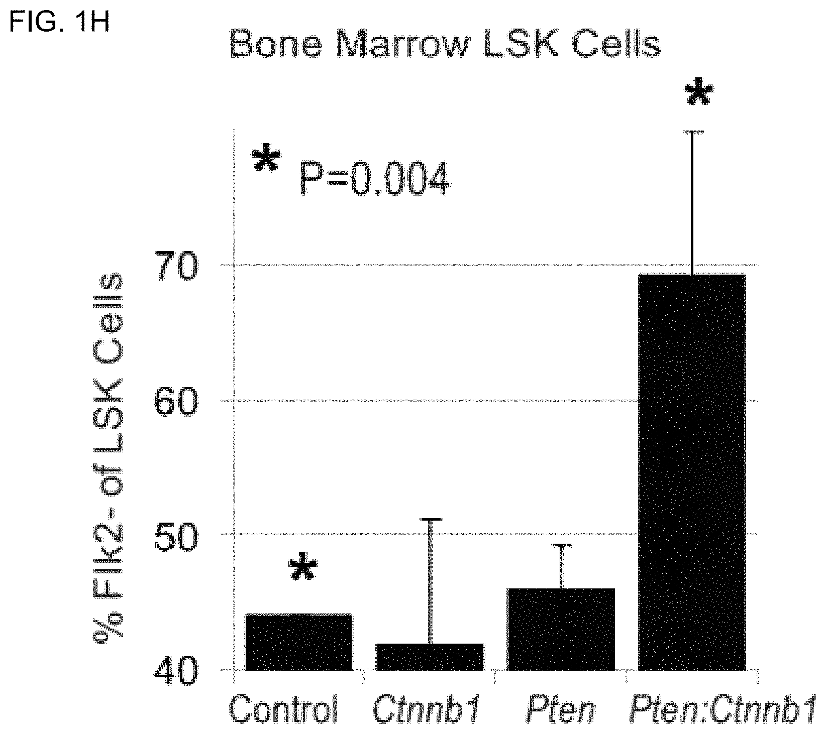
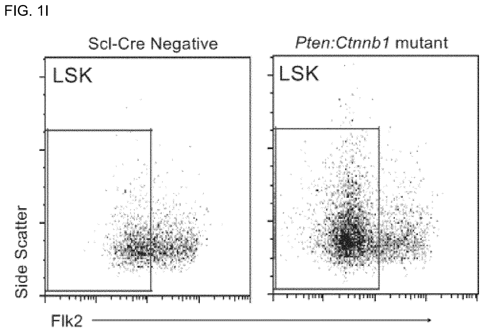
FIGS. 1H and 1I are bar graphs and FACS analysis, respectively, of percentage of LSK cells, which are Flk2.sup.- (indicating long-term reconstituting (LT)-HSGs) in control, Ctnnb1, Pten, and Pten:Ctnnb1 mutant bone marrow at 6 weeks post-induction. Ctnnb1 single mutants are not significantly different from controls at this time point (data not shown). Boxes in FIG. 1I indicate Flk2.sup.- (LT HSC) cells.
FIG. 1J is a set of FACS analyses of CD45 in leukemic Pten:Ctnnb1 mutant bone marrow. CD45 (high) blast crisis cells are indicated (blue box, left panel). No blast cell population is observed in control or Ctnnb1 mutants while a minor population has been observed in 1 of 1 Pten single mutant mice at 6 weeks post-induction (data not shown). The right panel shows LSK analysis of leukemic Pten:Ctnnb1 mutant mouse bone marrow. Note the conversion to blast cells (lower left) with only a remnant LSK population (compare to FIG. 1C).
FIG. 1K is a bar graph showing early hematopoietic progenitors defined by FACS analysis in control, Ctnnb1, Pten, and Pten:Ctnnb1 double mutant bone marrow. Common myeloid progenitor (CMP); granulocyte-monocyte progenitor (GMP); megakaryocyte-erythrocyte progenitor (MEP); and common lymphoid progenitor (CLP).
FIGS. 2A-2J are a series of photographs, bar graphs, and FACS analyses that collectively show that double mutant HSCs expand dramatically in vitro and in vivo but fail to differentiate.
FIG. 2A is a series of photographs showing 100 LSK cells isolated from control, active .beta.-catenin (Ctnnb1), Pten mutant, and double mutant (Pten:Ctnnb1) mice after 10 days in culture (original magnification 100.times.). Cell numbers are not dramatically increased from 100 seeded LSKs in control while Ctnnb1 single mutant LSKs do not survive. In contrast, Pten single mutant LSKs exhibit greater proliferation but appear more heterogeneous indicating more significant differentiation. The greatest and most homogeneous expansion occurs from Pten:Ctnnb1 double mutant LSKs.
FIG. 2B is a set of photographs showing LSK cells from Pten and Pten:Ctnnb1 mutants at 34 days culture (original magnification 200.times.). (Note: wild-type control cultures do not expand beyond 4 weeks; Ctnnb1 mutant cultures do not survive beyond 10 days.) Pten mutant HSC cultures appear more heterogeneous with significant cell clumping and more irregular cell morphology. Also note the spindle-shaped adherent cells (arrows) showing differentiation. In contrast, double mutant HSC cultures exhibit consistent morphology. Therefore, while Pten single mutant LSKs survive and expand, they have undergone more significant differentiation than the much more homogeneous Pten:Ctnnb1 double mutant LSKs.
FIGS. 2C and 2D are bar graphs showing the results of an expansion experiment. Pten and Pten:Ctnnb1 LSK seven week cultures were counted and analyzed by FACS for maintenance of the LSK phenotype (wild-type control and Ctnnb1 cultures did not survive this long in vitro). Double mutant LSKs undergo >1,200 fold expansion vs. 50 fold for Pten single mutant LSKs. LSK purity of cultures is significantly greater in Pten:Ctnnb1 cultures maintaining the LSK phenotype in about 85% of total live cells vs. about 50% for Pten single mutant cultures.
FIG. 2E is a FACS analysis of a 7 week culture of Pten:Ctnnb1 LSK cells (pre-gated on live, lineage negative cells). The boxed area indicates Kit.sup.+Sca-1.sup.+ (LSK) cells.
FIGS. 2F and 2G are FACS analyses showing a transplant engraftment experiment. At 5 weeks culture (see FIG. 2B), Pten and Pten:Ctnnb1 LSK cultures were re-sorted and 1000 LSK cells (CD45.2.sup.+) from each were transplanted into lethally irradiated (10 Gy) CD45.1.sup.+ recipient mice along with 2.times.10.sup.5 congenic whole bone marrow competitor cells. Because wild-type cells did not survive 5 weeks culture, 1000 fresh wild-type LSK cells were also transplanted as a separate control group. At 4 weeks post-transplant, there was no detectable engraftment from peripheral blood analysis of mice transplanted with either Pten or Pten:Ctnnb1 LSK cultures (data not shown). At 5 weeks post-transplant, bone marrow from recipient mice was analyzed for donor engraftment (CD45.2.sup.+ cells) and donor LSK cells (CD45.2.sup.+ LSKs). FIGS. 2F and 2G display representative donor engraftment (left, boxed areas indicate CD45.2.sup.+ donor cells) and donor LSK cell engraftment (right, boxed areas indicate LSK cells) from bone marrow of mice transplanted with 1000 Fresh LSK cells (2F) or 1000 cultured Pten:Ctnnb1 LSK cells (2G).
FIGS. 2H-2J are bar graphs showing the quantitative analysis of donor (CD45.2.sup.+) cells (2H), donor LSK cells (2I), and fold increase in donor LSKs (2J) isolated from bone marrow of recipient mice described in FIGS. 2F and 2G at 5 weeks post-transplant.
FIGS. 3A-3K are schematics, photographs, bar graphs, and FACS analyses demonstrating that ex vivo pharmacological manipulation of the PTEN/Akt and Wnt/.beta.-catenin signaling pathways cooperatively drive functional HSC expansion.
FIG. 3A is a schematic illustrating representative members of the Wnt and PTEN pathways. Inhibition of GSK-3.beta. leads to .beta.-catenin activation which blocks HSC differentiation. Inhibition of PTEN leads to Akt activation which promotes survival. Both pathways individually have been shown to promote HSC proliferation.
FIGS. 3B and 3C are photographs of HSCs. One hundred LSK Flk2.sup.- cells were sorted from wild-type (C57Bl/6) mice and cultured in (1) media, (2) media+1 .mu.M CHIR99021 (GSK-3.beta. inhibitor), (3) media+200 nM Dipotassium Bis-peroxo(picolinato)oxovanadate (BpV(pic), a PTEN inhibitor), and (4) media+1 .mu.M CHIR99021+200 nM BpV(pic). An alternative PTEN inhibitor, Shikonin, was also utilized at 200 nM alone (5) or in combination with 1 .mu.M CHIR99021 (6). Pictures are at 17 days culture (3B, original magnification 100.times.) and 23 days (3C, original magnification 40.times.). Compared to control, both inhibitors applied individually yield greater expansion of LSK cells indicating that GSK-3.beta. inhibition is not strictly equivalent to constitutive activation of .beta.-catenin shown in Ctnnb1 mutant LSKs while BpV(pic) yields similar results compared to Pten mutant LSKs (see FIG. 2A-2J). Similar to double mutant LSKs (FIGS. 2A-2J), the greatest expansion is shown with both inhibitors present (FIGS. 3B and 3C, panel 4).
FIG. 3D is a series of photographs showing LSK Flk2.sup.- cells at 28 days culture in the indicated media conditions (original magnification 200.times.). Here, significant expansion relative to control is observed with both inhibitors present individually; however, significant differentiation/heterogeneity of cell morphology is observed in both single inhibitor cultures, including more variable cell size/morphology and/or differentiation to adherent, spindle-shaped cells (middle panels). In contrast, and quite surprisingly, expansion with homogeneity is achieved when both inhibitors are present (last panel).
FIG. 3E is a FACS analysis of 28 day LSK Flk2.sup.- cells cultured in media containing both inhibitors (200 nM BpV(pic) and 1 .mu.M CHIR99021). Cells were pre-gated on live, lineage negative cells. The boxed area indicates Kit.sup.+Sca1.sup.+ (LSK) cells. Greater than 90% of LSKs retain Flk2 negativity (data not shown). The LSK Flk2.sup.- phenotype is maintained with high purity in cultures containing both inhibitors.
FIG. 3F is a bar graph showing fold expansion of LSK Flk2.sup.- cells after 28 days culture in the indicated conditions. While both inhibitors added individually lead to significant expansion compared to media without either inhibitor, the greatest expansion (.about.270 fold) is observed when both inhibitors are added together.
FIGS. 3G and 3H are bar graphs showing the % repopulation and % CD45.2.sup.+ cells from engrafted mice. Twenty-eight day cultures (FIGS. 3D-3F) were re-sorted for LSK Flk2.sup.- cells and 1000 LSK Flk2.sup.- cells (CD45.2.sup.+) from each media condition were transplanted into lethally irradiated (10 Gy) CD45.1.sup.+ recipient mice along with 2.times.10.sup.5 congenic whole bone marrow competitor cells. At 4 weeks post-transplant, peripheral blood was analyzed for donor (3G) and multi-lineage (3H) engraftment. In FIG. 3G, each bar represents an individual mouse; the horizontal-dashed line represents the average "engraftment" of mice transplanted with competitor cells only and thus the limit of detectability for true engraftment. Long-term (4 month) engraftment has not been observed from 28-day cultures (data not shown). 6 of 8 mice show >1% engraftment when transplanted with LSK Flk2.sup.- cells cultured with both inhibitors present compared to 4/8 with only CHIR99021 present, 0/10 with only BpV(pic) present, and 2/6 with media only. One percent or greater engraftment is a standard limit for substantial engraftment. (Zhang, C. C., et al., Nat Med, 12(2): 240-5, 2006. Zhang, C. C. and H. F. Lodish, Blood, 105(11): 4314-20, 2005.) Thus, while use of both inhibitors together leads to greatest expansion in LSKs (FIG. 3F), transplantation of equivalent numbers of these cultured LSK Flk2.sup.- cells also yields increased short-term engraftment/functionality when cultured with both inhibitors compared to no or either single inhibitor only.
FIG. 3I is a bar graph showing the fold expansion of LSK Flk2.sup.- cells after 9 days culture in (1) media, (2) media+200 nM BpV(pic), (3) media+100 nM CHIR99021, and (4) media+200 nM BpV(pic)+100 nM CHIR99021. Because long-term engraftment was not observed from 28 day cultures (FIGS. 3D-3H and data not shown), LSK Flk2.sup.- cells were cultured for only 9 days to test if both expansion and long-term repopulation can be achieved. Similar trends are observed here to the 28 day cultures (compare to FIG. 3F) although the extent of expansion is substantially reduced at only 9 days compared to 28 days culture.
FIG. 3J is a FACS analysis of 9 day LSK Flk2.sup.- cells cultured in media+200 nM BpV(pic)+100 nM CHIR99021. The boxed area indicates Kit.sup.+Sca-1.sup.+ (LSK) cells. Cells were pre-gated on live, lineage negative cells. Greater than 90% of LSKs retain Flk2 negativity (data not shown). Here, the levels of Sca-1 and Kit appear normal compared to the Sca-1.sup.(high)Kit.sup.(high) population shown from 28 day cultures (FIG. 3E).
FIG. 3K is a bar graph showing % repopulation of 10-day cultured cells in mice. Ten day cultures were transplanted into lethally irradiated (10 Gy) CD45.1.sup.+ recipient mice along with 2.times.10.sup.5 congenic whole bone marrow competitor cells. The total, non-adherent cell product after 10 days culture of 100 initial LSK Flk2.sup.- cells was transplanted per mouse. At 8 weeks post-transplant, peripheral blood was analyzed for donor engraftment. As in FIG. 3H, multi-lineage reconstitution was observed from all mice exhibiting true engraftment (data not shown). Each bar represents an individual mouse; the horizontal-dashed line represents the average `engraftment` of mice transplanted with competitor cells only and thus the limit of detectability for true engraftment. Here, 3/7 mice transplanted with LSK Flk2.sup.- cells cultured in the presence of both inhibitors exhibited 1% or greater donor engraftment vs. no mice reaching this threshold in the single or no inhibitor groups.
FIGS. 4A-4N show that ex vivo expansion of unsorted bone marrow mononuclear cells enhances functional long-term hematopoietic reconstitution relative to sorted, ex vivo expanded HSCs.
FIG. 4A is a logarithmic plot of CD45.2 (donor) frequency of total CD45.sup.+ cells in peripheral blood of transplant recipients. Red line denotes limit of detectable engraftment as determined by "engraftment" found in mice transplanted with competitor cells only.
FIG. 4B is a linear plot of CD45.2 (donor) frequency of total CD45.sup.+ cells in peripheral blood of transplant recipients. Putative HSCs were identified by fluorescence activated cell sorting (FACS) based upon cell-surface markers, including lineage marker negative, Sca-1.sup.+, c-Kit.sup.+, Flk2.sup.- (LSKF.sup.-), sorted and cultured for 14 days. Bone marrow mononuclear cells (MNCs) were also fractionated and the concentration of LSKF.sup.- cells was determined. MNCs containing a known quantity of LSKF.sup.- cells were cultured for 14 days. After 14 days, the cellular product of these cultures was transplanted into lethally-irradiated recipients at a dosage corresponding to an original input into culture of 100 LSKF.sup.- cells per mouse for sorted cultures and MNCs containing 5 LSKF.sup.- cells per mouse for unsorted cultures. In addition, 100 freshly isolated, sorted LSKF.sup.- cells per mouse and freshly isolated MNCs containing 5 LSKF.sup.- cells per mouse were transplanted into two additional groups. 1.times.10.sup.5 competitor bone marrow cells congenic with the hosts (CD45.1.sup.+) were included per mouse. At 4 weeks post-transplant, peripheral blood was collected from each transplant recipient, and donor vs. host derived hematopoietic cells were determined by FACS analysis.
FIG. 4C shows the percentage of donor derived peripheral blood cells (CD45.2.sup.+) contributing to the main hematopoietic lineages (B lymphoid, T lymphoid, and myeloid cells) from transplant recipients described in FIGS. 4A and 4B at 4 weeks post-transplantation.
FIGS. 4D-4F show repopulation data obtained from peripheral blood samples from transplant recipients described in FIGS. 4A-4C at 16 weeks post-transplant.
FIGS. 4G-4H show the results of a secondary transplantation. At 16 weeks post-transplant, mice transplanted with MNCs containing 5 LSKF.sup.- cells cultured for 14 days described in FIGS. 4A-4F were sacrificed, and bone marrow was isolated. A secondary transplantation was performed on new groups of lethally irradiated mice by transplanting 1.times.10.sup.6 bone marrow cells from the original transplant group per mouse. At 4 weeks post-transplant, peripheral blood was collected from each transplant recipient and donor-derived repopulation was determined as in FIGS. 4A-4B.
FIG. 4I shows the percentage of donor derived peripheral blood cells (CD45.2.sup.+) contributing to the main hematopoietic lineages from transplant recipients described in FIGS. 4G-4H at 4 weeks post-transplant.
FIGS. 4J-4L show repopulation data obtained from peripheral blood samples from transplant recipients described in FIGS. 4G-4I at 16 weeks post-transplant.
FIGS. 4M-4N show representative FACS plots of donor (CD45.2) vs. host (CD45.1) cells obtained from peripheral blood samples from recipients described in FIGS. 4J-4K.
FIGS. 5A-5F show that culturing with the small-molecule inhibitor of GSK-3.beta., CHIR99021, enhances long-term engraftment of ex vivo expanded HSCs.
FIG. 5A is a logarithmic plot of CD45.2 (donor) frequency of total CD45.sup.+ cells in peripheral blood of transplant recipients at 4 weeks post-transplant. FIG. 5B is a linear plot of the same. Sorted LSKF.sup.- cells and MNCs with a known quantity of LSKF.sup.- cells were cultured and transplanted as described in FIG. 4A. Cultures contained media alone or media with 250 nM CHIR99021 for each group.
FIG. 5C shows the percentage of donor derived peripheral blood cells (CD45.2.sup.+) contributing to the main hematopoietic lineages from transplant recipients described in FIGS. 5A-5B at 4 weeks post-transplant.
FIGS. 5D-5F the repopulation data obtained from peripheral blood samples from transplant recipients described in FIGS. 5A-5C at 16 weeks post-transplant.
FIGS. 6A-6H show that the ex vivo expansion protocol allows for elimination of bone marrow rescue cells and yields engraftment equivalent to a one-hundred fold greater dosage of freshly isolated cells.
FIG. 6A shows CD45.2 (donor) frequency of total CD45.sup.+ cells in peripheral blood of transplant recipients at 4 weeks post-transplant. Mice transplanted with freshly isolated MNCs containing 5 LSKF.sup.- cells (indicated by "X") do not survive beyond 2-3 weeks post-transplant preventing measurement of engraftment. For this experiment, MNCs with a known quantity of putative HSCs were cultured with and without CHIR99021 for 14 days. After 14 days, the cellular product of these cultures was transplanted into lethally-irradiated recipients at a dosage corresponding to an original input into culture of MNCs containing 5 LSKF.sup.- cells per mouse. Freshly isolated MNCs containing either 5 or 500 LSKF- cells were also transplanted into 2 additional lethally irradiated groups of mice. No rescue/competitor bone marrow cells were included.
FIG. 6B shows the percentage of donor derived peripheral blood cells (CD45.2.sup.+) contributing to the main hematopoietic lineages from transplant recipients described in FIG. 6A at 4 weeks post-transplant.
FIGS. 6C-6D show repopulation data obtained from peripheral blood samples from transplant recipients described in FIGS. 6A-6B at 16 weeks post-transplant.
FIG. 6E shows the results of a secondary transplant. At 16 weeks post-transplant, mice transplanted with MNCs containing 5 or 500 LSKF.sup.- cells freshly isolated or cultured for 14 days described in FIGS. 6A-6D were sacrificed and bone marrow isolated. A secondary transplantation was performed on new groups of lethally irradiated mice by transplanting 1.times.10.sup.6 bone marrow cells from the original transplant group per mouse. At 4 weeks post-transplant, peripheral blood was collected from each transplant recipient and donor-derived repopulation was determined. Mice transplanted with freshly isolated MNCs containing 5 LSKF.sup.- cells (indicated by "X") do not survive beyond 2-3 weeks post-transplant, thus preventing secondary transplantation.
FIG. 6F shows the percentage of donor derived peripheral blood cells (CD45.2.sup.+) contributing to the main hematopoietic lineages from transplant recipients described in FIG. 6E at 4 weeks post-transplant.
FIGS. 6G-6H show repopulation data obtained from peripheral blood samples from transplant recipients described in FIGS. 6E-6F at 16 weeks post-transplant.
FIGS. 7A-7C show ex vivo expansion of human HSCs. In FIG. 7A, bone marrow and mobilized peripheral blood was collected from human patients. Putative HSCs (CD34.sup.+ CD38.sup.- cells) were identified by FACS analysis. Ex vivo expansion was performed with and without CHIR99021. After 14 days culture, the cellular product of these cultures was analyzed to determine the expansion of CD34.sup.+ CD38.sup.- cells. FIGS. 7B-7C are representative FACS plots of CD34.sup.+ CD38.sup.- cells prior to (FIG. 7B) and following (FIG. 7C) ex vivo expansion.
FIG. 8 shows a .beta.-cat-pS552 immunoassaying of homed GFP-HSCs. Detection of .beta.-cat-pS552.sup.+ (red) cells adjacent or close to N-cadherin-LacZ.sup.+ (blue) osteoblasts which have been identified with the HSC niche (Xie, Y. et al. Detection of functional haematopoietic stem cell niche using real-time imaging. Nature 457, 97-101 (2009); Zhang, J. et al. Identification of the haematopoietic stem cell niche and control of the niche size. Nature 425, 836-841 (2003)). "BM" indicates bone marrow.
FIG. 9 shows the percent of Mac-1+Gr1+ myeloid cells in bone marrow and spleen at 8-9 weeks post-induction (wpi) in control, single and double mutants as determined by FACS. Results are graphed as mean.+-.SD.
FIGS. 10A-10F show that double mutant mice lose early myeloid progenitors as mutant HSCs predominate. Data shown relate to lethally irradiated recipient mice previously transplanted with 1,000 LSK Flk2.sup.- cells derived from control, single and double mutant donors+200,000 congenic rescue bone marrow cells. FIG. 10A shows FACS diagrams of LSK cells (right blue boxes) and myeloid progenitors (left blue boxes) in control, single and double mutant bone marrow (top panels) and spleen (bottom panels) as indicated. As used herein, .beta.-cat.sup.Act is used interchangeably with Ctnnb1, and Pten:.beta.-cat.sup.Act is used interchangeably with Pten:Ctnnb1. Mice were at 9 or 10 wpi as indicated. Note the LS.sup.LowK.sup.Mid population in double mutants at 9 wpi (red arrows). FIG. 10B shows FACS analysis of early hematopoietic progenitors in control, single and double mutant bone marrow at 9 wpi. FIGS. 10C and 10D shows the absolute number of bone marrow (per tibia and femur) (FIG. 10C) or spleen (FIG. 10D) LSK cells and early hematopoietic progenitors in control, single, and double mutants at 9-10 wpi. Note the collapse of LSK and early progenitor populations in double mutant bone marrow (red arrows) with conversion to a dominant "blast" population (see also FIGS. 12A-12C). FIG. 10E shows percent donor engraftment at 9 wpi of lethally-irradiated recipient mice previously transplanted with 1,000 LSK Flk2.sup.- cells derived from control, single and double mutant donors+200,000 congenic rescue bone marrow cells. FIG. 10F shows the EGFP-reporter expression of LSK Flk2.sup.- cells in control, single and double mutants with the Z/EG transgenic reporter construct at 9 wpi.
FIGS. 11A-11G show Ctnnb1 (.beta.-cat.sup.Act) HSCs undergo apoptosis whereas .beta.-catenin deletion prevents PTEN-deficiency-induced HSC expansion but not myeloproliferative disorder (MPD). To obtain the results shown in FIG. 11A, 1,000 LSK Flk2.sup.- cells per well were sorted from bone marrow isolated from uninduced control, Pten, Ctnnb1 (.beta.-cat.sup.Act) and Pten:Ctnnb1 (Pten:.beta.-cat.sup.Act) mice. Within 12 hours of sorting, OHT was added to the cultures for a final concentration of 1 .mu.M. Cultures depicted at 4 days post-in vitro induction. FIG. 11B shows control and Ctnnb1 (.beta.-cat.sup.Act) cultures as described in FIG. 11A at 48 hours post-in vitro induction. FIG. 11C shows representative FACS plots distinguishing live (Sytox Green negative) from dead (Sytox Green positive) cells. Cultures from FIG. 11B were stained with Sytox Green and Annexin V according to manufacturer's instructions (Vybrant Apoptosis Kit #9, Invitrogen) and analyzed by FACS. Live cells were further gated for Annexin V positive (apoptotic) cells. Numbers within gates represent the average.+-.standard deviation from 3 independent experiments. FIG. 11D shows the absolute number of LSK cells and early progenitors in spleen as determined by FACS analysis. Mice were transplanted with control, .beta.-cat.sup.-/-, Pten, and Pten:.beta.-cat.sup.-/- mice bone marrow as indicated; analysis is at 10 wpi. FIGS. 11E-11G show the percent of Gr1.sup.+ Mac-1.sup.+ cells (FIG. 11E), B-cells (FIG. 11F), and T-cells (FIG. 11G) in bone marrow of mice described in (FIG. 11D) as determined by FACS (see FIGS. 20A-20G).
FIGS. 12A-12C show that Leukemia development and niche disruption in double mutants. FIG. 12A shows a Kaplan-Meier survival curve for control, single and double mutants (as indicated in the figure legend) following tamoxifen induction (Scl-Cre system unless otherwise specified). FIG. 12B shows H&E stained sections of control and double mutant bone marrow at 9 wpi. White arrow indicates grossly normal cellularity in trabecular bone area. FIG. 12C shows FACS analysis of control, single and double mutant bone marrow at 10 wpi demonstrating typical CD45 expression. Note CD45.sup.High blast cells (blue box) only mainly appear in double mutants. Blast cells from double mutants were further analyzed for cell surface marker expression of the T-cell specific marker, CD3.
FIGS. 13A-13C show that Ex vivo expansion of HSCs is enhanced by inhibition of GSK3.beta.. For the experimental results shown in FIG. 13A, sorted LSK Flk2.sup.- cells and unsorted MNCs containing a known quantity of LSK Flk2.sup.- cells (CD45.2.sup.+) were cultured for 14 days in ST media with and without CHIR99021. The cultured product of 100 sorted or 5 unsorted LSK Flk2.sup.- cells per mouse were transplanted into lethally irradiated recipients (CD45.1.sup.+). 5 freshly isolated, unsorted LSK Flk2.sup.- cells per mouse were transplanted into a separate group. 1.times.10.sup.5 freshly isolated CD45.1.sup.+ competitor/radioprotective cells were also added per mouse. Peripheral blood analysis of recipients at 16 weeks post-transplant depicts % chimerism. FIG. 13B shows the percentage of donor-derived peripheral blood cells (CD45.2.sup.+) contributing to the main hematopoietic lineages (B lymphoid, T lymphoid, and myeloid cells) from transplant recipients described in (FIG. 13A) at 16 weeks post-transplantation. FIG. 13C shows representative FACS plots of donor (CD45.2) vs. host (CD45.1) cells obtained from peripheral blood samples at 16 weeks post-transplant from recipients described in FIG. 13A.
FIG. 14 shows abundant .beta.-cat-pS552.sup.+ cells in double mutant spleen. Spleen sections stained with .beta.-cat-pS552 antibody in control, single and double mutants at 3 dpi using Mx1-Cre system. Original magnification 400.times. (upper panels) and 1000.times. (lower panels).
FIG. 15 shows trichofolliculoma in double mutants using Mx1-Cre mediated conditional knockout. Abdomen of Mx1-Cre+Pten:Ctnnb1 (Pten:.beta.-cat.sup.Act) mutant (left panel, control mouse at left). H&E stained section of hair follicle tumor showing multiple, well-developed but densely packed hair follicles in cross section (right panel).
FIGS. 16A-16B show vascular niche disruption by splenic fibrosis in double mutants. FIG. 16A shows whole spleen isolated from control, single and double mutants at 9 wpi. Three examples of double mutant spleen exhibiting mild to severe fibrosis are shown. Scale bar indicates 1 cm. FIG. 16B shows Masson's Trichrome stained sections of control, single and double mutant spleens at 9 wpi. Red arrows indicate examples of collagen fibers (light blue).
FIGS. 17A-17B show the number of different types of LSK cells and early progenitors, as determined by FACS (see FIGS. 10A-10B) except that primary mutant mice were utilized here instead of transplant recipients. Absolute number of bone marrow (per tibia and femur) (FIG. 17A) or spleen (FIG. 17B) LSK cells and early hematopoietic progenitors in control, single and double mutants at 9-10 wpi. Note the collapse of LSK and early progenitor populations in double mutant bone marrow (red arrows) with conversion to a dominant "blast" population. Compare to FIGS. 10A-10B.
FIG. 18 shows that Ctnnb1 (.beta.-cat.sup.Act) mutant HSCs are not maintained in viva LSK Flk2.sup.- cells were sorted from Scl-Cre negative control and Ctnnb1 (.beta.-cat.sup.Act) mutants at 2 and 16 wpi and genotyped for deletion of exon 3. Primers utilized were: 5'-CGTGGACAATGGCTACTCAA-3' (forward) (SEQ ID NO: 1) and 5'-TGTCAGCTCAGGAATTGCAC-3' (reverse) (SEQ ID NO: 2) to yield wild-type (911 bp) and .DELTA.Exon 3 alleles (683 bp). Note that mice with the dominant .beta.-cat.sup.Act allele are all heterozygous for this allele.
FIGS. 19A-19C show the functional reversibility of myeloid differentiation blockage in double mutant HSCs. To obtain the experimental results shown in FIG. 19A, LSK Flk2.sup.- cells were sorted from uninduced control, Pten, and Pten:Ctnnb1 (Pten:.beta.-cat.sup.Act) mice into an HSC expansion media containing 0.25 .mu.M 4-hydroxy-tamoxifen (OHT) and cultured for 3 days. Cultures were transduced with lentiviral vector control and vector expressing shRNA targeting .beta.-catenin transcripts. Colony forming unit (CFU) assays were performed on day 6. Images depict typical colonies from control, Pten, and Pten:Ctnnb1 (Pten:.beta.-cat.sup.Act) cultures transduced with control vector (left panels) and vector expressing shRNA targeting .beta.-catenin (right panels). Scale bar indicates 0.5 mm. FIG. 19B shows the quantification of colonies by type from FIG. 19A including early erythoid progenitors (BFU-E, burst-forming unit-erythroid), granulocyte-monocyte progenitors (CFU-GM, colony forming unit-granulocyte/monocyte), and mixed early myeloid progenitors (CFU-GEMM, granulocyte/erythroid/macrophage/megakaryocyte). Large CFU (>0.5 mm diameter), which are further characterized in FIG. 19C and form only from double mutant cultures transduced with control vector, are designated as primitive CFU. FIG. 19C shows panels depicting representative plots of CD3 expression in control and Pten:Ctnnb1 (Pten:.beta.-cat.sup.Act) cells transduced with control vector and Pten:.beta.-cat.sup.Act cells transduced with vector expressing shRNA targeting .beta.-catenin transcripts. CFU were harvested, disaggregated into single-cell suspension and subjected to FACS analysis for CD3 expression. Average percentage of CD3.sup.+ cells from 3 experiments.+-.S.D. are shown.
FIGS. 20A-20G show hematopoietic lineage defects and leukemogenesis in single vs. double mutants. FIG. 20A shows the percent of immature (B220.sup.Low, IgM.sup.+), mature (B220.sup.High, IgM.sup.+) and Pre-Pro B (B220.sup.Low, IgM.sup.-) cells in control, single and double mutant bone marrow at 8-9 wpi as determined by FACS. As in FIGS. 10A-10F, mice here refer to transplant recipients of 1,000 LSK Flk2.sup.- cells derived from control, single and double mutants as indicated along with 2.times.10.sup.5 congenic rescue bone marrow cells. FIG. 20B shows FACS diagrams illustrating control and double mutant data on T-cell lineage quantified in FIG. 20C. FIG. 20C shows percent of CD3+, double and single positive T cells in control, single and double mutant bone marrow at 8-9 wpi. Note the logarithmic scale. FIGS. 20D-20E show Double Negative (DN) populations in control, single and double mutant thymus at 8-9 wpi. Representative FACS plots of control (upper panel) and double mutant (lower panel) thymus are shown in FIG. 20D. Note the logarithmic scale in FIG. 20E. FIGS. 20E-20G show double and single positive thymocyte populations from control, single and double mutants. Representative FACS plots of control (left panel) and double mutant (right panel) thymus (FIG. 20F). Results are graphed as mean.+-.SD (FIG. 20G).
FIG. 21 shows that PI3K inhibition reverses ex vivo HSC expansion and inhibits CHIR99021's ability to enhance this expansion. Bone marrow MNCs were cultured for 10 days in an HSC expansion media with and without 250 nM CHIR99021, along with the indicated concentrations of PI3K inhibitor (NVP-BEZ235) (Maira, S. M. et al. Identification and characterization of NVP-BEZ235, a new orally available dual phosphatidylinositol 3-kinase/mammalian target of rapamycin inhibitor with potent in vivo antitumor activity. (Molecular cancer therapeutics 7, 1851-1863 (2008)), and then subjected to FACS analysis to determine expansion of LSK Flk2.sup.- cells.
DETAILED DESCRIPTION OF THE INVENTION
One embodiment of the invention is an ex vivo method for expanding the number of hematopoietic stem cells (HSC) in a population of mononuclear cells (MNC). This method comprises culturing the population of MNCs comprising at least one HSC in an HSC expansion media for a period of time sufficient to expand the number of HSCs in the MNC population, wherein the expanded HSCs are functional with long term, multi-lineage, repopulating potential.
As used herein, "expand", "expanding" and like terms means to increase the number of stem cells in the population relative to the number of stem cells in the original population in vitro, in vivo or ex vivo using any of the methods disclosed herein. Preferably, the expansion is at least 40-fold compared to the original number of stem cells in the population. More preferably, the expansion is at least 80-fold, 100-fold, 150-fold, 200-fold, 250-fold, or 270-fold compared to the original number of stem cells.
In the present invention, "stem cells" mean cells that possess the ability to give rise to many different types of cells and which have the ability to self-renew. Representative, non-limiting examples of stem cells according to the present invention include bronchioalveolar stem cells (BASCs), bulge epithelial stem cells (bESCs), corneal epithelial stem cells (CESCs), cardiac stem cells (CSCs), epidermal neural crest stem cells (eNCSCs), embryonic stem cells (ESCs), endothelial progenitor cells (EPCs), hepatic oval cells (HOCs), hematopoetic stem cells (HSCs), keratinocyte stem cells (KSCs), mesenchymal stem cells (MSCs), neuronal stem cells (NSCs), pancreatic stem cells (PSCs), retinal stem cells (RSCs), and skin-derived precursors (SKPs).
Hematopoietic stem cells or HSCs, for example, have the ability to self-renew (i.e., expand) and can give rise to all the types of progenitor cells (such as, e.g., CMP, GMP, MEP and CLP) and ultimately all the types of blood cells (such as e.g., red blood cells, B lymphocytes, T lymphocytes, natural killer cells, neutrophils, basophils, eosinophils, monocytes, macrophages, and platelets) in the hematopoietic system.
As used herein, "mononuclear cells" or "MNC" mean blood cells that have a one-lobed nucleus. MNCs include without limitation monocytes, lymphocytes, plasma cells, macrophages, and mast cells.
As used herein, "HSC expansion media" means any media suitable for expanding the number of HSC population in a culture. It includes without limitation, the particular media disclosed in the Examples.
As used herein, cells with "long term, multi-lineage repopulating potential" means cells that are capable of repopulating many different types of blood cells in irradiated recipients upon transplantation and/or cells that possess high proliferative potential in vitro.
In one aspect of this embodiment, this method provides HSCs that, upon transplant into a recipient, exhibit greater than 5% donor repopulation, such as greater than 10%, 15%, 20%, 25%, 30%, 35%, 40%, 55%, 60%, 65%, 70%, 75%, 80%, 85%, or 90% donor repopulation. Preferably, the method provides HSCs that, upon transplant into a recipient, exhibit greater than 25%, 35%, 45%, or 60% donor repopulation. Preferably, the recipient is a mammal, for example, a primate, such as a human; or laboratory animals such as mice, rats, dogs, and pigs. In the present invention, "recipient" is used interchangeably with "patient."
In another aspect of this embodiment, the HSC expansion media comprises a modulator of the Wnt pathway. Preferably, the modulator of the Wnt pathway down-regulates GSK-3.beta.. As used herein, "down-regulating" GSK-3.beta. means decreasing or inhibiting the expression or the function of GSK-3.beta..
In the present invention, "a modulator of a Wnt Pathway" (or "Wnt pathway modulator") is any agent that regulates the activity of any member of the Wnt pathway, which results in, e.g., increased .beta.-catenin expression in a stem cell, and/or increased .beta.-catenin function in a stem cell, and/or increased .beta.-catenin localization to a nucleus of a stem cell. A modulator of the Wnt pathway may act upstream or downstream of Wnt. Preferably, the modulator acts at GSK-3.beta.. Representative, non-limiting examples of members of the Wnt pathway, include Wnt, seven-transmembrane Frizzled (Fz), the single-pass, LDL receptor-related proteins (LRP) 5/6, Axin, Dishevelled, glycogen synthase kinase 3 beta (GSK-3.beta.), adenomatous polyposis coli (APC), and .beta.-catenin. Inhibition of GSK-3.beta. leads to Akt activation which promotes survival.
In a preferred embodiment, the modulator of the Wnt pathway is a reversible GSK-3.beta. inhibitor selected from the group consisting of a small molecule, a biologic, an antisense RNA, a small interfering RNA (siRNA), and combinations thereof. As used herein, "reversible" means that the effect of the down-regulation is not permanent.
Preferably, the reversible GSK-3.beta. inhibitor is a small molecule. Examples of reversible GSK-3.beta. inhibitors include without limitation, Hymenialdisine, Flavopiridol, Kenpaullone, Alsterpaullone, Azakenpaullone, Indirubin-30-oxime, 6-Bromoindirubin-30-oxime (BIO), 6-Bromoindirubin-30-acetoxime, Aloisine A, Aloisine B, TDZD8, Compound 12, CHIR98014, CHIR99021 (CT99021), CT20026, Compound 1, SU9516, ARA014418, Staurosporine, Compound 5a, Compound 29, Compound 46, GF109203x (bisindolylmaleimide I), Ro318220 (bisindolylmaleimide IX), SB216763, SB415286, 15, CGP60474, Compound 8b, TWS119, Compound 1A, Compound 17, Lithium, Beryllium, Zinc, small molecule GSK-3.beta. inhibitors (Vertex Pharmaceuticals), NP-12 (Neuropharma), GSK-3.beta. inhibitors (Amphora), GSK-3.beta. inhibitors (CrystalGenomics), SAR-502250 (Sanofi-Aventis), 3544 (Hoffmann-La Roche), GSK-3.beta. inhibitors (Lundbeck), TDZD-8 (Cancer Center, University of Rochester), pharmaceutically acceptable salts thereof, or combinations thereof. Preferably, the GSK-3.beta. inhibitor is CHIR99201.
In the present invention, the term "small molecule" includes any chemical or other moiety, other than polypeptides and nucleic acids, that can act to affect biological processes, particularly to modulate members of the Wnt and PTEN pathways. Small molecules can include any number of therapeutic agents presently known and used, or that can be synthesized in a library of such molecules for the purpose of screening for biological function(s). Small molecules are distinguished from macromolecules by size. The small molecules of the present invention usually have a molecular weight less than about 5,000 daltons (Da), preferably less than about 2,500 Da, more preferably less than 1,000 Da, most preferably less than about 500 Da.
Small molecules include without limitation organic compounds, peptidomimetics and conjugates thereof. As used herein, the term "organic compound" refers to any carbon-based compound other than macromolecules such as nucleic acids and polypeptides. In addition to carbon, organic compounds may contain calcium, chlorine, fluorine, copper, hydrogen, iron, potassium, nitrogen, oxygen, sulfur and other elements. An organic compound may be in an aromatic or aliphatic form. Non-limiting examples of organic compounds include acetones, alcohols, anilines, carbohydrates, monosaccharides, oligosaccharides, polysaccharides, amino acids, nucleosides, nucleotides, lipids, retinoids, steroids, proteoglycans, ketones, aldehydes, saturated, unsaturated and polyunsaturated fats, oils and waxes, alkenes, esters, ethers, thiols, sulfides, cyclic compounds, heterocyclic compounds, imidizoles, and phenols. An organic compound as used herein also includes nitrated organic compounds and halogenated (e.g., chlorinated) organic compounds.
Preferred small molecules are relatively easier and less expensively manufactured, formulated or otherwise prepared. Preferred small molecules are stable under a variety of storage conditions. Preferred small molecules may be placed in tight association with macromolecules to form molecules that are biologically active and that have improved pharmaceutical properties. Improved pharmaceutical properties include changes in circulation time, distribution, metabolism, modification, excretion, secretion, elimination, and stability that are favorable to the desired biological activity. Improved pharmaceutical properties include changes in the toxicological and efficacy characteristics of the chemical entity.
As used herein, the term "biologic" means products derived from living sources as opposed to a chemical process. Non-limiting examples of a "biologic" include proteins, conditioned media, and partially purified products from tissues.
The terms "peptide," "polypeptide," and "protein" are used interchangeably herein. In the present invention, these terms mean a linked sequence of amino acids, which may be natural, synthetic, or a modification or combination of natural and synthetic. The term includes antibodies, antibody mimetics, domain antibodies, lipocalins, targeted proteases, and polypeptide mimetics. The term also includes vaccines containing a peptide or peptide fragment intended to raise antibodies against the peptide or peptide fragment.
"Antibody" as used herein includes an antibody of classes IgG, IgM, IgA, IgD, or IgE, or fragments or derivatives thereof, including Fab, F(ab')2, Fd, and single chain antibodies, diabodies, bispecific antibodies, and bifunctional antibodies. The antibody may be a monoclonal antibody, polyclonal antibody, affinity purified antibody, or mixtures thereof, which exhibits sufficient binding specificity to a desired epitope or a sequence derived therefrom. The antibody may also be a chimeric antibody. The antibody may be derivatized by the attachment of one or more chemical, peptide, or polypeptide moieties known in the art. The antibody may be conjugated with a chemical moiety. The antibody may be a human or humanized antibody. These and other antibodies are disclosed in U.S. Published Patent Application No. 20070065447.
Other antibody-like molecules are also within the scope of the present invention. Such antibody-like molecules include, e.g., receptor traps (such as entanercept), antibody mimetics (such as adnectins, fibronectin based "addressable" therapeutic binding molecules from, e.g., Compound Therapeutics, Inc.), domain antibodies (the smallest functional fragment of a naturally occurring single-domain antibody (such as, e.g., nanobodies; see, e.g., Cortez-Retamozo et al., Cancer Res. 2004 Apr. 15; 64(8):2853-7)).
Suitable antibody mimetics generally can be used as surrogates for the antibodies and antibody fragments described herein. Such antibody mimetics may be associated with advantageous properties (e.g., they may be water soluble, resistant to proteolysis, and/or be nonimmunogenic). For example, peptides comprising a synthetic beta-loop structure that mimics the second complementarity-determining region (CDR) of monoclonal antibodies have been proposed and generated. See, e.g., Saragovi et al., Science. Aug. 16, 1991; 253(5021):792-5. Peptide antibody mimetics also have been generated by use of peptide mapping to determine "active" antigen recognition residues, molecular modeling, and a molecular dynamics trajectory analysis, so as to design a peptide mimic containing antigen contact residues from multiple CDRs. See, e.g., Cassett et al., Biochem Biophys Res Commun. Jul. 18, 2003; 307(1):198-205. Additional discussion of related principles, methods, etc., that may be applicable in the context of this invention are provided in, e.g., Fassina, Immunomethods. October 1994; 5(2):121-9.
Targeted proteases are polypeptides which are capable of, e.g., substrate-targeted inhibition of post-translational modification such as disclosed in, e.g., U.S. Patent Application Publication No. 20060275823.
In general, a polypeptide mimetic ("peptidomimetic") is a molecule that mimics the biological activity of a polypeptide, but that is not peptidic in chemical nature. While, in certain embodiments, a peptidomimetic is a molecule that contains no peptide bonds (that is, amide bonds between amino acids), the term peptidomimetic may include molecules that are not completely peptidic in character, such as pseudo-peptides, semi-peptides, and peptoids.
"Antisense" molecules as used herein include antisense or sense oligonucleotides comprising a single-stranded nucleic acid sequence (either RNA or DNA) capable of binding to target mRNA (sense) or DNA (antisense) sequences. The ability to derive an antisense or a sense oligonucleotide, based upon a cDNA sequence encoding a given protein is described in, for example, Stein and Cohen, Cancer Res. 48:2659, (1988) and van der Krol et al., BioTechniques 6:958, (1988).
Antisense molecules can be modified or unmodified RNA, DNA, or mixed polymer oligonucleotides. These molecules function by specifically binding to matching sequences resulting in inhibition of peptide synthesis (Wu-Pong, November 1994, BioPharm, 20-33) either by steric blocking or by activating an RNase H enzyme. Antisense molecules can also alter protein synthesis by interfering with RNA processing or transport from the nucleus into the cytoplasm (Mukhopadhyay & Roth, 1996, Crit. Rev. in Oncogenesis 7, 151-190). In addition, binding of single stranded DNA to RNA can result in nuclease-mediated degradation of the heteroduplex (Wu-Pong, supra). Backbone modified DNA chemistry, which have thus far been shown to act as substrates for RNase H are phosphorothioates, phosphorodithioates, borontrifluoridates, and 2'-arabino and 2'-fluoro arabino-containing oligonucleotides.
Antisense molecules may be introduced into a cell containing the target nucleotide sequence by formation of a conjugate with a ligand binding molecule, as described, e.g., in WO 91/04753. Suitable ligand binding molecules include, but are not limited to, cell surface receptors, growth factors, other cytokines, or other ligands that bind to cell surface receptors. Preferably, conjugation of the ligand binding molecule does not substantially interfere with the ability of the ligand binding molecule to bind to its corresponding molecule or receptor, or block entry of the sense or antisense oligonucleotide or its conjugated version into the cell. Alternatively, a sense or an antisense oligonucleotide may be introduced into a cell containing the target nucleic acid sequence by formation of an oligonucleotide-lipid complex, as described, e.g., in WO 90/10448.
The term small interfering RNA ("siRNA") refers to small inhibitory RNA duplexes that induce the RNA interference (RNAi) pathway. (Elbashir, S. M. et al. Nature 411:494-498 (2001); Caplen, N. J. et al. Proc. Natl. Acad. Sci. USA 98:9742-9747 (2001); Harborth, J. et al. J Cell Sci. 114:4557-4565 (2001).) These molecules can vary in length (generally 18-30 base pairs) and contain varying degrees of complementarity to their target mRNA in the antisense strand. Some, but not all, siRNA have unpaired overhanging bases on the 5' or 3' end of the sense strand and/or the antisense strand. The term "siRNA" includes duplexes of two separate strands, as well as single strands that can form hairpin structures comprising a duplex region. As used herein, siRNA molecules are not limited to RNA molecules but further encompass chemically modified nucleotides and non-nucleotides. siRNA gene-targeting may be carried out by transient siRNA transfer into cells, achieved by such classic methods as lipid-mediated transfection (such as encapsulation in liposome, complexing with cationic lipids, cholesterol, and/or condensing polymers, electroporation, or microinjection). siRNA gene-targeting may also be carried out by administration of siRNA conjugated with antibodies or siRNA complexed with a fusion protein comprising a cell-penetrating peptide conjugated to a double-stranded (ds) RNA-binding domain (DRBD) that binds to the siRNA (see, e.g., U.S. Patent Application Publication No. 2009/0093026).
In another preferred embodiment, the method comprises culturing the population of MNCs comprising at least one HSC in any of the HSC expansion media disclosed herein, and the method provides HSCs that, upon transplant into a recipient, exhibit greater than 5%, such as greater than 10%, 15%, 20%, 25%, 30%, 35%, 40%, 45%, 50%, 55%, 60%, 65%, 70%, 75%, 80%, 85%, or 90% donor repopulation, preferably, greater than 60% donor repopulation.
In another aspect of this embodiment, the HSC is obtained from a mammalian tissue selected from the group consisting of cord blood, peripheral blood, and bone marrow.
A further aspect of this embodiment is an expanded, substantially undifferentiated HSC population made by any of the methods disclosed herein. Preferably, the substantially undifferentiated HSC population is made using an HSC expansion media comprising a modulator of the Wnt pathway.
A stem cell population is "substantially undifferentiated" if a sufficient number of cells in that population retain the ability to self-renew and can give rise to various differentiated cell types when transplanted into a recipient, for example, in the case of an HSC population, repopulating the HSC lineage when transplanted. As used herein, "without significant differentiation" means the expanded stem cell population has a sufficient number of cells that maintain a multi-lineage differentiation potential that the full scope of a target stem lineage may be regenerated upon transplantation of the expanded stem cell population into a recipient. Thus, e.g., in the case of an HSC population, the expanded HSC population, when transplanted into a recipient, is capable of regenerating the entire hematopoietic cell lineage.
An additional embodiment of the invention is a kit for expanding, ex vivo, the number of hematopoietic stem cells (HSC) in a population of mononuclear cells (MNC). The kit comprises a GSK-3.beta. inhibitor, and instructions for the use of the inhibitor, wherein, when used, the kit provides expanded HSCs that are functional with long term, multi-lineage, repopulating potential.
In one aspect of this embodiment, the GSK-3.beta. inhibitor is as disclosed herein. Preferably, the GSK-3.beta. inhibitor is CHIR99201.
In another aspect of this embodiment, the kit provides HSCs that, upon transplant into a recipient, exhibit greater than 5%, such as greater than 10%, 15%, 20%, 25%, 30%, 35%, 40%, 45%, 50%, 55%, 60%, 65%, 70%, 75%, 80%, 85%, or 90% donor repopulation, preferably, greater than 60% donor repopulation. The kit may be packaged in any convenient manner and include additional reagents and/or devices for carrying out its intended purpose.
A further embodiment of the invention is a media for carrying out ex vivo expansion of a stem cell in a population of MNCs. This media comprises a fluid media suitable for maintaining viable stem cells and a GSK-3.beta. inhibitor present in the media at a concentration sufficient to enable expansion of the stem cell population while maintaining a long term, multi-lineage, repopulating potential in the stem cells, wherein the stem cells, when transplanted into a recipient, exhibit greater than 5% donor repopulation, such as greater than 10%, 15%, 20%, 25%, 30%, 35%, 40%, 45%, 50%, 55%, 60%, 65%, 70%, 75%, 80%, 85%, or 90% donor repopulation.
Yet another embodiment of the invention is an ex vivo method for expanding the number of cells capable of supporting multi-lineage repopulation in a population of mononuclear cells (MNC). This method comprises culturing the population of MNCs comprising at least one hematopoietic stem cell (HSC) and at least one hematopoietic progenitor cell in an HSC expansion media for a period of time sufficient to expand the number of cells capable of supporting multi-lineage repopulation in the MNC population.
As used herein, "cells capable of supporting multi-lineage repopulation" means those cells that are capable of repopulating many different types of blood cells in irradiated recipients upon transplantation. Non-limiting examples of such cells include HSCs.
As used herein, an "hematopoietic progenitor cell" means a cell that has lost the capacity of self-renewal but is still able to give rise to different types of blood cells. Non-limiting examples of hematopoietic progenitor cells include CMP, GMP, MEP, and CLP.
Another embodiment of the invention is a method for expanding a population of stem cells obtained from a tissue selected from the group consisting of peripheral blood, cord blood, and bone marrow. This method comprises modulating a PTEN pathway and a Wnt pathway in the population of stem cells to expand the number of stem cells.
In the present invention, "modulating", "modulation" and like terms mean altering the signal transduction pathway, e.g., a protein in the PTEN and/or Wnt pathways, including but not limited to lowering or increasing the expression level of a protein, altering the sequence of such a protein (by mutation, pre-translational or post-translational modification or otherwise), or inhibiting or activating such a protein (whether by binding, phosphorylation, glycosylation, translocation or otherwise). Such modulation may be achieved genetically or pharmacologically.
As used herein, "a modulator of a PTEN pathway" (or "PTEN pathway modulator") is any agent that regulates the activity of any member of the PTEN pathway, which results in, e.g., increased .beta.-catenin expression in a stem cell, and/or increased .beta.-catenin function in a stem cell, and/or increased .beta.-catenin localization to a nucleus of a stem cell and/or provides a survival signal complementary to .beta.-catenin. Thus, a modulator of the PTEN pathway may act upstream or downstream of PTEN; preferably the modulator acts at or downstream from PTEN. Inhibition of PTEN leads to Akt activation which promotes survival (FIG. 3A). Representative, non-limiting examples of members of the PTEN pathway, include PTEN, phosphatidylinositol 3-kinase (PI3K), the serine/threonine protein kinase Akt, and .beta.-catenin.
Representative non-limiting examples of PI3K modulators, particularly PI3K activators, include pervanadate (Maude Tessier and James R. Woodgett, J. Biol. Chem., 281(33):23978-23989 (2006)), insulin (Hui, L., et al., Brain Research, 1052(1):1-9 (2005)), insulin-like growth factor (Kenney, A. M., et al., Development, 131:217-228 (2004) and Datta, S. R., et al., Cell, 91:231-241 (1997)), platelet derived growth factor (Datta, S. R., et al., Cell 91:231-241 (1997)), carbachol (Cui, Q L, et al., Neurochem Int, 48:383-393 (2006)), nicotine (West, K. et al., J. Clinical Investigation, 111:81-90 (2003)), 4-(methylnitrosamino)-1-(3-pyridyl)-1-butanone (NNK) (Id.), adrenomedullin (AM) (Nikitenko, L L et al., British J. Cancer, 94:1-7 (2006)), lysophosphatidic acid, platelet activating factor, macrophage simulating factor, and sphingosine-1-phosphate.
Representative non-limiting examples of Akt modulators, particularly Akt activators, include Ro-31-8220 (Wen, H. et al., Cellular signaling, 15:37-45 (2003)); Nicotine (West, K. et al., J. Clinical Investigation, 111:81-90 (2003)); carbachol (Cui Q L, Fogle E & Almazan G Neurochem Int, 48:383-393 (2006)); 4-(methylnitrosamino)-1-(3-pyridyl)-1-butanone (NNK) (West, K. et al., J. Clinical Investigation, 111:81-90 (2003)); adrenomedullin (AM) (Nikitenko, L L et al, British J. Cancer, 94:1-7 (2006)); lysophosphatidic acid; platelet activating factor, macrophage simulating factor; sphingosine-1-phosphate; cAMP-elevating agents, such as forskolin, chlorophenylthio-cAMP, prostaglandin-E1, and 8-bromo-cAMP (Song et al., J. Cell. Mol. Med., 9(1):59-71 (2005)); and growth factors, including insulin and insulin growth factor-1 (Datta, S. R., et al., Cell, 91:231-241 (1997)), and platelet derived growth factor.
Additional preferred modulators of the present invention include those that target mTOR, RHEB, FoxO, p27, BAD, caspase-9, or p53. Representative non-limiting examples of such modulators include mTOR modulators, particularly mTOR activators, such as phosphatidic acid (PA) (see, e.g., WO/2006/027545; Foster, D. A., Cancer Res, 67(1):1-4 (2007); and Tee et al., J. Biol. Chem. 278:37288-96 (2003)); RHEB modulators, particularly RHEB-GTPase inhibitors, such as RHEB antibodies (see, e.g., WO/2004/048536); FoxO modulators, particularly FoxO inhibitors, such as FKH(DBD), which is a truncated version of FKHRL1 (see, e.g., Gilley, J., et al., J. Cell Biol. 162(4):613-622 (2003)); p27 modulators, particularly p27 inhibitors, such as p27 antisense inhibitors and triplex forming oligonucleotides, protein and peptide antagonists (see, e.g., U.S. Pat. No. 5,958,769); BAD modulators, particularly BAD inhibitors, such as 14-3-3 protein (see, e.g., S. Hsu et al., Molecular Endocrinology 11 (12):1858-1867 (1997)); caspase-9 modulators, particularly caspase-9 inhibitors, such as LB-84451 (LG Life Sciences) and Z-LEHD-FMK Caspase Inhibitor (Thornberry, N. A., and Lazebnik, Y., Science 281:1312-1316 (1998)); and p53 modulators, particularly p53 inhibitors, such as Pifithrin-.alpha. and its derivatives (see, e.g., Science, Komarov et al., 285 (5434): 1733-1737 (1999), Pietrancosta et al., Drug Dev Res 65:43-49 (2005)).
In one aspect of the present invention, modulating the PTEN pathway comprises introducing a mutation into a population of stem cells, which mutation results in modulation of a molecule in the PTEN pathway. In the present invention, modulation of the PTEN pathway also includes contacting the stem cells with a modulator of a molecule in the PTEN pathway that leads to .beta.-catenin activation. Representative, non-limiting examples of such modulators include a small molecule, a biologic, an antisense RNA, a small interfering RNA (siRNA), and combinations thereof. This aspect of the invention further includes modulating the Wnt pathway, which comprises introducing a mutation into a population of stem cells that results in modulation of a molecule in the Wnt pathway. In the present invention modulation of the Wnt pathway also includes contacting the stem cells with a modulator of a molecule in the Wnt pathway. Representative, non-limiting examples of such a modulator include a small molecule, a biologic, an antisense RNA, a small interfering RNA (siRNA), and combinations thereof.
As used herein, "introducing a mutation" means any conventional method for producing an alteration in the genetic makeup of the stem cell population. Non-limiting examples for introducing a mutation into a stem cell population include mutagenesis via ultra-violet light irradiation, chemical mutagenesis, targeted mutagenesis such as site directed mutagenesis of a stem cell, and creation of a transgenic mouse.
In the present invention, the phrase "modulation of a molecule in the PTEN pathway" means altering the function of a member of the PTEN pathway, which altered function has an effect similar to inhibiting or decreasing the function of PTEN. Non-limiting examples of such "modulation" include constitutively activating .beta.-catenin, constitutively activating Akt, or loss-of-function or null alleles of PTEN. The phrase "modulation of a molecule in the Wnt pathway" means blocking or decreasing the function of a member of the Wnt pathway, which has an effect similar to blocking or decreasing GSK-3.beta. function. Non-limiting examples of such modulation include constitutively activating .beta.-catenin and loss-of-function or null alleles of GSK-3.beta..
"Modulators of a molecule in the PTEN pathway" are molecules that cause, directly or indirectly, activation of .beta.-catenin. Non-limiting examples of such molecules include those that activate .beta.-catenin, activate Akt, activate PI3K, or inhibit PTEN. "Modulators of a molecule in the Wnt pathway" are molecules that directly or indirectly block or decrease the function of a member of the Wnt pathway. Non-limiting examples of such molecules include those that activate .beta.-catenin or that inhibit GSK-3.beta., Axin, or APC.
In another aspect of the present invention, modulating the PTEN and Wnt pathways comprises contacting the stem cell population with a small molecule inhibitor of the PTEN pathway and a small molecule inhibitor of the Wnt pathway. Preferably, modulating the PTEN and Wnt pathways comprises down-regulating PTEN and GSK-3.beta., respectively. As used herein, "down-regulating" means inhibiting or reducing the amount of or inhibiting or decreasing the activity of PTEN and GSK-3.beta.. Such down-regulation may be accomplished using, e.g. antisense RNA, siRNA, antibodies, or small molecules.
Preferably, down-regulating PTEN and GSK-3.beta. comprises contacting the stem cell population with: (a) a reversible PTEN inhibitor selected from the group consisting of a small molecule, a biologic, an antisense RNA, a small interfering RNA (siRNA), and combinations thereof and (b) a reversible GSK-3.beta. inhibitor selected from the group consisting of a small molecule, a biologic, an antisense RNA, a small interfering RNA (siRNA), and combinations thereof. In the present invention, genetic alteration of both the PTEN and the Wnt pathways leads to an increased ability to self-renew both in vitro as well as in vivo following long-term culture but a failure to differentiate and thus a failure to repopulate the hematopoietic system of transplant recipients. In contrast, use of reversible down-regulators of both pathways, such as, e.g., bpV(pic) and CHIR99201, allows for expansion of functional HSCs, but (1) once the down-regulator is withdrawn, cultured HSCs can differentiate unlike cultured HSCs from genetic mutants, and (2) if such cultured HSC are transplanted, recipient animals do not develop leukemia as genetic mutants do.
Preferably, both the reversible PTEN inhibitor and the reversible GSK-3.beta. inhibitor are small molecules. In one aspect, the reversible PTEN inhibitor is any molecule, such as a small molecule, which is capable of inhibiting PTEN or a down-stream member of the PTEN pathway, which inhibition leads to .beta.-catenin activation. Preferably, the PTEN inhibitor is selected from the group consisting of shikonin, a bisperoxovanadium compound, SF-1751 (Semafore Pharmaceuticals), pharmaceutical salts thereof, and combinations thereof. In this aspect, the bisperoxovanadium compound is selected from the group consisting of bpV(phen)2, bpV(pic), pharmaceutical salts thereof, and combinations thereof.
In the present invention, the reversible GSK-3.beta. inhibitor is any molecule that is capable of reversibly inhibiting GSK-3.beta.. Preferably, such an inhibitor is selected from the group consisting of Hymenialdisine, Flavopiridol, Kenpaullone, Alsterpaullone, Azakenpaullone, Indirubin-30-oxime, 6-Bromoindirubin-30-oxime (BIO), 6-Bromoindirubin-30-acetoxime, Aloisine A, Aloisine B, TDZD8, Compound 12, CHIR98014, CHIR99021 (CT99021), CT20026, Compound 1, SU9516, ARA014418, Staurosporine, Compound 5a, Compound 29, Compound 46, GF109203x (bisindolylmaleimide I), Ro318220 (bisindolylmaleimide IX), SB216763, SB415286, 15, CGP60474, Compound 8b, TWS119, Compound 1A, Compound 17, Lithium, Beryllium, Zinc, small molecule GSK-3.beta. inhibitors (Vertex Pharmaceuticals), NP-12 (Neuropharma), GSK-3.beta. inhibitors (Amphora), GSK-3.beta. inhibitors (CrystalGenomics), SAR-502250 (Sanofi-Aventis), 3544 (Hoffmann-La Roche), GSK-3.beta. inhibitors (Lundbeck), TDZD-8 (Cancer Center, University of Rochester), pharmaceutically acceptable salts thereof, and combinations thereof.
While the PTEN and GSK-3.beta. inhibitors may be contacted with the stem cell population in any convenient manner that achieves the desired level of stem cell expansion, it is preferred that the inhibitors are co-administered. Moreover, multiple GSK-3.beta. and PTEN inhibitors may be contacted with the stem cells. Furthermore, the PTEN and GSK-3.beta. inhibitors may be contacted/administered to the stem cells in concert with other agents suitable for promoting stem cell self renewal. Preferably, the PTEN inhibitor is bpV(pic) and the GSK-3.beta. inhibitor is CHIR99201.
In an additional aspect of the present invention, the number of stem cells is increased by a factor of at least 40-fold. Preferably, the number of stem cells is increased by a factor of at least 80-fold, such as at least 100-fold, including at least 150-fold, at least 200-fold, at least 250-fold, or at least 270-fold. Surprisingly and unexpectedly such levels of stem cell expansion are achieved using the methods of the present invention.
As noted above, the methods of the present invention may be used to expand any population of stem cells. Representative, non-limiting examples of stem cells are as set forth above. Preferably, the stem cells that may be expanded according to the methods of the present invention may selected from hematopoietic stem cells (HSGs), endothelial progenitor cells (EPCs), mesenchymal stem cells (MSCs), cardiac stem cells (CSCs), neuronal stem cells (NSCs), and combinations thereof. More preferably, the stem cells are HSCs.
Another embodiment of the invention is a method for ex vivo expansion of a substantially undifferentiated stem cell population. This method comprises modulating a PTEN pathway and a Wnt pathway in the undifferentiated stem cell population to expand the number of undifferentiated stem cells without significant differentiation of the stem cell population.
A further embodiment of the invention is a method for ex vivo expansion of an hematopoietic stem cell (HSC) population obtained from a tissue selected from the group consisting of peripheral blood, cord blood, and bone marrow. This method comprises modulating a PTEN pathway and a Wnt pathway in the HSC population to expand the HSC population to a sufficient quantity while maintaining a multilineage differentiation potential in the HSC population, which is sufficient for subsequent transplantation into a patient in need thereof.
As used herein, "obtained" from a tissue means any conventional method of harvesting or partitioning tissue from a donor. For example, the tissue may obtained from a blood sample, such as a peripheral or cord blood sample, or harvested from bone marrow. Methods for obtaining such samples are well known to the artisan. In the present invention, the samples may be fresh, i.e., obtained from the donor without freezing. Moreover, the samples may be further manipulated to remove extraneous or unwanted components prior to expansion. The samples may also be obtained from a preserved stock. For example, in the case of peripheral or cord blood, the samples may be withdrawn from a cryogenically or otherwise preserved bank of such blood. Such samples may be obtained from any suitable donor. Preferably, the donor is a mammal, for example, a primate, such as a human; or laboratory animals such as mice, rats, dogs, and pigs. Furthermore, the sample may be obtained from an autologous or allogeneic donor or source. Preferably, the sample is obtained from an autologous source.
In this method, "maintaining a multilineage differentiation potential" means that the expanded HSC population has the ability, when transplanted into a patient in need of such a transplant, to regenerate all the types of progenitor cells e.g., CMP, GMP, MEP, and CLP, and ultimately all the types of blood cells including, e.g., red blood cells, B lymphocytes, T lymphocytes, natural killer cells, neutrophils, basophils, eosinophils, monocytes, macrophages, and platelets in the hematopoietic system.
In the present invention, that quantity of expanded HSCs, which is "sufficient for subsequent transplantation" generally corresponds to that number of HSCs, which would result in greater than about 1% engraftment after transplantation. This is one accepted measure of a successful transplant. In the present invention, any conventional method may be used to determine the % engraftment, including the one set forth in the Examples. Such a measure may be carried out with or without competitor cells, typically and preferably, without competitor cells. (Zhang, C. C., et al., Nat Med, 12(2): 240-5, 2006. Zhang, C. C. and H. F. Lodish, Blood, 105(11): 4314-20, 2005).
In the above described ex vivo expansion methods, modulating the PTEN and Wnt pathways may be achieved as previously set forth. Modulating the PTEN and Wnt pathways may include contacting the stem cell population with a small molecule inhibitor of the PTEN pathway and a small molecule inhibitor of the Wnt pathway. Modulating the PTEN and Wnt pathways may include down-regulating PTEN and GSK-3.beta., respectively. Preferably, down-regulating the PTEN and Wnt pathways comprises contacting the stem cell population with a reversible PTEN inhibitor and a reversible GSK-3.beta. inhibitor as previously described. Preferably, both the reversible PTEN inhibitor and the reversible GSK-3.beta. inhibitor are small molecules.
The reversible PTEN inhibitor may be selected from the group consisting of shikonin, a bisperoxovanadium compound, SF-1751 (Semafore Pharmaceuticals), pharmaceutical salts thereof, and combinations thereof. Preferably, the bisperoxovanadium compound is selected from the group consisting of bpV(phen)2, bpV(pic), pharmaceutical salts thereof, and combinations thereof.
The reversible GSK-3.beta. inhibitor may be selected from the group consisting of Hymenialdisine, Flavopiridol, Kenpaullone, Alsterpaullone, Azakenpaullone, Indirubin-30-oxime, 6-Bromoindirubin-30-oxime (BIO), 6-Bromoindirubin-30-acetoxime, Aloisine A, Aloisine B, TDZD8, Compound 12, CHIR98014, CHIR99021 (CT99021), CT20026, Compound 1, SU9516, ARA014418, Staurosporine, Compound 5a, Compound 29, Compound 46, GF109203x (bisindolylmaleimide I), Ro318220 (bisindolylmaleimide IX), SB216763, SB415286, 15, CGP60474, Compound 8b, TWS119, Compound 1A, Compound 17, Lithium, Beryllium, Zinc, small molecule GSK-3.beta. inhibitors (Vertex Pharmaceuticals), NP-12 (Neuropharma), GSK-3.beta. inhibitors (Amphora), GSK-3.beta. inhibitors (CrystalGenomics), SAR-502250 (Sanofi-Aventis), 3544 (Hoffmann-La Roche), GSK-3.beta. inhibitors (Lundbeck), TDZD-8 (Cancer Center, University of Rochester), pharmaceutically acceptable salts thereof, and combinations thereof.
In these ex vivo expansion methods, preferably, the PTEN inhibitor is bpV(pic), and the GSK-3.beta. inhibitor is CHIR99201. In these methods, it is preferred that the stem cell is selected from HSCs, endothelial progenitor cells, (EPCs), mesenchymal stem cells (MSCs), cardiac stem cells (CSCs), neuronal stem cells (NSCs), and combinations thereof. Preferably the stem cell is an HSC. In these methods, the HSC is obtained from a mammalian, e.g., primate or human, tissue selected from the group consisting of cord blood, peripheral blood, and bone marrow.
In another aspect of the method for ex vivo expansion of an hematopoietic stem cell (HSC) population, the expansion of the number of stem cells is by at least 40-fold, such as e.g., by at least 80-fold, including at least 100-fold, at least 150-fold, at least 200-fold, at least 250-fold, or at least 270-fold.
Yet another embodiment of the present invention is an expanded, substantially undifferentiated stem cell population made by a method of the present invention, such as, e.g., the method for ex vivo expansion of a substantially undifferentiated stem cell population or the method for ex vivo expansion of an hematopoietic stem cell (HSC) population.
An additional embodiment of the present invention is a method for ex vivo expansion of hematopoietic stem cells (HSCs) by at least 40-fold, wherein the expanded HSCs, are competent to reconstitute an HSC lineage upon transplantation into a mammalian patient in need thereof. This method comprises culturing a population of HSCs in a suitable culture medium comprising a PTEN inhibitor and a GSK-3.beta. inhibitor.
In this aspect of the invention, "competent to reconstitute an HSC lineage" means that the expanded HSCs, when transplanted into a suitable mammalian patient, result in greater than 1% engraftment in the recipient, which engrafted cells are able to differentiate into the cell lineages necessary to have a normal functioning hematopoietic system. In this method, a "suitable culture medium", "fluid media" and "media" which are used interchangeably herein, mean physiologically balanced salt solutions that can maintain a stem cell population for a required period of time, which solution may be supplemented with the PTEN and GSK-3.beta. modulator/inhibitors of the present invention. Such base culture media are well known in the arts. A non-limiting example of a suitable base culture medium for HSCs is StemSpan Media (Stem Cell Technologies; Cat. No. 09600), which is supplemented with 10 ug/ml Heparin, 0.5.times. Penicillin/Streptomycin, 10 ng/ml recombinant mouse (rm) Stem Cell Factor, and 20 ng/ml rm-Thrombopoietin.
Typically, the culture media also includes from about 100 to about 1000 nM of the PTEN inhibitor. The culture media may further include from about 50 nM to about 500 nM of the GSK-3.beta. inhibitor. In the present invention, when a range is recited, any value within that range, including the endpoints, is contemplated. Preferably, the culture media includes both the PTEN and the GSK-3.beta. inhibitors at the concentrations indicated. For example, the media may contain as the PTEN inhibitor, bpV(pic), and as the GSK-3.beta. inhibitor, CHIR99201.
In one aspect of this embodiment, the HSCs are obtained from a mammalian tissue, preferably primate or human tissue, which is selected from cord blood, peripheral blood, and bone marrow. In this embodiment, the number of HSCs is expanded by a factor of at least 80-fold, such as at least 100-fold, including at least 150-fold, at least 200-fold, at least 250-fold, or at least 270-fold.
Yet another embodiment of the present invention is a kit for expanding an hematopoietic stem cell (HSC) population for subsequent transplantation into a patient in need thereof. The kit comprises a PTEN inhibitor and a GSK-3.beta. inhibitor as described above and instructions for the use of the inhibitors. Preferably, in the kit, the PTEN inhibitor is bpV(pic) and the GSK-3.beta. inhibitor is CHIR99201. The kit and the components therein may be packaged in any suitable manner for distribution and/or storage.
A further embodiment of the present invention is a media for carrying out ex vivo expansion of a stem cell population. The media comprises a fluid media suitable for maintaining viable stem cells and PTEN and GSK-3.beta. inhibitors present in the media at concentrations sufficient to enable expansion of the stem cell population while maintaining a multilineage differentiation potential in the stem cells.
In this embodiment, a "concentration sufficient to enable expansion" means the minimum concentration of the PTEN and GSK-3.beta. inhibitors, which are sufficient to achieve the desired level of stem cell renewal, e.g., expansion sufficient for successful engraftment.
In one aspect of this embodiment, expansion of the number of stem cells is by a factor selected from the group consisting of at least 40-fold, at least 80-fold, at least 100-fold, at least 150-fold, at least 200-fold, at least 250-fold, or at least 270-fold.
A further embodiment of the present invention is a method for administering an hematopoietic stem cell (HSC) to a patient in need thereof. The method comprises (a) culturing, in a suitable culture media, a sample containing an HSC population in the presence of a modulator of a molecule in the PTEN pathway and a modulator of a molecule in the Wnt pathway for a period of time sufficient to expand the number of HSCs in the sample to a number sufficient to transplant into the patient; (b) removing from the culture the PTEN and Wnt pathway modulators; and (c) administering the HSCs to the patient. In this embodiment, the culture media, sample, and PTEN and GSK-3.beta. modulators are previously described.
An additional embodiment of the present invention is a method for reconstituting bone marrow in a patient in need thereof. The method comprises culturing, in a suitable culture media, a sample containing an HSC population in the presence of a modulator of a molecule in the PTEN pathway and a modulator of a molecule in the Wnt pathway for a period of time sufficient to expand the number of HSCs in the sample to a number that is sufficient to transplant into the patient. Next, the PTEN and Wnt pathway modulators are removed from the culture. Then, the expanded HSCs are administered to the patient in any conventional manner.
In this method, "reconstituting bone marrow" means restoration of all or a portion of the bone marrow in a patient suffering from a disease in which normal bone marrow function has been compromised. Non-limiting examples of such diseases include aplastic anemia, myelodysplastic syndromes (MDS), paroxysmal nocturnal hemoglobinuria (PNH), and blood cancers, such as leukemia. Thus, as used herein, "reconstituted" means that the transplanted HSCs are able to successfully engraft in the host and differentiate into all the cell lineages typically found in or derived from bone marrow.
In this method, "a period of time sufficient to expand the number of HSCs" means the minimum amount of time to expand the HSCs in culture to a point where there is a sufficient number of HSCs for one or more transplantations. Typically, such a period of time may be at least about 10 days in culture. Under certain circumstances, it may be desirable to expand the stem cell, e.g., HSC, population beyond what is required for a single transplantation. For example, it may be desirable to expand the stem cell, e.g., HSC, population to a number sufficient for multiple transplantations, such as e.g., from about 2 to about 100 transplantations. In these circumstances, the excess cells may be preserved for later use by any conventional method, such as e.g., by cryo-preservation.
As indicated previously, "a number sufficient to transplant" means the minimum number of stem cells, e.g., HSCs, necessary to achieve greater than 1% engraftment in a recipient. "Administering the HSCs to the patient" means conventional methods for delivering HSCs to the patient, including but not limited to, delivering the HSCs surgically and/or intravenously. In this embodiment, the tissue the HSCs are obtained from, and the PTEN and GSK-3.beta. inhibitors are as previously described.
An additional embodiment of the present invention is a method for expanding a population of hematopoietic stem cells (HSCs). This method comprises culturing a population of HSCs under conditions sufficient to result in an expansion of the HSC population by at least 40-fold, wherein the expanded population of HSCs is suitable for transplantation into a mammal in need thereof. In this embodiment the "conditions sufficient to result in an expansion of the HSC population" are those conditions that can result in expansion of HSCs in culture by, e.g., at least 40-fold, such as, e.g., by at least 80-fold, at least 150-fold, at least 200-fold, at least 250-fold, or at least 270-fold. "Suitable for transplantation into a mammal" means that the number and quality of HSCs is sufficient to support greater than 1% engraftment in a mammalian recipient, such as, e.g., a primate recipient, including an human recipient, in need thereof.
Yet another embodiment of the present invention is a method for treating a patient in need of a bone marrow transplant, a peripheral blood transplant, or a cord blood transplant comprising administering to the patient a population of HSCs obtained by a method disclosed herein, particularly the methods for expanding a population of hematopoietic stem cells (HSGs).
A further embodiment of the present invention is a method for expanding a population of hematopoietic stem cells (HSGs). The method comprises (a) obtaining from a mammal a tissue sample comprising an HSC population; (b) expanding, in vitro, the HSC population from the sample, wherein (i) the HSC population expands by at least 40-fold; and (ii) the expanded HSC population has the ability to reconstitute an hematopoietic lineage for at least 4-weeks, such as for example at least 8-weeks, after transplantation into a recipient. In this embodiment the "ability to reconstitute an hematopoietic lineage" means that the expanded HSC population when transplanted into a recipient will result in greater than 1% engraftment of HSC in a recipient. In one aspect of this embodiment, the HSC population expands by at least 80-fold, such as e.g., at least 100-fold, including at least 150-fold, at least 200-fold, at least 250-fold, or at least 270-fold. In another aspect of this embodiment, the mammal is a primate, including a human. Preferably, the human requires a peripheral blood transplant, a cord blood transplant, or a bone marrow transplant. In a further aspect, the tissue sample is obtained from a tissue selected from the group consisting of cord blood, peripheral blood, and bone marrow.
An additional embodiment of the present invention is a method for reconstituting an hematopoietic stem cell lineage in a recipient in need thereof. The method comprises (a) obtaining from a mammal a tissue sample comprising an HSC population; (b) expanding, in vitro, the HSC population from the sample, wherein: (i) the HSC population expands by at least 40-fold, such as for example, by at least 80-fold, including at least 100-fold, at least 150-fold, at least 200-fold, at least 250-fold, or at least 270-fold, and (ii) the expanded HSC population has the ability to reconstitute an hematopoietic lineage for at least 4-weeks, for example, at least 8-weeks, after transplantation into a recipient in need thereof; and (c) transplanting the expanded HSC population into a recipient such as a mammal, including a primate or human, in need thereof.
In this embodiment, "reconstituting an hematopoietic stem cell lineage" means that the expanded HSCs, when transplanted into a recipient result in greater than 1% engraftment of hematopoietic cells, which are able to differentiate into the normal hematopoietic lineages. In this embodiment, the human recipient requires a peripheral blood transplant, a cord blood transplant or a bone marrow transplant. Thus, in a further aspect, the tissue sample is obtained from a tissue selected from the group consisting of cord blood, peripheral blood, and bone marrow. The sample may be obtained from an autologous or allogeneic source. Preferably, the sample is obtained from an autologous source.
In the present invention, it is preferred that the expanded HSC population comprises HSCs that have a phenotype selected from the group consisting of CD34.sup.- or CD34.sup.+/CD38.sup.-/low/Thy-1.sup.+/CD90.sup.+/Kit.sup.-/lo/Lin.sup.-/C- D133.sup.+VEGFR2.sup.+, which are markers for the most primitive and long-term undifferentiated human HSCs; CD150.sup.+/CD48.sup.-/CD244.sup.-, which is a marker for human HSCs and their progenitors; and/or CD150.sup.-/CD48.sup.-/CD244.sup.+ and CD150.sup.-/CD48.sup.+/CD244.sup.+, which are markers for non-self-renewing multipotent hematopoietic progenitors, and combinations thereof. (See, e.g., Mimeault, M., et al., Stem Cells: A Revolution in Therapeutics--Recent Advances in Stem Cell Biology and Their Therapeutic Applications in Regenerative Medicine and Cancer Therapies. Clin Pharmacol Ther., 82(3):252-64 (2007)).
The exact proportions of HSCs having these markers in the population is not critical, so long as the expanded HSC population as a whole is sufficient to result in at least 1% engraftment in a recipient.
In another embodiment, the invention is a method for expanding a hematopoietic stem cell population in a mammal in need of such expansion. This method comprises administering to the mammal a therapeutically effective amount of a modulator of Wnt and Akt for a period of time sufficient to expand the HSC population by at least 40-fold with HSCs that possess the ability to reconstitute an hematopoietic lineage in the mammal.
In this method, the respective modulators of Wnt and Akt may be any molecule, such as a small molecule, a biologic, an antisense RNA, a siRNA, or combinations thereof, which acts directly or indirectly to activate .beta.-catenin. Preferably, the Wnt modulator is selected from a Wnt polypeptide, QS11 (Zhang, Q. et al., PNAS, 104(18):7444-8 (2007)), 2-amino-4-[3,4-(methylenedioxy)benzyl-amino]-6-(3-methoxyphenyl)pyrimidin- e (Liu, J. et al., Angew Chem Int Ed Engl. 44(13):1987-90 (2005)), deoxycholic acid (R. Pai et al., Mol Biol Cell. 15(5):2156-63 (2004)), and combinations thereof. Preferably, the modulator of Akt is selected from the group consisting of Ro-31-8220 (Wen, H. et al., Cellular signaling, 15:37-45 (2003)); Nicotine (West, K. et al., J. Clinical Investigation, 111:81-90 (2003)); carbachol (Cui Q L, Fogle E & Almazan G Neurochem Int, 48:383-393 (2006)); 4-(methylnitrosamino)-1-(3-pyridyl)-1-butanone (NNK) (West, K. et al., J. Clinical Investigation, 111:81-90 (2003)); adrenomedullin (AM) (Nikitenko, L L et al, British J. Cancer, 94:1-7 (2006)); lysophosphatidic acid; platelet activating factor, macrophage simulating factor; sphingosine-1-phosphate; cAMP-elevating agents, such as forskolin, chlorophenylthio-cAMP, prostaglandin-E1, and 8-bromo-cAMP (Song et al., J. Cell. Mol. Med., 9(1):59-71 (2005)); and growth factors, including insulin and insulin growth factor-1 (Datta, S. R., et al., Cell, 91:231-241 (1997)), platelet derived growth factor, and combinations thereof.
In this method, the Wnt and Akt modulators may be administered using any regimen that effectively expands the HSC population by at least 40-fold with HSCs that possess the ability to reconstitute an hematopoietic lineage in the mammal. Preferably, the Wnt and Akt modulators are co-administered.
In the present invention, a "therapeutically effective amount" is an amount sufficient to effect beneficial or desired results. In terms of treatment of a mammal, a "therapeutically effective amount" of a modulator is an amount sufficient to treat, manage, palliate, ameliorate, or stabilize a condition, such as a bone marrow disease, in the mammal. A therapeutically effective amount can be administered in one or more doses.
The therapeutically effective amount is generally determined by a physician on a case-by-case basis and is within the skill of one in the art. Several factors are typically taken into account when determining an appropriate dosage. These factors include age, sex and weight of the patient, the condition being treated, the severity of the condition and the form of the drug being administered.
Effective dosage forms, modes of administration, and dosage amounts may be determined empirically, and making such determinations is within the skill of the art. It is understood by those skilled in the art that the dosage amount will vary with the route of administration, the rate of excretion, the duration of the treatment, the identity of any other drugs being administered, the age, size, and species of animal, and like factors well known in the arts of medicine and veterinary medicine. In general, a suitable dose of a modulator according to the invention will be that amount of the modulator, which is the lowest dose effective to produce the desired effect. The effective dose of a modulator maybe administered as two, three, four, five, six or more sub-doses, administered separately at appropriate intervals throughout the day.
A modulator, particularly a Wnt or Akt modulator of the present invention, may be administered in any desired and effective manner: as pharmaceutical compositions for oral ingestion, or for parenteral or other administration in any appropriate manner such as intraperitoneal, subcutaneous, topical, intradermal, inhalation, intrapulmonary, rectal, vaginal, sublingual, intramuscular, intravenous, intraarterial, intrathecal, or intralymphatic. Further, a modulator, particularly a Wnt or Akt modulator, of the present invention may be administered in conjunction with other treatments. A modulator, particularly a Wnt or Akt modulator, of the present invention maybe encapsulated or otherwise protected against gastric or other secretions, if desired.
While it is possible for a modulator, particularly a Wnt or Akt modulator, of the invention to be administered alone, it is preferable to administer the modulator as a pharmaceutical formulation (composition). Such pharmaceutical formulations typically comprise one or more modulators as an active ingredient in admixture with one or more pharmaceutically-acceptable carriers and, optionally, one or more other compounds, drugs, ingredients and/or materials. Regardless of the route of administration selected, the modulator, particularly a Wnt or Akt modulator, of the present invention is formulated into pharmaceutically-acceptable dosage forms by conventional methods known to those of skill in the art. See, e.g., Remington's Pharmaceutical Sciences (Mack Publishing Co., Easton, Pa.).
Pharmaceutically acceptable carriers are well known in the art (see, e.g., Remington's Pharmaceutical Sciences (Mack Publishing Co., Easton, Pa.) and The National Formulary (American Pharmaceutical Association, Washington, D.C.)) and include sugars (e.g., lactose, sucrose, mannitol, and sorbitol), starches, cellulose preparations, calcium phosphates (e.g., dicalcium phosphate, tricalcium phosphate and calcium hydrogen phosphate), sodium citrate, water, aqueous solutions (e.g., saline, sodium chloride injection, Ringer's injection, dextrose injection, dextrose and sodium chloride injection, lactated Ringer's injection), alcohols (e.g., ethyl alcohol, propyl alcohol, and benzyl alcohol), polyols (e.g., glycerol, propylene glycol, and polyethylene glycol), organic esters (e.g., ethyl oleate and tryglycerides), biodegradable polymers (e.g., polylactide-polyglycolide, poly(orthoesters), and poly(anhydrides)), elastomeric matrices, liposomes, microspheres, oils (e.g., corn, germ, olive, castor, sesame, cottonseed, and groundnut), cocoa butter, waxes (e.g., suppository waxes), paraffins, silicones, talc, silicylate, etc. Each pharmaceutically acceptable carrier used in a pharmaceutical composition comprising a modulator of the invention must be "acceptable" in the sense of being compatible with the other ingredients of the formulation and not injurious to the subject. Carriers suitable for a selected dosage form and intended route of administration are well known in the art, and acceptable carriers for a chosen dosage form and method of administration can be determined using ordinary skill in the art.
Pharmaceutical compositions comprising a modulator of the invention may, optionally, contain additional ingredients and/or materials commonly used in pharmaceutical compositions. These ingredients and materials are well known in the art and include (1) fillers or extenders, such as starches, lactose, sucrose, glucose, mannitol, and silicic acid; (2) binders, such as carboxymethylcellulose, alginates, gelatin, polyvinyl pyrrolidone, hydroxypropylmethyl cellulose, sucrose and acacia; (3) humectants, such as glycerol; (4) disintegrating agents, such as agar-agar, calcium carbonate, potato or tapioca starch, alginic acid, certain silicates, sodium starch glycolate, cross-linked sodium carboxymethyl cellulose and sodium carbonate; (5) solution retarding agents, such as paraffin; (6) absorption accelerators, such as quaternary ammonium compounds; (7) wetting agents, such as cetyl alcohol and glycerol monosterate; (8) absorbents, such as kaolin and bentonite clay; (9) lubricants, such as talc, calcium stearate, magnesium stearate, solid polyethylene glycols, and sodium lauryl sulfate; (10) suspending agents, such as ethoxylated isostearyl alcohols, polyoxyethylene sorbitol and sorbitan esters, microcrystalline cellulose, aluminum metahydroxide, bentonite, agar-agar and tragacanth; (11) buffering agents; (12) excipients, such as lactose, milk sugars, polyethylene glycols, animal and vegetable fats, oils, waxes, paraffins, cocoa butter, starches, tragacanth, cellulose derivatives, polyethylene glycol, silicones, bentonites, silicic acid, talc, salicylate, zinc oxide, aluminum hydroxide, calcium silicates, and polyamide powder; (13) inert diluents, such as water or other solvents; (14) preservatives; (15) surface-active agents; (16) dispersing agents; (17) control-release or absorption-delaying agents, such as hydroxypropylmethyl cellulose, other polymer matrices, biodegradable polymers, liposomes, microspheres, aluminum monosterate, gelatin, and waxes; (18) opacifying agents; (19) adjuvants; (20) wetting agents; (21) emulsifying and suspending agents; (22), solubilizing agents and emulsifiers, such as ethyl alcohol, isopropyl alcohol, ethyl carbonate, ethyl acetate, benzyl alcohol, benzyl benzoate, propylene glycol, 1,3-butylene glycol, oils (in particular, cottonseed, groundnut, corn, germ, olive, castor and sesame oils), glycerol, tetrahydrofuryl alcohol, polyethylene glycols and fatty acid esters of sorbitan; (23) propellants, such as chlorofluorohydrocarbons and volatile unsubstituted hydrocarbons, such as butane and propane; (24) antioxidants; (25) agents which render the formulation isotonic with the blood of the intended recipient, such as sugars and sodium chloride; (26) thickening agents; (27) coating materials, such as lecithin; and (28) sweetening, flavoring, coloring, perfuming and preservative agents. Each such ingredient or material must be "acceptable" in the sense of being compatible with the other ingredients of the formulation and not injurious to the subject. Ingredients and materials suitable for a selected dosage form and intended route of administration are well known in the art, and acceptable ingredients and materials for a chosen dosage form and method of administration may be determined using ordinary skill in the art.
Pharmaceutical compositions suitable for oral administration may be in the form of capsules, cachets, pills, tablets, powders, granules, a solution or a suspension in an aqueous or non-aqueous liquid, an oil-in-water or water-in-oil liquid emulsion, an elixir or syrup, a pastille, a bolus, an electuary or a paste. These formulations may be prepared by methods known in the art, e.g., by means of conventional pan-coating, mixing, granulation or lyophilization processes.
Solid dosage forms for oral administration (capsules, tablets, pills, dragees, powders, granules and the like) may be prepared by mixing the active ingredient(s) with one or more pharmaceutically-acceptable carriers and, optionally, one or more fillers, extenders, binders, humectants, disintegrating agents, solution retarding agents, absorption accelerators, wetting agents, absorbents, lubricants, and/or coloring agents. Solid compositions of a similar type maybe employed as fillers in soft and hard-filled gelatin capsules using a suitable excipient. A tablet may be made by compression or molding, optionally with one or more accessory ingredients. Compressed tablets may be prepared using a suitable binder, lubricant, inert diluent, preservative, disintegrant, surface-active or dispersing agent. Molded tablets may be made by molding in a suitable machine. The tablets, and other solid dosage forms, such as dragees, capsules, pills and granules, may optionally be scored or prepared with coatings and shells, such as enteric coatings and other coatings well known in the pharmaceutical-formulating art. They may also be formulated so as to provide slow or controlled release of the active ingredient therein. They may be sterilized by, for example, filtration through a bacteria-retaining filter. These compositions may also optionally contain opacifying agents and may be of a composition such that they release the active ingredient only, or preferentially, in a certain portion of the gastrointestinal tract, optionally, in a delayed manner. The active ingredient can also be in microencapsulated form.
Liquid dosage forms for oral administration include pharmaceutically-acceptable emulsions, microemulsions, solutions, suspensions, syrups and elixirs. The liquid dosage forms may contain suitable inert diluents commonly used in the art. Besides inert diluents, the oral compositions may also include adjuvants, such as wetting agents, emulsifying and suspending agents, sweetening, flavoring, coloring, perfuming and preservative agents. Suspensions may contain suspending agents.
Pharmaceutical compositions for rectal or vaginal administration may be presented as a suppository, which maybe prepared by mixing one or more active ingredient(s) with one or more suitable nonirritating carriers which are solid at room temperature, but liquid at body temperature and, therefore, will melt in the rectum or vaginal cavity and release the active compound. Pharmaceutical compositions which are suitable for vaginal administration also include pessaries, tampons, creams, gels, pastes, foams or spray formulations containing such pharmaceutically-acceptable carriers as are known in the art to be appropriate.
Dosage forms for the topical or transdermal administration include powders, sprays, ointments, pastes, creams, lotions, gels, solutions, patches, drops and inhalants. The active compound may be mixed under sterile conditions with a suitable pharmaceutically-acceptable carrier. The ointments, pastes, creams and gels may contain excipients. Powders and sprays may contain excipients and propellants.
Pharmaceutical compositions suitable for parenteral administrations comprise one or more modulator in combination with one or more pharmaceutically-acceptable sterile isotonic aqueous or non-aqueous solutions, dispersions, suspensions or emulsions, or sterile powders which may be reconstituted into sterile injectable solutions or dispersions just prior to use, which may contain suitable antioxidants, buffers, solutes which render the formulation isotonic with the blood of the intended recipient, or suspending or thickening agents. Proper fluidity can be maintained, for example, by the use of coating materials, by the maintenance of the required particle size in the case of dispersions, and by the use of surfactants. These compositions may also contain suitable adjuvants, such as wetting agents, emulsifying agents and dispersing agents. It may also be desirable to include isotonic agents. In addition, prolonged absorption of the injectable pharmaceutical form may be brought about by the inclusion of agents which delay absorption.
In some cases, in order to prolong the effect of a drug containing a modulator of the present invention, it is desirable to slow its absorption from subcutaneous or intramuscular injection. This may be accomplished by the use of a liquid suspension of crystalline or amorphous material having poor water solubility.
The rate of absorption of the drug then depends upon its rate of dissolution which, in turn, may depend upon crystal size and crystalline form. Alternatively, delayed absorption of a parenterally-administered drug may be accomplished by dissolving or suspending the drug in an oil vehicle. Injectable depot forms may be made by forming microencapsule matrices of the active ingredient in biodegradable polymers. Depending on the ratio of the active ingredient to polymer, and the nature of the particular polymer employed, the rate of active ingredient release can be controlled. Depot injectable formulations are also prepared by entrapping the drug in liposomes or microemulsions which are compatible with body tissue. The injectable materials can be sterilized for example, by filtration through a bacterial-retaining filter.
The formulations may be presented in unit-dose or multi-dose sealed containers, for example, ampules and vials, and may be stored in a lyophilized condition requiring only the addition of the sterile liquid carrier, for example water for injection, immediately prior to use. Extemporaneous injection solutions and suspensions may be prepared from sterile powders, granules and tablets of the type described above.
The following examples are provided to further illustrate the methods and compositions of the present invention. These examples are illustrative only and are not intended to limit the scope of the invention in any way.
EXAMPLES
Example 1
Loss of PTEN with Constitutively Active .beta.-Catenin Leads to HSC Expansion with Loss of Early Hematopoietic Progenitors
Animals
All mice used in this study were housed in the animal facility at Stowers Institute for Medical Research (SIMR) and handled according to Institute and NIH guidelines. All procedures were approved by the IACUC of SIMR. Pten/constitutively active .beta.-catenin double mutant mice were induced by intra-peritoneal injection of Tamoxifen (Sigma, Cat. No. T5648) everyday for 5 days using 5 mg on day 1 and 2 mg on days 2-5 each dissolved in 0.1 ml of corn oil (Sigma, Cat. No. C8267) (complete dissolution was achieved by 42.degree. C. water bath sonication for about 5 minutes). Mx-1 Cre induction was achieved by 250 .mu.g injection of polyl:C every other day utilizing 1 dose (for the Mx-1 Cre Pten:.beta.-cat.sup.Act model) or 5 doses (for the Mx1-Cre Pten:.beta.-cat.sup.-/- transplant model). Scl-Cre, Pten, .beta.-cat.sup.Act, and .beta.-cat.sup.-/- mice were obtained from Joachim Goethert (University of Duisburg-Essen, Germany), Hong Wu (UCLA, Los Angeles, Calif.), Makoto Taketo (Kyoto University, Japan) and the Jackson Laboratory (Bar Harbor, Me.), respectively.
Histology
Paraffin sections of spleen, tumors or decalcified femurs were stained with H&E or Masson's Trichrome as indicated.
Immunofluorescent Assays
GFP.sup.+ HSCs were sorted and transplanted as previously reported (Xie, Y. et al. Detection of functional haematopoietic stem cell niche using real-time imaging. Nature 457, 97-101 (2009)). Femurs and tibias were fixed in 4% PFA or Zn.sup.2+-Formalin and processed for paraffin and frozen sections, respectively. For immunofluorescent staining, after antigen retrieval using EZ Retriever Microwave (BioGenex, San Ramon, Calif.) for 10 minutes at 95.degree. C. in citrate buffer, non-specific antibody binding was blocked by incubating slides with 1.times. Universal Block (BioGenex, # HK085-5k) at room temperature for 1 hour. p3-cat-S552 was stained as previously reported (He, X. C. et al. PTEN-deficient intestinal stem cells initiate intestinal polyposis. Nat Genet 39, 189-198 (2007)).
Induction of PTEN/Constitutively Active .beta.-Catenin Double Mutant Mice
The inventors have previously demonstrated that PTEN deficiency leads to excessive intestinal stem cell (ISC) proliferation resulting in intestinal polyposis, a pre-cancerous neoplasia (He, X. C. et al. PTEN-deficient intestinal stem cells initiate intestinal polyposis. Nat Genet 39, 189-198 (2007)). Akt has been shown to phosphorylate .beta.-catenin at serine residue 552 (S552), with the resulting phosphorylated form of .beta.-catenin being nuclear localized in ISCs. An antibody highly-specific for .beta.-catenin phosphorylated at S552 (.beta.-cat-pS552) reveals that cells with nuclear (activated) .beta.-cat-pS552 initiate ISC expansion, resulting in polyposis in PTEN-deficient mice. Staining with .beta.-cat-pS552 antibody shows simultaneous activation of the two pathways. Considering its role in ISCs, it was hypothesized that .beta.-cat-pS552 antibody may also recognize activated HSCs. To investigate this, purified HSCs which express green fluorescent protein (GFP.sup.+ HSCs) was transplanted into irradiated and non-irradiated mice. The recipients were sacrificed, and their bone sections were stained with anti-.beta.-cat-pS552 antibody. With irradiation, a condition previously shown to result in rapid HSC expansion (Xie, Y. et al. Detection of functional haematopoietic stem cell niche using real-time imaging. Nature 457, 97-101 (2009)), purified GFP.sup.+-HSCs were observed to be adjacent to their endosteal niche with 5 of 40 GFP.sup.+ HSCs costaining as .beta.-cat-pS552.sup.+ cells, some of which were in the process of active division (FIG. 8). However, without irradiation, a condition where HSCs do not expand (Id.), 0 of 15 GFP.sup.+ HSCs were found to be .beta.-cat-pS552.sup.+ (data not shown). .beta.-cat-pS552 antibody were also used to visualize Wnt/.beta.-catenin and PTEN/Akt signaling pathway interaction in control, single, and double mutant spleen. At 3 days post-induction (dpi), control and single mutant spleens showed only rare and lightly stained cells, while double mutants exhibited more intense and abundant .beta.-cat-pS552 staining (FIG. 14). These results support the importance of activated Akt and .beta.-catenin interaction in normal but proliferating HSCs and show that this pathway interaction is enhanced in double mutants compared to single mutants or control.
The consequences of combining both conditional Pten deletion with constitutive activation of .beta.-catenin (.beta.-cat.sup.Act) (Ctnnb1.sup.tm1Mmt) (Harada, N. et al. Intestinal polyposis in mice with a dominant stable mutation of the beta-catenin gene. The EMBO journal 18, 5931-5942 (1999)) was studied using Mx1-Cre. This interferon-inducible system results in tissue non-specific knockout of LoxP-flanked (floxed) alleles and has been used in previous studies focusing on either Pten or .beta.-cat.sup.Act single mutants (Kirstetter, P., Anderson, K., Porse, B. T., Jacobsen, S. E. & Nerlov, C. Activation of the canonical Wnt pathway leads to loss of hematopoietic stem cell repopulation and multilineage differentiation block. Nat Immunol 7, 1048-1056 (2006); Scheller, M. et al. Hematopoietic stem cell and multilineage defects generated by constitutive [beta]-catenin activation. Nature Immunology 7, 1037-1047 (2006); Yilmaz, O. H. et al. Pten dependence distinguishes haematopoietic stem cells from leukaemia-initiating cells. Nature 441, 475-482 (2006); Zhang, J. et al. PTEN maintains haematopoietic stem cells and acts in lineage choice and leukaemia prevention. Nature 441, 518-522 (2006)). Mx1-Cre.sup.+ Pten:.beta.-cat.sup.Act double mutants exhibit severe hair follicle tumors known as trichofolliculomas that consist of well-formed but densely packed hair follicles which rapidly cover the body (FIG. 15) (Gray, H. R. & Helwig, E. B. Trichofolliculoma. Arch Dermatol. 86, 99-105 (1962)).
Because trichofolliculomas made it impossible to complete long-term studies in these double mutants using Mx1-Cre, Pten and .beta.-cat.sup.Act(Ctnnb1.sup.fl/fl) single and double mutants were crossed with the tamoxifen-inducible HSC-SCL-Cre-ER.sup.T strain (hereafter referred to as Scl-Cre), which allowed for studying the effects initiating primarily from HSCs (Gothert, J. R., et al., "In vivo fate-tracing studies using the Scl stem cell enhancer: embryonic hematopoietic stem cells significantly contribute to adult hematopoiesis." Blood, 2005. 105(7): p. 2724-2732). The cross is set forth in more detail below.
Mice with homozygous floxed (fl) alleles of Pten (Pten.sup.fl/fl) were bred with Ctnnb1.sup.fl/fl mice in which exon 3 of the mouse .beta.-catenin gene (where all phosphorylation target serine/threonine residues are located) was sandwiched by two loxP sequences. (Harada, N., et al., Embo J, 18(21): 5931-42 1999. Yilmaz, O. H., et al., Nature, 441:475-82 2006. Zhang, J., et al., Nature, 441(7092): 518-22 2006.) Double heterozygous mice from this cross were then crossed to generate Pten.sup.fl/fl Ctnnb1.sup.fl/+ mice (since Ctnnb1 is a gain-of-function allele, only heterozygous mice for Ctnnb1 are necessary). Concurrently, Pten.sup.fl/fl mice were bred with Scl-Cre.sup.+ transgenic mice to generate Scl-Cre.sup.+ Pten.sup.fl/+ mice. These were then crossed to generate Scl-Cre.sup.+ Pten.sup.fl/fl mice ("Pten"). Finally, Pten.sup.fl/fl Ctnnb1.sup.fl/+ mice were bred with Scl-Cre.sup.+ Pten.sup.fl/fl mice to generate Scl-Cre.sup.+ Pten.sup.fl/fl Ctnnb1.sup.fl/+ mice ("Pten:Ctnnb1"). Scl-Cre mice were also bred with Ctnnb1.sup.fl/fl mice to generate the single mutant Scl-Cre.sup.+ Ctnnb1.sup.fl/+ mice ("Ctnnb1"). Mice lacking Scl-Cre ("Scl-Cre negative" or "Control") were used as controls.
HSC Analysis
For phenotype analysis, hematopoietic cells were harvested from bone marrow (femur and tibia), spleen, peripheral blood, and thymus. Red blood cell lysis was performed using hemolysis buffer (0.16M ammonium chloride, Sigma Cat. No. A9434). Cells were stained for lineage markers using CD3, CD4, CD8, B220, IgM, Mac-1, Gr1, and Ter119 antibodies along with Kit, and Sca-1 for LSK analysis or these markers along with IL-7R.alpha., CD34 and CD16/32 for progenitor analysis (Akashi, K., et al., A clonogenic common myeloid progenitor that gives rise to all myeloid lineages. Nature 2000. 404(6774): p. 193-7). Flk2 was added as indicated for LT-HSC analysis.
Unless otherwise indicated, all antibodies were obtained from eBiosciences (San Diego, Calif.) as indicated below: Fluorescein isothiocyanate (FITC) conjugated CD3 antibody (Cat. No. 11-0452-85), FITC conjugated CD4 antibody (Cat. No. 11-0042-85), FITC conjugated CD8 antibody (Cat. No. 11-0081-85), FITC conjugated B220 antibody (Cat. No. 11-0452-85), FITC conjugated Ter119 antibody (Cat. No. 11-5921-85), FITC conjugated Mac-1 antibody (Cat. No. 11-0112-85), FITC conjugated Gr1 antibody (Cat. No. 11-5931-85), FITC conjugated IgM antibody (Cat. No. 11-5790-85), Phycoerythrin (PE) conjugated Sca-1 antibody (Cat. No. 12-5981-83), Allophycocyanin (APC) conjugated Kit antibody (Cat. No. 17-1171-83), Biotin conjugated CD135 (Flk-2) antibody (Cat. No. 13-1351-85), PE-Cy5 conjugated CD127 (IL-7R.alpha.) antibody (Cat. No. 15-1271-83), PE-Cy7 conjugated CD16/32 (Fc.gamma.RII/III) antibody (Cat. No. 25-0161-82), Biotin conjugated CD34 antibody (Cat. No. 13-0341-85), Streptavidin conjugated PE-Cy7 antibody (Cat. No. 25-4317-82), Streptavidin conjugated APC-Cy7 antibody (Cat. No. 10-4317-82), APC conjugated Gr1 antibody (Cat. No. 17-5931-82), APC conjugated B220 antibody (Cat. No. 17-0452-83), PE conjugated Mac-1 antibody (Cat. No. 12-0112-83), and PE conjugated CD3 antibody (Cat. No. 12-0031-85).
Antibody stained cells were sorted by FACS using a MoFlo (Dako, Ft. Collins, Colo.) flow cytometer and/or a CyAn ADP (Dako, Ft. Collins, Colo.), and analyzed for lineage negative, Sca-1.sup.+Kit.sup.+ (LSK) cells in Scl-Cre negative control and Scl-Cre.sup.+ PTEN with constitutively activated .beta.-catenin (Pten:Ctnnb1) double mutant bone marrow and spleen. Data analysis was performed using FlowJo software (Ashland, Oreg.).
In order to study the consequences of Pten deletion combined with .beta.-catenin activation on stem cells in vivo, HSCs and early progenitors were analyzed from single and double mutants bred onto the Scl-Cre line. FIGS. 1A-1K demonstrate that loss of PTEN with constitutively active .beta.-catenin leads to hematopoietic stem cell (HSC) expansion with loss of early hematopoietic progenitors. At 10 days post-induction (dpi), Pten:.beta.-cat.sup.Act (hereafter mutants are Scl-Cre.sup.+ unless otherwise specified as Mx1-Cre.sup.+) LSK cells were slightly reduced in bone marrow but significantly increased in spleen (p<0.001), suggesting a mobilization of HSCs. Similar to previous reports (Yilmaz, O. H. et al. Pten dependence distinguishes haematopoietic stem cells from leukaemia-initiating cells. Nature 441, 475-482 (2006); Zhang, J. et al. PTEN maintains haematopoietic stem cells and acts in lineage choice and leukaemia prevention. Nature 441, 518-522 (2006)), LSK cells in Pten mutants were also significantly increased in spleen (p<0.05), though this expansion was not as great as in double mutant spleen (FIG. 1A). At 4 weeks post-induction (wpi), early myeloid progenitors including common myeloid, megakaryocyte-erythroid, and granulocyte-monocyte progenitors (CMPs, MEPs, and GMPs, respectively) (Akashi, K., Traver, D., Miyamoto, T. & Weissman, I. L. A clonogenic common myeloid progenitor that gives rise to all myeloid lineages. Nature 404, 193-197 (2000)) were significantly reduced in Pten:Ctnnb1 bone marrow. No other dramatic differences were observed between control, single, and double mutant bone marrow and spleen (data not shown). By 6 wpi, the frequency of LSK cells increased in Pten:Ctnnb1 bone marrow though the absolute number was not significantly increased due to low cellularity (FIGS. 1B-1C and 1F; cells were pre-gated on live, lineage negative cells). Strikingly, LSK cells in spleen, which increased only modestly in Pten single mutants, were dramatically increased in double mutants (FIGS. 1D-1E and 1G). The LSK population was further subdivided based on Flk2 expression, which allowed for further enrichment long-term reconstituting (LT) HSCs (Flk2.sup.-) from short-term reconstituting (ST) HSCs (Flk2.sup.+) (Christensen, J. L. & Weissman, I. L. Flk-2 is a marker in hematopoietic stem cell differentiation: a simple method to isolate long-term stem cells. Proceedings of the National Academy of Sciences of the United States of America 98, 14541-14546 (2001)). Compared to control and single mutants, a significantly higher percentage of LSK cells from double mutants were Flk2.sup.-, suggesting that the expansion occurred predominantly in the LT-HSC subpopulation (FIGS. 1H-1I). By 6 wpi, about 50% of double mutants began to develop leukemia with substantial blast cell (CD45.sup.HIgh) populations (FIG. 1J and data not shown) (Borowitz, M. J., Guenther, K. L., Shults, K. E., Stelzer, G. T. Immunophenotyping of acute leukemia by flow cytometric analysis. Am. J. Clin. Pathol. 100, 534-540 (1993)). As shown in FIG. 1J, CD45 (high) blast crisis cells are indicated in the blue box of the left panel. LSK analysis of leukemic Pten:Ctnnb1 mutant mouse bone marrow was also performed (FIG. 1J, right panel). Note the conversion to blast cells (lower left) with only a remnant LSK population (compare to FIG. 1C). These leukemic mice were excluded from the analyses presented in the rest of FIG. 1 because their LSK population was reduced when blast cells increased and out-competed other cells, which was accompanied by stromal/niche disruption (see below). In comparison, no blast cell population is observed in control or Ctnnb1 single mutants while a minor one was observed in 1 of 8 Pten single mutant mice at 6 weeks post-induction (data not shown).
Early hematopoietic progenitors were also analyzed in control, single, and double mutants. Notably, while LSK populations were increased prior to leukemia development in double mutants, the Lin.sup.-, Sca-1.sup.-, Kit.sup.+ population which contained early myeloid progenitors was reduced (FIGS. 1B-1E) (Akashi, K., Traver, D., Miyamoto, T. & Weissman, I. L. A clonogenic common myeloid progenitor that gives rise to all myeloid lineages. Nature 404, 193-197 (2000)). Absolute numbers of these early progenitors including CMPs, GMPs, and MEPs were substantially reduced in double but not single mutants, while the common lymphoid progenitor (CLP) number (Kondo, M., Weissman, I. L. & Akashi, K. Identification of clonogenic common lymphoid progenitors in mouse bone marrow. Cell 91, 661-672 (1997)) was similar to control (FIG. 1K), despite the double mutant's having substantially more HSCs (FIGS. 1E, 1G, and 1I). These data demonstrate that unlike either single mutant, double mutants exhibit a dramatic expansion in LT-HSCs with differentiation suppression of myeloid lineages but without corresponding increases in lymphoid differentiation. In contrast, Pten single mutants exhibit excessive myeloid differentiation, progressing to MPD (FIG. 9).
Together, this data demonstrates the phenotypic effect of the genetic loss of PTEN coupled with constitutive activation of .beta.-catenin in HSCs. While loss of PTEN alone results in a slight but significant expansion in splenic HSCs due to mobilization from the bone marrow, double mutant HSCs exhibit the greatest mobilization at 10 days post-induction. By six weeks post-induction, only double mutant splenic HSCs are dramatically increased while single mutants are not significantly different from controls. In addition, this dramatic increase in HSCs is not accompanied by an increase in early hematopoietic progenitors; rather these early progenitors are all reduced with the exception of CLPs which are not significantly different from control. HSCs accumulate dramatically in the spleen of double, but not single, mutants by proliferation with reduced differentiation. Thus, surprisingly and unexpectedly, loss of PTEN coupled with the constitutive activation of .beta.-catenin drives stem cell self-renewal while neither pathway individually is capable of driving long-term self-renewal.
Example 2
In Vitro Culture of Control and Mutant LSK Cells
Cell Culture
LSK or LSK Flk2.sup.- cells were sorted into 96-well U-bottom tissue culture plates at 100 cells/well with 200 .mu.l media/well. Cells were incubated at 37.degree. C., 5% O.sub.2, 5% CO.sub.2 (balance N.sub.2) for the indicated number of days. One-half total volume of media (see Table 1, below for the base media) was carefully pipetted from the top and replaced with fresh media every other day.
TABLE-US-00001 TABLE 1 Base Media Components Source StemSpan Media: (Iscove's-modified Stem Cell Technologies; Dulbecco's medium (IMDM) supplemented Cat. No. 09600 with 1% bovine serum albumin, 10 .mu.g ml.sup.-1 recombinant human insulin, 200 .mu.g ml.sup.-1 iron- saturated transferrin, 0.1 mM 2- mercaptoethanol and 2 mM glutamine.) 10 .mu.g/ml Heparin Sigma, Cat. No. H-3149 0.5X Penicillin/Streptomycin Sigma, Cat. No. P4333 10 ng/ml recombinant mouse (rm) Stem Cell Biovision, Cat. No. Factor 4328-10 20 ng/ml rm-Thrombopoietin Cell Sciences, Inc, Cat. No. CRT401B
Double Mutant HSCs Expand Dramatically In Vitro and In Vivo but Fail to Differentiate.
For the following experiments, the base media from Table 1 was further supplemented with 20 ng/ml rm-IGF-2 (R&D Systems, Cat. No. 792-MG) and 10 ng/ml recombinant human FGF-1 (Affinity BioReagents, Cat. No. ORP16010).
The ability of HSCs isolated from Mx1-Cre.sup.+ single and double mutants to expand in vitro was examined. FIGS. 2A-2J show that double mutant HSCs expand dramatically in vitro and in vivo but fail to differentiate. Lineage negative, Sca-1.sup.+, Kit.sup.+ (LSK) cells (a population highly enriched in HSCs) were sorted from wild-type (control), single, and double mutant Mx1-Cre.sup.+ bone marrow and cultured in defined media based on a previous report regarding ex vivo HSC expansion (Zhang, C. C. & Lodish, H. F. Murine hematopoietic stem cells change their surface phenotype during ex vivo expansion. Blood 105, 4314-4320 (2005)). After 10 days culture, control LSK cells had undergone a modest expansion; however, Ctnnb1 LSK cells did not survive, suggesting they had undergone apoptosis. In contrast, Pten LSK cultures expanded to a greater degree than control, while the best expansion was observed from double mutant cultures (FIG. 2A). Pten and Pten:Ctnnb1 cultures continued to expand up to 5 weeks in vitro (FIG. 2B); however, control cultures began to decline after 4 weeks (data not shown). Unlike control, both Pten and Pten:Ctnnb1 cultures remained robust after 5 weeks, but Pten:Ctnnb1 cultures contained far more cells and their appearance was more homogenous than Pten cultures (FIG. 2B). At 7 weeks, a portion of the remaining Pten and Pten:Ctnnb1 cultures was re-analyzed by fluorescence-activated cell scanning (FACS) analysis to determine how many cells had maintained their LSK phenotype (FIG. 2E). While LSK cells from Pten cultures had expanded 50-fold, Pten:Ctnnb1 cultures expanded more than 1,200-fold (FIG. 2C). In addition, the purity of LSK cells (% of total cells maintaining the LSK phenotype) was significantly higher in Pten:Ctnnb1 cultures compared to Pten only (84% vs. 52%, respectively, FIG. 2D).
Example 3
Transplantation Analysis of Pten and Pten:Ctnnb1 LSK Cells after 5 Weeks of Culture
For the following experiments, cells were cultured in the same manner as described in Example 2. As in Example 2, the base media of Table 1 was supplemented with 20 ng/ml rm-IGF-2 (R&D Systems, 792-MG) and 10 ng/ml recombinant human FGF-1 (Affinity BioReagents, ORP16010).
While Pten and especially Pten:Ctnnb1 cultures exhibited significant expansion in LSK cells, whether these cells were functional in vivo was determined.
At 5 weeks culture, Pten and Pten:Ctnnb1 LSK cultures were re-sorted and 1000 LSK cells (CD45.2.sup.+) from each were transplanted into lethally irradiated (10 Gy) CD45.1.sup.+ recipient mice along with 2.times.10.sup.5 congenic whole bone marrow competitor cells. Because wild-type cells did not survive 5 weeks culture, 1000 fresh wild-type LSK cells were also transplanted as a separate control group. Peripheral blood analysis at 4 weeks post-transplantation revealed robust repopulation in mice transplanted with fresh/uncultured control cells as expected; however, mice transplanted with either Pten or Pten:Ctnnb1 cultured cells did not exhibit repopulation (data not shown). At 5 weeks post-transplant, bone marrow from recipient mice was analyzed for donor engraftment (CD45.2.sup.+ cells) and donor LSK cells (CD45.2.sup.+ LSKs).
To determine whether LSK or other donor-derived (CD45.2+) cells remained in the bone marrow of mice transplanted with cultured cells, bone marrow was analyzed for donor (CD45.2+) and LSK cells. While the control group exhibited robust repopulation of CD45.2+ bone marrow cells, few CD45.2+ cells were maintained as LSK cells as expected (FIGS. 2C-2D and 2F). In contrast, recipients transplanted with in vitro expanded Pten or Pten:Ctnnb1 mutant LSK cells exhibited few donor-derived total bone marrow cells (FIGS. 2G-2H). However, a large portion of Pten:Ctnnb1 donor-derived cells were maintained as LSK cells in recipients, whereas those from Pten only cultures were similar in number to control (FIGS. 2G-2I). In order to determine whether ex vivo expanded donor cells had further expanded in vivo following transplantation, the total number of donor LSK cells in total bone marrow per mouse were estimated (Smith, L. H. & Clayton, M. L. Distribution of injected 59Fe in mice. Exp. Hematol. 20, 82-86 (1970)). As shown in FIG. 2J, the expansion of total donor-derived LSK cells in transplant recipients was modest and similar between control and Pten (8.6.+-.1.4 and 13.+-.6.3, respectively), but significantly greater in recipients transplanted with cultured Pten:Ctnnb1 LSK cells (43.+-.3.4).
Collectively, these data demonstrate that double mutant HSCs can be cultured longer and with far greater expansion than either single mutant or control HSCs. However, permanent genetic alteration of both pathways leads to an increased ability to self-renew both in vitro as well as in vivo following long-term culture but a failure to differentiate and thus repopulate the hematopoietic system of transplant recipients. This further demonstrates the ability of the PTEN and .beta.-catenin signaling pathways to cooperatively drive stem cell expansion by proliferation without differentiation.
Example 4
Differentiation Block and Dominant Phenotype of Pten:Ctnnb1 Mutant HSCs
Initially, primary (non-transplanted) animals were used for phenotypic analysis. These mice eventually exhibited severe non-hematopoietic defects, including reduction of the marrow cavity and splenic fibrosis resulting in disruption of splenic niches (FIGS. 16A-16B). Consequently, LT-HSC transplantations were used to verify Scl-Cre specificity (FIGS. 10A-10F). Comparing these transplant groups with the initial data from primary mutants revealed an essentially identical phenotypic manifestation of defects between transplant and non-transplant groups, demonstrating that non-hematopoietic effects are due to interaction between the hematopoietic system and stroma rather than from defects arising from the stroma (FIGS. 17A-17B and data not shown).
The health of double mutants typically declined by 9 wpi (see below). LSK cells and early progenitors from control, single, and double mutant bone marrow and spleen at 9-10 wpi were analyzed by FACS (FIGS. 10A-10B, which show that double mutant mice lose early myeloid progenitors as mutant HSCs predominate. Data shown relate to lethally irradiated recipient mice previously transplanted with 1,000 LSK Flk2-cells derived from control, single and double mutant donors+200,000 congenic rescue bone marrow cells). Absolute number of LSK cells were reduced in bone marrow and spleen of Ctnnb1 single mutants but increased in the spleens of Pten single mutants (FIGS. 10A and 10C-10D). CMPs and MEPs were increased in Pten bone marrow and spleen (FIGS. 10B-10D). In contrast, LSK cells and all early progenitors including CMPs, MEPs, GMPs and CLPs (Akashi, K., Traver, D., Miyamoto, T. & Weissman, I. L. A clonogenic common myeloid progenitor that gives rise to all myeloid lineages. Nature 404, 193-197 (2000); Kondo, M., Weissman, I. L. & Akashi, K. Identification of clonogenic common lymphoid progenitors in mouse bone marrow. Cell 91, 661-672 (1997)) were severely depleted in Pten:Ctnnb1 bone marrow. Interestingly, when leukemic cells predominated but health had not yet severely declined, Pten:Ctnnb1 mutants exhibited a distinct population of Lin- Sca-1.sup.Low Kit.sup.Mid (LS.sup.LowK.sup.Mid) cells at 9 wpi (FIG. 10A, panels IV and IX). At 10 wpi when health had severely declined, this population was typically absent and only leukemic blast cells remained (FIG. 10A, panels V and X).
CD45.1 (recipient) and CD45.2 (donor) markers were used to measure engraftment levels in recipients at 9-10 wpi. As expected, robust engraftment was observed in recipients of 1,000 control LSK Flk2- cells (77.+-.6%) (FIG. 10E). Pten mutants exhibited somewhat higher average engraftment of 88.+-.4%. The highest and most consistent engraftment of 97.+-.1.5% was exhibited in Pten:Ctnnb1 mice. In contrast, average engraftment was only 32.+-.36% in Ctnnb1 mutants, with half the recipients exhibiting little to no engraftment. The relatively poor and variable engraftment observed in the Ctnnb1 transplant group may be due to a minor portion of HSCs that escaped knockout of the floxed Ctnnb1 allele. Indeed, previous reports have shown that phenotypically defined HSCs in Ctnnb1 mutants are no longer functional (Kirstetter, P., Anderson, K., Porse, B. T., Jacobsen, S. E. & Nerlov, C. Activation of the canonical Wnt pathway leads to loss of hematopoietic stem cell repopulation and multilineage differentiation block. Nat Immunol 7, 1048-1056 (2006); Scheller, M. et al. Hematopoietic stem cell and multilineage defects generated by constitutive [beta]-catenin activation. Nature Immunology 7, 1037-1047 (2006)). In order to test this, LSK Flk2- cells from Scl-Cre negative (control) as well as Ctnnb1 mutants at 2 and 16 wpi were sorted and genotyped for presence of the knockout allele. At 2 wpi, the mutant Ctnnb1 allele was present; however, by 16 wpi no cells containing mutant Ctnnb1 allele remained, demonstrating that Ctnnb1 mutant HSCs are not maintained long-term (FIG. 18). In contrast, Pten:Ctnnb1 HSCs were highly dominant and almost wholly out-competed all HSCs found within the competitor bone marrow cells. To verify this, the Z/EG reporter system were included to determine which cells had undergone Cre-mediated excision of their floxed alleles (Novak, A., Guo, C., Yang, W., Nagy, A. & Lobe, C. G. Z/EG, a double reporter mouse line that expresses enhanced green fluorescent protein upon Cre-mediated excision. Genesis 28, 147-155 (2000)). The Z/EG reporter system activates expression of enhanced green fluorescent protein (EGFP) upon Cre-mediated excision. As expected, mice transplanted with Scl-Cre negative (control)-Z/EG donor LSK Flk2- cells exhibited no EGFP.sup.+ LSK Flk2.sup.- cells (0.8.+-.0.8%) (FIG. 10F). Similarly, recipients of Ctnnb1-Z/EG LSK Flk2.sup.- cells also had very few EGFP+LSK Flk2- (1.6.+-.0.9%) cells, further demonstrating that essentially only those LSK Flk2- cells escaping knockout induction remained. LSK Flk2.sup.- cells from Pten-Z/EG transplant recipients exhibited a minor GFP.sup.+ population (8.6.+-.2.1%), demonstrating that some mutant LSK Flk2.sup.- cells remained even after 9-10 wpi, although most had differentiated or had been otherwise lost. In contrast, LSK Flk2.sup.- cells from Pten:Ctnnb1-Z/EG transplant recipients were 90.0.+-.4.0% EGFP+.
Because the exact time point when Ctnnb1 HSCs were lost in vivo is not know, whether Ctnnb1 HSCs undergo apoptosis was tested. FIGS. 11A-11G show Ctnnb1 (.beta.-cat.sup.Act) HSCs undergo apoptosis whereas .beta.-catenin deletion prevents PTEN-deficiency-induced HSC expansion but not myeloproliferative disorder (MPD). LSK Flk2.sup.- cells were isolated from uninduced mice, genetic deletion was induced in vitro, and the resulting cultures were then visually monitored. These experiments revealed that, by 4 days post-induction, no Ctnnb1 LSK Flk2.sup.- cells survived; whereas control, Pten, and particularly double mutant LSK Flk2.sup.- cells survived and expanded (FIG. 11A). At 48 hours post-induction, although some Ctnnb1 LSK Flk2.sup.- cells remained, their numbers were reduced relative to control (FIG. 11B). Whether these cells were undergoing apoptosis was tested by Annexin V staining. Unlike control, the majority of Ctnnb1 LSK Flk2.sup.- cells at 48 hours post-induction were either undergoing apoptosis or already dead, demonstrating that constitutive activation of .beta.-catenin in LSK Flk2.sup.- cells in vitro results in rapid apoptosis (FIG. 11C). These data demonstrate that while most Pten mutant HSCs differentiate, Ctnnb1 mutant HSCs undergo rapid apoptosis in vitro, exhibit functional failure in vivo, and are not maintained in recipients. In contrast, double mutant LT-HCSs were phenotypically maintained 9-10 wpi, becoming the dominant HSC population in transplant recipients.
Example 5
Loss-of-Function .beta.-Catenin Prevents PTEN-Deficiency-Induced HSC Expansion but not MPD
Colony Forming Unit (CFU) Assays
CFU Assays were performed according to manufacturer's instructions using complete methylcellulose media with Epo (Cat. No. M3434, Stem Cell Technologies, Inc., Vancouver, Canada).
Lentiviral Production/HSC Transduction
To knockdown mouse .beta.-catenin in HSCs and their progeny, lentiviruses in which mouse .beta.-catenin short-hairpin RNAs (shRNAs) and an IRES GFP label are driven by a MSCV (murine stem cell virus) LTR promoter were generated. Lentiviral constructs were produced by directionally cloning DNA oligonucleotides corresponding to two Ctnnb1 shRNAs (sequences HP_224742 and HP_240000; SEQ ID NOs: 3-4, RNAicodex, Cold Spring, New York) into the Gateway.RTM. entry vector pEN-LmiRc3 (Invitrogen), then recombining with the destination vector pDSL-hpIG (Zhu, X. et al. A versatile approach to multiple gene RNA interference using microRNA-based short hairpin RNAs. BMC molecular biology 8, 98 (2007)). The control viral construct drives a luciferase shRNA (Id.) from the same vector backbone. Virus was produced in 293T cells by co-transfection of the virus plasmid with packaging plasmids (pRC-CMV-RaII, HDM-Tat16, HDM-HGPM2 and HDM-VSVG, a gift from Dr. Jeffrey M. Rosen, Baylor College of Medicine), and was purified by poly-ethylene-glycol (PEG) precipitation (0.45 .mu.m-filtered supernatant was precipitated with 10% PEG-8000, 1.5% fetal bovine serum for 72 hours at 4.degree. C., then pelleted at 1,500 g for 10 minutes), followed by ultracentrifugation through a sucrose cushion (al Yacoub, N., Romanowska, M., Haritonova, N. & Foerster, J. Optimized production and concentration of lentiviral vectors containing large inserts. The journal of gene medicine 9, 579-584 (2007)). Titres were established using 293T cells due to the limited numbers of HSCs available. Viral transduction of HSCs was performed overnight in ST media with 8 .mu.M final polybrene at a multiplicity of infection of 20-50 relative to the initial cell HSC number (500 cells).
Transplantation Assays
For the .beta.-cat.sup.-/- experiments, whole bone marrow transplants were performed into lethally irradiated Ptprc recipients using 1.times.10.sup.6 cells/recipient.
Experiment Results
In order to more comprehensively study the role of .beta.-catenin interaction with the PTEN/Akt signaling pathway, mice with floxed null alleles of .beta.-catenin (Ctnnb1.sup.tm2Kem) (Cobas, M. et al. Beta-catenin is dispensable for hematopoiesis and lymphopoiesis. The Journal of experimental medicine 199, 221-229 (2004)) were obtained and crossed to Mx1-Cre and Mx1-Cre Pten mutants, allowing for the combination of conditional deletion of .beta.-catenin (.beta.-cat.sup.-/-), Pten, and Pten:.beta.-cat.sup.-/-. As with the Pten:Ctnnb1 compound mutant, primary animals were studied; however, this was difficult to pursue because .beta.-cat.sup.-/- mice typically had to be sacrificed by 15 dpi, while Pten:.beta.-cat.sup.-/- double mutants rarely maintained adequate health beyond 7 dpi (data not shown). In order to study long-term and hematopoietic-specific defects in single and double knockout mutants, whole bone marrow transplantations were performed. Bone marrow from control (Cre.sup.-), Mx1-Cre.sup.+ single and double mutant donors into lethally irradiated Ptprc recipients prior to induction. At 10 wpi of transplant recipients, 5 mice from each group were sacrificed, and LSK cells as well as early progenitors were analyzed by FACS. Unlike Pten:Ctnnb1 double mutants, none of the Pten:.beta.-cat.sup.-/- double mutants exhibited signs of leukemia by 10 wpi (data not shown). Consistent with previous reports, .beta.-cat.sup.-/- single mutants did not exhibit any defects in absolute numbers of LSK or early progenitors (Cobas, M. et al. Beta-catenin is dispensable for hematopoiesis and lymphopoiesis. The Journal of experimental medicine 199, 221-229 (2004)); however Pten single mutants exhibited an expansion of LSK cells as well as CMPs and MEPs in the spleen (FIG. 11D). Interestingly, Pten:.beta.-cat.sup.-/- double knockout transplant recipients did not exhibit an expansion of LSK cells in the spleen, while CMPs and MEPs were increased compared to control but less than Pten only mutants (FIG. 11D). In contrast, analysis of more mature hematopoietic lineages revealed similar increases in Mac-1.sup.+ Gr1.sup.+ cells between Pten single and Pten:.beta.-cat.sup.-/- double knockouts, indicative of MPD, while lymphoid lineages were similarly reduced in both Pten and Pten:.beta.-cat.sup.-/- transplant recipients (FIGS. 11E-11G). These results demonstrate that loss of .beta.-catenin rescues the LSK cell expansion observed in Pten mutant spleen and partially rescues the early myeloid progenitor cell expansion, although MPD development still occurs. Relative to the number of LSK cells, Pten:.beta.-cat.sup.-/- mutants expanded early myeloid progenitors as well as or greater than Pten single mutants (FIG. 11D). Thus, loss of .beta.-catenin appears to primarily rescue HSC-specific effects, with the downstream events that lead to MPD being separable, .beta.-catenin-independent phenomena. These data also further confirm that the Wnt/.beta.-catenin and PTEN/Akt pathways cooperatively interact in driving HSC expansion.
Although putative HSCs can be highly enriched by cell surface marker phenotype, bona fide HSCs are functionally defined. When genetic mutation compromises function, formal proof that a putative HSC population represents true HSCs can be precluded. This is the case for mutants with constitutively active .beta.-catenin because differentiation is blocked. Whether LSK Flk2.sup.- cells isolated from double mutants could recover multilineage differentiation capacity if .beta.-catenin transcripts were degraded by RNA interference (RNAi) were determined. LSK Flk2.sup.- cells from uninduced control, Pten, and Pten:Ctnnb1 mice were sorted and induced knockout in vitro with 4-hydroxy-tamoxifen (OHT) added for 3 days in culture. At day 3, HSC cultures were transduced using lentiviral vectors targeting .beta.-catenin transcripts by RNAi as set forth below.
At day 6, colony forming unit (CFU) assays were performed on these HSC cultures. While knockdown of .beta.-catenin in control and Pten HSC cultures did not significantly affect colony formation, knockdown of .beta.-catenin in double mutant cultures resulted in reversal from a novel CFU phenotype to a CFU phenotype similar to Pten single mutants (FIGS. 19A-19B). Specifically, double mutant HSC cultures transduced with control vector formed large CFU (>0.5 mm) which were not produced in control or Pten cultures. Interestingly, these primitive CFU were mostly CD3.sup.+ (T lymphoid) cells not found in control colonies (FIG. 19C). Double mutant cultures transduced with short-hairpin (sh) RNA targeting .beta.-catenin produced only small CFU similar to control and Pten. These smaller colonies further contained only minor proportions of CD3.sup.+ cells. In addition, the number of colonies was shifted toward a predominance of granulocyte/monocyte progenitors (CFU-GM) similar to Pten single mutants (FIGS. 19A-19C). These data demonstrate that the differentiation blockage exhibited by double mutant LSK Flk2.sup.- cells is functionally reversible, supporting the idea that the phenotypically defined HSCs expanding in double mutants are, indeed, bona fide, though functionally compromised, HSCs.
Example 6
Unlike Single Mutants, Double Mutants Rapidly and Consistently Develop Leukemia
Control animals (Scl-Cre negative littermates) remained healthy as expected, and Ctnnb1 mutants also remained healthy through at least 20 wpi. In contrast, about 30% of Pten single mutants had to be sacrificed by 20 wpi, but the majority survived through at least 28 wpi. Pten:Ctnnb1 double mutants exhibited a far more rapid decline in health than Pten single mutants. Double mutants typically survived until at least 8 wpi when a minority had to be sacrificed due to poor condition (FIG. 12A). By 11 wpi, however, all double mutants had to be sacrificed. Histological examination of Pten:Ctnnb1 bone marrow at 9-10 wpi revealed that the bone shaft (diaphysis) became substantially filled with bone, while trabecular bone regions (metaphysis), reported to be enriched in sites containing the HSC niche (Xie, Y. et al. Detection of functional haematopoietic stem cell niche using real-time imaging. Nature 457, 97-101 (2009); Arai, F. et al. Tie2/angiopoietin-1 signaling regulates hematopoietic stem cell quiescence in the bone marrow niche. Cell 118, 149-161 (2004); Calvi, L. M. et al. Osteoblastic cells regulate the haematopoietic stem cell niche. Nature 425, 841-846 (2003); Zhang, J. et al. Identification of the haematopoietic stem cell niche and control of the niche size. Nature 425, 836-841 (2003)), were largely hypo-cellular with areas that appeared grossly normal (FIG. 11B). In contrast, no obvious defects were apparent in either single mutant bone marrow (data not shown). Splenomegaly in Pten:Ctnnb1 mutants at 9-10 wpi was observed, with the spleen exhibiting severe hypo-cellularity and fibrosis (FIGS. 16A-16B). In contrast, gross appearance of single mutant spleen was normal at 9-10 wpi. Furthermore, the hypo-cellularity and fibrosis observed in double mutant spleen was present even in wild-type mice transplanted with LSK Flk2-cells from Pten:Ctnnb1 donors. The stromal abnormalities observed were most likely a consequence of loss of negative inhibition from hematopoietic cells to normal stroma rather than of defects originating in the stroma.
Suspecting that acute leukemia/lymphoma caused the rapid decline in health of Pten:Ctnnb1 mutants between 8-11 wpi, FACS analysis was used to examine the abundance of CD45.sup.High primitive blast cells in control, single, and double mutants (Borowitz, M. J., Guenther, K. L., Shults, K. E., Stelzer, G. T. Immunophenotyping of acute leukemia by flow cytometric analysis. Am. J. Clin. Pathol. 100, 534-540 (1993)). As shown in FIG. 5C, Pten:Ctnnb1 mutants exhibited a conversion to predominantly leukemic blast cells in the bone marrow by 9-10 wpi. This was observed in all double mutants examined (n>20). FIGS. 12A-12C show leukemia development and niche disruption in double mutants. In contrast, control and Ctnnb1 mice never exhibited a significant blast population. Typically, Pten mutants were also similar to control regarding bone marrow CD45 expression level at 9-10 wpi, although 2/16 exhibited a minor blast population (data not shown). These data demonstrate that all Pten:Ctnnb1 mutants develop a severe acute leukemia by 9-10 wpi while single mutants do not. Lineage marker analysis further characterized the leukemic cells to express the T-cell specific marker CD3, revealing the leukemia to be T-cell acute lymphocytic leukemia or T-ALL (FIG. 5C).
To further investigate hematopoietic lineage defects in Pten:Ctnnb1 mutants and to characterize the type of leukemia, the major hematopoietic lineages was examined in bone marrow at 8-9 wpi (FIGS. 20A-20G, which show hematopoietic lineage defects and leukemogenesis in single vs. double mutants). Most prominently, CD3.sup.+ cells in double mutants did not express more differentiated T-cell markers, CD4 or CD8. Overall, more than 75% of total bone marrow cells in double mutants were CD3.sup.+ but CD4 and CD8 negative, compared with less than 5% in control (FIGS. 20B-20C). To further define the origin and nature of the T-ALL observed in double mutants, T-cell development in thymus was also examined. Double negative (DN) early T-cell precursors lack CD3, 4 and 8 expression, and their stage of maturation can be distinguished by CD25 and/or CD44 expression. While less than 5% of thymocytes were within the DN subset in control and single mutants, the majority of thymocytes were within this subset in double mutants (FIGS. 20D-20E). Although both single mutants were similar to control, Pten:Ctnnb1 mice exhibited a large increase in DN CD25- CD44- cells (FIGS. 20D-20E). Also, while the majority of thymocytes were double positive precursors in control and single mutants as expected, this population was essentially absent from Pten:Ctnnb1 mice (FIGS. 20E-20G). These data demonstrate that the T-ALL observed in double mutants involves expansion of an early thymic progenitor, resulting in the accumulation of immature T-lineage precursors.
Self-renewal has been proposed to require the co-occurrence of three events, proliferation while preventing apoptosis and blocking differentiation (Zhang, J. & Li, L. BMP signaling and stem cell regulation. Dev Biol 284, 1-11 (2005)). By studying the individual and combined effects of PTEN and .beta.-cat mutants, the inventors have discovered that HSC self-renewal is cooperatively controlled by the PTEN/Akt and Wnt/.beta.-catenin pathways acting in a manner consistent with this tripartite view of self-renewal. Switching from a non-tissue specific method of gene disruption to generating HSC-specific conditional mutants using the Scl-Cre system allowed for the study of defects arising primarily from HSCs and for the long-term, controlled study of double mutants. With this more refined model, it was found that Pten deletion results in relatively moderate HSC proliferation and increased myeloid differentiation. Pten deletion also results in Akt activation, a potent cell survival factor which prevents apoptosis (Datta, S. R. et al. Akt phosphorylation of BAD couples survival signals to the cell-intrinsic death machinery. Cell 91, 231-241 (1997); Salmena, L., Carracedo, A. & Pandolfi, P. P. Tenets of PTEN Tumor Suppression. Cell 133, 403-414 (2008)). In contrast, Wnt/.beta.-catenin signaling blocks differentiation, but additional signals are needed for HSC expansion. Similarly, all .beta.-catenin and most Pten single mutants fail to develop leukemia, which requires aberrant self-renewal. However, these experiments demonstrates that only in cooperation can Wnt/.beta.-catenin and PTEN/Akt signaling drive self-renewal and expansion without extensive differentiation. Although permanent mutation in both these pathways ultimately leads to T-ALL, transient, pharmacological manipulation allows for the expansion of functional HSCs. Thus, at the stem cell level, the interaction between these two pathways coordinates the necessary components of self-renewal, with each pathway making unique as well as joint contributions to HSC expansion.
These findings that Pten:Ctnnb1 double mutants expand HSCs to a greater degree than single mutants and that compound loss of both .beta.-catenin and Pten rescues Pten-deficiency-induced HSC expansion demonstrate that the effects of Pten loss on HSCs are partially mediated through .beta.-catenin. Rapamycin treatment has been reported to prevent the formation of leukemia-initiating cells in Pten mutants and to restore normal HSC function, indicating that mammalian target of rapamycin (mTor) is also an important mediator of the effects of Pten-deficiency (Yilmaz, O. H. et al. Pten dependence distinguishes haematopoietic stem cells from leukaemia-initiating cells. Nature 441, 475-482 (2006)).
A recent study using VE-cadherin-Cre mediated deletion of Pten has demonstrated that leukemic stem cells are highly enriched in a relatively rare population of Kit.sup.Mid CD3+Lin- cells, which appear to be driven by increased .beta.-catenin activation (Guo, W. et al. Multi-genetic events collaboratively contribute to Pten-null leukaemia stem-cell formation. Nature 453, 529-533 (2008)). Thus, excessive self-renewal driven by Wnt/.beta.-catenin and PTEN/Akt interaction may be important in cancer stem cell development as well as normal HSC self-renewal. Defining the origins and characteristics of cancer stem cells is critical if they are to be detected, if their formation is to be prevented, or if they are to be selectively eliminated. In the instant application, the HSC-like LS.sup.LowK.sup.Mid population frequently observed in double mutants prior to being out-competed by leukemic blast cells (FIG. 10A) may be of particular interest. As a primitive population these could be cancer stem cells or they could be the ultimate source of a more mature population of CD3+ cancer stem cells, possibilities that require further testing.
Example 7
Ex Vivo Pharmacological Manipulation of the PTEN/Akt and Wnt/.beta.-Catenin Signaling Pathways Cooperatively Drive Functional HSC Expansion
In double mutants, permanent genetic alteration leads to enhanced self-renewal, while differentiation is blocked except toward early T-cell commitment, ultimately resulting in T-ALL. The conversion of essentially all bone marrow cells to competitive leukemic blast cells along with the niche disruption prevents sustained HSC expansion in double mutants. However, reversible, pharmacological manipulation of the PTEN/Akt and/or Wnt/.beta.-catenin pathways may allow for the transient enhancement of self-renewal in vitro with the capacity to function as normal HSCs following removal of these agents and in vivo transplantation.
This concept was tested by utilizing a small molecule inhibitor of GSK3.beta. (CHIR99021) (Ring, D. B. et al. Selective glycogen synthase kinase 3 inhibitors potentiate insulin activation of glucose transport and utilization in vitro and in vivo. Diabetes 52, 588-595 (2003); Schmid, A. C., Byrne, R. D., Vilar, R. & Woscholski, R. Bisperoxovanadium compounds are potent PTEN inhibitors. FEBS Lett 566, 35-38 (2004)). GSK3.beta. inhibits .beta.-catenin by targeting .beta.-catenin for proteosomal degradation and acts in the Wnt/.beta.-catenin pathway. Indeed, CHIR99021, which is the most specific and potent small molecule inhibitor of the Wnt/.beta.-catenin pathway reported (Ring, D. B. et al. Selective glycogen synthase kinase 3 inhibitors potentiate insulin activation of glucose transport and utilization in vitro and in vivo. Diabetes 52, 588-595 (2003)), has been shown to promote embryonic stem (ES) cell self-renewal and expansion (Ying, Q.-L. et al. The ground state of embryonic stem cell self-renewal. Nature 453, 519-523 (2008)).
The inventors have developed a defined culture system utilizing only two cytokines, stem cell factor (SCF) and thrombopoietin (Tpo) (ST media), which have been shown previously to support HSC expansion in vitro (Zhang, C. C. & Lodish, H. F. Murine hematopoietic stem cells change their surface phenotype during ex vivo expansion. Blood 105, 4314-4320 (2005)). While addition of CHIR99021 increased the expansion of LSK Flk2.sup.- cells, addition of a small molecule inhibitor of PI3K (NVP-BEZ235) (Maira, S. M. et al. Identification and characterization of NVP-BEZ235, a new orally available dual phosphatidylinositol 3-kinase/mammalian target of rapamycin inhibitor with potent in vivo antitumor activity. Molecular cancer therapeutics 7, 1851-1863 (2008)) decreased the ability of LSK Flk2.sup.- cells to expand in a dose-dependent manner (FIG. 21). Also, the ability of CHIR99021 to enhance expansion was negated by PI3K inhibition (FIG. 21).
One hundred LSK Flk2.sup.- cells were sorted from wild-type (C57Bl/6) mice and cultured in (1) media, (2) media+1 .mu.M CHIR99021 (a GSK-3.beta. inhibitor, a gift from Dr. Sheng Ding), (3) media+200 nM Dipotassium Bis-peroxo(picolinato)oxovanadate (BpV(pic), a PTEN inhibitor, available from Calbiochem, Cat. No. 203705), (4) media+1 .mu.M CHIR99021+200 nM BpV(pic), (5) media+200 nM Shikonin (also a PTEN inhibitor, available from Calbiochem, Cat. No. 565850), and (6) media+200 nM Shikonin+1 .mu.M CHIR99021. (FIGS. 3B-3C). Cells were cultured as described above. FIGS. 3A-3K demonstrate that ex vivo pharmacological manipulation of the PTEN/Akt and Wnt/.beta.-catenin signaling pathways cooperatively drive functional HSC expansion. Cells were examined at 17 days of culture (FIG. 3B, original magnification 100.times.) and 23 days (FIG. 3C, original magnification 40.times.). Compared to control, both inhibitors applied individually exhibited greater expansion of LSK cells indicating that GSK-3.beta. inhibition is not strictly equivalent to constitutive activation of .beta.-catenin shown in Ctnnb1 mutant LSKs, while BpV(pic) exhibited similar results compared to Pten mutant LSKs (see FIGS. 2A-2J). Similar to double mutant LSKs (FIGS. 2A-2J), the greatest expansion occurred with both inhibitors present (FIG. 3B-3C panel 4).
LSK Flk2.sup.- cells at 28 days culture in the indicated media conditions were examined (FIG. 3D, original magnification 200.times.). Here, significant expansion relative to control was observed with both inhibitors present individually; however, significant differentiation/heterogeneity of cell morphology was observed in both cases, including more variable cell size/morphology and/or differentiation to adherent, spindle-shaped cells (middle panels). In contrast, expansion with homogeneity was achieved when both inhibitors were present (last panel).
FACS analysis of 28 day LSK Flk2.sup.- cells cultured in media+BpV(pic)+CHIR99021 (FIG. 3E) was performed. Cells were pre-gated on live, lineage negative cells. Greater than 90% of LSKs retained Flk2 negativity (data not shown). Thus, the LSK Flk2.sup.- phenotype was maintained with high purity in cultures containing both inhibitors.
Fold expansion of LSK Flk2.sup.- cells after 28 days culture in (1) media, (2) media+BpV(pic), (3) media+CHIR99021, and (4) media+CHIR99021+BpV(pic) were analyzed. While each inhibitor added individually led to significant expansion compared to media without either inhibitor, the greatest expansion (.about.270 fold) was observed when both inhibitors were added together.
Example 8
Transplantation Analysis of Cultured Sorted LSK Cells after Ex Vivo Pharmacological Manipulation
Cell Harvest and Repopulation
Cells were harvested from the wells prior to transplantation by pipetting up and down several times before transferring to a fresh tube. Residual was then collected by adding more media and repeating. Cells were washed in DMEM (Invitrogen, Cat. No. 31053) without phenol red and added to the appropriate number of whole bone marrow rescue cells from a congenic donor (for 200,000 rescue cells+1,000 re-sorted LSK Flk2.sup.- cells (FIGS. 3F-3H) or the non-adherent product of 10 days culture of 100 LSK Flk2.sup.- cultured cells (FIGS. 3I-3K) per mouse as indicated). Cells were injected into lethally irradiated (10 Grays, single dose) Ptprc (CD45.1.sup.+) recipient mice through the tail vein using an insulin syringe.
Repopulation was measured at 4 weeks post-transplant by collection of periperal blood, red blood cell lysis, and staining of CD45.1 (recipient) compared to CD45.2 (donor) engraftment using antibodies purchased from eBiosciences (FITC conjugated CD45.2 (Cat. No. 11-0454-85) and PE-Cy5 conjugated CD45.1 (Cat. No. 15-0453-82)). Mice transplanted with rescue/competitor cells only were used as a control to determine the limits of repopulation detection. Multi-lineage reconstitution was determined by CD3, B220 (for lymphoid) and Gr1, Mac-1 (for myeloid), as described above.
Transplantation Analysis of 28 Day Cultures.
Cells cultured for 28 days in (1) media, (2) media+BpV(pic), (3) media+CHIR99021 and (4) media+CHIR99021 (1 .mu.M)+BpV(pic) (200 nM) were re-sorted for LSK Flk2.sup.- cells. One thousand LSK Flk2.sup.- cells (CD45.2.sup.+) from each media condition were transplanted into lethally irradiated (10 Gy) CD45.1.sup.+ recipient mice along with 2.times.10.sup.5 congenic whole bone marrow competitor cells. At 4 weeks post-transplant, peripheral blood was analyzed for donor (FIG. 3G) and multi-lineage (FIG. 3H) engraftment. In FIG. 3G, each bar represents an individual mouse. The horizontal-dashed line represents the average `engraftment` of mice transplanted with competitor cells only and, thus, the limit of detectability for true engraftment. Long-term (4 month) engraftment has not been observed from 28-day cultures (data not shown). Six of 8 mice show >1% engraftment when transplanted with LSK Flk2.sup.- cells cultured with both inhibitors present compared to 4/8 with only CHIR99021 present, 0/10 with only BpV(pic) present, and 2/6 with media only. One percent or greater engraftment is a standard limit for substantial engraftment. (Zhang, C. C., et al., Nat Med, 12(2): 240-5, 2006. Zhang, C. C. and H. F. Lodish, Blood, 105(11): 4314-20, 2005). Thus, while both inhibitors together leads to greatest expansion in LSKs (FIG. 2F), transplantation of equivalent numbers of these cultured LSK Flk2.sup.- cells also leads to increased short-term engraftment/functionality when cultured with both inhibitors compared to no or either single inhibitor only.
While all mice with genetic alterations resulting in constitutively active .beta.-catenin and loss of PTEN will develop leukemia and must be sacrificed due to poor health within 8-10 weeks post-mutation induction (FIG. 11 and data not shown), no mice transplanted with LSK Flk2.sup.- cells cultured in either inhibitor singly or in combination has shown any sign of leukogenesis up to 16 weeks post-transplantation. All such mice appeared healthy unlike 8-10 weeks post-induction genetically double mutant mice, exhibiting no loss of body weight, anemia, loss of appetite, lethargy, hunched posture, etc. Thus, the effects of the inhibition of both pathways using, e.g., BpV(pic) and CHIR99021, is reversible.
Transplantation Analysis of 10 Day Cultures.
Cells cultured for 9 days in (1) media, (2) media+BpV(pic) (200 nM), (3) media+CHIR99021 (100 nM), and (4) media+CHIR99021 (100 nM)+BpV(pic) (200 nM) were re-sorted for LSK Flk2.sup.- cells, and fold expansion of LSK Flk2.sup.- cells after 9 days culture in the indicated conditions was determined (FIG. 3I). Because long-term engraftment was not observed from 28 day cultures (FIGS. 3D-3H and data not shown), LSK Flk2.sup.- cells were cultured for only 9 days to test if both expansion and long-term repopulation could be achieved. Similar trends were observed here when compare to the 28 day cultures (compare to FIG. 9F) although the extent of expansion was substantially reduced at only 9 days versus 28 days culture.
FACS analysis was performed on 9 day LSK Flk2.sup.- cells cultured in media+BpV(pic) (200 nM)+CHIR99021 (100 nM) (FIG. 3J). Cells were pre-gated on live, lineage negative cells. Greater than 90% of LSKs retain Flk2 negativity (data not shown). Here, the levels of Sca-1 and Kit appear normal compared to the Sca-1.sup.(high)Kit.sup.(high) population shown from 28 day cultures (FIG. 1E).
Ten day cultures were transplanted into lethally irradiated (10 Gy) CD45.1.sup.+ recipient mice along with 2.times.10.sup.5 congenic whole bone marrow competitor cells. The total, non-adherent cell product after 10 days culture of 100 initial LSK Flk-2 cells was transplanted per mouse. At 8 weeks post-transplant, peripheral blood was analyzed for donor (FIG. 3G) and multi-lineage (FIG. 3H) engraftment. As shown, multi-lineage reconstitution was observed from all mice exhibiting true engraftment (data not shown). In FIG. 3G, each bar represents an individual mouse; the horizontal-dashed line represents the average `engraftment` of mice transplanted with competitor cells only and thus the limit of detectability for true engraftment. Here, 3/7 mice transplanted with LSK Flk2.sup.- cells cultured in the presence of both inhibitors exhibited 1% or greater donor engraftment compared to no mice reaching this threshold in the single or no inhibitor groups.
Collectively, these data demonstrate that the PTEN/Akt and Wnt/.beta.-catenin signaling pathways can be manipulated pharmacologically to drive HSC expansion. Functional, short-term HSCs show highest reconstitution ability when cultured in the presence of both inhibitors. Substantial longer-term reconstitution (8 weeks) occurs only when HSCs are cultured in the presence of both inhibitors but not when cultured with either single inhibitor or in the absence of either inhibitor. Thus, the pharmacological manipulation of both pathways simultaneously results in the greatest expansion of functional HSCs. This effect is reversible because recipient animals did not develop leukemia as genetic mutants did (FIGS. 1A-1K) and cultured HSCs were able to differentiate unlike cultured HSCs from genetic mutants (FIGS. 2A-2J).
Example 9
Transplantation Analysis of HSCs in a Population of Bone Marrow Mononuclear Cells
Materials and Methods
For the experiments set forth in this Example, a particular HSC expansion media was used. This HSC expansion media consists of the following ingredients: (1) StemSpan Media (Stem Cell Technologies; Cat. No. 09600) (StemSpan Media consists of Iscove's-modified Dulbecco's medium (IMDM) supplemented with 1% bovine serum albumin, 10 .mu.g/ml recombinant human insulin, 200 .mu.g/ml iron-saturated transferrin, 0.1 mM 2-mercaptoethanol and 2 mM glutamine); (2) 10 ug/ml Heparin (Sigma; Cat. No. H-3149); (3) 0.5.times. Penicillin/Streptomycin (Sigma; Cat. No. P4333); (4) 10 ng/ml recombinant mouse (rm) or recombinant human (rh) Stem Cell Factor (SCF) (Biovision; Cat. No. 4328-10 or 4327-10, respectively); (5) 20 ng/ml rm or rh Thrombopoietin (Tpo) (Cell Sciences, Inc; Cat. No. CRT401B or CRT400B, respectively). CHIR99021 (250 nM) (Stemgent, Inc; Cat. No. 04-0004) may be added to this HSC expansion media as indicated.
Cells were cultured in 96-well U-bottom tissue culture plates (Becton, Dickinson and Company; Cat. No. 353077).
Antibodies used are listed below and as set forth in Example 1. The following antibodies were obtained from eBiosciences: FITC conjugated CD45.2 (Cat. No. 11-0454-85), PE-Cy5 conjugated CD45.1 (Cat. No. 15-0453-82), PE conjugated CD34 (Cat. No. 12-0349-73), and APC conjugated CD38 (Cat. No. 17-0389-73).
Cell counts were obtained using a Quanta cell counter/cytometer (Beckman-Coulter). Cell sorting and analysis were performed using a MoFlo (Dako, Ft. Collins, Colo.) flow cytometer and/or a CyAn ADP (Dako, Ft. Collins, Colo.). Frequency of LSK Flk.sup.- cells was determined by analyzing >3.times.10.sup.5 cells per sample independently in triplicate.
Bone marrow cells were harvested from C57Bl/6 (CD45.2) mice and made into a single cell suspension by gently drawing through a 22 g needle several times. Mobilized peripheral blood or bone marrow from human patients was harvested at the University of Kansas Medical Center (Kansas City, Mo. USA). Because red blood cell (RBC) lysis was determined to severely inhibit functional HSC expansion, cells were not exposed to any RBC lysis procedure. Mononuclear cells were isolated from mouse bone marrow using Histopaque 1077 (Sigma; Cat. No. 10771) and human blood or bone marrow using Ficoll-Paque PLUS (Stem Cell Technologies; Cat. No. 07917) according to the manufacturers' instructions. Cells were washed and resuspended in HSC expansion media. Cells were counted and a fraction of mononuclear cells (MNCs) were stained for lineage markers using CD3, CD4, CD8, B220, IgM, Mac-1, Gr1, and Ter119 antibodies along with Kit, Sca-1, and Flk2 for mouse HSC analysis or CD34 and CD38 for human HSC analysis. 1.times.10.sup.6 cells/0.1 ml were stained at 4.degree. C. for 30 minutes using 0.05 .mu.g of antibody for each lineage marker and 0.2 .mu.g for remaining antibodies. Cells were washed twice in staining buffer (1.times. Phosphate buffered saline (PBS) (Mediatech, Inc, Cat. No. 20-031-CV)+2% fetal bovine serum (FBS) (Gibco-BRL, Cat. No. 16140-071)). Frequency of putative HSCs (lineage negative, Sca-1.sup.+, Kit.sup.+, Flk-1.sup.- for mouse or CD34.sup.+, CD38.sup.- cells for human) was determined by analyzing >3.times.10.sup.5 cells per sample independently in triplicate. MNCs were then plated at 100 putative HSCs (along with 2.5-5.0.times.10.sup.4 MNCs depending on frequency of putative HSCs in the particular sample--typically 0.2-0.4%) in 200 .mu.l of HSC expansion media per well in a 96-well U bottom plate (Becton, Dickinson and Company; Cat. No. 353077). MNCs were also plated at 50 LSK Flk2.sup.- cells (along with 1.7-5.0.times.10.sup.4 MNC cells depending on frequency of putative HSC in the particular sample--typically 0.1-0.3%) in 200 .mu.l of HSC expansion media per well in a 96-well U-bottom plate. Cells were incubated at 37.degree. C. with 5% CO.sub.2 and 5% O.sub.2 (balance N.sub.2) for 14 days. Cultures were checked daily and cell pellets accumulating at the bottom of each well which exceeded 2 mm in diameter were split into new wells at a 1:1 ratio (splitting involved resuspension of the culture cell pellet by gently pipetting up and down 5-7 times and removing 1/2 of the volume of the original well and placing it into a fresh well. That volume of fresh media was then replaced in each "old" and "new" well). It is critical for optimal HSC expansion that cell pellets are maintained at a density of 1-2 mm in size. Splitting is typically required at day 1 and every 2-3 days thereafter. In parallel, putative HSCs were sorted into 96-well U bottom plates at 100 putative HSCs per well. Sorted putative HSCs were handled equivalently to unsorted cultures. After 14 days culture, the total culture product was harvested by pipetting up and down 10 times and combining into a test tube. Cells were washed and resuspended in DMEM (Invitrogen; Cat. No. 31053) in a volume equivalent to 5 original input putative HSCs per 100 .mu.l for unsorted HSC cultures (for example, a well containing MNCs with 100 putative HSCs along with its descendant wells resulting from splitting would be resuspended in 2,000 .mu.l) or 100 original input putative HSCs per 100 .mu.l for sorted HSC cultures. For competitive repopulation assays, 1.times.10.sup.5 bone marrow cells congenic with the host (CD45.1.sup.+) were included per mouse. 100 .mu.l of cultured cells or cells freshly isolated and quantified in the same manner were transplanted into lethally irradiated (10 Grays, single dose) Protein tyrosine phosphatase, receptor type, C (Ptprc or CD45.1) recipient mice through the tail vein using an insulin syringe (29 gauge). Mice were placed on Batril.RTM. water (Bayer Healthcare, LLC, Shawnee Mission, Kans.) 3 days prior to irradiation which continued for 2 weeks post-irradiation. Repopulation was measured every 4 weeks post-transplant by collection of periperal blood, red blood cell lysis and staining of CD45.1 (recipient) vs. CD45.2 (donor) engraftment. Mice transplanted with rescue/competitor cells only were used as a control to determine the limits of detectable repopulation (typically 0.2%). Multi-lineage reconstitution was determined by CD3, B220 (for T and B lymphoid, respectively) and Gr1, Mac-1 (for myeloid) gating on donor (CD45.2.sup.+) cells. For secondary transplantation, the original, primary transplant recipients were sacrificed and bone marrow was harvested from the femur, made into a single-cell suspension, and strained through a 70 .mu.M cell strainer (BD Biosciences; Cat. No. 21008-952). Bone marrow cells were counted and transplanted as above at a dosage of 1.times.10.sup.6 per mouse.
Experimental Results
The results show that ex vivo expansion of unsorted bone marrow mononuclear cells enhances functional long-term hematopoietic reconstitution relative to sorted, ex vivo expanded HSCs (FIG. 4A-4N). These data demonstrate that the culture methodology set forth above results in substantial expansion of functional HSCs with long-term, multi-lineage repopulating potential. The presence of non-stem cells is critical to this expansion, demonstrating that the typical practice of purifying specific putative HSC populations is not ideal for the ex vivo expansion of HSCs. Indeed, the cultured product of MNCs containing only 5 putative HSCs exhibits increased repopulation potential compared to 100 sorted putative HSCs which are either freshly isolated or also cultured. Secondary transplant experiments further demonstrate that functional, long-term repopulating HSCs have been expanded with all recipients exhibiting >25% donor repopulation with the average being >60%. In contrast, an equivalent sample of unexpanded MNCs yields long-term (16+ weeks) donor repopulation of <1% in all recipients, with 2/5 recipients being at or below levels of detectable engraftment. This culture expansion protocol meets rigorous functional tests, including the ability to yield high levels of repopulation even in the presence of 10.sup.5 fresh, uncompromised competitor cells and in serial transplantation experiments, conditions that are generally more rigorous than those encountered clinically.
The results further showed that culture with the small-molecule inhibitor of GSK-3.beta., CHIR99021, enhances long-term engraftment of ex vivo expanded HSCs (FIGS. 5A-5F and 13A-13C). While 100 sorted, putative HSCs cultured without CHIR99021 yield average repopulation of 1.1%, culturing with CHIR99021 yields average repopulation of 12.3%. Similarly, unsorted MNC cultures in the absence and presence of CHIR99021 yields average repopulation of 37.4 and 64.8%, respectively. These data demonstrate that ex vivo expansion in the presence of a small molecule inhibitor of GSK-3.beta., CHIR99021, substantially increases the level of long-term, multi-lineage engraftment.
Although more mature, radioprotective cells are necessary for short-term survival of mice transplanted with enriched, putative HSCs (Na Nakorn, T., Traver, D., Weissman, I. L. & Akashi, K. Myeloerythroid-restricted progenitors are sufficient to confer radioprotection and provide the majority of day 8 CFU-S. The Journal of clinical investigation 109, 1579-1585 (2002)), the inventors hypothesized that the culture system might allow for the transplantation of only ex vivo expanded cells without any competitor/radioprotective bone marrow cells. This would make the culture system more relevant to and useful in a potential clinical setting. To test this, after 14 days culture in ST media with and without CHIR99021, only the cultured product of MNCs containing 5 LSK Flk2.sup.- cells were transplanted into lethally irradiated recipients. For comparison, fresh, uncultured MNCs containing 5 LSK Flk2.sup.- cells per mouse were also transplanted. In addition, because the average 14-day expansion of LSK Flk2.sup.- cells in the unsorted cultures was approximately 100-fold (data not shown), fresh, uncultured MNCs containing 500 LSK Flk2.sup.- cells were also transplanted into lethally irradiated recipients for comparison. While recipients of uncultured MNCs containing 5 LSK Flk2.sup.- cells had to be sacrificed due to bone marrow failure between 2-3 weeks post-irradiation, mice transplanted with only cultured cells containing 5 LSK Flk2.sup.- cells or fresh MNCs containing 500 LSK Flk2.sup.- cells recovered. In these surviving groups, all primary recipients exhibited robust donor engraftment (>90%) with no significant difference between groups (data not shown). To determine if CHIR99021 was affecting the long-term potential of HSCs, the primary recipients and performed serial bone marrow transplantation into secondary, lethally-irradiated recipients. FIGS. 6A-6H show that the ex vivo expansion protocol allows for elimination of bone marrow rescue cells and yields engraftment equivalent to a one-hundred fold greater dosage of freshly isolated cells. At 16 weeks post-secondary transplant, donor reconstitution was 67.3.+-.20.6% for ex vivo expansion in ST media and 90.6.+-.4.8% in ST media with CHIR99021 (FIGS. 6G and 6H). Notably, there was no significant difference in long-term, multi-lineage donor reconstitution between mice receiving ex vivo expanded HSCs in the presence of CHIR99021 and mice receiving a 100-fold greater dosage of fresh LSK Flk2.sup.- cells (90.6.+-.4.8% vs. 90.1.+-.3.1, respectively; p=0.88). These data demonstrate that functional LT-HSCs can be expanded ex vivo to a significantly greater degree when they are not fractionated from more mature cells. Furthermore, by manipulating the Wnt/.beta.-catenin pathways, inhibition of GSK3.beta. with CHIR99021 during ex vivo HSC expansion substantially enhances long-term donor reconstitution (FIGS. 13A-13C).
These data demonstrate that the ex vivo expansion protocol allows for transplantation of only the cultured product of MNCs containing 5 putative HSCs, resulting in long-term survival of recipients. No fresh, rescue bone marrow cells are required. In contrast, transplantation of fresh, unexpanded MNCs containing 5 putative HSCs does not allow any of the recipients to survive beyond 2-3 weeks, the typical survival time of mice receiving lethal irradiation without transplantation (Na Nakorn, T., Traver, D., Weissman, I. L. & Akashi, K. Myeloerythroid-restricted progenitors are sufficient to confer radioprotection and provide the majority of day 8 CFU-S. The Journal of clinical investigation 109, 1579-1585 (2002)). Thus, in addition to the expansion of long-term repopulating HSCs, short-term radioprotective cells are also expanded utilizing the ex vivo expansion protocol. With the inclusion of CHIR99021 during ex vivo expansion, the level of repopulation of recipients of ex vivo expanded MNCs containing 5 putative HSCs is equivalent to fresh, unexpanded MNCs containing 500 putative HSCs at 16 weeks post-secondary transplantation. This data demonstrates that the ex vivo expansion protocol allows for long-term repopulation equivalent to a 100-fold greater dose of fresh, unexpanded cells.
The data obtained from experiments involving ex vivo expansion of human HSCs (FIGS. 7A-7C) indicate that the culture methodology developed in the mouse system should translate into the human system, allowing for substantial expansion of HSCs in culture. This should allow for currently limited sources of HSCs, such as umbilical cord blood, to be utilized with greater efficacy.
HSCs are known to be able to undergo considerable expansion in vivo and are the most extensively studied stem cell system. It is somewhat paradoxical, therefore, that they remain difficult to culture, with only modest expansion being consistently achieved, while more significant expansion is coupled with substantial differentiation (North, T. E. et al. Prostaglandin E2 regulates vertebrate haematopoietic stem cell homeostasis. Nature 447, 1007-1011 (2007); Kobayashi, M., Laver, J. H., Kato, T., Miyazaki, H. & Ogawa, M. Thrombopoietin supports proliferation of human primitive hematopoietic cells in synergy with steel factor and/or interleukin-3. Blood 88, 429-436 (1996); Antonchuk, J., Sauvageau, G. & Humphries, R. K. HOXB4-induced expansion of adult hematopoietic stem cells ex vivo. Cell 109, 39-45 (2002); Varnum-Finney, B. et al. Pluripotent, cytokine-dependent, hematopoietic stem cells are immortalized by constitutive Notch1 signaling. Nat Med 6, 1278-1281 (2000)). Driving self-renewal appears to require activation of certain proto-oncogenes along with simultaneous inhibition of certain tumor suppressors, a combination that limits regenerative capacity and makes substantial expansion difficult without risking oncogenesis or stem cell exhaustion (Reya, T. et al. A role for Wnt signaling in self-renewal of haematopoietic stem cells. Nature 423, 409-414 (2003); Yilmaz, O. H. et al. Pten dependence distinguishes haematopoietic stem cells from leukaemia-initiating cells. Nature 441, 475-482 (2006); Zhang, J. et al. PTEN maintains haematopoietic stem cells and acts in lineage choice and leukaemia prevention. Nature 441, 518-522 (2006); Varnum-Finney, B. et al. Pluripotent, cytokine-dependent, hematopoietic stem cells are immortalized by constitutive Notch1 signaling. Nat Med 6, 1278-1281 (2000); Matsuoka, S. et al. Fbxw7 acts as a critical fail-safe against premature loss of hematopoietic stem cells and development of T-ALL. Genes Dev 22, 986-991 (2008); Park, I. K. et al. Bmi-1 is required for maintenance of adult self-renewing haematopoietic stem cells. Nature 423, 302-305 (2003); Perry, J. M. & Li, L. Self-renewal versus transformation: Fbxw7 deletion leads to stem cell activation and leukemogenesis. Genes Dev. 22, 1107-1109 (2008)). Simultaneous manipulation of proto-oncogene and tumor suppressor activity can achieve substantial stem cell expansion in vitro; however, it is critical to balance this transient expansion with return to conditions that mimic the in vivo situation where relative quiescence is recovered and tumor suppressors are reactivated.
Although sorting specific populations enriched in HSCs has been the typical methodology utilized for culturing HSCs, the inventors found ex vivo HSC expansion to be best supported when cultured in the presence of more mature cells. Indeed, when LSK Flk2.sup.- cells were sorted, substantial HSC expansion was achieved only after some differentiation had occurred. It may be that HSCs negatively inhibit self-renewal and even survival of other HSCs in close proximity, helping to maintain stem cells as a rare population in vivo. Culturing unsorted HSCs combined with other technical procedures results in robust functional HSC expansion. While the ex vivo HSC expansion protocol yielded robust long-term reconstitution, even in competitive repopulation assays, competitor or rescue bone marrow cells were not necessary following ex vivo expansion, demonstrating that radioprotective cells also expanded in the culture system. Utilizing a small molecule inhibitor of GSK3.beta., the ex vivo HSC expansion protocol allowed for expansion of LT-HSCs which performed equivalently to a 100-fold greater dosage of uncultured cells. Employing a defined culture media with relatively low concentrations of only two cytokines, but without the necessity of feeder layers, cell sorting, or the use of fresh bone marrow for radioprotection, this culture system may have clinical value if developed for humans.
In summary, this process for the ex vivo expansion of hematopoietic stem cells utilizes defined culture media supplemented with low concentrations of only two specific cytokines and does not require complicated schemes such as cell sorting or contaminating cellular feeder layers. Therefore, it allows for fast, simple and relatively inexpensive expansion of functional HSCs. In addition, HSC transplantation following myeloablative therapy requires the transplantation of radioprotective cells, typically whole bone marrow cells, for short-term survival prior to the establishment of long-term hematopoiesis by HSCs (Paquette, R. & Dorshkind, K. Optimizing hematopoietic recovery following bone marrow transplantation. The Journal of clinical investigation 109, 1527-1528 (2002)). This HSC expansion protocol also expands these radioprotective cells, allowing for the transplantation of the cultured product alone. When cultured with the small molecule CHIR99021, an inhibitor of GSK-3.beta., this ex vivo expansion protocol allows for long-term repopulation equivalent to a 100-fold greater dose of fresh, unexpanded cells.
Example 10
Culturing of HSC in Media Containing Biologics
Anti-GSK-3.beta. and anti-PTEN antibodies may be made in accordance with procedures known in the art (or purchased, e.g., from Sigma, ExactAntigene, and Biocompare).
One hundred LSK Flk2.sup.- cells are sorted from wild-type (C57Bl/6) mice and are cultured in (1) media, (2) media+an GSK-3.beta. antibody, (3) media+an anti-PTEN antibody, and (4) media+anti-GSK-3.beta. and anti-PTEN antibodies. Cells are cultured as described above. Cells are examined at 9 days, 17 days and 23 days of culture. The greatest expansion of HSCs is expected to occur when both antibodies are present.
Example 11
Culturing of HSC in Media Containing siRNA or RNAi
PTEN siRNA and GSK-3b siRNA may be made in accordance with procedures known in the art. (See, e.g., Mise-Omata S et al. Biochem Biophys Res Commun. 328(4):1034-42 2005, or may be purchased from Biocompare).
One hundred LSK Flk2.sup.- cells are sorted from wild-type (C57Bl/6) mice and are cultured in (1) media, (2) media+GSK-3.beta. siRNA, (3) media+PTEN siRNA, and (4) media+GSK-3.beta. siRNA and PTEN siRNA. Cells are cultured as described above. Cells are examined at 9 days, 17 days and 23 days of culture. The greatest expansion of HSC is expected to occur when both siRNAs are present.
All documents cited in this application are hereby incorporated by reference as if recited in full herein.
Although illustrative embodiments of the present invention have been described herein, it should be understood that the invention is not limited to those described, and that various other changes or modifications may be made by one skilled in the art without departing from the scope or spirit of the invention.
SEQUENCE LISTINGS
1
4120DNAArtificialSynthetic Construct 1cgtggacaat ggctactcaa 20220DNAArtificialSynthetic Construct 2tgtcagctca ggaattgcac 20397DNAArtificialSynthetic Construct; HP_224742 3tgctgttgac agtgagcgac cagtgtgggt gaatacttta tagtgaagcc acagatgtat 60aaagtattca cccacactgg ctgcctactg cctcgga 97497DNAArtificialSynthetic Construct; HP_240000 4tgctgttgac agtgagcgcg gaccaggtgg tagttaataa tagtgaagcc acagatgtat 60tattaactac cacctggtcc ttgcctactg cctcgga 97
* * * * *
D00001
D00002
D00003
D00004
D00005
D00006
D00007
D00008
D00009
D00010
D00011
D00012
D00013

D00014

D00015

D00016

D00017

D00018

D00019

D00020

D00021

D00022

D00023

D00024

D00025

D00026

D00027

D00028

D00029

D00030

D00031

D00032

D00033

D00034

D00035

D00036

D00037

D00038

D00039

D00040

D00041

D00042

D00043

D00044

D00045

D00046

D00047

D00048

D00049

D00050

D00051

D00052

D00053

D00054

D00055

D00056

D00057

D00058

D00059

S00001
XML
uspto.report is an independent third-party trademark research tool that is not affiliated, endorsed, or sponsored by the United States Patent and Trademark Office (USPTO) or any other governmental organization. The information provided by uspto.report is based on publicly available data at the time of writing and is intended for informational purposes only.
While we strive to provide accurate and up-to-date information, we do not guarantee the accuracy, completeness, reliability, or suitability of the information displayed on this site. The use of this site is at your own risk. Any reliance you place on such information is therefore strictly at your own risk.
All official trademark data, including owner information, should be verified by visiting the official USPTO website at www.uspto.gov. This site is not intended to replace professional legal advice and should not be used as a substitute for consulting with a legal professional who is knowledgeable about trademark law.