Lyophilized compositions comprosing albumin-EGFR paclitaxel nanoparticle complexes
Markovic , et al. Sept
U.S. patent number 10,780,050 [Application Number 15/461,288] was granted by the patent office on 2020-09-22 for lyophilized compositions comprosing albumin-egfr paclitaxel nanoparticle complexes. This patent grant is currently assigned to Mayo Foundation for Medical Education and Research. The grantee listed for this patent is Mayo Foundation for Medical Education and Research. Invention is credited to Svetomir N. Markovic, Wendy K. Nevala.


View All Diagrams
| United States Patent | 10,780,050 |
| Markovic , et al. | September 22, 2020 |
Lyophilized compositions comprosing albumin-EGFR paclitaxel nanoparticle complexes
Abstract
Described herein are compositions of antibodies and carrier proteins and methods of making and using the same, in particular, as a cancer therapeutic. Also described are lyophilized compositions of antibodies and carrier proteins and methods of making and using the same, in particular, as a cancer therapeutic.
| Inventors: | Markovic; Svetomir N. (Rochester, MN), Nevala; Wendy K. (Rochester, MN) | ||||||||||
|---|---|---|---|---|---|---|---|---|---|---|---|
| Applicant: |
|
||||||||||
| Assignee: | Mayo Foundation for Medical
Education and Research (Rochester, MN) |
||||||||||
| Family ID: | 1000005067176 | ||||||||||
| Appl. No.: | 15/461,288 | ||||||||||
| Filed: | March 16, 2017 |
Prior Publication Data
| Document Identifier | Publication Date | |
|---|---|---|
| US 20170182180 A1 | Jun 29, 2017 | |
Related U.S. Patent Documents
| Application Number | Filing Date | Patent Number | Issue Date | ||
|---|---|---|---|---|---|
| 15286024 | Oct 5, 2016 | ||||
| 15064396 | Feb 14, 2017 | 9566350 | |||
| 14882327 | Sep 20, 2016 | 9446148 | |||
| PCT/US2015/054295 | Oct 6, 2015 | ||||
| 62206770 | Aug 18, 2015 | ||||
| 62206771 | Aug 18, 2015 | ||||
| 62206772 | Aug 18, 2015 | ||||
| 62060484 | Oct 6, 2014 | ||||
| Current U.S. Class: | 1/1 |
| Current CPC Class: | A61K 33/24 (20130101); C07K 16/2887 (20130101); A61K 47/6845 (20170801); A61K 9/146 (20130101); A61K 47/6931 (20170801); A61K 47/6803 (20170801); C07K 16/40 (20130101); C07K 16/2863 (20130101); A61K 9/08 (20130101); C07K 16/32 (20130101); A61K 9/1658 (20130101); A61K 31/337 (20130101); C07K 16/30 (20130101); C07K 16/22 (20130101); A61K 9/19 (20130101); A61K 47/643 (20170801); A61K 47/6851 (20170801); A61K 9/0095 (20130101); A61K 39/3955 (20130101); A61K 47/6849 (20170801); A61K 47/6871 (20170801); A61K 47/6929 (20170801); A61K 51/1021 (20130101); C07K 2317/24 (20130101); C07K 2317/92 (20130101); C07K 2317/52 (20130101); C07K 2317/76 (20130101); A61K 2039/505 (20130101) |
| Current International Class: | A61K 9/08 (20060101); A61K 51/10 (20060101); A61K 47/64 (20170101); A61K 47/68 (20170101); A61K 47/69 (20170101); A61K 9/14 (20060101); A61K 9/19 (20060101); A61K 33/24 (20190101); A61K 31/337 (20060101); A61K 9/00 (20060101); A61K 39/395 (20060101); C07K 16/32 (20060101); C07K 16/28 (20060101); A61K 9/16 (20060101); C07K 16/22 (20060101); C07K 16/30 (20060101); C07K 16/40 (20060101); A61K 39/00 (20060101) |
References Cited [Referenced By]
U.S. Patent Documents
| 4350687 | September 1982 | Lipton et al. |
| 5026772 | June 1991 | Kobayashi et al. |
| 5116944 | May 1992 | Sivam et al. |
| 5216130 | June 1993 | Line et al. |
| 5252713 | October 1993 | Morgan, Jr. et al. |
| 5260308 | November 1993 | Poduslo et al. |
| 5728541 | March 1998 | Kornblith |
| 5736137 | April 1998 | Anderson et al. |
| 6054297 | April 2000 | Carter et al. |
| 6147060 | November 2000 | Zasloff et al. |
| 6416967 | July 2002 | Kornblith |
| 6537579 | March 2003 | Desai et al. |
| 6933129 | August 2005 | Kornblith |
| 7041301 | May 2006 | Markovic |
| 7112409 | September 2006 | Blumenthal et al. |
| 7678552 | March 2010 | Kornblith |
| 7731950 | June 2010 | Noessner et al. |
| 7758891 | July 2010 | Desai et al. |
| 7820788 | October 2010 | Desai et al. |
| 7906121 | March 2011 | Chang et al. |
| 7923536 | April 2011 | Desai et al. |
| 8034375 | October 2011 | Desai et al. |
| 8119129 | February 2012 | Jure-Kunkel et al. |
| 8138229 | March 2012 | Desai et al. |
| 8268348 | September 2012 | Desai et al. |
| 8314156 | November 2012 | Desai et al. |
| 8344177 | January 2013 | Neri et al. |
| 8735394 | May 2014 | Desai et al. |
| 8853260 | October 2014 | Desai et al. |
| 9101543 | August 2015 | Desai et al. |
| 9387244 | July 2016 | Markovic |
| 9427477 | August 2016 | Markovic |
| 9446148 | September 2016 | Markovic |
| 9466148 | October 2016 | Gay et al. |
| 9533058 | January 2017 | Markovic |
| 9555128 | January 2017 | Markovic |
| 9566350 | February 2017 | Markovic |
| 9757453 | September 2017 | Markovic et al. |
| 10279035 | May 2019 | Markovic et al. |
| 10279036 | May 2019 | Markovic et al. |
| 10300016 | May 2019 | Markovic et al. |
| 10307482 | June 2019 | Markovic et al. |
| 10322084 | June 2019 | Markovic et al. |
| 10376579 | August 2019 | Markovic et al. |
| 10376580 | August 2019 | Markovic et al. |
| 10391055 | August 2019 | Markovic et al. |
| 10406224 | September 2019 | Markovic et al. |
| 10413606 | September 2019 | Markovic et al. |
| 10420839 | September 2019 | Markovic et al. |
| 10441656 | October 2019 | Markovic et al. |
| 10471145 | November 2019 | Markovic et al. |
| 10478495 | November 2019 | Markovic et al. |
| 10493150 | December 2019 | Markovic et al. |
| 10507243 | December 2019 | Markovic et al. |
| 10561726 | February 2020 | Swiss et al. |
| 10596111 | March 2020 | Markovic et al. |
| 10596112 | March 2020 | Markovic et al. |
| 10610484 | April 2020 | Markovic et al. |
| 10618969 | April 2020 | Markovic et al. |
| 10624846 | April 2020 | Markovic et al. |
| 10668151 | June 2020 | Markovic et al. |
| 2002/0111362 | August 2002 | Rubinfeld |
| 2004/0005318 | January 2004 | Davis et al. |
| 2004/0077601 | April 2004 | Adams et al. |
| 2005/0032699 | February 2005 | Holash et al. |
| 2006/0165652 | July 2006 | Dudley et al. |
| 2007/0020232 | January 2007 | Rossignol et al. |
| 2007/0148135 | June 2007 | Dang et al. |
| 2007/0166388 | July 2007 | Desai et al. |
| 2009/0004118 | January 2009 | Nie et al. |
| 2010/0047234 | February 2010 | Markovic |
| 2010/0092489 | April 2010 | Van De Winkel et al. |
| 2010/0112077 | May 2010 | Desai et al. |
| 2010/0172835 | July 2010 | Ruoslahti et al. |
| 2010/0260679 | October 2010 | Shachar et al. |
| 2010/0311679 | December 2010 | Olson et al. |
| 2011/0014117 | January 2011 | Wang |
| 2011/0076273 | March 2011 | Adler et al. |
| 2011/0097340 | April 2011 | Ramachandra et al. |
| 2011/0104143 | May 2011 | Buchsbaum et al. |
| 2011/0150902 | June 2011 | Markovic |
| 2011/0262525 | October 2011 | Wang et al. |
| 2012/0263739 | October 2012 | Langer et al. |
| 2012/0315273 | December 2012 | Markovic |
| 2013/0028895 | January 2013 | Wulf |
| 2013/0071403 | March 2013 | Rolland et al. |
| 2013/0149238 | June 2013 | Kavlie et al. |
| 2013/0164816 | June 2013 | Chang et al. |
| 2014/0056909 | February 2014 | Markovic |
| 2014/0155344 | June 2014 | Desai et al. |
| 2014/0161819 | June 2014 | Hann et al. |
| 2014/0178486 | June 2014 | Markovic et al. |
| 2014/0302017 | October 2014 | Markovic |
| 2014/0314774 | October 2014 | Zhou et al. |
| 2015/0050356 | February 2015 | Desai et al. |
| 2015/0246122 | September 2015 | Markovic et al. |
| 2016/0095942 | April 2016 | Markovic et al. |
| 2016/0184229 | June 2016 | Markovic et al. |
| 2016/0184452 | June 2016 | Markovic et al. |
| 2016/0184453 | June 2016 | Markovic et al. |
| 2016/0235860 | August 2016 | Markovic |
| 2016/0250351 | September 2016 | Markovic et al. |
| 2016/0256431 | September 2016 | Markovic et al. |
| 2016/0263241 | September 2016 | Markovic et al. |
| 2016/0310610 | October 2016 | Markovic et al. |
| 2016/0324964 | November 2016 | Markovic et al. |
| 2016/0338961 | November 2016 | Markovic |
| 2016/0339118 | November 2016 | Markovic |
| 2017/0021023 | January 2017 | Dikstein |
| 2017/0021032 | January 2017 | Markovic |
| 2017/0021034 | January 2017 | Markovic |
| 2017/0071897 | March 2017 | Markovic et al. |
| 2017/0095574 | April 2017 | Swiss et al. |
| 2017/0100492 | April 2017 | Markovic |
| 2017/0106087 | April 2017 | Markovic et al. |
| 2017/0128408 | May 2017 | Markovic et al. |
| 2017/0128583 | May 2017 | Markovic et al. |
| 2017/0128584 | May 2017 | Markovic et al. |
| 2017/0128585 | May 2017 | Markovic et al. |
| 2017/0128586 | May 2017 | Markovic et al. |
| 2017/0128587 | May 2017 | Markovic et al. |
| 2017/0128588 | May 2017 | Markovic et al. |
| 2017/0182174 | June 2017 | Markovic |
| 2017/0182175 | June 2017 | Markovic |
| 2017/0182180 | June 2017 | Markovic |
| 2017/0182183 | June 2017 | Markovic |
| 2017/0182184 | June 2017 | Markovic et al. |
| 2017/0182185 | June 2017 | Markovic |
| 2017/0182186 | June 2017 | Markovic |
| 2017/0182187 | June 2017 | Markovic |
| 2017/0196831 | July 2017 | Markovic et al. |
| 2017/0196832 | July 2017 | Markovic et al. |
| 2017/0196833 | July 2017 | Markovic et al. |
| 2017/0216453 | August 2017 | Markovic et al. |
| 2017/0232102 | August 2017 | Markovic et al. |
| 2017/0291952 | October 2017 | Markovic |
| 2018/0235886 | August 2018 | Markovic et al. |
| 2019/0022188 | January 2019 | Markovic |
| 2019/0038761 | February 2019 | Markovic et al. |
| 2019/0099498 | April 2019 | Markovic et al. |
| 2019/0184032 | June 2019 | Markovic et al. |
| 2019/0201546 | July 2019 | Markovic et al. |
| 2019/0202916 | July 2019 | Markovic et al. |
| 2019/0216944 | July 2019 | Markovic et al. |
| 1 913 947 | Apr 2008 | EP | |||
| 3204413 | Aug 2017 | EP | |||
| 3533870 | Sep 2019 | EP | |||
| S60146833 | Aug 1985 | JP | |||
| S6178731 | Apr 1986 | JP | |||
| H04504253 | Jul 1992 | JP | |||
| 2001-072589 | Mar 2001 | JP | |||
| 1020090078330 | Jul 2009 | KR | |||
| 2011133819 | Feb 2013 | RU | |||
| WO-89/10398 | Nov 1989 | WO | |||
| WO-97/49390 | Dec 1997 | WO | |||
| WO-99/00113 | Jan 1999 | WO | |||
| WO-99/51248 | Oct 1999 | WO | |||
| 2004/022097 | Mar 2004 | WO | |||
| WO-2004/096224 | Nov 2004 | WO | |||
| WO 2006/034455 | Mar 2006 | WO | |||
| 2006/089290 | Aug 2006 | WO | |||
| 2007/027819 | Mar 2007 | WO | |||
| 2007/027941 | Mar 2007 | WO | |||
| 2008/047272 | Apr 2008 | WO | |||
| 2008/057561 | May 2008 | WO | |||
| WO-2008/057562 | May 2008 | WO | |||
| WO-2008/112987 | Sep 2008 | WO | |||
| WO-2009/043159 | Apr 2009 | WO | |||
| WO-2009/055343 | Apr 2009 | WO | |||
| WO-2010/003057 | Jan 2010 | WO | |||
| WO-2010/017216 | Feb 2010 | WO | |||
| 2010/118365 | Oct 2010 | WO | |||
| 2010/124009 | Oct 2010 | WO | |||
| WO 2010/136492 | Dec 2010 | WO | |||
| WO-2012/048223 | Apr 2012 | WO | |||
| WO 2012/088388 | Jun 2012 | WO | |||
| WO-2012/154861 | Nov 2012 | WO | |||
| WO-2014/009774 | Jan 2014 | WO | |||
| 2014/037422 | Mar 2014 | WO | |||
| WO-2014/055415 | Apr 2014 | WO | |||
| 2014/105644 | Jul 2014 | WO | |||
| WO-2014/123612 | Aug 2014 | WO | |||
| 2015/048520 | Apr 2015 | WO | |||
| WO-2015/191969 | Dec 2015 | WO | |||
| WO-2015/195476 | Dec 2015 | WO | |||
| 2016/059220 | Apr 2016 | WO | |||
| WO-2016/057554 | Apr 2016 | WO | |||
| 2016/089873 | Jun 2016 | WO | |||
| 2017/031368 | Feb 2017 | WO | |||
| 2017/062063 | Apr 2017 | WO | |||
| 2017/120501 | Jul 2017 | WO | |||
| 2017/139698 | Aug 2017 | WO | |||
| 2017/165439 | Sep 2017 | WO | |||
| 2017/165440 | Sep 2017 | WO | |||
| 2017/176265 | Oct 2017 | WO | |||
| 2018/027205 | Feb 2018 | WO | |||
| 2018/045238 | Mar 2018 | WO | |||
| 2018/045239 | Mar 2018 | WO | |||
| 2018/048815 | Mar 2018 | WO | |||
| 2018/048816 | Mar 2018 | WO | |||
| 2018/048958 | Mar 2018 | WO | |||
Other References
|
Bedu-Addo. Understanding Lyophilization Formulation Development. Pharm. Tech., Lyophilization. 2004, pp. 10-18. (Year: 2004). cited by examiner . Verma et al. Effect of surface properties on nanoparticle-cell interactions. Small. Jan. 2010;6(1):12-21. (Year: 2010). cited by examiner . Cheng et al. Molecularly targeted drugs for metastatic colorectal cancer. Drug Des Devel Ther. Nov. 1, 2013;7:1315-22. (Year: 2013). cited by examiner . U.S. Appl. No. 14/116,619, filed Feb. 7, 2014. cited by applicant . U.S. Appl. No. 15/052,623, filed Feb. 24, 2016. cited by applicant . U.S. Appl. No. 14/432,979, filed Apr. 1, 2015. cited by applicant . U.S. Appl. No. 15/052,336, filed Feb. 24, 2016. cited by applicant . U.S. Appl. No. 15/187,672, filed Jun. 20, 2016. cited by applicant . U.S. Appl. No. 15/202,115, filed Jul. 5, 2016. cited by applicant . U.S. Appl. No. 15/412,581, filed Jan. 23, 2017. cited by applicant . U.S. Appl. No. 15/412,564, filed Jan. 23, 2017. cited by applicant . U.S. Appl. No. 15/412,596, filed Jan. 23, 2017. cited by applicant . U.S. Appl. No. 15/412,610, filed Jan. 23, 2017. cited by applicant . U.S. Appl. No. 15/412,536, filed Jan. 23, 2017. cited by applicant . U.S. Appl. No. 15/412,554, filed Jan. 23, 2017. cited by applicant . U.S. Appl. No. 15/414,533, filed Jan. 24, 2017. cited by applicant . U.S. Appl. No. 15/414,536, filed Jan. 24, 2017. cited by applicant . U.S. Appl. No. 15/414,526, filed Jan. 24, 2017. cited by applicant . U.S. Appl. No. 14/882,327, U.S. Pat. No. 9,446,148, filed Oct. 13, 2015. cited by applicant . U.S. Appl. No. 15/060,967, U.S. Pat. No. 9,533,058, filed Mar. 4, 2016. cited by applicant . U.S. Appl. No. 15/060,912, U.S. Pat. No. 9,555,128, filed Mar. 4, 2016. cited by applicant . U.S. Appl. No. 15/064,396, U.S. Pat. No. 9,566,350, filed Mar. 8, 2016. cited by applicant . U.S. Appl. No. 15/331,754, filed Oct. 21, 2016. cited by applicant . U.S. Appl. No. 15/225,504, filed Aug. 1, 2016. cited by applicant . U.S. Appl. No. 15/225,542, filed Aug. 1, 2016. cited by applicant . U.S. Appl. No. 15/286,024, filed Oct. 5, 2016. cited by applicant . U.S. Appl. No. 15/286,006, filed Oct. 5, 2016. cited by applicant . U.S. Appl. No. 15/359,569, filed Nov. 22, 2016. cited by applicant . U.S. Appl. No. 15/456,377, filed Mar. 10, 2017. cited by applicant . U.S. Appl. No. 15/456,382, filed Mar. 10, 2017. cited by applicant . U.S. Appl. No. 15/456,391, filed Mar. 10, 2017. cited by applicant . U.S. Appl. No. 15/456,395, filed Mar. 10, 2017. cited by applicant . U.S. Appl. No. 15/456,399, filed Mar. 10, 2017. cited by applicant . U.S. Appl. No. 15/460,552, filed Mar. 16, 2017. cited by applicant . U.S. Appl. No. 15/460,699, filed Mar. 16, 2017. cited by applicant . U.S. Appl. No. 15/413,257, filed Jan. 23, 2017. cited by applicant . U.S. Appl. No. 14/116,619, U.S. Pat. No. 9,427,477, filed May 9, 2012. cited by applicant . U.S. Appl. No. 15/225,428, filed Aug. 1, 2016. cited by applicant . U.S. Appl. No. 15/092,433, filed Apr. 6, 2016. cited by applicant . U.S. Appl. No. 15/092,403, filed Apr. 6, 2016. cited by applicant . U.S. Appl. No. 15/030,567, filed Apr. 19, 2016. cited by applicant . U.S. Appl. No. 15/452,669, filed Mar. 7, 2017. cited by applicant . U.S. Appl. No. 15/030,568, filed Apr. 19, 2016. cited by applicant . U.S. Appl. No. 15/430,411, filed Feb. 10, 2017. cited by applicant . Abraxis Bioscience, Inc., "Abraxane: For the adjuvant treatment of node-positive breast cancer administered sequentially to standard doxorubicin-containing combination chemotherapy," Oncologic Drugs Advisory Committee Meeting (available to public Aug. 4, 2006). cited by applicant . International Search Report and Written Opinion, PCT/US2017/023442, Mayo Foundation for Medical Education and Research, 8 pages (dated Jun. 16, 2017). cited by applicant . International Search Report and Written Opinion, PCT/US2017/023443, Mayo Foundation for Medical Education and Research, 10 pages (dated Jul. 11, 2017). cited by applicant . Vishnu, P. et al., Safety and efficacy of nab-paclitaxel in the treatment of patients with breast cancer, Breast Cancer: Basic and Clinical Research, vol. 5, pp. 53-65 (2011). cited by applicant . Adams et al., "(P2-11-01) Safety and clinical activity of atezolizumab (anti-PDL1) in combination with nab-paclitaxel in patients with metastatic triple-negative breast cancer", 2015, XP002775314, 2015 San Antonio Breast Cancer Symposium, URL: http://sabcs.org/portals/sabcs2016/documents/sabcs-2015-abstracts.pdf?v=5- . cited by applicant . Adams et at., "Phase Ib trial of atezolizumab in combination with nab-paclitaxel in patients with metastatic triple-negative breast cancer (mTNBC)" Journal of Clinical Oncology col. 34, No. 15, May 1 2016, 4 pages. cited by applicant . Anonymous, "A Phase III, Multicenter, Randomized Placebo-Controlled Study of Atezolizumab (Anti-PD-L1 Antibody) in Combination with Nab Paclitaxel Compared with Placebo with Nab Paclitaxel for Patients with Previously Untreated Metastatic Triple Negative Breast Cancer", ClinicalTrials.gov, Apr. 21, 2015, 1 page. cited by applicant . Anonymous, "Atezolizumab Plus Abraxane Promising New Treatment for Triple-Negative Breast Cancer", UNM Comprehensive Cancer Center, Jan. 7, 2016, pp. 1-2. cited by applicant . U.S. Appl. No. 14/116,619, office action dated Feb. 4, 2015. cited by applicant . U.S. Appl. No. 14/116,619, office action dated Apr. 28, 2016. cited by applicant . U.S. Appl. No. 14/116,619, office action dated Sep. 10, 2015. cited by applicant . U.S. Appl. No. 14/432,979, office action dated Jan. 7, 2019. cited by applicant . U.S. Appl. No. 14/432,979, office action dated May 16, 2018. cited by applicant . U.S. Appl. No. 14/432,979, office action dated Jun. 30, 2016. cited by applicant . U.S. Appl. No. 14/432,979, office action dated Oct. 4, 2017. cited by applicant . U.S. Appl. No. 14/432,979, office action dated Dec. 15, 2016. cited by applicant . U.S. Appl. No. 14/882,327, office action dated May 2, 2016. cited by applicant . U.S. Appl. No. 15/030,567, office action dated Sep. 7, 2016. cited by applicant . U.S. Appl. No. 15/030,568, office action dated May 25, 2017. cited by applicant . U.S. Appl. No. 15/030,568, office action dated Jun. 18, 2018. cited by applicant . U.S. Appl. No. 15/030,568, office action dated Dec. 1, 2017. cited by applicant . U.S. Appl. No. 15/052,336, office action dated Jan. 22, 2019. cited by applicant . U.S. Appl. No. 15/052,336, office action dated Feb. 9, 2018. cited by applicant . U.S. Appl. No. 15/052,336, office action dated Sep. 4, 2018. cited by applicant . U.S. Appl. No. 15/052,623, office action dated Jan. 7, 2019. cited by applicant . U.S. Appl. No. 15/052,623, office action dated Feb. 9, 2016. cited by applicant . U.S. Appl. No. 15/052,623, office action dated May 19, 2017. cited by applicant . U.S. Appl. No. 15/052,623, office action dated Jul. 9, 2018. cited by applicant . U.S. Appl. No. 15/052,623, office action dated Nov. 25, 2016. cited by applicant . U.S. Appl. No. 15/060,967, office action dated Aug. 2, 2016. cited by applicant . U.S. Appl. No. 15/064,396, office action dated Aug. 9, 2016. cited by applicant . U.S. Appl. No. 15/092,403, office action dated Apr. 2, 2018. cited by applicant . U.S. Appl. No. 15/092,403, office action dated Oct. 4, 2018. cited by applicant . U.S. Appl. No. 15/092,433, office action dated Mar. 21, 2018. cited by applicant . U.S. Appl. No. 15/092,433, office action dated Aug. 10, 2018. cited by applicant . U.S. Appl. No. 15/092,433, office action dated Oct. 11, 2017. cited by applicant . U.S. Appl. No. 15/092,433, office action dated Dec. 12, 2018. cited by applicant . U.S. Appl. No. 15/187,672, office action dated May 31, 2018. cited by applicant . U.S. Appl. No. 15/187,672, office action dated Nov. 28, 2018. cited by applicant . U.S. Appl. No. 15/202,115, office action dated Jan. 20, 2017. cited by applicant . U.S. Appl. No. 15/202,115, office action dated Sep. 26, 2016. cited by applicant . U.S. Appl. No. 15/225,428, office action dated Aug. 14, 2018. cited by applicant . U.S. Appl. No. 15/225,428, office action dated Dec. 20, 2017. cited by applicant . U.S. Appl. No. 15/225,504, office action dated Apr. 4, 2017. cited by applicant . U.S. Appl. No. 15/225,504, office action dated Aug. 1, 2018. cited by applicant . U.S. Appl. No. 15/225,504, office action dated Nov. 9, 2016. cited by applicant . U.S. Appl. No. 15/225,542, office action dated Apr. 4, 2017. cited by applicant . U.S. Appl. No. 15/225,542, office action dated Nov. 22, 2016. cited by applicant . U.S. Appl. No. 15/256,006, office action dated Jan. 9, 2017. cited by applicant . U.S. Appl. No. 15/286,006, office action dated Jan. 18, 2018. cited by applicant . U.S. Appl. No. 15/286,006, office action dated May 16, 2017. cited by applicant . U.S. Appl. No. 15/286,024, office action dated Jan. 6, 2017. cited by applicant . U.S. Appl. No. 15/286,024, office action dated May 19, 2017. cited by applicant . U.S. Appl. No. 15/331,754, office action dated Oct. 11, 2018. cited by applicant . U.S. Appl. No. 15/359,569, office action dated Feb. 22, 2017. cited by applicant . U.S. Appl. No. 15/359,569, office action dated Jun. 23, 2017. cited by applicant . U.S. Appl. No. 15/359,569, office action dated Jul. 12, 2018. cited by applicant . U.S. Appl. No. 15/412,536; office action dated Oct. 1, 2018. cited by applicant . U.S. Appl. No. 15/412,554, office action dated Sep. 27, 2018. cited by applicant . U.S. Appl. No. 15/412,564, office action dated Jul. 10, 2018. cited by applicant . U.S. Appl. No. 15/412,581; office action dated Nov. 13, 2018. cited by applicant . U.S. Appl. No. 15/412,596, office action dated Sep. 4, 2018. cited by applicant . U.S. Appl. No. 15/412,596, office action dated Dec. 27, 2018. cited by applicant . U.S. Appl. No. 15/412,610, office action dated Jul. 9, 2018. cited by applicant . U.S. Appl. No. 15/413,257; office action dated Sep. 25, 2018. cited by applicant . U.S. Appl. No. 15/414,526; office action dated Nov. 16, 2018. cited by applicant . U.S. Appl. No. 15/414,533; office action dated Nov. 19, 2018. cited by applicant . U.S. Appl. No. 15/414,536; office action dated Oct. 11, 2018. cited by applicant . U.S. Appl. No. 15/452,669, office action dated May 5, 2017. cited by applicant . U.S. Appl. No. 15/452,669, office action dated Nov. 16, 2017. cited by applicant . U.S. Appl. No. 15/452,669, office action dated Nov. 26, 2018. cited by applicant . Armitage et al., "New approach to classifying non-Hodgkin's lymphomas: clinical features of the major histologic subtypes. Non-Hodgkin's Lymphoma Classification Project" J Clin Oncol 16, 2780-2795 (1998). cited by applicant . Baba, Oleo Science 10(1):15-18 (Jan. 2010). cited by applicant . Colman, "Effects of amino acid sequence changes on antibody-antigen interactons," Research in Immunology (145(1):33-36, (1994). cited by applicant . Desai et al., "Enhanced antitumor activity and safety of albumin-bound nab-docetaxel versus polysorbate 80-based docetaxel", Eur. J. Cancer, Suppl.; 18th Symposium on molecular targets and cancer therapeutics; Prague, Czech Republic; Nov. 7-10, 2006, vol. 4, No. 12, Nov. 2006 *Nov. 2006), p. 49. cited by applicant . Edison, "MorphoSys," 16 pages (Aug. 8, 2013). cited by applicant . Elst et al. "Epidermal Growth Factor Receptor Expression and Activity In Acute Myeloid Leukemia", Blood 116:3144 (2010), abstract. cited by applicant . Emens et al.: "(OT1-01-06) A phase III randomized trial of atezolizumab in combination with nab-paclitaxel as firs tline therapy for patienst with metastatic triple-negative breast cancer (m-TNBC)", 2015, XP002775313, 2015 San Antonio Breast Cancer Symposium, URL: http://sabcs.org/portals/sabcs2016/documents/sabcs-2015-abstracts.pd?v=5. cited by applicant . European Application No. 08743903.0, Extended European Search Report dated Jan. 24, 2011. cited by applicant . European Application No. 09774506.1, Extended European Search Report dated Mar. 22, 2012. cited by applicant . European Application No. 12781802.9, Extended European Search Report dated Dec. 18, 2014. cited by applicant . European Application No. 13843209.1, Extended European Search Report, dated Sep. 5, 2016. cited by applicant . European Application No. 15806443.6. Extended European Search Report dated Dec. 11, 2017. cited by applicant . European Application No. 15809075.3, Extended European Search Report dated Dec. 21, 2017. cited by applicant . Fabi et al, "Prospective study on nanoparticle albumin-bound paclitaael in advanced breast cancer: clinical results and biological observations in taxane-pretreated patients", Drug Design, Development and Therapy vol. 9, Nov. 1, 2015, 7 pages. cited by applicant . Flores et al., "Novel oral taxane therapies: recent Phase I results", Clin. Invest. vol. 3, No. 4, Apr. 1, 2013 (Apr. 1, 2013), pp. 333-341, XP055426571, UK, ISSN: 2041-6792, DOI: 10.4155/cli.13.18. cited by applicant . Golay et al., "Mechanism of action of therapeutic monoclonal antibodies: promises and pitfalls of in vitro and in vivo assay," Arch. Biochem. Biophys. 526(2):146-153 (2012). cited by applicant . Hamilton et al, "Nab-Paclitaxel/Bevacizumab/Carboplatin Chemotherapy in First-Line Triple Negative Metastatic Breast Cancer", Clinical Breast Cancer, vol. 13, No. 6, Dec. 1, 2013, 6 pages. cited by applicant . Hara, "What is anti-HER2 antibody tubulin polymerization inhibitor complex T-DM1?," Pharm. Monthly 56(5):734-739 (May 2014). cited by applicant . Harlow et al., Antibodies: A Laboratory Manual, Cold Spring Harbor Laboratory, New York, 1988 (9 pages). cited by applicant . Hassan et al: "Comparison of Different Crosslinking Methods for Preparation of Docetaxel-loaded Albumin Nanoparticles", Iranian Journal of Pharmaceutical Research, vol. 14, No. 2, Apr. 2015 (Apr. 2015), pp. 385-394. cited by applicant . Hegde et al. "Predictive Impact of Circulating Vascular Endothelial Growth Factor in Four Phase III Trials Evaluating Bevacizumab," Clinical Cancer Research, Feb. 15, 2013 (Feb. 15, 2013) vol. 19, pp. 929-937. cited by applicant . Hood et al., Immunology, 1984, Benjamin, N.Y., 2nd edition. cited by applicant . Inman, "Atezolizumab/Nab-Paclitaxel Combo Shows High Response Rates in TNBC", OneLive, Dec. 10, 2015, 4 pages. cited by applicant . International Preliminary Report on Patentability for Application No. PCT/US2015/054295, dated Oct. 13, 2016. cited by applicant . International Preliminary Report on Patentability for Application No. PCT/US2016/026270, dated Oct. 18, 2018. cited by applicant . International Preliminary Report on Patentability for Application No. PCT/US2017/012580, dated Jul. 19, 2018. cited by applicant . International Preliminary Report on Patentability for Application No. PCT/US2017/023442, dated Oct. 4, 2018. cited by applicant . International Preliminary Report on Patentability for Application No. PCT/US2017/023443, dated Oct. 4, 2018. cited by applicant . International Preliminary Report on Patentability for Application PCT/US2016/026267, dated Apr. 10, 2018. cited by applicant . International Preliminary Report on Patentability for Application PCT/US2017/017553, dated Aug. 23, 2018. cited by applicant . International Search Report and Written Opinion for Application No. PCT/US2016/026267, dated Jul. 12, 2016. cited by applicant . International Search Report and Written Opinion for Application No. PCT/US2016/026270, dated Oct. 12, 2017. cited by applicant . International Search Report and Written Opinion for Application No. PCT/US2017/012580, dated Mar. 17, 2017. cited by applicant . International Search Report and Written Opinion for Application No. PCT/US2017/045643, dated Oct. 25, 2017. cited by applicant . International Search Report and Written Opinion for Application No. PCT/US2017/049745, dated Dec. 15, 2017. cited by applicant . International Search Search Report and Written Opinion for Application No. PCT/US2017/049746, dated Nov. 27, 2017. cited by applicant . International Search Report and Written Opinion for Application No. PCT/US2017/050134, dated Nov. 16, 2017. cited by applicant . International Search Report and Written Opinion for Application No. PCT/US2017/050137, dated Nov. 27, 2017. cited by applicant . International Search Report and Written Opinion for Application No. PCT/US2017/050355, dated Jan. 30, 2018. cited by applicant . Jaime et al., "Paclitaxel antibody conjugates and trehalose for preserving the immunological activity after freeze-drying," Curr Med Chem, 2004, 11(4):439-46 Abstract Only. cited by applicant . Jain et al., "Normalizing tumor vasculature with anti-angiogenic therapy: a new paradigm for combination therapy," Nat. Med. 7(9):987-989 (2001). cited by applicant . Jain, "Normalization of tumor vasculature: an emerging concept in antiangiogenic therapy," Science 307(5706):58-62 (2005). cited by applicant . Jin et al., "Paclitaxel-loaded nanoparticles decorated with anti-CD133 antibody: a targeted therapy for liver cancer stem cells," J. Nanopart. Res. 2014, 16:2157 (2014). cited by applicant . Jin et al: "Docetaxel-loaded PEG-albumin nanoparticles with improved antitumor efficiency against non-small cell lung cancer", Oncology Reports vol. 36. No. 2, Aug. 8, 2016 (Aug. 8, 2016), pp. 871-876, XP055425487, ISSN: 1021-335X, DOI: 10.3892/or.2016.4863. cited by applicant . Kelly et al. "Shape-Specific, Monodisperse Nano-Molding of Protein Particles," J. Am. Chem. Soc. 130:5438-5439 (2008). cited by applicant . Lee et al., "The co-delivery of paclitaxel and Herceptin using cationic micellar nanoparticles", Biomaterials vol. 30, No. 5, Feb. 1, 2009, pp. 919-927. cited by applicant . Liang et al., "IFN-alpha regulates NK cell cytotoxicity through STAT1 pathway," Cytokine, Aug. 13, 2003 (Aug. 13, 2013), vol. 23, pp. 190-199. cited by applicant . Lloyd et al. "Modelling the human immune response: performance of a 1011 human antibody repertoire against a broad panel of therapeutically relevant antigens", Protein Eng. , Design & Selection 22(3):159-168 (2009). cited by applicant . Makridis, et al., "MHC class I and II antigen expression and interferon ? treatment of human midgut carcinoid tumors," World Journal of Surgery, Aug. 1, 1993 (Aug. 1, 1993), vol. 16, Iss 4, pp. 481-486. cited by applicant . Meadows et al. "Anti-VEGF Therapies in the Clinic," Cold Spring Harbor Perspectives in Medicine, Oct. 1, 2012 (Oct. 1, 2012), vol. 2, pp. 1-27. cited by applicant . Mustacchi et al, "The role of taxanes in triple-negative breast cancer: literature review" Drug Design, Development and Therapy, vol. 9, Aug. 5, 2015, 16 pages. cited by applicant . Nahleh et al, "Swog S0800 (NCI CDR0000636131): addition of bevacizumba to neoadjuvant nab-paciltaxel with dose-dense doxorubicin and cyclophasphamide improves pathologic complete response (pCR) rates in inflammatory or locally advanced breast cancer", Breast Cancer Research and Treatment, vol. 158, No. 3 Jul. 8, 2016, 12 pages. cited by applicant . Nevala et al, "Abstract B77: Targeted nano-immune conjugates to melanoma: Preclinical testing of bevacizumab targeted nab-paclitaxel", Cancer Immunology Research, vol. 3, Oct. 1, 2015, 3 pages. cited by applicant . Nevala et al, "Antibody-targeted paclitaxel loaded nanopartioles for the treatment of CD20 B-cell lymphoma", Scientific Reports, vol. 7, Apr. 5, 2017, 9 pages. cited by applicant . Nevala et al, "Antibody-Targeted Chemotherapy for the Treatment of Melanoma", Cancer Research, vol. 76, No. 13, Jul. 1, 2016, pp. 3954-3964. cited by applicant . Nevala et al, "Targeted nano-immune conjugates to melanoma: Preclinical testing of bevacizumab targeted nab-paclitaxel", Proceedings of the AACR Special Conference: Tumor Immunology and Immunotherapy: A New Chapter, Dec. 1, 2014, 2 pages. cited by applicant . Nishida et al, English Translation of "Clinical Trials of New Drugs Cytotoxic Effect against Multiple Myeloma with Hight Expression of a CD38 Antigen and a Human CD38 Monoclonal Antibody Daratumumab:CD38 Antigen", history of Medicine, Sep. 29, 2012, vol. 242, No. 13, pp. 1176-1181. cited by applicant . Ortaldo et el., "Effects of several species of human leukocyte interferon on cytotoxic activity o fNK cells and monocytes," International Journal of Cancer, Mar. 15, 1983 (Mar. 15, 1983) vol. 31, No. 3, pp. 285-289. cited by applicant . Ouichi, "Antibody delivery--from basics to clinical test--Clinical development of antibody-drug conjugate," Drug Deliv. Sys. 28(5):424-429 (2013). cited by applicant . Parker et al., "Targeting CLL Cells Using Rituximab-Conjugated Surface Enhanced Raman Scattering (SERS) Gold Nanoparticies," Blood vol. 116, No. 21, Nov. 1, 2010, pp. 1109. cited by applicant . Petrelli et al., "Targeted Delivery for Breast Cancer Therapy: the History of Nanoparticle-Albumin-Bound Paclitaxel," Expert Opinion on Pharmacotherapy, Jun. 1, 2010 (Jun. 1, 2010), vol. 11, pp. 1413-32. cited by applicant . Qu Na et al: "Cabazitaxel-loaded human serum albumin nanoparticles as a therapeutic agent against prostate cancer", International Journal of Nanomedicine, vol. 11, Jul. 26, 2016 (Jul. 26, 2016), pp. 3451-3459. cited by applicant . Rudikoff et al., "Single amino acid substitution altering antigen-binding specificity," Proc Natl Acad Sci USA 1982 vol. 79 pp. 1979-1983. cited by applicant . Samaranayake et al., "Modified taxols. 5.1 Reaction of taxol with electrophilic reagents and preparation of a rearranged taxol derivative with tubulin assembly activity", J. Org. Chem., vol. 56, 1991, pp. 5114-5119. cited by applicant . Soda et al., "Latest topics of new medicine Albumin-bound paclitaxel," Mol. Respiratory Dis. 17(1):100-103 (Mar. 1, 2013). cited by applicant . Stancovski et al., "Mechanistic aspects of the opposing effects of monoclonal antivodies to the ERBB2 receptor on tumor growth," Proc Natl Acad Sci USA, 88: 8691-8695, (1991). cited by applicant . Terui, English Translation of Molecular-Targeted Therapy for Cancer, Progresses and Challenges, "Daratumumab, Antibody Drug against Myeloma", Pharma Med., Nov. 10, 2013, vol. 31, No. 11, p. 27-30. cited by applicant . Volk et al., "Nab-paclitaxel efficacy in the orthotopic model of human breast cancer is significantly enhanced by concurrent anti-vascular endothelial growth factor A therapy." Neoplasia 10(6):613-623 (2008). cited by applicant . Volk-Draper et al, "Novel Model for Basaloid Triple-negative Breast Cancer: Behavior In Vivo and Response to Therapy", vol. 14, No. 10, Oct. 1, 2012, 18 pages. cited by applicant . Washington University School of Medicine "Phase I/II Study of Abraxane in Recurrent and Refractory Lymphoma", ClinicalTrials.gov, Dec. 6, 2016, 7 pages. cited by applicant . Yu et al., "Interaction between bevacizumab and murine VEGF-A: a reassessment," Invest. Ophthalmol. Visual Sci. 49(2): 522-527, Feb. 2008. cited by applicant . Yuan et al., "Time-dependent vascular regression and permeability changes in established human tumor xenografts induced by an anti-vascular endothelial growth factor/vascular permeability factor antibody," Proc. Natl. Acad. Sci. USA 93(25):14765-14770 (1996). cited by applicant . U.S. Appl. No. 15/331,754; office action dated Feb. 22, 2019. cited by applicant . U.S. Appl. No. 15/412,581, office action dated Mar. 8, 2019. cited by applicant . U.S. Appl. No. 15/412,610, office action dated Mar. 14, 2019. cited by applicant . U.S. Appl. No. 15/414,526; office action dated Mar. 12, 2019. cited by applicant . U.S. Appl. No. 15/414,533; office action dated Mar. 8, 2019. cited by applicant . U.S. Appl. No. 15/456,377; office action dated Mar. 19, 2019. cited by applicant . U.S. Appl. No. 15/456,382; office action dated Mar. 18, 2019. cited by applicant . U.S. Appl. No. 15/456,391; office action dated Mar. 15, 2019. cited by applicant . Bedu-Addo "Understanding Lyophilization Formulation Development"; Pharmaceutical Technology Lyophilization. pp. 10-18 (2004). cited by applicant . International Preliminary Report on Patentability for Application No. PCT/US2017/049745, dated Mar. 14, 2019. cited by applicant . International Preliminary Report on Patentability for Application No. PCT/US2017/049746, dated Mar. 14, 2019. cited by applicant . Reck et al. "Ipilimumab in combination with paclitaxel and carboplatin as first-line therapy in extensive-disease-small-cell lung cancer results from a randomized, double-blind, multicenter phase 2 trial", Ann Oncol. 24(1):75-83 (2013). cited by applicant . Verma at al. "Effect of surface properties on nanoparticle-cell interactions", Small. 6(1): 12-21. (2010). cited by applicant . International Preiiminary Report on Patentability for Application No. PCT/US2017/045643, dated Feb. 14, 2019. cited by applicant . U.S. Appl. No. 15/225,542; office action dated Jul. 18, 2019. cited by applicant . U.S. Appl. No. 15/456,377; office action dated Jul. 5, 2019. cited by applicant . U.S. Appl. No. 15/456,382; office action dated Jul. 8, 2019. cited by applicant . U.S. Appl. No. 15/456,391; office action dated Jul. 24, 2019. cited by applicant . Belldegrun et al. "Human Renal Carcinoma Line Transfected with Interleukin-2 and/or Interferon alpha Gene(s): Implications for Live Cancer Vaccines", J National Cancer Institute 85(3):207-216 (1993). cited by applicant . Buechner "Intralesional interferon alfa-2b in the treatment of basal cell carcinoma", J Am Acad Dermatol 24:731-734 (1991). cited by applicant . Doveil et al. "Adjuvant Therapy of Stage IIIb Melanoma with Interferon Alfa-2b:Clinical and Immunological Relevance", Dermatology 191:234-239 (1995). cited by applicant . European Application No. 17736453.6, Extended European Search Report dated Jul. 8, 2019. cited by applicant . Khallouf et al. "5-Fluorouracil and Interferon-alpha Immunochemotherapy Enhances Immunogenicity of Murine Pancreatic Cancer Through Upregulation of NKG2D Ligands and MHC Class 1", Immunother 35(3) 245-253 (2012). cited by applicant . Korthais et al. "Monocyte derived dendritic cells generated by IFN-alpha acquire mature dendritic and natural killer cell properties as shown by gene expression analysis", J Translated Medicine 5:46 (2007) (11 pages). cited by applicant . Anonymous, "Phase I/II Study of Abraxane in Recurrent and Refractory Lymphoma", NCT01555853, ClinicalTrials.gov, Jun. 6, 2014 (8 pages). cited by applicant . U.S. Appl. No. 15/092,403, office action dated May 23, 2019. cited by applicant . U.S. Appl. No. 15/092,433, office action dated May 30, 2019. cited by applicant . U.S. Appl. No. 15/430,411, office action dated May 1, 2019. cited by applicant . U.S. Appl. No. 15/452,669; office action dated Jun. 24, 2019. cited by applicant . U.S. Appl. No. 15/456,395; office action dated Mar. 28, 2019. cited by applicant . U.S. Appl. No. 15/456,399; office action dated Mar. 28, 2019. cited by applicant . U.S. Appl. No. 15/460,552; office action dated Apr. 1, 2019. cited by applicant . U.S. Appl. No. 15/460,699; office action dated Mar. 28, 2019. cited by applicant . Beers et al. "CD20 as a Target for Therapeutic Type I and II Monoclonal Antibodies", Seminars in Hematology 47 (2):107-114 (2010). cited by applicant . Coiffier "The Role of Rituximab in Lymphomas", Rev. Bras. Hematol. Hemoter., 2002, vol. 24, No. 3, ISSN: 1516-8484 (6 pages). cited by applicant . Edwards et al. The Remarkable Flexibility of the Human Antibody Repertoire: Isolation of Over One Thousand Different Antibodies to a Single Protein, BLyS, J. Mol. Biol 334:103-118 (2003). cited by applicant . European Application No. 16837869.3, Extended European Search Report dated Apr. 4, 2019. cited by applicant . International Preliminary Report on Patentability for Application No. PCT/US2017/050137 dated Mar. 21, 2019. cited by applicant . Iqbal et al. Anti-Cancer Actions of Denosumab. Corr Osteoporos Rep. Dec. 2011;9(4): 173-6. (Year: 2011). cited by applicant . Matthay et al. Promising therapeutic targets in neuroblastoma. Clin Cancer Res. May 15, 2012;18(10):2740-53. (Year: 2012). cited by applicant . Package Insert, Campath.RTM. (ALEMTUZUMAB), Millennium and ILEX Partners, LP, 13 pages, available May 2001. cited by applicant . Robak, T. Emerging monoclonal antibodies and related agents for the reatment of chronic lymphocytic leukemia. Future Oncol. Jan. 2013;9(1):69-91. Abstract Only. (Year: 2013). cited by applicant . U.S. Appl. No. 15/225,428, office action dated Jul. 31, 2019. cited by applicant . U.S. Appl. No. 15/288,024, office action dated Aug. 1, 2019. cited by applicant . U.S. Appl. No. 15/359,569, office action dated Jul. 26, 2019. cited by applicant . U.S. Appl. No. 15/456,395; office action dated Aug. 14, 2019. cited by applicant . U.S. Appl. No. 15/456,399, office action dated Aug. 14, 2019. cited by applicant . U.S. Appl. No. 15/460,552; office action dated Aug. 14, 2019. cited by applicant . U.S. Appl. No. 15/286,024, office action dated Feb. 10, 2020. cited by applicant . U.S. Appl. No. 15/752,155; office action dated Feb. 7, 2020. cited by applicant . Miele et al. "Albumin-bound formulation of paclitaxel (Abraxane.RTM. ABI-007) in the treatment of breast cancer", International Journal of Nanomedicine 4:99-105 (2009). cited by applicant . Zhao et al. "Abraxane, the Nanoparticle Formulation of Paclitaxel Can induce Drug Resistance by Ip-Regulation of P-gp", PLoS One 10(7):e0131429 (2015) (19 pages). cited by applicant . Anonymous "Paciltaxel Albumin-Stabilized Nanoparticle Formulation and Bevacizumab in Treating Patients With Stage IV Melanoma That Cannot Be Removed by Surgery or Gynecological Cancers", NCT02020707, ClinicalTrials.gov, Dec. 25, 2013 (13 pages). cited by applicant . U.S. Appl. No. 15/225,428, office action dated Dec. 6, 2019. cited by applicant . U.S. Appl. No. 15/225,542, office action dated Jan. 14, 2020. cited by applicant . U.S. Appl. No. 15/359,569; office action dated Jan. 17, 2020. cited by applicant . U.S. Appl. No. 15/456,391; office action dated Feb. 4, 2020. cited by applicant . U.S. Appl. No. 15/675,596; office action dated Dec. 3, 2019. cited by applicant . European Application No. 17750912.2 Extended European Search Report dated Jan. 2, 2020. cited by applicant . Reynolds et al. "Phase II Trial of Nanoparticle Albumin-Bound Paclitaxel, Carboplatin: and Bevacizumab in First-Line Patients with Advanced Nonsquamous Non-small Cell Lung Cancer" J Thoracic Oncology 4(12):1537-1543 (2009). cited by applicant . Warner et al. "Alemtuzumab use in relapsed and refractory chronic lymphocytic leukemia: a history and discussion of future rational use". Ther Adv Hematol 3(6):375-389 (2012). cited by applicant . U.S. Appl. No. 15/187,672, office action dated Sep. 11, 2019. cited by applicant . U.S. Appl. No. 15/430,411; office action dated Oct. 31, 2019. cited by applicant . U.S. Appl. No. 15/460,699; office action dated Aug. 28, 2019. cited by applicant . U.S. Appl. No. 15/752,155; office action dated Sep. 25, 2019. cited by applicant . Cirstoiu-Hapca et al. "Benefit of anti-HER2-coated paclitaxel-loaded immuno-nanpoarticles in the treatment of disseminated ovarian cancer: Therapeutic efficacy and biodistribution in mice", Journal of Controlled Release 144:324-331 (2010). cited by applicant . European Application No. 17771005.0, Extended European Search Report dated Oct. 17, 2019. cited by applicant . European Application No. 17771006.8, Extended European Search Report dated Oct. 10, 2019. cited by applicant . International Preliminary Report on Patentability for Application No. PCT/US2017/050134, dated Mar. 21, 2019. cited by applicant . Liu et al. "Freeze-Drying of Proteins". In: Walkers W., Oldenhof H. (eds) Cryopreservation and Freeze-Drying Protocols. Methods in Molecular Biology (Methods and Protocols), vol. 1257, Springer, New York, NY: published online Nov. 14, 2014. cited by applicant . U.S. Appl. No. 15/430,411, office action dated Apr. 17, 2020. cited by applicant . U.S. Appl. No. 15/452,669; office action dated Mar. 3, 2020. cited by applicant . U.S. Appl. No. 15/456,377; office action dated Mar. 12, 2020. cited by applicant . U.S. Appl. No. 15/460,699; office action dated Mar. 3, 2020. cited by applicant . U.S. Appl. No. 15/675,596; office action dated May 28, 2020. cited by applicant . U.S. Appl. No. 16/328,146; office action dated Feb. 26, 2020. cited by applicant . Barua et al. "Particle shape enhances specificity of antibody-display nanoparticles", PNAS 110(9):3270-3275 (2013). cited by applicant . Chuang et al. "Recombinant human serum albumin", Drugs Today 43(8):547-561 (2007) (Abstract Only) (2 pages). cited by applicant . "A Study of Bevacizumab With Carboplatin and Paclitaxel Chemotherapy for the First-Line Treatment of Patients With Metastatic Melanoma (BEAM)," ClinicalTrials.gov [online]. Retrieved from the Internet: <URL: https://clinicaltrials.gov/archive/NCT00434252/200703 12>, dated Mar. 12, 2007, 3 pages. cited by applicant . "Concurrent Infusions", J Oncol Pract., 4(4): 171, Jul. 2008. cited by applicant . AACR Presentation, "Targeted nano-immune conjugates to melanoma: Pre-clinical testing of bevacizumab targeted nab-paclitaxel," Mayo Clinic, 2014. cited by applicant . ABRAXANE.RTM. for Injectable Suspension (paclitaxel protein-bound particles for injectable suspension) (albumin-bound), [drug label], 22 pages, Sep. 2009. cited by applicant . Agarwal et al., "Flow Cytometric analysis of Th1 and Th2 cytokines in PBMCs as a parameter of immunological dysfunction in patients of Superficial Transitional cell carcinoma of bladder", Cancer Immunol. Immunother., 2006, 55(6):734-743. cited by applicant . Agarwala et al., "Randomized phase III study of paclitaxel plus carboplatin with or without sorafenib as second-line treatment in patients with advanced melanoma", J. Clin. Oncol., 2007, 25(18S):8510 (Abstract). cited by applicant . Allen, TM, "Ligand-targeted therapeutics in anticancer therapy, Cancer", Oct. 2002, 2(10), pp. 750-763. cited by applicant . Alley et al., "Contribution of Linker Stability to the Activities of Anticancer Immunoconjugates", Bioconjugate Chem., 2008, 19(3), pp. 759-765. cited by applicant . Anonymous, "A Phase II , multicenter, randomized, double-blind placebo-controlled trial evaluating the efficacy and safety of bevacizumab in combination with carboplatin and paclitaxel chemotherapy for the first-line treatment of patients with metastatic melanoma", U.S. National Institutes of Health, 2007, 3 pages. cited by applicant . Anonymous, "Phase II trial of carboplatin, weekly paclitaxel and biweekly bevacizumab in patients with unresectable stage IV melanoma", U.S. National Institutes of Health , 2007, 4 pages. cited by applicant . Arakawa et al., "Protein-Solvent Interactions in Pharmaceutical Formulations", Pharm. Res., Mar. 1991, vol. 8, Issue 3, pp. 285-291. cited by applicant . Asadullah et al., "Interleukin-10 therapy--review of a new approach", Pharmarcol Rev., 2003, 55(2):241-269. cited by applicant . Atkins et al., "High-dose recombinant interleukin-2 therapy in patients with metastatic melanoma: long-term survival update", Cancer J Sci Am., 2000, Suppl 6:S11-14. cited by applicant . Atkins, Michael, "Interleukin-2: clinical applications", Semin Oncol., 2002, 29(3 Suppl 7):12-27. cited by applicant . Avastin.RTM. Bevacizumab, Roche, [drug label], 24 pages, Sep. 2008. cited by applicant . Bairagi et al., Albumin: A Versatile Drug Carrier, Austin Therapeutics, (Nov. 17, 2015) vol. 2, No. 2, p. 1021 (pp. 1-6), especially abstract, p. 2, Table 2, p. 2 col. 2 para. 2. cited by applicant . Balch et al., "The new melanoma staging system", Semin Cutan Med Surg., 2003, 22(1):42-54. cited by applicant . Balch et al., "Update on the melanoma staging system: The importance of sentinel node staging and primary tumor mitotic rate", Journal of Surgical Oncology, Aug. 19, 2011, vol. 104, Issue 4, pp. 379-385. cited by applicant . Bauer, K., et al., "Rituximab, ofatumumab, and other monoclonal anti-CD20 antibodies for chronic lymphocytic leukaemia (Review)," Cochrane Database of Systematic Reviews, Issue 11, 125 pages (copyright 2012). cited by applicant . Baumgartner et al., "Melanoma induces immunosuppression by up-regulating FOXP3(+) regulatory T cells", J Surg Res., 2007, 141(1): 72-77. cited by applicant . Belani et al., "Multicenter, randomized trial for stage IIIB or IV non-small-cell lung cancer using weekly paclitaxel and carboplatin followed by maintenance weekly paclitaxel or observation", J. Clin. Oncol., 2003, 21:2933-2939. cited by applicant . Bird et al., "Single-chain antigen-binding proteins", Science, Oct. 1988, 242(4877), pp. 423-426. cited by applicant . Boasberg et al., "Phase II trial of nab-paclitaxel and bevacizumab as first-line therapy in patients with unresectable melanoma", Journal of Clinical Oncology, May 20, 2011, vol. 29, No. 15 Supp, 8543. cited by applicant . Boasberg et al., Nab-paclitaxel and bevacizumab as first-line therapy in patients with unresectable stage III and IV melanoma, J Clin Oncol., 27:15s, 2009 (suppl; abstr 9061), 2009 ASCO Annual Meeting, Retrieved from the Internet: <URL: http://meetinglibrary.asco.org/print/584876>, 2 pages, 2009. cited by applicant . Bolstad et al., "A comparison of normalization methods for high density oligonucleotide array data based on variance and bias", Bioinformatics, 2003, 19:185-193. cited by applicant . Cao et al., "Response of resistant melanoma to a combination of weekly paclitaxel and bevacizumab", Clin Transl Oncol, 2007, 9:119-120. cited by applicant . Carson et al., "A phase 2 trial of a recombinant humanized monoclonal anti-vascular endothelial growth factor (VEGF) antibody in patients with malignant melanoma", Proceedings of the ASCO vol. 22, No. 2873, General Poster Session, Thirty-Ninth Annual Meeting of the American Society of Clinical Oncology, May 31-Jun. 3, 2003, Chicago, IL, 2 pages. cited by applicant . Celts, "Overlapping human leukocyte antigen class I/II binding peptide vaccine for the treatment of patients with stage IV melanoma: evidence of systemic immune dysfunction", Cancer, 2007, 110(1):203-214. cited by applicant . Chapman et al., "Improved Survival with Vemurafenib in Melanoma with BRAF V600E Mutation", The New England Journal of Medicine, Jun. 30, 2011, vol. 364, Issue 26, pp. 2507-2516. cited by applicant . Chisholm et al., "Response to influenza immunization during treatment for cancer", Arch Dis Child, 2001, 84(6):496-500. cited by applicant . Chong et al., "Combining cancer vaccines with chemotherapy", Expert Opin Pharmacother., 2006, 6(16):2813-2820. cited by applicant . Cleland et al., "The Development of Stable Protein Formulations: A close look at protein aggregation, deamidation, and oxidation", Therapeutic Drug Carrier Systems, 1993, 10(4), pp. 307-377. cited by applicant . Davis, "Affinity separation of antibody-toxin conjugate from albumin-stabilized formulation", Am Biotechnol Lab., 12(4):60-64, Mar. 1994. cited by applicant . Degrasse, J.A., "A Single-Stranded DNA Aptamer That Selectively Binds to Staphylococcus aureus Enterotoxin B", PLoS One, 2012, 7(3) e33410, pp. 1-7. cited by applicant . Deguchi et al., "Effect of Methotrexate-Monoclonal Anti-Prostatic Acid Phosphatase Antibody Conjugate on Human Prostate Tumor", Cancer Research, Aug. 1986, 46, pp. 3751-3755. cited by applicant . Demirkesen et al., "The correlation of angiogenesis with metastasis in primary cutaneous melanoma: a comparative analysis of microvessel density, expression of vascular endothelial growth factor and basic fibroblastic growth factor", Pathology, 2006, 38:132-137. cited by applicant . Denardo et al., "Inflammation and breast cancer. Balancing immune response: crosstalk between adaptive and innate immune cells during breast cancer progression", Breast Cancer Res., 2007, 9(4):212. cited by applicant . Desai et. al., "Increased antitumor activity, intratumor paclitaxel concentrations, and endothelial cell transport of cremophor-free, albumin-bound paclitaxel, ABI-007, compared with cremophor-based paclitaxel", Clin Cancer Res., 2006, 12(4): 1317-24. cited by applicant . Deweers et al., "Daratumumab, a novel therapeutic human CD38 monoclonal antibody, induces killing of multiple myeloma and other hematological tumors", J. Immunol., 186(3): 1840-1848, Feb. 1, 2011. cited by applicant . Dudek et al., "Autologous large multivalent immunogen vaccine in patients with metastatic melanoma and renal cell carcinoma", Am. J. Clin. Oncol., Apr. 1, 2008, 31(2):173-181. cited by applicant . Elbayoumi et al., "Tumor-Targeted Nanomedicines: Enhanced Antitumor Efficacy in vivo of Doxorubicin-Loaded, Long-Circulating Liposomes Modified with Cancer-Specific Monoclonal Antibody", Clin Cancer Res., 2009, 15(6):1973-1980. cited by applicant . Ellyard et al., "Th2-mediated anti-tumour immunity: friend or foe?", Tissue Antigens, 2007, 70(1):1-11. cited by applicant . Elsadek et al., "Impact of albumin on drug delivery--New applications on the horizon", J of Controlled Release, 2011, 1-25. cited by applicant . Ferrara et al., "The biology of VEGF and its receptors", Nat. Med., 2003, 9:669-676. cited by applicant . Flaherty et al., "Final Results of E2603: a double-blind, randomized phase III trial comparing carboplatin (C)/paclitaxel(P) with or without sorafenib(S) in metastatic melanoma", J. Clin Oncol., 2010, 28:15s (suppl: abstr 8511). cited by applicant . Folkman, J., "Angiogenesis in cancer, vascular, rheumatoid and other disease", Nat. Med., 1995, 1, 27-31. cited by applicant . Fricke et al., "Vascular endothelial growth factor-trap overcomes defects in dendritic cell differentiation but does not improve antigen-specific immune responses", Clin. Cancer Res., 2007, 13:4840-4848. cited by applicant . Gabrilovich et al., "Production of vascular endothelial growth factor by human tumors inhibits the functional maturation of dendritic cells", Nat. Med., 1996, 2: 1096-1103. cited by applicant . Gao et al., "In vivo cancer targeting and imaging with semiconductor quantum dots", Nat Biotech, 2004, 22(8):969-976. cited by applicant . Gogas et al., "Chemotherapy for metastatic melanoma: time for a change?", Cancer, 2007, 109(3): 455-464. cited by applicant . Graells et al., "Overproduction of VEGF16s concomitantly expressed with its receptors promotes growth and survival of melanoma cells through MAPK and PI3K signaling", J. Invest. Dermatol., 2004, 123:1151-1161. cited by applicant . Gupta, I., et al., "Ofatumumab, the first human anti-CD20 monoclonal antibody for the treatment of B cell hematologic malignancies," Ann. N.Y. Acad. Sci., 1263, pp. 43-56 (Jul. 25, 2012). cited by applicant . Haley et al., "Nanoparticles for drug delivery in cancer treatment", Urol. Oncol.: Seminars and Original Invest., 2008, 26:57-64. cited by applicant . Hauschild et al., "Individualized therapy of disseminated cancer using malignant melanoma as a model", Cancer and Metastasis Reviews, 2006, 25(2): 253-256. cited by applicant . Hauschild et al., "Results of a Phase III, Randomized, Placebo-Controlled Study of Sorafenib in Combination with Carboplatin and Paclitaxel as Second-Line Treatment in Patients with Unresectable Stage III or Stage IV Melanoma", Journal of Clinical Oncology, Jun. 10, 2009, vol. 27, No. 17, pp. 2823-2830. cited by applicant . Hersh et al., "A Phase 2 Clinical Trial of nab-Paclitaxel in Previously Treated and Chemotherapy-Naive Patients With Metastatic Melanoma", Cancer, Jan. 1, 2010, 116:155, pp. 155-163. cited by applicant . Hersh et al., "A randomized, controlled phase III trial of nab-Paclitaxel versus dacarbazine in chemotherapy-naive patients with metastatic melanoma", Ann Oncol, 2015, epub Sep. 26, 2015. cited by applicant . Hersh et al., "Open-label, multicenter, phase II trial of ABI-007 in previously treated and previously untreated patients with metastatic malignant melanoma", J. Clin. Oncol., 2005, 23(16S):7558 (Abstract). cited by applicant . Hobbs et al., "Regulation of Transport pathways in tumor vessels: role of tumor type and microenvironment", Proc Natl Acad Sci USA, Apr. 1998, 95, pp. 4607-4612. cited by applicant . Hodi et al., "Improved survival with ipilimumab in patients with metastatic melanoma", The New England Journal of Medicine, Aug. 19, 2010, vol. 363, No. 8, pp. 711-723. cited by applicant . Hodi et al., "Phase II study of paclitaxel and carboplatin for malignant melanoma", Am. J. Clin. Oncol., 2002, 25:283-286. cited by applicant . Huncharek et al., "Single-agent DTIC versus combination chemotherapy with or without immunotherapy in metastatic melanoma: a meta-analysis of 3273 patients from 20 randomized trials", Melanoma Research, 11:75-81 (2001). cited by applicant . Hunkapiller et al., "Immunology: The growing immunoglobulin gene superfamily", Nature, Sep. 1986, 323, pp. 15-16. cited by applicant . Huston et al., "Protein engineering of antibody binding sites: Recovery of specific activity in an antidigoxin single-chain Fv analogue produced in Escherichia coli", Proc. Natl. Acad. Sci. USA, Aug. 1988, vol. 85, pp. 5879-5883. cited by applicant . Ibrahim et al., "Phase I and Pharmacokinetic Study of ABI-007, a Cremophor-free, Protein-stabilized, Nanoparticle Formulation of Paclitaxel", Clinical Cancer Research, May 2002, vol. 8, pp. 1038-1044. cited by applicant . Inagaki et al., "Clinical significance of serum Th1-, Th2- and regulatory T cells-associated cytokines in adult T-cell leukemia/lymphoma: High interleukin-5 and -10 levels are significant unfavorable prognostic factors", Int. J. Cancer, 2006, 118(12):3054-3061. cited by applicant . International Preliminary Report on Patentability for Application No. PCT/US2012/037137 dated Nov. 12, 2013, 5 pages. cited by applicant . International Preliminary Report on Patentability for Application No. PCT/US2008/057025, dated Sep. 15, 2009, 6 pages. cited by applicant . International Preliminary Report on Patentability for Application No. PCT/US2009/049511, dated Jan. 5, 2011, 6 pages. cited by applicant . International Preliminary Report on Patentability for Application No. PCT/US2013/062638, dated Apr. 16, 2015, 11 pages. cited by applicant . International Preliminary Report on Patentability, PCT/US2015/035505, Mayo Foundation for Medical Education and Research, 10 pages (dated Dec. 22, 2016). cited by applicant . International Preliminary Report on Patentability, PCT/US2015/035515, Mayo Foundation for Medical Education and Research, 18 pages (dated Dec. 29, 2016). cited by applicant . International Search Report and Written Opinion for Application No. PCT/US15/35505 dated Nov. 24, 2015. 17 pages. cited by applicant . International Search Report and Written Opinion for Application No. PCT/US15/35515 dated Sep. 21, 2015. 23 pages. cited by applicant . International Search Report and Written Opinion for Application No. PCT/US16/47641 dated Oct. 31, 2016, 11 pages. cited by applicant . International Search Report and Written Opinion for Application No. PCT/US17/17553 dated Feb. 10, 2017, 13 pages. cited by applicant . International Search Report and Written Opinion for Application No. PCT/US2008/057025, dated Jul. 1, 2008, 11 pages. cited by applicant . International Search Report and Written Opinion for Application No. PCT/US2009/049511, dated Feb. 2, 2010, 8 pages. cited by applicant . International Search Report and Written Opinion for Application No. PCT/US2012/037137 dated Sep. 28, 2012, 5 pages. cited by applicant . International Search Report and Written Opinion for Application No. PCT/US2013/062638, dated Jan. 23, 2014, 19 pages. cited by applicant . International Search Report and Written Opinion for Application No. PCT/US2015/054295 dated Jan. 25, 2016, 15 pages. cited by applicant . Jain et al., "Delivering nanomedicine to solid tumors", Nature Reviews Clinical Oncology, Nov. 2010, 7, pp. 653-664. cited by applicant . Jazirehi et al., "Rituximab (anti-CD20) selectively modifies Bcl-xl and apoptosis protease activating factor-1 (Apaf-1) expression and sensitizes human non-Hodgkin's lymphoma B cell lines to paclitaxel-induced apoptosis," Mol. Cancer Ther., (2003), 2:1183-1193. cited by applicant . Jiang et al., "Regulation of Immune Responses by T Cells", N Engl J Med., 2006, 354(11): 1166-1176. cited by applicant . Julien et al, "Utilization of monoclonal antibody-targeted nanomaterials in the treatment of cancer", 2011, MAbs, 3:467-478. cited by applicant . Kamat et al., "Metronomic chemotherapy enhances the efficacy of antivascular therapy in ovarian cancer", Cancer Res., 2007, 67(1):281-288. cited by applicant . Kawai et al., "VEGF121 promotes lymphangiogenesis in the sentinel lymph nodes of non-small cell lung carcinoma patients", Lung Cancer, 2008, 59(1):41-47. cited by applicant . Kikuchi et al., "Vascular endothelial growth factor and dendritic cells in human squamous cell carcinoma of the oral cavity", Anticancer Res., 2006, 26(3A):1833-1848. cited by applicant . Kim et al., "A Dual Target-directed Agent against Interleukin-6 Receptor and Tumor Necrosis Factor a ameliorates experimental arthritis", Scientific Reports, 2016, pp. 1-12. cited by applicant . Kim et al., "BEAM: A Randomized Phase II Study Evaluating the Activity of Bevacizumab in Combination with Carboplatin Plus Paclitaxel in Patients With Previously Untreated Advanced Melanoma", Journal of Clinical Oncology: official journal of the American Society of Clinical Oncology, Jan. 1, 2012, vol. 30, No. 1, pp. 34-41. cited by applicant . Kirkwood et al., "A pooled analysis of eastern cooperative oncology group and intergroup trials of adjuvant high-dose interferon for melanoma", Clin Cancer Res., 2004, 10(5):1670-1677. cited by applicant . Kondejewski et al., "Synthesis and characterization of carbohydrate-linked murine monoclonal antibody K20-human serum albumin conjugates", Bioconjug Chem., 5(6):602-611, Nov.-Dec. 1994. cited by applicant . Korman et al., "Tumor immunotherapy: preclinical and clinical activity of anti-CTLA4 antibodies", Curt Opin Invest Drugs, 2005, 6(6):582-591. cited by applicant . Kottschade et al., "A Phase II Trial of Nab-Paclitaxel (ABI-007) and Carboplatin in Patients with Unresectable Stage IV Melanoma", Cancer, Apr. 15, 2011, 117(8), pp. 1704-1710. cited by applicant . Kottschade et al., "A Randomized Phase 2 Study of Temozolomide and Bevacizumab or nab-Paclitaxel, Carboplatin, and Bevacizumab in Patients with Unresectable Stage IV Melanoma", Cancer, 2013, vol. 119, Issue 3, pp. 586-592. cited by applicant . Kratz et al., "Serum proteins as drug carriers of anticancer agents: a review", Drug Deliv., 5(4):281-299, 1998. cited by applicant . Kratz, "Albumin as a drug carrier: design of prodrugs, drug conjugates and nanoparticles", J Control Release, 132(3):171-183, Epub May 17, 2008. cited by applicant . Krishnan et al., "Programmed death-1 receptor and interleukin-10 in liver transplant recipients at high risk for late cytomegalovirus disease", Transpl Infect Dis., 12(4):363-70, print Aug. 2010, ePub Jan. 2010. cited by applicant . Kukowska-Latallo et al., Nanoparticle Targeting of Anticancer Drug Improves Therapeutic Response in Animal Model of Human Epithelial Cancer, Cancer Res, 2005, 65(12):5317-5324. cited by applicant . Kumar et al., Th1/Th2 cytokine imbalance in meningioma, anaplastic astrocytoma and glioblastoma multiforme patients, Oncol. Rep., 2006, 15(6):1513-1516. cited by applicant . Lanzavecchia et al., "The use of hybrid hybridomas to target human cytotoxic T lymphocytes", Eur. J. Immunol., 1987, 17, pp. 105-111. cited by applicant . Lau et al., Is inhibition of cancer angiogenesis and growth by paclitaxel schedule dependent?, Anti-Cancer Drugs, 2004, 15:871-875. cited by applicant . Lei et al., Comparing cellular uptake and cytotoxicity of targeted drug carriers in cancer cell lines with different drug resistance mechanisms, Nanomed: Nanotech, Biol, and Med., 2011, 7:324-332. cited by applicant . Lev et al., Dacarbazine causes transcriptional up-regulation of interleukin 8 and vascular endothelial growth factor in melanoma cells: a possible escape mechanism from chemotherapy, Mol. Cancer Ther., 2003, 2:753-763. cited by applicant . Lev et al., Exposure of melanoma cells to dacarbazine results in enhanced tumor growth and metastasis in vivo, J. Clin. Oncol., 2004, 22:2092-2100. cited by applicant . Lin, S.Y, "Salmon calcitonin: conformational changes and stabilizer effects", AIMS Biophysics, 2015, 2(4): 695-723. cited by applicant . Lundin et al., Phase 2 Study of Alemtuzumab (anti-CD52 Monoclonal Antibody)in Patients with Advanced Mycosis Fungoides/Sezary Syndrome, Blood. (Jun. 1, 2003) vol. 101, No. 11, pp. 4267-4272, especially p. 4267 col. 1, para. 1, p. 4267 col. 2, para 2-p. 4268 col. 1, para. 1, p. 4271 col. 1, para. 3. cited by applicant . Marcoval et al., Angiogenesis and malignant melanoma. Angiogenesis is related to the development of vertical (tumorigenic) growth phase, J. Cutan. Pathol., 1997, 24:212-218. cited by applicant . Markovic et al., A phase II study of ABT-510 (thrombospondin-1 analog) for the treatment of metastatic melanoma, Am. J. Clin. Oncol., 2007, 30(3):303-309. cited by applicant . Markovic et al., A reproducible method for the enumeration of functional ( cytokine producing) versus non-functional peptide-specific cytotoxic T lymphocytes in human peripheral blood, Clin. Exo. Immunol., 2006, 145:438-447. cited by applicant . Markovic et al., Peptide vaccination of patients with metastatic melanoma: improved clinical outcome in patients demonstrating effective immunization, Am J Clin Oncol., 2006, 29(4):352-360. cited by applicant . Matejtschuk, P., "Lyophilization of Proteins", Methods in Molecular Biology, Cryopreservation and Freeze-Drying Protocols, Second Edition, Edited by: J. G. Day and G. N. Stacey .COPYRGT. Humana Press Inc., Totowa, NJ, 2007, vol. 368, pp. 59-72. cited by applicant . Matsuda et al., Preoperative oral immune-enhancing nutritional supplementation corrects TH1/TH2 imbalance in patients undergoing elective surgery for colorectal cancer, Dis. Colon Rectum, 2006, 49(4):507-516. cited by applicant . Mayo Clinic: Paclitaxel Albumin-Stabilized Nanoparticle Formulation and Bevacizumab in Treating Patients With Stage IV Melanoma That Cannot Be Removed by Surgery--Full Text View ClinicalTrials.gov, Dec. 19, 2013, pp. 1-4, Retrieved from the Internet URL:https//clinicaltrials.gov/ct2/show/NCT02020707?term=targeted+nanopart- icle+therapy+for+advanced+melanoma&rank=1 [retrieved on Jan. 6, 2016]. cited by applicant . McElroy et al., Imaging of Primary and Metastatic Pancreatic Cancer Using a Fluorophore-Conjugated Anti-CA19-9 Antibody for Surgical Navigation, World J Surg., 2008, 32: 1057-1066. cited by applicant . Melcher, Recommendations for influenza and pneumococcal vaccinations in people receiving chemotherapy, Clin Oncol (R Coll Radiol), 2005, 17(1): 12-15. cited by applicant . Merchan et al., Increased endothelial uptake of paclitaxel as a potential mechanism for its antiangiogenic effects: potentiation by Cox-2 inhibition, Int. J. Cancer, 2005, 113, pp. 490-498. cited by applicant . Mezzaroba et al., "New potential therapeutic approach for the treatment of B-Cell malignancies using chlorambucil/Hydroxychloroquine-Loaded Anti-CD20 Nanoparticles", Sep. 2103, PLoS One vol. No. 8, Issue 9 pp. 1-10, e74216. cited by applicant . Middleton et al., Randomized phase III study of temozolomide versus dacarbazine in the treatment of patients with advanced metastatic malignant melanoma, J. Clin. Oncol., 2000, 18, pp. 158-166. cited by applicant . Miller et al. "Paclitaxel plus Bevacizumab versus Paclitaxel Alone for Metastatic Breast Cancer," N Engl. J Med., (2007) vol. 357:2666-2676. cited by applicant . Mimura et al., Vascular endothelial growth factor inhibits the function of human mature dendritic cells mediated by VEGF receptor-2, Cancer Immunol Immunother., 2007, 56(6). pp. 761-770. cited by applicant . Mirtsching et al., "A Phase II Study of Weekly Nanoparticle Albumin-Bound Paclitaxel With or Without Trastuzumab in Metastatic Breast Cancer", Clinical Breast Cancer, 2011, 11(2):121-128. cited by applicant . Mocellin et al., Cytokines and immune response in the tumor microenvironment, J Immunother., 2001, 24(5), pp. 392-407. cited by applicant . Motl, S., Bevacizumab in combination chemotherapy for colorectal and other cancers, Am. J. Health-Syst. Pharm 2005, 62:1021-1032. cited by applicant . Ng et al., Influence of formulation vehicle on metronomic taxane chemotherapy: albumin-bound versus cremophor EL-based paclitaxel, Clin. Cancer Res., 2006, 12, pp. 4331-4338. cited by applicant . Ng et al., Taxane-mediated antiangiogenesis in vitro: influence of formulation vehicles and binding proteins, Cancer Res., 2004, 64, pp. 821-824. cited by applicant . Nilvebrant et al., "The Albumin-Binding Domain as a Scaffold for Protein Engineering", Computational and Structural Biotechnology Journal, Mar. 2013, vol. 6, Issue 7, e201303099, http://dx.doi.org/10.5936/csbj.201303099. cited by applicant . Oku et al., Tumor growth modulation by sense and antisense vascular endothelial growth factor gene expression: effects on angiogenesis, vascular permeability, blood volume, blood flow, fluorodeoxyglucose uptake, and proliferation of human melanoma intracerebral xenografts, Cancer Res., 1998, 58, pp. 4185-4192. cited by applicant . Parikh et al., The vascular endothelial growth factor family and its receptors, Hematol. Oncol. Clin. N. Am., 2004, 18, pp. 951-971. cited by applicant . Park et al., Anti-HER2 Immunoliposomes: Enhanced Efficacy Attributable to Targeted Delivery, Clin. Cancer Res., 2002, 8, pp. 1172-1181. cited by applicant . Perez et al., "Phase 2 trial of carboplatin, weekly paclitaxel, and biweekly bevacizumab in patients with unresectable stage IV melanoma," A north central cancer treatment group study, Cancer, 2009, 115(1), pp. 119-127. cited by applicant . Phase II: A Study of Bevacizumab With Carboplatin and Paclitaxel Chemotherapy for the First-Line Treatment of Patients With Metastatic Melanoma (BEAM) Mar. 12, 2007, [retrieved Mar. 15, 2010]. Retrieved from the Internet: <URL:http://clinicaltrials.gov/archive/NCT00434252/2007 03 12>, 3 pages. cited by applicant . Pikal, M., Freeze-drying of proteins, Part II: Formulation selection, Biopharm, 1990, 9, pp. 26-30. cited by applicant . Polak et al., Mechanisms of local immunosuppression in cutaneous melanoma, Br J Cancer, 2007, 96(12), pp. 1879-1887. cited by applicant . Porrata et al., Early lymphocyte recovery predicts superior survival after autologous hematopoietic stem cell transplantation in multiple myeloma or non-Hodgkin lymphoma, Blood, 2001, 98(3), pp. 579-585. cited by applicant . Porrata et al., Timely reconstitution of immune competence affects clinical outcome following autologous stem cell transplantation, Clin Exp Med., 2004, 4(2):78-85. cited by applicant . Powell et al., Adoptive transfer of vaccine-induced peripheral blood mononuclear cells to patients with metastatic melanoma following lymphodepletion, J Immunol., 2006, 177(9), pp. 6527-6539. cited by applicant . Pries et al., Cytokines in head and neck cancer, Cytokine Growth Factor Rev., 2006, 17(3), pp. 141-146. cited by applicant . Ranieri et al., "Vascular endothelial growth factor (VEGF) as a target of bevacizumab in cancer: from the biology to the clinic", Curr. Med. Chem., 2006, 13, 1845-1857. cited by applicant . Rao et al., "Combination of paclitaxel and carboplatin as second-line therapy for patients with metastatic melanoma", Cancer, 2006, 106(2), 375-382. cited by applicant . Ribas et al., "Antitumor activity in melanoma and anti-self responses in a phase I trial with the anti-cytotoxic T lymphocyte-associated antigen 4 monoclonal antibody CP-675,206", J Clin Oncol., Dec. 10, 2005, 23(35), pp. 8968-8977. cited by applicant . Rosenberg et al., "Tumor progression can occur despite the induction of very high levels of self/tumor antigen-specific CD8+ T cells in patients with melanoma", J. Immunol., 2005, 175(9), pp. 6169-6176. cited by applicant . Roy et al., "Tumor associated release of interleukin-10 alters the prolactin receptor and down-regulates prolactin responsiveness of immature cortical thymocytes", J Neuroimmunol., 2007, 186(1-2), pp. 112-120. cited by applicant . Rudnicka et al., "Rituximab causes a polarization of B cells that augments its therapeutic function in NK-cell-mediated antibody-dependent cellular cytotoxicity", Blood, 2013, 121(23):4694-4702. cited by applicant . Sadat et al., "Nano-pharmaceutical Formulations for Targeted Drug Delivery against HER2 in Breast Cancer", Current Cancer Drug Targets, 2015, 15(1):71-86. cited by applicant . Salven et al., "Enhanced expression of vascular endothelial growth factor in metastatic melanoma", Br. J. Cancer, 1997, 76(7), pp. 930-934. cited by applicant . Sandler et al., "Paclitaxel-carboplatin alone or with bevacizumab for non-small-cell lung cancer", N. Engl. J. Med., 2006, 355:2542-2550. cited by applicant . Sato et al., "Intraepithelial CD8+ tumor-infiltrating lymphocytes and a high CD8+/regulatory T cell ratio are associated with favorable prognosis in ovarian cancer", Proc Natl Acad Sci USA, 2005, 102(51):18538-18543. cited by applicant . Schrama et al., "Antibody targeted drugs as cancer therapeutics, Nature Reviews Drug Discovery", Feb. 2006, 5, pp. 147-159. cited by applicant . Sester et al., "Differences in CMV-specific T-cell levels and long-term susceptibility to CMV infection after kidney, heart and lung transplantation", Am J Transplant., 5(6):1483-1489, Jun. 2005. cited by applicant . Srivastava et al., "Angiogenesis in cutaneous melanoma: pathogenesis and clinical implications", Microsc. Res. Tech., 2003, 60:208-224. cited by applicant . Streit et al., "Angiogenesis, lymphangiogenesis, and melanoma metastasis", Oncogene, 2003, 22, pp. 3172-3179. cited by applicant . Taieb et al., "Chemoimmunotherapy of tumors: Cyclophosphamide synergizes with exosome based vaccines", J. Immunol., Mar. 1, 2006, 176(5):2722-2729. cited by applicant . Tao et al., "Inhibiting the growth of malignant melanoma by blocking the expression of vascular endothelial growth factor using an RNA interference approach", Br. J. Dermatol., 2005, 153:715-724. cited by applicant . Tas et al., "Circulating serum levels of angiogenic factors and vascular endothelial growth factor receptors 1 and 2 in melanoma patients", Melanoma Res., 2006, 16:405-411. cited by applicant . Terheyden et al., "Anti-vascular endothelial growth factor antibody bevacizumab in conjunction with chemotherapy in metastasizing melanoma", J Cancer Res Clin Oncol, 2007, 133(11), pp. 897-901. cited by applicant . Ugurel et al., "Increased serum concentration of angiogenic factors in malignant melanoma patients correlates with tumor progression and survival", J. Clin. Oncol., 2001, 19:577-583. cited by applicant . Vacca et al., "Docetaxel versus paclitaxel for antiangiogenesis", J. Hematother. Stem Cell Res., 2002, 11:103-118. cited by applicant . Varker et al., "A randomized phase 2 trial of bevacizumab with or without daily low-dose interferon alfa-2b in metastatic malignant melanoma", Ann Surg Oncol., 14(8):2367-2376, print Aug. 2007, Epub May 2007. cited by applicant . Vence et al., "Circulating tumor antigen-specific regulatory T cells in patients with metastatic melanoma", Proc Natl Acad Sci USA, 2007, 104(52), pp. 20884-20889. cited by applicant . Wagner et al., "Enhanced drug targeting by attachment of an anti alphav integrin antibody to doxorubicin loaded human serum albumin nanoparticles", Biomaterials., 31(8):2388-2398, Epub Dec. 23, 2009. cited by applicant . Walker et al., "Monitoring immune responses in cancer patients receiving tumor vaccines", Int Rev Immunol., 2003, 22(3-4):283-319. cited by applicant . Wang et al., "Biofunctionalized targeted nanoparticles for therapeutic applications", Expert Opin. Biol. Ther., 2008, 8(8): 1063-1070. cited by applicant . Wang et al., "Paclitaxel at ultra low concentrations inhibits angiogenesis without affecting cellular microtubule assembly", Anti-Cancer Drugs, 2003, vol. 14, Issue 1, pp. 13-19. cited by applicant . Weber, "Review: anti-CTLA-4 antibody ipilimumab: case studies of clinical response and immune-related adverse events", Oncologist, Jul. 2007, 12(7), pp. 864-872. cited by applicant . Wiernik et al., "Phase I trial of taxol given as a 24-hour infusion every 21 days: responses observed in metastatic melanoma", Journal of Clinical Oncology, Aug. 1987, vol. 5, No. 8, pp. 1232-1239. cited by applicant . Wong et al., "Programmed death-1 blockade enhances expansion and functional capacity of human melanoma antigen-specific CTLs", Int. Immunol., 2007, vol. 19, No. 10, pp. 1223-1234. cited by applicant . Wu et al., "Aptamers: Active Targeting Ligands for Cancer Diagnosis and Therapy", Theranostics, 2015, 5(4):322-344. cited by applicant . Yardley et al., "A pilot study of adjuvant nanoparticle albumin-bound (nab) paclitaxel and cyclophosphamide, with trastuzumab in HER2-positive patients, in the treatment of early-stage breast cancer", Breast Cancer Res Treat, 2010, 123:471-475. cited by applicant . Yee et al., "Adoptive T cell therapy using antigen-specific CD8+ T cell clones for the treatment of patients with metastatic melanoma: in vivo persistence, migration, and antitumor effect of transferred T cells", Proc Natl Acad Sci USA, 2002, 99(25):16168-16173. cited by applicant . Yuan et al., "Vascular Permeability in a Human Tumor Xenograft: Molecular Size Dependence and Cutoff Size", Cancer Research, Sep. 1, 1995, 55, pp. 3752-3756. cited by applicant . Zimpfer-Rechner et al., "Randomized phase II study of weekly paclitaxel versus paclitaxel and carboplatin as second-line therapy in disseminated melanoma: a multicentre trial of the Dermatologic Co-operative Oncology Group (DeCOG)", Melanoma Res., 2003, 13:531-536. cited by applicant . U.S. Appl. No. 15/286,024, office action dated Jul. 29, 2020. cited by applicant . U.S. Appl. No. 16/328,146; office action dated Jul. 28, 2020. cited by applicant . U.S. Appl. No. 15/225,542; office action dated Jul. 30, 2020. cited by applicant. |
Primary Examiner: Horning; Michelle S
Attorney, Agent or Firm: Myers Bigel, P.A.
Parent Case Text
CROSS REFERENCE TO RELATED APPLICATIONS
This application is a continuation of U.S. application Ser. No. 15/286,024, filed Oct. 5, 2016; which is a continuation of U.S. application Ser. No. 15/064,396, filed Mar. 8, 2016, now U.S. Pat. No. 9,566,350; which is a continuation of U.S. application Ser. No. 14/882,327, filed Oct. 13, 2015, now U.S. Pat. 9,446,148; which is a continuation of PCT Application No. PCT/US2015/054295, filed Oct. 6, 2015, which claims the benefit of the filing date of the following U.S. Provisional Patent Applications: U.S. Provisional Patent Application No. 62/060,484, filed Oct. 6, 2014; and U.S. Provisional Patent Application Nos. 62/206,770; 62/206,771; and 62/206,772, each filed Aug. 18, 2015. Each of the foregoing applications is incorporated by reference in its entirety.
Claims
What is claimed is:
1. A lyophilized composition comprising nanoparticle complexes comprising: albumin; antibodies, each having an epidermal growth factor receptor (EGFR)-binding portion; and paclitaxel; said nanoparticle complexes being lyophilized, and wherein upon reconstitution with an aqueous solution the nanoparticle complexes--have binding specificity for EGFR expressed by a cancer cell in vivo, wherein said complexes have an average size of less than 1 .mu.m, and wherein the antibodies are associated with the albumin through non-covalent bonds.
2. The lyophilized composition of claim 1 that is stable at about 20.degree. C. to about 25.degree. C. for at least 3 months.
3. The lyophilized composition of claim 1, wherein each of the nanoparticle complexes comprises between about 400 and about 800 antibodies.
4. The lyophilized composition of claim 1, wherein the albumin is human serum albumin.
5. The lyophilized composition of claim 1, wherein the nanoparticle complexes have a dissociation constant between about 1.times.10.sup.-11 M and about 1.times.10.sup.-9 M.
6. The lyophilized composition of claim 1 which does not contain a bulking agent.
7. The lyophilized composition of claim 1, wherein less than 0.01% of nanoparticle complexes have a size greater than 800 nm.
8. The lyophilized composition of claim 1, wherein said nanoparticle complexes have an average size between about 130 nm and about 800 nm.
9. The lyophilized composition of claim 1, wherein the ratio of albumin-paclitaxel nanoparticle to antibodies is between 10:1 and 10:30.
10. The lyophilized composition of claim 1, wherein the ratio of albumin-paclitaxel nanoparticle to antibodies is between 10:2 and 10:6.
11. The lyophilized composition of claim 1, wherein the antibodies are cetuximab or panitumumab.
12. The lyophilized composition of claim 11, wherein the antibodies are cetuximab.
13. The lyophilized composition of claim 11, wherein the antibodies are panitumumab.
14. The lyophilized composition of claim 11, wherein less than 0.01% of nanoparticle complexes have a size greater than 800 nm.
15. The lyophilized composition of claim 11, wherein said nanoparticle complexes have an average size between about 130 nm and about 800 nm.
16. The lyophilized composition of claim 11, wherein the ratio of albumin-paclitaxel nanoparticle to antibodies is between 10:1 and 10:30.
17. The lyophilized composition of claim 11, wherein the ratio of albumin-paclitaxel nanoparticle to antibodies is between 10:2 and 10:6.
18. The lyophilized composition of claim 1, wherein each of the nanoparticle complexes comprises between about 100 and about 1000 antibodies.
19. The lyophilized composition of claim 1, further comprising at least one additional cancer therapeutic agent.
20. The lyophilized composition of claim 1, wherein the paclitaxel is associated with the albumin through non-covalent bonds.
21. The lyophilized composition of claim 11, wherein the paclitaxel is associated with the albumin through non-covalent bonds.
Description
FIELD OF THE INVENTION
This disclosure relates to novel compositions of antibodies and carrier proteins and methods of making and using the same, in particular, as a cancer therapeutic.
STATE OF THE ART
Chemotherapy remains a mainstay for systemic therapy for many types of cancer, including melanoma. Most chemotherapeutics are only slightly selective to tumor cells, and toxicity to healthy proliferating cells can be high (Allen T M. (2002) Cancer 2:750-763), often requiring dose reduction and even discontinuation of treatment. In theory, one way to overcome chemotherapy toxicity issues as well as improve drug efficacy is to target the chemotherapy drug to the tumor using antibodies that are specific for proteins selectively expressed (or overexpressed) by tumors cells to attract targeted drugs to the tumor, thereby altering the biodistribution of the chemotherapy and resulting in more drug going to the tumor and less affecting healthy tissue. Despite 30 years of research, however, specific targeting rarely succeeds in the therapeutic context.
Conventional antibody dependent chemotherapy (ADC) is designed with a toxic agent linked to a targeting antibody via a synthetic protease-cleavable linker. The efficacy of such ADC therapy is dependent on the ability of the target cell to bind to the antibody, the linker to be cleaved, and the uptake of the toxic agent into the target cell. Schrama, D. et al. (2006) Nature reviews. Drug discovery 5:147-159.
Antibody-targeted chemotherapy promised advantages over conventional therapy because it provides combinations of targeting ability, multiple cytotoxic agents, and improved therapeutic capacity with potentially less toxicity. Despite extensive research, clinically effective antibody-targeted chemotherapy remains elusive: major hurdles include the instability of the linkers between the antibody and chemotherapy drug, reduced tumor toxicity of the chemotherapeutic agent when bound to the antibody, and the inability of the conjugate to bind and enter tumor cells. In addition, these therapies did not allow for control over the size of the antibody-drug conjugates.
There remains a need in the art for antibody-based cancer therapeutics that retain cytoxic effect for targeted drug delivery to provide reliable and improved anti-tumor efficacy over prior therapeutics.
In addition, as to any therapeutic application, there also remains a need for the composition to be stable in its physical, chemical and biological properties.
Lyophilization, or freeze drying, removes water from a composition. In the process, the material to be dried is first frozen and then the ice or frozen solvent is removed by sublimation in a vacuum environment. An excipient may be included in pre-lyophilized formulations to enhance stability during the freeze-drying process and/or to improve stability of the lyophilized product upon storage. Pikal, M. Biopharm. 3(9)26-30 (1990) and Arakawa et al., Pharm. Res. 8(3):285-291 (1991).
While proteins may be lyophilized, the process of lyophilization and reconstitution may affect the properties of the protein. Because proteins are larger and more complex than traditional organic and inorganic drugs (i.e. possessing multiple functional groups in addition to complex three-dimensional structures), the formulation of such proteins poses special problems. For a protein to remain biologically active, a formulation must preserve intact the conformational integrity of at least a core sequence of the protein's amino acids while at the same time protecting the protein's multiple functional groups from degradation. Degradation pathways for proteins can involve chemical instability (i.e. any process which involves modification of the protein by bond formation or cleavage resulting in a new chemical entity) or physical instability (i.e. changes in the higher order structure of the protein). Chemical instability can result from deamidation, racemization, hydrolysis, oxidation, beta elimination or disulfide exchange. Physical instability can result from denaturation, aggregation, precipitation or adsorption, for example. The three most common protein degradation pathways are protein aggregation, deamidation and oxidation. Cleland, et al., Critical Reviews in Therapeutic Drug Carrier Systems 10(4): 307-377 (1993).
In the present invention, the composition comprises nanoparticles which contain (a) carrier protein (b) antibody and (c) optionally a therapeutic agent. The antibody is believed to be bound to the carrier protein through hydrophobic interactions which, by their nature, are weak. The lyophilization and reconstitution of such a composition must, therefore, not only preserve the activity of the individual components, but also their relative relationship in nanoparticle.
Further challenges are imposed because the nanoparticles are used in therapy.
For example, rearrangement of the hydrophobic components in the nanoparticle may be mitigated through covalent bonds between the components. However, such covalent bonds pose challenges for the therapeutic use of nanoparticles in cancer treatment. The antibody, carrier protein, and additional therapeutic agent typically act at different locations in a tumor and through different mechanisms. Non-covalent bonds permit the components of the nanoparticle to dissociate at the tumor. Thus, while a covalent bond may be advantageous for lyophilization, it may be disadvantageous for therapeutic use.
The size of the nanoparticles, and the distribution of the size, is also important. The nanoparticles of the invention may behave differently according to their size. At large sizes, the nanoparticles or the agglomeration of these particles may block blood vessels either of which can affect the performance and safety of the composition.
Finally, cryoprotectants and agents that assist in the lyophilization process must be safe and tolerated for therapeutic use.
SUMMARY
In one aspect, provided herein are nanoparticle compositions comprising nanoparticles wherein each of the nanoparticles comprises a carrier protein, between about 100 to about 1000 antibodies, and optionally at least one therapeutic agent, wherein the antibodies are arranged outward from the surface of the nanoparticles and wherein the nanoparticles are capable of binding to a predetermined epitope in vivo.
When administered intravenously, large particles (e.g. greater than 1 .mu.m) are typically disfavored because they can become lodged in the microvasculature of the lungs. At the same time, larger particles can accumulate in the tumor or specific organs. See e.g. 20-60 micron glass particle that is used to inject into the hepatic artery feeding a tumor of the liver, called "therasphere" (in clinical use for liver cancer).
Therefore, for intravenous administration, particles under 1 .mu.m are used. Particles over 1 .mu.m are, more typically, administered directly into a tumor ("direct injection") or into an artery feeding into the site of the tumor.
In another aspect, provided herein are nanoparticle compositions comprising nanoparticles wherein each of the nanoparticles comprises a carrier protein that is not albumin, between about 100 to about 100 antibodies, preferably about 400 to about 800 antibodies, and optionally at least one therapeutic agent, wherein the antibodies are arranged on an outside surface of the nanoparticles and wherein the nanoparticles are capable of binding to a predetermined epitope in vivo. When nanoparticles multimerize, the number of antibodies is increased proportionally. For example, if a 160 nm nanoparticle contains 400 antibodies, a 320 nm dimer contains about 800 antibodies.
In another aspect, provided herein are nanoparticle compositions comprising nanoparticles, wherein each of the nanoparticles comprises carrier protein, between about 400 to about 800 antibodies, and optionally at least one therapeutic agent that is not paclitaxel, wherein the antibodies are arranged on a surface of the nanoparticles such that the binding portion of the antibody is directed outward from that surface and wherein the nanoparticles are capable of binding to a predetermined epitope in vivo.
In other embodiments, the nanoparticles multimerize, e.g. dimerize. Multimerization may be observed as multiples of the weight or size of the unit molecule, e.g. 160 nm particles multimerize to about 320 nm, 480 nm, 640 nm, etc. In some embodiments, less than 20% of the population are multimers. In some embodiments, more than 80% of the population are multimers.
In one embodiment, the weight ratio of carrier-bound drug to antibody (e.g. albumin-bound paclitaxel to bevacizumab) is between about 5:1 to about 1:1. In one embodiment, the weight ratio of carrier-bound drug to antibody is about 10:4. In one embodiment, the antibody is a substantially single layer of antibodies on all or part of the surface of the nanoparticle. In one embodiment, less than 0.01% of nanoparticles in the composition have a size selected from the group consisting of greater than 200 nm, greater than 300 nm, greater than 400 nm, greater than 500 nm, greater than 600 nm, greater than 700 nm and greater than 800 nm. Larger sizes are believed to be the result of multimerization of several nanoparticles, each comprising a core and antibody coating on all or part of the surface of each nanoparticle.
The invention further includes lyophilized compositions, and lyophilized compositions that do not materially differ from, or are the same as, the properties of freshly-prepared nanoparticles. In particular, the lypholized composition, upon resuspending in aqueous solution, is similar or identical to the fresh composition in terms of particle size, particle size distribution, toxicity for cancer cells, antibody affinity, and antibody specificity. The invention is directed to the surprising finding that lyophilized nanoparticles retain the properties of freshly-made nanoparticles notwithstanding the presence of two different protein components in these particles.
In one aspect, this invention relates to a lyophilized nanoparticle composition comprising nanoparticles, wherein each of the nanoparticles comprises a carrier-bound drug core and an amount of antibody arranged on a surface of the core such that the binding portion of the antibody is directed outward from that surface, wherein the antibodies retain their association with the outside surface of the nanoparticle upon reconstitution with an aqueous solution. In one embodiment, the lyophilized composition is stable at room temperature for at least 3 months. In one embodiment, the reconstituted nanoparticles retain the activity of the therapeutic agent and are capable of binding to the target in vivo.
In one embodiment, the average reconstituted nanoparticle size is from about 130 nm to about 1 .mu.m. In a preferred embodiment, the average reconstituted nanoparticle size is from about 130 nm to about 200 nm, and more preferably about 160 nm. In one embodiment, in the average reconstituted nanoparticle size is from greater than 800 nm to about 3.5 .mu.m, comprising multimers of smaller nanoparticles, e.g. multimers of 100-200 nm nanoparticles. In one embodiment, the weight ratio of core to antibody is from greater than 1:1 to about 1:3.
In one aspect, this disclosure relates to a lyophilized nanoparticle composition comprising nanoparticles, wherein each of the nanoparticles comprises: (a) an albumin-bound paclitaxel core and (b) between about 400 to about 800 molecules of bevacizumab arranged on a surface of the albumin-bound paclitaxel core such that the binding portion of the antibody is directed outward from that surface, wherein the antibodies retain their association with the surface of the nanoparticle upon reconstitution with an aqueous solution, provided that said lyophilized composition is stable at about 20.degree. C. to about 25.degree. C. for at least 3 months and the reconstituted nanoparticles are capable of binding to VEGF in vivo.
In other aspects, this disclosure relates to a lyophilized nanoparticle composition comprising nanoparticles, wherein each of the nanoparticles comprises: (a) an albumin-bound paclitaxel core and (b) an amount of bevacizumab arranged on a surface of the albumin-bound paclitaxel core such that the binding portion of the antibody is directed outward from that surface, wherein the antibodies retain their association with the surface of the nanoparticle upon reconstitution with an aqueous solution, provided that said lyophilized composition is stable at about 20.degree. C. to about 25.degree. C. for at least 3 months and the reconstituted nanoparticles are capable of binding to VEGF in vivo, and further wherein the average reconstituted nanoparticle size is not substantially different from the particle size of the freshly prepared nanoparticles. In some embodiments, the particle sizes are between 200 and 800 nm, including 200, 300, 400, 500, 600, 700 or 800 nm. In other embodiments, the particles are larger, e.g. from greater than 800 nm to about 3.5 .mu.m. In some embodiments, the particles are multimers of nanoparticles.
In some embodiments, the weight ratio of albumin-bound paclitaxel to bevacizumab is between about 5:1 to about 1:1. In other embodiments, the weight ratio of albumin-bound paclitaxel to bevacizumab is about 10:4. In further embodiments, the weight ratio of albumin-bound paclitaxel to bevacizumab is from greater than 1:1 to about 1:3.
In some embodiments, the core is albumin-bound paclitaxel, and the antibodies are selected from antibodies that selectively recognize VEGF (e.g. bevacizumab/Avastin), antibodies that selectively recognize CD20 (e.g. rituximab/Rituxin) and antibodies that selectively recognize Her2 (Trastuzumab/Herceptin).
In some embodiments, the at least one therapeutic agent is located inside the nanoparticle. In other embodiments, the at least one therapeutic agent is located on the outside surface of the nanoparticle. In yet other embodiments, the at least one therapeutic agent is located inside the nanoparticle and on the outside surface of the nanoparticle.
In some embodiments, the nanoparticle contains more than one type of therapeutic agent. For example, a taxane and a platinum drug, e.g. paclitaxel and cisplatin.
In some embodiments, the antibodies are selected from the group consisting of ado-trastuzumab emtansine, alemtuzumab, bevacizumab, cetuximab, denosumab, dinutuximab, ipilimumab, nivolumab, obinutuzumab, ofatumumab, panitumumab, pembrolizumab, pertuzumab, rituximab, and trastuzumab. In some embodiments, the antibodies are a substantially single layer of antibodies on all or part of the surface of the nanoparticle.
In further embodiments, the antibodies are less glycosylated than normally found in natural human antibodies. Such glycosylation can be influenced by e.g. the expression system, or the presence of glycosylation inhibitors during expression. In some embodiments, the glycosylation status of an antibody is altered through enzymatic or chemical action.
In some embodiments, the at least one therapeutic agent is selected from the group consisting of abiraterone, bendamustine, bortezomib, carboplatin, cabazitaxel, cisplatin, chlorambucil, dasatinib, docetaxel, doxorubicin, epirubicin, erlotinib, etoposide, everolimus, gefitinib, idarubicin, imatinib, hydroxyurea, imatinib, lapatinib, leuprorelin, melphalan, methotrexate, mitoxantrone, nedaplatin, nilotinib, oxaliplatin, paclitaxel, pazopanib, pemetrexed, picoplatin, romidepsin, satraplatin, sorafenib, vemurafenib, sunitinib, teniposide, triplatin, vinblastine, vinorelbine, vincristine, and cyclophosphamide.
In some embodiments, the nanoparticle further comprises at least one additional therapeutic agent that is not paclitaxel or bevacizumab.
In some embodiments, the antibodies, carrier protein and, when present, therapeutic agent, are bound through non-covalent bonds.
In some embodiments, the carrier protein is selected from the group consisting of gelatin, elastin, gliadin, legumin, zein, a soy protein, a milk protein, and a whey protein. In other embodiments, the carrier protein is albumin, for example, human serum albumin.
In some embodiments, the composition is formulated for intravenous delivery. In other embodiments, the composition is formulated for direct injection or perfusion into a tumor.
In some embodiments, the average nanoparticle size in the composition is from greater than 800 nm to about 3.5 .mu.m.
In some embodiments, the nanoparticles have a dissociation constant between about 1.times.10.sup.-11 M and about 1.times.10.sup.-9 M.
In another aspect, provided herein are methods of making nanoparticle compositions, wherein said methods comprise contacting the carrier protein and the optionally at least one therapeutic agent with the antibodies in a solution having a pH of between 5.0 and 7.5 and a temperature between about 5.degree. C. and about 60.degree. C., between about 23.degree. C. and about 60.degree. C., or between about 55.degree. C. and about 60.degree. C. under conditions and ratios of components that will allow for formation of the desired nanoparticles. In one embodiment, the nanoparticle is made at 55-600 C and pH 7.0. In another aspect, provided herein are methods of making the nanoparticle compositions, wherein said method comprises (a) contacting the carrier protein and optionally the at least one therapeutic agent to form a core and (b) contacting the core with the antibodies in a solution having a pH of about 5.0 to about 7.5 at a temperature between about 5.degree. C. and about 60.degree. C., between about 23.degree. C. and about 60.degree. C., or between about 55.degree. C. and about 60.degree. C. under conditions and ratios of components that will allow for formation of the desired nanoparticles.
The amount of components (e.g., carrier protein, antibodies, therapeutic agents, combinations thereof) is controlled in order to provide for formation of the desired nanoporaticles. A composition wherein the amount of components is too dilute will not form the nanoparticles as described herein. In a preferred embodiment, weight ratio of carrier protein to antibody is 10:4. In some embodiments, the amount of carrier protein is between about 1 mg/mL and about 100 mg/mL. In some embodiments, the amount of antibody is between about 1 mg/mL and about 30 mg/mL. For example, in some embodiments, the ratio of carrier protein:antibody:solution is approximately 9 mg of carrier protein (e.g., albumin) to 4 mg of antibody (e.g., BEV) in 1 mL of solution (e.g., saline). An amount of therapeutic agent (e.g., taxol) can also be added to the carrier protein.
In further embodiments, the nanoparticles are made as above, and then lyophilized.
In another aspect, provided herein are methods for treating a cancer cell, the method comprising contacting the cell with an effective amount of a nanoparticle composition disclosed herein to treat the cancer cell.
In another aspect, provided herein are methods for treating a tumor in a patient in need thereof, the method comprising contacting the cell with an effective amount of a nanoparticle composition disclosed herein to treat the tumor. In some embodiments, the size of the tumor is reduced. In other embodiments, the nanoparticle composition is administered intravenously. In yet other embodiments, the nanoparticle composition is administered by direct injection or perfusion into the tumor.
In some embodiments, the methods provided herein include the steps of: a) administering the nanoparticle composition once a week for three weeks; b) ceasing administration of the nanoparticle composition for one week; and c) repeating steps a) and b) as necessary to treat the tumor.
In related embodiments, the treatment comprises administration of the targeting antibody prior to administration of the nanoparticles. In one embodiment, the targeting antibody is administered between about 6 and 48, or 12 and 48 hours prior to administration of the nanoparticles. In another embodiment, the targeting antibody is administered between 6 and 12 hours prior to administration of the nanoparticles. In yet another embodiment, the targeting antibody is administered between 2 and 8 hours prior to administration of the nanoparticles. In still other embodiments, the targeting antibody is administered a week prior to administration of the nanoparticles. For example, administration of a dose of BEV 24 hours prior to administration of AB 160. In another example, prior administration of rituximab prior to administering AR nanoparticles. The antibody administered prior to the nanoparticle may be administered as a dose that is subtherapeutic, such as 1/2, 1/10.sup.th or 1/20 the amount normally considered therapeutic. Thus, in man, pretreatment with BEV may comprise administration of 1 mg/kg BEV which is 1/10.sup.th the usual dose, followed by administration of AB160.
In some embodiments, the therapeutically effective amount comprises about 75 mg/m.sup.2 to about 175 mg/m.sup.2 of the carrier protein (i.e., milligrams carrier protein per m.sup.2 of the patient). In other embodiments, the therapeutically effective amount comprises about 75 mg/m.sup.2 to about 175 mg/m.sup.2 of therapeutic agent (e.g., paclitaxel). In other embodiments, the therapeutically effective amount comprises about 30 mg/m.sup.2 to about 70 mg/m.sup.2 of the antibody. In yet other embodiments, the therapeutically effective amount comprises about 30 mg/m.sup.2 to about 70 mg/m.sup.2 bevacizumab.
In one specific embodiment, the lypholized composition comprises from about 75 mg/m.sup.2 to about 175 mg/m.sup.2 of the carrier protein which is preferably albumin; from about 30 mg/m.sup.2 to about 70 mg/m.sup.2 of the antibody which is preferably bevacizumab; and from about about 75 mg/m.sup.2 to about 175 mg/m.sup.2 of paclitaxel.
BRIEF DESCRIPTION OF THE DRAWINGS
The following figures are representative only of the invention and are not intended as a limitation. For the sake of consistency, the nanoparticles of this invention using Abraxane.RTM. and bevacizumab employ the acronym "AB" and the number after AB such as AB160 is meant to confer the average particle size of these nanoparticles (in nanometers). Likewise, when the antibody is rituximab, the acronym is "AR" while the number thereafter remains the same.
FIG. 1A shows flow cytometry scatterplots including: Abraxane.RTM. (ABX-commercially available from Celgene Corporation, Summit, N.J. 07901) stained with secondary antibody only (top left panel), ABX stained with goat anti-mouse IgG1 Fab plus secondary antibody (top right panel), AB160 (which is a nanoparticle of albumin-bound paclitaxel to bevacizumab in a ratio of about 10:4 and have an average particle size of 160 nm) stained with secondary antibody only (bottom left panel), or AB160 stained with goat anti-mouse IgG1 Fab plus secondary antibody (bottom right panel).
FIG. 1B shows a representative electron micrograph after incubation of AB160 with gold particle-labeled anti-human IgG Fe.
FIG. 1C shows a pie chart (top) indicating the percentages of total paclitaxel in AB160 fractions (particulate, proteins greater than 100 kD and proteins less than 100 kD); and a Western blot with antibodies against mouse IgG Fab (BEV) and paclitaxel to verify co-localization (bottom).
FIG. 1D represents the activity of paclitaxel in an in vitro toxicity assay with A375 human melanoma cells, compared to ABX alone. The results are represented by the average (+/-SEM) proliferation index, which is the percentage of total proliferation of untreated cells. This data represents 3 experiments and differences were not significant.
FIG. 1E represents results from a VEGF ELISA of supernatant after co-incubation of VEGF with ABX and AB160 to determine binding of the ligand, VEGF, by the antibody. The results are shown as the average percentage+/-SEM of VEGF that was unbound by the 2 complexes. The data represents 3 experiments **P<0.005.
FIG. 2A shows the size of the complexes (determined by light scattering technology) formed by adding BEV (bevacizumab) to ABX under conditions where nanoparticles and higher are formed. Increasing concentrations of BEV (0-25 mg) were added to 10 mg of ABX and the size of the complexes formed was determined. The average size of the complexes (146 nm to 2, 166 nm) increased as the concentration of BEV was increased. The data is displayed as volume of sample/size and graphs show the size distribution of the particles. This data is representative of 5 separate drug preparations. As a comparison, ABX, by itself, has an average particle size of about 130 nm.
FIG. 2B shows affinity of the binding of ABX and BEV (as determined by light absorption (BLItz) technology). The data is displayed as dissociation constant (Kd). The binding affinity of particles made at four pH levels (3, 5, 7, 9) and 3 temperatures (RT, 37.degree. C. and 58.degree. C.) was assessed, and the data are representative of 5 experiments.
FIG. 2C shows the stability of the nanoparticle complexes from FIG. 2B in serum as determined by a nanoparticle tracking analysis (NTA) on Nanosight 300 (NS300). The data are displayed as the number of particles/mg of ABX and compares AB160 prepared at RT and pH 7 (AB16007; particle size, pH), 58.degree. C. and pH 7 (AB1600758; particle size, pH, temperature) and 58.degree. C. and pH 5 (AB1600558; particle size, pH, temperature), relative to ABX alone under each condition. Once particles were prepared, they were added to human AB serum for 15, 30, and 60 minutes to determine stability in serum over time.
FIG. 3A shows in vivo testing of AB nanoparticles in athymic nude mice injected with 1.times.10.sup.6 A375 human melanoma cells in the right flank and treated with PBS, 12 mg/kg BEV, 30 mg/kg ABX, 12 mg/kg BEV+30 mg/kg ABX, or AB160 (having about 12 mg/kg BEV and about 30 mg/kg ABX) at tumor size between approximately 600 mm.sup.3 to 900 mm.sup.3 Data is represented at day 7-post treatment as the percent change in tumor size from baseline (the size of the tumor on the day of treatment). Student's t-test was used to determine significance. The p-values for the AB particles were all significantly different than PBS, the individual drugs alone and the 2 drugs given sequentially.
FIG. 3B shows Kaplan-Meier curves generated for median survival of the mice analyzed in FIG. 3A. Significance was determined using the Mantle-Cox test comparing survival curves.
FIG. 3C shows the percent change from baseline for mice treated when tumors were less than or greater than 700 mm.sup.3, to ascertain whether the size of the tumor affected tumor response for the ABX only and AB160 groups. The Student's t-test was used to determine significance; the ABX only groups showed no significant difference (p=0.752) based on tumor size, while the AB160 groups were significantly different (p=0.0057).
FIG. 3D shows in vivo testing of AB nanoparticles in athymic nude mice injected with 1.times.10.sup.6 A375 human melanoma cells in the right flank and treated with PBS, 30 mg/kg ABX, or 45 mg/kg BEV and AB160, AB580 (nanoparticle of albumin-bound paclitaxel to bevacizumab having an average particle size of 580 nm) or AB1130 (nanoparticle of albumin-bound paclitaxel to bevacizumab having an average particle size of 1130 nm) at tumor size between approximately 600 mm.sup.3 to 900 mm.sup.3. Data is represented at day 7-post treatment as the percent change in tumor size from baseline (the size of the tumor on the day of treatment). Student's t-test was used to determine significance. The p-values for the AB particles were all significantly different than PBS, the individual drugs alone and the 2 drugs given sequentially. The difference among the AB particles of different sizes was not significant.
FIG. 3E shows Kaplan-Meier curves generated for median survival of the mice analyzed in FIG. 3D. Significance was determined using the Mantle-Cox test comparing survival curves.
FIG. 4A demonstrates blood paclitaxel concentration displayed in line graph with y-axis in log scale, based on blood and tumor samples taken from non-tumor and tumor bearing mice at 0-24 hours after IV injection with 30 mg/kg of paclitaxel in the context of ABX or AB160 and measured by LC-MS. Mice were IV injected at time 0, with blood samples taken and the mice sacrificed at time points of 0, 4, 8, 12, and 24 hours. There were at least 3 mice per time point. Student's t-test was utilized to determine if any differences in concentrations between ABX and AB 160 were significant.
FIG. 4B demonstrates the blood paclitaxel concentration from FIG. 4A, displayed in line graph with y-axis in numeric scale.
FIG. 4C shows the C.sub.max, half-life and AUC values calculated from the blood concentration data provide in FIGS. 4A and 4B.
FIG. 4D demonstrates blood paclitaxel concentration displayed in line graph with y-axis in log scale from a second pharmacokinetic experiment using earlier time points (2 to 8 hours).
FIG. 4E demonstrates the blood paclitaxel concentration from FIG. 4D, displayed in line graph with y-axis in numeric scale.
FIG. 4F shows blood paclitaxel concentration in mice in which the tumors were allowed to grow to a larger size before ABX and AB160 injections.
FIG. 4G shows the C.sup.max and the AUC calculated from the data in FIG. 4F.
FIG. 4H shows paclitaxel concentrations in the tumors from the second mouse experiment as determined by LC-MS. Data are displayed as .mu.g of paclitaxel/mg of tumor tissue. Student's t-test was utilized to determine if differences were significant.
FIG. 4I shows I-125 radioactivity levels in mice treated with AB160 relative to ABX alone.
FIG. 4J shows a graphical representation of the I-125 radioactivity levels shown in FIG. 4I.
FIG. 5A shows particle size measurements and affinity of nanoparticles made with rituximab. 10 mg/ml of ABX was incubated with rituximab (RIT) at 0-10 mg/ml and light scatter technology (Mastersizer 2000) was used to determine resulting particle sizes. Data are displayed as the percent volume of particles at each size and the curves represent particle size distributions (top). The table (bottom) shows the sizes of the resulting particles at each concentration of antibody.
FIG. 5B shows particle size measurements and affinity of nanoparticles made with trastuzumab. 10 mg/ml of ABX was incubated with trastuzumab (HER) at 0-22 mg/ml and light scatter technology (Mastersizer 2000) was used to determine resulting particle sizes. Data are displayed as the percent volume of particles at each size and the curves represent particle size distributions (top). The table (bottom) shows the sizes of the resulting particles at each concentration of antibody.
FIG. 5C shows the binding affinity of rituximab and trastuzumab as compared to ABX at pH 3, 5, 7 and 9, determined by biolayer interferometry (BLitz) technology. The dissociation constants are displayed for each interaction.
FIG. 6A shows in vitro toxicity of AR160 as tested with the CD20-positive Daudi human lymphoma cell line. The data are displayed in a graph of the proliferation index, which is the percent of FITC positive cells in treated wells relative to FITC positive cells in the untreated well (the highest level of proliferation).
FIG. 6B shows in vivo tumor efficacy in athymic nude mice injected with 5.times.10.sup.6 Daudi human lymphoma cells in the right flank. The tumors were allowed to grow to 600 mm.sup.3 to 900 mm.sup.3 and the mice were treated with PBS, 30 mg/kg ABX, 12 mg/kg rituximab, 12 mg/kg rituximab+30 mg/kg ABX, or AR160. Tumor response was determined at day 7 post-treatment by the percent change in tumor size from the first day of treatment. Significance was determined by Student's t-test; the percent change from baseline was significantly different between the AR160 treated mice and all other groups (p<0.0001).
FIG. 6C shows Kaplan-Meier survival curves generated from the experiment shown in FIG. 6B. Median survival for each treatment group is shown. A Mantle-Cox test was used to determine whether survival curve differences were significant.
FIG. 7A demonstrates addition of another chemotherapy drug (cisplatin) cisplatin to AB160. ABX (5 mg/ml) was incubated with cisplatin (0.5 mg/ml) at room temperature for 30 minutes and free cisplatin was measured by HPLC in the supernatant after ABX particulate was removed. The quantity of free cisplatin was subtracted from the starting concentration to determine the quantity of cisplatin that bound to the ABX. The data are displayed in a column graph, along with the starting concentration (cisplatin).
FIG. 7B shows the toxicity of cisplatin-bound ABX (AC) in a proliferation assay of A375 human melanoma cells. After 24 hours of drug exposure and EdU incorporation, the cells were fixed, permeabilized and labeled with a FITC conjugated anti-EdU antibody. The data is displayed in a graph of the proliferation index, which is the percent of FITC positive cells in treated wells compared to FITC positive cells in the untreated well (the highest level of proliferation).
FIG. 7C shows in vivo tumor efficacy of AC (ABC complex; cisplatin-bound ABX) in athymic nude mice injected with 1.times.10.sup.6 A375 human melanoma cells in the right flank. The tumors were allowed to grow to 600 mm.sup.3 to 900 mm.sup.3 and the mice were treated with PBS, 30 mg/kg ABX, 2 mg/kg cisplatin, AB160, 2 mg/kg cisplatin+AB160 or ABC160. Tumor response was determined at day 7 post-treatment by the percent change in tumor size from the day of treatment. Significance was determined by Student's t-test; the percent change from baseline was significantly different between the ABC 160 treated mice and PBS-, cisplatin-, or ABX-treated mice (p<0.0001). There was no significant difference between the AB160, AB160+cisplatin, and ABC 160 treated groups for day 7 post-treatment percent change from baseline.
FIG. 7D shows Kaplan-Meier survival curves generated based on the experiment shown in FIG. 7C and median survival for each treatment group is shown. A Mantle-Cox test was used to determine whether survival curve differences were significant.
FIG. 8A shows the size distribution of AB160 nanoparticles that were lyophilized, stored at room temperature for one month, and reconstituted, as compared to fresh AB160 or ABX alone.
FIG. 8B shows the ligand (VEGF) binding ability of AB160 nanoparticles that were lyophilized, stored at room temperature for one month, and reconstituted, as compared to fresh AB160 or ABX alone.
FIG. 8C shows in vitro cancer cell toxicity of AB160 nanoparticles that were lyophilized, stored at room temperature for one month, and reconstituted, as compared to fresh AB160 or ABX alone.
FIG. 8D shows the size distribution of AB160 nanoparticles that were lyophilized, stored at room temperature for ten months, and reconstituted, as compared to fresh AB160 or ABX alone.
FIG. 8E shows the ligand (VEGF) binding ability of AB160 nanoparticles that were lyophilized, stored at room temperature for ten months, and reconstituted, as compared to fresh AB160 or ABX alone.
FIG. 8F shows in vitro cancer cell toxicity of AB160 nanoparticles that were lyophilized, stored at room temperature for ten months, and reconstituted, as compared to fresh AB160 or ABX alone.
FIGS. 9A-C show the size distributions of the ABX-BEV complexes at I.V. infusion conditions (ABX final concentration of 5 mg/mL) incubated in saline at room temperature for up to 24 hours (FIGs. A and B). By 4 hours at room temperature, there is some evidence of complex breakdown by ELISA (20%, FIG. C).
FIG. 10 shows in vitro incubation for 30 seconds of ABX (top panel) or AB160 (bottom panel) in saline or heparinized human plasma at relative volume ratios of 9:1 or 1:1.
FIGS. 11A-E show in vivo testing of athymic nude mice injected with 1.times.10.sup.6 A375 human melanoma cells in the right flank and treated with (FIG. 11A) PBS, (FIG. 11B) 12 mg/kg BEV, (FIG. 11C) 30 mg/kg ABX, (FIG. 11D) AB160, or (FIG. 11E) pretreated with 01.2 mg/kg BEV and, 24 hr later, AB160. Data is represented at day 7-post and 10-day treatment as tumor volume in mm.sup.3.
FIG. 11F summarizes the day 7-post treatment data from FIGS. 11A-E.
FIG. 11G summarizes the day 10-posttreatment data from FIGS. 11A-E.
DETAILED DESCRIPTION
After reading this description it will become apparent to one skilled in the art how to implement the invention in various alternative embodiments and alternative applications. However, all the various embodiments of the present invention will not be described herein. It will be understood that the embodiments presented here are presented by way of an example only, and not limitation. As such, this detailed description of various alternative embodiments should not be construed to limit the scope or breadth of the present invention as set forth below.
Before the present invention is disclosed and described, it is to be understood that the aspects described below are not limited to specific compositions, methods of preparing such compositions, or uses thereof as such may, of course, vary. It is also to be understood that the terminology used herein is for the purpose of describing particular aspects only and is not intended to be limiting.
The detailed description of the invention is divided into various sections only for the reader's convenience and disclosure found in any section may be combined with that in another section. Titles or subtitles may be used in the specification for the convenience of a reader, which are not intended to influence the scope of the present invention.
Definitions
Unless defined otherwise, all technical and scientific terms used herein have the same meaning as commonly understood by one of ordinary skill in the art to which this invention belongs. In this specification and in the claims that follow, reference will be made to a number of terms that shall be defined to have the following meanings:
The terminology used herein is for the purpose of describing particular embodiments only and is not intended to be limiting of the invention. As used herein, the singular forms "a", "an" and "the" are intended to include the plural forms as well, unless the context clearly indicates otherwise.
"Optional" or "optionally" means that the subsequently described event or circumstance can or cannot occur, and that the description includes instances where the event or circumstance occurs and instances where it does not.
The term "about" when used before a numerical designation, e.g., temperature, time, amount, concentration, and such other, including a range, indicates approximations which may vary by (+) or (-) 10%, 5%, 1%, or any subrange or subvalue there between. Preferably, the term "about" when used with regard to a dose amount means that the dose may vary by +/-10%. For example, "about 400 to about 800 antibodies" indicates that an outside surface of a nanoparticles contain an amount of antibody between 360 and 880 particles.
"Comprising" or "comprises" is intended to mean that the compositions and methods include the recited elements, but not excluding others. "Consisting essentially of" when used to define compositions and methods, shall mean excluding other elements of any essential significance to the combination for the stated purpose. Thus, a composition consisting essentially of the elements as defined herein would not exclude other materials or steps that do not materially affect the basic and novel characteristic(s) of the claimed invention. "Consisting of" shall mean excluding more than trace elements of other ingredients and substantial method steps. Embodiments defined by each of these transition terms are within the scope of this invention.
The term "nanoparticle" as used herein refers to particles having at least one dimension which is less than 5 microns. In preferred embodiments, such as for intravenous administration, the nanoparticle is less than 1 micron. For direct administration, the nanoparticle is larger. Even larger particles are expressly contemplated by the invention.
In a population of particles, the size of individual particles are distributed about a mean. Particle sizes for the population can therefore be represented by an average, and also by percentiles. D50 is the particle size below which 50% of the particles fall. 10% of particles are smaller than the DIO value and 90% of particles are smaller than D90. Where unclear, the "average" size is equivalent to D50. So, for example, AB160 refers to nanoparticles having an average size of 160 nanometers.
The term "nanoparticle" may also encompass discrete multimers of smaller unit nanoparticles. For example, a 320 nm particle comprises a dimer of a unit 160 nm nanoparticle. For 160 nm nanoparticles, multimers would therefore be approximately 320 nm, 480 nm, 640 nm, 800 nm, 960 nm, 1120 nm, and so on.
The term "carrier protein" as used herein refers to proteins that function to transport antibodies and/or therapeutic agents. The antibodies of the present disclosure can reversibly bind to the carrier proteins. Exemplary carrier proteins are discussed in more detail below.
The term "core" as used herein refers to central or inner portion of the nanoparticle which may be comprised of a carrier protein, a carrier protein and a therapeutic agent, or other agents or combination of agents. In some embodiments, a hydrophobic portion of the antibody may be incorporated into the core.
The term "therapeutic agent" as used herein means an agent which is therapeutically useful, e.g., an agent for the treatment, remission or attenuation of a disease state, physiological condition, symptoms, or etiological factors, or for the evaluation or diagnosis thereof. A therapeutic agent may be a chemotherapeutic agent, for example, mitotic inhibitors, topoisomerase inhibitors, steroids, anti-tumor antibiotics, antimetabolites, alkylating agents, enzymes, proteasome inhibitors, or any combination thereof.
The term "antibody" or "antibodies" as used herein refers to immunoglobulin molecules and immunologically active portions of immunoglobulin molecules (i.e., molecules that contain an antigen binding site that immuno-specifically bind an antigen). The term also refers to antibodies comprised of two immunoglobulin heavy chains and two immunoglobulin light chains as well as a variety of forms including full length antibodies and portions thereof; including, for example, an immunoglobulin molecule, a monoclonal antibody, a chimeric antibody, a CDR-grafted antibody, a humanized antibody, a Fab, a Fab', a F(ab')2, a Fv, a disulfide linked Fv, a scFv, a single domain antibody (dAb), a diabody, a multispecific antibody, a dual specific antibody, an anti-idiotypic antibody, a bispecific antibody, a functionally active epitope-binding fragment thereof, bifunctional hybrid antibodies (e.g., Lanzavecchia et al., Eur. J Immunol. 17, 105 (1987)) and single chains (e.g., Huston et al., Proc. Natl. Acad. Sci. US.A., 85, 5879-5883 (1988) and Bird et al., Science 242, 423-426 (1988), which are incorporated herein by reference). (See, generally, Hood et al., Immunology, Benjamin, N.Y., 2ND ed. (1984); Harlow and Lane, Antibodies. A Laboratory Manual, Cold Spring Harbor Laboratory (1988); Hunkapiller and Hood, Nature, 323, 15-16 (1986), which are incorporated herein by reference). The antibody may be of any type (e.g., IgG, IgA, IgM, IgE or IgD). Preferably, the antibody is IgG. An antibody may be non-human (e.g., from mouse, goat, or any other animal), fully human, humanized, or chimeric.
The term "dissociation constant," also referred to as "K.sub.d," refers to a quantity expressing the extent to which a particular substance separates into individual components (e.g., the protein carrier, antibody, and optional therapeutic agent).
The terms "lyophilized," "lyophilization" and the like as used herein refer to a process by which the material (e.g., nanoparticles) to be dried is first frozen and then the ice or frozen solvent is removed by sublimation in a vacuum environment. An excipient is optionally included in pre-lyophilized formulations to enhance stability of the lyophilized product upon storage. In some embodiments, the nanoparticles can be formed from lyophilized components (carrier protein, antibody and optional therapeutic) prior to use as a therapeutic. In other embodiments, the carrier protein, antibody, and optional therapeutic agent are first combined into nanoparticles and then lyophilized. The lyophilized sample may further contain additional excipients.
The term "bulking agents" comprise agents that provide the structure of the freeze-dried product. Common examples used for bulking agents include mannitol, glycine, lactose and sucrose. In addition to providing a pharmaceutically elegant cake, bulking agents may also impart useful qualities in regard to modifying the collapse temperature, providing freeze-thaw protection, and enhancing the protein stability over long-term storage. These agents can also serve as tonicity modifiers.
The term "buffer" encompasses those agents which maintain the solution pH in an acceptable range prior to lyophilization and may include succinate (sodium or potassium), histidine, phosphate (sodium or potassium), Tris(tris(hydroxymethyl)aminomethane), diethanolamine, citrate (sodium) and the like. The buffer of this invention has a pH in the range from about 5.5 to about 6.5; and preferably has a pH of about 6.0. Examples of buffers that will control the pH in this range include succinate (such as sodium succinate), gluconate, histidine, citrate and other organic acid buffers.
The term "cryoprotectants" generally includes agents which provide stability to the protein against freezing-induced stresses, presumably by being preferentially excluded from the protein surface. They may also offer protection during primary and secondary drying, and long-term product storage. Examples are polymers such as dextran and polyethylene glycol; sugars such as sucrose, glucose, trehalose, and lactose; surfactants such as polysorbates; and amino acids such as glycine, arginine, and serine.
The term "lyoprotectant" includes agents that provide stability to the protein during the drying or `dehydration` process (primary and secondary drying cycles), presumably by providing an amorphous glassy matrix and by binding with the protein through hydrogen bonding, replacing the water molecules that are removed during the drying process. This helps to maintain the protein conformation, minimize protein degradation during the lyophilization cycle and improve the long-term products. Examples include polyols or sugars such as sucrose and trehalose.
The term "pharmaceutical formulation" refers to preparations which are in such form as to permit the active ingredients to be effective, and which contains no additional components which are toxic to the subjects to which the formulation would be administered.
"Pharmaceutically acceptable" excipients (vehicles, additives) are those which can reasonably be administered to a subject mammal to provide an effective dose of the active ingredient employed.
"Reconstitution time" is the time that is required to rehydrate a lyophilized formulation with a solution to a particle-free clarified solution.
A "stable" formulation is one in which the protein therein essentially retains its physical stability and/or chemical stability and/or biological activity upon storage.
The term "epitope" as used herein refers to the portion of an antigen which is recognized by an antibody. Epitopes include, but are not limited to, a short amino acid sequence or peptide (optionally glycosylated or otherwise modified) enabling a specific interaction with a protein (e.g., an antibody) or ligand. For example, an epitope may be a part of a molecule to which the antigen-binding site of an antibody attaches.
The term "treating" or "treatment" covers the treatment of a disease or disorder (e.g., cancer), in a subject, such as a human, and includes: (i) inhibiting a disease or disorder, i.e., arresting its development; (ii) relieving a disease or disorder, i.e., causing regression of the disorder; (iii) slowing progression of the disorder; and/or (iv) inhibiting, relieving, or slowing progression of one or more symptoms of the disease or disorder. In some embodiments "treating" or "treatment" refers to the killing of cancer cells.
The term "kill" with respect to a cancer treatment is directed to include any type of manipulation that will lead to the death of that cancer cell or at least of portion of a population of cancer cells.
Additionally, some terms used in this specification are more specifically defined below.
Overview
The current invention is predicated, in part, on the surprising discovery that optionally lyophilized nanoparticles comprising a carrier protein, an antibody, and a therapeutic agent provide targeted therapy to a tumor while minimizing toxicity to the patient. The nanoparticles as described herein are thus a significant improvement versus conventional ADCs.
For conventional ADCs to be effective, it is critical that the linker be stable enough not to dissociate in the systemic circulation but allow for sufficient drug release at the tumor site. Alley, S. C., et al. (2008) Bioconjug Chem 19:759-765. This has proven to be a major hurdle in developing effective drug conjugate (Julien, D. C., et al. (2011) MAbs 3:467-478; Alley, S. C., et al. (2008) Bioconjug Chem 19:759-765); therefore, an attractive feature of the nano-immune conjugate is that a biochemical linker is not required.
Another shortcoming of current ADCs is that higher drug penetration into the tumor has not been substantively proven in human tumors. Early testing of ADCs in mouse models suggested that tumor targeting with antibodies would result in a higher concentration of the active agent in the tumor (Deguchi, T. et al. (1986) Cancer Res 46: 3751-3755); however, this has not correlated in the treatment of human disease, likely because human tumors are much more heterogeneous in permeability than mouse tumors. Jain, R. K. et al. (2010) Nat Rev Clin Oncol 7:653-664. Also, the size of the nanoparticle is critical for extravasation from the vasculature into the tumor. In a mouse study using a human colon adenocarcinoma xenotransplant model, the vascular pores were permeable to liposomes up to 400 nm. Yuan, F., et al. (1995) Cancer Res 55: 3752-3756. Another study of tumor pore size and permeability demonstrated that both characteristics were dependent on tumor location and growth status, with regressing tumors and cranial tumors permeable to particles less than 200 nm. Hobbs, S. K., et al. (1998) Proc Natl Acad Sci USA 95:4607-4612. The nano-immune conjugate described herein overcomes this issue by the fact that the large complex, which is less than 200 nm intact, is partially dissociated in systemic circulation into smaller functional units that are easily able to permeate tumor tissue. Furthermore, once the conjugate arrives to the tumor site, the smaller toxic payload can be released and only the toxic portion needs to be taken up by tumor cells, not the entire conjugate.
The advent of antibody-(i.e. AVASTIN.RTM.) coated albumin nanoparticles containing a therapeutic agent (i.e., ABRAXANE.RTM.) has led to a new paradigm of directional delivery of two or more therapeutic agents to a predetermined site in vivo. See PCT Patent Publication Nos. WO 2012/154861 and WO 2014/055415, each of which is incorporated herein by reference in its entirety.
When compositions of albumin and an antibody are admixed together in an aqueous solution at specific concentrations and ratios, the antibodies spontaneously self-assemble into and onto the albumin to form nanoparticles having multiple copies of the antibody (up to 500 or more). Without being limited to any theory, it is contemplated that the antigen receptor portion of the antibody is positioned outward from the nanoparticle while the hydrophobic tail in integrated into the albumin by hydrophobic-hydrophobic interactions.
While protein compositions comprising a single source protein are commonly stored in lyophilized form where they exhibit significant shelf-life, such lyophilized compositions do not contain a self-assembled nanoparticle of two different proteins integrated together by hydrophobic-hydrophobic interactions. Moreover, the nanoparticle configuration wherein a majority of the antibody binding portions are exposed on the surface of the nanoparticles lends itself to being susceptible to dislodgement or reconfiguration by conditions which otherwise would be considered benign. For example, during lyophilization, ionic charges on the proteins are dehydrated thereby exposing the underlying charges. Exposed charges allow for charge-charge interactions between the two proteins which can alter the binding affinity of each protein to the other. In addition, the concentration of the nanoparticles increases significantly as the solvent (e.g., water) is removed. Such increased concentrations of nanoparticles could lead to irreversible oligomerization. Oligomerization is a known property of proteins that reduces the biological properties of the oligomer as compared to the monomeric form and increases the size of the particle sometimes beyond 1 micron.
On the other hand, a stable form of a nanoparticle composition is required for clinical and/or commercial use where a shelf-life of at least 3 months is required and shelf-lives of greater than 6 months or 9 months are preferred. Such a stable composition must be readily available for intravenous injection, must retain its self-assembled form upon intravenous injection so as to direct the nanoparticle to the predetermined site in vivo, must have a maximum size of less than 1 micron so as to avoid any ischemic event when delivered into the blood stream, and finally must be compatible with the aqueous composition used for injection.
Compounds
As will be apparent to the skilled artisan upon reading this disclosure, the present disclosure relates to compositions of nanoparticles containing a carrier protein, antibodies, and optionally at least one therapeutic agent, wherein said compositions are optionally lyophilized.
In some embodiments, the carrier protein can be albumin, gelatin, elastin (including topoelastin) or elastin-derived polypeptides (e.g., .alpha.-elastin and elastin-like polypeptides (ELPs)), gliadin, legumin, zein, soy protein (e.g., soy protein isolate (SPI)), milk protein (e.g., 3-lactoglobulin (BLG) and casein), or whey protein (e.g., whey protein concentrates (WPC) and whey protein isolates (WPI)). In preferred embodiments, the carrier protein is albumin. In preferred embodiments, the albumin is egg white (ovalbumin), bovine serum albumin (BSA), or the like. In even more preferred embodiments, the carrier protein is human serum albumin (HSA). In some embodiments, the carrier protein is a generally regarded as safe (GRAS) excipient approved by the United States Food and Drug Administration (FDA).
In some embodiments, the antibodies are selected from the group consisting of ado-trastuzumab emtansine, alemtuzumab, bevacizumab, cetuximab, denosumab, dinutuximab, ipilimumab, nivolumab, obinutuzumab, ofatumumab, panitumumab, pembrolizumab, pertuzumab, rituximab, and trastuzumab. In some embodiments, the antibodies are a substantially single layer of antibodies on all or part of the surface of the nanoparticle.
Table 1 depicts a list of non-limiting list of antibodies.
TABLE-US-00001 TABLE 1 Antibodies Antibodies Biologic Treatment(s)/Taret(s) Mono- Rituximab (Rituxan .RTM.) Non-Hodgkin lymphoma clonal Alemtuzumab (Campath .RTM.) Chronic lymphocytic leukemia anti- (CLL) bodies Ipilimumab (Yervoy .RTM.) Metastatic melanoma (MAbs) Bevacizumab (Avastin .RTM.) Colon cancer, lung cancer, renal cancer, ovanan cancer, glioblastoma multiforme Cetuximab (Erbitux .RTM.) Colorectal cancer, non-small cell lung cancer, head and neck cancer, cervical cancer, glioblastoma, ovarian epithelia, fallopian tube or primary peritoneal cancer, renal cell cancer Panitumumab (Vectibix .RTM.) Colorectal cancer Trastuzumab (Herceptin .RTM.) Breast cancer, Adenocarcinoma .sup.90Y-ibritumomab tiuxetan Non-Hodgkin lymphoma (Zevalin .RTM.) Ado-trastuzumab emtansine Breast cancer (Kadycla .RTM., also called TDM- Brentuximab vedotin Hodgkin lymphoma, Anaplastic (Adcetris .RTM.) large cell lymphoma Blinatumomab (Blincyto) Acute lymphocytic leukemia (ALL) Pembrolizumab (Keytruda .RTM.) PD-1 (melanoma, non-small cell lung cancer) Nivolumab (Opdivo .RTM.) PD-1 (melanoma, non-small cell lung cancer) Ofatumumab (Arzerra .RTM.) Chronic lymphocytic leukemia (CLL) Pertuzumab (Perieta .RTM.) Breast cancer Obinutuzumab (Gazyva .RTM.) Lymphoma Dinutuximab (Unituxjn .RTM.) Neuroblastoma Denosurnab (Prolia .RTM.) Bone metastases, multiple myeloma, giant cell tumor of bone
In some embodiments, the at least one therapeutic agent is selected from the group consisting of abiraterone, bendamustine, bortezomib, carboplatin, cabazitaxel, cisplatin, chlorambucil, dasatinib, docetaxel, doxorubicin, epirubicin, erlotinib, etoposide, everolimus, gefitinib, idarubicin, imatinib, hydroxyurea, imatinib, lapatinib, leuprorelin, melphalan, methotrexate, mitoxantrone, nedaplatin, nilotinib, oxaliplatin, paclitaxel, pazopanib, pemetrexed, picoplatin, romidepsin, satraplatin, sorafenib, vemurafenib, sunitinib, teniposide, triplatin, vinblastine, vinorelbine, vincristine, and cyclophosphamide.
Table 2 depicts a list of non-limiting list of cancer therapeutic agents.
TABLE-US-00002 TABLE 2 Cancer therapeutic agents Cancer Drus Drug Target(s) Abitrexate (Methotrexate) Acute lymphoblastic leukemia; breast cancer; gestational trophoblastic disease, head and neck cancer; lung cancer; mycosis fungoides; non-Hodgkin lymphoma; Abraxane (Paclitaxel Albumin-stabilized Breast cancer; non-small cell lung cancer; Nanoparticle Formulation) pancreatic cancer ABVD (Adriamycin, bleomycin, vinblastine Hodgkin lymphoma sulfate, dacarbazine) ABVE (Adriamycin, bleomycin, vincristine Hodgkin lymphoma (in children) sulfate, etoposide) ABVE-PC(Adriamycin, bleomycin, vincristine Hodgkin lymphoma (in children) sulfate, etoposide, prednisone, cyclophosphamide) AC (Adriamycin cyclophosphamide) Breast cancer AC-T (Adriamycin, cylclophosphamide, Taxol) Breast cancer Adcetris (Brentuximab Vedotin) Anaplastic large cell lymphoma; Hodgkin lymphoma ADE (Cytarabine (Ara-C), Daunorubicin Acute myeloid leukemia (in children) Hydrochloride, Etoposide) Ado-Trastuzumab Emtansine Breast cancer Adriamycin (Doxorubicin Hydrochloride) Acute lymphoblastic leukemia; acute myeloid leukemia; breast cancer, gastric (stomach) cancer; Hodgkin lymphoma; neuroblastoma; non-Hodgkin lymphoma; ovarian cancer; small cell lung cancer; soft tissue and bone sarcomas; thyroid cancer; transitional cell bladder cancer; Wilms tumor Adrucil (Fluorouracil) Basal cell carcinoma; breast cancer; colorectal cancer; gastric (stomach) adenocarcinoma; pancreatic cancer; squamous the head and neck Afatinib Dimaleate Non-small cell lung cancer Afinitor (Everolimus) Breast cancer, pancreatic cancer; renal cell carcinoma; subependymal giant cell astrocytoma Alimta (Pemetrexed Disodium) Malignant pleural mesothelioma; non-small cell lung cancer Ambochlorin (Chlorambucil) Chronic lymphocytic leukemia; Hodgkin lymphoma; non-Hodgkin Anastrozole Breast cancer Aredia (Pamidronate Disodium) Breast cancer; multiple myeloma Arimidex (Anastrozole) Breast cancer Aromasin (Exemestane) Advanced breast cancer; early-stage breast cancer and estrogen receptor Arranon (Nelarabine) T-cell acute lymphoblastic leukemia; T-cell lymphoblastic lymphoma Azacitidine Myelodysplastic syndromes BEACOPP Hodgkin lymphoma Becenum (Carmustine) Brain tumors; Hodgkin lymphoma; multiple myeloma; non-Hodgkin Beleodaq (Belinostat) Peripheral T-cell lymphoma BEP Ovarian germ cell tumors; testicular germ cell tumors Bicalutamide Prostate cancer BiCNU (Carmustine) Brain tumors; Hodgkin lymphoma; multiple myeloma; non-Hodgkin Bleomycin Hodgkin lymphoma; non-Hodgkin lymphoma; penile cancer; squamous cell carcinoma of the cervix; squamous cell carcinoma of the head and neck; squamous cell carcinoma of the vulva; testicular cancer Bosulif (Bosutinib) Chronic myelogenous leukemia Brentuximab Vedotin Anaplastic large cell lymphoma; Hodgkin lymphoma Busulfan Chronic myelogenous leukemia Busulfex (Busulfan) Chronic myelogenous leukemia Cabozantinib-S-Malate Medullary thyroid cancer CAF Breast cancer Camptosar (Irinotecan Hydrochloride) Colorectal cancer CAPOX Colorectal cancer Carfilzomib Multiple myeloma Casodex (Bicalutamide) Prostate cancer CeeNU (Lomustine) Brain tumors; Hodgkin lymphoma Ceritinib Non-small cell lung cancer Cerubidine (Daunorubicin Hydrochloride) Acute lymphoblastic leukemia; acute myeloid leukemia Chlorambucil Chronic lymphocytic leukemia; Hodgkin lymphoma; non-Hodgkin CHLORAMBUCIL-PREDNISONE Chronic lymphocytic leukemia CHOP Non-Hodgkin lymphoma Cisplatin Bladder cancer; cervical cancer; malignant mesothelioma; non-small cell lung cancer; ovarian cancer; squamous cell carcinoma of the head and neck; testicular cancer Clafen (Cyclophosphamide) Acute lymphoblastic leukemia; acute myeloid leukemia; breast cancer; chronic lymphocytic leukemia; chronic myelogenous leukemia; Hodgkin lymphoma; multiple myeloma; mycosis fungoides; neuroblastoma; non- Hodgkin lymphoma; ovarian cancer; retinoblastoma Clofarex (Clofarabine) Acute lymphoblastic leukemia CMF Breast cancer Cometriq (Cabozantinib-S-Malate) Medullary thyroid cancer COPP Hodgkin lymphoma; non-Hodgkin lymphoma COPP-ABV Hodgkin lymphoma Cosmegen (Dactinomycin) Ewing sarcoma; gestational trophoblastic disease; rhabdomyosarcoma; solid tumors; testicular cancer; Wilms tumor CVP Non-Hodgkin lymphoma; chronic lymphocytic leukemia Cyclophosphamide Acute lymphoblastic leukemia; acute myeloid leukemia; breast cancer; chronic lymphocytic leukemia; chronic myelogenous leukemia; Hodgkin lymphoma; multiple myeloma; mycosis fungoides; neuroblastoma; non- Hodgkin lymphoma; ovarian cancer; retinoblastoma. Cyfos (Ifosfamide) Testicular germ cell tumors Cyramza (Ramucirumab) Adenocarcinoma; colorectal cancer; non-small cell lung cancer Cytarabine Acute lymphoblastic leukemia; acute myeloid leukemia; chronic myelogenous leukemia; meningeal leukemia Cytosar-U (Cytarabine) Acute lymphoblastic leukemia; acute myeloid leukemia; chronic myelogenous leukemia; meningeal leukemia Cytoxan (Cyclophosphamide) Acute lymphoblastic leukemia; acute myeloid leukemia; breast cancer; chronic lymphocytic leukemia; chronic myelogenous Hodgkin lymphoma; multiple myeloma; mycosis fungoides; neuroblastoma; non- Hodgkin lymphoma; ovarian cancer; Dacarbazine Hodgkin lymphoma; melanoma Dacogen (Decitabine) Myelodysplastic syndromes Dactinomycin Ewing sarcoma; gestational trophoblastic disease; rhabdomyosarcoma; solid tumors; testicular cancer; Wilms tumor Daunorubicin Hydrochloride Acute lymphoblastic leukemia; acute myeloid leukemia Degarelix Prostate cancer Denileukin Diftitox Cutaneous T-cell lymphoma Denosumab Giant cell tumor of the bone; breast cancer, prostate cancer DepoCyt (Liposomal Cytarabine) Lymphomatous meningitis DepoFoam (Liposomal Cytarabine) Lymphomatous meningitis Docetaxel Breast cancer; adenocarcinoma of the stomach or gastroesophageal junction; non-small cell lung cancer; prostate cancer; squamous cell carcinoma of the head and Doxil (Doxorubicin Hydrochloride Liposome) AIDS-related Kaposi sarcoma; multiple myeloma; ovarian cancer Doxorubicin Hydrochloride Acute lymphoblastic leukemia; acute myeloid leukemia; breast cancer; gastric (stomach) cancer; Hodgkin lymphoma; neuroblastoma; non-Hodgkin lymphoma; ovarian cancer; small cell lung cancer; soft tissue and bone sarcomas; thyroid cancer; transitional cell bladder cancer; Wilms tumor. Dox-SL (Doxorubicin Hydrochloride AIDS-related Kaposi sarcoma; multiple Liposome) myeloma; ovarian cancer DTIC-Dome (Dacarbazine) Hodgkin lymphoma; melanoma Efudex (Fluorouracil) Basal cell carcinoma; breast cancer; colorectal cancer; gastric (stomach) adenocarcinoma; pancreatic cancer; squamous cell carcinoma of the head and neck Ellence (Epirubicin Hydrochloride) Breast cancer Eloxatin (Oxaliplatin) Colorectal cancer; stage III colon cancer Emend (Aprepitant) Nausea and vomiting caused by chemotherapy and nausea and vomiting after Enzalutamide Prostate cancer Epirubicin Hydrochloride Breast cancer EPOCH Non-Hodgkin lymphoma Erbitux (Cetuximab) Colorectal cancer; squamous cell carcinoma of the head and neck Eribulin Mesylate Breast cancer Erivedge (Vismodegib) Basal cell carcinoma Erlotinib Hydrochloride Non-small cell lung cancer; pancreatic cancer Erwinaze (Asparaginase Acute lymphoblastic leukemia Erwinia chrysanthemi) Etopophos (Etoposide Phosphate) Small cell lung cancer; testicular cancer Evacet (Doxorubicin Hydrochloride Liposome) AIDS-related Kaposi sarcoma; multiple myeloma; ovarian cancer Everolimus Breast cancer; pancreatic cancer; renal cell carcinoma; subependymal giant cell astrocytoma Evista (Raloxifene Hydrochloride) Breast cancer Exemestane Breast cancer Fareston (Toremifene) Breast cancer Farydak (Panobinostat) Multiple myeloma Faslodex (Fulvestrant) Breast cancer FEC Breast cancer Femara (Letrozole) Breast cancer Filgrastim Neutropenia Fludara (Fludarabine Phosphate) Chronic lymphocytic leukemia Fluoroplex (Fluorouracil) Basal cell carcinoma; breast cancer; colorectal cancer; gastric (stomach) adenocarcinoma; pancreatic cancer; squamous cell carcinoma of the head and neck Folex (Methotrexate) Acute lymphoblastic leukemia; breast cancer; gestational trophoblastic disease; head and neck cancer; lung cancer; mycosis fungoides; non-Hodgkin lymphoma; FOLFIRI Colorectal cancer FOLFIRI-BEVACIZUMAB Colorectal cancer FOLFIRI-CETUXIMAB Colorectal cancer FOLFIRINOX Pancreatic cancer FOLFOX Colorectal cancer Folotyn (Pralatrexate) Peripheral T-cell lymphoma FU-LV Colorectal cancer; esophageal cancer; gastric cancer Fulvestrant Breast cancer Gefitinib Non-small cell lung cancer Gemcitabine Hydrochloride Breast cancer; non-small cell lung cancer; ovarian cancer; pancreatic cancer GEMCITABINE-CISPLATIN Biliary tract cancer; bladder cancer; cervical cancer; malignant mesothelioma; non-small cell lung cancer; ovarian cancer; pancreatic cancer GEMCITABINE-OXALIPLATIN Pancreatic cancer Gemtuzumab Ozogamicin (antibody drug Acute myeloid leukemia conjugate) Gemzar (Gemcitabine Hydrochloride) Breast cancer; non-small cell lung cancer; ovarian cancer; pancreatic cancer Gilotrif (Afatinib Dimaleate) Non-small cell lung cancer Gleevec (Imatinib Mesylate) Acute lymphoblastic leukemia; chronic eosinophilic leukemia or hypereosinophilic syndrome; chronic myelogenous leukemia; dermatofibrosarcoma protuberans; gastrointestinal stromal tumor; myelodysplastic/myeloproliferative neoplasms; systemic mastocytosis. Gliadel (Carmustine Implant) Glioblastoma multiforme; malignant glioma Goserelin Acetate Breast cancer; prostate cancer Halaven (Eribulin Mesylate) Breast cancer Hycamtin (Topotecan Hydrochloride) Cervical cancer; ovarian cancer; small cell lung cancer Hyper-CVAD Acute lymphoblastic leukemia; non-Hodgkin lymphoma Ibrance (Palbociclib) Breast cancer Ibrutinib Chronic lymphocytic leukemia; mantel cell lymphoma; ICE Hodgkin lymphoma; non-Hodgkin lymphoma Iclusig (Ponatinib Hydrochloride) Acute lymphoblastic leukemia; Chronic myelogenous leukemia Idamycin (Idarubicin Hydrochloride) Acute myeloid leukemia
Imatinib Mesylate Acute lymphoblastic leukemia; chronic eosinophilic leukemia or hypereosinophilic syndrome; chronic myelogenous leukemia; dermatofibrosarcoma protuberans; gastrointestinal stromal tumor; myelodysplastic/myeloproliferative neoplasms; systemic mastocytosis. Imbruvica (Ibrutinib) Chronic lymphocytic leukemia; mantle cell lymphoma; Waldenstr6m Inlyta (Axitinib) Renal cell carcinoma Iressa (Gefitinib) Non-small cell lung cancer Irinotecan Hydrochloride Colorectal cancer Istodax (Romidepsin) Cutaneous T-cell lymphoma Ixempra (Ixabepilone) Breast cancer Jevtana (Cabazitaxel) Prostate cancer Keoxifene (Raloxifene Hydrochloride) Breast cancer Kyprolis (Carfilzomib) Multiple myeloma Lenvima (Lenvatinib Mesylate) Thyroid cancer Letrozole Breast cancer Leucovorin Calcium Colorectal cancer Leukeran (Chlorambucil) Chronic lymphocytic leukemia; Hodgkin lymphoma; non-Hodgkin Leuprolide Acetate Prostate cancer Linfolizin (Chlorambucil) Chronic lymphocytic leukemia; Hodgkin lymphoma; non-Hodgkin LipoDox (Doxorubicin Hydrochloride AIDS-related Kaposi sarcoma; multiple Liposome) myeloma; ovarian cancer Lomustine Brain tumors; Hodgkin lymphoma Lupron (Leuprolide Acetate) Prostate cancer Lynparza (Olaparib) Ovarian cancer Marqibo (Vincristine Sulfate Liposome) Acute lymphoblastic leukemia Matulane (Procarbazine Hydrochloride) Hodgkin lymphoma Mechlorethamine Hydrochloride Bronchogenic carcinoma; chronic lymphocytic leukemia; chronic myelogenous leukemia; Hodgkin lymphoma; malignant pleural effusion, malignant pericardial effusion, and malignant peritoneal effusion; mycosis fungoides; non-Hodgkin lymphoma Megace (Megestrol Acetate) Breast cancer; endometrial cancer Mekinist (Trametinib) Melanoma Mercaptopurine Acute lymphoblastic leukemia Mesnex (Mesna) Hemorrhagic cystitis Methazolastone (Temozolomide) Anaplastic astrocytoma; glioblastoma multiforme Mexate (Methotrexate) Acute lymphoblastic leukemia; breast cancer; gestational trophoblastic disease; head and neck cancer; lung cancer; mycosis fungoides; non-Hodgkin lymphoma; Mexate-AQ (Methotrexate) Acute lymphoblastic leukemia; breast cancer; gestational trophoblastic disease; head and neck cancer; lung cancer; mycosis fungoides; non-Hodgkin lymphoma; Mitoxantrone Hydrochloride Acute myeloid leukemia; prostate cancer Mitozytrex (Mitomycin C) Gastric (stomach) and pancreatic adenocarcinoma MOPP Hodgkin lymphoma Mozobil (Plerixafor) Multiple myeloma; non-Hodgkin lymphoma Mustargen (Mechlorethamine Hydrochloride) Bronchogenic carcinoma; chronic lymphocytic leukemia; chronic myelogenous leukemia; Hodgkin lymphoma; malignant pleural effusion, malignant pericardial effusion, and malignant peritoneal effusion; mycosis fungoides; non-Hodgkin lymphoma Myleran (Busulfan) Chronic myelogenous leukemia Mylotarg (Gemtuzumab Ozogamicin) Acute myeloid leukemia Nanoparticle Paclitaxel (Paclitaxel Albumin- Breast cancer; Non-small cell lung cancer; stabilized Nanoparticle Formulation) Pancreatic cancer Navelbine (Vinorelbine Tartrate) Non-small cell lung cancer Nelarabine T-cell acute lymphoblastic leukemia Neosar (Cyclophosphamide) Acute lymphoblastic leukemia; Acute myeloid leukemia; Breast cancer; Chronic lymphocytic leukemia; Chronic myelogenous leukemia; Hodgkin lymphoma; Multiple myeloma; Mycosis fungoides; Neuroblastoma; Non- Hodgkin lymphoma; Ovarian cancer; Retinoblastoma Nexavar (Sorafenib Tosylate) Hepatocellular carcinoma; Renal cell carcinoma; Thyroid cancer Nilotinib Chronic myelogenous leukemia Nivolumab Melanoma; Squamous non-small cell lung cancer Nolvadex (Tamoxifen Citrate) Breast cancer Odomzo (Sonidegib) Basal cell carcinoma OEPA Hodgkin lymphoma OFF Pancreatic cancer Olaparib Ovarian cancer Oncaspar (Pegaspargase) Acute lymphoblastic leukemia OPPA Hodgkin lymphoma Oxaliplatin Colorectal cancer; Stage III colon cancer Paclitaxel AIDS-related Kaposi sarcoma; Breast cancer; Non-small cell lung cancer; Ovarian Paclitaxel Albumin-stabilized Nanoparticle Breast cancer; Non-small lung cancer; Formulation Pancreatic cancer PAD Multiple myeloma Palbociclib Breast cancer Pamidronate Disodium Breast cancer; Multiple myeloma Panitumumab Colorectal cancer Panobinostat Multiple myeloma Paraplat (Carboplatin) Non-small cell lung cancer; Ovarian cancer Paraplatin (Carboplatin) Non-small cell lung cancer; Ovarian cancer Pazopanib Hydrochloride Renal cell carcinoma; Soft tissue sarcoma Pegaspargase Acute lymphoblastic leukemia Pemetrexed Disodium Malignant pleural mesothelioma; Non-small cell lung cancer Platinol (Cisplatin) Bladder cancer; Cervical cancer; Malignant mesothelioma; Non-small cell lung cancer; Ovarian cancer; Squamous cell carcinoma of the head and neck; Testicular cancer Platinal-AQ (Cisplatin) Bladder cancer; Cervical cancer; Malignant mesothelioma; Non-small cell lung cancer; Ovarian cancer; Squamous cell carcinoma of the head and neck; Testicular cancer Plerixafor Multiple myeloma; Non-Hodgkin lymphoma Pomalidomide Multiple myeloma Pomalyst (Pomalidomide) Multiple myeloma Pontinib Hydrochloride Acute lymphoblastic leukemia; Chronic myelogenous leukemia Pralatrexate Peripheral T-cell lymphoma Prednisone Acute lymphoblastic leukemia; Chronic lymphocytic leukemia; Hodgkin lymphoma; Multiple myeloma; Non-Hodgkin lymphoma; Prostate cancer; Thymoma and thymic carcinoma Procarbazine Hydrochloride Hodgkin lymphoma Provenge (Sipuleucel-T) Prostate cancer Purinethol (Mercaptopurine) Acute lymphoblastic leukemia Radium 223 Dichloride Prostate cancer Raloxifene Hydrochloride Breast cancer R-CHOP Non-Hodgkin lymphoma R-CVP Non-Hodgkin lymphoma Regorafenib Colorectal cancer; Gastrointestinal stromal tumor R-EPOCH B-cell non-Hodgkin lymphoma Revlimid (Lenalidomide) Mantle cell lymphoma; Multiple myeloma; Anemia Rheumatrex (Methotrexate) Acute lymphoblastic leukemia; Breast cancer; Gestational trophoblastic disease; Head and neck cancer; Lung cancer; Non-Hodgkin lymphoma; Osteosarcoma Romidepsin Cutaneous T-cell lymphoma Rubidomycin (Daunorubicin Hydrochloride) Acute lymphoblastic leukemia; Acute myeloid leukemia Sipuleucel-T Prostate cancer Somatuline Depot (Lanreotide Acetate) Gastroenteropancreatic neuroendocrine tumors Sonidegib Basal cell carcinoma Sorafenib Tosylate Hepatocellular carcinoma; Renal cell carcinoma; Thyroid cancer Sprycel (Dasatinib) Acute lymphoblastic leukemia; Chronic myelogenous leukemia STANFORD V Hodgkin lymphoma Stivarga (Regorafenib) Colorectal cancer; Gastrointestinal stromal tumor Sunitnib Malate Gastronintestinal stromal tumor; Pancreatic cancer; Renal cell carcinoma Sutent (Sunitinib Malate) Gastronintestinal stromal tumor; Pancreatic cancer; Renal cell carcinoma Synovir (Thalidomide) Multiple myeloma Synribo (Omacetaxine Mepesuccinate) Chronic myelogenous leukemia TAC Breast cancer Tafinlar (Dabrafenib) Melanoma Tamoxifen Citrate Breast cancer Tarabine PFS (Cytarabine) Acute lymphoblastic leukemia; Acute myeloid leukemia; Chronic myelogenous Tarceva (Erlotinib Hydrochloride) Non-small cell lung cancer; Pancreatic cancer Targretin (Bexarotene) Skin problems caused by cutaneous T-cell lymphoma Tasigna (Niltinib) Chronic myelogenous leukemia Taxol (Paclitaxel) AIDS-related Kaposi sarcoma; Breast cancer; Non-small cell lung cancer; Ovarian Taxotere (Docetaxel) Breast cancer; Adenocarcinoma; Non-small cell lung cancer; Prostate cancer; Squamous cell carcinoma of the head and Temodar (Temozolomide) Anaplastic astrocytoma; Glioblastoma multiforme Temozolomide Anaplastic astrocytoma; Glioblastoma multiforme Thiotepa Bladder cancer; Breast cancer; Malignant pleural effusion, malignant pericardial effusion, and malignant peritoneal effusion; Ovarian cancer Toposar (Etoposide) Small cell lung cancer; Testicular cancer Topotecan Hydrochloride Cervical cancer; Ovarian cancer; Small cell lung cancer Toremifene Breast cancer Torisel (Temsirolimus) Renal cell carcinoma TPF Squamous cell carcinoma of the head and neck; Gastric (stomach) cancer Trastuzumab Adenocarcinoma; Breast cancer Treanda (Bendamustine Hydrochloride) B-cell non-Hodgkin lymphoma; Chronic lymphocytic leukemia Trisenox (Arsenic Trioxide) Acute promyelocytic leukemia Tykerb (Lapatinib Ditosylate) Breast cancer Vandetabib Medullary thyroid cancer VAMP Hodgkin lymphoma VeIP Ovarian germ cell; Testicular cancer Velban (Vinblastine Sulfate) Breast cancer; Choriocarcinoma; Hodgkin lymphoma; Kaposi sarcoma; Mycosid fungoides; Non-Hodgkin Testicular cancer Velcade (Bortezomib) Mulitple myeloma; Mantle cell lymphoma Velsar (Vinblastine Sulfate) Breast cancer; Choriocarcinoma; Hodgkin lymphoma; Kaposi sarcoma; Mycosis fungoides; Non-Hodgkin lymphoma; Testicular cancer VePesid (Etoposide) Small cell lung cancer; Testicular cancer Viadur (Leuprolide Acetate) Prostate cancer Vidaza (Azacitidine) Myelodysplastic syndromes Vincasar PFS (Vincristine Sulfate) Acute leukemia; Hodgkin lymphoma; Neuroblastoma; Non-Hodgkin lymphoma; Rhabdomyosarcoma; Wilms Vincristine Sulfate Liposome Acute lymphoblastic leukemia Vinorelbine Tartrate Non-small cell lung cancer VIP Testicular cancer Visbodegib Basal cell carcinoma Voraxaze (Glucarpidase) Toxic blood levels of the anticancer drug methotrexate Votrient (Pazopanib Hydrochloride) Renal cell carcinoma; Soft tissue sarcoma Wellcovorin (Leucovorin Calcium) Colorectal cancer; Anemia Xalkori (Crizotinib) Non-small cell lung cancer Xeloda (Capecitabine) Breast cancer; Colorectal cancer XELIRI Colorectal cancer; Esophageal cancer; Gastric (stomach) cancer XELOX Colorectal cancer Xofigo (Radium 223 Dichloride) Prostate cancer Xtandi (Enzalutamide) Prostate cancer Zaltrap (Ziv-Aflibercept) Colorectal cancer Zelboraf (Vemurafenib) Melanoma Ziv-Aflibercept Colorectal cancer Zoladex (Goserelin Acetate) Breast cancer; Prostate cancer Zolinza (Vorinostat) Cutaneous T-cell lymphoma Zometa (Zoledronic Acid) Multiple myeloma Zydelig (Idelalisib) Chronic lymphocytic leukemia; Non-Hodgkin lymphoma (Follicula B-cell non Hodgkin lymphoma and Small lymphocytic Zykadia (Certinib) Non-small cell lung cancer Zytiga (Abiraterone Acetate) Prostate cancer
It is to be understood that the therapeutic agent may be located inside the nanoparticle, on the outside surface of the nanoparticle, or both. The nanoparticle may contain more than one therapeutic agent, for example, two therapeutic agents, three therapeutic agents, four therapeutic agents, five therapeutic agents, or more. Furthermore, a nanoparticle may contain the same or different therapeutic agents inside and outside the nanoparticle.
In some embodiments any carrier protein, antibody, therapeutic agent, or any combination thereof is expressly excluded. For example in some embodiments a composition may comprise any carrier protein and chemotherapeutic except Abraxane.RTM. and/or any targeting antibody except bevacizumab.
In one aspect, the nanoparticle comprises at least 100 antibodies non-covalently bound to the surface of the nanoparticle. In one aspect, the nanoparticle comprises at least 200 antibodies non-covalently bound to the surface of the nanoparticle. In one aspect, the nanoparticle comprises at least 300 antibodies non-covalently bound to the surface of the nanoparticle. In one aspect, the nanoparticle comprises at least 400 antibodies non-covalently bound to the surface of the nanoparticle. In one aspect, the nanoparticle comprises at least 500 antibodies non-covalently bound to the surface of the nanoparticle. In one aspect, the nanoparticle comprises at least 600 antibodies non-covalently bound to the surface of the nanoparticle.
In one aspect, the nanoparticle comprises between about 100 and about 1000 antibodies non-covalently bound to the surface of the nanoparticle. In one aspect, the nanoparticle comprises between about 200 and about 1000 antibodies non-covalently bound to the surface of the nanoparticle. In one aspect, the nanoparticle comprises between about 300 and about 1000 antibodies non-covalently bound to the surface of the nanoparticle. In one aspect, the nanoparticle comprises between about 400 and about 1000 antibodies non-covalently bound to the surface of the nanoparticle. In one aspect, the nanoparticle comprises between about 500 and about 1000 antibodies non-covalently bound to the surface of the nanoparticle. In one aspect, the nanoparticle comprises between about 600 and about 1000 antibodies non-covalently bound to the surface of the nanoparticle. In one aspect, the nanoparticle comprises between about 200 and about 800 antibodies non-covalently bound to the surface of the nanoparticle. In one aspect, the nanoparticle comprises between about 300 and about 800 antibodies non-covalently bound to the surface of the nanoparticle. In preferred embodiments, the nanoparticle comprises between about 400 and about 800 antibodies non-covalently bound to the surface of the nanoparticle. Contemplated values include any value or subrange within any of the recited ranges, including endpoints.
In one aspect, the average particle size in the nanoparticle composition is less than about 1 .mu.m. In one aspect, the average particle size in the nanoparticle composition is between about 130 nm and about 1 .mu.m. In one aspect, the average particle size in the nanoparticle composition is between about 130 nm and about 900 nm. In one aspect, the average particle size in the nanoparticle composition is between about 130 nm and about 800 nm. In one aspect, the average particle size in the nanoparticle composition is between about 130 nm and about 700 nm. In one aspect, the average particle size in the nanoparticle composition is between about 130 nm and about 600 nm. In one aspect, the average particle size in the nanoparticle composition is between about 130 nm and about 500 nm. In one aspect, the average particle size in the nanoparticle composition is between about 130 nm and about 400 nm. In one aspect, the average particle size in the nanoparticle composition is between about 130 nm and about 300 nm. In one aspect, the average particle size in the nanoparticle composition is between about 130 nm and about 200 nm. In a preferred embodiment, the average particle size in the nanoparticle composition is between about 150 nm and about 180 nm. In an especially preferred embodiment, the mean particle size in the nanoparticle composition is about 160 nm. Contemplated values include any value, subrange, or range within any of the recited ranges, including endpoints.
In one aspect, the nanoparticle composition is formulated for intravenous injection. In order to avoid an ischemic event, the nanoparticle composition formulated for intravenous injection should comprise nanoparticles with an average particle size of less than about 1 .mu.m.
In one aspect, the average particle size in the nanoparticle composition is greater than about 1 .mu.m. In one aspect, the average particle size in the nanoparticle composition is between about 1 .mu.m and about 5 .mu.m. In one aspect, the average particle size in the nanoparticle composition is between about 1 .mu.m and about 4 .mu.m. In one aspect, the average particle size in the nanoparticle composition is between about 1 .mu.m and about 3 .mu.m. In one aspect, the average particle size in the nanoparticle composition is between about 1 .mu.m and about 2 .mu.m. In one aspect, the average particle size in the nanoparticle composition is between about 1 .mu.m and about 1.5 .mu.m. Contemplated values include any value, subrange, or range within any of the recited ranges, including endpoints.
In one aspect, the nanoparticle composition is formulated for direct injection into a tumor. Direct injection includes injection into or proximal to a tumor site, perfusion into a tumor, and the like. When formulated for direct injection into a tumor, the nanoparticle may comprise any average particle size. Without being bound by theory, it is believed that larger particles (e.g., greater than 500 nm, greater than 1 .mu.m, and the like) are more likely to be immobilized within the tumor, thereby providing a beneficial effect. Larger particles can accumulate in the tumor or specific organs. See, e.g., 20-60 micron glass particle that is used to inject into the hepatic artery feeding a tumor of the liver, called "TheraSphere.RTM." (in clinical use for liver cancer). Therefore, for intravenous administration, particles under 1 .mu.m are typically used. Particles over 1 .mu.m are, more typically, administered directly into a tumor ("direct injection") or into an artery feeding into the site of the tumor.
In one aspect, less than about 0.01% of the nanoparticles within the composition have a particle size greater than 200 nm, greater than 300 nm, greater than 400 nm, greater than 500 nm, greater than 600 nm, greater than 700 nm, or greater than 800 nm. In one aspect, less than about 0.001% of the nanoparticles within the composition have a particle size greater than 200 nm, greater than 300 nm, greater than 400 nm, greater than 500 nm, greater than 600 nm, greater than 700 nm, or greater than 800 nm. In a preferred embodiment, less than about 0.01% of the nanoparticles within the composition have a particle size greater than 800 nm. In a more preferred embodiment, less than about 0.001% of the nanoparticles within the composition have a particle size greater than 800 nm.
In a preferred aspect, the sizes and size ranges recited herein relate to particle sizes of the reconstituted lyophilized nanoparticle composition. That is, after the lyophilized nanoparticles are resuspended in an aqueous solution (e.g., water, other pharmaceutically acceptable excipient, buffer, etc.), the particle size or average particle size is within the range recited herein.
In one aspect, at least about 50%, 60%, 70%, 80%, 90%, 95%, 96%, 97%, 98%, 99%, 99.5%, or 99.9% of the nanoparticles are present in the reconstituted composition as single nanoparticles. That is, fewer than about 50%, 40%, 30%, etc. of the nanoparticles are dimerized or multimerized (oligomerized).
In some embodiments, the size of the nanoparticle can be controlled by the adjusting the amount (e.g., ratio) of carrier protein to antibody. The size of the nanoparticles, and the size distribution, is also important. The nanoparticles of the invention may behave differently according to their size. At large sizes, an agglomeration may block blood vessels. Therefore, agglomeration of nanoparticles can affect the performance and safety of the composition. On the other hand, larger particles may be more therapeutic under certain conditions (e.g., when not administered intravenously).
In one aspect, the nanoparticle composition comprises at least one additional therapeutic agent. In one embodiment, the at least one additional therapeutic agent is non-covalently bound to the outside surface of the nanoparticle. In one embodiment, the at least one additional therapeutic agent is arranged on the outside surface of the nanoparticle. In one embodiment, the at least one additional therapeutic agent is selected from the group consisting of abiraterone, bendamustine, bortezomib, carboplatin, cabazitaxel, cisplatin, chlorambucil, dasatinib, docetaxel, doxorubicin, epirubicin, erlotinib, etoposide, everolimus, gemcitabine, gefitinib, idarubicin, imatinib, hydroxyurea, imatinib, lapatinib, leuprorelin, melphalan, methotrexate, mitoxantrone, nedaplatin, nilotinib, oxaliplatin, pazopanib, pemetrexed, picoplatin, romidepsin, satraplatin, sorafenib, vemurafenib, sunitinib, teniposide, triplatin, vinblastine, vinorelbine, vincristine, and cyclophosphamide. In one embodiment, the at least one additional therapeutic agent is an anti-cancer antibody.
Methods of Making Nanoparticles
In some aspects, the current invention relates to methods of making nanoparticle compositions as described herein.
In one aspect, the nanoparticles of the nanoparticle composition are formed by contacting the carrier protein or carrier protein-therapeutic agent particle with the antibody at a ratio of about 10:1 to about 10:30 carrier protein particle or carrier protein-therapeutic agent particle to antibody. In one embodiment, the ratio is about 10:2 to about 10:25. In one embodiment, the ratio is about 10:2 to about 1:1. In a preferred embodiment, the ratio is about 10:2 to about 10:6. In an especially preferred embodiment, the ratio is about 10:4. Contemplated ratios include any value, subrange, or range within any of the recited ranges, including endpoints.
In one embodiment, the amount of solution or other liquid medium employed to form the nanoparticles is particularly important. No nanoparticles are formed in an overly dilute solution of the carrier protein (or carrier protein-therapeutic agent) and the antibodies. An overly concentrated solution will result in unstructured aggregates. In some embodiments, the amount of solution (e.g., sterile water, saline, phosphate buffered saline) employed is between about 0.5 mL of solution to about 20 mL of solution. In some embodiments, the amount of carrier protein is between about 1 mg/mL and about 100 mg/mL. In some embodiments, the amount of antibody is between about 1 mg/mL and about 30 mg/mL. For example, in some embodiments, the ratio of carrier protein:antibody:solution is approximately 9 mg of carrier protein (e.g., albumin) to 4 mg of antibody (e.g., BEV) in 1 mL of solution (e.g., saline). An amount of therapeutic agent (e.g., taxol) can also be added to the carrier protein. For example, 1 mg of taxol can be added 9 mg of carrier protein (10 mg carrier protein-therapeutic) and 4 mg of antibody in 1 mL of solution. When using a typical i.v. bag, for example, with the solution of approximately 1 liter one would need to use 1000.times. the amount of carrier protein/carrier protein-therapeutic agent and antibodies compared to that used in 1 mL. Thus, one cannot form the present nanoparticles in a standard i.v. bag. Furthermore, when the components are added to a standard i.v. bag in the therapeutic amounts of the present invention, the components do not self-assemble to form nanoparticles.
In one embodiment, the carrier protein or carrier protein-therapeutic agent particle is contacted with the antibody in a solution having a pH between about 4 and about 8. In one embodiment, the carrier protein or carrier protein-therapeutic agent particle is contacted with the antibody in a solution having a pH of about 4. In one embodiment, the carrier protein or carrier protein-therapeutic agent particle is contacted with the antibody in a solution having a pH of about 5. In one embodiment, the carrier protein or carrier protein-therapeutic agent particle is contacted with the antibody in a solution having a pH of about 6. In one embodiment, the carrier protein or carrier protein-therapeutic agent particle is contacted with the antibody in a solution having a pH of about 7. In one embodiment, the carrier protein or carrier protein-therapeutic agent particle is contacted with the antibody in a solution having a pH of about 8. In a preferred embodiment, the carrier protein or carrier protein-therapeutic agent particle is contacted with the antibody in a solution having a pH between about 5 and about 7.
In one embodiment, the carrier protein particle or carrier protein-therapeutic agent particle is incubated with the antibody at a temperature of about 5.degree. C. to about 60.degree. C., or any range, subrange, or value within that range including endpoints. In a preferred embodiment, the carrier protein particle or carrier protein-therapeutic agent particle is incubated with the antibody at a temperature of about 23.degree. C. to about 60.degree. C.
Without being bound by theory, it is believed that the stability of the nanoparticles within the nanoparticle composition is, at least in part, dependent upon the temperature and/or pH at which the nanoparticles are formed, as well as the concentration of the components (i.e., carrier protein, antibody, and optionally therapeutic agent) in the solution. In one embodiment, the K.sub.d of the nanoparticles is between about 1.times.10.sup.-11 M and about 2.times.10.sup.-5 M. In one embodiment, the K.sub.d of the nanoparticles is between about 1.times.10.sup.-11 M and about 2.times.10.sup.-8 M. In one embodiment, the K.sub.d of the nanoparticles is between about 1.times.10.sup.-11 M and about 7.times.10-9 M. In a preferred embodiment, the K.sub.d of the nanoparticles is between about 1.times.10.sup.-11 M and about 3.times.10.sup.-8M. Contemplated values include any value, subrange, or range within any of the recited ranges, including endpoints.
Lyophilization
The lyophilized compositions of this invention are prepared by standard lyophilization techniques with or without the presence of stabilizers, buffers, etc. Surprisingly, these conditions do not alter the relatively fragile structure of the nanoparticles. Moreover, at best, these nanoparticles retain their size distribution upon lyophilization and, more importantly, can be reconstituted for in vivo administration (e.g., intravenous delivery) in substantially the same form and ratios as if freshly made.
Formulations
In one aspect, the nanoparticle composition is formulated for direct injection into a tumor. Direct injection includes injection into or proximal to a tumor site, perfusion into a tumor, and the like. Because the nanoparticle composition is not administered systemically, a nanoparticle composition is formulated for direct injection into a tumor may comprise any average particle size. Without being bound by theory, it is believed that larger particles (e.g., greater than 500 nm, greater than 1 .mu.m, and the like) are more likely to be immobilized within the tumor, thereby providing what is believed to be a better beneficial effect.
In another aspect, provided herein is a composition comprising a compound provided herein, and at least one pharmaceutically acceptable excipient.
In general, the compounds provided herein can be formulated for administration to a patient by any of the accepted modes of administration. Various formulations and drug delivery systems are available in the art. See, e.g., Gennaro, A. R., ed. (1995) Remington's Pharmaceutical Sciences, 18th ed., Mack Publishing Co.
In general, compounds provided herein will be administered as pharmaceutical compositions by any one of the following routes: oral, systemic (e.g., transdermal, intranasal or by suppository), or parenteral (e.g., intramuscular, intravenous or subcutaneous) administration.
The compositions are comprised of, in general, a compound of the present invention in combination with at least one pharmaceutically acceptable excipient. Acceptable excipients are non-toxic, aid administration, and do not adversely affect the therapeutic benefit of the claimed compounds. Such excipient may be any solid, liquid, semi-solid or, in the case of an aerosol composition, gaseous excipient that is generally available to one of skill in the art.
Solid pharmaceutical excipients include starch, cellulose, talc, glucose, lactose, sucrose, gelatin, malt, rice, flour, chalk, silica gel, magnesium stearate, sodium stearate, glycerol monostearate, sodium chloride, dried skim milk and the like. Liquid and semisolid excipients may be selected from glycerol, propylene glycol, water, ethanol and various oils, including those of petroleum, animal, vegetable or synthetic origin, e.g., peanut oil, soybean oil, mineral oil, sesame oil, etc. Preferred liquid carriers, particularly for injectable solutions, include water, saline, aqueous dextrose, and glycols. Other suitable pharmaceutical excipients and their formulations are described in Remington's Pharmaceutical Sciences, edited by E. W. Martin (Mack Publishing Company, 18th ed., 1990).
The present compositions may, if desired, be presented in a pack or dispenser device containing one or more unit dosage forms containing the active ingredient. Such a pack or device may, for example, comprise metal or plastic foil, such as a blister pack, or glass, and rubber stoppers such as in vials. The pack or dispenser device may be accompanied by instructions for administration. Compositions comprising a compound of the invention formulated in a compatible pharmaceutical carrier may also be prepared, placed in an appropriate container, and labeled for treatment of an indicated condition.
Treatment Methods
The nanoparticle compositions as described herein are useful in treating cancer cells and/or tumors in a mammal. In a preferred embodiment, the mammal is a human (i.e., a human patient). Preferably, the lyophilized nanoparticle composition is reconstituted (suspended in an aqueous excipient) prior to administration.
In one aspect is provided a method for treating a cancer cell, the method comprising contacting the cell with an effective amount of nanoparticle composition as described herein to treat the cancer cell. Treatment of a cancer cell includes, without limitation, reduction in proliferation, killing the cell, preventing metastasis of the cell, and the like.
In one aspect is provided a method for treating a tumor in a patient in need thereof, the method comprising administering to the patient a therapeutically effective amount of a nanoparticle composition as described herein to treat the tumor. In one embodiment, the size of the tumor is reduced. In one embodiment, the tumor size does not increase (i.e. progress) for at least a period of time during and/or after treatment.
In one embodiment, the nanoparticle composition is administered intravenously. In one embodiment, the nanoparticle composition is administered directly to the tumor. In one embodiment, the nanoparticle composition is administered by direct injection or perfusion into the tumor.
In one embodiment, the method comprises: a) administering the nanoparticle composition once a week for three weeks; b) ceasing administration of the nanoparticle composition for one week; and c) optionally repeating steps a) and b) as necessary to treat the tumor.
In one embodiment, the therapeutically effective amount of the nanoparticles described herein comprises about 50 mg/m.sup.2 to about 200 mg/m.sup.2 carrier protein or carrier protein and therapeutic agent. In a preferred embodiment, the therapeutically effective amount comprises about 75 mg/m.sup.2 to about 175 mg/m.sup.2 carrier protein or carrier protein and therapeutic agent. Contemplated values include any value, subrange, or range within any of the recited ranges, including endpoints.
In one embodiment, the therapeutically effective amount comprises about 20 mg/m.sup.2 to about 90 mg/m.sup.2 antibody. In a preferred embodiment, the therapeutically effective amount comprises 30 mg/m.sup.2 to about 70 mg/m.sup.2 antibody. Contemplated values include any value, subrange, or range within any of the recited ranges, including endpoints.
Cancers or tumors that can be treated by the compositions and methods described herein include, but are not limited to: biliary tract cancer; brain cancer, including glioblastomas and medulloblastomas; breast cancer; cervical cancer; choriocarcinoma; colon cancer; endometrial cancer; esophageal cancer, gastric cancer; hematological neoplasms, including acute lymphocytic and myelogenous leukemia; multiple myeloma; AIDS associated leukemias and adult T-cell leukemia lymphoma; intraepithelial neoplasms, including Bowen's disease and Paget's disease; liver cancer (hepatocarcinoma); lung cancer; lymphomas, including Hodgkin's disease and lymphocytic lymphomas; neuroblastomas; oral cancer, including squamous cell carcinoma; ovarian cancer, including those arising from epithelial cells, stromal cells, germ cells and mesenchymal cells; pancreas cancer; prostate cancer; rectal cancer; sarcomas, including leiomyosarcoma, rhabdomyosarcoma, liposarcoma, fibrosarcoma and osteosarcoma; skin cancer, including melanoma, Kaposi's sarcoma, basocellular cancer and squamous cell cancer; testicular cancer, including germinal tumors (seminoma, non-seminoma[teratomas, choriocarcinomas]), stromal tumors and germ cell tumors; thyroid cancer, including thyroid adenocarcinoma and medullar carcinoma; and renal cancer including adenocarcinoma and Wilms tumor. In important embodiments, cancers or tumors include breast cancer, lymphoma, multiple myeloma, and melanoma.
In general, the compounds of this invention will be administered in a therapeutically effective amount by any of the accepted modes of administration for agents that serve similar utilities. The actual amount of the compound of this invention, i.e., the nanoparticles, will depend upon numerous factors such as the severity of the disease to be treated, the age and relative health of the subject, the potency of the compound used, the route and form of administration, and other factors well known to the skilled artisan.
An effective amount of such agents can readily be determined by routine experimentation, as can the most effective and convenient route of administration, and the most appropriate formulation. Various formulations and drug delivery systems are available in the art. See, e.g., Gennaro, A. R., ed. (1995) Remington's Pharmaceutical Sciences, 18th ed., Mack Publishing Co.
An effective amount or a therapeutically effective amount or dose of an agent, e.g., a compound of the invention, refers to that amount of the agent or compound that results in amelioration of symptoms or a prolongation of survival in a subject. Toxicity and therapeutic efficacy of such molecules can be determined by standard pharmaceutical procedures in cell cultures or experimental animals, e.g., by determining the LD50 (the dose lethal to 50% of the population) and the ED50 (the dose therapeutically effective in 50% of the population). The dose ratio of toxic to therapeutic effects is the therapeutic index, which can be expressed as the ratio LD50/ED50. Agents that exhibit high therapeutic indices are preferred.
The effective amount or therapeutically effective amount is the amount of the compound or pharmaceutical composition that will elicit the biological or medical response of a tissue, system, animal or human that is being sought by the researcher, veterinarian, medical doctor or other clinician. Dosages may vary within this range depending upon the dosage form employed and/or the route of administration utilized. The exact formulation, route of administration, dosage, and dosage interval should be chosen according to methods known in the art, in view of the specifics of a subject's condition.
Dosage amount and interval may be adjusted individually to provide plasma levels of the active moiety that are sufficient to achieve the desired effects; i.e., the minimal effective concentration (MEC). The MEC will vary for each compound but can be estimated from, for example, in vitro data and animal experiments. Dosages necessary to achieve the MEC will depend on individual characteristics and route of administration. In cases of local administration or selective uptake, the effective local concentration of the drug may not be related to plasma concentration.
Examples
The present disclosure is illustrated using nanoparticles composed of albumin-bound paclitaxel (i.e., Abraxane.RTM.) or cisplatin as core, and bevacizumab (i.e., Avastin.RTM.) or Rituximab (i.e., Rituxan.RTM.) as antibodies.
One skilled in the art would understand that making and using the nanoparticles of the Examples are for the sole purpose of illustration, and that the present disclosure is not limited by this illustration.
Any abbreviation used herein, has normal scientific meaning. All temperatures are .degree. C. unless otherwise stated. Herein, the following terms have the following meanings unless otherwise defined:
TABLE-US-00003 ABX = Abraxane .RTM./(albumin-bound paclitaxe AC = cisplatin-bound ABX ACN = acetonitrile ADC = antibody dependent chemotherapy BEV = bevacizumab BSA = bovine serum albumin dH2O = distilled water DMEM = Dulbecco's Modified Eagle's Medium nM = nano molar EdU = 5-ethynyl-2'-deoxyuridine EM = electron microscopy FCB = flow cytometry buffer FITC = Fluorescein kD = kilo-dalton Kd = dissociation constant kg = kilogram KV = kilo-volts L/hr = liter/hour LC-MS = liquid chromatography-mass spectrometry M = molar mCi = millicuries mg = milligram ml or mL = milliliter m.sup.2 = square meters mm.sup.3 = cubic millimeter .mu.g = microgram .mu.l = microliter .mu.m = micrometer/micron PBS = Phosphate buffered saline pK = pharmacokinetics RT = room temperate rpm = rotations per minute v = volts x g = times gravity
Example 1: Nanoparticle Preparation
Abraxane (ABX) (10 mg) was suspended in bevacizumab (BEV) (4 mg [160 .mu.l] unless otherwise indicated), and 840 .mu.l of 0.9% saline was added to give a final concentration of 10 mg/ml and 2 mg/ml of ABX and BEV, respectively. The mixture was incubated for 30 minutes at room temperature (or at the temperature indicated) to allow particle formation. For Mastersizer experiments to measure particle size of ABX:BEV complexes, 10 mg of ABX was suspended in BEV at concentrations of 0 to 25 mg/ml. Complexes of ABX with rituximab (0-10 mg/ml) or trastuzumab (0-22 mg/ml) were formed under similar conditions.
For use in humans, the ABX:BEV complexes may be prepared by obtaining the dose appropriate number of 4 mL vials of 25 mg/mL BEV and diluting each vial per the following directions to 4 mg/mL. The dose appropriate number of 100 mg vials of ABX can be prepared by reconstituting to a final concentration containing 10 mg/mL ABX nanoparticles. Using a sterile 3 mL syringe, 1.6 mL (40 mg) of bevacizumab (25 mg/mL) can be withdrawn and slowly injected, over a minimum of 1 minute, onto the inside wall of each of the vials containing 100 mg of ABX. The bevacizumab solution should not be injected directly onto the lyophilized cake as this will result in foaming. Then, using a sterile 12 mL sterile syringe, 8.4 mL 0.9% Sodium Chloride Injection, USP, can be withdrawn and slowly injected, over a minimum of 1 minute, 8.4 mL onto the inside wall of each vial containing ABX 100 mg and BEV 40 mg. Once the addition of BEV 1.6 mL and 0.9% Sodium Chloride Injection, USP 8.4 mL is completed, each vial can be gently swirled and/or inverted slowly for at least 2 minutes until complete dissolution of any cake/powder occurs. Generation of foam should be avoided. At this point, the concentration of each vial should be 100 mg/10 mL ABX and 40 mg/10 mL BEV. The vials containing the ABX and BEV should sit for 60 minutes. The vial(s) should be gently swirled and/or inverted every 10 minutes to continue to mix the complex. After 60 minutes has elapsed, the calculated dosing volume of ABX and BEV should be withdrawn from each vial and slowly added to an empty viaflex bag. An equal volume of 0.9% Sodium Chloride Injection, USP is then added to make the final concentration of ABX 5 mg/mL and BEV 2 mg/mL. The bag should then be gently swirled and/or inverted slowly for 1 minute to mix. The ABX:BEV nanoparticles can be stored for up to 4 hours at room temperature following final dilution.
Example 2: Binding of ABX and BEV In Vitro
To determine whether ABX and BEV interact, the nanoparticles formed in Example 1 were analyzed by flow cytometry and electron microscopy.
Methods
Flow Cytometry:
AB160 was produced as described in Example 1 above. To determine binding of BEV to ABX, visualization of AB 160 was performed on an Accuri C6 flow cytometer (BD Franklin Lakes, N.J.) and data analysis was done using Accuri C6 software. Biotinylated (5 .mu.g) goat anti-mouse IgG (Abeam, Cambridge, Mass.) was labeled with 5 .mu.g of streptavidin PE (Abeam, Cambridge, Mass.). The goat anti-mouse IgG was chosen to label AB160 because the Fab portion of the BEV is mouse derived. ABX and AB160 were incubated with the PE-labeled goat anti-mouse IgG for 30 minutes at room temperature, washed and visualized by flow cytometery.
Electron Microscopy:
Five .mu.l ABX, dissolved in PBS at 6 mg/ml, was added to a 300-mesh parlodian-carbon coated copper grid and allowed to sit for 1 minute. A pointed piece of filter paper was touched to the drop to remove excess liquid, leaving a thin film on the grid. The grids were allowed to dry. To dissolve the buffer crystals left on the dried grid, the sample was washed three times in dH20. A small drop of 1% phosphotungstic acid (PTA), pH 7.2, was added to the grid. The grid was then again touched by a pointed piece of filter paper to remove excess liquid, leaving a thin film on the grid and allowed to dry. BEV (Genentech) at 25 mg/ml in 0.9% sodium chloride solution was diluted with PBS at 1:10 ratio. Five .mu.l of BEV was loaded on nickel formvar-coated grid and allowed to air dry for 30 minutes to 1 hour. For the AB160, 10 mg/ml ABX, dissolved in PBS, and 4 mg/ml BEV, in 0.9% sodium chloride solution, were mixed at 2.5:1 ratio. The complex was further diluted with PBS at 1:5. Five .mu.l of the complex was loaded on nickel formvar-coated grid and air dried for 30 minutes to 1 hour. Both samples were incubated for 1 hour in goat anti-mouse IgG with 6 nm gold-conjugated particles (Electron Microscopy Sciences), diluted 1:30 with 10% FCB/PBS, washed 6 times with PBS (each 2 minutes), 6 times with dH.sub.2O, then stained with the mixture of 2% methylcellulose and 4% UA (9:1) for 5 minutes. Filter paper was used to drain the stain and the grid was air dried for 1 hour. Both samples were incubated overnight in donkey anti-mouse IgG with 6 nm gold-conjugated particles (Jackson ImmunoResearch) diluted 1:25 with 10% FCB/PBS, washed 6 times with PBS (each 2 minutes), 6 times with dH.sub.20 water, stained with 1% PTA for 5 minutes, air dried, covered with 2% methylcellulose, and air dried for 1 hour. The micrographs were taken on a JEOL1400 at operating at 80 KV.
Results
ABX (10 mg/ml) was co-incubated with 4 mg/ml BEV in vitro and found that they formed 160 nm nanoparticles (referred to herein as AB160). Because the Fab portion of the IgGI (BEV) is of mouse origin, particles containing BEV were selectively labeled with purified goat anti-mouse IgG followed by anti-goat PE as a secondary antibody. As a negative control, samples were stained with the anti-goat PE only. Particles were visualized by flow cytometry and demonstrated a bright signal of anti-mouse IgGI binding to AB160 (41.2% positive) relative to ABX (6.7% positive) alone (FIG. 1A). To validate binding of BEV to ABX, the BEV were labeled with gold-labeled mouse anti-human IgG and the particles were visualized with electron microscopy (FIG. 1B). Surprisingly, the EM pictures suggest a monolayer of BEV surrounding ABX nanoparticles.
To determine what protein (albumin or BEV) the paclitaxel remains bound to when the complex breaks down, AB160 were made and collected fractions: the particulate (nanoAB160), proteins greater than 100 kD and proteins less than 100 kD. Paclitaxel was measured in each fraction by liquid chromatography-mass spectrometry (LC-MS). Roughly 75% of the paclitaxel remained within the particulate, and the majority of the remaining paclitaxel was associated with the fraction containing proteins 100 kD or greater (FIG. 1 C, top), suggesting that when the particulate dissociates the paclitaxel is bound to BEV alone or a BEV and albumin heterodimer. This indicates that the dissociated complexes contain the chemotherapy drug with the antibody, which would still traffic to the high-VEGF tumor microenvironment. These findings were confirmed by Western blot analysis of the supernatants from AB160, which showed that BEV and paclitaxel co-localize at approximately 200 kD, a size consistent with a paclitaxel-BEV-albumin protein complex (FIG. 1C, bottom).
Example 3: Function of AB160 In Vitro
Confirmation that the two key elements in the complexes, the antibody and the paclitaxel, retained their function when present in the complexes was demonstrated.
Methods
In Vitro Toxicity:
The A375 human melanoma cell line (ATCC Manassa, Va.) and Daudi B-cell lymphoma line (ATCC Manassa, Va.) were cultured in DMEM with 1% PSG and 10% FBS. Cells were harvested and plated at 0.75.times.10.sup.6 cells per well in 24 well plates. Cells were exposed to ABX or AB160 at paclitaxel concentrations from 0 to 200 .mu.g/ml overnight at 37.degree. C. and 5% CO.sub.2. To measure proliferation, the Click-iT EdU (Molecular Probes, Eugene, Oreg.) kit was utilized. Briefly, 10 mM EdU was added to the wells and incubated overnight with the cells and ABX or AB160. The cells were permeabilized with 1% saponin and intercalated EdU was labeled with a FITC-conjugated antibody. The proliferation index was determined by dividing the FITC positive cells from each treatment by the maximum proliferation of untreated EdU labeled cells.
VEGF ELISA:
To determine whether BEV can still bind its ligand, VEGF, when bound to ABX, a standard VEGF ELISA (R and D Systems, Minneapolis, Minn.) was employed. AB160 was prepared as described and 2000 pg/ml VEGF was added to the AB160 complex or ABX alone. The VEGF was incubated with the nanoparticles for 2 hours at room temperature. The suspension was spun at 6000 rpm for 15 minutes, supernatants were collected and free VEGF was measured by ELISA. Briefly, ELISA plates were coated with capture antibody overnight at 4.degree. C. Plates were washed, blocked and standards and samples were added. After washing, detection antibody was added and plates were developed with substrate (R and D Systems, Minneapolis, Minn.). Absorbance was measured at 450 nm using a Versamax ELISA plate reader (Molecular Devices, Sunnyvale, Calif.). The concentration of unbound VEGF was determined with a standard curve from 0 to 2000 pg/ml.
Results
AB160 has similar toxicity relative to ABX alone in an in vitro toxicity assay with the human melanoma cell line, A375, suggesting that the paclitaxel functions equally in either formulation (FIG. 1D).
To test the binding of VEGF to BEV in the AB160 complex, AB160 or ABX was co-incubated with VEGF, the particulate removed, and the supernatant tested for VEGF content. The lack of VEGF in the supernatant measured from AB160 (<10% VEGF unbound) indicated that the VEGF was bound by the BEV in the AB160 complex, while it remained free when incubated with the ABX (>80% VEGF unbound) alone (FIG. 1 E).
Importantly, these assays demonstrated that the paclitaxel in AB160 retains its toxicity to tumor cells and the bound BEV maintains the ability to bind its ligand, VEGF.
Example 4: Particle Size and Protein Affinity
To understand the characteristics of the nanoparticles formed when binding BEV to ABX, the size of the ABX:BEV complexes was determined relative to ABX.
Methods
Mastersizer and Nanosight:
The particle size of ABX and antibody-ABX drug complexes were measured by dynamic light scattering on a Mastersizer 2000 (Malvern Instruments, Westborough, Mass.). To measure particle size, 2 ml (5 mg/ml) of Abraxane or complex was added to the sample chamber. Data were analyzed with Malvern software and particle size distributions were displayed by volume. The particle sizes and stability were later validated using the Nanosight System (Malvern Instruments, Westborough, Mass.). The ABX or complex particles were diluted to the appropriate range to accurately measure particle sizes. Data was displayed by particle size distribution; however, the nanoparticle tracking analysis uses Brownian motion to determine particle size.
Binding Assay:
Biotinylated BEV, rituximab or trastuzumab at 100 .mu.g/ml was bound to the streptavidin probe (ForteBio Corp. MenloPark, Calif.). The binding of ABX was measured by light absorbance on the BLitz system (ForteBio Corp. MenloPark, Calif.) at 1000, 500 and 100 mg/ml. The association and dissociation constants were calculated using the BLItz software.
Bio-Layer Interferometry (BLItz) technology was utilized to assess the binding affinity of BEV to ABX. Biotinylated BEV was bound to the streptavidin probe and exposed to ABX (1000, 500, and 100 .mu.g/ml). The dissociation constant (Kd) of BEV and ABX is 2.2.times.10.sup.-8 M at room temperature and pH 7, consistent with a strong non-covalent interaction. The binding affinity of BEV and ABX is within the range of dissociation constants observed between albumin and natural or engineered albumin-binding domains of some bacterial proteins. Nilvebrant, J. et al. (2013) Comput Struct Biotechnol J6:e201303009.
Results
ABX:BEV nanoparticles were consistently larger (approximately 160 nm) than the 130 nm ABX alone (FIG. 2a). The size of the nanoparticle created directly correlated to the concentration of BEV used, with median sizes ranging from 0.157 to 2.166 .mu.m. (FIG. 2A). With the goal of these studies being a Phase I clinical trial, the smallest ABX:BEV particle (AB160) were focused on because it is the most similar to the 130 nm ABX. The size of the AB160 particle was consistent with ABX plus a monolayer of BEV surrounding it and with the EM image of the particle (see FIG. 1B).
To determine whether intravenous administration conditions affect nanoparticle size distributions, the particle size distributions of AB160 (or ABX) incubated in saline for up to 24 hours at room temperature were evaluated. AB160 size distribution does not significantly change for up to 24 hours (FIGS. 9A and 9B). However, by 4 hours at room temperature, there is some evidence of AB160 breakdown by ELISA (FIG. 9C).
To determine the stability of AB160 in plasma, ABX or AB160 was incubated in saline or heparinized human plasma at relative volume ratios of 9:1 or 1:1. Notably, no particles (0.01 to 1 .mu.m) were detected when either ABX (FIG. 10, top panel) or AB160 (FIG. 10, bottom panel) is incubated in plasma at equal volumes (1:1).
Western blot (data not shown) indicated that, in saline or heparinized human plasma, the AB160 dissociated into smaller protein conjugates that still contain the tumor-targeting antibody, albumin and the cytotoxic agent, paclitaxel. These protein conjugates retain their ability to target the tumor and, once at the tumor site, can quickly dissolve and release the cytotoxic payload to effectively initiate tumor regression without internalization of the entire nanoparticle by tumor cells.
Next, the ABX was suspended in BEV and the mixture diluted with saline at pH 3, 5, 7, or 9 prior to incubation at various temperatures (RT, 37.degree. C. and 58.degree. C.) to allow particle formation in order to test whether binding affinity was pH- and/or temperature-dependent. The binding affinity of ABX and BEV is both pH- and temperature-dependent, with the highest binding affinity observed when the particles are formed at pH 5 and 58.degree. C. (FIG. 2B).
To determine if the higher affinity binding of BEV and ABX at 58.degree. C. and pH 5 translated into stability of the complex, various preparations were compared by nanoparticle tracking analysis (Nanosight). The stability of AB160 prepared at 58.degree. C. and pH 5 (AB1600558), room temperature and pH 7 (AB16007), or 58.degree. C. and pH 7 (AB1600758) was compared to ABX exposed to the same conditions (ABX0558, ABX07, and ABX0758, respectively) after incubation in human AB serum for 0, 15, 30, or 60 minutes.
The particles made under higher affinity conditions (pH 7 and 58.degree. C.) were also more stable, as indicated by the number of particles present per mg ABX after exposure to human AB serum. The AB160 particles exhibited increased stability in human serum that correlated with their binding affinities. In particular, AB16007 and AB1600558 were more stable in both saline and human serum than ABX alone, as determined by size and number of particles measured per mg ABX (FIG. 2C and Table 3). This shows that the stability of AB160 particles can be manipulated by changing the conditions under which the AB160 particles are formed.
TABLE-US-00004 TABLE 3 Stability of AB160 and ABX in human AB serum Human AB Serum Saline 0 min 15 min 30 min 60 min ABX07 221.5 54.4 85.2 84 32.1 AB16007 2500 516 508 756 296 ABX0758 236 182.4 155.4 54 66 AB1600758 2460 436 236 260 176 ABX0558 348 510 86.8 90 64 AB1600558 7296 2200 1224 1080 960 Particles per mg ABX .times. 10.sup.-8
These data demonstrated that BEV binds to ABX with affinity in the picomolar range, indicating a strong non-covalent bond, and demonstrated a particle size distribution consistent with ABX surrounded by a monolayer of antibody molecules; the size of the particles created is dependent on the antibody concentration.
Example 5: Efficacy of AB160 in Mice
A xenograft model of A375 human melanoma cells implanted into athymic nude mice was employed to test AB160 efficacy in vivo.
Methods
In vivo experiments were performed at least 2 times. The number of mice required for those experiments was determined by power analysis. Mouse tumors were measured 2-3 times/week and mice were sacrificed when the tumor was 10% by weight. Mice that had complete tumor responses were monitored for 60-80 days post-treatment. The end point of the mouse studies was median survival. Kaplan-Meier curves were generated and Mantle-Cox test was performed to determine significance of median survival between treatment groups. The in vitro results presented are representative of at least 5 repeated experiments. Statistical analyses of in vitro and in vivo percent change from baseline experiments were done using the Student's t-test.
Mouse Model:
To test tumor efficacy, 1.times.10.sup.6 A375 human melanoma cells were implanted into the right flank of athymic nude mice (Harlan Sprague Dawley, Indianapolis, Ind.). When the tumors had reached a size of about 700 mm.sup.3, the mice were randomized and treated with PBS, ABX (30 mg/kg), BEV (12 mg/kg), BEV followed by ABX, or AB160 at the above concentrations. For the mouse experiments testing bigger AB particles, the highest dose of BEV (45 mg/kg) necessary to create the larger particles was used in the BEV-only treatment group. Tumor size was monitored 3 times/week and tumor volume was calculated with the following equation: (length*width.sup.2)/2. Mice were sacrificed when the tumor size equaled 10% of the mouse body weight or about 2500 mm.sup.3. The day 7 percent change from baseline was calculated as follows: [(tumor size on treatment day-tumor size on day 7)/tumor size on treatment day]*100. The in vivo testing of the AR160 was similar except 5.times.10.sup.6 Daudi cells were injected into the right flank of athymic nude mice.
Results
AB160 was tested relative to PBS, the single drugs alone, and the drugs administered sequentially. Mice treated with AB160 had significantly reduced tumor size compared to all other treatment groups (p=0.0001 to 0.0089) at day 7 post-treatment, relative to baseline (FIG. 3A). Tumors in all of the mice treated with AB160 had regressed at day 7, and this tumor response translated into significantly increased median survival of the AB 160 group relative to all other groups (FIG. 3B), with a survival of 7, 14, 14, 18 and 33 days for the PBS (p<0.0001), BEV (p=0.003), ABX (p=0.0003), BEV+ABX (p=0.0006) and AB160 groups, respectively.
It is likely that larger tumors have a higher local VEGF concentration. When data were analyzed based on the size of the tumor on day of treatment (<700 mm.sup.3 and >700 mm.sup.3), the larger tumors were shown to have a greater response to AB160, suggesting that higher tumor VEGF concentration attracts more BEV-targeted ABX to the tumor. The difference in the percent change from baseline was significant for the AB160 groups (p=0.0057). This observation was not seen in the ABX only (p=0.752) group, where the ABX has no targeting capability (FIG. 3C).
Particles of increasing size were prepared using increasing BEV:ABX ratios as shown in FIG. 2A. Tumor regression and median survival positively correlated with increasing particle size, indicating that biodistribution of larger particles may be altered relative to the smaller ones (FIGS. 3D and 3E). Full toxicity studies were performed on the mice and no toxicities were noted.
Example 6: Paclitaxel Pharmakokinetics in Mice
To compare the pharmacokinetics (pk) of AB160 and ABX, plasma paclitaxel concentrations were measured in mice administered AB160 or ABX at 0, 4, 8, 12 and 24 hours.
Methods
Paclitaxel Pharmacokinetics:
The liquid chromatographic separation of paclitaxel and d5 paclitaxel were accomplished using an Agilent Poroshell 120 EC-C18 precolumn (2.1.times.5 mm, 2.7 .mu.m, Chrom Tech, Apple Valley, Minn.) attached to an Agilent Poroshell 120 EC-C18 analytical column (2.1.times.100 mm, 2.7 .mu.m Chrom Tech, Apple Valley, Minn.) at 40.degree. C., eluted with a gradient mobile phase composed of water with 0.1% formic acid (A) and ACN with 0.1% formic acid (B) with a constant flow rate of 0.5 ml/minute. The elution was initiated at 60% A and 40% B for 0.5 minutes, then B was linearly increased from 40-85% for 4.5 minutes, held at 85% B for 0.2 minutes, and returned to initial conditions for 1.3 minutes. Autosampler temperature was 10.degree. C. and sample injection volume was 2 .mu.l. Detection of paclitaxel and the internal standard d5-paclitaxel were accomplished using the mass spectrometer in positive ESI mode with capillary voltage 1.75 kV, source temp 150.degree. C., desolvation temp 500.degree. C., cone gas flow 150 L/hr, desolvation gas flow 1000 L/hr, using multiple reaction monitoring (MRM) scan mode with a dwell time of 0.075 seconds. The cone voltages and collision energies were determined by MassLynx-Intellistart, v4.1, software and varied between 6-16 V and 12-60 eV, respectively. The MRM precursor and product ions were monitored at m/z 854.3>105.2 for paclitaxel and 859.3>291.2 for d5 paclitaxel. The primary stock solutions of paclitaxel (1 mg/ml in EtOH) and d5 paclitaxel (1 mg/ml in EtOH) were prepared in 4 ml amber silanized glass vials and stored at -20.degree. C. Working standards were prepared by dilution of the stock solution with ACN in 2 ml amber silanized glass vials and stored at -20.degree. C. Plasma samples were extracted as follows, 100 .mu.l plasma sample was added to a 1.7 ml microcentrifuge tube containing d5 paclitaxel (116.4 nM or 100 ng/ml) and 300 .mu.l ACN, vortexed, incubated at room temperature for 10 minutes to precipitate proteins, and centrifuged (14,000 rpm) or 3 minutes. The supernatant was filtered on an Agilent Captiva ND.sup.lipids plate (Chrom Tech, Apple Valley, Minn.), collected in a deep 96-well plate, and dried using nitrogen gas. The samples were reconstituted using 100 .mu.l ACN and shaken on a plate shaker (high speed) for 5 minutes. Plasma standard curves were prepared daily containing paclitaxel (0.59-5855 nM or 0.5-5000 ng/ml) and d5 paclitaxel (116.4 nM) for paclitaxel quantitation. Mouse tumors were thawed on ice, weighed, and diluted 2 parts (weight to volume) in 1.times.PBS. Tumors were then homogenized using a PRO200 tissue homogenizer using the saw tooth probe (5 mm.times.75 mm). Tumor homogenate was than processed the same as the human plasma samples.
Mouse Imaging:
Avastin and IgG control solutions were prepared and I-125 labeled per protocol (Imanis Life Sciences). Briefly, Tris Buffer (0.125 M Tris-HCl, pH 6.8, 0.15 M NaCl) and 5 mCi Na.sup.125 I were added directly to iodination tubes (ThermoFischer Scientific, Waltham, Mass.). The iodide was allowed to activate and was swirled at room temperature. Activated iodide was mixed with the protein solution. 50 .mu.l of Scavenging Buffer (10 mg tyrosine/mL in PBS, pH 7.4) was added and incubated for five minutes. After addition of Tris/BSA buffer and mixing, samples were applied in 10 K MWCO dialysis cassettes against pre-cooled PBS for 30 minutes, 1 hour, 2 hours, and overnight at 4.degree. C. Radioactivity was determined by Gamma counter, then disintegrations per minute (DPM) and specific activity were calculated. Mice were injected in their tail vein with Avastin I-125, Abraxane-Avastin I-125, Abraxane-human IgG I-125, or Abraxane only. Animals were imaged at 3, 10, 24 and 72 hours post-administration via SPECT-CT imaging using the U-SPECT-II/CT scanner (MILabs, Utrecht, The Netherlands). SPECT reconstruction was performed using a POSEM (pixelated ordered subsets by expectation maximization) algorithm. CT data were reconstructed during the Feldkamp algorithm. Co-registered images were further rendered and visualized using PMOD software (PMOD Technologies, Zurich, Switzerland). Animals were sacrificed and dissected at 72 hours post-injection. Selected tissues and organs of interest were measured using radioisotope dose calibrator (Capintec CRC-127R, Capintec Inc.).
Results
Results of the first pk experiment are provided in FIGS. 4A and 4B. The area under the curve (AUC) and maximum serum concentration (C.sub.max) were calculated in A375 tumor bearing and non-tumor bearing mice. In the first pk experiment the C.sub.max and AUC were very similar in the non-tumor bearing mice for AB160 and ABX (63.3+/-39.4 vs. 65.5+/-14.4 and 129 vs. 133 .mu.g/ml, respectively). However, in the tumor bearing mice, the C.sub.max and AUC for the treatment groups were different (55.7+/-21.2 vs 63.3+/-17.3 and 112 vs 128 .mu.g/ml, respectively) (FIG. 4C). Although this difference was not statistically significant, it is consistent with superior targeting by AB160, relative to ABX.
A second pk experiment was performed with additional early time points and large versus small tumor sizes (FIGS. 4D-4F). The results of this experiment demonstrated smaller AUC in tumor bearing mice relative to non-tumor bearing mice, with the lowest blood values of paclitaxel in the large tumor mice relative to the small tumor mice (80.4+/-2.7, 48.4+/-12.3, and 30.7+/-5.2 for ABX-treated non-tumor, small tumor and large tumor bearing mice, respectively; 66.1+/-19.8, 44.4+/-12.1 and 22.8+/-6.9 for AB160-treated). Similarly, the C.sub.max dropped in both treatment groups in mice with larger tumors (47.2, 28.9 and 19.7 .mu.g/ml for ABX and 40.1, 26.9 and 15.3 .mu.g/ml for AB160) (FIG. 4G). The AUC and C.sub.max of paclitaxel in blood were lower in AB160-treated mice relative to ABX-treated mice. Although not statistically significant, this data is further consistent with higher deposition of paclitaxel in the tumors treated with AB160.
To directly test this hypothesis, tumor paclitaxel concentrations by LC-MS were measured. The tumor paclitaxel concentration was significantly higher in tumors treated with AB160 relative to ABX at the 4 hour (3473 .mu.g/mg of tissue+/-340 vs 2127 .mu.g/mg of tissue+/-3.5; p=0.02) and 8 hour (3005 .mu.g/mg of tissue+/-146 vs 1688 .mu.g/mg of tissue+/-146; p=0.01) time points, suggesting paclitaxel stays in the tumor longer when targeted by the antibody (FIG. 4H). This explains the blood pk and is consistent with redistribution of drug to tissues including the tumor.
Live in vivo imaging of I-125 labeled AB160 (Abx-AvtI125) and IgG isotype bound ABX (Abx-IgGI125) confirmed the results of the LC-MS, with higher levels of I-125 in the tumor of mice treated with AB160 relative to IgG-ABX at 3 hours (32.2 uCi/g+/-9.1 vs 18.5 uCi/g+/-1.65; p=0.06) and 10 hours (41.5 uCi/g+/-6.4 vs 28.7 uCi/g+/-2.66; p=0.03) post injection (FIGS. 4I and 4J). Taken together, these data demonstrate that binding BEV to ABX alters blood pk, and this alteration is due to a redistribution of the drug to the tumor tissue as shown by both LC-MS of paclitaxel and I-125 labeling of BEV relative to an isotype matched IgG1.
Without being bound by theory, it is believed that by binding a tumor-targeted antibody to ABX, the pk is altered more dramatically than ABX alone, lowering the C.sub.max and AUC in the blood because of redistribution of AB160 to the tumor tissue. These results from mouse blood paclitaxel pk, tumor tissue levels of paclitaxel, and I-125 radioactivity levels in mice treated with AB160 relative to ABX alone suggest that antibody targeting of the ABX alters biodistribution of paclitaxel such that increased levels reach the tumor and are retained there for a longer period of time, yielding enhanced tumor regression.
Example 7: Binding of Other Therapeutic Antibodies
The binding of the anti-human CD20 antibody (rituxamab) and the anti-HER2/neu receptor antibody (trastuzumab) to ABX was tested to determine if other IgG therapeutic antibodies also exhibit binding to ABX when combined ex vivo.
Methods
Nanoparticles containing rituximab or trastuzumab were prepared and tested as described in the above examples.
Results
The particle size of the complexes with both BEV and trastuzumab (HER) were very similar, with average sizes ranging from 0.157 to 2.166 .mu.m (FIG. 2A) and 0.148 to 2.868 .mu.m (FIG. 5B), respectively. In contrast, particles formed with rituximab became much larger at lower antibody:ABX ratios, with particle sizes ranging from 0.159 to 8.286 .mu.m (FIG. 5A).
The binding affinities of rituximab and trastuzumab with ABX were determined by BLitz under variable pH. Both antibodies bind with relatively high affinity in the picomolar range (FIG. 5C). The rituximab affinity to ABX decreased with higher pH, but trastuzumab affinity to ABX was unaffected by pH (FIG. 5C).
The efficacy of the 160 nm particle made with rituximab (AR160) was tested in vitro and in vivo. In vitro, the B-cell lymphoma cell line Daudi was treated with AR160, ABX, or rituximab alone at increasing concentrations (0 to 200 .mu.g/ml) of paclitaxel. AR160 (IC.sub.50=10 .mu.g/ml) significantly inhibited proliferation of Daudi cells treated for 24 hours (p=0.024) compared to either ABX (IC.sub.50>200 .mu.g/ml) or rituximab (IC.sub.50>200 .mu.g/ml) alone (FIG. 6A).
In vivo, a xenotransplant model of Daudi cells was established in athymic nude mice. Once tumors were established, mice were treated with PBS, ABX, rituximab, ABX and rituximab given sequentially, or AR160. On day 7 post treatment, tumors were measured and the percent change in tumor size from baseline was calculated. AR160-treated tumors regressed or remained stable, while tumors in all other treatment groups progressed (FIG. 6B). The percent change from baseline tumor size in the AR160 group compared to all other groups was significant (p<0.0001). The mice treated with AR160 had a significantly longer median survival of greater than 60 days compared to 12, 16, and 12 days for mice treated with PBS (p<0.0001), ABX (p<0.0001), or rituximab (p=0.0002), respectively (FIG. 6C). However, the difference in median survival was not significant between AR160 and the sequentially treated groups (p=0.36). This may be because the rituximab binds to the tumor cells and remains on the cell surface, allowing the subsequently-administered ABX to bind to the antibody when it enters the tumor site, unlike BEV which binds a soluble target and not a cell surface marker.
Example 8: Binding of Other Chemotherapy Drugs to AB160
The efficacy of other chemotherapy drugs to form functional nanoparticles was evaluated.
Methods
Nanoparticles containing cisplatin were prepared and tested as described in the above examples.
Results
To test if another chemotherapy drug could bind to the AB160 particles, cisplatin and ABX were co-incubated and the amount of free cisplatin remaining in the supernatant was measured by HPLC. Approximately 60% (i.e., only 40% remains in the supernatant) of the cisplatin bound to the ABX (FIG. 7A).
Next, tumor toxicity of AC relative to ABX and cisplatin alone was tested using A375 cells. The complexes were centrifuged to remove highly toxic unbound cisplatin, and reconstituted in media to ensure that any additional toxicity of AC relative to ABX is due only to ABX-bound cisplatin. For parity, the ABX only was centrifuged in a similar manner. AC (IC.sub.50=90 .mu.g/ml) inhibited proliferation of A375 cells to a greater extent than ABX alone (IC.sub.50>100 .mu.g/ml) (FIG. 7B). The diminished toxicity in this experiment relative to other toxicity experiments is due to some loss of drug in the centrifugation step, but the comparison of ABX to AC remains relevant.
To determine the tumor toxicity of cisplatin-containing AB160 complexes, AB160 was co-incubated with cisplatin to form cisplatin containing particles (ABC complex). The ABC complex was tested in the A375 melanoma xenotrasplant model relative to each drug alone and AB160. Tumors treated with AB160, AB160+cisplatin given sequentially, and the ABC complex all showed regression in tumor size at 7 days post treatment (FIG. 7C), but the ABC complex conferred the longest median survival (35 days, relative to AB160 and AB160+cisplatin at 24 and 26 days, respectively). Although the difference was not statistically significant (p=0.82 and 0.79) (FIG. 7D), the data is consistent with benefits of the ABC complex to long-term survival rates.
These data demonstrated that the albumin portion of the ABX provides a platform for other therapeutic antibodies to bind, such as rituximab and trastuzumab, as well as other chemotherapy agents (e.g., cisplatin), which all had similar efficacy in vitro and in vivo as AB160.
Together these data demonstrate a simple way to construct a versatile nano-immune conjugate, which allows multiple proteins or cytotoxic agents to be bound to a single albumin scaffold. Improved efficacy of the targeted drug relative to the single agents alone was demonstrated in the mouse model, which is at least in part due to altered pk of the antibody-targeted drug. Furthermore, without being bound by theory, it is believed that the versatility of the presently disclosured nano-immune conjugate that does not require a linker or target cell internalization will overcome the obstacles faced by other nanomedicines in translating results from mice to humans.
Example 9: Lyophilization of AB160
AB160 was synthesized by adding 8 mg (3200) of bevacizumab to 20 mg of Abraxane. 1.66 ml of 0.9% saline was then added for a final volume of 2 ml for a final concentration of 4 mg/ml bevacizumab and 10 m g/ml Abraxane, and the mixture was allowed to incubate at room temperature for 30 minutes in a 15 ml polypropylene conical tube.
After the 30 minute room temperature incubation, the mixture was diluted 1:2 in 0.9% saline to 2 mg/ml and 5 mg/ml bevacizumab and Abraxane, respectively. These are the concentrations of the 2 drugs when prepared by the pharmacy for administration to patients.
AB160 was divided into twenty 200 .mu.l aliquots in 1.5 ml polypropylene eppendorfs and frozen at -80.degree. C.
Once frozen, the aliquots were lyophilized overnight with the Virtis 3L benchtop lyophilizer (SP Scientific, Warmister, Pa.) with the refrigeration on. A lyophilized preparation was generated.
The dried aliquots were stored at room temperature in the same 1.5 ml polypropylene eppendorfs. These samples were readily reconstituted in saline at room temperature for 30 minutes, followed by centrifugation for 7 minutes at 2000.times. g. The resulting sample was then resuspended in the appropriate buffer, as needed.
By comparison, a sample that was dried with heat and a speed vacuum was impossible to reconstitute.
Example 10: Testing of Lyophilized Preparations
Samples were reconstituted at different time points after lyophilization and tested for their physical properties against ABX, and freshly made AB160.
Particle size distribution was evaluated as described above.
VEGF binding was evaluated by incubation of the sample with VEGF for 2 hours at room temperature, centrifuged at 2000.times. g for 7 minutes. The amount of VEGF bound to the pellet (corresponding to the nanoparticles) or remaining in the supernatant was measured with ELISA.
Paclitaxel activity was assessed by cytotoxicity against A375 cells in vitro.
Surprisingly, lyophilization did not significantly affect either the particle size, VEGF binding, or the activity of paclitaxel as shown by the ability to inhibit cancer cell proliferation. This result held for lyophilized samples stored for 1 month (FIGS. 8A-8C) or 10 months (FIGS. 8D-8F).
Further surprising is that these results were observed with nanoparticles lyophilized without the use of cryoprotectants or other agents that may adversely effect human therapeutic use.
Example 11: Efficacy of AB160 in Humans
AB160 was tested in a phase 1, first-in-man, clinical trial testing the safety of AB160 administered to patients with metastatic malignant melanoma that have failed prior therapies. The study utilizes a classical 3+3, phase 1 clinical trial design, testing 3 different doses of AB160 in the following schema:
TABLE-US-00005 TABLE 4 Dose AB-complex Both drugs MUST be reduced Level ABX dose Accompanying BEV dose 3 175 mg/m.sup.2 70 mg/m.sup.2 2 150 mg/m.sup.2 60 mg/m.sup.2 1* 125 mg/m.sup.2 50 mg/m.sup.2 -1 100 mg/m.sup.2 40 mg/m.sup.2 -2 75 mg/m.sup.2 30 mg/m.sup.2 *Dose level 1 refers to the starting dose.
The doses were selected relevant to doses of Abraxane currently used in clinical practice. AB160 was made prior to each treatment dose. Treatments were administered as a 30 minute intravenous infusion on days 1, 8 and 15 of a 28-day treatment cycle. Treatments were continued until intolerable toxicity, tumor progression or patient refusal. Prior to every treatment cycle, patients were evaluated for toxicity; tumor evaluations were performed every other cycle (RECIST).
The study is accompanied by formal (in-patient) pharmacokinetic studies associated with dose 1 of cycles 1 and 2 of therapy.
Five patients have been administered AB160, at 100 mg/m.sup.2 of ABX and 40 mg/m.sup.2 of BEV, of which four have been analyzed.
TABLE-US-00006 TABLE 5 Disease course in Phase I study Disease Course: Dose Level 100 mg/m.sup.2 off, number treatment follow-up Patient of cycles response PFS time reasons time 1 8 stable 238 off, 444+ progression 2 6 stable 400+ off, toxicity 400+ 3 1 -- 182+ off, toxicity 182+ 4 6 stable 181 off, 203+ progression
PFS refers to median progression free survival, i.e. the number of days of treatment before the cancer recurred. Adverse events are listed below. There was no dose limiting toxicity (DLT), i.e. the adverse events were not linked to the dose of AB160. More detail is provided in Table 6.
TABLE-US-00007 TABLE 6 Adverse events in Phase I study patient toxicity DLT 1 grade 2 lymphopenia NO 2 grade 3 neutropenia and leukopenia NO grade 2 3 grade 2 colonic perforation, fatigue, and blood NO bilirubin increase 4 grade 2 neutropenia NO
TABLE-US-00008 TABLE 7 Treatment Course: Dose Level 100 m/m.sup.2 number of cycles number cycles number of cycles where where of dose where dose cycles day 15 day 15 reasons day 15 reductions reduction reason for dose patient administered omitted omitted omitted taken taken reductions status- 1 8 0 1 4 grd 2 sensory off, neuropathy progression 2 6 3 1, 2, 4 grd 3 neutropenia 2 3, 5 cycle 3: grade 3 off toxicity and neutropenia and persistent grd leukopenia--all leukopenia 2 sensory 3 cycles cycle 5: grade 3 neuropathy neutropenia, leukopenia, and fatigue and grd 2 sensory neuropathy 3 1 off toxicity grd 2 colonic perforation 4 6 2 3, 5 grd 2 sensory off, neuropathy--both progression cycles
The mean PFS was 7.6 months and the median was 7.0 months.
Comparison with Other Clinical Trials
The following table shows other published clinical studies for taxane therapy for metastatic melanoma.
TABLE-US-00009 TABLE 8 Taxane therapy for metastatic melanoma Study or Author N Rx regimens PFS OS Hauschild 135 C = AUC 6 (q21) 4.5 10.5 P = 225 mg/m2; D1 (q21) Flaherty 411 C = AUC 6 (q21) 4.9 11.3 P = 225 mg/m2; D1 (q21) N057E 41 C = AUC2; D1, 8 15 (q28) 4.5 11.1 35 A = 100 mg/m2; D1, 8, 15 (q28) 4.1 10.9 N047A 53 C = AUC 6; D1 (q28) 6.0 12.0 P = 80 mg/m2; D1, 8, 15 (q28) B = 10 mg/kg; D1,15 (q28) BEAM 71 C = AUC5; D1 (q21) 4.2 8.6 P = 175 mg/m2; D1 (q21) 143 C = AUC5; D1 (q21) 5.6 12.3 P = 175 mg/m2; D1 (q21) B = 15 mg/kg D1 (q21) N0775 51 C = AUC6 (5); D1 (q28) 6.2 13.9 A = 100 (80) mg/m2; D1, 8, 15 (q28) Spitler 50 A = 150 mg/m2; D1, 8, 15 (q28) 7.6 15.6 B = 10 mg/kg; D1, 15 (q28) C = carboplatin, P = paclitaxel, A = nab-paclitaxel, B = bevacizumab References: Hauschild: Hauschild et al., (2009) J Clin Oneal. 27(17): 2823-30 Flaherty: Flaherty et al., (2010) J Clin Oneal. 28: 15s (suppl; abstr 8511) N057E: Kottschade et al., (2010) Cancer 117(8): 1704-10 N057A: Perez et al., (2009) Cancer 115(1): 119-27 BEAM: Kim et al., (2012) J Clin Oneal. 30(1): 34-41 N0775: Kottschade et al., (2013) Cancer 119(3): 586-92 Spitler: Boasberg et al., (2011) J Clin Oneal. 29 (suppl; abstr 8543)
In the current trial, administration of AB 160 particles is equivalent to a dose of 100 mg/m.sup.2 of abraxane, and 40 mg/m.sup.2 of bevacizumab. The only study that used BEV and ABX alone was Spitler. Spitler, however, used a higher dose of ABX. The present study also used less than than 10% of the dose of BEV reported in previous studies, if the doses are adjusted to the average patient (assumed to have a surface area of 1.9 m.sup.2 and a mass of 90 kg).
Spilter also examined patients who had not been previously treated, while the current study examined patients who had failed previous treatments. Ineffective prior treatment not only takes time from the expected PFS, but selects for cancer cells that are more resistant to treatment, and typically leaves a patient in poorer physical condition. Thus, the PFS for a population of patients on a "rescue" therapy (as here, with AB160) is expected to have a lower PFS than a naive population. This can be seen in a Phase 2 clinical trial (Hersh et al., Cancer, January 2010, 116:155) that examined both rescue and naive patients with Abraxane alone. For previously treated patients with Abraxane alone, the PFS was 3.5 months. Hersh et al. Ann. Oncol 2015, (epub Sep. 26, 2015), reported a 4.8 month PFS for naive patients treated with ABX alone.
TABLE-US-00010 TABLE 9 Performance of AB160 in a limited study against published data ABX dose in BEV dose in Prior average patient average patient PFS Study treatment (relative dose) (relative dose) (months) AB160 Yes 190 mg/patient 76 mg/patient 7.0 (100 mg/m.sup.2) (40 mg/m.sup.2) Spitler No 285 mg/patient 900 mg/patient 8.3 (150 mg/m.sup.2) (10 mg/kg) Hersh 2010 Yes 190 mg/patient -- 3.5 (100 mg/m.sup.2) Hersh 2010 No 285 mg/patient -- 4.5 (150 mg/m.sup.2) Hersh 2015 No 285 mg/patient -- 4.8 (150 mg/m.sup.2)
Thus, early results of the Phase I clinical trial with AB160 indicate an increase in PFS in late-stage metastatic malignant melanoma in previously treated patients. This increase is particularly surprising given that the PFS was greater than those in Spitler, who were chemotherapy naive and were given a higher dose of Abraxane, and an almost 12 fold higher dose of bevacizumab. The dose of BEV used in AB160 is far lower than any other study, so the best comparison is not Spitler, but Hersh.
Thus, the ABX/BEV complex (AB160) is superior to sequential administration of ABX and BEV, or ABX alone, and achieves this superior result with a very low effective dose of BEV. The data is therefore consistent with AB160 having improved targeting of the chemotherapeutics to the tumor, and that this targeting is mediated by BEV. It is possible that the ABX nanoparticle aids in targeting the BEV to the tumor, as albumin is selectively taken up by tumors. It is also possible that the existence of the BEV/ABX complex shows greater stability in vivo than Abraxane.
Example 12: Follow Up Study to Investigate Whether Pretreatment with BEV Improves Targeting
Following the general protocol above, athymic nude mice were injected with 1.times.10.sup.6 A375 human melanoma cells in the right flank and then treated with PBS, 12 mg/kg BEV, 30 mg/kg ABX, AB160, or pretreated with 1.2 mg/kg BEV and, 24 hr later, AB160. Data is represented at day 7-post and day 10-post treatment as tumor volume in mm.sup.3. FIGS. 11A-E track tumor size over 10 days. Only mice treated with AB160 (with or without pretreatment with BEV) showed a reduction in average tumor volume. See also FIG. 11F and FIG. 11G.
The day 7-post treatment data, as summarized in FIG. 11F, show that pretreatment with BEV was associated with a stastically significant reduction in tumor volume over control or BEV alone (p<0.0001), or ABX alone (p<0.0001).
The day 10-post treatment data, as summarized in FIG. 11G, again show that pretreatment with BEV was associated with a stastically significant reduction in tumor volume over control or BEV alone (p<S0.0001), or ABX alone (p<0.0001). Pretreatment with BEV before AB160 was also associated with a reduction in tumor volume over AB160 alone (p=0.02), with complete response in two mice.
In this experiment, a 12 mg/kg dose of BEV was not therapeutic. The amount of BEV added in the pretreatment group was only 1.2 mg/kg, which is 1/10 the usual dose in mice. Yet pretreatment with a subtherapeutic dose appears to show improved efficacy of the AB160 nanoparticle. This data support the idea that pretreatment with a subtherapeutic amount of BEV can clear systemic levels of VEGF, leaving a greater relative concentration at the tumor such that tumor-associated VEGF targeting by the AB160 nanoparticles is more effective.
Example 13: Alternative Means of Delivering Nanoparticles
It is contemplated that nanoparticles of this invention can be directly delivered to the tumor. For example, nanoparticles can be delivered via intra-arterial cannula or by direct injection into the tumor. In such embodiments, it is contemplated that large nanoparticles (e.g., 580 nm or 1130 nm) can be delivered by direct injection into or proximate to a tumor.
* * * * *
References
D00000
D00001
D00002
D00003
D00004
D00005
D00006
D00007
D00008
D00009
D00010
D00011
D00012

D00013
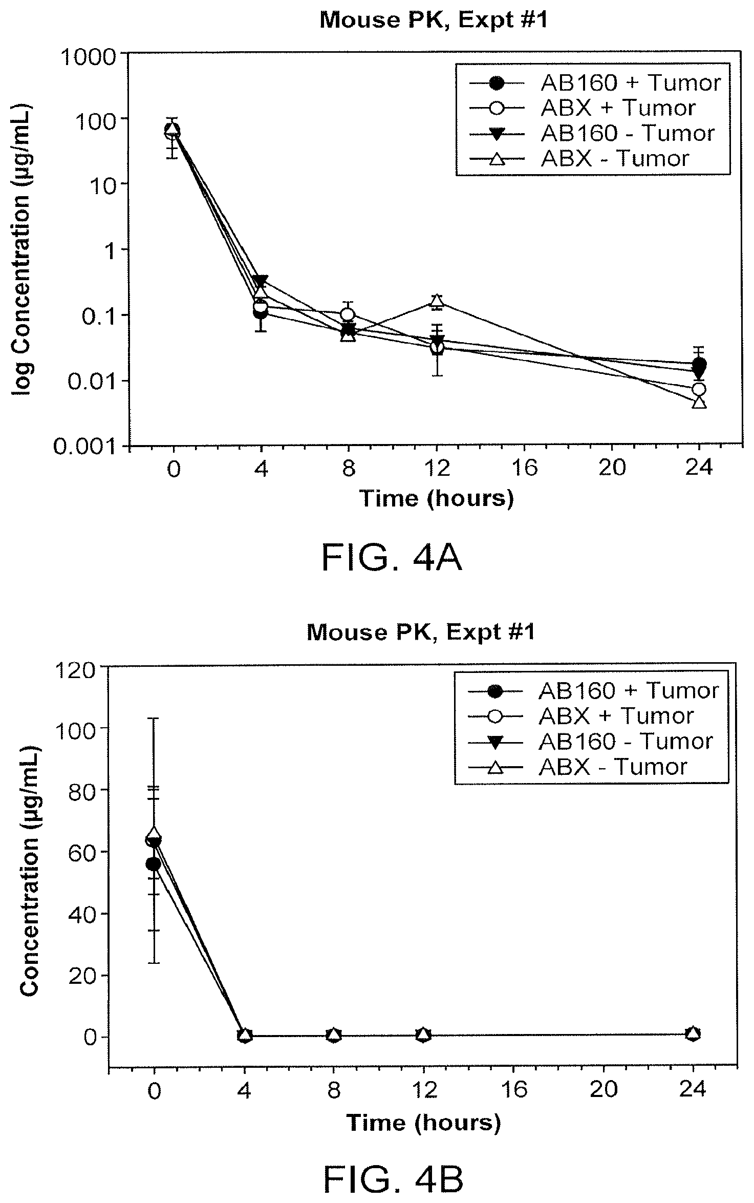
D00014

D00015

D00016

D00017
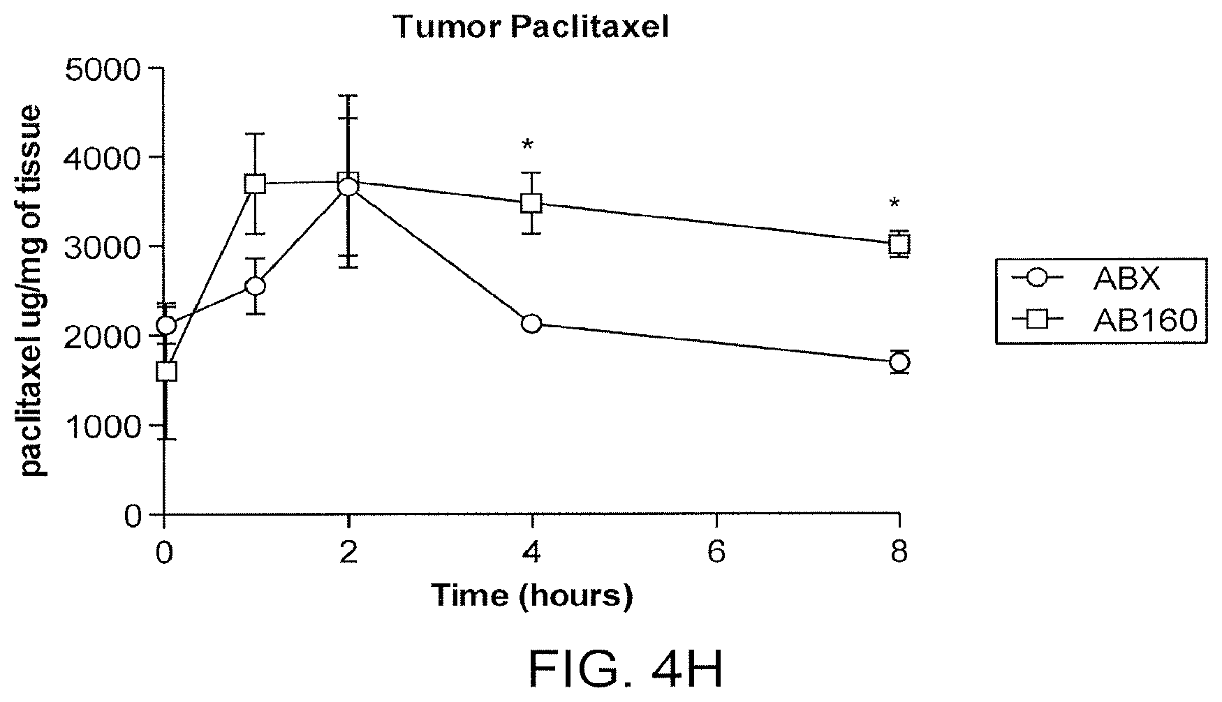
D00018

D00019

D00020
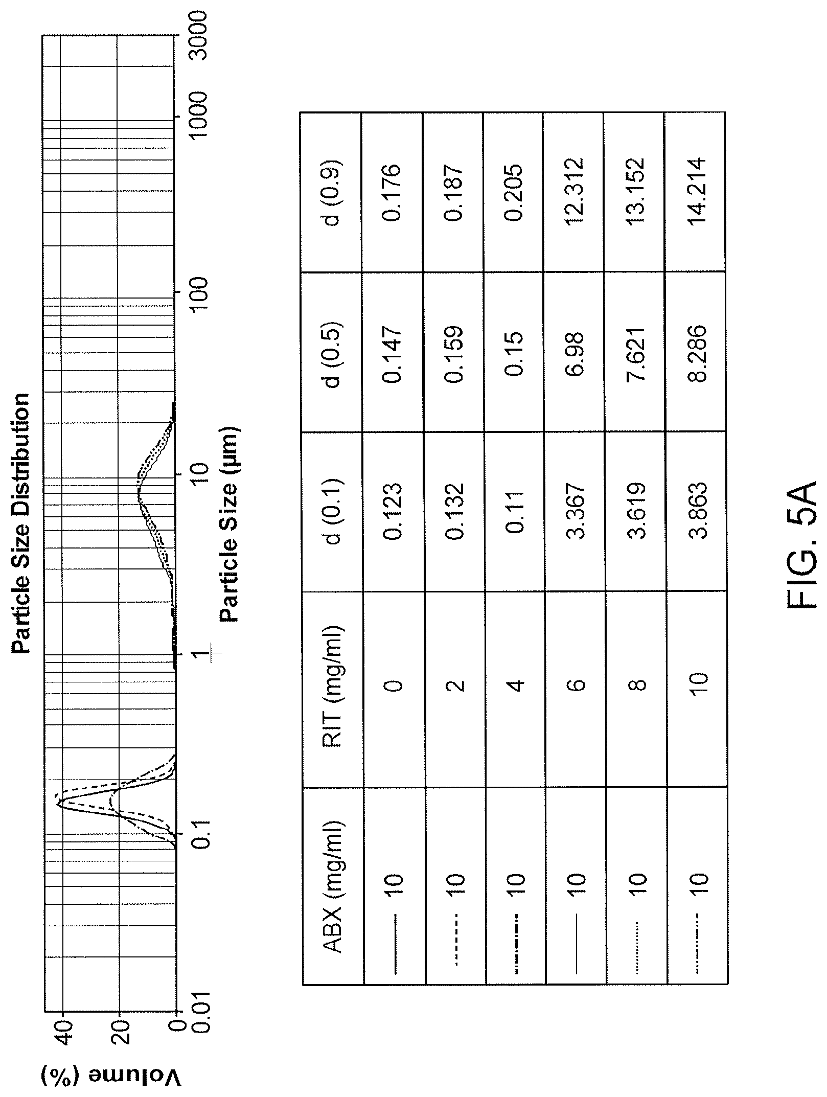
D00021

D00022

D00023

D00024

D00025

D00026

D00027
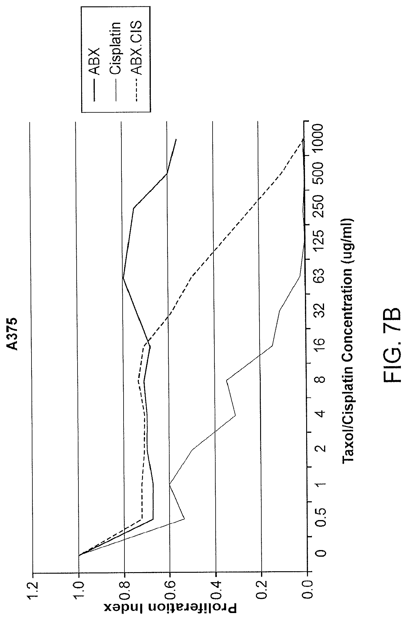
D00028

D00029

D00030

D00031

D00032

D00033

D00034

D00035

D00036

D00037

D00038

D00039

D00040

D00041

D00042

D00043

D00044

XML
uspto.report is an independent third-party trademark research tool that is not affiliated, endorsed, or sponsored by the United States Patent and Trademark Office (USPTO) or any other governmental organization. The information provided by uspto.report is based on publicly available data at the time of writing and is intended for informational purposes only.
While we strive to provide accurate and up-to-date information, we do not guarantee the accuracy, completeness, reliability, or suitability of the information displayed on this site. The use of this site is at your own risk. Any reliance you place on such information is therefore strictly at your own risk.
All official trademark data, including owner information, should be verified by visiting the official USPTO website at www.uspto.gov. This site is not intended to replace professional legal advice and should not be used as a substitute for consulting with a legal professional who is knowledgeable about trademark law.