Capacitively coupled REIMS technique and optically transparent counter electrode
Pringle , et al. Sept
U.S. patent number 10,777,399 [Application Number 15/764,791] was granted by the patent office on 2020-09-15 for capacitively coupled reims technique and optically transparent counter electrode. This patent grant is currently assigned to Micromass UK Limited. The grantee listed for this patent is Micromass UK Limited. Invention is credited to Lajos Godorhazy, Tamas Karancsi, Steven Derek Pringle, Daniel Simon, Daniel Szalay, Zoltan Takats.


| United States Patent | 10,777,399 |
| Pringle , et al. | September 15, 2020 |
Capacitively coupled REIMS technique and optically transparent counter electrode
Abstract
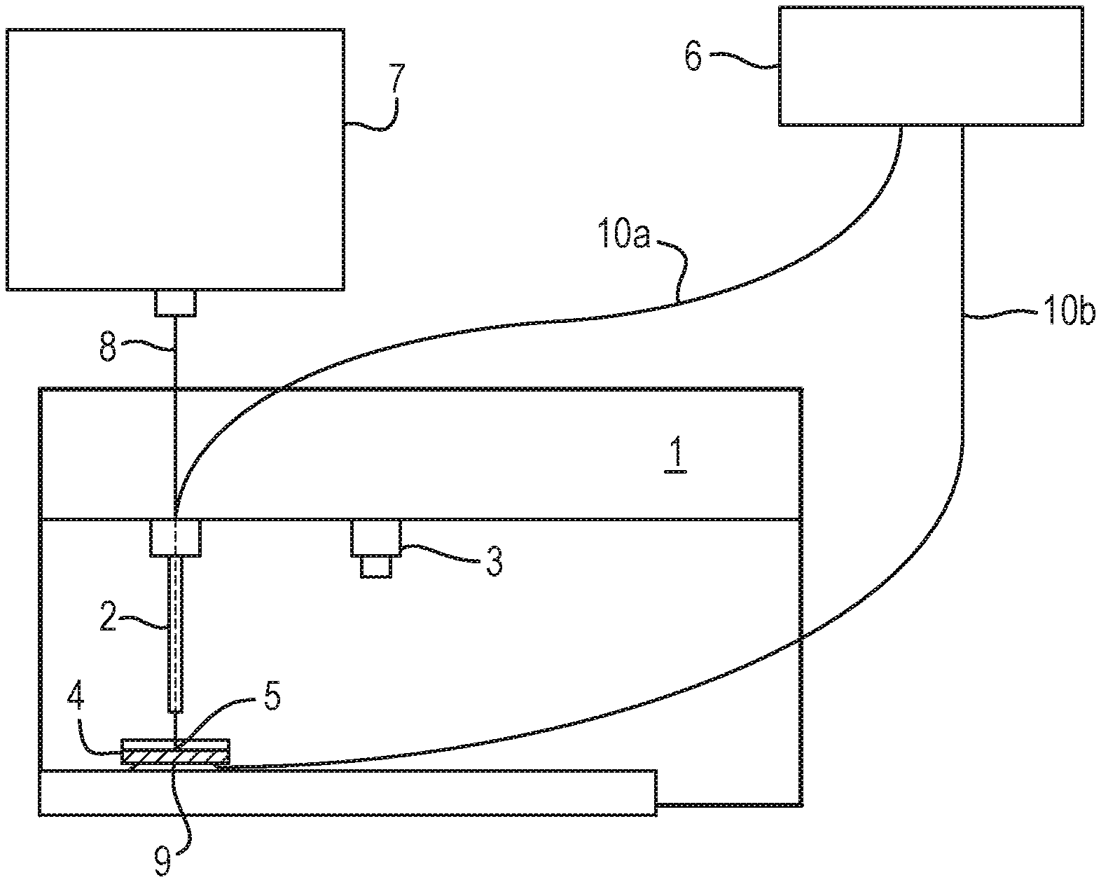
A method of analysis is disclosed comprising providing a sample on an insulating substrate such as a petri dish (4) and contacting e.g. the rear surface of the insulating substrate with a first electrode (9). The method further comprises contacting the sample with a second electrode (2) and applying an AC or RF voltage to the first and second electrodes (9,2) in order to generate an aerosol from the sample.
| Inventors: | Pringle; Steven Derek (Darwen, GB), Godorhazy; Lajos (Erd, HU), Simon; Daniel (Morichida, HU), Szalay; Daniel (Budapest, HU), Takats; Zoltan (Cambridge, GB), Karancsi; Tamas (Budapest, HU) | ||||||||||
|---|---|---|---|---|---|---|---|---|---|---|---|
| Applicant: |
|
||||||||||
| Assignee: | Micromass UK Limited (Wilmslow,
GB) |
||||||||||
| Family ID: | 1000005056349 | ||||||||||
| Appl. No.: | 15/764,791 | ||||||||||
| Filed: | September 22, 2016 | ||||||||||
| PCT Filed: | September 22, 2016 | ||||||||||
| PCT No.: | PCT/GB2016/052956 | ||||||||||
| 371(c)(1),(2),(4) Date: | March 29, 2018 | ||||||||||
| PCT Pub. No.: | WO2017/055812 | ||||||||||
| PCT Pub. Date: | April 06, 2017 |
Prior Publication Data
| Document Identifier | Publication Date | |
|---|---|---|
| US 20180286652 A1 | Oct 4, 2018 | |
Foreign Application Priority Data
| Sep 29, 2015 [GB] | 1517195.2 | |||
| Current U.S. Class: | 1/1 |
| Current CPC Class: | G01N 33/4833 (20130101); H01J 49/0445 (20130101); G01N 21/31 (20130101); H01J 49/0459 (20130101); G01N 21/65 (20130101); H01J 49/049 (20130101) |
| Current International Class: | H01J 49/04 (20060101); G01N 33/483 (20060101); G01N 21/31 (20060101); G01N 21/65 (20060101) |
References Cited [Referenced By]
U.S. Patent Documents
| 525799 | September 1894 | Rymes |
| 3479545 | November 1969 | Wilson et al. |
| 3770954 | November 1973 | Davis |
| H000414 | January 1988 | Young et al. |
| 4835383 | May 1989 | Mahoney et al. |
| 4845367 | July 1989 | Amirav et al. |
| 4883958 | November 1989 | Vestal |
| 4935624 | June 1990 | Henion et al. |
| 5033541 | July 1991 | D'Silva |
| 5053343 | October 1991 | Vora et al. |
| 5257991 | November 1993 | Fletcher et al. |
| 5308977 | May 1994 | Oishi et al. |
| 5374755 | December 1994 | Neue et al. |
| 5454274 | October 1995 | Zhu |
| 5509916 | April 1996 | Taylor |
| 5559326 | September 1996 | Goodley et al. |
| 5800597 | September 1998 | Perrotta et al. |
| 5828062 | October 1998 | Jarrell et al. |
| 5830214 | November 1998 | Flom et al. |
| 5836909 | November 1998 | Cosmescu |
| 5969352 | October 1999 | French et al. |
| 5989015 | November 1999 | Guerin et al. |
| 6032673 | March 2000 | Savage et al. |
| 6333632 | December 2001 | Yang et al. |
| 6348688 | February 2002 | Vestal |
| 6825464 | November 2004 | De La Mora |
| 6998622 | February 2006 | Wang et al. |
| 7238936 | July 2007 | Okamura et al. |
| 7247845 | July 2007 | Gebhardt et al. |
| 7329253 | February 2008 | Brounstein et al. |
| 7335897 | February 2008 | Takats et al. |
| 7365309 | April 2008 | Denny et al. |
| 7517348 | April 2009 | Vetter et al. |
| 7564028 | July 2009 | Vestal |
| 7718958 | May 2010 | Shiea et al. |
| 7828948 | November 2010 | Hatch et al. |
| 7960711 | June 2011 | Sheehan et al. |
| 8156151 | April 2012 | Sidman |
| 8193487 | June 2012 | Briglin et al. |
| 8232520 | July 2012 | Cristoni |
| 8253098 | August 2012 | Hiraoka et al. |
| 8286260 | October 2012 | Vertes et al. |
| 8314382 | November 2012 | Takats |
| 8334504 | December 2012 | Finlay et al. |
| 8431409 | April 2013 | Meinhart et al. |
| 8448493 | May 2013 | McIntyre et al. |
| 8481922 | July 2013 | Musselman |
| 8778695 | July 2014 | Caprioli |
| 8803085 | August 2014 | Ouyang et al. |
| 8834462 | September 2014 | Johnson et al. |
| 8970840 | March 2015 | Kulkarni et al. |
| 9046448 | June 2015 | Takats |
| 9053914 | June 2015 | Pringle et al. |
| 9082603 | July 2015 | Bajic |
| 9120083 | September 2015 | Wyndham et al. |
| 9255907 | February 2016 | Heanue et al. |
| 9281174 | March 2016 | Takats |
| 9287100 | March 2016 | Szalay et al. |
| 9709529 | July 2017 | Takats |
| 9731219 | August 2017 | Wang et al. |
| 9947524 | April 2018 | Pringle et al. |
| 10186626 | January 2019 | Song et al. |
| 2002/0008871 | January 2002 | Poustka et al. |
| 2002/0070338 | June 2002 | Loboda |
| 2002/0076824 | June 2002 | Haglund, Jr. et al. |
| 2003/0001084 | January 2003 | Bateman et al. |
| 2003/0008404 | January 2003 | Tomita et al. |
| 2003/0015657 | January 2003 | Takada et al. |
| 2003/0042412 | March 2003 | Park |
| 2003/0080278 | May 2003 | Okada et al. |
| 2003/0119193 | June 2003 | Hess et al. |
| 2003/0135222 | July 2003 | Baska |
| 2003/0136918 | July 2003 | Hartley |
| 2003/0193023 | October 2003 | Marsh |
| 2004/0007673 | January 2004 | Coon et al. |
| 2004/0079881 | April 2004 | Fischer et al. |
| 2004/0124352 | July 2004 | Kashima et al. |
| 2004/0197899 | October 2004 | Gomez et al. |
| 2004/0217274 | November 2004 | Bai et al. |
| 2004/0235395 | November 2004 | Hashish et al. |
| 2005/0017091 | January 2005 | Olsen et al. |
| 2005/0032471 | February 2005 | Pfarr et al. |
| 2005/0061779 | March 2005 | Blumenfeld |
| 2005/0067565 | March 2005 | Takada et al. |
| 2005/0072916 | April 2005 | Park |
| 2005/0074361 | April 2005 | Tanoshima et al. |
| 2005/0077644 | April 2005 | Bryan et al. |
| 2005/0124986 | June 2005 | Brounstein et al. |
| 2005/0138861 | June 2005 | O'Connor |
| 2005/0154490 | July 2005 | Blaine et al. |
| 2005/0159765 | July 2005 | Moutafis et al. |
| 2005/0178962 | August 2005 | Guevremont et al. |
| 2005/0178975 | August 2005 | Glukhoy |
| 2005/0230634 | October 2005 | Bajic et al. |
| 2005/0230635 | October 2005 | Takats et al. |
| 2005/0258358 | November 2005 | Thakur |
| 2005/0269518 | December 2005 | Bajic et al. |
| 2005/0274885 | December 2005 | Brown et al. |
| 2006/0035570 | February 2006 | Chisum et al. |
| 2006/0054806 | March 2006 | Yamada et al. |
| 2006/0091308 | May 2006 | Boyle et al. |
| 2006/0097084 | May 2006 | Gromer et al. |
| 2006/0108539 | May 2006 | Franzen |
| 2006/0122593 | June 2006 | Jun |
| 2006/0138321 | June 2006 | Ahem et al. |
| 2006/0145089 | July 2006 | Cristoni et al. |
| 2006/0186334 | August 2006 | Jolliffe et al. |
| 2006/0250138 | November 2006 | Sparkman et al. |
| 2006/0255264 | November 2006 | Belford |
| 2007/0023631 | February 2007 | Takats et al. |
| 2007/0023677 | February 2007 | Perkins et al. |
| 2007/0094389 | April 2007 | Nussey et al. |
| 2007/0114394 | May 2007 | Combs et al. |
| 2007/0114437 | May 2007 | Kovtoun |
| 2007/0176113 | August 2007 | Shiea et al. |
| 2007/0181802 | August 2007 | Yamada et al. |
| 2008/0001081 | January 2008 | Jindai et al. |
| 2008/0015278 | January 2008 | Malik et al. |
| 2008/0042056 | February 2008 | Fischer et al. |
| 2008/0067352 | March 2008 | Wang |
| 2008/0073503 | March 2008 | Wu |
| 2008/0073512 | March 2008 | Siuzdak et al. |
| 2008/0149822 | June 2008 | Vertes et al. |
| 2008/0172075 | July 2008 | Ammann |
| 2008/0173809 | July 2008 | Wu |
| 2008/0234579 | September 2008 | Halevy-Politch et al. |
| 2008/0312651 | December 2008 | Pope et al. |
| 2009/0065714 | March 2009 | Keady |
| 2009/0082637 | March 2009 | Galperin |
| 2009/0126891 | May 2009 | Koivunen et al. |
| 2009/0159790 | June 2009 | Kostiainen et al. |
| 2009/0272893 | November 2009 | Hieftje et al. |
| 2009/0302211 | December 2009 | Takats |
| 2010/0012830 | January 2010 | Cristoni |
| 2010/0072359 | March 2010 | Briglin et al. |
| 2010/0078550 | April 2010 | Wiseman et al. |
| 2010/0101304 | April 2010 | McIntyre et al. |
| 2010/0176290 | July 2010 | Vidal-De-Miguel |
| 2010/0186524 | July 2010 | Ariessohn et al. |
| 2010/0229263 | September 2010 | Vertes et al. |
| 2011/0036978 | February 2011 | Franzen |
| 2011/0049352 | March 2011 | Ding et al. |
| 2011/0059554 | March 2011 | Albers et al. |
| 2011/0087308 | April 2011 | Morgan |
| 2011/0295250 | December 2011 | Johnson et al. |
| 2012/0018628 | January 2012 | Wuijckhuijse et al. |
| 2012/0048264 | March 2012 | Finlay et al. |
| 2012/0074306 | March 2012 | Jesse et al. |
| 2012/0079894 | April 2012 | Van Berkel et al. |
| 2012/0080592 | April 2012 | Wiseman et al. |
| 2012/0085649 | April 2012 | Sano et al. |
| 2012/0119079 | May 2012 | Ouyang et al. |
| 2012/0149009 | June 2012 | Levis et al. |
| 2012/0156712 | June 2012 | Takats |
| 2012/0295276 | November 2012 | Cooks et al. |
| 2013/0178845 | July 2013 | Smith et al. |
| 2013/0181126 | July 2013 | Jong |
| 2013/0303846 | November 2013 | Cybulski et al. |
| 2014/0151547 | June 2014 | Bajic |
| 2014/0276775 | September 2014 | Funk et al. |
| 2014/0291506 | October 2014 | Tikhonski |
| 2014/0297201 | October 2014 | Knorr et al. |
| 2014/0299577 | October 2014 | Chung |
| 2014/0326865 | November 2014 | Pringle et al. |
| 2014/0353488 | December 2014 | Takats |
| 2014/0353489 | December 2014 | Szalay et al. |
| 2015/0021469 | January 2015 | Bajic |
| 2015/0048255 | February 2015 | Jarrell |
| 2015/0192590 | July 2015 | Sodeoka et al. |
| 2015/0201913 | July 2015 | Takats |
| 2016/0002696 | January 2016 | Galiano |
| 2016/0133450 | May 2016 | Green et al. |
| 2016/0215322 | July 2016 | Goodlett et al. |
| 2016/0247668 | August 2016 | Szalay et al. |
| 2016/0341712 | November 2016 | Agar |
| 2016/0372313 | December 2016 | Brown et al. |
| 2017/0103880 | April 2017 | Syage |
| 2018/0136091 | May 2018 | Ryan et al. |
| 2882003 | Feb 2014 | CA | |||
| 101170043 | Apr 2008 | CN | |||
| 101223625 | Jul 2008 | CN | |||
| 101288146 | Oct 2008 | CN | |||
| 101413905 | Apr 2009 | CN | |||
| 101490524 | Jul 2009 | CN | |||
| 201266145 | Jul 2009 | CN | |||
| 101657158 | Feb 2010 | CN | |||
| 101819179 | Sep 2010 | CN | |||
| 101871914 | Oct 2010 | CN | |||
| 102026709 | Apr 2011 | CN | |||
| 102121921 | Jul 2011 | CN | |||
| 102137618 | Jul 2011 | CN | |||
| 102164675 | Aug 2011 | CN | |||
| 102264404 | Nov 2011 | CN | |||
| 102367424 | Mar 2012 | CN | |||
| 102445544 | May 2012 | CN | |||
| 102483369 | May 2012 | CN | |||
| 102800553 | Nov 2012 | CN | |||
| 102879453 | Jan 2013 | CN | |||
| 102924993 | Feb 2013 | CN | |||
| 102928610 | Feb 2013 | CN | |||
| 103295873 | Sep 2013 | CN | |||
| 103335984 | Oct 2013 | CN | |||
| 103597574 | Feb 2014 | CN | |||
| 104254772 | Dec 2014 | CN | |||
| 104254901 | Dec 2014 | CN | |||
| 104582616 | Apr 2015 | CN | |||
| 0169469 | Jan 1986 | EP | |||
| 0437358 | Jul 1991 | EP | |||
| 1855306 | May 2006 | EP | |||
| 1730519 | Jul 2010 | EP | |||
| 3265817 | Jan 2018 | EP | |||
| 3265818 | Jan 2018 | EP | |||
| 2425178 | Oct 2006 | GB | |||
| 2491486 | Dec 2012 | GB | |||
| S63-243864 | Oct 1988 | JP | |||
| 03001435 | Jan 1991 | JP | |||
| H0785834 | Mar 1995 | JP | |||
| H07130325 | May 1995 | JP | |||
| H10247472 | Sep 1998 | JP | |||
| H10302710 | Nov 1998 | JP | |||
| H1164283 | Mar 1999 | JP | |||
| 2000180413 | Jun 2000 | JP | |||
| 2001183345 | Jul 2001 | JP | |||
| 2002170518 | Jun 2002 | JP | |||
| 2004264043 | Sep 2004 | JP | |||
| 2005205181 | Aug 2005 | JP | |||
| 2006-329710 | Dec 2006 | JP | |||
| 2007-51934 | Mar 2007 | JP | |||
| 2007170870 | Jul 2007 | JP | |||
| 2007-218916 | Aug 2007 | JP | |||
| 2010169454 | Aug 2010 | JP | |||
| 2014515831 | Jul 2014 | JP | |||
| 2015503109 | Jan 2015 | JP | |||
| 2015504160 | Feb 2015 | JP | |||
| 20020013544 | Feb 2002 | KR | |||
| 1020100106336 | Oct 2010 | KR | |||
| 9734534 | Sep 1997 | WO | |||
| 0160265 | Aug 2001 | WO | |||
| 2010075265 | Jul 2010 | WO | |||
| 2010136887 | Dec 2010 | WO | |||
| 2011/114902 | Sep 2011 | WO | |||
| 2012143737 | Oct 2012 | WO | |||
| 2012164312 | Dec 2012 | WO | |||
| 2012174437 | Dec 2012 | WO | |||
| 2013093517 | Jun 2013 | WO | |||
| 2013/102670 | Jul 2013 | WO | |||
| 2013098642 | Jul 2013 | WO | |||
| 2013098645 | Jul 2013 | WO | |||
| 2013148162 | Oct 2013 | WO | |||
| 2014/106165 | Jul 2014 | WO | |||
| 2014128629 | Aug 2014 | WO | |||
| 2014140601 | Sep 2014 | WO | |||
| 2014142926 | Sep 2014 | WO | |||
| 2014202828 | Dec 2014 | WO | |||
| 2015004457 | Jan 2015 | WO | |||
| 2015132579 | Sep 2015 | WO | |||
| 2016046748 | Mar 2016 | WO | |||
| 2016142674 | Sep 2016 | WO | |||
| 2016156615 | Oct 2016 | WO | |||
Other References
|
International Search Report and Written Opinion for International Application No. PCT/GB2016/052956 dated Jan. 26, 2017. cited by applicant . Hsu, et al., "Microscopy ambient ionization top-down mass spectrometry reveals developmental patterning", Proceedings of the National Academy of Sciences, vol. 110, No. 37, pp. 14855-14860, Aug. 22, 2013. cited by applicant . Na, et al., "Development of a Dielectric Barrier Discharge Ion Source for Ambient Mass Spectrometry", Journal of the American Society for Mass Spectrometry, Elsevier Science Inc, vol. 18, No. 10, pp. 1859-1862, Sep. 20, 2007. cited by applicant . Strittmatter, N., et al., Characterization and Identification of Clinically Relevant Microorganisms using Rapid Evaporative Ionization Mass Spectrometry Analytical Chemistry, vol. 86, No. 13, pp. 6555-6562, Jul. 1, 2014. cited by applicant . Golf, O., et al., "Rapid Evaporative Ionization Mass Spectrometry Imaging PI at form for Direct Mapping from Bulk Tissue and Bacterial Growth Media," Analytical Chemistry, vol. 87, No. 5, pp. 2527-2534, Mar. 3, 2015. cited by applicant . Agar, N. et al., "Development of Stereotactic Mass Spectrometry for Brain Tumor Surgery", Biosis, Neurosurgery [online], vol. 68, No. 2, (Feb. 2011) pp. 280-290. cited by applicant . Ahlf, Dorothy R. et al., "Correlated Mass Spectrometry Imaging and Confocal Raman Microscopy for Studies of Three-Dimensional Cell Culture Sections", Analyst, vol. 139, No. 18, p. 4578 (2014). cited by applicant . Azimzadeh, Omid et al., "Formalin-Fixed Paraffin-Embedded (FFPE) Proteome Analysis Using Gel-Free and Gel-Based Proteomics", Journal of Proteome Research, vol. 9, No. 9, pp. 4710-4720 (2010). cited by applicant . Balog, Julia et al., "Identification of Biological Tissues by Rapid Evaporative Ionization Mass Spectrometry", Analytical Chemistry, vol. 82, No. 17, pp. 7343-7350 (Sep. 2010). cited by applicant . Balog, Julia et al., "Supporting Information for Identification of Biological Tissues by Rapid Evaporative Ionization Mass Spectrometry", pp. SI-S9, http:/ /pubs.acs.org/doi/suppl/10.1021 /ac101, (2013). cited by applicant . Balog, J. et al., "Intraoperative Tissue Identification Using Rapid Evaporative Ionization Mass Spectrometry", Science Translational Medicine, vol. 5, No. 194, pp. 1-11 (Jul. 2013). cited by applicant . Balog, J. et al., "Supplementary Materials: Intraoperative Tissue Identification Using Rapid Evaporative Ionization Mass Spectrometry", Science Translational Medicine, vol. 5, No. 194 34 pages (Jul. 2013). cited by applicant . Bean, Heather D. et al., "Bacterial Volatile Discovery Using Solid Phase Microextraction and Comprehensive Two-Dimensional Gas Chromatographytime-of-Flight Mass Spectrometry", Journal of Chromatography B: Biomedical Sciences & Applications, Elsevier, Amersterdam, NL, vol. 901, pp. 41-46 (May 2012). cited by applicant . Bellet, V. et al., "Proteomic Analysis of RCL2 Paraffin-Embedded Tissues", Journal of Cellular and Molecular Medicine, vol. 12, No. 5B, pp. 2027-2036 (2008). cited by applicant . Bocklitz, T.W. et al., "Deeper Understanding of Biological Tissue: Quantitative Correlation of MALDITOF and Raman Imaging", Analytical Chemistry, vol. 85 No. 22, pp. 10829-10834 (2013). cited by applicant . Tait, Emma et al., "Identification of Volatile Organic Compounds Produced by Bacteria Using HS-SPME-GC-MS", Journal of Chromatographic Sci, pp. 1-11. cited by applicant . Cole, Laura M. et al., "Mass Spectrometry Imaging for the Proteomic Study of Clinical Tissue", Proteomics-Clinical Applications, vol. 9, No. 3-4, pp. 335-341 (Apr. 2015). cited by applicant . Crawshaw, Benjamin et al., "Gastrointestinal Surgery: Real-Time Tissue Identification During Surgery", Nature Review/Gastroenterology & Hepatology , vol. 10, No. 11. pp. 624-625 (Sep. 2013). cited by applicant . Cselik, Z. et al., "Impact of Infrared Laser Light-Induced Ablation at Different Wavelengths on Bovine Intervertebral Disc Ex Vivo: Evaluation with Magnetic Resonance Imaging and Histology", Lasers in Surgery and Medicine, vol. 44, No. 5, pp. 406-412 (Jul. 2012). cited by applicant . Davies, T.J. et al., "Volatile Products from Acetylcholine as Markers in the Rapid Urine Test Using Head-Space Gas-Liquid Chromatography" Journal of Chromatography B: Biomedical Sciences and Application, vol. 307, pp. 11-21 (Jan. 1984). cited by applicant . European Commission, "ISD Report Summary", http://cordis.europa.eu/result/rcn/163435_e, (2016). cited by applicant . Fahy, Eoin, et al., "Lipid Classification, Structures and Tools", Biochimica at Biophysica Acta (BBA)--Molecular and Cell Biology of Lipids, vol. 1811, No. 11, pp. 637-647 (2011). cited by applicant . Gerbig, Stefanie et al., "Analysis of Colorectal Adenocarcinoma Tissue by Desorption Electrospray Ionization Mass Spectrometric Imaging", Analytical and Bioanalytical Chemistry, vol. 403, No. 8, pp. cited by applicant . Golf, Ottmar et al., "XMS: Cross-Platform Normalization Method for Multimodal Mass Spectrometric Tissue Profiling", Journal of the American Society for Mass Spectrometry, vol. 26, No. 1, pp. 44-54 (Nov. 2014). cited by applicant . Guenther, Sabine et al., "Electrospray Post-Ionization Mass Spectrometry of Electrosurgical Aerosols", Journal of the American Society for Mass Spectrometry, vol. 22, No. 11, pp. 2082-2089 (Nov. 2011). cited by applicant . Gustafsson, Ove J.R. et al., "Proteomic Developments in the Analysis of Formalin-Fixed Tissue", Biochimica et Biophysica Acta, vol. 1854, No. 6, pp. 559-580. cited by applicant . Uribe, D.0. et al., "Piezoelectric Self-Sensing System for Tactile Intraoperative Brain Tumor Delineation in Neurosurgery", Proceedings of the 31st Annual International Conference of the IEEE Engineering in Medicine and Biology Society: Engineering the Future of BioMedicine, IEEE pp. 737-740 (Sep. 2009). cited by applicant . Hobbs, S.K. et al., "Magnetic Resonance Image-Guided Proteomics of Human Glioblastoma Multiforme", Journal of Magnetic Resonance Imaging, vol. 18, pp. 530-536 (Jan. 2003). cited by applicant . Jadoul, L. et al., "Matrix-Assisted Laser Desorption/Ionization Mass Spectrometry and Raman Spectroscopy: An Interesting Complementary Approach for Lipid Detection in Biological Tissues", Eurooean Journal of Lipid Science and Technology. vol. 116, No. 8. pp. 1080-1086 (2014). cited by applicant . Jain, M. et al., "Metabolite Profiling Identifies a Key Role for Glycine in Rapid Cancer Cell Proliferation", American Association for the Advancement of Science, vol. 336, No. 6084, pp. 1040-1044 (May 2012). cited by applicant . Jarmusch, Alan K. et al., "Detection of Strep Throat Causing Bacterium Directly from Medical Swabs by Touch Spray-Mass Spectrometry", Analyst, vol. 139, No. 19, pp. 4785 (Jan. 1, 2014). cited by applicant . Jarmusch, Alan K. et al., "Supplemental Information Detection of Strep Throat Causing Bacterium Directly from Medical Swabs by Touch Spray-Mass Spectrometry", http://www.rsc.org/suppdata/an/c4/c4an00959b/c4an00959b1 [retrieved on May 13, 2016] (2016). cited by applicant . Lazova, Rossitza et al., "Imaging Mass Spectrometry--A New and Promising Method to Differentiate Spitz Nevi From Spitwid Malignant Melanomas", American Journal of Dermatopathology, vol. 34, No. 1, pp. 82-90 (Feb. 2012). cited by applicant . Li, Yan et al., "Aberrant Mucin5B Expression in Lung Adenocarcinomas Detected by iTRAQ Labeling Quantitative Proteomics and Immunohistochemistry", Clinical Proteomics, vol. 10, No. 1, p. 15 (2013). cited by applicant . Lieuwe, D.J. et al., "Volatile Metabolites of Pathogens: A Systematic Review", PLoS Pathogens, vol. 9, Issue 5, e1003311 (May 2013) whole document. cited by applicant . Luge, S. et al., "Use of a Lower Power, High Frequency Stabilized Capacitive Plasma Combined with Graphite Furnace Vaporization for the Atomic Emission Spectrometric Analysis of Serum Samples", Analytical Chimica Acta, vol. 332, No. 2-3, pp. 193-199 (Oct. 1996). cited by applicant . Mccullough, Bryan J. et al., "On-Line Reaction Monitoring by Extractive Electrospray Ionisation", Rapid Communications in Mass Spectrometry, vol. 25, No. 10, pp. 1445-1451 (May 2011). cited by applicant . Murray, Patrick R, "What Is New in Clinical Microbiology--Microbial Identification by MALDI-TOF Mass Spectrometry", Journal of Molecular Diagnostics, vol. 14, No. 5, pp. 419-423 (Sep. 2012). cited by applicant . Nicholson, Jeremy K. et al., "Metabolic Phenotyping in Clinical and Surgical Environments", Nature, vol. 491, No. 7424 pp. 384-392 (Nov. 2012). cited by applicant . Pirro, Valentina et al., "Direct Drug Analysis from Oral Fluid Using Medical Swab Touch Spray Mass Spectrometry", Analytica Chimica Acta, vol. 861, Jan. 7, 2015 pp. 47-54. cited by applicant . Plata, N. et al., "Aerosols Sampling Using a New Cryogenic Instrument", Journal of Aerosol Science, Pergamon, Amsterdam, NL vol. 37, No. 12, pp. 1871-1875 (Dec. 2006). cited by applicant . Rodriguez-Rigueiro, Teresa et al., "A Novel Procedure for Protein Extraction from Formalin-Fixed Paraffin-Embedded Tissues", Proteomics, vol. 11, No. 12, pp. 2555-2559 (2011). cited by applicant . Ellis, S. et al., "Surface Analysis of Lipids by Mass Spectrometry: More Than Just Imaging", Progress in Lipid Research Pergamon Press, vol. 52, No. 4, pp. 329-353 (Oct. 2013). cited by applicant . Shoemaker, Robert H., "The NCI60 Human Tumour Cell Line Anticancer Drug Screen", (2013). cited by applicant . Strittmatter, N. et al., "Anaylsis of Intact Bacteria Using Rapid Evaporative Ionization Mass Spectrometry", Chemical Communications--Chemcon, vol. 49, No. 55, p. 6188 (May 2013). cited by applicant . Strittmatter, N. et al., "Taxon-Specific Markers for the Qualitative and Quantitative Detection of Bacteria in Human Samples", https://www.msacl.org/2015 US Long Abstract. cited by applicant . Cho, YT., et al. "Differentiation of Virulence of Helicobacter Pyloriby Matrix-Assited Laser Desorption/Ionization Mass Spectrometry and Multivariate Analyses" Clinica Chimica ACTA, Elsevier BV, 424:123-130, May 26, 2013. cited by applicant . Kohler, M. et al. "Characterization of lipid extracts from brain tissue and tumors using Raman spectroscopy and mass spectrometry," Anal Bioana Chem, 393:1513-1520, Jan. 20, 2009. cited by applicant . Harry, K. H., et al. "Effect of protein coating of flocked swabs on the collection and release of clinically important bacteria", Indian Journal of Medical Microbiology, 32(3):301-303 (2014). cited by applicant . Blais, B. W., "Swab-Based Enzyme Immunoassay System for Detection of Meat Residues on Food Contact Surfaces as a Hygiene Monitoring Tool", Journal of Food Protection, 62(4):386-389 (1999). cited by applicant . Farhat, S. E., et al., "Efficacy of a Swab Transport System in Maintaining Viability of Neisseria gonorrhoeae and Streptococcus pneumoniae", Journal of Clinical Microbiology, 39(8)2958-2960 (2001). cited by applicant . Harry, E. L. et al., "Direct analysis of pharmaceutical formulations from non-bonded reversed-phase thin-layer chromatography plates by desorption electrospray ionisation ion mobility mass spectrometry", Rapid Communications in Mass Spectrometry, 23(17):2597-2604, Jul. 28, 2009. cited by applicant . Hachmoeller et al., "Element bioimaging of liver needle biopsy specimens from patients with Wilson's disease by laser ablation-inductively coupled plasma-mass spectrometry", Journal of Trace Elements in Medicine and Biology, 35:97-102, Feb. 10, 2016. cited by applicant . Guenther et al., "Spatially Resolved Metabolic Phenotyping of Breast Cancer by Desorption Electrospray Ionization Mass Spectrometry", Cancer Research, 75:1828-1837, Feb. 17, 2015. cited by applicant . Chipuk, J. E., et al., "Transmission Mode Desorption Electrospray Ionization" , Journal of the American Society for Mass Spectrometry, 19(11):1612-1620, Nov. 1, 2008. cited by applicant . Santagata, S., et al., "Intraoperative mass spectrometry mapping of an onco-metabolite to guide brain tumor surgery", Proceedings of the National Academy of Sciences (PNAS), 111(30):11121-11126, Jun. 30, 2014. cited by applicant . Chen, H., et al., "What Can We Learn from Ambient Ionization Techniques?", Journal of the American Society for Mass Spectrometry, 20:1947-1963, (2009). cited by applicant . Sankaranarayanan, G., et al., "Common Uses and Cited Complications of Energy in Surgery", Surg Endosc., 27:3056--3072, (2013). cited by applicant . Rau, H.G., et al., "The use of water-jet dissection in open and laparoscopic liver resection", HPB, 10: 275-280, (2008). cited by applicant . Chen et al. "Desorption Electrospray Ionization Mass spectrometry for high-thoughput analysis of Pharamaceutical samples in the ambient environment", Anal. Chem 77:6915-6927 (2005). cited by applicant . Summons to Attend Oral Proceedings Pursuant to Rule 115(a) EPC of EP Application No. 1276643.5, dated Apr. 20, 2018, 7 pages. cited by applicant . Chen et al., "Surface desorption atmospheric pressure chemical ionization mass spectrometry for direct ambient sample analysis without toxic chemical contamination", Journal of Mass Spectrometry, 42(8):1045-1056, Jan. 1, 2007. cited by applicant . Chen, H., et al: "Neutral desorption sampling coupled to extractive electrospray ionization mass spectrometry for rapid differentiation of biosamples by metabolomic fingerprinting", Journal of Mass Spectromety, vol. 42, No. 9, Sep. 1, 2007 pp. 1123-1135. cited by applicant . Hensman C., et al: "Chemical Composition of Smoke Produced by High-Frequency Electrosurgery in a Closed Gaseous Environment An in Vitro Study", Surgical Endoscopy, vol. 12, No. 8, Aug. 1, 1998 (Aug. 1, 1998), pp. 1017-1019. cited by applicant . Moot, A. et al: "Composition of Volatile Organic Compouds in Diathermy Plume as Detected by Selected Ion Flow Tube Mass Spectrometry", ANZ Journal of Surgery, vol. 77, No. 1-2, (Jan. 2007) pp. 20-23. cited by applicant . Strittmatter, N.: "Home--Miss Nicole Strittmatter" Retrieved from the Internet URL: http://www.imperial.ac.uk/people/n.strittmatter12 [retrieved on May 19, 2016] the whole document. cited by applicant . Wehofsky, et al ("Automated deconvolution and deisotoping of electrospray mass spectra" J. Mass Spectrom. 2002; 37: pp. 223-229). cited by applicant . Al Sahaf et al., "Chemical Composition of Smoke Produced by High-Frequency Electrosurgery", Irish Journal of Medical Science, vol. 176, No. 3, pp. 229-232, 2007. cited by applicant . Barrett et al., "Surgical Smoke: A Review of the Literature", Surgical Endoscopy, vol. 17, No. 6, pp. 979-987, 2003. cited by applicant . Down, "A DESI-Rable Ionization Revolutionizes Mass Spectrometry", Base Peak, 2005. cited by applicant . International Search Report and Written Opinion for International Application. No. PCT/IB2012/003009, dated Aug. 14, 2013, 17 pages. cited by applicant . PCT International Search Report and Written Opinion for International Appln. No. PCT/IB2010/001261, dated Sep. 21, 2010, 5 pages. cited by applicant . PCT International Search Report and Written Opinion for International Appln. No. PCT/IB2012/002995, dated Sep. 10, 2013, 3 pages. cited by applicant . Qiao et al., "Electrostatic-Spray Ionization Mass Spectrometry", Analytical Chemistry, vol. 84, No. 17, pp. 7422-7430, 2012. cited by applicant . Lee et al., "Thermally Assisted Electrospray Interface for Liquid Chromatography/Mass Spectrometry", Rapid Communications in Mass Spectrometry, vol. 6, pp. 727-733, 1992. cited by applicant . McEwen et al., "Analysis of Solids, Liquids, and Biological Tissues Using Solids Probe Introduction at Atmospheric Pressure on Commercial LC/MS Instruments", Anal. Chem., vol. 77, pp. 7826-7831, 2005. cited by applicant . Sakairi et al., "Characteristics of a Liquid Chromatograph/Atmospheric Pressure Ionization Mass Spectrometer", Anal. Chem., vol. 60, pp. 774-780, 1988. cited by applicant . Takats et al., "Characterization of DESI-FTICR Mass Spectrometry--From ECD to Accurate Mass Tissue Analysis", Journal of Mass Spectrometry, vol. 43, pp. 196-203, 2008. cited by applicant . Vander Wilp, W. et al., "Lead in Micro-Samples of Whole Blood by Rhenium-Cup in-Torch Vaporization-Inductively Coupled Plasma-Atomic Emission Spectrometry (ITV-ICP-AES)", Fresenius Journal of Analytical Chemistry, vol. 368, No. 7, pp. 734-736 (Nov. 2000). cited by applicant . Eagles, et al., "Fast Atom Bombardment Mass Spectrometry of Amine Mixtures", John Wiley & Sons, Ltd, 1988. cited by applicant . Slemr et al., "Concentration Profiles of Diamines in Fresh and aerobically Stored Park and Beef", American Chemical Society, 1985. cited by applicant . Mulligan, Christopher C. et al., "Desorption electrospray ionization with a portable mass spectrometer: in situ analysis of ambient surfaces", Chemical Communications--Chemcom, No. 16, pp. 1709-1711, (Jan. 2006). cited by applicant . Van Berke!, "Thin-Layer Chromatography and El3ectrospray Mass Spectrometry Coupled Using a Surface Sampling probe". Anal. Chem. 2002. cited by applicant . Takats et al., "Mass Spectrometry Sampling Under Ambient Conditions with Desorption Electrospray Ionization", Science, vol. 306, 2004. cited by applicant . Tottszer et al., "Laser Heating Versus Resistive Heating in the Field-Desorption Mass Spectrometry of Organic Polymers", J. Phys. D: Appl. Phys., vol. 21, pp. 1713-1720, 1988. cited by applicant . Zhou, X. et al., "Development of miniature mass spectrometry systems for bioanalysis outside the conventional laboratories." Bioanalysis, 6 (11) 1497-1508 (2014). cited by applicant . Bolt, F., et al., "Automated High-Throughput Identification and Characterization of Clinically Important Bacteria and Fungi using Rapid Evaporative Ionization Mass Spectrometry," American Chemical Socieity, 88 9419-9426 (2016). cited by applicant . McJimpsey, E.L., et al., "Parameters Contributing to Efficient Ion Generation in Aerosol MALDI Mass Spectrometry," American Society for Mass Spectrometry pp. 1044-0305 (2007). cited by applicant . Mutters, N. T., et al., "Performance of Kiestra Total Laboratory Automation Combined with MS in Clinical Microbiology Practice," Annals of Laboratory Medicine 34: 111-117 (2014). cited by applicant . Hsu C. et al. "Visualizing Life with Ambient Mass Spectrometry", Current Opinion in Biotechnology, vol. 31, pp. 24-34 (2015). cited by applicant . Longuespee, R., et al., Tissue Proteomics for the Next Decade? Towards a Molecular Dimension in Histology, OMICS A Journal of Integrative Biology 28(9): 539-552 (2014). cited by applicant . Lu, K, et al., "Arsenic Exposure Perturbs the Gut Microbiome and its Metabolic Profile in Mice: An Integrated Metagenomics and Metabolomics Analysis," Environmental Health Perspectives, 122(3): 284-291 (2014). cited by applicant . Suarez, S. et al., Ribosomal proteins as biomarkers for bacterial identification by mass spectrometry in the clinical microbiology laboratory, Journal of microbiological Methods, 94: 390-396 (2013). cited by applicant . Trimpin, S. et al., New Ionization Method for Analysis on Atmospheric Pressure Ionization Mass Spectrometers Requiring Only Vacuum and Matrix Assistance, Analytical Chemistry, 85:2005-2009 (2013). cited by applicant . Cha, S., Laser desorption/ionization mass spectrometry for direct profiling and imaging of small moledcules from raw biological materials, Doctoral Dissertation, Iowa State University (2008). cited by applicant . Asano et al., "Self-aspirating atmospheric pressure chemical ionization source for direct sampling of analytes on Surfaces in liquid solution", Rapid Communications in Mass Spectrometry 2005. cited by applicant . International Search Report and Written Opinion for application No. PCT/GB2017/051050, dated Jun. 27, 2017, 15 pages. cited by applicant . Gerbig, Stefanie et al, "Spatially resolved investigation of systemic and contact pesticides in plant material by desorption electrospray ionization mass spectrometry imagine", Analytical and Bioanalytical Chemistry, 407 (24):7379-7389 (2015). cited by applicant . Lesiak, A., et al.,"Rapid detection by direct analysis in real time-mass spectrometry (DART-MS) of psychoactive plant drugs of abuse: the case of Mitragyna speciosa aka "Kratom"", 242:210-218 (2014). cited by applicant . Bartels, B. et al., "Spatially resolved in vivo plant metabolomics by laser ablation-based mass spectrometry imaging (MSI) techniques: LDI-MSI and LAESI", Frontiers in Plant Science vol. 6 (2015). cited by applicant . Bagley, B.M., et al., "Evaluation of archival time on shotgun proteomics of formalin-fixed and paraffin-embedded tissues", Journal of Proteome Research 8(2):917-925, (2009). cited by applicant . Nielen, M et al., "Desorption electrospray ionization mass spectrometry in the analysis of chemical food contaminants in food", Trac Trends in Analytical Chemistry, 30(2):165-180 (2011). cited by applicant . Boughton, B. et al., "Mass spectrometry imaging for plant biology: a review", Phytochemistry Reviews, 15(3):445-488 (2015). cited by applicant . Schafer, K.C., et al., "In Situ, Real-Time Identification of Biological Tissue by Ultraviolet and Infrared Laser Desorption Ionization Mass Spectrometry", Analytical Chemistry, 83(5):1632-1640, Mar. 1, 2011. cited by applicant . Jackson, S. N. et al. On-line laser desorption/ionization mass spectrometry of matrix-coated aerosols, Rapid Communications in Mass Spectrometry, vol. 18, pp. 2041-2045 (Year 2004). cited by applicant . Dong, Y., et al., "Sample Preparation for Mass Spectrometry Imaging of Plant Tissues: A Review", Frontiers in Plant Science 7(60): 1-16 (2016). cited by applicant . Communication pursuant to Article 94(3) EPC, for application No. 16710788.7, dated Jun. 13, 2019, 9 pages. cited by applicant . Examination Report under Section 18(3), for application No. GB1714122.7, dated May 9, 2019, 6 pages. cited by applicant . Bagley, B.M., et al., "Evaluation of archival time on shotgun proteomics of formalin-fixed and paraffin-embedded issues", Journal of Proteome Research 8(2):917-925, (2009). cited by applicant . Vircks, Kyle E. et al., "Rapid Screening of Synthetic Cathinones as Trace Residues and in Authentic Seizures Using a Portable Mass Spectrometer Equipped with Desorption Electrospray Ionization", Rapid Communications in Mass Spectrometry, vol. 26, No. 23, pp. 2665-2672 (Dec. 15, 2012). cited by applicant . Office Action for CN Patent Application No. 201680025801.0 dated Apr. 7, 2020. cited by applicant . Office Action for CN Patent Application No. 201680025801.0 dated Apr. 7, 2020 [translation]. cited by applicant . Adams, F., et al, "Inorganic Mass Spectrometry", copyright John Wiley Sons, Inc. pp. 174-180 (1988). cited by applicant . Vemury, S., and Pratsinis, S.E., "Charging and Coagulation During Flame Synthesis of Silica", Journal of Aerosol Science 27(6):951-966. cited by applicant . Examination Report under Section 18(3), for application No. GB1715787.6, dated Jun. 1, 2020, 6 pages. cited by applicant . Panpradist, N., et al., "Swab Sample Transfer for Point-Of-Care Diagnostics: Characterization of Swab types and Manual Agitation Methods", Plos One 9(9):1-11 (2014). cited by applicant . CNOA for application No. 201680026285.3 dated Jun. 12, 2020, 12 pages. cited by applicant. |
Primary Examiner: Ippolito; Nicole M
Assistant Examiner: Luck; Sean M
Claims
The invention claimed is:
1. A method of analysis comprising: providing a sample on an insulating substrate; contacting said insulating substrate with a first electrode and contacting said sample with a second electrode; and applying an AC or RF voltage to said first and second electrodes in order to generate an aerosol from said sample.
2. A method as claimed in claim 1, wherein the step of providing a sample on said insulating substrate comprises providing said sample in a container, a petri dish, a vial or a microtitre or microwell plate.
3. A method as claimed in claim 1, wherein the step of providing said sample on said insulating substrate comprises providing said sample and optionally a culture or growth medium on a first surface of said insulating substrate and wherein the step of contacting said insulating substrate with said first electrode comprises contacting said insulating substrate with said first electrode on a second surface of said insulating substrate which is opposed to said first surface.
4. A method as claimed in claim 1, wherein the step of contacting said insulating substrate with said first electrode is such that said first electrode does not contact said sample or optionally any culture or growth medium.
5. A method as claimed in claim 1, wherein said first electrode comprises a mesh electrode.
6. A method as claimed in claim 1, wherein said first electrode comprises a substrate which is substantially optically transparent or optically translucent.
7. A method as claimed in claim 6, wherein said substrate further comprises a conductive layer or a conductive coating.
8. A method as claimed in claim 1, further comprising locating said insulating substrate upon said first electrode, wherein said insulating substrate is substantially optically transparent or optically translucent and wherein said first electrode is substantially optically transparent or translucent.
9. A method as claimed in claim 8, further comprising passing light or photons through said first electrode and said insulating substrate in order to illuminate, image or analyse said sample.
10. A method as claimed in claim 1, further comprising directing at least some of the aerosol into a vacuum chamber of a mass spectrometer and ionising at least some the aerosol within a or the vacuum chamber of the mass spectrometer so as to generate a plurality of analyte ions.
11. A method as claimed in claim 1, further comprising spectroscopically imaging or analysing said sample at substantially the same time as obtaining mass spectral data corresponding to one or more locations on or in said sample.
12. Analysis apparatus comprising: a first electrode for contacting an insulating substrate upon which a sample is located in use; a second electrode for contacting said sample; and a device for applying an AC or RF voltage to said first and second electrodes in order to generate an aerosol from said sample.
13. Apparatus as claimed in claim 12, wherein said first electrode comprises a mesh electrode.
14. Apparatus as claimed in claim 12, wherein said first electrode comprises a substrate which is substantially optically transparent or optically translucent.
15. Apparatus as claimed in claim 14, wherein said substrate further comprises a conductive layer or a conductive coating.
16. Apparatus as claimed in claim 12, wherein said insulating substrate is positioned, in use, upon said first electrode and wherein said insulating substrate is substantially optically transparent or optically translucent and wherein said first electrode is substantially optically transparent or translucent, and wherein the apparatus further comprises a device for passing light or photons through said first electrode and said insulating substrate in order to illuminate, image or analyse said sample.
17. Apparatus as claimed in claim 12, further comprising a device for directing at least some of said aerosol into a vacuum chamber of a mass spectrometer and an ion source located within a or said vacuum chamber of said mass spectrometer for ionising at least some said aerosol so as to generate a plurality of analyte ions.
18. Apparatus as claimed in claim 12, further comprising a spectroscopic imaging or analysing device for spectroscopically imaging or analysing said sample.
19. Apparatus as claimed in claim 18, wherein said spectroscopic imaging or analysing device comprises a Raman spectroscope and/or an infra-red ("IR") spectroscope.
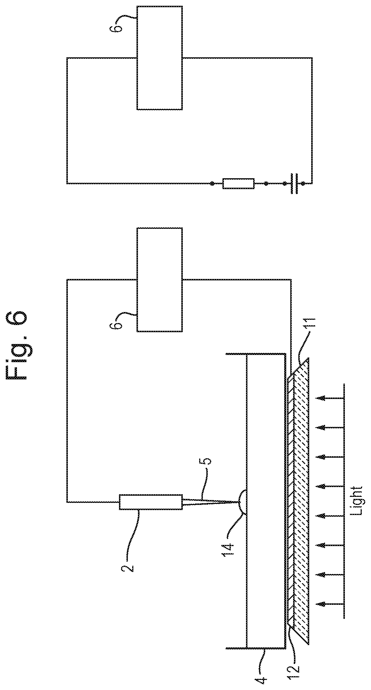
20. Analysis apparatus comprising: a first electrode; a first device for passing light or photons through said first electrode in order to illuminate, image or analyse, in use, a sample located on said first electrode; a second electrode for contacting said sample; and a second device for applying an AC or RF voltage to said first and second electrodes in order to generate an aerosol from said sample.
Description
CROSS-REFERENCE TO RELATED APPLICATION
This application is a national phase filing claiming the benefit of and priority to International Patent Application No. PCT/GB2016/052956, filed on Sept. 22, 2016, which claims priority from and the benefit of United Kingdom patent application No. 1517195.2 filed on Sep. 29, 2015. The entire contents of these applications are incorporated herein by reference.
FIELD OF THE INVENTION
The present invention relates generally to mass spectrometers and in particular to the analysis of material by rapid evaporation ionisation mass spectrometry ("REIMS").
BACKGROUND
Rapid evaporation ionisation mass spectrometry ("REIMS") is a relatively new technique that is useful for the analysis of many different samples including the identification of tissue such as food. For example, it is known to use rapid evaporation ionisation mass spectrometry to determine the animal of origin of a food sample and also the pathological state of a tissue sample. It is also known to use rapid evaporation ionisation mass spectrometry to identify microbes, yeast and fungi.
The known approach for analysing bacterial colonies by rapid evaporation ionisation mass spectrometry involves using bipolar electrosurgical forceps and an electrosurgical RF generator. A bacterial colony is scraped from the surface of an agar layer using the bipolar electrosurgical forceps and a short burst of RF voltage from the electrosurgical RF generator is applied between the bipolar electrosurgical forceps. For example, it is known to apply 60 W of power in a bipolar mode at a frequency of 470 kHz sinusoid. The RF voltage which is applied to the electrosurgical forceps has the result of rapidly heating the particular portion of the bacterial colony which is being analysed due to its nonzero impedance. The rapid heating of the microbial mass results in an aerosol being generated. The aerosol is transferred directly into a mass spectrometer and the aerosol sample may then be analysed by the mass spectrometer. It is known for the control system of the mass spectrometer to utilise multivariate statistical analysis in order to help distinguish and identify different samples.
Rapid evaporation ionisation mass spectrometry is, therefore, a form of mass spectrometry that uses high frequency energy to ablate or vaporise a sample wherein the resulting vapour or aerosol is then subjected to mass spectrometry.
Conventional rapid evaporation ionisation mass spectrometry analysis involves ensuring that the sample is in direct electrical (and physical) contact with a RF voltage supply. This approach works well for tissue identification either ex vivo or in vivo. However, such an approach is problematic if it is desired to process a sample which is housed in a container. In particular, the known approach is problematic if it is desired to process a bacterial culture grown on agar in a petri dish.
It is also problematic to attempt to use the known arrangement for the analysis of tissue sections mounted on a glass slide when it is desired to analyse the tissue sections optically and also to subject the tissue sections to REIMS microprobe imaging.
For completeness, it should be mentioned that other forms of analysis are known such as Matrix Assisted Laser Desorption Ionisation ("MALDI") analysis. However, such approaches are quite different to REIMS and involve looking at the protein/peptide fingerprint of a sample. This is a relatively slow process that requires significant sample preparation and hence such a process is problematic.
N. Strittmatter, M. Rebec, E. Jones, O. Golf, A. Abdolrasouli, J. Balog, V. Behrends, K. Veselkov, Z. Takats "Characterization and Identification of Clinically Relevant Microorganisms Using Rapid Evaporative Ionization Mass Spectrometry" Anal. Chem. 2014, 86, 6555-6562 discloses a known arrangement wherein two hand-held electrodes in the form of a forceps are used to scrape microbial biomass off from an agar surface. The two electrodes are then squeezed together so as to pinch the biomass between the tips of the forceps. RF power is then applied to the biomass and an aerosol containing analytes is passed to a mass spectrometer for analysis. Accordingly, this reference discloses effectively resistive heating of a sample wherein current flows from one electrode through the sample to the other electrode. Power is dissipated in the sample via resistive heating.
It is desired to provide an improved method of analysing a sample and in particular an improved method of analysing a sample which may comprise a biological sample which has been grown on a culture medium and which is provided in, for example, a petri dish.
SUMMARY
According to an aspect there is provided a method of analysis comprising:
providing a sample on an insulating substrate;
contacting the insulating substrate with a first electrode and contacting the sample with a second electrode; and
applying an AC or RF voltage to the first and second electrodes in order to generate an aerosol from the sample.
The known arrangement disclosed in N. Strittmatter, M. Rebec, E. Jones, O. Golf, A. Abdolrasouli, J. Balog, V. Behrends, K. Veselkov, Z. Takats "Characterization and Identification of Clinically Relevant Microorganisms Using Rapid Evaporative Ionization Mass Spectrometry" Anal. Chem. 2014, 86, 6555-6562 does not disclose contacting an insulating substrate such as a petri dish with a first electrode and contacting the sample with a second electrode.
In particular, according to various embodiments the sample may be cultured on or within a solid or liquid culture or growth medium wherein both the sample and culture or growth medium are provided on the insulating substrate which may comprise a container, petri dish, a vial or a microtitre plate. A bottom surface of the container, petri dish, vial or microtitre plate may be brought into contact with the first electrode which may be positioned below the container, petri dish, vial or microtitre plate. The second electrode may be brought into contact with the sample and/or culture or growth medium. It will be appreciated that the insulating substrate is located between the first and second electrodes when the AC or RF voltage is applied to the first and second electrodes. As a result, electrical energy is predominantly capacitively coupled into the sample. The known arrangement referred to above does not disclose capacitively coupling electrical energy into a sample by bringing a first electrode into contact with the bottom surface of a container, petri dish, vial or microtitre plate containing the sample whilst a second electrode is brought into contact with the sample such that the housing or body of the insulating substrate (i.e. insulating container, petri dish, vial or microtitre plate) is intermediate or between the first and second electrodes when the AC or RF voltage is applied to the first and second electrodes.
The method may further comprise locating the insulating substrate upon the first electrode, wherein the insulating substrate (e.g. container, petri dish, vial or microtitre plate) is optically transparent or optically translucent and wherein the first electrode is also substantially optically transparent or translucent. The method may further comprise passing light or photons through the first (transparent) electrode and the (transparent) insulating substrate in order to illuminate, image or analyse the sample. The known arrangement does not disclose providing an optically transparent electrode or passing light through an optically transparent electrode in order to illuminate a sample.
The sample may comprise a biological, bacterial, fungal or yeast sample or a cell line which has been cultured on to or within a culture or growth medium. The culture or growth medium may comprise a solid or liquid culture or growth medium.
For example, the culture or growth medium may comprise an agar-based medium, a carbohydrate matrix or another solid growth medium.
Alternatively, the culture or growth medium may comprise a liquid medium, a cell growth medium such as but not limited to DME (Dulbecco's Modified Eagle's medium), a modified DME medium (e.g. glucose or glutamine free), RPMI (Roswell Park Memorial Institute medium), MEM (Minimum Essential Medium), IMDM (Iscove's Modified Dulbecco's Medium) or another liquid growth medium.
The sample may be spun down using a centrifuge to form a pellet and the supernate may be discarded or used for subsequent analysis.
The sample or pellet may be smeared onto a glass slide or other insulating surface, or may be anlaysed in situ.
The sample may be provided on or in a container, a petri dish, a vial or a microtitre or microwell plate. The microtitre or microwell plate may, for example, comprise 6, 24, 96, 384 or 1536 wells.
The step of providing the sample on the insulating substrate may further comprise providing the sample and optional culture or growth medium on a first surface of the insulating substrate.
The step of contacting the insulating substrate with the first electrode may further comprise contacting the insulating substrate with the first electrode on a second surface of the insulating substrate which is opposed to the first surface.
The step of contacting the insulating substrate with the first electrode may be such that the first electrode does not contact the sample.
The step of contacting the insulating substrate with the first electrode may be such that the first electrode does not contact any culture or growth medium.
The step of applying an AC or RF voltage to the first and second electrodes may be such that electrical energy is predominantly capacitively coupled into the sample.
The transfer of electrical energy into the sample may cause the aerosol to be generated.
The method may further comprise ionising at least some of the aerosol so as to generate analyte ions.
The method may further comprise directing at least some of the aerosol into a vacuum chamber of a mass spectrometer.
The method may further comprise ionising at least some of the aerosol within a or the vacuum chamber of the mass spectrometer so as to generate a plurality of analyte ions.
The method may further comprise mass analysing the analyte ions.
The method may further comprise obtaining mass spectral data corresponding to one or more locations on or in the sample.
The method may further comprise spectroscopically imaging or analysing the sample.
The step of spectroscopically imaging or analysing the sample may further comprise spectroscopically imaging or analysing the sample at substantially the same time as obtaining mass spectral data corresponding to one or more locations on or in the sample.
The step of spectroscopically imaging or analysing the sample may comprise subjecting the sample to Raman spectroscopy and/or to infra-red ("IR") spectroscopy.
The step of subjecting the sample to Raman spectroscopy and/or to infra-red ("IR") spectroscopy may further comprise: (i) determining one or more physico-chemical properties of the sample; (ii) determining one or more chemical properties of the sample; (iii) determining one or more absorption properties of the sample; or (iv) determining one or more vibrational and/or rotational modes or states of the sample.
The method may further comprise using the obtained mass spectral data to identify one or more biological substances, one or more bacterial strains, one or more fungal strains, one or more yeast strains or one or more cell lines located at one or more locations on or in the sample.
The method may further comprise optically or visually identifying one or more regions of interest on or in the sample.
The step of optically or visually identifying one or more regions of interest on or in the sample may comprise using a video camera or a digital camera to obtain one or more images of the sample.
The method may further comprise processing the one or more images of the sample in order to determine one or more regions of interest on or in the sample.
The first and second electrodes may comprise a rapid evaporation ionization mass spectrometry ("REIMS") device.
The rapid evaporation ionization mass spectrometry ("REIMS") device may comprise a monopolar device.
The step of applying the AC or RF voltage to the first and second electrodes may further comprise applying one or more pulses of the AC or RF voltage to the first and second electrodes.
The step of applying the AC or RF voltage to the first and second electrodes may cause heat to be dissipated into the sample.
The sample may comprise a biological, bacterial, fungal or yeast sample or a cell line which has been cultured on to or within a culture or growth medium. The culture or growth medium may comprise a solid or liquid culture or growth medium.
For example, the culture or growth medium may comprise an agar-based medium, a carbohydrate matrix or another solid growth medium.
Alternatively, the culture or growth medium may comprise a liquid medium, a cell growth medium such as but not limited to DME (Dulbecco's Modified Eagle's medium), a modified DME medium (e.g. glucose or glutamine free), RPMI (Roswell Park Memorial Institute medium), MEM (Minimum Essential Medium), IMDM (Iscove's Modified Dulbecco's Medium) or another liquid growth medium.
The sample may be spun down using a centrifuge to form a pellet and the supernate may be discarded or used for subsequent analysis.
The sample or pellet may be smeared onto a glass slide or other insulating surface, or may be anlaysed in situ.
The method may further comprise determining a spatial distribution of one or more excreted substances emanating from one or more biological substances, one or more bacterial colonies, one or more fungal colonies, one or more yeast colonies or one or more cell lines which have been cultured on or within the culture or growth medium.
The one or more excreted substances may be selected from the group consisting of: (i) one or more metabolites; (ii) one or more primary metabolites; (iii) one or more secondary metabolites; (iv) one or more lipopeptides; (v) surfactin; (vi) one or more quorum sensing molecules; (vii) 2-Heptyl-3-hydroxy-4(1H)-quinolone or 2-heptyl-3,4-dihydroxyquinoline ("PQS" or Pseudomonas quinolone signal); (viii) 4-hydroxy-2-heptylquinoline ("HHQ"); (ix) one or more antibiotics; (x) one or more alkaloids; (xi) one or more terpenoids; (xii) one or more glycosides; (xiii) one or more natural phenols; (xiv) one or more phenazines; (xv) one or more biphenyls and dibenzofurans; (xvi) one or more beta-lactams; (xvii) one or more polyketides; (xviii) one or more fatty acid synthase products; (xix) one or more nonribosomal peptides; (xx) one or more ribosomal peptides; and (xxi) one or more drugs or toxins.
The method may further comprise providing the sample in a container, petri dish, a vial or a microtitre or microwell plate. The microtitre or microwell plate may, for example, comprise 6, 24, 96, 384 or 1536 wells.
The first electrode may comprise a mesh electrode.
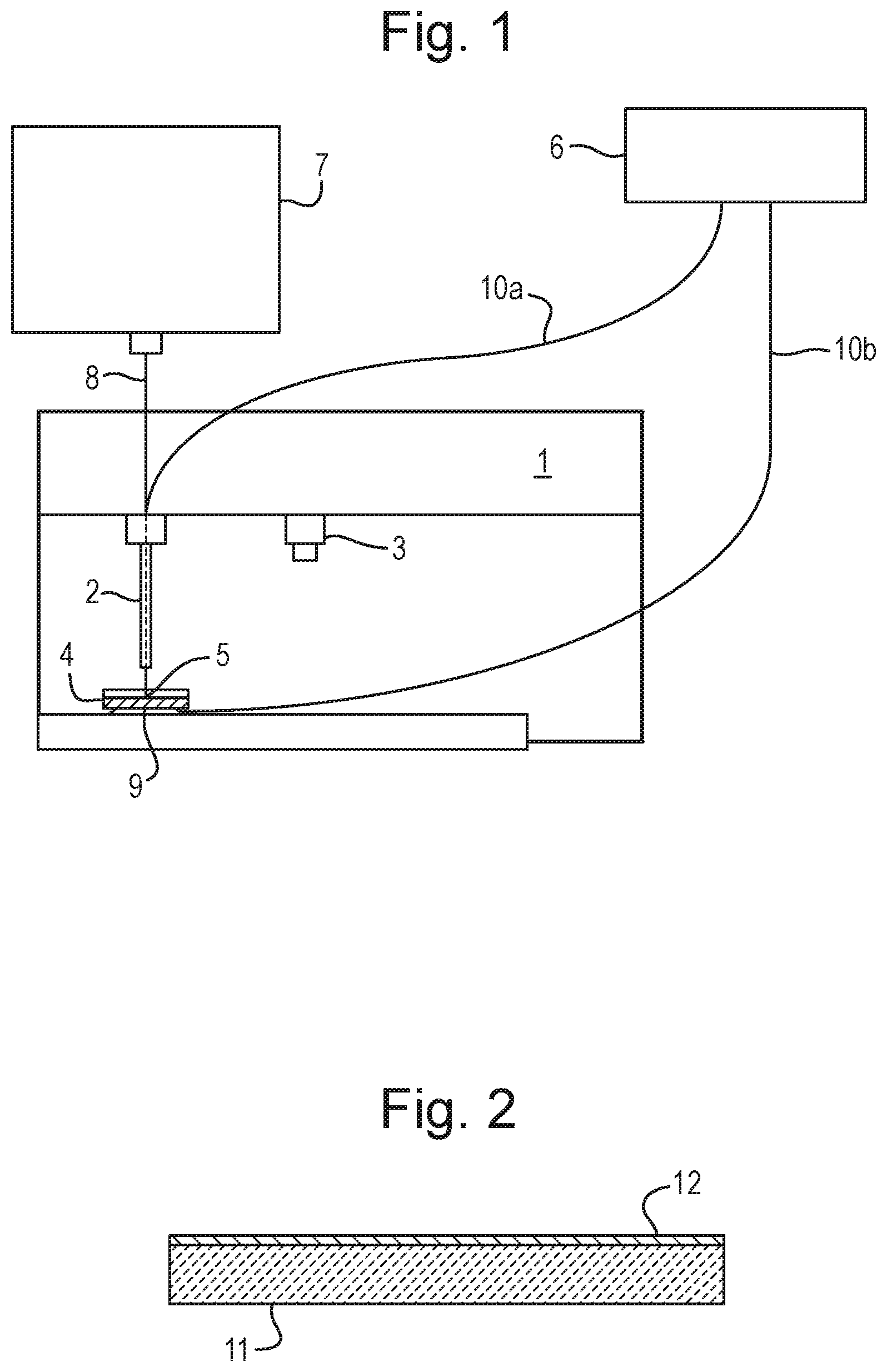
The first electrode may comprise a substrate which is substantially optically transparent or optically translucent.
The substrate may comprise glass, plastic, polycarbonate, poly(methyl methacrylate), Plexiglas.RTM. or quartz.
The substrate may further comprise a conductive layer or a conductive coating.
The conductive layer or the conductive coating may be substantially optically transparent or optically translucent.
The conductive layer or the conductive coating is selected from the group consisting of: (i) a conductive oxide layer or coating; (ii) indium-tin oxide; (iii) aluminium-doped zinc oxide ("AZO"); (iv) indium-doped cadmium oxide; (v) aluminium-doped zinc oxide ("AZO"); (vi) gallium-doped zinc oxide ("GZO"); (vii) indium-doped zinc oxide ("IZO"); (viii) a metallic layer; (ix) a carbon nanotube conductive coating; (x) a graphene film; (xi) one or more silver nanowires covered with graphene; (xii) a polymeric layer; (xiii) polyaniline; or (xiv) a poly(3,4-ethylenedioxythiophene):poly(styrenesulfonate) ("PEDOT:PSS") composite.
According to an aspect there is provided a method of rapid evaporation ionization mass spectrometry ("REIMS") comprising a method as described above.
According to an aspect there is provided a method of mass spectrometry comprising a method as described above.
According to an aspect there is provided analysis apparatus comprising:
a first electrode for contacting an insulating substrate upon which a sample is located in use;
a second electrode for contacting the sample; and
a device for applying an AC or RF voltage to the first and second electrodes in order to generate an aerosol from the sample.
The known arrangement disclosed in N. Strittmatter, M. Rebec, E. Jones, O. Golf, A. Abdolrasouli, J. Balog, V. Behrends, K. Veselkov, Z. Takats "Characterization and Identification of Clinically Relevant Microorganisms Using Rapid Evaporative Ionization Mass Spectrometry" Anal. Chem. 2014, 86, 6555-6562 does not disclose contacting an insulating substrate such as a petri dish with a first electrode and contacting the sample with a second electrode.
In particular, according to various embodiments the sample may be cultured on or within a culture medium wherein both the sample and culture medium are provided on the insulating substrate which may comprise a container, petri dish, a vial or microtitre or microwell plate. The microtitre or microwell plate may, for example, comprise 6, 24, 96, 384 or 1536 wells. A bottom surface of the container, petri dish, vial or microtitre plate may be brought into contact with the first electrode which may be positioned below the container, petri dish, vial or microtitre plate. The second electrode may be brought into contact with the sample and/or culture medium. It will be appreciated that the insulating substrate is located in between the first and second electrodes when the AC or RF voltage is applied to the first and second electrodes. As a result, electrical energy is predominantly capacitively coupled into the sample. The known arrangement referred to above does not disclose capacitively coupling electrical energy into a sample by bringing a first electrode into contact with the bottom surface of a container, petri dish, vial or petri dish containing the sample whilst a second electrode is brought into contact with the sample such that the housing or body of the insulating substrate (i.e. insulating container, petri dish, vial or microtitre plate) is intermediate or between the first and second electrodes when the AC or RF voltage is applied to the first and second electrodes.
According to an embodiment the insulating substrate may be located, in use, upon the first electrode and the insulating substrate (e.g. container, petri dish, vial or microtitre plate) may be optically transparent or optically translucent and the first electrode may also be substantially optically transparent or translucent. The apparatus may further comprise a device arranged and adapted to pass light or photons through the first (transparent) electrode and the (transparent) insulating substrate in order to illuminate, image or analyse the sample. The known arrangement does not disclose providing an optically transparent electrode or passing light through an optically transparent electrode in order to illuminate a sample.
The sample may comprise a biological, bacterial, fungal or yeast sample or a cell line which has been cultured on to or within a culture or growth medium. The culture or growth medium may comprise a solid or liquid culture or growth medium.
For example, the culture or growth medium may comprise an agar-based medium, a carbohydrate matrix or another solid growth medium.
Alternatively, the culture or growth medium may comprise a liquid medium, a cell growth medium such as but not limited to DME (Dulbecco's Modified Eagle's medium), a modified DME medium (e.g. glucose or glutamine free), RPMI (Roswell Park Memorial Institute medium), MEM (Minimum Essential Medium), IMDM (Iscove's Modified Dulbecco's Medium) or another liquid growth medium.
The sample may be spun down using a centrifuge to form a pellet and the supernate may be discarded or used for subsequent analysis.
The sample or pellet may be smeared onto a glass slide or other insulating surface, or may be anlaysed in situ.
The sample may be provided on or in a container, petri dish, vial or a microtitre or microwell plate. The microtitre or microwell plate may, for example, comprise 6, 24, 96, 384 or 1536 wells.
The sample and optionally a culture or growth medium may be provided on a first surface of the insulating substrate.
The first electrode may be arranged to contact a second surface of the insulating substrate which is opposed to the first surface.
According to an embodiment the first electrode does not contact the sample in use.
According to an embodiment the first electrode does not contact any culture or growth medium in use.
The device for applying an AC or RF voltage to the first and second electrodes may be arranged and adapted to predominantly capacitively couple electrical energy into the sample.
The transfer of electrical energy into the sample may cause the aerosol to be generated.
The apparatus may further comprise an ion source for ionising at least some of the aerosol so as to generate analyte ions.
The apparatus may further comprise a device for directing at least some of the aerosol into a vacuum chamber of a mass spectrometer.
The apparatus may further comprise an ion source located within a or the vacuum chamber of the mass spectrometer for ionising at least some the aerosol so as to generate a plurality of analyte ions.
The apparatus may further comprise a mass analyser for mass analysing the analyte ions.
The mass analyser may be further arranged and adapted to obtain mass spectral data corresponding to one or more locations on or in the sample.
The apparatus may further comprise a spectroscopic imaging or analysing device for spectroscopically imaging or analysing the sample.
The spectroscopic imaging or analysing device may be arranged and adapted to spectroscopically image or analyse the sample at substantially the same time as the mass analyser obtains mass spectral data corresponding to one or more locations on or in the sample.
The spectroscopic imaging or analysing device may comprise a Raman spectroscope and/or an infra-red ("IR") spectroscope.
The Raman spectroscope and/or the infra-red ("IR") spectroscope may be arranged and adapted: (i) to determine one or more physico-chemical properties of the sample; (ii) to determine one or more chemical properties of the sample; (iii) to determine one or more absorption properties of the sample; or (iv) to determine one or more vibrational and/or rotational modes or states of the sample.
The apparatus may further comprise a control system arranged and adapted to use the obtained mass spectral data to identify one or more biological substances, one or more bacterial strains, one or more fungal strains, one or more yeast strains or one or more cell lines located at one or more locations on or in the sample.
The apparatus may further comprise a device for optically or visually identifying one or more regions of interest on or in the sample.
The apparatus may further comprise a video camera or a digital camera for obtaining one or more images of the sample.
The apparatus may further comprise a processor for processing the one or more images of the sample in order to determine one or more regions of interest on or in the sample.
The first and second electrodes may comprise a rapid evaporation ionization mass spectrometry ("REIMS") device.
The rapid evaporation ionization mass spectrometry ("REIMS") device may comprise a monopolar device.
The AC or RF voltage device may be arranged and adapted to apply one or more pulses of the AC or RF voltage to the first and second electrodes.
The AC or RF voltage device may be arranged and adapted to apply the AC or RF voltage to the first and second electrodes in order to cause heat to be dissipated into the sample.
The sample may comprise a biological, bacterial, fungal or yeast sample or a cell line which has been cultured on to or within a solid or liquid culture or growth medium.
The culture or growth medium may comprise an agar-based medium, a carbohydrate matrix or another solid growth medium.
The apparatus may further comprise a device for determining a spatial distribution of one or more excreted substances emanating from one or more biological substances, one or more bacterial colonies, one or more fungal, one or more yeast colonies or one or more cell lines which have been cultured on or within the culture or growth medium.
The one or more excreted substances may be selected from the group consisting of: (i) one or more metabolites; (ii) one or more primary metabolites; (iii) one or more secondary metabolites; (iv) one or more lipopeptides; (v) surfactin; (vi) one or more quorum sensing molecules; (vii) 2-Heptyl-3-hydroxy-4(1H)-quinolone or 2-heptyl-3,4-dihydroxyquinoline ("PQS" or Pseudomonas quinolone signal); (viii) 4-hydroxy-2-heptylquinoline ("HHQ"); (ix) one or more antibiotics; (x) one or more alkaloids; (xi) one or more terpenoids; (xii) one or more glycosides; (xiii) one or more natural phenols; (xiv) one or more phenazines; (xv) one or more biphenyls and dibenzofurans; (xvi) one or more beta-lactams; (xvii) one or more polyketides; (xviii) one or more fatty acid synthase products; (xix) one or more nonribosomal peptides; (xx) one or more ribosomal peptides; and (xxi) one or more drugs or toxins.
The sample may be provided in a container, petri dish, vial or a microtitre or microwell plate. The microtitre or microwell plate may, for example, comprise 6, 24, 96, 384 or 1536 wells.
The first electrode may comprise a mesh electrode.
The first electrode may comprise a substrate which is substantially optically transparent or optically translucent.
The substrate may comprise glass, plastic, polycarbonate, poly(methyl methacrylate), Plexiglas.RTM. or quartz.
The substrate may further comprise a conductive layer or a conductive coating.
The conductive layer or the conductive coating may be substantially optically transparent or optically translucent.
The conductive layer or the conductive coating may be selected from the group consisting of: (i) a conductive oxide layer or coating; (ii) indium-tin oxide; (iii) aluminium-doped zinc oxide ("AZO"); (iv) indium-doped cadmium oxide; (v) aluminium-doped zinc oxide ("AZO"); (vi) gallium-doped zinc oxide ("GZO"); (vii) indium-doped zinc oxide ("IZO"); (viii) a metallic layer; (ix) a carbon nanotube conductive coating; (x) a graphene film; (xi) one or more silver nanowires covered with graphene; (xii) a polymeric layer; (xiii) polyaniline; or (xiv) a poly(3,4-ethylenedioxythiophene):poly(styrenesulfonate) ("PEDOT:PSS") composite.
According to an aspect there is provided a rapid evaporation ionization mass spectrometry ("REIMS") device comprising apparatus as described above.
According to an aspect there is provided a mass spectrometer comprising apparatus as described above.
According to an aspect there is provided a method of analysis comprising:
locating a sample on a first electrode;
passing light or photons through the first electrode in order to illuminate, image or analyse the sample;
contacting the sample with a second electrode; and
applying an AC or RF voltage to the first and second electrodes in order to generate an aerosol from the sample.
The arrangement disclosed in N. Strittmatter, M. Rebec, E. Jones, O. Golf, A. Abdolrasouli, J. Balog, V. Behrends, K. Veselkov, Z. Takats "Characterization and Identification of Clinically Relevant Microorganisms Using Rapid Evaporative Ionization Mass Spectrometry" Anal. Chem. 2014, 86, 6555-6562 does not disclose passing light or photons through an electrode in order to illuminate, image or analyse a sample.
According to various embodiments a transparent counter electrode may be used in conjunction with rapid evaporation ionisation mass spectrometry in order to facilitate backlighting of a sample.
The transparent counter electrode also facilitates simultaneous spectroscopic imaging of the sample. Various embodiments are contemplated wherein a sample may be subjected to simultaneous REIMS and Raman spectroscopy or to simultaneous REIMS and infra-red ("IR") spectroscopy.
Various embodiments relate to the field of microorganism identification including the identification of bacteria, yeast, fungi or cell lines using rapid evaporation ionisation mass spectrometry. Other applications are contemplated which relate to rapid evaporation ionisation mass spectrometry tissue imaging.
According to other embodiments automated rapid evaporation ionisation mass spectrometry analysis of bacteria, yeast, fungal colonies or cell lines grown on agar or in a liquid growth medium may be performed. The colonies grown on agar or other culture media may be contained in a standard petri dish. Of particular interest is the fact that the colonies can be analysed directly from the petri dish (or other sample plate) without requiring any substantive intervention from a user or operator and without requiring any prior sample preparation.
According to an embodiment a spatial distribution of one or more excreted substances emanating from one or more biological substances, one or more bacterial colonies, one or more fungal colonies, one or more yeast colonies or one or more cell lines which have been cultured on or within a culture or growth medium may be determined. The one or more excreted substances may be selected from the group consisting of: (i) one or more metabolites; (ii) one or more primary metabolites; (iii) one or more secondary metabolites; (iv) one or more lipopeptides; (v) surfactin; (vi) one or more quorum sensing molecules; (vii) 2-Heptyl-3-hydroxy-4(1H)-quinolone or 2-heptyl-3,4-dihydroxyquinoline ("PQS" or Pseudomonas quinolone signal); (viii) 4-hydroxy-2-heptylquinoline ("HHQ"); (ix) one or more antibiotics; (x) one or more alkaloids; (xi) one or more terpenoids; (xii) one or more glycosides; (xiii) one or more natural phenols; (xiv) one or more phenazines; (xv) one or more biphenyls and dibenzofurans; (xvi) one or more beta-lactams; (xvii) one or more polyketides; (xviii) one or more fatty acid synthase products; (xix) one or more nonribosomal peptides; (xx) one or more ribosomal peptides; and (xxi) one or more drugs or toxins. It is known to use genetic engineering or modification of microbes and bacteria to force the production of novel compounds. This technique may be used to monitor the production of the compound and may also be used to screen the micro-organisms for unwanted mutations.
It will be apparent, therefore, that the various embodiments have significant benefits compared with known method of REIMS analysis.
The method may further comprise ionising at least some of the aerosol so as to generate analyte ions.
The method may further comprise directing at least some of the aerosol into a vacuum chamber of a mass spectrometer.
The method may further comprise ionising at least some the aerosol within a or the vacuum chamber of the mass spectrometer so as to generate a plurality of analyte ions.
The method may further comprise mass analysing the analyte ions.
The method may further comprise obtaining mass spectral data corresponding to one or more locations on or in the sample.
The step of passing light or photons through the first electrode may further comprise spectroscopically imaging or analysing the sample.
The step of spectroscopically imaging or analysing the sample may further comprise spectroscopically imaging or analysing the sample at substantially the same time as obtaining mass spectral data corresponding to one or more locations on or in the sample.
The step of spectroscopically imaging or analysing the sample may comprise subjecting the sample to Raman spectroscopy and/or to infra-red ("IR") spectroscopy.
The step of subjecting the sample to Raman spectroscopy and/or to infra-red ("IR") spectroscopy may further comprise: (i) determining one or more physico-chemical properties of the sample; (ii) determining one or more chemical properties of the sample; (iii) determining one or more absorption properties of the sample; or (iv) determining one or more vibrational and/or rotational modes or states of the sample.
The light or the photons may either: (i) be in the visible spectrum or have a wavelength in the range 390-700 nm; (ii) be in the near infra-red or have a wavelength in the range 700-1400 nm; or (iii) be in the near ultra-violet or have a wavelength in the range 300-390 nm.
The method may further comprise using the obtained mass spectral data to identify one or more biological substances, one or more bacterial strains, one or more fungal strains, one or more yeast strains or one or more cell lines located at one or more locations on or in the sample.
The method may further comprise optically or visually identifying one or more regions of interest on or in the sample.
The step of optically or visually identifying one or more regions of interest on or in the sample may comprise using a video camera or a digital camera to obtain one or more images of the sample.
The method may further comprise processing the one or more images of the sample in order to determine one or more regions of interest on or in the sample.
The first and second electrodes may comprise a rapid evaporation ionization mass spectrometry ("REIMS") device.
The rapid evaporation ionization mass spectrometry ("REIMS") device may comprise a monopolar device.
The step of applying the AC or RF voltage to the first and second electrodes may further comprise applying one or more pulses of the AC or RF voltage to the first and second electrodes.
The step of applying the AC or RF voltage to the first and second electrodes may cause heat to be dissipated into the sample.
The sample may comprise a biological, bacterial, fungal or yeast sample or a cell line which has been cultured on to or within a solid or liquid culture or growth medium.
The culture or growth medium may comprise an agar-based medium, a carbohydrate matrix or another solid growth medium.
Alternatively, the culture or growth medium may comprise a liquid medium, a cell growth medium such as but not limited to DME (Dulbecco's Modified Eagle's medium), a modified DME medium (e.g. glucose or glutamine free), RPMI (Roswell Park Memorial Institute medium), MEM (Minimum Essential Medium), IMDM (Iscove's Modified Dulbecco's Medium) or another liquid growth medium.
The sample may be spun down using a centrifuge to form a pellet and the supernate may be discarded or used for subsequent analysis.
The sample or pellet may be smeared onto a glass slide or other insulating surface, or may be anlaysed in situ.
The method may further comprise determining a spatial distribution of one or more excreted substances emanating from one or more biological substances, one or more bacterial colonies, one or more fungal colonies or one or more yeast colonies which have been cultured on to or within the culture or growth medium.
The one or more excreted substances may be selected from the group consisting of: (i) one or more metabolites; (ii) one or more primary metabolites; (iii) one or more secondary metabolites; (iv) one or more lipopeptides; (v) surfactin; (vi) one or more quorum sensing molecules; (vii) 2-Heptyl-3-hydroxy-4(1H)-quinolone or 2-heptyl-3,4-dihydroxyquinoline ("PQS" or Pseudomonas quinolone signal); (viii) 4-hydroxy-2-heptylquinoline ("HHQ"); (ix) one or more antibiotics; (x) one or more alkaloids; (xi) one or more terpenoids; (xii) one or more glycosides; (xiii) one or more natural phenols; (xiv) one or more phenazines; (xv) one or more biphenyls and dibenzofurans; (xvi) one or more beta-lactams; (xvii) one or more polyketides; (xviii) one or more fatty acid synthase products; (xix) one or more nonribosomal peptides; (xx) one or more ribosomal peptides; and (xxi) one or more drugs or toxins.
The method may further comprise providing the sample in a container, petri dish or a microtitre or microwell plate. The microtitre or microwell plate may, for example, comprise 6, 24, 96, 384 or 1536 wells.
The first electrode may comprise a mesh electrode.
The first electrode may comprise a substrate which is substantially optically transparent or optically translucent.
The substrate may comprise glass, plastic, polycarbonate, poly(methyl methacrylate), Plexiglas.RTM. or quartz.
The substrate may further comprise a conductive layer or a conductive coating.
The conductive layer or the conductive coating may be substantially optically transparent or optically translucent.
The conductive layer or the conductive coating may be selected from the group consisting of: (i) a conductive oxide layer or coating; (ii) indium-tin oxide; (iii) aluminium-doped zinc oxide ("AZO"); (iv) indium-doped cadmium oxide; (v) aluminium-doped zinc oxide ("AZO"); (vi) gallium-doped zinc oxide ("GZO"); (vii) indium-doped zinc oxide ("IZO"); (viii) a metallic layer; (ix) a carbon nanotube conductive coating; (x) a graphene film; (xi) one or more silver nanowires covered with graphene; (xii) a polymeric layer; (xiii) polyaniline; or (xiv) a poly(3,4-ethylenedioxythiophene):poly(styrenesulfonate) ("PEDOT:PSS") composite.
According to an aspect there is provided a method of rapid evaporation ionization mass spectrometry ("REIMS") comprising a method as described above.
According to an aspect there is provided a method of mass spectrometry comprising a method as described above.
According to an aspect there is provided analysis apparatus comprising:
a first electrode;
a first device for passing light or photons through the first electrode in order to illuminate, image or analyse, in use, a sample located on the first electrode;
a second electrode for contacting the sample; and
a second device for applying an AC or RF voltage to the first and second electrodes in order to generate an aerosol from the sample.
The arrangement disclosed in N. Strittmatter, M. Rebec, E. Jones, O. Golf, A. Abdolrasouli, J. Balog, V. Behrends, K. Veselkov, Z. Takats "Characterization and Identification of Clinically Relevant Microorganisms Using Rapid Evaporative Ionization Mass Spectrometry" Anal. Chem. 2014, 86, 6555-6562 does not disclose passing light or photons through an electrode in order to illuminate, image or analyse a sample.
The apparatus may further comprise an ion source for ionising at least some of the aerosol so as to generate analyte ions.
The apparatus may further comprise a device for directing at least some of the aerosol into a vacuum chamber of a mass spectrometer.
The apparatus may further comprise an ion source located within a or the vacuum chamber of the mass spectrometer for ionising at least some the aerosol so as to generate a plurality of analyte ions.
The apparatus may further comprise a mass analyser for mass analysing the analyte ions.
The mass analyser may be arranged and adapted to obtain mass spectral data corresponding to one or more locations on or in the sample.
The first device may comprise a spectroscopic imaging or analysing device for spectroscopically imaging or analysing the sample.
The spectroscopic imaging or analysing device may be arranged and adapted to spectroscopically image or analyse the sample at substantially the same time as the mass analyser obtains mass spectral data corresponding to one or more locations on or in the sample.
The spectroscopic imaging or analysing device may comprise a Raman spectroscope and/or an infra-red ("IR") spectroscope.
The Raman spectroscope and/or the infra-red ("IR") spectroscope may be arranged and adapted: (i) to determine one or more physico-chemical properties of the sample; (ii) to determine one or more chemical properties of the sample; (iii) to determine one or more absorption properties of the sample; or (iv) to determine one or more vibrational and/or rotational modes or states of the sample.
According to an embodiment the light or the photons may either: (i) be in the visible spectrum or have a wavelength in the range 390-700 nm; (ii) be in the near infra-red or have a wavelength in the range 700-1400 nm; or (iii) be in the near ultra-violet or have a wavelength in the range 300-390 nm.
The apparatus may further comprise a control system which is arranged and adapted to use the obtained mass spectral data to identify one or more biological substances, one or more bacterial strains, one or more fungal strains, one or more yeast strains or one or more cell lines located at one or more locations on or in the sample.
The apparatus may further comprise a control system which is arranged and adapted to optically or visually identify one or more regions of interest on or in the sample.
The apparatus may further comprise a video camera or a digital camera for obtaining one or more images of the sample.
The apparatus may further comprise a processor for processing the one or more images of the sample in order to determine one or more regions of interest on or in the sample.
The first and second electrodes may comprise a rapid evaporation ionization mass spectrometry ("REIMS") device.
The rapid evaporation ionization mass spectrometry ("REIMS") device may comprise a monopolar device.
The device for applying the AC or RF voltage to the first and second electrodes may be arranged and adapted to apply one or more pulses of the AC or RF voltage to the first and second electrodes.
The device for applying the AC or RF voltage to the first and second electrodes may be arranged and adapted to cause heat to be dissipated into the sample.
The sample may comprise a biological, bacterial, fungal or yeast sample or a cell line which has been cultured on to or within a solid or liquid culture or growth medium.
The culture or growth medium may comprise an agar-based medium, a carbohydrate matrix or another solid growth medium.
Alternatively, the culture or growth medium may comprise a liquid medium, a cell growth medium such as but not limited to DME (Dulbecco's Modified Eagle's medium), a modified DME medium (e.g. glucose or glutamine free), RPMI (Roswell Park Memorial Institute medium), MEM (Minimum Essential Medium), IMDM (Iscove's Modified Dulbecco's Medium) or another liquid growth medium.
The sample may be spun down using a centrifuge to form a pellet and the supernate may be discarded or used for subsequent analysis.
The sample or pellet may be smeared onto a glass slide or other insulating surface, or may be anlaysed in situ.
The apparatus may further comprise a device for determining a spatial distribution of one or more excreted substances emanating from one or more biological substances, one or more bacterial colonies, one or more fungal colonies, one or more yeast colonies or one or more cell lines which have been cultured on or within the culture or growth medium.
The one or more excreted substances may be selected from the group consisting of: (i) one or more metabolites; (ii) one or more primary metabolites; (iii) one or more secondary metabolites; (iv) one or more lipopeptides; (v) surfactin; (vi) one or more quorum sensing molecules; (vii) 2-Heptyl-3-hydroxy-4(1H)-quinolone or 2-heptyl-3,4-dihydroxyquinoline ("PQS" or Pseudomonas quinolone signal); (viii) 4-hydroxy-2-heptylquinoline ("HHQ"); (ix) one or more antibiotics; (x) one or more alkaloids; (xi) one or more terpenoids; (xii) one or more glycosides; (xiii) one or more natural phenols; (xiv) one or more phenazines; (xv) one or more biphenyls and dibenzofurans; (xvi) one or more beta-lactams; (xvii) one or more polyketides; (xviii) one or more fatty acid synthase products; (xix) one or more nonribosomal peptides; (xx) one or more ribosomal peptides; and (xxi) one or more drugs or toxins.
The sample may be provided in a container, petri dish or or a microtitre or microwell plate. The microtitre or microwell plate may, for example, comprise 6, 24, 96, 384 or 1536 wells.
The first electrode may comprise a mesh electrode.
The first electrode may comprise a substrate which is substantially optically transparent or optically translucent.
The substrate may comprise glass, plastic, polycarbonate, poly(methyl methacrylate), Plexiglas.RTM. or quartz.
The substrate may further comprise a conductive layer or a conductive coating.
The conductive layer or the conductive coating may be substantially optically transparent or optically translucent.
The conductive layer or the conductive coating may be selected from the group consisting of: (i) a conductive oxide layer or coating; (ii) indium-tin oxide; (iii) aluminium-doped zinc oxide ("AZO"); (iv) indium-doped cadmium oxide; (v) aluminium-doped zinc oxide ("AZO"); (vi) gallium-doped zinc oxide ("GZO"); (vii) indium-doped zinc oxide ("IZO"); (viii) a metallic layer; (ix) a carbon nanotube conductive coating; (x) a graphene film; (xi) one or more silver nanowires covered with graphene; (xii) a polymeric layer; (xiii) polyaniline; or (xiv) a poly(3,4-ethylenedioxythiophene):poly(styrenesulfonate) ("PEDOT:PSS") composite.
According to an aspect there is provided a rapid evaporation ionization mass spectrometry ("REIMS") device comprising apparatus as described above.
According to an aspect there is provided a mass spectrometer comprising apparatus as described above.
BRIEF DESCRIPTION OF THE DRAWINGS
Various embodiments will now be described, by way of example only, and with reference to the accompanying drawings in which:
FIG. 1 shows a schematic of an automated microbial sampling system according to an embodiment and shows a REIMS sampling probe located above a sample which is provided in a petri dish;
FIG. 2 shows a schematic of an embodiment wherein a counter electrode is provided comprising a transparent conductive indium-tin oxide layer which is deposited upon a glass support plate;
FIG. 3 shows in greater detail a circular glass support plate which may be coated with a transparent conductive layer in order to form a transparent counter electrode according to an embodiment;
FIG. 4 shows a glass support plate located in a metal holder according to an embodiment;
FIG. 5 shows a petri dish resting on a transparent counter electrode assembly according to an embodiment;
FIG. 6 shows a transparent counter electrode according to an embodiment located below a petri dish which contains a culture or growth medium and a microorganism colony which has been cultured on the culture or growth medium together with a simplified equivalent electrical circuit, wherein the colony may be illuminated from below the petri dish in order to aid microorganism identification and wherein a REIMS probe may be used to sample and analyse the microorganism colony;
FIG. 7 shows a total ion current ("TIC") obtained by analysing a sample according to an embodiment; and
FIG. 8 shows three mass spectra which were obtained by analysing samples using the disclosed REIMS apparatus according to various embodiments and illustrates the ability of the disclosed REIMS apparatus to be able to differentiate between different types of colonies.
DETAILED DESCRIPTION
Various embodiments will now be described in the context of seeking to aid high throughput microbe identification. However, it should be understood that the various embodiments described herewith are merely illustrative and that the present invention is not intended to be limited to microbe identification.
According to an arrangement it is contemplated that a two-electrode (bipolar) sampling probe might be used to analyse a sample. However, using a two-electrode sampling probe is potentially problematic since a two-electrode sampling probe has an increased physical size at the point of contact with the sample. It is also necessary to avoid electrical breakdown between the two electrodes forming the probe and to take steps to avoid potential problems due to cross contamination effects.
According to other arrangements a single analysis probe (e.g. monopolar electrode) may be used to vaporize or ablate a colony and an additional probe (or counter electrode) may be used to make direct electrical and physical contact with the bulk media or agar. Although this approach has been demonstrated to work (to some extent) it suffers from the problem that it requires an additional probe (or counter electrode) to be provided which needs to contact the bulk media or agar. Furthermore, this approach suffers from the problem of having a restricted available analysis area as the heating effect scales quadratically with the current density. It has been recognised that ideally the sampling probe area should be relatively small whereas the additional probe (or counter electrode) should be relatively large so as to avoid heating at the additional probe (or counter electrode).
Further embodiments which are of particular interest will now be described in more detail below. These embodiments are of particular interest since they eliminate any need for an additional probe (or counter electrode) to be placed in direct physical contact with the bulk material e.g. agar or other culture or growth medium thereby avoiding the problem of cross contamination.
The various embodiments which will now be described in more detail below are of particular interest since they enable a precise region of a colony to be analysed without suffering from heating effects at the counter electrode. Furthermore, these embodiments do not suffer from potential cross contamination effects. A yet further beneficial effect is that the various embodiments described below significantly improve the ability to illuminate a sample in order to identify one or more regions of interest to be analysed.
According to various embodiments a sample to be analysed (such as a microbe colony) may be located or provided on a culture or growth medium such as agar. The sample and the culture or growth medium on which it may have been grown may be located or provided in a petri dish, vial or other container such as a microtitre plate or microwell plate. The microtitre or microwell plate may, for example, comprise 6, 24, 96, 384 or 1536 wells. As will become more apparent below, the sample which has been grown on the culture or growth medium in the petri dish can be analysed in situ in the petri dish. This is of particular interest since conventional approaches require a sample portion to be scraped off the surface of the agar or other culture or growth medium and to be analysed whilst being held by a pair of electrosurgical forceps.
The petri dish or other container in which the sample and culture or growth medium may be provided may be made from glass or plastic. As a result, the petri dish or other container may be essentially electrically non-conductive or may comprise an electrical insulator.
According to various embodiments a first or counter electrode may be located underneath (or otherwise in close proximity to) the lower or bottom surface of a petri dish or other container so that the petri dish or other container (which is an insulator) sits or otherwise rests upon the first or counter electrode.
According to various embodiments the first or counter electrode may be arranged to have a similar surface area to that of the petri dish or other container which contains the sample to be analysed.
According to various embodiments a rapid evaporation ionisation mass spectrometry ("REIMS") sampling system may be provided which comprises a first (counter) electrode which is placed below or underneath an insulating petri dish (or other container) and a second (sample probe) electrode which is brought into direct physical contact with the sample to be analysed in the petri dish (or other container).
According to an embodiment a REIMS sample probe electrode may be brought into contact (from above) with a microbe colony to be analysed and identified whilst a first or counter electrode is located below the petri dish or other container containing the sample to be analysed.
An AC or RF voltage may be applied between the REIMS sample probe electrode (which is direct contact with the sample to be analysed) and the REIMS counter electrode (which is in contact with the rear or lower surface of the petri dish or other container containing the sample and optional culture or growth medium). It will be apparent, therefore, that the REIMS counter electrode is not brought into direct physical with the sample to be analysed. This enables the REIMS counter electrode to be reused without suffering from any cross contamination problems.
According to various embodiments the application of the AC or RF voltage to the first (counter) electrode and the second (sample probe) electrode causes a current to flow but it will be apparent that since the REIMS counter electrode is in electrical or direct physical contact with an insulator (i.e. the bottom of a petri dish) then the petri dish and the contents of the petri dish (i.e. the culture or growth medium) effectively form a dielectric of a capacitor. Therefore, as a result, electrical energy is predominantly capacitively coupled into the sample.
It will be understood that this approach is therefore quite different from the known approach wherein bipolar electrosurgical forceps are used to remove a sample from a culture or growth medium and wherein both electrodes are brought into direct physical and electrical contact with the sample.
In contrast to the known approach, the first (counter) electrode does not make direct physical contact with the sample to be analysed and energy is capacitively coupled into the sample in order to rapidly heat and vaporise the sample thereby generating an aerosol to be analysed.
The frequency of the applied AC or RF voltage may be maximized so as to ensure that the electrical impedance of the bulk material is essentially minimised thereby resulting in maximum energy dissipation into the sample which is desired to be analysed. As a result, AC or RF energy is capacitively coupled into the microbe colony with the result that a portion of the microbe colony in the immediate vicinity of the REIMS probe electrode is vapourised so as to form an aerosol.
A particular embodiment will now be described in more detail with reference to FIG. 1. FIG. 1 shows an embodiment comprising a commercial robotic system 1 such as, for example, a Tecan EVO.RTM. robotic system together with a Picolo.RTM. colony identification system. The commercial robotic system 1 has been modified so as to include a REIMS sampling probe electrode 2 and a REIMS counter electrode 9. The REIMS sampling probe electrode 2 and the REIMS counter electrode 9 are connected to an electrosurgical RF generator 6 via a feed electrode 10a and a counter or return electrode 10b respectively.
Colonies grown on a culture or growth medium (such as agar) in a petri dish 4 or other container may be identified using a video camera system 3 which may, for example, be attached to a robotic arm of the robotic system 1. Images of any colonies which have grown on the culture or growth medium may then be processed by digital image processing software. According to an embodiment Picolo.RTM. digital imaging processing software may be used to process images of any colonies which have grown on the culture or growth medium.
The robotic arm of the robotic system 1 may be arranged to position the video camera system 3 above the petri dish 4 so that one or more digital pictures of the colony may be recorded. The digital image processing software may then analyse the picture(s) and identify the colonies. Colonies may be selected according to various predetermined criteria or may be manually selected from a video picture.
The REIMS sampling probe (or head) 2 may be moved directly above selected colonies and the position of the surface of a colony relative to e.g. the tip 5 of the REIMS sampling probe 2 may be determined using a built-in capacitive probe. According to various embodiments the tip 5 of the REIMS sampling probe 2 may be moved so that the tip 5 of the sampling probe 2 just contacts and makes physical contact with a selected colony. REIMS sampling may then be performed by briefly energizing the RF generator 6.
One pole of the RF generator 6 may be connected to the tip 5 of the REIMS sampling probe 2 via the feed electrode cable 10a and the other pole of the RF generator 6 may be connected to the counter electrode 9 (located in use underneath the petri dish 4 or other container) by the counter electrode cable 10b.
A RF voltage may be applied to a selected colony by, for example, applying a pulse of a RF voltage via the cables 10a,10b to the tip 5 of the REIMS sampling probe 2 and also to the REIMS counter electrode 9 located underneath the petri dish 4 or other container. As a result of capacitively coupling electrical energy into the sample, surgical fumes or an aerosol of sample material may be generated. The surgical fumes or aerosol of sample material may then be passed via a tube 8 or other conduit (which may be attached to the REIMS sampling probe 2) directly into the housing of a mass spectrometer 7. According to an embodiment the surgical fumes or aerosol may be ionised within a vacuum chamber of the mass spectrometer 7 by an ion source which may be located within the vacuum chamber of the mass spectrometer 7.
In order for the image recognition software to be able to identify a colony as effectively as possible further embodiments which will be discussed in more detail below enable the petri dish 4 or other container to be illuminated from below the petri dish 4 or other container. Illumination of sample in a petri dish 4 or other container from below is beneficial since this enables clear colony boundaries to be observed by, for example, the camera system 3. Furthermore, illuminating the petri dish 4 or other container from below removes or substantially removes reflections (which would otherwise be observed) and also reduces the complexity of the overall optical system including the video camera system 3.
According to various embodiments which are of particular interest and which will be described in more detail below, the counter electrode 9 may comprise a transparent substrate (e.g. glass or plastic) having a transparent conductive coating on e.g. an upper surface so as to form a transparent electrode on the transparent substrate.
Other embodiments are also contemplated wherein the counter electrode 4 may instead comprise an electrically conductive mesh. Such an approach has been demonstrated to work in principle although the mesh electrode can generate shadows which can complicate the automated identification of colonies. For this reason a transparent electrode having a transparent conductive coating or layer is particularly of interest.
According to embodiments which are of particular interest a transparent (or translucent) support plate or substrate may be provided which may be coated with a transparent (or translucent) conductive layer. The support plate or substrate may comprise a transparent or translucent glass plate which may coated with a transparent or translucent conductive layer on an upper surface of the glass support plate. It will be apparent, therefore, that the transparent or translucent conductive coating or layer will then be direct contact with the bottom of a petri dish 4 or other container. According to various embodiments the support plate or substrate may, for example, be coated with an indium-tin oxide ("ITO") layer. Coating the support plate or substrate with an indium-tin oxide layer is beneficial since the indium-tin oxide layer will have a significant conductivity and will also have sufficient mechanical strength for this application.
FIG. 2 shows a glass support plate 11 according to an embodiment which was fabricated. The glass support plate 11 was of optical grade and the top and bottom surfaces of the glass support plate 11 were polished. An indium-tin oxide layer 12 was then deposited onto the glass support plate 11 using a Physical Vapor Deposition ("PVD") process so that a layer of optically transparent indium-tin oxide having a thickness of about 1-2 .mu.m was deposited onto the upper surface of the glass support plate 11.
The indium-tin oxide layer 12 is optically transparent and has sufficient transmission so as to enable bacterial colonies which may be present on a culture or growth medium such as agar located within a petri dish 4 or other container to be illuminated. The petri dish 4 may be placed in use on top of the glass support plate 11. The petri dish 4 or other container may be illuminated from below so that light passes in turn through the support plate 11, through the transparent layer 12 and then passes through the petri dish 4 in order to illuminate the culture or growth medium and any colonies growing on the culture or growth medium. Illuminating the petri dish 4 from below is particularly beneficial as it allows colony boundaries to be more easily determined and thus identified.
FIG. 3 shows a glass support plate 11 according to an embodiment wherein the glass support plate 11 is arranged to have a 45.degree. angled or bevelled edge. The angled or bevelled edge facilitates electrical contact at the side with the upper transparent conductive electrode 12 such as a layer of indium-tin oxide 12.
As shown in FIG. 4, a metal plate 13 may be placed onto the glass support plate 11 and may be secured to a light box using springs. The edge of the opening in the metal plate 13 may be arranged so as to connect to the angled edge of the glass support plate 11 thereby ensuring that a reliable electrical contact is made between the metal plate 13 and the conductive electrode layer 12 on the upper surface of the glass support plate 11. The metal plate 13 also enables the glass support plate 11 to be precisely located or positioned.
FIG. 5 shows an embodiment wherein a petri dish 4 or other container is located on a transparent conductive counter electrode comprising a glass support plate 11 having an upper conductive electrode layer 12. The petri dish 4 or other container may be fabricated from glass or plastic and hence is an insulator.
After one or more colonies which have grown on the culture or growth medium in the petri dish 4 or other container have been optically identified by, for example, the camera system 3 then the sampling head or REIMS probe 2 may then be moved so that the tip 5 of the REIMS sampling probe 2 comes into direct contact with the colony mass with the height being determined by, for example, using a capacitive liquid level sensor located in the robotic arm of the robotic system 1.
According to various embodiments the RF generator 6 may be energised for a short period of time (e.g. a pulse of 1 s) with the result that electrical energy is capacitively coupled into the sample in close proximity to tip 5 of the REIMS sampling probe 2. The result of the applied RF voltage is a rapid evaporation of the sample to form an aerosol which may then be aspirated into a vacuum chamber of the mass spectrometer 7 via a tube 8. The aerosol comprises analyte which may be ionised by, for example, an ion source located within a vacuum chamber of the mass spectrometer 7 with the result that analyte ions are then mass analysed by the mass spectrometer 7.
FIG. 6 shows an embodiment showing a glass support plate 11 having an upper conductive layer 12. The glass support plate 11 and the conductive layer 12 may be both transparent (or at least translucent) and enable light from a light source (not shown) to pass through the glass support plate 11, the conductive layer 12 and the base of a petri dish 4 or other container located on the upper surface of the glass support plate 11 (and hence in direct contact with the conductive layer 12).
A REIMS sampling probe 2 comprising a tubular sampling head with a sampling tip 5 is shown. The tip 5 of the REIMS sampling probe 2 is in direct contact with a colony 14 on the surface of a culture or growth medium located in the petri dish 4 or other container. The REIMS sampling probe 2 and the upper conductive layer 12 of the glass support plate 11 are shown to be in electrical contact with an RF electrosurgical generator 6.
FIG. 6 also shows a simplified equivalent electrical circuit of the overall system. Current is arranged to pass through resistive layers (e.g. the colony and the agar culture or growth medium) and also through capacitive layers (e.g. the interface between the agar and the petri dish 4 and the interface between the petri dish 4 and the indium-tin oxide or other conductive layer 12). The result of capacitively coupling electrical energy into the sample is the generation of an aerosol comprising analytes which are subsequently mass analysed by a mass spectrometer 7.
FIG. 7 shows a sample total ion current TIC which was obtained when a yeast colony was mass analysed according to an embodiment as described above wherein a REIMS sampling probe 2 was used to sample a yeast colony on a culture or growth medium. A high signal intensity was obtained and the signal was obtained for the duration of the applied RF voltage pulse.
FIG. 8 shows three mass spectra relating to skin samples, yeast samples and normaflore samples wherein the samples were provided on a culture or growth medium and were obtained using a REIMS sampling probe 2 as discussed above. It is apparent from FIG. 8 that a high signal to noise ("S/N") ratio was obtained and that the different mass spectra enabled different colonies to be identified. The mass spectral peak at mass to charge ratio of 693.43 corresponds to a lipid which is common to all three samples. The different mass spectral peaks correspond predominantly to the colony being analysed with the agar culture or growth medium having an insignificant impact upon the resulting mass spectra.
It is apparent, therefore, that the REIMS sampling probe 2 according to various embodiments is able to identify various different biological substances on an agar substrate without needing to scrap sample from the agar substrate.
Various further embodiments are also contemplated. According to an embodiment the method may be utilised with micro-REIMS imaging experiments wherein, for example, a sample may be microtome sectioned and then a thin slice may be mounted on to a glass slide.
The method may also be used with simultaneous spectroscopy and rapid evaporation ionisation mass spectrometry imaging.
According to other embodiments the support plate 11 may be fabricated from materials other than glass such as a plastic such as polycarbonate, poly(methyl)methacrylate or Plexiglas.RTM.. Alternatively, the support plate 11 may be fabricated from quartz or another transparent insulator material.
It will also be understood that it is not essential that the support plate 11 is totally optically transparent. For example, the support plate 11 may be fabricated from an opaque, translucent or semi-translucent material.
It is also not essential that the conductive layer 12 provided on the support plate 11 comprises indium-tin oxide. For example, other embodiments are contemplated wherein the conductive layer 12 may comprise other transparent conductive oxides such as aluminum-doped zinc oxide ("AZO"), indium-doped cadmium oxide, aluminum-doped zinc oxide ("AZO"), gallium-doped zinc oxide ("GZO") or indium-doped zinc oxide ("IZO").
According to other embodiments the conductive layer 12 on the surface of the support plate 11 may comprise a thin translucent or transparent metallic layer.
Alternatively, the translucent or transparent layer 12 on the surface of the support plate 11 may comprise a carbon nanotube conductive coating, a graphene film or silver nanowires covered with graphene.
The transparent or translucent layer 12 on the surface of the support plate 11 may alternatively comprise a conductive polymeric layer such as polyaniline or poly(3,4-ethylenedioxythiophene):poly(styrenesulfonate) ("PEDOT:PSS") composite.
It is also contemplated that the counter electrode 9,12 may comprise a conductive plastic foil. The transparent supporting plate 11 and the conductive plastic foil may be assembled using a fixture that holds them together.
Other embodiments are contemplated wherein the culture or growth medium may comprise a liquid medium, a cell growth medium such as but not limited to DME (Dulbecco's Modified Eagle's medium), a modified DME medium (e.g. glucose or glutamine free), RPMI (Roswell Park Memorial Institute medium), MEM (Minimum Essential Medium), IMDM (Iscove's Modified Dulbecco's Medium) or another liquid growth medium.
The sample may be spun down using a centrifuge to form a pellet and the supernate may be discarded or used for subsequent analysis.
The sample or pellet may be smeared onto a glass slide or other insulating surface, or may be anlaysed in situ.
Although the present invention has been described with reference to preferred embodiments, it will be understood by those skilled in the art that various changes in form and detail may be made without departing from the scope of the invention as set forth in the accompanying claims.
* * * * *
References
D00000
D00001
D00002
D00003
D00004
D00005
XML
uspto.report is an independent third-party trademark research tool that is not affiliated, endorsed, or sponsored by the United States Patent and Trademark Office (USPTO) or any other governmental organization. The information provided by uspto.report is based on publicly available data at the time of writing and is intended for informational purposes only.
While we strive to provide accurate and up-to-date information, we do not guarantee the accuracy, completeness, reliability, or suitability of the information displayed on this site. The use of this site is at your own risk. Any reliance you place on such information is therefore strictly at your own risk.
All official trademark data, including owner information, should be verified by visiting the official USPTO website at www.uspto.gov. This site is not intended to replace professional legal advice and should not be used as a substitute for consulting with a legal professional who is knowledgeable about trademark law.