Filamin A binding proteins and uses thereof
Wu , et al. Sep
U.S. patent number 10,759,850 [Application Number 15/801,093] was granted by the patent office on 2020-09-01 for filamin a binding proteins and uses thereof. This patent grant is currently assigned to Berg LLC. The grantee listed for this patent is Berg LLC. Invention is credited to Viatcheslav R. Akmaev, Tracey Friss, Shobha Ravipaty, Nikunj Narendra Tanna, Wenfang Sybil Wu.


| United States Patent | 10,759,850 |
| Wu , et al. | September 1, 2020 |
Filamin A binding proteins and uses thereof
Abstract
The present invention encompasses filamin A (FLNA) binding proteins. Specifically, the invention relates to antibodies to FLNA. An antibody of the invention can be a full-length antibody or an antigen-binding portion thereof. Methods of making and methods of using the antibodies of the invention in methods of diagnosis, monitoring and prognosis of prostate cancer are also provided.
| Inventors: | Wu; Wenfang Sybil (Wayland, MA), Ravipaty; Shobha (Framingham, MA), Friss; Tracey (Holliston, MA), Akmaev; Viatcheslav R. (Sudbury, MA), Tanna; Nikunj Narendra (Boston, MA) | ||||||||||
|---|---|---|---|---|---|---|---|---|---|---|---|
| Applicant: |
|
||||||||||
| Assignee: | Berg LLC (Framingham,
MA) |
||||||||||
| Family ID: | 62077155 | ||||||||||
| Appl. No.: | 15/801,093 | ||||||||||
| Filed: | November 1, 2017 |
Prior Publication Data
| Document Identifier | Publication Date | |
|---|---|---|
| US 20180171002 A1 | Jun 21, 2018 | |
Related U.S. Patent Documents
| Application Number | Filing Date | Patent Number | Issue Date | ||
|---|---|---|---|---|---|
| 62415893 | Nov 1, 2016 | ||||
| Current U.S. Class: | 1/1 |
| Current CPC Class: | C07K 16/3069 (20130101); C07K 16/18 (20130101); A01K 67/0275 (20130101); G01N 3/50 (20130101); G01N 33/57434 (20130101); C07K 2317/56 (20130101); G01N 2333/4742 (20130101); G01N 2333/96455 (20130101); C07K 2317/21 (20130101); G01N 2333/47 (20130101); C07K 2317/565 (20130101); C07K 2317/92 (20130101); A01K 2227/105 (20130101); A01K 2217/05 (20130101) |
| Current International Class: | C07K 16/18 (20060101); G01N 3/50 (20060101); A01K 67/027 (20060101); A61K 39/395 (20060101); G01N 33/574 (20060101); C07K 16/30 (20060101) |
References Cited [Referenced By]
U.S. Patent Documents
| 2005/0153311 | July 2005 | Nelson et al. |
| 2012/0171782 | July 2012 | Nelson et al. |
| 2012/0294861 | November 2012 | Sonoda et al. |
| 2014/0178391 | June 2014 | Domon et al. |
| 2016/0258958 | September 2016 | Narain et al. |
Other References
|
Paul, Fundamental Immunology, 3rd Edition, 1993, pp. 292-295. cited by examiner . Rudikoff et al., Proc. Natl. Acad. Sci. USA, 1982, 79(6):1979-1983. cited by examiner . Colman, Research in Immunology, 1994, 145:33-36. cited by examiner . Bendig, Methods: A Companion to Methods in Enzymology, 1995; 8:83-93. cited by examiner . Khantasup et al., Monoclonal Antibodies in Immunodiagnosis and Immunotherapy, 2015, 34(6): 404-417. cited by examiner . Bedolla et al., Nuclear versus cytoplasmic localization of filamin A in prostate cancer: immunohistochemical correlation with metastases. Clin Cancer Res. Feb. 1, 2009;15(3):788-96. cited by applicant . Krastins et al., Rapid development of sensitive, high-throughput, quantitative and highly selective mass spectrometric targeted immunoassays for clinically important proteins in human plasma and serum. Clin Biochem. Apr. 2013;46(6):399-410. cited by applicant . Lin et al., Comparison of protein immunoprecipitation-multiple reaction monitoring with ELISA for assay of biomarker candidates in plasma. J Proteome Res. Dec. 6, 2013;12(12):5996-6003. cited by applicant . Ravipaty et al., Clinical Validation of a Serum Protein Panel (FLNA, FLNB and KRT19) for Diagnosis of Prostate Cancer. Journal of Molecular Biomarkers & Diagnosis. 2017;8(2):1-8. cited by applicant . Van Der Flier et al., Structural and functional aspects of filamins. Biochim Biophys Acta. Apr. 23, 2001;1538(2-3):99-117. cited by applicant . International Search Report and Written Opinion for Application No. PCT/US2017/059573, dated Feb. 26, 2018. 19 pages. cited by applicant. |
Primary Examiner: Sang; Hong
Attorney, Agent or Firm: McCarter & English, LLP Mello; Jill
Parent Case Text
RELATED APPLICATION
This application claims the benefit of priority to U.S. provisional application No. 62/415,893, filed on Nov. 1, 2016. The entire contents of the foregoing application are incorporated herein by reference.
Claims
We claim:
1. An antibody, or antigen-binding fragment thereof, capable of binding filamin A (FLNA), comprising a heavy chain variable region comprising a CDR3 domain comprising the amino acid sequence set forth in SEQ ID NO: 9, a CDR2 domain comprising the amino acid sequence set forth in SEQ ID NO: 8, and a CDR1 domain comprising the amino acid sequence set forth in SEQ ID NO: 7; and a light chain variable region comprising a CDR3 domain comprising the amino acid sequence set forth in SEQ ID NO: 12, a CDR2 domain comprising the amino acid sequence set forth in SEQ ID NO: 11, and a CDR1 domain comprising the amino acid sequence set forth in SEQ ID NO: 10.
2. The antibody, or antigen-binding fragment thereof, of claim 1, comprising a heavy chain variable region comprising the amino acid sequence set forth in SEQ ID NO: 1 and a light chain variable region comprising the amino acid sequence set forth in SEQ ID NO: 2.
3. The antibody, or antigen-binding fragment thereof, of claim 1 comprising a heavy chain comprising the amino acid sequence set forth in SEQ ID NO: 25, and a light chain comprising the amino acid sequence set forth in SEQ ID NO: 26.
4. The antibody, or antigen-binding fragment thereof, of claim 1, wherein said antibody, or antigen-binding fragment thereof has an on rate constant (K.sub.on) to FLNA of at least about 7.90.times.10.sup.4 M.sup.-1 s.sup.-1.
5. The antibody, or antigen-binding fragment thereof, of claim 1, wherein said antibody, or antigen-binding fragment thereof has a dissociation constant (K.sub.D) to FLNA of 4.82.times.10.sup.-9 M or less.
6. An antibody construct comprising the antibody, or antigen-binding fragment thereof, of claim 1, said antibody construct further comprising a linker polypeptide or an immunoglobulin constant domain.
7. An antibody, or antigen-binding fragment thereof, capable of binding FLNA, produced by culturing a host cell comprising a vector comprising an isolated nucleic acid encoding the antibody, or antigen-binding fragment thereof, of claim 1, in a culture medium under conditions sufficient to produce the antibody, or antigen-binding fragment thereof, capable of binding FLNA.
8. A pharmaceutical composition comprising the antibody, or antigen-binding fragment thereof, of claim 1, and a pharmaceutically acceptable carrier.
9. A kit for the diagnosis, monitoring, or characterization of prostate cancer, comprising: at least one reagent specific for the detection of a level of FLNA, wherein the reagent is an antibody, or antigen-binding fragment thereof, of claim 1.
10. The kit of claim 9, wherein the kit further comprises instructions for the diagnosis, monitoring, or characterization of prostate cancer based on the level of FLNA detected.
11. An antibody, or antigen-binding fragment thereof, capable of binding filamin A (FLNA), comprising a heavy chain variable region comprising a CDR3 domain comprising the amino acid sequence set forth in SEQ ID NO: 15, a CDR2 domain comprising the amino acid sequence set forth in SEQ ID NO: 14, and a CDR1 domain comprising the amino acid sequence set forth in SEQ ID NO: 13; and a light chain variable region comprising a CDR3 domain comprising the amino acid sequence set forth in SEQ ID NO: 18, a CDR2 domain comprising the amino acid sequence set forth in SEQ ID NO: 17, and a CDR1 domain comprising the amino acid sequence set forth in SEQ ID NO: 16.
12. The antibody, or antigen-binding fragment thereof, of claim 11 comprising a heavy chain variable region comprising the amino acid sequence set forth in SEQ ID NO: 3 and a light chain variable region comprising the amino acid sequence set forth in SEQ ID NO: 4.
13. The antibody, or antigen-binding fragment thereof, of claim 11 comprising a heavy chain comprising the amino acid sequence set forth in SEQ ID NO: 27, and a light chain comprising the amino acid sequence set forth in SEQ ID NO: 28.
14. The antibody, or antigen-binding fragment thereof, of claim 11, wherein said antibody, or antigen-binding fragment thereof has an on rate constant (K.sub.on) to FLNA of at least about 8.05.times.10.sup.5 M.sup.-1 s.sup.-1.
15. The antibody, or antigen-binding fragment thereof, of claim 11, wherein said antibody, or antigen-binding fragment thereof has a dissociation constant (K.sub.D) to FLNA of 9.99.times.10.sup.-10 M or less.
16. An antibody construct comprising the antibody, or antigen-binding fragment thereof, of claim 11, said antibody construct further comprising a linker polypeptide or an immunoglobulin constant domain.
17. An antibody, or antigen-binding fragment thereof, capable of binding FLNA, produced by culturing a host cell comprising a vector comprising an isolated nucleic acid encoding the antibody, or antigen-binding fragment thereof, of claim 11, in a culture medium under conditions sufficient to produce the antibody, or antigen-binding fragment thereof, capable of binding FLNA.
18. A pharmaceutical composition comprising the antibody, or antigen-binding fragment thereof, of claim 11, and a pharmaceutically acceptable carrier.
19. A kit for the diagnosis, monitoring, or characterization of prostate cancer, comprising: at least one reagent specific for the detection of a level of FLNA, wherein the reagent is an antibody, or antigen-binding fragment thereof, of claim 11.
20. The kit of claim 19, wherein the kit further comprises instructions for the diagnosis, monitoring, or characterization of prostate cancer based on the level of FLNA detected.
21. An antibody, or antigen-binding fragment thereof, capable of binding filamin A (FLNA), comprising a heavy chain variable region comprising a CDR3 domain comprising the amino acid sequence set forth in SEQ ID NO: 21, a CDR2 domain comprising the amino acid sequence set forth in SEQ ID NO: 20, and a CDR1 domain comprising the amino acid sequence set forth in SEQ ID NO: 19; and a light chain variable region comprising a CDR3 domain comprising the amino acid sequence set forth in SEQ ID NO: 24, a CDR2 domain comprising the amino acid sequence set forth in SEQ ID NO: 23, and a CDR1 domain comprising the amino acid sequence set forth in SEQ ID NO: 22.
22. The antibody, or antigen-binding fragment thereof, of claim 21 comprising a heavy chain variable region comprising the amino acid sequence set forth in SEQ ID NO: 5 and a light chain variable region comprising the amino acid sequence set forth in SEQ ID NO: 6.
23. The antibody, or antigen-binding fragment thereof, of claim 21 comprising a heavy chain comprising the amino acid sequence set forth in SEQ ID NO: 29, and a light chain comprising the amino acid sequence set forth in SEQ ID NO: 30.
24. The antibody, or antigen-binding fragment thereof, of claim 21, wherein said antibody, or antigen-binding fragment thereof has an on rate constant (K.sub.on) to FLNA of at least about 1.95.times.10.sup.5 M.sup.-1 s.sup.-1.
25. The antibody, or antigen-binding fragment thereof, of claim 21, wherein said antibody, or antigen-binding fragment thereof has a dissociation constant (K.sub.D) to FLNA of 4.02.times.10.sup.-9 M or less.
26. An antibody construct comprising the antibody, or antigen-binding fragment thereof, of claim 21, said antibody construct further comprising a linker polypeptide or an immunoglobulin constant domain.
27. An antibody, or antigen-binding fragment thereof, capable of binding FLNA, produced by culturing a host cell comprising a vector comprising an isolated nucleic acid encoding the antibody, or antigen-binding fragment thereof, of claim 21, in a culture medium under conditions sufficient to produce the antibody, or antigen-binding fragment thereof, capable of binding FLNA.
28. A pharmaceutical composition comprising the antibody, or antigen-binding fragment thereof, of claim 21, and a pharmaceutically acceptable carrier.
29. A kit for the diagnosis, monitoring, or characterization of prostate cancer, comprising: at least one reagent specific for the detection of a level of FLNA, wherein the reagent is an antibody, or antigen-binding fragment thereof, of claim 21.
30. The kit of claim 29, wherein the kit further comprises instructions for the diagnosis, monitoring, or characterization of prostate cancer based on the level of FLNA detected.
Description
SEQUENCE LISTING
This specification incorporates by reference the Sequence Listing submitted via EFS web on Mar. 9, 2020, identified as 119992-12702_seqlist, which is 153,589 bytes, and was created on Mar. 9, 2020. The Sequence Listing, electronically filed, does not extend beyond the scope of the specification and does not contain new matter.
FIELD OF THE INVENTION
The present invention relates to filamin A (FLNA) binding proteins and their use in the diagnosis, monitoring and prognosis of cancers, including prostate cancer.
BACKGROUND OF THE INVENTION
Prostate cancer is a form of cancer that develops in the prostate, a gland in the male reproductive system. Most prostate cancers are slow growing. However, there are cases of aggressive prostate cancers. The cancer cells may metastasize from the prostate to other parts of the body, particularly to the bones and lymph nodes. Prostate cancer may cause pain, difficulty in urinating, problems during sexual intercourse, or erectile dysfunction. Other symptoms can potentially develop during later stages of the disease.
Rates of detection of prostate cancers vary widely across the world, with detection rates in south and east Asia being lower than those in Europe, and especially in the United States. Prostate cancer tends to develop in men over the age of fifty and, although it is one of the most prevalent types of cancer in men, many never have symptoms or undergo therapy for prostate cancer, and eventually die of other causes. Further, treatment of prostate cancer may do more harm to the subject than the prostate cancer itself. Prostate specific antigen (PSA) screening has led to a significant rise in the number of men diagnosed with prostate cancer with an associated increase in potentially unnecessary biopsies preformed. Despite its limitations, including a positive predictive value of only 25-40%, PSA remains the only generally accepted biomarker for prostate cancer.
Prostate cancer is, in most cases, slow-growing and symptom-free. Moreover, since men with the condition are typically older, they often die of causes unrelated to the prostate cancer, such as heart/circulatory disease, pneumonia, other unrelated cancers, or old age. On the other hand, the more aggressive prostate cancers account for more cancer-related deaths among men in the United States than any other cancer except lung cancer.
About two-thirds of prostate cancer cases are slow growing, whereas the other third are more aggressive and fast developing. It is important to be able to distinguish between aggressive and non-aggressive forms of the disease, and further, to distinguish prostate cancer from benign prostate hyperplasia (BPH). Commonly used screening tests, e.g., for prostate specific antigen (PSA) cannot distinguish between prostate cancer and BPH.
Filamin A (FLNA) (also known as FLN-A, FLN1, ABP-280, OPD1, OPD2, Endothelial Actin-Binding Protein, CVD1, FMD, MNS, NHBP, XLVD, XMVD, Actin Binding Protein 280, Alpha-Filamin, Filamin-1, Filamin-A--each of which may appear herein and are considered equivalent terms as used herein) is a 280-kD protein that is thought to crosslink actin filaments into orthogonal networks in cortical cytoplasm. The large molecular-weight protein also participates in the anchoring of membrane proteins to the actin cytoskeleton. FLNA has previously been associated with various cancers.
There remains a need for markers and methods that can be used for the diagnosis, monitoring or prognosis of prostate cancer.
SUMMARY OF THE INVENTION
This invention pertains to FLNA binding proteins, in particular, to human FLNA binding proteins. Binding proteins of the inventions include, but are not limited to antibodies, antigen binding portions, and other antigen binding proteins capable of binding the FLNA. Further, the invention provides methods of making and using FLNA binding proteins. The inventions also provides methods for the diagnosis, monitoring or prognosis of prostate cancer in a subject.
In one aspect, the invention provides a binding protein comprising an antigen binding domain, said binding protein capable of binding filamin A (FLNA), said antigen binding domain comprising a heavy chain CDR3 domain comprising the amino acid sequence selected from the group consisting of SEQ ID NO: 9, SEQ ID NO: 15 or SEQ ID NO: 21.
In one embodiment of the foregoing aspect, the binding protein further comprises a heavy chain CDR2 domain comprising the amino acid sequence selected from the group consisting of SEQ ID NO: 8, SEQ ID NO: 14 or SEQ ID NO: 20.
In another embodiment of the foregoing aspect, the binding protein further comprises a heavy chain CDR1 domain comprising the amino acid sequence selected from the group consisting of SEQ ID NO: 7, SEQ ID NO: 13 or SEQ ID NO: 19.
In another embodiment of the foregoing aspect, the binding protein further comprises a light chain CDR3 domain comprising the amino acid sequence selected from the group consisting of SEQ ID NO: 12, SEQ ID NO: 18 or SEQ ID NO: 24.
In another embodiment of the foregoing aspect, the binding protein further comprises a light chain CDR2 domain comprising the amino acid sequence selected from the group consisting of SEQ ID NO: 11, SEQ ID NO: 17 or SEQ ID NO: 23.
In another embodiment of the foregoing aspect, the binding protein further comprises a light chain CDR1 domain comprising the amino acid sequence selected from the group consisting of SEQ ID NO: 10, SEQ ID NO: 16 or SEQ ID NO: 22.
In another aspect, the invention provides a binding protein comprising an antigen binding domain, said binding protein capable of binding filamin A (FLNA), said antigen binding domain comprising a heavy chain variable region comprising a CDR3 domain comprising the amino acid sequence set forth in SEQ ID NO: 9, a CDR2 domain comprising the amino acid sequence set forth in SEQ ID NO: 8, and a CDR1 domain comprising the amino acid sequence set forth in SEQ ID NO: 7 or a heavy chain variable region comprising a CDR3 domain comprising the amino acid sequence set forth in SEQ ID NO: 15, a CDR2 domain comprising the amino acid sequence set forth in SEQ ID NO: 14, and a CDR1 domain comprising the amino acid sequence set forth in SEQ ID NO: 13 or a heavy chain variable region comprising a CDR3 domain comprising the amino acid sequence set forth in SEQ ID NO: 21, a CDR2 domain comprising the amino acid sequence set forth in SEQ ID NO: 20, and a CDR1 domain comprising the amino acid sequence set forth in SEQ ID NO: 19; and a light chain variable region comprising a CDR3 domain comprising the amino acid sequence set forth in SEQ ID NO: 12, a CDR2 domain comprising the amino acid sequence set forth in SEQ ID NO: 11, and a CDR1 domain comprising the amino acid sequence set forth in SEQ ID NO: 10 or a light chain variable region comprising a CDR3 domain comprising the amino acid sequence set forth in SEQ ID NO: 18, a CDR2 domain comprising the amino acid sequence set forth in SEQ ID NO: 17, and a CDR1 domain comprising the amino acid sequence set forth in SEQ ID NO: 16 or a light chain variable region comprising a CDR3 domain comprising the amino acid sequence set forth in SEQ ID NO: 24, a CDR2 domain comprising the amino acid sequence set forth in SEQ ID NO: 23, and a CDR1 domain comprising the amino acid sequence set forth in SEQ ID NO: 22.
In one embodiment of any of the foregoing aspects, the antigen binding domain comprises a heavy chain variable region selected from the group consisting of: the amino acid sequence set forth in SEQ ID NO: 1, the amino acid sequence set forth in SEQ ID NO: 3 or the amino acid sequence set forth in SEQ ID NO: 5.
In another embodiment of any of the foregoing aspects, the antigen binding domain comprises a light chain variable region selected from the group consisting of: the amino acid sequence set forth in SEQ ID NO: 2, the amino acid sequence set forth in SEQ ID NO: 4 or the amino acid set forth in SEQ ID NO: 6.
In another embodiment of any of the foregoing aspects, the antigen binding domain comprises a heavy chain variable region comprising the amino acid sequence set forth in SEQ ID NO: 1 and a light chain variable region comprising the amino acid sequence set forth in SEQ ID NO: 2.
In another embodiment of any of the foregoing aspects, the antigen binding domain comprises a heavy chain variable region comprising the amino acid sequence set forth in SEQ ID NO: 3 and a light chain variable region comprising the amino acid sequence set forth in SEQ ID NO: 4.
In another embodiment of any of the foregoing aspects, the antigen binding domain comprises a heavy chain variable region comprising the amino acid sequence set forth in SEQ ID NO: 5 and a light chain variable region comprising the amino acid sequence set forth in SEQ ID NO: 6.
In another embodiment of any of the foregoing aspects, the antigen binding domain comprises a heavy chain comprising the amino acid sequence set forth in SEQ ID NO: 25, and a light chain comprising the amino acid sequence set forth in SEQ ID NO: 26; or the antigen binding domain comprises a heavy chain comprising the amino acid sequence set forth in SEQ ID NO: 27, and a light chain comprising the amino acid sequence set forth in SEQ ID NO: 28; or the antigen binding domain comprises a heavy chain comprising the amino acid sequence set forth in SEQ ID NO: 29, and a light chain comprising the amino acid sequence set forth in SEQ ID NO: 30.
In another embodiment of any of the foregoing aspects, the binding protein has an on rate constant (K.sub.on) to FLNA selected from the group consisting of: at least about 7.90.times.10.sup.4 M.sup.-1 s.sup.-1, least about 8.05.times.10.sup.5 M.sup.-1 s.sup.-1, and at least about 1.95.times.10.sup.5 M.sup.-1 s.sup.-1. In another embodiment of any of the foregoing aspects, the binding protein has a dissociation constant (K.sub.D) to FLNA selected from the group consisting of: 4.82.times.10.sup.-9 s.sup.-1 or less; 9.99.times.10.sup.-10 s.sup.-1 or less: 4.02.times.10.sup.-9 s.sup.-1 or less.
In one embodiment of any of the foregoing aspects, the binding protein is an antibody.
In another aspect, the invention provides an antibody construct comprising the binding protein of any one of the foregoing aspects, said antibody construct further comprises a linker polypeptide or an immunoglobulin constant domain.
In one embodiment of any of the foregoing aspects, the binding protein is selected from the group consisting of: an immunoglobulin molecule, a monoclonal antibody, a murine antibody, a chimeric antibody, a CDR-grafted antibody, a humanized antibody, a single domain antibody, a Fv, a disulfide linked Fv, a scFv, a diabody, a Fab, a Fab', a F(ab')2, a multispecific antibody, a dual specific antibody, and a bispecific antibody.
In another embodiment of any of the foregoing aspects, the binding protein comprises a heavy chain immunoglobulin constant domain selected from the group consisting of: a IgM constant domain, a IgG4 constant domain, a IgG1 constant domain, a IgE constant domain, a IgG2 constant domain, a IgG3 constant domain and a IgA constant domain. In one embodiment of any of the foregoing aspects, the binding protein comprises a IgG1 or IgG2 constant domain.
In another aspect, the invention provides an isolated nucleic acid encoding a binding protein amino acid sequence of the invention.
In another aspect, the invention provides an isolated nucleic acid encoding an antibody construct amino acid sequence of the invention.
In another aspect, the invention provides a vector comprising an isolated nucleic acid of the invention.
In another aspect, the invention provides a host cell comprising a vector of the invention. In one embodiment, the host cell is a prokaryotic cell or a eukaryotic cell. In another embodiment, the prokaryotic host cell is E. Coli. In another embodiment, the eukaryotic cell is selected from the group consisting of a protist cell, an animal cell, a plant cell and a fungal cell. In one embodiment, the animal cell is selected from the group consisting of a mammalian cell, an avian cell, and an insect cell.
In another embodiment of any of the foregoing aspects, the host cell is selected from the group consisting of a CHO cell, a COS cell, a yeast cell, and an insect Sf9 cell. In one embodiment, the yeast cell is Saccharomyces cerevisiae.
In another aspect, the invention provides a method of producing an antibody, or antigen binding portion thereof, comprising culturing a host cell of the invention in culture medium so that the nucleic acid is expressed and the antibody is produced.
In another aspect, the invention provides a transgenic mouse comprising the host cell of the invention, wherein the mouse expresses a polypeptide encoded by the nucleic acid, or antigen binding portion thereof, that binds to FLNA.
In another aspect, the invention provides a hybridoma that produces an antibody construct of the invention.
In another aspect, the invention provides a method of producing a protein capable of binding FLNA, comprising culturing a host cell of the invention in culture medium under conditions sufficient to produce a binding protein capable of binding FLNA.
In another aspect, the invention provides a protein produced according to any of the methods of the invention.
In one embodiment of any of the foregoing aspects, the method of preparing an antibody comprises expressing a nucleic acid under conditions to bring about expression of said antibody, and recovering said antibody.
In another aspect, the invention provides a pharmaceutical composition comprising the binding protein of the invention, and a pharmaceutically acceptable carrier.
In one aspect, the invention provides methods for diagnosing an abnormal prostate state in a subject comprising detecting a level of FLNA in a biological sample from the subject; and comparing the level of FLNA in the biological sample with the level of FLNA in a normal control sample, wherein the level of FLNA is detected using a binding protein of the invention; and wherein an altered level of FLNA in the biological sample relative to the normal control sample is indicative of an abnormal prostate state in the subject.
In one embodiment of the foregoing aspect, the level of FLNA is detected using an enzyme-linked immunosorbent assay (ELISA), radioimmunoassay (RIA), antibody-labeled fluorescence imaging, tissue immunohistochemistry, or an immunoprecipitation-multiple reaction monitoring (IPMRM) assay
In another aspect, the invention provides methods for diagnosing an abnormal prostate state in a subject comprising detecting a level of FLNA in a biological sample from the subject; and comparing the level of FLNA in the biological sample with the level of FLNA in a normal control sample, wherein the level of FLNA is detected using multiple reaction monitoring (MRM) mass spectrometry, wherein the MRM comprises detecting one or more surrogate peptides comprising the amino acid sequence of SEQ ID NO:40 (P2) and/or SEQ ID NO:41 (P4); and wherein an altered level of FLNA in the biological sample relative to the normal control sample is indicative of an abnormal prostate state in the subject.
In one embodiment of the foregoing aspect, the range of detection for P2 is 62.5 pg/mL to 1500 pg/mL and the range of detection for P4 563 pg/mL to 27000 pg/mL.
In another embodiment of the foregoing aspect, the MRM is immunoprecipitation-multiple reaction monitoring (IPMRM) comprising a FLNA immunoprecipitation step.
In one embodiment of the foregoing aspect, the immunoprecipitation step is carried out using a binding protein of the invention.
In another embodiment of the foregoing aspect, a P2 internal standard (P2_IS) and/or a P4 internal standard (P4_IS) is also detected.
In another embodiment of the foregoing aspect, the MRM comprises monitoring one or more mass transitions m/z selected from the group consisting of: 441.7 (M+2H).sup.2+.fwdarw.584.5 (y.sub.5.sup.1+) for P2; 535 (M+3H).sup.3+.fwdarw.832.4 (y.sub.8.sup.1+) for P4, 445.5 (M+2H).sup.2+.fwdarw.592.1 (y.sub.5.sup.1+) for P2 internal standard (P2_IS), and 538.4 (M+3H).sup.3+.fwdarw.842.5 (y.sub.8.sup.1+) for P4 internal standard P4_IS.
In one embodiment of the foregoing aspects, an increased level of FLNA in the biological sample relative to the normal control sample is indicative of an abnormal prostate state in the subject. In another embodiment of the foregoing aspects, no increase in the detected level of FLNA in the biological sample relative to the normal control sample is indicative of a normal prostate state in the subject.
In one embodiment, the methods of the foregoing aspects further comprise detecting the level of prostate specific antigen (PSA) in the biological sample.
In another embodiment, the methods of the foregoing aspects further comprise comparing the level of PSA in the biological sample to the level of PSA in a normal control sample. In one embodiment, an increase in the level of FLNA in the biological sample relative to the normal control sample, in combination with an increase in the level of PSA in the biological sample relative to the level of PSA in the normal control sample is indicative of an abnormal prostate state in the subject. In another embodiment, no increase in the detected level of expression FLNA in the biological sample relative to the normal control sample, in combination with a decreased or normal level of PSA in the biological sample as compared to the level of PSA in the normal control sample, is indicative of a normal prostate state in the subject.
In one embodiment, the methods of the foregoing aspects further comprise detecting the level of keratin 19 (KRT19) and/or filamin B (FLNB) in the biological sample. In another embodiment, the methods of the foregoing aspects further comprise comparing the level of keratin 19 (KRT19) and/or filamin B (FLNB) in the biological sample to the level of keratin 19 (KRT19) and/or filamin B (FLNB) in a normal control sample.
In one embodiment of the foregoing aspects, the abnormal prostate state is prostate cancer. In one embodiment, prostate cancer is selected from the group consisting of: androgen-dependent prostate cancer, androgen-independent prostate cancer, aggressive prostate cancer and non-aggressive prostate cancer.
In another aspect, the invention provides methods for identifying a subject as being at increased risk for developing prostate cancer, the method comprising detecting a level of FLNA in a biological sample from the subject; and comparing the level of FLNA in the biological sample with the level of FLNA in a normal control sample, wherein the level of FLNA is detected using a binding protein of the invention; and wherein an altered level of FLNA in the biological sample relative to the normal control sample is indicative of an increased risk for developing prostate cancer in the subject.
In one embodiment of the foregoing aspect, the level of FLNA is detected using an enzyme-linked immunosorbent assay (ELISA), radioimmunoassay (RIA), antibody-labeled fluorescence imaging, tissue immunohistochemistry, or an immunoprecipitation-multiple reaction monitoring (IPMRM) assay.
In another aspect, the invention provides methods for identifying a subject as being at increased risk for developing prostate cancer, the method comprising detecting a level of FLNA in a biological sample from the subject; and comparing the level of FLNA in the biological sample with the level of FLNA in a normal control sample, wherein the level of FLNA is detected using multiple reaction monitoring (MRM) mass spectrometry, wherein the MRM comprises detecting one or more surrogate peptides comprising the amino acid sequence of SEQ ID NO:40 (P2) and/or SEQ ID NO:41 (P4); and wherein an altered level of FLNA in the biological sample relative to the normal control sample is indicative of an increased risk for developing prostate cancer in the subject.
In one embodiment of the foregoing aspects, the range of detection for P2 is 62.5 pg/mL to 1500 pg/mL and the range of detection for P4 563 pg/mL to 27000 pg/mL.
In another embodiment of the foregoing aspects, the MRM is immunoprecipitation-multiple reaction monitoring (IPMRM) comprising a FLNA immunoprecipitation step.
In one embodiment of the foregoing aspects, the immunoprecipitation step is carried out using a binding protein of the invention.
In one embodiment of the foregoing aspects, a P2 internal standard (P2_IS) and/or a P4 internal standard (P4_IS) is also detected.
In another embodiment of the foregoing aspects, MRM comprises monitoring one or more mass transitions m/z selected from the group consisting of: 441.7 (M+2H).sup.2+.fwdarw.584.5 (y.sub.5.sup.1+) for P2; 535 (M+3H).sup.3+.fwdarw.832.4 (y.sub.8.sup.1+) for P4, 445.5 (M+2H).sup.2+.fwdarw.592.1 (y.sub.5.sup.1+) for P2 internal standard (P2_IS), and 538.4 (M+3H).sup.3+.fwdarw.842.5 (y.sub.8.sup.1+) for P4 internal standard P4_IS.
In another embodiment of the foregoing aspects, the method further comprises detecting the level of PSA, keratin 19 (KRT19) and/or filamin B (FLNB) in the biological sample. In another embodiment of the foregoing aspects, the method further comprises comparing the level of PSA, keratin 19 (KRT19) and/or filamin B (FLNB) in the biological sample to the level of PSA, keratin 19 (KRT19) and/or filamin B (FLNB) in a normal control sample.
In another aspect, the invention provides methods for monitoring prostate cancer in a subject, the method comprising detecting a level of FLNA in a first biological sample obtained at a first time from a subject having prostate cancer; detecting a level of expression of FLNA in a second biological sample obtained from the subject at a second time, wherein the second time is later than the first time; and comparing the level of FLNA in the second sample with the level of FLNA in the first sample, wherein the level of FLNA is detected using a binding protein of the invention; and wherein a change in the level of FLNA in the second sample as compared to the first sample is indicative of a change in prostate cancer status in the subject.
In one embodiment of the foregoing aspects, the level of FLNA is detected using an enzyme-linked immunosorbent assay (ELISA), radioimmunoassay (RIA), antibody-labeled fluorescence imaging, tissue immunohistochemistry, or an immunoprecipitation-multiple reaction monitoring (IPMRM) assay.
In another aspect, the invention provides methods for monitoring prostate cancer in a subject, the method comprising detecting a level of FLNA in a first biological sample obtained at a first time from a subject having prostate cancer; detecting a level of expression of FLNA in a second biological sample obtained from the subject at a second time, wherein the second time is later than the first time; and comparing the level of FLNA in the second sample with the level of FLNA in the first sample, wherein the level of FLNA is detected using multiple reaction monitoring (MRM) mass spectrometry, wherein the MRM comprises detecting one or more surrogate peptides comprising the amino acid sequence of SEQ ID NO:40 (P2) and/or SEQ ID NO:41 (P4); and wherein a change in the level of FLNA in the second sample as compared to the first sample is indicative of a change in prostate cancer status in the subject.
In one embodiment of the foregoing aspects, the range of detection for P2 is 62.5 pg/mL to 1500 pg/mL and the range of detection for P4 563 pg/mL to 27000 pg/mL.
In one embodiment of the foregoing aspects, the MRM is immunoprecipitation-multiple reaction monitoring (IPMRM) comprising a FLNA immunoprecipitation step.
In another embodiment of the foregoing aspects, the immunoprecipitation step is carried out using a binding protein of the invention.
In one embodiment of the foregoing aspects, a P2 internal standard (P2_IS) and/or a P4 internal standard (P4_IS) is also detected.
In one embodiment of the foregoing aspects, the MRM comprises monitoring one or more mass transitions m/z selected from the group consisting of: 441.7 (M+2H).sup.2+.fwdarw.584.5 (y.sub.5.sup.1+) for P2; 535 (M+3H).sup.3+.fwdarw.832.4 (y.sub.8.sup.1+) for P4, 445.5 (M+2H).sup.2+.fwdarw.592.1 (y.sub.5.sup.1+) for P2 internal standard (P2_IS), and 538.4 (M+3H).sup.3+.fwdarw.842.5 (y.sub.8.sup.1+) for P4 internal standard P4_IS.
In one embodiment of the foregoing aspects, the subject is actively treated for prostate cancer prior to obtaining the second sample. In another embodiment of the foregoing aspects, the subject is not actively treated for prostate cancer prior to obtaining the second sample.
In one embodiment of the foregoing aspects, an increased level of FLNA in the second biological sample as compared to the first biological sample is indicative of progression of the prostate cancer in the subject. In another embodiment of the foregoing aspects, no increase in the detected level of expression of FLNA in the second biological sample as compared to the first biological sample is indicative of non-progression of the prostate cancer in the subject.
In one embodiment of the foregoing aspects, the methods further comprise determining the level of prostate specific antigen (PSA) in the first biological sample and the second biological sample. In another embodiment of the foregoing aspects, the methods further comprise comparing the level of PSA in the second biological sample to the level of PSA in the first biological sample.
In one embodiment of the foregoing aspects, the methods further comprise detecting the level of keratin 19 (KRT19) and/or filamin B (FLNB) in the biological sample. In another embodiment of the foregoing aspects, the methods further comprise comparing the level of keratin 19 (KRT19) and/or filamin B (FLNB) in the biological sample to the level of keratin 19 (KRT19) and/or filamin B (FLNB) in a normal control sample.
In another aspect, the invention provides methods for detecting and/or quantifying the level of FLNA in a sample, comprising contacting the sample with a binding protein of the invention under conditions such that the binding protein binds to FLNA in the sample, to thereby detect and/or quantify the level of FLNA in a sample.
In another aspect, the invention provides a method for detecting and/or quantifying the level of FLNA in a sample, comprising detecting and/or quantifying one or more surrogate peptides, wherein the one or more surrogate peptides comprise the amino acid sequence of SEQ ID NO:40 (P2) and/or SEQ ID NO:41 (P4), in a protein digest prepared from said sample using mass spectrometry.
In one embodiment of the foregoing aspects, the mass spectrometry is multiple reaction monitoring (MRM) mass spectrometry.
In one embodiment of the foregoing aspects, the MRM is immunoprecipitation-multiple reaction monitoring (IPMRM) comprising a FLNA immunoprecipitation step.
In one embodiment of the foregoing aspects, the immunoprecipitation step is carried out using a binding protein of the invention.
In another embodiment of the foregoing aspects, the range of detection for P2 is 62.5 pg/mL to 1500 pg/mL and the range of detection for P4 563 pg/mL to 27000 pg/mL.
In another embodiment of the foregoing aspects, a P2 internal standard (P2_IS) and/or a P4 internal standard (P4_IS) is also detected.
In one embodiment of the foregoing aspects, the MRM comprises monitoring one or more mass transitions m/z selected from the group consisting of: 441.7 (M+2H).sup.2+.fwdarw.584.5 (y.sub.5.sup.1+) for P2; 535 (M+3H).sup.3+.fwdarw.832.4 (y.sub.8.sup.1+) for P4, 445.5 (M+2H).sup.2+.fwdarw.592.1 (y.sub.5.sup.1+) for P2 internal standard (P2_IS), and 538.4 (M+3H).sup.3+.fwdarw.842.5 (y.sub.8.sup.1+) for P4 internal standard P4_IS.
In another aspect, the invention provides a panel of one or more reagents for use in a detection method, the panel comprising a detection reagent specific for the detection of FLNA, wherein the detection reagent is a binding protein of the invention.
In one embodiment of the foregoing aspects, the panel further comprises a detection reagent for specific for the detection of PSA, keratin 19 (KRT19), filamin B (FLNB), or a combination thereof.
In another aspect, the invention provides a kit for the diagnosis, monitoring, or characterization of an abnormal prostate state, comprising: at least one reagent specific for the detection of a level of FLNA, wherein the detection reagent is a binding protein of the invention.
In one embodiment of the foregoing aspects, the kit further comprises instructions for the diagnosis, monitoring, or characterization of an abnormal prostate state based on the level of FLNA detected.
In one embodiment of the foregoing aspects, the kit further comprises instructions to detect the level of PSA, keratin 19 (KRT19), filamin B (FLNB), or a combination thereof, in a sample in which FLNA is detected.
In another embodiment of the foregoing aspects, the kit further comprises at least one reagent specific for the detection of a level of PSA, keratin 19 (KRT19), filamin B (FLNB), or a combination thereof.
BRIEF DESCRIPTION OF THE DRAWINGS
FIG. 1 shows a schematic of filamin domain structure.
FIG. 2 shows the results of kinetics studies with ForteBio Bio-Layer Interferometry (BLI) technology.
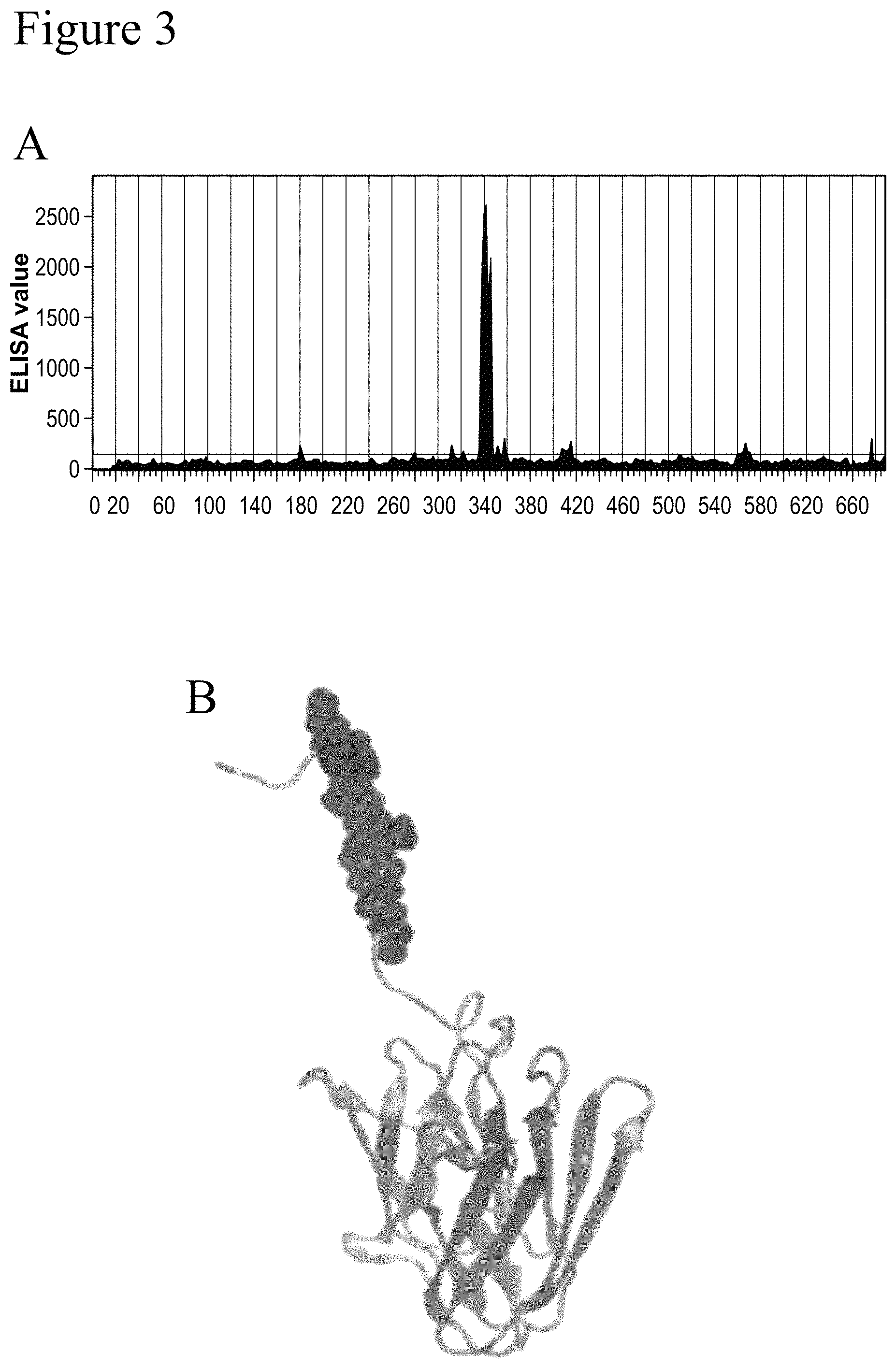
FIG. 3 (A and B) shows that peptides containing motif ERPLVGV (SEQ ID NO: 42) are bound by anti-FLNA 6E3 antibody (A). The epitope of mab 6E3 (ERPLVGV) (SEQ ID NO: 42) is indicated at the top portion, superposed on all resolved structure depicted in cartoon representation (B).
FIG. 4 shows a graphical representation of the CDR loops of the 2C12 clone variable heavy chain. Shaded circles are hydrophobic (non-polar) residues in frameworks 1-3 at sites that are hydrophobic in the majority of antibodies. Squares are key residues at the start and end of the CDR. Amino acids in the framework with an asterisk are structurally conserved amino acids.
FIG. 5 shows a graphical representation of the CDR loops of the 2C12 clone variable light chain. Shaded circles are hydrophobic (non-polar) residues in frameworks 1-3 at sites that are hydrophobic in the majority of antibodies. Squares are key residues at the start and end of the CDR. Amino acids in the framework with an asterisk are structurally conserved amino acids.
FIG. 6 shows a graphical representation of the CDR loops of the 3F4 clone variable heavy chain. Shaded circles are hydrophobic (non-polar) residues in frameworks 1-3 at sites that are hydrophobic in the majority of antibodies. Squares are key residues at the start and end of the CDR. Amino acids in the framework with an asterisk are structurally conserved amino acids.
FIG. 7 shows a graphical representation of the CDR loops of the 3F4 clone variable light chain. Shaded circles are hydrophobic (non-polar) residues in frameworks 1-3 at sites that are hydrophobic in the majority of antibodies. Squares are key residues at the start and end of the CDR. Amino acids in the framework with an asterisk are structurally conserved amino acids.
FIG. 8 shows a graphical representation of the CDR loops of the 6E3 clone variable heavy chain. Shaded circles are hydrophobic (non-polar) residues in frameworks 1-3 at sites that are hydrophobic in the majority of antibodies. Squares are key residues at the start and end of the CDR. Amino acids in the framework with an asterisk are structurally conserved amino acids.
FIG. 9 shows a graphical representation of the CDR loops of the 6E3 clone variable light chain. Shaded circles are hydrophobic (non-polar) residues in frameworks 1-3 at sites that are hydrophobic in the majority of antibodies. Squares are key residues at the start and end of the CDR. Amino acids in the framework with an asterisk are structurally conserved amino acids.
FIG. 10 shows a ROC curve of regression models using a prostate biomarker panel, age and PSA test compared to PSA alone. Specifically, the Prostate Biomarker Panel (FLNA, FLNB, age and PSA) predicts prostate cancer more accurately than PSA alone between patients with or without prostate cancer (Prostate Biomarker Panel (FLNA, FLNB, age and PSA) AUC, 0.64 (0.59, 0.69) (solid line), PSA alone AUC, 0.58 (dotted line)).
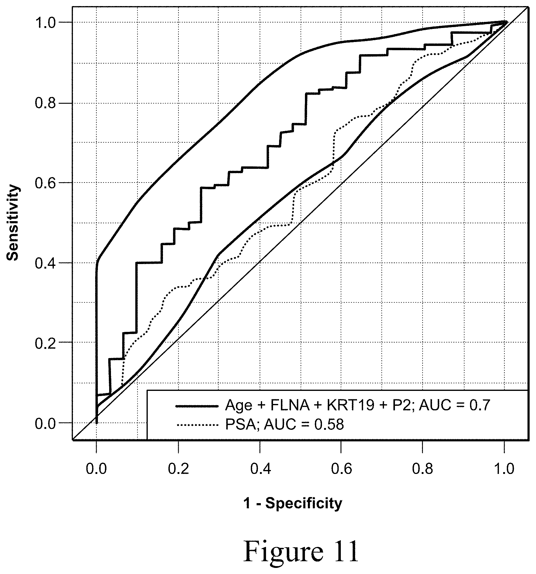
FIG. 11 shows a ROC curve of regression models using a prostate biomarker panel, age and PSA test compared to PSA alone. Specifically, the prostate biomarkers FLNA, KRT19 and age with PSA discriminates between prostate cancer and benign prostatic hyperplasia (BPH) over use of PSA alone (Prostate panel (FLNA, KRT19, PSA and age) (solid line) AUC, 0.70, (0.60, 0.80), PSA alone AUC, 0.58 (dotted line)).
DETAILED DESCRIPTION OF THE INVENTION
This invention pertains to FLNA binding proteins, particularly human FLNA binding proteins, and more particularly anti-FLNA antibodies, or antigen-binding portions thereof, that bind FLNA, and uses thereof. Various aspects of the invention relate to antibodies and antibody fragments, conjugates thereof and pharmaceutical compositions thereof, as well as nucleic acids, recombinant expression vectors and host cells for making such antibodies and fragments. Methods of using the antibodies of the invention to detect FLNA and to diagnose, monitor or prognose disorders such as prostate cancer are also encompassed by the invention.
Unless otherwise defined herein, scientific and technical terms used in connection with the present invention shall have the meanings that are commonly understood by those of ordinary skill in the art. The meaning and scope of the terms should be clear, however, in the event of any latent ambiguity, definitions provided herein take precedent over any dictionary or extrinsic definition. Further, unless otherwise required by context, singular terms shall include pluralities and plural terms shall include the singular. In this application, the use of "or" means "and/or" unless stated otherwise. Furthermore, the use of the term "including", as well as other forms, such as "includes" and "included", is not limiting. Also, terms such as "element" or "component" encompass both elements and components comprising one unit and elements and components that comprise more than one subunit unless specifically stated otherwise. The term "such as" is used herein to mean, and is used interchangeably, with the phrase "such as but not limited to."
The articles "a" and "an" are used herein to refer to one or to more than one (i.e. to at least one) of the grammatical object of the article. By way of example, "an element" means one element or more than one element.
Unless specifically stated or obvious from context, as used herein, the term "about" is understood as within a range of normal tolerance in the art, for example within 2 standard deviations of the mean. About can be understood as within 10%, 9%, 8%, 7%, 6%, 5%, 4%, 3%, 2%, 1%, 0.5%, 0.1%, 0.05%, or 0.01% of the stated value. Unless otherwise clear from context, all numerical values provided herein can be modified by the term about.
The recitation of a listing of chemical group(s) in any definition of a variable herein includes definitions of that variable as any single group or combination of listed groups. The recitation of an embodiment for a variable or aspect herein includes that embodiment as any single embodiment or in combination with any other embodiments or portions thereof.
Any compositions or methods provided herein can be combined with one or more of any of the other compositions and methods provided herein.
Ranges provided herein are understood to be shorthand for all of the values within the range. For example, a range of 1 to 50 is understood to include any number, combination of numbers, or sub-range from the group consisting 1, 2, 3, 4, 5, 6, 7, 8, 9, 10, 11, 12, 13, 14, 15, 16, 17, 18, 19, 20, 21, 22, 23, 24, 25, 26, 27, 28, 29, 30, 31, 32, 33, 34, 35, 36, 37, 38, 39, 40, 41, 42, 43, 44, 45, 46, 47, 48, 49, or 50. As used herein, "one or more" is understood as each value 1, 2, 3, 4, 5, 6, 7, 8, 9, 10, and any value greater than 10.
Generally, nomenclatures used in connection with, and techniques of, cell and tissue culture, molecular biology, immunology, microbiology, genetics and protein and nucleic acid chemistry and hybridization described herein are those well-known and commonly used in the art. The methods and techniques of the present invention are generally performed according to conventional methods well known in the art and as described in various general and more specific references that are cited and discussed throughout the present specification unless otherwise indicated. Enzymatic reactions and purification techniques are performed according to manufacturer's specifications, as commonly accomplished in the art or as described herein. The nomenclatures used in connection with, and the laboratory procedures and techniques of, analytical chemistry, synthetic organic chemistry, and medicinal and pharmaceutical chemistry described herein are those well-known and commonly used in the art. Standard techniques are used for chemical syntheses, chemical analyses, pharmaceutical preparation, formulation, and delivery, and treatment of patients.
That the present invention may be more readily understood, select terms are defined below.
The term "polypeptide" as used herein, refers to any polymeric chain of amino acids. The terms "peptide" and "protein" are used interchangeably with the term polypeptide and also refer to a polymeric chain of amino acids. The term "polypeptide" encompasses native or artificial proteins, protein fragments and polypeptide analogs of a protein sequence. A polypeptide may be monomeric or polymeric.
The term "isolated protein" or "isolated polypeptide" is a protein or polypeptide that by virtue of its origin or source of derivation is not associated with naturally associated components that accompany it in its native state; is substantially free of other proteins from the same species; is expressed by a cell from a different species; or does not occur in nature. Thus, a polypeptide that is chemically synthesized or synthesized in a cellular system different from the cell from which it naturally originates will be "isolated" from its naturally associated components. A protein may also be rendered substantially free of naturally associated components by isolation, using protein purification techniques well known in the art. An example of an isolated polypeptide is an isolated antibody, or antigen-binding portion thereof.
The term "Filamin A" (FLNA) as used herein refers to a member of the filamin family. Filamin A (also known as FLN-A, FLN1, ABP-280, OPD1, OPD2, Endothelial Actin-Binding Protein, CVD1, FMD, MNS, NHBP, XLVD, XMVD, Actin Binding Protein 280, Alpha-Filamin, Filamin-1, Filamin-A--each of which may appear herein and are considered equivalent terms as used herein) is a 280-kD protein that is thought to crosslink actin filaments into orthogonal networks in cortical cytoplasm. As used herein, filamin A refers to both the gene and the protein unless clearly indicated otherwise by context. The large molecular-weight protein also participates in the anchoring of membrane proteins to the actin cytoskeleton. Remodeling of the cytoskeleton is central to the modulation of cell shape and migration cells. Filamin A, encoded by the FLNA gene, is a widely expressed protein that regulates reorganization of the actin cytoskeleton by interacting with integrins, transmembrane receptor complexes, and second messengers. At least two different isoforms are known, isoform 1 and isoform 2, all of which are contemplated by the invention and encompassed by the term "filamin A" It will be appreciated that isoform 1 is the predominant transcript encoding filamin A. Isoform 2 includes an alternate in-frame exon and encodes a slightly longer protein isoform. Interaction with FLNA may allow neuroblast migration from the ventricular zone into the cortical plate. FLNA tethers cell surface-localized furin, modulates its rate of internalization and directs its intracellular trafficking. Further reference to filamin A can be found in the scientific literature, for example, in Gorlin J B et la., (October 1993). "Actin-binding protein (ABP-280) filamin gene (FLN) maps telomeric to the color vision locus (R/GCP) and centromeric to G6PD in Xq28". Genomics 17 (2): 496-8, and Robertson S P et al. (March 2003). "Localized mutations in the gene encoding the cytoskeletal protein filamin A cause diverse malformations in humans". Nat Genet 33 (4): 487-91, each of which are incorporated herein by reference. The nucleotide and amino acid sequences of human filamin A can be found as GenBank Accession No. NM_001456.3 (filamin A--isoform 1--mRNA transcript sequence--SEQ ID NO: 31) and the corresponding polypeptide sequence of NP_001447.2 (filamin A--isoform 1--polypeptide sequence--SEQ ID NO: 32) and as GenBank Accession No. NM_001110556 (filamin A--isoform 2--mRNA transcript sequence--SEQ ID NO: 33) and the corresponding polypeptide sequence of NP_001104026 (filamin A--isoform 2--polypeptide sequence--SEQ ID NO: 34). These GenBank numbers are incorporated herein by reference in the versions available on the earliest effective filing date of this application.
It is understood that the invention includes the use of any combination of one or more of the filamin A sequences provided in the sequence listing or any fragments thereof as long as the fragment can allow for the specific identification of filamin A Methods of the invention and reagents can be used to detect single isoforms of filamin A, combinations of filamin A isoforms, or all of the filamin A isoforms simultaneously. Unless specified, filamin A can be considered to refer to one or more isoforms of filamin A, including total filamin A. Moreover, it is understood that there are naturally occurring variants of filamin A, which may or may not be associated with a specific disease state, the use of which are also included in the instant application.
The terms "specific binding" or "specifically binding", as used herein, in reference to the interaction of an antibody, a protein, or a peptide with a second chemical species, mean that the interaction is dependent upon the presence of a particular structure (e.g., an antigenic determinant or epitope) on the chemical species; for example, an antibody recognizes and binds to a specific protein structure rather than to proteins generally. If an antibody is specific for epitope "A", the presence of a molecule containing epitope A (or free, unlabeled A), in a reaction containing labeled "A" and the antibody, will reduce the amount of labeled A bound to the antibody.
The term "antibody", as used herein, broadly refers to any immunoglobulin (Ig) molecule comprised of four polypeptide chains, two heavy (H) chains and two light (L) chains, or any functional fragment, mutant, variant, or derivation thereof, which retains the essential epitope binding features of an Ig molecule. Such mutant, variant, or derivative antibody formats are known in the art. Non-limiting embodiments of which are discussed below.
In a full-length antibody, each heavy chain is comprised of a heavy chain variable region (abbreviated herein as HCVR or VH) and a heavy chain constant region. The heavy chain constant region is comprised of three domains, CH1, CH2 and CH3. Each light chain is comprised of a light chain variable region (abbreviated herein as LCVR or VL) and a light chain constant region. The light chain constant region is comprised of one domain, CL. The VH and VL regions can be further subdivided into regions of hypervariability, termed complementarity determining regions (CDR), interspersed with regions that are more conserved, termed framework regions (FR). Each VH and VL is composed of three CDRs and four FRs, arranged from amino-terminus to carboxy-terminus in the following order: FR1, CDR1, FR2, CDR2, FR3, CDR3, FR4. Immunoglobulin molecules can be of any type (e.g., IgG, IgE, IgM, IgD, IgA and IgY), class (e.g., IgG, 1, IgG2, IgG 3, IgG4, IgA1 and IgA2) or subclass.
The term "antigen-binding portion" of an antibody (or simply "antibody portion"), as used herein, refers to one or more fragments of an antibody that retain the ability to specifically bind to an antigen (e.g., FLNA). It has been shown that the antigen-binding function of an antibody can be performed by fragments of a full-length antibody. Such antibody embodiments may also be bispecific, dual specific, or multi-specific formats; specifically binding to two or more different antigens. Examples of binding fragments encompassed within the term "antigen-binding portion" of an antibody include (i) a Fab fragment, a monovalent fragment consisting of the VL, VH, CL and CH1 domains; (ii) a F(ab').sub.2 fragment, a bivalent fragment comprising two Fab fragments linked by a disulfide bridge at the hinge region; (iii) a Fd fragment consisting of the VH and CH1 domains; (iv) a Fv fragment consisting of the VL and VH domains of a single arm of an antibody, (v) a dAb fragment (Ward et al., (1989) Nature 341:544-546, Winter et al., PCT publication WO 90/05144 A1 herein incorporated by reference), which comprises a single variable domain; and (vi) an isolated complementarity determining region (CDR). Furthermore, although the two domains of the Fv fragment, VL and VH, are coded for by separate genes, they can be joined, using recombinant methods, by a synthetic linker that enables them to be made as a single protein chain in which the VL and VH regions pair to form monovalent molecules (known as single chain Fv (scFv); see e.g., Bird et al. (1988) Science 242:423-426; and Huston et al. (1988) Proc. Natl. Acad. Sci. USA 85:5879-5883). Such single chain antibodies are also intended to be encompassed within the term "antigen-binding portion" of an antibody. Other forms of single chain antibodies, such as diabodies are also encompassed. Diabodies are bivalent, bispecific antibodies in which VH and VL domains are expressed on a single polypeptide chain, but using a linker that is too short to allow for pairing between the two domains on the same chain, thereby forcing the domains to pair with complementary domains of another chain and creating two antigen binding sites (see e.g., Holliger, P., et al. (1993) Proc. Natl. Acad. Sci. USA 90:6444-6448; Poljak, R. J., et al. (1994) Structure 2:1121-1123). Such antibody binding portions are known in the art (Kontermann and Dubel eds., Antibody Engineering (2001) Springer-Verlag. New York. 790 pp. (ISBN 3-540-41354-5).
An "isolated antibody", as used herein, is intended to refer to an antibody that is substantially free of other antibodies having different antigenic specificities (e.g., an isolated antibody that specifically binds FLNA is substantially free of antibodies that specifically bind antigens other than FLNA). An isolated antibody that specifically binds human FLNA may, however, have cross-reactivity to other antigens, such as FLNA molecules from other species. Moreover, an isolated antibody may be substantially free of other cellular material and/or chemicals.
As used herein, the term "CDR" refers to the complementarity determining region within antibody variable sequences. There are three CDRs in each of the variable regions of the heavy chain and the light chain, which are designated CDR1, CDR2 and CDR3, for each of the variable regions. The term "CDR set" as used herein refers to a group of three CDRs that occur in a single variable region capable of binding the antigen. The exact boundaries of these CDRs have been defined differently according to different systems. The system described by Kabat (Kabat et al., Sequences of Proteins of Immunological Interest (National Institutes of Health, Bethesda, Md. (1987) and (1991)) not only provides an unambiguous residue numbering system applicable to any variable region of an antibody, but also provides precise residue boundaries defining the three CDRs. These CDRs may be referred to as Kabat CDRs. Chothia and coworkers (Chothia et al., J. Mol. Biol. 196:901-917 (1987) and Chothia et al., Nature 342:877-883 (1989)) found that certain sub-portions within Kabat CDRs adopt nearly identical peptide backbone conformations, despite having great diversity at the level of amino acid sequence. These sub-portions were designated as L1, L2 and L3 or H1, H2 and H3 where the "L" and the "H" designates the light chain and the heavy chains regions, respectively. These regions may be referred to as Chothia CDRs, which have boundaries that overlap with Kabat CDRs. Other boundaries defining CDRs overlapping with the Kabat CDRs have been described by Padlan (FASEB J. 9:133-139 (1995)) and MacCallum (J Mol Biol 262(5):732-45 (1996)). Still other CDR boundary definitions may not strictly follow one of the above systems, but will nonetheless overlap with the Kabat CDRs, although they may be shortened or lengthened in light of prediction or experimental findings that particular residues or groups of residues or even entire CDRs do not significantly impact antigen binding. The methods used herein may utilize CDRs defined according to any of these systems, although preferred embodiments use Kabat or Chothia defined CDRs.
In preferred embodiments of the present invention, the CDRs are determined by the IMGT numbering system (Lefranc et al., Nucleic Acids Research, 27, 209-212 (1990)).
As used herein, the term "framework" or "framework sequence" refers to the remaining sequences of a variable region minus the CDRs. Because the exact definition of a CDR sequence can be determined by different systems, the meaning of a framework sequence is subject to correspondingly different interpretations. The six CDRs (CDR-L1, CDR-L2, and CDR-L3 of light chain and CDR-H1, CDR-H2, and CDR-H3 of heavy chain) also divide the framework regions on the light chain and the heavy chain into four sub-regions (FR1, FR2, FR3 and FR4) on each chain, in which CDR1 is positioned between FR1 and FR2, CDR2 between FR2 and FR3, and CDR3 between FR3 and FR4. Without specifying the particular sub-regions as FR1, FR2, FR3 or FR4, a framework region, as referred by others, represents the combined FR's within the variable region of a single, naturally occurring immunoglobulin chain. As used herein, a FR represents one of the four sub-regions, and FRs represents two or more of the four sub-regions constituting a framework region.
As used herein, the term "humanized antibody" is an antibody or a variant, derivative, analog or fragment thereof which immunospecifically binds to an antigen of interest and which comprises a framework (FR) region having substantially the amino acid sequence of a human antibody and a complementary determining region (CDR) having substantially the amino acid sequence of a non-human antibody. As used herein, the term "substantially" in the context of a CDR refers to a CDR having an amino acid sequence at least 80%, preferably at least 85%, at least 90%, at least 95%, at least 98% or at least 99% identical to the amino acid sequence of a non-human antibody CDR. A humanized antibody comprises substantially all of at least one, and typically two, variable domains (Fab, Fab', F(ab') 2, FabC, Fv) in which all or substantially all of the CDR regions correspond to those of a non-human immunoglobulin (i.e., donor antibody) and all or substantially all of the framework regions are those of a human immunoglobulin consensus sequence. Preferably, a humanized antibody also comprises at least a portion of an immunoglobulin constant region (Fc), typically that of a human immunoglobulin. In some embodiments, a humanized antibody contains both the light chain as well as at least the variable domain of a heavy chain. The antibody also may include the CH1, hinge, CH2, CH3, and CH4 regions of the heavy chain. In some embodiments, a humanized antibody only contains a humanized light chain. In some embodiments, a humanized antibody only contains a humanized heavy chain. In specific embodiments, a humanized antibody only contains a humanized variable domain of a light chain and/or humanized heavy chain.
The humanized antibody can be selected from any class of immunoglobulins, including IgM, IgG, IgD, IgA and IgE, and any isotype, including without limitation IgG 1, IgG2, IgG3 and IgG4. The humanized antibody may comprise sequences from more than one class or isotype, and particular constant domains may be selected to optimize desired effector functions using techniques well-known in the art.
The framework and CDR regions of a humanized antibody need not correspond precisely to the parental sequences, e.g., the donor antibody CDR or the consensus framework may be mutagenized by substitution, insertion and/or deletion of at least one amino acid residue so that the CDR or framework residue at that site does not correspond to either the donor antibody or the consensus framework. In a preferred embodiment, such mutations, however, will not be extensive. Usually, at least 80%, preferably at least 85%, more preferably at least 90%, and most preferably at least 95% of the humanized antibody residues will correspond to those of the parental FR and CDR sequences. As used herein, the term "consensus framework" refers to the framework region in the consensus immunoglobulin sequence. As used herein, the term "consensus immunoglobulin sequence" refers to the sequence formed from the most frequently occurring amino acids (or nucleotides) in a family of related immunoglobulin sequences (See e.g., Winnaker, From Genes to Clones (Verlagsgesellschaft, Weinheim, Germany 1987). In a family of immunoglobulins, each position in the consensus sequence is occupied by the amino acid occurring most frequently at that position in the family. If two amino acids occur equally frequently, either can be included in the consensus sequence.
The term "epitope" includes any polypeptide determinant capable of specific binding to a binding protein, e.g., an antibody or antigen binding portion thereof. In certain embodiments, epitope determinants include chemically active surface groupings of molecules such as amino acids, sugar side chains, phosphoryl, or sulfonyl, and, in certain embodiments, may have specific three dimensional structural characteristics, and/or specific charge characteristics. In various embodiments, an epitope may be a linear or sequential epitope, i.e., a linear sequence of amino acids, of the primary structure of the antigen, i.e., FLNA. Alternatively, in other embodiments, an epitope may be a conformational epitope having a specific three-dimensional shape when the antigen assumes its secondary structure. For example, the conformational epitope may comprise non-linear, i.e., non-sequential, amino acids of the antigen.
In a particular embodiment, an epitope is a region of an antigen that is bound by a binding protein, e.g., antibody or antigen binding portion thereof. In certain embodiments, a binding protein, e.g., antibody or antigen binding portion thereof, is said to specifically bind an antigen when it preferentially recognizes its target antigen in a complex mixture of proteins and/or macromolecules.
In certain embodiments, the epitope is a linear motif ERPLVGV (SEQ ID NO: 42), corresponding to residue 1774-1780 in the hinge connecting Filamin domains 15 and 16 of the target protein.
The term "K.sub.D", as used herein, is intended to refer to the dissociation constant of a particular antibody-antigen interaction as is known in the art.
The term "k.sub.ON", as used herein, is intended to refer to the on rate constant for association of an antibody to the antigen to form the antibody/antigen complex as is known in the art.
The term "k.sub.OFF", as used herein, is intended to refer to the off rate constant for dissociation of an antibody from the antibody/antigen complex as is known in the art.
The term "labeled binding protein" as used herein, refers to a protein with a label incorporated that provides for the identification of the binding protein. Preferably, the label is a detectable marker, e.g., incorporation of a radiolabeled amino acid or attachment to a polypeptide of biotinyl moieties that can be detected by marked avidin (e.g., streptavidin containing a fluorescent marker or enzymatic activity that can be detected by optical or colorimetric methods). Examples of labels for polypeptides include, but are not limited to, the following: radioisotopes or radionuclides (e.g., .sup.3H, .sup.14C, .sup.35S, .sup.90Y, .sup.99Tc, .sup.111In, .sup.125I, .sup.131I, .sup.177Lu, .sup.166Ho, or .sup.153Sm); fluorescent labels (e.g., FITC, rhodamine, lanthanide phosphors), enzymatic labels (e.g., horseradish peroxidase, luciferase, alkaline phosphatase); chemiluminescent markers; biotinyl groups; predetermined polypeptide epitopes recognized by a secondary reporter (e.g., leucine zipper pair sequences, binding sites for secondary antibodies, metal binding domains, epitope tags); and magnetic agents, such as gadolinium chelates.
The term "polynucleotide" as used herein refers to a polymeric form of two or more nucleotides, either ribonucleotides or deoxvnucleotides or a modified form of either type of nucleotide. The term includes single and double stranded forms of DNA but preferably is double-stranded DNA.
The term "isolated polynucleotide" as used herein shall mean a polynucleotide (e.g., of genomic, cDNA, or synthetic origin, or some combination thereof) that, by virtue of its origin, the "isolated polynucleotide": is not associated with all or a portion of a polynucleotide with which the "isolated polynucleotide" is found in nature; is operably linked to a polynucleotide that it is not linked to in nature; or does not occur in nature as part of a larger sequence.
The term "vector", as used herein, is intended to refer to a nucleic acid molecule capable of transporting another nucleic acid to which it has been linked. One type of vector is a "plasmid", which refers to a circular double stranded DNA loop into which additional DNA segments may be ligated. Another type of vector is a viral vector, wherein additional DNA segments may be ligated into the viral genome. Certain vectors are capable of autonomous replication in a host cell into which they are introduced (e.g., bacterial vectors having a bacterial origin of replication and episomal mammalian vectors). Other vectors (e.g., non-episomal mammalian vectors) can be integrated into the genome of a host cell upon introduction into the host cell, and thereby are replicated along with the host genome. Moreover, certain vectors are capable of directing the expression of genes to which they are operatively linked. Such vectors are referred to herein as "recombinant expression vectors" (or simply, "expression vectors"). In general, expression vectors of utility in recombinant DNA techniques are often in the form of plasmids. In the present specification, "plasmid" and "vector" may be used interchangeably as the plasmid is the most commonly used form of vector. However, the invention is intended to include such other forms of expression vectors, such as viral vectors (e.g., replication defective retroviruses, adenoviruses and adeno-associated viruses), which serve equivalent functions.
The term "operably linked" refers to a juxtaposition wherein the components described are in a relationship permitting them to function in their intended manner A control sequence "operably linked" to a coding sequence is ligated in such a way that expression of the coding sequence is achieved under conditions compatible with the control sequences. "Operably linked" sequences include both expression control sequences that are contiguous with the gene of interest and expression control sequences that act in trans or at a distance to control the gene of interest. The term "expression control sequence" as used herein refers to polynucleotide sequences which are necessary to effect the expression and processing of coding sequences to which they are ligated. Expression control sequences include appropriate transcription initiation, termination, promoter and enhancer sequences; efficient RNA processing signals such as splicing and polyadenylation signals; sequences that stabilize cytoplasmic mRNA; sequences that enhance translation efficiency (i.e., Kozak consensus sequence); sequences that enhance protein stability; and when desired, sequences that enhance protein secretion. The nature of such control sequences differs depending upon the host organism; in prokaryotes, such control sequences generally include promoter, ribosomal binding site, and transcription termination sequence; in eukaryotes, generally, such control sequences include promoters and transcription termination sequence. The term "control sequences" is intended to include components whose presence is essential for expression and processing, and can also include additional components whose presence is advantageous, for example, leader sequences and fusion partner sequences. Protein constructs of the present invention may be expressed, and purified using expression vectors and host cells known in the art, including expression cassettes, vectors, recombinant host cells and methods for the recombinant expression and proteolytic processing of recombinant polyproteins and pre-proteins from a single open reading frame (e.g., WO 2007/014162 incorporated herein by reference).
"Transformation", as defined herein, refers to any process by which exogenous DNA enters a host cell. Transformation may occur under natural or artificial conditions using various methods well known in the art. Transformation may rely on any known method for the insertion of foreign nucleic acid sequences into a prokaryotic or eukaryotic host cell. The method is selected based on the host cell being transformed and may include, but is not limited to, viral infection, electroporation, lipofection, and particle bombardment. Such "transformed" cells include stably transformed cells in which the inserted DNA is capable of replication either as an autonomously replicating plasmid or as part of the host chromosome. They also include cells which transiently express the inserted DNA or RNA for limited periods of time.
The term "recombinant host cell" (or simply "host cell"), as used herein, is intended to refer to a cell into which exogenous DNA has been introduced. It should be understood that such terms are intended to refer not only to the particular subject cell, but, to the progeny of such a cell. Because certain modifications may occur in succeeding generations due to either mutation or environmental influences, such progeny may not, in fact, be identical to the parent cell, but are still included within the scope of the term "host cell" as used herein. Preferably host cells include prokaryotic and eukaryotic cells selected from any of the Kingdoms of life. Preferred eukaryotic cells include protist, fungal, plant and animal cells. Most preferably host cells include but are not limited to the prokaryotic cell line E. coli; mammalian cell lines CHO, HEK 293 and COS; the insect cell line Sf9; and the fungal cell Saccharomyces cerevisiae.
Standard techniques may be used for recombinant DNA, oligonucleotide synthesis, and tissue culture and transformation (e.g., electroporation, lipofection). Enzymatic reactions and purification techniques may be performed according to manufacturer's specifications or as commonly accomplished in the art or as described herein. The foregoing techniques and procedures may be generally performed according to conventional methods well known in the art and as described in various general and more specific references that are cited and discussed throughout the present specification. See e.g., Sambrook et al. Molecular Cloning: A Laboratory Manual (2d ed., Cold Spring Harbor Laboratory Press, Cold Spring Harbor, N.Y. (1989)), which is incorporated herein by reference for any purpose.
The term "sample", as used herein, is used in its broadest sense. A "biological sample", as used herein, includes, but is not limited to, any quantity of a substance from a living thing or formerly living thing. Such living things include, but are not limited to, humans, mice, rats, monkeys, dogs, rabbits and other animals. Such substances include, but are not limited to, blood, serum, urine, synovial fluid, cells, organs, tissues, bone marrow, lymph nodes and spleen.
A "patient" or "subject" to be treated by the method of the invention can mean either a human or non-human animal, preferably a mammal By "subject" is meant any animal, including horses, dogs, cats, pigs, goats, rabbits, hamsters, monkeys, guinea pigs, rats, mice, lizards, snakes, sheep, cattle, fish, and birds. A human subject may be referred to as a patient. It should be noted that clinical observations described herein were made with human subjects and, in at least some embodiments, the subjects are human.
The terms "disorders", "diseases", and "abnormal state" are used inclusively and refer to any deviation from the normal structure or function of any part, organ, or system of the body (or any combination thereof). A specific disease is manifested by characteristic symptoms and signs, including biological, chemical, and physical changes, and is often associated with a variety of other factors including, but not limited to, demographic, environmental, employment, genetic, and medically historical factors. Certain characteristic signs, symptoms, and related factors can be quantitated through a variety of methods to yield important diagnostic information. As used herein the disorder, disease, or abnormal state is an abnormal prostate state, including benign prostate hyperplasia and cancer, particularly prostate cancer. The abnormal prostate state of prostate cancer can be further subdivided into stages and grades of prostate cancer as provided, for example in Prostate. In: Edge S B, Byrd D R, Compton C C, et al., eds.: AJCC Cancer Staging Manual. 7th ed. New York, N.Y.: Springer, 2010, pp 457-68 (incorporated herein by reference). Further, abnormal prostate states can be classified as one or more of benign prostate hyperplasia (BPH), androgen sensitive prostate cancer, androgen insensitive or resistant prostate cancer, aggressive prostate cancer, non-aggressive prostate cancer, metastatic prostate cancer, and non-metastatic prostate cancer. Further, the prostate cancer may be a prostatic intraepithelial neoplasia, adenocarcinoma, small cell carcinoma, or squamous cell carcinoma.
A subject at "increased risk for developing prostate cancer" may or may not develop prostate cancer. Identification of a subject at increased risk for developing prostate cancer should be monitored for additional signs or symptoms of prostate cancer. The methods provided herein for identifying a subject with increased risk for developing prostate cancer can be used in combination with assessment of other known risk factors or signs of prostate cancer including, but not limited to decreased urinary stream, urgency, hesitancy, nocturia, incomplete bladder emptying, and age.
The term "expression" is used herein to mean the process by which a polypeptide is produced from DNA. The process involves the transcription of the gene into mRNA and the translation of this mRNA into a polypeptide. Depending on the context in which used, "expression" may refer to the production of RNA, or protein, or both.
The terms "level of expression of a gene", "gene expression level", "level of a marker", and the like refer to the level of mRNA, as well as pre-mRNA nascent transcript(s), transcript processing intermediates, mature mRNA(s) and degradation products, or the level of protein, encoded by the gene in the cell.
The term "specific identification" is understood as detection of a marker of interest with sufficiently low background of the assay and cross-reactivity of the reagents used such that the detection method is diagnostically useful. In certain embodiments, reagents for specific identification of a marker bind to only one isoform of the marker. In certain embodiments, reagents for specific identification of a marker bind to more than one isoform of the marker. In certain embodiments, reagents for specific identification of a marker bind to all known isoforms of the marker.
The term "control sample," as used herein, refers to any clinically relevant comparative sample, including, for example, a sample from a healthy subject not afflicted with an oncological disorder, e.g., prostate cancer, or a sample from a subject from an earlier time point, e.g., prior to treatment, an earlier tumor assessment time point, at an earlier stage of treatment. A control sample can be a purified sample, protein, and/or nucleic acid provided with a kit. Such control samples can be diluted, for example, in a dilution series to allow for quantitative measurement of levels of analytes, e.g., markers, in test samples. A control sample may include a sample derived from one or more subjects. A control sample may also be a sample made at an earlier time point from the subject to be assessed. For example, the control sample could be a sample taken from the subject to be assessed before the onset of an oncological disorder, e.g., prostate cancer, at an earlier stage of disease, or before the administration of treatment or of a portion of treatment. The control sample may also be a sample from an animal model, or from a tissue or cell lines derived from the animal model of oncological disorder, e.g., prostate cancer. The level of activity or expression of a marker, e.g. FLNA, in a control sample consists of a group of measurements that may be determined based on any appropriate statistical measure, such as, for example, measures of central tendency including average, median, or modal values. Different from a control is preferably statistically significantly different from a control.
The term "control level" refers to an accepted or pre-determined level of a marker in a subject sample. A control level can be a range of values. Marker levels can be compared to a single control value, to a range of control values, to the upper level of normal, or to the lower level of normal as appropriate for the assay.
In one embodiment, the control is a standardized control, such as, for example, a control which is predetermined using an average of the levels of expression of one or more markers from a population of subjects having no cancer, especially subjects having no prostate cancer. In still other embodiments of the invention, a control level of a marker in a non-cancerous sample(s) derived from the subject having cancer. For example, when a biopsy or other medical procedure reveals the presence of cancer in one portion of the tissue, the control level of a marker may be determined using the non-affected portion of the tissue, and this control level may be compared with the level of the marker in an affected portion of the tissue.
In certain embodiments, the control can be from a subject, or a population of subject, having an abnormal prostate state. For example, the control can be from a subject suffering from benign prostate hyperplasia (BPH), androgen sensitive prostate cancer, androgen insensitive or resistant prostate cancer, aggressive prostate cancer, non-aggressive prostate cancer, metastatic prostate cancer, or non-metastatic prostate cancer. It is understood that not all markers will have different levels for each of the abnormal prostate states listed. It is understood that a combination of maker levels may be most useful to distinguish between abnormal prostate states, possibly in combination with other diagnostic methods. Further, marker levels in biological samples can be compared to more than one control sample (e.g., normal, abnormal, from the same subject, from a population control). Marker levels can be used in combination with other signs or symptoms of an abnormal prostate state to provide a diagnosis for the subject.
A control can also be a sample from a subject at an earlier time point, e.g., a baseline level prior to suspected presence of disease, before the diagnosis of a disease, at an earlier assessment time point during watchful waiting, before the treatment with a specific agent (e.g., chemotherapy, hormone therapy) or intervention (e.g., radiation, surgery). In certain embodiments, a change in the level of the marker in a subject can be more significant than the absolute level of a marker, e.g., as compared to control.
As used herein, a sample obtained at an "earlier time point" is a sample that was obtained at a sufficient time in the past such that clinically relevant information could be obtained in the sample from the earlier time point as compared to the later time point. In certain embodiments, an earlier time point is at least four weeks earlier. In certain embodiments, an earlier time point is at least six weeks earlier. In certain embodiments, an earlier time point is at least two months earlier. In certain embodiments, an earlier time point is at least three months earlier. In certain embodiments, an earlier time point is at least six months earlier. In certain embodiments, an earlier time point is at least nine months earlier. In certain embodiments, an earlier time point is at least one year earlier. Multiple subject samples (e.g., 3, 4, 5, 6, 7, or more) can be obtained at regular or irregular intervals over time and analyzed for trends in changes in marker levels. Appropriate intervals for testing for a particular subject can be determined by one of skill in the art based on ordinary considerations.
As used herein, "changed as compared to a control" sample or subject is understood as having a level of the analyte or diagnostic or therapeutic indicator (e.g., marker) to be detected at a level that is statistically different than a sample from a normal, untreated, or abnormal state control sample. Changed as compared to control can also include a difference in the rate of change of the level of one or more markers obtained in a series of at least two subject samples obtained over time. Determination of statistical significance is within the ability of those skilled in the art, e.g., the number of standard deviations from the mean that constitute a positive or negative result.
As used herein, the term "obtaining" is understood herein as manufacturing, purchasing, or otherwise coming into possession of.
As used herein, "surrogate peptide" is understood as any peptide derived from a FLNA marker wherein the surrogate peptide is prepared by digesting the marker protein, e.g., FLNA, with a protease of known specificity (e.g., trypsin or endoproteinase Lys-C), and wherein the peptide can be used as a surrogate reporter to determine the abundance of the FLNA marker protein, and optionally isoforms or fragments thereof, in a sample, using a mass spectrometry-based assay, e.g., MRM or IPMRM. Surrogate peptides can be tryptic peptides between, for example, about 8 and 22 amino acids. Surrogate peptides can be chosen by methods known in the art, e.g., Skyline software and LC-MS/MS analysis (LTQ Orbitrap Velos coupled to Eksigent nano-LC) of recombinant protein (GenScript) tryptic digest. Surrogate peptides can be chosen based on surrogate peptide selection rules (Halquist, et al., Biomed Chromatography 25 (1-2):47-58) and signal intensities of the peptides in spiked and unspiked serum digests. The uniqueness of the surrogate peptides to the target FLNA marker can be confirmed by BLAST searches.
As used herein, "detecting", "detection", "determining", and the like are understood that an assay performed for identification of a specific marker in a sample, e.g., FLNA. The amount of marker expression or activity detected in the sample can be none or below the level of detection of the assay or method.
Reference will now be made in detail to exemplary embodiments of the invention. While the invention will be described in conjunction with the exemplary embodiments, it will be understood that it is not intended to limit the invention to those embodiments. To the contrary, it is intended to cover alternatives, modifications, and equivalents as may be included within the spirit and scope of the invention as defined by the appended claims.
I. Antibodies that Bind Filamin A (FLNA)
One aspect of the present invention provides isolated murine monoclonal antibodies, or antigen-binding portions thereof, that bind to FLNA. Preferably, the antibodies bind human FLNB. Methods of making the antibodies, methods of producing the antibodies and the anti-FLNA antibodies are described in detail hereinbelow.
A. Method of Making Anti-FLNA Antibodies
Antibodies of the present invention may be made by any of a number of techniques known in the art. The general methodology for making monoclonal antibodies by hybridomas is well known Immortal, antibody-producing cell lines can also be created by techniques other than fusion, such as direct transformation of B lymphocytes with oncogenic DNA, or transfection with Epstein-Barr virus. See, e.g., M. Schreier et al., "Hybridoma Techniques" (1980); Hammering et al., "Monoclonal Antibodies And T cell Hybridomas" (1981); Kennett et al., "Monoclonal Antibodies" (1980); see also U.S. Pat. Nos. 4,341,761; 4,399,121; 4,427,783; 4,444,887; 4,451,570; 4,466,917; 4,472,500; 4,491,632; and 4,493,890. Methods for producing polyclonal anti-EFGR antibodies are well-known in the art. See U.S. Pat. No. 4,493,795 to Nestor et al.
Panels of monoclonal antibodies produced against FLNA can be screened for various properties; i.e., isotype, epitope, affinity, etc.
A monoclonal antibody, typically containing Fab and/or F (ab')2 portions of useful antibody molecules, can be prepared using the hybridoma technology described in Antibodies-A Laboratory Manual, Harlow and Lane, eds., Cold Spring Harbor Laboratory, New York (1988), which is incorporated herein by reference. Briefly, to form the hybridoma from which the monoclonal antibody composition is produced, a myeloma or other self-perpetuating cell line is fused with lymphocytes obtained from the spleen of a mammal hyperimmunized with an appropriate FLNA.
Splenocytes are typically fused with myeloma cells using polyethylene glycol (PEG) 6000. Fused hybrids are selected by their sensitivity to HAT. Hybridomas producing a monoclonal antibody useful in practicing this invention are identified by their ability to immunoreact with the present antibody or binding member and their ability to inhibit specified tumorigenic or hyperproliferative activity in target cells.
A monoclonal antibody useful in practicing the present invention can be produced by initiating a monoclonal hybridoma culture comprising a nutrient medium containing a hybridoma that secretes antibody molecules of the appropriate antigen specificity. The culture is maintained under conditions and for a time period sufficient for the hybridoma to secrete the antibody molecules into the medium. The antibody-containing medium is then collected. The antibody molecules can then be further isolated by well-known techniques.
Media useful for the preparation of these compositions are both well-known in the art and commercially available and include synthetic culture media, inbred mice and the like. An exemplary synthetic medium is Dulbecco's minimal essential medium (DMEM; Dulbecco et al., Virol. 8:396 (1959)) supplemented with 4.5 gm/1 glucose, 20 mm glutamine, and 20% fetal calf serum. An exemplary inbred mouse strain is the Balb/c.
1. Anti-FLNA Monoclonal Antibodies Using Hybridoma Technology
Monoclonal antibodies can be prepared using a wide variety of techniques known in the art including the use of hybridoma, recombinant, and phage display technologies, or a combination thereof. For example, monoclonal antibodies can be produced using hybridoma techniques including those known in the art and taught, for example, in Harlow et al., Antibodies: A Laboratory Manual, (Cold Spring Harbor Laboratory Press, 2nd ed. 1988); Hammerling, et al., in: Monoclonal Antibodies and T-Cell Hybridomas 563-681 (Elsevier, N.Y., 1981) (said references incorporated by reference in their entireties). The term "monoclonal antibody" as used herein is not limited to antibodies produced through hybridoma technology. The term "monoclonal antibody" refers to an antibody that is derived from a single clone, including any eukaryotic, prokaryotic, or phage clone, and not the method by which it is produced.
Methods for producing and screening for specific antibodies using hybridoma technology are routine and well known in the art. For example, monoclonal antibodies can be generated by the method of culturing a hybridoma cell secreting an antibody of the invention wherein, preferably, the hybridoma is generated by fusing splenocytes isolated from a mouse immunized with an antigen of the invention with myeloma cells and then screening the hybridomas resulting from the fusion for hybridoma clones that secrete an antibody able to bind a polypeptide of the invention. Briefly, mice can be immunized with a FLNA antigen. In certain embodiments, the FLNA antigen is administered with an adjuvant to stimulate the immune response. Such adjuvants include complete or incomplete Freund's adjuvant, RIBI (muramyl dipeptides) or ISCOM (immunostimulating complexes). Such adjuvants may protect the polypeptide from rapid dispersal by sequestering it in a local deposit, or they may contain substances that stimulate the host to secrete factors that are chemotactic for macrophages and other components of the immune system. Preferably, if a polypeptide is being administered, the immunization schedule will involve two or more administrations of the polypeptide, spread out over several weeks.
After immunization of an animal with a FLNA antigen, antibodies and/or antibody-producing cells may be obtained from the animal. An anti-FLNA antibody-containing serum is obtained from the animal by bleeding or sacrificing the animal. The serum may be used as it is obtained from the animal, an immunoglobulin fraction may be obtained from the serum, or the anti-FLNB antibodies may be purified from the serum. Serum or immunoglobulins obtained in this manner are polyclonal, thus having a heterogeneous array of properties.
Once an immune response is detected, e.g., antibodies specific for the antigen FLNA are detected in the mouse serum, the mouse spleen is harvested and splenocytes isolated. The splenocytes are then fused by well-known techniques to any suitable myeloma cells, for example cells from cell line SP20 available from the ATCC. Hybridomas are selected and cloned by limited dilution. The hybridoma clones are then assayed by methods known in the art for cells that secrete antibodies capable of binding FLNA. Ascites fluid, which generally contains high levels of antibodies, can be generated by immunizing mice with positive hybridoma clones.
In another embodiment, antibody-producing immortalized hybridomas may be prepared from the immunized animal. After immunization, the animal is sacrificed and the splenic B cells are fused to immortalized myeloma cells as is well known in the art. See, e.g., Harlow and Lane, supra. In a preferred embodiment, the myeloma cells do not secrete immunoglobulin polypeptides (a non-secretory cell line). After fusion and antibiotic selection, the hybridomas are screened using FLNA, or a portion thereof, or a cell expressing FLNA. The initial screening is performed using an enzyme-linked immunoassay (ELISA) or a radioimmunoassay (RIA). An example of ELISA screening is provided in WO 00/37504, herein incorporated by reference.
Anti-FLNA antibody-producing hybridomas are selected, cloned and further screened for desirable characteristics, including robust hybridoma growth, high antibody production and desirable antibody characteristics, as discussed further below. Hybridomas may be cultured and expanded in vivo in syngeneic animals, in animals that lack an immune system, e.g., nude mice, or in cell culture in vitro. Methods of selecting, cloning and expanding hybridomas are well known to those of ordinary skill in the art.
In a preferred embodiment, the hybridomas are mouse hybridomas, as described herein. In particular exemplary embodiments, the mouse hybridomas are 2C12, 3F4 and 6E7. In another preferred embodiment, the hybridomas are produced in a non-human, non-mouse species such as rats, sheep, pigs, goats, cattle or horses. In another embodiment, the hybridomas are human hybridomas, in which a human non-secretory myeloma is fused with a human cell expressing an anti-FLNA antibody.
Antibody fragments that recognize specific epitopes may be generated by known techniques. For example, Fab and F(ab')2 fragments of the invention may be produced by proteolytic cleavage of immunoglobulin molecules, using enzymes such as papain (to produce Fab fragments) or pepsin (to produce F(ab')2 fragments). F(ab')2 fragments contain the variable region, the light chain constant region and the CHI domain of the heavy chain.
In preferred embodiments, the present invention features a hybridoma that produces the antibody construct of any of the aspects or embodiments described herein. In certain embodiments, the hybridomas that produce the antibodies of the present invention are "2C12", "3F4" and "6E3," as described herein.
In certain embodiments, FLNA (aa 1443-2131) covering Filamin repeats 13-19 was manufactured as immunogen with E. coli expression system. The immunogen sequence is shown as below as SEQ ID NO: 35.
TABLE-US-00001 SEQ ID NO: 35 MHHHHHHHDV TDASKVKCSG PGLSPGMVRA NLPQSFQVDT SKAGVAPLQV KVQGPKGLVE PVDVVDNADG TQTVNYVPSR EGPYSISVLY GDEEVPRSPF KVKVLPTHDA SKVKASGPGL NTTGVPASLP VEFTIDAKDA GEGLLAVQIT DPEGKPKKTH IQDNHDGTYT VAYVPDVTGR YTILIKYGGD EIPFSPYRVR AVPTGDASKC TVTVSIGGHG LGAGIGPTIQ IGEETVITVD TKAAGKGKVT CTVCTPDGSE VDVDVVENED GTFDIFYTAP QPGKYVICVR FGGEHVPNSP FQVTALAGDQ PSVQPPLRSQ QLAPQYTYAQ GGQQTWAPER PLVGVNGLDV TSLRPFDLVI PFTIKKGEIT GEVRMPSGKV AQPTITDNKD GTVTVRYAPS EAGLHEMDIR YDNMHIPGSP LQFYVDYVNC GHVTAYGPGL THGVVNKPAT FTVNTKDAGE GGLSLAIEGP SKAEISCTDNQDGTCSVSYL PVLPGDYSIL VKYNEQHVPG SPFTARVTGD DSMRMSHLKV GSAADIPINISETDLSLLTA TVVPPSGREE PCLLKRLRNG HVGISFVPKE TGEHLVHVKK NGQHVASSPIPVVISQSEIG DASRVRVSGQ GLHEGHTFEP AEFIIDTRDA GYGGLSLSIE GPSKVDINTEDLEDGTCRVT YCPTEPGNYI INIKFADQHV PGSPFS
2. Anti-FLNA Monoclonal Antibodies Using Selected Lymphocyte Antibody Method
In another aspect of the invention, recombinant antibodies are generated from single, isolated lymphocytes using a procedure referred to in the art as the selected lymphocyte antibody method (SLAM), as described in U.S. Pat. No. 5,627,052, PCT Publication WO 92/02551 and Babcock, J. S. et al. (1996) Proc. Natl. Acad. Sci. USA 93:7843-7848. In this method, single cells secreting antibodies of interest, e.g., lymphocytes derived from any one of the immunized animals described in Section 1, are screened using an antigen-specific hemolytic plaque assay, wherein the antigen FLNA, a subunit of FLNA, or a fragment thereof, is coupled to sheep red blood cells using a linker, such as biotin, and used to identify single cells that secrete antibodies with specificity for FLNA. Following identification of antibody-secreting cells of interest, heavy- and light-chain variable region cDNAs are rescued from the cells by reverse transcriptase-PCR and these variable regions can then be expressed, in the context of appropriate immunoglobulin constant regions (e.g., human constant regions), in mammalian host cells, such as COS or CHO cells. The host cells transfected with the amplified immunoglobulin sequences, derived from in vivo selected lymphocytes, can then undergo further analysis and selection in vitro, for example by panning the transfected cells to isolate cells expressing antibodies to FLNA. The amplified immunoglobulin sequences further can be manipulated in vitro, such as by in vitro affinity maturation methods such as those described in PCT Publication WO 97/29131 and PCT Publication WO 00/56772.
3. Anti-FLNA Monoclonal Antibodies Using Transgenic Animals
In another embodiment of the instant invention, antibodies are produced by immunizing a non-human animal comprising some, or all, of the human immunoglobulin locus with a FLNA antigen. In a preferred embodiment, the non-human animal is a XENOMOUSE transgenic mouse, an engineered mouse strain that comprises large fragments of the human immunoglobulin loci and is deficient in mouse antibody production. See, e.g., Green et al. Nature Genetics 7:13-21 (1994) and U.S. Pat. Nos. 5,916,771, 5,939,598, 5,985,615, 5,998,209, 6,075,181, 6,091,001, 6,114,598 and 6,130,364. See also WO 91/10741, published Jul. 25, 1991, WO 94/02602, published Feb. 3, 1994, WO 96/34096 and WO 96/33735, both published Oct. 31, 1996, WO 98/16654, published Apr. 23, 1998, WO 98/24893, published Jun. 11, 1998, WO 98/50433, published Nov. 12, 1998, WO 99/45031, published Sep. 10, 1999, WO 99/53049, published Oct. 21, 1999, WO 00/09560, published Feb. 24, 2000 and WO 00/037504, published Jun. 29, 2000. The XENOMOUSE transgenic mouse produces an adult-like human repertoire of fully human antibodies, and generates antigen-specific human Mabs. The XENOMOUSE transgenic mouse contains approximately 80% of the human antibody repertoire through introduction of megabase sized, germline configuration YAC fragments of the human heavy chain loci and x light chain loci. See Mendez et al., Nature Genetics 15:146-156 (1997), Green and Jakobovits J. Exp. Med. 188:483-495 (1998), the disclosures of which are hereby incorporated by reference.
In certain embodiments, the invention features a transgenic mouse comprising a host cell comprising a vector comprising an isolated nucleic acid, wherein the mouse expresses a polypeptide encoded by the nucleic acid, or antigen binding portion thereof, that binds to FLNA. The isolated nucleic acid may encode a binding protein amino acid sequence as described herein, or an antibody construct as described herein.
4. Anti-FLNA Monoclonal Antibodies Using Recombinant Antibody Libraries
In vitro methods also can be used to make the antibodies of the invention, wherein an antibody library is screened to identify an antibody having the desired binding specificity. Methods for such screening of recombinant antibody libraries are well known in the art and include methods described in, for example, Ladner et al. U.S. Pat. No. 5,223,409; Kang et al. PCT Publication No. WO 92/18619; Dower et al. PCT Publication No. WO 91/17271; Winter et al. PCT Publication No. WO 92/20791; Markland et al. PCT Publication No. WO 92/15679; Breitling et al. PCT Publication No. WO 93/01288; McCafferty et al. PCT Publication No. WO 92/01047; Garrard et al. PCT Publication No. WO 92/09690; Fuchs et al. (1991) Bio/Technology 9:1370-1372; Hay et al. (1992) Hum Antibod Hybridomas 3:81-85; Huse et al. (1989) Science 246:1275-1281; McCafferty et al., Nature (1990) 348:552-554; Griffiths et al. (1993) EMBO J 12:725-734; Hawkins et al. (1992) J Mol Biol 226:889-896; Clackson et al. (1991) Nature 352:624-628; Gram et al. (1992) PNAS 89:3576-3580; Garrad et al. (1991) Bio/Technology 9:1373-1377; Hoogenboom et al. (1991) Nuc Acid Res 19:4133-4137; and Barbas et al. (1991) PNAS 88:7978-7982, US patent application publication 20030186374, and PCT Publication No. WO 97/29131, the contents of each of which are incorporated herein by reference.
The recombinant antibody library may be from a subject immunized with FLNA, or a portion of FLNA, such as the extracellular domain. Alternatively, the recombinant antibody library may be from a naive subject, i.e., one who has not been immunized with FLNA, such as a human antibody library from a human subject who has not been immunized with FLNA. Antibodies of the invention are selected by screening the recombinant antibody library with the peptide comprising FLNA to thereby select those antibodies that recognize FLNA. Methods for conducting such screening and selection are well known in the art, such as described in the references in the preceding paragraph. To select antibodies of the invention having particular binding affinities for FLNA, e.g. human FLNA, such as those that dissociate from FLNA with a particular k.sub.off rate constant, the art-known method of surface plasmon resonance can be used to select antibodies having the desired k.sub.off rate constant. To select antibodies of the invention having a particular neutralizing activity for FLNA, such as those with a particular an IC.sub.50, standard methods known in the art for assessing the inhibition of FLNA activity may be used.
In one aspect, the invention pertains to an isolated antibody, or an antigen-binding portion thereof, that binds FLNA, in particular human FLNA. In various embodiments, the antibody is a recombinant antibody or a monoclonal antibody.
For example, the antibodies of the present invention can also be generated using various phage display methods known in the art. In phage display methods, functional antibody domains are displayed on the surface of phage particles which carry the polynucleotide sequences encoding them. In a particular, such phage can be utilized to display antigen-binding domains expressed from a repertoire or combinatorial antibody library (e.g., human or murine). Phage expressing an antigen binding domain that binds the antigen of interest can be selected or identified with antigen, e.g., using labeled antigen or antigen bound or captured to a solid surface or bead. Phage used in these methods are typically filamentous phage including fd and M13 binding domains expressed from phage with Fab, Fv or disulfide stabilized Fv antibody domains recombinantly fused to either the phage gene III or gene VIII protein. Examples of phage display methods that can be used to make the antibodies of the present invention include those disclosed in Brinkman et al., J. Immunol. Methods 182:41-50 (1995); Ames et al., J. Immunol. Methods 184:177-186 (1995); Kettleborough et al., Eur. J. Immunol. 24:952-958 (1994); Persic et al., Gene 187 9-18 (1997); Burton et al., Advances in Immunology 57:191-280 (1994); PCT application No. PCT/GB91/01134; PCT publications WO 90/02809; WO 91/10737; WO 92/01047; WO 92/18619; WO 93/11236; WO 95/15982; WO 95/20401; and U.S. Pat. Nos. 5,698,426; 5,223,409; 5,403,484; 5,580,717; 5,427,908; 5,750,753; 5,821,047; 5,571,698; 5,427,908; 5,516,637; 5,780,225; 5,658,727; 5,733,743 and 5,969,108; each of which is incorporated herein by reference in its entirety.
As described in the above references, after phage selection, the antibody coding regions from the phage can be isolated and used to generate whole antibodies including human antibodies or any other desired antigen binding fragment, and expressed in any desired host, including mammalian cells, insect cells, plant cells, yeast, and bacteria, e.g., as described in detail below. For example, techniques to recombinantly produce Fab, Fab' and F(ab')2 fragments can also be employed using methods known in the art such as those disclosed in PCT publication WO 92/22324; Mullinax et al., BioTechniques 12(6):864-869 (1992); and Sawai et al., AJRI 34:26-34 (1995); and Better et al., Science 240:1041-1043 (1988) (said references incorporated by reference in their entireties). Examples of techniques which can be used to produce single-chain Fvs and antibodies include those described in U.S. Pat. Nos. 4,946,778 and 5,258,498; Huston et al., Methods in Enzymology 203:46-88 (1991); Shu et al., PNAS 90:7995-7999 (1993); and Skerra et al., Science 240:1038-1040 (1988).
Alternative to screening of recombinant antibody libraries by phage display, other methodologies known in the art for screening large combinatorial libraries can be applied to the identification of dual specificity antibodies of the invention. One type of alternative expression system is one in which the recombinant antibody library is expressed as RNA-protein fusions, as described in PCT Publication No. WO 98/31700 by Szostak and Roberts, and in Roberts, R. W. and Szostak, J. W. (1997) Proc. Natl. Acad. Sci. USA 94:12297-12302. In this system, a covalent fusion is created between an mRNA and the peptide or protein that it encodes by in vitro translation of synthetic mRNAs that carry puromycin, a peptidyl acceptor antibiotic, at their 3' end. Thus, a specific mRNA can be enriched from a complex mixture of mRNAs (e.g., a combinatorial library) based on the properties of the encoded peptide or protein, e.g., antibody, or portion thereof, such as binding of the antibody, or portion thereof, to the dual specificity antigen. Nucleic acid sequences encoding antibodies, or portions thereof, recovered from screening of such libraries can be expressed by recombinant means as described above (e.g., in mammalian host cells) and, moreover, can be subjected to further affinity maturation by either additional rounds of screening of mRNA-peptide fusions in which mutations have been introduced into the originally selected sequence(s), or by other methods for affinity maturation in vitro of recombinant antibodies, as described above.
In another approach the antibodies of the present invention can also be generated using yeast display methods known in the art. In yeast display methods, genetic methods are used to tether antibody domains to the yeast cell wall and display them on the surface of yeast. In particular, such yeast can be utilized to display antigen-binding domains expressed from a repertoire or combinatorial antibody library (e.g., human or murine). Examples of yeast display methods that can be used to make the antibodies of the present invention include those disclosed in Wittrup et al. (U.S. Pat. No. 6,699,658) incorporated herein by reference.
5. Recombinant Anti-FLNA Antibodies
Antibodies of the present invention may be produced by any of a number of techniques known in the art. For example, expression from host cells, wherein expression vector(s) encoding the heavy and light chains is (are) transfected into a host cell by standard techniques. The various forms of the term "transfection" are intended to encompass a wide variety of techniques commonly used for the introduction of exogenous DNA into a prokaryotic or eukaryotic host cell, e.g., electroporation, calcium-phosphate precipitation, DEAE-dextran transfection and the like. Although it is possible to express the antibodies of the invention in either prokaryotic or eukaryotic host cells, expression of antibodies in eukaryotic cells is preferable, and most preferable in mammalian host cells, because such eukaryotic cells (and in particular mammalian cells) are more likely than prokaryotic cells to assemble and secrete a properly folded and immunologically active antibody.
The invention features in certain embodiments an isolated nucleic acid encoding a binding protein amino acid sequence as described herein. The invention also features in other certain embodiments, an isolated nucleic acid encoding an antibody construct amino acid sequence as described herein. In methods of production, an expression vector comprises the isolated nucleic acid. Non-limiting examples of such expression vectors are the pUC series of vectors (Fermentas Life Sciences), the pBluescript series of vectors (Stratagene, La Jolla, Calif.), the pET series of vectors (Novagen, Madison, Wis.), the pGEX series of vectors (Pharmacia Biotech, Uppsala, Sweden), and the pEX series vectors (Clontech, Palo Alto, Calif.).
A host cell comprises the vector described herein. According to embodiments of the invention, the host cell is a prokaryotic cell or a eukaryotic cell. For example, the prokaryotic host cells is E. coli. The eukaryotic cell may be selected from a protist cell, an animal cell, a plant cell or a fungal cell. The animal cell may be selected from a mammalian cell, an avian cell, and an insect cell. Preferably, the host cell is selected from a CHO cell, a COS cell, a yeast cell, and an insect Sf9 cell. In further related embodiments, the yeast cell is Saccharomyces cerevisiae.
Preferred mammalian host cells for expressing the recombinant antibodies of the invention include Chinese Hamster Ovary (CHO cells) (including dhfr-CHO cells, described in Urlaub and Chasin, (1980) Proc. Natl. Acad. Sci. USA 77:4216-4220, used with a DHFR selectable marker, e.g., as described in R. J. Kaufman and P. A. Sharp (1982) Mol. Biol. 159:601-621), NS0 myeloma cells, COS cells and SP2 cells. When recombinant expression vectors encoding antibody genes are introduced into mammalian host cells, the antibodies are produced by culturing the host cells for a period of time sufficient to allow for expression of the antibody in the host cells or, more preferably, secretion of the antibody into the culture medium in which the host cells are grown. Antibodies can be recovered from the culture medium using standard protein purification methods.
Host cells can also be used to produce functional antibody fragments, such as Fab fragments or scFv molecules. It will be understood that variations on the above procedure are within the scope of the present invention. For example, it may be desirable to transfect a host cell with DNA encoding functional fragments of either the light chain and/or the heavy chain of an antibody of this invention. Recombinant DNA technology may also be used to remove some, or all, of the DNA encoding either or both of the light and heavy chains that is not necessary for binding to the antigens of interest. The molecules expressed from such truncated DNA molecules are also encompassed by the antibodies of the invention. In addition, bifunctional antibodies may be produced in which one heavy and one light chain are an antibody of the invention and the other heavy and light chain are specific for an antigen other than the antigens of interest by crosslinking an antibody of the invention to a second antibody by standard chemical crosslinking methods.
In a preferred system for recombinant expression of an antibody, or antigen-binding portion thereof, of the invention, a recombinant expression vector encoding both the antibody heavy chain and the antibody light chain is introduced into dhfr-CHO cells by calcium phosphate-mediated transfection. Within the recombinant expression vector, the antibody heavy and light chain genes are each operatively linked to CMV enhancer/AdMLP promoter regulatory elements to drive high levels of transcription of the genes. The recombinant expression vector also carries a DHFR gene, which allows for selection of CHO cells that have been transfected with the vector using methotrexate selection/amplification. The selected transformant host cells are cultured to allow for expression of the antibody heavy and light chains and intact antibody is recovered from the culture medium. Standard molecular biology techniques are used to prepare the recombinant expression vector, transfect the host cells, select for transformants, culture the host cells and recover the antibody from the culture medium. Still further the invention provides a method of synthesizing a recombinant antibody of the invention by culturing a host cell of the invention in a suitable culture medium until a recombinant antibody of the invention is synthesized. The method can further comprise isolating the recombinant antibody from the culture medium.
Also contemplated by the present invention are various methods of production of a protein capable of binding FLNA or of production of an antibody, or antigen binding portion thereof that binds FLNA, comprising culturing a host cell as described herein in culture medium so that the nucleic acid is expressed and the antibody is produced. An exemplary method of producing a protein capable of binding FLNA comprises culturing a host cell as described herein in culture medium under conditions sufficient to produce a binding protein capable of binding FLNA.
The invention also features a protein produced according to said methods.
6. Humanized Anti FLNA antibodies
Humanized antibodies are antibody molecules from non-human species antibody that binds the desired antigen having one or more complementarity determining regions (CDRs) from the non-human species and framework regions from a human immunoglobulin molecule. Known human Ig sequences are disclosed at the following websites, e.g., ncbi.nlm.nih.gov/entrez-/query.fcgi; atcc.org/phage/hdb.html; sciquest.com/; abcam.com/; antibodyresource.com/onlinecomp.html; public.iastate.edu/.about.pedro/research_tools.html; mgen.uni-heidelberg.de/SD/IT/IT.html; whfreeman.com/immunology/CH-05/kuby05.htm; library.thinkquest.org/12429/Immune/Antibody.html; hhmi.org/grants/lectures/1996/vlab/; path.cam.ac.uk/.about.mrc7/m-ikeimages.html; antibodyresource.com/; mcb.harvard.edu/BioLinks/Immuno-logy; immunologylink.com/; pathbox.wustl.edu/.about.hcenter/index.-html; biotech.ufl.eduLabout.hcl/; pebio.com/pa/340913/340913.html-; nal.usda.gov/awic/pubs/antibody/; m.ehime-u.acjp/.about.yasuhito-/Elisa.html; biodesign.com/table.asp; icnet.uk/axp/facs/davies/lin-ks.html; biotech.ufl.eduLabout.fccl/protocol.html; isac-net.org/sites_geo.html; aximt1.imt.uni-marburg.de/.about.rek/AEP-Start.html; baserv.uci.kun.nl/.about.jraats/links1.html; recab.uni-hd.de/immuno.bme.nwu.edu/; mrc-cpe.cam.ac.uk/imt-doc/pu-blic/INTRO.html; ibt.unam.mx/vir/V_mice.html; imgt.cnusc.fr:8104/; biochem.ucl.ac.uk/.about.martin/abs/index.html; antibody.bath.ac.uk/; abgen.cvm.tamu.edu/lab/wwwabgen.html; unizh.ch/.about.honegger/AHOsem-inar/Slide01.html; cryst.bbk.ac.uk/.about.ubcg07s/; nimr.mrc.ac.uk/CC/ccaewg/ccaewg.htm; path.cam.ac.uk/.about.mrc7/h-umanisation/TAHHP.html; ibt.unam.mx/vir/structure/stat_aim.html; biosci.missouri.edu/smithgp/index.html; cryst.bioc.cam.ac.uk/.abo-ut.fmolina/Web-pages/Pept/spottech.html; jerini.de/fr roducts.htm; patents.ibm.com/ibm.html.Kabat et al., Sequences of Proteins of Immunological Interest, U.S. Dept. Health (1983), each entirely incorporated herein by reference. Such imported sequences can be used to reduce immunogenicity or reduce, enhance or modify binding, affinity, on-rate, off-rate, avidity, specificity, half-life, or any other suitable characteristic, as known in the art.
Framework residues in the human framework regions may be substituted with the corresponding residue from the CDR donor antibody to alter, preferably improve, antigen binding. These framework substitutions are identified by methods well known in the art, e.g., by modeling of the interactions of the CDR and framework residues to identify framework residues important for antigen binding and sequence comparison to identify unusual framework residues at particular positions. (See, e.g., Queen et al., U.S. Pat. No. 5,585,089; Riechmann et al., Nature 332:323 (1988), which are incorporated herein by reference in their entireties.) Three-dimensional immunoglobulin models are commonly available and are familiar to those skilled in the art. Computer programs are available which illustrate and display probable three-dimensional conformational structures of selected candidate immunoglobulin sequences. Inspection of these displays permits analysis of the likely role of the residues in the functioning of the candidate immunoglobulin sequence, i.e., the analysis of residues that influence the ability of the candidate immunoglobulin to bind its antigen. In this way, FR residues can be selected and combined from the consensus and import sequences so that the desired antibody characteristic, such as increased affinity for the target antigen(s), is achieved. In general, the CDR residues are directly and most substantially involved in influencing antigen binding. Antibodies can be humanized using a variety of techniques known in the art, such as but not limited to those described in Jones et al., Nature 321:522 (1986); Verhoeyen et al., Science 239:1534 (1988)), Sims et al., J. Immunol. 151: 2296 (1993); Chothia and Lesk, J. Mol. Biol. 196:901 (1987), Carter et al., Proc. Natl. Acad. Sci. U.S.A. 89:4285 (1992); Presta et al., J. Immunol. 151:2623 (1993), Padlan, Molecular Immunology 28(4/5):489-498 (1991); Studnicka et al., Protein Engineering 7(6):805-814 (1994); Roguska. et al., PNAS 91:969-973 (1994); PCT publication WO 91/09967, PCT/: U.S. Ser No. 98/16280, U.S. Ser. No. 96/18978, U.S. Ser. No. 91/09630, U.S. Ser. No. 91/05939, U.S. Ser. No. 94/01234, GB89/01334, GB91/01134, GB92/01755; WO90/14443, WO90/14424, WO90/14430, EP 229246, EP 592,106; EP 519,596, EP 239,400, U.S. Pat. Nos. 5,565,332, 5,723,323, 5,976,862, 5,824,514, 5,817,483, 5,814,476, 5,763,192, 5,723,323, 5,766,886, 5,714,352, 6,204,023, 6,180,370, 5,693,762, 5,530,101, 5,585,089, 5,225,539; 4,816,567, each entirely incorporated herein by reference, included references cited therein.
Humanized anti-FLNA antibodies are contemplated by the present invention.
7. Additional Competing Antibodies
The term "competing antibodies" herein refers to any number of antibodies targeting the same molecular or stably but non-covalently linked supermolecular entity, preferably the same molecule, i.e., PRLR, wherein at least one is capable of specifically reducing the measurable binding of another, preferably by sterically hampering the other's access to its target epitope or by inducing and/or stabilizing a conformation in the target entity that reduces the target's affinity for the other antibody, more preferably by directly blocking access to the other's target epitope by binding to an epitope in sufficiently close vicinity of the former, overlapping with the former or identical to the former, most preferably overlapping or identical, in particular identical. Two epitopes are herein said to be "overlapping" if they share part of their chemical structures, preferably their amino acid sequences, and to be "identical", if their chemical structures, preferably their amino acid sequences, are identical.
In particular embodiments, the competing antibody, or antigen binding portion thereof, is an antibody, or antigen binding portion thereof, that competes with any one of the FLNA antibody constructs described herein.
8. FLNA Epitopes
In another aspect, the invention pertains to a binding protein, e.g., antibody, or antigen binding fragment thereof, capable of binding FLNA that binds to an epitope in FLNA comprising three, four, five, six, seven, or all of the amino acid residues of the motif ERPLVGV (SEQ ID NO: 42), corresponding to residue 1774-1780 in the hinge connecting Filamin domains 15 and 16 of the target protein. In one embodiment, the binding protein, e.g., antibody, or antigen binding fragment thereof, capable of binding FLNA, binds to an epitope, wherein the epitope comprises all of the amino acid residues. In other embodiments, the binding protein, e.g., antibody, or antigen binding fragment thereof, capable of binding FLNA, binds to a conformational epitope.
In a particular embodiment, the binding protein that binds to said epitope is an antibody, or antigen binding portion thereof.
B. Anti FLNA Antibodies
The present invention features binding proteins comprising an antigen binding domain, said binding protein capable of binding filamin A (FLNA).
A list of amino acid sequences of VH and VL regions of preferred anti-FLNA monoclonal antibodies of the invention are shown below. The CDRs, as determined by the IMGT numbering system (Lefranc, M.-P. et al., Nucleic Acids Research, 27, 209-212 (1999)), are underlined, and also shown below in Table 1.
TABLE-US-00002 TABLE 1 List of Amino Acid Sequences of VH and VL regions SEQ ID No. Clone-Protein Sequence 1 2C12 VH QVQLKQSGPGLVQPSQSLSITCTVSG FSLTNYGVHWVRQSPGKGLEWLGVIW RGGSTDYNAAFMSRLSITKDNSKSQV FFKMNSLQADDTAIYFCALRGNYVHY YLMDYWGQGTSVTVSS 7 2C12VH CDR1 GFSLTNYG 8 2C12 VH CDR2 IWRGGST 9 2C12 VH CDR3 ALRGNYVHYYLMDY 2 2C12 VL DIQVTQTPSSLSASLGDRVTISCRAS QDISNYLNWYQQKPDGTVKLLIYYTS RLHSGVPSRFSGSGSGTDYSLTISNL DQEDIATYFCQQGNTLPPTFGGGTNL EIK 10 2C12 VL CDR1 QDISNY 11 2C12VL CDR2 YTS 12 2C12 VL CDR3 QQGNTLPPT 3 3F4 VH EVQLQESGPGLAKPSQTLSLTCSVTG YSITSNYWNWIRKFPGNKLEYMGYIS FSGSTYYNPSLKSRISITRDTSKNQY YLQLNSVTTEDTATYYCARWNYYAMD YWGQGTSVTVSS 13 3F4 VH CDR1 GYSITSNY 14 3F4VH CDR2 ISFSGST 15 3F4 VH CDR3 ARWNYYAMDY 4 3F4 VL DFLLTQSPAILSVSPGERVSFSCRAS QSIGTNIHWYQQRTNGSPRLLIKFAS ESISGIPSRFSGSGSGTDFTLTINSV ESEDIADYYCQQSNSWPYTFGGGTKL EIK 16 3F4 VL CDR1 QSIGTN 17 3F4 VL CDR2 FAS 18 3F4 VL CDR3 QQSNSWPYT 5 6E3 VH QVQLQQSGAELMKPGASVKLSCKATG YTFTGYWIEWVKQRPGHGLEWIGEIL PGNGSTNCNEKFKGKATFTATTSSNT AYMQLSSLTTEDSAIYYCTTVSYWGQ GTTLTVSS 19 6E3 VH CDR1 GYFTFGYW 20 6E3 VH CDR2 ILPGNGST 21 6E3 VH CDR3 TTVSY 6 6E3 VL DVVMTQTPLSLPVSLGDQASISCRSS QSLVHSNGNTYLHWYLQKPGQSPNLL IYKVSNRFSGVPDRFTGSGSGTDFTL KISRVEAEDLGVYFCSQSTHVPFTFG SGTKLEIK 22 6E3 VL CDR1 QSLVHSNGNTY 23 6E3 VL CDR2 KVS 24 6E3 VL CDR3 SQSTHVPFT
The present invention features binding proteins, for example antibodies, and fragments thereof, comprising an antigen binding domain, said binding protein capable of binding filamin A (FLNA). In one aspect, the present invention features a binding protein comprising an antigen binding domain, said binding protein capable of binding filamin A (FLNA), said antigen binding domain comprising a heavy chain CDR3 domain comprising the amino acid sequence selected from the group consisting of SEQ ID NO: 9, SEQ ID NO: 15 or SEQ ID NO: 21.
In one embodiment, the binding protein further comprises a heavy chain CDR2 domain comprising the amino acid sequence selected from the group consisting of SEQ ID NO: 8, SEQ ID NO: 14 or SEQ ID NO: 20. In another embodiment, the binding protein further comprises a heavy chain CDR1 domain comprising the amino acid sequence selected from the group consisting of SEQ ID NO: 7, SEQ ID NO: 13 or SEQ ID NO: 19.
In one embodiment, the binding protein further comprises a light chain CDR3 domain comprising the amino acid sequence selected from the group consisting of SEQ ID NO: 12, SEQ ID NO: 18 or SEQ ID NO: 24. In another embodiment, the binding protein further comprises a light chain CDR2 domain comprising the amino acid sequence selected from the group consisting of SEQ ID NO: 11, SEQ ID NO: 17 or SEQ ID NO: 23. In another embodiment, the binding protein further comprises a light chain CDR1 domain comprising the amino acid sequence selected from the group consisting of SEQ ID NO: 10, SEQ ID NO: 16 or SEQ ID NO: 22.
The present invention also features in other aspects, a binding protein comprising an antigen binding domain, said binding protein capable of binding filamin A (FLNA), said antigen binding domain comprising a heavy chain variable region comprising a CDR3 domain comprising the amino acid sequence set forth in SEQ ID NO: 9, a CDR2 domain comprising the amino acid sequence set forth in SEQ ID NO: 8, and a CDR1 domain comprising the amino acid sequence set forth in SEQ ID NO: 7 or a heavy chain variable region comprising a CDR3 domain comprising the amino acid sequence set forth in SEQ ID NO: 15, a CDR2 domain comprising the amino acid sequence set forth in SEQ ID NO: 14, and a CDR1 domain comprising the amino acid sequence set forth in SEQ ID NO: 13 or a heavy chain variable region comprising a CDR3 domain comprising the amino acid sequence set forth in SEQ ID NO: 21, a CDR2 domain comprising the amino acid sequence set forth in SEQ ID NO: 20, and a CDR1 domain comprising the amino acid sequence set forth in SEQ ID NO: 19; and a light chain variable region comprising a CDR3 domain comprising the amino acid sequence set forth in SEQ ID NO: 12, a CDR2 domain comprising the amino acid sequence set forth in SEQ ID NO: 11, and a CDR1 domain comprising the amino acid sequence set forth in SEQ ID NO: 10 or a light chain variable region comprising a CDR3 domain comprising the amino acid sequence set forth in SEQ ID NO: 18, a CDR2 domain comprising the amino acid sequence set forth in SEQ ID NO: 17, and a CDR1 domain comprising the amino acid sequence set forth in SEQ ID NO: 16 or a light chain variable region comprising a CDR3 domain comprising the amino acid sequence set forth in SEQ ID NO: 24, a CDR2 domain comprising the amino acid sequence set forth in SEQ ID NO: 23, and a CDR1 domain comprising the amino acid sequence set forth in SEQ ID NO: 22.
The present invention also features in other aspects, a binding protein comprising an antigen binding domain, said binding protein capable of binding filamin A (FLNA), said antigen binding domain comprising a heavy chain variable region comprising a CDR3 domain comprising the amino acid sequence of SEQ ID NO: 9, a CDR2 domain comprising the amino acid sequence of SEQ ID NO: 8, and a CDR1 domain comprising the amino acid sequence of SEQ ID NO: 7, and a light chain variable region comprising a CDR3 domain comprising the amino acid sequence of SEQ ID NO: 12, a CDR2 domain comprising the amino acid sequence of SEQ ID NO: 11, and a CDR1 domain comprising the amino acid sequence of SEQ ID NO: 10.
The present invention also features in other aspects, a binding protein comprising an antigen binding domain, said binding protein capable of binding filamin A (FLNA), said antigen binding domain comprising a heavy chain variable region comprising a CDR3 domain comprising the amino acid sequence of SEQ ID NO: 15, a CDR2 domain comprising the amino acid sequence of SEQ ID NO: 14, and a CDR1 domain comprising the amino acid sequence of SEQ ID NO: 13, and a light chain variable region comprising a CDR3 domain comprising the amino acid sequence of SEQ ID NO: 18, a CDR2 domain comprising the amino acid sequence of SEQ ID NO: 17, and a CDR1 domain comprising the amino acid sequence of SEQ ID NO: 16.
The present invention also features in other aspects, a binding protein comprising an antigen binding domain, said binding protein capable of binding filamin A (FLNA), said antigen binding domain comprising a heavy chain variable region comprising a CDR3 domain comprising the amino acid sequence of SEQ ID NO: 21, a CDR2 domain comprising the amino acid sequence of SEQ ID NO: 20, and a CDR1 domain comprising the amino acid sequence of SEQ ID NO: 19, and a light chain variable region comprising a CDR3 domain comprising the amino acid sequence of SEQ ID NO: 24, a CDR2 domain comprising the amino acid sequence of SEQ ID NO: 23, and a CDR1 domain comprising the amino acid sequence of SEQ ID NO: 22. In one embodiment of the above aspects, the antigen binding domain comprises a heavy chain variable region selected from the group consisting of: the amino acid sequence set forth in SEQ ID NO: 1, the amino acid sequence set forth in SEQ ID NO: 3 or the amino acid sequence set forth in SEQ ID NO: 5.
In another embodiment of the above aspects, the antigen binding domain comprises a light chain variable region selected from the group consisting of: the amino acid sequence set forth in SEQ ID NO: 2, the amino acid sequence set forth in SEQ ID NO: 4 or the amino acid set forth in SEQ ID NO: 6.
In one embodiment of the above aspects, the antigen binding domain comprises a heavy chain variable region comprising the amino acid sequence set forth in SEQ ID NO: 1 and a light chain variable region comprising the amino acid sequence set forth in SEQ ID NO: 2.
In another embodiment of the above aspects, the antigen binding domain comprises a heavy chain variable region comprising the amino acid sequence set forth in SEQ ID NO: 3 and a light chain variable region comprising the amino acid sequence set forth in SEQ ID NO: 4.
In another embodiment of the above aspects, the antigen binding domain comprises a heavy chain variable region comprising the amino acid sequence set forth in SEQ ID NO: 5 and a light chain variable region comprising the amino acid sequence set forth in SEQ ID NO: 6.
In certain embodiments, the term "2C12" refers to a hybridoma that produces an antibody comprising (i) one variable heavy chain having an amino acid sequence comprising SEQ ID NO:1; and (ii) one variable light chain having an amino acid sequence comprising SEQ ID NO:2. In certain embodiments, the 2C12 heavy chain variable region comprises a CDR3 domain comprising the amino acid sequence of SEQ ID NO: 9, a CDR2 domain comprising the amino acid sequence of SEQ ID NO: 8, and a CDR1 domain comprising the amino acid sequence of SEQ ID NO: 7, and the light chain variable region comprises a CDR3 domain comprising the amino acid sequence of SEQ ID NO: 12, a CDR2 domain comprising the amino acid sequence of SEQ ID NO: 11, and a CDR1 domain comprising the amino acid sequence of SEQ ID NO: 10. In certain embodiments, antibody 2C12 can have an on rate constant (K.sub.ON) to FLNA of at least about 1.times.10.sup.4 M.sup.-1 s.sup.-1 to about 6.times.10.sup.6 M.sup.-1 s.sup.-1 or about 5.times.10.sup.4 M.sup.-1 s.sup.-1 to about 9.times.10.sup.5 M.sup.-1 s.sup.-1 as measured by surface plasmon resonance. In other embodiments, the binding protein according to the present invention can have an on rate constant (K.sub.ON) to FLNA of least about 7.9.times.10.sup.4 M.sup.-1 s.sup.-1 as measured by surface plasmon resonance. In other embodiments, the binding protein according to the present invention can have a dissociation constant (K.sub.D) to FLNA of 4.82.times.10.sup.-9 s.sup.-1 or less. In certain preferred embodiments, the binding protein according to the present invention has a dissociation constant (K.sub.D) to FLNA of about 1.0.times.10.sup.-7 s.sup.-1 or less, or about 1.times.10.sup.-8 M or less. According to preferred embodiments of the invention, the isotype of the antibody construct produced by the 2C12 hybridoma clone is IgG1/.kappa..
In other certain embodiments, the term "3F4" refers to a hybridomas that produces an antibody comprising (i) one variable heavy chain having an amino acid sequence comprising SEQ ID NO:3; and (ii) one variable light chain having an amino acid sequence comprising SEQ ID NO:4. In certain embodiments, the 3F4 heavy chain variable region comprises a CDR3 domain comprising the amino acid sequence of SEQ ID NO: 15, a CDR2 domain comprising the amino acid sequence of SEQ ID NO: 14, and a CDR1 domain comprising the amino acid sequence of SEQ ID NO: 13, and the light chain variable region comprises a CDR3 domain comprising the amino acid sequence of SEQ ID NO: 18, a CDR2 domain comprising the amino acid sequence of SEQ ID NO: 17, and a CDR1 domain comprising the amino acid sequence of SEQ ID NO: 16. In certain embodiments, the antibody 3F4 can have an on rate constant (K.sub.ON) to FLNA of at least about 1.times.10.sup.4 M.sup.-1 s.sup.-1 to about 6.times.10.sup.6 M.sup.-1 s.sup.-1 or about 5.times.10.sup.4 M.sup.-1 s.sup.-1 to about 9.times.10.sup.5 M.sup.-1 s.sup.-1 as measured by surface plasmon resonance. In other embodiments, the binding protein according to the present invention can have an on rate constant (K.sub.ON) to FLNA of at least about 8.05.times.10.sup.5 M.sup.-1 s.sup.-1 as measured by surface plasmon resonance. In other embodiments, the binding protein according to the present invention can have a dissociation constant (K.sub.D) to FLNA of 9.99.times.10.sup.-10 s.sup.-1 or less. In certain preferred embodiments, the binding protein according to the present invention has a dissociation constant (K.sub.D) to FLNA of about 1.0.times.10.sup.-7 s.sup.-1 or less, or about 1.times.10.sup.-8 M or less. According to other preferred embodiments of the invention, the isotype of the antibody construct produced by the 3F4 hybridoma clone is IgG2B/.kappa..
In other certain embodiments, the term "6E3" refers to a hybridomas that produces an antibody comprising (i) one variable heavy chain having an amino acid sequence comprising SEQ ID NO:5; and (ii) one variable light chain having an amino acid sequence comprising SEQ ID NO:6. In certain embodiments, the 6E3 heavy chain variable region comprises a CDR3 domain comprising the amino acid sequence of SEQ ID NO: 21, a CDR2 domain comprising the amino acid sequence of SEQ ID NO: 20, and a CDR1 domain comprising the amino acid sequence of SEQ ID NO: 19, and the light chain variable region comprises a CDR3 domain comprising the amino acid sequence of SEQ ID NO: 24, a CDR2 domain comprising the amino acid sequence of SEQ ID NO: 23, and a CDR1 domain comprising the amino acid sequence of SEQ ID NO: 22. In certain embodiments, antibody 6E3 can have an on rate constant (K.sub.ON) to FLNA of at least about 1.times.10.sup.4 M.sup.-1 s.sup.-1 to about 6.times.10.sup.6 M.sup.-1 s.sup.-1 or about 5.times.10.sup.4 M.sup.-1 s.sup.-1 to about 9.times.10.sup.5 M.sup.-1 s.sup.-1 as measured by surface plasmon resonance. In other embodiments, the binding protein according to the present invention can have an on rate constant (K.sub.ON) to FLNA of at least about 1.95.times.10.sup.5 M.sup.-1 s.sup.-1 as measured by surface plasmon resonance. In other embodiments, the binding protein according to the present invention can have a dissociation constant (K.sub.D) to FLNA of 4.09.times.10.sup.-9 s.sup.-1 or less. In certain preferred embodiments, the binding protein according to the present invention has a dissociation constant (K.sub.D) to FLNA of about 1.0.times.10.sup.-7 s.sup.-1 or less, or about 1.times.10.sup.-8 M or less. According to other preferred embodiments of the invention, the isotype of the antibody construct produced by the 6E3 hybridoma clone is IgG1/.kappa..
In one embodiment, the antigen binding domain comprises a heavy chain comprising the amino acid sequence set forth in SEQ ID NO: 25, and a light chain comprising the amino acid sequence set forth in SEQ ID NO: 26.
In another embodiment, the antigen binding domain comprises a heavy chain comprising the amino acid sequence set forth in SEQ ID NO: 27, and a light chain comprising the amino acid sequence set forth in SEQ ID NO: 28.
In another embodiment, the antigen binding domain comprises a heavy chain comprising the amino acid sequence set forth in SEQ ID NO: 29, and a light chain comprising the amino acid sequence set forth in SEQ ID NO: 30.
In certain embodiments, the heavy chain consensus amino acid sequence produced by the 2C12 hybridoma comprises SEQ ID NO: 25, shown below. In SEQ ID NO:25, the variable heavy domain is highlighted in bold.
TABLE-US-00003 SEQ ID NO: 25 MAVLGLLFCLVTFPSCVLSQVQLKQSGPGLVQPSQSLSITCTVSGFSLT NYGVHWVRQSPGKGLEWLGVIWRGGSTDYNAAFMSRLSITKDNSKSQVF FKMNSLQADDTAIYFCALRGNYVHYYLMDYWGQGTSVTVSSAKTTPPSV YPLAP
In certain embodiments, the light chain consensus amino acid sequence produced by the 2C12 hybridoma comprises SEQ ID NO: 26, shown below. In SEQ ID NO:26, the variable light domain is highlighted in bold.
TABLE-US-00004 SEQ ID NO: 26 MVSTAQFLGLLLLCFQGTRCDIQVTQTPSSLSASLGDRVTISCRASQDI SNYLNWYQQKPDGTVKLLIYYTSRLHSGVPSRFSGSGSGTDYSLTISNL DQEDIATYFCQQGNTLPPTFGGGTNLEIKRADAAPTVSIFPPSSEQLTS GGASVVCFLNNFYPK
In certain embodiments, the heavy chain consensus amino acid sequence produced by the 3F4 hybridoma comprises SEQ ID NO: 27, shown below. In SEQ ID NO:27, the variable heavy domain is highlighted in bold.
TABLE-US-00005 SEQ ID NO: 27 MMVLSLLYLLTALPGILSEVQLQESGPGLAKPSQTLSLTCSVTGYSITS NYWNWIRKFPGNKLEYMGYISFSGSTYYNPSLKSRISITRDTSKNQYYL QLNSVTTEDTATYYCARWNYYAMDYWGQGTSVTVSSAKTTPPSVFPLA
In certain embodiments, the light chain consensus amino acid sequence produced by the 3F4 hybridoma comprises SEQ ID NO: 28, shown below. In SEQ ID NO:28, the variable light domain is highlighted in bold.
TABLE-US-00006 SEQ ID NO: 28 MVSTAQFLVFLLFWIPASRGDFLLTQSPAILSVSPGERVSFSCRASQSI GTNIHWYQQRTNGSPRLLIKFASESISGIPSRFSGSGSGTDFTLTINSV ESEDIADYYCQQSNSWPYTFGGGTKLEIKRADAAPTVSIFPPSSEQLTS GGASVVCFLNNFYPR
In certain embodiments, the heavy chain consensus amino acid sequence produced by the 6E3 hybridoma comprises SEQ ID NO: 29, shown below. In SEQ ID NO:29, the variable heavy domain is highlighted in bold.
TABLE-US-00007 SEQ ID NO: 29 MGWSWVMLFLLSVTAGVHSQVQLQQSGAELMKPGASVKLSCKATGYTFT GYWIEWVKQRPGHGLEWIGEILPGNGSTNCNEKFKGKATFTATTSSNTA YMQLSSLTTEDSAIYYCTTVSYWGQGTTLTVSSAKTTPPSVFPLA
In certain embodiments, the light chain consensus amino acid sequence produced by the 6E3 hybridoma comprises SEQ ID NO: 30, shown below. In SEQ ID NO:30, the variable light domain is highlighted in bold.
TABLE-US-00008 SEQ ID NO: 30 MKLPVRLLVLMFWIPASTSDVVMTQTPLSLPVSLGDQASISCRSSQSLV HSNGNTYLHWYLQKPGQSPNLLIYKVSNRFSGVPDRFTGSGSGTDFTLK ISRVEAEDLGVYFCSQSTHVPFTFGSGTKLEIKRADAAPTVSIFPPSSE QLTSGGASVVCFLNNFYPK
According to preferred embodiments of the present invention, the binding protein as described herein is an antibody.
Accordingly, the present invention features an antibody construct comprising a binding protein as described herein, wherein the antibody construct further comprises a linker polypeptide or an immunoglobulin constant domain.
The antibody construct according the present invention may comprise a heavy chain immunoglobulin constant domain selected from the group consisting of a IgM constant domain, a IgG4 constant domain, a IgG1 constant domain, a IgE constant domain, a IgG2 constant domain, a IgG3 constant domain and a IgA constant domain.
In certain embodiments, the binding protein comprises an IgG1 constant domain. In other embodiments, the binding protein comprises an IgG2 constant region, preferably IgG2b.
Furthermore, the antibody can comprise a light chain constant region, either a kappa light chain constant region or a lambda light chain constant region. Preferably, the antibody comprises a kappa light chain constant region. According to preferred embodiments of the invention, the isotype of the antibody construct produced by the 2C12 hybridoma clone is IgG1/.kappa.. According to other preferred embodiments of the invention, the isotype of the antibody construct produced by the 3F4 hybridoma clone is IgG2B/.kappa.. According to other preferred embodiments of the invention, the isotype of the antibody construct produced by the 6E3 hybridoma clone is IgG1/.kappa..
In certain embodiments, the binding protein according to the present invention can have an on rate constant (K.sub.ON) to FLNA of at least about 1.times.10.sup.4 M.sup.-1 s.sup.-1 to about 6.times.10.sup.6 M.sup.-1 s.sup.-1 or about 5.times.10.sup.4 M.sup.-1 s.sub.-1 to about 9.times.10.sup.5 M.sup.-1 s.sup.-1. In other embodiments, the binding protein according to the present invention can have an on rate constant (K.sub.ON) to FLNA selected from the group consisting of at least about 7.9.times.10.sup.4 M.sup.-1 s.sup.-1, at least about 8.05.times.10.sup.5 M.sup.-1 s.sup.-1, and at least about 1.95.times.10.sup.5 M.sup.-1 s.sup.-1 as measured by surface plasmon resonance.
In other embodiments, the binding protein according to the present invention can have a dissociation constant (K.sub.D) to FLNA selected from the group consisting of 4.82.times.10.sup.-9 s.sup.-1 or less, 9.99.times.10.sup.-10 s.sup.-1 or less and 4.09.times.10.sup.-9 s.sup.-1 or less. In certain preferred embodiments, the binding protein according to the present invention has a dissociation constant (K.sub.D) to FLNA of about 1.0.times.10.sup.-7 s.sup.-1 or less, or about 1.times.10.sup.-8 M or less.
The binding protein can be selected from an immunoglobulin molecule, a monoclonal antibody, a murine antibody, a chimeric antibody, a CDR-grafted antibody, a humanized antibody, a single domain antibody, a Fv, a disulfide linked Fv, a scFv, a diabody, a Fab, a Fab', a F(ab')2, a multispecific antibody, a dual specific antibody, and a bispecific antibody.
Alternatively, the antibody portion can be, for example, a Fab fragment or a single chain Fv fragment.
Replacements of amino acid residues in the Fc portion to alter antibody effector function are known in the art (Winter, et al. U.S. Pat Nos. 5,648,260; 5,624,821). The Fc portion of an antibody mediates several important effector functions e.g. cytokine induction, ADCC, phagocytosis, complement dependent cytotoxicity (CDC) and half-life/clearance rate of antibody and antigen-antibody complexes. In some cases these effector functions are desirable for therapeutic antibody but in other cases might be unnecessary or even deleterious, depending on the therapeutic objectives. Certain human IgG isotypes, particularly IgG1 and IgG3, mediate ADCC and CDC via binding to Fc.gamma.Rs and complement C1q, respectively. Neonatal Fc receptors (FcRn) are the critical components determining the circulating half-life of antibodies. In still another embodiment at least one amino acid residue is replaced in the constant region of the antibody, for example the Fc region of the antibody, such that effector functions of the antibody are altered.
One embodiment provides a labeled binding protein wherein an antibody or antibody portion of the invention is derivatized or linked to one or more functional molecule(s) (e.g., another peptide or protein). For example, a labeled binding protein of the invention can be derived by functionally linking an antibody or antibody portion of the invention (by chemical coupling, genetic fusion, noncovalent association or otherwise) to one or more other molecular entities, such as another antibody (e.g., a bispecific antibody or a diabody), a detectable agent, a pharmaceutical agent, a protein or peptide that can mediate the association of the antibody or antibody portion with another molecule (such as a streptavidin core region or a polyhistidine tag), and/or a cytotoxic or therapeutic agent selected from the group consisting of a mitotic inhibitor, an antitumor antibiotic, an immunomodulating agent, a vector for gene therapy, an alkylating agent, an antiangiogenic agent, an antimetabolite, a boron-containing agent, a chemoprotective agent, a hormone, an antihormone agent, a corticosteroid, a photoactive therapeutic agent, an oligonucleotide, a radionuclide agent, a topoisomerase inhibitor, a tyrosine kinase inhibitor, a radiosensitizer, and a combination thereof.
Useful detectable agents with which an antibody or antibody portion of the invention may be derivatized include fluorescent compounds. Exemplary fluorescent detectable agents include fluorescein, fluorescein isothiocyanate, rhodamine, 5-dimethylamine-1-napthalenesulfonyl chloride, phycoerythrin and the like. An antibody may also be derivatized with detectable enzymes, such as alkaline phosphatase, horseradish peroxidase, glucose oxidase and the like. When an antibody is derivatized with a detectable enzyme, it is detected by adding additional reagents that the enzyme uses to produce a detectable reaction product. For example, when the detectable agent horseradish peroxidase is present, the addition of hydrogen peroxide and diaminobenzidine leads to a colored reaction product, which is detectable. An antibody may also be derivatized with biotin, and detected through indirect measurement of avidin or streptavidin binding.
In still another embodiment, the glycosylation of the antibody or antigen-binding portion of the invention is modified. For example, an aglycoslated antibody can be made (i.e., the antibody lacks glycosylation). Glycosylation can be altered to, for example, increase the affinity of the antibody for antigen. Such carbohydrate modifications can be accomplished by, for example, altering one or more sites of glycosylation within the antibody sequence. For example, one or more amino acid substitutions can be made that result in elimination of one or more variable region glycosylation sites to thereby eliminate glycosylation at that site. Such aglycosylation may increase the affinity of the antibody for antigen. Such an approach is described in further detail in PCT Publication WO2003016466A2, and U.S. Pat. Nos. 5,714,350 and 6,350,861, each of which is incorporated herein by reference in its entirety.
Additionally or alternatively, a modified antibody of the invention can be made that has an altered type of glycosylation, such as a hypofucosylated antibody having reduced amounts of fucosyl residues or an antibody having increased bisecting GlcNAc structures. Such altered glycosylation patterns have been demonstrated to increase the ADCC ability of antibodies. Such carbohydrate modifications can be accomplished by, for example, expressing the antibody in a host cell with altered glycosylation machinery. Cells with altered glycosylation machinery have been described in the art and can be used as host cells in which to express recombinant antibodies of the invention to thereby produce an antibody with altered glycosylation. See, for example, Shields, R. L. et al. (2002) J. Biol. Chem. 277:26733-26740; Umana et al. (1999) Nat. Biotech. 17:176-1, as well as, European Patent No: EP 1,176,195; PCT Publications WO 03/035835; WO 99/54342 80, each of which is incorporated herein by reference in its entirety.
Protein glycosylation depends on the amino acid sequence of the protein of interest, as well as the host cell in which the protein is expressed. Different organisms may produce different glycosylation enzymes (e.g., glycosyltransferases and glycosidases), and have different substrates (nucleotide sugars) available. Due to such factors, protein glycosylation pattern, and composition of glycosyl residues, may differ depending on the host system in which the particular protein is expressed. Glycosyl residues useful in the invention may include, but are not limited to, glucose, galactose, mannose, fucose, n-acetylglucosamine and sialic acid. Preferably the glycosylated binding protein comprises glycosyl residues such that the glycosylation pattern is human.
It is known to those skilled in the art that differing protein glycosylation may result in differing protein characteristics. For instance, the efficacy of a therapeutic protein produced in a microorganism host, such as yeast, and glycosylated utilizing the yeast endogenous pathway may be reduced compared to that of the same protein expressed in a mammalian cell, such as a CHO cell line. Such glycoproteins may also be immunogenic in humans and show reduced half-life in vivo after administration. Specific receptors in humans and other animals may recognize specific glycosyl residues and promote the rapid clearance of the protein from the bloodstream. Other adverse effects may include changes in protein folding, solubility, susceptibility to proteases, trafficking, transport, compartmentalization, secretion, recognition by other proteins or factors, antigenicity, or allergenicity. Accordingly, a practitioner may prefer a therapeutic protein with a specific composition and pattern of glycosylation, for example glycosylation composition and pattern identical, or at least similar, to that produced in human cells or in the species-specific cells of the intended subject animal.
Expressing glycosylated proteins different from that of a host cell may be achieved by genetically modifying the host cell to express heterologous glycosylation enzymes. Using techniques known in the art a practitioner may generate antibodies or antigen-binding portions thereof exhibiting human protein glycosylation. For example, yeast strains have been genetically modified to express non-naturally occurring glycosylation enzymes such that glycosylated proteins (glycoproteins) produced in these yeast strains exhibit protein glycosylation identical to that of animal cells, especially human cells (U.S. patent Publication Nos. 20040018590 and 20020137134 and PCT publication WO2005100584 A2).
In addition to the binding proteins, the present invention is also directed to an anti-idiotypic (anti-Id) antibody specific for such binding proteins of the invention. An anti-Id antibody is an antibody, which recognizes unique determinants generally associated with the antigen-binding region of another antibody. The anti-Id can be prepared by immunizing an animal with the binding protein or a CDR containing region thereof. The immunized animal will recognize, and respond to the idiotypic determinants of the immunizing antibody and produce an anti-Id antibody. The anti-Id antibody may also be used as an "immunogen" to induce an immune response in yet another animal, producing a so-called anti-anti-Id antibody.
Further, it will be appreciated by one skilled in the art that a protein of interest may be expressed using a library of host cells genetically engineered to express various glycosylation enzymes, such that member host cells of the library produce the protein of interest with variant glycosylation patterns. A practitioner may then select and isolate the protein of interest with particular novel glycosylation patterns. Preferably, the protein having a particularly selected novel glycosylation pattern exhibits improved or altered biological properties.
II. Uses of Anti-FLNA Antibodies
A. Detection
Given their ability to bind to FLNA, in particular human FLNA, the anti-FLNA antibodies, or portions thereof, of the invention can be used to detect FLNA (e.g., in a biological sample, such as serum, plasma, tissues or cells), using a conventional immunoassay, such as an enzyme linked immunosorbent assays (ELISA), a radioimmunoassay (RIA), antibody-labeled fluorescence imaging, or tissue immunohistochemistry. It is understood that the invention includes the use of any fragments of filamin A polypeptide as long as the fragment allows for the specific identification of filamin A by a detection method of the invention. For example, an ELISA antibody must be able to bind to the filamin A fragment so that detection is possible.
In one exemplary ELISA, antibodies binding to FLNA are immobilized onto a selected surface exhibiting protein affinity, such as a well in a polystyrene microtiter plate. Then, a test composition suspected of containing the prostate cancer marker antigen, such as a clinical sample, is added to the wells. After binding and washing to remove non-specifically bound immunecomplexes, the bound antigen may be detected. Detection is generally achieved by the addition of a second antibody specific for the target protein, that is linked to a detectable label. This type of ELISA is a simple "sandwich ELISA." Detection also may be achieved by the addition of a second antibody, followed by the addition of a third antibody that has binding affinity for the second antibody, with the third antibody being linked to a detectable label.
In another exemplary ELISA, the samples suspected of containing FLNA are immobilized onto the well surface and then contacted with anti-FLNA antibodies of the invention. After binding and washing to remove non-specifically bound immunecomplexes, the bound antigen is detected. Where the initial antibodies are linked to a detectable label, the immunecomplexes may be detected directly. Again, the immunecomplexes may be detected using a second antibody that has binding affinity for the first antibody, with the second antibody being linked to a detectable label.
Irrespective of the format employed, ELISAs have certain features in common, such as coating, incubating or binding, washing to remove non-specifically bound species, and detecting the bound immunecomplexes. These are described as follows.
In coating a plate with either antigen or antibody, one will generally incubate the wells of the plate with a solution of the antigen or antibody, either overnight or for a specified period of hours. The wells of the plate will then be washed to remove incompletely adsorbed material. Any remaining available surfaces of the wells are then "coated" with a nonspecific protein that is antigenically neutral with regard to the test antisera. These include bovine serum albumin (BSA), casein and solutions of milk powder. The coating allows for blocking of nonspecific adsorption sites on the immobilizing surface and thus reduces the background caused by nonspecific binding of antisera onto the surface.
In ELISAs, it is probably more customary to use a secondary or tertiary detection means rather than a direct procedure. Thus, after binding of a protein or antibody to the well, coating with a non-reactive material to reduce background, and washing to remove unbound material, the immobilizing surface is contacted with the control human prostate, cancer and/or clinical or biological sample to be tested under conditions effective to allow immunecomplex (antigen/antibody) formation. Detection of the immunecomplex then requires a labeled secondary binding ligand or antibody, or a secondary binding ligand or antibody in conjunction with a labeled tertiary antibody or third binding ligand.
The phrase "under conditions effective to allow immunecomplex (antigen/antibody) formation" means that the conditions preferably include diluting the antigens and antibodies with solutions such as BSA, bovine gamma globulin (BGG) and phosphate buffered saline (PBS)/Tween. These added agents also tend to assist in the reduction of nonspecific background.
The "suitable" conditions also mean that the incubation is at a temperature and for a period of time sufficient to allow effective binding. Incubation steps are typically from about 1 to 2 to 4 h, at temperatures preferably on the order of 25 to 27.degree. C., or may be overnight at about 4.degree. C. or so.
Following all incubation steps in an ELISA, the contacted surface is washed so as to remove non-complexed material. A preferred washing procedure includes washing with a solution such as PBS/Tween, or borate buffer. Following the formation of specific immunecomplexes between the test sample and the originally bound material, and subsequent washing, the occurrence of even minute amounts of immunecomplexes may be determined.
To provide a detecting means, the second or third antibody will have an associated label to allow detection. Preferably, this will be an enzyme that will generate color development upon incubating with an appropriate chromogenic substrate. Thus, for example, one will desire to contact and incubate the first or second immunecomplex with a urease, glucose oxidase, alkaline phosphatase or hydrogen peroxidase-conjugated antibody for a period of time and under conditions that favor the development of further immunecomplex formation (e.g., incubation for 2 h at room temperature in a PBS-containing solution such as PBS-Tween).
After incubation with the labeled antibody, and subsequent to washing to remove unbound material, the amount of label is quantified, e.g., by incubation with a chromogenic substrate such as urea and bromocresol purple. Quantitation is then achieved by measuring the degree of color generation, e.g., using a visible spectra spectrophotometer.
In certain embodiments, an alternative approach for detection of FLNA using the anti-FLNA antibodies of the invention is employing protein immunoprecipitation combined with multiple reaction monitoring mass spectrometry (IPMRM). IPMRM is known in the art and is described, for example, in Lin et al. (Journal of Proteome Research, 2013, 12, 5996-6003) and Ravipaty et al. (J. Mol. Biomark Diagn), 2017, vol. 8, issue 2), the contents of which are expressly incorporated herein by reference.
B. Labeling
The invention provides a method for detecting FLNA in a biological sample comprising contacting a biological sample with an antibody, or antibody portion, of the invention and detecting either the antibody (or antibody portion) bound to FLNA or unbound antibody (or antibody portion), to thereby detect FLNA in the biological sample. The antibody is directly or indirectly labeled with a detectable substance to facilitate detection of the bound or unbound antibody.
Suitable detectable substances include various enzymes, prosthetic groups, fluorescent materials, luminescent materials and radioactive materials. Examples of suitable enzymes include horseradish peroxidase, alkaline phosphatase, .beta.-alactosidase, or acetylcholinesterase; examples of suitable prosthetic group complexes include streptavidin/biotin and avidin/biotin; examples of suitable fluorescent materials include umbelliferone, fluorescein, fluorescein isothiocyanate, rhodamine, dichlorotriazinylamine fluorescein, dansyl chloride or phycoerythrin; an example of a luminescent material includes luminol; and examples of suitable radioactive material include .sup.3H, .sup.14C, .sup.35S, .sup.90Y, .sup.99Tc, .sup.111In, .sup.125I, .sup.131I, .sup.177Lu, .sup.166Ho, or .sup.153Sm.
One skilled in the art will recognize that many strategies can be used for labeling target molecules to enable their detection or discrimination in a mixture of particles (e.g., labeled anti-filamin A antibodies as described herein). The labels may be attached by any known means, including methods that utilize non-specific or specific interactions of label and target. Labels may provide a detectable signal or affect the mobility of the particle in an electric field. In addition, labeling can be accomplished directly or through binding partners.
In some embodiments, the label comprises a binding partner, e.g. a FLNA antibody as described herein, that binds to FLNA, where the binding partner is attached to a fluorescent moiety. The compositions and methods of the invention may utilize highly fluorescent moieties, e.g., a moiety capable of emitting at least about 200 photons when simulated by a laser emitting light at the excitation wavelength of the moiety, wherein the laser is focused on a spot not less than about 5 microns in diameter that contains the moiety, and wherein the total energy directed at the spot by the laser is no more than about 3 microJoules. Moieties suitable for the compositions and methods of the invention are described in more detail below.
In some embodiments, the invention provides a label for detecting a biological molecule comprising a binding partner for the biological molecule, e.g. a FLNA antibody as described herein, that is attached to a fluorescent moiety, wherein the fluorescent moiety is capable of emitting at least about 200 photons when simulated by a laser emitting light at the excitation wavelength of the moiety, wherein the laser is focused on a spot not less than about 5 microns in diameter that contains the moiety, and wherein the total energy directed at the spot by the laser is no more than about 3 microJoules. In some embodiments, the moiety comprises a plurality of fluorescent entities, e.g., about 2 to 4, 2 to 5, 2 to 6, 2 to 7, 2 to 8, 2 to 9, 2 to 10, or about 3 to 5, 3 to 6, 3 to 7, 3 to 8, 3 to 9, or 3 to 10 fluorescent entities. In some embodiments, the moiety comprises about 2 to 4 fluorescent entities. The fluorescent entities can be fluorescent dye molecules. In some embodiments, the fluorescent dye molecules comprise at least one substituted indolium ring system in which the substituent on the 3-carbon of the indolium ring contains a chemically reactive group or a conjugated substance. In some embodiments, the dye molecules are Alexa Fluor molecules selected from the group consisting of Alexa Fluor 488, Alexa Fluor 532, Alexa Fluor 647, Alexa Fluor 680 or Alexa Fluor 700. In some embodiments, the dye molecules are Alexa Fluor molecules selected from the group consisting of Alexa Fluor 488, Alexa Fluor 532, Alexa Fluor 680 or Alexa Fluor 700. In some embodiments, the dye molecules are Alexa Fluor 647 dye molecules. In some embodiments, the dye molecules comprise a first type and a second type of dye molecules, e.g., two different Alexa Fluor molecules, e.g., where the first type and second type of dye molecules have different emission spectra. The ratio of the number of first type to second type of dye molecule can be, e.g., 4 to 1, 3 to 1, 2 to 1, 1 to 1, 1 to 2, 1 to 3 or 1 to 4. The binding partner can be, e.g. a FLNA antibody as described herein.
In some embodiments, the invention provides a label for the detection of FLNA, wherein the label comprises a binding partner for the marker and a fluorescent moiety, wherein the fluorescent moiety is capable of emitting at least about 200 photons when simulated by a laser emitting light at the excitation wavelength of the moiety, wherein the laser is focused on a spot not less than about 5 microns in diameter that contains the moiety, and wherein the total energy directed at the spot by the laser is no more than about 3 microJoules. In some embodiments, the fluorescent moiety comprises a fluorescent molecule. In some embodiments, the fluorescent moiety comprises a plurality of fluorescent molecules, e.g., about 2 to 10, 2 to 8, 2 to 6, 2 to 4, 3 to 10, 3 to 8, or 3 to 6 fluorescent molecules. In some embodiments, the label comprises about 2 to 4 fluorescent molecules. In some embodiments, the fluorescent dye molecules comprise at least one substituted indolium ring system in which the substituent on the 3-carbon of the indolium ring contains a chemically reactive group or a conjugated substance. In some embodiments, the fluorescent molecules are selected from the group consisting of Alexa Fluor 488, Alexa Fluor 532, Alexa Fluor 647, Alexa Fluor 680 or Alexa Fluor 700. In some embodiments, the fluorescent molecules are selected from the group consisting of Alexa Fluor 488, Alexa Fluor 532, Alexa Fluor 680 or Alexa Fluor 700. In some embodiments, the fluorescent molecules are Alexa Fluor 647 molecules. In some embodiments, the binding partner comprises an anti-FLNA antibody as described herein.
Alternative to labeling the antibody, FLNA can be assayed in biological fluids by a competition immunoassay utilizing FLNA standards labeled with a detectable substance and an unlabeled FLNA antibody. In this assay, the biological sample, the labeled FLNA standards and the FLNA antibody are combined and the amount of labeled standard bound to the unlabeled antibody is determined. The amount of FLNA in the biological sample is inversely proportional to the amount of labeled standard bound to the anti-FLNA antibody. Similarly, FLNA can also be assayed in biological fluids by a competition immunoassay utilizing FLNA standards labeled with a detectable substance and an unlabeled FLNA antibody.
C. Diagnostic and Prognostic Uses of the Invention
The invention provides methods for diagnosing an abnormal prostate state, e.g., BPH or an oncological disease state, e.g., prostate cancer, in a subject. The invention further provides methods for prognosing or monitoring progression or monitoring response of an abnormal prostate state, e.g., BPH or prostate cancer, to a therapeutic treatment during active treatment or during watchful waiting. In one embodiment, the invention provides methods for diagnosis of prostate cancer versus benign prostatic hyperplasia (BPH) in a subject, e.g., using one or more marker described herein.
As used herein the disorder, disease, or abnormal state is an abnormal prostate state, including benign prostate hyperplasia and cancer, particularly prostate cancer. Abnormal prostate states can be classified as one or more of benign prostate hyperplasia (BPH), androgen sensitive prostate cancer, androgen insensitive or resistant prostate cancer, aggressive prostate cancer, non-aggressive prostate cancer, metastatic prostate cancer, and non-metastatic prostate cancer. Further, the prostate cancer may be a prostatic intraepithelial neoplasia, adenocarcinoma, small cell carcinoma, or squamous cell carcinoma.
The invention provides, in one embodiment, methods for diagnosing an oncological disorder, e.g., prostate cancer. The methods of the present invention can be practiced in conjunction with any other method used by the skilled practitioner to provide a prognosis of the occurrence or recurrence of an oncologic disorder and/or the survival of a subject being treated for an oncologic disorder. The diagnostic and prognostic methods provided herein can be used to determine if additional and/or more invasive tests or monitoring should be performed on a subject. It is understood that a disease as complex as an oncological disorder is rarely diagnosed using a single test. Therefore, it is understood that the diagnostic, prognostic, and monitoring methods provided herein are typically used in conjunction with other methods known in the art. For example, the methods of the invention may be performed in conjunction with a morphological or cytological analysis of the sample obtained from the subject, imaging analysis, and/or physical exam. Cytological methods would include immunohistochemical or immunofluorescence detection (and quantitation if appropriate) of any other molecular marker either by itself, in conjunction with other markers. Other methods would include detection of other markers by in situ PCR, or by extracting tissue and quantitating other markers by real time PCR. PCR is defined as polymerase chain reaction.
Methods for assessing tumor progression during watchful waiting or the efficacy of a treatment regimen, e.g., chemotherapy, radiation therapy, e.g., radiation of the prostate, surgery, e.g., surgical prostate resection, hormone therapy, or any other therapeutic approach useful for treating an oncologic disorder in a subject are also provided. In these methods the amount of marker in a pair of samples (a first sample obtained from the subject at an earlier time point or prior to the treatment regimen and a second sample obtained from the subject at a later time point, e.g., at a later time point when the subject has undergone at least a portion of the treatment regimen) is assessed. It is understood that the methods of the invention include obtaining and analyzing more than two samples (e.g., 3, 4, 5, 6, 7, 8, 9, or more samples) at regular or irregular intervals for assessment of marker levels. Pairwise comparisons can be made between consecutive or non-consecutive subject samples. Trends of marker levels and rates of change of marker levels can be analyzed for any two or more consecutive or non-consecutive subject samples.
The invention also provides a method for determining whether an oncologic disorder, e.g., prostate cancer, is aggressive. The method comprises determining the amount of a marker present in a sample and comparing the amount to a control amount of the marker present in one or more control samples, as defined in Definitions, thereby determining whether an oncologic disorder is aggressive. Marker levels can be compared to marker levels in samples obtained at different times from the same subject or marker levels from normal or abnormal prostate state subjects. A rapid increase in the level of marker may be indicative of a more aggressive cancer than a slow increase or no increase or change in the marker level.
The methods of the invention may also be used to select a compound that is capable of modulating, i.e., decreasing, the aggressiveness of an oncologic disorder, e.g., prostate cancer. In this method, a cancer cell is contacted with a test compound, and the ability of the test compound to modulate the expression and/or activity of a marker in the invention in the cancer cell is determined, thereby selecting a compound that is capable of modulating aggressiveness of an oncologic disorder.
Using the methods described herein, a variety of molecules may be screened in order to identify molecules which modulate, e.g., increase or decrease the expression and/or activity of a marker of the invention, i.e., FLNA, optionally in combination with one or more additional markers such as PSA, keratin 19 (KRT19), and/or filamin B (FLNB). Compounds so identified can be provided to a subject in order to inhibit the aggressiveness of an oncologic disorder in the subject, to prevent the recurrence of an oncologic disorder in the subject, or to treat an oncologic disorder in the subject.
In particular embodiments, the invention provides methods for detecting an abnormal prostate state in a subject by contacting a biological sample from a subject with a detection reagent specific for a prostate-cancer related marker, e.g. FLNA; (2) measuring the amount of the prostate-cancer related marker detected in the biological sample by the detection reagent; and (3) comparing the level of expression of the prostate-cancer related marker in the biological sample obtained from the subject with a level of expression of the prostate-cancer related protein in a normal control sample, thereby detecting an abnormal prostate state. In preferred embodiments, the detection reagent is an anti-FLNA antibody, or an antigen-binding portion thereof and the prostate cancer-related marker is FLNA. Optionally, additional prostate cancer-related markers can be detected such as PSA, keratin 19 (KRT19), and/or filamin B (FLNB) in the methods of the invention. In one embodiment, prostate cancer-related markers FLNA, KRT19 and PSA are detected in the methods of the invention. In one embodiment, prostate cancer-related markers FLNA and KRT19 are detected in the methods of the invention. In one embodiment, prostate cancer-related markers FLNA, FLNB and PSA are detected in the methods of the invention. In one embodiment, prostate cancer-related markers FLNA and FLNB are detected in the methods of the invention. Additional markers, including filamin B and keratin 19, and uses thereof in the diagnosis and prognosis of prostate cancer, are described in PCT Publication Nos. WO 2014/004931, filed on Jun. 27, 2013, and WO 2016/094425, filed on Dec. 8, 2015, the contents of which are expressly incorporated herein by reference.
In certain embodiments, detecting an abnormal prostate state comprises diagnosing prostate cancer status in a subject. In certain embodiments, an abnormal prostate state comprises identifying a predisposition to developing prostate cancer.
The invention provides methods for monitoring the treatment of prostate cancer in a subject by (1) contacting a first biological sample obtained from the subject prior to administering at least a portion of a treatment regimen to the subject with a detection reagent specific for a prostate-cancer related protein, e.g. FLNA; (2) contacting a second biological sample obtained from the subject after administering at least a portion of a treatment regimen to the subject with a detection reagent specific for a prostate-cancer related protein, e.g., FLNA; (3) measuring the amount of prostate-cancer related marker detected in each the first biological sample and the second biological sample by each detection reagent; and (4) comparing the level of expression of the prostate-cancer related marker in the first sample with the expression level of the prostate-cancer related marker in the second sample, thereby monitoring the treatment of prostate cancer in the subject. In preferred embodiments, the detection reagent is an anti-FLNA antibody, or an antigen-binding portion thereof and the prostate cancer-related marker is FLNA. Optionally, additional prostate cancer-related markers are detected such as PSA, keratin 19 (KRT19), and/or filamin B (FLNB). In one embodiment, prostate cancer-related markers FLNA, KRT19 and PSA are detected in the methods of the invention. In one embodiment, prostate cancer-related markers FLNA and KRT19 are detected in the methods of the invention. In one embodiment, prostate cancer-related markers FLNA, FLNB and PSA are detected in the methods of the invention. In one embodiment, prostate cancer-related markers FLNA and FLNB are detected in the methods of the invention.
The invention provides method of selecting for administration of active treatment or against administration of active treatment of prostate cancer in a subject by (1) contacting a first biological sample obtained from the subject prior to administering a treatment regimen to the subject with a detection reagent specific for a prostate-cancer related protein, e.g. FLNA; (2) contacting a second biological sample obtained from the subject prior to administering a treatment regimen to the subject with a detection reagent specific for a prostate-cancer related protein; (3) measuring the amount of prostate-cancer related marker detected in each the first biological sample and the second biological sample by each detection reagent; and (4) comparing the level of expression of the prostate-cancer related markers in the first sample with the expression level of the prostate-cancer related markers in the second sample, wherein selecting for administration of active treatment or against administration of active treatment of prostate cancer is based on the presence or absence of changes in the level of expression of one or more markers between the first sample and the second sample. Treatment includes, e.g., chemotherapy, radiation therapy, e.g., radiation of the prostate, surgery, e.g., surgical prostate resection, hormone therapy, or any other therapeutic approach useful for treating an oncologic disorder in a subject. In preferred embodiments, the detection reagent is an anti-FLNA antibody, or an antigen-binding portion thereof and the prostate cancer-related marker is FLNA. Optionally, additional prostate cancer-related markers are detected such as PSA, keratin 19 (KRT19), and/or filamin B (FLNB). In one embodiment, prostate cancer-related markers FLNA, KRT19 and PSA are detected in the methods of the invention. In one embodiment, prostate cancer-related markers FLNA and KRT19 are detected in the methods of the invention. In one embodiment, prostate cancer-related markers FLNA, FLNB and PSA are detected in the methods of the invention. In one embodiment, prostate cancer-related markers FLNA and FLNB are detected in the methods of the invention.
In certain embodiments of the diagnostic methods provided herein, an increase in the level of expression of a prostate-cancer related markers, e.g. FLNA, in the biological sample as compared to the level of expression of the prostate-cancer related markers, e.g. FLNA, in a normal control sample is an indication that the subject is afflicted with prostate cancer. In one embodiment, an increase in the level of expression of prostate cancer-related markers FLNA and KRT19 (and optionally PSA) in the biological sample as compared to the level of expression of FLNA and KRT19 in a normal control sample is an indication that the subject is afflicted with prostate cancer.
In certain embodiments of the diagnostic methods provided herein, no increase in the detected expression level of FLNA in the biological sample as compared to the expression level in a normal control sample is an indication that the subject is not afflicted with prostate cancer or not predisposed to developing prostate cancer. In one embodiment, no increase in the level of expression of prostate cancer-related markers FLNA and KRT19 (and optionally PSA) in the biological sample as compared to the level of expression of FLNA and KRT19 (and optionally PSA) in a normal control sample is an indication that the subject is not afflicted with prostate cancer or not predisposed to developing prostate cancer.
In certain embodiments of the diagnostic methods provided herein, an increase in the level of expression of the prostate-cancer related markers, e.g. FLNA, in the biological sample as compared to the level of expression of the prostate-cancer related markers, e.g. FLNA, in a normal control sample is an indication that the subject is predisposed to developing prostate cancer. In one embodiment, an increase in the level of expression of prostate cancer-related markers FLNA, KRT19 and PSA in the biological sample as compared to the level of expression of FLNA, KRT19 and PSA in a normal control sample is an indication that the subject is predisposed to developing prostate cancer.
In certain embodiments of the monitoring methods provided herein, no increase in the detected level of expression of any of the prostate-cancer related markers, e.g. FLNA, in the second sample as compared to the level of expression of the prostate-cancer related markers, e.g. FLNA, in the first sample is an indication that the therapy is efficacious for treating prostate cancer in the subject. In certain embodiments the monitoring methods provided herein, further comprise comparing the level of expression of the prostate-cancer related markers, e.g. FLNA, in the first sample or the level of expression of the prostate-cancer related markers, e.g. FLNA, in the second sample with the expression of the one or more prostate-cancer related markers in a control sample.
In certain embodiments of the monitoring methods provided herein, an increase in the level of expression of the prostate-cancer related markers, e.g. FLNA, in the second sample as compared to the level of expression of the prostate-cancer related markers, e.g. FLNA, in the first sample is an indication for selection of active treatment of prostate cancer in the subject. In certain embodiments of the monitoring methods provided herein, no increase in the detected level of expression of any of the prostate-cancer related markers, e.g. FLNA, in the second sample as compared to the level of expression of the prostate-cancer related markers, e.g. FLNA, in the first sample is an indication against selection of active treatment of prostate cancer in the subject, e.g., adopting watchful waiting. In certain embodiments of the monitoring methods provided herein, wherein an increased expression level of FLNA in the second sample as compared to the expression level in the first sample is an indication that the therapy is not efficacious in the treatment of prostate cancer.
In certain embodiments of the monitoring methods provided herein, modulation of the level of expression of FLNA in the second sample as compared to the level of expression of FLNA in the first sample is indicative of a change in prostate cancer status in response to treatment of the prostate cancer in the subject. In certain embodiments of the monitoring methods provided herein, the methods further comprise comparing the level of expression of FLNA in the first sample; or the level of expression of FLNA in the second sample to the level of expression of one or more prostate-cancer related markers in a normal control sample.
In certain embodiments the diagnostic methods provided herein further comprise detecting the level of expression of prostate specific antigen (PSA), keratin 19, and/or filamin B in the biological sample and preferably further comprise comparing the level of expression of PSA, keratin 19, and/or filamin B in the biological sample to a PSA, keratin 19, and/or filamin B expression level in a normal control sample. In certain embodiments, the combination of PSA, keratin 19, and/or filamin B level with one or more of the prostate-cancer maker levels increases the predictive value of the method. In certain embodiments, the combination of PSA, keratin 19, and FLNA levels increases the predictive value of the method. In certain embodiments, the combination of FLNA and KRT19 levels increases the predictive value of the method. In a particular embodiment, the combination of FLNA and KRT19 levels and age increases the predictive value of the method. In certain embodiments, the combination of PSA, FLNA, and FLNB levels increases the predictive value of the method. In certain embodiments, the combination of FLNA and FLNB levels increases the predictive value of the method.
In particular embodiments, the invention provides methods for diagnosing or predicting prostate cancer in a subject by contacting a biological sample from a subject with a detection reagent specific for prostate cancer markers FLNA, FLNB, and PSA; (2) measuring the amount of FLNA, FLNB, and PSA in the biological sample by the detection reagent; and (3) comparing the level of expression of FLNA, FLNB, and PSA in the biological sample obtained from the subject with a level of expression of the prostate-cancer related protein in a normal control sample, thereby diagnosing or predicting prostate cancer in the subject. In one embodiment of this method, age is also used as a prostate-cancer marker. In one embodiment, prostate cancer markers FLNA, FLNB, age and PSA together predict prostate cancer more accurately than PSA alone between patients with or without prostate cancer. In another embodiment, the method provides a predictive score, or AUC, of about 0.59, 0.64, or 0.69 (see FIG. 10).
In particular embodiments, the invention provides methods for diagnosing or predicting prostate cancer versus benign prostatic hyperplasia (BPH) in a subject by contacting a biological sample from a subject with a detection reagent specific for prostate cancer markers FLNA, KRT19, and PSA; (2) measuring the amount of FLNA, KRT19, and PSA in the biological sample by the detection reagent; and (3) comparing the level of expression of FLNA, KRT19, and PSA in the biological sample obtained from the subject with a level of expression of the prostate-cancer related protein in a normal control sample, thereby diagnosing or predicting prostate cancer versus BPH in the subject, i.e., distinguishing between prostate cancer and BPH in the subject. In one embodiment of this method, age is also used as a prostate-cancer marker to distinguish prosate cancer from BPH. In one embodiment, prostate cancer markers FLNA, KRT19, PSA and age together discriminate between prostate cancer and BPH over use of PSA alone. In one embodiment, the method provides a predictive score, or AUC, of about 0.59, 0.69, or 0.70 (see FIG. 11).
In certain embodiments the monitoring methods provided herein further comprise detecting the level of expression of prostate specific antigen (PSA), keratin 19, and/or filamin B in the first sample and the second sample, and preferably further comprising comparing the level of expression of PSA, keratin 19, and/or filamin B in the first sample with the level of expression of PSA, keratin 19, and/or filamin B in the second sample. In certain monitoring methods, the change in PSA, keratin 19, and/or filamin B level in combination with the change in prostate-cancer maker level increases the predictive value of the method.
In certain embodiments the diagnostic and monitoring methods provided herein further comprise comparing the detected level of the one or more prostate markers in the biological samples with one or more control samples wherein the control sample is one or more of a sample from the same subject at an earlier time point than the biological sample, a sample from a subject with benign prostatic hyperplasia (BPH), a sample from a subject with non-metastatic prostate cancer, a sample from a subject with metastatic prostate cancer, a sample from a subject with androgen sensitive prostate cancer, a sample from a subject with androgen insensitive prostate cancer, a sample from a subject with aggressive prostate cancer, and sample obtained from a subject with non-aggressive prostate cancer. Comparison of the marker levels in the biological samples with control samples from subjects with various normal and abnormal prostate states facilitates the differentiation between various prostate states including normal prostate and prostate cancer, benign prostate hyperplasia and prostate cancer, benign prostate hyperplasia and normal prostate, androgen dependent and androgen independent prostate cancer, aggressive prostate cancer and non-aggressive prostate cancer, aggressive prostate cancer and non-aggressive prostate cancer, or between any two or more prostate states including normal prostate, prostate cancer, benign prostate hyperplasia, androgen dependent prostate cancer, androgen independent prostate cancer, aggressive prostate cancer, non-aggressive prostate cancer, metastatic prostate cancer, and non-metastatic prostate cancer. Further, the prostate cancer may be a prostatic intraepithelial neoplasia, adenocarcinoma, small cell carcinoma, or squamous cell carcinoma.
In certain embodiments the diagnostic and monitoring methods provided herein further comprising detecting the size of the prostate tumor in the subject. In certain embodiments the monitoring methods provided herein further comprise detecting a change in the size or relative aggressiveness of the tumor. In certain embodiments, the size of the prostate tumor in the subject is detected prior to administering the at least a portion of a treatment regimen to the subject. In certain embodiments, the size of the prostate tumor in the subject is detected after administering the at least a portion of a treatment regimen to the subject. Certain monitoring methods, further comprise comparing the size of the prostate tumor in the subject prior to administering the at least a portion of a treatment regimen to the subject to the size of the prostate tumor in the subject after administering the at least a portion of a treatment regimen to the subject.
In certain embodiments the diagnostic and monitoring methods provided herein further comprising obtaining a subject sample.
In certain embodiments the diagnostic and monitoring methods provided herein further comprising selecting a treatment regimen for the subject based on the level expression of one or more of the prostate-cancer related markers provided herein. A treatment regimen can include active treatment, for example, chemotherapy, radiation therapy, e.g., radiation of the prostate, surgery, e.g., surgical prostate resection, hormone therapy, or any other therapeutic approach useful for treating an oncologic disorder in a subject, or watchful waiting.
In certain embodiments the diagnostic and monitoring methods provided herein further comprising selecting a subject for having or being suspected of having prostate cancer.
In certain embodiments the diagnostic and monitoring methods provided herein further comprising treating the subject with a regimen including one or more treatments selected from the group consisting of surgery (e.g., surgical prostate resection), radiation (e.g., radiation of the prostate), hormone therapy, antibody therapy, therapy with growth factors, cytokines, and chemotherapy.
In certain embodiments the diagnostic and monitoring methods provided herein further comprising selecting the one or more specific treatment regimens for the subject based on the results of the diagnostic and monitoring methods provided herein. In certain embodiments, the treatment method is maintained based on the results from the diagnostic or prognostic methods. n certain embodiments, the treatment method is changed based on the results from the diagnostic or prognostic methods.
In certain embodiments, a change the treatment regimen comprises changing a hormone based therapy treatment. In certain embodiments, treatments for prostate cancer include one or more of surgery, e.g., surgical prostate resection, radiation, e.g., radiation of the prostate, hormone therapy, antibody therapy, therapy with growth factors, cytokines, or chemotherapy for an interval prior to performing a subsequent diagnostic, prognostic, or monitoring method provided herein.
In certain embodiments of the diagnostic and monitoring methods provided herein, the method of detecting a level comprises isolating a component of the biological sample.
In certain embodiments of the diagnostic and monitoring methods provided herein, the method of detecting a level comprises labeling a component of the biological sample.
In certain embodiments of the diagnostic and monitoring methods provided herein, the method of detecting a level comprises amplifying a component of a biological sample.
In certain embodiments of the diagnostic and monitoring methods provided herein, the method of detecting a level comprises forming a complex with a probe and a component of a biological sample. In certain embodiments, forming a complex with a probe comprises forming a complex with at least one non-naturally occurring reagent. In certain embodiments of the diagnostic and monitoring methods provided herein, the method of detecting a level comprises processing the biological sample. In certain embodiments of the diagnostic and monitoring methods provided herein, the method of detecting a level of at least two markers comprises a panel of markers. In certain embodiments of the diagnostic and monitoring methods provided herein, the method of detecting a level comprises attaching the marker to be detected to a solid surface.
The invention provides methods of selecting for administration of active treatment or against administration of active treatment of prostate cancer in a subject comprising: (1) detecting a level of a marker, e.g. FLNA, in a first sample obtained from the subject having prostate cancer wherein the subject has not been actively treated for prostate cancer; (2) detecting a level of a marker, e.g. FLNA, in a second sample from the subject; (3) comparing the level of the marker in the first sample with the level of the marker in the second sample; wherein selecting for administration of active treatment or against administration of active treatment of prostate cancer is based on the presence or absence of changes in the level of expression of the marker between the first sample and the second sample.
In certain embodiments, the method further comprising obtaining a third sample obtained from the subject, detecting a level of a marker, e.g. FLNA, in the third sample, and comparing the level of the marker in the third sample with the level of the marker in the first sample or the marker in the second sample.
In certain embodiments, an increased level of FLNA in the second sample as compared to the level of FLNA in the first sample is an indication that the therapy is not efficacious in the treatment of prostate cancer.
In certain embodiments, an increase of FLNA in the second sample as compared to the level of FLNA in the first sample is an indication for selecting active treatment for prostate cancer.
In certain embodiments, the method further comprises comparing the level of the marker, e.g. FLNA, in the first sample or the level of the marker, e.g. FLNA, in a control sample. In certain embodiments, the method comprises detecting the level of the marker, e.g. FLNA, in the second sample; and comparing the level of the marker, e.g. FLNA, in the second sample with the marker, e.g. FLNA, in the first sample. In certain embodiments, the method further comprises comparing the level of the marker, e.g. FLNA, in the first sample; or the level of expression of the marker, e.g. FLNA, in the second sample to the level of the marker, e.g. FLNA, in a control sample.
In certain embodiments, no change in the level of expression of the marker, e.g. FLNA, between the first sample and the second sample is an indication for selecting against active treatment for prostate cancer.
In certain embodiments, the methods further comprise detecting the level of prostate specific antigen (PSA), keratin 19, and/or filamin B in the first sample and the second sample, and then preferably further comprising comparing the level of PSA, keratin 19, and/or filamin B in the first sample with the level of PSA, keratin 19, and/or filamin B in the second sample. In one embodiment, FLNA and keratin 19 are detected and compared. In one embodiment, FLNA, keratin 19 and PSA are detected and compared. In one embodiment, FLNA, keratin 19 and age are detected or determined and compared. In one embodiment, FLNA and FLNB are detected and compared. In one embodiment, FLNA, FLNB and PSA are detected and compared.
In certain embodiments, a decrease in the level of FLNA in the second sample as compared to the level of FLNA in the first sample in combination with a decrease in the level of PSA, keratin 19, and/or filamin B in the second sample as compared to the level of PSA, keratin 19, and/or filamin B in the first sample has greater predictive value that the therapy is efficacious in treating prostate cancer in the subject than analysis of a single marker alone.
In certain embodiments, a decrease in the level of FLNA in the second sample as compared to the level of FLNA in the first sample in combination with a decrease in the level of expression of PSA, keratin 19, and/or filamin B in the second sample as compared to the level of PSA, keratin 19, and/or filamin B in the first sample has greater predictive value that for selecting against active treatment for prostate cancer than analysis of a single marker alone.
1. Diagnostic Assays
An exemplary method for detecting the presence or absence or change of expression level of a marker protein or nucleic acid in a biological sample involves obtaining a biological sample (e.g. an oncological disorder-associated body fluid) from a test subject and contacting the biological sample with a compound or an agent capable of detecting the polypeptide or nucleic acid (e.g., mRNA, genomic DNA, or cDNA). The detection methods of the invention can thus be used to detect mRNA, protein, cDNA, or genomic DNA, for example, in a biological sample in vitro as well as in vivo. In a preferred embodiment, the binding agent is an FLNA binding protein, e.g., antibody, or antigen binding fragment thereof, as described herein.
Methods provided herein for detecting the presence, absence, change of expression level of a marker protein or nucleic acid in a biological sample include obtaining a biological sample from a subject that may or may not contain the marker protein to be detected, contacting the sample with a marker-specific binding agent (i.e., a FLNA binding protein, e.g., antibody, or antigen binding fragment thereof, as described herein) that is capable of forming a complex with the marker protein, and contacting the sample with a detection reagent for detection of the marker--marker-specific binding agent complex, if formed. It is understood that the methods provided herein for detecting an expression level of a marker in a biological sample includes the steps to perform the assay. In certain embodiments of the detection methods, the level of the marker protein or nucleic acid in the sample is none or below the threshold for detection.
The methods include formation of either a transient or stable complex between the marker and the marker-specific binding agent (e.g., a FLNA antibody, or antigen binding fragment thereof as described herein). The methods require that the complex, if formed, be formed for sufficient time to allow a detection reagent to bind the complex and produce a detectable signal (e.g., fluorescent signal, a signal from a product of an enzymatic reaction, e.g., a peroxidase reaction, a phosphatase reaction, a beta-galactosidase reaction, or a polymerase reaction).
In certain embodiments, all markers are detected using the same method. In certain embodiments, all markers are detected using the same biological sample (e.g., same body fluid or tissue). In certain embodiments, different markers are detected using various methods. In certain embodiments, markers are detected in different biological samples.
In certain embodiments of the invention, the marker to be detected is a protein, in particular FLNA. Proteins are detected using a number of assays in which a complex between the marker protein to be detected and the marker specific binding agent would not occur naturally, for example, because one of the components is not a naturally occurring compound or the marker for detection and the marker specific binding agent are not from the same organism (e.g., human marker proteins detected using marker-specific binding antibodies from mouse, rat, or goat). In a preferred embodiment of the invention, the marker protein for detection is a human marker protein. In certain detection assays, the human markers for detection are bound by marker-specific, non-human antibodies, thus, the complex would not be formed in nature. The complex of the marker protein can be detected directly, e.g., by use of a labeled marker-specific antibody that binds directly to the marker, or by binding a further component to the marker-specific antibody complex. In certain embodiments, the further component is a second marker-specific antibody capable of binding the marker at the same time as the first marker-specific antibody. In certain embodiments, the further component is a secondary antibody that binds to a marker-specific antibody, wherein the secondary antibody preferably linked to a detectable label (e.g., fluorescent label, enzymatic label, biotin). When the secondary antibody is linked to an enzymatic detectable label (e.g., a peroxidase, a phosphatase, a beta-galactosidase), the secondary antibody is detected by contacting the enzymatic detectable label with an appropriate substrate to produce a colorimetric, fluorescent, or other detectable, preferably quantitatively detectable, product. Antibodies for use in the methods of the invention can be polyclonal, however, in a preferred embodiment monoclonal antibodies are used. An intact antibody, or a fragment or derivative thereof (e.g., Fab or F(ab').sub.2) can be used in the methods of the invention. Such strategies of marker protein detection are used, for example, in ELISA, RIA, immunoprecipitation and western blot, Immunoprecipitation-Multiple Reaction Monitoring (IPMRM) or LC-MS/MS, and immunofluorescence assay methods.
In certain embodiments, the marker-specific binding agent complex is attached to a solid support for detection of the marker. The complex can be formed on the substrate or formed prior to capture on the substrate. For example, in an ELISA, RIA, immunoprecipitation assay, western blot, immunofluorescence assay, in gel enzymatic assay the marker for detection is attached to a solid support, either directly or indirectly. In an ELISA, RIA, or immunofluorescence assay, the marker is typically attached indirectly to a solid support through an antibody or binding protein. In a western blot or immunofluorescence assay, the marker is typically attached directly to the solid support. For in-gel enzyme assays, the marker is resolved in a gel, typically an acrylamide gel, in which a substrate for the enzyme is integrated.
In another aspect, this application provides methods for detecting the presence, absence, change of expression level of FLNA using an immunoaffinity enrichment approach coupled with MRM (i.e., IPMRM). IPMRM combines immunoprecipitation (IP) with mass spectrometry and allows for the rapid quantitation of proteins with enhanced sensitivity and specificity. For biomarkers, this technique has been shown to achieve low mg/mL quantitation by selective enrichment of target proteins in complex matrices (see Nicol G R, et al. (2008) Molecular & Cellular Proteomics 7 (10):1974-1982; Kulasingam V, et al. (2008) Journal of Proteome Research 7 (2):640-647; Berna M, Ackermann B (2009) Anal Chem 81 (10):3950-3956, the contents of which are incorporated herein by reference). For example, ELISA alone may not detect all forms of FLNA in a sample. However, IPMRM allows detection of different peptides along the length of the entire protein and thus has increased specificity. In one embodiment, IPMRM is used for the detection of FLNA in a serum sample. In one embodiment, IPMRM is used for the detection of FLNA in a plasma sample.
In one embodiment, IPMRM involves enrichment of one or more markers, e.g., FLNA, using one or more capture antibodies (e.g., one or more of the binding proteins of the invention), followed by digestion and analysis of surrogate peptides by stable isotope dilution MRM. For example, one or more of the 2C12, 3F4, and/or 6E3 antibodies may be used as the capture antibodies in the methods of the invention. In one embodiment, the 2C12 and 3F4 antibodies are used as the capture antibodies in the assay. In another embodiment, surrogate peptides can be tryptic peptides between, for example, 8 and 22 amino acids. In one embodiment, surrogate peptides used in FLNA IPMRM can comprise one or more of peptides P2 (AGVAPLQVK) (SEQ ID NO:40) and P4 (YNEQHVPGSPFTAR) (SEQ ID NO:41). In one embodiment, peptide P2 is used in the IPMRM.
In particular, for IPMRM, capture antibodies (e.g., one or more of the 2C12, 3F4, and/or 6E3 antibodies described herein), are immobilized onto a support using methods known in the art, e.g., onto an agarose support using, for example, the ThermoFisher Scientific Pierce Direct IP Kit (ThermoFisher Scientific), and coupled to coupling resin. Immunoprecipitation can then be performed using methods known in the art. For example, the Pierce Direct IP Kit can be used. In one embodiment, the resin-coupled antibodies can be washed and human serum added along with prepared lysis buffer solution and EDTA, and incubated. The resin can then be washed again with IP lysis/wash buffer and conditioning buffer. The captured proteins can then be eluted and incubated. The IP eluates from the surrogate matrix can be used to prepare peptide (e.g., P2 and/or P4) calibration curves by spiking with a synthetic peptide stock solution. Samples can then by subjected to trypsin digestion using methods known in the art (e.g., using the Flash Digest Kit (Perfinity Biosciences, West Lafayette, Ind.).
MRM analysis can be performed on a mass spectrometer, e.g., a 6500 QTRAP mass spectrometer (Sciex) equipped with an electrospray source, a 1290 Infinity UPLC system (Agilent Technologies, Santa Clara, Calif.) and a XBridge Peptide BEH300 C18 (3.5 .mu.m, 2.1 mm.times.150 mm) column (Waters, Milford, Mass.). Liquid chromatography can then be carried out. For example, liquid chromatography can be carried out at a flow rate of 400 .mu.L/min, with a sample injection volume of 30 .mu.L at a temperature of 60.degree. C. In one embodiment, mobile phase A can consist of 0.1% formic acid (Sigma Aldrich) in water (ThermoFisher Scientific) and mobile phase B can consist of 0.1% formic acid in acetonitrile (ThermoFisher Scientific). The gradient with respect to % B can be as follows: 0-1.5 min, 5%; 1.5-2 min, 5-15%; 2-5 min, 15%; 5-7.1 min, 15-20%; 7.1-8.1 min, 20-80%; 8.1-9.0 min, 80%; and 9.0-9.1 min, 80-5%. 9.1-16 min, 5%.
In one embodiment, the instrument parameters for the mass spectrometer, e.g., a 6500 QTRAP mass spectrometer, can be as follows: Ion spray voltage of 5500 V, curtain gas of 20 psi, collision gas set to "medium", interface heater temperature of 400.degree. C., nebulizer gas (GS1) of 80 psi and ion source gas (GS2) of 80 psi and unit resolution for both Q1 and Q3 quadrupoles. Potential surrogate peptides for FLNA quantitation can be chosen by methods known in the art, e.g., using Skyline software and LC-MS/MS analysis (LTQ Orbitrap Velos coupled to Eksigent nano-LC) of recombinant FLNA protein (GenScript) tryptic digest. Surrogate peptides can be chosen based on surrogate peptide selection rules (Halquist, et al., Biomed Chromatography 25 (1-2):47-58) and signal intensities of the peptides in spiked and unspiked serum digests. The uniqueness of the surrogate peptides to the target protein can confirmed by running BLAST searches. In one embodiment, heavy labeled versions of surrogate peptide 2 (P2) and peptide 4 (P4), AGVAPLQV[K(13C6; 15N2)] (SEQ ID NO:40) and YNEQHVPGSPFTA[R(13C6; 15N4)] (SEQ ID NO:41), which were selected using the methods described above, can be used as internal standards.
MRM transitions can be optimized using synthetic surrogate peptides (GenScript) and their internal standards (ThermoFisher Scientific) and the following m/z transitions can be monitored: 441.7 (M+2H).sup.2+.fwdarw.584.5 (y.sub.5.sup.1+) for P2; 535 (M+3H).sup.3+.fwdarw.832.4 (y.sub.8.sup.1+) for P4, 445.5 (M+2H).sup.2+.fwdarw.592.1 (y.sub.5.sup.1+) for P2 internal standard (P2_IS), and 538.4 (M+3H).sup.3+.fwdarw.842.5 (y.sub.8.sup.1+) for P4 internal standard P4_IS.
Analysis and quantitation of IPMRM data can be performed using methods known in the art, for example, the Analyst.RTM. software (version 1.6.2, AB Sciex, Framingham, Mass.). In one embodiment, peak integrations can be reviewed manually. The calibration curve for FLNA P2 and P4 peptides can be constructed by plotting the peak area ratios (analyte/internal standard) versus concentration of the standard with 1/x.sup.2 linear least square regression.
In yet another aspect, this application provides a method for detecting the presence of FLNA in vivo (e.g., in vivo imaging in a subject). The subject method can be used to diagnose a disorder, e.g., prostate cancer. In exemplary embodiments, the method includes: (i) administering the anti-FLNA antibody or fragment thereof as described herein to a subject or a control subject under conditions that allow binding of the antibody or fragment to FLNA; and (ii) detecting formation of a complex between the antibody or fragment and FLNA, wherein a statistically significant change in the formation of the complex in the subject relative to the control subject is indicative of the presence of FLNA.
2. Detection of Expression Levels
Marker levels can be detected based on the absolute expression level or a normalized or relative expression level. Detection of absolute marker levels may be preferable when monitoring the treatment of a subject or in determining if there is a change in the prostate cancer status of a subject. For example, the expression level of one or more markers can be monitored in a subject undergoing treatment for prostate cancer, e.g., at regular intervals, such a monthly intervals. A modulation in the level of one or more markers can be monitored over time to observe trends in changes in marker levels. Expression levels of FLNA in the subject may be higher than the expression level of those markers in a normal sample, but may be lower than the prior expression level, thus indicating a benefit of the treatment regimen for the subject. Similarly, rates of change of marker levels can be important in a subject who is not subject to active treatment for prostate cancer (e.g., watchful waiting). Changes, or not, in marker levels may be more relevant to treatment decisions for the subject than marker levels present in the population. Rapid changes in marker levels in a subject who otherwise appears to have a normal prostate may be indicative of an abnormal prostate state, even if the markers are within normal ranges for the population.
As an alternative to making determinations based on the absolute expression level of the marker, determinations may be based on the normalized expression level of the marker. Expression levels are normalized by correcting the absolute expression level of a marker by comparing its expression to the expression of a gene that is not a marker, e.g., a housekeeping gene that is constitutively expressed. Suitable genes for normalization include housekeeping genes such as the actin gene, or epithelial cell-specific genes. This normalization allows the comparison of the expression level in one sample, e.g., a patient sample, to another sample, e.g., a non-cancer sample, or between samples from different sources.
Alternatively, the expression level can be provided as a relative expression level as compared to an appropriate control, e.g., population control, adjacent normal tissue control, earlier time point control, etc. Preferably, the samples used in the baseline determination will be from non-cancer cells. The choice of the cell source is dependent on the use of the relative expression level. Using expression found in normal tissues as a mean expression score aids in validating whether the marker assayed is cancer specific (versus normal cells). In addition, as more data is accumulated, the mean expression value can be revised, providing improved relative expression values based on accumulated data. Expression data from cancer cells provides a means for grading the severity of the cancer state.
As described in detail herein, the expression level of FLNA in a sample can be detected and/or quantified by using any one or more of the binding proteins described herein, wherein FLNA is detected and/or quantified under conditions such that the binding protein binds to FLNA in the sample.
In particular, the present invention provides methods for diagnosing an abnormal prostate state, e.g., prostate cancer, in a subject comprising detecting a level of FLNA in a biological sample from the subject; and comparing the level of FLNA in the biological sample with the level of FLNA in a normal control sample. The level of FLNA can be detected using a binding protein described herein, wherein an altered level of FLNA in the biological sample relative to the normal control sample is indicative of an abnormal prostate state, e.g., prostate cancer, in the subject.
The present invention also provides methods for identifying a subject as being at increased risk for developing prostate cancer, the method comprising detecting a level of FLNA in a biological sample from the subject; and comparing the level of FLNA in the biological sample with the level of FLNA in a normal control sample, wherein the level of FLNA is detected using a binding protein described herein. An altered level of FLNA in the biological sample relative to the normal control sample is indicative of an increased risk for developing prostate cancer in the subject.
The present invention further provides methods for monitoring prostate cancer in a subject, the method comprising detecting a level of FLNA in a first biological sample obtained at a first time from a subject having prostate cancer; detecting a level of expression of FLNA in a second biological sample obtained from the subject at a second time, wherein the second time is later than the first time; and comparing the level of FLNA in the second sample with the level of FLNA in the first sample. The level of FLNA is detected using a binding protein as described herein. A change in the level of FLNA in the second sample as compared to the first sample is indicative of a change in prostate cancer status in the subject.
In the methods of the invention, the level of FLNA can be detected using, for example, an enzyme-linked immunosorbent assay (ELISA), radioimmunoassay (RIA), antibody-labeled fluorescence imaging, tissue immunohistochemistry, or an immunoprecipitation-multiple reaction monitoring (IPMRM) assay, as described herein.
The present invention also provides methods for measuring the level of FLNA protein in a biological sample by detecting and/or quantifying the amount of one or more FLNA surrogate peptides in a protein digest prepared from the biological sample (e.g., protein digest prepared from filamin A protein isolated, purified or precipitated from the biological sample, e.g. by using a filamin A binding protein) using mass spectrometry; and calculating the level of FLNA protein in the sample. In one embodiment, the amount of FLNA is a relative amount or an absolute amount. In a particular embodiment, the protein digest comprises a protease digest, for example, a trypsin digest.
Quantifying the amount of one or more FLNA surrogate peptides may comprise comparing an amount of one or more FLNA surrogate peptides in one biological sample to the amount of the same FLNA surrogate peptides in a different and separate biological sample. Quantifying one or more FLNA surrogate peptides may comprise determining the amount of the each of the FLNA surrogate peptides in a biological sample by comparison to an added, corresponding internal standard peptide of known amount, where each of the FLNA surrogate peptides in the biological sample is compared to an internal standard peptide having the same amino acid sequence. The internal standard peptide may be an isotopically labeled peptide. The isotopically labeled internal standard peptide may contain one or more heavy stable isotopes selected from .sup.18O, .sup.17O, .sup.34S, .sup.15N, .sup.13N, .sup.13C, .sup.2H or combinations thereof.
In these embodiments, the mass spectrometry may comprise tandem mass spectrometry, ion trap mass spectrometry, triple quadrupole mass spectrometry, MALDI-TOF mass spectrometry, MALDI mass spectrometry, and/or time of flight mass spectrometry. The mode of mass spectrometry used may be, for example, Multiple Reaction Monitoring (MRM).
The present invention provides methods for detecting and/or quantifying the level of FLNA in a sample by detecting and/or quantifying one or more surrogate peptides comprising or consisting of the amino acid sequence of SEQ ID NO:40 (P2) and/or SEQ ID NO:41 (P4), in a protein digest prepared from the sample using a mass spectrometry technique, e.g., multiple reaction monitoring (MRM). In one embodiment, the protein digest is prepared from filamin A protein isolated, purified or precipitated from the biological sample, e.g. by using a filamin A binding protein such as a binding protein described herein. In one embodiment, the MRM is immunoprecipitation-multiple reaction monitoring (IPMRM) comprising a FLNA immunoprecipitation step, wherein the immunoprecipitation is carried out using one or more binding proteins as described herein. In one embodiment, the IPMRM detects more than one prostate cancer marker, e.g, the FLNA, PSA, and/or keratin 19 prostate cancer markers. In one embodiment, the IPMRM detects FLNA and keratin 19.
In these embodiments, the one or more surrogate peptides detected by mass spectrometry, e.g., MRM, have amino acid sequences consisting of SEQ ID NO:40 and/or SEQ ID NO:41. In other embodiments, the surrogate peptide detected by mass spectrometry, e.g., MRM, has an amino acid sequence comprising SEQ ID NO:40. In a preferred embodiment, the surrogate peptide detected in the mass spectrometry assay, e.g., MRM, has an amino acid sequence consisting of SEQ ID NO:40.
In one embodiment, MRM comprises identifying the one or more surrogate peptides using one or more mass transitions m/z selected from the group consisting of: 441.7 (M+2H).sup.2+.fwdarw.584.5 (y.sub.5.sup.1+) for P2; 535 (M+3H).sup.3+.fwdarw.832.4 (y.sub.8.sup.1+) for P4, 445.5 (M+2H).sup.2+.fwdarw.592.1 (y.sub.5.sup.1+) for P2 internal standard (P2_IS), and 538.4 (M+3H).sup.3+.fwdarw.842.5 (y.sub.8.sup.1+) for P4 internal standard P4_IS. In one embodiment, MRM comprises detecting and/or quantifying a surrogate peptide for FLNA having the amino acid sequence consisting of SEQ ID NO:40 (P2) using the mass transition m/z 441.7 (M+2H).sup.2+.fwdarw.584.5 (y.sub.5.sup.1+) for P2 and, optionally, further using the mass transition m/z 445.5 (M+2H).sup.2+.fwdarw.592.1 (y.sub.5.sup.1+) for P2 internal standard (P2_IS). In one embodiment, MRM comprises detecting and/or quantifying a surrogate peptide for FLNA having the amino acid sequence consisting of SEQ ID NO:41 (P4) using the mass transition 535 (M+3H).sup.3+.fwdarw.832.4 (y.sub.8.sup.1+) for P4 and, optionally, further using the mass transition 538.4 (M+3H).sup.3+.fwdarw.842.5 (y.sub.8.sup.1) for P4 internal standard P4_IS.
Methods for diagnosing an abnormal prostate state, e.g., prostate cancer, in a subject using an MRM assay, e.g., immunoprecipitation-multiple reaction monitoring (IPMRM), are also provided. In one embodiment, the level of FLNA in a biological sample from the subject is detected and compared with the level of FLNA in a normal control sample using MRM wherein one or more surrogate peptides comprising the amino acid sequence of SEQ ID NO:40 (P2) and/or SEQ ID NO:41 (P4) are detected, and wherein an altered level of FLNA in the biological sample relative to the normal control sample is indicative of an abnormal prostate state in the subject. An increased level of FLNA in the biological sample relative to the normal control sample is indicative of an abnormal prostate state, e.g., prostate cancer, in the subject, whereas no increase in the detected level of FLNA in the biological sample relative to the normal control sample is indicative of a normal prostate state in the subject.
The level of one or more of PSA, keratin 19 and/or filamin B in the biological sample can also be detected using any of the methods described herein, and compared to the level of the corresponding markers in a normal control sample.
The present invention also provides methods for identifying a subject as being at increased risk for developing prostate cancer comprising detecting a level of FLNA in a biological sample from the subject and comparing the level of FLNA in the biological sample with the level of FLNA in a normal control sample, wherein the level of FLNA is detected using an MRM assay, e.g., IPMRM, wherein one or more surrogate peptides comprising the amino acid sequence SEQ ID NO:40 (P2) and/or SEQ ID NO:41 (P4) are detected, and wherein an altered level of FLNA in the biological sample relative to the normal control sample is indicative of an increased risk for developing prostate cancer in the subject.
The level of one or more of PSA, keratin 19 and/or filamin B in the biological sample can also be detected using any of the methods described herein, and compared to the level of the corresponding markers in a normal control sample.
The present invention also provides methods for monitoring prostate cancer in a subject comprising detecting a level of FLNA in a first biological sample obtained at a first time from a subject having prostate cancer, detecting a level of expression of FLNA in a second biological sample obtained from the subject at a second time, wherein the second time is later than the first time, and comparing the level of FLNA in the second sample with the level of FLNA in the first sample. In one embodiment, the level of FLNA is detected using an MRM assay, e.g., IPMRM, wherein one or more surrogate peptides comprising the amino acid sequence of SEQ ID NO:40 (P2) and/or SEQ ID NO:41 (P4) are detected, and wherein a change in the level of FLNA in the second sample as compared to the first sample is indicative of a change in prostate cancer status in the subject. In one embodiment, the subject is actively treated for prostate cancer prior to obtaining the second sample. In another embodiment, the subject is not actively treated for prostate cancer prior to obtaining the second sample. In one embodiment, an increased level of FLNA in the second biological sample as compared to the first biological sample is indicative of progression of the prostate cancer in the subject. In another embodiment, no increase in the detected level of expression of FLNA in the second biological sample as compared to the first biological sample is indicative of non-progression of the prostate cancer in the subject.
In any of the foregoing embodiments, the immunoprecipitation step of the IPMRM can be carried out using any one or more of the binding proteins described herein. For example, the 2C12, 3F4, and/or the 6E3 antibodies may be used in the IPMRM methods of the invention. In one embodiment, the 2C12 and 3F4 antibodies are used in IPMRM. In one embodiment, the 2C12 antibody is used in IPMRM. In one embodiment, the 3F4 antibody is used in IPMRM. Also in any of the foregoing embodiments, the surrogate peptide detected in the assay can be P2 (SEQ ID NO:40).
D. Predictive Medicine
The present invention pertains to the field of predictive medicine in which diagnostic assays, prognostic assays, pharmacogenomics, and monitoring clinical trials are used for prognostic (predictive) purposes to thereby treat an individual prophylactically. Accordingly, one aspect of the present invention relates to diagnostic assays for determining the level of expression of one or more marker proteins or nucleic acids, in order to determine whether an individual is at risk of developing a disease or disorder, such as, without limitation, an oncological disorder, e.g., prostate cancer. Such assays can be used for prognostic or predictive purposes to thereby prophylactically treat an individual prior to the onset of the disorder.
Yet another aspect of the invention pertains to monitoring the influence of agents (e.g., drugs or other compounds administered either to inhibit an oncological disorder, e.g., prostate cancer, or to treat or prevent any other disorder, i.e. in order to understand any carcinogenic effects that such treatment may have) on the expression or activity of a marker of the invention in clinical trials.
E. Monitoring Clinical Trials
Monitoring the influence of agents (e.g., drug compounds) on the level of expression of a marker of the invention can be applied not only in basic drug screening or monitoring the treatment of a single subject, but also in clinical trials. For example, the effectiveness of an agent to affect marker expression can be monitored in clinical trials of subjects receiving treatment for an oncological disorder. In a preferred embodiment, the present invention provides a method for monitoring the effectiveness of treatment of a subject with an agent (e.g., an agonist, antagonist, peptidomimetic, protein, peptide, nucleic acid, small molecule, or other drug candidate) comprising the steps of (i) obtaining a pre-administration sample from a subject prior to administration of the agent; (ii) detecting the level of expression of one or more selected markers of the invention (e.g., FLNA, optionally in combination with PSA) in the pre-administration sample; (iii) obtaining one or more post-administration samples from the subject; (iv) detecting the level of expression of the marker(s) in the post-administration samples; (v) comparing the level of expression of the marker(s) in the pre-administration sample with the level of expression of the marker(s) in the post-administration sample or samples; and (vi) altering the administration of the agent to the subject accordingly. For example, increased expression of the marker gene(s), e.g. FLNA, during the course of treatment may indicate ineffective dosage and the desirability of increasing the dosage. Conversely, decreased expression of the marker gene(s), e.g. FLNA, may indicate efficacious treatment and no need to change dosage.
F. Kits
The invention also provides compositions and kits for diagnosing, prognosing, or monitoring a disease or disorder, recurrence of a disorder, or survival of a subject being treated for a disorder (e.g., an abnormal prostate state, BPH, an oncologic disorder, e.g., prostate cancer). These kits include one or more of the following: a detectable antibody that specifically binds to a marker of the invention, reagents for obtaining and/or preparing subject tissue samples for staining, and instructions for use. In one embodiment, the antibody is any one or more of the binding proteins described herein, including the 2C12, 3F4, and/or 6E3 antibodies of the invention.
The invention also encompasses kits for detecting the presence of a marker protein or nucleic acid in a biological sample. Such kits can be used to determine if a subject is suffering from or is at increased risk of developing an abnormal prostate state. For example, the kit can comprise a labeled compound or agent capable of detecting a marker protein or nucleic acid in a biological sample and means for determining the amount of the protein or mRNA in the sample (e.g., an antibody which binds the protein or a fragment thereof, or an oligonucleotide probe which binds to DNA or mRNA encoding the protein). Kits can also include instructions for use of the kit for practicing any of the methods provided herein or interpreting the results obtained using the kit based on the teachings provided herein. The kits can also include reagents for detection of a control protein in the sample not related to the abnormal prostate state, e.g., actin for tissue samples, albumin in blood or blood derived samples for normalization of the amount of the marker present in the sample. The kit can also include the purified marker for detection for use as a control or for quantitation of the assay performed with the kit.
Kits include reagents for use in a method to diagnose prostate cancer in a subject (or to identify a subject predisposed to developing prostate cancer, etc.), the kit comprising a detection reagent, e.g. an antibody of the invention, wherein the detection reagent is specific for a prostate cancer-specific protein, e.g. FLNA. In one embodiment, the detection reagent is any one or more of the binding proteins described herein, including the 2C12, 3F4, and/or 6E3 antibodies of the invention. In one embodiment, the detection reagent comprises the 2C12 and/or 3F4 antibodies. In one embodiment, the detection reagent comprises the 2C12 antibody. In one embodiment, the detection reagent comprises the 3F4 antibody.
For antibody-based kits, the kit can comprise, for example: (1) a first antibody (e.g., attached to a solid support) which binds to a first marker protein; and, optionally, (2) a second, different antibody which binds to either the first marker protein or the first antibody and is conjugated to a detectable label. In certain embodiments, the kit includes (1) a second antibody (e.g., attached to a solid support) which binds to a second marker protein; and, optionally, (2) a second, different antibody which binds to either the second marker protein or the second antibody and is conjugated to a detectable label. The first and second marker proteins are different. In an embodiment, the first marker is FLNA. In another embodiment, either the first or the second marker is PSA. In other certain embodiments, neither the first marker nor the second marker is PSA. In certain embodiments, the kit comprises a third antibody which binds to a third marker protein which is different from the first and second marker proteins, and a second different antibody that binds to either the third marker protein or the antibody that binds the third marker protein wherein the third marker protein is different from the first and second marker proteins. Additional marker proteins can include, for example, keratin 19 and/or filamin B (FLNB).
Reagents specific for detection of a marker of the invention, e.g., FLNA, PSA, keratin 19 and/or FLNB, allow for detection and quantitation of the marker in a complex mixture, e.g., serum, tissue sample. In certain embodiments, the reagents are species specific. In certain embodiments, the reagents are not species specific. In certain embodiments, the reagents are isoform specific. In certain embodiments, the reagents are not isoform specific. In certain embodiments, the reagents detect total FLNA, PSA, keratin 19 and/or FLNB.
In certain embodiments, the kit includes reagents for use in an immunoprecipitation assay for the detection of one or more markers, e.g., FLNA, PSA, keratin 19 and/or FLNB, wherein the immunoprecipitation assay is followed by a multiple reaction monitoring (MRM) assay. In one embodiment, labeled surrogate peptides for FLNA, e.g. P2 and/or P4, are included in the kit for use as internal standards.
In certain embodiments, the kits for the diagnosis, monitoring, or characterization of prostate cancer comprise at least one reagent specific for the detection of the level of expression of at least one marker, e.g., FLNA. In certain embodiments, the kits further comprise instructions for the diagnosis, monitoring, or characterization of prostate cancer based on the level of expression of the at least one marker, e.g. FLNA. In certain embodiments, the kits further comprise instructions to detect the level of PSA, keratin 19 and/or FLNB in a sample in which the at least one marker, e.g. FLNA. In certain embodiments, the kits further comprise at least one reagent for the specific detection of PSA, keratin 19 and/or FLNB. In certain embodiments, the kits further comprise at least one reagent for the specific detection of keratin 19. In certain embodiments, the kits further comprise at least one reagent for the specific detection of PSA. In certain embodiments, the kits further comprise at least one reagent for the specific detection of FLNB. In certain embodiments, the kits further comprise at least one reagent for the specific detection of FLNB and one reagent specific for the detection of PSA. In certain embodiments, the kits further comprise at least one reagent for the specific detection of keratin 19 and one reagent specific for the detection of PSA.
The invention provides kits comprising at least one reagent specific for the detection of a level of expression of at least one marker, e.g. FLNA, and at least one reagent specific for the detection of a level of expression of PSA, keratin 19 and/or FLNB. In one embodiment, the kits comprise at least one reagent specific for the detection of a level of expression of FLNA and at least one reagent specific for the detection of keratin 19. In one embodiment, the kits comprise at least one reagent specific for the detection of a level of expression of FLNA and at least one reagent specific for the detection of PSA. In one embodiment, the kits comprise at least one reagent specific for the detection of a level of expression of FLNA and at least one reagent specific for the detection of FLNB. In one embodiment, the kits comprise at least one reagent specific for the detection of a level of expression of FLNA and at least one reagent specific for the detection of keratin 19 and at least one reagent specific for the detection of PSA. In one embodiment, the kits comprise at least one reagent specific for the detection of a level of expression of FLNA and at least one reagent specific for the detection of FLNB and at least one reagent specific for the detection of PSA.
In certain embodiments, the kits can also comprise any one of, but not limited to, a buffering agent(s), a preservative, a protein stabilizing agent, reaction buffers. The kit can further comprise components necessary for detecting the detectable label (e.g., an enzyme or a substrate). The kit can also contain a control sample or a series of control samples which can be assayed and compared to the test sample. The controls can be control serum samples or control samples of purified proteins or nucleic acids, as appropriate, with known levels of target markers. Each component of the kit can be enclosed within an individual container and all of the various containers can be within a single package, along with instructions for interpreting the results of the assays performed using the kit.
The kits of the invention may optionally comprise additional components useful for performing the methods of the invention.
G. Panels
The invention provides panels of reagents for detection of a prostate-related marker, e.g. FLNA, in a subject sample and at least one control reagent. The invention also provides panels of reagents for detection of one or more prostate-related marker, e.g. FLNA and another marker, such as, for example, PSA, keratin 19 and/or FLNB, in a subject sample and at least one control reagent. In certain embodiments, the control reagent is to detect the marker for detection in the biological sample wherein the panel is provided with a control sample containing the marker for use as a positive control and optionally to quantitate the amount of marker present in the biological sample. In certain embodiments, the panel includes a detection reagent for a maker not related to an abnormal prostate state that is known to be present or absent in the biological sample to provide a positive or negative control, respectively. The panel can be provided with reagents for detection of a control protein in the sample not related to the abnormal prostate state, e.g., actin for tissue samples, albumin in blood or blood derived samples for normalization of the amount of the marker present in the sample. The panel can be provided with a purified marker for detection for use as a control or for quantitation of the assay performed with the panel.
In a preferred embodiment, the panel includes reagents for detection of markers of the invention, e.g. FLNA, preferably in conjunction with a control reagent or a detection reagent. In one embodiment, the detection reagent is any one or more of the binding proteins described herein, including the 2C12, 3F4, and/or 6E3 antibodies of the invention. In one embodiment, the detection reagent comprises the 2C12 and/or 3F4 antibodies.
In the panel, each marker is detected by a reagent specific for that marker. In certain embodiments, the panel further includes a reagent for the detection of PSA, keratin 19 and/or FLNB. In certain embodiments, the panel includes replicate wells, spots, or portions to allow for analysis of various dilutions (e.g., serial dilutions) of biological samples and control samples. In a preferred embodiment, the panel allows for quantitative detection of one or more markers of the invention.
In certain embodiments, the panel is a protein chip for detection of one or more markers. In certain embodiments, the panel is an ELISA plate for detection of one or more markers. In other embodiments, the panel provides reagents for use in an immunoprecipitation assay for the detection of one or more markers, e.g., FLNA, and optionally further providing reagents for the detection of one or more of PSA, keratin 19 and/or FLNB, wherein the immunoprecipitation assay is followed by a multiple reaction monitoring (MRM) assay. In one embodiment, labeled surrogate peptides for FLNA, e.g. P2 and/or P4, are included for use as internal standards.
In certain embodiments, the panel is a plate for quantitative PCR for detection of one or more markers.
In certain embodiments, the panel of detection reagents is provided on a single device including a detection reagent for one or more markers of the invention and at least one control sample. In certain embodiments, the panel of detection reagents is provided on a single device including a detection reagent for two or more markers of the invention and at least one control sample. In certain embodiments, multiple panels for the detection of different markers of the invention are provided with at least one uniform control sample to facilitate comparison of results between panels.
III. Pharmaceutical Compositions
The invention also provides pharmaceutical compositions comprising an antibody, or antigen-binding portion thereof, of the invention and a pharmaceutically acceptable carrier. The pharmaceutical compositions comprising antibodies of the invention are for use in, but not limited to, detecting, monitoring or prognosing a disorder, and/or in research. In a specific embodiment, a pharmaceutical composition comprises one or more antibodies of the invention. In accordance with these embodiments, the composition may further comprise of a carrier, diluent or excipient.
The antibodies and antibody-portions of the invention can be incorporated into pharmaceutical compositions suitable for administration to a subject. Typically, the pharmaceutical composition comprises an antibody or antibody portion of the invention and a pharmaceutically acceptable carrier. As used herein, "pharmaceutically acceptable carrier" includes any and all solvents, dispersion media, coatings, antibacterial and antifungal agents, isotonic and absorption delaying agents, and the like that are physiologically compatible. Examples of pharmaceutically acceptable carriers include one or more of water, saline, phosphate buffered saline, dextrose, glycerol, ethanol and the like, as well as combinations thereof. In many cases, it will be preferable to include isotonic agents, for example, sugars, polyalcohols such as mannitol, sorbitol, or sodium chloride in the composition. Pharmaceutically acceptable carriers may further comprise minor amounts of auxiliary substances such as wetting or emulsifying agents, preservatives or buffers, which enhance the shelf life or effectiveness of the antibody or antibody portion.
Various delivery systems are known and can be used to administer one or more antibodies of the invention, e.g., encapsulation in liposomes, microparticles, microcapsules, recombinant cells capable of expressing the antibody or antibody fragment, receptor-mediated endocytosis (see, e.g., Wu and Wu, J. Biol. Chem. 262:4429-4432 (1987)), construction of a nucleic acid as part of a retroviral or other vector, etc. Methods of administering an antibody of the invention include, but are not limited to, parenteral administration (e.g., intradermal, intramuscular, intraperitoneal, intravenous and subcutaneous), epidural administration, intratumoral administration, and mucosal administration (e.g., intranasal and oral routes).
A pharmaceutical composition of the invention is formulated to be compatible with its intended route of administration. Examples of routes of administration include, but are not limited to, parenteral, e.g., intravenous, intradermal, subcutaneous, oral, intranasal (e.g., inhalation), transdermal (e.g., topical), transmucosal, and rectal administration. In a specific embodiment, the composition is formulated in accordance with routine procedures as a pharmaceutical composition adapted for intravenous, subcutaneous, intramuscular, oral, intranasal, or topical administration to human beings. Typically, compositions for intravenous administration are solutions in sterile isotonic aqueous buffer. Where necessary, the composition may also include a solubilizing agent and a local anesthetic such as lignocaine to ease pain at the site of the injection.
As will be appreciated by the skilled artisan, the route and/or mode of administration will vary depending upon the desired results.
It will be readily apparent to those skilled in the art that other suitable modifications and adaptations of the methods of the invention described herein are obvious and may be made using suitable equivalents without departing from the scope of the invention or the embodiments disclosed herein. Having now described the present invention in detail, the same will be more clearly understood by reference to the following examples, which are included for purposes of illustration only and are not intended to be limiting of the invention.
EXAMPLES
Example 1
Human FLNA Antibody Development
Animal Immunization and Fusion and Cloning
The partial FLNA protein (SEQ ID NO: 35) was used as the immunization target antigen. Lymphocytes from immunized animal with desired serum titer were fused with myeloma fusion partner derived from the P3X63Ag8.653 myeloma cell line for immortal hybridoma cell clones. Hybridoma clones 2C12, 3F4 and 6E3 were identified. These clones were propagated in DMEM (4.5 gm/L glucose, w/L-Glut, Sodium Pyruvate)+10% FBS+50 .mu.g/ml Gentamicin. The medium is formulated for use with a 5% CO2 in air atmosphere. Cultures are incubated at 37.degree. C. The hybridoma cells are semi-adherent, however the cells can be resuspended by pipetting alone. The 2C12, 3F4 and 6E3 clones were screened as follows for FLNA specificity.
E. coli-expressed partial FLNA and HEK293-expressed full length FLNA (UniProt: P21333) were screened for positive FLNA specificity. The HEK293-expressed full length FLNA sequence is shown as SEQ ID NO: 36.
Filamin B (FLNB) and Filamin C (FLNC) are the other two members from the Filamin family; they share 69% and 70% identity to FLNA, respectively. The clones were screened for negative with E. coli-expressed partial FLNB (aa 1416-2089) (UniProt: 075369) and E. coli-expressed FLNC (aa 1438-2128) (UniProt: Q14315) which cover the same region as FLNA (aa1443-2131), and HEK293-expressed full length FLNB. 10% human serum was further tested for any unexpected cross-reactivity. The sequences for these proteins are shown as SEQ ID NO: 37, SEQ ID NO: 38 and SEQ ID NO: 39, respectively.
Next, supernatants from clone 2C12, 3F4, and 6E3 were tested to bind the above proteins by ELISA. ELISA test results on 2C12, 3F4, and 6E3 specificity are shown in Table 2. FLNA proteins were tested for specificity, while homologous proteins FLNB and FLNC from the same family were tested for cross-reactivity. 10% pooled normal human serum was also included for any unexpected non-specific binding. The data in Table 2 indicates that the supernatants from the clones are highly reactive to partial or full length FLNA with limited reactivity to homologous proteins FLNB and FLNC or human serum.
TABLE-US-00009 TABLE 2 ELISA test results on 2C12, 3F4, and 6E3 specificity 2C12 3F4 6E3 Neg ctl Pos ctl FLNA aa1443-2131 3.320 3.040 2.570 0.072 3.845 Full-Length FLNA 1.868 2.355 2.199 0.043 3.090 FLNB aa1416-2089 0.379 0.711 0.398 0.069 OVRFLW Full-Length FLNB 1.105 0.696 0.364 0.072 OVRFLW FLNC aa1438-2128 0.044 0.042 0.041 0.042 1.700 10% Pooled Normal Human 0.627 0.779 0.635 0.321 N/A Serum
Antibody Characterization
Kinetics Studies
Kinetics studies with ForteBio Bio-Layer Interferometry (BLI) technology were carried out. BLI is a label-free technology for measuring biomolecular interactions. It is an optical analytical technique that analyzes the interference pattern of white light reflected from two surfaces: a layer of immobilized protein on the biosensor tip, and an internal reference layer. Any change in the number of molecules bound to the biosensor tip causes a shift in the interference pattern that can be measured in real-time. The results are shown in FIG. 2. The Octet analysis results are shown as below in Table 3. Both antibodies show K.sub.D less than pM, suggesting their high affinity to the partial FLNA protein.
TABLE-US-00010 TABLE 3 Octet analysis of 2C12, 3F4, and 6E3 antibody binding kinetics to full length FLNA protein Sensor Sample Conc. KD kon Kdis Clone Type ID (nM) Response (M) (1/Ms) (1/s) Full X{circumflex over ( )}2 Full R{circumflex over ( )}2 2C12 AMC FLNA 100 0.2224 4.82E-09 7.90E+04 3.81E-04 0.005684 0.996439 (Anti- Partial mIgG Fc Capture) 3F4 AMC FLNA 100 0.2831 9.99E-10 8.05E+05 8.04E-04 0.018584 0.97077 (Anti- Partial mIgG Fc Capture) 6E3 AMC FLNA 100 0.3299 4.02E-09 1.95E+05 7.84E-04 0.039811 0.975718 (Anti- Partial mIgG Fc Capture)
Antibody Isotype
Pierce rapid isotyping kit--mouse was used to decide the isotypes of the clones. According to results, 2C12 is IgG1/.kappa., 3F4 is IgG2b/.kappa., and 6E3 is IgG1/.kappa..
Sequencing
Hybridoma clones were sent to Fusion Antibodies for sequencing. mRNA was extracted and reverse-transcribed for PCR amplification of variable regions for sequencing. The results are as follows:
The heavy chain consensus amino acid sequence produced by the 2C12 hybridoma comprises SEQ ID NO: 25, shown below. In SEQ ID NO:25, the variable heavy domain is highlighted in bold.
TABLE-US-00011 SEQ ID NO: 25 MAVLGLLFCLVTFPSCVLSQVQLKQSGPGLVQPSQSLSITCTVSGFSLT NYGVHWVRQSPGKGLEWLGVIWRGGSTDYNAAFMSRLSITKDNSKSQVF FKMNSLQADDTAIYFCALRGNYVHYYLMDYWGQGTSVTVSSAKTTPPSV YPLAP
The light chain consensus amino acid sequence produced by the 2C12 hybridoma comprises SEQ ID NO: 26, shown below. In SEQ ID NO:26, the variable light domain is highlighted in bold.
TABLE-US-00012 SEQ ID NO: 26 MVSTAQFLGLLLLCFQGTRCDIQVTQTPSSLSASLGDRVTISCRASQDI SNYLNWYQQKPDGTVKLLIYYTSRLHSGVPSRFSGSGSGTDYSLTISNL DQEDIATYFCQQGNTLPPTFGGGTNLEIKRADAAPTVSIFPPSSEQLTS GGASVVCFLNNFYPK
The heavy chain consensus amino acid sequence produced by the 3F4 hybridoma comprises SEQ ID NO: 27, shown below. In SEQ ID NO:27, the variable heavy domain is highlighted in bold.
TABLE-US-00013 SEQ ID NO: 27 MMVLSLLYLLTALPGILSEVQLQESGPGLAKPSQTLSLTCSVTGYSITS NYWNWIRKFPGNKLEYMGYISFSGSTYYNPSLKSRISITRDTSKNQYYL QLNSVTTEDTATYYCARWNYYAMDYWGQGTSVTVSSAKTTPPSVFPLA
The light chain consensus amino acid sequence produced by the 3F4 hybridoma comprises SEQ ID NO: 28, shown below. In SEQ ID NO:28, the variable light domain is highlighted in bold.
TABLE-US-00014 SEQ ID NO: 28 MVSTAQFLVFLLFWIPASRGDFLLTQSPAILSVSPGERVSFSCRASQSI GTNIHWYQQRTNGSPRLLIKFASESISGIPSRFSGSGSGTDFTLTINSV ESEDIADYYCQQSNSWPYTFGGGTKLEIKRADAAPTVSIFPPSSEQLTS GGASVVCFLNNFYPR
The heavy chain consensus amino acid sequence produced by the 6E3 hybridoma comprises SEQ ID NO: 29, shown below. In SEQ ID NO:29, the variable heavy domain is highlighted in bold.
TABLE-US-00015 SEQ ID NO: 29 MGWSWVMLFLLSVTAGVHSQVQLQQSGAELMKPGASVKLSCKATGYTFT GYWIEWVKQRPGHGLEWIGEILPGNGSTNCNEKFKGKATFTATTSSNTA YMQLSSLTTEDSAIYYCTTVSYWGQGTTLTVSSAKTTPPSVFPLA
The light chain consensus amino acid sequence produced by the 6E3 hybridoma comprises SEQ ID NO: 30, shown below. In SEQ ID NO:30, the variable light domain is highlighted in bold.
TABLE-US-00016 SEQ ID NO: 30 MKLPVRLLVLMFWIPASTSDVVMTQTPLSLPVSLGDQASISCRSSQSLV HSNGNTYLHWYLQKPGQSPNLLIYKVSNRFSGVPDRFTGSGSGTDFTLK ISRVEAEDLGVYFCSQSTHVPFTFGSGTKLEIKRADAAPTVSIFPPSSE QLTSGGASVVCFLNNFYPK
Epitope Mapping
A HiSense linear array containing all linear 15 mer peptides with 13 residue overlap derived of the FLNA (aa 1443-2131) regions from the target proteins was constructed. The antibodies were probed on the array under different stringency conditions. Antibody 6E3 could be mapped to a linear motif ERPLVGV (SEQ ID NO: 42), corresponding to residue 1774-1780 in the hinge connecting Filamin domains 15 and 16 of the target protein. 2C12 and 3F4 failed this linear epitope mapping procedure, suggesting they may bind to conformational epitopes of the target instead of linear epitopes. The results from the epitope mapping studies are shown in FIGS. 3A and 3B.
In conclusion, C12 (IgG1/.kappa.), 3F4 (IgG2b/.kappa.), and 6E3 (IgG1/.kappa.) murine monoclonal antibodies were developed that specifically recognize full length human FLNA protein with high affinity according to ELISA and Octet BLI analysis. The clones were nucleotide-sequenced for V region amino acid sequence with CDR regions determined by IMGT numbering system. All three antibodies were investigated for their binding epitopes by linear peptide library. 6E3 was determined to bind linear motif ERPLVGV (aa1774-1780) (SEQ ID NO: 42) in the hinge connecting Filamin domains 15 and 16 of the target protein, while 2C12 and 3F4 failed the specific binding to any sequence in the peptide library, suggesting conformational epitopes for these two antibodies.
Example 2
Stratification of Subjects with Prostate Cancer Using FLNA
Using the antibodies of the invention as described herein, FLNA levels can be used to distinguish subjects who are or are not suffering from prostate cancer.
A series of subject samples are obtained from an appropriate source, e.g., a commercial source, wherein the samples were obtained from subjects with different stages of prostate cancer, e.g., aggressive prostate cancer, androgen sensitive, androgen insensitive, metastatic; or from subjects not suffering from prostate cancer, e.g., subjects with normal prostate or subjects with BPH. The samples are analyzed for the expression level of FLNA and/or PSA. Optionally other markers, such as the expression level of keratin 19 and/or filamin B, the age of the subjects, or the prostate volume of the subjects, can also be analyzed in addition to filamin A and/or PSA. The level of FLNA and PSA correlate with the presence or absence of disease, and with the severity of prostate cancer.
Example 3
Monitoring of Prostate Cancer Treatment Using FLNA
At the time of diagnosis with prostate cancer, subjects are invited to participate in a trial. A subject sample, e.g., blood, is obtained. Periodically, throughout the monitoring, watchful waiting, or active treatment of the subject, e.g., chemotherapy, radiation therapy, e.g., radiation of the prostate, surgery, e.g., surgical prostate resection, hormone therapy, a new subject sample is obtained. At the end of the study, all subject samples are tested for the level of FLNA and/or PSA, and optionally other markers. The subject samples are matched to the medical records of the subjects to correlate FLNA and/or PSA levels, as appropriate, with prostate cancer status at the time of diagnosis, rate of progression of disease, response of subjects to one or more interventions, and transitions between androgen dependent and independent status. Other markers, such as the expression level of keratin 19 and/or filamin B, the age of the subjects, or the prostate volume of the subjects, can also be analyzed in addition to filamin A and/or PSA.
Example 4
Detection and Monitoring of Prostate Cancer Using FLNA
Despite its limitations, including a positive predictive value of only 25-40%, PSA remains the only generally accepted biomarker for prostate cancer. Moreover, as prostate cancer is most commonly a slow growing tumor in men of advanced age, treatment of the cancer may do more harm to the subject than the tumor itself would. Tests together to determine expression of FLNA and/or PSA, optionally in combination with other markers, in detection, including in routine, preventative, screening methods in men having an increased risk of prostate cancer (e.g., increased age, family history, race, etc.) or in monitoring of subjects diagnosed with prostate cancer prior to or during treatment may be useful to better identify subjects in need of further, potentially more invasive, diagnostic tests, e.g., prostate exam or biopsy, digital rectal exam; or more aggressive treatment. Detection of levels of expression of FLNA and/or PSA, may also be indicative of a good or poor response to a specific treatment regimen prior to changes in other signs or symptoms, e.g., loss of tumor response to hormone therapy.
In routine screening methods for prostate cancer, a serum sample from a subject is tested for the level of expression of FLNA and/or PSA, and optionally other markers, such as the expression level of keratin 19 and/or filamin B, the age of the subjects, or the prostate volume of the subjects. The levels are compared to one or more appropriate controls, e.g., other normal subjects, subjects with prostate cancer. Detection of an abnormal level of one or more of FLNA and/or PSA indicates that the subject should be considered for further tests for the presence of prostate cancer. Changes in the level of FLNA, optionally in combination with PSA in the subject may be more indicative of a change in prostate cancer status than comparison to a population control.
In embodiments where a diagnosis of prostate cancer is made, the invention also contemplates administering a therapeutic anti-cancer treatment, wherein the anti-cancer treatment is selected from the group consisting of (a) radiation therapy, (b) chemotherapy, (c) surgery, (d) hormone therapy, (e) antibody therapy, (f) immunotherapy, (g) cytokine therapy, (h) growth factor therapy, and (d) any combination of (a)-(h).
In determining a therapeutic regimen for a subject with prostate cancer not yet being actively treated for prostate cancer (i.e., watchful waiting) can be tested at regular intervals to determine if there is a change in the level of expression of FLNA and/or PSA. An increase in the level of FLNA and/or PSA indicates that the subject should be considered for further tests to monitor the prostate cancer and more active therapeutic interventions should be considered.
A subject undergoing treatment for prostate cancer (e.g., hormone therapy, chemotherapy, radiation therapy, e.g., radiation of the prostate, surgery, e.g., surgical prostate resection) is tested prior to the initiation of the treatment and during and/or after the treatment to determine if the treatment results in a change in the level of expression of one or more of FLNA and/or PSA. A decrease in the level of FLNA and/or PSA is indicative of response to treatment.
Example 5
Determination of Concentration of FLNA in Human Serum, EDTA and Lithium Heparin Plasma by Sandwich ELISA
To quantify FLNA levels in human serum or plasma (EDTA plasma or lithium heparin plasma), the quantatitive sandwich ELISA was performed using the antibodies of the invention as described herein.
Briefly, anti-FLNA antibody (6E3) was coated onto a microplate. The microplate was incubated overnight. Each well was then washed with Wash Buffer (0.05% Tween.RTM. 20 in PBS, pH 7.2-7.4; R and D Systems). Plates were blocked by adding Block Buffer (1% BSA in PBS, pH 7.2-7.4, 0.2 .mu.m filtered; diluted 10 fold as per instructions; R and D Systems) to each well and removing the Block Buffer.
Serum or plasma samples as well as FLNA standards and controls were pipetted into the wells and any FLNA present was bound by the immobilized anti-FLNA antibody (6E3). For EDTA plasma samples, dilution was required due to high sample values. EDTA plasma samples was diluted 20-fold in Calibrator Diluent RD6E (1.times.) prior to assay. For serum and lithium heparin plasma samples, no extra preparation was necessary.
After washing away any unbound substances with Wash Buffer (four washes), a biotin-linked monoclonal antibody specific for FLNA (3F4) was added to the wells. Following incubation for two hours and four washes with Wash Buffer to remove any unbound biotinylated antibody, enzyme-linked streptavidin (Streptavidin-HRP, R and D Systems) was added and incubated for 30 minutes. Another four washes removed unbound enzyme-linked streptavidin, and a Substrate Solution (1:1 mixture of Color Reagent A (H2O2) and Color Reagent B (Tetramethylbenzidine; R and D Systems) was added to the wells, and incubated for 30 minutes. Color developed in proportion to the amount of FLNA bound in the initial step. The color development was then stopped using Stop Solution (2N H.sub.2SO.sub.4; R and D Systems) and the intensity of the color was measured using a microplate reader set to 450 nm with a 540 or 570 nm correction wavelength, which corresponded to the concentration of FLNA in the sample. Alternate combinations of the three anti-FLNA antibodies, 2C12, 3F4 and 6E3, for use as a coating and detection antibody, to the one described currently in the example, can also be used for this assay.
This ELISA assay successfully detected FLNA levels in the range of 3.125-200 ng/ml, with no cross reactivity to FLNB. Table 4 shows the FLNA ELISA validation summary
TABLE-US-00017 TABLE 4 FLNA ELISA Validation Summary Study FLNA Analytical Range 3.13 ng/ml to 200 ng/ml R.sup.2 of calibration curves .gtoreq.0.99 Intra-day Precision CV<10% (n = 8) Inter-day Precision CV<8.7% (n = 41) Spike Recovery in serum 124% Dilutional Linearity in serum % bias <20% for up to 1:8 dilution Freeze-Thaw Stability in serum Stable up to 3 freeze-thaw cycles Short-term Stability in serum Stable for 2 hours at room temperature and at 6 hours at 4.degree. C. Long-term Stability in serum Stable for up to 1 year at -80.degree. C. Interfering Substances in serum No interference for levels below 250 mg/dL Hemoglobin; 30 mg/dL Bilirubin; 1000 mg/dL Lipoproteins Specificity in serum No cross reactivity with FLNB protein at 10 pM
Example 6
Determination of the Concentration of FLNA Peptides from Human Serum Using Overnight Immunoprecipitation and LC-MS/NIS (MRM) Analysis
Since FLNA is a low abundance protein in human serum and plasma matrices, development of quantitative mass spectrometry based assays was challenging. To improve sensitivity of the assay, an immunoaffinity enrichment approach (immunoprecipitation (IP)) coupled with multiple reaction monitoring (MRM) was developed using the antibodies described herein. The IPMRM approach, as described below, is feasible for FLNA in serum matrix.
IPMRM combines IP with mass spectrometry and allows the rapid quantitation of proteins with enhanced sensitivity and specificity. For biomarkers, this technique has shown to achieve low ng/mL quantitation by selective enrichment of target proteins in complex matrices (see Nicol G R, et al. (2008) Molecular & Cellular Proteomics 7 (10):1974-1982; Kulasingam V, et al. (2008) Journal of Proteome Research 7 (2):640-647; Berna M, Ackermann B (2009) Anal Chem 81 (10):3950-3956, the contents of which are incorporated herein by reference).
Antibody Immobilization:
Anti-FLNA monoclonal antibodies 2C12, 3F4 and 6E3 were immobilized onto an agarose support using the ThermoFisher Scientific Pierce Direct IP Kit (ThermoFisher Scientific) according to the manufacturer's protocol with a few modifications. 200 .mu.g of each of the three antibodies were coupled individually to 200 .mu.L of AminoLink Plus coupling resin and stored at 4.degree. C. until needed.
Immunoprecipitation and Preparation of Calibration Standards:
Immunoprecipitation was performed using the Pierce Direct IP Kit (ThermoFisher Scientific) according to the manufacturer's protocol with few modifications. Immunoprecipitation tubes were prepared by aliquoting 15 .mu.L of antibody-coupled resins into the IP tube (Pierce Direct IP Kit, ThermoFisher Scientific). The resin was washed twice with 200 .mu.L of IP lysis/wash buffer. 100 .mu.L of sample or 100 .mu.L of water (surrogate matrix) was added to each IP tube along with 500 .mu.L of prepared lysis buffer solution (IP lysis/wash buffer with 1.2.times. Halt protease cocktail inhibitor (ThermoFisher Scientific) and 0.5 M EDTA and incubated overnight at 4.degree. C. with end-over-end mixing. The resin was washed five times with 200 .mu.L of IP lysis/wash buffer and once with 100 .mu.L of 1.times. conditioning buffer. The captured proteins were eluted with 50 .mu.L of elution buffer with an incubation time of 15 minutes and neutralized with 5 .mu.L of 1M Tris HCl, pH 9.0 (Teknova, Hollister, Calif.). The IP eluates from the surrogate matrix were used to prepare peptide 2 (P2) (AGVAPLQVK) (SEQ ID NO:40) and peptide 4 (P4) (YNEQHVPGSPFTAR) (SEQ ID NO:41) peptide calibration curves by spiking with a P2/P4 synthetic peptide (Genscript, Piscataway, N.J.) stock solution (0.2/0.36 .mu.g/mL) followed by serial dilution. P2 and P4 calibration standards ranged from 62.5 pg/mL to 1500 pg/mL and 563 pg/mL to 27000 pg/mL, respectively. All samples were then subjected to trypsin digestion.
Digestion of IP Extracted Samples:
Trypsin digestion was performed using the SMART Digest Kit (ThermoFisher Scientific) following the manufacturer's protocol with few modifications. 50 .mu.L, of samples or controls were transferred to labeled SMART Digest tubes. Immediately before use, SMART Digestion buffer was combined in a glass vial with IS Sub-stock B in a 5:1 ratio to generate the Working IS solution. 30 .mu.L, of the Working IS solution was added to each tube using a repeater and dispensing over the tube without touching the tip. Samples were put in the ThermoMixer C and digestion occurred at approximately 70.degree. C. for 20 minutes at 1000 rpm. After digestion was complete, the SMART Digest tubes were put in a 96 well-plate and centrifuged for 5 minutes at approximately 1500.times.g (rcf) and 5.degree. C. using the plate centrifuge. 60 .mu.L, of the supernatant was then transferred to appropriately labeled LC-MS vials for LC-MS/MS for analysis.
LC-MS/MS (MRM) Analysis:
MRM analyses were performed on a 6500 QTRAP mass spectrometer (Sciex) equipped with an electrospray source, a 1290 Infinity UPLC system (Agilent Technologies, Santa Clara, Calif.) and a XBridge Peptide BEH300 C18 (3.5 .mu.m, 2.1 mm.times.150 mm) column (Waters, Milford, Mass.). Liquid chromatography was carried out at a flow rate of 400 .mu.L/min, and the sample injection volume was 25 .mu.L. Mobile phase A consisted of 0.1% formic acid (Sigma Aldrich) in water (ThermoFisher Scientific) and mobile phase B consisted of 0.1% formic acid in acetonitrile (ThermoFisher Scientific). The gradient with respect to % B was as follows: 0 to 1.5 min, 5%; 1.5 to 2 min, 5% to 15%; 2 to 5 min, 15%; 5 to 7.1 min, 15% to 20%; 7.1 to 8.1 min, 20% to 80%; 8.1 to 9.0 min, 80%; and 9.0 to 9.1 min, 80% to 5%. 9.1 to 16 min, 5%. The instrument parameters for 6500 QTRAP mass spectrometer were as follows: Ion spray voltage of 5500 V, curtain gas of 20 psi, collision gas set to "medium", interface heater temperature of 400.degree. C., nebulizer gas (GS1) of 80 psi and ion source gas (GS2) of 80 psi and unit resolution for both Q1 and Q3 quadrupoles.
Selection of Surrogate Peptides and MRM Transitions:
Two surrogate peptides, peptide 2 (AGVAPLQVK) (SEQ ID NO:40) and peptide 4 (YNEQHVPGSPFTAR) (SEQ ID NO:41) were selected based on surrogate peptide selection rules (see Halquist et al, 2011, Biomed Chromatography 25: 47-58, the contents of which are incorporated herein by reference) and signal intensities of the peptides in spiked and unspiked serum digests. The uniqueness of the surrogate peptides to the target protein was confirmed by running BLAST searches. Heavy labeled versions of the surrogate peptide 2 and 4, AGVAPLQV[K(13C6; 15N2)] (SEQ ID NO:40) and YNEQHVPGSPFTA[R(13C6; 15N4)] (SEQ ID NO:41) were used as internal standards. MRM transitions were optimized using synthetic surrogate peptides (GenScript) and their internal standards (ThermoFisher Scientific) and the following m/z transitions were monitored: P2, 441.7 (M+2H).sup.2+.fwdarw.584.5 (y5.sup.1+); P4 535 (M+3H).sup.3+.fwdarw.832.4 (y8.sup.1+); P2_IS 445.5 (M+2H).sup.2+.fwdarw.592.1 (y5.sup.1+); and P4_IS, 538.4 (M+3H).sup.3+.fwdarw.842.5 (y8.sup.1+).
IPMRM Data Analysis and Quantitation
Data analysis was performed using the Analyst.RTM. software (version 1.6.2, AB Sciex, Framingham, Mass.) and peak integrations were reviewed manually. The calibration curve for FLNA P2 and P4 peptides was constructed by plotting the peak area ratios (analyte/internal standard) versus concentration of the standard with 1/x.sup.2 linear least square regression. The regression equations from P2 and P4 calibration standards was used to back-calculate the measured P2 and P4 concentrations for each QC and unknown sample.
Based on the calculation, the FLNA IPMRM allowed detection of FLNA peptides from human serum in the range of 62.5 pg/ml to 1500 pg/ml for peptide 2, and 563 pg/ml to 27000 pg/ml for peptide 4.
FLNA IPMRM development involved optimization of several parameters including: selection of antibodies, immobilization, immunoaffinity capture, incubation, elution, trypsin digestion and other mass spectrometry parameters. The performance of the FLNA IPMRM was assessed using serum QCs and the assay met validation acceptance criteria.
Example 7
FLNA IPMRM Method Advancements
In order to improve the robustness of the IPMRM assay, several modifications were developed, in particular to optimize the conjugation process. First, the antibodies used in the assay were optimized. Instead of using all three anti-FLNA antibodies 2C12, 3F4 and 6E3, as described in Example 6, only antibodies 2C12 and 3F4 were used and 6E3 was no longer included. The amount of these two antibodies was varied and it was found that using a 1:2 ratio of 2C12:3F4 was optimal. Second, the volume of antibody conjugated resin for immunoprecipitation was increased from 15 .mu.L to 45 .mu.L. Third, the peptide curve (in water) that was employed in Example 6, which was only spiked after immunoprecipitation and thus could not track immunoprecipitation and digestion, was replaced with a protein curve (in 5% BSA) that undergoes immunoprecipitation and digestion, just as the samples do. Lastly, the time for liquid chromatography gradient was shortened from 16 minutes to 12 minutes without compromising assay integrity. These method improvements successfully increased the robustness of the assay.
Example 8
Use of Anti-FLNA Antibodies and IPMRM for Analysis of Serum Samples from Patients
Serum samples from patients were tested with the FLNA IPMRM, as described above, using the anti-FLNA monoclonal antibodies of the invention. The results were combined with data on age, PSA, and Gleason score and subjected to regression modelling. As shown in FIG. 10, a Prostate Cancer Biomarker Panel consisting of biomarkers FLNA, FLNB, age and PSA improved the classification of prediction of prostate cancer over PSA alone (AUC=0.64, [0.59, 0.69], vs 0.58).
Samples of patient serum were also analyzed for the biomarkers FLNA, keratin 19 (KRT19) and age combined, versus PSA alone. FIG. 11 shows that the biomarkers FLNA, KRT19 and age have improved classification of prediction between patients with benign prostatic hyperplasia versus prostate cancer over PSA alone (AUC=0.70 [0.60, 0.80], vs 0.58).
EQUIVALENTS
Those skilled in the art will recognize, or be able to ascertain using no more than routine experimentation, many equivalents to the specific embodiments and methods described herein. Such equivalents are intended to be encompassed by the scope of the following claims.
SEQUENCE LISTING
TABLE-US-00018 Sequence Identifier Protein or Nucleic Acid SEQ ID NO: 1 2C12 variable heavy (VH) domain SEQ ID NO: 2 2C12 variable light (VL) domain SEQ ID NO: 3 3F4 variable heavy (VH) domain SEQ ID NO: 4 3F4 variable light (VL) domain SEQ ID NO: 5 6E3 variable heavy (VH) domain SEQ ID NO: 6 6E3 variable light (VL) domain SEQ ID NO: 7 2C12 VH CDR1 SEQ ID NO: 8 2C12 VH CDR2 SEQ ID NO: 9 2C12 VH CDR3 SEQ ID NO: 10 2C12 VL CDR1 SEQ ID NO: 11 2C12 VL CDR2 SEQ ID NO: 12 2C12 VL CDR3 SEQ ID NO: 13 3F4 VH CDR1 SEQ ID NO: 14 3F4 VH CDR2 SEQ ID NO: 15 3F4 VH CDR3 SEQ ID NO: 16 3F4 VL CDR1 SEQ ID NO: 17 3F4 VL CDR2 SEQ ID NO: 18 3F4 VL CDR3 SEQ ID NO: 19 6E3 VH CDR1 SEQ ID NO: 20 6E3 VH CDR2 SEQ ID NO: 21 6E3 VH CDR3 SEQ ID NO: 22 6E3 VL CDR1 SEQ ID NO: 23 6E3 VL CDR2 SEQ ID NO: 24 6E3 VL CDR3 SEQ ID NO: 25 2C12 hybridoma clone heavy chain consensus SEQ ID NO: 26 2C12 hybridoma clone light chain consensus SEQ ID NO: 27 3F4 hybridoma clone heavy chain consensus SEQ ID NO: 28 3F4 hybridoma clone light chain consensus SEQ ID NO: 29 6E3 hybridoma clone heavy chain consensus SEQ ID NO: 30 6E3 hybridoma clone light chain consensus SEQ ID NO: 31 Filamin A, isoform 1 SEQ ID NO: 32 Filamin A, isoform 1 SEQ ID NO: 33 Filamin A, isoform 2 SEQ ID NO: 34 Filamin A, isoform 2 SEQ ID NO: 35 Filamin A immunogen SEQ ID NO: 36 Full length filamin A SEQ ID NO: 37 Partial filamin B (aa1416-2089) SEQ ID NO: 38 Partial filamin C (aa 1438-2128) SEQ ID NO: 39 Full length filamin B SEQ ID NO: 40 Peptide SEQ ID NO: 41 Peptide
SEQUENCE LISTINGS
1
421120PRTArtificial SequenceSynthetic 2C12 variable heavy (VH) domain 1Gln Val Gln Leu Lys Gln Ser Gly Pro Gly Leu Val Gln Pro Ser Gln1 5 10 15Ser Leu Ser Ile Thr Cys Thr Val Ser Gly Phe Ser Leu Thr Asn Tyr 20 25 30Gly Val His Trp Val Arg Gln Ser Pro Gly Lys Gly Leu Glu Trp Leu 35 40 45Gly Val Ile Trp Arg Gly Gly Ser Thr Asp Tyr Asn Ala Ala Phe Met 50 55 60Ser Arg Leu Ser Ile Thr Lys Asp Asn Ser Lys Ser Gln Val Phe Phe65 70 75 80Lys Met Asn Ser Leu Gln Ala Asp Asp Thr Ala Ile Tyr Phe Cys Ala 85 90 95Leu Arg Gly Asn Tyr Val His Tyr Tyr Leu Met Asp Tyr Trp Gly Gln 100 105 110Gly Thr Ser Val Thr Val Ser Ser 115 1202107PRTArtificial SequenceSynthetic 2C12 variable light (VL) domain 2Asp Ile Gln Val Thr Gln Thr Pro Ser Ser Leu Ser Ala Ser Leu Gly1 5 10 15Asp Arg Val Thr Ile Ser Cys Arg Ala Ser Gln Asp Ile Ser Asn Tyr 20 25 30Leu Asn Trp Tyr Gln Gln Lys Pro Asp Gly Thr Val Lys Leu Leu Ile 35 40 45Tyr Tyr Thr Ser Arg Leu His Ser Gly Val Pro Ser Arg Phe Ser Gly 50 55 60Ser Gly Ser Gly Thr Asp Tyr Ser Leu Thr Ile Ser Asn Leu Asp Gln65 70 75 80Glu Asp Ile Ala Thr Tyr Phe Cys Gln Gln Gly Asn Thr Leu Pro Pro 85 90 95Thr Phe Gly Gly Gly Thr Asn Leu Glu Ile Lys 100 1053116PRTArtificial SequenceSynthetic 3F4 variable heavy (VH) domain 3Glu Val Gln Leu Gln Glu Ser Gly Pro Gly Leu Ala Lys Pro Ser Gln1 5 10 15Thr Leu Ser Leu Thr Cys Ser Val Thr Gly Tyr Ser Ile Thr Ser Asn 20 25 30Tyr Trp Asn Trp Ile Arg Lys Phe Pro Gly Asn Lys Leu Glu Tyr Met 35 40 45Gly Tyr Ile Ser Phe Ser Gly Ser Thr Tyr Tyr Asn Pro Ser Leu Lys 50 55 60Ser Arg Ile Ser Ile Thr Arg Asp Thr Ser Lys Asn Gln Tyr Tyr Leu65 70 75 80Gln Leu Asn Ser Val Thr Thr Glu Asp Thr Ala Thr Tyr Tyr Cys Ala 85 90 95Arg Trp Asn Tyr Tyr Ala Met Asp Tyr Trp Gly Gln Gly Thr Ser Val 100 105 110Thr Val Ser Ser 1154107PRTArtificial SequenceSynthetic 3F4 variable light (VL) domain 4Asp Phe Leu Leu Thr Gln Ser Pro Ala Ile Leu Ser Val Ser Pro Gly1 5 10 15Glu Arg Val Ser Phe Ser Cys Arg Ala Ser Gln Ser Ile Gly Thr Asn 20 25 30Ile His Trp Tyr Gln Gln Arg Thr Asn Gly Ser Pro Arg Leu Leu Ile 35 40 45Lys Phe Ala Ser Glu Ser Ile Ser Gly Ile Pro Ser Arg Phe Ser Gly 50 55 60Ser Gly Ser Gly Thr Asp Phe Thr Leu Thr Ile Asn Ser Val Glu Ser65 70 75 80Glu Asp Ile Ala Asp Tyr Tyr Cys Gln Gln Ser Asn Ser Trp Pro Tyr 85 90 95Thr Phe Gly Gly Gly Thr Lys Leu Glu Ile Lys 100 1055112PRTArtificial SequenceSynthetic 6E3 variable heavy (VH) domain 5Gln Val Gln Leu Gln Gln Ser Gly Ala Glu Leu Met Lys Pro Gly Ala1 5 10 15Ser Val Lys Leu Ser Cys Lys Ala Thr Gly Tyr Thr Phe Thr Gly Tyr 20 25 30Trp Ile Glu Trp Val Lys Gln Arg Pro Gly His Gly Leu Glu Trp Ile 35 40 45Gly Glu Ile Leu Pro Gly Asn Gly Ser Thr Asn Cys Asn Glu Lys Phe 50 55 60Lys Gly Lys Ala Thr Phe Thr Ala Thr Thr Ser Ser Asn Thr Ala Tyr65 70 75 80Met Gln Leu Ser Ser Leu Thr Thr Glu Asp Ser Ala Ile Tyr Tyr Cys 85 90 95Thr Thr Val Ser Tyr Trp Gly Gln Gly Thr Thr Leu Thr Val Ser Ser 100 105 1106112PRTArtificial SequenceSynthetic 6E3 variable light (VL) domain 6Asp Val Val Met Thr Gln Thr Pro Leu Ser Leu Pro Val Ser Leu Gly1 5 10 15Asp Gln Ala Ser Ile Ser Cys Arg Ser Ser Gln Ser Leu Val His Ser 20 25 30Asn Gly Asn Thr Tyr Leu His Trp Tyr Leu Gln Lys Pro Gly Gln Ser 35 40 45Pro Asn Leu Leu Ile Tyr Lys Val Ser Asn Arg Phe Ser Gly Val Pro 50 55 60Asp Arg Phe Thr Gly Ser Gly Ser Gly Thr Asp Phe Thr Leu Lys Ile65 70 75 80Ser Arg Val Glu Ala Glu Asp Leu Gly Val Tyr Phe Cys Ser Gln Ser 85 90 95Thr His Val Pro Phe Thr Phe Gly Ser Gly Thr Lys Leu Glu Ile Lys 100 105 11078PRTArtificial SequenceSynthetic 2C12 VH CDR1 7Gly Phe Ser Leu Thr Asn Tyr Gly1 587PRTArtificial SequenceSynthetic 2C12 VH CDR2 8Ile Trp Arg Gly Gly Ser Thr1 5914PRTArtificial SequenceSynthetic 2C12 VH CDR3 9Ala Leu Arg Gly Asn Tyr Val His Tyr Tyr Leu Met Asp Tyr1 5 10106PRTArtificial SequenceSynthetic 2C12 VL CDR1 10Gln Asp Ile Ser Asn Tyr1 5113PRTArtificial SequenceSynthetic 2C12 VL CDR2 11Tyr Thr Ser1129PRTArtificial SequenceSynthetic 2C12 VL CDR3 12Gln Gln Gly Asn Thr Leu Pro Pro Thr1 5138PRTArtificial SequenceSynthetic 3F4 VH CDR1 13Gly Tyr Ser Ile Thr Ser Asn Tyr1 5147PRTArtificial SequenceSynthetic 3F4 VH CDR2 14Ile Ser Phe Ser Gly Ser Thr1 51510PRTArtificial SequenceSynthetic 3F4 VH CDR3 15Ala Arg Trp Asn Tyr Tyr Ala Met Asp Tyr1 5 10166PRTArtificial SequenceSynthetic 3F4 VL CDR1 16Gln Ser Ile Gly Thr Asn1 5173PRTArtificial SequenceSynthetic 3F4 VL CDR2 17Phe Ala Ser1189PRTArtificial SequenceSynthetic 3F4 VL CDR3 18Gln Gln Ser Asn Ser Trp Pro Tyr Thr1 5198PRTArtificial SequenceSynthetic 6E3 VH CDR1 19Gly Tyr Thr Phe Thr Gly Tyr Trp1 5208PRTArtificial SequenceSynthetic 6E3 VH CDR2 20Ile Leu Pro Gly Asn Gly Ser Thr1 5215PRTArtificial SequenceSynthetic 6E3 VH CDR3 21Thr Thr Val Ser Tyr1 52211PRTArtificial SequenceSynthetic 6E3 VL CDR1 22Gln Ser Leu Val His Ser Asn Gly Asn Thr Tyr1 5 10233PRTArtificial SequenceSynthetic 6E3 VL CDR2 23Lys Val Ser1249PRTArtificial SequenceSynthetic 6E3 VL CDR3 24Ser Gln Ser Thr His Val Pro Phe Thr1 525152PRTArtificial SequenceSynthetic 2C12 hybridoma clone heavy chain consensus 25Met Ala Val Leu Gly Leu Leu Phe Cys Leu Val Thr Phe Pro Ser Cys1 5 10 15Val Leu Ser Gln Val Gln Leu Lys Gln Ser Gly Pro Gly Leu Val Gln 20 25 30Pro Ser Gln Ser Leu Ser Ile Thr Cys Thr Val Ser Gly Phe Ser Leu 35 40 45Thr Asn Tyr Gly Val His Trp Val Arg Gln Ser Pro Gly Lys Gly Leu 50 55 60Glu Trp Leu Gly Val Ile Trp Arg Gly Gly Ser Thr Asp Tyr Asn Ala65 70 75 80Ala Phe Met Ser Arg Leu Ser Ile Thr Lys Asp Asn Ser Lys Ser Gln 85 90 95Val Phe Phe Lys Met Asn Ser Leu Gln Ala Asp Asp Thr Ala Ile Tyr 100 105 110Phe Cys Ala Leu Arg Gly Asn Tyr Val His Tyr Tyr Leu Met Asp Tyr 115 120 125Trp Gly Gln Gly Thr Ser Val Thr Val Ser Ser Ala Lys Thr Thr Pro 130 135 140Pro Ser Val Tyr Pro Leu Ala Pro145 15026162PRTArtificial SequenceSynthetic 2C12 hybridoma clone light chain consensus 26Met Val Ser Thr Ala Gln Phe Leu Gly Leu Leu Leu Leu Cys Phe Gln1 5 10 15Gly Thr Arg Cys Asp Ile Gln Val Thr Gln Thr Pro Ser Ser Leu Ser 20 25 30Ala Ser Leu Gly Asp Arg Val Thr Ile Ser Cys Arg Ala Ser Gln Asp 35 40 45Ile Ser Asn Tyr Leu Asn Trp Tyr Gln Gln Lys Pro Asp Gly Thr Val 50 55 60Lys Leu Leu Ile Tyr Tyr Thr Ser Arg Leu His Ser Gly Val Pro Ser65 70 75 80Arg Phe Ser Gly Ser Gly Ser Gly Thr Asp Tyr Ser Leu Thr Ile Ser 85 90 95Asn Leu Asp Gln Glu Asp Ile Ala Thr Tyr Phe Cys Gln Gln Gly Asn 100 105 110Thr Leu Pro Pro Thr Phe Gly Gly Gly Thr Asn Leu Glu Ile Lys Arg 115 120 125Ala Asp Ala Ala Pro Thr Val Ser Ile Phe Pro Pro Ser Ser Glu Gln 130 135 140Leu Thr Ser Gly Gly Ala Ser Val Val Cys Phe Leu Asn Asn Phe Tyr145 150 155 160Pro Lys27146PRTArtificial SequenceSynthetic 3F4 hybridoma clone heavy chain consensus 27Met Met Val Leu Ser Leu Leu Tyr Leu Leu Thr Ala Leu Pro Gly Ile1 5 10 15Leu Ser Glu Val Gln Leu Gln Glu Ser Gly Pro Gly Leu Ala Lys Pro 20 25 30Ser Gln Thr Leu Ser Leu Thr Cys Ser Val Thr Gly Tyr Ser Ile Thr 35 40 45Ser Asn Tyr Trp Asn Trp Ile Arg Lys Phe Pro Gly Asn Lys Leu Glu 50 55 60Tyr Met Gly Tyr Ile Ser Phe Ser Gly Ser Thr Tyr Tyr Asn Pro Ser65 70 75 80Leu Lys Ser Arg Ile Ser Ile Thr Arg Asp Thr Ser Lys Asn Gln Tyr 85 90 95Tyr Leu Gln Leu Asn Ser Val Thr Thr Glu Asp Thr Ala Thr Tyr Tyr 100 105 110Cys Ala Arg Trp Asn Tyr Tyr Ala Met Asp Tyr Trp Gly Gln Gly Thr 115 120 125Ser Val Thr Val Ser Ser Ala Lys Thr Thr Pro Pro Ser Val Phe Pro 130 135 140Leu Ala14528162PRTArtificial SequenceSynthetic 3F4 hybridoma clone light chain consensus 28Met Val Ser Thr Ala Gln Phe Leu Val Phe Leu Leu Phe Trp Ile Pro1 5 10 15Ala Ser Arg Gly Asp Phe Leu Leu Thr Gln Ser Pro Ala Ile Leu Ser 20 25 30Val Ser Pro Gly Glu Arg Val Ser Phe Ser Cys Arg Ala Ser Gln Ser 35 40 45Ile Gly Thr Asn Ile His Trp Tyr Gln Gln Arg Thr Asn Gly Ser Pro 50 55 60Arg Leu Leu Ile Lys Phe Ala Ser Glu Ser Ile Ser Gly Ile Pro Ser65 70 75 80Arg Phe Ser Gly Ser Gly Ser Gly Thr Asp Phe Thr Leu Thr Ile Asn 85 90 95Ser Val Glu Ser Glu Asp Ile Ala Asp Tyr Tyr Cys Gln Gln Ser Asn 100 105 110Ser Trp Pro Tyr Thr Phe Gly Gly Gly Thr Lys Leu Glu Ile Lys Arg 115 120 125Ala Asp Ala Ala Pro Thr Val Ser Ile Phe Pro Pro Ser Ser Glu Gln 130 135 140Leu Thr Ser Gly Gly Ala Ser Val Val Cys Phe Leu Asn Asn Phe Tyr145 150 155 160Pro Arg29143PRTArtificial SequenceSynthetic 6E3 hybridoma clone heavy chain consensus 29Met Gly Trp Ser Trp Val Met Leu Phe Leu Leu Ser Val Thr Ala Gly1 5 10 15Val His Ser Gln Val Gln Leu Gln Gln Ser Gly Ala Glu Leu Met Lys 20 25 30Pro Gly Ala Ser Val Lys Leu Ser Cys Lys Ala Thr Gly Tyr Thr Phe 35 40 45Thr Gly Tyr Trp Ile Glu Trp Val Lys Gln Arg Pro Gly His Gly Leu 50 55 60Glu Trp Ile Gly Glu Ile Leu Pro Gly Asn Gly Ser Thr Asn Cys Asn65 70 75 80Glu Lys Phe Lys Gly Lys Ala Thr Phe Thr Ala Thr Thr Ser Ser Asn 85 90 95Thr Ala Tyr Met Gln Leu Ser Ser Leu Thr Thr Glu Asp Ser Ala Ile 100 105 110Tyr Tyr Cys Thr Thr Val Ser Tyr Trp Gly Gln Gly Thr Thr Leu Thr 115 120 125Val Ser Ser Ala Lys Thr Thr Pro Pro Ser Val Phe Pro Leu Ala 130 135 14030166PRTArtificial SequenceSynthetic 6E3 hybridoma clone light chain consensus 30Met Lys Leu Pro Val Arg Leu Leu Val Leu Met Phe Trp Ile Pro Ala1 5 10 15Ser Thr Ser Asp Val Val Met Thr Gln Thr Pro Leu Ser Leu Pro Val 20 25 30Ser Leu Gly Asp Gln Ala Ser Ile Ser Cys Arg Ser Ser Gln Ser Leu 35 40 45Val His Ser Asn Gly Asn Thr Tyr Leu His Trp Tyr Leu Gln Lys Pro 50 55 60Gly Gln Ser Pro Asn Leu Leu Ile Tyr Lys Val Ser Asn Arg Phe Ser65 70 75 80Gly Val Pro Asp Arg Phe Thr Gly Ser Gly Ser Gly Thr Asp Phe Thr 85 90 95Leu Lys Ile Ser Arg Val Glu Ala Glu Asp Leu Gly Val Tyr Phe Cys 100 105 110Ser Gln Ser Thr His Val Pro Phe Thr Phe Gly Ser Gly Thr Lys Leu 115 120 125Glu Ile Lys Arg Ala Asp Ala Ala Pro Thr Val Ser Ile Phe Pro Pro 130 135 140Ser Ser Glu Gln Leu Thr Ser Gly Gly Ala Ser Val Val Cys Phe Leu145 150 155 160Asn Asn Phe Tyr Pro Lys 165318533DNAHomo sapiensmisc_feature(1)..(8533)Filamin A, isoform 1 31attcgcgtgg aggcgcgtcg cgcgcagcgg acgccgacag aatccccgag gcgcctggcg 60cgggcgcggg cgcgaaggcg atccgggcgc caccccgcgg tcatcggtca ccggtcgctc 120tcaggaacag cagcgcaacc tctgctccct gcctcgcctc ccgcgcgcct aggtgcctgc 180gactttaatt aaagggccgt cccctcgccg aggctgcagc accgcccccc cggcttctcg 240cgcctcaaaa tgagtagctc ccactctcgg gcgggccaga gcgcagcagg cgcggctccg 300ggcggcggcg tcgacacgcg ggacgccgag atgccggcca ccgagaagga cctggcggag 360gacgcgccgt ggaagaagat ccagcagaac actttcacgc gctggtgcaa cgagcacctg 420aagtgcgtga gcaagcgcat cgccaacctg cagacggacc tgagcgacgg gctgcggctt 480atcgcgctgt tggaggtgct cagccagaag aagatgcacc gcaagcacaa ccagcggccc 540actttccgcc aaatgcagct tgagaacgtg tcggtggcgc tcgagttcct ggaccgcgag 600agcatcaaac tggtgtccat cgacagcaag gccatcgtgg acgggaacct gaagctgatc 660ctgggcctca tctggaccct gatcctgcac tactccatct ccatgcccat gtgggacgag 720gaggaggatg aggaggccaa gaagcagacc cccaagcaga ggctcctggg ctggatccag 780aacaagctgc cgcagctgcc catcaccaac ttcagccggg actggcagag cggccgggcc 840ctgggcgccc tggtggacag ctgtgccccg ggcctgtgtc ctgactggga ctcttgggac 900gccagcaagc ccgttaccaa tgcgcgagag gccatgcagc aggcggatga ctggctgggc 960atcccccagg tgatcacccc cgaggagatt gtggacccca acgtggacga gcactctgtc 1020atgacctacc tgtcccagtt ccccaaggcc aagctgaagc caggggctcc cttgcggccc 1080aaactgaacc cgaagaaagc ccgtgcctac gggccaggca tcgagcccac aggcaacatg 1140gtgaagaagc gggcagagtt cactgtggag accagaagtg ctggccaggg agaggtgctg 1200gtgtacgtgg aggacccggc cggacaccag gaggaggcaa aagtgaccgc caataacgac 1260aagaaccgca ccttctccgt ctggtacgtc cccgaggtga cggggactca taaggttact 1320gtgctctttg ctggccagca catcgccaag agccccttcg aggtgtacgt ggataagtca 1380cagggtgacg ccagcaaagt gacagcccaa ggtcccggcc tggagcccag tggcaacatc 1440gccaacaaga ccacctactt tgagatcttt acggcaggag ctggcacggg cgaggtcgag 1500gttgtgatcc aggaccccat gggacagaag ggcacggtag agcctcagct ggaggcccgg 1560ggcgacagca cataccgctg cagctaccag cccaccatgg agggcgtcca caccgtgcac 1620gtcacgtttg ccggcgtgcc catccctcgc agcccctaca ctgtcactgt tggccaagcc 1680tgtaacccga gtgcctgccg ggcggttggc cggggcctcc agcccaaggg tgtgcgggtg 1740aaggagacag ctgacttcaa ggtgtacaca aagggcgctg gcagtgggga gctgaaggtc 1800accgtgaagg gccccaaggg agaggagcgc gtgaagcaga aggacctggg ggatggcgtg 1860tatggcttcg agtattaccc catggtccct ggaacctata tcgtcaccat cacgtggggt 1920ggtcagaaca tcgggcgcag tcccttcgaa gtgaaggtgg gcaccgagtg tggcaatcag 1980aaggtacggg cctggggccc tgggctggag ggcggcgtcg ttggcaagtc agcagacttt 2040gtggtggagg ctatcgggga cgacgtgggc acgctgggct tctcggtgga agggccatcg 2100caggctaaga tcgaatgtga cgacaagggc gacggctcct gtgatgtgcg ctactggccg 2160caggaggctg gcgagtatgc cgttcacgtg ctgtgcaaca gcgaagacat ccgcctcagc 2220cccttcatgg ctgacatccg tgacgcgccc caggacttcc acccagacag ggtgaaggca 2280cgtgggcctg gattggagaa gacaggtgtg gccgtcaaca agccagcaga gttcacagtg 2340gatgccaagc acggtggcaa ggccccactt cgggtccaag tccaggacaa tgaaggctgc 2400cctgtggagg cgttggtcaa ggacaacggc aatggcactt acagctgctc ctacgtgccc 2460aggaagccgg tgaagcacac agccatggtg tcctggggag gcgtcagcat ccccaacagc 2520cccttcaggg tgaatgtggg agctggcagc caccccaaca aggtcaaagt atacggcccc 2580ggagtagcca agacagggct caaggcccac gagcccacct acttcactgt ggactgcgcc 2640gaggctggcc agggggacgt cagcatcggc atcaagtgtg cccctggagt ggtaggcccc 2700gccgaagctg acatcgactt cgacatcatc cgcaatgaca atgacacctt cacggtcaag 2760tacacgcccc ggggggctgg cagctacacc attatggtcc tctttgctga ccaggccacg 2820cccaccagcc ccatccgagt caaggtggag ccctctcatg acgccagtaa ggtgaaggcc 2880gagggccctg gcctcagtcg cactggtgtc gagcttggca agcccaccca cttcacagta 2940aatgccaaag ctgctggcaa aggcaagctg gacgtccagt tctcaggact caccaagggg 3000gatgcagtgc
gagatgtgga catcatcgac caccatgaca acacctacac agtcaagtac 3060acgcctgtcc agcagggtcc agtaggcgtc aatgtcactt atggagggga tcccatccct 3120aagagccctt tctcagtggc agtatctcca agcctggacc tcagcaagat caaggtgtct 3180ggcctgggag agaaggtgga cgttggcaaa gaccaggagt tcacagtcaa atcaaagggt 3240gctggtggtc aaggcaaagt ggcatccaag attgtgggcc cctcgggtgc agcggtgccc 3300tgcaaggtgg agccaggcct gggggctgac aacagtgtgg tgcgcttcct gccccgtgag 3360gaagggccct atgaggtgga ggtgacctat gacggcgtgc ccgtgcctgg cagccccttt 3420cctctggaag ctgtggcccc caccaagcct agcaaggtga aggcgtttgg gccggggctg 3480cagggaggca gtgcgggctc ccccgcccgc ttcaccatcg acaccaaggg cgccggcaca 3540ggtggcctgg gcctgacggt ggagggcccc tgtgaggcgc agctcgagtg cttggacaat 3600ggggatggca catgttccgt gtcctacgtg cccaccgagc ccggggacta caacatcaac 3660atcctcttcg ctgacaccca catccctggc tccccattca aggcccacgt ggttccctgc 3720tttgacgcat ccaaagtcaa gtgctcaggc cccgggctgg agcgggccac cgctggggag 3780gtgggccaat tccaagtgga ctgctcgagc gcgggcagcg cggagctgac cattgagatc 3840tgctcggagg cggggcttcc ggccgaggtg tacatccagg accacggtga tggcacgcac 3900accattacct acattcccct ctgccccggg gcctacaccg tcaccatcaa gtacggcggc 3960cagcccgtgc ccaacttccc cagcaagctg caggtggaac ctgcggtgga cacttccggt 4020gtccagtgct atgggcctgg tattgagggc cagggtgtct tccgtgaggc caccactgag 4080ttcagtgtgg acgcccgggc tctgacacag accggagggc cgcacgtcaa ggcccgtgtg 4140gccaacccct caggcaacct gacggagacc tacgttcagg accgtggcga tggcatgtac 4200aaagtggagt acacgcctta cgaggaggga ctgcactccg tggacgtgac ctatgacggc 4260agtcccgtgc ccagcagccc cttccaggtg cccgtgaccg agggctgcga cccctcccgg 4320gtgcgtgtcc acgggccagg catccaaagt ggcaccacca acaagcccaa caagttcact 4380gtggagacca ggggagctgg cacgggcggc ctgggcctgg ctgtagaggg cccctccgag 4440gccaagatgt cctgcatgga taacaaggac ggcagctgct cggtcgagta catcccttat 4500gaggctggca cctacagcct caacgtcacc tatggtggcc atcaagtgcc aggcagtcct 4560ttcaaggtcc ctgtgcatga tgtgacagat gcgtccaagg tcaagtgctc tgggcccggc 4620ctgagcccag gcatggttcg tgccaacctc cctcagtcct tccaggtgga cacaagcaag 4680gctggtgtgg ccccattgca ggtcaaagtg caagggccca aaggcctggt ggagccagtg 4740gacgtggtag acaacgctga tggcacccag accgtcaatt atgtgcccag ccgagaaggg 4800ccctacagca tctcagtact gtatggagat gaagaggtac cccggagccc cttcaaggtc 4860aaggtgctgc ctactcatga tgccagcaag gtgaaggcca gtggccccgg gctcaacacc 4920actggcgtgc ctgccagcct gcccgtggag ttcaccatcg atgcaaagga cgccggggag 4980ggcctgctgg ctgtccagat cacggatccc gaaggcaagc cgaagaagac acacatccaa 5040gacaaccatg acggcacgta tacagtggcc tacgtgccag acgtgacagg tcgctacacc 5100atcctcatca agtacggtgg tgacgagatc cccttctccc cgtaccgcgt gcgtgccgtg 5160cccaccgggg acgccagcaa gtgcactgtc acaggtgctg gcatcggccc caccattcag 5220attggggagg agacggtgat cactgtggac actaaggcgg caggcaaagg caaagtgacg 5280tgcaccgtgt gcacgcctga tggctcagag gtggatgtgg acgtggtgga gaatgaggac 5340ggcactttcg acatcttcta cacggccccc cagccgggca aatacgtcat ctgtgtgcgc 5400tttggtggcg agcacgtgcc caacagcccc ttccaagtga cggctctggc tggggaccag 5460ccctcggtgc agccccctct acggtctcag cagctggccc cacagtacac ctacgcccag 5520ggcggccagc agacttgggc cccggagagg cccctggtgg gtgtcaatgg gctggatgtg 5580accagcctga ggccctttga ccttgtcatc cccttcacca tcaagaaggg cgagatcaca 5640ggggaggttc ggatgccctc aggcaaggtg gcgcagccca ccatcactga caacaaagac 5700ggcaccgtga ccgtgcggta tgcacccagc gaggctggcc tgcacgagat ggacatccgc 5760tatgacaaca tgcacatccc aggaagcccc ttgcagttct atgtggatta cgtcaactgt 5820ggccatgtca ctgcctatgg gcctggcctc acccatggag tagtgaacaa gcctgccacc 5880ttcaccgtca acaccaagga tgcaggagag gggggcctgt ctctggccat tgagggcccg 5940tccaaagcag aaatcagctg cactgacaac caggatggga catgcagcgt gtcctacctg 6000cctgtgctgc cgggggacta cagcattcta gtcaagtaca atgaacagca cgtcccaggc 6060agccccttca ctgctcgggt cacaggtgac gactccatgc gtatgtccca cctaaaggtc 6120ggctctgctg ccgacatccc catcaacatc tcagagacgg atctcagcct gctgacggcc 6180actgtggtcc cgccctcggg ccgggaggag ccctgtttgc tgaagcggct gcgtaatggc 6240cacgtgggga tttcattcgt gcccaaggag acgggggagc acctggtgca tgtgaagaaa 6300aatggccagc acgtggccag cagccccatc ccggtggtga tcagccagtc ggaaattggg 6360gatgccagtc gtgttcgggt ctctggtcag ggccttcacg aaggccacac ctttgagcct 6420gcagagttta tcattgatac ccgcgatgca ggctatggtg ggctcagcct gtccattgag 6480ggccccagca aggtggacat caacacagag gacctggagg acgggacgtg cagggtcacc 6540tactgcccca cagagccagg caactacatc atcaacatca agtttgccga ccagcacgtg 6600cctggcagcc ccttctctgt gaaggtgaca ggcgagggcc gggtgaaaga gagcatcacc 6660cgcaggcgtc gggctccttc agtggccaac gttggtagtc attgtgacct cagcctgaaa 6720atccctgaaa ttagcatcca ggatatgaca gcccaggtga ccagcccatc gggcaagacc 6780catgaggccg agatcgtgga aggggagaac cacacctact gcatccgctt tgttcccgct 6840gagatgggca cacacacagt cagcgtgaag tacaagggcc agcacgtgcc tgggagcccc 6900ttccagttca ccgtggggcc cctaggggaa gggggagccc acaaggtccg agctgggggc 6960cctggcctgg agagagctga agctggagtg ccagccgaat tcagtatctg gacccgggaa 7020gctggtgctg gaggcctggc cattgctgtc gagggcccca gcaaggctga gatctctttt 7080gaggaccgca aggacggctc ctgtggtgtg gcttatgtgg tccaggagcc aggtgactac 7140gaagtctcag tcaagttcaa cgaggaacac attcccgaca gccccttcgt ggtgcctgtg 7200gcttctccgt ctggcgacgc ccgccgcctc actgtttcta gccttcagga gtcagggcta 7260aaggtcaacc agccagcctc ttttgcagtc agcctgaacg gggccaaggg ggcgatcgat 7320gccaaggtgc acagcccctc aggagccctg gaggagtgct atgtcacaga aattgaccaa 7380gataagtatg ctgtgcgctt catccctcgg gagaatggcg tttacctgat tgacgtcaag 7440ttcaacggca cccacatccc tggaagcccc ttcaagatcc gagttgggga gcctgggcat 7500ggaggggacc caggcttggt gtctgcttac ggagcaggtc tggaaggcgg tgtcacaggg 7560aacccagctg agttcgtcgt gaacacgagc aatgcgggag ctggtgccct gtcggtgacc 7620attgacggcc cctccaaggt gaagatggat tgccaggagt gccctgaggg ctaccgcgtc 7680acctataccc ccatggcacc tggcagctac ctcatctcca tcaagtacgg cggcccctac 7740cacattgggg gcagcccctt caaggccaaa gtcacaggcc cccgtctcgt cagcaaccac 7800agcctccacg agacatcatc agtgtttgta gactctctga ccaaggccac ctgtgccccc 7860cagcatgggg ccccgggtcc tgggcctgct gacgccagca aggtggtggc caagggcctg 7920gggctgagca aggcctacgt aggccagaag agcagcttca cagtagactg cagcaaagca 7980ggcaacaaca tgctgctggt gggggttcat ggcccaagga ccccctgcga ggagatcctg 8040gtgaagcacg tgggcagccg gctctacagc gtgtcctacc tgctcaagga caagggggag 8100tacacactgg tggtcaaatg gggggacgag cacatcccag gcagccccta ccgcgttgtg 8160gtgccctgag tctggggccc gtgccagccg gcagccccca agcctgcccc gctacccaag 8220cagccccgcc ctcttcccct caaccccggc ccaggccgcc ctggccgccc gcctgtcact 8280gcagccgccc ctgccctgtg ccgtgctgcg ctcacctgcc tccccagcca gccgctgacc 8340tctcggcttt cacttgggca gagggagcca tttggtggcg ctgcttgtct tctttggttc 8400tgggaggggt gagggatggg ggtcctgtac acaaccaccc actagttctc ttctccagcc 8460aagaggaata aagttttgct tccattaaaa aaaaaaaaaa aaaaaaaaaa aaaaaaaaaa 8520aaaaaaaaaa aaa 8533322639PRTHomo sapiensmisc_feature(1)..(2639)Filamin A, isoform 1 32Met Ser Ser Ser His Ser Arg Ala Gly Gln Ser Ala Ala Gly Ala Ala1 5 10 15Pro Gly Gly Gly Val Asp Thr Arg Asp Ala Glu Met Pro Ala Thr Glu 20 25 30Lys Asp Leu Ala Glu Asp Ala Pro Trp Lys Lys Ile Gln Gln Asn Thr 35 40 45Phe Thr Arg Trp Cys Asn Glu His Leu Lys Cys Val Ser Lys Arg Ile 50 55 60Ala Asn Leu Gln Thr Asp Leu Ser Asp Gly Leu Arg Leu Ile Ala Leu65 70 75 80Leu Glu Val Leu Ser Gln Lys Lys Met His Arg Lys His Asn Gln Arg 85 90 95Pro Thr Phe Arg Gln Met Gln Leu Glu Asn Val Ser Val Ala Leu Glu 100 105 110Phe Leu Asp Arg Glu Ser Ile Lys Leu Val Ser Ile Asp Ser Lys Ala 115 120 125Ile Val Asp Gly Asn Leu Lys Leu Ile Leu Gly Leu Ile Trp Thr Leu 130 135 140Ile Leu His Tyr Ser Ile Ser Met Pro Met Trp Asp Glu Glu Glu Asp145 150 155 160Glu Glu Ala Lys Lys Gln Thr Pro Lys Gln Arg Leu Leu Gly Trp Ile 165 170 175Gln Asn Lys Leu Pro Gln Leu Pro Ile Thr Asn Phe Ser Arg Asp Trp 180 185 190Gln Ser Gly Arg Ala Leu Gly Ala Leu Val Asp Ser Cys Ala Pro Gly 195 200 205Leu Cys Pro Asp Trp Asp Ser Trp Asp Ala Ser Lys Pro Val Thr Asn 210 215 220Ala Arg Glu Ala Met Gln Gln Ala Asp Asp Trp Leu Gly Ile Pro Gln225 230 235 240Val Ile Thr Pro Glu Glu Ile Val Asp Pro Asn Val Asp Glu His Ser 245 250 255Val Met Thr Tyr Leu Ser Gln Phe Pro Lys Ala Lys Leu Lys Pro Gly 260 265 270Ala Pro Leu Arg Pro Lys Leu Asn Pro Lys Lys Ala Arg Ala Tyr Gly 275 280 285Pro Gly Ile Glu Pro Thr Gly Asn Met Val Lys Lys Arg Ala Glu Phe 290 295 300Thr Val Glu Thr Arg Ser Ala Gly Gln Gly Glu Val Leu Val Tyr Val305 310 315 320Glu Asp Pro Ala Gly His Gln Glu Glu Ala Lys Val Thr Ala Asn Asn 325 330 335Asp Lys Asn Arg Thr Phe Ser Val Trp Tyr Val Pro Glu Val Thr Gly 340 345 350Thr His Lys Val Thr Val Leu Phe Ala Gly Gln His Ile Ala Lys Ser 355 360 365Pro Phe Glu Val Tyr Val Asp Lys Ser Gln Gly Asp Ala Ser Lys Val 370 375 380Thr Ala Gln Gly Pro Gly Leu Glu Pro Ser Gly Asn Ile Ala Asn Lys385 390 395 400Thr Thr Tyr Phe Glu Ile Phe Thr Ala Gly Ala Gly Thr Gly Glu Val 405 410 415Glu Val Val Ile Gln Asp Pro Met Gly Gln Lys Gly Thr Val Glu Pro 420 425 430Gln Leu Glu Ala Arg Gly Asp Ser Thr Tyr Arg Cys Ser Tyr Gln Pro 435 440 445Thr Met Glu Gly Val His Thr Val His Val Thr Phe Ala Gly Val Pro 450 455 460Ile Pro Arg Ser Pro Tyr Thr Val Thr Val Gly Gln Ala Cys Asn Pro465 470 475 480Ser Ala Cys Arg Ala Val Gly Arg Gly Leu Gln Pro Lys Gly Val Arg 485 490 495Val Lys Glu Thr Ala Asp Phe Lys Val Tyr Thr Lys Gly Ala Gly Ser 500 505 510Gly Glu Leu Lys Val Thr Val Lys Gly Pro Lys Gly Glu Glu Arg Val 515 520 525Lys Gln Lys Asp Leu Gly Asp Gly Val Tyr Gly Phe Glu Tyr Tyr Pro 530 535 540Met Val Pro Gly Thr Tyr Ile Val Thr Ile Thr Trp Gly Gly Gln Asn545 550 555 560Ile Gly Arg Ser Pro Phe Glu Val Lys Val Gly Thr Glu Cys Gly Asn 565 570 575Gln Lys Val Arg Ala Trp Gly Pro Gly Leu Glu Gly Gly Val Val Gly 580 585 590Lys Ser Ala Asp Phe Val Val Glu Ala Ile Gly Asp Asp Val Gly Thr 595 600 605Leu Gly Phe Ser Val Glu Gly Pro Ser Gln Ala Lys Ile Glu Cys Asp 610 615 620Asp Lys Gly Asp Gly Ser Cys Asp Val Arg Tyr Trp Pro Gln Glu Ala625 630 635 640Gly Glu Tyr Ala Val His Val Leu Cys Asn Ser Glu Asp Ile Arg Leu 645 650 655Ser Pro Phe Met Ala Asp Ile Arg Asp Ala Pro Gln Asp Phe His Pro 660 665 670Asp Arg Val Lys Ala Arg Gly Pro Gly Leu Glu Lys Thr Gly Val Ala 675 680 685Val Asn Lys Pro Ala Glu Phe Thr Val Asp Ala Lys His Gly Gly Lys 690 695 700Ala Pro Leu Arg Val Gln Val Gln Asp Asn Glu Gly Cys Pro Val Glu705 710 715 720Ala Leu Val Lys Asp Asn Gly Asn Gly Thr Tyr Ser Cys Ser Tyr Val 725 730 735Pro Arg Lys Pro Val Lys His Thr Ala Met Val Ser Trp Gly Gly Val 740 745 750Ser Ile Pro Asn Ser Pro Phe Arg Val Asn Val Gly Ala Gly Ser His 755 760 765Pro Asn Lys Val Lys Val Tyr Gly Pro Gly Val Ala Lys Thr Gly Leu 770 775 780Lys Ala His Glu Pro Thr Tyr Phe Thr Val Asp Cys Ala Glu Ala Gly785 790 795 800Gln Gly Asp Val Ser Ile Gly Ile Lys Cys Ala Pro Gly Val Val Gly 805 810 815Pro Ala Glu Ala Asp Ile Asp Phe Asp Ile Ile Arg Asn Asp Asn Asp 820 825 830Thr Phe Thr Val Lys Tyr Thr Pro Arg Gly Ala Gly Ser Tyr Thr Ile 835 840 845Met Val Leu Phe Ala Asp Gln Ala Thr Pro Thr Ser Pro Ile Arg Val 850 855 860Lys Val Glu Pro Ser His Asp Ala Ser Lys Val Lys Ala Glu Gly Pro865 870 875 880Gly Leu Ser Arg Thr Gly Val Glu Leu Gly Lys Pro Thr His Phe Thr 885 890 895Val Asn Ala Lys Ala Ala Gly Lys Gly Lys Leu Asp Val Gln Phe Ser 900 905 910Gly Leu Thr Lys Gly Asp Ala Val Arg Asp Val Asp Ile Ile Asp His 915 920 925His Asp Asn Thr Tyr Thr Val Lys Tyr Thr Pro Val Gln Gln Gly Pro 930 935 940Val Gly Val Asn Val Thr Tyr Gly Gly Asp Pro Ile Pro Lys Ser Pro945 950 955 960Phe Ser Val Ala Val Ser Pro Ser Leu Asp Leu Ser Lys Ile Lys Val 965 970 975Ser Gly Leu Gly Glu Lys Val Asp Val Gly Lys Asp Gln Glu Phe Thr 980 985 990Val Lys Ser Lys Gly Ala Gly Gly Gln Gly Lys Val Ala Ser Lys Ile 995 1000 1005Val Gly Pro Ser Gly Ala Ala Val Pro Cys Lys Val Glu Pro Gly 1010 1015 1020Leu Gly Ala Asp Asn Ser Val Val Arg Phe Leu Pro Arg Glu Glu 1025 1030 1035Gly Pro Tyr Glu Val Glu Val Thr Tyr Asp Gly Val Pro Val Pro 1040 1045 1050Gly Ser Pro Phe Pro Leu Glu Ala Val Ala Pro Thr Lys Pro Ser 1055 1060 1065Lys Val Lys Ala Phe Gly Pro Gly Leu Gln Gly Gly Ser Ala Gly 1070 1075 1080Ser Pro Ala Arg Phe Thr Ile Asp Thr Lys Gly Ala Gly Thr Gly 1085 1090 1095Gly Leu Gly Leu Thr Val Glu Gly Pro Cys Glu Ala Gln Leu Glu 1100 1105 1110Cys Leu Asp Asn Gly Asp Gly Thr Cys Ser Val Ser Tyr Val Pro 1115 1120 1125Thr Glu Pro Gly Asp Tyr Asn Ile Asn Ile Leu Phe Ala Asp Thr 1130 1135 1140His Ile Pro Gly Ser Pro Phe Lys Ala His Val Val Pro Cys Phe 1145 1150 1155Asp Ala Ser Lys Val Lys Cys Ser Gly Pro Gly Leu Glu Arg Ala 1160 1165 1170Thr Ala Gly Glu Val Gly Gln Phe Gln Val Asp Cys Ser Ser Ala 1175 1180 1185Gly Ser Ala Glu Leu Thr Ile Glu Ile Cys Ser Glu Ala Gly Leu 1190 1195 1200Pro Ala Glu Val Tyr Ile Gln Asp His Gly Asp Gly Thr His Thr 1205 1210 1215Ile Thr Tyr Ile Pro Leu Cys Pro Gly Ala Tyr Thr Val Thr Ile 1220 1225 1230Lys Tyr Gly Gly Gln Pro Val Pro Asn Phe Pro Ser Lys Leu Gln 1235 1240 1245Val Glu Pro Ala Val Asp Thr Ser Gly Val Gln Cys Tyr Gly Pro 1250 1255 1260Gly Ile Glu Gly Gln Gly Val Phe Arg Glu Ala Thr Thr Glu Phe 1265 1270 1275Ser Val Asp Ala Arg Ala Leu Thr Gln Thr Gly Gly Pro His Val 1280 1285 1290Lys Ala Arg Val Ala Asn Pro Ser Gly Asn Leu Thr Glu Thr Tyr 1295 1300 1305Val Gln Asp Arg Gly Asp Gly Met Tyr Lys Val Glu Tyr Thr Pro 1310 1315 1320Tyr Glu Glu Gly Leu His Ser Val Asp Val Thr Tyr Asp Gly Ser 1325 1330 1335Pro Val Pro Ser Ser Pro Phe Gln Val Pro Val Thr Glu Gly Cys 1340 1345 1350Asp Pro Ser Arg Val Arg Val His Gly Pro Gly Ile Gln Ser Gly 1355 1360 1365Thr Thr Asn Lys Pro Asn Lys Phe Thr Val Glu Thr Arg Gly Ala 1370 1375 1380Gly Thr Gly Gly Leu Gly Leu Ala Val Glu Gly Pro Ser Glu Ala 1385 1390 1395Lys Met Ser Cys Met Asp Asn Lys Asp Gly Ser Cys Ser Val Glu 1400 1405 1410Tyr Ile Pro Tyr Glu Ala Gly Thr Tyr Ser Leu Asn Val Thr Tyr 1415 1420 1425Gly Gly His Gln Val Pro Gly Ser Pro Phe Lys Val Pro Val His 1430 1435 1440Asp Val Thr Asp Ala Ser Lys Val Lys Cys Ser Gly Pro Gly Leu 1445 1450 1455Ser Pro Gly Met Val Arg Ala Asn Leu Pro Gln Ser Phe Gln Val 1460 1465 1470Asp Thr Ser Lys Ala Gly Val Ala Pro Leu Gln Val Lys Val Gln 1475 1480 1485Gly Pro Lys Gly Leu Val Glu Pro Val Asp Val Val Asp Asn Ala 1490 1495 1500Asp Gly Thr Gln Thr Val Asn Tyr Val Pro Ser Arg Glu Gly Pro 1505 1510 1515Tyr Ser Ile Ser Val Leu Tyr Gly Asp Glu Glu Val Pro Arg Ser 1520 1525 1530Pro Phe Lys Val Lys Val Leu Pro Thr His Asp
Ala Ser Lys Val 1535 1540 1545Lys Ala Ser Gly Pro Gly Leu Asn Thr Thr Gly Val Pro Ala Ser 1550 1555 1560Leu Pro Val Glu Phe Thr Ile Asp Ala Lys Asp Ala Gly Glu Gly 1565 1570 1575Leu Leu Ala Val Gln Ile Thr Asp Pro Glu Gly Lys Pro Lys Lys 1580 1585 1590Thr His Ile Gln Asp Asn His Asp Gly Thr Tyr Thr Val Ala Tyr 1595 1600 1605Val Pro Asp Val Thr Gly Arg Tyr Thr Ile Leu Ile Lys Tyr Gly 1610 1615 1620Gly Asp Glu Ile Pro Phe Ser Pro Tyr Arg Val Arg Ala Val Pro 1625 1630 1635Thr Gly Asp Ala Ser Lys Cys Thr Val Thr Gly Ala Gly Ile Gly 1640 1645 1650Pro Thr Ile Gln Ile Gly Glu Glu Thr Val Ile Thr Val Asp Thr 1655 1660 1665Lys Ala Ala Gly Lys Gly Lys Val Thr Cys Thr Val Cys Thr Pro 1670 1675 1680Asp Gly Ser Glu Val Asp Val Asp Val Val Glu Asn Glu Asp Gly 1685 1690 1695Thr Phe Asp Ile Phe Tyr Thr Ala Pro Gln Pro Gly Lys Tyr Val 1700 1705 1710Ile Cys Val Arg Phe Gly Gly Glu His Val Pro Asn Ser Pro Phe 1715 1720 1725Gln Val Thr Ala Leu Ala Gly Asp Gln Pro Ser Val Gln Pro Pro 1730 1735 1740Leu Arg Ser Gln Gln Leu Ala Pro Gln Tyr Thr Tyr Ala Gln Gly 1745 1750 1755Gly Gln Gln Thr Trp Ala Pro Glu Arg Pro Leu Val Gly Val Asn 1760 1765 1770Gly Leu Asp Val Thr Ser Leu Arg Pro Phe Asp Leu Val Ile Pro 1775 1780 1785Phe Thr Ile Lys Lys Gly Glu Ile Thr Gly Glu Val Arg Met Pro 1790 1795 1800Ser Gly Lys Val Ala Gln Pro Thr Ile Thr Asp Asn Lys Asp Gly 1805 1810 1815Thr Val Thr Val Arg Tyr Ala Pro Ser Glu Ala Gly Leu His Glu 1820 1825 1830Met Asp Ile Arg Tyr Asp Asn Met His Ile Pro Gly Ser Pro Leu 1835 1840 1845Gln Phe Tyr Val Asp Tyr Val Asn Cys Gly His Val Thr Ala Tyr 1850 1855 1860Gly Pro Gly Leu Thr His Gly Val Val Asn Lys Pro Ala Thr Phe 1865 1870 1875Thr Val Asn Thr Lys Asp Ala Gly Glu Gly Gly Leu Ser Leu Ala 1880 1885 1890Ile Glu Gly Pro Ser Lys Ala Glu Ile Ser Cys Thr Asp Asn Gln 1895 1900 1905Asp Gly Thr Cys Ser Val Ser Tyr Leu Pro Val Leu Pro Gly Asp 1910 1915 1920Tyr Ser Ile Leu Val Lys Tyr Asn Glu Gln His Val Pro Gly Ser 1925 1930 1935Pro Phe Thr Ala Arg Val Thr Gly Asp Asp Ser Met Arg Met Ser 1940 1945 1950His Leu Lys Val Gly Ser Ala Ala Asp Ile Pro Ile Asn Ile Ser 1955 1960 1965Glu Thr Asp Leu Ser Leu Leu Thr Ala Thr Val Val Pro Pro Ser 1970 1975 1980Gly Arg Glu Glu Pro Cys Leu Leu Lys Arg Leu Arg Asn Gly His 1985 1990 1995Val Gly Ile Ser Phe Val Pro Lys Glu Thr Gly Glu His Leu Val 2000 2005 2010His Val Lys Lys Asn Gly Gln His Val Ala Ser Ser Pro Ile Pro 2015 2020 2025Val Val Ile Ser Gln Ser Glu Ile Gly Asp Ala Ser Arg Val Arg 2030 2035 2040Val Ser Gly Gln Gly Leu His Glu Gly His Thr Phe Glu Pro Ala 2045 2050 2055Glu Phe Ile Ile Asp Thr Arg Asp Ala Gly Tyr Gly Gly Leu Ser 2060 2065 2070Leu Ser Ile Glu Gly Pro Ser Lys Val Asp Ile Asn Thr Glu Asp 2075 2080 2085Leu Glu Asp Gly Thr Cys Arg Val Thr Tyr Cys Pro Thr Glu Pro 2090 2095 2100Gly Asn Tyr Ile Ile Asn Ile Lys Phe Ala Asp Gln His Val Pro 2105 2110 2115Gly Ser Pro Phe Ser Val Lys Val Thr Gly Glu Gly Arg Val Lys 2120 2125 2130Glu Ser Ile Thr Arg Arg Arg Arg Ala Pro Ser Val Ala Asn Val 2135 2140 2145Gly Ser His Cys Asp Leu Ser Leu Lys Ile Pro Glu Ile Ser Ile 2150 2155 2160Gln Asp Met Thr Ala Gln Val Thr Ser Pro Ser Gly Lys Thr His 2165 2170 2175Glu Ala Glu Ile Val Glu Gly Glu Asn His Thr Tyr Cys Ile Arg 2180 2185 2190Phe Val Pro Ala Glu Met Gly Thr His Thr Val Ser Val Lys Tyr 2195 2200 2205Lys Gly Gln His Val Pro Gly Ser Pro Phe Gln Phe Thr Val Gly 2210 2215 2220Pro Leu Gly Glu Gly Gly Ala His Lys Val Arg Ala Gly Gly Pro 2225 2230 2235Gly Leu Glu Arg Ala Glu Ala Gly Val Pro Ala Glu Phe Ser Ile 2240 2245 2250Trp Thr Arg Glu Ala Gly Ala Gly Gly Leu Ala Ile Ala Val Glu 2255 2260 2265Gly Pro Ser Lys Ala Glu Ile Ser Phe Glu Asp Arg Lys Asp Gly 2270 2275 2280Ser Cys Gly Val Ala Tyr Val Val Gln Glu Pro Gly Asp Tyr Glu 2285 2290 2295Val Ser Val Lys Phe Asn Glu Glu His Ile Pro Asp Ser Pro Phe 2300 2305 2310Val Val Pro Val Ala Ser Pro Ser Gly Asp Ala Arg Arg Leu Thr 2315 2320 2325Val Ser Ser Leu Gln Glu Ser Gly Leu Lys Val Asn Gln Pro Ala 2330 2335 2340Ser Phe Ala Val Ser Leu Asn Gly Ala Lys Gly Ala Ile Asp Ala 2345 2350 2355Lys Val His Ser Pro Ser Gly Ala Leu Glu Glu Cys Tyr Val Thr 2360 2365 2370Glu Ile Asp Gln Asp Lys Tyr Ala Val Arg Phe Ile Pro Arg Glu 2375 2380 2385Asn Gly Val Tyr Leu Ile Asp Val Lys Phe Asn Gly Thr His Ile 2390 2395 2400Pro Gly Ser Pro Phe Lys Ile Arg Val Gly Glu Pro Gly His Gly 2405 2410 2415Gly Asp Pro Gly Leu Val Ser Ala Tyr Gly Ala Gly Leu Glu Gly 2420 2425 2430Gly Val Thr Gly Asn Pro Ala Glu Phe Val Val Asn Thr Ser Asn 2435 2440 2445Ala Gly Ala Gly Ala Leu Ser Val Thr Ile Asp Gly Pro Ser Lys 2450 2455 2460Val Lys Met Asp Cys Gln Glu Cys Pro Glu Gly Tyr Arg Val Thr 2465 2470 2475Tyr Thr Pro Met Ala Pro Gly Ser Tyr Leu Ile Ser Ile Lys Tyr 2480 2485 2490Gly Gly Pro Tyr His Ile Gly Gly Ser Pro Phe Lys Ala Lys Val 2495 2500 2505Thr Gly Pro Arg Leu Val Ser Asn His Ser Leu His Glu Thr Ser 2510 2515 2520Ser Val Phe Val Asp Ser Leu Thr Lys Ala Thr Cys Ala Pro Gln 2525 2530 2535His Gly Ala Pro Gly Pro Gly Pro Ala Asp Ala Ser Lys Val Val 2540 2545 2550Ala Lys Gly Leu Gly Leu Ser Lys Ala Tyr Val Gly Gln Lys Ser 2555 2560 2565Ser Phe Thr Val Asp Cys Ser Lys Ala Gly Asn Asn Met Leu Leu 2570 2575 2580Val Gly Val His Gly Pro Arg Thr Pro Cys Glu Glu Ile Leu Val 2585 2590 2595Lys His Val Gly Ser Arg Leu Tyr Ser Val Ser Tyr Leu Leu Lys 2600 2605 2610Asp Lys Gly Glu Tyr Thr Leu Val Val Lys Trp Gly Asp Glu His 2615 2620 2625Ile Pro Gly Ser Pro Tyr Arg Val Val Val Pro 2630 2635338557DNAHomo sapiensmisc_feature(1)..(8557)Filamin A, isoform 2 33attcgcgtgg aggcgcgtcg cgcgcagcgg acgccgacag aatccccgag gcgcctggcg 60cgggcgcggg cgcgaaggcg atccgggcgc caccccgcgg tcatcggtca ccggtcgctc 120tcaggaacag cagcgcaacc tctgctccct gcctcgcctc ccgcgcgcct aggtgcctgc 180gactttaatt aaagggccgt cccctcgccg aggctgcagc accgcccccc cggcttctcg 240cgcctcaaaa tgagtagctc ccactctcgg gcgggccaga gcgcagcagg cgcggctccg 300ggcggcggcg tcgacacgcg ggacgccgag atgccggcca ccgagaagga cctggcggag 360gacgcgccgt ggaagaagat ccagcagaac actttcacgc gctggtgcaa cgagcacctg 420aagtgcgtga gcaagcgcat cgccaacctg cagacggacc tgagcgacgg gctgcggctt 480atcgcgctgt tggaggtgct cagccagaag aagatgcacc gcaagcacaa ccagcggccc 540actttccgcc aaatgcagct tgagaacgtg tcggtggcgc tcgagttcct ggaccgcgag 600agcatcaaac tggtgtccat cgacagcaag gccatcgtgg acgggaacct gaagctgatc 660ctgggcctca tctggaccct gatcctgcac tactccatct ccatgcccat gtgggacgag 720gaggaggatg aggaggccaa gaagcagacc cccaagcaga ggctcctggg ctggatccag 780aacaagctgc cgcagctgcc catcaccaac ttcagccggg actggcagag cggccgggcc 840ctgggcgccc tggtggacag ctgtgccccg ggcctgtgtc ctgactggga ctcttgggac 900gccagcaagc ccgttaccaa tgcgcgagag gccatgcagc aggcggatga ctggctgggc 960atcccccagg tgatcacccc cgaggagatt gtggacccca acgtggacga gcactctgtc 1020atgacctacc tgtcccagtt ccccaaggcc aagctgaagc caggggctcc cttgcggccc 1080aaactgaacc cgaagaaagc ccgtgcctac gggccaggca tcgagcccac aggcaacatg 1140gtgaagaagc gggcagagtt cactgtggag accagaagtg ctggccaggg agaggtgctg 1200gtgtacgtgg aggacccggc cggacaccag gaggaggcaa aagtgaccgc caataacgac 1260aagaaccgca ccttctccgt ctggtacgtc cccgaggtga cggggactca taaggttact 1320gtgctctttg ctggccagca catcgccaag agccccttcg aggtgtacgt ggataagtca 1380cagggtgacg ccagcaaagt gacagcccaa ggtcccggcc tggagcccag tggcaacatc 1440gccaacaaga ccacctactt tgagatcttt acggcaggag ctggcacggg cgaggtcgag 1500gttgtgatcc aggaccccat gggacagaag ggcacggtag agcctcagct ggaggcccgg 1560ggcgacagca cataccgctg cagctaccag cccaccatgg agggcgtcca caccgtgcac 1620gtcacgtttg ccggcgtgcc catccctcgc agcccctaca ctgtcactgt tggccaagcc 1680tgtaacccga gtgcctgccg ggcggttggc cggggcctcc agcccaaggg tgtgcgggtg 1740aaggagacag ctgacttcaa ggtgtacaca aagggcgctg gcagtgggga gctgaaggtc 1800accgtgaagg gccccaaggg agaggagcgc gtgaagcaga aggacctggg ggatggcgtg 1860tatggcttcg agtattaccc catggtccct ggaacctata tcgtcaccat cacgtggggt 1920ggtcagaaca tcgggcgcag tcccttcgaa gtgaaggtgg gcaccgagtg tggcaatcag 1980aaggtacggg cctggggccc tgggctggag ggcggcgtcg ttggcaagtc agcagacttt 2040gtggtggagg ctatcgggga cgacgtgggc acgctgggct tctcggtgga agggccatcg 2100caggctaaga tcgaatgtga cgacaagggc gacggctcct gtgatgtgcg ctactggccg 2160caggaggctg gcgagtatgc cgttcacgtg ctgtgcaaca gcgaagacat ccgcctcagc 2220cccttcatgg ctgacatccg tgacgcgccc caggacttcc acccagacag ggtgaaggca 2280cgtgggcctg gattggagaa gacaggtgtg gccgtcaaca agccagcaga gttcacagtg 2340gatgccaagc acggtggcaa ggccccactt cgggtccaag tccaggacaa tgaaggctgc 2400cctgtggagg cgttggtcaa ggacaacggc aatggcactt acagctgctc ctacgtgccc 2460aggaagccgg tgaagcacac agccatggtg tcctggggag gcgtcagcat ccccaacagc 2520cccttcaggg tgaatgtggg agctggcagc caccccaaca aggtcaaagt atacggcccc 2580ggagtagcca agacagggct caaggcccac gagcccacct acttcactgt ggactgcgcc 2640gaggctggcc agggggacgt cagcatcggc atcaagtgtg cccctggagt ggtaggcccc 2700gccgaagctg acatcgactt cgacatcatc cgcaatgaca atgacacctt cacggtcaag 2760tacacgcccc ggggggctgg cagctacacc attatggtcc tctttgctga ccaggccacg 2820cccaccagcc ccatccgagt caaggtggag ccctctcatg acgccagtaa ggtgaaggcc 2880gagggccctg gcctcagtcg cactggtgtc gagcttggca agcccaccca cttcacagta 2940aatgccaaag ctgctggcaa aggcaagctg gacgtccagt tctcaggact caccaagggg 3000gatgcagtgc gagatgtgga catcatcgac caccatgaca acacctacac agtcaagtac 3060acgcctgtcc agcagggtcc agtaggcgtc aatgtcactt atggagggga tcccatccct 3120aagagccctt tctcagtggc agtatctcca agcctggacc tcagcaagat caaggtgtct 3180ggcctgggag agaaggtgga cgttggcaaa gaccaggagt tcacagtcaa atcaaagggt 3240gctggtggtc aaggcaaagt ggcatccaag attgtgggcc cctcgggtgc agcggtgccc 3300tgcaaggtgg agccaggcct gggggctgac aacagtgtgg tgcgcttcct gccccgtgag 3360gaagggccct atgaggtgga ggtgacctat gacggcgtgc ccgtgcctgg cagccccttt 3420cctctggaag ctgtggcccc caccaagcct agcaaggtga aggcgtttgg gccggggctg 3480cagggaggca gtgcgggctc ccccgcccgc ttcaccatcg acaccaaggg cgccggcaca 3540ggtggcctgg gcctgacggt ggagggcccc tgtgaggcgc agctcgagtg cttggacaat 3600ggggatggca catgttccgt gtcctacgtg cccaccgagc ccggggacta caacatcaac 3660atcctcttcg ctgacaccca catccctggc tccccattca aggcccacgt ggttccctgc 3720tttgacgcat ccaaagtcaa gtgctcaggc cccgggctgg agcgggccac cgctggggag 3780gtgggccaat tccaagtgga ctgctcgagc gcgggcagcg cggagctgac cattgagatc 3840tgctcggagg cggggcttcc ggccgaggtg tacatccagg accacggtga tggcacgcac 3900accattacct acattcccct ctgccccggg gcctacaccg tcaccatcaa gtacggcggc 3960cagcccgtgc ccaacttccc cagcaagctg caggtggaac ctgcggtgga cacttccggt 4020gtccagtgct atgggcctgg tattgagggc cagggtgtct tccgtgaggc caccactgag 4080ttcagtgtgg acgcccgggc tctgacacag accggagggc cgcacgtcaa ggcccgtgtg 4140gccaacccct caggcaacct gacggagacc tacgttcagg accgtggcga tggcatgtac 4200aaagtggagt acacgcctta cgaggaggga ctgcactccg tggacgtgac ctatgacggc 4260agtcccgtgc ccagcagccc cttccaggtg cccgtgaccg agggctgcga cccctcccgg 4320gtgcgtgtcc acgggccagg catccaaagt ggcaccacca acaagcccaa caagttcact 4380gtggagacca ggggagctgg cacgggcggc ctgggcctgg ctgtagaggg cccctccgag 4440gccaagatgt cctgcatgga taacaaggac ggcagctgct cggtcgagta catcccttat 4500gaggctggca cctacagcct caacgtcacc tatggtggcc atcaagtgcc aggcagtcct 4560ttcaaggtcc ctgtgcatga tgtgacagat gcgtccaagg tcaagtgctc tgggcccggc 4620ctgagcccag gcatggttcg tgccaacctc cctcagtcct tccaggtgga cacaagcaag 4680gctggtgtgg ccccattgca ggtcaaagtg caagggccca aaggcctggt ggagccagtg 4740gacgtggtag acaacgctga tggcacccag accgtcaatt atgtgcccag ccgagaaggg 4800ccctacagca tctcagtact gtatggagat gaagaggtac cccggagccc cttcaaggtc 4860aaggtgctgc ctactcatga tgccagcaag gtgaaggcca gtggccccgg gctcaacacc 4920actggcgtgc ctgccagcct gcccgtggag ttcaccatcg atgcaaagga cgccggggag 4980ggcctgctgg ctgtccagat cacggatccc gaaggcaagc cgaagaagac acacatccaa 5040gacaaccatg acggcacgta tacagtggcc tacgtgccag acgtgacagg tcgctacacc 5100atcctcatca agtacggtgg tgacgagatc cccttctccc cgtaccgcgt gcgtgccgtg 5160cccaccgggg acgccagcaa gtgcactgtc acagtgtcaa tcggaggtca cgggctaggt 5220gctggcatcg gccccaccat tcagattggg gaggagacgg tgatcactgt ggacactaag 5280gcggcaggca aaggcaaagt gacgtgcacc gtgtgcacgc ctgatggctc agaggtggat 5340gtggacgtgg tggagaatga ggacggcact ttcgacatct tctacacggc cccccagccg 5400ggcaaatacg tcatctgtgt gcgctttggt ggcgagcacg tgcccaacag ccccttccaa 5460gtgacggctc tggctgggga ccagccctcg gtgcagcccc ctctacggtc tcagcagctg 5520gccccacagt acacctacgc ccagggcggc cagcagactt gggccccgga gaggcccctg 5580gtgggtgtca atgggctgga tgtgaccagc ctgaggccct ttgaccttgt catccccttc 5640accatcaaga agggcgagat cacaggggag gttcggatgc cctcaggcaa ggtggcgcag 5700cccaccatca ctgacaacaa agacggcacc gtgaccgtgc ggtatgcacc cagcgaggct 5760ggcctgcacg agatggacat ccgctatgac aacatgcaca tcccaggaag ccccttgcag 5820ttctatgtgg attacgtcaa ctgtggccat gtcactgcct atgggcctgg cctcacccat 5880ggagtagtga acaagcctgc caccttcacc gtcaacacca aggatgcagg agaggggggc 5940ctgtctctgg ccattgaggg cccgtccaaa gcagaaatca gctgcactga caaccaggat 6000gggacatgca gcgtgtccta cctgcctgtg ctgccggggg actacagcat tctagtcaag 6060tacaatgaac agcacgtccc aggcagcccc ttcactgctc gggtcacagg tgacgactcc 6120atgcgtatgt cccacctaaa ggtcggctct gctgccgaca tccccatcaa catctcagag 6180acggatctca gcctgctgac ggccactgtg gtcccgccct cgggccggga ggagccctgt 6240ttgctgaagc ggctgcgtaa tggccacgtg gggatttcat tcgtgcccaa ggagacgggg 6300gagcacctgg tgcatgtgaa gaaaaatggc cagcacgtgg ccagcagccc catcccggtg 6360gtgatcagcc agtcggaaat tggggatgcc agtcgtgttc gggtctctgg tcagggcctt 6420cacgaaggcc acacctttga gcctgcagag tttatcattg atacccgcga tgcaggctat 6480ggtgggctca gcctgtccat tgagggcccc agcaaggtgg acatcaacac agaggacctg 6540gaggacggga cgtgcagggt cacctactgc cccacagagc caggcaacta catcatcaac 6600atcaagtttg ccgaccagca cgtgcctggc agccccttct ctgtgaaggt gacaggcgag 6660ggccgggtga aagagagcat cacccgcagg cgtcgggctc cttcagtggc caacgttggt 6720agtcattgtg acctcagcct gaaaatccct gaaattagca tccaggatat gacagcccag 6780gtgaccagcc catcgggcaa gacccatgag gccgagatcg tggaagggga gaaccacacc 6840tactgcatcc gctttgttcc cgctgagatg ggcacacaca cagtcagcgt gaagtacaag 6900ggccagcacg tgcctgggag ccccttccag ttcaccgtgg ggcccctagg ggaaggggga 6960gcccacaagg tccgagctgg gggccctggc ctggagagag ctgaagctgg agtgccagcc 7020gaattcagta tctggacccg ggaagctggt gctggaggcc tggccattgc tgtcgagggc 7080cccagcaagg ctgagatctc ttttgaggac cgcaaggacg gctcctgtgg tgtggcttat 7140gtggtccagg agccaggtga ctacgaagtc tcagtcaagt tcaacgagga acacattccc 7200gacagcccct tcgtggtgcc tgtggcttct ccgtctggcg acgcccgccg cctcactgtt 7260tctagccttc aggagtcagg gctaaaggtc aaccagccag cctcttttgc agtcagcctg 7320aacggggcca agggggcgat cgatgccaag gtgcacagcc cctcaggagc cctggaggag 7380tgctatgtca cagaaattga ccaagataag tatgctgtgc gcttcatccc tcgggagaat 7440ggcgtttacc tgattgacgt caagttcaac ggcacccaca tccctggaag ccccttcaag 7500atccgagttg gggagcctgg gcatggaggg gacccaggct tggtgtctgc ttacggagca 7560ggtctggaag gcggtgtcac agggaaccca gctgagttcg tcgtgaacac gagcaatgcg 7620ggagctggtg ccctgtcggt gaccattgac ggcccctcca aggtgaagat ggattgccag 7680gagtgccctg agggctaccg cgtcacctat acccccatgg cacctggcag ctacctcatc 7740tccatcaagt acggcggccc ctaccacatt gggggcagcc ccttcaaggc caaagtcaca 7800ggcccccgtc tcgtcagcaa ccacagcctc cacgagacat catcagtgtt tgtagactct 7860ctgaccaagg ccacctgtgc cccccagcat ggggccccgg gtcctgggcc tgctgacgcc 7920agcaaggtgg tggccaaggg cctggggctg agcaaggcct acgtaggcca gaagagcagc 7980ttcacagtag actgcagcaa agcaggcaac aacatgctgc tggtgggggt tcatggccca 8040aggaccccct gcgaggagat cctggtgaag cacgtgggca gccggctcta cagcgtgtcc 8100tacctgctca aggacaaggg ggagtacaca ctggtggtca aatgggggga cgagcacatc 8160ccaggcagcc cctaccgcgt tgtggtgccc tgagtctggg gcccgtgcca
gccggcagcc 8220cccaagcctg ccccgctacc caagcagccc cgccctcttc ccctcaaccc cggcccaggc 8280cgccctggcc gcccgcctgt cactgcagcc gcccctgccc tgtgccgtgc tgcgctcacc 8340tgcctcccca gccagccgct gacctctcgg ctttcacttg ggcagaggga gccatttggt 8400ggcgctgctt gtcttctttg gttctgggag gggtgaggga tgggggtcct gtacacaacc 8460acccactagt tctcttctcc agccaagagg aataaagttt tgcttccatt aaaaaaaaaa 8520aaaaaaaaaa aaaaaaaaaa aaaaaaaaaa aaaaaaa 8557342647PRTHomo sapiensmisc_feature(1)..(2647)Filamin A, isoform 2 34Met Ser Ser Ser His Ser Arg Ala Gly Gln Ser Ala Ala Gly Ala Ala1 5 10 15Pro Gly Gly Gly Val Asp Thr Arg Asp Ala Glu Met Pro Ala Thr Glu 20 25 30Lys Asp Leu Ala Glu Asp Ala Pro Trp Lys Lys Ile Gln Gln Asn Thr 35 40 45Phe Thr Arg Trp Cys Asn Glu His Leu Lys Cys Val Ser Lys Arg Ile 50 55 60Ala Asn Leu Gln Thr Asp Leu Ser Asp Gly Leu Arg Leu Ile Ala Leu65 70 75 80Leu Glu Val Leu Ser Gln Lys Lys Met His Arg Lys His Asn Gln Arg 85 90 95Pro Thr Phe Arg Gln Met Gln Leu Glu Asn Val Ser Val Ala Leu Glu 100 105 110Phe Leu Asp Arg Glu Ser Ile Lys Leu Val Ser Ile Asp Ser Lys Ala 115 120 125Ile Val Asp Gly Asn Leu Lys Leu Ile Leu Gly Leu Ile Trp Thr Leu 130 135 140Ile Leu His Tyr Ser Ile Ser Met Pro Met Trp Asp Glu Glu Glu Asp145 150 155 160Glu Glu Ala Lys Lys Gln Thr Pro Lys Gln Arg Leu Leu Gly Trp Ile 165 170 175Gln Asn Lys Leu Pro Gln Leu Pro Ile Thr Asn Phe Ser Arg Asp Trp 180 185 190Gln Ser Gly Arg Ala Leu Gly Ala Leu Val Asp Ser Cys Ala Pro Gly 195 200 205Leu Cys Pro Asp Trp Asp Ser Trp Asp Ala Ser Lys Pro Val Thr Asn 210 215 220Ala Arg Glu Ala Met Gln Gln Ala Asp Asp Trp Leu Gly Ile Pro Gln225 230 235 240Val Ile Thr Pro Glu Glu Ile Val Asp Pro Asn Val Asp Glu His Ser 245 250 255Val Met Thr Tyr Leu Ser Gln Phe Pro Lys Ala Lys Leu Lys Pro Gly 260 265 270Ala Pro Leu Arg Pro Lys Leu Asn Pro Lys Lys Ala Arg Ala Tyr Gly 275 280 285Pro Gly Ile Glu Pro Thr Gly Asn Met Val Lys Lys Arg Ala Glu Phe 290 295 300Thr Val Glu Thr Arg Ser Ala Gly Gln Gly Glu Val Leu Val Tyr Val305 310 315 320Glu Asp Pro Ala Gly His Gln Glu Glu Ala Lys Val Thr Ala Asn Asn 325 330 335Asp Lys Asn Arg Thr Phe Ser Val Trp Tyr Val Pro Glu Val Thr Gly 340 345 350Thr His Lys Val Thr Val Leu Phe Ala Gly Gln His Ile Ala Lys Ser 355 360 365Pro Phe Glu Val Tyr Val Asp Lys Ser Gln Gly Asp Ala Ser Lys Val 370 375 380Thr Ala Gln Gly Pro Gly Leu Glu Pro Ser Gly Asn Ile Ala Asn Lys385 390 395 400Thr Thr Tyr Phe Glu Ile Phe Thr Ala Gly Ala Gly Thr Gly Glu Val 405 410 415Glu Val Val Ile Gln Asp Pro Met Gly Gln Lys Gly Thr Val Glu Pro 420 425 430Gln Leu Glu Ala Arg Gly Asp Ser Thr Tyr Arg Cys Ser Tyr Gln Pro 435 440 445Thr Met Glu Gly Val His Thr Val His Val Thr Phe Ala Gly Val Pro 450 455 460Ile Pro Arg Ser Pro Tyr Thr Val Thr Val Gly Gln Ala Cys Asn Pro465 470 475 480Ser Ala Cys Arg Ala Val Gly Arg Gly Leu Gln Pro Lys Gly Val Arg 485 490 495Val Lys Glu Thr Ala Asp Phe Lys Val Tyr Thr Lys Gly Ala Gly Ser 500 505 510Gly Glu Leu Lys Val Thr Val Lys Gly Pro Lys Gly Glu Glu Arg Val 515 520 525Lys Gln Lys Asp Leu Gly Asp Gly Val Tyr Gly Phe Glu Tyr Tyr Pro 530 535 540Met Val Pro Gly Thr Tyr Ile Val Thr Ile Thr Trp Gly Gly Gln Asn545 550 555 560Ile Gly Arg Ser Pro Phe Glu Val Lys Val Gly Thr Glu Cys Gly Asn 565 570 575Gln Lys Val Arg Ala Trp Gly Pro Gly Leu Glu Gly Gly Val Val Gly 580 585 590Lys Ser Ala Asp Phe Val Val Glu Ala Ile Gly Asp Asp Val Gly Thr 595 600 605Leu Gly Phe Ser Val Glu Gly Pro Ser Gln Ala Lys Ile Glu Cys Asp 610 615 620Asp Lys Gly Asp Gly Ser Cys Asp Val Arg Tyr Trp Pro Gln Glu Ala625 630 635 640Gly Glu Tyr Ala Val His Val Leu Cys Asn Ser Glu Asp Ile Arg Leu 645 650 655Ser Pro Phe Met Ala Asp Ile Arg Asp Ala Pro Gln Asp Phe His Pro 660 665 670Asp Arg Val Lys Ala Arg Gly Pro Gly Leu Glu Lys Thr Gly Val Ala 675 680 685Val Asn Lys Pro Ala Glu Phe Thr Val Asp Ala Lys His Gly Gly Lys 690 695 700Ala Pro Leu Arg Val Gln Val Gln Asp Asn Glu Gly Cys Pro Val Glu705 710 715 720Ala Leu Val Lys Asp Asn Gly Asn Gly Thr Tyr Ser Cys Ser Tyr Val 725 730 735Pro Arg Lys Pro Val Lys His Thr Ala Met Val Ser Trp Gly Gly Val 740 745 750Ser Ile Pro Asn Ser Pro Phe Arg Val Asn Val Gly Ala Gly Ser His 755 760 765Pro Asn Lys Val Lys Val Tyr Gly Pro Gly Val Ala Lys Thr Gly Leu 770 775 780Lys Ala His Glu Pro Thr Tyr Phe Thr Val Asp Cys Ala Glu Ala Gly785 790 795 800Gln Gly Asp Val Ser Ile Gly Ile Lys Cys Ala Pro Gly Val Val Gly 805 810 815Pro Ala Glu Ala Asp Ile Asp Phe Asp Ile Ile Arg Asn Asp Asn Asp 820 825 830Thr Phe Thr Val Lys Tyr Thr Pro Arg Gly Ala Gly Ser Tyr Thr Ile 835 840 845Met Val Leu Phe Ala Asp Gln Ala Thr Pro Thr Ser Pro Ile Arg Val 850 855 860Lys Val Glu Pro Ser His Asp Ala Ser Lys Val Lys Ala Glu Gly Pro865 870 875 880Gly Leu Ser Arg Thr Gly Val Glu Leu Gly Lys Pro Thr His Phe Thr 885 890 895Val Asn Ala Lys Ala Ala Gly Lys Gly Lys Leu Asp Val Gln Phe Ser 900 905 910Gly Leu Thr Lys Gly Asp Ala Val Arg Asp Val Asp Ile Ile Asp His 915 920 925His Asp Asn Thr Tyr Thr Val Lys Tyr Thr Pro Val Gln Gln Gly Pro 930 935 940Val Gly Val Asn Val Thr Tyr Gly Gly Asp Pro Ile Pro Lys Ser Pro945 950 955 960Phe Ser Val Ala Val Ser Pro Ser Leu Asp Leu Ser Lys Ile Lys Val 965 970 975Ser Gly Leu Gly Glu Lys Val Asp Val Gly Lys Asp Gln Glu Phe Thr 980 985 990Val Lys Ser Lys Gly Ala Gly Gly Gln Gly Lys Val Ala Ser Lys Ile 995 1000 1005Val Gly Pro Ser Gly Ala Ala Val Pro Cys Lys Val Glu Pro Gly 1010 1015 1020Leu Gly Ala Asp Asn Ser Val Val Arg Phe Leu Pro Arg Glu Glu 1025 1030 1035Gly Pro Tyr Glu Val Glu Val Thr Tyr Asp Gly Val Pro Val Pro 1040 1045 1050Gly Ser Pro Phe Pro Leu Glu Ala Val Ala Pro Thr Lys Pro Ser 1055 1060 1065Lys Val Lys Ala Phe Gly Pro Gly Leu Gln Gly Gly Ser Ala Gly 1070 1075 1080Ser Pro Ala Arg Phe Thr Ile Asp Thr Lys Gly Ala Gly Thr Gly 1085 1090 1095Gly Leu Gly Leu Thr Val Glu Gly Pro Cys Glu Ala Gln Leu Glu 1100 1105 1110Cys Leu Asp Asn Gly Asp Gly Thr Cys Ser Val Ser Tyr Val Pro 1115 1120 1125Thr Glu Pro Gly Asp Tyr Asn Ile Asn Ile Leu Phe Ala Asp Thr 1130 1135 1140His Ile Pro Gly Ser Pro Phe Lys Ala His Val Val Pro Cys Phe 1145 1150 1155Asp Ala Ser Lys Val Lys Cys Ser Gly Pro Gly Leu Glu Arg Ala 1160 1165 1170Thr Ala Gly Glu Val Gly Gln Phe Gln Val Asp Cys Ser Ser Ala 1175 1180 1185Gly Ser Ala Glu Leu Thr Ile Glu Ile Cys Ser Glu Ala Gly Leu 1190 1195 1200Pro Ala Glu Val Tyr Ile Gln Asp His Gly Asp Gly Thr His Thr 1205 1210 1215Ile Thr Tyr Ile Pro Leu Cys Pro Gly Ala Tyr Thr Val Thr Ile 1220 1225 1230Lys Tyr Gly Gly Gln Pro Val Pro Asn Phe Pro Ser Lys Leu Gln 1235 1240 1245Val Glu Pro Ala Val Asp Thr Ser Gly Val Gln Cys Tyr Gly Pro 1250 1255 1260Gly Ile Glu Gly Gln Gly Val Phe Arg Glu Ala Thr Thr Glu Phe 1265 1270 1275Ser Val Asp Ala Arg Ala Leu Thr Gln Thr Gly Gly Pro His Val 1280 1285 1290Lys Ala Arg Val Ala Asn Pro Ser Gly Asn Leu Thr Glu Thr Tyr 1295 1300 1305Val Gln Asp Arg Gly Asp Gly Met Tyr Lys Val Glu Tyr Thr Pro 1310 1315 1320Tyr Glu Glu Gly Leu His Ser Val Asp Val Thr Tyr Asp Gly Ser 1325 1330 1335Pro Val Pro Ser Ser Pro Phe Gln Val Pro Val Thr Glu Gly Cys 1340 1345 1350Asp Pro Ser Arg Val Arg Val His Gly Pro Gly Ile Gln Ser Gly 1355 1360 1365Thr Thr Asn Lys Pro Asn Lys Phe Thr Val Glu Thr Arg Gly Ala 1370 1375 1380Gly Thr Gly Gly Leu Gly Leu Ala Val Glu Gly Pro Ser Glu Ala 1385 1390 1395Lys Met Ser Cys Met Asp Asn Lys Asp Gly Ser Cys Ser Val Glu 1400 1405 1410Tyr Ile Pro Tyr Glu Ala Gly Thr Tyr Ser Leu Asn Val Thr Tyr 1415 1420 1425Gly Gly His Gln Val Pro Gly Ser Pro Phe Lys Val Pro Val His 1430 1435 1440Asp Val Thr Asp Ala Ser Lys Val Lys Cys Ser Gly Pro Gly Leu 1445 1450 1455Ser Pro Gly Met Val Arg Ala Asn Leu Pro Gln Ser Phe Gln Val 1460 1465 1470Asp Thr Ser Lys Ala Gly Val Ala Pro Leu Gln Val Lys Val Gln 1475 1480 1485Gly Pro Lys Gly Leu Val Glu Pro Val Asp Val Val Asp Asn Ala 1490 1495 1500Asp Gly Thr Gln Thr Val Asn Tyr Val Pro Ser Arg Glu Gly Pro 1505 1510 1515Tyr Ser Ile Ser Val Leu Tyr Gly Asp Glu Glu Val Pro Arg Ser 1520 1525 1530Pro Phe Lys Val Lys Val Leu Pro Thr His Asp Ala Ser Lys Val 1535 1540 1545Lys Ala Ser Gly Pro Gly Leu Asn Thr Thr Gly Val Pro Ala Ser 1550 1555 1560Leu Pro Val Glu Phe Thr Ile Asp Ala Lys Asp Ala Gly Glu Gly 1565 1570 1575Leu Leu Ala Val Gln Ile Thr Asp Pro Glu Gly Lys Pro Lys Lys 1580 1585 1590Thr His Ile Gln Asp Asn His Asp Gly Thr Tyr Thr Val Ala Tyr 1595 1600 1605Val Pro Asp Val Thr Gly Arg Tyr Thr Ile Leu Ile Lys Tyr Gly 1610 1615 1620Gly Asp Glu Ile Pro Phe Ser Pro Tyr Arg Val Arg Ala Val Pro 1625 1630 1635Thr Gly Asp Ala Ser Lys Cys Thr Val Thr Val Ser Ile Gly Gly 1640 1645 1650His Gly Leu Gly Ala Gly Ile Gly Pro Thr Ile Gln Ile Gly Glu 1655 1660 1665Glu Thr Val Ile Thr Val Asp Thr Lys Ala Ala Gly Lys Gly Lys 1670 1675 1680Val Thr Cys Thr Val Cys Thr Pro Asp Gly Ser Glu Val Asp Val 1685 1690 1695Asp Val Val Glu Asn Glu Asp Gly Thr Phe Asp Ile Phe Tyr Thr 1700 1705 1710Ala Pro Gln Pro Gly Lys Tyr Val Ile Cys Val Arg Phe Gly Gly 1715 1720 1725Glu His Val Pro Asn Ser Pro Phe Gln Val Thr Ala Leu Ala Gly 1730 1735 1740Asp Gln Pro Ser Val Gln Pro Pro Leu Arg Ser Gln Gln Leu Ala 1745 1750 1755Pro Gln Tyr Thr Tyr Ala Gln Gly Gly Gln Gln Thr Trp Ala Pro 1760 1765 1770Glu Arg Pro Leu Val Gly Val Asn Gly Leu Asp Val Thr Ser Leu 1775 1780 1785Arg Pro Phe Asp Leu Val Ile Pro Phe Thr Ile Lys Lys Gly Glu 1790 1795 1800Ile Thr Gly Glu Val Arg Met Pro Ser Gly Lys Val Ala Gln Pro 1805 1810 1815Thr Ile Thr Asp Asn Lys Asp Gly Thr Val Thr Val Arg Tyr Ala 1820 1825 1830Pro Ser Glu Ala Gly Leu His Glu Met Asp Ile Arg Tyr Asp Asn 1835 1840 1845Met His Ile Pro Gly Ser Pro Leu Gln Phe Tyr Val Asp Tyr Val 1850 1855 1860Asn Cys Gly His Val Thr Ala Tyr Gly Pro Gly Leu Thr His Gly 1865 1870 1875Val Val Asn Lys Pro Ala Thr Phe Thr Val Asn Thr Lys Asp Ala 1880 1885 1890Gly Glu Gly Gly Leu Ser Leu Ala Ile Glu Gly Pro Ser Lys Ala 1895 1900 1905Glu Ile Ser Cys Thr Asp Asn Gln Asp Gly Thr Cys Ser Val Ser 1910 1915 1920Tyr Leu Pro Val Leu Pro Gly Asp Tyr Ser Ile Leu Val Lys Tyr 1925 1930 1935Asn Glu Gln His Val Pro Gly Ser Pro Phe Thr Ala Arg Val Thr 1940 1945 1950Gly Asp Asp Ser Met Arg Met Ser His Leu Lys Val Gly Ser Ala 1955 1960 1965Ala Asp Ile Pro Ile Asn Ile Ser Glu Thr Asp Leu Ser Leu Leu 1970 1975 1980Thr Ala Thr Val Val Pro Pro Ser Gly Arg Glu Glu Pro Cys Leu 1985 1990 1995Leu Lys Arg Leu Arg Asn Gly His Val Gly Ile Ser Phe Val Pro 2000 2005 2010Lys Glu Thr Gly Glu His Leu Val His Val Lys Lys Asn Gly Gln 2015 2020 2025His Val Ala Ser Ser Pro Ile Pro Val Val Ile Ser Gln Ser Glu 2030 2035 2040Ile Gly Asp Ala Ser Arg Val Arg Val Ser Gly Gln Gly Leu His 2045 2050 2055Glu Gly His Thr Phe Glu Pro Ala Glu Phe Ile Ile Asp Thr Arg 2060 2065 2070Asp Ala Gly Tyr Gly Gly Leu Ser Leu Ser Ile Glu Gly Pro Ser 2075 2080 2085Lys Val Asp Ile Asn Thr Glu Asp Leu Glu Asp Gly Thr Cys Arg 2090 2095 2100Val Thr Tyr Cys Pro Thr Glu Pro Gly Asn Tyr Ile Ile Asn Ile 2105 2110 2115Lys Phe Ala Asp Gln His Val Pro Gly Ser Pro Phe Ser Val Lys 2120 2125 2130Val Thr Gly Glu Gly Arg Val Lys Glu Ser Ile Thr Arg Arg Arg 2135 2140 2145Arg Ala Pro Ser Val Ala Asn Val Gly Ser His Cys Asp Leu Ser 2150 2155 2160Leu Lys Ile Pro Glu Ile Ser Ile Gln Asp Met Thr Ala Gln Val 2165 2170 2175Thr Ser Pro Ser Gly Lys Thr His Glu Ala Glu Ile Val Glu Gly 2180 2185 2190Glu Asn His Thr Tyr Cys Ile Arg Phe Val Pro Ala Glu Met Gly 2195 2200 2205Thr His Thr Val Ser Val Lys Tyr Lys Gly Gln His Val Pro Gly 2210 2215 2220Ser Pro Phe Gln Phe Thr Val Gly Pro Leu Gly Glu Gly Gly Ala 2225 2230 2235His Lys Val Arg Ala Gly Gly Pro Gly Leu Glu Arg Ala Glu Ala 2240 2245 2250Gly Val Pro Ala Glu Phe Ser Ile Trp Thr Arg Glu Ala Gly Ala 2255 2260 2265Gly Gly Leu Ala Ile Ala Val Glu Gly Pro Ser Lys Ala Glu Ile 2270 2275 2280Ser Phe Glu Asp Arg Lys Asp Gly Ser Cys Gly Val Ala Tyr Val 2285 2290 2295Val Gln Glu Pro Gly Asp Tyr Glu Val Ser Val Lys Phe Asn Glu 2300 2305 2310Glu His Ile Pro Asp Ser Pro Phe Val Val Pro Val Ala Ser Pro 2315 2320 2325Ser Gly Asp Ala Arg Arg Leu Thr Val Ser Ser Leu Gln Glu Ser 2330 2335 2340Gly Leu Lys Val Asn Gln Pro Ala Ser Phe Ala Val Ser Leu Asn 2345 2350 2355Gly Ala Lys Gly Ala Ile Asp Ala Lys Val His Ser Pro Ser Gly 2360 2365 2370Ala Leu Glu Glu Cys Tyr Val Thr Glu Ile Asp Gln Asp Lys
Tyr 2375 2380 2385Ala Val Arg Phe Ile Pro Arg Glu Asn Gly Val Tyr Leu Ile Asp 2390 2395 2400Val Lys Phe Asn Gly Thr His Ile Pro Gly Ser Pro Phe Lys Ile 2405 2410 2415Arg Val Gly Glu Pro Gly His Gly Gly Asp Pro Gly Leu Val Ser 2420 2425 2430Ala Tyr Gly Ala Gly Leu Glu Gly Gly Val Thr Gly Asn Pro Ala 2435 2440 2445Glu Phe Val Val Asn Thr Ser Asn Ala Gly Ala Gly Ala Leu Ser 2450 2455 2460Val Thr Ile Asp Gly Pro Ser Lys Val Lys Met Asp Cys Gln Glu 2465 2470 2475Cys Pro Glu Gly Tyr Arg Val Thr Tyr Thr Pro Met Ala Pro Gly 2480 2485 2490Ser Tyr Leu Ile Ser Ile Lys Tyr Gly Gly Pro Tyr His Ile Gly 2495 2500 2505Gly Ser Pro Phe Lys Ala Lys Val Thr Gly Pro Arg Leu Val Ser 2510 2515 2520Asn His Ser Leu His Glu Thr Ser Ser Val Phe Val Asp Ser Leu 2525 2530 2535Thr Lys Ala Thr Cys Ala Pro Gln His Gly Ala Pro Gly Pro Gly 2540 2545 2550Pro Ala Asp Ala Ser Lys Val Val Ala Lys Gly Leu Gly Leu Ser 2555 2560 2565Lys Ala Tyr Val Gly Gln Lys Ser Ser Phe Thr Val Asp Cys Ser 2570 2575 2580Lys Ala Gly Asn Asn Met Leu Leu Val Gly Val His Gly Pro Arg 2585 2590 2595Thr Pro Cys Glu Glu Ile Leu Val Lys His Val Gly Ser Arg Leu 2600 2605 2610Tyr Ser Val Ser Tyr Leu Leu Lys Asp Lys Gly Glu Tyr Thr Leu 2615 2620 2625Val Val Lys Trp Gly Asp Glu His Ile Pro Gly Ser Pro Tyr Arg 2630 2635 2640Val Val Val Pro 264535696PRTHomo sapiensmisc_feature(1)..(696)Filamin A immunogen 35Met His His His His His His His Asp Val Thr Asp Ala Ser Lys Val1 5 10 15Lys Cys Ser Gly Pro Gly Leu Ser Pro Gly Met Val Arg Ala Asn Leu 20 25 30Pro Gln Ser Phe Gln Val Asp Thr Ser Lys Ala Gly Val Ala Pro Leu 35 40 45Gln Val Lys Val Gln Gly Pro Lys Gly Leu Val Glu Pro Val Asp Val 50 55 60Val Asp Asn Ala Asp Gly Thr Gln Thr Val Asn Tyr Val Pro Ser Arg65 70 75 80Glu Gly Pro Tyr Ser Ile Ser Val Leu Tyr Gly Asp Glu Glu Val Pro 85 90 95Arg Ser Pro Phe Lys Val Lys Val Leu Pro Thr His Asp Ala Ser Lys 100 105 110Val Lys Ala Ser Gly Pro Gly Leu Asn Thr Thr Gly Val Pro Ala Ser 115 120 125Leu Pro Val Glu Phe Thr Ile Asp Ala Lys Asp Ala Gly Glu Gly Leu 130 135 140Leu Ala Val Gln Ile Thr Asp Pro Glu Gly Lys Pro Lys Lys Thr His145 150 155 160Ile Gln Asp Asn His Asp Gly Thr Tyr Thr Val Ala Tyr Val Pro Asp 165 170 175Val Thr Gly Arg Tyr Thr Ile Leu Ile Lys Tyr Gly Gly Asp Glu Ile 180 185 190Pro Phe Ser Pro Tyr Arg Val Arg Ala Val Pro Thr Gly Asp Ala Ser 195 200 205Lys Cys Thr Val Thr Val Ser Ile Gly Gly His Gly Leu Gly Ala Gly 210 215 220Ile Gly Pro Thr Ile Gln Ile Gly Glu Glu Thr Val Ile Thr Val Asp225 230 235 240Thr Lys Ala Ala Gly Lys Gly Lys Val Thr Cys Thr Val Cys Thr Pro 245 250 255Asp Gly Ser Glu Val Asp Val Asp Val Val Glu Asn Glu Asp Gly Thr 260 265 270Phe Asp Ile Phe Tyr Thr Ala Pro Gln Pro Gly Lys Tyr Val Ile Cys 275 280 285Val Arg Phe Gly Gly Glu His Val Pro Asn Ser Pro Phe Gln Val Thr 290 295 300Ala Leu Ala Gly Asp Gln Pro Ser Val Gln Pro Pro Leu Arg Ser Gln305 310 315 320Gln Leu Ala Pro Gln Tyr Thr Tyr Ala Gln Gly Gly Gln Gln Thr Trp 325 330 335Ala Pro Glu Arg Pro Leu Val Gly Val Asn Gly Leu Asp Val Thr Ser 340 345 350Leu Arg Pro Phe Asp Leu Val Ile Pro Phe Thr Ile Lys Lys Gly Glu 355 360 365Ile Thr Gly Glu Val Arg Met Pro Ser Gly Lys Val Ala Gln Pro Thr 370 375 380Ile Thr Asp Asn Lys Asp Gly Thr Val Thr Val Arg Tyr Ala Pro Ser385 390 395 400Glu Ala Gly Leu His Glu Met Asp Ile Arg Tyr Asp Asn Met His Ile 405 410 415Pro Gly Ser Pro Leu Gln Phe Tyr Val Asp Tyr Val Asn Cys Gly His 420 425 430Val Thr Ala Tyr Gly Pro Gly Leu Thr His Gly Val Val Asn Lys Pro 435 440 445Ala Thr Phe Thr Val Asn Thr Lys Asp Ala Gly Glu Gly Gly Leu Ser 450 455 460Leu Ala Ile Glu Gly Pro Ser Lys Ala Glu Ile Ser Cys Thr Asp Asn465 470 475 480Gln Asp Gly Thr Cys Ser Val Ser Tyr Leu Pro Val Leu Pro Gly Asp 485 490 495Tyr Ser Ile Leu Val Lys Tyr Asn Glu Gln His Val Pro Gly Ser Pro 500 505 510Phe Thr Ala Arg Val Thr Gly Asp Asp Ser Met Arg Met Ser His Leu 515 520 525Lys Val Gly Ser Ala Ala Asp Ile Pro Ile Asn Ile Ser Glu Thr Asp 530 535 540Leu Ser Leu Leu Thr Ala Thr Val Val Pro Pro Ser Gly Arg Glu Glu545 550 555 560Pro Cys Leu Leu Lys Arg Leu Arg Asn Gly His Val Gly Ile Ser Phe 565 570 575Val Pro Lys Glu Thr Gly Glu His Leu Val His Val Lys Lys Asn Gly 580 585 590Gln His Val Ala Ser Ser Pro Ile Pro Val Val Ile Ser Gln Ser Glu 595 600 605Ile Gly Asp Ala Ser Arg Val Arg Val Ser Gly Gln Gly Leu His Glu 610 615 620Gly His Thr Phe Glu Pro Ala Glu Phe Ile Ile Asp Thr Arg Asp Ala625 630 635 640Gly Tyr Gly Gly Leu Ser Leu Ser Ile Glu Gly Pro Ser Lys Val Asp 645 650 655Ile Asn Thr Glu Asp Leu Glu Asp Gly Thr Cys Arg Val Thr Tyr Cys 660 665 670Pro Thr Glu Pro Gly Asn Tyr Ile Ile Asn Ile Lys Phe Ala Asp Gln 675 680 685His Val Pro Gly Ser Pro Phe Ser 690 695362655PRTHomo sapiensmisc_feature(1)..(2655)Full length filamin A 36Met Asp Tyr Lys Asp Asp Asp Asp Lys Ser Ser Ser His Ser Arg Ala1 5 10 15Gly Gln Ser Ala Ala Gly Ala Ala Pro Gly Gly Gly Val Asp Thr Arg 20 25 30Asp Ala Glu Met Pro Ala Thr Glu Lys Asp Leu Ala Glu Asp Ala Pro 35 40 45Trp Lys Lys Ile Gln Gln Asn Thr Phe Thr Arg Trp Cys Asn Glu His 50 55 60Leu Lys Cys Val Ser Lys Arg Ile Ala Asn Leu Gln Thr Asp Leu Ser65 70 75 80Asp Gly Leu Arg Leu Ile Ala Leu Leu Glu Val Leu Ser Gln Lys Lys 85 90 95Met His Arg Lys His Asn Gln Arg Pro Thr Phe Arg Gln Met Gln Leu 100 105 110Glu Asn Val Ser Val Ala Leu Glu Phe Leu Asp Arg Glu Ser Ile Lys 115 120 125Leu Val Ser Ile Asp Ser Lys Ala Ile Val Asp Gly Asn Leu Lys Leu 130 135 140Ile Leu Gly Leu Ile Trp Thr Leu Ile Leu His Tyr Ser Ile Ser Met145 150 155 160Pro Met Trp Asp Glu Glu Glu Asp Glu Glu Ala Lys Lys Gln Thr Pro 165 170 175Lys Gln Arg Leu Leu Gly Trp Ile Gln Asn Lys Leu Pro Gln Leu Pro 180 185 190Ile Thr Asn Phe Ser Arg Asp Trp Gln Ser Gly Arg Ala Leu Gly Ala 195 200 205Leu Val Asp Ser Cys Ala Pro Gly Leu Cys Pro Asp Trp Asp Ser Trp 210 215 220Asp Ala Ser Lys Pro Val Thr Asn Ala Arg Glu Ala Met Gln Gln Ala225 230 235 240Asp Asp Trp Leu Gly Ile Pro Gln Val Ile Thr Pro Glu Glu Ile Val 245 250 255Asp Pro Asn Val Asp Glu His Ser Val Met Thr Tyr Leu Ser Gln Phe 260 265 270Pro Lys Ala Lys Leu Lys Pro Gly Ala Pro Leu Arg Pro Lys Leu Asn 275 280 285Pro Lys Lys Ala Arg Ala Tyr Gly Pro Gly Ile Glu Pro Thr Gly Asn 290 295 300Met Val Lys Lys Arg Ala Glu Phe Thr Val Glu Thr Arg Ser Ala Gly305 310 315 320Gln Gly Glu Val Leu Val Tyr Val Glu Asp Pro Ala Gly His Gln Glu 325 330 335Glu Ala Lys Val Thr Ala Asn Asn Asp Lys Asn Arg Thr Phe Ser Val 340 345 350Trp Tyr Val Pro Glu Val Thr Gly Thr His Lys Val Thr Val Leu Phe 355 360 365Ala Gly Gln His Ile Ala Lys Ser Pro Phe Glu Val Tyr Val Asp Lys 370 375 380Ser Gln Gly Asp Ala Ser Lys Val Thr Ala Gln Gly Pro Gly Leu Glu385 390 395 400Pro Ser Gly Asn Ile Ala Asn Lys Thr Thr Tyr Phe Glu Ile Phe Thr 405 410 415Ala Gly Ala Gly Thr Gly Glu Val Glu Val Val Ile Gln Asp Pro Met 420 425 430Gly Gln Lys Gly Thr Val Glu Pro Gln Leu Glu Ala Arg Gly Asp Ser 435 440 445Thr Tyr Arg Cys Ser Tyr Gln Pro Thr Met Glu Gly Val His Thr Val 450 455 460His Val Thr Phe Ala Gly Val Pro Ile Pro Arg Ser Pro Tyr Thr Val465 470 475 480Thr Val Gly Gln Ala Cys Asn Pro Ser Ala Cys Arg Ala Val Gly Arg 485 490 495Gly Leu Gln Pro Lys Gly Val Arg Val Lys Glu Thr Ala Asp Phe Lys 500 505 510Val Tyr Thr Lys Gly Ala Gly Ser Gly Glu Leu Lys Val Thr Val Lys 515 520 525Gly Pro Lys Gly Glu Glu Arg Val Lys Gln Lys Asp Leu Gly Asp Gly 530 535 540Val Tyr Gly Phe Glu Tyr Tyr Pro Met Val Pro Gly Thr Tyr Ile Val545 550 555 560Thr Ile Thr Trp Gly Gly Gln Asn Ile Gly Arg Ser Pro Phe Glu Val 565 570 575Lys Val Gly Thr Glu Cys Gly Asn Gln Lys Val Arg Ala Trp Gly Pro 580 585 590Gly Leu Glu Gly Gly Val Val Gly Lys Ser Ala Asp Phe Val Val Glu 595 600 605Ala Ile Gly Asp Asp Val Gly Thr Leu Gly Phe Ser Val Glu Gly Pro 610 615 620Ser Gln Ala Lys Ile Glu Cys Asp Asp Lys Gly Asp Gly Ser Cys Asp625 630 635 640Val Arg Tyr Trp Pro Gln Glu Ala Gly Glu Tyr Ala Val His Val Leu 645 650 655Cys Asn Ser Glu Asp Ile Arg Leu Ser Pro Phe Met Ala Asp Ile Arg 660 665 670Asp Ala Pro Gln Asp Phe His Pro Asp Arg Val Lys Ala Arg Gly Pro 675 680 685Gly Leu Glu Lys Thr Gly Val Ala Val Asn Lys Pro Ala Glu Phe Thr 690 695 700Val Asp Ala Lys His Gly Gly Lys Ala Pro Leu Arg Val Gln Val Gln705 710 715 720Asp Asn Glu Gly Cys Pro Val Glu Ala Leu Val Lys Asp Asn Gly Asn 725 730 735Gly Thr Tyr Ser Cys Ser Tyr Val Pro Arg Lys Pro Val Lys His Thr 740 745 750Ala Met Val Ser Trp Gly Gly Val Ser Ile Pro Asn Ser Pro Phe Arg 755 760 765Val Asn Val Gly Ala Gly Ser His Pro Asn Lys Val Lys Val Tyr Gly 770 775 780Pro Gly Val Ala Lys Thr Gly Leu Lys Ala His Glu Pro Thr Tyr Phe785 790 795 800Thr Val Asp Cys Ala Glu Ala Gly Gln Gly Asp Val Ser Ile Gly Ile 805 810 815Lys Cys Ala Pro Gly Val Val Gly Pro Ala Glu Ala Asp Ile Asp Phe 820 825 830Asp Ile Ile Arg Asn Asp Asn Asp Thr Phe Thr Val Lys Tyr Thr Pro 835 840 845Arg Gly Ala Gly Ser Tyr Thr Ile Met Val Leu Phe Ala Asp Gln Ala 850 855 860Thr Pro Thr Ser Pro Ile Arg Val Lys Val Glu Pro Ser His Asp Ala865 870 875 880Ser Lys Val Lys Ala Glu Gly Pro Gly Leu Ser Arg Thr Gly Val Glu 885 890 895Leu Gly Lys Pro Thr His Phe Thr Val Asn Ala Lys Ala Ala Gly Lys 900 905 910Gly Lys Leu Asp Val Gln Phe Ser Gly Leu Thr Lys Gly Asp Ala Val 915 920 925Arg Asp Val Asp Ile Ile Asp His His Asp Asn Thr Tyr Thr Val Lys 930 935 940Tyr Thr Pro Val Gln Gln Gly Pro Val Gly Val Asn Val Thr Tyr Gly945 950 955 960Gly Asp Pro Ile Pro Lys Ser Pro Phe Ser Val Ala Val Ser Pro Ser 965 970 975Leu Asp Leu Ser Lys Ile Lys Val Ser Gly Leu Gly Glu Lys Val Asp 980 985 990Val Gly Lys Asp Gln Glu Phe Thr Val Lys Ser Lys Gly Ala Gly Gly 995 1000 1005Gln Gly Lys Val Ala Ser Lys Ile Val Gly Pro Ser Gly Ala Ala 1010 1015 1020Val Pro Cys Lys Val Glu Pro Gly Leu Gly Ala Asp Asn Ser Val 1025 1030 1035Val Arg Phe Leu Pro Arg Glu Glu Gly Pro Tyr Glu Val Glu Val 1040 1045 1050Thr Tyr Asp Gly Val Pro Val Pro Gly Ser Pro Phe Pro Leu Glu 1055 1060 1065Ala Val Ala Pro Thr Lys Pro Ser Lys Val Lys Ala Phe Gly Pro 1070 1075 1080Gly Leu Gln Gly Gly Ser Ala Gly Ser Pro Ala Arg Phe Thr Ile 1085 1090 1095Asp Thr Lys Gly Ala Gly Thr Gly Gly Leu Gly Leu Thr Val Glu 1100 1105 1110Gly Pro Cys Glu Ala Gln Leu Glu Cys Leu Asp Asn Gly Asp Gly 1115 1120 1125Thr Cys Ser Val Ser Tyr Val Pro Thr Glu Pro Gly Asp Tyr Asn 1130 1135 1140Ile Asn Ile Leu Phe Ala Asp Thr His Ile Pro Gly Ser Pro Phe 1145 1150 1155Lys Ala His Val Val Pro Cys Phe Asp Ala Ser Lys Val Lys Cys 1160 1165 1170Ser Gly Pro Gly Leu Glu Arg Ala Thr Ala Gly Glu Val Gly Gln 1175 1180 1185Phe Gln Val Asp Cys Ser Ser Ala Gly Ser Ala Glu Leu Thr Ile 1190 1195 1200Glu Ile Cys Ser Glu Ala Gly Leu Pro Ala Glu Val Tyr Ile Gln 1205 1210 1215Asp His Gly Asp Gly Thr His Thr Ile Thr Tyr Ile Pro Leu Cys 1220 1225 1230Pro Gly Ala Tyr Thr Val Thr Ile Lys Tyr Gly Gly Gln Pro Val 1235 1240 1245Pro Asn Phe Pro Ser Lys Leu Gln Val Glu Pro Ala Val Asp Thr 1250 1255 1260Ser Gly Val Gln Cys Tyr Gly Pro Gly Ile Glu Gly Gln Gly Val 1265 1270 1275Phe Arg Glu Ala Thr Thr Glu Phe Ser Val Asp Ala Arg Ala Leu 1280 1285 1290Thr Gln Thr Gly Gly Pro His Val Lys Ala Arg Val Ala Asn Pro 1295 1300 1305Ser Gly Asn Leu Thr Glu Thr Tyr Val Gln Asp Arg Gly Asp Gly 1310 1315 1320Met Tyr Lys Val Glu Tyr Thr Pro Tyr Glu Glu Gly Leu His Ser 1325 1330 1335Val Asp Val Thr Tyr Asp Gly Ser Pro Val Pro Ser Ser Pro Phe 1340 1345 1350Gln Val Pro Val Thr Glu Gly Cys Asp Pro Ser Arg Val Arg Val 1355 1360 1365His Gly Pro Gly Ile Gln Ser Gly Thr Thr Asn Lys Pro Asn Lys 1370 1375 1380Phe Thr Val Glu Thr Arg Gly Ala Gly Thr Gly Gly Leu Gly Leu 1385 1390 1395Ala Val Glu Gly Pro Ser Glu Ala Lys Met Ser Cys Met Asp Asn 1400 1405 1410Lys Asp Gly Ser Cys Ser Val Glu Tyr Ile Pro Tyr Glu Ala Gly 1415 1420 1425Thr Tyr Ser Leu Asn Val Thr Tyr Gly Gly His Gln Val Pro Gly 1430 1435 1440Ser Pro Phe Lys Val Pro Val His Asp Val Thr Asp Ala Ser Lys 1445 1450 1455Val Lys Cys Ser Gly Pro Gly Leu Ser Pro Gly Met Val Arg Ala 1460 1465 1470Asn Leu Pro Gln Ser Phe Gln Val Asp Thr Ser Lys Ala Gly Val 1475
1480 1485Ala Pro Leu Gln Val Lys Val Gln Gly Pro Lys Gly Leu Val Glu 1490 1495 1500Pro Val Asp Val Val Asp Asn Ala Asp Gly Thr Gln Thr Val Asn 1505 1510 1515Tyr Val Pro Ser Arg Glu Gly Pro Tyr Ser Ile Ser Val Leu Tyr 1520 1525 1530Gly Asp Glu Glu Val Pro Arg Ser Pro Phe Lys Val Lys Val Leu 1535 1540 1545Pro Thr His Asp Ala Ser Lys Val Lys Ala Ser Gly Pro Gly Leu 1550 1555 1560Asn Thr Thr Gly Val Pro Ala Ser Leu Pro Val Glu Phe Thr Ile 1565 1570 1575Asp Ala Lys Asp Ala Gly Glu Gly Leu Leu Ala Val Gln Ile Thr 1580 1585 1590Asp Pro Glu Gly Lys Pro Lys Lys Thr His Ile Gln Asp Asn His 1595 1600 1605Asp Gly Thr Tyr Thr Val Ala Tyr Val Pro Asp Val Thr Gly Arg 1610 1615 1620Tyr Thr Ile Leu Ile Lys Tyr Gly Gly Asp Glu Ile Pro Phe Ser 1625 1630 1635Pro Tyr Arg Val Arg Ala Val Pro Thr Gly Asp Ala Ser Lys Cys 1640 1645 1650Thr Val Thr Val Ser Ile Gly Gly His Gly Leu Gly Ala Gly Ile 1655 1660 1665Gly Pro Thr Ile Gln Ile Gly Glu Glu Thr Val Ile Thr Val Asp 1670 1675 1680Thr Lys Ala Ala Gly Lys Gly Lys Val Thr Cys Thr Val Cys Thr 1685 1690 1695Pro Asp Gly Ser Glu Val Asp Val Asp Val Val Glu Asn Glu Asp 1700 1705 1710Gly Thr Phe Asp Ile Phe Tyr Thr Ala Pro Gln Pro Gly Lys Tyr 1715 1720 1725Val Ile Cys Val Arg Phe Gly Gly Glu His Val Pro Asn Ser Pro 1730 1735 1740Phe Gln Val Thr Ala Leu Ala Gly Asp Gln Pro Ser Val Gln Pro 1745 1750 1755Pro Leu Arg Ser Gln Gln Leu Ala Pro Gln Tyr Thr Tyr Ala Gln 1760 1765 1770Gly Gly Gln Gln Thr Trp Ala Pro Glu Arg Pro Leu Val Gly Val 1775 1780 1785Asn Gly Leu Asp Val Thr Ser Leu Arg Pro Phe Asp Leu Val Ile 1790 1795 1800Pro Phe Thr Ile Lys Lys Gly Glu Ile Thr Gly Glu Val Arg Met 1805 1810 1815Pro Ser Gly Lys Val Ala Gln Pro Thr Ile Thr Asp Asn Lys Asp 1820 1825 1830Gly Thr Val Thr Val Arg Tyr Ala Pro Ser Glu Ala Gly Leu His 1835 1840 1845Glu Met Asp Ile Arg Tyr Asp Asn Met His Ile Pro Gly Ser Pro 1850 1855 1860Leu Gln Phe Tyr Val Asp Tyr Val Asn Cys Gly His Val Thr Ala 1865 1870 1875Tyr Gly Pro Gly Leu Thr His Gly Val Val Asn Lys Pro Ala Thr 1880 1885 1890Phe Thr Val Asn Thr Lys Asp Ala Gly Glu Gly Gly Leu Ser Leu 1895 1900 1905Ala Ile Glu Gly Pro Ser Lys Ala Glu Ile Ser Cys Thr Asp Asn 1910 1915 1920Gln Asp Gly Thr Cys Ser Val Ser Tyr Leu Pro Val Leu Pro Gly 1925 1930 1935Asp Tyr Ser Ile Leu Val Lys Tyr Asn Glu Gln His Val Pro Gly 1940 1945 1950Ser Pro Phe Thr Ala Arg Val Thr Gly Asp Asp Ser Met Arg Met 1955 1960 1965Ser His Leu Lys Val Gly Ser Ala Ala Asp Ile Pro Ile Asn Ile 1970 1975 1980Ser Glu Thr Asp Leu Ser Leu Leu Thr Ala Thr Val Val Pro Pro 1985 1990 1995Ser Gly Arg Glu Glu Pro Cys Leu Leu Lys Arg Leu Arg Asn Gly 2000 2005 2010His Val Gly Ile Ser Phe Val Pro Lys Glu Thr Gly Glu His Leu 2015 2020 2025Val His Val Lys Lys Asn Gly Gln His Val Ala Ser Ser Pro Ile 2030 2035 2040Pro Val Val Ile Ser Gln Ser Glu Ile Gly Asp Ala Ser Arg Val 2045 2050 2055Arg Val Ser Gly Gln Gly Leu His Glu Gly His Thr Phe Glu Pro 2060 2065 2070Ala Glu Phe Ile Ile Asp Thr Arg Asp Ala Gly Tyr Gly Gly Leu 2075 2080 2085Ser Leu Ser Ile Glu Gly Pro Ser Lys Val Asp Ile Asn Thr Glu 2090 2095 2100Asp Leu Glu Asp Gly Thr Cys Arg Val Thr Tyr Cys Pro Thr Glu 2105 2110 2115Pro Gly Asn Tyr Ile Ile Asn Ile Lys Phe Ala Asp Gln His Val 2120 2125 2130Pro Gly Ser Pro Phe Ser Val Lys Val Thr Gly Glu Gly Arg Val 2135 2140 2145Lys Glu Ser Ile Thr Arg Arg Arg Arg Ala Pro Ser Val Ala Asn 2150 2155 2160Val Gly Ser His Cys Asp Leu Ser Leu Lys Ile Pro Glu Ile Ser 2165 2170 2175Ile Gln Asp Met Thr Ala Gln Val Thr Ser Pro Ser Gly Lys Thr 2180 2185 2190His Glu Ala Glu Ile Val Glu Gly Glu Asn His Thr Tyr Cys Ile 2195 2200 2205Arg Phe Val Pro Ala Glu Met Gly Thr His Thr Val Ser Val Lys 2210 2215 2220Tyr Lys Gly Gln His Val Pro Gly Ser Pro Phe Gln Phe Thr Val 2225 2230 2235Gly Pro Leu Gly Glu Gly Gly Ala His Lys Val Arg Ala Gly Gly 2240 2245 2250Pro Gly Leu Glu Arg Ala Glu Ala Gly Val Pro Ala Glu Phe Ser 2255 2260 2265Ile Trp Thr Arg Glu Ala Gly Ala Gly Gly Leu Ala Ile Ala Val 2270 2275 2280Glu Gly Pro Ser Lys Ala Glu Ile Ser Phe Glu Asp Arg Lys Asp 2285 2290 2295Gly Ser Cys Gly Val Ala Tyr Val Val Gln Glu Pro Gly Asp Tyr 2300 2305 2310Glu Val Ser Val Lys Phe Asn Glu Glu His Ile Pro Asp Ser Pro 2315 2320 2325Phe Val Val Pro Val Ala Ser Pro Ser Gly Asp Ala Arg Arg Leu 2330 2335 2340Thr Val Ser Ser Leu Gln Glu Ser Gly Leu Lys Val Asn Gln Pro 2345 2350 2355Ala Ser Phe Ala Val Ser Leu Asn Gly Ala Lys Gly Ala Ile Asp 2360 2365 2370Ala Lys Val His Ser Pro Ser Gly Ala Leu Glu Glu Cys Tyr Val 2375 2380 2385Thr Glu Ile Asp Gln Asp Lys Tyr Ala Val Arg Phe Ile Pro Arg 2390 2395 2400Glu Asn Gly Val Tyr Leu Ile Asp Val Lys Phe Asn Gly Thr His 2405 2410 2415Ile Pro Gly Ser Pro Phe Lys Ile Arg Val Gly Glu Pro Gly His 2420 2425 2430Gly Gly Asp Pro Gly Leu Val Ser Ala Tyr Gly Ala Gly Leu Glu 2435 2440 2445Gly Gly Val Thr Gly Asn Pro Ala Glu Phe Val Val Asn Thr Ser 2450 2455 2460Asn Ala Gly Ala Gly Ala Leu Ser Val Thr Ile Asp Gly Pro Ser 2465 2470 2475Lys Val Lys Met Asp Cys Gln Glu Cys Pro Glu Gly Tyr Arg Val 2480 2485 2490Thr Tyr Thr Pro Met Ala Pro Gly Ser Tyr Leu Ile Ser Ile Lys 2495 2500 2505Tyr Gly Gly Pro Tyr His Ile Gly Gly Ser Pro Phe Lys Ala Lys 2510 2515 2520Val Thr Gly Pro Arg Leu Val Ser Asn His Ser Leu His Glu Thr 2525 2530 2535Ser Ser Val Phe Val Asp Ser Leu Thr Lys Ala Thr Cys Ala Pro 2540 2545 2550Gln His Gly Ala Pro Gly Pro Gly Pro Ala Asp Ala Ser Lys Val 2555 2560 2565Val Ala Lys Gly Leu Gly Leu Ser Lys Ala Tyr Val Gly Gln Lys 2570 2575 2580Ser Ser Phe Thr Val Asp Cys Ser Lys Ala Gly Asn Asn Met Leu 2585 2590 2595Leu Val Gly Val His Gly Pro Arg Thr Pro Cys Glu Glu Ile Leu 2600 2605 2610Val Lys His Val Gly Ser Arg Leu Tyr Ser Val Ser Tyr Leu Leu 2615 2620 2625Lys Asp Lys Gly Glu Tyr Thr Leu Val Val Lys Trp Gly Asp Glu 2630 2635 2640His Ile Pro Gly Ser Pro Tyr Arg Val Val Val Pro 2645 2650 265537681PRTHomo sapiensmisc_feature(1)..(681)Partial filamin B (aa1416-2089) 37Met His His His His His His Lys Asp Val Val Asp Pro Ser Lys Val1 5 10 15Lys Ile Ala Gly Pro Gly Leu Gly Ser Gly Val Arg Ala Arg Val Leu 20 25 30Gln Ser Phe Thr Val Asp Ser Ser Lys Ala Gly Leu Ala Pro Leu Glu 35 40 45Val Arg Val Leu Gly Pro Arg Gly Leu Val Glu Pro Val Asn Val Val 50 55 60Asp Asn Gly Asp Gly Thr His Thr Val Thr Tyr Thr Pro Ser Gln Glu65 70 75 80Gly Pro Tyr Met Val Ser Val Lys Tyr Ala Asp Glu Glu Ile Pro Arg 85 90 95Ser Pro Phe Lys Val Lys Val Leu Pro Thr Tyr Asp Ala Ser Lys Val 100 105 110Thr Ala Ser Gly Pro Gly Leu Ser Ser Tyr Gly Val Pro Ala Ser Leu 115 120 125Pro Val Asp Phe Ala Ile Asp Ala Arg Asp Ala Gly Glu Gly Leu Leu 130 135 140Ala Val Gln Ile Thr Asp Gln Glu Gly Lys Pro Lys Arg Ala Ile Val145 150 155 160His Asp Asn Lys Asp Gly Thr Tyr Ala Val Thr Tyr Ile Pro Asp Lys 165 170 175Thr Gly Arg Tyr Met Ile Gly Val Thr Tyr Gly Gly Asp Asp Ile Pro 180 185 190Leu Ser Pro Tyr Arg Ile Arg Ala Thr Gln Thr Gly Asp Ala Ser Lys 195 200 205Cys Leu Ala Thr Gly Pro Gly Ile Ala Ser Thr Val Lys Thr Gly Glu 210 215 220Glu Val Gly Phe Val Val Asp Ala Lys Thr Ala Gly Lys Gly Lys Val225 230 235 240Thr Cys Thr Val Leu Thr Pro Asp Gly Thr Glu Ala Glu Ala Asp Val 245 250 255Ile Glu Asn Glu Asp Gly Thr Tyr Asp Ile Phe Tyr Thr Ala Ala Lys 260 265 270Pro Gly Thr Tyr Val Ile Tyr Val Arg Phe Gly Gly Val Asp Ile Pro 275 280 285Asn Ser Pro Phe Thr Val Met Ala Thr Asp Gly Glu Val Thr Ala Val 290 295 300Glu Glu Ala Pro Val Asn Ala Cys Pro Pro Gly Phe Arg Pro Trp Val305 310 315 320Thr Glu Glu Ala Tyr Val Pro Val Ser Asp Met Asn Gly Leu Gly Phe 325 330 335Lys Pro Phe Asp Leu Val Ile Pro Phe Ala Val Arg Lys Gly Glu Ile 340 345 350Thr Gly Glu Val His Met Pro Ser Gly Lys Thr Ala Thr Pro Glu Ile 355 360 365Val Asp Asn Lys Asp Gly Thr Val Thr Val Arg Tyr Ala Pro Thr Glu 370 375 380Val Gly Leu His Glu Met His Ile Lys Tyr Met Gly Ser His Ile Pro385 390 395 400Glu Ser Pro Leu Gln Phe Tyr Val Asn Tyr Pro Asn Ser Gly Ser Val 405 410 415Ser Ala Tyr Gly Pro Gly Leu Val Tyr Gly Val Ala Asn Lys Thr Ala 420 425 430Thr Phe Thr Ile Val Thr Glu Asp Ala Gly Glu Gly Gly Leu Asp Leu 435 440 445Ala Ile Glu Gly Pro Ser Lys Ala Glu Ile Ser Cys Ile Asp Asn Lys 450 455 460Asp Gly Thr Cys Thr Val Thr Tyr Leu Pro Thr Leu Pro Gly Asp Tyr465 470 475 480Ser Ile Leu Val Lys Tyr Asn Asp Lys His Ile Pro Gly Ser Pro Phe 485 490 495Thr Ala Lys Ile Thr Asp Asp Ser Arg Arg Cys Ser Gln Val Lys Leu 500 505 510Gly Ser Ala Ala Asp Phe Leu Leu Asp Ile Ser Glu Thr Asp Leu Ser 515 520 525Ser Leu Thr Ala Ser Ile Lys Ala Pro Ser Gly Arg Asp Glu Pro Cys 530 535 540Leu Leu Lys Arg Leu Pro Asn Asn His Ile Gly Ile Ser Phe Ile Pro545 550 555 560Arg Glu Val Gly Glu His Leu Val Ser Ile Lys Lys Asn Gly Asn His 565 570 575Val Ala Asn Ser Pro Val Ser Ile Met Val Val Gln Ser Glu Ile Gly 580 585 590Asp Ala Arg Arg Ala Lys Val Tyr Gly Arg Gly Leu Ser Glu Gly Arg 595 600 605Thr Phe Glu Met Ser Asp Phe Ile Val Asp Thr Arg Asp Ala Gly Tyr 610 615 620Gly Gly Ile Ser Leu Ala Val Glu Gly Pro Ser Lys Val Asp Ile Gln625 630 635 640Thr Glu Asp Leu Glu Asp Gly Thr Cys Lys Val Ser Tyr Phe Pro Thr 645 650 655Val Pro Gly Val Tyr Ile Val Ser Thr Lys Phe Ala Asp Glu His Val 660 665 670Pro Gly Ser Pro Phe Thr Val Lys Ile 675 68038698PRTHomo sapiensmisc_feature(1)..(698)Partial filamin C (aa 1438-2128) 38Met His His His His His His Lys Asp Val Val Asp Pro Gly Lys Val1 5 10 15Lys Cys Ser Gly Pro Gly Leu Gly Ala Gly Val Arg Ala Arg Val Pro 20 25 30Gln Thr Phe Thr Val Asp Cys Ser Gln Ala Gly Arg Ala Pro Leu Gln 35 40 45Val Ala Val Leu Gly Pro Thr Gly Val Ala Glu Pro Val Glu Val Arg 50 55 60Asp Asn Gly Asp Gly Thr His Thr Val His Tyr Thr Pro Ala Thr Asp65 70 75 80Gly Pro Tyr Thr Val Ala Val Lys Tyr Ala Asp Gln Glu Val Pro Arg 85 90 95Ser Pro Phe Lys Ile Lys Val Leu Pro Ala His Asp Ala Ser Lys Val 100 105 110Arg Ala Ser Gly Pro Gly Leu Asn Ala Ser Gly Ile Pro Ala Ser Leu 115 120 125Pro Val Glu Phe Thr Ile Asp Ala Arg Asp Ala Gly Glu Gly Leu Leu 130 135 140Thr Val Gln Ile Leu Asp Pro Glu Gly Lys Pro Lys Lys Ala Asn Ile145 150 155 160Arg Asp Asn Gly Asp Gly Thr Tyr Thr Val Ser Tyr Leu Pro Asp Met 165 170 175Ser Gly Arg Tyr Thr Ile Thr Ile Lys Tyr Gly Gly Asp Glu Ile Pro 180 185 190Tyr Ser Pro Phe Arg Ile His Ala Leu Pro Thr Gly Asp Ala Ser Lys 195 200 205Cys Leu Val Thr Val Ser Ile Gly Gly His Gly Leu Gly Ala Cys Leu 210 215 220Gly Pro Arg Ile Gln Ile Gly Gln Glu Thr Val Ile Thr Val Asp Ala225 230 235 240Lys Ala Ala Gly Glu Gly Lys Val Thr Cys Thr Val Ser Thr Pro Asp 245 250 255Gly Ala Glu Leu Asp Val Asp Val Val Glu Asn His Asp Gly Thr Phe 260 265 270Asp Ile Tyr Tyr Thr Ala Pro Glu Pro Gly Lys Tyr Val Ile Thr Ile 275 280 285Arg Phe Gly Gly Glu His Ile Pro Asn Ser Pro Phe His Val Leu Ala 290 295 300Cys Asp Pro Leu Pro His Glu Glu Glu Pro Ser Glu Val Pro Gln Leu305 310 315 320Arg Gln Pro Tyr Ala Pro Pro Arg Pro Gly Ala Arg Pro Thr His Trp 325 330 335Ala Thr Glu Glu Pro Val Val Pro Val Glu Pro Met Glu Ser Met Leu 340 345 350Arg Pro Phe Asn Leu Val Ile Pro Phe Ala Val Gln Lys Gly Glu Leu 355 360 365Thr Gly Glu Val Arg Met Pro Ser Gly Lys Thr Ala Arg Pro Asn Ile 370 375 380Thr Asp Asn Lys Asp Gly Thr Ile Thr Val Arg Tyr Ala Pro Thr Glu385 390 395 400Lys Gly Leu His Gln Met Gly Ile Lys Tyr Asp Gly Asn His Ile Pro 405 410 415Gly Ser Pro Leu Gln Phe Tyr Val Asp Ala Ile Asn Ser Arg His Val 420 425 430Ser Ala Tyr Gly Pro Gly Leu Ser His Gly Met Val Asn Lys Pro Ala 435 440 445Thr Phe Thr Ile Val Thr Lys Asp Ala Gly Glu Gly Gly Leu Ser Leu 450 455 460Ala Val Glu Gly Pro Ser Lys Ala Glu Ile Thr Cys Lys Asp Asn Lys465 470 475 480Asp Gly Thr Cys Thr Val Ser Tyr Leu Pro Thr Ala Pro Gly Asp Tyr 485 490 495Ser Ile Ile Val Arg Phe Asp Asp Lys His Ile Pro Gly Ser Pro Phe 500 505 510Thr Ala Lys Ile Thr Gly Asp Asp Ser Met Arg Thr Ser Gln Leu Asn 515 520 525Val Gly Thr Ser Thr Asp Val Ser Leu Lys Ile Thr Glu Ser Asp Leu 530 535 540Ser Gln Leu Thr Ala Ser Ile Arg Ala Pro Ser Gly Asn Glu Glu Pro545 550 555 560Cys Leu Leu Lys Arg Leu Pro Asn Arg His Ile Gly Ile Ser Phe Thr 565 570 575Pro Lys Glu Val Gly
Glu His Val Val Ser Val Arg Lys Ser Gly Lys 580 585 590His Val Thr Asn Ser Pro Phe Lys Ile Leu Val Gly Pro Ser Glu Ile 595 600 605Gly Asp Ala Ser Lys Val Arg Val Trp Gly Lys Gly Leu Ser Glu Gly 610 615 620His Thr Phe Gln Val Ala Glu Phe Ile Val Asp Thr Arg Asn Ala Gly625 630 635 640Tyr Gly Gly Leu Gly Leu Ser Ile Glu Gly Pro Ser Lys Val Asp Ile 645 650 655Asn Cys Glu Asp Met Glu Asp Gly Thr Cys Lys Val Thr Tyr Cys Pro 660 665 670Thr Glu Pro Gly Thr Tyr Ile Ile Asn Ile Lys Phe Ala Asp Lys His 675 680 685Val Pro Gly Ser Pro Phe Thr Val Lys Val 690 695392609PRTHomo sapiensmisc_feature(1)..(2609)Full length filamin B 39Met Asp Tyr Lys Asp Asp Asp Asp Lys Pro Val Thr Glu Lys Asp Leu1 5 10 15Ala Glu Asp Ala Pro Trp Lys Lys Ile Gln Gln Asn Thr Phe Thr Arg 20 25 30Trp Cys Asn Glu His Leu Lys Cys Val Asn Lys Arg Ile Gly Asn Leu 35 40 45Gln Thr Asp Leu Ser Asp Gly Leu Arg Leu Ile Ala Leu Leu Glu Val 50 55 60Leu Ser Gln Lys Arg Met Tyr Arg Lys Tyr His Gln Arg Pro Thr Phe65 70 75 80Arg Gln Met Gln Leu Glu Asn Val Ser Val Ala Leu Glu Phe Leu Asp 85 90 95Arg Glu Ser Ile Lys Leu Val Ser Ile Asp Ser Lys Ala Ile Val Asp 100 105 110Gly Asn Leu Lys Leu Ile Leu Gly Leu Val Trp Thr Leu Ile Leu His 115 120 125Tyr Ser Ile Ser Met Pro Val Trp Glu Asp Glu Gly Asp Asp Asp Ala 130 135 140Lys Lys Gln Thr Pro Lys Gln Arg Leu Leu Gly Trp Ile Gln Asn Lys145 150 155 160Ile Pro Tyr Leu Pro Ile Thr Asn Phe Asn Gln Asn Trp Gln Asp Gly 165 170 175Lys Ala Leu Gly Ala Leu Val Asp Ser Cys Ala Pro Gly Leu Cys Pro 180 185 190Asp Trp Glu Ser Trp Asp Pro Gln Lys Pro Val Asp Asn Ala Arg Glu 195 200 205Ala Met Gln Gln Ala Asp Asp Trp Leu Gly Val Pro Gln Val Ile Thr 210 215 220Pro Glu Glu Ile Ile His Pro Asp Val Asp Glu His Ser Val Met Thr225 230 235 240Tyr Leu Ser Gln Phe Pro Lys Ala Lys Leu Lys Pro Gly Ala Pro Leu 245 250 255Lys Pro Lys Leu Asn Pro Lys Lys Ala Arg Ala Tyr Gly Arg Gly Ile 260 265 270Glu Pro Thr Gly Asn Met Val Lys Gln Pro Ala Lys Phe Thr Val Asp 275 280 285Thr Ile Ser Ala Gly Gln Gly Asp Val Met Val Phe Val Glu Asp Pro 290 295 300Glu Gly Asn Lys Glu Glu Ala Gln Val Thr Pro Asp Ser Asp Lys Asn305 310 315 320Lys Thr Tyr Ser Val Glu Tyr Leu Pro Lys Val Thr Gly Leu His Lys 325 330 335Val Thr Val Leu Phe Ala Gly Gln His Ile Ser Lys Ser Pro Phe Glu 340 345 350Val Ser Val Asp Lys Ala Gln Gly Asp Ala Ser Lys Val Thr Ala Lys 355 360 365Gly Pro Gly Leu Glu Ala Val Gly Asn Ile Ala Asn Lys Pro Thr Tyr 370 375 380Phe Asp Ile Tyr Thr Ala Gly Ala Gly Val Gly Asp Ile Gly Val Glu385 390 395 400Val Glu Asp Pro Gln Gly Lys Asn Thr Val Glu Leu Leu Val Glu Asp 405 410 415Lys Gly Asn Gln Val Tyr Arg Cys Val Tyr Lys Pro Met Gln Pro Gly 420 425 430Pro His Val Val Lys Ile Phe Phe Ala Gly Asp Thr Ile Pro Lys Ser 435 440 445Pro Phe Val Val Gln Val Gly Glu Ala Cys Asn Pro Asn Ala Cys Arg 450 455 460Ala Ser Gly Arg Gly Leu Gln Pro Lys Gly Val Arg Ile Arg Glu Thr465 470 475 480Thr Asp Phe Lys Val Asp Thr Lys Ala Ala Gly Ser Gly Glu Leu Gly 485 490 495Val Thr Met Lys Gly Pro Lys Gly Leu Glu Glu Leu Val Lys Gln Lys 500 505 510Asp Phe Leu Asp Gly Val Tyr Ala Phe Glu Tyr Tyr Pro Ser Thr Pro 515 520 525Gly Arg Tyr Ser Ile Ala Ile Thr Trp Gly Gly His His Ile Pro Lys 530 535 540Ser Pro Phe Glu Val Gln Val Gly Pro Glu Ala Gly Met Gln Lys Val545 550 555 560Arg Ala Trp Gly Pro Gly Leu His Gly Gly Ile Val Gly Arg Ser Ala 565 570 575Asp Phe Val Val Glu Ser Ile Gly Ser Glu Val Gly Ser Leu Gly Phe 580 585 590Ala Ile Glu Gly Pro Ser Gln Ala Lys Ile Glu Tyr Asn Asp Gln Asn 595 600 605Asp Gly Ser Cys Asp Val Lys Tyr Trp Pro Lys Glu Pro Gly Glu Tyr 610 615 620Ala Val His Ile Met Cys Asp Asp Glu Asp Ile Lys Asp Ser Pro Tyr625 630 635 640Met Ala Phe Ile His Pro Ala Thr Gly Gly Tyr Asn Pro Asp Leu Val 645 650 655Arg Ala Tyr Gly Pro Gly Leu Glu Lys Ser Gly Cys Ile Val Asn Asn 660 665 670Leu Ala Glu Phe Thr Val Asp Pro Lys Asp Ala Gly Lys Ala Pro Leu 675 680 685Lys Ile Phe Ala Gln Asp Gly Glu Gly Gln Arg Ile Asp Ile Gln Met 690 695 700Lys Asn Arg Met Asp Gly Thr Tyr Ala Cys Ser Tyr Thr Pro Val Lys705 710 715 720Ala Ile Lys His Thr Ile Ala Val Val Trp Gly Gly Val Asn Ile Pro 725 730 735His Ser Pro Tyr Arg Val Asn Ile Gly Gln Gly Ser His Pro Gln Lys 740 745 750Val Lys Val Phe Gly Pro Gly Val Glu Arg Ser Gly Leu Lys Ala Asn 755 760 765Glu Pro Thr His Phe Thr Val Asp Cys Thr Glu Ala Gly Glu Gly Asp 770 775 780Val Ser Val Gly Ile Lys Cys Asp Ala Arg Val Leu Ser Glu Asp Glu785 790 795 800Glu Asp Val Asp Phe Asp Ile Ile His Asn Ala Asn Asp Thr Phe Thr 805 810 815Val Lys Tyr Val Pro Pro Ala Ala Gly Arg Tyr Thr Ile Lys Val Leu 820 825 830Phe Ala Ser Gln Glu Ile Pro Ala Ser Pro Phe Arg Val Lys Val Asp 835 840 845Pro Ser His Asp Ala Ser Lys Val Lys Ala Glu Gly Pro Gly Leu Ser 850 855 860Lys Ala Gly Val Glu Asn Gly Lys Pro Thr His Phe Thr Val Tyr Thr865 870 875 880Lys Gly Ala Gly Lys Ala Pro Leu Asn Val Gln Phe Asn Ser Pro Leu 885 890 895Pro Gly Asp Ala Val Lys Asp Leu Asp Ile Ile Asp Asn Tyr Asp Tyr 900 905 910Ser His Thr Val Lys Tyr Thr Pro Thr Gln Gln Gly Asn Met Gln Val 915 920 925Leu Val Thr Tyr Gly Gly Asp Pro Ile Pro Lys Ser Pro Phe Thr Val 930 935 940Gly Val Ala Ala Pro Leu Asp Leu Ser Lys Ile Lys Leu Asn Gly Leu945 950 955 960Glu Asn Arg Val Glu Val Gly Lys Asp Gln Glu Phe Thr Val Asp Thr 965 970 975Arg Gly Ala Gly Gly Gln Gly Lys Leu Asp Val Thr Ile Leu Ser Pro 980 985 990Ser Arg Lys Val Val Pro Cys Leu Val Thr Pro Val Thr Gly Arg Glu 995 1000 1005Asn Ser Thr Ala Lys Phe Ile Pro Arg Glu Glu Gly Leu Tyr Ala 1010 1015 1020Val Asp Val Thr Tyr Asp Gly His Pro Val Pro Gly Ser Pro Tyr 1025 1030 1035Thr Val Glu Ala Ser Leu Pro Pro Asp Pro Ser Lys Val Lys Ala 1040 1045 1050His Gly Pro Gly Leu Glu Gly Gly Leu Val Gly Lys Pro Ala Glu 1055 1060 1065Phe Thr Ile Asp Thr Lys Gly Ala Gly Thr Gly Gly Leu Gly Leu 1070 1075 1080Thr Val Glu Gly Pro Cys Glu Ala Lys Ile Glu Cys Ser Asp Asn 1085 1090 1095Gly Asp Gly Thr Cys Ser Val Ser Tyr Leu Pro Thr Lys Pro Gly 1100 1105 1110Glu Tyr Phe Val Asn Ile Leu Phe Glu Glu Val His Ile Pro Gly 1115 1120 1125Ser Pro Phe Lys Ala Asp Ile Glu Met Pro Phe Asp Pro Ser Lys 1130 1135 1140Val Val Ala Ser Gly Pro Gly Leu Glu His Gly Lys Val Gly Glu 1145 1150 1155Ala Gly Leu Leu Ser Val Asp Cys Ser Glu Ala Gly Pro Gly Ala 1160 1165 1170Leu Gly Leu Glu Ala Val Ser Asp Ser Gly Thr Lys Ala Glu Val 1175 1180 1185Ser Ile Gln Asn Asn Lys Asp Gly Thr Tyr Ala Val Thr Tyr Val 1190 1195 1200Pro Leu Thr Ala Gly Met Tyr Thr Leu Thr Met Lys Tyr Gly Gly 1205 1210 1215Glu Leu Val Pro His Phe Pro Ala Arg Val Lys Val Glu Pro Ala 1220 1225 1230Val Asp Thr Ser Arg Ile Lys Val Phe Gly Pro Gly Ile Glu Gly 1235 1240 1245Lys Asp Val Phe Arg Glu Ala Thr Thr Asp Phe Thr Val Asp Ser 1250 1255 1260Arg Pro Leu Thr Gln Val Gly Gly Asp His Ile Lys Ala His Ile 1265 1270 1275Ala Asn Pro Ser Gly Ala Ser Thr Glu Cys Phe Val Thr Asp Asn 1280 1285 1290Ala Asp Gly Thr Tyr Gln Val Glu Tyr Thr Pro Phe Glu Lys Gly 1295 1300 1305Leu His Val Val Glu Val Thr Tyr Asp Asp Val Pro Ile Pro Asn 1310 1315 1320Ser Pro Phe Lys Val Ala Val Thr Glu Gly Cys Gln Pro Ser Arg 1325 1330 1335Val Gln Ala Gln Gly Pro Gly Leu Lys Glu Ala Phe Thr Asn Lys 1340 1345 1350Pro Asn Val Phe Thr Val Val Thr Arg Gly Ala Gly Ile Gly Gly 1355 1360 1365Leu Gly Ile Thr Val Glu Gly Pro Ser Glu Ser Lys Ile Asn Cys 1370 1375 1380Arg Asp Asn Lys Asp Gly Ser Cys Ser Ala Glu Tyr Ile Pro Phe 1385 1390 1395Ala Pro Gly Asp Tyr Asp Val Asn Ile Thr Tyr Gly Gly Ala His 1400 1405 1410Ile Pro Gly Ser Pro Phe Arg Val Pro Val Lys Asp Val Val Asp 1415 1420 1425Pro Ser Lys Val Lys Ile Ala Gly Pro Gly Leu Gly Ser Gly Val 1430 1435 1440Arg Ala Arg Val Leu Gln Ser Phe Thr Val Asp Ser Ser Lys Ala 1445 1450 1455Gly Leu Ala Pro Leu Glu Val Arg Val Leu Gly Pro Arg Gly Leu 1460 1465 1470Val Glu Pro Val Asn Val Val Asp Asn Gly Asp Gly Thr His Thr 1475 1480 1485Val Thr Tyr Thr Pro Ser Gln Glu Gly Pro Tyr Met Val Ser Val 1490 1495 1500Lys Tyr Ala Asp Glu Glu Ile Pro Arg Ser Pro Phe Lys Val Lys 1505 1510 1515Val Leu Pro Thr Tyr Asp Ala Ser Lys Val Thr Ala Ser Gly Pro 1520 1525 1530Gly Leu Ser Ser Tyr Gly Val Pro Ala Ser Leu Pro Val Asp Phe 1535 1540 1545Ala Ile Asp Ala Arg Asp Ala Gly Glu Gly Leu Leu Ala Val Gln 1550 1555 1560Ile Thr Asp Gln Glu Gly Lys Pro Lys Arg Ala Ile Val His Asp 1565 1570 1575Asn Lys Asp Gly Thr Tyr Ala Val Thr Tyr Ile Pro Asp Lys Thr 1580 1585 1590Gly Arg Tyr Met Ile Gly Val Thr Tyr Gly Gly Asp Asp Ile Pro 1595 1600 1605Leu Ser Pro Tyr Arg Ile Arg Ala Thr Gln Thr Gly Asp Ala Ser 1610 1615 1620Lys Cys Leu Ala Thr Gly Pro Gly Ile Ala Ser Thr Val Lys Thr 1625 1630 1635Gly Glu Glu Val Gly Phe Val Val Asp Ala Lys Thr Ala Gly Lys 1640 1645 1650Gly Lys Val Thr Cys Thr Val Leu Thr Pro Asp Gly Thr Glu Ala 1655 1660 1665Glu Ala Asp Val Ile Glu Asn Glu Asp Gly Thr Tyr Asp Ile Phe 1670 1675 1680Tyr Thr Ala Ala Lys Pro Gly Thr Tyr Val Ile Tyr Val Arg Phe 1685 1690 1695Gly Gly Val Asp Ile Pro Asn Ser Pro Phe Thr Val Met Ala Thr 1700 1705 1710Asp Gly Glu Val Thr Ala Val Glu Glu Ala Pro Val Asn Ala Cys 1715 1720 1725Pro Pro Gly Phe Arg Pro Trp Val Thr Glu Glu Ala Tyr Val Pro 1730 1735 1740Val Ser Asp Met Asn Gly Leu Gly Phe Lys Pro Phe Asp Leu Val 1745 1750 1755Ile Pro Phe Ala Val Arg Lys Gly Glu Ile Thr Gly Glu Val His 1760 1765 1770Met Pro Ser Gly Lys Thr Ala Thr Pro Glu Ile Val Asp Asn Lys 1775 1780 1785Asp Gly Thr Val Thr Val Arg Tyr Ala Pro Thr Glu Val Gly Leu 1790 1795 1800His Glu Met His Ile Lys Tyr Met Gly Ser His Ile Pro Glu Ser 1805 1810 1815Pro Leu Gln Phe Tyr Val Asn Tyr Pro Asn Ser Gly Ser Val Ser 1820 1825 1830Ala Tyr Gly Pro Gly Leu Val Tyr Gly Val Ala Asn Lys Thr Ala 1835 1840 1845Thr Phe Thr Ile Val Thr Glu Asp Ala Gly Glu Gly Gly Leu Asp 1850 1855 1860Leu Ala Ile Glu Gly Pro Ser Lys Ala Glu Ile Ser Cys Ile Asp 1865 1870 1875Asn Lys Asp Gly Thr Cys Thr Val Thr Tyr Leu Pro Thr Leu Pro 1880 1885 1890Gly Asp Tyr Ser Ile Leu Val Lys Tyr Asn Asp Lys His Ile Pro 1895 1900 1905Gly Ser Pro Phe Thr Ala Lys Ile Thr Asp Asp Ser Arg Arg Cys 1910 1915 1920Ser Gln Val Lys Leu Gly Ser Ala Ala Asp Phe Leu Leu Asp Ile 1925 1930 1935Ser Glu Thr Asp Leu Ser Ser Leu Thr Ala Ser Ile Lys Ala Pro 1940 1945 1950Ser Gly Arg Asp Glu Pro Cys Leu Leu Lys Arg Leu Pro Asn Asn 1955 1960 1965His Ile Gly Ile Ser Phe Ile Pro Arg Glu Val Gly Glu His Leu 1970 1975 1980Val Ser Ile Lys Lys Asn Gly Asn His Val Ala Asn Ser Pro Val 1985 1990 1995Ser Ile Met Val Val Gln Ser Glu Ile Gly Asp Ala Arg Arg Ala 2000 2005 2010Lys Val Tyr Gly Arg Gly Leu Ser Glu Gly Arg Thr Phe Glu Met 2015 2020 2025Ser Asp Phe Ile Val Asp Thr Arg Asp Ala Gly Tyr Gly Gly Ile 2030 2035 2040Ser Leu Ala Val Glu Gly Pro Ser Lys Val Asp Ile Gln Thr Glu 2045 2050 2055Asp Leu Glu Asp Gly Thr Cys Lys Val Ser Tyr Phe Pro Thr Val 2060 2065 2070Pro Gly Val Tyr Ile Val Ser Thr Lys Phe Ala Asp Glu His Val 2075 2080 2085Pro Gly Ser Pro Phe Thr Val Lys Ile Ser Gly Glu Gly Arg Val 2090 2095 2100Lys Glu Ser Ile Thr Arg Thr Ser Arg Ala Pro Ser Val Ala Thr 2105 2110 2115Val Gly Ser Ile Cys Asp Leu Asn Leu Lys Ile Pro Glu Ile Asn 2120 2125 2130Ser Ser Asp Met Ser Ala His Val Thr Ser Pro Ser Gly Arg Val 2135 2140 2145Thr Glu Ala Glu Ile Val Pro Met Gly Lys Asn Ser His Cys Val 2150 2155 2160Arg Phe Val Pro Gln Glu Met Gly Val His Thr Val Ser Val Lys 2165 2170 2175Tyr Arg Gly Gln His Val Thr Gly Ser Pro Phe Gln Phe Thr Val 2180 2185 2190Gly Pro Leu Gly Glu Gly Gly Ala His Lys Val Arg Ala Gly Gly 2195 2200 2205Pro Gly Leu Glu Arg Gly Glu Ala Gly Val Pro Ala Glu Phe Ser 2210 2215 2220Ile Trp Thr Arg Glu Ala Gly Ala Gly Gly Leu Ser Ile Ala Val 2225 2230 2235Glu Gly Pro Ser Lys Ala Glu Ile Thr Phe Asp Asp His Lys Asn 2240 2245 2250Gly Ser Cys Gly Val Ser Tyr Ile Ala Gln Glu Pro Gly Asn Tyr 2255 2260 2265Glu Val Ser Ile Lys Phe Asn Asp Glu His Ile Pro Glu Ser Pro 2270 2275 2280Tyr Leu Val Pro Val Ile Ala Pro Ser Asp Asp Ala Arg Arg Leu 2285 2290 2295Thr Val Met Ser Leu Gln Glu Ser Gly Leu Lys Val Asn Gln Pro 2300 2305 2310Ala Ser Phe Ala Ile Arg Leu Asn Gly Ala Lys Gly Lys Ile Asp 2315 2320
2325Ala Lys Val His Ser Pro Ser Gly Ala Val Glu Glu Cys His Val 2330 2335 2340Ser Glu Leu Glu Pro Asp Lys Tyr Ala Val Arg Phe Ile Pro His 2345 2350 2355Glu Asn Gly Val His Thr Ile Asp Val Lys Phe Asn Gly Ser His 2360 2365 2370Val Val Gly Ser Pro Phe Lys Val Arg Val Gly Glu Pro Gly Gln 2375 2380 2385Ala Gly Asn Pro Ala Leu Val Ser Ala Tyr Gly Thr Gly Leu Glu 2390 2395 2400Gly Gly Thr Thr Gly Ile Gln Ser Glu Phe Phe Ile Asn Thr Thr 2405 2410 2415Arg Ala Gly Pro Gly Thr Leu Ser Val Thr Ile Glu Gly Pro Ser 2420 2425 2430Lys Val Lys Met Asp Cys Gln Glu Thr Pro Glu Gly Tyr Lys Val 2435 2440 2445Met Tyr Thr Pro Met Ala Pro Gly Asn Tyr Leu Ile Ser Val Lys 2450 2455 2460Tyr Gly Gly Pro Asn His Ile Val Gly Ser Pro Phe Lys Ala Lys 2465 2470 2475Val Thr Gly Gln Arg Leu Val Ser Pro Gly Ser Ala Asn Glu Thr 2480 2485 2490Ser Ser Ile Leu Val Glu Ser Val Thr Arg Ser Ser Thr Glu Thr 2495 2500 2505Cys Tyr Ser Ala Ile Pro Lys Ala Ser Ser Asp Ala Ser Lys Val 2510 2515 2520Thr Ser Lys Gly Ala Gly Leu Ser Lys Ala Phe Val Gly Gln Lys 2525 2530 2535Ser Ser Phe Leu Val Asp Cys Ser Lys Ala Gly Ser Asn Met Leu 2540 2545 2550Leu Ile Gly Val His Gly Pro Thr Thr Pro Cys Glu Glu Val Ser 2555 2560 2565Met Lys His Val Gly Asn Gln Gln Tyr Asn Val Thr Tyr Val Val 2570 2575 2580Lys Glu Arg Gly Asp Tyr Val Leu Ala Val Lys Trp Gly Glu Glu 2585 2590 2595His Ile Pro Gly Ser Pro Phe His Val Thr Val 2600 2605409PRTArtificial SequenceSynthetic peptide 40Ala Gly Val Ala Pro Leu Gln Val Lys1 54114PRTArtificial SequenceSynthetic peptide 41Tyr Asn Glu Gln His Val Pro Gly Ser Pro Phe Thr Ala Arg1 5 10427PRTArtificial SequenceSynthetic peptide 42Glu Arg Pro Leu Val Gly Val1 5
* * * * *
D00001
D00002
D00003
D00004
D00005
D00006
D00007
D00008
D00009
D00010
D00011
S00001
XML
uspto.report is an independent third-party trademark research tool that is not affiliated, endorsed, or sponsored by the United States Patent and Trademark Office (USPTO) or any other governmental organization. The information provided by uspto.report is based on publicly available data at the time of writing and is intended for informational purposes only.
While we strive to provide accurate and up-to-date information, we do not guarantee the accuracy, completeness, reliability, or suitability of the information displayed on this site. The use of this site is at your own risk. Any reliance you place on such information is therefore strictly at your own risk.
All official trademark data, including owner information, should be verified by visiting the official USPTO website at www.uspto.gov. This site is not intended to replace professional legal advice and should not be used as a substitute for consulting with a legal professional who is knowledgeable about trademark law.