Compositions and methods for quantitative assessment of DNA-protein complex density
Ruthenburg , et al.
U.S. patent number 10,732,185 [Application Number 15/115,081] was granted by the patent office on 2020-08-04 for compositions and methods for quantitative assessment of dna-protein complex density. This patent grant is currently assigned to THE UNIVERSITY OF CHICAGO. The grantee listed for this patent is THE UNIVERSITY OF CHICAGO. Invention is credited to Adrian Grzybowski, Alexander J. Ruthenburg, Chen Zhonglei.


View All Diagrams
| United States Patent | 10,732,185 |
| Ruthenburg , et al. | August 4, 2020 |
Compositions and methods for quantitative assessment of DNA-protein complex density
Abstract
One aspect of the present invention describes materials and methods of quantitatively measuring the density or percent occupancy of DNA binding proteins such as histones, histone variants, histone post translational modifications and transcription factors in chromatin at given DNA loci. One embodiment measures a factor's average quantity at specific gene loci, and controls for a number of pitfalls concerning antibody quality and handling issues. Other embodiments include calibrating and quantifying chromatin immunoprecipitation assays, assessing an affinity reagent specificity, as well as required reagents and their formulation in kits. Another embodiment allows for the diagnosis of a condition or disease by measuring the DNA density of a histone modification at a genomic locus.
| Inventors: | Ruthenburg; Alexander J. (Chicago, IL), Grzybowski; Adrian (Chicago, IL), Zhonglei; Chen (Evanston, IL) | ||||||||||
|---|---|---|---|---|---|---|---|---|---|---|---|
| Applicant: |
|
||||||||||
| Assignee: | THE UNIVERSITY OF CHICAGO
(Chicago, IL) |
||||||||||
| Family ID: | 1000004964397 | ||||||||||
| Appl. No.: | 15/115,081 | ||||||||||
| Filed: | February 3, 2015 | ||||||||||
| PCT Filed: | February 03, 2015 | ||||||||||
| PCT No.: | PCT/US2015/014296 | ||||||||||
| 371(c)(1),(2),(4) Date: | July 28, 2016 | ||||||||||
| PCT Pub. No.: | WO2015/117145 | ||||||||||
| PCT Pub. Date: | August 06, 2015 |
Prior Publication Data
| Document Identifier | Publication Date | |
|---|---|---|
| US 20160341743 A1 | Nov 24, 2016 | |
Related U.S. Patent Documents
| Application Number | Filing Date | Patent Number | Issue Date | ||
|---|---|---|---|---|---|
| 61935129 | Feb 3, 2014 | ||||
| Current U.S. Class: | 1/1 |
| Current CPC Class: | C12Q 1/68 (20130101); G01N 33/6875 (20130101); C12Q 1/6804 (20130101); C12Q 1/6804 (20130101); C12Q 2522/101 (20130101); C12Q 2535/122 (20130101); C12Q 2545/101 (20130101); C12Q 2563/179 (20130101); G01N 2440/00 (20130101) |
| Current International Class: | C12Q 1/68 (20180101); G01N 33/68 (20060101); C12Q 1/6804 (20180101) |
References Cited [Referenced By]
U.S. Patent Documents
| 2013/0217027 | August 2013 | Bernstein et al. |
| 2012047726 | Sep 2011 | WO | |||
| WO2013/184930 | Dec 2013 | WO | |||
Other References
|
Shah et al (Molecular Cell 72:162-77) (Year: 2018). cited by examiner . Gryzbowski et al (Molecular Cell 58:886-99) (Year: 2015). cited by examiner . Yigit et al. "High-resolution nucleosome mapping of targeted regions using BAC-based enrichment," Nucleic Acids Research, Feb. 14, 2013 (Feb. 14, 2013), pp. 1-11. cited by applicant . Kuan et al. "A Statistical Framework for the Analysis of ChiP-Seq Data," Journal of American Statistical Association, 2011, vol. 106, Iss. 495, pp. 1-35. cited by applicant . Ruthenburg et al. "Recognition of a mononucleosomal histone modification pattern by BPTF via multivalent interactions," Cell, May 27, 2012 (May 27, 2012), vol. 145, No. 5, pp. 692-706. cited by applicant . International Search Report for PCT/US2015/014296 dated Jul. 13, 2015, 6 pgs. cited by applicant . Written Opinion for for PCT/US2015/014296 dated Jul. 13, 2015, 10 pgs. cited by applicant . Alewood, P., Alewood, D., Miranda, L., Love, S., Meutermans, W., and Wilson, D. (1997). Rapid in situ neutralization protocols for Boc and Fmoc solid-phase chemistries. Methods Enzymol. 289, 14-29. cited by applicant . Benayoun, B.A., Pollina, E.A., Ucar, D., Mahmoudi, S., Karra, K., Wong, E.D., Devarajan, K., Daugherty, A.C., Kundaje, A.B., Mancini, E., et al. (2014). H3K4me3 Breadth Is Linked to Cell Identity and Transcriptional Consistency. Cell 158, 673-688. cited by applicant . Bernstein, B.E., Meissner, A., and Lander, E.S. (2007). The mammalian epigenome. Cell 128, 669-681. cited by applicant . Bernt K.M. et al. (2011). MLL-rearranged leukemia is dependent on aberrant H3k79 methylation by DOT1L. Cancer Cell 20, 66-78. cited by applicant . Bin Liu, Yi, J., Sv, A., Lan, X., Ma, Y., Huang, T.H., Leone, G., and Jin, V.X. (2013). QChIPat: a quantitative method to identify distinct binding patterns for two biological ChIP-seq samples in different experimental conditions. BMC Genomics 14, S3. cited by applicant . Blankenberg, D., Von Kuster, G., Coraor, N., Ananda, G., Lazarus, R., Mangan, M., Nekrutenko, A., and Taylor, J. (2010). Galaxy: a web-based genome analysis tool for experimentalists. Curr. Protoc. Mol. Biol. Ed. Frederick M Ausubel Al Chapter 19, Unit 19.10.1-21. cited by applicant . Bock, I., Dhayalan, A., Kudithipudi, S., Brandt, O., Rathert, P., and Jeltsch, A. (2011). Detailed specificity analysis of antibodies binding to modified histone tails with peptide arrays. Epigenetics Off. J. DNA Methylation Soc. 6, 256-263. cited by applicant . Brand, M., Rampalli, S., Chaturvedi, C.-P., and Dilworth, F.J. (2008). Analysis of epigenetic modifications of chromatin at specific gene loci by native chromatin immunoprecipitation of nucleosomes isolated using hydroxyapatite chromatography. Nat. Protoc. 3, 398-409. cited by applicant . Chen, Z., Grzybowski, A.T., and Ruthenburg, A.J. (2014). Traceless semisynthesis of a set of histone 3 species bearing specific lysine methylation marks. Chembiochem 15, 2071-2075. cited by applicant . Chi, P., Allis, C. D. & Wang, G. G. Covalent histone modifications--miswritten, misinterpreted and mis-erased in human cancers. Nat. Rev. Cancer 10, 457-469 (2010). cited by applicant . Daigle, S.R. et al. (2011). Selective killing of mixed lineage leukemia cells by a potent small-molecule DOT1L inhibitor. Cancer Cell 20(1) 53-65. cited by applicant . Dawson, M.A., and Kouzarides, T. (2012). Cancer epigenetics: from mechanism to therapy. Cell 150, 12-27. cited by applicant . Dawson, P.E., Muir, T.W., Clark-Lewis, I., and Kent, S.B. (1994). Synthesis of proteins by native chemical ligation. Science 266, 776-779. cited by applicant . Feinberg, A.P. (2007). Phenotypic plasticity and the epigenetics of human disease. Nature 447, 433-440. cited by applicant . Egelhofer, T. A. et al. An assessment of histone-modification antibody quality. Nat Struct Mol Biol 18, 91-93 (2011). cited by applicant . Fuchs, S. M., Krajewski, K., Baker, R. W., Miller, V. L. & Strahl, B. D. Influence of combinatorial histone modifications on antibody and effector protein recognition. Curr Biol 21, 53-58 (2011). cited by applicant . Giardine, B., Riemer, C., Hardison, R.C., Burhans, R., Elnitski, L., Shah, P., Zhang, Y., Blankenberg, D., Albert, I., Taylor, J., et al. (2005). Galaxy: a platform for interactive large-scale genome analysis. Genome Res. 15, 1451-1455. cited by applicant . Goecks, J., Nekrutenko, A., Taylor, J., and Galaxy Team (2010). Galaxy: a comprehensive approach for supporting accessible, reproducible, and transparent computational research in the life sciences. Genome Biol. 11, R86. cited by applicant . Guenther, M.G., Levine, S.S., Boyer, L.A., Jaenisch, R., and Young, R.A. (2007). A chromatin landmark and transcription initiation at most promoters in human cells. Cell 130, 77-88. cited by applicant . Hattori, T., Taft, J.M., Swist, K.M., Luo, H., Witt, H., Slattery, M., Koide, A., Ruthenburg, A.J., Krajewski, K., Strahl, B.D., et al. (2013). Recombinant antibodies to histone post-translational modifications. Nat Methods 10, 992-995. cited by applicant . Henikoff, S. (2008). Nucleosome destabilization in the epigenetic regulation of gene expression. Nat. Rev. Genet. 9, 15-26. cited by applicant . Herold, J., Kurtz, S., and Giegerich, R. (2008). Efficient computation of absent words in genomic sequences. BMC Bioinformatics 9, 167. cited by applicant . Jiang, C., and Pugh, B.F. (2009). Nucleosome positioning and gene regulation: advances through genomics. Nat. Rev. Genet. 10, 161-172. cited by applicant . Johnson, E.C.B., and Kent, S.B.H. (2006). Insights into the mechanism and catalysis of the native chemical ligation reaction. J. Am. Chem. Soc. 128, 6640-6646. cited by applicant . Kroon E and Krosl J. (1998). Hoxa9 transforms primary bone marrow cells through specific collaboration with Meis1a but not Pbx1b. EMBO 17(13) 3714-3725. cited by applicant . Landt, S. G. et al. ChIP-seq guidelines and practices of the ENCODE and modENCODE consortia. Genome Res 22, 1813-1831 (2012). cited by applicant . Langmead, B., Trapnell, C., Pop, M., and Salzberg, S.L. (2009). Ultrafast and memory-efficient alignment of short DNA sequences to the human genome. Genome Biol. 10, R25. cited by applicant . Lauberth, S.M., Nakayama, T., Wu, X., Ferris, A.L., Tang, Z., Hughes, S.H., and Roeder, R.G. (2013). H3K4me3 Interactions with TAF3 Regulate Preinitiation Complex Assembly and Selective Gene Activation. Cell 152,1021-1036. cited by applicant . Leroy, G., Dimaggio, P.A., Chan, E.Y., Zee, B.M., Blanco, M.A., Bryant, B., Flaniken, I.Z., Liu, S., Kang, Y., Trojer, P., et al. (2013). A quantitative atlas of histone modification signatures from human cancer cells. Epigenetics Chromatin 6, 20. cited by applicant . Li, B., and Carey, M. (2007). The Role of Chromatin during Transcription. Cell 128,707-719. cited by applicant . Li, H., Handsaker, B., Wysoker, A., Fennell, T., Ruan, J., Homer, N., Marth, G., Abecasis, G., Durbin, R., and 1000 Genome Project Data Processing Subgroup (2009). The Sequence Alignment/Map format and SAMtools. Bioinforma. Oxf. Engl. 25,2078-2079. cited by applicant . Liang, K., and Keles, S. (2012). Normalization of ChIP-seq data with control. BMC Bioinformatics 13, 199. cited by applicant . Lowary, P.T., and Widom, J. (1998). New DNA sequence rules for high affinity binding to histone octamer and sequence-directed nucleosome positioning. J. Mol. Biol. 276,19-42. cited by applicant . Luger, K., Rechsteiner, T.J., and Richmond, T.J. (1999). Preparation of nucleosome core particle from recombinant histones. Methods Enzymol. 304,3-19. cited by applicant . Marinov, G.K., Kundaje, A., Park, P.J., and Wold, B.J. (2014). Large-scale quality analysis of published ChIP-seq data. G3 (Bethesda) 4,209-223. cited by applicant . Mikkelsen, T.S., Ku, M., Jaffe, D.B., Issac, B., Lieberman, E., Giannoukos, G., Alvarez, P., Brockman, W., Kim, T.-K., Koche, R.P., et al. (2007). Genome-wide maps of chromatin state in pluripotent and lineage-committed cells. Nature 448, 553-560. cited by applicant . Muthurajan, U.M., Park, Y.-J., Edayathumangalam, R.S., Suto, R.K., Chakravarthy, S., Dyer, P.N., and Luger, K. (2003). Structure and dynamics of nucleosomal DNA. Biopolymers 68,547-556. cited by applicant . Nady, N., Min, J., Kareta, M.S., Chedin, F., and Arrowsmith, C.H. (2008). A SPOT on the chromatin landscape? Histone peptide arrays as a tool for epigenetic research. Trends Biochem. Sci. 33,305-313. cited by applicant . Nishikori, S., Hattori, T., Fuchs, S.M., Yasui, N., Wojcik, J., Koide, A., Strahl, B.D., and Koide, S. (2012). Broad ranges of affinity and specificity of anti-histone antibodies revealed by a quantitative peptide immunoprecipitation assay. J Mol Biol 424, 391-399. cited by applicant . Park, P.J. (2009). ChIP-seq: advantages and challenges of a maturing technology. Nat. Rev. Genet. 10,669-680. cited by applicant . Quinlan, A.R., and Hall, I.M. (2010). BEDTools: a flexible suite of utilities for comparing genomic features. Bioinforma. Oxf. Engl. 26,841-842. cited by applicant . Santos-Rosa, H., Schneider, R., Bannister, A.J., Sherriff, J., Bernstein, B.E., Emre, N.C.T., Schreiber, S.L., Mellor, J., and Kouzarides, T. (2002). Active genes are tri-methylated at K4 of histone H3. Nature 419,407-411. cited by applicant . Schubeler, D. (2004). The histone modification pattern of active genes revealed through genome-wide chromatin analysis of a higher eukaryote. Genes & Development 18, 1263-1271. cited by applicant . Shogren-Knaak, M.A., and Peterson, C.L. (2003). Creating Designer Histones by Native Chemical Ligation. In Methods in Enzymology, C. David Allis and Carl Wu, ed. (Academic Press), pp. 62-76. cited by applicant . Trygve Tollefsbol Epigenetics in Human Disease 2012 Academic Press. cited by applicant . Voigt, P., Leroy, G., Drury, W.J., III, Zee, B.M., Son, J., Beck, D.B., Young, N.L., Garcia, B.A., and Reinberg, D. (2012). Asymmetrically modified nucleosomes. Cell 151, 181-193. cited by applicant . Wan, Q., and Danishefsky, S.J. (2007). Free-radical-based, specific desulfurization of cysteine: a powerful advance in the synthesis of polypeptides and glycopolypeptides. Angew. Chem. Int. Ed Engl. 46, 9248-9252. cited by applicant . Young, N.L., Dimaggio, P.A., Plazas-Mayorca, M.D., Baliban, R.C., Floudas, C.A., and Garcia, B.A. (2009). High throughput characterization of combinatorial histone codes. Mol Cell Proteomics 8, 2266-2284. cited by applicant . Zhang, Y., Liu, T., Meyer, C.A., Eeckhoute, J., Johnson, D.S., Bernstein, B.E., Nussbaum, C., Myers, R.M., Brown, M., Li, W., et al. (2008). Model-based analysis of ChIP-Seq (MACS). Genome Biol 9, R137. cited by applicant . Zhang, Z., and Pugh, B.F. (2011). High-resolution genome-wide mapping of the primary structure of chromatin. Cell 144, 175-186. cited by applicant . Ryuichiro et al., "Recent advances in ChIP-seq analysis: from quality management to whole-genome annotation," Briefings in Bioinformatics, 18(2):279-290 (2017). cited by applicant . Supplementary Search Report issued in Appl. No. EP15744035.5 dated (Jun. 27, 2017). cited by applicant . Teytelman et al., "Highly expressed loci are vulnerable to misleading ChIP localization of multiple unrelated proteins," Proc Natl Acad Sci USA, 110(46): 18602-18607 (Nov. 12, 2013). cited by applicant. |
Primary Examiner: Gross; Christopher M
Attorney, Agent or Firm: Barnes & Thornburg LLP
Government Interests
FEDERALLY SPONSORED RESEARCH
This invention was made with government support under grant R21 HG007426 awarded by the National Institutes of Health. The government has certain rights in the invention.
Parent Case Text
RELATED APPLICATIONS
The present patent application is a National Stage of International Application PCT/US2015/014296, filed Feb. 3, 2015, which claims the benefit of the filing date of U.S. Provisional Patent Application No. 61/935,129, filed Feb. 3, 2014, the contents of which applications are hereby incorporated by reference.
Claims
We claim:
1. A method of determining a density of a first epitope of a core histone at a genomic locus in chromatin of a cell, the method comprising: preparing a library of native nucleosomes from the chromatin, wherein the library comprises a nucleosome comprising the core histone having the first epitope and a nucleosome nucleotide sequence indicative of the genomic locus; adding a standard to the library to create a doped library; wherein the standard comprises a reconstituted nucleosome comprising (i) a standard histone assembled into a core histone octamer complex containing core histones H2A, H2B, H3, and H4, the standard histone having the first epitope and (ii) a standard molecule comprising a standard-nucleotide sequence comprising a nucleosome positioning sequence and a barcode molecule, wherein the core histone octamer complex and the standard nucleotide sequence form a stable protein-DNA association; adding a first affinity reagent to the doped library to capture an amount of native nucleosomes and standard comprising the first epitope; determining a relative genomic abundance for the first epitope by comparing the amount of a given nucleotide sequence associated with the captured native nucleosomes comprising the first epitope and the amount of a given nucleotide sequence associated with the native nucleosome in an input amount from the doped library determining a standard capture efficiency for the first epitope by comparing the amount of a barcode sequence associated with the captured standard and the amount of a given nucleotide sequence associated with the standard in an input amount from the doped library; determining the density of the first epitope of the core histone at the genomic locus by comparing the relative genomic abundance to the standard capture efficiency.
2. The method of claim 1, wherein determining the standard capture efficiency comprises comparing the ratio of a captured amount of the barcode molecule to an input amount of the reconstituted nucleosomes.
3. The method of claim 1, wherein determining the relative genomic abundance comprises comparing the ratio of a captured amount of the native nucleosome nucleotide sequence to an input amount of native nucleosome nucleotide sequence.
4. The method of claim 1, wherein the first affinity agent is an antibody directed towards the first epitope.
5. The method of claim 1, wherein a plurality of standards is added to the library, each standard comprising a reconstituted nucleosome comprising (i) the standard histone assembled into a core histone octamer complex containing core histones H2A, H2B, H3, and H4, the standard histone having the first epitope and (ii) the standard molecule comprising the standard nucleotide sequence comprising the nucleosome positioning sequence and the barcode molecule, wherein the barcode molecule encodes a concentration parameter indicative of the concentration of the standard added to the library and wherein standards having at least two differing concentrations are added to the library.
6. The method of claim 5, wherein the plurality of standards further comprises standards comprising reconstituted nucleosomes comprising (i) one or more off-target epitopes and (ii) a standard molecule barcode encoding an off-target epitope identity and concentration parameters indicative to the off-target epitope.
7. The method of claim 5, further comprising determining a specificity of off-target capture for the first affinity reagent based on one or more capture efficiencies for the off-target epitopes and correcting the density of the first epitope of the core histone at the genomic locus based on the specificity of off-target capture.
8. The method of claim 1, wherein the first epitope is a post-translational modification or a protein isoform.
9. The method of claim 1, wherein the barcode sequence is a sequence absent in the genome of the cell.
10. The method of claim 1, wherein an abundance of at least one of the nucleosome nucleotide sequence and the standard nucleotide sequence is determined by a method selected from the group consisting of PCR, qPCR, ddPCR, Next Generation Sequencing, hybridization, autoradiography, fluorescent labeling, optical density and the use of intercalating fluorescent probes.
11. The method of claim 1, wherein the first epitope of the core histone comprises at least one post-translational amino acid modification selected from the group consisting of N-acetylation of serine and alanine; phosphorylation of serine, threonine and tyrosine; N-crotonylation, N-acetylation of lysine; N6-methylation, N6,N6-dimethylation, N6,N6,N6-trimethylation of lysine; omega-N-methylation, symmetrical-dimethylation, asymmetrical-dimethylation of arginine; citrullination of arginine; ubiquitinylation of lysine; sumoylation of lysine; O-methylation of serine and threonine, and ADP-ribosylation of arginine, aspartic acid and glutamic acid.
12. The method of claim 1, wherein the standard molecule is a double stranded polynucleotide.
13. The method of claim 12, wherein the double-stranded polynucleotide comprises a nucleotide sequence selected from the group consisting of SEQ ID. NOs 1-115.
14. The method of claim 1, wherein the barcode molecule comprises a molecule selected from the group consisting of a nucleotide barcode sequence molecule, a locked nucleic acid sequence and a DNA sequence.
15. The method of claim 1, wherein the cell is a cell from a patient and wherein the amount of the first epitope at a given locus is indicative of a disease or condition selected from the group consisting of renal cell carcinoma, glioma, gliosarcoma, anaplastic astrocytoma, medulloblastoma, lung cancer, small cell lung carcinoma, cervical carcinoma, colon cancer, rectal cancer, chordoma, throat cancer, Kaposi's sarcoma, lymphangiosarcoma, lymphangioendotheliosarcoma, colorectal cancer, endometrium cancer, ovarian cancer, breast cancer, pancreatic cancer, prostate cancer, renal cell carcinoma, hepatic carcinoma, bile duct carcinoma, choriocarcinoma, seminoma, testicular tumor, Wilms' tumor, Ewing's tumor, bladder carcinoma, angiosarcoma, endotheliosarcoma, adenocarcinoma, sweat gland carcinoma, sebaceous gland sarcoma, papillary sarcoma, papillary adenosarcoma, cystadenosarcoma, bronchogenic carcinoma, medullar carcinoma, mastocytoma, mesothelioma, synovioma, melanoma, leiomyosarcoma, rhabdomyosarcoma, neuroblastoma, retinoblastoma, oligodentroglioma, acoustic neuroma, hemangioblastoma, meningioma, pinealoma, ependymoma, craniopharyngioma, epithelial carcinoma, embryonic carcinoma, squamous cell carcinoma, base cell carcinoma, fibrosarcoma, myxoma, myxosarcoma, glioma, liposarcoma, infections caused by Heliocobacter pylori, Listeria monocytogenes, Shigella flexneri, Anaplasma phagocytophilum, Chlamdophila, Epstein-Barr Virus, herpes, HIV, Schistosoma haematobium; Obesity, diabetes, heart disease; autism, fragile X syndrome, ATR-X syndrome, Angelman syndrome, Prader-Willi syndrome, Beckwith Wiedemann syndrome, Rett syndrome, Rubinstein-Taybi syndrome, Coffin-Lowry syndrome Immunodeficiency-centrometric instability-facial anomalies syndrome, a-thalassaemia, leukemia, Huntington's disease, schizophrenia, bipolar disease, aging, dementia, Alzheimer's disease, Parkinson's disease, Cornelia de Langue syndrome, Kabuki syndrome, Sjogren's syndrome, Vitiligo, progressive systemic sclerosis, psoriasis, primary biliary cirrhosis, Crohn's disease and ulcerative colitis, Hashimoto's thyroiditis, Grave's disease, inflammatory bowel disease, atherosclerosis, and cardiac hypertrophy.
Description
BACKGROUND
Chromatin, the assemblage of protein and DNA that is the physiologic form of the genome, is a crucial regulator of underlying DNA function, playing key roles in all aspects of DNA metabolism, cell and whole organism function. The fundamental repeating unit of chromatin structure is the nucleosome: a DNA-binding spool of eight core histone proteins (two copies of H2A, H2B, H3, and H4), around which nearly two full turns of genomic DNA are wrapped. Individual nucleosomes may be generated, for example, by micrococcal nuclease digestion. The histones include the H1, H2A, H2B, H3, and H4 histones and may be modified to include a plurality of epitopes and post-translational modifications.
In the cell, post-translational modifications (or variation of the amino acid sequence) of the histone are able to regulate changes in local chromatin states that govern the accessibility of underlying DNA, regulating processes that range from transcriptional activation to gene silencing. These chemical modifications are referred to as "epigenetic marks" and add another layer of information without altering the standard base-pairing capacity of DNA and seem to act in concert with one another and other distinguishing chromatin features to control the genome. Cellular processes as varied as transcription, replication, stem cell pluripotency, gene silencing, X-chromosome inactivation, DNA repair, apoptosis, epigenetic inheritance, cellular identity retention, hematopoiesis, cancers, numerous disorders of the central nervous system, cardiovascular disease, diabetes, obesity, bacterial infections, and gene expression programs during development all appear to involve epigenetic modifications in their course or causation.
Chromatin immunoprecipitation (ChIP) is the central methodology for querying where these epigenetic modifications exist in the genome as well as tracking their changes as a function of cellular identity in development and pathological transitions (e.g., hematopoetic stem cell to leukemia). ChIP is well known in the art. In brief, ChIP is a pull-down assay that relies on fragmenting genomic material of living organisms by mechanical, physical, chemical or enzymatic shearing to generate a pool of protein-DNA fragments (largely nucleosomes) that can then be probed with an affinity reagent such as an antibody that binds a particular protein or posttranslational modification thereof to pull-down specific fragments of chromatin. ChIP uses affinity capture from a pool of fragmented chromatin "input" to enrich fragments that bear the epitope of interest. The identity, relative abundance and position in the genome of the indirectly captured DNA fragments can be identified by numerous techniques including RT-PCR, Next Generation Sequencing, ddPCR, qPCR, microarray probe hybridization and other methods with capability to read out and quantify DNA sequence, all of which are known in the art.
This information about the position of DNA associated with protein in situ can be then used to infer the position of the bound protein to the DNA in the intact genome, and provide an assessment of how much bound material was present at that DNA loci as compared to the frequency of that sequence in the initial pool of fragments subjected to affinity capture, i.e., "the input", or relative to some other genomic locus. In other words, the captured material is analyzed by qPCR, next generation sequencing, or the like and compared to negative controls to assess the relative enrichment afforded by the immunoprecipitation, also known as pull-down. Notably, present technology answers the "where in the genome" question in a relative sense, without providing meaningful information about the actual abundance of the targeted epitope at that site. Nevertheless, ChIP has provided insight into how a combination of positioning, histone marks and histone variants can regulate gene expression (Henikoff, 2008; Jiang and Pugh, 2009; Li and Carey, 2007) and how these changes can regulate cell differentiation (Bernstein et al., 2007). Moreover, it is a crucial tool in understanding the role of epigenetics in cancer and other diseases, including discovery of disease markers (Dawson and Kouzarides, 2012; Feinberg, 2007).
Despite serving as the central experimental technique in epigenetics research, chromatin immunoprecipitation coupled to deep sequencing (ChIP-seq) or other analysis suffers from several serious drawbacks. First, each ChIP measurement is relative, it is not standardized to any reference, which hinders direct comparison of data coming from different repetitions of the same sample, different cells, and different patients. Second, ChIP is heavily dependent on the quality of antibody reagents which vary in specificity and affinity even within different batches of the same antibody, which can have significant affinity for off-target epitopes often leading to false-positive detection and misinterpretation of the data (Bock et al., 2011; Nady et al., 2008; Park, 2009; Fuchs et al., 2011; Landt et al., 2012; Egelhofer et al., 2011). The greatest source of experimental error in ChIP is the quality of the antibody affinity reagents employed to capture desired epitopes (either histone modifications, variants or transcription factors). The troubling promiscuity of "ChIP grade" antibody binding revealed using immobilized arrays of related peptide epitopes (Bock et al., 2011; Egelhofer et al., 2011; Fuchs et al., 2011), is compounded by increasingly sophisticated measures of affinity, specificity and reproducibility; up to 80% of several hundred commercial antibodies failed stringent quality controls (Egelhofer et al., 2011; Landt et al., 2012). Even different lots of the same commercial antibody can vary in apparent affinity for target by up to 20-fold (Hattori et al., 2013) and display marked specificity differences (Nishikori et al., 2012). Yet at present, there are no available measures of antibody specificity within ChIP experiments available, leading to substantial uncertainty in evaluating the data. Third, even with equivalent antibody affinity and specificity for two different epitopes, the wide variability of epitope abundance would preclude meaningful comparison of ChIP results (Leroy et al., 2013; Young et al., 2009). Finally, very small differences in ChIP preparation can yield significant differences in the output data, leading to inconsistency from experiment to experiment. Differences in experimenter handling (Marinov et al., 2014), as well as loading equivalent quantities of sample in each sequencer lane despite differential amplification (Zhang and Pugh, 2011) render unbiased ChIP-based comparisons problematic.
Because ChIP data are expressed on a relative scale that is severely dependent on the precise experimental conditions, normalization ultimately requires assumptions that may not be warranted (Bin Liu et al., 2013; Liang and Keles, 2012), or the bulk of experimental data must be sacrificed in peak calling to permit comparisons (Zhang et al., 2008). Beyond peak calling, there are few widely applied ChIP-seq quality controls, yet in the worst cases, ChIP is not reproducible (Egelhofer et al., 2011; Landt et al., 2012; Marinov et al., 2014). Yet none of these factors are taken into account in current methodologies or technologies. With present ChIP technology, it is impossible to measure the absolute densities of histone modifications in a locus-specific manner. Consequently, the peaks of different histone modifications that seem to overlap on certain genomic loci cannot be meaningfully compared. Moreover, experimental variation and pitfalls that are opaque to the experimenter preclude ChIP assays from serving as reliable patient diagnostics (despite clear connections between the epigenetic marks it measures and numerous disease states), as well as hinder the utility of ChIP in basic science research.
SUMMARY OF PRESENTLY PREFERRED EMBODIMENTS OF THE INVENTION
One aspect of the present invention provides materials and methods to make pull-down assays, such as ChIP, applicable to medical diagnostics and research. The present invention enables quantification of results from pull-down assays with absolute values. Materials and Methods are provided that are related to evaluating samples containing nucleosomes to determine the density of specific epitopes at genomic loci across multiple samples.
In one aspect of the invention, there are methods to transform the results of a pull-down assay from an arbitrary scale with arbitrary units into a standardized scale with absolute units, which improve accuracy of data interpretation. In one embodiment of the invention a standard comprises at least one reconstituted, recombinant, semi-synthetic and/or variant-containing DNA-binding protein, such as a histone comprising a post-translational modification of interest, with native-like affinity, specificity and avidity of a true positive epitope. In a preferred embodiment a standard also contains a barcode molecule that is linked to the reconstituted, recombinant, semi-synthetic and/or variant-containing DNA-binding protein. Numerous standards of the same type may constitute a standard. Numerous standards of different types may also constitute a standard. A "standard" can be, for example, a plurality of histone-barcode molecules of the same type or, in other embodiments, can include histone-barcode molecules including a number of different barcode molecules, each indicating, for example different concentrations at which the standard in doped into the library.
In another aspect of the invention, pull-down efficiency of false positive and true positive epitopes in situ is quantified, which improves precision of data interpretation, by employing a set of standards. In one embodiment, a set of standards includes at least one reconstituted, semi-synthetic or variant-containing DNA-binding protein with native-like affinity, specificity and avidity of a true positive epitope and at least one semi-synthetic or variant-containing DNA-binding protein with native-like affinity, specificity and avidity of a false positive epitope. The use of said set of standards improves absolute quantification of pull-down assay as it enables one to quantitate abundance of false positive and true positive epitopes in situ. The knowledge of abundance of false positive and true positive epitopes in situ improves data analysis as a Positive Predictive Value can be readily calculated. Knowledge of a Positive Predictive Value improves data analysis, as it allows an estimation of minimal abundance of epitope at a certain confidence level to be considered true positive, which is critical for such uses as medical diagnostics and research.
In another aspect of the invention, the invention provides a kit comprising a standard or a set of standards and one or more affinity reagents for absolute quantification of true positive and--in the case of a set of standards--false positive epitopes in chromatin immunoprecipitation assay. In yet another aspect of the invention, the invention provides a method of comparing pull-down assay results across multiple samples.
In another aspect, the invention provides a method of determining a density of a first epitope of a core histone at a genomic locus in chromatin of a cell. The method includes preparing a library of native nucleosomes from the chromatin, wherein the library comprises a nucleosome comprising the core histone having the first epitope and a nucleosome nucleotide sequence indicative of the genomic locus. A standard is added to the library to create a doped library; wherein the standard comprises a reconstituted nucleosome comprising (i) a standard histone or standard histone fragment having the first epitope and (ii) a standard molecule comprising a standard nucleotide sequence linking to a barcode molecule, wherein the standard histone or standard histone fragment and the standard nucleotide sequence form a stable protein-DNA association.
A first affinity reagent is added to the doped library to capture an amount of native nucleosomes and standard comprising the first epitope and a relative genomic abundance determined for the first epitope by comparing the amount of a given nucleotide sequence associated with the captured native nucleosomes comprising the first epitope and the amount of a given nucleotide sequence associated with the native nucleosome in an input amount from the doped library. A standard capture efficiency is determined for the first epitope by comparing the amount of a barcode sequence associated with the captured standard and the amount of a given nucleotide sequence associated with the standard in an input amount from the doped library. The density of the first epitope of the core histone at the genomic locus id determined by comparing the relative genomic abundance to the standard capture efficiency.
In one embodiment, determining the standard capture efficiency comprises comparing the ratio of a captured amount of the barcode molecule to an input amount of the reconstituted nucleosomes. In another embodiment, determining the relative genomic abundance comprises comparing the ratio of a captured amount of the native nucleosome nucleotide sequence to an input amount of native nucleosome nucleotide sequence. In yet another embodiment, the first affinity agent is an antibody directed towards the first epitope.
In certain embodiments a plurality of standards is added to the library, each standard comprising a reconstituted nucleosome comprising (i) the standard histone having the first epitope and (ii) the standard molecule comprising the standard nucleotide sequence linking to the barcode molecule, wherein the barcode molecule encodes a concentration parameter indicative of the concentration of the standard added to the library and wherein standards having at least two differing concentrations are added to the library. The plurality of standards may further include standards comprising reconstituted nucleosomes comprising (i) one or more off-target epitopes and (ii) a standard molecule barcode encoding an off-target epitope identity and concentration parameters indicative to the off-target epitope.
Determining a specificity of off-target capture for the first affinity reagent may be based on one or more capture efficiencies for the off-target epitopes and correcting the density of the first epitope of the core histone at the genomic locus based on the specificity of off-target capture. The first epitope is a post-translational modification or a protein isoform. The barcode sequence may be a sequence absent in the genome of the cell.
The abundance of at least one of the nucleosome nucleotide sequence and the standard nucleotide sequence may be determined by a method selected from the group consisting of PCR, qPCR, ddPCR, Next Generation Sequencing, hybridization, autoradiography, fluorescent labeling, optical density and the use of intercalating fluorescent probes. The first epitope of the core histone may comprise at least one post-translational amino acid modification selected from the group consisting of N-acetylation of serine and alanine; phosphorylation of serine, threonine and tyrosine; N-crotonylation, N-acetylation of lysine; N6-methylation, N6,N6-dimethylation, N6,N6,N6-trimethylation of lysine; omega-N-methylation, symmetrical-dimethylation, asymmetrical-dimethylation of arginine; citrullination of arginine; ubiquitinylation of lysine; sumoylation of lysine; O-methylation of serine and threonine, and ADP-ribosylation of arginine, aspartic acid and glutamic acid.
The standard molecule may be a double stranded polynucleotide. The double-stranded polynucleotide may include a nucleotide sequence selected from the group consisting of a SEQ ID. NOs 1-115. The barcode molecule may include a molecule selected from the group consisting of a nucleotide barcode sequence molecule, a locked nucleic acid sequence and a DNA sequence.
The cell may be a cell from a patient and wherein the amount of the first epitope at a given locus is indicative of a disease or condition selected from the group consisting of renal cell carcinoma, glioma, gliosarcoma, anaplastic astrocytoma, medulloblastoma, lung cancer, small cell lung carcinoma, cervical carcinoma, colon cancer, rectal cancer, chordoma, throat cancer, Kaposi's sarcoma, lymphangiosarcoma, lymphangioendotheliosarcoma, colorectal cancer, endometrium cancer, ovarian cancer, breast cancer, pancreatic cancer, prostate cancer, renal cell carcinoma, hepatic carcinoma, bile duct carcinoma, choriocarcinoma, seminoma, testicular tumor, Wilms' tumor, Ewing's tumor, bladder carcinoma, angiosarcoma, endotheliosarcoma, adenocarcinoma, sweat gland carcinoma, sebaceous gland sarcoma, papillary sarcoma, papillary adenosarcoma, cystadenosarcoma, bronchogenic carcinoma, medullar carcinoma, mastocytoma, mesothelioma, synovioma, melanoma, leiomyosarcoma, rhabdomyosarcoma, neuroblastoma, retinoblastoma, oligodentroglioma, acoustic neuroma, hemangioblastoma, meningioma, pinealoma, ependymoma, craniopharyngioma, epithelial carcinoma, embryonic carcinoma, squamous cell carcinoma, base cell carcinoma, fibrosarcoma, myxoma, myxosarcoma, glioma, liposarcoma, infections caused by Heliocobacter pylori, Listeria monocytogenes, Shigella flexneri, Anaplasma phagocytophilum, Chlamdophila, Epstein-Barr Virus, herpes, HIV, Schistosoma haematobium; Obesity, diabetes, heart disease; autism, fragile X syndrome, ATR-X syndrome, Angelman syndrome, Prader-Willi syndrome, Beckwith Wiedemann syndrome, Rett syndrome, Rubinstein-Taybi syndrome, Coffin-Lowry syndrome Immunodeficiency-centrometric instability-facial anomalies syndrome, .alpha.-thalassaemia, leukemia, Huntington's disease, schizophrenia, bipolar disease, aging, dementia, Alzheimer's disease, Parkinson's disease, Cornelia de Langue syndrome, Kabuki syndrome, Sjogren's syndrome, Vitiligo, progressive systemic sclerosis, psoriasis, primary biliary cirrhosis, Crohn's disease and ulcerative colitis, Hashimoto's thyroiditis, Grave's disease, inflammatory bowel disease, atherosclerosis, and cardiac hypertrophy.
Another embodiment provides a method of determining a density of a first epitope of a core histone at a genomic locus in chromatin of a cell. The method includes preparing a library of native nucleosomes from the chromatin, wherein the library comprises nucleosomes, each comprising the core histone and a nucleosome nucleotide sequence indicative of its genomic locus of origin. A standard is added to the library to create a doped library; wherein the standard comprises a reconstituted nucleosome comprising (i) a standard histone or standard histone fragment having the first epitope and (ii) a standard molecule comprising a barcode molecule, wherein the standard histone or standard histone fragment and the standard molecule form a stable protein-DNA association.
The amount of the core histone is determined at the genomic locus in the doped library and the amount of standard in the doped library is determined. An affinity reagent is added to the doped library to capture an amount of native nucleosomes and reconstituted nucleosomes comprising the epitope and a relative genomic abundance determined for the first epitope at a genomic locus based on the amount of the captured standard comprising the epitope and the amount of the core histone at the genomic locus in the doped library. A standard capture efficiency is determined for the epitope based on the amount of captured reconstituted nucleosomes and the amount of standard in the doped library and the relative genomic abundance determined of the first epitope of the core histone at the genomic locus based on the first epitope abundance for the core histone and the standard capture efficiency.
In one embodiment determining the amount of the core histone at the genomic locus in the doped library includes adding a second affinity reagent to the doped library to recover an amount of nucleosomes comprising a second epitope, wherein the second epitope is an invariant epitope present on the core histone, and determining an amount of nucleosome nucleotide sequence in the amount of recovered nucleosomes comprising the second epitope. In another embodiment determining the amount of standard in the doped library includes recovering an amount of reconstituted nucleosome; wherein the reconstituted nucleosome comprises the second epitope, and determining an amount of the standard molecule in the amount of recovered reconstituted nucleosomes comprising the second epitope. In yet another embodiment, the first affinity reagent is an antibody directed to the first epitope and wherein the second affinity reagent is an antibody directed to the second epitope.
Another aspect provides a composition comprising a nucleosome comprising a nucleotide sequence selected from the group consisting of sequences comprising SEQ ID. NOs 1-115. Yet another aspect provides a kit for performing the method as described herein. In one embodiment, the kit includes one or more standards comprising a plurality of epitopes and standard molecules comprising a barcode. In another embodiment, the kit comprises at least one affinity reagent that recognizes at least one of the plurality epitope.
BRIEF DESCRIPTION OF FIGURES
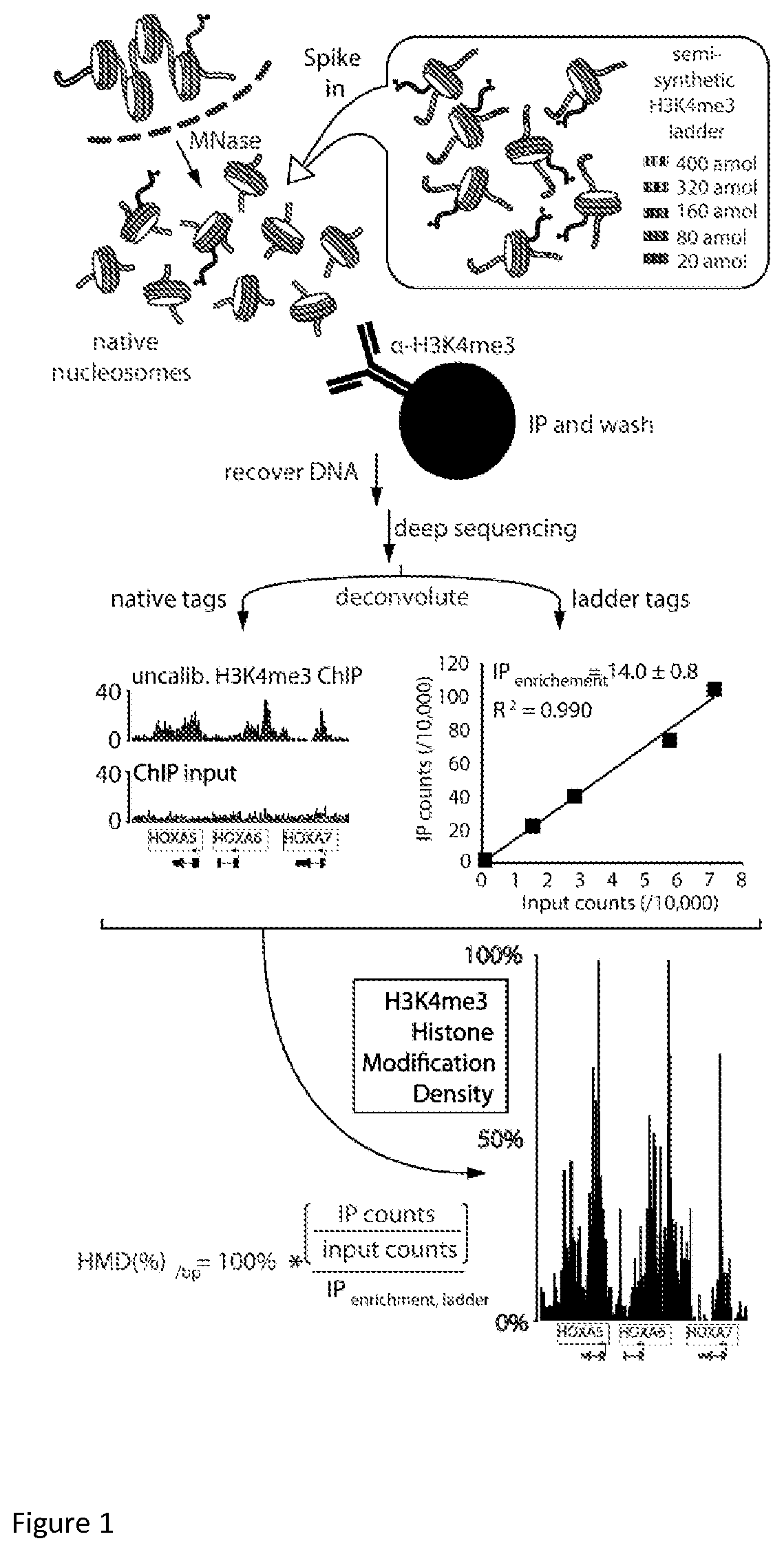
FIG. 1 is a schematic diagram of H3K4me3 ICe-ChIP-seq--one of the embodiments of calibrated chromatin immunoprecipitation experiments.
FIG. 2 illustrates the design and preparation of barcoded semisynthetic nucleosomes. Schematic depiction of the reconstitution of a semisynthetic H3K4me3 nucleosome ladder: histone octamers, produced by refolding equimolar core histones from recombinant and semisynthetic sources, are purified then mixed with equal amounts of barcoded ladder DNA. Schematic representation of barcoded nucleosome positioning DNA sequences based on the 601 positioning nucleosome sequence.
FIG. 3: (A) Amplification per cycle of barcoded ladder DNA is measured with qPCR utilizing a 2.times. serial dilution series fit by linear regression (R.sup.2 of the fit displayed in each bar). (B) Amplification per cycle of all barcoded DNA ladder members versus native genomic DNA fragments after ligation of sequencing adaptors with primers that hybridize to these adaptors.
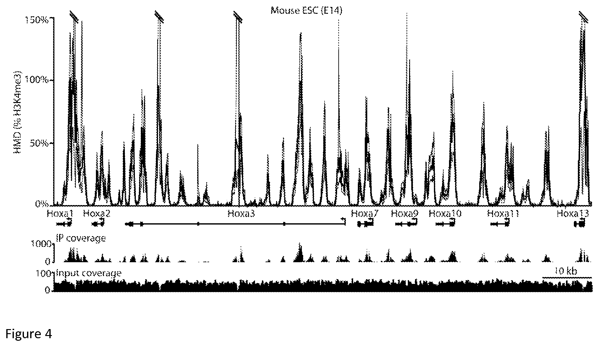
FIG. 4: The H3K4me3 ICe-ChIP-seq of mESCs E14 cell line shows Histone Modification Density to be within expected range. The top graph represents actual H3K4me3 Histone Modification Density for HOXA gene cluster in the mESC E14 cell line as a function of chromosomal coordinate for Chr6. ICeChIP coupled to Illumina paired-end sequencing reveals H3K4me3 modification density per base pair (HMD, darker line, 95% confidence interval, lighter line)) at the Hoxa gene cluster in the E14 mESC line as a function of chromosomal coordinate. Coding and non-coding genes are marked with bars and directional arrows below each graph. The small peaks below represent H3K4me3 ChIP signal (top) and input signal (bottom), expressed in raw read count.
FIG. 5. A Critical examination of ICeChIP (A) The relative abundance of barcode tags normalized to the most abundant ladder member measured in IP and input from HEK293 H3K4me3 ICeChIP-seq. (B) ICeChIP-seq compared to ddPCR and qPCR: the middle line represents uncorrected H3K4me3 Histone Modification Density (HMD) .+-.95% CI (top and bottom lines) in the mESC E14 cell line as a function of chromosomal windows. bars represent H3K4me3 measured by ddPCR and qPCR respectively on the same HMD scale (error bars are 95% CI), positioned over the indicated amplicon.
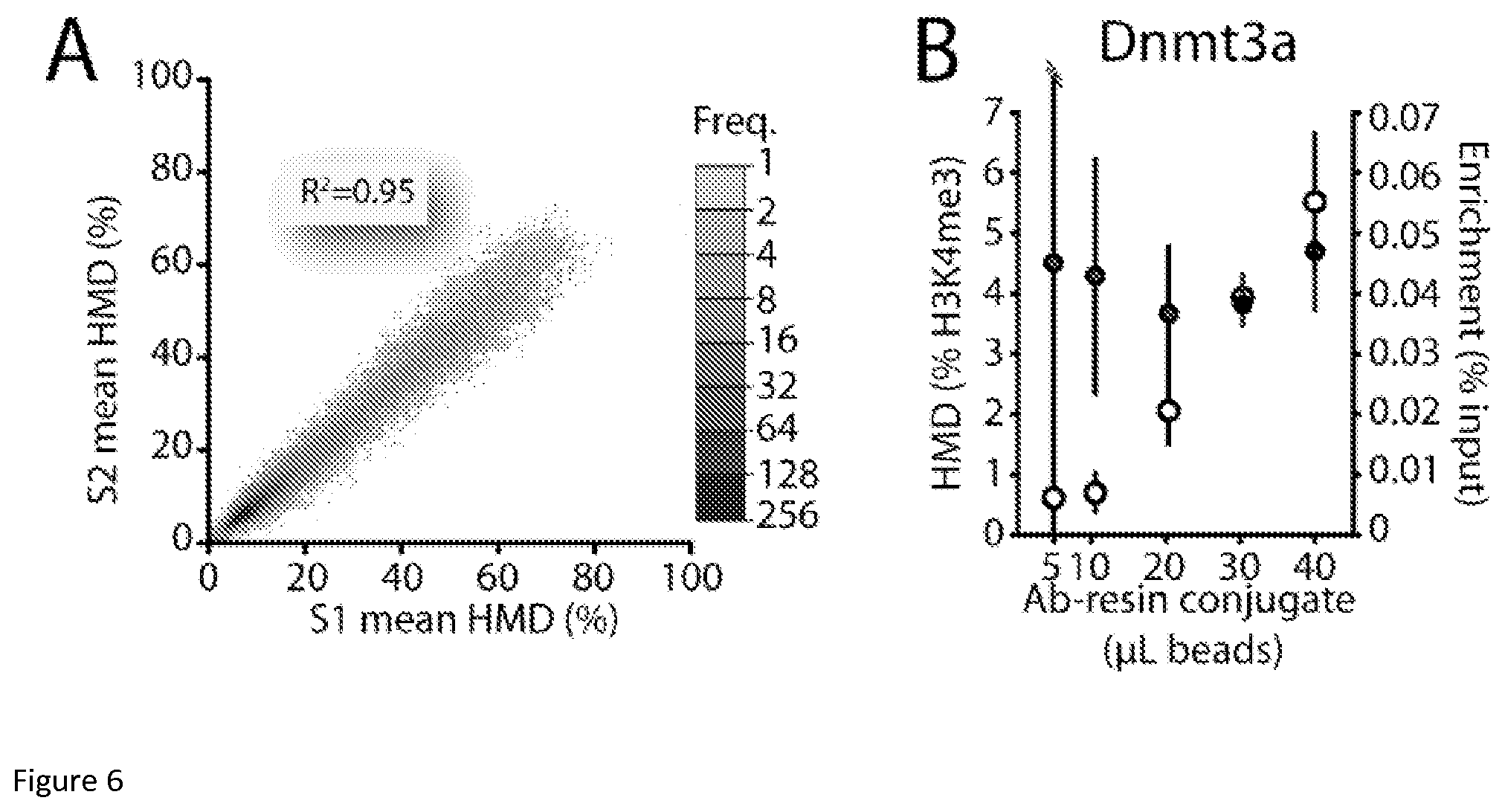
FIG. 6: ICeChIP is highly reproducible and more robust to experimental differences than conventional ChIP (A) Scatter plot comparison of the two samples (51 and S2) via plotting the mean mononucleosome HMD (% H3K4me3) for called peaks at the same loci. (B) Measurement of HMD (% H3K4me3) versus enrichment (% IP/input, representing the conventional way of presenting ChIP data) at the DNMT3a locus by ICeChIP-qPCR in mESCs as a function of antibody-resin conjugate with fixed 10 .mu.g of chromatin input.
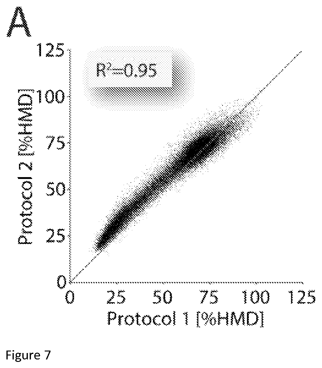
FIG. 7: The reproducibility and robustness of ICeChIP. (A) Comparison of two H3K27me3-directed ICeChIP experiments from Drosophila S2 cells, staged from the same input but with great variation in IP and washes. Sample 1 data was generated using our standard ICeChIP conditions (15 minute incubation of resin-Ab conjugate with input, followed by five washes over 50 minutes) whereas the Sample 2 IP was performed with a shorter incubation and flow washes of the resin with the same volumes applied over the span of one minute. Each data point corresponds to mean H3K27me3 averaged over 3000 bp non-overlapping window (N=41158); windows with insufficient input depth were excluded from analysis (cut-off>5). Data pooled from technical triplicates for each protocol (independent IPs and measurements).
FIG. 8: ICeChIP with multiple internal standards. Chromatin input titration for a small scale ICeChIP experiment as presented in FIG. 9. The method works well down to the chromatin equivalent of 400 cells.
FIG. 9: ICeChIP with multiple internal standards reveals the specificity of the IP in situ. (A) A comparison of internal standard capture (unmodified, H3K4me3, H3K9me3, H3K27me3, H3K36me3, H3K79me2 barcoded nucleosome ladders simultaneously doped in equimolar concentration) in five multi-standard ICeChIP-seq experiments with antibodies to each of the methyl marks. The data, presented as relative IP-efficiency, normalized to the on-target ladder, permit facile comparison to potential off-target methylated nucleosomes, as well unmodified nucleosomes. (B) Calculation of IP-enrichment in multi-standard ICeChIP experiments from mESCs presented as raw ladder member read counts in the IP versus input for the on-target mark, as well as the highest off-target background ladders for H3K4me3 (Active Motif AM39159), (C) H3K9me3 (M309M3-A (Hattori et al., 2013)), (D) H3K27me3 (Millipore 07-449)
DETAILED DESCRIPTION
Unless otherwise defined, all technical and scientific terms used herein have the same meaning as commonly understood by one of ordinary skill in the art to which this invention pertains. In case of conflict, the present document, including definitions, will control. Preferred methods and materials are described below, although methods and materials similar or equivalent to those described herein can be used in the practice or testing of the present invention.
The uses of the terms "a" and "an" and "the" and similar references in the context of describing the invention (especially in the context of the following claims) are to be construed to cover both the singular and the plural, unless otherwise indicated herein or clearly contradicted by context. Recitation of ranges of values herein are merely intended to serve as a shorthand method of referring individually to each separate value falling within the range, unless otherwise indicated herein, and each separate value is incorporated into the specification as if it were individually recited herein. All methods described herein can be performed in any suitable order unless otherwise indicated herein or otherwise clearly contradicted by context. The use of any and all examples, or exemplary language (e.g., "such as", "for example") provided herein, is intended merely to better illuminate the invention and does not pose a limitation on the scope of the invention unless otherwise claimed. No language in the specification should be construed as indicating any non-claimed element as essential to the practice of the invention.
I) Definitions
The term "Epitope" refers to any site on biomolecule that can evoke binding of affinity reagent. Affinity reagent might recognize linear sequence of biomolecule or biomolecule fragment, shape of biomolecule or biomolecule fragment, chemo-physical property of biomolecule or it fragment or combination of these.
"Amino acids" may be referred to herein by either their commonly known three letter symbols or by the one-letter symbols recommended by the IUPAC-IUB Biochemical Nomenclature Commission Amino acid residues in proteins or peptides are abbreviated as follows: phenylalanine is Phe or F; leucine is Leu or L; isoleucine is Ile or I; methionine is Met or M; valine is Val or V; serine is Ser or S; proline is Pro or P; threonine is Thr or T; alanine is Ala or A; tyrosine is Tyr or Y; histidine is His or H; glutamine is Gln or Q; asparagine is Asn or N; lysine is Lys or K; aspartic acid is Asp or D; glutamic Acid is Glu or E; cysteine is Cys or C; tryptophan is Trp or W; arginine is Arg or R; and glycine is Gly or G.
The term "amino acid" refers to naturally occurring and non-natural amino acids, as well as amino acid analogs and amino acid mimetics that function in a manner similar to the naturally occurring amino acids. Naturally encoded amino acids are the 20 common amino acids (alanine, arginine, asparagine, aspartic acid, cysteine, glutamine, glutamic acid, glycine, histidine, isoleucine, leucine, lysine, methionine, phenylalanine, proline, serine, threonine, tryptophan, tyrosine, and valine) and pyrrolysine and selenocysteine Amino acid analogs refers to compounds that have the same basic chemical structure as a naturally occurring amino acid, i.e., an a carbon that is bound to a hydrogen, a carboxyl group, an amino group, and an R group, such as, homoserine, norleucine, methionine sulfoxide, methionine methyl sulfonium. Such analogs have modified R groups (such as, norleucine) or modified peptide backbones, but retain the same basic chemical structure as a naturally occurring amino acid.
As to amino acid sequences, one of skill will recognize that individual substitutions, deletions or additions to a nucleic acid, peptide, polypeptide, or protein sequence which alters, adds or deletes a single amino acid or a small percentage of amino acids in the encoded sequence is a "conservatively modified variant" where the alteration results in the substitution of an amino acid with a chemically similar amino acid. Conservative substitution tables providing functionally similar amino acids are known to those of ordinary skill in the art. Such conservatively modified variants are in addition to and do not exclude polymorphic variants, interspecies homologs/orthologs, and alleles of the agents described herein.
An "antigen" as used herein may be any amino acid fragment (modified or unmodified) of 5 amino acids or more which are recognized by an antibody or for which recognizing antibodies can be raised. In certain embodiments, antigens may comprise modifications of an amino acid, such as acetylation, methylation (e.g. mono-, di-, tri-), phosphorylation, ubiquitination e.g. mono-, di-, tri-, poly-), sumoylation, ADP-ribosylation, citullination, biotinylation, and cis-trans isomerization. In other embodiments, antigens may comprise specific mutations, such as point mutations. In other yet embodiments, antigens may comprise wild-type amino acid sequence.
The terms "polypeptide," "peptide" and "protein" are used interchangeably herein to refer to a polymer of amino acid residues. That is, a description directed to a polypeptide applies equally to a description of a peptide and a description of a protein, and vice versa. The terms apply to naturally occurring amino acid polymers as well as amino acid polymers in which one or more amino acid residues is a non-natural amino acid. As used herein, the terms encompass amino acid chains of any length, including full length proteins, wherein the amino acid residues are linked by covalent peptide and/or pseudopeptide bonds.
The term "post-translational modification" refers to any modification of a natural or non-natural amino acid that occurs or would occur to such an amino acid after it has been incorporated into a polypeptide chain in vivo or in vitro. Such modifications include, but are not limited to, acetylation, methylation (e.g. mono-, di-, tri-), phosphorylation, ubiquitination (e.g. mono-, di-, tri-, poly-), sumoylation, ADP-ribosylation, citullination, biotinylation, and cis-trans isomerization. Such modifications may be introduced synthetically, e.g. chemically, during polypeptide synthesis or enzymatically after polypeptide synthesis or polypeptide purification.
The term "immunoprecipitation (IP) enrichment" refers to the internal standard reads from the immunoprecipitated sample divided by the internal standard reads from the input sample.
The term "asymmetric" refers to a nucleosome wherein one histone within a dimer of histones contains a post-translational modification. For example, the trimethyl modification is found on lysine 9 of one histone H3 but absent on the second H3 within a dimer.
The term "symmetric" refers to a nucleosome wherein both histones within a dimer of histones contain a post-translational modification. For example, the trimethyl modification is found on lysine 9 of both histone H3.
II) Internal Standard Calibrated Chip (Icechip)
Currently performed pull-down assays suffer from arbitrary of the units of measurement, which makes any kind of comparison between any kind of pull-down experiment highly inaccurate and hinders use of pull-down assays in medical diagnostics and research. Accuracy of data interpretation is improved by a standardized scale with absolute units by uncoupling test outcome values from the assay and coupling them to actual biological phenomenon. One aspect of the present invention provides materials and methods enabling the use of pull-down assays in medical diagnostics such as in assays identifying disease markers. In these methods, the data resulting from the pull-down assay, such as ChIP, are characterized not by arbitrary values specific for an assay but by absolute values specific for the disease marker itself. This means that results from pull-downs of different samples, different pull-downs of the same sample, pull-downs of different epitopes, pull-downs performed in different laboratories may be readily and directly compared to each other which is often impossible with currently available methods and technologies.
One aspect of the invention includes a method of absolute assessment of DNA bound proteins, protein isoforms, and protein post-translational modification densities that we call Internal Standard Calibrated ChIP (ICeChIP). This method provides the first local measurement of histone modifications on a biologically meaningful scale. This improvement of ChIP utilizes a non-naturally occurring internal standard to which ChIP readout may be compared. As an internal standard, we have developed recombinant and semi-synthetic protein-DNA complexes engineered to contain epitopes with native-like affinity, specificity and avidity characteristics.
These protein-DNA complexes include nucleosomes bearing protein epitopes with native-like affinity, specificity and avidity for an affinity reagent, and a DNA sequence including a standard recognition molecule comprising a positioning sequence and a unique sequence or barcode. The "barcode", which provides a unique means of specific recognition of the DNA-protein complex, may be for example a nucleotide sequence such as DNA, a polypeptide, fluorophore, chromophore, RNA sequence, locked nucleic acid sequence, affinity tag etc., that identifies the identity and/or concentration of a specific standard semi-synthetic nucleosome. Here, the term "native-like" refers to any protein epitope having affinity, specificity and avidity properties similar to naturally occurring epitopes.
FIG. 1 shows one embodiment of an ICeChIP assay. In this schematic, a semi-synthetic nucleosome ladder of standards with modified histone H3 carrying N6,N6,N6-trimethylation of lysine 4 in defined concentrations (encoded by each unique DNA barcodes) is doped into a library of native nucleosomes isolated from human nuclei and released by in nucleo digestion with micrococcal nuclease. A sample of the ladder-doped library is then subjected to immunoprecipitation (IP), DNA purification and Next-Generation-Sequencing. Another sample of the ladder-doped library is retained as an input sample and is not subject to immunoprecipitation. Here, Immunoprecipitation (IP) or "pull-down" refers to a method or technique for purifying chromatin, nucleosomes, DNA-proteins complexes, or proteins including one or more epitopes of interest where the epitope is contacted with an affinity reagent specific to an epitope and separated from other components of the library.
The immunoprecipitated sample and the input sample are subject to a method with capability to read out and quantify DNA sequences. Recovered DNA fragments are mapped to the relative genomic position based on reference genome and abundance of these fragments is measured for every base pair of the genome for DNA recovered from IP (the sample produced through immunoprecipitation using an affinity reagent) and input (the sample not subject to immunoprecipitation). The same read counting from the sequencing data is performed for the unique nucleotide sequences used to make semi-synthetic nucleosomes. The ratio of abundance of semi-synthetic nucleosomes in IP and input is used to measure IP efficiency and the ratio of abundance of DNA fragments for any genomic loci in IP and input is used to measure relative enrichment. The resulting tag counts for the added semisynthetic nucleosomes constitute a calibration curve to derive histone modification density for native nucleosomes genome-wide. The average IP-enrichment ratio for the semi-synthetic nucleosome ladder bearing 100% of the modification is used as a scalar correction for native chromatin bearing the same epitope to compute the amount of modification over a desired genomic interval as a ratio of ratios. Subsequently IP efficiency is applied to relative enrichment to measure histone modification density of H3K4me3 histone post-translational modification with base pair resolution for the span of the whole genome. In some embodiments, protein epitopes having native-like affinity, specificity and avidity include a protein isoform and/or protein having a post-translational modification. For example, the epitope may be the histone modification to whose density is measured in the assay or an epitope having similar binding characteristics. In a preferred embodiment, the protein part of a DNA-protein complex is a core histone octamer complex containing core histones H2A, H2B, H3, H4. These sequences are described in Patent Application No: US2013/044537, the contents of which are incorporated by reference. In order to reproduce native-like affinity, specificity and avidity of the protein epitope for any of the aforementioned core histones can be represented by any histone variant including those in listed in Table 1.alpha.-f-. In one embodiment of the invention, the protein epitope may be a fragment of a histone.
In another aspect of the invention, the protein-DNA complexes comprise a standard recognition molecule comprising but not limited to a positioning sequence and a unique sequence or barcode. Inclusion of a protein positioning sequence allows for the creation of a DNA-protein complex through specific native-like interaction with protein. In a preferred embodiment, the protein positioning sequence is a nucleosome positioning sequence. In one embodiment, the positioning sequence comprises a natural or synthetic double-stranded DNA sequence of at least 146 base pairs. In a more preferred embodiment, the protein positioning sequence is a "601-Widom" sequence--a synthetic nucleosome binding sequence made through a selection of sequences which exhibited affinity toward a nucleosome. While we have mentioned here a "601-Widom" sequence as a nucleosome positioning sequence the present embodiments encompass the use of other such synthetic and native sequences which exhibit affinity toward nucleosomes.
A unique sequence allows for specific identification of a DNA-protein complex in a library or pool of native DNA-protein complexes i.e. a barcode. In some embodiments the unique sequence can be substituted with another means of specific recognition e.g. a polypeptide, fluorophore, chromophore, RNA sequence, locked nucleic acid sequence, affinity tag etc. In one aspect, the unique sequence can be analyzed by known nucleotide analysis for example Next-Generation sequencing, qPCR. RT-PCR, or ddPCR. A unique sequence and a positioning sequence might be the same sequence and serve a dual function as the recognition molecule. The unique sequence may reside at the 5'-end of the positioning sequence, the 3' end of the positioning sequence, or at both ends of the positioning sequence.
In a preferred embodiment, a unique sequence is a duplex DNA sequence with minimal length to maintain a Hamming distance of at least 1 from the genomic sequence of the organism that is being investigated and all other sequences that might be found in the sample. In a more preferred embodiment, to guarantee robust discrimination of barcodes in the milieu of native genomic sequences, each barcode is made out of two 11 base pair (bp) sequences absent in human and mice genome (Herold et al., 2008), where 11 bp sequences is the shortest sequence guaranteeing Hamming distance of at least 1 for human and mice genome. In another embodiment, the barcode sequence is a sequence not present in the genome of the cell. In another embodiment, the barcode sequence is a sequence not present in nature. While 11 bp are mentioned here as the shortest possible sequence with Hamming distance of at least 1 for human and mouse there is unlimited number of longer sequences with Hamming distance of at least 1 which can be successfully used to serve as aforementioned unique sequences. Moreover the shortest sequence of unique sequence with Hamming distance of at least 1 for genomes of other organisms might be shorter than 11 bp and as such, shorter sequences than 11 bp might be successfully used for these organisms. The barcode is a molecule, in a preferred embodiment it is DNA, that can be analyzed by known DNA analysis comprising but not limited to Next-Generation sequencing and PCR. The barcode sequence encodes a concentration and/or identity of a given internal standard nucleosome.
In a preferred embodiment, a unique nucleotide sequence indicates the concentration and identity of a given internal standard. In one aspect of the invention, a unique sequence comprises a length of at least or at most 10, 11, 12, 13, 14, 15, 16, 17, 18, 19, 20, 25, 30, 35, 40, 45, 50, 60, 70, 80, 90 or 100 base pairs in length. In yet another embodiment, the total length of the positioning sequence and unique sequence has a length of at least 100 base pairs. In a preferred embodiment, a positioning sequence and a unique sequence are selected from Table 7. In one aspect, the unique sequence is micrococcal nuclease resistant. In one embodiment of the invention the standard molecule comprising but not limited to a positioning sequence and a unique sequence or barcode includes SEQ ID NO:1; SEQ ID NO:2; SEQ ID NO:3; SEQ ID NO:4; SEQ ID NO:5; SEQ ID NO:6; SEQ ID NO:7; SEQ ID NO:8; SEQ ID NO:9; SEQ ID NO:10; SEQ ID NO:11; SEQ ID NO:12; SEQ ID NO:13; SEQ ID NO:14; or SEQ ID NO:15. In a preferred embodiment, the standard molecule comprising but not limited to a positioning sequence and a unique sequence or barcode includes SEQ ID NO:16; SEQ ID NO:17; SEQ ID NO:18; SEQ ID NO:19; SEQ ID NO:20; SEQ ID NO:21; SEQ ID NO:22; SEQ ID NO:23; SEQ ID NO:24; SEQ ID NO:25; SEQ ID NO:26; SEQ ID NO:27; SEQ ID NO:28; SEQ ID NO:29; SEQ ID NO:30; SEQ ID NO:31; SEQ ID NO:32; SEQ ID NO:33; SEQ ID NO:34; SEQ ID NO:35; SEQ ID NO:36; SEQ ID NO:37; SEQ ID NO:38; SEQ ID NO:39; SEQ ID NO:40; SEQ ID NO:41; SEQ ID NO:42; SEQ ID NO:43; SEQ ID NO:44; SEQ ID NO:45; SEQ ID NO:46; SEQ ID NO:47; SEQ ID NO:48; SEQ ID NO:49; SEQ ID NO:50; SEQ ID NO:51; SEQ ID NO:52; SEQ ID NO:53; SEQ ID NO:54; SEQ ID NO:55; SEQ ID NO:56; SEQ ID NO:57; SEQ ID NO:58; SEQ ID NO:59; SEQ ID NO:60; SEQ ID NO:61; SEQ ID NO:62; SEQ ID NO:63; SEQ ID NO:64; SEQ ID NO:65; SEQ ID NO:66; SEQ ID NO:67; SEQ ID NO:68; SEQ ID NO:69; SEQ ID NO:70; SEQ ID NO:71; SEQ ID NO:72; SEQ ID NO:73; SEQ ID NO:74; SEQ ID NO: SEQ ID NO:75; SEQ ID NO:76; SEQ ID NO:77; SEQ ID NO:78; SEQ ID NO:79; SEQ ID NO:80; SEQ ID NO:81; SEQ ID NO:82; SEQ ID NO:83; SEQ ID NO:84; SEQ ID NO:85; SEQ ID NO:86; SEQ ID NO:87; SEQ ID NO:88; SEQ ID NO:89; SEQ ID NO:90; SEQ ID NO:91; SEQ ID NO:92; SEQ ID NO:93; SEQ ID NO:94; SEQ ID NO:95; SEQ ID NO:96; SEQ ID NO:97; SEQ ID NO:98; SEQ ID NO:99; SEQ ID NO:100; SEQ ID NO:101; SEQ ID NO:102; SEQ ID NO:103; SEQ ID NO:104; SEQ ID NO: 105; SEQ ID NO:106 SEQ ID NO:107; SEQ ID NO:108; SEQ ID NO:109; SEQ ID NO:110; SEQ ID NO:111; SEQ ID NO:112; SEQ ID NO:113; SEQ ID NO:114; or SEQ ID NO:115.
In one embodiment of the method of determining epitope density as described wherein, a set of the aforementioned semi-synthetic nucleosomes with the standard recognition molecule is doped into a collection of native nucleosomes. The set may comprise of semi-synthetic nucleosomes with the standard recognition molecule harboring more than one epitope but comprising at least one epitope of interest. For example, a set of semi-synthetic nucleosomes may harbor the post-translational modification i.e. H3K9me3 and a conserved or invariant epitope such as the polypeptide sequence of the histone. Alternatively, a set of semi-synthetic nucleosomes may harbor more than one post-translational modification such as H3K9me3 or insert second epitope. In another aspect, the set of standards comprises at least one semi-synthetic, reconstituted, or variant-containing DNA-binding protein with native-like affinity, specificity and avidity of a false positive epitope that is different than the epitope of interest. In a preferred embodiment a set of semi-synthetic or variant containing nucleosomes including at least one nucleosome with native-like affinity, specificity and avidity of a true positive epitope and at least one nucleosome with native-like affinity, specificity and avidity of a false positive epitope.
To purify a population of native or semi-synthetic nucleosomes from a pool of protein-DNA complexes one may use an affinity capture step where an affinity reagent recognizes an invariant fragment of the nucleosome for example the histone. In one aspect the affinity reagent contacting the epitope of interest comprises an antibody, a monobody, an aptamer, a Fab, or a binding peptide. The method of purifying a population of nucleosomes may apply to semi-synthetic nucleosomes alone, native nucleosomes alone, or a native nucleosomes doped with semi-synthetic nucleosomes.
ICe-ChIP Data Analysis
In one embodiment, to perform ICe-ChIP a set of the aforementioned internal standards to which a ChIP read-out can be compared, is doped into a collection of native DNA-protein complexes. Below we describe how these standards are used to calculate Standard IP efficiency, which in turn can be used to calculate what we have called Protein or Epitope Density (PD), Protein Variant Density (PVD), or Protein Modification Density (PMD), depending whether the investigated epitope is an invariant protein fragment, protein isoform, or protein post-translational modification. Standards based on semi-synthetic or variant containing nucleosomes with native-like affinity, specificity and avidity improve a chromatin immunoprecipitation by allowing one to perform absolute quantification of Histone Modification Density (HMD) or Histone Variant Density (HVD).
Histone Modification Density is a standardized scale and is defined as the apparent percentage of nucleosomes bearing a specific epitope out of all nucleosomes in a given genomic position. Histone Modification Density is expressed on an analog scale ranging between 0%, meaning absence, and 100% meaning saturating presence of the epitope. For example 90% H3K4me3 Histone Modification Density for nucleosome+1 (the first nucleosome downstream of transcription start site) of GAPDH gene should be interpreted that in the population of all histone H3 molecules composing nucleosome+1 at the GAPDH gene promoter, 90% of them bear post translational modification N6,N6,N6-trimethylation of lysine 4 of histone H3 (H3K4me3) and 10% should be free of H3K4me3. While this example was given for region of genome spanning a single nucleosome, which is roughly 147 bp, the same can be applied to any span of the genome ranging from single base pair to the whole genome.
In order to calculate Protein or Epitope density one needs to know four things: genomic locus size, epitope abundance, general protein abundance, and ImmunoPrecipitation efficiency ("IP efficiency".) Genomic locus size is defined by the user and can range from a single base pair to the whole genome. Epitope abundance is defined as the abundance of the epitope over the span of the genomic locus. Abundance is usually inferred by quantifying the amount of DNA bound to DNA-protein complex as it is stoichiometric to protein and DNA is easy to quantify with numerous methods e.g. PCR, RT-PCR, ddPCR, Next-Generation-Sequencing, hybridization, autoradiography, fluorescent labeling, optical density, intercalating fluorescent probes etc. However, abundance may also be measured directly by measuring protein concentration through optical density, fluorescence, autoradiography, mass spectrometry, colorimetric assay, polypeptide total decomposition etc.
Epitope abundance is measured after an affinity capture step in which a specific affinity reagent recognizes the epitope, after which step epitope-affinity reagent complex is separated from unbound population of DNA-protein complexes. Most often epitope-affinity reagent complex is separated from unbound nucleosomes by immobilizing epitope-affinity reagent complex on the surface and washing away the unbound population of DNA-protein complexes. General protein abundance is defined as the abundance of all proteins of a given kind making DNA-complexes within the span of the given genomic locus. General protein abundance is measured with the same methods as epitope abundance.
To purify a population of nucleosomes from other protein-DNA complexes one can use an affinity capture step where an affinity reagent recognizes an invariant fragment of the nucleosome, for example the histone. However, if a given invariant fragment involved in making the protein-DNA complex is dominant over a considered genomic locus size then the affinity capture step for general protein population can be skipped under assumption that the population of other protein-DNA complexes is insignificant. The ratio of epitope abundance and general protein abundance should yield epitope density per protein. However it is rarely the case as the affinity capture step is 100% efficient and if two or more affinity capture steps are utilized their capture efficiencies will rarely be equal to each other. To solve this problem one needs to know relative IP efficiency between epitope abundance and general protein abundance measurement.
The "IP efficiency" refers to the relative recovery of the epitope between one or more pull-down. Knowledge of IP efficiency for the standard allows performing absolute quantification by correcting for differences in recovery between one or more pull-downs. In one embodiment, the aforementioned IP efficiency is measured by using a set of the aforementioned standards that has the same affinity, specificity and avidity as the native epitope and which abundance is easy to measure in a complex mixture. These semi-synthetic standards are doped into a pool of native DNA-Protein complexes, a sample of which will be subject to affinity capture. Following this step, the aforementioned measurements of epitope abundance and general protein density is performed for the semi-synthetic standards and the pool of native DNA-protein complexes population with one of the mentioned abundance measurement methods. In one embodiment, the set of standards includes standards that are added at differing concentrations. Here the concentration added is uniquely identified by the barcode.
In one embodiment, epitope abundance can be measured through quantification of DNA bound to DNA-protein complexes for standard DNA-protein complexes and native DNA-protein complexes. In a preferred embodiment, the ratio of epitope of a given standard barcode in the IP versus input material for semi-synthetic nucleosomes is equal to Standard IP Efficiency. Alternatively this Standard IP efficiency may be computed as a ratio of barcode abundance in the epitope-specific IP versus general protein abundance (for histone H3, for example the barcode counts in the anti-H3 general IP). Once IP efficiency is calculated, one may apply this Standard IP efficiency to IP/input DNA or IP-epitope/IP-general protein ratios any genomic locus. This is calculated by dividing the genomic IP efficiency--ratio of the epitope abundance in the IP (amount of DNA for a given genomic interval captured in the affinity step) to the amount of DNA covering the same interval present in the input--by the Standard IP efficiency. Alternatively this may be computed as the ratio of a given genomic DNA fragment in the IP divided amount of the same species in the general epitope abundance IP for any genomic locus as described above and then dividing by Standard IP efficiency. The resultant value is a Protein or Epitope Density (PD), also known as a Protein Variant Density (PVD), or Protein Modification Density (PMD).
.function..times..times..times..times..times. ##EQU00001## Correction of Off-Target Specificity
Another problem challenging analysis of pull-down experiments is the low precision of prediction stemming from off-target specificity of an affinity reagent used in a pull-down assay. The terms "false positive" and "off-target" are synonymous and refer to an epitope that contacts an affinity reagent promiscuously or non-specifically or an incorrect result. The term "true positive" and "on-target" are synonymous and refers to an epitope of interest or correct result.
Prevalence of false positive epitope signal varies between pull-down to pull-down and depends on the quality of affinity reagent (its intrinsic binding affinity for the desired epitope versus its affinity for other related epitopes), the abundance of on-versus off-target epitope in the native chromatin, the ratio of capacity of affinity reagent and loading levels of DNA-protein complexes in a pull-down, as well as other conditions under which the pull-down is performed. For different affinity reagents, on- and off-target binding both contribute to the apparent ChIP signal to different degrees, the extent to which either source contributes within a given experiment with conventional ChIP is unknown. In the absence of knowledge of the abundance of off-target binding, one cannot make a decision whether observed epitope abundance is significant or not, which in turn makes use of pull-down in medical diagnostics and research impractical. The inventors have found a method to quantitate IP efficiency of false positive and true positive epitopes in a pull-down assay in situ, which improves precision of data interpretation as Positive Predictive Value (PPV) may be readily calculated. PPV allows for an estimation of minimal abundance of epitope at a certain confidence level to be considered a true positive.
Using and the aforementioned methods of calculating IP efficiency and Standard IP efficiency, Positive Predictive Value (PPV) also referred to as Precision may be calculated. Knowledge of PPV streamlines any data analysis as it allows estimation of whether any difference in Protein Density is significant or not, which is not achievable with currently available methods and techniques.
.alpha..eta..alpha..eta..times..beta..eta. ##EQU00002##
.eta.TP is IP efficiency of true positive epitope and a is a given weight of true positive epitope, .eta.FP is IP efficiency of false positive epitope, also known as off-target epitope and .beta. is a weight of false positive epitope. In the absence of prior knowledge of weight distribution .alpha.=.beta.=1. Other variants of this equation exist and use of knowledge of false positive and true positive epitope prevalence can be used in other applications.
There are two alternate ways to calibrate ChIP: global histone modification density calibration using an external standard and direct internal standard calibration. Like the relative internal standard approach that was predominantly employed in this work, these two can yield results expressed in "histone modification density" units, which are equal to apparent ratio of probed epitope to all other epitopes available in the given locus.
Global histone modification density calibration relies on a measurement of the total ratio of modification relative to the amount of histone, for example, knowing the percentage of all H3 that is K4 trimethylated. This global histone modification density, derived from either mass spectrometry or quantitative western blot measurements can be then redistributed among all IP peaks corrected for input depth in any given locus. The drawback of this method, apart from the sizable error in making the global abundance measurement (for example, MS accuracy plus the ambiguity of perhaps not observing all potential forms of the modification), is that such external measurements by orthogonal methodologies need to be made from the same nucleosomal sample used in the ChIP, and sample handling losses in both techniques are a considerable source of error. In particular, IP-efficiency is never 100% (in practice this can be considerably less), so the degree by which efficiency deviates from the theoretical maximum will be reflected in commensurately inflated values for apparent HMD.
Direct internal standard calibration measures the tag count of a spiked-in barcoded nucleosome standard through the ChIP process, knowing the precise molar concentrations of each internal standard ladder member in the input to extrapolate absolute molar abundance of probed epitope in the original sample. This sort of calibration is limited by the accuracy of counting the number of nuclei subjected to the micrococcal nuclease digest and biased loses that mount on the way from this well quantified number to exhaustively fragmented chromatin isolate. As we recover little more than 80% of the total nucleic acid from digested nuclei under highly optimized digest and isolation conditions, there is some systematic error due to biased genome recovery (Henikoff et al., 2009).
Yet another advantage of this embodiment is ability to deconvolute the true positive epitope signal from false positive epitope signal, presented here on the example of histone modification density, by solving the following matrix equation: A*x=b. For indicated datasets, ICeChIP-seq tracks were corrected for off-specificity by solving the following matrix equation: A*x=b,
Another embodiment of the invention describes a method to deconvolute the true positive epitope signal from false positive epitope signal, presented here is the example of histone modification density, by solving the following matrix equation: A*x=b
.times..function..function. .function..function. ##EQU00003##
where, x is a matrix of corrected HMD scores, A is a matrix of correction factors and b is a matrix of non-corrected HMD scores, where, t is correction factor for specificity toward histone marks from the set of `a` to `z` histone marks (subscript), in the immunoprecipitation using antibody toward a histone mark from the set of `a` to `z` histone marks (superscript); HMD is histone modification density for a given histone mark (`a` to `z`) from the 1st to the nth locus; HMD(Cor) is corrected histone modification density for a given histone mark from the 1st to the nth locus,
.times..times..times..times. ##EQU00004##
where, t is correction factor for specificity toward histone marks from the set of `a` to `z` histone marks (subscript), in the immunoprecipitation using antibody toward a histone mark from the set of `a` to `z` histone marks (superscript); HMD is histone modification density for a given histone mark (`a` to `z`) from the 1st to the nth locus; HMD(Cor) is corrected histone modification density for a given histone mark from the 1st to the nth locus,
.times..times..times..times. ##EQU00005##
where, .SIGMA..sub.1.sup.N IP and .SIGMA..sub.1.sup.N input refer to abundance of the given barcode in the IP or in the input, superscript refers to histone mark toward which antibody was raised, while subscript refers to mark on the semisynthetic nucleosome that was pulled-down.
Disease Diagnosis
The main reasons why conventional ChIP assays have not been adopted in the clinic is that they are often irreproducible due to subtle handling differences and variable antibody specificity, making the % enrichment in the IP widely variant from experiment to experiment, and rendering unbiased comparisons problematic and unreliable. By virtue of having an internal standard that is subject to the steps of ChIP that are sensitive to variation, ICe-ChIP is far more robust in terms of replication and reliability of results, as demonstrated in FIGS. 6A, 6B, and 7A, and the numbers are readily compared as HMD is a universal, biologically relevant scale, made by direct in situ comparison to a well-defined internal standard.
Histone modifications and other epigenetic mechanisms are crucial for regulating gene activity and cellular processes. Different histone modifications regulate different processes, such as transcription, DNA replication, and DNA repair. Deregulation of any of these modifications can shift the balance of gene expression leading to aberrant epigenetic patterns and cellular abnormalities. For example, changes in histone post-translational modifications and variants have been detected in various cancers, and aberrant modification patterns are known to be drivers of disease in some cases (Daigle et al., 2011; Chi et al., 2010).
The present materials and methods can be used in the diagnosis, prognosis, classification, prediction of disease risk, detection of recurrence, selection of treatment, and evaluation of treatment efficacy for any disease associated with changes in histone post-translational modifications, including cancer in a patient, for example, a human patient. Such analyses could also be useful in conjunction with ex vivo culture of patient cells or induced pluripotency stem cells to assess the suitability of a given de-differentiation protocol for producing truly pluripotent stem cells, or the protocols for differentiating stem cells into specific cell types.
Any stage of progression can be detected, such as primary, metastatic, and recurrent cancer. Information regarding numerous types of cancer can be found, e.g., from the American Cancer Society (available on the worldwide web at cancer.org), or from, e.g., Harrison's Principles of Internal Medicine, (2005).
Certain aspects of the present invention provide methods for disease prognosis, such as estimating the likelihood of a patient developing cancer, classifying disease stages, and monitoring the efficacy of treatment in a patient with cancer. Such methods are based on the discovery that ICe-ChIP can be used to calibrate ChIP experiments to control for handling differences and antibody variability. Accordingly, by determining the level of a particular histone PTM (See, for example, Table 1) within a cell taken from the patient, including methylated histones as described herein, it is possible to determine whether or not the patient has a risk of developing a particular disease or has already developed a particular disease. For example, as described herein, quantification of histone PTM levels in cancerous tissues may be used for cancer prognosis or diagnosis.
In numerous embodiments of the present invention, the materials and methods described in certain aspects of the invention may be used to detect the levels of histone PTMs or variants in a biological sample at given genomic loci, thereby detecting the presence or absence of diseased cells in the biological sample. In some embodiments, the biological sample comprises a tissue sample from a tissue suspected of containing diseased cells, such as cancerous cells. Human chromatin DNA samples can be obtained by any means known in the art. In cases where a particular phenotype or disease is to be detected, histone-containing samples should be prepared from a tissue of interest, blood cells, or as appropriate, from cerebral spinal fluid. For example, histone-containing samples can be prepared from biopsy tissue to detect the histone PTM state associated with cancer.
As appropriate, the tissue or cells can be obtained by any method known in the art including by surgery. In other embodiments, a tissue sample known to contain cancerous cells, e.g., from a tumor, will be analyzed for the presence or quantity of histone PTMS at one or more of the histone PTM sites, such as those described in Table 1, to determine information about the disease, e.g., the efficacy of certain treatments, the survival expectancy of the individual, the presence of specific types of disease etc. In some embodiments, the methods may be used in conjunction with additional prognostic or diagnostic methods, e.g., detection of other disease markers, etc.
The materials and methods of certain aspects of the invention can be used to evaluate individuals known or suspected to have a disease, including cancer, or as a routine clinical test, e.g., in an individual not necessarily suspected to have a disease. Further diagnostic assays can be performed to confirm the status of disease in the individual.
Further, the present methods and materials may be used to assess the efficacy of a course of treatment. The efficacy of a treatment can be assessed by monitoring histone post-translational modifications or variant deposition using the methods and materials described herein over time in a mammal having a disease. For example, a reduction or absence of histone methylation in any of the methylation biomarkers as described herein in a biological sample taken from a mammal following a treatment, compared to a level in a sample taken from the mammal before, or earlier in, the treatment, indicates efficacious treatment. Detection of a histone PTM as described above can be used either alone, or in combination with other markers, for the diagnosis or prognosis of disease.
The materials and methods of certain embodiments can be used to determine the optimal course of treatment in a mammal with a disease. For example, the presence of methylated histone marks within certain methylation biomarkers as described herein or an increased quantity of methylation within certain of the methylation biomarkers can indicate a reduced survival expectancy of a mammal with cancer, thereby indicating a more aggressive treatment for the mammal. In addition, a correlation can be readily established between the presence, absence or quantity of methylation at a methylation biomarkers, as described herein, and the relative efficacy of one or another anti-cancer agent. Such analyses can be performed, e.g., retrospectively, i.e., by detecting methylation using the materials and methods described herein in one or more of the methylation biomarkers in samples taken previously from mammals that have subsequently undergone one or more types of anti-cancer therapy, and correlating the known efficacy of the treatment with the presence, absence or levels of methylation of one or more of the methylation biomarkers as described above.
In making a diagnosis, prognosis, risk assessment, classification, detection of recurrence or selection of therapy based on the presence, absence, or HMD of a particular histone PTM, the quantity of the PTM or variant may be compared to a threshold value that distinguishes between one diagnosis, prognosis, risk assessment, classification, etc., and another. For example, a threshold value can represent the degree of histone methylation that adequately distinguishes between cancer samples and normal biopsy samples with a desired level of sensitivity and specificity. With the use of ICe-ChIP the threshold value will not vary depending on the antibody used or the handling conditions. Threshold value or range can be determined by measuring the particular histone PTM of interest in diseased and normal samples using ICe-ChIP and then determining a value that distinguishes at least a majority of the cancer samples from a majority of non-cancer samples.
In some embodiments, the methods comprise recording a diagnosis, prognosis, risk assessment or classification, based on the histone PTM status determined from an individual. Any type of recordation is contemplated, including electronic recordation, e.g., by a computer.
Certain embodiments of the present invention provide for determination of histone post-translational modification status in a patient's cancer. The histone post-translational modification information may be used for cancer prognosis, assessment, classification and/or treatment. Cancers which may be examined by a method described herein may include, but are not limited to, renal cell carcinoma, glioma, gliosarcoma, anaplastic astrocytoma, medulloblastoma, lung cancer, small cell lung carcinoma, cervical carcinoma, colon cancer, rectal cancer, chordoma, throat cancer, Kaposi's sarcoma, lymphangiosarcoma, lymphangioendotheliosarcoma, colorectal cancer, endometrium cancer, ovarian cancer, breast cancer, pancreatic cancer, prostate cancer, renal cell carcinoma, hepatic carcinoma, bile duct carcinoma, choriocarcinoma, seminoma, testicular tumor, Wilms' tumor, Ewing's tumor, bladder carcinoma, angiosarcoma, endotheliosarcoma, adenocarcinoma, sweat gland carcinoma, sebaceous gland sarcoma, papillary sarcoma, papillary adenosarcoma, cystadenosarcoma, bronchogenic carcinoma, medullar carcinoma, mastocytoma, mesothelioma, synovioma, melanoma, leiomyosarcoma, rhabdomyosarcoma, neuroblastoma, retinoblastoma, oligodentroglioma, acoustic neuroma, hemangioblastoma, meningioma, pinealoma, ependymoma, craniopharyngioma, epithelial carcinoma, embryonic carcinoma, squamous cell carcinoma, base cell carcinoma, fibrosarcoma, myxoma, myxosarcoma, glioma, or liposarcoma.
In certain embodiments the following diseases may be diagnosed using the present methods and materials: Bacterial infections caused by Heliocobacter pylori, Listeria monocytogenes, Shigella flexneri, Anaplasma phagocytophilum, Chlamdophila, Epstein-Barr Virus, herpes, HIV, Schistosoma haematobium; Obesity, diabetes, heart disease, autism, fragile X syndrome, ATR-X syndrome, Angelman syndrome, Prader-Willi syndrome, Beckwith Wiedemann syndrome, Rett syndrome, Rubinstein-Taybi syndrome, Coffin-Lowry syndrome, Immunodeficiency-centrometric instability-facial anomalies syndrome, .alpha.-thalassaemia, leukemia, Huntington's disease, schizophrenia, bipolar disease, aging, dementia, Alzheimer's disease, Parkinson's disease, Cornelia de Langue syndrome, Kabuki syndrome, Sjogren's syndrome, Vitiligo, progressive systemic sclerosis, psoriasis, primary biliary cirrhosis, Crohn's disease and ulcerative colitis, Hashimoto's thyroiditis, Grave's disease, inflammatory bowel disease, atherosclerosis, and cardiac hypertrophy.
Reagents and Kits
Another aspect of the invention provides reagents and kits including reagents for carrying out one of the methods described herein. The reagents may be included suitable packages or containers. The kit may include one or more reagents containing standards as described herein for the absolute quantification of true positive and false positive epitopes, for example in a pull-down assay or chromatin immunoprecipitation assay. The kit may also include at least one affinity reagent as described herein, for example an antibody. The standards may have native-like affinity, specificity and avidity for a true positive epitope. The kit can also comprise at least one standard with native-like affinity, specificity and avidity of epitope for false positive epitope.
In another preferred embodiment, the aforementioned standards include DNA-protein complexes comprising semi-synthetic nucleosomes, made with histones, histone isoforms or histone post-translational modifications with native-like affinity, specificity and avidity and a barcode molecule. In various embodiments, any variant of core histone sequences, which are known in the art, or post-translational modification, including those defined in Table 1, can be installed on the histones that comprise the histone octamer under presumption that native-like affinity, specificity and avidity of epitope is maintained. In a preferred embodiment, a set of standards is comprised of at least a single standard of DNA-complexes with native-like affinity, specificity and avidity of epitope for true positive epitope and multiple standard DNA-complexes with native-like affinity, specificity and avidity of epitope covering a range of possible off-target epitopes (false positive epitopes) present in the native pool of DNA-protein complexes.
In other embodiments, the kit may include one or more wash buffers, (for example, Phosphate buffered saline) and/or other buffers in packages or containers. In yet other embodiments, the kits may include reagents necessary for the separation of the captured agents, for example a solid-phase capture reagent including, for example, paramagnetic particles linked to a second antibody or protein-A. The kit may also include reagents necessary for the measurement of the amount of captured standard or sample.
When a kit is supplied, the different components may be packaged in separate containers and admixed immediately before use. Such packaging of the components separately may permit long-term storage without losing the active components' functions. Kits may also be supplied with instructional materials. Instructions may be printed on paper or other substrate, and/or may be supplied as an electronic-readable medium.
Example 1: H3K4Me3 Ice-ChIP-Seq of Mouse ESC E14 Cell Line
To normalize chromatin immunoprecipitation to a biologically meaningful scale, we adapted the analytical chemistry concept of calibration by defined internal standards. We spiked-in reconstituted nucleosomes bearing a posttranslational modification that precisely resembles its native mononucleosomal counterpart isolated by micrococcal nuclease fragmentation in conventional native ChIP (Brand et al., 2008). FIG. 4 shows data for H3K4me3 ICe-ChIP-seq for HOXA gene cluster of mouse ESC E14 cell line. Histone Modification Density values lays within expected range (0-100%). As shown previously H3K4me3 is predominantly enriched at transcription start sites and enhancers.
In ICeChIP, such nucleosomal internal standards take the form of a "ladder" or concentration series of the same modified nucleosome, distinct only in short barcoded sequences that encode the relative concentration of each ladder member so that a calibration curve can be constructed. See FIG. 2.
The second component of the nucleosomal internal standard is a set of barcoded DNA species that will stably associate with histone octamer upon reconstitution and can be readily distinguished from genomic sequences. We constructed a nine member DNA library composed of a constant "601" nucleosome-positioning sequence (Lowary and Widom, 1998) and variable flanking barcodes sequences selected to be both unique and devoid of PCR amplification artifacts relative to random DNA (FIG. 2). Barcode sequences were designed to be substantially different from the human, mouse and yeast genomes so that deconvolution of the internal standard ladder from genomic DNA sequences is robust to four or more base-calling errors in paired-end sequencing. Candidate barcodes were appended in pairs flanking the 601-core and further selected for clean single-band PCR product formation with high and equal amplification efficiency (FIG. 3A). As our ICeChIP analytical readouts entail PCR, either to prepare libraries for sequencing or to directly make the measurement (qPCR or ddPCR), we examined whether our ladder DNA displays any amplification bias relative to genomic DNA and found no detectable differences (FIG. 3B). We prepared the ICeChIP nucleosome ladder by gradient dialysis of histone octamer with a concentration series of different barcoded DNAs in a single tube (Luger et al., 1999; Ruthenburg et al., 2011) (FIG. 2).
We performed ICeChIP-seq by doping a nucleosome internal standard bearing the H3K4me3 mark into digested genomic chromatin prior to immunoprecipitation or pull-down. Here we present ICeChIP-seq data for E14 mouse embryonic stem cells (FIG. 4). We found that subtle improvements to the Dilworth protocol for native ChIP (Brand et al., 2008) maximized recovery of chromatin (>80% by qPCR) affording at least 95% pure mononucleosomes, and thereby minimized euchromatin bias. This native nucleosome population was then spiked with the internal standard ladder and subjected to hydroxyapatite chromatography purification prior to immunoprecipitation or pull-down. We quantified the number of nuclei prior to MNase digestion in order to stage our nucleosome ladder range around the genome copies represented so that our ladder concentration range is representative of a given native nucleosome. With miniscule quantities of ladder added (typically 0.0001-0.002% of total nucleosomes in the input), we do not appreciably undercut our sequencing depth, nor perturb native nucleosome capture. We subjected both the immunoprecipitated material and doped-input to Illumina sequencing; reads from the ladder and native nucleosomes were deconvoluted by alignment to the appropriate genome assembly concatenated with the internal standard DNA sequences.
As opposed to conventional ChIP, where the peak heights lack direct biological meaning, ICeChIP is able to calculate histone modification density (HMD %): the actual percentage of a mark's epitope present on a given chromosomal interval, with the by resolution proper to ChIP-seq. With a good antibody, HMD % typically spans 0-100% but is not restricted to be in this range (FIG. 4). In ICeChIP-seq, the ratio of internal standard reads in the IP and input is a direct measure of IP enrichment, a value applied to the ratio of aligned native IP/input reads per base pair, genome wide (FIG. 1).
As a representative region of H3K4me3 enrichment, we present the HOXA/Hoxa gene clusters in mouse cells (Bernstein et al., 2006; Guenther et al., 2007; Mikkelsen et al., 2007) (FIG. 4). At this sequencing depth, significantly enriched peaks range in HMD between as little as 1% to over 100%. The error estimates spike asymptotically near the dyad of high-occupancy nucleosomes; as number of reads from these regions are low, the statistics of small numbers on a by interval are a large source of experimental error. Greater input sequencing depth reduces the magnitude of the error, discernable by comparing the .about.4-fold deeper sequencing (error .varies.1/ depth). Alternatively, HMD can be expressed over larger chromosomal intervals with reduced uncertainty Importantly, these data are within a physically plausible range--apparent modification density rarely exceeds 100% within experimental error. In particular, of 60,530 called peaks in the mESC H3K4me3 dataset (MACS2, p<10-20), 18,300 of these have an HMD/bp value that exceeds 100% at any point within the peak, yet only 1627 have an HMD/bp value where the lower bound of the 95% confidence interval is greater than 100%. Nonetheless, we undertook a more careful appraisal of this method's validity in measuring histone modification density.
The behavior of internal standards in the course of performing the ICeChIP-seq measurements in FIG. 4 affords a direct assessment of precision. Linear regression of the observed relative abundances of each ladder member in the IP versus the input for our HEK293 ICeChIP directed against H3K4me3 revealed a marked correlation with a slope of 1.02.+-.0.02 and an R.sup.2 of 0.998 (FIG. 5A). Additional independent experiments revealed similarly striking linearity indicative of very high precision with no apparent systematic deviations suggesting that each ladder member displays equivalent IP-enrichment (FIG. 9B-D). These experiments represent the first demonstration that there can be a linear relationship between the amount of epitope and corresponding ChIP-signal intensity. Such linearity is a requirement for using scalar factor correction in ICeChIP, and therefore is routinely examined for strict linearity prior to applying ICeChIP scaling. In our experiments this linearity exists through a useful working range as we have staged the concentration series of nucleosomal internal standards in approximately the same range of the number of nuclei in the experiment. We sought ways to compare HMD/bp computed from Illumina sequencing to other quantitative DNA counting methods. Digital droplet PCR (ddPCR) and quantitative PCR (qPCR) rely on amplicons defined by specific primer sets, so that HMD/bp derived from ICeChIP-seq averaged over the chromosomal interval of the amplicon may be directly compared. To our surprise, we found a 5.7-fold enrichment of DNA fragments larger than mononucleosomes in our IP relative to input by paired-end sequencing, leading to .about.16% inflation of apparent HMD. We refer to this overrepresentation as an "oligonucleosome avidity bias", which we believe stems from a higher valence of epitope per DNA fragment. As a correction, we typically filter raw paired-end sequencing data to remove DNA larger fragments. However, measurements made with qPCR and ddPCR cannot distinguish between mononucleosome and oligonucleosome-derived signal without stringent size selection. Thus, for comparison purposes we display the uncorrected HMD signal (FIG. 5B), and provide the mononucleosome-corrected HMD in the supplemental information. With this analysis, the three methods of measurement were identical within experimental error at HoxA5 lociin mESCs. (FIG. 5B). Further, we performed ICeChIP with antibodies for histones H3 and H4 and found the expected .about.2:2 ratio in nucleosomes to be indistinguishable for all three measurement modalities. This congruence suggests that either the ICeChIP is accurate or harbors systematic error independent of the method of DNA quantification.
Semi-Synthetic Histone Preparation
Human histone H3.2(C110A)K4me3 was made by semi-synthesis (Ruthenburg et al., 2011; Shogren-Knaak and Peterson, 2003), but distinct in one critical respect--the ligation junction is scarless following a desulfurization step (Wan and Danishefsky, 2007)--the resulting histone is identical to the native modified histone save for the C110A mutation that is frequently made to ease of handling in recombinant histone. The sequence corresponding to residues 1-20 of histone 3, bearing the K4me3 modification was synthesized as a peptide thioester by Boc-chemistry SPPS on S-trityl-.beta.-mercaptopronionyl-p-methyl-benzhydrylamine resin (Nova Biochem)(Alewood et al., 1997). Resin was swelled for 1 hour with DMF and subsequently deprotected by washing it three times for three minutes with 95% TFA, 2.5% triisopropylsilane, and 2.5% H.sub.2O. All amino acid couplings were performed with 4 molar equivalents of Boc-protected amino acid, 3.9 molar equivalents of HBTU and 6 molar equivalents of DIPEA incubated with resin for 10 minutes under nitrogen agitation. Following coupling, the resin was washed three times with DMF (with exception of glutamine where DCM was used instead), and Boc deprotection effected with three washes of TFA, where the first one is a flow wash. After last amino acid deprotection, the resin was washed sequentially with DMF, DCM and methanol. All peptides were cleaved off of resin with HF/DMS/anisole (10:1:1), precipitated with cold diethyl ether and lyophilized.
Truncated histone H3.2.DELTA.20 (C110A) was expressed recombinantly with His.sub.6-tag at N-terminus and a TEV protease cleavage site (ENLYFQ{circumflex over ( )}C) inserted after position H3.2L20, replacing A21, so that upon TEV protease cleavage, an N-terminal cysteine is released. The C-terminal peptidyl thioester described above was ligated to the recombinant histone H3.2.DELTA.20-A21C fragment through native chemical ligation (Dawson et al., 1994), using the MPAA ligation auxiliary (Johnson and Kent, 2006). Briefly, equimolar amounts of peptidyl 3-mercaptopropionamide thioester and truncated histone were mixed at 2 mM final concentration in NCL buffer (6M Guanidinium chloride, 200 mM phosphate pH 7.0) in the presence of 30 mM MPAA and 20 mM TCEP. If needed pH was adjusted to 7.0 and reaction was incubated for 12-16 hrs at room temperature. Subsequently, the completion of reaction was validated with MALDI MS and product was purified by semipreparative HPLC (column YMC pack C8, 250 mm*10 mm, 5 .mu.m, 30 nm). The native alanine at position 21 was restored by radical-mediated desulfurization of cysteine (Wan and Danishefsky, 2007). The completion of reaction was validated by ESI MS, purified by semipreparative HPLC (YMC pack C8, 250 mm*10 mm, 5 .mu.m, 30 nm) and subsequently lyophilized.
Octamers were prepared on 250-500 .mu.g scale as previously described (Luger et al., 1999; Muthurajan et al., 2003), using human histones expressed in E. coli (Ruthenburg et al., 2011). Briefly, equimolar core histones were mixed in unfolding buffer (50 mM Tris-HCl pH 8, 6.3 M Guanidine-HCl, 10 mM 2-mercaptoethanol, 4 mM EDTA) to final concentration of total histone .gtoreq.1 mg/mL, and dialyzed against two changes of 500 volumes of refolding buffer (20 mM Tris-HCl pH 7.8, 2M NaCl, 1 mM EDTA, 5 mM DTT) over 16 hours in 3500 MWCO SnakeSkin dialysis tubing (Pierce) at 4.degree. C. Following dialysis and centrifugation to remove any precipitated material, the soluble fraction of crude octamer was subjected to gel filtration chromatography (Superdex 200 10/300 GL, GE Healthcare) resolved in refolding buffer. Fractions containing pure octamer were pooled and concentrated with Amicon Ultra-4 centrifugal filters (10 k MWCO, Millipore) to a final concentration of 5-15 .mu.M (measured spectroscopically, .epsilon..sub.280 nm-44700 M.sup.-1 cm.sup.-1, blanked with concentrator flow-through).
DNA for nucleosome reconstitution is based on "601-Widom" nucleosome positioning sequence (Lowary and Widom, 1998). To each end of 601 sequence we have appended 22 bp barcode sequences--each composed of two catenated 11 bp sequences absent in human and mouse genome (Herold et al., 2008)--flanked by constant 6 bp of linker DNA.
Nucleosomes were reconstituted by mixing equimolar histone octamer and DNA to final concentrations of 1 .mu.M, them dialyzing this solution in dialysis buttons (Hampton Research) against a non-linear gradient starting with 2M NaCl and ending at 200 mM NaCl over the course of 12-16 hours in buffer containing 20 mM Tris-HCl pH 7.5, 1 mM EDTA, 10 mM 2-mercaptoetanol (Ruthenburg et al., 2011). Subsequent to dialysis, semi-synthetic nucleosomes were diluted 1:1 with 2.times. storage buffer (20 mM Na.Cacodylate pH 7.5, 10% v/v glycerol, 1 mM EDTA), 1.times. RL Protease Inhibitor Cocktail [1 mM PMSF, 1 mM ABESF, 0.8 .mu.M aprotinin, 20 .mu.M leupeptin, 15 .mu.M pepstatin A, 40 .mu.M bestatin, 15 .mu.M E-64], 200 .mu.M PMSF and kept at 4.degree. C. The concentration of nucleosomes was measured in triplicate by stripping DNA with 2M NaCl and measuring concentration of DNA by densitometry of ethidium bromide-stained agarose gels calibrated in situ with the Thermo Scientific MassRuler Low Range DNA Ladder. Working concentrations of semi-synthetic nucleosomes were prepared by dilution to desired concentrations in long-term storage buffer (10 mM Na.Cacodylate pH 7.5, 100 mM NaCl, 50% Glycerol, 1 mM EDTA, 1.times. RL Protease Inhibitor Cocktail, 200 .mu.M PMSF) and stored at -20.degree. C.
ICeChIP
The ICeChIP protocol is a pull-down protocol like a native ChIP protocol (Brand et al., 2008). Plate-adhered cells (.about.10.sup.7 cells per IP) were washed twice with 10 mL of PBS, and released by 5 mL Accutase (Millipore) for 5 minutes in 37.degree. C., quenched with 2 mL of complete media, and collected by centrifugation (500.times.g, for 5 minutes at 4.degree. C.). All subsequent steps were performed on ice with ice-cold buffers. Cells were washed twice with 10 mL PBS, and twice with 5 mL of Buffer N (15 mM Tris pH 7.5, 15 mM NaCl, 60 mM KCl, 8.5% (w/v) Sucrose, 5 mM MgCl.sub.2, 1 mM CaCl.sub.2 1 mM DTT, 200 .mu.M PMSF, 1.times.RL Protease Inhibitor Cocktail). Cells were resuspended in 2 PCVs (packed cell volumes) of Buffer N and lysed by adding 2 PCV of 2.times. Lysis Buffer (Buffer N supplemented with 0.6% NP-40 substitute (Sigma)) for 10 minutes at 4.degree. C. Nuclei were collected by centrifugation (500.times.g for 5 minutes at 4.degree. C.) and were resuspended in 6 PCVs of Buffer N. To remove cell debris, resuspended nuclei were overlaid on the surface of 7.5 mL of sucrose cushion (10 mM HEPES pH 7.9, 30% (w/v) sucrose, 1.5 mM MgCl.sub.2) in a 50 mL centrifuge tube centrifuged (1300.times.g, Sorvall Legend XTR swinging bucket rotor for 12 minutes at 4.degree. C.). Most cell debris remained in upper layer while nuclei sedimented through the sucrose cushion and pelleted on the bottom of the tube. The supernatant was discarded and nuclei were resuspended in 2 PCVs of Buffer N. To measure apparent concentration of chromatin, 2 .mu.L, of resuspended nuclei were diluted in 98 .mu.L, of 2M NaCl in triplicate, total nucleic acid absorbance was measured at 260 nm by Nanodrop (Thermo Scientific), and the conversion factor assuming 1A.sub.260=50 ng/.mu.L it of chromatin employed. Based on these measurements, apparent concentration of chromatin was adjusted to 1 .mu.g/.mu.L with Buffer N. The quantity and quality of nuclei were also assessed using a hemocytometer.
At this stage, a ladder of semisynthetic nucleosomes was doped into the pool of native nucleosomes. The amount of spiked ladder was comparable to estimated amount of genome copies in the pool based on the nuclei counting times the average DNA content per cell (.about.2.5 copy of genome per cell).
To remove debris coming from nuclei lysis and MNase digestion as well as strip chromatin bound factors, the pool of nucleosomes was subjected to hydroxyapatite chromatography purification (Brand et aI., 2008). Fragmented chromatin with internal standard ladders were split into 100 .mu.g total nucleic acid fractions and each fraction was mixed with 66 mg of hydroxyapatite (HAP) resin (Bio-Rad Macro-Prep.RTM. Ceramic Hydroxyapatite Type I 20 .mu.m) rehydrated with 200 .mu.L of HAP buffer 1 (3.42 mM Na.sub.2HPO.sub.4 and 1.58 mM NaH.sub.2PO.sub.4 final pH 7.2, 600 mM NaCl, 1 mM EDTA, 200 .mu.M PMSF), incubated for 10 minutes at 4.degree. C. on rotator and subsequently was applied to the centrifugal filter unit (Millipore Ultrafree.RTM. MC-HV Centrifugal Filter 0.45 .mu.m). The chromatin-loaded resin in the column was drained and then washed four times with 200 .mu.L HAP buffer 1 and four times with 200 .mu.L of HAP buffer 2 (3.42 mM Na.sub.2HPO.sub.4 and 1.58 mM NaH.sub.2PO.sub.4 final pH 7.2, 100 mM NaCl, 1 mM EDTA, 200 .mu.M PMSF) by centrifugation (600.times.g, 1 minute at 4.degree. C. in fixed angle rotor. Nucleosomes were eluted from the HAP column with three 100 .mu.L washes of HAP elution buffer (342 mM Na.sub.2HPO.sub.4 and 158 mM NaH.sub.2PO.sub.4 final pH 7.2, 100 mM NaCl, 1 mM EDTA, 200 .mu.M PMSF). To measure apparent concentration of HAP purified chromatin fragments, 10 .mu.L of HAP elution was diluted in 40 .mu.L of 2M NaCl in triplicate, and absorbance measured at 260 nm averaged and adjusted (1A.sub.260=50 ng/.mu.L it of chromatin). Apparent concentration of chromatin was adjusted to 20 .mu.g/mL with ChIP Buffer 1 (25 mM Tris pH 7.5, 5 mM MgCl.sub.2, 100 mM KCl, 10% (v/v) glycerol, 0.1% (v/v) NP-40 substitute).
H3K4me3 ChIP was performed with 10 .mu.g of chromatin and 15 .mu.L of AM39159 antibody, H3 and H4 ChIP was performed with 1 .mu.g of chromatin and 15 .mu.L of AM61277 and AM61299 antibody, respectively (Active Motif). 10% of initial chromatin for each IP was set aside to serve as ChIP input. Each IP experiment used 50 .mu.L of Protein A Dynabeads (Invitrogen) that were washed twice with 1 mL of ChIP buffer 1 with 1 min collection on magnetic rack after each wash. To prepare the resin, 15 .mu.L of antibody and 85 .mu.L of ChIP buffer 1 was added to Protein A Dynabeads and incubated for 10 minutes at room temperature on a rotator, then washed twice with 1 mL of ChIP Buffer 1. Chromatin (10 .mu.g unless otherwise indicated) in 500 .mu.L of ChIP buffer 1 was then added to magnetic beads and incubated for 15 minutes at room temperature on rotator. Beads were washed 3 times with 1 mL of ChIP Buffer 2 (mM Tris pH 7.5, 5 mM MgCl.sub.2, 300 mM KCl, 10% (v/v) glycerol, 0.1% (v/v) NP-40 substitute), then twice with ChIP buffer 3 (10 mM Tris pH 7.5, 250 mM LiCl, 1 mM EDTA, 0.5% Na.Deoxycholate, 0.5% (v/v) NP-40 substitute), each wash consisting of a 10 minute rotating incubation and 1 minute collection on magnetic rack at 4.degree. C. During the course of washing, at least two tube changes reduced non-specific background. Beads were then rinsed with 1 mL of ChIP Buffer 1 and 1 mL of TE buffer, followed with two 200 .mu.L ChIP elution buffer steps (50 mM Tris pH 7.5, 1 mM EDTA, 1% w/v SDS). Each elution step consisted of 10 minute incubation at 65.degree. C. in a Thermoshaker (Eppendorf) at 900 rpm. Elutions were combined and ChIP elution buffer was added to inputs to match volume of ChIP elutions. After adjusting the buffer to 200 mM NaCl, 100 ng of RNase A was added into the mixture and incubated at 65.degree. C. for 45 minutes in Thermoshaker at 800 rpm, and terminated with 10 mM EDTA. Next, protein digestion was accomplished with 20 ug of proteinase K (Roche) for 2 hrs at 42.degree. C. in the Thermoshaker at 800 rpm. DNA was recovered and purified with Qiaquick columns (Qiagen): 6 volumes of PB buffer were added to the digestion and this solution applied to the column (17900.times.g, 30 s) followed by 3.times.750 .mu.L of PE buffer washes (17900.times.g, 30 s) with an extra 1 minute spin to remove residual ethanol. DNA was eluted by applying two times 25 .mu.L of TE buffer at 50.degree. C. and centrifuging (17900.times.g, 1 min).
Illumina Library Preparation
For library preparation 10 ng of DNA isolated from IP or input was used. In cases in which the total amount of DNA was below 10 ng, all available DNA was subjected to library preparation. Ends of DNA were blunted using the End-it.TM.DNA End-Repair Kit (Epicentre) (7 .mu.L 10.times. End-It buffer, 7 .mu.L 2.5 mM dNTP Mix, 7 .mu.L 10 mM ATP, 1.4 .mu.L of End-Repair Enzyme Mix and 47.6 .mu.L of DNA in TE buffer, incubated for 45 minutes at room temperature. DNA was purified with 126 .mu.L (1.8 volume) of Ampure XP Beads (Beckman Coulter). Beads were mixed with End repair mixture by pipetting 10 times up and down followed by 5 minutes incubation at room temperature. Magnetic beads were collected on side of the tube by magnet and two 30 sec 250 .mu.L 80% EtOH washes on magnet were performed. Tubes were removed from the magnetic rack and 34 .mu.L of TE buffer was added to beads and pipetted 10 times up and down. Magnetic beads were not removed from elution and remained in the tube during A-tailing. Addition of single adenosine to 3' ends of DNA was accomplished by adding to 5 .mu.L NEB buffer 2, 10 .mu.L 1 mM dATP, 1 .mu.L Klenow fragment (3'.fwdarw.5' exo-, NEB) to the End-repaired DNA, and with incubation at 37.degree. C. for 30 minutes. To purify DNA, 110 .mu.L (2.2 volume) of SPRI Buffer (20% PEG6000, 2.5M NaCl) was added to the reaction and was pipetted 10 times up and down followed by 5 minutes incubation at room temperature. Magnetic beads were collected on side of the tube with magnet and two 30 sec 200 .mu.L 80% EtOH washes on magnet were performed. Tubes were then taken out of magnetic rack and 13 .mu.L of TE buffer was added to beads and mixed by pipette. Magnetic beads were not removed from elution and remained in the tube during adaptor ligation. To ligate adaptors, the following mixture was prepared: 2.times. Quick DNA ligase buffer, 2 .mu.L 2 .mu.M of adaptor duplex, 1 .mu.L of Quick DNA ligase (NEB) and added to 13 .mu.L of A-tailed DNA. The reaction was incubated for 15 minutes at room temperature. To purify DNA, 21 .mu.L (0.7 volume) of SPRI Buffer was added to the reaction and was pipetted 10 times up and down followed by 5 minutes incubation at room temperature. Magnetic beads were collected via magnet and washed twice with 30 sec 200 .mu.L 80% EtOH incubations, and eluted with 46 .mu.L of TE buffer. The supernatant was transferred to the new siliconized tube.
Quantitative-PCR was run to estimate minimal number of PCR cycles to amplify DNA library. 7.15 .mu.L of H2O, 1 .mu.L of 10.times. AccuPrime PCR buffer II, 0.25 .mu.L of 20.times. EvaGreen.RTM. dye (Biotum) to final 0.5.times. dilution, 1 .mu.L of DNA library, 0.2 .mu.L of 25 .mu.M MP_PCR_Primer1, 0.2 .mu.L of 25 .mu.M MP_PCR_Primer2, and 0.2 .mu.L AccuPrime Taq DNA Polymerase (Invitrogen #12339-016). Bio-Rad CFX384 qPCR machine program was set to: 1-95.degree. C. for 5 min, 2-95.degree. C. for 80 s, 3-65.degree. C. for 90 s--read at the end, 4-go back to step 2 for 24 times. Based on the readings, cycle number to amplify library was set to C.sub.t+3 cycles. If the C.sub.t value observed was below 7 cycles, the template was diluted 10 fold and procedure were be repeated.
DNA library was amplified by mixing: 40 .mu.L of DNA library, 5 .mu.L 10.times. AccuPrime PCR buffer II, 1 .mu.L 25 .mu.M MP_PCR_Primer1, 1 .mu.L 25 .mu.M MP_PCR_Primer2_INDEX, 1 .mu.L AccuPrime Taq DNA Polymerase, and 2 .mu.L of H.sub.2O, followed by thermal cycling in a C1000 (Bio-Rad). The machine was set to: 1-95.degree. C. for 5 min, 2-95.degree. C. for 80 s, 3-65.degree. C. for 90 s, 4-go back to step 2 for number of cycles determined with qPCR (C.sub.t+3 cycles). Amplified DNA was purified with 90 .mu.L (1.8 volume) of Agencourt Ampure XP Beads. Beads were mixed with PCR mixture by pipetting 10 times up and down, followed by 5 minutes incubation at room temperature. Magnetic beads were collected on the side of the tube via magnet and two 30 s 250 .mu.L 80% EtOH washes on magnet were performed. The tube was removed from the magnetic rack and 25 .mu.L of TE buffer was added to beads and pipetted 10 times up and down. Magnetic beads were collected on the side of the tube and supernatant was moved to new siliconized tube. Size distribution and concentration of amplified library was assessed with Agilent Technologies 2100 Bioanalyzer.
Sequencing and Data Analysis
Cluster generation and sequencing was performed using the standard Illumina protocol for Illumina HiSeq 2500 bp the University of Chicago Functional Genomics core facility. Data analysis was performed with Galaxy (Blankenberg et al., 2010; Giardine et al., 2005; Goecks et al., 2010). Raw reads in FastQ format were first submitted to FastQ Groomer. Reads were mapped with Bowtie2 (Langmead et al., 2009) (sensitive preset option, end-to-end alignment), depending on organism of origin, to mouse (MM10) reference genomes with sequences of barcodes catenated at the end (each barcode with its own entry). Resulting SAM files were then filtered using SAMtools (Li et al., 2009). Reads that were unmapped, unpaired (distance >1000 bp) and paired in wrong pair were removed from the set by this data analysis pipeline. To remove noise coming from low quality reads and contaminants as well to mask repeatable genomic sequences, reads with mapping quality lower than 20 were removed. To avoid signal artifacts and not distort Poisson sampling statistics paired reads were merged together into single entries (overlapping fragments were flattened and gaps were filled). To avoid oligonucleosome avidity bias, reads longer than 220 bps were removed, except where explicitly stated otherwise. BEDTools (Quinlan and Hall, 2010) was used to create genome coverage bedgraphs.
In order to get high precision we have aimed to achieve IP coverage ranging between 1000 and 10 reads of depth and average depth of input to be at least .about.20. However, the deeper input sequencing the better, as it is limiting factor for precision. In order to compute barcode IP efficiency, we calculated the ratio of integrated coverage over the whole sequence of each barcode in IP over the input.
.times..times..times..times. ##EQU00006##
where, n is the length of barcoded construct, in this case it is 203 bp, IP is integrated counts for IP and input is integrated counts for input.
To increase accuracy, we have averaged the barcode IP efficiency values for multiple barcodes. To calculate Histone Modification Density (HMD) we have applied the following equation to genome coverage information for IP and input:
.function..times..times..times. ##EQU00007##
To estimate HMDs confidence intervals 95% we have applied following equation:
.function..times..apprxeq..times..times..times..times..times..times..time- s..times. ##EQU00008##
Here we assume that standard deviation of efficiency is negligible, and sampling of reads in IP and input follows Poisson Sampling statistics. To calculate total HMD content genome-wide (percent of all nucleosomes in all genomic loci bearing modification), HMD signal was integrated and subsequently divided by total number of base pairs for which we had genome coverage or alternatively by reported total genome size.
Example 2: Validation of ICeChIP-Seq: Reproducibility and Robustness
To examine the consistency of ICeChIP upon replication we repeated the H3K4me3 ICeChIP-seq in mESCs and observed tight coupling of HMD tracks. Correspondingly, mean HMD values for each biological replicate at called peaks are highly correlated (R.sup.2=0.95) and the distribution is within the estimated error (FIG. 6A).
Variation of IP enrichment as a consequence of different experimental handling conditions is a major complicating factor in the reproducibility of conventional ChIP (Marinov et al., 2014). By tethering the output values of each experiment to a defined internal standard, HMD measured by ICeChIP is more tolerant of experimental variation. As apparent ChIP enrichment is a function of the amount of input chromatin relative to the number of epitope binding sites on the resin, we sought to simulate experimental handling disparities by manipulating the ratio of input relative to resin-immobilized antibody. In a linear staging regime, we examined H3K4me3 HMD via ICeChIP-qPCR and found that it is independent of the amount of input, traceable to relatively uniform IP enrichment of H3K4me3 at the GAPDH locus. Although these experiments confirmed that HMD is consistent over a typical ChIP input range, we sought experimental conditions that yielded differential enrichment. For a fixed amount of input, altering the amount of resin-immobilized antibody used in the immunoprecipitation produced a range of IP efficiencies greater than 6-fold, yet H3K4me3 density computed from these experiments for the Dnmt3A and Hoxa9 locus was identical within experimental error (FIG. 6B). Similarly, radical alteration of binding and wash conditions during ICeChIP-seq afford very similar HMD measurements (FIG. 7A). Finally, we titrated down the input quantity to examine the performance of ICeChIP near the limits of low-cell number protocols, and found it to perform stably down to the input equivalent to .about.400 cells (FIG. 8). Collectively, these data indicate that while IP enrichment may vary as a function of the experimental conditions, HMD is stable and highly reproducible.
Example 3: Multiple Ladder ICeChIP Measures IP Specificity In Situ
Apparent ChIP signal is an admixture of on-target capture, off-target capture of related epitopes (for example other lysine methyl marks) and non-specific adhesion of nucleosomes to the antibody resin. ICeChIP performed with several different types of internal standards measures all three of these possible sources of ChIP signal, thereby critically addressing the true signal and error of a ChIP for the first time. We queried mESC nucleosomes doped with three types internal standards, H3K4me3, H3K36me3, and unmodified nucleosomes reconstituted on distinguishable DNA species (two nucleosomes of each type) by ICeChIP-qPCR. H3K36me3 was chosen as it bears a trimethyllysine embedded in a different sequence context and modest off-target affinity of this antibody for H3K36me3 has been previously observed on peptide arrays (Bock et al., 2011). By inspection of our internal standards, we observed a barely detectable enrichment of H3K36me3 (2.8.+-.0.4) beyond the unmodified nucleosome background (1.9.+-.0.2). As compared to the robust on target signal (81.+-.10), there is a 30-fold apparent specificity in this experiment. Thus, off-target binding of the antibody to H3K36me3-bearing nucleosomes is a negligible contributor to apparent H3K4me3 density.
In order to establish a more comprehensive set of internal standards, we constructed a number of modified histones encompassing many of the most well-studied di- and tri-methyllysines in histone H3 (Chen et al., 2014) and engineered a much larger set of barcoded DNA templates. Specifically, we designed a second generation of potential DNA templates (n=100) that had the additional feature of being putatively MNase resistant relative to our first generation. We tested all of these templates when reconstituted into H3K4me3-bearing nucleosomes in two parallel ICeChIP experiments, spiking them in either before or after MNase digestion of mESC nuclei. The 72 unique barcoded templates that passed this stringent test (essentially combining all elements of our previous validation at once) were divided into nine sets, each with 8 members. We reconstituted six discrete ladders for unmodified, H3K4me3, H3K9me3, H3K27me3, H3K36me3 and H3K79me2 nucleosomes, and doped an equivalent of each ladder into a single pool of mESC nuclei. This combined mixture was subjected to micrococcal nuclease digestion, followed by hydroxyapatite purification as before, and then the pool of largely mononucleosomes was probed with the most well validated antibodies available for each of these marks.
Sequencing of each ICeChIP afforded a direct in situ assessment of antibody specificity by comparison of on-versus off-target internal standard capture (FIG. 9A). Gratifyingly, the H3K4me3 antibody proved to be highly specific when challenged with these other nucleosomal internal standards (H3K9me3 is equivalent to 3% of the on-target capture). H3K9me3 and H3K27me3 antibodies were slightly less specific, with mutual cross-recognition (representing 10% and 26% of the on-target signal, respectively) as might be expected as both marks reside within in an "ARKS" motif Surprisingly, the most widely used antibodies for H3K36me3 and H3K79me2 were quite promiscuous in this experiment (around 2-3 fold specific at best, despite passing several independent ENCODE validations). The modest selectivity apparent is especially problematic for these two marks, as they are far less abundant than most of the off-target nucleosomal marks that their antibodies also recognize. In particular, mass spectrometry measurements from the same cell line report H3K36me3 and H3K79me2 to account for 2.5% and 0.5% of all H3, whereas H3K9me3 and H3K27me3 are an order of magnitude more abundant (Voigt et al., 2012). Thus, the modest fold-specificities are more than offset by the fold-abundance differences in the opposite direction.
The off-target capture is linear with respect to the amount of nucleosomal epitope for five different antibodies (FIG. 9B-D). While antibody specificity may vary, the background for a given antibody is deterministic and proportional to the amount of the off-target species present in the input. Thus our approach of applying the internal standard as a scalar is valid when the internal standard is linear and the background binding is modest and measurable. Specific HMD signal for a given mark can be corrected by solving a set of linear equations. Despite higher apparent HMD values for H3K36me3 and H3K79me2 at loci previously reported to be enriched in these marks, HMD as well as native ChIP measurements with these antibodies represent more noise than signal in our experiments (FIG. 9A. Conversely, HMD values for H3K4me3, H3K9me3 and H3K27me3 display minimal inflation and correctable off-target binding, so that we can quantitatively compare the amounts of these three marks genome wide.
With accurate measurements of actual histone amounts from H3K4me3, H3K9me3 and H3K27me3 ICeChIP-seq from diploid cells, we make statistical arguments about nucleosomal co-occupancy of marks when the sum of HMDs from two marks exceeds 100%. This interpretation applies to two different marks as well as one versus two copies of a given mark within a nucleosome, termed asymmetrically and symmetrically modified nucleosomes, respectively (Voigt et al., 2012). Plotting heat maps for H3K4me3 and H3K27me3 modification density arranged in rank order from highest to lowest TSS-localized H3K4me3 HMD for all genes in mESCs reveals several broad classes of genes with different patterns of these two modifications. Surprisingly, the levels of H3K27me3 are only modestly reduced at genes that are highly expressed, as exemplified by metabolic/housekeeping genes, whereas the highest HMDs for this mark are present at a subset of early developmental genes. Indeed, other repressed late development genes from classes that are silent in mESCs, such as neurological and immune system processes (58 and 62% H3K27me3), are significantly less enriched in H3K27me3 (p<10 56, 10 19, respectively) than repressed cell differentiation genes (70% H3K27me3). Developmental genes in Drosophila S2 cells also bear the highest H3K27me3 average HMD.
The H3K4me3 mark promotes transcriptional initiation via several known mechanisms (Guenther et al., 2007; Lauberth et al., 2013; Ruthenburg et al., 2007a; Santos-Rosa et al., 2002; Schubeler, 2004). A priori, HMD might be construed to be uninformative for examining correlations to gene expression, because relative ChIP-seq peak height is equivalent to HMD when correlated to gene expression. Yet, when H3K4me3 is examined on a biologically meaningful scale, binned mRNA abundance reveals an intriguing sigmoidal dependence on average apparent HMD at the corresponding TSSs. Assuming accurate measurement of H3K4me3 density, the inflection point of this curve (.about.50% HMD) lies approximately at the statistical boundary between, on average, asymmetrically-versus symmetrically-modified nucleosomes over both alleles. Could the lower HMD population simply represent a broader spatial distribution of the H3K4me3 beyond the TSS as has recently been suggested to reduce transcriptional variation (Benayoun et al., 2014)? Close examination reveals quite the opposite--mean peak HMD values positively correlate with peak span in mouse and human cells; again this distribution is bimodal and that larger modification domains have higher average HMD values consistent with symmetric modification.
Example 4: ICe-ChIP as a Diagnostic Tool
The ICe-ChIP materials and methods described herein are envisioned for use in assays aimed at detecting levels of histone PTMs at particular genetic loci within mammalian samples. The present materials and methods can be used in the diagnosis, prognosis, classification, prediction of disease risk, detection of recurrence, selection of treatment, and evaluation of treatment efficacy for any disease associated with changes in histone post-translational modifications, including cancer.
For example, H3K79me2 driven expression of two crucial genes, HOXA9 and MEIS1 is a common checkpoint driving a large percentage of acute myelogenous leukemias that arise from diverse genetic mutations (Bernt et al., 2011; Kroon et al., 1998). The present invention can be used to measure the H3K79me2 HMD at these loci from a patient blood sample to determine whether a patient's cells have passed this checkpoint and whether acute myelogenous leukemias can be diagnosed. The H3K79me2 HMD at these loci in the patient blood sample are compared to the H3K79me2 HMD at these loci from a normal sample and an increase in HMD in the patient sample relative to the control indicates a high risk of acute myelogenous leukemia.
In another embodiment, anti-cancer treatment can be assessed by monitoring histone post-translational modifications described herein over time in a mammal having receiving treatment for a disease. For example, prior to administration of an H3K79me2-methyltransferase inhibitor, such as DOT1L inhibitor (Diagle et al., 2011), to treat acute myelogenous leukemia, the post-translational modification status of H3K79me2 at the HOXA9 and MEIS 1 loci can be determined using ICe-ChIP. Effectiveness of the inhibitor is then determined by comparing the H3K79me2 HMD to the pre-treatment sample, a control sample, or a pre-established threshold as described above. Because ICe-ChIP standardizes the analysis across multiple samples, comparison between pre- and post-treatment or healthy and unhealthy samples yield biologically relevant information and is thus useful for diagnostics that assess efficacy of a therapeutic in patients, including during the course of pharmaceutical development.
Further, the present methods and materials may be used to detect whether a particular drug has had no effect on histone, thereby indicating the specificity of a drug for modifying histone post-translation modifications of interest.
TABLE-US-00001 TABLE 1 (a) Post translational modifications for Human Histones H2A type 1/2/3, H2A.X, H2A.Z and H2A.V Isoform 1/2/3/4/5 Position Description of Modification Type Post translational modifications of: Human Histone H2a type 1/2/3 1 N-acetylserine 1 Phosphoserine 3 Citrulline 5 N6-acetyllysine 36 N6-crotonyl-L-lysine 118 N6-crotonyl-L-lysine 119 N6-crotonyl-L-lysine; 120 Phosphothreonine 126 N6-crotonyl-L-lysine 13 Glycyl lysine isopeptide (Lys-Gly) (interchain with G-Cter in ubiquitin) 15 Glycyl lysine isopeptide (Lys-Gly) (interchain with G-Cter in ubiquitin) 119 Glycyl lysine isopeptide (Lys-Gly) (interchain with G-Cter in ubiquitin) Post translational modifications of: Human Histone H2A.X 1 N-acetylserine 1 Phosphoserine 36 N6-acetyllysine 119 Phosphoserine 142 Phosphotyrosine 13 Glycyl lysine isopeptide (Lys-Gly) (interchain with G-Cter in ubiquitin) 15 Glycyl lysine isopeptide (Lys-Gly) (interchain with G-Cter in ubiquitin) 119 Glycyl lysine isopeptide (Lys-Gly) (interchain with G-Cter in ubiquitin) Post translational modifications of: Human Histone H2A.Z 1 N-acetylalanine 4 N6-acetyllysine 7 N6-acetyllysine 11 N6-acetyllysine 13 N6-acetyllysine 121 Glycyl lysine isopeptide (Lys-Gly) (interchain with G-Cter in ubiquitin) Post translational modifications of: Human Histone H2A.V Isoform 1/2/3/4/5 4 N6-acetyllysine 7 N6-acetyllysine 11 N6-acetyllysine 121 Glycyl lysine isopeptide (Lys-Gly) (interchain with G-Cter in ubiquitin)
TABLE-US-00002 TABLE 1 (b) Post translational modifications for Human Histone H2A.J and H2B type 1 Position Description of Modification Type Post translational modifications of: Human Histone H2A.J 1 N-acetylserine 1 Phosphoserine 5 N6-acetyllysine 120 Phosphothreonine 122 Phosphoserine 13 Glycyl lysine isopeptide (Lys-Gly) (interchain with G-Cter in ubiquitin) 15 Glycyl lysine isopeptide (Lys-Gly) (interchain with G-Cter in ubiquitin) 119 Glycyl lysine isopeptide (Lys-Gly) (interchain with G-Cter in ubiquitin) Post translational modifications of: Human Histone H2B type 1 1 N-acetylproline 6 N6-acetyllysine 6 N6-crotonyl-L-lysine 12 N6-acetyllysine 12 N6-crotonyl-L-lysine 13 N6-acetyllysine 13 N6-crotonyl-L-lysine 16 N6-acetyllysine 16 N6-crotonyl-L-lysine 17 N6-acetyllysine 17 N6-crotonyl-L-lysine 21 N6-acetyllysine 21 N6-crotonyl-L-lysine 24 N6-acetyllysine 24 N6-crotonyl-L-lysine 35 N6-crotonyl-L-lysine 37 Phosphoserine 47 N6-methyllysine 58 N6,N6-dimethyllysine 80 Dimethylated arginine 85 Phosphoserine 86 N6,N6,N6-trimethyllysine 86 N6-acetyllysine 87 Omega-N-methylarginine 93 Omega-N-methylarginine 109 N6-methyllysine 116 Phosphothreonine 117 N6-methylated lysine 35 Glycyl lysine isopeptide (Lys-Gly) (interchain with G-Cter in ubiquitin) 121 Glycyl lysine isopeptide (Lys-Gly) (interchain with G-Cter in ubiquitin)
TABLE-US-00003 TABLE 1 (c) Post translational modifications for Human Histone H2B type 2/3/F-S Post translational modifications of: Human Histone H2B type 2/3/F-S Position Description of Modification Type 1 N-acetylproline 5 N6-acetyllysine 5 N6-crotonyl-L-lysine 11 N6-acetyllysine 11 N6-crotonyl-L-lysine 12 N6-acetyllysine 12 N6-crotonyl-L-lysine 14 Phosphoserine 15 N6-acetyllysine 15 N6-crotonyl-L-lysine 16 N6-acetyllysine 16 N6-crotonyl-L-lysine 20 N6-acetyllysine 20 N6-crotonyl-L-lysine 23 N6-acetyllysine 23 N6-crotonyl-L-lysine 34 N6-crotonyl-L-lysine 36 Phosphoserine 46 N6-methyllysine 57 N6,N6-dimethyllysine 79 Dimethylated arginine 85 N6,N6,N6-trimethyllysine 85 N6-acetyllysine 86 Omega-N-methylarginine 92 Omega-N-methylarginine 108 N6-methyllysine 115 Phosphothreonine 116 N6-methylated lysine 112 O-linked (GlcNAc) 34 Glycyl lysine isopeptide (Lys-Gly) (interchain with G-Cter in ubiquitin) 121 Glycyl lysine isopeptide (Lys-Gly) (interchain with G-Cter in ubiquitin)
TABLE-US-00004 TABLE 1 (d) Post translational modifications for Human Putative Histone H2B type 2-D/2-C Post translational modifications of: Human Putative Histone H2B type 2-D/2-C Position Description of Modification Type 1 N-acetylproline 5 N6-acetyllysine 5 N6-crotonyl-L-lysine 11 N6-acetyllysine 11 N6-crotonyl-L-lysine 12 N6-acetyllysine 12 N6-crotonyl-L-lysine 14 Phosphoserine 15 N6-acetyllysine 15 N6-crotonyl-L-lysine 16 N6-acetyllysine 16 N6-crotonyl-L-lysine 20 N6-acetyllysine 20 N6-crotonyl-L-lysine 23 N6-acetyllysine 23 N6-crotonyl-L-lysine 34 N6-crotonyl-L-lysine 36 Phosphoserine 46 N6-methyllysine 57 N6,N6-dimethyllysine 79 Dimethylated arginine 85 N6,N6,N6-trimethyllysine 85 N6-acetyllysine 86 Omega-N-methylarginine 92 Omega-N-methylarginine
TABLE-US-00005 TABLE 1(e) Post translational modifications for Human Histone H3.1/H3.1t/H3.2/H3.3/H3.3C Post translational modifications of: Human Histone H3.1/H3.1t/H3.2/H3.3/H3.3C Position Modification Type 2 Asymmetric dimethylarginine 3 Phosphothreonine 4 Allysine 4 N6,N6,N6-trimethyllysine 4 N6,N6-dimethyllysine 4 N6-acetyllysine 4 N6-crotonyl-L-lysine 4 N6-methyllysine 6 Phosphothreonine 8 Citrulline 8 Symmetric dimethylarginine 9 N6,N6,N6-trimethyllysine 9 N6,N6-dimethyllysine 9 N6-acetyllysine 9 N6-crotonyl-L-lysine 9 N6-methyllysine 10 Phosphoserine 11 Phosphothreonine 14 N6-acetyllysine 17 Asymmetric dimethylarginine 17 Citrulline 18 N6-acetyllysine 18 N6-crotonyl-L-lysine 18 N6-methyllysine 23 N6-acetyllysine 23 N6-crotonyl-L-lysine 23 N6-methyllysine 27 N6,N6,N6-trimethyllysine 27 N6,N6-dimethyllysine 27 N6-acetyllysine 27 N6-crotonyl-L-lysine 27 N6-methyllysine 28 Phosphoserine 36 N6,N6,N6-trimethyllysine 36 N6,N6-dimethyllysine 36 N6-acetyllysine 36 N6-methyllysine 37 N6-methyllysine 41 Phosphotyrosine 56 N6,N6,N6-trimethyllysine 56 N6-acetyllysine 56 N6-crotonyl-L-lysine 56 N6-methyllysine 57 Phosphoserine 64 N6-methyllysine 79 N6,N6,N6-trimethyllysine 79 N6,N6-dimethyllysine 79 N6-acetyllysine 79 N6-methyllysine 80 Phosphothreonine 107 Phosphothreonine 115 N6-acetyllysine 122 N6-acetyllysine 122 N6-methyllysine
TABLE-US-00006 TABLE 1 (f) Post translational modifications for Human Histone H3-like centromeric protein A and Human Histone H4 Position Description of Modification Type Post translational modifications of: Human Histone H3-like centromeric protein A 6 Phosphoserine; by AURKA and AURKB 16 Phosphoserine 18 Phosphoserine 26 Phosphoserine Post translational modifications of: Human Histone H4 1 N-acetylserine 1 Phosphoserine 3 Asymmetric dimethylarginine 3 Citrulline 3 Omega-N-methylarginine 3 Symmetric dimethylarginine 5 N6-acetyllysine 5 N6-crotonyl-L-lysine 8 N6-acetyllysine 8 N6-crotonyl-L-lysine 12 N6-acetyllysine 12 N6-crotonyl-L-lysine 16 N6-acetyllysine 16 N6-crotonyl-L-lysine 20 N6,N6,N6-trimethyllysine 20 N6,N6-dimethyllysine 20 N6-methyllysine 31 N6-acetyllysine 47 Phosphoserine 51 Phosphotyrosine 88 Phosphotyrosine 91 N6-acetyllysine 91 Glycyl lysine isopeptide (Lys-Gly) (interchain with G-Cter in ubiquitin)
TABLE-US-00007 SEQ extended Length Performance ID NO Name name Sequence [bp] in ICeChIP 1 60 601- GGCGGCcgacgcgatacaccgttcgtcgctggagaatcccggtgccgaggccgct 203 Po- or 1-A1 0102 caattggtcgtagacagctctagcaccgcttaaacgcacgtacgcgctgtccccc gcgttttaaccgccaaggggattactccctagtctccaggcacgtgtcagatata tacatcctgtgcgttcgacggtacgtcgagcgGCCGCC 2 60 601- GGCGGCgtatcgcgtcgcgcgtaatcgactggagaatcccggtgccgaggccgct 203 Po- or 1-A2 0304 caattggtcgtagacagctctagcaccgcttaaacgcacgtacgcgctgtccccc gcgttttaaccgccaaggggattactccctagtctccaggcacgtgtcagatata tacatcctgtgcgcgacgttacgctcgacgtaGCCGCC 3 60 601- GGCGGCaccgatacgcgcgcggtacgatctggagaatcccggtgccgaggccgct 203 Po- or 1-A3 0506 caattggtcgtagacagctctagcaccgcttaaacgcacgtacgcgctgtccccc gcgttttaaccgccaaggggattactccctagtctccaggcacgtgtcagatata tacatcctgttaatcgacgcgatatcgcgcgtGCCGCC 4 60 601- GGCGGCatatcgcgcgtcgtatcgcggtctggagaatcccggtgccgaggccgct 203 Go- od 1-A4 0708 caattggtcgtagacagctctagcaccgcttaaacgcacgtacgcgctgtccccc gcgttttaaccgccaaggggattactccctagtctccaggcacgtgtcagatata tacatcctgttcgtatcgcgccgcgtattcggGCCGCC 5 60 601- GGCGGCccgcgcgatattacgcgcgaatctggagaatcccggtgccgaggccgct 203 Po- or 1-A5 0910 caattggtcgtagacagctctagcaccgcttaaacgcacgtacgcgctgtccccc gcgttttaaccgccaaggggattactccctagtctccaggcacgtgtcagatata tacatcctgtacgaacgtcgatcgtcgattcgGCCGCC 6 60 601- GGCGGCcgacgaacggttcgtacgcgagctggagaatcccggtgccgaggccgct 203 Po- or 1-A6 1112 caattggtcgtagacagctctagcaccgcttaaacgcacgtacgcgctgtccccc gcgttttaaccgccaaggggattactccctagtctccaggcacgtgtcagatata tacatcctgttcgcgtacgaatcgcgtaatcgGCCGCC 7 60 601- GGCGGCcgcgtaatacgccgcgatacgactggagaatcccggtgccgaggccgct 203 Po- or 1-A7 1314 caattggtcgtagacagctctagcaccgcttaaacgcacgtacgcgctgtccccc gcgttttaaccgccaaggggattactccctagtctccaggcacgtgtcagatata tacatcctgtaacgcgtatcgcgcgtaacgcgGCCGCC 8 60 601- GGCGGCcgtacgacgctcgcgatatccgctggagaatcccggtgccgaggccgct 203 Po- or 1-A8 1516 caattggtcgtagacagctctagcaccgcttaaacgcacgtacgcgctgtccccc gcgttttaaccgccaaggggattactccctagtctccaggcacgtgtcagatata tacatcctgtcgacgttaacgcgttacgcgtcGCCGCC 9 60 601- GGCGGCgcgttcgacgggtcgcgaactactggagaatcccggtgccgaggccgct 203 Po- or 1-A9 1718 caattggtcgtagacagctctagcaccgcttaaacgcacgtacgcgctgtccccc gcgttttaaccgccaaggggattactccctagtctccaggcacgtgtcagatata tacatcctgtgtcgcgaactacgtcgttcgacGCCGCC 10 60 601- GGCGGCtacgctcggactcgcgcgatgactggagaatcccggtgccgaggccgct 203 Po- or 1-A10 1920 caattggtcgtagacagctctagcaccgcttaaacgcacgtacgcgctgtccccc gcgttttaaccgccaaggggattactccctagtctccaggcacgtgtcagatata tacatcctgtcgatcgtcgcatcggtacgctaGCCGCC 11 60 601- GCCGCCtattatgcgcgacccgcgtacgctggagaatcccggtgccgaggccgct 203 Go- od 1-A11 2122 caattggtcgtagacagctctagcaccgcttaaacgcacgtacgcgctgtccccc gcgttttaaccgccaaggggattactccctagtctccaggcacgtgtcagatata tacatcctgtcgtaccgcgatccgacgatcgaGCCGCC 12 60 601- GCCGCCtcgcgaccgtacgaatttcgcgctggagaatcccggtgccgaggccgct 203 Po- or 1-A12 2324 caattggtcgtagacagctctagcaccgcttaaacgcacgtacgcgctgtccccc gcgttttaaccgccaaggggattactccctagtctccaggcacgtgtcagatata tacatcctgtcgcgtcaatcgcgattacgcgaGCCGCC 13 60 601- GGCGGCtcgtacgaccgcgcgtatcgggctggagaatcccggtgccgaggccgct 203 Po- or 1-A13 2526 caattggtcgtagacagctctagcaccgcttaaacgcacgtacgcgctgtccccc gcgttttaaccgccaaggggattactccctagtctccaggcacgtgtcagatata tacatcctgtgcgatcgtacgcgcgacgttaaGCCGCC 14 60 601- GGCGGCccgcgcgatattacgcgcgaatctggagaatcccggtgccgaggccgct 203 Go- od 1-A14 0916 caattggtcgtagacagctctagcaccgcttaaacgcacgtacgcgctgtccccc gcgttttaaccgccaaggggattactccctagtctccaggcacgtgtcagatata tacatcctgtcgacgttaacgcgttacgcgtcGCCGCC 15 60 601- GGCGGCcgtacgacgctcgcgatatccgctggagaatcccggtgccgaggccgct 203 Po- or 1-A15 1510 caattggtcgtagacagctctagcaccgcttaaacgcacgtacgcgctgtccccc gcgttttaaccgccaaggggattactccctagtctccaggcacgtgtcagatata tacatcctgtacgaacgtcgatcgtcgattcgGCCGCC 16 C0 601- ctggagaatcccggtgccgaggccgctcaattggtcgtagacagctctagcaccg 147 Go- od 01 C001_Rev cttaaacgcacgtacgcgctgtcccccgcgttttaaccgccaaggggattactcc ctagtctccaggctattatgcgcgcgatacgcgttTC 17 C0 601- ctggagaatcccggtgccgaggccgctcaattggtcgtagacagctctagcaccg 147 Go- od 02 C002_Rev cttaaacgcacgtacgcgctgtcccccgcgttttaaccgccaaggggattactcc ctagtctccaggccgcgcataataatcgcgcgattTC 18 C0 601- ctggagaatcccggtgccgaggccgctcaattggtcgtagacagctctagcaccg 147 Go- od 03 C003_Rev cttaaacgcacgtacgcgctgtcccccgcgttttaaccgccaaggggattactcc ctagtctccaggcatatcgcgcgttcgacgttcgtTC 19 C0 601- ctggagaatcccggtgccgaggccgctcaattggtcgtagacagctctagcaccg 147 Go- od 04 C004_Rev cttaaacgcacgtacgcgctgtcccccgcgttttaaccgccaaggggattactcc ctagtctccaggcacgcgcgatattatcgcgtcgtTC 20 C0 601- ctggagaatcccggtgccgaggccgctcaattggtcgtagacagctctagcaccg 147 Go- od 05 C005_Rev cttaaacgcacgtacgcgctgtcccccgcgttttaaccgccaaggggattactcc ctagtctccaggctcgtcgacgatcgtcgaatcgtTC 21 C0 601- ctggagaatcccggtgccgaggccgctcaattggtcgtagacagctctagcaccg 147 Go- od 06 C006_Rev cttaaacgcacgtacgcgctgtcccccgcgttttaaccgccaaggggattactcc ctagtctccaggctcgtcgattcgacgcgaatcgtTC 22 C0 601- ctggagaatcccggtgccgaggccgctcaattggtcgtagacagctctagcaccg 147 Go- od 07 C007_Rev cttaaacgcacgtacgcgctgtcccccgcgttttaaccgccaaggggattactcc ctagtctccaggctacgcgattcgtcgtttcgcgtTC 23 C0 601- ctggagaatcccggtgccgaggccgctcaattggtcgtagacagctctagcaccg 147 Go- od 08 C008_Rev cttaaacgcacgtacgcgctgtcccccgcgttttaaccgccaaggggattactcc ctagtctccaggctatacgcgtcgacgattcgcgtTC 24 C0 601- ctggagaatcccggtgccgaggccgctcaattggtcgtagacagctctagcaccg 147 Go- od 09 C009_Rev cttaaacgcacgtacgcgctgtcccccgcgttttaaccgccaaggggattactcc ctagtctccaggctcgcgtaatcgtttcgacgcgtTC 25 C0 601- ctggagaatcccggtgccgaggccgctcaattggtcgtagacagctctagcaccg 147 Go- od 10 C010_Rev cttaaacgcacgtacgcgctgtcccccgcgttttaaccgccaaggggattactcc ctagtctccaggcttaacgtcgcgcgttcgaacgtTC 26 C0 601- ctggagaatcccggtgccgaggccgctcaattggtcgtagacagctctagcaccg 147 Go- od 11 C011_Rev cttaaacgcacgtacgcgctgtcccccgcgttttaaccgccaaggggattactcc ctagtctccaggccgtattacgcgaatcgcgcgatTC 27 C0 601- ctggagaatcccggtgccgaggccgctcaattggtcgtagacagctctagcaccg 147 Go- od 12 C012_Rev cttaaacgcacgtacgcgctgtcccccgcgttttaaccgccaaggggattactcc ctagtctccaggctcgattacgcgtcgcgcgtaatcTC 28 C0 601- ctggagaatcccggtgccgaggccgctcaattggtcgtagacagctctagcaccg 147 Go- od 13 C013_Rev cttaaacgcacgtacgcgctgtcccccgcgttttaaccgccaaggggattactcc ctagtctccaggccgtttcgtacgcgcgacgtaatTC 29 C0 601- ctggagaatcccggtgccgaggccgctcaattggtcgtagacagctctagcaccg 147 Go- od 14 C014_Rev cttaaacgcacgtacgcgctgtcccccgcgttttaaccgccaaggggattactcc ctagtctccaggctcgcgtatacgtacgcgcgaatTC 30 C0 601- ctggagaatcccggtgccgaggccgctcaattggtcgtagacagctctagcaccg 147 Go- od 15 C015_Rev cttaaacgcacgtacgcgctcccccgcgttttaaccgccaaggggattactcc ctagtctccaggccgcgtaatacgcgcgaaattcgTC 31 C0 601- ctggagaatcccggtgccgaggccgctcaattggtcgtagacagctctagcaccg 147 Go- od 16 C016_Rev cttaaacgcacgtacgcgctgtcccccgcgttttaaccgccaaggggattactcc ctagtctccaggcgatagtcgacgttatcgcgtcgTC 32 C0 601- ctggagaatcccggtgccgaggccgctcaattggtcgtagacagctctagcaccg 147 Go- od 17 C017_Rev cttaaacgcacgtacgcgctgtcccccgcgttttaaccgccaaggggattactcc ctagtctccaggccgtacgaaacgcgttaacgtcgTC 33 C0 601- ctggagaatcccggtgccgaggccgctcaattggtcgtagacagctctagcaccg 147 Go- od 18 C018_Rev cttaaacgcacgtacgcgctgtcccccgcgttttaaccgccaaggggattactcc ctagtctccaggccgtcgactatctcgtcgtatcgTC 34 C0 601- ctggagaatcccggtgccgaggccgctcaattggtcgtagacagctctagcaccg 147 Go- od 19 C019_Rev cttaaacgcacgtacgcgctgtcccccgcgttttaaccgccaaggggattactcc ctagtctccaggcttacgcgtaccaacgcgtatcgTC 35 C0 601- ctggagaatcccggtgccgaggccgctcaattggtcgtagacagctctagcaccg 147 Go- od 20 C020_Rev cttaaacgcacgtacgcgctgtcccccgcgttttaaccgccaaggggattactcc ctagtctccaggccgaatcgcgtattacgcgatcgTC 36 C0 601- ctggagaatcccggtgccgaggccgctcaattggtcgtagacagctctagcaccg 147 Go- od 21 C021_Rev cttaaacgcacgtacgcgctgtcccccgcgttttaaccgccaaggggattactcc ctagtctccaggctcggtacgctatcgtacgatcgTC 37 C0 601- ctggagaatcccggtgccgaggccgctcaattggtcgtagacagctctagcaccg 147 Go- od 22 C022_Rev cttaaacgcacgtacgcgctgtcccccgcgttttaaccgccaaggggattactcc ctagtctccaggccgacgcgtatacgaatttcgcgTC 38 C0 601- ctggagaatcccggtgccgaggccgctcaattggtcgtagacagctctagcaccg 147 Go- od 23 C023_Rev cttaaacgcacgtacgcgctgtcccccgcgttttaaccgccaaggggattactcc ctagtctccaggctcgacgcgataattacgtcgcgTC 39 C0 601- ctggagaatcccggtgccgaggccgctcaattggtcgtagacagctctagcaccg 147 Go- od 24 C024_Rev cttaaacgcacgtacgcgctgtcccccgcgttttaaccgccaaggggattactcc ctagtctccaggctcgcgcgaatattcgtatcgcgTC 40 C0 601- ctggagaatcccggtgccgaggccgctcaattggtcgtagacagctctagcaccg 147 Go- od 25 C025_Rev cttaaacgcacgtacgcgctgtcccccgcgttttaaccgccaaggggattactcc ctagtctccaggctatcgcgtcgagtgatatcgcgTC 41 C0 601- ctggagaatcccggtgccgaggccgctcaattggtcgtagacagctctagcaccg 147 Go- od 26 C026_Rev cttaaacgcacgtacgcgctgtcccccgcgttttaaccgccaaggggattactcc ctagtctccaggccgcgtaatcgatacgttacgcgTC 42 C0 601- ctggagaatcccggtgccgaggccgctcaattggtcgtagacagctctagcaccg 147 Go- od 27 C027_Rev cttaaacgcacgtacgcgctgtcccccgcgttttaaccgccaaggggattactcc ctagtctccaggcttacgtcgcgataatcgacgcgTC 43 C0 601- ctggagaatcccggtgccgaggccgctcaattggtcgtagacagctctagcaccg 147 Go- od 28 C028_Rev cttaaacgcacgtacgcgctgtcccccgcgttttaaccgccaaggggattactcc ctagtctccaggctattcgcgcgatcgcgattacgTC 44 C0 601- ctggagaatcccggtgccgaggccgctcaattggtcgtagacagctctagcaccg 147 Go- od 29 C029_Rev cttaaacgcacgtacgcgctgtcccccgcgttttaaccgccaaggggattactcc ctagtctccaggccgattacgcgaacgattcgacgTC 45 C0 601- ctggagaatcccggtgccgaggccgctcaattggtcgtagacagctctagcaccg 147 Go- od 30 C030_Rev cttaaacgcacgtacgcgctgtcccccgcgttttaaccgccaaggggattactcc ctagtctccaggccgtatacgcgattaacgcgacgTC 46 C0 601- ctggagaatcccggtgccgaggccgctcaattggtcgtagacagctctagcaccg 147 Go- od 31 C031_Rev cttaaacgcacgtacgcgctgtcccccgcgttttaaccgccaaggggattactcc ctagtctccaggctagcgtaccgacgacgttaacgTC
47 C0 601- ctggagaatcccggtgccgaggccgctcaattggtcgtagacagctctagcaccg 147 Go- od 32 C032_Rev cttaaacgcacgtacgcgctgtcccccgcgttttaaccgccaaggggattactcc ctagtctccaggcatcgtcgacgaacgttcgaacgTC 48 C0 601- ctggagaatcccggtgccgaggccgctcaattggtcgtagacagctctagcaccg 147 Go- od 33 C033_Rev cttaaacgcacgtacgcgctgtcccccgcgttttaaccgccaaggggattactcc ctagtctccaggccgaatcgacgatagttcgcgacTC 49 C0 601- ctggagaatcccggtgccgaggccgctcaattggtcgtagacagctctagcaccg 147 Go- od 34 C034_Rev cttaaacgcacgtacgcgctgtcccccgcgttttaaccgccaaggggattactcc ctagtctccaggccgcgacgttaacgcgatatcacTC 50 C0 601- ctggagaatcccggtgccgaggccgctcaattggtcgtagacagctctagcaccg 147 Go- od 35 C035_Rev cttaaacgcacgtacgcgctgtcccccgcgttttaaccgccaaggggattactcc ctagtctccaggcggtacgcgtaacgcgtcgattaTC 51 C0 601- ctggagaatcccggtgccgaggccgctcaattggtcgtagacagctctagcaccg 147 Go- od 36 C036_Rev cttaaacgcacgtacgcgctgtcccccgcgttttaaccgccaaggggattactcc ctagtctccaggctcgcgacgtaaattcgcgcgtaTC 52 C0 601- ctggagaatcccggtgccgaggccgctcaattggtcgtagacagctctagcaccg 147 Go- od 37 C037_Rev cttaaacgcacgtacgcgctgtcccccgcgttttaaccgccaaggggattactcc ctagtctccaggccgcgtatcggtcgcgtaacgtaTC 53 C0 601- ctggagaatcccggtgccgaggccgctcaattggtcgtagacagctctagcaccg 147 Go- od 38 C038_Rev cttaaacgcacgtacgcgctgtcccccgcgttttaaccgccaaggggattactcc ctagtctccaggccgacgaacggtgtcgcgaactaTC 54 C0 601- ctggagaatcccggtgccgaggccgctcaattggtcgtagacagctctagcaccg 147 Go- od 39 C039_Rev cttaaacgcacgtacgcgctgtcccccgcgttttaaccgccaaggggattactcc ctagtctccaggccgaacggtcgtttcgcgcgataTC 55 C0 601- ctggagaatcccggtgccgaggccgctcaattggtcgtagacagctctagcaccg 147 Go- od 40 C040_Rev cttaaacgcacgtacgcgctgtcccccgcgttttaaccgccaaggggattactcc ctagtctccaggcccgacgatcgtacgacgcgataTC 56 C0 601- ctggagaatcccggtgccgaggccgctcaattggtcgtagacagctctagcaccg 147 Go- od 41 C041_Rev cttaaacgcacgtacgcgctgtcccccgcgttttaaccgccaaggggattactcc ctagtctccaggccgacgtaccgtttacgcgtcgaTC 57 C0 601- ctggagaatcccggtgccgaggccgctcaattggtcgtagacagctctagcaccg 147 Go- od 42 C042_Rev cttaaacgcacgtacgcgctgtcccccgcgttttaaccgccaaggggattactcc ctagtctccaggccgtacgacgctacgaacgtcgaTC 58 C0 601- ctggagaatcccggtgccgaggccgctcaattggtcgtagacagctctagcaccg 147 Go- od 43 C043_Rev cttaaacgcacgtacgcgctgtcccccgcgttttaaccgccaaggggattactcc ctagtctccaggcccgcgcgatattttcgtcgcgaTC 59 C0 601- ctggagaatcccggtgccgaggccgctcaattggtcgtagacagctctagcaccg 147 Go- od 44 C044_Rev cttaaacgcacgtacgcgctgtcccccgcgttttaaccgccaaggggattactcc ctagtctccaggctcgcgcgacatcgtaatcgcgaTA 60 C0 601- ctggagaatcccggtgccgaggccgctcaattggtcgtagacagctctagcaccg 147 Go- od 45 C045_Rev cttaaacgcacgtacgcgctgtcccccgcgttttaaccgccaaggggattactcc ctagtctccaggccgcgcgatatgattacgcgcgaTC 61 C0 601- ctggagaatcccggtgccgaggccgctcaattggtcgtagacagctctagcaccg 147 Go- od 46 C046_Rev cttaaacgcacgtacgcgctgtcccccgcgttttaaccgccaaggggattactcc ctagtctccaggccgcgtattcggttcgtacgcgaTC 62 C0 601- ctggagaatcccggtgccgaggccgctcaattggtcgtagacagctctagcaccg 147 Go- od 47 C047_Rev cttaaacgcacgtacgcgctgtcccccgcgttttaaccgccaaggggattactcc ctagtctccaggctcgatcgtcggcgatcgtacgaTC 63 C0 601- ctggagaatcccggtgccgaggccgctcaattggtcgtagacagctctagcaccg 147 Go- od 48 C048_Rev cttaaacgcacgtacgcgctgtcccccgcgttttaaccgccaaggggattactcc ctagtctccaggcacgatcgtcggtcgttcgacgaTC 64 C0 601- ctggagaatcccggtgccgaggccgctcaattggtcgtagacagctctagcaccg 147 Go- od 49 C049_Rev cttaaacgcacgtacgcgctgtcccccgcgttttaaccgccaaggggattactcc ctagtctccaggccgacgtatcggcgatacgacgaTC 65 C0 601- ctggagaatcccggtgccgaggccgctcaattggtcgtagacagctctagcaccg 147 Go- od 50 C050_Rev cttaaacgcacgtacgcgctgtcccccgcgttttaaccgccaaggggattactcc ctagtctccaggcatatcgcgcggtcgtcgaacgaTC 66 C0 601- ctggagaatcccggtgccgaggccgctcaattggtcgtagacagctctagcaccg 147 Go- od 51 C051_Rev cttaaacgcacgtacgcgctgtcccccgcgttttaaccgccaaggggattactcc ctagtctccaggccgacgtaacggacgcgaaacgaTC 67 C0 601- ctggagaatcccggtgccgaggccgctcaattggtcgtagacagctctagcaccg 147 Go- od 52 C052_Rev cttaaacgcacgtacgcgctgtcccccgcgttttaaccgccaaggggattactcc ctagtctccaggcacgaccgttcgcgtcgcgttaaTC 68 C0 601- ctggagaatcccggtgccgaggccgctcaattggtcgtagacagctctagcaccg 147 Go- od 53 C053_Rev cttaaacgcacgtacgcgctgtcccccgcgttttaaccgccaaggggattactcc ctagtctccaggccgtatcggtcgcgatcgcgtaaTC 69 C0 601- ctggagaatcccggtgccgaggccgctcaattggtcgtagacagctctagcaccg 147 Go- od 54 C054_Rev cttaaacgcacgtacgcgctgtcccccgcgttttaaccgccaaggggattactcc ctagtctccaggcctcgttcgtcgttcgcgcgtaaTC 70 C0 601- ctggagaatcccggtgccgaggccgctcaattggtcgtagacagctctagcaccg 147 Go- od 55 C055_Rev cttaaacgcacgtacgcgctgtcccccgcgttttaaccgccaaggggattactcc ctagtctccaggcaccgttcgtcgtcgacgcgtaaTC 71 C0 601- ctggagaatcccggtgccgaggccgctcaattggtcgtagacagctctagcaccg 147 Go- od 56 C056_Rev cttaaacgcacgtacgcgctgtcccccgcgttttaaccgccaaggggattactcc ctagtctccaggctacgtccgtcgcgacgcgataaTC 72 C0 601- ctggagaatcccggtgccgaggccgctcaattggtcgtagacagctctagcaccg 147 Go- od 57 C057_Rev cttaaacgcacgtacgcgctgtcccccgcgttttaaccgccaaggggattactcc ctagtctccaggcccgttacgtcgtatcgcgcgaaTC 73 C0 601- ctggagaatcccggtgccgaggccgctcaattggtcgtagacagctctagcaccg 147 Go- od 58 C058_Rev cttaaacgcacgtacgcgctgtcccccgcgttttaaccgccaaggggattactcc ctagtctccaggcacggtacgtcgttacgcgcgaaTC 74 C0 601- ctggagaatcccggtgccgaggccgctcaattggtcgtagacagctctagcaccg 147 Go- od 59 C059_Rev cttaaacgcacgtacgcgctgtcccccgcgttttaaccgccaaggggattactcc ctagtctccaggcccgatacgtcgtcgcgtacgaaTC 75 C0 601- ctggagaatcccggtgccgaggccgctcaattggtcgtagacagctctagcaccg 147 Go- od 60 C060_Rev cttaaacgcacgtacgcgctgtcccccgcgttttaaccgccaaggggattactcc ctagtctccaggctcgcacgatcgcgcgatacgaaTC 76 C0 601- ctggagaatcccggtgccgaggccgctcaattggtcgtagacagctctagcaccg 147 Go- od 61 C061_Rev cttaaacgcacgtacgcgctgtcccccgcgttttaaccgccaaggggattactcc ctagtctccaggctcgccgaatcgacgcgtcgaaaTC 77 C0 601- ctggagaatcccggtgccgaggccgctcaattggtcgtagacagctctagcaccg 147 Go- od 62 C062_Rev cttaaacgcacgtacgcgctgtcccccgcgttttaaccgccaaggggattactcc ctagtctccaggctatgcgtcgcgtcgcgacgaaaTC 78 C0 601- ctggagaatcccggtgccgaggccgctcaattggtcgtagacagctctagcaccg 147 Go- od 63 C063_Rev cttaaacgcacgtacgcgctgtcccccgcgttttaaccgccaaggggattactcc ctagtctccaggccatatcgcgcgcgtatcgcggtTC 79 C0 601- ctggagaatcccggtgccgaggccgctcaattggtcgtagacagctctagcaccg 147 Go- od 64 C064_Rev cttaaacgcacgtacgcgctgtcccccgcgttttaaccgccaaggggattactcc ctagtctccaggccgtatagcgcgccgtacgtcgtTC 80 C0 601- ctggagaatcccggtgccgaggccgctcaattggtcgtagacagctctagcaccg 147 Go- od 65 C065_Rev cttaaacgcacgtacgcgctgtcccccgcgttttaaccgccaaggggattactcc ctagtctccaggcaccgatacgcgtagcgacgcgtTC 81 C0 601- ctggagaatcccggtgccgaggccgctcaattggtcgtagacagctctagcaccg 147 Go- od 66 C066_Rev cttaaacgcacgtacgcgctgtcccccgcgttttaaccgccaaggggattactcc ctagtctccaggcccgaatacgcgtcgacgaccgtTC 82 C0 601- ctggagaatcccggtgccgaggccgctcaattggtcgtagacagctctagcaccg 147 Go- od 67 C067_Rev cttaaacgcacgtacgcgctgtcccccgcgttttaaccgccaaggggattactcc ctagtctccaggctcgtacgaccgcggtcgaacgtTC 83 C0 601- ctggagaatcccggtgccgaggccgctcaattggtcgtagacagctctagcaccg 147 Go- od 68 C068_Rev cttaaacgcacgtacgcgctgtcccccgcgttttaaccgccaaggggattactcc ctagtctccaggcagcgtcgtacgtcgcgacgagtTC 84 C0 601- ctggagaatcccggtgccgaggccgctcaattggtcgtagacagctctagcaccg 147 Go- od 69 C069_Rev cttaaacgcacgtacgcgctgtcccccgcgttttaaccgccaaggggattactcc ctagtctccaggccgcgctatacgcgtaccgcgatTC 85 C0 601- ctggagaatcccggtgccgaggccgctcaattggtcgtagacagctctagcaccg 147 Go- od 70 C070_Rev cttaaacgcacgtacgcgctgtcccccgcgttttaaccgccaaggggattactcc ctagtctccaggccgaccgatacgcgcggtacgatTC 86 C0 601- ctggagaatcccggtgccgaggccgctcaattggtcgtagacagctctagcaccg 147 Go- od 71 C071_Rev cttaaacgcacgtacgcgctgtcccccgcgttttaaccgccaaggggattactcc ctagtctccaggcttcgagcgacgcggcgtacgatTC 87 C0 601- ctggagaatcccggtgccgaggccgctcaattggtcgtagacagctctagcaccg 147 Go- od 72 C072_Rev cttaaacgcacgtacgcgctgtcccccgcgttttaaccgccaaggggattactcc ctagtctccaggcgtcgaacgacgcggtcgacgatTC 88 C0 601- ctggagaatcccggtgccgaggccgctcaattggtcgtagacagctctagcaccg 147 Go- od 73 C073_Rev cttaaacgcacgtacgcgctgtcccccgcgttttaaccgccaaggggattactcc ctagtctccaggcgacgcgtaacgccgcgcgtaatTC 89 C0 601- ctggagaatcccggtgccgaggccgctcaattggtcgtagacagctctagcaccg 147 Go- od 74 C074_Rev cttaaacgcacgtacgcgctgtcccccgcgttttaaccgccaaggggattactcc ctagtctccaggctcgacgcgtagcgcgacgcaatTC 90 C0 601- ctggagaatcccggtgccgaggccgctcaattggtcgtagacagctctagcaccg 147 Go- od 75 C075_Rev cttaaacgcacgtacgcgctgtcccccgcgttttaaccgccaaggggattactcc ctagtctccaggccgacgaacgagtcgtatcgcggTC 91 C0 601- ctggagaatcccggtgccgaggccgctcaattggtcgtagacagctctagcaccg 147 Go- od 76 C076_Rev cttaaacgcacgtacgcgctgtcccccgcgttttaaccgccaaggggattactcc ctagtctccaggccgttacgcgtcttatcgcgcggTC 92 C0 601- ctggagaatcccggtgccgaggccgctcaattggtcgtagacagctctagcaccg 147 Go- od 77 C077_Rev cttaaacgcacgtacgcgctgtcccccgcgttttaaccgccaaggggattactcc ctagtctccaggctaacgtcgcgcattacgcgcggTC 93 C0 601- ctggagaatcccggtgccgaggccgctcaattggtcgtagacagctctagcaccg 147 Go- od 78 C078_Rev cttaaacgcacgtacgcgctgtcccccgcgttttaaccgccaaggggattactcc ctagtctccaggctacgctcggactatacgcgcggTC 94 C0 601- ctggagaatcccggtgccgaggccgctcaattggtcgtagacagctctagcaccg 147 Go- od 79 C079_Rev cttaaacgcacgtacgcgctgtcccccgcgttttaaccgccaaggggattactcc ctagtctccaggccgtcgttcgacacgacgtacggTC 95 C0 601- ctggagaatcccggtgccgaggccgctcaattggtcgtagacagctctagcaccg 147 Go- od 80 C080_Rev cttaaacgcacgtacgcgctgtcccccgcgttttaaccgccaaggggattactcc ctagtctccaggcgcgcgacgttacgattcgacggTC 96 C0 601- ctggagaatcccggtgccgaggccgctcaattggtcgtagacagctctagcaccg 147 Go- od 81 C081_Rev cttaaacgcacgtacgcgctgtcccccgcgttttaaccgccaaggggattactcc ctagtctccaggctgtcgcgcgtatacgctcgtcgTC 97 C0 601- ctggagaatcccggtgccgaggccgctcaattggtcgtagacagctctagcaccg 147 Go-
od 82 C082_Rev cttaaacgcacgtacgcgctgtcccccgcgttttaaccgccaaggggattactcc ctagtctccaggcgtccgagcgtagtatcgcgtcgTC 98 C0 601- ctggagaatcccggtgccgaggccgctcaattggtcgtagacagctctagcaccg 147 Go- od 83 C083_Rev cttaaacgcacgtacgcgctgtcccccgcgttttaaccgccaaggggattactcc ctagtctccaggctcgcgaccgtagttacgcgtcgTC 99 C0 601- ctggagaatcccggtgccgaggccgctcaattggtcgtagacagctctagcaccg 147 Go- od 84 C084_Rev cttaaacgcacgtacgcgctgtcccccgcgttttaaccgccaaggggattactcc ctagtctccaggccgacggacgtacgtatccgtcgTC 100 C0 601- ctggagaatcccggtgccgaggccgctcaattggtcgtagacagctctagcaccg 147 Go- od 85 C085_Rev cttaaacgcacgtacgcgctgtcccccgcgttttaaccgccaaggggattactcc ctagtctccaggccgcgacgcatagcgttacgtcgTC 101 C0 601- ctggagaatcccggtgccgaggccgctcaattggtcgtagacagctctagcaccg 147 Go- od 86 C086_Rev cttaaacgcacgtacgcgctgtcccccgcgttttaaccgccaaggggattactcc ctagtctccaggcctacgcgtcgacgcgttagtcgTC 102 C0 601- ctggagaatcccggtgccgaggccgctcaattggtcgtagacagctctagcaccg 147 Go- od 87 C087_Rev cttaaacgcacgtacgcgctgtcccccgcgttttaaccgccaaggggattactcc ctagtctccaggcccgacgatcgatcggcgtatcgTC 103 C0 601- ctggagaatcccggtgccgaggccgctcaattggtcgtagacagctctagcaccg 147 Go- od 88 C088_Rev cttaaacgcacgtacgcgctgtcccccgcgttttaaccgccaaggggattactcc ctagtctccaggccgatcgtgcgacgcgactatcgTC 104 C0 601- ctggagaatcccggtgccgaggccgctcaattggtcgtagacagctctagcaccg 147 Go- od 89 C089_Rev cttaaacgcacgtacgcgctgtcccccgcgttttaaccgccaaggggattactcc ctagtctccaggccgattcggcgatgcgacgatcgTC 105 C0 601- ctggagaatcccggtgccgaggccgctcaattggtcgtagacagctctagcaccg 147 Go- od 90 C090_Rev cttaaacgcacgtacgcgctgtcccccgcgttttaaccgccaaggggattactcc ctagtctccaggctacggtcgcgaccgtcgaatcgTC 106 C0 601- ctggagaatcccggtgccgaggccgctcaattggtcgtagacagctctagcaccg 147 Go- od 91 C091_Rev cttaaacgcacgtacgcgctgtcccccgcgttttaaccgccaaggggattactcc ctagtctccaggcatgtcgcgcgacgcgtcaatcgTC 107 C0 601- ctggagaatcccggtgccgaggccgctcaattggtcgtagacagctctagcaccg 147 Go- od 92 C092_Rev cttaaacgcacgtacgcgctgtcccccgcgttttaaccgccaaggggattactcc ctagtctccaggccggtcgtacgacgcgatatgcgTC 108 C0 601- ctggagaatcccggtgccgaggccgctcaattggtcgtagacagctctagcaccg 147 Go- od 93 C093_Rev cttaaacgcacgtacgcgctgtcccccgcgttttaaccgccaaggggattactcc ctagtctccaggctacgcgcgacacgtaatcggcgTC 109 C0 601- ctggagaatcccggtgccgaggccgctcaattggtcgtagacagctctagcaccg 147 Go- od 94 C094_Rev cttaaacgcacgtacgcgctgtcccccgcgttttaaccgccaaggggattactcc ctagtctccaggccgtcgctcgaatatcggtcgcgTC 110 C0 601- ctggagaatcccggtgccgaggccgctcaattggtcgtagacagctctagcaccg 147 Go- od 95 C095_Rev cttaaacgcacgtacgcgctgtcccccgcgttttaaccgccaaggggattactcc ctagtctccaggcgcgttcgacggattgcgtcgcgTC 111 C0 601- ctggagaatcccggtgccgaggccgctcaattggtcgtagacagctctagcaccg 147 Go- od 96 C096_Rev cttaaacgcacgtacgcgctgtcccccgcgttttaaccgccaaggggattactcc ctagtctccaggccgcgttacgcgcgatagtcgcgTC 112 C0 601- ctggagaatcccggtgccgaggccgctcaattggtcgtagacagctctagcaccg 147 Go- od 97 C097_Rev cttaaacgcacgtacgcgctgtcccccgcgttttaaccgccaaggggattactcc ctagtctccaggccgcgtaacgcggtcgtatcgcgTC 113 C0 601- ctggagaatcccggtgccgaggccgctcaattggtcgtagacagctctagcaccg 147 Go- od 98 C098_Rev cttaaacgcacgtacgcgctgtcccccgcgttttaaccgccaaggggattactcc ctagtctccaggctcggtacgcgccggatatcgcgTC 114 C0 601- ctggagaatcccggtgccgaggccgctcaattggtcgtagacagctctagcaccg 147 Go- od 99 C099_Rev cttaaacgcacgtacgcgctgtcccccgcgttttaaccgccaaggggattactcc ctagtctccaggcccgtcgaacgccgcatatcgcgTC 115 C1 601- ctggagaatcccggtgccgaggccgctcaattggtcgtagacagctctagcaccg 147 Go- od 00 C100_Rev cttaaacgcacgtacgcgctgtcccccgcgttttaaccgccaaggggattactcc ctagtctccaggcgcgcgtaccgataccgatcgcgTC TABLE 2-Nucleotide Sequences-Capital -> Annealing fragment; lowercase -> barcode; bolded lowercase -> nucleosome positioning sequence [601 Widom and Lowary]
BIBLIOGRAPHY
Alewood, P., Alewood, D., Miranda, L., Love, S., Meutermans, W., and Wilson, D. (1997). Rapid in situ neutralization protocols for Boc and Fmoc solid-phase chemistries. Methods Enzymol. 289, 14-29. Benayoun, B. A., Pollina, E. A., Ucar, D., Mahmoudi, S., Karra, K., Wong, E. D., Devarajan, K., Daugherty, A. C., Kundaje, A. B., Mancini, E., et al. (2014). H3K4me3 Breadth Is Linked to Cell Identity and Transcriptional Consistency. Cell 158, 673-688. Bernstein, B. E., Meissner, A., and Lander, E. S. (2007). The mammalian epigenome. Cell 128, 669-681. Bernt K. M. et al. (2011). MLL-rearranged leukemia is dependent on aberrant H3k79 methylation by DOT1L. Cancer Cell 20, 66-78. Bin Liu, Yi, J., S V, A., Lan, X., Ma, Y., Huang, T. H., Leone, G., and Jin, V. X. (2013). QChIPat: a quantitative method to identify distinct binding patterns for two biological ChIP-seq samples in different experimental conditions. BMC Genomics 14, S3. Blankenberg, D., Von Kuster, G., Coraor, N., Ananda, G., Lazarus, R., Mangan, M., Nekrutenko, A., and Taylor, J. (2010). Galaxy: a web-based genome analysis tool for experimentalists. Curr. Protoc. Mol. Biol. Ed. Frederick M Ausubel Al Chapter 19, Unit 19.10.1-21. Bock, I., Dhayalan, A., Kudithipudi, S., Brandt, O., Rathert, P., and Jeltsch, A. (2011). Detailed specificity analysis of antibodies binding to modified histone tails with peptide arrays. Epigenetics Off. J. DNA Methylation Soc. 6, 256-263. Brand, M., Rampalli, S., Chaturvedi, C.-P., and Dilworth, F. J. (2008). Analysis of epigenetic modifications of chromatin at specific gene loci by native chromatin immunoprecipitation of nucleosomes isolated using hydroxyapatite chromatography. Nat. Protoc. 3, 398-409. Chen, Z., Grzybowski, A. T., and Ruthenburg, A. J. (2014). Traceless semisynthesis of a set of histone 3 species bearing specific lysine methylation marks. Chembiochem 15, 2071-2075. Chi, P., Allis, C. D. & Wang, G. G. Covalent histone modifications--miswritten, misinterpreted and mis-erased in human cancers. Nat. Rev. Cancer 10, 457-469 (2010). Daigle, S. R. et al. (2011). Selective killing of mixed lineage leukemia cells by a potent small-molecule DOT1L inhibitor. Cancer Cell 20(1) 53-65. Dawson, M. A., and Kouzarides, T. (2012). Cancer epigenetics: from mechanism to therapy. Cell 150, 12-27. Dawson, P. E., Muir, T. W., Clark-Lewis, I., and Kent, S. B. (1994). Synthesis of proteins by native chemical ligation. Science 266, 776-779. Feinberg, A. P. (2007). Phenotypic plasticity and the epigenetics of human disease. Nature 447, 433-440. Egelhofer, T. A. et al. An assessment of histone-modification antibody quality. Nat Struct Mol Biol 18, 91-93 (2011). Fuchs, S. M., Krajewski, K., Baker, R. W., Miller, V. L. & Strahl, B. D. Influence of combinatorial histone modifications on antibody and effector protein recognition. Curr Biol 21, 53-58 (2011). Giardine, B., Riemer, C., Hardison, R. C., Burhans, R., Elnitski, L., Shah, P., Zhang, Y., Blankenberg, D., Albert, I., Taylor, J., et al. (2005). Galaxy: a platform for interactive large-scale genome analysis. Genome Res. 15, 1451-1455. Goecks, J., Nekrutenko, A., Taylor, J., and Galaxy Team (2010). Galaxy: a comprehensive approach for supporting accessible, reproducible, and transparent computational research in the life sciences. Genome Biol. 11, R86. Guenther, M. G., Levine, S. S., Boyer, L. A., Jaenisch, R., and Young, R. A. (2007). A chromatin landmark and transcription initiation at most promoters in human cells. Cell 130, 77-88. Hattori, T., Taft, J. M., Swist, K. M., Luo, H., Witt, H., Slattery, M., Koide, A., Ruthenburg, A. J., Krajewski, K., Strahl, B. D., et al. (2013). Recombinant antibodies to histone post-translational modifications. Nat Methods 10, 992-995. Henikoff, S. (2008). Nucleosome destabilization in the epigenetic regulation of gene expression. Nat. Rev. Genet. 9, 15-26. Herold, J., Kurtz, S., and Giegerich, R. (2008). Efficient computation of absent words in genomic sequences. BMC Bioinformatics 9, 167. Jiang, C., and Pugh, B. F. (2009). Nucleosome positioning and gene regulation: advances through genomics. Nat. Rev. Genet. 10, 161-172. Johnson, E. C. B., and Kent, S. B. H. (2006). Insights into the mechanism and catalysis of the native chemical ligation reaction. J. Am. Chem. Soc. 128, 6640-6646. Kroon E and Krosl J. (1998). Hoxa9 transforms primary bone marrow cells through specific collaboration with Meisla but not Pbxlb. EMBO 17(13) 3714-3725. Landt, S. G. et al. ChIP-seq guidelines and practices of the ENCODE and modENCODE consortia. Genome Res 22, 1813-1831 (2012). Langmead, B., Trapnell, C., Pop, M., and Salzberg, S. L. (2009). Ultrafast and memory-efficient alignment of short DNA sequences to the human genome. Genome Biol. 10, R25. Lauberth, S. M., Nakayama, T., Wu, X., Ferris, A. L., Tang, Z., Hughes, S. H., and Roeder, R. G. (2013). H3K4me3 Interactions with TAF3 Regulate Preinitiation Complex Assembly and Selective Gene Activation. Cell 152, 1021-1036. Leroy, G., Dimaggio, P. A., Chan, E. Y., Zee, B. M., Blanco, M. A., Bryant, B., Flaniken, I. Z., Liu, S., Kang, Y., Trojer, P., et al. (2013). A quantitative atlas of histone modification signatures from human cancer cells. Epigenetics Chromatin 6, 20. Li, B., and Carey, M. (2007). The Role of Chromatin during Transcription. Cell 128, 707-719. Li, H., Handsaker, B., Wysoker, A., Fennell, T., Ruan, J., Homer, N., Marth, G., Abecasis, G., Durbin, R., and 1000 Genome Project Data Processing Subgroup (2009). The Sequence Alignment/Map format and SAMtools. Bioinforma. Oxf. Engl. 25, 2078-2079. Liang, K., and Keles, S. (2012). Normalization of ChIP-seq data with control. BMC Bioinformatics 13, 199. Lowary, P. T., and Widom, J. (1998). New DNA sequence rules for high affinity binding to histone octamer and sequence-directed nucleosome positioning. J. Mol. Biol. 276, 19-42. Luger, K., Rechsteiner, T. J., and Richmond, T. J. (1999). Preparation of nucleosome core particle from recombinant histones. Methods Enzymol. 304, 3-19. Marinov, G. K., Kundaje, A., Park, P. J., and Wold, B. J. (2014). Large-scale quality analysis of published ChIP-seq data. G3 (Bethesda) 4, 209-223. Mikkelsen, T. S., Ku, M., Jaffe, D. B., Issac, B., Lieberman, E., Giannoukos, G., Alvarez, P., Brockman, W., Kim, T.-K., Koche, R. P., et al. (2007). Genome-wide maps of chromatin state in pluripotent and lineage-committed cells. Nature 448, 553-560. Muthurajan, U. M., Park, Y.-J., Edayathumangalam, R. S., Suto, R. K., Chakravarthy, S., Dyer, P. N., and Luger, K. (2003). Structure and dynamics of nucleosomal DNA. Biopolymers 68, 547-556. Nady, N., Min, J., Kareta, M. S., Chedin, F., and Arrowsmith, C. H. (2008). A SPOT on the chromatin landscape? Histone peptide arrays as a tool for epigenetic research. Trends Biochem. Sci. 33, 305-313. Nishikori, S., Hattori, T., Fuchs, S. M., Yasui, N., Wojcik, J., Koide, A., Strahl, B. D., and Koide, S. (2012). Broad ranges of affinity and specificity of anti-histone antibodies revealed by a quantitative peptide immunoprecipitation assay. J Mol Biol 424, 391-399. Park, P. J. (2009). ChIP-seq: advantages and challenges of a maturing technology. Nat. Rev. Genet. 10, 669-680. Quinlan, A. R., and Hall, I. M. (2010). BEDToo1s: a flexible suite of utilities for comparing genomic features. Bioinforma. Oxf. Engl. 26, 841-842. Ruthenburg, A. J., Li, H., Milne, T. A., Dewell, S., McGinty, R. K., Yuen, M., Ueberheide, B., Dou, Y., Muir, T. W., Patel, D. J., et al. (2011). Recognition of a mononucleosomal histone modification pattern by BPTF via multivalent interactions. Cell 145, 692-706. Santos-Rosa, H., Schneider, R., Bannister, A. J., Sherriff, J., Bernstein, B. E., Emre, N. C. T., Schreiber, S. L., Mellor, J., and Kouzarides, T. (2002). Active genes are trimethylated at K4 of histone H3. Nature 419, 407-411. Schubeler, D. (2004). The histone modification pattern of active genes revealed through genome-wide chromatin analysis of a higher eukaryote. Genes & Development 18, 1263-1271.
Shogren-Knaak, M. A., and Peterson, C. L. (2003). Creating Designer Histones by Native Chemical Ligation. In Methods in Enzymology, C. David Allis and Carl Wu, ed. (Academic Press), pp. 62-76. Trygve Tollefsbol Epigenetics in Human Disease 2012 Academic Press. Voigt, P., Leroy, G., Drury, W. J., III, Zee, B. M., Son, J., Beck, D. B., Young, N. L., Garcia, B. A., and Reinberg, D. (2012). Asymmetrically modified nucleosomes. Cell 151, 181-193. Wan, Q., and Danishefsky, S. J. (2007). Free-radical-based, specific desulfurization of cysteine: a powerful advance in the synthesis of polypeptides and glycopolypeptides. Angew. Chem. Int. Ed Engl. 46, 9248-9252. Young, N. L., Dimaggio, P. A., Plazas-Mayorca, M. D., Baliban, R. C., Floudas, C. A., and Garcia, B. A. (2009). High throughput characterization of combinatorial histone codes. Mol Cell Proteomics 8, 2266-2284. Zhang, Y., Liu, T., Meyer, C. A., Eeckhoute, J., Johnson, D. S., Bernstein, B. E., Nussbaum, C., Myers, R. M., Brown, M., Li, W., et al. (2008). Model-based analysis of ChIP-Seq (MACS). Genome Biol 9, R137.
Zhang, Z., and Pugh, B. F. (2011). High-resolution genome-wide mapping of the primary structure of chromatin. Cell 144, 175-186.
SEQUENCE LISTINGS
1
1151203DNAArtificial SequenceNucleosome positioning sequence [601 Widom and Lowary]- Annealing fragment-barcode sequence 1ggcggccgac gcgatacacc gttcgtcgct ggagaatccc ggtgccgagg ccgctcaatt 60ggtcgtagac agctctagca ccgcttaaac gcacgtacgc gctgtccccc gcgttttaac 120cgccaagggg attactccct agtctccagg cacgtgtcag atatatacat cctgtgcgtt 180cgacggtacg tcgagcggcc gcc 2032203DNAArtificial SequenceNucleosome positioning sequence [601 Widom and Lowary]- Annealing fragment-barcode sequence 2ggcggcgtat cgcgtcgcgc gtaatcgact ggagaatccc ggtgccgagg ccgctcaatt 60ggtcgtagac agctctagca ccgcttaaac gcacgtacgc gctgtccccc gcgttttaac 120cgccaagggg attactccct agtctccagg cacgtgtcag atatatacat cctgtgcgcg 180acgttacgct cgacgtagcc gcc 2033203DNAArtificial SequenceNucleosome positioning sequence [601 Widom and Lowary]- Annealing fragment-barcode sequence 3ggcggcaccg atacgcgcgc ggtacgatct ggagaatccc ggtgccgagg ccgctcaatt 60ggtcgtagac agctctagca ccgcttaaac gcacgtacgc gctgtccccc gcgttttaac 120cgccaagggg attactccct agtctccagg cacgtgtcag atatatacat cctgttaatc 180gacgcgatat cgcgcgtgcc gcc 2034203DNAArtificial SequenceNucleosome positioning sequence [601 Widom and Lowary]- Annealing fragment-barcode sequence 4ggcggcatat cgcgcgtcgt atcgcggtct ggagaatccc ggtgccgagg ccgctcaatt 60ggtcgtagac agctctagca ccgcttaaac gcacgtacgc gctgtccccc gcgttttaac 120cgccaagggg attactccct agtctccagg cacgtgtcag atatatacat cctgttcgta 180tcgcgccgcg tattcgggcc gcc 2035203DNAArtificial SequenceNucleosome positioning sequence [601 Widom and Lowary]- Annealing fragment-barcode sequence 5ggcggcccgc gcgatattac gcgcgaatct ggagaatccc ggtgccgagg ccgctcaatt 60ggtcgtagac agctctagca ccgcttaaac gcacgtacgc gctgtccccc gcgttttaac 120cgccaagggg attactccct agtctccagg cacgtgtcag atatatacat cctgtacgaa 180cgtcgatcgt cgattcggcc gcc 2036203DNAArtificial SequenceNucleosome positioning sequence [601 Widom and Lowary]- Annealing fragment-barcode sequence 6ggcggccgac gaacggttcg tacgcgagct ggagaatccc ggtgccgagg ccgctcaatt 60ggtcgtagac agctctagca ccgcttaaac gcacgtacgc gctgtccccc gcgttttaac 120cgccaagggg attactccct agtctccagg cacgtgtcag atatatacat cctgttcgcg 180tacgaatcgc gtaatcggcc gcc 2037203DNAArtificial SequenceNucleosome positioning sequence [601 Widom and Lowary]- Annealing fragment-barcode sequence 7ggcggccgcg taatacgccg cgatacgact ggagaatccc ggtgccgagg ccgctcaatt 60ggtcgtagac agctctagca ccgcttaaac gcacgtacgc gctgtccccc gcgttttaac 120cgccaagggg attactccct agtctccagg cacgtgtcag atatatacat cctgtaacgc 180gtatcgcgcg taacgcggcc gcc 2038203DNAArtificial SequenceNucleosome positioning sequence [601 Widom and Lowary]- Annealing fragment-barcode sequence 8ggcggccgta cgacgctcgc gatatccgct ggagaatccc ggtgccgagg ccgctcaatt 60ggtcgtagac agctctagca ccgcttaaac gcacgtacgc gctgtccccc gcgttttaac 120cgccaagggg attactccct agtctccagg cacgtgtcag atatatacat cctgtcgacg 180ttaacgcgtt acgcgtcgcc gcc 2039203DNAArtificial SequenceNucleosome positioning sequence [601 Widom and Lowary]- Annealing fragment-barcode sequence 9ggcggcgcgt tcgacgggtc gcgaactact ggagaatccc ggtgccgagg ccgctcaatt 60ggtcgtagac agctctagca ccgcttaaac gcacgtacgc gctgtccccc gcgttttaac 120cgccaagggg attactccct agtctccagg cacgtgtcag atatatacat cctgtgtcgc 180gaactacgtc gttcgacgcc gcc 20310203DNAArtificial SequenceNucleosome positioning sequence [601 Widom and Lowary]- Annealing fragment-barcode sequence 10ggcggctacg ctcggactcg cgcgatgact ggagaatccc ggtgccgagg ccgctcaatt 60ggtcgtagac agctctagca ccgcttaaac gcacgtacgc gctgtccccc gcgttttaac 120cgccaagggg attactccct agtctccagg cacgtgtcag atatatacat cctgtcgatc 180gtcgcatcgg tacgctagcc gcc 20311203DNAArtificial SequenceNucleosome positioning sequence [601 Widom and Lowary]- Annealing fragment-barcode sequence 11ggcggctatt atgcgcgacc cgcgtacgct ggagaatccc ggtgccgagg ccgctcaatt 60ggtcgtagac agctctagca ccgcttaaac gcacgtacgc gctgtccccc gcgttttaac 120cgccaagggg attactccct agtctccagg cacgtgtcag atatatacat cctgtcgtac 180cgcgatccga cgatcgagcc gcc 20312203DNAArtificial SequenceNucleosome positioning sequence [601 Widom and Lowary]- Annealing fragment-barcode sequence 12ggcggctcgc gaccgtacga atttcgcgct ggagaatccc ggtgccgagg ccgctcaatt 60ggtcgtagac agctctagca ccgcttaaac gcacgtacgc gctgtccccc gcgttttaac 120cgccaagggg attactccct agtctccagg cacgtgtcag atatatacat cctgtcgcgt 180caatcgcgat tacgcgagcc gcc 20313203DNAArtificial SequenceNucleosome positioning sequence [601 Widom and Lowary]- Annealing fragment-barcode sequence 13ggcggctcgt acgaccgcgc gtatcgggct ggagaatccc ggtgccgagg ccgctcaatt 60ggtcgtagac agctctagca ccgcttaaac gcacgtacgc gctgtccccc gcgttttaac 120cgccaagggg attactccct agtctccagg cacgtgtcag atatatacat cctgtgcgat 180cgtacgcgcg acgttaagcc gcc 20314203DNAArtificial SequenceNucleosome positioning sequence [601 Widom and Lowary]- Annealing fragment-barcode sequence 14ggcggcccgc gcgatattac gcgcgaatct ggagaatccc ggtgccgagg ccgctcaatt 60ggtcgtagac agctctagca ccgcttaaac gcacgtacgc gctgtccccc gcgttttaac 120cgccaagggg attactccct agtctccagg cacgtgtcag atatatacat cctgtcgacg 180ttaacgcgtt acgcgtcgcc gcc 20315203DNAArtificial SequenceNucleosome positioning sequence [601 Widom and Lowary]- Annealing fragment-barcode sequence 15ggcggccgta cgacgctcgc gatatccgct ggagaatccc ggtgccgagg ccgctcaatt 60ggtcgtagac agctctagca ccgcttaaac gcacgtacgc gctgtccccc gcgttttaac 120cgccaagggg attactccct agtctccagg cacgtgtcag atatatacat cctgtacgaa 180cgtcgatcgt cgattcggcc gcc 20316147DNAArtificial SequenceNucleosome positioning sequence [601 Widom and Lowary]- Annealing fragment-barcode sequence 16ctggagaatc ccggtgccga ggccgctcaa ttggtcgtag acagctctag caccgcttaa 60acgcacgtac gcgctgtccc ccgcgtttta accgccaagg ggattactcc ctagtctcca 120ggctattatg cgcgcgatac gcgtttc 14717147DNAArtificial SequenceNucleosome positioning sequence [601 Widom and Lowary]- Annealing fragment-barcode sequence 17ctggagaatc ccggtgccga ggccgctcaa ttggtcgtag acagctctag caccgcttaa 60acgcacgtac gcgctgtccc ccgcgtttta accgccaagg ggattactcc ctagtctcca 120ggccgcgcat aataatcgcg cgatttc 14718147DNAArtificial SequenceNucleosome positioning sequence [601 Widom and Lowary]- Annealing fragment-barcode sequence 18ctggagaatc ccggtgccga ggccgctcaa ttggtcgtag acagctctag caccgcttaa 60acgcacgtac gcgctgtccc ccgcgtttta accgccaagg ggattactcc ctagtctcca 120ggcatatcgc gcgttcgacg ttcgttc 14719147DNAArtificial SequenceNucleosome positioning sequence [601 Widom and Lowary]- Annealing fragment-barcode sequence 19ctggagaatc ccggtgccga ggccgctcaa ttggtcgtag acagctctag caccgcttaa 60acgcacgtac gcgctgtccc ccgcgtttta accgccaagg ggattactcc ctagtctcca 120ggcacgcgcg atattatcgc gtcgttc 14720147DNAArtificial SequenceNucleosome positioning sequence [601 Widom and Lowary]- Annealing fragment-barcode sequence 20ctggagaatc ccggtgccga ggccgctcaa ttggtcgtag acagctctag caccgcttaa 60acgcacgtac gcgctgtccc ccgcgtttta accgccaagg ggattactcc ctagtctcca 120ggctcgtcga cgatcgtcga atcgttc 14721147DNAArtificial SequenceNucleosome positioning sequence [601 Widom and Lowary]- Annealing fragment-barcode sequence 21ctggagaatc ccggtgccga ggccgctcaa ttggtcgtag acagctctag caccgcttaa 60acgcacgtac gcgctgtccc ccgcgtttta accgccaagg ggattactcc ctagtctcca 120ggctcgtcga ttcgacgcga atcgttc 14722147DNAArtificial SequenceNucleosome positioning sequence [601 Widom and Lowary]- Annealing fragment-barcode sequence 22ctggagaatc ccggtgccga ggccgctcaa ttggtcgtag acagctctag caccgcttaa 60acgcacgtac gcgctgtccc ccgcgtttta accgccaagg ggattactcc ctagtctcca 120ggctacgcga ttcgtcgttt cgcgttc 14723147DNAArtificial SequenceNucleosome positioning sequence [601 Widom and Lowary]- Annealing fragment-barcode sequence 23ctggagaatc ccggtgccga ggccgctcaa ttggtcgtag acagctctag caccgcttaa 60acgcacgtac gcgctgtccc ccgcgtttta accgccaagg ggattactcc ctagtctcca 120ggctatacgc gtcgacgatt cgcgttc 14724147DNAArtificial SequenceNucleosome positioning sequence [601 Widom and Lowary]- Annealing fragment-barcode sequence 24ctggagaatc ccggtgccga ggccgctcaa ttggtcgtag acagctctag caccgcttaa 60acgcacgtac gcgctgtccc ccgcgtttta accgccaagg ggattactcc ctagtctcca 120ggctcgcgta atcgtttcga cgcgttc 14725147DNAArtificial SequenceNucleosome positioning sequence [601 Widom and Lowary]- Annealing fragment-barcode sequence 25ctggagaatc ccggtgccga ggccgctcaa ttggtcgtag acagctctag caccgcttaa 60acgcacgtac gcgctgtccc ccgcgtttta accgccaagg ggattactcc ctagtctcca 120ggcttaacgt cgcgcgttcg aacgttc 14726147DNAArtificial SequenceNucleosome positioning sequence [601 Widom and Lowary]- Annealing fragment-barcode sequence 26ctggagaatc ccggtgccga ggccgctcaa ttggtcgtag acagctctag caccgcttaa 60acgcacgtac gcgctgtccc ccgcgtttta accgccaagg ggattactcc ctagtctcca 120ggccgtatta cgcgaatcgc gcgattc 14727147DNAArtificial SequenceNucleosome positioning sequence [601 Widom and Lowary]- Annealing fragment-barcode sequence 27ctggagaatc ccggtgccga ggccgctcaa ttggtcgtag acagctctag caccgcttaa 60acgcacgtac gcgctgtccc ccgcgtttta accgccaagg ggattactcc ctagtctcca 120ggctcgatta cgcgtcgcgc gtaattc 14728147DNAArtificial SequenceNucleosome positioning sequence [601 Widom and Lowary]- Annealing fragment-barcode sequence 28ctggagaatc ccggtgccga ggccgctcaa ttggtcgtag acagctctag caccgcttaa 60acgcacgtac gcgctgtccc ccgcgtttta accgccaagg ggattactcc ctagtctcca 120ggccgtttcg tacgcgcgac gtaattc 14729147DNAArtificial SequenceNucleosome positioning sequence [601 Widom and Lowary]- Annealing fragment-barcode sequence 29ctggagaatc ccggtgccga ggccgctcaa ttggtcgtag acagctctag caccgcttaa 60acgcacgtac gcgctgtccc ccgcgtttta accgccaagg ggattactcc ctagtctcca 120ggctcgcgta tacgtacgcg cgaattc 14730147DNAArtificial SequenceNucleosome positioning sequence [601 Widom and Lowary]- Annealing fragment-barcode sequence 30ctggagaatc ccggtgccga ggccgctcaa ttggtcgtag acagctctag caccgcttaa 60acgcacgtac gcgctgtccc ccgcgtttta accgccaagg ggattactcc ctagtctcca 120ggccgcgtaa tacgcgcgaa attcgtc 14731147DNAArtificial SequenceNucleosome positioning sequence [601 Widom and Lowary]- Annealing fragment-barcode sequence 31ctggagaatc ccggtgccga ggccgctcaa ttggtcgtag acagctctag caccgcttaa 60acgcacgtac gcgctgtccc ccgcgtttta accgccaagg ggattactcc ctagtctcca 120ggcgatagtc gacgttatcg cgtcgtc 14732147DNAArtificial SequenceNucleosome positioning sequence [601 Widom and Lowary]- Annealing fragment-barcode sequence 32ctggagaatc ccggtgccga ggccgctcaa ttggtcgtag acagctctag caccgcttaa 60acgcacgtac gcgctgtccc ccgcgtttta accgccaagg ggattactcc ctagtctcca 120ggccgtacga aacgcgttaa cgtcgtc 14733147DNAArtificial SequenceNucleosome positioning sequence [601 Widom and Lowary]- Annealing fragment-barcode sequence 33ctggagaatc ccggtgccga ggccgctcaa ttggtcgtag acagctctag caccgcttaa 60acgcacgtac gcgctgtccc ccgcgtttta accgccaagg ggattactcc ctagtctcca 120ggccgtcgac tatctcgtcg tatcgtc 14734147DNAArtificial SequenceNucleosome positioning sequence [601 Widom and Lowary]- Annealing fragment-barcode sequence 34ctggagaatc ccggtgccga ggccgctcaa ttggtcgtag acagctctag caccgcttaa 60acgcacgtac gcgctgtccc ccgcgtttta accgccaagg ggattactcc ctagtctcca 120ggcttacgcg taccaacgcg tatcgtc 14735147DNAArtificial SequenceNucleosome positioning sequence [601 Widom and Lowary]- Annealing fragment-barcode sequence 35ctggagaatc ccggtgccga ggccgctcaa ttggtcgtag acagctctag caccgcttaa 60acgcacgtac gcgctgtccc ccgcgtttta accgccaagg ggattactcc ctagtctcca 120ggccgaatcg cgtattacgc gatcgtc 14736147DNAArtificial SequenceNucleosome positioning sequence [601 Widom and Lowary]- Annealing fragment-barcode sequence 36ctggagaatc ccggtgccga ggccgctcaa ttggtcgtag acagctctag caccgcttaa 60acgcacgtac gcgctgtccc ccgcgtttta accgccaagg ggattactcc ctagtctcca 120ggctcggtac gctatcgtac gatcgtc 14737147DNAArtificial SequenceNucleosome positioning sequence [601 Widom and Lowary]- Annealing fragment-barcode sequence 37ctggagaatc ccggtgccga ggccgctcaa ttggtcgtag acagctctag caccgcttaa 60acgcacgtac gcgctgtccc ccgcgtttta accgccaagg ggattactcc ctagtctcca 120ggccgacgcg tatacgaatt tcgcgtc 14738147DNAArtificial SequenceNucleosome positioning sequence [601 Widom and Lowary]- Annealing fragment-barcode sequence 38ctggagaatc ccggtgccga ggccgctcaa ttggtcgtag acagctctag caccgcttaa 60acgcacgtac gcgctgtccc ccgcgtttta accgccaagg ggattactcc ctagtctcca 120ggctcgacgc gataattacg tcgcgtc 14739147DNAArtificial SequenceNucleosome positioning sequence [601 Widom and Lowary]- Annealing fragment-barcode sequence 39ctggagaatc ccggtgccga ggccgctcaa ttggtcgtag acagctctag caccgcttaa 60acgcacgtac gcgctgtccc ccgcgtttta accgccaagg ggattactcc ctagtctcca 120ggctcgcgcg aatattcgta tcgcgtc 14740147DNAArtificial SequenceNucleosome positioning sequence [601 Widom and Lowary]- Annealing fragment-barcode sequence 40ctggagaatc ccggtgccga ggccgctcaa ttggtcgtag acagctctag caccgcttaa 60acgcacgtac gcgctgtccc ccgcgtttta accgccaagg ggattactcc ctagtctcca 120ggctatcgcg tcgagtgata tcgcgtc 14741147DNAArtificial SequenceNucleosome positioning sequence [601 Widom and Lowary]- Annealing fragment-barcode sequence 41ctggagaatc ccggtgccga ggccgctcaa ttggtcgtag acagctctag caccgcttaa 60acgcacgtac gcgctgtccc ccgcgtttta accgccaagg ggattactcc ctagtctcca 120ggccgcgtaa tcgatacgtt acgcgtc 14742147DNAArtificial SequenceNucleosome positioning sequence [601 Widom and Lowary]- Annealing fragment-barcode sequence 42ctggagaatc ccggtgccga ggccgctcaa ttggtcgtag acagctctag caccgcttaa 60acgcacgtac gcgctgtccc ccgcgtttta accgccaagg ggattactcc ctagtctcca 120ggcttacgtc gcgataatcg acgcgtc 14743147DNAArtificial SequenceNucleosome positioning sequence [601 Widom and Lowary]- Annealing fragment-barcode sequence 43ctggagaatc ccggtgccga ggccgctcaa ttggtcgtag acagctctag caccgcttaa 60acgcacgtac gcgctgtccc ccgcgtttta accgccaagg ggattactcc ctagtctcca 120ggctattcgc gcgatcgcga ttacgtc 14744147DNAArtificial SequenceNucleosome positioning sequence [601 Widom and Lowary]- Annealing fragment-barcode sequence 44ctggagaatc ccggtgccga ggccgctcaa ttggtcgtag acagctctag caccgcttaa 60acgcacgtac gcgctgtccc ccgcgtttta accgccaagg ggattactcc ctagtctcca 120ggccgattac gcgaacgatt cgacgtc 14745147DNAArtificial SequenceNucleosome positioning sequence [601 Widom and Lowary]- Annealing fragment-barcode sequence 45ctggagaatc ccggtgccga ggccgctcaa ttggtcgtag acagctctag caccgcttaa 60acgcacgtac gcgctgtccc ccgcgtttta accgccaagg ggattactcc ctagtctcca 120ggccgtatac gcgattaacg cgacgtc 14746147DNAArtificial SequenceNucleosome positioning sequence [601 Widom and Lowary]- Annealing fragment-barcode sequence 46ctggagaatc ccggtgccga ggccgctcaa ttggtcgtag acagctctag caccgcttaa 60acgcacgtac gcgctgtccc ccgcgtttta accgccaagg ggattactcc ctagtctcca 120ggctagcgta ccgacgacgt taacgtc 14747147DNAArtificial SequenceNucleosome positioning sequence [601 Widom and Lowary]- Annealing fragment-barcode sequence 47ctggagaatc ccggtgccga ggccgctcaa ttggtcgtag acagctctag caccgcttaa 60acgcacgtac gcgctgtccc ccgcgtttta accgccaagg ggattactcc ctagtctcca 120ggcatcgtcg acgaacgttc gaacgtc 14748147DNAArtificial SequenceNucleosome positioning sequence [601 Widom
and Lowary]- Annealing fragment-barcode sequence 48ctggagaatc ccggtgccga ggccgctcaa ttggtcgtag acagctctag caccgcttaa 60acgcacgtac gcgctgtccc ccgcgtttta accgccaagg ggattactcc ctagtctcca 120ggccgaatcg acgatagttc gcgactc 14749147DNAArtificial SequenceNucleosome positioning sequence [601 Widom and Lowary]- Annealing fragment-barcode sequence 49ctggagaatc ccggtgccga ggccgctcaa ttggtcgtag acagctctag caccgcttaa 60acgcacgtac gcgctgtccc ccgcgtttta accgccaagg ggattactcc ctagtctcca 120ggccgcgacg ttaacgcgat atcactc 14750147DNAArtificial SequenceNucleosome positioning sequence [601 Widom and Lowary]- Annealing fragment-barcode sequence 50ctggagaatc ccggtgccga ggccgctcaa ttggtcgtag acagctctag caccgcttaa 60acgcacgtac gcgctgtccc ccgcgtttta accgccaagg ggattactcc ctagtctcca 120ggcggtacgc gtaacgcgtc gattatc 14751147DNAArtificial SequenceNucleosome positioning sequence [601 Widom and Lowary]- Annealing fragment-barcode sequence 51ctggagaatc ccggtgccga ggccgctcaa ttggtcgtag acagctctag caccgcttaa 60acgcacgtac gcgctgtccc ccgcgtttta accgccaagg ggattactcc ctagtctcca 120ggctcgcgac gtaaattcgc gcgtatc 14752147DNAArtificial SequenceNucleosome positioning sequence [601 Widom and Lowary]- Annealing fragment-barcode sequence 52ctggagaatc ccggtgccga ggccgctcaa ttggtcgtag acagctctag caccgcttaa 60acgcacgtac gcgctgtccc ccgcgtttta accgccaagg ggattactcc ctagtctcca 120ggccgcgtat cggtcgcgta acgtatc 14753147DNAArtificial SequenceNucleosome positioning sequence [601 Widom and Lowary]- Annealing fragment-barcode sequence 53ctggagaatc ccggtgccga ggccgctcaa ttggtcgtag acagctctag caccgcttaa 60acgcacgtac gcgctgtccc ccgcgtttta accgccaagg ggattactcc ctagtctcca 120ggccgacgaa cggtgtcgcg aactatc 14754147DNAArtificial SequenceNucleosome positioning sequence [601 Widom and Lowary]- Annealing fragment-barcode sequence 54ctggagaatc ccggtgccga ggccgctcaa ttggtcgtag acagctctag caccgcttaa 60acgcacgtac gcgctgtccc ccgcgtttta accgccaagg ggattactcc ctagtctcca 120ggccgaacgg tcgtttcgcg cgatatc 14755147DNAArtificial SequenceNucleosome positioning sequence [601 Widom and Lowary]- Annealing fragment-barcode sequence 55ctggagaatc ccggtgccga ggccgctcaa ttggtcgtag acagctctag caccgcttaa 60acgcacgtac gcgctgtccc ccgcgtttta accgccaagg ggattactcc ctagtctcca 120ggcccgacga tcgtacgacg cgatatc 14756147DNAArtificial SequenceNucleosome positioning sequence [601 Widom and Lowary]- Annealing fragment-barcode sequence 56ctggagaatc ccggtgccga ggccgctcaa ttggtcgtag acagctctag caccgcttaa 60acgcacgtac gcgctgtccc ccgcgtttta accgccaagg ggattactcc ctagtctcca 120ggccgacgta ccgtttacgc gtcgatc 14757147DNAArtificial SequenceNucleosome positioning sequence [601 Widom and Lowary]- Annealing fragment-barcode sequence 57ctggagaatc ccggtgccga ggccgctcaa ttggtcgtag acagctctag caccgcttaa 60acgcacgtac gcgctgtccc ccgcgtttta accgccaagg ggattactcc ctagtctcca 120ggccgtacga cgctacgaac gtcgatc 14758147DNAArtificial SequenceNucleosome positioning sequence [601 Widom and Lowary]- Annealing fragment-barcode sequence 58ctggagaatc ccggtgccga ggccgctcaa ttggtcgtag acagctctag caccgcttaa 60acgcacgtac gcgctgtccc ccgcgtttta accgccaagg ggattactcc ctagtctcca 120ggcccgcgcg atattttcgt cgcgatc 14759147DNAArtificial SequenceNucleosome positioning sequence [601 Widom and Lowary]- Annealing fragment-barcode sequence 59ctggagaatc ccggtgccga ggccgctcaa ttggtcgtag acagctctag caccgcttaa 60acgcacgtac gcgctgtccc ccgcgtttta accgccaagg ggattactcc ctagtctcca 120ggctcgcgcg acatcgtaat cgcgatc 14760147DNAArtificial SequenceNucleosome positioning sequence [601 Widom and Lowary]- Annealing fragment-barcode sequence 60ctggagaatc ccggtgccga ggccgctcaa ttggtcgtag acagctctag caccgcttaa 60acgcacgtac gcgctgtccc ccgcgtttta accgccaagg ggattactcc ctagtctcca 120ggccgcgcga tatgattacg cgcgatc 14761147DNAArtificial SequenceNucleosome positioning sequence [601 Widom and Lowary]- Annealing fragment-barcode sequence 61ctggagaatc ccggtgccga ggccgctcaa ttggtcgtag acagctctag caccgcttaa 60acgcacgtac gcgctgtccc ccgcgtttta accgccaagg ggattactcc ctagtctcca 120ggccgcgtat tcggttcgta cgcgatc 14762147DNAArtificial SequenceNucleosome positioning sequence [601 Widom and Lowary]- Annealing fragment-barcode sequence 62ctggagaatc ccggtgccga ggccgctcaa ttggtcgtag acagctctag caccgcttaa 60acgcacgtac gcgctgtccc ccgcgtttta accgccaagg ggattactcc ctagtctcca 120ggctcgatcg tcggcgatcg tacgatc 14763147DNAArtificial SequenceNucleosome positioning sequence [601 Widom and Lowary]- Annealing fragment-barcode sequence 63ctggagaatc ccggtgccga ggccgctcaa ttggtcgtag acagctctag caccgcttaa 60acgcacgtac gcgctgtccc ccgcgtttta accgccaagg ggattactcc ctagtctcca 120ggcacgatcg tcggtcgttc gacgatc 14764147DNAArtificial SequenceNucleosome positioning sequence [601 Widom and Lowary]- Annealing fragment-barcode sequence 64ctggagaatc ccggtgccga ggccgctcaa ttggtcgtag acagctctag caccgcttaa 60acgcacgtac gcgctgtccc ccgcgtttta accgccaagg ggattactcc ctagtctcca 120ggccgacgta tcggcgatac gacgatc 14765147DNAArtificial SequenceNucleosome positioning sequence [601 Widom and Lowary]- Annealing fragment-barcode sequence 65ctggagaatc ccggtgccga ggccgctcaa ttggtcgtag acagctctag caccgcttaa 60acgcacgtac gcgctgtccc ccgcgtttta accgccaagg ggattactcc ctagtctcca 120ggcatatcgc gcggtcgtcg aacgatc 14766147DNAArtificial SequenceNucleosome positioning sequence [601 Widom and Lowary]- Annealing fragment-barcode sequence 66ctggagaatc ccggtgccga ggccgctcaa ttggtcgtag acagctctag caccgcttaa 60acgcacgtac gcgctgtccc ccgcgtttta accgccaagg ggattactcc ctagtctcca 120ggccgacgta acggacgcga aacgatc 14767147DNAArtificial SequenceNucleosome positioning sequence [601 Widom and Lowary]- Annealing fragment-barcode sequence 67ctggagaatc ccggtgccga ggccgctcaa ttggtcgtag acagctctag caccgcttaa 60acgcacgtac gcgctgtccc ccgcgtttta accgccaagg ggattactcc ctagtctcca 120ggcacgaccg ttcgcgtcgc gttaatc 14768147DNAArtificial SequenceNucleosome positioning sequence [601 Widom and Lowary]- Annealing fragment-barcode sequence 68ctggagaatc ccggtgccga ggccgctcaa ttggtcgtag acagctctag caccgcttaa 60acgcacgtac gcgctgtccc ccgcgtttta accgccaagg ggattactcc ctagtctcca 120ggccgtatcg gtcgcgatcg cgtaatc 14769147DNAArtificial SequenceNucleosome positioning sequence [601 Widom and Lowary]- Annealing fragment-barcode sequence 69ctggagaatc ccggtgccga ggccgctcaa ttggtcgtag acagctctag caccgcttaa 60acgcacgtac gcgctgtccc ccgcgtttta accgccaagg ggattactcc ctagtctcca 120ggcctcgttc gtcgttcgcg cgtaatc 14770147DNAArtificial SequenceNucleosome positioning sequence [601 Widom and Lowary]- Annealing fragment-barcode sequence 70ctggagaatc ccggtgccga ggccgctcaa ttggtcgtag acagctctag caccgcttaa 60acgcacgtac gcgctgtccc ccgcgtttta accgccaagg ggattactcc ctagtctcca 120ggcaccgttc gtcgtcgacg cgtaatc 14771147DNAArtificial SequenceNucleosome positioning sequence [601 Widom and Lowary]- Annealing fragment-barcode sequence 71ctggagaatc ccggtgccga ggccgctcaa ttggtcgtag acagctctag caccgcttaa 60acgcacgtac gcgctgtccc ccgcgtttta accgccaagg ggattactcc ctagtctcca 120ggctacgtcc gtcgcgacgc gataatc 14772147DNAArtificial SequenceNucleosome positioning sequence [601 Widom and Lowary]- Annealing fragment-barcode sequence 72ctggagaatc ccggtgccga ggccgctcaa ttggtcgtag acagctctag caccgcttaa 60acgcacgtac gcgctgtccc ccgcgtttta accgccaagg ggattactcc ctagtctcca 120ggcccgttac gtcgtatcgc gcgaatc 14773147DNAArtificial SequenceNucleosome positioning sequence [601 Widom and Lowary]- Annealing fragment-barcode sequence 73ctggagaatc ccggtgccga ggccgctcaa ttggtcgtag acagctctag caccgcttaa 60acgcacgtac gcgctgtccc ccgcgtttta accgccaagg ggattactcc ctagtctcca 120ggcacggtac gtcgttacgc gcgaatc 14774147DNAArtificial SequenceNucleosome positioning sequence [601 Widom and Lowary]- Annealing fragment-barcode sequence 74ctggagaatc ccggtgccga ggccgctcaa ttggtcgtag acagctctag caccgcttaa 60acgcacgtac gcgctgtccc ccgcgtttta accgccaagg ggattactcc ctagtctcca 120ggcccgatac gtcgtcgcgt acgaatc 14775147DNAArtificial SequenceNucleosome positioning sequence [601 Widom and Lowary]- Annealing fragment-barcode sequence 75ctggagaatc ccggtgccga ggccgctcaa ttggtcgtag acagctctag caccgcttaa 60acgcacgtac gcgctgtccc ccgcgtttta accgccaagg ggattactcc ctagtctcca 120ggctcgcacg atcgcgcgat acgaatc 14776147DNAArtificial SequenceNucleosome positioning sequence [601 Widom and Lowary]- Annealing fragment-barcode sequence 76ctggagaatc ccggtgccga ggccgctcaa ttggtcgtag acagctctag caccgcttaa 60acgcacgtac gcgctgtccc ccgcgtttta accgccaagg ggattactcc ctagtctcca 120ggctcgccga atcgacgcgt cgaaatc 14777147DNAArtificial SequenceNucleosome positioning sequence [601 Widom and Lowary]- Annealing fragment-barcode sequence 77ctggagaatc ccggtgccga ggccgctcaa ttggtcgtag acagctctag caccgcttaa 60acgcacgtac gcgctgtccc ccgcgtttta accgccaagg ggattactcc ctagtctcca 120ggctatgcgt cgcgtcgcga cgaaatc 14778147DNAArtificial SequenceNucleosome positioning sequence [601 Widom and Lowary]- Annealing fragment-barcode sequence 78ctggagaatc ccggtgccga ggccgctcaa ttggtcgtag acagctctag caccgcttaa 60acgcacgtac gcgctgtccc ccgcgtttta accgccaagg ggattactcc ctagtctcca 120ggccatatcg cgcgcgtatc gcggttc 14779147DNAArtificial SequenceNucleosome positioning sequence [601 Widom and Lowary]- Annealing fragment-barcode sequence 79ctggagaatc ccggtgccga ggccgctcaa ttggtcgtag acagctctag caccgcttaa 60acgcacgtac gcgctgtccc ccgcgtttta accgccaagg ggattactcc ctagtctcca 120ggccgtatag cgcgccgtac gtcgttc 14780147DNAArtificial SequenceNucleosome positioning sequence [601 Widom and Lowary]- Annealing fragment-barcode sequence 80ctggagaatc ccggtgccga ggccgctcaa ttggtcgtag acagctctag caccgcttaa 60acgcacgtac gcgctgtccc ccgcgtttta accgccaagg ggattactcc ctagtctcca 120ggcaccgata cgcgtagcga cgcgttc 14781147DNAArtificial SequenceNucleosome positioning sequence [601 Widom and Lowary]- Annealing fragment-barcode sequence 81ctggagaatc ccggtgccga ggccgctcaa ttggtcgtag acagctctag caccgcttaa 60acgcacgtac gcgctgtccc ccgcgtttta accgccaagg ggattactcc ctagtctcca 120ggcccgaata cgcgtcgacg accgttc 14782147DNAArtificial SequenceNucleosome positioning sequence [601 Widom and Lowary]- Annealing fragment-barcode sequence 82ctggagaatc ccggtgccga ggccgctcaa ttggtcgtag acagctctag caccgcttaa 60acgcacgtac gcgctgtccc ccgcgtttta accgccaagg ggattactcc ctagtctcca 120ggctcgtacg accgcggtcg aacgttc 14783147DNAArtificial SequenceNucleosome positioning sequence [601 Widom and Lowary]- Annealing fragment-barcode sequence 83ctggagaatc ccggtgccga ggccgctcaa ttggtcgtag acagctctag caccgcttaa 60acgcacgtac gcgctgtccc ccgcgtttta accgccaagg ggattactcc ctagtctcca 120ggcagcgtcg tacgtcgcga cgagttc 14784147DNAArtificial SequenceNucleosome positioning sequence [601 Widom and Lowary]- Annealing fragment-barcode sequence 84ctggagaatc ccggtgccga ggccgctcaa ttggtcgtag acagctctag caccgcttaa 60acgcacgtac gcgctgtccc ccgcgtttta accgccaagg ggattactcc ctagtctcca 120ggccgcgcta tacgcgtacc gcgattc 14785147DNAArtificial SequenceNucleosome positioning sequence [601 Widom and Lowary]- Annealing fragment-barcode sequence 85ctggagaatc ccggtgccga ggccgctcaa ttggtcgtag acagctctag caccgcttaa 60acgcacgtac gcgctgtccc ccgcgtttta accgccaagg ggattactcc ctagtctcca 120ggccgaccga tacgcgcggt acgattc 14786147DNAArtificial SequenceNucleosome positioning sequence [601 Widom and Lowary]- Annealing fragment-barcode sequence 86ctggagaatc ccggtgccga ggccgctcaa ttggtcgtag acagctctag caccgcttaa 60acgcacgtac gcgctgtccc ccgcgtttta accgccaagg ggattactcc ctagtctcca 120ggcttcgagc gacgcggcgt acgattc 14787147DNAArtificial SequenceNucleosome positioning sequence [601 Widom and Lowary]- Annealing fragment-barcode sequence 87ctggagaatc ccggtgccga ggccgctcaa ttggtcgtag acagctctag caccgcttaa 60acgcacgtac gcgctgtccc ccgcgtttta accgccaagg ggattactcc ctagtctcca 120ggcgtcgaac gacgcggtcg acgattc 14788147DNAArtificial SequenceNucleosome positioning sequence [601 Widom and Lowary]- Annealing fragment-barcode sequence 88ctggagaatc ccggtgccga ggccgctcaa ttggtcgtag acagctctag caccgcttaa 60acgcacgtac gcgctgtccc ccgcgtttta accgccaagg ggattactcc ctagtctcca 120ggcgacgcgt aacgccgcgc gtaattc 14789147DNAArtificial SequenceNucleosome positioning sequence [601 Widom and Lowary]- Annealing fragment-barcode sequence 89ctggagaatc ccggtgccga ggccgctcaa ttggtcgtag acagctctag caccgcttaa 60acgcacgtac gcgctgtccc ccgcgtttta accgccaagg ggattactcc ctagtctcca 120ggctcgacgc gtagcgcgac gcaattc 14790147DNAArtificial SequenceNucleosome positioning sequence [601 Widom and Lowary]- Annealing fragment-barcode sequence 90ctggagaatc ccggtgccga ggccgctcaa ttggtcgtag acagctctag caccgcttaa 60acgcacgtac gcgctgtccc ccgcgtttta accgccaagg ggattactcc ctagtctcca 120ggccgacgaa cgagtcgtat cgcggtc 14791147DNAArtificial SequenceNucleosome positioning sequence [601 Widom and Lowary]- Annealing fragment-barcode sequence 91ctggagaatc ccggtgccga ggccgctcaa ttggtcgtag acagctctag caccgcttaa 60acgcacgtac gcgctgtccc ccgcgtttta accgccaagg ggattactcc ctagtctcca 120ggccgttacg cgtcttatcg cgcggtc 14792147DNAArtificial SequenceNucleosome positioning sequence [601 Widom and Lowary]- Annealing fragment-barcode sequence 92ctggagaatc ccggtgccga ggccgctcaa ttggtcgtag acagctctag caccgcttaa 60acgcacgtac gcgctgtccc ccgcgtttta accgccaagg ggattactcc ctagtctcca 120ggctaacgtc gcgcattacg cgcggtc 14793147DNAArtificial SequenceNucleosome positioning sequence [601 Widom and Lowary]- Annealing fragment-barcode sequence 93ctggagaatc ccggtgccga ggccgctcaa ttggtcgtag acagctctag caccgcttaa 60acgcacgtac gcgctgtccc ccgcgtttta accgccaagg ggattactcc ctagtctcca 120ggctacgctc ggactatacg cgcggtc 14794147DNAArtificial SequenceNucleosome positioning sequence [601 Widom and Lowary]- Annealing fragment-barcode sequence 94ctggagaatc ccggtgccga ggccgctcaa ttggtcgtag acagctctag caccgcttaa 60acgcacgtac gcgctgtccc ccgcgtttta accgccaagg ggattactcc ctagtctcca 120ggccgtcgtt cgacacgacg tacggtc 14795147DNAArtificial SequenceNucleosome positioning sequence [601 Widom and Lowary]- Annealing fragment-barcode sequence 95ctggagaatc ccggtgccga ggccgctcaa ttggtcgtag acagctctag caccgcttaa 60acgcacgtac gcgctgtccc ccgcgtttta accgccaagg ggattactcc ctagtctcca 120ggcgcgcgac gttacgattc gacggtc 14796147DNAArtificial SequenceNucleosome positioning sequence [601 Widom and Lowary]- Annealing fragment-barcode sequence 96ctggagaatc ccggtgccga ggccgctcaa ttggtcgtag acagctctag caccgcttaa 60acgcacgtac gcgctgtccc ccgcgtttta accgccaagg ggattactcc ctagtctcca 120ggctgtcgcg cgtatacgct cgtcgtc 14797147DNAArtificial SequenceNucleosome positioning sequence [601 Widom and Lowary]- Annealing fragment-barcode sequence 97ctggagaatc ccggtgccga ggccgctcaa ttggtcgtag acagctctag caccgcttaa 60acgcacgtac gcgctgtccc ccgcgtttta accgccaagg ggattactcc ctagtctcca 120ggcgtccgag cgtagtatcg cgtcgtc 14798147DNAArtificial SequenceNucleosome positioning sequence [601 Widom and Lowary]- Annealing fragment-barcode sequence 98ctggagaatc
ccggtgccga ggccgctcaa ttggtcgtag acagctctag caccgcttaa 60acgcacgtac gcgctgtccc ccgcgtttta accgccaagg ggattactcc ctagtctcca 120ggctcgcgac cgtagttacg cgtcgtc 14799147DNAArtificial SequenceNucleosome positioning sequence [601 Widom and Lowary]- Annealing fragment-barcode sequence 99ctggagaatc ccggtgccga ggccgctcaa ttggtcgtag acagctctag caccgcttaa 60acgcacgtac gcgctgtccc ccgcgtttta accgccaagg ggattactcc ctagtctcca 120ggccgacgga cgtacgtatc cgtcgtc 147100147DNAArtificial SequenceNucleosome positioning sequence [601 Widom and Lowary]- Annealing fragment-barcode sequence 100ctggagaatc ccggtgccga ggccgctcaa ttggtcgtag acagctctag caccgcttaa 60acgcacgtac gcgctgtccc ccgcgtttta accgccaagg ggattactcc ctagtctcca 120ggccgcgacg catagcgtta cgtcgtc 147101147DNAArtificial SequenceNucleosome positioning sequence [601 Widom and Lowary]- Annealing fragment-barcode sequence 101ctggagaatc ccggtgccga ggccgctcaa ttggtcgtag acagctctag caccgcttaa 60acgcacgtac gcgctgtccc ccgcgtttta accgccaagg ggattactcc ctagtctcca 120ggcctacgcg tcgacgcgtt agtcgtc 147102147DNAArtificial SequenceNucleosome positioning sequence [601 Widom and Lowary]- Annealing fragment-barcode sequence 102ctggagaatc ccggtgccga ggccgctcaa ttggtcgtag acagctctag caccgcttaa 60acgcacgtac gcgctgtccc ccgcgtttta accgccaagg ggattactcc ctagtctcca 120ggcccgacga tcgatcggcg tatcgtc 147103147DNAArtificial SequenceNucleosome positioning sequence [601 Widom and Lowary]- Annealing fragment-barcode sequence 103ctggagaatc ccggtgccga ggccgctcaa ttggtcgtag acagctctag caccgcttaa 60acgcacgtac gcgctgtccc ccgcgtttta accgccaagg ggattactcc ctagtctcca 120ggccgatcgt gcgacgcgac tatcgtc 147104147DNAArtificial SequenceNucleosome positioning sequence [601 Widom and Lowary]- Annealing fragment-barcode sequence 104ctggagaatc ccggtgccga ggccgctcaa ttggtcgtag acagctctag caccgcttaa 60acgcacgtac gcgctgtccc ccgcgtttta accgccaagg ggattactcc ctagtctcca 120ggccgattcg gcgatgcgac gatcgtc 147105147DNAArtificial SequenceNucleosome positioning sequence [601 Widom and Lowary]- Annealing fragment-barcode sequence 105ctggagaatc ccggtgccga ggccgctcaa ttggtcgtag acagctctag caccgcttaa 60acgcacgtac gcgctgtccc ccgcgtttta accgccaagg ggattactcc ctagtctcca 120ggctacggtc gcgaccgtcg aatcgtc 147106147DNAArtificial SequenceNucleosome positioning sequence [601 Widom and Lowary]- Annealing fragment-barcode sequence 106ctggagaatc ccggtgccga ggccgctcaa ttggtcgtag acagctctag caccgcttaa 60acgcacgtac gcgctgtccc ccgcgtttta accgccaagg ggattactcc ctagtctcca 120ggcatgtcgc gcgacgcgtc aatcgtc 147107147DNAArtificial SequenceNucleosome positioning sequence [601 Widom and Lowary]- Annealing fragment-barcode sequence 107ctggagaatc ccggtgccga ggccgctcaa ttggtcgtag acagctctag caccgcttaa 60acgcacgtac gcgctgtccc ccgcgtttta accgccaagg ggattactcc ctagtctcca 120ggccggtcgt acgacgcgat atgcgtc 147108147DNAArtificial SequenceNucleosome positioning sequence [601 Widom and Lowary]- Annealing fragment-barcode sequence 108ctggagaatc ccggtgccga ggccgctcaa ttggtcgtag acagctctag caccgcttaa 60acgcacgtac gcgctgtccc ccgcgtttta accgccaagg ggattactcc ctagtctcca 120ggctacgcgc gacacgtaat cggcgtc 147109147DNAArtificial SequenceNucleosome positioning sequence [601 Widom and Lowary]- Annealing fragment-barcode sequence 109ctggagaatc ccggtgccga ggccgctcaa ttggtcgtag acagctctag caccgcttaa 60acgcacgtac gcgctgtccc ccgcgtttta accgccaagg ggattactcc ctagtctcca 120ggccgtcgct cgaatatcgg tcgcgtc 147110147DNAArtificial SequenceNucleosome positioning sequence [601 Widom and Lowary]- Annealing fragment-barcode sequence 110ctggagaatc ccggtgccga ggccgctcaa ttggtcgtag acagctctag caccgcttaa 60acgcacgtac gcgctgtccc ccgcgtttta accgccaagg ggattactcc ctagtctcca 120ggcgcgttcg acggattgcg tcgcgtc 147111147DNAArtificial SequenceNucleosome positioning sequence [601 Widom and Lowary]- Annealing fragment-barcode sequence 111ctggagaatc ccggtgccga ggccgctcaa ttggtcgtag acagctctag caccgcttaa 60acgcacgtac gcgctgtccc ccgcgtttta accgccaagg ggattactcc ctagtctcca 120ggccgcgtta cgcgcgatag tcgcgtc 147112147DNAArtificial SequenceNucleosome positioning sequence [601 Widom and Lowary]- Annealing fragment-barcode sequence 112ctggagaatc ccggtgccga ggccgctcaa ttggtcgtag acagctctag caccgcttaa 60acgcacgtac gcgctgtccc ccgcgtttta accgccaagg ggattactcc ctagtctcca 120ggccgcgtaa cgcggtcgta tcgcgtc 147113147DNAArtificial SequenceNucleosome positioning sequence [601 Widom and Lowary]- Annealing fragment-barcode sequence 113ctggagaatc ccggtgccga ggccgctcaa ttggtcgtag acagctctag caccgcttaa 60acgcacgtac gcgctgtccc ccgcgtttta accgccaagg ggattactcc ctagtctcca 120ggctcggtac gcgccggata tcgcgtc 147114147DNAArtificial SequenceNucleosome positioning sequence [601 Widom and Lowary]- Annealing fragment-barcode sequence 114ctggagaatc ccggtgccga ggccgctcaa ttggtcgtag acagctctag caccgcttaa 60acgcacgtac gcgctgtccc ccgcgtttta accgccaagg ggattactcc ctagtctcca 120ggcccgtcga acgccgcata tcgcgtc 147115147DNAArtificial SequenceNucleosome positioning sequence [601 Widom and Lowary]- Annealing fragment-barcode sequence 115ctggagaatc ccggtgccga ggccgctcaa ttggtcgtag acagctctag caccgcttaa 60acgcacgtac gcgctgtccc ccgcgtttta accgccaagg ggattactcc ctagtctcca 120ggcgcgcgta ccgataccga tcgcgtc 147
* * * * *
D00001
D00002
D00003
D00004
D00005
D00006
D00007
D00008
D00009
M00001
M00002
M00003
M00004

M00005

M00006

M00007

M00008

S00001
XML
uspto.report is an independent third-party trademark research tool that is not affiliated, endorsed, or sponsored by the United States Patent and Trademark Office (USPTO) or any other governmental organization. The information provided by uspto.report is based on publicly available data at the time of writing and is intended for informational purposes only.
While we strive to provide accurate and up-to-date information, we do not guarantee the accuracy, completeness, reliability, or suitability of the information displayed on this site. The use of this site is at your own risk. Any reliance you place on such information is therefore strictly at your own risk.
All official trademark data, including owner information, should be verified by visiting the official USPTO website at www.uspto.gov. This site is not intended to replace professional legal advice and should not be used as a substitute for consulting with a legal professional who is knowledgeable about trademark law.