Compound for selective determination of free cysteine and a process for the preparation thereof
Das , et al.
U.S. patent number 10,730,891 [Application Number 15/749,160] was granted by the patent office on 2020-08-04 for compound for selective determination of free cysteine and a process for the preparation thereof. This patent grant is currently assigned to COUNCIL OF SCIENTIFIC & INDUSTRIAL RESEARCH. The grantee listed for this patent is COUNCIL OF SCIENTIFIC & INDUSTRIAL RESEARCH. Invention is credited to Firoj Ali, Anila Hoskere Ashok, Samit Chattopadhyay, Amitava Das, Upendar Reddy Gandra, Nandaraj Taye.


View All Diagrams
| United States Patent | 10,730,891 |
| Das , et al. | August 4, 2020 |
Compound for selective determination of free cysteine and a process for the preparation thereof
Abstract
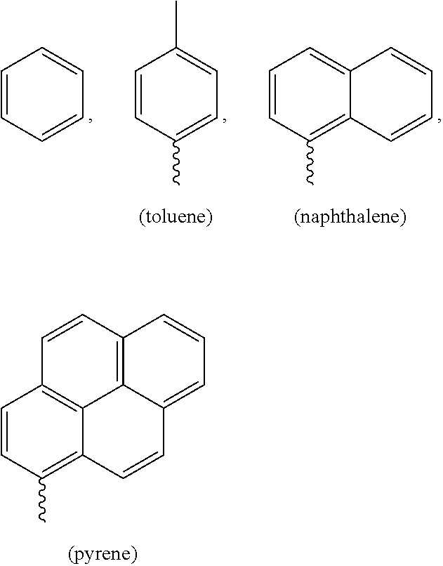
The present invention relates to a compound (L) for selective determination of free cysteine and a process for the preparation thereof. ##STR00001## wherein R1 is selected from benzene, toluene, naphthalene and pyrene.
| Inventors: | Das; Amitava (Maharashtra, IN), Ali; Firoj (Maharashtra, IN), Gandra; Upendar Reddy (Maharashtra, IN), Ashok; Anila Hoskere (Maharashtra, IN), Chattopadhyay; Samit (Maharashtra, IN), Taye; Nandaraj (Maharashtra, IN) | ||||||||||
|---|---|---|---|---|---|---|---|---|---|---|---|
| Applicant: |
|
||||||||||
| Assignee: | COUNCIL OF SCIENTIFIC &
INDUSTRIAL RESEARCH (New Delhi, IN) |
||||||||||
| Family ID: | 1000004963208 | ||||||||||
| Appl. No.: | 15/749,160 | ||||||||||
| Filed: | July 29, 2016 | ||||||||||
| PCT Filed: | July 29, 2016 | ||||||||||
| PCT No.: | PCT/IN2016/050254 | ||||||||||
| 371(c)(1),(2),(4) Date: | January 31, 2018 | ||||||||||
| PCT Pub. No.: | WO2017/021980 | ||||||||||
| PCT Pub. Date: | February 09, 2017 |
Prior Publication Data
| Document Identifier | Publication Date | |
|---|---|---|
| US 20180354975 A1 | Dec 13, 2018 | |
Foreign Application Priority Data
| Jul 31, 2015 [IN] | 2354/DEL/2015 | |||
| Current U.S. Class: | 1/1 |
| Current CPC Class: | G01N 33/6815 (20130101); C07F 5/022 (20130101) |
| Current International Class: | C07F 5/02 (20060101); G01N 33/68 (20060101) |
References Cited [Referenced By]
U.S. Patent Documents
| 2004/0076547 | April 2004 | Carney |
| 2012/0070382 | March 2012 | Liu |
Other References
|
Reedy et al., "A Novel Fluorescence Probe for Estimation of Cysteine/Histidine in Human Blood Plasma and Recoginition of Endogenous Cysteine in Live Hct116 Cells", Chem. Commun., 2014, vol. 50, pp. 9899-9902. cited by applicant . Das et al., "Desigining a Thiol Specific Fluorescent Probe for Possible Use as a Reagent for Intercellular Detection and Estimation in Blood Serum: Kinetic Analysis to Probe the Role of Intramolecular Hydrogen Bonding", Org. Biomol. Chem., 2013, vol. 11, pp. 6604-6614. cited by applicant . Yang et al., "Conjugate Addition/Cyclization Sequence Enables Selective and Simultaneous Fluorescence Detection of Cysteine and Homocysteine", Angew. Chem. Int. Ed., 2011, vol. 50, pp. 10690-10693. cited by applicant . Das et al., "New Chemodosimetric Reagents as Ratiometric Probes for Cysteine and Homocysteine and Possible Detection in Living Cells and in Blood Plasma", Chem. Eur. J., 2012, vol. 18, pp. 15382-15393. cited by applicant. |
Primary Examiner: Jarrett; Lore R
Attorney, Agent or Firm: Baker & Hostetler LLP
Claims
We claim:
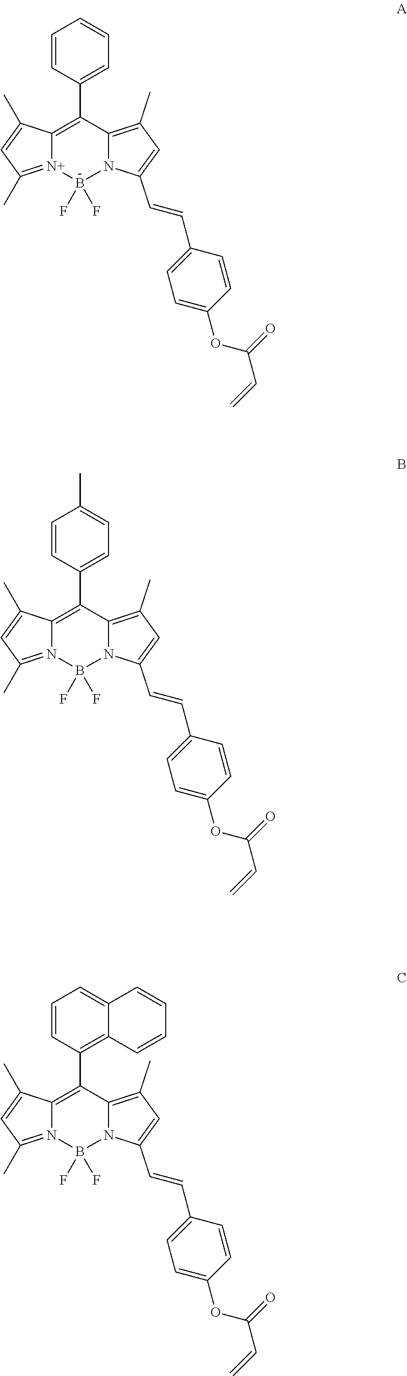
1. A process for the preparation of compound of formula (L) comprising the steps of: (i) Refluxing boron-dipyrromethene (BODIPY) of formula R'' with 4-hydroxybenzaldehyde, piperidine and glacial acetic acid in toluene to obtain compound of formula R'; ##STR00019## (ii) Reacting the compound of formula R' with acryloyl chloride in dichlotomethane (DCM) at room temperature to obtain ligand of formula (L) ##STR00020## wherein R represent phenyl, toluene, naphthalene and pyrene.
2. A compound for selective determination of free cysteine having general formula (L) prepared by the process as claimed in claim 1 ##STR00021##
3. The compound as claimed in claim 1 wherein the compounds of formula (L) comprises: ##STR00022## ##STR00023##
4. A method for selective detection of free cysteine, said method comprising; i. Dissolving 500 .mu.l of 1.times.10.sup.-4 M of ligand of formula (L) ##STR00024## in 4.5 ml of 10 mM HEPES aqueous buffer medium of pH 7.2 to obtain 1.times.10.sup.-5 M ligand concentration; ii. preparing the stock solution of 1.times.10.sup.-2 M analytes in aqueous HEPES buffer medium at pH 7.2; iii. thin layer chromatography (TLC) test strips; and iv. performing photo physical studies selected from fluorescence assay, MTT assay and Ultraviolet-visible spectroscopy for detection of free cysteine in HEPES: acetonitrile medium (9:1, v/v) at pH 7.2 as per instruction manual.
Description
FIELD OF THE INVENTION
The present invention relates to a compound of general formula (L) for selective determination of free cysteine and a process for the preparation thereof.
##STR00002## wherein R1 is selected from phenyl, toluene, naphthalene and pyrene.
BACKGROUND AND PRIOR ART OF THE INVENTION
Biological thiols such as Cysteine (Cys), Homocysteine (Hcy) and Glutathione (GSH) play crucial roles in maintaining cellular antioxidant defence system. Among them cysteine plays many important roles in living systems. Cysteine is one of the three main precursors required for GSH synthesis. The deficiency of this compound causes many diseases such as slowed growth in children, depigmentation of hair, edema, liver damage, skin lesions, and weakness. An elevated level of Hcy is a risk factor for cardio-vascular disease, dementia and Alzheimer's disease. Abnormal levels of GSH is connected to many diseases such as HIV, cell death and aging. Thus, detection and discrimination of these thiol containing molecules are of great importance. Because of the similar structure and reactivity, distinction among biothiols is a challenging task. The respective concentration level of Cys and His in human plasma is typically 240-360 .mu.M and 15-75 .mu.M. Estimation of these amino thiols in human blood plasma is essential for understanding the role of these in the pathogenesis of vascular diseases.
##STR00003##
The past two decade has seen significant effort being devoted to the development of optical probes for the selective recognition of thiol containing amino acids. There are many strategies for sensing biothiols, based on Micheal addition, cyclization with aldehydes, disulfide cleavage and others.
Das et al. (Chem. commun., 2014, 50, 9899-9902) discloses chemo dosimetric reagents for biothiol. A Cu (II)-complex based probe for detection of Cys and Histidine was reported.
P Das et al. (Org. Biomol. Chem., 2013, 11, 6604-6614) reports a new and simple chemodosimetric probe L.sub.11 is utilized for the selective detection of biothiols in the presence of other relevant amino acids under physiological conditions (pH=7.4). Furthermore, the studies with human blood plasma demonstrated the possibility of using this reagent for the quantitative optical detection of total biothiols in biological fluid.
X Yang et al. (Angewandte Chemie International Edition, Nov. 4, 2011, Volume 50, Issue 45, pages 10690-10693) reports a benzothiazole derivative used to detect cysteine (Cys) and homocysteine (Hcy) simultaneously in neutral media. The method involves thioether formation followed by cyclization to release 2-(2'-hydroxy-3'-methoxyphenyl) benzothiazole (HMBT) and a lactam.
P Das et al. (Chemistry--A European Journal, Nov. 26, 2012, Volume 18, Issue 48, pages 15382-15393) reports rationally designed and synthesized two new reagents L1 {(4-(1H-imidazo[4,5-f][1,10]phenanthrolin-2-yl)benzaldehyde)} and L2 {(4-(6,11-dioxo-6,11-dihydro-3H-anthra[1,2-d]imidazol-2-yl)benzaldehyde)}- , each bearing a pendant aldehyde functionality. This aldehyde group can take part in cyclization reactions with 13- or .gamma.-amino thiols to yield the corresponding thiazolidine and thiazinane derivatives, respectively. These two chemodosimetric reagents could be used for the quantitative detection of cysteine present in blood plasma by using a pre-column HPLC technique.
However, the development of probes for specific discrimination of biothiols is an unmet need in the art. Probes that are selective to any one of these amino biothiols are very rare in literature. Probes that give specific response with colour change as well as emission change are much needed. Especially those probes which allows real time monitoring without the aid of any instrumental techniques are highly recommended as for as the practical utility is concerned. There are no reports on detection of thiols using simple test strips. Particularly, a further challenge that is unaddressed in the art is that of a simple process for the determination of free cysteine.
Therefore, there is need in the art to develop a reagent for selective detection of free cysteine, especially with a high degree of selectivity towards free cysteine. Accordingly, the inventors of present invention developed a novel ligand for the selective detection of free cysteine.
OBJECTIVE OF THE INVENTION
The main objective of present invention is to provide a novel ligand of formula (L) useful for the selective detection of free cysteine and process for the preparation thereof.
Another objective of present invention is to provide a kit for the selective detection of free cysteine characterized in that the selectivity of novel ligand of formula (L) to cysteine is very high and a process for detection using the kit.
Yet another objective of present invention is to provide a visual as well as fluorescence test for detection of cysteine using novel ligand of formula (L).
SUMMARY OF THE INVENTION
Accordingly, the present invention provides a compound of general formula (L) which is useful for the selective detection of free cysteine and process for the preparation thereof.
##STR00004##
Wherein R.sub.1 is selected from
##STR00005## The representative compounds of formula (L) being the ligand of formula (L) are;
##STR00006## ##STR00007##
In an aspect, the present invention provides a kit for the selective detection of free cysteine characterized in that the selectivity of novel ligand of formula (L) to cysteine is 100% and a process for detection using the kit.
In another aspect, the present invention provides a visual as well as fluorescence test for detection of cysteine using novel ligands of formula (L).
Abbreviations Used:
GSH: Glutathione
Hcy: Homocysteine
Cys: Cysteine
NAC: N-Acetylcysteine
AAs: Aminoacids
Probe L: Novel ligand of formula (L)
HEPES: (4-(2-hydroxyethyl)-1-piperazineethanesulfonic acid)
BODIPY--Boron-dipyrromethene
DCM--Dicholomethane
BRIEF DESCRIPTION OF THE DRAWINGS
FIG. 1: (A) Change in emission spectrum (.lamda..sub.ex 530 nm) of L (10 uM) in presence and absence of different AAs (2 mM); (B) Emission response of L (10 uM) for Cys (200 uM) in presence of other amino acids (200 uM) in aq. HEPES buffer-acetonitrile medium at pH 7.2. Red bar indicates addition of Cys to the mixture and black bar represents emission response at 583 nm in absence of Cys.
FIG. 2: (A) Emission (.lamda..sub.Ext: 530 nm) spectral pattern for L (10 uM) in presence of varying [Cys] (0-2 mM); Inset: B-H plot obtain from systematic emission titration of L with Cys; (B) Time dependent studies of L (10 uM) in presence and absence of 5 eqv of Cys, Hcy and GSH in aq. HEPES-acetonitrile (ACN) (9:1, v/v) medium at pH 7.2. .lamda..sub.Ext/.lamda..sub.em: 530/585 nm.
FIG. 3: (A) Emission response of probe L (10 uM) and NAC (2 mM) with various concentration acylase enzyme, Each spectra was recorded after 20 min at 37.degree. C. at pH 7.2, .lamda..sub.Ext 530 nm; (B Emission intensity at 586 nm of probe L with NAC in absence and presence of enzyme vs. time plot.
FIG. 4: Visual (A) and Fluorescence (B) colour changes of probe L coated TLC plates in presence and absence of different analytes (Cys, NAC, GSH, Hcy and BSA). Fluorescence colour was recorded using UV lamp excitation at 365 nm.
FIG. 5: Emission response of L (10 .mu.M) in the absence and presence of various amino acids (200 mole equiv. each). (From 1-23, Cysteine, L only, Homocysteine, Glutathione, Histidine, Leucine, Methionine, Phenylalanine, Tryptophon, Tyrosine, Valine, Alanine, Arginine, Glycine, Glutamine, Proline, Serine, Aspartic acid, Glutamic acid, Threonine, Isoleucine, Lysine). In 10 mM HEPES: CH.sub.3CN (9:1, v/v) at pH 7, .lamda..sub.Ext=530 nm.
FIG. 6: Fluorescence intensity at 586 nm upon addition of Cys (0-150 .mu.M) in 10 mM HEPES: ACN (9:1, v/v) at pH7, .lamda..sub.Ext=530 nm.
FIG. 7: Change of emission intensity of probe L with pH.
FIG. 8: MTT assay to determine the cell viability percentage in Hct116 colon cancer cells.
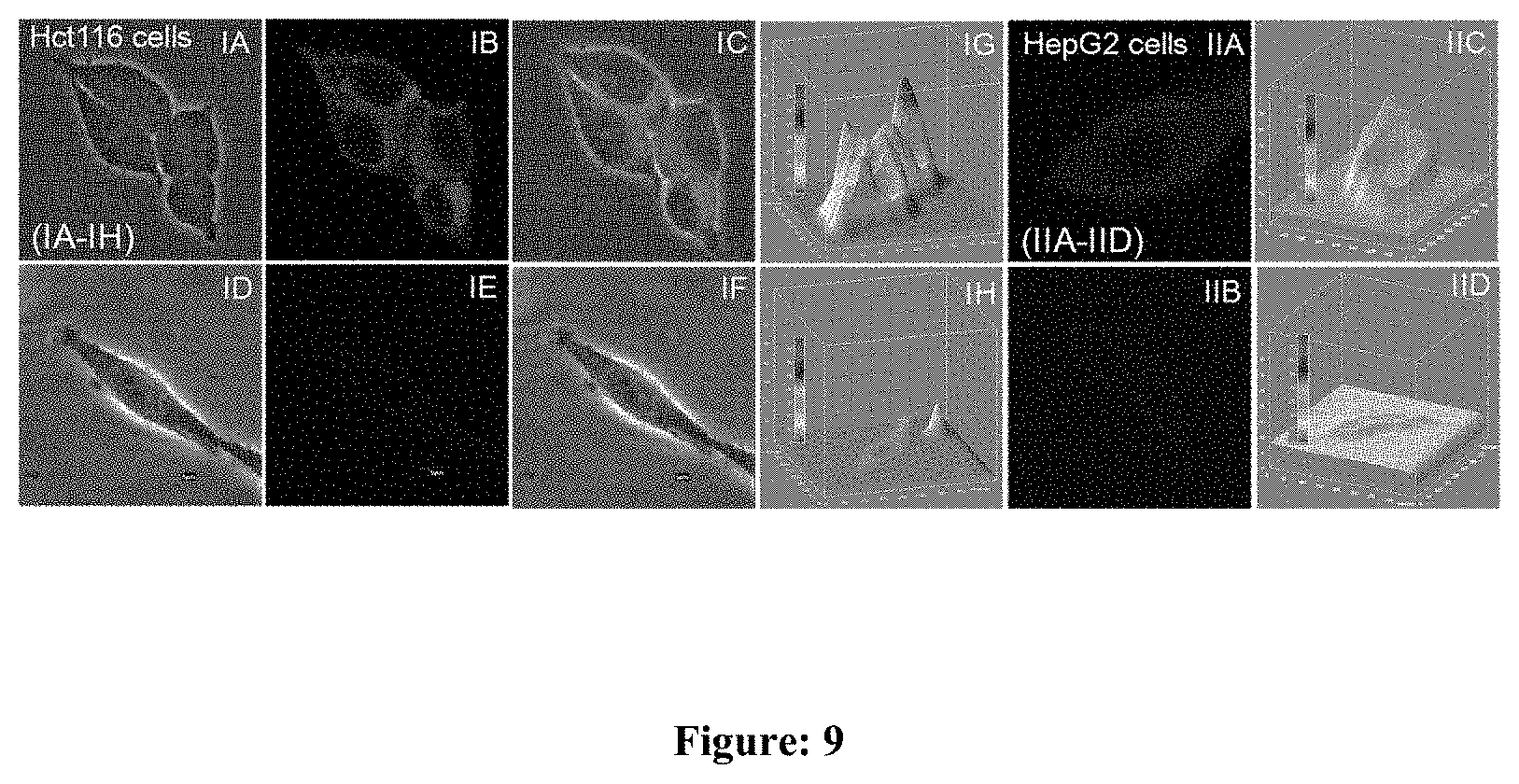
FIG. 9: (I) CLSM images of Hct116 cells (IA-IH), cells incubated with 1 .mu.M of L, (IA): bright field images, (IB): dark field image and (IC): overlay images of (IA) & (IB); Cells pre-treated with 1 mM of NEM then incubated with 1 .mu.M of L, (ID): bright field image, (IE): dark field image, (IF): overlay of (ID) & (IE). (IG) and (IH) were 3D intensity plot of (IB) & (IE) respectively; (II) CLSM images of live HepG2 cells: (IIA) & (IIB) dark field images of cells treated with 1 mM NEM, washed and then treated with (IIA) or without (IIB) 25 .mu.M NAC for 1 h, followed by incubation of 1 .mu.M of L for 20 min and (IIC) & (IID) are respective 3D intensity profile plot of images (II)A & (II)B. (II)A & (II)C revealed the generation of Cys from NAC by aminoacylase-I present in HepG2 cells, .lamda..sub.Ext/.lamda..sub.Em: 530/573 nm.
FIG. 9: (A) Hct116 cells stained with 1 .mu.M of L in presence of ER tracker green; (i) Intensity profile of ROIs across cells: red line represent intensity of L and green line indicate intensity for ER Tracker green; (B) co-localization experiment: Cells were co-stained with L, ER tracker green and DAPI; .lamda..sub.Ext/.lamda..sub.Em: 530/573 nm.
DETAILED DESCRIPTION OF THE INVENTION
For the purpose of this invention, the expression "probe L" and "novel ligand of formula (L)" are used interchangeably throughout the specification and both having the same meaning.
The present invention provides a novel ligand of formula (L) which is useful for the selective detection of free cysteine and process for the preparation thereof. This probe L is used for the detection of free cysteine by a simple, convenient, selective and fast visual method in aqueous medium as well as biological fluids. The present invention also provides a kit for the selective detection of free cysteine characterized in that the selectivity of novel ligand of formula (L) to cysteine is 100% and a process for detection using the kit.
Accordingly, the main embodiment of the present invention provides a compound for selective determination of free cysteine having general formula (L)
##STR00008## wherein R.sub.1 is selected from
##STR00009##
Another embodiment of the present invention provides a novel ligand of formula (L) useful for the detection of free cysteine.
##STR00010## wherein R.sub.1 is selected from
##STR00011##
In another embodiment of the present invention the compounds of formula (L) comprises:
##STR00012## ##STR00013##
Another embodiment of the present invention provides a process for the preparation of compound of formula (L), comprising the steps of: (i) Refluxing boron-dipyrromethene (BODIPY) of formula R'' with 4-hydroxybenzaldehyde, piperidine and glacial acetic acid in toluene to obtain compound of formula R';
##STR00014## wherein R represent phenyl, toluene, naphthalene and pyrene; (ii) Reacting the compound of formula R' with acryloyl chloride in dichlotomethane (DCM) at room temperature to obtain ligand of formula (L)
##STR00015## wherein R represent phenyl, toluene, naphthalene and pyrene.
Another embodiment of the present invention provides a process for the synthesis of ligand of formula (L) comprising the steps of: a) Refluxing the reaction mixture comprising BODIPY of formula R'', 4-hydroxybenzaldehyde, piperidine and glacial acetic acid in toluene for 3 h at 110.degree. C. to obtain compound of formula (R'). b) Reacting the compound of formula R' with acryloyl chloride in dicholomethane (DCM) at room temperature to obtain ligand of formula (L).
In another embodiment of the present invention, a mixture of BODIPY (boron-dipyrromethene) (400 mg, 1.23 mmol), 4-hydroxybenzaldehyde (165 mg, 1.35 mmol), 0.9 ml piperidine and 0.6 ml glacial acetic acid was reflux in 30 ml toluene in a Dean-Stark apparatus for 3 h. Then water was added into it and crude organic layer was extracted with dichloromethane. The organic layer was collected and dried over anhydrous sodium sulphate and solvent was removed under reduced pressure. It was then subjected to column chromatography using silica gel (100-200 mesh) as stationary phase and 10% EtOAc in hexane as mobile phase to get compound R' as red solid.
In yet another embodiment, BODIPY compound R' (30 mg, 0.07 mmol) was dissolved in 10 ml anhydrous dichloromethane in a 100 ml round bottom two neck flask. Then 100 uL of Et.sub.3N was added to the reaction mixture and allowed to stir for 10 mins at room temperature under N.sub.2 atmosphere. 20 uL of acryloyl chloride was added to this and resulting mixture was stirred at room temperature until all the starting material was consumed monitored by TLC. Then water was added to it and organic layer was extracted using dichloromethane. The organic layer was collected and dried over anhydrous sodium sulphate and solvent was removed under reduced pressure. It was then subjected to column chromatography using silica gel (100-200 mesh) as stationary phase and 5% EtOAc in hexane as mobile phase to get probe L as solid as 70% yield.
In yet another embodiment, the present invention discloses the process for the synthesis for novel ligand of formula (L) is depicted in scheme 1 below;
##STR00016##
The BODIPY of formula R'' is prepared by known methods comprising reacting aldehyde or acid chloride with 2, 4 dimethyl pyrrole. The aldehyde is used appropriately to obtain the BODIPY of formula R'' wherein the substituent R represents toluene, naphthalene and pyrene. The acid chloride is selected from benzoyl chloride to obtain BODIPY of formula R'' wherein the substituent R is phenyl.
Accordingly, the process for preparation of BODIPY of formula R'', wherein R represents toluene, naphthalene and pyrene comprises reacting the corresponding aldehyde with 2, 4 dimethyl pyrrole in presence of DDQ, Et.sub.3N:BF.sub.3 in presence of trifluroacetic acid and DCM.
##STR00017##
The process for preparation of BODIPY of formula R'', wherein R is phenyl comprises reacting benzoyl chloride and 2, 4 dimethyl pyrrole in presence of triethylamine, BF.sub.3.OEt.sub.2 in dichloromethane under nitrogen atmosphere.
##STR00018##
Another embodiment of the present invention provides for a diagnostic kit for selective detection of free cysteine.
In another embodiment of the present invention, the diagnostic kit comprises:
(a) Ligand of Formula (L)
(b) HEPES aqueous buffer medium
(c) TLC Test strips
(d) HEPES:acetonitrile medium
(e) Instruction manual
Another embodiment of the present invention provides a method for selective detection of free cysteine using the diagnostic kit comprising; (i) Dissolving 500 .mu.l of 1.times.10.sup.-4 M of ligand of formula (L) in 4.5 ml of 10 mM HEPES aqueous buffer medium of pH 7.2 to obtain 1.times.10.sup.-5 M ligand concentration; (ii) preparing the stock solution of 1.times.10.sup.-2 M analytes in aqueous HEPES buffer medium at pH 7.2; (iii) TLC test strips; and (iv) performing the photo physical studies for detection of free cysteine in HEPES:acetonitrile medium (9:1, v/v) at pH 7.2 as per the instruction manual.
In another embodiment of the present invention the emission response of ligand L with various amino acids (AAs) as well as anionic analytes in aqueous buffer-acetonitrile (9:1, v/v) at pH 7.2 solution (FIG. 1) is studied. FIG. 1 shows that emission spectrum recorded for L (10 uM) and L+ AAS (200 uM) except Cys does not shows any prominent changes, only after addition cysteine (Cys), probe L shows a fluorescence turn on response at 583 nm upon .lamda..sub.ex at 530 nm under specified experimental condition. Further emission response of probe L towards Cys in presence of other AAs is also carried out under identical condition (FIG. 1). These findings suggested L is a selective fluorescence turn on probe for Cys in aqueous HEPES buffer medium. Probe L does not show emission changes in presence of Hcy, GSH, N-acetyl cysteine (NAC) as well as cysteine bound protein like BSA, upon .lamda..sub.ex at 530 nm (FIG. 1). Thus L can specifically detect Cys, over other biothiols, NAC and cysteine bound protein.
In another embodiment of the present invention time dependent fluorescence assay of probe L (10 uM) is carried out in presence of 5 eqv. of Cys, Hcy and GSH by monitoring emission changes at .lamda..sub.em at 583 nm in aq. HEPES buffer-acetonitrile (9:1, v/v) at pH 7.2 medium (FIG. 2). FIG. 2 reveals that emission intensity reaches maximum within 1 min after addition of 2 mM of Cys, whereas no prominent changes is observed for Hcy and GSH even after 40 min of incubation under identical condition.
In another embodiment of the present invention the fluorescence intensity of probe L is determined to be linearly proportional with Cys concentration and the detection limit of free cysteine by the fluorescence spectroscopy method is found to be 15 nM. The effect of pH towards probe L indicated ligand L is stable up to pH 9. FIG. 3 indicates that with increase in acylase concentration, probe L gives fluorescence turn on response with fixed initial concentration of NAC and L. The probe L may be used for monitoring enzymatic generation of Cys.
In another embodiment, probe L is used for the detection of free cysteine by a simple, convenient, selective and fast visual method in aqueous medium as well as biological fluids. Accordingly, a TLC plate is immersed into acetonitrile solution of probe L (5 .mu.L) and dried. This TLC plate coated with probe L is exposed to aqueous solution of different analytes under identical condition. Upon addition of Cysteine solution a prominent change in fluorescence (green to reddish) using UV lamp with excitation at 365 nm and little colour (light pink to dark pink) are observed in TLC plate. In case of other analytes including Hcy, GSH, NAC, and BSA protein (contain terminal Cys residue) no detectable changes are induced.
In another embodiment, the probe L is tested for biological properties. In a preferred embodiment, the probe L is tested for cytotoxicity and at micro molar concentration of probe L used for cellular studies 85% of cell servility. The in vitro cytotoxicity of L on Hct116 cells (Colon cancer cell) are determined by conventional MTT (3-(4, 5-Dimethylthiazol-2-yl)-2, 5-diphenyltetrazolium bromide, a yellow tetrazole) assay. Hct116 colon cancer cells (7.times.10.sup.3) are seeded in each well of a 96 well plate and cultured in a 37.degree. C. incubator supplied with 5% CO.sub.2. Cells are maintained in DMEM medium, supplemented with 10% Foetal Bovine Serum and 100 Units of Penicillin Streptomycin antibiotics. After 24 hours the cells are treated with different concentrations of the L in triplicates for 12 hours. After treatment cells are added with 0.5 .mu.g/ml of MTT reagent. The plate is then incubated for 4 hours at 37.degree. C. 100 .mu.l of Isopropyl Alcohol is added to each well.
The following examples are given by way of illustration and therefore should not be construed to limit the scope of the present invention:
EXAMPLES
Example 1: Synthesis of R'
A mixture of BODIPY (boron-dipyrromethene) (400 mg, 1.23 mmol), 4-hydroxybenzaldehyde (165 mg, 1.35 mmol), 0.9 ml piperidine and 0.6 ml glacial acetic acid was reflux in 30 ml toluene in a Dean-Stark apparatus for 3 h. Then water was added into it and crude organic layer was extracted with dichloromethane. The organic layer was collected and dried over anhydrous sodium sulphate and solvent was removed under reduced pressure. It was then subjected to column chromatography using silica gel (100-200 mesh) as stationary phase and 10% EtOAc in hexane as mobile phase to get compound R' as red solid. Yield: 56%; .sup.1H NMR (500 MHz, CDCl.sub.3, J in Hz, .delta. ppm): 7.52 (1H, d, J=16.5), 7.47 (5H, m), 7.3 (2H, d, J=8), 7.18 (1H, d, J=16), 6.83 (2H, d, J=8.5), 6.58 (1H, s), 5.99 (1H, s), 5.4 (1H, s), 2.59 (3H, s), 1.42 (3H, s), 1.38 (3H, s); .sup.13C NMR (125 MHz, CDCl.sub.3, .delta. ppm): 156.80, 154.68, 153.47, 142.74, 142.40, 140.08, 136.24, 135.13, 132.84, 131.68, 129.38, 129.22, 129.09, 128.93, 128.25, 121.14, 117.55, 116.90, 115.82, 14.69, 14.59, 14.32. HRMS (ESI): m/z calculated for C.sub.26H.sub.23BF.sub.2N.sub.2O [M+H]: 429.1872, found 429.1943.
Example 2: Synthesis of Probe L
In a 100 ml round bottom two neck flask BODIPY compound R' (30 mg, 0.07 mmol) was dissolved in 10 ml anhydrous dichloromethane. Then 100 uL of Et.sub.3N was added to the reaction mixture and allowed to stir for 10 mins at room temperature under N.sub.2 atmosphere. 20 uL of acryloyl chloride was added to this and resulting mixture was stirred at room temperature until all the starting material was consumed monitored by TLC. Then water was added to it and organic layer was extracted using dichloromethane. The organic layer was collected and dried over anhydrous sodium sulphate and solvent was removed under reduced pressure. It was then subjected to column chromatography using silica gel (100-200 mesh) as stationary phase and 5% EtOAc in hexane as mobile phase to get probe L as solid as 70% yield. .sup.1H NMR (500 MHz, CDCl.sub.3, J in Hz, .delta. ppm): 7.57 (1H, s), 7.53 (3H, d, J=8.2), 7.41 (3H, d, J=5.8), 7.25-7.21 (2H, m), 7.13 (1H, d, J=16.3), 7.07 (2H, d, J=8.4), 6.56 (1H, s), 6.52 (1H, s), 6.25 (1H, dd, J=17.3, 10.5), 5.95 (2H, d, J=11.1), 2.52 (3H, s), 1.35 (3H, s), 1.31 (3H, s); .sup.13C NMR (125 MHz, CDCl.sub.3, .delta. ppm): 164.35, 155.92, 152.21, 150.90, 143.27, 142.34, 140.65, 135.03, 134.69, 134.39, 132.78, 132.05, 129.15, 129.01, 128.44, 128.14, 127.86, 121.88, 121.55, 119.41, 117.48, 14.79, 14.57, 14.42. HRMS (ESI): m/z calculated for C.sub.29H.sub.26BF.sub.2N.sub.2O.sub.2 [M+H]: 483.1977 found 483.2057.
Example 3: General Experimental Procedure for UV-Vis and Fluorescence Studies
Stock solution of probe L (1.times.10.sup.-4 M) was prepared in HPLC grade acetonitrile. All the analytes stock solution (1.times.10.sup.-2 M) was prepared in aqueous HEPES buffer medium at pH 7.2. 500 .mu.L of this stock solution of probe L was added to 4.5 ml of HEPES (10 mM) aqueous buffer medium having solution pH 7.2 to make the effective ligand concentration of 1.times.10.sup.-5 M. This solution was used for all the photophysical studies. All the photo physical studies were performed in HEPES:acetonitrile medium (9:1, v/v) at pH 7.2. The detection limit was calculated based on the fluorescence titration. To determine the S/N ratio, the emission intensity of L without Cys was measured 10 times and the standard deviation of blank measurements was determined. The detection limit (DL) of L for Cys was determined from the following equation: DL=K*Sb1/S, Where K=2 or 3 (we took 3 in this case); Sb1 is the standard deviation of the blank solution; S is the slope of the calibration curve. From the graph we get slope=4.99.times.10.sup.8, and Sb1 value is 2.49 Thus using the formula we get the Detection Limit=15.times.10.sup.-9M.
Example 4: Preparation of TLC Test Strips
TLC test strips were prepared by coating 5 .mu.M of probe solution in acetonitrile on silica TLC plates. 5 .mu.l of Cys (1.times.10.sup.-1M) in 10 mM aq. HEPES buffer (pH 7) was added on it, dried and the visual as well as fluorescence colour changes were observed after 5 min. The same was repeated for Hcy, GSH and other analytes as well.
Example 5: General Procedure for Enzymatic Study
Cipla made effervescent tablets of N-Acetyl-Cysteine (mucinac 600) were purchased from commercially available sources. Based on the quantity of NAC present in the tablet, 1.times.10.sup.-1M tablet solution was prepared in 10 mM aq. HEPES buffer solution (pH7). Enzyme solution was prepared according to the requirement by dissolving 1 mg/ml in 10 mM aq. HEPES buffer solution (pH7). A fixed concentration of NAC (200 equiv.) was added to the 10 .mu.M probe in HEPES: ACN (9:1). Since 1 mg of solid enzyme contains 3301 units of protein and 1 unit can hydrolyse 1 .mu.M of substrate, accordingly enzyme concentration was varied with respect to the substrate concentration.
Example 6: General Procedure for Confocal Studies
Hct116 cells (3.times.10.sup.5) (ATCC.RTM. CCL247.TM.) were seeded on cover slips placed in 6 well plates. After 24 hours, cells were treated with L (10 .mu.M) for 30 minutes. Cells were then washed thrice with Phosphate Buffer Saline (1.times.PBS) and fixed with 4% PFA for 20 minutes and washed again with 1.times.PBS. Nail paint was used to seal the cover slips mounted on the glass slides. Images were acquired in Olympus Fluoview Microscope.
Example 7: In Vitro Cytotoxicity of L on Hct116 Cells
The in vitro cytotoxicity of L on Hct116 cells (Colon cancer cell) were determined by conventional MTT (3-(4, 5-Dimethylthiazol-2-yl)-2, 5-diphenyltetrazolium bromide, a yellow tetrazole) assay. Hct116 colon cancer cells (7.times.10.sup.3) were seeded in each well of a 96 well plate and cultured in a 37.degree. C. incubator supplied with 5% CO.sub.2. Cells were maintained in DMEM medium, supplemented with 10% Foetal Bovine Serum and 100 Units of Penicillin Streptomycin antibiotics. After 24 hours the cells were treated with different concentrations of the L in triplicates for 12 hours. After treatment cells were added with 0.5 .mu.g/ml of MTT reagent. The plate was then incubated for 4 hours at 37.degree. C. 100 .mu.l of Isopropyl Alcohol was added to each well. Optical density was measured at 570 nm using Multiskan Go (Thermo Scientific) to find the concentration of the cell inhibition. IC.sub.50 value has been calculated to be 100 .mu.M.
The formula used for the calculation of the MTT assay for evaluation of the cell viability was as follows: Cell viability (%)=(means of Absorbance value of treated group/means of Absorbance value of untreated control).times.100.
ADVANTAGES OF INVENTION
a) Simple process of detection b) Selective determination of free cysteine c) This method is of much practical significance in real time monitoring as it could be done without the aid of any instruments.
* * * * *
C00001
C00002
C00003
C00004
C00005
C00006
C00007
C00008
C00009
C00010
C00011
C00012
C00013

C00014

C00015

C00016

C00017

C00018

C00019

C00020

C00021

C00022
C00023

C00024

D00000

D00001

D00002

D00003

D00004

D00005

D00006

D00007

D00008

D00009
XML
uspto.report is an independent third-party trademark research tool that is not affiliated, endorsed, or sponsored by the United States Patent and Trademark Office (USPTO) or any other governmental organization. The information provided by uspto.report is based on publicly available data at the time of writing and is intended for informational purposes only.
While we strive to provide accurate and up-to-date information, we do not guarantee the accuracy, completeness, reliability, or suitability of the information displayed on this site. The use of this site is at your own risk. Any reliance you place on such information is therefore strictly at your own risk.
All official trademark data, including owner information, should be verified by visiting the official USPTO website at www.uspto.gov. This site is not intended to replace professional legal advice and should not be used as a substitute for consulting with a legal professional who is knowledgeable about trademark law.