Method for identifying epitope on protein
Inoue , et al.
U.S. patent number 10,718,781 [Application Number 15/326,295] was granted by the patent office on 2020-07-21 for method for identifying epitope on protein. This patent grant is currently assigned to Chugai Seiyaku Kabushiki Kaisha. The grantee listed for this patent is Chugai Seiyaku Kabushiki Kaisha. Invention is credited to Tomoaki Inoue, Shunsuke Ito, Nobuo Sekiguchi.


View All Diagrams
| United States Patent | 10,718,781 |
| Inoue , et al. | July 21, 2020 |
Method for identifying epitope on protein
Abstract
In one aspect, the present invention provides, for example, an improved method for identifying an epitope on a protein, comprising the following steps: (A) contacting a major histocompatibility complex (MHC molecule)-expressing cell differentiated from a stem cell or a progenitor cell derived therefrom with a target protein; (B) isolating a complex of a peptide contained in the target protein and the MHC molecule from the MHC molecule-expressing cell; and (C) eluting the peptide from the complex and identifying the peptide.
| Inventors: | Inoue; Tomoaki (Gotemba, JP), Ito; Shunsuke (Gotemba, JP), Sekiguchi; Nobuo (Gotemba, JP) | ||||||||||
|---|---|---|---|---|---|---|---|---|---|---|---|
| Applicant: |
|
||||||||||
| Assignee: | Chugai Seiyaku Kabushiki Kaisha
(Tokyo, JP) |
||||||||||
| Family ID: | 55078494 | ||||||||||
| Appl. No.: | 15/326,295 | ||||||||||
| Filed: | July 13, 2015 | ||||||||||
| PCT Filed: | July 13, 2015 | ||||||||||
| PCT No.: | PCT/JP2015/070072 | ||||||||||
| 371(c)(1),(2),(4) Date: | January 13, 2017 | ||||||||||
| PCT Pub. No.: | WO2016/010002 | ||||||||||
| PCT Pub. Date: | January 21, 2016 |
Prior Publication Data
| Document Identifier | Publication Date | |
|---|---|---|
| US 20170219607 A1 | Aug 3, 2017 | |
Foreign Application Priority Data
| Jul 14, 2014 [JP] | 2014-144217 | |||
| Current U.S. Class: | 1/1 |
| Current CPC Class: | C07K 16/241 (20130101); C12P 21/02 (20130101); C12N 5/10 (20130101); G01N 33/6872 (20130101); C07K 14/00 (20130101); C07K 14/415 (20130101); A61K 38/00 (20130101); C07K 7/00 (20130101); C07K 14/755 (20130101); C12Q 1/02 (20130101); C07K 2317/24 (20130101) |
| Current International Class: | G01N 33/53 (20060101); C12P 21/02 (20060101); C07K 7/00 (20060101); C07K 14/00 (20060101); C12Q 1/02 (20060101); A61K 38/00 (20060101); G01N 33/68 (20060101); C12N 5/10 (20060101); C07K 16/24 (20060101); C07K 14/755 (20060101); C07K 14/415 (20060101) |
References Cited [Referenced By]
U.S. Patent Documents
| 2006/0251664 | November 2006 | Kropshofer et al. |
| 2009/0246869 | October 2009 | Tseng et al. |
| 2010/0322931 | December 2010 | Harding et al. |
| 2013/0195818 | August 2013 | Senju |
| 2013/0330822 | December 2013 | Nekahata et al. |
| 1 826 217 | Aug 2007 | EP | |||
| 2006-300945 | Nov 2006 | JP | |||
| 2011-515100 | May 2011 | JP | |||
| 2012-530496 | Dec 2012 | JP | |||
| 2014-506447 | Mar 2014 | JP | |||
| WO 2012/043651 | Apr 2012 | WO | |||
| WO 2012/115276 | Aug 2012 | WO | |||
Other References
|
Google translation of JP2006300945A (Year: 2018). cited by examiner . Berges et al., "A cell line model for the differentiation of human dendritic cells," Biochemical and Biophysical Research Communications 333: 896-907 (Year: 2005). cited by examiner . International Search Report for International Application No. PCT/JP2015/070072, Japan Patent Office, Japan, dated Oct. 13, 2015, 10 pages. cited by applicant . Kropshofer, H. and Singer, T., "Overview of Cell-Based Tools for Pre-Clinical Assessment of Immunogenicity of Biotherapeutics," Journal of Immunotoxicology 3:131-136, Informa Healthcare, Switzerland (2006). cited by applicant . Senju, S., "Anti-Cancer Therapy with Pluipotent Stem Cell-Derived Dendritic Cells," Biotherapy 24(2):87-94, (2010). cited by applicant . Senju, S., "Saisei Iryo hyo no Genjo to Shinpo-ES Saibo, iPS Saibo to Taisei Kansaibo no Rinsho eno Oyo-6. Tanosei Saibo Yurai no Jujo Saibo o Mochiita Men'eki Ryoho," Hematology Frontier 19(11):1685-1692 (2009). cited by applicant . Senju, S., "Establishment of pluripotent stem cell-derived dendritic cells for clinical application," The Japanese Journal of Clinical Hematology 51(11):1668-1673 (2010). cited by applicant . Yanagimachi, M. D., et al., "Robust and Highly-Efficient Differentiation of Functional Monocytic Cells from Human Pluripotent Stem Cells under Serum- and Feeder Cell-Free Conditions," PLOS One 8(4):1-9 e59243 (2013). cited by applicant . Iwamoto, H., et al., "Antitumor immune response of dendritic cells (DCs) expressing tumor-associated antigens derived from induced pluripotent stem cells: In comparison to bone marrow-derived DCs," International Journal of Cancer 134:332-341, Wiley-Liss, United States (2014). cited by applicant . Karle, A. C., et al., "Nitration of the Pollen Allergen Bet v 1.0101 Enhances the Presentation of Bet v 1-Derived Peptides by HLA-DR on Human Dendritic Cells," PLos One 7:e31483, 9 pages, Public Library of Science, United States (2012). cited by applicant . Mutschlechner, S., et al, "Naturally processed T cell-activating peptides of the major birch pollen allergen," J Allergy Clin Innnunol 125:711-718, American Academy of Allergy, Asthma & Immunology, United States (2010). cited by applicant . Niwa, A., et al., "A Novel Serum-Free Monolayer Culture for Orderly Hematopoietic Differentiation of Human Pluripotent Cells via Mesodermal Progenitors," PLos One 6:e22261, 11 pages, Public Library of Science, Untied States (2011). cited by applicant . Rombach-Riegraf, V., et al., "Aggregation of Human Recombinant Monoclonal Antibodies Influences the Capacity of Dendritic Cells to Stimulate Adaptive T-Cell Responses In Vitro," PLos One 9:e86322, 17 pages, Public Library of Science, United States (2014). cited by applicant . Senju, S., et al., "Generation of dendritic cells and macrophages from human induced pluripotent stem cells aiming at cell therapy," Gene Therapy 18:874-883, Macmillan Publishers Limited, England (2011). cited by applicant . Van Haren, S.D., et al., "HLA-DR-presented Peptide Repertoires Derived From Human Monocyte-derived Dendritic Cells Pulsed With Blood Coagulation Factor VIII," Molecular & Cellular Proteomics 10:10.6, 12 pages, The American Society for Biochemistry and Molecular Biology Inc., United States (2011). cited by applicant. |
Primary Examiner: Salvoza; M Franco G
Attorney, Agent or Firm: Nixon & Vanderyhe P.C.
Claims
The invention claimed is:
1. A method for identifying an epitope on a protein, comprising the following steps: (A) contacting a major histocompatibility complex (MHC) molecule-expressing dendritic cell with a target protein, wherein the MHC molecule-expressing dendritic cell is differentiated from a stem cell or a progenitor cell derived from a stem cell; (B) isolating a complex of a peptide contained in the target protein and the MHC molecule from the MHC molecule-expressing dendritic cell; and (C) eluting the peptide from the complex and identifying the peptide, wherein the step (A) further comprises the following steps for differentiating the MHC molecule-expressing dendritic cell from a stem cell or a progenitor cell derived from a stem cell: (a) differentiating the stem cell or the progenitor cell derived from the stem cell into a mesodermal progenitor cell; (b) differentiating the mesodermal progenitor cell into a monocyte; and (c) differentiating the monocyte into an immature dendritic cell, and optionally stimulating the immature dendritic cell to obtain a mature dendritic cell, wherein among the steps (a) to (c) at least the steps (a) and (c) are performed in a serum-free medium, the step (b) comprises the step of differentiating the mesodermal progenitor cell into the monocyte in a serum-free medium containing a granulocyte macrophage colony-stimulating factor (GM-CSF) and a macrophage colony-stimulating factor (M-CSF), and the step (c) comprises the step of: (c1) differentiating the monocyte into the immature dendritic cell in a serum-free medium containing a granulocyte macrophage colony-stimulating factor (GM-CSF) and interleukin 4 (IL-4), and optionally comprises the step of: (c2) contacting the immature dendritic cell with an immunogen or an immunogen and an inflammatory cytokine to induce the mature dendritic cell; the stem cell is selected from the group consisting of an induced pluripotent stem cell (iPS cell), an embryonic stem cell (ES cell), a nuclear transfer ES cell (ntES cell), an embryonic germ stem cell (EG cell), and an adult stem cell; and the MHC molecule is a MHC II molecule and the MHC II molecule is HLA-DR, HLA-DQ or HLA-DP.
2. The method according to claim 1, further comprising the following step: (D) testing whether the identified peptide is an epitope that induces immunogenicity.
3. The method according to claim 1, wherein the MHC molecule-expressing dendritic cell further expresses at least one selected from the group consisting of CD80, CD86, CD206, and CD209.
4. The method according to claim 1, wherein the MHC molecule-expressing dendritic cell expresses CD80, CD86, CD206, and CD209.
5. The method according to claim 1, wherein the MHC molecule-expressing dendritic cell expresses one or more MHC molecule allotypes in a subject intended to receive the target protein.
6. The method according to claim 1, wherein the dendritic cell is an immature dendritic cell, and the immature dendritic cell is contacted with a target protein having immunogenicity to induce the mature dendritic cell.
7. The method according to claim 1, wherein the target protein is selected from the group consisting of (a) a cytokine, (b) a chemokine, (c) a growth factor, (d) an antibody, (e) an enzyme, (f) a structural protein, (g) a hormone, and (h) a fragment of (a)-(g).
Description
CROSS-REFERENCE TO RELATED APPLICATION
This application is a U.S. National Phase of PCT Application No. PCT/JP2015/070072, filed Jul. 13, 2015, which claims priority to Japanese Patent Application No. 2014-144217, filed Jul. 14, 2014, each of which is incorporated herein by reference.
REFERENCE TO SEQUENCE LISTING SUBMITTED ELECTRONICALLY
The content of the electronically submitted sequence listing (Name: Sequence_Listing.txt; Size: 85,742 bytes; and Date of Creation: Jan. 12, 2017) filed with the application is incorporated herein by reference in its entirety.
TECHNICAL FIELD
In one aspect, the present invention relates to, for example, a method for identifying a protein having immunogenicity, and also relates to, for example, a method for identifying an epitope that may play a causative role in the induction of immunogenicity.
BACKGROUND ART
In recent years, many bio-pharmaceuticals (antibody drugs, biologics, hormones, proteins, etc.) have contributed to medical innovation. However, immunogenicity possessed by these bio-pharmaceuticals is controversial, for example, from the viewpoint of efficacy and safety. In general, a property of an antigen that induces antibody production or cell-mediated immunity is called immunogenicity. The bio-pharmaceuticals can act as antigens to induce antibody production in the bodies of patients. In such a case, neutralizing antibodies against the bio-pharmaceuticals are produced, sometimes resulting in the reduced efficiency of treatment. Alternatively, allergic response, leaching reaction, infusion reaction, or the like may be caused. Alternatively, antibodies that cause autoimmune diseases or the like due to the neutralization of endogenous self-proteins may be produced in response to the bio-pharmaceuticals.
For the process of antibody production, it is important that an antigen is presented on a major histocompatibility complex (also referred to as a MHC molecule) present on the cell surface of an antigen-presenting cell (APC) (this is called "antigen presentation"). A MHC I molecule (class I) and a MHC II molecule (class II) are known as MHC molecules involved in the antigen presentation. For example, the MHC I molecule acts on killer T cells (CD8-positive T cells), and the MHC II molecule acts on helper T cells (CD4-positive T cells). The MHC I molecule acts on endogenous antigens in autologous cells, while the MHC II molecule acts on foreign antigens. Thus, antigen-antibody reaction or the like can be caused, for example, against cancer antigens produced in cancer cells, through antigen presentation mediated by the MHC I molecule. On the other hand, antigen-antibody reaction or the like can be caused against foreign antigens such as the bio-pharmaceuticals, or toxins through antigen presentation mediated by the MHC II molecule.
More specifically, in the case of the event mediated by the MHC I molecule, endogenous proteins in autologous cells are decomposed into smaller peptides by proteasome. Subsequently, each peptide binds to the MHC I molecule synthesized in the vesicle to form a complex. Then, the complex is delivered to the cell surface so that the peptide is presented as an epitope on the MHC I molecule.
On the other hand, in the case of the event mediated by the MHC II molecule, foreign proteins are first taken up into antigen-presenting cells by endocytosis. Subsequently, the taken-up proteins are decomposed into smaller peptides by lysosome. Then, each peptide binds to the MHC II molecule to form a complex. Then, the complex is delivered to the cell surface so that the peptide is presented as an epitope on the MCH II molecule. Subsequently, a T cell receptor on a helper T cell can bind to the antigen-presenting cell via the peptide.
However, these pathways are not definitive, and even foreign antigens may be processed by the MHC I molecule-mediated antigen presentation pathway (this is called "cross-priming").
In order to circumvent the immunogenicity of antibody drugs, etc., research has been conducted on the identification of peptide sequences presented on MHC molecules. This permits the prediction of the immunogenicity of proteins or peptides intended to be administered to organisms. Furthermore, epitopes can be modified by site-directed mutagenesis for the purpose of producing non-immunogenic proteins, for example, on the basis of information on epitope sequences. A method using a prediction algorithm in silico and T cell proliferation assay (e.g., the measurement of the ability of helper T cells to proliferate by the uptake of tritium-labeled thymidine) are known as methods for identifying peptide sequences. However, it has been difficult to predict an epitope sequence, for example, only from the binding affinity between an epitope candidate peptide and a MHC molecule. Accordingly, there has been a demand for more accurately predicting an epitope that may play a causative role in the induction of immunogenicity by directly identifying the sequence of a peptide presented on a MHC molecule.
Methods have been developed which involve contacting an antigen-presenting cell such as a dendritic cell (DC) with a protein to induce antigen presentation, allowing a MHC molecule on the cell to present a peptide derived from the protein, then separating and purifying a complex of the MHC molecule and the peptide, then eluting the peptide, and directly identifying the sequence of the peptide by use of liquid chromatography mass spectrometry (LC/MS) or the like (Patent Literature 1, Patent Literature 2, and Non Patent Literature 1). These methods are called MAPPs (MHC-associated peptide proteomics).
CITATION LIST
Patent Literature
Patent Literature 1: European Patent Application Publication No. 1715343 Patent Literature 2: European Patent Application Publication No. 1826217
Non Patent Literature
Non Patent Literature 1: Kropshofer, H, et al., J. Immunotoxicol., 3, 131, 2006
SUMMARY OF INVENTION
Technical Problem
In one aspect, in the method of Patent Literature 1, human peripheral blood mononuclear cells (PBMCs) separated by blood collection from a human are used as primary cells, and dendritic cells obtained by the induction of differentiation of monocytes further separated from these PBMCs are utilized as antigen-presenting cells. However, the monocytes are present only at approximately 10% of PBMCs and are also limited by the number of divisions. Therefore, there is a limitation on the number of cells that can be obtained. Furthermore, since MHC molecule allotypes may vary among different donors, it is impossible to constantly obtain a desired MHC molecule allotype. Thus, peptides to be identified by MAPPs may also vary. Since each component in blood also fluctuates depending on the states of patients, it is impossible to constantly isolate PBMCs under the same conditions. Accordingly, there has been demand for stably securing a plurality of antigen-presenting cells having diverse MHC molecule allotypes.
In an alternative aspect, serum is added for inducing the differentiation of monocytes into dendritic cells in many cases. Therefore, peptide sequences derived from proteins in the serum might also be detected.
In an alternative aspect, at present, there is also a limitation on the amount of PBMCs that can be obtained. Therefore, PBMCs derived from a plurality of donors are often pooled and used as bulks. Thus, it has not been easy to determine which peptide among identified peptides is involved in the induction of immunogenicity in a certain patient.
Solution to Problem
The present inventors have conducted diligent studies and consequently completed the present invention by, surprisingly, solving some or all of the problems described above, by the application of an antigen-presenting cell (specifically, a major histocompatibility complex (MHC molecule)-expressing cell) differentiated from a stem cell or a progenitor cell derived therefrom to MAPPs.
Specifically, the present invention provides the following exemplary aspects:
[1] A method for identifying an epitope on a protein, comprising the following steps:
(A) contacting a major histocompatibility complex (MHC molecule)-expressing cell differentiated from a stem cell or a progenitor cell derived therefrom with a target protein;
(B) isolating a complex of a peptide contained in the target protein and the MHC molecule from the MHC molecule-expressing cell; and
(C) eluting the peptide from the complex and identifying the peptide.
[2] The method according to [1], further comprising the following step:
(D) testing whether or not the identified peptide is an epitope that induces immunogenicity.
[3] The method according to [1] or [2], wherein the stem cell is selected from the group consisting of an induced pluripotent stem cell (iPS cell), an embryonic stem cell (ES cell), a nuclear transfer ES cell (ntES cell), an embryonic germ stem cell (EG cell), and an adult stem cell.
[4] The method according to any one of [1] to [3], wherein the MHC molecule is a MHC II molecule.
[5] The method according to [4], wherein the MHC II molecule is HLA-DR, HLA-DQ, or HLA-DP.
[6] The method according to any one of [1] to [5], wherein the MHC molecule-expressing cell further expresses at least one of CD80, CD86, CD206, and CD209.
[7] The method according to [6], wherein the MHC molecule-expressing cell expresses all of CD80, CD86, CD206, and CD209.
[8] The method according to any one of [1] to [7], wherein the MHC molecule-expressing cell is a dendritic cell.
[9] The method according to any one of [1] to [8], wherein the MHC molecule-expressing cell expresses one or more MHC molecule allotypes in a subject intended to receive the target protein.
[10] The method according to any one of [1] to [9], wherein the step (A) is performed under serum-free conditions.
[11] The method according to any one of [1] to [10], wherein the dendritic cell is produced by a method comprising the following steps:
(a) differentiating the stem cell or the progenitor cell derived therefrom into a mesodermal progenitor cell;
(b) differentiating the mesodermal progenitor cell into a monocyte; and
(c) differentiating the monocyte into an immature dendritic cell, and optionally further stimulating the immature dendritic cell to obtain a mature dendritic cell, wherein among the steps (a) to (c), at least the step (c) employs a serum-free medium.
[12] The method according to [11], wherein the step (b) comprises the step of differentiating the mesodermal progenitor cell into the monocyte in a serum-free medium containing a granulocyte macrophage colony-stimulating factor (GM-CSF) and a macrophage colony-stimulating factor (M-C SF).
[13] The method according to [11] or [12], wherein the step (c) comprises the step of:
(c1) differentiating the monocyte into the immature dendritic cell in a serum-free medium containing a granulocyte macrophage colony-stimulating factor (GM-C SF) and interleukin 4 (IL-4), and optionally comprises the step of:
(c2) contacting the immature dendritic cell with an immunogen and optionally an inflammatory cytokine to induce the mature dendritic cell.
[14] The method according to any one of [8] to [13], wherein the dendritic cell is an immature dendritic cell, and the immature dendritic cell is contacted with a target protein having immunogenicity to induce the mature dendritic cell.
[15] The method according to any one of [1] to [14], wherein the target protein is at least one selected from the group consisting of cytokines, chemokines, growth factors, antibodies, enzymes, structural proteins, hormones, and fragments of any of these proteins.
[16] A method for producing a protein with reduced or eliminated immunogenicity, comprising the following steps:
(1) identifying an epitope on a protein according to a method according to any one of [1] to [15];
(2) modifying the epitope to reduce or eliminate the binding of the epitope to a MHC molecule; and
(3) producing a protein having the modified epitope.
[17] A protein obtainable according to a method according to [16].
[18] A method for predicting whether or not a protein has immunogenicity in a subject, comprising the steps of:
(I) providing a cell expressing one or more MHC molecule allotypes in the subject intended to receive the target protein, wherein the cell is differentiated from a stem cell or a progenitor cell derived therefrom;
(II) contacting the "cell expressing one or more MHC molecule allotypes" with the target protein;
(III) isolating a complex of a peptide contained in the target protein and the MHC molecule from the "cell expressing one or more MHC molecule allotypes";
(IV) eluting the peptide from the complex and identifying the peptide; and
(V) optionally testing whether or not the identified peptide is an epitope that induces immunogenicity, wherein when the identified peptide is an epitope that induces immunogenicity, this indicates that the target protein has immunogenicity in the subject.
[19] The method according to [18], wherein one or more cells expressing one or more MHC molecule allotypes in the subject are provided such that all sets of MHC molecule allotypes carried by the subject are contained therein.
[20] The method according to [18] or [19], wherein the stem cell is an induced pluripotent stem cell (iPS cell) derived from the subject.
[21] A composition for the treatment and/or prevention of a disease related to a protein, in a subject, comprising the protein as an active ingredient, wherein the subject is selected from subjects predicted to be free from the immunogenicity of the protein according to a method according to any one of [18] to [20].
[22] Use of a stem cell or a progenitor cell derived therefrom, or a MHC molecule-expressing cell differentiated from the stem cell or the progenitor cell in a method according to any one of [1] to [16] and [18] to [20].
[23] A method for producing a dendritic cell from a stem cell or a progenitor cell derived therefrom, comprising the following steps:
(a') differentiating the stem cell or the progenitor cell derived therefrom into a mesodermal progenitor cell;
(b') differentiating the mesodermal progenitor cell into a monocyte in a serum-free medium containing a granulocyte macrophage colony-stimulating factor (GM-C SF) and a macrophage colony-stimulating factor (M-CSF); and
(c') differentiating the monocyte into an immature dendritic cell in a serum-free medium, and optionally further stimulating the immature dendritic cell to obtain a mature dendritic cell.
[24] The method according to [23], wherein the step (c') comprises the step of:
(c1') differentiating the monocyte into the immature dendritic cell in a serum-free medium containing a granulocyte macrophage colony-stimulating factor (GM-C SF) and interleukin 4 (IL-4), and optionally comprise the step of:
(c2') contacting the immature dendritic cell with an immunogen and optionally an inflammatory cytokine to induce the mature dendritic cell.
[25] A dendritic cell obtainable by a method according to [23] or [24].
[26] The dendritic cell according to [25], wherein the dendritic cell further expresses at least one of CD80, CD86, CD206, and CD209 in addition to the MHC II molecule.
[27] The dendritic cell according to [26], wherein the dendritic cell expresses all of CD80, CD86, CD206, and CD209.
[28] A cell composition comprising a dendritic cell according to any one of [25] to [27].
[29] Those skilled in the art should understand that one of or any combination of two or more of the aspects described above is also included in the present invention unless a technical contradiction arises on the basis of the technical common sense of those skilled in the art.
Advantageous Effects of Invention
In one aspect, for example, antigen-presenting cells having diverse MHC molecule allotypes can be stably secured by providing a plurality of stem cells expressing different MHC molecule allotypes. Thus, combined analysis to predict whether or not desired proteins have immunogenicity in patients, which has not been easy to accomplish so far, can be conducted by providing one or more antigen-presenting cells expressing one or more, preferably all, of MHC molecule allotypes carried by patients.
In an alternative aspect, it is suggested that a system of the present invention using stem cells or progenitor cells derived therefrom as starting materials of antigen-presenting cells for MAPPs is more highly sensitive than a system using PBMCs as such starting materials.
In an alternative aspect, stem cells are not limited by the number of cell divisions, and methods for proliferation and maintenance thereof have already been established. Therefore, antigen-presenting cells expressing necessary MHC molecule allotypes can be produced and supplied stably in large amounts. Thus, the present invention is also excellent from the viewpoint of production cost and convenience.
In an alternative aspect, the possibility of detecting peptide sequences derived from proteins in serum can be circumvented by using a serum-free medium during the course of differentiation of stem cells into antigen-presenting cells.
BRIEF DESCRIPTION OF DRAWINGS
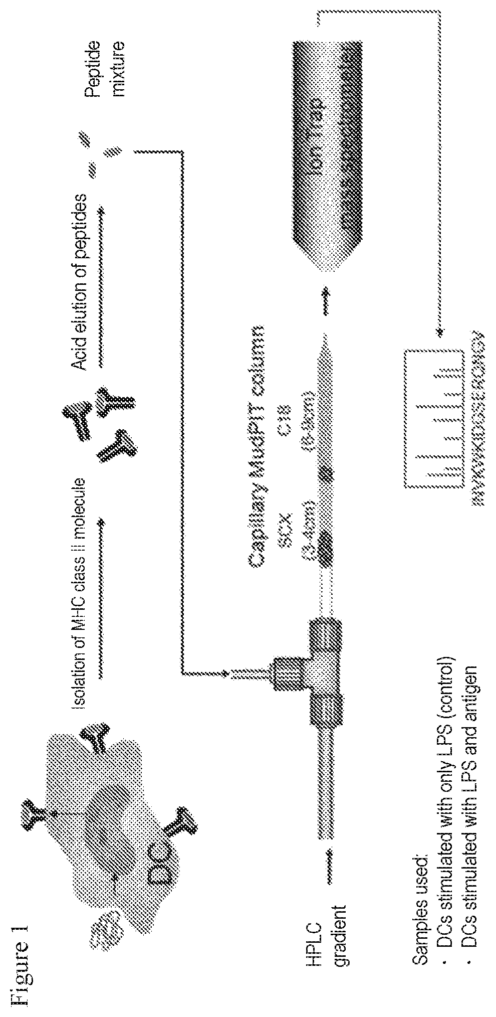
FIG. 1 shows one example of the outline of a technique using MHC II molecules among techniques of MAPPs (MHC-associated peptide proteomics). In the diagram, DC refers to a dendritic cell.
FIG. 2 shows one example of a scheme for obtaining dendritic cell-like cells by the differentiation of human iPS cells.
FIG. 3A shows results of examining molecules expressed on the cell surface of monocyte-like cells prepared from a Tic line, wherein the results were obtained by analysis using a flow cytometer.
FIG. 3B shows results of examining molecules expressed on the cell surface of monocyte-like cells prepared from a Tic line, wherein the results were obtained by analysis using a flow cytometer.
FIG. 4A shows results of examining molecules expressed on the cell surface of monocyte-like cells prepared from a 201B7 line, wherein the results were obtained by analysis using a flow cytometer.
FIG. 4B shows results of examining molecules expressed on the cell surface of monocyte-like cells prepared from a 201B7 line, wherein the results were obtained by analysis using a flow cytometer.
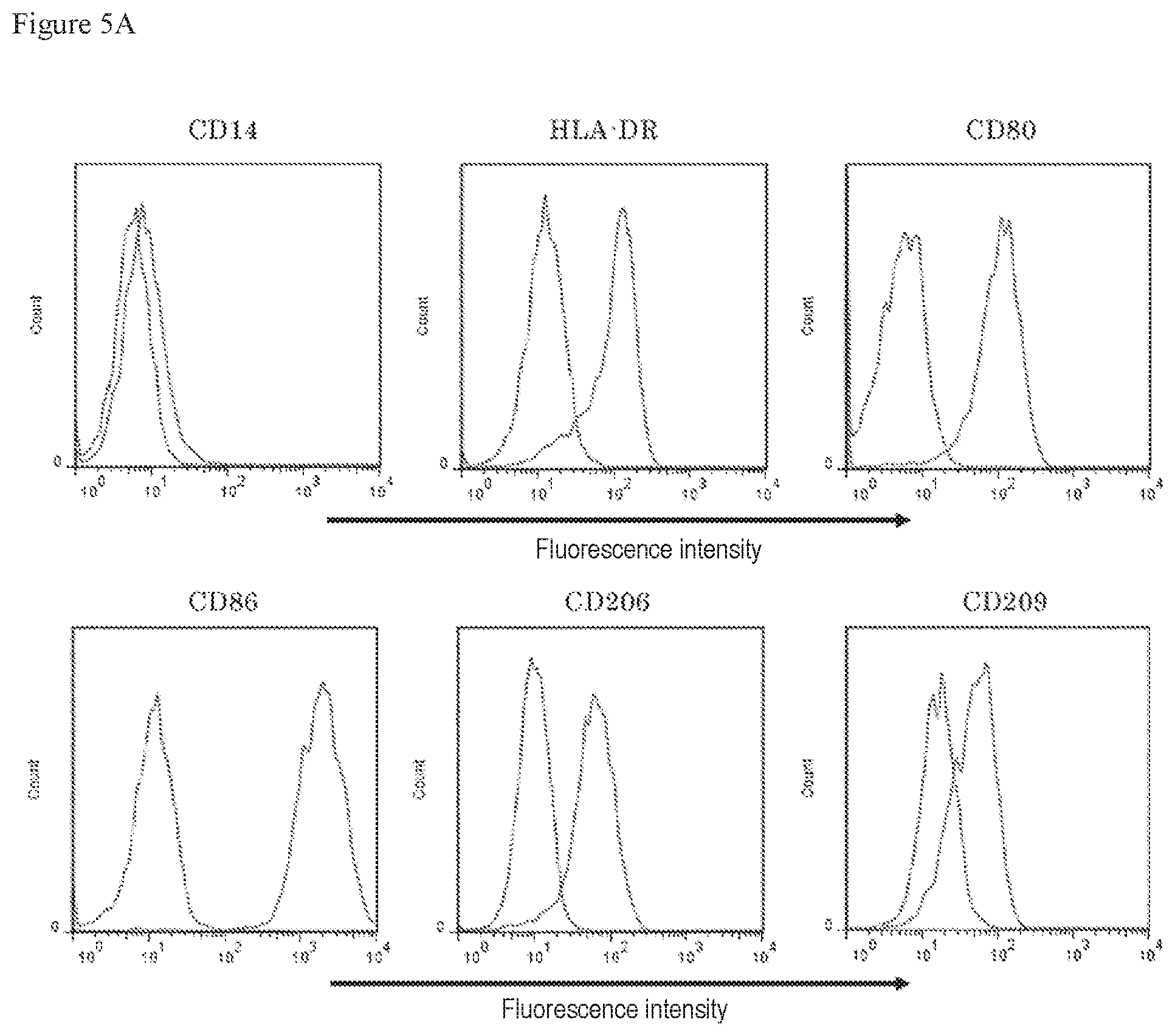
FIG. 5A shows results of examining molecules expressed on the cell surface of dendritic cell-like cells prepared from a Tic line, wherein the results were obtained by analysis using a flow cytometer.
FIG. 5B shows results of examining molecules expressed on the cell surface of dendritic cell-like cells prepared from a Tic line, wherein the results were obtained by analysis using a flow cytometer.
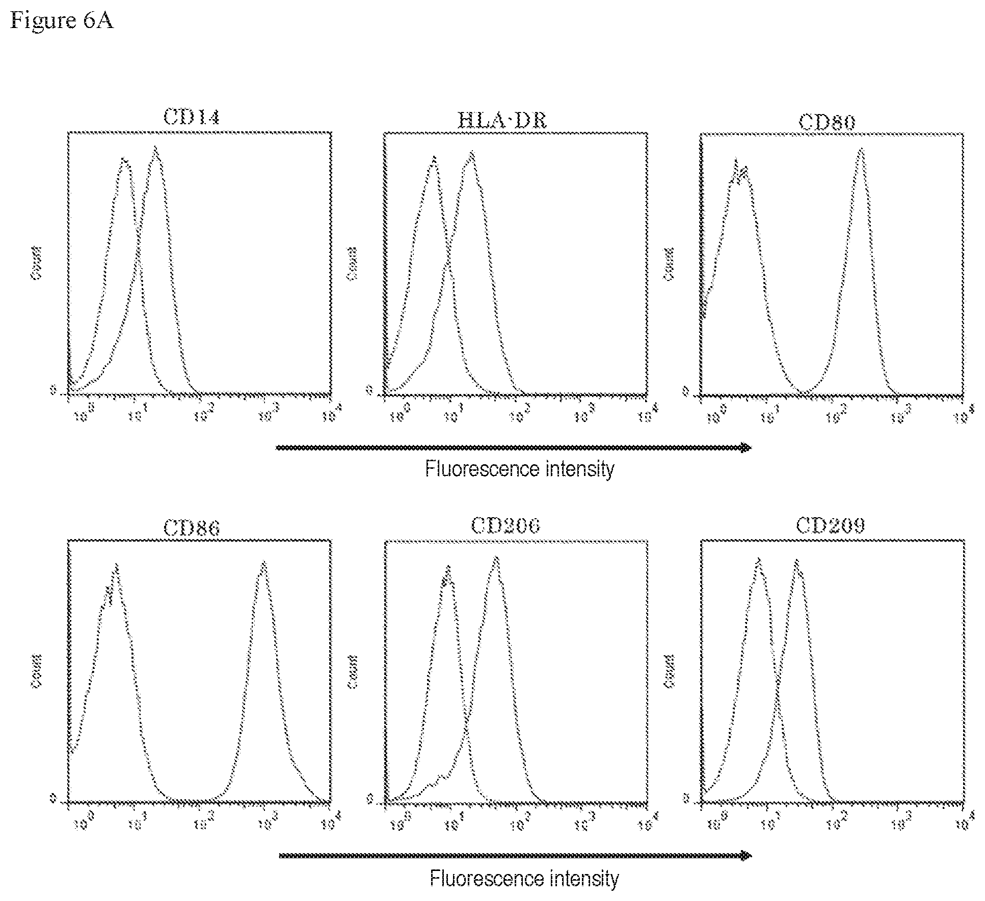
FIG. 6A shows results of examining molecules expressed on the cell surface of dendritic cell-like cells prepared from a 201B7 line, wherein the results were obtained by analysis using a flow cytometer.
FIG. 6B shows results of examining molecules expressed on the cell surface of dendritic cell-like cells prepared from a 201B7 line, wherein the results were obtained by analysis using a flow cytometer.
FIG. 7A and FIG. 7B. FIG. 7A shows results of analyzing the amino acid sequences of peptides detected by exposure to Bet v1a in MAPPs using dendritic cell-like cells derived from human iPS cells (Tic line) FIG. 7B shows results of analyzing the amino acid sequence of peptides detected both under Bet v1a non-treatment conditions and by exposure to Bet v1a. The amino acid sequence of Bet v1a is also shown. In the amino acid sequence of Bet v1a, peptides were detected at four roughly divided sites.
FIG. 8A and FIG. 8B. FIG. 8A shows results of analyzing the amino acid sequences of peptides detected by exposure to Bet v1a in MAPPs using dendritic cell-like cells derived from human iPS cells (201B7 line) FIG. 8B shows results of analyzing the amino acid sequence of peptides detected both under Bet v1a non-treatment conditions and by exposure to Bet v1a. The amino acid sequence of Bet v1a is also shown. In the amino acid sequence of Bet v1a, peptides were detected at three roughly divided sites.
FIG. 9A shows results of analyzing the amino acid sequences of peptides detected by exposure to infliximab in MAPPs using dendritic cell-like cells derived from human iPS cells (Tic line). The amino acid sequences of H and L chains of infliximab are also each shown.
FIG. 9B shows results of analyzing the amino acid sequences of peptides detected both under infliximab non-treatment conditions and by exposure to infliximab in MAPPs using dendritic cell-like cells derived from human iPS cells (Tic line). The amino acid sequences of H and L chains of infliximab are also each shown.
FIGS. 10A-10H. FIG. 10A and FIGS. 10B-10H show results of analyzing the amino acid sequences of peptides detected by exposure to recombinant human Factor VIII (SEQ ID NO: 112) in MAPPs using dendritic cell-like cells derived from human iPS cells (Tic line).
FIG. 10B shows a sequel of FIG. 10A.
FIG. 10C shows a sequel of FIG. 10B.
FIG. 10D shows a sequel of FIG. 10C.
FIG. 10E shows a sequel of FIG. 10D.
FIG. 10F shows a sequel of FIG. 10E
FIG. 10G shows a sequel of FIG. 10F.
FIG. 10H shows a sequel of FIG. 10G.
FIG. 11 shows results of analyzing the amino acid sequences of peptides detected by exposure to Phl p1 in MAPPs using dendritic cell-like cells derived from human iPS cells (Tic line).
FIG. 12 shows results of examining molecules expressed on the cell surface of monocytes, wherein the results were obtained by analysis using a flow cytometer.
FIG. 13A shows results of examining molecules expressed on the cell surface of dendritic cells, wherein the results were obtained by analysis using a flow cytometer.
FIG. 13B shows results of examining molecules expressed on the cell surface of dendritic cells, wherein the results were obtained by analysis using a flow cytometer.
FIG. 14A and FIG. 14B. FIG. 14A shows results of analyzing the amino acid sequences of peptides detected under Bet v1a addition conditions in MAPPs using dendritic cells derived from human donor PBMC. FIG. 14B shows results of analyzing the amino acid sequences of peptides detected under Bet v1a non-addition conditions (control) in MAPPs using dendritic cells derived from human donor PBMC.
FIG. 15A and FIG. 15B. FIG. 15A shows results of analyzing the amino acid sequences of peptides detected under Bet v1a addition conditions in MAPPs using dendritic cells derived from human donor PBMC. FIG. 15B shows results of analyzing the amino acid sequences of peptides detected under Bet v1a non-addition conditions (control) in MAPPs using dendritic cells derived from human donor PBMC.
FIG. 16A and FIG. 16B. FIG. 16A shows results of analyzing the amino acid sequences of peptides detected under Bet v1a addition conditions in MAPPs using dendritic cells derived from human donor PBMC. FIG. 16B shows results of analyzing the amino acid sequences of peptides detected under Bet v1a non-addition conditions (control) in MAPPs using dendritic cells derived from human donor PBMC.
FIG. 17A and FIG. 17B. FIG. 17A shows results of analyzing the amino acid sequences of peptides detected under Bet v1a addition conditions in MAPPs using dendritic cells derived from human donor PBMC. FIG. 17B shows results of analyzing the amino acid sequences of peptides detected under Bet v1a non-addition conditions (control) in MAPPs using dendritic cells derived from human donor PBMC.
FIG. 18A and FIG. 18B. FIG. 8A shows results of analyzing the amino acid sequences of peptides detected under Bet v1a addition conditions in MAPPs using dendritic cells derived from human donor PBMC. FIG. 18B shows results of analyzing the amino acid sequences of peptides detected under Bet v1a non-addition conditions (control) in MAPPs using dendritic cells derived from human donor PBMC.
FIG. 19A and FIG. 19B. FIG. 19A shows results of analyzing the amino acid sequences of peptides detected under Bet v1a addition conditions in MAPPs using dendritic cells derived from human donor PBMC. FIG. 19B shows results of analyzing the amino acid sequences of peptides detected under Bet v1a non-addition conditions (control) in MAPPs using dendritic cells derived from human donor PBMC.
FIG. 20A and FIG. 20B. FIG. 20A shows results of analyzing the amino acid sequences of peptides detected under Bet v1a addition conditions in MAPPs using dendritic cells derived from human donor PBMC. FIG. 20B shows results of analyzing the amino acid sequences of peptides detected under Bet v1a non-addition conditions (control) in MAPPs using dendritic cells derived from human donor PBMC.
FIGS. 21A-21B. FIG. 21A and FIG. 21B show results of comparing the amino acid sequences of peptides detected under Bet v1a (SEQ ID NO:1) addition conditions between use of dendritic cell-like cells derived from human iPS cells and use of dendritic cells derived from PBMCs.
FIG. 21B shows a sequel of FIG. 21A.
DESCRIPTION OF EMBODIMENTS
Hereinafter, preferred embodiments of the present invention will be described.
In the present specification, the protein may be, for example, a natural protein, a recombinant protein, or a synthetic peptide prepared by artificially bonding amino acids. It is understood that the protein may be one protein or a mixture of a plurality of different proteins. The protein may contain a non-natural amino acid. The protein may also be glycosylated, for example, when produced in vivo. The protein is preferably a protein (e.g., an antibody and a hormone) related to treatment or prevention for an animal (preferably a human). In one embodiment, the protein may be one or more, preferably one, selected from the group consisting of cytokines, chemokines, growth factors, antibodies, enzymes, structural proteins, hormones, and fragments of any of these proteins.
The protein is not particularly limited by the length of its amino acid sequence as long as the protein forms a complex with a MHC molecule for antigen presentation after being taken up into a cell and decomposed, after being taken up into a cell but not decomposed, or after being produced in a cell and decomposed. The protein may be a peptide itself that forms a complex with a MHC molecule for antigen presentation.
In the present specification, the epitope refers to a particular structural unit of an antigen to be recognized and bound by an antibody. The epitope is a minimum unit for antigenicity and is also called antigenic determinant.
In the present specification, the differentiation may refer to a state or an aspect, etc., in which an individual cell or a cell population, etc., which has been originally single or identical cell(s), is altered to acquire a complicated or distinct structure and/or function. The differentiation may be used interchangeably with the induction of differentiation, for example, and includes a state in which the induction of differentiation has been started, a state in which the induction of differentiation is ongoing, a state in which the induction of differentiation has been terminated, etc. It is understood that the differentiation should further encompass a state in which a cell or a cell population whose induction of differentiation has been terminated is proliferating, etc. In this context, the induction may mean that a certain cell or cell population, etc., is encouraged to differentiate structurally and/or functionally into another cell or cell population, etc. The induction is not particularly limited as long as the differentiation can be achieved.
In the present specification, the stem cell means a pluripotent stem cell and is not particularly limited as long as the stem cell is a cell having pluripotent differentiation and the ability to self-renew. Examples of the stem cell include induced pluripotent stem cells (iPS cells), embryonic stem cells (ES cells), nuclear transfer ES cells (ntES cells), embryonic germ stem cells (EG cells), and adult stem cells (WO2012/115276). These stem cells are preferably derived from a mammal and more preferably derived from a human.
The ES cell is an embryo-derived stem cell that is derived from, for example, the inner cell mass of a blastocyst, which is an embryo after an 8-cell stage of a fertilized egg and morula. The ES cell can be established by isolating the inner cell mass from the blastocyst of a fertilized egg of a subject animal and culturing the inner cell mass on a feeder of fibroblasts, and methods for establishment and maintenance thereof are known in the art (e.g., U.S. Pat. No. 5,843,780). The ES cell may be selected by real-time PCR by using, for example, the expression of a gene marker such as alkaline phosphatase, OCT-3/4, or NANOG gene as an index. Particularly, the human ES cell may be selected by using the expression of a gene marker such as OCT-3/4, NANOG, FBX15, FGF4, REX1, or ECAD gene as an index (E. Kroon et al., (2008), Nat. Biotechnol., 26: 443-452).
In one aspect, in view of ethical problems that may arise in association with the destroying of fertilized eggs or embryos, which may be potential human beings, for example, redundant embryos determined to be discarded among cryopreserved embryos that have not been brought back to mothers in fertility treatment based on external fertilization may be utilized as embryos for use in the preparation of human ES cells, or embryos that have stopped growing during development in the external fertilization process may be utilized as such embryos. Alternatively, unfertilized eggs may be utilized which lack the innate ability to grow into humans by themselves and are based on parthenogenesis in terms of cell division and growth. Alternatively, only single blastomeres of embryos at a cleavage stage prior to a blastocyst stage may be used to prepare ES cells without impairing the ability of the embryos to develop and without destroying fertilized eggs (Chung Y, Klimanskaya I, Becker S, Marh J, Lu S J, Johnson J, Meisner L, Lanza R. (2006). Nature 439: 216-219; Klimanskaya I, Chung Y, Becker S, Lu S J, Lanza R. (2006). Nature 444: 481-485; and Chung Y, Klimanskaya I, Becker S, Li T, Maserati M, Lu S J, Zdravkovic T, Ilic D, Genbacev O, Fisher S, Krtolica A, Lanza R. (2008). Cell Stem Cell 2: 113-117.). Alternatively, the ES cell may be prepared from a human embryo that has stopped developing (Zhang X, Stojkovic P, Przyborski S, Cooke M, Armstrong L, Lako M, Stojkovic M. (2006). Stem Cells 24: 2669-2676.).
The iPS cell is a somatic cell-derived artificial stem cell that has properties substantially equivalent to the ES cell, for example, pluripotent differentiation and the ability to self-renew, and can be prepared by introducing a particular reprogramming factor in a DNA or protein form into a somatic cell (K. Takahashi and S. Yamanaka (2006) Cell, 126: 663-676; K. Takahashi et al. (2007), Cell, 131: 861-872; J. Yu et al. (2007), Science, 318: 1917-1920; Nakagawa, M. et al., Nat. Biotechnol. 26: 101-106 (2008); and WO2007/069666). (In this context, the somatic cell may refer to every animal cell (preferably a mammalian cell including a human cell) except for germ-line cells and pluripotent stem cells.)
The reprogramming factor may be a gene specifically expressed in ES cells, a gene product or non-cording RNA thereof, a gene that plays an important role in maintaining the undifferentiation of ES cells, a gene product or non-cording RNA thereof, or a low-molecular compound. Examples of the reprogramming factors may include OCT3/4, SOX2, SOX1, SOX3, SOX15, SOX17, KLF4, KLF2, c-MYC, N-MYC, L-MYC, NANOG, LIN28, FBX15, ERAS, ECAT15-2, TCLL, beta-catenin, LIN28B, SALL1, SALL4, ESRRB, NR5A2, and TBX3. These reprogramming factors may be used alone or in combination. Examples of the combination of the reprogramming factors can include the following combinations: (i) OCT gene, KLF gene, SOX gene, and MYC gene; (ii) OCT gene, SOX gene, NANOG gene, and LIN28 gene; (iii) OCT gene, KLF gene, SOX gene, MYC gene, hTERT gene, and SV40 large T gene; and (iv) OCT gene, KLF gene, and SOX gene.
Alternatively, for example, combinations described in WO2007/069666, WO2008/118820, WO2009/007852, WO2009/032194, WO2009/058413, WO2009/057831, WO2009/075119, WO2009/079007, WO2009/091659, WO2009/101084, WO2009/101407, WO2009/102983, WO2009/114949, WO2009/117439, WO2009/126250, WO2009/126251, WO2009/126655, WO2009/157593, WO2010/009015, WO2010/033906, WO2010/033920, WO2010/042800, WO2010/050626, WO2010/056831, WO2010/068955, WO2010/098419, WO2010/102267, WO2010/111409, WO2010/111422, WO2010/115050, WO2010/124290, WO2010/147395, WO2010/147612, and WO2012/115276 may be used as combinations of the reprogramming factors. Examples of the reprogramming factor or a factor promoting reprogramming may include MEK inhibitors, DNA methyl transferase inhibitors, histone deacetylase (HDAC) inhibitors, histone methyl transferase inhibitors, and p53 inhibitors, which are inhibitors generally known to those skilled in the art. The reprogramming factor may be introduced into a somatic cell according to a method generally known to those skilled in the art, for example, a calcium phosphate method, lipofection, or microinjection, optionally using a vector (e.g., a viral vector, a plasmid vector, and an artificial chromosome vector) or the like. For example, a DMEM, DMEM/F12, or DME medium containing 10 to 15% FBS (which may further appropriately contain a leukemia inhibitory factor (LIF), penicillin/streptomycin, puromycin, L-glutamine, nonessential amino acids, .beta.-mercaptoethanol, etc.), or a commercially available medium generally known to those skilled in the art may be appropriately used as a medium for the induction of the iPS cell.
The culture of the iPS cell may be appropriately set according to the composition of the medium, etc. For example, somatic cells are contacted with the reprogramming factor and cultured for approximately 4 to 7 days using a DMEM or DMEM/F12 medium containing 10% FBS at 37.degree. C. in the presence of 5% CO.sub.2. Then, the cells are reseeded onto feeder cells (e.g., mitomycin C-treated STO cells and SNL cells). Approximately 10 days after the contact of somatic cells with the reprogramming factor, the cells are cultured in a medium for primate ES cell culture containing a basic fibroblast growth factor (bFGF). Approximately 30 to approximately 45 days after the contact, or thereafter, an iPS-like colony may appear. Alternatively, the cells are cultured on feeder cells (e.g., mitomycin C-treated STO cells and SNL cells) in a DMEM medium containing 10% FBS (which may further appropriately contain LIF, penicillin/streptomycin, puromycin, L-glutamine, nonessential amino acids, .beta.-mercaptoethanol, etc.) at 37.degree. C. in the presence of 5% CO.sub.2. Approximately 25 to approximately 30 days later, or thereafter, an iPS-like colony may appear. Instead of the feeder cells, somatic cells themselves to be reprogrammed may be used, or extracellular matrix or Matrigel (Becton, Dickinson and Company (BD)) may be used. Alternatively, the culture may be performed using a serum-free medium (Sun N, et al., (2009), Proc Natl Acad Sci USA. 106: 15720-15725).
The iPS cell may be selected according to the shape of the formed colony (e.g., whether to obtain a cell mass having a nearly spherical shape). Alternatively, in the case of introducing, as a marker gene, a drug resistance gene to be expressed in conjunction with a gene (e.g., alkaline phosphatase, OCT3/4, and NANOG genes) that is expressed by the reprogramming of somatic cells, the cells can be cultured in a medium containing the corresponding drug to select the established iPS cell. When the marker gene is a fluorescent protein gene, the iPS cell can also be selected by observation under a fluorescence microscope. Alternatively, the iPS cell may be determined by culturing the cells in vitro by a differentiation method known in the art and using their ability to differentiate into desired cells as an index. Alternatively, the iPS cell may be determined by subcutaneously transplanting the cells into an immunodeficient mouse and analyzing tumor tissues formed after a lapse of a predetermined period to confirm that teratomas made up of a mixture of various tissues are formed. Alternatively, the iPS cell may be determined by confirming that a marker gene specifically expressed in ES cells is expressed. Alternatively, the iPS cell may be determined by detecting a genome-wide gene expression pattern using a microarray or the like to confirm that the cells have an expression pattern highly correlating with that of ES cells.
Alternatively, established iPS cells may be furnished and used.
The ntES cell is a clone embryo-derived ES cell prepared by a nuclear transfer technique and has substantially the same properties as those of fertilized egg-derived ES cells (T. Wakayama et al. (2001), Science, 292: 740-743; S. Wakayama et al. (2005), Biol. Reprod., 72: 932-936; and J. Byrne et al. (2007), Nature, 450: 497-502). In short, the ntES cell is an ES cell established from the inner cell mass of a blastocyst derived from a clone embryo obtained by replacing the nucleus of an unfertilized egg with the nucleus of a somatic cell. For the preparation of the ntES cell, a nuclear transfer technique known in the art (e.g., J. B. Cibelli et al., (1998), Nature Biotechnol., 16: 642-646) may be combined with an ES cell preparation technique known in the art (Sayaka Wakayama, et al., (2008), Experimental Medicine, Vol. 26, No. 5 (extra number), p. 47 to 52). In the nuclear transfer, the nucleus of a somatic cell may be injected into an enucleated unfertilized mammalian egg and cultured for a few hours for reprogramming.
The EG cell is a cell that has pluripotency similar to that of ES cells and is established from a primordial germ cell during fetal life (Y. Matsui et al., (1992), Cell, 70: 841-847). The EG cell may be established by culturing primordial germ cells in the presence of LIF, bFGF, stem cell factor (STF), or the like (Y. Matsui et al., (1992), Cell, 70: 841-847).
The adult stem cell is a cell that has not been finally differentiated and is found in vivo. The adult stem cell exists as a source of a progenitor cell for a finally differentiated cell. The adult stem cell is present in each tissue in vivo and is usually limited by the types of cells into which the adult stem cell can differentiate. In the present invention, examples of the adult stem cell particularly preferably include hematopoietic stem cells considered to be able to differentiate into monocytes, macrophages, dendritic cells, and the like. In this context, hematopoietic progenitor cells refer to cells differentiated from the hematopoietic stem cells.
In the present specification, the progenitor cell derived from the stem cell may include every cell (e.g., mesodermal progenitor cells, hematopoietic progenitor cells, granulocyte macrophage colony-forming cells, lymphoblasts, monoblasts, promonocytes, and monocytes) that is observed in the course of differentiating the stem cell into an antigen-presenting cell (specifically, a MHC molecule-expressing cell). In this context, almost all of nucleated cells have MHC I molecules (Peter Parham (2007), THE IMMUNE SYSTEM; The Human Protein Atlas, http://www.proteinatlas.org/) and can antigen-present endogenous proteins in autologous cells, on killer T cells via the MHC I molecules. On the other hand, particular cells have MHC II molecules in addition to the MHC I molecules and can present foreign antigens on helper T cells via the MHC II molecules (these cells are also called professional antigen-presenting cells).
In the present specification, the antigen-presenting cell may include both of these types of cells. Examples of the antigen-presenting cell of the latter type preferably include dendritic cells, macrophages, monocytes, and B cells. Further, thyroid follicular cells, fibroblasts, vascular endothelial cells, and the like also work as antigen-presenting cells when MHC II molecules are induced through the activation by cytokines such as interferons. Therefore, these cells may be also included in the latter type. A criterion to determine whether to have properties as antigen-presenting cells (specifically, MHC molecule-expressing cells, for example, dendritic cells, macrophages, monocytes, and B cells) may be based on, for example, cells expressing MHC I and/or the MHC II molecules as an index and is more preferably based on cells further expressing at least one of CD11a, CD11b, CD11c, CD14, CD15, CD40, CD80, CD83, CD86, CD123, CD205, CD206, CD209, and CCR7 as an index.
Particularly, the dendritic cells have the strong ability to present antigens and the strong ability to activate helper T cells and are therefore advantageous as antigen-presenting cells. The antigen-presenting cell is most preferably an immature dendritic cell. The dendritic cells are cells that have cell processes and assume a dendritic form. A criterion to determine whether to have properties as dendritic cells may be based on, for example, the further expression of at least one of CD11b, CD11c, CD40, CD80, CD83, CD86, CD123, CD205, CD206, CD209, and CCR7 in addition to the MHC II molecule as an index and is more preferably based on the expression of all of the MHC II molecule, CD80, CD86, CD206, and CD209 as an index. A dendritic cell that expresses all of the MHC II molecule, CD80, CD86, CD206, and CD209 and is negative for CD14 is further preferred. A criterion to determine whether to have properties as macrophage cells may be based on, for example, the further expression of CD11b in addition to the MHC II molecule as an index. In this context, CD80 and CD86 are known to transduce signals to helper T cells and activate these cells. Dendritic cell-like cells obtained in the present Examples have properties similar to those of monocyte-derived dendritic cells in terms of cell shape, the expression of cell surface molecules, and the ability to stimulate helper T cells and may therefore be included in the dendritic cell described in the present specification. Likewise, monocyte-like cells obtained in the present Examples may be included in the monocyte described in the present specification.
The antigen-presenting cell is preferably derived from a mammal and is more preferably derived from a human.
In the present specification, the MHC molecule may be any of MHC I and MHC II molecules and is more preferably a MHC II molecule. Human MHC is called human leucocyte antigen (HLA). The MHC I molecules are further divided into classical class I (class Ia) and a non-classical class I (class Ib) molecules. Examples of the classical class I molecules include HLA-A, HLA-B, and HLA-C in humans. Examples of the non-classical class I molecules include HLA-E, HLA-F, and HLA-G in humans. On the other hand, examples of the MHC II molecules include HLA-DR, HLA-DQ, and HLA-DP in humans.
The MHC molecule differs slightly in amino acid sequence in individuals even among animals of the same species and is further divided into some subtypes called allotypes. For example, HLA-DR is known to have many allotypes such as DR1, DR2, DR3, DR4, . . . . Each allotype is linked on the MHC gene with the other allotypes. Therefore, these allotypes are inherited as a set from parent to child unless gene recombination occurs in this region. This unit is called haplotype. Stem cells (e.g., iPS cells) derived from a patient basically maintain the MHC gene sequence, as it is, of the patient even after being subcultured and/or differentiated. Therefore, antigen-presenting cells obtained by the differentiation of the stem cells maintain MHC molecule allotypes carried by the patient.
The allotypes respectively form complexes with different antigen peptide fragments (epitopes) so that the epitopes can be presented on the cell surface. Therefore, the presence or absence or the degree of immunogenicity, adverse reaction, etc., caused by a target protein varies depending on each allotype set, in other words, each patient having the allotype set. Allotypes or haplotypes have a pattern characteristic of a race or an ethnic group and can therefore be utilized, for example, in the analysis of the presence or absence of immunogenicity, adverse reaction, etc., caused by a target protein for each race or ethnic group.
Thus, a series of antigen-presenting cells having the MHC molecule allotypes of a fixed population (race, ethnic group, etc.) are advantageously used for predicting the immunogenicity of a protein for the population.
Allotypes carried by an individual person can be conveniently identified by genetic diagnosis (e.g., a HLA genotyping method which involves hybridizing DNA amplified by polymerase chain reaction (PCR) to probe-immobilized beads, digitizing the fluorescence intensity thereof, and analyzing the data to identify the HLA genotype) or the like. Therefore, whether the target protein causes immunogenicity, adverse reaction, etc., can be determined on the basis of information on the identified allotypes. Other specific examples of the genetic diagnosis method include methods described in, for example, International Journal of Immunogenetics, 2011; 38: 6, pp. 463-473.
Thus, in a preferred embodiment, the antigen-presenting cell may express one or more MHC molecule allotypes in a subject (e.g., a mammal, preferably a human) intended to receive the target protein.
In a preferred aspect, a cell expressing one or more MHC molecule allotypes carried by a subject (e.g., a human patient or a healthy person) intended to be analyzed may be used as the stem cell or the progenitor cell derived therefrom according to the present invention. For example, one or more cells expressing one or more MHC molecule allotypes in the subject may be used such that all sets of MHC molecule allotypes carried by the subject are contained therein. Alternatively, cells expressing one or more MHC molecule allotypes with high expression frequency in a race or an ethnic group intended to be analyzed may be prepared. For example, a fixed percentage (e.g., 30% to 80% or more) of a population of the race or the ethnic group may be covered by preparing a plurality of such cells and analyzed for the immunogenicity. For example, appropriate comparative analysis among human patients, between human patients and healthy persons, among healthy persons, etc., is advantageous.
In the present invention, the method for differentiating the stem cell or the progenitor cell derived therefrom into the antigen-presenting cell is not particularly limited as long as the method is generally known to those skilled in the art. Methods described in WO2009/120891; WO2009/074341; Regen. Med. (2009) 4 (4), p. 513-526; WO2012/115276; WO2012/043651; PLoS One, July 2011, Vol. 6, Issue 7, e22261; Gene Therapy (2011), 1-1024 March 2011, doi: 10.1038/gt.2011.22; Japan Science and Technology Agency, Strategic Basic Research Programs, CREST: H20-23 Research Report on Fundamental Technologies for Medicine Concerning the Generation and Regulation of Induced Pluripotent Stem (iPS) Cells; International Journal of Cancer 2013 Jul. 3. doi: 10.1002/ijc.28367; Zhuang, L. et al., J. Immunol. Methods (2012); PLoS One, April 2013, Vol. 8, Issue 4, e59243; and NATURE IMMUNOLOGY Vol. 5, No. 4, 2004, pp. 410-417 may be used as methods for differentiating stem cells such as ES cells or iPS cells into, for example, monocytes, macrophages, B cells, or dendritic cells. For example, Regen. Med. (2009) 4 (4), p. 513-526 discloses a method for inducing the in vitro differentiation of human ES cells into dendritic cells in a serum-free medium. In the method disclosed therein, human ES cells are differentiated into monocytes using bone morphogenetic protein-4 (BMP-4), a granulocyte macrophage-colony stimulating factor (GM-CSF), a stem cell factor (SCF), and a vascular endothelial growth factor (VEGF); subsequently, the monocytes are further differentiated into immature dendritic cells using GM-CSF and interleukin-4 (IL-4); and the immature dendritic cells are further differentiated into mature dendritic cells using a maturation cocktail consisting of GM-MSF, TNF-.alpha., interleukin-1.beta. (IL-1.beta.), interferon-.gamma. (IFN-.gamma.), and PGE2. Also, PLoS One, April 2013, Vol. 8, Issue 4, e59243 discloses that functional macrophages and dendritic cells were obtained on the basis of monocytes differentiated from ES cells and iPS cells. Furthermore, NATURE IMMUNOLOGY Vol. 5, No. 4, 2004, pp. 410-417 describes a method for preparing T cells from ES cells as a theme and discloses that B cells were also able to be prepared in the course of this preparation (e.g., in this literature, the second paragraph of the right column on p. 411 to the second paragraph of the left column on p. 412; FIG. 1).
As mentioned above, related techniques have been reported as to the technique itself of differentiating stem cells into antigen-presenting cells (MHC molecule-expressing cells). However, all of these literatures intend the exploitation of the techniques in regenerative medicine or immunotherapy and do not intend the application thereof to the epitope sequence analysis of proteins.
For the specific method for differentiating the stem cell or the progenitor cell derived therefrom into the antigen-presenting cell according to the present invention, preferably, see WO2012/115276. When the antigen-presenting cell is, for example, a dendritic cell, this method may comprise the step of providing the stem cell or the progenitor cell derived therefrom and subsequently the following steps: (a) differentiating the stem cell or the progenitor cell derived therefrom into a mesodermal progenitor cell; (b) differentiating the mesodermal progenitor cell into a monocyte; and (c) differentiating the monocyte into an immature dendritic cell, and optionally further stimulating the immature dendritic cell to obtain a mature dendritic cell.
The step (a) and the step (b) can be continuously performed. Among the steps (a) to (c), at least the step (c) may employ a serum-free medium. Preferably, both the steps (b) and (c) employ a serum-free medium. More preferably, all of the steps (a) to (c) employ a serum-free medium.
The serum may refer to mammal-derived serum such as human serum, monkey serum, fetal bovine serum, sheep serum, rabbit serum, rat serum, guinea pig serum, or mouse serum.
The serum-free medium refers to a medium that is supplemented neither with serum nor with a commercially available serum replacement such as B-27 and may be preferably a medium containing at least one of albumin or an albumin replacement, transferrin or a transferrin replacement, insulin or an insulin replacement, and selenious acid. More preferably, the serum-free medium may be a medium containing insulin-transferrin-selenium-X supplement (ITS). Preferred examples of the serum-free medium include a minimum essential medium (MEM), a Dulbecco's modified eagle medium (DMEM), an Iscove's modified Dulbecco's medium (IMDM), StemPro-34 medium (Life Technologies/Thermo Fisher Scientific Inc.), Stemline II (Sigma-Aldrich Corp.), and Primate ES cell medium (ReproCELL Inc.) each supplemented with ITS.
The step (a) may comprise the step of culturing the stem cell or the progenitor cell derived therefrom in a medium containing BMP family protein and subsequently culturing the cell in a medium containing a growth factor and a hematopoietic factor, or culturing the cell in a medium containing VEGF and then culturing the cell in a medium containing a hematopoietic factor to obtain the mesodermal progenitor cell. The step (b) may comprise the step of differentiating the mesodermal progenitor cell into the monocyte by culture in a medium containing a hematopoietic factor. The step (a) and the step (b) can be continuously performed.
The BMP family protein may refer to a cytokine that belongs to the TGF-.beta. superfamily and has approximately 20 subtypes. In the present invention, the BMP family protein is preferably BMP2 and/or BMP4, more preferably BMP4.
The growth factor may be preferably VEGF and may be specifically VEGF-A, VEGF-B, VEGF-C, VEGF-D, VEGF-E, PlGF (placental growth factor)-1, PlGF-2, or a selective splicing variant thereof (e.g., variants composed of 121, 165, 189, or 206 amino acids are known for VEGF-A). In the present invention, the VEGF is preferably VEGF-A. The growth factor may further include bFGF in addition to VEGF.
The hematopoietic factor is a factor promoting the differentiation and proliferation of blood cells and may be, for example, a stem cell factor (SCF), a granulocyte colony-stimulating factor (G-CSF), a granulocyte macrophage colony-stimulating factor (GM-CSF), a macrophage colony-stimulating factor (M-CSF), erythropoietin (EPO), thrombopoietin (TPO), an interleukin (IL), or Flt3 ligand. The interleukin may be IL-1, IL-2, IL-3, IL-4, IL-5, IL-6, IL-7, IL-8, or IL-9, etc.
The hematopoietic factor preferred for the step (b) may be selected from the group consisting of SCF, TPO, IL-3, Flt3 ligand, GM-CSF, and M-CSF. These hematopoietic factors may be used alone or in combination.
More preferably, the step (a) may be performed using VEGF as the growth factor and SCF as the hematopoietic factor in combination, and subsequently, the step (b) may be performed using GM-CSF and M-CSF as the hematopoietic factor in combination. In the step (b), it is preferred to replace the medium with a fresh one every few days (e.g., every 3 to 4 days) for culture.
Provided that a non-adherent cell (monocyte or monocyte-like cell) is obtained by the step (b), this non-adherent cell may be used as the monocyte in the step (c). Whether the non-adherent cell has the properties of the monocyte can be confirmed by using, for example, flow cytometry and using, as an index, the expression of a monocyte marker such as CD14, CD45.sup.hi, CD11a, CD11b, or CD15 in addition to the expression of the MHC II molecule. From the viewpoint of improving the efficiency of induction of dendritic cells, the cell proportion of monocytes for use in the induction of dendritic cells can be increased, for example, by separating only CD14-positive cells from non-adherent cells by use of a magnetic bead method or the like.
The step (c) may further comprise the step of:
(ci) differentiating the monocyte into an immature dendritic cell (or an immature dendritic cell-like cell) by (suspension-) culture in a medium containing a hematopoietic factor; and optionally comprise the step of:
(cii) further contacting the obtained immature dendritic cell (or immature dendritic cell-like cell) with an immunogen and optionally an inflammatory cytokine to induce a mature dendritic cell (or a mature dendritic cell-like cell).
Whether the immature dendritic cell-like cell or the mature dendritic cell-like cell has the properties of the dendritic cell may be confirmed by using, for example, flow cytometry and using, as an index, the further expression of at least one of dendritic cell markers CD11b, CD11c, CD40, CD80, CD83, CD86, CD123, CD205, CD206, CD209, and CCR7 in addition to the expression of the MHC II molecule. Whether the dendritic cell has the properties of the immature dendritic cell or has the properties of the mature dendritic cell can be tested by using, for example, change in the expression of the MHC II molecule (HLA-DR, etc.) as an index.
The hematopoietic factor may be any of the factors mentioned above. Preferably, a combination of GM-CSF, IL-3, and IL-4 or a combination of GM-CSF and IL-4 may be used as the hematopoietic factor.
Upon contact with the immature dendritic cell, the immunogen and the inflammatory cytokine can stimulate (pulse) the cell to induce a mature dendritic cell. The immature dendritic cell has high phagocytic capacity for an antigen but has the low ability to present the antigen, whereas this cell can be matured into a mature dendritic cell, for example, by the invasion of the antigen into an organism to enhance the expression of a protein, such as the MHC II molecule, necessary for antigen presentation and thereby improve the ability to present the antigen.
The immunogen may be any substance that causes immune response when introduced into an organism. Examples thereof include lipopolysaccharide (LPS, which is present in a pathogen). In the present invention, when the protein to be evaluated has immunogenicity, those skilled in the art can understand that this protein can act as the immunogen. Thus, in a preferred embodiment, the immature dendritic cell is contacted with a target protein having immunogenicity to induce the mature dendritic cell.
The inflammatory cytokine may be, for example, tumor necrosis factor-.alpha. (TNF-.alpha.), TNF-.beta., IL-12, or IFN-.gamma.. These immunogens or inflammatory cytokines may be appropriately used alone or in combination.
If it is desired to obtain a macrophage instead of the dendritic cell as the antigen-presenting cell, the following step:
(d) differentiating the monocyte into a macrophage may be performed, instead of the step (c), according to a method described in PLoS One, April 2013, Vol. 8, Issue 4, e59243. In such a case, the monocyte can be differentiated into the macrophage using preferably GM-CSF or M-CSF as the hematopoietic factor. Further, the macrophage can be differentiated into M1 macrophage by adding, for example, IFN-.gamma. or LPS or can be differentiated into M2 macrophage by adding, for example, IL-4 or IL-13 (macrophages are known to be activated by receiving cytokines produced by helper T cells, and classical activation (M1 macrophage) and selective activation (M2 macrophage) are known).
The respective concentrations of the growth factor, the hematopoietic factor, the cytokine, etc., used in each step mentioned above can be concentrations at which the antigen-presenting cell of interest is obtained, and can be appropriately determined by those skilled in the art. The concentration of BMP4 may be, for example, 5 to 150 ng/ml and is more preferably 10 to 100 ng/ml, further preferably 20 to 80 ng/ml. The concentration of VEGF may be, for example, 20 to 100 ng/ml and is more preferably 30 to 70 ng/ml, further preferably 40 to 50 ng/ml. The concentration of bFGF may be, for example, 10 to 100 ng/ml and is more preferably 20 to 50 ng/ml. The concentration of SCF may be, for example, 20 to 100 ng/ml and is more preferably 30 to 70 ng/ml, further preferably 40 to 50 ng/ml. The concentration of IL-3 may be, for example, 5 to 100 ng/ml and is more preferably 30 to 70 ng/ml. The concentration of TPO may be, for example, 1 to 25 ng/ml and is more preferably 1 to 10 ng/ml. The concentration of Flt3 ligand may be, for example, 10 to 100 ng/ml and is more preferably 30 to 70 ng/ml. The concentration of GM-C SF may be, for example, 5 to 250 ng/ml and is more preferably 50 to 200 ng/ml. The concentration of M-CSF may be, for example, 5 to 100 ng/ml and is more preferably 30 to 70 ng/ml. The concentration of IL-4 may be, for example, 3 to 100 ng/ml and is more preferably 10 to 70 ng/ml. The concentration of TNF-.alpha. may be, for example, 0.05 to 50 ng/ml and is more preferably 0.1 to 20 ng/ml. The concentration of LPS may be, for example, 0.01 to 100 .mu.g/ml and is more preferably 0.1 to 10 .mu.g/ml. These growth factors, hematopoietic factors, cytokines, etc., may be appropriately used in combination according to the purpose, and the optimum concentrations can be appropriately determined by those skilled in the art.
The concentration of the (target) protein to be evaluated can be, for example, a concentration at which an epitope on the protein can be identified, according to the purpose, or can be a concentration at which whether or not the protein has immunogenicity in a subject (e.g., a mammal, preferably a human) can be evaluated. Alternatively, the concentration thereof can be a concentration at which the mature dendritic cell can be induced by the stimulation of the immature dendritic cell. Such a concentration can be appropriately determined by those skilled in the art. Such a concentration may be, for example, 0.01 to 1000 .mu.g/ml and is more preferably 0.1 to 100 .mu.g/ml.
In order to obtain the antigen-presenting cell of interest, those skilled in the art can appropriately optimize the period of each step in consideration of the types and combination of factors to be added to cells. The period of the step (a) may be, for example, 2 days or longer and is preferably 2 to 10 days, more preferably 5 to 8 days. The period of the step (b) may be, for example, 1 day or longer and is preferably 20 to 200 days, more preferably 50 to 150 days. In the step (c), the period of the step (ci) may be, for example, 1 day or longer and is preferably 1 to 10 days, more preferably 4 to 6 days. The period of the step (cii) may be, for example, 12 hours or longer and is preferably 12 to 36 hours, more preferably 24 hours (1 day). The period of the step (d) may be, for example, 1 day or longer and is preferably 1 to 20 days. For example, at day 5 to 15, the macrophage may be further differentiated into M1 macrophage or M2 macrophage. However, those skilled in the art can appropriately determine the optimum culture period in consideration of each culture condition, as a matter of course.
The present invention may also relate to a method for producing a dendritic cell (in vitro) from a stem cell or a progenitor cell derived therefrom, comprising the steps (a) to (c), and a dendritic cell obtained or obtainable by the method. Specifically, the dendritic cell produced by the method expresses not only the MHC II molecule but CD80 and CD86, costimulatory molecules of helper T cells and expresses carbohydrate receptors CD206 and CD209, suggesting that this cell has the ability to activate helper T cells and resistance to viruses and the like. The sequence analysis of an epitope on a protein using the dendritic cell obtained by the method contributes to the development of a protein having low immunogenicity and, in addition, is also expected to bring about an excellent material for research on antigen-presenting cells against autoimmune diseases, viruses, and the like.
Specifically, the present invention further provides, as other aspects, for example, the following aspects:
[23] A method for producing a dendritic cell (in vitro) from a stem cell or a progenitor cell derived therefrom, comprising the following steps:
(a') differentiating the stem cell or the progenitor cell derived therefrom into a mesodermal progenitor cell;
(b') differentiating the mesodermal progenitor cell into a monocyte in a serum-free medium containing a granulocyte macrophage colony-stimulating factor (GM-C SF) and a macrophage colony-stimulating factor (M-CSF); and
(c') differentiating the monocyte into an immature dendritic cell in a serum-free medium, and optionally further stimulating the immature dendritic cell to obtain a mature dendritic cell.
[24] The method according to [23], wherein
the step (c') comprises the step of:
(c1') differentiating the monocyte into the immature dendritic cell in a serum-free medium containing a granulocyte macrophage colony-stimulating factor (GM-C SF) and interleukin 4 (IL-4), and optionally comprises the step of:
(c2') contacting the immature dendritic cell with an immunogen and optionally an inflammatory cytokine to induce the mature dendritic cell.
[25] A dendritic cell obtainable by a method according to [23] or [24].
[26] The dendritic cell according to [25], wherein the dendritic cell further expresses at least one of CD80, CD86, CD206, and CD209 in addition to the MHC II molecule.
[27] The dendritic cell according to [26], wherein the dendritic cell expresses all of CD80, CD86, CD206, and CD209.
[28] A cell composition comprising a dendritic cell according to any of [25] to [27].
The dendritic cell or the cell composition may be used as a cell medicament for performing immune cell therapy for infectious diseases or malignant tumors, or for use in the control of immune response for the purpose of treating autoimmune diseases or rejection or the like associated with organ transplantation. The cell medicament may be appropriately used in combination with an auxiliary, for example, a medium, for the purpose of stably maintaining the dendritic cell.
In one aspect, the present invention also relates to a method for identifying an epitope on a protein, comprising the following steps:
(A) contacting a major histocompatibility complex (MHC molecule)-expressing cell differentiated from a stem cell or a progenitor cell derived therefrom with a target protein;
(B) isolating a complex of a peptide contained in the target protein and the MHC molecule from the MHC molecule-expressing cell; and
(C) eluting the peptide from the complex and identifying the peptide. The method may further comprise the following step:
(D) testing whether or not the identified peptide is an epitope that induces immunogenicity. All of the steps of the method can be carried out in vitro.
For avoiding detecting the amino acid sequences of peptides derived from proteins in serum, it is preferred that the step (A) should be performed under serum-free conditions.
The degree of immunogenicity (or antigenicity) can also be compared, for example, among different proteins, among different protein preparations, or among different bio-pharmaceuticals by use of the method for identifying an epitope on a protein. This method can also be utilized in the quality control of produced proteins.
In the present invention, the amount of the MHC molecule-expressing cell necessary for obtaining, for example, 100 ng of MHC molecules may depend on the number of cells, the expression intensity of the MHC molecules, and the degree of the expression. The optimum amount of the cell can be appropriately determined by those skilled in the art.
Each MHC II molecule allotype (e.g., HLA-DQ1) can carry approximately 500 to 1000 different peptide fragments (Chicz R M et al., J Exp. Med. 1993, 178, 27-47; and Chicz R M & Urban R G, Immunol. Today, 1993, 15, 155-160). However, a great majority of these different peptides merely attain a very low copy number and therefore are not very likely to play a physiological role in vivo. On the other hand, peptide fragments that participate in immunogenicity and activate, for example, helper T cells attain a moderate to high copy number (Latek R R & Unanue E R, Immunol. Rev. 1999, 172: 209-228). These peptides having a moderate to high copy number account for approximately 40 to 50% of the total amount of peptides eluted from MHC II molecules and can correspond to approximately 10 to 200 individual peptides.
Many peptide fragments that form complexes with MHC II molecules are presented as 2 to 5 C-terminally and N-terminally truncated variants sharing a common core sequence of approximately 10 to 13 amino acids indispensable for recognition by T cell receptor (Rudensky A Y et al., Nature 1992, 359, 429-431; and Chicz et al., Nature 1992, 358: 764-768). These variants constitute the same epitope. This means that the number of important different epitopes is actually smaller and falls within the range of, for example, approximately 5 to 70.
The peptide is a peptide that is derived from (the amino acid sequence of) the target protein and can form a complex with the MHC molecule on the surface of the antigen-presenting cell (specifically, the MHC molecule-expressing cell). The peptide may be bound with an intracellular or extracellular MHC molecule. Each MHC II molecule allotype can form a complex with diverse peptides, and the amount of the peptide necessary for the sequencing of each eluted peptide can be, for example, only a fmol amount. According to the method of the present invention, approximately a fmol amount of peptide fragments bound with MHC molecules can be isolated from, for example, approximately 0.1 to 5 .mu.g of MHC molecules, and the sequences of the peptides can be identified.
In order to isolate the complexes of the MHC molecules and the peptides from the antigen-presenting cell, the cell membrane of the cell may be lysed. This lysis may be carried out by a method generally known to those skilled in the art, for example, freezing-thawing, use of a surfactant, or a combination thereof. For example, Triton X-100 (TX100), Nonidet P-40 (NP-40), Tween 20, Tween 80, n-octylglucoside, ZWITTERGENT, Lubrol, or CHAPS may be used as the surfactant. Cell debris and nucleus are removed by centrifugation from the cell lysate containing the solubilized MHC molecule-peptide complexes.
The cell lysate containing the solubilized MHC molecule-peptide complexes may be subjected to immunoprecipitation or immunoaffinity chromatography to purify the MHC molecule-peptide complexes. An antibody that is specific for each MHC molecule and is suitable for immunoprecipitation or immunoaffinity chromatography (anti-MHC I molecule antibody, for example, anti-HLA-A antibody, anti-HLA-B antibody, anti-HLA-C antibody, or anti-HLA-ABC antibody, etc.; or anti-MHC II molecule antibody, preferably anti-HLA-DR antibody, anti-HLA-DQ antibody, or anti-HLA-DP antibody) may be used for these methods. The specific antibody is preferably a monoclonal antibody and may be conjugated with beads (e.g., Sepharose beads or agarose beads) through a covalent bond or a noncovalent bond, for example, via protein A. For example, the amino group of the antibody may be covalently bonded to CNBr-activated Sepharose so that the antibody is immobilized thereon. A commercially available product may be purchased as the monoclonal antibody, or the monoclonal antibody may be purified from the supernatant of each corresponding hybridoma cell using protein A- or protein G-affinity chromatography.
The MHC molecules may be immunoisolated, for example, by performing incubation while rotating the antibody-beads together with the cell lysate for a few hours. Also, the antibody-beads bound with the MHC molecule-peptide complexes may be washed in an Eppendorf tube. The results of immunoprecipitation may be analyzed by SDS-PAGE and Western blotting using an antibody that recognizes a denatured MHC molecule.
A mixture of peptides that are derived from the target protein through the decomposition by the antigen-presenting cell is obtained by eluting the peptides from the complexes formed with the MHC molecules.
The peptides may be eluted by a method generally known to those skilled in the art, for example, by use of a diluted acid, for example, diluted acetonitrile (Jardetzky T S et al., Nature 1991 353, 326-329), diluted acetic acid and heating (Rudensky A Y et al., Nature 1991, 353, 622-626; and Chicz R M et al., Nature 1992, 358, 764-768), or diluted trifluoroacetic acid (Kropshofer H et al., J Exp Med 1992, 175, 1799-1803). The peptides are preferably eluted with diluted trifluoroacetic acid, for example, at 37.degree. C.
Before the elution of the peptides from the MHC molecule-peptide complexes, these complexes may be washed with water or a low-salt buffer solution in order to remove surfactant residues. A Tris buffer solution, a phosphate buffer solution, or an acetate buffer solution having a concentration of 0.5 to 10 mM may be used as the low-salt buffer solution. Alternatively, the MHC molecule-peptide complexes may be washed with ultrapure water for HPLC. This washing may be performed by ultrafiltration. The ultrafiltration may be performed in an ultrafiltration tube having, for example, a cutoff value of 30 kD, 20 kD, 10 kD, or 5 kD and a tube volume of 0.5 to 1.0 ml. The inside of the ultrafiltration tube may be washed, for example, 4 to 12 times, with a washing solution having tens of times the volume of the beads carrying the MHC molecule-peptide complexes.
The peptides may be eluted from the MHC molecule-peptide complexes by use of this ultrafiltration tube. Subsequently, the eluted peptides may be dried by use of freeze drying or a centrifugal evaporator.
The peptide mixture thus eluted may be fractionated and subjected to sequence analysis by use of liquid chromatography mass spectrometry (LC/MS) to identify (the amino acid sequence of) each peptide.
The amino acid sequence of each peptide in the peptide mixture can be determined by the sequence analysis according to a method known in the art sufficient for sequencing a fmol amount of peptides.
The identification reveals a protein from which the peptide is derived, and a sequence from which the peptide is derived in the protein.
The eluted peptide mixture is preferably fractionated, for example, using reverse-phase chromatography and anion-exchange chromatography or cation-exchange chromatography in combination or reverse-phase chromatography alone. The fractionation may be performed on a HPLC mode using a fused-silica micro-capillary column connected either to nanoflow electrospray ion source in a mass spectrometer or to a microfractionation apparatus that spots fractions on a plate for MALDI analysis.
Electrospray ionization tandem mass spectrometry (ESI-MS) or MALDI-post source decay (PSD) MS may be used as a mass spectrometry technique, and ESI-MS is preferred.
In the sequence analysis, the amino acid sequence of each peptide may be determined by various approaches generally known to those skilled in the art. The sequence analysis may be performed by the computer analysis of a peptide fragment spectrum using, for example, MASCOT algorithm or SEQUEST algorithm. These algorithms preferably employ protein and nucleotide sequence databases, for conducting the cross-correlation analysis of experimentally and theoretically prepared tandem mass spectra. This permits automatic high-throughput sequence analysis.
Matrix-assisted laser desorption ionization time-of-flight (MALDI-TOF) mass spectrometry may be performed for the qualitative analysis of all of the peptides obtained by elution. The MALDI-TOF analysis can provide a rough outline regarding the complexity of the peptide mixture and the presence of a primary peptide.
In order to estimate the amount of each single peptide eluted from the complex with the MHC molecule, substances that have passed through a microcapillary column may be analyzed using a UV detector at a detection wavelength of 214 nm. The peak area of the peptide to be analyzed may be compared with the peak areas of serial amounts of standard peptides (control) to estimate the amount of the peptide.
A set of peptides that are derived from the target protein by natural fragmentation in the antigen-presenting cell is obtained by eluting the peptides from the MHC molecules. For identifying and excluding false-positive peptides, it is preferred that a set of antigen-presenting cells exposed to the target protein as well as a set of antigen-presenting cells unexposed to the target protein as a negative control should be prepared and analyzed by comparison. A peptide that can be detected in only the antigen-presenting cells exposed to the target protein as compared with the antigen-presenting cells unexposed to the target protein may be identified as functioning as an epitope on the protein from which the peptide is derived, and having antigenicity.
Whether or not the identified peptide functions as an epitope can be tested by using, for example, a MHC-binding motif, the ability to bind to MHC, or recognition by helper T cells as an index. Alternatively, this test may be appropriately combined with an in silico epitope prediction algorithm.
The MHC-binding motif means a structural feature that is common in peptides binding to particular MHC molecules (allelic polymorphism) and is necessary for forming stable complexes with the MHC molecules. In the case of MHC II molecules, the peptide length varies from 12 to 18 amino acids, and even longer peptides can bind because both ends of the peptide-binding groove are open. Most of MHC II molecules can accommodate up to 4 residues (called "anchor residues") related to the binding, at relative positions P1, P4, P6 and P9 contained in a nonameric core region. However, this core region may vary in distance from the N terminus of the peptide. In many cases, 2 to 4 N-terminal residues precede the core region. Thus, the P1 anchor residue is located at position 3, 4, or 5 in many peptides that can form with complexes with the MHC II molecules. Peptides eluted from, for example, HLA-DR molecules can share a hydrophobic P1 anchor such as tyrosine, phenylalanine, tryptophan, methionine, leucine, isoleucine, or valine. The positions and types of the anchor residues can be estimated from the peptide-binding motifs of frequently occurring MHC molecules. A computer algorithm that permits motif validation in peptide sequences can be obtained from, for example, "Tepitope" (www.vaccinome.com, J. Hammer, Nutley, USA).
The ability to bind to MHC may be tested by a method generally known to those skilled in the art using the detected peptide itself (e.g., a synthetic peptide may be utilized) and a desired MHC molecule (Kropshofer H et al., J. Exp. Med. 1992, 175, 1799-1803; Vogt A B et al., J. Immunol. 1994, 153, 1665-1673; and Sloan V S et al., Nature 1995, 375, 802-806). Alternatively, cellular binding assay using MHC molecule-expressing cell lines and biotinylated peptides may be used to test the ability to bind to MHC (Arndt S O et al., EMBO J., 2000, 19, 1241-1251). The relative ability of each peptide to bind to MHC may be determined by measuring a concentration necessary to reduce the binding of a labelled reporter peptide to 50% (IC50). In this context, each identified peptide may be used as such a peptide, or a peptide having a sequence common in identified peptides (core sequence) may be used. The peptide to be detected is considered to depend on the types of MHC molecule allotypes, the strength of binding affinity for a MHC molecule, etc.
The ability to stimulate helper T cells is particularly important for testing whether the identified peptide functions as an epitope. When each identified peptide stimulates helper T cells, this may be used as one index for determining that the peptide has immunogenicity. This determination method may involve testing whether the peptide identified by the method of the present invention has the ability to activate helper T cells. Each identified peptide may be used as such a peptide, or a peptide having a sequence common in identified peptides (core sequence) may be used.
The cellular response of helper T cells may be measured by various in vitro methods generally known to those skilled in the art. For example, MHC molecule-expressing cells (e.g., monocytes, macrophages, or dendritic cells) are cultured together with helper T cells in the presence of the peptide to be evaluated. The uptake of radioactive material-labeled thymidine (T) during DNA replication may be measured with helper T cell proliferation as an index. Alternatively, 5-bromo-2'-deoxyuridine (BrdU) may be used instead of thymidine. In such a case, helper T cells that have taken up BrdU during DNA replication are treated with a monoclonal antibody against BrdU. Then, the amount of BrdU taken up may be measured using an enzymatically or fluorescently labeled secondary antibody (e.g., 5-Bromo-2'-deoxyuridine Labeling & Detection Kit III, Roche-Biochem, Cat No. 1 444 611). Alternatively, Naive Primary T cell Assay (Proimmune Ltd.) which employs flow cytometry using the dilution of a fluorescent dye label 5,6-carboxyfluorescein diacetate succinimidyl ester (CFSE) by the proliferation of helper T cells as an index may be used in the measurement. Alternatively, the cellular response of helper T cells may be evaluated by measuring various cytokines produced from the helper T cells, instead of measuring the cell proliferation. Examples of such cytokines include IL-2, IL-4, IL-6, IL-10, IL-12, IFN-.gamma., and transforming growth factor-.beta. (TGF-.beta.). Examples of the method for measuring the cytokines include various methods generally known to those skilled in the art, for example, ELISA and ELISPOT.
Preferably, dendritic cells produced by the method for producing a dendritic cell from a stem cell or a progenitor cell derived therefrom according to the present invention as mentioned above may be used as the MHC molecule-expressing cells. The MHC molecule-expressing cells may be rendered non-proliferative, for example, by treatment with ionizing radiation or mitomycin C before the assay.
In an alternative aspect, the present invention also relates to a method for producing a protein with reduced or eliminated immunogenicity, comprising the following steps:
(1) identifying an epitope on a protein according to the aforementioned method;
(2) modifying the epitope to reduce or eliminate the binding of the epitope to a MHC molecule; and
(3) producing a protein having the modified epitope.
In a further alternative aspect, the present invention also relates to a protein obtained or obtainable according to the production method. The "obtainable protein" may mean a protein that can be obtained provided that the production method is used.
In relation to the step (2), the amino acid sequence of the identified epitope peptide can be altered or modified to reduce or eliminate the binding of the epitope peptide to a MHC molecule or reduce or eliminate the immunogenicity. The reduction or elimination of the immunogenicity is determined by a method generally known to those skilled in the art without particular limitations and may be determined by using, for example, the aforementioned MHC-binding motif, ability to bind to MHC, or recognition by helper T cells as an index. Alternatively, this approach may be appropriately combined with an in silico epitope prediction algorithm.
The alteration or the modification may be performed according to a method generally known to those skilled in the art. For example, a DNA nucleotide sequence encoding the amino acid sequence of a protein comprising the epitope may be subjected to, for example, site-directed mutagenesis or homologous recombination for the insertion, substitution, or deletion, etc., of desired one or more nucleotides in the DNA sequence. Preferably, for example, one or more anchor residues important for binding to a MHC molecule are changed to other amino acid residues, whereby the immunogenicity can be reduced or eliminated. Alternatively, amino acid residue(s) important for the recognition of the epitope by, for example, T cell receptor on helper T cells or B cell receptor on B cells may be changed to other amino acid residues. The method for replacing the anchor residues important for binding to a MHC molecule is well known to those skilled in the art. For example, the P1 anchor of a HLA-DR1-restricted T cell epitope may be replaced with alanine, proline, glycine, or a charged amino acid residue (Kropshofer et al., EMBO J. 15, 1996, 6144-6154).
In relation to the step (3), the protein having the modified epitope may be chemically synthesized or may be genetically or biologically synthesized. In the case of genetic or biological synthesis, host cells or animals transiently or permanently harboring the gene of the protein having the modified epitope may be utilized. The host cells or the animals can be used, for example, as a production system for protein production or expression. Eukaryotic cells or prokaryotic cells may be used as the host cells.
Examples of the eukaryotic cells that can be used as the host cells include animal cells, plant cells, and fungal cells. Examples of the animal cells include: mammalian cells, for example, CHO (Puck et al., (1958) J. Exp. Med. 108 (6): 945-956), COS, HEK293, 3T3, myeloma, BHK (baby hamster kidney), HeLa, and Vero; amphibian cells, for example, Xenopus oocyte cells (Valle et al., Nature (1981) 291: 338-340); and insect cells, for example, Sf9, Sf21, and Tn5. CHO cells are preferred for the purpose of large-scale expression in the animal cells. The host cells may be transfected with a vector having the gene of the protein having the modified epitope by a method, for example, a calcium phosphate method, a DEAE-dextran method, a method using a cationic ribosome DOTAP (manufactured by Boehringer Mannheim), electroporation, or lipofection. For example, Nicotiana tabacum-derived cells and Lemna minor are known as a protein production system of the plant cells. These cells may be allowed to produce the protein by a callus culture method. A protein expression system using cells of a yeast, for example, the genus Saccharomyces (Saccharomyces cerevisiae, Saccharomyces pombe, etc.), or cells of a filamentous fungus, for example, the genus Aspergillus (Aspergillus niger, etc.) may be used as the fungal cells. In the case of using the prokaryotic cells, a production system using bacterial cells may be used. For example, E. coli or Bacillus subtilis cells may be used as the bacterial cells.
Examples of the animals include genetically recombinant animals and transgenic animals. The type of the animals is not limited, and, for example, cattle, sheep, or mice may be used. In such a case, for example, the protein secreted into a body fluid such as milk may be recovered.
In the present invention, the protein having the modified epitope may be produced at a large scale continuously or commercially. The produced protein or a composition comprising the protein (e.g., a pharmaceutical composition) is also included in the present invention.
In an alternative aspect, the present invention also relates to a method for predicting whether or not a protein has immunogenicity in a subject, comprising the steps of
(I) providing a cell expressing one or more MHC molecule allotypes in the subject intended to receive the target protein, wherein the cell is differentiated from a stem cell or a progenitor cell derived therefrom;
(II) contacting the "cell expressing one or more MHC molecule allotypes" with the target protein;
(III) isolating a complex of a peptide contained in the target protein and the MHC molecule from the "cell expressing one or more MHC molecule allotypes";
(IV) eluting the peptide from the complex and identifying the peptide; and
(V) optionally testing whether or not the identified peptide is an epitope that induces immunogenicity, wherein
when the identified peptide is an epitope that induces immunogenicity, this indicates that the target protein has immunogenicity in the subject.
Those skilled in the art can understand that the prediction method can be carried out by one of or an appropriate combination of two or more of the technical features described above in the present specification.
The prediction method also permits comparison of the presence or absence or the degree of immunogenicity based on each MHC molecule allotype or allotype set against the predetermined protein.
In the prediction method, it is further preferred that one or more cells expressing one or more MHC molecule allotypes in the subject should be provided such that all sets of MHC molecule allotypes carried by the subject are contained therein. The stem cell is still more preferably a stem cell (e.g., an iPS cell or ES cell) derived from the subject (e.g., a mammal, preferably a human). This is because antigen-presenting cells differentiated from stem cells maintain MHC molecule allotypes; thus, antigen-presenting cells that cover all sets of MHC molecule allotypes carried by the subject can be produced by use of the stem cell derived from the subject. Whether or not the target protein has immunogenicity in each subject (e.g., human patient or healthy person) may be evaluated individually by use of such antigen-presenting cells (accomplishment of individual medicine). In other words, the prediction method also relates to a method for selecting a subject (e.g. a patient) having immunogenicity or a method for selecting a subject (e.g. a patient) free from immunogenicity. Alternatively, the prediction method also relates to a method for indicating that one or more particular MHC molecule allotypes are involved in immunogenicity in association with a protein intended to be administered.
It is considered that genes that define MHC molecule allotypes (HLA genotypes in humans) are preserved in almost all of somatic cells in one individual. Thus, the stem cell (e.g., an iPS cell or an ES cell) derived from the subject (e.g., a mammal, preferably a human) may be prepared from any somatic cell or the like of the subject as long as the stem cell maintains the MHC molecule allotypes. Examples of the somatic cell are not particularly limited, and, for example, iPS cells can also be prepared from PBMCs separated from a human or the like. Examples of such reports include Soares F A, Pedersen R A, Vallier L., Generation of Human Induced Pluripotent Stem Cells from Peripheral Blood Mononuclear Cells Using Sendai Virus, Methods Mol Biol. 2015 Feb. 17; Quintana-Bustamante O, Segovia J C., Generation of Patient-Specific induced Pluripotent Stem Cell from Peripheral Blood Mononuclear Cells by Sendai Reprogramming Vectors, Methods Mol Biol. 2014 Dec. 19; Su R J, Neises A, Zhang X B., Generation of iPS Cells from Human Peripheral Blood Mononuclear Cells Using Episomal Vectors, Methods Mol Biol. 2014 Nov. 18; and Riedel M, Jou C J, Lai S, Lux R L, Moreno A P, Spitzer K W, Christians E, Tristani-Firouzi M, Benjamin I J., Functional and pharmacological analysis of cardiomyocytes differentiated from human peripheral blood mononuclear-derived pluripotent stem cells, Stem Cell Reports. 2014 May 29; 3 (1): 131-41. A large number of methods, as listed in the present specification, have been reported as methods for establishing antigen-presenting cells such as dendritic cells from stem cells such as iPS cells. Thus, those skilled in the art should understand that, for example, after preparation of stem cells such as iPS cells from cells such as PBMCs separated from the predetermined subject (e.g. a human patient), antigen-presenting cells such as dendritic cells can be established using the stem cells and thereby utilized in the prediction method. In this respect, the subject-derived cells for use in the preparation of stem cells such as iPS cells may be cells whose MHC molecule allotypes (HLA genotypes in humans) can be identified, or cells whose MHC molecule allotypes have already been revealed (or predicted). Alternatively, iPS cells whose MHC molecule allotypes have already been revealed (or predicted) may be used.
In a further alternative aspect, the present invention relates to a composition for the treatment and/or prevention of a disease related to a protein, in a subject, comprising the protein as an active ingredient, wherein the subject is selected from (only) subjects predicted to be free from the immunogenicity of the protein according to the aforementioned prediction method. The "composition for the treatment and/or prevention" preferably comprises a therapeutically and/or prophylactically effective amount of the protein, and the effective amount of the protein can be appropriately determined by those skilled in the art. Also, the "composition for the treatment and/or prevention" may contain one or more additional agents.
The term "predicted to be free from the immunogenicity of the protein" may mean that the target protein does not evoke any immunogenicity in the subject or merely evokes immunogenicity to an extent tolerable from the viewpoint of efficacy, safety, etc.
It is preferred that the protein or the composition for the treatment and/or prevention comprising the protein as an active ingredient should be administered to only a subject predicted to be free from immunogenicity, by preparing cells expressing one or more MHC molecule allotypes according to a race, an ethnic group, a population, or an individual person, and evaluating the immunogenicity of the protein. The composition is preferably a pharmaceutical composition.
When the protein is contained in the (pharmaceutical) composition for use, the (pharmaceutical) composition can be formulated and used by a pharmaceutical production method known in the art. The (pharmaceutical) composition can be used, for example, orally as an optionally sugar-coated tablet, a capsule, an elixir, or a microcapsule, or parenterally (e.g., percutaneously, intranasally, transbronchially, intramuscularly, or intravenously) in the form of an aseptic solution or suspension containing the protein in water or any of other pharmaceutically acceptable solutions. The (pharmaceutical) composition may be produced so as to appropriately contain a pharmaceutically acceptable carrier, flavor, excipient, vehicle, antiseptic, stabilizer, or binder. For example, a binder such as gelatin, corn starch, gum tragacanth, or gum arabic, an excipient such as crystalline cellulose, a swelling agent such as corn starch, gelatin, or alginic acid, a lubricant such as magnesium stearate, a sweetener such as sucrose, lactose, or saccharin, and a flavor such as peppermint, Akamono oil, or cherry may be used as additives that can be mixed into tablets or capsules. When the unit dosage form is a capsule, this capsule can further contain a liquid carrier such as oil and fat in addition to the materials described above. The aseptic solution for injection can be formulated according to a method well known to those skilled in the art using a vehicle such as injectable distilled water. Examples of the aqueous solution for injection include physiological saline and isotonic solutions containing glucose or other adjuvants, for example, D-sorbitol, D-mannose, and D-mannitol. An appropriate solubilizer, for example, an alcohol such as ethanol, a polyalcohol such as propylene glycol or polyethylene glycol, or a nonionic surfactant such as polysorbate 80.TM. or HCO-50 may be further used in combination therewith. Examples of the oily liquid include sesame oil and soybean oil, which may be used in combination with a solubilizer, for example, benzyl benzoate or benzyl alcohol. For example, a buffer such as a phosphate buffer solution or a sodium acetate buffer solution, for example, a soothing agent such as procaine hydrochloride, for example, a stabilizer such as benzyl alcohol or phenol, or an antioxidant may also be contained therein. The prepared injection solution may usually be filled into an appropriate ampule for use.
The dose, administration method, dosing intervals, etc., of the protein or the composition for the treatment and/or prevention comprising the protein as an active ingredient varies depending on the body weight, age, symptoms, etc., of a patient and can be appropriately selected and determined by those skilled in the art.
As for the "disease related to a protein", the protein and the disease related to the protein are not particularly limited. The protein is still more preferably a protein that may cause, for example, immunogenicity or problems associated with efficacy or safety when administered into an organism. Examples of the disease include, but are not limited to, autoimmune diseases (e.g., rheumatoid arthritis, type I diabetes, multiple sclerosis (MS), coeliac disease, myasthenia gravis (MG), and systemic lupus erythematosus (SLE)), cancers (e.g., melanoma, breast cancer, B cell lymphomas, prostate cancer, and renal cancer), and infectious diseases (e.g. diseases caused by HIV, hepatitis C virus, measles virus, and mycobacteria).
Examples of a combination of such a protein and a disease related to the protein may include examples described in Self/Nonself 2010; 1 (4) pp. 314-322; PHARM TECH JAPAN Vol. 28, No. 10 (2012), pp. 117 (2065)-126 (2074); Sorensen, P. S., et al., Neurology, 67 (9), 1681-3 (2006); Hesse, D., et al., Eur. J. Neurol., 14 (8), 850-9 (2007); Casadevall, N., et al., N. Engl. J. Med., 346 (7), 469-75 (2002); Gershon, S. K., et al., N. Eng. J. Med., 346 (20), 1584-6 (2002); and Locatelli F., et al., Perit. Dial. Int., 27 (Supp12), S303-7 (2006).
Specific examples of the combination of the protein and the disease related to the protein include, but are not limited to: muromonab and allograft rejection; abciximab and PTCA adjunct; rituximab and non-Hodgkin lymphoma; daclizumab and transplant rejection; trastuzumab and breast cancer; palivizumab and RSV prophylaxis; basiliximab and transplant rejection; infliximab and rheumatoid arthritis or Crohn; arcitumomab and colorectal cancer; canakinumab and cryopyrin-associated periodic syndrome; fanolesomab and imaging for appendicitis; imciromab and cardiac imaging for MI; capromab and prostate cancer diagnostic; nofetumomab and detection of SCLC; gemtuzumab and acute myeloid leukemia; alemtuzumab and B cell chronic lymphocytic leukemia; ibritumomab and non-Hodgkin lymphoma; adalimumab and rheumatoid arthritis, Crohn, PsA, JIA, ankylosing spondylitis, or plaque psoriasis; omalizumab and asthma; efalizumab and psoriasis; tositumomab and non-Hodgkin lymphoma; cetuximab and colorectal cancer; bevacizumab and colorectal, breast, renal or NSCL cancer; panitumumab and colorectal cancer; ranibizumab and macular degeneration; eculizumab and paroxysmal nocturnal hemoglobinuria; natalizumab and multiple sclerosis (MS) or Crohn; golimumab and rheumatoid arthritis, PsA, or ankylosing spondylitis; cetolizumab pegol and rheumatoid arthritis or Crohn; ofatumumab and CLL; ustekinumab and plaque psoriasis; tocilizumab and rheumatoid arthritis; denosumab and osteoporosis; Prolastin and .alpha.1-antitrypsin deficiency; Aralast and .alpha.1-antitrypsin deficiency; Zemaira and .alpha.1-antitrypsin deficiency; Kogenate F S and hemophilia A; ReFacto and hemophilia A; Zyntha and hemophilia A; NovoSeven and hemophilia; Benefix and hemophilia B; ATryn and thromboembolism; BabyBIG and infant botulism; Berinert and angioedema; Cinryze and angioedema; Rhophylac and ITP; Evithrom and coagulation; Recothrom and coagulation; Wilate and coagulation; Cerezyme and Gaucher disease; exenatide or Byetta and type II diabetes; Intron A and leukemia, Kaposi sarcoma, or hepatitis B/C; Betaseron and multiple sclerosis; NovoLog and type II diabetes; Leukine and preventing infection in cancer; NEUPOGEN and preventing infection in cancer; Retavase and myocardial infarction or pulmonary embolism; Humatrope and dwarfism; Adagen and inherited immunodeficiency; Pulmozyme and cystic fibrosis; Procrit and anemia in chronic renal disease; and Proleukin and oncology.
In a further alternative aspect, the present invention relates to [30] a method for treating and/or preventing a disease related to a protein, comprising the step of administering the protein to a subject in need of the treatment and/or prevention, wherein the subject is selected from (only) subjects predicted to be free from the immunogenicity of the protein according to the aforementioned prediction method.
In a further alternative aspect, the present invention relates to [31] use of a protein for the production of a medicament for the treatment and/or prevention of a disease related to the protein, wherein a subject of the treatment and/or prevention is selected from (only) subjects predicted to be free from the immunogenicity of the protein according to the aforementioned prediction method.
In a further alternative aspect, the present invention relates to use of a stem cell or a progenitor cell derived therefrom, or a MHC molecule-expressing cell differentiated from the stem cell or the progenitor cell in various methods according to the present invention described above.
Those skilled in the art can understand that these aspects of the present invention can be carried out by one of or an appropriate combination of two or more of the technical features described in the present specification.
Those skilled in the art should understand that one of or any combination of two or more of the aspects described in the present specification is also included in the present invention unless a technical contradiction arises on the basis of the technical common sense of those skilled in the art.
The terms in the present specification are used for illustrating particular embodiments and are not intended to limit the invention. The terms (including technical terms and scientific terms) used in the present specification are interpreted to have the same meanings as those understood in a broad sense by those skilled in the art to which the present invention belongs, unless otherwise defined. These terms used in the present specification should not be interpreted in an idealized or excessively formal sense, unless otherwise defined.
The term "comprise" used in the present specification means that described items (members, steps, factors, numbers, etc.) are present and the presence of the other items (members, steps, factors, numbers, etc.) is not excluded therefrom, unless the context evidently requires different interpretation.
The embodiments of the present invention may be described with reference to a schematic diagram, which may be exaggerated for the purpose of clear illustration.
All literatures (Patent Literatures and Non Patent Literatures) described in the present specification may be incorporated herein by reference in their entirety. The present invention can be understood by appropriately referring to the contents thereof in light of the technical common sense of those skilled in the art.
The numeric values described in the present specification are understood as values having given ranges according to the technical common sense of those skilled in the art, unless inconsistent to the context. For example, the term "1 mg" is understood to represent "approximately 1 mg" and is understood to include a given variation. For example, the term "1 to 5" described in the present specification is understood to concretely describe the individual values of "1, 2, 3, 4, and 5", unless inconsistent to the context.
Hereinafter, the present invention will be described in detail with reference to Examples. However, the present invention can be embodied by various aspects and should not be interpreted as being limited to Examples described herein.
Each noun described in the present specification and claims is intended to indicate that one or more objects may be present unless otherwise specified in the present specification and claims and unless a contradiction arises in the context.
EXAMPLES
A. Method
--Cells Used--
Human iPS cells: Tic (JCRB1331), obtained from JCRB Cell Bank; and 201B7, obtained from iPS Academia Japan, Inc.
Feeder cells: EmbryoMax Primary Mouse Embryonic Fibroblasts (MEF), Hygro resistant, C57BL/6 (purchased from Merck Millipore Corp., Cat.: PMEF-HL); and SNL 76/7 feeder cells (SNL) (purchased from Cell Biolabs, Inc., Cat.: CBA-316).
--Culture Method for Maintaining Undifferentiated Human iPS Cells (Tic)--
1. Gelatin from porcine skin (Sigma-Aldrich Corp., Cat.: G1890) diluted to 0.1% with distilled water was prepared in a sol form by warming, added in an amount of 2 mL to each 60-mm dish, and left at 37.degree. C. for 30 to 180 minutes under 5% CO.sub.2 conditions to prepare a gelatin-coated dish.
2. Feeder cells (MEF) were suspended in DMEM (Gibco, Cat.: 10569-010) supplemented with 10% Embryonic Stem Cell Fetal Bovine Serum (FBS) (Gibco, Cat.: 10439-024), 2 mM L-glutamine (Invitrogen Corp., Cat.: 25030-081), and 0.5% Penicillin/Streptomycin (Invitrogen Corp., Cat.: 15140-122), diluted to 1 to 2.times.10.sup.5 cells/mL, inoculated in an amount of 4 mL to each gelatin-coated dish, and cultured at 37.degree. C. for 1 day under 5% CO.sub.2 conditions.
3. Culture for maintaining the undifferentiation of human iPS cells was carried out at 37.degree. C. under 5% CO.sub.2 conditions in the feeder cell-inoculated 60-mm dish using iPSellon (Cardio Inc., Cat.: 007101) supplemented with 10 ng/mL basic fibroblast growth factor (bFGF) (Wako Pure Chemical Industries, Ltd., Cat.: 064-04541).
4. A colony that differentiated in response to the proliferation of the cells was removed with a scraper. After reaction with 2 U/mL Neutral protease, grade I (Roche Applied Science, Cat.: 04 942 086 001), the feeder cells dissociated first were removed from the dish. Then, a colony of the human iPS cells was recovered from the dish using a scraper, suspended in iPSellon supplemented with 10 ng/mL bFGF, and inoculated to a newly feeder cell-inoculated 60-mm dish, and the culture was continued at 37.degree. C. under 5% CO.sub.2 conditions.
--Culture Method for Maintaining Undifferentiated Human iPS Cells (201B7)--
1. Gelatin from porcine skin diluted to 0.1% with distilled water was prepared in a sol form by warming, added in an amount of 2 mL to each 60-mm dish, and left at 37.degree. C. for 30 to 180 minutes under 5% CO.sub.2 conditions to prepare a gelatin-coated dish.
2. Feeder cells (SNL) were suspended in DMEM (Gibco, Cat.: 10569-010) supplemented with 7% FBS, 2 mM L-glutamine, and 0.5% Penicillin/Streptomycin, diluted to 1 to 2.times.10.sup.5 cells/mL, inoculated in an amount of 4 mL to each gelatin-coated dish, and cultured at 37.degree. C. for 1 day under 5% CO.sub.2 conditions.
3. Culture for maintaining the undifferentiation of human iPS cells was carried out at 37.degree. C. under 5% CO.sub.2 conditions in the feeder cell-inoculated 60-mm dish using Primate ES cell medium (ReproCELL Inc., Cat.: RCHEMD001) supplemented with 4 ng/mL bFGF.
4. A colony that differentiated in response to the proliferation of the cells was removed with a scraper. After reaction with 2 U/mL Neutral protease, grade I, the feeder cells dissociated first were removed from the dish. Then, a colony of the human iPS cells was recovered from the dish using a scraper, suspended in Primate ES cell medium supplemented with 4 ng/mL bFGF, and inoculated to a newly feeder cell-inoculated 60-mm dish, and the culture was continued at 37.degree. C. under 5% CO.sub.2 conditions.
--Method for Differentiating Human iPS Cells into Monocyte-Like Cells--
1. Matrigel, growth-factor reduced (BD Biosciences, Cat.: 356230) diluted 40-fold with DMEM (Gibco, Cat.: 10569-010) was added in an amount of 2 mL to each 60-mm dish and left at 37.degree. C. for 12 to 72 hours under 5% CO.sub.2 conditions to prepare a MG dish.
2. Gelatin from porcine skin (Sigma-Aldrich Corp., Cat.: G1890) diluted into 0.1% with distilled water was prepared in a sol form by warming, added in an amount of 2 mL to each 60-mm dish, and left at 37.degree. C. for 30 to 180 minutes under 5% CO.sub.2 conditions to prepare a gelatin-coated dish.
3. As for a colony of human iPS cells cultured with their undifferentiation maintained, 2 U/mL Neutral protease, grade I (Roche Applied Science, Cat.: 04 942 086 001) was added to the dish, and the feeder cells dissociated first were removed from the dish. Then, a colony of the undifferentiated human iPS cells was recovered from the dish using a scraper, suspended in MEM Alpha 1.times.+Glutamax I (Life Technologies/Thermo Fisher Scientific Inc., Cat.: 32561-037) supplemented with 20% Fetal Bovine Serum, embryonic stem cell-qualified (FBS) (Life Technologies/Thermo Fisher Scientific Inc., Cat.: 16141), 1% L-glutamine (Invitrogen Corp., Cat.: 25030-081), 0.5% Penicillin/Streptomycin (Invitrogen Corp., Cat.: 15140-122), and 55 .mu.M 2-mercaptoethanol (Invitrogen Corp., Cat.: 21985-023), and inoculated in an amount of 4 mL to each gelatin-coated dish from which the supernatant had been removed, and cultured at 37.degree. C. for 1 hour under 5% CO.sub.2 conditions so that the feeder cells were attached to the dish bottom and separated from the human iPS cell colony.
4. The whole amount of an unattached colony of the human iPS cells was recovered from the gelatin-coated dish, suspended in Primate ES cell medium supplemented with Insulin-Transferrin-Selenium-X 100X (ITS) (Life Technologies/Thermo Fisher Scientific Inc., Cat.: 51500-056) at a dilution ratio of 1/100-fold, inoculated in an amount of 3 mL to each MG dish from which the supernatant had been removed, and cultured at 37.degree. C. for 1 day under 5% CO.sub.2 conditions (see the left photograph on the upper column of FIG. 2).
5. After removal of the whole amount of the medium from the dish, Primate ES cell medium supplemented with 1/100-fold ITS and 50 ng/mL recombinant human bone morphogenetic protein 4 (rhBMP4) (HumanZyme, Inc., Cat.: 314-BP) was added in an amount of 7 mL to each dish, followed by culture at 37.degree. C. for 4 days under 5% CO.sub.2 conditions (see the middle photograph on the upper column of FIG. 2).
6. After removal of the whole amount of the medium from the dish, Primate ES cell medium supplemented with 1/100-fold ITS, 40 ng/mL recombinant human Vascular Endothelial Growth Factor 165 (rhVEGF165) (R&D Systems, Inc., Cat.: 293-VE), and 50 ng/mL recombinant human Stem Cell Factor (rhSCF) (R&D Systems, Inc., Cat.: 255-SC) was added in an amount of 4 mL to each dish, followed by culture at 37.degree. C. for 2 days under 5% CO.sub.2 conditions (see the right photograph on the upper column of FIG. 2).
7. After removal of the whole amount of the medium, StemPro-34 medium (Life Technologies/Thermo Fisher Scientific Inc., Cat.: 10640) supplemented with 1/100-fold ITS, 100 ng/mL recombinant human Granulocyte Macrophage colony-stimulating factor (rhGM-CSF) (HumanZyme, Inc., Cat.: HZ-1082), and 50 ng/mL recombinant human Macrophage colony-stimulating factor (rhM-CSF) (HumanZyme, Inc., Cat.: HZ-1039) was added in an amount of 5 mL to each dish, followed by culture at 37.degree. C. under 5% CO.sub.2 conditions during which the culture solution was replaced with a fresh one every 3 to 4 days (see the middle photograph on the lower column of FIG. 2).
8. The operation of Step 7 was continued for 120 days. Non-adherent cells appeared since around culture day 50, and the non-adherent cells were recovered from the dish at a frequency of once per 7 days to 14 days and used as monocyte-like cells.
9. A portion of the prepared monocyte-like cells was recovered, stained with an anti-HLA-DR antibody (BD Biosciences, Cat.: 347364), an anti-human HLA-DQ antibody (BD Biosciences, Cat.: 555563), an anti-human HLA-DP antibody (Santa Cruz Biotechnology, Inc., Cat.: sc-53308), an anti-human HLA-ABC antibody (BD Biosciences, Cat.: 555552), an anti-human CD14 antibody (BD Biosciences, Cat.: 558121), an anti-human CD80 antibody (BD Biosciences, Cat.: 561134), an anti-human CD86 antibody (BD Biosciences, Cat.: 561128), an anti-human CD206 antibody (BD Biosciences, Cat.: 551135), an anti-human CD209 antibody (BD Biosciences, Cat.: 551545), an anti-human CD11b antibody (BD Biosciences, Cat.: 555388), and an anti-human CD11c antibody (BD Biosciences, Cat.: 340544), and analyzed using a flow cytometry apparatus BD FACSCanto.TM. II (BD Biosciences).
--Antigens Used--
The following positive control proteins were used as proteins having immunogenicity. (1) Betula verrucosa, birch pollen allergen 1, Isoform a (Bet v1a) (# Bet v 1.0101; Biomay AG) (amino acid sequence: SEQ ID NO: 1) as a white birch pollen allergen (2) Infliximab (trade name: REMICADE.RTM. (Mitsubishi Tanabe Pharma Corp.) (amino acid sequences: heavy chain variable region: SEQ ID NO: 2; heavy chain constant region: SEQ ID NO: 3; light chain variable region: SEQ ID NO: 4; light chain constant region: SEQ ID NO: 5)
Infliximab has been clinically confirmed to elicit an anti-drug antibody (ADA) and thus considered to have an epitope sequence (Self/Nonself 2010; 1 (4) pp. 314-322; Current Rheumatology Report 2005; 7: 3-9; and Current Opinion in Monoclonal Therapeutics 2003; 5 (2): 172-179).
(3) Recombinant human factor VIII (rhFVIII) (trade name: ADVATE.RTM. (Baxter) (amino acid sequence: SEQ ID NO: 112)
rhFVIII has been clinically confirmed to elicit an anti-drug antibody (ADA) and thus considered to have an epitope sequence (Simon D. Van Haren et al., Mol Cell Proteomics 2011: 10: M110.002246). Also, rhFVIII has approximately twice the molecular weight of a normal IgG antibody.
(4) Phleum pretense, timothy grass pollen allergen 1 (Phl p1) (trade name: Phl p 1.0102 (Biomay AG) (amino acid sequence: SEQ ID NO: 113)
Phl p1 is a pollen antigen of the family Poaceae and has been reported to have an epitope sequence (Carla Oseroff et al., J of Immunol 2010: 185 (2): 943-955).
--Maturation into Dendritic Cells and Exposure to Antigen--
1. For the recovered monocyte-like cells, the medium was removed, and the cells were suspended at a cell density of 1.times.10.sup.5 cells/mL in StemPro-34 medium supplemented with 1/100-fold ITS, 200 ng/mL rhGM-CSF, and 10 ng/mL recombinant human Interleukin-4 (rhIL-4) (HumanZyme, Inc., Cat.: HZ-1075), inoculated in an amount of 3 mL/well to a 6-well plate, and cultured at 37.degree. C. for 5 days under 5% CO.sub.2 conditions.
2. 3.3 .mu.g/mL Bet v1a or 10 .mu.g/mL infliximab was added to each well, and subsequently, 10 ng/mL recombinant human Tumor Necrosis Factor-.alpha. (rhTNF-.alpha.) (HumanZyme, Inc., Cat.: HZ-1014) was added thereto, followed by culture at 37.degree. C. for 1 day under 5% CO.sub.2 conditions to yield dendritic cell-like cells. For the addition of rhFVIII or Phl p1, 2 mL of the culture supernatant was removed from each well, and then, 30 .mu.g/mL rhFVIII or 10 .mu.g/mL Phl p1 and subsequently 10 ng/mL rhTNF-.alpha. were added to each well, followed by culture at 37.degree. C. for 1 day under 5% CO.sub.2 conditions to yield dendritic cell-like cells.
3. The whole amount of the dendritic cell-like cells was recovered from the 6-well plate and spun down at 1200 rpm at 4.degree. C. for 5 minutes. Then, the whole supernatant was removed, and the cells were suspended in 1 mL of DPBS of 4.degree. C. Subsequently, the whole amount thereof was transferred to an Eppendorf tube, and spun down at 2500 rpm at 4.degree. C. for 5 minutes. The whole supernatant was removed, and a pellet of the cells was prepared and stored at -80.degree. C.
4. A portion of the prepared dendritic cell-like cells was recovered, stained with an anti-human HLA-DR antibody, an anti-human HLA-DQ antibody, an anti-human HLA-DP antibody, an anti-human HLA-ABC antibody, an anti-human CD14 antibody, an anti-human CD80 antibody, an anti-human CD86 antibody, an anti-human CD206 antibody, an anti-human CD209 antibody, an anti-human CD11b antibody, and an anti-human CD11c antibody, and analyzed using a flow cytometry apparatus BD FACSCanto.TM. II.
--Formation of Anti-HLA-DR Beads--
1. An anti-HLA-DR antibody G46-6 (BD Biosciences, Cat.: 555809) was immobilized at a final concentration of 1 mg/mL on CNBr-activated Sepharose beads (GE Healthcare Japan Corp., Cat.: 17-0430-01) to prepare anti-HLA-DR antibody-immobilized beads.
2. The anti-HLA-DR antibody-immobilized beads were stored in PBS (Wako Pure Chemical Industries, Ltd., Cat.: 041-20211) containing 0.02% sodium azide (Wako Pure Chemical Industries, Ltd., Cat.: 190-14901).
--Nanoscale Purification of HLA-DR-Peptide Complex--
1. 1/10-fold 10% Triton X-100 (Roche Diag, Cat.: 11332481001) and 17/5000-fold protease inhibitor mix (a mixture of 11.6 mg/mL PMSF (Nacalai Tesque, Inc., Cat.: 27327-94), 1.7 mg/mL pepstatin A (Sigma-Aldrich Corp., Cat.: P4265-25MG), 1.7 mg/mL chymostatin (Roche Diag, Cat.: 11004638001), 0.8 mg/mL leupeptin (Sigma-Aldrich Corp., Cat.: L9783-25MG), and 133 mg/mL sodium azide (Wako Pure Chemical Industries, Ltd., Cat.: 190-14901)) were added to an ultrapure water (Wako Pure Chemical Industries, Ltd., Cat.: 210-01303) solution supplemented with 20 mM Tris (Sigma-Aldrich Corp., Cat.: T1503-1KG) and 5 mM MgCl.sub.2 (Merck KGaA, Cat.: 1.05833.0250) and prepared at pH 7.8 using HCl (Merck KGaA, Cat.: 1.00316.1000), to prepare a lysis buffer.
2. The lysis buffer was added in a 10-fold amount to the frozen pellet of the dendritic cell-like cells under ice cooling conditions and shaken at 1100 rpm at 4.degree. C. for 1 hour in Thermomixer Comfort (Eppendorf AG) to obtain a lysate.
3. The lysate was spun down at 14000 rpm at 4.degree. C. for 10 minutes and thereby separated from cells debris or cell nuclei.
4. The anti-HLA-DR antibody-immobilized beads were added in an amount of 5 to 10 .mu.L with respect to 100 .mu.L of the lysate and shaken overnight at 1100 rpm at 4.degree. C. in a horizontal shaker so that HLA-DR-peptide complexes in the lysate bound to the anti-HLA-DR antibody-immobilized beads.
5. The HLA-DR-peptide complexes bound with the anti-HLA-DR antibody-immobilized beads were spun down at 3000 rpm at 4.degree. C. for 1 minute and then washed once with 500 .mu.L of a lysis buffer and further twice with 500 .mu.L of PBS containing 0.1% Zwittergent 3-12 (Calbiochem, Cat.: 693015).
--Elution of HLA-DR-Related Peptide--
1. The HLA-DR-peptide complexes bound with the HLA-DR antibody-immobilized beads were suspended in 400 .mu.L of ultrapure water, transferred to Ultrafree-MC filter (Durapore PVDF, 0.22 um) (Merck Millipore Corp.), and spun down at 14000 rpm at 4.degree. C. for 10 seconds.
2. The ultrapure water that dropped to the tube bottom was removed, and 400 .mu.L of ultrapure water was added onto the filter, followed by spinning down at 14000 rpm at 4.degree. C. for 10 to 30 seconds. This washing operation was repetitively carried out 10 times.
3. 60 uL of ultrapure water containing 0.1% trifluoroacetic acid (Thermo Fisher Scientific Inc., Cat.: 28904) was added thereto, and a peptide mixture was eluted from the HLA-DR-peptide complexes by incubation at 37.degree. C. for 30 minutes and then spun down at 14000 rpm at 18.degree. C. for 3 minutes. The eluted peptide mixture was dried using vacuum centrifuge 5305C (Eppendorf AG).
--Sequence analysis of peptide by ion trap MS/MS mass spectrometry--
1. The dried peptide mixture was redissolved in 15 .mu.L of ultrapure water containing 2% acetonitrile (Wako Pure Chemical Industries, Ltd., Cat.: 018-19853), 0.5% acetic acid (Merck KGaA, Cat.: 1.00066.0250), and 1% formic acid (Merck KGaA, Cat.: 1.11670.1000). A 5 .mu.L aliquot of this solution was injected to nano-LC Ultimate 3000 RSLCnano system (Dionex) connected to MS. The LC analysis conditions used are the conditions described in EP1715343A1 or their similar conditions generally known to those skilled in the art, under which the analysis can be conducted using a column packed with a reverse-phase material and an ion-exchange material in combination or a column packed with a reverse-phase material alone and using an appropriate buffer solution. The HPLC column was connected to Orbitrap Elite (Thermo Fisher Scientific Inc.) equipped with a nano-LC electrospray ionization source, and the mass spectrometry was carried out by full scan accurate mass spectrometry and MS-MS according to the protocol of the manufacturer.
2. The sequence analysis of the peptides was carried out using SEQUEST algorithm.
B. Results
--Properties of Differentiated Cells--
FIGS. 3A and 3B each show results of examining molecules expressed on the cell surface of the monocyte-like cells prepared using Tic, wherein the results were obtained by analysis using a flow cytometer. The monocyte-like cells obtained by the present Examples were found to express a monocyte-specific marker CD14 and also found to express T cell-activating molecules CD80 and CD86 and adhesion molecules CD11b and CD11c.
FIGS. 4A and 4B each show results of examining molecules expressed on the cell surface of the monocyte-like cells prepared using 201B7, wherein the results were obtained by analysis using a flow cytometer. The monocyte-like cells obtained by the present Examples were found to express a monocyte-specific marker CD14 and also found to express T cell-activating molecules CD80 and CD86 and adhesion molecules CD11b and CD11c, as with the monocyte-like cells prepared using Tic.
FIGS. 5A and 5B each show results of examining molecules expressed on the cell surface of the dendritic cell-like cells prepared using Tic, wherein the results were obtained by analysis using a flow cytometer. The dendritic cell-like cells obtained by the present Examples were found to express antigen presentation molecules HLA-DR, HLA-DQ, HLA-DP, and HLA-ABC and dendritic cell-specific markers CD206 and CD209 and also found to express T cell-activating molecules CD80 and CD86 and adhesion molecules CD11b and CD11c, as with the monocyte-like cells. Increase in the expression of CD11c was found, as compared with the monocyte-like cells. On the other hand, the expression of the monocyte-specific marker CD14 was decreased. Each marker had a single peak, suggesting that cells having the features of dendritic cells were homogeneously prepared.
FIGS. 6A and 6B each show results of examining molecules expressed on the cell surface of the dendritic cell-like cells prepared using 201B7, wherein the results were obtained by analysis using a flow cytometer. The dendritic cell-like cells prepared from 201B7 were also found to express antigen presentation molecules HLA-DR, HLA-DQ, HLA-DP, and HLA-ABC and dendritic cell-specific markers CD206 and CD209, as with the dendritic cell-like cells prepared from Tic. These dendritic cell-like cells were also found to express T cell-activating molecules CD80 and CD86 and adhesion molecules CD11b and CD11c, as with the monocyte-like cells.
--Results Obtained Using Bet v1a--
FIG. 7A shows results of analyzing the amino acid sequences of peptides detected by the exposure of the dendritic cell-like cells prepared from Tic to Bet v1a. In the present Examples, a portion of the amino acid sequence of Bet v1a was detected from the peptides separated from HLA-DR molecules extracted from the dendritic cell-like cells exposed to Bet v1a.
FIG. 7B shows results of analyzing the amino acid sequence of peptides detected both under Bet v1a non-treatment conditions (control) and by exposure to Bet v1a.
These specific amino acid sequences thus detected are also shown in Tables 1A and 1B. In these tables, Epitope No. represents a group of detected peptides observed in order from the N terminus of the amino acid sequence of Bet v1a. For example, 18 types of peptides were detected in Epitope No. 1.
TABLE-US-00001 TABLE 1A Betula verrucosa, birch pollen allergen 1, Isoform a (Bet v1a) epitope associated with iPS-DC (Tic line) Epitope Bet v1a No. region #1 ILDGDNLFPKVAPQAISSVENIEGNGGPGTIKK 23-55 ILDGDNLFPKVAPQAISSVENIEGNGGPGTIK 23-54 ILDGDNLFPKVAPQAISSVENIEGNGGPGT 23-52 ILDGDNLFPKVAPQAISSVENIEGNGGPG 23-51 ILDGDNLFPKVAPQAISSVENIEGNGGPGTIK 24-54 DGDNLFPKVAPQAISSVENIEGNGGPGTIK 25-54 LFPKVAPQAISSVENIEGNGGPGTIK 29-54 LFPKVAPQAISSVENIEGNGGPG 29-51 FPKVAPQAISSVENIEGNGGPGTIK 30-54 KVAPQAISSVENIEGNGGPGTIKK 32-55 KVAPQAISSVENIEGNGGPGTIK 32-54 KVAPQAISSVENIEGNGGPG 32-51 VAPQAISSVENIEGNGGPGTIK 33-54 VAPQAISSVENIEGNGGPG 33-51 APQAISSVENIEGNGGPG 34-51 APQAISSVENIEGNG 34-48 APQAISSVENIEGN 34-47 GGPGTIKKISFPEGFPFK 48-65 #2 RVDEVDHTNFKYNY 70-83 #3 DTLEKISNEIKIVATPDGGSILK 93-115 KISNEIKIVATPDGGSILK 97-115 KISNEIKIVATPDGGSIL 97-114 KISNEIKIVATPDGGSI 97-113 ISNEIKIVATPDGGSILK 98-115 ISNEIKIVATPDGGSIL 98-114 ISNEIKIVATPDGGSI 98-113 ISNEIKIVATPDGGS 98-112 ISNEIKIVATPDGG 98-111 ISNEIKIVATPDG 98-110 NEIKIVATPDGGSIL 100-114 ATPDGGSILKISNKYHT 106-122
(The peptides described in Table 1A were described in SEQ ID NOs: 6 to 36.)
TABLE-US-00002 TABLE 1B #4 KEMGETLLRAVESYLLAHSDAYN 137-159 KEMGETLLRAVESYLLAHSDA 137-159 KEMGETLLRAVESYLLAHSD 137-156 KEMGETLLRAVESYLLAHS 137-155 KEMGETLLR 137-145 EMGETLLRAVESYLLAHSDA 138-157 EMGETLLRAVESYLLAHSD 138-156 EMGETLLRAVESYLLAHS 138-155 EMGETLLRAVESYLLAH 138-154 MGETLLRAVESYLLAHSDA 139-157 MGETLLRAVESYLLAHSD 139-156 MGETLLRAVESYLLAHS 139-155 MGETLLRAVESYLLAH 139-154 GETLLRAVESYLLAH 140-154 GETLLRAVESYLLAHSDA 140-157 GETLLRAVESYLLAHSD 140-156 GETLLRAVESYLLAHS 140-155 ETLLRAVESYLLAHSDA 141-157 ETLLRAVESYLLAHSD 141-156 ETLLRAVESYLLAHS 141-155 ETLLRAVESYLLAH 141-154 AVESYLLAHS 146-155 Betula verrucosa, birch pollen allergen 1, isoform a (Bet v1a) epitope associated with untreated iPS-DC (Tic line) Epitope Bet v1a No. region #1' LFPKVAPQAISSVENIEGNG 29-48 #2' GPIGDTLEKISNEIKIVA 89-106
(The peptides described in Table 1B were described in SEQ ID NOs: 37 to 60.)
Almost similar results were obtained by duplicate measurement, and reproducibility was obtained (second data not shown). Similar peptide sequences were also detected by performing the differentiation into dendritic cell-like cells at a different timing (changed timing of recovering non-adherent cells and adding GM-CSF and IL-4) and promoting a rise in the expression of HLA-DR, etc. (data not shown).
FIG. 8A shows results of analyzing the amino acid sequences of peptides detected by the exposure of the dendritic cell-like cells prepared from 201B7 to Bet v1a. In the present Examples, a portion of the amino acid sequence of Bet v1a was detected from the peptides separated from HLA-DR molecules extracted from the dendritic cell-like cells exposed to Bet v1a.
FIG. 8B shows results of analyzing the amino acid sequence of peptides detected both under Bet v1a non-treatment conditions (control) and by exposure to Bet v1a. These specific amino acid sequences thus detected are also shown in Tables 1C and 1D. In these tables, Epitope No. represents a group of detected peptides observed in order from the N terminus of the amino acid sequence of Bet v1a.
TABLE-US-00003 TABLE 1C Betula verrucosa, birch pollen allergen 1, Isoform a (Bet v1a) epitope associated with iPS-DC (201B7 line) Epitope Bet v1a No. region #1 RLFKAFILDGDNLFPK 17-32 LFKAFILDGDNLFPK 18-32 LFKAFILDGDNLFPKV 18-33 LFKAFILDGDNLFPKVA 18-34 LFKAFILDGDNLFPKVAP 18-35 LFKAFILDGDNLFPKVAPQ 18-36 LFKAFILDGDNLFPKVAPQA 18-37 LFKAFILDGDNLFPKVAPQAISSVEN 18-43 LFKAFILDGDNLFPKVAPQAISSVENIEGN 18-47 FKAFILDGDNLFPK 19-32 KAFILDGDNLFPK 20-32 KAFILDGDNLFPKVAPQ 20-36 AFILDGDNLFPK 21-32 #2 GGSILKISNKYHTKGD 110-125 GGSILKISNKYHTKGDHE 110-127 GSILKISNKYHTKG 111-124 #3 KEMGETLLRAVESYLLAHSDA 137-157 GETLLRAVESYLLAH 140-154
(The peptides described in Table 1C were described in SEQ ID NOs: 114 to 131.)
TABLE-US-00004 TABLE 1D Betula verrucosa, birch pollen allergen 1, Isoform a (Bet v1a) epitope associated with untreated iPS-DC (201B7 line) Epitope No. Bet v1a region 1 #1 VAPQAISSVENIEGNGGPG 33-51
(The peptide described in Table 1D was described in SEQ ID NO: 132.)
Almost similar results were obtained by duplicate measurement, and reproducibility was obtained (second data not shown).
--Results Obtained Using Infliximab--
FIG. 9A shows results of analyzing the amino acid sequences of peptides detected by the exposure of the dendritic cell-like cells prepared from Tic to infliximab. In the present Examples, a portion of the amino acid sequences found in the H and L chains of infliximab was detected from the peptides separated from HLA-DR molecules extracted from the dendritic cell-like cells exposed to infliximab.
(The peptides described in FIG. 9A were described in SEQ ID NOs: 133 to 151.)
FIG. 9B shows results of analyzing the amino acid sequence of peptides detected both under infliximab non-treatment conditions (control) and by exposure to infliximab. Only a portion of the amino acid sequence found in the H chain of infliximab was detected.
(The peptides described in FIG. 9B were described in SEQ ID NOs: 152 and 153.)
Almost similar results were obtained by duplicate measurement, and reproducibility was obtained (second data not shown). Similar peptide sequences were also detected by performing the differentiation into dendritic cell-like cells at a different timing (changed timing of recovering non-adherent cells and adding GM-CSF and IL-4) and promoting a rise in the expression of HLA-DR, etc. (data not shown).
--Results Obtained Using rhFVIII--
FIGS. 10A to 10H each show results of analyzing the amino acid sequences of peptides detected by the exposure of the dendritic cell-like cells prepared from Tic to rhFVIII. In the present Examples, a portion of the amino acid sequence of rhFVIII was detected from the peptides separated from HLA-DR molecules extracted from the dendritic cell-like cells exposed to rhFVIII.
(The peptides described in FIGS. 10A to 10H were described in SEQ ID NOs: 154 to 250.)
Almost similar results were obtained by duplicate measurement, and reproducibility was obtained (second data not shown).
Results Obtained Using Phl p1--
FIG. 11 shows results of analyzing the amino acid sequences of peptides detected by the exposure of the dendritic cell-like cells prepared from Tic to Phl p1. In the present Examples, a portion of the amino acid sequence of Phl p1 was detected.
(The peptides described in FIG. 11 were described in SEQ ID NOs: 251 to 253.)
Almost similar results were obtained by duplicate measurement, and reproducibility was obtained (second data not shown). Similar peptide sequences were also detected by starting the differentiation of human iPS cells into monocyte-like cells at a different timing, recovering non-adherent cells, performing the differentiation into dendritic cell-like cells, and promoting a rise in the expression of HLA-DR, etc. (data not shown).
In the aforementioned methods, the peptides presented on the HLA-DR molecules of the dendritic cell-like cells exposed to each antigen were separated and purified as HLA-DR-peptide complexes by use of the anti-HLA-DR beads, and the sequence of each presented peptide was identified by ion trap MS/MS mass spectrometry. Here, as is evident from, for example, FIGS. 3B, 4B, and 5B, the antigen-presenting cells such as monocyte-like cells or dendritic cell-like cells were confirmed to express HLA-DQ molecules, HLA-DP molecules, HLA-A molecules, HLA-B molecules, and HLA-C molecules. Thus, those skilled in the art can understand that, similarly, antigen-presented peptides can also be detected and identified by using, instead of the HLA-DR molecules, other MHC II molecules (e.g., HLA-DQ and HLA-DP molecules), or MHC I molecules.
For example, Karbach J, Pauligk C, Bender A, Gnjatic S, Franzmann K, Wahle C, Jager D, Knuth A, Old L J, Jager E., Identification of new NY-ESO-1 epitopes recognized by CD4+ T cells and presented by HLA-DQ B1 03011, Int J Cancer. 2006 Feb. 1; 118 (3): 668-74 discloses that antigen-specific T cells were prepared using dendritic cells allowed to present NY-ESO-1, a cancer antigen, and restimulated with an EBV-B cell line expressing HLA-DQ molecules allowed to present the same antigen as above, whereby peptide sequences in NY-ESO-1 presented by the HLA-DQ molecules were able to be detected. This literature also discloses that antigen-presented T cell epitopes can be identified using HLA-DQ molecules. For example, Duquesnoy R J, Marrari M, Tambur A R, Mulder A, Sousa L C, da Silva A S, do Monte S J, First report on the antibody verification of HLA-DR, HLA-DQ and HLA-DP epitopes recorded in the HLA Epitope Registry, Hum Immunol. 2014 November; 75 (11): 1097-103 discloses that sequences more likely to be presented by HLA-DR, HLA-DQ, and HLA-DP molecules were predicted from a database, and the HLA-DQ and HLA-DP molecules antigen-present particular sequences, as with the HLA-DR molecules. On the basis of such technical common sense, those skilled in the art will understand that MAPPs can also be applied to cells expressing other MHC II molecules such as HLA-DQ and HLA-DP molecules, which have been differentiated from a stem cell or a progenitor cell derived therefrom according to the present invention. Also, MAPPs using MHC I molecules have already been reported in, for example, Wahl A, Schafer F, Bardet W, Buchli R, Air G M, Hildebrand W H., HLA class I molecules consistently present internal influenza epitopes. Proc Natl Acad Sci USA. 2009 Jan. 13; 106 (2): 540-5. In this literature, cell lines allowed to express particular HLA-B molecule allotypes were sensitized with influenza virus, and influenza virus-derived peptide sequences presented on the HLA-B molecules were detected by MAPPs. As mentioned above, MHC I molecules are known to be expressed by many cell lines. Hence, it is desirable to utilize cells highly expressing MHC I molecules, from the viewpoint of the easy detection of MHC I molecule-peptide complexes. In one embodiment, a dendritic cell differentiated from a stem cell or a progenitor cell derived therefrom according to the present invention may be used in MAPPs using MHC I molecules.
Comparative Examples
A. Method
--Cells Used--
Human peripheral blood mononuclear cells (PBMCs): (purchased from Lonza Co., Ltd.).
--Separation of Monocytes from Human Peripheral Blood Mononuclear Cells--
1. 80 .mu.L/10.sup.7 cells of DPBS (Invitrogen Corp., Cat: 14190) supplemented with 0.5% Human Serum Albumin low IgG (Sigma-Aldrich Corp., Cat.: A3782) and 2 mM EDTA 0.5 M stock solution pH 8.0 (Invitrogen Corp., Cat.: 15575) and 20 .mu.L/10.sup.7 cells of CD14 micro beads (Miltenyi Biotec K.K., Cat.: 130-050-201) were added to human peripheral blood mononuclear cells, which were then suspended with a vortex mixer and left standing at 4.degree. C. for 15 minutes under light shielding conditions.
2. 20 mL of DPBS containing 0.5% Human Serum Albumin low IgG and 2 mM EDTA 0.5 M stock solution pH 8.0 was added to the human peripheral blood mononuclear cells thus left standing for 15 minutes, and spun down at 1200 rpm at 4.degree. C. for 5 minutes, followed by the removal of the whole supernatant. This operation was performed two times.
3. DPBS containing 0.5% Human Serum Albumin low IgG and 2 mM EDTA 0.5 M stock solution pH 8.0 was added at 1.2.times.10.sup.8 cells/mL to the human peripheral blood mononuclear cells thus rendered free from the supernatant, and the human peripheral blood mononuclear cells recovered by passing through a magnet LS Column for cell separation (Miltenyi Biotec K.K., Cat.: 130-042-401) were used as monocytes.
--Antigens Used--
Betula verrucosa, birch pollen allergen 1, Isoform a (Bet v1a) (# Bet v 1.0101; Biomay AG) (amino acid sequence: SEQ ID NO: 1) as a white birch pollen allergen was used in the same way as in Examples of the present invention.
--Differentiation into Dendritic Cells and Exposure to Antigen--
1. 20 mL of DPBS containing 0.5% Human Serum Albumin low IgG and 2 mM EDTA 0.5 M stock solution pH 8.0 was added to the recovered monocytes and spun down at 1200 rpm at 4.degree. C. for 5 minutes, followed by the removal of the whole supernatant.
2. The monocytes thus rendered free from the supernatant were suspended at a cell density of 3.times.10.sup.5 cells/mL using RPMI 1640 (Life Technologies/Thermo Fisher Scientific Inc., Cat.: 11875) supplemented with 10% fetal bovine serum (FBS) (Gibco, Cat.: 10270, 26140), 1% non-essential amino acids (Gibco, Cat.: 11140-035), 1% Na-pyruvate (Gibco, Cat.: 11360-039), 1% kanamycin (Gibco, Cat.: 15160-047), 50 ng/mL recombinant human Granulocyte Macrophage colony-stimulating factor (rhGM-CSF) (R&D Systems, Inc., Cat.: 215-GM), and 3 ng/mL recombinant human Interleukin-4 (rhIL-4) (R&D Systems, Inc., Cat.: 204-IL), and inoculated in an amount of 3 mL/well to a 6-well plate, followed by culture at 37.degree. C. for 5 days under 5% CO2 conditions. The monocytes thus cultured for 5 days were used as dendritic cells.
3. After the 5-day culture, 2 mL/well of the supernatant was removed from each well, and 3.3 .mu.g/mL Bet v1a and subsequently 1 .mu.g/mL Lipopolysaccharides from Salmonella enterica serotype abortusequi (LPS) (Sigma-Aldrich Corp., Cat.: L5886) were added to each well, followed by culture at 37.degree. C. for 1 day under 5% CO.sub.2 conditions.
4. The whole amount of the dendritic cells thus cultured for 1 day was recovered from the 6-well plate and spun down at 1200 rpm at 4.degree. C. for 5 minutes. Then, the whole supernatant was removed, and the cells were suspended in 1 mL of DPBS of 4.degree. C. Subsequently, the whole amount thereof was transferred to an Eppendorf tube, and spun down at 2500 rpm at 4.degree. C. for 5 minutes. The whole supernatant was removed, and a pellet of the cells was prepared and stored at -80.degree. C.
5. A portion of the dendritic cells was recovered, stained with an anti-human HLA-DR antibody, an anti-human HLA-DQ antibody, an anti-human HLA-DP antibody, an anti-human HLA-ABC antibody, an anti-human CD14 antibody, an anti-human CD80 antibody, an anti-human CD86 antibody, an anti-human CD206 antibody, an anti-human CD209 antibody, an anti-human CD11b antibody, and an anti-human CD11c antibody, and analyzed using a flow cytometry apparatus BD FACSCanto.TM. II.
--Formation of Anti-HLA-DR Beads--
This operation was performed in the same way as in Examples of the present invention.
--Nanoscale Purification of HLA-DR-Peptide Complex--
This operation was performed in the same way as in Examples of the present invention.
--Elution of HLA-DR-Related Peptide--
This operation was performed in the same way as in Examples of the present invention.
--Sequence Analysis of Peptide by Ion Tap MS/MS Mass Spectrometry--
This operation was performed in the same way as in Examples of the present invention.
B. Results
--Properties of Differentiated Cells--
FIG. 12 shows results of examining molecules expressed on the cell surface of the monocytes, wherein the results were obtained by analysis using a flow cytometer. The monocytes obtained by Comparative Examples were found to express a monocyte-specific marker CD14 and an antigen presentation molecule HLA-DR and also found to express a T cell-activating molecule CD86.
FIGS. 13A and 13B each show results of examining molecules expressed on the cell surface of the dendritic cells, wherein the results were obtained by analysis using a flow cytometer. The dendritic cells obtained by Comparative Examples were found to express antigen presentation molecules HLA-DR, HLA-DQ, and HLA-ABC and dendritic cell-specific markers CD206 and CD209 and also found to express T cell-activating molecules CD80 and CD86 and adhesion molecules CD11b and CD11c. On the other hand, the expression of CD14 was not observed.
--Results Obtained Using Bet v1a--
FIGS. 14A to 20B each show results of analyzing the amino acid sequences of peptides detected under Bet v1a addition conditions (FIGS. 14A, 15A, 16A, 17A, 18A, 19A, and 20A) or non-addition conditions (control) (FIGS. 14B, 15B, 16B, 17B, 18B, 19B, and 20B) for each human donor subjected to the evaluation. These specific amino acid sequences thus detected are also shown in Tables 2 to 8 (which correspond to FIGS. 14A and 14B to FIGS. 20A and 20B, respectively). In these tables, Epitope No. represents a group of detected peptides observed in order from the N terminus of the amino acid sequence of Bet v1a.
TABLE-US-00005 TABLE 2 Betula verrucosa, birch pollen allergen 1, Isoform a (Bet v1a) epitope associated with donor 484 Epitope No. Bet v1a region 1 #1 EVDHTNFKYNYSVIEGGPIG 73-92 EVDHTNFKYNYSVIEGGPI 73-91 #2 TPDGGSILKISNKYHTKGDHE 107-127
(The peptides described in Table 2 were described in SEQ ID NOs: 61 to 63.)
TABLE-US-00006 TABLE 3 Betula verrucosa, birch pollen allergen 1, Isoform a (Bet v1a) epitope associated with donor 554 Epitope No. Bet v1a region 1 #3 LFKAFILDGDNLFPKVAPQA 18-37 LFKAFILDGDNLFPKVAPQ 18-36 LFKAFILDGDNLFPK 18-32 LFKAFILDGDNLFP 18-31 FKAFILDGDNLFPK 19-32 #2 TPDGGSILKISNKYHTKGDHE 107-127 Betula verrucosa, birch pollen allergen 1, Isoform a (Bet v1a) epitope associated with untreated donor 554 Epitope No. Bet v1a region 1 #1' LEKISNEIKIVATPDGGSI 95-113
(The peptides described in Table 3 were described in SEQ ID NOs: 64 to 70.)
TABLE-US-00007 TABLE 4 Betula verrucosa, birch pollen allergen 1, Isoform a (Bet v1a) epitope associated with donor 558 Epitope No. Bet v1a region 1 #4 LFPKVAPQAISSVENIEGNG 29-48 APQAISSVENIEGNGGPG 34-51 Betula verrucosa, birch pollen allergen 1, Isoform a (Bet v1a) epitope associated with untreated donor 558 Epitope No. Bet v1a region 1 #2' LFPKVAPQAISSVENIEGNG 29-48
(The peptides described in Table 4 were described in SEQ ID NOs: 71 to 73.)
TABLE-US-00008 TABLE 5 Betula verrucosa, birch pollen allergen 1, Isoform a (Bet v1a) epitope associated with donor 560 Epitope No. Bet v1a region 1 #3 LFKAFILDGDNLFPKVAPQA 18-37 LFKAFILDGDNLFPKVAPQ 18-36 LFKAFILDGDNLFPK 18-32 LFKAFILDGDNLFP 18-31 #1 EVDHTNFKYNYSVIEGGPIG 73-92 #2 TPDGGSILKISNKYHTKGDHE 107-127
(The peptides described in Table 5 were described in SEQ ID NOs: 74 to 79.)
TABLE-US-00009 TABLE 6 Betula verrucosa, birch pollen allergen 1, Isoform a (Bet v1a) epitope associated with donor 562 Epitope Bet v1a No. region 1 #5 ENIEGNGGPGTIKKISFPEGF 42-62 #1 DRVDEVDHTNFKYNYSVIEGGPIG 69-92 EVDHTNFKYNYSVIEGGPIG 73-92 #2 TPDGGSILKISNKYHTKGDHE 107-127 GGSILKISNKYHTKGDHE 110-127 #6 VSASKEMGETLLRAVESYLLAHSDAYN 133-159 VSASKEMGETLLRAVESYLLAHSDA 133-157 ASKEMGETLLRAVESYLLAHSDA 135-157 EMGETLLRAVESYLLAHSDA 138-157 EMGETLLRAVESYLLAHSD 138-156 GETLLRAVESYLLAHSDA 140-157 GETLLRAVESYLLAHSD 140-156 GETLLRAVESYLLAHS 140-155
(The peptides described in Table 6 were described in SEQ ID NOs: 80 to 92.)
TABLE-US-00010 TABLE 7 Betula verrucosa, birch pollen allergen 1, Isoform a (Bet v1a) epitope associated with donor 565 Epitope No. Bet v1a region 1 #6 EMGETLLRAVESYLLAHS 138-155 GETLLRAVESYLLAHS 140-155 Betula verrucosa, birch pollen allergen 1, Isoform a (Bet v1a) epitope associated with untreated donor 565 Epitope No. Bet v1a region 1 #3 PEGFPFKYVKDRVDE 59-73
(The peptides described in Table 7 were described in SEQ ID NOs: 93 to 95.)
TABLE-US-00011 TABLE 8 Betula verrucosa, birch pollen allergen 1, Isoform a (Bet v1a) epitope associated with donor 566 Epitope Bet v1a No. region 1 #5 NIEGNGGPGTIKKISFPEGFP 43-63 #6 VKASKEMGETLLRAVESYLLAHSDAYN 133-159 VKASKEMGETLLRAVESYLLAHSDA 133-157 ASKEMGETLLRAVESYLLAHSDA 135-157 KEMGETLLRAVESYLLAHSDA 137-157 EMGETLLRAVESYLLAHSDA 138-157 EMGETLLRAVESYLLAHSD 138-156 MGETLLRAVESYLLAHSDA 139-157 MGETLLRAVESYLLAHSD 139-156 GETLLRAVESYLLAHSDA 140-157 GETLLRAVESYLLAHSD 140-156 GETLLRAVESYLLAHSDAYN 140-155 GETLLRAVESYLLAH 140-154 ETLLRAVESYLLAHSDA 141-157 ETLLRAVESYLLAHSD 141-156 ETLLRAVESYLLAHS 141-155
(The peptides described in Table 8 were described in SEQ ID NOs: 96 to 111.)
Some of the peptides detected under the Bet v1a addition conditions were detected from the peptides detected under the Bet v1a non-addition conditions (control). However, more peptides were detected under the Bet v1a addition conditions than under the Bet v1a non-addition conditions.
--Comparison Between MAPPs Using Human iPS Cell-Derived Dendritic Cell-Like Cells and MAPPs Using PBMC-Derived Dendritic Cells--
FIGS. 21A and 21B each show results of comparing the amino acid sequences of peptides detected under Bet v1a addition conditions between use of dendritic cell-like cells derived from two types of human iPS cells and use of dendritic cells derived from PBMCs. In the analysis using the dendritic cells derived from PBMCs, the donors had different MHC II molecules, which therefore seemed to result in the difference in the amino acid sequences of the detected peptides among the donors. As for the difference in the detected amino acid sequences between the dendritic cell-like cells derived from two types of iPS cells, the original donors also had different types of MHC II molecules, which therefore seemed to result in the difference in the detected sequences between the lines. Nonetheless, many sequences common in the peptides detected under the Bet v1a addition conditions using the dendritic cell-like cells derived from human iPS cells were consistent with the sequences of the peptides detected using the dendritic cells derived from PBMCs. The detected peptide sequence 140-155 was consistent with the sequence reported as an epitope sequence portion by S. Mutschlener et al., Journal of Allergy and Clinical Immunology Vol. 125 (3), 2010.
More peptides were detected and more antigens were presented using the dendritic cell-like cells derived from human iPS cells than using the dendritic cells derived from PBMCs. These results suggested that use of a dendritic cell differentiated from a stem cell or a progenitor cell derived therefrom has higher sensitivity than that of use of a PBMC-derived dendritic cell. This shows unpredictable remarkable effects of the present invention.
The results described above suggested that MAPPs using a stem cell or a progenitor cell derived therefrom are more sensitive than MAPPs using PBMC and probably serve as an approach useful for reducing protein immunogenicity.
INDUSTRIAL APPLICABILITY
The present invention can be used in the fields of diagnosis or medicine of various bio-pharmaceuticals, for example, by applying the epitope sequence analysis of a protein. In the studies shown herein, sequences presented on MHC class II molecules were detected as to all of the pollen-derived foreign proteins Bet v1a and Phl p1, the antibody drug product infliximab, and the drug product rhFVIII having an amino acid sequence analogous to that of an endogenous protein. The MAPPs of the present invention were capable of detecting epitope sequences for various proteins including bio-pharmaceuticals, regardless of features such as natural and non-natural ones or foreign and endogenous ones. There may be the possibility that the MAPPs of the present invention can be applied to a wide range of purposes such as the development of drugs as well as the analysis of epitopes for allergens or autoimmune diseases.
FREE TEXT OF SEQUENCE LISTING
SEQ ID NOs: 133 to 150: Partial peptide of the H chain of infliximab SEQ ID NO: 151: Partial peptide of the L chain of infliximab SEQ ID NOs: 152 and 153: Partial peptide of the H chain of infliximab
SEQUENCE LISTINGS
1
2531158PRTBetula verrucosa 1Gly Val Phe Asn Tyr Glu Thr Glu Thr Thr Ser Val Ile Pro Ala Ala1 5 10 15Arg Leu Phe Lys Ala Phe Ile Leu Asp Gly Asp Asn Leu Phe Pro Lys 20 25 30Val Ala Pro Gln Ala Ile Ser Ser Val Glu Asn Ile Glu Asn Gly Gly 35 40 45Pro Gly Thr Ile Lys Lys Ile Ser Phe Pro Glu Gly Phe Pro Phe Lys 50 55 60Tyr Val Lys Asp Arg Val Asp Glu Val Asp His Thr Asn Phe Lys Tyr65 70 75 80Asn Tyr Ser Val Ile Glu Gly Gly Pro Ile Gly Asp Thr Leu Glu Lys 85 90 95Ile Ser Asn Glu Ile Lys Ile Val Ala Thr Pro Asp Gly Gly Ser Ile 100 105 110Leu Lys Ile Ser Asn Lys Tyr His Thr Lys Gly Asp His Glu Val Lys 115 120 125Ala Glu Gln Val Lys Ala Ser Lys Glu Met Gly Glu Thr Leu Leu Arg 130 135 140Ala Val Glu Ser Tyr Leu Leu Ala His Ser Asp Ala Tyr Asn145 150 1552132PRTMus musculus 2Glu Val Lys Leu Glu Glu Ser Gly Gly Gly Leu Val Gln Pro Gly Gly1 5 10 15Ser Met Lys Leu Ser Cys Val Ala Ser Gly Phe Ile Phe Ser Asn His 20 25 30Trp Met Asn Trp Val Arg Gln Ser Pro Glu Lys Gly Leu Glu Trp Val 35 40 45Ala Glu Ile Arg Ser Lys Ser Ile Asn Ser Ala Thr His Tyr Ala Glu 50 55 60Ser Val Lys Gly Arg Phe Thr Ile Ser Arg Asp Asp Ser Lys Ser Ala65 70 75 80Val Tyr Leu Gln Met Thr Asp Leu Arg Thr Glu Asp Thr Gly Val Tyr 85 90 95Tyr Cys Ser Arg Asn Tyr Tyr Gly Ser Thr Tyr Asp Tyr Trp Gly Gln 100 105 110Gly Thr Thr Leu Thr Val Ser Ser Ala Ser Thr Lys Gly Pro Ser Val 115 120 125Phe Pro Leu Ala 1303318PRTHomo sapiens 3Pro Ser Ser Lys Ser Thr Ser Gly Gly Thr Ala Ala Leu Gly Cys Leu1 5 10 15Val Lys Asp Tyr Phe Pro Glu Pro Val Thr Val Ser Trp Asn Ser Gly 20 25 30Ala Leu Thr Ser Gly Val His Thr Phe Pro Ala Val Leu Gln Ser Ser 35 40 45Gly Leu Tyr Ser Leu Ser Ser Val Val Thr Val Pro Ser Ser Ser Leu 50 55 60Gly Thr Gln Thr Tyr Ile Cys Asn Val Asn His Lys Pro Ser Asn Thr65 70 75 80Lys Val Asp Lys Lys Val Glu Pro Lys Ser Cys Asp Lys Thr His Thr 85 90 95Cys Pro Pro Cys Pro Ala Pro Glu Leu Leu Gly Gly Pro Ser Val Phe 100 105 110Leu Phe Pro Pro Lys Pro Lys Asp Thr Leu Met Ile Ser Arg Thr Pro 115 120 125Glu Val Thr Cys Val Val Val Asp Val Ser His Glu Asp Pro Glu Val 130 135 140Lys Phe Asn Trp Tyr Val Asp Gly Val Glu Val His Asn Ala Lys Thr145 150 155 160Lys Pro Arg Glu Glu Gln Tyr Asn Ser Thr Tyr Arg Val Val Ser Val 165 170 175Leu Thr Val Leu His Gln Asp Trp Leu Asn Gly Lys Glu Tyr Lys Cys 180 185 190Lys Val Ser Asn Lys Ala Leu Pro Ala Pro Ile Glu Lys Thr Ile Ser 195 200 205Lys Ala Lys Gly Gln Pro Arg Glu Pro Gln Val Tyr Thr Leu Pro Pro 210 215 220Ser Arg Asp Glu Leu Thr Lys Asn Gln Val Ser Leu Thr Cys Leu Val225 230 235 240Lys Gly Phe Tyr Pro Ser Asp Ile Ala Val Glu Trp Glu Ser Asn Gly 245 250 255Gln Pro Glu Asn Asn Tyr Lys Thr Thr Pro Pro Val Leu Asp Ser Asp 260 265 270Gly Ser Phe Phe Leu Tyr Ser Lys Leu Thr Val Asp Lys Ser Arg Trp 275 280 285Gln Gln Gly Asn Val Phe Ser Cys Ser Val Met His Glu Ala Leu His 290 295 300Asn His Tyr Thr Gln Lys Ser Leu Ser Leu Ser Pro Gly Lys305 310 3154117PRTMus musculus 4Asp Ile Leu Leu Thr Gln Ser Pro Ala Ile Leu Ser Val Ser Pro Gly1 5 10 15Glu Arg Val Ser Phe Ser Cys Arg Ala Ser Gln Phe Val Gly Ser Ser 20 25 30Ile His Trp Tyr Gln Gln Arg Thr Asn Gly Ser Pro Arg Leu Leu Ile 35 40 45Lys Tyr Ala Ser Glu Ser Met Ser Gly Ile Pro Ser Arg Phe Ser Gly 50 55 60Ser Gly Ser Gly Thr Asp Phe Thr Leu Ser Ile Asn Thr Val Glu Ser65 70 75 80Glu Asp Ile Ala Asp Tyr Tyr Cys Gln Gln Ser His Ser Trp Pro Phe 85 90 95Thr Phe Gly Ser Gly Thr Asn Leu Glu Val Lys Arg Thr Val Ala Ala 100 105 110Pro Ser Val Phe Ile 115597PRTHomo sapiens 5Phe Pro Pro Ser Asp Glu Gln Leu Lys Ser Gly Thr Ala Ser Val Val1 5 10 15Cys Leu Leu Asn Asn Phe Tyr Pro Arg Glu Ala Lys Val Gln Trp Lys 20 25 30Val Asp Asn Ala Leu Gln Ser Gly Asn Ser Gln Glu Ser Val Thr Glu 35 40 45Gln Asp Ser Lys Asp Ser Thr Tyr Ser Leu Ser Ser Thr Leu Thr Leu 50 55 60Ser Lys Ala Asp Tyr Glu Lys His Lys Val Tyr Ala Cys Glu Val Thr65 70 75 80His Gln Gly Leu Ser Ser Pro Val Thr Lys Ser Phe Asn Arg Gly Glu 85 90 95Cys633PRTBetula verrucosa 6Ile Leu Asp Gly Asp Asn Leu Phe Pro Lys Val Ala Pro Gln Ala Ile1 5 10 15Ser Ser Val Glu Asn Ile Glu Gly Asn Gly Gly Pro Gly Thr Ile Lys 20 25 30Lys732PRTBetula verrucosa 7Ile Leu Asp Gly Asp Asn Leu Phe Pro Lys Val Ala Pro Gln Ala Ile1 5 10 15Ser Ser Val Glu Asn Ile Glu Gly Asn Gly Gly Pro Gly Thr Ile Lys 20 25 30830PRTBetula verrucosa 8Ile Leu Asp Gly Asp Asn Leu Phe Pro Lys Val Ala Pro Gln Ala Ile1 5 10 15Ser Ser Val Glu Asn Ile Glu Gly Asn Gly Gly Pro Gly Thr 20 25 30929PRTBetula verrucosa 9Ile Leu Asp Gly Asp Asn Leu Phe Pro Lys Val Ala Pro Gln Ala Ile1 5 10 15Ser Ser Val Glu Asn Ile Glu Gly Asn Gly Gly Pro Gly 20 251031PRTBetula verrucosa 10Leu Asp Gly Asp Asn Leu Phe Pro Lys Val Ala Pro Gln Ala Ile Ser1 5 10 15Ser Val Glu Asn Ile Glu Gly Asn Gly Gly Pro Gly Thr Ile Lys 20 25 301130PRTBetula verrucosa 11Asp Gly Asp Asn Leu Phe Pro Lys Val Ala Pro Gln Ala Ile Ser Ser1 5 10 15Val Glu Asn Ile Glu Gly Asn Gly Gly Pro Gly Thr Ile Lys 20 25 301226PRTBetula verrucosa 12Leu Phe Pro Lys Val Ala Pro Gln Ala Ile Ser Ser Val Glu Asn Ile1 5 10 15Glu Gly Asn Gly Gly Pro Gly Thr Ile Lys 20 251323PRTBetula verrucosa 13Leu Phe Pro Lys Val Ala Pro Gln Ala Ile Ser Ser Val Glu Asn Ile1 5 10 15Glu Gly Asn Gly Gly Pro Gly 201425PRTBetula verrucosa 14Phe Pro Lys Val Ala Pro Gln Ala Ile Ser Ser Val Glu Asn Ile Glu1 5 10 15Gly Asn Gly Gly Pro Gly Thr Ile Lys 20 251524PRTBetula verrucosa 15Lys Val Ala Pro Gln Ala Ile Ser Ser Val Glu Asn Ile Glu Gly Asn1 5 10 15Gly Gly Pro Gly Thr Ile Lys Lys 201623PRTBetula verrucosa 16Lys Val Ala Pro Gln Ala Ile Ser Ser Val Glu Asn Ile Glu Gly Asn1 5 10 15Gly Gly Pro Gly Thr Ile Lys 201720PRTBetula verrucosa 17Lys Val Ala Pro Gln Ala Ile Ser Ser Val Glu Asn Ile Glu Gly Asn1 5 10 15Gly Gly Pro Gly 201822PRTBetula verrucosa 18Val Ala Pro Gln Ala Ile Ser Ser Val Glu Asn Ile Glu Gly Asn Gly1 5 10 15Gly Pro Gly Thr Ile Lys 201919PRTBetula verrucosa 19Val Ala Pro Gln Ala Ile Ser Ser Val Glu Asn Ile Glu Gly Asn Gly1 5 10 15Gly Pro Gly2018PRTBetula verrucosa 20Ala Pro Gln Ala Ile Ser Ser Val Glu Asn Ile Glu Gly Asn Gly Gly1 5 10 15Pro Gly2115PRTBetula verrucosa 21Ala Pro Gln Ala Ile Ser Ser Val Glu Asn Ile Glu Gly Asn Gly1 5 10 152214PRTBetula verrucosa 22Ala Pro Gln Ala Ile Ser Ser Val Glu Asn Ile Glu Gly Asn1 5 102318PRTBetula verrucosa 23Gly Gly Pro Gly Thr Ile Lys Lys Ile Ser Phe Pro Glu Gly Phe Pro1 5 10 15Phe Lys2414PRTBetula verrucosa 24Arg Val Asp Glu Val Asp His Thr Asn Phe Lys Tyr Asn Tyr1 5 102523PRTBetula verrucosa 25Asp Thr Leu Glu Lys Ile Ser Asn Glu Ile Lys Ile Val Ala Thr Pro1 5 10 15Asp Gly Gly Ser Ile Leu Lys 202619PRTBetula verrucosa 26Lys Ile Ser Asn Glu Ile Lys Ile Val Ala Thr Pro Asp Gly Gly Ser1 5 10 15Ile Leu Lys2718PRTBetula verrucosa 27Lys Ile Ser Asn Glu Ile Lys Ile Val Ala Thr Pro Asp Gly Gly Ser1 5 10 15Ile Leu2817PRTBetula verrucosa 28Lys Ile Ser Asn Glu Ile Lys Ile Val Ala Thr Pro Asp Gly Gly Ser1 5 10 15Ile2918PRTBetula verrucosa 29Ile Ser Asn Glu Ile Lys Ile Val Ala Thr Pro Asp Gly Gly Ser Ile1 5 10 15Leu Lys3017PRTBetula verrucosa 30Ile Ser Asn Glu Ile Lys Ile Val Ala Thr Pro Asp Gly Gly Ser Ile1 5 10 15Leu3116PRTBetula verrucosa 31Ile Ser Asn Glu Ile Lys Ile Val Ala Thr Pro Asp Gly Gly Ser Ile1 5 10 153215PRTBetula verrucosa 32Ile Ser Asn Glu Ile Lys Ile Val Ala Thr Pro Asp Gly Gly Ser1 5 10 153314PRTBetula verrucosa 33Ile Ser Asn Glu Ile Lys Ile Val Ala Thr Pro Asp Gly Gly1 5 103413PRTBetula verrucosa 34Ile Ser Asn Glu Ile Lys Ile Val Ala Thr Pro Asp Gly1 5 103515PRTBetula verrucosa 35Asn Glu Ile Lys Ile Val Ala Thr Pro Asp Gly Gly Ser Ile Leu1 5 10 153617PRTBetula verrucosa 36Ala Thr Pro Asp Gly Gly Ser Ile Leu Lys Ile Ser Asn Lys Tyr His1 5 10 15Thr3723PRTBetula verrucosa 37Lys Glu Met Gly Glu Thr Leu Leu Arg Ala Val Glu Ser Tyr Leu Leu1 5 10 15Ala His Ser Asp Ala Tyr Asn 203821PRTBetula verrucosa 38Lys Glu Met Gly Glu Thr Leu Leu Arg Ala Val Glu Ser Tyr Leu Leu1 5 10 15Ala His Ser Asp Ala 203920PRTBetula verrucosa 39Lys Glu Met Gly Glu Thr Leu Leu Arg Ala Val Glu Ser Tyr Leu Leu1 5 10 15Ala His Ser Asp 204019PRTBetula verrucosa 40Lys Glu Met Gly Glu Thr Leu Leu Arg Ala Val Glu Ser Tyr Leu Leu1 5 10 15Ala His Ser419PRTBetula verrucosa 41Lys Glu Met Gly Glu Thr Leu Leu Arg1 54220PRTBetula verrucosa 42Glu Met Gly Glu Thr Leu Leu Arg Ala Val Glu Ser Tyr Leu Leu Ala1 5 10 15His Ser Asp Ala 204319PRTBetula verrucosa 43Glu Met Gly Glu Thr Leu Leu Arg Ala Val Glu Ser Tyr Leu Leu Ala1 5 10 15His Ser Asp4418PRTBetula verrucosa 44Glu Met Gly Glu Thr Leu Leu Arg Ala Val Glu Ser Tyr Leu Leu Ala1 5 10 15His Ser4517PRTBetula verrucosa 45Glu Met Gly Glu Thr Leu Leu Arg Ala Val Glu Ser Tyr Leu Leu Ala1 5 10 15His4619PRTBetula verrucosa 46Met Gly Glu Thr Leu Leu Arg Ala Val Glu Ser Tyr Leu Leu Ala His1 5 10 15Ser Asp Ala4718PRTBetula verrucosa 47Met Gly Glu Thr Leu Leu Arg Ala Val Glu Ser Tyr Leu Leu Ala His1 5 10 15Ser Asp4817PRTBetula verrucosa 48Met Gly Glu Thr Leu Leu Arg Ala Val Glu Ser Tyr Leu Leu Ala His1 5 10 15Ser4916PRTBetula verrucosa 49Met Gly Glu Thr Leu Leu Arg Ala Val Glu Ser Tyr Leu Leu Ala His1 5 10 155015PRTBetula verrucosa 50Gly Glu Thr Leu Leu Arg Ala Val Glu Ser Tyr Leu Leu Ala His1 5 10 155118PRTBetula verrucosa 51Gly Glu Thr Leu Leu Arg Ala Val Glu Ser Tyr Leu Leu Ala His Ser1 5 10 15Asp Ala5217PRTBetula verrucosa 52Gly Glu Thr Leu Leu Arg Ala Val Glu Ser Tyr Leu Leu Ala His Ser1 5 10 15Asp5316PRTBetula verrucosa 53Gly Glu Thr Leu Leu Arg Ala Val Glu Ser Tyr Leu Leu Ala His Ser1 5 10 155417PRTBetula verrucosa 54Glu Thr Leu Leu Arg Ala Val Glu Ser Tyr Leu Leu Ala His Ser Asp1 5 10 15Ala5516PRTBetula verrucosa 55Glu Thr Leu Leu Arg Ala Val Glu Ser Tyr Leu Leu Ala His Ser Asp1 5 10 155615PRTBetula verrucosa 56Glu Thr Leu Leu Arg Ala Val Glu Ser Tyr Leu Leu Ala His Ser1 5 10 155714PRTBetula verrucosa 57Glu Thr Leu Leu Arg Ala Val Glu Ser Tyr Leu Leu Ala His1 5 105810PRTBetula verrucosa 58Ala Val Glu Ser Tyr Leu Leu Ala His Ser1 5 105920PRTBetula verrucosa 59Leu Phe Pro Lys Val Ala Pro Gln Ala Ile Ser Ser Val Glu Asn Ile1 5 10 15Glu Gly Asn Gly 206018PRTBetula verrucosa 60Gly Pro Ile Gly Asp Thr Leu Glu Lys Ile Ser Asn Glu Ile Lys Ile1 5 10 15Val Ala6120PRTBetula verrucosa 61Glu Val Asp His Thr Asn Phe Lys Tyr Asn Tyr Ser Val Ile Glu Gly1 5 10 15Gly Pro Ile Gly 206219PRTBetula verrucosa 62Glu Val Asp His Thr Asn Phe Lys Tyr Asn Tyr Ser Val Ile Glu Gly1 5 10 15Gly Pro Ile6321PRTBetula verrucosa 63Thr Pro Asp Gly Gly Ser Ile Leu Lys Ile Ser Asn Lys Tyr His Thr1 5 10 15Lys Gly Asp His Glu 206420PRTBetula verrucosa 64Leu Phe Lys Ala Phe Ile Leu Asp Gly Asp Asn Leu Phe Pro Lys Val1 5 10 15Ala Pro Gln Ala 206519PRTBetula verrucosa 65Leu Phe Lys Ala Phe Ile Leu Asp Gly Asp Asn Leu Phe Pro Lys Val1 5 10 15Ala Pro Gln6615PRTBetula verrucosa 66Leu Phe Lys Ala Phe Ile Leu Asp Gly Asp Asn Leu Phe Pro Lys1 5 10 156714PRTBetula verrucosa 67Leu Phe Lys Ala Phe Ile Leu Asp Gly Asp Asn Leu Phe Pro1 5 106814PRTBetula verrucosa 68Phe Lys Ala Phe Ile Leu Asp Gly Asp Asn Leu Phe Pro Lys1 5 106921PRTBetula verrucosa 69Thr Pro Asp Gly Gly Ser Ile Leu Lys Ile Ser Asn Lys Tyr His Thr1 5 10 15Lys Gly Asp His Glu 207019PRTBetula verrucosa 70Leu Glu Lys Ile Ser Asn Glu Ile Lys Ile Val Ala Thr Pro Asp Gly1 5 10 15Gly Ser Ile7120PRTBetula verrucosa 71Leu Phe Pro Lys Val Ala Pro Gln Ala Ile Ser Ser Val Glu Asn Ile1 5 10 15Glu Gly Asn Gly 207218PRTBetula verrucosa 72Ala Pro Gln Ala Ile Ser Ser Val Glu Asn Ile Glu Gly Asn Gly Gly1 5 10 15Pro Gly7320PRTBetula verrucosa 73Leu Phe Pro Lys Val Ala Pro Gln Ala Ile Ser Ser Val Glu Asn Ile1 5 10 15Glu Gly Asn Gly 207420PRTBetula verrucosa 74Leu Phe Lys Ala Phe Ile Leu Asp Gly Asp Asn Leu Phe Pro Lys Val1 5 10 15Ala Pro Gln Ala 207519PRTBetula verrucosa 75Leu Phe Lys Ala Phe Ile Leu Asp Gly Asp Asn Leu Phe Pro Lys Val1 5 10 15Ala Pro Gln7615PRTBetula verrucosa 76Leu Phe Lys Ala Phe Ile Leu Asp Gly Asp Asn Leu Phe Pro Lys1 5 10 157714PRTBetula verrucosa 77Leu Phe Lys Ala Phe Ile Leu Asp Gly Asp Asn Leu Phe Pro1 5 107820PRTBetula verrucosa 78Glu Val Asp His Thr Asn Phe Lys Tyr Asn Tyr Ser Val Ile Glu Gly1 5 10 15Gly Pro Ile Gly 207921PRTBetula verrucosa 79Thr Pro Asp Gly Gly Ser Ile Leu Lys Ile Ser Asn Lys Tyr His Thr1 5 10
15Lys Gly Asp His Glu 208021PRTBetula verrucosa 80Glu Asn Ile Glu Gly Asn Gly Gly Pro Gly Thr Ile Lys Lys Ile Ser1 5 10 15Phe Pro Glu Gly Phe 208124PRTBetula verrucosa 81Asp Arg Val Asp Glu Val Asp His Thr Asn Phe Lys Tyr Asn Tyr Ser1 5 10 15Val Ile Glu Gly Gly Pro Ile Gly 208220PRTBetula verrucosa 82Glu Val Asp His Thr Asn Phe Lys Tyr Asn Tyr Ser Val Ile Glu Gly1 5 10 15Gly Pro Ile Gly 208321PRTBetula verrucosa 83Thr Pro Asp Gly Gly Ser Ile Leu Lys Ile Ser Asn Lys Tyr His Thr1 5 10 15Lys Gly Asp His Glu 208418PRTBetula verrucosa 84Gly Gly Ser Ile Leu Lys Ile Ser Asn Lys Tyr His Thr Lys Gly Asp1 5 10 15His Glu8527PRTBetula verrucosa 85Val Lys Ala Ser Lys Glu Met Gly Glu Thr Leu Leu Arg Ala Val Glu1 5 10 15Ser Tyr Leu Leu Ala His Ser Asp Ala Tyr Asn 20 258625PRTBetula verrucosa 86Val Lys Ala Ser Lys Glu Met Gly Glu Thr Leu Leu Arg Ala Val Glu1 5 10 15Ser Tyr Leu Leu Ala His Ser Asp Ala 20 258723PRTBetula verrucosa 87Ala Ser Lys Glu Met Gly Glu Thr Leu Leu Arg Ala Val Glu Ser Tyr1 5 10 15Leu Leu Ala His Ser Asp Ala 208820PRTBetula verrucosa 88Glu Met Gly Glu Thr Leu Leu Arg Ala Val Glu Ser Tyr Leu Leu Ala1 5 10 15His Ser Asp Ala 208919PRTBetula verrucosa 89Glu Met Gly Glu Thr Leu Leu Arg Ala Val Glu Ser Tyr Leu Leu Ala1 5 10 15His Ser Asp9018PRTBetula verrucosa 90Gly Glu Thr Leu Leu Arg Ala Val Glu Ser Tyr Leu Leu Ala His Ser1 5 10 15Asp Ala9117PRTBetula verrucosa 91Gly Glu Thr Leu Leu Arg Ala Val Glu Ser Tyr Leu Leu Ala His Ser1 5 10 15Asp9216PRTBetula verrucosa 92Gly Glu Thr Leu Leu Arg Ala Val Glu Ser Tyr Leu Leu Ala His Ser1 5 10 159318PRTBetula verrucosa 93Glu Met Gly Glu Thr Leu Leu Arg Ala Val Glu Ser Tyr Leu Leu Ala1 5 10 15His Ser9416PRTBetula verrucosa 94Gly Glu Thr Leu Leu Arg Ala Val Glu Ser Tyr Leu Leu Ala His Ser1 5 10 159515PRTBetula verrucosa 95Pro Glu Gly Phe Pro Phe Lys Tyr Val Lys Asp Arg Val Asp Glu1 5 10 159621PRTBetula verrucosa 96Asn Ile Glu Gly Asn Gly Gly Pro Gly Thr Ile Lys Lys Ile Ser Phe1 5 10 15Pro Glu Gly Phe Pro 209727PRTBetula verrucosa 97Val Lys Ala Ser Lys Glu Met Gly Glu Thr Leu Leu Arg Ala Val Glu1 5 10 15Ser Tyr Leu Leu Ala His Ser Asp Ala Tyr Asn 20 259825PRTBetula verrucosa 98Val Lys Ala Ser Lys Glu Met Gly Glu Thr Leu Leu Arg Ala Val Glu1 5 10 15Ser Tyr Leu Leu Ala His Ser Asp Ala 20 259923PRTBetula verrucosa 99Ala Ser Lys Glu Met Gly Glu Thr Leu Leu Arg Ala Val Glu Ser Tyr1 5 10 15Leu Leu Ala His Ser Asp Ala 2010021PRTBetula verrucosa 100Lys Glu Met Gly Glu Thr Leu Leu Arg Ala Val Glu Ser Tyr Leu Leu1 5 10 15Ala His Ser Asp Ala 2010120PRTBetula verrucosa 101Glu Met Gly Glu Thr Leu Leu Arg Ala Val Glu Ser Tyr Leu Leu Ala1 5 10 15His Ser Asp Ala 2010219PRTBetula verrucosa 102Glu Met Gly Glu Thr Leu Leu Arg Ala Val Glu Ser Tyr Leu Leu Ala1 5 10 15His Ser Asp10319PRTBetula verrucosa 103Met Gly Glu Thr Leu Leu Arg Ala Val Glu Ser Tyr Leu Leu Ala His1 5 10 15Ser Asp Ala10418PRTBetula verrucosa 104Met Gly Glu Thr Leu Leu Arg Ala Val Glu Ser Tyr Leu Leu Ala His1 5 10 15Ser Asp10518PRTBetula verrucosa 105Gly Glu Thr Leu Leu Arg Ala Val Glu Ser Tyr Leu Leu Ala His Ser1 5 10 15Asp Ala10617PRTBetula verrucosa 106Gly Glu Thr Leu Leu Arg Ala Val Glu Ser Tyr Leu Leu Ala His Ser1 5 10 15Asp10716PRTBetula verrucosa 107Gly Glu Thr Leu Leu Arg Ala Val Glu Ser Tyr Leu Leu Ala His Ser1 5 10 1510815PRTBetula verrucosa 108Gly Glu Thr Leu Leu Arg Ala Val Glu Ser Tyr Leu Leu Ala His1 5 10 1510917PRTBetula verrucosa 109Glu Thr Leu Leu Arg Ala Val Glu Ser Tyr Leu Leu Ala His Ser Asp1 5 10 15Ala11016PRTBetula verrucosa 110Glu Thr Leu Leu Arg Ala Val Glu Ser Tyr Leu Leu Ala His Ser Asp1 5 10 1511115PRTBetula verrucosa 111Glu Thr Leu Leu Arg Ala Val Glu Ser Tyr Leu Leu Ala His Ser1 5 10 151122332PRTHomo sapiens 112Ala Thr Arg Arg Tyr Tyr Leu Gly Ala Val Glu Leu Ser Trp Asp Tyr1 5 10 15Met Gln Ser Asp Leu Gly Glu Leu Pro Val Asp Ala Arg Phe Pro Pro 20 25 30Arg Val Pro Lys Ser Phe Pro Phe Asn Thr Ser Val Val Tyr Lys Lys 35 40 45Thr Leu Phe Val Glu Phe Thr Asp His Leu Phe Asn Ile Ala Lys Pro 50 55 60Arg Pro Pro Trp Met Gly Leu Leu Gly Pro Thr Ile Gln Ala Glu Val65 70 75 80Tyr Asp Thr Val Val Ile Thr Leu Lys Asn Met Ala Ser His Pro Val 85 90 95Ser Leu His Ala Val Gly Val Ser Tyr Trp Lys Ala Ser Glu Gly Ala 100 105 110Glu Tyr Asp Asp Gln Thr Ser Gln Arg Glu Lys Glu Asp Asp Lys Val 115 120 125Phe Pro Gly Gly Ser His Thr Tyr Val Trp Gln Val Leu Lys Glu Asn 130 135 140Gly Pro Met Ala Ser Asp Pro Leu Cys Leu Thr Tyr Ser Tyr Leu Ser145 150 155 160His Val Asp Leu Val Lys Asp Leu Asn Ser Gly Leu Ile Gly Ala Leu 165 170 175Leu Val Cys Arg Glu Gly Ser Leu Ala Lys Glu Lys Thr Gln Thr Leu 180 185 190His Lys Phe Ile Leu Leu Phe Ala Val Phe Asp Glu Gly Lys Ser Trp 195 200 205His Ser Glu Thr Lys Asn Ser Leu Met Gln Asp Arg Asp Ala Ala Ser 210 215 220Ala Arg Ala Trp Pro Lys Met His Thr Val Asn Gly Tyr Val Asn Arg225 230 235 240Ser Leu Pro Gly Leu Ile Gly Cys His Arg Lys Ser Val Tyr Trp His 245 250 255Val Ile Gly Met Gly Thr Thr Pro Glu Val His Ser Ile Phe Leu Glu 260 265 270Gly His Thr Phe Leu Val Arg Asn His Arg Gln Ala Ser Leu Glu Ile 275 280 285Ser Pro Ile Thr Phe Leu Thr Ala Gln Thr Leu Leu Met Asp Leu Gly 290 295 300Gln Phe Leu Leu Phe Cys His Ile Ser Ser His Gln His Asp Gly Met305 310 315 320Glu Ala Tyr Val Lys Val Asp Ser Cys Pro Glu Glu Pro Gln Leu Arg 325 330 335Met Lys Asn Asn Glu Glu Ala Glu Asp Tyr Asp Asp Asp Leu Thr Asp 340 345 350Ser Glu Met Asp Val Val Arg Phe Asp Asp Asp Asn Ser Pro Ser Phe 355 360 365Ile Gln Ile Arg Ser Val Ala Lys Lys His Pro Lys Thr Trp Val His 370 375 380Tyr Ile Ala Ala Glu Glu Glu Asp Trp Asp Tyr Ala Pro Leu Val Leu385 390 395 400Ala Pro Asp Asp Arg Ser Tyr Lys Ser Gln Tyr Leu Asn Asn Gly Pro 405 410 415Gln Arg Ile Gly Arg Lys Tyr Lys Lys Val Arg Phe Met Ala Tyr Thr 420 425 430Asp Glu Thr Phe Lys Thr Arg Glu Ala Ile Gln His Glu Ser Gly Ile 435 440 445Leu Gly Pro Leu Leu Tyr Gly Glu Val Gly Asp Thr Leu Leu Ile Ile 450 455 460Phe Lys Asn Gln Ala Ser Arg Pro Tyr Asn Ile Tyr Pro His Gly Ile465 470 475 480Thr Asp Val Arg Pro Leu Tyr Ser Arg Arg Leu Pro Lys Gly Val Lys 485 490 495His Leu Lys Asp Phe Pro Ile Leu Pro Gly Glu Ile Phe Lys Tyr Lys 500 505 510Trp Thr Val Thr Val Glu Asp Gly Pro Thr Lys Ser Asp Pro Arg Cys 515 520 525Leu Thr Arg Tyr Tyr Ser Ser Phe Val Asn Met Glu Arg Asp Leu Ala 530 535 540Ser Gly Leu Ile Gly Pro Leu Leu Ile Cys Tyr Lys Glu Ser Val Asp545 550 555 560Gln Arg Gly Asn Gln Ile Met Ser Asp Lys Arg Asn Val Ile Leu Phe 565 570 575Ser Val Phe Asp Glu Asn Arg Ser Trp Tyr Leu Thr Glu Asn Ile Gln 580 585 590Arg Phe Leu Pro Asn Pro Ala Gly Val Gln Leu Glu Asp Pro Glu Phe 595 600 605Gln Ala Ser Asn Ile Met His Ser Ile Asn Gly Tyr Val Phe Asp Ser 610 615 620Leu Gln Leu Ser Val Cys Leu His Glu Val Ala Tyr Trp Tyr Ile Leu625 630 635 640Ser Ile Gly Ala Gln Thr Asp Phe Leu Ser Val Phe Phe Ser Gly Tyr 645 650 655Thr Phe Lys His Lys Met Val Tyr Glu Asp Thr Leu Thr Leu Phe Pro 660 665 670Phe Ser Gly Glu Thr Val Phe Met Ser Met Glu Asn Pro Gly Leu Trp 675 680 685Ile Leu Gly Cys His Asn Ser Asp Phe Arg Asn Arg Gly Met Thr Ala 690 695 700Leu Leu Lys Val Ser Ser Cys Asp Lys Asn Thr Gly Asp Tyr Tyr Glu705 710 715 720Asp Ser Tyr Glu Asp Ile Ser Ala Tyr Leu Leu Ser Lys Asn Asn Ala 725 730 735Ile Glu Pro Arg Ser Phe Ser Gln Asn Ser Arg His Pro Ser Thr Arg 740 745 750Gln Tyr Gln Phe Asn Ala Thr Thr Ile Pro Glu Asn Asp Ile Glu Lys 755 760 765Thr Asp Pro Trp Phe Ala His Arg Thr Pro Met Pro Lys Ile Gln Asn 770 775 780Val Ser Ser Ser Asp Leu Leu Met Leu Leu Arg Gln Ser Pro Thr Pro785 790 795 800His Gly Leu Ser Leu Ser Asp Leu Gln Glu Ala Lys Tyr Glu Thr Phe 805 810 815Ser Asp Asp Pro Ser Pro Gly Ala Ile Asp Ser Asn Asn Ser Leu Ser 820 825 830Glu Met Thr His Phe Arg Pro Gln Leu His His Ser Gly Asp Met Val 835 840 845Phe Thr Pro Glu Ser Gly Leu Gln Leu Arg Leu Asn Glu Lys Leu Gly 850 855 860Thr Thr Ala Ala Thr Glu Leu Lys Lys Leu Asp Phe Lys Val Ser Ser865 870 875 880Thr Ser Asn Asn Leu Ile Ser Thr Ile Pro Ser Asp Asn Leu Ala Ala 885 890 895Gly Thr Asp Asn Thr Ser Ser Leu Gly Pro Pro Ser Met Pro Val His 900 905 910Tyr Asp Ser Gln Leu Asp Thr Thr Leu Phe Gly Lys Lys Ser Ser Pro 915 920 925Leu Thr Glu Ser Gly Gly Pro Leu Ser Leu Ser Glu Glu Asn Asn Asp 930 935 940Ser Lys Leu Leu Glu Ser Gly Leu Met Asn Ser Gln Glu Ser Ser Trp945 950 955 960Gly Lys Asn Val Ser Ser Thr Glu Ser Gly Arg Leu Phe Lys Gly Lys 965 970 975Arg Ala His Gly Pro Ala Leu Leu Thr Lys Asp Asn Ala Leu Phe Lys 980 985 990Val Ser Ile Ser Leu Leu Lys Thr Asn Lys Thr Ser Asn Asn Ser Ala 995 1000 1005Thr Asn Arg Lys Thr His Ile Asp Gly Pro Ser Leu Leu Ile Glu 1010 1015 1020Asn Ser Pro Ser Val Trp Gln Asn Ile Leu Glu Ser Asp Thr Glu 1025 1030 1035Phe Lys Lys Val Thr Pro Leu Ile His Asp Arg Met Leu Met Asp 1040 1045 1050Lys Asn Ala Thr Ala Leu Arg Leu Asn His Met Ser Asn Lys Thr 1055 1060 1065Thr Ser Ser Lys Asn Met Glu Met Val Gln Gln Lys Lys Glu Gly 1070 1075 1080Pro Ile Pro Pro Asp Ala Gln Asn Pro Asp Met Ser Phe Phe Lys 1085 1090 1095Met Leu Phe Leu Pro Glu Ser Ala Arg Trp Ile Gln Arg Thr His 1100 1105 1110Gly Lys Asn Ser Leu Asn Ser Gly Gln Gly Pro Ser Pro Lys Gln 1115 1120 1125Leu Val Ser Leu Gly Pro Glu Lys Ser Val Glu Gly Gln Asn Phe 1130 1135 1140Leu Ser Glu Lys Asn Lys Val Val Val Gly Lys Gly Glu Phe Thr 1145 1150 1155Lys Asp Val Gly Leu Lys Glu Met Val Phe Pro Ser Ser Arg Asn 1160 1165 1170Leu Phe Leu Thr Asn Leu Asp Asn Leu His Glu Asn Asn Thr His 1175 1180 1185Asn Gln Glu Lys Lys Ile Gln Glu Glu Ile Glu Lys Lys Glu Thr 1190 1195 1200Leu Ile Gln Glu Asn Val Val Leu Pro Gln Ile His Thr Val Thr 1205 1210 1215Gly Thr Lys Asn Phe Met Lys Asn Leu Phe Leu Leu Ser Thr Arg 1220 1225 1230Gln Asn Val Glu Gly Ser Tyr Glu Gly Ala Tyr Ala Pro Val Leu 1235 1240 1245Gln Asp Phe Arg Ser Leu Asn Asp Ser Thr Asn Arg Thr Lys Lys 1250 1255 1260His Thr Ala His Phe Ser Lys Lys Gly Glu Glu Glu Asn Leu Glu 1265 1270 1275Gly Leu Gly Asn Gln Thr Lys Gln Ile Val Glu Lys Tyr Ala Cys 1280 1285 1290Thr Thr Arg Ile Ser Pro Asn Thr Ser Gln Gln Asn Phe Val Thr 1295 1300 1305Gln Arg Ser Lys Arg Ala Leu Lys Gln Phe Arg Leu Pro Leu Glu 1310 1315 1320Glu Thr Glu Leu Glu Lys Arg Ile Ile Val Asp Asp Thr Ser Thr 1325 1330 1335Gln Trp Ser Lys Asn Met Lys His Leu Thr Pro Ser Thr Leu Thr 1340 1345 1350Gln Ile Asp Tyr Asn Glu Lys Glu Lys Gly Ala Ile Thr Gln Ser 1355 1360 1365Pro Leu Ser Asp Cys Leu Thr Arg Ser His Ser Ile Pro Gln Ala 1370 1375 1380Asn Arg Ser Pro Leu Pro Ile Ala Lys Val Ser Ser Phe Pro Ser 1385 1390 1395Ile Arg Pro Ile Tyr Leu Thr Arg Val Leu Phe Gln Asp Asn Ser 1400 1405 1410Ser His Leu Pro Ala Ala Ser Tyr Arg Lys Lys Asp Ser Gly Val 1415 1420 1425Gln Glu Ser Ser His Phe Leu Gln Gly Ala Lys Lys Asn Asn Leu 1430 1435 1440Ser Leu Ala Ile Leu Thr Leu Glu Met Thr Gly Asp Gln Arg Glu 1445 1450 1455Val Gly Ser Leu Gly Thr Ser Ala Thr Asn Ser Val Thr Tyr Lys 1460 1465 1470Lys Val Glu Asn Thr Val Leu Pro Lys Pro Asp Leu Pro Lys Thr 1475 1480 1485Ser Gly Lys Val Glu Leu Leu Pro Lys Val His Ile Tyr Gln Lys 1490 1495 1500Asp Leu Phe Pro Thr Glu Thr Ser Asn Gly Ser Pro Gly His Leu 1505 1510 1515Asp Leu Val Glu Gly Ser Leu Leu Gln Gly Thr Glu Gly Ala Ile 1520 1525 1530Lys Trp Asn Glu Ala Asn Arg Pro Gly Lys Val Pro Phe Leu Arg 1535 1540 1545Val Ala Thr Glu Ser Ser Ala Lys Thr Pro Ser Lys Leu Leu Asp 1550 1555 1560Pro Leu Ala Trp Asp Asn His Tyr Gly Thr Gln Ile Pro Lys Glu 1565 1570 1575Glu Trp Lys Ser Gln Glu Lys Ser Pro Glu Lys Thr Ala Phe Lys 1580 1585 1590Lys Lys Asp Thr Ile Leu Ser Leu Asn Ala Cys Glu Ser Asn His 1595 1600 1605Ala Ile Ala Ala Ile Asn Glu Gly Gln Asn Lys Pro Glu Ile Glu 1610 1615 1620Val Thr Trp Ala Lys Gln Gly Arg Thr Glu Arg Leu Cys Ser Gln 1625 1630 1635Asn Pro Pro Val Leu Lys Arg His Gln Arg Glu Ile Thr Arg Thr 1640 1645 1650Thr Leu Gln Ser Asp Gln Glu Glu Ile Asp Tyr Asp Asp Thr Ile 1655 1660 1665Ser Val Glu Met Lys Lys Glu Asp Phe Asp Ile Tyr Asp Glu Asp 1670 1675 1680Glu Asn Gln Ser Pro Arg Ser Phe Gln Lys Lys Thr Arg His Tyr 1685 1690 1695Phe Ile Ala Ala Val Glu Arg Leu Trp Asp Tyr Gly Met Ser Ser 1700 1705 1710Ser Pro His Val Leu
Arg Asn Arg Ala Gln Ser Gly Ser Val Pro 1715 1720 1725Gln Phe Lys Lys Val Val Phe Gln Glu Phe Thr Asp Gly Ser Phe 1730 1735 1740Thr Gln Pro Leu Tyr Arg Gly Glu Leu Asn Glu His Leu Gly Leu 1745 1750 1755Leu Gly Pro Tyr Ile Arg Ala Glu Val Glu Asp Asn Ile Met Val 1760 1765 1770Thr Phe Arg Asn Gln Ala Ser Arg Pro Tyr Ser Phe Tyr Ser Ser 1775 1780 1785Leu Ile Ser Tyr Glu Glu Asp Gln Arg Gln Gly Ala Glu Pro Arg 1790 1795 1800Lys Asn Phe Val Lys Pro Asn Glu Thr Lys Thr Tyr Phe Trp Lys 1805 1810 1815Val Gln His His Met Ala Pro Thr Lys Asp Glu Phe Asp Cys Lys 1820 1825 1830Ala Trp Ala Tyr Phe Ser Asp Val Asp Leu Glu Lys Asp Val His 1835 1840 1845Ser Gly Leu Ile Gly Pro Leu Leu Val Cys His Thr Asn Thr Leu 1850 1855 1860Asn Pro Ala His Gly Arg Gln Val Thr Val Gln Glu Phe Ala Leu 1865 1870 1875Phe Phe Thr Ile Phe Asp Glu Thr Lys Ser Trp Tyr Phe Thr Glu 1880 1885 1890Asn Met Glu Arg Asn Cys Arg Ala Pro Cys Asn Ile Gln Met Glu 1895 1900 1905Asp Pro Thr Phe Lys Glu Asn Tyr Arg Phe His Ala Ile Asn Gly 1910 1915 1920Tyr Ile Met Asp Thr Leu Pro Gly Leu Val Met Ala Gln Asp Gln 1925 1930 1935Arg Ile Arg Trp Tyr Leu Leu Ser Met Gly Ser Asn Glu Asn Ile 1940 1945 1950His Ser Ile His Phe Ser Gly His Val Phe Thr Val Arg Lys Lys 1955 1960 1965Glu Glu Tyr Lys Met Ala Leu Tyr Asn Leu Tyr Pro Gly Val Phe 1970 1975 1980Glu Thr Val Glu Met Leu Pro Ser Lys Ala Gly Ile Trp Arg Val 1985 1990 1995Glu Cys Leu Ile Gly Glu His Leu His Ala Gly Met Ser Thr Leu 2000 2005 2010Phe Leu Val Tyr Ser Asn Lys Cys Gln Thr Pro Leu Gly Met Ala 2015 2020 2025Ser Gly His Ile Arg Asp Phe Gln Ile Thr Ala Ser Gly Gln Tyr 2030 2035 2040Gly Gln Trp Ala Pro Lys Leu Ala Arg Leu His Tyr Ser Gly Ser 2045 2050 2055Ile Asn Ala Trp Ser Thr Lys Glu Pro Phe Ser Trp Ile Lys Val 2060 2065 2070Asp Leu Leu Ala Pro Met Ile Ile His Gly Ile Lys Thr Gln Gly 2075 2080 2085Ala Arg Gln Lys Phe Ser Ser Leu Tyr Ile Ser Gln Phe Ile Ile 2090 2095 2100Met Tyr Ser Leu Asp Gly Lys Lys Trp Gln Thr Tyr Arg Gly Asn 2105 2110 2115Ser Thr Gly Thr Leu Met Val Phe Phe Gly Asn Val Asp Ser Ser 2120 2125 2130Gly Ile Lys His Asn Ile Phe Asn Pro Pro Ile Ile Ala Arg Tyr 2135 2140 2145Ile Arg Leu His Pro Thr His Tyr Ser Ile Arg Ser Thr Leu Arg 2150 2155 2160Met Glu Leu Met Gly Cys Asp Leu Asn Ser Cys Ser Met Pro Leu 2165 2170 2175Gly Met Glu Ser Lys Ala Ile Ser Asp Ala Gln Ile Thr Ala Ser 2180 2185 2190Ser Tyr Phe Thr Asn Met Phe Ala Thr Trp Ser Pro Ser Lys Ala 2195 2200 2205Arg Leu His Leu Gln Gly Arg Ser Asn Ala Trp Arg Pro Gln Val 2210 2215 2220Asn Asn Pro Lys Glu Trp Leu Gln Val Asp Phe Gln Lys Thr Met 2225 2230 2235Lys Val Thr Gly Val Thr Thr Gln Gly Val Lys Ser Leu Leu Thr 2240 2245 2250Ser Met Tyr Val Lys Glu Phe Leu Ile Ser Ser Ser Gln Asp Gly 2255 2260 2265His Gln Trp Thr Leu Phe Phe Gln Asn Gly Lys Val Lys Val Phe 2270 2275 2280Gln Gly Asn Gln Asp Ser Phe Thr Pro Val Val Asn Ser Leu Asp 2285 2290 2295Pro Pro Leu Leu Thr Arg Tyr Leu Arg Ile His Pro Gln Ser Trp 2300 2305 2310Val His Gln Ile Ala Leu Arg Met Glu Val Leu Gly Cys Glu Ala 2315 2320 2325Gln Asp Leu Tyr 2330113180PRTPhleum pratense 113Ile Pro Lys Val Pro Pro Gly Pro Asn Ile Thr Ala Thr Tyr Gly Asp1 5 10 15Lys Trp Leu Asp Ala Lys Ser Thr Trp Tyr Gly Lys Pro Thr Gly Ala 20 25 30Gly Pro Lys Asp Asn Gly Gly Ala Cys Gly Tyr Lys Asp Val Asp Lys 35 40 45Pro Pro Phe Ser Gly Met Thr Gly Cys Gly Asn Thr Pro Ile Phe Lys 50 55 60Ser Gly Arg Gly Cys Gly Ser Cys Phe Glu Ile Lys Cys Thr Lys Pro65 70 75 80Glu Ala Cys Ser Gly Glu Pro Val Val Val His Ile Thr Asp Asp Asn 85 90 95Glu Glu Pro Ile Ala Pro Tyr His Phe Asp Leu Ser Gly His Ala Phe 100 105 110Gly Ala Met Ala Lys Lys Gly Asp Glu Gln Lys Leu Arg Ser Ala Gly 115 120 125Glu Leu Glu Leu Gln Phe Arg Arg Val Lys Cys Lys Tyr Pro Glu Gly 130 135 140Thr Lys Val Thr Phe His Val Glu Lys Gly Ser Asn Pro Asn Tyr Leu145 150 155 160Ala Leu Leu Val Lys Tyr Val Asn Gly Asp Gly Asp Val Val Ala Val 165 170 175Asp Ile Lys Glu 18011416PRTBetula verrucosa 114Arg Leu Phe Lys Ala Phe Ile Leu Asp Gly Asp Asn Leu Phe Pro Lys1 5 10 1511515PRTBetula verrucosa 115Leu Phe Lys Ala Phe Ile Leu Asp Gly Asp Asn Leu Phe Pro Lys1 5 10 1511616PRTBetula verrucosa 116Leu Phe Lys Ala Phe Ile Leu Asp Gly Asp Asn Leu Phe Pro Lys Val1 5 10 1511717PRTBetula verrucosa 117Leu Phe Lys Ala Phe Ile Leu Asp Gly Asp Asn Leu Phe Pro Lys Val1 5 10 15Ala11818PRTBetula verrucosa 118Leu Phe Lys Ala Phe Ile Leu Asp Gly Asp Asn Leu Phe Pro Lys Val1 5 10 15Ala Pro11919PRTBetula verrucosa 119Leu Phe Lys Ala Phe Ile Leu Asp Gly Asp Asn Leu Phe Pro Lys Val1 5 10 15Ala Pro Gln12020PRTBetula verrucosa 120Leu Phe Lys Ala Phe Ile Leu Asp Gly Asp Asn Leu Phe Pro Lys Val1 5 10 15Ala Pro Gln Ala 2012126PRTBetula verrucosa 121Leu Phe Lys Ala Phe Ile Leu Asp Gly Asp Asn Leu Phe Pro Lys Val1 5 10 15Ala Pro Gln Ala Ile Ser Ser Val Glu Asn 20 2512230PRTBetula verrucosa 122Leu Phe Lys Ala Phe Ile Leu Asp Gly Asp Asn Leu Phe Pro Lys Val1 5 10 15Ala Pro Gln Ala Ile Ser Ser Val Glu Asn Ile Glu Gly Asn 20 25 3012314PRTBetula verrucosa 123Phe Lys Ala Phe Ile Leu Asp Gly Asp Asn Leu Phe Pro Lys1 5 1012413PRTBetula verrucosa 124Lys Ala Phe Ile Leu Asp Gly Asp Asn Leu Phe Pro Lys1 5 1012517PRTBetula verrucosa 125Lys Ala Phe Ile Leu Asp Gly Asp Asn Leu Phe Pro Lys Val Ala Pro1 5 10 15Gln12612PRTBetula verrucosa 126Ala Phe Ile Leu Asp Gly Asp Asn Leu Phe Pro Lys1 5 1012716PRTBetula verrucosa 127Gly Gly Ser Ile Leu Lys Ile Ser Asn Lys Tyr His Thr Lys Gly Asp1 5 10 1512818PRTBetula verrucosa 128Gly Gly Ser Ile Leu Lys Ile Ser Asn Lys Tyr His Thr Lys Gly Asp1 5 10 15His Glu12914PRTBetula verrucosa 129Gly Ser Ile Leu Lys Ile Ser Asn Lys Tyr His Thr Lys Gly1 5 1013021PRTBetula verrucosa 130Lys Glu Met Gly Glu Thr Leu Leu Arg Ala Val Glu Ser Tyr Leu Leu1 5 10 15Ala His Ser Asp Ala 2013115PRTBetula verrucosa 131Gly Glu Thr Leu Leu Arg Ala Val Glu Ser Tyr Leu Leu Ala His1 5 10 1513219PRTBetula verrucosa 132Val Ala Pro Gln Ala Ile Ser Ser Val Glu Asn Ile Glu Gly Asn Gly1 5 10 15Gly Pro Gly13321PRTArtificial SequenceA partial peptide of Infliximab Heavy Chain 133Arg Asp Asp Ser Lys Ser Ala Val Tyr Leu Gln Met Thr Asp Leu Arg1 5 10 15Thr Glu Asp Thr Gly 2013420PRTArtificial SequenceA partial peptide of Infliximab Heavy Chain 134Arg Asp Asp Ser Lys Ser Ala Val Tyr Leu Gln Met Thr Asp Leu Arg1 5 10 15Thr Glu Asp Thr 2013519PRTArtificial SequenceA partial peptide of Infliximab Heavy Chain 135Arg Asp Asp Ser Lys Ser Ala Val Tyr Leu Gln Met Thr Asp Leu Arg1 5 10 15Thr Glu Asp13620PRTArtificial SequenceA partial peptide of Infliximab Heavy Chain 136Asp Asp Ser Lys Ser Ala Val Tyr Leu Gln Met Thr Asp Leu Arg Thr1 5 10 15Glu Asp Thr Gly 2013719PRTArtificial SequenceA partial peptide of Infliximab Heavy Chain 137Asp Asp Ser Lys Ser Ala Val Tyr Leu Gln Met Thr Asp Leu Arg Thr1 5 10 15Glu Asp Thr13819PRTArtificial SequenceA partial peptide of Infliximab Heavy Chain 138Asp Ser Lys Ser Ala Val Tyr Leu Gln Met Thr Asp Leu Arg Thr Glu1 5 10 15Asp Thr Gly13918PRTArtificial SequenceA partial peptide of Infliximab Heavy Chain 139Ser Lys Ser Ala Val Tyr Leu Gln Met Thr Asp Leu Arg Thr Glu Asp1 5 10 15Thr Gly14017PRTArtificial SequenceA partial peptide of Infliximab Heavy Chain 140Ser Lys Ser Ala Val Tyr Leu Gln Met Thr Asp Leu Arg Thr Glu Asp1 5 10 15Thr14116PRTArtificial SequenceA partial peptide of Infliximab Heavy Chain 141Ser Lys Ser Ala Val Tyr Leu Gln Met Thr Asp Leu Arg Thr Glu Asp1 5 10 1514217PRTArtificial SequenceA partial peptide of Infliximab Heavy Chain 142Lys Ser Ala Val Tyr Leu Gln Met Thr Asp Leu Arg Thr Glu Asp Thr1 5 10 15Gly14316PRTArtificial SequenceA partial peptide of Infliximab Heavy Chain 143Lys Ser Ala Val Tyr Leu Gln Met Thr Asp Leu Arg Thr Glu Asp Thr1 5 10 1514415PRTArtificial SequenceA partial peptide of Infliximab Heavy Chain 144Lys Ser Ala Val Tyr Leu Gln Met Thr Asp Leu Arg Thr Glu Asp1 5 10 1514516PRTArtificial SequenceA partial peptide of Infliximab Heavy Chain 145Ser Ala Val Tyr Leu Gln Met Thr Asp Leu Arg Thr Glu Asp Thr Gly1 5 10 1514615PRTArtificial SequenceA partial peptide of Infliximab Heavy Chain 146Ser Ala Val Tyr Leu Gln Met Thr Asp Leu Arg Thr Glu Asp Thr1 5 10 1514714PRTArtificial SequenceA partial peptide of Infliximab Heavy Chain 147Ser Ala Val Tyr Leu Gln Met Thr Asp Leu Arg Thr Glu Asp1 5 1014813PRTArtificial SequenceA partial peptide of Infliximab Heavy Chain 148Ser Ala Val Tyr Leu Gln Met Thr Asp Leu Arg Thr Glu1 5 1014917PRTArtificial SequenceA partial peptide of Infliximab Heavy Chain 149Leu Val Lys Asp Tyr Phe Pro Glu Pro Val Thr Val Ser Trp Asn Ser1 5 10 15Gly15027PRTArtificial SequenceA partial peptide of Infliximab Heavy Chain 150Thr Val Pro Ser Ser Ser Leu Gly Thr Gln Thr Tyr Ile Cys Asn Val1 5 10 15Asn His Lys Pro Ser Asn Thr Lys Val Asp Lys 20 2515115PRTArtificial SequenceA partial peptide of Infliximab Light Chain 151Gln Ser Pro Ala Ile Leu Ser Val Ser Pro Gly Glu Arg Val Ser1 5 10 1515215PRTArtificial SequenceA partial peptide of Infliximab Heavy Chain 152Leu Val Gln Pro Gly Gly Ser Met Lys Leu Ser Cys Val Ala Ser1 5 10 1515317PRTArtificial SequenceA partial peptide of Infliximab Heavy Chain 153Leu Val Lys Asp Tyr Phe Pro Glu Pro Val Thr Val Ser Trp Asn Ser1 5 10 15Gly15424PRTHomo sapiens 154Gly Pro Thr Ile Gln Ala Glu Val Tyr Asp Thr Val Val Ile Thr Leu1 5 10 15Lys Asn Met Ala Ser His Pro Val 2015525PRTHomo sapiens 155Gly Pro Thr Ile Gln Ala Glu Val Tyr Asp Thr Val Val Ile Thr Leu1 5 10 15Lys Asn Met Ala Ser His Pro Val Ser 20 2515618PRTHomo sapiens 156Glu Val Tyr Asp Thr Val Val Ile Thr Leu Lys Asn Met Ala Ser His1 5 10 15Pro Val15719PRTHomo sapiens 157Glu Val Tyr Asp Thr Val Val Ile Thr Leu Lys Asn Met Ala Ser His1 5 10 15Pro Val Ser15817PRTHomo sapiens 158Val Tyr Asp Thr Val Val Ile Thr Leu Lys Asn Met Ala Ser His Pro1 5 10 15Val15918PRTHomo sapiens 159Val Tyr Asp Thr Val Val Ile Thr Leu Lys Asn Met Ala Ser His Pro1 5 10 15Val Ser16016PRTHomo sapiens 160Tyr Asp Thr Val Val Ile Thr Leu Lys Asn Met Ala Ser His Pro Val1 5 10 1516117PRTHomo sapiens 161Tyr Asp Thr Val Val Ile Thr Leu Lys Asn Met Ala Ser His Pro Val1 5 10 15Ser16215PRTHomo sapiens 162Asp Thr Val Val Ile Thr Leu Lys Asn Met Ala Ser His Pro Val1 5 10 1516314PRTHomo sapiens 163Thr Val Val Ile Thr Leu Lys Asn Met Ala Ser His Pro Val1 5 1016415PRTHomo sapiens 164Thr Val Val Ile Thr Leu Lys Asn Met Ala Ser His Pro Val Ser1 5 10 1516514PRTHomo sapiens 165Val Val Ile Thr Leu Lys Asn Met Ala Ser His Pro Val Ser1 5 1016618PRTHomo sapiens 166Glu Lys Glu Asp Asp Lys Val Phe Pro Gly Gly Ser His Thr Tyr Val1 5 10 15Trp Gln16713PRTHomo sapiens 167Ser Pro Ser Phe Ile Gln Ile Arg Ser Val Ala Lys Lys1 5 1016814PRTHomo sapiens 168Ser Pro Ser Phe Ile Gln Ile Arg Ser Val Ala Lys Lys His1 5 1016916PRTHomo sapiens 169Ser Pro Ser Phe Ile Gln Ile Arg Ser Val Ala Lys Lys His Pro Lys1 5 10 1517017PRTHomo sapiens 170Ser Pro Ser Phe Ile Gln Ile Arg Ser Val Ala Lys Lys His Pro Lys1 5 10 15Thr17120PRTHomo sapiens 171Gly Glu Val Gly Asp Thr Leu Leu Ile Ile Phe Lys Asn Gln Ala Ser1 5 10 15Arg Pro Tyr Asn 2017218PRTHomo sapiens 172Glu Val Gly Asp Thr Leu Leu Ile Ile Phe Lys Asn Gln Ala Ser Arg1 5 10 15Pro Tyr17319PRTHomo sapiens 173Glu Val Gly Asp Thr Leu Leu Ile Ile Phe Lys Asn Gln Ala Ser Arg1 5 10 15Pro Tyr Asn17420PRTHomo sapiens 174Glu Val Gly Asp Thr Leu Leu Ile Ile Phe Lys Asn Gln Ala Ser Arg1 5 10 15Pro Tyr Asn Ile 2017523PRTHomo sapiens 175Glu Val Gly Asp Thr Leu Leu Ile Ile Phe Lys Asn Gln Ala Ser Arg1 5 10 15Pro Tyr Asn Ile Tyr Pro His 2017624PRTHomo sapiens 176Glu Val Gly Asp Thr Leu Leu Ile Ile Phe Lys Asn Gln Ala Ser Arg1 5 10 15Pro Tyr Asn Ile Tyr Pro His Gly 2017717PRTHomo sapiens 177Val Gly Asp Thr Leu Leu Ile Ile Phe Lys Asn Gln Ala Ser Arg Pro1 5 10 15Tyr17818PRTHomo sapiens 178Val Gly Asp Thr Leu Leu Ile Ile Phe Lys Asn Gln Ala Ser Arg Pro1 5 10 15Tyr Asn17919PRTHomo sapiens 179Val Gly Asp Thr Leu Leu Ile Ile Phe Lys Asn Gln Ala Ser Arg Pro1 5 10 15Tyr Asn Ile18016PRTHomo sapiens 180Gly Asp Thr Leu Leu Ile Ile Phe Lys Asn Gln Ala Ser Arg Pro Tyr1 5 10 1518117PRTHomo sapiens 181Gly Asp Thr Leu Leu Ile Ile Phe Lys Asn Gln Ala Ser Arg Pro Tyr1 5 10 15Asn18215PRTHomo sapiens 182Asp Thr Leu Leu Ile Ile Phe Lys Asn Gln Ala Ser Arg Pro Tyr1 5 10 1518316PRTHomo sapiens 183Asp Thr Leu Leu Ile Ile Phe Lys Asn Gln Ala Ser Arg Pro Tyr Asn1 5 10 1518413PRTHomo sapiens 184Leu Ile Ile Phe Lys Asn Gln Ala Ser Arg Pro Tyr Asn1 5 1018512PRTHomo sapiens 185Ile Ile Phe Lys Asn Gln Ala Ser Arg Pro Tyr Asn1 5 1018616PRTHomo sapiens 186Asp Ile Ser Ala Tyr Leu Leu Ser Lys Asn Asn Ala Ile Glu Pro Arg1 5 10
1518715PRTHomo sapiens 187Gln Glu Ala Lys Tyr Glu Thr Phe Ser Asp Asp Pro Ser Pro Gly1 5 10 151886PRTHomo sapiens 188Met Ser Phe Phe Lys Met1 518927PRTHomo sapiens 189Asn Ser Leu Asn Ser Gly Gln Gly Pro Ser Pro Lys Gln Leu Val Ser1 5 10 15Leu Gly Pro Glu Lys Ser Val Glu Gly Gln Asn 20 2519026PRTHomo sapiens 190Ser Leu Asn Ser Gly Gln Gly Pro Ser Pro Lys Gln Leu Val Ser Leu1 5 10 15Gly Pro Glu Lys Ser Val Glu Gly Gln Asn 20 2519125PRTHomo sapiens 191Leu Asn Ser Gly Gln Gly Pro Ser Pro Lys Gln Leu Val Ser Leu Gly1 5 10 15Pro Glu Lys Ser Val Glu Gly Gln Asn 20 2519224PRTHomo sapiens 192Asn Ser Gly Gln Gly Pro Ser Pro Lys Gln Leu Val Ser Leu Gly Pro1 5 10 15Glu Lys Ser Val Glu Gly Gln Asn 2019319PRTHomo sapiens 193Gln Gly Pro Ser Pro Lys Gln Leu Val Ser Leu Gly Pro Glu Lys Ser1 5 10 15Val Glu Gly19421PRTHomo sapiens 194Gln Gly Pro Ser Pro Lys Gln Leu Val Ser Leu Gly Pro Glu Lys Ser1 5 10 15Val Glu Gly Gln Asn 2019522PRTHomo sapiens 195Gln Gly Pro Ser Pro Lys Gln Leu Val Ser Leu Gly Pro Glu Lys Ser1 5 10 15Val Glu Gly Gln Asn Phe 2019623PRTHomo sapiens 196Gln Gly Pro Ser Pro Lys Gln Leu Val Ser Leu Gly Pro Glu Lys Ser1 5 10 15Val Glu Gly Gln Asn Phe Leu 2019724PRTHomo sapiens 197Gln Gly Pro Ser Pro Lys Gln Leu Val Ser Leu Gly Pro Glu Lys Ser1 5 10 15Val Glu Gly Gln Asn Phe Leu Ser 2019827PRTHomo sapiens 198Gln Gly Pro Ser Pro Lys Gln Leu Val Ser Leu Gly Pro Glu Lys Ser1 5 10 15Val Glu Gly Gln Asn Phe Leu Ser Glu Lys Asn 20 2519915PRTHomo sapiens 199Gly Pro Ser Pro Lys Gln Leu Val Ser Leu Gly Pro Glu Lys Ser1 5 10 1520018PRTHomo sapiens 200Gly Pro Ser Pro Lys Gln Leu Val Ser Leu Gly Pro Glu Lys Ser Val1 5 10 15Glu Gly20119PRTHomo sapiens 201Gly Pro Ser Pro Lys Gln Leu Val Ser Leu Gly Pro Glu Lys Ser Val1 5 10 15Glu Gly Gln20220PRTHomo sapiens 202Gly Pro Ser Pro Lys Gln Leu Val Ser Leu Gly Pro Glu Lys Ser Val1 5 10 15Glu Gly Gln Asn 2020321PRTHomo sapiens 203Gly Pro Ser Pro Lys Gln Leu Val Ser Leu Gly Pro Glu Lys Ser Val1 5 10 15Glu Gly Gln Asn Phe 2020422PRTHomo sapiens 204Gly Pro Ser Pro Lys Gln Leu Val Ser Leu Gly Pro Glu Lys Ser Val1 5 10 15Glu Gly Gln Asn Phe Leu 2020524PRTHomo sapiens 205Gly Pro Ser Pro Lys Gln Leu Val Ser Leu Gly Pro Glu Lys Ser Val1 5 10 15Glu Gly Gln Asn Phe Leu Ser Glu 2020626PRTHomo sapiens 206Gly Pro Ser Pro Lys Gln Leu Val Ser Leu Gly Pro Glu Lys Ser Val1 5 10 15Glu Gly Gln Asn Phe Leu Ser Glu Lys Asn 20 2520716PRTHomo sapiens 207Arg Ser Pro Leu Pro Ile Ala Lys Val Ser Ser Phe Pro Ser Ile Arg1 5 10 1520819PRTHomo sapiens 208Arg Ser Pro Leu Pro Ile Ala Lys Val Ser Ser Phe Pro Ser Ile Arg1 5 10 15Pro Ile Tyr20920PRTHomo sapiens 209Arg Ser Pro Leu Pro Ile Ala Lys Val Ser Ser Phe Pro Ser Ile Arg1 5 10 15Pro Ile Tyr Leu 2021017PRTHomo sapiens 210Ser Pro Leu Pro Ile Ala Lys Val Ser Ser Phe Pro Ser Ile Arg Pro1 5 10 15Ile21118PRTHomo sapiens 211Ser Pro Leu Pro Ile Ala Lys Val Ser Ser Phe Pro Ser Ile Arg Pro1 5 10 15Ile Tyr21215PRTHomo sapiens 212Leu Pro Ile Ala Lys Val Ser Ser Phe Pro Ser Ile Arg Pro Ile1 5 10 1521327PRTHomo sapiens 213Thr Tyr Lys Lys Val Glu Asn Thr Val Leu Pro Lys Pro Asp Leu Pro1 5 10 15Lys Thr Ser Gly Lys Val Glu Leu Leu Pro Lys 20 2521418PRTHomo sapiens 214Gly Lys Val Pro Phe Leu Arg Val Ala Thr Glu Ser Ser Ala Lys Thr1 5 10 15Pro Ser21517PRTHomo sapiens 215Lys Val Pro Phe Leu Arg Val Ala Thr Glu Ser Ser Ala Lys Thr Pro1 5 10 15Ser21617PRTHomo sapiens 216Lys Lys Lys Asp Thr Ile Leu Ser Leu Asn Ala Cys Glu Ser Asn His1 5 10 15Ala21716PRTHomo sapiens 217Lys Lys Asp Thr Ile Leu Ser Leu Asn Ala Cys Glu Ser Asn His Ala1 5 10 1521816PRTHomo sapiens 218Thr Ile Leu Ser Leu Asn Ala Cys Glu Ser Asn His Ala Ile Ala Ala1 5 10 1521915PRTHomo sapiens 219Ile Leu Ser Leu Asn Ala Cys Glu Ser Asn His Ala Ile Ala Ala1 5 10 1522018PRTHomo sapiens 220Glu Val Glu Asp Asn Ile Met Val Thr Phe Arg Asn Gln Ala Ser Arg1 5 10 15Pro Tyr22119PRTHomo sapiens 221Glu Val Glu Asp Asn Ile Met Val Thr Phe Arg Asn Gln Ala Ser Arg1 5 10 15Pro Tyr Ser22216PRTHomo sapiens 222Val Glu Asp Asn Ile Met Val Thr Phe Arg Asn Gln Ala Ser Arg Pro1 5 10 1522317PRTHomo sapiens 223Val Glu Asp Asn Ile Met Val Thr Phe Arg Asn Gln Ala Ser Arg Pro1 5 10 15Tyr22418PRTHomo sapiens 224Val Glu Asp Asn Ile Met Val Thr Phe Arg Asn Gln Ala Ser Arg Pro1 5 10 15Tyr Ser22519PRTHomo sapiens 225Val Glu Asp Asn Ile Met Val Thr Phe Arg Asn Gln Ala Ser Arg Pro1 5 10 15Tyr Ser Phe22615PRTHomo sapiens 226Glu Asp Asn Ile Met Val Thr Phe Arg Asn Gln Ala Ser Arg Pro1 5 10 1522716PRTHomo sapiens 227Glu Asp Asn Ile Met Val Thr Phe Arg Asn Gln Ala Ser Arg Pro Tyr1 5 10 1522817PRTHomo sapiens 228Glu Asp Asn Ile Met Val Thr Phe Arg Asn Gln Ala Ser Arg Pro Tyr1 5 10 15Ser22914PRTHomo sapiens 229Asp Asn Ile Met Val Thr Phe Arg Asn Gln Ala Ser Arg Pro1 5 1023015PRTHomo sapiens 230Asp Asn Ile Met Val Thr Phe Arg Asn Gln Ala Ser Arg Pro Tyr1 5 10 1523116PRTHomo sapiens 231Asp Asn Ile Met Val Thr Phe Arg Asn Gln Ala Ser Arg Pro Tyr Ser1 5 10 1523214PRTHomo sapiens 232Asn Ile Met Val Thr Phe Arg Asn Gln Ala Ser Arg Pro Tyr1 5 102339PRTHomo sapiens 233Phe Arg Asn Gln Ala Ser Arg Pro Tyr1 523416PRTHomo sapiens 234Leu Ala Pro Met Ile Ile His Gly Ile Lys Thr Gln Gly Ala Arg Gln1 5 10 1523515PRTHomo sapiens 235Ala Pro Met Ile Ile His Gly Ile Lys Thr Gln Gly Ala Arg Gln1 5 10 1523616PRTHomo sapiens 236Ala Pro Met Ile Ile His Gly Ile Lys Thr Gln Gly Ala Arg Gln Lys1 5 10 1523716PRTHomo sapiens 237Tyr Ile Ser Gln Phe Ile Ile Met Tyr Ser Leu Asp Gly Lys Lys Trp1 5 10 1523815PRTHomo sapiens 238Ile Ser Gln Phe Ile Ile Met Tyr Ser Leu Asp Gly Lys Lys Trp1 5 10 1523916PRTHomo sapiens 239Ile Ser Gln Phe Ile Ile Met Tyr Ser Leu Asp Gly Lys Lys Trp Gln1 5 10 152407PRTHomo sapiens 240Tyr Ser Leu Asp Gly Lys Lys1 52418PRTHomo sapiens 241Tyr Ser Leu Asp Gly Lys Lys Trp1 524216PRTHomo sapiens 242Tyr Val Lys Glu Phe Leu Ile Ser Ser Ser Gln Asp Gly His Gln Trp1 5 10 1524317PRTHomo sapiens 243Tyr Val Lys Glu Phe Leu Ile Ser Ser Ser Gln Asp Gly His Gln Trp1 5 10 15Thr24414PRTHomo sapiens 244Lys Glu Phe Leu Ile Ser Ser Ser Gln Asp Gly His Gln Trp1 5 1024516PRTHomo sapiens 245Gly Asn Gln Asp Ser Phe Thr Pro Val Val Asn Ser Leu Asp Pro Pro1 5 10 1524617PRTHomo sapiens 246Gly Asn Gln Asp Ser Phe Thr Pro Val Val Asn Ser Leu Asp Pro Pro1 5 10 15Leu24715PRTHomo sapiens 247Asn Gln Asp Ser Phe Thr Pro Val Val Asn Ser Leu Asp Pro Pro1 5 10 1524816PRTHomo sapiens 248Asn Gln Asp Ser Phe Thr Pro Val Val Asn Ser Leu Asp Pro Pro Leu1 5 10 1524914PRTHomo sapiens 249Gln Asp Ser Phe Thr Pro Val Val Asn Ser Leu Asp Pro Pro1 5 1025013PRTHomo sapiens 250Asp Ser Phe Thr Pro Val Val Asn Ser Leu Asp Pro Pro1 5 1025129PRTPhleum pratense 251Pro Lys Asp Asn Gly Gly Ala Cys Gly Tyr Lys Asp Val Asp Lys Pro1 5 10 15Pro Phe Ser Gly Met Thr Gly Cys Gly Asn Thr Pro Ile 20 2525215PRTPhleum pratense 252Glu Glu Pro Ile Ala Pro Tyr His Phe Asp Leu Ser Gly His Ala1 5 10 1525311PRTPhleum pratense 253Gly Thr Lys Val Thr Phe His Val Glu Lys Gly1 5 10
* * * * *
References
D00000
D00001
D00002
D00003
D00004
D00005
D00006
D00007
D00008
D00009
D00010
D00011
D00012

D00013

D00014

D00015

D00016

D00017

D00018

D00019

D00020

D00021

D00022
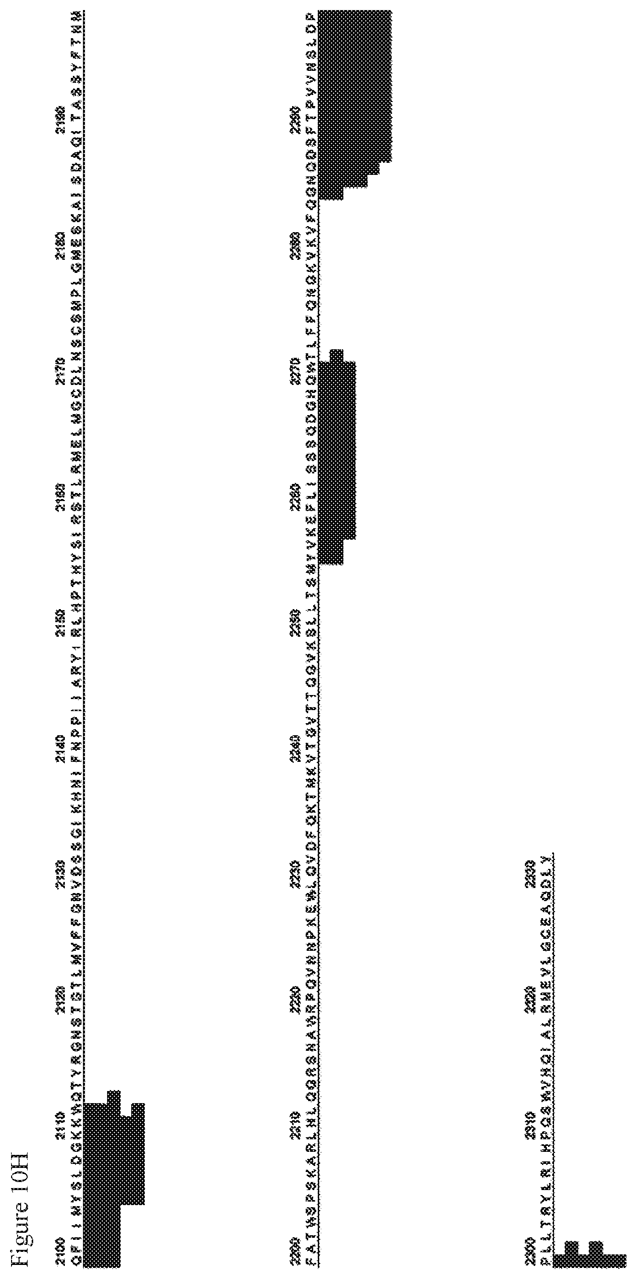
D00023
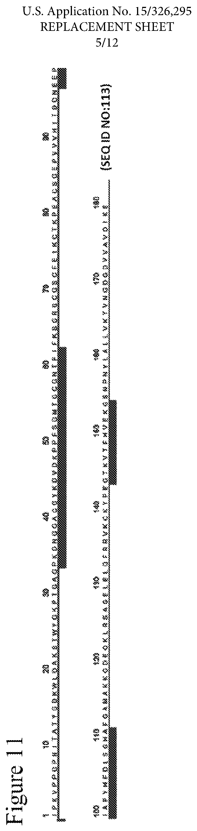
D00024

D00025

D00026
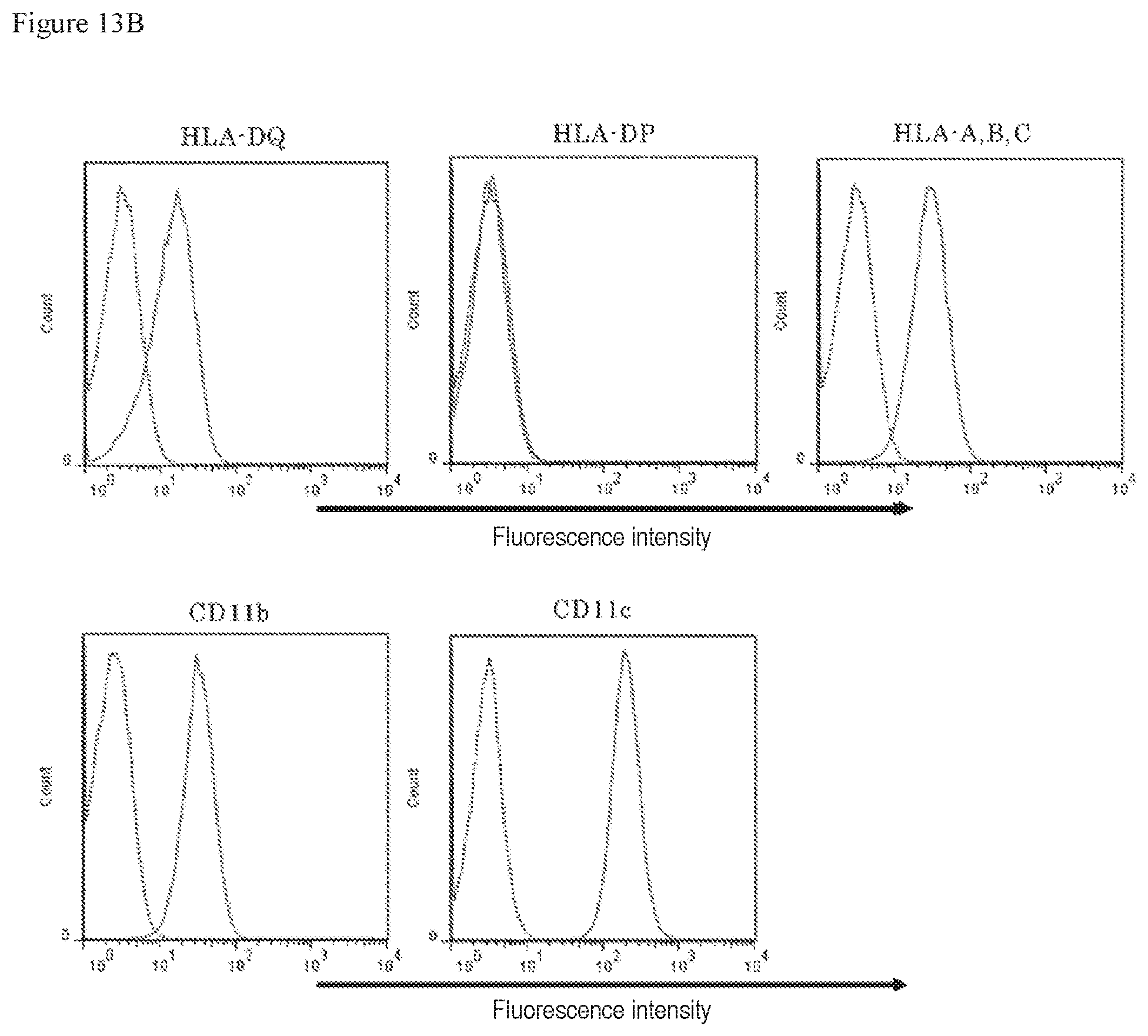
D00027

D00028

D00029

D00030

D00031

D00032

D00033

D00034

D00035

P00001

P00002

P00003

P00004

P00005

P00006

P00007

P00008

P00009

P00010

P00011

P00012

P00013

P00014

P00015

P00016

P00017

P00018

P00019

P00020

S00001
XML
uspto.report is an independent third-party trademark research tool that is not affiliated, endorsed, or sponsored by the United States Patent and Trademark Office (USPTO) or any other governmental organization. The information provided by uspto.report is based on publicly available data at the time of writing and is intended for informational purposes only.
While we strive to provide accurate and up-to-date information, we do not guarantee the accuracy, completeness, reliability, or suitability of the information displayed on this site. The use of this site is at your own risk. Any reliance you place on such information is therefore strictly at your own risk.
All official trademark data, including owner information, should be verified by visiting the official USPTO website at www.uspto.gov. This site is not intended to replace professional legal advice and should not be used as a substitute for consulting with a legal professional who is knowledgeable about trademark law.