Thyroglobulin quantitation by mass spectroscopy
Zhang , et al.
U.S. patent number 10,718,779 [Application Number 16/259,696] was granted by the patent office on 2020-07-21 for thyroglobulin quantitation by mass spectroscopy. This patent grant is currently assigned to Quest Diagnostics Investments Incorporated. The grantee listed for this patent is Quest Diagnostics Investments Incorporated. Invention is credited to Nigel J. Clarke, Richard E. Reitz, Yanni Zhang.


View All Diagrams
| United States Patent | 10,718,779 |
| Zhang , et al. | July 21, 2020 |
Thyroglobulin quantitation by mass spectroscopy
Abstract
Provided are methods for determining the amount of thyroglobulin in a sample using various purification steps followed by mass spectrometry. The methods generally involve purifying thyroglobulin in a test sample, digesting thyroglobulin to form peptide T129, purifying peptide T129, ionizing peptide T129, detecting the amount of peptide T129 ion generated, and relating the amount of peptide T129 ion to the amount of thyroglobulin originally present in the sample.
| Inventors: | Zhang; Yanni (Mission Viejo, CA), Clarke; Nigel J. (Vista, CA), Reitz; Richard E. (Las Vegas, NV) | ||||||||||
|---|---|---|---|---|---|---|---|---|---|---|---|
| Applicant: |
|
||||||||||
| Assignee: | Quest Diagnostics Investments
Incorporated (Wilmington, DE) |
||||||||||
| Family ID: | 50341936 | ||||||||||
| Appl. No.: | 16/259,696 | ||||||||||
| Filed: | January 28, 2019 |
Prior Publication Data
| Document Identifier | Publication Date | |
|---|---|---|
| US 20190145984 A1 | May 16, 2019 | |
Related U.S. Patent Documents
| Application Number | Filing Date | Patent Number | Issue Date | ||
|---|---|---|---|---|---|
| 15906078 | Jan 29, 2019 | 10191064 | |||
| 15443805 | Mar 13, 2018 | 9915663 | |||
| 14689542 | Feb 28, 2017 | 9580740 | |||
| 14031678 | Apr 21, 2015 | 9012394 | |||
| 61703721 | Sep 20, 2012 | ||||
| Current U.S. Class: | 1/1 |
| Current CPC Class: | C12Q 1/37 (20130101); G01N 33/78 (20130101); G01N 33/6848 (20130101); G01N 33/6893 (20130101); G01N 2496/00 (20130101); G01N 2800/7028 (20130101); G01N 2800/046 (20130101); G01N 2560/00 (20130101); G01N 2333/47 (20130101) |
| Current International Class: | G01N 33/68 (20060101); G01N 33/78 (20060101); C12Q 1/37 (20060101) |
References Cited [Referenced By]
U.S. Patent Documents
| 6107623 | August 2000 | Bateman et al. |
| 6124137 | September 2000 | Hutchens et al. |
| 6204500 | March 2001 | Whitehouse et al. |
| 6268144 | July 2001 | Koster |
| 7807172 | October 2010 | Hoofnagle |
| 8030084 | October 2011 | Zhang et al. |
| 8455259 | June 2013 | Zhang et al. |
| 8574915 | November 2013 | Zhang et al. |
| 9012394 | April 2015 | Zhang et al. |
| 9046531 | June 2015 | Zhang et al. |
| 9140695 | September 2015 | Kushnir et al. |
| 9274124 | March 2016 | Anderson |
| 9580740 | February 2017 | Zhang et al. |
| 9915663 | March 2018 | Zhang et al. |
| 9970943 | May 2018 | Anderson |
| 10191064 | January 2019 | Zhang |
| 2004/0072251 | April 2004 | Anderson |
| 2005/0064422 | March 2005 | Barnidge et al. |
| 2006/0223188 | October 2006 | Soldin |
| 2007/0105179 | May 2007 | Madson |
| 2007/0224628 | September 2007 | Gordon et al. |
| 2009/0042213 | February 2009 | Hoofnagle et al. |
| 2012/0009614 | January 2012 | Zhang et al. |
| 2008027861 | Mar 2008 | WO | |||
| WO2011116028 | Sep 2011 | WO | |||
| 2012111249 | Aug 2012 | WO | |||
Other References
|
Spencer C.A., et al., "Thyroglobulin Measurement Techniques, Clinical Benefits, and Pitfalls," Endocrinology Metabolism Clinics of North America , 1995, vol. 24 (4), pp. 841-863. cited by applicant . Steen H., et al., "The ABC's (and XYZ's) of Peptide Sequencing," Nature Reviews Molecular Cell Biology, 2004, vol. 5 (9), pp. 699-711. cited by applicant . Supplementary European Search Report for Application No. EP08860014, dated Jan. 28, 2011, 5 pages. cited by applicant . Tang X.J., et al., "An Investigation of Fragmentation Mechanisms of Doubly Protonated Tryptic Peptides," Rapid Communications in Mass Spectrometry, 1992, vol. 6 (11), pp. 651-677. cited by applicant . Taylor P.J., et al., "Simultaneous Quantification of Tacrolimus and Sirolimus in Human Blood, by High- Performance Liquid Chromatography--Tandem Mass Spectrometry," Therapeutic Drug Monitoring, 2000, vol. 22 (5), pp. 608-612. cited by applicant . Wright Jr., G.L., et al., "Proteinchip Surface Enhanced Laser Desorption/Ionization (SELDI) Mass Spectrometry: A Novel Protein Biochip Technology for Detection of Prostate Cancer Biomarkers in Complex Protein Mixtures," Prostate Cancer and Prostatic Diseases, 1999, vol. 2 (5-6), pp. 264-276. cited by applicant . Written Opinion for Application No. PCT/US08/85435, dated Apr. 22, 2009, 5 Pages. cited by applicant . Bartolucci G., et al., "Liquid Chromatography Tandem Mass Spectrometric Quantitation of Sulfamethazine and its Metabolites: Direct Analysis of Swine Urine by Triple Quadrupole and by Ion Trap Mass Spectrometry," Rapid Communications in Mass Spectrometry, 2000, vol. 14 (11), pp. 967-973. cited by applicant . Biemann K., "Mass Spectrometry of Peptides and Proteins," Annual Review of Biochemistry, 1992, vol. 61, pp. 977-1010. cited by applicant . Bourrel F., et al., "Immunoradiometric Assay of Thyroglobulin in Patients with Differentiated Thyroid Carcinomas: Need for Thyroglobulin Recovery Tests," Clinical Chemistry and Laboratory Medicine, 1998, vol. 36 (8), pp. 725-730. cited by applicant . Di Jeso B., et al., "Mixed-Disulfide Folding Intermediates between Thyroglobulin and Endoplasmic Reticulum Resident Oxidoreductases ERp57 and protein Disulfide Isomerase," Molecular and Cellular Biology, 2005, vol. 25 (22), pp. 9793-9805. cited by applicant . Dunn A.D., et al., "Tyrosine 130 is an Important Outer Ring Donor for Thyroxine Formation in Thyroglobulin," The Journal of Biological Chemistry, 1998, vol. 273 (39), pp. 25223-25229. cited by applicant . Dunn J.T., et al., "The Sites of Thyroid Hormone Formation in Rabbit Thyroglobulin," The Journal of Biological Chemistry, 1987, vol. 262 (35), pp. 16948-16952. cited by applicant . European Search Report for Application No. EP13185360, dated Jan. 22, 2014, 7 pages. cited by applicant . Extended European Search Report for Application No. EP17200516.7, dated Jan. 30, 2018, 8 pages. cited by applicant . Extended European Search Report for Application No. PCT/US2013/060659, dated Mar. 21, 2016, 11 pages. cited by applicant . Final Office Action dated Aug. 1, 2016 for U.S. Appl. No. 14/726,957, filed Jun. 1, 2015. cited by applicant . Final Office Action dated Sep. 6, 2012 for U.S. Appl. No. 13/198,620, filed Aug. 4, 2011. cited by applicant . Final Office Action dated Jan. 25, 2011 for U.S. Appl. No. 12/001,076, filed Dec. 6, 2007. cited by applicant . Final Office Action dated Sep. 25, 2017 for U.S. Appl. No. 14/726,957, filed Jun. 1, 2015. cited by applicant . Final Office Action dated Aug. 27, 2014 for U.S. Appl. No. 14/053,423, filed Oct. 14, 2013. cited by applicant . Final Office Action dated Oct. 27, 2010 for U.S. Appl. No. 12/001,076, filed Dec. 6, 2007. cited by applicant . Gentile F., et al., "Identification of Hormonogenic Tyrosines in Fragment 1218-1591 of Bovine Thyroglobulin by Mass Spectrometry. Hormonogenic Acceptor Tyr-1291 and Donor Tyr-1375," The Journal of Biological Chemistry, 1997, vol. 272 (1), pp. 639-646. cited by applicant . Guo-Zhong JI, et al., "Clinical Test Diagnosis and Resolution" Jan. 2011, pp. 335-336. cited by applicant . Hoofnagle A.N., et al., "Quantification of Thyroglobulin, a Low-Abundance Serum Protein, by Immunoaffinity Peptide Enrichment and Tandem Mass Spectrometry," Clinical Chemistry, 2008, vol. 54 (11), pp. 1796-1804. cited by applicant . International Preliminary Report on Patentability for Application No. PCT/US2008/085435, dated Jun. 8, 2010. cited by applicant . International Search Report and Written Opinion for Application No. PCT/US2013/60659, dated Dec. 23, 2013, 10 pages. cited by applicant . International Search Report for Application No. PCT/US08/85435, dated Apr. 22, 2009, 2 Pages. cited by applicant . Kim P.S., et al., "Folding and Assembly of Newly Synthesized Thyroblobulin Occurs in a Pre-Golgi Compartment," The Journal of Biological Chemistry, 1991, vol. 266 (19), pp. 12412-12418. cited by applicant . Kushnir M.M., et al., "High Sensitivity Measurement of Thyroglobulin in Serum in Presence of Anti-Thyroglobulin Autoantibodies," May 15, 2012. Retrieved from the Internet: URL:https://www.aruplab.com/Research&Development/resources/Posters/2012/K- ushnir_ASMS_0512.pdf. cited by applicant . Kushnir M.M., et al., "Mass Spectrometry Based Method for Accurate Measurement of Thyroglobulin in the Presence of Anti-Thyroglobulin Autoantibodies," May 15, 2012. Retrieved from the Internet: URL:https://www.aruplab.com/Research&Development/resources/Posters/2013/K- ushnir_ENDO_0613.pdf. cited by applicant . Kushnir M.M., et al., "Measurement of Thyroglobulin by Liquid Chromatography--Tandem Mass Spectrometry in Serum and Plasma in the Presence of Anti-thyroglobulin Autoantibodies," Clinical Chemistry, 2013, vol. 59(6), pp. 982-990. cited by applicant . "Screenshot of Google page", Mar. 10, 2016, XP055257300, Retrieved from the Internet: URL: www.google.com. cited by applicant . Learmonth M., et al., "Protein Identification by In-Gel Digestion and Mass Spectrometric Analysis," in: The Proteomics Protocols Handbook, 2005, Chapter 30, Walker J.M., ed., Humana Press, pp. 311-314. cited by applicant . Luo J.L., et al., "Diagnostic Value of Combing TG, TGAb and Cervical Ultrasonic Examination in the Recurrence or Metastasis Lesion of Differentiated Thyroid Carcinoma after Treatment," Chinese Journal of Clinicians (Electronic Edition), 2012, vol. 6 (3), pp. 580-583. cited by applicant . Mann M., et al., "Analysis of Proteins and Proteomes by Mass Spectrometry," Annual Review of Biochemistry, 2001, vol. 70, pp. 437-473. cited by applicant . Mann M., "Functional and Quantitative Proteomics Using SILAC," Nature Reviews Molecular Cell Biology, 2006, vol. 7 (12), pp. 952-958. cited by applicant . Meikla.W., et al., "Diagnosis and Management of Thyroid Nodules and Cancer Focus on Thyroglobulin; Thyroglobulin and Thyroid Cancer: Part 2 of Presentation: Analytical Method and Performance", Nov. 16, 2012. Retrieved from the Internet: URL:http://arup.utah.edu/media/thyroidglobulin/thyroid.cancer.pgr.final.p- df. cited by applicant . Merchant M., et al., "Recent Advancements in Surface-Enhanced Laser Desorption/Ionization-Time of Flight-Mass Spectrometry," Electrophoresis, 2000, vol. 21 (6), pp. 1164-1167. cited by applicant . Non-Final Office Action dated Jan. 12, 2016 for U.S. Appl. No. 14/726,957, filed Jun. 1, 2015. cited by applicant . Non-Final Office Action dated Mar. 13, 2017 for U.S. Appl. No. 14/726,957, filed Jun. 1, 2015. cited by applicant . Non-Final Office Action dated Jun. 16, 2017 for U.S. Appl. No. 15/443,805, filed Feb. 27, 2017. cited by applicant . Non-Final Office Action dated Jun. 19, 2014 for U.S. Appl. No. 14/031,678, filed Sep. 19, 2013. cited by applicant . Non-Final Office Action dated Jan. 22, 2016 for U.S. Appl. No. 14/063,956, filed Oct. 25, 2013. cited by applicant . Non-Final Office Action dated Dec. 23, 2013 for U.S. Appl. No. 14/053,423, filed Oct. 14, 2013. cited by applicant . Non-Final Office Action dated Apr. 27, 2016 for U.S. Appl. No. 14/689,542, filed Apr. 14, 2015. cited by applicant . Non-Final Office Action dated Apr. 28, 2010 for U.S. Appl. No. 12/001,076, filed Dec. 6, 2007. cited by applicant . Non-Final Office Action dated May 29, 2018 for U.S. Appl. No. 15/906,078, filed Feb. 27, 2018. cited by applicant . Non-Final Office Action dated Nov. 30, 2011 for U.S. Appl. No. 13/198,620, filed Aug. 4, 2011. cited by applicant . Notice of Allowance dated Jan. 23, 2018 for U.S. Appl. No. 15/443,805, filed Feb. 27, 2017. cited by applicant . Olsen J.V., et al., "Trypsin Cleaves Exclusively C-Terminal to Arginine and Lysine Residues," Molecular & Cellular Proteomics, 2004, vol. 3 (6), pp. 608-614. cited by applicant . Persoon A.C., et al., "Clinical Utility of an Automated Immunochemiluminometric Thyroglobulin Assay in Differentiated Thyroid Carcinoma," Clinical Chemistry, 2006, vol. 52 (4), pp. 686-691. cited by applicant . Persoon A.C., et al., "Thyroglobulin (Tg) Recovery Testing with Quantitative Tg Antibody Measurement for Determining Interference in Serum Tg Assays in Diffferentiated Thyroid Carcinoma," Clinical Chemistry, 2006, vol. 52 (6), pp. 1196-1199. cited by applicant . Robb D.B., et al., "Atmospheric Pressure Photoionization: An Ionization Method for Liquid Chromatography-Mass Spectrometry," Analytical Chemistry, 2000, vol. 72 (15), pp. 3653-3659. cited by applicant . Salek, "Analysis of Thyroblobulin Iodination by Tandem Mass Spectrometry Using Immonium Ions of Monoiodo- and Diiodo-Tyrosine," Proteomics, 2005, vol. 5 (2), pp. 351-353. cited by applicant . Salm P., et al., "The Quantification of Sirolimus by High-Performance Liquid Chromatography-Tandem Mass Spectrometry and Microparticle Enzyme Immunoassay in Renal Transplant Recipients," Clinical Therapeutics, 2000, vol. 22 Suppl B, pp. B71-B85. cited by applicant . Spencer C.A., et al., "Detection of Residual and Recurrent Differentiated Thyroid Carcinoma by Serum Thyroglobulin Measurement," Thyroid, 1999, vol. 9 (5), pp. 435-441. cited by applicant. |
Primary Examiner: Cordero Garcia; Marcela M
Attorney, Agent or Firm: Quest Diagnostics, Inc.
Parent Case Text
CROSS-REFERENCE TO RELATED APPLICATIONS
This application is a continuation of U.S. application Ser. No. 15/906,078, filed Feb. 27, 2018, which is a continuation of U.S. application Ser. No. 15/443,805, filed Feb. 27, 2017, now U.S. Pat. No. 9,915,663, which is a continuation of U.S. application Ser. No. 14/689,542, filed Apr. 17, 2015, now U.S. Pat. No. 9,580,740, which is a continuation of U.S. application Ser. No. 14/031,678, filed Sep. 19, 2013, now U.S. Pat. No. 9,012,394, which claims the benefit under 35 U.S.C. .sctn. 119(e) to U.S. Provisional Application Ser. No. 61/703,721, filed Sep. 20, 2012, the contents of each of which are incorporated by reference in its entirety into the present disclosure.
Claims
That which is claimed is:
1. A method for determining the amount of thyroglobulin in a test sample, comprising: (a)) adding a thyroglobulin peptide standard comprising an amino acid sequence having at least about 85% sequence identity to SEQ ID NO: 2 to said test sample containing thyroglobulin peptides; (b) reducing said thyroglobulin peptides and thyroglobulin peptide standard from step (a); (c) alkylating said thyroglobulin peptides and thyroglobulin peptide standard; (d) digesting said thyroglobulin peptides and thyroglobulin peptide standard; (e) enriching said thyroglobulin peptides and thyroglobulin peptide standard; (f) ionizing said thyroglobulin peptides and thyroglobulin peptide standard to produce one or more thyroglobulin peptide ions and thyroglobulin peptide standard ions detectable by mass spectrometry; and (g) detecting the amount of the ion(s) from step (f) by mass spectrometry; wherein the amount of the ion(s) detected is related to the amount of thyroglobulin in said test sample and the amount of thyroglobulin peptide standard.
2. The method of claim 1, wherein the thyroglobulin peptide standard is less than 50 amino acid residues long.
3. The method of claim 2, wherein thyroglobulin peptides and thyroglobulin peptide standard both comprise a T129 peptide (SEQ ID NO: 1, VIFDANAPVAVR).
4. The method of claim 1, wherein the thyroglobulin peptide ions produced in step (c) comprise one or more ions selected from the group of ions with a mass/charge ratio of 541.3.+-.0.5, 612.3.+-.0.5, 636.4.+-.0.5, 726.4.+-.0.5, 797.4.+-.0.5, 912.4.+-.0.5, or 1059.5.+-.0.5.
5. The method of claim 1, wherein said ionizing comprises generating a thyroglobulin peptide precursor ion with a mass/charge ratio of 636.4.+-.0.5.
6. The method of claim 1, wherein said ionizing comprises generating one or more fragment ions with a mass/charge ratio of 797.4.+-.0.5, 912.4.+-.0.5, or 1059.5.+-.0.5.
7. The method of claim 1, wherein said test sample is body fluid or tissue.
8. The method of claim 1, wherein said test sample is plasma or serum.
Description
FIELD OF THE DISCLOSURE
The disclosure relates to the quantitation of thyroglobulin. In a particular aspect, the disclosure relates to methods for quantitation of thyroglobulin by mass spectrometry.
BACKGROUND
The following description of the background of the disclosure is provided simply as an aid in understanding the disclosure and is not admitted to describe or constitute prior art to the disclosure.
Thyroglobulin, or Tg, is a large dimeric secretary glycoprotein with a molecular weight of 660 kDa comprised of noncovalently bound homodimers.
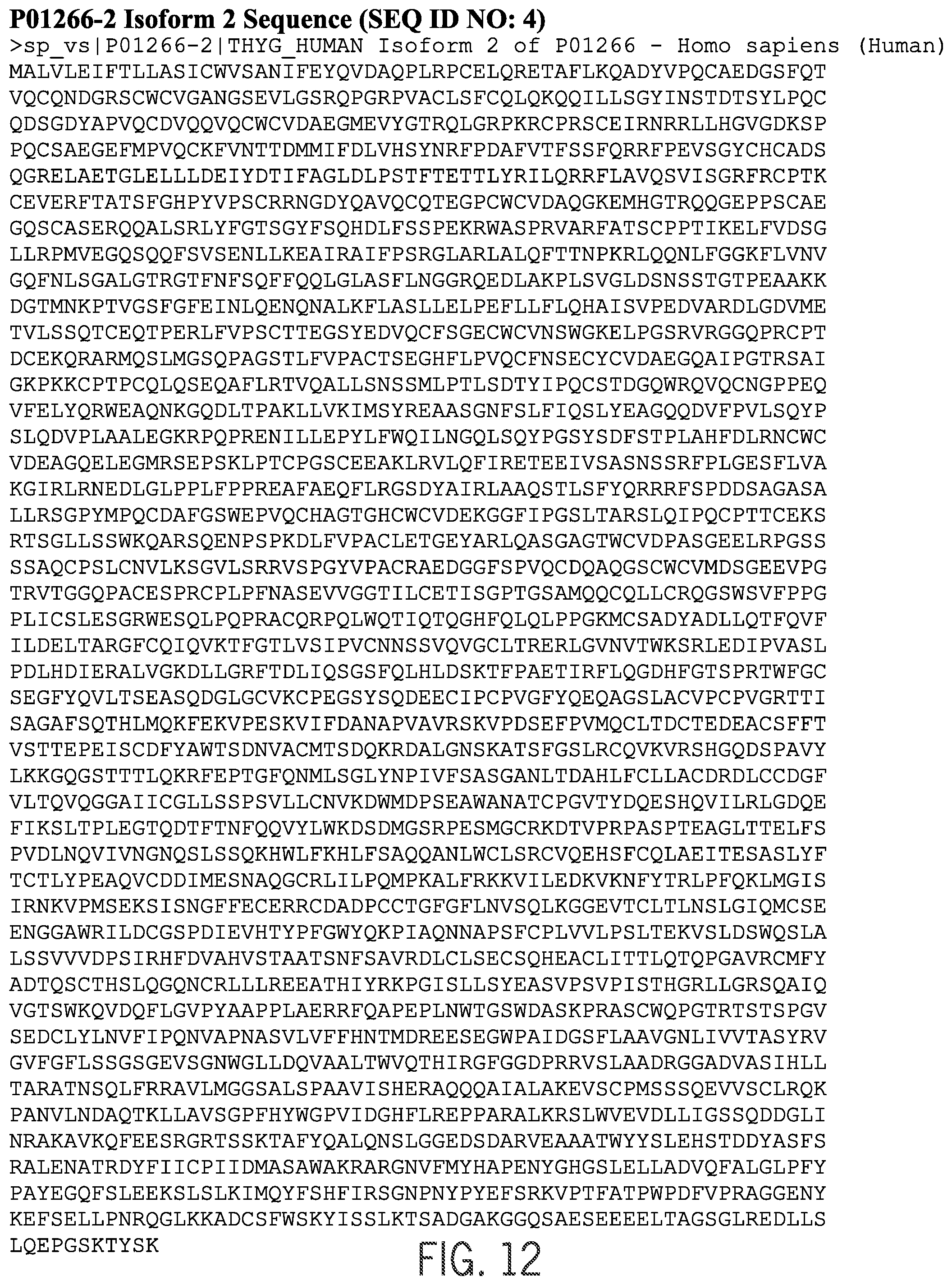
Tg molecules exist in several forms. The three major Tg molecule sequences as found in the UniProt Knowledgebase (Swiss-Prot+TrEMBL) are P01266 (Human Thyroglobulin Precursor), P01266-2 (Isoform 2 of P01266), and Q59GF02 (Human Thyroglobulin Variant). (See FIGS. 11, 12, and 13, respectively.)
P01266 is the major variant of P01266 with a length of 2768 AA; P01266-2 is an isoform of P01266 with a length of 2711 AA. P01266-2 varies from P01266 at amino acid positions 1510 to 1567 of Tg; and Q59GF0 is a thyroglobulin fragment with a length of 1574 AA. Q59GF0 contains amino acids from positions 1212 to 2768 of Tg.
Tg can only be produced in the thyroid gland and may be produced by either normal well differentiated benign thyroid cells or thyroid cancer cells. It is the precursor protein for thyroid hormone syntheses and serves as the matrix for thyroid iodine storage. Tg is used by the thyroid gland to produce the thyroid hormones thyroxine (T4) and triiodothyroine (T3). Tg levels in the blood can be used as a tumor marker for differentiated thyroid carcinoma (DTC). A high level of Tg in the blood is not by itself an indicator of thyroid cancer, but persistence of Tg in the blood following surgical removal of the thyroid gland indicates persistence of thyroid tissue. A course of treatment following detection of Tg in the blood following surgical removal of the thyroid gland may include administration of radioiodine to ablate all remaining normal thyroid. Continued persistence of Tg in the blood following ablation of all normal thyroid could indicate that some amount of tumor is still present.
Several methods for quantaition of Tg have been developed. For example Spencer, et al., Thyroid, 1999, 9(5):435-41 and Persoon, et al., Clinical Chem 2006, 52(4):686-691 disclose immunometric, radioimmunometric, and immunochemiluminometric methods for quantitation of Tg. These methods are all subject to methodological problems such as differences in standardization, variability in interassay sensitivity and precision, hook effects, and interference attributable to Tg antibodies. The problem of interference attributable to Tg antibodies is particularly troubling for clinical application of monitoring Tg levels as a tumor marker because up to 20% of thyroid cancer patients have Tg autoantibodies.
SUMMARY
The present disclosure provides methods for quantitation of Tg in a sample by mass spectrometry, including tandem mass spectrometry.
In one aspect, methods are provided for determining the amount of Tg in a test sample that include: (a) subjecting a Tg containing test sample to digestion resulting in creation of Tg peptides; (b) purifying one or more Tg peptides; (c) ionizing one or more Tg peptides; (d) detecting the amount of the Tg peptide ion(s) by mass spectrometry; and (e) relating the amount of detected Tg peptide ion(s) to the amount of Tg in the test sample. A preferred enzyme for preparing Tg peptides is trypsin. A suitable Tg peptide for the method is one that can be evaluated by mass spectrometry and can be sufficiently purified from related peptides that may be generated from proteins other than Tg. An example of one such peptide is peptide T129 (sequence VIFDANAPVAVR; SEQ ID NO: 1) which contains amino acids from positions 1579 to 1590 of Tg, has a molecular weight of about 1,270 Da, and is present in all three isoforms of Tg. See FIG. 4.
Formation of peptide T129 provides a unique trypsin generated peptide for thyroglobulin. Also, creation of peptide T129 from tryptic digestion of Tg should be unaffected by the presence or absence of the Tg antibodies. Thus, measurement of the increase in peptide T129 in a test sample offers a way of quantitating the amount of Tg originally in the test sample free from inference from Tg antibodies.
Any appropriate method may be used to determine the amount of Tg peptide resulting from digestion of Tg in a sample. In the event that a test sample may contain endogenous Tg peptide, steps may be taken to make certain that the endogenous peptide is not confused with peptide generated by digesting Tg in sample. One approach is to remove the endogenous Tg peptide from the sample before digesting Tg. This may done, for example, using a size separation technique. Another approach is to analyze a portion of a test sample according to the claimed methods but excluding the digestion step in order to establish a baseline level for the endogenous peptide in the test sample. In this approach, the once a baseline is determined, it can be subtracted from the post-digestion level of the peptide, the later representing both the endogenous peptide and that generated by digestion.
Because the methods may be applied to complex test samples (particularly body fluids or test samples derived from tissue), steps may be taken to purify Tg in the test sample prior to digestion. This may done, for example, using a size separation technique.
In some embodiments, the methods include generating one or more Tg peptide ions in which at least one of the ions has a mass/charge ratio (m/z) corresponding to that of (singly or multiply charged) peptide T129 ions. In preferred related embodiments, the methods include generating one or more Tg peptide ions in which at least one has m/z of 1272.8.+-.0.5, 636.4.+-.0.5, or 424.3.+-.0.5 (corresponding to singly, doubly, or triply charged peptide T129 ions). In related preferred embodiments, the methods may include generating one or more fragment ions of a Tg peptide ion in which at least one has a m/z of 541.3.+-.0.5, 612.3.+-.0.5, 726.4.+-.0.5, 797.4.+-.0.5, 912.4.+-.0.5, or 1059.5.+-.0.5; preferably one or more of the fragment ions are selected from the group consisting of ions with a m/z of 797.4.+-.0.5, 912.4.+-.0.5, and 1059.5.+-.0.5.
In some embodiments, the purification in step (b) is accomplished with at least one size separation technique. Preferably, size separation techniques may be filtration, LC, or any combination thereof. In certain preferred embodiments, the test sample is a body fluid or tissue. In some embodiments, an additional step is included where a second quantity of the test sample is subjected to steps (b) through (e) in order to establish a baseline level of one or more endogenous Tg peptides. In these embodiments, this baseline level can be subtracted from the amount of Tg peptide ion(s) detected in the test sample to determine the amount of Tg peptide ion(s) that result from Tg in the original test sample. In other embodiments, the methods include an additional initial step of purifying Tg in the test sample prior to digestion. In these embodiments, the pre-digestion purification and/or the purification in step (b) may each be accomplished with at least one size separation technique. Preferably, at least one size separation technique used in both pre-digestion purification and step (b) is filtration; more preferably, this filtration is done with a molecular weight cut-off filter with molecular weight cut off that allows for retention of Tg above the filter and allows Tg peptides to pass through with the filtrate. In related embodiments, the molecular weight cut-off is about 2 kD to 300 kD; more preferably about 100 kD to 300 kD. In these embodiments, the two filtrations (pre-digestion and step (b)) may be conducted with the same filter.
In a second aspect, methods are provided for determining the amount of Tg in a test sample that include: (a) subjecting a Tg containing test sample to digestion resulting in creation of peptide T129; (b) purifying peptide T129; (c) ionizing peptide T129 to generate a precursor ion with a m/z of 636.4.+-.0.5; (d) fragmenting the peptide T129 precursor ion to form one or more fragment ions in which at least one has a m/z of about 797.4.+-.0.5, 912.4.+-.0.5, or 1059.5.+-.0.5; detecting the amount of peptide T129 precursor ions, one or more fragment ions, or both, by mass spectrometry; and (e) relating the amount of detected ion(s) to the amount of Tg in the test sample. In certain preferred embodiments, the test sample is a body fluid or tissue or tissue. In some embodiments, an additional step is included where a second quantity of the test sample is subjected to steps (b) through (e) in order to establish a baseline level of one or more endogenous peptide T129. In these embodiments, this baseline level can be subtracted from the amount of peptide T129 ion(s) detected in the test sample to determine the amount of peptide T129 ion(s) that result from Tg in the original test sample. In other embodiments, the methods include an additional initial step of purifying Tg in the test sample prior to digestion. In these embodiments, the pre-digestion purification and/or the purification in step (b) may each be accomplished with at least one size separation technique. Preferably, at least one size separation technique used in both pre-digestion purification and step (b) is filtration; more preferably, this filtration is done with a molecular weight cut-off filter with molecular weight cut off that allows for retention of Tg above the filter and allows Tg peptides to pass through with the filtrate. In related embodiments, the molecular weight cut-off is about 2 kD to 300 kD; more preferably about 100 kD to 300 kD. In these embodiments, the two filtrations (pre-digestion and step (b)) may be conducted with the same filter.
As used herein, the term "purification" or "purifying" does not refer to removing all materials from the sample other than the analyte(s) of interest. Instead, purification refers to a procedure that enriches the amount of one or more analytes of interest relative to one or more other components of the sample. Purification, as used herein, does not require the isolation of an analyte from all others. In preferred embodiments, a purification step or procedure can be used to remove one or more interfering substances, e.g., one or more substances that would interfere with the operation of the instruments used in the methods or substances that may interfere with the detection of an analyte ion by mass spectrometry.
As used herein, the term "about" in reference to quantitative measurements, not including the measurement of mass of an ion, refers to the indicated value plus or minus 10%.
As used herein, the term "substantially all" refers to any proportion greater than 50%, more preferably greater than 60%, more preferably greater than 70%, more preferably greater than 80%, and more preferably greater than 90%.
As used herein, the term "test sample" refers to any sample that may contain Tg. As used herein, the term "body fluid or tissue" means any fluid or tissue that can be isolated from the body of an individual. For example, "body fluid or tissue" may include blood, plasma, serum, bile, saliva, urine, tears, perspiration, and the like. If solid tissue is to be analyzed, it may be processed to release a liquid fraction that could contain any Tg present in the tissue. The liquid fraction can then be subject to the methods described herein.
As used herein, the term "digestion" means proteolytic cleavage of proteins into peptides. Digestion agents may include trypsin, Lyc-C, Arg-R, Asp-N and the like. Digestion is carried out by adding a digestion agent (i.e., an enzyme) to a sample and incubating for some period of time.
As used herein, "Tg" or "Tg molecule" means an intact Tg protein molecule.
As used herein, the term "Tg peptide" means any peptide of 100 amino acids or less that is a fragment of the native Tg. Tg peptides can be endogenous to a test sample or formed as a result of digestion of Tg. Peptide T129 is an example of a Tg peptide formed as a result of trypsin digestion of Tg.
As used herein, the term "size separation technique" means any technique (physical or chemical) that allows for the separation of at least one species from a test sample based on any one or more of molecular weight and shape. Examples of such techniques include, but are not limited to, filtration, chromatography, and certain aspects of mass spectrometry.
As used herein, the term "chromatography" refers to a process in which a chemical mixture carried by a liquid or gas is separated into components as a result of differential distribution of the chemical entities as they flow around, over, and/or through a stationary liquid or solid phase.
As used herein, the term "liquid chromatography" or "LC" means a process of selective retardation of one or more components of a fluid solution as the fluid uniformly percolates through a column of a finely divided substance, or through capillary passageways. The retardation results from the distribution of the components of the mixture between one or more stationary phases and the bulk fluid, (i.e., mobile phase), as this fluid moves relative to the stationary phase(s). "Liquid chromatography" includes reverse phase liquid chromatography (RPLC), high performance liquid chromatography (HPLC) and high turbulence liquid chromatography (HTLC).
As used herein, the term "high performance liquid chromatography" or "HPLC" refers to liquid chromatography in which the degree of separation is increased by forcing the mobile phase under pressure through a stationary phase, typically a densely packed column.
As used herein, the term "mass spectrometry" or "MS" refers to an analytical technique to identify compounds by their mass. MS refers to methods of filtering, detecting, and measuring ions based on their m/z. MS technology generally includes (1) ionizing the compounds to form charged species (e.g., ions); and (2) detecting the molecular weight of the ions and calculating their m/z. The compounds may be ionized and detected by any suitable means. A "mass spectrometer" generally includes an ionizer and an ion detector. In general, one or more molecules of interest are ionized, and the ions are subsequently introduced into a mass spectrographic instrument where, due to a combination of magnetic and electric fields, the ions follow a path in space that is dependent upon mass ("m") and charge ("z"). See, e.g., U.S. Pat. No. 6,204,500, entitled "Mass Spectrometry From Surfaces;" U.S. Pat. No. 6,107,623, entitled "Methods and Apparatus for Tandem Mass Spectrometry;" U.S. Pat. No. 6,268,144, entitled "DNA Diagnostics Based On Mass Spectrometry;" U.S. Pat. No. 6,124,137, entitled "Surface-Enhanced Photolabile Attachment And Release For Desorption And Detection Of Analytes;" Wright et al., Prostate Cancer and Prostatic Diseases 2:264-76 (1999); and Merchant and Weinberger, Electrophoresis 21:1164-67 (2000).
As used herein, the term "operating in positive ion mode" refers to those mass spectrometry methods where positive ions are detected. Similarly, the term "operating in negative ion mode" refers to those mass spectrometry methods where negative ions are detected.
As used herein, the term "ionization" or "ionizing" refers to the process of generating an analyte ion having a net electrical charge equal to one or more electron units. Positive ions are those having a net positive charge of one or more electron units. Negative ions are those having a net negative charge of one or more electron units.
As used herein, the term "electron ionization" or "EI" refers to methods in which an analyte of interest in a gaseous or vapor phase interacts with a flow of electrons. Impact of the electrons with the analyte produces analyte ions, which may then be subjected to a mass spectrometry technique.
As used herein, the term "chemical ionization" or "CI" refers to methods in which a reagent gas (e.g. ammonia) is subjected to electron impact, and analyte ions are formed by the interaction of reagent gas ions and analyte molecules.
As used herein, the term "fast atom bombardment" or "FAB" refers to methods in which a beam of high energy atoms (often Xe or Ar) impacts a non-volatile sample, desorbing and ionizing molecules contained in the sample. Test samples are dissolved in a viscous liquid matrix such as glycerol, thioglycerol, m-nitrobenzyl alcohol, 18-crown-6 crown ether, 2-nitrophenyloctyl ether, sulfolane, diethanolamine, and triethanolamine. The choice of an appropriate matrix for a compound or sample is an empirical process.
As used herein, the term "matrix-assisted laser desorption ionization" or "MALDI" refers to methods in which a non-volatile sample is exposed to laser irradiation, which desorbs and ionizes analytes in the sample by various ionization pathways, including photo-ionization, protonation, deprotonation, and cluster decay. For MALDI, the sample is mixed with an energy-absorbing matrix, which facilitates desorption of analyte molecules.
As used herein, the term "surface enhanced laser desorption ionization" or "SELDI" refers to another method in which a non-volatile sample is exposed to laser irradiation, which desorbs and ionizes analytes in the sample by various ionization pathways, including photo-ionization, protonation, deprotonation, and cluster decay. For SELDI, the sample is typically bound to a surface that preferentially retains one or more analytes of interest. As in MALDI, this process may also employ an energy-absorbing material to facilitate ionization.
As used herein, the term "electrospray ionization" or "ESI," refers to methods in which a solution is passed along a short length of capillary tube, to the end of which is applied a high positive or negative electric potential. Solution reaching the end of the tube is vaporized (nebulized) into a jet or spray of very small droplets of solution in solvent vapor. This mist of droplets flows through an evaporation chamber, which is heated slightly to prevent condensation and to evaporate solvent. As the droplets get smaller the electrical surface charge density increases until such time that the natural repulsion between like charges causes ions as well as neutral molecules to be released.
As used herein, the term "atmospheric pressure chemical ionization" or "APCI," refers to mass spectroscopy methods that are similar to ESI; however, APCI produces ions by ion-molecule reactions that occur within a plasma at atmospheric pressure. The plasma is maintained by an electric discharge between the spray capillary and a counter electrode. Then ions are typically extracted into the mass analyzer by use of a set of differentially pumped skimmer stages. A counterflow of dry and preheated N.sub.2 gas may be used to improve removal of solvent. The gas-phase ionization in APCI can be more effective than ESI for analyzing less-polar species.
The term "Atmospheric Pressure Photoionization" or "APPI" as used herein refers to the form of mass spectroscopy where the mechanism for the photoionization of molecule M is photon absorption and electron ejection to form the molecular M+. Because the photon energy typically is just above the ionization potential, the molecular ion is less susceptible to dissociation. In many cases it may be possible to analyze samples without the need for chromatography, thus saving significant time and expense. In the presence of water vapor or protic solvents, the molecular ion can extract H to form MH+. This tends to occur if M has a high proton affinity. This does not affect quantitation accuracy because the sum of M+ and MH+ is constant. Drug compounds in protic solvents are usually observed as MH+, whereas nonpolar compounds such as naphthalene or testosterone usually form M+. Robb, D. B., Covey, T. R. and Bruins, A. P. (2000): See, e.g., Robb et al., Atmospheric pressure photoionization: An ionization method for liquid chromatography-mass spectrometry. Anal. Chem. 72(15): 3653-3659.
As used herein, the term "inductively coupled plasma" or "ICP" refers to methods in which a sample is interacted with a partially ionized gas at a sufficiently high temperature to atomize and ionize most elements
As used, herein, the term "field desorption" refers to methods in which a non-volatile test sample is placed on an ionization surface, and an intense electric field is used to generate analyte ions.
As used herein, the term "desorption" refers to the removal of an analyte from a surface and/or the entry of an analyte into a gaseous phase.
As used herein, the term "limit of quantification" or "LOQ" refers to the point where measurements become quantitatively meaningful. The analyte response at this LOQ is identifiable, discrete and reproducible with a precision of 20% and an accuracy of 80% to 120%.
In certain preferred embodiments of the methods disclosed herein, mass spectrometry is performed in positive ion mode. In certain particularly preferred embodiments of the methods disclosed herein, mass spectrometry is performed using ESI as the method of creating ions from Tg peptides.
In preferred embodiments, the ions from Tg peptide ionization detectable in a mass spectrometer are selected from the group consisting of ions with a m/z of 636.4.+-.0.5, 1059.5.+-.0.5, 921.4.+-.0.5, 797.4.+-.0.5, 726.4.+-.0.5, 612.3.+-.0.5, and 541.3.+-.0.5; the first ion listed (m/z of 636.4.+-.0.5) being a precursor ion with a net charge of positive 2 electron units and the latter six ions listed being fragment ions of the precursor ion. In particularly preferred embodiments, the precursor ion has a net charge of positive 2 electron units and a m/z of about 636.4.+-.0.5, and the fragment ions have a m/z of 1059.5.+-.0.5, 921.4.+-.0.5, or 797.4.+-.0.5.
In some preferred embodiments, a separately detectable internal standard peptide (e.g., T129) is introduced in the test sample after trypsin digestion. In these embodiments, all or a portion of the peptide present in the test sample both from digestion of endogenous Tg and the addition of the internal standard are ionized to produce a plurality of ions detectable in a mass spectrometer, and one or more ions produced from the peptide ionization are detected in a mass spectrometer.
In other preferred embodiments, a separately detectable internal Tg standard is provided in the test sample prior to trypsin digestion. In these embodiments, all or a portion of both the endogenous Tg and the internal standard present in the test sample are digested by trypsin resulting in formation of Tg peptides. Tg peptides are ionized to produce a plurality of ions detectable in a mass spectrometer, and one or more ions produced from Tg peptide ionization are detected by mass spectrometry.
In preferred embodiments, the ions detectable in a mass spectrometer produced from the ionization of Tg peptides resulting from Tg digestion are selected from the group consisting of ions with a m/z of 636.4.+-.0.5, 1059.5.+-.0.5, 921.4.+-.0.5, 797.4.+-.0.5, 726.4.+-.0.5, 612.3.+-.0.5, and 541.3.+-.0.5; the first ion listed (m/z of 636.4.+-.0.5) being a precursor ion with a net charge of positive 2 electron units and the latter six ions listed being fragment ions of the precursor ion. In particularly preferred embodiments, the precursor ion has a net charge of positive 2 electron units and a m/z of 636.4.+-.0.5, and the fragment ions have a m/z of 1059.5.+-.0.5, 921.4.+-.0.5, 797.4.+-.0.5.
In preferred embodiments, the presence or amount of Tg peptide ions is related to the presence or amount of Tg in the original test sample by comparison to a reference Tg sample.
In one embodiment, the methods involve the combination of LC with mass spectrometry. In another preferred embodiment, the mass spectrometry is tandem mass spectrometry (MS/MS).
The summary of the disclosure described above is non-limiting and other features and advantages of the disclosure will be apparent from the following detailed description of the disclosure, and from the claims.
BRIEF DESCRIPTION OF THE DRAWINGS
FIG. 1 shows the limit of quantitation verification for Tg peptide ion with m/z corresponding to peptide T129 by MS/MS. Details are described in Example 1.
FIG. 2 shows the linearity of the quantitation of peptide T129 in serially diluted stock samples using an LC-MS/MS assay. Details are described in Example 1.
FIG. 3 shows the limit of quantitation verification for peptide T129 in stripped serum by MS/MS. Details are described in Example 2.
FIG. 4 shows the linearity of the quantitation of peptide T129 in peptide T129 spiked stripped serum using an LC-MS/MS assay. Details are described in Example 2.
FIG. 5 shows the linearity of the quantitation of Tg peptide ions with m/z corresponding to peptide T129 using an LC-MS/MS assay in stripped serum spiked with Tg prior to processing and concentration according to the methods described herein. Details are described in Example 3.
FIG. 6 shows an illustrative embodiment of an isotopically labeled thyroglobulin peptide standard for use as an internal standard as described in Example 4. FIG. 6 discloses SEQ ID NOS 6-9, respectively, in order of appearance.
FIG. 7-10 show graphs of thyroglobulin quantitation in test samples by methods of the present technology (Y-axis) versus quantitation by immunoassay or radioimmunoassay (X-axis) from antibody-negative patient discards (FIG. 7, 9) and antibody-positive patient discards (FIG. 8, 10) as described in Example 5.
FIG. 11 shows the amino acid sequence for P01266 (Human Thyroglobulin Precursor; SEQ ID NO: 3).
FIG. 12 shows the amino acid sequence for P01266-2 (Isoform 2 of P01266; SEQ ID NO: 4).
FIG. 13 shows the amino acid sequence for Q59GF0 (Thyroglobulin Variant-Fragment; SEQ ID NO: 5).
FIG. 14A through 14G show a comparison of the three sequences contained in FIG. 1-3 demonstrating that they all contain amino acids corresponding to positions 1579 to 1590 of Tg. Sequence P01266 is on top (SEQ ID NO:3); sequence P01266-2 is in the middle (SEQ ID NO:4); and sequence Q59GF0 is at the bottom (SEQ ID NO:5).
DETAILED DESCRIPTION OF THE DISCLOSURE
Methods are described for quantitatively measuring Tg in a test sample. This quantitative measurement is achieved through the use of LC-MS/MS techniques. Prior to the use of LC-MS/MS, samples may be prepared by the following technique, or any portion thereof. A first purification of Tg in a test sample may be conducted through the use of a size separation technique such that substantially all Tg in the test sample is retained. Following the first purification step, enzymatic digestion of Tg may be carried out creating Tg peptides of interest. After digestion, another utilization of a size separation technique may be employed such that a selected Tg peptide generated in the enzymatic digestion of Tg is purified. This second size separation technique can be used to remove substantially all undigested, higher-molecular weight species. Properly executed, the sample preparation techniques ensure that selected Tg peptides quantitated by LC-MS/MS directly result from enzymatic digestion of Tg originally in the test sample; thus, the level of selected Tg peptides in the test sample at the start of LC-MS/MS is directly proportional to the amount of Tg originally present in the test sample.
Any suitable size separation technique may be utilized, but in the examples that follow, both the first and second size separation techniques are filtration through a molecular weight cut-off filter. It is also possible, as discussed in the Examples that follow, to select a molecular weight cut-off filter with an appropriate molecular weight cut-off such that the same filter can be used for both the first size separation and the second size separation.
LC, most preferably HPLC, is utilized, may be utilized either alone or in combination with other purification methods, to purify selected Tg peptides. This purification is combined with MS/MS, thereby providing an assay system for quantifying selected Tg peptides in a test sample. The quantity of the selected Tg peptides in the test sample is then used to determine the quantity of Tg in the original test sample. The Tg quantitation methods provided herein have enhanced specificity and are less subject to methodological problems (such as Tg antibody interference).
Suitable test samples may include any test sample that may contain the analyte of interest. In some preferred embodiments, a sample is a biological sample; that is, a sample obtained from any biological source, such as an animal, a cell culture, an organ culture, and the like. In certain preferred embodiments, samples are obtained from a mammalian animal, such as a dog, cat, horse, etc. Particularly preferred mammalian animals are primates, most preferably humans. Particularly preferred samples include blood, plasma, serum, urine, saliva, tears, cerebrospinal fluid, or other body fluid or tissue samples. Such samples may be obtained, for example, from a patient; that is, a living person presenting oneself in a clinical setting for diagnosis, prognosis, or treatment of a disease or condition. The test sample is preferably obtained from a patient, for example, serum or plasma.
Sample Preparation for Mass Spectrometry
Samples may be processed or purified to obtain preparations that are suitable for analysis by mass spectrometry. Such purification will usually include chromatography, such as liquid chromatography, and may also often involve an additional purification procedure that is performed prior to chromatography. Various procedures may be used for this purpose depending on the type of sample or the type of chromatography. Examples include filtration, centrifugation, combinations thereof and the like. In certain preferred embodiments, Tg present in a test sample prior to enzymatic digestion.
Filtration is one preferred method of preparing a test sample, especially a biological test sample, such as serum or plasma, for chromatography. Such filtration is carried out by filtering a test sample through a molecular weight cut-off filter to separate species with molecular weights higher than the filter's cut-off (including Tg) from those with molecular weights lower than the filter's cut-off. The test sample remaining above the filter following complete (or near complete) filtration is substantially free of potentially interfering species with molecular weights lower than the filter's cut-off.
The pH of the test sample may then be adjusted to any point required by a digestion agent. In certain preferred embodiments, the digestion agent is trypsin and pH can be adjusted with a solution of ammonium acetate to have a pH suitable for this enzyme. In these preferred embodiments, the sample is then digested with trypsin to form Tg peptides (including peptide T129).
After trypsin digestion, the sample may be purified with a second filtration. This post-digestion filtration can be carried out similarly to the pre-digestion filtration described above (with the exception that the filtrate is retained), in order to separate Tg fragments from potentially interfering species with molecular weights higher than the filter's cut-off that may also be present in the sample. The filtrate from this post-digestion filtration can then be purified by liquid chromatography and subsequently subjected to mass spectrometry analysis.
Various methods have been described involving the use of HPLC for sample clean-up prior to mass spectrometry analysis. See, e.g., Taylor et al., Therapeutic Drug Monitoring 22:608-12 (2000) (manual precipitation of blood samples, followed by manual C18 solid phase extraction, injection into an HPLC for chromatography on a C18 analytical column, and MS/MS analysis); and Salm et al., Clin. Therapeutics 22 Supl. B:B71-B85 (2000) (manual precipitation of blood samples, followed by manual C18 solid phase extraction, injection into an HPLC for chromatography on a C18 analytical column, and MS/MS analysis). One of skill in the art may select HPLC instruments and columns that are suitable for use in the methods. The chromatographic column typically includes a medium (i.e., a packing material) to facilitate separation of chemical moieties (i.e., fractionation). The medium may include minute particles. The particles include a bonded surface that interacts with the various chemical moieties to facilitate separation of the chemical moieties. One suitable bonded surface is a hydrophobic bonded surface such as an alkyl bonded surface. Alkyl bonded surfaces may include C-4, C-8, or C-18 bonded alkyl groups, preferably C-8 bonded groups. The chromatographic column includes an inlet port for receiving a sample and an outlet port for discharging an effluent that includes the fractionated sample.
In certain embodiments, an analyte may be purified by applying a sample to a column under conditions where the analyte of interest is reversibly retained by the column packing material, while one or more other materials are not retained. In these embodiments, a first mobile phase condition can be employed where the analyte of interest is retained by the column and a second mobile phase condition can subsequently be employed to remove retained material from the column, once the non-retained materials are washed through. Alternatively, an analyte may be purified by applying a sample to a column under mobile phase conditions where the analyte of interest elutes at a differential rate in comparison to one or more other materials. Such procedures may enrich the amount of one or more analytes of interest relative to one or more other components of the sample.
In one embodiment, the sample to be analyzed is applied to the column at the inlet port, eluted with a solvent or solvent mixture, and discharged at the outlet port. Different solvent modes may be selected for eluting the analytes of interest. For example, liquid chromatography may be performed using a gradient mode, an isocratic mode, or a polytyptic (i.e. mixed) mode. In preferred embodiments, HPLC is performed on an analytical HPLC system with a C8 solid phase using 0.2% formic acid in HPLC Grade Ultra Pure Water and 0.2% formic acid in 100% methanol as the mobile phases.
Numerous column packings are available for chromatographic separation of samples and selection of an appropriate separation protocol is an empirical process that depends on the sample characteristics, analyte of interest, presence of interfering substances and their characteristics, etc. Commercially available HPLC columns include, but are not limited to, polar, ion exchange (both cation and anion), hydrophobic interaction, phenyl, C-2, C-8, C-18, and polar coating on porous polymer columns.
In one embodiment, the HPLC column has a C8 solid phase with a median particle size of 5 .mu.m (nominal) and a median particle pore size of 100 .ANG.. In a preferred embodiment the column dimensions are 1.0 mm ID.times.50 mm length (Phenomenex Corp. Luna 5.mu. C8(2) 100 .ANG. New Column 50.times.1.0 mm, Phenomenex Cat. No. 00B-4249-A0 or equivalent).
During chromatography, the separation of materials is effected by variables such as choice of eluent (also known as a "mobile phase"), choice of gradient elution and the gradient conditions, temperature, etc.
Detection and Quantitation by Mass Spectrometry
In various embodiments, Tg peptides may be ionized by any method known to the skilled artisan. Mass spectrometry is performed using a mass spectrometer, which includes an ion source for ionizing the fractionated sample and creating charged molecules for further analysis. Ionization sources used in various MS techniques include, but are not limited to, electron ionization, chemical ionization, electrospray ionization (ESI), photon ionization, atmospheric pressure chemical ionization (APCI), photoionization, atmospheric pressure photoionization (APPI), fast atom bombardment (FAB)/liquid secondary ionization (LSIMS), matrix assisted laser desorption ionization (MALDI), field ionization, field desorption, thermospray/plasmaspray ionization, surface enhanced laser desorption ionization (SELDI), inductively coupled plasma (ICP) and particle beam ionization. The skilled artisan will understand that the choice of ionization method may be determined based on the analyte to be measured, type of sample, the type of detector, the choice of positive versus negative mode, etc.
In preferred embodiments, Tg peptides are ionized by electrospray ionization (ESI) creating Tg peptide precursor ions. In related preferred embodiments, Tg peptide precursor ions are in a gaseous state and the inert collision gas is argon.
After the sample has been ionized, the positively charged ions thereby created may be analyzed to determine m/z. Suitable analyzers for determining m/z include quadrupole analyzers, ion trap analyzers, and time-of-flight analyzers. The ions may be detected using one of several detection modes. For example, only selected ions may be detected using a selective ion monitoring mode (SIM), or alternatively, multiple ions may be detected using a scanning mode, e.g., multiple reaction monitoring (MRM) or selected reaction monitoring (SRM). In preferred embodiments, ions are detected using SRM.
Preferably, m/z is determined using a quadrupole instrument. In a "quadrupole" or "quadrupole ion trap" instrument, ions in an oscillating radio frequency field experience a force proportional to the DC potential applied between electrodes, the amplitude of the RF signal, and m/z. The voltage and amplitude may be selected so that only ions having a particular m/z travel the length of the quadrupole, while all other ions are deflected. Thus, quadrupole instruments may act as both a "mass filter" and as a "mass detector" for the ions injected into the instrument.
One may enhance the resolution of the MS technique by employing "tandem mass spectrometry," or "MS/MS." In this technique, a precursor ion (also called a parent ion) generated from a molecule of interest can be filtered in an MS instrument, and the precursor ion subsequently fragmented to yield one or more fragment ions (also called daughter ions or product ions) that are then analyzed in a second MS procedure. By careful selection of precursor ions, only ions produced by certain analytes are passed to the fragmentation chamber, where collision with atoms of an inert gas produce the fragment ions. Because both the precursor and fragment ions are produced in a reproducible fashion under a given set of ionization/fragmentation conditions, the MS/MS technique may provide an extremely powerful analytical tool. For example, the combination of filtration/fragmentation may be used to eliminate interfering substances, and may be particularly useful in complex samples, such as biological samples.
Additionally, recent advances in technology, such as matrix-assisted laser desorption ionization coupled with time-of-flight analyzers ("MALDI-TOF") permit the analysis of analytes at femtomole levels in very short ion pulses. Mass spectrometers that combine time-of-flight analyzers with tandem MS are also well known to the artisan. Additionally, multiple mass spectrometry steps may be combined in methods known as "MS/MS". Various other combinations may be employed, such as MS/MS/TOF, MALDI/MS/MS/TOF, or SELDI/MS/MS/TOF mass spectrometry.
The mass spectrometer typically provides the user with an ion scan; that is, the relative abundance of each ion with a particular m/z over a given range (e.g., 400 to 1600 amu). The results of an analyte assay, that is, a mass spectrum, may be related to the amount of the analyte in the original sample by numerous methods known in the art. For example, given that sampling and analysis parameters are carefully controlled, the relative abundance of a given ion may be compared to a table that converts that relative abundance to an absolute amount of the original molecule. Alternatively, molecular standards may be run with the samples and a standard curve constructed based on ions generated from those standards. Using such a standard curve, the relative abundance of a given ion may be converted into an absolute amount of the original molecule. In certain preferred embodiments, an internal standard is used to generate a standard curve for calculating the quantity of Tg. Methods of generating and using such standard curves are well known in the art and one of ordinary skill is capable of selecting an appropriate internal standard. Numerous other methods for relating the amount of an ion to the amount of the original molecule will be well known to those of ordinary skill in the art.
One or more steps of the methods may be performed using automated machines. In certain embodiments, one or more purification steps are performed on-line, and more preferably all of the LC purification and mass spectrometry steps may be performed in an on-line fashion.
In certain embodiments, techniques such as MS/MS are used to isolate precursor ions for further fragmentation. In these embodiments, collision activation dissociation (CAD) may be used to generate the fragment ions for further detection. In CAD, precursor ions gain energy through collisions with an inert gas, and subsequently fragment by a process referred to as "unimolecular decomposition". Sufficient energy must be deposited in the precursor ion so that certain bonds within the ion can be broken due to increased vibrational energy. In alternative embodiments, electron transfer dissociation (ETD) may be used to generate the fragment ions. In ETD, radical anions are used to transfer electrons to multiply charged peptide or protein cations resulting in random cleavage along the peptide backbone.
In particularly preferred embodiments, Tg is detected and/or quantified using LC-MS/MS as follows. A Tg peptide enriched test sample prepared as described above is subjected to LC. The flow of liquid solvent from the chromatographic column enters the heated nebulizer interface of a LC-MS/MS analyzer and the solvent/analyte mixture is converted to vapor in the heated tubing of the interface. The analyte (e.g., Tg peptides), contained in the nebulized solvent, is ionized by the corona discharge needle of the interface, which applies a large voltage to the nebulized solvent/analyte mixture. The ions (i.e. Tg peptide precursor ions) pass through the orifice of the instrument and enter the first quadrupole. Quadrupoles 1 and 3 (Q1 and Q3) are mass filters, allowing selection of ions (i.e., "precursor" and "fragment" ions) based on their m/z. Quadrupole 2 (Q2) is the collision cell, where ions are fragmented. Q1 selects for ions with m/z of peptide T129 precursor ions (m/z of 636.4.+-.0.5). Selected precursor ions are allowed to pass into the collision chamber (Q2), while ions with any other m/z collide with the sides of Q1 and are eliminated. Precursor ions entering Q2 may be fragmented with collision activated dissociation (CAD) through collisions with neutral argon gas molecules. Alternatively, if the precursor ions entering Q2 are multiply charged cations, they may be fragmented with electron transfer dissociation (ETD). The fragment ions generated are passed into Q3, where selected fragment ions are collected while other ions are eliminated.
Using standard methods well known in the art, one of ordinary skill is capable of identifying one or more fragment ions of a particular Tg peptide precursor ion that may be used for selection in Q3. A specific fragment ion is one that will not be formed in significant amounts by other molecules with similar molecular structures. In contrast, a non-specific fragment ion is one that is formed by molecules other than the desired analyte. Suitable specific fragment ions can be identified by testing various molecular standards to determine whether fragment ions formed by a selected Tg peptide are also formed by other molecules with similar structures or features. Preferably, at least one fragment ion specific for Tg peptide ions with m/z corresponding to that of peptide T129 ions are identified. More preferably, one or more of these fragment ions have m/z of 797.4.+-.0.5, 912.4.+-.0.5 or 1059.5.+-.0.5.
As ions collide with the detector they produce a pulse of electrons that are converted to a digital signal. The acquired data is relayed to a computer, which plots ion counts per unit time. The areas under the peaks corresponding to particular ions, or the amplitude of such peaks, are measured and the area or amplitude is correlated to the amount of the analyte of interest. In certain embodiments, the area under the curves, or amplitude of the peaks, for fragment ion(s) and/or precursor ions are measured to determine the amount of Tg peptides with m/z corresponding to peptide T129. As described above, the relative abundance of a given ion may be converted into an absolute amount of the original analyte using calibration standard curves based on peaks of one or more ions of an internal molecular standard. The absolute amount of an analyte detected by LC-MS/MS can then be converted into an absolute amount of Tg that was present in the original test sample.
System Calibration with a Labeled Thyroglobulin Peptide Standard
Incomplete digestion of the thyroglobulin can cause inaccuracy of thyroglobulin quantitation. In some embodiments, a thyroglobulin peptide standard can be added to a test sample prior to digestion. The thyroglobulin peptide standard, in some embodiments, produces one or more Tg peptides that are the same as those produced by thyroglobulin, when digested. In some aspects, the amount of the thyroglobulin peptide standard added to the sample is known.
When the amount of the Tg peptides are quantitated by mass spectrometry, then the rate of digestion can be calculated. In some aspects, the thyroglobulin peptide standard is configured to include the same digestion sites around the Tg peptides so that the digestion rate of the thyroglobulin peptide standard is the same as or similar to that of thyroglobulin. Accordingly, the digestion rate of thyroglobulin is determined, which can be used to calibrate or adjust the quantification of thyroglobulin in the test sample.
In some embodiments, the thyroglobulin peptide standard is shorter than thyroglobulin. In one aspect, the thyroglobulin peptide standard is not longer than about 100 amino acid residues, not longer than about 75 amino acid residues, or not longer than about 70, 60, 50, 40, 30, 25 or 20 amino acid residues long.
In some embodiments, the thyroglobulin peptide standard is digested to form a T129 peptide, or to form a peptide having at least about 70%, 75%, 80%, 85%, 90%, or 95% sequence identity to T129.
In some embodiments, the thyroglobulin peptide standard comprises an amino acid sequence of SEQ ID NO: 2 (KVPESKVIFDANAPVAVRSKVPDS). In some embodiments, the thyroglobulin peptide standard comprises an amino acid sequence having at least about 70%, 75%, 80%, 85%, 90%, or 95% sequence identity SEQ ID NO: 2 (KVPESKVIFDANAPVAVRSKVPDS) but and can form a T129 peptide upon digestion. In some embodiment, the digestion is trypsin digestion.
In some embodiments, one or more residues of the thyroglobulin peptide standard are isotopically labeled, for instance, with .sup.13C, .sup.15N, or both. In some embodiments, the labeled amino acid residues are valine residues. In some embodiments, the thyroglobulin peptide standard comprises KVPESKVIFDANAPV*AV*RSKVPDS (SEQ ID NO:6) which, upon trypsin digestion, produces K, VPESK (SEQ ID NO:7), VIFDANAPV*AV*R (SEQ ID NO:8) (T-129-IS1), SK and VPDS (SEQ ID NO:9).
The following examples serve to illustrate the disclosure. These examples are in no way intended to limit the scope of the methods.
EXAMPLES
Example 1: Demonstration of MS Quantitation of Peptide T129
Several samples with various known concentrations of peptide T129 were prepared by series dilution starting with a sample of known peptide T129 concentration. Peptide T129 LOQ and calibration curves were developed from LC-MS/MS analysis of these samples.
LC was performed with a Phenomenex analytical column (Phenomenex Corp. Luna 5.mu. C8(2) 100 .ANG. New Column 50.times.1.0 mm). A binary HPLC eluent composed of 0.2% formic acid in ultra pure water (HPLC grade) (mobile phase A) and 0.2% formic acid in 100% methanol (mobile phase B) was applied to the analytical column to separate selected Tg peptides from other species contained in the sample. The binary eluent was applied according to the following gradient profile: as a first step, an 80/20 mixture of mobile phase A/mobile phase B was applied for 120 seconds; as a second step, a 30/70 mixture of mobile phase A/mobile phase B was applied for 60 seconds; as a third step, the relative amount of mobile phase B in the mixture was ramped to a 5/95 mixture of mobile phase A/mobile phase B over a period of 120 seconds; as a fourth step, a 5/95 mixture of mobile phase A/mobile phase B was applied for 60 seconds; as a fifth and final step, an 80/20 mixture of mobile phase A/mobile phase B was applied for 240 seconds.
The separated sample was then subjected to MS/MS for quantitation of one or more Tg peptides with m/z corresponding to peptide T129.
MS/MS was performed using a Finnigan TSQ Quantum Ultra MS/MS system (Thermo Electron Corporation). The following software programs all from ThermoElectron were used in the Examples described herein: Tune Master V 1.2 or newer, Xcalibur V 2.0 SR1 or newer, TSQ Quantum 1.4 or newer, LCQuan V 2.0 or newer, and XReport 1.0 or newer. Liquid solvent/analyte exiting the analytical HPLC column flowed to the heated nebulizer interface of a Thermo Finnigan MS/MS analyzer. The solvent/analyte mixture was converted to vapor in the heated tubing of the interface. Analytes in the nebulized solvent were ionized by the corona discharge needle of the interface, which applied voltage to the nebulized solvent/analyte mixture.
Ions passed to the first quadrupole (Q1), which selected ions with a m/z of 636.4.+-.0.5. Ions entering Quadrupole 2 (Q2) collided with argon gas to generate ion fragments, which were passed to quadrupole 3 (Q3) for further selection. Mass transitions used for quantitation of precursor ions with m/z corresponding to peptide T129 during validation on positive polarity are shown in Table 1.
TABLE-US-00001 TABLE 1 Mass transitions for precursor ions with m/z corresponding to peptide T129 (Positive Polarity) Precursor Ion (m/z) Fragment Ion (m/z) 636.4 .+-. 0.5 797.4 .+-. 0.5, 912.4 .+-. 0.5 & 1059.5 .+-. 0.5
To determine the limit of quantitation (LOQ) with a precision of 20% and an accuracy of 80% to 120%, seven different samples at varying concentrations were assayed and the reproducibility (CV) determined for each. The LOQ for one or more Tg peptides with m/z corresponding to peptide T129 was defined at about 67 amol/.mu.l.
Data collected and used to develop the LOQ and Calibration curves in FIGS. 5 and 6 is shown in Table 2.
TABLE-US-00002 TABLE 2 Data collected and used to develop LOQ and Calibration curves for peptide T129 in spiked stripped serum samples Peptide Femtomoles Average T129 of peptide Ion Concentration T129 Counts (Attomoles/ in 30 .mu.l per .mu.l) sample Second CV (%) 2.5 0.075 1471.6 0.264429 25 0.75 2435.6 0.188653 75 2.25 6455.4 0.147946 150 4.5 13322.4 0.075327 300 9 28805 0.073374 450 13.5 46199.6 0.067088 600 18 61302.2 0.030893
Example 2: Demonstration of Quantitation of Peptide T129 in Peptide T129 Spiked Processed, Concentrated and Digested Stripped Serum
A 500 .mu.l sample of stripped serum (e.g., the test sample in this Example) was added atop the filter element of a commercially available 300 kDa molecular weight cut-off filter cartridge (Pall Corp. Nanosep 300 kDa, Pall Corp. Cat. No. OD300C33).
The test sample was completely filtered upon centrifugation of the cartridge at 13 kg for 6 minutes. The filtrate was removed and discarded. 500 .mu.l of HPLC grade water was then added to the top of the filter and the cartridge was again centrifuged at 13 kg for 6 minutes. The filtrate was again removed and discarded. Next, 200 .mu.l of 20 mM ammonium acetate was added to the top of the filter. The cartridge was again centrifuged at 13 kg for 3 minutes. The filtrate was again removed and discarded and 100 .mu.l of 20 mM ammonium acetate was added to the top of the filter.
Then, 15 .mu.g of trypsin (Promega Trypsin Gold, Mass Spec Grade, Promega Corp. Cat. No. V5280 or equivalent) was added to the test sample remaining on top of the filter. The resulting mixture was incubated without removal from the filter cartridge at 37 C. for up to 17 hours.
After incubation, the filter cartridge was centrifuged at 13 kg for 6 minutes, and the filtrate retained. The filter cartridge was then washed by adding 50 .mu.l of 20 mM ammonium acetate to the top of the filter and centrifuged at 13 kg for 6 minutes. Test samples for analysis by LC-MS/MS were created by pooling the two retained post-digestion filtrates.
The starting volume of stripped serum samples subjected to the above processing and concentration was about 500 .mu.l. The final volume of each pooled post-digestion filtrate was about 130 .mu.l. Thus the above process concentrates samples by a factor of 3.83.
Peptide T129 was then added to the pooled post-digestion filtrates in varying concentrations. 30 .mu.l samples were then analyzed for quantitation of peptide T129 by LC-MS/MS according to the procedure described in Example 1 with the exception that the mass transitions shown in Table 3 were used. The fragment ion with a m/z of 797.4.+-.0.5 was not used due to increased background generated by the processed, concentrated stripped serum.
TABLE-US-00003 TABLE 3 Mass transitions for precursor ions with m/z corresponding to peptide T129 from peptide T129 spiked stripped serum samples (Positive Polarity) Precursor Ion (m/z) Fragment Ion (m/z) 636.4 .+-. 0.5 912.4 .+-. 0.5 & 1059.5 .+-. 0.5
Data collected and used to develop the LOQ and Calibration curves found in FIGS. 7 and 8 is shown in Table 4.
TABLE-US-00004 TABLE 4 Data collected and used to develop LOQ and Calibration curves for peptide T129 Femtomoles of Average Ion Tg in spiked Counts per CV serum sample Second (%) 0.75 203 0.348839 1.5 957.25 0.263782 3 2984.75 0.269659 4.5 6504.75 0.063318 11.25 18210.5 0.097296 22.5 37620 0.085823 30 51451 0.035083
Example 3: Demonstration of Quantitation of Peptide T129 in Stripped Serum Containing Various Concentrations of Added Tg
Several 500 .mu.l samples of stripped serum containing various concentrations of added Tg were prepared according to the procedure detailed in Example 2. LC-MS/MS of the resulting test samples was carried out following the steps detailed in Example 1.
Data collected and used to develop the calibration curve found in FIG. 9 are found in Table 5.
TABLE-US-00005 TABLE 5 Data collected and used to develop the calibration curve for peptide T129 MS/MS in Tg spiked stripped serum (processed and condensed as described in Example 3). Femtomoles of Average Ion Tg in spiked Counts per serum sample Second CV (%) 0 8784.667 0.176987 1.5 8259.5 0.246833 4.5 9953.25 0.186588 11.25 9696.25 0.23816 22.5 13848.25 0.225496 45 18125.5 0.110826
Example 4: Procedure to Confirm Extent of Tg Digestion
To confirm good Tg digestion a synthetic "winged" peptide, which must be digested to liberate an istopically labeled T-129 internal standard, was added to the test sample. Ideally, a fully labeled Tg would be used that could be added to the serum sample prior to the preparation process (denature/reduction, alkylation and digestions). However, it is currently impossible to obtain such a labeled Tg protein. Therefore a short Tg peptide was synthesized that contained the isotopically labeled T-129 within it (T129-IS1). T129-IS1 is generated during the processing procedure in the same fashion as T129 from Tg present in the serum sample. It therefore acts as a surrogate to a labeled Tg protein and confirms complete processing of the sample.
An illustrative embodiment of this procedure is illustrated in FIG. 6. Briefly, a peptide of sequence KVPESKVIFDANAPV*AV*RSKVPDS (SEQ ID NO:6), was prepared with isotopically labeled valine at the positions indicated. The valine residues were labeled .sup.13C5 and .sup.15N. The peptide was added to the test samples prior to the tryptic digestion. The following fragments are produced during the digestion: K, VPESK (SEQ ID NO:7), VIFDANAPV*AV*R (SEQ ID NO:8) (T-129-IS1), SK and VPDS (SEQ ID NO:9). The amount of T-129-IS1 is quantitated in the same way as T-129 and allows determination of the extent of T-129 digestion.
Example 5: Comparison of Quantitation of Tg in Antibody Negative and Antibody Positive Patient Samples
Tg in patient samples was quantitated using both an immunoassay and the present mass spectrometric methods. As shown in the following tables, results from the tests correlated well for antibody-negative samples, but produced very different results when the patient test sample was antibody-positive. The table below shows that in the presence of significant TgAb concentrations, the amount of Tg detected by immunoassay is low, whereas the amount of Tg determined by LC-MS/MS is higher.
TABLE-US-00006 IA TgAb IA Tg LC-MS/MS (IU/mL) (ng/mL) Tg ng/mL Ratio 56 4.6 3.3 0.7 65 0.2 <1 NA 65 0.2 <1 NA 137 0.2 <1 NA 218 0.2 <1 NA 1263 0.2 <1 NA 133 2.2 2.2 1 153 65.2 72.4 1.1 83 2.4 3.2 1.3 259 2.1 4.2 2 85 5.7 13.4 2.3 180 3.7 10 2.7 220 2.3 7.4 3.2 223 0.2 1.8 6 45 0.2 1.3 6.5 317 0.2 1.3 6.5 137 0.6 4.9 8.2 812 0.8 7.2 9 2565 0.2 1.9 9.4 81 0.2 2 10.2 1474 0.5 5.3 10.6 90 0.9 11.1 12.3 524 0.2 3.2 16 218 0.2 3.2 16 621 0.2 5.6 27.9
The following table shows the recovery rate of the quantitation assay measured with spiking standards.
TABLE-US-00007 Spike Sample TgAb Pre Tg MS, Post-Tg Recovery Amount Number IU/mL ng/mL MS, ng/mL % 10 ng/mL 1 141 0.8 10.8 100 2 176 6.3 15.3 93.8 3 469 2.6 14.5 112.3 4 568 2.5 14.1 108.4 5 577 1 11.8 107 Control <20 0.8 11.1 102.8 20 ng/mL 6 91 0.8 21.4 102.9 7 141 0.8 19.6 94.2 8 176 6.6 24.4 91.7 9 469 0.6 24.2 117.5 10 2625 14.2 38.9 113.7 Control <20 0.8 20.6 101 Control Spike Average Recovery % 101.9 Ab + ve Spike Average Recovery % 104.15
The following table compares the recovery rate between pre-mix and post-mix experiments.
TABLE-US-00008 TgAb (a), TgAb (b), RIA LC-MS/ Tg IU/mL IU/mL ng/mL MS ng/mL Pre-Mix 13.5 73 3.8 <0.4 <0.2 4.7 93 4.6 2.4 1.1 13.3 164 5.1 4.6 2.3 12.8 204 9.1 5.9 <0.2 Post-Mix 13.1 58 12.3 (89%) 9.5 (95%) 4.1 (41%) 4.7 105 15 (103%) 11.8 (95%) 4.3 (39%) 12.3 166 13.6 (90%) 14.2 (97%) 8.5 (69%) 12.6 190 16.7 (87%) 15.3 (96%) 2.1 (21%)
Example 6: Tg Quantitation of Test Sample from Tg Antibody Positive Patient
In the present example, a 37 year old woman presented with DTC--had radical thyroidectomy and radioiodine ablation. The patient was found to be Tg Ab positive. The patient tested on automated ICMA platform with result of 0.2 ng/mL Tg. The sample was sent for RIA testing and resulted in Tg of 15 ng/mL, a contradictory result with the ICMA test. An LC-MS/MS assay of the present technology was performed. The latter assay destroys the interfering antibodies and returns a result of 5.6 ng/mL Tg, suggesting patient requires follow-up and potentially needs further surgery.
The contents of the articles, patents, patent applications, and all other documents and electronically available information mentioned or cited herein, are hereby incorporated by reference in their entirety to the same extent as if each individual publication was specifically and individually indicated to be incorporated by reference. Applicants reserve the right to physically incorporate into this application any and all materials and information from any such articles, patents, patent applications, or other physical and electronic documents.
The methods illustratively described herein may suitably be practiced in the absence of any element or elements, limitation or limitations, not specifically disclosed herein. Thus, for example, the terms "comprising", "including," containing", etc. shall be read expansively and without limitation. Additionally, the terms and expressions employed herein have been used as terms of description and not of limitation, and there is no intention in the use of such terms and expressions of excluding any equivalents of the features shown and described or portions thereof, but it is recognized that various modifications are possible within the scope of the disclosure claimed. Thus, it should be understood that although the present disclosure has been specifically disclosed by preferred embodiments and optional features, modification and variation of the disclosure embodied therein herein disclosed may be resorted to by those skilled in the art, and that such modifications and variations are considered to be within the scope of this disclosure.
The disclosure has been described broadly and generically herein. Each of the narrower species and subgeneric groupings falling within the generic disclosure also form part of the methods. This includes the generic description of the methods with a proviso or negative limitation removing any subject matter from the genus, regardless of whether or not the excised material is specifically recited herein.
Other embodiments are within the following claims. In addition, where features or aspects of the methods are described in terms of Markush groups, those skilled in the art will recognize that the disclosure is also thereby described in terms of any individual member or subgroup of members of the Markush group.
SEQUENCE LISTINGS
1
9112PRTHomo sapiens 1Val Ile Phe Asp Ala Asn Ala Pro Val Ala Val Arg1 5 10224PRTHomo sapiens 2Lys Val Pro Glu Ser Lys Val Ile Phe Asp Ala Asn Ala Pro Val Ala1 5 10 15Val Arg Ser Lys Val Pro Asp Ser 2032768PRTHomo sapiens 3Met Ala Leu Val Leu Glu Ile Phe Thr Leu Leu Ala Ser Ile Cys Trp1 5 10 15Val Ser Ala Asn Ile Phe Glu Tyr Gln Val Asp Ala Gln Pro Leu Arg 20 25 30Pro Cys Glu Leu Gln Arg Glu Thr Ala Phe Leu Lys Gln Ala Asp Tyr 35 40 45Val Pro Gln Cys Ala Glu Asp Gly Ser Phe Gln Thr Val Gln Cys Gln 50 55 60Asn Asp Gly Arg Ser Cys Trp Cys Val Gly Ala Asn Gly Ser Glu Val65 70 75 80Leu Gly Ser Arg Gln Pro Gly Arg Pro Val Ala Cys Leu Ser Phe Cys 85 90 95Gln Leu Gln Lys Gln Gln Ile Leu Leu Ser Gly Tyr Ile Asn Ser Thr 100 105 110Asp Thr Ser Tyr Leu Pro Gln Cys Gln Asp Ser Gly Asp Tyr Ala Pro 115 120 125Val Gln Cys Asp Val Gln Gln Val Gln Cys Trp Cys Val Asp Ala Glu 130 135 140Gly Met Glu Val Tyr Gly Thr Arg Gln Leu Gly Arg Pro Lys Arg Cys145 150 155 160Pro Arg Ser Cys Glu Ile Arg Asn Arg Arg Leu Leu His Gly Val Gly 165 170 175Asp Lys Ser Pro Pro Gln Cys Ser Ala Glu Gly Glu Phe Met Pro Val 180 185 190Gln Cys Lys Phe Val Asn Thr Thr Asp Met Met Ile Phe Asp Leu Val 195 200 205His Ser Tyr Asn Arg Phe Pro Asp Ala Phe Val Thr Phe Ser Ser Phe 210 215 220Gln Arg Arg Phe Pro Glu Val Ser Gly Tyr Cys His Cys Ala Asp Ser225 230 235 240Gln Gly Arg Glu Leu Ala Glu Thr Gly Leu Glu Leu Leu Leu Asp Glu 245 250 255Ile Tyr Asp Thr Ile Phe Ala Gly Leu Asp Leu Pro Ser Thr Phe Thr 260 265 270Glu Thr Thr Leu Tyr Arg Ile Leu Gln Arg Arg Phe Leu Ala Val Gln 275 280 285Ser Val Ile Ser Gly Arg Phe Arg Cys Pro Thr Lys Cys Glu Val Glu 290 295 300Arg Phe Thr Ala Thr Ser Phe Gly His Pro Tyr Val Pro Ser Cys Arg305 310 315 320Arg Asn Gly Asp Tyr Gln Ala Val Gln Cys Gln Thr Glu Gly Pro Cys 325 330 335Trp Cys Val Asp Ala Gln Gly Lys Glu Met His Gly Thr Arg Gln Gln 340 345 350Gly Glu Pro Pro Ser Cys Ala Glu Gly Gln Ser Cys Ala Ser Glu Arg 355 360 365Gln Gln Ala Leu Ser Arg Leu Tyr Phe Gly Thr Ser Gly Tyr Phe Ser 370 375 380Gln His Asp Leu Phe Ser Ser Pro Glu Lys Arg Trp Ala Ser Pro Arg385 390 395 400Val Ala Arg Phe Ala Thr Ser Cys Pro Pro Thr Ile Lys Glu Leu Phe 405 410 415Val Asp Ser Gly Leu Leu Arg Pro Met Val Glu Gly Gln Ser Gln Gln 420 425 430Phe Ser Val Ser Glu Asn Leu Leu Lys Glu Ala Ile Arg Ala Ile Phe 435 440 445Pro Ser Arg Gly Leu Ala Arg Leu Ala Leu Gln Phe Thr Thr Asn Pro 450 455 460Lys Arg Leu Gln Gln Asn Leu Phe Gly Gly Lys Phe Leu Val Asn Val465 470 475 480Gly Gln Phe Asn Leu Ser Gly Ala Leu Gly Thr Arg Gly Thr Phe Asn 485 490 495Phe Ser Gln Phe Phe Gln Gln Leu Gly Leu Ala Ser Phe Leu Asn Gly 500 505 510Gly Arg Gln Glu Asp Leu Ala Lys Pro Leu Ser Val Gly Leu Asp Ser 515 520 525Asn Ser Ser Thr Gly Thr Pro Glu Ala Ala Lys Lys Asp Gly Thr Met 530 535 540Asn Lys Pro Thr Val Gly Ser Phe Gly Phe Glu Ile Asn Leu Gln Glu545 550 555 560Asn Gln Asn Ala Leu Lys Phe Leu Ala Ser Leu Leu Glu Leu Pro Glu 565 570 575Phe Leu Leu Phe Leu Gln His Ala Ile Ser Val Pro Glu Asp Val Ala 580 585 590Arg Asp Leu Gly Asp Val Met Glu Thr Val Leu Ser Ser Gln Thr Cys 595 600 605Glu Gln Thr Pro Glu Arg Leu Phe Val Pro Ser Cys Thr Thr Glu Gly 610 615 620Ser Tyr Glu Asp Val Gln Cys Phe Ser Gly Glu Cys Trp Cys Val Asn625 630 635 640Ser Trp Gly Lys Glu Leu Pro Gly Ser Arg Val Arg Gly Gly Gln Pro 645 650 655Arg Cys Pro Thr Asp Cys Glu Lys Gln Arg Ala Arg Met Gln Ser Leu 660 665 670Met Gly Ser Gln Pro Ala Gly Ser Thr Leu Phe Val Pro Ala Cys Thr 675 680 685Ser Glu Gly His Phe Leu Pro Val Gln Cys Phe Asn Ser Glu Cys Tyr 690 695 700Cys Val Asp Ala Glu Gly Gln Ala Ile Pro Gly Thr Arg Ser Ala Ile705 710 715 720Gly Lys Pro Lys Lys Cys Pro Thr Pro Cys Gln Leu Gln Ser Glu Gln 725 730 735Ala Phe Leu Arg Thr Val Gln Ala Leu Leu Ser Asn Ser Ser Met Leu 740 745 750Pro Thr Leu Ser Asp Thr Tyr Ile Pro Gln Cys Ser Thr Asp Gly Gln 755 760 765Trp Arg Gln Val Gln Cys Asn Gly Pro Pro Glu Gln Val Phe Glu Leu 770 775 780Tyr Gln Arg Trp Glu Ala Gln Asn Lys Gly Gln Asp Leu Thr Pro Ala785 790 795 800Lys Leu Leu Val Lys Ile Met Ser Tyr Arg Glu Ala Ala Ser Gly Asn 805 810 815Phe Ser Leu Phe Ile Gln Ser Leu Tyr Glu Ala Gly Gln Gln Asp Val 820 825 830Phe Pro Val Leu Ser Gln Tyr Pro Ser Leu Gln Asp Val Pro Leu Ala 835 840 845Ala Leu Glu Gly Lys Arg Pro Gln Pro Arg Glu Asn Ile Leu Leu Glu 850 855 860Pro Tyr Leu Phe Trp Gln Ile Leu Asn Gly Gln Leu Ser Gln Tyr Pro865 870 875 880Gly Ser Tyr Ser Asp Phe Ser Thr Pro Leu Ala His Phe Asp Leu Arg 885 890 895Asn Cys Trp Cys Val Asp Glu Ala Gly Gln Glu Leu Glu Gly Met Arg 900 905 910Ser Glu Pro Ser Lys Leu Pro Thr Cys Pro Gly Ser Cys Glu Glu Ala 915 920 925Lys Leu Arg Val Leu Gln Phe Ile Arg Glu Thr Glu Glu Ile Val Ser 930 935 940Ala Ser Asn Ser Ser Arg Phe Pro Leu Gly Glu Ser Phe Leu Val Ala945 950 955 960Lys Gly Ile Arg Leu Arg Asn Glu Asp Leu Gly Leu Pro Pro Leu Phe 965 970 975Pro Pro Arg Glu Ala Phe Ala Glu Gln Phe Leu Arg Gly Ser Asp Tyr 980 985 990Ala Ile Arg Leu Ala Ala Gln Ser Thr Leu Ser Phe Tyr Gln Arg Arg 995 1000 1005Arg Phe Ser Pro Asp Asp Ser Ala Gly Ala Ser Ala Leu Leu Arg 1010 1015 1020Ser Gly Pro Tyr Met Pro Gln Cys Asp Ala Phe Gly Ser Trp Glu 1025 1030 1035Pro Val Gln Cys His Ala Gly Thr Gly His Cys Trp Cys Val Asp 1040 1045 1050Glu Lys Gly Gly Phe Ile Pro Gly Ser Leu Thr Ala Arg Ser Leu 1055 1060 1065Gln Ile Pro Gln Cys Pro Thr Thr Cys Glu Lys Ser Arg Thr Ser 1070 1075 1080Gly Leu Leu Ser Ser Trp Lys Gln Ala Arg Ser Gln Glu Asn Pro 1085 1090 1095Ser Pro Lys Asp Leu Phe Val Pro Ala Cys Leu Glu Thr Gly Glu 1100 1105 1110Tyr Ala Arg Leu Gln Ala Ser Gly Ala Gly Thr Trp Cys Val Asp 1115 1120 1125Pro Ala Ser Gly Glu Glu Leu Arg Pro Gly Ser Ser Ser Ser Ala 1130 1135 1140Gln Cys Pro Ser Leu Cys Asn Val Leu Lys Ser Gly Val Leu Ser 1145 1150 1155Arg Arg Val Ser Pro Gly Tyr Val Pro Ala Cys Arg Ala Glu Asp 1160 1165 1170Gly Gly Phe Ser Pro Val Gln Cys Asp Gln Ala Gln Gly Ser Cys 1175 1180 1185Trp Cys Val Met Asp Ser Gly Glu Glu Val Pro Gly Thr Arg Val 1190 1195 1200Thr Gly Gly Gln Pro Ala Cys Glu Ser Pro Arg Cys Pro Leu Pro 1205 1210 1215Phe Asn Ala Ser Glu Val Val Gly Gly Thr Ile Leu Cys Glu Thr 1220 1225 1230Ile Ser Gly Pro Thr Gly Ser Ala Met Gln Gln Cys Gln Leu Leu 1235 1240 1245Cys Arg Gln Gly Ser Trp Ser Val Phe Pro Pro Gly Pro Leu Ile 1250 1255 1260Cys Ser Leu Glu Ser Gly Arg Trp Glu Ser Gln Leu Pro Gln Pro 1265 1270 1275Arg Ala Cys Gln Arg Pro Gln Leu Trp Gln Thr Ile Gln Thr Gln 1280 1285 1290Gly His Phe Gln Leu Gln Leu Pro Pro Gly Lys Met Cys Ser Ala 1295 1300 1305Asp Tyr Ala Asp Leu Leu Gln Thr Phe Gln Val Phe Ile Leu Asp 1310 1315 1320Glu Leu Thr Ala Arg Gly Phe Cys Gln Ile Gln Val Lys Thr Phe 1325 1330 1335Gly Thr Leu Val Ser Ile Pro Val Cys Asn Asn Ser Ser Val Gln 1340 1345 1350Val Gly Cys Leu Thr Arg Glu Arg Leu Gly Val Asn Val Thr Trp 1355 1360 1365Lys Ser Arg Leu Glu Asp Ile Pro Val Ala Ser Leu Pro Asp Leu 1370 1375 1380His Asp Ile Glu Arg Ala Leu Val Gly Lys Asp Leu Leu Gly Arg 1385 1390 1395Phe Thr Asp Leu Ile Gln Ser Gly Ser Phe Gln Leu His Leu Asp 1400 1405 1410Ser Lys Thr Phe Pro Ala Glu Thr Ile Arg Phe Leu Gln Gly Asp 1415 1420 1425His Phe Gly Thr Ser Pro Arg Thr Trp Phe Gly Cys Ser Glu Gly 1430 1435 1440Phe Tyr Gln Val Leu Thr Ser Glu Ala Ser Gln Asp Gly Leu Gly 1445 1450 1455Cys Val Lys Cys Pro Glu Gly Ser Tyr Ser Gln Asp Glu Glu Cys 1460 1465 1470Ile Pro Cys Pro Val Gly Phe Tyr Gln Glu Gln Ala Gly Ser Leu 1475 1480 1485Ala Cys Val Pro Cys Pro Val Gly Arg Thr Thr Ile Ser Ala Gly 1490 1495 1500Ala Phe Ser Gln Thr His Cys Val Thr Asp Cys Gln Arg Asn Glu 1505 1510 1515Ala Gly Leu Gln Cys Asp Gln Asn Gly Gln Tyr Arg Ala Ser Gln 1520 1525 1530Lys Asp Arg Gly Ser Gly Lys Ala Phe Cys Val Asp Gly Glu Gly 1535 1540 1545Arg Arg Leu Pro Trp Trp Glu Thr Glu Ala Pro Leu Glu Asp Ser 1550 1555 1560Gln Cys Leu Met Met Gln Lys Phe Glu Lys Val Pro Glu Ser Lys 1565 1570 1575Val Ile Phe Asp Ala Asn Ala Pro Val Ala Val Arg Ser Lys Val 1580 1585 1590Pro Asp Ser Glu Phe Pro Val Met Gln Cys Leu Thr Asp Cys Thr 1595 1600 1605Glu Asp Glu Ala Cys Ser Phe Phe Thr Val Ser Thr Thr Glu Pro 1610 1615 1620Glu Ile Ser Cys Asp Phe Tyr Ala Trp Thr Ser Asp Asn Val Ala 1625 1630 1635Cys Met Thr Ser Asp Gln Lys Arg Asp Ala Leu Gly Asn Ser Lys 1640 1645 1650Ala Thr Ser Phe Gly Ser Leu Arg Cys Gln Val Lys Val Arg Ser 1655 1660 1665His Gly Gln Asp Ser Pro Ala Val Tyr Leu Lys Lys Gly Gln Gly 1670 1675 1680Ser Thr Thr Thr Leu Gln Lys Arg Phe Glu Pro Thr Gly Phe Gln 1685 1690 1695Asn Met Leu Ser Gly Leu Tyr Asn Pro Ile Val Phe Ser Ala Ser 1700 1705 1710Gly Ala Asn Leu Thr Asp Ala His Leu Phe Cys Leu Leu Ala Cys 1715 1720 1725Asp Arg Asp Leu Cys Cys Asp Gly Phe Val Leu Thr Gln Val Gln 1730 1735 1740Gly Gly Ala Ile Ile Cys Gly Leu Leu Ser Ser Pro Ser Val Leu 1745 1750 1755Leu Cys Asn Val Lys Asp Trp Met Asp Pro Ser Glu Ala Trp Ala 1760 1765 1770Asn Ala Thr Cys Pro Gly Val Thr Tyr Asp Gln Glu Ser His Gln 1775 1780 1785Val Ile Leu Arg Leu Gly Asp Gln Glu Phe Ile Lys Ser Leu Thr 1790 1795 1800Pro Leu Glu Gly Thr Gln Asp Thr Phe Thr Asn Phe Gln Gln Val 1805 1810 1815Tyr Leu Trp Lys Asp Ser Asp Met Gly Ser Arg Pro Glu Ser Met 1820 1825 1830Gly Cys Arg Lys Asp Thr Val Pro Arg Pro Ala Ser Pro Thr Glu 1835 1840 1845Ala Gly Leu Thr Thr Glu Leu Phe Ser Pro Val Asp Leu Asn Gln 1850 1855 1860Val Ile Val Asn Gly Asn Gln Ser Leu Ser Ser Gln Lys His Trp 1865 1870 1875Leu Phe Lys His Leu Phe Ser Ala Gln Gln Ala Asn Leu Trp Cys 1880 1885 1890Leu Ser Arg Cys Val Gln Glu His Ser Phe Cys Gln Leu Ala Glu 1895 1900 1905Ile Thr Glu Ser Ala Ser Leu Tyr Phe Thr Cys Thr Leu Tyr Pro 1910 1915 1920Glu Ala Gln Val Cys Asp Asp Ile Met Glu Ser Asn Ala Gln Gly 1925 1930 1935Cys Arg Leu Ile Leu Pro Gln Met Pro Lys Ala Leu Phe Arg Lys 1940 1945 1950Lys Val Ile Leu Glu Asp Lys Val Lys Asn Phe Tyr Thr Arg Leu 1955 1960 1965Pro Phe Gln Lys Leu Met Gly Ile Ser Ile Arg Asn Lys Val Pro 1970 1975 1980Met Ser Glu Lys Ser Ile Ser Asn Gly Phe Phe Glu Cys Glu Arg 1985 1990 1995Arg Cys Asp Ala Asp Pro Cys Cys Thr Gly Phe Gly Phe Leu Asn 2000 2005 2010Val Ser Gln Leu Lys Gly Gly Glu Val Thr Cys Leu Thr Leu Asn 2015 2020 2025Ser Leu Gly Ile Gln Met Cys Ser Glu Glu Asn Gly Gly Ala Trp 2030 2035 2040Arg Ile Leu Asp Cys Gly Ser Pro Asp Ile Glu Val His Thr Tyr 2045 2050 2055Pro Phe Gly Trp Tyr Gln Lys Pro Ile Ala Gln Asn Asn Ala Pro 2060 2065 2070Ser Phe Cys Pro Leu Val Val Leu Pro Ser Leu Thr Glu Lys Val 2075 2080 2085Ser Leu Asp Ser Trp Gln Ser Leu Ala Leu Ser Ser Val Val Val 2090 2095 2100Asp Pro Ser Ile Arg His Phe Asp Val Ala His Val Ser Thr Ala 2105 2110 2115Ala Thr Ser Asn Phe Ser Ala Val Arg Asp Leu Cys Leu Ser Glu 2120 2125 2130Cys Ser Gln His Glu Ala Cys Leu Ile Thr Thr Leu Gln Thr Gln 2135 2140 2145Pro Gly Ala Val Arg Cys Met Phe Tyr Ala Asp Thr Gln Ser Cys 2150 2155 2160Thr His Ser Leu Gln Gly Gln Asn Cys Arg Leu Leu Leu Arg Glu 2165 2170 2175Glu Ala Thr His Ile Tyr Arg Lys Pro Gly Ile Ser Leu Leu Ser 2180 2185 2190Tyr Glu Ala Ser Val Pro Ser Val Pro Ile Ser Thr His Gly Arg 2195 2200 2205Leu Leu Gly Arg Ser Gln Ala Ile Gln Val Gly Thr Ser Trp Lys 2210 2215 2220Gln Val Asp Gln Phe Leu Gly Val Pro Tyr Ala Ala Pro Pro Leu 2225 2230 2235Ala Glu Arg Arg Phe Gln Ala Pro Glu Pro Leu Asn Trp Thr Gly 2240 2245 2250Ser Trp Asp Ala Ser Lys Pro Arg Ala Ser Cys Trp Gln Pro Gly 2255 2260 2265Thr Arg Thr Ser Thr Ser Pro Gly Val Ser Glu Asp Cys Leu Tyr 2270 2275 2280Leu Asn Val Phe Ile Pro Gln Asn Val Ala Pro Asn Ala Ser Val 2285 2290 2295Leu Val Phe Phe His Asn Thr Met Asp Arg Glu Glu Ser Glu Gly 2300 2305 2310Trp Pro Ala Ile Asp Gly Ser Phe Leu Ala Ala Val Gly Asn Leu 2315 2320 2325Ile Val Val Thr Ala Ser Tyr Arg Val Gly Val Phe Gly Phe Leu 2330 2335 2340Ser Ser Gly Ser Gly Glu Val Ser Gly Asn Trp Gly Leu Leu Asp 2345 2350 2355Gln Val Ala Ala Leu Thr Trp Val Gln Thr His Ile Arg Gly Phe 2360 2365 2370Gly Gly Asp Pro Arg Arg Val Ser Leu Ala Ala Asp Arg Gly Gly 2375 2380 2385Ala Asp Val Ala Ser Ile His Leu Leu Thr Ala Arg Ala Thr Asn 2390 2395 2400Ser Gln Leu Phe Arg Arg Ala Val Leu Met Gly
Gly Ser Ala Leu 2405 2410 2415Ser Pro Ala Ala Val Ile Ser His Glu Arg Ala Gln Gln Gln Ala 2420 2425 2430Ile Ala Leu Ala Lys Glu Val Ser Cys Pro Met Ser Ser Ser Gln 2435 2440 2445Glu Val Val Ser Cys Leu Arg Gln Lys Pro Ala Asn Val Leu Asn 2450 2455 2460Asp Ala Gln Thr Lys Leu Leu Ala Val Ser Gly Pro Phe His Tyr 2465 2470 2475Trp Gly Pro Val Ile Asp Gly His Phe Leu Arg Glu Pro Pro Ala 2480 2485 2490Arg Ala Leu Lys Arg Ser Leu Trp Val Glu Val Asp Leu Leu Ile 2495 2500 2505Gly Ser Ser Gln Asp Asp Gly Leu Ile Asn Arg Ala Lys Ala Val 2510 2515 2520Lys Gln Phe Glu Glu Ser Arg Gly Arg Thr Ser Ser Lys Thr Ala 2525 2530 2535Phe Tyr Gln Ala Leu Gln Asn Ser Leu Gly Gly Glu Asp Ser Asp 2540 2545 2550Ala Arg Val Glu Ala Ala Ala Thr Trp Tyr Tyr Ser Leu Glu His 2555 2560 2565Ser Thr Asp Asp Tyr Ala Ser Phe Ser Arg Ala Leu Glu Asn Ala 2570 2575 2580Thr Arg Asp Tyr Phe Ile Ile Cys Pro Ile Ile Asp Met Ala Ser 2585 2590 2595Ala Trp Ala Lys Arg Ala Arg Gly Asn Val Phe Met Tyr His Ala 2600 2605 2610Pro Glu Asn Tyr Gly His Gly Ser Leu Glu Leu Leu Ala Asp Val 2615 2620 2625Gln Phe Ala Leu Gly Leu Pro Phe Tyr Pro Ala Tyr Glu Gly Gln 2630 2635 2640Phe Ser Leu Glu Glu Lys Ser Leu Ser Leu Lys Ile Met Gln Tyr 2645 2650 2655Phe Ser His Phe Ile Arg Ser Gly Asn Pro Asn Tyr Pro Tyr Glu 2660 2665 2670Phe Ser Arg Lys Val Pro Thr Phe Ala Thr Pro Trp Pro Asp Phe 2675 2680 2685Val Pro Arg Ala Gly Gly Glu Asn Tyr Lys Glu Phe Ser Glu Leu 2690 2695 2700Leu Pro Asn Arg Gln Gly Leu Lys Lys Ala Asp Cys Ser Phe Trp 2705 2710 2715Ser Lys Tyr Ile Ser Ser Leu Lys Thr Ser Ala Asp Gly Ala Lys 2720 2725 2730Gly Gly Gln Ser Ala Glu Ser Glu Glu Glu Glu Leu Thr Ala Gly 2735 2740 2745Ser Gly Leu Arg Glu Asp Leu Leu Ser Leu Gln Glu Pro Gly Ser 2750 2755 2760Lys Thr Tyr Ser Lys 276542711PRTHomo sapiens 4Met Ala Leu Val Leu Glu Ile Phe Thr Leu Leu Ala Ser Ile Cys Trp1 5 10 15Val Ser Ala Asn Ile Phe Glu Tyr Gln Val Asp Ala Gln Pro Leu Arg 20 25 30Pro Cys Glu Leu Gln Arg Glu Thr Ala Phe Leu Lys Gln Ala Asp Tyr 35 40 45Val Pro Gln Cys Ala Glu Asp Gly Ser Phe Gln Thr Val Gln Cys Gln 50 55 60Asn Asp Gly Arg Ser Cys Trp Cys Val Gly Ala Asn Gly Ser Glu Val65 70 75 80Leu Gly Ser Arg Gln Pro Gly Arg Pro Val Ala Cys Leu Ser Phe Cys 85 90 95Gln Leu Gln Lys Gln Gln Ile Leu Leu Ser Gly Tyr Ile Asn Ser Thr 100 105 110Asp Thr Ser Tyr Leu Pro Gln Cys Gln Asp Ser Gly Asp Tyr Ala Pro 115 120 125Val Gln Cys Asp Val Gln Gln Val Gln Cys Trp Cys Val Asp Ala Glu 130 135 140Gly Met Glu Val Tyr Gly Thr Arg Gln Leu Gly Arg Pro Lys Arg Cys145 150 155 160Pro Arg Ser Cys Glu Ile Arg Asn Arg Arg Leu Leu His Gly Val Gly 165 170 175Asp Lys Ser Pro Pro Gln Cys Ser Ala Glu Gly Glu Phe Met Pro Val 180 185 190Gln Cys Lys Phe Val Asn Thr Thr Asp Met Met Ile Phe Asp Leu Val 195 200 205His Ser Tyr Asn Arg Phe Pro Asp Ala Phe Val Thr Phe Ser Ser Phe 210 215 220Gln Arg Arg Phe Pro Glu Val Ser Gly Tyr Cys His Cys Ala Asp Ser225 230 235 240Gln Gly Arg Glu Leu Ala Glu Thr Gly Leu Glu Leu Leu Leu Asp Glu 245 250 255Ile Tyr Asp Thr Ile Phe Ala Gly Leu Asp Leu Pro Ser Thr Phe Thr 260 265 270Glu Thr Thr Leu Tyr Arg Ile Leu Gln Arg Arg Phe Leu Ala Val Gln 275 280 285Ser Val Ile Ser Gly Arg Phe Arg Cys Pro Thr Lys Cys Glu Val Glu 290 295 300Arg Phe Thr Ala Thr Ser Phe Gly His Pro Tyr Val Pro Ser Cys Arg305 310 315 320Arg Asn Gly Asp Tyr Gln Ala Val Gln Cys Gln Thr Glu Gly Pro Cys 325 330 335Trp Cys Val Asp Ala Gln Gly Lys Glu Met His Gly Thr Arg Gln Gln 340 345 350Gly Glu Pro Pro Ser Cys Ala Glu Gly Gln Ser Cys Ala Ser Glu Arg 355 360 365Gln Gln Ala Leu Ser Arg Leu Tyr Phe Gly Thr Ser Gly Tyr Phe Ser 370 375 380Gln His Asp Leu Phe Ser Ser Pro Glu Lys Arg Trp Ala Ser Pro Arg385 390 395 400Val Ala Arg Phe Ala Thr Ser Cys Pro Pro Thr Ile Lys Glu Leu Phe 405 410 415Val Asp Ser Gly Leu Leu Arg Pro Met Val Glu Gly Gln Ser Gln Gln 420 425 430Phe Ser Val Ser Glu Asn Leu Leu Lys Glu Ala Ile Arg Ala Ile Phe 435 440 445Pro Ser Arg Gly Leu Ala Arg Leu Ala Leu Gln Phe Thr Thr Asn Pro 450 455 460Lys Arg Leu Gln Gln Asn Leu Phe Gly Gly Lys Phe Leu Val Asn Val465 470 475 480Gly Gln Phe Asn Leu Ser Gly Ala Leu Gly Thr Arg Gly Thr Phe Asn 485 490 495Phe Ser Gln Phe Phe Gln Gln Leu Gly Leu Ala Ser Phe Leu Asn Gly 500 505 510Gly Arg Gln Glu Asp Leu Ala Lys Pro Leu Ser Val Gly Leu Asp Ser 515 520 525Asn Ser Ser Thr Gly Thr Pro Glu Ala Ala Lys Lys Asp Gly Thr Met 530 535 540Asn Lys Pro Thr Val Gly Ser Phe Gly Phe Glu Ile Asn Leu Gln Glu545 550 555 560Asn Gln Asn Ala Leu Lys Phe Leu Ala Ser Leu Leu Glu Leu Pro Glu 565 570 575Phe Leu Leu Phe Leu Gln His Ala Ile Ser Val Pro Glu Asp Val Ala 580 585 590Arg Asp Leu Gly Asp Val Met Glu Thr Val Leu Ser Ser Gln Thr Cys 595 600 605Glu Gln Thr Pro Glu Arg Leu Phe Val Pro Ser Cys Thr Thr Glu Gly 610 615 620Ser Tyr Glu Asp Val Gln Cys Phe Ser Gly Glu Cys Trp Cys Val Asn625 630 635 640Ser Trp Gly Lys Glu Leu Pro Gly Ser Arg Val Arg Gly Gly Gln Pro 645 650 655Arg Cys Pro Thr Asp Cys Glu Lys Gln Arg Ala Arg Met Gln Ser Leu 660 665 670Met Gly Ser Gln Pro Ala Gly Ser Thr Leu Phe Val Pro Ala Cys Thr 675 680 685Ser Glu Gly His Phe Leu Pro Val Gln Cys Phe Asn Ser Glu Cys Tyr 690 695 700Cys Val Asp Ala Glu Gly Gln Ala Ile Pro Gly Thr Arg Ser Ala Ile705 710 715 720Gly Lys Pro Lys Lys Cys Pro Thr Pro Cys Gln Leu Gln Ser Glu Gln 725 730 735Ala Phe Leu Arg Thr Val Gln Ala Leu Leu Ser Asn Ser Ser Met Leu 740 745 750Pro Thr Leu Ser Asp Thr Tyr Ile Pro Gln Cys Ser Thr Asp Gly Gln 755 760 765Trp Arg Gln Val Gln Cys Asn Gly Pro Pro Glu Gln Val Phe Glu Leu 770 775 780Tyr Gln Arg Trp Glu Ala Gln Asn Lys Gly Gln Asp Leu Thr Pro Ala785 790 795 800Lys Leu Leu Val Lys Ile Met Ser Tyr Arg Glu Ala Ala Ser Gly Asn 805 810 815Phe Ser Leu Phe Ile Gln Ser Leu Tyr Glu Ala Gly Gln Gln Asp Val 820 825 830Phe Pro Val Leu Ser Gln Tyr Pro Ser Leu Gln Asp Val Pro Leu Ala 835 840 845Ala Leu Glu Gly Lys Arg Pro Gln Pro Arg Glu Asn Ile Leu Leu Glu 850 855 860Pro Tyr Leu Phe Trp Gln Ile Leu Asn Gly Gln Leu Ser Gln Tyr Pro865 870 875 880Gly Ser Tyr Ser Asp Phe Ser Thr Pro Leu Ala His Phe Asp Leu Arg 885 890 895Asn Cys Trp Cys Val Asp Glu Ala Gly Gln Glu Leu Glu Gly Met Arg 900 905 910Ser Glu Pro Ser Lys Leu Pro Thr Cys Pro Gly Ser Cys Glu Glu Ala 915 920 925Lys Leu Arg Val Leu Gln Phe Ile Arg Glu Thr Glu Glu Ile Val Ser 930 935 940Ala Ser Asn Ser Ser Arg Phe Pro Leu Gly Glu Ser Phe Leu Val Ala945 950 955 960Lys Gly Ile Arg Leu Arg Asn Glu Asp Leu Gly Leu Pro Pro Leu Phe 965 970 975Pro Pro Arg Glu Ala Phe Ala Glu Gln Phe Leu Arg Gly Ser Asp Tyr 980 985 990Ala Ile Arg Leu Ala Ala Gln Ser Thr Leu Ser Phe Tyr Gln Arg Arg 995 1000 1005Arg Phe Ser Pro Asp Asp Ser Ala Gly Ala Ser Ala Leu Leu Arg 1010 1015 1020Ser Gly Pro Tyr Met Pro Gln Cys Asp Ala Phe Gly Ser Trp Glu 1025 1030 1035Pro Val Gln Cys His Ala Gly Thr Gly His Cys Trp Cys Val Asp 1040 1045 1050Glu Lys Gly Gly Phe Ile Pro Gly Ser Leu Thr Ala Arg Ser Leu 1055 1060 1065Gln Ile Pro Gln Cys Pro Thr Thr Cys Glu Lys Ser Arg Thr Ser 1070 1075 1080Gly Leu Leu Ser Ser Trp Lys Gln Ala Arg Ser Gln Glu Asn Pro 1085 1090 1095Ser Pro Lys Asp Leu Phe Val Pro Ala Cys Leu Glu Thr Gly Glu 1100 1105 1110Tyr Ala Arg Leu Gln Ala Ser Gly Ala Gly Thr Trp Cys Val Asp 1115 1120 1125Pro Ala Ser Gly Glu Glu Leu Arg Pro Gly Ser Ser Ser Ser Ala 1130 1135 1140Gln Cys Pro Ser Leu Cys Asn Val Leu Lys Ser Gly Val Leu Ser 1145 1150 1155Arg Arg Val Ser Pro Gly Tyr Val Pro Ala Cys Arg Ala Glu Asp 1160 1165 1170Gly Gly Phe Ser Pro Val Gln Cys Asp Gln Ala Gln Gly Ser Cys 1175 1180 1185Trp Cys Val Met Asp Ser Gly Glu Glu Val Pro Gly Thr Arg Val 1190 1195 1200Thr Gly Gly Gln Pro Ala Cys Glu Ser Pro Arg Cys Pro Leu Pro 1205 1210 1215Phe Asn Ala Ser Glu Val Val Gly Gly Thr Ile Leu Cys Glu Thr 1220 1225 1230Ile Ser Gly Pro Thr Gly Ser Ala Met Gln Gln Cys Gln Leu Leu 1235 1240 1245Cys Arg Gln Gly Ser Trp Ser Val Phe Pro Pro Gly Pro Leu Ile 1250 1255 1260Cys Ser Leu Glu Ser Gly Arg Trp Glu Ser Gln Leu Pro Gln Pro 1265 1270 1275Arg Ala Cys Gln Arg Pro Gln Leu Trp Gln Thr Ile Gln Thr Gln 1280 1285 1290Gly His Phe Gln Leu Gln Leu Pro Pro Gly Lys Met Cys Ser Ala 1295 1300 1305Asp Tyr Ala Asp Leu Leu Gln Thr Phe Gln Val Phe Ile Leu Asp 1310 1315 1320Glu Leu Thr Ala Arg Gly Phe Cys Gln Ile Gln Val Lys Thr Phe 1325 1330 1335Gly Thr Leu Val Ser Ile Pro Val Cys Asn Asn Ser Ser Val Gln 1340 1345 1350Val Gly Cys Leu Thr Arg Glu Arg Leu Gly Val Asn Val Thr Trp 1355 1360 1365Lys Ser Arg Leu Glu Asp Ile Pro Val Ala Ser Leu Pro Asp Leu 1370 1375 1380His Asp Ile Glu Arg Ala Leu Val Gly Lys Asp Leu Leu Gly Arg 1385 1390 1395Phe Thr Asp Leu Ile Gln Ser Gly Ser Phe Gln Leu His Leu Asp 1400 1405 1410Ser Lys Thr Phe Pro Ala Glu Thr Ile Arg Phe Leu Gln Gly Asp 1415 1420 1425His Phe Gly Thr Ser Pro Arg Thr Trp Phe Gly Cys Ser Glu Gly 1430 1435 1440Phe Tyr Gln Val Leu Thr Ser Glu Ala Ser Gln Asp Gly Leu Gly 1445 1450 1455Cys Val Lys Cys Pro Glu Gly Ser Tyr Ser Gln Asp Glu Glu Cys 1460 1465 1470Ile Pro Cys Pro Val Gly Phe Tyr Gln Glu Gln Ala Gly Ser Leu 1475 1480 1485Ala Cys Val Pro Cys Pro Val Gly Arg Thr Thr Ile Ser Ala Gly 1490 1495 1500Ala Phe Ser Gln Thr His Leu Met Gln Lys Phe Glu Lys Val Pro 1505 1510 1515Glu Ser Lys Val Ile Phe Asp Ala Asn Ala Pro Val Ala Val Arg 1520 1525 1530Ser Lys Val Pro Asp Ser Glu Phe Pro Val Met Gln Cys Leu Thr 1535 1540 1545Asp Cys Thr Glu Asp Glu Ala Cys Ser Phe Phe Thr Val Ser Thr 1550 1555 1560Thr Glu Pro Glu Ile Ser Cys Asp Phe Tyr Ala Trp Thr Ser Asp 1565 1570 1575Asn Val Ala Cys Met Thr Ser Asp Gln Lys Arg Asp Ala Leu Gly 1580 1585 1590Asn Ser Lys Ala Thr Ser Phe Gly Ser Leu Arg Cys Gln Val Lys 1595 1600 1605Val Arg Ser His Gly Gln Asp Ser Pro Ala Val Tyr Leu Lys Lys 1610 1615 1620Gly Gln Gly Ser Thr Thr Thr Leu Gln Lys Arg Phe Glu Pro Thr 1625 1630 1635Gly Phe Gln Asn Met Leu Ser Gly Leu Tyr Asn Pro Ile Val Phe 1640 1645 1650Ser Ala Ser Gly Ala Asn Leu Thr Asp Ala His Leu Phe Cys Leu 1655 1660 1665Leu Ala Cys Asp Arg Asp Leu Cys Cys Asp Gly Phe Val Leu Thr 1670 1675 1680Gln Val Gln Gly Gly Ala Ile Ile Cys Gly Leu Leu Ser Ser Pro 1685 1690 1695Ser Val Leu Leu Cys Asn Val Lys Asp Trp Met Asp Pro Ser Glu 1700 1705 1710Ala Trp Ala Asn Ala Thr Cys Pro Gly Val Thr Tyr Asp Gln Glu 1715 1720 1725Ser His Gln Val Ile Leu Arg Leu Gly Asp Gln Glu Phe Ile Lys 1730 1735 1740Ser Leu Thr Pro Leu Glu Gly Thr Gln Asp Thr Phe Thr Asn Phe 1745 1750 1755Gln Gln Val Tyr Leu Trp Lys Asp Ser Asp Met Gly Ser Arg Pro 1760 1765 1770Glu Ser Met Gly Cys Arg Lys Asp Thr Val Pro Arg Pro Ala Ser 1775 1780 1785Pro Thr Glu Ala Gly Leu Thr Thr Glu Leu Phe Ser Pro Val Asp 1790 1795 1800Leu Asn Gln Val Ile Val Asn Gly Asn Gln Ser Leu Ser Ser Gln 1805 1810 1815Lys His Trp Leu Phe Lys His Leu Phe Ser Ala Gln Gln Ala Asn 1820 1825 1830Leu Trp Cys Leu Ser Arg Cys Val Gln Glu His Ser Phe Cys Gln 1835 1840 1845Leu Ala Glu Ile Thr Glu Ser Ala Ser Leu Tyr Phe Thr Cys Thr 1850 1855 1860Leu Tyr Pro Glu Ala Gln Val Cys Asp Asp Ile Met Glu Ser Asn 1865 1870 1875Ala Gln Gly Cys Arg Leu Ile Leu Pro Gln Met Pro Lys Ala Leu 1880 1885 1890Phe Arg Lys Lys Val Ile Leu Glu Asp Lys Val Lys Asn Phe Tyr 1895 1900 1905Thr Arg Leu Pro Phe Gln Lys Leu Met Gly Ile Ser Ile Arg Asn 1910 1915 1920Lys Val Pro Met Ser Glu Lys Ser Ile Ser Asn Gly Phe Phe Glu 1925 1930 1935Cys Glu Arg Arg Cys Asp Ala Asp Pro Cys Cys Thr Gly Phe Gly 1940 1945 1950Phe Leu Asn Val Ser Gln Leu Lys Gly Gly Glu Val Thr Cys Leu 1955 1960 1965Thr Leu Asn Ser Leu Gly Ile Gln Met Cys Ser Glu Glu Asn Gly 1970 1975 1980Gly Ala Trp Arg Ile Leu Asp Cys Gly Ser Pro Asp Ile Glu Val 1985 1990 1995His Thr Tyr Pro Phe Gly Trp Tyr Gln Lys Pro Ile Ala Gln Asn 2000 2005 2010Asn Ala Pro Ser Phe Cys Pro Leu Val Val Leu Pro Ser Leu Thr 2015 2020 2025Glu Lys Val Ser Leu Asp Ser Trp Gln Ser Leu Ala Leu Ser Ser 2030 2035 2040Val Val Val Asp Pro Ser Ile Arg His Phe Asp Val Ala His Val 2045 2050 2055Ser Thr Ala Ala Thr Ser Asn Phe Ser Ala Val Arg Asp Leu Cys 2060 2065 2070Leu Ser Glu Cys Ser Gln His Glu Ala Cys Leu Ile Thr Thr Leu 2075 2080 2085Gln Thr Gln Pro
Gly Ala Val Arg Cys Met Phe Tyr Ala Asp Thr 2090 2095 2100Gln Ser Cys Thr His Ser Leu Gln Gly Gln Asn Cys Arg Leu Leu 2105 2110 2115Leu Arg Glu Glu Ala Thr His Ile Tyr Arg Lys Pro Gly Ile Ser 2120 2125 2130Leu Leu Ser Tyr Glu Ala Ser Val Pro Ser Val Pro Ile Ser Thr 2135 2140 2145His Gly Arg Leu Leu Gly Arg Ser Gln Ala Ile Gln Val Gly Thr 2150 2155 2160Ser Trp Lys Gln Val Asp Gln Phe Leu Gly Val Pro Tyr Ala Ala 2165 2170 2175Pro Pro Leu Ala Glu Arg Arg Phe Gln Ala Pro Glu Pro Leu Asn 2180 2185 2190Trp Thr Gly Ser Trp Asp Ala Ser Lys Pro Arg Ala Ser Cys Trp 2195 2200 2205Gln Pro Gly Thr Arg Thr Ser Thr Ser Pro Gly Val Ser Glu Asp 2210 2215 2220Cys Leu Tyr Leu Asn Val Phe Ile Pro Gln Asn Val Ala Pro Asn 2225 2230 2235Ala Ser Val Leu Val Phe Phe His Asn Thr Met Asp Arg Glu Glu 2240 2245 2250Ser Glu Gly Trp Pro Ala Ile Asp Gly Ser Phe Leu Ala Ala Val 2255 2260 2265Gly Asn Leu Ile Val Val Thr Ala Ser Tyr Arg Val Gly Val Phe 2270 2275 2280Gly Phe Leu Ser Ser Gly Ser Gly Glu Val Ser Gly Asn Trp Gly 2285 2290 2295Leu Leu Asp Gln Val Ala Ala Leu Thr Trp Val Gln Thr His Ile 2300 2305 2310Arg Gly Phe Gly Gly Asp Pro Arg Arg Val Ser Leu Ala Ala Asp 2315 2320 2325Arg Gly Gly Ala Asp Val Ala Ser Ile His Leu Leu Thr Ala Arg 2330 2335 2340Ala Thr Asn Ser Gln Leu Phe Arg Arg Ala Val Leu Met Gly Gly 2345 2350 2355Ser Ala Leu Ser Pro Ala Ala Val Ile Ser His Glu Arg Ala Gln 2360 2365 2370Gln Gln Ala Ile Ala Leu Ala Lys Glu Val Ser Cys Pro Met Ser 2375 2380 2385Ser Ser Gln Glu Val Val Ser Cys Leu Arg Gln Lys Pro Ala Asn 2390 2395 2400Val Leu Asn Asp Ala Gln Thr Lys Leu Leu Ala Val Ser Gly Pro 2405 2410 2415Phe His Tyr Trp Gly Pro Val Ile Asp Gly His Phe Leu Arg Glu 2420 2425 2430Pro Pro Ala Arg Ala Leu Lys Arg Ser Leu Trp Val Glu Val Asp 2435 2440 2445Leu Leu Ile Gly Ser Ser Gln Asp Asp Gly Leu Ile Asn Arg Ala 2450 2455 2460Lys Ala Val Lys Gln Phe Glu Glu Ser Arg Gly Arg Thr Ser Ser 2465 2470 2475Lys Thr Ala Phe Tyr Gln Ala Leu Gln Asn Ser Leu Gly Gly Glu 2480 2485 2490Asp Ser Asp Ala Arg Val Glu Ala Ala Ala Thr Trp Tyr Tyr Ser 2495 2500 2505Leu Glu His Ser Thr Asp Asp Tyr Ala Ser Phe Ser Arg Ala Leu 2510 2515 2520Glu Asn Ala Thr Arg Asp Tyr Phe Ile Ile Cys Pro Ile Ile Asp 2525 2530 2535Met Ala Ser Ala Trp Ala Lys Arg Ala Arg Gly Asn Val Phe Met 2540 2545 2550Tyr His Ala Pro Glu Asn Tyr Gly His Gly Ser Leu Glu Leu Leu 2555 2560 2565Ala Asp Val Gln Phe Ala Leu Gly Leu Pro Phe Tyr Pro Ala Tyr 2570 2575 2580Glu Gly Gln Phe Ser Leu Glu Glu Lys Ser Leu Ser Leu Lys Ile 2585 2590 2595Met Gln Tyr Phe Ser His Phe Ile Arg Ser Gly Asn Pro Asn Tyr 2600 2605 2610Pro Tyr Glu Phe Ser Arg Lys Val Pro Thr Phe Ala Thr Pro Trp 2615 2620 2625Pro Asp Phe Val Pro Arg Ala Gly Gly Glu Asn Tyr Lys Glu Phe 2630 2635 2640Ser Glu Leu Leu Pro Asn Arg Gln Gly Leu Lys Lys Ala Asp Cys 2645 2650 2655Ser Phe Trp Ser Lys Tyr Ile Ser Ser Leu Lys Thr Ser Ala Asp 2660 2665 2670Gly Ala Lys Gly Gly Gln Ser Ala Glu Ser Glu Glu Glu Glu Leu 2675 2680 2685Thr Ala Gly Ser Gly Leu Arg Glu Asp Leu Leu Ser Leu Gln Glu 2690 2695 2700Pro Gly Ser Lys Thr Tyr Ser Lys 2705 271051574PRTHomo sapiens 5Ile Pro Arg Lys Pro Ile Ser Lys Arg Pro Val Arg Pro Ser Leu Pro1 5 10 15Arg Ser Pro Arg Cys Pro Leu Pro Phe Asn Ala Ser Glu Val Val Gly 20 25 30Gly Thr Ile Leu Cys Glu Thr Ile Ser Gly Pro Thr Gly Ser Ala Met 35 40 45Gln Gln Cys Gln Leu Leu Cys Arg Gln Gly Ser Trp Ser Val Phe Pro 50 55 60Pro Gly Pro Leu Ile Cys Ser Leu Glu Ser Gly Arg Trp Glu Ser Gln65 70 75 80Leu Pro Gln Pro Arg Ala Cys Gln Arg Pro Gln Leu Trp Gln Thr Ile 85 90 95Gln Thr Gln Gly His Phe Gln Leu Gln Leu Pro Pro Gly Lys Met Cys 100 105 110Ser Ala Asp Tyr Ala Gly Leu Leu Gln Thr Phe Gln Val Phe Ile Leu 115 120 125Asp Glu Leu Thr Ala Arg Gly Phe Cys Gln Ile Gln Val Lys Thr Phe 130 135 140Gly Thr Leu Val Ser Ile Pro Val Cys Asn Asn Ser Ser Val Gln Val145 150 155 160Gly Cys Leu Thr Arg Glu Arg Leu Gly Val Asn Val Thr Trp Lys Ser 165 170 175Arg Leu Glu Asp Ile Pro Val Ala Ser Leu Pro Asp Leu His Asp Ile 180 185 190Glu Arg Ala Leu Val Gly Lys Asp Leu Leu Gly Arg Phe Thr Asp Leu 195 200 205Ile Gln Ser Gly Ser Phe Gln Leu His Leu Asp Ser Lys Thr Phe Pro 210 215 220Ala Glu Thr Ile Arg Phe Leu Gln Gly Asp His Phe Gly Thr Ser Pro225 230 235 240Arg Thr Trp Phe Gly Cys Ser Glu Gly Phe Tyr Gln Val Leu Thr Ser 245 250 255Glu Ala Ser Gln Asp Gly Leu Gly Cys Val Lys Cys Pro Glu Gly Ser 260 265 270Tyr Ser Gln Asp Glu Glu Cys Ile Pro Cys Pro Val Gly Phe Tyr Gln 275 280 285Glu Gln Ala Gly Ser Leu Ala Cys Val Pro Cys Pro Val Gly Arg Thr 290 295 300Thr Ile Ser Ala Gly Ala Phe Ser Gln Thr His Cys Val Thr Asp Cys305 310 315 320Gln Arg Asn Glu Ala Gly Leu Gln Cys Asp Gln Asn Gly Gln Tyr Arg 325 330 335Ala Ser Gln Lys Asp Arg Gly Ser Gly Lys Ala Phe Cys Val Asp Gly 340 345 350Glu Gly Arg Arg Leu Pro Trp Trp Glu Thr Glu Ala Pro Leu Glu Asp 355 360 365Ser Gln Cys Leu Met Met Gln Lys Phe Glu Lys Val Pro Glu Ser Lys 370 375 380Val Ile Phe Asp Ala Asn Ala Pro Val Ala Val Arg Ser Lys Val Pro385 390 395 400Asp Ser Glu Phe Pro Val Met Gln Cys Leu Thr Asp Cys Thr Glu Asp 405 410 415Glu Ala Cys Ser Phe Phe Thr Val Ser Thr Thr Glu Pro Glu Ile Ser 420 425 430Cys Asp Phe Tyr Ala Trp Thr Ser Asp Asn Val Ala Cys Met Thr Ser 435 440 445Asp Gln Lys Arg Asp Ala Leu Gly Asn Ser Lys Ala Thr Ser Phe Gly 450 455 460Ser Leu Arg Cys Gln Val Lys Val Arg Ser His Gly Gln Asp Ser Pro465 470 475 480Ala Val Tyr Leu Lys Lys Gly Gln Gly Ser Thr Thr Thr Leu Gln Lys 485 490 495Arg Phe Glu Pro Thr Gly Phe Gln Asn Met Leu Ser Gly Leu Tyr Asn 500 505 510Pro Ile Val Phe Ser Ala Ser Gly Ala Asn Leu Thr Asp Ala His Leu 515 520 525Phe Cys Leu Leu Ala Cys Asp Arg Asp Leu Cys Cys Asp Gly Phe Val 530 535 540Leu Thr Gln Val Gln Gly Gly Ala Ile Ile Cys Gly Leu Leu Ser Ser545 550 555 560Pro Ser Val Leu Leu Cys Asn Val Lys Asp Trp Met Asp Pro Ser Glu 565 570 575Ala Trp Ala Asn Ala Thr Cys Pro Gly Val Thr Tyr Asp Gln Glu Ser 580 585 590His Gln Val Ile Leu Arg Leu Gly Asp Gln Glu Phe Ile Lys Ser Leu 595 600 605Thr Pro Leu Glu Gly Thr Gln Asp Thr Phe Thr Asn Phe Gln Gln Val 610 615 620Tyr Leu Trp Lys Asp Ser Asp Met Gly Ser Arg Pro Glu Ser Met Gly625 630 635 640Cys Arg Lys Asn Thr Val Pro Arg Pro Ala Ser Pro Thr Glu Ala Gly 645 650 655Leu Thr Thr Glu Leu Phe Ser Pro Val Asp Leu Asn Gln Val Ile Val 660 665 670Asn Gly Asn Gln Ser Leu Ser Ser Gln Lys His Trp Leu Phe Lys His 675 680 685Leu Phe Ser Ala Gln Gln Ala Asn Leu Trp Cys Leu Ser Arg Cys Val 690 695 700Gln Glu His Ser Phe Cys Gln Leu Ala Glu Ile Thr Glu Ser Ala Ser705 710 715 720Leu Tyr Phe Thr Cys Thr Leu Tyr Pro Glu Ala Gln Val Cys Asp Asp 725 730 735Ile Met Glu Ser Asn Ala Gln Gly Cys Arg Leu Ile Leu Pro Gln Met 740 745 750Pro Lys Ala Leu Phe Arg Lys Lys Val Ile Leu Glu Asp Lys Val Lys 755 760 765Asn Phe Tyr Thr Arg Leu Pro Phe Gln Lys Leu Thr Gly Ile Ser Ile 770 775 780Arg Asn Lys Val Pro Met Ser Glu Lys Ser Ile Ser Asn Gly Phe Phe785 790 795 800Glu Cys Glu Arg Arg Cys Asp Ala Asp Pro Cys Cys Thr Gly Phe Gly 805 810 815Phe Leu Asn Val Ser Gln Leu Lys Gly Gly Glu Val Thr Cys Leu Thr 820 825 830Leu Asn Ser Leu Gly Ile Gln Met Cys Ser Glu Glu Asn Gly Gly Ala 835 840 845Trp Arg Ile Leu Asp Cys Gly Ser Pro Asp Ile Glu Val His Thr Tyr 850 855 860Pro Phe Gly Trp Tyr Gln Lys Pro Ile Ala Gln Asn Asn Ala Pro Ser865 870 875 880Phe Cys Pro Leu Val Val Leu Pro Ser Leu Thr Glu Lys Val Ser Leu 885 890 895Asp Ser Trp Gln Ser Leu Ala Leu Ser Ser Val Val Val Asp Pro Ser 900 905 910Ile Arg His Phe Asp Val Ala His Val Ser Thr Ala Ala Thr Ser Asn 915 920 925Phe Ser Ala Val Arg Asp Leu Cys Leu Ser Glu Cys Ser Gln His Glu 930 935 940Ala Cys Leu Ile Thr Thr Leu Gln Thr Gln Pro Gly Ala Val Arg Cys945 950 955 960Met Phe Tyr Ala Asp Thr Gln Ser Cys Thr His Ser Leu Gln Gly Gln 965 970 975Asn Cys Arg Leu Leu Leu Arg Glu Glu Ala Thr His Ile Tyr Arg Lys 980 985 990Pro Gly Ile Ser Leu Leu Ser Tyr Glu Ala Ser Val Pro Ser Val Pro 995 1000 1005Ile Ser Thr His Gly Arg Leu Leu Gly Arg Ser Gln Ala Ile Gln 1010 1015 1020Val Gly Thr Ser Trp Lys Gln Val Asp Gln Phe Leu Gly Val Pro 1025 1030 1035Tyr Ala Ala Pro Pro Leu Ala Glu Arg Arg Phe Gln Ala Pro Glu 1040 1045 1050Pro Leu Asn Trp Thr Gly Ser Trp Asp Ala Ser Lys Pro Arg Ala 1055 1060 1065Ser Cys Trp Gln Pro Gly Thr Arg Thr Ser Thr Ser Pro Gly Val 1070 1075 1080Ser Glu Asp Cys Leu Tyr Leu Asn Val Phe Ile Pro Gln Asn Val 1085 1090 1095Ala Pro Asn Ala Ser Val Leu Val Phe Phe His Asn Thr Met Asp 1100 1105 1110Arg Glu Glu Ser Glu Gly Trp Pro Ala Ile Asp Gly Ser Phe Leu 1115 1120 1125Ala Ala Val Gly Asn Leu Ile Val Val Thr Ala Ser Tyr Arg Val 1130 1135 1140Gly Val Phe Gly Phe Leu Ser Ser Gly Ser Gly Glu Val Ser Gly 1145 1150 1155Asn Trp Gly Leu Leu Asp Gln Val Ala Ala Leu Thr Trp Val Gln 1160 1165 1170Thr His Ile Arg Gly Phe Gly Gly Asp Pro Arg Arg Val Ser Leu 1175 1180 1185Ala Ala Asp Arg Gly Gly Ala Asp Val Ala Ser Ile His Leu Leu 1190 1195 1200Thr Ala Arg Ala Thr Asn Ser Gln Leu Phe Arg Arg Ala Val Leu 1205 1210 1215Met Gly Gly Ser Ala Leu Ser Pro Ala Ala Val Ile Ser His Glu 1220 1225 1230Arg Ala Gln Gln Gln Ala Ile Ala Leu Ala Lys Glu Val Ser Cys 1235 1240 1245Pro Met Ser Ser Ser Gln Glu Val Val Ser Cys Leu Arg Gln Lys 1250 1255 1260Pro Ala Asn Val Leu Asn Asp Ala Gln Thr Lys Leu Leu Ala Val 1265 1270 1275Ser Gly Pro Phe His Tyr Trp Gly Pro Val Ile Asp Gly His Phe 1280 1285 1290Leu Arg Glu Pro Pro Ala Arg Ala Leu Lys Arg Ser Leu Trp Val 1295 1300 1305Glu Val Asp Leu Leu Ile Gly Ser Ser Gln Asp Asp Gly Leu Ile 1310 1315 1320Asn Arg Ala Lys Ala Val Lys Gln Phe Glu Glu Ser Gln Gly Arg 1325 1330 1335Thr Ser Ser Lys Thr Ala Phe Tyr Gln Ala Leu Gln Asn Ser Leu 1340 1345 1350Gly Gly Glu Asp Ser Asp Ala Arg Val Glu Ala Ala Ala Thr Trp 1355 1360 1365Tyr Tyr Ser Leu Glu His Ser Thr Asp Asp Tyr Ala Ser Phe Ser 1370 1375 1380Arg Ala Leu Glu Asn Ala Thr Arg Asp Tyr Phe Ile Ile Cys Pro 1385 1390 1395Ile Ile Asp Met Ala Ser Ala Trp Ala Lys Arg Ala Arg Gly Asn 1400 1405 1410Val Phe Met Tyr His Ala Pro Glu Asn Tyr Gly His Gly Ser Leu 1415 1420 1425Glu Leu Leu Ala Asp Val Gln Phe Ala Leu Gly Leu Pro Phe Tyr 1430 1435 1440Pro Ala Tyr Glu Gly Gln Phe Ser Leu Glu Glu Lys Ser Leu Ser 1445 1450 1455Leu Lys Ile Met Gln Tyr Phe Ser His Phe Ile Arg Ser Gly Asn 1460 1465 1470Pro Asn Tyr Pro Tyr Glu Phe Ser Arg Lys Val Pro Thr Phe Ala 1475 1480 1485Thr Pro Trp Pro Asp Phe Val Pro Arg Ala Gly Gly Glu Asn Tyr 1490 1495 1500Lys Glu Phe Ser Glu Leu Leu Pro Asn Arg Gln Gly Leu Lys Lys 1505 1510 1515Ala Asp Cys Ser Phe Trp Ser Lys Tyr Ile Ser Ser Leu Lys Thr 1520 1525 1530Ser Ala Asp Gly Ala Lys Gly Gly Gln Ser Ala Glu Ser Glu Glu 1535 1540 1545Glu Glu Leu Thr Ala Gly Ser Gly Leu Arg Glu Asp Leu Leu Ser 1550 1555 1560Leu Gln Glu Pro Gly Ser Lys Thr Tyr Ser Lys 1565 1570624PRTArtificial SequenceDescription of Artificial Sequence Synthetic peptideMOD_RES(15)..(15)Isotope labeled 13C5 and 15NMOD_RES(17)..(17)Isotope labeled 13C5 and 15N 6Lys Val Pro Glu Ser Lys Val Ile Phe Asp Ala Asn Ala Pro Val Ala1 5 10 15Val Arg Ser Lys Val Pro Asp Ser 2075PRTArtificial SequenceDescription of Artificial Sequence Synthetic peptide 7Val Pro Glu Ser Lys1 5812PRTArtificial SequenceDescription of Artificial Sequence Synthetic peptideMOD_RES(9)..(9)Isotope labeled 13C5 and 15NMOD_RES(11)..(11)Isotope labeled 13C5 and 15N 8Val Ile Phe Asp Ala Asn Ala Pro Val Ala Val Arg1 5 1094PRTArtificial SequenceDescription of Artificial Sequence Synthetic peptide 9Val Pro Asp Ser1
* * * * *
References
D00001
D00002
D00003
D00004
D00005
D00006
D00007
D00008
D00009
D00010
D00011
D00012
D00013

D00014

D00015

D00016

D00017
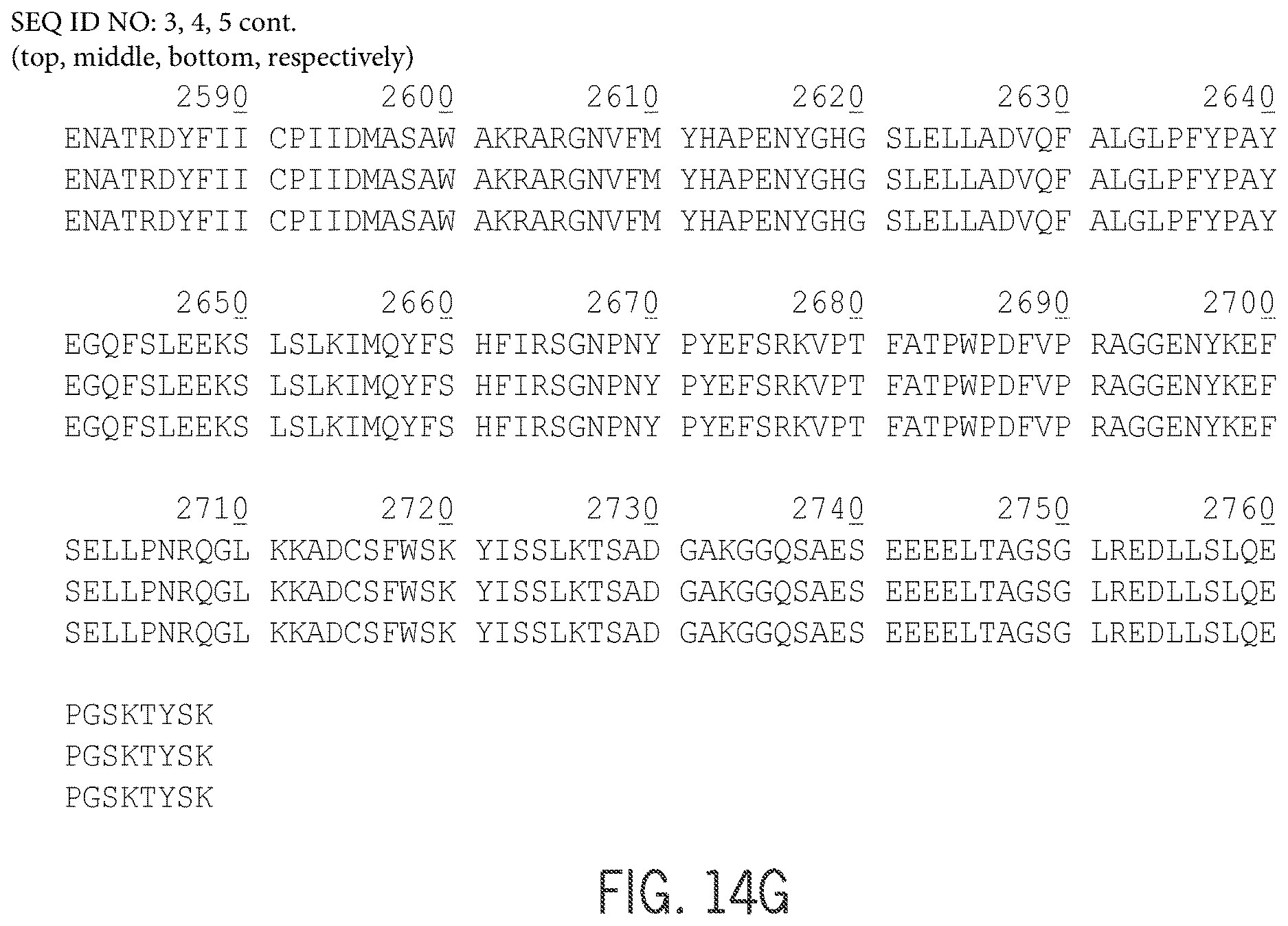
S00001
XML
uspto.report is an independent third-party trademark research tool that is not affiliated, endorsed, or sponsored by the United States Patent and Trademark Office (USPTO) or any other governmental organization. The information provided by uspto.report is based on publicly available data at the time of writing and is intended for informational purposes only.
While we strive to provide accurate and up-to-date information, we do not guarantee the accuracy, completeness, reliability, or suitability of the information displayed on this site. The use of this site is at your own risk. Any reliance you place on such information is therefore strictly at your own risk.
All official trademark data, including owner information, should be verified by visiting the official USPTO website at www.uspto.gov. This site is not intended to replace professional legal advice and should not be used as a substitute for consulting with a legal professional who is knowledgeable about trademark law.